WO2019050977A1 - Reversible linkers and use thereof - Google Patents
Reversible linkers and use thereof Download PDFInfo
- Publication number
- WO2019050977A1 WO2019050977A1 PCT/US2018/049594 US2018049594W WO2019050977A1 WO 2019050977 A1 WO2019050977 A1 WO 2019050977A1 US 2018049594 W US2018049594 W US 2018049594W WO 2019050977 A1 WO2019050977 A1 WO 2019050977A1
- Authority
- WO
- WIPO (PCT)
- Prior art keywords
- cells
- linker
- pmel
- compound
- protein
- Prior art date
Links
- SNDCMFGICSVBRZ-JTQLQIEISA-N CC(C)[C@H](C)C1(C)CCCC1 Chemical compound CC(C)[C@H](C)C1(C)CCCC1 SNDCMFGICSVBRZ-JTQLQIEISA-N 0.000 description 1
Classifications
-
- A—HUMAN NECESSITIES
- A61—MEDICAL OR VETERINARY SCIENCE; HYGIENE
- A61K—PREPARATIONS FOR MEDICAL, DENTAL OR TOILETRY PURPOSES
- A61K47/00—Medicinal preparations characterised by the non-active ingredients used, e.g. carriers or inert additives; Targeting or modifying agents chemically bound to the active ingredient
- A61K47/50—Medicinal preparations characterised by the non-active ingredients used, e.g. carriers or inert additives; Targeting or modifying agents chemically bound to the active ingredient the non-active ingredient being chemically bound to the active ingredient, e.g. polymer-drug conjugates
- A61K47/51—Medicinal preparations characterised by the non-active ingredients used, e.g. carriers or inert additives; Targeting or modifying agents chemically bound to the active ingredient the non-active ingredient being chemically bound to the active ingredient, e.g. polymer-drug conjugates the non-active ingredient being a modifying agent
- A61K47/62—Medicinal preparations characterised by the non-active ingredients used, e.g. carriers or inert additives; Targeting or modifying agents chemically bound to the active ingredient the non-active ingredient being chemically bound to the active ingredient, e.g. polymer-drug conjugates the non-active ingredient being a modifying agent the modifying agent being a protein, peptide or polyamino acid
- A61K47/65—Peptidic linkers, binders or spacers, e.g. peptidic enzyme-labile linkers
-
- A—HUMAN NECESSITIES
- A61—MEDICAL OR VETERINARY SCIENCE; HYGIENE
- A61K—PREPARATIONS FOR MEDICAL, DENTAL OR TOILETRY PURPOSES
- A61K47/00—Medicinal preparations characterised by the non-active ingredients used, e.g. carriers or inert additives; Targeting or modifying agents chemically bound to the active ingredient
- A61K47/50—Medicinal preparations characterised by the non-active ingredients used, e.g. carriers or inert additives; Targeting or modifying agents chemically bound to the active ingredient the non-active ingredient being chemically bound to the active ingredient, e.g. polymer-drug conjugates
- A61K47/51—Medicinal preparations characterised by the non-active ingredients used, e.g. carriers or inert additives; Targeting or modifying agents chemically bound to the active ingredient the non-active ingredient being chemically bound to the active ingredient, e.g. polymer-drug conjugates the non-active ingredient being a modifying agent
- A61K47/62—Medicinal preparations characterised by the non-active ingredients used, e.g. carriers or inert additives; Targeting or modifying agents chemically bound to the active ingredient the non-active ingredient being chemically bound to the active ingredient, e.g. polymer-drug conjugates the non-active ingredient being a modifying agent the modifying agent being a protein, peptide or polyamino acid
- A61K47/64—Drug-peptide, drug-protein or drug-polyamino acid conjugates, i.e. the modifying agent being a peptide, protein or polyamino acid which is covalently bonded or complexed to a therapeutically active agent
- A61K47/645—Polycationic or polyanionic oligopeptides, polypeptides or polyamino acids, e.g. polylysine, polyarginine, polyglutamic acid or peptide TAT
-
- A—HUMAN NECESSITIES
- A61—MEDICAL OR VETERINARY SCIENCE; HYGIENE
- A61K—PREPARATIONS FOR MEDICAL, DENTAL OR TOILETRY PURPOSES
- A61K47/00—Medicinal preparations characterised by the non-active ingredients used, e.g. carriers or inert additives; Targeting or modifying agents chemically bound to the active ingredient
- A61K47/50—Medicinal preparations characterised by the non-active ingredients used, e.g. carriers or inert additives; Targeting or modifying agents chemically bound to the active ingredient the non-active ingredient being chemically bound to the active ingredient, e.g. polymer-drug conjugates
- A61K47/51—Medicinal preparations characterised by the non-active ingredients used, e.g. carriers or inert additives; Targeting or modifying agents chemically bound to the active ingredient the non-active ingredient being chemically bound to the active ingredient, e.g. polymer-drug conjugates the non-active ingredient being a modifying agent
- A61K47/56—Medicinal preparations characterised by the non-active ingredients used, e.g. carriers or inert additives; Targeting or modifying agents chemically bound to the active ingredient the non-active ingredient being chemically bound to the active ingredient, e.g. polymer-drug conjugates the non-active ingredient being a modifying agent the modifying agent being an organic macromolecular compound, e.g. an oligomeric, polymeric or dendrimeric molecule
- A61K47/59—Medicinal preparations characterised by the non-active ingredients used, e.g. carriers or inert additives; Targeting or modifying agents chemically bound to the active ingredient the non-active ingredient being chemically bound to the active ingredient, e.g. polymer-drug conjugates the non-active ingredient being a modifying agent the modifying agent being an organic macromolecular compound, e.g. an oligomeric, polymeric or dendrimeric molecule obtained otherwise than by reactions only involving carbon-to-carbon unsaturated bonds, e.g. polyureas or polyurethanes
- A61K47/60—Medicinal preparations characterised by the non-active ingredients used, e.g. carriers or inert additives; Targeting or modifying agents chemically bound to the active ingredient the non-active ingredient being chemically bound to the active ingredient, e.g. polymer-drug conjugates the non-active ingredient being a modifying agent the modifying agent being an organic macromolecular compound, e.g. an oligomeric, polymeric or dendrimeric molecule obtained otherwise than by reactions only involving carbon-to-carbon unsaturated bonds, e.g. polyureas or polyurethanes the organic macromolecular compound being a polyoxyalkylene oligomer, polymer or dendrimer, e.g. PEG, PPG, PEO or polyglycerol
-
- A—HUMAN NECESSITIES
- A61—MEDICAL OR VETERINARY SCIENCE; HYGIENE
- A61K—PREPARATIONS FOR MEDICAL, DENTAL OR TOILETRY PURPOSES
- A61K47/00—Medicinal preparations characterised by the non-active ingredients used, e.g. carriers or inert additives; Targeting or modifying agents chemically bound to the active ingredient
- A61K47/50—Medicinal preparations characterised by the non-active ingredients used, e.g. carriers or inert additives; Targeting or modifying agents chemically bound to the active ingredient the non-active ingredient being chemically bound to the active ingredient, e.g. polymer-drug conjugates
- A61K47/51—Medicinal preparations characterised by the non-active ingredients used, e.g. carriers or inert additives; Targeting or modifying agents chemically bound to the active ingredient the non-active ingredient being chemically bound to the active ingredient, e.g. polymer-drug conjugates the non-active ingredient being a modifying agent
- A61K47/68—Medicinal preparations characterised by the non-active ingredients used, e.g. carriers or inert additives; Targeting or modifying agents chemically bound to the active ingredient the non-active ingredient being chemically bound to the active ingredient, e.g. polymer-drug conjugates the non-active ingredient being a modifying agent the modifying agent being an antibody, an immunoglobulin or a fragment thereof, e.g. an Fc-fragment
- A61K47/6889—Conjugates wherein the antibody being the modifying agent and wherein the linker, binder or spacer confers particular properties to the conjugates, e.g. peptidic enzyme-labile linkers or acid-labile linkers, providing for an acid-labile immuno conjugate wherein the drug may be released from its antibody conjugated part in an acidic, e.g. tumoural or environment
-
- A—HUMAN NECESSITIES
- A61—MEDICAL OR VETERINARY SCIENCE; HYGIENE
- A61K—PREPARATIONS FOR MEDICAL, DENTAL OR TOILETRY PURPOSES
- A61K47/00—Medicinal preparations characterised by the non-active ingredients used, e.g. carriers or inert additives; Targeting or modifying agents chemically bound to the active ingredient
- A61K47/50—Medicinal preparations characterised by the non-active ingredients used, e.g. carriers or inert additives; Targeting or modifying agents chemically bound to the active ingredient the non-active ingredient being chemically bound to the active ingredient, e.g. polymer-drug conjugates
- A61K47/69—Medicinal preparations characterised by the non-active ingredients used, e.g. carriers or inert additives; Targeting or modifying agents chemically bound to the active ingredient the non-active ingredient being chemically bound to the active ingredient, e.g. polymer-drug conjugates the conjugate being characterised by physical or galenical forms, e.g. emulsion, particle, inclusion complex, stent or kit
- A61K47/6903—Medicinal preparations characterised by the non-active ingredients used, e.g. carriers or inert additives; Targeting or modifying agents chemically bound to the active ingredient the non-active ingredient being chemically bound to the active ingredient, e.g. polymer-drug conjugates the conjugate being characterised by physical or galenical forms, e.g. emulsion, particle, inclusion complex, stent or kit the form being semi-solid, e.g. an ointment, a gel, a hydrogel or a solidifying gel
-
- A—HUMAN NECESSITIES
- A61—MEDICAL OR VETERINARY SCIENCE; HYGIENE
- A61K—PREPARATIONS FOR MEDICAL, DENTAL OR TOILETRY PURPOSES
- A61K47/00—Medicinal preparations characterised by the non-active ingredients used, e.g. carriers or inert additives; Targeting or modifying agents chemically bound to the active ingredient
- A61K47/50—Medicinal preparations characterised by the non-active ingredients used, e.g. carriers or inert additives; Targeting or modifying agents chemically bound to the active ingredient the non-active ingredient being chemically bound to the active ingredient, e.g. polymer-drug conjugates
- A61K47/69—Medicinal preparations characterised by the non-active ingredients used, e.g. carriers or inert additives; Targeting or modifying agents chemically bound to the active ingredient the non-active ingredient being chemically bound to the active ingredient, e.g. polymer-drug conjugates the conjugate being characterised by physical or galenical forms, e.g. emulsion, particle, inclusion complex, stent or kit
- A61K47/6921—Medicinal preparations characterised by the non-active ingredients used, e.g. carriers or inert additives; Targeting or modifying agents chemically bound to the active ingredient the non-active ingredient being chemically bound to the active ingredient, e.g. polymer-drug conjugates the conjugate being characterised by physical or galenical forms, e.g. emulsion, particle, inclusion complex, stent or kit the form being a particulate, a powder, an adsorbate, a bead or a sphere
- A61K47/6927—Medicinal preparations characterised by the non-active ingredients used, e.g. carriers or inert additives; Targeting or modifying agents chemically bound to the active ingredient the non-active ingredient being chemically bound to the active ingredient, e.g. polymer-drug conjugates the conjugate being characterised by physical or galenical forms, e.g. emulsion, particle, inclusion complex, stent or kit the form being a particulate, a powder, an adsorbate, a bead or a sphere the form being a solid microparticle having no hollow or gas-filled cores
- A61K47/6929—Medicinal preparations characterised by the non-active ingredients used, e.g. carriers or inert additives; Targeting or modifying agents chemically bound to the active ingredient the non-active ingredient being chemically bound to the active ingredient, e.g. polymer-drug conjugates the conjugate being characterised by physical or galenical forms, e.g. emulsion, particle, inclusion complex, stent or kit the form being a particulate, a powder, an adsorbate, a bead or a sphere the form being a solid microparticle having no hollow or gas-filled cores the form being a nanoparticle, e.g. an immuno-nanoparticle
- A61K47/6931—Medicinal preparations characterised by the non-active ingredients used, e.g. carriers or inert additives; Targeting or modifying agents chemically bound to the active ingredient the non-active ingredient being chemically bound to the active ingredient, e.g. polymer-drug conjugates the conjugate being characterised by physical or galenical forms, e.g. emulsion, particle, inclusion complex, stent or kit the form being a particulate, a powder, an adsorbate, a bead or a sphere the form being a solid microparticle having no hollow or gas-filled cores the form being a nanoparticle, e.g. an immuno-nanoparticle the material constituting the nanoparticle being a polymer
- A61K47/6935—Medicinal preparations characterised by the non-active ingredients used, e.g. carriers or inert additives; Targeting or modifying agents chemically bound to the active ingredient the non-active ingredient being chemically bound to the active ingredient, e.g. polymer-drug conjugates the conjugate being characterised by physical or galenical forms, e.g. emulsion, particle, inclusion complex, stent or kit the form being a particulate, a powder, an adsorbate, a bead or a sphere the form being a solid microparticle having no hollow or gas-filled cores the form being a nanoparticle, e.g. an immuno-nanoparticle the material constituting the nanoparticle being a polymer the polymer being obtained otherwise than by reactions involving carbon to carbon unsaturated bonds, e.g. polyesters, polyamides or polyglycerol
-
- C—CHEMISTRY; METALLURGY
- C07—ORGANIC CHEMISTRY
- C07D—HETEROCYCLIC COMPOUNDS
- C07D207/00—Heterocyclic compounds containing five-membered rings not condensed with other rings, with one nitrogen atom as the only ring hetero atom
- C07D207/46—Heterocyclic compounds containing five-membered rings not condensed with other rings, with one nitrogen atom as the only ring hetero atom with hetero atoms directly attached to the ring nitrogen atom
-
- C—CHEMISTRY; METALLURGY
- C07—ORGANIC CHEMISTRY
- C07D—HETEROCYCLIC COMPOUNDS
- C07D403/00—Heterocyclic compounds containing two or more hetero rings, having nitrogen atoms as the only ring hetero atoms, not provided for by group C07D401/00
- C07D403/02—Heterocyclic compounds containing two or more hetero rings, having nitrogen atoms as the only ring hetero atoms, not provided for by group C07D401/00 containing two hetero rings
- C07D403/12—Heterocyclic compounds containing two or more hetero rings, having nitrogen atoms as the only ring hetero atoms, not provided for by group C07D401/00 containing two hetero rings linked by a chain containing hetero atoms as chain links
-
- C—CHEMISTRY; METALLURGY
- C07—ORGANIC CHEMISTRY
- C07K—PEPTIDES
- C07K14/00—Peptides having more than 20 amino acids; Gastrins; Somatostatins; Melanotropins; Derivatives thereof
- C07K14/435—Peptides having more than 20 amino acids; Gastrins; Somatostatins; Melanotropins; Derivatives thereof from animals; from humans
- C07K14/52—Cytokines; Lymphokines; Interferons
- C07K14/54—Interleukins [IL]
- C07K14/5443—IL-15
Definitions
- the present disclosure generally relates to compositions and methods for preparation and delivery of protein therapeutics, and more particularly reversible linkers and use thereof.
- Protein therapeutics such as antibodies, cytokines, growth factors and vaccines, are important therapeutics for the treatment of a variety of diseases including, for example, cancer, diabetes and cardiovascular diseases.
- This class of protein therapeutics has been developed rapidly in the global pharmaceutical industry over the last few years. Protein therapeutics have the advantages of high specificity and potency relative to small molecule drugs. Nonetheless, the use of protein therapeutics is limited as a result of their intrinsic instability, immunogenicity and short half-life.
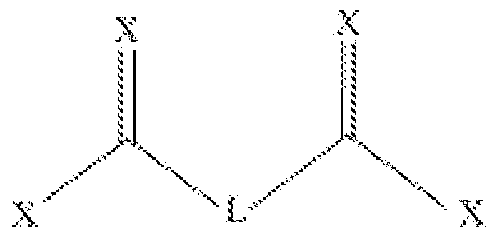
- LGi and LG2 are each a leaving group, independently selected from triflate, tosyl, CI, hydroxysuccinimide and imidazolide;
- Yi and Y2 are each independently selected from O and S;
- X at each occurrence, is independently selected from O, S, and N;
- L is a linker such that is biodegradable
- n is an integer selected from 1-6
- the compound of formula (I) is symmetrical.
- LGi and LG2 are capable of reacting with a protein, a drug and/or a particle.
- LGi and LG2 are both imidazolide.
- LGi and LG2 are both N-hydroxysuccinimide.
- L is selected from:
- n is an integer selected from 0-5; ted from 0-5; wherein X, at each occurrence, is independently selected from O, S, and N.
- m is 2.
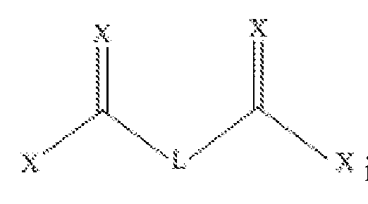
- a further aspect relates to a compound having formula (II):
- Xi and X2 are each independently selected from triflate, tosyl, CI, N-hydroxysuccinimide and imidazolide;
- Ai and A3 are each independently -(CR 1 R 2 )n- ;
- Yi and Y 2 are each independently selected from R 3 , O and S;
- R 1 and R 2 at each occurrence are independently selected from hydrogen, halogen, hydroxyl, C 1-12 alkyl, C 2-12 alkenyl, C3-12 cycloalkyl, C 2-12 heterocyclyl; C6-i2 aryl optionally substituted with 1 or more halo, hydroxyl, C 1-6 alkyl and/or C 1-6 alkoxyl; and C 4-12 heteroaryl optionally substituted with 1 or more halo, hydroxyl, C 1-6 alkyl and/or C 1-6 alkoxyl
- R 3 is selected from hydrogen, C 1-12 alkyl, C 2-12 alkenyl, C3-12 cycloalkyl, C 2-12 heterocyclyl; C6-12 aryl optionally substituted with 1 or more halo, hydroxyl, C 1-6 alkyl and/or Ci- 6 alkoxyl; and C 4-12 heteroaryl optionally substituted with 1 or more halo, hydroxyl, C 1-6 alkyl and/or C 1-6 alkoxyl;
- n at each occurrence, is an integer independently selected from 1-12;
- n is an integer selected from 0-12.
- the compound of formula (II) is symmetrical. In some embodiments, the compound of formula (II) is symmetrical. In some
- Xi and X2 can each be a leaving group capable of reacting with a protein, a drug and/or a particle.
- Xi and Xi are both imidazolide.
- Xi and Xi are both N-hydroxysuccinimide.
- R 1 and R 2 are both hydrogen.
- Ai and A 3 are both -(CH ⁇ -.
- Ai is -(CH ⁇ -.
- Yi and Y 2 are both O.
- the compound is:
- a 2 is a bond.
- Yi and Yi are both H.
- the compound, in some embodiments, is:
- the compound may be used to conjugate or cross-link one or more protein or agent of interest at, e.g., an amine group such as a terminal amine or an internal amine.
- an amine group such as a terminal amine or an internal amine.
- Internal amines include side chain amines such as lysine amines.
- the compounds disclosed herein can be used to reversibly crossed-link a plurality of therapeutic protein monomers into protein clusters of, e.g., 30 nm and 1000 nm in diameter.
- the protein clusters can be subject to surface modification such as polycation. In some embodiments, these protein clusters are referred to as "backpacks" or "BP.”
- a cell therapy composition can be prepared by providing the protein clusters or backpacks disclosed herein, and incubating the protein clusters or backpacks with a nucleated cell such as T cell, B cell, natural killer (NK) cell and hematopoietic stem cell.
- T cells can include CD4+T cells, cytotoxic T cells (e.g., CD8+ T cells), alpha T cells, beta T cells, gamma T cells, delta T cells and regulatory T-cell (Tregs).
- the nucleated cell e.g., T cell or NK cell
- the nucleated cell may comprise, e.g., express, a Chimeric Antigen Receptor (CAR) such as a CAR that binds to a cancer antigen.
- CAR Chimeric Antigen Receptor
- the compound can be used as a degradable or hydrolysable linker.
- the degradable linker is a redox responsive linker.
- the disclosure provides a particle, e.g., a nanoparticle, that is formed by the linkers as described herein, e.g., nanoparticle that comprises a protein (e.g., a protein nanogel).
- a protein e.g., a protein nanogel.
- the linkers disclosed herein can be used in connection with the backpack technology for e.g., cell therapy as disclosed in, e.g., U.S. Publication No. 2017/0080104, U.S. Patent No. 9,603,944, U.S. Publication No. 2014/0081012, and PCT Application No. PCT/US2017/037249, each of which is incorporated herein by reference in its entirety.
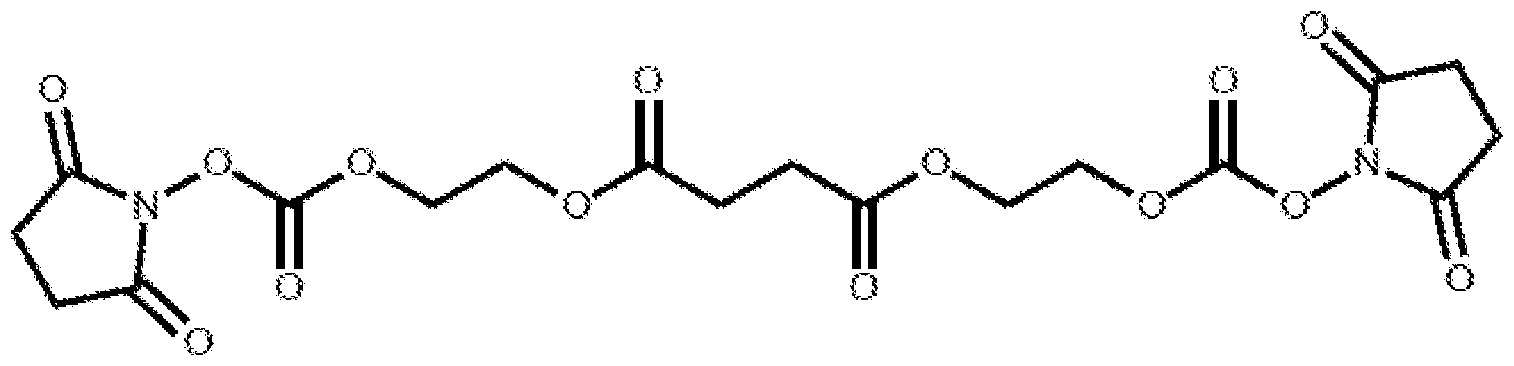
- FIG. 1 illustrates two exemplary linkers, Linker- 1 and Linker-2.
- FIG. 2 shows that backpacks can be prepared by reacting various therapeutic protein monomers cross-linked using one or more cross-linkers disclosed herein.
- FIG. 3 shows an exemplary use of backpacks in cell therapy.
- FIG. 4 Two Linker-2 cross-linked IL-15 backpack formulations (17HF1 and 17HF2) showed comparable cell expansion with a Linker- 1 cross-linked IL-15 backpack formulation (HF6).
- FIG. 5 Exemplary PMEL experimental outline.
- FIG. 6 BP-Linker-1 and BP-Linker-2 show similar anti -tumor activity in vivo. In addition, BP-Linker-1 and BP-Linker-2 show comparable distribution in the organs analyzed including blood, spleen, lung, and tumor (not shown).
- FIG. 7 shows that there are no significant effects on Complete Blood Counts (CBC) with BP-Linker-1 (TRQ-PMEL1) and BP-Linker-2 (TRQ-PMEL2) in either tumor or non tumor- bearing mice.
- FIG. 8 BP-Linker-2 shows trends toward lower lymphodepletion (CD8, K1.1 and transferred PMELs) compared to BP-Linker-1.
- FIG. 9 Experimental outline of another set of PMEL study.
- FIGS. 10 and 11 are the in vitro Pmel expansion curves by number of cells (FIG. 10) and present viable cells (FIG. 11).
- FIG. 11 Linker- 1 NG and Linker-2 NG both displayed anti-tumor activity.
- FIG. 12 Linker- 1 NG and Linker-2 NG show comparable amount of circulating Pmel.
- FIG. 13 Linker- 1 NG and Linker-2 NG show comparable amount of tumor-infiltrating
- FIG. 14 Linker-2 backpacked Cells have older memory phenotype, with significant increase in Teff cells in Linker-2 relative to Linker-1 at d4 after injection.
- Tcm central memory T cells
- Teff effector memory T cells
- Temra effector memory-RA+ T cells
- Tscm memory stem T cells
- Tnaive naive T cells.
- FIG. 15 Efficacy of backpacked Chimeric Antigen Receptor (CAR) T cells.
- FIG. 16 In vitro proliferation on day 0, 1, 3, 7, 10, 14, 17 and 21 of CAR, Linker- 1-HF6,
- FIG. 18 Backpacked cells expanded significantly more compared to CAR alone or mock.
- FIGS. 19A-19B Phenotypes remain similar through day 21 for Tnaive, Tscm and Tcm, while Tern and Temra composition varied slightly (Tcm: central memory T cells; Tern: effector memory T cells; Temra: effector memory-RA+ T cells; Tscm: memory stem T cells; Tnaive: naive T cells).
- FIG. 20 shows the efficacy of backpacked CAR T treatment, as Linker- 1-HF6 and Linker-2-HFl both statistically significantly (as indicated by **) delayed/inhibited tumor growth compared to CAR alone or h9.4-IL15 backpack at all time points analyzed.
- FIGS. 21 and 22 On day 22, tumor size, tumor weight, spleen weight, number of tumor- infiltrating lymphocytes (TILs), number of CAR TILs and TIL phenotype were also analyzed.
- TILs tumor- infiltrating lymphocytes
- FIG. 23 Clinical chemistry parameters in naive mice at Dl and D4 post-dose.
- HBSS vehicle control
- DP- 15 PMEL Deep IL-15 Primed PMEL cells
- D Day.
- FIG. 24 Clinical chemistry parameters in tumor - bearing mice at Dl and D4 post-dose.
- HBSS vehicle control
- DP-15 PMEL Deep IL-15 Primed PMEL cells
- D Day.
- FIG. 25 Serum levels of IFN- ⁇ in tumor - bearing compared to naive mice 24 hr after ACT. he serum levels of IFN- ⁇ in the PMEL + IL15-Fc group were significantly increased (2- way ANOVA with Tukey's multiple comparison, p ⁇ 0.001) compared to both the PMEL and DP- 15 PMEL groups in both naive and tumor - bearing mice.
- ACT adoptive cell transfer;
- DP-15 PMEL Deep IL-15 Primed PMEL cells.
- FIG. 26 IL15-Fc systemic exposure in mice treated with PMEL + IL15-Fc and Deep IL- 15 Primed PMEL cells, in naive and tumor - bearing mice.
- HBSS green asterisk over the grey
- HBSS vehicle control;
- DP-15 PMEL Deep IL-15 Primed PMEL cells.
- Cancer immunotherapy including adoptive T cell therapy, is a promising strategy to treat cancer because it harnesses a subject's own immune system to attack cancer cells. Nonetheless, a major limitation of this approach is the rapid decline in viability and function of the transplanted T lymphocytes.
- co-administration of immunostimulatory agents with transferred cells is necessary. When given systemically at high doses, these agents could enhance the in vivo viability of transferred (i.e., donor) cells, improve the therapeutic function of transferred cells, and thus lead to overall improved efficacy against cancer; however, high doses of such agents could also result in life-threatening side effects.
- interleukin-2 IL-2
- Tregs regulatory T cells
- One approach to focus adjuvant activity on the transferred cells is to genetically engineer the transferred cells to secrete their own supporting factors.
- the technical difficulty and challenges as well as the high cost for large-scale production of genetically engineered T lymphocytes have significantly limited the potential of this method in clinical applications, to date.
- a technology platform that permits simple, safe and efficient delivery of biologically-active agents, such as a drug, protein (e.g., adjuvants such as IL-2) or particle to cells through chemical conjugation of protein, drug, or particle-loaded, carrier-free linkers directly onto the plasma membrane of cells.
- biologically-active agents such as a drug, protein (e.g., adjuvants such as IL-2) or particle to cells through chemical conjugation of protein, drug, or particle-loaded, carrier-free linkers directly onto the plasma membrane of cells.
- the present disclosure further contemplates other nanostructures that comprise other protein therapeutics for purposes other than adjuvant effect on adoptively-transferred cells.
- the disclosure has broader applications, as provided herein.
- the articles “a” and “an” refer to one or more than one, e.g., to at least one, of the grammatical object of the article.
- the use of the words “a” or “an” when used in conjunction with the term “comprising” herein may mean “one,” but it is also consistent with the meaning of "one or more,” “at least one,” and “one or more than one.”
- compositions, methods, and respective component(s) thereof are used in reference to compositions, methods, and respective component(s) thereof, that are present in a given embodiment, yet open to the inclusion of unspecified elements.
- the term "consisting essentially of” refers to those elements required for a given embodiment. The term permits the presence of additional elements that do not materially affect the basic and novel or functional characteristic(s) of that embodiment of the disclosure.
- compositions, methods, and respective components thereof as described herein, which are exclusive of any element not recited in that description of the embodiment.
- a plurality of means more than 1, e.g., 2, 3, 4, 5, 6, 7, 8, 9, 10, 11, 12, 13, 14, 15, 16, 17, 18, 19, 20, or more, e.g., 25, 30, 40, 50, 60, 70, 80, 90, 100, 200, 300, 400, 500, or more, or any integer therebetween.
- therapeutic agent any active pharmaceutical ingredient
- active pharmaceutical agent active drug
- Therapeutic agents include pharmaceutical, chemical or biological agents. Additionally, pharmaceutical, chemical or biological agents can include any agent that has a desired property or affect whether it is a therapeutic agent. For example, agents also include diagnostic agents, biocides and the like.
- protein protein
- peptide and “polypeptide” are used interchangeably herein to refer to polymers of amino acids of any length.
- the polymer may be linear or branched, it may comprise modified amino acids, and it may be interrupted by non-amino acids.
- the terms also encompass an amino acid polymer that has been modified; for example, disulfide bond formation, glycosylation, lipidation, acetylation, phosphorylation, or any other manipulation, such as conjugation with a labeling component.
- the polypeptide can be isolated from natural sources, can be a produced by recombinant techniques from a eukaryotic or prokaryotic host, or can be a product of synthetic procedures. It should be understood that the term “protein” includes fusion or chimeric proteins, as well as cytokines, antibodies and antigen-binding fragments thereof.
- Antibody or "antibody molecule” as used herein refers to a protein, e.g., an immunoglobulin chain or fragment thereof, comprising at least one immunoglobulin variable domain sequence.
- An antibody molecule encompasses antibodies (e.g., full-length antibodies) and antibody fragments.
- an antibody molecule comprises an antigen binding or functional fragment of a full length antibody, or a full length immunoglobulin chain.
- a full-length antibody is an immunoglobulin (Ig) molecule (e.g., IgG) that is naturally occurring or formed by normal immunoglobulin gene fragment recombinatorial processes).
- an antibody molecule refers to an immunologically active, antigen-binding portion of an immunoglobulin molecule, such as an antibody fragment.
- An antibody fragment e.g., functional fragment, is a portion of an antibody, e.g., Fab, Fab', F(ab') 2 , F(ab) 2 , variable fragment (Fv), domain antibody (dAb), or single chain variable fragment (scFv).
- a functional antibody fragment binds to the same antigen as that recognized by the intact (e.g., full-length) antibody.
- antibody fragment or “functional fragment” also include isolated fragments consisting of the variable regions, such as the "Fv” fragments consisting of the variable regions of the heavy and light chains or recombinant single chain polypeptide molecules in which light and heavy variable regions are connected by a peptide linker ("scFv proteins").
- an antibody fragment does not include portions of antibodies without antigen binding activity, such as Fc fragments or single amino acid residues.
- Exemplary antibody molecules include full length antibodies and antibody fragments, e.g., dAb (domain antibody), single chain, Fab, Fab', and F(ab') 2 fragments, and single chain variable fragments (scFvs).
- Fab and “Fab fragment” are used interchangeably and refer to a region that includes one constant and one variable domain from each heavy and light chain of the antibody, i.e., VL, CL, VH, and CHI .
- an antibody molecule is monospecific, e.g., it comprises binding specificity for a single epitope.
- an antibody molecule is multispecific, e.g., it comprises a plurality of immunoglobulin variable domain sequences, where a first immunoglobulin variable domain sequence has binding specificity for a first epitope and a second immunoglobulin variable domain sequence has binding specificity for a second epitope.
- an antibody molecule is a bispecific antibody molecule. "Bispecific antibody molecule" as used herein refers to an antibody molecule that has specificity for more than one (e.g., two, three, four, or more) epitope and/or antigen.
- Antigen refers to a macromolecule, including all proteins or peptides.
- an antigen is a molecule that can provoke an immune response, e.g., involving activation of certain immune cells and/or antibody generation. Antigens are not only involved in antibody generation. T cell receptors also recognized antigens (albeit antigens whose peptides or peptide fragments are complexed with an MHC molecule). Any macromolecule, including almost all proteins or peptides, can be an antigen. Antigens can also be derived from genomic recombinant or DNA.
- any DNA comprising a nucleotide sequence or a partial nucleotide sequence that encodes a protein capable of eliciting an immune response encodes an "antigen.”
- an antigen does not need to be encoded solely by a full length nucleotide sequence of a gene, nor does an antigen need to be encoded by a gene at all.
- an antigen can be synthesized or can be derived from a biological sample, e.g., a tissue sample, a tumor sample, a cell, or a fluid with other biological components.
- a tumor antigen or interchangeably, a “cancer antigen” includes any molecule present on, or associated with, a cancer, e.g., a cancer cell or a tumor microenvironment that can provoke an immune response.
- an "immune cell antigen” includes any molecule present on, or associated with, an immune cell that can provoke an immune response.
- the "antigen-binding site" or “antigen-binding fragment” or “antigen-binding portion” (used interchangeably herein) of an antibody molecule refers to the part of an antibody molecule, e.g., an immunoglobulin (Ig) molecule such as IgG, that participates in antigen binding.
- the antigen-binding site is formed by amino acid residues of the variable (V) regions of the heavy (H) and light (L) chains.
- hypervariable regions Three highly divergent stretches within the variable regions of the heavy and light chains, referred to as hypervariable regions, are disposed between more conserved flanking stretches called “framework regions" (FRs).
- FRs are amino acid sequences that are naturally found between, and adjacent to, hypervariable regions in immunoglobulins.
- the three hypervariable regions of a light chain and the three hypervariable regions of a heavy chain are disposed relative to each other in three dimensional space to form an antigen-binding surface, which is complementary to the three-dimensional surface of a bound antigen.
- the three hypervariable regions of each of the heavy and light chains are referred to as "complementarity-determining regions," or "CDRs.”
- the framework region and CDRs have been defined and described, e.g., in Kabat, E.A., et al. (1991) Sequences of Proteins of Immunological Interest, Fifth Edition, U.S.
- variable chain e.g., variable heavy chain and variable light chain
- Each variable chain is typically made up of three CDRs and four FRs, arranged from amino-terminus to carboxy- terminus in the amino acid order: FR1, CDR1, FR2, CDR2, FR3, CDR3, and FR4.
- VL variable light chain
- VH CDRs are generally defined to include residues at positions 27-33 (CDR1), 52-56 (CDR2), and 95-102 (CDR3).
- CDR1 residues at positions 27-33
- CDR2 52-56
- CDR3 95-102
- the loops can be of different length across antibodies and the numbering systems such as the Kabat or Chotia control so that the frameworks have consistent numbering across antibodies.
- the antigen-binding fragment of an antibody can lack or be free of a full Fc domain.
- an antibody-binding fragment does not include a full IgG or a full Fc but may include one or more constant regions (or fragments thereof) from the light and/or heavy chains.
- the antigen-binding fragment can be completely free of any Fc domain.
- the antigen-binding fragment can be substantially free of a full Fc domain.
- the antigen-binding fragment can include a portion of a full Fc domain (e.g., CH2 or CH3 domain or a portion thereof).
- the antigen-binding fragment can include a full Fc domain.
- the Fc domain is an IgG domain, e.g., an IgGl, IgG2, IgG3, or IgG4 Fc domain.
- the Fc domain comprises a CH2 domain and a CH3 domain.
- a "cytokine molecule” refers to full length, a fragment or a variant of a naturally-occurring, wild type cytokine (including fragments and functional variants thereof having at least 10% of the activity of the naturally-occurring cytokine molecule).
- the cytokine molecule has at least 30, 50, or 80% of the activity, e.g., the immunomodulatory activity, of the naturally-occurring molecule.
- the cytokine molecule further comprises a receptor domain, e.g., a cytokine receptor domain, optionally, coupled to an immunoglobulin Fc region.
- the cytokine molecule is coupled to an immunoglobulin Fc region.
- the cytokine molecule is coupled to an antibody molecule (e.g., an immunoglobulin Fab or scFv fragment, a Fab fragment, a FAB 2 fragment, or an affibody fragment or derivative, e.g., a sdAb (nanobody) fragment, a heavy chain antibody fragment, single-domain antibody, a bi-specific or multispecific antibody), or non-antibody scaffolds and antibody mimetics (e.g., lipocalins (e.g., anticalins), affibodies, fibronectin (e.g., monobodies or Adnectins), knottins, ankyrin repeats (e.g., DARPins), and A domains (e.g., avimers)).
- an antibody molecule e.g., an immunoglobulin Fab or scFv fragment, a Fab fragment, a F
- alkyl refers to a saturated hydrocarbon chain that may be a straight chain or branched chain, containing the indicated number of carbon atoms.
- Ci-e alkyl indicates that the group may have 1 to 6 (inclusive) carbon atoms in it. Any atom can be optionally substituted, e.g., by one or more substituents.
- alkyl groups include without limitation methyl, ethyl, n-propyl, isopropyl, and tert-butyl .
- alkoxy refers to a group of formula— O(alkyl).
- Alkoxy can be, for example, methoxy (— OCHi), ethoxy, propoxy, isopropoxy, butoxy, iso-butoxy, sec- butoxy, pentoxy, 2-pentoxy, 3-pentoxy, or hexyloxy.
- hydroxyl employed alone or in combination with other terms, refers to a group of formula— OH.
- alkenyl refers to a straight or branched hydrocarbon chain containing the indicated number of carbon atoms and having one or more carbon-carbon double bonds. Any atom can be optionally substituted, e.g., by one or more substituents. Alkenyl groups can include, e.g., vinyl, allyl, 1-butenyl, and 2-hexenyl. One of the double bond carbons can optionally be the point of attachment of the alkenyl substituent.
- alkynyl refers to a straight or branched hydrocarbon chain containing the indicated number of carbon atoms and having one or more carbon-carbon triple bonds.
- Alkynyl groups can be optionally substituted, e.g., by one or more substituents.
- Alkynyl groups can include, e.g., ethynyl, propargyl, and 3-hexynyl.
- One of the triple bond carbons can optionally be the point of attachment of the alkynyl substituent.
- heterocyclyl refers to a fully saturated monocyclic, bicyclic, tricyclic or other polycyclic ring system having one or more constituent heteroatom ring atoms independently selected from O, N (it is understood that one or two additional groups may be present to complete the nitrogen valence and/or form a salt), or S.
- the heteroatom or ring carbon can be the point of attachment of the heterocyclyl substituent to another moiety. Any atom can be optionally substituted, e.g., by one or more substituents.
- Heterocyclyl groups can include, e.g., tetrahydrofuryl, tetrahy dropy ranyl , piperidyl (piperidino), piperazinyl, mo holinyl (morpholino), pyrrolinyl, and pyrrolidinyl.
- cycloalkyl refers to a fully saturated monocyclic, bicyclic, tricyclic, or other polycyclic hydrocarbon groups. Any atom can be optionally substituted, e.g., by one or more substituents. A ring carbon serves as the point of attachment of a cycloalkyl group to another moiety. Cycloalkyl moieties can include, e.g., cyclopropyl, cyclobutyl, cyclopentyl, cyclohexyl, cycloheptyl, adamantyl, and norbornyl (bicycle[2.2. IJheptyl).
- aryl refers to an aromatic monocyclic, bicyclic (2 fused rings), or tricyclic (3 fused rings), or polycyclic (>3 fused rings) hydrocarbon ring system.
- One or more ring atoms can be optionally substituted, e.g., by one or more substituents.
- Aryl moieties include, e.g., phenyl and naphthyl.
- heteroaryl refers to an aromatic monocyclic, bicyclic (2 fused rings), tricyclic (3 fused rings), or polycyclic (>3 fused rings) hydrocarbon groups having one or more heteroatom ring atoms independently selected from O, N (it is understood that one or two additional groups may be present to complete the nitrogen valence and/or form a salt), or S.
- One or more ring atoms can be optionally substituted, e.g., by one or more substituents.
- heteroaryl groups include, but are not limited to, 2H-pyrrolyl, 3H-indolyl, 4H-quinolizinyl, acridinyl, benzo[b]thienyl, benzothiazolyl, P-carbolinyl, carbazolyl, coumarinyl, chromenyl, cinnolinyl, dibenzo[b,d]furanyl, furazanyl, furyl, imidazolyl, imidizolyl, indazolyl, indolyl, isobenzofuranyl, isoindolyl, isoquinolyl, isothiazolyl, isoxazolyl, naphthyridinyl, oxazolyl, perimidinyl, phenanthridinyl, phenanthrolinyl, phenarsazinyl, phenazinyl, phenothiazinyl, phenoxathiinyl, pheno
- substituted refers to a group "substituted” on, e.g., an alkyl, haloalkyl, cycloalkyl, heterocyclyl, heterocycloalkenyl, cycloalkenyl, aryl, or heteroaryl group at any atom of that group.
- the substituent(s) on a group are independently any one single, or any combination of two or more of the permissible atoms or groups of atoms delineated for that substituent.
- a substituent may itself be substituted with any one of the above substituents.
- the phrase "optionally substituted” means unsubstituted (e.g., substituted with an H) or substituted.
- substituted means that a hydrogen atom is removed and replaced by a substituent. It is understood that substitution at a given atom is limited by valency.
- At least one drug, protein, polymer and/or particle (collectively, "agents") of the present disclosure are reversibly linked to one another through a degradable linker such that under physiological conditions, the linker degrades and releases the intact, biologically-active agent.
- protein monomers can be cross-linked together to form a cluster that contains a plurality of the protein monomers.
- various agents are linked to functional groups through a degradable linker.
- the agents are reversibly modified or linked, as described below.
- An agent that is "reversibly linked to another" agent refers to a drug, protein, polymer or particle that is attached (e.g., covalently attached) to another drug, protein, polymer or particle through a degradable linker.
- agent that is "reversibly linked to a functional group,” or an agent that is “reversibly modified,” herein refers to an agent that is attached (e.g., covalently attached) to a functional group through a degradable linker.
- agent conjugate or a “reversibly modified agent conjugate”— the terms may be used interchangeably herein.
- proteins and polymers each contain functional groups to which an agent can be linked via a reversible linker, such as amine, silane, hydroxy!, poly(eihylene oxide), polylactic acid, poiy(iac ⁇ .ic ⁇ co ⁇ glycolic acid), etc.
- agent conjugates and reversibly modified proteins include without limitation, an agent reversibly linked (e.g., via a degradable linker) to another agent, an agent reversibly linked to a polymer, and a protein reversibly linked to another functional group.
- protein includes fusion proteins.
- LGi and LG2 are each a leaving group, preferably independently selected from triflate, tosyl, CI, N-hy droxy succi ni mi de and imidazolide;
- Yi and Y 2 are each independently selected from O and S;
- X at each occurrence, is independently selected from O, S, and N;
- L is a linkage such that is biodegradable
- n is an integer selected from 1-6.
- the compound represented by formula (I) is symmetrical at L.
- LGi and LG2 can be the same.
- Yi and Y2 can be the same.
- LGi and LG2 may be capable of reacting with a protein, a drug and/or a particle.
- LGi and LG2 can both be imidazolide.
- LGi and LG2 are both N-hy droxy succi ni mi de .
- L can be selected from:
- n is an integer selected from 0-5; wherein n is an integer selected from 0-5; or wherein X, at each occurrence, is independently selected from O, S, and N.
- Xi and Xi are each independently selected from triflate, tosyl, CI, N-hydroxysuccinimide and imidazolide;
- Ai and A 3 are each independently -(CR 1 !* 2 )!- ;
- a 2 is -(CR ⁇ m- ;
- Yi and Y 2 are each independently selected from NR 3 , O and S;
- R 1 and R 2 at each occurrence are independently selected from hydrogen, halogen, hydroxyl, Ci-u alkyl, C 2 - 12 alkenyl, C 3 - 12 cycloalkyl, C 2 - 12 heterocyclyl; C6-12 aryl optionally substituted with 1 or more halo, hydroxyl, Ci-e alkyl and/or Ci-e alkoxyl; and C 4 - 12 heteroaryl optionally substituted with 1 or more halo, hydroxyl, Ci-e alkyl and/or Ci-e alkoxyl
- R 3 is selected from hydrogen, Ci-u alkyl, C 2 - 12 alkenyl, C 3 - 12 cycloalkyl, C 2 - 12 heterocyclyl; C6-12 aryl optionally substituted with 1 or more halo, hydroxyl, Ci-e alkyl and/or Ci- 6 alkoxyl; and C 4 - 12 heteroaryl optionally substituted with 1 or more halo, hydroxyl, Ci-e alkyl and/or Ci-e alkoxyl; n, at each occurrence, is an integer independently selected from 1-12; and
- n is an integer selected from 0-12.
- the compound represented by formula ( ⁇ ) is symmetrical.
- Xi and X 2 are each a leaving group capable of reacting with a protein, a drug and/or a particle.
- Xi and Xi are both imidazolide or N- hydroxysuccinimide.
- R 1 and R 2 are both hydrogen.
- Ai and A 3 are both -(CH ⁇ -.
- Ai is -(CH 2 ) 2 -.
- Yi and Y 2 are both O.
- the compound is:
- a 2 is a bond.
- Yi and Yi are both NH.
- the compound is:
- Examples of protein monomers for use in accordance with the present disclosure include, without limitation, antibodies (e.g., IgG, Fab, mixed Fc and Fab), single chain antibodies, antibody fragments, engineered proteins such as Fc fusions, enzymes, co-factors, receptors, ligands, transcription factors and other regulatory factors, cytokines, chemokines, human serum albumin, and the like. These proteins may or may not be naturally occurring. Other proteins are contemplated and may be used in accordance with the disclosure. Any of the proteins can be reversibly modified through cross-linking to form a cluster or nanogel structure as disclosed in, e.g., U.S. Publication No. 2017/0080104, U.S. Patent No. 9,603,944, U.S Publication No.
- Linker-1 and Linker-2 can be used to cross-link various protein mononers having amine groups (represented by R-NI fc). Under suitable conditions, the disulfide bond in Linker-1 or the di ester bond in Linker-2 can be hydrolyzed to release the original protein monomers to achieve, e.g., therapeutic effects.
- protein monomers of the disclosure are immunostimulatory proteins.
- an immunostimulatory protein is a protein that stimulates an immune response (including enhancing a pre-existing immune response) in a subject to whom it is administered, whether alone or in combination with another protein or agent.
- immunostimulatory proteins examples include, without limitation, antigens, adjuvants (e.g., flagellin, muramyl dipeptide), cytokines including interleukins (e.g., JL-2, JL-7, IL-15 , IL-10, IL-18, IL-21, IL-23 (or superagonist/mutant forms of these cytokines, such as, IL-15SA), IL-12, IFN-gamma, IFN-alpha, GM-CSF, FLT3-ligand), and immunostimulatory antibodies (e.g., anti-CTLA-4, anti-CD28, anti-CD3, or single
- the immunostimulatory proteins can be an antibody or antigen-binding fragment thereof that binds an inhibitor of an immunosuppressor, e.g., an inhibitor of a checkpoint inhibitor, such as PD-1, PD-L1, LAG-3, TIM-3, CTLA-4, inhibitory KIR, CD276, VTCN1, BTLA/HVEM, HAVCR2 and ADORA2A, e.g., as described in US 2016/0184399 incorporated herein by reference.
- an immunosuppressor e.g., an inhibitor of a checkpoint inhibitor, such as PD-1, PD-L1, LAG-3, TIM-3, CTLA-4, inhibitory KIR, CD276, VTCN1, BTLA/HVEM, HAVCR2 and ADORA2A, e.g., as described in US 2016/0184399 incorporated herein by reference.
- protein monomers of the disclosure are antigens.
- antigens that may be used in accordance with the disclosure include, without limitation, cancer antigens, self-antigens, microbial antigens, allergens and environmental antigens. Other protein antigens are contemplated and may be used in accordance with the disclosure.
- proteins of the disclosure are cancer antigens.
- a cancer antigen is an antigen that is expressed preferentially by cancer cells (i.e., it is expressed at higher levels in cancer cells than on non-cancer cells) and, in some instances, it is expressed solely by cancer cells. Cancer antigens may be expressed within a cancer cell or on the surface of the cancer cell.
- Cancer antigens that may be used in accordance with the disclosure include, without limitation, MART-l/Melan-A, gplOO, adenosine deaminase-binding protein (ADAbp), FAP, cyclophilin b, colorectal associated antigen (CRC)-0017-1A/GA733, carcinoembryonic antigen (CEA), CAP-1, CAP-2, etv6, AMLl, prostate specific antigen (PSA), PSA-1, PSA-2, PSA-3, prostate-specific membrane antigen (PSMA), T cell receptor/CD3-zeta chain and CD20.
- MART-l/Melan-A gplOO
- ADAbp adenosine deaminase-binding protein
- FAP cyclophilin b
- CRC colorectal associated antigen
- CEA carcinoembryonic antigen
- CAP-1 CAP-2
- etv6 etv6, A
- the cancer antigen may be selected from the group consisting of MAGE-A1, MAGE-A2, MAGE- A3, MAGE-A4, MAGE-A5, MAGE-A6, MAGE-A7, MAGE-A8, MAGE-A9, MAGE- A 10, MAGE-A11, MAGE-A12, MAGE-Xp2 (MAGE-B2), MAGE-Xp3 (MAGE-B3), MAGE-Xp4 (MAGE-B4), MAGE-C1, MAGE-C2, MAGE-C3, MAGE-C4 and MAGE-C5.
- the cancer antigen may be selected from the group consisting of GAGE-1, GAGE-2, GAGE-3, GAGE-4, GAGE-5, GAGE-6, GAGE-7, GAGE-8 and GAGE-9.
- the cancer antigen may be selected from the group consisting of BAGE, RAGE, LAGE-1, NAG, GnT-V, MUM-1, CDK4, tyrosinase, p53, MUC family, HER2/neu, p21ras, RCAS1, a-fetoprotein, E-cadherin, a-catenin,P-catenin, ⁇ - catenin, pl20ctn, gpl00Pmell l7, PRAME, NY-ESO-1, cdc27, adenomatous polyposis coli protein (APC), fodrin, Connexin 37, Ig-idiotype, pl5, gp75, GM2 ganglioside, GD2 ganglioside, human papilloma virus proteins, Smad family of tumor antigens, Imp-1, PI A, EBV-encoded nuclear antigen (EBNA)-1, brain glycogen phosphorylase, SSX-1, SSX-2 (HO
- proteins of the disclosure are antibodies or antibody fragments including, without limitation, bevacizumab (AVASTIN®), trastuzumab
- HERCEPTIN® alemtuzumab (CAMPATH®, indicated for B cell chronic lymphocytic leukemia,), gemtuzumab (MYLOTARG®, hP67.6, anti-CD33, indicated for leukemia such as acute myeloid leukemia), rituximab (RITUXAN®), tositumomab (BEXXAR®, anti-CD20, indicated for B cell malignancy), MDX-210 (bispecific antibody that binds simultaneously to HER-2/neu oncogene protein product and type I Fc receptors for immunoglobulin G (IgG) (Fc gamma RI)), oregovomab (OVAREX®, indicated for ovarian cancer), edrecolomab (PANOREX®), daclizumab (ZENAPAX®), palivizumab (SYNAGIS®, indicated for respiratory conditions such as RSV infection), ibritumomab tiuxet
- Proteins may be linked (e.g., covalently linked) to a degradable linker through any terminal or internal nucleophilic groups such as a— NH2 functional group (e.g., side chain of a lysine).
- proteins can be contacted with a degradable linker under conditions that permit reversible covalent crossiinking of proteins to each other through the degradable linker, in some embodiments, the proteins can be cross-linked to form a plurality of protein nanogels.
- the conditions include contacting the protein with the degradable linker in an aqueous buffer at a temperature of 4° C to 25° C.
- the contacting step can be performed in an aqueous buffer for 30 minutes to one hour.
- the aqueous buffer comprises phosphate buffered saline (PBS).
- PBS phosphate buffered saline
- the concentration of the protein in the aqueous buffer is 10 mg/mL to 50 mg/mL (e.g., 10, ⁇ 5, 20, 25, 30, 35, 40, 45 or 50 mg/mL).
- cytokine molecules can be used to cross-link one or more cytokine molecules.
- the cytokine molecule is full length, a fragment or a variant of a cytokine, e.g., a cytokine comprising one or more mutations.
- the cytokine molecule comprises a cytokine chosen from interleukin-1 alpha (TL-1 alpha), interleukin-1 beta (TL-1 beta), interleukin-2 (TL-2), interleukin-4 (IL-4), interleukin-5 (IL-5), interleukin-6 (IL-6), interleukin-7 (IL-7), interleukin-10 (IL-10), interleukin-12 (IL-12), interleukin-15 (IL-15), interleukin-17 (IL-17), interleukin-18 (IL-18), interleukin-21 (TL-21), interleukin-23 (TL-23), interferon (TFN) alpha, IFN beta, IFN gamma, tumor necrosis alpha, GM-CSF, GCSF, or a fragment or variant thereof, or a combination of any of the aforesaid cytokines.
- a cytokine chosen from interleukin-1 alpha (TL-1 alpha), interleukin-1 beta (TL-1 beta), interleukin-2
- the cytokine molecule is chosen from interleukin-2 (IL-2), interleukin-7 (IL-7), interleukin-12 (IL-12), interleukin-15 (IL-15), interleukin-18 (IL-18), interleukin-21 (IL-21), interleukin-23 (IL-23) or interferon gamma, or a fragment or variant thereof, or a combination of any of the aforesaid cytokines.
- the cytokine molecule can be a monomer or a dimer.
- the cytokine molecule further comprises a receptor domain, e.g., a cytokine receptor domain.
- the cytokine molecule comprises an IL-15 receptor, or a fragment thereof (e.g., an extracellular IL-15 binding domain of an IL-15 receptor alpha) as described herein.
- the cytokine molecule is an IL-15 molecule, e.g., IL-15 or an IL-15 superagonist as described herein.
- a "superagonist" form of a cytokine molecule shows increased activity, e.g., by at least 10%, 20%, 30%, compared to the naturally-occurring cytokine.
- an exemplary superagonist is an IL-15 SA.
- the IL-15 SA comprises a complex of IL-15 and an IL-15 binding fragment of an IL-15 receptor, e.g., IL-15 receptor alpha or an IL-15 binding fragment thereof, e.g., as described herein.
- the cytokine molecule further comprises an antibody molecule, e.g., an immunoglobulin Fab or scFv fragment, a Fab fragment, a FAB 2 fragment, or an affibody fragment or derivative, e.g. a sdAb (nanobody) fragment, a heavy chain antibody fragment, e.g., an Fc region, single-domain antibody, a bi-specific or multispecific antibody).
- the cytokine molecule further comprises an immunoglobulin Fc or a Fab.
- the cytokine molecule is an IL-2 molecule, e.g., IL-2 or IL- 2-Fc.
- a cytokine agonist can be used in the methods and compositions disclosed herein.
- the cytokine agonist is an agonist of a cytokine receptor, e.g., an antibody molecule (e.g., an agonistic antibody) to a cytokine receptor, that elicits at least one activity of a naturally-occurring cytokine.
- the cytokine agonist is an agonist of a cytokine receptor, e.g., an antibody molecule (e.g., an agonistic antibody) to a cytokine receptor chosen from an IL-15Ra or IL-21R.
- a cytokine receptor e.g., an antibody molecule (e.g., an agonistic antibody) to a cytokine receptor chosen from an IL-15Ra or IL-21R.
- the cytokine molecule is an IL-15 molecule, e.g., a full length, a fragment or a variant of IL-15, e.g., human IL-15.
- the IL-15 molecule is a wild-type, human IL-15.
- the IL-15 molecule is a variant of human IL- 5, e.g., having one or more amino acid modifications.
- the IL-15 molecule comprises a mutation, e.g., an N72D point mutation.
- the cytokine molecule further comprises a receptor domain, e.g., an extracellular domain of an IL-15R alpha, optionally, coupled to an immunoglobulin Fc or an antibody molecule.
- the cytokine molecule is an IL-15 superagonist (TL- 15SA) as described in WO 2010/059253.
- the cytokine molecule comprises IL-15 and a soluble IL-15 receptor alpha domain fused to an Fc (e.g., a sIL-15Ra-Fc fusion protein), e.g., as described in Rubinstein et al PNAS 103:24 p. 9166-9171 (2006).
- the IL-15 molecule can further comprise a polypeptide, e.g., a cytokine receptor, e.g., a cytokine receptor domain, and a second, heterologous domain.
- the heterologous domain is an immunoglobulin Fc region.
- the heterologous domain is an antibody molecule, e.g., a Fab fragment, a Fab 2 fragment, a scFv fragment, or an affibody fragment or derivative, e.g. a sdAb (nanobody) fragment, a heavy chain antibody fragment.
- the polypeptide also comprises a third heterologous domain.
- the cytokine receptor domain is N-terminal of the second domain, and in other embodiments, the cytokine receptor domain is C-terminal of the second domain.
- cytokines and antibodies are disclosed in e.g., U.S. Publication No. 2017/0080104, U.S. Patent No. 9,603,944, U.S. Publication No. 2014/0081012, and PCT Application No. PCT/US2017/037249 (each incorporated herein by reference in its entirety).
- the cytokines or other immunemodulators can target receptors (e.g., on an immune cell) by way of a fusion protein, such as those disclosed in PCT Application Nos. PCT/US2018/040777, PCT/US 18/40783 and PCT/US 18/40786 (each incorporated herein by reference in its entirety).
- Backpacks can be prepared by reacting various therapeutic protein monomers that can be cross-linked using one or more cross-linkers disclosed herein, as shown in FIG. 2. While FIG. 2 shows disulfide-containing linker for the purpose of illustration only, other biodegradable linkers disclosed herein can also be used.
- the backpacks can be prepared by reacting the plurality of therapeutic protein monomers with the plurality of cross-linkers to form protein clusters having a size of, e.g., about 30 nm to 1000 nm in diameter.
- the reaction can be performed at a temperature between about 5 °C and about 40 °C. The reaction can be performed for about 1 minute to about 8 hours.
- the protein clusters can be provided with a surface modification such as polycation (FIG. 2) so at to attach to cell surface which is negatively charged.
- a surface modification such as polycation (FIG. 2) so at to attach to cell surface which is negatively charged.
- the cross-linking reaction can proceed in the presence of one or more crowding agents such as polyethylene glycol (PEGs) and triglycerides.
- PEGs polyethylene glycol
- triglycerides include PEG400, PEG1000, PEG1500, PEG2000, PEG3000 and PEG4000.
- Certain protein solubility aids such as glycerol, ethylene glycol and propylene glycol, Sorbitol and Mannitol can also improve the yield of backpack formation.
- certain crosslinkers of the invention due to the reaction of cationic lysine residues in the backpack, will result in a backpack having a net negative charge which will inhibit cell attachment.
- the backpacks can be coated or surface modified with a polycation such as polylysine (poly-L-lysine), polyethyleneimine, polyarginine, polyhistidine, polybrene and/or DEAE-dextran. Polycation can help the backpacks non-specifically bind or adsorb on cell membranes which are negatively charged.
- polycation to be contained in a mixed solution may be a polymeric compound having a cationic group or a group that may become a cationic group, and an aqueous solution of a free polycation shows basic.
- group that may become a cationic group include an amino group, an imino group, and the like.
- polycation examples include: polyamino acid such as polylysine, polyornithine, polyhistidine, polyarginine, polytryptophan, poly-2,4-diaminobutyric acid, poly-2,3-diaminopropionic acid, protamine, and polypeptide having at least one or more kinds of amino acid residues in a polypeptide chain selected from the group consisting of lysine, histidine, arginine, tryptophan, ornithine, 2,4- diaminobutyric acid and 2,3-diaminopropionic acid; polyamine such as polyallylamine, polyvinylamine, a copolymer of allylamine and diallylamine, and polydiallylamine; and polyimine such as polyethyleneimine.
- polyamino acid such as polylysine, polyornithine, polyhistidine, polyarginine, polytryptophan, poly-2,4-diaminobuty
- the polycation coating or surface modifying agent used to promote backpack adhesion to the cell is a cationic block copolymer of PEG-polylysine such as [methoxy-poly(ethylene glycol)n-block-poly(L-lysine hydrochloride), PEG-polylysine] (PK30).
- PEG-polylysine such as [methoxy-poly(ethylene glycol)n-block-poly(L-lysine hydrochloride), PEG-polylysine] (PK30).
- This block copolymer may contain approximately 114 PEG units (MW approximately 5000 Da) and 30 lysine units (MW approximately 4900 Da).
- the linear PEG polymer has a methoxy end group, the poly-lysines are in the hydrochloride salt form.
- PK30 is a linear amphiphilic block copolymer which has a poly(L-lysine hydrochloride) block and a non-reactive PEG block.
- the poly-L-lysine block provides a net cationic charge at physiological pH and renders the backpack with a net positive charge after association.
- PK30 Structure [Methoxy-poly(ethylene glycol)n- block-poly(L-lysine hydrochloride)] is as follows.
- the backpacks can be coated with an antibody or antigen- binding fragment thereof that bind to a receptor on the surface of an immune cell, so as to specifically target the backpacks to the immune cell.
- exemplary antibodies include those disclosed herein, or fusion proteins containing the same.
- a cell therapy composition can be prepared by providing the protein clusters or backpacks disclosed herein, and incubating the protein clusters or backpacks with a nucleated cell such as T cell, B cell, natural killer (NK) cell and hematopoietic stem cell.
- T cells can include CD4+T cells, cytotoxic T cells (e.g., CD8+ T cells), alpha T cells, beta T cells, gamma T cells, delta T cells and regulatory T-cell (Tregs).
- the nucleated cell e.g., T cell or NK cell
- the nucleated cell may comprise, e.g., express, a Chimeric Antigen Receptor (CAR) such as a CAR that binds to a cancer antigen.
- CAR Chimeric Antigen Receptor
- Example 3 IL-15 Backpacks mediated cell expansion in vitro
- Protein nanogels comprising a crosslinked protein nanoparticle were formed as follows. IL-15 was crosslinked into protein nanogels by incubating the protein at a concentration of 17 mg/niL with a 27-fold molar excess of Linker- 1 or Linker-2 in the presence of a final concentration of 2.5% polyethylene glycol with an average molecular weight of 400 Dalton (PEG-1)
- Protein nanogels at a cytokine concentration of approximately 1 - 1.5 mg/mL were conjugated with a polyethylene glycol-polylysine (PEG-polyK) block co-polymer: PEG5k- polyK30 (Alamanda Polymers cat. no. 050-KC030), which is a block co-polymer comprising a polyethylene glycol polymer of 5 kiloDalton (kD) average molecular weight and a 30 amino acid polylysine polymer (polylysine30 or polyK30).
- PEG-polyK polyethylene glycol-polylysine
- PEG5k-polyK30 or were reconstituted to 1 mg/mL in DPBS and added to 1-1.5 mg/mL of protein nanogels at a final block copolymer concentration of 50 ug/mL and incubated at room temperature for 30 min.
- T cell expansion analysis Protein nanogels comprising PEG5k-polyK30 at a protein concentration of 1-1.5 mg/mL were mixed in equal volume with lxlO 6 CD3+ T-cells in HBSS at a cell concentration of lOOxlO 6 cells/mL and incubated at 37°C for 1 hr. T-cells were then washed three times with RPMI media containing 10% FBS, penicillin/streptomycin, and Glutamax (all from ThermoFisher Scientific, Inc.), seeded at a cell density of approximately 1 x 10 6 cells/mL, and cultured for 9 days in 24-well tissue culture plates.
- Cells were split into fresh medium at a ratio of 1:5 on three days after cell attachment of backpacks.
- Cell proliferation was measured by live- dead cell stain (7-AAD) and counting beads (CountBright Absolute Counting Beads, Thermo Fisher Scientific, Inc.) by flow cytometry on Days 0, 3, 6, 9, 12, 15.
- Example 4 Comparison of Linker-1- and Linker-2-crosslinked IL-15 backpacks using Pmel T cells
- B16F10 murine melanoma cells were injected intra-dermally into the shaved flank of 6-week old C57BL/6 female mice (10 5 cells/mouse).
- Pmel transgenic CD8 T cells previously incubated with HBSS, IL15, Linker-1-IL15 backpack ("PMEL1” or "BP-Linker-1”) or Linker-2-IL15 backpack (“PMEL2" or "BP-Linker-2”) were dosed intravenously (10 6 cells/mouse).
- PMEL1 Linker-1-IL15 backpack
- PMEL2 Linker-2-IL15 backpack
- PMEL2 Linker-2-IL15 backpack
- TRQ-PMEL1 (10 x 10 ⁇ 6) (Linker-l cross-linked IL-015; BP-Linker-1)
- TRQ-PMEL2 (10 x 10 ⁇ 6) (Linker-2 cross-linked IL-015; BP-Linker-2)
- Readouts include:
- IL-15 TRQ-15A content in blood and tissues (tumor, liver, spleen, lung, kidney;
- BP-Linker-1 and BP-Linker-2 show similar anti -tumor activity in vivo.
- BP-Linker-1 and BP-Linker-2 show comparable distribution in the organs analyzed including blood, spleen, lung, and tumor (not shown).
- FIG. 7 shows that there are no significant effects on Complete Blood Counts (CBC) with BP-Linker-1 (TRQ-PMEL1) and BP-Linker-2 (TRQ-PMEL2) in either tumor or non tumor-bearing mice.
- TRQ-PMEL1 Complete Blood Counts
- TRQ-PMEL2 BP-Linker-2 shows trends toward lower lymphodepletion (CD8, K1.1 and transferred PMELs) compared to BP-Linker-1 (FIG. 8).
- BP-Linker-1 also referred to as “Linker- 1 NG”
- BP-Linker-2 also referred to as “Linker-2 NG”
- IL15 TF tethered fusion of anti-CD45 Fab and IL-15
- Pmel cells were grown according to standard protocols, and then backpacked with 1.5mg/ml Linker-1 NG, Linker-2 NG or IL15 TF. Then the cells were resuspended at 12.5M/ml (2.5M/injection), and diluted 1 : 10 in HBSS for 250k/ml injections.
- Tethered fusions are disclosed in PCT Application Nos. PCT/US2018/040777, PCT/US 18/40783 and PCT/US 18/40786 (each incorporated herein by reference in its entirety).
- FIGS. 10 and 11 are the in vitro Pmel expansion curves by number of cells (FIG. 10) and present viable cells (FIG. 11). Without NG or TF, Pmel rapidly die in vitro. Linker-1 NG and Linker-2 NG have a slight lag in expansion relative to TF and then expand robustly. Despite being normalized for cell number at plating/injection, by 4 hours there are significantly more cells and a significant increase in viability for Linker-1 NG and Linker-2 NG backpacked cells relative to mock or TF backpacked cells
- Tscm memory stem T cells
- Tnaive naive T cells.
- Group 1 Untreated ("Mock")
- Group 2 5 M EGFR CAR CD3 T cells (“CAR”)
- Linker- 1-IL 15 backpacked (75 ⁇ g 1 hr) EGFR CAR CD3 T cells (“Linker- 1- HF6")
- Linker-2-IL15 backpacked (75 ⁇ g 1 hr) EGFR CAR CD3 T cells ("Linker-2- HF1")
- Group 5 5 M anti-CD45 Fab-IL15 tethered fusion (100 nM, 0.5 hr) EGFR CAR CD3 T cells ("TF" or "h9.4Fab-IL15”)
- FIGS. 18 and 19A-19B In vivo proliferation and phenotyping are shown in FIGS. 18 and 19A-19B, respectively. Backpacked cells expanded significantly more compared to CAR alone or mock (FIG. 18). Phenotypes remain similar through day 21 for Tnaive, Tscm and Tcm, while Tern and Temra composition varied slightly (FIGS. 19A-19B) (Tcm: central memory T cells; Tern:
- Tscm memory stem T cells
- Tnaive naive T cells
- FIG. 20 shows the efficacy of backpacked CAR T treatment, as Linker- 1-HF6 and Linker-2-HFl both statistically significantly (as indicated by **) delayed/inhibited tumor growth compared to CAR alone or h9.4-IL15 backpack at all time points analyzed.
- tumor size, tumor weight, spleen weight, number of tumor-infiltrating lymphocytes (TILs), number of CAR TILs and TIL phenotype were also analyzed (FIGS. 21 and 22).
- both Linker- 1- and Linker-2-backpacked CAR T cells showed enhanced T cell expansion in vivo, resulting in better therapeutic efficacy and tumor killing activity than non-backpacked CAR T cells.
- the folds of expanded CAR T induced by Linker- 1 or Linker-2 are identical and 4-fold higher than non-backpacked CAR T cells.
- Linker-2 may slow down T cell differentiation at early time point, while having substantially the same outcome of tumor inhibition, compared to Linker- 1. Tumor size is slightly correlated with spleen size, peripheral CAR T cells, proliferative tumor-infiltrating CAR T cells in 3 -week treatment.
- Linker- 1 and Linker-2 are comparable in in vitro proliferation and in vivo efficacy study.
- Deep IL- 15 refers to a multimer of human IL- 15 receptor a-sushi-domain-Fc fusion homodimers with two associated IL-15 molecules (IL15-Fc), connected by a cleavable crosslinker (Linker-2), and non-covalently coated with a polyethylene glycol (PEG)-polylysine 30 block copolymer (PK30).
- IL-15 is a multimer of human IL15-Fc monomers, connected by a hydroly sable crosslinker (CL17) and non-covalently coated with a polyethylene glycol (PEG)-polylysine 30 block copolymer (PK30).
- IL15-Fc monomers consist of two subunits, each consisting of an effector attenuated IgG2 Fc variant fused with an IL-15 receptor a-sushi- domain noncovalently bound to a molecule of IL-15.
- Deep IL-15 Primed T cells are generated via a loading process in which target cells are co-incubated with Deep IL-15 at high
- Deep IL-15 becomes associated with the cell via electrostatic interactions and is internalized to create intracellular reservoirs of Deep IL-15. From these reservoirs, Deep IL-15 slowly releases bioactive IL15-Fc by hydrolysis of the crosslinker. This extended release of IL15-Fc promotes proliferation and survival of Deep IL-15 Primed T cells, providing a targeted, controllable and time-dependent immune stimulus.
- the objective of this study was to test the pharmacological activity of Deep IL-15 primed PMEL T cells in C57BL/6J mice with and without orthotopically placed B16-F10 melanoma tumors.
- Control groups included vehicle control, PMEL cells alone and PMEL cells + IL15-Fc, administered in a separate injection (10 ⁇ g, maximum tolerated dose, MTD).
- B 16-F 10 melanoma tumor cells (0.2 x 10 6 ) were inj ected intra-dermally into the shaved right flank of female C57BL/6 mice (Jackson Labs) on study day -12. The body weights were recorded and tumor dimensions (length [L] and width [W], defined in the list of abbreviations) were measured with calipers 2 to 3 times per week. Tumor volumes were calculated using the formula: W 2 x L x ⁇ /6. Isolation and expansion ofPMEL cells
- PMEL cells were isolated from the spleens and lymph nodes (inguinal, axillary and cervical) of 14 female transgenic PMEL mice (Jackson Laboratories, Bar Harbor, ME). The spleens and lymph nodes were processed with a GentleMACS Octo Dissociator (Miltenyi Biotech, Auburn, CA) and passed through a 40 um strainer. The cells were washed by centrifugation and the CD8a+ cells were purified using an IMACS naive CD8a + isolation kit (Miltenyi Biotech,) and a MultiMACS cell 24 block (Miltenyi Biotech) and separator (Miltenyi Biotech) with 18 columns following the manufacturer's protocol. The non-CD8a + cells were removed by an affinity column and the CD8a + T-cells were collected in the column eluate. The purity of CD8a+ cells was confirmed by flow cytometry.
- CD8a + cells from PMEL mice were plated into ten, 6-well tissue culture plates coated with anti-CD3 and anti-CD28 at a density of 5 x 10 6 cells/well and incubated for 24 hr at 37°C and 5% C02.
- Murine IL-2 (20 ng/niL) and murine IL-7 (0.5 ng/mL) were added 24 hr post plating (Dl).
- D2 and D3 the cells were counted and diluted to a concentration of 0.2 x 10 6 cells/mL with fresh media containing murine IL-21 (10 ng/mL). The cells were collected on D4 to obtain a total of 100 xlO 6 PMEL cells/mL in 28 mL of vehicle control.
- PMEL cells (15 mL at 100 xlO 6 cells/mL) were mixed with 15 mL of HBSS, incubated with rotation for 1 hr at 37°C, washed (3X, first with medium and then twice with HBSS) by centrifugation (500g) and counted. PMEL cells were resuspended at a concentration of 50 xlO 6 cells/mL. The mice in Groups 2A and 2B were injected IV with 200 uL of this preparation for a total of 10 x 10 6 PMEL cells per mouse.
- mice in Groups 3 A and 3B were injected IV with 200 uL of this preparation for a total of 10 x 10 6 PMEL cells per mouse, and received a retro-orbital injection of IL15-Fc (10 ug/mouse in 50 ul HBSS; lot# TSO). Based on an average loading efficiency of 39%, the total amount of IL15-Fc associated with 10 x 10 6 PMEL cells is 58.5 ⁇ g, which is 5.85- fold higher than the amount delivered systemically by injection of IL15-Fc (10 ⁇ g) in Groups 3 A and 3B.
- IL15 Fc concentration was determined in the samples collected at 2 hr, Dl, 2, 4 and 10 post-dose.
- ELISA plates (were coated overnight at 4°C with Goat Anti-human IgG Fc Capture Antibody. Plates were washed and blocked with reagent diluent for at least 2 hours at 30°C. Plates were washed, samples (diluted in reagent diluent) and IL15-Fc standards (in duplicate, 31 to 2000 pg/mL, in reagent diluent) were added to the wells, and plates were incubated for 1 hour at 37°C.
- the assay was run twice.
- samples were evaluated at the following dilutions: 1 : 20000 for the 2 hr time point, 1 : 5000 for the Dl time point, and 1 :250 for the D2, D4 and D10 time points.
- samples from groups 3 A and 3B were diluted 1 :5000 for the Dl time point, 1 :250 for the D2 time point and 1 :25 for the D4 and D10 time points.
- Samples from groups 1A and IB, 2A and 2B and 5A and 5B were diluted 1:25 for all the time points analyzed. The data is reported for the second run.
- LLOQ lower limit of quantitation
- ThermoFisher ProcartaPlex mouse high sensitivity panel 5plex CatJ EPXS0S0- 22199-901 kits were used according to manufacturer's protocol and samples were analyzed on a Bio-Plex 200 system. Serum was thawed on ice, and 20 uL of serum were tested for IFN- ⁇ , TNF- a, IL-2, IL-4 and IL-6 levels. In a few samples, 20 uL of serum were not available, so a smaller volume was utilized. Dilution factors were adjusted, to calculate concentrations according to the standard curves. Statistical analysis was carried out in GraphPad Prism.
- FIG. 23 shows clinical chemistry parameters where statistically significant changes were observed for the naive mice at Dl and D4 post-dose. At Dl post-dose, a significant reduction (p ⁇ 0.05) in Albumin levels was observed in the PMEL + IL15-Fc group relative to the Deep IL-15 Primed PMEL group as well as in the Blood Urea Nitrogen (BUN) levels compared to both vehicle control and Deep IL-15 Primed PMEL (p ⁇ 0.05 for both).
- BUN Blood Urea Nitrogen
- the PMEL + IL15-Fc group showed significantly reduced Albumin (p ⁇ 0.05 compared to all the other treatment groups), total protein (p ⁇ 0.05 compared to vehicle control), Glucose (p ⁇ 0.05 compared to the Deep IL-15 Primed PMEL), Albumin/Globulin (ALB/GLOB) ratio (p ⁇ 0.05 compared to vehicle control, and p ⁇ 0.01 compared to PMEL and Deep IL-15 Primed PMEL). Additionally, the PMEL + IL15-Fc group showed a significant increase (p ⁇ 0.05 compared to vehicle control and Deep IL-15 Primed PMEL) in Cholesterol levels.
- FIG. 24 shows clinical chemistry parameters where statistically significant changes were observed for the tumor - bearing mice at Dl and D4 post-dose.
- Dl post-dose the only statistically significant change in clinical chemistry was a reduction in Bilirubin - conjugated, observed with both the PMEL + IL15-Fc and with the Deep IL-15 Primed PMEL group (p ⁇ 0.05 compared to vehicle control for both).
- D4 post-dose statistically significant increases in Albumin (p ⁇ 0.05 compared to vehicle control), Total Protein (p ⁇ 0.01 compared to vehicle control) and Bicarbonate TC02 (p ⁇ 0.05 compared to vehicle control) were seen with the PMEL group.
- a sandwich ELISA (anti-Fc capture antibody followed by anti-IL15 detection antibody) was used to measure IL15-Fc in the blood of mice injected with PMEL + IL15-Fc (10 ⁇ g) and Deep IL-15 Primed PMEL (carrying 58.5 ug of IL15-Fc).
- PK pharmacokinetics
- the calculated mean tl/2 for IL15-Fc in the PMEL + IL15-Fc group was 28.9 hr and 7.12 hr in tumor bearing mice and non-tumor bearing mice, respectively.
- the IL 15-Fc concentrations at the 24 hr timepoint were compared between the PMEL + IL15-Fc and Deep IL-15 Primed PMEL groups.
- the total IL15-Fc concentration was higher in the PMEL + IL15-Fc (10 ⁇ g) group than in the Deep IL-15 Primed PMEL group (58.5 ug of IL15-Fc), approximately 3488-fold higher in the naive mice and 3299-fold higher in the tumor bearing mice.
- Composite IL15-Fc PK parameters are summarized in Table 1 and the mean (SD) IL15-Fc PK profiles are depicted in FIG. 26.
- Table 1 Composite IL15-Fc PK parameters for the PMEL + IL15-Fc group, in naive and tumor - bearing mice (lOug dose of IL15-Fc)
- the serum levels of IL15-Fc in the Deep IL-15 Primed PMEL group were over 3000-fold lower compared to the levels detected in the PMEL + IL15-Fc group, corresponding to no weight loss, no significant changes in CBCs and in endogenous immune cells (CD8 + , NK1.1 + and CD4 + cells), reduced IFN- ⁇ serum levels and associated pharmacological changes compared to the PMEL + IL15-Fc group.
Abstract
Description


Claims


Priority Applications (8)
| Application Number | Priority Date | Filing Date | Title |
|---|---|---|---|
| KR1020207009619A KR20210031848A (en) | 2017-09-05 | 2018-09-05 | Reversible linker and uses thereof |
| AU2018328208A AU2018328208A1 (en) | 2017-09-05 | 2018-09-05 | Reversible linkers and use thereof |
| JP2020513526A JP2020532565A (en) | 2017-09-05 | 2018-09-05 | Reversible linker and its use |
| EP18853003.4A EP3678704A4 (en) | 2017-09-05 | 2018-09-05 | Reversible linkers and use thereof |
| US16/644,675 US11547760B2 (en) | 2017-09-05 | 2018-09-05 | Reversible linkers and use thereof |
| CN201880067030.0A CN111432843A (en) | 2017-09-05 | 2018-09-05 | Reversible linkers and uses thereof |
| CA3075027A CA3075027A1 (en) | 2017-09-05 | 2018-09-05 | Reversible linkers and use thereof |
| US17/985,401 US20230248841A1 (en) | 2017-09-05 | 2022-11-11 | Reversible linkers and use thereof |
Applications Claiming Priority (4)
| Application Number | Priority Date | Filing Date | Title |
|---|---|---|---|
| US201762554067P | 2017-09-05 | 2017-09-05 | |
| US62/554,067 | 2017-09-05 | ||
| US201862616221P | 2018-01-11 | 2018-01-11 | |
| US62/616,221 | 2018-01-11 |
Related Child Applications (2)
| Application Number | Title | Priority Date | Filing Date |
|---|---|---|---|
| US16/644,675 A-371-Of-International US11547760B2 (en) | 2017-09-05 | 2018-09-05 | Reversible linkers and use thereof |
| US17/985,401 Division US20230248841A1 (en) | 2017-09-05 | 2022-11-11 | Reversible linkers and use thereof |
Publications (1)
| Publication Number | Publication Date |
|---|---|
| WO2019050977A1 true WO2019050977A1 (en) | 2019-03-14 |
Family
ID=65634522
Family Applications (1)
| Application Number | Title | Priority Date | Filing Date |
|---|---|---|---|
| PCT/US2018/049594 WO2019050977A1 (en) | 2017-09-05 | 2018-09-05 | Reversible linkers and use thereof |
Country Status (8)
| Country | Link |
|---|---|
| US (2) | US11547760B2 (en) |
| EP (1) | EP3678704A4 (en) |
| JP (1) | JP2020532565A (en) |
| KR (1) | KR20210031848A (en) |
| CN (1) | CN111432843A (en) |
| AU (1) | AU2018328208A1 (en) |
| CA (1) | CA3075027A1 (en) |
| WO (1) | WO2019050977A1 (en) |
Cited By (1)
| Publication number | Priority date | Publication date | Assignee | Title |
|---|---|---|---|---|
| US11524033B2 (en) | 2017-09-05 | 2022-12-13 | Torque Therapeutics, Inc. | Therapeutic protein compositions and methods of making and using the same |
Families Citing this family (2)
| Publication number | Priority date | Publication date | Assignee | Title |
|---|---|---|---|---|
| CN112891556B (en) * | 2021-02-01 | 2023-03-03 | 浙江大学医学院附属第一医院 | Oral nanogel of monoclonal antibody medicines and preparation method thereof |
| CN115785490A (en) * | 2022-11-29 | 2023-03-14 | 广西医科大学 | Cross-linked modified gelatin material and preparation method thereof |
Citations (2)
| Publication number | Priority date | Publication date | Assignee | Title |
|---|---|---|---|---|
| US4549010A (en) * | 1984-06-27 | 1985-10-22 | Merck & Co., Inc. | Bioerodible poly(ortho ester) thermoplastic elastomer from diketene diacetal |
| US7011812B1 (en) * | 1996-05-03 | 2006-03-14 | Immunomedics, Inc. | Targeted combination immunotherapy of cancer and infectious diseases |
Family Cites Families (27)
| Publication number | Priority date | Publication date | Assignee | Title |
|---|---|---|---|---|
| GB1112021A (en) * | 1964-01-02 | 1968-05-01 | Gevaert Photo Prod Nv | Polycarbonates with increased crystallizability |
| DE2633685A1 (en) * | 1976-07-27 | 1978-02-02 | Basf Ag | 1,3,5-TRIS- (2-CHLOROFORMYL- OXYAETHYL) -ISOCYANURATE |
| WO1998012274A1 (en) | 1996-09-23 | 1998-03-26 | Chandrashekar Pathak | Methods and devices for preparing protein concentrates |
| US6258351B1 (en) * | 1996-11-06 | 2001-07-10 | Shearwater Corporation | Delivery of poly(ethylene glycol)-modified molecules from degradable hydrogels |
| PT1101115E (en) | 1998-07-30 | 2004-09-30 | Oakville Trading Hong Kong Ltd | METHOD FOR PREPARING SOLID WATER SOLUBLE CONJUGATES |
| US6613582B1 (en) | 1999-05-25 | 2003-09-02 | Board Of Regents, The University Of Texas System | Methods for rapid and efficient protein cross-linking |
| US6521431B1 (en) | 1999-06-22 | 2003-02-18 | Access Pharmaceuticals, Inc. | Biodegradable cross-linkers having a polyacid connected to reactive groups for cross-linking polymer filaments |
| AU2003273413A1 (en) | 2002-10-08 | 2004-05-04 | Fresenius Kabi Deutschland Gmbh | Pharmaceutically active oligosaccharide conjugates |
| US7648962B2 (en) | 2002-11-26 | 2010-01-19 | Biocon Limited | Natriuretic compounds, conjugates, and uses thereof |
| AU2003297583B2 (en) | 2002-11-26 | 2010-01-14 | Biocon, Ltd | Modified naturetic compounds, conjugates, and uses thereof |
| US7604804B2 (en) | 2004-02-09 | 2009-10-20 | University Of Maryland Biotechnology Institute | Enhancing anti-HIV efficiency through multivalent inhibitors targeting oligomeric gp120 |
| US8562965B2 (en) | 2004-05-03 | 2013-10-22 | Nektar Therapeutics | Polymer derivatives comprising an acetal or ketal branching point |
| TW200616604A (en) | 2004-08-26 | 2006-06-01 | Nicholas Piramal India Ltd | Nitric oxide releasing prodrugs containing bio-cleavable linker |
| WO2006052790A2 (en) | 2004-11-05 | 2006-05-18 | The Children's Hospital Of Philadelphia | Biodegradable linkers for molecular therapies |
| WO2006076471A2 (en) | 2005-01-12 | 2006-07-20 | Nobex Corporation | Bnp conjugates and methods of use |
| WO2008013860A2 (en) | 2006-07-28 | 2008-01-31 | Xenoport, Inc. | Acyloxyalkyl carbamate prodrugs of alpha-amino acids, methods of synthesis and use |
| CN102264697B (en) | 2008-10-24 | 2015-09-09 | Irm责任有限公司 | Pyrroline-carboxy-Methionin that biosynthesizing produces and protein modified by the locus specificity of pyrroline-carboxy-Methionin and pyrrolysine residue chemical derivatization |
| CN103068371A (en) | 2010-03-29 | 2013-04-24 | 赢创有限公司 | Compositions and methods for improved retention of a pharmaceutical composition at a local administration site |
| US8440309B2 (en) | 2011-01-31 | 2013-05-14 | Confluent Surgical, Inc. | Crosslinked polymers with the crosslinker as therapeutic for sustained release |
| WO2012112689A1 (en) | 2011-02-15 | 2012-08-23 | The University Of North Carolina At Chapel Hill | Nanoparticle, liposomes, polymers, agents and proteins modified with reversible linkers |
| CN107043339B (en) | 2011-07-19 | 2019-09-06 | 希默赛生物技术有限责任公司 | New cross-linking reagent, macromolecular, treatment conjugate and its synthetic method |
| US9511150B2 (en) | 2011-07-19 | 2016-12-06 | CellMosaic, Inc. | Crosslinking reagents, macromolecules, therapeutic bioconjugates, and synthetic methods thereof |
| US9695189B2 (en) * | 2013-03-01 | 2017-07-04 | Mark Quang Nguyen | Chemical crosslinkers and compositions thereof |
| CN108969753B (en) * | 2013-06-14 | 2022-05-10 | 康思葆(北京)生物技术有限公司 | Protein-cell conjugates, methods of making and uses thereof |
| WO2015048498A2 (en) | 2013-09-27 | 2015-04-02 | Massachusetts Institute Of Technology | Carrier-free biologically-active protein nanostructures |
| JP6887685B2 (en) | 2015-08-12 | 2021-06-16 | マサチューセッツ インスティテュート オブ テクノロジー | Cell surface conjugate of nanoparticles |
| AU2018328209A1 (en) | 2017-09-05 | 2020-04-23 | Torque Therapeutics, Inc. | Therapeutic protein compositions and methods of making and using the same |
-
2018
- 2018-09-05 EP EP18853003.4A patent/EP3678704A4/en not_active Withdrawn
- 2018-09-05 CN CN201880067030.0A patent/CN111432843A/en active Pending
- 2018-09-05 CA CA3075027A patent/CA3075027A1/en active Pending
- 2018-09-05 WO PCT/US2018/049594 patent/WO2019050977A1/en unknown
- 2018-09-05 US US16/644,675 patent/US11547760B2/en active Active
- 2018-09-05 JP JP2020513526A patent/JP2020532565A/en active Pending
- 2018-09-05 AU AU2018328208A patent/AU2018328208A1/en not_active Abandoned
- 2018-09-05 KR KR1020207009619A patent/KR20210031848A/en unknown
-
2022
- 2022-11-11 US US17/985,401 patent/US20230248841A1/en active Pending
Patent Citations (2)
| Publication number | Priority date | Publication date | Assignee | Title |
|---|---|---|---|---|
| US4549010A (en) * | 1984-06-27 | 1985-10-22 | Merck & Co., Inc. | Bioerodible poly(ortho ester) thermoplastic elastomer from diketene diacetal |
| US7011812B1 (en) * | 1996-05-03 | 2006-03-14 | Immunomedics, Inc. | Targeted combination immunotherapy of cancer and infectious diseases |
Non-Patent Citations (1)
| Title |
|---|
| See also references of EP3678704A4 * |
Cited By (1)
| Publication number | Priority date | Publication date | Assignee | Title |
|---|---|---|---|---|
| US11524033B2 (en) | 2017-09-05 | 2022-12-13 | Torque Therapeutics, Inc. | Therapeutic protein compositions and methods of making and using the same |
Also Published As
| Publication number | Publication date |
|---|---|
| AU2018328208A1 (en) | 2020-04-23 |
| US11547760B2 (en) | 2023-01-10 |
| US20230248841A1 (en) | 2023-08-10 |
| JP2020532565A (en) | 2020-11-12 |
| US20210154313A1 (en) | 2021-05-27 |
| KR20210031848A (en) | 2021-03-23 |
| CN111432843A (en) | 2020-07-17 |
| EP3678704A1 (en) | 2020-07-15 |
| EP3678704A4 (en) | 2021-09-01 |
| CA3075027A1 (en) | 2019-03-14 |
Similar Documents
| Publication | Publication Date | Title |
|---|---|---|
| US20230248841A1 (en) | Reversible linkers and use thereof | |
| US20230233609A1 (en) | Therapeutic protein compositions and methods of making and using the same | |
| JP6648080B2 (en) | Antigen binding protein that binds to PD-L1 | |
| TWI733687B (en) | Antibody therapeutics that bind lag3 | |
| JP2022116099A (en) | Interleukin-21 muteins and methods of treatment | |
| TW202309073A (en) | Il-2rβγc binding compounds | |
| TW201938587A (en) | GLYPICAN 3 antibodies and conjugates thereof | |
| JP2020503860A (en) | Conditional agonist of the immune response | |
| JP2022512944A (en) | Long-acting interleukin-15 receptor agonist in combination with another pharmacologically active drug | |
| US20220008526A1 (en) | Methods and compositions for cancer immunotherapy | |
| EP3889183A1 (en) | A protein complex comprising an immunocytokine | |
| Lu et al. | Folate-targeted dinitrophenyl hapten immunotherapy: effect of linker chemistry on antitumor activity and allergic potential | |
| JP2023545394A (en) | Anti-PD-L1 antibody and its uses | |
| JP2008526686A (en) | Formulation for immunotherapy having neutralizing ability of interleukin-2 | |
| WO2024051747A1 (en) | A pharmaceutical composition of anti-her2 antibody-immune agonist conjugate and applications thereof | |
| US20230032465A1 (en) | Antibody-drug conjugates specific for cd276 and uses thereof | |
| WO2023022141A1 (en) | Cancer therapeutic agent | |
| CN116059392A (en) | Ligand conjugate and application thereof |
Legal Events
| Date | Code | Title | Description |
|---|---|---|---|
| 121 | Ep: the epo has been informed by wipo that ep was designated in this application |
Ref document number: 18853003 Country of ref document: EP Kind code of ref document: A1 |
|
| ENP | Entry into the national phase |
Ref document number: 3075027 Country of ref document: CA Ref document number: 2020513526 Country of ref document: JP Kind code of ref document: A |
|
| NENP | Non-entry into the national phase |
Ref country code: DE |
|
| ENP | Entry into the national phase |
Ref document number: 2018853003 Country of ref document: EP Effective date: 20200406 |
|
| ENP | Entry into the national phase |
Ref document number: 2018328208 Country of ref document: AU Date of ref document: 20180905 Kind code of ref document: A |