WO2019039439A1 - Fusion protein of dctn1 protein with ret protein - Google Patents
Fusion protein of dctn1 protein with ret protein Download PDFInfo
- Publication number
- WO2019039439A1 WO2019039439A1 PCT/JP2018/030688 JP2018030688W WO2019039439A1 WO 2019039439 A1 WO2019039439 A1 WO 2019039439A1 JP 2018030688 W JP2018030688 W JP 2018030688W WO 2019039439 A1 WO2019039439 A1 WO 2019039439A1
- Authority
- WO
- WIPO (PCT)
- Prior art keywords
- seq
- ret
- polynucleotide
- polypeptide
- protein
- Prior art date
Links
- 108090000623 proteins and genes Proteins 0.000 title claims description 161
- 102000004169 proteins and genes Human genes 0.000 title claims description 45
- 102000037865 fusion proteins Human genes 0.000 title claims description 31
- 108020001507 fusion proteins Proteins 0.000 title claims description 31
- 101150077555 Ret gene Proteins 0.000 title claims description 24
- 108091033319 polynucleotide Proteins 0.000 claims abstract description 286
- 102000040430 polynucleotide Human genes 0.000 claims abstract description 286
- 239000002157 polynucleotide Substances 0.000 claims abstract description 286
- 108090000765 processed proteins & peptides Proteins 0.000 claims abstract description 254
- 229920001184 polypeptide Polymers 0.000 claims abstract description 228
- 102000004196 processed proteins & peptides Human genes 0.000 claims abstract description 228
- 101000579425 Homo sapiens Proto-oncogene tyrosine-protein kinase receptor Ret Proteins 0.000 claims abstract description 207
- 102100028286 Proto-oncogene tyrosine-protein kinase receptor Ret Human genes 0.000 claims abstract description 202
- 238000000034 method Methods 0.000 claims abstract description 114
- 102100036654 Dynactin subunit 1 Human genes 0.000 claims abstract description 102
- 101000929626 Homo sapiens Dynactin subunit 1 Proteins 0.000 claims abstract description 101
- 150000001875 compounds Chemical class 0.000 claims abstract description 90
- 230000014509 gene expression Effects 0.000 claims abstract description 82
- 230000000694 effects Effects 0.000 claims abstract description 57
- 239000004480 active ingredient Substances 0.000 claims abstract description 10
- 230000004927 fusion Effects 0.000 claims description 172
- 210000004027 cell Anatomy 0.000 claims description 169
- 150000001413 amino acids Chemical class 0.000 claims description 96
- 239000002773 nucleotide Substances 0.000 claims description 83
- 125000003729 nucleotide group Chemical group 0.000 claims description 83
- 239000000523 sample Substances 0.000 claims description 81
- 206010028980 Neoplasm Diseases 0.000 claims description 61
- 201000011510 cancer Diseases 0.000 claims description 54
- FWMNVWWHGCHHJJ-SKKKGAJSSA-N 4-amino-1-[(2r)-6-amino-2-[[(2r)-2-[[(2r)-2-[[(2r)-2-amino-3-phenylpropanoyl]amino]-3-phenylpropanoyl]amino]-4-methylpentanoyl]amino]hexanoyl]piperidine-4-carboxylic acid Chemical compound C([C@H](C(=O)N[C@H](CC(C)C)C(=O)N[C@H](CCCCN)C(=O)N1CCC(N)(CC1)C(O)=O)NC(=O)[C@H](N)CC=1C=CC=CC=1)C1=CC=CC=C1 FWMNVWWHGCHHJJ-SKKKGAJSSA-N 0.000 claims description 33
- 210000004899 c-terminal region Anatomy 0.000 claims description 33
- 238000012360 testing method Methods 0.000 claims description 29
- 238000001514 detection method Methods 0.000 claims description 25
- 239000013604 expression vector Substances 0.000 claims description 20
- 101150080336 Dctn1 gene Proteins 0.000 claims description 17
- 230000035755 proliferation Effects 0.000 claims description 17
- 238000002512 chemotherapy Methods 0.000 claims description 16
- 239000008194 pharmaceutical composition Substances 0.000 claims description 13
- 230000000692 anti-sense effect Effects 0.000 claims description 11
- 239000003814 drug Substances 0.000 claims description 10
- 229940079593 drug Drugs 0.000 claims description 7
- 230000000295 complement effect Effects 0.000 claims description 5
- 239000003795 chemical substances by application Substances 0.000 claims description 4
- 239000000090 biomarker Substances 0.000 claims description 3
- 238000004519 manufacturing process Methods 0.000 claims description 2
- 125000003275 alpha amino acid group Chemical group 0.000 claims 10
- 239000000203 mixture Substances 0.000 abstract description 14
- 238000012216 screening Methods 0.000 abstract description 14
- 230000002401 inhibitory effect Effects 0.000 abstract description 13
- 108020004459 Small interfering RNA Proteins 0.000 description 39
- 235000018102 proteins Nutrition 0.000 description 35
- 239000013598 vector Substances 0.000 description 20
- 108020004414 DNA Proteins 0.000 description 19
- 239000002609 medium Substances 0.000 description 19
- 239000000243 solution Substances 0.000 description 16
- 235000001014 amino acid Nutrition 0.000 description 15
- 229940024606 amino acid Drugs 0.000 description 15
- 239000002299 complementary DNA Substances 0.000 description 15
- 238000012790 confirmation Methods 0.000 description 15
- 239000000047 product Substances 0.000 description 15
- XLYOFNOQVPJJNP-UHFFFAOYSA-N water Substances O XLYOFNOQVPJJNP-UHFFFAOYSA-N 0.000 description 15
- 208000024770 Thyroid neoplasm Diseases 0.000 description 14
- 230000004663 cell proliferation Effects 0.000 description 14
- 108091032973 (ribonucleotides)n+m Proteins 0.000 description 13
- 201000002510 thyroid cancer Diseases 0.000 description 13
- 108091000080 Phosphotransferase Proteins 0.000 description 12
- 230000026731 phosphorylation Effects 0.000 description 12
- 238000006366 phosphorylation reaction Methods 0.000 description 12
- 102000020233 phosphotransferase Human genes 0.000 description 12
- 210000001519 tissue Anatomy 0.000 description 12
- 241000588724 Escherichia coli Species 0.000 description 11
- 239000000284 extract Substances 0.000 description 11
- 238000002360 preparation method Methods 0.000 description 10
- OUYCCCASQSFEME-UHFFFAOYSA-N tyrosine Natural products OC(=O)C(N)CC1=CC=C(O)C=C1 OUYCCCASQSFEME-UHFFFAOYSA-N 0.000 description 10
- OUYCCCASQSFEME-QMMMGPOBSA-N L-tyrosine Chemical compound OC(=O)[C@@H](N)CC1=CC=C(O)C=C1 OUYCCCASQSFEME-QMMMGPOBSA-N 0.000 description 9
- -1 pyrimidine compound Chemical class 0.000 description 8
- 238000001890 transfection Methods 0.000 description 8
- 239000002118 L01XE12 - Vandetanib Substances 0.000 description 7
- 239000002176 L01XE26 - Cabozantinib Substances 0.000 description 7
- 108700020796 Oncogene Proteins 0.000 description 7
- 239000000872 buffer Substances 0.000 description 7
- 229960001292 cabozantinib Drugs 0.000 description 7
- ONIQOQHATWINJY-UHFFFAOYSA-N cabozantinib Chemical compound C=12C=C(OC)C(OC)=CC2=NC=CC=1OC(C=C1)=CC=C1NC(=O)C1(C(=O)NC=2C=CC(F)=CC=2)CC1 ONIQOQHATWINJY-UHFFFAOYSA-N 0.000 description 7
- 230000010261 cell growth Effects 0.000 description 7
- 230000012010 growth Effects 0.000 description 7
- 239000013612 plasmid Substances 0.000 description 7
- 229960000241 vandetanib Drugs 0.000 description 7
- UHTHHESEBZOYNR-UHFFFAOYSA-N vandetanib Chemical compound COC1=CC(C(/N=CN2)=N/C=3C(=CC(Br)=CC=3)F)=C2C=C1OCC1CCN(C)CC1 UHTHHESEBZOYNR-UHFFFAOYSA-N 0.000 description 7
- 125000006432 1-methyl cyclopropyl group Chemical group [H]C([H])([H])C1(*)C([H])([H])C1([H])[H] 0.000 description 6
- 102000004190 Enzymes Human genes 0.000 description 6
- 108090000790 Enzymes Proteins 0.000 description 6
- 102100031181 Glyceraldehyde-3-phosphate dehydrogenase Human genes 0.000 description 6
- 238000011304 droplet digital PCR Methods 0.000 description 6
- 238000010362 genome editing Methods 0.000 description 6
- 108020004445 glyceraldehyde-3-phosphate dehydrogenase Proteins 0.000 description 6
- 229960003784 lenvatinib Drugs 0.000 description 6
- WOSKHXYHFSIKNG-UHFFFAOYSA-N lenvatinib Chemical compound C=12C=C(C(N)=O)C(OC)=CC2=NC=CC=1OC(C=C1Cl)=CC=C1NC(=O)NC1CC1 WOSKHXYHFSIKNG-UHFFFAOYSA-N 0.000 description 6
- 239000013642 negative control Substances 0.000 description 6
- 238000010899 nucleation Methods 0.000 description 6
- 238000011580 nude mouse model Methods 0.000 description 6
- 125000001997 phenyl group Chemical group [H]C1=C([H])C([H])=C(*)C([H])=C1[H] 0.000 description 6
- 238000001262 western blot Methods 0.000 description 6
- 241000699660 Mus musculus Species 0.000 description 5
- 239000002033 PVDF binder Substances 0.000 description 5
- 239000012472 biological sample Substances 0.000 description 5
- 238000006243 chemical reaction Methods 0.000 description 5
- 239000003153 chemical reaction reagent Substances 0.000 description 5
- 239000012153 distilled water Substances 0.000 description 5
- 238000010195 expression analysis Methods 0.000 description 5
- 238000011534 incubation Methods 0.000 description 5
- 238000005259 measurement Methods 0.000 description 5
- 125000004184 methoxymethyl group Chemical group [H]C([H])([H])OC([H])([H])* 0.000 description 5
- 229920002981 polyvinylidene fluoride Polymers 0.000 description 5
- QAPSNMNOIOSXSQ-YNEHKIRRSA-N 1-[(2r,4s,5r)-4-[tert-butyl(dimethyl)silyl]oxy-5-(hydroxymethyl)oxolan-2-yl]-5-methylpyrimidine-2,4-dione Chemical compound O=C1NC(=O)C(C)=CN1[C@@H]1O[C@H](CO)[C@@H](O[Si](C)(C)C(C)(C)C)C1 QAPSNMNOIOSXSQ-YNEHKIRRSA-N 0.000 description 4
- IAZDPXIOMUYVGZ-UHFFFAOYSA-N Dimethylsulphoxide Chemical compound CS(C)=O IAZDPXIOMUYVGZ-UHFFFAOYSA-N 0.000 description 4
- LFQSCWFLJHTTHZ-UHFFFAOYSA-N Ethanol Chemical compound CCO LFQSCWFLJHTTHZ-UHFFFAOYSA-N 0.000 description 4
- WHUUTDBJXJRKMK-UHFFFAOYSA-N Glutamic acid Chemical group OC(=O)C(N)CCC(O)=O WHUUTDBJXJRKMK-UHFFFAOYSA-N 0.000 description 4
- 241001465754 Metazoa Species 0.000 description 4
- 241000283973 Oryctolagus cuniculus Species 0.000 description 4
- UIIMBOGNXHQVGW-UHFFFAOYSA-M Sodium bicarbonate Chemical compound [Na+].OC([O-])=O UIIMBOGNXHQVGW-UHFFFAOYSA-M 0.000 description 4
- 108090000631 Trypsin Proteins 0.000 description 4
- 102000004142 Trypsin Human genes 0.000 description 4
- 229960000723 ampicillin Drugs 0.000 description 4
- AVKUERGKIZMTKX-NJBDSQKTSA-N ampicillin Chemical compound C1([C@@H](N)C(=O)N[C@H]2[C@H]3SC([C@@H](N3C2=O)C(O)=O)(C)C)=CC=CC=C1 AVKUERGKIZMTKX-NJBDSQKTSA-N 0.000 description 4
- 238000001962 electrophoresis Methods 0.000 description 4
- 239000000499 gel Substances 0.000 description 4
- 235000013922 glutamic acid Nutrition 0.000 description 4
- 239000004220 glutamic acid Chemical group 0.000 description 4
- 239000001963 growth medium Substances 0.000 description 4
- 238000001727 in vivo Methods 0.000 description 4
- 210000004379 membrane Anatomy 0.000 description 4
- 239000012528 membrane Substances 0.000 description 4
- 238000002156 mixing Methods 0.000 description 4
- 238000007857 nested PCR Methods 0.000 description 4
- 230000002829 reductive effect Effects 0.000 description 4
- DAEPDZWVDSPTHF-UHFFFAOYSA-M sodium pyruvate Chemical compound [Na+].CC(=O)C([O-])=O DAEPDZWVDSPTHF-UHFFFAOYSA-M 0.000 description 4
- 210000001685 thyroid gland Anatomy 0.000 description 4
- 238000002054 transplantation Methods 0.000 description 4
- 239000012588 trypsin Substances 0.000 description 4
- 206010009944 Colon cancer Diseases 0.000 description 3
- 238000002965 ELISA Methods 0.000 description 3
- 241000620209 Escherichia coli DH5[alpha] Species 0.000 description 3
- 206010058467 Lung neoplasm malignant Diseases 0.000 description 3
- 239000006142 Luria-Bertani Agar Substances 0.000 description 3
- 108010029485 Protein Isoforms Proteins 0.000 description 3
- 102000001708 Protein Isoforms Human genes 0.000 description 3
- 238000012300 Sequence Analysis Methods 0.000 description 3
- 206010041067 Small cell lung cancer Diseases 0.000 description 3
- 241000700605 Viruses Species 0.000 description 3
- 239000011543 agarose gel Substances 0.000 description 3
- 238000010276 construction Methods 0.000 description 3
- 238000007796 conventional method Methods 0.000 description 3
- 239000000706 filtrate Substances 0.000 description 3
- 239000000796 flavoring agent Substances 0.000 description 3
- 235000013355 food flavoring agent Nutrition 0.000 description 3
- 238000002347 injection Methods 0.000 description 3
- 239000007924 injection Substances 0.000 description 3
- 201000005202 lung cancer Diseases 0.000 description 3
- 208000020816 lung neoplasm Diseases 0.000 description 3
- 230000035772 mutation Effects 0.000 description 3
- 201000010198 papillary carcinoma Diseases 0.000 description 3
- 239000000546 pharmaceutical excipient Substances 0.000 description 3
- 238000000746 purification Methods 0.000 description 3
- 238000012163 sequencing technique Methods 0.000 description 3
- 210000002966 serum Anatomy 0.000 description 3
- 230000035939 shock Effects 0.000 description 3
- 208000000587 small cell lung carcinoma Diseases 0.000 description 3
- 239000003381 stabilizer Substances 0.000 description 3
- 230000001629 suppression Effects 0.000 description 3
- 230000008685 targeting Effects 0.000 description 3
- 229940124597 therapeutic agent Drugs 0.000 description 3
- 238000012546 transfer Methods 0.000 description 3
- ZQADJVAMOQTKDB-UHFFFAOYSA-N 4-amino-7-tert-butyl-N-(5-methyl-1H-pyrazol-3-yl)pyrrolo[2,3-d]pyrimidine-5-carboxamide Chemical compound NC=1C2=C(N=CN=1)N(C=C2C(=O)NC1=NNC(=C1)C)C(C)(C)C ZQADJVAMOQTKDB-UHFFFAOYSA-N 0.000 description 2
- IJGRMHOSHXDMSA-UHFFFAOYSA-N Atomic nitrogen Chemical compound N#N IJGRMHOSHXDMSA-UHFFFAOYSA-N 0.000 description 2
- 201000009030 Carcinoma Diseases 0.000 description 2
- 102000004127 Cytokines Human genes 0.000 description 2
- 108090000695 Cytokines Proteins 0.000 description 2
- 102000053602 DNA Human genes 0.000 description 2
- 102000012410 DNA Ligases Human genes 0.000 description 2
- 108010061982 DNA Ligases Proteins 0.000 description 2
- 102000016928 DNA-directed DNA polymerase Human genes 0.000 description 2
- 108010014303 DNA-directed DNA polymerase Proteins 0.000 description 2
- 101710195747 Dynactin subunit 1 Proteins 0.000 description 2
- 241000196324 Embryophyta Species 0.000 description 2
- 108010067770 Endopeptidase K Proteins 0.000 description 2
- WSFSSNUMVMOOMR-UHFFFAOYSA-N Formaldehyde Chemical compound O=C WSFSSNUMVMOOMR-UHFFFAOYSA-N 0.000 description 2
- 241000233866 Fungi Species 0.000 description 2
- WQZGKKKJIJFFOK-GASJEMHNSA-N Glucose Natural products OC[C@H]1OC(O)[C@H](O)[C@@H](O)[C@@H]1O WQZGKKKJIJFFOK-GASJEMHNSA-N 0.000 description 2
- DHMQDGOQFOQNFH-UHFFFAOYSA-N Glycine Chemical compound NCC(O)=O DHMQDGOQFOQNFH-UHFFFAOYSA-N 0.000 description 2
- 241000238631 Hexapoda Species 0.000 description 2
- 241000725303 Human immunodeficiency virus Species 0.000 description 2
- GRRNUXAQVGOGFE-UHFFFAOYSA-N Hygromycin-B Natural products OC1C(NC)CC(N)C(O)C1OC1C2OC3(C(C(O)C(O)C(C(N)CO)O3)O)OC2C(O)C(CO)O1 GRRNUXAQVGOGFE-UHFFFAOYSA-N 0.000 description 2
- WHUUTDBJXJRKMK-VKHMYHEASA-N L-glutamic acid Chemical group OC(=O)[C@@H](N)CCC(O)=O WHUUTDBJXJRKMK-VKHMYHEASA-N 0.000 description 2
- ZDXPYRJPNDTMRX-VKHMYHEASA-N L-glutamine Chemical compound OC(=O)[C@@H](N)CCC(N)=O ZDXPYRJPNDTMRX-VKHMYHEASA-N 0.000 description 2
- 229930182816 L-glutamine Natural products 0.000 description 2
- ROHFNLRQFUQHCH-UHFFFAOYSA-N Leucine Natural products CC(C)CC(N)C(O)=O ROHFNLRQFUQHCH-UHFFFAOYSA-N 0.000 description 2
- 241000829100 Macaca mulatta polyomavirus 1 Species 0.000 description 2
- 241000699666 Mus <mouse, genus> Species 0.000 description 2
- 102000043276 Oncogene Human genes 0.000 description 2
- 239000012124 Opti-MEM Substances 0.000 description 2
- BELBBZDIHDAJOR-UHFFFAOYSA-N Phenolsulfonephthalein Chemical compound C1=CC(O)=CC=C1C1(C=2C=CC(O)=CC=2)C2=CC=CC=C2S(=O)(=O)O1 BELBBZDIHDAJOR-UHFFFAOYSA-N 0.000 description 2
- 108090000412 Protein-Tyrosine Kinases Proteins 0.000 description 2
- 102000004022 Protein-Tyrosine Kinases Human genes 0.000 description 2
- MTCFGRXMJLQNBG-UHFFFAOYSA-N Serine Chemical group OCC(N)C(O)=O MTCFGRXMJLQNBG-UHFFFAOYSA-N 0.000 description 2
- FAPWRFPIFSIZLT-UHFFFAOYSA-M Sodium chloride Chemical compound [Na+].[Cl-] FAPWRFPIFSIZLT-UHFFFAOYSA-M 0.000 description 2
- KZSNJWFQEVHDMF-UHFFFAOYSA-N Valine Natural products CC(C)C(N)C(O)=O KZSNJWFQEVHDMF-UHFFFAOYSA-N 0.000 description 2
- 230000005856 abnormality Effects 0.000 description 2
- 229960001611 alectinib Drugs 0.000 description 2
- KDGFLJKFZUIJMX-UHFFFAOYSA-N alectinib Chemical compound CCC1=CC=2C(=O)C(C3=CC=C(C=C3N3)C#N)=C3C(C)(C)C=2C=C1N(CC1)CCC1N1CCOCC1 KDGFLJKFZUIJMX-UHFFFAOYSA-N 0.000 description 2
- 238000003556 assay Methods 0.000 description 2
- 239000011230 binding agent Substances 0.000 description 2
- 230000000903 blocking effect Effects 0.000 description 2
- 238000010805 cDNA synthesis kit Methods 0.000 description 2
- 244000309466 calf Species 0.000 description 2
- 239000013592 cell lysate Substances 0.000 description 2
- 238000012054 celltiter-glo Methods 0.000 description 2
- 238000005119 centrifugation Methods 0.000 description 2
- 238000010367 cloning Methods 0.000 description 2
- 208000029742 colonic neoplasm Diseases 0.000 description 2
- 239000003086 colorant Substances 0.000 description 2
- 229940125904 compound 1 Drugs 0.000 description 2
- 238000011161 development Methods 0.000 description 2
- 239000003085 diluting agent Substances 0.000 description 2
- 239000007884 disintegrant Substances 0.000 description 2
- 239000012154 double-distilled water Substances 0.000 description 2
- 239000003937 drug carrier Substances 0.000 description 2
- 238000011156 evaluation Methods 0.000 description 2
- 238000002474 experimental method Methods 0.000 description 2
- 238000002866 fluorescence resonance energy transfer Methods 0.000 description 2
- 238000009472 formulation Methods 0.000 description 2
- 239000012634 fragment Substances 0.000 description 2
- 238000007429 general method Methods 0.000 description 2
- 230000002068 genetic effect Effects 0.000 description 2
- 239000008103 glucose Substances 0.000 description 2
- 229960002743 glutamine Drugs 0.000 description 2
- 102000050427 human RET Human genes 0.000 description 2
- GRRNUXAQVGOGFE-NZSRVPFOSA-N hygromycin B Chemical compound O[C@@H]1[C@@H](NC)C[C@@H](N)[C@H](O)[C@H]1O[C@H]1[C@H]2O[C@@]3([C@@H]([C@@H](O)[C@@H](O)[C@@H](C(N)CO)O3)O)O[C@H]2[C@@H](O)[C@@H](CO)O1 GRRNUXAQVGOGFE-NZSRVPFOSA-N 0.000 description 2
- 229940097277 hygromycin b Drugs 0.000 description 2
- 238000011532 immunohistochemical staining Methods 0.000 description 2
- 239000003112 inhibitor Substances 0.000 description 2
- 230000005764 inhibitory process Effects 0.000 description 2
- 230000003834 intracellular effect Effects 0.000 description 2
- 229930027917 kanamycin Natural products 0.000 description 2
- 229960000318 kanamycin Drugs 0.000 description 2
- SBUJHOSQTJFQJX-NOAMYHISSA-N kanamycin Chemical compound O[C@@H]1[C@@H](O)[C@H](O)[C@@H](CN)O[C@@H]1O[C@H]1[C@H](O)[C@@H](O[C@@H]2[C@@H]([C@@H](N)[C@H](O)[C@@H](CO)O2)O)[C@H](N)C[C@@H]1N SBUJHOSQTJFQJX-NOAMYHISSA-N 0.000 description 2
- 229930182823 kanamycin A Natural products 0.000 description 2
- 239000007788 liquid Substances 0.000 description 2
- 239000000314 lubricant Substances 0.000 description 2
- 238000000504 luminescence detection Methods 0.000 description 2
- 238000004020 luminiscence type Methods 0.000 description 2
- 239000000463 material Substances 0.000 description 2
- 108020004999 messenger RNA Proteins 0.000 description 2
- 229930182817 methionine Chemical group 0.000 description 2
- 239000007758 minimum essential medium Substances 0.000 description 2
- 208000002154 non-small cell lung carcinoma Diseases 0.000 description 2
- 102000039446 nucleic acids Human genes 0.000 description 2
- 108020004707 nucleic acids Proteins 0.000 description 2
- 150000007523 nucleic acids Chemical class 0.000 description 2
- 210000000056 organ Anatomy 0.000 description 2
- 229960003531 phenolsulfonphthalein Drugs 0.000 description 2
- LVTJOONKWUXEFR-FZRMHRINSA-N protoneodioscin Natural products O(C[C@@H](CC[C@]1(O)[C@H](C)[C@@H]2[C@]3(C)[C@H]([C@H]4[C@@H]([C@]5(C)C(=CC4)C[C@@H](O[C@@H]4[C@H](O[C@H]6[C@@H](O)[C@@H](O)[C@@H](O)[C@H](C)O6)[C@@H](O)[C@H](O[C@H]6[C@@H](O)[C@@H](O)[C@@H](O)[C@H](C)O6)[C@H](CO)O4)CC5)CC3)C[C@@H]2O1)C)[C@H]1[C@H](O)[C@H](O)[C@H](O)[C@@H](CO)O1 LVTJOONKWUXEFR-FZRMHRINSA-N 0.000 description 2
- 150000003230 pyrimidines Chemical class 0.000 description 2
- 239000011541 reaction mixture Substances 0.000 description 2
- 238000005070 sampling Methods 0.000 description 2
- 235000004400 serine Nutrition 0.000 description 2
- 235000017557 sodium bicarbonate Nutrition 0.000 description 2
- 229910000030 sodium bicarbonate Inorganic materials 0.000 description 2
- 238000002415 sodium dodecyl sulfate polyacrylamide gel electrophoresis Methods 0.000 description 2
- 229940054269 sodium pyruvate Drugs 0.000 description 2
- 239000007787 solid Substances 0.000 description 2
- 239000002904 solvent Substances 0.000 description 2
- 239000000126 substance Substances 0.000 description 2
- 239000012929 tonicity agent Substances 0.000 description 2
- 239000012096 transfection reagent Substances 0.000 description 2
- 230000014616 translation Effects 0.000 description 2
- 208000029729 tumor suppressor gene on chromosome 11 Diseases 0.000 description 2
- 208000010576 undifferentiated carcinoma Diseases 0.000 description 2
- 239000004474 valine Substances 0.000 description 2
- 125000002987 valine group Chemical group [H]N([H])C([H])(C(*)=O)C([H])(C([H])([H])[H])C([H])([H])[H] 0.000 description 2
- 238000005406 washing Methods 0.000 description 2
- MTCFGRXMJLQNBG-REOHCLBHSA-N (2S)-2-Amino-3-hydroxypropansäure Chemical group OC[C@H](N)C(O)=O MTCFGRXMJLQNBG-REOHCLBHSA-N 0.000 description 1
- 102000040650 (ribonucleotides)n+m Human genes 0.000 description 1
- NHBKXEKEPDILRR-UHFFFAOYSA-N 2,3-bis(butanoylsulfanyl)propyl butanoate Chemical compound CCCC(=O)OCC(SC(=O)CCC)CSC(=O)CCC NHBKXEKEPDILRR-UHFFFAOYSA-N 0.000 description 1
- LPCQBTAOTIZGAE-UHFFFAOYSA-N 2h-pyrimidine-1-carboxamide Chemical compound NC(=O)N1CN=CC=C1 LPCQBTAOTIZGAE-UHFFFAOYSA-N 0.000 description 1
- QIBXPQVQBRFRIY-UHFFFAOYSA-N 4-amino-1-tert-butyl-N-(5-methyl-1H-pyrazol-3-yl)pyrazolo[3,4-d]pyrimidine-3-carboxamide Chemical compound NC1=C2C(=NC=N1)N(N=C2C(=O)NC1=NNC(=C1)C)C(C)(C)C QIBXPQVQBRFRIY-UHFFFAOYSA-N 0.000 description 1
- UFKINFUUYHPLQS-UHFFFAOYSA-N 4-amino-7-(1-fluoro-2-methylpropan-2-yl)-N-(5-methyl-1H-pyrazol-3-yl)pyrrolo[2,3-d]pyrimidine-5-carboxamide Chemical compound NC=1C2=C(N=CN=1)N(C=C2C(=O)NC1=NNC(=C1)C)C(CF)(C)C UFKINFUUYHPLQS-UHFFFAOYSA-N 0.000 description 1
- VKNSWNWSJODLGL-UHFFFAOYSA-N 4-amino-7-(1-methylcyclopropyl)-N-(5-methyl-1H-pyrazol-3-yl)pyrrolo[2,3-d]pyrimidine-5-carboxamide Chemical compound NC=1C2=C(N=CN=1)N(C=C2C(=O)NC1=NNC(=C1)C)C1(CC1)C VKNSWNWSJODLGL-UHFFFAOYSA-N 0.000 description 1
- ZJVPEIBBMXWONH-UHFFFAOYSA-N 4-amino-7-(2-cyclopropylpropan-2-yl)-N-(5-methyl-1H-pyrazol-3-yl)pyrrolo[2,3-d]pyrimidine-5-carboxamide Chemical compound NC=1C2=C(N=CN=1)N(C=C2C(=O)NC1=NNC(=C1)C)C(C)(C)C1CC1 ZJVPEIBBMXWONH-UHFFFAOYSA-N 0.000 description 1
- GVRVHSKQODOXTL-UHFFFAOYSA-N 4-amino-N-[4-(methoxymethyl)phenyl]-7-(1-methylcyclopropyl)-6-[2-(1-methylpyrazol-4-yl)ethynyl]pyrrolo[2,3-d]pyrimidine-5-carboxamide Chemical compound NC=1C2=C(N=CN=1)N(C(=C2C(=O)NC1=CC=C(C=C1)COC)C#CC=1C=NN(C=1)C)C1(CC1)C GVRVHSKQODOXTL-UHFFFAOYSA-N 0.000 description 1
- 239000013607 AAV vector Substances 0.000 description 1
- HRPVXLWXLXDGHG-UHFFFAOYSA-N Acrylamide Chemical compound NC(=O)C=C HRPVXLWXLXDGHG-UHFFFAOYSA-N 0.000 description 1
- 244000303258 Annona diversifolia Species 0.000 description 1
- 235000002198 Annona diversifolia Nutrition 0.000 description 1
- 108091023037 Aptamer Proteins 0.000 description 1
- DCXYFEDJOCDNAF-UHFFFAOYSA-N Asparagine Chemical group OC(=O)C(N)CC(N)=O DCXYFEDJOCDNAF-UHFFFAOYSA-N 0.000 description 1
- MLDQJTXFUGDVEO-UHFFFAOYSA-N BAY-43-9006 Chemical compound C1=NC(C(=O)NC)=CC(OC=2C=CC(NC(=O)NC=3C=C(C(Cl)=CC=3)C(F)(F)F)=CC=2)=C1 MLDQJTXFUGDVEO-UHFFFAOYSA-N 0.000 description 1
- 244000063299 Bacillus subtilis Species 0.000 description 1
- 235000014469 Bacillus subtilis Nutrition 0.000 description 1
- 241000894006 Bacteria Species 0.000 description 1
- 206010005003 Bladder cancer Diseases 0.000 description 1
- 241000255789 Bombyx mori Species 0.000 description 1
- 206010006187 Breast cancer Diseases 0.000 description 1
- 208000026310 Breast neoplasm Diseases 0.000 description 1
- 101710117545 C protein Proteins 0.000 description 1
- OKTJSMMVPCPJKN-UHFFFAOYSA-N Carbon Chemical compound [C] OKTJSMMVPCPJKN-UHFFFAOYSA-N 0.000 description 1
- 206010008342 Cervix carcinoma Diseases 0.000 description 1
- 241000725101 Clea Species 0.000 description 1
- 208000001333 Colorectal Neoplasms Diseases 0.000 description 1
- 238000000018 DNA microarray Methods 0.000 description 1
- 241000450599 DNA viruses Species 0.000 description 1
- 241000702421 Dependoparvovirus Species 0.000 description 1
- 206010061825 Duodenal neoplasm Diseases 0.000 description 1
- 241001269524 Dura Species 0.000 description 1
- 108010012830 Dynactin Complex Proteins 0.000 description 1
- 102000019205 Dynactin Complex Human genes 0.000 description 1
- 206010014733 Endometrial cancer Diseases 0.000 description 1
- 206010014759 Endometrial neoplasm Diseases 0.000 description 1
- 241000991587 Enterovirus C Species 0.000 description 1
- 208000000461 Esophageal Neoplasms Diseases 0.000 description 1
- 241000287828 Gallus gallus Species 0.000 description 1
- 239000004471 Glycine Substances 0.000 description 1
- 101000984753 Homo sapiens Serine/threonine-protein kinase B-raf Proteins 0.000 description 1
- 108060003951 Immunoglobulin Proteins 0.000 description 1
- 108010054477 Immunoglobulin Fab Fragments Proteins 0.000 description 1
- 102000001706 Immunoglobulin Fab Fragments Human genes 0.000 description 1
- 108010021625 Immunoglobulin Fragments Proteins 0.000 description 1
- 102000008394 Immunoglobulin Fragments Human genes 0.000 description 1
- 102100034343 Integrase Human genes 0.000 description 1
- 108010061833 Integrases Proteins 0.000 description 1
- 208000008839 Kidney Neoplasms Diseases 0.000 description 1
- QNAYBMKLOCPYGJ-REOHCLBHSA-N L-alanine Chemical compound C[C@H](N)C(O)=O QNAYBMKLOCPYGJ-REOHCLBHSA-N 0.000 description 1
- DCXYFEDJOCDNAF-REOHCLBHSA-N L-asparagine Chemical group OC(=O)[C@@H](N)CC(N)=O DCXYFEDJOCDNAF-REOHCLBHSA-N 0.000 description 1
- HNDVDQJCIGZPNO-YFKPBYRVSA-N L-histidine Chemical group OC(=O)[C@@H](N)CC1=CN=CN1 HNDVDQJCIGZPNO-YFKPBYRVSA-N 0.000 description 1
- ROHFNLRQFUQHCH-YFKPBYRVSA-N L-leucine Chemical compound CC(C)C[C@H](N)C(O)=O ROHFNLRQFUQHCH-YFKPBYRVSA-N 0.000 description 1
- FFEARJCKVFRZRR-BYPYZUCNSA-N L-methionine Chemical group CSCC[C@H](N)C(O)=O FFEARJCKVFRZRR-BYPYZUCNSA-N 0.000 description 1
- 239000002147 L01XE04 - Sunitinib Substances 0.000 description 1
- 239000005511 L01XE05 - Sorafenib Substances 0.000 description 1
- 241000713666 Lentivirus Species 0.000 description 1
- 206010025323 Lymphomas Diseases 0.000 description 1
- 208000007054 Medullary Carcinoma Diseases 0.000 description 1
- 241000711408 Murine respirovirus Species 0.000 description 1
- 108010021466 Mutant Proteins Proteins 0.000 description 1
- 102000008300 Mutant Proteins Human genes 0.000 description 1
- ZOFYNKHSCZTTIE-QGZVFWFLSA-N NC=1C2=C(N=CN=1)N(C(=C2C(=O)NC1=CC=C(C=C1)COC)OC[C@@H]1OCCC1)C1(CC1)C Chemical compound NC=1C2=C(N=CN=1)N(C(=C2C(=O)NC1=CC=C(C=C1)COC)OC[C@@H]1OCCC1)C1(CC1)C ZOFYNKHSCZTTIE-QGZVFWFLSA-N 0.000 description 1
- 238000000636 Northern blotting Methods 0.000 description 1
- 108091028043 Nucleic acid sequence Proteins 0.000 description 1
- 206010030155 Oesophageal carcinoma Diseases 0.000 description 1
- 206010033128 Ovarian cancer Diseases 0.000 description 1
- 206010061535 Ovarian neoplasm Diseases 0.000 description 1
- 206010061902 Pancreatic neoplasm Diseases 0.000 description 1
- 229940122907 Phosphatase inhibitor Drugs 0.000 description 1
- 206010060862 Prostate cancer Diseases 0.000 description 1
- 208000000236 Prostatic Neoplasms Diseases 0.000 description 1
- 229940124158 Protease/peptidase inhibitor Drugs 0.000 description 1
- 108010001648 Proto-Oncogene Proteins c-ret Proteins 0.000 description 1
- 102000000813 Proto-Oncogene Proteins c-ret Human genes 0.000 description 1
- 238000012181 QIAquick gel extraction kit Methods 0.000 description 1
- 238000011530 RNeasy Mini Kit Methods 0.000 description 1
- 208000015634 Rectal Neoplasms Diseases 0.000 description 1
- 206010038389 Renal cancer Diseases 0.000 description 1
- 102000006382 Ribonucleases Human genes 0.000 description 1
- 108010083644 Ribonucleases Proteins 0.000 description 1
- 240000004808 Saccharomyces cerevisiae Species 0.000 description 1
- 108091081021 Sense strand Proteins 0.000 description 1
- 102100027103 Serine/threonine-protein kinase B-raf Human genes 0.000 description 1
- 108010003723 Single-Domain Antibodies Proteins 0.000 description 1
- 108020004682 Single-Stranded DNA Proteins 0.000 description 1
- 208000000453 Skin Neoplasms Diseases 0.000 description 1
- 206010054184 Small intestine carcinoma Diseases 0.000 description 1
- 238000002105 Southern blotting Methods 0.000 description 1
- 208000005718 Stomach Neoplasms Diseases 0.000 description 1
- AYFVYJQAPQTCCC-UHFFFAOYSA-N Threonine Natural products CC(O)C(N)C(O)=O AYFVYJQAPQTCCC-UHFFFAOYSA-N 0.000 description 1
- 239000004473 Threonine Substances 0.000 description 1
- 239000007984 Tris EDTA buffer Substances 0.000 description 1
- 239000007983 Tris buffer Substances 0.000 description 1
- 241000209140 Triticum Species 0.000 description 1
- 235000021307 Triticum Nutrition 0.000 description 1
- 208000007097 Urinary Bladder Neoplasms Diseases 0.000 description 1
- 208000006105 Uterine Cervical Neoplasms Diseases 0.000 description 1
- 208000002495 Uterine Neoplasms Diseases 0.000 description 1
- 241000700618 Vaccinia virus Species 0.000 description 1
- 230000004913 activation Effects 0.000 description 1
- 239000000654 additive Substances 0.000 description 1
- 235000004279 alanine Nutrition 0.000 description 1
- HSFWRNGVRCDJHI-UHFFFAOYSA-N alpha-acetylene Natural products C#C HSFWRNGVRCDJHI-UHFFFAOYSA-N 0.000 description 1
- 230000003321 amplification Effects 0.000 description 1
- 210000004102 animal cell Anatomy 0.000 description 1
- 239000003963 antioxidant agent Substances 0.000 description 1
- 235000009582 asparagine Nutrition 0.000 description 1
- 229960001230 asparagine Drugs 0.000 description 1
- 210000002469 basement membrane Anatomy 0.000 description 1
- 201000009036 biliary tract cancer Diseases 0.000 description 1
- 208000020790 biliary tract neoplasm Diseases 0.000 description 1
- 210000004369 blood Anatomy 0.000 description 1
- 239000008280 blood Substances 0.000 description 1
- 210000001124 body fluid Anatomy 0.000 description 1
- 239000010839 body fluid Substances 0.000 description 1
- 239000012830 cancer therapeutic Substances 0.000 description 1
- 239000002775 capsule Substances 0.000 description 1
- 229910052799 carbon Inorganic materials 0.000 description 1
- 238000010370 cell cloning Methods 0.000 description 1
- 238000004113 cell culture Methods 0.000 description 1
- 201000010881 cervical cancer Diseases 0.000 description 1
- 210000000038 chest Anatomy 0.000 description 1
- 208000006990 cholangiocarcinoma Diseases 0.000 description 1
- 239000011248 coating agent Substances 0.000 description 1
- 239000012141 concentrate Substances 0.000 description 1
- 235000008504 concentrate Nutrition 0.000 description 1
- 238000012258 culturing Methods 0.000 description 1
- 235000018417 cysteine Nutrition 0.000 description 1
- XUJNEKJLAYXESH-UHFFFAOYSA-N cysteine Natural products SCC(N)C(O)=O XUJNEKJLAYXESH-UHFFFAOYSA-N 0.000 description 1
- 125000000151 cysteine group Chemical group N[C@@H](CS)C(=O)* 0.000 description 1
- 230000003247 decreasing effect Effects 0.000 description 1
- 230000001419 dependent effect Effects 0.000 description 1
- 238000003745 diagnosis Methods 0.000 description 1
- 230000001079 digestive effect Effects 0.000 description 1
- 238000007847 digital PCR Methods 0.000 description 1
- 238000010790 dilution Methods 0.000 description 1
- 239000012895 dilution Substances 0.000 description 1
- 239000002552 dosage form Substances 0.000 description 1
- 230000037437 driver mutation Effects 0.000 description 1
- 230000000857 drug effect Effects 0.000 description 1
- 241001493065 dsRNA viruses Species 0.000 description 1
- 201000000312 duodenum cancer Diseases 0.000 description 1
- 238000013399 early diagnosis Methods 0.000 description 1
- 238000004520 electroporation Methods 0.000 description 1
- 102000052116 epidermal growth factor receptor activity proteins Human genes 0.000 description 1
- 108700015053 epidermal growth factor receptor activity proteins Proteins 0.000 description 1
- 201000004101 esophageal cancer Diseases 0.000 description 1
- 125000002534 ethynyl group Chemical group [H]C#C* 0.000 description 1
- 238000000605 extraction Methods 0.000 description 1
- 230000001605 fetal effect Effects 0.000 description 1
- 210000002950 fibroblast Anatomy 0.000 description 1
- 238000000684 flow cytometry Methods 0.000 description 1
- 239000012530 fluid Substances 0.000 description 1
- 239000007850 fluorescent dye Substances 0.000 description 1
- 230000008014 freezing Effects 0.000 description 1
- 238000007710 freezing Methods 0.000 description 1
- 210000000232 gallbladder Anatomy 0.000 description 1
- 201000010175 gallbladder cancer Diseases 0.000 description 1
- 201000007487 gallbladder carcinoma Diseases 0.000 description 1
- 206010017758 gastric cancer Diseases 0.000 description 1
- 210000001035 gastrointestinal tract Anatomy 0.000 description 1
- 230000005861 gene abnormality Effects 0.000 description 1
- 125000000291 glutamic acid group Chemical group N[C@@H](CCC(O)=O)C(=O)* 0.000 description 1
- 239000008187 granular material Substances 0.000 description 1
- 230000009422 growth inhibiting effect Effects 0.000 description 1
- 229940094991 herring sperm dna Drugs 0.000 description 1
- 235000014304 histidine Nutrition 0.000 description 1
- HNDVDQJCIGZPNO-UHFFFAOYSA-N histidine Chemical group OC(=O)C(N)CC1=CN=CN1 HNDVDQJCIGZPNO-UHFFFAOYSA-N 0.000 description 1
- 102000045233 human DCTN1 Human genes 0.000 description 1
- 210000005260 human cell Anatomy 0.000 description 1
- 210000004408 hybridoma Anatomy 0.000 description 1
- 210000000987 immune system Anatomy 0.000 description 1
- 102000018358 immunoglobulin Human genes 0.000 description 1
- 238000003364 immunohistochemistry Methods 0.000 description 1
- 238000001114 immunoprecipitation Methods 0.000 description 1
- 238000007901 in situ hybridization Methods 0.000 description 1
- 238000000338 in vitro Methods 0.000 description 1
- 238000010255 intramuscular injection Methods 0.000 description 1
- 238000010253 intravenous injection Methods 0.000 description 1
- 201000010982 kidney cancer Diseases 0.000 description 1
- 125000001909 leucine group Chemical group [H]N(*)C(C(*)=O)C([H])([H])C(C([H])([H])[H])C([H])([H])[H] 0.000 description 1
- 238000007169 ligase reaction Methods 0.000 description 1
- 210000004185 liver Anatomy 0.000 description 1
- 201000007270 liver cancer Diseases 0.000 description 1
- 208000014018 liver neoplasm Diseases 0.000 description 1
- 239000003589 local anesthetic agent Substances 0.000 description 1
- 210000002751 lymph Anatomy 0.000 description 1
- 208000015486 malignant pancreatic neoplasm Diseases 0.000 description 1
- 108010082117 matrigel Proteins 0.000 description 1
- 239000011159 matrix material Substances 0.000 description 1
- 208000023356 medullary thyroid gland carcinoma Diseases 0.000 description 1
- 229910052751 metal Inorganic materials 0.000 description 1
- 239000002184 metal Substances 0.000 description 1
- 125000001360 methionine group Chemical group N[C@@H](CCSC)C(=O)* 0.000 description 1
- 125000002496 methyl group Chemical group [H]C([H])([H])* 0.000 description 1
- 108091070501 miRNA Proteins 0.000 description 1
- 239000002679 microRNA Substances 0.000 description 1
- 238000010369 molecular cloning Methods 0.000 description 1
- 229950003968 motesanib Drugs 0.000 description 1
- RAHBGWKEPAQNFF-UHFFFAOYSA-N motesanib Chemical compound C=1C=C2C(C)(C)CNC2=CC=1NC(=O)C1=CC=CN=C1NCC1=CC=NC=C1 RAHBGWKEPAQNFF-UHFFFAOYSA-N 0.000 description 1
- 229940124303 multikinase inhibitor Drugs 0.000 description 1
- YOHYSYJDKVYCJI-UHFFFAOYSA-N n-[3-[[6-[3-(trifluoromethyl)anilino]pyrimidin-4-yl]amino]phenyl]cyclopropanecarboxamide Chemical compound FC(F)(F)C1=CC=CC(NC=2N=CN=C(NC=3C=C(NC(=O)C4CC4)C=CC=3)C=2)=C1 YOHYSYJDKVYCJI-UHFFFAOYSA-N 0.000 description 1
- 229910052757 nitrogen Inorganic materials 0.000 description 1
- 238000003199 nucleic acid amplification method Methods 0.000 description 1
- 239000002674 ointment Substances 0.000 description 1
- 210000004798 organs belonging to the digestive system Anatomy 0.000 description 1
- 239000003002 pH adjusting agent Substances 0.000 description 1
- 201000002528 pancreatic cancer Diseases 0.000 description 1
- 208000008443 pancreatic carcinoma Diseases 0.000 description 1
- 239000012188 paraffin wax Substances 0.000 description 1
- 239000000137 peptide hydrolase inhibitor Substances 0.000 description 1
- 238000002823 phage display Methods 0.000 description 1
- 238000010837 poor prognosis Methods 0.000 description 1
- 239000000843 powder Substances 0.000 description 1
- 239000003755 preservative agent Substances 0.000 description 1
- 230000008569 process Effects 0.000 description 1
- 230000002062 proliferating effect Effects 0.000 description 1
- 238000002331 protein detection Methods 0.000 description 1
- 238000001243 protein synthesis Methods 0.000 description 1
- 210000001938 protoplast Anatomy 0.000 description 1
- QDXGKRWDQCEABB-UHFFFAOYSA-N pyrimidine-5-carboxamide Chemical compound NC(=O)C1=CN=CN=C1 QDXGKRWDQCEABB-UHFFFAOYSA-N 0.000 description 1
- 238000011002 quantification Methods 0.000 description 1
- 239000011535 reaction buffer Substances 0.000 description 1
- 238000003753 real-time PCR Methods 0.000 description 1
- 206010038038 rectal cancer Diseases 0.000 description 1
- 201000001275 rectum cancer Diseases 0.000 description 1
- 230000009467 reduction Effects 0.000 description 1
- 108091008146 restriction endonucleases Proteins 0.000 description 1
- 101150012243 ret3 gene Proteins 0.000 description 1
- 210000001995 reticulocyte Anatomy 0.000 description 1
- 238000003757 reverse transcription PCR Methods 0.000 description 1
- 150000003839 salts Chemical class 0.000 description 1
- 239000012723 sample buffer Substances 0.000 description 1
- 230000003584 silencer Effects 0.000 description 1
- 201000000849 skin cancer Diseases 0.000 description 1
- 201000002314 small intestine cancer Diseases 0.000 description 1
- 239000011780 sodium chloride Substances 0.000 description 1
- 239000001509 sodium citrate Substances 0.000 description 1
- NLJMYIDDQXHKNR-UHFFFAOYSA-K sodium citrate Chemical compound O.O.[Na+].[Na+].[Na+].[O-]C(=O)CC(O)(CC([O-])=O)C([O-])=O NLJMYIDDQXHKNR-UHFFFAOYSA-K 0.000 description 1
- 229960003787 sorafenib Drugs 0.000 description 1
- 238000009331 sowing Methods 0.000 description 1
- 201000011549 stomach cancer Diseases 0.000 description 1
- 238000010254 subcutaneous injection Methods 0.000 description 1
- 239000000758 substrate Substances 0.000 description 1
- 229960001796 sunitinib Drugs 0.000 description 1
- WINHZLLDWRZWRT-ATVHPVEESA-N sunitinib Chemical compound CCN(CC)CCNC(=O)C1=C(C)NC(\C=C/2C3=CC(F)=CC=C3NC\2=O)=C1C WINHZLLDWRZWRT-ATVHPVEESA-N 0.000 description 1
- 239000000829 suppository Substances 0.000 description 1
- 230000004083 survival effect Effects 0.000 description 1
- 239000000375 suspending agent Substances 0.000 description 1
- 208000024891 symptom Diseases 0.000 description 1
- 238000003786 synthesis reaction Methods 0.000 description 1
- 230000002194 synthesizing effect Effects 0.000 description 1
- 239000006188 syrup Substances 0.000 description 1
- 235000020357 syrup Nutrition 0.000 description 1
- 239000003826 tablet Substances 0.000 description 1
- 125000000999 tert-butyl group Chemical group [H]C([H])([H])C(*)(C([H])([H])[H])C([H])([H])[H] 0.000 description 1
- 230000001225 therapeutic effect Effects 0.000 description 1
- 208000013077 thyroid gland carcinoma Diseases 0.000 description 1
- 208000030901 thyroid gland follicular carcinoma Diseases 0.000 description 1
- 230000009466 transformation Effects 0.000 description 1
- 238000013519 translation Methods 0.000 description 1
- LENZDBCJOHFCAS-UHFFFAOYSA-N tris Chemical compound OCC(N)(CO)CO LENZDBCJOHFCAS-UHFFFAOYSA-N 0.000 description 1
- 239000000107 tumor biomarker Substances 0.000 description 1
- 210000004881 tumor cell Anatomy 0.000 description 1
- 125000001493 tyrosinyl group Chemical group [H]OC1=C([H])C([H])=C(C([H])=C1[H])C([H])([H])C([H])(N([H])[H])C(*)=O 0.000 description 1
- 241000701161 unidentified adenovirus Species 0.000 description 1
- 241000701447 unidentified baculovirus Species 0.000 description 1
- 241001529453 unidentified herpesvirus Species 0.000 description 1
- 241001430294 unidentified retrovirus Species 0.000 description 1
- 201000005112 urinary bladder cancer Diseases 0.000 description 1
- 210000002700 urine Anatomy 0.000 description 1
- 206010046766 uterine cancer Diseases 0.000 description 1
- 239000013603 viral vector Substances 0.000 description 1
- 239000011782 vitamin Substances 0.000 description 1
- 235000013343 vitamin Nutrition 0.000 description 1
- 229940088594 vitamin Drugs 0.000 description 1
- 229930003231 vitamin Natural products 0.000 description 1
Images


Classifications
-
- G—PHYSICS
- G01—MEASURING; TESTING
- G01N—INVESTIGATING OR ANALYSING MATERIALS BY DETERMINING THEIR CHEMICAL OR PHYSICAL PROPERTIES
- G01N33/00—Investigating or analysing materials by specific methods not covered by groups G01N1/00 - G01N31/00
- G01N33/48—Biological material, e.g. blood, urine; Haemocytometers
- G01N33/50—Chemical analysis of biological material, e.g. blood, urine; Testing involving biospecific ligand binding methods; Immunological testing
- G01N33/53—Immunoassay; Biospecific binding assay; Materials therefor
- G01N33/574—Immunoassay; Biospecific binding assay; Materials therefor for cancer
- G01N33/57407—Specifically defined cancers
-
- A—HUMAN NECESSITIES
- A61—MEDICAL OR VETERINARY SCIENCE; HYGIENE
- A61P—SPECIFIC THERAPEUTIC ACTIVITY OF CHEMICAL COMPOUNDS OR MEDICINAL PREPARATIONS
- A61P35/00—Antineoplastic agents
-
- C—CHEMISTRY; METALLURGY
- C07—ORGANIC CHEMISTRY
- C07K—PEPTIDES
- C07K14/00—Peptides having more than 20 amino acids; Gastrins; Somatostatins; Melanotropins; Derivatives thereof
- C07K14/435—Peptides having more than 20 amino acids; Gastrins; Somatostatins; Melanotropins; Derivatives thereof from animals; from humans
- C07K14/46—Peptides having more than 20 amino acids; Gastrins; Somatostatins; Melanotropins; Derivatives thereof from animals; from humans from vertebrates
- C07K14/47—Peptides having more than 20 amino acids; Gastrins; Somatostatins; Melanotropins; Derivatives thereof from animals; from humans from vertebrates from mammals
-
- C—CHEMISTRY; METALLURGY
- C07—ORGANIC CHEMISTRY
- C07K—PEPTIDES
- C07K14/00—Peptides having more than 20 amino acids; Gastrins; Somatostatins; Melanotropins; Derivatives thereof
- C07K14/82—Translation products from oncogenes
-
- C—CHEMISTRY; METALLURGY
- C07—ORGANIC CHEMISTRY
- C07K—PEPTIDES
- C07K16/00—Immunoglobulins [IGs], e.g. monoclonal or polyclonal antibodies
- C07K16/18—Immunoglobulins [IGs], e.g. monoclonal or polyclonal antibodies against material from animals or humans
-
- C—CHEMISTRY; METALLURGY
- C07—ORGANIC CHEMISTRY
- C07K—PEPTIDES
- C07K16/00—Immunoglobulins [IGs], e.g. monoclonal or polyclonal antibodies
- C07K16/18—Immunoglobulins [IGs], e.g. monoclonal or polyclonal antibodies against material from animals or humans
- C07K16/32—Immunoglobulins [IGs], e.g. monoclonal or polyclonal antibodies against material from animals or humans against translation products of oncogenes
-
- C—CHEMISTRY; METALLURGY
- C07—ORGANIC CHEMISTRY
- C07K—PEPTIDES
- C07K19/00—Hybrid peptides, i.e. peptides covalently bound to nucleic acids, or non-covalently bound protein-protein complexes
-
- C—CHEMISTRY; METALLURGY
- C12—BIOCHEMISTRY; BEER; SPIRITS; WINE; VINEGAR; MICROBIOLOGY; ENZYMOLOGY; MUTATION OR GENETIC ENGINEERING
- C12N—MICROORGANISMS OR ENZYMES; COMPOSITIONS THEREOF; PROPAGATING, PRESERVING, OR MAINTAINING MICROORGANISMS; MUTATION OR GENETIC ENGINEERING; CULTURE MEDIA
- C12N15/00—Mutation or genetic engineering; DNA or RNA concerning genetic engineering, vectors, e.g. plasmids, or their isolation, preparation or purification; Use of hosts therefor
- C12N15/09—Recombinant DNA-technology
- C12N15/63—Introduction of foreign genetic material using vectors; Vectors; Use of hosts therefor; Regulation of expression
-
- C—CHEMISTRY; METALLURGY
- C12—BIOCHEMISTRY; BEER; SPIRITS; WINE; VINEGAR; MICROBIOLOGY; ENZYMOLOGY; MUTATION OR GENETIC ENGINEERING
- C12N—MICROORGANISMS OR ENZYMES; COMPOSITIONS THEREOF; PROPAGATING, PRESERVING, OR MAINTAINING MICROORGANISMS; MUTATION OR GENETIC ENGINEERING; CULTURE MEDIA
- C12N9/00—Enzymes; Proenzymes; Compositions thereof; Processes for preparing, activating, inhibiting, separating or purifying enzymes
- C12N9/10—Transferases (2.)
- C12N9/12—Transferases (2.) transferring phosphorus containing groups, e.g. kinases (2.7)
-
- C—CHEMISTRY; METALLURGY
- C12—BIOCHEMISTRY; BEER; SPIRITS; WINE; VINEGAR; MICROBIOLOGY; ENZYMOLOGY; MUTATION OR GENETIC ENGINEERING
- C12Q—MEASURING OR TESTING PROCESSES INVOLVING ENZYMES, NUCLEIC ACIDS OR MICROORGANISMS; COMPOSITIONS OR TEST PAPERS THEREFOR; PROCESSES OF PREPARING SUCH COMPOSITIONS; CONDITION-RESPONSIVE CONTROL IN MICROBIOLOGICAL OR ENZYMOLOGICAL PROCESSES
- C12Q1/00—Measuring or testing processes involving enzymes, nucleic acids or microorganisms; Compositions therefor; Processes of preparing such compositions
- C12Q1/02—Measuring or testing processes involving enzymes, nucleic acids or microorganisms; Compositions therefor; Processes of preparing such compositions involving viable microorganisms
-
- C—CHEMISTRY; METALLURGY
- C12—BIOCHEMISTRY; BEER; SPIRITS; WINE; VINEGAR; MICROBIOLOGY; ENZYMOLOGY; MUTATION OR GENETIC ENGINEERING
- C12Q—MEASURING OR TESTING PROCESSES INVOLVING ENZYMES, NUCLEIC ACIDS OR MICROORGANISMS; COMPOSITIONS OR TEST PAPERS THEREFOR; PROCESSES OF PREPARING SUCH COMPOSITIONS; CONDITION-RESPONSIVE CONTROL IN MICROBIOLOGICAL OR ENZYMOLOGICAL PROCESSES
- C12Q1/00—Measuring or testing processes involving enzymes, nucleic acids or microorganisms; Compositions therefor; Processes of preparing such compositions
- C12Q1/68—Measuring or testing processes involving enzymes, nucleic acids or microorganisms; Compositions therefor; Processes of preparing such compositions involving nucleic acids
- C12Q1/6806—Preparing nucleic acids for analysis, e.g. for polymerase chain reaction [PCR] assay
-
- C—CHEMISTRY; METALLURGY
- C12—BIOCHEMISTRY; BEER; SPIRITS; WINE; VINEGAR; MICROBIOLOGY; ENZYMOLOGY; MUTATION OR GENETIC ENGINEERING
- C12Q—MEASURING OR TESTING PROCESSES INVOLVING ENZYMES, NUCLEIC ACIDS OR MICROORGANISMS; COMPOSITIONS OR TEST PAPERS THEREFOR; PROCESSES OF PREPARING SUCH COMPOSITIONS; CONDITION-RESPONSIVE CONTROL IN MICROBIOLOGICAL OR ENZYMOLOGICAL PROCESSES
- C12Q1/00—Measuring or testing processes involving enzymes, nucleic acids or microorganisms; Compositions therefor; Processes of preparing such compositions
- C12Q1/68—Measuring or testing processes involving enzymes, nucleic acids or microorganisms; Compositions therefor; Processes of preparing such compositions involving nucleic acids
- C12Q1/6813—Hybridisation assays
-
- C—CHEMISTRY; METALLURGY
- C12—BIOCHEMISTRY; BEER; SPIRITS; WINE; VINEGAR; MICROBIOLOGY; ENZYMOLOGY; MUTATION OR GENETIC ENGINEERING
- C12Q—MEASURING OR TESTING PROCESSES INVOLVING ENZYMES, NUCLEIC ACIDS OR MICROORGANISMS; COMPOSITIONS OR TEST PAPERS THEREFOR; PROCESSES OF PREPARING SUCH COMPOSITIONS; CONDITION-RESPONSIVE CONTROL IN MICROBIOLOGICAL OR ENZYMOLOGICAL PROCESSES
- C12Q1/00—Measuring or testing processes involving enzymes, nucleic acids or microorganisms; Compositions therefor; Processes of preparing such compositions
- C12Q1/68—Measuring or testing processes involving enzymes, nucleic acids or microorganisms; Compositions therefor; Processes of preparing such compositions involving nucleic acids
- C12Q1/6876—Nucleic acid products used in the analysis of nucleic acids, e.g. primers or probes
- C12Q1/6883—Nucleic acid products used in the analysis of nucleic acids, e.g. primers or probes for diseases caused by alterations of genetic material
- C12Q1/6886—Nucleic acid products used in the analysis of nucleic acids, e.g. primers or probes for diseases caused by alterations of genetic material for cancer
-
- G—PHYSICS
- G01—MEASURING; TESTING
- G01N—INVESTIGATING OR ANALYSING MATERIALS BY DETERMINING THEIR CHEMICAL OR PHYSICAL PROPERTIES
- G01N33/00—Investigating or analysing materials by specific methods not covered by groups G01N1/00 - G01N31/00
- G01N33/15—Medicinal preparations ; Physical properties thereof, e.g. dissolubility
-
- G—PHYSICS
- G01—MEASURING; TESTING
- G01N—INVESTIGATING OR ANALYSING MATERIALS BY DETERMINING THEIR CHEMICAL OR PHYSICAL PROPERTIES
- G01N33/00—Investigating or analysing materials by specific methods not covered by groups G01N1/00 - G01N31/00
- G01N33/48—Biological material, e.g. blood, urine; Haemocytometers
- G01N33/50—Chemical analysis of biological material, e.g. blood, urine; Testing involving biospecific ligand binding methods; Immunological testing
- G01N33/53—Immunoassay; Biospecific binding assay; Materials therefor
-
- A—HUMAN NECESSITIES
- A61—MEDICAL OR VETERINARY SCIENCE; HYGIENE
- A61K—PREPARATIONS FOR MEDICAL, DENTAL OR TOILETRY PURPOSES
- A61K31/00—Medicinal preparations containing organic active ingredients
- A61K31/33—Heterocyclic compounds
- A61K31/395—Heterocyclic compounds having nitrogen as a ring hetero atom, e.g. guanethidine or rifamycins
- A61K31/435—Heterocyclic compounds having nitrogen as a ring hetero atom, e.g. guanethidine or rifamycins having six-membered rings with one nitrogen as the only ring hetero atom
- A61K31/47—Quinolines; Isoquinolines
-
- A—HUMAN NECESSITIES
- A61—MEDICAL OR VETERINARY SCIENCE; HYGIENE
- A61K—PREPARATIONS FOR MEDICAL, DENTAL OR TOILETRY PURPOSES
- A61K31/00—Medicinal preparations containing organic active ingredients
- A61K31/33—Heterocyclic compounds
- A61K31/395—Heterocyclic compounds having nitrogen as a ring hetero atom, e.g. guanethidine or rifamycins
- A61K31/495—Heterocyclic compounds having nitrogen as a ring hetero atom, e.g. guanethidine or rifamycins having six-membered rings with two or more nitrogen atoms as the only ring heteroatoms, e.g. piperazine or tetrazines
- A61K31/505—Pyrimidines; Hydrogenated pyrimidines, e.g. trimethoprim
- A61K31/517—Pyrimidines; Hydrogenated pyrimidines, e.g. trimethoprim ortho- or peri-condensed with carbocyclic ring systems, e.g. quinazoline, perimidine
-
- A—HUMAN NECESSITIES
- A61—MEDICAL OR VETERINARY SCIENCE; HYGIENE
- A61K—PREPARATIONS FOR MEDICAL, DENTAL OR TOILETRY PURPOSES
- A61K31/00—Medicinal preparations containing organic active ingredients
- A61K31/33—Heterocyclic compounds
- A61K31/395—Heterocyclic compounds having nitrogen as a ring hetero atom, e.g. guanethidine or rifamycins
- A61K31/495—Heterocyclic compounds having nitrogen as a ring hetero atom, e.g. guanethidine or rifamycins having six-membered rings with two or more nitrogen atoms as the only ring heteroatoms, e.g. piperazine or tetrazines
- A61K31/505—Pyrimidines; Hydrogenated pyrimidines, e.g. trimethoprim
- A61K31/519—Pyrimidines; Hydrogenated pyrimidines, e.g. trimethoprim ortho- or peri-condensed with heterocyclic rings
- A61K31/52—Purines, e.g. adenine
-
- A—HUMAN NECESSITIES
- A61—MEDICAL OR VETERINARY SCIENCE; HYGIENE
- A61K—PREPARATIONS FOR MEDICAL, DENTAL OR TOILETRY PURPOSES
- A61K31/00—Medicinal preparations containing organic active ingredients
- A61K31/33—Heterocyclic compounds
- A61K31/395—Heterocyclic compounds having nitrogen as a ring hetero atom, e.g. guanethidine or rifamycins
- A61K31/535—Heterocyclic compounds having nitrogen as a ring hetero atom, e.g. guanethidine or rifamycins having six-membered rings with at least one nitrogen and one oxygen as the ring hetero atoms, e.g. 1,2-oxazines
- A61K31/5375—1,4-Oxazines, e.g. morpholine
- A61K31/5377—1,4-Oxazines, e.g. morpholine not condensed and containing further heterocyclic rings, e.g. timolol
-
- A—HUMAN NECESSITIES
- A61—MEDICAL OR VETERINARY SCIENCE; HYGIENE
- A61K—PREPARATIONS FOR MEDICAL, DENTAL OR TOILETRY PURPOSES
- A61K45/00—Medicinal preparations containing active ingredients not provided for in groups A61K31/00 - A61K41/00
- A61K45/06—Mixtures of active ingredients without chemical characterisation, e.g. antiphlogistics and cardiaca
-
- C—CHEMISTRY; METALLURGY
- C07—ORGANIC CHEMISTRY
- C07K—PEPTIDES
- C07K2319/00—Fusion polypeptide
- C07K2319/40—Fusion polypeptide containing a tag for immunodetection, or an epitope for immunisation
-
- C—CHEMISTRY; METALLURGY
- C12—BIOCHEMISTRY; BEER; SPIRITS; WINE; VINEGAR; MICROBIOLOGY; ENZYMOLOGY; MUTATION OR GENETIC ENGINEERING
- C12N—MICROORGANISMS OR ENZYMES; COMPOSITIONS THEREOF; PROPAGATING, PRESERVING, OR MAINTAINING MICROORGANISMS; MUTATION OR GENETIC ENGINEERING; CULTURE MEDIA
- C12N15/00—Mutation or genetic engineering; DNA or RNA concerning genetic engineering, vectors, e.g. plasmids, or their isolation, preparation or purification; Use of hosts therefor
- C12N15/09—Recombinant DNA-technology
- C12N15/11—DNA or RNA fragments; Modified forms thereof; Non-coding nucleic acids having a biological activity
- C12N15/113—Non-coding nucleic acids modulating the expression of genes, e.g. antisense oligonucleotides; Antisense DNA or RNA; Triplex- forming oligonucleotides; Catalytic nucleic acids, e.g. ribozymes; Nucleic acids used in co-suppression or gene silencing
- C12N15/1135—Non-coding nucleic acids modulating the expression of genes, e.g. antisense oligonucleotides; Antisense DNA or RNA; Triplex- forming oligonucleotides; Catalytic nucleic acids, e.g. ribozymes; Nucleic acids used in co-suppression or gene silencing against oncogenes or tumor suppressor genes
-
- C—CHEMISTRY; METALLURGY
- C12—BIOCHEMISTRY; BEER; SPIRITS; WINE; VINEGAR; MICROBIOLOGY; ENZYMOLOGY; MUTATION OR GENETIC ENGINEERING
- C12N—MICROORGANISMS OR ENZYMES; COMPOSITIONS THEREOF; PROPAGATING, PRESERVING, OR MAINTAINING MICROORGANISMS; MUTATION OR GENETIC ENGINEERING; CULTURE MEDIA
- C12N2310/00—Structure or type of the nucleic acid
- C12N2310/10—Type of nucleic acid
- C12N2310/14—Type of nucleic acid interfering N.A.
-
- C—CHEMISTRY; METALLURGY
- C12—BIOCHEMISTRY; BEER; SPIRITS; WINE; VINEGAR; MICROBIOLOGY; ENZYMOLOGY; MUTATION OR GENETIC ENGINEERING
- C12Q—MEASURING OR TESTING PROCESSES INVOLVING ENZYMES, NUCLEIC ACIDS OR MICROORGANISMS; COMPOSITIONS OR TEST PAPERS THEREFOR; PROCESSES OF PREPARING SUCH COMPOSITIONS; CONDITION-RESPONSIVE CONTROL IN MICROBIOLOGICAL OR ENZYMOLOGICAL PROCESSES
- C12Q2600/00—Oligonucleotides characterized by their use
- C12Q2600/158—Expression markers
-
- G—PHYSICS
- G01—MEASURING; TESTING
- G01N—INVESTIGATING OR ANALYSING MATERIALS BY DETERMINING THEIR CHEMICAL OR PHYSICAL PROPERTIES
- G01N2800/00—Detection or diagnosis of diseases
- G01N2800/52—Predicting or monitoring the response to treatment, e.g. for selection of therapy based on assay results in personalised medicine; Prognosis
Definitions
- the present invention relates to a polypeptide which is a fusion protein of a DCTN1 protein and a RET protein, a polynucleotide encoding the polypeptide, a method of detecting the polypeptide or the polynucleotide, a compound targeting the polypeptide or the polynucleotide, The present invention relates to a method of screening a compound.
- Cancer is the leading cause of death in Japan, and there is a need to improve its treatment. Although the number of thyroid cancer sufferers is increasing, in many cases the progression is slow and in the early stage appropriate treatment is shown with high survival rates. On the other hand, early diagnosis is essential for proper treatment because there are almost no subjective symptoms.
- Thyroid cancer can be divided into papillary carcinoma, follicular carcinoma, medullary carcinoma, anaplastic carcinoma and malignant lymphoma according to tissue types.
- Papillary carcinoma accounts for about 80% of thyroid carcinomas, and it is known that undifferentiated carcinoma has a low frequency but a very poor prognosis (Non-patent Document 1).
- mutant gene mutant protein
- fusion gene fusion protein
- the present invention provides a novel polypeptide which is a fusion protein containing at least a part of RET protein, or a polynucleotide encoding the polypeptide, a method of detecting the polypeptide or the polynucleotide, a target of the polypeptide or the polynucleotide It is an object of the present invention to provide a compound, and a method of screening the compound.
- the present inventors have found that, in cells derived from thyroid cancer patients, a novel polypeptide in which a portion of the DCTN1 protein and a portion of the RET protein are fused Identified a polynucleotide encoding
- the present inventors have found a method for detecting a polynucleotide or polypeptide of the present invention in cancer cells, and a method for screening a compound that suppresses the expression of the polynucleotide or the expression and / or activity of the polypeptide.
- a fusion protein having a combination of the N-terminal part of the DCTN1 protein and the C-terminal part of the RET protein is naturally generated in the cell, and the fusion gene of the DCTN1 and RET is a cancer driver
- the present inventors have found a pharmaceutical composition containing as an active ingredient a RET-inhibiting compound for treating a cancer patient expressing the polypeptide and / or polynucleotide, and has completed the present invention.
- the present invention provides the following aspects.
- Item 1 A polypeptide in which the N-terminal part of the DCTN1 protein and the C-terminal part of the RET protein are fused.
- Item 2. The polypeptide according to item 1 selected from the following (a) to (c):
- SEQ ID NO: 2 SEQ ID NO: 4, SEQ ID NO: 6, SEQ ID NO: 8, SEQ ID NO: 10, SEQ ID NO: 12, SEQ ID NO: 14, SEQ ID NO: 16, SEQ ID NO: 18, SEQ ID NO: 20, SEQ ID NO: 22, or SEQ ID NO: 24.
- SEQ ID NO: 2 SEQ ID NO: 4, SEQ ID NO: 6, SEQ ID NO: 8, SEQ ID NO: 10, SEQ ID NO: 12, SEQ ID NO: 14, SEQ ID NO: 16, SEQ ID NO: 18, SEQ ID NO: 20, SEQ ID NO: 22, or SEQ ID NO: 24.
- a polypeptide comprising an amino acid sequence in which one or several amino acids are substituted, deleted or added in the amino acid sequence shown by 24.
- SEQ ID NO: 2 SEQ ID NO: 4, SEQ ID NO: 6, SEQ ID NO: 8, SEQ ID NO: 10, SEQ ID NO: 12, SEQ ID NO: 14, SEQ ID NO: 16, SEQ ID NO: 18, SEQ ID NO: 20, SEQ ID NO: 22, or SEQ ID NO: 24.
- Item 3 A polynucleotide encoding the polypeptide according to item 1 or 2.
- SEQ ID NO: 2 SEQ ID NO: 4, SEQ ID NO: 6, SEQ ID NO: 8, SEQ ID NO: 10, SEQ ID NO: 12, SEQ ID NO: 14, SEQ ID NO: 16, SEQ ID NO: 18, SEQ ID NO: 20, SEQ ID NO: 22, or SEQ ID NO:
- SEQ ID NO: 2 SEQ ID NO: 4, SEQ ID NO: 6, SEQ ID NO: 8, SEQ ID NO: 10, SEQ ID NO: 12, SEQ ID NO: 14, SEQ ID NO: 16, SEQ ID NO: 18, SEQ ID NO: 20, SEQ ID NO: 22, or SEQ ID NO:
- SEQ ID NO: 2 SEQ ID NO: 4, SEQ ID NO: 6, SEQ ID NO: 8, SEQ ID NO: 10, SEQ ID NO: 12, SEQ ID NO: 14, SEQ ID NO: 16, SEQ ID NO: 18, SEQ ID NO: 20, SEQ ID NO: 22, or SEQ ID NO: 24.
- SEQ ID NO: 1 SEQ ID NO: 3, SEQ ID NO: 5, SEQ ID NO: 7, SEQ ID NO: 9, SEQ ID NO: 11, SEQ ID NO: 13, SEQ ID NO: 15, SEQ ID NO: 17, SEQ ID NO: 19, SEQ ID NO: 21 or SEQ ID NO: A polynucleotide consisting of the nucleotide sequence shown by 23
- SEQ ID NO: 1 SEQ ID NO: 3, SEQ ID NO: 5, SEQ ID NO: 7, SEQ ID NO: 9, SEQ ID NO: 11, SEQ ID NO: 13, SEQ ID NO: 15, SEQ ID NO: 17, SEQ ID NO: 19, SEQ ID NO: 21 or SEQ ID NO:
- SEQ ID NO: 1 SEQ ID NO: 3, SEQ ID NO: 5, SEQ ID NO: 7, SEQ ID NO: 9, SEQ ID NO: 11, SEQ ID NO: 13, SEQ ID NO: 15, SEQ ID NO: 17, SEQ ID NO: 19, SEQ ID NO: 21 or SEQ ID NO A polynucleotide having 90% or more identity to the nucleotide sequence shown in 23
- Item 6. An expression vector comprising the polynucleotide according to any one of Items 3 to 5.
- Item 7. A cell into which the polynucleotide according to any one of Items 3 to 5 has been introduced.
- Item 8. An antibody which specifically binds to the polypeptide according to item 1 or 2.
- Item 9 A method of detecting the presence of the polypeptide according to item 1 or 2 in a sample.
- Item 10 A primer or a probe for detecting the presence of the polynucleotide according to any one of Items 3 to 5 in a sample, wherein the primer or the probe is selected from the following (j) to (l): .
- (J) A polynucleotide that is at least one probe selected from the group consisting of a probe that hybridizes to a polynucleotide encoding a DCTN1 protein and a probe that hybridizes to a polynucleotide that encodes a RET protein.
- (K) A polynucleotide that is a probe that hybridizes to a fusion point of a polynucleotide encoding a DCTN1 protein and a polynucleotide encoding a RET protein.
- (L) A polynucleotide which is a set of a sense primer and an antisense primer designed to sandwich a fusion point of a polynucleotide encoding a DCTN1 protein and a polynucleotide encoding a RET protein.
- Item 11 A method for detecting the presence of the polynucleotide according to any one of Items 3 to 5 in a sample.
- Item 12. In the detection method according to Item 9 or 11, a patient from which the sample is derived when the presence of the polypeptide according to Item 1 or the polynucleotide according to any of Items 3 to 5 is detected in the sample. How to determine that you have cancer.
- a pharmaceutical composition for treating cancer which is a fusion gene positive between a DCTN gene and a RET gene and / or a fusion protein positive between a DCTN protein and a RET protein, comprising a compound that inhibits RET as an active ingredient.
- Item 14 A compound which suppresses the expression and / or activity of the polypeptide according to item 1 or 2 or the expression of the polynucleotide according to any of items 3 to 5 comprising the following steps (1) and (2) is screened: Method.
- step (1) measuring whether expression and / or activity of the polypeptide according to item 1 or 2 or expression of the polynucleotide according to any of items 3 to 5 is suppressed in the step (1), or A step of measuring whether or not the proliferation of the cells described in the above step (1) is suppressed.
- Item 15 A method comprising using the polypeptide according to item 1 or 2 or the polynucleotide according to any one of items 3 to 5 as an indicator as to whether chemotherapy using a compound that inhibits RET is effective or not.
- the polypeptide according to item 1 or 2 is detected from the sample by the detection method according to 9, and / or from the sample according to the detection method according to item 11, A method of determining that chemotherapy using a compound that inhibits RET is effective when detecting the presence of a nucleotide.
- Item 16 For detecting a cancer comprising at least one selected from the group consisting of a polypeptide in which an N-terminal portion of a DCTN1 protein and a C-terminal portion of a RET protein are fused, and a polynucleotide encoding the polypeptide Biomarker.
- a cancer treatment method comprising a step of applying a chemotherapy using a compound that inhibits RET to a cancer patient with a fusion gene positive between a DCTN1 gene and a RET gene and / or a fusion protein positive between a DCTN1 protein and a RET protein.
- Item 18 Detecting the presence of the polypeptide of Item 1 or 2 and / or detecting the presence of the polynucleotide of any of Items 3 to 5 in a sample derived from a subject, and Item 1 or Using a compound that inhibits RET in the subject when the presence of the polypeptide according to 2 is detected and / or the presence of the polynucleotide according to any of items 3 to 5 is detected
- a method of treating cancer comprising the step of performing a chemotherapy.
- Item 19 A compound that inhibits RET, for treating a cancer patient of a fusion gene positive of a DCTN1 gene and a RET gene and / or a fusion protein of a DCTN1 protein and a RET protein.
- Item 20 Use of a compound that inhibits RET for producing a pharmaceutical composition for treating cancer for treating a cancer patient of fusion gene positive of DCTN1 gene and RET gene and / or fusion protein of DCTN1 protein and RET protein positive .
- Item 21 A means for detecting the presence of a polypeptide according to item 1 or 2 in a sample and / or a means for detecting the presence of a polynucleotide according to any of items 3 to 5 in a sample, which inhibits RET Method for determining whether or not chemotherapy using a compound is effective.
- Item 22 A combination of anti-DCTN1 antibody and anti-RET antibody for detecting the presence of the polynucleotide according to any one of Items 3 to 5.
- Item 23 An antibody according to item 8, a combination of antibodies according to item 22, or a combination of antibodies according to item 22, for producing a detection agent for detecting the presence of the polypeptide according to item 1 or 2 or the polynucleotide according to any of items 3 to 5.
- Item 11 Use of the primer or the probe according to Item 10.
- the polynucleotide and / or polypeptide of the present invention is specifically expressed in cancer cells.
- the polynucleotide, the polypeptide of the present invention and cells expressing the polynucleotide and / or the polypeptide are screened for a compound that suppresses the expression of the polynucleotide of the present invention or the expression and / or activity of the polypeptide. It can be used in the method.
- the polynucleotide and / or the polypeptide of the present invention as an index, detection of a fusion gene positive target of a DCTN1 gene and a RET gene and / or a fusion protein positive target of a DCTN1 protein and a RET protein is performed. It is possible.
- a compound that inhibits RET is useful as a fusion gene positive for the DCTN1 gene and the RET gene and / or as a fusion protein positive cancer therapeutic pharmaceutical composition for the DCTN1 protein and the RET protein.
- the present invention relates to a novel polynucleotide or polypeptide, a method of detecting the polynucleotide or polypeptide, a compound targeted to the polynucleotide or polypeptide, and a method of screening the compound.
- the present invention provides a polypeptide in which the N-terminal portion of the DCTN1 protein and the C-terminal portion of the RET protein are fused (hereinafter also referred to as “the polypeptide of the present invention”).
- the present invention also provides a polynucleotide encoding the polypeptide (hereinafter also referred to as “the polynucleotide of the present invention”).
- the "DCTN1 (Dynactin Subunit 1) protein” is a 150 kDa Dynein-associated polypeptide protein or a protein also referred to as DAP-150 protein, and includes human or non-human mammalian DCTN1 protein, and preferably Human DCTN1 protein. It is a protein encoded by a gene located at 2p13.1 in human.
- the "DCTN1 protein” includes the isoform which is a splice variant thereof, and if it is of human origin, for example, an amino acid represented by GenPept accession numbers NP_004073, NP_075408, NP_001128512, NP_001128513, NP_001177765, or NP_001177766. Included are polypeptides consisting of sequences. Furthermore, more specifically, for example, a polypeptide consisting of the amino acid sequence shown by SEQ ID NO: 25, SEQ ID NO: 26, SEQ ID NO: 27, SEQ ID NO: 28, SEQ ID NO: 29 or SEQ ID NO: 30 can be mentioned.
- the N-terminal part of the DCTN1 protein is a polypeptide comprising a part or all of the coiled coil domain at the N-terminal side of the DCTN protein, preferably at the N-terminal side of the DCTN1 protein It is a polypeptide comprising all of the coiled coil domain.
- RET protein is a protein also referred to as Ret Proto-Oncogene protein, RET Receptor Tyrosine kinase protein, or Rearranged During Transfection protein, and includes human or non-human mammalian RET protein, preferably human RET protein. It is a protein encoded by a gene located at 10 q 11.2 in human.
- "RET protein” includes isoforms that are splice variants thereof, and if it is of human origin, for example, a polypeptide consisting of the amino acid sequence represented by GenPept Accession No. NP_066124 or NP_065681 is mentioned .
- examples include polypeptides consisting of the amino acid sequence shown in SEQ ID NO: 31 or SEQ ID NO: 32.
- the "C-terminal portion of RET protein” is a polypeptide comprising a kinase domain at the C-terminal side of the RET protein.
- the “polypeptide in which the N-terminal portion of the DCTN1 protein and the C-terminal portion of the RET protein are fused” of the present invention comprises a part or all of the coiled coil domain at the N-terminal side of the DCTN protein
- a polypeptide in which a polypeptide and a polypeptide comprising a kinase domain at the C-terminal side of the RET protein are fused preferably a polypeptide comprising all of the coiled coil domain at the N-terminal side of the DCTN protein
- a polypeptide in which a polypeptide comprising a kinase domain at the C-terminal side of the RET protein is fused is a polypeptide selected from the following (a) to (c):
- these polypeptides have kinase activity and / or cell proliferation effect.
- SEQ ID NO: 2 SEQ ID NO: 4, SEQ ID NO: 6, SEQ ID NO: 8, SEQ ID NO: 10, SEQ ID NO: 12, SEQ ID NO: 14, SEQ ID NO: 16, SEQ ID NO: 18, SEQ ID NO: 20, SEQ ID NO: 22, or SEQ ID NO: 24.
- SEQ ID NO: 2 SEQ ID NO: 4, SEQ ID NO: 6, SEQ ID NO: 8, SEQ ID NO: 10, SEQ ID NO: 12, SEQ ID NO: 14, SEQ ID NO: 16, SEQ ID NO: 18, SEQ ID NO: 20, SEQ ID NO: 22, or SEQ ID NO: 24.
- a polypeptide comprising an amino acid sequence in which one or several amino acids are substituted, deleted or added in the amino acid sequence shown by 24.
- SEQ ID NO: 2 SEQ ID NO: 4, SEQ ID NO: 6, SEQ ID NO: 8, SEQ ID NO: 10, SEQ ID NO: 12, SEQ ID NO: 14, SEQ ID NO: 16, SEQ ID NO: 18, SEQ ID NO: 20, SEQ ID NO: 22, or SEQ ID NO: 24.
- polypeptide selected from the following (a) to (c). It is preferred that these polypeptides have kinase activity or cell proliferation effect.
- (B) A polypeptide comprising an amino acid sequence in which one or several amino acids are substituted, deleted or added in the amino acid sequence shown by SEQ ID NO: 18.
- the “polypeptide in which the N-terminal part of the DCTN1 protein and the C-terminal part of the RET protein are fused” of the present invention comprises SEQ ID NO: 2, SEQ ID NO: 4, SEQ ID NO: 6, SEQ ID NO: 8, SEQ ID NO: 10, In the amino acid sequence shown by SEQ ID NO: 12, SEQ ID NO: 14, SEQ ID NO: 18, SEQ ID NO: 20, SEQ ID NO: 22 or SEQ ID NO: 24, one or several amino acids are substituted, deleted or added.
- the polypeptide (above (b)) consisting of the amino acid sequence is included.
- the polypeptide in which the N-terminal part of the DCTN1 protein consisting of such amino acid sequence and the C-terminal part of the RET protein are fused is, for example, SEQ ID NO: 2, SEQ ID NO: 4, SEQ ID NO: 6, SEQ ID NO: 8, sequence SEQ ID NO: 12, SEQ ID NO: 14, SEQ ID NO: 16, SEQ ID NO: 18, SEQ ID NO: 20, SEQ ID NO: 22 or SEQ ID NO: 24 N-terminal part of DCTN protein and C protein of RET protein Included are isoforms of the polypeptide fused to the terminal portion. It is preferred that these polypeptides have kinase activity or cell proliferation effect.
- the several amino acids to be deleted, substituted or added are, for example, preferably 1 to 10, more preferably 1 to 5 amino acids.
- the above addition also includes the addition of one to several amino acids at the N-terminus or C-terminus, or the addition of one to several amino acids at both ends.
- polypeptide in which the amino acid of the polypeptide is substituted is, for example, 804, which is the gatekeeper site of a RET protein having an amino acid sequence represented by GenPept Accession No. NP_066124 (SEQ ID NO: 31) or NP_065681 (SEQ ID NO: 32).
- amino acid other than the gatekeeper site is 768th (1289th in SEQ ID NO: 2 and SEQ ID NO: 4; 1155th in SEQ ID NO: 6 and SEQ ID NO: 8; 1264 in SEQ ID NO: 10 and SEQ ID NO: 12; SEQ ID NO: 14 and A polypeptide in which glutamic acid at SEQ ID NO: 16 is 1150th, SEQ ID NO: 18 and SEQ ID NO: 20 is SEQ ID NO: 22 and SEQ ID NO: 22 is SEQ ID NO: 22), SEQ ID NO: 2 and SEQ ID NO: 4 In SEQ ID NO: 6 and SEQ ID NO: 8 at 1270, in SEQ ID NO: 10 and SEQ ID NO: 12 at 1379, in SEQ ID NO 14 and SEQ ID NO: 1265, in SEQ ID NO 18 and SEQ ID NO: 1362 in SEQ ID NO: 22 and 1397 in SEQ ID NO: 24) (SEQ ID NO: 2 and SEQ ID NO: 4 at position 1405; SEQ ID NO: 6 and
- SEQ ID NO: 6 and SEQ ID NO: 8 for 1278th, SEQ ID NO: 10 and SEQ ID NO: 12 for 1387th, SEQ ID NO 14 and SEQ ID NO: 16th for 1273, SEQ ID NO 18 and SEQ ID NO 20 for 1370th, It is 1405th in number 22 and sequence number 24 Or a polypeptide in which serine in the a is substituted for alanine or leucine, or 918th (1439th in SEQ ID NO: 2 and SEQ ID NO: 4; 1305 in SEQ ID NO: 6 and SEQ ID NO: 8; 1414 in SEQ ID NO: 12) SEQ ID NO: 14 and SEQ ID NO: 16 include polypeptides in which methionine at position 1300 is substituted for threonine in SEQ ID NO: 18 and SEQ ID NO: 20 for sequence 1397 and SEQ ID NO: 22 and SEQ ID NO: 24) I will not.
- the polypeptide in which the N-terminal part of the DCTN1 protein of the present invention and the C-terminal part of the RET protein are fused is SEQ ID NO: 2, SEQ ID NO: 4, SEQ ID NO: 6, SEQ ID NO: 8, SEQ ID NO: 10,
- SEQ ID NO: 12 SEQ ID NO: 14, SEQ ID NO: 18, SEQ ID NO: 20, SEQ ID NO: 22 or SEQ ID NO: 24 is properly aligned
- a polypeptide consisting of an amino acid sequence having 90% or more identity to one of the amino acid sequences shown in ((c) above) is encompassed. It is preferred that these polypeptides have kinase activity or cell proliferation effect.
- SEQ ID NO: 2 SEQ ID NO: 4, SEQ ID NO: 6, SEQ ID NO: 8, SEQ ID NO: 10, SEQ ID NO: 12, SEQ ID NO: 14, SEQ ID NO: 16, SEQ ID NO: 18, SEQ ID NO: 20, SEQ ID NO: 22, or SEQ ID NO:
- the identity to the amino acid sequence shown by 24 is preferably 90% or more, more preferably 95% or more, and still more preferably 98% or more.
- the identity of the amino acid sequence can be calculated by a commonly used method.
- the polypeptide of the present invention may have an amino acid constituting a protein tag, in addition to the amino acid sequence constituting the polypeptide of the present invention.
- tags well known to those skilled in the art, such as tags for improving expression efficiency and tags for improving purification efficiency, can be used, including His tag, Myc tag, FLAG tag and the like.
- the polynucleotide of the present invention is a polynucleotide encoding a polypeptide in which the N-terminal portion of the DCTN1 protein and the C-terminal portion of the RET protein are fused, preferably the following (d) to (i): It is a polynucleotide of choice.
- These polynucleotides are preferably polynucleotides encoding polypeptides having kinase activity or cell proliferation effect.
- SEQ ID NO: 2 SEQ ID NO: 4, SEQ ID NO: 6, SEQ ID NO: 8, SEQ ID NO: 10, SEQ ID NO: 12, SEQ ID NO: 14, SEQ ID NO: 16, SEQ ID NO: 18, SEQ ID NO: 20, SEQ ID NO: 22, or SEQ ID NO:
- SEQ ID NO: 2 SEQ ID NO: 4, SEQ ID NO: 6, SEQ ID NO: 8, SEQ ID NO: 10, SEQ ID NO: 12, SEQ ID NO: 14, SEQ ID NO: 16, SEQ ID NO: 18, SEQ ID NO: 20, SEQ ID NO: 22, or SEQ ID NO:
- SEQ ID NO: 2 SEQ ID NO: 4, SEQ ID NO: 6, SEQ ID NO: 8, SEQ ID NO: 10, SEQ ID NO: 12, SEQ ID NO: 14, SEQ ID NO: 16, SEQ ID NO: 18, SEQ ID NO: 20, SEQ ID NO: 22, or SEQ ID NO: 24.
- SEQ ID NO: 1 SEQ ID NO: 3, SEQ ID NO: 5, SEQ ID NO: 7, SEQ ID NO: 9, SEQ ID NO: 11, SEQ ID NO: 13, SEQ ID NO: 15, SEQ ID NO: 17, SEQ ID NO: 19, SEQ ID NO: 21 or SEQ ID NO: A polynucleotide consisting of the nucleotide sequence shown by 23
- SEQ ID NO: 1 SEQ ID NO: 3, SEQ ID NO: 5, SEQ ID NO: 7, SEQ ID NO: 9, SEQ ID NO: 11, SEQ ID NO: 13, SEQ ID NO: 15, SEQ ID NO: 17, SEQ ID NO: 19, SEQ ID NO: 21 or SEQ ID NO:
- SEQ ID NO: 1 SEQ ID NO: 3, SEQ ID NO: 5, SEQ ID NO: 7, SEQ ID NO: 9, SEQ ID NO: 11, SEQ ID NO: 13, SEQ ID NO: 15, SEQ ID NO: 17, SEQ ID NO: 19, SEQ ID NO: 21 or SEQ ID NO A polynucleotide having 90% or more identity to the nucleotide sequence shown in 23
- polynucleotide is selected from the following (d) to (i). These polynucleotides are preferably polynucleotides encoding polypeptides having kinase activity or cell proliferation effect.
- (E) A polynucleotide encoding a polypeptide consisting of an amino acid sequence in which one or several amino acids are substituted, deleted or added in the amino acid sequence shown by SEQ ID NO: 18.
- (F) A polynucleotide encoding a polypeptide consisting of an amino acid sequence having an identity of 90% or more with the amino acid sequence shown by SEQ ID NO: 18.
- (G) A polynucleotide consisting of the base sequence shown in SEQ ID NO: 17.
- the polynucleotide of the present invention includes not only double-stranded DNA but also various single-stranded DNAs and RNAs such as the sense strand and the antisense strand constituting it.
- the antisense strand can be used as a probe or the like.
- DNA includes, for example, cDNA and genomic DNA as obtained by cloning, chemical synthesis techniques or a combination thereof.
- a nucleotide sequence such as a non-translation region (UTR) may be added to the polynucleotide of the present invention.
- stringent conditions include, for example, the conditions described in Molecular Cloning: A Laboratory Manual (Second Edition, J. Sambrook et. Al, 1989). That is, a solution containing 6 ⁇ SSC (composition of 1 ⁇ SSC: 0.15 M sodium chloride, 0.015 M sodium citrate, pH 7.0), 0.5% SDS, 5 ⁇ denhardt and 100 mg / mL herring sperm DNA The conditions under which the probe is incubated at 65 ° C. for 8 to 16 hours and hybridized are mentioned.
- SEQ ID NO: 1 SEQ ID NO: 3, SEQ ID NO: 5, SEQ ID NO: 7, SEQ ID NO: 9, SEQ ID NO: 11, SEQ ID NO: 13, SEQ ID NO: 15, SEQ ID NO: 17, SEQ ID NO: 19, SEQ ID NO: 21 or SEQ ID NO: 23
- the identity with the base sequence represented by is preferably 90% or more, more preferably 95% or more, still more preferably 98% or more.
- the identity of the base sequence can be calculated by a commonly used method.
- having a kinase activity of “having a kinase activity or a cell proliferation effect” means having an activity as an enzyme that phosphorylates tyrosine.
- having the cell proliferation effect of “having the kinase activity or the cell proliferation effect” means that the polynucleotide and / or the polypeptide of the present invention is introduced into the cells, as compared to the cells that have not been introduced. It is an effect to improve the proliferation ability of the introduced cells.
- Such an effect may be confirmed, for example, when a polynucleotide and / or a polypeptide is introduced into a cell line that proliferates in a cytokine-dependent manner and that it has a cell proliferation effect when proliferating in a cytokine-independent manner. it can.
- the polynucleotide of the present invention can be prepared, for example, by using a cDNA library or a genomic DNA library prepared from thyroid cancer or the like carrying a fusion gene of the DCTN1 gene and the RET gene, using one of the nucleotide sequences of the polynucleotide of the present invention. It can be extracted using a primer that specifically hybridizes with a part. Such a primer may be of any sequence and length as long as it specifically hybridizes to at least a part of the polynucleotide of the present invention or its antisense strand. There is also a method of artificially synthesizing a polynucleotide (Nat. Methods, 11: 499-507, 2014).
- the expression vector of the present invention contains the polynucleotide of the present invention, and is not particularly limited as long as the polypeptide of the present invention is expressed.
- an expression vector obtained by inserting the polynucleotide of the present invention into a known expression vector appropriately selected depending on the host to be used can be mentioned.
- the host is not particularly limited as long as it is a viable living cell that can be transformed, for example, bacteria such as E. coli and Bacillus subtilis, fungi such as yeast and filamentous fungi, insect cells such as Sf9 cells, insects such as silkworm, Animal cells, plants or plant-derived cells can be mentioned.
- bacteria such as E. coli and Bacillus subtilis
- fungi such as yeast and filamentous fungi
- insect cells such as Sf9 cells
- insects such as silkworm
- the vector for inserting the polynucleotide of the present invention is not particularly limited as long as it can be replicated in the host, and can be appropriately selected depending on the type of host to be introduced, the introduction method, and the like.
- Examples include plasmid DNA, phage DNA, and viral vectors.
- vector DNA used for construction of expression vectors widely available and easily available ones are used. For example, pUC19 (Takara Bio), pTV118N (Takara Bio), pMAMneo (Clontech), pGEX (GE Healthcare), pET160 (Invitrogen), pDEST (Invitrogen), pIEx (Merck Millipore), pBacPAK (Clontech) can be mentioned.
- virus vector for example, baculovirus vector, retrovirus vector, lentivirus vector such as human immunodeficiency virus (HIV), adenovirus vector, adeno-associated virus vector (AAV vector), herpes virus, vaccinia virus, DNA viruses such as pox virus, polio virus, synbis virus, Sendai virus, simian virus -40 (SV-40) and RNA viruses are included.
- HIV human immunodeficiency virus
- AAV vector adeno-associated virus vector
- herpes virus vaccinia virus
- DNA viruses such as pox virus, polio virus, synbis virus, Sendai virus, simian virus -40 (SV-40) and RNA viruses are included.
- the host can be transformed using the expression vector by protoplast method, competent cell method, electroporation method or the like.
- the resulting transformant may be cultured under appropriate conditions using a medium containing assimilable carbon sources, nitrogen sources, metal salts, vitamins and the like.
- Examples of cells into which the polynucleotide of the present invention has been introduced include cells transformed with the above-mentioned expression vector of the present invention, and cells into which the polynucleotide of the present invention has been introduced by genome editing. Examples of cells that can be used here include the above-mentioned host cells.
- a method for confirming whether a cell is a cell transformed with an expression vector for example, a method for detecting a polypeptide of the present invention or a method for detecting a polynucleotide of the present invention can be mentioned.
- a “cell into which the polynucleotide of the present invention has been introduced by genome editing” is preferably a cell having a gene in which the DCTN gene and the RET gene each present alone are fused by genome editing, and more preferably each is present alone It is a cell having a gene in which the DCTN1 gene and the exon 27 of the DCTN1 of the RET gene and the exon 12 of the RET gene are fused by genome editing.
- Such cells can be usually produced by conventional methods, for example, Cell Rep. , 9 (4), pp 1219-1227 (2014), Nat. Commun. , 5, 3728 (2014).
- a method for confirming whether the cell is a cell into which the polynucleotide of the present invention has been introduced by genome editing for example, a method for detecting the polypeptide of the present invention or a method for detecting the polynucleotide of the present invention Can be mentioned.
- the polypeptide of the present invention can be obtained from a culture solution and / or cells obtained by culturing cells transformed with the expression vector of the present invention under appropriate conditions using a medium suitable for cell culture, It can be obtained by collecting and purifying a protein by a general method.
- the polypeptide of the present invention may be an expression vector having the polynucleotide of the present invention, or a template RNA or template DNA encoding the polynucleotide of the present invention in a cell-free protein synthesis system (eg, a cell extract derived from a human cell line) , Extract from rabbit reticulocyte extract, wheat germ extract, E. coli extract), and from the reaction solution obtained by incubation under appropriate conditions, the protein is collected and purified by a general method. , Can get.
- the antibody that specifically binds to the polypeptide of the present invention includes an antibody that specifically binds to the fusion point of the N-terminal portion of the DCTN protein and the C-terminal portion of the RET protein.
- the antibody means an antibody that specifically binds to the fusion point of the N-terminal part of the DCTN1 protein and the C-terminal part of the RET protein, but does not bind to either wild-type DCTN1 or wild-type RET protein.
- the “fusion point” at “the fusion point of the N-terminal portion of the DCTN protein and the C-terminal portion of the RET protein” refers to a polypeptide derived from the N-terminal portion of the DCTN protein and a poly derived from the C-terminal portion of the RET protein. It means a point fused with the peptide.
- the fusion point in SEQ ID NO: 2 is a polypeptide having an amino acid sequence derived from the N-terminal portion of DCTN1 at 1-1233rd position in SEQ ID NO: 2, and the amino acid sequence derived from the C-terminal portion of RET 1234-1635 in SEQ ID NO: 2 It is the point where the polypeptide which it has is fused.
- the fusion point in SEQ ID NO: 4 is a polypeptide having an amino acid sequence derived from the N-terminal portion of DCTN1 at 1-1233rd position in SEQ ID NO: 4 and an amino acid sequence derived from the C-terminal portion of RET at 1234-1593th in SEQ ID NO: 4 It is the point where the polypeptide which it has is fused.
- the fusion point in SEQ ID NO: 6 is a polypeptide having an amino acid sequence derived from the N-terminal portion of DCTN1 at positions 1-1099 in SEQ ID NO: 6 and the amino acid sequence derived from the C-terminal portion of RET 1100-1501 in SEQ ID NO: 6 It is the point where the polypeptide which it has is fused.
- the fusion point in SEQ ID NO: 8 is a polypeptide having an amino acid sequence derived from the N-terminal portion of DCTN1 at positions 1-1099 in SEQ ID NO: 8 and the amino acid sequence derived from the C-terminal portion of RET 1100-1459 in SEQ ID NO: 8 It is the point where the polypeptide which it has is fused.
- the fusion point in SEQ ID NO: 10 is a polypeptide having an amino acid sequence derived from the N-terminal portion of DCTN1 at positions 1-1208 in SEQ ID NO: 10 and the amino acid sequence derived from the C-terminal portion of RET in positions 1209-1610 in SEQ ID NO: 10 It is the point where the polypeptide which it has is fused.
- the fusion point in SEQ ID NO: 12 is a polypeptide having an amino acid sequence derived from the N-terminal portion of DCTN1 at positions 1-1208 in SEQ ID NO: 12 and an amino acid sequence derived from the C-terminal portion of RET in positions 1209 to 1568 in SEQ ID NO: 12 It is the point where the polypeptide which it has is fused.
- the fusion point in SEQ ID NO: 14 is a polypeptide having an amino acid sequence derived from the N-terminal portion of DCTN1 at position 1-1094 in SEQ ID NO: 14 and the amino acid sequence derived from the C-terminal portion of RET at positions 1095-1496 in SEQ ID NO: 14 It is the point where the polypeptide which it has is fused.
- the fusion point in SEQ ID NO: 16 is a polypeptide having an amino acid sequence derived from the N-terminal portion of DCTN1 at positions 1-1094 in SEQ ID NO: 16 and the amino acid sequence derived from the C-terminal portion of RET in positions 1095-1454 in SEQ ID NO: 16 It is the point where the polypeptide which it has is fused.
- the fusion point in SEQ ID NO: 18 is a polypeptide having an amino acid sequence derived from the N-terminal portion of DCTN1 at position 1-1191 in SEQ ID NO: 18 and an amino acid sequence derived from the C-terminal portion of RET in positions 1192 to 1593 in SEQ ID NO: 18 It is the point where the polypeptide which it has is fused.
- the fusion point in SEQ ID NO: 20 is a polypeptide having an amino acid sequence derived from the N-terminal portion of DCTN1 at position 1-1191 in SEQ ID NO: 20 and an amino acid sequence derived from the C-terminal portion of RET at positions 1192-1551 in SEQ ID NO: 20 It is the point where the polypeptide which it has is fused.
- the fusion point in SEQ ID NO: 22 is a polypeptide having an amino acid sequence derived from the N-terminal portion of DCTN1 in 1-122 in SEQ ID NO: 22 and an amino acid sequence derived from the C-terminal portion in RET in 1227-1628 in SEQ ID NO: 22 It is the point where the polypeptide which it has is fused.
- the fusion point in SEQ ID NO: 24 is a polypeptide having an amino acid sequence derived from the N-terminal portion of DCTN1 at position 1-1226 in SEQ ID NO: 24 and an amino acid sequence derived from the C-terminal portion of RET in positions 1227-1586 in SEQ ID NO: 24 It is the point where the polypeptide which it has is fused.
- the antibody examples include immunoglobulin (IgA, IgD, IgE, IgG, IgM, IgY, etc.), Fab fragment, F (ab ') 2 fragment, single chain antibody fragment (scFv), single domain antibody, Diabody etc. Nat. Rev. Immunol., 6: 343-357, 2006), which include human antibodies, humanized antibodies, chimeric antibodies, mouse antibodies, llama antibodies, monoclonal antibodies such as chicken antibodies, etc. or polyclonal antibodies. It is not limited to these.
- the antibody can be produced using various known methods, and the production method is not particularly limited.
- an immune animal is inoculated with a polypeptide of the present invention, a polypeptide fragment containing a fusion point of N-terminal part of DCTN1 protein and C-terminal part of RET protein, etc. to activate the animal's immune system.
- serum of the animal is collected and obtained as a polyclonal antibody, or a method of obtaining a monoclonal antibody by a hybridoma method, phage display method or the like.
- a method of screening a compound that suppresses the expression and / or activity of the polypeptide of the present invention or the expression of the polynucleotide of the present invention can be performed by a method comprising the following steps (1) and (2).
- the screening method of the present invention is (1) A step of contacting a test compound with cells expressing the polypeptide of the present invention, or the polypeptide and / or the polynucleotide of the present invention.
- the method includes the following steps (1) and (2).
- a method of screening a compound that suppresses the expression and / or activity of the polypeptide of the present invention or the expression of the polynucleotide of the present invention can be performed by a method comprising the following steps (1) to (3): it can.
- the screening method of the present invention is (1) A step of contacting a test compound with cells expressing the polypeptide of the present invention, or the polypeptide and / or the polynucleotide of the present invention.
- step (2) when the expression and / or activity of the polypeptide of the present invention or the expression of the polynucleotide of the present invention is suppressed, or the proliferation of the cells according to the step (1) is suppressed. Determining that the test compound suppresses the expression and / or activity of the polypeptide of the present invention or the expression of the polynucleotide of the present invention. Can be performed by a method including
- the method includes the following steps (1) to (3).
- step (2) when the cell growth described in the step (1) is suppressed, the test compound expresses the expression and / or activity of the polypeptide of the present invention or the expression of the polynucleotide of the present invention Determining to suppress.
- a cell expressing the polypeptide and / or polynucleotide of the present invention is a cell transformed with the expression vector of the present invention, a cell into which the polynucleotide of the present invention has been introduced by genome editing, a polypeptide of the present invention And / or primary culture cells expressing a polynucleotide, a cell line expressing a polypeptide and / or a polynucleotide of the present invention, and a cancer expressing the polypeptide and / or polynucleotide of the present invention Examples include cells derived from patients.
- a method for confirming whether the cell is a cell expressing the polypeptide and / or polynucleotide of the present invention for example, a method for detecting the polypeptide of the present invention or detection of the polynucleotide of the present invention Methods to do this.
- the expression and / or activity of the polypeptide of the present invention or the expression of the polynucleotide of the present invention is suppressed means, for example, after the test compound is brought into contact with a cell expressing the polypeptide and / or polynucleotide of the present invention, the expression amount of the polypeptide or polynucleotide of the present invention in the cell
- the poly of the present invention in cells contacted with the test compound as compared to cells not contacted with the test compound.
- the expression level of the peptide or polynucleotide is statistically significantly decreased, the expression of the polypeptide or polynucleotide of the present invention is suppressed It can be determined that.
- the expression and / or activity of the polypeptide of the present invention or the expression of the polynucleotide of the present invention is suppressed means, for example, Percentage of tyrosine phosphorylation when the test compound is contacted with the polypeptide of the present invention or cells expressing the polypeptide of the present invention, as compared with the case where the test compound is not contacted. It can be judged that the activity of the polypeptide of the present invention is suppressed when S. is statistically significantly reduced.
- cell proliferation was statistically significantly suppressed when the test compound was brought into contact with cells expressing the polypeptide of the present invention as compared with the case where the test compound was not brought into contact. At that time, it can be judged that the activity of the polypeptide of the present invention is suppressed.
- phosphorylation of tyrosine means not only phosphorylation of tyrosine of RET protein (including RET protein fused to other proteins), but also phosphorylation of tyrosine of protein on RET downstream signal included.
- proteins on RET downstream signals include STAT, AKT, and ERK.
- it is a phosphorylation of tyrosine of RET protein (including RET protein fused to other proteins).
- the “proportion of phosphorylation of tyrosine” can be measured, for example, by western blotting, immunoprecipitation, immunohistochemistry, ELISA, flow cytometry using a phosphorylated RET specific antibody.
- the “sample” means not only a biological sample (for example, cells, tissues, organs, body fluid (blood, lymph, etc.), digestive fluid, urine) but also nucleic acid extracts (genomic DNA) obtained from these biological samples It also includes extracts, mRNA extracts, cDNA preparations prepared from mRNA extracts, cRNA preparations, etc.) and protein extracts.
- the sample may be subjected to formalin fixation treatment, alcohol fixation treatment, freezing treatment or paraffin embedding treatment.
- collected from the biological body can be used.
- it is a sample derived from a cancer patient, more preferably a sample containing tumor cells.
- the method of collecting the biological sample can be appropriately selected according to the type of the biological sample.
- the invention includes a method of detecting the presence of a polypeptide of the invention in a sample.
- the method of detecting the presence of the polypeptide of the present invention in a sample comprises ELISA, Western blotting or immunohistochemical staining using an antibody that specifically binds to the polypeptide of the present invention, Or a method of detection by a commonly used detection method such as FRET (Fluorescence Resonance Energy Transfer) method using an antibody that specifically binds to the DCTN protein and an antibody that specifically binds to the RET protein.
- FRET Fluorescence Resonance Energy Transfer
- the antibody that specifically binds to the DCTN1 protein and an antibody that specifically binds to the RET protein the antibody that binds to the N-terminal portion from the fusion point of the DCTN protein binds to the C-terminal portion from the fusion point of the RET protein
- Antibodies are preferred, and these antibodies can be commercially available or can be produced by generally known methods.
- the means for detecting the presence of the polypeptide of the present invention is not particularly limited, for example, a combination of an antibody that specifically binds to the DCTN1 protein and an antibody that specifically binds to the RET protein; Examples include antibodies that specifically bind to a polypeptide.
- the invention includes primers or probes for detecting the presence of a polynucleotide of the invention in a sample.
- means for detecting the presence of the polypeptide of the present invention are not particularly limited, and examples thereof include primers or probes for detecting the presence of the polynucleotide of the present invention.
- (K) A polynucleotide that is a probe that hybridizes to a fusion point of a polynucleotide encoding a DCTN1 protein and a polynucleotide encoding a RET protein.
- (L) A polynucleotide which is a set of a sense primer and an antisense primer designed to sandwich a fusion point of a polynucleotide encoding a DCTN1 protein and a polynucleotide encoding a RET protein. Can be mentioned.
- the “fusion point” at “the fusion point of the polynucleotide encoding the DCTN1 protein and the polynucleotide encoding the RET protein” is a polynucleotide encoding the DCTN1 protein and a polynucleotide encoding the RET protein. It means a fused point.
- the fusion point in SEQ ID NO: 1 is a polynucleotide having a nucleotide sequence derived from a polynucleotide encoding a DCTN1 at positions 1-3699 of SEQ ID NO: 1 and a polynucleotide derived from a polynucleotide encoding a RET at position 3700-4905 in SEQ ID NO: 1. It is a point where the polynucleotide having the sequence is fused.
- the fusion point in SEQ ID NO: 3 is a polynucleotide having a nucleotide sequence derived from a polynucleotide encoding a DCTN1 at positions 1-3699 of SEQ ID NO: 3 and a polynucleotide derived from a polynucleotide encoding a RET at positions 3700-4779 in SEQ ID NO: 3 It is a point where the polynucleotide having the sequence is fused.
- the fusion point in SEQ ID NO: 5 is a polynucleotide having a nucleotide sequence derived from a polynucleotide encoding a DCTN1 at the 1-3297th position in SEQ ID NO: 5 and a polynucleotide derived from a polynucleotide encoding a RET in the 3298-4503th position in SEQ ID NO: 5 It is a point where the polynucleotide having the sequence is fused.
- the fusion point in SEQ ID NO: 7 is a polynucleotide having a nucleotide sequence derived from a polynucleotide encoding a DCTN1 at positions 1-3297 of SEQ ID NO: 7 and a polynucleotide derived from a polynucleotide encoding a RET in positions 3298-4377 in SEQ ID NO: 7 It is a point where the polynucleotide having the sequence is fused.
- the fusion point in SEQ ID NO: 9 is a polynucleotide having a nucleotide sequence derived from a polynucleotide encoding a DCTN1 at positions 1-3624 in SEQ ID NO: 9 and a polynucleotide derived from a polynucleotide encoding a RET in positions 3625-4830 in SEQ ID NO: 9. It is a point where the polynucleotide having the sequence is fused.
- the fusion point in SEQ ID NO: 11 is a polynucleotide having a nucleotide sequence derived from a polynucleotide encoding a DCTN1 at positions 1-3624 in SEQ ID NO: 11 and a polynucleotide derived from a polynucleotide encoding a RET in positions 3625-4704 in SEQ ID NO: 11 It is a point where the polynucleotide having the sequence is fused.
- the fusion point in SEQ ID NO: 13 is a polynucleotide having a nucleotide sequence derived from a polynucleotide encoding a DCTN1 at positions 1-3282 in SEQ ID NO: 13 and a polynucleotide derived from a polynucleotide encoding a RET in the positions 3283-4488 in SEQ ID NO: 13 It is a point where the polynucleotide having the sequence is fused.
- the fusion point in SEQ ID NO: 15 is a polynucleotide having a nucleotide sequence derived from a polynucleotide encoding a DCTN1 at positions 1-3282 in SEQ ID NO: 15 and a nucleotide derived from a polynucleotide encoding a RET in the positions 3283-4362 in SEQ ID NO: 15 It is a point where the polynucleotide having the sequence is fused.
- the fusion point in SEQ ID NO: 17 is a polynucleotide having a nucleotide sequence derived from a polynucleotide encoding a DCTN1 at positions 1-3573 in SEQ ID NO: 17 and a polynucleotide derived from a polynucleotide encoding a RET in the positions 3574-4779 in SEQ ID NO: 17 It is a point where the polynucleotide having the sequence is fused.
- the fusion point in SEQ ID NO: 19 is a polynucleotide having a nucleotide sequence derived from a polynucleotide encoding a DCTN1 at positions 1-3573 in SEQ ID NO: 19 and a polynucleotide derived from a polynucleotide encoding a RET in positions 3574-4653 in SEQ ID NO: 19 It is a point where the polynucleotide having the sequence is fused.
- the fusion point in SEQ ID NO: 21 is a polynucleotide having a nucleotide sequence derived from a polynucleotide encoding a DCTN1 at positions 1-3678 in SEQ ID NO: 21 and a polynucleotide derived from a polynucleotide encoding a RET in positions 3679-4884 in SEQ ID NO: 21. It is a point where the polynucleotide having the sequence is fused.
- the fusion point in SEQ ID NO: 23 is a polynucleotide having a nucleotide sequence derived from a polynucleotide encoding a DCTN1 at positions 1-3678 in SEQ ID NO: 23 and a polynucleotide derived from a polynucleotide encoding a RET in positions 3679-4758 in SEQ ID NO: 23 It is a point where the polynucleotide having the sequence is fused.
- a primer or a probe is prepared by a commonly known method as a polynucleotide which specifically hybridizes with the polynucleotide of the present invention based on the sequence information of the polynucleotide of the present invention.
- the number of bases of the primer or probe is 10 to 50 bases, preferably 15 to 50 bases, more preferably 18 to 35 bases.
- the primer or probe does not have to be completely complementary as long as it specifically hybridizes to the polynucleotide of the present invention.
- Such primers or probes have an identity of 70% or more, preferably 80% or more, more preferably 90% or more, more preferably 95% or more, more preferably 98% or more, as compared to the corresponding base sequence. It is a polynucleotide.
- the primer or probe of the present invention is preferably a polynucleotide represented by (i) SEQ ID NO: 69, (ii) SEQ ID NO: 70, or (iii) SEQ ID NO: 71, more preferably (iv) SEQ ID NO: 69 And a polynucleotide which is a set of a sense primer and an antisense primer represented by SEQ ID NO: 70, more preferably (v) a sense primer represented by SEQ ID NO: 69, SEQ ID NO: 70 and SEQ ID NO: 71, an antisense primer And a polynucleotide which is a set of probes.
- the invention includes a method of detecting the presence of a polynucleotide of the invention in a sample.
- the method for detecting the presence of the polynucleotide of the present invention in a sample includes Northern blotting, Southern blotting, RT-PCR, real-time PCR, digital PCR, DNA microarray, in situ hybridization It is a method of detection by a conventional detection method such as a method or sequence analysis method.
- the method of detecting the presence of the polynucleotide of the present invention in a sample also includes a method for detecting the presence of a polynucleotide of a RET fusion gene containing the polynucleotide of the present invention.
- a PCR product amplified by the 5 ′ RACE method using a primer that hybridizes to a polynucleotide encoding RET protein eg, a primer that hybridizes to a sequence on the 3 ′ side after the RET kinase domain
- a primer that hybridizes to a sequence on the 3 ′ side after the RET kinase domain eg, a primer that hybridizes to a sequence on the 3 ′ side after the RET kinase domain
- the method for detecting the presence of the polynucleotide of the present invention in a sample preferably comprises the step of detecting the polynucleotide of the present invention using the primer or probe of the present invention. It is a method of detecting the presence of the polynucleotide of the invention.
- the present invention includes a pharmaceutical composition for treating cancer which is a fusion gene positive between the DCTN1 gene and the RET gene or a fusion protein positive between the DCTN1 protein and the RET protein, comprising a compound that inhibits RET as an active ingredient.
- fusion gene positive cancer of DCTN1 gene and RET gene in “fusion cancer of fusion gene of DCTN1 gene and RET gene and / or fusion protein of DCTN1 protein and RET protein positive” means It is a cancer in which the polynucleotide of the invention is expressed, preferably a cancer in which the polynucleotide of the invention is detected using a method for detecting the presence of the polynucleotide of the invention.
- the term “cancer that is positive for the fusion protein of the DCTN1 protein and RET protein” in “fusion cancer that is positive for the fusion gene of the DCTN1 gene and RET gene or fusion protein of the DCTN1 protein and RET protein” is A cancer in which the polypeptide of the invention is expressed, preferably a cancer in which the polypeptide of the present invention is detected using a method for detecting the presence of the polypeptide of the present invention.
- the active ingredient in the pharmaceutical composition for treating cancer of the present invention is a compound which inhibits RET, more preferably a compound which suppresses the expression and / or activity of the polypeptide of the present invention or the expression of the polynucleotide of the present invention It is.
- the compound selected by the screening method of the present invention can be used as an active ingredient.
- known RET-inhibiting compounds can be used as an active ingredient in the pharmaceutical composition of the present invention.
- any compound that can inhibit the expression and / or activity of RET may be a compound that inhibits the expression and / or activity of other tyrosine kinases, and more preferably the activity of RET It may be a compound that can be inhibited and that inhibits the expression and / or activity of other tyrosine kinases.
- a compound that inhibits fusion of the DCTN1 gene and the RET gene and / or the fusion protein positive between the DCTN1 protein and the RET protein is a compound that inhibits RET, more preferably vandetanib.
- Cabozantinib, lenvatinib, a fused pyrimidine compound represented by the general formula (1) described in WO 2017/043550 or a fused pyrimidine compound represented by the general formula (1) described in WO 2017/146116 More preferably, vandetanib, cabozantinib, lenvatinib, Example compounds described in WO 2017/043550, or Example compounds 1 to 20 described in WO 2017/146116. And more preferably vandetanib, cabozantinib, lenvatinib, 4-amino-1- (tert-butyl) -N- (5-methyl-1H-pyrazol-3-yl) -1H-pyrazolo [3,4-d].
- “capable of inhibiting the expression of RET” means, for example, a cell expressing a RET polypeptide and / or polynucleotide.
- siRNA examples include CACAUGUCAUCAAAUUGUATT (SEQ ID NO: 74), GGUAUGUAACAAACUCUATT (SEQ ID NO: 75), GCUUGUCCCGAAGAUGUUUATT (SEQ ID NO: 76), preferably CACAUGUCAUCAAAUUGUATT (SEQ ID NO: 74) or GGAUUGAACAAACACUCUATT (SEQ ID NO: 75).
- “capable of inhibiting the activity of RET” can be determined, for example, by using tyrosine phosphorylation as an indicator.
- tyrosine phosphorylation As a method of measuring the phosphorylation of tyrosine, for example, the method described in Test Example 1 of WO 2017/043550 is mentioned.
- cells expressing a RET polypeptide and / or polynucleotide can be used to judge that "the activity of RET can be inhibited" using the cell growth inhibitory effect as an index.
- the cell growth inhibitory effect include the methods described in Test Example 3 and Test Example 4 of WO 2017/043550.
- the cancer targeted by the pharmaceutical composition of the present invention is not particularly limited as long as it expresses the polynucleotide and / or polypeptide of the present invention, and for example, head and neck cancer, thyroid cancer, digestive organ cancer (esophageal cancer, Gastric cancer, duodenal cancer, liver cancer, biliary tract cancer (such as gallbladder and cholangiocarcinoma), pancreatic cancer, small intestine cancer, colon cancer (such as colorectal cancer, colon cancer, rectal cancer), digestive tract interstitial tumor, etc., lung cancer Small cell lung cancer, small cell lung cancer), breast cancer, ovarian cancer, uterine cancer (cervical cancer, endometrial cancer etc.), renal cancer, bladder cancer, prostate cancer, skin cancer etc., preferably thyroid cancer or lung cancer (Non-small cell lung cancer, small cell lung cancer).
- the cancer includes not only the primary site but also cancer that has metastasized to other organs (such as liver).
- a preparation containing a compound that suppresses the expression and / or activity of the polypeptide of the present invention or the expression of the polynucleotide of the present invention as an active ingredient comprises a pharmaceutical carrier and is a pharmaceutical composition according to various administration forms. It can be manufactured.
- the forms include, for example, oral agents, injections, suppositories, ointments and patches. Each of these dosage forms can be prepared by conventional formulation methods known to those skilled in the art.
- the pharmaceutical carrier various organic or inorganic carrier substances commonly used as pharmaceutical materials are used, and in solid preparations, excipients, binders, disintegrants, lubricants, coating agents, etc., solvents in liquid preparations, solubilizing agents It is formulated as a suspending agent, tonicity agent, pH adjuster / buffer, soothing agent, etc.
- formulation additives such as preservatives, antioxidants, coloring agents, flavoring agents, stabilizers and the like can also be used, if necessary.
- the compound of the present invention is added with an excipient, if necessary, an excipient, a binder, a disintegrant, a lubricant, a coloring agent, a flavoring agent, etc.
- an excipient if necessary, an excipient, a binder, a disintegrant, a lubricant, a coloring agent, a flavoring agent, etc.
- a pH regulator, buffer, stabilizer, flavoring agent, etc. may be added to the compound of the present invention to produce an internal solution, syrup, elixir etc. by a conventional method. it can.
- a pH regulator / buffer, a stabilizer, a tonicity agent, a local anesthetic and the like are added to the compound of the present invention, and subcutaneous, intramuscular and intravenous injections are prepared by a conventional method. It can be manufactured.
- the presence of the polypeptide of the present invention or the polynucleotide of the present invention in a sample was detected.
- methods of determining cancer are included. Examples of cancers that can be determined by the present invention include those listed as cancers targeted by the pharmaceutical composition of the present invention. As described above, cancer can be determined by using the polypeptide or polynucleotide of the present invention. Therefore, the polypeptide and polynucleotide of the present invention can also be used as a biomarker for detecting cancer.
- chemotherapy using a polypeptide of the present invention or a polynucleotide of the present invention and a compound that suppresses the expression and / or activity of the polypeptide of the present invention or the expression of the polynucleotide of the present invention is effective
- the polypeptide of the present invention is detected from the sample by the detection method of the present invention and / or from the sample by the detection method of the present invention
- Included is a method of determining that chemotherapy using a compound that suppresses the expression and / or activity of the polypeptide of the present invention or the expression of the polynucleotide of the present invention is effective when the presence of the polynucleotide is detected.
- the screening method of the present invention when the polypeptide of the present invention is detected from the sample by the detection method of and / or the presence of the polynucleotide of the present invention is detected from the sample by the detection method of the present invention Included are methods of determining that chemotherapy using a compound obtained by the method is effective.
- cDNA was synthesized by the following method using SuperScript VILO cDNA Synthesis Kit (invitorgen). 500 ng of total RNA was prepared with RNAse free water to a volume of 14 ⁇ L, and 4 ⁇ L of 5 ⁇ VILO Reaction Mix and 2 ⁇ L of 10 ⁇ SuperScript Enzyme Mix were added and mixed. The mixture was incubated at 25 ° C. for 10 minutes and then at 42 ° C. for 60 minutes. Finally, the reaction was terminated by incubation at 85 ° C. for 5 minutes to obtain cDNA.
- KOD-Plus-Neo (TOYOBO) was used to amplify a DCTN1-RET fusion gene in the following manner. 2 ⁇ l of cDNA, 5 ⁇ l of 10 ⁇ PCR Buffer for KOD-Plus-Neo, 5 ⁇ l of 2 mM dNTPs, 3 ⁇ l of 25 mM MgSO 4 , 1 ⁇ l of KOD-Plus- Neo, 1.5 ⁇ l of Primer 1 (10 ⁇ M), 1.5 ⁇ l of Primer 2 PCR was carried out by mixing (10 ⁇ M) and 31 ⁇ L of double distilled water (DDW).
- DDW double distilled water
- the resulting PCR product is then diluted 100-fold and 2 ⁇ L of the diluted PCR product, 5 ⁇ L of 10 ⁇ PCR Buffer for KOD-Plus-Neo, 5 ⁇ L of 2 mM dNTPs, 3 ⁇ L of 25 mM MgSO 4 , 1 ⁇ L of KOD-Plus- Neo, 1.5 ⁇ L of Primer 3 (10 ⁇ M), 1.5 ⁇ L of Primer 4 (10 ⁇ M) and 31 ⁇ L of DDW were mixed, and nested PCR was performed.
- the Nested PCR product was separated by electrophoresis using a 1% agarose gel (Nakarai), and the PCR product was purified from the gel using QIAquick Gel Extraction Kit (Qiagen).
- PUC18 DNA (Takara Bio) digested with restriction enzyme SmaI (NEB), purified PCR product, T4 DNA ligase (NEB) and T4 DNA Ligase Reaction Buffer (NEB) were mixed and incubated overnight at 16 ° C.
- the ligation product was treated with SmaI (NEB), and transformation into competent cells was performed in the following manner. 50 ⁇ L of E. coli.
- the Ligation product treated with SmaI was added to E. coli DH5 ⁇ Competent Cells (Takara Bio) and allowed to stand on ice for 30 minutes. After that, heat shock was applied at 42 ° C. for 30 seconds, and left on ice for 2 minutes.
- the DCTN1-RET fusion gene comprises the exon12 to exon20 of RET variant 2 (GenBank accession number: NM_020975) downstream of the 3 'side of exon1 to exon27 of DCTN variant 5 (GenBank accession number: NM_001190836). It was a fused gene (SEQ ID NO: 17).
- Example 2 Detection of DCTN1-RET Fusion Gene Using SuperScript VILO cDNA Synthesis Kit (invitorgen) from human normal thyroid tissue-derived RNA purchased from Asterand Bioscience and human thyroid cancer tissue-derived RNA obtained in 1-1 above.
- CDNA was synthesized by the following method. 280 ng of total RNA was prepared with RNAse free water to a volume of 14 ⁇ L, and 4 ⁇ L of 5 ⁇ VILO Reaction Mix and 2 ⁇ L of 10 ⁇ SuperScript Enzyme Mix were added and mixed. The mixture was incubated at 25 ° C. for 10 minutes and then at 42 ° C. for 60 minutes. Finally, the reaction was incubated at 85 ° C. for 5 minutes to stop the reaction.
- Primer37 (SEQ ID NO: 69) as a sense primer for detecting a DCTN-RET fusion gene and Primer38 (SEQ ID NO: 3) as an antisense primer for detecting a DCTN-RET fusion gene.
- Primer39 (SEQ ID NO: 71) was designed as a probe for detecting a DCTN1-RET fusion gene (the probe is a TaqMan MGB probe, and the fluorescent dye is FAM (Thermo Fisher Scientific)).
- the obtained cDNA is diluted 10-fold, and 1.1 ⁇ L is used as a template, 11 ⁇ L of ddPCR Supermix for probe (Bio-Rad), 2 ⁇ L of Primer 37 (10 ⁇ M), 2 ⁇ L of Primer 38 (10 ⁇ M), 0.6 ⁇ L of Primer 39 ( 10 ⁇ M), 20 ⁇ HEX assay (PrimePCR ddPCR Expression Probe Assay: GAPDH, Human, Bio-Rad) for detecting 1.1 ⁇ L of GAPDH was mixed, and droplets were produced with Automated Droplet Generator (Bio-Rad). PCR was performed using the prepared droplets, and droplets of DCTN1-RET and GAPDH positive were counted by Droplet Reader (Bio-Rad). The results are shown in FIG. 1 and FIG.
- KOD-Plus-Neo (TOYOBO) was used to amplify a DCTN1-RET fusion gene in the following manner. 2 ⁇ l of cDNA, 5 ⁇ l of 10 ⁇ PCR Buffer for KOD-Plus-Neo, 5 ⁇ l of 2 mM dNTPs, 3 ⁇ l of 25 mM MgSO 4 , 1 ⁇ l of KOD-Plus- Neo, 1.5 ⁇ l of Primer 1 (10 ⁇ M), 1.5 ⁇ l of Primer 2 PCR was performed by mixing (10 ⁇ M) and 31 ⁇ L of DDW.
- the resulting PCR product is then diluted 100-fold and 2 ⁇ L of the diluted PCR product, 5 ⁇ L of 10 ⁇ PCR Buffer for KOD-Plus-Neo, 5 ⁇ L of 2 mM dNTPs, 3 ⁇ L of 25 mM MgSO 4 , 1 ⁇ L of KOD-Plus- Neo, 1.5 ⁇ L of Primer 3 (10 ⁇ M), 1.5 ⁇ L of Primer 4 (10 ⁇ M) and 31 ⁇ L of DDW were mixed, and nested PCR was performed. Nested PCR products were separated by electrophoresis using 1% agarose gel (Nakarai) and photographed. The results are shown in FIG.
- the DCTN1-RET fusion gene was detected in the cDNA synthesized from RNA from human thyroid cancer tissue, whereas it was synthesized from human normal thyroid tissue-derived RNA In the resulting cDNA, the DCTN1-RET fusion gene was not detected. From the above results, it was shown that the DCTN1-RET fusion gene is useful as a cancer biomarker.
- Prime Star Max DNA Polymerase (TaKaRa) was used to amplify a DCTN1-RET fusion gene by the following method. PCR was performed by mixing 1 ⁇ L of cDNA, 25 ⁇ L of 2 ⁇ Prime STAR Max DNA Polymerase, 1 ⁇ L of Primer 40 (10 ⁇ M), 1 ⁇ L of Primer 41 (10 ⁇ M), and 22 ⁇ L of double distilled water (DDW). The resulting PCR products were separated by electrophoresis using a 1% agarose gel (Nakarai), and the PCR products were purified from the gel using GFX PCR DNA and Gel Band Purification Kit (GE Healthcare).
- the purified PCR product obtained was incorporated into Gateway pDONR221 Vector by the following method using Gateway BP Clonase II Enzyme mix (ThermoFisher) to prepare an entry vector.
- Gateway BP Clonase II Enzyme mix ThermoFisher
- 5.0 ⁇ L of the purified PCR product, 3.5 ⁇ L of pDONR221 (85 ng / ⁇ L), 4.0 ⁇ L of BP Clonase II EnzymeMix, 7.5 ⁇ L of TE were mixed, and incubated at 25 ° C. for 90 minutes. After incubation, 1 ⁇ L of proteinase K (2 mg / mL) was added, and the entry vector was obtained by incubating at 37 ° C. for 10 minutes.
- the obtained entry vector was treated with 50 ⁇ L of E. coli.
- the cells were added to E. coli DH5 ⁇ Competent Cells (Takara Bio) and allowed to stand on ice for 30 minutes. After that, heat shock was applied at 37 ° C. for 20 seconds, and left on ice for 2 minutes.
- SOC medium Tekara Bio
- shake culture was performed at 37 ° C. for 1 hour, the culture solution was applied to a kanamycin-containing LB agar medium plate and allowed to stand overnight at 37 ° C. E. coli colonies were suspended in kanamycin-containing LB medium and shake cultured overnight at 37 ° C.
- the plasmid DNA into which the DCTN-RET fusion gene was incorporated was purified from the grown E. coli using a DNA automatic separator GENE PREP STAR PI-480 (Kurabo).
- the DCTN1-RET fusion gene was incorporated into the pJTI Fast DEST vector by the following method, and used as an expression vector.
- 150 ng of entry vector clone, 1 ⁇ L of pJTI Fast DEST vector (150 ng / ⁇ L), 2 ⁇ L of LR Clonase II Enzyme mix, and TE buffer were mixed to make a total volume of 10 ⁇ L and incubated at 25 ° C. for 90 minutes. After incubation, 1 ⁇ L of proteinase K (2 mg / mL) was added and incubated at 37 ° C.
- DCTN1-RET fusion gene expression vector incorporating the DCTN1-RET fusion gene .
- the obtained DCTN1-RET fusion gene expression vector was treated with 50 ⁇ L of E. coli.
- the cells were added to E. coli DH5 ⁇ Competent Cells (Takara Bio) and allowed to stand on ice for 30 minutes. After that, heat shock was applied at 37 ° C. for 20 seconds, and left on ice for 2 minutes. After SOC medium (Takara Bio) was added and shake cultured at 37 ° C. for 1 hour, the culture solution was applied to an ampicillin-containing LB agar plate and allowed to stand overnight at 37 ° C. E.
- coli colonies were suspended in LB medium containing ampicillin and cultured with shaking overnight at 37 ° C.
- a plasmid DNA (DCTN1-RET fusion gene expression vector) into which a DCTN1-RET fusion gene had been incorporated from E. coli grown was purified using a plasmid plus Maxi kit (QIAGEN).
- Example 4 Establishment of DCTN1-RET Fusion Gene Expressing Cells ⁇ 4-1 Establishment of Cells>
- Mouse fetal fibroblasts NIH / 3T3 cells (American Type Culture Collection) were selected as host cells for establishment of DCTN1-RET fusion gene-expressing cells, and expression incorporating the DCTN1-RET fusion gene prepared above By transfection of the vector, cells expressing the DCTN1-RET fusion gene were established. The detailed method was implemented as shown below.
- NIH / 3T3 cells were cultured in D-MEM (high glucose) (containing L-glutamine, phenol red, sodium pyruvate, 1500 mg / L sodium bicarbonate) (WAKO) for normal culture (two-dimensional culture).
- NIH / 3T3 cells were seeded at 1.5 ⁇ 10 5 cells / 2 mL in a 6-well plate (IWAKI) and incubated overnight at 37 ° C., 5% CO 2 . After adding 6 times the amount of ViaFect Transfection Reagent of the mixture to a mixture containing 1.5 ⁇ g of the DCTN1-RET fusion gene expression vector and 1.5 ⁇ g of the pJTI phiC31 integrase vector, the total volume will be 300 ⁇ L.
- NBCS Newborn Calf Serum
- a transfection solution was made by adding Opti-MEM and incubating for 5 minutes at room temperature. 300 ⁇ L of culture medium was removed from the wells in which NIH / 3T3 cells were seeded, 300 ⁇ L of the transfection solution prepared above was added, and the mixture was incubated overnight at 37 ° C., 5% CO 2 . The next day, the medium was changed to remove the transfection solution. When replacing the medium, hygromycin B (Nacalai Tesque) was added to the new medium to 500 ⁇ g / mL. Hygromycin B eliminated cells into which the DCTN1-RET fusion gene transfer expression vector had not been introduced. After transfection, culture was performed until the cells proliferated while changing the medium about twice a week.
- DCTN1-RET fusion gene-expressing NIH / 3T3 cells Twenty-two days after transfection, cells were harvested with trypsin and single cell cloning was performed by the following method. The number of collected cells was measured, and the medium was added to 1 cell / 200 ⁇ L. Cells were seeded at 200 ⁇ l / well in a 96-well plate (ThermoFisher). After the seeding, daily observation was performed, and cells grown from a single cell were obtained and used as cells expressing DCTN1-RET fusion gene (DCTN1-RET fusion gene-expressing NIH / 3T3 cells).
- Sample Buffer Solution with Reducing Reagent (6x) for SDS-PAGE (Nacalai Tesque) is added to a constant concentration protein sample, and the protein is denatured by incubating at 95 ° C for 5 minutes, and a sample for Western blotting I got As a sample for negative control, a parent strain NIH / 3T3 cell was used, and a sample for western blotting was obtained in the same manner. The expression of protein was confirmed by the method described below using the above sample. The proteins were separated by SDS-PAGE electrophoresis (30 minutes at 200 V) using 4-15% acrylamide gel (BIO-RAD) and 1 ⁇ Tris / Glycine / SDS Buffer.
- the protein was transferred to the PVDF membrane using Trans-Blot Turb RTA Midi PVDF Transfer Kit (BIO-RAD) and Transblot Turbo Transfer System (BIO-RAD), and the PVDF membrane was immersed in Blocking One-P for 1 hour.
- Primary antibody Phospho-Ret (Tyr905) Antibody (CST), Ret (C31 B4) Rabbit mAb (CST) and Anti-Dctn1 Antibody (ATLAS ANTIBODIES) in a solution diluted with TBS-T so that Blocking One-P is 10% ) was diluted to a concentration of 1/1000, the PVDF membrane was immersed, and incubated overnight at 4 ° C.
- the PVDF membrane After washing with TBS-T, the PVDF membrane is immersed in a secondary antibody diluent diluted with TBS-T so that the concentration of Anti-rabbit IgG and HRP-linked Antibody (CST) becomes 1/2000. Incubated for 1 hour at room temperature. After washing with TBS-T, protein detection was performed using SuperSignal West Dura Extended Duration Substrate (ThermoFisher) and a Lumino Image Analyzer Amersham Imager 600 (GE Healthcare). The molecular weight of the detection protein was confirmed by Precision Plus proteinoid standard (BIORAD). As a result, as shown in FIG.
- FCeM series Preparation kit (Nissan Chemical Industries, Ltd.) and D-MEM (high glucose) (L-glutamine, phenol red, sodium pyruvate, containing 1500 mg / L sodium bicarbonate) (WAKO) to perform three-dimensional culture And Newborn Calf Serum (NBCS) (GIBCO) were used to prepare a culture medium for three-dimensional culture.
- the cells are suspended in the prepared three-dimensional culture medium to 1000 cells / 90 ⁇ L and seeded at 90 ⁇ L / well in 96 Well Clear Black Round Bottom, Spheroid Microplate (Corning), 37 ° C., 5% It was incubated in CO 2.
- the amount of luminescence (counts per second: cps) using Intracellular ATP luminescence detection reagent Celltiter-Glo 2.0 Reagent (Progema) and luminometer (EnSpire, PerkinElmer) the day after seeding (Day 1) and 8 days after seeding (Day 8) was used as an indicator of the number of viable cells.
- Example 6 Confirming Tumorigenicity of DCH1-RET Fusion Gene Expressing NIH / 3T3 Cells In Vivo Transplantation using nude mice to confirm in vivo tumorigenicity of DCTN-RET fusion gene expressing NIH / 3T3 cells I did an experiment. In addition, it is generally known that the parent strain NIH / 3T3 cells do not proliferate in nude mice subcutaneously, and by implanting the DCTN1-RET fusion gene-expressing NIH / 3T3 cells subcutaneously in nude mice, It can be confirmed whether the DCTN1-RET fusion gene contributes to tumorigenicity, that is, it is an oncogene.
- nude mice As a recipient animal, nude mice (BALB / cAJcl-nu / nu, CLEA Japan, Inc.) were used.
- the DCTN1-RET fusion gene-expressing NIH / 3T3 cells are recovered with trypsin, suspended in PBS to a final concentration of 1 ⁇ 10 8 cells / mL, and the same amount of Matrigel basement membrane matrix (Corning) is added, 5
- the long diameter and the short diameter of the tumor were measured one by one on day 10, 13 and 17 after transplantation using an electronic caliper (Mitutoyo), and the tumor volume was calculated using the following equation.
- Tumor volume (mm 3 ) (long diameter, mm) ⁇ (short diameter, mm) ⁇ (short diameter, mm) / 2
- the measurement results of the tumor volume are shown in FIG.
- the DCTN1-RET fusion gene-expressing NIH / 3T3 cells implanted subcutaneously in nude mice form a tumor and proliferate well, and the in vivo DCTN1-RET fusion gene is an oncogene.
- the DCTN1-RET fusion gene-expressing NIH / 3T3 cells implanted subcutaneously in nude mice form a tumor and proliferate well, and the in vivo DCTN1-RET fusion gene is an oncogene.
- Example 7 Confirmation of suppression of the DCTN1-RET fusion protein by siRNA using DCTN1-RET fusion gene expressing NIH / 3T3 cells and confirmation of cell growth inhibitory effect The effect of siRNA treatment on the DCTN1-RET fusion gene expressing NIH / 3T3 cells was confirmed .
- the siRNAs used were three RET siRNAs shown in Table 5 below and Silencer Select Negative Control # 1 siRNA (Ambion) as a negative control.
- the three RET siRNAs are all siRNAs targeting human RET, but RET siRNA 1 and RET siRNA 2 contain sequences that bind to the RET portion in the DCTN 1-RET fusion gene, and RET siRNA 3 is a DCTN 1-RET It does not contain sequences that bind within the fusion gene. That is, it was assumed that RET siRNA 1 and RET siRNA 2 suppress the expression of the DCTN 1 -RET fusion gene, but RET siRNA 3 does not suppress the expression of the DCTN 1 -RET fusion gene. The method of the experiment using siRNA was described below.
- a DCTN1-RET fusion gene-expressing NIH / 3T3 cell was cultured at 37 ° C., 5% CO 2 using a medium for two-dimensional culture. The day before the siRNA treatment, each cell was seeded at 3 ⁇ 10 5 cells / 2 mL in a 6-well plate (IWAKI) and incubated overnight at 37 ° C., 5% CO 2 .
- the siRNA solution was prepared by mixing 12 ⁇ L of each siRNA prepared beforehand to 20 ⁇ M with water, 4 ⁇ L of Lipofectamin RNAiMAX Transfection Reagent (ThermoFisher) and 384 ⁇ L of Opti-MEM, and incubating for 15 minutes at room temperature.
- Three-dimensional culture was performed in the same manner as in Example 5 above, and the number of viable cells was measured in the same manner as in Example 5 on the day of seeding (Day 0) and 4 days after sowing (Day 4).
- the proliferation rate in each cell was calculated from the measurement result on Day 0 and the measurement result on Day 4.
- the number of cells in Day 4 was 4 compared to the number of cells in Day 0.
- the cells treated with RET siRNA 1 and RET siRNA 2 showed about 2.0 and 2.4 times proliferation, whereas their proliferation rates were significantly reduced.
- Example 8 Cell growth inhibitory effect using DCTN1-RET fusion gene-expressing NIH / 3T3 cells.
- An in vitro cell proliferation test was performed on DCTN-RET fusion gene-expressing NIH / 3T3 cells. Three-dimensional culture and seeding were performed in the same manner as in Example 5 above. After seeding, the plate was incubated overnight at 37 ° C., 5% CO 2 (Day 0).
- Cabozatininib, vandetanib, alectinib, lenvatinib, fused pyrimidine compounds (Compound 1 to 9; shown in Table 6), which are reported to inhibit RET, are dissolved in dimethylsulfoxide to a concentration of 10 mmol / L and further for three-dimensional culture The medium was used for dilution so that the final concentrations of these compounds would be 1000, 333, 111, 37.0, 12.3, 4.12, 1.37, and 0.457 nmol / L, respectively. The above was added to each well of the plate seeded with the above cells (0.01 mL each) (Day 1), and incubated at 37 ° C., 5% CO 2 for 7 days.
- the above RET inhibitor may be useful as a therapeutic agent for cancer in which the DCTN-RET fusion gene is detected.
- screening of a compound that suppresses DCTN1-RET can be performed by using NITN / 3 RET3 cells expressing the DCTN1-RET fusion gene.
- Example 9 Phosphorylation inhibition of RET using DCTN1-RET fusion gene-expressing cells
- the following describes whether phosphorylation of RET in cells expressing DCTN-RET fusion gene expression is inhibited by existing drugs that have been reported to inhibit RET.
- a DCTN1-RET fusion gene-expressing NIH / 3T3 cell was cultured at 37 ° C., 5% CO 2 using a medium for two-dimensional culture. The day before drug treatment, each cell was seeded at 3 ⁇ 10 5 cells / 2 mL in a 6-well plate (IWAKI) and incubated overnight at 37 ° C., 5% CO 2 .
- the drug that significantly reduces the phosphorylated RET level is a compound that can suppress the growth of the DCTN1-RET fusion gene-expressing NIH / 3T3 cells, and a therapeutic agent for cancer in which the DCTN-RET fusion gene is detected It was suggested that it might be useful. In addition, it was suggested that screening of RET inhibitors is possible by using phosphorylated RET levels of DCTN1-RET fusion gene-expressing NIH / 3T3 cells.
- SEQ ID NO: 1 shows the base sequence of a polynucleotide encoding a fusion peptide of DCTN1 variant 1 (v1) [part of SEQ ID NO: 25] and RET variant 2 (v2) [part of SEQ ID NO: 31].
- SEQ ID NO: 2 shows the amino acid sequence of a fusion peptide of DCTN1 v1 and RET v2.
- SEQ ID NO: 3 shows the base sequence of a polynucleotide encoding a fusion peptide of DCTN1 v1 and RET variant 4 (v4) [part of SEQ ID NO: 32].
- SEQ ID NO: 4 shows the amino acid sequence of a fusion peptide of DCTN1 v1 and RET v4.
- SEQ ID NO: 5 shows the base sequence of a polynucleotide encoding a fusion peptide of DCTN variant 2 (v2) [part of SEQ ID NO: 26] and RET v2.
- SEQ ID NO: 6 shows the amino acid sequence of the fusion peptide of DCTN1 v2 and RET v2.
- SEQ ID NO: 7 shows the nucleotide sequence of a polynucleotide encoding a fusion peptide of DCTN1 v2 and RET v4.
- SEQ ID NO: 8 shows the amino acid sequence of a fusion peptide of DCTN1 v2 and RET v4.
- SEQ ID NO: 9 shows the base sequence of a polynucleotide encoding a fusion peptide of DCTN variant 3 (v3) [part of SEQ ID NO: 27] and RET v2.
- SEQ ID NO: 10 shows the amino acid sequence of a fusion peptide of DCTN1 v3 and RET v2.
- SEQ ID NO: 11 shows the nucleotide sequence of a polynucleotide encoding a fusion peptide of DCTN1 v3 and RET v4.
- SEQ ID NO: 12 shows the amino acid sequence of a fusion peptide of DCTN1 v3 and RET v4.
- SEQ ID NO: 13 shows the nucleotide sequence of a polynucleotide encoding a fusion peptide of DCTN variant 4 (v 4) [part of SEQ ID NO: 28] and RET v2.
- SEQ ID NO: 14 shows the amino acid sequence of a fusion peptide of DCTN1 v4 and RET v2.
- SEQ ID NO: 15 shows the nucleotide sequence of a polynucleotide encoding a fusion peptide of DCTN1 v4 and RET v4.
- SEQ ID NO: 16 shows the amino acid sequence of a fusion peptide of DCTN1 v4 and RET v4.
- SEQ ID NO: 17 shows the base sequence of a polynucleotide encoding a fusion peptide of DCTN variant 5 (v5) [part of SEQ ID NO: 29] and RET v2.
- SEQ ID NO: 18 shows the amino acid sequence of a fusion peptide of DCTN1 v5 and RET v2.
- SEQ ID NO: 19 shows the nucleotide sequence of a polynucleotide encoding a fusion peptide of DCTN1 v5 and RET v4.
- SEQ ID NO: 20 shows the amino acid sequence of a fusion peptide of DCTN1 v5 and RET v4.
- SEQ ID NO: 21 shows the nucleotide sequence of a polynucleotide encoding a fusion peptide of DCTN1 v6 and RET v2.
- SEQ ID NO: 22 shows the amino acid sequence of a fusion peptide of DCTN1 v6 and RET v2.
- SEQ ID NO: 23 shows the nucleotide sequence of a polynucleotide encoding a fusion peptide of DCTN1 v6 and RET v4.
- SEQ ID NO: 24 shows the amino acid sequence of a fusion peptide of DCTN1 v6 and RET v4.
- SEQ ID NO: 33 shows the nucleotide sequence of a primer.
- SEQ ID NO: 34 shows the nucleotide sequence of a primer.
- SEQ ID NO: 35 shows the nucleotide sequence of a primer.
- SEQ ID NO: 36 shows the nucleotide sequence of a primer.
- SEQ ID NO: 37 shows the nucleotide sequence of a primer.
- SEQ ID NO: 38 shows the nucleotide sequence of a primer.
- SEQ ID NO: 39 shows the nucleotide sequence of a primer.
- SEQ ID NO: 40 shows the nucleotide sequence of a primer.
- SEQ ID NO: 41 shows the nucleotide sequence of a primer.
- SEQ ID NO: 42 shows the nucleotide sequence of a primer.
- SEQ ID NO: 43 shows the nucleotide sequence of a primer.
- SEQ ID NO: 44 shows the nucleotide sequence of a primer.
- SEQ ID NO: 45 shows the nucleotide sequence of a primer.
- SEQ ID NO: 46 shows the nucleotide sequence of a primer.
- SEQ ID NO: 47 shows the nucleotide sequence of a primer.
- SEQ ID NO: 48 shows the nucleotide sequence of a primer.
- SEQ ID NO: 49 shows the nucleotide sequence of a primer.
- SEQ ID NO: 50 shows the nucleotide sequence of a primer.
- SEQ ID NO: 51 shows the nucleotide sequence of a primer.
- SEQ ID NO: 52 shows the nucleotide sequence of a primer.
- SEQ ID NO: 53 shows the nucleotide sequence of a primer.
- SEQ ID NO: 54 shows the nucleotide sequence of a primer.
- SEQ ID NO: 55 shows the nucleotide sequence of a primer.
- SEQ ID NO: 56 shows the nucleotide sequence of a primer.
- SEQ ID NO: 57 shows the nucleotide sequence of a primer.
- SEQ ID NO: 58 shows the nucleotide sequence of a primer.
- SEQ ID NO: 59 shows the nucleotide sequence of a primer.
- SEQ ID NO: 60 shows the nucleotide sequence of a primer.
- SEQ ID NO: 61 shows the nucleotide sequence of a primer.
- SEQ ID NO: 62 shows the nucleotide sequence of a primer.
- SEQ ID NO: 63 shows the nucleotide sequence of a primer.
- SEQ ID NO: 64 shows the nucleotide sequence of a primer.
- SEQ ID NO: 65 shows the nucleotide sequence of a primer.
- SEQ ID NO: 66 shows the nucleotide sequence of a primer.
- SEQ ID NO: 67 shows the nucleotide sequence of a primer.
- SEQ ID NO: 68 shows the nucleotide sequence of a primer.
- SEQ ID NO: 69 shows the nucleotide sequence of a primer.
- SEQ ID NO: 70 shows the nucleotide sequence of a primer.
- SEQ ID NO: 71 shows the nucleotide sequence of a primer.
- SEQ ID NO: 72 shows the nucleotide sequence of a primer.
- SEQ ID NO: 73 shows the nucleotide sequence of a primer.
- SEQ ID NO: 74 shows the nucleotide sequence of RET siRNA.
- SEQ ID NO: 75 shows the nucleotide sequence of RET siRNA.
- SEQ ID NO: 76 shows the nucleotide sequence of RET siRNA.
- SEQ ID NO: 77 shows the nucleotide sequence of RET siRNA.
Abstract
Description
しかしながら、がんの発生のドライバーとなり得る変異遺伝子や融合遺伝子等は十分に解明されておらず、薬剤の治療効果に関連し得る遺伝子異常を同定することは非常に有意義である。 In cancer, identifying a mutant gene (mutant protein) and a fusion gene (fusion protein) to be drivers, as well as clarifying the nature of cancer, new cancer targeting such mutant gene and fusion gene There is a strong demand for it to contribute significantly to the development of therapeutic agents and testing methods.
However, mutant genes and fusion genes that can be the drivers of cancer development have not been fully elucidated, and it is very significant to identify genetic abnormalities that can be related to the therapeutic effect of drugs.
項1又は2記載のポリペプチドの存在が検出されるか、かつ/又は項3から5のいずれかに記載のポリヌクレオチドの存在が検出された場合に、当該被験者に対し、RETを阻害する化合物を用いた化学療法を行う工程を含む、癌の治療方法。 Item 18. Detecting the presence of the polypeptide of
(1)本発明のポリペプチド、又は本発明のポリペプチド及び/又はポリヌクレオチドを発現している細胞に試験化合物を接触する工程。 That is, the screening method of the present invention is
(1) A step of contacting a test compound with cells expressing the polypeptide of the present invention, or the polypeptide and / or the polynucleotide of the present invention.
を含む方法により行うことができる。 (2) measuring the expression and / or activity of the polypeptide of the present invention or the expression of the polynucleotide of the present invention in the step (1) or measuring the proliferation of the cell according to the step (1) Measuring whether or not
Can be performed by a method including
(1)本発明のポリペプチド、又は本発明のポリペプチド及び/又はポリヌクレオチドを発現している細胞に試験化合物を接触する工程。 That is, the screening method of the present invention is
(1) A step of contacting a test compound with cells expressing the polypeptide of the present invention, or the polypeptide and / or the polynucleotide of the present invention.
を含む方法により行うことができる。 (3) In the step (2), when the expression and / or activity of the polypeptide of the present invention or the expression of the polynucleotide of the present invention is suppressed, or the proliferation of the cells according to the step (1) is suppressed. Determining that the test compound suppresses the expression and / or activity of the polypeptide of the present invention or the expression of the polynucleotide of the present invention.
Can be performed by a method including
(j)DCTN1タンパク質をコードするポリヌクレオチドにハイブリダイズするプローブ及びRETタンパク質をコードするポリヌクレオチドにハイブリダイズするプローブからなる群から選択される少なくとも1つのプローブであるポリヌクレオチド。 As the primer or the probe, a polynucleotide selected from (j) to (l);
(J) A polynucleotide that is at least one probe selected from the group consisting of a probe that hybridizes to a polynucleotide encoding a DCTN1 protein and a probe that hybridizes to a polynucleotide that encodes a RET protein.
が挙げられる。 (L) A polynucleotide which is a set of a sense primer and an antisense primer designed to sandwich a fusion point of a polynucleotide encoding a DCTN1 protein and a polynucleotide encoding a RET protein.
Can be mentioned.
DCTN1遺伝子とRET遺伝子との融合遺伝子陽性及び/又はDCTN1タンパク質とRETタンパク質との融合タンパク質陽性の癌治療用医薬組成物の有効性分としては、RETを阻害する化合物であり、より好ましくはバンデタニブ、カボザンチニブ、レンバチニブ、国際公開第2017/043550号パンフレットに記載の一般式(1)で示される縮合ピリミジン化合物又は国際公開第2017/146116号パンフレットに記載の一般式(1)で示される縮合ピリミジン化合物であり、より好ましくは、バンデタニブ、カボザンチニブ、レンバチニブ、国際公開第2017/043550号パンフレットに記載の実施例化合物1から90又は国際公開第2017/146116号パンフレットに記載の実施例化合物1から207であり、さらに好ましくはバンデタニブ、カボザンチニブ、レンバチニブ、4-アミノ-1-(tert-ブチル)-N-(5-メチル-1H-ピラゾール-3-イル)-1H-ピラゾロ[3,4-d]ピリミジン-3-カルボキサミド、4-アミノ-7-(tert-ブチル)-N-(5-メチル-1H-ピラゾール-3-イル)-7H-ピロロ[2,3-d]ピリミジン-5-カルボキサミド、4-アミノ-7-(1-フルオロ-2-メチルプロパン-2-イル)―N-(5-メチル-1H-ピラゾール-3-イル)-7H-ピロロ[2,3-d]ピリミジン-5-カルボキサミド、4-アミノ-N-(5-メチル-1H-ピラゾール-3-イル)―7-(1―メチルシクロプロピル)-7H-ピロロ[2,3-d]ピリミジン-5-カルボキサミド、4-アミノ-7-(2-シクロプロピルプロパン-2-イル)-N-(5-メチル-1H-ピラゾール-3-イル)-7H-ピロロ[2,3-d]ピリミジン-5-カルボキサミド、4-アミノ-N-[4-(メトキシメチル)フェニル]-7-(1-メチルシクロプロピル)-6-(3-モルホリノプロ-1-ピン-1-イル)-7H-ピロロ[2,3-d]ピリミジン-5-カルボキサミド、4-アミノ-N-[4-(メトキシメチル)フェニル]-7-(1-メチルシクロプロピル)-6-((テトラヒドロ-2H-ピラン-4-イル)エチニル)-7H-ピロロ[2,3-d]ピリミジン-5-カルボキサミド、(R)-4-アミノ-N-[4-(メトキシメチル)フェニル]-7-(1-メチルシクロプロピル)-6-((テトラヒドロフラン-2-イル)メトキシ)-7H-ピロロ[2,3-d]ピリミジン-5-カルボキサミド又は4-アミノ-N-[4-(メトキシメチル)フェニル]-6-((1-メチル-1H-ピラゾール-4-イル)エチニル)-7-(1-メチルシクロプロピル)-7H-ピロロ[2,3-d]ピリミジン-5-カルボキサミドであり、特に好ましくは4-アミノ-1-(tert-ブチル)-N-(5-メチル-1H-ピラゾール-3-イル)-1H-ピラゾロ[3,4-d]ピリミジン-3-カルボキサミド、4-アミノ-7-(tert-ブチル)-N-(5-メチル-1H-ピラゾール-3-イル)-7H-ピロロ[2,3-d]ピリミジン-5-カルボキサミド、4-アミノ-7-(1-フルオロ-2-メチルプロパン-2-イル)―N-(5-メチル-1H-ピラゾール-3-イル)-7H-ピロロ[2,3-d]ピリミジン-5-カルボキサミド、4-アミノ-N-(5-メチル-1H-ピラゾール-3-イル)―7-(1―メチルシクロプロピル)-7H-ピロロ[2,3-d]ピリミジン-5-カルボキサミド、4-アミノ-7-(2-シクロプロピルプロパン-2-イル)-N-(5-メチル-1H-ピラゾール-3-イル)-7H-ピロロ[2,3-d]ピリミジン-5-カルボキサミド、4-アミノ-N-[4-(メトキシメチル)フェニル]-7-(1-メチルシクロプロピル)-6-(3-モルホリノプロ-1-ピン-1-イル)-7H-ピロロ[2,3-d]ピリミジン-5-カルボキサミド、4-アミノ-N-[4-(メトキシメチル)フェニル]-7-(1-メチルシクロプロピル)-6-((テトラヒドロ-2H-ピラン-4-イル)エチニル)-7H-ピロロ[2,3-d]ピリミジン-5-カルボキサミド、(R)-4-アミノ-N-[4-(メトキシメチル)フェニル]-7-(1-メチルシクロプロピル)-6-((テトラヒドロフラン-2-イル)メトキシ)-7H-ピロロ[2,3-d]ピリミジン-5-カルボキサミド又は4-アミノ-N-[4-(メトキシメチル)フェニル]-6-((1-メチル-1H-ピラゾール-4-イル)エチニル)-7-(1-メチルシクロプロピル)-7H-ピロロ[2,3-d]ピリミジン-5-カルボキサミドである。 The active ingredient in the pharmaceutical composition for treating cancer of the present invention is a compound which inhibits RET, more preferably a compound which suppresses the expression and / or activity of the polypeptide of the present invention or the expression of the polynucleotide of the present invention It is. Moreover, the compound selected by the screening method of the present invention can be used as an active ingredient. For example, known RET-inhibiting compounds can be used as an active ingredient in the pharmaceutical composition of the present invention. As a compound that inhibits RET, any compound that can inhibit the expression and / or activity of RET may be a compound that inhibits the expression and / or activity of other tyrosine kinases, and more preferably the activity of RET It may be a compound that can be inhibited and that inhibits the expression and / or activity of other tyrosine kinases. As such a compound, for example, vandetanib, sorafenib, sunitinib, motesanib, cabozantinib, lenvatinib, WO 2016/127074 pamphlet, WO 2017/043550 pamphlet, WO 2017/011776 pamphlet, WO 00/011776 The compounds described in the 2017/146116 pamphlet can be mentioned.
A compound that inhibits fusion of the DCTN1 gene and the RET gene and / or the fusion protein positive between the DCTN1 protein and the RET protein is a compound that inhibits RET, more preferably vandetanib. Cabozantinib, lenvatinib, a fused pyrimidine compound represented by the general formula (1) described in WO 2017/043550 or a fused pyrimidine compound represented by the general formula (1) described in WO 2017/146116 More preferably, vandetanib, cabozantinib, lenvatinib, Example compounds described in WO 2017/043550, or
<1-1 臨床検体由来RNAの抽出>
Asterand Bioscienceより購入したヒト甲状腺がん組織から、RNeasy Mini Kit(Qiagen)を用いて以下の方法によりRNAを抽出した。甲状腺がん組織にBuffer RLTを600μL加え、QIAshredderスピンカラムにアプライした後遠心し (16,000 rpm、2分、室温)、濾液を回収した。回収した濾液に同量の70%エタノール水溶液を加え混和した後に、RNeasy Miniカラムにアプライし遠心した(10,000 rpm、15秒、室温)。RNeasy Miniカラムに700μLのBuffer RW1を添加し遠心した (10,000 rpm、15秒、室温)。さらに500μLのbuffer RPEを加え遠心した(10,000 rpm、15秒、室温)。同様に再度500μLのbuffer RPEを加え遠心した (10,000 rpm、2分、室温)。再度RNeasy Miniカラムを遠心(16,000 rpm、1分、室温)し、持ち越したbufferを取り除いた。RNeasy MiniカラムにRNase free waterを40μLアプライした後、遠心(10,000 rpm、1分、室温)し、濾液をtotal RNAとして回収した。 Example 1 Acquisition of fusion gene of DCTN1 gene and RET gene (DCTN1-RET fusion gene) <1-1 Extraction of RNA derived from clinical specimen>
RNA was extracted from human thyroid cancer tissues purchased from Asterand Bioscience using the RNeasy Mini Kit (Qiagen) according to the following method. After adding 600 μL of Buffer RLT to thyroid cancer tissue, applying to a QIAshredder spin column, and centrifuging (16,000 rpm, 2 minutes, room temperature), the filtrate was collected. The collected filtrate was mixed with the same amount of 70% aqueous ethanol, mixed, applied to an RNeasy Mini column, and centrifuged (10,000 rpm, 15 seconds, room temperature). 700 μL of Buffer RW1 was added to the RNeasy Mini column and centrifuged (10,000 rpm, 15 seconds, room temperature). Further, 500 μL of buffer RPE was added and centrifuged (10,000 rpm, 15 seconds, room temperature). Similarly, 500 μL of buffer RPE was again added and centrifuged (10,000 rpm, 2 minutes, room temperature). The RNeasy Mini column was centrifuged again (16,000 rpm, 1 minute, room temperature) to remove the carried over buffer. After applying 40 μL of RNase free water to an RNeasy Mini column, centrifugation (10,000 rpm, 1 minute, room temperature) was performed, and the filtrate was recovered as total RNA.
上記1-1で得られたtotal RNAから、SuperScript VILO cDNA Synthesis Kit(invitorgen)を用いて、以下の方法によりcDNAを合成した。500ngのtotal RNAを容量が14μLになるようにRNAse free waterで調製し、4μLの5× VILO Reaction Mix及び2μLの10× SuperScript Enzyme Mixを加え混和した。25℃で10分間保温しその後42℃で60分保温した。反応を停止させるために最後に85℃で5分間インキュベートし、cDNAを得た。 <1-2 Preparation of cDNA from Clinical Specimens>
From the total RNA obtained in 1-1 above, cDNA was synthesized by the following method using SuperScript VILO cDNA Synthesis Kit (invitorgen). 500 ng of total RNA was prepared with RNAse free water to a volume of 14 μL, and 4 μL of 5 × VILO Reaction Mix and 2 μL of 10 × SuperScript Enzyme Mix were added and mixed. The mixture was incubated at 25 ° C. for 10 minutes and then at 42 ° C. for 60 minutes. Finally, the reaction was terminated by incubation at 85 ° C. for 5 minutes to obtain cDNA.
DCTN1-RET融合遺伝子を増幅させるため、表1に示すように、センスプライマーとしてPrimer1(配列番号33)とアンチセンスプライマーとしてPrimer2(配列番号34)、さらにnested PCRに用いるセンスプライマーとしてPrimer3(配列番号35)とアンチセンスプライマーとしてPrimer4(配列番号36)を設計した。 <1-3 Preparation and Purification of Cloning Vectors>
As shown in Table 1, Primer1 (SEQ ID NO: 33) and Primer2 (SEQ ID NO: 34) as sense primers and Primer3 (SEQ ID NO: 34) as nested primers for amplification of the DCTN1-RET fusion gene. 35) and Primer4 (SEQ ID NO: 36) were designed as antisense primers.

上記1-3で得られたプラスミドDNAをテンプレートにして、表2に示したシークエンス用プライマーPrimer5からPrimer36を用い、BigDye Terminator v3.1 Cycle Sequencing Kitを用いシークエンス反応を行い、Applied Biosystems 3730xl DNA Analyzerを用いてシークエンス解析を実施した。シークエンス解析の結果、DCTN1-RET融合遺伝子は、DCTN1 variant 5(GenBankアクセッション番号:NM_001190836)のexon1~exon27の3'側の下流にRET variant 2(GenBankアクセッション番号:NM_020975)のexon12~exon20が融合した遺伝子(配列番号17)であった。 <1-4 Determination of sequence>
Using the plasmid DNA obtained in the above 1-3 as a template, using the sequencing primers Primer5 to Primer36 shown in Table 2, a sequencing reaction is carried out using BigDye Terminator v3.1 Cycle Sequencing Kit, and the Applied Biosystems 3730xl DNA Analyzer Sequence analysis was performed using. As a result of the sequence analysis, the DCTN1-RET fusion gene comprises the exon12 to exon20 of RET variant 2 (GenBank accession number: NM_020975) downstream of the 3 'side of exon1 to exon27 of DCTN variant 5 (GenBank accession number: NM_001190836). It was a fused gene (SEQ ID NO: 17).
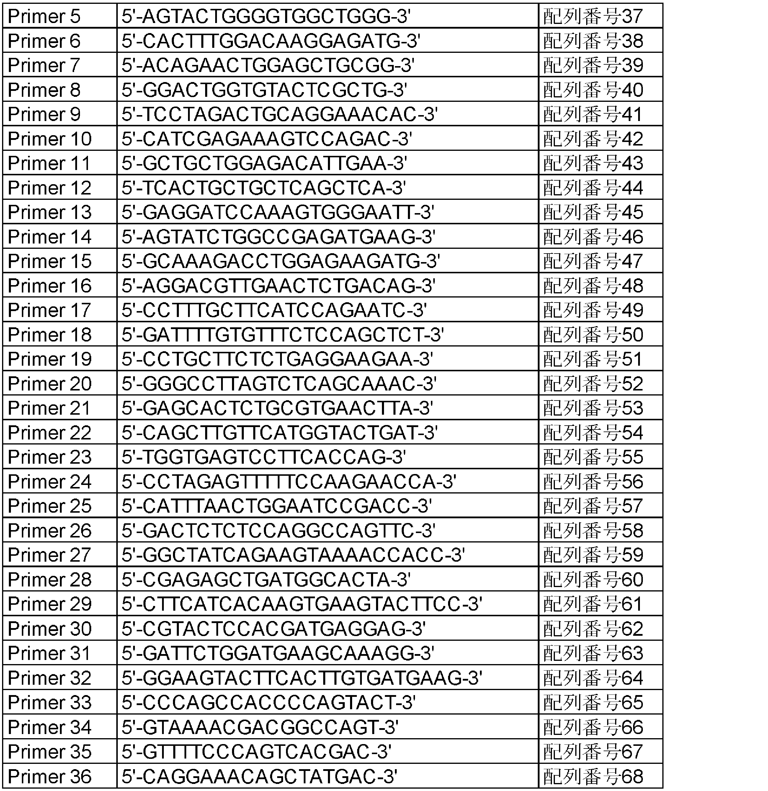
Asterand Bioscienceより購入したヒト正常甲状腺組織由来RNAと前記1-1で得られたヒト甲状腺がん組織由来RNAからSuperScript VILO cDNA Synthesis Kit(invitorgen)を用いて、以下の方法によりcDNAを合成した。280ngのTotal RNAを 容量が14μLになるようにRNAse free waterで調製し、5× VILO Reaction Mixを4μL、10× SuperScript Enzyme Mixを2μLそれぞれ加え混和した。25℃で10分間保温しその後42℃で60分保温した。反応を停止させるために最後に85℃で5分間インキュベートした。 Example 2 Detection of DCTN1-RET Fusion Gene Using SuperScript VILO cDNA Synthesis Kit (invitorgen) from human normal thyroid tissue-derived RNA purchased from Asterand Bioscience and human thyroid cancer tissue-derived RNA obtained in 1-1 above. CDNA was synthesized by the following method. 280 ng of total RNA was prepared with RNAse free water to a volume of 14 μL, and 4 μL of 5 × VILO Reaction Mix and 2 μL of 10 × SuperScript Enzyme Mix were added and mixed. The mixture was incubated at 25 ° C. for 10 minutes and then at 42 ° C. for 60 minutes. Finally, the reaction was incubated at 85 ° C. for 5 minutes to stop the reaction.

発現ベクター構築のため、表4に示すように、センスプライマーとしてPrimer40(配列番号72)とアンチセンスプライマーとしてPrimer41(配列番号73)を設計した。 Example 3 Construction of Expression Vector for DCTN1-RET Fusion Gene For expression vector construction, Primer40 (SEQ ID NO: 72) as a sense primer and Primer41 (SEQ ID NO: 73) as an antisense primer were designed as shown in Table 4.
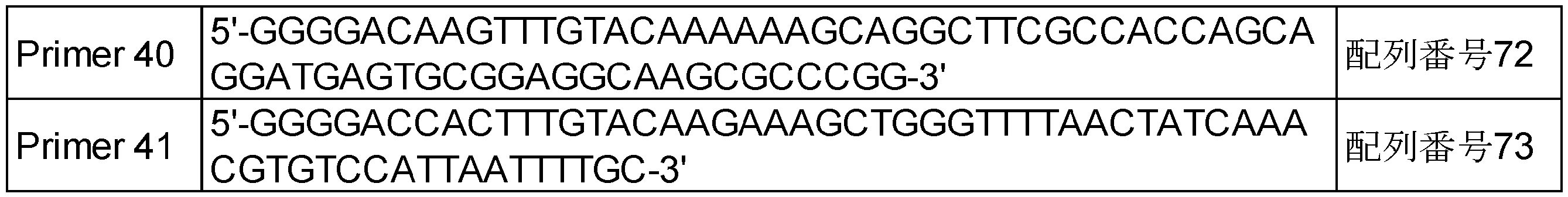
<4-1 細胞の樹立>
DCTN1-RET融合遺伝子発現細胞の樹立のための宿主細胞として、マウス胎児線維芽細胞NIH/3T3細胞(American Type Culture Collection)を選択し、上記にて作製したDCTN1-RET融合遺伝子が組み込まれた発現ベクターをトランスフェクションすることにより、DCTN1-RET融合遺伝子発現細胞を樹立した。詳細な方法は以下に示すとおりに実施した。NIH/3T3細胞は、通常の培養(2次元培養)のために、D-MEM(高グルコース)(L-グルタミン、フェノールレッド、ピルビン酸ナトリウム、1500mg/L炭酸水素ナトリウム含有)(WAKO)に10%となるようにNewborn Calf Serum(NBCS)(GIBCO)を添加したものを2次元培養用の培地として使用し、37℃、5%CO2にて培養したものを使用した。トランスフェクションを行う前日に、NIH/3T3細胞を1.5×105cells/2mLにて、6well plate(IWAKI)に播種し、37℃、5%CO2にて、一晩インキュベートした。1.5μgのDCTN1-RET融合遺伝子発現ベクター、1.5μgのpJTI phiC31 integrase vectorを混合した混合液に、その混合液の6倍量のViaFect Transfection Reagentを添加した後、全量が300μLとなるようにOpti-MEMを添加し、室温にて5分間インキュベートすることによりトランスフェクション溶液を作製した。NIH/3T3細胞が播種されているwellから培地を300μL除去し、上記で作製したトランスフェクション溶液を300μL添加し、37℃、5%CO2にて、一晩インキュベートした。翌日、トランスフェクション溶液を除去するために、培地を交換した。培地を交換する際に、新しい培地には、ハイグロマイシンB(ナカライテスク)を500μg/mLとなるように添加した。ハイグロマイシンBにより、DCTN1-RET融合遺伝子導入発現ベクターが導入されていない細胞を除去した。
トランスフェクション後、1週間に2回程度の培地交換をしながら、細胞が増殖するまで、培養を行った。トランスフェクション22日後に、細胞をトリプシンにて回収し、以下の方法にてシングルセルクローニングを行った。回収した細胞の細胞数を測定し、1cell/200μLとなるように培地を添加した。96well Plate(ThermoFisher)に200μL/1wellとなるように細胞を播種した。播種後、日々観察を行い、シングルセルから増殖してきた細胞を取得し、DCTN1-RET融合遺伝子発現細胞(DCTN1-RET融合遺伝子発現NIH/3T3細胞)とした。 Example 4 Establishment of DCTN1-RET Fusion Gene Expressing Cells <4-1 Establishment of Cells>
Mouse fetal fibroblasts NIH / 3T3 cells (American Type Culture Collection) were selected as host cells for establishment of DCTN1-RET fusion gene-expressing cells, and expression incorporating the DCTN1-RET fusion gene prepared above By transfection of the vector, cells expressing the DCTN1-RET fusion gene were established. The detailed method was implemented as shown below. NIH / 3T3 cells were cultured in D-MEM (high glucose) (containing L-glutamine, phenol red, sodium pyruvate, 1500 mg / L sodium bicarbonate) (WAKO) for normal culture (two-dimensional culture). % and comprising as the material obtained by adding Newborn Calf Serum (NBCS) (GIBCO ) was used as a medium for 2 dimensional culture, 37 ° C., it was used after cultured at 5% CO 2. The day before transfection, NIH / 3T3 cells were seeded at 1.5 × 10 5 cells / 2 mL in a 6-well plate (IWAKI) and incubated overnight at 37 ° C., 5% CO 2 . After adding 6 times the amount of ViaFect Transfection Reagent of the mixture to a mixture containing 1.5 μg of the DCTN1-RET fusion gene expression vector and 1.5 μg of the pJTI phiC31 integrase vector, the total volume will be 300 μL. A transfection solution was made by adding Opti-MEM and incubating for 5 minutes at room temperature. 300 μL of culture medium was removed from the wells in which NIH / 3T3 cells were seeded, 300 μL of the transfection solution prepared above was added, and the mixture was incubated overnight at 37 ° C., 5% CO 2 . The next day, the medium was changed to remove the transfection solution. When replacing the medium, hygromycin B (Nacalai Tesque) was added to the new medium to 500 μg / mL. Hygromycin B eliminated cells into which the DCTN1-RET fusion gene transfer expression vector had not been introduced.
After transfection, culture was performed until the cells proliferated while changing the medium about twice a week. Twenty-two days after transfection, cells were harvested with trypsin and single cell cloning was performed by the following method. The number of collected cells was measured, and the medium was added to 1 cell / 200 μL. Cells were seeded at 200 μl / well in a 96-well plate (ThermoFisher). After the seeding, daily observation was performed, and cells grown from a single cell were obtained and used as cells expressing DCTN1-RET fusion gene (DCTN1-RET fusion gene-expressing NIH / 3T3 cells).
得られたDCTN1-RET融合遺伝子発現NIH/3T3細胞におけるDCTN1-RET融合タンパク質の発現を、ウエスタンブロット法にて確認した。すなわち、培養フラスコから培地を除去し、PBSにて1度洗浄した。培養フラスコ内にPhosphatase inhibitor(ロシュ)とProtease inhibitor(ロシュ)を含有したSample Diluent Concentrate 2(R&D SYSTEMS)を添加し、スクレイパーにて細胞溶解液を回収した。回収した細胞溶解液から遠心分離により、タンパク質サンプルを得た。タンパク質サンプルは、タンパク質定量を行い、タンパク質濃度を一定にした。一定濃度のタンパク質サンプルに対して、Sample Buffer Solution with Reducing Reagent(6x) for SDS-PAGE(ナカライテスク)を加え、95℃にて5分間インキュベートすることにより、タンパク質を変性させ、ウエスタンブロッティング用のサンプルを得た。なお、ネガティブコントロール用のサンプルとして、親株であるNIH/3T3細胞を用いて、同様の方法にてウエスタンブロティング用のサンプルを得た。上記サンプルを用いて、以下に示す方法にて、タンパク質の発現を確認した。4-15パーセント Acrylamide gel(BIO―RAD)および1× Tris/Glycine/SDS Bufferを用いて、SDS-PAGE電気泳動(200Vで30分)にて、タンパク質を分離した。Trans-Blot Turb RTA Midi PVDF Transfer Kit(BIO―RAD)とTransblot Turbo 転写システム(BIO―RAD)を用いてPVDF膜にタンパク質を転写し、PVDF膜をBlocking One-Pに1時間浸漬した。Blocking One-Pが10%となるようにTBS-Tで希釈した溶液で一次抗体(Phospho-Ret (Tyr905) Antibody(CST)、Ret (C31B4) Rabbit mAb(CST)及びAnti-Dctn1 Antibody(ATLAS ANTIBODIES))を1/1000濃度になるように希釈し、PVDF膜を浸漬させ、4℃にて一晩インキュベートした。TBS-Tにて洗浄後、Anti-rabbit IgG,HRP-linked Antibody(CST)を1/2000濃度になるようにTBS-Tにて希釈した2次抗体希釈液にて、PVDF膜を浸漬させ、室温にて1時間インキュベートした。TBS-Tにて洗浄後、SuperSignal West Dura Extended Duration Substrate(ThermoFisher)及びルミノ・イメージアナライザー Amersham Imager 600(GEヘルスケア)を用いてタンパク質の検出を行った。なお、検出タンパク質の分子量は、プレシジョン Plusプロテイ カレイドスコープスタンダード(BIORAD)により確認した。
その結果、図4a)及びb)に示すように、抗pRET抗体と抗RET抗体を用いたところ内在性のRET(150及び175kDa)は検出されなかった。一方、DCTN1-RET融合遺伝子発現NIH/3T3細胞においてのみ、175kDa付近にDCTN1-RET融合タンパク質と推測されるバンドが確認できた。
また、図4c)に示すように抗DCTN1抗体を用いたところ、150kDa付近に内在性のDCTN1が検出された。一方、DCTN1-RET融合遺伝子発現NIH/3T3細胞においてのみ、内在性の150kDaのDCTN1のバンドの上の175kDa付近にバンドが検出された。すなわち、DCTN1-RET融合遺伝子発現NIH/3T3細胞においてのみ、RETに対する抗体及びDCTN1に対する抗体の両方で175kDa付近のバンドが検出されたことから、作成したDCTN1-RET融合遺伝子発現NIH/3T3細胞では、DCTN1とRETが融合したタンパク質が発現していることが明らかとなった。 <4-2 Confirmation of target protein expression>
Expression of the DCTN1-RET fusion protein in the obtained DCTN1-RET fusion gene-expressing NIH / 3T3 cells was confirmed by Western blotting. That is, the culture medium was removed from the culture flask and washed once with PBS. Sample Diluent Concentrate 2 (R & D SYSTEMS) containing Phosphatase inhibitor (Roche) and Protease inhibitor (Roche) was added to the culture flask, and the cell lysate was collected with a scraper. A protein sample was obtained by centrifugation from the collected cell lysate. The protein sample was subjected to protein quantification to keep the protein concentration constant. Sample Buffer Solution with Reducing Reagent (6x) for SDS-PAGE (Nacalai Tesque) is added to a constant concentration protein sample, and the protein is denatured by incubating at 95 ° C for 5 minutes, and a sample for Western blotting I got As a sample for negative control, a parent strain NIH / 3T3 cell was used, and a sample for western blotting was obtained in the same manner. The expression of protein was confirmed by the method described below using the above sample. The proteins were separated by SDS-PAGE electrophoresis (30 minutes at 200 V) using 4-15% acrylamide gel (BIO-RAD) and 1 × Tris / Glycine / SDS Buffer. The protein was transferred to the PVDF membrane using Trans-Blot Turb RTA Midi PVDF Transfer Kit (BIO-RAD) and Transblot Turbo Transfer System (BIO-RAD), and the PVDF membrane was immersed in Blocking One-P for 1 hour. Primary antibody (Phospho-Ret (Tyr905) Antibody (CST), Ret (C31 B4) Rabbit mAb (CST) and Anti-Dctn1 Antibody (ATLAS ANTIBODIES) in a solution diluted with TBS-T so that Blocking One-P is 10% ) Was diluted to a concentration of 1/1000, the PVDF membrane was immersed, and incubated overnight at 4 ° C. After washing with TBS-T, the PVDF membrane is immersed in a secondary antibody diluent diluted with TBS-T so that the concentration of Anti-rabbit IgG and HRP-linked Antibody (CST) becomes 1/2000. Incubated for 1 hour at room temperature. After washing with TBS-T, protein detection was performed using SuperSignal West Dura Extended Duration Substrate (ThermoFisher) and a Lumino Image Analyzer Amersham Imager 600 (GE Healthcare). The molecular weight of the detection protein was confirmed by Precision Plus proteinoid standard (BIORAD).
As a result, as shown in FIG. 4 a) and b), endogenous RET (150 and 175 kDa) was not detected when anti-pRET antibody and anti-RET antibody were used. On the other hand, only in the DCTH1-RET fusion gene-expressing NIH / 3T3 cell, a band presumed to be a DCTN1-RET fusion protein could be confirmed around 175 kDa.
In addition, as shown in FIG. 4 c), when an anti-DCTN1 antibody was used, endogenous DCTN1 was detected around 150 kDa. On the other hand, a band was detected around 175 kDa above the endogenous 150 kDa DCTN1 band only in the DCTH1-RET fusion gene-expressing NIH / 3T3 cells. That is, since bands around 175 kDa were detected in both the antibody against RET and the antibody against DCTN1 only in the DCTN1-RET fusion gene-expressing NIH / 3T3 cells, in the constructed DCTN1-RET fusion gene-expressing NIH / 3T3 cells, It was revealed that a protein in which DCTN1 and RET were fused was expressed.
NIH/3T3細胞は、2次元培養条件では良好な増殖を示すが、3次元培養条件では、ほとんど増殖しない。一方で、NIH/3T3細胞に癌遺伝子を発現させることにより、3次元培養条件でも増殖することが知られている。その性質を利用し、DCTN1-RET融合遺伝子が癌遺伝子であるかの確認を行った。37℃、5%CO2にて、2次元培養で培養したDCTN1-RET融合遺伝子発現NIH/3T3細胞及びNIH/3T3細胞をトリプシンにて回収し、細胞数を測定した。3次元培養を行うために、FCeM series Preparation kit(日産化学工業株式会社)とD-MEM(高グルコース)(L-グルタミン、フェノールレッド、ピルビン酸ナトリウム、1500mg/L炭酸水素ナトリウム含有)(WAKO)とNewborn Calf Serum(NBCS)(GIBCO)を用いて、3次元培養用の培地を作製した。作製した3次元培養用の培地に、細胞を1000cells/90μLとなるように懸濁し、96 Well Clear Black Round Bottom、Spheroid Microplate(Corning)に90μL/1wellとなるように播種し、37℃、5%CO2にてインキュベートした。播種翌日(Day1)及び播種8日後(Day8)に細胞内ATP発光検出試薬であるCelltiter-Glo 2.0Reagent(Progema)及びルミノメーター(EnSpire, PerkinElmer)を用いて発光量(counts per second:cps)を測定し、生細胞数の指標とした。Day1での測定結果とDay8での測定結果から各細胞の増殖率を算出した(N=3)。
その結果、図5に示すように、NIH/3T3細胞では、Day8の細胞数はDay1の細胞数と比較して2.4倍であったのに対して、DCTN1-RET融合遺伝子発現NIH/3T3細胞では、20.9倍であった。また、NIH/3T3細胞は、3次元培養において細胞の凝集塊は形成されないが、DCTN1-RET融合遺伝子発現NIH/3T3細胞では、3次元培養により細胞の凝集塊が形成されていることが確認された。
すなわち、DCTN1-RET融合遺伝子を導入することにより、細胞の増殖が亢進したことが明らかとなり、DCTN1-RET融合遺伝子が癌遺伝子であることが示唆された。 Example 5 Growth Confirmation by Three-Dimensional Culture of DCTN1-RET Fusion Gene Expressing NIH / 3T3 Cells NIH / 3T3 cells show good growth in two-dimensional culture conditions, but hardly grow in three-dimensional culture conditions. On the other hand, it is known that, by expressing oncogenes in NIH / 3T3 cells, they proliferate under three-dimensional culture conditions. Using this property, it was confirmed that the DCTN1-RET fusion gene was an oncogene. The DCTN1-RET fusion gene-expressing NIH / 3T3 cells and NIH / 3T3 cells cultured in two-dimensional culture at 37 ° C. and 5% CO 2 were collected by trypsin, and the number of cells was measured. FCeM series Preparation kit (Nissan Chemical Industries, Ltd.) and D-MEM (high glucose) (L-glutamine, phenol red, sodium pyruvate, containing 1500 mg / L sodium bicarbonate) (WAKO) to perform three-dimensional culture And Newborn Calf Serum (NBCS) (GIBCO) were used to prepare a culture medium for three-dimensional culture. The cells are suspended in the prepared three-dimensional culture medium to 1000 cells / 90 μL and seeded at 90 μL / well in 96 Well Clear Black Round Bottom, Spheroid Microplate (Corning), 37 ° C., 5% It was incubated in CO 2. The amount of luminescence (counts per second: cps) using Intracellular ATP luminescence detection reagent Celltiter-Glo 2.0 Reagent (Progema) and luminometer (EnSpire, PerkinElmer) the day after seeding (Day 1) and 8 days after seeding (Day 8) Was used as an indicator of the number of viable cells. The proliferation rate of each cell was calculated from the measurement result on
As a result, as shown in FIG. 5, in NIH / 3T3 cells, the cell number of Day 8 was 2.4 times as compared with the cell number of
That is, it became clear that the cell proliferation was enhanced by introducing the DCTN1-RET fusion gene, suggesting that the DCTN-RET fusion gene was an oncogene.
DCTN1-RET融合遺伝子発現NIH/3T3細胞のin vivoにおける造腫瘍性を確認するために、ヌードマウスを用いた移植実験を行った。なお、親株であるNIH/3T3細胞は、ヌードマウスの皮下では増殖しないことが、一般的に知られており、DCTN1-RET融合遺伝子発現NIH/3T3細胞をヌードマウスの皮下に移植することにより、DCTN1-RET融合遺伝子が造腫瘍性に寄与するか、すなわち癌遺伝子であるかの確認ができる。被移植動物としては、ヌードマウス(BALB/cAJcl-nu/nu、日本クレア)を用いた。DCTN1-RET融合遺伝子発現NIH/3T3細胞をトリプシンにて回収し、最終的に1×108cells/mLとなるようにPBSに懸濁し、同量のマトリゲル基底膜マトリックス(Corning)を加え、5×107cells/mLにしたものを移植用細胞液とした。25G注射針と1mLシリンジを用いて、移植用細胞液をヌードマウス(N=10)の右側胸部の皮下に0.1mLずつ移植した。電子ノギス(ミツトヨ)を用い、移植後、10、13、17日目に、1匹ずつ腫瘍の長径及び短径を測定し、以下の式を用いて腫瘍体積を算出した。
腫瘍体積(mm3)=(長径、mm)×(短径、mm)×(短径、mm)/2
腫瘍体積の測定結果を図6に示す。その結果、ヌードマウスの皮下に移植されたDCTN1-RET融合遺伝子発現NIH/3T3細胞は、腫瘍を形成し、良好に増殖することが確認され、in vivo実験においてもDCTN1-RET融合遺伝子が癌遺伝子であることが示唆された。 Example 6 Confirming Tumorigenicity of DCH1-RET Fusion Gene Expressing NIH / 3T3 Cells In Vivo Transplantation using nude mice to confirm in vivo tumorigenicity of DCTN-RET fusion gene expressing NIH / 3T3 cells I did an experiment. In addition, it is generally known that the parent strain NIH / 3T3 cells do not proliferate in nude mice subcutaneously, and by implanting the DCTN1-RET fusion gene-expressing NIH / 3T3 cells subcutaneously in nude mice, It can be confirmed whether the DCTN1-RET fusion gene contributes to tumorigenicity, that is, it is an oncogene. As a recipient animal, nude mice (BALB / cAJcl-nu / nu, CLEA Japan, Inc.) were used. The DCTN1-RET fusion gene-expressing NIH / 3T3 cells are recovered with trypsin, suspended in PBS to a final concentration of 1 × 10 8 cells / mL, and the same amount of Matrigel basement membrane matrix (Corning) is added, 5 A cell solution for transplantation was prepared at 10 7 cells / mL. Using a 25G injection needle and a 1 mL syringe, 0.1 mL each of the cell solution for transplantation was subcutaneously implanted in the right chest of a nude mouse (N = 10). The long diameter and the short diameter of the tumor were measured one by one on
Tumor volume (mm 3 ) = (long diameter, mm) × (short diameter, mm) × (short diameter, mm) / 2
The measurement results of the tumor volume are shown in FIG. As a result, it has been confirmed that the DCTN1-RET fusion gene-expressing NIH / 3T3 cells implanted subcutaneously in nude mice form a tumor and proliferate well, and the in vivo DCTN1-RET fusion gene is an oncogene. Was suggested.
DCTN1-RET融合遺伝子発現NIH/3T3細胞に対するsiRNA処理による影響を確認した。用いたsiRNAは、下記の表5に示した3種類のRET siRNAとネガティブコントロールとしてSilencer Select Negative Control #1 siRNA(Ambion)を使用した。なお、3種類のRET siRNAは、いずれもヒトRETを標的とするsiRNAであるが、RET siRNA1及びRET siRNA2はDCTN1-RET融合遺伝子内のRET部分に結合する配列を含み、RET siRNA3はDCTN1-RET融合遺伝子内に結合する配列を含まない。すなわち、RET siRNA1及びRET siRNA2はDCTN1-RET融合遺伝子の発現を抑制するが、RET siRNA3はDCTN1-RET融合遺伝子の発現を抑制しないことが想定された。以下にsiRNAを用いた実験の方法について記載した。 Example 7 Confirmation of suppression of the DCTN1-RET fusion protein by siRNA using DCTN1-RET fusion gene expressing NIH / 3T3 cells and confirmation of cell growth inhibitory effect The effect of siRNA treatment on the DCTN1-RET fusion gene expressing NIH / 3T3 cells was confirmed . The siRNAs used were three RET siRNAs shown in Table 5 below and Silencer Select

翌日、一部は、タンパク質発現解析用にサンプリングを行い、一部については、細胞増殖確認用に再播種を行った。タンパク質発現解析用のサンプリング及びタンパク質発現解析は、一次抗体として、Phospho-Ret (Tyr905) Antibody(CST)、Ret (C31B4) Rabbit mAb(CST)及びGAPDH (D16H11) XP Rabbit mAb(CST)を用いた以外は、上記<4-2 目的タンパク質の発現確認>と同様の方法で実施した。その結果、図7に示すように、siRNA処理していない細胞(無処理)に比べ、ネガティブコントロールsiRNA(NC)を処理した細胞において、DCTN1-RET融合タンパク質の発現は抑制されていないことが確認できた。一方、RET siRNA1及びRET siRNA2を処理した場合、DCTN1-RET融合遺伝子発現NIH/3T3細胞のDCTN1-RET融合タンパク質の発現が抑制されることが確認され、RET siRNA3では抑制されないことが明らかとなった。
次に細胞増殖抑制効果の確認のために、無処理又はsiRNA処理されたwellからトリプシンにより細胞を回収し、細胞数を測定した。上記実施例5と同様の方法で3次元培養を行い、播種当日(Day0)及び播種4日後(Day4)に実施例5と同様の方法で生細胞数を測定した。Day0での測定結果とDay4での測定結果から各細胞における増殖率を算出した。
その結果、図8に示すように、siRNAを処理していない細胞(無処理)及びネガティブコントロールsiRNA(NC)を処理した細胞では、Day4の細胞数は、Day0の細胞数と比較して、4.9倍及び3.6倍であったのに対して、RET siRNA1及びRET siRNA2で処理した細胞では、2.0倍及び2.4倍程度の増殖であり、顕著に増殖率が低下した。一方、RET siRNA3はで処理した細胞では、3.7倍の増殖であり、ネガティブコントロールsiRNAと同程度の増殖率であり、増殖率の低下は見られなかった。これらの結果から、DCTN1-RET融合遺伝子発現NIH/3T3細胞の増殖は、siRNAによってRETの発現を阻害した場合にも抑制されることが明らかとなった。 A DCTN1-RET fusion gene-expressing NIH / 3T3 cell was cultured at 37 ° C., 5% CO 2 using a medium for two-dimensional culture. The day before the siRNA treatment, each cell was seeded at 3 × 10 5 cells / 2 mL in a 6-well plate (IWAKI) and incubated overnight at 37 ° C., 5% CO 2 . The siRNA solution was prepared by mixing 12 μL of each siRNA prepared beforehand to 20 μM with water, 4 μL of Lipofectamin RNAiMAX Transfection Reagent (ThermoFisher) and 384 μL of Opti-MEM, and incubating for 15 minutes at room temperature. 400 μL of the siRNA solution was added to the wells in which each cell was seeded, and incubated overnight at 37 ° C., 5% CO 2 .
The next day, some were sampled for protein expression analysis, and some were replated for cell proliferation confirmation. For sampling and protein expression analysis for protein expression analysis, Phospho-Ret (Tyr905) Antibody (CST), Ret (C31 B4) Rabbit mAb (CST) and GAPDH (D16 H11) XP Rabbit mAb (CST) were used as primary antibodies. Except for the above, it was carried out in the same manner as the above <4-2 Confirmation of target protein expression>. As a result, as shown in FIG. 7, it was confirmed that the expression of the DCTN1-RET fusion protein was not suppressed in the cells treated with the negative control siRNA (NC) as compared to the cells not treated with siRNA (untreated). did it. On the other hand, when
Next, in order to confirm the cell growth inhibitory effect, cells were collected by trypsin from the untreated or siRNA-treated wells, and the number of cells was measured. Three-dimensional culture was performed in the same manner as in Example 5 above, and the number of viable cells was measured in the same manner as in Example 5 on the day of seeding (Day 0) and 4 days after sowing (Day 4). The proliferation rate in each cell was calculated from the measurement result on
As a result, as shown in FIG. 8, in the cells not treated with siRNA (no treatment) and the cells treated with negative control siRNA (NC), the number of cells in Day 4 was 4 compared to the number of cells in
DCTN1-RET融合遺伝子発現NIH/3T3細胞に対するin vitro細胞増殖試験を行った。上記実施例5と同様の方法で、3次元培養及び播種を行った。播種後37℃、5%CO2にて一晩インキュベートした(Day0)。RETを阻害すると報告されているカボザンチニブ、バンデタニブ、アレクチニブ、レンバチニブ、縮合ピリミジン化合物(Compound1から9;表6に示す)をジメチルスルホキシドにて10 mmol/Lの濃度に溶解し、さらに3次元培養用の培地を用いて、これらの化合物の最終濃度がそれぞれ1000、333、111、37.0、12.3、4.12、1.37、0.457nmol/Lになるように希釈を行った。これを先に述べた細胞が播種されたプレートの各Wellに0.01mLずつ加え(Day1)、37℃、5%CO2にて7日間インキュベートした。培養後(Day8)、全てのWellに細胞内ATP発光検出試薬であるCelltiter-Glo 2.0Reagent(Progema)を添加し、ルミノメーター(EnSpire、PerkinElmer)を用いて発光量(counts per second:cps)を測定した。Tday8とCday1の値の大きさに応じて、以下の式より化合物の各濃度におけるDay1からの増殖率を算出し、細胞増殖を50%抑制する被験化合物の濃度(GI50(nM))を求めた。
1)Tday8≧Cday1の場合
増殖率(%)=(Tday8-Cday1)/(Cday8-Cday1)×100
T:被験化合物を添加したWellのcps
C:被験化合物を添加しなかったWellのcps
Day1:被験化合物を添加した日
Day8:評価日
2)Tday8<Cday1の場合
増殖率(%)=(Tday8-Cday1)/(Cday1)×100
T:被験化合物を添加したWellのcps
C:被験化合物を添加しなかったWellのcps
Day1:被験化合物を添加した日
Day8:評価日 Example 8 Cell growth inhibitory effect using DCTN1-RET fusion gene-expressing NIH / 3T3 cells An in vitro cell proliferation test was performed on DCTN-RET fusion gene-expressing NIH / 3T3 cells. Three-dimensional culture and seeding were performed in the same manner as in Example 5 above. After seeding, the plate was incubated overnight at 37 ° C., 5% CO 2 (Day 0). Cabozatininib, vandetanib, alectinib, lenvatinib, fused pyrimidine compounds (
1) When T day 8 CC day 1 Growth rate (%) = (T day 8- C day 1 ) / (C day 8- C day 1 ) × 100
T: Well's cps added with test compound
C: Well's cps to which no test compound was added
Day 1: Day when test compound was added Day 8: Evaluation day 2) When T day 8 <C day 1 Growth rate (%) = (T day 8- C day 1 ) / (C day 1 ) × 100
T: Well's cps added with test compound
C: Well's cps to which no test compound was added
Day 1: Day when the test compound was added Day 8: Evaluation day

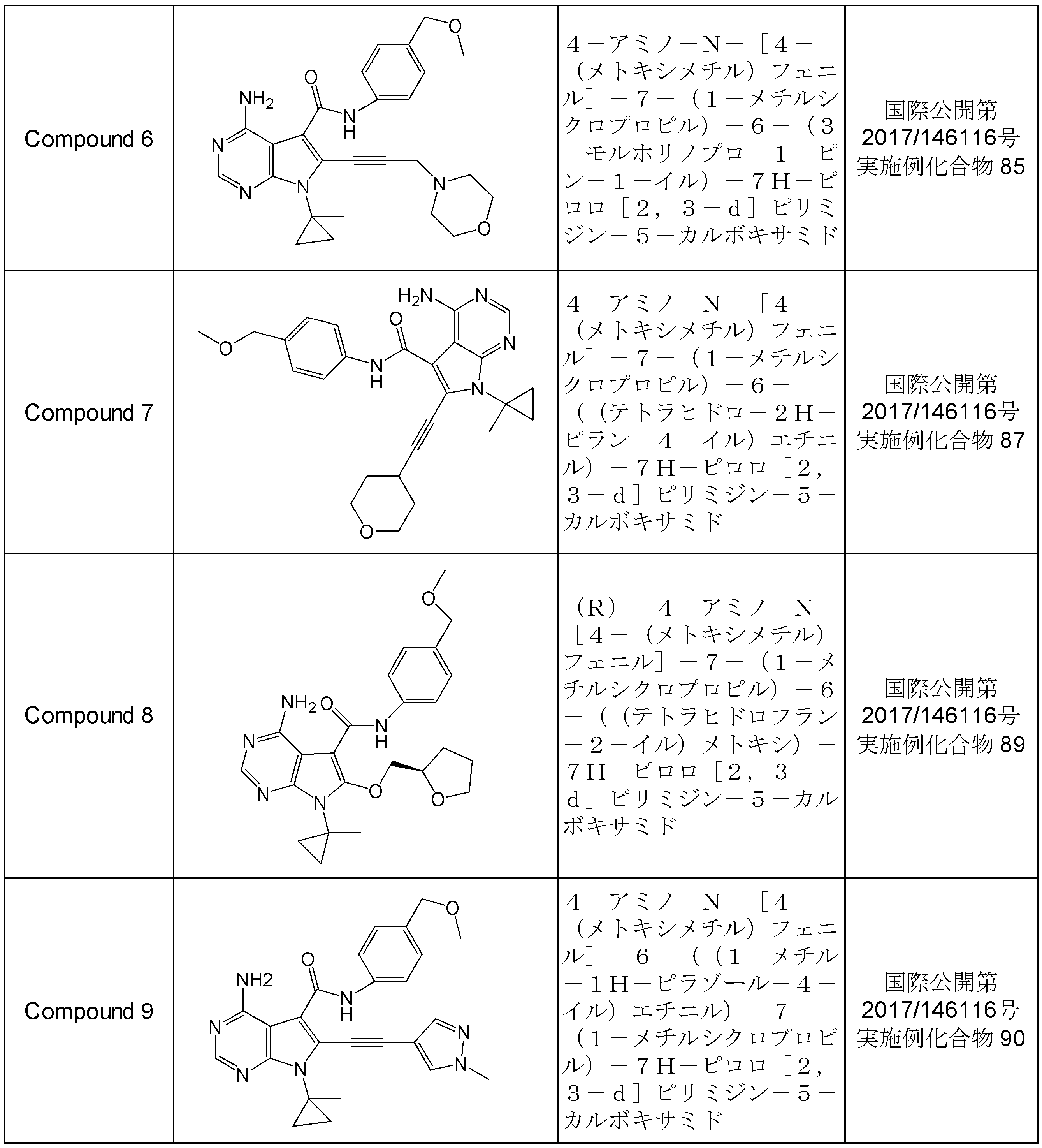
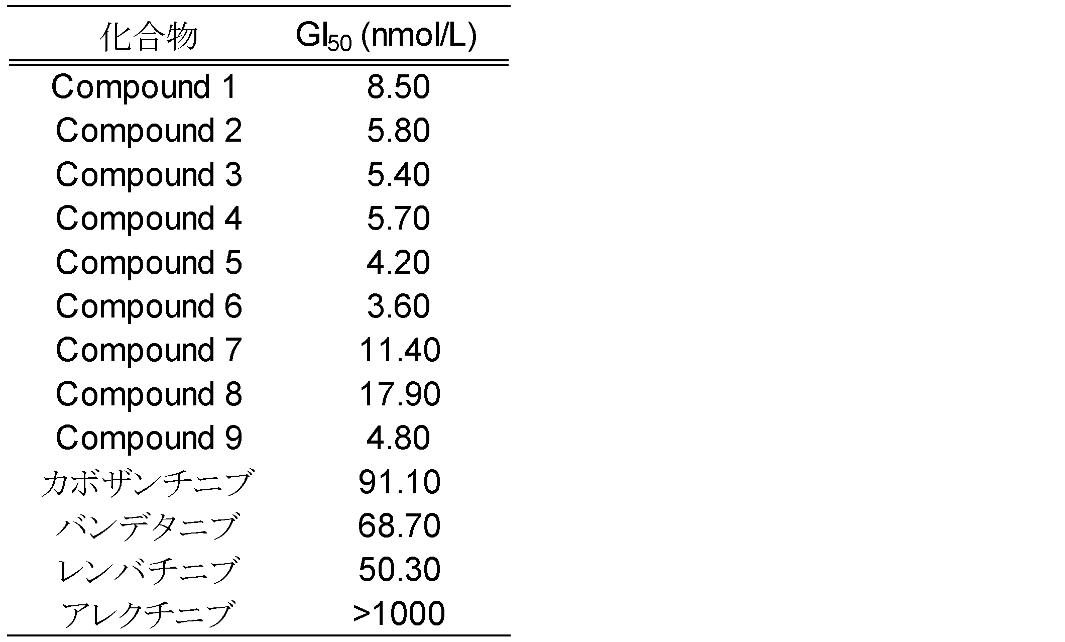
DCTN1-RET融合遺伝子発現細胞におけるRETのリン酸化が、RETを阻害すると報告されている既存の薬剤で阻害されるかについて以下の方法にて検討した。
DCTN1-RET融合遺伝子発現NIH/3T3細胞は、2次元培養用の培地を使用し、37℃、5%CO2にて培養したものを使用した。薬剤処理を行う前日に、各細胞を3×105cells/2mLにて、6well plate(IWAKI)に播種し、37℃、5%CO2にて、一晩インキュベートした。カボザンチニブ、バンデタニブ、アレクチニブ、レンバチニブをジメチルスルホキシドにて10mmol/Lの濃度に溶解し、さらにPBSを用いて、これらの化合物の最終濃度がそれぞれ1000、100、10nmol/Lになるように希釈を行った。これを先に述べた細胞が播種されたプレートの各Wellに20μLずつ加え(Day1)、37℃、5%CO2にて1時間インキュベートした。インキュベート後、上記実施例7に記載されている方法と同様の方法でタンパク質発現解析用のサンプリングを行い、タンパク質発現解析を実施した。
その結果、図9に示すように、DCTN1-RET融合遺伝子発現NIH/3T3細胞のリン酸化RETレベルがカボザンチニブ及びレンバチニブにより顕著に減少することが確認された。また、上記と同様の方法で、縮合ピリミジン化合物を用いて、RETのリン酸化阻害を評価した結果、縮合ピリミジン化合物においてもRETのリン酸化が顕著に減少することが確認できた。
以上の結果から、リン酸化RETレベルを顕著に減少させる薬剤は、DCTN1-RET融合遺伝子発現NIH/3T3細胞の増殖を抑制できる化合物であり、DCTN1-RET融合遺伝子が検出されたがんに対する治療薬として有用である可能性が示唆された。また、DCTN1-RET融合遺伝子発現NIH/3T3細胞のリン酸化RETレベルを用いることでRET阻害剤のスクリーニングが可能であることが示唆された。 Example 9 Phosphorylation inhibition of RET using DCTN1-RET fusion gene-expressing cells The following describes whether phosphorylation of RET in cells expressing DCTN-RET fusion gene expression is inhibited by existing drugs that have been reported to inhibit RET. We examined by method of.
A DCTN1-RET fusion gene-expressing NIH / 3T3 cell was cultured at 37 ° C., 5% CO 2 using a medium for two-dimensional culture. The day before drug treatment, each cell was seeded at 3 × 10 5 cells / 2 mL in a 6-well plate (IWAKI) and incubated overnight at 37 ° C., 5% CO 2 . Cabozantinib, vandetanib, alectinib, renvatinib were dissolved in dimethyl sulfoxide to a concentration of 10 mmol / L, and further diluted with PBS so that the final concentrations of these compounds would be 1000, 100, and 10 nmol / L, respectively. . This was added to each well of the plate on which the cells described above were seeded (Day 1), and incubated at 37 ° C., 5
As a result, as shown in FIG. 9, it was confirmed that phosphorylated RET levels of the DCTN1-RET fusion gene-expressing NIH / 3T3 cells were significantly reduced by cabozantinib and renvatinib. Moreover, as a result of evaluating the phosphorylation inhibition of RET using a fused pyrimidine compound in the same manner as described above, it was confirmed that the phosphorylation of RET is also significantly reduced in the fused pyrimidine compound.
From the above results, the drug that significantly reduces the phosphorylated RET level is a compound that can suppress the growth of the DCTN1-RET fusion gene-expressing NIH / 3T3 cells, and a therapeutic agent for cancer in which the DCTN-RET fusion gene is detected It was suggested that it might be useful. In addition, it was suggested that screening of RET inhibitors is possible by using phosphorylated RET levels of DCTN1-RET fusion gene-expressing NIH / 3T3 cells.
配列番号2は、DCTN1 v1とRET v2との融合ペプチドのアミノ酸配列を示す。
配列番号3は、DCTN1 v1とRET variant 4(v4)[配列番号32の一部]との融合ペプチドをコードするポリヌクレオチドの塩基配列を示す。
配列番号4は、DCTN1 v1とRET v4との融合ペプチドのアミノ酸配列を示す。
配列番号5は、DCTN1 variant 2(v2)[配列番号26の一部]とRET v2との融合ペプチドをコードするポリヌクレオチドの塩基配列を示す。
配列番号6は、DCTN1 v2とRET v2との融合ペプチドのアミノ酸配列を示す。
配列番号7は、DCTN1 v2とRET v4との融合ペプチドをコードするポリヌクレオチドの塩基配列を示す。
配列番号8は、DCTN1 v2とRET v4との融合ペプチドのアミノ酸配列を示す。
配列番号9は、DCTN1 variant 3(v3)[配列番号27の一部]とRET v2との融合ペプチドをコードするポリヌクレオチドの塩基配列を示す。
配列番号10は、DCTN1 v3とRET v2との融合ペプチドのアミノ酸配列を示す。
配列番号11は、DCTN1 v3とRET v4との融合ペプチドをコードするポリヌクレオチドの塩基配列を示す。
配列番号12は、DCTN1 v3とRET v4との融合ペプチドのアミノ酸配列を示す。
配列番号13は、DCTN1 variant 4(v4)[配列番号28の一部]とRET v2との融合ペプチドをコードするポリヌクレオチドの塩基配列を示す。
配列番号14は、DCTN1 v4とRET v2との融合ペプチドのアミノ酸配列を示す。
配列番号15は、DCTN1 v4とRET v4との融合ペプチドをコードするポリヌクレオチドの塩基配列を示す。
配列番号16は、DCTN1 v4とRET v4との融合ペプチドのアミノ酸配列を示す。
配列番号17は、DCTN1 variant 5(v5)[配列番号29の一部]とRET v2との融合ペプチドをコードするポリヌクレオチドの塩基配列を示す。
配列番号18は、DCTN1 v5とRET v2との融合ペプチドのアミノ酸配列を示す。
配列番号19は、DCTN1 v5とRET v4との融合ペプチドをコードするポリヌクレオチドの塩基配列を示す。
配列番号20は、DCTN1 v5とRET v4との融合ペプチドのアミノ酸配列を示す。
配列番号21は、DCTN1 v6とRET v2との融合ペプチドをコードするポリヌクレオチドの塩基配列を示す。
配列番号22は、DCTN1 v6とRET v2との融合ペプチドのアミノ酸配列を示す。
配列番号23は、DCTN1 v6とRET v4との融合ペプチドをコードするポリヌクレオチドの塩基配列を示す。
配列番号24は、DCTN1 v6とRET v4との融合ペプチドのアミノ酸配列を示す。
配列番号33は、プライマーの塩基配列を示す。
配列番号34は、プライマーの塩基配列を示す。
配列番号35は、プライマーの塩基配列を示す。
配列番号36は、プライマーの塩基配列を示す。
配列番号37は、プライマーの塩基配列を示す。
配列番号38は、プライマーの塩基配列を示す。
配列番号39は、プライマーの塩基配列を示す。
配列番号40は、プライマーの塩基配列を示す。
配列番号41は、プライマーの塩基配列を示す。
配列番号42は、プライマーの塩基配列を示す。
配列番号43は、プライマーの塩基配列を示す。
配列番号44は、プライマーの塩基配列を示す。
配列番号45は、プライマーの塩基配列を示す。
配列番号46は、プライマーの塩基配列を示す。
配列番号47は、プライマーの塩基配列を示す。
配列番号48は、プライマーの塩基配列を示す。
配列番号49は、プライマーの塩基配列を示す。
配列番号50は、プライマーの塩基配列を示す。
配列番号51は、プライマーの塩基配列を示す。
配列番号52は、プライマーの塩基配列を示す。
配列番号53は、プライマーの塩基配列を示す。
配列番号54は、プライマーの塩基配列を示す。
配列番号55は、プライマーの塩基配列を示す。
配列番号56は、プライマーの塩基配列を示す。
配列番号57は、プライマーの塩基配列を示す。
配列番号58は、プライマーの塩基配列を示す。
配列番号59は、プライマーの塩基配列を示す。
配列番号60は、プライマーの塩基配列を示す。
配列番号61は、プライマーの塩基配列を示す。
配列番号62は、プライマーの塩基配列を示す。
配列番号63は、プライマーの塩基配列を示す。
配列番号64は、プライマーの塩基配列を示す。
配列番号65は、プライマーの塩基配列を示す。
配列番号66は、プライマーの塩基配列を示す。
配列番号67は、プライマーの塩基配列を示す。
配列番号68は、プライマーの塩基配列を示す。
配列番号69は、プライマーの塩基配列を示す。
配列番号70は、プライマーの塩基配列を示す。
配列番号71は、プライマーの塩基配列を示す。
配列番号72は、プライマーの塩基配列を示す。
配列番号73は、プライマーの塩基配列を示す。
配列番号74は、RET siRNAの塩基配列を示す。
配列番号75は、RET siRNAの塩基配列を示す。
配列番号76は、RET siRNAの塩基配列を示す。
配列番号77は、RET siRNAの塩基配列を示す。 SEQ ID NO: 1 shows the base sequence of a polynucleotide encoding a fusion peptide of DCTN1 variant 1 (v1) [part of SEQ ID NO: 25] and RET variant 2 (v2) [part of SEQ ID NO: 31].
SEQ ID NO: 2 shows the amino acid sequence of a fusion peptide of DCTN1 v1 and RET v2.
SEQ ID NO: 3 shows the base sequence of a polynucleotide encoding a fusion peptide of DCTN1 v1 and RET variant 4 (v4) [part of SEQ ID NO: 32].
SEQ ID NO: 4 shows the amino acid sequence of a fusion peptide of DCTN1 v1 and RET v4.
SEQ ID NO: 5 shows the base sequence of a polynucleotide encoding a fusion peptide of DCTN variant 2 (v2) [part of SEQ ID NO: 26] and RET v2.
SEQ ID NO: 6 shows the amino acid sequence of the fusion peptide of DCTN1 v2 and RET v2.
SEQ ID NO: 7 shows the nucleotide sequence of a polynucleotide encoding a fusion peptide of DCTN1 v2 and RET v4.
SEQ ID NO: 8 shows the amino acid sequence of a fusion peptide of DCTN1 v2 and RET v4.
SEQ ID NO: 9 shows the base sequence of a polynucleotide encoding a fusion peptide of DCTN variant 3 (v3) [part of SEQ ID NO: 27] and RET v2.
SEQ ID NO: 10 shows the amino acid sequence of a fusion peptide of DCTN1 v3 and RET v2.
SEQ ID NO: 11 shows the nucleotide sequence of a polynucleotide encoding a fusion peptide of DCTN1 v3 and RET v4.
SEQ ID NO: 12 shows the amino acid sequence of a fusion peptide of DCTN1 v3 and RET v4.
SEQ ID NO: 13 shows the nucleotide sequence of a polynucleotide encoding a fusion peptide of DCTN variant 4 (v 4) [part of SEQ ID NO: 28] and RET v2.
SEQ ID NO: 14 shows the amino acid sequence of a fusion peptide of DCTN1 v4 and RET v2.
SEQ ID NO: 15 shows the nucleotide sequence of a polynucleotide encoding a fusion peptide of DCTN1 v4 and RET v4.
SEQ ID NO: 16 shows the amino acid sequence of a fusion peptide of DCTN1 v4 and RET v4.
SEQ ID NO: 17 shows the base sequence of a polynucleotide encoding a fusion peptide of DCTN variant 5 (v5) [part of SEQ ID NO: 29] and RET v2.
SEQ ID NO: 18 shows the amino acid sequence of a fusion peptide of DCTN1 v5 and RET v2.
SEQ ID NO: 19 shows the nucleotide sequence of a polynucleotide encoding a fusion peptide of DCTN1 v5 and RET v4.
SEQ ID NO: 20 shows the amino acid sequence of a fusion peptide of DCTN1 v5 and RET v4.
SEQ ID NO: 21 shows the nucleotide sequence of a polynucleotide encoding a fusion peptide of DCTN1 v6 and RET v2.
SEQ ID NO: 22 shows the amino acid sequence of a fusion peptide of DCTN1 v6 and RET v2.
SEQ ID NO: 23 shows the nucleotide sequence of a polynucleotide encoding a fusion peptide of DCTN1 v6 and RET v4.
SEQ ID NO: 24 shows the amino acid sequence of a fusion peptide of DCTN1 v6 and RET v4.
SEQ ID NO: 33 shows the nucleotide sequence of a primer.
SEQ ID NO: 34 shows the nucleotide sequence of a primer.
SEQ ID NO: 35 shows the nucleotide sequence of a primer.
SEQ ID NO: 36 shows the nucleotide sequence of a primer.
SEQ ID NO: 37 shows the nucleotide sequence of a primer.
SEQ ID NO: 38 shows the nucleotide sequence of a primer.
SEQ ID NO: 39 shows the nucleotide sequence of a primer.
SEQ ID NO: 40 shows the nucleotide sequence of a primer.
SEQ ID NO: 41 shows the nucleotide sequence of a primer.
SEQ ID NO: 42 shows the nucleotide sequence of a primer.
SEQ ID NO: 43 shows the nucleotide sequence of a primer.
SEQ ID NO: 44 shows the nucleotide sequence of a primer.
SEQ ID NO: 45 shows the nucleotide sequence of a primer.
SEQ ID NO: 46 shows the nucleotide sequence of a primer.
SEQ ID NO: 47 shows the nucleotide sequence of a primer.
SEQ ID NO: 48 shows the nucleotide sequence of a primer.
SEQ ID NO: 49 shows the nucleotide sequence of a primer.
SEQ ID NO: 50 shows the nucleotide sequence of a primer.
SEQ ID NO: 51 shows the nucleotide sequence of a primer.
SEQ ID NO: 52 shows the nucleotide sequence of a primer.
SEQ ID NO: 53 shows the nucleotide sequence of a primer.
SEQ ID NO: 54 shows the nucleotide sequence of a primer.
SEQ ID NO: 55 shows the nucleotide sequence of a primer.
SEQ ID NO: 56 shows the nucleotide sequence of a primer.
SEQ ID NO: 57 shows the nucleotide sequence of a primer.
SEQ ID NO: 58 shows the nucleotide sequence of a primer.
SEQ ID NO: 59 shows the nucleotide sequence of a primer.
SEQ ID NO: 60 shows the nucleotide sequence of a primer.
SEQ ID NO: 61 shows the nucleotide sequence of a primer.
SEQ ID NO: 62 shows the nucleotide sequence of a primer.
SEQ ID NO: 63 shows the nucleotide sequence of a primer.
SEQ ID NO: 64 shows the nucleotide sequence of a primer.
SEQ ID NO: 65 shows the nucleotide sequence of a primer.
SEQ ID NO: 66 shows the nucleotide sequence of a primer.
SEQ ID NO: 67 shows the nucleotide sequence of a primer.
SEQ ID NO: 68 shows the nucleotide sequence of a primer.
SEQ ID NO: 69 shows the nucleotide sequence of a primer.
SEQ ID NO: 70 shows the nucleotide sequence of a primer.
SEQ ID NO: 71 shows the nucleotide sequence of a primer.
SEQ ID NO: 72 shows the nucleotide sequence of a primer.
SEQ ID NO: 73 shows the nucleotide sequence of a primer.
SEQ ID NO: 74 shows the nucleotide sequence of RET siRNA.
SEQ ID NO: 75 shows the nucleotide sequence of RET siRNA.
SEQ ID NO: 76 shows the nucleotide sequence of RET siRNA.
SEQ ID NO: 77 shows the nucleotide sequence of RET siRNA.
Claims (23)
- DCTN1タンパク質のN末端部分と、RETタンパク質のC末端部分とが融合しているポリペプチド。 A polypeptide in which the N-terminal part of the DCTN1 protein and the C-terminal part of the RET protein are fused.
- 以下の(a)~(c)から選択される請求項1記載のポリペプチド。
(a)配列番号2、配列番号4、配列番号6、配列番号8、配列番号10、配列番号12、配列番号14、配列番号16、配列番号18、配列番号20、配列番号22、又は配列番号24で示されるアミノ酸配列からなるポリペプチド。
(b)配列番号2、配列番号4、配列番号6、配列番号8、配列番号10、配列番号12、配列番号14、配列番号16、配列番号18、配列番号20、配列番号22、又は配列番号24で示されるアミノ酸配列において、1個若しくは数個のアミノ酸が置換、欠失、又は付加されたアミノ酸配列からなるポリペプチド。
(c)配列番号2、配列番号4、配列番号6、配列番号8、配列番号10、配列番号12、配列番号14、配列番号16、配列番号18、配列番号20、配列番号22、又は配列番号24で示されるアミノ酸配列と90%以上の同一性を有するアミノ酸配列からなるポリペプチド。 The polypeptide according to claim 1, which is selected from the following (a) to (c):
(A) SEQ ID NO: 2, SEQ ID NO: 4, SEQ ID NO: 6, SEQ ID NO: 8, SEQ ID NO: 10, SEQ ID NO: 12, SEQ ID NO: 14, SEQ ID NO: 16, SEQ ID NO: 18, SEQ ID NO: 20, SEQ ID NO: 22, or SEQ ID NO: 24. A polypeptide consisting of the amino acid sequence shown by 24.
(B) SEQ ID NO: 2, SEQ ID NO: 4, SEQ ID NO: 6, SEQ ID NO: 8, SEQ ID NO: 10, SEQ ID NO: 12, SEQ ID NO: 14, SEQ ID NO: 16, SEQ ID NO: 18, SEQ ID NO: 20, SEQ ID NO: 22, or SEQ ID NO: 24. A polypeptide comprising an amino acid sequence in which one or several amino acids are substituted, deleted or added in the amino acid sequence shown by 24.
(C) SEQ ID NO: 2, SEQ ID NO: 4, SEQ ID NO: 6, SEQ ID NO: 8, SEQ ID NO: 10, SEQ ID NO: 12, SEQ ID NO: 14, SEQ ID NO: 16, SEQ ID NO: 18, SEQ ID NO: 20, SEQ ID NO: 22, or SEQ ID NO: 24. A polypeptide consisting of an amino acid sequence having 90% or more identity to the amino acid sequence shown in 24. - 請求項1又は2記載のポリペプチドをコードするポリヌクレオチド。 A polynucleotide encoding the polypeptide according to claim 1 or 2.
- 以下の(d)~(f)から選択される請求項3記載のポリヌクレオチド。
(d)配列番号2、配列番号4、配列番号6、配列番号8、配列番号10、配列番号12、配列番号14、配列番号16、配列番号18、配列番号20、配列番号22、又は配列番号24で示されるアミノ酸配列からなるポリペプチドをコードするポリヌクレオチド。
(e)配列番号2、配列番号4、配列番号6、配列番号8、配列番号10、配列番号12、配列番号14、配列番号16、配列番号18、配列番号20、配列番号22、又は配列番号24で示されるアミノ酸配列において、1個若しくは数個のアミノ酸が置換、欠失、又は付加されたアミノ酸配列からなるポリペプチドをコードするポリヌクレオチド。 (f)配列番号2、配列番号4、配列番号6、配列番号8、配列番号10、配列番号12、配列番号14、配列番号16、配列番号18、配列番号20、配列番号22、又は配列番号24で示されるアミノ酸配列と90%以上の同一性を有するアミノ酸配列からなるポリペプチドをコードするポリヌクレオチド。 The polynucleotide according to claim 3, which is selected from the following (d) to (f):
(D) SEQ ID NO: 2, SEQ ID NO: 4, SEQ ID NO: 6, SEQ ID NO: 8, SEQ ID NO: 10, SEQ ID NO: 12, SEQ ID NO: 14, SEQ ID NO: 16, SEQ ID NO: 18, SEQ ID NO: 20, SEQ ID NO: 22, or SEQ ID NO: A polynucleotide encoding a polypeptide consisting of the amino acid sequence shown by 24.
(E) SEQ ID NO: 2, SEQ ID NO: 4, SEQ ID NO: 6, SEQ ID NO: 8, SEQ ID NO: 10, SEQ ID NO: 12, SEQ ID NO: 14, SEQ ID NO: 16, SEQ ID NO: 18, SEQ ID NO: 20, SEQ ID NO: 22, or SEQ ID NO: A polynucleotide encoding a polypeptide comprising an amino acid sequence in which one or several amino acids are substituted, deleted or added in the amino acid sequence shown by 24. (F) SEQ ID NO: 2, SEQ ID NO: 4, SEQ ID NO: 6, SEQ ID NO: 8, SEQ ID NO: 10, SEQ ID NO: 12, SEQ ID NO: 14, SEQ ID NO: 16, SEQ ID NO: 18, SEQ ID NO: 20, SEQ ID NO: 22, or SEQ ID NO: 24. A polynucleotide encoding a polypeptide consisting of an amino acid sequence having 90% or more identity to the amino acid sequence shown in 24. - 以下の(g)~(i)から選択される請求項3記載のポリヌクレオチド。
(g)配列番号1、配列番号3、配列番号5、配列番号7、配列番号9、配列番号11、配列番号13、配列番号15、配列番号17、配列番号19、配列番号21、又は配列番号23で示される塩基配列からなるポリヌクレオチド。
(h)配列番号1、配列番号3、配列番号5、配列番号7、配列番号9、配列番号11、配列番号13、配列番号15、配列番号17、配列番号19、配列番号21、又は配列番号23で示される塩基配列と相補的な塩基配列からなるポリヌクレオチドとストリンジェントな条件下でハイブリダイズするポリヌクレオチド。
(i)配列番号1、配列番号3、配列番号5、配列番号7、配列番号9、配列番号11、配列番号13、配列番号15、配列番号17、配列番号19、配列番号21、又は配列番号23で示される塩基配列と90%以上の同一性を有するポリヌクレオチド。 The polynucleotide according to claim 3, which is selected from the following (g) to (i).
(G) SEQ ID NO: 1, SEQ ID NO: 3, SEQ ID NO: 5, SEQ ID NO: 7, SEQ ID NO: 9, SEQ ID NO: 11, SEQ ID NO: 13, SEQ ID NO: 15, SEQ ID NO: 17, SEQ ID NO: 19, SEQ ID NO: 21 or SEQ ID NO: A polynucleotide consisting of the nucleotide sequence shown by 23
(H) SEQ ID NO: 1, SEQ ID NO: 3, SEQ ID NO: 5, SEQ ID NO: 7, SEQ ID NO: 9, SEQ ID NO: 11, SEQ ID NO: 13, SEQ ID NO: 15, SEQ ID NO: 17, SEQ ID NO: 19, SEQ ID NO: 21 or SEQ ID NO: A polynucleotide which hybridizes under stringent conditions with a polynucleotide consisting of a nucleotide sequence complementary to the nucleotide sequence shown in 23.
(I) SEQ ID NO: 1, SEQ ID NO: 3, SEQ ID NO: 5, SEQ ID NO: 7, SEQ ID NO: 9, SEQ ID NO: 11, SEQ ID NO: 13, SEQ ID NO: 15, SEQ ID NO: 17, SEQ ID NO: 19, SEQ ID NO: 21 or SEQ ID NO A polynucleotide having 90% or more identity to the nucleotide sequence shown in 23 - 請求項3から5のいずれかに記載のポリヌクレオチドを含む発現ベクター。 An expression vector comprising the polynucleotide according to any one of claims 3 to 5.
- 請求項3から5のいずれかに記載のポリヌクレオチドを導入した細胞。 A cell into which the polynucleotide according to any one of claims 3 to 5 has been introduced.
- 請求項1又は2記載のポリペプチドに特異的に結合する抗体。 An antibody that specifically binds to the polypeptide according to claim 1 or 2.
- 試料中の、請求項1又は2記載のポリペプチドの存在を検出する方法。 A method of detecting the presence of the polypeptide according to claim 1 or 2 in a sample.
- 試料中の、請求項3から5のいずれかに記載のポリヌクレオチドの存在を検出するためのプライマー又はプローブであって、当該プライマー又はプローブが以下の(j)~(l)から選択されるポリヌクレオチド。
(j)DCTN1タンパク質をコードするポリヌクレオチドにハイブリダイズするプローブ及びRETタンパク質をコードするポリヌクレオチドにハイブリダイズするプローブからなる群から選択される少なくとも1つのプローブであるポリヌクレオチド。
(k)DCTN1タンパク質をコードするポリヌクレオチドとRETタンパク質をコードするポリヌクレオチドとの融合点にハイブリダイズするプローブであるポリヌクレオチド。
(l)DCTN1タンパク質をコードするポリヌクレオチドとRETタンパク質をコードするポリヌクレオチドとの融合点を挟み込むように設計されたセンスプライマーとアンチセンスプライマーのセットであるポリヌクレオチド。 A primer or a probe for detecting the presence of the polynucleotide according to any of claims 3 to 5 in a sample, wherein said primer or probe is selected from the following (j) to (l): nucleotide.
(J) A polynucleotide that is at least one probe selected from the group consisting of a probe that hybridizes to a polynucleotide encoding a DCTN1 protein and a probe that hybridizes to a polynucleotide that encodes a RET protein.
(K) A polynucleotide that is a probe that hybridizes to a fusion point of a polynucleotide encoding a DCTN1 protein and a polynucleotide encoding a RET protein.
(L) A polynucleotide which is a set of a sense primer and an antisense primer designed to sandwich a fusion point of a polynucleotide encoding a DCTN1 protein and a polynucleotide encoding a RET protein. - 試料中の、請求項3から5のいずれかに記載のポリヌクレオチドの存在を検出する方法。 A method of detecting the presence of the polynucleotide according to any of claims 3 to 5 in a sample.
- 請求項9又は11記載の検出方法において、試料中の、請求項1若しくは2記載のポリペプチド又は請求項3から5のいずれかに記載のポリヌクレオチドの存在を検出した場合に、試料の由来となる患者が癌であると判定する方法。 The detection method according to claim 9 or 11, wherein when the presence of the polypeptide according to claim 1 or 2 or the polynucleotide according to any of claims 3 to 5 in the sample is detected, To determine that the patient is cancer.
- RETを阻害する化合物を有効成分とする、DCTN1遺伝子とRET遺伝子との融合遺伝子陽性及び/又はDCTN1タンパク質とRETタンパク質との融合タンパク質陽性の癌治療用医薬組成物。 A pharmaceutical composition for treating cancer which is a fusion gene positive between a DCTN gene and a RET gene and / or a fusion protein positive between a DCTN protein and a RET protein, comprising a compound that inhibits RET as an active ingredient.
- 以下の(1)及び(2)の工程を含む請求項1又は2記載のポリペプチドの発現及び/又は活性、又は請求項3から5のいずれかに記載のポリヌクレオチドの発現を抑制する化合物をスクリーニングする方法。
(1)請求項1又は2記載のポリペプチド、請求項1若しくは2記載のポリペプチド又は請求項3から5のいずれかに記載のポリヌクレオチドを発現している細胞、又は請求項7の細胞に試験化合物を接触する工程。
(2)上記工程(1)において、請求項1又は2記載のポリペプチドの発現及び/又は活性、又は請求項3から5のいずれかに記載のポリヌクレオチドの発現が抑制されるか測定する工程、又は上記工程(1)記載の細胞の増殖が抑制されるか測定する工程。 A compound which suppresses the expression and / or activity of the polypeptide according to claim 1 or 2 comprising the following steps (1) and (2), or the expression of the polynucleotide according to any of claims 3 to 5: How to screen.
(1) A cell expressing the polypeptide according to claim 1 or 2, the polypeptide according to claim 1 or 2, or the polynucleotide according to any one of claims 3 to 5, or a cell according to claim 7 Contacting the test compound.
(2) measuring whether expression and / or activity of the polypeptide according to claim 1 or 2 or expression of the polynucleotide according to any of claims 3 to 5 is suppressed in the step (1) Or a step of measuring whether or not the proliferation of the cells described in the above step (1) is suppressed. - 請求項1若しくは2記載のポリペプチド又は請求項3から5のいずれかに記載のポリヌクレオチドを、RETを阻害する化合物を用いた化学療法が有効であるか否かの指標とする方法であって、請求項9記載の検出方法により、試料中から、請求項1若しくは2記載のポリペプチドを検出した場合、及び/又は請求項11記載の検出方法により、試料中から、請求項3から5のいずれかに記載のポリヌクレオチドの存在を検出した場合に、RETを阻害する化合物を用いた化学療法が有効であると判定する方法。 A method of using the polypeptide according to claim 1 or 2 or the polynucleotide according to any one of claims 3 to 5 as an indicator of whether chemotherapy using a compound that inhibits RET is effective. The method according to claim 9, when the polypeptide according to claim 1 or 2 is detected in the sample by the detection method according to claim 9, and / or the sample according to the method according to claim 11, according to claim 3 to 5 A method of determining that chemotherapy using a compound that inhibits RET is effective when detecting the presence of the polynucleotide described in any of them.
- DCTN1タンパク質のN末端部分と、RETタンパク質のC末端部分とが融合しているポリペプチド、及び当該ポリペプチドをコードするポリヌクレオチドからなる群より選択される少なくとも1種からなる癌を検出するためのバイオマーカー。 For detecting a cancer comprising at least one selected from the group consisting of a polypeptide in which an N-terminal portion of a DCTN1 protein and a C-terminal portion of a RET protein are fused, and a polynucleotide encoding the polypeptide Biomarker.
- DCTN1遺伝子とRET遺伝子との融合遺伝子陽性及び/又はDCTN1タンパク質とRETタンパク質との融合タンパク質陽性の癌患者に、RETを阻害する化合物を用いた化学療法を行う工程を含む、癌の治療方法。 A cancer treatment method comprising a step of applying a chemotherapy using a compound that inhibits RET to a cancer patient with a fusion gene positive between a DCTN1 gene and a RET gene and / or a fusion protein positive between a DCTN1 protein and a RET protein.
- 被験者由来の試料中から、請求項1又は2記載のポリペプチドの存在を検出すること及び/又は請求項3から5のいずれかに記載のポリヌクレオチドの存在を検出することを行う工程、ならびに
請求項1又は2記載のポリペプチドの存在が検出されるか、かつ/又は請求項3から5のいずれかに記載のポリヌクレオチドの存在が検出された場合に、当該被験者に対し、RETを阻害する化合物を用いた化学療法を行う工程を含む、癌の治療方法。 Detecting the presence of the polypeptide according to claim 1 or 2 and / or detecting the presence of the polynucleotide according to any one of claims 3 to 5 from a sample derived from a subject; A subject that inhibits RET when the presence of the polypeptide according to item 1 or 2 is detected and / or the presence of the polynucleotide according to any of claims 3 to 5 is detected. A method of treating cancer, comprising the step of performing a chemotherapy using a compound. - DCTN1遺伝子とRET遺伝子との融合遺伝子陽性及び/又はDCTN1タンパク質とRETタンパク質との融合タンパク質陽性の癌患者を治療するための、RETを阻害する化合物。 A compound that inhibits RET, for treating a cancer patient of a fusion gene positive of a DCTN1 gene and a RET gene and / or a fusion protein of a DCTN1 protein and a RET protein.
- DCTN1遺伝子とRET遺伝子との融合遺伝子陽性及び/又はDCTN1タンパク質とRETタンパク質との融合タンパク質陽性の癌患者を治療するための癌治療用医薬組成物を製造するための、RETを阻害する化合物の使用。 Use of a compound that inhibits RET for producing a pharmaceutical composition for treating cancer for treating a cancer patient of fusion gene positive of DCTN1 gene and RET gene and / or fusion protein of DCTN1 protein and RET protein positive .
- 試料中の請求項1又は2記載のポリペプチドの存在を検出するための手段及び/又は試料中の請求項3から5のいずれかに記載のポリヌクレオチドの存在を検出するための手段の、RETを阻害する化合物を用いた化学療法が有効であるか否かの判定薬の製造方法。 A means for detecting the presence of a polypeptide according to claim 1 or 2 in a sample and / or a means for detecting the presence of a polynucleotide according to any of claims 3 to 5 in a sample A method for producing a drug for determining whether or not chemotherapy using a compound that inhibits
- 請求項3から5のいずれかに記載のポリヌクレオチドの存在を検出するための抗DCTN1抗体及び抗RET抗体の組み合わせ。 A combination of anti-DCTN1 antibody and anti-RET antibody for detecting the presence of the polynucleotide according to any of claims 3-5.
- 請求項1若しくは2記載のポリペプチド又は請求項3から5のいずれかに記載のポリヌクレオチドの存在を検出するための検出薬を製造するための、請求項8記載の抗体、請求項22記載の抗体の組み合わせ又は請求項10記載のプライマー若しくはプローブの使用。 An antibody according to claim 8, for producing a detection agent for detecting the presence of the polypeptide according to claim 1 or 2 or the polynucleotide according to any of claims 3 to 5, 11. Use of a combination of antibodies or a primer or probe according to claim 10.
Priority Applications (12)
| Application Number | Priority Date | Filing Date | Title |
|---|---|---|---|
| AU2018322286A AU2018322286B2 (en) | 2017-08-21 | 2018-08-20 | Fusion protein of DCTN1 protein with RET protein |
| KR1020207007893A KR20200038528A (en) | 2017-08-21 | 2018-08-20 | Fusion protein of DCTN1 protein and RET protein |
| CA3073375A CA3073375A1 (en) | 2017-08-21 | 2018-08-20 | Fusion protein of dctn1 protein with ret protein |
| SG11202001212SA SG11202001212SA (en) | 2017-08-21 | 2018-08-20 | Fusion protein of dctn1 protein with ret protein |
| EP18849160.9A EP3674325A4 (en) | 2017-08-21 | 2018-08-20 | Fusion protein of dctn1 protein with ret protein |
| RU2020111214A RU2813996C2 (en) | 2017-08-21 | 2018-08-20 | Fusion protein of dctn1 protein with ret protein |
| MX2020002002A MX2020002002A (en) | 2017-08-21 | 2018-08-20 | Fusion protein of dctn1 protein with ret protein. |
| JP2019537622A JP7033143B2 (en) | 2017-08-21 | 2018-08-20 | Fusion protein of DCTN1 protein and RET protein |
| BR112020003006-6A BR112020003006A2 (en) | 2017-08-21 | 2018-08-20 | fusion protein of the dctn1 protein with the ret protein |
| CN201880054583.2A CN111032696B (en) | 2017-08-21 | 2018-08-20 | Fusion proteins of DCTN1 protein and RET protein |
| US16/640,955 US20200190154A1 (en) | 2017-08-21 | 2018-08-20 | Fusion protein of dctn1 protein with ret protein |
| PH12020500269A PH12020500269A1 (en) | 2017-08-21 | 2020-02-05 | Fusion protein of dctn1 protein with ret protein |
Applications Claiming Priority (2)
| Application Number | Priority Date | Filing Date | Title |
|---|---|---|---|
| JP2017-158796 | 2017-08-21 | ||
| JP2017158796 | 2017-08-21 |
Publications (1)
| Publication Number | Publication Date |
|---|---|
| WO2019039439A1 true WO2019039439A1 (en) | 2019-02-28 |
Family
ID=65439881
Family Applications (1)
| Application Number | Title | Priority Date | Filing Date |
|---|---|---|---|
| PCT/JP2018/030688 WO2019039439A1 (en) | 2017-08-21 | 2018-08-20 | Fusion protein of dctn1 protein with ret protein |
Country Status (14)
| Country | Link |
|---|---|
| US (1) | US20200190154A1 (en) |
| EP (1) | EP3674325A4 (en) |
| JP (1) | JP7033143B2 (en) |
| KR (1) | KR20200038528A (en) |
| CN (1) | CN111032696B (en) |
| AU (1) | AU2018322286B2 (en) |
| BR (1) | BR112020003006A2 (en) |
| CA (1) | CA3073375A1 (en) |
| MA (1) | MA49988A (en) |
| MX (1) | MX2020002002A (en) |
| PH (1) | PH12020500269A1 (en) |
| SG (1) | SG11202001212SA (en) |
| TW (1) | TW201915027A (en) |
| WO (1) | WO2019039439A1 (en) |
Families Citing this family (1)
| Publication number | Priority date | Publication date | Assignee | Title |
|---|---|---|---|---|
| CA3201329A1 (en) * | 2020-11-20 | 2022-05-27 | Helsinn Healthcare Sa | Methods of using 4-amino-n-[4-(methoxymethyl)phenyl]-7-(1-methylcyclopropyl)-6-(3-morpholinoprop-1-yn-1-yl)-7h-pyrrolo[2,3-d]pyrimidine-5-carboxamide for the treatment of tumors |
Citations (6)
| Publication number | Priority date | Publication date | Assignee | Title |
|---|---|---|---|---|
| WO2013059740A1 (en) * | 2011-10-21 | 2013-04-25 | Foundation Medicine, Inc. | Novel alk and ntrk1 fusion molecules and uses thereof |
| WO2014130975A1 (en) * | 2013-02-22 | 2014-08-28 | Bastian Boris C | Fusion polynucleotides and fusion polypeptides associated with cancer and particularly melanoma and their uses as therapeutic and diagnostic targets |
| WO2016127074A1 (en) | 2015-02-06 | 2016-08-11 | Blueprint Medicines Corporation | 2-(pyridin-3-yl)-pyrimidine derivatives as ret inhibitors |
| WO2017011776A1 (en) | 2015-07-16 | 2017-01-19 | Array Biopharma, Inc. | Substituted pyrazolo[1,5-a]pyridine compounds as ret kinase inhibitors |
| WO2017043550A1 (en) | 2015-09-08 | 2017-03-16 | 大鵬薬品工業株式会社 | Condensed pyrimidine compound or salt thereof |
| WO2017146116A1 (en) | 2016-02-23 | 2017-08-31 | 大鵬薬品工業株式会社 | Novel condensed pyrimidine compound or salt thereof |
Family Cites Families (5)
| Publication number | Priority date | Publication date | Assignee | Title |
|---|---|---|---|---|
| CN100478355C (en) * | 2002-08-07 | 2009-04-15 | 上海新世界基因技术开发有限公司 | New human protein with mouse NIH/3T3 cell transformation improving function and its code sequence |
| CA2848369A1 (en) * | 2011-08-04 | 2013-02-07 | National Cancer Center | Fusion gene of kif5b gene and ret gene, and method for determining effectiveness of cancer treatment targeting fusion gene |
| US10023855B2 (en) * | 2011-10-31 | 2018-07-17 | Macrogen, Inc. | Fusion protein comprising C-terminal domain of RET protein and use thereof as a diagnosing marker |
| EP4039811B1 (en) * | 2014-01-31 | 2023-09-20 | Integrated DNA Technologies, Inc. | Improved methods for processing dna substrates |
| US20190177794A1 (en) * | 2015-12-01 | 2019-06-13 | Seracare Life Sciences, Inc. | Multiplex cellular reference materials |
-
2018
- 2018-08-20 SG SG11202001212SA patent/SG11202001212SA/en unknown
- 2018-08-20 JP JP2019537622A patent/JP7033143B2/en active Active
- 2018-08-20 TW TW107129031A patent/TW201915027A/en unknown
- 2018-08-20 WO PCT/JP2018/030688 patent/WO2019039439A1/en unknown
- 2018-08-20 BR BR112020003006-6A patent/BR112020003006A2/en unknown
- 2018-08-20 MX MX2020002002A patent/MX2020002002A/en unknown
- 2018-08-20 CA CA3073375A patent/CA3073375A1/en active Pending
- 2018-08-20 AU AU2018322286A patent/AU2018322286B2/en active Active
- 2018-08-20 KR KR1020207007893A patent/KR20200038528A/en not_active Application Discontinuation
- 2018-08-20 MA MA049988A patent/MA49988A/en unknown
- 2018-08-20 EP EP18849160.9A patent/EP3674325A4/en active Pending
- 2018-08-20 US US16/640,955 patent/US20200190154A1/en active Pending
- 2018-08-20 CN CN201880054583.2A patent/CN111032696B/en active Active
-
2020
- 2020-02-05 PH PH12020500269A patent/PH12020500269A1/en unknown
Patent Citations (6)
| Publication number | Priority date | Publication date | Assignee | Title |
|---|---|---|---|---|
| WO2013059740A1 (en) * | 2011-10-21 | 2013-04-25 | Foundation Medicine, Inc. | Novel alk and ntrk1 fusion molecules and uses thereof |
| WO2014130975A1 (en) * | 2013-02-22 | 2014-08-28 | Bastian Boris C | Fusion polynucleotides and fusion polypeptides associated with cancer and particularly melanoma and their uses as therapeutic and diagnostic targets |
| WO2016127074A1 (en) | 2015-02-06 | 2016-08-11 | Blueprint Medicines Corporation | 2-(pyridin-3-yl)-pyrimidine derivatives as ret inhibitors |
| WO2017011776A1 (en) | 2015-07-16 | 2017-01-19 | Array Biopharma, Inc. | Substituted pyrazolo[1,5-a]pyridine compounds as ret kinase inhibitors |
| WO2017043550A1 (en) | 2015-09-08 | 2017-03-16 | 大鵬薬品工業株式会社 | Condensed pyrimidine compound or salt thereof |
| WO2017146116A1 (en) | 2016-02-23 | 2017-08-31 | 大鵬薬品工業株式会社 | Novel condensed pyrimidine compound or salt thereof |
Non-Patent Citations (16)
| Title |
|---|
| "GenBank", Database accession no. NM_001190836 |
| "GenPept", Database accession no. NP_001177766 |
| CANCER DISCOV., vol. 3, no. 6, 2013, pages 630 - 5 |
| CANCER, vol. 115, no. 16, 2009, pages 3801 - 7 |
| CELL REP., vol. 9, no. 4, 2014, pages 1219 - 1227 |
| CELL, vol. 159, no. 3, 2014, pages 676 - 90 |
| J. SAMBROOK ET AL.: "Molecular Cloning: A Laboratory Manual", 1989 |
| LANCET RESPIR MED., vol. 5, no. 1, 2017, pages 42 - 50 |
| NAT. COMMUN., vol. 5, 2014, pages 3728 |
| NAT. METHODS, vol. 11, 2014, pages 499 - 507 |
| NAT. REV. IMMUNOL., vol. 6, 2006, pages 343 - 357 |
| NATURE, vol. 511, no. 7511, 2014, pages 543 - 50 |
| ONCOGENE, vol. 22, no. 29, 2003, pages 4578 - 80 |
| VAUGHAN, K. T. ET AL.: "Cytoplasmic Dynein Binds Dynactin through a Direct Interaction between the Intermediate Chains and pl50 Glued", THE JOURNAL OF CELL BIOLOGY, vol. 131, no. 6, 1 December 1995 (1995-12-01), pages 1507 - 1516, XP055579472 * |
| WANG, X. ET AL.: "Fusion of dynactin 1 to anaplastic lymphoma kinase in inflammatory myofibroblastic tumor", HUMAN PATHOLOGY, vol. 43, 2012, pages 2047 - 2052, XP055064469, DOI: 10.1016/j.humpath.2012.02.014 * |
| YOH, K. ET AL.: "Vandetanib in patients with previously treated RET-rearranged advanced non-small- cell lung cancer (LURET):an open-label, multicentre phase 2 trial", LANCET RESPIRATRY MEDICINE, vol. 5, no. 1, 1 January 2017 (2017-01-01), pages 42 - 50, XP009519239, ISSN: 2213-2600, DOI: 10.1016/S2213-2600(16)30322-8 * |
Also Published As
| Publication number | Publication date |
|---|---|
| RU2020111214A3 (en) | 2021-12-17 |
| TW201915027A (en) | 2019-04-16 |
| US20200190154A1 (en) | 2020-06-18 |
| CN111032696A (en) | 2020-04-17 |
| JPWO2019039439A1 (en) | 2020-08-13 |
| EP3674325A4 (en) | 2021-11-03 |
| AU2018322286B2 (en) | 2022-03-10 |
| BR112020003006A2 (en) | 2020-08-11 |
| CN111032696B (en) | 2023-11-03 |
| MX2020002002A (en) | 2020-07-20 |
| EP3674325A1 (en) | 2020-07-01 |
| JP7033143B2 (en) | 2022-03-09 |
| PH12020500269A1 (en) | 2020-09-21 |
| SG11202001212SA (en) | 2020-03-30 |
| AU2018322286A1 (en) | 2020-04-02 |
| RU2020111214A (en) | 2021-09-24 |
| KR20200038528A (en) | 2020-04-13 |
| MA49988A (en) | 2020-07-01 |
| CA3073375A1 (en) | 2019-02-28 |
Similar Documents
| Publication | Publication Date | Title |
|---|---|---|
| US20220213219A1 (en) | Bruton's Tyrosine Kinase as Anti-Cancer Drug Target | |
| US20150057335A1 (en) | Novel fusion genes identified in lung cancer | |
| Skoda et al. | Cancer stem cell markers in pediatric sarcomas: Sox2 is associated with tumorigenicity in immunodeficient mice | |
| JP2006500946A (en) | Diagnostic method of testicular seminoma | |
| US8513212B2 (en) | Bruton's tyrosine kinase as anti-cancer drug target | |
| CN101528926A (en) | Treating or preventing cancers over-expressing REG4 or KIAA0101 | |
| US20200355672A1 (en) | Inhibitor of the Expression of Cancer-Promoting Factors, Screening Method for Active Ingredient Thereof, Expression Cassette Useful in said Method, Diagnostic Drug, and Diagnostic Method | |
| CN110172462B (en) | Gene with promotion effect on generation and development of tumor, expression product and application thereof | |
| CN107249636B (en) | Antitumor agent containing CKAP4 as target molecule | |
| US20160319253A1 (en) | Novel fusion genes as factors responsible for gastric cancer | |
| JP7033143B2 (en) | Fusion protein of DCTN1 protein and RET protein | |
| WO2011129427A9 (en) | Diagnostic agent and therapeutic agent for cancer | |
| KR20130102990A (en) | Apoptosis inhibitor 5 as biomarker associated with immune escape and resistance for cancer immunotherapy and use thereof | |
| RU2813996C2 (en) | Fusion protein of dctn1 protein with ret protein | |
| KR101612356B1 (en) | Use of HOXB5 for diagnosis and treatment of breast cancer | |
| WO2022244807A1 (en) | Ltk fusion gene | |
| CA2773614A1 (en) | Method of treating cancer by inhibiting trim59 expression or activity | |
| Dai et al. | SOCS2 affects the proliferation, migration, and invasion of nasopharyngeal carcinoma cells via regulating EphA1. | |
| WO2014034798A1 (en) | Method for detection of cancer, diagnostic drug and diagnostic kit for cancer, and pharmaceutical composition for treatment of cancer | |
| KR101796091B1 (en) | A biomarker composition for diagnosis of head and neck cancer comprising carboxyl-terminal modulator protein | |
| JP2013147464A (en) | Pharmaceutical composition |
Legal Events
| Date | Code | Title | Description |
|---|---|---|---|
| 121 | Ep: the epo has been informed by wipo that ep was designated in this application |
Ref document number: 18849160 Country of ref document: EP Kind code of ref document: A1 |
|
| ENP | Entry into the national phase |
Ref document number: 2019537622 Country of ref document: JP Kind code of ref document: A |
|
| ENP | Entry into the national phase |
Ref document number: 3073375 Country of ref document: CA |
|
| NENP | Non-entry into the national phase |
Ref country code: DE |
|
| REG | Reference to national code |
Ref country code: BR Ref legal event code: B01A Ref document number: 112020003006 Country of ref document: BR |
|
| ENP | Entry into the national phase |
Ref document number: 20207007893 Country of ref document: KR Kind code of ref document: A |
|
| ENP | Entry into the national phase |
Ref document number: 2018849160 Country of ref document: EP Effective date: 20200323 |
|
| ENP | Entry into the national phase |
Ref document number: 2018322286 Country of ref document: AU Date of ref document: 20180820 Kind code of ref document: A |
|
| ENP | Entry into the national phase |
Ref document number: 112020003006 Country of ref document: BR Kind code of ref document: A2 Effective date: 20200212 |