WO2019022189A1 - グロビンタイピングペプチド - Google Patents
グロビンタイピングペプチド Download PDFInfo
- Publication number
- WO2019022189A1 WO2019022189A1 PCT/JP2018/028050 JP2018028050W WO2019022189A1 WO 2019022189 A1 WO2019022189 A1 WO 2019022189A1 JP 2018028050 W JP2018028050 W JP 2018028050W WO 2019022189 A1 WO2019022189 A1 WO 2019022189A1
- Authority
- WO
- WIPO (PCT)
- Prior art keywords
- seq
- globin
- hemoglobin
- chain
- cam
- Prior art date
- Legal status (The legal status is an assumption and is not a legal conclusion. Google has not performed a legal analysis and makes no representation as to the accuracy of the status listed.)
- Ceased
Links
Images



Classifications
-
- C—CHEMISTRY; METALLURGY
- C07—ORGANIC CHEMISTRY
- C07K—PEPTIDES
- C07K7/00—Peptides having 5 to 20 amino acids in a fully defined sequence; Derivatives thereof
- C07K7/04—Linear peptides containing only normal peptide links
-
- C—CHEMISTRY; METALLURGY
- C12—BIOCHEMISTRY; BEER; SPIRITS; WINE; VINEGAR; MICROBIOLOGY; ENZYMOLOGY; MUTATION OR GENETIC ENGINEERING
- C12Q—MEASURING OR TESTING PROCESSES INVOLVING ENZYMES, NUCLEIC ACIDS OR MICROORGANISMS; COMPOSITIONS OR TEST PAPERS THEREFOR; PROCESSES OF PREPARING SUCH COMPOSITIONS; CONDITION-RESPONSIVE CONTROL IN MICROBIOLOGICAL OR ENZYMOLOGICAL PROCESSES
- C12Q1/00—Measuring or testing processes involving enzymes, nucleic acids or microorganisms; Compositions therefor; Processes of preparing such compositions
- C12Q1/02—Measuring or testing processes involving enzymes, nucleic acids or microorganisms; Compositions therefor; Processes of preparing such compositions involving viable microorganisms
- C12Q1/04—Determining presence or kind of microorganism; Use of selective media for testing antibiotics or bacteriocides; Compositions containing a chemical indicator therefor
-
- G—PHYSICS
- G01—MEASURING; TESTING
- G01N—INVESTIGATING OR ANALYSING MATERIALS BY DETERMINING THEIR CHEMICAL OR PHYSICAL PROPERTIES
- G01N33/00—Investigating or analysing materials by specific methods not covered by groups G01N1/00 - G01N31/00
- G01N33/48—Biological material, e.g. blood, urine; Haemocytometers
- G01N33/50—Chemical analysis of biological material, e.g. blood, urine; Testing involving biospecific ligand binding methods; Immunological testing
- G01N33/68—Chemical analysis of biological material, e.g. blood, urine; Testing involving biospecific ligand binding methods; Immunological testing involving proteins, peptides or amino acids
Definitions
- the present invention relates to human globin typing peptides and methods for testing globin.
- Hemoglobin is a protein responsible for oxygen transport which is abundantly present in red blood cells.
- HbA a tetrameric protein composed of ⁇ and ⁇ chains, but in the fetal stage, fetal Hb (HbF) composed of ⁇ and ⁇ chains Exists.
- Hb Growr1 composed of ⁇ chain and ⁇ chain. The quantitative and qualitative abnormalities are called Hemoglobinopathies (abnormal hemoglobinopathies) and cause an anemic condition.
- hemoglobinopathies are monogenic diseases, affecting approximately 5.2% of the world population. 1.1% of couples worldwide maintain a predisposition for abnormal hemoglobin disease, and the risk rate of children who actually develop abnormal hemoglobin disease is estimated to be 2.7 out of 1000.
- thalassemia is the most common hereditary anemic disease in the world (one disease type alone affects about 7% of the world population and 240 million people).
- ⁇ -thalassemia with abnormalities in the ⁇ -globin chain has a strong degree of anemia and causes developmental disorders, intellectual developmental disorders, etc., causing childhood death and causing clinical problems.
- Patients with severe ⁇ -thalassemia are chronically anemic and require regular red blood cell transfusions, and also require growth retardation, bone changes, hepatosplenomegaly, severe iron overload, heart failure, osteoporosis, other endocrine disorders etc.
- Merge Patients with thalassemia are always at risk for infection and iron overload due to life-long blood transfusion, and in developed countries the decline in blood donors is becoming a serious problem.
- An object of the present invention is to provide a globin typing peptide and a method of testing globin using the peptide.
- the present inventors have found that among the amino acid sequences of globin proteins, a peptide having a predetermined amino acid sequence is useful for testing for globin, and completed the present invention. It came to That is, the present invention is as follows.
- At least one globin typing peptide which belongs to any one of the following hemoglobin chains (1) to (9) and is selected from peptides consisting of the following amino acid sequences: (1) Hemoglobin ⁇ 1 chain or Hemoglobin ⁇ 2 chain LRVDPVNFK (SEQ ID NO: 1) FLASVSTVLTSK (SEQ ID NO: 2) (2) Hemoglobin ⁇ chain EFTPPVQAAYQK (SEQ ID NO: 3) VNVDEVGGEALGR (SEQ ID NO: 4) LLGN VL VC [CAM] VLAHHFGK (SEQ ID NO: 5) GTFATLSELHC [CAM] DK (SEQ ID NO: 6) SAVTALWGK (SEQ ID NO: 7) FFESFGDLSTPDAVMGNPK (SEQ ID NO: 8) LLGN VL VC [CAM] VLAHHFGK (SEQ ID NO: 9) (C [CAM] represents carbamidomethylated cysteine, hereinafter the same.) (3) Hem
- Hemoglobin ⁇ 1 chain or Hemoglobin ⁇ 2 chain LRVDPVNFK (SEQ ID NO: 1) FLASVSTVLTSK (SEQ ID NO: 2) (2) Hemoglobin ⁇ chain EFTPPVQAAYQK (SEQ ID NO: 3) VNVDEVGGEALGR (SEQ ID NO: 4) LLGN VL VC [CAM] VLAHHFGK (SEQ ID NO: 5) GTFATLSELHC [CAM] DK (SEQ ID NO: 6) SAVTALWGK (SEQ ID NO: 7) FFESFGDLSTPDAVMGNPK (SEQ ID NO: 8) LLGN VL VC [CAM] VLAHHFGK (SEQ ID NO: 9) (C [CAM] represents carbamidomethylated cysteine, hereinafter the same.) (3) Hemoglobin ⁇ chain EFTPQMQAAYQK (SEQ ID NO: 10) LLGN VL VC [CAM] VLAR (SEQ ID NO: 11) GTFSQL
- the globin sample preparation means comprises a biological sample collection unit, a hemolytic digestion treatment unit, and a desalting treatment unit.
- a kit for testing globin which comprises the globin typing peptide according to [1] or the mutant globin typing peptide according to [2].
- the present invention provides globin typing peptides.
- the globin typing peptide of the present invention it is possible to simultaneously measure a large amount of ⁇ chain and ⁇ chain, and a small amount of other isozymes. Furthermore, since the data obtained is the absolute amount of each protein, the composition of each globin chain becomes clear, and it becomes possible to provide a new diagnostic index.
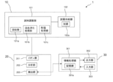
- FIG. 1 It is a block diagram of the globin analysis device of the present invention. It is a schematic diagram which shows the method of this invention.
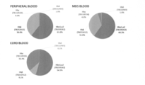
- Figure 2 is a flow chart illustrating the method of the present invention. It is a figure which shows the result of having performed globin typing using the peptide of this invention. It is a figure (The ratio of the amount of each globin chain to the amount of total hemoglobin) which shows the result of having performed the typing of the peripheral blood of the blood disease patient using the peptide of this invention.
- the present invention relates to at least one globin typing peptide selected from peptides consisting of a predetermined amino acid sequence, and a method of testing globin using the peptide.
- the inventor has developed a technique for quantitatively typing globin chains expressed by erythrocytes, and an apparatus capable of performing the technique for multiple specimens at high speed. And we succeeded in developing diagnostic method and treatment of various hematologic diseases by quantitative monitoring of globin abnormality resulting from globin gene mutation. Selection of specific and sensitive peptide fragments is extremely important for protein quantification using mass spectrometry.
- each globin gene product was identified by mass spectrometry using human erythrocytes, cord blood, or human erythroleukemia cells (K562) capable of differentiation.
- a plurality of mass spectrometers (such as Qq-TOF type and Qq-Orbitrap type) were used to search for peptides that can be universally identified.
- amino acid sequences specific to globin chains belonging to each subtype could be identified.
- the present invention is completed by this finding and can be used for diagnosis of various diseases.
- the present invention can be used for (1) development of diagnostic methods for hemoglobin abnormalities such as ⁇ -thalassemia by peripheral blood, and (2) elucidation of globin expression mechanism important for erythrocytes derived from pluripotent stem cells It is useful for evaluating and validating high-speed globin typing devices.
- the globin typing peptide of the present invention is a peptide specific to each globin chain, and is a hemoglobin ⁇ 1 chain (HBA1) or a hemoglobin ⁇ 2 chain (HBA2), a hemoglobin ⁇ chain (HBB), a hemoglobin ⁇ chain Any of the group consisting of: HBD), hemoglobin ⁇ 1 chain (HBE1), hemoglobin ⁇ a chain (HBG1) or hemoglobin ⁇ g chain (HBG2), hemoglobin ⁇ chain (HBM), hemoglobin ⁇ 1 chain (HBQ1) and hemoglobin zeta chain (HBZ) Is a peptide belonging to
- the globin typing peptides used as standard peptides in the present invention are shown below.
- HBA 1; HBA 2 The globin chains belonging to this group are hemoglobin ⁇ 1 chains or hemoglobin ⁇ 2 chains and consist of the following amino acid sequences: LRVDPVNFK (SEQ ID NO: 1) FLASVSTVLTSK (SEQ ID NO: 2) (2) HBB
- the globin chains belonging to this group are hemoglobin ⁇ chains and consist of the following amino acid sequences: EFTPPVQAAYQK (SEQ ID NO: 3) VNVDEVGGEALGR (SEQ ID NO: 4) LLGN VL VC [CAM] VLAHHFGK (SEQ ID NO: 5) GTFATLSELHC [CAM] DK (SEQ ID NO: 6) SAVTALWGK (SEQ ID NO: 7) FFESFGDLSTPDAVMGNPK (SEQ ID NO: 8) LLGN VL VC [CAM] VLAHHFGK (SEQ ID NO: 9)
- the globin chains belonging to this group are hemoglobin ⁇ chains and consist of the following amino acid sequences: EFTPQMQAAYQK (SEQ ID NO: 10) LLGN VL VC [CAM] VLAR (SEQ ID NO: 11) GTFSQLSELHC [CAM] DK (SEQ ID NO: 12) (4) HBE1
- the globin chain belonging to this group is the hemoglobin ⁇ 1 chain and consists of the following amino acid sequence: LSELHC [CAM] DK (SEQ ID NO: 13) EFTPEVQAAWQK (SEQ ID NO: 14) FFDSFGNLSSPSAILGNPK (SEQ ID NO: 15) AAVTSLWSK (SEQ ID NO: 16) MNVEEAGGEALGR (SEQ ID NO: 17) VLTSFGDAIK (SEQ ID NO: 18) AAVTSLWSK (SEQ ID NO: 19) NMDNLKPAFAK (SEQ ID NO: 20)
- HBG1 The globin chains belonging to this group are hemoglobin ⁇ A chains and consist of the following amino acid sequences: VLTSLGDATK (SEQ ID NO: 21) (6) HBG1; HBG2
- the globin chains belonging to this group are hemoglobin ⁇ A chains or ⁇ G chains and consist of the following amino acid sequences: VNVEDAGGETLGR (SEQ ID NO: 22) EFTPEVQASWQK (SEQ ID NO: 23) GTFAQLSELHC [CAM] DK (SEQ ID NO: 24) FFDSFGNLSSASAIMGN PK (SEQ ID NO: 25) ATITSLWGK (SEQ ID NO: 26) EFTPEVQASWQK (SEQ ID NO: 27)
- HBM The globin chains belonging to this group are hemoglobin ⁇ chains and consist of the following amino acid sequences: MLAAVGAAVQHVDNLR (SEQ ID NO: 28) AALSPLADDLHALVLR (SEQ ID NO: 29) LFTVYPSTK (SEQ ID NO: 30) (8) HBQ1
- the globin chain belonging to this group is the hemoglobin ⁇ 1 chain and consists of the following amino acid sequence: TFLAFPATK (SEQ ID NO: 31) VADALSLAVER (SEQ ID NO: 32) TYFSHLDLSPGSSQVR (SEQ ID NO: 33) KLGSNVGVYTTEALER (SEQ ID NO: 34)
- the globin chains belonging to this group are hemoglobin zeta chains and consist of the following amino acid sequences: FLSVVSVSLTEK (SEQ ID NO: 35) TYFPHFDLHPGSAQLR (SEQ ID NO: 36) SIDDIGGALSK (SEQ ID NO: 37) ISTQADTIGTETLER (SEQ ID NO: 38) VVAAVGDAVK (SEQ ID NO: 39) LFLSHPQTK (SEQ ID NO: 40) LSELHAYILR (SEQ ID NO: 41)
- Peptides consisting of these amino acid sequences can be obtained using known peptide synthesis methods.
- the synthesis method include azide method, acid chloride method, acid anhydride method, mixed acid anhydride method, DCC method, active ester method, carboimidazole method, redox method and the like.
- combination can apply any of a solid phase synthesis method and a liquid phase synthesis method.
- a commercially available peptide synthesizer may be used. After the synthesis reaction, the peptide can be purified by combining known purification methods such as chromatography. It is also possible to synthesize using a commercially available amino acid synthesizer.
- the mutated globin typing peptide of the present invention (also referred to herein as “mutated peptide”) has a deletion or substitution of one or several amino acids in the amino acid sequence of the globin typing peptide. Or consisting of the added amino acid sequence.
- amino acid sequence in which one or several amino acids are deleted, substituted or added for example, about 1 to 5 or so, preferably 1 or 2 or so amino acids are deleted, substituted or added Amino acid sequences are included and not limited.
- amino acid residues of the peptide can be substituted for amino acid residues (below) within groups based on such side chain properties.
- Nonpolar aliphatic Gly, Ala, Val, Leu, Ile, Pro Polar non-charge: Ser, Thr, Cys, Met, Asn, Gln Negative charge (acidic): Asp, Glu Positive charge (basic): His, Lys, Arg Aromatic: Trp, Tyr, Phe
- amino acid residues belonging to other groups as well as the above group are not particularly limited.
- the present invention uses the above globin typing peptide or mutant globin typing peptide (herein, these peptides are also collectively referred to as "globin peptide") as a standard peptide and uses a biological sample as a mass peptide. It is an examination method of globin characterized by analyzing by analysis method.
- Biological samples which can be tested by the method of the present invention include, for example, whole blood, serum or plasma, urine, other body fluids (feces, tears, synovial fluid, sputum) or bone marrow cells, but iPS cells Is included as a biological sample.
- iPS cells, bone marrow cells, serum, plasma or the like can be used as a biological sample.
- the test method of the present invention comprises the step of analyzing a biological sample by mass spectrometry.
- the globin peptide is used as an internal standard peptide.
- a predetermined amount of a stable isotope labeled globin peptide of the present invention is added to a substance extracted and enzymatically digested with a globin protein in a biological sample to be tested, and measured by mass spectrometry Do. Then, the amount of globin peptide derived from the sample is determined by comparing the signal derived from the sample with the signal derived from the test item (internal standard peptide).
- mass spectrometry when the sample is ionized in a high voltage applied vacuum, electrostatic force causes the sample to fly through the apparatus. Ions in flight are separated according to the mass-to-charge ratio by electrical and magnetic action etc., and then each is detected to obtain a mass spectrum with m / z as abscissa and detection intensity as ordinate. be able to.
- the mass spectrometry employed in the present invention is not particularly limited, and examples thereof include liquid chromatography mass spectrometry (LC-MS), MALDI TOF MS and the like.
- Multiple reaction monitoring (MRM) is also a mass spectrometry technique that allows monitoring of selected ions. By using this technique, it is possible to obtain an absolute quantitative value of globin to be measured.
- the globin peptide of the present invention can be used as an internal standard in mass spectrometry.
- the “internal standard” in the present invention means a peptide having the same peptide sequence as the sample but having a mass difference with a stable isotope label or the like. The test results obtained in this way can be used for testing for globin related diseases and monitoring of globin described later. Further, in the present invention, when the mass spectrometric data of the standard peptide and the mass spectrometric data derived from the measuring object coincide with each other, the measuring object can be identified as the globin chain to which the standard peptide belongs.
- the present invention provides a testing method for globin related diseases characterized by analyzing a biological sample by mass spectrometry using the globin peptide as a standard peptide.
- the globin related diseases include globin synthesis disorders, blood diseases or inflammatory diseases.
- blood diseases include hemolytic diseases, diseases based on bone marrow dysfunction, and the like, and inflammatory diseases include, for example, collagen diseases, malignant tumors, immune related diseases, and the like.
- Hemolytic disease is a disease caused by hemolysis of red blood cells represented by hemolytic anemia and the like. Hemolytic diseases include congenital and acquired.
- Congenital diseases include sickle cell disease, hereditary spherocytosis, thalasseemia, glucose-6-phosphate dehydrogenase deficiency and the like.
- acquired diseases include autoimmune hemolytic anemia, paroxysmal nocturnal hemoglobinuria, bunch syndrome and the like.
- Diseases based on bone marrow hypofunction include aplastic anemia (AA) and myelodysplastic syndrome (MDS).
- Evaluation method provides a method of monitoring or evaluating at least one selected from the group consisting of the following items (a) to (d), using the result obtained by the above-mentioned inspection method as an index.
- the globin analyzer The analysis of protein by mass spectrometry means can easily distinguish the difference of one amino acid, but in order to carry out high-throughput analysis, construction of a rapid analysis system that automates from sample preparation to analysis Is required.
- the present invention provides a globin analyzer comprising globin sample preparation means, mass analysis means and data analysis means.
- the globin analyzer 1 comprises globin sample preparation means 10, mass analysis means 20 and data analysis means 30.
- Globin Sample Preparation Means The globin sample preparation means 10 includes a sample preparation unit 101 and a reagent supply unit 102.
- the sample preparation unit 101 includes a recovery unit 101a for recovering a biological sample, a hemolytic digestion processing unit 101b for hemolyzing, cell lysis or enzyme digestion of the recovered sample, and a desalting processing unit 101c.
- the sample preparation unit 101 is connected to a reagent supply unit 102 for storing the reagent 102 a.
- the reagent 102a is a cell lysate, an enzyme, a reducing agent, an alkylating agent, a label, a diluent, or the like.
- the sample preparation unit 101 mixes the reagent 102a with a biological sample such as blood to prepare a measurement sample.
- the reagent 102 a is also used as a solution for flowing the measurement sample to the mass spectrometric means 20.
- the recovery unit 101a includes a multi-sample storage unit, and is, for example, a test tube, a 96 well plate, a vial, or the like.
- the biological sample collected in advance is dispensed by the automatic robot to the collection unit 101a.
- the hemolytic digestion processing unit 101 b is a processing unit that adds a cell lysate, an enzyme, a reducing agent, an alkylating agent, and the like to the recovery unit 101 a into which a biological sample is dispensed.
- the desalting processing unit 101c is a processing unit that performs solid phase extraction using a C18 carrier. In the present invention, a plurality of globin sample preparation means 10 can be provided so that multiple samples can be processed in parallel.
- the mass analysis means 20 includes an ion source 201 for ionizing a sample, a mass analysis unit 202 for separating generated ions at a mass-to-charge ratio, and a detection unit 203 for detecting the separated ions.
- a matrix assisted laser desorption ionization method MALDI method
- ESI method electrospray ionization method
- the analysis unit 202 is a quadrupole type, time of flight type, Fourier transform ion cyclotron resonance type, depending on the required characteristics of the m / z near peak (mass resolution) and the two elements of the measurable mass range. And tandem type are adopted.
- the detection unit 203 detects the ions sorted by the analysis unit 202 by sensitization with an electron multiplier or a microchannel plate. In the present invention, a method of detecting and counting with a Faraday cup may be adopted. The invention is not limited to the number of intensifiers, and may be either a single channel detector or a multichannel detector.
- the detection unit 203 communicates with the information processing unit 301 of the data analysis unit 30.
- the data analysis means 30 includes an information processing unit 301, an output unit 302, and an input unit 303.
- the information processing unit 301 includes a storage unit 301a.
- the information processing unit 301 creates a database of globin typing peptides, and compares this with the mass spectrum of the test sample to identify and quantify the sample. Then, a mass spectrum is prepared from the detected data.
- the storage unit 301a stores programs executed by the information processing unit 301 and various data.
- the output unit 302 is a display for displaying character information and graphic information.
- the input unit 23 is a keyboard and a mouse for receiving an input from the user.
- FIG. 1 An example of the high throughput globin typing apparatus of the present invention is shown in FIG.
- the apparatus of the present invention is configured, for example, such that a C18 filter and a chip column packed with immobilized enzyme (trypsin) are arranged in parallel so that one batch of sample preparation can be processed simultaneously in 96 sample units. ( Figure 2). Thereby, it is possible to achieve from the biological sample to the enzyme digestion within one hour. Since the analysis target is eight known globin chains, it is possible to identify all of them specifically and quantify them all at once by using the device of the present invention. The time required for measurement can be analyzed in 5 to 30 minutes per sample, and in the case of 96 samples (batch sample preparation unit) in 8 hours.
- immobilized enzyme trypsin
- globin sample preparation means after sample collection (S31), globin protein in the biological sample to be examined is extracted and enzymatically digested (S32). If necessary, treatment such as stable isotope labeling or reductive alkylation is performed (S32), and a predetermined amount of the globin peptide of the present invention is added as an internal standard peptide (S33).
- S33 a predetermined amount of the globin peptide of the present invention is added as an internal standard peptide (S33).
- S34 mass spectrometry
- the mass spectrometric means compares the signal from the test sample (mass spectrum) with the signal from the item (internal standard peptide) to determine the amount of globin peptide from the sample (S34).
- the data analysis means creates a database of globin typing peptides, and compares this with the mass spectrum of the test sample to identify and quantify the sample (S35).
- Kit The kit of the present invention comprises at least one of the above globin peptides or variant globin peptides.
- the combination of globin peptides may be arbitrary, and may be only one or more combinations of globin peptides belonging to one chain, and one or more of globin peptides belonging to one chain and globin peptides belonging to the other chain One or more may be combined.
- the kit of the present invention can contain buffers, enzymes, instructions for use, etc. necessary for preparation of the peptide.
- Methods Peptides specific for each globin chain were extracted from the analysis results of various protein samples and public databases.
- the resulting candidate peptides were synthesized, subjected to alkylation, and then labeled with mTRAQD0 reagent.
- Evaluation of MRM method was performed using the obtained synthetic peptide, and measurement conditions were fixed.
- a protein extract is obtained from human healthy human peripheral blood, umbilical cord blood, and K562 cells, which are evaluation samples, and after enzyme digestion, reductive alkylation and labeling with mTRAQD4 reagent are performed and mixed with mTRAQD0 synthetic peptide for evaluation An experiment was conducted to construct the final MRM measurement method.
- the set MRM methods are shown in Table 1.
- Q1 is m / z value of peptide ion
- Q3 is m / z value of fragment ion
- RTime is retention time
- ID is arbitrary designation
- DP declustering potential
- EP is entrance potential
- CE is CID Energy
- CXP represents collision cell exit potential.
- RT, DP, EP, CE, and CXP shown in this table have different optimum values depending on the mass spectrometer used, and therefore, except for Q1 and Q3, they should be adjusted according to the equipment used.
- Method 1 Dissolution of sample After adding and dissolving 100 mM Tris-HCl, pH 8.5 containing 100 ⁇ l or 200 ⁇ l of 1% SDS / 7 M guanidine hydrochloride respectively to the five specimens shown in Table 2, the BCA method is used. Protein quantification was performed.
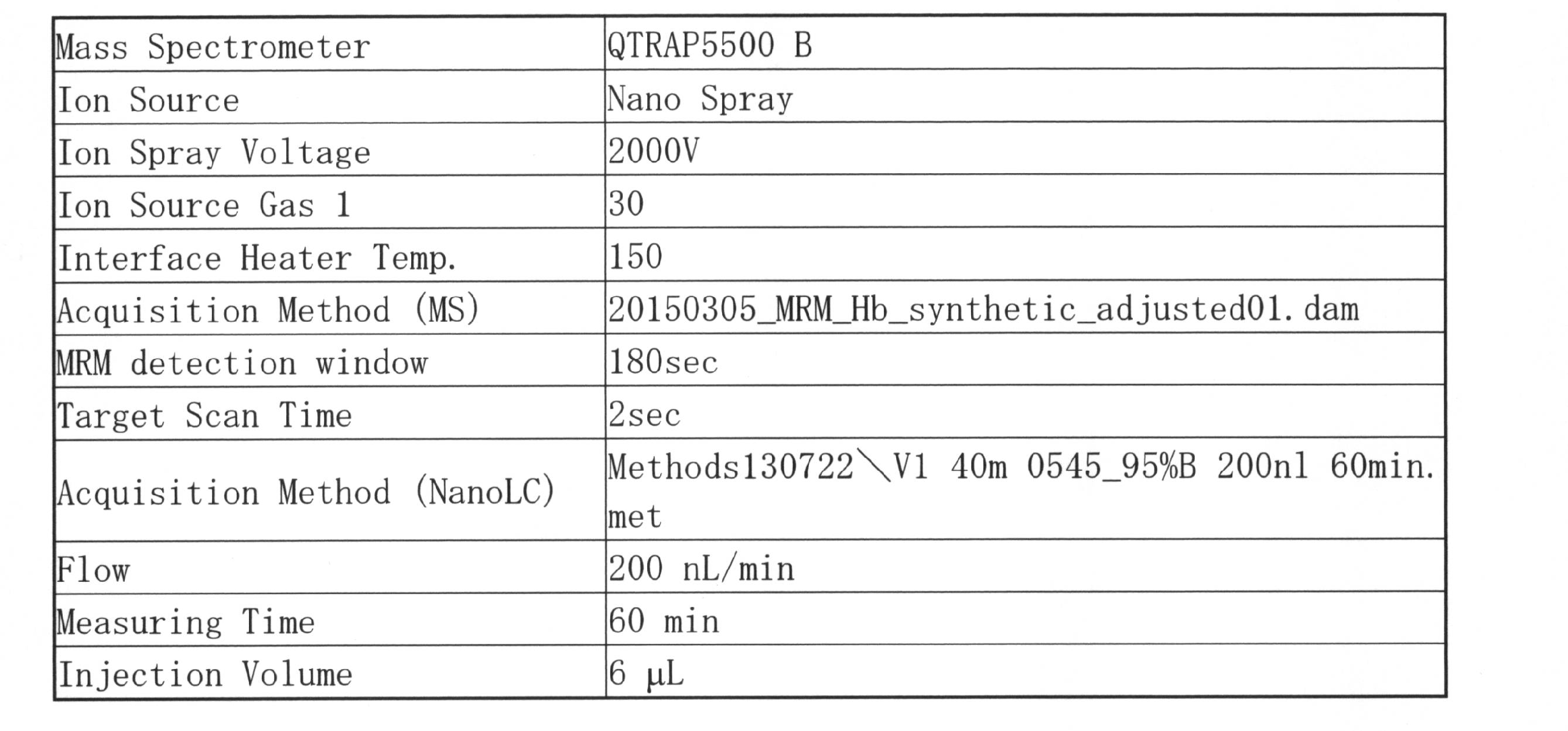
- MRM analysis An mTRAQd0 labeled peptide of known concentration was added to each digest to measure MRM. Analysis by MRM was performed on QTRAP5500 (SCIEX) linked with nano-LC. 5 to 45% B for 40 minutes, 45 to 95% B for 1 minute, and 95% B for 10 minutes (A solvent: 0.1% formic acid / 2% acetonitrile, B solvent: 0.1% formic acid 90% acetonitrile) Using a self-made capillary column (inner diameter 100 ⁇ m, length 15 cm, 3 ⁇ m L-column 2) at a flow rate of 200 nl / min. The spray voltage was 2000 V, the gas 1 was 30, and interface heater temp .: 150 ° C.
- Hemoglobins of peripheral blood and umbilical cord blood of healthy subjects have Hb ⁇ of 500 amol and 70 amol, respectively, while hemoglobin of peripheral blood of patients with MDS is 2.6 times and 18.6 times as much as 1300 amol of hemoglobin ⁇ It became clear that it included. In addition, it was also shown that ⁇ type is characteristically expressed in this patient.
- MDS myelodysplastic syndrome
- RCUD-RA refractory anemia
- RARS ferroblastic refractory anemia
- RCMD polyhemocytic dysplasia Refractory cytopenia
- RAEB-1 refractory anemia with blast augmentation
- RAEB-2 refractory anemia with blast augmentation
- Each globin chain is quantified for each of the following three blood diseases other than MDS, aplastic anemia (AA), anemia (PA) and acute myeloid leukemia (AML), and each globin chain The ratio of total amount of hemoglobin to total amount of hemoglobin was calculated.
- each globin chain was measured in the same manner as in Example 2.
- the amino acid sequence of the globin peptide used as an internal standard is described in the column "Component name” of Table 6 (described in upper case alphabetic letters indicating each globin chain such as "alpha” and "beta”).
- Table 6 shows the concentration of each globin chain (fmol / 0.1 ⁇ l of lysate solution) calculated from the peptide concentration. In Table 6, those less than 10 times the Blank Ratio (below the lower limit of quantification) are left blank.
- Table 7 and FIG. 5 show the ratio of each globin chain to the total amount of hemoglobin. In FIG. 5, the vertical axis is shown on a scale of 60 to 100%.
- the expression ratio of ⁇ globin tends to be higher in the order of AA, MDS, PA and AML, and IDA. Expression of ⁇ globin was not observed in Healthy.
- the MDS and AA When each patient was treated, in the MDS and AA, the expression ratio of ⁇ globin tended to decrease.
- the incidence of ⁇ globin tended to decrease when patients became anemic.
- patient No. 17 was equivalent to RAEB-2, but was diagnosed with treatment-related bone marrow tumor according to WHO classification because of history of chemotherapy.
- ⁇ means that there is blood transfusion
- ⁇ indicates that there is a medication other than an anemia treatment
- ⁇ indicates that there is a medication for an anemia treatment.
- globin analyzer 10 globin sample preparation means 20: mass analysis means 30: data analysis means 101: sample preparation unit 102: reagent supply unit 201: ion source 202: mass analysis unit 203: detection unit 203 301: information processing unit 302: output unit 303: input unit
- Sequence number 5 Xaa represents carbamido methylated cysteine (location: 8).
- SEQ ID NO: 6 Xaa represents carbamidomethylated cysteine (location: 11).
- Sequence number 9 Xaa represents carbamido methylated cysteine (location: 8).
- Sequence number 11 Xaa represents carbamido methylated cysteine (location: 8).
- SEQ ID NO: 13: Xaa represents carbamidomethylated cysteine (location: 6).
Landscapes
- Life Sciences & Earth Sciences (AREA)
- Chemical & Material Sciences (AREA)
- Health & Medical Sciences (AREA)
- Organic Chemistry (AREA)
- Engineering & Computer Science (AREA)
- Molecular Biology (AREA)
- Proteomics, Peptides & Aminoacids (AREA)
- Biochemistry (AREA)
- General Health & Medical Sciences (AREA)
- Immunology (AREA)
- Analytical Chemistry (AREA)
- Wood Science & Technology (AREA)
- Biomedical Technology (AREA)
- Biotechnology (AREA)
- Zoology (AREA)
- Hematology (AREA)
- Genetics & Genomics (AREA)
- Microbiology (AREA)
- Urology & Nephrology (AREA)
- Medicinal Chemistry (AREA)
- Physics & Mathematics (AREA)
- Biophysics (AREA)
- Food Science & Technology (AREA)
- Pathology (AREA)
- Toxicology (AREA)
- General Physics & Mathematics (AREA)
- Cell Biology (AREA)
- Bioinformatics & Cheminformatics (AREA)
- General Engineering & Computer Science (AREA)
- Other Investigation Or Analysis Of Materials By Electrical Means (AREA)
Abstract
【課題】ヒトグロビンタイピングペプチド及びグロビンの検査方法の提供。 【解決手段】ヘモグロビンα1、ヘモグロビンα2、ヘモグロビンβ、ヘモグロビンδ、ヘモグロビンε1、ヘモグロビンγA、ヘモグロビンγG、ヘモグロビンμ、ヘモグロビンθ1及びヘモグロビンζのいずれかの鎖に属するグロビンタイピングペプチドであって、所定のアミノ酸配列からなるグロビンタイピングペプチド。
Description
本発明は、ヒトグロビンタイピングペプチド及びグロビンの検査方法に関する。
ヘモグロビンは、赤血球中に大量に存在する酸素運搬を司るタンパク質である。成人では、ヘモグロビンはα鎖とβ鎖から構成される4量体タンパク質であるHbAが発現し機能しているが、胎児期にはα鎖とγ鎖で構成される胎児期Hb (HbF)が存在する。また、初期胚ではε鎖及びζ鎖によって構成されるHb Grower1が存在する。その量的及び質的異常はHemoglobinopathies(異常ヘモグロビン症)と呼ばれ、貧血状態を惹起する。
異常ヘモグロビン症の多くは単一遺伝子疾患であり、世界人口のおおよそ5.2%が罹患している。世界中の夫婦の1.1%は異常ヘモグロビン症の素因を保持しており、実際に異常ヘモグロビン症を発症する子供の危険率は、1000人中2.7人と推定されている。異常ヘモグロビン症の中でも、サラセミアは世界でもっとも罹患率の高い遺伝性貧血疾患である(一病型だけで世界人口の約7%、2.4億人が罹患)。特に、β-グロビン鎖に異常を認めるβ-サラセミアは貧血の程度が強く、発育障害、知能発達障害などを合併するため小児期死亡の原因となり臨床上問題となる。
重症β-サラセミア患者は慢性的に貧血状態であるため定期的な赤血球輸血を必要とし、更に発育遅延、骨の変化、肝脾腫大症、重度の鉄過剰、心不全、骨粗鬆症、他の内分泌疾患などを合併する。サラセミア患者は生涯に渡る輸血によって常に感染や鉄過剰症のリスクに晒されており、また先進国においては献血者の減少が深刻な問題となりつつある。
これまで、iPS細胞などの多能性幹細胞から赤血球を作製する研究も進められているが、未だヒト多能性幹細胞由来の赤血球においては、大部分が胎児型グロビンから成人型へのスイッチングが不完全であり、その制御機構は解明されていない。また、β-サラセミア/HbEは単一遺伝子疾患にもかかわらず、その臨床症状や重症度のパターンは不均一であり、疾患の重症度を規定する機構の解明が重要な課題である。
β-サラセミアは、β-グロビンの減少によって過剰な遊離α-グロビンが生じ、これが毒性を有することで未成熟赤血球のアポトーシス(無効造血)を引き起こし(Libani IV et al., Blood, 2008, Vol. 112, 875-885)、脾臓肥大および鉄過剰症という2つの致命的な合併症が生じる。従来、無効造血の診断には骨髄穿刺による検体収集が行われてきたが、これは極めて大きな侵襲を伴うことから患者にとって大きな負担となっており、末梢血を用いた診断法の開発が渇望されている。
末梢血における各グロビン鎖の絶対定量は、β-サラセミアを始めとする異常ヘモグロビン症の診断に有効であると考えられるが、複数のグロビン鎖を同時かつ絶対値として定量することが必要であることから、ELISAなどの抗体ベースの手法では当該目的を達成できない。グロビン鎖のように類似した複数分子の一斉定量には、近年、特異的ペプチド定量技術として注目されている3連4重極型質量分析計を用いた多重反応モニタリング(MRM:multiple reaction monitoring)法が有効であり、MRM法を利用したペプチドの定量などが試みられている(特許文献1:特許第5468073号)。
しかしながら、一般に質量分析計によるタンパク質解析では試料調製の煩雑さや装置の堅牢性の低さが障壁となり、医療現場での普及には程遠いのが現状である。
しかしながら、一般に質量分析計によるタンパク質解析では試料調製の煩雑さや装置の堅牢性の低さが障壁となり、医療現場での普及には程遠いのが現状である。
Libani IV et al., Blood, 2008, Vol. 112, 875-885
本発明は、グロビンタイピングペプチド、及び当該ペプチドを用いたグロビンの検査方法を提供することを目的とする。
本発明者は、上記課題を解決するために鋭意検討を行った結果、グロビンタンパク質のアミノ酸配列のうち、所定のアミノ酸配列を有するペプチドがグロビンの検査に有用であることを見出し、本発明を完成するに至った。
すなわち、本発明は以下の通りである。
[1] 以下の(1)~(9)のいずれかのヘモグロビン鎖に属し、下記アミノ酸配列からなるペプチドから選ばれる少なくとも1つのグロビンタイピングペプチド。
(1)ヘモグロビンα1鎖又はヘモグロビンα2鎖
LRVDPVNFK(配列番号1)
FLASVSTVLTSK(配列番号2)
(2)ヘモグロビンβ鎖
EFTPPVQAAYQK(配列番号3)
VNVDEVGGEALGR(配列番号4)
LLGNVLVC[CAM]VLAHHFGK(配列番号5)
GTFATLSELHC[CAM]DK(配列番号6)
SAVTALWGK(配列番号7)
FFESFGDLSTPDAVMGNPK(配列番号8)
LLGNVLVC[CAM]VLAHHFGK(配列番号9)
(C[CAM]はカルバミドメチル化システインを表す。以下、同様。)
(3)ヘモグロビンδ鎖
EFTPQMQAAYQK(配列番号10)
LLGNVLVC[CAM]VLAR(配列番号11)
GTFSQLSELHC[CAM]DK(配列番号12)
(4)ヘモグロビンε1鎖
LSELHC[CAM]DK(配列番号13)
EFTPEVQAAWQK(配列番号14)
FFDSFGNLSSPSAILGNPK(配列番号15)
AAVTSLWSK(配列番号16)
MNVEEAGGEALGR(配列番号17)
VLTSFGDAIK(配列番号18)
AAVTSLWSK(配列番号19)
NMDNLKPAFAK(配列番号20)
(5)ヘモグロビンγA鎖
VLTSLGDATK(配列番号21)
(6)ヘモグロビンγA鎖又はヘモグロビンγG鎖
VNVEDAGGETLGR(配列番号22)
EFTPEVQASWQK(配列番号23)
GTFAQLSELHC[CAM]DK(配列番号24)
FFDSFGNLSSASAIMGNPK(配列番号25)
ATITSLWGK(配列番号26)
EFTPEVQASWQK(配列番号27)
(7)ヘモグロビンμ鎖
MLAAVGAAVQHVDNLR(配列番号28)
AALSPLADLHALVLR(配列番号29)
LFTVYPSTK(配列番号30)
(8)ヘモグロビンθ1鎖
TFLAFPATK(配列番号31)
VADALSLAVER(配列番号32)
TYFSHLDLSPGSSQVR(配列番号33)
KLGSNVGVYTTEALER(配列番号34)
(9)ヘモグロビンζ鎖
FLSVVSSVLTEK(配列番号35)
TYFPHFDLHPGSAQLR(配列番号36)
SIDDIGGALSK(配列番号37)
ISTQADTIGTETLER(配列番号38)
VVAAVGDAVK(配列番号39)
LFLSHPQTK(配列番号40)
LSELHAYILR(配列番号41)
[2] 以下の(1)~(9)のいずれかのヘモグロビン鎖に属し、下記アミノ酸配列において1又は数個のアミノ酸が欠失、置換又は付加したアミノ酸配列からなるペプチドから選ばれる少なくとも1つの変異型グロビンタイピングペプチド。
(1)ヘモグロビンα1鎖又はヘモグロビンα2鎖
LRVDPVNFK(配列番号1)
FLASVSTVLTSK(配列番号2)
(2)ヘモグロビンβ鎖
EFTPPVQAAYQK(配列番号3)
VNVDEVGGEALGR(配列番号4)
LLGNVLVC[CAM]VLAHHFGK(配列番号5)
GTFATLSELHC[CAM]DK(配列番号6)
SAVTALWGK(配列番号7)
FFESFGDLSTPDAVMGNPK(配列番号8)
LLGNVLVC[CAM]VLAHHFGK(配列番号9)
(C[CAM]はカルバミドメチル化システインを表す。以下、同様。)
(3)ヘモグロビンδ鎖
EFTPQMQAAYQK(配列番号10)
LLGNVLVC[CAM]VLAR(配列番号11)
GTFSQLSELHC[CAM]DK(配列番号12)
(4)ヘモグロビンε1鎖
LSELHC[CAM]DK(配列番号13)
EFTPEVQAAWQK(配列番号14)
FFDSFGNLSSPSAILGNPK(配列番号15)
AAVTSLWSK(配列番号16)
MNVEEAGGEALGR(配列番号17)
VLTSFGDAIK(配列番号18)
AAVTSLWSK(配列番号19)
NMDNLKPAFAK(配列番号20)
(5)ヘモグロビンγA鎖
VLTSLGDATK(配列番号21)
(6)ヘモグロビンγA鎖又はヘモグロビンγG鎖
VNVEDAGGETLGR(配列番号22)
EFTPEVQASWQK(配列番号23)
GTFAQLSELHC[CAM]DK(配列番号24)
FFDSFGNLSSASAIMGNPK(配列番号25)
ATITSLWGK(配列番号26)
EFTPEVQASWQK(配列番号27)
(7)ヘモグロビンμ鎖
MLAAVGAAVQHVDNLR(配列番号28)
AALSPLADLHALVLR(配列番号29)
LFTVYPSTK(配列番号30)
(8)ヘモグロビンθ1鎖
TFLAFPATK(配列番号31)
VADALSLAVER(配列番号32)
TYFSHLDLSPGSSQVR(配列番号33)
KLGSNVGVYTTEALER(配列番号34)
(9)ヘモグロビンζ鎖
FLSVVSSVLTEK(配列番号35)
TYFPHFDLHPGSAQLR(配列番号36)
SIDDIGGALSK(配列番号37)
ISTQADTIGTETLER(配列番号38)
VVAAVGDAVK(配列番号39)
LFLSHPQTK(配列番号40)
LSELHAYILR(配列番号41)
[3] 前記[1]に記載のグロビンタイピングペプチド又は[2]に記載の変異型グロビンタイピングペプチドを標準ペプチドとして用いて、生体試料を質量分析法で解析することを特徴とする、グロビンの検査方法。
[4] グロビンの検査が、グロビンの同定又はグロビンの定量である、[3]に記載の方法。
[5] 前記[1]に記載のグロビンタイピングペプチド又は[2]に記載の変異型グロビンタイピングペプチドを標準ペプチドとして用いて、生体試料を質量分析法で解析することを特徴とする、グロビン関連疾患の検査方法。
[6] グロビン関連疾患が、グロビン合成異常症、血液疾患又は炎症性疾患である[5]に記載の方法。
[7] 血液疾患が溶血性疾患である[6]に記載の方法。
[8] 生体試料が、iPS細胞、骨髄細胞、血清又は血漿である[3]~[7]のいずれか1項に記載の方法。
[9] 前記[3]~[8]のいずれか1項に記載の方法により得られた結果を指標として、下記(a)~(d)の項目からなる群から選ばれる少なくとも1つを評価する方法。
(a) 抗癌剤の副作用又は血液毒性
(b) 造血刺激の有無
(c) 血液疾患又は炎症性疾患の病態
(d) 疾患の予後
[10] グロビン試料調製手段、質量分析手段及びデータ解析手段を含む、グロビン解析装置。
[11] グロビン試料調製手段が、生体試料回収部、溶血消化処理部、及び脱塩処理部を含む、[10]に記載の装置。
[12] 前記[1]に記載のグロビンタイピングペプチド又は[2]に記載の変異型グロビンタイピングペプチドを含む、グロビンの検査用キット。
すなわち、本発明は以下の通りである。
[1] 以下の(1)~(9)のいずれかのヘモグロビン鎖に属し、下記アミノ酸配列からなるペプチドから選ばれる少なくとも1つのグロビンタイピングペプチド。
(1)ヘモグロビンα1鎖又はヘモグロビンα2鎖
LRVDPVNFK(配列番号1)
FLASVSTVLTSK(配列番号2)
(2)ヘモグロビンβ鎖
EFTPPVQAAYQK(配列番号3)
VNVDEVGGEALGR(配列番号4)
LLGNVLVC[CAM]VLAHHFGK(配列番号5)
GTFATLSELHC[CAM]DK(配列番号6)
SAVTALWGK(配列番号7)
FFESFGDLSTPDAVMGNPK(配列番号8)
LLGNVLVC[CAM]VLAHHFGK(配列番号9)
(C[CAM]はカルバミドメチル化システインを表す。以下、同様。)
(3)ヘモグロビンδ鎖
EFTPQMQAAYQK(配列番号10)
LLGNVLVC[CAM]VLAR(配列番号11)
GTFSQLSELHC[CAM]DK(配列番号12)
(4)ヘモグロビンε1鎖
LSELHC[CAM]DK(配列番号13)
EFTPEVQAAWQK(配列番号14)
FFDSFGNLSSPSAILGNPK(配列番号15)
AAVTSLWSK(配列番号16)
MNVEEAGGEALGR(配列番号17)
VLTSFGDAIK(配列番号18)
AAVTSLWSK(配列番号19)
NMDNLKPAFAK(配列番号20)
(5)ヘモグロビンγA鎖
VLTSLGDATK(配列番号21)
(6)ヘモグロビンγA鎖又はヘモグロビンγG鎖
VNVEDAGGETLGR(配列番号22)
EFTPEVQASWQK(配列番号23)
GTFAQLSELHC[CAM]DK(配列番号24)
FFDSFGNLSSASAIMGNPK(配列番号25)
ATITSLWGK(配列番号26)
EFTPEVQASWQK(配列番号27)
(7)ヘモグロビンμ鎖
MLAAVGAAVQHVDNLR(配列番号28)
AALSPLADLHALVLR(配列番号29)
LFTVYPSTK(配列番号30)
(8)ヘモグロビンθ1鎖
TFLAFPATK(配列番号31)
VADALSLAVER(配列番号32)
TYFSHLDLSPGSSQVR(配列番号33)
KLGSNVGVYTTEALER(配列番号34)
(9)ヘモグロビンζ鎖
FLSVVSSVLTEK(配列番号35)
TYFPHFDLHPGSAQLR(配列番号36)
SIDDIGGALSK(配列番号37)
ISTQADTIGTETLER(配列番号38)
VVAAVGDAVK(配列番号39)
LFLSHPQTK(配列番号40)
LSELHAYILR(配列番号41)
[2] 以下の(1)~(9)のいずれかのヘモグロビン鎖に属し、下記アミノ酸配列において1又は数個のアミノ酸が欠失、置換又は付加したアミノ酸配列からなるペプチドから選ばれる少なくとも1つの変異型グロビンタイピングペプチド。
(1)ヘモグロビンα1鎖又はヘモグロビンα2鎖
LRVDPVNFK(配列番号1)
FLASVSTVLTSK(配列番号2)
(2)ヘモグロビンβ鎖
EFTPPVQAAYQK(配列番号3)
VNVDEVGGEALGR(配列番号4)
LLGNVLVC[CAM]VLAHHFGK(配列番号5)
GTFATLSELHC[CAM]DK(配列番号6)
SAVTALWGK(配列番号7)
FFESFGDLSTPDAVMGNPK(配列番号8)
LLGNVLVC[CAM]VLAHHFGK(配列番号9)
(C[CAM]はカルバミドメチル化システインを表す。以下、同様。)
(3)ヘモグロビンδ鎖
EFTPQMQAAYQK(配列番号10)
LLGNVLVC[CAM]VLAR(配列番号11)
GTFSQLSELHC[CAM]DK(配列番号12)
(4)ヘモグロビンε1鎖
LSELHC[CAM]DK(配列番号13)
EFTPEVQAAWQK(配列番号14)
FFDSFGNLSSPSAILGNPK(配列番号15)
AAVTSLWSK(配列番号16)
MNVEEAGGEALGR(配列番号17)
VLTSFGDAIK(配列番号18)
AAVTSLWSK(配列番号19)
NMDNLKPAFAK(配列番号20)
(5)ヘモグロビンγA鎖
VLTSLGDATK(配列番号21)
(6)ヘモグロビンγA鎖又はヘモグロビンγG鎖
VNVEDAGGETLGR(配列番号22)
EFTPEVQASWQK(配列番号23)
GTFAQLSELHC[CAM]DK(配列番号24)
FFDSFGNLSSASAIMGNPK(配列番号25)
ATITSLWGK(配列番号26)
EFTPEVQASWQK(配列番号27)
(7)ヘモグロビンμ鎖
MLAAVGAAVQHVDNLR(配列番号28)
AALSPLADLHALVLR(配列番号29)
LFTVYPSTK(配列番号30)
(8)ヘモグロビンθ1鎖
TFLAFPATK(配列番号31)
VADALSLAVER(配列番号32)
TYFSHLDLSPGSSQVR(配列番号33)
KLGSNVGVYTTEALER(配列番号34)
(9)ヘモグロビンζ鎖
FLSVVSSVLTEK(配列番号35)
TYFPHFDLHPGSAQLR(配列番号36)
SIDDIGGALSK(配列番号37)
ISTQADTIGTETLER(配列番号38)
VVAAVGDAVK(配列番号39)
LFLSHPQTK(配列番号40)
LSELHAYILR(配列番号41)
[3] 前記[1]に記載のグロビンタイピングペプチド又は[2]に記載の変異型グロビンタイピングペプチドを標準ペプチドとして用いて、生体試料を質量分析法で解析することを特徴とする、グロビンの検査方法。
[4] グロビンの検査が、グロビンの同定又はグロビンの定量である、[3]に記載の方法。
[5] 前記[1]に記載のグロビンタイピングペプチド又は[2]に記載の変異型グロビンタイピングペプチドを標準ペプチドとして用いて、生体試料を質量分析法で解析することを特徴とする、グロビン関連疾患の検査方法。
[6] グロビン関連疾患が、グロビン合成異常症、血液疾患又は炎症性疾患である[5]に記載の方法。
[7] 血液疾患が溶血性疾患である[6]に記載の方法。
[8] 生体試料が、iPS細胞、骨髄細胞、血清又は血漿である[3]~[7]のいずれか1項に記載の方法。
[9] 前記[3]~[8]のいずれか1項に記載の方法により得られた結果を指標として、下記(a)~(d)の項目からなる群から選ばれる少なくとも1つを評価する方法。
(a) 抗癌剤の副作用又は血液毒性
(b) 造血刺激の有無
(c) 血液疾患又は炎症性疾患の病態
(d) 疾患の予後
[10] グロビン試料調製手段、質量分析手段及びデータ解析手段を含む、グロビン解析装置。
[11] グロビン試料調製手段が、生体試料回収部、溶血消化処理部、及び脱塩処理部を含む、[10]に記載の装置。
[12] 前記[1]に記載のグロビンタイピングペプチド又は[2]に記載の変異型グロビンタイピングペプチドを含む、グロビンの検査用キット。
本発明により、グロビンタイピングペプチドが提供される。本発明のグロビンタイピングペプチドにより、大量に存在するα鎖やβ鎖、及び微量に存在するその他のアイソザイムを同時に計測することが可能である。さらに、得られるデータは各タンパク質の絶対量であることから、各グロビン鎖の組成が明らかとなり、新たな診断指標を提供することが可能となる。
本発明は、所定のアミノ酸配列からなるペプチドから選ばれる少なくとも1つのグロビンタイピングペプチド、及び当該ペプチドを用いたグロビンの検査方法に関する。
本発明者は、赤血球が発現しているグロビン鎖を定量的にタイピングする技術、及びその技術を多検体に対して高速に実行できる装置を開発した。そして、グロビン遺伝子変異の結果生じるグロビン異常の定量的モニタリングによって各種血液疾患の診断法や治療法を開発することに成功した。
質量分析を用いたタンパク質の定量には、特異的かつ感度が高いペプチド断片の選出が極めて重要である。本発明においては、ヒト赤血球や臍帯血、あるいは分化誘導可能なヒト赤白血病細胞(K562)を用いて各グロビン遺伝子産物を質量分析計によって同定した。タンパク質の同定には複数の質量分析計(Qq-TOF型やQq-Orbitrap型など)を用いて、普遍的に同定できるペプチドを探索した。その結果、各サブタイプに属するグロビン鎖に特異的なアミノ酸配列を同定することができた。本発明はこの知見により完成されたものでり、各種疾患の診断等に利用することが可能となる。特に、本発明は、(1)末梢血によるβ-サラセミア等のヘモグロビン異常症の診断法開発、及び(2) 多能性幹細胞から誘導した赤血球に重要なグロビン発現機構の解明に利用することができ、高速グロビンタイピング装置の評価及び有効性の実証に有用である。
本発明者は、赤血球が発現しているグロビン鎖を定量的にタイピングする技術、及びその技術を多検体に対して高速に実行できる装置を開発した。そして、グロビン遺伝子変異の結果生じるグロビン異常の定量的モニタリングによって各種血液疾患の診断法や治療法を開発することに成功した。
質量分析を用いたタンパク質の定量には、特異的かつ感度が高いペプチド断片の選出が極めて重要である。本発明においては、ヒト赤血球や臍帯血、あるいは分化誘導可能なヒト赤白血病細胞(K562)を用いて各グロビン遺伝子産物を質量分析計によって同定した。タンパク質の同定には複数の質量分析計(Qq-TOF型やQq-Orbitrap型など)を用いて、普遍的に同定できるペプチドを探索した。その結果、各サブタイプに属するグロビン鎖に特異的なアミノ酸配列を同定することができた。本発明はこの知見により完成されたものでり、各種疾患の診断等に利用することが可能となる。特に、本発明は、(1)末梢血によるβ-サラセミア等のヘモグロビン異常症の診断法開発、及び(2) 多能性幹細胞から誘導した赤血球に重要なグロビン発現機構の解明に利用することができ、高速グロビンタイピング装置の評価及び有効性の実証に有用である。
1.本発明のグロビンタイピングペプチド
本発明のグロビンタイピングペプチドは、各グロビン鎖に特異的なペプチドであり、ヘモグロビンα1鎖(HBA1)又はヘモグロビンα2鎖(HBA2)、ヘモグロビンβ鎖(HBB)、ヘモグロビンδ鎖(HBD)、ヘモグロビンε1鎖(HBE1)、ヘモグロビンγa鎖(HBG1)又はヘモグロビンγg鎖(HBG2)、ヘモグロビンμ鎖(HBM)、ヘモグロビンθ1鎖(HBQ1)及びヘモグロビンζ鎖(HBZ)からなる群のいずれかに属するペプチドである。
本発明のグロビンタイピングペプチドは、各グロビン鎖に特異的なペプチドであり、ヘモグロビンα1鎖(HBA1)又はヘモグロビンα2鎖(HBA2)、ヘモグロビンβ鎖(HBB)、ヘモグロビンδ鎖(HBD)、ヘモグロビンε1鎖(HBE1)、ヘモグロビンγa鎖(HBG1)又はヘモグロビンγg鎖(HBG2)、ヘモグロビンμ鎖(HBM)、ヘモグロビンθ1鎖(HBQ1)及びヘモグロビンζ鎖(HBZ)からなる群のいずれかに属するペプチドである。
本発明において標準ペプチドとして使用されるグロビンタイピングペプチドを以下に示す。
(1)HBA1;HBA2
この群に属するグロビン鎖はヘモグロビンα1鎖又はヘモグロビンα2鎖であり、以下のアミノ酸配列からなる。
LRVDPVNFK(配列番号1)
FLASVSTVLTSK(配列番号2)
(2)HBB
この群に属するグロビン鎖はヘモグロビンβ鎖であり、以下のアミノ酸配列からなる。
EFTPPVQAAYQK(配列番号3)
VNVDEVGGEALGR(配列番号4)
LLGNVLVC[CAM]VLAHHFGK(配列番号5)
GTFATLSELHC[CAM]DK(配列番号6)
SAVTALWGK(配列番号7)
FFESFGDLSTPDAVMGNPK(配列番号8)
LLGNVLVC[CAM]VLAHHFGK(配列番号9)
上記アミノ酸配列において、C[CAM]はカルバミドメチル化システインを表す。以下、同様である。
(1)HBA1;HBA2
この群に属するグロビン鎖はヘモグロビンα1鎖又はヘモグロビンα2鎖であり、以下のアミノ酸配列からなる。
LRVDPVNFK(配列番号1)
FLASVSTVLTSK(配列番号2)
(2)HBB
この群に属するグロビン鎖はヘモグロビンβ鎖であり、以下のアミノ酸配列からなる。
EFTPPVQAAYQK(配列番号3)
VNVDEVGGEALGR(配列番号4)
LLGNVLVC[CAM]VLAHHFGK(配列番号5)
GTFATLSELHC[CAM]DK(配列番号6)
SAVTALWGK(配列番号7)
FFESFGDLSTPDAVMGNPK(配列番号8)
LLGNVLVC[CAM]VLAHHFGK(配列番号9)
上記アミノ酸配列において、C[CAM]はカルバミドメチル化システインを表す。以下、同様である。
(3)HBD
この群に属するグロビン鎖はヘモグロビンδ鎖であり、以下のアミノ酸配列からなる。
EFTPQMQAAYQK(配列番号10)
LLGNVLVC[CAM]VLAR(配列番号11)
GTFSQLSELHC[CAM]DK(配列番号12)
(4)HBE1
この群に属するグロビン鎖はヘモグロビンε1鎖であり、以下のアミノ酸配列からなる。
LSELHC[CAM]DK(配列番号13)
EFTPEVQAAWQK(配列番号14)
FFDSFGNLSSPSAILGNPK(配列番号15)
AAVTSLWSK(配列番号16)
MNVEEAGGEALGR(配列番号17)
VLTSFGDAIK(配列番号18)
AAVTSLWSK(配列番号19)
NMDNLKPAFAK(配列番号20)
この群に属するグロビン鎖はヘモグロビンδ鎖であり、以下のアミノ酸配列からなる。
EFTPQMQAAYQK(配列番号10)
LLGNVLVC[CAM]VLAR(配列番号11)
GTFSQLSELHC[CAM]DK(配列番号12)
(4)HBE1
この群に属するグロビン鎖はヘモグロビンε1鎖であり、以下のアミノ酸配列からなる。
LSELHC[CAM]DK(配列番号13)
EFTPEVQAAWQK(配列番号14)
FFDSFGNLSSPSAILGNPK(配列番号15)
AAVTSLWSK(配列番号16)
MNVEEAGGEALGR(配列番号17)
VLTSFGDAIK(配列番号18)
AAVTSLWSK(配列番号19)
NMDNLKPAFAK(配列番号20)
(5)HBG1
この群に属するグロビン鎖はヘモグロビンγA鎖であり、以下のアミノ酸配列からなる。
VLTSLGDATK(配列番号21)
(6)HBG1;HBG2
この群に属するグロビン鎖はヘモグロビンγA鎖又はγG鎖であり、以下のアミノ酸配列からなる。
VNVEDAGGETLGR(配列番号22)
EFTPEVQASWQK(配列番号23)
GTFAQLSELHC[CAM]DK(配列番号24)
FFDSFGNLSSASAIMGNPK(配列番号25)
ATITSLWGK(配列番号26)
EFTPEVQASWQK(配列番号27)
この群に属するグロビン鎖はヘモグロビンγA鎖であり、以下のアミノ酸配列からなる。
VLTSLGDATK(配列番号21)
(6)HBG1;HBG2
この群に属するグロビン鎖はヘモグロビンγA鎖又はγG鎖であり、以下のアミノ酸配列からなる。
VNVEDAGGETLGR(配列番号22)
EFTPEVQASWQK(配列番号23)
GTFAQLSELHC[CAM]DK(配列番号24)
FFDSFGNLSSASAIMGNPK(配列番号25)
ATITSLWGK(配列番号26)
EFTPEVQASWQK(配列番号27)
(7)HBM
この群に属するグロビン鎖はヘモグロビンμ鎖であり、以下のアミノ酸配列からなる。
MLAAVGAAVQHVDNLR(配列番号28)
AALSPLADLHALVLR(配列番号29)
LFTVYPSTK(配列番号30)
(8)HBQ1
この群に属するグロビン鎖はヘモグロビンθ1鎖であり、以下のアミノ酸配列からなる。
TFLAFPATK(配列番号31)
VADALSLAVER(配列番号32)
TYFSHLDLSPGSSQVR(配列番号33)
KLGSNVGVYTTEALER(配列番号34)
この群に属するグロビン鎖はヘモグロビンμ鎖であり、以下のアミノ酸配列からなる。
MLAAVGAAVQHVDNLR(配列番号28)
AALSPLADLHALVLR(配列番号29)
LFTVYPSTK(配列番号30)
(8)HBQ1
この群に属するグロビン鎖はヘモグロビンθ1鎖であり、以下のアミノ酸配列からなる。
TFLAFPATK(配列番号31)
VADALSLAVER(配列番号32)
TYFSHLDLSPGSSQVR(配列番号33)
KLGSNVGVYTTEALER(配列番号34)
(9)HBZ
この群に属するグロビン鎖はヘモグロビンζ鎖であり、以下のアミノ酸配列からなる。
FLSVVSSVLTEK(配列番号35)
TYFPHFDLHPGSAQLR(配列番号36)
SIDDIGGALSK(配列番号37)
ISTQADTIGTETLER(配列番号38)
VVAAVGDAVK(配列番号39)
LFLSHPQTK(配列番号40)
LSELHAYILR(配列番号41)
この群に属するグロビン鎖はヘモグロビンζ鎖であり、以下のアミノ酸配列からなる。
FLSVVSSVLTEK(配列番号35)
TYFPHFDLHPGSAQLR(配列番号36)
SIDDIGGALSK(配列番号37)
ISTQADTIGTETLER(配列番号38)
VVAAVGDAVK(配列番号39)
LFLSHPQTK(配列番号40)
LSELHAYILR(配列番号41)
これらのアミノ酸配列からなるペプチドは、公知のペプチド合成方法を用いて得ることができる。合成方法としては、例えば、アジド法、酸クロライド法、酸無水物法、混合酸無水物法、DCC法、活性エステル法、カルボイミダゾール法及び酸化還元法等が挙げられる。また、その合成は、固相合成法及び液相合成法のいずれをも適用することができる。市販のペプチド合成装置を使用してもよい。合成反応後は、クロマトグラフィー等の公知の精製法を組み合わせてペプチドを精製することができる。市販のアミノ酸合成装置を用いて合成することも可能である。
2.本発明の変異型グロビンタイピングペプチド
本発明の変異型グロビンタイピングペプチド(本明細書において「変異型ペプチド」ともいう)は、前記グロビンタイピングペプチドのアミノ酸配列において1又は数個のアミノ酸が欠失、置換又は付加したアミノ酸配列からなるものである。
上記「1又は数個のアミノ酸が欠失、置換又は付加されたアミノ酸配列」としては、例えば、1個~5個程度、好ましくは1個~2個程度のアミノ酸が欠失、置換又は付加されたアミノ酸配列が挙げられ、限定はされない。
本発明の変異型グロビンタイピングペプチド(本明細書において「変異型ペプチド」ともいう)は、前記グロビンタイピングペプチドのアミノ酸配列において1又は数個のアミノ酸が欠失、置換又は付加したアミノ酸配列からなるものである。
上記「1又は数個のアミノ酸が欠失、置換又は付加されたアミノ酸配列」としては、例えば、1個~5個程度、好ましくは1個~2個程度のアミノ酸が欠失、置換又は付加されたアミノ酸配列が挙げられ、限定はされない。
ここで、天然に存在するアミノ酸は、側鎖特性に基づいて、以下の通り分類することができる。このため、ペプチドのアミノ酸残基は、このような側鎖特性に基づくグループ内のアミノ酸残基(下記)に置換することができる。
非極性脂肪族:Gly、Ala、Val、Leu、Ile、Pro
極性非電荷:Ser、Thr、Cys、Met、Asn、Gln
負電荷(酸性): Asp、Glu
正電荷(塩基性): His、Lys、Arg
芳香族:Trp、Tyr、Phe
非極性脂肪族:Gly、Ala、Val、Leu、Ile、Pro
極性非電荷:Ser、Thr、Cys、Met、Asn、Gln
負電荷(酸性): Asp、Glu
正電荷(塩基性): His、Lys、Arg
芳香族:Trp、Tyr、Phe
但し、上記グループ内に限らず、他のグループに属するアミノ酸残基に置換することも可能であり、特に限定されるものではない。
3.グロビンの検査方法
本発明は、前記グロビンタイピングペプチド又は変異型グロビンタイピングペプチド(本明細書においては、これらのペプチドを総称して「グロビンペプチド」ともいう)を標準ペプチドとして用いて、生体試料を質量分析法で解析することを特徴とする、グロビンの検査方法である。
本発明の方法で試験することができる生体試料としては、例えば全血、血清若しくは血漿、尿、その他の体液(糞便、涙液、滑液、痰)、又は骨髄細胞が挙げられるが、iPS細胞も生体試料として含めることとする。本発明においては、生体試料としてiPS細胞、骨髄細胞、血清又は血漿等を使用することができる。
本発明は、前記グロビンタイピングペプチド又は変異型グロビンタイピングペプチド(本明細書においては、これらのペプチドを総称して「グロビンペプチド」ともいう)を標準ペプチドとして用いて、生体試料を質量分析法で解析することを特徴とする、グロビンの検査方法である。
本発明の方法で試験することができる生体試料としては、例えば全血、血清若しくは血漿、尿、その他の体液(糞便、涙液、滑液、痰)、又は骨髄細胞が挙げられるが、iPS細胞も生体試料として含めることとする。本発明においては、生体試料としてiPS細胞、骨髄細胞、血清又は血漿等を使用することができる。
1つの態様において、本発明の検査方法は、生体試料を質量分析法で解析するステップを含む。その際、前記グロビンペプチドを内部標準ペプチドとして使用する。
例えば、本発明においては、検査対象の生物学的試料中のグロビンタンパク質を抽出し、酵素消化したものに、安定同位体標識を施した本発明のグロビンペプチドを既定量添加し、質量分析により測定する。そして、検体由来のシグナルと評品(内部標準ペプチド)由来のシグナルとを比較して、検体由来のグロビンペプチドの量を決定する。
例えば、本発明においては、検査対象の生物学的試料中のグロビンタンパク質を抽出し、酵素消化したものに、安定同位体標識を施した本発明のグロビンペプチドを既定量添加し、質量分析により測定する。そして、検体由来のシグナルと評品(内部標準ペプチド)由来のシグナルとを比較して、検体由来のグロビンペプチドの量を決定する。
質量分析では、高電圧をかけた真空中で試料をイオン化すると、静電力によって試料は装置内を飛行する。飛行しているイオンを電気的・磁気的な作用等により質量電荷比に応じて分離し、その後それぞれを検出することで、m/zを横軸、検出強度を縦軸とするマススペクトルを得ることができる。
本発明において採用される質量分析は、特に限定されるものではないが、例えば液体クロマトグラフ質量分析(LC-MS)、MALDI TOF MSなどが挙げられる。
また、多重反応モニタリング(MRM)は、選択されたイオンのモニタリングを可能とする質量分析技術である。この技術を使うことにより、測定対象グロビンの絶対的な定量値を得ることができる。
また、多重反応モニタリング(MRM)は、選択されたイオンのモニタリングを可能とする質量分析技術である。この技術を使うことにより、測定対象グロビンの絶対的な定量値を得ることができる。
本発明のグロビンペプチドは、質量分析の際に内部標準として使用することができる。本発明における「内部標準」とは、検体と同一のペプチド配列を持つが安定同位体標識などで質量差をもたせたペプチドであることを意味する。
このようにして得られた検査結果は、後述のグロビン関連疾患の検査、グロビンのモニタリングに使用することができる。
また、本発明においては、標準ペプチドの質量分析データと、測定対象由来の質量分析データとが一致したときは、測定対象は、標準ペプチドが属するグロビン鎖であると同定することができる。他方、標準ペプチドの質量分析データと、測定対象由来の質量分析データとの間に差異(偏差)が生じたときは、測定対象は、標準ペプチドが属するグロビン鎖ではないと判断することができる。
このようにして得られた検査結果は、後述のグロビン関連疾患の検査、グロビンのモニタリングに使用することができる。
また、本発明においては、標準ペプチドの質量分析データと、測定対象由来の質量分析データとが一致したときは、測定対象は、標準ペプチドが属するグロビン鎖であると同定することができる。他方、標準ペプチドの質量分析データと、測定対象由来の質量分析データとの間に差異(偏差)が生じたときは、測定対象は、標準ペプチドが属するグロビン鎖ではないと判断することができる。
4.グロビン関連疾患の検査方法
本発明は、前記グロビンペプチドを標準ペプチドとして用いて、生体試料を質量分析法で解析することを特徴とするグロビン関連疾患の検査方法を提供する。
グロビン関連疾患としては、グロビン合成異常症、血液疾患又は炎症性疾患が挙げられる。また、血液疾患としては、溶血性疾患、骨髄機能低下に基づく疾患などが挙げられ、炎症性疾患としては例えば膠原病、悪性腫瘍、免疫関連疾患などが挙げられる。
溶血性疾患とは、溶血性貧血などを代表とする赤血球の溶血により生じる疾患である。溶血性疾患には先天性及び後天性が含まれる。先天性疾患には鎌状赤血球症、遺伝性球状赤血球症、サラセミア、グルコース-6-リン酸脱水素酵素欠損症などがある。また、後天性疾患には自己免疫性溶血性貧血、発作性夜間血色素尿症、バンチ症候群などがある。
骨髄機能低下に基づく疾患には、再生不良性貧血(AA)、骨髄異形成症候群(MDS)などがある。
本発明は、前記グロビンペプチドを標準ペプチドとして用いて、生体試料を質量分析法で解析することを特徴とするグロビン関連疾患の検査方法を提供する。
グロビン関連疾患としては、グロビン合成異常症、血液疾患又は炎症性疾患が挙げられる。また、血液疾患としては、溶血性疾患、骨髄機能低下に基づく疾患などが挙げられ、炎症性疾患としては例えば膠原病、悪性腫瘍、免疫関連疾患などが挙げられる。
溶血性疾患とは、溶血性貧血などを代表とする赤血球の溶血により生じる疾患である。溶血性疾患には先天性及び後天性が含まれる。先天性疾患には鎌状赤血球症、遺伝性球状赤血球症、サラセミア、グルコース-6-リン酸脱水素酵素欠損症などがある。また、後天性疾患には自己免疫性溶血性貧血、発作性夜間血色素尿症、バンチ症候群などがある。
骨髄機能低下に基づく疾患には、再生不良性貧血(AA)、骨髄異形成症候群(MDS)などがある。
5.評価方法
本発明は、前記検査方法により得られた結果を指標として、下記(a)~(d)の項目からなる群から選ばれる少なくとも1つをモニタリング又は評価する方法を提供する。
(a) 抗癌剤の副作用又は血液毒性
(b) 造血刺激の有無
(c) 血液疾患又は炎症性疾患の病態
(d) 疾患の予後
本発明は、前記検査方法により得られた結果を指標として、下記(a)~(d)の項目からなる群から選ばれる少なくとも1つをモニタリング又は評価する方法を提供する。
(a) 抗癌剤の副作用又は血液毒性
(b) 造血刺激の有無
(c) 血液疾患又は炎症性疾患の病態
(d) 疾患の予後
6.グロビン解析装置
質量分析手段によるタンパク質の解析では、1アミノ酸の違いを容易に区別することができるが、ハイスループットな分析を実施するためには試料調製から分析までを自動化した迅速な分析システムの構築が必須である。
本発明は、グロビン試料調製手段、質量分析手段及びデータ解析手段を含む、グロビン解析装置を提供する。
質量分析手段によるタンパク質の解析では、1アミノ酸の違いを容易に区別することができるが、ハイスループットな分析を実施するためには試料調製から分析までを自動化した迅速な分析システムの構築が必須である。
本発明は、グロビン試料調製手段、質量分析手段及びデータ解析手段を含む、グロビン解析装置を提供する。
図1に示すように、本発明のグロビン解析装置1は、グロビン試料調製手段10、質量分析手段20及びデータ解析手段30を備える。
(1)グロビン試料調製手段
グロビン試料調製手段10は、試料調製部101と、試薬供給部102とを備える。試料調製部101は、生体試料を回収する回収部101aと、回収された試料を、溶血、細胞融解又は酵素消化する溶血消化処理部101bと、脱塩処理部101cとを備える。
試料調製部101には、試薬102aを収容する試薬供給部102が接続されている。試薬102aは、細胞溶解液、酵素、還元剤、アルキル化剤、標識、希釈液等である。試料調製部101は、試薬102aと、血液等の生体試料とを混和して、測定試料を調製する。試薬102aは、測定試料を質量分析手段20に流すための溶液としても用いられる。
(1)グロビン試料調製手段
グロビン試料調製手段10は、試料調製部101と、試薬供給部102とを備える。試料調製部101は、生体試料を回収する回収部101aと、回収された試料を、溶血、細胞融解又は酵素消化する溶血消化処理部101bと、脱塩処理部101cとを備える。
試料調製部101には、試薬102aを収容する試薬供給部102が接続されている。試薬102aは、細胞溶解液、酵素、還元剤、アルキル化剤、標識、希釈液等である。試料調製部101は、試薬102aと、血液等の生体試料とを混和して、測定試料を調製する。試薬102aは、測定試料を質量分析手段20に流すための溶液としても用いられる。
回収部101aは、多検体収容部を備え、例えば試験管、96ウェルプレート、バイアルなどである。予め採取された生体試料は、自動ロボットにより回収部101aに分注される。
溶血消化処理部101bは、生体試料が分注された回収部101aに細胞溶解液、酵素、還元剤、アルキル化剤などを添加する処理部である。
脱塩処理部101cは、C18担体を用いた固相抽出をする処理部である。
本発明では多検体を並列的に処理できるように、グロビン試料調製手段10を複数設けることができる。
溶血消化処理部101bは、生体試料が分注された回収部101aに細胞溶解液、酵素、還元剤、アルキル化剤などを添加する処理部である。
脱塩処理部101cは、C18担体を用いた固相抽出をする処理部である。
本発明では多検体を並列的に処理できるように、グロビン試料調製手段10を複数設けることができる。
(2)質量分析手段
質量分析手段20は、試料をイオン化するイオン源201と、生成されたイオンを質量と電化の比で分離する質量分析部202と、分離されたイオンを検出する検出部203とを備える。
イオン源201は、目的に応じて、マトリックス支援レーザー脱離イオン化法(MALDI法)、エレクトロスプレーイオン化法(ESI法)などが採用される。
質量分析手段20は、試料をイオン化するイオン源201と、生成されたイオンを質量と電化の比で分離する質量分析部202と、分離されたイオンを検出する検出部203とを備える。
イオン源201は、目的に応じて、マトリックス支援レーザー脱離イオン化法(MALDI法)、エレクトロスプレーイオン化法(ESI法)などが採用される。
分析部202は、m/zの近いピークを区別する能力(質量分解能)と測定可能質量範囲の二つの要素の要求される特性によって、四重極型、飛行時間型、フーリエ変換イオンサイクロトロン共鳴型、タンデム型などが採用される。
検出部203は、分析部202で選別されたイオンを電子増倍管やマイクロチャンネルプレートで増感して検出する。本発明においては、ファラデーカップで検出してカウントする形式を採用してもよい。増倍管の数に限定されるものではなく、単一チャネル検出器及びマルチチャネル検出器のいずれでもよい。
検出部203は、データ解析手段30の情報処理部301と通信を行う。
検出部203は、分析部202で選別されたイオンを電子増倍管やマイクロチャンネルプレートで増感して検出する。本発明においては、ファラデーカップで検出してカウントする形式を採用してもよい。増倍管の数に限定されるものではなく、単一チャネル検出器及びマルチチャネル検出器のいずれでもよい。
検出部203は、データ解析手段30の情報処理部301と通信を行う。
(3)データ解析手段
データ解析手段30は、情報処理部301と、出力部302と、入力部303とを備え、情報処理部301は、記憶部301aを備える。
情報処理部301は、グロビンタイピングペプチドのデータベースを作成し、これと被験試料のマススペクトルと比較することで試料の同定、定量を行う。そして、検出されたデータからマススペクトルを作製する。
記憶部301aは、情報処理部301により実行されるプログラムと、各種データとを記憶している。
出力部302は、文字情報や図形情報を表示するためのディスプレイである。入力部23は、ユーザーによる入力を受けるためのキーボード及びマウスである。
データ解析手段30は、情報処理部301と、出力部302と、入力部303とを備え、情報処理部301は、記憶部301aを備える。
情報処理部301は、グロビンタイピングペプチドのデータベースを作成し、これと被験試料のマススペクトルと比較することで試料の同定、定量を行う。そして、検出されたデータからマススペクトルを作製する。
記憶部301aは、情報処理部301により実行されるプログラムと、各種データとを記憶している。
出力部302は、文字情報や図形情報を表示するためのディスプレイである。入力部23は、ユーザーによる入力を受けるためのキーボード及びマウスである。
本発明のハイスループット・グロビンタイピング装置の一例を図2に示す。
本発明の装置は、例えば、試料調製を1バッチ、96検体単位で一斉処理できるように、C18フィルターと固相化酵素(トリプシン)が充填されたチップカラムが並列して配置された構成となっている(図2)。これにより、生体試料から酵素消化までを1時間以内で達成することができる。分析対象は既知の8種類のグロビン鎖であるから、本発明の装置を用いることにより、これを全て特異的に識別して一斉に定量することが可能となる。測定に要する時間は1検体あたり5~30分、96検体(一括試料調製単位)であれば8時間で分析可能である。
本発明の装置は、例えば、試料調製を1バッチ、96検体単位で一斉処理できるように、C18フィルターと固相化酵素(トリプシン)が充填されたチップカラムが並列して配置された構成となっている(図2)。これにより、生体試料から酵素消化までを1時間以内で達成することができる。分析対象は既知の8種類のグロビン鎖であるから、本発明の装置を用いることにより、これを全て特異的に識別して一斉に定量することが可能となる。測定に要する時間は1検体あたり5~30分、96検体(一括試料調製単位)であれば8時間で分析可能である。
本発明の装置の動作を、図3を用いて説明する。
グロビン試料調製手段では、試料回収(S31)の後、検査対象の生物学的試料中のグロビンタンパク質を抽出し、酵素消化する(S32)。必要に応じて、安定同位体標識又は還元アルキル化等の処置を施し(S32)、本発明のグロビンペプチドを内部標準ペプチドとして既定量添加する(S33)。得られたサンプルを質量分析により測定する(S34)。質量分析手段では、被験試料由来のシグナル(マススペクトル)と評品(内部標準ペプチド)由来のシグナルとを比較して、検体由来のグロビンペプチドの量を決定する(S34)。
データ解析手段では、グロビンタイピングペプチドのデータベースを作成し、これと被験試料のマススペクトルと比較することで試料の同定、定量を行う(S35)。
グロビン試料調製手段では、試料回収(S31)の後、検査対象の生物学的試料中のグロビンタンパク質を抽出し、酵素消化する(S32)。必要に応じて、安定同位体標識又は還元アルキル化等の処置を施し(S32)、本発明のグロビンペプチドを内部標準ペプチドとして既定量添加する(S33)。得られたサンプルを質量分析により測定する(S34)。質量分析手段では、被験試料由来のシグナル(マススペクトル)と評品(内部標準ペプチド)由来のシグナルとを比較して、検体由来のグロビンペプチドの量を決定する(S34)。
データ解析手段では、グロビンタイピングペプチドのデータベースを作成し、これと被験試料のマススペクトルと比較することで試料の同定、定量を行う(S35)。
7.キット
本発明のキットは、前記グロビンペプチド又は変異型グロビンペプチドの少なくとも1つを含む。グロビンペプチドの組み合わせは任意でよく、1つの鎖に属するグロビンペプチドの1つ又は複数の組み合わせでのみでもよく、1つの鎖に属するグロビンペプチドの1つ又は複数と、他の鎖に属するグロビンペプチドの1つ又は複数とを組み合わせてもよい。
さらに、本発明のキットには、ペプチドの調製に必要な緩衝液、酵素、使用説明書などを含めることができる。
本発明のキットは、前記グロビンペプチド又は変異型グロビンペプチドの少なくとも1つを含む。グロビンペプチドの組み合わせは任意でよく、1つの鎖に属するグロビンペプチドの1つ又は複数の組み合わせでのみでもよく、1つの鎖に属するグロビンペプチドの1つ又は複数と、他の鎖に属するグロビンペプチドの1つ又は複数とを組み合わせてもよい。
さらに、本発明のキットには、ペプチドの調製に必要な緩衝液、酵素、使用説明書などを含めることができる。
実施例
以下、実施例により本発明をさらに具体的に説明する。但し、本発明の範囲はこれらの実施例により限定されるものではない。
以下、実施例により本発明をさらに具体的に説明する。但し、本発明の範囲はこれらの実施例により限定されるものではない。
方法
各グロビン鎖に特異的なペプチドを各種タンパク質試料の分析結果および公共データベースから抽出した。得られた候補ペプチドを合成し、これらをアルキル化処理したのち、mTRAQD0試薬にて標識した。得られた合成ペプチドを用いてMRMメソッドの評価を実施し、測定条件を固定した。さらに、評価用検体であるヒト健常人末梢血、臍帯血、およびK562細胞からタンパク質抽出液を得て、酵素消化後、還元アルキル化、mTRAQD4試薬による標識を行い、mTRAQD0合成ペプチドと混合して評価実験を行い、最終的なMRM測定メソッドを構築した。
各グロビン鎖に特異的なペプチドを各種タンパク質試料の分析結果および公共データベースから抽出した。得られた候補ペプチドを合成し、これらをアルキル化処理したのち、mTRAQD0試薬にて標識した。得られた合成ペプチドを用いてMRMメソッドの評価を実施し、測定条件を固定した。さらに、評価用検体であるヒト健常人末梢血、臍帯血、およびK562細胞からタンパク質抽出液を得て、酵素消化後、還元アルキル化、mTRAQD4試薬による標識を行い、mTRAQD0合成ペプチドと混合して評価実験を行い、最終的なMRM測定メソッドを構築した。
設定されたMRMメソッドを表1に示す。

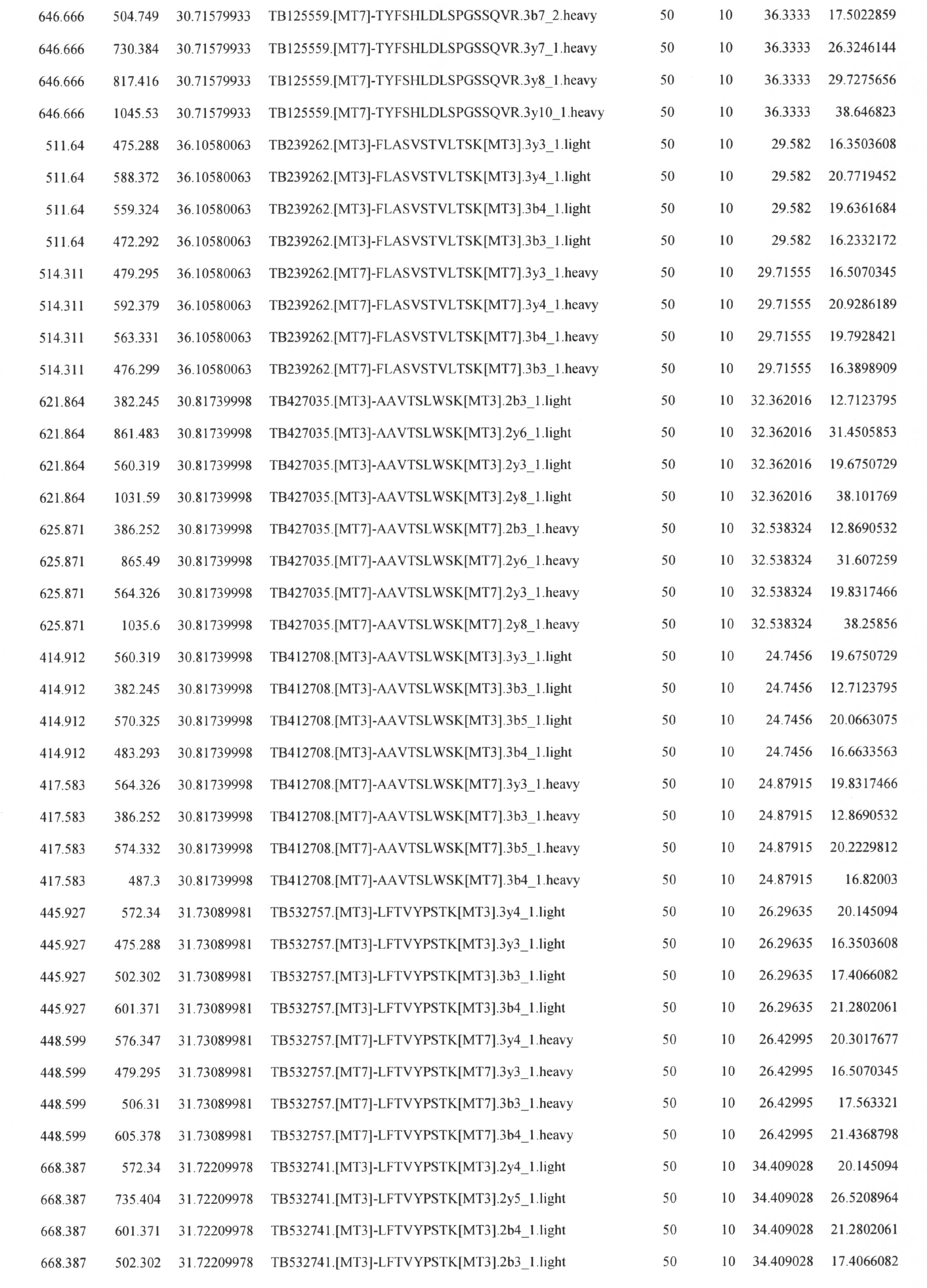

表1において、Q1はペプチドイオンのm/zの値、Q3は断片イオンのm/zの値、RTimeは保持時間、IDは任意の呼称、DPはdeclustering potential、EPはentrance potential、CEはCIDのエネルギー、CXPはcollision cell exit potentialを表す。ただし、この表で示す、RT, DP, EP, CE, CXPは使用する質量分析計によって最適値が異なるため、Q1, Q3以外は使用する装置に合わせて調整すべきである。
方法
1)試料の溶解
表2に示す5つの検体に対して、それぞれ100μl又は200μlの1% SDS/7 M 塩酸グアニジンを含む100 mM Tris-HCl, pH8.5を添加し溶解後、BCA法にてタンパク質定量を実施した。

1)試料の溶解
表2に示す5つの検体に対して、それぞれ100μl又は200μlの1% SDS/7 M 塩酸グアニジンを含む100 mM Tris-HCl, pH8.5を添加し溶解後、BCA法にてタンパク質定量を実施した。
2)メタノールクロロホルム法による試料浄化
各サンプルに対して600μlの氷冷メタノールを添加し、ボルテックス後、氷冷クロロフォルム200μlと氷冷MQ 400μlを加えてボルテックスで撹拌した。その後、氷上で30分静置後、マイクロ冷却遠心機(TOMY MX207)にて遠心(4℃、13500 rpm、5分)し、上層をピペットで除去後、氷冷メタノール1mLを加えて撹拌した。スイングローター式の遠心機(KUBOTA6200)にて遠心(4℃、3300 rpm、5分)後、アングルローター式の遠心機(TOMY MX207)にてさらに遠心(4℃、13500 rpm、5分)し、上清をピペットを使って除去後、氷冷した80%メタノールを1.0mL加えて撹拌した。スイングローター式の遠心機(KUBOTA6200)にて遠心(4℃、3300 rpm、5分)後、アングルローター式の遠心機(TOMY MX207)にて遠心(4℃、13500 rpm、5分)。
上清を除去後、得られた沈殿に7 M Guanidine HClを含む0.5 M Triethylammonium bicarbonate bufferに溶解した。ヒートブロックにて65℃で15分インキュベート後、MQ 28μlを加えてボルテックスで撹拌した。各サンプル2μl(N=3)ずつBCAアッセイにて定量した。(サンプル残量50μlの想定)
各サンプルに対して600μlの氷冷メタノールを添加し、ボルテックス後、氷冷クロロフォルム200μlと氷冷MQ 400μlを加えてボルテックスで撹拌した。その後、氷上で30分静置後、マイクロ冷却遠心機(TOMY MX207)にて遠心(4℃、13500 rpm、5分)し、上層をピペットで除去後、氷冷メタノール1mLを加えて撹拌した。スイングローター式の遠心機(KUBOTA6200)にて遠心(4℃、3300 rpm、5分)後、アングルローター式の遠心機(TOMY MX207)にてさらに遠心(4℃、13500 rpm、5分)し、上清をピペットを使って除去後、氷冷した80%メタノールを1.0mL加えて撹拌した。スイングローター式の遠心機(KUBOTA6200)にて遠心(4℃、3300 rpm、5分)後、アングルローター式の遠心機(TOMY MX207)にて遠心(4℃、13500 rpm、5分)。
上清を除去後、得られた沈殿に7 M Guanidine HClを含む0.5 M Triethylammonium bicarbonate bufferに溶解した。ヒートブロックにて65℃で15分インキュベート後、MQ 28μlを加えてボルテックスで撹拌した。各サンプル2μl(N=3)ずつBCAアッセイにて定量した。(サンプル残量50μlの想定)
各サンプルともチューブ辺りのタンパク質量は200μg以下であるため(表3)、このまま酵素消化とmTRAQラベリングを実施することとした。

3)酵素消化・還元アルキル化
・ 濃度測定後(終体積50μl)の各サンプルに対し50μlのDDWを添加し後、Lysyl endopeptidase溶液を1μl添加し、37℃で3時間インキュベートした。さらに、100μLの水で希釈し、Trypsin溶液 1μlを添加し後37℃で3時間インキュベートした。さらにTrypsin溶液 1μlを添加し一晩インキュベートした。その後、50 mM TCEP HCl溶液を2.5 ml添加(Final conc.0.625mMとなるように)して37℃で30分インキュベーし、250 mM Iodoacetamide溶液を2.5μl添加し遮光して室温で30分反応させた。500 mMのN-Acetyl-L-Cystein溶液を1μl添加して室温で30分反応した。得られたペプチド消化物は4時間凍結乾燥した。
・ 濃度測定後(終体積50μl)の各サンプルに対し50μlのDDWを添加し後、Lysyl endopeptidase溶液を1μl添加し、37℃で3時間インキュベートした。さらに、100μLの水で希釈し、Trypsin溶液 1μlを添加し後37℃で3時間インキュベートした。さらにTrypsin溶液 1μlを添加し一晩インキュベートした。その後、50 mM TCEP HCl溶液を2.5 ml添加(Final conc.0.625mMとなるように)して37℃で30分インキュベーし、250 mM Iodoacetamide溶液を2.5μl添加し遮光して室温で30分反応させた。500 mMのN-Acetyl-L-Cystein溶液を1μl添加して室温で30分反応した。得られたペプチド消化物は4時間凍結乾燥した。
4)mTRAQ標識
凍結乾燥後のペプチド消化物を20μlの水に溶解後、イソプロパノール 50μlを添加して撹拌した。mTRAQ Reagent Δ0試薬を20μl添加し撹拌後、室温で2時間インキュベートした。100μlのMQを添加し、室温で30分静置した。得られた標識ペプチドは一晩凍結乾燥後、1.0 mlの0.1% TFAに再溶解した。
凍結乾燥後のペプチド消化物を20μlの水に溶解後、イソプロパノール 50μlを添加して撹拌した。mTRAQ Reagent Δ0試薬を20μl添加し撹拌後、室温で2時間インキュベートした。100μlのMQを添加し、室温で30分静置した。得られた標識ペプチドは一晩凍結乾燥後、1.0 mlの0.1% TFAに再溶解した。
5)MRM解析
各消化物に濃度既知のmTRAQd0標識ペプチドを添加し、MRMの測定を行なった。MRMによる分析はnano-LCを連結したQTRAP5500 (SCIEX社)で実施した。5 から 45% B を40分、45 から95% B を 1分、そして95% Bで10分(A 溶媒: 0.1% 蟻酸/2% アセトニトリル, B 溶媒:0.1% 蟻酸 90% アセトニトリル )のグラジエントを使用して200 nl/分の流速で自作のキャピラリーカラム(内径100 μm, 長さ15cm, 3μm L-column2)で分離した。スプレー電圧は2000 V, Gas1は30, interface heater temp.:150 ℃の測定条件で行った。
各消化物に濃度既知のmTRAQd0標識ペプチドを添加し、MRMの測定を行なった。MRMによる分析はnano-LCを連結したQTRAP5500 (SCIEX社)で実施した。5 から 45% B を40分、45 から95% B を 1分、そして95% Bで10分(A 溶媒: 0.1% 蟻酸/2% アセトニトリル, B 溶媒:0.1% 蟻酸 90% アセトニトリル )のグラジエントを使用して200 nl/分の流速で自作のキャピラリーカラム(内径100 μm, 長さ15cm, 3μm L-column2)で分離した。スプレー電圧は2000 V, Gas1は30, interface heater temp.:150 ℃の測定条件で行った。
詳細は表4に示す。
結果
結果を図4及び表5に示す。
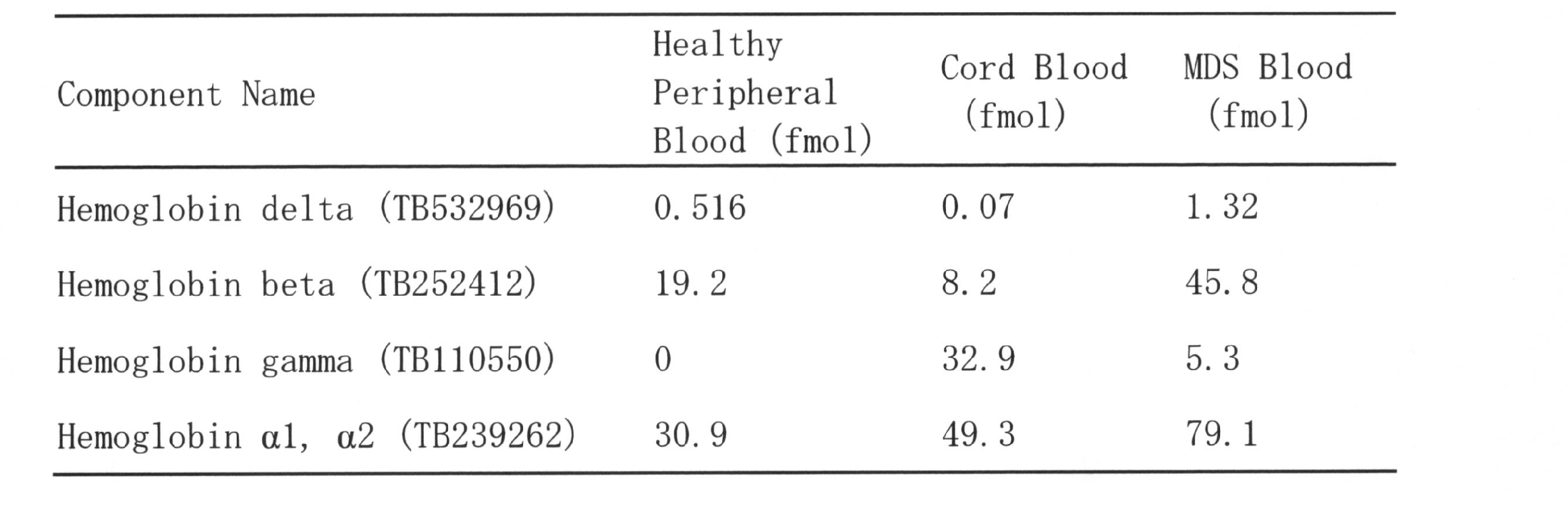
結果を図4及び表5に示す。
健常者末梢血および臍帯血のヘモグロビンは、1細胞あたりそれぞれ500amol、70amolのHbδを有するのに対して、MDS患者末梢血のヘモグロビンはそれぞれ2.6倍、18.6倍の1300amolものヘモグロビンδを含むことが明らかとなった。また今回の患者においては、γ型が特徴的に発現していることも示された。
各種疾患におけるグロビン鎖の解析
方法
本実施例では、健常者、骨髄異形成症候群(MDS)の以下の6つの病型、不応性貧血(RCUD-RA)、鉄芽球性不応性貧血(RARS)、多血球系異形成を伴う不応性血球減少症(RCMD)、芽球増加を伴う不応性貧血-1(RAEB-1)、芽球増加を伴う不応性貧血-2(RAEB-2)、分類不能性骨髄異形成症候群(MDS-U)、MDS以外の以下の3つの血液疾患、再生不良性貧血(AA)、悪性貧血(PA)、急性骨髄性白血病(AML)のそれぞれについて、各グロビン鎖を定量し、各グロビン鎖の量の総ヘモグロビン量に対する割合を算出した。
各グロビン鎖の量は、実施例2の方法と同様にして測定した。内部標準として使用したグロビンペプチドのアミノ酸配列は、表6の「Component name」の欄に記載した(「alpha」、「beta」等の各グロビン鎖を示す用語の後にアルファベット大文字で記載。)。
本実施例では、健常者、骨髄異形成症候群(MDS)の以下の6つの病型、不応性貧血(RCUD-RA)、鉄芽球性不応性貧血(RARS)、多血球系異形成を伴う不応性血球減少症(RCMD)、芽球増加を伴う不応性貧血-1(RAEB-1)、芽球増加を伴う不応性貧血-2(RAEB-2)、分類不能性骨髄異形成症候群(MDS-U)、MDS以外の以下の3つの血液疾患、再生不良性貧血(AA)、悪性貧血(PA)、急性骨髄性白血病(AML)のそれぞれについて、各グロビン鎖を定量し、各グロビン鎖の量の総ヘモグロビン量に対する割合を算出した。
各グロビン鎖の量は、実施例2の方法と同様にして測定した。内部標準として使用したグロビンペプチドのアミノ酸配列は、表6の「Component name」の欄に記載した(「alpha」、「beta」等の各グロビン鎖を示す用語の後にアルファベット大文字で記載。)。
結果
結果を表6~7及び図5に示す。

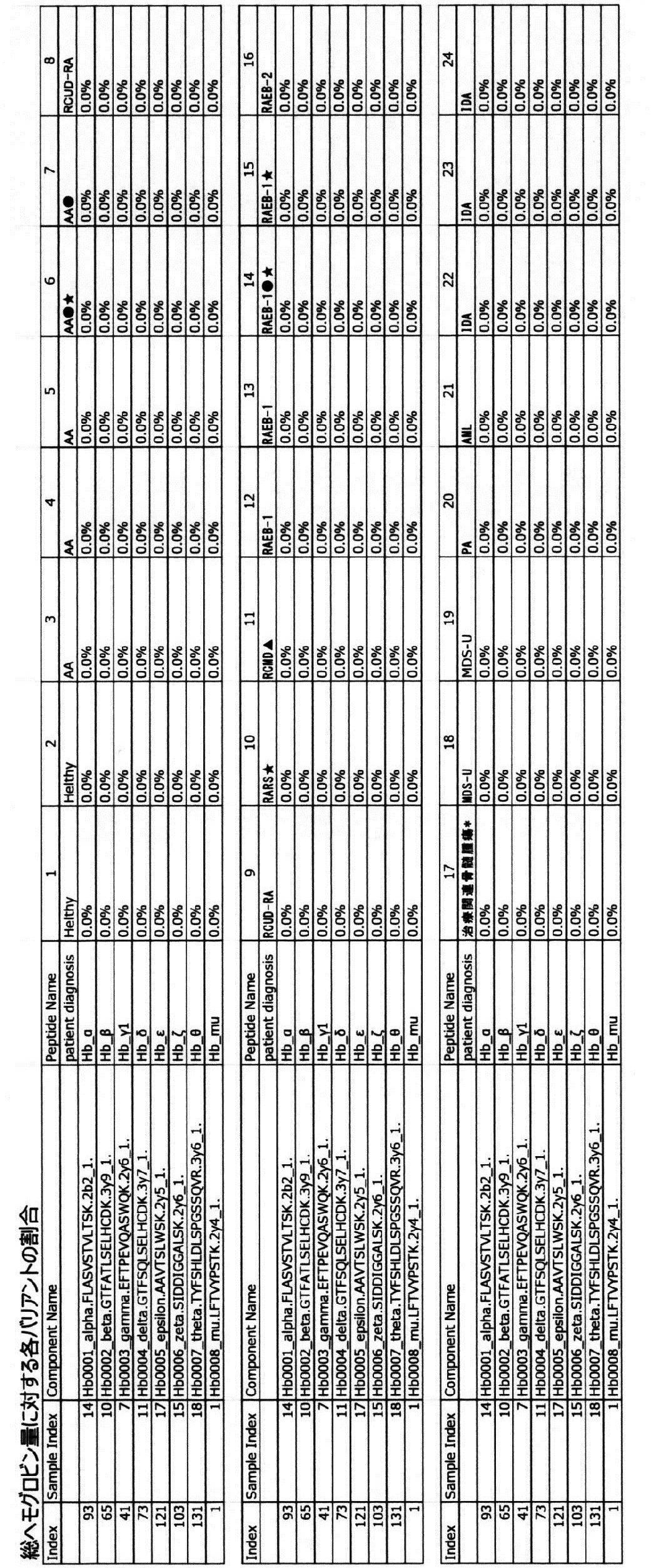
結果を表6~7及び図5に示す。
表6は、ペプチド濃度から算出した各グロビン鎖の濃度(fmol/ライセート溶液0.1μl)である。表6において、Blank Ratioの10倍未満の値(定量下限値以下)であったものは空欄にしてある。
表7及び図5は、総ヘモグロビン量に対する各グロビン鎖の割合である。図5において、縦軸は60~100%のスケールで示した。
(1)γグロビンの発現比率は、AA、MDS、PA及びAML、IDAの順番に高い傾向がある。γグロビンの発現は、Healthyでは認められなかった。
(2)各患者を加療すると、MDS、AAにおいては、γグロビンの発現比率が低下する傾向が認められた。
(3)患者が貧血状態になると、δグロビンの発現比率が低下する傾向が認められた。
(4)MDS及びAAにおいては、δグロビンの発現が認められない傾向があった。
表7及び図5は、総ヘモグロビン量に対する各グロビン鎖の割合である。図5において、縦軸は60~100%のスケールで示した。
(1)γグロビンの発現比率は、AA、MDS、PA及びAML、IDAの順番に高い傾向がある。γグロビンの発現は、Healthyでは認められなかった。
(2)各患者を加療すると、MDS、AAにおいては、γグロビンの発現比率が低下する傾向が認められた。
(3)患者が貧血状態になると、δグロビンの発現比率が低下する傾向が認められた。
(4)MDS及びAAにおいては、δグロビンの発現が認められない傾向があった。
なお、表6及び7において、17番の患者はRAEB-2相当であるが、化学療法歴があるためWHO分類では治療関連骨髄腫瘍と診断した。また、★は輸血あり、▲は貧血治療薬以外の投薬あり、●は貧血治療薬の投薬ありであることを意味する。
1:グロビン解析装置
10:グロビン試料調製手段
20:質量分析手段
30:データ解析手段
101:試料調製部
102:試薬供給部
201:イオン源
202:質量分析部
203:検出部203
301:情報処理部
302:出力部
303:入力部
10:グロビン試料調製手段
20:質量分析手段
30:データ解析手段
101:試料調製部
102:試薬供給部
201:イオン源
202:質量分析部
203:検出部203
301:情報処理部
302:出力部
303:入力部
配列番号5:Xaaはカルバミドメチル化システインを表す(存在位置:8)。
配列番号6:Xaaはカルバミドメチル化システインを表す(存在位置:11)。
配列番号9:Xaaはカルバミドメチル化システインを表す(存在位置:8)。
配列番号11:Xaaはカルバミドメチル化システインを表す(存在位置:8)。
配列番号12:Xaaはカルバミドメチル化システインを表す(存在位置:11)。
配列番号13:Xaaはカルバミドメチル化システインを表す(存在位置:6)。
配列番号24:Xaaはカルバミドメチル化システインを表す(存在位置:11)。
配列番号6:Xaaはカルバミドメチル化システインを表す(存在位置:11)。
配列番号9:Xaaはカルバミドメチル化システインを表す(存在位置:8)。
配列番号11:Xaaはカルバミドメチル化システインを表す(存在位置:8)。
配列番号12:Xaaはカルバミドメチル化システインを表す(存在位置:11)。
配列番号13:Xaaはカルバミドメチル化システインを表す(存在位置:6)。
配列番号24:Xaaはカルバミドメチル化システインを表す(存在位置:11)。
Claims (12)
- 以下の(1)~(9)のいずれかのヘモグロビン鎖に属し、下記アミノ酸配列からなるペプチドから選ばれる少なくとも1つのグロビンタイピングペプチド。
(1)ヘモグロビンα1鎖又はヘモグロビンα2鎖
LRVDPVNFK(配列番号1)
FLASVSTVLTSK(配列番号2)
(2)ヘモグロビンβ鎖
EFTPPVQAAYQK(配列番号3)
VNVDEVGGEALGR(配列番号4)
LLGNVLVC[CAM]VLAHHFGK(配列番号5)
GTFATLSELHC[CAM]DK(配列番号6)
SAVTALWGK(配列番号7)
FFESFGDLSTPDAVMGNPK(配列番号8)
LLGNVLVC[CAM]VLAHHFGK(配列番号9)
(C[CAM]はカルバミドメチル化システインを表す。以下、同様。)
(3)ヘモグロビンδ鎖
EFTPQMQAAYQK(配列番号10)
LLGNVLVC[CAM]VLAR(配列番号11)
GTFSQLSELHC[CAM]DK(配列番号12)
(4)ヘモグロビンε1鎖
LSELHC[CAM]DK(配列番号13)
EFTPEVQAAWQK(配列番号14)
FFDSFGNLSSPSAILGNPK(配列番号15)
AAVTSLWSK(配列番号16)
MNVEEAGGEALGR(配列番号17)
VLTSFGDAIK(配列番号18)
AAVTSLWSK(配列番号19)
NMDNLKPAFAK(配列番号20)
(5)ヘモグロビンγA鎖
VLTSLGDATK(配列番号21)
(6)ヘモグロビンγA鎖又はヘモグロビンγG鎖
VNVEDAGGETLGR(配列番号22)
EFTPEVQASWQK(配列番号23)
GTFAQLSELHC[CAM]DK(配列番号24)
FFDSFGNLSSASAIMGNPK(配列番号25)
ATITSLWGK(配列番号26)
EFTPEVQASWQK(配列番号27)
(7)ヘモグロビンμ鎖
MLAAVGAAVQHVDNLR(配列番号28)
AALSPLADLHALVLR(配列番号29)
LFTVYPSTK(配列番号30)
(8)ヘモグロビンθ1鎖
TFLAFPATK(配列番号31)
VADALSLAVER(配列番号32)
TYFSHLDLSPGSSQVR(配列番号33)
KLGSNVGVYTTEALER(配列番号34)
(9)ヘモグロビンζ鎖
FLSVVSSVLTEK(配列番号35)
TYFPHFDLHPGSAQLR(配列番号36)
SIDDIGGALSK(配列番号37)
ISTQADTIGTETLER(配列番号38)
VVAAVGDAVK(配列番号39)
LFLSHPQTK(配列番号40)
LSELHAYILR(配列番号41) - 以下の(1)~(9)のいずれかのヘモグロビン鎖に属し、下記アミノ酸配列において1又は数個のアミノ酸が欠失、置換又は付加したアミノ酸配列からなるペプチドから選ばれる少なくとも1つの変異型グロビンタイピングペプチド。
(1)ヘモグロビンα1鎖又はヘモグロビンα2鎖
LRVDPVNFK(配列番号1)
FLASVSTVLTSK(配列番号2)
(2)ヘモグロビンβ鎖
EFTPPVQAAYQK(配列番号3)
VNVDEVGGEALGR(配列番号4)
LLGNVLVC[CAM]VLAHHFGK(配列番号5)
GTFATLSELHC[CAM]DK(配列番号6)
SAVTALWGK(配列番号7)
FFESFGDLSTPDAVMGNPK(配列番号8)
LLGNVLVC[CAM]VLAHHFGK(配列番号9)
(C[CAM]はカルバミドメチル化システインを表す。以下、同様。)
(3)ヘモグロビンδ鎖
EFTPQMQAAYQK(配列番号10)
LLGNVLVC[CAM]VLAR(配列番号11)
GTFSQLSELHC[CAM]DK(配列番号12)
(4)ヘモグロビンε1鎖
LSELHC[CAM]DK(配列番号13)
EFTPEVQAAWQK(配列番号14)
FFDSFGNLSSPSAILGNPK(配列番号15)
AAVTSLWSK(配列番号16)
MNVEEAGGEALGR(配列番号17)
VLTSFGDAIK(配列番号18)
AAVTSLWSK(配列番号19)
NMDNLKPAFAK(配列番号20)
(5)ヘモグロビンγA鎖
VLTSLGDATK(配列番号21)
(6)ヘモグロビンγA鎖又はヘモグロビンγG鎖
VNVEDAGGETLGR(配列番号22)
EFTPEVQASWQK(配列番号23)
GTFAQLSELHC[CAM]DK(配列番号24)
FFDSFGNLSSASAIMGNPK(配列番号25)
ATITSLWGK(配列番号26)
EFTPEVQASWQK(配列番号27)
(7)ヘモグロビンμ鎖
MLAAVGAAVQHVDNLR(配列番号28)
AALSPLADLHALVLR(配列番号29)
LFTVYPSTK(配列番号30)
(8)ヘモグロビンθ1鎖
TFLAFPATK(配列番号31)
VADALSLAVER(配列番号32)
TYFSHLDLSPGSSQVR(配列番号33)
KLGSNVGVYTTEALER(配列番号34)
(9)ヘモグロビンζ鎖
FLSVVSSVLTEK(配列番号35)
TYFPHFDLHPGSAQLR(配列番号36)
SIDDIGGALSK(配列番号37)
ISTQADTIGTETLER(配列番号38)
VVAAVGDAVK(配列番号39)
LFLSHPQTK(配列番号40)
LSELHAYILR(配列番号41) - 請求項1に記載のグロビンタイピングペプチド又は請求項2に記載の変異型グロビンタイピングペプチドを標準ペプチドとして用いて、生体試料を質量分析法で解析することを特徴とする、グロビンの検査方法。
- グロビンの検査が、グロビンの同定又はグロビンの定量である、請求項3に記載の方法。
- 請求項1に記載のグロビンタイピングペプチド又は請求項2に記載の変異型グロビンタイピングペプチドを標準ペプチドとして用いて、生体試料を質量分析法で解析することを特徴とする、グロビン関連疾患の検査方法。
- グロビン関連疾患が、グロビン合成異常症、血液疾患又は炎症性疾患である請求項5に記載の方法。
- 血液疾患が溶血性疾患である請求項6に記載の方法。
- 生体試料が、iPS細胞、骨髄細胞、血清又は血漿である請求項3~7のいずれか1項に記載の方法。
- 請求項3~8のいずれか1項に記載の方法により得られた結果を指標として、下記(a)~(d)の項目からなる群から選ばれる少なくとも1つを評価する方法。
(a) 抗癌剤の副作用又は血液毒性
(b) 造血刺激の有無
(c) 血液疾患又は炎症性疾患の病態
(d) 疾患の予後 - グロビン試料調製手段、質量分析手段及びデータ解析手段を含む、グロビン解析装置。
- グロビン試料調製手段が、生体試料回収部、溶血消化処理部、及び脱塩処理部を含む、請求項10に記載の装置。
- 請求項1に記載のグロビンタイピングペプチド又は請求項2に記載の変異型グロビンタイピングペプチドを含む、グロビンの検査用キット。
Applications Claiming Priority (2)
| Application Number | Priority Date | Filing Date | Title |
|---|---|---|---|
| JP2017-145308 | 2017-07-27 | ||
| JP2017145308 | 2017-07-27 |
Publications (1)
| Publication Number | Publication Date |
|---|---|
| WO2019022189A1 true WO2019022189A1 (ja) | 2019-01-31 |
Family
ID=65040591
Family Applications (1)
| Application Number | Title | Priority Date | Filing Date |
|---|---|---|---|
| PCT/JP2018/028050 Ceased WO2019022189A1 (ja) | 2017-07-27 | 2018-07-26 | グロビンタイピングペプチド |
Country Status (1)
| Country | Link |
|---|---|
| WO (1) | WO2019022189A1 (ja) |
Cited By (2)
| Publication number | Priority date | Publication date | Assignee | Title |
|---|---|---|---|---|
| CN111948404A (zh) * | 2020-08-03 | 2020-11-17 | 融智生物科技(青岛)有限公司 | 用于筛查地中海贫血的特征蛋白标记组合物、质谱模型及其应用 |
| CN116203143A (zh) * | 2022-05-11 | 2023-06-02 | 重庆医科大学附属儿童医院 | 血红蛋白病的标志物组合物及其筛查试剂与应用 |
Citations (1)
| Publication number | Priority date | Publication date | Assignee | Title |
|---|---|---|---|---|
| WO2016030688A1 (en) * | 2014-08-29 | 2016-03-03 | Raymond Kruse Iles | Method for detecting abnormalities in hemoglobin |
-
2018
- 2018-07-26 WO PCT/JP2018/028050 patent/WO2019022189A1/ja not_active Ceased
Patent Citations (1)
| Publication number | Priority date | Publication date | Assignee | Title |
|---|---|---|---|---|
| WO2016030688A1 (en) * | 2014-08-29 | 2016-03-03 | Raymond Kruse Iles | Method for detecting abnormalities in hemoglobin |
Non-Patent Citations (2)
| Title |
|---|
| KATAKUSE, I. ET AL.: "Secondary Ion Mass Spectra of Tryptic Peptides of Human Hemoglobin Chains", BIOMEDICAL MASS SPECTROMETRY, vol. 11, no. 8, August 1984 (1984-08-01), pages 386 - 391, XP002595040 * |
| YUMINE, A. ET AL.: "Identification of the globin typing peptide and prospects of the clinical utilization", THE JAPANESE JOURNAL OF CLINICAL HEMATOLOGY, vol. 58, no. 9, September 2017 (2017-09-01), pages 537 * |
Cited By (3)
| Publication number | Priority date | Publication date | Assignee | Title |
|---|---|---|---|---|
| CN111948404A (zh) * | 2020-08-03 | 2020-11-17 | 融智生物科技(青岛)有限公司 | 用于筛查地中海贫血的特征蛋白标记组合物、质谱模型及其应用 |
| CN116203143A (zh) * | 2022-05-11 | 2023-06-02 | 重庆医科大学附属儿童医院 | 血红蛋白病的标志物组合物及其筛查试剂与应用 |
| CN116203143B (zh) * | 2022-05-11 | 2024-01-05 | 重庆医科大学附属儿童医院 | 血红蛋白病的标志物组合物及其筛查试剂与应用 |
Similar Documents
| Publication | Publication Date | Title |
|---|---|---|
| US11428696B2 (en) | Mass spectrometry analysis of mutant polypeptides in biological samples | |
| JP5592791B2 (ja) | ヒト肺組織の病態を示すヒト血清中のタンパク質の同定 | |
| US20050158708A1 (en) | Methods and compositions related to tagging of membrane surface proteins | |
| CN114113381B (zh) | 一种舒氏海龙特征多肽及其应用和鉴别舒适海龙的方法 | |
| Synowsky et al. | Probing genuine strong interactions and post-translational modifications in the heterogeneous yeast exosome protein complex | |
| US20150111238A1 (en) | Method for detecting disease biomarkers | |
| Scheffler et al. | Two-dimensional electrophoresis and mass spectrometric identification of mitochondrial proteins from an SH-SY5Y neuroblastoma cell line | |
| EP1710577B1 (en) | Rapid and quantitative proteome analysis and related methods | |
| WO2019022189A1 (ja) | グロビンタイピングペプチド | |
| Huang et al. | Phosphoproteomics characterization of novel phosphorylated sites of lens proteins from normal and cataractous human eye lenses | |
| Zhou et al. | Discovery top-down proteomics in symbiotic soybean root nodules | |
| CN110790819A (zh) | 一种阿胶多肽及其制备方法 | |
| Dowling | DIGE analysis software and protein identification approaches | |
| US6747273B2 (en) | Methods of detecting protein arginine methyltransferase, and uses related thereto | |
| CN115876991A (zh) | 用于肺栓塞诊断的糖链标志物及其应用 | |
| CN113049726A (zh) | 一种检测全血中免疫抑制剂药物的方法 | |
| Hmmier et al. | DIGE analysis software and protein identification approaches | |
| EP4443163A1 (en) | Method for high-throughput qualitative and quantitative intracellular peptidomics of mammalian cells | |
| Kaneko et al. | Establishment of a combined strategy of genetic and mass spectrometric analyses for characterizing hemoglobin mutations: An example of Hb Hoshida (β43Glu→ Gln) | |
| CN107449916A (zh) | 肺癌和肺部结节蛋白特征谱及其构建方法与应用 | |
| Zellner | Classification of biopsy findings in individuals with hereditary glomerulopathies using molecular genetic testing and proteomics | |
| Ghosh | Proteomics and Their Applications to Biology-A Short Review. | |
| Butenko et al. | Direct empirical in-house assessment of peptide proteotypicity for targeted proteomics | |
| JP2025131404A (ja) | 膵癌の検出方法 | |
| Ranjitha Kumari | Plant proteomics |
Legal Events
| Date | Code | Title | Description |
|---|---|---|---|
| 121 | Ep: the epo has been informed by wipo that ep was designated in this application |
Ref document number: 18838881 Country of ref document: EP Kind code of ref document: A1 |
|
| NENP | Non-entry into the national phase |
Ref country code: DE |
|
| 122 | Ep: pct application non-entry in european phase |
Ref document number: 18838881 Country of ref document: EP Kind code of ref document: A1 |
|
| NENP | Non-entry into the national phase |
Ref country code: JP |