WO2018232149A1 - Gene therapy for ocular disorders - Google Patents
Gene therapy for ocular disorders Download PDFInfo
- Publication number
- WO2018232149A1 WO2018232149A1 PCT/US2018/037592 US2018037592W WO2018232149A1 WO 2018232149 A1 WO2018232149 A1 WO 2018232149A1 US 2018037592 W US2018037592 W US 2018037592W WO 2018232149 A1 WO2018232149 A1 WO 2018232149A1
- Authority
- WO
- WIPO (PCT)
- Prior art keywords
- aav
- vector
- seq
- promoter
- sequence
- Prior art date
Links
Classifications
-
- C—CHEMISTRY; METALLURGY
- C07—ORGANIC CHEMISTRY
- C07K—PEPTIDES
- C07K14/00—Peptides having more than 20 amino acids; Gastrins; Somatostatins; Melanotropins; Derivatives thereof
- C07K14/435—Peptides having more than 20 amino acids; Gastrins; Somatostatins; Melanotropins; Derivatives thereof from animals; from humans
- C07K14/705—Receptors; Cell surface antigens; Cell surface determinants
-
- A—HUMAN NECESSITIES
- A61—MEDICAL OR VETERINARY SCIENCE; HYGIENE
- A61K—PREPARATIONS FOR MEDICAL, DENTAL OR TOILETRY PURPOSES
- A61K48/00—Medicinal preparations containing genetic material which is inserted into cells of the living body to treat genetic diseases; Gene therapy
- A61K48/005—Medicinal preparations containing genetic material which is inserted into cells of the living body to treat genetic diseases; Gene therapy characterised by an aspect of the 'active' part of the composition delivered, i.e. the nucleic acid delivered
-
- C—CHEMISTRY; METALLURGY
- C07—ORGANIC CHEMISTRY
- C07K—PEPTIDES
- C07K14/00—Peptides having more than 20 amino acids; Gastrins; Somatostatins; Melanotropins; Derivatives thereof
- C07K14/435—Peptides having more than 20 amino acids; Gastrins; Somatostatins; Melanotropins; Derivatives thereof from animals; from humans
- C07K14/46—Peptides having more than 20 amino acids; Gastrins; Somatostatins; Melanotropins; Derivatives thereof from animals; from humans from vertebrates
- C07K14/47—Peptides having more than 20 amino acids; Gastrins; Somatostatins; Melanotropins; Derivatives thereof from animals; from humans from vertebrates from mammals
- C07K14/4701—Peptides having more than 20 amino acids; Gastrins; Somatostatins; Melanotropins; Derivatives thereof from animals; from humans from vertebrates from mammals not used
- C07K14/4702—Regulators; Modulating activity
- C07K14/4705—Regulators; Modulating activity stimulating, promoting or activating activity
-
- C—CHEMISTRY; METALLURGY
- C12—BIOCHEMISTRY; BEER; SPIRITS; WINE; VINEGAR; MICROBIOLOGY; ENZYMOLOGY; MUTATION OR GENETIC ENGINEERING
- C12N—MICROORGANISMS OR ENZYMES; COMPOSITIONS THEREOF; PROPAGATING, PRESERVING, OR MAINTAINING MICROORGANISMS; MUTATION OR GENETIC ENGINEERING; CULTURE MEDIA
- C12N15/00—Mutation or genetic engineering; DNA or RNA concerning genetic engineering, vectors, e.g. plasmids, or their isolation, preparation or purification; Use of hosts therefor
- C12N15/09—Recombinant DNA-technology
- C12N15/63—Introduction of foreign genetic material using vectors; Vectors; Use of hosts therefor; Regulation of expression
- C12N15/79—Vectors or expression systems specially adapted for eukaryotic hosts
- C12N15/85—Vectors or expression systems specially adapted for eukaryotic hosts for animal cells
- C12N15/86—Viral vectors
-
- A—HUMAN NECESSITIES
- A01—AGRICULTURE; FORESTRY; ANIMAL HUSBANDRY; HUNTING; TRAPPING; FISHING
- A01K—ANIMAL HUSBANDRY; CARE OF BIRDS, FISHES, INSECTS; FISHING; REARING OR BREEDING ANIMALS, NOT OTHERWISE PROVIDED FOR; NEW BREEDS OF ANIMALS
- A01K2217/00—Genetically modified animals
- A01K2217/07—Animals genetically altered by homologous recombination
- A01K2217/075—Animals genetically altered by homologous recombination inducing loss of function, i.e. knock out
-
- A—HUMAN NECESSITIES
- A01—AGRICULTURE; FORESTRY; ANIMAL HUSBANDRY; HUNTING; TRAPPING; FISHING
- A01K—ANIMAL HUSBANDRY; CARE OF BIRDS, FISHES, INSECTS; FISHING; REARING OR BREEDING ANIMALS, NOT OTHERWISE PROVIDED FOR; NEW BREEDS OF ANIMALS
- A01K2227/00—Animals characterised by species
- A01K2227/10—Mammal
- A01K2227/105—Murine
-
- A—HUMAN NECESSITIES
- A01—AGRICULTURE; FORESTRY; ANIMAL HUSBANDRY; HUNTING; TRAPPING; FISHING
- A01K—ANIMAL HUSBANDRY; CARE OF BIRDS, FISHES, INSECTS; FISHING; REARING OR BREEDING ANIMALS, NOT OTHERWISE PROVIDED FOR; NEW BREEDS OF ANIMALS
- A01K2267/00—Animals characterised by purpose
- A01K2267/03—Animal model, e.g. for test or diseases
- A01K2267/0306—Animal model for genetic diseases
-
- A—HUMAN NECESSITIES
- A61—MEDICAL OR VETERINARY SCIENCE; HYGIENE
- A61K—PREPARATIONS FOR MEDICAL, DENTAL OR TOILETRY PURPOSES
- A61K48/00—Medicinal preparations containing genetic material which is inserted into cells of the living body to treat genetic diseases; Gene therapy
- A61K48/0075—Medicinal preparations containing genetic material which is inserted into cells of the living body to treat genetic diseases; Gene therapy characterised by an aspect of the delivery route, e.g. oral, subcutaneous
-
- A—HUMAN NECESSITIES
- A61—MEDICAL OR VETERINARY SCIENCE; HYGIENE
- A61K—PREPARATIONS FOR MEDICAL, DENTAL OR TOILETRY PURPOSES
- A61K9/00—Medicinal preparations characterised by special physical form
- A61K9/0012—Galenical forms characterised by the site of application
- A61K9/0048—Eye, e.g. artificial tears
-
- C—CHEMISTRY; METALLURGY
- C12—BIOCHEMISTRY; BEER; SPIRITS; WINE; VINEGAR; MICROBIOLOGY; ENZYMOLOGY; MUTATION OR GENETIC ENGINEERING
- C12N—MICROORGANISMS OR ENZYMES; COMPOSITIONS THEREOF; PROPAGATING, PRESERVING, OR MAINTAINING MICROORGANISMS; MUTATION OR GENETIC ENGINEERING; CULTURE MEDIA
- C12N2750/00—MICROORGANISMS OR ENZYMES; COMPOSITIONS THEREOF; PROPAGATING, PRESERVING, OR MAINTAINING MICROORGANISMS; MUTATION OR GENETIC ENGINEERING; CULTURE MEDIA ssDNA viruses
- C12N2750/00011—Details
- C12N2750/14011—Parvoviridae
- C12N2750/14111—Dependovirus, e.g. adenoassociated viruses
- C12N2750/14141—Use of virus, viral particle or viral elements as a vector
- C12N2750/14143—Use of virus, viral particle or viral elements as a vector viral genome or elements thereof as genetic vector
-
- C—CHEMISTRY; METALLURGY
- C12—BIOCHEMISTRY; BEER; SPIRITS; WINE; VINEGAR; MICROBIOLOGY; ENZYMOLOGY; MUTATION OR GENETIC ENGINEERING
- C12N—MICROORGANISMS OR ENZYMES; COMPOSITIONS THEREOF; PROPAGATING, PRESERVING, OR MAINTAINING MICROORGANISMS; MUTATION OR GENETIC ENGINEERING; CULTURE MEDIA
- C12N2750/00—MICROORGANISMS OR ENZYMES; COMPOSITIONS THEREOF; PROPAGATING, PRESERVING, OR MAINTAINING MICROORGANISMS; MUTATION OR GENETIC ENGINEERING; CULTURE MEDIA ssDNA viruses
- C12N2750/00011—Details
- C12N2750/14011—Parvoviridae
- C12N2750/14111—Dependovirus, e.g. adenoassociated viruses
- C12N2750/14171—Demonstrated in vivo effect
Definitions
- Choroideremia is an X- linked inherited retinal disease characterized by the degeneration of photoreceptors, retinal pigment epithelium (RPE) and choriocapillaris. Symptoms develop in the 1st or 2nd decade of life with complaints of poor night vision (nyctalopia) and progressive loss of peripheral vision. Visual fields constrict as the disease progresses. This culminates with loss of central vision (visual acuity) and blindness as early as the fourth decade of life. More than 140 mutations in the CHM gene have been found to cause choroideremia.
- Mutations may lead to the production of an abnormally small, nonfunctional and/or unstable Rab escort protein-1 (REP-1) protein, a decrease in the protein's function or loss of REP-1 protein production. Lack of normal REP-1 disrupts the ability of Rab proteins to aid in intracellular trafficking. The immobility of proteins and organelles within the cell causes the cells to function poorly and to die prematurely.
- REP-1 Rab escort protein-1
- CHM The choroideremia gene, encodes Rab Escort Protein-1 (REP-1), a 653 amino acid protein involved in regulation of membrane trafficking. Since the CHM locus is on the X-chromosome, choroideremia is typically only diagnosed in males. Although female carriers of the disease are usually asymptomatic, retinal exams often reveal a patchy degeneration of the retina and RPE and female individuals can be affected depending on the extent of X-inactivation of the normal X chromosome (lyonization).
- Achromatopsia is a heterogeneous group of autosomal recessive inherited retinal diseases characterized by early onset reduced visual acuity, impaired or complete color blindness, nystagmus, photoaversion and loss of cone photoreceptor function.
- CNG cyclic nucleotide-gated channel
- compositions useful for expressing REP-1, CNGA3 or CNGB3 in human subjects are needed.
- Choroideremia is an X- linked retinal degeneration that is
- CHM is a favorable target for gene augmentation therapy, as the disease is due to loss of function of a protein necessary for retinal cell health, Rab Escort Protein 1 (REP1), which is encoded by the CHM gene.
- the CHM cDNA can be packaged in recombinant adeno-associated virus (rAAV), which has an established track record in human gene therapy studies.
- rAAV recombinant adeno-associated virus
- there are sensitive and quantitative assays to document REP1 activity including its ability to prenylate Rab proteins such as Rab27 and to correct a defect in Rab27 localization and trafficking due to lack of prenylation in REP-1 deficient cells.
- a codon optimized cDNA sequence encoding Rab Escort Protein-1 (REP-1) is provided.
- the codon optimized cDNA sequence is a variant of SEQ ID NO: 3.
- the codon optimized cDNA sequence is SEQ ID NO: 1.
- the cDNA sequence is codon optimized for expression in humans.
- an expression cassette includes a codon optimized nucleic acid sequence that encodes REP-1.
- the expression cassette includes the cDNA sequence of SEQ ID NO: 1.
- the REP-1 encoding sequence is positioned between 5’ and 3’ AAV ITR sequences.
- the vector genome includes all of the nucleic acid sequence between, and including, the 5' ITR and 3' ITR.
- an adeno-associated virus (AAV) vector in another embodiment, includes an AAV capsid and a nucleic acid sequence comprising AAV inverted terminal repeat sequences and a nucleic acid sequence encoding human Rab Escort Protein-1 (REP-1), and expression control sequences that direct expression of the REP-1 in a host cell.
- the REP-1 sequence encodes a full length REP-1 protein.
- the REP-1 sequence is the protein sequence of SEQ ID NO: 2.
- a codon optimized cDNA sequence encoding cyclic nucleotide gated channel alpha 3 is provided.
- the codon optimized cDNA sequence is a variant of SEQ ID NO: 13 or SEQ ID NO: 15.
- the codon optimized cDNA sequence is SEQ ID NO: 9 or SEQ ID NO: 11.
- the cDNA sequence is codon optimized for expression in humans.
- a codon optimized cDNA sequence encoding CNGB3 is provided.
- the codon optimized cDNA sequence is a variant of SEQ ID NO: 19 or 21 or 23.
- the codon optimized cDNA sequence is SEQ ID NO: 45.
- the cDNA sequence is codon optimized for expression in humans.
- an expression cassette includes a codon optimized nucleic acid sequence that encodes cyclic nucleotide gated channel alpha 3 (CNGA3).
- the expression cassette includes the cDNA sequence of SEQ ID NO: 9, SEQ ID NO: 11, SEQ ID NO: 13, or SEQ ID NO: 15.
- the CNGA3 encoding sequence is positioned between 5’ and 3’ AAV ITR sequences.
- an expression cassette includes a codon optimized nucleic acid sequence that encodes cyclic nucleotide gated channel beta 3 (CNGB3).
- the expression cassette includes the cDNA sequence of SEQ ID NO: 19 or SEQ ID NO: 21 or SEQ ID NO: 23 or SEQ ID NO: 45.
- the CNGB3 encoding sequence is positioned between 5’ and 3’ AAV ITR sequences.
- an adeno-associated virus (AAV) vector in another embodiment, is provided.
- the AAV vector includes an AAV capsid and a nucleic acid sequence comprising AAV inverted terminal repeat sequences and a nucleic acid sequence encoding human CNGA3, and expression control sequences that direct expression of the CNGA3 in a host cell.
- the CNGA3sequence encodes a full length CNGA3 protein.
- the CNGA3 sequence is the protein sequence of SEQ ID NO: 10, SEQ ID NO: 12 or SEQ ID NO: 14.
- an adeno-associated virus (AAV) vector in another embodiment, is provided.
- the AAV vector includes an AAV capsid and a nucleic acid sequence comprising AAV inverted terminal repeat sequences and a nucleic acid sequence encoding human CNGB3, and expression control sequences that direct expression of the CNGB3 in a host cell.
- the CNGB3 sequence encodes a full length CNGB3 protein.
- the CNGB3 sequence is the protein sequence of SEQ ID NO: 20.
- an adeno-associated virus (AAV) vector which comprises an AAV8 capsid and an expression cassette, wherein said expression cassette comprises nucleic acid sequences encoding REP-1, inverted terminal repeat sequences and expression control sequences that direct expression of REP-1 in a host cell.
- the expression control sequences include a Chicken Beta Actin (CBA) promoter with a cytomegalovirus (CMV) enhancer.
- the nucleic acid sequence comprises SEQ ID NO: 1.
- an adeno-associated virus (AAV) vector which comprises an AAV8 capsid and an expression cassette, wherein said expression cassette comprises nucleic acid sequences encoding CNGA3, inverted terminal repeat sequences and expression control sequences that direct expression of CNGA3 in a host cell.
- the expression control sequences include a rhodopsin kinase promoter.
- the expression control sequences include a human cone arrestin promoter.
- the nucleic acid sequence comprises SEQ ID NO: 9.
- the nucleic acid sequence comprises SEQ ID NO: 11.
- an adeno-associated virus (AAV) vector which comprises an AAV8 capsid and an expression cassette, wherein said expression cassette comprises nucleic acid sequences encoding CNGB3, inverted terminal repeat sequences and expression control sequences that direct expression of CNGB3 in a host cell.
- AAV adeno-associated virus
- an adeno-associated virus (AAV) vector which comprises an AAV2 capsid and an expression cassette, wherein said expression cassette comprises nucleic acid sequences encoding REP-1, inverted terminal repeat sequences and expression control sequences that direct expression of REP-1 in a host cell.
- the expression control sequences include a CBA promoter with a CMV enhancer.
- the nucleic acid sequence comprises SEQ ID NO: 1.
- an adeno-associated virus (AAV) vector which comprises an AAV2 capsid and an expression cassette, wherein said expression cassette comprises nucleic acid sequences encoding CNGA3, inverted terminal repeat sequences and expression control sequences that direct expression of CNGA3 in a host cell.
- the expression control sequences include a rhodopsin kinase promoter.
- the expression control sequences include a human cone arrestin promoter.
- the nucleic acid sequence comprises SEQ ID NO: 9.
- the nucleic acid sequence comprises SEQ ID NO: 11.
- an adeno-associated virus (AAV) vector which comprises an AAV2 capsid and an expression cassette, wherein said expression cassette comprises nucleic acid sequences encoding CNGB3, inverted terminal repeat sequences and expression control sequences that direct expression of CNGB3 in a host cell.
- AAV adeno-associated virus
- an adeno-associated virus (AAV) vector which comprises an AAV9 capsid and an expression cassette, wherein said expression cassette comprises nucleic acid sequences encoding CNGA3, inverted terminal repeat sequences and expression control sequences that direct expression of CNGA3 in a host cell.
- the expression control sequences include a rhodopsin kinase promoter.
- the expression control sequences include a human cone arrestin promoter.
- the nucleic acid sequence comprises SEQ ID NO: 9.
- the nucleic acid sequence comprises SEQ ID NO: 11.
- an adeno-associated virus (AAV) vector which comprises an AAV9 capsid and an expression cassette, wherein said expression cassette comprises nucleic acid sequences encoding CNGB3, inverted terminal repeat sequences and expression control sequences that direct expression of CNGB3 in a host cell.
- AAV adeno-associated virus
- an adeno-associated virus (AAV) vector which comprises an AAV9 capsid and an expression cassette, wherein said expression cassette comprises nucleic acid sequences encoding REP-1, inverted terminal repeat sequences and expression control sequences that direct expression of REP-1 in a host cell.
- the expression control sequences include a CBA promoter with a CMV enhancer.
- the nucleic acid sequence comprises SEQ ID NO: 1.
- a pharmaceutical composition which includes a pharmaceutically acceptable carrier, diluent, excipient and/or adjuvant and a least a viral vector as described herein.
- a pharmaceutical composition comprises a
- a pharmaceutically acceptable carrier such as the rAAV, described specifically herein.
- a method for treating choroideremia includes administering a composition which includes the AAV vector which encodes REP-1, as described herein, to a subject in need thereof.
- a method for treating achromatopsia includes administering a composition which includes the AAV vector which encodes CNGA3, as described herein, to a subject in need thereof.
- a method for treating achromatopsia includes administering a composition which includes the AAV vector which encodes CNGB3, as described herein, to a subject in need thereof.
- a plasmid for producing an AAV vector includes the codon optimized cDNA sequence encoding REP-1 as described herein.
- the plasmid includes the codon optimized cDNA sequence encoding CNGA3 as described herein.
- the plasmid includes a codon optimized cDNA sequence encoding CNGB3 which is a sequence sharing at least 70% identity with SEQ ID NO: 19 or SEQ ID NO: 21.
- the plasmid includes the codon optimized cDNA sequence encoding CNGB3 as described herein.
- the plasmid is modular.
- a method of generating a rAAV virus includes culturing a packaging cell carrying the plasmid described herein in the presence of sufficient viral sequences to permit packaging of the gene expression cassette viral genome into an infectious AAV envelope or capsid.
- a recombinant AAV produced according to the method is provided.
- FIG.1A and FIG.1B are gels showing REP-1 protein expression in vitro after transfection of cultured 84-31 HEK cells.
- the first lane of each gel shows expression of codon-optimized REP-1 as described herein, expressed from plasmid p944.
- the second lane shows expression of native REP-1 from plasmid p742.
- the third lane shows endogenous expression of REP-1 by 84-31 cells that were not transfected with a plasmid.
- the last lane is a blank.
- the gels demonstrate that the codon-optimized REP-1 sequence, as described herein, results in a higher level of protein expression than the native REP-1 sequence, and that levels of expression from the exogenously transfected plasmids are many-fold higher than endogenous REP-1 expression.
- FIG.2A to FIG.2F provide an alignment of the native REP-1 coding sequence of SEQ ID NO: 1 vs. the codon optimized REP-1 coding sequence of SEQ ID NO: 3.
- FIG.3A to FIG.3F provide an alignment of the native CNGA3 coding sequence of SEQ ID NO: 13 vs. the codon optimized CNGA3 coding sequence of SEQ ID NO: 9.
- FIG.4A to FIG.4F provide an alignment of CNGB3 native ORF (SEQ ID NO: 19) vs. CNGB3 modified ORF (SEQ ID NO: 21) vs. CNGB3 modified orf with modified ends (SEQ ID NO: 23). Point mutations are highlighted.
- FIG.5 is a plasmid map of p584, described herein.
- the sequence of p584 is shown in SEQ ID NO: 7.
- FIG.6 is a plasmid map of AAV.hCHMco.Version 2a, described herein. The sequence of Version 2a is shown in SEQ ID NO: 25.
- FIG.7 is a plasmid map of AAV.hCHMco.Version 2b, described herein.
- the sequence of Version 2b is shown in SEQ ID NO: 26.
- FIG.8 is a plasmid map of AAV.hCHMco.Version 3a, described herein. The sequence of Version 3a is shown in SEQ ID NO: 27.
- FIG.9 is a plasmid map of AAV.hCHMco.Version 3b, described herein. The sequence of Version 3b is shown in SEQ ID NO: 28.
- FIG.10 is a plasmid map of AAV.hCHM.Version 1, described herein. The sequence of Version 1 is shown in SEQ ID NO: 29.
- FIG.11 is a graphic representation of the effect of lambda insert on AAV product impurity. All a-version (lambda containing) vectors have much reduced Kan+ signals from qPCR test.
- FIG.12A is a western blot showing human anti-REP-1 antibody detection of a protein of ⁇ 75-80 kDa in ocular tissues of CD-1 mice injected with AAV8.2b at 5E9 (High dose) vector genome copies. Animals injected with AAV8.2b at 5E8 (Low dose) showed a very faint protein band at ⁇ 75-80 kDa.
- FIG.12B is a Western blot analysis of ocular tissues of AAV8.3b injected CD1 mice (2 mice/group) detected with anti-REP-1 antibody, which revealed the presence of a protein of ⁇ 75-80 kDa in one eye injected with low dose and in both eyes injected with high dose of AAV8.3b. In the ocular tissues of uninjected mice there was no REP-1 expression detected.
- FIG.13A to FIG.13B provide a plasmid map of pAAV-RK1-nativeCNGA3, described herein. The sequence is shown in SEQ ID NO: 30.
- FIG.14A to FIG.14B provide a plasmid map of pAAV-RK1-codon optimized CNGA3, described herein. The sequence is shown in SEQ ID NO: 31.
- FIG.15A to FIG.15B provide a plasmid map of pAAV-RK1- codon optimized CNGA3 variant 3, described herein. The sequence is shown in SEQ ID NO: 32.
- FIG.16A to FIG.16B provide a plasmid map of pAAV-hCAR-nativeCNGA3, described herein. The sequence is shown in SEQ ID NO: 33.
- FIG.17A to FIG.17B provide a plasmid map of pAAV-hCAR-codon optimized CNGA3, described herein. The sequence is shown in SEQ ID NO: 34.
- FIG.18A to FIG.18B provide a plasmid map of pAAV-hCAR- codon optimized CNGA3 variant 3, described herein. The sequence is shown in SEQ ID NO: 35.
- FIG.19A to FIG.19B provide a plasmid map of pAAV-CMV-CBA- nativeCNGA3, described herein. The sequence is shown in SEQ ID NO: 36.
- FIG.20A to FIG.20B provide a plasmid map of pAAV- CMV-CBA -codon optimized CNGA3, described herein. The sequence is shown in SEQ ID NO: 37.
- FIG.21A to FIG.21B provide a plasmid map of pAAV- CMV-CBA - codon optimized CNGA3 variant 3, described herein. The sequence is shown in SEQ ID NO: 38.
- FIG.22A to FIG.22B provide a plasmid map of pAAV- RK1- native CNGB3, described herein. The sequence is shown in SEQ ID NO: 39.
- FIG.23A to FIG.23B provide a plasmid map of pAAV- RK1- codon optimized CNGB3, described herein. The sequence is shown in SEQ ID NO: 40.
- FIG.24A to FIG.24B provide a plasmid map of pAAV- hCAR- native CNGB3, described herein. The sequence is shown in SEQ ID NO: 41.
- FIG.25A to FIG.25B provide a plasmid map of pAAV- hCAR- codon optimized CNGB3, described herein. The sequence is shown in SEQ ID NO: 42.
- FIG.26A to FIG.26B provide a plasmid map of pAAV- CMV-CBA- native CNGB3, described herein. The sequence is shown in SEQ ID NO: 43.
- FIG.27A to FIG.27B provide a plasmid map of pAAV- CMV-CBA- codon optimized CNGB3, described herein. The sequence is shown in SEQ ID NO: 44.
- FIG.28 is a western blot showing hCNGA3 protein expression in 84-31 cells transduced with the noted vectors. 48 hours post transduction, protein was harvested and western blotting was performed.
- the native and codon optimized (hopt) proteins are expected to be 79kDa and V3-hopt is expected to be 85kDa.
- FIG.29 is a western blot showing hCNGA3 protein expression in 84-31 cells transduced with the noted vectors. 48 hours post transduction, protein was harvested and western blotting was performed.
- the native and codon optimized (hopt) proteins are expected to be 79kDa and V3-hopt is expected to be 85kDa.
- FIG.30 are three bar graphs showing vector/plasmid expression as measured by RT-PCR for the three noted vectors.
- FIG.31 is a bar graph showing the results of cone ERG for CNGA3 null mice treated with the noted vectors, as described in Example 8. n>5 for all groups, error bars represent standard deviation.
- FIG.32A and FIG.32B are two line graphs showing fluorescence intensity of untransduced (FIG.32A) and transduced (FIG.32B) cells. These results demonstrate calcium uptake in transduced vs. untransduced 84-31 cells after cGMP addition. Each series is a cell.
- FIG.33 is a bar graph showing the results of cone ERG for CNGB3 null mice treated with the noted vectors, as described in Example 9.
- FIG.34 is a graphic representation of the effect of lambda insert on AAV product impurity.
- FIG.35 is a western blot showing human anti-REP-1 antibody detection of a protein of ⁇ 75-80 kDa in ocular tissues of CD-1 mice injected with indicated rAAVs at 5E7 vector genome copies per eye.
- A1, A2 and A3 indicate Animal 1, 2 and 3, respectively.
- FIG.36 is a western blot showing human anti-REP-1 antibody detection of a protein of ⁇ 75-80 kDa in ocular tissues of CD-1 mice injected with indicated rAAVs at 1E8 vector genome copies per eye.
- A1, A2 and A3 indicate Animal 1, 2 and 3, respectively.
- FIG.37 is a western blot showing human anti-REP-1 antibody detection of a protein of ⁇ 75-80 kDa in ocular tissues of CD-1 mice injected with indicated rAAVs at 5E8 vector genome copies per eye.
- A1 and A2 indicate Animal 1 and 2, respectively.
- FIG.38 is a western blot showing human anti-REP-1 antibody detection of a protein of ⁇ 75-80 kDa in ocular tissues of CD-1 mice injected with indicated rAAVs at 5E9 vector genome copies per eye.
- A1 and A2 indicate Animal 1 and 2, respectively.
- FIGs.39A and 39B are western blots showing human anti-REP-1 antibody detection of a protein of ⁇ 75-80 kDa in ocular tissues of CD-1 mice injected with indicated rAAVs at 5E9 vector genome copies per eye (FIG.39A) or 1E10 vector genome copies per eye (FIG.39B).
- A3, A4, A5, and A6 indicate Animal 3, 4, 5, and 6, respectively.
- FIG.40 is a graphic representation of transduction efficiency of
- AAV8.CMV/CbA-GFP (indicated as Article 6) and AAV2.CMV/CbA-GFP (indicated as Article 5) in PBWT3.1 cells or BMC1 Cells at various dosages.
- the fold change of GFP above background was plotted in y axis while the dosage with unit vector genome copies were indicated on x axis.
- FIG.41 provides a bar graph showing percentage of caspase-3 positive cells in the cells described in FIG.44. Stauro indicates Straurosporine-treated cells while UNT are untreated cells. Article 1 is AAV2.V2a; Article 2 is AAV8.V3a; while Article 3 is AAV8.V1.
- FIG.42 provides bar graphs showing prenylation of target RAB Proteins in CHM patient derived iPSCs after transduction with AAV8.V2a, AAV8.V3a and AAV8.V1.
- CHM patient derived iPSC Cell Line JB 588 demonstrates a CHM mutation with Arg 555 Stop (AGA to TGA).
- CHM patient derived iPSC Cell Line JB 527 demonstrates a deletion of Ex 2-4 of CHM.
- CHM patient derived iPSC Cell Line JB 415 demonstrates a CHM mutation with Ex 10c.1327-1328 del AT.
- incorporated 3 H GGPP normalized to untransduced iPSC cells was plotted in y axis.
- amount of incorporated 3 H GGPP in pmol was plotted in y axis.
- FIG.43A shows the results of cone response 5-7 weeks post injection for the test articles described in Example 16.
- WT wild type
- Un Cnga3 null mice uninjected
- Ex Cnga3 null mice injected with excipient
- OPT codon-optimized
- NAT native
- V3 variant 3
- NA not applicable
- L low dose (8E8 vg/eye)
- H high dose (8E9 vg/eye)
- error bars standard deviation
- threshold 4 standard deviations above the excipient mean, * P ⁇ 0.05, ** P ⁇ 0.01, ***P ⁇ 0.001.
- FIG.43B shows the results of rod response 5-7 weeks post injection for the test articles described in Example 16.
- WT wild type
- Un Cnga3 null mice uninjected
- Ex Cnga3 null mice injected with excipient
- OPT codon optimized
- NAT native
- V3 variant 3
- NA not applicable
- L low dose (8E8 vg/eye)
- FIG.44A shows the results of cone response 12-15 weeks post injection for the test articles described in Example 16.
- WT wild type
- Un Cnga3 null mice uninjected
- Ex Cnga3 null mice injected with excipient
- OPT codon optimized
- NAT native
- V3 variant 3
- NA not applicable
- L low dose (8E8 vg/eye)
- H high dose (8E9 vg/eye)
- error bars standard deviation
- threshold 4 standard deviations above the excipient mean, * P ⁇ 0.05, ** P ⁇ 0.01, ***P ⁇ 0.001.
- FIG.44B shows the results of rod response 12-15 weeks post injection for the test articles described in Example 16.
- FIG.45A and FIG.45B provide a plasmid map of pAAV-hCAR-native- CNGA3-WPRE (p1122), described herein. The sequence of pAAV-hCAR-native-CNGA3- WPRE (p1122) is shown in SEQ ID NO: 46. DETAILED DESCRIPTION OF THE INVENTION
- compositions described herein include compositions and methods for delivering optimized CHM encoding REP-1 to mammalian subjects for the treatment of ocular disorders, primarily blinding diseases such as choroideremia.
- methods and compositions described herein involve compositions and methods for delivering optimized CNGA3 or CNGB3 to mammalian subjects for the treatment of ocular disorders, primarily blinding diseases such as achromatopsia.
- such compositions involve codon optimization of the REP-1, CNGA3 or CNGB3 coding sequence. It is believed that these features increase the efficacy of the product, and increase safety, since a lower dose of reagent is used.
- compositions which include the native REP1, CNGA3, and CNGB3 coding sequences, as shown in SEQ ID NO: 3, SEQ ID NO: 13 and SEQ ID NO: 19, respectively. It is to be understood that when an embodiment is described for either REP-1, CNGA3 or CNGB3, a similar embodiment is intended to be recited for the other.
- the choroideremia gene, CHM encodes Rab Escort Protein-1 (REP-1), a 653 amino acid protein thought to be involved in membrane trafficking.
- REP-1 Rab Escort Protein-1
- CHM Chiroideremia Since the CHM locus is on the X-chromosome, choroideremia is typically only diagnosed in males. Although female carriers of the disease are usually asymptomatic, retinal exams often reveal a patchy degeneration of the retina and RPE and female individuals can be affected depending on the extent of X-inactivation of the normal X chromosome (lyonization). See, Coussa, cited above.
- the native amino acid sequence encoding human REP-1 is reported at GenBank accession number P24386, and reproduced here in SEQ ID NO: 2.
- the native human nucleic acid sequence of CHM is reproduced here at SEQ ID NO: 3 (accession no. NM_000390.2).
- Cyclic nucleotide-gated (CNG) ion channels are key mediators underlying signal transduction in retinal and olfactory receptors. Genetic defects in CNGA3 and CNGB3, encoding two structurally related subunits of cone CNG channels, are known to lead to achromatopsia. CNGA3 is a 694 amino acid protein. CNGB is an 809 amino acid protein.
- Achromatopsia is a heterogeneous group of congenital, autosomal recessive retinal disorders that manifest by early onset cone photoreceptor dysfunction, severely reduced visual acuity, impaired or complete color blindness and photophobia.
- the native nucleic acid sequence encoding human CNGA3 is reported at GenBank accession no.
- XM_011210554.1 The native nucleic acid sequence encoding human CNGA3 is reported at GenBank accession no. XM_011210554.1, and reproduced in SEQ ID NO: 13.
- NM_001298.2 and reproduced in SEQ ID NO: 15.
- the native nucleic acid sequence encoding human CNGB3 is reproduced in SEQ ID NO: 19.
- a subject has an“ocular disorder”, for which the components, compositions and methods of this invention are designed to treat.
- the term“subject” as used herein means a mammalian animal, including a human, a veterinary or farm animal, a domestic animal or pet, and animals normally used for clinical research.
- the subject of these methods and compositions is a human.
- Still other suitable subjects include, without limitation, murine, rat, canine, feline, porcine, bovine, ovine, non-human primate and others.
- the term“subject” is used interchangeably with“patient”.
- ocular disorder includes, cone-rod dystrophies and retinal diseases including, without limitation, Stargardt disease (autosomal dominant or autosomal recessive), retinitis pigmentosa, and pattern dystrophy.
- the subject has achromatopsia.
- the subject has choroideremia or an X-linked hereditary retinal degeneration.
- Clinical signs of such ocular diseases include, but are not limited to, decreased peripheral vision, decreased central (reading) vision, decreased night vision, loss of color perception, reduction in visual acuity, decreased photoreceptor function, pigmentary changes, and ultimately blindness.
- treatment or“treating” is defined encompassing administering to a subject one or more compounds or compositions described herein for the purposes of amelioration of one or more symptoms of an ocular disease.“Treatment” can thus include one or more of reducing onset or progression of an ocular disease, preventing disease, reducing the severity of the disease symptoms, or retarding their progression, including the progression of blindness, removing the disease symptoms, delaying onset of disease or monitoring progression of disease or efficacy of therapy in a given subject.
- exogenous nucleic acid sequence or protein means that the nucleic acid or protein does not naturally occur in the position in which it exists in a chromosome, or host cell.
- An exogenous nucleic acid sequence also refers to a sequence derived from and inserted into the same host cell or subject, but which is present in a non-natural state, e.g. a different copy number, or under the control of different regulatory elements.
- heterologous as used to describe a nucleic acid sequence or protein means that the nucleic acid or protein was derived from a different organism or a different species of the same organism than the host cell or subject in which it is expressed.
- heterologous when used with reference to a protein or a nucleic acid in a plasmid, expression cassette, or vector, indicates that the protein or the nucleic acid is present with another sequence or subsequence which with which the protein or nucleic acid in question is not found in the same relationship to each other in nature.
- the terms“percent (%) identity”,“sequence identity”,“percent sequence identity”, or“percent identical” in the context of nucleic acid sequences refers to the bases in the two sequences which are the same when aligned for correspondence.
- the percent identity is determined by comparing two sequences aligned under optimal conditions over the sequences to be compared.
- the length of sequence identity comparison may be over the full- length of the REP-1, CNGA3 or CNGB3 coding sequence, or a fragment of at least about 100 to 150 nucleotides, or as desired. However, identity among smaller fragments, e.g.
- nucleotide sequences can be compared using FastaTM, a program in GCG Version 6.1. Commonly available sequence analysis software, more specifically, BLAST or analysis tools provided by public databases may also be used.
- isolated means that the material is removed from its original environment (e.g., the natural environment if it is naturally occurring).
- a naturally-occurring polynucleotide or polypeptide present in a living animal is not isolated, but the same polynucleotide or polypeptide, separated from some or all of the coexisting materials in the natural system, is isolated, even if subsequently reintroduced into the natural system.
- Such polynucleotides could be part of a vector and/or such polynucleotides or polypeptides could be part of a composition, and still be isolated in that such vector or composition is not part of its natural environment.
- nucleic acid sequences encoding the REP-1 or CNGA3 or CNGB3 protein described herein are assembled and placed into any suitable genetic element, e.g., naked DNA, phage, transposon, cosmid, episome, etc., which transfers the REP-1 or CNGA3 or CNGB3 sequences carried thereon to a host cell, e.g., for generating non-viral delivery systems (e.g., RNA-based systems, naked DNA, or the like) or for generating viral vectors in a packaging host cell and/or for delivery to a host cells in a subject.
- the genetic element is a plasmid.
- transgene means an exogenous or engineered protein-encoding nucleic acid sequence that is under the control of a promoter or expression control sequence in an expression cassette, rAAV genome, recombinant plasmid or production plasmid, vector, or host cell described in this specification.
- the transgene is a human CHM (REP-1) sequence, encoding a functional REP- 1 protein.
- the transgene is a codon optimized nucleic acid CHM (REP-1) encoding the REP-1 amino acid sequence set forth in SEQ ID NO: 2.
- the transgene is encoded by the sequence set forth in SEQ ID NO: 1.
- the REP-1 transgene is encoded by the sequence set forth in SEQ ID NO: 5.
- SEQ ID NO: 5 includes modified ends, which include restriction sites for cloning into a plasmid, such as a production plasmid described herein.
- the transgene is a human CNGA3 sequence, encoding a functional CNGA3 protein.
- the transgene is a codon optimized CNGA3 encoding sequence SEQ ID NO: 10.
- the transgene is encoded by the sequence set forth in SEQ ID NO: 9.
- the transgene includes modified ends, such as that shown in SEQ ID NO: 16, SEQ IDNO 17 or SEQ ID NO: 18, which include restriction sites for cloning into a plasmid, such as a plasmid described herein.
- the transgene is a codon optimized CNGA3 encoding sequence SEQ ID NO: 12.
- the transgene is encoded by the sequence set forth in SEQ ID NO: 11.
- the transgene is encoded by the native coding sequence of CNGA3, which is set forth in SEQ ID NO: 13.
- the transgene is a human CNGB3 sequence, encoding a functional CNGB3 protein.
- the transgene is a codon optimized CNGB3 encoding sequence which is a sequence sharing at least 70% identity with SEQ ID NO: 19 or 21.
- the transgene is encoded by the sequence set forth in SEQ ID NO: 23.
- SEQ ID NO: 23 includes modified ends, which include restriction sites for cloning into a plasmid, such as a production plasmid described herein. Nucleotides 13 to 2448 of SEQ ID NO: 23 provide the ORF for CNGB3.
- the transgene is a codon optimized CNGB3 encoding sequence SEQ ID NO: 20. In certain embodiments, the transgene is encoded by the sequence set forth in SEQ ID NO: 19. In certain embodiments, the transgene is encoded by the sequence set forth in SEQ ID NO: 21. In certain embodiments, the transgene includes modified ends for cloning into a plasmid, such as the plasmids described herein. SEQ ID NO: 21 is a novel cDNA sequence in which certain silent mutations have been made to the native coding sequence. In certain embodiments, the CNGB3 sequence is the codon optimized sequence set forth in SEQ ID NO: 45. Further modifications to the native sequence, as described herein, are contemplated by the invention.
- the nucleic acid sequence encoding REP-1, CNGA3 or CNGB3 further comprises a nucleic acid encoding a tag polypeptide covalently linked thereto.
- the tag polypeptide may be selected from known“epitope tags” including, without limitation, a myc tag polypeptide, a glutathione-S-transferase tag polypeptide, a green fluorescent protein tag polypeptide, a myc-pyruvate kinase tag polypeptide, a His6 tag polypeptide, an influenza virus hemagglutinin tag polypeptide, a flag tag polypeptide, and a maltose binding protein tag polypeptide.
- A“vector” as used herein is a nucleic acid molecule into which an exogenous or heterologous or engineered nucleic acid transgene may be inserted which can then be introduced into an appropriate host cell.
- Vectors preferably have one or more origin of replication, and one or more site into which the recombinant DNA can be inserted.
- Vectors often have convenient means by which cells with vectors can be selected from those without, e.g., they encode drug resistance genes.
- Common vectors include plasmids, viral genomes, and (primarily in yeast and bacteria)“artificial chromosomes.” Certain plasmids are described herein.
- Virus vectors are defined as replication defective viruses containing the exogenous or heterologous CHM (REP-1) or CNGA3 or CNGB3 nucleic acid transgene(s).
- an expression cassette as described herein may be engineered onto a plasmid which is used for drug delivery or for production of a viral vector.
- Suitable viral vectors are preferably replication defective and selected from amongst those which target ocular cells.
- Viral vectors may include any virus suitable for gene therapy, including but not limited to adenovirus; herpes virus; lentivirus; retrovirus; parvovirus, etc.
- the adeno-associated virus is referenced herein as an exemplary virus vector.
- A“replication-defective virus” or“viral vector” refers to a synthetic or recombinant viral particle in which an expression cassette containing a gene of interest is packaged in a viral capsid or envelope, where any viral genomic sequences also packaged within the viral capsid or envelope are replication- deficient; i.e., they cannot generate progeny virions but retain the ability to infect target cells.
- the genome of the viral vector does not include genes encoding the enzymes required to replicate (the genome can be engineered to be“gutless”– containing only the transgene of interest flanked by the signals required for amplification and packaging of the artificial genome), but these genes may be supplied during production. Therefore, it is deemed safe for use in gene therapy since replication and infection by progeny virions cannot occur except in the presence of the viral enzyme required for replication.
- the expression cassette including any of those described herein is employed to generate a recombinant AAV genome.
- the term“host cell” may refer to the packaging cell line in which a recombinant AAV is produced from a production plasmid.
- the term“host cell” may refer to any target cell in which expression of the transgene is desired.
- a“host cell” refers to a prokaryotic or eukaryotic cell that contains exogenous or heterologous DNA that has been introduced into the cell by any means, e.g., electroporation, calcium phosphate precipitation, microinjection, transformation, viral infection, transfection, liposome delivery, membrane fusion techniques, high velocity DNA-coated pellets, viral infection and protoplast fusion.
- the term“host cell” refers to cultures of ocular cells of various mammalian species for in vitro assessment of the compositions described herein.
- the term“host cell” refers to the cells employed to generate and package the viral vector or recombinant virus.
- the term“host cell” is intended to reference the ocular cells of the subject being treated in vivo for the ocular disease.
- the term“ocular cells” refers to any cell in, or associated with the function of, the eye.
- the term may refer to any one of photoreceptor cells, including rod photoreceptors, cone photoreceptors and photosensitive ganglion cells, retinal pigment epithelium (RPE) cells, Mueller cells, choroidal cells, bipolar cells, horizontal cells, and amacrine cells.
- the ocular cells are the photoreceptor cells.
- the ocular cells are RPE cells.
- Plasmids generally are designated herein by a lower case p preceded and/or followed by capital letters and/or numbers, in accordance with standard naming conventions that are familiar to those of skill in the art.
- Many plasmids and other cloning and expression vectors that can be used in accordance with the present invention are well known and readily available to those of skill in the art.
- those of skill readily may construct any number of other plasmids suitable for use in the invention. The properties, construction and use of such plasmids, as well as other vectors, in the present invention will be readily apparent to those of skill from the present disclosure.
- transcriptional control sequence or“expression control sequence” refers to DNA sequences, such as initiator sequences, enhancer sequences, and promoter sequences, which induce, repress, or otherwise control the transcription of protein encoding nucleic acid sequences to which they are operably linked.
- operably linked refers to both expression control sequences that are contiguous with the nucleic acid sequence encoding the REP-1 or CNGA3 or CNGB3 and/or expression control sequences that act in trans or at a distance to control the transcription and expression thereof.
- AAV or“AAV serotype” as used herein refers to the dozens of naturally occurring and available adeno-associated viruses, as well as artificial AAVs.
- human AAV2 is the first AAV that was developed as a gene transfer vector; it has been widely used for efficient gene transfer experiments in different target tissues and animal models.
- the AAV capsid, ITRs, and other selected AAV components described herein may be readily selected from among any AAV, including, without limitation, AAV1, AAV2, AAV3, AAV4, AAV5, AAV6, AAV7, AAV8, AAV9, AAV8bp, AAV7M8 and AAVAnc80, variants of any of the known or mentioned AAVs or AAVs yet to be discovered or variants or mixtures thereof. See, e.g., WO
- the AAV capsid is an AAV8bp capsid, which preferentially targets bipolar cells. See, WO
- the AAV capsid is an AAV7m8 capsid, which has shown preferential delivery to the outer retina. See, Dalkara et al, In Vivo–Directed Evolution of a New Adeno-Associated Virus for Therapeutic Outer Retinal Gene Delivery from the Vitreous, Sci Transl Med 5, 189ra76 (2013), which is incorporated herein by reference.
- the AAV capsid is an AAV8 capsid.
- the AAV capsid an AAV9 capsid.
- the AAV capsid an AAV5 capsid.
- the AAV capsid an AAV2 capsid.
- the term variant means any AAV sequence which is derived from a known AAV sequence, including those sharing at least 70%, at least 75%, at least 80%, at least 85%, at least 90%, at least 95%, at least 97%, at least 99% or greater sequence identity over the amino acid or nucleic acid sequence.
- the AAV capsid includes variants which may include up to about 10% variation from any described or known AAV capsid sequence. That is, the AAV capsid shares about 90% identity to about 99.9 % identity, about 95% to about 99% identity or about 97% to about 98% identity to an AAV capsid provided herein and/or known in the art.
- the AAV capsid shares at least 95% identity with an AAV capsid.
- the comparison may be made over any of the variable proteins (e.g., vp1, vp2, or vp3).
- the AAV capsid shares at least 95% identity with the AAV8 vp3.
- a self-complementary AAV is used.
- the ITRs or other AAV components may be readily isolated or engineered using techniques available to those of skill in the art from an AAV.
- AAV may be isolated, engineered, or obtained from academic, commercial, or public sources (e.g., the American Type Culture Collection, Manassas, VA).
- the AAV sequences may be engineered through synthetic or other suitable means by reference to published sequences such as are available in the literature or in databases such as, e.g., GenBank, PubMed, or the like.
- AAV viruses may be engineered by conventional molecular biology techniques, making it possible to optimize these particles for cell specific delivery of nucleic acid sequences, for minimizing immunogenicity, for tuning stability and particle lifetime, for efficient degradation, for accurate delivery to the nucleus, etc.
- “artificial AAV” means, without limitation, an AAV with a non-naturally occurring capsid protein.
- Such an artificial capsid may be generated by any suitable technique, using a selected AAV sequence (e.g., a fragment of a vp1 capsid protein) in combination with heterologous sequences which may be obtained from a different selected AAV, non-contiguous portions of the same AAV, from a non-AAV viral source, or from a non-viral source.
- An artificial AAV may be, without limitation, a pseudotyped AAV, a chimeric AAV capsid, a recombinant AAV capsid, or a“humanized” AAV capsid.
- AAV2/5 and AAV2/8 are exemplary pseudotyped vectors.
- Self-complementary AAV refers a plasmid or vector having an expression cassette in which a coding region carried by a recombinant AAV nucleic acid sequence has been designed to form an intra-molecular double-stranded DNA template.
- dsDNA double stranded DNA
- administering means delivering the composition to the target selected cell which is characterized by the ocular disease.
- the method involves delivering the composition by subretinal injection to the RPE, photoreceptor cells or other ocular cells.
- intravitreal injection to ocular cells is employed.
- injection via the palpebral vein to ocular cells may be employed.
- Still other methods of administration may be selected by one of skill in the art given this disclosure.
- By“administering” or“route of administration” is delivery of composition described herein, with or without a pharmaceutical carrier or excipient, of the subject. Routes of administration may be combined, if desired. In some embodiments, the administration is repeated periodically.
- compositions described herein are designed for delivery to subjects in need thereof by any suitable route or a combination of different routes.
- Direct delivery to the eye (optionally via ocular delivery, subretinal injection, intra-retinal injection, intravitreal, topical), or delivery via systemic routes, e.g., intraarterial, intraocular, intravenous, intramuscular, subcutaneous, intradermal, and other parental routes of administration.
- the nucleic acid molecules and/or vectors described herein may be delivered in a single composition or multiple compositions.
- two or more different AAV may be delivered, or multiple viruses [see, e.g., WO202011/126808 and WO 2013/049493].
- multiple viruses may contain different replication- defective viruses (e.g., AAV and adenovirus), alone or in combination with proteins.
- compositions described herein are isolated, or synthetically or recombinantly engineered nucleic acid sequences that provide novel codon-optimized sequences encoding REP-1 or CNGA3 or CNGB3.
- an isolated or engineered codon optimized nucleic acid sequence encoding human REP-1 is provided.
- the codon-optimized sequence is SEQ ID NO: 1.
- the codon optimized sequence includes N-terminal and C-terminal restriction sites for cloning.
- the REP-1 coding sequence includes an N-terminal NotI restriction site and a C-terminal BamHI restriction site, in addition to a Kozak consensus sequence.
- the codon optimized sequence in some embodiments, includes one or more additional restriction sites to allow for addition of markers, such as an epitope tag.
- the codon optimized REP-1 may have a percent identity of at least 50%, or at least 60%, or at least 70%, or at least 80% or at least 90%, including any integer between any of those ranges.
- the codon optimized REP-1 has a percent identify with the native sequence of at least 51, 52, 53, 54, 55, 56, 57, 58, 59, 60, 61, 62, 63, 64, 65, 66, 67, 68, 69, 70, 71, 72, 73, 74, 75, 76, 77, 78, 79, 80, 81, 82, 83, 84, 85, 86, 87, 88, 89, 90, 91, 92, 93, 94, 95, 96, 97, 98 or 99%.
- codon optimized REP-1 SEQ ID NO: 1 has a percent sequence identity of only 74% (see FIG.2).
- an isolated or engineered codon optimized nucleic acid sequence encoding human CNGA3 is provided.
- the codon- optimized sequence is SEQ ID NO: 9.
- the codon-optimized sequence is a CNGA3 variant shown in SEQ ID NO: 11.
- the codon optimized sequence includes N-terminal and C-terminal restriction sites for cloning.
- the CNGA3 coding sequence includes an N-terminal NotI restriction site and a C-terminal BglII restriction site, in addition to a Kozak consensus sequence. Examples of CNGA3 sequences which include such modifications can be found in SEQ ID NO: 16, SEQ ID NO: 17 and SEQ ID NO: 18.
- the codon optimized sequence in some embodiments, includes one or more additional restriction sites to allow for addition of markers, such as an epitope tag.
- the codon optimized CNGA3 may have a percent identity of at least 50%, or at least 60%, or at least 70%, or at least 80% or at least 90%, including any integer between any of those ranges.
- the codon optimized CNGA3 has a percent identify with the native sequence of at least 51, 52, 53, 54, 55, 56, 57, 58, 59, 60, 61, 62, 63, 64, 65, 66, 67, 68, 69, 70, 71, 72, 73, 74, 75, 76, 77, 78, 79, 80, 81, 82, 83, 84, 85, 86, 87, 88, 89, 90, 91, 92, 93, 94, 95, 96, 97, 98 or 99%.
- codon optimized CNGA3 when aligned with the native nucleic acid sequence SEQ ID NO: 13, it is revealed that codon optimized CNGA3 (SEQ ID NO: 9) has a percent sequence identity of only 80% (see FIG.3).
- an isolated or engineered codon optimized nucleic acid sequence encoding human CNGB3 is provided.
- the codon- optimized sequence is a sequence sharing at least 70% identity with SEQ ID NO: 19 or SEQ ID NO 21.
- the codon optimized sequence is that set forth in SEQ ID NO: 45, which shares about 76% identity with the modified CNGB3 sequence of SEQ ID NO: 21.
- the codon optimized sequence includes N-terminal and C- terminal restriction sites for cloning, for example, as shown in SEQ ID NO: 23.
- the codon optimized sequence in some embodiments, includes one or more additional restriction sites to allow for addition of markers, such as an epitope tag.
- the codon optimized CNGB3 may have a percent identity of at least 50%, or at least 60%, or at least 70%, or at least 80% or at least 90%, including any integer between any of those ranges.
- the codon optimized CNGB3 has a percent identify with the native sequence of at least 51, 52, 53, 54, 55, 56, 57, 58, 59, 60, 61, 62, 63, 64, 65, 66, 67, 68, 69, 70, 71, 72, 73, 74, 75, 76, 77, 78, 79, 80, 81, 82, 83, 84, 85, 86, 87, 88, 89, 90, 91, 92, 93, 94, 95, 96, 97, 98 or 99%.
- the optimized nucleic acid sequences encoding the REP-1 or CNGA3 or CNGB3 constructs described herein are engineered into any suitable genetic element, e.g., naked DNA, phage, transposon, cosmid, RNA molecule (e.g., mRNA), episome, etc., which transfers the REP-1 or CNGA3 or CNGB3 sequences carried thereon to a host cell, e.g., for generating nanoparticles carrying DNA or RNA, viral vectors in a packaging host cell and/or for delivery to a host cells in subject.
- the genetic element is a plasmid.
- the selected genetic element may be delivered by any suitable method, including transfection, electroporation, liposome delivery, membrane fusion techniques, high velocity DNA-coated pellets, viral infection and protoplast fusion.
- suitable method including transfection, electroporation, liposome delivery, membrane fusion techniques, high velocity DNA-coated pellets, viral infection and protoplast fusion.
- the methods used to make such constructs are known to those with skill in nucleic acid manipulation and include genetic engineering, recombinant engineering, and synthetic techniques. See, e.g., Green and Sambrook, Molecular Cloning: A Laboratory Manual, Cold Spring Harbor Press, Cold Spring Harbor, NY (2012).
- a variety of expression cassettes are provided which employ SEQ ID Nos.1 or 5 for expression of the REP-1 protein.
- an example of a plasmid containing such an expression cassette is shown in SEQ ID NO.25.
- an example of a plasmid containing such an expression cassette is shown in SEQ ID NO.26.
- an example of a plasmid containing such an expression cassette is shown in SEQ ID NO.27.
- an example of a plasmid containing such an expression cassette is shown in SEQ ID NO.28.
- the“vector genome” is the nucleic acid sequence which is packaged between the 5’ and 3’ ITRs, including the ITRs themselves.
- the term“vector genome” is used interchangeably with“expression cassette”.
- the vector genome includes a 5’ ITR, a CMV enhancer, a Chicken beta-actin promoter, CBA exon 1 and intron, a Kozak sequence, a codon optimized CHM, bGH poly A and a 3’ ITR.
- the vector genome comprises nt 1 to 4233 of SEQ ID NO: 25.
- the vector genome comprises nt 1 to 4233 of SEQ ID NO: 26.
- the vector genome comprises nt 1 to 4233 of SEQ ID NO: 27.
- the vector genome comprises nt 1 to 4233 of SEQ ID NO: 28.
- the vector genome includes a 5’ ITR, a RK1 promoter, a codon optimized CNGA3, bGH poly A and a 3’ ITR. In another embodiment, the vector genome includes a 5’ ITR, a RK1 promoter, a native CNGA3, bGH poly A and a 3’ ITR.
- the vector genome includes a 5’ ITR, a RK1 promoter, a codon optimized CNGA3 variant 3, bGH poly A and a 3’ ITR.
- the vector genome includes a 5’ ITR, a hCAR promoter, a codon optimized CNGA3, bGH poly A and a 3’ ITR.
- the vector genome includes a 5’ ITR, a hCAR promoter, a native CNGA3, bGH poly A and a 3’ ITR.
- the vector genome includes a 5’ ITR, a hCAR promoter, a codon optimized CNGA3 variant 3, bGH poly A and a 3’ ITR.
- the vector genome includes a 5’ ITR, a CMV enhancer, a Chicken beta-actin promoter, CBA exon 1 and intron, a codon optimized CNGA3, bGH poly A and a 3’ ITR.
- the vector genome includes a 5’ ITR, a CMV enhancer, a Chicken beta-actin promoter, CBA exon 1 and intron, a native CNGA3, bGH poly A and a 3’ ITR.
- the vector genome includes a 5’ ITR, a CMV enhancer, a Chicken beta-actin promoter, CBA exon 1 and intron, a codon optimized CNGA3 variant 3, bGH poly A and a 3’ ITR.
- the vector genome comprises nt 1 to 3189 of SEQ ID NO: 30. In another embodiment, the vector genome comprises nt 1 to 3189 of SEQ ID NO: 31. In another embodiment, the vector genome comprises nt 1 to 3354 of SEQ ID NO: 32. In another embodiment, the vector genome comprises nt 1 to 3583 of SEQ ID NO: 33. In another embodiment, the vector genome comprises nt 1 to 3580 of SEQ ID NO: 34. In another embodiment, the vector genome comprises nt 1 to 3748 of SEQ ID NO: 35. In another embodiment, the vector genome comprises nt 1 to 4357 of SEQ ID NO: 36. In another embodiment, the vector genome comprises nt 1 to 4357 of SEQ ID NO: 37. In another embodiment, the vector genome comprises nt 1 to 4522 of SEQ ID NO: 38.
- the vector genome includes a 5’ ITR, a RK1 promoter, a codon optimized CNGB3, bGH poly A and a 3’ ITR.
- the vector genome includes a 5’ ITR, a RK1 promoter, a native CNGB3, bGH poly A and a 3’ ITR.
- the vector genome includes a 5’ ITR, a hCAR promoter, a codon optimized CNGB3, bGH poly A and a 3’ ITR. In another embodiment, the vector genome includes a 5’ ITR, a hCAR promoter, a native CNGB3, bGH poly A and a 3’ ITR. In one embodiment, the vector genome includes a 5’ ITR, a CMV enhancer, a Chicken beta-actin promoter, CBA exon 1 and intron, a codon optimized CNGB3, bGH poly A and a 3’ ITR. In another embodiment, the vector genome includes a 5’ ITR, a CMV enhancer, a Chicken beta-actin promoter, CBA exon 1 and intron, a native CNGB3, bGH poly A and a 3’ ITR.
- the vector genome comprises nt 1 to 3537 of SEQ ID NO: 39. In another embodiment, the vector genome comprises nt 1 to 3536 of SEQ ID NO: 40. In another embodiment, the vector genome comprises nt 1 to 3930 of SEQ ID NO: 41. In another embodiment, the vector genome comprises nt 1 to 3930 of SEQ ID NO: 42. In another embodiment, the vector genome comprises nt 1 to 4704 of SEQ ID NO: 43. In another embodiment, the vector genome comprises nt 1 to 4704 of SEQ ID NO: 44. In another embodiment, the vector genome comprises nt 1 to 4154 of SEQ ID NO: 46.
- an“expression cassette” refers to a nucleic acid molecule which comprises coding sequences for the optimized REP-1 or CNGA3 or CNGB3 proteins, promoter, and may include other regulatory sequences therefor, which cassette may be engineered into a genetic element or plasmid, and/or packaged into the capsid of a viral vector (e.g., a viral particle).
- an expression cassette comprises a codon optimized nucleic acid sequence that encodes REP-1.
- the cassette provides the codon optimized REP-1 operatively associated with expression control sequences that direct expression of the codon optimized nucleic acid sequence that encodes REP-1 in a host cell.
- an expression cassette comprises a codon optimized nucleic acid sequence that encodes CNGA3.
- the cassette provides the codon optimized CNGA3 operatively associated with expression control sequences that direct expression of the codon optimized nucleic acid sequence that encodes CNGA3 in a host cell.
- an expression cassette comprises a codon optimized nucleic acid sequence that encodes CNGB3.
- the cassette provides the codon optimized CNGB3 operatively associated with expression control sequences that direct expression of the codon optimized nucleic acid sequence that encodes CNGB3 in a host cell.
- an expression cassette for use in an AAV vector is provided.
- the AAV expression cassette includes at least one AAV inverted terminal repeat (ITR) sequence.
- the expression cassette comprises 5’ ITR sequences and 3’ ITR sequences.
- the 5’ and 3’ ITRs flank the codon optimized nucleic acid sequence that encodes REP-1 or CNGA3 or CNGB3, optionally with additional sequences which direct expression of the codon optimized nucleic acid sequence that encodes REP-1 or CNGA3 or CNGB3 in a host cell.
- a AAV expression cassette is meant to describe an expression cassette as described above flanked on its 5’ end by a 5’AAV inverted terminal repeat sequence (ITR) and on its 3’ end by a 3’ AAV ITR.
- ITR inverted terminal repeat sequence
- this rAAV genome contains the minimal sequences required to package the expression cassette into an AAV viral particle, i.e., the AAV 5’ and 3’ ITRs.
- the AAV ITRs may be obtained from the ITR sequences of any AAV, such as described herein. These ITRs may be of the same AAV origin as the capsid employed in the resulting recombinant AAV, or of a different AAV origin (to produce an AAV pseudotype).
- the ITR sequences from AAV2, or the deleted version thereof ( ⁇ ITR) are used for convenience and to accelerate regulatory approval. However, ITRs from other AAV sources may be selected.
- Each rAAV genome can be then introduced into a production plasmid.
- the production plasmid is that described herein, or as described in WO2012/158757, which is incorporated herein by reference.
- Various plasmids are known in the art for use in producing rAAV vectors, and are useful herein.
- the production plasmids are cultured in the host cells which express the AAV cap and/or rep proteins. In the host cells, each rAAV genome is rescued and packaged into the capsid protein or envelope protein to form an infectious viral particle.
- One type of production plasmid is that shown in SEQ ID NO: 7, which is termed p584.
- This plasmid is used in the examples for generation of the rAAV-REP-1 vector.
- Such a plasmid is one that contains a 5’ AAV ITR sequence; a selected promoter; a polyA sequence; and a 3’ ITR; additionally, it also contains a stuffer sequence, such as lambda.
- a non-coding lambda stuffer region is included in the vector backbone.
- the nucleic acid sequence encoding REP-1, CNGA3 or CNGB3 are inserted between the selected promoter and the polyA sequence, or a similar, plasmid.
- p584 which includes the REP-1 encoding sequence can be found in SEQ ID NO: 8.
- the production plasmid is modified to optimized vector plasmid production efficiency. Such modifications include addition of other neutral sequences, or deletion of portion(s) of or the entire lambda stuffer sequence to modulate the level of supercoil of the vector plasmid. Such modifications are contemplated herein. In other embodiments, terminator and other sequences are included in the plasmid.
- a recombinant adeno-associated virus (AAV) vector is provided for delivery of the REP-1, CNGA3 and CNGB3 constructs and optimized sequences described herein.
- An adeno-associated virus (AAV) viral vector is an AAV Dnase-resistant particle having an AAV protein capsid into which is packaged nucleic acid sequences for delivery to target cells.
- An AAV capsid is composed of 60 capsid (cap) protein subunits, VP1, VP2, and VP3, that are arranged in an icosahedral symmetry in a ratio of approximately 1:1:10 to 1:1:20, depending upon the selected AAV.
- AAVs may be selected as sources for capsids of AAV viral vectors as identified above. See, e.g., US Published Patent Application No.2007-0036760-A1; US Published Patent Application No.2009- 0197338-A1; EP 1310571. See also, WO 2003/042397 (AAV7 and other simian AAV), US Patent 7790449 and US Patent 7282199 (AAV8), WO 2005/033321 and US 7,906,111 (AAV9), and WO 2006/110689, and WO 2003/042397 (rh.10). Each of these documents is incorporated by reference in its entirety. In some embodiments, the AAV capsids are generated using the nucleic acid sequences described in the listed documents.
- an AAV cap for use in the viral vector can be generated by mutagenesis (i.e., by insertions, deletions, or substitutions) of one of the aforementioned AAV capsids or its encoding nucleic acid.
- the AAV capsid is chimeric, comprising domains from two or three or four or more of the
- the AAV capsid is a mosaic of Vpl, Vp2, and Vp3 monomers from two or three different AAVs or recombinant AAVs.
- an rAAV composition comprises more than one of the aforementioned Caps.
- the AAV capsid includes variants which may include up to about 10% variation from any described or known AAV capsid sequence. That is, the AAV capsid shares about 90% identity to about 99.9 % identity, about 95% to about 99% identity or about 97% to about 98% identity to an AAV capsid provided herein and/or known in the art. In one embodiment, the AAV capsid shares at least 95% identity with an AAV capsid. When determining the percent identity of an AAV capsid, the comparison may be made over any of the variable proteins (e.g., vp1, vp2, or vp3). In one embodiment, the AAV capsid shares at least 95% identity with the AAV8 vp3.
- the variable proteins e.g., vp1, vp2, or vp3
- a self- complementary AAV is used.
- the AAV capsid is a tyrosine capsid-mutant in which certain surface exposed tyrosine residues are substituted with phenylalanine (F).
- F phenylalanine
- the ITRs are the only AAV components required in cis in the same construct as the transgene.
- the coding sequences for the replication (rep) and/or capsid (cap) are removed from the AAV genome and supplied in trans or by a packaging cell line in order to generate the AAV vector.
- a pseudotyped AAV may contain ITRs from a source which differs from the source of the AAV capsid.
- a chimeric AAV capsid may be utilized. Still other AAV components may be selected.
- AAV sequences are described herein and may also be isolated or engineered obtained from academic, commercial, or public sources (e.g., the American Type Culture Collection, Manassas, VA). Alternatively, the AAV sequences may be obtained through synthetic or other suitable means by reference to published sequences such as are available in the literature or in databases such as, e.g., GenBank®, PubMed®, or the like. [000134] Methods for generating and isolating AAV viral vectors suitable for delivery to a subject are known in the art. See, e.g., US Patent 7790449; US Patent 7282199; WO 2003/042397; WO 2005/033321, WO 2006/110689; and US 7588772 B2].
- a producer cell line is transiently transfected with a construct that encodes the transgene flanked by ITRs and a construct(s) that encodes rep and cap.
- a packaging cell line that stably supplies rep and cap is transiently transfected with a construct encoding the transgene flanked by ITRs.
- AAV virions are produced in response to infection with helper adenovirus or herpesvirus, requiring the separation of the rAAVs from contaminating virus.
- helper functions i.e., adenovirus E1, E2a, VA, and E4 or herpesvirus UL5, UL8, UL52, and UL29, and herpesvirus polymerase
- the helper functions can be supplied by transient transfection of the cells with constructs that encode the required helper functions, or the cells can be engineered to stably contain genes encoding the helper functions, the expression of which can be controlled at the transcriptional or posttranscriptional level.
- the transgene flanked by ITRs and rep/cap genes are introduced into insect cells by infection with baculovirus-based vectors.
- baculovirus-based vectors For reviews on these production systems, see generally, e.g., Zhang et al., 2009,“Adenovirus-adeno-associated virus hybrid for large-scale recombinant adeno-associated virus production,” Human Gene Therapy 20:922-929, the contents of which is incorporated herein by reference in its entirety. Methods of making and using these and other AAV production systems are also described in the following U.S.
- the rAAV vectors comprise an AAV capsid and an AAV expression cassette comprising sequences encoding REP-1 or CNGA3 or CNGB3, such as described above.
- the rAAV expression cassette comprises AAV inverted terminal repeat sequences and a codon optimized nucleic acid sequence that encodes REP-1 or CNGA3 or CNGB3, and expression control sequences that direct expression of the encoded proteins in a host cell.
- the rAAV expression cassette in other embodiments, further comprises one or more of an intron, a Kozak sequence, a polyA, and post-transcriptional regulatory elements.
- Such rAAV vectors for use in pharmaceutical compositions for delivery to the eye may employ a capsid from any of the many known AAVs identified above.
- Other conventional components of the expression cassettes and vectors include other components that can be optimized for a specific species using techniques known in the art including, e.g, codon optimization, as described herein.
- the components of the cassettes, vectors, plasmids and viruses or other compositions described herein include a promoter sequence as part of the expression control sequences.
- the promoter is cell-specific.
- the term“cell-specific” means that the particular promoter selected for the recombinant vector can direct expression of the optimized REP-1 or CNGA3 or CNGB3 transgene in a particular ocular cell type.
- the promoter is specific for expression of the transgene in photoreceptor cells.
- the promoter is specific for expression in the rods and cones. In another embodiment, the promoter is specific for expression in the rods. In another embodiment, the promoter is specific for expression in the cones.
- the photoreceptor-specific promoter is a human rhodopsin kinase promoter.
- the rhodopsin kinase (RK1) promoter has been shown to be active in both rods and cones. See, e.g., Sun et al, Gene Therapy with a
- the promoter is modified to add one or more restriction sites to facilitate cloning.
- the RK1 promoter is shown in nt 175-684 of SEQ ID NO: 30.
- hCAR Human cone arrestin
- hCAR promoter In experiments performed in animals aimed at characterizing gene expression, human cone arrestin promoters drove strong expression in retina. See, Dyka, Frank M., et al. "Cone specific promoter for use in gene therapy of retinal degenerative diseases.” Retinal Degenerative Diseases. Springer New York, 2014.695-701. Dyka el al also report that the specificity of hCAR promoter was poor, with rods and RPE clearly being transduced. Sequences of hCAR promoter are accessible via publicly available literatures, database and commercially available products. In one embodiment, the nucleic acid sequence of hCAR promoter is reproduced in nt 175 to nt 1078 of SEQ ID NO: 33. In another embodiment, the nucleic acid sequence of hCAR promoter is reproduced in nt 181 to nt 1078 of SEQ ID NO: 33.
- the promoter is a human rhodopsin promoter.
- the promoter is modified to include restriction on the ends for cloning. See, e.g, Nathans and Hogness, Isolation and nucleotide sequence of the gene encoding human rhodopsin, PNAS, 81:4851-5 (August 1984), which is incorporated herein by reference in its entirety.
- the promoter is a portion or fragment of the human rhodopsin promoter.
- the promoter is a variant of the human rhodopsin promoter.
- promoters include the human G-protein-coupled receptor protein kinase 1 (GRK1) promoter (Genbank Accession number AY327580).
- the promoter is a 292 nt fragment (positions 1793-2087) of the GRK1 promoter (See, Beltran et al, Gene Therapy 201017:1162-74, which is hereby incorporated by reference in its entirety).
- the promoter is the human interphotoreceptor retinoid-binding protein proximal (IRBP) promoter.
- IRBP interphotoreceptor retinoid-binding protein proximal
- the promoter is a 235 nt fragment of the hIRBP promoter.
- the promoter is the RPGR proximal promoter (Shu et al, IOVS, May 2102, which is incorporated by reference in its entirety).
- Other promoters useful in the invention include, without limitation, the rod opsin promoter, the red-green opsin promoter, the blue opsin promoter, the cGMP- ⁇ - phosphodiesterase promoter (Qgueta et al, IOVS, Invest Ophthalmol Vis Sci.2000
- the promoter is selected from human human EF1 ⁇ promoter, rhodopsin promoter, rhodopsin kinase, interphotoreceptor binding protein (IRBP), cone opsin promoters (red-green, blue), cone opsin upstream sequences containing the red-green cone locus control region, cone transducing, and transcription factor promoters (neural retina leucine zipper (Nrl) and photoreceptor-specific nuclear receptor Nr2e3, bZIP).
- human human EF1 ⁇ promoter rhodopsin promoter, rhodopsin kinase
- IRBP interphotoreceptor binding protein
- cone opsin promoters red-green, blue
- cone opsin upstream sequences containing the red-green cone locus control region cone transducing
- transcription factor promoters neural retina leucine zipper (Nrl) and photoreceptor-specific nuclear receptor Nr2e3, bZIP.
- the promoter is a ubiquitous or consistutive promoter.
- An example of a suitable promoter is a hybrid chicken ⁇ -actin (CBA) promoter with cytomegalovirus (CMV) enhancer elements.
- CBA chicken ⁇ -actin
- CMV cytomegalovirus
- the nucleic acid sequence of the CBA promoter, with CMV enhancer elements is shown in nt 1 to nt 544 of SEQ ID NO: 36.
- the promoter includes CBA exon 1 and intron sequences such as that shown in nt 546 to nt.823 of SEQ ID NO: 36.
- the promoter is the CB7 promoter.
- suitable promoters include the human ⁇ -actin promoter, the human elongation factor-1 ⁇ promoter, the cytomegalovirus (CMV) promoter, the simian virus 40 promoter, and the herpes simplex virus thymidine kinase promoter. See, e.g., Damdindorj et al, (August 2014) A Comparative Analysis of Constitutive Promoters Located in Adeno-Associated Viral Vectors. PloS ONE 9(8): e106472.
- promoters include viral promoters, constitutive promoters, regulatable promoters [see, e.g., WO 2011/126808 and WO 2013/04943].
- a promoter responsive to physiologic cues may be utilized in the expression cassette, rAAV genomes, vectors, plasmids and viruses described herein.
- the promoter is of a small size, under 1000 bp, due to the size limitations of the AAV vector.
- the promoter is under 400 bp.
- Other promoters may be selected by one of skill in the art.
- the REP-1 construct incorporates a ubiquitous promoter.
- the CNGA3 construct incorporates a photoreceptor-specific promoter.
- the REP-1 construct includes a CBA promoter with CMV enhancer elements.
- the promoter is an inducible promoter.
- the inducible promoter may be selected from known promoters including the rapamycin/rapalog promoter, the ecdysone promoter, the estrogen-responsive promoter, and the tetracycline-responsive promoter, or heterodimeric repressor switch. See, Sochor et al, An Autogenously Regulated Expression System for Gene Therapeutic Ocular Applications. Scientific Reports, 2015 Nov 24;5:17105 and Daber R, Lewis M., A novel molecular switch. J Mol Biol.2009 Aug 28;391(4):661-70, Epub 2009 Jun 21 which are both incorporated herein by reference in their entirety.
- the cassette, vector, plasmid and virus constructs described herein contain other appropriate transcription initiation, termination, enhancer sequences, efficient RNA processing signals such as splicing and polyadenylation (polyA) signals; TATA sequences; sequences that stabilize cytoplasmic mRNA; sequences that enhance translation efficiency (i.e., Kozak consensus sequence); introns; sequences that enhance protein stability; and when desired, sequences that enhance secretion of the encoded product.
- a Woodchuck hepatitis virus post-transcriptional regulatory element WPRE
- the expression cassette or vector may contain none, one or more of any of the elements described herein.
- suitable polyA sequences include, e.g., SV40, bovine growth hormone (bGH), and TK polyA.
- suitable enhancers include, e.g., the CMV enhancer, the RSV enhancer, the alpha fetoprotein enhancer, the TTR minimal promoter/enhancer, LSP (TH-binding globulin promoter/alpha1- microglobulin/bikunin enhancer), amongst others.
- a Kozak sequence is included upstream of the transgene coding sequence to enhance translation from the correct initiation codon.
- CBA exon 1 and intron are included in the expression cassette.
- the transgene is placed under the control of a hybrid chicken ⁇ actin (CBA) promoter.
- CBA hybrid chicken ⁇ actin
- This promoter consists of the cytomegalovirus (CMV) immediate early enhancer, the proximal chicken ⁇ actin promoter, and CBA exon 1 flanked by intron 1 sequences. See, nt 1 to 823 of SEQ ID NO: 36.
- An adeno-associated virus (AAV) vector comprising an AAV capsid and a nucleic acid sequence comprising AAV inverted terminal repeat sequences and the nucleic acid sequence of SEQ ID NO: 1 encoding human Rab Escort Protein-1 (REP-1), and expression control sequences comprising a CBA promoter with a CMV enhancer that direct expression of the REP-1 in a host cell.
- AAV adeno-associated virus
- an adeno-associated virus (AAV) vector which includes an AAV capsid having packaged therein a nucleic acid sequence comprising AAV inverted terminal repeat sequences and the nucleic acid sequence of SEQ ID NO: 9 or SEQ ID NO: 11, encoding human cyclic nucleotide gated channel alpha 3 (CNGA3), and expression control sequences that direct expression of the CNGA3 in a host cell.
- the CNGA sequence comprises SEQ ID NO: 9 and a rhodopsin kinase 1 (RK1) promoter.
- the RK1 promoter sequence is nt 175-684 of SEQ ID NO: 30.
- the CNGA sequence comprises SEQ ID NO: 11 and a human cone arrestin (hCAR) promoter.
- hCAR human cone arrestin
- the hCAR promoter sequenced is that shown in nt 175 to nt 1078 of SEQ ID NO: 33 or nt 181 to nt 1078 of SEQ ID NO: 33.
- an adeno-associated virus (AAV) vector which includes an AAV capsid having packaged therein a nucleic acid sequence comprising AAV inverted terminal repeat sequences and the nucleic acid sequence SEQ ID NO.45 encoding human cyclic nucleotide gated channel beta 3 (CNGB3), and expression control sequences that direct expression of the CNGB3 in a host cell.
- the expression control sequences comprise a CMV/CBA promoter, RK1 promoter or hCAR promoter.
- the expression cassette contains a 5’ ITR, CBA promoter, CMV enhancer, CBA exon 1 and intron, kozak sequence, human codon optimized CHM sequence (SEQ ID NO: 1), bGH poly A and 3’ ITR.
- these nucleic acid sequences, vectors, expression cassettes and rAAV viral vectors are useful in a pharmaceutical composition, which also comprises a pharmaceutically acceptable carrier, buffer, diluent and/or adjuvant, etc.
- compositions are used to express the optimized REP-1 or CNGA3 or CNGB3 in the ocular cells through delivery by such recombinantly engineered AAVs or artificial AAVs.
- compositions containing the nucleic acid sequences, vectors, expression cassettes and rAAV viral vectors are preferably assessed for contamination by conventional methods and then formulated into a pharmaceutical composition suitable for administration to the eye.
- a pharmaceutically and/or physiologically acceptable vehicle or carrier particularly one suitable for administration to the eye, such as buffered saline or other buffers, e.g., HEPES, to maintain pH at appropriate physiological levels, and, optionally, other medicinal agents, pharmaceutical agents, stabilizing agents, buffers, carriers, adjuvants, diluents, etc.
- the carrier will typically be a liquid.
- Exemplary physiologically acceptable carriers include sterile, pyrogen-free water and sterile, pyrogen- free, phosphate buffered saline.
- the carrier is an isotonic sodium chloride solution.
- the carrier is balanced salt solution.
- the carrier includes tween. If the virus is to be stored long- term, it may be frozen in the presence of glycerol or Tween20.
- the composition of the carrier or excipient contains 180 mM NaCl, 10 mM NaPi, pH7.3 with 0.0001% - 0.01% Pluronic F68 (PF68).
- the exact composition of the saline component of the buffer ranges from 160 mM to 180 mM NaCl.
- a different pH buffer pH buffer (potentially HEPES, sodium bicarbonate, TRIS) is used in place of the buffer specifically described.
- a buffer containing 0.9% NaCl is useful.
- compositions of the invention may contain, in addition to the rAAV and/or variants and carrier(s), other conventional pharmaceutical ingredients, such as preservatives, or chemical stabilizers.
- suitable exemplary preservatives include
- chlorobutanol potassium sorbate, sorbic acid, sulfur dioxide, propyl gallate, the parabens, ethyl vanillin, glycerin, phenol, and parachlorophenol.
- Suitable chemical stabilizers include gelatin and albumin.
- compositions containing at least one replication-defective rAAV virus can be formulated with a physiologically acceptable carrier, diluent, excipient and/or adjuvant, for use in gene transfer and gene therapy applications.
- a physiologically acceptable carrier diluent, excipient and/or adjuvant
- quantification of the genome copies (“GC”), vector genomes (“VG”), or virus particles may be used as the measure of the dose contained in the formulation or suspension. Any method known in the art can be used to determine the genome copy (GC) number of the replication-defective virus compositions of the invention.
- AAV GC number titration is as follows: Purified AAV vector samples are first treated with Dnase to eliminate un-encapsidated AAV genome DNA or contaminating plasmid DNA from the production process. The Dnase resistant particles are then subjected to heat treatment to release the genome from the capsid. The released genomes are then quantitated by real-time PCR using primer/probe sets targeting specific region of the viral genome (usually poly A signal).
- the effective dose of a recombinant adeno-associated virus carrying a nucleic acid sequence encoding the optimized REP-1 or CNGA3 or CNGB3 transgene is measured as described in S.K. McLaughlin et al, 1988 J. Virol., 62:1963, which is incorporated by reference in its entirety.
- the term“dosage” can refer to the total dosage delivered to the subject in the course of treatment, or the amount delivered in a single unit (or multiple unit or split dosage) administration.
- the pharmaceutical virus compositions can be formulated in dosage units to contain an amount of replication-defective virus carrying the codon optimized nucleic acid sequences encoding REP-1 or CNGA3 or CNGB3 as described herein that is in the range of about 1.0 x 10 6 GC to about 1.0 x 10 15 GC including all integers or fractional amounts within the range.
- the compositions are formulated to contain at least 1x10 7 , 2x10 7 , 3x10 7 , 4x10 7 , 5x10 7 , 6x10 7 , 7x10 7 , 8x10 7 , or 9x10 7 GC per dose including all integers or fractional amounts within the range. In one embodiment, the compositions are formulated to contain at least 1x10 8 , 2x10 8 , 3x10 8 , 4x10 8 , 5x10 8 , 6x10 8 , 7x10 8 , 8x10 8 , or 9x10 9 GC per dose including all integers or fractional amounts within the range.
- the compositions are formulated to contain at least 1x10 9 , 2x10 9 , 3x10 9 , 4x10 9 , 5x10 9 , 6x10 9 , 7x10 9 , 8x10 9 , or 9x10 9 GC per dose including all integers or fractional amounts within the range.
- the compositions are formulated to contain at least 1x10 10 , 2x10 10 , 3x10 10 , 4x10 10 , 5x10 10 , 6x10 10 , 7x10 10 , 8x10 10 , or 9x10 10 GC per dose including all integers or fractional amounts within the range.
- compositions are formulated to contain at least 1x10 11 , 2x10 11 , 3x10 11 , 4x10 11 , 5x10 11 , 6x10 11 , 7x10 11 , 8x10 11 , or 9x10 11 GC per dose including all integers or fractional amounts within the range.
- compositions are formulated to contain at least 1x10 12 , 2x10 12 , 3x10 12 , 4x10 12 , 5x10 12 , 6x10 12 , 7x10 12 , 8x10 12 , or 9x10 12 GC per dose including all integers or fractional amounts within the range.
- compositions are formulated to contain at least 1x10 13 , 2x10 13 , 3x10 13 , 4x10 13 , 5x10 13 , 6x10 13 , 7x10 13 , 8x10 13 , or 9x10 13 GC per dose including all integers or fractional amounts within the range.
- compositions are formulated to contain at least 1x10 14 , 2x10 14 , 3x10 14 , 4x10 14 , 5x10 14 , 6x10 14 , 7x10 14 , 8x10 14 , or 9x10 14 GC per dose including all integers or fractional amounts within the range.
- compositions are formulated to contain at least 1x10 15 , 2x10 15 , 3x10 15 , 4x10 15 , 5x10 15 , 6x10 15 , 7x10 15 , 8x10 15 , or 9x10 15 GC per dose including all integers or fractional amounts within the range.
- the dose can range from 1x10 10 to about 1x10 12 GC per dose including all integers or fractional amounts within the range. All dosages may be measured by any known method, including as measured by oqPCR or digital droplet PCR (ddPCR) as described in, e.g., M. Lock et al, Hum Gene Ther Methods.2014 Apr;25(2):115-25. Doi: 10.1089/hgtb.2013.131, which is incorporated herein by reference.
- ddPCR digital droplet PCR
- volume of carrier, excipient or buffer formulation may be administered in a variety of volumes of carrier, excipient or buffer formulation, ranging from about 25 to about 1000 microliters, including all numbers within the range, depending on the size of the area to be treated, the viral titer used, the route of administration, and the desired effect of the method.
- the volume of carrier, excipient or buffer is at least about 25 ⁇ L.
- the volume is about 50 ⁇ L.
- the volume is about 75 ⁇ L.
- the volume is about 100 ⁇ L.
- the volume is about 125 ⁇ L.
- the volume is about 150 ⁇ L.
- the volume is about 175 ⁇ L.
- the volume is about 200 ⁇ L. In another embodiment, the volume is about 225 ⁇ L. In yet another embodiment, the volume is about 250 ⁇ L. In yet another embodiment, the volume is about 275 ⁇ L. In yet another embodiment, the volume is about 300 ⁇ L. In yet another embodiment, the volume is about 325 ⁇ L. In another embodiment, the volume is about 350 ⁇ L. In another embodiment, the volume is about 375 ⁇ L. In another embodiment, the volume is about 400 ⁇ L. In another embodiment, the volume is about 450 ⁇ L. In another embodiment, the volume is about 500 ⁇ L. In another embodiment, the volume is about 550 ⁇ L. In another embodiment, the volume is about 600 ⁇ L. In another embodiment, the volume is about 650 ⁇ L. In another embodiment, the volume is about 700 ⁇ L. In another embodiment, the volume is between about 700 and 1000 ⁇ L.
- the viral constructs may be delivered in doses of from at least 1x10 7 to about least 1x10 11 GCs in volumes of about 1 ⁇ L to about 3 ⁇ L for small animal subjects, such as mice.
- small animal subjects such as mice.
- the larger human dosages and volumes stated above are useful. See, e.g., Diehl et al, J. Applied Toxicology, 21:15-23 (2001) for a discussion of good practices for administration of substances to various veterinary animals. This document is incorporated herein by reference.
- the lowest effective concentration of virus or other delivery vehicle be utilized in order to reduce the risk of undesirable effects, such as toxicity, retinal dysplasia and detachment.
- Still other dosages in these ranges may be selected by the attending physician, taking into account the physical state of the subject, preferably human, being treated, the age of the subject, the particular ocular disorder and the degree to which the disorder, if progressive, has developed.
- a rAAV carrying the REP-1 codon optimized sequences may be administered to a desired subject including a human subject.
- This method comprises administering to a subject in need thereof any of the nucleic acid sequences, expression cassettes, rAAV genomes, plasmids, vectors or rAAV vectors or compositions containing them.
- the composition is delivered subretinally.
- the composition is delivered intravitreally.
- the composition is delivered using a combination of administrative routes suitable for treatment of ocular diseases, and may also involve administration via the palpebral vein or other intravenous or conventional administration routes.
- an rAAV carrying the CNGA3 or CNGB3 native, modified or codon optimized sequence, preferably suspended in a physiologically compatible carrier, diluent, excipient and/or adjuvant may be administered to a desired subject including a human subject.
- This method comprises administering to a subject in need thereof any of the nucleic acid sequences, expression cassettes, rAAV genomes, plasmids, vectors or rAAV vectors or compositions containing them.
- the composition is delivered subretinally.
- the composition is delivered intravitreally.
- the composition is delivered using a combination of administrative routes suitable for treatment of ocular diseases, and may also involve administration via the palpebral vein or other intravenous or conventional administration routes.
- the volume and viral titer of each dosage is determined individually, as further described herein, and may be the same or different from other treatments performed in the same, or contralateral, eye.
- the dosages, administrations and regimens may be determined by the attending physician given the teachings of this specification.
- the composition is administered in a single dosage selected from those above listed in a single affected eye.
- the composition is administered as a single dosage selected from those above listed in a both affected eyes, either simultaneously or sequentially. Sequential administration may imply a time gap of administration from one eye to another from intervals of minutes, hours, days, weeks or months.
- the method involves administering the compositions to an eye two or more dosages (e.g., split dosages). In another embodiment, multiple injections are made in different portions of the same eye.
- a second administration of an rAAV including the selected expression cassette e.g., CHM containing cassette
- Such time point may be weeks, months or years following the first administration.
- Such second administration is, in one embodiment, performed with an rAAV having a different capsid than the rAAV from the first administration.
- the rAAV from the first and second administration have the same capsid.
- the compositions described herein may be delivered in a single composition or multiple compositions.
- two or more different AAV may be delivered, or multiple viruses [see, e.g., WO 2011/126808 and WO 2013/049493].
- multiple viruses may contain different replication- defective viruses (e.g., AAV and adenovirus).
- one or more injections are performed in the same eye in order to target different areas of the affected eye.
- the volume and viral titer of each injection is determined individually, as further described herein, and may be the same or different from other injections performed in the same, or contralateral, eye.
- a single, larger volume injection is made in order to treat the entire eye.
- the volume and concentration of the rAAV composition is selected so that only the region of damaged ocular cells is impacted.
- the volume and/or concentration of the rAAV composition is a greater amount, in order reach larger portions of the eye, including non-damaged photoreceptors.
- a one-time intra-ocular delivery of a composition as described herein is useful in preventing vision loss and blindness in a subject at risk of developing choroideremia.
- a one-time intra- ocular delivery of a composition as described herein e.g., an AAV delivery of an optimized CNGA3 or CNGB3 cassette, is useful in preventing vision loss and blindness in a subject at risk of developing achromatopsia.
- the composition is administered before disease onset.
- the composition is administered prior to the initiation of vision impairment or loss. In another embodiment, the composition is administered after initiation of vision impairment or loss. In yet another embodiment, the composition is administered when less than 90% of the rod and/or cones or photoreceptors are functioning or remaining, as compared to a non-diseased eye.
- the method includes performing additional studies, e.g., functional and imaging studies to determine the efficacy of the treatment.
- additional studies e.g., functional and imaging studies to determine the efficacy of the treatment.
- such tests include retinal and visual function assessment via electroretinograms (ERGs) looking at rod and cone photoreceptor function, optokinetic nystagmus, pupillometry, water maze testing, light-dark preference, optical coherence tomography (to measure thickness of various layers of the retina), histology (retinal thickness, rows of nuclei in the outer nuclear layer, immunofluorescence to document transgene expression, cone
- EMGs electroretinograms
- fMRI functional magnetic resonance imaging
- full-field light sensitivity testing retinal structure studies including optical coherence tomography
- fundus photography fundus autofluorescence
- adaptive optics laser scanning ophthalmoscopy mobility testing
- test of reading speed and accuracy microperimetry and/or ophthalmoscopy.
- any of the above described methods is performed in combination with another, or secondary, therapy.
- the methods of treatment of these ocular diseases involve treating the subject with the composition described in detail herein in combination with another therapy, such as antibiotic treatment, palliative treatment for pain, and the like.
- the additional therapy may be any now known, or as yet unknown, therapy which helps prevent, arrest or ameliorate these mutations or defects or any of the effects associated therewith.
- the secondary therapy can be administered before, concurrent with, or after administration of the compositions described above.
- a secondary therapy involves non-specific approaches for maintaining the health of the retinal cells, such as administration of neurotrophic factors, anti-oxidants, anti- apoptotic agents.
- the non-specific approaches are achieved through injection of proteins, recombinant DNA, recombinant viral vectors, stem cells, fetal tissue, or genetically modified cells. The latter could include genetically modified cells that are encapsulated.
- a method of generating a recombinant rAAV comprises obtaining a plasmid containing an AAV expression cassette as described above and culturing a packaging cell carrying the plasmid in the presence of sufficient viral sequences to permit packaging of the AAV viral genome into an infectious AAV envelope or capsid.
- Specific methods of rAAV vector generation are described above and may be employed in generating a rAAV vector that can deliver the codon optimized REP-1 or CNGA3 or CNGB3 in the expression cassettes and genomes described above and in the examples below.
- a vector comprising any of the expression cassettes described herein is provided.
- such vectors can be plasmids of variety of origins and are useful in certain embodiments for the generation of recombinant replication defective viruses as described further herein.
- the vector is a plasmid that comprises an expression cassette, wherein the expression cassette comprises AAV inverted terminal repeat sequences and a codon optimized nucleic acid sequence that encodes REP-1, and expression control sequences that direct expression of the encoded protein in a host cell.
- the vector is a plasmid that comprises an expression cassette, wherein the expression cassette comprises AAV inverted terminal repeat sequences and a codon optimized nucleic acid sequence that encodes CNGA3, and expression control sequences that direct expression of the encoded protein in a host cell.
- the vector is a plasmid that comprises an AAV expression cassette, wherein the expression cassette comprises AAV inverted terminal repeat sequences and a codon optimized nucleic acid sequence that encodes CNGB3, and expression control sequences that direct expression of the encoded protein in a host cell.
- regulation refers to the ability of a composition to inhibit one or more components of a biological pathway.
- Choroideremia lacks a relevant mouse model and there is no canine model, therefore, transduction and expression is tested in a human retinal cell model of the disease. Because it is impossible to obtain retinal cells from a living patient, RPE are generated from induced pluripotent stem cells. Pluripotent stem cells are directed to RPE using the protocol described by Buchholz et al, Rapid and Efficient Directed Differentiation of Human
- the human induced pluripotent stem cell line is maintained in Dulbecco’s Modified Eagle’s Medium: Nutrient Mixture F-12 (DMEM/F12) containing 2mMGlutaMAX-I, 20% knockout serum replacement, 0.1 mM Modified Eagle’s Medium Non-Essential Amino Acids (MEM NEAA), 0.1 mM ⁇ -mercaptoethanol and 4 ng/ml bFGF on a mitomycin C–treated or irradiated mouse embryonic fibroblast feeder layer.
- DEM/F12 Nutrient Mixture F-12
- MEM NEAA Non-Essential Amino Acids
- 0.1 mM ⁇ -mercaptoethanol 4 ng/ml bFGF
- Pluripotent stem cells are passaged directly onto Matrigel (BD Biosciences) in DMEM/F12 with 1X B27, 1X N2, and 1X NEAA (Invitrogen). From days 0 to 2, 50 ng/ml Noggin, 10 ng/ml Dkk1, 10 ng/ml IGF1 and 10mM nicotinamide are added to the base medium. From days 2 to 4, 10 ng/ml Noggin, 10 ng/ml Dkk1, 10 ng/ml IGF1, 5 ng/ml bFGF and 10mMnicotinamide are added to the base medium.
- the cells are mechanically enriched by scraping away cells with non-RPE morphology. Subsequently, the remaining RPE are digested using TrypLE Express
- Example 2 Cells transduced with AAV-REP-1
- AAV2/8CMV.CBA-REP-1 viral vector incorporating REP-1 codon optimized sequences are produced by transient transfection of HEK293 cells, and the viral particles are precipitated from either the supernatant using polyethylene glycol. See, e.g., Guo et al, Rapid and simplified purification of recombinant adeno-associated virus, J Virol Methods.2012 Aug; 183(2): 139–146, which is incorporated herein by reference.
- the vectors are purified by double CsCl centrifugation, dialyzed, and titered by dot blot assay.
- RPE are seeded in 24-well plates, and 1.2 ⁇ 10 6 cells are estimated at confluence. Cells are transduced with 100,000 vg per cell, and prenylation assays are performed at 4 weeks posttransduction. Experiments are performed in triplicate.
- transduced REP cells are washed with cold PBS.
- Cell pellets are collected and washed thoroughly with cold PBS.
- Cells are lysed on ice for 30 min using RIPA + Protease inhibitors.
- cells are sonicated. Cytosolic fractions are collected by centrifuging the lysate at 75,000- 100000 g for 1-2 h at 4°C.
- GGPP pyrophosphate
- the reaction mixture is incubated at 37°C for 30 min.
- the reaction is stopped by adding 9:1 ethanol/HCL, and incubated for 30 minutes.
- the proteins are collected on glass fiber filter papers (Whatman papers) by vacuum filtration (0.1 ml).
- the filters are washed carefully with cold phosphate buffer– 3 times to remove unbound material.
- the membranes are dried carefully.
- the filters are placed in 5 ml scintillation cocktail and scinitillation counting is performed.
- CHM/REP1 cDNA delivery by lentiviral vectors provides functional expression of the transgene in the retinal pigment epithelium of choroideremia mice, The Journal of Gene Medicine, 2012; 14-158-68, which is incorporated herein by reference in its entirety.
- Assays for CNGA3 or CNGB3 proof-of-concept may include use of a spontaneous mutant animal model (for example, the Cnga3-/- mouse or the Awassi sheep).
- the mouse model could be bred with an“all-cone” photoreceptor mouse, the Nrl-/- mouse, to obtain double knockouts.
- the latter (Cnga3-/-Nrl-/-) mouse may expedite identification of efficacy.
- Efficacy could be measured by pupillometry, measures of visual acuity and contrast (for example, using optokinetics), electroretinograms, and visual behavior.
- histology will document expression of the transgene with improved outcomes on the other measures.
- Hsitologic approaches will also be used to document any effects of the intervention on cone photoreceptors (total number of cone photoreceptors, density, location, etc).
- assays for proof-of-concept for gene augmentation therapy for CNGA3- or CNGB3-associated achromatopsia may include use of induced pluripotent stem cell (iPSC) models.
- iPSC induced pluripotent stem cell
- the wildtype CNGA3 (or CNGB3) cDNA will be delivered to these cells using recombinant AAV and the cells will be analyzed for biogenesis and preservation of function of the relevant (Cyclic nucleotide-gated, CNG) channel comprised by these subunits. Channel function will be assessed by electrophysiology on membrane patches. Restoration of the channel should rescue cGMP-activated currents. Additional studies can test for sensitivity of channel function before and after delivery of the wildtype CNG cDNA to physiological ligands.
- the objective of this study was to evaluate the ability of AAV mediated CHM expression after gene delivery using a series of next generation AAV 2 and AAV8 vectors encoding the codon optimized CHM gene (SEQ ID NO: 1) in 84-31 and COS-7 cell lines.
- a codon optimized CHM sequence was produced (SEQ ID NO: 1).
- the codon optimized plasmid was synthesized and used in the creation of all the next generation CHM transgene expression cassettes.
- a non-coding lambda stuffer region was included in the vector backbone. Incorporation of stuffer not only increases the length of the plasmid, but also diminishes the possibility of plasmid DNA backbone contamination while packaging the AAV. The impact of incorporating a stuffer region in the vector backbone to eliminate the plasmid DNA impurities was carried out as an independent study.
- AAV proviral plasmids Two recombinant AAV proviral plasmids (high and low copy) backbones were used to generate the different constructs.
- the high copy plasmid was designed based on the pUC vector origin.
- the low copy plasmid was designed based on the p15A origin. To further enhance the translation from the correct initiation codon, a Kozak sequence upstream of the start codon was incorporated.
- a total of four plasmids have been engineered for the current study and those described in the following examples (Table 1).
- a plasmid carrying the CHM native sequence which is currently being used in a clinical trial, was also generated (version 1). Plasmid maps for each of Version 2a, 2b, 3a and 3b, and Version 1 are shown in FIGs.6- 10, respectively.
- AAV proviral high and low copy plasmids were generated by cloning the codon optimized human CHM cDNA (hCHM) (SEQ ID NO: 1) into the transgene cassette.
- the transgene was placed under the control of a hybrid chicken ⁇ actin (CBA) promoter.
- CBA hybrid chicken ⁇ actin
- This promoter consists of the cytomegalovirus (CMV) immediate early enhancer, the proximal chicken ⁇ actin promoter, and CBA exon 1 flanked by intron 1 sequences.
- CBA hybrid chicken ⁇ actin
- the proviral high and low copy number plasmids also contain AAV inverted terminal repeats and a PolyA sequence.
- next generation plasmid backbones used in the current study contain a lambda phage fragment stuffer followed by the kanamycin bacterial selection gene. Additional plasmids lack the stuffer but contain the kanamycin selection gene.
- the high-copy number vector is similar to that of pUC plasmids ( ⁇ 300 copies/bacterial cell).
- the low copy number plasmid ( ⁇ 10 copies/bacterial cell) has an origin of p15A.
- all next generation constructs contain a KOZAK consensus sequence upstream of the start codon, ATG.
- the generated plasmids are sequence verified using primers that can specifically target either the promoter+enhancer extension sequence or the codon optimized CHM sequence.
- FIGs 6-10 The plasmid maps and sequences of all five constructs are shown in FIGs 6-10. Standard triple transfection with calcium phosphate was used to generate AAV vectors listed below (see Table 2 for vector qualification). Both AAV2 and AAV8 serotypes of the vectors were generated to ensure the results are serotype- independent.
- Table 2 Summary of AAV2 and AAV8 vectors generated and concentration of viral stocks.
- the 84-31 cell line is a subclone of 293 HEK cell line (human embryonic kidney cells) and constitutively expresses adenovirus E4 proteins to enhance transduction of AAV virus.
- COS-7 cells are fibroblast like cell lines that are derived from monkey kidney tissues. Both 84-31 cells and COS-7 cells were plated, separately, in 6-well cell culture plates and transduced with one of the ten test articles (either AAV2 or AAV8) at five different multiplicity of infection (MOIs). After 36-48 hours, cells were harvested, lysed and protein samples were prepared for SDS-PAGE followed by Western blot analysis to detect the expression of exogenous CHM.
- MOIs multiplicity of infection
- Both 84-31 and COS-7 cells were cultured in Dulbecco’s modified Eagle medium (DMEM)-high glucose with 10% fetal bovine serum, and 1% penicillin/ streptomycin at 37°C in an environment supplied with 5% CO2.
- DMEM Dulbecco modified Eagle medium
- the day before transduction (18-24 h before) cells at a density of 3E5 were seeded in 2 ml of cell culture media in each well of a 6-well cell culture dish. Seeded cells were incubated at 37°C in an environment supplied with 5% CO2.
- Wells of both COS-7 and 84-31 cells were infected with AAV vectors listed below at various multiplicities of infection (MOI) (Table 3 and Table 4). No virus was added to negative control cells (untransduced cells).
- tissue culture media was removed and a fresh 2 ml aliquot of media was added to each well of the 6 well culture dish. Then the predetermined amount of AAV vector was measured (directly from the stock) and added to each well (Table 3 and Table 4).
- the predetermined amount of AAV vector was measured (directly from the stock) and added to each well (Table 3 and Table 4).
- MOI of 1E4 1 ⁇ L of respective virus stock was diluted to 10 ⁇ L with cell culture media. From this solution, the predetermined volume of the virus was added to respective well (Table 3 and 4).
- Cells were incubated with the AAV virus for 36-48 hours at 37°C with 5% CO 2 till harvesting. Cells were observed under microscope before harvesting to check for abnormalities.
- Table 3 Infection does of four next generation AAV2 and AAV8 hCHM vectors in COS-7 cells.
- SDS-PAGE and blotting SDS-PAGE and western blot analysis were carried out according to known protocols. Briefly, the protein gels were transferred on to a nitrocellulose membrane, blocked in milk and incubated with the primary antibodies.
- the blots were developed by chemiluminescence using ECL reagents according to the manufacturer’s instructions.
- Controls 1. Loading controls: One of the following: anti-Actin antibody, anti-tubulin antibody or anti-GAPDH antibody was used as a loading control to demonstrate equal loading of protein in each well of the gels.
- Anti- Tubulin antibody detects a protein of ⁇ 51 kDa.
- Anti-Actin antibody detects a protein of ⁇ 42 kDa
- anti-GAPDH antibody detects a protein of ⁇ 39 kDa.
- Initial blots were probed with either anti- tubulin antibody or anti-Actin antibody or anti- GAPDH antibody depending up on their availability.
- AAV8.copt.CHM A 75-80 kDa band was not observed in cell lysates of untreated control cells. Probing of the blots with either anti-Actin/antitubulin/anti-GAPDH antibody showed a band of equal density in all lanes of the western blot including in untreated controls.
- Anti- actin antibody detected a protein molecular weight band at ⁇ 42 kDa
- anti-tubulin antibody detected a protein at ⁇ 51 kDa
- anti-GAPDH antibody detected a protein at ⁇ 39 kDa. All antibodies detected only specific bands of expected size molecular weight. No nonspecific bands were observed in any of the blots.
- a pre-stained molecular weight marker was used to compare the molecular weights of protein of interest.
- REP-1 protein was observed at the expected size in COS-7 and 84-31 cells transduced with AAV2.V2a, AAV2.V2b, AAV2.hCHM.V3a and AAV2.hCHM.V3b. Untreated controls did not reveal the presence of expected size human REP-1 protein.
- AAV2 and AAV8 serotype vectors containing next generation plasmids are able to transduce 84-31 and COS-7 cells efficiently.
- Expression of CHM in the next generation plasmids was in the detectable range, and demonstrated a dose dependent trend.
- Transduction of cells with the next generation hCHM viruses resulted in production of REP-1 protein of the predicted size.
- Example 5 Comparison of in Vitro Protein Expression of AAV.Codon- Optimized.Human CHM with AAV Native.Human CHM
- the objective of this study was to delineate transduction efficiency of AAV vectors (serotype 2 and 8) containing various versions of the CHM-containing transgene cassettes by measuring levels of REP-1 protein in a 84-31 cell line based study model.
- Plasmids and Vectors A total of 5 transgene plasmids were compared either in AAV2 or AAV8: Version 1 (previously being used in an on-going clinical trial) and four next generation versions (V2a, V2b, V3a, and V3b). The plasmids were engineered as described in Example 4, and the features thereof are shown in Table 1. Table 2 above shows a summary of AAV2 and AAV8 vectors generated and concentration of viral stocks.
- Study design e.g. treatment groups
- AAV2.hCHM.Version1, Version2a and Version 2b Western blot was performed to compare transduction efficiency levels in the two cells lines.
- 83-41 cells and COS-7 both were cultured in Dulbecco’s modified Eagle medium (DMEM)-high glucose with 10% fetal bovine serum, and 1% penicillin/ streptomycin at 37°C in an environment supplied with 5% CO2.
- DMEM modified Eagle medium
- Table 7 Infection doses of AAV.hCHM next generation vectors and V1 (AAV2 and AAV8) in 84-31 cells
- Aav2.hCHM.Version2b Western blot analysis with an anti-human REP-1 antibody, showed that REP-1 protein levels were detectable at ⁇ 75-80 kDa in all AAV2 (V1, V2a, V2b) transduced samples and in both cells lines (Data not shown). A slightly better protein expression was seen in 84-31 cell line (Table 8). Anti-REP1 antibody detected negligible amount of REP-1 protein in untreated cells. Labeling of the blot with GAPDH antibody detected a band at ⁇ 39 kDa in all cell lysates, including the untreated cells.
- Table 9 Values of REP-1 protein in 84-31 cells after transduction with AAV2.hCHM. V1, Va, V2b, V3a or V3b for PLATE 1 (Western Blot not shown)
- Table 10 Values of REP-1 protein in 84-31 cells after transduction with AAV2.hCHM. V1, V2a, V2b, V3a or V3b for PLATE 2 (Western blot not shown)
- Table 11 Values of REP-1 protein expression in 84-31 cells after transduction with AAV8 hCHM Version 1, 2a, 2b, 3a and 3b– PLATE 1 (Western blot not shown)
- Table 12 Values of REP-1 protein expression in 84-31 cells after transduction with AAV8 hCHM Version 1, 2a, 2b, 3a and 3b– PLATE 2 (Western blot not shown)
- Example 6 Evaluation of Lambda Stuffer’s Effect on AAV Vector Production by qPCR Titer Analysis
- a single qPCR (quantitative polymerase chain reaction) run was performed on all 8 AAV vectors shown in Table 2, above, in order to assess the effect of lambda stuffer sequences on the amount of DNA impurities.
- Linearized AAV plasmid standard was used to generate the assay standard.
- Primer-probe sets were designed on either the CMV/CBA promoter region for quantitation of properly packaged AAV genome or the Kanamycin resistance (KanR) encoding region for reverse packaging.
- Standards and vector samples were run in two sets, one with the CMV/CBA primer-probe set and the other with the KanR set.
- Vector sample values (viral genome copy per mL) were determined from each respective standard curve. The effect of the stuffer sequence was assessed by comparing the relative amount of KanR-containing impurities in each vector lot against CMV/CBA containing sequences.
- KAN-F GAT GGT CGG AAG TGG CAT AA
- KAN-R TGC GCC AGA GTT GTT TCT
- Diluent Q (0.001% PF-68 in nuclease free water): Diluted 1% PF-68 solution 1000-folds with sterile water.
- Diluent S Diluent Q + 2 ng/ ⁇ L salmon sperm DNA (Agilent technologies Cat# 201190)
- Dnase digest solution was prepared by combining the following: Dnase buffer (10X) 5 ⁇ L, Nuclease-free H2O 30 ⁇ L, Dnase I (Invitrogen, 18068-015) 5 ⁇ L
- PCR reaction sequence was set up as follows: 50°C 2 minutes 1 cycle; 95°C 10 minutes 1 cycle; 95°C 15 seconds 40 cycles; 60°C 1 minutes 40 cycles
- Sample value determination The sample values (AAV genome and reverse- packaging copy number) were interpolated from each matching standard curve (CMV/CBA or KanR), using CT values. Interpolated DNA copy number was corrected for initial dilution and/or digest dilution. Additional correction factor of 2 was applied to account for the difference between double-stranded DNA standards and single-stranded DNA in samples.
- Example 6 In vitro Expression of Next generation AAV8 vectors in iPS cells by western blot.
- the objective of this study was to evaluate the ability of AAV mediated CHM expression after gene delivery using a series of next generation AAV2 and AAV8 vectors carrying the codon optimized REP-1–encoding gene in induced pluripotent cell lines (iPSC).
- iPSC induced pluripotent cell lines
- iPS Induced pluripotent stem
- Plasmids and vectors were as described in Example 4.
- Induced pluripotent stem (iPS) cells are stem cells generated in the laboratory from somatic cells, peripheral blood mononuclear cells, that were reprogrammed back to a pluripotent state.
- Table 17 An overview of the iPS cells generated from patients with CHM m i n
- Study design e.g. treatment groups
- iPS cells plated on a 12 well cell culture plate are infected with AAV2. hCHM Version 1, Version 2a; Version 2b; Version 3a; Version 3b (AAV2.V1; V2a; V2b; V3a; V3b) at an MOI of either 1E5 or 3E5.
- AAV2.V1; V2a; V2b; V3a; V3b MOI of either 1E5 or 3E5.
- 1 ml of iPS cell culture media was added to the cells.36-48 hours of transduction, cells were harvested, lysed and processed for SDS-PAGE followed by Western blot analysis. Production of REP-1 protein was evaluated in cells transduced with all versions of the constructs and compared with untreated controls.
- iPS cell lines plated on a 12 well cell culture plate are transduced with AAV8. hCHM Version 1 and AAV8. hCHM Version 2a (AAV8.V1; AAV8.V2a) at an MOI of 1E6.
- the iPS cell lines were derived from three CHM affected individuals with unrelated mutations in REP1 gene and were plated in separate plates for this purpose. After 36-48 hours, cells were harvested and lysed and subjected to Western blot analyses compared with untreated cell lysate.
- iPS cells Culturing of iPS cells from CHM patient.
- the iPS cells were cultured on Mouse Embryonic Fibroblasts (MEFs, feeders) in iPS cell culture media at 37°C in an environment supplied with 5% CO2 and 5% O2.
- MEFs Mouse Embryonic Fibroblasts
- Table 18 Infection details and MOIs of next generation AAV2.hCHMV2a, 2B, 3a, 3b Vectors and AAV2.hCHM.V1 vectors in CHM patient-derived iPS cells.
- Table 19 Infection dose of AAV8.V2a and AAV8.V1 vectors in three iPS cell lines derived from 3 different CHM patients.
- hCHM viral mediated delivery of the hCHM gene, to iPS cells resulted in a dose-dependent production of REP-1 protein.
- Probing of the blots with GAPDH antibody showed a band of equal density in all lysates.
- GAPDH detected a protein at ⁇ 39 kDa.
- Both REP-1 and GAPDH antibodies detected only specific bands of expected molecular weight. No nonspecific bands were observed in the blots. [000277] Expression of AAV8–hCHM.
- V1, V2a in iPS cells Monoclonal human REP- 1-specific antibody, detected one single ⁇ 75-80 kDa REP-1 protein in the transduced JB527, JB500 and JB588 patient derived iPS cells (Data not shown). No protein band was observed in the case of the untreated control. (Data not shown). Probing of the blots with GAPDH antibody showed a band of equal density in all cell lysates including the cell lysates from untreated cells. Anti-GAPDH antibody detected a specific ⁇ 39 kDa protein band. Both REP-1 and GAPDH antibodies detected only specific protein bands at the expected size molecular weight. No detectable nonspecific protein bands were observed in the blot.
- hCHM.Version 2a and AAV8.hCHMVersion1 at an MOI of 1E6 resulted in production of REP1 protein in all three CHM iPS cell lines.
- Level of REP1 production was higher in the iPSCs infected with AAV8.hCHM.V2a than with AAV8.hCHM.V1.
- RPE retinal pigment epithelium
- CD1 mice Wild type, CD1 mice were used to test the in vivo expression of CHM as assessed by production of REP-1 protein.
- CD1 mouse strain is an outbred Swiss mouse strain which colony we maintain in house. The details of the study are described under CAROT study protocol PCPR02.01.
- Test material administration The test article formulation provided by the CAROT Vector Core was used for dose administration. The test material was stored at– 60 to -80°C. The test material was thawed on ice prior to dosing. For intra-ocular injections, the test article is diluted to the target concentration with phosphate-buffered saline as described in the formulation Table 20. A total of 60 ⁇ l of master mix was prepared.
- Table 20 Dose Formulation table for subretinal injections of test articles.
- Subretinal injections Injections were performed by Subretinal Injection by the Surgeon. In brief, animals were anaesthetized before injection. Subretinal injection of the test article was performed using Hamilton 33G syringe. The details of test articles and injections are described in Table 21. From the prepared injection master mix, a volume of 1.5 ⁇ l was administered, per injection. One eye per animal was injected with 5E8 vg/eye and the contralateral eye was injected with 5E9 vg/eye.
- Animal Sacrifice a. After injecting the animals with the test articles, all animals were observed for 48 hours for any post injection related abnormalities. B.21-35 days of post injection, the animals were observed for ocular abnormalities using ophthalmoscopy. C.90-12- days post injection, the animals were sacrificed and eye tissues were collected for evaluating the production of exogenous REP-1 protein by SDS-PAGE followed by western blot analysis.
- Eye tissue for western blot analysis was collected after removing the lens from the eye using a sharp surgical blade.
- the eye (without the lens) was collected in freezer tubes that are labelled appropriately.
- Tissue lysates were cleared by centrifuging at 13,000 rpm for 10 min.
- the protein gels were transferred on to a nitrocellulose membrane, blocked in milk and incubated with the primary antibodies.
- HRP conjugated anti-mouse IgG antibody and/or anti- rabbit IgG antibody at a concentration of 1:5000 were used as secondary antibodies.
- the blots were developed by chemiluminescence using ECL reagents according to the manufacturer’s instructions.
- a) Loading controls Anti-GAPDH antibody was used as loading controls to demonstrate equal loading of protein in each well of the gels. Anti-GAPDH antibody detects a protein of ⁇ 39 kDa.
- REP-1 values are normalized to expression of REP-1 either in animal 1 or animal 2 of the AAV8.V1 injected group and are expressed as fold change (column 8 and 9)
- a band of increased intensity was observed in tissues that were transduced with next generation vectors (AAV8V.2a and AAV8.V3a) compared to the tissues transduced with Version.1.
- Anti-GAPDH antibodies showed a ⁇ 39 kDa band of equal density in all lanes of the western blot including the uninjected controls.
- Pre-stained protein marker is used to compare the molecular weights of protein of interest. Densitometric evaluation (quantification of the expression level) of the blots using ImageJ software demonstrated that production of REP-1 was increased in animals injected with one of the next generation AAV8. High and low doses constructs (V2a or V3a). (See Table 22 for values.)
- Table 23 Quantified REP-1 protein production results for treatment with low dose (5E8 vg) AAV8 V1, V2a and V2b
- Human anti-REP-1 antibody detected a protein of ⁇ 75-80 kDa in ocular tissues of CD-1 mice injected with AAV8.2b at 5E9 (High dose) vector genome copies (FIG.12A). Animals injected with AAV8.2b at 5E8 (Low dose) showed a very faint protein band at ⁇ 75- 80 kDa (FIG.12A). Lysates of ocular tissues from uninjected control animals did not show the presence of REP-1 protein.
- Anti-GAPDH antibody detected a protein of ⁇ 39 kDa in all ocular tissue lysates including the uninjected controls. This data may establish the minimal dose for AAV8.2b.
- next generation vectors AAV8.Version2a, 2b, 3a and 3b are able to transduce ocular tissues efficiently.2) Expression of the transgene (codon optimized CHM) was detectable for all of the next generation vectors.3) Expression of transgene (codon optimized CHM) is dose dependent.4) AAV8.Version2a and AAV8.Version.2b induced an increased production of REP-1 protein compared to AAV8.Version 1 in ocular tissues of CD- 1 mice.5) There is variation in the exact level of production of the transgenic protein between eyes injected with the same dose reflecting the variability in the surgical delivery procedure.
- AAV8.CHM.V2a and AAV8.V3a result in much higher levels of REP-1 protein production than AAV8.V1 after in vivo administration of high dose (5E9 vg) vector subretinally in mice.
- CNGA3 a codon optimized CNGA3 sequence was produced (SEQ ID NO: 9).
- a CNGA3 variant was codon optimized (SEQ ID NO: 11).
- the CNGA3 mouse model CNGA3 null mice which have a CPFL5 containing a missense mutation (Exon 5).
- CNGA3 null mice were injected at p16-18, subretinally with one of two doses of noted vectors. Low dose: 8E8 vg/eye; high dose: 8E9 vg/eye. At least 5 animals were injected per group. ERG and OKR were performed at 5-7 weeks and 12-15 weeks. At sacrifice, IHC, western blots and histology were performed.
- ERG electroretinogram
- mice with eye defects that may potentially compromise the results of the ERG are excluded. These include corneal opacities such as cataract, corneal injury or inflammation. Mice were then dark-adapted for at least 4 h, weighed under dark conditions and injected anaesthetic intraperitoneally (ketamine/xylazine cocktail with phosphate buffered saline (PBS; pH 7.2), 100 mg/kg and 10 mg/kg respectively). The pupils of pigmented mice were dilated using 1% tropicamide solution while albino mice did need pupil dilation.
- PBS phosphate buffered saline
- the reference electrode was placed to contact with the body of the mouse, and the recording electrodes were positioned over the cornea of respective eyes and contacting with the corneas gently. If necessary, operations using a magnifier was performed.
- Stimulator was set as indicated below. Stimuli of any color or achromatic may be used unless indicated.
- Testing protocol I includes Step 1: 0.01076 scotopic cd s m -2 (Candela second per square meter (cd/m 2 )); Step 2: 500 scotopic cd s m -2 , achromatic xenon flash; and Step 3: background intensity 100 scotopic cd m -2 , Stimulus : 500 scotopic cd s m -2 .
- Testing protocol II includes: Step 1: 500 scotopic cd s m -2 , achromatic xenon flash; Step 2: 0.0015 cd s m -2 , UV (365 nm), isi (Interstimulus interval, time interval between consecutive flashes) 1.5 s; Step 3: 0.004cd s m -2 , UV (365 nm), isi 1.5 s; Step 4: 0.01 cd s m -2 , UV (365 nm), isi 2 s; Step 5: 0.03 cd s m -2 , UV (365 nm), isi 2 s; Step 6: 4scot cd s m -2 , green (520 nm), isi 2 s; Step 7:
- the CNG3A/B3 null mice used have normally functioning rods, so the rod- generated ERG was utilized as a signature of the retina condition.
- the parameter was the amplitude of the saturating a-wave.
- a fast cornea-negative voltage was the first (in temporal order) component of the ERG.
- the amplitude of the a-wave was the difference between the most negative point of the ERG during the time interval of up to 20 ms after the flash and the baseline value of the ERG signal measured at the 3 ms time point after the flash.
- the choice of the 3 ms data point as the“zero” allowed for exclusion of the flash artifact and minimizing effects of drifts.
- the a-wave of the mouse ERG is directly proportional to the magnitude of the retinal rod photocurrent (Lyubarsky, Arkady L., and Edward N. Pugh Jr. "Recovery phase of the murine rod photoresponse reconstructed from electroretinographic recordings.” Journal of Neuroscience 16.2 (1996): 563-571), and therefore, is the most direct measure of the rod function.
- Retinal function was assessed at 5-7 weeks of age following administration of the noted vectors was compared to retinal function of wild type, excipient injected and uninjected eyes.
- the measured results from three light intensities (0.01 cd.s/m ⁇ 2, 10 cd.s/m ⁇ 2 and 25 cd.s/m ⁇ 2) for each vector/dosage are shown below in Table 25, and in FIG. 31.
- CNGB3 sequences were incorporated into vectors as described above, for REP-1 and CNGA3.
- Vectors using AAV8 and AAV9 capsids were generated, as described below.
- CNGB3 null mice were injected at p16-18, subretinally with one of two doses of noted vectors. Low dose: 1E9 vg/eye; high dose: 1E10 vg/eye. At least 5 animals were injected per group. ERG and OKR were performed at 5-7 weeks and 12-15 weeks. At sacrifice, IHC, western blots and histology were performed.
- Retinal function was assessed at 12-16 weeks of age following administration of the noted vectors was compared to retinal function of wild type, excipient injected and uninjected eyes.
- the measured results from three light intensities (0.01 cd.s/m ⁇ 2, 10 cd.s/m ⁇ 2 and 25 cd.s/m ⁇ 2) for each vector/dosage are shown below, and in FIG.33.
- Series 5, 8 and 9 are light intensities of 0.01 cd.s/m ⁇ 2, 10 cd.s/m ⁇ 2 and 25 cd.s/m ⁇ 2, respectively.
- ERG electroretinogram
- mice with eye defects that may potentially compromise the results of the ERG are excluded. These include corneal opacities such as cataract, corneal injury or inflammation. Mice were then dark-adapted for at least 4 h, weighed under dark conditions and injected anaesthetic intraperitoneally (ketamine/xylazine cocktail with phosphate buffered saline (PBS; pH 7.2), 100 mg/kg and 10 mg/kg respectively). The pupils of pigmented mice were dilated using 1% tropicamide solution while albino mice did need pupil dilation.
- PBS phosphate buffered saline
- the reference electrode was placed to contact with the body of the mouse, and the recording electrodes were positioned over the cornea of respective eyes and contacting with the corneas gently. If necessary, operations using a magnifier was performed.
- Stimulator was set as indicated below. Stimuli of any color or achromatic may be used unless indicated.
- Testing protocol I includes Step 1: 0.01076 scotopic cd s m-2 (Candela second per square meter (cd/m2)); Step 2: 500 scotopic cd s m-2, achromatic xenon flash; and Step 3: background intensity 100 scotopic cd m-2, Stimulus : 500 scotopic cd s m- 2.
- stimulus intensity Set II for all steps the following stimuli were delivered on a 100 scotopic cd m-2 (Candela per square meter (cd/m2)) green (520 nm) background
- Testing protocol II includes: Step 1: 500 scotopic cd s m-2, achromatic xenon flash; Step 2: 0.0015 cd s m-2, UV (365 nm), isi (Interstimulus interval, time interval between consecutive flashes) 1.5 s; Step 3: 0.004cd s m-2, UV (365 nm), isi 1.5 s; Step 4: 0.01 cd s m-2, UV (365 nm), isi 2 s; Step 5: 0.03 cd s m-2, UV (365 nm), isi 2 s; Step 6: 4scot cd s m-2, green (520 nm), isi 2 s; Step 7: 10 scot cd s m-2, green (520 nm), isi 2 s; Step 8: 25scot cd s m-2, green (520 nm), isi 2 s; and Step 9: 500 scotopic cd s
- the CNG3 mice used have normally functioning rods, so the rod-generated ERG was utilized as a signature of the retina condition.
- the parameter was the amplitude of the saturating a-wave.
- a fast cornea-negative voltage was the first (in temporal order) component of the ERG.
- the amplitude of the a-wave was the difference between the most negative point of the ERG during the time interval of up to 20 ms after the flash and the baseline value of the ERG signal measured at the 3 ms time point after the flash.
- the choice of the 3 ms data point as the“zero” allowed for exclusion of the flash artifact and minimizing effects of drifts.
- the a-wave of the mouse ERG is directly proportional to the magnitude of the retinal rod photocurrent (Lyubarsky, Arkady L., and Edward N. Pugh Jr. "Recovery phase of the murine rod photoresponse reconstructed from electroretinographic recordings.” Journal of Neuroscience 16.2 (1996): 563-571), and therefore, is the most direct measure of the rod function.
- Results showed that compared to AAV8.V1, AAV8.V2a and AAV8.V3a demonstrated higher expression level of REP1 protein at 5E8 vg/eye, 5E9 vg/eye.
- Subretinal delivery of AAV8.V2a, and AAV8.V3a resulted in robust and reproducible delivery of the CHM transgene to retinal cells.
- Recombinant AAV8.CHM mediated delivery of the CHM gene resulted in a dose-dependent effect on REP1 protein production.
- Mouse eyes injected with the highest dose of AAV8.V2a, AAV8.V3, and AAV8.V1 showed inflammation, retinal degeneration and apoptosis.
- Staurosporine untreated, transduced with 2E5 vector genome copies per cell of AAV2.V2a, transduced with 1E7 vector genome copies per cell of AAV8.V3a, and transduced with 1E7 vector genome copies per cell of AAV8.V1 was performed (images not shown).
- CHM encodes Rab Escort Protein 1 (REP1) while REP1 is required for the prenylation of target RAB proteins.
- REP1 Rab Escort Protein 1
- Example 3 prenylation of target RAB Proteins in CHM patient derived iPSCs after transduction with test rAAVs (using incorporation of a 3 H GGPP substrate) was evaluated.
- the iPSC cells are generated and treated as described in Example 1. Briefly, CHM patient derived iPSC Cell Line JB 588, JB 527 and JB 415 were generated and maintained. Transduction with AAV8.V2a, AAV8.V3a and AAV8.V1 at MOI of 1E7 were performed. Untreated cells served as negative control.
- Retinal and visual function in the study animals was evaluated by electroretinogram (ERG) and optokinetic reflex (OKR) tests. Cohorts of injected animals were euthanized at study end-point to evaluate retinal histopathology. Additional histological analysis of the retina photoreceptor laminae was performed using TUNEL staining and fluorescent immunohistochemistry for CNGA3.
- Ophthalmoscopy performed between 5-7 weeks revealed dose-dependent histopathologic changes. OKR results were inconclusive.
- ERG analyses revealed improved cone photoreceptor function compared to excipient-treated eyes with dose-dependent effects. Optimal responses were found using transgene cassettes incorporating a photoreceptor- specific promoter.
- Results showed that hCNGA3 gene delivery utilizing AA8 and AAV9 viral vectors showed excellent preliminary safety when injected at 8E8 vg/eye. Minimal ocular inflammation was observed and there were no adverse reactions following subretinal delivery of the test articles. Additionally, histopathological analysis of tissue from test article-injected eyes revealed minimal toxicity to the retina at 8E8 vg/eye, with localization of the transgene protein limited to the photoreceptors. In addition, AAV-mediated delivery of hCNGA3 was sufficient to improve cone response with minimal impact on rod response for AAV8.RK1- hCNGA3.Opt and AAV8.hCAR-hCNGA3.V3.
- Cnga3 -/- mice The in vivo expression and function of hCNGA3 was tested in B6.RHJ- Cnga3 cpfl5 /BocJ (hereafter referred to as Cnga3 -/- ) mice.
- the test article consisted of a combination of 3 promoters (RK1, hCAR, and CMV/CßA).
- the Rhodopsin Kinase (RK1) promoter drives photoreceptor-specific expression
- hCAR human Cone Arrestin
- WPRE woodchuck posttranscriptional regulatory element
- the 3 alleles used were the native allele (Nat), a codon-optimized allele (Opt), and a naturally occurring variant 3 (V3) which was also codon-optimized.
- the combinations of these promoter/enhancers and alleles were encapsulated in either AAV8 or AAV9 serotypes.
- Test article dilutions All test articles were diluted into an excipient comprised of Dulbecco’s phosphate buffered saline and excipient or low dose was injected in the left eye while high dose was injected into the right eye.
- Subretinal injections were performed to deliver one of the following test articles: Group 1– an AAV8 encapsidated native human CNGA3 transgene vector controlled by an upstream RK1 promoter sequence; Group 2– an AAV8 encapsidated codon-optimized human CNGA3 transgene vector controlled by an upstream RK1 promoter sequence; Group 3 – an AAV8 encapsidated codon-optimized human variant 3 CNGA3 transgene vector controlled by an upstream RK1 promoter sequence; Group 4– an AAV8 encapsidated native human CNGA3 transgene vector controlled by an upstream human CAR promoter sequence; Group 5– an AAV8 encapsidated codon-optimized human CNGA3 transgene vector controlled by an upstream human CAR promoter sequence; Group 6– an AAV8 encapsidated native human CNGA3 transgene vector controlled by an upstream human CAR promoter sequence; Group 6– an AAV8 en
- Test of retinal and visual function by ERG generally revealed better functioning in eyes injected with the low dose of AAV8.RK1-hCNGA3 native, AAV8.RK1- hCNGA3 codon-optimized, AAV9.RK1-hCNGA3 codon-optimized, and AAV8.hCAR- hCNGA3 variant 3 compared to controls. See Figs.43A-44B.
- ERG recordings were gathered following a 12-hour period during which animals were dark-adapted. Each recording consisted of 10 cycles of light stimulation to elicit rod-, S-type, or L/M-type cone-driven responses. Cone responses were used to gauge therapeutic efficacy while rod responses were used to measure toxicity.
- Group 1 (AAV8.RK1-hCNGA3 native):
- Eyes injected with 8E8 vg/eye and 8E9 vg/eye showed a robust and significant improvement in cone response at 5-7 and 12-15 weeks post-injection compared to excipient injected controls. There was no change in rod function compared to excipient-injected eyes at 5-7 or 12-15 weeks post-injection for eyes injected with 8E8 vg/eye of vector. However, injection of 8E9 vg/eye resulted in a large and significant decrease in rod function at 5-7 weeks post-injection. Rod function improved slightly by 12-15 weeks post-injection.
- Eyes injected with 8E8 vg/eye and 8E9 vg/eye showed a robust and significant improvement in cone response at 5-7 and 12-15 weeks post-injection compared to excipient- injected controls. There was a mild, but significant, decrease in rod response in eyes injected with 8E8 vg/eye at 5-7 and 12-15 weeks post-injection. The decrease in rod response was even greater for eyes injected with 8E9 vg/eye.
- Group 3 (AAV8.RK1-hCNGA3 variant 3):
- Eyes injected with 8E8 vg/eye showed no change in cone response at 5-7 and 12-15 weeks post-injection compared to excipient-injected controls but did display a large and significant improvement in rod response at 5-7 weeks. There was a robust and significant improvement in cone response 5-7 and 12-15 weeks post-injection in eyes injected with 8E9 vg/eye compared to excipient-injected controls. However, eyes injected with 8E9 vg showed a large and significant decrease in rod response at 5-7 and 12-15 weeks post-injection.
- Group 4 (AAV8.hCAR-hCNGA3 native):
- Eyes injected with 8E8 vg showed a robust and significant improvement in cone response at both 5-7 and 12-15 weeks post-injection compared to excipient-injected controls. There was no observed change in rod response in these eyes at either time point. Eyes injected with 8E9 vg showed a robust and significant increase in cone function compared to excipient-injected controls at 5-7 weeks but not 12-15 weeks post-injection. These eyes displayed a significant decrease in rod function at both time points.
- Group 8 (AAV8.CMV/CBA-hCNGA3 codon optimized):
- Eyes injected with 8E8 vg displayed a robust and significant improvement in cone response at both 5-7 and 12-15 weeks post-injection, with no change in rod response. Although eyes injected with 8E9 vg also showed a significant improvement in cone response at both time points, those eyes also exhibited a decrease in rod response.
- Group 14 (AAV9.hCAR-hCNGA3 native with WPRE): [000416] Eyes injected with 8E8 or 8E9 vg showed a moderate to robust increase in cone response at both 5-7 and 12-15 weeks post-injection. However, injection of either dose resulted in a significant decrease in rod response by 12-15 weeks post-injection.
- AAV Group 11 (AAV9.RK1-hCNGA3 codon-optimized) has a significantly lower impact on rod function than AAVs used in groups 1 (AAV8.RK1-hCNGA3 native), 2 (AAV8.RK1-hCNGA3 codon-optimized), and 14 (AAV9.hCAR-hCNGA3 native with WPRE) .
- Rod function is significantly better at 8E8 vg/eye than at 8E9 vg/eye for group 11 (AAV9.RK1-hCNGA3 codon-optimized) at 12-15 weeks post-injection.
- mice injected at a lower dosage of 8E8 vg/eye of test articles did not reveal the presence of significant histological changes or inflammation.
- Mice injected at higher dosage of 8E9 vg/eye of test articles showed significant histological changes or inflammation.
- Vector to vector variation in effecting the retinal architecture was noted.
- Post mortem histopathology revealed subretinal delivery of all test articles at the high dose resulted in inflammation and degenerative changes in the retina (Appendix 6).
- eyes receiving vector with the CMV/CßA promoter displayed severe retinal histopathological changes.
- Histopathologic findings revealed 108 samples, including those injected with excipient, in which there was cellular infiltrate.
- the retinal layers were deteriorated in 98 of the retinas, including those injected with excipient.
- AAV8.hCAR-hCNGA3 codon-optimized variant 3 resulted in a significant preservation of retinal histology and improved visual function compared to excipient-treated eyes. Based on these results we conclude that subretinal delivery of AAV8.RK1-hCNGA3 codon-optimized or AAV8.hCAR-hCNGA3 codon-optimized variant 3 is sufficient to arrest the progression of ACHM in the Cnga3 -/- mouse.
Abstract
Compositions and methods are provided for treating ocular disorders in a subject are provided. In one aspect, an adeno-associated viral vector is provided which includes a nucleic acid molecule comprising a sequence encoding CNGA3. In another aspect, an adeno- associated viral vector is provided which includes a nucleic acid molecule comprising a sequence encoding CNGB3. In another aspect, an adeno-associated viral vector is provided which includes a nucleic acid molecule comprising a sequence encoding REP-1. In desired embodiments, the subject is human, cat, dog, sheep, or non-human primate.
Description
GENE THERAPY FOR OCULAR DISORDERS INCORPORATION-BY-REFERENCE OF MATERIAL SUBMITTED IN
ELECTRONIC FORM
[0001] Applicant hereby incorporates by reference the Sequence Listing material filed in electronic form herewith. This file is labeled "17-8318PCT_Seq_Listing_ST25.txt". BACKGROUND OF THE INVENTION
[0002] Choroideremia (CHM) is an X- linked inherited retinal disease characterized by the degeneration of photoreceptors, retinal pigment epithelium (RPE) and choriocapillaris. Symptoms develop in the 1st or 2nd decade of life with complaints of poor night vision (nyctalopia) and progressive loss of peripheral vision. Visual fields constrict as the disease progresses. This culminates with loss of central vision (visual acuity) and blindness as early as the fourth decade of life. More than 140 mutations in the CHM gene have been found to cause choroideremia. Mutations may lead to the production of an abnormally small, nonfunctional and/or unstable Rab escort protein-1 (REP-1) protein, a decrease in the protein's function or loss of REP-1 protein production. Lack of normal REP-1 disrupts the ability of Rab proteins to aid in intracellular trafficking. The immobility of proteins and organelles within the cell causes the cells to function poorly and to die prematurely.
[0003] The choroideremia gene, CHM, encodes Rab Escort Protein-1 (REP-1), a 653 amino acid protein involved in regulation of membrane trafficking. Since the CHM locus is on the X-chromosome, choroideremia is typically only diagnosed in males. Although female carriers of the disease are usually asymptomatic, retinal exams often reveal a patchy degeneration of the retina and RPE and female individuals can be affected depending on the extent of X-inactivation of the normal X chromosome (lyonization). Coussa, RG, Traboulsi, EI (2012) Choroideremia: a review of general findings and pathogenesis, Ophthalmic Genet 33(2):57-65, which is incorporated herein by reference. See also, Vasireddy et al, AAV- mediated gene therapy for choroideremia: preclinical studies in personalized models. PLoS One.2013 May 7;8(5):e61396, which is incorporated herein by reference.
[0004] Achromatopsia is a heterogeneous group of autosomal recessive inherited retinal diseases characterized by early onset reduced visual acuity, impaired or complete color blindness, nystagmus, photoaversion and loss of cone photoreceptor function. About 80 % of achromatopsia patients show mutations in the alpha or beta subunit (A3 and B3) of the cGMP controlled cation channel cyclic nucleotide-gated channel (CNG) of cone photoreceptors. Homologous to the human disease, Cnga3 deficient mice reveal a loss of cone specific functionality leading to malfunction and degeneration of affected cone photoreceptors.
[0005] Therefore, compositions useful for expressing REP-1, CNGA3 or CNGB3 in human subjects are needed. SUMMARY OF THE INVENTION
[0006] Choroideremia (CHM) is an X- linked retinal degeneration that is
symptomatic in the 1st or 2nd decade of life causing nyctalopia and loss of peripheral vision. The disease progresses through mid-life, when most patients become blind. CHM is a favorable target for gene augmentation therapy, as the disease is due to loss of function of a protein necessary for retinal cell health, Rab Escort Protein 1 (REP1), which is encoded by the CHM gene. The CHM cDNA can be packaged in recombinant adeno-associated virus (rAAV), which has an established track record in human gene therapy studies. In addition, there are sensitive and quantitative assays to document REP1 activity, including its ability to prenylate Rab proteins such as Rab27 and to correct a defect in Rab27 localization and trafficking due to lack of prenylation in REP-1 deficient cells.
[0007] In one aspect, a codon optimized cDNA sequence encoding Rab Escort Protein-1 (REP-1) is provided. In one embodiment, the codon optimized cDNA sequence is a variant of SEQ ID NO: 3. In another embodiment, the codon optimized cDNA sequence is SEQ ID NO: 1. In another embodiment, the cDNA sequence is codon optimized for expression in humans.
[0008] In another aspect, an expression cassette includes a codon optimized nucleic acid sequence that encodes REP-1. In one embodiment, the expression cassette includes the cDNA sequence of SEQ ID NO: 1. In still other embodiments, the REP-1 encoding sequence
is positioned between 5’ and 3’ AAV ITR sequences. In one embodiment, the vector genome includes all of the nucleic acid sequence between, and including, the 5' ITR and 3' ITR.
[0009] In another embodiment, an adeno-associated virus (AAV) vector is provided. The AAV vector includes an AAV capsid and a nucleic acid sequence comprising AAV inverted terminal repeat sequences and a nucleic acid sequence encoding human Rab Escort Protein-1 (REP-1), and expression control sequences that direct expression of the REP-1 in a host cell. In one embodiment, the REP-1 sequence encodes a full length REP-1 protein. In one embodiment, the REP-1 sequence is the protein sequence of SEQ ID NO: 2.
[00010] In one aspect, a codon optimized cDNA sequence encoding cyclic nucleotide gated channel alpha 3 (CNGA3) is provided. In one embodiment, the codon optimized cDNA sequence is a variant of SEQ ID NO: 13 or SEQ ID NO: 15. In another embodiment, the codon optimized cDNA sequence is SEQ ID NO: 9 or SEQ ID NO: 11. In another embodiment, the cDNA sequence is codon optimized for expression in humans.
[00011] In another aspect, a codon optimized cDNA sequence encoding CNGB3 is provided. In one embodiment, the codon optimized cDNA sequence is a variant of SEQ ID NO: 19 or 21 or 23. In another embodiment, the codon optimized cDNA sequence is SEQ ID NO: 45. In another embodiment, the cDNA sequence is codon optimized for expression in humans.
[00012] In another aspect, an expression cassette includes a codon optimized nucleic acid sequence that encodes cyclic nucleotide gated channel alpha 3 (CNGA3). In one embodiment, the expression cassette includes the cDNA sequence of SEQ ID NO: 9, SEQ ID NO: 11, SEQ ID NO: 13, or SEQ ID NO: 15. In still other embodiments, the CNGA3 encoding sequence is positioned between 5’ and 3’ AAV ITR sequences.
[00013] In another aspect, an expression cassette includes a codon optimized nucleic acid sequence that encodes cyclic nucleotide gated channel beta 3 (CNGB3). In one embodiment, the expression cassette includes the cDNA sequence of SEQ ID NO: 19 or SEQ ID NO: 21 or SEQ ID NO: 23 or SEQ ID NO: 45. In still other embodiments, the CNGB3 encoding sequence is positioned between 5’ and 3’ AAV ITR sequences.
[00014] In another embodiment, an adeno-associated virus (AAV) vector is provided. The AAV vector includes an AAV capsid and a nucleic acid sequence comprising AAV inverted terminal repeat sequences and a nucleic acid sequence encoding human CNGA3,
and expression control sequences that direct expression of the CNGA3 in a host cell. In one embodiment, the CNGA3sequence encodes a full length CNGA3 protein. In one
embodiment, the CNGA3 sequence is the protein sequence of SEQ ID NO: 10, SEQ ID NO: 12 or SEQ ID NO: 14.
[00015] In another embodiment, an adeno-associated virus (AAV) vector is provided. The AAV vector includes an AAV capsid and a nucleic acid sequence comprising AAV inverted terminal repeat sequences and a nucleic acid sequence encoding human CNGB3, and expression control sequences that direct expression of the CNGB3 in a host cell. In one embodiment, the CNGB3 sequence encodes a full length CNGB3 protein. In one embodiment, the CNGB3 sequence is the protein sequence of SEQ ID NO: 20.
[00016] In another aspect, an adeno-associated virus (AAV) vector is provided which comprises an AAV8 capsid and an expression cassette, wherein said expression cassette comprises nucleic acid sequences encoding REP-1, inverted terminal repeat sequences and expression control sequences that direct expression of REP-1 in a host cell. In one embodiment, the expression control sequences include a Chicken Beta Actin (CBA) promoter with a cytomegalovirus (CMV) enhancer. In one embodiment, the nucleic acid sequence comprises SEQ ID NO: 1.
[00017] In yet another aspect, an adeno-associated virus (AAV) vector is provided which comprises an AAV8 capsid and an expression cassette, wherein said expression cassette comprises nucleic acid sequences encoding CNGA3, inverted terminal repeat sequences and expression control sequences that direct expression of CNGA3 in a host cell. In one embodiment, the expression control sequences include a rhodopsin kinase promoter. In one embodiment, the expression control sequences include a human cone arrestin promoter. In one embodiment, the nucleic acid sequence comprises SEQ ID NO: 9. In one embodiment, the nucleic acid sequence comprises SEQ ID NO: 11.
[00018] In yet another aspect, an adeno-associated virus (AAV) vector is provided which comprises an AAV8 capsid and an expression cassette, wherein said expression cassette comprises nucleic acid sequences encoding CNGB3, inverted terminal repeat sequences and expression control sequences that direct expression of CNGB3 in a host cell.
[00019] In another aspect, an adeno-associated virus (AAV) vector is provided which comprises an AAV2 capsid and an expression cassette, wherein said expression cassette
comprises nucleic acid sequences encoding REP-1, inverted terminal repeat sequences and expression control sequences that direct expression of REP-1 in a host cell. In one embodiment, the expression control sequences include a CBA promoter with a CMV enhancer. In one embodiment, the nucleic acid sequence comprises SEQ ID NO: 1.
[00020] In yet another aspect, an adeno-associated virus (AAV) vector is provided which comprises an AAV2 capsid and an expression cassette, wherein said expression cassette comprises nucleic acid sequences encoding CNGA3, inverted terminal repeat sequences and expression control sequences that direct expression of CNGA3 in a host cell. In one embodiment, the expression control sequences include a rhodopsin kinase promoter. In one embodiment, the expression control sequences include a human cone arrestin promoter. In one embodiment, the nucleic acid sequence comprises SEQ ID NO: 9. In one embodiment, the nucleic acid sequence comprises SEQ ID NO: 11.
[00021] In yet another aspect, an adeno-associated virus (AAV) vector is provided which comprises an AAV2 capsid and an expression cassette, wherein said expression cassette comprises nucleic acid sequences encoding CNGB3, inverted terminal repeat sequences and expression control sequences that direct expression of CNGB3 in a host cell.
[00022] In yet another aspect, an adeno-associated virus (AAV) vector is provided which comprises an AAV9 capsid and an expression cassette, wherein said expression cassette comprises nucleic acid sequences encoding CNGA3, inverted terminal repeat sequences and expression control sequences that direct expression of CNGA3 in a host cell. In one embodiment, the expression control sequences include a rhodopsin kinase promoter. In one embodiment, the expression control sequences include a human cone arrestin promoter. In one embodiment, the nucleic acid sequence comprises SEQ ID NO: 9. In one embodiment, the nucleic acid sequence comprises SEQ ID NO: 11.
[00023] In yet another aspect, an adeno-associated virus (AAV) vector is provided which comprises an AAV9 capsid and an expression cassette, wherein said expression cassette comprises nucleic acid sequences encoding CNGB3, inverted terminal repeat sequences and expression control sequences that direct expression of CNGB3 in a host cell.
[00024] In another aspect, an adeno-associated virus (AAV) vector is provided which comprises an AAV9 capsid and an expression cassette, wherein said expression cassette comprises nucleic acid sequences encoding REP-1, inverted terminal repeat sequences and
expression control sequences that direct expression of REP-1 in a host cell. In one embodiment, the expression control sequences include a CBA promoter with a CMV enhancer. In one embodiment, the nucleic acid sequence comprises SEQ ID NO: 1.
[00025] In another aspect, a pharmaceutical composition is provided which includes a pharmaceutically acceptable carrier, diluent, excipient and/or adjuvant and a least a viral vector as described herein.
[00026] In yet a further aspect a pharmaceutical composition comprises a
pharmaceutically acceptable carrier, diluent, excipient and/or adjuvant and the nucleic acid sequence, a plasmid, a vector, or a viral vector, such as the rAAV, described specifically herein.
[00027] In another aspect, a method for treating choroideremia is provided. In one embodiment, the method includes administering a composition which includes the AAV vector which encodes REP-1, as described herein, to a subject in need thereof.
[00028] In another aspect, a method for treating achromatopsia is provided. In one embodiment, the method includes administering a composition which includes the AAV vector which encodes CNGA3, as described herein, to a subject in need thereof.
[00029] In another aspect, a method for treating achromatopsia is provided. In one embodiment, the method includes administering a composition which includes the AAV vector which encodes CNGB3, as described herein, to a subject in need thereof.
[00030] In yet another aspect, a plasmid for producing an AAV vector is provided. In one embodiment, the plasmid includes the codon optimized cDNA sequence encoding REP-1 as described herein. In another embodiment, the plasmid includes the codon optimized cDNA sequence encoding CNGA3 as described herein. In another embodiment, the plasmid includes a codon optimized cDNA sequence encoding CNGB3 which is a sequence sharing at least 70% identity with SEQ ID NO: 19 or SEQ ID NO: 21. In yet another embodiment, the plasmid includes the codon optimized cDNA sequence encoding CNGB3 as described herein. In one embodiment, the plasmid is modular.
[00031] In another aspect, a method of generating a rAAV virus is provided. The method includes culturing a packaging cell carrying the plasmid described herein in the presence of sufficient viral sequences to permit packaging of the gene expression cassette
viral genome into an infectious AAV envelope or capsid. In another, aspect, a recombinant AAV produced according to the method is provided.
[00032] Other aspects and advantages of the invention will be readily apparent from the following detailed description of the invention. BRIEF DESCRIPTION OF THE DRAWINGS
[00033] FIG.1A and FIG.1B are gels showing REP-1 protein expression in vitro after transfection of cultured 84-31 HEK cells. The first lane of each gel shows expression of codon-optimized REP-1 as described herein, expressed from plasmid p944. The second lane shows expression of native REP-1 from plasmid p742. The third lane shows endogenous expression of REP-1 by 84-31 cells that were not transfected with a plasmid. The last lane is a blank. The gels demonstrate that the codon-optimized REP-1 sequence, as described herein, results in a higher level of protein expression than the native REP-1 sequence, and that levels of expression from the exogenously transfected plasmids are many-fold higher than endogenous REP-1 expression.
[00034] FIG.2A to FIG.2F provide an alignment of the native REP-1 coding sequence of SEQ ID NO: 1 vs. the codon optimized REP-1 coding sequence of SEQ ID NO: 3.
[00035] FIG.3A to FIG.3F provide an alignment of the native CNGA3 coding sequence of SEQ ID NO: 13 vs. the codon optimized CNGA3 coding sequence of SEQ ID NO: 9.
[00036] FIG.4A to FIG.4F provide an alignment of CNGB3 native ORF (SEQ ID NO: 19) vs. CNGB3 modified ORF (SEQ ID NO: 21) vs. CNGB3 modified orf with modified ends (SEQ ID NO: 23). Point mutations are highlighted.
[00037] FIG.5 is a plasmid map of p584, described herein. The sequence of p584 is shown in SEQ ID NO: 7.
[00038] FIG.6 is a plasmid map of AAV.hCHMco.Version 2a, described herein. The sequence of Version 2a is shown in SEQ ID NO: 25.
[00039] FIG.7 is a plasmid map of AAV.hCHMco.Version 2b, described herein. The sequence of Version 2b is shown in SEQ ID NO: 26.
[00040] FIG.8 is a plasmid map of AAV.hCHMco.Version 3a, described herein. The sequence of Version 3a is shown in SEQ ID NO: 27.
[00041] FIG.9 is a plasmid map of AAV.hCHMco.Version 3b, described herein. The sequence of Version 3b is shown in SEQ ID NO: 28.
[00042] FIG.10 is a plasmid map of AAV.hCHM.Version 1, described herein. The sequence of Version 1 is shown in SEQ ID NO: 29.
[00043] FIG.11 is a graphic representation of the effect of lambda insert on AAV product impurity. All a-version (lambda containing) vectors have much reduced Kan+ signals from qPCR test.
[00044] FIG.12A is a western blot showing human anti-REP-1 antibody detection of a protein of ~75-80 kDa in ocular tissues of CD-1 mice injected with AAV8.2b at 5E9 (High dose) vector genome copies. Animals injected with AAV8.2b at 5E8 (Low dose) showed a very faint protein band at ~75-80 kDa.
[00045] FIG.12B is a Western blot analysis of ocular tissues of AAV8.3b injected CD1 mice (2 mice/group) detected with anti-REP-1 antibody, which revealed the presence of a protein of ~75-80 kDa in one eye injected with low dose and in both eyes injected with high dose of AAV8.3b. In the ocular tissues of uninjected mice there was no REP-1 expression detected.
[00046] FIG.13A to FIG.13B provide a plasmid map of pAAV-RK1-nativeCNGA3, described herein. The sequence is shown in SEQ ID NO: 30.
[00047] FIG.14A to FIG.14B provide a plasmid map of pAAV-RK1-codon optimized CNGA3, described herein. The sequence is shown in SEQ ID NO: 31.
[00048] FIG.15A to FIG.15B provide a plasmid map of pAAV-RK1- codon optimized CNGA3 variant 3, described herein. The sequence is shown in SEQ ID NO: 32.
[00049] FIG.16A to FIG.16B provide a plasmid map of pAAV-hCAR-nativeCNGA3, described herein. The sequence is shown in SEQ ID NO: 33.
[00050] FIG.17A to FIG.17B provide a plasmid map of pAAV-hCAR-codon optimized CNGA3, described herein. The sequence is shown in SEQ ID NO: 34.
[00051] FIG.18A to FIG.18B provide a plasmid map of pAAV-hCAR- codon optimized CNGA3 variant 3, described herein. The sequence is shown in SEQ ID NO: 35.
[00052] FIG.19A to FIG.19B provide a plasmid map of pAAV-CMV-CBA- nativeCNGA3, described herein. The sequence is shown in SEQ ID NO: 36.
[00053] FIG.20A to FIG.20B provide a plasmid map of pAAV- CMV-CBA -codon optimized CNGA3, described herein. The sequence is shown in SEQ ID NO: 37.
[00054] FIG.21A to FIG.21B provide a plasmid map of pAAV- CMV-CBA - codon optimized CNGA3 variant 3, described herein. The sequence is shown in SEQ ID NO: 38.
[00055] FIG.22A to FIG.22B provide a plasmid map of pAAV- RK1- native CNGB3, described herein. The sequence is shown in SEQ ID NO: 39.
[00056] FIG.23A to FIG.23B provide a plasmid map of pAAV- RK1- codon optimized CNGB3, described herein. The sequence is shown in SEQ ID NO: 40.
[00057] FIG.24A to FIG.24B provide a plasmid map of pAAV- hCAR- native CNGB3, described herein. The sequence is shown in SEQ ID NO: 41.
[00058] FIG.25A to FIG.25B provide a plasmid map of pAAV- hCAR- codon optimized CNGB3, described herein. The sequence is shown in SEQ ID NO: 42.
[00059] FIG.26A to FIG.26B provide a plasmid map of pAAV- CMV-CBA- native CNGB3, described herein. The sequence is shown in SEQ ID NO: 43.
[00060] FIG.27A to FIG.27B provide a plasmid map of pAAV- CMV-CBA- codon optimized CNGB3, described herein. The sequence is shown in SEQ ID NO: 44.
[00061] FIG.28 is a western blot showing hCNGA3 protein expression in 84-31 cells transduced with the noted vectors. 48 hours post transduction, protein was harvested and western blotting was performed. The native and codon optimized (hopt) proteins are expected to be 79kDa and V3-hopt is expected to be 85kDa.
[00062] FIG.29 is a western blot showing hCNGA3 protein expression in 84-31 cells transduced with the noted vectors. 48 hours post transduction, protein was harvested and western blotting was performed. The native and codon optimized (hopt) proteins are expected to be 79kDa and V3-hopt is expected to be 85kDa.
[00063] FIG.30 are three bar graphs showing vector/plasmid expression as measured by RT-PCR for the three noted vectors.
[00064] FIG.31 is a bar graph showing the results of cone ERG for CNGA3 null mice treated with the noted vectors, as described in Example 8. n>5 for all groups, error bars represent standard deviation.
[00065] FIG.32A and FIG.32B are two line graphs showing fluorescence intensity of untransduced (FIG.32A) and transduced (FIG.32B) cells. These results demonstrate
calcium uptake in transduced vs. untransduced 84-31 cells after cGMP addition. Each series is a cell.
[00066] FIG.33 is a bar graph showing the results of cone ERG for CNGB3 null mice treated with the noted vectors, as described in Example 9.
[00067] FIG.34 is a graphic representation of the effect of lambda insert on AAV product impurity.
[00068] FIG.35 is a western blot showing human anti-REP-1 antibody detection of a protein of ~75-80 kDa in ocular tissues of CD-1 mice injected with indicated rAAVs at 5E7 vector genome copies per eye. A1, A2 and A3 indicate Animal 1, 2 and 3, respectively.
[00069] FIG.36 is a western blot showing human anti-REP-1 antibody detection of a protein of ~75-80 kDa in ocular tissues of CD-1 mice injected with indicated rAAVs at 1E8 vector genome copies per eye. A1, A2 and A3 indicate Animal 1, 2 and 3, respectively.
[00070] FIG.37 is a western blot showing human anti-REP-1 antibody detection of a protein of ~75-80 kDa in ocular tissues of CD-1 mice injected with indicated rAAVs at 5E8 vector genome copies per eye. A1 and A2 indicate Animal 1 and 2, respectively.
[00071] FIG.38 is a western blot showing human anti-REP-1 antibody detection of a protein of ~75-80 kDa in ocular tissues of CD-1 mice injected with indicated rAAVs at 5E9 vector genome copies per eye. A1 and A2 indicate Animal 1 and 2, respectively.
[00072] FIGs.39A and 39B are western blots showing human anti-REP-1 antibody detection of a protein of ~75-80 kDa in ocular tissues of CD-1 mice injected with indicated rAAVs at 5E9 vector genome copies per eye (FIG.39A) or 1E10 vector genome copies per eye (FIG.39B). A3, A4, A5, and A6 indicate Animal 3, 4, 5, and 6, respectively.
[00073] FIG.40 is a graphic representation of transduction efficiency of
AAV8.CMV/CbA-GFP (indicated as Article 6) and AAV2.CMV/CbA-GFP (indicated as Article 5) in PBWT3.1 cells or BMC1 Cells at various dosages. The fold change of GFP above background was plotted in y axis while the dosage with unit vector genome copies were indicated on x axis.
[00074] FIG.41 provides a bar graph showing percentage of caspase-3 positive cells in the cells described in FIG.44. Stauro indicates Straurosporine-treated cells while UNT are untreated cells. Article 1 is AAV2.V2a; Article 2 is AAV8.V3a; while Article 3 is AAV8.V1.
[00075] FIG.42 provides bar graphs showing prenylation of target RAB Proteins in CHM patient derived iPSCs after transduction with AAV8.V2a, AAV8.V3a and AAV8.V1. CHM patient derived iPSC Cell Line JB 588 demonstrates a CHM mutation with Arg 555 Stop (AGA to TGA). CHM patient derived iPSC Cell Line JB 527 demonstrates a deletion of Ex 2-4 of CHM. CHM patient derived iPSC Cell Line JB 415 demonstrates a CHM mutation with Ex 10c.1327-1328 del AT. In the left panels, incorporated 3H GGPP normalized to untransduced iPSC cells was plotted in y axis. In the right panels, amount of incorporated 3H GGPP in pmol was plotted in y axis.
[00076] FIG.43A shows the results of cone response 5-7 weeks post injection for the test articles described in Example 16. WT = wild type, Un = Cnga3 null mice uninjected, Ex = Cnga3 null mice injected with excipient; OPT = codon-optimized, NAT = native, V3 = variant 3; NA = not applicable, L = low dose (8E8 vg/eye), H = high dose (8E9 vg/eye); error bars = standard deviation, threshold = 4 standard deviations above the excipient mean, * P<0.05, ** P<0.01, ***P<0.001.
[00077] FIG.43B shows the results of rod response 5-7 weeks post injection for the test articles described in Example 16. WT = wild type, Un = Cnga3 null mice uninjected, Ex = Cnga3 null mice injected with excipient; OPT = codon optimized, NAT = native, V3 = variant 3; NA = not applicable, L = low dose (8E8 vg/eye), H = high dose (8E9 vg/eye); error bars = standard deviation, * P<0.05, ** P<0.01, ***P<0.001.
[00078] FIG.44A shows the results of cone response 12-15 weeks post injection for the test articles described in Example 16. WT = wild type, Un = Cnga3 null mice uninjected, Ex = Cnga3 null mice injected with excipient; OPT = codon optimized, NAT = native, V3 = variant 3; NA = not applicable, L = low dose (8E8 vg/eye), H = high dose (8E9 vg/eye); error bars = standard deviation, threshold = 4 standard deviations above the excipient mean, * P<0.05, ** P<0.01, ***P<0.001.
[00079] FIG.44B shows the results of rod response 12-15 weeks post injection for the test articles described in Example 16. WT = wild type, Un = Cnga3 null mice uninjected, Ex = Cnga3 null mice injected with excipient; OPT = codon optimized, NAT = native, V3 = variant 3; NA = not applicable, L = low dose (8E8 vg/eye), H = high dose (8E9 vg/eye); error bars = standard deviation, * P<0.05, ** P<0.01, ***P<0.001.
[00080] FIG.45A and FIG.45B provide a plasmid map of pAAV-hCAR-native- CNGA3-WPRE (p1122), described herein. The sequence of pAAV-hCAR-native-CNGA3- WPRE (p1122) is shown in SEQ ID NO: 46. DETAILED DESCRIPTION OF THE INVENTION
[00081] The methods and compositions described herein include compositions and methods for delivering optimized CHM encoding REP-1 to mammalian subjects for the treatment of ocular disorders, primarily blinding diseases such as choroideremia. In addition, methods and compositions described herein involve compositions and methods for delivering optimized CNGA3 or CNGB3 to mammalian subjects for the treatment of ocular disorders, primarily blinding diseases such as achromatopsia. In one embodiment, such compositions involve codon optimization of the REP-1, CNGA3 or CNGB3 coding sequence. It is believed that these features increase the efficacy of the product, and increase safety, since a lower dose of reagent is used. It is anticipated that this optimization of the transgene cassette could theoretically maximize the level of production of the experimental protein compared to levels that can be generated using the endogenous sequence. However, also encompassed herein are compositions which include the native REP1, CNGA3, and CNGB3 coding sequences, as shown in SEQ ID NO: 3, SEQ ID NO: 13 and SEQ ID NO: 19, respectively. It is to be understood that when an embodiment is described for either REP-1, CNGA3 or CNGB3, a similar embodiment is intended to be recited for the other.
[00082] Technical and scientific terms used herein have the same meaning as commonly understood by one of ordinary skill in the art to which this invention belongs and by reference to published texts, which provide one skilled in the art with a general guide to many of the terms used in the present application. The definitions contained in this specification are provided for clarity in describing the components and compositions herein and are not intended to limit the claimed invention.
[00083] The choroideremia gene, CHM, encodes Rab Escort Protein-1 (REP-1), a 653 amino acid protein thought to be involved in membrane trafficking. As used herein, the terms “REP-1” and“CHM” are used interchangeably when referring to the coding sequence. Since the CHM locus is on the X-chromosome, choroideremia is typically only diagnosed in males. Although female carriers of the disease are usually asymptomatic, retinal exams often reveal
a patchy degeneration of the retina and RPE and female individuals can be affected depending on the extent of X-inactivation of the normal X chromosome (lyonization). See, Coussa, cited above. The native amino acid sequence encoding human REP-1 is reported at GenBank accession number P24386, and reproduced here in SEQ ID NO: 2. The native human nucleic acid sequence of CHM is reproduced here at SEQ ID NO: 3 (accession no. NM_000390.2).
[00084] Cyclic nucleotide-gated (CNG) ion channels are key mediators underlying signal transduction in retinal and olfactory receptors. Genetic defects in CNGA3 and CNGB3, encoding two structurally related subunits of cone CNG channels, are known to lead to achromatopsia. CNGA3 is a 694 amino acid protein. CNGB is an 809 amino acid protein.
[00085] Achromatopsia is a heterogeneous group of congenital, autosomal recessive retinal disorders that manifest by early onset cone photoreceptor dysfunction, severely reduced visual acuity, impaired or complete color blindness and photophobia. The native nucleic acid sequence encoding human CNGA3 is reported at GenBank accession no.
XM_011210554.1, and reproduced in SEQ ID NO: 13. The native nucleic acid sequence encoding human CNGA3 is reported at GenBank accession no. XM_011210554.1, and reproduced in SEQ ID NO: 13. The native nucleic acid sequence for the human CNGA3 X1 variant, which includes an additional exon, is reported at GenBank accession no.
NM_001298.2, and reproduced in SEQ ID NO: 15. The native nucleic acid sequence encoding human CNGB3 is reproduced in SEQ ID NO: 19.
[00086] In certain embodiments of this invention, a subject has an“ocular disorder”, for which the components, compositions and methods of this invention are designed to treat. As used herein, the term“subject” as used herein means a mammalian animal, including a human, a veterinary or farm animal, a domestic animal or pet, and animals normally used for clinical research. In one embodiment, the subject of these methods and compositions is a human. Still other suitable subjects include, without limitation, murine, rat, canine, feline, porcine, bovine, ovine, non-human primate and others. As used herein, the term“subject” is used interchangeably with“patient”.
[00087] As used herein“ocular disorder” includes, cone-rod dystrophies and retinal diseases including, without limitation, Stargardt disease (autosomal dominant or autosomal recessive), retinitis pigmentosa, and pattern dystrophy. In one embodiment, the subject has
achromatopsia. In another embodiment, the subject has choroideremia or an X-linked hereditary retinal degeneration. Clinical signs of such ocular diseases include, but are not limited to, decreased peripheral vision, decreased central (reading) vision, decreased night vision, loss of color perception, reduction in visual acuity, decreased photoreceptor function, pigmentary changes, and ultimately blindness.
[00088] As used herein, the term“treatment” or“treating” is defined encompassing administering to a subject one or more compounds or compositions described herein for the purposes of amelioration of one or more symptoms of an ocular disease.“Treatment” can thus include one or more of reducing onset or progression of an ocular disease, preventing disease, reducing the severity of the disease symptoms, or retarding their progression, including the progression of blindness, removing the disease symptoms, delaying onset of disease or monitoring progression of disease or efficacy of therapy in a given subject.
[00089] The term“exogenous” as used to describe a nucleic acid sequence or protein means that the nucleic acid or protein does not naturally occur in the position in which it exists in a chromosome, or host cell. An exogenous nucleic acid sequence also refers to a sequence derived from and inserted into the same host cell or subject, but which is present in a non-natural state, e.g. a different copy number, or under the control of different regulatory elements.
[00090] The term“heterologous” as used to describe a nucleic acid sequence or protein means that the nucleic acid or protein was derived from a different organism or a different species of the same organism than the host cell or subject in which it is expressed. The term “heterologous” when used with reference to a protein or a nucleic acid in a plasmid, expression cassette, or vector, indicates that the protein or the nucleic acid is present with another sequence or subsequence which with which the protein or nucleic acid in question is not found in the same relationship to each other in nature.
[00091] The terms“percent (%) identity”,“sequence identity”,“percent sequence identity”, or“percent identical” in the context of nucleic acid sequences refers to the bases in the two sequences which are the same when aligned for correspondence. The percent identity is determined by comparing two sequences aligned under optimal conditions over the sequences to be compared. The length of sequence identity comparison may be over the full- length of the REP-1, CNGA3 or CNGB3 coding sequence, or a fragment of at least about 100
to 150 nucleotides, or as desired. However, identity among smaller fragments, e.g. of at least about nine nucleotides, usually at least about 20 to 24 nucleotides, at least about 28 to 32 nucleotides, at least about 36 or more nucleotides, may also be desired. Multiple sequence alignment programs are also available for nucleic acid sequences. Examples of such programs include,“Clustal W”,“CAP Sequence Assembly”,“BLAST”,“MAP”, and “MEME”, which are accessible through Web Servers on the internet. Other sources for such programs are known to those of skill in the art. Alternatively, Vector NTI utilities are also used. There are also a number of algorithms known in the art that can be used to measure nucleotide sequence identity, including those contained in the programs described above. As another example, polynucleotide sequences can be compared using Fasta™, a program in GCG Version 6.1. Commonly available sequence analysis software, more specifically, BLAST or analysis tools provided by public databases may also be used.
[00092] The term“isolated” means that the material is removed from its original environment (e.g., the natural environment if it is naturally occurring). For example, a naturally-occurring polynucleotide or polypeptide present in a living animal is not isolated, but the same polynucleotide or polypeptide, separated from some or all of the coexisting materials in the natural system, is isolated, even if subsequently reintroduced into the natural system. Such polynucleotides could be part of a vector and/or such polynucleotides or polypeptides could be part of a composition, and still be isolated in that such vector or composition is not part of its natural environment.
[00093] By“engineered” is meant that the nucleic acid sequences encoding the REP-1 or CNGA3 or CNGB3 protein described herein are assembled and placed into any suitable genetic element, e.g., naked DNA, phage, transposon, cosmid, episome, etc., which transfers the REP-1 or CNGA3 or CNGB3 sequences carried thereon to a host cell, e.g., for generating non-viral delivery systems (e.g., RNA-based systems, naked DNA, or the like) or for generating viral vectors in a packaging host cell and/or for delivery to a host cells in a subject. In one embodiment, the genetic element is a plasmid. The methods used to make such engineered constructs are known to those with skill in nucleic acid manipulation and include genetic engineering, recombinant engineering, and synthetic techniques. See, e.g., Green and Sambrook, Molecular Cloning: A Laboratory Manual, Cold Spring Harbor Press, Cold Spring Harbor, NY (2012).
[00094] The term“transgene” as used herein means an exogenous or engineered protein-encoding nucleic acid sequence that is under the control of a promoter or expression control sequence in an expression cassette, rAAV genome, recombinant plasmid or production plasmid, vector, or host cell described in this specification. In certain
embodiments, the transgene is a human CHM (REP-1) sequence, encoding a functional REP- 1 protein. In some embodiments, the transgene is a codon optimized nucleic acid CHM (REP-1) encoding the REP-1 amino acid sequence set forth in SEQ ID NO: 2. In certain embodiments, the transgene is encoded by the sequence set forth in SEQ ID NO: 1. In certain embodiments, the REP-1 transgene is encoded by the sequence set forth in SEQ ID NO: 5. SEQ ID NO: 5 includes modified ends, which include restriction sites for cloning into a plasmid, such as a production plasmid described herein.
[00095] In certain embodiments, the transgene is a human CNGA3 sequence, encoding a functional CNGA3 protein. In certain embodiments, the transgene is a codon optimized CNGA3 encoding sequence SEQ ID NO: 10. In certain embodiments, the transgene is encoded by the sequence set forth in SEQ ID NO: 9. In one embodiment, the transgene includes modified ends, such as that shown in SEQ ID NO: 16, SEQ IDNO 17 or SEQ ID NO: 18, which include restriction sites for cloning into a plasmid, such as a plasmid described herein. In certain embodiments, the transgene is a codon optimized CNGA3 encoding sequence SEQ ID NO: 12. In certain embodiments, the transgene is encoded by the sequence set forth in SEQ ID NO: 11. In certain embodiments, the transgene is encoded by the native coding sequence of CNGA3, which is set forth in SEQ ID NO: 13.
[00096] In certain embodiments, the transgene is a human CNGB3 sequence, encoding a functional CNGB3 protein. In certain embodiments, the transgene is a codon optimized CNGB3 encoding sequence which is a sequence sharing at least 70% identity with SEQ ID NO: 19 or 21. In certain embodiments, the transgene is encoded by the sequence set forth in SEQ ID NO: 23. SEQ ID NO: 23 includes modified ends, which include restriction sites for cloning into a plasmid, such as a production plasmid described herein. Nucleotides 13 to 2448 of SEQ ID NO: 23 provide the ORF for CNGB3. In certain embodiments, the transgene is a codon optimized CNGB3 encoding sequence SEQ ID NO: 20. In certain embodiments, the transgene is encoded by the sequence set forth in SEQ ID NO: 19. In certain embodiments, the transgene is encoded by the sequence set forth in SEQ ID NO: 21.
In certain embodiments, the transgene includes modified ends for cloning into a plasmid, such as the plasmids described herein. SEQ ID NO: 21 is a novel cDNA sequence in which certain silent mutations have been made to the native coding sequence. In certain embodiments, the CNGB3 sequence is the codon optimized sequence set forth in SEQ ID NO: 45. Further modifications to the native sequence, as described herein, are contemplated by the invention.
[00097] In one embodiment, the nucleic acid sequence encoding REP-1, CNGA3 or CNGB3 further comprises a nucleic acid encoding a tag polypeptide covalently linked thereto. The tag polypeptide may be selected from known“epitope tags” including, without limitation, a myc tag polypeptide, a glutathione-S-transferase tag polypeptide, a green fluorescent protein tag polypeptide, a myc-pyruvate kinase tag polypeptide, a His6 tag polypeptide, an influenza virus hemagglutinin tag polypeptide, a flag tag polypeptide, and a maltose binding protein tag polypeptide.
[00098] A“vector” as used herein is a nucleic acid molecule into which an exogenous or heterologous or engineered nucleic acid transgene may be inserted which can then be introduced into an appropriate host cell. Vectors preferably have one or more origin of replication, and one or more site into which the recombinant DNA can be inserted. Vectors often have convenient means by which cells with vectors can be selected from those without, e.g., they encode drug resistance genes. Common vectors include plasmids, viral genomes, and (primarily in yeast and bacteria)“artificial chromosomes.” Certain plasmids are described herein.
[00099] “Virus vectors” are defined as replication defective viruses containing the exogenous or heterologous CHM (REP-1) or CNGA3 or CNGB3 nucleic acid transgene(s). In one embodiment, an expression cassette as described herein may be engineered onto a plasmid which is used for drug delivery or for production of a viral vector. Suitable viral vectors are preferably replication defective and selected from amongst those which target ocular cells. Viral vectors may include any virus suitable for gene therapy, including but not limited to adenovirus; herpes virus; lentivirus; retrovirus; parvovirus, etc. However, for ease of understanding, the adeno-associated virus is referenced herein as an exemplary virus vector.
[000100] A“replication-defective virus” or“viral vector” refers to a synthetic or recombinant viral particle in which an expression cassette containing a gene of interest is packaged in a viral capsid or envelope, where any viral genomic sequences also packaged within the viral capsid or envelope are replication- deficient; i.e., they cannot generate progeny virions but retain the ability to infect target cells. In one embodiment, the genome of the viral vector does not include genes encoding the enzymes required to replicate (the genome can be engineered to be“gutless”– containing only the transgene of interest flanked by the signals required for amplification and packaging of the artificial genome), but these genes may be supplied during production. Therefore, it is deemed safe for use in gene therapy since replication and infection by progeny virions cannot occur except in the presence of the viral enzyme required for replication.
[000101] In still another embodiment, the expression cassette, including any of those described herein is employed to generate a recombinant AAV genome.
[000102] As used herein, the term“host cell” may refer to the packaging cell line in which a recombinant AAV is produced from a production plasmid. In the alternative, the term“host cell” may refer to any target cell in which expression of the transgene is desired. Thus, a“host cell,” refers to a prokaryotic or eukaryotic cell that contains exogenous or heterologous DNA that has been introduced into the cell by any means, e.g., electroporation, calcium phosphate precipitation, microinjection, transformation, viral infection, transfection, liposome delivery, membrane fusion techniques, high velocity DNA-coated pellets, viral infection and protoplast fusion.
[000103] In certain embodiments herein, the term“host cell” refers to cultures of ocular cells of various mammalian species for in vitro assessment of the compositions described herein. In other embodiments herein, the term“host cell” refers to the cells employed to generate and package the viral vector or recombinant virus. Still in other embodiments, the term“host cell” is intended to reference the ocular cells of the subject being treated in vivo for the ocular disease.
[000104] As used herein, the term“ocular cells” refers to any cell in, or associated with the function of, the eye. The term may refer to any one of photoreceptor cells, including rod photoreceptors, cone photoreceptors and photosensitive ganglion cells, retinal pigment epithelium (RPE) cells, Mueller cells, choroidal cells, bipolar cells, horizontal cells, and
amacrine cells. In one embodiment, the ocular cells are the photoreceptor cells. In another embodiment, the ocular cells are RPE cells.
[000105] “Plasmids” generally are designated herein by a lower case p preceded and/or followed by capital letters and/or numbers, in accordance with standard naming conventions that are familiar to those of skill in the art. Many plasmids and other cloning and expression vectors that can be used in accordance with the present invention are well known and readily available to those of skill in the art. Moreover, those of skill readily may construct any number of other plasmids suitable for use in the invention. The properties, construction and use of such plasmids, as well as other vectors, in the present invention will be readily apparent to those of skill from the present disclosure.
[000106] As used herein, the term“transcriptional control sequence” or“expression control sequence” refers to DNA sequences, such as initiator sequences, enhancer sequences, and promoter sequences, which induce, repress, or otherwise control the transcription of protein encoding nucleic acid sequences to which they are operably linked.
[000107] As used herein, the term“operably linked” or“operatively associated” refers to both expression control sequences that are contiguous with the nucleic acid sequence encoding the REP-1 or CNGA3 or CNGB3 and/or expression control sequences that act in trans or at a distance to control the transcription and expression thereof.
[000108] The term“AAV” or“AAV serotype” as used herein refers to the dozens of naturally occurring and available adeno-associated viruses, as well as artificial AAVs.
Among the AAVs isolated or engineered from human or non-human primates (NHP) and well characterized, human AAV2 is the first AAV that was developed as a gene transfer vector; it has been widely used for efficient gene transfer experiments in different target tissues and animal models. Unless otherwise specified, the AAV capsid, ITRs, and other selected AAV components described herein, may be readily selected from among any AAV, including, without limitation, AAV1, AAV2, AAV3, AAV4, AAV5, AAV6, AAV7, AAV8, AAV9, AAV8bp, AAV7M8 and AAVAnc80, variants of any of the known or mentioned AAVs or AAVs yet to be discovered or variants or mixtures thereof. See, e.g., WO
2005/033321, which is incorporated herein by reference. In another embodiment, the AAV capsid is an AAV8bp capsid, which preferentially targets bipolar cells. See, WO
2014/024282, which is incorporated herein by reference. In another embodiment, the AAV
capsid is an AAV7m8 capsid, which has shown preferential delivery to the outer retina. See, Dalkara et al, In Vivo–Directed Evolution of a New Adeno-Associated Virus for Therapeutic Outer Retinal Gene Delivery from the Vitreous, Sci Transl Med 5, 189ra76 (2013), which is incorporated herein by reference. In one embodiment, the AAV capsid is an AAV8 capsid. In another embodiment, the AAV capsid an AAV9 capsid. In another embodiment, the AAV capsid an AAV5 capsid. In another embodiment, the AAV capsid an AAV2 capsid.
[000109] As used herein, relating to AAV, the term variant means any AAV sequence which is derived from a known AAV sequence, including those sharing at least 70%, at least 75%, at least 80%, at least 85%, at least 90%, at least 95%, at least 97%, at least 99% or greater sequence identity over the amino acid or nucleic acid sequence. In another embodiment, the AAV capsid includes variants which may include up to about 10% variation from any described or known AAV capsid sequence. That is, the AAV capsid shares about 90% identity to about 99.9 % identity, about 95% to about 99% identity or about 97% to about 98% identity to an AAV capsid provided herein and/or known in the art. In one embodiment, the AAV capsid shares at least 95% identity with an AAV capsid. When determining the percent identity of an AAV capsid, the comparison may be made over any of the variable proteins (e.g., vp1, vp2, or vp3). In one embodiment, the AAV capsid shares at least 95% identity with the AAV8 vp3. In another embodiment, a self-complementary AAV is used.
[000110] The ITRs or other AAV components may be readily isolated or engineered using techniques available to those of skill in the art from an AAV. Such AAV may be isolated, engineered, or obtained from academic, commercial, or public sources (e.g., the American Type Culture Collection, Manassas, VA). Alternatively, the AAV sequences may be engineered through synthetic or other suitable means by reference to published sequences such as are available in the literature or in databases such as, e.g., GenBank, PubMed, or the like. AAV viruses may be engineered by conventional molecular biology techniques, making it possible to optimize these particles for cell specific delivery of nucleic acid sequences, for minimizing immunogenicity, for tuning stability and particle lifetime, for efficient degradation, for accurate delivery to the nucleus, etc.
[000111] As used herein,“artificial AAV” means, without limitation, an AAV with a non-naturally occurring capsid protein. Such an artificial capsid may be generated by any
suitable technique, using a selected AAV sequence (e.g., a fragment of a vp1 capsid protein) in combination with heterologous sequences which may be obtained from a different selected AAV, non-contiguous portions of the same AAV, from a non-AAV viral source, or from a non-viral source. An artificial AAV may be, without limitation, a pseudotyped AAV, a chimeric AAV capsid, a recombinant AAV capsid, or a“humanized” AAV capsid.
Pseudotyped vectors, wherein the capsid of one AAV is replaced with a heterologous capsid protein, are useful in the invention. In one embodiment, AAV2/5 and AAV2/8 are exemplary pseudotyped vectors.
[000112] “Self-complementary AAV” refers a plasmid or vector having an expression cassette in which a coding region carried by a recombinant AAV nucleic acid sequence has been designed to form an intra-molecular double-stranded DNA template. Upon infection, rather than waiting for cell mediated synthesis of the second strand, the two complementary halves of scAAV will associate to form one double stranded DNA (dsDNA) unit that is ready for immediate replication and transcription. See, e.g., D M McCarty et al,“Self- complementary recombinant adeno-associated virus (scAAV) vectors promote efficient transduction independently of DNA synthesis”, Gene Therapy, (August 2001), Vol 8, Number 16, Pages 1248-1254. Self-complementary AAVs are described in, e.g., U.S. Patent Nos.6,596,535; 7,125,717; and 7,456,683, each of which is incorporated herein by reference in its entirety.
[000113] By“administering” as used in the methods means delivering the composition to the target selected cell which is characterized by the ocular disease. In one embodiment, the method involves delivering the composition by subretinal injection to the RPE, photoreceptor cells or other ocular cells. In another embodiment, intravitreal injection to ocular cells is employed. In still another method, injection via the palpebral vein to ocular cells may be employed. Still other methods of administration may be selected by one of skill in the art given this disclosure. By“administering” or“route of administration” is delivery of composition described herein, with or without a pharmaceutical carrier or excipient, of the subject. Routes of administration may be combined, if desired. In some embodiments, the administration is repeated periodically. The pharmaceutical compositions described herein are designed for delivery to subjects in need thereof by any suitable route or a combination of different routes. Direct delivery to the eye (optionally via ocular delivery, subretinal
injection, intra-retinal injection, intravitreal, topical), or delivery via systemic routes, e.g., intraarterial, intraocular, intravenous, intramuscular, subcutaneous, intradermal, and other parental routes of administration. The nucleic acid molecules and/or vectors described herein may be delivered in a single composition or multiple compositions. Optionally, two or more different AAV may be delivered, or multiple viruses [see, e.g., WO202011/126808 and WO 2013/049493]. In another embodiment, multiple viruses may contain different replication- defective viruses (e.g., AAV and adenovirus), alone or in combination with proteins.
[000114] Certain compositions described herein are isolated, or synthetically or recombinantly engineered nucleic acid sequences that provide novel codon-optimized sequences encoding REP-1 or CNGA3 or CNGB3. In one embodiment, an isolated or engineered codon optimized nucleic acid sequence encoding human REP-1 is provided. In one embodiment, the codon-optimized sequence is SEQ ID NO: 1. In another embodiment, the codon optimized sequence includes N-terminal and C-terminal restriction sites for cloning. In one embodiment, such as that disclosed in SEQ ID NO: 5, the REP-1 coding sequence includes an N-terminal NotI restriction site and a C-terminal BamHI restriction site, in addition to a Kozak consensus sequence. In addition, the codon optimized sequence, in some embodiments, includes one or more additional restriction sites to allow for addition of markers, such as an epitope tag. When aligned with the native nucleic acid sequence, the codon optimized REP-1 may have a percent identity of at least 50%, or at least 60%, or at least 70%, or at least 80% or at least 90%, including any integer between any of those ranges. In one embodiment, the codon optimized REP-1 has a percent identify with the native sequence of at least 51, 52, 53, 54, 55, 56, 57, 58, 59, 60, 61, 62, 63, 64, 65, 66, 67, 68, 69, 70, 71, 72, 73, 74, 75, 76, 77, 78, 79, 80, 81, 82, 83, 84, 85, 86, 87, 88, 89, 90, 91, 92, 93, 94, 95, 96, 97, 98 or 99%. In one embodiment, when aligned with the native nucleic acid sequence SEQ ID NO: 3, it is revealed that codon optimized REP-1 (SEQ ID NO: 1) has a percent sequence identity of only 74% (see FIG.2).
[000115] In another embodiment, an isolated or engineered codon optimized nucleic acid sequence encoding human CNGA3 is provided. In one embodiment, the codon- optimized sequence is SEQ ID NO: 9. In one embodiment, the codon-optimized sequence is a CNGA3 variant shown in SEQ ID NO: 11. In another embodiment, the codon optimized sequence includes N-terminal and C-terminal restriction sites for cloning. In one
embodiment, the CNGA3 coding sequence includes an N-terminal NotI restriction site and a C-terminal BglII restriction site, in addition to a Kozak consensus sequence. Examples of CNGA3 sequences which include such modifications can be found in SEQ ID NO: 16, SEQ ID NO: 17 and SEQ ID NO: 18. In addition, the codon optimized sequence, in some embodiments, includes one or more additional restriction sites to allow for addition of markers, such as an epitope tag. When aligned with the native nucleic acid sequence, the codon optimized CNGA3 may have a percent identity of at least 50%, or at least 60%, or at least 70%, or at least 80% or at least 90%, including any integer between any of those ranges. In one embodiment, the codon optimized CNGA3 has a percent identify with the native sequence of at least 51, 52, 53, 54, 55, 56, 57, 58, 59, 60, 61, 62, 63, 64, 65, 66, 67, 68, 69, 70, 71, 72, 73, 74, 75, 76, 77, 78, 79, 80, 81, 82, 83, 84, 85, 86, 87, 88, 89, 90, 91, 92, 93, 94, 95, 96, 97, 98 or 99%. In one embodiment, when aligned with the native nucleic acid sequence SEQ ID NO: 13, it is revealed that codon optimized CNGA3 (SEQ ID NO: 9) has a percent sequence identity of only 80% (see FIG.3).
[000116] In another embodiment, an isolated or engineered codon optimized nucleic acid sequence encoding human CNGB3 is provided. In one embodiment, the codon- optimized sequence is a sequence sharing at least 70% identity with SEQ ID NO: 19 or SEQ ID NO 21. In one embodiment, the codon optimized sequence is that set forth in SEQ ID NO: 45, which shares about 76% identity with the modified CNGB3 sequence of SEQ ID NO: 21. In another embodiment, the codon optimized sequence includes N-terminal and C- terminal restriction sites for cloning, for example, as shown in SEQ ID NO: 23. In addition, the codon optimized sequence, in some embodiments, includes one or more additional restriction sites to allow for addition of markers, such as an epitope tag. When aligned with the native nucleic acid sequence (as shown in SEQ ID NO: 19) or the modified sequence of SEQ ID NO: 21, the codon optimized CNGB3 may have a percent identity of at least 50%, or at least 60%, or at least 70%, or at least 80% or at least 90%, including any integer between any of those ranges. In one embodiment, the codon optimized CNGB3 has a percent identify with the native sequence of at least 51, 52, 53, 54, 55, 56, 57, 58, 59, 60, 61, 62, 63, 64, 65, 66, 67, 68, 69, 70, 71, 72, 73, 74, 75, 76, 77, 78, 79, 80, 81, 82, 83, 84, 85, 86, 87, 88, 89, 90, 91, 92, 93, 94, 95, 96, 97, 98 or 99%.
[000117] In one embodiment, the optimized nucleic acid sequences encoding the REP-1 or CNGA3 or CNGB3 constructs described herein are engineered into any suitable genetic element, e.g., naked DNA, phage, transposon, cosmid, RNA molecule (e.g., mRNA), episome, etc., which transfers the REP-1 or CNGA3 or CNGB3 sequences carried thereon to a host cell, e.g., for generating nanoparticles carrying DNA or RNA, viral vectors in a packaging host cell and/or for delivery to a host cells in subject. In one embodiment, the genetic element is a plasmid.
[000118] The selected genetic element may be delivered by any suitable method, including transfection, electroporation, liposome delivery, membrane fusion techniques, high velocity DNA-coated pellets, viral infection and protoplast fusion. The methods used to make such constructs are known to those with skill in nucleic acid manipulation and include genetic engineering, recombinant engineering, and synthetic techniques. See, e.g., Green and Sambrook, Molecular Cloning: A Laboratory Manual, Cold Spring Harbor Press, Cold Spring Harbor, NY (2012).
[000119] A variety of expression cassettes are provided which employ SEQ ID Nos.1 or 5 for expression of the REP-1 protein. In one embodiment, an example of a plasmid containing such an expression cassette is shown in SEQ ID NO.25. In one embodiment, an example of a plasmid containing such an expression cassette is shown in SEQ ID NO.26. In one embodiment, an example of a plasmid containing such an expression cassette is shown in SEQ ID NO.27. In one embodiment, an example of a plasmid containing such an expression cassette is shown in SEQ ID NO.28. As used herein, the“vector genome” is the nucleic acid sequence which is packaged between the 5’ and 3’ ITRs, including the ITRs themselves. In some embodiments, the term“vector genome” is used interchangeably with“expression cassette”. Thus, in one embodiment, the vector genome includes a 5’ ITR, a CMV enhancer, a Chicken beta-actin promoter, CBA exon 1 and intron, a Kozak sequence, a codon optimized CHM, bGH poly A and a 3’ ITR. In one embodiment, the vector genome comprises nt 1 to 4233 of SEQ ID NO: 25. In another embodiment, the vector genome comprises nt 1 to 4233 of SEQ ID NO: 26. In another embodiment, the vector genome comprises nt 1 to 4233 of SEQ ID NO: 27. In another embodiment, the vector genome comprises nt 1 to 4233 of SEQ ID NO: 28.
[000120] In another embodiment, a variety of expression cassettes are provided which employ SEQ ID Nos.9, 11 or 13 for expression of the CNGA3 protein. In one embodiment, an example of a plasmid containing such an expression cassette is shown in SEQ ID NOs.30- 38. In one embodiment, the vector genome includes a 5’ ITR, a RK1 promoter, a codon optimized CNGA3, bGH poly A and a 3’ ITR. In another embodiment, the vector genome includes a 5’ ITR, a RK1 promoter, a native CNGA3, bGH poly A and a 3’ ITR. In another embodiment, the vector genome includes a 5’ ITR, a RK1 promoter, a codon optimized CNGA3 variant 3, bGH poly A and a 3’ ITR. Thus, in one embodiment, the vector genome includes a 5’ ITR, a hCAR promoter, a codon optimized CNGA3, bGH poly A and a 3’ ITR. In another embodiment, the vector genome includes a 5’ ITR, a hCAR promoter, a native CNGA3, bGH poly A and a 3’ ITR. In another embodiment, the vector genome includes a 5’ ITR, a hCAR promoter, a codon optimized CNGA3 variant 3, bGH poly A and a 3’ ITR. Thus, in one embodiment, the vector genome includes a 5’ ITR, a CMV enhancer, a Chicken beta-actin promoter, CBA exon 1 and intron, a codon optimized CNGA3, bGH poly A and a 3’ ITR. In another embodiment, the vector genome includes a 5’ ITR, a CMV enhancer, a Chicken beta-actin promoter, CBA exon 1 and intron, a native CNGA3, bGH poly A and a 3’ ITR. In another embodiment, the vector genome includes a 5’ ITR, a CMV enhancer, a Chicken beta-actin promoter, CBA exon 1 and intron, a codon optimized CNGA3 variant 3, bGH poly A and a 3’ ITR.
[000121] In one embodiment, the vector genome comprises nt 1 to 3189 of SEQ ID NO: 30. In another embodiment, the vector genome comprises nt 1 to 3189 of SEQ ID NO: 31. In another embodiment, the vector genome comprises nt 1 to 3354 of SEQ ID NO: 32. In another embodiment, the vector genome comprises nt 1 to 3583 of SEQ ID NO: 33. In another embodiment, the vector genome comprises nt 1 to 3580 of SEQ ID NO: 34. In another embodiment, the vector genome comprises nt 1 to 3748 of SEQ ID NO: 35. In another embodiment, the vector genome comprises nt 1 to 4357 of SEQ ID NO: 36. In another embodiment, the vector genome comprises nt 1 to 4357 of SEQ ID NO: 37. In another embodiment, the vector genome comprises nt 1 to 4522 of SEQ ID NO: 38.
[000122] In another embodiment, a variety of expression cassettes are provided which employ SEQ ID Nos.19, 21, 23 or 45 for expression of the CNGB3 protein.
[000123] In one embodiment, an example of a plasmid containing such an expression cassette is shown in SEQ ID NOs.39-44. In one embodiment, the vector genome includes a 5’ ITR, a RK1 promoter, a codon optimized CNGB3, bGH poly A and a 3’ ITR. In another embodiment, the vector genome includes a 5’ ITR, a RK1 promoter, a native CNGB3, bGH poly A and a 3’ ITR. In one embodiment, the vector genome includes a 5’ ITR, a hCAR promoter, a codon optimized CNGB3, bGH poly A and a 3’ ITR. In another embodiment, the vector genome includes a 5’ ITR, a hCAR promoter, a native CNGB3, bGH poly A and a 3’ ITR. In one embodiment, the vector genome includes a 5’ ITR, a CMV enhancer, a Chicken beta-actin promoter, CBA exon 1 and intron, a codon optimized CNGB3, bGH poly A and a 3’ ITR. In another embodiment, the vector genome includes a 5’ ITR, a CMV enhancer, a Chicken beta-actin promoter, CBA exon 1 and intron, a native CNGB3, bGH poly A and a 3’ ITR.
[000124] In one embodiment, the vector genome comprises nt 1 to 3537 of SEQ ID NO: 39. In another embodiment, the vector genome comprises nt 1 to 3536 of SEQ ID NO: 40. In another embodiment, the vector genome comprises nt 1 to 3930 of SEQ ID NO: 41. In another embodiment, the vector genome comprises nt 1 to 3930 of SEQ ID NO: 42. In another embodiment, the vector genome comprises nt 1 to 4704 of SEQ ID NO: 43. In another embodiment, the vector genome comprises nt 1 to 4704 of SEQ ID NO: 44. In another embodiment, the vector genome comprises nt 1 to 4154 of SEQ ID NO: 46.
[000125] As used herein, an“expression cassette” refers to a nucleic acid molecule which comprises coding sequences for the optimized REP-1 or CNGA3 or CNGB3 proteins, promoter, and may include other regulatory sequences therefor, which cassette may be engineered into a genetic element or plasmid, and/or packaged into the capsid of a viral vector (e.g., a viral particle).
[000126] In one embodiment, an expression cassette comprises a codon optimized nucleic acid sequence that encodes REP-1. In one embodiment, the cassette provides the codon optimized REP-1 operatively associated with expression control sequences that direct expression of the codon optimized nucleic acid sequence that encodes REP-1 in a host cell.
[000127] In another embodiment, an expression cassette comprises a codon optimized nucleic acid sequence that encodes CNGA3. In one embodiment, the cassette provides the
codon optimized CNGA3 operatively associated with expression control sequences that direct expression of the codon optimized nucleic acid sequence that encodes CNGA3 in a host cell.
[000128] In another embodiment, an expression cassette comprises a codon optimized nucleic acid sequence that encodes CNGB3. In one embodiment, the cassette provides the codon optimized CNGB3 operatively associated with expression control sequences that direct expression of the codon optimized nucleic acid sequence that encodes CNGB3 in a host cell.
[000129] In another embodiment, an expression cassette for use in an AAV vector is provided. In that embodiment, the AAV expression cassette includes at least one AAV inverted terminal repeat (ITR) sequence. In another embodiment, the expression cassette comprises 5’ ITR sequences and 3’ ITR sequences. In one embodiment, the 5’ and 3’ ITRs flank the codon optimized nucleic acid sequence that encodes REP-1 or CNGA3 or CNGB3, optionally with additional sequences which direct expression of the codon optimized nucleic acid sequence that encodes REP-1 or CNGA3 or CNGB3 in a host cell. Thus, as described herein, a AAV expression cassette is meant to describe an expression cassette as described above flanked on its 5’ end by a 5’AAV inverted terminal repeat sequence (ITR) and on its 3’ end by a 3’ AAV ITR. Thus, this rAAV genome contains the minimal sequences required to package the expression cassette into an AAV viral particle, i.e., the AAV 5’ and 3’ ITRs. The AAV ITRs may be obtained from the ITR sequences of any AAV, such as described herein. These ITRs may be of the same AAV origin as the capsid employed in the resulting recombinant AAV, or of a different AAV origin (to produce an AAV pseudotype). In one embodiment, the ITR sequences from AAV2, or the deleted version thereof (∆ITR), are used for convenience and to accelerate regulatory approval. However, ITRs from other AAV sources may be selected. Each rAAV genome can be then introduced into a production plasmid. In one embodiment, the production plasmid is that described herein, or as described in WO2012/158757, which is incorporated herein by reference. Various plasmids are known in the art for use in producing rAAV vectors, and are useful herein. The production plasmids are cultured in the host cells which express the AAV cap and/or rep proteins. In the host cells, each rAAV genome is rescued and packaged into the capsid protein or envelope protein to form an infectious viral particle.
[000130] One type of production plasmid is that shown in SEQ ID NO: 7, which is termed p584. This plasmid is used in the examples for generation of the rAAV-REP-1
vector. Such a plasmid is one that contains a 5’ AAV ITR sequence; a selected promoter; a polyA sequence; and a 3’ ITR; additionally, it also contains a stuffer sequence, such as lambda. In one embodiment, a non-coding lambda stuffer region is included in the vector backbone. The nucleic acid sequence encoding REP-1, CNGA3 or CNGB3 are inserted between the selected promoter and the polyA sequence, or a similar, plasmid. An example of p584 which includes the REP-1 encoding sequence can be found in SEQ ID NO: 8. In another embodiment, the production plasmid is modified to optimized vector plasmid production efficiency. Such modifications include addition of other neutral sequences, or deletion of portion(s) of or the entire lambda stuffer sequence to modulate the level of supercoil of the vector plasmid. Such modifications are contemplated herein. In other embodiments, terminator and other sequences are included in the plasmid.
[000131] In still a further embodiment, a recombinant adeno-associated virus (AAV) vector is provided for delivery of the REP-1, CNGA3 and CNGB3 constructs and optimized sequences described herein. An adeno-associated virus (AAV) viral vector is an AAV Dnase-resistant particle having an AAV protein capsid into which is packaged nucleic acid sequences for delivery to target cells. An AAV capsid is composed of 60 capsid (cap) protein subunits, VP1, VP2, and VP3, that are arranged in an icosahedral symmetry in a ratio of approximately 1:1:10 to 1:1:20, depending upon the selected AAV. AAVs may be selected as sources for capsids of AAV viral vectors as identified above. See, e.g., US Published Patent Application No.2007-0036760-A1; US Published Patent Application No.2009- 0197338-A1; EP 1310571. See also, WO 2003/042397 (AAV7 and other simian AAV), US Patent 7790449 and US Patent 7282199 (AAV8), WO 2005/033321 and US 7,906,111 (AAV9), and WO 2006/110689, and WO 2003/042397 (rh.10). Each of these documents is incorporated by reference in its entirety. In some embodiments, the AAV capsids are generated using the nucleic acid sequences described in the listed documents. These documents also describe other AAV which may be selected for generating AAV and are incorporated by reference. In some embodiments, an AAV cap for use in the viral vector can be generated by mutagenesis (i.e., by insertions, deletions, or substitutions) of one of the aforementioned AAV capsids or its encoding nucleic acid. In some embodiments, the AAV capsid is chimeric, comprising domains from two or three or four or more of the
aforementioned AAV capsid proteins. In some embodiments, the AAV capsid is a mosaic of
Vpl, Vp2, and Vp3 monomers from two or three different AAVs or recombinant AAVs. In some embodiments, an rAAV composition comprises more than one of the aforementioned Caps.
[000132] In another embodiment, the AAV capsid includes variants which may include up to about 10% variation from any described or known AAV capsid sequence. That is, the AAV capsid shares about 90% identity to about 99.9 % identity, about 95% to about 99% identity or about 97% to about 98% identity to an AAV capsid provided herein and/or known in the art. In one embodiment, the AAV capsid shares at least 95% identity with an AAV capsid. When determining the percent identity of an AAV capsid, the comparison may be made over any of the variable proteins (e.g., vp1, vp2, or vp3). In one embodiment, the AAV capsid shares at least 95% identity with the AAV8 vp3. In another embodiment, a self- complementary AAV is used. In one embodiment, it is desirable to utilize an AAV capsid, which shows tropism for the desired target cell, e.g., photoreceptors, RPE or other ocular cells. In one embodiment, the AAV capsid is a tyrosine capsid-mutant in which certain surface exposed tyrosine residues are substituted with phenylalanine (F). Such AAV variants are described, e.g., in Mowat et al, Tyrosine capsid-mutant AAV vectors for gene delivery to the canine retina from a subretinal or intravitreal approach, Gene Therapy 21, 96-105 (January 2014), which is incorporated herein by reference.
[000133] For packaging an expression cassette or rAAV genome or production plasmid into virions, the ITRs are the only AAV components required in cis in the same construct as the transgene. In one embodiment, the coding sequences for the replication (rep) and/or capsid (cap) are removed from the AAV genome and supplied in trans or by a packaging cell line in order to generate the AAV vector. For example, as described above, a pseudotyped AAV may contain ITRs from a source which differs from the source of the AAV capsid. Additionally or alternatively, a chimeric AAV capsid may be utilized. Still other AAV components may be selected. Sources of such AAV sequences are described herein and may also be isolated or engineered obtained from academic, commercial, or public sources (e.g., the American Type Culture Collection, Manassas, VA). Alternatively, the AAV sequences may be obtained through synthetic or other suitable means by reference to published sequences such as are available in the literature or in databases such as, e.g., GenBank®, PubMed®, or the like.
[000134] Methods for generating and isolating AAV viral vectors suitable for delivery to a subject are known in the art. See, e.g., US Patent 7790449; US Patent 7282199; WO 2003/042397; WO 2005/033321, WO 2006/110689; and US 7588772 B2]. In a one system, a producer cell line is transiently transfected with a construct that encodes the transgene flanked by ITRs and a construct(s) that encodes rep and cap. In a second system, a packaging cell line that stably supplies rep and cap is transiently transfected with a construct encoding the transgene flanked by ITRs. In each of these systems, AAV virions are produced in response to infection with helper adenovirus or herpesvirus, requiring the separation of the rAAVs from contaminating virus. More recently, systems have been developed that do not require infection with helper virus to recover the AAV– the required helper functions (i.e., adenovirus E1, E2a, VA, and E4 or herpesvirus UL5, UL8, UL52, and UL29, and herpesvirus polymerase) are also supplied, in trans, by the system. In these newer systems, the helper functions can be supplied by transient transfection of the cells with constructs that encode the required helper functions, or the cells can be engineered to stably contain genes encoding the helper functions, the expression of which can be controlled at the transcriptional or posttranscriptional level.
[000135] In yet another system, the transgene flanked by ITRs and rep/cap genes are introduced into insect cells by infection with baculovirus-based vectors. For reviews on these production systems, see generally, e.g., Zhang et al., 2009,“Adenovirus-adeno-associated virus hybrid for large-scale recombinant adeno-associated virus production,” Human Gene Therapy 20:922-929, the contents of which is incorporated herein by reference in its entirety. Methods of making and using these and other AAV production systems are also described in the following U.S. patents, the contents of each of which is incorporated herein by reference in its entirety: 5,139,941; 5,741,683; 6,057,152; 6,204,059; 6,268,213; 6,491,907; 6,660,514; 6,951,753; 7,094,604; 7,172,893; 7,201,898; 7,229,823; and 7,439,065. See generally, e.g., Grieger & Samulski, 2005,“Adeno-associated virus as a gene therapy vector: Vector development, production and clinical applications,” Adv. Biochem. Engin/Biotechnol.99: 119-145; Buning et al., 2008,“Recent developments in adeno-associated virus vector technology,” J. Gene Med.10:717-733; and the references cited below, each of which is incorporated herein by reference in its entirety.
[000136] The methods used to construct any embodiment of this invention are known to those with skill in nucleic acid manipulation and include genetic engineering, recombinant engineering, and synthetic techniques. See, e.g., Green and Sambrook et al, Molecular Cloning: A Laboratory Manual, Cold Spring Harbor Press, Cold Spring Harbor, NY (2012). Similarly, methods of generating rAAV virions are well known and the selection of a suitable method is not a limitation on the present invention. See, e.g., K. Fisher et al, (1993) J. Virol., 70:520-532 and US Patent No.5,478,745.
[000137] The rAAV vectors comprise an AAV capsid and an AAV expression cassette comprising sequences encoding REP-1 or CNGA3 or CNGB3, such as described above. In certain embodiments, the rAAV expression cassette comprises AAV inverted terminal repeat sequences and a codon optimized nucleic acid sequence that encodes REP-1 or CNGA3 or CNGB3, and expression control sequences that direct expression of the encoded proteins in a host cell. The rAAV expression cassette, in other embodiments, further comprises one or more of an intron, a Kozak sequence, a polyA, and post-transcriptional regulatory elements. Such rAAV vectors for use in pharmaceutical compositions for delivery to the eye, may employ a capsid from any of the many known AAVs identified above.
[000138] Other conventional components of the expression cassettes and vectors include other components that can be optimized for a specific species using techniques known in the art including, e.g, codon optimization, as described herein. The components of the cassettes, vectors, plasmids and viruses or other compositions described herein include a promoter sequence as part of the expression control sequences. In another embodiment, the promoter is cell-specific. The term“cell-specific” means that the particular promoter selected for the recombinant vector can direct expression of the optimized REP-1 or CNGA3 or CNGB3 transgene in a particular ocular cell type. In one embodiment, the promoter is specific for expression of the transgene in photoreceptor cells. In another embodiment, the promoter is specific for expression in the rods and cones. In another embodiment, the promoter is specific for expression in the rods. In another embodiment, the promoter is specific for expression in the cones. In one embodiment, the photoreceptor-specific promoter is a human rhodopsin kinase promoter. The rhodopsin kinase (RK1) promoter has been shown to be active in both rods and cones. See, e.g., Sun et al, Gene Therapy with a
Promoter Targeting Both Rods and Cones Rescues Retinal Degeneration Caused by AIPL1
Mutations, Gene Ther.2010 January; 17(1): 117–131, which is incorporated herein by reference in its entirety. In one embodiment, the promoter is modified to add one or more restriction sites to facilitate cloning. In one embodiment, the RK1 promoter is shown in nt 175-684 of SEQ ID NO: 30.
[000139] Human cone arrestin (hCAR) promoter has been identified and utilized in AAV transduction experiments and in gene replacement studies. See, e.g. Li A, Zhu X, Craft CM. Retinoic acid upregulates cone arrestin expression in retinoblastoma cells through a Cis element in the distal promoter region. Invest Ophthalmol Vis Sci.2002;43(5):1375–1383; and Carvalho, Livia S., et al. "Long-term and age-dependent restoration of visual function in a mouse model of CNGB3-associated achromatopsia following gene therapy." Human molecular genetics 20.16 (2011): 3161-3175. In experiments performed in animals aimed at characterizing gene expression, human cone arrestin promoters drove strong expression in retina. See, Dyka, Frank M., et al. "Cone specific promoter for use in gene therapy of retinal degenerative diseases." Retinal Degenerative Diseases. Springer New York, 2014.695-701. Dyka el al also report that the specificity of hCAR promoter was poor, with rods and RPE clearly being transduced. Sequences of hCAR promoter are accessible via publicly available literatures, database and commercially available products. In one embodiment, the nucleic acid sequence of hCAR promoter is reproduced in nt 175 to nt 1078 of SEQ ID NO: 33. In another embodiment, the nucleic acid sequence of hCAR promoter is reproduced in nt 181 to nt 1078 of SEQ ID NO: 33.
[000140] In another embodiment, the promoter is a human rhodopsin promoter. In one embodiment, the promoter is modified to include restriction on the ends for cloning. See, e.g, Nathans and Hogness, Isolation and nucleotide sequence of the gene encoding human rhodopsin, PNAS, 81:4851-5 (August 1984), which is incorporated herein by reference in its entirety. In another embodiment, the promoter is a portion or fragment of the human rhodopsin promoter. In another embodiment, the promoter is a variant of the human rhodopsin promoter.
[000141] Other exemplary promoters include the human G-protein-coupled receptor protein kinase 1 (GRK1) promoter (Genbank Accession number AY327580). In another embodiment, the promoter is a 292 nt fragment (positions 1793-2087) of the GRK1 promoter (See, Beltran et al, Gene Therapy 201017:1162-74, which is hereby incorporated by
reference in its entirety). In another preferred embodiment, the promoter is the human interphotoreceptor retinoid-binding protein proximal (IRBP) promoter. In one embodiment, the promoter is a 235 nt fragment of the hIRBP promoter. In one embodiment, the promoter is the RPGR proximal promoter (Shu et al, IOVS, May 2102, which is incorporated by reference in its entirety). Other promoters useful in the invention include, without limitation, the rod opsin promoter, the red-green opsin promoter, the blue opsin promoter, the cGMP-β- phosphodiesterase promoter (Qgueta et al, IOVS, Invest Ophthalmol Vis Sci.2000
Dec;41(13):4059-63), the mouse opsin promoter (Beltran et al 2010 cited above), the rhodopsin promoter (Mussolino et al, Gene Ther, July 2011, 18(7):637-45); the alpha-subunit of cone transducin (Morrissey et al, BMC Dev, Biol, Jan 2011, 11:3); beta phosphodiesterase (PDE) promoter; the retinitis pigmentosa (RP1) promoter (Nicord et al, J. Gene Med, Dec 2007, 9(12):1015-23); the NXNL2/NXNL1 promoter (Lambard et al, PloS One, Oct.2010, 5(10):e13025), the RPE65 promoter; the retinal degeneration slow/peripherin 2 (Rds/perph2) promoter (Cai et al, Exp Eye Res.2010 Aug;91(2):186-94); and the VMD2 promoter (Kachi et al, Human Gene Therapy, 2009 (20:31-9)). Each of these documents is incorporated by reference herein in its entirety. In another embodiment, the promoter is selected from human human EF1α promoter, rhodopsin promoter, rhodopsin kinase, interphotoreceptor binding protein (IRBP), cone opsin promoters (red-green, blue), cone opsin upstream sequences containing the red-green cone locus control region, cone transducing, and transcription factor promoters (neural retina leucine zipper (Nrl) and photoreceptor-specific nuclear receptor Nr2e3, bZIP).
[000142] In another embodiment, the promoter is a ubiquitous or consistutive promoter. An example of a suitable promoter is a hybrid chicken β-actin (CBA) promoter with cytomegalovirus (CMV) enhancer elements. In one embodiment, the nucleic acid sequence of the CBA promoter, with CMV enhancer elements is shown in nt 1 to nt 544 of SEQ ID NO: 36. In one embodiment, the promoter includes CBA exon 1 and intron sequences such as that shown in nt 546 to nt.823 of SEQ ID NO: 36.
[000143] In another embodiment, the promoter is the CB7 promoter. Other suitable promoters include the human β-actin promoter, the human elongation factor-1α promoter, the cytomegalovirus (CMV) promoter, the simian virus 40 promoter, and the herpes simplex virus thymidine kinase promoter. See, e.g., Damdindorj et al, (August 2014) A Comparative
Analysis of Constitutive Promoters Located in Adeno-Associated Viral Vectors. PloS ONE 9(8): e106472. Still other suitable promoters include viral promoters, constitutive promoters, regulatable promoters [see, e.g., WO 2011/126808 and WO 2013/04943]. Alternatively a promoter responsive to physiologic cues may be utilized in the expression cassette, rAAV genomes, vectors, plasmids and viruses described herein. In one embodiment, the promoter is of a small size, under 1000 bp, due to the size limitations of the AAV vector. In another embodiment, the promoter is under 400 bp. Other promoters may be selected by one of skill in the art. In one embodiment, the REP-1 construct incorporates a ubiquitous promoter. In another embodiment, the CNGA3 construct incorporates a photoreceptor-specific promoter. In one embodiment, the REP-1 construct includes a CBA promoter with CMV enhancer elements.
[000144] In another embodiment, the promoter is an inducible promoter. The inducible promoter may be selected from known promoters including the rapamycin/rapalog promoter, the ecdysone promoter, the estrogen-responsive promoter, and the tetracycline-responsive promoter, or heterodimeric repressor switch. See, Sochor et al, An Autogenously Regulated Expression System for Gene Therapeutic Ocular Applications. Scientific Reports, 2015 Nov 24;5:17105 and Daber R, Lewis M., A novel molecular switch. J Mol Biol.2009 Aug 28;391(4):661-70, Epub 2009 Jun 21 which are both incorporated herein by reference in their entirety.
[000145] In other embodiments, the cassette, vector, plasmid and virus constructs described herein contain other appropriate transcription initiation, termination, enhancer sequences, efficient RNA processing signals such as splicing and polyadenylation (polyA) signals; TATA sequences; sequences that stabilize cytoplasmic mRNA; sequences that enhance translation efficiency (i.e., Kozak consensus sequence); introns; sequences that enhance protein stability; and when desired, sequences that enhance secretion of the encoded product. In another embodiment, a Woodchuck hepatitis virus post-transcriptional regulatory element (WPRE) is included. The expression cassette or vector may contain none, one or more of any of the elements described herein. Examples of suitable polyA sequences include, e.g., SV40, bovine growth hormone (bGH), and TK polyA. Examples of suitable enhancers include, e.g., the CMV enhancer, the RSV enhancer, the alpha fetoprotein enhancer, the TTR minimal promoter/enhancer, LSP (TH-binding globulin promoter/alpha1-
microglobulin/bikunin enhancer), amongst others. In one embodiment, a Kozak sequence is included upstream of the transgene coding sequence to enhance translation from the correct initiation codon. In another embodiment, CBA exon 1 and intron are included in the expression cassette. In one embodiment, the transgene is placed under the control of a hybrid chicken β actin (CBA) promoter. This promoter consists of the cytomegalovirus (CMV) immediate early enhancer, the proximal chicken β actin promoter, and CBA exon 1 flanked by intron 1 sequences. See, nt 1 to 823 of SEQ ID NO: 36.
[000146] An adeno-associated virus (AAV) vector comprising an AAV capsid and a nucleic acid sequence comprising AAV inverted terminal repeat sequences and the nucleic acid sequence of SEQ ID NO: 1 encoding human Rab Escort Protein-1 (REP-1), and expression control sequences comprising a CBA promoter with a CMV enhancer that direct expression of the REP-1 in a host cell.
[000147] In one embodiment, an adeno-associated virus (AAV) vector is provided which includes an AAV capsid having packaged therein a nucleic acid sequence comprising AAV inverted terminal repeat sequences and the nucleic acid sequence of SEQ ID NO: 9 or SEQ ID NO: 11, encoding human cyclic nucleotide gated channel alpha 3 (CNGA3), and expression control sequences that direct expression of the CNGA3 in a host cell. In one embodiment, the CNGA sequence comprises SEQ ID NO: 9 and a rhodopsin kinase 1 (RK1) promoter. In another embodiment, the RK1 promoter sequence is nt 175-684 of SEQ ID NO: 30. In another embodiment, the CNGA sequence comprises SEQ ID NO: 11 and a human cone arrestin (hCAR) promoter. In another embodiment, the hCAR promoter sequenced is that shown in nt 175 to nt 1078 of SEQ ID NO: 33 or nt 181 to nt 1078 of SEQ ID NO: 33.
[000148] In another embodiment, an adeno-associated virus (AAV) vector is provided which includes an AAV capsid having packaged therein a nucleic acid sequence comprising AAV inverted terminal repeat sequences and the nucleic acid sequence SEQ ID NO.45 encoding human cyclic nucleotide gated channel beta 3 (CNGB3), and expression control sequences that direct expression of the CNGB3 in a host cell. In one embodiment, the expression control sequences comprise a CMV/CBA promoter, RK1 promoter or hCAR promoter. In another embodiment, the expression cassette contains a 5’ ITR, CBA promoter, CMV enhancer, CBA exon 1 and intron, kozak sequence, human codon optimized CHM sequence (SEQ ID NO: 1), bGH poly A and 3’ ITR.
[000149] In yet other aspects, these nucleic acid sequences, vectors, expression cassettes and rAAV viral vectors are useful in a pharmaceutical composition, which also comprises a pharmaceutically acceptable carrier, buffer, diluent and/or adjuvant, etc. Such
pharmaceutical compositions are used to express the optimized REP-1 or CNGA3 or CNGB3 in the ocular cells through delivery by such recombinantly engineered AAVs or artificial AAVs.
[000150] To prepare these pharmaceutical compositions containing the nucleic acid sequences, vectors, expression cassettes and rAAV viral vectors, the sequences or vectors or viral vector is preferably assessed for contamination by conventional methods and then formulated into a pharmaceutical composition suitable for administration to the eye. Such formulation involves the use of a pharmaceutically and/or physiologically acceptable vehicle or carrier, particularly one suitable for administration to the eye, such as buffered saline or other buffers, e.g., HEPES, to maintain pH at appropriate physiological levels, and, optionally, other medicinal agents, pharmaceutical agents, stabilizing agents, buffers, carriers, adjuvants, diluents, etc. For injection, the carrier will typically be a liquid. Exemplary physiologically acceptable carriers include sterile, pyrogen-free water and sterile, pyrogen- free, phosphate buffered saline. A variety of such known carriers are provided in US Patent Publication No.7,629,322, incorporated herein by reference. In one embodiment, the carrier is an isotonic sodium chloride solution. In another embodiment, the carrier is balanced salt solution. In one embodiment, the carrier includes tween. If the virus is to be stored long- term, it may be frozen in the presence of glycerol or Tween20.
[000151] In one exemplary specific embodiment, the composition of the carrier or excipient contains 180 mM NaCl, 10 mM NaPi, pH7.3 with 0.0001% - 0.01% Pluronic F68 (PF68). The exact composition of the saline component of the buffer ranges from 160 mM to 180 mM NaCl. Optionally, a different pH buffer (potentially HEPES, sodium bicarbonate, TRIS) is used in place of the buffer specifically described. Still alternatively, a buffer containing 0.9% NaCl is useful.
[000152] Optionally, the compositions of the invention may contain, in addition to the rAAV and/or variants and carrier(s), other conventional pharmaceutical ingredients, such as preservatives, or chemical stabilizers. Suitable exemplary preservatives include
chlorobutanol, potassium sorbate, sorbic acid, sulfur dioxide, propyl gallate, the parabens,
ethyl vanillin, glycerin, phenol, and parachlorophenol. Suitable chemical stabilizers include gelatin and albumin.
[000153] The pharmaceutical compositions containing at least one replication-defective rAAV virus, as described herein, can be formulated with a physiologically acceptable carrier, diluent, excipient and/or adjuvant, for use in gene transfer and gene therapy applications. In the case of AAV viral vectors, quantification of the genome copies (“GC”), vector genomes (“VG”), or virus particles may be used as the measure of the dose contained in the formulation or suspension. Any method known in the art can be used to determine the genome copy (GC) number of the replication-defective virus compositions of the invention. One method for performing AAV GC number titration is as follows: Purified AAV vector samples are first treated with Dnase to eliminate un-encapsidated AAV genome DNA or contaminating plasmid DNA from the production process. The Dnase resistant particles are then subjected to heat treatment to release the genome from the capsid. The released genomes are then quantitated by real-time PCR using primer/probe sets targeting specific region of the viral genome (usually poly A signal). In another method the effective dose of a recombinant adeno-associated virus carrying a nucleic acid sequence encoding the optimized REP-1 or CNGA3 or CNGB3 transgene is measured as described in S.K. McLaughlin et al, 1988 J. Virol., 62:1963, which is incorporated by reference in its entirety.
[000154] As used herein, the term“dosage” can refer to the total dosage delivered to the subject in the course of treatment, or the amount delivered in a single unit (or multiple unit or split dosage) administration. The pharmaceutical virus compositions can be formulated in dosage units to contain an amount of replication-defective virus carrying the codon optimized nucleic acid sequences encoding REP-1 or CNGA3 or CNGB3 as described herein that is in the range of about 1.0 x 10⁶ GC to about 1.0 x 10¹⁵ GC including all integers or fractional amounts within the range. In one embodiment, the compositions are formulated to contain at least 1x10⁷, 2x10⁷, 3x10⁷, 4x10⁷, 5x10⁷, 6x10⁷, 7x10⁷, 8x10⁷, or 9x10⁷ GC per dose including all integers or fractional amounts within the range. In one embodiment, the compositions are formulated to contain at least 1x10⁸, 2x10⁸, 3x10⁸, 4x10⁸, 5x10⁸, 6x10⁸, 7x10⁸, 8x10⁸, or 9x10⁹ GC per dose including all integers or fractional amounts within the range. In one embodiment, the compositions are formulated to contain at least 1x10⁹, 2x10⁹, 3x10⁹, 4x10⁹, 5x10⁹, 6x10⁹, 7x10⁹, 8x10⁹, or 9x10⁹ GC per dose including all integers or fractional amounts
within the range. In another embodiment, the compositions are formulated to contain at least 1x10¹⁰, 2x10¹⁰, 3x10¹⁰, 4x10¹⁰, 5x10¹⁰, 6x10¹⁰, 7x10¹⁰, 8x10¹⁰, or 9x10¹⁰ GC per dose including all integers or fractional amounts within the range. In another embodiment, the compositions are formulated to contain at least 1x10¹¹, 2x10¹¹, 3x10¹¹, 4x10¹¹, 5x10¹¹, 6x10¹¹, 7x10¹¹, 8x10¹¹, or 9x10¹¹ GC per dose including all integers or fractional amounts within the range. In another embodiment, the compositions are formulated to contain at least 1x10¹², 2x10¹², 3x10¹², 4x10¹², 5x10¹², 6x10¹², 7x10¹², 8x10¹², or 9x10¹² GC per dose including all integers or fractional amounts within the range. In another embodiment, the compositions are formulated to contain at least 1x10¹³, 2x10¹³, 3x10¹³, 4x10¹³, 5x10¹³, 6x10¹³, 7x10¹³, 8x10¹³, or 9x10¹³ GC per dose including all integers or fractional amounts within the range. In another embodiment, the compositions are formulated to contain at least 1x10¹⁴, 2x10¹⁴, 3x10¹⁴, 4x10¹⁴, 5x10¹⁴, 6x10¹⁴, 7x10¹⁴, 8x10¹⁴, or 9x10¹⁴ GC per dose including all integers or fractional amounts within the range. In another embodiment, the compositions are formulated to contain at least 1x10¹⁵, 2x10¹⁵, 3x10¹⁵, 4x10¹⁵, 5x10¹⁵, 6x10¹⁵, 7x10¹⁵, 8x10¹⁵, or 9x10¹⁵ GC per dose including all integers or fractional amounts within the range. In one embodiment, for human application the dose can range from 1x10¹⁰ to about 1x10¹² GC per dose including all integers or fractional amounts within the range. All dosages may be measured by any known method, including as measured by oqPCR or digital droplet PCR (ddPCR) as described in, e.g., M. Lock et al, Hum Gene Ther Methods.2014 Apr;25(2):115-25. Doi: 10.1089/hgtb.2013.131, which is incorporated herein by reference.
[000155] These above doses may be administered in a variety of volumes of carrier, excipient or buffer formulation, ranging from about 25 to about 1000 microliters, including all numbers within the range, depending on the size of the area to be treated, the viral titer used, the route of administration, and the desired effect of the method. In one embodiment, the volume of carrier, excipient or buffer is at least about 25 µL. In one embodiment, the volume is about 50 µL. In another embodiment, the volume is about 75 µL. In another embodiment, the volume is about 100 µL. In another embodiment, the volume is about 125 µL. In another embodiment, the volume is about 150 µL. In another embodiment, the volume is about 175 µL. In yet another embodiment, the volume is about 200 µL. In another embodiment, the volume is about 225 µL. In yet another embodiment, the volume is about 250 µL. In yet another embodiment, the volume is about 275 µL. In yet another
embodiment, the volume is about 300 µL. In yet another embodiment, the volume is about 325 µL. In another embodiment, the volume is about 350 µL. In another embodiment, the volume is about 375 µL. In another embodiment, the volume is about 400 µL. In another embodiment, the volume is about 450 µL. In another embodiment, the volume is about 500 µL. In another embodiment, the volume is about 550 µL. In another embodiment, the volume is about 600 µL. In another embodiment, the volume is about 650 µL. In another embodiment, the volume is about 700 µL. In another embodiment, the volume is between about 700 and 1000 µL.
[000156] In one embodiment, the viral constructs may be delivered in doses of from at least 1x10⁷ to about least 1x10¹¹ GCs in volumes of about 1µL to about 3 µL for small animal subjects, such as mice. For larger veterinary subjects having eyes about the same size as human eyes, the larger human dosages and volumes stated above are useful. See, e.g., Diehl et al, J. Applied Toxicology, 21:15-23 (2001) for a discussion of good practices for administration of substances to various veterinary animals. This document is incorporated herein by reference.
[000157] It is desirable that the lowest effective concentration of virus or other delivery vehicle be utilized in order to reduce the risk of undesirable effects, such as toxicity, retinal dysplasia and detachment. Still other dosages in these ranges may be selected by the attending physician, taking into account the physical state of the subject, preferably human, being treated, the age of the subject, the particular ocular disorder and the degree to which the disorder, if progressive, has developed.
[000158] Yet another aspect described herein is a method for treating, retarding or halting progression of blindness in a mammalian subject having, or at risk of developing, choroideremia. In one embodiment, a rAAV carrying the REP-1 codon optimized sequences, preferably suspended in a physiologically compatible carrier, diluent, excipient and/or adjuvant, may be administered to a desired subject including a human subject. This method comprises administering to a subject in need thereof any of the nucleic acid sequences, expression cassettes, rAAV genomes, plasmids, vectors or rAAV vectors or compositions containing them. In one embodiment, the composition is delivered subretinally. In another embodiment, the composition is delivered intravitreally. In still another embodiment, the composition is delivered using a combination of administrative routes suitable for treatment
of ocular diseases, and may also involve administration via the palpebral vein or other intravenous or conventional administration routes.
[000159] Yet another aspect described herein is a method for treating, retarding or halting progression of blindness in a mammalian subject having, or at risk of developing, achromatopsia. In one embodiment, an rAAV carrying the CNGA3 or CNGB3 native, modified or codon optimized sequence, preferably suspended in a physiologically compatible carrier, diluent, excipient and/or adjuvant, may be administered to a desired subject including a human subject. This method comprises administering to a subject in need thereof any of the nucleic acid sequences, expression cassettes, rAAV genomes, plasmids, vectors or rAAV vectors or compositions containing them. In one embodiment, the composition is delivered subretinally. In another embodiment, the composition is delivered intravitreally. In still another embodiment, the composition is delivered using a combination of administrative routes suitable for treatment of ocular diseases, and may also involve administration via the palpebral vein or other intravenous or conventional administration routes.
[000160] For use in these methods, the volume and viral titer of each dosage is determined individually, as further described herein, and may be the same or different from other treatments performed in the same, or contralateral, eye. The dosages, administrations and regimens may be determined by the attending physician given the teachings of this specification. In one embodiment, the composition is administered in a single dosage selected from those above listed in a single affected eye. In another embodiment, the composition is administered as a single dosage selected from those above listed in a both affected eyes, either simultaneously or sequentially. Sequential administration may imply a time gap of administration from one eye to another from intervals of minutes, hours, days, weeks or months. In another embodiment, the method involves administering the compositions to an eye two or more dosages (e.g., split dosages). In another embodiment, multiple injections are made in different portions of the same eye. In another embodiment, a second administration of an rAAV including the selected expression cassette (e.g., CHM containing cassette) is performed at a later time point. Such time point may be weeks, months or years following the first administration. Such second administration is, in one embodiment, performed with an rAAV having a different capsid than the rAAV from the first administration. In another embodiment, the rAAV from the first and second administration have the same capsid.
[000161] In still other embodiments, the compositions described herein may be delivered in a single composition or multiple compositions. Optionally, two or more different AAV may be delivered, or multiple viruses [see, e.g., WO 2011/126808 and WO 2013/049493]. In another embodiment, multiple viruses may contain different replication- defective viruses (e.g., AAV and adenovirus).
[000162] In certain embodiments of the invention it is desirable to perform non-invasive retinal imaging and functional studies to identify areas of the rod and cone photoreceptors to be targeted for therapy. In these embodiments, clinical diagnostic tests are employed to determine the precise location(s) for one or more subretinal injection(s). These tests may include electroretinography (ERG), perimetry, topographical mapping of the layers of the retina and measurement of the thickness of its layers by means of confocal scanning laser ophthalmoscopy (cSLO) and optical coherence tomography (OCT), topographical mapping of cone density via adaptive optics (AO), functional eye exam, etc, depending upon the species of the subject being treated, their physical status and health and the dosage. In view of the imaging and functional studies, in some embodiments of the invention one or more injections are performed in the same eye in order to target different areas of the affected eye. The volume and viral titer of each injection is determined individually, as further described herein, and may be the same or different from other injections performed in the same, or contralateral, eye. In another embodiment, a single, larger volume injection is made in order to treat the entire eye. In one embodiment, the volume and concentration of the rAAV composition is selected so that only the region of damaged ocular cells is impacted. In another embodiment, the volume and/or concentration of the rAAV composition is a greater amount, in order reach larger portions of the eye, including non-damaged photoreceptors.
[000163] In one embodiment of the methods described herein, a one-time intra-ocular delivery of a composition as described herein, e.g., an AAV delivery of an optimized REP-1 cassette, is useful in preventing vision loss and blindness in a subject at risk of developing choroideremia. In another embodiment of the methods described herein, a one-time intra- ocular delivery of a composition as described herein, e.g., an AAV delivery of an optimized CNGA3 or CNGB3 cassette, is useful in preventing vision loss and blindness in a subject at risk of developing achromatopsia.
[000164] Thus, in one embodiment, the composition is administered before disease onset. In another embodiment, the composition is administered prior to the initiation of vision impairment or loss. In another embodiment, the composition is administered after initiation of vision impairment or loss. In yet another embodiment, the composition is administered when less than 90% of the rod and/or cones or photoreceptors are functioning or remaining, as compared to a non-diseased eye.
[000165] In another embodiment, the method includes performing additional studies, e.g., functional and imaging studies to determine the efficacy of the treatment. For examination in animals, such tests include retinal and visual function assessment via electroretinograms (ERGs) looking at rod and cone photoreceptor function, optokinetic nystagmus, pupillometry, water maze testing, light-dark preference, optical coherence tomography (to measure thickness of various layers of the retina), histology (retinal thickness, rows of nuclei in the outer nuclear layer, immunofluorescence to document transgene expression, cone
photoreceptor counting, staining of retinal sections with peanut agglutinin– which identifies cone photoreceptor sheaths).
[000166] Specifically for human subjects, following administration of a dosage of a composition described in this specification, the subject is tested for efficacy of treatment using electroretinograms (ERGs) to examine rod and cone photoreceptor function, pupillometry visual acuity, contrast sensitivity color vision testing, visual field testing (Humphrey visual fields/Goldmann visual fields), perimetry mobility test (obstacle course), and reading speed test. Other useful post-treatment efficacy test to which the subject is exposed following treatment with a pharmaceutical composition described herein are functional magnetic resonance imaging (fMRI), full-field light sensitivity testing, retinal structure studies including optical coherence tomography, fundus photography, fundus autofluorescence, adaptive optics laser scanning ophthalmoscopy, mobility testing, test of reading speed and accuracy, microperimetry and/or ophthalmoscopy. These and other efficacy tests are described in US Patent No.8,147,823; in co-pending International patent application publication WO 2014/011210 or WO 2014/124282, incorporated by reference.
[000167] In yet another embodiment, any of the above described methods is performed in combination with another, or secondary, therapy. In still other embodiments, the methods of treatment of these ocular diseases involve treating the subject with the composition
described in detail herein in combination with another therapy, such as antibiotic treatment, palliative treatment for pain, and the like. The additional therapy may be any now known, or as yet unknown, therapy which helps prevent, arrest or ameliorate these mutations or defects or any of the effects associated therewith. The secondary therapy can be administered before, concurrent with, or after administration of the compositions described above. In one embodiment, a secondary therapy involves non-specific approaches for maintaining the health of the retinal cells, such as administration of neurotrophic factors, anti-oxidants, anti- apoptotic agents. The non-specific approaches are achieved through injection of proteins, recombinant DNA, recombinant viral vectors, stem cells, fetal tissue, or genetically modified cells. The latter could include genetically modified cells that are encapsulated.
[000168] In one embodiment, a method of generating a recombinant rAAV comprises obtaining a plasmid containing an AAV expression cassette as described above and culturing a packaging cell carrying the plasmid in the presence of sufficient viral sequences to permit packaging of the AAV viral genome into an infectious AAV envelope or capsid. Specific methods of rAAV vector generation are described above and may be employed in generating a rAAV vector that can deliver the codon optimized REP-1 or CNGA3 or CNGB3 in the expression cassettes and genomes described above and in the examples below.
[000169] In yet another embodiment, a vector comprising any of the expression cassettes described herein is provided. As described above, such vectors can be plasmids of variety of origins and are useful in certain embodiments for the generation of recombinant replication defective viruses as described further herein.
[000170] In one another embodiment, the vector is a plasmid that comprises an expression cassette, wherein the expression cassette comprises AAV inverted terminal repeat sequences and a codon optimized nucleic acid sequence that encodes REP-1, and expression control sequences that direct expression of the encoded protein in a host cell.
[000171] In another embodiment, the vector is a plasmid that comprises an expression cassette, wherein the expression cassette comprises AAV inverted terminal repeat sequences and a codon optimized nucleic acid sequence that encodes CNGA3, and expression control sequences that direct expression of the encoded protein in a host cell.
[000172] In another embodiment, the vector is a plasmid that comprises an AAV expression cassette, wherein the expression cassette comprises AAV inverted terminal repeat
sequences and a codon optimized nucleic acid sequence that encodes CNGB3, and expression control sequences that direct expression of the encoded protein in a host cell.
[000173] It is to be noted that the term“a” or“an” refers to one or more. As such, the terms“a” (or“an”),“one or more,” and“at least one” are used interchangeably herein.
[000174] The words“comprise”,“comprises”, and“comprising” are to be interpreted inclusively rather than exclusively. The words“consist”,“consisting”, and its variants, are to be interpreted exclusively, rather than inclusively. While various embodiments in the specification are presented using“comprising” language, under other circumstances, a related embodiment is also intended to be interpreted and described using“consisting of” or “consisting essentially of” language.
[000175] As used herein, the term“about” means a variability of 10% from the reference given, unless otherwise specified.
[000176] The term“regulation” or variations thereof as used herein refers to the ability of a composition to inhibit one or more components of a biological pathway.
[000177] Unless defined otherwise in this specification, technical and scientific terms used herein have the same meaning as commonly understood by one of ordinary skill in the art and by reference to published texts, which provide one skilled in the art with a general guide to many of the terms used in the present application.
[000178] The following examples are illustrative only and are not intended to limit the present invention.
[000179] Example 1– Differentiation of Pluripotent Stem Cells into RPE
[000180] Choroideremia lacks a relevant mouse model and there is no canine model, therefore, transduction and expression is tested in a human retinal cell model of the disease. Because it is impossible to obtain retinal cells from a living patient, RPE are generated from induced pluripotent stem cells. Pluripotent stem cells are directed to RPE using the protocol described by Buchholz et al, Rapid and Efficient Directed Differentiation of Human
Pluripotent Stem Cells Into Retinal Pigmented Epithelium, Stem Cells Translational Medicine, 2013;2:384–393 which is incorporated by reference in its entirety. See also, Cereso et al, Proof of concept for AAV2/5-mediated gene therapy in iPSC-derived retinal pigment epithelium of a choroideremia patient, Molecular Therapy— Methods & Clinical
Development (2014) 1, 14011, which is incorporated by reference in its entirety. Other methods for producing RPE are known in the art.
[000181] Briefly, the human induced pluripotent stem cell line is maintained in Dulbecco’s Modified Eagle’s Medium: Nutrient Mixture F-12 (DMEM/F12) containing 2mMGlutaMAX-I, 20% knockout serum replacement, 0.1 mM Modified Eagle’s Medium Non-Essential Amino Acids (MEM NEAA), 0.1 mM β-mercaptoethanol and 4 ng/ml bFGF on a mitomycin C–treated or irradiated mouse embryonic fibroblast feeder layer.
[000182] Pluripotent stem cells are passaged directly onto Matrigel (BD Biosciences) in DMEM/F12 with 1X B27, 1X N2, and 1X NEAA (Invitrogen). From days 0 to 2, 50 ng/ml Noggin, 10 ng/ml Dkk1, 10 ng/ml IGF1 and 10mM nicotinamide are added to the base medium. From days 2 to 4, 10 ng/ml Noggin, 10 ng/ml Dkk1, 10 ng/ml IGF1, 5 ng/ml bFGF and 10mMnicotinamide are added to the base medium. From days 4 to 6, 10 ng/ml Dkk1, 10 ng/ml IGF1 and 100 ng/ml Activin A (R&D Systems) are added to the base medium. From days 6 to 14, 100 ng/ml Activin A, 10µM SU5402 (EMD Millipore, Darmstadt, Germany), and 1 mM VIP are added to the base medium. Control experiments are performed in base media alone (DMEM/F12, B27, N2, and NEAA).
[000183] The cells are mechanically enriched by scraping away cells with non-RPE morphology. Subsequently, the remaining RPE are digested using TrypLE Express
(Invitrogen) for 5 minutes at 37°C. The cells are passed through a 30-µm single-cell strainer and seeded onto Matrigel-coated tissue culture plastic, Transwell membranes or CC2-treated chambered slides. Enriched cells are cultured in DMEM-high glucose with 1% fetal bovine serum (FBS), GlutaMAX, and sodium pyruvate for 30 days.
[000184] Example 2– Cells transduced with AAV-REP-1
[000185] Briefly, AAV2/8CMV.CBA-REP-1 viral vector incorporating REP-1 codon optimized sequences are produced by transient transfection of HEK293 cells, and the viral particles are precipitated from either the supernatant using polyethylene glycol. See, e.g., Guo et al, Rapid and simplified purification of recombinant adeno-associated virus, J Virol Methods.2012 Aug; 183(2): 139–146, which is incorporated herein by reference. The vectors are purified by double CsCl centrifugation, dialyzed, and titered by dot blot assay.
[000186] For the prenylation experiments, RPE are seeded in 24-well plates, and 1.2×10⁶ cells are estimated at confluence. Cells are transduced with 100,000 vg per cell, and
prenylation assays are performed at 4 weeks posttransduction. Experiments are performed in triplicate.
[000187] Example 3– Prenylation
[000188] An in vitro prenylation assay is performed as described in Vasireddy et al, PloS One.2013 May 7;8(5):e61396, cited above, using [3H]-geranylgeranyl pyrophosphate (GGPP) (Perkin Elmer, Boston, MA, USA) as a prenyl group donor, in the presence of recombinant Rab geranylgeranyl transferease and RAB27. Incorporation of radiolabeled prenyl groups into the RAB27 protein is measured by scintillation counting. For consistency the control values are normalized to 100 and used as the base value. All experiments are performed in triplicate, and statistical comparison of prenylation between experimental and control groups is evaluated using the two-tailed unpaired student’s t-test.
[000189] Briefly, 48 hr post transduction, transduced REP cells are washed with cold PBS. Cell pellets are collected and washed thoroughly with cold PBS. Cells are lysed on ice for 30 min using RIPA + Protease inhibitors. In an alternative protocol, cells are sonicated. Cytosolic fractions are collected by centrifuging the lysate at 75,000- 100000 g for 1-2 h at 4°C.
[000190] Stocks are prepared for the prenylation reaction as follows.

Rab GGTase a
ul
0.085
Rab Ggtaseb
ul
[3H]-geranylgeranyl
pyrophosphate (GGPP) 5.68 ul
NP40 0.15 ul
DTT 2.5 ul
HEPES 1.25 ul
MgCl2 1.25 ul
Rab 27a 3.12 ul
10.89
Cytosolic Fraction (Cell lysate)
ul [000192] The reaction mixture is incubated at 37°C for 30 min. The reaction is stopped by adding 9:1 ethanol/HCL, and incubated for 30 minutes. The proteins are collected on glass fiber filter papers (Whatman papers) by vacuum filtration (0.1 ml). The filters are washed carefully with cold phosphate buffer– 3 times to remove unbound material. The membranes are dried carefully. The filters are placed in 5 ml scintillation cocktail and scinitillation counting is performed. See also, Tolmachova et al, CHM/REP1 cDNA delivery by lentiviral vectors provides functional expression of the transgene in the retinal pigment epithelium of choroideremia mice, The Journal of Gene Medicine, 2012; 14-158-68, which is incorporated herein by reference in its entirety.
[000193] Assays for CNGA3 or CNGB3 proof-of-concept may include use of a spontaneous mutant animal model (for example, the Cnga3-/- mouse or the Awassi sheep). The mouse model could be bred with an“all-cone” photoreceptor mouse, the Nrl-/- mouse, to obtain double knockouts. The latter (Cnga3-/-Nrl-/-) mouse may expedite identification of efficacy. Efficacy could be measured by pupillometry, measures of visual acuity and contrast (for example, using optokinetics), electroretinograms, and visual behavior. Ultimately, histology will document expression of the transgene with improved outcomes on the other
measures. Hsitologic approaches will also be used to document any effects of the intervention on cone photoreceptors (total number of cone photoreceptors, density, location, etc).
[000194] Similar to choroideremia as discussed above, assays for proof-of-concept for gene augmentation therapy for CNGA3- or CNGB3-associated achromatopsia may include use of induced pluripotent stem cell (iPSC) models. The iPSC models, generated from patients with achromatopsia due to CNGA3 or CNGB3 mutations, will be differentiated into retinal precursors and/or photoreceptor cells in vitro. The wildtype CNGA3 (or CNGB3) cDNA will be delivered to these cells using recombinant AAV and the cells will be analyzed for biogenesis and preservation of function of the relevant (Cyclic nucleotide-gated, CNG) channel comprised by these subunits. Channel function will be assessed by electrophysiology on membrane patches. Restoration of the channel should rescue cGMP-activated currents. Additional studies can test for sensitivity of channel function before and after delivery of the wildtype CNG cDNA to physiological ligands.
[000195] Example 4: In Vitro Expression of AAV.Codon-Optimized Human CHM
[000196] The objective of this study was to evaluate the ability of AAV mediated CHM expression after gene delivery using a series of next generation AAV 2 and AAV8 vectors encoding the codon optimized CHM gene (SEQ ID NO: 1) in 84-31 and COS-7 cell lines.
[000197] To maximize the expression of CHM, a codon optimized CHM sequence was produced (SEQ ID NO: 1). The codon optimized plasmid was synthesized and used in the creation of all the next generation CHM transgene expression cassettes. To overcome the potential problem of contamination of non-functional AAV genomes, a non-coding lambda stuffer region was included in the vector backbone. Incorporation of stuffer not only increases the length of the plasmid, but also diminishes the possibility of plasmid DNA backbone contamination while packaging the AAV. The impact of incorporating a stuffer region in the vector backbone to eliminate the plasmid DNA impurities was carried out as an independent study. Two recombinant AAV proviral plasmids (high and low copy) backbones were used to generate the different constructs. The high copy plasmid was designed based on the pUC vector origin. The low copy plasmid was designed based on the p15A origin. To further enhance the translation from the correct initiation codon, a Kozak sequence upstream of the start codon was incorporated.
[000198] A total of four plasmids have been engineered for the current study and those described in the following examples (Table 1). In addition, a plasmid carrying the CHM native sequence, which is currently being used in a clinical trial, was also generated (version 1). Plasmid maps for each of Version 2a, 2b, 3a and 3b, and Version 1 are shown in FIGs.6- 10, respectively.
[000199] Table 1: Plasmid features

[000200] The in vitro expression of these constructs was tested in COS-7 and 84-31 cell lines. The engineered features of the next-generation CHM constructs are depicted in Table.1.
[000201] Recombinant AAV proviral high and low copy plasmids were generated by cloning the codon optimized human CHM cDNA (hCHM) (SEQ ID NO: 1) into the transgene cassette. The transgene was placed under the control of a hybrid chicken β actin (CBA) promoter. This promoter consists of the cytomegalovirus (CMV) immediate early enhancer, the proximal chicken β actin promoter, and CBA exon 1 flanked by intron 1 sequences. The proviral high and low copy number plasmids also contain AAV inverted terminal repeats and a PolyA sequence. The next generation plasmid backbones used in the current study contain a lambda phage fragment stuffer followed by the kanamycin bacterial selection gene. Additional plasmids lack the stuffer but contain the kanamycin selection gene. The high-copy number vector is similar to that of pUC plasmids (~300 copies/bacterial cell). The low copy number plasmid (~10 copies/bacterial cell) has an origin of p15A. To enhance translation from the correct initiation codon, all next generation constructs contain a KOZAK consensus sequence upstream of the start codon, ATG. The generated plasmids are sequence verified using primers that can specifically target either the promoter+enhancer extension sequence or the codon optimized CHM sequence. The plasmid maps and sequences of all five constructs are shown in FIGs 6-10. Standard triple transfection with calcium phosphate was
used to generate AAV vectors listed below (see Table 2 for vector qualification). Both AAV2 and AAV8 serotypes of the vectors were generated to ensure the results are serotype- independent.
[000202] Table 2: Summary of AAV2 and AAV8 vectors generated and concentration of viral stocks.

[000203] The 84-31 cell line is a subclone of 293 HEK cell line (human embryonic kidney cells) and constitutively expresses adenovirus E4 proteins to enhance transduction of AAV virus. COS-7 cells are fibroblast like cell lines that are derived from monkey kidney tissues. Both 84-31 cells and COS-7 cells were plated, separately, in 6-well cell culture plates and transduced with one of the ten test articles (either AAV2 or AAV8) at five different multiplicity of infection (MOIs). After 36-48 hours, cells were harvested, lysed and protein samples were prepared for SDS-PAGE followed by Western blot analysis to detect the expression of exogenous CHM.
[000204] Both 84-31 and COS-7 cells were cultured in Dulbecco’s modified Eagle medium (DMEM)-high glucose with 10% fetal bovine serum, and 1% penicillin/ streptomycin at 37°C in an environment supplied with 5% CO2. The day before transduction (18-24 h before) cells at a density of 3E5 were seeded in 2 ml of cell culture media in each well of a 6-well cell culture dish. Seeded cells were incubated at 37°C in an environment supplied with 5% CO2. Wells of both COS-7 and 84-31 cells were infected with AAV vectors listed below at various multiplicities of infection (MOI) (Table 3 and Table 4). No virus was added to negative control cells (untransduced cells). Briefly, the tissue culture media was removed and a fresh 2 ml aliquot of media was added to each well of the 6 well culture dish. Then the predetermined amount of AAV vector was measured (directly from the stock) and added to each well (Table 3 and Table 4). For an MOI of 1E4, 1 μL of respective
virus stock was diluted to 10 μL with cell culture media. From this solution, the predetermined volume of the virus was added to respective well (Table 3 and 4). Cells were incubated with the AAV virus for 36-48 hours at 37°C with 5% CO2 till harvesting. Cells were observed under microscope before harvesting to check for abnormalities.
[000205] Table 3: Infection does of four next generation AAV2 and AAV8 hCHM vectors in COS-7 cells.

[000206] Table 4: Infection rates of four next generation AAV2 and AAV8 hCHM vectors in 84-31 cells

84-31 3E5 8.1 3E5
84-31 3E5 13.5 5E5
84-31 3E5 27 1E6 [000207] First, both, the COS7 and 84-31 cell lines were used to test if the in vitro expression of CHM is cell line independent. Once the independence was established, all subsequent experiments were carried out only in 84-31 cells, which have shown superior transduction efficiency with AAV. Wells of 84-31 cells were infected with the AAV vectors listed below at various MOI (see table 3 and 4).
[000208] Western blot analysis: 1. Cell lysates were prepared. The AAV transduced cells along with the untreated control cells, were harvested 36-48 h post-infection after a thorough PBS wash. Cells were then lysed on ice using RIPA buffer with protease inhibitors. Cell lysates were cleared by centrifuging at 13,000 rpm for 10 min.2. Quantification and preparation of proteins. Protein quantification of the cell lysates was carried out using ThermoFisher Micro BCATM Protein Assay Kit following manufacturer’s instructions. Protein concentration was determined by taking OD reading at 562 nm. To evaluate the in vitro expression of CHM, between 40-60 ug of measured protein was loaded on 4-12% Bis- Tris gels.3. SDS-PAGE and blotting SDS-PAGE and western blot analysis were carried out according to known protocols. Briefly, the protein gels were transferred on to a nitrocellulose membrane, blocked in milk and incubated with the primary antibodies. Antihuman REP-1 2F1 antibody (2F1, 1∶1000 dilution) and one of the following: anti-GAPDH antibody (1:1000 dilution), anti actin antibody (1:1000 dilution) or anti-Tubulin antibody (1:5000 dilution) was used as primary antibodies for each blot. After washing the blot, HRP conjugated anti-mouse IgG antibody and/or anti-rabbit IgG antibody at a concentration of 1∶5000 were used as secondary antibodies. The blots were developed by chemiluminescence using ECL reagents according to the manufacturer’s instructions. Controls: 1. Loading controls: One of the following: anti-Actin antibody, anti-tubulin antibody or anti-GAPDH antibody was used as a loading control to demonstrate equal loading of protein in each well of the gels. Anti- Tubulin antibody detects a protein of ~51 kDa. Anti-Actin antibody detects a protein of ~42 kDa, and anti-GAPDH antibody detects a protein of ~39 kDa. Initial blots were probed with either anti-
tubulin antibody or anti-Actin antibody or anti- GAPDH antibody depending up on their availability. After initial experiments, to be consistent, anti-GAPDH antibody was used as the loading control.2. Positive control: After the production of hREP-1 protein was established in AAV2.V2a transduced COS-7 cells, the AAV2.V2a- Cos-7 cell lysates were used as positive control in later western blot experiments.3. Negative control: Untreated cells were used as negative control. Analyses of western blot results of REP-1 protein production in various cell lines are summarized in Table.5.
[000209] Table 5

[000210] Monoclonal human REP-1-specific antibody, detected one single ~ 75-80 kDa hREP-1 protein in cells transduced with next generation AAV2.copt.CHM/
AAV8.copt.CHM. A 75-80 kDa band was not observed in cell lysates of untreated control cells. Probing of the blots with either anti-Actin/antitubulin/anti-GAPDH antibody showed a band of equal density in all lanes of the western blot including in untreated controls. Anti- actin antibody detected a protein molecular weight band at ~42 kDa, anti-tubulin antibody detected a protein at ~ 51 kDa, and anti-GAPDH antibody detected a protein at ~39 kDa. All
antibodies detected only specific bands of expected size molecular weight. No nonspecific bands were observed in any of the blots. A pre-stained molecular weight marker was used to compare the molecular weights of protein of interest.
[000211] Briefly, REP-1 protein was observed at the expected size in COS-7 and 84-31 cells transduced with AAV2.V2a, AAV2.V2b, AAV2.hCHM.V3a and AAV2.hCHM.V3b. Untreated controls did not reveal the presence of expected size human REP-1 protein.
Labeling the blot with anti-actin antibody detected a protein band of equal intensity in all lanes of the gel at ~ 42 kDa. Pre-stained protein ladder was used to compare the molecular weights of REP-1 and Actin. Data not shown.
[000212] The results indicate that AAV2 and AAV8 serotype vectors containing next generation plasmids are able to transduce 84-31 and COS-7 cells efficiently. Expression of CHM in the next generation plasmids was in the detectable range, and demonstrated a dose dependent trend. Transduction of cells with the next generation hCHM viruses resulted in production of REP-1 protein of the predicted size.
[000213] Example 5: Comparison of in Vitro Protein Expression of AAV.Codon- Optimized.Human CHM with AAV Native.Human CHM
[000214] The objective of this study was to delineate transduction efficiency of AAV vectors (serotype 2 and 8) containing various versions of the CHM-containing transgene cassettes by measuring levels of REP-1 protein in a 84-31 cell line based study model.
[000215] Plasmids and Vectors: A total of 5 transgene plasmids were compared either in AAV2 or AAV8: Version 1 (previously being used in an on-going clinical trial) and four next generation versions (V2a, V2b, V3a, and V3b). The plasmids were engineered as described in Example 4, and the features thereof are shown in Table 1. Table 2 above shows a summary of AAV2 and AAV8 vectors generated and concentration of viral stocks.
[000216] Study design (e.g. treatment groups)
[000217] 1. In a pilot experiment, COS-7 and 84-31 cells were transduced with
AAV2.hCHM.Version1, Version2a and Version 2b. Western blot was performed to compare transduction efficiency levels in the two cells lines.
[000218] 2.84-31 cells, plated in 6-well plates were transduced with one of the 10 test articles (Version 1, 2a, 2b, 3a and 3b in either AAV2 or AAV8 background) at an MOI of 3E5. After 36-48 hours, cells were harvested and lysed. The lysate was loaded on SDS-
PAGE, and subjected to further Western blot analyses. Levels of REP-1 protein are compared amongst all construct versions. Two separate plates were setup for each AAV2.CHM or AAV8.CHM experiments were analyzed, separately.
[000219] Test material administration
[000220] 3.4.1 Cell culture
[000221] 83-41 cells and COS-7, both were cultured in Dulbecco’s modified Eagle medium (DMEM)-high glucose with 10% fetal bovine serum, and 1% penicillin/ streptomycin at 37°C in an environment supplied with 5% CO2.
[000222] 3.4.2 Preparation of cells for transduction:
[000223] The day before transduction (18-24 h before) 83-41 and COS-7 cells were seeded at a density of 3E5 in 2 ml of cell culture media per well in a 6-well cell culture dish. The seeded cells were incubated at 37°C in an environment supplied with 5% CO2.
[000224] 3.4.3 Transduction:
[000225] Wells of 84-31 cells and Cos-7 were infected with AAV vectors as described below at an MOI of 3E5 (see Table 6 for the pilot experiment and Table 7 for the second set of experiments). No virus was added to the negative (untransduced) control. Briefly, first, the tissue culture media was removed and replaced with 2ml fresh media/well in each the wells in the 6 well cell culture dish. Then the predetermined amount of AAV vector (see table 2 for vector volumes used for transduction) was measured (from the stock) and directly added to each well. Cells were incubated with the AAV virus for 36-48 hours at 37°C with 5% CO2 until harvesting. Cells were observed under microscope before harvesting to check any abnormality. Western blot analysis was performed as described in Example 4.
[000226] Table 6: Pilot Experiment: Infection doses of AAV2.hCHM.V1, 2a, 2b in 84- 31 and COS-7 cells.


[000228] Results: Comparison of the expression of native hCHM (AAV2.hCHM.V1) versus codon-optimized CHM AAV2a and 2b vectors in 84-31 and COS-7 cells
[000229] In this experiment 84-31 and COS-7 cells were transduced with either no vector (untreated control), AAV2.hCHM.Version1, AAV2.hCHM.Version2a or
Aav2.hCHM.Version2b. Western blot analysis with an anti-human REP-1 antibody, showed that REP-1 protein levels were detectable at ~ 75-80 kDa in all AAV2 (V1, V2a, V2b) transduced samples and in both cells lines (Data not shown). A slightly better protein expression was seen in 84-31 cell line (Table 8). Anti-REP1 antibody detected negligible amount of REP-1 protein in untreated cells. Labeling of the blot with GAPDH antibody detected a band at ~ 39 kDa in all cell lysates, including the untreated cells.
[000230] Densitometric evaluation (quantification of the protein level) of the blots using ImageJ software demonstrated that after normalizing the values to the expression of endogenous GAPDH protein the transduction efficiency was similar in 84-31 and COS-7 cells. (See Table 8 for results.) Based on this, the 84-31 cell line, which is from human origin was used for further experiments.
[000231] In conclusion, AAV2.V1, AAV2.V2a and Aav2.V2b induced the production of REP-1 protein in both, 84-31 and COS-7 cells with similar transduction efficiency.
[000232] Table 8: Densitometric evaluation of Western Blots

[000233] Comparison of the expression of native CHM versus codon-optimized CHM AAV2 vectors in 84-31 cells: Using an anti-human REP-1 antibody, the Western blot analysis of the 84-31 cells transduced with AAV2.hCHM.V2a, V3a, V2b, V3b and V1 detected a band at ~ 75- 80 kDa in all conditions (Data not shown). Anti-REP1 antibody detected negligible amount of REP-1 protein in untreated cells. Labeling of the blot with GAPDH antibody detected a band at ~ 39 kDa in all cell lysates, including the untreated cells. Densitometric evaluation (quantification of the expression level) of the blots using ImageJ software demonstrated an increase in the expression of AAV2.hCHM.V2a, 3a, 2b, and 3b compared to AAV2.hCHM.V1 after normalizing the values to the production of endogenous GAPDH protein. See Table 9 and 10 for results.
[000234] Table 9: Values of REP-1 protein in 84-31 cells after transduction with AAV2.hCHM. V1, Va, V2b, V3a or V3b for PLATE 1 (Western Blot not shown)

[000235] Table 10: Values of REP-1 protein in 84-31 cells after transduction with AAV2.hCHM. V1, V2a, V2b, V3a or V3b for PLATE 2 (Western blot not shown)
[000236] Comparison of the expression of native CHM versus codon-optimized CHM in AAV8.V1, V2a, V3a, V2b, V3b vectors in 84-31 cells: Western blot analysis of cells transduced with AAV8.V1, AAV8.V2a, AAV8.V3a, AAV8.2b, AAV8.3b, with anti-human REP-1 antibody detected a band at ~75- 80 kDa in all transduced cells (Data not shown). Anti-REP1 antibody detected negligible amount of REP-1 protein in untreated cells. Labeling of the blot with GAPDH antibody detected a band at ~ 39 kDa in all cell lysates, including the untreated cells. Densitometric evaluation of the blots using ImageJ software demonstrated higher expression of AAV8.hCHM.V2a; 3a; 2b; 3b compared to AAV8.V1. Values are obtained after normalizing the CHM values first, to the expression of the respective endogenous GAPDH protein and then normalized to the expression level of the average of Version 1. See Table 11 and Table 12 for results.
[000237] Table 11: Values of REP-1 protein expression in 84-31 cells after transduction with AAV8 hCHM Version 1, 2a, 2b, 3a and 3b– PLATE 1 (Western blot not shown)


[000239] Conclusion: Comparative expression studies demonstrated that application of AAV vectors carrying the next generation AAV. hCHM. Version 2a, 2b, 3a and 3b induced increased production of REP-1 protein compared with Version 1 (currently used in clinical trials) in both AAV2 and AAV8 serotype vectors in 84-31 cells.
[000240] Example 6: Evaluation of Lambda Stuffer’s Effect on AAV Vector Production by qPCR Titer Analysis
[000241] A single qPCR (quantitative polymerase chain reaction) run was performed on all 8 AAV vectors shown in Table 2, above, in order to assess the effect of lambda stuffer sequences on the amount of DNA impurities. Linearized AAV plasmid standard was used to generate the assay standard. Primer-probe sets were designed on either the CMV/CBA promoter region for quantitation of properly packaged AAV genome or the Kanamycin resistance (KanR) encoding region for reverse packaging. Standards and vector samples were run in two sets, one with the CMV/CBA primer-probe set and the other with the KanR set. Vector sample values (viral genome copy per mL) were determined from each respective standard curve. The effect of the stuffer sequence was assessed by comparing the relative amount of KanR-containing impurities in each vector lot against CMV/CBA containing sequences.
Reagents:
Transgene-containing Viral Vector Titer:
Reference: CMV-CBA Promoter
Primers: CMV-F: CCC ACT TGG CAG TAC ATC AA
CMV-R: GCC AAG TAG GAA AGT CCC ATA A
FAM-Probe: /56-FAM/CA TAA TGC C/ZEN/A GGC GGG CCA TTT AC/3IABkFQ/
Impurity-containing Viral Vector Titer:
Reference: Kanamycin Resistance Gene
Primers:
KAN-F: GAT GGT CGG AAG TGG CAT AA
KAN-R: TGC GCC AGA GTT GTT TCT
FAM-Probe: /56-FAM/CC GTC AGC C/ZEN/A GTT TAG TCT GAC CA/3IABkFQ/ Dilution Reagent: Diluent Q (0.001% PF-68 in nuclease free water): Diluted 1% PF-68 solution 1000-folds with sterile water. Diluent S: Diluent Q + 2 ng/μL salmon sperm DNA (Agilent technologies Cat# 201190)
ABI TaqManTM Universal Master Mix (Applied Biosystems 4304437/4326708)
Qiagen PCR Product Purification Kit (Qiagen 28104)
• ABI QuantStudio 6 Flex System
[000242] SAMPLE PREPARATION
Dnase digest solution was prepared by combining the following: Dnase buffer (10X) 5 μL, Nuclease-free H2O 30 μL, Dnase I (Invitrogen, 18068-015) 5 μL
[000243] Ten μL of each AAV vector sample was mixed in and incubated at ambient temperature for 10 minutes. The digest mix was inactivated by adding 50 μL of
SDS/EDTA/NaCl solution (0.2% SDS/5mM EDTA/0.2M NaCl) and incubating at 95°C for 10 minutes. Each AAV vector sample was diluted 10-100,000 fold in Diluent S for qPCR analysis.
[000244] qPCR STANDARD PREPARATION
[000245] Reference standard DNA (linearized) was prepared by digesting plasmid p1008 (low copy transgene plasmid without stuffer) with XhoI and purifying with Qiagen PCR purification kit. Purified material was analyzed on agarose gel to confirm identity, and quantified by Nanodrop. DNA copy number was determined from the stock concentration using the following equivalence: 1 bp = 1.096E-21 g. qPCR Standards were prepared according to the following table:
[000246] Table 13

[000247] PCR Reaction Setup
[000248] Extracted DNA samples were analyzed in triplicate (3 wells) in a single qPCR run. The run included reference DNA standards in triplicate, ranging from 103 to 108 copies per well. No-template-control (NTC) was included as negative control. Each AAV vector preparation was analyzed with both CMV/CBA and KanR primer/probe sets. Similarly, for quantitation of each set, the standards were also analyzed with both CMV/CBA and KanR primer/probe sets.
[000249] Table 14: PCR Reaction Setup
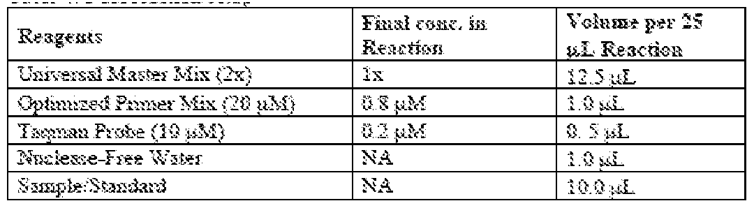
[000250] PCR reaction sequence was set up as follows: 50°C 2 minutes 1 cycle; 95°C 10 minutes 1 cycle; 95°C 15 seconds 40 cycles; 60°C 1 minutes 40 cycles
[000251] Run performance. Standards were prepared and run at 103 to 108 DNA copies per well. Lower limit of the assay was set at 1000 copies since assay sensitivity was not an important factor for this experiment. A standard curve was generated for the run using the standard copy numbers and CT (threshold cycle) values of the standards. Linear regression of the standards was performed using the ABI software (data not shown). Standard curve fit had a correlation coefficient (R2 value) of 0.998 or greater indicating a reliable fit model. The slope of the standard curves was -3.5. Slope was used to calculate the efficiency of the amplification reaction, and values between -3.2 and -3.6 represented amplification efficiency between 90% and 110%. Both standard reactions were run with 92.6~93.8% efficiency. Precision of triplicate wells ranged from 2~10%, indicating good agreement among replicates. No-template-control (NTC) resulted in non-quantifiable amplification below the lower limit of the assay.
[000252] Table 15: Summary of standard curve fit


[000253] RESULTS:
[000254] Sample value determination: The sample values (AAV genome and reverse- packaging copy number) were interpolated from each matching standard curve (CMV/CBA or KanR), using CT values. Interpolated DNA copy number was corrected for initial dilution and/or digest dilution. Additional correction factor of 2 was applied to account for the difference between double-stranded DNA standards and single-stranded DNA in samples.
[000255] Analysis results for 8 AAV vectors are summarized in the table below, with quantitative comparison between the transgene-containing AAV concentration (CMV/CBA) and the KanR-containing impurity concentration. Analysis of results demonstrate that insertion of lambda stuffer into the transgene plasmid effectively reduced the occurrence of plasmid-backbone DNA (i.e. KanR) packaging during AAV production from ~7-20 folds (FIG.11).
[000256] Table 16: qPCR amplification of kanamycin versus CMV/CBA expressed as percentage

[000257] Example 6: In vitro Expression of Next generation AAV8 vectors in iPS cells by western blot.
[000258] The objective of this study was to evaluate the ability of AAV mediated CHM expression after gene delivery using a series of next generation AAV2 and AAV8 vectors carrying the codon optimized REP-1–encoding gene in induced pluripotent cell lines (iPSC).
[000259] Induced pluripotent stem (iPS) cell technology has been successfully utilized as a platform for testing gene therapy vectors in several proof-of-concept and gene therapy studies including ocular diseases. These patient-specific iPS cells provide a valuable in vitro model system to study disease pathogenesis and establish a model to test proof-of-concept of
gene therapy where relevant animal models are unavailable. As a preliminary step to test our AAV-mediated gene augmentation therapy for Choroideremia (CHM), we have generated iPS cells from human patients harboring mutations in the causative gene, CHM, which encodes Rab Escort Protein 1 (REP-1) (See example 1) (Method is described in NCP.003). The generated iPS cells were used to evaluate the in vitro expression of our next generation AAV.codon optimized.CHM constructs.
[000260] Plasmids and vectors were as described in Example 4. Induced pluripotent stem (iPS) cells are stem cells generated in the laboratory from somatic cells, peripheral blood mononuclear cells, that were reprogrammed back to a pluripotent state.
Reprogramming of blood cells enables the development of personalized in vitro cellular models for therapeutic applications. In this report, iPS cells from individuals affected by CHM were used to test the in vitro production of REP-1 protein through western blot analysis. The following table (Table 17) describes the details of iPS cells studied and their respective CHM disease-causing mutations.
[000261] Table 17: An overview of the iPS cells generated from patients with CHM m i n


* iPS cell line qualification tests are on-going.
[000262] Study design (e.g. treatment groups)
[000263] 1. iPS cells plated on a 12 well cell culture plate are infected with AAV2. hCHM Version 1, Version 2a; Version 2b; Version 3a; Version 3b (AAV2.V1; V2a; V2b; V3a; V3b) at an MOI of either 1E5 or 3E5. After 24 hours of transduction, 1 ml of iPS cell culture media was added to the cells.36-48 hours of transduction, cells were harvested, lysed and processed for SDS-PAGE followed by Western blot analysis. Production of REP-1 protein was evaluated in cells transduced with all versions of the constructs and compared with untreated controls.
[000264] 2. As a pilot experiment, three different iPS cell lines plated on a 12 well cell culture plate are transduced with AAV8. hCHM Version 1 and AAV8. hCHM Version 2a
(AAV8.V1; AAV8.V2a) at an MOI of 1E6. The iPS cell lines were derived from three CHM affected individuals with unrelated mutations in REP1 gene and were plated in separate plates for this purpose. After 36-48 hours, cells were harvested and lysed and subjected to Western blot analyses compared with untreated cell lysate.
[000265] Test material administration
[000266] 3.4.1 Cell culture
[000267] Culturing of iPS cells from CHM patient. In brief, the iPS cells were cultured on Mouse Embryonic Fibroblasts (MEFs, feeders) in iPS cell culture media at 37°C in an environment supplied with 5% CO2 and 5% O2.
[000268] 3.4.2 Preparation of cells for transduction
[000269] The day before seeding the cells, 12-well dishes were coated with Matrigel as described in reference NCP.003 (NCP.003: Culturing of iPS cells from CHM patient and controls). Before transduction of iPS cells with respective AAV2 or AAV8 viral vectors, the cells that are cultured on MEFs were seeded on Matrigel without MEFs (feeder free culturing). Cells were seeded at a density of 4.5+E5 to 6 +E5 in 1 ml of iPS cell culture media in each well of a 12-well cell culture dish. Seeded cells were incubated at 37°C in an environment supplied with 5% CO2, 5% O2.
[000270] 3.4.3 Transduction
[000271] To infect the iPS cells with viral vectors, cells were grown to approximately 50-60% confluence. (This can take 2-4 days in feeder free conditions). Once 50-60% confluence is achieved, one well of the 12-wells is dissociated and cell counts were performed to determine the total number of cells per well. Wells of the iPS cells were then infected with AAV vectors listed below at the predetermined MOI (see Table 18 and 19). Before transduction, the old iPS cell culture media from the plates was removed and a fresh 1 ml of iPS cell culture media was added in each well. Predetermined volumes of the virus from the stock were directly added to each well. See Table 18. And Table 19. For the information on total number of cells infected, MOI and the volume of virus used for infection. Cells were then incubated at 37°C in an environment supplied with 5% CO2, 5% O2 for 18-24 hours. After 18-24 h of transduction, cells were observed under microscope for any abnormalities or cell death. At this point, another 1 ml of fresh iPS cell culture media was added to each well containing infected and uninfected cells and were further incubated for
additional 18-24 hours at 37°C in an environment supplied with 5% CO2, 5% O2. Cells were observed under the microscope before harvesting to evaluate any cell death or abnormal appearance.
[000272] Table 18: Infection details and MOIs of next generation AAV2.hCHMV2a, 2B, 3a, 3b Vectors and AAV2.hCHM.V1 vectors in CHM patient-derived iPS cells.

[000273] Table 19: Infection dose of AAV8.V2a and AAV8.V1 vectors in three iPS cell lines derived from 3 different CHM patients.

[000274] Outcome measurement method– Western blot analysis was performed as described herein.
[000275] Results
[000276] 5.1 Expression of AAV2- hCHM V1, V2a, V2b, V3a, V3b in JB588 iPS cell line: Monoclonal human REP-1-specific antibody, detected one single ~ 75-80 kDa hREP-1 protein in the transduced JB 588 iPS cells (Data not shown). No band was observed in the case of the untreated control, confirming presence of the disease (data not shown). The intensity of REP-1 protein band at an MOI of 3E5 observed to be stronger in all vectors compared to an MOI of 1E5. Recombinant AAV2. hCHM viral mediated delivery of the hCHM gene, to iPS cells, resulted in a dose-dependent production of REP-1 protein. Probing of the blots with GAPDH antibody showed a band of equal density in all lysates. GAPDH detected a protein at ~39 kDa. Both REP-1 and GAPDH antibodies detected only specific bands of expected molecular weight. No nonspecific bands were observed in the blots.
[000277] Expression of AAV8–hCHM. V1, V2a in iPS cells: Monoclonal human REP- 1-specific antibody, detected one single ~ 75-80 kDa REP-1 protein in the transduced JB527, JB500 and JB588 patient derived iPS cells (Data not shown). No protein band was observed in the case of the untreated control. (Data not shown). Probing of the blots with GAPDH antibody showed a band of equal density in all cell lysates including the cell lysates from untreated cells. Anti-GAPDH antibody detected a specific ~39 kDa protein band. Both REP-1 and GAPDH antibodies detected only specific protein bands at the expected size molecular weight. No detectable nonspecific protein bands were observed in the blot.
[000278] Conclusions
[000279] The preliminary results presented in the current report revealed the following observations: Western blot analysis confirmed presence of CHM (lack of REP-1 protein) in each one of the three patient-derived iPSCs (JB588, JB500, JB527). In vitro expression studies demonstrated that infecting iPS cells from CHM patients with AAV2.hCHM. Version 2a, 2b, 3a, 3b and AAV2.hCHM Version1 (a current clinical trial candidate) induced the production of REP-1 protein at all tested MOIs. Infecting iPS cells with AAV8.
hCHM.Version 2a and AAV8.hCHMVersion1 at an MOI of 1E6 resulted in production of REP1 protein in all three CHM iPS cell lines. Level of REP1 production was higher in the iPSCs infected with AAV8.hCHM.V2a than with AAV8.hCHM.V1.
[000280] Example 7: Comparison of In Vivo Expression of AAV8.Codon
Optimized.Human CHM versus AAV.Native.Human CHM
[000281] Gene therapy for a number of retinal diseases depends on efficient transduction of the appropriate target cells, which for choroideremia, are retinal pigment epithelium (RPE) cells and photoreceptor cells. This study report focuses on the comparison of in vivo expression induced by the native CHM sequence based construct, (Version 1) and four next generation transgene cassettes packaged into an AAV8 backbone in wild type mice. Here we evaluated AAV8 serotype for the purpose of improving gene transfer to
photoreceptor cells.
[000282] Our experiments were designed to answer the following questions: a. How would these vectors compare for in vivo transduction of photoreceptors: In particular, how efficiently would the next generation AAV8. CHM transduce photoreceptors after subretinal injection of the respective test article compared to version.1. b. Dose response: Would the
next generation AAV8. CHM and AAV8. CHM-Version1 vectors differ in dose response of gene expression.
[000283] Experimental details:
[000284] Plasmids and vectors were as described in Example 4. Mice (Animals): Wild type, CD1 mice were used to test the in vivo expression of CHM as assessed by production of REP-1 protein. CD1 mouse strain is an outbred Swiss mouse strain which colony we maintain in house. The details of the study are described under CAROT study protocol PCPR02.01.
[000285] 3.3 Study design (e.g. treatment groups)
[000286] 3.3.1 Animal Husbandry: Both male and female mice (~3-4 months old) weighing ~ 20-30 gm were injected with the described test articles. Animals were housed in the University of Pennsylvania’s John Morgan University Laboratory Animal Resources (ULAR) facility according to University of Pennsylvania’s ULAR regulations. Mice were maintained on a 12-hour light/12-hour dark cycle. Food and water were provided ad libitum. All animals were identified with ear tag numbers.
[000287] 3.4 Test material administration: The test article formulation provided by the CAROT Vector Core was used for dose administration. The test material was stored at– 60 to -80°C. The test material was thawed on ice prior to dosing. For intra-ocular injections, the test article is diluted to the target concentration with phosphate-buffered saline as described in the formulation Table 20. A total of 60 μl of master mix was prepared.
[000288] Table 20: Dose Formulation table for subretinal injections of test articles.
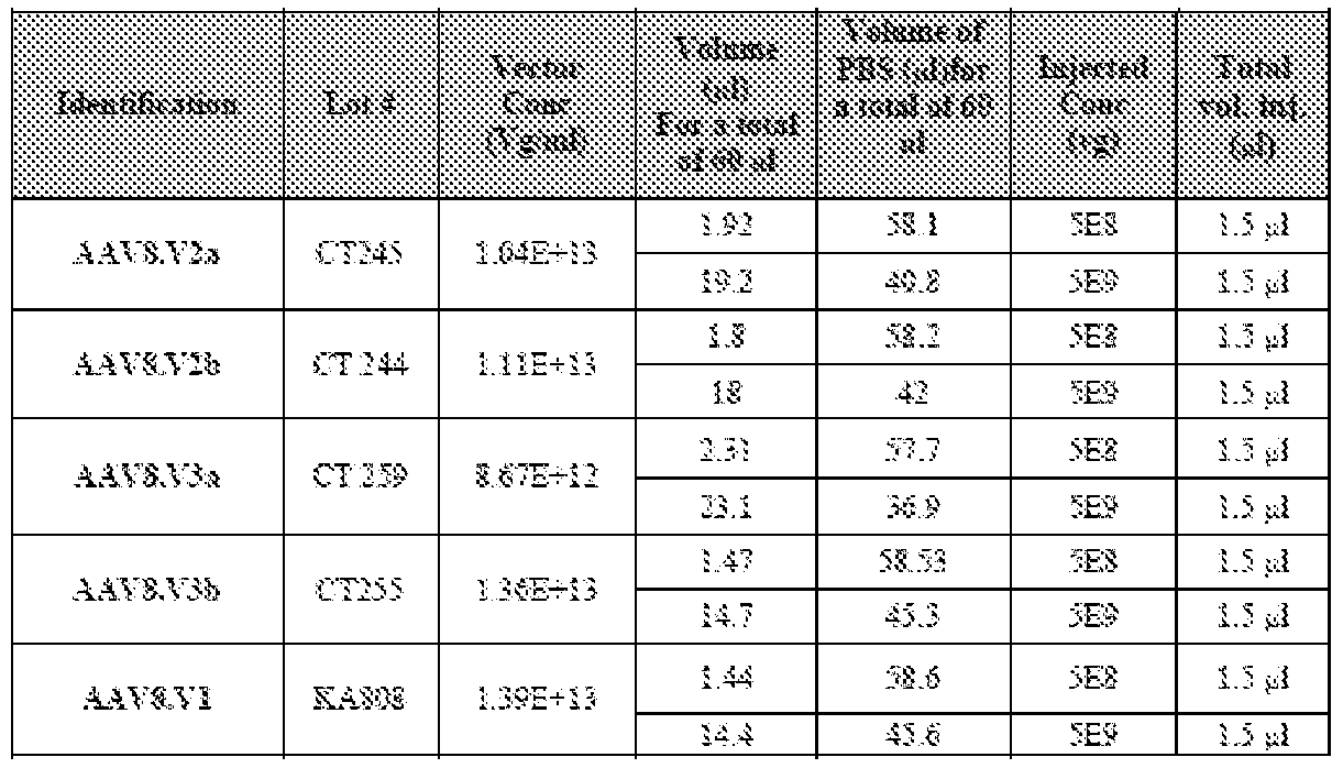
[000290] An injection log was maintained with the following information before subretinal injection of the test articles:
• Cage Number/mouse number
• Study Identification
• Strain
• Date of Birth
• Date of injection
• Name of investigator/injector
• Eye injected into (left or right)
• Injection material (vector/serotype)
• Dose and Volume
• Route of Administration (ROA)
[000291] Subretinal injections: Injections were performed by Subretinal Injection by the Surgeon. In brief, animals were anaesthetized before injection. Subretinal injection of the test article was performed using Hamilton 33G syringe. The details of test articles and injections are described in Table 21. From the prepared injection master mix, a volume of 1.5 μl was administered, per injection. One eye per animal was injected with 5E8 vg/eye and the contralateral eye was injected with 5E9 vg/eye.
[000292] Table 21: Subretinal injection scheme and injection doses

[000293] Outcome measurement methods
[000294] Animal Sacrifice: a. After injecting the animals with the test articles, all animals were observed for 48 hours for any post injection related abnormalities. B.21-35 days of post injection, the animals were observed for ocular abnormalities using
ophthalmoscopy. C.90-12- days post injection, the animals were sacrificed and eye tissues were collected for evaluating the production of exogenous REP-1 protein by SDS-PAGE followed by western blot analysis.
[000295] Collection of Eye Tissue: Eye tissue for western blot analysis was collected after removing the lens from the eye using a sharp surgical blade. The eye (without the lens) was collected in freezer tubes that are labelled appropriately.
[000296] Western blot analysis
[000297] Briefly: 1. Preparation of tissue lysate
[000298] a. Ocular tissue of animals injected with 2 different doses of next generation AAV8. CHM and AAV8.V1 along with the uninjected control animal tissues, were collected after 21-35 days of injection by sacrificing the animals. B. Tissues were then lysed on ice using RIPA buffer with protease inhibitors.
[000299] c. Tissue lysates were cleared by centrifuging at 13,000 rpm for 10 min.
[000300] 2. Quantification and preparation of proteins
[000301] a. Protein quantification of the cell lysates was carried out using ThermoFisher Micro BCATM Protein Assay Kit following manufacturer’s instructions. B. Protein concentration was determined by taking OD reading at 562 nm. C. To evaluate the in vivo expression of CHM, between 20-40 ug of measured protein was loaded on 4-12% Bis-Tris gels.
[000302] 3. SDS-PAGE and Western Blot
[000303] The protein gels were transferred on to a nitrocellulose membrane, blocked in milk and incubated with the primary antibodies. Anti-human REP-12F1 antibody (2F1, 1:1000 dilution) and/or anti-GAPDH antibody (1:1000 dilution); were used as primary antibodies. After washing the blot, HRP conjugated anti-mouse IgG antibody and/or anti- rabbit IgG antibody at a concentration of 1:5000 were used as secondary antibodies. The blots were developed by chemiluminescence using ECL reagents according to the manufacturer’s instructions.
[000304] 4. Controls
[000305] a) Loading controls: Anti-GAPDH antibody was used as loading controls to demonstrate equal loading of protein in each well of the gels. Anti-GAPDH antibody detects a protein of ~39 kDa. B) Positive control: AAV2.V2a transduced COS-7 cell lysates were
used as positive controls. C) Negative control: Ocular tissues of uninjected animals were used as negative control.
[000306] Sample Value Determination
[000307] Quantification of Western blot analysis using Image J software. In brief, densitometric evaluations presented in this report are normalized first, to the levels of endogenous expression of GAPDH protein of the corresponding sample. Then the expression levels are normalized again, to the average REP-1 expression level of uninjected control.
[000308] The details of densitometric evaluations and fold change calculations to represent the expression of REP-1 protein are presented as Table 22 and 23.
[000309] The description in brief:
[000310] 1. In table 22 and 23, Column 2 shows the raw values of REP-1 protein and column 3 shows the raw values of GAPDH protein.
[000311] 2. GAPDH value of each samples was first normalized to the GAPDH values of animal-1 of AAV8.V1 and are shown in Table 22 in 4th column.
[000312] 3. The values of each sample were also normalized to the GAPDH values of animal-2 of AAV8.V1 and are shown in Table 22 in 5th column.
[000313] 4. The REP-1 values (Column 2) are then normalized to either to the GAPDH normalized to animal 1 (column 4) or to the GAPDH previously normalized to animal 2 (column 5). These are represented in column 6 and 7 respectively.
[000314] 5. The normalized REP-1 values are then converted to fold change.
[000315] 6. The respective REP-1 values are normalized to expression of REP-1 either in animal 1 or animal 2 of the AAV8.V1 injected group and are expressed as fold change (column 8 and 9)
[000316] 7. Column 10 represents the average fold change in REP-1 protein expression.
[000317] Results
[000318] Comparison of the CHM expression using the native CHM AAV8.V1 versus the codon optimized CHM vectors: AAV8.V2a, V2b, V3a and V3b. Wild type CD1 mice were injected with two different doses of the each AAV8 vector: a high dose of 5E9 vg/eye and a low dose of 5E8 vg/eye. Following results describe the levels of REP1 protein after injection with high and low doses of AAV8.V1, AAV8.V2a and AAV8.V3a.
[000319] Comparison of the expression of AAV8.V1 versus AAV8.V2a and AAV8.V3a (vectors with stuffer) in animals injected with high dose (5E9 vg/eye) of viral vector. Western blot analysis with human anti REP-1 antibody detected a ~ 75-80 kDa hREP-1 protein band in both (low and high dose injected) ocular tissues of each animal treated with either the next generation AAV8.V2a or V3a or the original AAV8.Version1. A very faint (minimal) band is observed in the case of the uninjected control mice, both. A band of increased intensity was observed in tissues that were transduced with next generation vectors (AAV8V.2a and AAV8.V3a) compared to the tissues transduced with Version.1. Anti-GAPDH antibodies showed a ~39 kDa band of equal density in all lanes of the western blot including the uninjected controls. Pre-stained protein marker is used to compare the molecular weights of protein of interest. Densitometric evaluation (quantification of the expression level) of the blots using ImageJ software demonstrated that production of REP-1 was increased in animals injected with one of the next generation AAV8. High and low doses constructs (V2a or V3a). (See Table 22 for values.)
[000320] Table 22: Quantified REP-1 protein production results for treatment with high dose (5E9vg) AAV8 V1, V2a and V2b


[000321] Table 23: Quantified REP-1 protein production results for treatment with low dose (5E8 vg) AAV8 V1, V2a and V2b


[000322] Comparison of the expression of AAV8.V1 versus AAV8.V2a and AAV8.V3a in animals injected with low dose (5E8 vg/eye) of viral vector
[000323] Human anti REP-1 antibody, the Western blot analysis of the ocular tissues of animals injected with next generation AAV8.V2a,V3a and AAV8.Version1 at a dose of 5E8 detected a ~ 75-80 kDa hREP-1 protein band in tissues of injected mice. A faint (minimal) band of REP-1 was observed in ocular tissue lysates of the uninjected control mice, both. A band of increased intensity was observed in tissue lysates that are transduced with next generation vectors compared to the lysates that are transduced with Version1. Anti-GAPDH antibody detected an equal intensity protein band at ~ 39 kDa in all cell lysates. This data demonstrates that delivery of next generation V2a CHM through AAV8 results in robust levels of REP-1 protein in comparison with levels produced after injection of AAV8.V3a or AAV8.V1.
[000324] Densitometric evaluation (quantification of the expression level) of the blots using ImageJ software further demonstrate an increased production of REP-1 in animals injected with next generation AAV8.CHM constructs (especially V2a) compared with Version 1. See Table 23 for values.
[000325] Expression of AAV8.V2b in CD1 mice
[000326] This current study and the evaluation of lambda stuffer’s effect on AAV vector production by qPCR titer analysis were carried out simultaneously. We performed all the animal injections for the in vivo expression study as described in the study protocol PCPR.02 and all samples were harvested. After the qPCR study on the lambda stuffer
element was concluded (described above), we decided to carry out the Western blot experiments only to test the expression of AAV vectors without the stuffer such as AAV8.2b and AAV8.3b and exclude them from further analysis (such as comparison with Version 1).
[000327] Human anti-REP-1 antibody detected a protein of ~75-80 kDa in ocular tissues of CD-1 mice injected with AAV8.2b at 5E9 (High dose) vector genome copies (FIG.12A). Animals injected with AAV8.2b at 5E8 (Low dose) showed a very faint protein band at ~75- 80 kDa (FIG.12A). Lysates of ocular tissues from uninjected control animals did not show the presence of REP-1 protein. Anti-GAPDH antibody detected a protein of ~39 kDa in all ocular tissue lysates including the uninjected controls. This data may establish the minimal dose for AAV8.2b.
[000328] Expression of AAV8.V3b in CD1 mice
[000329] We performed a Western blot analysis on ocular tissues of AAV8.3b injected CD1 mice (2 mice/group) with anti-REP-1 antibody, which revealed the presence of a protein of ~75-80 kDa in one eye injected with low dose and in both eyes injected with high dose of AAV8.3b. In the ocular tissues of uninjected mice there was no REP-1 expression detected (FIG.12B). The level of REP-1 produced was dose dependent in animals injected with AAV8.3b. Injection with high dose of AAV8.3b (5E9 vector genomes) induced a higher amount of REP-1 compared with the low dose injected eyes (5E8 vector genomes). Anti- GAPDH antibody detected a protein of ~39 kDa in ocular tissue lysates of all injected and uninjected animals.
[000330] These results revealed the following observations:
[000331] 1) The next generation vectors AAV8.Version2a, 2b, 3a and 3b are able to transduce ocular tissues efficiently.2) Expression of the transgene (codon optimized CHM) was detectable for all of the next generation vectors.3) Expression of transgene (codon optimized CHM) is dose dependent.4) AAV8.Version2a and AAV8.Version.2b induced an increased production of REP-1 protein compared to AAV8.Version 1 in ocular tissues of CD- 1 mice.5) There is variation in the exact level of production of the transgenic protein between eyes injected with the same dose reflecting the variability in the surgical delivery procedure. However, differences in levels are large between the low (5E8) and high (5E9) doses.6) AAV8.CHM.V2a and AAV8.V3a result in much higher levels of REP-1 protein
production than AAV8.V1 after in vivo administration of high dose (5E9 vg) vector subretinally in mice.
[000332] Example 8– Expression of CNGA3
[000333] To maximize the expression of CNGA3, a codon optimized CNGA3 sequence was produced (SEQ ID NO: 9). In addition, a CNGA3 variant was codon optimized (SEQ ID NO: 11). These sequences, as well as the native CNGA3 coding sequence, were incorporated into production plasmids as described herein (SEQ ID Nos: 30-38) and AAV vectors created. Vectors using AAV8 and AAV9 capsids were generated, as described below.

[000336] Exogenous hCNGA3 expression using AAV8 & 9 was tested in vivo
[000337] Subretinal injections of 30-120 day old Wt mice were performed. Mice were sacrificed at 3-4 weeks post injection, tissues collected and endpoints measured. In normal mice, expression of CNGA3 measured by qPCR as shown in FIG.30. In null mice, expression of CNGA3 protein in retina is measured using western and IHC and retinal histopathology is examined.
[000338] The CNGA3 mouse model: CNGA3 null mice which have a CPFL5 containing a missense mutation (Exon 5).
[000339] Phenotype:
[000340] 5 weeks: severely reduced cone-specific ERGs
[000341] 10 weeks: reduction and mislocalization of cone opsin pigments
[000342] 5 months: Reduced optokinetic reflexes
[000343] Gene replacement therapy for CNGA3-achromatopsia:
[000344] CNGA3 null mice were injected at p16-18, subretinally with one of two doses of noted vectors. Low dose: 8E8 vg/eye; high dose: 8E9 vg/eye. At least 5 animals were injected per group. ERG and OKR were performed at 5-7 weeks and 12-15 weeks. At sacrifice, IHC, western blots and histology were performed.
[000345] To provide objective information about the function of retina and to serve as a parameter for efficacy in preclinical studies, electroretinogram (ERG), an electrical response of the cells of the retina to a flash of light, was evaluated in mice according to conventional method and User Manuals. Please see, e.g. Marmor, Michael F., et al. "Standard for clinical electroretinography (2004 update)." Documenta ophthalmologica 108.2 (2004): 107-114; and Cronin, Therese, Arkady Lyubarsky, and Jean Bennett. "Dark-rearing the rd10 mouse: implications for therapy." Retinal Degenerative Diseases. Springer US, 2012.129-136.
[000346] Briefly, an ophthalmoscopic evaluation of animals was completed prior to ERG measurement. Mice with eye defects that may potentially compromise the results of the ERG are excluded. These include corneal opacities such as cataract, corneal injury or inflammation. Mice were then dark-adapted for at least 4 h, weighed under dark conditions and injected anaesthetic intraperitoneally (ketamine/xylazine cocktail with phosphate buffered saline (PBS; pH 7.2), 100 mg/kg and 10 mg/kg respectively). The pupils of pigmented mice were dilated using 1% tropicamide solution while albino mice did need pupil dilation. While the animal was kept on an absorbent bedding on top of the heated platform, the reference electrode was placed to contact with the body of the mouse, and the recording electrodes were positioned over the cornea of respective eyes and contacting with the corneas gently. If necessary, operations using a magnifier was performed.
[000347] Stimulator was set as indicated below. Stimuli of any color or achromatic may be used unless indicated. Testing protocol I includes Step 1: 0.01076 scotopic cd s m-2 (Candela second per square meter (cd/m2)); Step 2: 500 scotopic cd s m-2, achromatic xenon flash; and Step 3: background intensity 100 scotopic cd m-2, Stimulus : 500 scotopic cd s m-2. In stimulus intensity Set II, for all steps the following stimuli were delivered on a 100 scotopic cd m-2 (Candela per square meter (cd/m2)) green (520 nm) background illumination. Testing protocol II includes: Step 1: 500 scotopic cd s m-2, achromatic xenon flash; Step 2: 0.0015 cd s m-2, UV (365 nm), isi (Interstimulus interval, time interval between consecutive flashes) 1.5 s; Step 3: 0.004cd s m-2, UV (365 nm), isi 1.5 s; Step 4: 0.01 cd s m-2, UV (365 nm), isi 2 s; Step 5: 0.03 cd s m-2, UV (365 nm), isi 2 s; Step 6: 4scot cd s m-2, green (520 nm), isi 2 s; Step 7: 10 scot cd s m-2, green (520 nm), isi 2 s; Step 8: 25scot cd s m-2, green (520 nm), isi 2 s; and Step 9: 500 scotopic cd s m-2, achromatic xenon flash. Testing protocol I and II was run on each animal successively.
[000348] The CNG3A/B3 null mice used have normally functioning rods, so the rod- generated ERG was utilized as a signature of the retina condition. The parameter was the amplitude of the saturating a-wave. On stimulation with a bright flash, a fast cornea-negative voltage, was the first (in temporal order) component of the ERG. The amplitude of the a-wave was the difference between the most negative point of the ERG during the time interval of up to 20 ms after the flash and the baseline value of the ERG signal measured at the 3 ms time point after the flash. The choice of the 3 ms data point as the“zero” allowed for exclusion of
the flash artifact and minimizing effects of drifts. With incremental increase of stimulus intensity, the amplitude of the a-wave increased until it reached saturation. The a-wave of the mouse ERG is directly proportional to the magnitude of the retinal rod photocurrent (Lyubarsky, Arkady L., and Edward N. Pugh Jr. "Recovery phase of the murine rod photoresponse reconstructed from electroretinographic recordings." Journal of Neuroscience 16.2 (1996): 563-571), and therefore, is the most direct measure of the rod function.
[000349] The amplitude of the a-wave as the voltage difference between baseline and the first trough, which occurs at 7-10 ms after the flash was measured for both eyes using data acquired via testing protocol I. Amplitudes of cone ERGs was measured using the data acquired via testing protocol II. Magnitudes of cone ERGs was divided by the amplitude of the saturating a-wave from the respective eye. If the amplitude of the a-wave from the injected eye was less than 50% of the amplitude of the a-wave from the control eye, this animal was excluded from further processing and counted as severely injured on injection. Results acquired were calculated and normalized. Paired t-test between sets of normalized cone ERGs from the injected and non-injected eye was performed. Statistically significant improvement in the treated eye was to be a signature of successful treatment.
[000350] Retinal function was assessed at 5-7 weeks of age following administration of the noted vectors was compared to retinal function of wild type, excipient injected and uninjected eyes. The measured results from three light intensities (0.01 cd.s/m^2, 10 cd.s/m^2 and 25 cd.s/m^2) for each vector/dosage are shown below in Table 25, and in FIG. 31. These results show that RK‐1 promoter along with optimized CNGA3 expression cassette is able to correct ERG function in a mouse model.
[000351] Table 25
Student's t‐test 0.01 cd.s/m^210 cd.s/m^25 cd.s/m^ Uninjected versus :
Excipient 4.09E‐06 0.161284 0.007365 AAV8.CMV/CβA‐CNGA3‐hopt high dose 0.003812136 0.214523 0.060289 AAV8.CMV/CβA‐CNGA3‐hopt low dose 0.77537772 0.305382 0.279831 AAV8.RK1‐CNGA3‐hopt high dose 5.05248E‐07 1.02E‐06 1.72E‐07 AAV8.RK1‐CNGA3‐hopt low dose 0.001563149 0.000454 0.000955 AAV8.hCAR‐CNGA3‐hopt high dose 0.00051437 0.000873 0.001657 AAV8.hCAR‐CNGA3‐hopt low dose 0.011644961 0.002392 0.001796 AAV9‐hCAR‐CNGA3‐hopt high dose 0.010237532 0.012328 0.020935 AAV9‐hCAR‐CNGA3‐hopt low dose 0.843706492 0.669637 0.432698 AAV9‐RK1‐CNGA3‐hopt high dose 0.004789373 0.005494 0.005689 AAV9‐RK1‐CNGA3‐hopt low dose 0.004873537 0.000563 0.000923
[000352] P values <0.5 are highlighted in red
[000353] Calcium uptake in transduced vs. untransduced 84-31 cells after cGMP addition is shown in FIG.32.
[000354] Example 9– CNGB3
[000355] CNGB3 sequences were incorporated into vectors as described above, for REP-1 and CNGA3. Vectors using AAV8 and AAV9 capsids were generated, as described below.
[000356] CNGB3 null mice were injected at p16-18, subretinally with one of two doses of noted vectors. Low dose: 1E9 vg/eye; high dose: 1E10 vg/eye. At least 5 animals were injected per group. ERG and OKR were performed at 5-7 weeks and 12-15 weeks. At sacrifice, IHC, western blots and histology were performed.
[000357] Retinal function was assessed at 12-16 weeks of age following administration of the noted vectors was compared to retinal function of wild type, excipient injected and uninjected eyes. The measured results from three light intensities (0.01 cd.s/m^2, 10 cd.s/m^2 and 25 cd.s/m^2) for each vector/dosage are shown below, and in FIG.33. Series 5, 8 and 9 are light intensities of 0.01 cd.s/m^2, 10 cd.s/m^2 and 25 cd.s/m^2, respectively.
[000358] These results show that both hCAR and RK‐1 promoter along with optimized CNGB3 expression cassette are able to correct ERG function in the CNGB3 mouse model.
[000359] ERG studies suitable for use with the constructs described herein, are described, e.g., in International Patent Application No. PCT/US17/27529, filed April 14, 2017, which is incorporated herein by reference.
[000360] Example 10: ELECTRORETINOGRAM (ERG) of MICE
[000361] To provide objective information about the function of retina and to serve as a parameter for efficacy in preclinical studies, electroretinogram (ERG), an electrical response of the cells of the retina to a flash of light, was evaluated in mice according to conventional method and User Manuals. Please see, e.g. Marmor, Michael F., et al. "Standard for clinical electroretinography (2004 update)." Documenta ophthalmologica 108.2 (2004): 107-114; and Cronin, Therese, Arkady Lyubarsky, and Jean Bennett. "Dark-rearing the rd10 mouse:
implications for therapy." Retinal Degenerative Diseases. Springer US, 2012.129-136.
[000362] Briefly, an ophthalmoscopic evaluation of animals was completed prior to ERG measurement. Mice with eye defects that may potentially compromise the results of the ERG are excluded. These include corneal opacities such as cataract, corneal injury or inflammation. Mice were then dark-adapted for at least 4 h, weighed under dark conditions and injected anaesthetic intraperitoneally (ketamine/xylazine cocktail with phosphate buffered saline (PBS; pH 7.2), 100 mg/kg and 10 mg/kg respectively). The pupils of pigmented mice were dilated using 1% tropicamide solution while albino mice did need pupil dilation. While the animal was kept on an absorbent bedding on top of the heated platform, the reference electrode was placed to contact with the body of the mouse, and the recording electrodes were positioned over the cornea of respective eyes and contacting with the corneas gently. If necessary, operations using a magnifier was performed.
[000363] Stimulator was set as indicated below. Stimuli of any color or achromatic may be used unless indicated. Testing protocol I includes Step 1: 0.01076 scotopic cd s m-2 (Candela second per square meter (cd/m2)); Step 2: 500 scotopic cd s m-2, achromatic xenon flash; and Step 3: background intensity 100 scotopic cd m-2, Stimulus : 500 scotopic cd s m- 2. In stimulus intensity Set II, for all steps the following stimuli were delivered on a 100 scotopic cd m-2 (Candela per square meter (cd/m2)) green (520 nm) background
illumination. Testing protocol II includes: Step 1: 500 scotopic cd s m-2, achromatic xenon flash; Step 2: 0.0015 cd s m-2, UV (365 nm), isi (Interstimulus interval, time interval between consecutive flashes) 1.5 s; Step 3: 0.004cd s m-2, UV (365 nm), isi 1.5 s; Step 4: 0.01 cd s m-2, UV (365 nm), isi 2 s; Step 5: 0.03 cd s m-2, UV (365 nm), isi 2 s; Step 6: 4scot cd s m-2, green (520 nm), isi 2 s; Step 7: 10 scot cd s m-2, green (520 nm), isi 2 s; Step 8:
25scot cd s m-2, green (520 nm), isi 2 s; and Step 9: 500 scotopic cd s m-2, achromatic xenon flash. Testing protocol I and II was run on each animal successively.
[000364] The CNG3 mice used have normally functioning rods, so the rod-generated ERG was utilized as a signature of the retina condition. The parameter was the amplitude of the saturating a-wave. On stimulation with a bright flash, a fast cornea-negative voltage, was the first (in temporal order) component of the ERG. The amplitude of the a-wave was the difference between the most negative point of the ERG during the time interval of up to 20 ms after the flash and the baseline value of the ERG signal measured at the 3 ms time point after the flash. The choice of the 3 ms data point as the“zero” allowed for exclusion of the flash artifact and minimizing effects of drifts. With incremental increase of stimulus intensity, the amplitude of the a-wave increased until it reached saturation. The a-wave of the mouse ERG is directly proportional to the magnitude of the retinal rod photocurrent (Lyubarsky, Arkady L., and Edward N. Pugh Jr. "Recovery phase of the murine rod photoresponse reconstructed from electroretinographic recordings." Journal of Neuroscience 16.2 (1996): 563-571), and therefore, is the most direct measure of the rod function.
[000365] The amplitude of the a-wave as the voltage difference between baseline and the first trough, which occurs at 7-10 ms after the flash was measured for both eyes using data acquired via testing protocol I. Amplitudes of cone ERGs was measured using the data acquired via testing protocol II. Magnitudes of cone ERGs was divided by the amplitude of the saturating a-wave from the respective eye. If the amplitude of the a-wave from the injected eye was less than 50% of the amplitude of the a-wave from the control eye, this animal was excluded from further processing and counted as severely injured on injection. Results acquired were calculated and normalized. Paired t-test between sets of normalized cone ERGs from the injected and non-injected eye was performed. Statistically significant improvement in the treated eye was to be a signature of successful treatment.
[000366] Example 11 - Evaluation of Lambda Stuffer’s Effect
[000367] Further to the experiments described in Example 6, percentage of residual plasmid DNA was evaluated and the data acquired was plotted in FIG.34. Result showed that oversized stuffer sequences reduced DNA impurity by ~80% during triple transfection production for both AAV2 and AAV8.
[000368] Example 12– in vivo Expression of rAAV
[000369] Further to the experiments described in Example 7, various dosages (5E7 vector genome copies per eye (vg/eye), 1E8 vg/eye, 5E8 vg/eye, 5E9 vg/eye, and 1E10 vg/eye) of AAV8.V2a, AAV8.V3a and AAV8.V1 were injected to about 3-4-month-old CD- 1 mice. Eyeballs were harvested and REP1 protein expressions thereof were evaluated via Western blots. Representative results are shown in FIGs.35 to 39. Quantifications were performed and indicated in the Tables below. Results showed that compared to AAV8.V1, AAV8.V2a and AAV8.V3a demonstrated higher expression level of REP1 protein at 5E8 vg/eye, 5E9 vg/eye. Subretinal delivery of AAV8.V2a, and AAV8.V3a resulted in robust and reproducible delivery of the CHM transgene to retinal cells. Recombinant AAV8.CHM mediated delivery of the CHM gene resulted in a dose-dependent effect on REP1 protein production.
[000370] Table 26

AAV8.V2a (Animal 4) 8.70 2.04
AAV8.V2a (Animal 5) 1.66 1.16
AAV8.V2a (Animal 6) 6.13 1.53
AAV8.V3a (Animal 1) 9.27 0.99
AAV8.V3a(Animal 2) 4.51 0.99
AAV8.V3a (Animal 3) 0.33 0.15
AAV8.V3a (Animal 4) 0.80 0.64
AAV8.V3a (Animal 5) 0.79 0.52
AAV8.V3a (Animal 6) 0.13 0.30 [000372] Example 13– Retinal Histopathology upon Injection of rAAVs
[000373] Analysis of retinal histopathology was performed on the retina harvested from the mice treated as described in Example 7 and Example 12. H&E staining was performed to reveal changes on photoreceptors as well as presence of immune infiltration. Tunel staining was performed to reveal the presence of apoptosis. Ve treated cells was provided as a positive control. Represented images not shown. A summary of the observation was provided below as a Table. Results indicate that recombinant AAV8.CHM mediated delivery of the CHM gene resulted in a dose-dependent effect on REP1 protein production and retinal
histopathology. Mouse eyes injected with the highest dose of AAV8.V2a, AAV8.V3, and AAV8.V1 showed inflammation, retinal degeneration and apoptosis.
[000374] Table 28

presence of immune infiltrates cells
Severe:
1E10 Loss of photoreceptors, presence +Ve
of immune infiltrates
5E7 - N/A 1E8 - N/A 5E8 - N/A
Mild:
AAV8.V3a very few +Ve
5E9 Loss of photoreceptors, presence
cells of immune infiltrates
Severe:
1E10 Loss of photoreceptors, presence +Ve of immune infiltrates
5E8 - N/A
Mild:
very few +Ve 5E9 Loss of photoreceptors, presence
cells
AAV8.V1 of immune infiltrates
Severe:
1E10 Loss of photoreceptors, presence +Ve of immune infiltrates [000375] Example 14– iPSCs
[000376] To determine the multiplicity of infection (MOI) required for AAV8 to achieve comparable transduction/expression of a GFP reporter as AAV2 vectors in human iPSCs, and to examine cytotoxicity of AAV8.V2a and AAV8.V3a at high MOI, the following
experiments were performed. iPSC Cells were transduced with AAV2.CMV/CßA-GFP and AAV8.CMV/CßA-GFP at multiple MOIs (1E4 to 1E7). The culture wells were imaged and GFP quantified to determine AAV8 MOI comparability of AAV2 vectors. The result was plotted in FIG.40, indicating that transduction efficiency achieved with 1E7 vg/cell of AAV8.CMV/CßA-GFP) is comparable to AAV2.CMV/CßA-GFP at about 2E5 vg/cell.
[000377] Furthermore, cells were transduced at 1E7 vg with AAV8.V2a and
AAV8.V3a, and at 2E5 vg with AAV2.V1. Cells were then stained and counted for caspase-3 (an apoptosis marker). Immunofluorescent staining of iPSC treated with 1uM of
Staurosporine, untreated, transduced with 2E5 vector genome copies per cell of AAV2.V2a, transduced with 1E7 vector genome copies per cell of AAV8.V3a, and transduced with 1E7 vector genome copies per cell of AAV8.V1 was performed (images not shown).
Staurosporine was used to induce apoptosis. Cells treated thereby were served as positive control. Article 1 is AAV2.V2a; Article 2 is AAV8.V3a; while Article 3 is AAV8.V1.
[000378] The data acquired are presented in FIG.41, showing that AAV8 serotype vectors at an MOI of 1E7 vg/cell resulted in robust transgene expression in transduced iPSCs, and did not lead to apoptosis.
[000379] Example 15– Prenylation of RAB
[000380] CHM encodes Rab Escort Protein 1 (REP1) while REP1 is required for the prenylation of target RAB proteins. Thus, as described in Example 3, prenylation of target RAB Proteins in CHM patient derived iPSCs after transduction with test rAAVs (using incorporation of a 3H GGPP substrate) was evaluated. The iPSC cells are generated and treated as described in Example 1. Briefly, CHM patient derived iPSC Cell Line JB 588, JB 527 and JB 415 were generated and maintained. Transduction with AAV8.V2a, AAV8.V3a and AAV8.V1 at MOI of 1E7 were performed. Untreated cells served as negative control.
[000381] Results are shown in FIG.42. Compared to untransduced iPSCs, transduced cells showed an increase in both absolute and normalized incorporation of a 3H GGPP substrate.
[000382] Example 16– Therapeutic Efficacy of hCNGA3 Gene Therapy on Cnga3 Null Mice
[000383] A proof of concept study was performed to test the feasibility of gene augmentation as a therapy for hCNGA3 mediated ACHM disease using a Cnga3 knockout
mouse model of ACHM. To do so, we generated 14 unique transgenic cassettes each carrying one of three different promoters (RK1, CMV/CBA, and hCAR) and packaged into either AAV vector serotype 8 or 9. The cDNA sequences used among these 14 test vectors were either native, codon-optimized, or a codon-optimized version of the naturally-occurring variant 3. Subretinal injections of the test articles at either two different doses were performed between P16 and P19, and general clinical observations and ophthalmoscopic examinations were performed on all study animals. Retinal and visual function in the study animals was evaluated by electroretinogram (ERG) and optokinetic reflex (OKR) tests. Cohorts of injected animals were euthanized at study end-point to evaluate retinal histopathology. Additional histological analysis of the retina photoreceptor laminae was performed using TUNEL staining and fluorescent immunohistochemistry for CNGA3.
[000384] Ophthalmoscopy performed between 5-7 weeks revealed dose-dependent histopathologic changes. OKR results were inconclusive. ERG analyses revealed improved cone photoreceptor function compared to excipient-treated eyes with dose-dependent effects. Optimal responses were found using transgene cassettes incorporating a photoreceptor- specific promoter.
[000385] Results showed that hCNGA3 gene delivery utilizing AA8 and AAV9 viral vectors showed excellent preliminary safety when injected at 8E8 vg/eye. Minimal ocular inflammation was observed and there were no adverse reactions following subretinal delivery of the test articles. Additionally, histopathological analysis of tissue from test article-injected eyes revealed minimal toxicity to the retina at 8E8 vg/eye, with localization of the transgene protein limited to the photoreceptors. In addition, AAV-mediated delivery of hCNGA3 was sufficient to improve cone response with minimal impact on rod response for AAV8.RK1- hCNGA3.Opt and AAV8.hCAR-hCNGA3.V3.
[000386] Summary
[000387] The in vivo expression and function of hCNGA3 was tested in B6.RHJ- Cnga3cpfl5/BocJ (hereafter referred to as Cnga3-/-) mice. The test article consisted of a combination of 3 promoters (RK1, hCAR, and CMV/CßA). The Rhodopsin Kinase (RK1) promoter drives photoreceptor-specific expression, the human Cone Arrestin (hCAR) promoter, with or without the woodchuck posttranscriptional regulatory element (WPRE) enhancer, drives expression in the cones and the cyto megalo virus enhancer with the chicken
beta actin promoter (CMV/CßA) drives expression ubiquitously. The 3 alleles used were the native allele (Nat), a codon-optimized allele (Opt), and a naturally occurring variant 3 (V3) which was also codon-optimized. The combinations of these promoter/enhancers and alleles were encapsulated in either AAV8 or AAV9 serotypes. Test article dilutions: All test articles were diluted into an excipient comprised of Dulbecco’s phosphate buffered saline and excipient or low dose was injected in the left eye while high dose was injected into the right eye.
[000388] Subretinal injections were performed to deliver one of the following test articles: Group 1– an AAV8 encapsidated native human CNGA3 transgene vector controlled by an upstream RK1 promoter sequence; Group 2– an AAV8 encapsidated codon-optimized human CNGA3 transgene vector controlled by an upstream RK1 promoter sequence; Group 3 – an AAV8 encapsidated codon-optimized human variant 3 CNGA3 transgene vector controlled by an upstream RK1 promoter sequence; Group 4– an AAV8 encapsidated native human CNGA3 transgene vector controlled by an upstream human CAR promoter sequence; Group 5– an AAV8 encapsidated codon-optimized human CNGA3 transgene vector controlled by an upstream human CAR promoter sequence; Group 6– an AAV8
encapsidated codon-optimized human variant 3 CNGA3 transgene vector controlled by an upstream human CAR promoter sequence; Group 8– an AAV8 encapsidated codon- optimized human CNGA3 transgene vector controlled by an upstream cytomegalovirus enhancer/chicken beta actin promoter (CMV/CßA) sequence; Group 10– an AAV8 encapsidated native human CNGA3 transgene vector controlled by an upstream human CAR promoter sequence with the woodchuck posttranscriptional regulatory element (WPRE); Group 11– an AAV9 encapsidated codon-optimized human CNGA3 transgene vector controlled by an upstream RK1 promoter sequence; Group 12– an AAV9 encapsidated codon-optimized human CNGA3 transgene vector controlled by an upstream human CAR promoter sequence; or Group 14– an AAV9 encapsidated native human CNGA3 transgene vector controlled by an upstream human CAR promoter sequence with the woodchuck posttranscriptional regulatory element (WPRE).
[000389] General clinical observations, and ophthalmoscopic examinations were performed on all study animals. Visual function in the study animals was evaluated by electroretinogram (ERG) and optokinetic reflex (OKR) tests. Cohorts of injected animals
were euthanized at study end-point to process the eyes and stain with Hematoxylin and Eosin (H&E) for histopathology. Additional histological analysis of the retina photoreceptor laminae was performed using TUNEL staining and fluorescent immunohistochemistry for CNGA3.
[000390] Results - Summary
[000391] Subretinal administration of all test materials carrying hCNGA3 transgenes resulted in minor inflammatory changes in the eyes post-operatively. Zero (0) animals needed to be euthanized as a result of inflammation or adverse reactions to a delivered test article. Of the 182 pups injected with vector, 169 remained in the study at the time of weaning and 162 remained in the study to completion. Ophthalmoscopy performed between 5-7 weeks revealed that most eyes exhibited low to moderate evidence of inflammation associated with degenerative histopathologic change (Appendix 3). Specifically, injection of all vectors at the high dose (8E9 vg/eye) resulted in a greater degree of signs of loss of photoreceptors than in eyes injected with vectors at the low dose (8E8 vg/eye). Among eyes injected at the low dose, variations in the degree of retinal changes among eyes of the same cohort likely reflect variability in the surgical delivery procedure. Overall, the best outcomes were observed in eyes that received low dose of AAV8.RK1-hCNGA3 native, AAV8. RK1-hCNGA3 codon- optimized, AAV8.RK1-hCNGA3 codon-optimized variant 3, AAV8.hCAR-hCNGA3 native, and AAV8.hCAR-hCNGA3 codon-optimized variant 3.
[000392] Test of retinal and visual function by ERG generally revealed better functioning in eyes injected with the low dose of AAV8.RK1-hCNGA3 native, AAV8.RK1- hCNGA3 codon-optimized, AAV9.RK1-hCNGA3 codon-optimized, and AAV8.hCAR- hCNGA3 variant 3 compared to controls. See Figs.43A-44B.
[000393] Electroretinogram Results
[000394] ERG recordings were gathered following a 12-hour period during which animals were dark-adapted. Each recording consisted of 10 cycles of light stimulation to elicit rod-, S-type, or L/M-type cone-driven responses. Cone responses were used to gauge therapeutic efficacy while rod responses were used to measure toxicity.
[000395] Group 1 (AAV8.RK1-hCNGA3 native):
[000396] Eyes injected with 8E8 vg/eye and 8E9 vg/eye showed a robust and significant improvement in cone response at 5-7 and 12-15 weeks post-injection compared to excipient
injected controls. There was no change in rod function compared to excipient-injected eyes at 5-7 or 12-15 weeks post-injection for eyes injected with 8E8 vg/eye of vector. However, injection of 8E9 vg/eye resulted in a large and significant decrease in rod function at 5-7 weeks post-injection. Rod function improved slightly by 12-15 weeks post-injection.
[000397] Group 2 (AAV8.RK1-hCNGA3 codon optimized):
[000398] Eyes injected with 8E8 vg/eye and 8E9 vg/eye showed a robust and significant improvement in cone response at 5-7 and 12-15 weeks post-injection compared to excipient- injected controls. There was a mild, but significant, decrease in rod response in eyes injected with 8E8 vg/eye at 5-7 and 12-15 weeks post-injection. The decrease in rod response was even greater for eyes injected with 8E9 vg/eye.
[000399] Group 3 (AAV8.RK1-hCNGA3 variant 3):
[000400] Eyes injected with 8E8 vg/eye showed no change in cone response at 5-7 and 12-15 weeks post-injection compared to excipient-injected controls but did display a large and significant improvement in rod response at 5-7 weeks. There was a robust and significant improvement in cone response 5-7 and 12-15 weeks post-injection in eyes injected with 8E9 vg/eye compared to excipient-injected controls. However, eyes injected with 8E9 vg showed a large and significant decrease in rod response at 5-7 and 12-15 weeks post-injection.
[000401] Group 4 (AAV8.hCAR-hCNGA3 native):
[000402] Eyes injected with 8E8 or 8E9 vg showed a robust and significant
improvement in cone response at 5-7 and 12-15 weeks post-injection compared to excipient- injected controls. There was no observed change in rod response 5-7 weeks post-injection in eyes injected with 8E8 vg, though rod response in these eyes did significantly decrease compared to excipient-inject controls by 12-15 weeks post-injection. Eyes injected with 8E9 vg displayed a decrease in rod response at both time points.
[000403] Group 5 (AAV8.hCAR-hCNGA3 codon optimized):
[000404] Eyes injected with 8E8 or 8E9 vg showed a moderate to robust and significant improvement in cone response at 5-7 and 12-15 weeks post-injection compared to excipient- injected controls. While there was no observed change in rod response 5-7 weeks post- injection in eyes that received 8E8 vg, there was a significant decrease in function by 12-15 weeks post injection. Decreased rod response was also observed at both time points in eyes injected with 8E9 vg.
[000405] Group 6 (AAV8.hCAR-hCNGA3 variant 3):
[000406] Eyes injected with 8E8 vg showed a robust and significant improvement in cone response at both 5-7 and 12-15 weeks post-injection compared to excipient-injected controls. There was no observed change in rod response in these eyes at either time point. Eyes injected with 8E9 vg showed a robust and significant increase in cone function compared to excipient-injected controls at 5-7 weeks but not 12-15 weeks post-injection. These eyes displayed a significant decrease in rod function at both time points.
[000407] Group 8 (AAV8.CMV/CBA-hCNGA3 codon optimized):
[000408] There was no change in cone response compared to excipient-injected eyes at 5-7 or 12-15 weeks post-injection in eyes injected at either dose. In addition, both doses resulted in a significant decrease in rod response at both time points.
[000409] Group 10 (AAV8.hCAR-hCNGA3 native with WPRE):
[000410] Eyes injected with either dose showed a significant improvement in cone response compared to excipient-injected controls at both 5-7 and 12-15 weeks post-injections. Although there was no change in cone response 5-7 weeks post-injection in eyes injected with 8E8 vg, rod response did significantly decrease by 5-7 weeks post-injection in eyes injected with 8E9 vg. In addition, both doses resulted in decreased rod response 12-15 weeks post-injection.
[000411] Group 11 (AAV9.RK1-hCNGA3 codon optimized):
[000412] Eyes injected with 8E8 vg displayed a robust and significant improvement in cone response at both 5-7 and 12-15 weeks post-injection, with no change in rod response. Although eyes injected with 8E9 vg also showed a significant improvement in cone response at both time points, those eyes also exhibited a decrease in rod response.
[000413] Group 12 (AAV9.hCAR-hCNGA3 codon optimized):
[000414] There was not a significant improvement in cone response at either time point in eyes injected with 8E8 vg. Eyes injected with 8E9 vg did show a significant improvement in cone response at both time points, but rod response was significantly decreased by 5-7 weeks post-injection.
[000415] Group 14 (AAV9.hCAR-hCNGA3 native with WPRE):
[000416] Eyes injected with 8E8 or 8E9 vg showed a moderate to robust increase in cone response at both 5-7 and 12-15 weeks post-injection. However, injection of either dose resulted in a significant decrease in rod response by 12-15 weeks post-injection.
[000417] Low doses (8E8 vg/eye) of AAV in groups 1 (AAV8.RK1-hCNGA3 native), 2 (AAV8.RK1-hCNGA3 codon-optimized), 11 (AAV9.RK1-hCNGA3 codon-optimized), 6 (AAV8.hCAR-hCNGA3 codon-optimized variant 3), and 14 (AAV9.hCAR-hCNGA3 native with WPRE) resulted in retained rod function and improved cone function at both time points post-injection.
[000418] There is no significant difference in S- or L/M-type cone function among these test groups.
[000419] AAV Group 11 (AAV9.RK1-hCNGA3 codon-optimized) has a significantly lower impact on rod function than AAVs used in groups 1 (AAV8.RK1-hCNGA3 native), 2 (AAV8.RK1-hCNGA3 codon-optimized), and 14 (AAV9.hCAR-hCNGA3 native with WPRE) .
[000420] High doses (8E9 vg/eye) of AAV in groups 11 (AAV9.RK1-hCNGA3 codon- optimized) and 12 (AAV9.hCAR-hCNGA3 codon-optimized) resulted in retained rod function and improved cone function at both time points.
[000421] Rod function is significantly better at 8E8 vg/eye than at 8E9 vg/eye for group 11 (AAV9.RK1-hCNGA3 codon-optimized) at 12-15 weeks post-injection.
[000422] There is no difference in rod function at 8E9 vg/eye between groups 11 (AAV9.RK1-hCNGA3 codon-optimized) and 12 (AAV9.hCAR-hCNGA3 codon-optimized) at 12-15 weeks post-injection.
[000423] Histology, Immunohistochemistry, and TUNEL Assay Conclusions:
[000424] Histological evaluations revealed the loss of photoreceptors, presence of inflammation and abnormal retinal architecture in mice injected with higher dosage of all test articles.
[000425] Retinal architecture of mice injected with AAV.hCNGA3, where the expression of hCNGA3 was driven by the promoter CMV.CßA was found to be significantly damaged.
[000426] Mice injected at a lower dosage of 8E8 vg/eye of test articles did not reveal the presence of significant histological changes or inflammation.
[000427] Mice injected at higher dosage of 8E9 vg/eye of test articles showed significant histological changes or inflammation. Vector to vector variation in effecting the retinal architecture was noted.
[000428] Transduction of hCNGA3 null murine retinal tissues with codon-optimized and codon-optimized variant 3 hCNGA3 driven by RK1 and hCAR promoters, respectively, resulted in localization of hCNGA3 to photoreceptors cells. The localization was similar among all test articles at injected dosage.
[000429] Variability in the expression of hCNGA3, between mice injected with AAV8.RK1-hCNGA3 codon-optimized and AAV8.hCAR-hCNGA3 codon-optimized variant 3 is relatively low. In mice injected with AAV9.RK1-hCNGA3 codon-optimized, expression of hCNGA3 was found to be inconsistent between animals of the same cohort.
[000430] Presence of apoptotic cells was not evident in retinas of mice injected with any given test article.
[000431] OVERALL CONCLUSIONS: Subretinal injections of AAV.RK1-hCNGA3, AAV.hCAR-hCNGA3 resulted in robust delivery of the hCNGA3 transgene to photoreceptor cells of the retina. Subretinal delivery of all test articles at higher dose (8E9 vg/eye) resulted in inflammation and degenerative changes in the retina. The severity of the retinal histopathological changes showed a dependency on the promoter used. Vectors where the expression of hCNGA3 was driven by CMV-CßA exerted more severe retinal degenerative changes, followed by the RK1 and then hCAR. Inflammatory changes were also noted after injection with lower doses (8E8 vg/eye) of vectors, but were not as severe. The results of this report establish that delivery of the for hCNGA3 codon-optimized cDNA driven by a photoreceptor-specific promoter by either the AAV8 or AAV9 capsid or of the hCNGA3 codon-optimized variant 3 cDNA driven by a photoreceptor-specific promoter through delivery by the AAV8 capsid, at a viral dose of 8E8 vg/eye is sufficient to for production of hCNGA3 protein in retinal photoreceptors with minimal toxicity.
[000432] Post mortem histopathology revealed subretinal delivery of all test articles at the high dose resulted in inflammation and degenerative changes in the retina (Appendix 6). Among eyes injected at the low dose, eyes receiving vector with the CMV/CßA promoter displayed severe retinal histopathological changes. Histopathologic findings revealed 108 samples, including those injected with excipient, in which there was cellular infiltrate. The
retinal layers were deteriorated in 98 of the retinas, including those injected with excipient. Forty of the 320 eyes scored displayed inflammatory cells in the choroid or vitreous, distributed across all groups, and 32 samples contained rare macrophages that were observed in subretinal spaces.
[000433] Three test articles AAV8.CMV/CßA-hCNGA3 native, AAV8.CMV/CßA- hCNGA3 codon-optimized variant 3, and AAV9.CMV/CßA-hCNGA3 codon-optimized, all of which were driven by a constitutive promoter, caused tissue damage and so were not included in this study report. Observed diminished cone function was consistent with the previously reported phenotype of the Cnga3-/- mouse. Ocular inflammation was detected in only one animal following subretinal injections with either gene therapy vectors or excipient controls. The animals receiving viral test articles did not show an increase in morbidity or moribundity. Delivery of 8E8 vg/eye of AAV8.RK1-hCNGA3 codon-optimized or
AAV8.hCAR-hCNGA3 codon-optimized variant 3 resulted in a significant preservation of retinal histology and improved visual function compared to excipient-treated eyes. Based on these results we conclude that subretinal delivery of AAV8.RK1-hCNGA3 codon-optimized or AAV8.hCAR-hCNGA3 codon-optimized variant 3 is sufficient to arrest the progression of ACHM in the Cnga3-/- mouse.
[000434] All publications cited in this specification, including provisional patent application no.62/266,789, filed December 14, 2015, provisional patent application no. 62/519,821, filed June 14, 2017, and WO 2017/106202 are incorporated herein by reference in their entirety. Similarly, the SEQ ID Nos which are referenced herein and which appear in the appended Sequence Listing are incorporated by reference. While the invention has been described with reference to particular embodiments, it will be appreciated that modifications can be made without departing from the spirit of the invention. Such modifications are intended to fall within the scope of the appended claims. (Sequence Listing Free Text)
[000435] The following information is provided for sequences containing free text under numeric identifier <223>.


Claims
WHAT IS CLAIMED IS:
1. An adeno-associated virus (AAV) vector comprising an AAV capsid and a nucleic acid sequence comprising AAV inverted terminal repeat (ITR) sequences and a nucleic acid sequence encoding human cyclic nucleotide gated channel alpha 3 (CNGA3), and expression control sequences that direct expression of the CNGA3 in a host cell.
2. The AAV vector of claim 1, wherein the CNGA3 sequence encodes the protein sequence of SEQ ID NO: 10 or SEQ ID NO: 12.
3. The AAV vector of any of claims 1 or 2, wherein the CNGA3 sequence comprises SEQ ID NO: 9.
4. The AAV vector of any of claims 1 or 2, wherein the CNGA3 sequence comprises SEQ ID NO: 11.
5. The AAV vector of any of claims 1 to 3, wherein the CNGA3 sequence comprises SEQ ID NO: 9 and wherein the expression control sequences comprise a rhodopsin kinase 1 (RK1) promoter.
6. The AAV vector of claim 5, wherein the RK1 promoter sequence is nt 175- 684 of SEQ ID NO: 30.
7. The AAV vector of any of claims 1, 2, and 4, wherein the CNGA3 sequence comprises SEQ ID NO: 11 and wherein the expression control sequences comprise a human cone arrestin (hCAR) promoter.
8. The AAV vector of claim 7, wherein the hCAR promoter sequence is that shown in nt 175 to nt 1078 of SEQ ID NO: 33 or nt 181 to nt 1078 of SEQ ID NO: 33. 9. A codon optimized cDNA sequence encoding cyclic nucleotide gated channel beta 3 (CNGB3) comprising SEQ ID NO: 45.
10. An expression cassette comprising the cDNA sequence of claim 9.
11. An adeno-associated virus (AAV) vector comprising an AAV capsid and a nucleic acid sequence comprising AAV inverted terminal repeat (ITR) sequences and the nucleic acid sequence SEQ ID NO.45 encoding human cyclic nucleotide gated channel beta 3 (CNGB3), and expression control sequences that direct expression of the CNGB3 in a host cell.
12. The AAV vector of claim 11, wherein the expression control sequences comprise a CMV/CBA promoter, RK1 promoter or hCAR promoter.
13. The AAV vector of claim 11, wherein the expression control sequences comprise an ocular cell-specific promoter.
14. The AAV vector of claim 11, wherein the wherein the expression control sequences comprise a promoter selected from a human EF1α promoter, metabotropic glutamate receptor 6 (mGluR6) promoter, rhodopsin promoter, cone opsin promoters, and transcription factor promoters (neural retina leucine zipper (Nrl) and photoreceptor-specific nuclear receptor Nr2e3, bZIP).
15. The AAV vector according to claim 11, wherein the expression control sequences comprise a promoter selected from an inducible promoter, a constitutive promoter and a tissue specific promoter.
16. The AAV vector according to claim 15, wherein the promoter is an inducible promoter selected from rapamycin/rapalog promoter, the ecdysone promoter, the estrogen- responsive promoter, and the tetracycline-responsive promoter, and heterodimeric repressor switch.
17. An adeno-associated virus (AAV) vector comprising an AAV capsid and a nucleic acid sequence comprising AAV inverted terminal repeat (ITR) sequences and the nucleic acid sequence of SEQ ID NO: 1 encoding human Rab Escort Protein-1 (REP-1), and expression control sequences comprising a Chicken Beta Actin (CBA) promoter with a cytomegalovirus (CMV) enhancer that direct expression of the REP-1 in a host cell.
18. The AAV vector of any of claims 1 to 8 and 11 to 17, further comprising one or more of an intron, a Kozak sequence, a polyA, and post-transcriptional regulatory elements.
19. The AAV vector of any of claims 1 to 8 and 11 to 18, wherein the AAV capsid is selected from AAV2, AAV5, AAV8, AAV9, AAV8bp, AAV7m8 and variants thereof. 20. The AAV vector of any of claims 1 to 8 and 11 to 19, wherein the vector is a rAAV having an AAV8 capsid.
21. The AAV vector of any of claims 1 to 8 and 11 to 20, wherein the vector is a rAAV having an AAV9 capsid.
22. The AAV vector of any of claims 1 to 8 and 11 to 21, wherein the ITR sequences are from an AAV different than that supplying the capsid protein.
23. The AAV vector of any of claims 1 to 8 and 11 to 22, wherein the ITR sequences are from AAV2.
24. A pharmaceutical composition comprising a pharmaceutically acceptable carrier and a least an AAV vector according to any of claims 1 to 8 and 11 to 23.
25. A method for treating choroideremia, said method comprising administering the AAV vector of any of claims 17 and 18 to 23 to a subject in need thereof.
26. A method for treating achromatopsia, said method comprising administering the AAV vector of claims 2 to 8, 11 to 16, and 18 to 23 to a subject in need thereof.
27. The method according to claim 25 or 26, wherein said AAV vector is administered subretinally.
28. The method according to any of claims 25 to 27, wherein said subject is a mammal.
29. The method according to claim 28, wherein said subject is a human.
30. The method according to any of claims 25 to 29, wherein said AAV vector is administered in combination with another therapy.
31. The method according to any of claims 25 to 30, wherein said AAV vector is administered at a dosage of about 107 to about 1013 vector genomes (VG).
32. The method according to any of claims 25 to 31, wherein said AAV vector is administered in a volume of about 100µL to about 500µL.
33. The method according to any of claims 25 to 32, wherein said AAV vector is administered more than once.
34. A plasmid for producing an AAV vector, the plasmid comprising SEQ ID NO: 1 or SEQ ID NO: 9 or SEQ ID NO: 25, or SEQ ID NO: 26, or SEQ ID NO: 27 or SEQ ID NO: 28, or any of SEQ ID N0: 30 to 44 or a sequence sharing at least 80% identity therewith.
35. A method of generating a recombinant AAV (rAAV) virus comprising culturing a packaging cell carrying the plasmid of claim 34 in the presence of sufficient viral sequences to permit packaging of the gene expression cassette viral genome into an infectious AAV envelope or capsid.
36. A recombinant AAV produced according to the method of claim 35.
37. A viral vector comprising a vector genome comprising nt 1 to 4233 of SEQ ID NO: 25, SEQ ID NO: 26, SEQ ID NO: 27 or SEQ ID NO: 28.
38. A viral vector comprising a vector genome comprising a 5' ITR, a CMV enhancer, a Chicken beta-actin promoter, CBA exon 1 and intron, a Kozak sequence, a codon optimized REP-1, bGH poly A and a 3' ITR.
39. A viral vector comprising a vector genome comprising a 5' ITR, a RK1 promoter, a codon optimized CNGA3 of SEQ ID NO: 9, bGH poly A and a 3' ITR.
40. A viral vector comprising a vector genome comprising a 5' ITR, a hCAR promoter, a codon optimized CNGA3 of SEQ ID NO: 11, bGH poly A and a 3' ITR.
41. A viral vector comprising a vector genome comprising nt 1 to 3189 of SEQ ID NO: 31.
42. A viral vector comprising a vector genome comprising nt 1 to 3748 of SEQ ID NO: 35.
43. A composition for use in a method for treating achromatopsia, said composition comprising the AAV vector of claim 2 to 8, 11 to 16, and 18 to 23 , or the viral vector of any of claims 39 to 42.
44. The use of the AAV vector of claim 2 to 8, 11 to 16, and 18 to 23 , or the viral vector of any of claims 39 to 42 in the manufacture of a medicament for the treatment of achromatopsia.
45. A composition for use in a method for treating choroideremia, said composition comprising the AAV vector of claim 17 and 18 to 23, or the viral vector of any of claims 37 to 38.
47. The use of the AAV vector of claim 17 and 18 to 23, or the viral vector of any of claims 37 to 38 in the manufacture of a medicament for the treatment of choroideremia.
Priority Applications (4)
| Application Number | Priority Date | Filing Date | Title |
|---|---|---|---|
| US16/621,627 US11827898B2 (en) | 2017-06-14 | 2018-06-14 | Gene therapy for ocular disorders |
| JP2019569784A JP2020527335A (en) | 2017-06-14 | 2018-06-14 | Gene therapy for eye diseases |
| EP18817402.3A EP3638316A4 (en) | 2017-06-14 | 2018-06-14 | Gene therapy for ocular disorders |
| JP2023098376A JP2023116709A (en) | 2017-06-14 | 2023-06-15 | Gene therapy for ocular disorders |
Applications Claiming Priority (2)
| Application Number | Priority Date | Filing Date | Title |
|---|---|---|---|
| US201762519821P | 2017-06-14 | 2017-06-14 | |
| US62/519,821 | 2017-06-14 |
Publications (1)
| Publication Number | Publication Date |
|---|---|
| WO2018232149A1 true WO2018232149A1 (en) | 2018-12-20 |
Family
ID=64660849
Family Applications (1)
| Application Number | Title | Priority Date | Filing Date |
|---|---|---|---|
| PCT/US2018/037592 WO2018232149A1 (en) | 2017-06-14 | 2018-06-14 | Gene therapy for ocular disorders |
Country Status (4)
| Country | Link |
|---|---|
| US (1) | US11827898B2 (en) |
| EP (1) | EP3638316A4 (en) |
| JP (2) | JP2020527335A (en) |
| WO (1) | WO2018232149A1 (en) |
Cited By (1)
| Publication number | Priority date | Publication date | Assignee | Title |
|---|---|---|---|---|
| WO2022165313A1 (en) | 2021-02-01 | 2022-08-04 | Regenxbio Inc. | Gene therapy for neuronal ceroid lipofuscinoses |
Families Citing this family (2)
| Publication number | Priority date | Publication date | Assignee | Title |
|---|---|---|---|---|
| AU2021337580B2 (en) * | 2020-09-02 | 2023-09-07 | 4D Molecular Therapeutics Inc. | Codon optimized REP1 genes and uses thereof |
| CN113322281B (en) * | 2021-05-12 | 2024-01-05 | 成都金唯科生物科技有限公司 | Recombinant adeno-associated virus for high-efficiency tissue-specific expression of RS1 protein and application thereof |
Citations (3)
| Publication number | Priority date | Publication date | Assignee | Title |
|---|---|---|---|---|
| US20140010861A1 (en) * | 2012-04-02 | 2014-01-09 | modeRNA Therapeutics | Modified polynucleotides for the production of proteins associated with human disease |
| US20150259395A1 (en) * | 2014-03-17 | 2015-09-17 | Avalanche Biotechnologies, Inc. | Compositions and methods for enhanced gene expression in cone cells |
| WO2017106202A2 (en) * | 2015-12-14 | 2017-06-22 | The Trustees Of The University Of Pennsylvania | Gene therapy for ocular disorders |
Family Cites Families (40)
| Publication number | Priority date | Publication date | Assignee | Title |
|---|---|---|---|---|
| US5139941A (en) | 1985-10-31 | 1992-08-18 | University Of Florida Research Foundation, Inc. | AAV transduction vectors |
| US5436146A (en) | 1989-09-07 | 1995-07-25 | The Trustees Of Princeton University | Helper-free stocks of recombinant adeno-associated virus vectors |
| US6268213B1 (en) | 1992-06-03 | 2001-07-31 | Richard Jude Samulski | Adeno-associated virus vector and cis-acting regulatory and promoter elements capable of expressing at least one gene and method of using same for gene therapy |
| US5869305A (en) | 1992-12-04 | 1999-02-09 | The University Of Pittsburgh | Recombinant viral vector system |
| US5478745A (en) | 1992-12-04 | 1995-12-26 | University Of Pittsburgh | Recombinant viral vector system |
| US6204059B1 (en) | 1994-06-30 | 2001-03-20 | University Of Pittsburgh | AAV capsid vehicles for molecular transfer |
| US5741683A (en) | 1995-06-07 | 1998-04-21 | The Research Foundation Of State University Of New York | In vitro packaging of adeno-associated virus DNA |
| US6093570A (en) | 1995-06-07 | 2000-07-25 | The University Of North Carolina At Chapel Hill | Helper virus-free AAV production |
| US6548286B1 (en) | 1997-04-14 | 2003-04-15 | Cell Genesys, Inc. | Methods for increasing the efficiency of recombinant AAV product |
| US6146874A (en) | 1998-05-27 | 2000-11-14 | University Of Florida | Method of preparing recombinant adeno-associated virus compositions |
| CA2348382C (en) | 1998-11-10 | 2013-09-17 | The University Of North Carolina At Chapel Hill | Chimeric parvovirus vectors and methods of making and administering the same |
| US6596535B1 (en) | 1999-08-09 | 2003-07-22 | Targeted Genetics Corporation | Metabolically activated recombinant viral vectors and methods for the preparation and use |
| NZ522841A (en) | 2000-06-01 | 2004-12-24 | Univ North Carolina | Surgically implantable matrix with a viral vector dried onto it for controlled release of recombinant parvovirus vectors |
| JP2005500008A (en) | 2001-01-19 | 2005-01-06 | インサイト・ゲノミックス・インコーポレイテッド | Receptors and membrane-bound proteins |
| WO2002082904A2 (en) * | 2001-04-13 | 2002-10-24 | The Trustees Of The University Of Pennsylvania | Method of treating or retarding the development of blindness |
| SG168422A1 (en) | 2001-11-13 | 2011-02-28 | Univ Pennsylvania | A method of detecting and/or identifying adeno-associated virus (aav) sequences and isolating novel sequences identified thereby |
| DK2359869T3 (en) | 2001-12-17 | 2019-04-15 | Univ Pennsylvania | Sequences of adeno-associated virus (AAV) serotype 8, vectors containing these, and uses thereof |
| US20070015238A1 (en) | 2002-06-05 | 2007-01-18 | Snyder Richard O | Production of pseudotyped recombinant AAV virions |
| EP1486567A1 (en) | 2003-06-11 | 2004-12-15 | Deutsches Krebsforschungszentrum Stiftung des öffentlichen Rechts | Improved adeno-associated virus (AAV) vector for gene therapy |
| CN102174574B (en) | 2003-09-30 | 2016-08-03 | 宾夕法尼亚大学托管会 | Adeno associated virus (AAV) is evolved, sequence, carrier containing these sequences and their application |
| ES2428218T3 (en) | 2005-04-07 | 2013-11-06 | The Trustees Of The University Of Pennsylvania | AAV rh48 modified capsules, compositions containing them and uses thereof |
| US7456683B2 (en) | 2005-06-09 | 2008-11-25 | Panasonic Corporation | Amplitude error compensating device and quadrature skew error compensating device |
| WO2007120542A2 (en) | 2006-03-30 | 2007-10-25 | The Board Of Trustees Of The Leland Stanford Junior University | Aav capsid library and aav capsid proteins |
| WO2010005533A2 (en) | 2008-06-30 | 2010-01-14 | The Johns Hopkins University | Compositions and methods for the treatment of ocular oxidative stress and retinitis pigmentosa |
| US20110305772A1 (en) | 2009-02-26 | 2011-12-15 | The Johns Hopkins University | Compositions and methods for ex vivo hepatic nucleic acid delivery |
| US20120172419A1 (en) | 2009-09-15 | 2012-07-05 | Medical College Of Wisconsin Research Foundation Inc. | Reagents and methods for modulating cone photoreceptor activity |
| US9315825B2 (en) | 2010-03-29 | 2016-04-19 | The Trustees Of The University Of Pennsylvania | Pharmacologically induced transgene ablation system |
| MX342858B (en) | 2010-03-29 | 2016-10-13 | The Trustees Of The Univ Of Pennsylvania * | Pharmacologically induced transgene ablation system. |
| EP2661494B1 (en) | 2011-01-07 | 2019-06-12 | Applied Genetic Technologies Corporation | Promoters, expression cassettes and vectors for use treating achromatopsia and other diseases |
| GB201103062D0 (en) | 2011-02-22 | 2011-04-06 | Isis Innovation | Method |
| WO2012158757A1 (en) | 2011-05-16 | 2012-11-22 | The Trustees Of The University Of Pennsylvania | Proviral plasmids for production of recombinant adeno-associated virus |
| US9375491B2 (en) | 2011-10-28 | 2016-06-28 | University Of Florida Research Foundation, Inc. | Chimeric promoter for cone photoreceptor targeted gene therapy |
| US20140314740A1 (en) | 2011-12-09 | 2014-10-23 | The Johns Hopkins University | Compositions and methods for the prevention or treatment of diabetic complications |
| EP2872183B1 (en) | 2012-07-11 | 2018-09-26 | The Trustees Of The University Of Pennsylvania | Aav-mediated gene therapy for rpgr x-linked retinal degeneration |
| US9567376B2 (en) | 2013-02-08 | 2017-02-14 | The Trustees Of The University Of Pennsylvania | Enhanced AAV-mediated gene transfer for retinal therapies |
| EP2986635B1 (en) | 2013-04-18 | 2018-10-03 | Fondazione Telethon | Effective delivery of large genes by dual aav vectors |
| CA2912525A1 (en) * | 2013-05-16 | 2014-11-20 | Applied Genetic Technologies Corporation | Promoters, expression cassettes, vectors, kits, and methods for the treatment of achromatopsia and other diseases |
| GB201322798D0 (en) | 2013-12-20 | 2014-02-05 | Oxford Biomedica Ltd | Production system |
| AU2015247777B2 (en) | 2014-04-15 | 2019-03-21 | Beacon Therapeutics Limited | Codon optimized nucleic acid encoding a retinitis pigmentosa GTPase regulator (RPGR) |
| US20160206704A1 (en) | 2015-01-16 | 2016-07-21 | Isis Innovation Limited | Method |
-
2018
- 2018-06-14 JP JP2019569784A patent/JP2020527335A/en active Pending
- 2018-06-14 US US16/621,627 patent/US11827898B2/en active Active
- 2018-06-14 EP EP18817402.3A patent/EP3638316A4/en active Pending
- 2018-06-14 WO PCT/US2018/037592 patent/WO2018232149A1/en unknown
-
2023
- 2023-06-15 JP JP2023098376A patent/JP2023116709A/en active Pending
Patent Citations (3)
| Publication number | Priority date | Publication date | Assignee | Title |
|---|---|---|---|---|
| US20140010861A1 (en) * | 2012-04-02 | 2014-01-09 | modeRNA Therapeutics | Modified polynucleotides for the production of proteins associated with human disease |
| US20150259395A1 (en) * | 2014-03-17 | 2015-09-17 | Avalanche Biotechnologies, Inc. | Compositions and methods for enhanced gene expression in cone cells |
| WO2017106202A2 (en) * | 2015-12-14 | 2017-06-22 | The Trustees Of The University Of Pennsylvania | Gene therapy for ocular disorders |
Cited By (1)
| Publication number | Priority date | Publication date | Assignee | Title |
|---|---|---|---|---|
| WO2022165313A1 (en) | 2021-02-01 | 2022-08-04 | Regenxbio Inc. | Gene therapy for neuronal ceroid lipofuscinoses |
Also Published As
| Publication number | Publication date |
|---|---|
| US11827898B2 (en) | 2023-11-28 |
| EP3638316A1 (en) | 2020-04-22 |
| JP2023116709A (en) | 2023-08-22 |
| US20200172929A1 (en) | 2020-06-04 |
| EP3638316A4 (en) | 2021-03-24 |
| JP2020527335A (en) | 2020-09-10 |
Similar Documents
| Publication | Publication Date | Title |
|---|---|---|
| US20210330816A1 (en) | Gene therapy for ocular disorders | |
| US9803217B2 (en) | Viral vectors for the treatment of retinal dystrophy | |
| JP7007273B2 (en) | Improved complex double-recombinant AAV vector system for gene therapy | |
| JP2023116709A (en) | Gene therapy for ocular disorders | |
| US20210060176A1 (en) | Methods and compositions for treatment of ocular disorders and blinding diseases | |
| US11879133B2 (en) | Gene therapy for ocular disorders | |
| US11564996B2 (en) | Gene therapy for ocular disorders | |
| WO2023116745A1 (en) | Optimized cyp4v2 gene and application thereof | |
| US11793887B2 (en) | Gene therapy for treating peroxisomal disorders | |
| US20220143217A1 (en) | Neuroprotective gene therapy targeting the akt pathway |
Legal Events
| Date | Code | Title | Description |
|---|---|---|---|
| 121 | Ep: the epo has been informed by wipo that ep was designated in this application |
Ref document number: 18817402 Country of ref document: EP Kind code of ref document: A1 |
|
| ENP | Entry into the national phase |
Ref document number: 2019569784 Country of ref document: JP Kind code of ref document: A |
|
| NENP | Non-entry into the national phase |
Ref country code: DE |
|
| ENP | Entry into the national phase |
Ref document number: 2018817402 Country of ref document: EP Effective date: 20200114 |