WO2018208864A1 - Anti-cd3-binding domains and antibodies comprising them, and methods for their generation and use - Google Patents
Anti-cd3-binding domains and antibodies comprising them, and methods for their generation and use Download PDFInfo
- Publication number
- WO2018208864A1 WO2018208864A1 PCT/US2018/031705 US2018031705W WO2018208864A1 WO 2018208864 A1 WO2018208864 A1 WO 2018208864A1 US 2018031705 W US2018031705 W US 2018031705W WO 2018208864 A1 WO2018208864 A1 WO 2018208864A1
- Authority
- WO
- WIPO (PCT)
- Prior art keywords
- adi
- antigen
- antibody
- protein
- antibodies
- Prior art date
Links
- 230000027455 binding Effects 0.000 title claims abstract description 252
- 238000000034 method Methods 0.000 title abstract description 69
- 239000000427 antigen Substances 0.000 claims description 173
- 102000036639 antigens Human genes 0.000 claims description 172
- 108091007433 antigens Proteins 0.000 claims description 172
- 108090000765 processed proteins & peptides Proteins 0.000 claims description 111
- 102000004196 processed proteins & peptides Human genes 0.000 claims description 108
- 229920001184 polypeptide Polymers 0.000 claims description 107
- 125000003275 alpha amino acid group Chemical group 0.000 claims description 28
- 150000001413 amino acids Chemical class 0.000 claims description 28
- 229910052717 sulfur Inorganic materials 0.000 claims description 3
- 230000006044 T cell activation Effects 0.000 abstract description 9
- 230000022534 cell killing Effects 0.000 abstract description 7
- 238000002360 preparation method Methods 0.000 abstract description 7
- 239000003153 chemical reaction reagent Substances 0.000 abstract description 6
- 238000002955 isolation Methods 0.000 abstract description 2
- -1 Soluble Proteins 0.000 description 142
- 241000282414 Homo sapiens Species 0.000 description 120
- 108090000623 proteins and genes Proteins 0.000 description 84
- 206010028980 Neoplasm Diseases 0.000 description 66
- 102000004169 proteins and genes Human genes 0.000 description 65
- 210000004027 cell Anatomy 0.000 description 64
- 235000018102 proteins Nutrition 0.000 description 61
- 208000037265 diseases, disorders, signs and symptoms Diseases 0.000 description 58
- 206010052015 cytokine release syndrome Diseases 0.000 description 55
- 102100034256 Mucin-1 Human genes 0.000 description 50
- 101001133056 Homo sapiens Mucin-1 Proteins 0.000 description 43
- 238000003556 assay Methods 0.000 description 43
- 239000012634 fragment Substances 0.000 description 39
- 201000011510 cancer Diseases 0.000 description 35
- 210000001744 T-lymphocyte Anatomy 0.000 description 34
- 208000035475 disorder Diseases 0.000 description 31
- 235000001014 amino acid Nutrition 0.000 description 28
- 102100025243 Myeloid cell surface antigen CD33 Human genes 0.000 description 27
- 201000010099 disease Diseases 0.000 description 27
- 102100038929 V-set domain-containing T-cell activation inhibitor 1 Human genes 0.000 description 26
- 230000003247 decreasing effect Effects 0.000 description 26
- 210000004602 germ cell Anatomy 0.000 description 25
- 102100032312 Brevican core protein Human genes 0.000 description 24
- 102000004127 Cytokines Human genes 0.000 description 22
- 108090000695 Cytokines Proteins 0.000 description 22
- 102100031940 Epithelial cell adhesion molecule Human genes 0.000 description 21
- 229940024606 amino acid Drugs 0.000 description 21
- 102100038916 Caspase-5 Human genes 0.000 description 20
- 102100034929 Cell division cycle protein 27 homolog Human genes 0.000 description 20
- 108060003951 Immunoglobulin Proteins 0.000 description 20
- 102000018358 immunoglobulin Human genes 0.000 description 20
- 102100022430 Melanocyte protein PMEL Human genes 0.000 description 19
- 102100030086 Receptor tyrosine-protein kinase erbB-2 Human genes 0.000 description 19
- 102000008394 Immunoglobulin Fragments Human genes 0.000 description 18
- 108010021625 Immunoglobulin Fragments Proteins 0.000 description 18
- 102100023635 Alpha-fetoprotein Human genes 0.000 description 16
- 102100024423 Carbonic anhydrase 9 Human genes 0.000 description 16
- 102100031511 Fc receptor-like protein 2 Human genes 0.000 description 16
- 102100031507 Fc receptor-like protein 5 Human genes 0.000 description 16
- 101000920667 Homo sapiens Epithelial cell adhesion molecule Proteins 0.000 description 16
- 102100028276 WD repeat-containing protein 46 Human genes 0.000 description 16
- 238000002983 circular dichroism Methods 0.000 description 16
- 238000004519 manufacturing process Methods 0.000 description 16
- 102100040069 Aldehyde dehydrogenase 1A1 Human genes 0.000 description 15
- 102100025570 Cancer/testis antigen 1 Human genes 0.000 description 15
- 102100032530 Glypican-3 Human genes 0.000 description 15
- 101000620359 Homo sapiens Melanocyte protein PMEL Proteins 0.000 description 15
- 108060008682 Tumor Necrosis Factor Proteins 0.000 description 15
- 102100029690 Tumor necrosis factor receptor superfamily member 13C Human genes 0.000 description 15
- 230000016396 cytokine production Effects 0.000 description 15
- 238000011161 development Methods 0.000 description 15
- 230000018109 developmental process Effects 0.000 description 15
- 238000005516 engineering process Methods 0.000 description 15
- 150000007523 nucleic acids Chemical group 0.000 description 15
- 102100030310 5,6-dihydroxyindole-2-carboxylic acid oxidase Human genes 0.000 description 14
- 102000001301 EGF receptor Human genes 0.000 description 14
- 102100030324 Ephrin type-A receptor 3 Human genes 0.000 description 14
- 102100036242 HLA class II histocompatibility antigen, DQ alpha 2 chain Human genes 0.000 description 14
- 101000741072 Homo sapiens Caspase-5 Proteins 0.000 description 14
- 101000846911 Homo sapiens Fc receptor-like protein 2 Proteins 0.000 description 14
- 101000930801 Homo sapiens HLA class II histocompatibility antigen, DQ alpha 2 chain Proteins 0.000 description 14
- 102000008070 Interferon-gamma Human genes 0.000 description 14
- 108010074328 Interferon-gamma Proteins 0.000 description 14
- 102100034872 Kallikrein-4 Human genes 0.000 description 14
- 102100025077 Melanoma-associated antigen 4 Human genes 0.000 description 14
- 102100022078 Peroxiredoxin-5, mitochondrial Human genes 0.000 description 14
- 102000000852 Tumor Necrosis Factor-alpha Human genes 0.000 description 14
- 108010079206 V-Set Domain-Containing T-Cell Activation Inhibitor 1 Proteins 0.000 description 14
- 239000003795 chemical substances by application Substances 0.000 description 14
- 229960003130 interferon gamma Drugs 0.000 description 14
- 238000006467 substitution reaction Methods 0.000 description 14
- 102100022005 B-lymphocyte antigen CD20 Human genes 0.000 description 13
- 238000002965 ELISA Methods 0.000 description 13
- 101000897405 Homo sapiens B-lymphocyte antigen CD20 Proteins 0.000 description 13
- 101000856237 Homo sapiens Cancer/testis antigen 1 Proteins 0.000 description 13
- 101001005720 Homo sapiens Melanoma-associated antigen 4 Proteins 0.000 description 13
- 102100020873 Interleukin-2 Human genes 0.000 description 13
- 108010002350 Interleukin-2 Proteins 0.000 description 13
- 102100025050 Melanoma-associated antigen 1 Human genes 0.000 description 13
- 102100025075 Melanoma-associated antigen 6 Human genes 0.000 description 13
- 102100027336 Regenerating islet-derived protein 3-alpha Human genes 0.000 description 13
- 239000003814 drug Substances 0.000 description 13
- 239000003446 ligand Substances 0.000 description 13
- 201000001441 melanoma Diseases 0.000 description 13
- 102000005962 receptors Human genes 0.000 description 13
- 108020003175 receptors Proteins 0.000 description 13
- 102100032959 Alpha-actinin-4 Human genes 0.000 description 12
- 102100039361 Chondrosarcoma-associated gene 2/3 protein Human genes 0.000 description 12
- 102100030074 Dickkopf-related protein 1 Human genes 0.000 description 12
- 102100024405 GPI-linked NAD(P)(+)-arginine ADP-ribosyltransferase 1 Human genes 0.000 description 12
- 101000946837 Homo sapiens Cell division cycle protein 27 homolog Proteins 0.000 description 12
- 101000846908 Homo sapiens Fc receptor-like protein 5 Proteins 0.000 description 12
- 101001005728 Homo sapiens Melanoma-associated antigen 1 Proteins 0.000 description 12
- 101001005722 Homo sapiens Melanoma-associated antigen 6 Proteins 0.000 description 12
- 101000934338 Homo sapiens Myeloid cell surface antigen CD33 Proteins 0.000 description 12
- 101001012157 Homo sapiens Receptor tyrosine-protein kinase erbB-2 Proteins 0.000 description 12
- 101000955999 Homo sapiens V-set domain-containing T-cell activation inhibitor 1 Proteins 0.000 description 12
- 108010065805 Interleukin-12 Proteins 0.000 description 12
- 102000013462 Interleukin-12 Human genes 0.000 description 12
- 108010008707 Mucin-1 Proteins 0.000 description 12
- 102100040153 Poly(A) polymerase gamma Human genes 0.000 description 12
- 102100037686 Protein SSX2 Human genes 0.000 description 12
- 102100030810 Tumor necrosis factor receptor superfamily member EDAR Human genes 0.000 description 12
- 210000004408 hybridoma Anatomy 0.000 description 12
- 230000003993 interaction Effects 0.000 description 12
- 229940117681 interleukin-12 Drugs 0.000 description 12
- 230000001225 therapeutic effect Effects 0.000 description 12
- 102100038080 B-cell receptor CD22 Human genes 0.000 description 11
- 102100025588 Calcitonin gene-related peptide 1 Human genes 0.000 description 11
- 102100032768 Complement receptor type 2 Human genes 0.000 description 11
- 101000745414 Homo sapiens Chondrosarcoma-associated gene 2/3 protein Proteins 0.000 description 11
- 102000004889 Interleukin-6 Human genes 0.000 description 11
- 108090001005 Interleukin-6 Proteins 0.000 description 11
- 102100030417 Matrilysin Human genes 0.000 description 11
- 108090000855 Matrilysin Proteins 0.000 description 11
- 102100027252 Melanoma-associated antigen C2 Human genes 0.000 description 11
- 241001465754 Metazoa Species 0.000 description 11
- 208000015914 Non-Hodgkin lymphomas Diseases 0.000 description 11
- 102100040733 Zinc finger protein 395 Human genes 0.000 description 11
- 229960003008 blinatumomab Drugs 0.000 description 11
- 229940101815 blincyto Drugs 0.000 description 11
- 210000000349 chromosome Anatomy 0.000 description 11
- 230000000694 effects Effects 0.000 description 11
- 229940022353 herceptin Drugs 0.000 description 11
- 229940100601 interleukin-6 Drugs 0.000 description 11
- 229950002950 lintuzumab Drugs 0.000 description 11
- 102000039446 nucleic acids Human genes 0.000 description 11
- 108020004707 nucleic acids Proteins 0.000 description 11
- 239000008194 pharmaceutical composition Substances 0.000 description 11
- 238000002198 surface plasmon resonance spectroscopy Methods 0.000 description 11
- 210000001519 tissue Anatomy 0.000 description 11
- 229960000575 trastuzumab Drugs 0.000 description 11
- 102100038778 Amphiregulin Human genes 0.000 description 10
- 102000007536 B-Cell Activation Factor Receptor Human genes 0.000 description 10
- 108010046304 B-Cell Activation Factor Receptor Proteins 0.000 description 10
- 102100028914 Catenin beta-1 Human genes 0.000 description 10
- 102100037238 E3 ubiquitin-protein ligase UBR4 Human genes 0.000 description 10
- 102100031517 Fc receptor-like protein 1 Human genes 0.000 description 10
- 101000650009 Homo sapiens WD repeat-containing protein 46 Proteins 0.000 description 10
- 102100031520 MAPK/MAK/MRK overlapping kinase Human genes 0.000 description 10
- 241000124008 Mammalia Species 0.000 description 10
- 102100025081 Melanoma-associated antigen 2 Human genes 0.000 description 10
- 108010029180 Sialic Acid Binding Ig-like Lectin 3 Proteins 0.000 description 10
- 108091008874 T cell receptors Proteins 0.000 description 10
- 102000016266 T-Cell Antigen Receptors Human genes 0.000 description 10
- 102100023935 Transmembrane glycoprotein NMB Human genes 0.000 description 10
- 108010024383 kallikrein 4 Proteins 0.000 description 10
- 230000035772 mutation Effects 0.000 description 10
- 230000002062 proliferating effect Effects 0.000 description 10
- 102100039583 116 kDa U5 small nuclear ribonucleoprotein component Human genes 0.000 description 9
- 102100026802 72 kDa type IV collagenase Human genes 0.000 description 9
- 101710133479 Aldehyde dehydrogenase 1A1 Proteins 0.000 description 9
- 102100024342 Contactin-2 Human genes 0.000 description 9
- 108010066687 Epithelial Cell Adhesion Molecule Proteins 0.000 description 9
- 102100033423 GDNF family receptor alpha-1 Human genes 0.000 description 9
- 102000003886 Glycoproteins Human genes 0.000 description 9
- 108090000288 Glycoproteins Proteins 0.000 description 9
- 102100038970 Histone-lysine N-methyltransferase EZH2 Human genes 0.000 description 9
- 101001005718 Homo sapiens Melanoma-associated antigen 2 Proteins 0.000 description 9
- 101001057156 Homo sapiens Melanoma-associated antigen C2 Proteins 0.000 description 9
- 101000619805 Homo sapiens Peroxiredoxin-5, mitochondrial Proteins 0.000 description 9
- 101000880770 Homo sapiens Protein SSX2 Proteins 0.000 description 9
- 102100020793 Interleukin-13 receptor subunit alpha-2 Human genes 0.000 description 9
- 102100028123 Macrophage colony-stimulating factor 1 Human genes 0.000 description 9
- 102100037273 Mammaglobin-A Human genes 0.000 description 9
- 102100025082 Melanoma-associated antigen 3 Human genes 0.000 description 9
- 102100025079 Melanoma-associated antigen 9 Human genes 0.000 description 9
- 102000007298 Mucin-1 Human genes 0.000 description 9
- 102100033455 TGF-beta receptor type-2 Human genes 0.000 description 9
- 125000000539 amino acid group Chemical group 0.000 description 9
- 210000003719 b-lymphocyte Anatomy 0.000 description 9
- 239000011230 binding agent Substances 0.000 description 9
- 210000004369 blood Anatomy 0.000 description 9
- 239000008280 blood Substances 0.000 description 9
- 230000000875 corresponding effect Effects 0.000 description 9
- 238000010494 dissociation reaction Methods 0.000 description 9
- 230000005593 dissociations Effects 0.000 description 9
- 230000006870 function Effects 0.000 description 9
- 102000037865 fusion proteins Human genes 0.000 description 9
- 108020001507 fusion proteins Proteins 0.000 description 9
- 230000014509 gene expression Effects 0.000 description 9
- 230000001939 inductive effect Effects 0.000 description 9
- 230000002401 inhibitory effect Effects 0.000 description 9
- 239000000126 substance Substances 0.000 description 9
- 102100021222 ATP-dependent Clp protease proteolytic subunit, mitochondrial Human genes 0.000 description 8
- 108010059616 Activins Proteins 0.000 description 8
- 101710083889 Alpha-fetoprotein Proteins 0.000 description 8
- 102100027203 B-cell antigen receptor complex-associated protein beta chain Human genes 0.000 description 8
- 108010085074 Brevican Proteins 0.000 description 8
- 101710140080 Brevican core protein Proteins 0.000 description 8
- 102100025221 CD70 antigen Human genes 0.000 description 8
- 102100024458 Cyclin-dependent kinase inhibitor 2A Human genes 0.000 description 8
- 102100037362 Fibronectin Human genes 0.000 description 8
- 102100024165 G1/S-specific cyclin-D1 Human genes 0.000 description 8
- 108010007712 Hepatitis A Virus Cellular Receptor 1 Proteins 0.000 description 8
- 102100034459 Hepatitis A virus cellular receptor 1 Human genes 0.000 description 8
- 101000773083 Homo sapiens 5,6-dihydroxyindole-2-carboxylic acid oxidase Proteins 0.000 description 8
- 101000797282 Homo sapiens Alpha-actinin-4 Proteins 0.000 description 8
- 101000731086 Homo sapiens Brevican core protein Proteins 0.000 description 8
- 101000807547 Homo sapiens E3 ubiquitin-protein ligase UBR4 Proteins 0.000 description 8
- 101000938351 Homo sapiens Ephrin type-A receptor 3 Proteins 0.000 description 8
- 101000980756 Homo sapiens G1/S-specific cyclin-D1 Proteins 0.000 description 8
- 101000981252 Homo sapiens GPI-linked NAD(P)(+)-arginine ADP-ribosyltransferase 1 Proteins 0.000 description 8
- 101001014668 Homo sapiens Glypican-3 Proteins 0.000 description 8
- 101001005719 Homo sapiens Melanoma-associated antigen 3 Proteins 0.000 description 8
- 101001036406 Homo sapiens Melanoma-associated antigen C1 Proteins 0.000 description 8
- 101000579425 Homo sapiens Proto-oncogene tyrosine-protein kinase receptor Ret Proteins 0.000 description 8
- 101000920026 Homo sapiens Tumor necrosis factor receptor superfamily member EDAR Proteins 0.000 description 8
- 101000964713 Homo sapiens Zinc finger protein 395 Proteins 0.000 description 8
- 102100026818 Inhibin beta E chain Human genes 0.000 description 8
- 102100024144 Lengsin Human genes 0.000 description 8
- 102100025084 Melanoma-associated antigen 12 Human genes 0.000 description 8
- 102100039447 Melanoma-associated antigen C1 Human genes 0.000 description 8
- 102000015728 Mucins Human genes 0.000 description 8
- 108010063954 Mucins Proteins 0.000 description 8
- 102100022913 NAD-dependent protein deacetylase sirtuin-2 Human genes 0.000 description 8
- 102100037504 Paired box protein Pax-5 Human genes 0.000 description 8
- 108010067163 Perilipin-2 Proteins 0.000 description 8
- 102000017794 Perilipin-2 Human genes 0.000 description 8
- 102100031227 Protein Dr1 Human genes 0.000 description 8
- 102100028286 Proto-oncogene tyrosine-protein kinase receptor Ret Human genes 0.000 description 8
- 108010003723 Single-Domain Antibodies Proteins 0.000 description 8
- 102100024598 Tumor necrosis factor ligand superfamily member 10 Human genes 0.000 description 8
- 239000000488 activin Substances 0.000 description 8
- 238000012575 bio-layer interferometry Methods 0.000 description 8
- 150000001875 compounds Chemical class 0.000 description 8
- 229940079593 drug Drugs 0.000 description 8
- 238000001727 in vivo Methods 0.000 description 8
- 238000011282 treatment Methods 0.000 description 8
- 210000004881 tumor cell Anatomy 0.000 description 8
- 102100025064 Cellular tumor antigen p53 Human genes 0.000 description 7
- 102000009508 Cyclin-Dependent Kinase Inhibitor p16 Human genes 0.000 description 7
- 108010009392 Cyclin-Dependent Kinase Inhibitor p16 Proteins 0.000 description 7
- 101710099518 Dickkopf-related protein 1 Proteins 0.000 description 7
- 102000017930 EDNRB Human genes 0.000 description 7
- 101000934356 Homo sapiens CD70 antigen Proteins 0.000 description 7
- 101001027128 Homo sapiens Fibronectin Proteins 0.000 description 7
- 101000882127 Homo sapiens Histone-lysine N-methyltransferase EZH2 Proteins 0.000 description 7
- 101001005717 Homo sapiens Melanoma-associated antigen 12 Proteins 0.000 description 7
- 101001005724 Homo sapiens Melanoma-associated antigen 9 Proteins 0.000 description 7
- 101000904724 Homo sapiens Transmembrane glycoprotein NMB Proteins 0.000 description 7
- 102000000424 Matrix Metalloproteinase 2 Human genes 0.000 description 7
- 108010016165 Matrix Metalloproteinase 2 Proteins 0.000 description 7
- 102100037020 Melanoma antigen preferentially expressed in tumors Human genes 0.000 description 7
- 102000000440 Melanoma-associated antigen Human genes 0.000 description 7
- 108050008953 Melanoma-associated antigen Proteins 0.000 description 7
- 241000699666 Mus <mouse, genus> Species 0.000 description 7
- 102100035093 Protein enabled homolog Human genes 0.000 description 7
- 102100030616 Protein-cysteine N-palmitoyltransferase HHAT Human genes 0.000 description 7
- 102100034089 Receptor-type tyrosine-protein phosphatase kappa Human genes 0.000 description 7
- 108700012411 TNFSF10 Proteins 0.000 description 7
- 102000004887 Transforming Growth Factor beta Human genes 0.000 description 7
- 108090001012 Transforming Growth Factor beta Proteins 0.000 description 7
- 102100033598 Triosephosphate isomerase Human genes 0.000 description 7
- 101710178300 Tumor necrosis factor receptor superfamily member 13C Proteins 0.000 description 7
- 102100022203 Tumor necrosis factor receptor superfamily member 25 Human genes 0.000 description 7
- 102100022748 Wilms tumor protein Human genes 0.000 description 7
- 230000004913 activation Effects 0.000 description 7
- 230000004071 biological effect Effects 0.000 description 7
- 230000012010 growth Effects 0.000 description 7
- 230000001965 increasing effect Effects 0.000 description 7
- 239000000203 mixture Substances 0.000 description 7
- 230000002797 proteolythic effect Effects 0.000 description 7
- 230000004044 response Effects 0.000 description 7
- 238000012360 testing method Methods 0.000 description 7
- ZRKFYGHZFMAOKI-QMGMOQQFSA-N tgfbeta Chemical compound C([C@H](NC(=O)[C@H](C(C)C)NC(=O)CNC(=O)[C@H](CCC(O)=O)NC(=O)[C@H](CCCNC(N)=N)NC(=O)[C@H](CC(N)=O)NC(=O)[C@H](CC(C)C)NC(=O)[C@H]([C@@H](C)O)NC(=O)[C@H](CCC(O)=O)NC(=O)[C@H]([C@@H](C)O)NC(=O)[C@H](CC(C)C)NC(=O)CNC(=O)[C@H](C)NC(=O)[C@H](CO)NC(=O)[C@H](CCC(N)=O)NC(=O)[C@@H](NC(=O)[C@H](C)NC(=O)[C@H](C)NC(=O)[C@@H](NC(=O)[C@H](CC(C)C)NC(=O)[C@@H](N)CCSC)C(C)C)[C@@H](C)CC)C(=O)N[C@@H]([C@@H](C)O)C(=O)N[C@@H](C(C)C)C(=O)N[C@@H](CC=1C=CC=CC=1)C(=O)N[C@@H](C)C(=O)N1[C@@H](CCC1)C(=O)N[C@@H]([C@@H](C)O)C(=O)N[C@@H](CC(N)=O)C(=O)N[C@@H](CCC(O)=O)C(=O)N[C@@H](C)C(=O)N[C@@H](CC=1C=CC=CC=1)C(=O)N[C@@H](CCCNC(N)=N)C(=O)N[C@@H](C)C(=O)N[C@@H](CC(C)C)C(=O)N1[C@@H](CCC1)C(=O)N1[C@@H](CCC1)C(=O)N[C@@H](CCCNC(N)=N)C(=O)N[C@@H](CCC(O)=O)C(=O)N[C@@H](CCCNC(N)=N)C(=O)N[C@@H](CO)C(=O)N[C@@H](CCCNC(N)=N)C(=O)N[C@@H](CC(C)C)C(=O)N[C@@H](CC(C)C)C(O)=O)C1=CC=C(O)C=C1 ZRKFYGHZFMAOKI-QMGMOQQFSA-N 0.000 description 7
- 102000052030 Aldehyde Dehydrogenase 1 Family Human genes 0.000 description 6
- 101710138386 Aldehyde dehydrogenase, cytosolic 1 Proteins 0.000 description 6
- 108010033760 Amphiregulin Proteins 0.000 description 6
- 102100020998 Aspartate beta-hydroxylase domain-containing protein 1 Human genes 0.000 description 6
- 102100025218 B-cell differentiation antigen CD72 Human genes 0.000 description 6
- 108700012439 CA9 Proteins 0.000 description 6
- 102100032937 CD40 ligand Human genes 0.000 description 6
- 108010001445 CD79 Antigens Proteins 0.000 description 6
- 102000000796 CD79 Antigens Human genes 0.000 description 6
- 206010009944 Colon cancer Diseases 0.000 description 6
- 108010087196 Contactin 2 Proteins 0.000 description 6
- 108020004414 DNA Proteins 0.000 description 6
- 108060006698 EGF receptor Proteins 0.000 description 6
- 102100031334 Elongation factor 2 Human genes 0.000 description 6
- 102000018651 Epithelial Cell Adhesion Molecule Human genes 0.000 description 6
- 102100039788 GTPase NRas Human genes 0.000 description 6
- 108050007237 Glypican-3 Proteins 0.000 description 6
- 101000809450 Homo sapiens Amphiregulin Proteins 0.000 description 6
- 101000914491 Homo sapiens B-cell antigen receptor complex-associated protein beta chain Proteins 0.000 description 6
- 101000884305 Homo sapiens B-cell receptor CD22 Proteins 0.000 description 6
- 101000916173 Homo sapiens Catenin beta-1 Proteins 0.000 description 6
- 101001003132 Homo sapiens Interleukin-13 receptor subunit alpha-2 Proteins 0.000 description 6
- 101001014223 Homo sapiens MAPK/MAK/MRK overlapping kinase Proteins 0.000 description 6
- 101000877404 Homo sapiens Protein enabled homolog Proteins 0.000 description 6
- 101000795169 Homo sapiens Tumor necrosis factor receptor superfamily member 13C Proteins 0.000 description 6
- 102100024064 Interferon-inducible protein AIM2 Human genes 0.000 description 6
- 102100040442 Kidney-associated antigen 1 Human genes 0.000 description 6
- 108010031030 Mammaglobin A Proteins 0.000 description 6
- 102100025049 Melanoma-associated antigen 10 Human genes 0.000 description 6
- 102100026711 Metalloreductase STEAP2 Human genes 0.000 description 6
- 102100022496 Mucin-5AC Human genes 0.000 description 6
- 102100031719 Nuclear transcription factor Y subunit gamma Human genes 0.000 description 6
- 108700020796 Oncogene Proteins 0.000 description 6
- 101710090312 Poly(A) polymerase gamma Proteins 0.000 description 6
- 108010029485 Protein Isoforms Proteins 0.000 description 6
- 102000001708 Protein Isoforms Human genes 0.000 description 6
- 101710100968 Receptor tyrosine-protein kinase erbB-2 Proteins 0.000 description 6
- 102100038808 Transcription factor SOX-10 Human genes 0.000 description 6
- 239000002246 antineoplastic agent Substances 0.000 description 6
- 230000001086 cytosolic effect Effects 0.000 description 6
- 206010012818 diffuse large B-cell lymphoma Diseases 0.000 description 6
- 238000012063 dual-affinity re-targeting Methods 0.000 description 6
- 210000000918 epididymis Anatomy 0.000 description 6
- 201000010063 epididymitis Diseases 0.000 description 6
- 238000002826 magnetic-activated cell sorting Methods 0.000 description 6
- 238000005259 measurement Methods 0.000 description 6
- 238000003127 radioimmunoassay Methods 0.000 description 6
- 239000000243 solution Substances 0.000 description 6
- 230000009261 transgenic effect Effects 0.000 description 6
- 102100023003 Ankyrin repeat domain-containing protein 30A Human genes 0.000 description 5
- 102100026094 C-type lectin domain family 12 member A Human genes 0.000 description 5
- 102000007499 CD27 Ligand Human genes 0.000 description 5
- 108010046080 CD27 Ligand Proteins 0.000 description 5
- 108010021064 CTLA-4 Antigen Proteins 0.000 description 5
- 102100038518 Calcitonin Human genes 0.000 description 5
- 108060001064 Calcitonin Proteins 0.000 description 5
- 101100112922 Candida albicans CDR3 gene Proteins 0.000 description 5
- 102100025475 Carcinoembryonic antigen-related cell adhesion molecule 5 Human genes 0.000 description 5
- 208000001333 Colorectal Neoplasms Diseases 0.000 description 5
- 108050006400 Cyclin Proteins 0.000 description 5
- 108010025464 Cyclin-Dependent Kinase 4 Proteins 0.000 description 5
- 102100036252 Cyclin-dependent kinase 4 Human genes 0.000 description 5
- 101710105157 GDNF family receptor alpha-1 Proteins 0.000 description 5
- 102100028515 Heat shock-related 70 kDa protein 2 Human genes 0.000 description 5
- 101000627872 Homo sapiens 72 kDa type IV collagenase Proteins 0.000 description 5
- 101000750222 Homo sapiens ATP-dependent Clp protease proteolytic subunit, mitochondrial Proteins 0.000 description 5
- 101000910338 Homo sapiens Carbonic anhydrase 9 Proteins 0.000 description 5
- 101000721661 Homo sapiens Cellular tumor antigen p53 Proteins 0.000 description 5
- 101000941929 Homo sapiens Complement receptor type 2 Proteins 0.000 description 5
- 101000980932 Homo sapiens Cyclin-dependent kinase inhibitor 2A Proteins 0.000 description 5
- 101000864646 Homo sapiens Dickkopf-related protein 1 Proteins 0.000 description 5
- 101000846913 Homo sapiens Fc receptor-like protein 1 Proteins 0.000 description 5
- 101000981886 Homo sapiens Lengsin Proteins 0.000 description 5
- 101001095088 Homo sapiens Melanoma antigen preferentially expressed in tumors Proteins 0.000 description 5
- 101001005725 Homo sapiens Melanoma-associated antigen 10 Proteins 0.000 description 5
- 101000628535 Homo sapiens Metalloreductase STEAP2 Proteins 0.000 description 5
- 101000623901 Homo sapiens Mucin-16 Proteins 0.000 description 5
- 101000972282 Homo sapiens Mucin-5AC Proteins 0.000 description 5
- 101000842302 Homo sapiens Protein-cysteine N-palmitoyltransferase HHAT Proteins 0.000 description 5
- 101001100327 Homo sapiens RNA-binding protein 45 Proteins 0.000 description 5
- 101000713169 Homo sapiens Solute carrier family 52, riboflavin transporter, member 2 Proteins 0.000 description 5
- 101000716102 Homo sapiens T-cell surface glycoprotein CD4 Proteins 0.000 description 5
- 101000834948 Homo sapiens Tomoregulin-2 Proteins 0.000 description 5
- 101000679903 Homo sapiens Tumor necrosis factor receptor superfamily member 25 Proteins 0.000 description 5
- 101000851370 Homo sapiens Tumor necrosis factor receptor superfamily member 9 Proteins 0.000 description 5
- 101000621309 Homo sapiens Wilms tumor protein Proteins 0.000 description 5
- 241000701024 Human betaherpesvirus 5 Species 0.000 description 5
- 102100040061 Indoleamine 2,3-dioxygenase 1 Human genes 0.000 description 5
- 101710112634 Interleukin-13 receptor subunit alpha-2 Proteins 0.000 description 5
- 102000001399 Kallikrein Human genes 0.000 description 5
- 108060005987 Kallikrein Proteins 0.000 description 5
- 102100034845 KiSS-1 receptor Human genes 0.000 description 5
- 102100023123 Mucin-16 Human genes 0.000 description 5
- OVRNDRQMDRJTHS-UHFFFAOYSA-N N-acelyl-D-glucosamine Natural products CC(=O)NC1C(O)OC(CO)C(O)C1O OVRNDRQMDRJTHS-UHFFFAOYSA-N 0.000 description 5
- MBLBDJOUHNCFQT-LXGUWJNJSA-N N-acetylglucosamine Natural products CC(=O)N[C@@H](C=O)[C@@H](O)[C@H](O)[C@H](O)CO MBLBDJOUHNCFQT-LXGUWJNJSA-N 0.000 description 5
- 101710132828 Peroxiredoxin-5, mitochondrial Proteins 0.000 description 5
- 206010035226 Plasma cell myeloma Diseases 0.000 description 5
- 102100038823 RNA-binding protein 45 Human genes 0.000 description 5
- 102100022305 Ras-related protein Rab-38 Human genes 0.000 description 5
- 102100021466 Sarcoma antigen 1 Human genes 0.000 description 5
- 102100032780 Semaphorin-5B Human genes 0.000 description 5
- 102100036862 Solute carrier family 52, riboflavin transporter, member 2 Human genes 0.000 description 5
- 102100026503 Sperm mitochondrial-associated cysteine-rich protein Human genes 0.000 description 5
- 230000024932 T cell mediated immunity Effects 0.000 description 5
- 102100036011 T-cell surface glycoprotein CD4 Human genes 0.000 description 5
- 101710084188 TGF-beta receptor type-2 Proteins 0.000 description 5
- 102100026160 Tomoregulin-2 Human genes 0.000 description 5
- 102100025256 Trafficking protein particle complex subunit 1 Human genes 0.000 description 5
- 108700015934 Triose-phosphate isomerases Proteins 0.000 description 5
- 102100040247 Tumor necrosis factor Human genes 0.000 description 5
- 102100026890 Tumor necrosis factor ligand superfamily member 4 Human genes 0.000 description 5
- 102100040403 Tumor necrosis factor receptor superfamily member 6 Human genes 0.000 description 5
- 102100036856 Tumor necrosis factor receptor superfamily member 9 Human genes 0.000 description 5
- 102100031929 UDP-N-acetylglucosamine-peptide N-acetylglucosaminyltransferase 110 kDa subunit Human genes 0.000 description 5
- 230000000890 antigenic effect Effects 0.000 description 5
- BBBFJLBPOGFECG-VJVYQDLKSA-N calcitonin Chemical compound N([C@H](C(=O)N[C@@H](CC(C)C)C(=O)NCC(=O)N[C@@H](CCCCN)C(=O)N[C@@H](CC(C)C)C(=O)N[C@@H](CO)C(=O)N[C@@H](CCC(N)=O)C(=O)N[C@@H](CCC(O)=O)C(=O)N[C@@H](CC(C)C)C(=O)N[C@@H](CC=1NC=NC=1)C(=O)N[C@@H](CCCCN)C(=O)N[C@@H](CC(C)C)C(=O)N[C@@H](CCC(N)=O)C(=O)N[C@@H]([C@@H](C)O)C(=O)N[C@@H](CC=1C=CC(O)=CC=1)C(=O)N1[C@@H](CCC1)C(=O)N[C@@H](CCCNC(N)=N)C(=O)N[C@@H]([C@@H](C)O)C(=O)N[C@@H](CC(N)=O)C(=O)N[C@@H]([C@@H](C)O)C(=O)NCC(=O)N[C@@H](CO)C(=O)NCC(=O)N[C@@H]([C@@H](C)O)C(=O)N1[C@@H](CCC1)C(N)=O)C(C)C)C(=O)[C@@H]1CSSC[C@H](N)C(=O)N[C@@H](CO)C(=O)N[C@@H](CC(N)=O)C(=O)N[C@@H](CC(C)C)C(=O)N[C@@H](CO)C(=O)N[C@@H]([C@@H](C)O)C(=O)N1 BBBFJLBPOGFECG-VJVYQDLKSA-N 0.000 description 5
- 229960004015 calcitonin Drugs 0.000 description 5
- 230000004087 circulation Effects 0.000 description 5
- 238000000375 direct analysis in real time Methods 0.000 description 5
- 238000002296 dynamic light scattering Methods 0.000 description 5
- 239000012636 effector Substances 0.000 description 5
- 201000003444 follicular lymphoma Diseases 0.000 description 5
- 239000003102 growth factor Substances 0.000 description 5
- 206010073071 hepatocellular carcinoma Diseases 0.000 description 5
- 230000028993 immune response Effects 0.000 description 5
- 238000000338 in vitro Methods 0.000 description 5
- 210000000265 leukocyte Anatomy 0.000 description 5
- 239000011159 matrix material Substances 0.000 description 5
- 230000001404 mediated effect Effects 0.000 description 5
- 238000012737 microarray-based gene expression Methods 0.000 description 5
- 238000012243 multiplex automated genomic engineering Methods 0.000 description 5
- 238000002823 phage display Methods 0.000 description 5
- 239000002243 precursor Substances 0.000 description 5
- 230000001105 regulatory effect Effects 0.000 description 5
- 238000012216 screening Methods 0.000 description 5
- 238000001542 size-exclusion chromatography Methods 0.000 description 5
- 210000001550 testis Anatomy 0.000 description 5
- STQGQHZAVUOBTE-UHFFFAOYSA-N 9-acetyl-7-[(4-amino-5-hydroxy-6-methyl-2-oxanyl)oxy]-6,9,11-trihydroxy-4-methoxy-8,10-dihydro-7H-tetracene-5,12-dione Chemical compound C1=2C(O)=C3C(=O)C=4C(OC)=CC=CC=4C(=O)C3=C(O)C=2CC(O)(C(C)=O)CC1OC1CC(N)C(O)C(C)O1 STQGQHZAVUOBTE-UHFFFAOYSA-N 0.000 description 4
- 102000009062 ADP Ribose Transferases Human genes 0.000 description 4
- 108010049290 ADP Ribose Transferases Proteins 0.000 description 4
- 108020002663 Aldehyde Dehydrogenase Proteins 0.000 description 4
- 102000005369 Aldehyde Dehydrogenase Human genes 0.000 description 4
- 101710115256 Alpha-actinin-4 Proteins 0.000 description 4
- 102100024222 B-lymphocyte antigen CD19 Human genes 0.000 description 4
- 206010006187 Breast cancer Diseases 0.000 description 4
- 208000026310 Breast neoplasm Diseases 0.000 description 4
- 208000011691 Burkitt lymphomas Diseases 0.000 description 4
- 101710188619 C-type lectin domain family 12 member A Proteins 0.000 description 4
- 108090000932 Calcitonin Gene-Related Peptide Proteins 0.000 description 4
- 101710117582 Calcitonin gene-related peptide 1 Proteins 0.000 description 4
- 201000009030 Carcinoma Diseases 0.000 description 4
- 101710135175 Cell division cycle protein 27 homolog Proteins 0.000 description 4
- 102100023126 Cell surface glycoprotein MUC18 Human genes 0.000 description 4
- 108010023729 Complement 3d Receptors Proteins 0.000 description 4
- 102000016736 Cyclin Human genes 0.000 description 4
- 206010050685 Cytokine storm Diseases 0.000 description 4
- 102000013816 Cytotoxic T-lymphocyte antigen 4 Human genes 0.000 description 4
- AOJJSUZBOXZQNB-TZSSRYMLSA-N Doxorubicin Chemical compound O([C@H]1C[C@@](O)(CC=2C(O)=C3C(=O)C=4C=CC=C(C=4C(=O)C3=C(O)C=21)OC)C(=O)CO)[C@H]1C[C@H](N)[C@H](O)[C@H](C)O1 AOJJSUZBOXZQNB-TZSSRYMLSA-N 0.000 description 4
- 101150001833 EDNRB gene Proteins 0.000 description 4
- 101150029707 ERBB2 gene Proteins 0.000 description 4
- 101710116735 Ephrin type-A receptor 3 Proteins 0.000 description 4
- 108091008794 FGF receptors Proteins 0.000 description 4
- 101710120224 Fc receptor-like protein 1 Proteins 0.000 description 4
- 102100020997 Fractalkine Human genes 0.000 description 4
- 102000007343 Hepatitis A Virus Cellular Receptor 1 Human genes 0.000 description 4
- 241000282412 Homo Species 0.000 description 4
- 101000608799 Homo sapiens 116 kDa U5 small nuclear ribonucleoprotein component Proteins 0.000 description 4
- 101000757191 Homo sapiens Ankyrin repeat domain-containing protein 30A Proteins 0.000 description 4
- 101000783987 Homo sapiens Aspartate beta-hydroxylase domain-containing protein 1 Proteins 0.000 description 4
- 101000934359 Homo sapiens B-cell differentiation antigen CD72 Proteins 0.000 description 4
- 101000980825 Homo sapiens B-lymphocyte antigen CD19 Proteins 0.000 description 4
- 101000868215 Homo sapiens CD40 ligand Proteins 0.000 description 4
- 101000623903 Homo sapiens Cell surface glycoprotein MUC18 Proteins 0.000 description 4
- 101000909516 Homo sapiens Contactin-2 Proteins 0.000 description 4
- 101000967299 Homo sapiens Endothelin receptor type B Proteins 0.000 description 4
- 101000851181 Homo sapiens Epidermal growth factor receptor Proteins 0.000 description 4
- 101000744505 Homo sapiens GTPase NRas Proteins 0.000 description 4
- 101000985806 Homo sapiens Heat shock-related 70 kDa protein 2 Proteins 0.000 description 4
- 101000833614 Homo sapiens Interferon-inducible protein AIM2 Proteins 0.000 description 4
- 101001091376 Homo sapiens Kallikrein-4 Proteins 0.000 description 4
- 101000614481 Homo sapiens Kidney-associated antigen 1 Proteins 0.000 description 4
- 101001027621 Homo sapiens Kinesin-like protein KIF20A Proteins 0.000 description 4
- 101001137987 Homo sapiens Lymphocyte activation gene 3 protein Proteins 0.000 description 4
- 101000990912 Homo sapiens Matrilysin Proteins 0.000 description 4
- 101000601724 Homo sapiens Paired box protein Pax-5 Proteins 0.000 description 4
- 101000610208 Homo sapiens Poly(A) polymerase gamma Proteins 0.000 description 4
- 101000620554 Homo sapiens Ras-related protein Rab-38 Proteins 0.000 description 4
- 101000581815 Homo sapiens Regenerating islet-derived protein 3-alpha Proteins 0.000 description 4
- 101000821981 Homo sapiens Sarcoma antigen 1 Proteins 0.000 description 4
- 101000654679 Homo sapiens Semaphorin-5B Proteins 0.000 description 4
- 101001056234 Homo sapiens Sperm mitochondrial-associated cysteine-rich protein Proteins 0.000 description 4
- 101000835745 Homo sapiens Teratocarcinoma-derived growth factor 1 Proteins 0.000 description 4
- 101000834937 Homo sapiens Tomoregulin-1 Proteins 0.000 description 4
- 101000664703 Homo sapiens Transcription factor SOX-10 Proteins 0.000 description 4
- 101000611183 Homo sapiens Tumor necrosis factor Proteins 0.000 description 4
- 101000597785 Homo sapiens Tumor necrosis factor receptor superfamily member 6B Proteins 0.000 description 4
- 101000733249 Homo sapiens Tumor suppressor ARF Proteins 0.000 description 4
- 108010054477 Immunoglobulin Fab Fragments Proteins 0.000 description 4
- 102000001706 Immunoglobulin Fab Fragments Human genes 0.000 description 4
- 108010067060 Immunoglobulin Variable Region Proteins 0.000 description 4
- 102000017727 Immunoglobulin Variable Region Human genes 0.000 description 4
- 101710120843 Indoleamine 2,3-dioxygenase 1 Proteins 0.000 description 4
- 108010002616 Interleukin-5 Proteins 0.000 description 4
- 102100037694 Kinesin-like protein KIF20A Human genes 0.000 description 4
- 102100020862 Lymphocyte activation gene 3 protein Human genes 0.000 description 4
- 102100032129 Lymphocyte antigen 6K Human genes 0.000 description 4
- 206010025323 Lymphomas Diseases 0.000 description 4
- 102100026894 Lymphotoxin-beta Human genes 0.000 description 4
- 102000004318 Matrilysin Human genes 0.000 description 4
- 206010027476 Metastases Diseases 0.000 description 4
- ZDZOTLJHXYCWBA-VCVYQWHSSA-N N-debenzoyl-N-(tert-butoxycarbonyl)-10-deacetyltaxol Chemical compound O([C@H]1[C@H]2[C@@](C([C@H](O)C3=C(C)[C@@H](OC(=O)[C@H](O)[C@@H](NC(=O)OC(C)(C)C)C=4C=CC=CC=4)C[C@]1(O)C3(C)C)=O)(C)[C@@H](O)C[C@H]1OC[C@]12OC(=O)C)C(=O)C1=CC=CC=C1 ZDZOTLJHXYCWBA-VCVYQWHSSA-N 0.000 description 4
- 101710150472 Nuclear transcription factor Y subunit gamma Proteins 0.000 description 4
- 102000035195 Peptidases Human genes 0.000 description 4
- 108091005804 Peptidases Proteins 0.000 description 4
- 102100040678 Programmed cell death protein 1 Human genes 0.000 description 4
- 101710089372 Programmed cell death protein 1 Proteins 0.000 description 4
- 102100036735 Prostate stem cell antigen Human genes 0.000 description 4
- 102000014128 RANK Ligand Human genes 0.000 description 4
- 108010025832 RANK Ligand Proteins 0.000 description 4
- 101710130046 Receptor-type tyrosine-protein phosphatase kappa Proteins 0.000 description 4
- 101710175687 Regenerating islet-derived protein 3-alpha Proteins 0.000 description 4
- 108010041216 Sirtuin 2 Proteins 0.000 description 4
- 102000004598 Small Nuclear Ribonucleoproteins Human genes 0.000 description 4
- 108010003165 Small Nuclear Ribonucleoproteins Proteins 0.000 description 4
- 102100032281 Solute carrier family 35 member D3 Human genes 0.000 description 4
- 102100026404 Teratocarcinoma-derived growth factor 1 Human genes 0.000 description 4
- 102100026159 Tomoregulin-1 Human genes 0.000 description 4
- 102000004357 Transferases Human genes 0.000 description 4
- 108090000992 Transferases Proteins 0.000 description 4
- 102100024584 Tumor necrosis factor ligand superfamily member 12 Human genes 0.000 description 4
- 102100036922 Tumor necrosis factor ligand superfamily member 13B Human genes 0.000 description 4
- 102100024587 Tumor necrosis factor ligand superfamily member 15 Human genes 0.000 description 4
- 102100028785 Tumor necrosis factor receptor superfamily member 14 Human genes 0.000 description 4
- 102100035284 Tumor necrosis factor receptor superfamily member 6B Human genes 0.000 description 4
- 101710107540 Type-2 ice-structuring protein Proteins 0.000 description 4
- 102100039094 Tyrosinase Human genes 0.000 description 4
- 101710093274 WD repeat-containing protein 46 Proteins 0.000 description 4
- 208000033559 Waldenström macroglobulinemia Diseases 0.000 description 4
- HCHKCACWOHOZIP-UHFFFAOYSA-N Zinc Chemical compound [Zn] HCHKCACWOHOZIP-UHFFFAOYSA-N 0.000 description 4
- 230000002776 aggregation Effects 0.000 description 4
- 238000004220 aggregation Methods 0.000 description 4
- 230000004075 alteration Effects 0.000 description 4
- 230000010056 antibody-dependent cellular cytotoxicity Effects 0.000 description 4
- 238000013459 approach Methods 0.000 description 4
- 238000004587 chromatography analysis Methods 0.000 description 4
- 230000000295 complement effect Effects 0.000 description 4
- 230000004540 complement-dependent cytotoxicity Effects 0.000 description 4
- 229940127089 cytotoxic agent Drugs 0.000 description 4
- 230000034994 death Effects 0.000 description 4
- 231100000517 death Toxicity 0.000 description 4
- 230000004069 differentiation Effects 0.000 description 4
- 239000003937 drug carrier Substances 0.000 description 4
- 201000001144 focal segmental glomerulosclerosis 1 Diseases 0.000 description 4
- 208000005017 glioblastoma Diseases 0.000 description 4
- 230000002163 immunogen Effects 0.000 description 4
- 230000005847 immunogenicity Effects 0.000 description 4
- 238000012004 kinetic exclusion assay Methods 0.000 description 4
- 208000032839 leukemia Diseases 0.000 description 4
- 208000014018 liver neoplasm Diseases 0.000 description 4
- 230000004048 modification Effects 0.000 description 4
- 238000012986 modification Methods 0.000 description 4
- 239000002953 phosphate buffered saline Substances 0.000 description 4
- 230000003389 potentiating effect Effects 0.000 description 4
- 230000002265 prevention Effects 0.000 description 4
- 238000012552 review Methods 0.000 description 4
- 241000894007 species Species 0.000 description 4
- 208000024891 symptom Diseases 0.000 description 4
- 239000003053 toxin Substances 0.000 description 4
- 231100000765 toxin Toxicity 0.000 description 4
- 108700012359 toxins Proteins 0.000 description 4
- 101710151090 116 kDa U5 small nuclear ribonucleoprotein component Proteins 0.000 description 3
- BGFTWECWAICPDG-UHFFFAOYSA-N 2-[bis(4-chlorophenyl)methyl]-4-n-[3-[bis(4-chlorophenyl)methyl]-4-(dimethylamino)phenyl]-1-n,1-n-dimethylbenzene-1,4-diamine Chemical compound C1=C(C(C=2C=CC(Cl)=CC=2)C=2C=CC(Cl)=CC=2)C(N(C)C)=CC=C1NC(C=1)=CC=C(N(C)C)C=1C(C=1C=CC(Cl)=CC=1)C1=CC=C(Cl)C=C1 BGFTWECWAICPDG-UHFFFAOYSA-N 0.000 description 3
- 101710163881 5,6-dihydroxyindole-2-carboxylic acid oxidase Proteins 0.000 description 3
- 101710151806 72 kDa type IV collagenase Proteins 0.000 description 3
- 102000007566 ATP-Dependent Proteases Human genes 0.000 description 3
- 208000031261 Acute myeloid leukaemia Diseases 0.000 description 3
- 101000642536 Apis mellifera Venom serine protease 34 Proteins 0.000 description 3
- 102100022718 Atypical chemokine receptor 2 Human genes 0.000 description 3
- 108091008875 B cell receptors Proteins 0.000 description 3
- 102000016605 B-Cell Activating Factor Human genes 0.000 description 3
- 108010028006 B-Cell Activating Factor Proteins 0.000 description 3
- 108010008014 B-Cell Maturation Antigen Proteins 0.000 description 3
- 102000006942 B-Cell Maturation Antigen Human genes 0.000 description 3
- 208000003950 B-cell lymphoma Diseases 0.000 description 3
- 101710187595 B-cell receptor CD22 Proteins 0.000 description 3
- 102100032367 C-C motif chemokine 5 Human genes 0.000 description 3
- 102000002086 C-type lectin-like Human genes 0.000 description 3
- 108050009406 C-type lectin-like Proteins 0.000 description 3
- 108010022366 Carcinoembryonic Antigen Proteins 0.000 description 3
- 102100025473 Carcinoembryonic antigen-related cell adhesion molecule 6 Human genes 0.000 description 3
- 102000029816 Collagenase Human genes 0.000 description 3
- 108060005980 Collagenase Proteins 0.000 description 3
- 102100031111 Disintegrin and metalloproteinase domain-containing protein 17 Human genes 0.000 description 3
- 102100026245 E3 ubiquitin-protein ligase RNF43 Human genes 0.000 description 3
- 101710188196 E3 ubiquitin-protein ligase UBR4 Proteins 0.000 description 3
- 102100037354 Ectodysplasin-A Human genes 0.000 description 3
- 102100040611 Endothelin receptor type B Human genes 0.000 description 3
- 108010087819 Fc receptors Proteins 0.000 description 3
- 102000009109 Fc receptors Human genes 0.000 description 3
- 102100041003 Glutamate carboxypeptidase 2 Human genes 0.000 description 3
- 108050001154 Glypican Proteins 0.000 description 3
- 108010017213 Granulocyte-Macrophage Colony-Stimulating Factor Proteins 0.000 description 3
- 102100039620 Granulocyte-macrophage colony-stimulating factor Human genes 0.000 description 3
- 101710196274 Histone-lysine N-methyltransferase EZH2 Proteins 0.000 description 3
- 101000678892 Homo sapiens Atypical chemokine receptor 2 Proteins 0.000 description 3
- 101000914326 Homo sapiens Carcinoembryonic antigen-related cell adhesion molecule 6 Proteins 0.000 description 3
- 101000880080 Homo sapiens Ectodysplasin-A Proteins 0.000 description 3
- 101001010541 Homo sapiens Electron transfer flavoprotein subunit alpha, mitochondrial Proteins 0.000 description 3
- 101000866749 Homo sapiens Elongation factor 2 Proteins 0.000 description 3
- 101000997961 Homo sapiens GDNF family receptor alpha-1 Proteins 0.000 description 3
- 101000892862 Homo sapiens Glutamate carboxypeptidase 2 Proteins 0.000 description 3
- 101001091205 Homo sapiens KiSS-1 receptor Proteins 0.000 description 3
- 101001065550 Homo sapiens Lymphocyte antigen 6K Proteins 0.000 description 3
- 101000764294 Homo sapiens Lymphotoxin-beta Proteins 0.000 description 3
- 101000916628 Homo sapiens Macrophage colony-stimulating factor 1 Proteins 0.000 description 3
- 101000739159 Homo sapiens Mammaglobin-A Proteins 0.000 description 3
- 101001024605 Homo sapiens Next to BRCA1 gene 1 protein Proteins 0.000 description 3
- 101000588345 Homo sapiens Nuclear transcription factor Y subunit gamma Proteins 0.000 description 3
- 101001136592 Homo sapiens Prostate stem cell antigen Proteins 0.000 description 3
- 101001128963 Homo sapiens Protein Dr1 Proteins 0.000 description 3
- 101000853730 Homo sapiens RING finger and transmembrane domain-containing protein 2 Proteins 0.000 description 3
- 101000738771 Homo sapiens Receptor-type tyrosine-protein phosphatase C Proteins 0.000 description 3
- 101000648075 Homo sapiens Trafficking protein particle complex subunit 1 Proteins 0.000 description 3
- 101000844504 Homo sapiens Transient receptor potential cation channel subfamily M member 4 Proteins 0.000 description 3
- 101000830596 Homo sapiens Tumor necrosis factor ligand superfamily member 15 Proteins 0.000 description 3
- 101000764263 Homo sapiens Tumor necrosis factor ligand superfamily member 4 Proteins 0.000 description 3
- 101000610605 Homo sapiens Tumor necrosis factor receptor superfamily member 10A Proteins 0.000 description 3
- 101000610604 Homo sapiens Tumor necrosis factor receptor superfamily member 10B Proteins 0.000 description 3
- 101000798130 Homo sapiens Tumor necrosis factor receptor superfamily member 11B Proteins 0.000 description 3
- 101000801234 Homo sapiens Tumor necrosis factor receptor superfamily member 18 Proteins 0.000 description 3
- 101000611023 Homo sapiens Tumor necrosis factor receptor superfamily member 6 Proteins 0.000 description 3
- 101000851376 Homo sapiens Tumor necrosis factor receptor superfamily member 8 Proteins 0.000 description 3
- 102000004877 Insulin Human genes 0.000 description 3
- 108090001061 Insulin Proteins 0.000 description 3
- 108090000723 Insulin-Like Growth Factor I Proteins 0.000 description 3
- 102000004218 Insulin-Like Growth Factor I Human genes 0.000 description 3
- 102100032817 Integrin alpha-5 Human genes 0.000 description 3
- 108010041014 Integrin alpha5 Proteins 0.000 description 3
- 102100025390 Integrin beta-2 Human genes 0.000 description 3
- 102000003816 Interleukin-13 Human genes 0.000 description 3
- 108090000176 Interleukin-13 Proteins 0.000 description 3
- 108010085418 Interleukin-13 Receptor alpha2 Subunit Proteins 0.000 description 3
- 102000007482 Interleukin-13 Receptor alpha2 Subunit Human genes 0.000 description 3
- 102100020791 Interleukin-13 receptor subunit alpha-1 Human genes 0.000 description 3
- 101710113750 Lengsin Proteins 0.000 description 3
- 102100031036 Leucine-rich repeat-containing G-protein coupled receptor 5 Human genes 0.000 description 3
- 102100032131 Lymphocyte antigen 6E Human genes 0.000 description 3
- 102000043131 MHC class II family Human genes 0.000 description 3
- 108091054438 MHC class II family Proteins 0.000 description 3
- 108010046938 Macrophage Colony-Stimulating Factor Proteins 0.000 description 3
- 208000025205 Mantle-Cell Lymphoma Diseases 0.000 description 3
- 108010061593 Member 14 Tumor Necrosis Factor Receptors Proteins 0.000 description 3
- 108090000192 Methionyl aminopeptidases Proteins 0.000 description 3
- 208000034578 Multiple myelomas Diseases 0.000 description 3
- 101000866339 Mus musculus Transcription factor E2F6 Proteins 0.000 description 3
- 102100027347 Neural cell adhesion molecule 1 Human genes 0.000 description 3
- 206010030113 Oedema Diseases 0.000 description 3
- 102000007456 Peroxiredoxin Human genes 0.000 description 3
- 229920001213 Polysorbate 20 Polymers 0.000 description 3
- 108010048233 Procalcitonin Proteins 0.000 description 3
- 102100025498 Proepiregulin Human genes 0.000 description 3
- 206010060862 Prostate cancer Diseases 0.000 description 3
- 208000000236 Prostatic Neoplasms Diseases 0.000 description 3
- 101710173662 Protein Dr1 Proteins 0.000 description 3
- 102000002727 Protein Tyrosine Phosphatase Human genes 0.000 description 3
- 102100035928 RING finger and transmembrane domain-containing protein 2 Human genes 0.000 description 3
- 241000700159 Rattus Species 0.000 description 3
- 108090000873 Receptor Protein-Tyrosine Kinases Proteins 0.000 description 3
- 102100037422 Receptor-type tyrosine-protein phosphatase C Human genes 0.000 description 3
- 102100037421 Regulator of G-protein signaling 5 Human genes 0.000 description 3
- 241000725643 Respiratory syncytial virus Species 0.000 description 3
- 230000018199 S phase Effects 0.000 description 3
- 206010039491 Sarcoma Diseases 0.000 description 3
- 102100031312 Secernin-1 Human genes 0.000 description 3
- 241000700584 Simplexvirus Species 0.000 description 3
- 208000003874 Simpson-Golabi-Behmel syndrome Diseases 0.000 description 3
- 102100038707 Small nuclear ribonucleoprotein Sm D1 Human genes 0.000 description 3
- 102100038437 Sodium-dependent phosphate transport protein 2B Human genes 0.000 description 3
- 208000005718 Stomach Neoplasms Diseases 0.000 description 3
- 102000015098 Tumor Suppressor Protein p53 Human genes 0.000 description 3
- 108010078814 Tumor Suppressor Protein p53 Proteins 0.000 description 3
- 101710097155 Tumor necrosis factor ligand superfamily member 12 Proteins 0.000 description 3
- 102100032100 Tumor necrosis factor ligand superfamily member 8 Human genes 0.000 description 3
- 102100040113 Tumor necrosis factor receptor superfamily member 10A Human genes 0.000 description 3
- 102100040112 Tumor necrosis factor receptor superfamily member 10B Human genes 0.000 description 3
- 102100032236 Tumor necrosis factor receptor superfamily member 11B Human genes 0.000 description 3
- 102100033728 Tumor necrosis factor receptor superfamily member 18 Human genes 0.000 description 3
- 102100040245 Tumor necrosis factor receptor superfamily member 5 Human genes 0.000 description 3
- 102100036857 Tumor necrosis factor receptor superfamily member 8 Human genes 0.000 description 3
- 101710104885 Tumor necrosis factor receptor superfamily member EDAR Proteins 0.000 description 3
- 108060008724 Tyrosinase Proteins 0.000 description 3
- 108010053100 Vascular Endothelial Growth Factor Receptor-3 Proteins 0.000 description 3
- 208000016025 Waldenstroem macroglobulinemia Diseases 0.000 description 3
- 238000004458 analytical method Methods 0.000 description 3
- 230000009286 beneficial effect Effects 0.000 description 3
- 210000004899 c-terminal region Anatomy 0.000 description 3
- 230000015556 catabolic process Effects 0.000 description 3
- 230000008859 change Effects 0.000 description 3
- 229960002424 collagenase Drugs 0.000 description 3
- 239000002254 cytotoxic agent Substances 0.000 description 3
- 231100000599 cytotoxic agent Toxicity 0.000 description 3
- 230000006378 damage Effects 0.000 description 3
- 238000006731 degradation reaction Methods 0.000 description 3
- 238000012217 deletion Methods 0.000 description 3
- 230000037430 deletion Effects 0.000 description 3
- 239000000539 dimer Substances 0.000 description 3
- 229960003668 docetaxel Drugs 0.000 description 3
- 238000011162 downstream development Methods 0.000 description 3
- 230000009977 dual effect Effects 0.000 description 3
- 239000013604 expression vector Substances 0.000 description 3
- 206010017758 gastric cancer Diseases 0.000 description 3
- 238000010353 genetic engineering Methods 0.000 description 3
- 102000007192 gp100 Melanoma Antigen Human genes 0.000 description 3
- 108010008486 gp100 Melanoma Antigen Proteins 0.000 description 3
- 201000009277 hairy cell leukemia Diseases 0.000 description 3
- 238000004191 hydrophobic interaction chromatography Methods 0.000 description 3
- 230000001900 immune effect Effects 0.000 description 3
- 210000000987 immune system Anatomy 0.000 description 3
- 230000001976 improved effect Effects 0.000 description 3
- 238000010348 incorporation Methods 0.000 description 3
- 230000005764 inhibitory process Effects 0.000 description 3
- 102000006495 integrins Human genes 0.000 description 3
- 108010044426 integrins Proteins 0.000 description 3
- 238000002844 melting Methods 0.000 description 3
- 230000008018 melting Effects 0.000 description 3
- 230000009401 metastasis Effects 0.000 description 3
- 229950006780 n-acetylglucosamine Drugs 0.000 description 3
- 208000002154 non-small cell lung carcinoma Diseases 0.000 description 3
- 230000001575 pathological effect Effects 0.000 description 3
- 108030002458 peroxiredoxin Proteins 0.000 description 3
- 239000000546 pharmaceutical excipient Substances 0.000 description 3
- 238000003752 polymerase chain reaction Methods 0.000 description 3
- 239000000256 polyoxyethylene sorbitan monolaurate Substances 0.000 description 3
- 235000010486 polyoxyethylene sorbitan monolaurate Nutrition 0.000 description 3
- 230000008569 process Effects 0.000 description 3
- 239000000047 product Substances 0.000 description 3
- 230000000069 prophylactic effect Effects 0.000 description 3
- 108020000494 protein-tyrosine phosphatase Proteins 0.000 description 3
- 230000009467 reduction Effects 0.000 description 3
- 102220238658 rs1468529365 Human genes 0.000 description 3
- 102220268018 rs201210997 Human genes 0.000 description 3
- 102220041046 rs587778589 Human genes 0.000 description 3
- 230000028327 secretion Effects 0.000 description 3
- 206010041823 squamous cell carcinoma Diseases 0.000 description 3
- 201000011549 stomach cancer Diseases 0.000 description 3
- 230000008685 targeting Effects 0.000 description 3
- RCINICONZNJXQF-MZXODVADSA-N taxol Chemical compound O([C@@H]1[C@@]2(C[C@@H](C(C)=C(C2(C)C)[C@H](C([C@]2(C)[C@@H](O)C[C@H]3OC[C@]3([C@H]21)OC(C)=O)=O)OC(=O)C)OC(=O)[C@H](O)[C@@H](NC(=O)C=1C=CC=CC=1)C=1C=CC=CC=1)O)C(=O)C1=CC=CC=C1 RCINICONZNJXQF-MZXODVADSA-N 0.000 description 3
- 229940124597 therapeutic agent Drugs 0.000 description 3
- 231100000331 toxic Toxicity 0.000 description 3
- 230000002588 toxic effect Effects 0.000 description 3
- 208000029729 tumor suppressor gene on chromosome 11 Diseases 0.000 description 3
- OGWKCGZFUXNPDA-XQKSVPLYSA-N vincristine Chemical compound C([N@]1C[C@@H](C[C@]2(C(=O)OC)C=3C(=CC4=C([C@]56[C@H]([C@@]([C@H](OC(C)=O)[C@]7(CC)C=CCN([C@H]67)CC5)(O)C(=O)OC)N4C=O)C=3)OC)C[C@@](C1)(O)CC)CC1=C2NC2=CC=CC=C12 OGWKCGZFUXNPDA-XQKSVPLYSA-N 0.000 description 3
- OGWKCGZFUXNPDA-UHFFFAOYSA-N vincristine Natural products C1C(CC)(O)CC(CC2(C(=O)OC)C=3C(=CC4=C(C56C(C(C(OC(C)=O)C7(CC)C=CCN(C67)CC5)(O)C(=O)OC)N4C=O)C=3)OC)CN1CCC1=C2NC2=CC=CC=C12 OGWKCGZFUXNPDA-UHFFFAOYSA-N 0.000 description 3
- 230000003612 virological effect Effects 0.000 description 3
- XLYOFNOQVPJJNP-UHFFFAOYSA-N water Chemical compound O XLYOFNOQVPJJNP-UHFFFAOYSA-N 0.000 description 3
- 229910052725 zinc Inorganic materials 0.000 description 3
- 239000011701 zinc Substances 0.000 description 3
- MZOFCQQQCNRIBI-VMXHOPILSA-N (3s)-4-[[(2s)-1-[[(2s)-1-[[(1s)-1-carboxy-2-hydroxyethyl]amino]-4-methyl-1-oxopentan-2-yl]amino]-5-(diaminomethylideneamino)-1-oxopentan-2-yl]amino]-3-[[2-[[(2s)-2,6-diaminohexanoyl]amino]acetyl]amino]-4-oxobutanoic acid Chemical compound OC[C@@H](C(O)=O)NC(=O)[C@H](CC(C)C)NC(=O)[C@H](CCCN=C(N)N)NC(=O)[C@H](CC(O)=O)NC(=O)CNC(=O)[C@@H](N)CCCCN MZOFCQQQCNRIBI-VMXHOPILSA-N 0.000 description 2
- PJOHVEQSYPOERL-SHEAVXILSA-N (e)-n-[(4r,4as,7ar,12br)-3-(cyclopropylmethyl)-9-hydroxy-7-oxo-2,4,5,6,7a,13-hexahydro-1h-4,12-methanobenzofuro[3,2-e]isoquinoline-4a-yl]-3-(4-methylphenyl)prop-2-enamide Chemical compound C1=CC(C)=CC=C1\C=C\C(=O)N[C@]1(CCC(=O)[C@@H]2O3)[C@H]4CC5=CC=C(O)C3=C5[C@]12CCN4CC1CC1 PJOHVEQSYPOERL-SHEAVXILSA-N 0.000 description 2
- HZAXFHJVJLSVMW-UHFFFAOYSA-N 2-Aminoethan-1-ol Chemical compound NCCO HZAXFHJVJLSVMW-UHFFFAOYSA-N 0.000 description 2
- FPQQSJJWHUJYPU-UHFFFAOYSA-N 3-(dimethylamino)propyliminomethylidene-ethylazanium;chloride Chemical compound Cl.CCN=C=NCCCN(C)C FPQQSJJWHUJYPU-UHFFFAOYSA-N 0.000 description 2
- 108091007505 ADAM17 Proteins 0.000 description 2
- 108010071550 ATP-Dependent Proteases Proteins 0.000 description 2
- 102100028163 ATP-binding cassette sub-family C member 4 Human genes 0.000 description 2
- 101710185498 Acetaldehyde dehydrogenase 1 Proteins 0.000 description 2
- RZVAJINKPMORJF-UHFFFAOYSA-N Acetaminophen Chemical compound CC(=O)NC1=CC=C(O)C=C1 RZVAJINKPMORJF-UHFFFAOYSA-N 0.000 description 2
- 102000010825 Actinin Human genes 0.000 description 2
- 108010063503 Actinin Proteins 0.000 description 2
- 208000024893 Acute lymphoblastic leukemia Diseases 0.000 description 2
- 108700041701 Aldehyde Dehydrogenase 1 Family Proteins 0.000 description 2
- 101710196131 Aldehyde dehydrogenase 1 Proteins 0.000 description 2
- 102100024321 Alkaline phosphatase, placental type Human genes 0.000 description 2
- 101710172830 Alpha-actinin, sarcomeric Proteins 0.000 description 2
- 101710115082 Alpha-actinin-1 Proteins 0.000 description 2
- 101710115259 Alpha-actinin-2 Proteins 0.000 description 2
- 102100032956 Alpha-actinin-3 Human genes 0.000 description 2
- 101710115089 Alpha-actinin-3 Proteins 0.000 description 2
- 102000007299 Amphiregulin Human genes 0.000 description 2
- 108700004606 Anaphase-Promoting Complex-Cyclosome Apc3 Subunit Proteins 0.000 description 2
- 108050009514 Antigen peptide transporter 1 Proteins 0.000 description 2
- 239000004475 Arginine Substances 0.000 description 2
- 108010014223 Armadillo Domain Proteins Proteins 0.000 description 2
- CJLHTKGWEUGORV-UHFFFAOYSA-N Artemin Chemical compound C1CC2(C)C(O)CCC(=C)C2(O)C2C1C(C)C(=O)O2 CJLHTKGWEUGORV-UHFFFAOYSA-N 0.000 description 2
- 101710190338 Aspartate beta-hydroxylase domain-containing protein 1 Proteins 0.000 description 2
- 208000023275 Autoimmune disease Diseases 0.000 description 2
- 208000036075 Autosomal dominant tubulointerstitial kidney disease Diseases 0.000 description 2
- 102100027205 B-cell antigen receptor complex-associated protein alpha chain Human genes 0.000 description 2
- 101710166261 B-cell antigen receptor complex-associated protein beta chain Proteins 0.000 description 2
- 208000010839 B-cell chronic lymphocytic leukemia Diseases 0.000 description 2
- 101710129514 B-cell differentiation antigen CD72 Proteins 0.000 description 2
- 230000003844 B-cell-activation Effects 0.000 description 2
- 102100026596 Bcl-2-like protein 1 Human genes 0.000 description 2
- 102000015735 Beta-catenin Human genes 0.000 description 2
- 108060000903 Beta-catenin Proteins 0.000 description 2
- 108010049870 Bone Morphogenetic Protein 7 Proteins 0.000 description 2
- 102000007350 Bone Morphogenetic Proteins Human genes 0.000 description 2
- 108010007726 Bone Morphogenetic Proteins Proteins 0.000 description 2
- 102100022544 Bone morphogenetic protein 7 Human genes 0.000 description 2
- 102100022545 Bone morphogenetic protein 8B Human genes 0.000 description 2
- 208000003174 Brain Neoplasms Diseases 0.000 description 2
- 101000836529 Brevibacillus brevis Alpha-acetolactate decarboxylase Proteins 0.000 description 2
- 102100023701 C-C motif chemokine 18 Human genes 0.000 description 2
- 102100021933 C-C motif chemokine 25 Human genes 0.000 description 2
- 102100021936 C-C motif chemokine 27 Human genes 0.000 description 2
- 102100032366 C-C motif chemokine 7 Human genes 0.000 description 2
- 102100031658 C-X-C chemokine receptor type 5 Human genes 0.000 description 2
- 102100025277 C-X-C motif chemokine 13 Human genes 0.000 description 2
- 102100036153 C-X-C motif chemokine 6 Human genes 0.000 description 2
- 102100036170 C-X-C motif chemokine 9 Human genes 0.000 description 2
- 108010026988 CCAAT-Binding Factor Proteins 0.000 description 2
- 102100024220 CD180 antigen Human genes 0.000 description 2
- 108010029697 CD40 Ligand Proteins 0.000 description 2
- 101710130444 CD70 antigen Proteins 0.000 description 2
- 101150108242 CDC27 gene Proteins 0.000 description 2
- 102000000905 Cadherin Human genes 0.000 description 2
- 108050007957 Cadherin Proteins 0.000 description 2
- 102000004414 Calcitonin Gene-Related Peptide Human genes 0.000 description 2
- 101710090333 Caspase-5 Proteins 0.000 description 2
- 102100026548 Caspase-8 Human genes 0.000 description 2
- 108090000538 Caspase-8 Proteins 0.000 description 2
- 101710174494 Catenin beta-1 Proteins 0.000 description 2
- 102100024940 Cathepsin K Human genes 0.000 description 2
- 206010007953 Central nervous system lymphoma Diseases 0.000 description 2
- 241000282693 Cercopithecidae Species 0.000 description 2
- 108010055166 Chemokine CCL5 Proteins 0.000 description 2
- 108010078239 Chemokine CX3CL1 Proteins 0.000 description 2
- 102100035294 Chemokine XC receptor 1 Human genes 0.000 description 2
- 102000019034 Chemokines Human genes 0.000 description 2
- 108010012236 Chemokines Proteins 0.000 description 2
- 101100004180 Chironomus tentans BR3 gene Proteins 0.000 description 2
- 102000005598 Chondroitin Sulfate Proteoglycans Human genes 0.000 description 2
- 108010059480 Chondroitin Sulfate Proteoglycans Proteins 0.000 description 2
- 102000011412 Complement 3d Receptors Human genes 0.000 description 2
- 108010047041 Complementarity Determining Regions Proteins 0.000 description 2
- 108091035707 Consensus sequence Proteins 0.000 description 2
- 102100025176 Cyclin-A1 Human genes 0.000 description 2
- 101710154003 Cyclin-dependent kinase inhibitor 2A Proteins 0.000 description 2
- 108090000266 Cyclin-dependent kinases Proteins 0.000 description 2
- 102000053602 DNA Human genes 0.000 description 2
- 241000289632 Dasypodidae Species 0.000 description 2
- 101710088194 Dehydrogenase Proteins 0.000 description 2
- 101100216227 Dictyostelium discoideum anapc3 gene Proteins 0.000 description 2
- 102100025012 Dipeptidyl peptidase 4 Human genes 0.000 description 2
- 108700033164 EC 3.4.22.58 Proteins 0.000 description 2
- 108090000860 Endopeptidase Clp Proteins 0.000 description 2
- 102000004190 Enzymes Human genes 0.000 description 2
- 108090000790 Enzymes Proteins 0.000 description 2
- 241000588724 Escherichia coli Species 0.000 description 2
- 101150064015 FAS gene Proteins 0.000 description 2
- 101710120217 Fc receptor-like protein 5 Proteins 0.000 description 2
- 101150032879 Fcrl5 gene Proteins 0.000 description 2
- 108090000379 Fibroblast growth factor 2 Proteins 0.000 description 2
- 102000003972 Fibroblast growth factor 7 Human genes 0.000 description 2
- 108090000385 Fibroblast growth factor 7 Proteins 0.000 description 2
- 108090000368 Fibroblast growth factor 8 Proteins 0.000 description 2
- PLDUPXSUYLZYBN-UHFFFAOYSA-N Fluphenazine Chemical compound C1CN(CCO)CCN1CCCN1C2=CC(C(F)(F)F)=CC=C2SC2=CC=CC=C21 PLDUPXSUYLZYBN-UHFFFAOYSA-N 0.000 description 2
- 102100025413 Formyltetrahydrofolate synthetase Human genes 0.000 description 2
- 102100028461 Frizzled-9 Human genes 0.000 description 2
- 102000024452 GDNF Human genes 0.000 description 2
- 101710144640 GPI-linked NAD(P)(+)-arginine ADP-ribosyltransferase 1 Proteins 0.000 description 2
- 101710204378 GTPase NRas Proteins 0.000 description 2
- 102000013382 Gelatinases Human genes 0.000 description 2
- 108010026132 Gelatinases Proteins 0.000 description 2
- 101710128254 Gene 42 protein Proteins 0.000 description 2
- 102100035184 General transcription and DNA repair factor IIH helicase subunit XPD Human genes 0.000 description 2
- 108091010837 Glial cell line-derived neurotrophic factor Proteins 0.000 description 2
- DHMQDGOQFOQNFH-UHFFFAOYSA-N Glycine Chemical compound NCC(O)=O DHMQDGOQFOQNFH-UHFFFAOYSA-N 0.000 description 2
- 102000012966 Growth Differentiation Factor 5 Human genes 0.000 description 2
- 108010090254 Growth Differentiation Factor 5 Proteins 0.000 description 2
- 102100034221 Growth-regulated alpha protein Human genes 0.000 description 2
- 102100035368 Growth/differentiation factor 6 Human genes 0.000 description 2
- 102100039939 Growth/differentiation factor 8 Human genes 0.000 description 2
- 102100031547 HLA class II histocompatibility antigen, DO alpha chain Human genes 0.000 description 2
- 102100040485 HLA class II histocompatibility antigen, DRB1 beta chain Human genes 0.000 description 2
- 108010058597 HLA-DR Antigens Proteins 0.000 description 2
- 102000006354 HLA-DR Antigens Human genes 0.000 description 2
- 108010053491 HLA-DR beta-Chains Proteins 0.000 description 2
- 102100038009 High affinity immunoglobulin epsilon receptor subunit beta Human genes 0.000 description 2
- 102100026122 High affinity immunoglobulin gamma Fc receptor I Human genes 0.000 description 2
- 102000018713 Histocompatibility Antigens Class II Human genes 0.000 description 2
- 108010027412 Histocompatibility Antigens Class II Proteins 0.000 description 2
- 101000986629 Homo sapiens ATP-binding cassette sub-family C member 4 Proteins 0.000 description 2
- 101000890570 Homo sapiens Aldehyde dehydrogenase 1A1 Proteins 0.000 description 2
- 101000827785 Homo sapiens Alpha-fetoprotein Proteins 0.000 description 2
- 101000914489 Homo sapiens B-cell antigen receptor complex-associated protein alpha chain Proteins 0.000 description 2
- 101000765923 Homo sapiens Bcl-2-like protein 1 Proteins 0.000 description 2
- 101000777558 Homo sapiens C-C chemokine receptor type 10 Proteins 0.000 description 2
- 101000978371 Homo sapiens C-C motif chemokine 18 Proteins 0.000 description 2
- 101000897486 Homo sapiens C-C motif chemokine 25 Proteins 0.000 description 2
- 101000797758 Homo sapiens C-C motif chemokine 7 Proteins 0.000 description 2
- 101000922405 Homo sapiens C-X-C chemokine receptor type 5 Proteins 0.000 description 2
- 101000858064 Homo sapiens C-X-C motif chemokine 13 Proteins 0.000 description 2
- 101100112336 Homo sapiens CASP5 gene Proteins 0.000 description 2
- 101000980829 Homo sapiens CD180 antigen Proteins 0.000 description 2
- 101100438972 Homo sapiens CDC27 gene Proteins 0.000 description 2
- 101000741445 Homo sapiens Calcitonin Proteins 0.000 description 2
- 101000932890 Homo sapiens Calcitonin gene-related peptide 1 Proteins 0.000 description 2
- 101000914324 Homo sapiens Carcinoembryonic antigen-related cell adhesion molecule 5 Proteins 0.000 description 2
- 101000804783 Homo sapiens Chemokine XC receptor 1 Proteins 0.000 description 2
- 101000940558 Homo sapiens Chorionic somatomammotropin hormone-like 1 Proteins 0.000 description 2
- 101000934314 Homo sapiens Cyclin-A1 Proteins 0.000 description 2
- 101000692702 Homo sapiens E3 ubiquitin-protein ligase RNF43 Proteins 0.000 description 2
- 101000989832 Homo sapiens Ester hydrolase C11orf54 Proteins 0.000 description 2
- 101000935587 Homo sapiens Flavin reductase (NADPH) Proteins 0.000 description 2
- 101000934942 Homo sapiens Formyltetrahydrofolate synthetase Proteins 0.000 description 2
- 101000854520 Homo sapiens Fractalkine Proteins 0.000 description 2
- 101001061405 Homo sapiens Frizzled-9 Proteins 0.000 description 2
- 101000876511 Homo sapiens General transcription and DNA repair factor IIH helicase subunit XPD Proteins 0.000 description 2
- 101000866278 Homo sapiens HLA class II histocompatibility antigen, DO alpha chain Proteins 0.000 description 2
- 101000968028 Homo sapiens HLA class II histocompatibility antigen, DRB1 beta chain Proteins 0.000 description 2
- 101001068136 Homo sapiens Hepatitis A virus cellular receptor 1 Proteins 0.000 description 2
- 101000878594 Homo sapiens High affinity immunoglobulin epsilon receptor subunit beta Proteins 0.000 description 2
- 101000913074 Homo sapiens High affinity immunoglobulin gamma Fc receptor I Proteins 0.000 description 2
- 101000599951 Homo sapiens Insulin-like growth factor I Proteins 0.000 description 2
- 101001003135 Homo sapiens Interleukin-13 receptor subunit alpha-1 Proteins 0.000 description 2
- 101001063456 Homo sapiens Leucine-rich repeat-containing G-protein coupled receptor 5 Proteins 0.000 description 2
- 101001065568 Homo sapiens Lymphocyte antigen 6E Proteins 0.000 description 2
- 101000825628 Homo sapiens NAD-dependent protein deacetylase sirtuin-2 Proteins 0.000 description 2
- 101001096050 Homo sapiens Perilipin-2 Proteins 0.000 description 2
- 101000595923 Homo sapiens Placenta growth factor Proteins 0.000 description 2
- 101000582950 Homo sapiens Platelet factor 4 Proteins 0.000 description 2
- 101000829779 Homo sapiens Probable G-protein coupled receptor 19 Proteins 0.000 description 2
- 101000650117 Homo sapiens Protein Wnt-9a Proteins 0.000 description 2
- 101000825475 Homo sapiens Protein shisa-2 homolog Proteins 0.000 description 2
- 101000642817 Homo sapiens Putative protein SSX9 Proteins 0.000 description 2
- 101000697544 Homo sapiens SCL-interrupting locus protein Proteins 0.000 description 2
- 101000654335 Homo sapiens Secernin-1 Proteins 0.000 description 2
- 101000665150 Homo sapiens Small nuclear ribonucleoprotein Sm D1 Proteins 0.000 description 2
- 101000604039 Homo sapiens Sodium-dependent phosphate transport protein 2B Proteins 0.000 description 2
- 101000934346 Homo sapiens T-cell surface antigen CD2 Proteins 0.000 description 2
- 101000712669 Homo sapiens TGF-beta receptor type-2 Proteins 0.000 description 2
- 101100425948 Homo sapiens TNFRSF13C gene Proteins 0.000 description 2
- 101100369992 Homo sapiens TNFSF10 gene Proteins 0.000 description 2
- 101000597779 Homo sapiens Tumor necrosis factor ligand superfamily member 18 Proteins 0.000 description 2
- 101000610609 Homo sapiens Tumor necrosis factor receptor superfamily member 10D Proteins 0.000 description 2
- 101000795167 Homo sapiens Tumor necrosis factor receptor superfamily member 13B Proteins 0.000 description 2
- 101000679921 Homo sapiens Tumor necrosis factor receptor superfamily member 21 Proteins 0.000 description 2
- 101000679851 Homo sapiens Tumor necrosis factor receptor superfamily member 4 Proteins 0.000 description 2
- 101000606090 Homo sapiens Tyrosinase Proteins 0.000 description 2
- 101000992235 Homo sapiens UDP-N-acetylglucosamine-peptide N-acetylglucosaminyltransferase 110 kDa subunit Proteins 0.000 description 2
- 101100210234 Homo sapiens WDR46 gene Proteins 0.000 description 2
- 102000002265 Human Growth Hormone Human genes 0.000 description 2
- 108010000521 Human Growth Hormone Proteins 0.000 description 2
- 239000000854 Human Growth Hormone Substances 0.000 description 2
- 241000725303 Human immunodeficiency virus Species 0.000 description 2
- 208000023105 Huntington disease Diseases 0.000 description 2
- 101710123134 Ice-binding protein Proteins 0.000 description 2
- 101710082837 Ice-structuring protein Proteins 0.000 description 2
- 108700005091 Immunoglobulin Genes Proteins 0.000 description 2
- 102000006496 Immunoglobulin Heavy Chains Human genes 0.000 description 2
- 108010019476 Immunoglobulin Heavy Chains Proteins 0.000 description 2
- 102100037852 Insulin-like growth factor I Human genes 0.000 description 2
- 102100025323 Integrin alpha-1 Human genes 0.000 description 2
- 108010041012 Integrin alpha4 Proteins 0.000 description 2
- 102100037877 Intercellular adhesion molecule 1 Human genes 0.000 description 2
- 101710084227 Interferon-inducible protein AIM2 Proteins 0.000 description 2
- 102000014150 Interferons Human genes 0.000 description 2
- 108010050904 Interferons Proteins 0.000 description 2
- 102000003814 Interleukin-10 Human genes 0.000 description 2
- 108090000174 Interleukin-10 Proteins 0.000 description 2
- 108010065637 Interleukin-23 Proteins 0.000 description 2
- 102100036705 Interleukin-23 subunit alpha Human genes 0.000 description 2
- 102000000743 Interleukin-5 Human genes 0.000 description 2
- 102000004890 Interleukin-8 Human genes 0.000 description 2
- 108090001007 Interleukin-8 Proteins 0.000 description 2
- ZSBXGIUJOOQZMP-UHFFFAOYSA-N Isomatrine Natural products C1CCC2CN3C(=O)CCCC3C3C2N1CCC3 ZSBXGIUJOOQZMP-UHFFFAOYSA-N 0.000 description 2
- 102400000112 Katacalcin Human genes 0.000 description 2
- 101800003632 Katacalcin Proteins 0.000 description 2
- 208000008839 Kidney Neoplasms Diseases 0.000 description 2
- 101710197393 Kidney-associated antigen 1 Proteins 0.000 description 2
- 108010076800 Kisspeptin-1 Receptors Proteins 0.000 description 2
- FBOZXECLQNJBKD-ZDUSSCGKSA-N L-methotrexate Chemical compound C=1N=C2N=C(N)N=C(N)C2=NC=1CN(C)C1=CC=C(C(=O)N[C@@H](CCC(O)=O)C(O)=O)C=C1 FBOZXECLQNJBKD-ZDUSSCGKSA-N 0.000 description 2
- OUYCCCASQSFEME-QMMMGPOBSA-N L-tyrosine Chemical compound OC(=O)[C@@H](N)CC1=CC=C(O)C=C1 OUYCCCASQSFEME-QMMMGPOBSA-N 0.000 description 2
- 101000844802 Lacticaseibacillus rhamnosus Teichoic acid D-alanyltransferase Proteins 0.000 description 2
- 108010006444 Leucine-Rich Repeat Proteins Proteins 0.000 description 2
- 102100038210 Lymphocyte antigen 6 complex locus protein G6d Human genes 0.000 description 2
- KDXKERNSBIXSRK-UHFFFAOYSA-N Lysine Natural products NCCCCC(N)C(O)=O KDXKERNSBIXSRK-UHFFFAOYSA-N 0.000 description 2
- 239000004472 Lysine Substances 0.000 description 2
- 230000027311 M phase Effects 0.000 description 2
- 201000003791 MALT lymphoma Diseases 0.000 description 2
- 101710198182 MAPK/MAK/MRK overlapping kinase Proteins 0.000 description 2
- 208000033195 MUC1-related autosomal dominant tubulointerstitial kidney disease Diseases 0.000 description 2
- 101710127797 Macrophage colony-stimulating factor 1 Proteins 0.000 description 2
- 101710141347 Major envelope glycoprotein Proteins 0.000 description 2
- 101710202646 Malonyl-CoA decarboxylase, mitochondrial Proteins 0.000 description 2
- NLSTZAFFNDSJGQ-UHFFFAOYSA-N Matrin Natural products O=C1CCCN2CC3CCNC4CCCC(C34)C12 NLSTZAFFNDSJGQ-UHFFFAOYSA-N 0.000 description 2
- 101710130208 Melanocyte protein PMEL Proteins 0.000 description 2
- 101710178381 Melanoma antigen preferentially expressed in tumors Proteins 0.000 description 2
- 102100039373 Membrane cofactor protein Human genes 0.000 description 2
- 102000003735 Mesothelin Human genes 0.000 description 2
- 108090000015 Mesothelin Proteins 0.000 description 2
- 102100039364 Metalloproteinase inhibitor 1 Human genes 0.000 description 2
- 102000029749 Microtubule Human genes 0.000 description 2
- 108091022875 Microtubule Proteins 0.000 description 2
- 101100273286 Mus musculus Casp4 gene Proteins 0.000 description 2
- 101000933115 Mus musculus Caspase-4 Proteins 0.000 description 2
- 101100327295 Mus musculus Cd22 gene Proteins 0.000 description 2
- 101001003134 Mus musculus Interleukin-13 receptor subunit alpha-1 Proteins 0.000 description 2
- 241000699670 Mus sp. Species 0.000 description 2
- 208000033776 Myeloid Acute Leukemia Diseases 0.000 description 2
- 101710147545 Myeloid cell surface antigen CD33 Proteins 0.000 description 2
- 108010056852 Myostatin Proteins 0.000 description 2
- NWIBSHFKIJFRCO-WUDYKRTCSA-N Mytomycin Chemical compound C1N2C(C(C(C)=C(N)C3=O)=O)=C3[C@@H](COC(N)=O)[C@@]2(OC)[C@@H]2[C@H]1N2 NWIBSHFKIJFRCO-WUDYKRTCSA-N 0.000 description 2
- OVRNDRQMDRJTHS-RTRLPJTCSA-N N-acetyl-D-glucosamine Chemical compound CC(=O)N[C@H]1C(O)O[C@H](CO)[C@@H](O)[C@@H]1O OVRNDRQMDRJTHS-RTRLPJTCSA-N 0.000 description 2
- OVRNDRQMDRJTHS-FMDGEEDCSA-N N-acetyl-beta-D-glucosamine Chemical compound CC(=O)N[C@H]1[C@H](O)O[C@H](CO)[C@@H](O)[C@@H]1O OVRNDRQMDRJTHS-FMDGEEDCSA-N 0.000 description 2
- 108030005603 NAD(+)-protein-arginine ADP-ribosyltransferases Proteins 0.000 description 2
- 101710120397 NAD(P)(+)-arginine ADP-ribosyltransferase 1 Proteins 0.000 description 2
- 101710169359 NAD-dependent protein deacetylase sirtuin-2 Proteins 0.000 description 2
- 108090000028 Neprilysin Proteins 0.000 description 2
- 102000003729 Neprilysin Human genes 0.000 description 2
- 108010069196 Neural Cell Adhesion Molecules Proteins 0.000 description 2
- 102400000058 Neuregulin-1 Human genes 0.000 description 2
- 206010029260 Neuroblastoma Diseases 0.000 description 2
- 206010029461 Nodal marginal zone B-cell lymphomas Diseases 0.000 description 2
- 108091028043 Nucleic acid sequence Proteins 0.000 description 2
- 102000005520 O-GlcNAc transferase Human genes 0.000 description 2
- 108010077991 O-GlcNAc transferase Proteins 0.000 description 2
- 241000283973 Oryctolagus cuniculus Species 0.000 description 2
- 206010033128 Ovarian cancer Diseases 0.000 description 2
- 206010061535 Ovarian neoplasm Diseases 0.000 description 2
- 102100037603 P2X purinoceptor 5 Human genes 0.000 description 2
- 108010045055 PAX5 Transcription Factor Proteins 0.000 description 2
- 108010011536 PTEN Phosphohydrolase Proteins 0.000 description 2
- 229930012538 Paclitaxel Natural products 0.000 description 2
- 101710149067 Paired box protein Pax-5 Proteins 0.000 description 2
- 206010061902 Pancreatic neoplasm Diseases 0.000 description 2
- 108010074467 Pancreatitis-Associated Proteins Proteins 0.000 description 2
- 108010077519 Peptide Elongation Factor 2 Proteins 0.000 description 2
- 102100037896 Perilipin-2 Human genes 0.000 description 2
- 108010030678 Phosphatidylethanolamine N-Methyltransferase Proteins 0.000 description 2
- 102100032543 Phosphatidylinositol 3,4,5-trisphosphate 3-phosphatase and dual-specificity protein phosphatase PTEN Human genes 0.000 description 2
- 102100035194 Placenta growth factor Human genes 0.000 description 2
- 102100036154 Platelet basic protein Human genes 0.000 description 2
- 102100030304 Platelet factor 4 Human genes 0.000 description 2
- 241000288906 Primates Species 0.000 description 2
- 102100033237 Pro-epidermal growth factor Human genes 0.000 description 2
- 101710098940 Pro-epidermal growth factor Proteins 0.000 description 2
- 102100023417 Probable G-protein coupled receptor 19 Human genes 0.000 description 2
- 239000004365 Protease Substances 0.000 description 2
- 101710149284 Protein SSX2 Proteins 0.000 description 2
- 102100027503 Protein Wnt-9a Human genes 0.000 description 2
- 102100022938 Protein shisa-2 homolog Human genes 0.000 description 2
- 102000004022 Protein-Tyrosine Kinases Human genes 0.000 description 2
- 108090000412 Protein-Tyrosine Kinases Proteins 0.000 description 2
- 108050001988 Protein-cysteine N-palmitoyltransferase HHAT Proteins 0.000 description 2
- 102000016611 Proteoglycans Human genes 0.000 description 2
- 108010067787 Proteoglycans Proteins 0.000 description 2
- 108020004511 Recombinant DNA Proteins 0.000 description 2
- 208000015634 Rectal Neoplasms Diseases 0.000 description 2
- 101710140403 Regulator of G-protein signaling 5 Proteins 0.000 description 2
- 206010038389 Renal cancer Diseases 0.000 description 2
- 208000006265 Renal cell carcinoma Diseases 0.000 description 2
- 108010027691 Retinal dehydrogenase Proteins 0.000 description 2
- 241000283984 Rodentia Species 0.000 description 2
- 108091058557 SILV Proteins 0.000 description 2
- 108091006455 SLC25A25 Proteins 0.000 description 2
- 108091058545 Secretory proteins Proteins 0.000 description 2
- 102000040739 Secretory proteins Human genes 0.000 description 2
- MTCFGRXMJLQNBG-UHFFFAOYSA-N Serine Natural products OCC(N)C(O)=O MTCFGRXMJLQNBG-UHFFFAOYSA-N 0.000 description 2
- 108010029157 Sialic Acid Binding Ig-like Lectin 2 Proteins 0.000 description 2
- 102000001555 Sialic Acid Binding Ig-like Lectin 3 Human genes 0.000 description 2
- BQCADISMDOOEFD-UHFFFAOYSA-N Silver Chemical compound [Ag] BQCADISMDOOEFD-UHFFFAOYSA-N 0.000 description 2
- 206010041067 Small cell lung cancer Diseases 0.000 description 2
- CDBYLPFSWZWCQE-UHFFFAOYSA-L Sodium Carbonate Chemical compound [Na+].[Na+].[O-]C([O-])=O CDBYLPFSWZWCQE-UHFFFAOYSA-L 0.000 description 2
- 101710088548 Solute carrier family 35 member D3 Proteins 0.000 description 2
- 102100021669 Stromal cell-derived factor 1 Human genes 0.000 description 2
- 101710088580 Stromal cell-derived factor 1 Proteins 0.000 description 2
- 102100025237 T-cell surface antigen CD2 Human genes 0.000 description 2
- 210000000662 T-lymphocyte subset Anatomy 0.000 description 2
- NKANXQFJJICGDU-QPLCGJKRSA-N Tamoxifen Chemical compound C=1C=CC=CC=1C(/CC)=C(C=1C=CC(OCCN(C)C)=CC=1)/C1=CC=CC=C1 NKANXQFJJICGDU-QPLCGJKRSA-N 0.000 description 2
- 229940123237 Taxane Drugs 0.000 description 2
- 241001116498 Taxus baccata Species 0.000 description 2
- 102000002689 Toll-like receptor Human genes 0.000 description 2
- 108020000411 Toll-like receptor Proteins 0.000 description 2
- 101710136494 Trafficking protein particle complex subunit 1 Proteins 0.000 description 2
- 102000040945 Transcription factor Human genes 0.000 description 2
- 108091023040 Transcription factor Proteins 0.000 description 2
- 101710176133 Transcription factor Sox-10 Proteins 0.000 description 2
- 102000016715 Transforming Growth Factor beta Receptors Human genes 0.000 description 2
- 108010082684 Transforming Growth Factor-beta Type II Receptor Proteins 0.000 description 2
- 108010009583 Transforming Growth Factors Proteins 0.000 description 2
- 102000009618 Transforming Growth Factors Human genes 0.000 description 2
- 102100031228 Transient receptor potential cation channel subfamily M member 4 Human genes 0.000 description 2
- 102100024568 Tumor necrosis factor ligand superfamily member 11 Human genes 0.000 description 2
- 101710097161 Tumor necrosis factor ligand superfamily member 11 Proteins 0.000 description 2
- 102100024585 Tumor necrosis factor ligand superfamily member 13 Human genes 0.000 description 2
- 101710181056 Tumor necrosis factor ligand superfamily member 13B Proteins 0.000 description 2
- 102100024586 Tumor necrosis factor ligand superfamily member 14 Human genes 0.000 description 2
- 102100035283 Tumor necrosis factor ligand superfamily member 18 Human genes 0.000 description 2
- 102100031988 Tumor necrosis factor ligand superfamily member 6 Human genes 0.000 description 2
- 102100032101 Tumor necrosis factor ligand superfamily member 9 Human genes 0.000 description 2
- 102100040110 Tumor necrosis factor receptor superfamily member 10D Human genes 0.000 description 2
- 102100029675 Tumor necrosis factor receptor superfamily member 13B Human genes 0.000 description 2
- 102100033760 Tumor necrosis factor receptor superfamily member 19 Human genes 0.000 description 2
- 102100033732 Tumor necrosis factor receptor superfamily member 1A Human genes 0.000 description 2
- 102100033733 Tumor necrosis factor receptor superfamily member 1B Human genes 0.000 description 2
- 102100022205 Tumor necrosis factor receptor superfamily member 21 Human genes 0.000 description 2
- 102100022156 Tumor necrosis factor receptor superfamily member 3 Human genes 0.000 description 2
- 102100022153 Tumor necrosis factor receptor superfamily member 4 Human genes 0.000 description 2
- 101710117112 UDP-N-acetylglucosamine-peptide N-acetylglucosaminyltransferase 110 kDa subunit Proteins 0.000 description 2
- 102000016663 Vascular Endothelial Growth Factor Receptor-3 Human genes 0.000 description 2
- 108010019530 Vascular Endothelial Growth Factors Proteins 0.000 description 2
- 102000005789 Vascular Endothelial Growth Factors Human genes 0.000 description 2
- 102100033177 Vascular endothelial growth factor receptor 2 Human genes 0.000 description 2
- JXLYSJRDGCGARV-WWYNWVTFSA-N Vinblastine Natural products O=C(O[C@H]1[C@](O)(C(=O)OC)[C@@H]2N(C)c3c(cc(c(OC)c3)[C@]3(C(=O)OC)c4[nH]c5c(c4CCN4C[C@](O)(CC)C[C@H](C3)C4)cccc5)[C@@]32[C@H]2[C@@]1(CC)C=CCN2CC3)C JXLYSJRDGCGARV-WWYNWVTFSA-N 0.000 description 2
- 101710127857 Wilms tumor protein Proteins 0.000 description 2
- 208000027418 Wounds and injury Diseases 0.000 description 2
- 238000012452 Xenomouse strains Methods 0.000 description 2
- 230000002159 abnormal effect Effects 0.000 description 2
- 239000004480 active ingredient Substances 0.000 description 2
- 208000021841 acute erythroid leukemia Diseases 0.000 description 2
- 238000001042 affinity chromatography Methods 0.000 description 2
- 150000001299 aldehydes Chemical class 0.000 description 2
- 108010026331 alpha-Fetoproteins Proteins 0.000 description 2
- 230000001668 ameliorated effect Effects 0.000 description 2
- 239000003242 anti bacterial agent Substances 0.000 description 2
- 230000005888 antibody-dependent cellular phagocytosis Effects 0.000 description 2
- 229940041181 antineoplastic drug Drugs 0.000 description 2
- 238000003782 apoptosis assay Methods 0.000 description 2
- 229940009098 aspartate Drugs 0.000 description 2
- 208000035704 autosomal dominant 2 tubulointerstitial kidney disease Diseases 0.000 description 2
- 201000000250 autosomal dominant non-syndromic intellectual disability 19 Diseases 0.000 description 2
- 230000003376 axonal effect Effects 0.000 description 2
- 108010056708 bcr-abl Fusion Proteins Proteins 0.000 description 2
- 102000004441 bcr-abl Fusion Proteins Human genes 0.000 description 2
- 230000008901 benefit Effects 0.000 description 2
- 229940112869 bone morphogenetic protein Drugs 0.000 description 2
- 210000004556 brain Anatomy 0.000 description 2
- 201000008275 breast carcinoma Diseases 0.000 description 2
- 238000004422 calculation algorithm Methods 0.000 description 2
- 229910052799 carbon Inorganic materials 0.000 description 2
- 230000000963 caseinolytic effect Effects 0.000 description 2
- 230000010261 cell growth Effects 0.000 description 2
- 230000004663 cell proliferation Effects 0.000 description 2
- 230000001413 cellular effect Effects 0.000 description 2
- 230000002759 chromosomal effect Effects 0.000 description 2
- 230000001684 chronic effect Effects 0.000 description 2
- 208000032852 chronic lymphocytic leukemia Diseases 0.000 description 2
- 102000006834 complement receptors Human genes 0.000 description 2
- 108010047295 complement receptors Proteins 0.000 description 2
- 239000002299 complementary DNA Substances 0.000 description 2
- 230000002596 correlated effect Effects 0.000 description 2
- 238000004132 cross linking Methods 0.000 description 2
- 230000009260 cross reactivity Effects 0.000 description 2
- 210000001151 cytotoxic T lymphocyte Anatomy 0.000 description 2
- STQGQHZAVUOBTE-VGBVRHCVSA-N daunorubicin Chemical compound O([C@H]1C[C@@](O)(CC=2C(O)=C3C(=O)C=4C=CC=C(C=4C(=O)C3=C(O)C=21)OC)C(C)=O)[C@H]1C[C@H](N)[C@H](O)[C@H](C)O1 STQGQHZAVUOBTE-VGBVRHCVSA-N 0.000 description 2
- 229960000975 daunorubicin Drugs 0.000 description 2
- 230000007423 decrease Effects 0.000 description 2
- 230000007123 defense Effects 0.000 description 2
- 238000013461 design Methods 0.000 description 2
- 238000001514 detection method Methods 0.000 description 2
- CEJLBZWIKQJOAT-UHFFFAOYSA-N dichloroisocyanuric acid Chemical compound ClN1C(=O)NC(=O)N(Cl)C1=O CEJLBZWIKQJOAT-UHFFFAOYSA-N 0.000 description 2
- 230000029087 digestion Effects 0.000 description 2
- 230000003292 diminished effect Effects 0.000 description 2
- 229960004679 doxorubicin Drugs 0.000 description 2
- 230000004064 dysfunction Effects 0.000 description 2
- 229940088598 enzyme Drugs 0.000 description 2
- 229960005420 etoposide Drugs 0.000 description 2
- VJJPUSNTGOMMGY-MRVIYFEKSA-N etoposide Chemical compound COC1=C(O)C(OC)=CC([C@@H]2C3=CC=4OCOC=4C=C3[C@@H](O[C@H]3[C@@H]([C@@H](O)[C@@H]4O[C@H](C)OC[C@H]4O3)O)[C@@H]3[C@@H]2C(OC3)=O)=C1 VJJPUSNTGOMMGY-MRVIYFEKSA-N 0.000 description 2
- 238000000684 flow cytometry Methods 0.000 description 2
- 238000002866 fluorescence resonance energy transfer Methods 0.000 description 2
- 201000005206 focal segmental glomerulosclerosis Diseases 0.000 description 2
- 201000010536 head and neck cancer Diseases 0.000 description 2
- 208000014829 head and neck neoplasm Diseases 0.000 description 2
- 208000025750 heavy chain disease Diseases 0.000 description 2
- 239000000833 heterodimer Substances 0.000 description 2
- 230000028996 humoral immune response Effects 0.000 description 2
- 210000002865 immune cell Anatomy 0.000 description 2
- 102000027596 immune receptors Human genes 0.000 description 2
- 108091008915 immune receptors Proteins 0.000 description 2
- 229940127121 immunoconjugate Drugs 0.000 description 2
- 238000003364 immunohistochemistry Methods 0.000 description 2
- 238000001114 immunoprecipitation Methods 0.000 description 2
- 230000006872 improvement Effects 0.000 description 2
- 238000011534 incubation Methods 0.000 description 2
- 102000006639 indoleamine 2,3-dioxygenase Human genes 0.000 description 2
- 108020004201 indoleamine 2,3-dioxygenase Proteins 0.000 description 2
- 239000000411 inducer Substances 0.000 description 2
- 208000015181 infectious disease Diseases 0.000 description 2
- 238000002347 injection Methods 0.000 description 2
- 239000007924 injection Substances 0.000 description 2
- 208000014674 injury Diseases 0.000 description 2
- 238000003780 insertion Methods 0.000 description 2
- 230000037431 insertion Effects 0.000 description 2
- NOESYZHRGYRDHS-UHFFFAOYSA-N insulin Chemical compound N1C(=O)C(NC(=O)C(CCC(N)=O)NC(=O)C(CCC(O)=O)NC(=O)C(C(C)C)NC(=O)C(NC(=O)CN)C(C)CC)CSSCC(C(NC(CO)C(=O)NC(CC(C)C)C(=O)NC(CC=2C=CC(O)=CC=2)C(=O)NC(CCC(N)=O)C(=O)NC(CC(C)C)C(=O)NC(CCC(O)=O)C(=O)NC(CC(N)=O)C(=O)NC(CC=2C=CC(O)=CC=2)C(=O)NC(CSSCC(NC(=O)C(C(C)C)NC(=O)C(CC(C)C)NC(=O)C(CC=2C=CC(O)=CC=2)NC(=O)C(CC(C)C)NC(=O)C(C)NC(=O)C(CCC(O)=O)NC(=O)C(C(C)C)NC(=O)C(CC(C)C)NC(=O)C(CC=2NC=NC=2)NC(=O)C(CO)NC(=O)CNC2=O)C(=O)NCC(=O)NC(CCC(O)=O)C(=O)NC(CCCNC(N)=N)C(=O)NCC(=O)NC(CC=3C=CC=CC=3)C(=O)NC(CC=3C=CC=CC=3)C(=O)NC(CC=3C=CC(O)=CC=3)C(=O)NC(C(C)O)C(=O)N3C(CCC3)C(=O)NC(CCCCN)C(=O)NC(C)C(O)=O)C(=O)NC(CC(N)=O)C(O)=O)=O)NC(=O)C(C(C)CC)NC(=O)C(CO)NC(=O)C(C(C)O)NC(=O)C1CSSCC2NC(=O)C(CC(C)C)NC(=O)C(NC(=O)C(CCC(N)=O)NC(=O)C(CC(N)=O)NC(=O)C(NC(=O)C(N)CC=1C=CC=CC=1)C(C)C)CC1=CN=CN1 NOESYZHRGYRDHS-UHFFFAOYSA-N 0.000 description 2
- 102000028416 insulin-like growth factor binding Human genes 0.000 description 2
- 108091022911 insulin-like growth factor binding Proteins 0.000 description 2
- 229940079322 interferon Drugs 0.000 description 2
- 108040006849 interleukin-2 receptor activity proteins Proteins 0.000 description 2
- INQOMBQAUSQDDS-UHFFFAOYSA-N iodomethane Chemical compound IC INQOMBQAUSQDDS-UHFFFAOYSA-N 0.000 description 2
- 238000001155 isoelectric focusing Methods 0.000 description 2
- 229960000310 isoleucine Drugs 0.000 description 2
- AGPKZVBTJJNPAG-UHFFFAOYSA-N isoleucine Natural products CCC(C)C(N)C(O)=O AGPKZVBTJJNPAG-UHFFFAOYSA-N 0.000 description 2
- BOARIOLZPFSAQJ-NQSKQZERSA-N katacalcin Chemical compound C([C@@H](C(=O)N[C@H](C(=O)N[C@@H](OC)C(=O)N[C@@H](CCSC)C(=O)N1[C@@H](CCC1)C(=O)N[C@@H](CCC(N)=O)C(=O)N[C@@H](CC(N)=O)C(=O)N[C@@H](C)C(=O)N[C@@H](CC(N)=O)C(O)=O)C(C)C)NC(=O)[C@H]1N(CCC1)C(=O)[C@H](CCCNC(N)=N)NC(=O)C(CC=1NC=NC=1)NC(=O)C(CC(O)=O)NC(=O)C(CCCNC(N)=N)NC(=O)C(CCC(O)=O)NC(=O)C(CC(C)C)NC(=O)[C@H](CC(O)=O)NC(=O)[C@H](CO)NC(=O)C(CO)NC(=O)C(CCSC)NC(=O)[C@@H](N)CC(O)=O)C1=CN=CN1 BOARIOLZPFSAQJ-NQSKQZERSA-N 0.000 description 2
- 201000010982 kidney cancer Diseases 0.000 description 2
- 229950008914 lanimostim Drugs 0.000 description 2
- 210000004901 leucine-rich repeat Anatomy 0.000 description 2
- 210000004185 liver Anatomy 0.000 description 2
- 201000007270 liver cancer Diseases 0.000 description 2
- 201000007919 lymphoplasmacytic lymphoma Diseases 0.000 description 2
- 230000014759 maintenance of location Effects 0.000 description 2
- 208000015486 malignant pancreatic neoplasm Diseases 0.000 description 2
- 210000004962 mammalian cell Anatomy 0.000 description 2
- 238000003159 mammalian two-hybrid assay Methods 0.000 description 2
- 201000007924 marginal zone B-cell lymphoma Diseases 0.000 description 2
- 208000021937 marginal zone lymphoma Diseases 0.000 description 2
- 239000002609 medium Substances 0.000 description 2
- 208000004840 megacolon Diseases 0.000 description 2
- 229960000485 methotrexate Drugs 0.000 description 2
- 210000004688 microtubule Anatomy 0.000 description 2
- 210000003097 mucus Anatomy 0.000 description 2
- 238000002703 mutagenesis Methods 0.000 description 2
- 231100000350 mutagenesis Toxicity 0.000 description 2
- 201000000050 myeloid neoplasm Diseases 0.000 description 2
- 210000000822 natural killer cell Anatomy 0.000 description 2
- 208000015122 neurodegenerative disease Diseases 0.000 description 2
- 201000006039 nodal marginal zone lymphoma Diseases 0.000 description 2
- 230000000771 oncological effect Effects 0.000 description 2
- 210000000056 organ Anatomy 0.000 description 2
- 229960001592 paclitaxel Drugs 0.000 description 2
- 230000036407 pain Effects 0.000 description 2
- 201000002528 pancreatic cancer Diseases 0.000 description 2
- 208000008443 pancreatic carcinoma Diseases 0.000 description 2
- 229960005489 paracetamol Drugs 0.000 description 2
- 230000004962 physiological condition Effects 0.000 description 2
- 108010031345 placental alkaline phosphatase Proteins 0.000 description 2
- 229940068977 polysorbate 20 Drugs 0.000 description 2
- 230000004481 post-translational protein modification Effects 0.000 description 2
- 208000017805 post-transplant lymphoproliferative disease Diseases 0.000 description 2
- 208000016800 primary central nervous system lymphoma Diseases 0.000 description 2
- 230000005522 programmed cell death Effects 0.000 description 2
- 230000000770 proinflammatory effect Effects 0.000 description 2
- 230000035755 proliferation Effects 0.000 description 2
- 210000002307 prostate Anatomy 0.000 description 2
- 235000019833 protease Nutrition 0.000 description 2
- 235000019419 proteases Nutrition 0.000 description 2
- NGVDGCNFYWLIFO-UHFFFAOYSA-N pyridoxal 5'-phosphate Chemical compound CC1=NC=C(COP(O)(O)=O)C(C=O)=C1O NGVDGCNFYWLIFO-UHFFFAOYSA-N 0.000 description 2
- 230000002285 radioactive effect Effects 0.000 description 2
- 206010038038 rectal cancer Diseases 0.000 description 2
- 201000001275 rectum cancer Diseases 0.000 description 2
- 102220040518 rs61735064 Human genes 0.000 description 2
- 238000013207 serial dilution Methods 0.000 description 2
- 208000017403 severe intellectual disability-progressive spastic diplegia syndrome Diseases 0.000 description 2
- 229910052709 silver Inorganic materials 0.000 description 2
- 239000004332 silver Substances 0.000 description 2
- 208000000587 small cell lung carcinoma Diseases 0.000 description 2
- 239000008279 sol Substances 0.000 description 2
- 208000017572 squamous cell neoplasm Diseases 0.000 description 2
- 230000000638 stimulation Effects 0.000 description 2
- 235000000346 sugar Nutrition 0.000 description 2
- 238000012546 transfer Methods 0.000 description 2
- 108010014402 tyrosinase-related protein-1 Proteins 0.000 description 2
- 239000004474 valine Substances 0.000 description 2
- 230000002792 vascular Effects 0.000 description 2
- 239000013598 vector Substances 0.000 description 2
- 229960003048 vinblastine Drugs 0.000 description 2
- JXLYSJRDGCGARV-XQKSVPLYSA-N vincaleukoblastine Chemical compound C([C@@H](C[C@]1(C(=O)OC)C=2C(=CC3=C([C@]45[C@H]([C@@]([C@H](OC(C)=O)[C@]6(CC)C=CCN([C@H]56)CC4)(O)C(=O)OC)N3C)C=2)OC)C[C@@](C2)(O)CC)N2CCC2=C1NC1=CC=CC=C21 JXLYSJRDGCGARV-XQKSVPLYSA-N 0.000 description 2
- 229960004528 vincristine Drugs 0.000 description 2
- 238000003158 yeast two-hybrid assay Methods 0.000 description 2
- MTCFGRXMJLQNBG-REOHCLBHSA-N (2S)-2-Amino-3-hydroxypropansäure Chemical compound OC[C@H](N)C(O)=O MTCFGRXMJLQNBG-REOHCLBHSA-N 0.000 description 1
- ARLKVQYMFRECLV-JSGCOSHPSA-N (2s)-2-[[(2s)-2-amino-3-(1h-indol-3-yl)propanoyl]amino]-4-methylsulfanylbutanamide Chemical compound C1=CC=C2C(C[C@H](N)C(=O)N[C@@H](CCSC)C(N)=O)=CNC2=C1 ARLKVQYMFRECLV-JSGCOSHPSA-N 0.000 description 1
- BEJKOYIMCGMNRB-GRHHLOCNSA-N (2s)-2-amino-3-(4-hydroxyphenyl)propanoic acid;(2s)-2-amino-3-phenylpropanoic acid Chemical compound OC(=O)[C@@H](N)CC1=CC=CC=C1.OC(=O)[C@@H](N)CC1=CC=C(O)C=C1 BEJKOYIMCGMNRB-GRHHLOCNSA-N 0.000 description 1
- JPSHPWJJSVEEAX-OWPBQMJCSA-N (2s)-2-amino-4-fluoranylpentanedioic acid Chemical compound OC(=O)[C@@H](N)CC([18F])C(O)=O JPSHPWJJSVEEAX-OWPBQMJCSA-N 0.000 description 1
- SSOORFWOBGFTHL-OTEJMHTDSA-N (4S)-5-[[(2S)-1-[[(2S)-1-[[(2S)-1-[[(2S)-1-[[(2S)-1-[[(2S)-1-[[(2S)-1-[[(2S)-6-amino-1-[[(2S)-1-[[(2S)-1-[[(2S)-1-[[(2S)-1-[[2-[(2S)-2-[[(2S)-1-[[(2S)-1-[[(2S)-1-[[(2S)-1-[[(2S)-1-[[(2S)-1-[[(2S)-6-amino-1-[[(2S)-1-[[(2S)-1-[[(2S,3S)-1-[[(2S)-1-[[(2S)-1-[[(2S)-6-amino-1-[[(2S)-1-[[(2S)-1-[[(2S)-1-[[(2S)-1-[[(2S)-1-[[(2S)-5-amino-1-[[(2S)-1-[[(2S)-1-[[(2S)-6-amino-1-[[(2S)-6-amino-1-[[(2S)-1-[[(2S)-1-[[(2S)-5-amino-1-[[(2S)-5-carbamimidamido-1-[[(2S)-5-carbamimidamido-1-[[(1S)-4-carbamimidamido-1-carboxybutyl]amino]-1-oxopentan-2-yl]amino]-1-oxopentan-2-yl]amino]-1,5-dioxopentan-2-yl]amino]-5-carbamimidamido-1-oxopentan-2-yl]amino]-5-carbamimidamido-1-oxopentan-2-yl]amino]-1-oxohexan-2-yl]amino]-1-oxohexan-2-yl]amino]-5-carbamimidamido-1-oxopentan-2-yl]amino]-4-methyl-1-oxopentan-2-yl]amino]-1,5-dioxopentan-2-yl]amino]-4-methyl-1-oxopentan-2-yl]amino]-3-hydroxy-1-oxopropan-2-yl]amino]-3-hydroxy-1-oxopropan-2-yl]amino]-3-hydroxy-1-oxopropan-2-yl]amino]-1-oxopropan-2-yl]amino]-1-oxohexan-2-yl]amino]-3-hydroxy-1-oxopropan-2-yl]amino]-1-oxo-3-phenylpropan-2-yl]amino]-3-methyl-1-oxopentan-2-yl]amino]-3-methyl-1-oxobutan-2-yl]amino]-5-carbamimidamido-1-oxopentan-2-yl]amino]-1-oxohexan-2-yl]amino]-3-methyl-1-oxobutan-2-yl]amino]-5-carbamimidamido-1-oxopentan-2-yl]amino]-3-methyl-1-oxobutan-2-yl]amino]-4-methyl-1-oxopentan-2-yl]amino]-1-oxopropan-2-yl]amino]-5-carbamimidamido-1-oxopentan-2-yl]carbamoyl]pyrrolidin-1-yl]-2-oxoethyl]amino]-3-(1H-indol-3-yl)-1-oxopropan-2-yl]amino]-4-methyl-1-oxopentan-2-yl]amino]-1-oxo-3-phenylpropan-2-yl]amino]-5-carbamimidamido-1-oxopentan-2-yl]amino]-1-oxohexan-2-yl]amino]-3-methyl-1-oxobutan-2-yl]amino]-5-carbamimidamido-1-oxopentan-2-yl]amino]-4-methyl-1-oxopentan-2-yl]amino]-1-oxo-3-phenylpropan-2-yl]amino]-3-(1H-imidazol-4-yl)-1-oxopropan-2-yl]amino]-3-methyl-1-oxobutan-2-yl]amino]-4-methyl-1-oxopentan-2-yl]amino]-4-[[(2S)-2-[[(2S)-2-[[(2S)-2,6-diaminohexanoyl]amino]-3-methylbutanoyl]amino]propanoyl]amino]-5-oxopentanoic acid Chemical compound CC[C@H](C)[C@H](NC(=O)[C@@H](NC(=O)[C@H](CCCNC(N)=N)NC(=O)[C@H](CCCCN)NC(=O)[C@@H](NC(=O)[C@H](CCCNC(N)=N)NC(=O)[C@@H](NC(=O)[C@H](CC(C)C)NC(=O)[C@H](C)NC(=O)[C@H](CCCNC(N)=N)NC(=O)[C@@H]1CCCN1C(=O)CNC(=O)[C@H](Cc1c[nH]c2ccccc12)NC(=O)[C@H](CC(C)C)NC(=O)[C@H](Cc1ccccc1)NC(=O)[C@H](CCCNC(N)=N)NC(=O)[C@H](CCCCN)NC(=O)[C@@H](NC(=O)[C@H](CCCNC(N)=N)NC(=O)[C@H](CC(C)C)NC(=O)[C@H](Cc1ccccc1)NC(=O)[C@H](Cc1c[nH]cn1)NC(=O)[C@@H](NC(=O)[C@H](CC(C)C)NC(=O)[C@H](CCC(O)=O)NC(=O)[C@H](C)NC(=O)[C@@H](NC(=O)[C@@H](N)CCCCN)C(C)C)C(C)C)C(C)C)C(C)C)C(C)C)C(C)C)C(=O)N[C@@H](Cc1ccccc1)C(=O)N[C@@H](CO)C(=O)N[C@@H](CCCCN)C(=O)N[C@@H](C)C(=O)N[C@@H](CO)C(=O)N[C@@H](CO)C(=O)N[C@@H](CO)C(=O)N[C@@H](CC(C)C)C(=O)N[C@@H](CCC(N)=O)C(=O)N[C@@H](CC(C)C)C(=O)N[C@@H](CCCNC(N)=N)C(=O)N[C@@H](CCCCN)C(=O)N[C@@H](CCCCN)C(=O)N[C@@H](CCCNC(N)=N)C(=O)N[C@@H](CCCNC(N)=N)C(=O)N[C@@H](CCC(N)=O)C(=O)N[C@@H](CCCNC(N)=N)C(=O)N[C@@H](CCCNC(N)=N)C(=O)N[C@@H](CCCNC(N)=N)C(O)=O SSOORFWOBGFTHL-OTEJMHTDSA-N 0.000 description 1
- FDKXTQMXEQVLRF-ZHACJKMWSA-N (E)-dacarbazine Chemical compound CN(C)\N=N\c1[nH]cnc1C(N)=O FDKXTQMXEQVLRF-ZHACJKMWSA-N 0.000 description 1
- PXGPLTODNUVGFL-BRIYLRKRSA-N (E,Z)-(1R,2R,3R,5S)-7-(3,5-Dihydroxy-2-((3S)-(3-hydroxy-1-octenyl))cyclopentyl)-5-heptenoic acid Chemical compound CCCCC[C@H](O)C=C[C@H]1[C@H](O)C[C@H](O)[C@@H]1CC=CCCCC(O)=O PXGPLTODNUVGFL-BRIYLRKRSA-N 0.000 description 1
- AMGNHZVUZWILSB-UHFFFAOYSA-N 1,2-bis(2-chloroethylsulfanyl)ethane Chemical compound ClCCSCCSCCCl AMGNHZVUZWILSB-UHFFFAOYSA-N 0.000 description 1
- UOTMYNBWXDUBNX-UHFFFAOYSA-N 1-[(3,4-dimethoxyphenyl)methyl]-6,7-dimethoxyisoquinolin-2-ium;chloride Chemical compound Cl.C1=C(OC)C(OC)=CC=C1CC1=NC=CC2=CC(OC)=C(OC)C=C12 UOTMYNBWXDUBNX-UHFFFAOYSA-N 0.000 description 1
- NFGXHKASABOEEW-UHFFFAOYSA-N 1-methylethyl 11-methoxy-3,7,11-trimethyl-2,4-dodecadienoate Chemical compound COC(C)(C)CCCC(C)CC=CC(C)=CC(=O)OC(C)C NFGXHKASABOEEW-UHFFFAOYSA-N 0.000 description 1
- 108010030844 2-methylcitrate synthase Proteins 0.000 description 1
- 101710111653 2-methylisocitrate lyase Proteins 0.000 description 1
- ZOOGRGPOEVQQDX-UUOKFMHZSA-N 3',5'-cyclic GMP Chemical compound C([C@H]1O2)OP(O)(=O)O[C@H]1[C@@H](O)[C@@H]2N1C(N=C(NC2=O)N)=C2N=C1 ZOOGRGPOEVQQDX-UUOKFMHZSA-N 0.000 description 1
- LKKMLIBUAXYLOY-UHFFFAOYSA-N 3-Amino-1-methyl-5H-pyrido[4,3-b]indole Chemical compound N1C2=CC=CC=C2C2=C1C=C(N)N=C2C LKKMLIBUAXYLOY-UHFFFAOYSA-N 0.000 description 1
- 102100021834 3-hydroxyacyl-CoA dehydrogenase Human genes 0.000 description 1
- AOJJSUZBOXZQNB-VTZDEGQISA-N 4'-epidoxorubicin Chemical compound O([C@H]1C[C@@](O)(CC=2C(O)=C3C(=O)C=4C=CC=C(C=4C(=O)C3=C(O)C=21)OC)C(=O)CO)[C@H]1C[C@H](N)[C@@H](O)[C@H](C)O1 AOJJSUZBOXZQNB-VTZDEGQISA-N 0.000 description 1
- XYDVHKCVOMGRSY-UHFFFAOYSA-N 4-(4-benzylphenyl)-1,3-thiazol-2-amine Chemical compound S1C(N)=NC(C=2C=CC(CC=3C=CC=CC=3)=CC=2)=C1 XYDVHKCVOMGRSY-UHFFFAOYSA-N 0.000 description 1
- 108010082808 4-1BB Ligand Proteins 0.000 description 1
- AVPYQKSLYISFPO-UHFFFAOYSA-N 4-chlorobenzaldehyde Chemical compound ClC1=CC=C(C=O)C=C1 AVPYQKSLYISFPO-UHFFFAOYSA-N 0.000 description 1
- 102000017304 72kDa type IV collagenases Human genes 0.000 description 1
- 108050005269 72kDa type IV collagenases Proteins 0.000 description 1
- HCAJQHYUCKICQH-VPENINKCSA-N 8-Oxo-7,8-dihydro-2'-deoxyguanosine Chemical compound C1=2NC(N)=NC(=O)C=2NC(=O)N1[C@H]1C[C@H](O)[C@@H](CO)O1 HCAJQHYUCKICQH-VPENINKCSA-N 0.000 description 1
- ZKRFOXLVOKTUTA-KQYNXXCUSA-N 9-(5-phosphoribofuranosyl)-6-mercaptopurine Chemical compound O[C@@H]1[C@H](O)[C@@H](COP(O)(O)=O)O[C@H]1N1C(NC=NC2=S)=C2N=C1 ZKRFOXLVOKTUTA-KQYNXXCUSA-N 0.000 description 1
- 102100027400 A disintegrin and metalloproteinase with thrombospondin motifs 4 Human genes 0.000 description 1
- 102000029791 ADAM Human genes 0.000 description 1
- 108091022885 ADAM Proteins 0.000 description 1
- 108091007507 ADAM12 Proteins 0.000 description 1
- 102000029750 ADAMTS Human genes 0.000 description 1
- 108091022879 ADAMTS Proteins 0.000 description 1
- 108091005664 ADAMTS4 Proteins 0.000 description 1
- 102000051389 ADAMTS5 Human genes 0.000 description 1
- 108091005663 ADAMTS5 Proteins 0.000 description 1
- 102100031585 ADP-ribosyl cyclase/cyclic ADP-ribose hydrolase 1 Human genes 0.000 description 1
- 208000030507 AIDS Diseases 0.000 description 1
- 208000002008 AIDS-Related Lymphoma Diseases 0.000 description 1
- 102100033793 ALK tyrosine kinase receptor Human genes 0.000 description 1
- 208000017726 ALK-positive large B-cell lymphoma Diseases 0.000 description 1
- 108050001496 ATP-dependent Clp protease proteolytic subunit Proteins 0.000 description 1
- 101100192359 Acinetobacter johnsonii ptk gene Proteins 0.000 description 1
- 206010069754 Acquired gene mutation Diseases 0.000 description 1
- 108010075348 Activated-Leukocyte Cell Adhesion Molecule Proteins 0.000 description 1
- 102100034111 Activin receptor type-1 Human genes 0.000 description 1
- 102100034134 Activin receptor type-1B Human genes 0.000 description 1
- 101710173011 Activin receptor type-1B Proteins 0.000 description 1
- 102100034135 Activin receptor type-1C Human genes 0.000 description 1
- 101710173005 Activin receptor type-1C Proteins 0.000 description 1
- 208000014697 Acute lymphocytic leukaemia Diseases 0.000 description 1
- 206010001052 Acute respiratory distress syndrome Diseases 0.000 description 1
- ORILYTVJVMAKLC-UHFFFAOYSA-N Adamantane Natural products C1C(C2)CC3CC1CC2C3 ORILYTVJVMAKLC-UHFFFAOYSA-N 0.000 description 1
- 102000009346 Adenosine receptors Human genes 0.000 description 1
- 108050000203 Adenosine receptors Proteins 0.000 description 1
- 108010054404 Adenylyl-sulfate kinase Proteins 0.000 description 1
- 102100024439 Adhesion G protein-coupled receptor A2 Human genes 0.000 description 1
- 102100025677 Alkaline phosphatase, germ cell type Human genes 0.000 description 1
- 102100035248 Alpha-(1,3)-fucosyltransferase 4 Human genes 0.000 description 1
- 102100022749 Aminopeptidase N Human genes 0.000 description 1
- 206010061424 Anal cancer Diseases 0.000 description 1
- 102000005446 Anaphase-Promoting Complex-Cyclosome Human genes 0.000 description 1
- 108010031677 Anaphase-Promoting Complex-Cyclosome Proteins 0.000 description 1
- 206010002412 Angiocentric lymphomas Diseases 0.000 description 1
- 102100030988 Angiotensin-converting enzyme Human genes 0.000 description 1
- 101710185050 Angiotensin-converting enzyme Proteins 0.000 description 1
- 101710114929 Ankyrin repeat domain-containing protein 30A Proteins 0.000 description 1
- 102100031323 Anthrax toxin receptor 1 Human genes 0.000 description 1
- 108010032595 Antibody Binding Sites Proteins 0.000 description 1
- 102100021253 Antileukoproteinase Human genes 0.000 description 1
- 108010062544 Apoptotic Protease-Activating Factor 1 Proteins 0.000 description 1
- 102100034524 Apoptotic protease-activating factor 1 Human genes 0.000 description 1
- 108091023037 Aptamer Proteins 0.000 description 1
- 101001059227 Arabidopsis thaliana 109 kDa U5 small nuclear ribonucleoprotein component GFL Proteins 0.000 description 1
- 101000882918 Arabidopsis thaliana 110 kDa U5 small nuclear ribonucleoprotein component CLO Proteins 0.000 description 1
- 101100377789 Arabidopsis thaliana ASP5 gene Proteins 0.000 description 1
- 101100382835 Arabidopsis thaliana CCC1 gene Proteins 0.000 description 1
- 101100288313 Arabidopsis thaliana KTI4 gene Proteins 0.000 description 1
- 101100517192 Arabidopsis thaliana NRPD1 gene Proteins 0.000 description 1
- 101100243447 Arabidopsis thaliana PER53 gene Proteins 0.000 description 1
- 101100067974 Arabidopsis thaliana POP2 gene Proteins 0.000 description 1
- 101100038200 Arabidopsis thaliana RPD1 gene Proteins 0.000 description 1
- 102100026376 Artemin Human genes 0.000 description 1
- 101710205806 Artemin Proteins 0.000 description 1
- DCXYFEDJOCDNAF-UHFFFAOYSA-N Asparagine Natural products OC(=O)C(N)CC(N)=O DCXYFEDJOCDNAF-UHFFFAOYSA-N 0.000 description 1
- 101000605172 Aspergillus niger (strain CBS 513.88 / FGSC A1513) Probable endopolygalacturonase E Proteins 0.000 description 1
- 101000605171 Aspergillus niger Endopolygalacturonase E Proteins 0.000 description 1
- 101800001288 Atrial natriuretic factor Proteins 0.000 description 1
- 102400001282 Atrial natriuretic peptide Human genes 0.000 description 1
- 101800001890 Atrial natriuretic peptide Proteins 0.000 description 1
- 241000271566 Aves Species 0.000 description 1
- 230000024704 B cell apoptotic process Effects 0.000 description 1
- 208000036170 B-Cell Marginal Zone Lymphoma Diseases 0.000 description 1
- 208000010566 B-cell lymphoma, unclassifiable, with features intermediate between diffuse large b-cell lymphoma and classical Hodgkin lymphoma Diseases 0.000 description 1
- 208000032568 B-cell prolymphocytic leukaemia Diseases 0.000 description 1
- 101150033765 BAG1 gene Proteins 0.000 description 1
- PCLCDPVEEFVAAQ-UHFFFAOYSA-N BCA 1 Chemical compound CC(CO)CCCC(C)C1=CCC(C)(O)C1CC2=C(O)C(O)CCC2=O PCLCDPVEEFVAAQ-UHFFFAOYSA-N 0.000 description 1
- 108091007065 BIRCs Proteins 0.000 description 1
- 241000894006 Bacteria Species 0.000 description 1
- 102100021676 Baculoviral IAP repeat-containing protein 1 Human genes 0.000 description 1
- 102100021663 Baculoviral IAP repeat-containing protein 5 Human genes 0.000 description 1
- 102100028239 Basal cell adhesion molecule Human genes 0.000 description 1
- 102000015279 Basigin Human genes 0.000 description 1
- 108010064528 Basigin Proteins 0.000 description 1
- 102100032412 Basigin Human genes 0.000 description 1
- 102100032305 Bcl-2 homologous antagonist/killer Human genes 0.000 description 1
- 102100031109 Beta-catenin-like protein 1 Human genes 0.000 description 1
- 101710164563 Beta-catenin-like protein 1 Proteins 0.000 description 1
- 102100021257 Beta-secretase 1 Human genes 0.000 description 1
- 206010005003 Bladder cancer Diseases 0.000 description 1
- 108010006654 Bleomycin Proteins 0.000 description 1
- 102000004506 Blood Proteins Human genes 0.000 description 1
- 108010017384 Blood Proteins Proteins 0.000 description 1
- 208000019838 Blood disease Diseases 0.000 description 1
- 108010051479 Bombesin Proteins 0.000 description 1
- 102000013585 Bombesin Human genes 0.000 description 1
- 108010049931 Bone Morphogenetic Protein 2 Proteins 0.000 description 1
- 108010049951 Bone Morphogenetic Protein 3 Proteins 0.000 description 1
- 108010049955 Bone Morphogenetic Protein 4 Proteins 0.000 description 1
- 108010049976 Bone Morphogenetic Protein 5 Proteins 0.000 description 1
- 108010049974 Bone Morphogenetic Protein 6 Proteins 0.000 description 1
- 102100037086 Bone marrow stromal antigen 2 Human genes 0.000 description 1
- 102100024506 Bone morphogenetic protein 2 Human genes 0.000 description 1
- 102100024504 Bone morphogenetic protein 3 Human genes 0.000 description 1
- 102100024505 Bone morphogenetic protein 4 Human genes 0.000 description 1
- 102100022526 Bone morphogenetic protein 5 Human genes 0.000 description 1
- 102100022525 Bone morphogenetic protein 6 Human genes 0.000 description 1
- 102100025423 Bone morphogenetic protein receptor type-1A Human genes 0.000 description 1
- 101710120270 Bone morphogenetic protein receptor type-1A Proteins 0.000 description 1
- 102100025422 Bone morphogenetic protein receptor type-2 Human genes 0.000 description 1
- 108050008407 Bone morphogenetic protein receptor type-2 Proteins 0.000 description 1
- 108030001720 Bontoxilysin Proteins 0.000 description 1
- 241000283690 Bos taurus Species 0.000 description 1
- 101000941281 Bos taurus Gastric triacylglycerol lipase Proteins 0.000 description 1
- 108091003079 Bovine Serum Albumin Proteins 0.000 description 1
- 108090000715 Brain-derived neurotrophic factor Proteins 0.000 description 1
- 102000004219 Brain-derived neurotrophic factor Human genes 0.000 description 1
- 102100031172 C-C chemokine receptor type 1 Human genes 0.000 description 1
- 101710149814 C-C chemokine receptor type 1 Proteins 0.000 description 1
- 102100031151 C-C chemokine receptor type 2 Human genes 0.000 description 1
- 101710149815 C-C chemokine receptor type 2 Proteins 0.000 description 1
- 102100024167 C-C chemokine receptor type 3 Human genes 0.000 description 1
- 101710149862 C-C chemokine receptor type 3 Proteins 0.000 description 1
- 101710149863 C-C chemokine receptor type 4 Proteins 0.000 description 1
- 102100035875 C-C chemokine receptor type 5 Human genes 0.000 description 1
- 101710149870 C-C chemokine receptor type 5 Proteins 0.000 description 1
- 102100036301 C-C chemokine receptor type 7 Human genes 0.000 description 1
- 102100036305 C-C chemokine receptor type 8 Human genes 0.000 description 1
- 102100025074 C-C chemokine receptor-like 2 Human genes 0.000 description 1
- 102100023702 C-C motif chemokine 13 Human genes 0.000 description 1
- 102100023703 C-C motif chemokine 15 Human genes 0.000 description 1
- 102100023700 C-C motif chemokine 16 Human genes 0.000 description 1
- 102100023698 C-C motif chemokine 17 Human genes 0.000 description 1
- 102100036842 C-C motif chemokine 19 Human genes 0.000 description 1
- 102100021943 C-C motif chemokine 2 Human genes 0.000 description 1
- 102100036848 C-C motif chemokine 20 Human genes 0.000 description 1
- 102100036846 C-C motif chemokine 21 Human genes 0.000 description 1
- 102100036850 C-C motif chemokine 23 Human genes 0.000 description 1
- 102100036849 C-C motif chemokine 24 Human genes 0.000 description 1
- 102100021935 C-C motif chemokine 26 Human genes 0.000 description 1
- 101710112538 C-C motif chemokine 27 Proteins 0.000 description 1
- 102100021942 C-C motif chemokine 28 Human genes 0.000 description 1
- 102100034871 C-C motif chemokine 8 Human genes 0.000 description 1
- 102100036166 C-X-C chemokine receptor type 1 Human genes 0.000 description 1
- 102100028989 C-X-C chemokine receptor type 2 Human genes 0.000 description 1
- 102100028990 C-X-C chemokine receptor type 3 Human genes 0.000 description 1
- 102100031650 C-X-C chemokine receptor type 4 Human genes 0.000 description 1
- 102100025618 C-X-C chemokine receptor type 6 Human genes 0.000 description 1
- 102100025248 C-X-C motif chemokine 10 Human genes 0.000 description 1
- 102100039398 C-X-C motif chemokine 2 Human genes 0.000 description 1
- 102100036189 C-X-C motif chemokine 3 Human genes 0.000 description 1
- 102100036150 C-X-C motif chemokine 5 Human genes 0.000 description 1
- 101710085504 C-X-C motif chemokine 6 Proteins 0.000 description 1
- 101710085500 C-X-C motif chemokine 9 Proteins 0.000 description 1
- 108010008629 CA-125 Antigen Proteins 0.000 description 1
- 102000007269 CA-125 Antigen Human genes 0.000 description 1
- 102100024217 CAMPATH-1 antigen Human genes 0.000 description 1
- 102000019063 CCAAT-Binding Factor Human genes 0.000 description 1
- 102100037676 CCAAT/enhancer-binding protein zeta Human genes 0.000 description 1
- 108091005932 CCKBR Proteins 0.000 description 1
- 101150049756 CCL6 gene Proteins 0.000 description 1
- 101150011672 CCL9 gene Proteins 0.000 description 1
- 102100031168 CCN family member 2 Human genes 0.000 description 1
- 102100032976 CCR4-NOT transcription complex subunit 6 Human genes 0.000 description 1
- 102100024210 CD166 antigen Human genes 0.000 description 1
- 108010059108 CD18 Antigens Proteins 0.000 description 1
- 102100038077 CD226 antigen Human genes 0.000 description 1
- 102100027207 CD27 antigen Human genes 0.000 description 1
- 108010017987 CD30 Ligand Proteins 0.000 description 1
- 101150013553 CD40 gene Proteins 0.000 description 1
- 102100032912 CD44 antigen Human genes 0.000 description 1
- 108010065524 CD52 Antigen Proteins 0.000 description 1
- 108010009575 CD55 Antigens Proteins 0.000 description 1
- 108010084313 CD58 Antigens Proteins 0.000 description 1
- 108091011896 CSF1 Proteins 0.000 description 1
- 102000008203 CTLA-4 Antigen Human genes 0.000 description 1
- 229940045513 CTLA4 antagonist Drugs 0.000 description 1
- 108090000835 CX3C Chemokine Receptor 1 Proteins 0.000 description 1
- 102100039196 CX3C chemokine receptor 1 Human genes 0.000 description 1
- 102100036364 Cadherin-2 Human genes 0.000 description 1
- 102100036360 Cadherin-3 Human genes 0.000 description 1
- 101100381481 Caenorhabditis elegans baz-2 gene Proteins 0.000 description 1
- 101100289995 Caenorhabditis elegans mac-1 gene Proteins 0.000 description 1
- 101100027969 Caenorhabditis elegans old-1 gene Proteins 0.000 description 1
- 101100537311 Caenorhabditis elegans tkr-1 gene Proteins 0.000 description 1
- OYPRJOBELJOOCE-UHFFFAOYSA-N Calcium Chemical compound [Ca] OYPRJOBELJOOCE-UHFFFAOYSA-N 0.000 description 1
- 101710120600 Cancer/testis antigen 1 Proteins 0.000 description 1
- 102100039510 Cancer/testis antigen 2 Human genes 0.000 description 1
- 101000620266 Candida boidinii Putative peroxiredoxin-A Proteins 0.000 description 1
- 101000620273 Candida boidinii Putative peroxiredoxin-B Proteins 0.000 description 1
- 241000282472 Canis lupus familiaris Species 0.000 description 1
- 101710132601 Capsid protein Proteins 0.000 description 1
- 108010051152 Carboxylesterase Proteins 0.000 description 1
- 102000013392 Carboxylesterase Human genes 0.000 description 1
- 229920002134 Carboxymethyl cellulose Polymers 0.000 description 1
- 102000013602 Cardiac Myosins Human genes 0.000 description 1
- 108010051609 Cardiac Myosins Proteins 0.000 description 1
- 102100028892 Cardiotrophin-1 Human genes 0.000 description 1
- 101710116789 Catalase B Proteins 0.000 description 1
- 102100028003 Catenin alpha-1 Human genes 0.000 description 1
- 101710106619 Catenin alpha-3 Proteins 0.000 description 1
- 102000016362 Catenins Human genes 0.000 description 1
- 108010067316 Catenins Proteins 0.000 description 1
- 108010059081 Cathepsin A Proteins 0.000 description 1
- 102000005572 Cathepsin A Human genes 0.000 description 1
- 108090000712 Cathepsin B Proteins 0.000 description 1
- 102000004225 Cathepsin B Human genes 0.000 description 1
- 102000003902 Cathepsin C Human genes 0.000 description 1
- 108090000267 Cathepsin C Proteins 0.000 description 1
- 102000003908 Cathepsin D Human genes 0.000 description 1
- 108090000258 Cathepsin D Proteins 0.000 description 1
- 102000004178 Cathepsin E Human genes 0.000 description 1
- 108090000611 Cathepsin E Proteins 0.000 description 1
- 108090000619 Cathepsin H Proteins 0.000 description 1
- 102400001321 Cathepsin L Human genes 0.000 description 1
- 108090000624 Cathepsin L Proteins 0.000 description 1
- 102100026540 Cathepsin L2 Human genes 0.000 description 1
- 101710177066 Cathepsin O Proteins 0.000 description 1
- 108090000613 Cathepsin S Proteins 0.000 description 1
- 102100035654 Cathepsin S Human genes 0.000 description 1
- 102100026657 Cathepsin Z Human genes 0.000 description 1
- 108010061117 Cathepsin Z Proteins 0.000 description 1
- 101150075117 Ccl12 gene Proteins 0.000 description 1
- 102000000844 Cell Surface Receptors Human genes 0.000 description 1
- 108010001857 Cell Surface Receptors Proteins 0.000 description 1
- 231100000023 Cell-mediated cytotoxicity Toxicity 0.000 description 1
- 206010057250 Cell-mediated cytotoxicity Diseases 0.000 description 1
- 101710150820 Cellular tumor antigen p53 Proteins 0.000 description 1
- 206010008342 Cervix carcinoma Diseases 0.000 description 1
- 101710181335 Chaperone protein dnaK3 Proteins 0.000 description 1
- 108010008955 Chemokine CXCL13 Proteins 0.000 description 1
- 102000006574 Chemokine CXCL13 Human genes 0.000 description 1
- 102000009410 Chemokine receptor Human genes 0.000 description 1
- 108050000299 Chemokine receptor Proteins 0.000 description 1
- 241000251730 Chondrichthyes Species 0.000 description 1
- 101710126325 Chondrosarcoma-associated gene 2/3 protein Proteins 0.000 description 1
- 108010005939 Ciliary Neurotrophic Factor Proteins 0.000 description 1
- 102100031614 Ciliary neurotrophic factor Human genes 0.000 description 1
- 101000862089 Clarkia lewisii Glucose-6-phosphate isomerase, cytosolic 1A Proteins 0.000 description 1
- 102000011591 Cleavage And Polyadenylation Specificity Factor Human genes 0.000 description 1
- 108010076130 Cleavage And Polyadenylation Specificity Factor Proteins 0.000 description 1
- 241000193403 Clostridium Species 0.000 description 1
- 241000193155 Clostridium botulinum Species 0.000 description 1
- 102000008169 Co-Repressor Proteins Human genes 0.000 description 1
- 108010060434 Co-Repressor Proteins Proteins 0.000 description 1
- 102100022641 Coagulation factor IX Human genes 0.000 description 1
- 102100023804 Coagulation factor VII Human genes 0.000 description 1
- 102220498532 Coagulation factor VII_E79Q_mutation Human genes 0.000 description 1
- 108020004705 Codon Proteins 0.000 description 1
- 108010048623 Collagen Receptors Proteins 0.000 description 1
- 102100027995 Collagenase 3 Human genes 0.000 description 1
- 108050005238 Collagenase 3 Proteins 0.000 description 1
- 208000035473 Communicable disease Diseases 0.000 description 1
- 102100025680 Complement decay-accelerating factor Human genes 0.000 description 1
- 102000006630 Contactin 2 Human genes 0.000 description 1
- 241000699800 Cricetinae Species 0.000 description 1
- 239000004971 Cross linker Substances 0.000 description 1
- 229940083347 Cyclin-dependent kinase 4 inhibitor Drugs 0.000 description 1
- UHDGCWIWMRVCDJ-CCXZUQQUSA-N Cytarabine Chemical compound O=C1N=C(N)C=CN1[C@H]1[C@@H](O)[C@H](O)[C@@H](CO)O1 UHDGCWIWMRVCDJ-CCXZUQQUSA-N 0.000 description 1
- 102100035298 Cytokine SCM-1 beta Human genes 0.000 description 1
- 102100039498 Cytotoxic T-lymphocyte protein 4 Human genes 0.000 description 1
- 108010037897 DC-specific ICAM-3 grabbing nonintegrin Proteins 0.000 description 1
- 101150082208 DIABLO gene Proteins 0.000 description 1
- 239000012624 DNA alkylating agent Substances 0.000 description 1
- 238000000018 DNA microarray Methods 0.000 description 1
- 102100033215 DNA nucleotidylexotransferase Human genes 0.000 description 1
- 229940124087 DNA topoisomerase II inhibitor Drugs 0.000 description 1
- 102100035619 DNA-(apurinic or apyrimidinic site) lyase Human genes 0.000 description 1
- 230000004568 DNA-binding Effects 0.000 description 1
- 102100027700 DNA-directed RNA polymerase I subunit RPA2 Human genes 0.000 description 1
- 101100434045 Danio rerio acp7 gene Proteins 0.000 description 1
- 101100203200 Danio rerio shha gene Proteins 0.000 description 1
- 101100317380 Danio rerio wnt4a gene Proteins 0.000 description 1
- 102100033189 Diablo IAP-binding mitochondrial protein Human genes 0.000 description 1
- 102100037981 Dickkopf-like protein 1 Human genes 0.000 description 1
- 101710125833 Dickkopf-like protein 1 Proteins 0.000 description 1
- LTMHDMANZUZIPE-AMTYYWEZSA-N Digoxin Natural products O([C@H]1[C@H](C)O[C@H](O[C@@H]2C[C@@H]3[C@@](C)([C@@H]4[C@H]([C@]5(O)[C@](C)([C@H](O)C4)[C@H](C4=CC(=O)OC4)CC5)CC3)CC2)C[C@@H]1O)[C@H]1O[C@H](C)[C@@H](O[C@H]2O[C@@H](C)[C@H](O)[C@@H](O)C2)[C@@H](O)C1 LTMHDMANZUZIPE-AMTYYWEZSA-N 0.000 description 1
- BWGNESOTFCXPMA-UHFFFAOYSA-N Dihydrogen disulfide Chemical compound SS BWGNESOTFCXPMA-UHFFFAOYSA-N 0.000 description 1
- 102100029921 Dipeptidyl peptidase 1 Human genes 0.000 description 1
- 101710087078 Dipeptidyl peptidase 1 Proteins 0.000 description 1
- 206010061818 Disease progression Diseases 0.000 description 1
- 102100031107 Disintegrin and metalloproteinase domain-containing protein 11 Human genes 0.000 description 1
- 101710121366 Disintegrin and metalloproteinase domain-containing protein 11 Proteins 0.000 description 1
- 102100031112 Disintegrin and metalloproteinase domain-containing protein 12 Human genes 0.000 description 1
- 102100031113 Disintegrin and metalloproteinase domain-containing protein 15 Human genes 0.000 description 1
- 102100024364 Disintegrin and metalloproteinase domain-containing protein 8 Human genes 0.000 description 1
- 102100024361 Disintegrin and metalloproteinase domain-containing protein 9 Human genes 0.000 description 1
- 101001063932 Drosophila melanogaster Histone-lysine N-methyltransferase E(z) Proteins 0.000 description 1
- 101100347633 Drosophila melanogaster Mhc gene Proteins 0.000 description 1
- 101000702533 Drosophila melanogaster NAD-dependent protein deacetylase Sirt2 Proteins 0.000 description 1
- 101000708615 Drosophila melanogaster Protein smoothened Proteins 0.000 description 1
- 101100386912 Drosophila melanogaster bel gene Proteins 0.000 description 1
- 101100044298 Drosophila melanogaster fand gene Proteins 0.000 description 1
- 101100261976 Drosophila melanogaster trk gene Proteins 0.000 description 1
- 102100037024 E3 ubiquitin-protein ligase XIAP Human genes 0.000 description 1
- 108700034623 EC 1.11.1.15 Proteins 0.000 description 1
- 108700033384 EC 3.4.24.24 Proteins 0.000 description 1
- 231100000491 EC50 Toxicity 0.000 description 1
- 101150115146 EEF2 gene Proteins 0.000 description 1
- 101150021140 EFTUD2 gene Proteins 0.000 description 1
- 238000012286 ELISA Assay Methods 0.000 description 1
- 108010059397 ENA-VASP proteins Proteins 0.000 description 1
- 101150084967 EPCAM gene Proteins 0.000 description 1
- 101150016325 EPHA3 gene Proteins 0.000 description 1
- 108010090310 Ectodysplasin Receptors Proteins 0.000 description 1
- 101150039808 Egfr gene Proteins 0.000 description 1
- 101710088791 Elongation factor 2 Proteins 0.000 description 1
- 102100040897 Embryonic growth/differentiation factor 1 Human genes 0.000 description 1
- 241000196324 Embryophyta Species 0.000 description 1
- 102100038132 Endogenous retrovirus group K member 6 Pro protein Human genes 0.000 description 1
- 206010014733 Endometrial cancer Diseases 0.000 description 1
- 206010014759 Endometrial neoplasm Diseases 0.000 description 1
- 102100038083 Endosialin Human genes 0.000 description 1
- 108010090557 Endothelin B Receptor Proteins 0.000 description 1
- 102000010180 Endothelin receptor Human genes 0.000 description 1
- 108050001739 Endothelin receptor Proteins 0.000 description 1
- 101710194572 Endothelin receptor type B Proteins 0.000 description 1
- 101710147220 Ent-copalyl diphosphate synthase, chloroplastic Proteins 0.000 description 1
- 101710121417 Envelope glycoprotein Proteins 0.000 description 1
- 102100023688 Eotaxin Human genes 0.000 description 1
- 108010055191 EphA3 Receptor Proteins 0.000 description 1
- 102100031983 Ephrin type-B receptor 4 Human genes 0.000 description 1
- 102100023721 Ephrin-B2 Human genes 0.000 description 1
- 108010044090 Ephrin-B2 Proteins 0.000 description 1
- 101800000155 Epiregulin Proteins 0.000 description 1
- HTIJFSOGRVMCQR-UHFFFAOYSA-N Epirubicin Natural products COc1cccc2C(=O)c3c(O)c4CC(O)(CC(OC5CC(N)C(=O)C(C)O5)c4c(O)c3C(=O)c12)C(=O)CO HTIJFSOGRVMCQR-UHFFFAOYSA-N 0.000 description 1
- 208000016403 Epstein-Barr virus-positive diffuse large B-cell lymphoma of the elderly Diseases 0.000 description 1
- 241000283086 Equidae Species 0.000 description 1
- 241000402754 Erythranthe moschata Species 0.000 description 1
- 101100172469 Escherichia coli (strain K12) envZ gene Proteins 0.000 description 1
- 101000759376 Escherichia phage Mu Tail sheath protein Proteins 0.000 description 1
- 206010061850 Extranodal marginal zone B-cell lymphoma (MALT type) Diseases 0.000 description 1
- 108091072036 F family Proteins 0.000 description 1
- 101150021185 FGF gene Proteins 0.000 description 1
- 108010076282 Factor IX Proteins 0.000 description 1
- 108010023321 Factor VII Proteins 0.000 description 1
- 101710142642 Fc receptor-like B Proteins 0.000 description 1
- 101710120223 Fc receptor-like protein 2 Proteins 0.000 description 1
- 101150051800 Fcrl1 gene Proteins 0.000 description 1
- 101150032412 Fcrla gene Proteins 0.000 description 1
- 108010008177 Fd immunoglobulins Proteins 0.000 description 1
- 241000282326 Felis catus Species 0.000 description 1
- 102000008857 Ferritin Human genes 0.000 description 1
- 108050000784 Ferritin Proteins 0.000 description 1
- 238000008416 Ferritin Methods 0.000 description 1
- 108010073385 Fibrin Proteins 0.000 description 1
- 102000009123 Fibrin Human genes 0.000 description 1
- BWGVNKXGVNDBDI-UHFFFAOYSA-N Fibrin monomer Chemical compound CNC(=O)CNC(=O)CN BWGVNKXGVNDBDI-UHFFFAOYSA-N 0.000 description 1
- 108090000386 Fibroblast Growth Factor 1 Proteins 0.000 description 1
- 102100031706 Fibroblast growth factor 1 Human genes 0.000 description 1
- 102100031734 Fibroblast growth factor 19 Human genes 0.000 description 1
- 102000003974 Fibroblast growth factor 2 Human genes 0.000 description 1
- 102100024785 Fibroblast growth factor 2 Human genes 0.000 description 1
- 102100028043 Fibroblast growth factor 3 Human genes 0.000 description 1
- 102100037680 Fibroblast growth factor 8 Human genes 0.000 description 1
- 101710182396 Fibroblast growth factor receptor 3 Proteins 0.000 description 1
- 102100027842 Fibroblast growth factor receptor 3 Human genes 0.000 description 1
- 108010067306 Fibronectins Proteins 0.000 description 1
- 108090000652 Flap endonucleases Proteins 0.000 description 1
- 102000004150 Flap endonucleases Human genes 0.000 description 1
- 101710164820 Flotillin-2 Proteins 0.000 description 1
- GHASVSINZRGABV-UHFFFAOYSA-N Fluorouracil Chemical compound FC1=CNC(=O)NC1=O GHASVSINZRGABV-UHFFFAOYSA-N 0.000 description 1
- 102000012673 Follicle Stimulating Hormone Human genes 0.000 description 1
- 108010079345 Follicle Stimulating Hormone Proteins 0.000 description 1
- 108010014612 Follistatin Proteins 0.000 description 1
- 102000016970 Follistatin Human genes 0.000 description 1
- 102000016751 Fringe-like Human genes 0.000 description 1
- 108050006300 Fringe-like Proteins 0.000 description 1
- 102100021259 Frizzled-1 Human genes 0.000 description 1
- 102100021261 Frizzled-10 Human genes 0.000 description 1
- 102100021265 Frizzled-2 Human genes 0.000 description 1
- 102100039820 Frizzled-4 Human genes 0.000 description 1
- 102100039818 Frizzled-5 Human genes 0.000 description 1
- 102100039799 Frizzled-6 Human genes 0.000 description 1
- 102100039676 Frizzled-7 Human genes 0.000 description 1
- 102100028466 Frizzled-8 Human genes 0.000 description 1
- 102000003688 G-Protein-Coupled Receptors Human genes 0.000 description 1
- 108090000045 G-Protein-Coupled Receptors Proteins 0.000 description 1
- 102100033861 G-protein coupled receptor 6 Human genes 0.000 description 1
- 102100022086 GRB2-related adapter protein 2 Human genes 0.000 description 1
- 101000934641 Gallus gallus Bone morphogenetic protein receptor type-1B Proteins 0.000 description 1
- 101100181195 Gallus gallus RPS6KA gene Proteins 0.000 description 1
- 208000012841 Gamma-heavy chain disease Diseases 0.000 description 1
- 101710115997 Gamma-tubulin complex component 2 Proteins 0.000 description 1
- 108010009066 Gastric Mucins Proteins 0.000 description 1
- 206010017993 Gastrointestinal neoplasms Diseases 0.000 description 1
- 208000031852 Gastrointestinal stromal cancer Diseases 0.000 description 1
- 108090000722 Glial cell line-derived neurotrophic factor receptors Proteins 0.000 description 1
- 102100039289 Glial fibrillary acidic protein Human genes 0.000 description 1
- 101710193519 Glial fibrillary acidic protein Proteins 0.000 description 1
- 206010018338 Glioma Diseases 0.000 description 1
- 102400000321 Glucagon Human genes 0.000 description 1
- 108060003199 Glucagon Proteins 0.000 description 1
- 102100039611 Glutamine synthetase Human genes 0.000 description 1
- PEDCQBHIVMGVHV-UHFFFAOYSA-N Glycerine Chemical compound OCC(O)CO PEDCQBHIVMGVHV-UHFFFAOYSA-N 0.000 description 1
- 239000004471 Glycine Substances 0.000 description 1
- 102100025303 Glycogenin-2 Human genes 0.000 description 1
- 108060003393 Granulin Proteins 0.000 description 1
- 102100039619 Granulocyte colony-stimulating factor Human genes 0.000 description 1
- 108010090296 Growth Differentiation Factor 1 Proteins 0.000 description 1
- 108010041834 Growth Differentiation Factor 15 Proteins 0.000 description 1
- 102000000597 Growth Differentiation Factor 15 Human genes 0.000 description 1
- 108010090250 Growth Differentiation Factor 6 Proteins 0.000 description 1
- 239000000095 Growth Hormone-Releasing Hormone Substances 0.000 description 1
- 102000004858 Growth differentiation factor-9 Human genes 0.000 description 1
- 108090001086 Growth differentiation factor-9 Proteins 0.000 description 1
- 102100040896 Growth/differentiation factor 15 Human genes 0.000 description 1
- 101710194460 Growth/differentiation factor 15 Proteins 0.000 description 1
- 101710204281 Growth/differentiation factor 6 Proteins 0.000 description 1
- 102100035363 Growth/differentiation factor 7 Human genes 0.000 description 1
- 101710204283 Growth/differentiation factor 7 Proteins 0.000 description 1
- 201000000439 HCL-V Diseases 0.000 description 1
- 208000035481 HHV-8-associated multicentric Castleman disease Diseases 0.000 description 1
- 208000031886 HIV Infections Diseases 0.000 description 1
- 102100030595 HLA class II histocompatibility antigen gamma chain Human genes 0.000 description 1
- 102100031546 HLA class II histocompatibility antigen, DO beta chain Human genes 0.000 description 1
- 208000010956 Hairy cell leukemia variant Diseases 0.000 description 1
- 101710191109 Heat shock-related 70 kDa protein 2 Proteins 0.000 description 1
- 102100031573 Hematopoietic progenitor cell antigen CD34 Human genes 0.000 description 1
- 229920002971 Heparan sulfate Polymers 0.000 description 1
- 102100024025 Heparanase Human genes 0.000 description 1
- 102400001369 Heparin-binding EGF-like growth factor Human genes 0.000 description 1
- 101800001649 Heparin-binding EGF-like growth factor Proteins 0.000 description 1
- 206010019663 Hepatic failure Diseases 0.000 description 1
- 101710185991 Hepatitis A virus cellular receptor 1 homolog Proteins 0.000 description 1
- 101710121996 Hexon protein p72 Proteins 0.000 description 1
- 208000004592 Hirschsprung disease Diseases 0.000 description 1
- 108010033040 Histones Proteins 0.000 description 1
- 208000017604 Hodgkin disease Diseases 0.000 description 1
- 208000021519 Hodgkin lymphoma Diseases 0.000 description 1
- 208000010747 Hodgkins lymphoma Diseases 0.000 description 1
- 108700005087 Homeobox Genes Proteins 0.000 description 1
- 101000777636 Homo sapiens ADP-ribosyl cyclase/cyclic ADP-ribose hydrolase 1 Proteins 0.000 description 1
- 101100532521 Homo sapiens AHCYL1 gene Proteins 0.000 description 1
- 101000779641 Homo sapiens ALK tyrosine kinase receptor Proteins 0.000 description 1
- 101000799140 Homo sapiens Activin receptor type-1 Proteins 0.000 description 1
- 101000833358 Homo sapiens Adhesion G protein-coupled receptor A2 Proteins 0.000 description 1
- 101000690301 Homo sapiens Aldo-keto reductase family 1 member C4 Proteins 0.000 description 1
- 101000574440 Homo sapiens Alkaline phosphatase, germ cell type Proteins 0.000 description 1
- 101001022185 Homo sapiens Alpha-(1,3)-fucosyltransferase 4 Proteins 0.000 description 1
- 101000757160 Homo sapiens Aminopeptidase N Proteins 0.000 description 1
- 101000796095 Homo sapiens Anthrax toxin receptor 1 Proteins 0.000 description 1
- 101000615334 Homo sapiens Antileukoproteinase Proteins 0.000 description 1
- 101100325746 Homo sapiens BAK1 gene Proteins 0.000 description 1
- 101000935638 Homo sapiens Basal cell adhesion molecule Proteins 0.000 description 1
- 101000798441 Homo sapiens Basigin Proteins 0.000 description 1
- 101000894895 Homo sapiens Beta-secretase 1 Proteins 0.000 description 1
- 101000740785 Homo sapiens Bone marrow stromal antigen 2 Proteins 0.000 description 1
- 101000899368 Homo sapiens Bone morphogenetic protein 8B Proteins 0.000 description 1
- 101000766294 Homo sapiens Branched-chain-amino-acid aminotransferase, mitochondrial Proteins 0.000 description 1
- 101000716068 Homo sapiens C-C chemokine receptor type 6 Proteins 0.000 description 1
- 101000716065 Homo sapiens C-C chemokine receptor type 7 Proteins 0.000 description 1
- 101000716063 Homo sapiens C-C chemokine receptor type 8 Proteins 0.000 description 1
- 101000716070 Homo sapiens C-C chemokine receptor type 9 Proteins 0.000 description 1
- 101000978379 Homo sapiens C-C motif chemokine 13 Proteins 0.000 description 1
- 101000978376 Homo sapiens C-C motif chemokine 15 Proteins 0.000 description 1
- 101000978375 Homo sapiens C-C motif chemokine 16 Proteins 0.000 description 1
- 101000978362 Homo sapiens C-C motif chemokine 17 Proteins 0.000 description 1
- 101000713106 Homo sapiens C-C motif chemokine 19 Proteins 0.000 description 1
- 101000897480 Homo sapiens C-C motif chemokine 2 Proteins 0.000 description 1
- 101000713099 Homo sapiens C-C motif chemokine 20 Proteins 0.000 description 1
- 101000713085 Homo sapiens C-C motif chemokine 21 Proteins 0.000 description 1
- 101000713081 Homo sapiens C-C motif chemokine 23 Proteins 0.000 description 1
- 101000713078 Homo sapiens C-C motif chemokine 24 Proteins 0.000 description 1
- 101000897493 Homo sapiens C-C motif chemokine 26 Proteins 0.000 description 1
- 101000897494 Homo sapiens C-C motif chemokine 27 Proteins 0.000 description 1
- 101000897477 Homo sapiens C-C motif chemokine 28 Proteins 0.000 description 1
- 101000797762 Homo sapiens C-C motif chemokine 5 Proteins 0.000 description 1
- 101000946794 Homo sapiens C-C motif chemokine 8 Proteins 0.000 description 1
- 101000947174 Homo sapiens C-X-C chemokine receptor type 1 Proteins 0.000 description 1
- 101000916050 Homo sapiens C-X-C chemokine receptor type 3 Proteins 0.000 description 1
- 101000922348 Homo sapiens C-X-C chemokine receptor type 4 Proteins 0.000 description 1
- 101000856683 Homo sapiens C-X-C chemokine receptor type 6 Proteins 0.000 description 1
- 101000858088 Homo sapiens C-X-C motif chemokine 10 Proteins 0.000 description 1
- 101000889128 Homo sapiens C-X-C motif chemokine 2 Proteins 0.000 description 1
- 101000947193 Homo sapiens C-X-C motif chemokine 3 Proteins 0.000 description 1
- 101000947186 Homo sapiens C-X-C motif chemokine 5 Proteins 0.000 description 1
- 101000947177 Homo sapiens C-X-C motif chemokine 6 Proteins 0.000 description 1
- 101000947172 Homo sapiens C-X-C motif chemokine 9 Proteins 0.000 description 1
- 101000912622 Homo sapiens C-type lectin domain family 12 member A Proteins 0.000 description 1
- 101100165850 Homo sapiens CA9 gene Proteins 0.000 description 1
- 101000914211 Homo sapiens CASP8 and FADD-like apoptosis regulator Proteins 0.000 description 1
- 101000777550 Homo sapiens CCN family member 2 Proteins 0.000 description 1
- 101000884298 Homo sapiens CD226 antigen Proteins 0.000 description 1
- 101000914511 Homo sapiens CD27 antigen Proteins 0.000 description 1
- 101000868273 Homo sapiens CD44 antigen Proteins 0.000 description 1
- 101000714537 Homo sapiens Cadherin-2 Proteins 0.000 description 1
- 101000714553 Homo sapiens Cadherin-3 Proteins 0.000 description 1
- 101000889345 Homo sapiens Cancer/testis antigen 2 Proteins 0.000 description 1
- 101000761509 Homo sapiens Cathepsin K Proteins 0.000 description 1
- 101000983577 Homo sapiens Cathepsin L2 Proteins 0.000 description 1
- 101000910979 Homo sapiens Cathepsin Z Proteins 0.000 description 1
- 101000916489 Homo sapiens Chondroitin sulfate proteoglycan 4 Proteins 0.000 description 1
- 101000856022 Homo sapiens Complement decay-accelerating factor Proteins 0.000 description 1
- 101000856395 Homo sapiens Cullin-9 Proteins 0.000 description 1
- 101000804771 Homo sapiens Cytokine SCM-1 beta Proteins 0.000 description 1
- 101000935548 Homo sapiens Cytoplasmic tyrosine-protein kinase BMX Proteins 0.000 description 1
- 101000889276 Homo sapiens Cytotoxic T-lymphocyte protein 4 Proteins 0.000 description 1
- 101001137256 Homo sapiens DNA-(apurinic or apyrimidinic site) lyase Proteins 0.000 description 1
- 101000650600 Homo sapiens DNA-directed RNA polymerase I subunit RPA2 Proteins 0.000 description 1
- 101000908391 Homo sapiens Dipeptidyl peptidase 4 Proteins 0.000 description 1
- 101000777455 Homo sapiens Disintegrin and metalloproteinase domain-containing protein 15 Proteins 0.000 description 1
- 101000832767 Homo sapiens Disintegrin and metalloproteinase domain-containing protein 8 Proteins 0.000 description 1
- 101000832769 Homo sapiens Disintegrin and metalloproteinase domain-containing protein 9 Proteins 0.000 description 1
- 101100101343 Homo sapiens EFTUD2 gene Proteins 0.000 description 1
- 101100118549 Homo sapiens EGFR gene Proteins 0.000 description 1
- 101100501688 Homo sapiens ERBB2 gene Proteins 0.000 description 1
- 101000884275 Homo sapiens Endosialin Proteins 0.000 description 1
- 101000978392 Homo sapiens Eotaxin Proteins 0.000 description 1
- 101000918311 Homo sapiens Exostosin-1 Proteins 0.000 description 1
- 101100119857 Homo sapiens FCRL2 gene Proteins 0.000 description 1
- 101000892451 Homo sapiens Fc receptor-like B Proteins 0.000 description 1
- 101000846394 Homo sapiens Fibroblast growth factor 19 Proteins 0.000 description 1
- 101001060280 Homo sapiens Fibroblast growth factor 3 Proteins 0.000 description 1
- 101000819438 Homo sapiens Frizzled-1 Proteins 0.000 description 1
- 101000819451 Homo sapiens Frizzled-10 Proteins 0.000 description 1
- 101000819477 Homo sapiens Frizzled-2 Proteins 0.000 description 1
- 101000819458 Homo sapiens Frizzled-3 Proteins 0.000 description 1
- 101000885581 Homo sapiens Frizzled-4 Proteins 0.000 description 1
- 101000885585 Homo sapiens Frizzled-5 Proteins 0.000 description 1
- 101000885673 Homo sapiens Frizzled-6 Proteins 0.000 description 1
- 101000885797 Homo sapiens Frizzled-7 Proteins 0.000 description 1
- 101001061408 Homo sapiens Frizzled-8 Proteins 0.000 description 1
- 101001069613 Homo sapiens G-protein coupled receptor 6 Proteins 0.000 description 1
- 101100337462 Homo sapiens GPC3 gene Proteins 0.000 description 1
- 101000888841 Homo sapiens Glutamine synthetase Proteins 0.000 description 1
- 101000746367 Homo sapiens Granulocyte colony-stimulating factor Proteins 0.000 description 1
- 101001069921 Homo sapiens Growth-regulated alpha protein Proteins 0.000 description 1
- 101001082627 Homo sapiens HLA class II histocompatibility antigen gamma chain Proteins 0.000 description 1
- 101000866281 Homo sapiens HLA class II histocompatibility antigen, DO beta chain Proteins 0.000 description 1
- 101000777663 Homo sapiens Hematopoietic progenitor cell antigen CD34 Proteins 0.000 description 1
- 101001028782 Homo sapiens Histone-lysine N-methyltransferase EZH1 Proteins 0.000 description 1
- 101100232357 Homo sapiens IL13RA1 gene Proteins 0.000 description 1
- 101100232360 Homo sapiens IL13RA2 gene Proteins 0.000 description 1
- 101000878602 Homo sapiens Immunoglobulin alpha Fc receptor Proteins 0.000 description 1
- 101001078158 Homo sapiens Integrin alpha-1 Proteins 0.000 description 1
- 101000935043 Homo sapiens Integrin beta-1 Proteins 0.000 description 1
- 101000935040 Homo sapiens Integrin beta-2 Proteins 0.000 description 1
- 101001015004 Homo sapiens Integrin beta-3 Proteins 0.000 description 1
- 101001015037 Homo sapiens Integrin beta-7 Proteins 0.000 description 1
- 101000599852 Homo sapiens Intercellular adhesion molecule 1 Proteins 0.000 description 1
- 101001057504 Homo sapiens Interferon-stimulated gene 20 kDa protein Proteins 0.000 description 1
- 101001055144 Homo sapiens Interleukin-2 receptor subunit alpha Proteins 0.000 description 1
- 101000998120 Homo sapiens Interleukin-3 receptor subunit alpha Proteins 0.000 description 1
- 101001055222 Homo sapiens Interleukin-8 Proteins 0.000 description 1
- 101000971605 Homo sapiens Kita-kyushu lung cancer antigen 1 Proteins 0.000 description 1
- 101001034314 Homo sapiens Lactadherin Proteins 0.000 description 1
- 101000619610 Homo sapiens Leucine-rich repeat-containing protein 4B Proteins 0.000 description 1
- 101000945751 Homo sapiens Leukocyte cell-derived chemotaxin-2 Proteins 0.000 description 1
- 101000878605 Homo sapiens Low affinity immunoglobulin epsilon Fc receptor Proteins 0.000 description 1
- 101000917826 Homo sapiens Low affinity immunoglobulin gamma Fc region receptor II-a Proteins 0.000 description 1
- 101000917824 Homo sapiens Low affinity immunoglobulin gamma Fc region receptor II-b Proteins 0.000 description 1
- 101001038505 Homo sapiens Ly6/PLAUR domain-containing protein 1 Proteins 0.000 description 1
- 101000958332 Homo sapiens Lymphocyte antigen 6 complex locus protein G6d Proteins 0.000 description 1
- 101000804764 Homo sapiens Lymphotactin Proteins 0.000 description 1
- 101001057131 Homo sapiens Melanoma-associated antigen D4 Proteins 0.000 description 1
- 101001057137 Homo sapiens Melanoma-associated antigen E1 Proteins 0.000 description 1
- 101000961414 Homo sapiens Membrane cofactor protein Proteins 0.000 description 1
- 101000576802 Homo sapiens Mesothelin Proteins 0.000 description 1
- 101000669513 Homo sapiens Metalloproteinase inhibitor 1 Proteins 0.000 description 1
- 101000628547 Homo sapiens Metalloreductase STEAP1 Proteins 0.000 description 1
- 101000979001 Homo sapiens Methionine aminopeptidase 2 Proteins 0.000 description 1
- 101000969087 Homo sapiens Microtubule-associated protein 2 Proteins 0.000 description 1
- 101000946889 Homo sapiens Monocyte differentiation antigen CD14 Proteins 0.000 description 1
- 101000972286 Homo sapiens Mucin-4 Proteins 0.000 description 1
- 101000972276 Homo sapiens Mucin-5B Proteins 0.000 description 1
- 101001017254 Homo sapiens Myb-binding protein 1A Proteins 0.000 description 1
- 101000616778 Homo sapiens Myelin-associated glycoprotein Proteins 0.000 description 1
- 101100405240 Homo sapiens NRG1 gene Proteins 0.000 description 1
- 101000581981 Homo sapiens Neural cell adhesion molecule 1 Proteins 0.000 description 1
- 101000604168 Homo sapiens Neuromedin-B Proteins 0.000 description 1
- 101000613820 Homo sapiens Osteopontin Proteins 0.000 description 1
- 101100189357 Homo sapiens PAPOLG gene Proteins 0.000 description 1
- 101100085220 Homo sapiens PTPRK gene Proteins 0.000 description 1
- 101000601647 Homo sapiens Paired box protein Pax-6 Proteins 0.000 description 1
- 101000987581 Homo sapiens Perforin-1 Proteins 0.000 description 1
- 101000619708 Homo sapiens Peroxiredoxin-6 Proteins 0.000 description 1
- 101001001487 Homo sapiens Phosphatidylinositol-glycan biosynthesis class F protein Proteins 0.000 description 1
- 101000829725 Homo sapiens Phospholipid hydroperoxide glutathione peroxidase Proteins 0.000 description 1
- 101000582992 Homo sapiens Phospholipid phosphatase-related protein type 5 Proteins 0.000 description 1
- 101000947178 Homo sapiens Platelet basic protein Proteins 0.000 description 1
- 101001126417 Homo sapiens Platelet-derived growth factor receptor alpha Proteins 0.000 description 1
- 101001096065 Homo sapiens Plexin domain-containing protein 1 Proteins 0.000 description 1
- 101000914035 Homo sapiens Pre-mRNA-splicing regulator WTAP Proteins 0.000 description 1
- 101000883798 Homo sapiens Probable ATP-dependent RNA helicase DDX53 Proteins 0.000 description 1
- 101001056707 Homo sapiens Proepiregulin Proteins 0.000 description 1
- 101000684208 Homo sapiens Prolyl endopeptidase FAP Proteins 0.000 description 1
- 101000928034 Homo sapiens Proteasomal ubiquitin receptor ADRM1 Proteins 0.000 description 1
- 101001116548 Homo sapiens Protein CBFA2T1 Proteins 0.000 description 1
- 101000781981 Homo sapiens Protein Wnt-11 Proteins 0.000 description 1
- 101000804728 Homo sapiens Protein Wnt-2b Proteins 0.000 description 1
- 101000804792 Homo sapiens Protein Wnt-5a Proteins 0.000 description 1
- 101000804804 Homo sapiens Protein Wnt-5b Proteins 0.000 description 1
- 101000855002 Homo sapiens Protein Wnt-6 Proteins 0.000 description 1
- 101000855004 Homo sapiens Protein Wnt-7a Proteins 0.000 description 1
- 101000814380 Homo sapiens Protein Wnt-7b Proteins 0.000 description 1
- 101000814350 Homo sapiens Protein Wnt-8a Proteins 0.000 description 1
- 101000650149 Homo sapiens Protein Wnt-8b Proteins 0.000 description 1
- 101000650119 Homo sapiens Protein Wnt-9b Proteins 0.000 description 1
- 101001123332 Homo sapiens Proteoglycan 4 Proteins 0.000 description 1
- 101000954762 Homo sapiens Proto-oncogene Wnt-3 Proteins 0.000 description 1
- 101001099199 Homo sapiens RalA-binding protein 1 Proteins 0.000 description 1
- 101001061924 Homo sapiens Ras-related protein Rab-39A Proteins 0.000 description 1
- 101001089248 Homo sapiens Receptor-interacting serine/threonine-protein kinase 4 Proteins 0.000 description 1
- 101000606506 Homo sapiens Receptor-type tyrosine-protein phosphatase eta Proteins 0.000 description 1
- 101000591201 Homo sapiens Receptor-type tyrosine-protein phosphatase kappa Proteins 0.000 description 1
- 101001096522 Homo sapiens Regulator of G-protein signaling 5 Proteins 0.000 description 1
- 101001092206 Homo sapiens Replication protein A 32 kDa subunit Proteins 0.000 description 1
- 101001073409 Homo sapiens Retrotransposon-derived protein PEG10 Proteins 0.000 description 1
- 101000881168 Homo sapiens SPARC Proteins 0.000 description 1
- 101000632056 Homo sapiens Septin-9 Proteins 0.000 description 1
- 101000652846 Homo sapiens Single Ig IL-1-related receptor Proteins 0.000 description 1
- 101000708614 Homo sapiens Smoothened homolog Proteins 0.000 description 1
- 101000869400 Homo sapiens Solute carrier family 35 member D3 Proteins 0.000 description 1
- 101000874179 Homo sapiens Syndecan-1 Proteins 0.000 description 1
- 101000914496 Homo sapiens T-cell antigen CD7 Proteins 0.000 description 1
- 101000934376 Homo sapiens T-cell differentiation antigen CD6 Proteins 0.000 description 1
- 101000980827 Homo sapiens T-cell surface glycoprotein CD1a Proteins 0.000 description 1
- 101000716149 Homo sapiens T-cell surface glycoprotein CD1b Proteins 0.000 description 1
- 101000716124 Homo sapiens T-cell surface glycoprotein CD1c Proteins 0.000 description 1
- 101000946860 Homo sapiens T-cell surface glycoprotein CD3 epsilon chain Proteins 0.000 description 1
- 101000934341 Homo sapiens T-cell surface glycoprotein CD5 Proteins 0.000 description 1
- 101000914514 Homo sapiens T-cell-specific surface glycoprotein CD28 Proteins 0.000 description 1
- 101000914484 Homo sapiens T-lymphocyte activation antigen CD80 Proteins 0.000 description 1
- 101000712674 Homo sapiens TGF-beta receptor type-1 Proteins 0.000 description 1
- 101100369999 Homo sapiens TNFSF13 gene Proteins 0.000 description 1
- 101100207070 Homo sapiens TNFSF8 gene Proteins 0.000 description 1
- 101100154911 Homo sapiens TYRP1 gene Proteins 0.000 description 1
- 101000845170 Homo sapiens Thymic stromal lymphopoietin Proteins 0.000 description 1
- 101000669447 Homo sapiens Toll-like receptor 4 Proteins 0.000 description 1
- 101000638154 Homo sapiens Transmembrane protease serine 2 Proteins 0.000 description 1
- 101000830598 Homo sapiens Tumor necrosis factor ligand superfamily member 12 Proteins 0.000 description 1
- 101000830600 Homo sapiens Tumor necrosis factor ligand superfamily member 13 Proteins 0.000 description 1
- 101000851434 Homo sapiens Tumor necrosis factor ligand superfamily member 13B Proteins 0.000 description 1
- 101000638161 Homo sapiens Tumor necrosis factor ligand superfamily member 6 Proteins 0.000 description 1
- 101000638255 Homo sapiens Tumor necrosis factor ligand superfamily member 8 Proteins 0.000 description 1
- 101000638251 Homo sapiens Tumor necrosis factor ligand superfamily member 9 Proteins 0.000 description 1
- 101000610602 Homo sapiens Tumor necrosis factor receptor superfamily member 10C Proteins 0.000 description 1
- 101000648505 Homo sapiens Tumor necrosis factor receptor superfamily member 12A Proteins 0.000 description 1
- 101000801254 Homo sapiens Tumor necrosis factor receptor superfamily member 16 Proteins 0.000 description 1
- 101000801227 Homo sapiens Tumor necrosis factor receptor superfamily member 19 Proteins 0.000 description 1
- 101000801228 Homo sapiens Tumor necrosis factor receptor superfamily member 1A Proteins 0.000 description 1
- 101000801232 Homo sapiens Tumor necrosis factor receptor superfamily member 1B Proteins 0.000 description 1
- 101000679907 Homo sapiens Tumor necrosis factor receptor superfamily member 27 Proteins 0.000 description 1
- 101000679857 Homo sapiens Tumor necrosis factor receptor superfamily member 3 Proteins 0.000 description 1
- 101000611185 Homo sapiens Tumor necrosis factor receptor superfamily member 5 Proteins 0.000 description 1
- 101000984551 Homo sapiens Tyrosine-protein kinase Blk Proteins 0.000 description 1
- 102000003839 Human Proteins Human genes 0.000 description 1
- 108090000144 Human Proteins Proteins 0.000 description 1
- 206010020853 Hypertonic bladder Diseases 0.000 description 1
- 108010042653 IgA receptor Proteins 0.000 description 1
- 102000009438 IgE Receptors Human genes 0.000 description 1
- DGAQECJNVWCQMB-PUAWFVPOSA-M Ilexoside XXIX Chemical compound C[C@@H]1CC[C@@]2(CC[C@@]3(C(=CC[C@H]4[C@]3(CC[C@@H]5[C@@]4(CC[C@@H](C5(C)C)OS(=O)(=O)[O-])C)C)[C@@H]2[C@]1(C)O)C)C(=O)O[C@H]6[C@@H]([C@H]([C@@H]([C@H](O6)CO)O)O)O.[Na+] DGAQECJNVWCQMB-PUAWFVPOSA-M 0.000 description 1
- 102000013463 Immunoglobulin Light Chains Human genes 0.000 description 1
- 108010065825 Immunoglobulin Light Chains Proteins 0.000 description 1
- 102100038005 Immunoglobulin alpha Fc receptor Human genes 0.000 description 1
- 108010088212 Indole 2,3-dioxygenase Proteins 0.000 description 1
- 206010061218 Inflammation Diseases 0.000 description 1
- 102000002746 Inhibins Human genes 0.000 description 1
- 108010004250 Inhibins Proteins 0.000 description 1
- 102000048143 Insulin-Like Growth Factor II Human genes 0.000 description 1
- 108090001117 Insulin-Like Growth Factor II Proteins 0.000 description 1
- 102100032818 Integrin alpha-4 Human genes 0.000 description 1
- 108010041341 Integrin alpha1 Proteins 0.000 description 1
- 108010055795 Integrin alpha1beta1 Proteins 0.000 description 1
- 102000000507 Integrin alpha2 Human genes 0.000 description 1
- 102000000510 Integrin alpha3 Human genes 0.000 description 1
- 108010041357 Integrin alpha3 Proteins 0.000 description 1
- 108010008212 Integrin alpha4beta1 Proteins 0.000 description 1
- 102000000426 Integrin alpha6 Human genes 0.000 description 1
- 108010041100 Integrin alpha6 Proteins 0.000 description 1
- 102100025304 Integrin beta-1 Human genes 0.000 description 1
- 102100032999 Integrin beta-3 Human genes 0.000 description 1
- 108010020950 Integrin beta3 Proteins 0.000 description 1
- 102000008607 Integrin beta3 Human genes 0.000 description 1
- 108010064593 Intercellular Adhesion Molecule-1 Proteins 0.000 description 1
- 108010064600 Intercellular Adhesion Molecule-3 Proteins 0.000 description 1
- 102100037871 Intercellular adhesion molecule 3 Human genes 0.000 description 1
- 108010002352 Interleukin-1 Proteins 0.000 description 1
- 102000000589 Interleukin-1 Human genes 0.000 description 1
- 102000003812 Interleukin-15 Human genes 0.000 description 1
- 108090000172 Interleukin-15 Proteins 0.000 description 1
- 108050003558 Interleukin-17 Proteins 0.000 description 1
- 102000013691 Interleukin-17 Human genes 0.000 description 1
- 102000003810 Interleukin-18 Human genes 0.000 description 1
- 108090000171 Interleukin-18 Proteins 0.000 description 1
- 102100026878 Interleukin-2 receptor subunit alpha Human genes 0.000 description 1
- 102100033493 Interleukin-3 receptor subunit alpha Human genes 0.000 description 1
- 108090000978 Interleukin-4 Proteins 0.000 description 1
- 102100037792 Interleukin-6 receptor subunit alpha Human genes 0.000 description 1
- 102100026236 Interleukin-8 Human genes 0.000 description 1
- 108010018951 Interleukin-8B Receptors Proteins 0.000 description 1
- 102000004310 Ion Channels Human genes 0.000 description 1
- 108090000862 Ion Channels Proteins 0.000 description 1
- 102100027612 Kallikrein-11 Human genes 0.000 description 1
- 101710115807 Kallikrein-11 Proteins 0.000 description 1
- 102100038318 Kallikrein-12 Human genes 0.000 description 1
- 101710115809 Kallikrein-12 Proteins 0.000 description 1
- 102100038298 Kallikrein-14 Human genes 0.000 description 1
- 101710115806 Kallikrein-14 Proteins 0.000 description 1
- 102100038301 Kallikrein-15 Human genes 0.000 description 1
- 101710115873 Kallikrein-15 Proteins 0.000 description 1
- 102100038356 Kallikrein-2 Human genes 0.000 description 1
- 101710176220 Kallikrein-2 Proteins 0.000 description 1
- 102100034868 Kallikrein-5 Human genes 0.000 description 1
- 101710176223 Kallikrein-5 Proteins 0.000 description 1
- 102100034866 Kallikrein-6 Human genes 0.000 description 1
- 101710176224 Kallikrein-6 Proteins 0.000 description 1
- 208000007766 Kaposi sarcoma Diseases 0.000 description 1
- 108010076876 Keratins Proteins 0.000 description 1
- 102000011782 Keratins Human genes 0.000 description 1
- 102100021533 Kita-kyushu lung cancer antigen 1 Human genes 0.000 description 1
- XUJNEKJLAYXESH-REOHCLBHSA-N L-Cysteine Chemical compound SC[C@H](N)C(O)=O XUJNEKJLAYXESH-REOHCLBHSA-N 0.000 description 1
- 108010092694 L-Selectin Proteins 0.000 description 1
- QNAYBMKLOCPYGJ-REOHCLBHSA-N L-alanine Chemical compound C[C@H](N)C(O)=O QNAYBMKLOCPYGJ-REOHCLBHSA-N 0.000 description 1
- ODKSFYDXXFIFQN-BYPYZUCNSA-P L-argininium(2+) Chemical compound NC(=[NH2+])NCCC[C@H]([NH3+])C(O)=O ODKSFYDXXFIFQN-BYPYZUCNSA-P 0.000 description 1
- DCXYFEDJOCDNAF-REOHCLBHSA-N L-asparagine Chemical compound OC(=O)[C@@H](N)CC(N)=O DCXYFEDJOCDNAF-REOHCLBHSA-N 0.000 description 1
- CKLJMWTZIZZHCS-REOHCLBHSA-N L-aspartic acid Chemical compound OC(=O)[C@@H](N)CC(O)=O CKLJMWTZIZZHCS-REOHCLBHSA-N 0.000 description 1
- 102100031413 L-dopachrome tautomerase Human genes 0.000 description 1
- 101710093778 L-dopachrome tautomerase Proteins 0.000 description 1
- WHUUTDBJXJRKMK-VKHMYHEASA-N L-glutamic acid Chemical compound OC(=O)[C@@H](N)CCC(O)=O WHUUTDBJXJRKMK-VKHMYHEASA-N 0.000 description 1
- ZDXPYRJPNDTMRX-VKHMYHEASA-N L-glutamine Chemical compound OC(=O)[C@@H](N)CCC(N)=O ZDXPYRJPNDTMRX-VKHMYHEASA-N 0.000 description 1
- HNDVDQJCIGZPNO-YFKPBYRVSA-N L-histidine Chemical compound OC(=O)[C@@H](N)CC1=CN=CN1 HNDVDQJCIGZPNO-YFKPBYRVSA-N 0.000 description 1
- AGPKZVBTJJNPAG-WHFBIAKZSA-N L-isoleucine Chemical compound CC[C@H](C)[C@H](N)C(O)=O AGPKZVBTJJNPAG-WHFBIAKZSA-N 0.000 description 1
- ROHFNLRQFUQHCH-YFKPBYRVSA-N L-leucine Chemical compound CC(C)C[C@H](N)C(O)=O ROHFNLRQFUQHCH-YFKPBYRVSA-N 0.000 description 1
- KDXKERNSBIXSRK-YFKPBYRVSA-N L-lysine Chemical compound NCCCC[C@H](N)C(O)=O KDXKERNSBIXSRK-YFKPBYRVSA-N 0.000 description 1
- FFEARJCKVFRZRR-BYPYZUCNSA-N L-methionine Chemical compound CSCC[C@H](N)C(O)=O FFEARJCKVFRZRR-BYPYZUCNSA-N 0.000 description 1
- COLNVLDHVKWLRT-QMMMGPOBSA-N L-phenylalanine Chemical compound OC(=O)[C@@H](N)CC1=CC=CC=C1 COLNVLDHVKWLRT-QMMMGPOBSA-N 0.000 description 1
- 102100033467 L-selectin Human genes 0.000 description 1
- AYFVYJQAPQTCCC-GBXIJSLDSA-N L-threonine Chemical compound C[C@@H](O)[C@H](N)C(O)=O AYFVYJQAPQTCCC-GBXIJSLDSA-N 0.000 description 1
- QIVBCDIJIAJPQS-VIFPVBQESA-N L-tryptophane Chemical compound C1=CC=C2C(C[C@H](N)C(O)=O)=CNC2=C1 QIVBCDIJIAJPQS-VIFPVBQESA-N 0.000 description 1
- KZSNJWFQEVHDMF-BYPYZUCNSA-N L-valine Chemical compound CC(C)[C@H](N)C(O)=O KZSNJWFQEVHDMF-BYPYZUCNSA-N 0.000 description 1
- 102100039648 Lactadherin Human genes 0.000 description 1
- 208000031671 Large B-Cell Diffuse Lymphoma Diseases 0.000 description 1
- 102100038204 Large neutral amino acids transporter small subunit 1 Human genes 0.000 description 1
- 102100027000 Latent-transforming growth factor beta-binding protein 1 Human genes 0.000 description 1
- 101710178954 Latent-transforming growth factor beta-binding protein 1 Proteins 0.000 description 1
- 102100023487 Lens fiber major intrinsic protein Human genes 0.000 description 1
- 206010024218 Lentigo maligna Diseases 0.000 description 1
- ROHFNLRQFUQHCH-UHFFFAOYSA-N Leucine Natural products CC(C)CC(N)C(O)=O ROHFNLRQFUQHCH-UHFFFAOYSA-N 0.000 description 1
- 101710174256 Leucine-rich repeat-containing G-protein coupled receptor 5 Proteins 0.000 description 1
- 102100022182 Leucine-rich repeat-containing protein 4B Human genes 0.000 description 1
- 102100034762 Leukocyte cell-derived chemotaxin-2 Human genes 0.000 description 1
- 201000011062 Li-Fraumeni syndrome Diseases 0.000 description 1
- 102000004086 Ligand-Gated Ion Channels Human genes 0.000 description 1
- 108090000543 Ligand-Gated Ion Channels Proteins 0.000 description 1
- 101710089435 Lipopolysaccharide-binding protein Proteins 0.000 description 1
- 108090001030 Lipoproteins Proteins 0.000 description 1
- 102000004895 Lipoproteins Human genes 0.000 description 1
- 201000001483 Loeys-Dietz syndrome 2 Diseases 0.000 description 1
- 102100038007 Low affinity immunoglobulin epsilon Fc receptor Human genes 0.000 description 1
- 102100029204 Low affinity immunoglobulin gamma Fc region receptor II-a Human genes 0.000 description 1
- 206010058467 Lung neoplasm malignant Diseases 0.000 description 1
- 102000009151 Luteinizing Hormone Human genes 0.000 description 1
- 108010073521 Luteinizing Hormone Proteins 0.000 description 1
- 102100040284 Ly6/PLAUR domain-containing protein 1 Human genes 0.000 description 1
- 108010064548 Lymphocyte Function-Associated Antigen-1 Proteins 0.000 description 1
- 101710160635 Lymphocyte antigen 6 complex locus protein G6d Proteins 0.000 description 1
- 101710157879 Lymphocyte antigen 6E Proteins 0.000 description 1
- 101710158212 Lymphocyte antigen 6K Proteins 0.000 description 1
- 208000031422 Lymphocytic Chronic B-Cell Leukemia Diseases 0.000 description 1
- 208000028018 Lymphocytic leukaemia Diseases 0.000 description 1
- 206010025312 Lymphoma AIDS related Diseases 0.000 description 1
- 102100035304 Lymphotactin Human genes 0.000 description 1
- 108010091221 Lymphotoxin beta Receptor Proteins 0.000 description 1
- 108010059255 MAGE-A10 antigen Proteins 0.000 description 1
- 108060004872 MIF Proteins 0.000 description 1
- 102100027998 Macrophage metalloelastase Human genes 0.000 description 1
- 101710187853 Macrophage metalloelastase Proteins 0.000 description 1
- 101710125418 Major capsid protein Proteins 0.000 description 1
- 208000004436 Marfan syndrome type 2 Diseases 0.000 description 1
- 102000000380 Matrix Metalloproteinase 1 Human genes 0.000 description 1
- 108010016113 Matrix Metalloproteinase 1 Proteins 0.000 description 1
- 108010076557 Matrix Metalloproteinase 14 Proteins 0.000 description 1
- 102000002274 Matrix Metalloproteinases Human genes 0.000 description 1
- 108010000684 Matrix Metalloproteinases Proteins 0.000 description 1
- 102100030216 Matrix metalloproteinase-14 Human genes 0.000 description 1
- 102100030201 Matrix metalloproteinase-15 Human genes 0.000 description 1
- 108090000560 Matrix metalloproteinase-15 Proteins 0.000 description 1
- 102100024129 Matrix metalloproteinase-24 Human genes 0.000 description 1
- 108050005214 Matrix metalloproteinase-24 Proteins 0.000 description 1
- 102100030412 Matrix metalloproteinase-9 Human genes 0.000 description 1
- 108010015302 Matrix metalloproteinase-9 Proteins 0.000 description 1
- 208000006395 Meigs Syndrome Diseases 0.000 description 1
- 206010027139 Meigs' syndrome Diseases 0.000 description 1
- 108050000731 Melanoma-associated antigen 1 Proteins 0.000 description 1
- 101710197900 Melanoma-associated antigen 12 Proteins 0.000 description 1
- 108700035707 Melanoma-associated antigen 2 Proteins 0.000 description 1
- 101710204288 Melanoma-associated antigen 3 Proteins 0.000 description 1
- 101710204291 Melanoma-associated antigen 4 Proteins 0.000 description 1
- 101710204239 Melanoma-associated antigen 6 Proteins 0.000 description 1
- 108050000730 Melanoma-associated antigen 9 Proteins 0.000 description 1
- 101710178897 Melanoma-associated antigen C2 Proteins 0.000 description 1
- 108050000734 Melanoma-associated antigen D4 Proteins 0.000 description 1
- 241001482085 Meloe Species 0.000 description 1
- 108010060408 Member 25 Tumor Necrosis Factor Receptors Proteins 0.000 description 1
- 102000012750 Membrane Glycoproteins Human genes 0.000 description 1
- 108010090054 Membrane Glycoproteins Proteins 0.000 description 1
- 102000018697 Membrane Proteins Human genes 0.000 description 1
- 108010052285 Membrane Proteins Proteins 0.000 description 1
- 101100366137 Mesembryanthemum crystallinum SODCC.1 gene Proteins 0.000 description 1
- 101100189458 Mesocricetus auratus INGAP gene Proteins 0.000 description 1
- 102100025096 Mesothelin Human genes 0.000 description 1
- 206010027406 Mesothelioma Diseases 0.000 description 1
- 102000005741 Metalloproteases Human genes 0.000 description 1
- 108010006035 Metalloproteases Proteins 0.000 description 1
- 102100026712 Metalloreductase STEAP1 Human genes 0.000 description 1
- 101710147242 Metalloreductase STEAP2 Proteins 0.000 description 1
- PQMWYJDJHJQZDE-UHFFFAOYSA-M Methantheline bromide Chemical compound [Br-].C1=CC=C2C(C(=O)OCC[N+](C)(CC)CC)C3=CC=CC=C3OC2=C1 PQMWYJDJHJQZDE-UHFFFAOYSA-M 0.000 description 1
- 102100023174 Methionine aminopeptidase 2 Human genes 0.000 description 1
- 102100025825 Methylated-DNA-protein-cysteine methyltransferase Human genes 0.000 description 1
- 108010047660 Mitochondrial intermediate peptidase Proteins 0.000 description 1
- 101710151805 Mitochondrial intermediate peptidase 1 Proteins 0.000 description 1
- 102100035877 Monocyte differentiation antigen CD14 Human genes 0.000 description 1
- 208000012799 Mu-heavy chain disease Diseases 0.000 description 1
- 102100030173 Muellerian-inhibiting factor Human genes 0.000 description 1
- 241001529936 Murinae Species 0.000 description 1
- 241000699660 Mus musculus Species 0.000 description 1
- 101100434047 Mus musculus Acp7 gene Proteins 0.000 description 1
- 101100437777 Mus musculus Bmpr1a gene Proteins 0.000 description 1
- 101000713102 Mus musculus C-C motif chemokine 1 Proteins 0.000 description 1
- 101100119865 Mus musculus Fcrla gene Proteins 0.000 description 1
- 101100335081 Mus musculus Flt3 gene Proteins 0.000 description 1
- 101000900711 Mus musculus GRB2-related adaptor protein 2 Proteins 0.000 description 1
- 101100175313 Mus musculus Gdf3 gene Proteins 0.000 description 1
- 101100288960 Mus musculus Lefty1 gene Proteins 0.000 description 1
- 101100153533 Mus musculus Ltbr gene Proteins 0.000 description 1
- 101100182721 Mus musculus Ly6e gene Proteins 0.000 description 1
- 101100346932 Mus musculus Muc1 gene Proteins 0.000 description 1
- 101000623899 Mus musculus Mucin-13 Proteins 0.000 description 1
- 101100239613 Mus musculus Myadm gene Proteins 0.000 description 1
- 101000654471 Mus musculus NAD-dependent protein deacetylase sirtuin-1 Proteins 0.000 description 1
- 101100042271 Mus musculus Sema3b gene Proteins 0.000 description 1
- 101100365741 Mus musculus Shisa2 gene Proteins 0.000 description 1
- 101100153523 Mus musculus Tnfrsf22 gene Proteins 0.000 description 1
- 101100153524 Mus musculus Tnfrsf23 gene Proteins 0.000 description 1
- 101100153526 Mus musculus Tnfrsf26 gene Proteins 0.000 description 1
- 101100207071 Mus musculus Tnfsf8 gene Proteins 0.000 description 1
- 101000904718 Mus musculus Transmembrane glycoprotein NMB Proteins 0.000 description 1
- 101000597780 Mus musculus Tumor necrosis factor ligand superfamily member 18 Proteins 0.000 description 1
- 101100264116 Mus musculus Xcl1 gene Proteins 0.000 description 1
- 101710190051 Muscle, skeletal receptor tyrosine protein kinase Proteins 0.000 description 1
- 102100038168 Muscle, skeletal receptor tyrosine-protein kinase Human genes 0.000 description 1
- 101000894405 Mycobacterium leprae (strain TN) Bacterioferritin Proteins 0.000 description 1
- 201000003793 Myelodysplastic syndrome Diseases 0.000 description 1
- 108060008487 Myosin Proteins 0.000 description 1
- 102000003505 Myosin Human genes 0.000 description 1
- 108050002089 Myotubularin-related protein 4 Proteins 0.000 description 1
- 101001055320 Myxine glutinosa Insulin-like growth factor Proteins 0.000 description 1
- NQTADLQHYWFPDB-UHFFFAOYSA-N N-Hydroxysuccinimide Chemical compound ON1C(=O)CCC1=O NQTADLQHYWFPDB-UHFFFAOYSA-N 0.000 description 1
- 108050000637 N-cadherin Proteins 0.000 description 1
- 101710112216 NAD-dependent histone deacetylase SIR2 Proteins 0.000 description 1
- 101710087486 NAD-dependent protein deacetylase 2 Proteins 0.000 description 1
- 101710099413 NAD-dependent protein deacetylase HST2 Proteins 0.000 description 1
- 101710088396 NAD-dependent protein deacylase 2 Proteins 0.000 description 1
- 101710097630 NAD-dependent protein deacylase SIR2rp2 Proteins 0.000 description 1
- 108091007491 NSP3 Papain-like protease domains Proteins 0.000 description 1
- 108010025020 Nerve Growth Factor Proteins 0.000 description 1
- 102000007339 Nerve Growth Factor Receptors Human genes 0.000 description 1
- 108010032605 Nerve Growth Factor Receptors Proteins 0.000 description 1
- 102000007072 Nerve Growth Factors Human genes 0.000 description 1
- 208000012902 Nervous system disease Diseases 0.000 description 1
- 108090000556 Neuregulin-1 Proteins 0.000 description 1
- 102400000054 Neuregulin-3 Human genes 0.000 description 1
- 101800000673 Neuregulin-3 Proteins 0.000 description 1
- 108010006696 Neuronal Apoptosis-Inhibitory Protein Proteins 0.000 description 1
- 101100241454 Neurospora crassa (strain ATCC 24698 / 74-OR23-1A / CBS 708.71 / DSM 1257 / FGSC 987) nuc-2 gene Proteins 0.000 description 1
- 108090000742 Neurotrophin 3 Proteins 0.000 description 1
- 102000004230 Neurotrophin 3 Human genes 0.000 description 1
- 102000003683 Neurotrophin-4 Human genes 0.000 description 1
- 108090000099 Neurotrophin-4 Proteins 0.000 description 1
- 108090000095 Neurotrophin-6 Proteins 0.000 description 1
- 102100021584 Neurturin Human genes 0.000 description 1
- 108010015406 Neurturin Proteins 0.000 description 1
- 102100030411 Neutrophil collagenase Human genes 0.000 description 1
- 101710118230 Neutrophil collagenase Proteins 0.000 description 1
- 102100022397 Nitric oxide synthase, brain Human genes 0.000 description 1
- 101710111444 Nitric oxide synthase, brain Proteins 0.000 description 1
- 102100028452 Nitric oxide synthase, endothelial Human genes 0.000 description 1
- 101710090055 Nitric oxide synthase, endothelial Proteins 0.000 description 1
- 102100029438 Nitric oxide synthase, inducible Human genes 0.000 description 1
- 101710089543 Nitric oxide synthase, inducible Proteins 0.000 description 1
- 206010029488 Nodular melanoma Diseases 0.000 description 1
- 108010042215 OX40 Ligand Proteins 0.000 description 1
- 206010061534 Oesophageal squamous cell carcinoma Diseases 0.000 description 1
- 102000043276 Oncogene Human genes 0.000 description 1
- 108090000630 Oncostatin M Proteins 0.000 description 1
- 102100031942 Oncostatin-M Human genes 0.000 description 1
- 102100040557 Osteopontin Human genes 0.000 description 1
- 101710160107 Outer membrane protein A Proteins 0.000 description 1
- 102100036201 Oxygen-dependent coproporphyrinogen-III oxidase, mitochondrial Human genes 0.000 description 1
- 101710189969 P2X purinoceptor 5 Proteins 0.000 description 1
- 201000004027 PCWH syndrome Diseases 0.000 description 1
- 102000036673 PRAME Human genes 0.000 description 1
- 108060006580 PRAME Proteins 0.000 description 1
- 101150085780 PSR gene Proteins 0.000 description 1
- 201000010917 PTEN hamartoma tumor syndrome Diseases 0.000 description 1
- 101150071808 PTHLH gene Proteins 0.000 description 1
- 241000609499 Palicourea Species 0.000 description 1
- 101100096142 Panax ginseng SODCC gene Proteins 0.000 description 1
- 101710098477 Pancreatic beta cell growth factor Proteins 0.000 description 1
- 241001631646 Papillomaviridae Species 0.000 description 1
- 108090000445 Parathyroid hormone Proteins 0.000 description 1
- 102000003982 Parathyroid hormone Human genes 0.000 description 1
- 241001494479 Pecora Species 0.000 description 1
- 102100028467 Perforin-1 Human genes 0.000 description 1
- 208000037679 Peripheral demyelinating neuropathy-central dysmyelinating leukodystrophy-Waardenburg syndrome-Hirschsprung disease Diseases 0.000 description 1
- 206010057249 Phagocytosis Diseases 0.000 description 1
- 206010048734 Phakomatosis Diseases 0.000 description 1
- 108010092528 Phosphate Transport Proteins Proteins 0.000 description 1
- 102000016462 Phosphate Transport Proteins Human genes 0.000 description 1
- 102100026918 Phospholipase A2 Human genes 0.000 description 1
- 101710096328 Phospholipase A2 Proteins 0.000 description 1
- 102100021768 Phosphoserine aminotransferase Human genes 0.000 description 1
- 102100030485 Platelet-derived growth factor receptor alpha Human genes 0.000 description 1
- 102100037891 Plexin domain-containing protein 1 Human genes 0.000 description 1
- 101100335198 Pneumocystis carinii fol1 gene Proteins 0.000 description 1
- 101710179684 Poly [ADP-ribose] polymerase Proteins 0.000 description 1
- 102100023712 Poly [ADP-ribose] polymerase 1 Human genes 0.000 description 1
- 102100034937 Poly(A) RNA polymerase, mitochondrial Human genes 0.000 description 1
- 229920000776 Poly(Adenosine diphosphate-ribose) polymerase Polymers 0.000 description 1
- 108010024055 Polynucleotide adenylyltransferase Proteins 0.000 description 1
- 239000004793 Polystyrene Substances 0.000 description 1
- 101000621511 Potato virus M (strain German) RNA silencing suppressor Proteins 0.000 description 1
- 208000006664 Precursor Cell Lymphoblastic Leukemia-Lymphoma Diseases 0.000 description 1
- 206010065857 Primary Effusion Lymphoma Diseases 0.000 description 1
- 208000024588 Primary cutaneous follicle center lymphoma Diseases 0.000 description 1
- 206010036711 Primary mediastinal large B-cell lymphomas Diseases 0.000 description 1
- 102100025974 Pro-cathepsin H Human genes 0.000 description 1
- 102100022661 Pro-neuregulin-1, membrane-bound isoform Human genes 0.000 description 1
- 102100038236 Probable ATP-dependent RNA helicase DDX53 Human genes 0.000 description 1
- 101710181699 Proepiregulin Proteins 0.000 description 1
- 108010076181 Proinsulin Proteins 0.000 description 1
- 102100036691 Proliferating cell nuclear antigen Human genes 0.000 description 1
- 208000035416 Prolymphocytic B-Cell Leukemia Diseases 0.000 description 1
- 101710120463 Prostate stem cell antigen Proteins 0.000 description 1
- 108010072866 Prostate-Specific Antigen Proteins 0.000 description 1
- 102100036915 Proteasomal ubiquitin receptor ADRM1 Human genes 0.000 description 1
- 101800004937 Protein C Proteins 0.000 description 1
- 102100036567 Protein Wnt-11 Human genes 0.000 description 1
- 102100035289 Protein Wnt-2b Human genes 0.000 description 1
- 102100035331 Protein Wnt-5b Human genes 0.000 description 1
- 102100020732 Protein Wnt-6 Human genes 0.000 description 1
- 102100020729 Protein Wnt-7a Human genes 0.000 description 1
- 102100039470 Protein Wnt-7b Human genes 0.000 description 1
- 102100039453 Protein Wnt-8a Human genes 0.000 description 1
- 102100027542 Protein Wnt-8b Human genes 0.000 description 1
- 102100027502 Protein Wnt-9b Human genes 0.000 description 1
- 101710171310 Protein enabled homolog Proteins 0.000 description 1
- 102000018471 Proto-Oncogene Proteins B-raf Human genes 0.000 description 1
- 108010091528 Proto-Oncogene Proteins B-raf Proteins 0.000 description 1
- 102100036385 Protocadherin-12 Human genes 0.000 description 1
- 101710158929 Protocadherin-12 Proteins 0.000 description 1
- 108010080192 Purinergic Receptors Proteins 0.000 description 1
- 101710198309 Putative ATP-dependent Clp protease proteolytic subunit Proteins 0.000 description 1
- 101710126089 Putative inactive carbonic anhydrase 5B-like protein Proteins 0.000 description 1
- 102100031269 Putative peripheral benzodiazepine receptor-related protein Human genes 0.000 description 1
- 108091058543 REG3 Proteins 0.000 description 1
- 101150004107 REG3A gene Proteins 0.000 description 1
- 108010052562 RELT Proteins 0.000 description 1
- 102000018795 RELT Human genes 0.000 description 1
- 102100038914 RalA-binding protein 1 Human genes 0.000 description 1
- 101710113678 Ras-related protein Rab-38 Proteins 0.000 description 1
- 102100029546 Ras-related protein Rab-39A Human genes 0.000 description 1
- 101100372762 Rattus norvegicus Flt1 gene Proteins 0.000 description 1
- 101100517381 Rattus norvegicus Ntrk1 gene Proteins 0.000 description 1
- 108010038036 Receptor Activator of Nuclear Factor-kappa B Proteins 0.000 description 1
- 102000010498 Receptor Activator of Nuclear Factor-kappa B Human genes 0.000 description 1
- 101710100969 Receptor tyrosine-protein kinase erbB-3 Proteins 0.000 description 1
- 101710100963 Receptor tyrosine-protein kinase erbB-4 Proteins 0.000 description 1
- 102100033734 Receptor-interacting serine/threonine-protein kinase 4 Human genes 0.000 description 1
- 102100039808 Receptor-type tyrosine-protein phosphatase eta Human genes 0.000 description 1
- 101800000074 Relaxin A chain Proteins 0.000 description 1
- 102400000834 Relaxin A chain Human genes 0.000 description 1
- 102400000610 Relaxin B chain Human genes 0.000 description 1
- 101710109558 Relaxin B chain Proteins 0.000 description 1
- 208000001647 Renal Insufficiency Diseases 0.000 description 1
- 102100028255 Renin Human genes 0.000 description 1
- 108090000783 Renin Proteins 0.000 description 1
- 208000013616 Respiratory Distress Syndrome Diseases 0.000 description 1
- 102220514190 Ret finger protein-like 1_S287E_mutation Human genes 0.000 description 1
- 241000219061 Rheum Species 0.000 description 1
- 102100022479 S-adenosylhomocysteine hydrolase-like protein 1 Human genes 0.000 description 1
- 108091006576 SLC34A2 Proteins 0.000 description 1
- 108091006959 SLC35D3 Proteins 0.000 description 1
- 108091006232 SLC7A5 Proteins 0.000 description 1
- 102100037599 SPARC Human genes 0.000 description 1
- 108700019345 SYT-SSX fusion Proteins 0.000 description 1
- 240000004808 Saccharomyces cerevisiae Species 0.000 description 1
- 235000014680 Saccharomyces cerevisiae Nutrition 0.000 description 1
- 101100127691 Saccharomyces cerevisiae (strain ATCC 204508 / S288c) FAA3 gene Proteins 0.000 description 1
- 101100120435 Saccharomyces cerevisiae (strain ATCC 204508 / S288c) FOB1 gene Proteins 0.000 description 1
- 101100123851 Saccharomyces cerevisiae (strain ATCC 204508 / S288c) HER1 gene Proteins 0.000 description 1
- 101100398801 Saccharomyces cerevisiae (strain ATCC 204508 / S288c) LDS2 gene Proteins 0.000 description 1
- 101100473190 Saccharomyces cerevisiae (strain ATCC 204508 / S288c) RPN1 gene Proteins 0.000 description 1
- 101100042631 Saccharomyces cerevisiae (strain ATCC 204508 / S288c) SIN3 gene Proteins 0.000 description 1
- 101001117144 Saccharomyces cerevisiae (strain ATCC 204508 / S288c) [Pyruvate dehydrogenase (acetyl-transferring)] kinase 1, mitochondrial Proteins 0.000 description 1
- 102100036546 Salivary acidic proline-rich phosphoprotein 1/2 Human genes 0.000 description 1
- 206010061934 Salivary gland cancer Diseases 0.000 description 1
- 101800001700 Saposin-D Proteins 0.000 description 1
- 101710199699 Sarcoma antigen 1 Proteins 0.000 description 1
- 101100537955 Schizosaccharomyces pombe (strain 972 / ATCC 24843) trk1 gene Proteins 0.000 description 1
- 241000239226 Scorpiones Species 0.000 description 1
- 101710186590 Secernin-1 Proteins 0.000 description 1
- 102100030053 Secreted frizzled-related protein 3 Human genes 0.000 description 1
- 108050007990 Secreted frizzled-related protein 3 Proteins 0.000 description 1
- 102000011274 Secretoglobin Human genes 0.000 description 1
- 108050001520 Secretoglobin Proteins 0.000 description 1
- 102000003800 Selectins Human genes 0.000 description 1
- 108090000184 Selectins Proteins 0.000 description 1
- 102000009203 Sema domains Human genes 0.000 description 1
- 108050000099 Sema domains Proteins 0.000 description 1
- 102000014105 Semaphorin Human genes 0.000 description 1
- 108050003978 Semaphorin Proteins 0.000 description 1
- 101710199399 Semaphorin-5B Proteins 0.000 description 1
- 102100032491 Serine protease 1 Human genes 0.000 description 1
- 101710151387 Serine protease 1 Proteins 0.000 description 1
- 102100034136 Serine/threonine-protein kinase receptor R3 Human genes 0.000 description 1
- 101710082813 Serine/threonine-protein kinase receptor R3 Proteins 0.000 description 1
- 108010071390 Serum Albumin Proteins 0.000 description 1
- 102000007562 Serum Albumin Human genes 0.000 description 1
- 102100037580 Sesquipedalian-1 Human genes 0.000 description 1
- 101710169844 Sesquipedalian-1 Proteins 0.000 description 1
- 101710173694 Short transient receptor potential channel 2 Proteins 0.000 description 1
- 102000008115 Signaling Lymphocytic Activation Molecule Family Member 1 Human genes 0.000 description 1
- 108010074687 Signaling Lymphocytic Activation Molecule Family Member 1 Proteins 0.000 description 1
- 201000002946 Simpson-Golabi-Behmel syndrome type 1 Diseases 0.000 description 1
- 102100030929 Single Ig IL-1-related receptor Human genes 0.000 description 1
- 102000011990 Sirtuin Human genes 0.000 description 1
- 108050002485 Sirtuin Proteins 0.000 description 1
- 101710173239 Small nuclear ribonucleoprotein Sm D1 Proteins 0.000 description 1
- 102100032799 Smoothened homolog Human genes 0.000 description 1
- VMHLLURERBWHNL-UHFFFAOYSA-M Sodium acetate Chemical compound [Na+].CC([O-])=O VMHLLURERBWHNL-UHFFFAOYSA-M 0.000 description 1
- 208000021712 Soft tissue sarcoma Diseases 0.000 description 1
- 101000690440 Solanum lycopersicum Floral homeotic protein AGAMOUS Proteins 0.000 description 1
- 102100022831 Somatoliberin Human genes 0.000 description 1
- 101710142969 Somatoliberin Proteins 0.000 description 1
- 102100039024 Sphingosine kinase 1 Human genes 0.000 description 1
- 201000003522 Spinocerebellar ataxia type 26 Diseases 0.000 description 1
- 208000011783 Splenic diffuse red pulp small B-cell lymphoma Diseases 0.000 description 1
- 208000036765 Squamous cell carcinoma of the esophagus Diseases 0.000 description 1
- 101100523267 Staphylococcus aureus qacC gene Proteins 0.000 description 1
- 101000761220 Streptomyces clavuligerus Clavaminate synthase 2 Proteins 0.000 description 1
- 101000879712 Streptomyces lividans Protease inhibitor Proteins 0.000 description 1
- 102100028848 Stromelysin-2 Human genes 0.000 description 1
- 101710108792 Stromelysin-2 Proteins 0.000 description 1
- 102100028847 Stromelysin-3 Human genes 0.000 description 1
- 108050005271 Stromelysin-3 Proteins 0.000 description 1
- NINIDFKCEFEMDL-UHFFFAOYSA-N Sulfur Chemical compound [S] NINIDFKCEFEMDL-UHFFFAOYSA-N 0.000 description 1
- 206010042553 Superficial spreading melanoma stage unspecified Diseases 0.000 description 1
- 108010002687 Survivin Proteins 0.000 description 1
- 229940100514 Syk tyrosine kinase inhibitor Drugs 0.000 description 1
- 102100035721 Syndecan-1 Human genes 0.000 description 1
- 101100342402 Synechocystis sp. (strain PCC 6803 / Kazusa) prk gene Proteins 0.000 description 1
- 230000006043 T cell recruitment Effects 0.000 description 1
- 102100027208 T-cell antigen CD7 Human genes 0.000 description 1
- 102100025131 T-cell differentiation antigen CD6 Human genes 0.000 description 1
- 102100024219 T-cell surface glycoprotein CD1a Human genes 0.000 description 1
- 102100035794 T-cell surface glycoprotein CD3 epsilon chain Human genes 0.000 description 1
- 102100025244 T-cell surface glycoprotein CD5 Human genes 0.000 description 1
- 102100027213 T-cell-specific surface glycoprotein CD28 Human genes 0.000 description 1
- 208000011778 T-cell/histiocyte rich large B cell lymphoma Diseases 0.000 description 1
- 102100027222 T-lymphocyte activation antigen CD80 Human genes 0.000 description 1
- 101150057140 TACSTD1 gene Proteins 0.000 description 1
- 108700019889 TEL-AML1 fusion Proteins 0.000 description 1
- 102100033456 TGF-beta receptor type-1 Human genes 0.000 description 1
- 108091005735 TGF-beta receptors Proteins 0.000 description 1
- 101150081494 TMPO gene Proteins 0.000 description 1
- 101150080074 TP53 gene Proteins 0.000 description 1
- 101150033985 TPI gene Proteins 0.000 description 1
- 101150032817 TPI1 gene Proteins 0.000 description 1
- 101150077103 TPO gene Proteins 0.000 description 1
- 102000003618 TRPM4 Human genes 0.000 description 1
- 101710098080 Teratocarcinoma-derived growth factor Proteins 0.000 description 1
- 101710199837 Terminal uridylyltransferase 1 Proteins 0.000 description 1
- 101000770837 Thermoascus aurantiacus Endo-1,4-beta-xylanase Proteins 0.000 description 1
- 102000013090 Thioredoxin-Disulfide Reductase Human genes 0.000 description 1
- 108010079911 Thioredoxin-disulfide reductase Proteins 0.000 description 1
- AYFVYJQAPQTCCC-UHFFFAOYSA-N Threonine Natural products CC(O)C(N)C(O)=O AYFVYJQAPQTCCC-UHFFFAOYSA-N 0.000 description 1
- 239000004473 Threonine Substances 0.000 description 1
- 108090000190 Thrombin Proteins 0.000 description 1
- 108010000499 Thromboplastin Proteins 0.000 description 1
- 108010041111 Thrombopoietin Proteins 0.000 description 1
- 102100034195 Thrombopoietin Human genes 0.000 description 1
- 102000002938 Thrombospondin Human genes 0.000 description 1
- 108060008245 Thrombospondin Proteins 0.000 description 1
- 208000034841 Thrombotic Microangiopathies Diseases 0.000 description 1
- 102100031294 Thymic stromal lymphopoietin Human genes 0.000 description 1
- 208000024770 Thyroid neoplasm Diseases 0.000 description 1
- 102000011923 Thyrotropin Human genes 0.000 description 1
- 108010061174 Thyrotropin Proteins 0.000 description 1
- 102100030859 Tissue factor Human genes 0.000 description 1
- 108050006955 Tissue-type plasminogen activator Proteins 0.000 description 1
- 102000008235 Toll-Like Receptor 9 Human genes 0.000 description 1
- 108010060818 Toll-Like Receptor 9 Proteins 0.000 description 1
- 102100039360 Toll-like receptor 4 Human genes 0.000 description 1
- 239000000317 Topoisomerase II Inhibitor Substances 0.000 description 1
- 102100026144 Transferrin receptor protein 1 Human genes 0.000 description 1
- 108050003222 Transferrin receptor protein 1 Proteins 0.000 description 1
- 108010092867 Transforming Growth Factor beta Receptors Proteins 0.000 description 1
- 102000011117 Transforming Growth Factor beta2 Human genes 0.000 description 1
- 101800004564 Transforming growth factor alpha Proteins 0.000 description 1
- 102400001320 Transforming growth factor alpha Human genes 0.000 description 1
- 101800000304 Transforming growth factor beta-2 Proteins 0.000 description 1
- 108090000097 Transforming growth factor beta-3 Proteins 0.000 description 1
- 102000056172 Transforming growth factor beta-3 Human genes 0.000 description 1
- 101710205823 Translation elongation factor 2 Proteins 0.000 description 1
- 108050005134 Translocation protein Sec62 Proteins 0.000 description 1
- 101710166801 Translocator protein Proteins 0.000 description 1
- 101710170091 Transmembrane glycoprotein NMB Proteins 0.000 description 1
- 102100031989 Transmembrane protease serine 2 Human genes 0.000 description 1
- 101710194411 Triosephosphate isomerase 1 Proteins 0.000 description 1
- QIVBCDIJIAJPQS-UHFFFAOYSA-N Tryptophan Natural products C1=CC=C2C(CC(N)C(O)=O)=CNC2=C1 QIVBCDIJIAJPQS-UHFFFAOYSA-N 0.000 description 1
- 102000004243 Tubulin Human genes 0.000 description 1
- 108090000704 Tubulin Proteins 0.000 description 1
- 108010065158 Tumor Necrosis Factor Ligand Superfamily Member 14 Proteins 0.000 description 1
- 108090000138 Tumor necrosis factor ligand superfamily member 15 Proteins 0.000 description 1
- 108050002568 Tumor necrosis factor ligand superfamily member 6 Proteins 0.000 description 1
- 102100040115 Tumor necrosis factor receptor superfamily member 10C Human genes 0.000 description 1
- 102100028786 Tumor necrosis factor receptor superfamily member 12A Human genes 0.000 description 1
- 101710187887 Tumor necrosis factor receptor superfamily member 19 Proteins 0.000 description 1
- 101710187743 Tumor necrosis factor receptor superfamily member 1A Proteins 0.000 description 1
- 101710187830 Tumor necrosis factor receptor superfamily member 1B Proteins 0.000 description 1
- 102100022202 Tumor necrosis factor receptor superfamily member 27 Human genes 0.000 description 1
- 101710102803 Tumor suppressor ARF Proteins 0.000 description 1
- 206010054094 Tumour necrosis Diseases 0.000 description 1
- 208000035896 Twin-reversed arterial perfusion sequence Diseases 0.000 description 1
- 208000006391 Type 1 Hyper-IgM Immunodeficiency Syndrome Diseases 0.000 description 1
- 102000003425 Tyrosinase Human genes 0.000 description 1
- 102100027053 Tyrosine-protein kinase Blk Human genes 0.000 description 1
- 102100039127 Tyrosine-protein kinase receptor TYRO3 Human genes 0.000 description 1
- 108010086857 U5 Small Nuclear Ribonucleoprotein Proteins 0.000 description 1
- 102000006837 U5 Small Nuclear Ribonucleoprotein Human genes 0.000 description 1
- 102000003442 UBR4 Human genes 0.000 description 1
- 229940127174 UCHT1 Drugs 0.000 description 1
- LFTYTUAZOPRMMI-CFRASDGPSA-N UDP-N-acetyl-alpha-D-glucosamine Chemical compound O1[C@H](CO)[C@@H](O)[C@H](O)[C@@H](NC(=O)C)[C@H]1OP(O)(=O)OP(O)(=O)OC[C@@H]1[C@@H](O)[C@@H](O)[C@H](N2C(NC(=O)C=C2)=O)O1 LFTYTUAZOPRMMI-CFRASDGPSA-N 0.000 description 1
- 208000007097 Urinary Bladder Neoplasms Diseases 0.000 description 1
- 102000003990 Urokinase-type plasminogen activator Human genes 0.000 description 1
- 108090000435 Urokinase-type plasminogen activator Proteins 0.000 description 1
- 208000006593 Urologic Neoplasms Diseases 0.000 description 1
- 208000006105 Uterine Cervical Neoplasms Diseases 0.000 description 1
- 101150117115 V gene Proteins 0.000 description 1
- 102000008790 VE-cadherin Human genes 0.000 description 1
- 108091008605 VEGF receptors Proteins 0.000 description 1
- KZSNJWFQEVHDMF-UHFFFAOYSA-N Valine Natural products CC(C)C(N)C(O)=O KZSNJWFQEVHDMF-UHFFFAOYSA-N 0.000 description 1
- 108010000134 Vascular Cell Adhesion Molecule-1 Proteins 0.000 description 1
- 102000009484 Vascular Endothelial Growth Factor Receptors Human genes 0.000 description 1
- 108010034265 Vascular Endothelial Growth Factor Receptors Proteins 0.000 description 1
- 102100023543 Vascular cell adhesion protein 1 Human genes 0.000 description 1
- 102100033179 Vascular endothelial growth factor receptor 3 Human genes 0.000 description 1
- 241000251539 Vertebrata <Metazoa> Species 0.000 description 1
- 241000863480 Vinca Species 0.000 description 1
- 229940122803 Vinca alkaloid Drugs 0.000 description 1
- 241000700605 Viruses Species 0.000 description 1
- 102100020676 Visual system homeobox 2 Human genes 0.000 description 1
- 206010047741 Vulval cancer Diseases 0.000 description 1
- 101150010310 WNT-4 gene Proteins 0.000 description 1
- 101150019524 WNT2 gene Proteins 0.000 description 1
- 201000003253 Waardenburg syndrome type 2E Diseases 0.000 description 1
- 201000003257 Waardenburg syndrome type 4A Diseases 0.000 description 1
- 201000003254 Waardenburg syndrome type 4C Diseases 0.000 description 1
- 208000018181 Waardenburg-Shah syndrome Diseases 0.000 description 1
- 208000008383 Wilms tumor Diseases 0.000 description 1
- 208000026448 Wilms tumor 1 Diseases 0.000 description 1
- 108700020986 Wnt-2 Proteins 0.000 description 1
- 102000052556 Wnt-2 Human genes 0.000 description 1
- 102000052549 Wnt-3 Human genes 0.000 description 1
- 102000052548 Wnt-4 Human genes 0.000 description 1
- 108700020984 Wnt-4 Proteins 0.000 description 1
- 102000043366 Wnt-5a Human genes 0.000 description 1
- 102000044880 Wnt3A Human genes 0.000 description 1
- 108700013515 Wnt3A Proteins 0.000 description 1
- 108700031544 X-Linked Inhibitor of Apoptosis Proteins 0.000 description 1
- 201000001696 X-linked hyper IgM syndrome Diseases 0.000 description 1
- 101100365738 Xenopus laevis shisa1 gene Proteins 0.000 description 1
- 101100485099 Xenopus laevis wnt2b-b gene Proteins 0.000 description 1
- 101000929049 Xenopus tropicalis Derriere protein Proteins 0.000 description 1
- 101710146675 Zinc finger protein 395 Proteins 0.000 description 1
- 239000002253 acid Substances 0.000 description 1
- 230000002378 acidificating effect Effects 0.000 description 1
- 150000007513 acids Chemical class 0.000 description 1
- 206010000583 acral lentiginous melanoma Diseases 0.000 description 1
- 230000003213 activating effect Effects 0.000 description 1
- 108010023082 activin A Proteins 0.000 description 1
- 108010023079 activin B Proteins 0.000 description 1
- 230000001154 acute effect Effects 0.000 description 1
- 208000038016 acute inflammation Diseases 0.000 description 1
- 230000006022 acute inflammation Effects 0.000 description 1
- 239000003463 adsorbent Substances 0.000 description 1
- 208000011341 adult acute respiratory distress syndrome Diseases 0.000 description 1
- 201000000028 adult respiratory distress syndrome Diseases 0.000 description 1
- 230000001270 agonistic effect Effects 0.000 description 1
- 235000004279 alanine Nutrition 0.000 description 1
- 125000001931 aliphatic group Chemical group 0.000 description 1
- SRHNADOZAAWYLV-XLMUYGLTSA-N alpha-L-Fucp-(1->2)-beta-D-Galp-(1->4)-[alpha-L-Fucp-(1->3)]-beta-D-GlcpNAc Chemical compound O[C@H]1[C@H](O)[C@H](O)[C@H](C)O[C@H]1O[C@H]1[C@H](O[C@H]2[C@@H]([C@@H](NC(C)=O)[C@H](O)O[C@@H]2CO)O[C@H]2[C@H]([C@H](O)[C@H](O)[C@H](C)O2)O)O[C@H](CO)[C@H](O)[C@@H]1O SRHNADOZAAWYLV-XLMUYGLTSA-N 0.000 description 1
- MXKCYTKUIDTFLY-ZNNSSXPHSA-N alpha-L-Fucp-(1->2)-beta-D-Galp-(1->4)-[alpha-L-Fucp-(1->3)]-beta-D-GlcpNAc-(1->3)-D-Galp Chemical compound O[C@H]1[C@H](O)[C@H](O)[C@H](C)O[C@H]1O[C@H]1[C@H](O[C@H]2[C@@H]([C@@H](NC(C)=O)[C@H](O[C@H]3[C@H]([C@@H](CO)OC(O)[C@@H]3O)O)O[C@@H]2CO)O[C@H]2[C@H]([C@H](O)[C@H](O)[C@H](C)O2)O)O[C@H](CO)[C@H](O)[C@@H]1O MXKCYTKUIDTFLY-ZNNSSXPHSA-N 0.000 description 1
- 201000002395 amelogenesis imperfecta type 2A1 Diseases 0.000 description 1
- 150000001408 amides Chemical class 0.000 description 1
- 201000007538 anal carcinoma Diseases 0.000 description 1
- 230000031016 anaphase Effects 0.000 description 1
- 230000003042 antagnostic effect Effects 0.000 description 1
- 239000005557 antagonist Substances 0.000 description 1
- 230000001093 anti-cancer Effects 0.000 description 1
- 230000000259 anti-tumor effect Effects 0.000 description 1
- 229940088710 antibiotic agent Drugs 0.000 description 1
- 230000009830 antibody antigen interaction Effects 0.000 description 1
- 230000001640 apoptogenic effect Effects 0.000 description 1
- ODKSFYDXXFIFQN-UHFFFAOYSA-N arginine Natural products OC(=O)C(N)CCCNC(N)=N ODKSFYDXXFIFQN-UHFFFAOYSA-N 0.000 description 1
- 206010003246 arthritis Diseases 0.000 description 1
- 125000003118 aryl group Chemical group 0.000 description 1
- 230000001174 ascending effect Effects 0.000 description 1
- 229960001230 asparagine Drugs 0.000 description 1
- 235000009582 asparagine Nutrition 0.000 description 1
- 201000009200 autoimmune lymphoproliferative syndrome type 4 Diseases 0.000 description 1
- 230000001580 bacterial effect Effects 0.000 description 1
- 102000055102 bcl-2-Associated X Human genes 0.000 description 1
- 108700000707 bcl-2-Associated X Proteins 0.000 description 1
- 208000013404 behavioral symptom Diseases 0.000 description 1
- DQEPMTIXHXSFOR-UHFFFAOYSA-N benzo[a]pyrene diol epoxide I Chemical compound C1=C2C(C3OC3C(C3O)O)=C3C=C(C=C3)C2=C2C3=CC=CC2=C1 DQEPMTIXHXSFOR-UHFFFAOYSA-N 0.000 description 1
- 102000012740 beta Adrenergic Receptors Human genes 0.000 description 1
- 108010079452 beta Adrenergic Receptors Proteins 0.000 description 1
- 230000003115 biocidal effect Effects 0.000 description 1
- 230000031018 biological processes and functions Effects 0.000 description 1
- 229920001222 biopolymer Polymers 0.000 description 1
- 230000015572 biosynthetic process Effects 0.000 description 1
- 201000000053 blastoma Diseases 0.000 description 1
- 229960001561 bleomycin Drugs 0.000 description 1
- OYVAGSVQBOHSSS-UAPAGMARSA-O bleomycin A2 Chemical compound N([C@H](C(=O)N[C@H](C)[C@@H](O)[C@H](C)C(=O)N[C@@H]([C@H](O)C)C(=O)NCCC=1SC=C(N=1)C=1SC=C(N=1)C(=O)NCCC[S+](C)C)[C@@H](O[C@H]1[C@H]([C@@H](O)[C@H](O)[C@H](CO)O1)O[C@@H]1[C@H]([C@@H](OC(N)=O)[C@H](O)[C@@H](CO)O1)O)C=1N=CNC=1)C(=O)C1=NC([C@H](CC(N)=O)NC[C@H](N)C(N)=O)=NC(N)=C1C OYVAGSVQBOHSSS-UAPAGMARSA-O 0.000 description 1
- 230000000903 blocking effect Effects 0.000 description 1
- 230000008499 blood brain barrier function Effects 0.000 description 1
- 230000017531 blood circulation Effects 0.000 description 1
- 210000001218 blood-brain barrier Anatomy 0.000 description 1
- DNDCVAGJPBKION-DOPDSADYSA-N bombesin Chemical compound C([C@@H](C(=O)N[C@@H](CC(C)C)C(=O)N[C@@H](CCSC)C(N)=O)NC(=O)CNC(=O)[C@@H](NC(=O)[C@H](C)NC(=O)[C@H](CC=1NC2=CC=CC=C2C=1)NC(=O)[C@H](CCC(N)=O)NC(=O)[C@H](CC(N)=O)NC(=O)CNC(=O)[C@H](CC(C)C)NC(=O)[C@H](CCCNC(N)=N)NC(=O)[C@H](CCC(N)=O)NC(=O)[C@H]1NC(=O)CC1)C(C)C)C1=CN=CN1 DNDCVAGJPBKION-DOPDSADYSA-N 0.000 description 1
- 210000000988 bone and bone Anatomy 0.000 description 1
- 229940053031 botulinum toxin Drugs 0.000 description 1
- 229940098773 bovine serum albumin Drugs 0.000 description 1
- 239000000872 buffer Substances 0.000 description 1
- 102220349284 c.287A>T Human genes 0.000 description 1
- 102220359230 c.52C>A Human genes 0.000 description 1
- 108010018828 cadherin 5 Proteins 0.000 description 1
- 239000011575 calcium Substances 0.000 description 1
- 229910052791 calcium Inorganic materials 0.000 description 1
- 230000035571 calor Effects 0.000 description 1
- 238000002619 cancer immunotherapy Methods 0.000 description 1
- 238000005251 capillar electrophoresis Methods 0.000 description 1
- 150000001720 carbohydrates Chemical class 0.000 description 1
- 235000014633 carbohydrates Nutrition 0.000 description 1
- 235000010948 carboxy methyl cellulose Nutrition 0.000 description 1
- 208000002458 carcinoid tumor Diseases 0.000 description 1
- 230000000747 cardiac effect Effects 0.000 description 1
- 108010041776 cardiotrophin 1 Proteins 0.000 description 1
- NSQLIUXCMFBZME-MPVJKSABSA-N carperitide Chemical compound C([C@H]1C(=O)NCC(=O)NCC(=O)N[C@@H](CCCNC(N)=N)C(=O)N[C@@H](CCSC)C(=O)N[C@@H](CC(O)=O)C(=O)N[C@@H](CCCNC(N)=N)C(=O)N[C@H](C(NCC(=O)N[C@@H](C)C(=O)N[C@@H](CCC(N)=O)C(=O)N[C@@H](CO)C(=O)NCC(=O)N[C@@H](CC(C)C)C(=O)NCC(=O)N[C@@H](CSSC[C@@H](C(=O)N1)NC(=O)[C@H](CO)NC(=O)[C@H](CO)NC(=O)[C@H](CCCNC(N)=N)NC(=O)[C@H](CCCNC(N)=N)NC(=O)[C@H](CC(C)C)NC(=O)[C@@H](N)CO)C(=O)N[C@@H](CC(N)=O)C(=O)N[C@@H](CO)C(=O)N[C@@H](CC=1C=CC=CC=1)C(=O)N[C@@H](CCCNC(N)=N)C(=O)N[C@@H](CC=1C=CC(O)=CC=1)C(O)=O)=O)[C@@H](C)CC)C1=CC=CC=C1 NSQLIUXCMFBZME-MPVJKSABSA-N 0.000 description 1
- 108010027904 cartilage-derived-morphogenetic protein-2 Proteins 0.000 description 1
- 230000021164 cell adhesion Effects 0.000 description 1
- 238000004113 cell culture Methods 0.000 description 1
- 239000006143 cell culture medium Substances 0.000 description 1
- 230000022131 cell cycle Effects 0.000 description 1
- 230000006369 cell cycle progression Effects 0.000 description 1
- 230000030833 cell death Effects 0.000 description 1
- 230000003915 cell function Effects 0.000 description 1
- 230000023549 cell-cell signaling Effects 0.000 description 1
- 230000005890 cell-mediated cytotoxicity Effects 0.000 description 1
- 238000005119 centrifugation Methods 0.000 description 1
- 201000010881 cervical cancer Diseases 0.000 description 1
- 238000007385 chemical modification Methods 0.000 description 1
- 229940044683 chemotherapy drug Drugs 0.000 description 1
- 229960004630 chlorambucil Drugs 0.000 description 1
- JCKYGMPEJWAADB-UHFFFAOYSA-N chlorambucil Chemical compound OC(=O)CCCC1=CC=C(N(CCCl)CCCl)C=C1 JCKYGMPEJWAADB-UHFFFAOYSA-N 0.000 description 1
- BFPSDSIWYFKGBC-UHFFFAOYSA-N chlorotrianisene Chemical compound C1=CC(OC)=CC=C1C(Cl)=C(C=1C=CC(OC)=CC=1)C1=CC=C(OC)C=C1 BFPSDSIWYFKGBC-UHFFFAOYSA-N 0.000 description 1
- 208000037976 chronic inflammation Diseases 0.000 description 1
- 230000006020 chronic inflammation Effects 0.000 description 1
- DQLATGHUWYMOKM-UHFFFAOYSA-L cisplatin Chemical compound N[Pt](N)(Cl)Cl DQLATGHUWYMOKM-UHFFFAOYSA-L 0.000 description 1
- 229960004316 cisplatin Drugs 0.000 description 1
- 238000010367 cloning Methods 0.000 description 1
- 208000010877 cognitive disease Diseases 0.000 description 1
- 208000029742 colonic neoplasm Diseases 0.000 description 1
- 238000004440 column chromatography Methods 0.000 description 1
- 238000012875 competitive assay Methods 0.000 description 1
- 238000013329 compounding Methods 0.000 description 1
- 238000004590 computer program Methods 0.000 description 1
- 201000001416 congenital diarrhea 5 with tufting enteropathy Diseases 0.000 description 1
- 239000000562 conjugate Substances 0.000 description 1
- 101150107440 crl gene Proteins 0.000 description 1
- 230000005574 cross-species transmission Effects 0.000 description 1
- 108040004564 crotonyl-CoA reductase activity proteins Proteins 0.000 description 1
- 239000013078 crystal Substances 0.000 description 1
- 210000004748 cultured cell Anatomy 0.000 description 1
- XUJNEKJLAYXESH-UHFFFAOYSA-N cysteine Natural products SCC(N)C(O)=O XUJNEKJLAYXESH-UHFFFAOYSA-N 0.000 description 1
- 235000018417 cysteine Nutrition 0.000 description 1
- 125000000151 cysteine group Chemical group N[C@@H](CS)C(=O)* 0.000 description 1
- 102000003675 cytokine receptors Human genes 0.000 description 1
- 108010057085 cytokine receptors Proteins 0.000 description 1
- 210000005220 cytoplasmic tail Anatomy 0.000 description 1
- 231100000433 cytotoxic Toxicity 0.000 description 1
- 230000001472 cytotoxic effect Effects 0.000 description 1
- 229960003901 dacarbazine Drugs 0.000 description 1
- 230000007812 deficiency Effects 0.000 description 1
- 230000003111 delayed effect Effects 0.000 description 1
- 210000003298 dental enamel Anatomy 0.000 description 1
- 230000001419 dependent effect Effects 0.000 description 1
- 230000001627 detrimental effect Effects 0.000 description 1
- 239000000032 diagnostic agent Substances 0.000 description 1
- 229940039227 diagnostic agent Drugs 0.000 description 1
- 238000000502 dialysis Methods 0.000 description 1
- 238000000113 differential scanning calorimetry Methods 0.000 description 1
- 208000011782 diffuse large B-cell lymphoma of the central nervous system Diseases 0.000 description 1
- LTMHDMANZUZIPE-PUGKRICDSA-N digoxin Chemical compound C1[C@H](O)[C@H](O)[C@@H](C)O[C@H]1O[C@@H]1[C@@H](C)O[C@@H](O[C@@H]2[C@H](O[C@@H](O[C@@H]3C[C@@H]4[C@]([C@@H]5[C@H]([C@]6(CC[C@@H]([C@@]6(C)[C@H](O)C5)C=5COC(=O)C=5)O)CC4)(C)CC3)C[C@@H]2O)C)C[C@@H]1O LTMHDMANZUZIPE-PUGKRICDSA-N 0.000 description 1
- 229960005156 digoxin Drugs 0.000 description 1
- LTMHDMANZUZIPE-UHFFFAOYSA-N digoxine Natural products C1C(O)C(O)C(C)OC1OC1C(C)OC(OC2C(OC(OC3CC4C(C5C(C6(CCC(C6(C)C(O)C5)C=5COC(=O)C=5)O)CC4)(C)CC3)CC2O)C)CC1O LTMHDMANZUZIPE-UHFFFAOYSA-N 0.000 description 1
- 230000009266 disease activity Effects 0.000 description 1
- 208000016097 disease of metabolism Diseases 0.000 description 1
- 230000005750 disease progression Effects 0.000 description 1
- 208000009190 disseminated intravascular coagulation Diseases 0.000 description 1
- 101150007302 dntt gene Proteins 0.000 description 1
- 230000035620 dolor Effects 0.000 description 1
- 108010038305 down-regulator of transcription 1 Proteins 0.000 description 1
- 230000003828 downregulation Effects 0.000 description 1
- 101150046920 drl gene Proteins 0.000 description 1
- 208000018618 ectodermal dysplasia 10A Diseases 0.000 description 1
- 208000018589 ectodermal dysplasia 10B Diseases 0.000 description 1
- 210000003162 effector t lymphocyte Anatomy 0.000 description 1
- 238000010828 elution Methods 0.000 description 1
- 201000008184 embryoma Diseases 0.000 description 1
- 201000003914 endometrial carcinoma Diseases 0.000 description 1
- 230000002357 endometrial effect Effects 0.000 description 1
- 230000002708 enhancing effect Effects 0.000 description 1
- 229960001904 epirubicin Drugs 0.000 description 1
- 208000007276 esophageal squamous cell carcinoma Diseases 0.000 description 1
- 230000005284 excitation Effects 0.000 description 1
- 201000006569 extramedullary plasmacytoma Diseases 0.000 description 1
- 229960004222 factor ix Drugs 0.000 description 1
- 229940012413 factor vii Drugs 0.000 description 1
- 229950003499 fibrin Drugs 0.000 description 1
- 210000002950 fibroblast Anatomy 0.000 description 1
- 102000052178 fibroblast growth factor receptor activity proteins Human genes 0.000 description 1
- 208000030376 fibronectin glomerulopathy Diseases 0.000 description 1
- 229960002949 fluorouracil Drugs 0.000 description 1
- 229940028334 follicle stimulating hormone Drugs 0.000 description 1
- 230000003325 follicular Effects 0.000 description 1
- 238000009472 formulation Methods 0.000 description 1
- 230000005714 functional activity Effects 0.000 description 1
- 230000002538 fungal effect Effects 0.000 description 1
- 230000004927 fusion Effects 0.000 description 1
- UIWYJDYFSGRHKR-UHFFFAOYSA-N gadolinium atom Chemical compound [Gd] UIWYJDYFSGRHKR-UHFFFAOYSA-N 0.000 description 1
- 239000007789 gas Substances 0.000 description 1
- 230000002496 gastric effect Effects 0.000 description 1
- 239000000499 gel Substances 0.000 description 1
- 210000001280 germinal center Anatomy 0.000 description 1
- 210000005046 glial fibrillary acidic protein Anatomy 0.000 description 1
- MASNOZXLGMXCHN-ZLPAWPGGSA-N glucagon Chemical compound C([C@@H](C(=O)N[C@H](C(=O)N[C@@H](CCC(N)=O)C(=O)N[C@@H](CC=1C2=CC=CC=C2NC=1)C(=O)N[C@@H](CC(C)C)C(=O)N[C@@H](CCSC)C(=O)N[C@@H](CC(N)=O)C(=O)N[C@@H]([C@@H](C)O)C(O)=O)C(C)C)NC(=O)[C@H](CC(O)=O)NC(=O)[C@H](CCC(N)=O)NC(=O)[C@H](C)NC(=O)[C@H](CCCNC(N)=N)NC(=O)[C@H](CCCNC(N)=N)NC(=O)[C@H](CO)NC(=O)[C@H](CC(O)=O)NC(=O)[C@H](CC(C)C)NC(=O)[C@H](CC=1C=CC(O)=CC=1)NC(=O)[C@H](CCCCN)NC(=O)[C@H](CO)NC(=O)[C@H](CC=1C=CC(O)=CC=1)NC(=O)[C@H](CC(O)=O)NC(=O)[C@H](CO)NC(=O)[C@@H](NC(=O)[C@H](CC=1C=CC=CC=1)NC(=O)[C@@H](NC(=O)CNC(=O)[C@H](CCC(N)=O)NC(=O)[C@H](CO)NC(=O)[C@@H](N)CC=1NC=NC=1)[C@@H](C)O)[C@@H](C)O)C1=CC=CC=C1 MASNOZXLGMXCHN-ZLPAWPGGSA-N 0.000 description 1
- 229960004666 glucagon Drugs 0.000 description 1
- 229930195712 glutamate Natural products 0.000 description 1
- ZDXPYRJPNDTMRX-UHFFFAOYSA-N glutamine Natural products OC(=O)C(N)CCC(N)=O ZDXPYRJPNDTMRX-UHFFFAOYSA-N 0.000 description 1
- PCHJSUWPFVWCPO-UHFFFAOYSA-N gold Chemical compound [Au] PCHJSUWPFVWCPO-UHFFFAOYSA-N 0.000 description 1
- 229910052737 gold Inorganic materials 0.000 description 1
- 239000010931 gold Substances 0.000 description 1
- 210000003714 granulocyte Anatomy 0.000 description 1
- 101150020171 hap5 gene Proteins 0.000 description 1
- 101150113725 hd gene Proteins 0.000 description 1
- 230000036541 health Effects 0.000 description 1
- 208000014951 hematologic disease Diseases 0.000 description 1
- 208000018706 hematopoietic system disease Diseases 0.000 description 1
- 230000002607 hemopoietic effect Effects 0.000 description 1
- 108010021083 hen egg lysozyme Proteins 0.000 description 1
- 108010037536 heparanase Proteins 0.000 description 1
- 230000002440 hepatic effect Effects 0.000 description 1
- 231100000844 hepatocellular carcinoma Toxicity 0.000 description 1
- 201000001659 hereditary nonpolyposis colorectal cancer type 6 Diseases 0.000 description 1
- 201000001599 hereditary nonpolyposis colorectal cancer type 8 Diseases 0.000 description 1
- 108010011705 herstatin Proteins 0.000 description 1
- 229920005669 high impact polystyrene Polymers 0.000 description 1
- 239000004797 high-impact polystyrene Substances 0.000 description 1
- HNDVDQJCIGZPNO-UHFFFAOYSA-N histidine Natural products OC(=O)C(N)CC1=CN=CN1 HNDVDQJCIGZPNO-UHFFFAOYSA-N 0.000 description 1
- 239000000710 homodimer Substances 0.000 description 1
- 229940088597 hormone Drugs 0.000 description 1
- 239000005556 hormone Substances 0.000 description 1
- 102000045551 human EPCAM Human genes 0.000 description 1
- 102000054751 human RUNX1T1 Human genes 0.000 description 1
- 210000005260 human cell Anatomy 0.000 description 1
- 208000026095 hyper-IgM syndrome type 1 Diseases 0.000 description 1
- 230000002519 immonomodulatory effect Effects 0.000 description 1
- 230000008629 immune suppression Effects 0.000 description 1
- 230000003053 immunization Effects 0.000 description 1
- 238000002649 immunization Methods 0.000 description 1
- 238000003018 immunoassay Methods 0.000 description 1
- 230000016784 immunoglobulin production Effects 0.000 description 1
- 229940072221 immunoglobulins Drugs 0.000 description 1
- 238000011532 immunohistochemical staining Methods 0.000 description 1
- 230000003308 immunostimulating effect Effects 0.000 description 1
- 230000001771 impaired effect Effects 0.000 description 1
- 230000008595 infiltration Effects 0.000 description 1
- 238000001764 infiltration Methods 0.000 description 1
- 230000004054 inflammatory process Effects 0.000 description 1
- 239000004615 ingredient Substances 0.000 description 1
- 239000000893 inhibin Substances 0.000 description 1
- ZPNFWUPYTFPOJU-LPYSRVMUSA-N iniprol Chemical compound C([C@H]1C(=O)NCC(=O)NCC(=O)N[C@H]2CSSC[C@H]3C(=O)N[C@@H](CCCCN)C(=O)N[C@@H](C)C(=O)N[C@@H](CCCNC(N)=N)C(=O)N[C@H](C(N[C@H](C(=O)N[C@@H](CCCNC(N)=N)C(=O)N[C@@H](CC=4C=CC(O)=CC=4)C(=O)N[C@@H](CC=4C=CC=CC=4)C(=O)N[C@@H](CC=4C=CC(O)=CC=4)C(=O)N[C@@H](CC(N)=O)C(=O)N[C@@H](C)C(=O)N[C@@H](CCCCN)C(=O)N[C@@H](C)C(=O)NCC(=O)N[C@@H](CC(C)C)C(=O)N[C@@H](CSSC[C@H](NC(=O)[C@H](CC(O)=O)NC(=O)[C@H](CCC(O)=O)NC(=O)[C@H](C)NC(=O)[C@H](CO)NC(=O)[C@H](CCCCN)NC(=O)[C@H](CC=4C=CC=CC=4)NC(=O)[C@H](CC(N)=O)NC(=O)[C@H](CC(N)=O)NC(=O)[C@H](CCCNC(N)=N)NC(=O)[C@H](CCCCN)NC(=O)[C@H](C)NC(=O)[C@H](CCCNC(N)=N)NC2=O)C(=O)N[C@@H](CCSC)C(=O)N[C@@H](CCCNC(N)=N)C(=O)N[C@@H]([C@@H](C)O)C(=O)N[C@@H](CSSC[C@H](NC(=O)[C@H](CC=2C=CC=CC=2)NC(=O)[C@H](CC(O)=O)NC(=O)[C@H]2N(CCC2)C(=O)[C@@H](N)CCCNC(N)=N)C(=O)N[C@@H](CC(C)C)C(=O)N[C@@H](CCC(O)=O)C(=O)N2[C@@H](CCC2)C(=O)N2[C@@H](CCC2)C(=O)N[C@@H](CC=2C=CC(O)=CC=2)C(=O)N[C@@H]([C@@H](C)O)C(=O)NCC(=O)N2[C@@H](CCC2)C(=O)N3)C(=O)NCC(=O)NCC(=O)N[C@@H](C)C(O)=O)C(=O)N[C@@H](CCC(N)=O)C(=O)N[C@H](C(=O)N[C@@H](CC=2C=CC=CC=2)C(=O)N[C@H](C(=O)N1)C(C)C)[C@@H](C)O)[C@@H](C)CC)=O)[C@@H](C)CC)C1=CC=C(O)C=C1 ZPNFWUPYTFPOJU-LPYSRVMUSA-N 0.000 description 1
- 229940125396 insulin Drugs 0.000 description 1
- 239000000138 intercalating agent Substances 0.000 description 1
- 108040006732 interleukin-1 receptor activity proteins Proteins 0.000 description 1
- 102000014909 interleukin-1 receptor activity proteins Human genes 0.000 description 1
- 108040002014 interleukin-18 receptor activity proteins Proteins 0.000 description 1
- 102000008625 interleukin-18 receptor activity proteins Human genes 0.000 description 1
- 230000004073 interleukin-2 production Effects 0.000 description 1
- 108040006852 interleukin-4 receptor activity proteins Proteins 0.000 description 1
- 108040006859 interleukin-5 receptor activity proteins Proteins 0.000 description 1
- 108040006858 interleukin-6 receptor activity proteins Proteins 0.000 description 1
- 238000003046 intermediate neglect of differential overlap Methods 0.000 description 1
- 230000000968 intestinal effect Effects 0.000 description 1
- 208000026876 intravascular large B-cell lymphoma Diseases 0.000 description 1
- 238000005342 ion exchange Methods 0.000 description 1
- 108010028309 kalinin Proteins 0.000 description 1
- 210000003734 kidney Anatomy 0.000 description 1
- 201000006370 kidney failure Diseases 0.000 description 1
- 108010012808 leiomyoma-derived growth factor Proteins 0.000 description 1
- 208000011080 lentigo maligna melanoma Diseases 0.000 description 1
- VNYSSYRCGWBHLG-AMOLWHMGSA-N leukotriene B4 Chemical compound CCCCC\C=C/C[C@@H](O)\C=C\C=C\C=C/[C@@H](O)CCCC(O)=O VNYSSYRCGWBHLG-AMOLWHMGSA-N 0.000 description 1
- 231100000835 liver failure Toxicity 0.000 description 1
- 208000007903 liver failure Diseases 0.000 description 1
- 230000007774 longterm Effects 0.000 description 1
- 210000004072 lung Anatomy 0.000 description 1
- 201000005249 lung adenocarcinoma Diseases 0.000 description 1
- 201000005202 lung cancer Diseases 0.000 description 1
- 208000020816 lung neoplasm Diseases 0.000 description 1
- 229940066294 lung surfactant Drugs 0.000 description 1
- 239000003580 lung surfactant Substances 0.000 description 1
- 229940040129 luteinizing hormone Drugs 0.000 description 1
- 210000004698 lymphocyte Anatomy 0.000 description 1
- 230000000527 lymphocytic effect Effects 0.000 description 1
- 208000003747 lymphoid leukemia Diseases 0.000 description 1
- 208000006116 lymphomatoid granulomatosis Diseases 0.000 description 1
- 125000003588 lysine group Chemical group [H]N([H])C([H])([H])C([H])([H])C([H])([H])C([H])([H])C([H])(N([H])[H])C(*)=O 0.000 description 1
- 210000002540 macrophage Anatomy 0.000 description 1
- 230000036210 malignancy Effects 0.000 description 1
- 230000003211 malignant effect Effects 0.000 description 1
- 239000003550 marker Substances 0.000 description 1
- 239000000463 material Substances 0.000 description 1
- 230000035800 maturation Effects 0.000 description 1
- 210000003519 mature b lymphocyte Anatomy 0.000 description 1
- HAWPXGHAZFHHAD-UHFFFAOYSA-N mechlorethamine Chemical compound ClCCN(C)CCCl HAWPXGHAZFHHAD-UHFFFAOYSA-N 0.000 description 1
- 229960004961 mechlorethamine Drugs 0.000 description 1
- 238000002483 medication Methods 0.000 description 1
- 210000002752 melanocyte Anatomy 0.000 description 1
- SGDBTWWWUNNDEQ-LBPRGKRZSA-N melphalan Chemical compound OC(=O)[C@@H](N)CC1=CC=C(N(CCCl)CCCl)C=C1 SGDBTWWWUNNDEQ-LBPRGKRZSA-N 0.000 description 1
- 229960001924 melphalan Drugs 0.000 description 1
- 210000004379 membrane Anatomy 0.000 description 1
- 239000012528 membrane Substances 0.000 description 1
- 208000030159 metabolic disease Diseases 0.000 description 1
- 230000001394 metastastic effect Effects 0.000 description 1
- 206010061289 metastatic neoplasm Diseases 0.000 description 1
- 229930182817 methionine Natural products 0.000 description 1
- 125000002496 methyl group Chemical group [H]C([H])([H])* 0.000 description 1
- 108040008770 methylated-DNA-[protein]-cysteine S-methyltransferase activity proteins Proteins 0.000 description 1
- 238000002493 microarray Methods 0.000 description 1
- 230000005012 migration Effects 0.000 description 1
- 238000013508 migration Methods 0.000 description 1
- 230000003278 mimic effect Effects 0.000 description 1
- 229960004857 mitomycin Drugs 0.000 description 1
- 230000000394 mitotic effect Effects 0.000 description 1
- 239000003607 modifier Substances 0.000 description 1
- 238000001823 molecular biology technique Methods 0.000 description 1
- 238000000302 molecular modelling Methods 0.000 description 1
- 210000001616 monocyte Anatomy 0.000 description 1
- 101150069922 mug gene Proteins 0.000 description 1
- 208000015325 multicentric Castleman disease Diseases 0.000 description 1
- 208000025113 myeloid leukemia Diseases 0.000 description 1
- 239000002105 nanoparticle Substances 0.000 description 1
- 230000027498 negative regulation of mitosis Effects 0.000 description 1
- 230000009826 neoplastic cell growth Effects 0.000 description 1
- 230000004770 neurodegeneration Effects 0.000 description 1
- 230000004766 neurogenesis Effects 0.000 description 1
- 230000007514 neuronal growth Effects 0.000 description 1
- 230000007135 neurotoxicity Effects 0.000 description 1
- 239000003900 neurotrophic factor Substances 0.000 description 1
- 229940032018 neurotrophin 3 Drugs 0.000 description 1
- 229940097998 neurotrophin 4 Drugs 0.000 description 1
- 238000006386 neutralization reaction Methods 0.000 description 1
- 230000003472 neutralizing effect Effects 0.000 description 1
- 210000000440 neutrophil Anatomy 0.000 description 1
- 201000000032 nodular malignant melanoma Diseases 0.000 description 1
- 230000009871 nonspecific binding Effects 0.000 description 1
- 231100000252 nontoxic Toxicity 0.000 description 1
- 230000003000 nontoxic effect Effects 0.000 description 1
- 230000001293 nucleolytic effect Effects 0.000 description 1
- 238000011275 oncology therapy Methods 0.000 description 1
- 230000003287 optical effect Effects 0.000 description 1
- 201000008482 osteoarthritis Diseases 0.000 description 1
- 239000000199 parathyroid hormone Substances 0.000 description 1
- 229960001319 parathyroid hormone Drugs 0.000 description 1
- 230000036961 partial effect Effects 0.000 description 1
- 239000002245 particle Substances 0.000 description 1
- 230000007170 pathology Effects 0.000 description 1
- 230000007310 pathophysiology Effects 0.000 description 1
- 208000030940 penile carcinoma Diseases 0.000 description 1
- 201000008174 penis carcinoma Diseases 0.000 description 1
- SVHOVVJFOWGYJO-UHFFFAOYSA-N pentabromophenol Chemical compound OC1=C(Br)C(Br)=C(Br)C(Br)=C1Br SVHOVVJFOWGYJO-UHFFFAOYSA-N 0.000 description 1
- LUYQYZLEHLTPBH-UHFFFAOYSA-N perfluorobutanesulfonyl fluoride Chemical compound FC(F)(F)C(F)(F)C(F)(F)C(F)(F)S(F)(=O)=O LUYQYZLEHLTPBH-UHFFFAOYSA-N 0.000 description 1
- 230000010412 perfusion Effects 0.000 description 1
- 210000003819 peripheral blood mononuclear cell Anatomy 0.000 description 1
- 230000002093 peripheral effect Effects 0.000 description 1
- 201000002628 peritoneum cancer Diseases 0.000 description 1
- 230000008782 phagocytosis Effects 0.000 description 1
- 230000003285 pharmacodynamic effect Effects 0.000 description 1
- 230000000144 pharmacologic effect Effects 0.000 description 1
- COLNVLDHVKWLRT-UHFFFAOYSA-N phenylalanine Natural products OC(=O)C(N)CC1=CC=CC=C1 COLNVLDHVKWLRT-UHFFFAOYSA-N 0.000 description 1
- 230000000704 physical effect Effects 0.000 description 1
- 230000001766 physiological effect Effects 0.000 description 1
- 208000007525 plasmablastic lymphoma Diseases 0.000 description 1
- 102000040430 polynucleotide Human genes 0.000 description 1
- 108091033319 polynucleotide Proteins 0.000 description 1
- 239000002157 polynucleotide Substances 0.000 description 1
- 239000013641 positive control Substances 0.000 description 1
- 230000027317 positive regulation of immune response Effects 0.000 description 1
- XOFYZVNMUHMLCC-ZPOLXVRWSA-N prednisone Chemical compound O=C1C=C[C@]2(C)[C@H]3C(=O)C[C@](C)([C@@](CC4)(O)C(=O)CO)[C@@H]4[C@@H]3CCC2=C1 XOFYZVNMUHMLCC-ZPOLXVRWSA-N 0.000 description 1
- 229960004618 prednisone Drugs 0.000 description 1
- 239000003755 preservative agent Substances 0.000 description 1
- 230000002335 preservative effect Effects 0.000 description 1
- 238000012545 processing Methods 0.000 description 1
- 238000004393 prognosis Methods 0.000 description 1
- 230000001737 promoting effect Effects 0.000 description 1
- 238000011321 prophylaxis Methods 0.000 description 1
- 108010087851 prorelaxin Proteins 0.000 description 1
- KAQKFAOMNZTLHT-OZUDYXHBSA-N prostaglandin I2 Chemical compound O1\C(=C/CCCC(O)=O)C[C@@H]2[C@@H](/C=C/[C@@H](O)CCCCC)[C@H](O)C[C@@H]21 KAQKFAOMNZTLHT-OZUDYXHBSA-N 0.000 description 1
- UQOQENZZLBSFKO-POPPZSFYSA-N prostaglandin J2 Chemical compound CCCCC[C@H](O)\C=C\[C@@H]1[C@@H](C\C=C/CCCC(O)=O)C=CC1=O UQOQENZZLBSFKO-POPPZSFYSA-N 0.000 description 1
- 229960000856 protein c Drugs 0.000 description 1
- 108020001775 protein parts Proteins 0.000 description 1
- GPTFURBXHJWNHR-UHFFFAOYSA-N protopine Chemical compound C1=C2C(=O)CC3=CC=C4OCOC4=C3CN(C)CCC2=CC2=C1OCO2 GPTFURBXHJWNHR-UHFFFAOYSA-N 0.000 description 1
- 230000005180 public health Effects 0.000 description 1
- 238000000746 purification Methods 0.000 description 1
- 238000010791 quenching Methods 0.000 description 1
- 230000000171 quenching effect Effects 0.000 description 1
- 238000002708 random mutagenesis Methods 0.000 description 1
- 230000008707 rearrangement Effects 0.000 description 1
- 238000003259 recombinant expression Methods 0.000 description 1
- 230000007115 recruitment Effects 0.000 description 1
- 230000002829 reductive effect Effects 0.000 description 1
- 230000022983 regulation of cell cycle Effects 0.000 description 1
- 210000003289 regulatory T cell Anatomy 0.000 description 1
- 208000015347 renal cell adenocarcinoma Diseases 0.000 description 1
- 230000002441 reversible effect Effects 0.000 description 1
- YVSWPCCVTYEEHG-UHFFFAOYSA-N rhodamine B 5-isothiocyanate Chemical compound [Cl-].C=12C=CC(=[N+](CC)CC)C=C2OC2=CC(N(CC)CC)=CC=C2C=1C1=CC=C(N=C=S)C=C1C(O)=O YVSWPCCVTYEEHG-UHFFFAOYSA-N 0.000 description 1
- 102220210869 rs1057524586 Human genes 0.000 description 1
- 102200048928 rs121434568 Human genes 0.000 description 1
- 102200048955 rs121434569 Human genes 0.000 description 1
- 102200048795 rs121913428 Human genes 0.000 description 1
- 102220325921 rs1555376589 Human genes 0.000 description 1
- 102220142694 rs192332456 Human genes 0.000 description 1
- 102200048796 rs28929495 Human genes 0.000 description 1
- 102200048979 rs28929495 Human genes 0.000 description 1
- 102200164344 rs63751661 Human genes 0.000 description 1
- 230000036185 rubor Effects 0.000 description 1
- 235000002020 sage Nutrition 0.000 description 1
- 201000003804 salivary gland carcinoma Diseases 0.000 description 1
- 238000004062 sedimentation Methods 0.000 description 1
- 238000000926 separation method Methods 0.000 description 1
- 210000002966 serum Anatomy 0.000 description 1
- 108010080511 serum sodium transport inhibitor Proteins 0.000 description 1
- 101150088976 shh gene Proteins 0.000 description 1
- 230000011664 signaling Effects 0.000 description 1
- 238000002741 site-directed mutagenesis Methods 0.000 description 1
- 210000003491 skin Anatomy 0.000 description 1
- 150000003384 small molecules Chemical class 0.000 description 1
- 101150017120 sod gene Proteins 0.000 description 1
- 239000011734 sodium Substances 0.000 description 1
- 229910052708 sodium Inorganic materials 0.000 description 1
- 239000001632 sodium acetate Substances 0.000 description 1
- 235000017281 sodium acetate Nutrition 0.000 description 1
- 229910000029 sodium carbonate Inorganic materials 0.000 description 1
- 238000002415 sodium dodecyl sulfate polyacrylamide gel electrophoresis Methods 0.000 description 1
- 201000006576 solitary osseous plasmacytoma Diseases 0.000 description 1
- 230000000392 somatic effect Effects 0.000 description 1
- 230000037439 somatic mutation Effects 0.000 description 1
- 230000009870 specific binding Effects 0.000 description 1
- 238000004611 spectroscopical analysis Methods 0.000 description 1
- 206010062113 splenic marginal zone lymphoma Diseases 0.000 description 1
- 239000003381 stabilizer Substances 0.000 description 1
- 238000010561 standard procedure Methods 0.000 description 1
- 210000000130 stem cell Anatomy 0.000 description 1
- 238000003860 storage Methods 0.000 description 1
- 125000000472 sulfonyl group Chemical group *S(*)(=O)=O 0.000 description 1
- 239000011593 sulfur Substances 0.000 description 1
- 208000030457 superficial spreading melanoma Diseases 0.000 description 1
- 239000004094 surface-active agent Substances 0.000 description 1
- 230000004083 survival effect Effects 0.000 description 1
- 208000035782 susceptibility to 1 Hirschsprung disease Diseases 0.000 description 1
- 230000008961 swelling Effects 0.000 description 1
- 230000005062 synaptic transmission Effects 0.000 description 1
- 230000002194 synthesizing effect Effects 0.000 description 1
- 201000000596 systemic lupus erythematosus Diseases 0.000 description 1
- 229960001603 tamoxifen Drugs 0.000 description 1
- RCINICONZNJXQF-XAZOAEDWSA-N taxol® Chemical compound O([C@@H]1[C@@]2(CC(C(C)=C(C2(C)C)[C@H](C([C@]2(C)[C@@H](O)C[C@H]3OC[C@]3(C21)OC(C)=O)=O)OC(=O)C)OC(=O)[C@H](O)[C@@H](NC(=O)C=1C=CC=CC=1)C=1C=CC=CC=1)O)C(=O)C1=CC=CC=C1 RCINICONZNJXQF-XAZOAEDWSA-N 0.000 description 1
- 229940063683 taxotere Drugs 0.000 description 1
- 230000002381 testicular Effects 0.000 description 1
- 229940124598 therapeutic candidate Drugs 0.000 description 1
- 238000002560 therapeutic procedure Methods 0.000 description 1
- 229960004072 thrombin Drugs 0.000 description 1
- XNRNNGPBEPRNAR-JQBLCGNGSA-N thromboxane B2 Chemical compound CCCCC[C@H](O)\C=C\[C@H]1OC(O)C[C@H](O)[C@@H]1C\C=C/CCCC(O)=O XNRNNGPBEPRNAR-JQBLCGNGSA-N 0.000 description 1
- 210000001541 thymus gland Anatomy 0.000 description 1
- 201000002510 thyroid cancer Diseases 0.000 description 1
- 238000004448 titration Methods 0.000 description 1
- 238000013518 transcription Methods 0.000 description 1
- 230000035897 transcription Effects 0.000 description 1
- 108010042974 transforming growth factor beta4 Proteins 0.000 description 1
- 238000011830 transgenic mouse model Methods 0.000 description 1
- 230000001052 transient effect Effects 0.000 description 1
- 230000005945 translocation Effects 0.000 description 1
- 108091007466 transmembrane glycoproteins Proteins 0.000 description 1
- 102000027257 transmembrane receptors Human genes 0.000 description 1
- 108091008578 transmembrane receptors Proteins 0.000 description 1
- 230000004614 tumor growth Effects 0.000 description 1
- 230000005909 tumor killing Effects 0.000 description 1
- 229940046728 tumor necrosis factor alpha inhibitor Drugs 0.000 description 1
- 239000002452 tumor necrosis factor alpha inhibitor Substances 0.000 description 1
- 230000005760 tumorsuppression Effects 0.000 description 1
- 230000007306 turnover Effects 0.000 description 1
- 238000003160 two-hybrid assay Methods 0.000 description 1
- OUYCCCASQSFEME-UHFFFAOYSA-N tyrosine Natural products OC(=O)C(N)CC1=CC=C(O)C=C1 OUYCCCASQSFEME-UHFFFAOYSA-N 0.000 description 1
- 238000004704 ultra performance liquid chromatography Methods 0.000 description 1
- 230000004222 uncontrolled growth Effects 0.000 description 1
- 241000701447 unidentified baculovirus Species 0.000 description 1
- 238000011144 upstream manufacturing Methods 0.000 description 1
- 201000005112 urinary bladder cancer Diseases 0.000 description 1
- 229960005356 urokinase Drugs 0.000 description 1
- 102000009816 urokinase plasminogen activator receptor activity proteins Human genes 0.000 description 1
- 108040001269 urokinase plasminogen activator receptor activity proteins Proteins 0.000 description 1
- 206010046766 uterine cancer Diseases 0.000 description 1
- 208000012991 uterine carcinoma Diseases 0.000 description 1
- 229960005486 vaccine Drugs 0.000 description 1
- 230000009385 viral infection Effects 0.000 description 1
- 201000005102 vulva cancer Diseases 0.000 description 1
- 101150068520 wnt3a gene Proteins 0.000 description 1
- 210000005253 yeast cell Anatomy 0.000 description 1
Classifications
-
- C—CHEMISTRY; METALLURGY
- C07—ORGANIC CHEMISTRY
- C07K—PEPTIDES
- C07K16/00—Immunoglobulins [IGs], e.g. monoclonal or polyclonal antibodies
- C07K16/18—Immunoglobulins [IGs], e.g. monoclonal or polyclonal antibodies against material from animals or humans
- C07K16/28—Immunoglobulins [IGs], e.g. monoclonal or polyclonal antibodies against material from animals or humans against receptors, cell surface antigens or cell surface determinants
- C07K16/2803—Immunoglobulins [IGs], e.g. monoclonal or polyclonal antibodies against material from animals or humans against receptors, cell surface antigens or cell surface determinants against the immunoglobulin superfamily
-
- A—HUMAN NECESSITIES
- A61—MEDICAL OR VETERINARY SCIENCE; HYGIENE
- A61P—SPECIFIC THERAPEUTIC ACTIVITY OF CHEMICAL COMPOUNDS OR MEDICINAL PREPARATIONS
- A61P25/00—Drugs for disorders of the nervous system
-
- A—HUMAN NECESSITIES
- A61—MEDICAL OR VETERINARY SCIENCE; HYGIENE
- A61P—SPECIFIC THERAPEUTIC ACTIVITY OF CHEMICAL COMPOUNDS OR MEDICINAL PREPARATIONS
- A61P25/00—Drugs for disorders of the nervous system
- A61P25/28—Drugs for disorders of the nervous system for treating neurodegenerative disorders of the central nervous system, e.g. nootropic agents, cognition enhancers, drugs for treating Alzheimer's disease or other forms of dementia
-
- A—HUMAN NECESSITIES
- A61—MEDICAL OR VETERINARY SCIENCE; HYGIENE
- A61P—SPECIFIC THERAPEUTIC ACTIVITY OF CHEMICAL COMPOUNDS OR MEDICINAL PREPARATIONS
- A61P35/00—Antineoplastic agents
-
- A—HUMAN NECESSITIES
- A61—MEDICAL OR VETERINARY SCIENCE; HYGIENE
- A61P—SPECIFIC THERAPEUTIC ACTIVITY OF CHEMICAL COMPOUNDS OR MEDICINAL PREPARATIONS
- A61P37/00—Drugs for immunological or allergic disorders
- A61P37/02—Immunomodulators
-
- C—CHEMISTRY; METALLURGY
- C07—ORGANIC CHEMISTRY
- C07K—PEPTIDES
- C07K16/00—Immunoglobulins [IGs], e.g. monoclonal or polyclonal antibodies
- C07K16/18—Immunoglobulins [IGs], e.g. monoclonal or polyclonal antibodies against material from animals or humans
- C07K16/28—Immunoglobulins [IGs], e.g. monoclonal or polyclonal antibodies against material from animals or humans against receptors, cell surface antigens or cell surface determinants
- C07K16/2803—Immunoglobulins [IGs], e.g. monoclonal or polyclonal antibodies against material from animals or humans against receptors, cell surface antigens or cell surface determinants against the immunoglobulin superfamily
- C07K16/2809—Immunoglobulins [IGs], e.g. monoclonal or polyclonal antibodies against material from animals or humans against receptors, cell surface antigens or cell surface determinants against the immunoglobulin superfamily against the T-cell receptor (TcR)-CD3 complex
-
- C—CHEMISTRY; METALLURGY
- C07—ORGANIC CHEMISTRY
- C07K—PEPTIDES
- C07K16/00—Immunoglobulins [IGs], e.g. monoclonal or polyclonal antibodies
- C07K16/18—Immunoglobulins [IGs], e.g. monoclonal or polyclonal antibodies against material from animals or humans
- C07K16/28—Immunoglobulins [IGs], e.g. monoclonal or polyclonal antibodies against material from animals or humans against receptors, cell surface antigens or cell surface determinants
- C07K16/30—Immunoglobulins [IGs], e.g. monoclonal or polyclonal antibodies against material from animals or humans against receptors, cell surface antigens or cell surface determinants from tumour cells
-
- C—CHEMISTRY; METALLURGY
- C07—ORGANIC CHEMISTRY
- C07K—PEPTIDES
- C07K16/00—Immunoglobulins [IGs], e.g. monoclonal or polyclonal antibodies
- C07K16/18—Immunoglobulins [IGs], e.g. monoclonal or polyclonal antibodies against material from animals or humans
- C07K16/32—Immunoglobulins [IGs], e.g. monoclonal or polyclonal antibodies against material from animals or humans against translation products of oncogenes
-
- C—CHEMISTRY; METALLURGY
- C07—ORGANIC CHEMISTRY
- C07K—PEPTIDES
- C07K2317/00—Immunoglobulins specific features
- C07K2317/20—Immunoglobulins specific features characterized by taxonomic origin
- C07K2317/24—Immunoglobulins specific features characterized by taxonomic origin containing regions, domains or residues from different species, e.g. chimeric, humanized or veneered
-
- C—CHEMISTRY; METALLURGY
- C07—ORGANIC CHEMISTRY
- C07K—PEPTIDES
- C07K2317/00—Immunoglobulins specific features
- C07K2317/30—Immunoglobulins specific features characterized by aspects of specificity or valency
- C07K2317/31—Immunoglobulins specific features characterized by aspects of specificity or valency multispecific
-
- C—CHEMISTRY; METALLURGY
- C07—ORGANIC CHEMISTRY
- C07K—PEPTIDES
- C07K2317/00—Immunoglobulins specific features
- C07K2317/30—Immunoglobulins specific features characterized by aspects of specificity or valency
- C07K2317/33—Crossreactivity, e.g. for species or epitope, or lack of said crossreactivity
-
- C—CHEMISTRY; METALLURGY
- C07—ORGANIC CHEMISTRY
- C07K—PEPTIDES
- C07K2317/00—Immunoglobulins specific features
- C07K2317/50—Immunoglobulins specific features characterized by immunoglobulin fragments
- C07K2317/55—Fab or Fab'
-
- C—CHEMISTRY; METALLURGY
- C07—ORGANIC CHEMISTRY
- C07K—PEPTIDES
- C07K2317/00—Immunoglobulins specific features
- C07K2317/50—Immunoglobulins specific features characterized by immunoglobulin fragments
- C07K2317/56—Immunoglobulins specific features characterized by immunoglobulin fragments variable (Fv) region, i.e. VH and/or VL
- C07K2317/565—Complementarity determining region [CDR]
-
- C—CHEMISTRY; METALLURGY
- C07—ORGANIC CHEMISTRY
- C07K—PEPTIDES
- C07K2317/00—Immunoglobulins specific features
- C07K2317/50—Immunoglobulins specific features characterized by immunoglobulin fragments
- C07K2317/56—Immunoglobulins specific features characterized by immunoglobulin fragments variable (Fv) region, i.e. VH and/or VL
- C07K2317/567—Framework region [FR]
-
- C—CHEMISTRY; METALLURGY
- C07—ORGANIC CHEMISTRY
- C07K—PEPTIDES
- C07K2317/00—Immunoglobulins specific features
- C07K2317/60—Immunoglobulins specific features characterized by non-natural combinations of immunoglobulin fragments
- C07K2317/62—Immunoglobulins specific features characterized by non-natural combinations of immunoglobulin fragments comprising only variable region components
- C07K2317/622—Single chain antibody (scFv)
-
- C—CHEMISTRY; METALLURGY
- C07—ORGANIC CHEMISTRY
- C07K—PEPTIDES
- C07K2317/00—Immunoglobulins specific features
- C07K2317/70—Immunoglobulins specific features characterized by effect upon binding to a cell or to an antigen
-
- C—CHEMISTRY; METALLURGY
- C07—ORGANIC CHEMISTRY
- C07K—PEPTIDES
- C07K2317/00—Immunoglobulins specific features
- C07K2317/70—Immunoglobulins specific features characterized by effect upon binding to a cell or to an antigen
- C07K2317/73—Inducing cell death, e.g. apoptosis, necrosis or inhibition of cell proliferation
-
- C—CHEMISTRY; METALLURGY
- C07—ORGANIC CHEMISTRY
- C07K—PEPTIDES
- C07K2317/00—Immunoglobulins specific features
- C07K2317/70—Immunoglobulins specific features characterized by effect upon binding to a cell or to an antigen
- C07K2317/75—Agonist effect on antigen
-
- C—CHEMISTRY; METALLURGY
- C07—ORGANIC CHEMISTRY
- C07K—PEPTIDES
- C07K2317/00—Immunoglobulins specific features
- C07K2317/90—Immunoglobulins specific features characterized by (pharmaco)kinetic aspects or by stability of the immunoglobulin
- C07K2317/92—Affinity (KD), association rate (Ka), dissociation rate (Kd) or EC50 value
Definitions
- the invention relates, inter alia, to anti-Cluster of Differentiation 3 (CD3)- binding domains and antibodies comprising them, including multispecific and bispecific antibodies, and functional fragments thereof, and methods and reagents for their identification, isolation, preparation, and use. Reagents for identifying, isolating, selecting, generating and characterizing CD3 binding domains and antibodies comprising them are also provided.
- CD3-binding domains and antibodies including multispecific and bispecific antibodies, and functional fragments thereof, and methods and reagents for their identification, isolation, preparation, and use.
- Reagents for identifying, isolating, selecting, generating and characterizing CD3 binding domains and antibodies comprising them are also provided.
- the body's immune system serves as a defense against infection, injury and cancer.
- the humoral system is mediated by soluble factors, named antibodies, which neutralize products recognized as being foreign by the body.
- the cellular system involves cells, such as T cells and macrophages, which remove and neutralize foreign invaders.
- T cells exhibit immunological specificity and direct most of the cellular immune responses.
- T cells do not secrete antibodies, they are required for the secretion of antibodies by B lymphocytes.
- T cell activation requires the participation of a number of cell surface molecules, such as the T cell receptor complex, and CD4 or CD8 molecules.
- the antigen-specific T cell receptor is composed of a disulfide-linked heterodimer, membrane glycoprotein with chains, alpha and beta (a and ⁇ ), or gamma and delta ( ⁇ and ⁇ ).
- the TcR is non-covalently linked with a complex of invariant proteins, designated CD3.
- the TcR confers antigen specificity and the CD3 structures transduce activation signals to T cells.
- the CD3 complex contains four subunits. They can contain two zeta subunits, one epsilon subunit and either a gamma or a delta subunit. Antigen binding leads to the cross- linking and activation of the TCR complex. T-cell receptor signaling leads to T-cell activation and IL-2 production and other cytokines in a complex process.
- the ligand of the TcR is the MHC-peptide complex on the surface of target cells such as virus-infected cells.
- target cells such as virus-infected cells.
- T cells can have a cytotoxic or an apoptotic effect on the target cell.
- cytotoxic T cells CD8 positive T cells
- This arm of the cellular immune response is particularly advantageous and is important for fighting virus infections and eliminating tumor cells.
- Activation of the cytotoxic T cell may occur via direct binding of the CD3 antigen without the recognition of the MHC-peptide complex by the TcR.
- This alternative activation route can be achieved with anti-CD3 antibodies.
- Non-human monoclonal antibodies have been developed against some of the CD3 chains (subunits), as exemplified by the murine antibodies OKT3, SP34, UCHT1 or 64.1. (See e.g., June, et al., J. Immunol. 136:3945-3952 (1986); Yang, et al., J. Immunol. 137: 1097-1100 (1986); and Hayward, et al., Immunol. 64:87-92 (1988)).
- Other CD3 antibodies are disclosed, for example, in U.S. Pat. Nos. 5,585,097; 5,929,212;
- bispecific antibodies such as bispecific antibodies (bsAbs)
- bsAbs bispecific antibodies
- Many of the bispecific antibodies developed to date contain a first binding site specific to CD3 for T-cell recruitment and activation, and a second binding site for a targeted disease-associated antigen, such as CD 19 (Bassan, 2012, Blood 120:5094-95).
- bispecific antibody is thought to bring CD3 + T cells into direct contact with targeted disease cells and induce cell-mediated cytotoxicity (Bassan, 2012).
- Anti-CD3 X anti-CD19 bispecific antibodies have been reported to produce a complete and durable molecular remission at very low concentrations in approximately 70% of adult patients with MRD + ALL (Topp et al, 2012, Blood 120:5185-87).
- Bispecific antibodies recognizing gliomas and the CD3 epitope on T cells have been successfully used in treating brain tumors in human patients (Nitta, et al. Lancet 1990; 355:368-371).
- CD3 x CD20 bispecific antibodies have been produced for clinical testing (US2015/0166661 and
- Leukocyte redirecting bsAbs are not limited to T cells.
- the bispecific killer engagers comprising scFvs against the NK cell antigen CD 16 and a tumor-associated antigen (e.g., CD19, CD22, CD33) have also shown potent anti-cancer activity (e.g., Miller, Hematology Soc Hematol Educ Program 2013 :247-53).
- Other alternatives include trispecific killer engagers (TriKEs), such as anti-CD 16 x anti-CD 19 x anti-CD22 (Miller, 2013; Gleason et al, 2012, Mol Cancer Ther 11 :2674-84).
- An anti-CD 16 x anti-CD33 BiKE was used to treat AML and myelodysplastic syndrome (Miller, 2013; Wiernik et al, 2013, Clin Cancer Res 19:3844-55).
- a CD 16 x CD33 BiTE led to potent tumor cell killing and cytokine production by NK cells.
- Inhibition of ADAM 17 enhanced the CD16 x CD33 BiKE response (Miller, 2013).
- CD 16/CD 19/HL A-DR have been reported (Schubert et al, 2012, mAbs 4:45-56).
- Bispecific antibodies can be produced by the quadroma method, which involves the fusion of two different hybridomas, each producing a monoclonal antibody recognizing a different antigenic site (Milstein and Cuello, Nature 1983; 305:537-540).
- the fused hybridomas are capable of synthesizing two different heavy chains and two different light chains, which can associate randomly to give a heterogeneous population of 10 different antibody structures of which only one of them, amounting to 1/8 of the total antibody molecules, will be bispecific, and therefore must be further purified from the other forms.
- Fused hybridomas are often less stable cytogenetically than the parent hybridomas, making the generation of a production cell line more problematic.
- bispecific antibodies Another method for producing bispecific antibodies uses heterobifunctional cross- linkers to chemically tether two different monoclonal antibodies, so that the resulting hybrid conjugate will bind to two different targets (Staerz, et al. Nature 1985; 314:628-631; Perez, et al. Nature 1985; 316:354-356).
- Bispecific antibodies generated by this approach are essentially heteroconjugates of two IgG molecules, which diffuse slowly into tissues and are rapidly removed from the circulation.
- Bispecific antibodies can also be produced by reduction of each of two parental monoclonal antibodies to the respective half molecules, which are then mixed and allowed to reoxidize to obtain the hybrid structure (Staerz and Bevan.
- Discrete VH and VL domains of antibodies produced by recombinant DNA technology may pair with each other to form a dimer (recombinant Fv fragment) with binding capability (U.S. Pat. No. 4,642,334).
- a dimer recombinant Fv fragment
- binding capability U.S. Pat. No. 4,642,334
- Cognate VH and VL domains can be joined with a peptide linker of appropriate composition and length (usually consisting of more than 12 amino acid residues) to form a single-chain Fv (scFv) with binding activity.
- scFv single-chain Fv
- bispecific antibodies targeting CD3 and CD 19 are in various stages of development and/or have been approved as therapeutics.
- An scFv-based bispecific antibody construct known as BITE® (Bispecific T-cell Engager)
- BITE® Bispecific T-cell Engager
- a single polypeptide containing 2 antigen-binding specificities, each contributed by a cognate VH and VL, linked in tandem via a flexible linker see, e.g., Nagorsen et al, 2009, Leukemia & Lymphoma 50:886-91; Amann et al, 2009, J Immunother 32:453-64; Baeuerle and Reinhardt, 2009, Cancer Res 69:4941-44).
- DART® Dual-Affinity Re-Targeting
- DART® Dual-Affinity Re-Targeting
- BITE® and DART® exhibit fast blood clearance due to their small size (-55 kDa), which requires frequent administration to maintain therapeutic levels of the bispecific antibodies.
- SCORPION Therapeutics (Emergent Biosolutions, Inc., Seattle, Wash.) is a platform technology combining two antigen-binding domains in a single chain protein. One binding domain is on the C-terminus and a second binding domain on the N-terminus of an effector domain base on immunoglobulin Fc regions.
- DVD-Igs Tetravalent and bispecific antibody-like proteins are DVD-Igs which are engineered from two monoclonal antibodies (Wu, C. et al., Nature Biotechnology, 25, p 1290- 1297, 2007).
- VAAAP short linker
- VL variable domain of the first antibody light chain
- variable regions of the heavy (VH) chain of the two mAbs are fused in tandem by a short linker (ASTKGP) (SEQ ID NO: 6715) with the first antibody at the N terminus, followed by the other antibody and the heavy chain constant domains to form the DVD-Ig protein heavy chain (VHl/VLl). All light chain and heavy chain constant domains are preserved in the DVD-Ig design, as they are critical for the formation of a disulfide- linked full IgG-like molecule.
- Cotransfection of mammalian cells with expression vectors encoding the DVD-Ig light chain and heavy chain leads to the secretion of a single species of an IgG-like molecule with molecular weight of approximately 200 kDa. This molecule has now four binding sites, 2 from each mAb.
- Bispecific antibodies have shown considerable benefits over monospecific antibodies for the treatment and the detection of cancer. Broad commercial application of bispecific antibodies has been hampered by the lack of efficient/low-cost production methods, the lack of stability of bispecific polypeptides and the lack of long half-lives in humans. A large variety of methods have been developed over the last decades to produce bispecific monoclonal antibodies (BsMAB).
- post-discovery antibodies such as CIC, SIC, BVP- ELISA, TMA, and other assays; however, such assays are typically not amenable to their incorporation into high-throughput early polypeptide and antibody discovery platforms, such as antibody display platforms.
- assessment of these attributes typically requires milligram to gram quantities of protein, thus often imposing a de facto limitation on the number of leads that can be pragmatically considered for development, and consequently reducing the likelihood of program success. Consequently, significant resources are often expended
- SIC SIC
- CIC cross-interaction chromatography
- polyspecificity is a highly undesirable property that has been linked to poor antibody pharmacokinetics (Wu et al, J Mol Biol 368:652-665, 2007; Hotzel et al., 2012, MAbs 4(6):753-760).
- Certain polyspecificity assays have been reported in the art to serve as medium-throughput substitutes for broad panel tissue immunohistochemistry. Wardemann and colleagues have reported an enzyme-linked immunosorbent assay (ELISA) method using LPS, Insulin, dsDNA, and ssDNA to study polyreactivity in natural antibody repertoires over the course of B-cell maturation (Wardemann et a/., 2003, Science 301(5638): 1374-7).
- ELISA enzyme-linked immunosorbent assay
- Protein biochips in which a diverse set of proteins are spotted onto an array for high-throughput ELISAs are another type of screening tool.
- a chip with ⁇ 400 different human proteins from Protagen has been reported to compare favorably with IHC staining analysis (Lueking et a/., 2008, Bio Techniques 45(4):Pi-Pv), as well as a measure of off-target binding of clinically approved TNF-alpha inhibitors (Fey en et a/., Anal Bioanal Chem 391 : 1713-1720, 2008).
- cytokine release syndrome also known as cytokine storm or cytokine release crisis.
- CRS toxic cytokine release syndrome
- the high cytokine-producing CD4+ T cell subset is recruited.
- the CD4+ T cell subset includes regulatory T cells, whose recruitment and expansion can potentially lead to immune suppression and have a negative impact on long-term tumor suppression.
- Cell proliferative disorders such as cancer
- cancer are characterized by the uncontrolled growth of cell subpopulations. They are the leading cause of death in the developed world and the second leading cause of death in developing countries, with over 12 million new cancer cases diagnosed and 7 million cancer deaths occurring each year.
- the National Cancer Institute estimates that greater than half a million Americans will die of cancer in 2013, accounting for nearly one out of every four deaths in the country.
- the elderly population has grown, the incidence of cancer has concurrently risen, as the probability of developing cancer is more than two-fold higher after the age of seventy. Cancer care thus represents a significant and ever- increasing societal burden.
- CD3 binding domains and antibodies comprising them (including multispecific antibodies), which display desirable developability and/or CRS risk profiles and are safe and efficacious in, for example, binding specifically to CD3 expressed on T-cells, activating T-cells, (re)-directing the activated T-cells to kill target cells, and doing this with diminished risk of eliciting cytokine release syndrome (also known as cytokine storm or cytokine release crisis).
- cytokine release syndrome also known as cytokine storm or cytokine release crisis.
- CD3 binding domains and antibodies comprising them, and methods of preparing and using them.
- Members of this large series of CD3 binding domains collectively display a broad range of desirable properties, including, e.g.,: broad of affinities for CD3 epsilon; cross-reactivity towards both human CD3 ("Hu CD3") and cynomolgus CD3 ("Cy CD3”); as well as desirable developability profiles and/or cytokine release syndrome (CRS) risk profiles, which for many of the CD3 binding domains disclosed herein are observed to be superior to the developability profiles of other anti-CD3 antibodies (e.g., I2C; SP34 ; 38E4; CAB21609 A01,
- CRS cytokine release syndrome
- CAB21609 B01, CAB21609_C01,CAB21609_D01 as disclosed herein as well as in Yang et al., J Immunol, Vol 137, pages 1097-1100 (March 4, 1986); US 2014/008295; and WO
- the CD3 binding domains may be incorporated into essentially any antibody format, including immunoglobulin formats (e.g., IgG, IgM, IgA, IgE, and isotypes thereof), and multispecific (including bispecific) formats.
- immunoglobulin formats e.g., IgG, IgM, IgA, IgE, and isotypes thereof
- multispecific formats include, e.g.: Fab-Fc-scFv ("bottle-opener") (XENCOR), Mab-scFv (XENCOR), Mab-Fv (XENCOR), Dual scFv
- inventive CD3 binding domains and antibodies comprising them collectively possess a broad range of affinity for cell-surface expressed CD3 and a broad range of T-cell activation and (re)directed target cell killing potency, and are further amenable to essentially any multispecific (including bispecific) antibody format of any valency of interest
- inventive CD3 antibodies may be selected and incorporated into therapeutic molecules designed to target almost any cell type, tissue type, and physiological compartment, and thus may serve as components of therapeutic molecules designed to address almost any disease type or disease state.
- the invention provides CD3 binding domains and antibodies comprising them that bind to CD3 (e.g., CD3s and/or CD3y).
- inventive CD3 binding domains and antibodies comprising them are provided which display an enhanced developability profile relative to other CD3 binding domains (or antibodies comprising them). In certain embodiments, the inventive CD3 binding domains and antibodies comprising them are provided which display an enhanced developability profile relative to one or more of: trastuzumab; (Herceptin®); lintuzumab;
- blinatumomab (Blincyto®); and Mab 364, Mab 366, Mab 367, Mab 368, Mab 369, Mab 370, or Mab 22, as provided in Table 2.
- the inventive CD3 binding domains and antibodies comprising them display a developability score of: between about 0 MFI and about 500 MFI; between about 0 MFI and about 450 MFI; between about 0 MFI and about 400 MFI; between about 0 MFI and about 350 MFI; between about 0 MFI and about 300 MFI; between about 0 MFI and about 250 MFI; between about 0 MFI and about 200 MFI; between about 0 MFI and about 150 MFI; between about 0 MFI and about 100 MFI; between about 0 MFI and about 50 MFI; between about 200 MFI and 500 MFI; between about 200 MFI and about 450 MFI;
- the inventive CD3 binders and antibodies comprising them display a normalized developability score of between about 0.0 and about 0.6; between about 0.0 and about 0.57; between about 0.0 and about 0.55; between about 0.0 and about 0.53; between about 0.0 and about 0.51; between about 0.0 and about 0.49; between about 0.0 and about 0.47; between about 0.0 and about 0.45; between about 0.0 and about 0.43; between about 0.0 and about 0.41; between about 0.0 and about 0.39; between about 0.0 and about 0.37; between about 0.0 and about 0.35; between about 0.0 and about 0.33; between about 0.0 and about 0.31;
- the developability profile and/or developability score for the inventive CD3 binders and antibodies comprising them is obtained by performing a PSR assay; an SCP assay; AS-CINS; a BVP assay; an ELISA; a DSF assay; a Tm assay; a HIC assay; a CIC assay; or combinations thereof.
- the inventive CD3 binding domains and antibodies comprising them elicit potent T cell activation or T cell killing while displaying a decreased propensity to elicit cytokine production to levels capable of inducing cytokine release syndrome.
- at least one cytokine for which cytokine production levels are measured in order to assess the propensity to elicit cytokine production levels capable of inducing cytokine release syndrome is selected from the group consisting of: Interleukin 6 (IL-6); Interleukin 12 (IL-12); tumor necrosis factor alpha (TNFa); (TGFb); Interleukin 2 (IL-2); and Interferon gamma (IFNg).
- IL-6 Interleukin 6
- IL-12 Interleukin 12
- TGFa tumor necrosis factor alpha
- TGFb tumor necrosis factor alpha
- IL-2 Interleukin 2
- IFNg Interferon gamma
- the inventive CD3 binding domains and antibodies comprising them elicit T cell activation or T cell killing while displaying a decreased propensity to elicit cytokine production to levels capable of inducing cytokine release relative to that observed one or more of: trastuzumab; (Herceptin®); lintuzumab; blinatumomab (Blincyto®); and Mab 364, Mab 366, Mab 367, Mab 368, Mab 369, Mab 370, or Mab 22, as provided in Table 2.
- At least one cytokine for which cytokine production levels are measured in order to assess the propensity to elicit cytokine production levels capable of inducing cytokine release syndrome is selected from the group consisting of: Interleukin 6 (IL- 6); Interleukin 12 (IL-12); tumor necrosis factor alpha (TNFa); (TGFb); Interleukin 2 (IL-2); and Interferon gamma (IFNg).
- IL- 6 Interleukin 6
- IL-12 Interleukin 12
- TGFa tumor necrosis factor alpha
- TGFb tumor necrosis factor alpha
- IL-2 Interleukin 2
- IFNg Interferon gamma
- the inventive CD3 binding domains and antibodies comprising them display a cytokine release syndrome risk profile that is indicative of decreased risk of eliciting cytokine release syndrome (CRS).
- the inventive CD3 binding domains and antibodies comprising them display a cytokine release syndrome risk profile that is indicative of decreased risk of eliciting cytokine release syndrome (CRS) when compared to the cytokine release syndrome risk profile assessed for one or more of: trastuzumab; (Herceptin®); lintuzumab; blinatumomab (Blincyto®); and Mab 364, Mab 366, Mab 367, Mab 368, Mab 369, Mab 370, or Mab 22, as provided in Table 2.
- the invention provides an antibody comprising a CDRH3 that is: 100% identical to; at least 99% identical to; at least 98%> identical to; at least 97% identical to; at least 96% identical to; at least 95% identical to; at least 94% identical to; at least 93%) identical to; at least 92% identical to; at least 91% identical to; at least 90% identical to; at least 89%) identical to; at least 88% identical to; at least 87% identical to; at least 86% identical to; at least 85% identical to; at least 84% identical to; at least 83% identical to; at least 82% identical to; or at least 80% identical to; a CDRH3 selected from the group consisting of the CDRFBs of Mabs 1 through 21, 23 through 222, 224 through 363, and 371 through 405 as provided in Table 2; with the proviso that the CDRH3 is not 100% identical to the CDRH3 of any of Mabs 223, 364, 365, 366, 367, 368, 369
- the invention provides an antibody comprising a CDRH2 that is: 100%> identical to; at least 99% identical to; at least 98%> identical to; at least 97% identical to; at least 96% identical to; at least 95% identical to; at least 94% identical to; at least 93%) identical to; at least 92% identical to; at least 91% identical to; at least 90% identical to; at least 89%o identical to; at least 88% identical to; at least 87% identical to; at least 86% identical to; at least 85% identical to; at least 84% identical to; at least 83% identical to; at least 82% identical to; or at least 80% identical to; a CDRH2 selected from the group consisting of the CDRH2s of Mabs 1 through 21, 23 through 222, 224 through 363, and 371 through 405 as provided in Table 2; with the proviso that the CDRH2 is not 100% identical to the CDRH2 of any of Mabs 223, 364, 365, 366, 367, 368,
- the invention provides an antibody comprising a CDRH1 that is: 100% identical to; at least 99% identical to; at least 98% identical to; at least 97% identical to; at least 96% identical to; at least 95% identical to; at least 94% identical to; at least 93%) identical to; at least 92% identical to; at least 91% identical to; at least 90% identical to; at least 89%o identical to; at least 88% identical to; at least 87% identical to; at least 86% identical to; at least 85% identical to; at least 84% identical to; at least 83% identical to; at least 82% identical to; or at least 80% identical to; a CDRH1 selected from the group consisting of the CDRHls of Mabs 1 through 21, 23 through 222, 224 through 363, and 371 through 405 as provided in Table 2; with the proviso that the CDRH1 is not 100% identical to the CDRH1 of any of Mabs 223, 364, 365, 366, 367, 368, 369
- the invention provides an antibody comprising a CDRL3 that is: 100% identical to; at least 99% identical to; at least 98% identical to; at least 97% identical to; at least 96% identical to; at least 95% identical to; at least 94% identical to; at least 93%) identical to; at least 92% identical to; at least 91% identical to; at least 90% identical to; at least 89%o identical to; at least 88% identical to; at least 87% identical to; at least 86% identical to; at least 85% identical to; at least 84% identical to; at least 83% identical to; at least 82% identical to; or at least 80% identical to; a CDRL3 selected from the group consisting of the CDRL3s of Mabs 1 through 21, 23 through 222, 224 through 363, and 371 through 405 as provided in Table 2; with the proviso that the CDRL3 is not 100% identical to the CDRL3 of any of Mabs 223, 364, 365, 366, 367, 368, 369, or 370 as
- the invention provides an antibody comprising a CDRL2 that is: 100% identical to; at least 99% identical to; at least 98% identical to; at least 97% identical to; at least 96% identical to; at least 95% identical to; at least 94% identical to; at least 93%) identical to; at least 92% identical to; at least 91% identical to; at least 90% identical to; at least 89%) identical to; at least 88% identical to; at least 87% identical to; at least 86% identical to; at least 85% identical to; at least 84% identical to; at least 83% identical to; at least 82% identical to; or at least 80% identical to; a CDRL2 selected from the group consisting of the CDRL2s of Mabs 1 through 21, 23 through 222, 224 through 363, and 371 through 405 as provided in Table 2; with the proviso that the CDRL2 is not 100% identical to the CDRL2 of any of Mabs 223, 364, 365, 366, 367, 368, 369, or 370 as
- the invention provides an antibody comprising a CDRLl that is: 100% identical to; at least 99% identical to; at least 98% identical to; at least 97% identical to; at least 96% identical to; at least 95% identical to; at least 94% identical to; at least 93%) identical to; at least 92% identical to; at least 91% identical to; at least 90% identical to; at least 89%) identical to; at least 88% identical to; at least 87% identical to; at least 86% identical to; at least 85% identical to; at least 84% identical to; at least 83% identical to; at least 82% identical to; or at least 80% identical to; a CDRLl selected from the group consisting of the CDRLls of Mabs 1 through 21, 23 through 222, 224 through 363, and 371 through 405 as provided in Table 2; with the proviso that the CDRLl is not 100%) identical to the CDRLl of any of Mabs 223, 364, 365, 366, 367, 368, 369, or 370
- the invention provides an antibody comprising a heavy chain (HC) that is: 100% identical to; at least 99% identical to; at least 98% identical to; at least 97%) identical to; at least 96% identical to; at least 95% identical to; at least 94% identical to; at least 93%) identical to; at least 92% identical to; at least 91% identical to; at least 90% identical to; at least 89% identical to; at least 88% identical to; at least 87% identical to; at least 86% identical to; at least 85% identical to; at least 84% identical to; at least 83% identical to; at least 82%) identical to; or at least 80% identical to; an HC selected from the group consisting of the HCs of Mabs 1 through 21, 23 through 222, 224 through 363, and 371 through 405 as provided in Table 2; with the proviso that the HC is not 100% identical to the HC of any of Mabs 223,
- the invention provides an antibody comprising a light chain (LC) that is: 100%> identical to; at least 99% identical to; at least 98%> identical to; at least 97%o identical to; at least 96% identical to; at least 95% identical to; at least 94% identical to; at least 93%) identical to; at least 92% identical to; at least 91% identical to; at least 90% identical to; at least 89% identical to; at least 88% identical to; at least 87% identical to; at least 86% identical to; at least 85% identical to; at least 84% identical to; at least 83% identical to; at least 82%) identical to; or at least 80% identical to; an LC selected from the group consisting of the LCs of Mabs 1 through 21, 23 through 222, 224 through 363, and 371 through 405 as provided in Table 2; with the proviso that the LC is not 100% identical to the LC of any of Mabs 223, 364,
- the invention provides an antibody comprising a CDRH3 that is: 100% identical to; at least 99% identical to; at least 98% identical to; at least 97% identical to; at least 96% identical to; at least 95% identical to; at least 94% identical to; at least 93%) identical to; at least 92% identical to; at least 91% identical to; at least 90% identical to; at least 89%) identical to; at least 88% identical to; at least 87% identical to; at least 86% identical to; at least 85% identical to; at least 84% identical to; at least 83% identical to; at least 82% identical to; or at least 80% identical to; a CDRH3 selected from the group consisting of the CDRH3s of Mabs 1 through 21, 23 through 222, 224 through 363, and 371 through 405 as provided in Table 2; a CDRH2 that is: 100% identical to; at least 99% identical to; at least 98% identical to; at least 97% identical to; at least 96% identical to; at least 9
- the invention provides an antibody comprising a CDRL3 that is: 100%> identical to; at least 99% identical to; at least 98% identical to; at least 97% identical to; at least 96% identical to; at least 95% identical to; at least 94% identical to; at least 93%) identical to; at least 92% identical to; at least 91% identical to; at least 90% identical to; at least 89%) identical to; at least 88% identical to; at least 87% identical to; at least 86% identical to; at least 85% identical to; at least 84% identical to; at least 83% identical to; at least 82% identical to; or at least 80% identical to; a CDRL3 selected from the group consisting of the CDRL3s of Mabs 1 through 21, 23 through 222, 224 through 363, and 371 through 405 as provided in Table 2; a CDRL2 that is: 100% identical to; at least 99% identical to; at least 98% identical to; at least 97% identical to; at least 96% identical to; at least 95% identical to; or at least 80% identical to
- the invention provides an antibody comprising a CDRH3 that is: 100%> identical to; at least 99% identical to; at least 98%> identical to; at least 97% identical to; at least 96% identical to; at least 95% identical to; at least 94% identical to; at least 93%) identical to; at least 92% identical to; at least 91% identical to; at least 90% identical to; at least 89%o identical to; at least 88% identical to; at least 87% identical to; at least 86% identical to; at least 85% identical to; at least 84% identical to; at least 83% identical to; at least 82% identical to; or at least 80% identical to; a CDRH3 selected from the group consisting of the CDRH3s of Mabs 1 through 21, 23 through 222, 224 through 363, and 371 through 405 as provided in Table 2; a CDRH2 that is: 100% identical to; at least 99% identical to; at least 98% identical to; at least 97% identical to; at least 96% identical to; at least
- the invention provides an antibody comprising a CD3 binding domain selected from the group consisting of: the CD3 binding domains of ADI-15512; ADI-15516; and ADI-16513; as provided in Table 2.
- the invention provides an antibody comprising a CD3 binding domain selected from the group consisting of: the CD3 binding domains of ADI-18562; ADI-18564; ADI-18565; ADI-18566; ADI-18567; ADI-18568; ADI-18570; ADI-18571; ADI- 18572; ADI-18573; ADI-18563; ADI-18569; ADI-18574; ADI-18575; ADI-18576; ADI-18578; ADI-18579; ADI-18580; ADI-18581; ADI-18582; ADI-18584; ADI-18585; ADI-18577; ADI- 18583; ADI-18588; ADI-18589; ADI-18590; ADI-18591; ADI-18593; ADI-18594; ADI-18595; ADI-18596; ADI-18597; ADI-18592; ADI-18587; ADI-18586; and
- the invention provides an antibody comprising a CD3 binding domain selected from the group consisting of: the CD3 binding domains of ADI-18576; ADI-20820; ADI-20578; ADI-20571; ADI-21097; ADI-20577; ADI-20576; ADI-20568; ADI- 20582; ADI-20575; ADI-20567; ADI-20574; ADI-20573; ADI-20579; ADI-18565; ADI-20818; ADI-20587; ADI-20588; ADI-20589; ADI-20590; ADI-20594; ADI-20596; ADI-20599; ADI- 20605; ADI-20607; ADI-20608; and ADI-20609; as provided in Table 2.
- the invention provides an antibody comprising a CD3 binding domain selected from the group consisting of: the CD3 binding domains of ADI-16606; ADI-20587; ADI-20607; ADI-20590; ADI-28708; ADI-28709; ADI-28710; ADI-21943; ADI- 28711; ADI-28712; ADI-28713; ADI-28714; ADI-28715; ADI-21944; ADI-28716; ADI-21945; ADI-21946; ADI-28717; ADI-21947; ADI-28718; ADI-28719; ADI-28720; ADI-28721; ADI- 28722; ADI-28723; ADI-28724; ADI-28725; ADI-28726; ADI-28727; ADI-28728; ADI-28729; ADI-28730; ADI-28731; ADI-28732; ADI-28733; ADI-28734;
- the invention provides an antibody comprising a CD3 binding domain selected from the group consisting of: the CD3 binding domains of ADI-21959; ADI-21963; ADI-21965; ADI-21967; ADI-21970; ADI-21971; ADI-21972; ADI-21973; ADI- 21974; ADI-21975; ADI-21976; ADI-21977; ADI-21978; ADI-21979; ADI-21943; ADI-21944; ADI-21945; ADI-21946; ADI-21947; ADI-21948; ADI-21949; ADI-21950; ADI-21951; ADI- 21952; ADI-21953; ADI-21954; ADI-21955; and ADI-21956; as provided in Table 2.
- the invention provides an antibody comprising a CD3 binding domain selected from the group consisting of: the CD3 binding domains of ADI-21952; ADI-22523; ADI-24403; ADI-24404; ADI-24405; ADI-24407; ADI-24408; ADI-24409; ADI- 24410; ADI-24411; ADI-24412; ADI-24413; ADI-24414; ADI-24415; ADI-24416; ADI-24417; ADI-24418; ADI-24434; ADI-24435; ADI-24436; ADI-24437; ADI-24438; ADI-24439; ADI- 24440; ADI-24441; ADI-24442; ADI-24443; ADI-24444; ADI-24445; ADI-24446; ADI-24449; ADI-24388; ADI-24389; ADI-24390; ADI-24391; ADI-24392; ADI-
- the invention provides an antibody comprising a CD3 binding domain selected from the group consisting of: the CD3 binding domains of ADI-22523; ADI-23652; ADI-23653; ADI-23654; ADI-23655; ADI-23656; ADI-23657; ADI-23658; ADI- 23651; ADI-23644; ADI-23645; ADI-23646; ADI-23647; ADI-23648; ADI-23649; ADI-23650; ADI-23667; ADI-23668; ADI-23669; ADI-23670; ADI-23671; ADI-23672; ADI-23673; ADI- 23659; ADI-23660; ADI-23661; ADI-23663; ADI-23664; ADI-23639; ADI-23641; ADI-23642; ADI-23640; ADI-23643; ADI-21952; ADI-23633; ADI-23634;
- the invention provides an antibody comprising a CD3 binding domain selected from the group consisting of: the CD3 binding domains of ADI-22523; ADI-26906; ADI-26907; ADI-26908; ADI-26909; ADI-26910; ADI-26912; ADI-26913; ADI- 26915; ADI-26916; ADI-26917; ADI-26918; ADI-26919; ADI-26920; ADI-26921; ADI-26924; ADI-26925; ADI-26927; ADI-26928; ADI-26929; ADI-26930; ADI-26932; ADI-26933; ADI- 26938; ADI-26939; ADI-26940; ADI-26941; ADI-26942; ADI-26943; ADI-26944; ADI-26945; ADI-26950; ADI-26954; ADI-23672; ADI-23673; ADI-23664;
- the invention provides an antibody comprising a CD3 binding domain selected from the group consisting of: the CD3 binding domains of ADI-22523; ADI-26906; ADI-26907; ADI-26908; ADI-26910; ADI-26913; ADI-26915; ADI-26919; ADI- 26920; ADI-26921; ADI-26943; ADI-26954; ADI-21952; ADI-26955; ADI-26956; ADI-26962; ADI-26978; ADI-26983; and ADI-26994; as provided in Table 2.
- a CD3 binding domain selected from the group consisting of: the CD3 binding domains of ADI-22523; ADI-26906; ADI-26907; ADI-26908; ADI-26910; ADI-26913; ADI-26915; ADI-26919; ADI- 26920; ADI-26921; ADI-26943; ADI-26954; ADI-21952; ADI-26955
- the invention provides an antibody comprising a CD3 binding domain selected from the group consisting of: ADI-15512; ADI-16513; ADI-15516; ADI-18565; ADI-18589; ADI-18585; ADI-18590; ADI-18576; ADI-20568; ADI-20580; ADI- 21978; ADI-22523; ADI-25133; and ADI-26906.
- the invention provides an antibody comprising a CD3 binding domain selected from the group consisting of: ADI-16606; ADI-29601; ADI-29602; ADI-29603; ADI-20587; ADI-20607; ADI-20590; ADI-21952; ADI-23633; ADI-26955; ADI- 26956; ADI-26957; ADI-26958; ADI-26959; ADI-26960; ADI-26961; ADI-26962; ADI-26963; ADI-26964; ADI-26965; ADI-26966; ADI-26967; ADI-26968; ADI-26969; ADI-26970; ADI- 26971; ADI-26972; ADI-26973; ADI-26974; ADI-26975; ADI-26976; ADI-26977; ADI-26978; ADI-26979; ADI-26980; ADI-26981; ADI-26982;
- the inventive CD3 binding domains and antibodies comprising them display a decreased propensity for degradation relative to one or more of: trastuzumab; (Herceptin®); lintuzumab; blinatumomab (Blincyto®); and Mab 364, Mab 366, Mab 367, Mab 368, Mab 369, Mab 370, or Mab 22, as provided in Table 2.
- the inventive CD3 binding domains and antibodies comprising them display a decreased CRS risk profile relative to one or more of: trastuzumab; (Herceptin®); lintuzumab; blinatumomab (Blincyto®); and Mab 364, Mab 366, Mab 367, Mab 368, Mab 369, Mab 370, or Mab 22, as provided in Table 2.
- CD3 binding domains and antibodies comprising them display a decreased propensity for degradation relative to one or more of: trastuzumab; (Herceptin®); lintuzumab; blinatumomab (Blincyto®); and Mab 364, Mab 366, Mab 367, Mab 368, Mab 369, Mab 370, or Mab 22, as provided in Table 2.
- CD3 binding domains and antibodies comprising them that are humanized.
- such CD3 binding domains comprise CDRs of such other embodiments, and further comprise an acceptor human framework, e.g., a human immunoglobulin framework or a human consensus framework.
- CD3 binding domains and antibodies comprising them comprising a VH as in any of the embodiments provided herein and throughout, and a VL as in any of the embodiments provided herein and throughout, wherein one or both of the variable domain sequences include post-translational modifications.
- CD3 binding domains and antibodies comprising them that bind to the same epitope as a CD3 binding domain provided in the other embodiments disclosed therein and throughout.
- CD3 binding domains and/or antibodies comprising them have a CD3 dissociation constant (Kd) of ⁇ 1 ⁇ , ⁇ 100 nM, ⁇ 10 nM, ⁇ 1 nM, ⁇ 0.1 nM, ⁇ 0.01 nM, or ⁇ 0.001 nM (e.g., 10 "8 M or less, e.g., from 10 "8 M to 10 "13 M, e.g., from 10 "9 M to 10 "13 M).
- Kd CD3 dissociation constant
- CD3 binding domains and antibodies comprising them comprise a multispecific antibody. In a further aspect of the invention, CD3 binding domains and antibodies comprising them comprise a bispecific antibody.
- CD3 binding domains and antibodies comprising them comprise at least a second antigen binding domain that specifically binds to an oncology target; an immune-oncology target; a neurodegenerative disease target; an autoimmune disorder target; an infectious disease target; a metabolic disease target; a cognitive disorder target; a blood-brain barrier target; or a blood disease target.
- the inventive CD3 binding domains and antibodies comprising them comprise a multispecific antibody, for example, a bispecific antibody wherein the multispecific antibody comprises a second binding domain having specificity for a second antigen selected from the group consisting of: 0772P (CA125, MUC16; Genbank accession no.
- AF36148 adipophilin (perilipin-2, Adipose differentiation-related protein, ADRP, ADFP, MGC 10598; NCBI Reference Sequence: NP— 001113.2); AIM-2 (Absent In Melanoma 2, PYHIN4, Interferon-Inducible Protein AIM2; NCBI Reference Sequence: NP— 004824.1); ALDHl Al (Aldehyde Dehydrogenase 1 Family, Member Al, ALDHl, PUMB 1, Retinaldehyde Dehydrogenase 1, ALDC, ALDH-E1, ALHDII, RALDH 1, EC 1.2.1.36, ALDHl 1, HEL-9, HEL-S-53e, HEL12, RALDH 1, Acetaldehyde Dehydrogenase 1, Aldehyde Dehydrogenase 1, Soluble, Aldehyde Dehydrogenase, Liver Cytosolic, ALDH Class 1, Epididymis Luminal Protein 12, Epididymis
- alpha-actinin- 4 (ACTN4, Actinin, Alpha 4, FSGS1, Focal Segmental Glomerulosclerosis 1, Non-Muscle Alpha- Actinin 4, F-Actin Cross-Linking Protein, FSGS, ACTININ-4, Actinin Alpha4 Isoform, alpha-actinin-4; NCBI Reference Sequence: NP— 004915.2); alpha-fetoprotein (AFP, HP AFP, FETA, alpha- 1 -fetoprotein, alpha-fetoglobulin, Alpha- 1 -fetoprotein, Alpha-fetoglobulin, HP; GenBank: AAB58754.1); Amphiregulin (AREG, SDGF, Schwannoma-Derived Growth Factor, Colorectum Cell-Derived Growth Factor, AR, CRDGF; GenBank: AAA51781.1); ARTCl (ARTCl (ART
- CALCA Calcitonin-Related Polypeptide Alpha, CALCl, Calcitonin 1, calcitonin, Alpha-Type CGRP, Calcitonin Gene-Related Peptide I, CGRP-I, CGRP, CGRP1, CT, KC, Calcitonin/Calcitonin-Related Polypeptide, Alpha, katacalcin; NP); CASP-5 (CASP5, Caspase 5, Apoptosis-Related Cysteine Peptidase, Caspase 5, Apoptosis-Related Cysteine Protease, Protease ICH-3, Protease
- CD22 B-cell receptor CD22- B isoform, BL-CAM, Lyb-8, LybB, SIGLEC-2, FLJ22814, Genbank accession No. AK02646); CD22; CD33 (CD33 Molecule, CD33 Antigen (Gp67), Sialic Acid Binding Ig-Like Lectin 3, Sialic Acid-Binding Ig-Like Lectin 3, SIGLEC3, gp67, SIGLEC-3, Myeloid Cell Surface Antigen CD33, p67, Siglec-3, CD33 Antigen; GenBank: AAH28152.1); CD45; CD70 (CD70- tumor necrosis factor (ligand) superfamily, member 7; surface antigen CD70; Ki-24 antigen; CD27 ligand; CD27-L; tumor necrosis factor ligand superfamily member 7; NCBI Reference Sequence for species homo sapiens: NP— 001243.1); CD72 (CD72 (B-cell differentiation antigen CD72, Ly
- CD79a CD79a (CD79A, CD79a, immunoglobulin- associated alpha, a B cell-specific protein that covalently interacts with Ig beta (CD79B) and forms a complex on the surface with Ig M molecules, transduces a signal involved in B-cell differentiation), ⁇ : 4.84, MW: 25028 TM: 2 [P] Gene Chromosome: 19ql3.2, Genbank accession No. NP— 001774.1); CD79b (CD79b (CD79B, CD79b, IGb (immunoglobulin- associated beta), B29, Genbank accession no.
- Cdc27 Cell Division Cycle 27, D0S1430E, D17S978E, Anaphase Promoting Complex Subunit 3, Anaphase- Promoting Complex Subunit 3, ANAPC3, APC3, CDC27Hs, H-NUC, CDC27 Homolog, Cell Division Cycle 27 Homolog (S.
- HNUC HNUC
- NUC2 Anaphase-Promoting Complex
- Protein 3 Cell Division Cycle 27 Homolog, Cell Division Cycle Protein 27 Homolog, Nuc2 Homolog; GenBank: AAHl 1656.1); CDK4 (Cyclin-Dependent Kinase 4, Cell Division Protein Kinase 4, PSK-J3, EC 2.7.11.22, CMM3, EC 2.7.11; NCBI Reference Sequence: NP—
- CDKN2A Cyclin-Dependent Kinase Inhibitor 2A, MLM, CDKN2, MTS1, Cyclin- Dependent Kinase Inhibitor 2 A (Melanoma, PI 6, Inhibits CDK4), Cyclin-Dependent Kinase 4 Inhibitor A, Multiple Tumor Suppressor 1, CDK4I, MTS-1, CMM2, P16, ARF, INK4, INK4A, PI 4, P14ARF, P16-INK4A, P16INK4, P16INK4A, PI 9, P19ARF, TP 16, CDK4 Inhibitor PI 6- INK4, Cell Cycle Negative Regulator Beta, pl4ARF, pl6-INK4, pl6-INK4a, pl6INK4A, pl9ARF; NP); CEA; CLLl (CLL-1 (CLEC12A, MICL, and DCAL, encodes a member of the C- type lectin
- CLL-1 has been shown to be a type II transmembrane receptor comprising a single C-type lectin-like domain (which is not predicted to bind either calcium or sugar), a stalk region, a transmembrane domain and a short cytoplasmic tail containing an ITIM motif.); CLPP (Caseinolytic Mitochondrial Matrix Peptidase Proteolytic Subunit, Endopeptidase Clp, EC 3.4.21.92, PRLTS3, ATP-Dependent Protease ClpAP (E. coli), ClpP (Caseinolytic Protease, ATP-Dependent, Proteolytic Subunit, E.
- Homolog, ClpP Caseinolytic Protease, ATP-Dependent, Proteolytic Subunit Homolog, Putative ATP-Dependent Clp Protease Proteolytic Subunit, Mitochondrial; P); COA-1; CPSF; CRIPTO (CRIPTO (CR, CRl, CRGF, CRIPTO, TDGFl, teratocarcinoma-derived growth factor, Genbank accession no.
- CXORF61 chromosome X open reading frame 61 [Homo sapiens], NCBI Reference Sequence: NP— 001017978.1); cyclin Dl (CCNDl, BCL1, PRADl, D11 S287E, B-Cell CLL/Lymphoma 1, B-Cell Lymphoma 1 Protein, BCL-1 Oncogene, PRADl Oncogene, Cyclin Dl (PRADl :
- Parathyroid Adenomatosis 1 Gl/S-Specific Cyclin Dl, Parathyroid Adenomatosis 1, U21B31, Gl/S-Specific Cyclin-Dl, BCL-1; NCBI Reference Sequence: NP— 444284.1); Cyclin-Al (CCNA1, CT146, Cyclin Al; GenBank: AAH36346.1); dek-can fusion protein; DKK1
- DR1 Down-Regulator Of Transcription 1, TBP- Binding (Negative Cofactor 2), Negative Cofactor 2-Beta, TATA-Binding Protein-Associated Phosphoprotein, NC2, NC2-BETA, Protein Drl, NC2-beta, Down-Regulator Of Transcription 1; NCBI Reference Sequence: NP— 001929.1); DR13 (Major Histocompatibility Complex, Class II, DR Beta 1, HLA-DR1B, DRwlO, DW2.2/DR2.2, SSI, D
- EDAR tumor necrosis factor receptor superfamily member EDAR precursor, EDA-A1 receptor; downless homolog; ectodysplasin-A receptor; ectodermal dysplasia receptor; anhidrotic ectodysplasin receptor 1, DL; ECTD10A; ECTD10B; ED1R; ED3; ED5; EDA-A1R; EDA1R; ED A3; HRM1 [Homo sapiens]; NCBI Reference Sequence: NP—
- EFTUD2 Elongation Factor Tu GTP Binding Domain Containing 2, Elongation Factor Tu GTP-Binding Domain-Containing Protein 2, hSNUl 14, SNU114 Homolog, U5 SnRNP-Specific Protein, 116 KDa, MFDGA, KIAA0031, 116 KD, U5 SnRNP Specific Protein, 116 KDa U5 Small Nuclear Ribonucleoprotein Component, MFDM, SNRNP 116, Snrpl 16, Snul 14, U5-116KD, SNRP116, U5-116 KDa; GenBank: AAH02360.1); EGFR (Epidermal Growth Factor Receptor, ERBB, Proto-Oncogene C-ErbB-1, Receptor Tyrosine-Protein Kinase ErbB-1, ERBB1, HER1, EC 2.7.10.1, Epidermal Growth Factor Receptor (Avian Erythroblastic
- AAH95481.1 results for just “ENAH” not “ENAH (hMena)”; EpCAM (Epithelial Cell Adhesion Molecule, M4S1, MIC18, Tumor-Associated Calcium Signal Transducer 1,
- Chromosome 4 Surface Marker (35kD Glycoprotein), EGP, Ep-CAM, GA733-2, M1 S2, CD326 Antigen, Epithelial Cell Surface Antigen, hEGP314, KS 1/4 Antigen, ACSTD1; GenBank:
- EphA3 EPH Receptor A3, ETK1, ETK, TYR04, HEK, Eph-Like Tyrosine Kinase 1, Tyrosine-Protein Kinase Receptor ETK 1, EK4, EPH-Like Kinase 4, EC 2.7.10.1, EPHA3, HEK4, Ephrin Type- A Receptor 3, Human Embryo Kinase 1, TYR04 Protein Tyrosine Kinase, hEK4, Human Embryo Kinase, Tyrosine-Protein Kinase TYR04, EC 2.7.10; GenBank: AAH63282.1); EphB2R; Epiregulin (EREG, ER, proepiregulin; GenBank: AAI36405.1); ETBR (EDNRB, Endothelin Receptor Type B, HSCR2, HSCR, Endothelin Receptor Non-Selective Type, ET-B, ET-BR, ET
- Translocation Associated 2 (IRTA2), FCRL5, CD307e Antigen; GenBank: AAI01070.1); FLT3- ITD; FNl(Fibronectin 1, Cold-Insoluble Globulin, FN, Migration-Stimulating Factor, CIG, FNZ, GFND2, LETS, ED-B, FINC, GFND, MSF, fibronectin; GenBank: AAI43764.1); G250 (MN, CAIX, Carbonic Anhydrase IX, Carbonic Dehydratase, RCC-Associated Protein G250,
- GD FR-alphal GFR- ALPHA-; U95847; BC014962; M— 145793 M— 005264); GEDA (Genbank accession No. AY26076); GFRA1— GDNF family receptor alpha-1; GDNF receptor alpha- 1; GDNFR-alpha-1; GFR-alpha-1; RET ligand 1; TGF -beta-related neurotrophic factor receptor 1 [Homo sapiens]; ProtKB/Swiss-Prot: P56159.2; glypican-3 (GPC3, Glypican 3, SDYS, Glypican Proteoglycan 3, Intestinal Protein OCI-5, GTR2-2, MXR7, SGBS1, DGSX, OCI-5.
- HAVCR1 Hepatitis A Virus Cellular Receptor 1, T-Cell Immunoglobulin Mucin Family Member 1, Kidney Injury Molecule 1, KFM- 1, KIM1, TFM, TIM-1, TFM1, TFMD-1, TIMD1, T-Cell Immunoglobulin Mucin Receptor 1, T- Cell Membrane Protein 1, HA VCR, HAVCR-1, T Cell Immunoglobin Domain And Mucin Domain Protein 1, HAVcr-1, T-Cell Immunoglobulin And Mucin Domain-Containing Protein 1; GenBank: AAH13325.1); HER2 (ERBB2, V-Erb-B2 Avian Erythroblastic Leukemia Viral Oncogene Homolog 2, NGL, NEU, Neuro/Glioblastoma Derived Oncogene Homolog,
- TM 1 [P] Gene Chromosome: 6p21.3, Genbank accession No. NP— 002111); hsp70-2 (HSPA2, Heat Shock 70 kDa Protein 2, Heat Shock 70kD Protein 2, HSP70-3, Heat Shock-Related 70 KDa Protein 2, Heat Shock 70 KDa Protein 2; GenBank: AAD21815.1); IDOl (Indoleamine 2,3- Dioxygenase 1, IDO, INDO, Indoleamine-Pyrrole 2,3-Dioxygenase, IDO-1, Indoleamine-Pyrrole 2,3 Dioxygenase, Indolamine 2,3 Dioxygenase, Indole 2,3 Dioxygenase, EC 1.13.11.52; NCBI Reference Sequence: NP— 002155.1); IGF2B3; IL13Ralpha2 (IL13RA2, Interleukin 13
- MAGE-C1 (MAGEC1, Melanoma Antigen Family C, 1, Cancer/Testis Antigen 7.1, CT7.1, MAGE-C1 Antigen, Cancer/Testis Antigen Family 7, Member 1, CT7, Cancer/Testis Antigen Family 7, Member 1, Melanoma- Associated Antigen CI; NCBI Reference Sequence: NP— 005453.2); MAGE-C2 (MAGEC2, Melanoma Antigen Family C, 2, MAGEE1, Cancer/Testis Antigen 10, CT10, HCA587, Melanoma Antigen, Family E, 1, Cancer/Testis Specific,
- MART2 H HAT, Hedgehog Acyltransferase, SKIl, Melanoma Antigen Recognized By T-Cells 2, Skinny Hedgehog Protein 1, Skn, Melanoma Antigen Recognized By T Cells 2, Protein-Cysteine N-Palmitoyltransferase HHAT, EC 2.3.1.-; GenBank: AAH39071.1); M-CSF (CSF1, Colony Stimulating Factor 1 (Macrophage), MCSF, CSF-1, lanimostim, Macrophage Colony-Stimulating Factor 1, Lanimostim; GenBank:
- MCSP SMCP, Sperm Mitochondria-Associated Cysteine-Rich Protein, MCS, Mitochondrial Capsule Selenoprotein, HSMCSGEN1, Sperm Mitochondrial-Associated
- Cysteine-Rich Protein NCBI Reference Sequence: NP— 109588.2); XAGE- 1 b/GAGED2a; WTl (Wilms Tumor 1, WAGR, GUD, WIT-2, WT33, Amino-Terminal Domain Of EWS, NPHS4, Last Three Zinc Fingers Of The DNA-Binding Domain Of WTl, AWTl, Wilms Tumor Protein, EWS-WTl; GenBank: AAB33443.1); VEGF; Tyrosinase (TYR; OCAIA; OCAIA; tyrosinase; SHEP; NP— 000363.1; NM— 000372.4; GenBank: AAB60319.1); TrpM4 (BR22450, FLJ20041, TRPM4, TRPM4B, transient receptor potential cation channel, subfamily M, member 4,
- TRP2-INT2 TRP-2
- TRP-l/gp75 Teyrosinase-Related Protein 1, 5,6-Dihydroxyindole-2-Carboxylic Acid Oxidase, CAS2, CATB, TYRP, OCAS, Catalase B, b-PROTEIN, Glycoprotein 75, EC 1.14.18., Melanoma Antigen Gp75, TYRP1, TRP, TYRRP, TRP1, SHEP11, DHICA Oxidase, EC 1.14.18, GP75, EC 1.14.18.1; Triosephosphate isomerase (Triosephosphate isomerase 1, TPID, Triose-Phosphate Isomerase, HEL-S-49, TIM, Epididymis Secretory Protein Li 49, TPI, Triosephosphate Isomerase, EC 5.3.1.1; TRAG-3 (CSAG Family Member 2, Cancer/Testis Antigen Family
- TAG-2 TAG-1 (Contactin 2 (Axonal), TAG-1, AXT, Axonin-1 Cell Adhesion Molecule, TAX, Contactin 2 (transiently Expressed), TAXI, Contactin- 2, Axonal Glycoprotein TAG-1, Transiently-Expressed Axonal Glycoprotein, Transient Axonal Glycoprotein, Axonin-1, TAX-1, TAG1, FAMES; PRF: 444868); SYT-SSX1 or -SSX2 fusion protein; survivin; STEAP2 (HGNC 8639, IPCA-1, PCANAPl, STAMP 1, STEAP2, STMP, prostate cancer associated gene 1, prostate cancer associated protein 1, six transmembrane epithelial antigen of prostate 2, six transmembrane prostate protein, Genbank accession no.
- STEAPl (six transmembrane epithelial antigen of prostate, Genbank accession no. NM— 01244; SSX-4; SSX-2 (SSX2, Synovial Sarcoma, X Breakpoint2, X Breakpoint 2, SSX, X Breakpoint 2B, Cancer/Testis Antigen 5.2, X-Chromosome-Related 2, Tumor Antigen HOM- MEL-40, CT5.2, HD21, Cancer/Testis Antigen Family 5, HOM-MEL-40, Isoform B, Cancer/Testis Antigen Family 5 member 2a, member 2a, Protein SSX2, Sarcoma, Sarcoma, Synovial, X-Chromosome-Related 2, synovial, Synovial Sarcoma, X Breakpoint 2B, Synovial Sarcomam, SSX2A; Spl7; SOX10 (SRY (Sex Determining Region Y)-Box 10, mouse, PCWH, DOM, WS4, WS
- SnRNP Core Protein Dl Small Nuclear Ribonucleoprotein Sm Dl
- SLC35D3 Solute Carrier Family 35, Member D3, FRCLl, Fringe Connection-Like Protein 1, bA55K22.3, Frc, Fringe-Like 1, Solute Carrier Family 35 Member D3; NCBI GenBank: NC— 000006.11 NC— 018917.2 NT— 025741.16); SIRT2 (Sirtuin 2, NAD-Dependent Deacetylase Sirtuin-2, SIRL2, Silent Information Regulator 2, Regulatory Protein SIR2 Homolog 2, Sir2-Related Protein Type 2, SIR2-Like Protein 2, Sirtuin Type 2, Sirtuin (Silent Mating Type Information Regulation 2 Homolog) 2 (S.
- GenBank CAG33484.1
- PRAME Preferentially Expressed Antigen In Melanoma
- MMP-7 MMP7, matrilysin, MPSL1, matrin, Matrix Metalloproteinase 7 (Matrilysin, Uterine), Uterine Matrilysin, Matrix Metalloproteinase-7, EC 3.4.24.23, Pump-1 Protease, Matrin, Uterine Metalloproteinase, PUMPl, MMP-7, EC 3.4.24, PUMP-1; GenBank: AAC37543.1); MMP-2 (MMP2, Matrix Metallopeptidase 2 (Gelatinase A, 72 kDa Gelatinase, 72 kDa Type IV
- the inventive CD3 binding domains and antibodies comprising them comprise a multispecific antibody, for example, a bispecific antibody wherein the multispecific antibody comprises a second binding domain having specificity for a second antigen selected from the group consisting of: 17-IA, 4-1BB, 4Dc, 6- keto-PGFla, 8-iso-PGF2a, 8-oxo-dG, Al Adenosine Receptor, A33, ACE, ACE-2, Activin, Activin A, Activin AB, Activin B, Activin C, Activin RIA, Activin RIA ALK-2, Activin RIB ALK-4, Activin RIIA, Activin RUB, ADAM, ADAMIO, ADAM 12, ADAM15, ADAM 17/T ACE, ADAM8, ADAM9,
- a second antigen selected from the group consisting of: 17-IA, 4-1BB, 4Dc, 6- keto-PGFla, 8-iso-PGF2a, 8
- ADAMTS ADAMTS4, ADAMTS5, Addressins, aFGF, ALCAM, ALK, ALK-1, ALK-7, alpha- 1-antitiypsin, alpha- V/beta-1 antagonist, ANG, Ang, APAF-1, APE, APJ, APP, APRIL, AR, ARC, ART, Artemin, anti-Id, ASPARTIC, Atrial natriuretic factor, av/b3 integrin, Axl, b2M, B7-1, B7-2, B7-H, B-lymphocyte Stimulator (BlyS), BACE, BACE-1, Bad, BAFF, BAFF-R, Bag-1, BAK, Bax, BCA-1, BCAM, Bel, BCMA, BDNF, b-ECGF, bFGF, BID, Bik, BFM, BLC, BL-CAM, BLK, BMP, BMP-2 BMP-2a, BMP-3 Osteogenin, BMP
- perfringens toxin CKb8-l, CLC, CMV, CMV UL, CNTF, CNTN-1, COX, C-Ret, CRG-2, CT-1, CTACK, CTGF, CTLA-4, CX3CL1, CX3CR1, CXCL, CXCL1, CXCL2, CXCL3, CXCL4, CXCL5, CXCL6, CXCL7, CXCL8, CXCL9, CXCLIO, CXCLl l, CXCL 12, CXCL 13, CXCL 14, CXCL 15, CXCL 16, CXCR, CXCR1, CXCR2, CXCR3, CXCR4, CXCR5, CXCR6, cytokeratin tumor-associated antigen, DAN, DCC, DcR3, DC-SIGN, Decay accelerating factor, des(l-3)- IGF-I (brain IGF-1), Dhh, digoxin, DNAM-1, Dnase, Dpp, DPPIV/CD26, Dtk
- HGF Hemopoietic growth factor
- Hep B gpl20 Heparanase
- TNFRSFIA (TNF RI CD120a, p55-60), TNFRSFIB (TNF RII CD120b, p75-80), TNFRSF26 (TNFRH3), TNFRSF3 (LTbR TNF RIII, TNFC R), TNFRSF4 (OX40 ACT35, TXGP1 R), TNFRSF5 (CD40 p50), TNFRSF6 (Fas Apo-1, APT1, CD95), TNFRSF6B (DcR3 M68, TR6), TNFRSF7 (CD27), TNFRSF8 (CD30), TNFRSF9 (4-1BB CD137, ILA), TNFRSF21 (DR6), TNFRSF22 (DcTRAIL R2 TNFRH2), TNFRST23 (DcTRAIL Rl TNFRH1), TNFRSF25 (DR3 Apo-3, LARD, TR-3, TRAMP, WSL-1), TNFSFIO (TRAIL Apo-2 Ligand, TL
- the inventive CD3 binding domains and antibodies comprising them comprise a multispecific antibody, for example, a bispecific antibody wherein the multispecific antibody comprises a second binding domain having specificity for a second antigen selected from the group consisting of: BCMA, CTLA4 (cytotoxic T lymphocyte antigen- 4), PD1 (programmed cell death protein 1), PD-L1 (programmed cell death ligand 1), LAG-3 (lymphocyte activation gene-3), TIM-3, CD20, CD2, CD19, Her2, EGFR, EpCAM, FcyRIIIa (CD 16), FcyRIIa (CD32a), FcyRIIb (CD32b), FcyRI (CD64), Toll-like receptors (TLRs), TLR4, TLR9, cytokines, IL-2, IL-5, IL-13, IL-6, IL-17, IL-12, IL-23, TNFa, TGFb, cytokine receptors, IL-2R,
- a second antigen selected
- the inventive CD3 binding domains and antibodies comprising them comprise at least a second antigen binding domain that specifically binds to an antigen, wherein said antibody comprises a multispecific format selected from the group consisting of: Fab-Fc-scFv, "bottle-opener", Mab-scFv, Mab-Fv, Dual scFv, central Fv, central scFv, one-arm central scFv, Fab-Fab, Fab-Fv, mAb-Fv, mAb-Fab, DART, BiTE, common light chain-IgG, TandAb, Cross-Mab, SEED, BEAT, TrioMab, and DuetMab.
- Fab-Fc-scFv "bottle-opener”
- Mab-scFv Mab-Fv
- Dual scFv central Fv
- central scFv one-arm central scFv
- Fab-Fab Fab-Fv
- nucleic acid sequences encoding the inventive CD3 binding domains and antibodies comprising them.
- expression vectors comprising nucleic acid sequences encoding the inventive CD3 binding domains and antibodies comprising them.
- host cells transfected, transformed, or transduced with a nucleic acid sequences encoding the inventive CD3 binding domains and antibodies comprising them.
- host cells transfected, transformed, or transduced with vectors encoding the inventive CD3 binding domains and antibodies comprising them.
- compositions comprising one or more of the inventive CD3 binding domains and antibodies comprising them.
- pharmaceutical compositions comprising one or more of the inventive CD3 binding domains and antibodies comprising them and a pharmaceutically acceptable carrier and/or excipient.
- compositions comprising one or more nucleic acids encoding one or more of the inventive CD3 binding domains and antibodies comprising them.
- compositions comprising one or more vectors encoding one or more of the inventive CD3 binding domains and antibodies comprising them and a pharmaceutically acceptable carrier and/or excipient.
- methods of treating or delaying the progression of a disorder in a mammal in need of such treating comprising adimistering one or more of the inventive CD3 binding domains and antibodies comprising them, wherein the disorder is decreased or ameliorated as a result of said administering.
- methods of preventing or decreasing risk of developing a disorder in a mammal by administering one or more of the inventive CD3 binding domains and antibodies comprising them, wherein the disorder is prevented as a result of said administering.
- a disorder in a mammal in need of such treating comprising a proliferative disorder, an oncological disorder, an immuno- oncological disorder, a neurological disorder, a neurodegenerative disorder, or an autoimmune disorder, comprising administering one or more of the inventive CD3 binding domains and antibodies comprising them, wherein the disorder is decreased or ameliorated as a result of said administering.
- such methods further comprise administering to the mammal an additional therapeutic agent.
- such mammal is a human.
- a heterodimeric CD3 fusion protein comprising: a first polypeptide chain comprising a CD3 epsilon polypeptide fused to a first Fc region; and a second polypeptide chain comprising CD3 delta polypeptide fused to a second Fc region.
- the CD3 epsilon polypeptide comprises a human CD3 epsilon polypeptide and the CD3 delta polypeptide comprises a human CD3 delta polypeptide.
- the CD3 epsilon polypeptide is a human CD3 epsilon polypeptide and the CD3 delta polypeptide is a human CD3 delta polypeptide.
- the CD3 epsilon polypeptide comprises a cynomolgus CD3 epsilon polypeptide and the CD3 delta polypeptide comprises a cynomolgus CD3 delta polypeptide.
- the CD3 epsilon polypeptide is a cynomolgus CD3 epsilon polypeptide and the CD3 delta polypeptide is a cynomolgus CD3 delta polypeptide.
- the first polypeptide of the heterodimeric CD3 fusion protein comprises the following amino acid sequence:
- the first polypeptide of the heterodimeric CD3 fusion protein comprises the following amino acid sequence:
- SRWQQGNVFSCSVMHEALHNHYTQKSLSLSPGK (SEQ ID NO: 6718); and the second polypeptide chain comprises the following amino acid sequence:
- CD3 fusion proteins in identifying, isolating, selecting, generating, and/or characterizing CD3 binding domains and antibodies comprising them.
- CD3 Cluster of Differentiation 3
- mammals such as primates (e.g., humans) and rodents (e.g., mice and rats), unless otherwise indicated, including, for example, CD3s, CD3y, CD3a, and CD3P chains.
- the term encompasses "full-length,” unprocessed CD3 (e.g., unprocessed or unmodified CD3s or CD3y), as well as any form of CD3 that results from processing in the cell.
- the term also encompasses naturally occurring variants of CD3, including, for example, splice variants or allelic variants.
- CD3 includes, for example, human CD3s protein (NCBI RefSeq No. NP-000724), which is 207 amino acids in length, and human CD3y protein (NCBI RefSeq No. NP_000064), which is 182 amino acids in length.
- the term also refers to either the human or cynomolgus CD3epsilon protein, the amino acid sequence for which is depcted, for example in Figure 18 herein.
- CD3eN27 and CD3eN13 refer to the N-terminal 27 amino acids and the N- terminal 13 amino acids, respectively, of CD3, and optionally containing chemical modifications or cojugations made thereto.
- the "class" of an antibody refers to the type of constant domain or constant region possessed by its heavy chain.
- the heavy chain constant domains that correspond to the different classes of immunoglobulins are called ⁇ , ⁇ , ⁇ , ⁇ , and ⁇ , respectively.
- cytotoxic agent refers to a substance that inhibits or prevents a cellular function and/or causes cell death or destruction.
- Cytotoxic agents include, but are not limited to, radioactive isotopes (e.g., At 211 , 1 131 , 1 125 , Y 90 , Re 186 , Re 188 , Sm 153 , Bi 212 , P 32 , Pb 212 and radioactive isotopes of Lu); chemotherapeutic agents or drugs (e.g., methotrexate, adriamicin, vinca alkaloids (vincristine, vinblastine, etoposide), doxorubicin, melphalan, mitomycin C, chlorambucil, daunorubicin or other intercalating agents); growth inhibitory agents; enzymes and fragments thereof such as nucleolytic enzymes; antibiotics; toxins such as small molecule toxins or enzymatically active toxins of
- a “disorder” refers to any condition or disease that would benefit from treatment including, but not limited to, chronic and acute disorders or diseases including those pathological conditions which predispose a mammal to the disorder in question.
- cell proliferative disorder and “proliferative disorder” refer to disorders that are associated with some degree of abnormal cell proliferation. In one
- the cell proliferative disorder comprises cancer. In one embodiment, the cell proliferative disorder comprises a tumor.
- cancer and “cancerous” refer to or describe the physiological condition in mammals that is typically characterized by unregulated cell growth.
- examples of cancer include but are not limited to, carcinoma, lymphoma, blastoma, sarcoma, and leukemia or lymphoid malignancies.
- cancers include, but not limited to, squamous cell cancer (e.g., epithelial squamous cell cancer), lung cancer including small-cell lung cancer, non-small cell lung cancer, adenocarcinoma of the lung and squamous carcinoma of the lung, cancer of the peritoneum, hepatocellular cancer, gastric or stomach cancer including gastrointestinal cancer and gastrointestinal stromal cancer, pancreatic cancer, glioblastoma, cervical cancer, ovarian cancer, liver cancer, bladder cancer, cancer of the urinary tract, hepatoma, breast cancer, colon cancer, rectal cancer, colorectal cancer, endometrial or uterine carcinoma, salivary gland carcinoma, kidney or renal cancer, prostate cancer, vulval cancer, thyroid cancer, hepatic carcinoma, anal carcinoma, penile carcinoma, melanoma, superficial spreading melanoma, lentigo maligna melanoma, acral lentiginous melanomas, nodular melan
- AIDS-related lymphoma and Waldenstrom's Macroglobulinemia
- chronic lymphocytic leukemia CLL
- ALL acute lymphoblastic leukemia
- hairy cell leukemia CML
- chronic myeloblastic leukemia CAD
- post-transplant lymphoproliferative disorder PTLD
- abnormal vascular proliferation associated with phakomatoses, edema such as that associated with brain tumors
- Meigs' syndrome such as that associated with brain tumors
- brain as well as head and neck cancer, and associated metastases.
- cancers that are amenable to treatment by the antibodies of the invention include breast cancer, colorectal cancer, rectal cancer, non-small cell lung cancer, glioblastoma, non- Hodgkins lymphoma (NHL), renal cell cancer, prostate cancer, liver cancer, pancreatic cancer, soft-tissue sarcoma, kaposi's sarcoma, carcinoid carcinoma, head and neck cancer, ovarian cancer, mesothelioma, and multiple myeloma.
- the cancer is selected from: small cell lung cancer, gliblastoma, neuroblastomas, melanoma, breast carcinoma, gastric cancer, colorectal cancer (CRC), and hepatocellular carcinoma.
- the cancer is selected from: non-small cell lung cancer, colorectal cancer, glioblastoma and breast carcinoma, including metastatic forms of those cancers.
- the cancer is selected from a class of mature B-Cell cancers excluding Hodgkin's Lymphoma but including germinal-center B- cell-like (GCB) DLBCL, activated B-cell-like (ABC) DLBCL, follicular lymphoma (FL), mantle cell lymphoma (MCL), acute myeloid leukemia (AML), chronic lymphoid leukemia (CLL), marginal zone lymphoma (MZL), small lymphocytic leukemia (SLL), lymphoplasmacytic lymphoma (LL), Waldenstrom macroglobulinemia (WM), central nervous system lymphoma (CNSL), Burkitt's lymphoma (BL), B-cell prolymphocytic leukemia, Splenic marginal zone lymphoma, Hairy cell
- GCB germinal-center
- tumor refers to all neoplastic cell growth and proliferation, whether malignant or benign, and all pre-cancerous and cancerous cells and tissues.
- cancer refers to all neoplastic cell growth and proliferation, whether malignant or benign, and all pre-cancerous and cancerous cells and tissues.
- cancer refers to all neoplastic cell growth and proliferation, whether malignant or benign, and all pre-cancerous and cancerous cells and tissues.
- tumor antigen may be understood as those antigens that are presented on tumor cells. These antigens can be presented on the cell surface with an extracellular part, which is often combined with a transmembrane and cytoplasmic part of the molecule. These antigens can sometimes be presented only by tumor cells and never by the normal ones. Tumor antigens can be exclusively expressed on tumor cells or might represent a tumor specific mutation compared to normal cells. In this case, they are called tumor-specific antigens. More common are tumor antigens that are presented by tumor cells and normal cells, and they are called tumor-associated antigens. These tumor-associated antigens can be overexpressed compared to normal cells or are accessible for antibody binding in tumor cells due to the less compact structure of the tumor tissue compared to normal tissue.
- EC 50 refers to the "half maximal effective concentration", which value measures the effectiveness of compound (e.g. an anti-CD3 bonding domain-comprising antibody) towards a biological or biochemical utility. This quantitative measure indicates the quantity or concentration required for a particular compound or antibody to elicit a given biological process to half of the maximal response.
- Effective functions refer to those biological activities attributable to the Fc region of an antibody, which vary with the antibody isotype. Examples of antibody effector functions include: Clq binding and complement dependent cytotoxicity (CDC); Fc receptor binding; antibody-dependent cell-mediated cytotoxicity (ADCC); phagocytosis; down regulation of cell surface receptors (e.g., B cell receptor); and B cell activation.
- an "effective amount" of a compound for example, an anti-CD3 antibody of the invention or a composition (e.g., pharmaceutical composition) thereof, is at least the minimum amount required to achieve the desired therapeutic or prophylactic result, such as a measurable improvement or prevention of a particular disorder (e.g., a cell proliferative disorder, e.g., cancer).
- An effective amount herein may vary according to factors such as the disease state, age, sex, and weight of the patient, and the ability of the antibody to elicit a desired response in the individual.
- An effective amount is also one in which any toxic or detrimental effects of the treatment are outweighed by the therapeutically beneficial effects.
- beneficial or desired results include results such as eliminating or reducing the risk, lessening the severity, or delaying the onset of the disease, including biochemical, histological and/or behavioral symptoms of the disease, its complications and intermediate pathological phenotypes presenting during development of the disease.
- beneficial or desired results include clinical results such as decreasing one or more symptoms resulting from the disease, increasing the quality of life of those suffering from the disease, decreasing the dose of other medications required to treat the disease, enhancing effect of another medication such as via targeting, delaying the progression of the disease, and/or prolonging survival.
- an effective amount of the drug may have the effect in reducing the number of cancer cells; reducing the tumor size; inhibiting (i.e., slow to some extent or desirably stop) cancer cell infiltration into peripheral organs; inhibit (i.e., slow to some extent and desirably stop) tumor metastasis; inhibiting to some extent tumor growth; and/or relieving to some extent one or more of the symptoms associated with the disorder.
- An effective amount can be administered in one or more administrations.
- an effective amount of drug, compound, or pharmaceutical composition is an amount sufficient to accomplish prophylactic or therapeutic treatment either directly or indirectly.
- an effective amount of a drug, compound, or pharmaceutical composition may or may not be achieved in conjunction with another drug, compound, or pharmaceutical composition.
- an "effective amount" may be considered in the context of administering one or more therapeutic agents, and a single agent may be considered to be given in an effective amount if, in conjunction with one or more other agents, a desirable result may be or is achieved.
- Fc region herein is used to define a C-terminal region of an immunoglobulin heavy chain that contains at least a portion of the constant region.
- the term includes native sequence Fc regions and variant Fc regions.
- a human IgG heavy chain Fc region extends from Cys226, or from Pro230, to the carboxyl-terminus of the heavy chain.
- the C-terminal lysine (Lys447) of the Fc region may or may not be present.
- numbering of amino acid residues in the Fc region or constant region is according to the EU numbering system, also called the EU index, as described in Kabat et al., Sequences of Proteins of Immunological Interest, 5th Ed. Public Health Service, National Institutes of Health, Bethesda, Md., 1991.
- the FR of a variable domain generally consists of four FR domains: FRl, FR2, FR3, and FR4. Accordingly, the CDR and FR sequences generally appear in the following sequence in VH (or VL): FR1-H1(L1)-FR2-H2(L2)-FR3- H3(L3)-FR4.
- full-length antibody “intact antibody,” and “whole antibody” are used herein interchangeably to refer to an antibody having a structure substantially similar to a native antibody structure or having heavy chains that contain an Fc region as defined herein.
- a "growth inhibitory agent" when used herein refers to a compound or composition which inhibits growth of a cell either in vitro or in vivo.
- growth inhibitory agent is growth inhibitory antibody that prevents or reduces proliferation of a cell expressing an antigen to which the antibody binds.
- the growth inhibitory agent may be one which significantly reduces the percentage of cells in S phase. Examples of growth inhibitory agents include agents that block cell cycle progression (at a place other than S phase), such as agents that induce Gl arrest and M-phase arrest.
- Classical M-phase blockers include the vincas (vincristine and vinblastine), taxanes, and topoisomerase II inhibitors such as doxorubicin, epirubicin, daunorubicin, etoposide, and bleomycin.
- Those agents that arrest Gl also spill over into S-phase arrest, for example, DNA alkylating agents such as tamoxifen, prednisone, dacarbazine, mechlorethamine, cisplatin, methotrexate, 5-fluorouracil, and ara-C.
- Taxanes are anticancer drugs both derived from the yew tree.
- Docetaxel (TAXOTERE®, RHO E-POULENC RORER), derived from the European yew, is a semisynthetic analogue of paclitaxel (TAXOL®, BRISTOL-MYERS SQUIBB). Paclitaxel and docetaxel promote the assembly of microtubules from tubulin dimers and stabilize microtubules by preventing depolymerization, which results in the inhibition of mitosis in cells.
- a phrase that includes an antigen and the term, "-positive”, such as "HER2- positive”, means that a disease, condition, or disorder is characterized, inter alia, by higher than normal levels of the referenced antigen or target.
- the antigen or target comprises a moiety that is expressed or otherwise present on cancer cells at a level that is higher than on normal cells.
- Host cells include “transformants” and “transformed cells,” which include the primary transformed cell and progeny derived therefrom without regard to the number of passages.
- Progeny may not be completely identical in nucleic acid content to a parent cell, but may contain mutations. Mutant progeny that have the same function or biological activity as screened or selected for in the originally transformed cell are included herein.
- a "human antibody” is one which possesses an amino acid sequence which corresponds to that of an antibody produced by a human or a human cell or derived from a non- human source that utilizes human antibody repertoires or other human antibody-encoding sequences. This definition of a human antibody specifically excludes a humanized antibody comprising non-human antigen-binding residues.
- Human antibodies can be produced using various techniques known in the art, including phage-display libraries. Hoogenboom and Winter, J. Mol. Biol., 227:381 (1991); Marks et al., J. Mol. Biol., 222:581 (1991).
- Human antibodies can be prepared by administering the antigen to a transgenic animal that has been modified to produce such antibodies in response to antigenic challenge, but whose endogenous loci have been disabled, e.g., immunized xenomice (see, e.g., U.S. Pat. Nos.
- human antibody is intended to include antibodies having variable and constant regions derived from human germline immunoglobulin sequences.
- the human mAbs of the invention may include amino acid residues not encoded by human germline immunoglobulin sequences (e.g., mutations introduced by random or site-specific mutagenesis in vitro or by somatic mutation in vivo), for example in the CDRs and in particular CDR3.
- the term "human antibody”, as used herein is not intended to include mAbs in which CDR sequences derived from the germline of another mammalian species (e.g., mouse), have been grafted onto human FR sequences.
- a "human consensus framework” is a framework which represents the most commonly occurring amino acid residues in a selection of human immunoglobulin VL or VH framework sequences.
- the selection of human immunoglobulin VL or VH sequences is from a subgroup of variable domain sequences.
- the subgroup of sequences is a subgroup as in Kabat et al., Sequences of Proteins of Immunological Interest, Fifth Edition, NIH Publication 91-3242, Bethesda Md. (1991), vols. 1-3.
- the subgroup is subgroup kappa I as in Kabat et al., supra.
- the subgroup is subgroup III as in Kabat et al., supra.
- a “humanized antibody” refers to an antibody comprising amino acid residues from non-human CDRs and amino acid residues from human FRs.
- a humanized antibody will comprise substantially all of at least one, and typically two, variable domains, in which all or substantially all of the (e.g., CDRs) correspond to those of a non-human antibody, and all or substantially all of the FRs correspond to those of a human antibody.
- a humanized antibody optionally may comprise at least a portion of an antibody constant region derived from a human antibody.
- a humanized form of an antibody, e.g., a non-human antibody refers to an antibody that has undergone humanization.
- An “immunoconjugate” is an antibody conjugated to one or more heterologous molecule(s), including but not limited to a cytotoxic agent.
- a "subject" or an “individual” is a mammal. Mammals include, but are not limited to, domesticated animals (e.g., cows, sheep, cats, dogs, and horses), primates (e.g., humans and non-human primates such as monkeys), rabbits, and rodents (e.g., mice and rats). In certain embodiments, the subject or individual is a human.
- an "isolated antibody” is one which has been separated from a component of its natural environment.
- an antibody is purified to greater than 95% or 99% purity as determined by, for example, electrophoretic (e.g., SDS-PAGE, isoelectric focusing (IEF), capillary electrophoresis) or chromatographic (e.g., ion exchange or reverse phase UPLC).
- electrophoretic e.g., SDS-PAGE, isoelectric focusing (IEF), capillary electrophoresis
- chromatographic e.g., ion exchange or reverse phase UPLC
- An "isolated nucleic acid” refers to a nucleic acid molecule that has been separated from a component of its natural environment.
- An isolated nucleic acid includes a nucleic acid molecule contained in cells that ordinarily contain the nucleic acid molecule, but the nucleic acid molecule is present extrachromosomally or at a chromosomal location that is different from its natural chromosomal location.
- the term "monoclonal antibody” or "mAb” as used herein refers to an antibody obtained from a population of substantially homogeneous antibodies, i.e., the individual antibodies comprising the population are identical and/or bind the same epitope, except for possible variant antibodies, e.g., containing naturally occurring mutations or arising during production of a monoclonal antibody preparation, such variants generally being present in minor amounts.
- polyclonal antibody preparations typically include different antibodies directed against different determinants (epitopes)
- each monoclonal antibody of a monoclonal antibody preparation is directed against a single determinant on an antigen.
- the modifier "monoclonal” indicates the character of the antibody as being obtained from a substantially homogeneous population of antibodies, and is not to be construed as requiring production of the antibody by any particular method.
- the monoclonal antibodies to be used in accordance with the present invention may be made by a variety of techniques, including but not limited to the hybridoma method, recombinant DNA methods, phage-display methods, and methods utilizing transgenic animals containing all or part of the human immunoglobulin loci, such methods and other exemplary methods for making monoclonal antibodies being described herein.
- composition refers to a preparation which is in such form as to permit the biological activity of an active ingredient contained therein, such as the inventive CD3 binding domains and antibodies comprsising them, to be effective, and which contains no additional components which are unacceptably toxic to a subject to which the formulation would be administered.
- a “pharmaceutically acceptable carrier” refers to an ingredient in a
- a pharmaceutically acceptable carrier includes, but is not limited to, a buffer, excipient, stabilizer, or preservative.
- terapéuticaally effective amount is meant an amount that produces the desired effect for which it is administered. The exact amount will depend on the purpose of the treatment, and will be ascertainable by one skilled in the art using known techniques (see, for example, Lloyd (1999) The Art, Science and Technology of Pharmaceutical Compounding).
- an "immune response” or “immunological response” as used herein, in a subject refers to the development of a humoral immune response, a cellular- immune response, or a humoral and a cellular immune response to an antigen/immunogen.
- a “humoral immune response” refers to one that is at least in part mediated by antibodies.
- a “cellular immune response” is one mediated by T-lymphocytes or other white blood cells or both, and includes the production of cytokines, chemokines and similar molecules produced by activated T-cells, white blood cells, or both. Immune responses can be determined using standard immunoassays and neutralization assays, which are known in the art.
- Immunogenicity refers to the capability of a protein or polypeptide to elicit an immune response directed specifically against a bacteria or virus that causes the identified disease.
- treatment and grammatical variations thereof such as “treat” or
- treating refers to clinical intervention in an attempt to alter the natural course of the individual being treated, and can be performed either for prophylaxis or during the course of clinical pathology. Desirable effects of treatment include, but are not limited to, preventing occurrence or recurrence of disease, alleviation of symptoms, diminishment of any direct or indirect pathological consequences of the disease, preventing metastasis, decreasing the rate of disease progression, amelioration or palliation of the disease state, and remission or improved prognosis.
- antibodies of the invention are used to delay development of a disorder or disease or to slow the progression of a disorder or disease.
- delaying progression of a disorder or disease means to defer, hinder, slow, retard, stabilize, and/or postpone development of the disease or disorder (e.g., a cell proliferative disorder, e.g., cancer).
- This delay can be of varying lengths of time, depending on the history of the disease and/or individual being treated.
- a sufficient or significant delay can, in effect, encompass prevention, in that the individual does not develop the disease.
- a late stage cancer such as development of metastasis, may be delayed.
- reduce or inhibit is meant the ability to cause an overall decrease, for example, of 20% or greater, of 50% or greater, or of 75%, 85%, 90%, 95%, or greater.
- reduce or inhibit can refer to the effector function of an antibody that is mediated by the antibody Fc region, such effector functions specifically including complement-dependent cytotoxicity (CDC), antibody-dependent cellular cytotoxicity (ADCC), and antibody-dependent cellular phagocytosis (ADCP).
- CDC complement-dependent cytotoxicity
- ADCC antibody-dependent cellular cytotoxicity
- ADCP antibody-dependent cellular phagocytosis
- the terms “prevent,” “preventing,” and “prevention” refer to the prevention or inhibition of the development or onset of a disorder or disease.
- the terms “ameliorate” and “alleviate” refer to a reduction or diminishment in the severity a condition or any symptoms thereof.
- antibody herein is used in the broadest sense and encompasses various antibody structures, including but not limited to monoclonal antibodies, polyclonal antibodies, multispecific antibodies (e.g., bispecific antibodies), and antibody fragments so long as they exhibit the desired antigen-binding activity.
- multispecific antibodies such antibodies comprise at least two different antigen binding domains which recognized and specifically bind to at least two different antigens.
- bispecific antibodies such antibodies comprise two different antigen binding domains which recognized and specifically bind to at least two different antigens.
- a “different antigen” or “different antigens” may refer to different and/or distinct proteins, polypeptides, or molecules; as well as different and/or distinct epitopes, which epitopes may be contained within one protein, polypeptide, or other molecule.
- the term "antibody,” as used herein, shall also be understood to encompass, in addition to the above, antibody molecules comprising two immunoglobulin heavy chains and two immunoglobulin light chains (i.e., “full antibody molecules”) as well as antigen-binding fragments thereof.
- antigen-binding portion of an antibody, "antigen-binding fragment” of an antibody, and the like, as used herein, include any naturally occurring, enzymatically obtainable, synthetic, or genetically engineered polypeptide or glycoprotein that specifically binds an antigen to form a complex.
- an “antibody” and “antibodies” may also refer to immunoglobulin molecules comprised of four polypeptide chains, two heavy (H) chains and two light (L) chains
- Each heavy chain is comprised of a heavy chain variable region ("VH") and a heavy chain constant region (comprised of domains C H I , C H and C H 3).
- Each light chain is comprised of a light chain variable region ("VL”) and a light chain constant region ("CL").
- V H and V L regions can be further subdivided into regions of hypervariability, termed complementarity determining regions (CDR), interspersed with regions that are more conserved, termed framework regions (FR).
- Each V H and V L is composed of three CDRs and four FRs, arranged from amino-terminus to carboxy -terminus in the following order: FRl, CDRl, FR2, CDR2, FR3, CDR3, FR4.
- the FRs of the antibody may be identical to the human germline sequences, or may be naturally or artificially modified.
- An amino acid consensus sequence may be defined based on a side-by-side analysis of two or more CDRs. Accordingly, the CDRs in a heavy chain are designated "CHRH1", “CDRH2", and “CDRH3", respectively, and the CDRs in a light chain are designated "CDRL1", “CDRL2", and "CDRL3".
- an "antibody fragment” refers to a molecule other than an intact antibody that comprises a portion of an intact antibody that binds the antigen to which the intact antibody binds.
- antibody fragments include but are not lim ited to Fv, Fab, Fab', Fab'-SH , F(ab') 2; diabodies ; linear antibodies; single-chain antibody molecules (e.g. scFv) ; and multispecific antibodies formed from antibody fragments.
- binding domain or "antigen binding domain” is meant a part of an antibody, compound, or a molecule that specifically binds to a target epitope, antigen, ligand, or receptor.
- Binding domains include but are not limited to antibodies (e.g., monoclonal, polyclonal, recombinant, humanized, and chimeric antibodies), antibody fragments or portions thereof (e.g., Fab fragments, Fab' 2 , scFv antibodies, Fv fragments, SMIP, domain antibodies, diabodies, minibodies, scFv-Fc, affibodies, nanobodies, and VH and/or VL domains of antibodies) , receptors, ligands, aptamers, and other molecules having an identified binding partner.
- antibodies e.g., monoclonal, polyclonal, recombinant, humanized, and chimeric antibodies
- antibody fragments or portions thereof e.g., Fab fragments, Fab'
- an "affinity matured antibody” refers to an antibody with one or more alterations in one or more complementary determining regions (CDRs), compared to a parent antibody which does not possess such alterations, such alterations resulting in an improvement in the affinity of the antibody for antigen. Substitution of one or more CDR residues or omission of one or more CDRs is also possible.
- Antibodies have been described in the scientific literature in which one or two CDRs can be dispensed with for binding. Padlan et al. (1995 FASEB J. 9: 133- 139) analyzed the contact regions between antibodies and their antigens, based on published crystal structures, and concluded that only about one fifth to one third of CDR residues actually contact the antigen.
- Padlan also found many antibodies in which one or two CDRs had no amino acids in contact with an antigen (see also, Vajdos et al. 2002 J Mol Biol 320:415-428).
- CDR residues not contacting antigen can be identified based on previous studies (for example residues H60-H65 in CDRH2 are often not required), from regions of Kabat CDRs lying outside Chothia CDRs, by molecular modeling and/or empirically. If a CDR or residue(s) thereof is omitted, it is usually substituted with an amino acid occupying the corresponding position in another human antibody sequence or a consensus of such sequences. Positions for substitution within CDRs and amino acids to substitute can also be selected empirically.
- Fully human monoclonal antibodies as disclosed herein may comprise one or more amino acid substitutions, insertions and/or deletions in the framework and/or CDR regions of the heavy and light chain variable domains as compared to the corresponding germline sequences. Such mutations can be readily ascertained by comparing the amino acid sequences disclosed herein to germline sequences available from, for example, public antibody sequence databases.
- the present invention includes antibodies, and antigen-binding fragments thereof, which are derived from any of the amino acid sequences disclosed herein, wherein one or more amino acids within one or more framework and/or CDR regions are mutated to the
- germline mutations corresponding residue(s) of the germline sequence from which the antibody was derived, or to the corresponding residue(s) of another human germline sequence, or to a conservative amino acid substitution of the corresponding germline residue(s) (such sequence changes are referred to herein collectively as "germline mutations").
- Germline mutations A person of ordinary skill in the art, starting with the heavy and light chain variable region sequences disclosed herein, can easily produce numerous antibodies and antigen-binding fragments which comprise one or more individual germline mutations or combinations thereof.
- all of the framework and/or CDR residues within the V H and/or V L domains are mutated back to the residues found in the original germline sequence from which the antibody was derived.
- only certain residues are mutated back to the original germline sequence, e.g., only the mutated residues found within the first 8 amino acids of FR1 or within the last 8 amino acids of FR4, or only the mutated residues found within CDR1, CDR2 or CDR3.
- one or more of the framework and/or CDR residue(s) are mutated to the corresponding residue(s) of a different germline sequence (i.e., a germline sequence that is different from the germline sequence from which the antibody was originally derived).
- the antibodies of the present invention may contain any combination of two or more germline mutations within the framework and/or CDR regions, e.g., wherein certain individual residues are mutated to the corresponding residue of a particular germline sequence while certain other residues that differ from the original germline sequence are maintained or are mutated to the corresponding residue of a different germline sequence.
- antibodies and antigen-binding fragments that contain one or more germline mutations can be easily tested for one or more desired property such as, improved binding specificity, increased binding affinity, improved or enhanced antagonistic or agonistic biological properties (as the case may be), reduced immunogenicity, etc.
- Antibodies and antigen-binding fragments obtained in this general manner are encompassed within the present invention.
- the present invention also includes fully monoclonal antibodies comprising variants of any of the CDR amino acid sequences disclosed herein having one or more conservative substitutions.
- the present invention includes antibodies having CDR amino acid sequences with, e.g., 10 or fewer, 8 or fewer, 6 or fewer, 4 or fewer, etc. conservative amino acid substitutions relative to any of the CDR amino acid sequences disclosed herein.
- anti-CD3 antibody anti-CD3 binding domain-comprising antibody
- an antibody that binds to CD3 a CD3 binding domain
- an antibody comprising a CD3 binding domain refers to an antibody (or a CD3 binding domain) that is capable of binding CD3 with sufficient affinity and/or specificity such that the antibody is useful as a diagnostic and/or therapeutic agent in targeting CD3.
- the extent of binding of an anti-CD3 antibody to an unrelated, non-CD3 protein is less than about 10% of the binding of the antibody to CD3 as measured, e.g., by one of a variety of solution and surface based affinity measurement methodologies and instrumentation, such as, e.g., biolayer interferometry (BLI), surface plasmon resonance (SPR), solution equilibrium based kinetic exclusion assays, KinExA direct association, assays using FORTEBIO instruments and reagents, such as Octet RED 384 and HTX BLI-based instruments, enzyme-linked immunosorbent assays (ELISA), and radioimmunoassay (RIA) (see, e.g., Estep et al, MAbs, Vol.
- BLI biolayer interferometry
- SPR surface plasmon resonance
- KinExA direct association assays using FORTEBIO instruments and reagents, such as Octet RED 384 and HTX BLI-based instruments
- ELISA
- an antibody that binds to CD3 has a dissociation constant (Kd) of ⁇ ImM, ⁇ 100 nM, ⁇ 10 nM, ⁇ 1 nM, ⁇ 0.1 nM, ⁇ 0.01 nM, or ⁇ 0.001 nM (e.g. 10 "8 M or less, e.g. from 10 "8 M to 10 "13 M, e.g., from 10 "9 M to 10 "13 M).
- an anti-CD3 antibody binds to an epitope of CD3 that is conserved among CD3 from different species.
- developer refers to extent to which one or more polypeptides in a plurality of polypeptides possess desirable characteristics, such as, e.g. : desirable expression, for example, in mammalian cells; solubility; viscosity; aggregation; chemical and/or physical stability; desirable "shelf-life”; melting temperature; pharmacokinetic profiles; circulation half- life; and clearance characteristics.
- desirable characteristics may serve as indicia, independently, as combinations of sub-sets of such indicia, or in totality, for the likelihood that such one or more polypeptides may be successfully developed as a therapeutic candidate, and ultimately an approved drug.
- polypeptides with desirable developability characteristics possess, e.g., relatively high solubility, relatively low viscosity, relatively low propensity for aggregation, relatively high chemical stability, relatively high physical stability, relatively long "shelf life”, relatively high melting temperature, relatively long circulation half-life, relatively long clearance time, and the like.
- Polypeptides with undesirable developability characteristics possess, e.g., relatively low solubility, relatively high viscosity, relatively high propensity for aggregation, relatively poor chemical stability, relatively poor physical stability, relatively short "shelf life”, relatively low melting temperature, relatively short circulation half-life, relatively short clearance time, and the like.
- polypeptides such as the inventive CD3 binding domains and antibodies comprising them selected or identified as possessing enhanced developability are referred to as "developable”.
- Polypeptides that are detected in accordance with the disclosed and claimed methods as possessing decreased developability are so detected by virtue of their interaction with the disclosed and claimed PSRs, and as such are referred to as "polyspecific" polypeptides.
- polyspecific polypeptides are further referred to as relatively
- any means for detecting an interaction between one or more polypeptides and a moiety with which the one or more polypeptide interacts may be employed in accordance with the disclosed and claimed methods. Exemplary such means include, e.g. : flow cytometry;
- MAGS magnetic-activated cell sorting
- FACS fluorescence assisted cell sorting
- detecting such an interaction comprises employing magnetic-activated cell sorting (MAGS); fluorescence assisted cell sorting (FACS); and/or combinations of magnetic-activated cell sorting (MAGS) and fluorescence assisted cell sorting (FACS).
- MAGS magnetic-activated cell sorting
- FACS fluorescence assisted cell sorting
- MAGS magnetic-activated cell sorting
- FACS fluorescence assisted cell sorting
- a "developability score” also known as a “developability profile” refers to an index that may be assigned to the inventive CD3 binding domains and/or antibodies comprising them upon assessing developability as described herein and in, e.g., WO 2014/179363 and Xu et al., Protein EngDes Sol, Vol. 26, pages 663-670 (2013).
- the developability score is thus a measure or metric by which developability of CD3 binders and/or antibodies comprising them may be assessed, compared, and/or ranked.
- Such developability scores serve as a measure of the degree of interaction of CD3 binders and antibodies comprising them.
- the degree of interaction may be assessed by any number of means available in the art that provides an output value that correlates with a strength or affinity of a polypeptide for a moiety to which it is bound.
- Exemplary such means include flow cytometry means, such as FACS; ELISA; quantitative immunoaffinity assays or immunoprecipitation assays; mammalian two-hybrid or yeast two- hybrid assays, and the like.
- flow cytometry means such as FACS; ELISA; quantitative immunoaffinity assays or immunoprecipitation assays; mammalian two-hybrid or yeast two- hybrid assays, and the like.
- FACS flow cytometry means
- ELISA quantitative immunoaffinity assays or immunoprecipitation assays
- mammalian two-hybrid or yeast two- hybrid assays and the like.
- a degree of interaction between polypeptides in the plurality and the PSR may be ascertained by generating a mean fluorescence intensity (MFI) for each polypeptide-PSR interaction that is detected, and then ordering the MFI in either ascending or descending order, thereby ranking the polypeptides in the plurality according to the relative degree
- Such a ranking provides for a ranking of polypeptides of the plurality such that those polypeptides possessing enhanced developability are readily ascertained, as are those polypeptides possessing decreased developability.
- an MFI of 500 or less is demonstrative of a polypeptide possessing enhanced developability.
- an MFI of 400 or less is demonstrative of a polypeptide possessing enhanced developability.
- an MFI of 300 or less is demonstrative of a polypeptide possessing enhanced developability.
- an MFI of 200 or less is demonstrative of a polypeptide possessing enhanced developability.
- an MFI of 100 or less is demonstrative of a polypeptide possessing enhanced developability.
- an MFI of 1000 or more is demonstrative of polypeptide possessing decreased developability.
- an MFI of 900 or more is demonstrative of polypeptide possessing decreased developability.
- an MFI of 800 or more is demonstrative of polypeptide possessing decreased developability.
- an MFI of 700 or more is demonstrative of polypeptide possessing decreased developability.
- an MFI of 600 or less is demonstrative of a polypeptide possessing decreased developability.
- a developability score may also take the form of a normalized score, for example, on a scale of 0.0 to 1.0, in which the score of one or more test CD3 binders and/or antibodies comprising them are normalized to the developability score determined by performing the assay with a standard, or control, polypeptide or antibody.
- a standard or control antibody may comprise, e.g., a hen egg lysozyme-binding (HEL) antibody, such as ADI-03847.
- HEL hen egg lysozyme-binding
- cytokine release syndrome refers to a pro-inflammatory, positive feedback loop between cytokines and immune cells leading to excessive or uncontrolled release of pro-inflammatory cytokines by cells within immune system (e.g., T cells)
- immune system e.g., T cells
- T cells e.g., T cells
- the immune cells e.g., T cells
- cytokines Although numerous cytokines appear to associate with onset of CRS, elevated levels of IFNy, IL-6, TNFa, TGFbeta, IL-2, granulocyte macrophage-colony- stimulating factor (GM-CSF), IL-10, IL-8, IL-5, and/or fractalkine are implicated as predictive and/or causative of CRS or the propensity to elicit CRS upon T-cell stimulation.
- GM-CSF granulocyte macrophage-colony- stimulating factor
- IL-10 IL-8
- IL-5 IL-5
- IL-6 Interleukin 6
- IL-12 Interleukin 12
- TGFa tumor necrosis factor alpha
- TGFb tumor necrosis factor alpha
- IFNg interferon gamma
- IFNg interferon gamma
- the developability score is thus a measure or metric by which CRS risk of CD3 binders and/or antibodies comprising them may be assessed, compared, and/or ranked.
- CRS risk scores serve as a measure of the propensity, either predictive or demonstrative, of CD3 binders and antibodies comprising them to elicit cytokine release when assessed in either an in vitro (e.g., cell-based) or an in vivo (e.g., in a test subject or patient) cytokine release assay.
- Such assays are available to the artisan and include, in certain embodiments of the invention e.g., the use of PBMCs, either as cultured cell lines or as primary cells obtained from living donors, to assess the ability of immunomodulatory agents, such as CD3 binding domains and antibodies comprising them, to induce cytokine production/secretion in cell-based cytokine release assays performed in immobilized or, preferably soluble antibody formats (see, e.g., Vessillier et al., J Immunol Methods, Vol. 424, pages 43-52 (2015).
- production levels of cytokines that are measured as indicators or predictive of CRS risk, as understood by the artisan and/or as disclosed therein and throughout, that are lower for test molecules than that determined for known (i.e., "positive control") inducers of cytokine release syndrome or cytokine storm are indicative of diminished risk of inducing CRS by the test inducers.
- the term "recombinant” generally refers to any protein, polypeptide, or cell expressing a gene of interest that is produced by genetic engineering methods.
- the term “recombinant” generally refers to any protein, polypeptide, or cell expressing a gene of interest that is produced by genetic engineering methods.
- recombinant as used with respect to a protein or polypeptide, means a polypeptide produced by expression of a recombinant polynucleotide.
- the proteins used in the immunogenic compositions of the invention may be isolated from a natural source or produced by genetic engineering methods.
- the antibodies of the invention may, in some embodiments, be recombinant human antibodies.
- the term "recombinant human antibody”, as used herein, is intended to include all antibodies, including human or humanized antibodies, that are prepared, expressed, created or isolated by recombinant means, such as antibodies expressed using a recombinant expression vector transfected into a host cell (described further below), antibodies isolated from a recombinant, combinatorial human antibody library (described further below), antibodies isolated from an animal (e.g., a mouse) that is transgenic for human immunoglobulin genes (see e.g., Taylor et al. (1992) Nucl. Acids Res.
- Such recombinant human antibodies have variable and constant regions derived from human germline immunoglobulin sequences.
- the amino acid sequences of the V H and V L regions of the recombinant antibodies are sequences that, while derived from and related to human germline V H and
- V L sequences may not naturally exist within the human antibody germline repertoire in vivo.
- the term "specifically binds,” or “binds specifically to”, or the like, means that an antibody or antigen-binding fragment thereof forms a complex with an antigen that is relatively stable under physiologic conditions. Specific binding can be characterized by an equilibrium dissociation constant of at least about 1 xlO "6 M or less (e.g., a smaller K D denotes a tighter binding). Methods for determining whether two molecules specifically bind are well known in the art and include, for example, equilibrium dialysis, surface plasmon resonance, and the like.
- antibodies have been identified by surface plasmon resonance, e.g., BIACORETM, biolayer interferometry measurements using, e.g., a FORTEBIO Octet HTX instrument (Pall Life Sciences), which bind specifically to CD3.
- surface plasmon resonance e.g., BIACORETM
- biolayer interferometry measurements using, e.g., a FORTEBIO Octet HTX instrument (Pall Life Sciences)
- multi-specific antibodies that bind to CD3 and one or more additional antigens, or a bi-specific that binds to two different regions of CD3 and/or different regions of an additional antigen are nonetheless considered antibodies that "specifically bind", as used herein.
- the antibodies disclosed herein display equilibrium dissociation constants (and hence specificities) of about 1 x 10 "6 M; about 1 x 10 "7 M; about 1 x 10 "8 M; about 1 x 10 "9 M; about 1 x 10 "10 M;
- high affinity antibody refers to those mAbs having a binding affinity to CD3, expressed as K D , of at least 10 "9 M; more preferably 10 "10 M, more preferably 10 "U M, more preferably 10 "12 M as measured by surface plasmon resonance, e.g., BIACOR, biolayer interferometry measurements using, e.g., a FORTEBIO Octet HTX instrument (Pall Life).
- slow off rate an antibody that dissociates from CD3, with a rate constant (K off or K D) of 1 x 10 "3 s 1 or less, preferably 1 x 10 "4 s 1 or less, as determined by surface plasmon resonance, e.g., BIACORETM or a FORTEBIO Octet HTX instrument (Pall Life Sciences).
- antibody fragments may include a
- Antigen-binding fragments of an antibody may be derived, e.g., from full antibody molecules using any suitable standard techniques such as proteolytic digestion or recombinant genetic engineering techniques involving the manipulation and expression of DNA encoding antibody variable and (optionally) constant domains.
- DNA is known and/or is readily available from, e.g., commercial sources, DNA libraries (including, e.g., phage-antibody libraries), or can be synthesized.
- the DNA may be sequenced and manipulated chemically or by using molecular biology techniques, for example, to arrange one or more variable and/or constant domains into a suitable configuration, or to introduce codons, create cysteine residues, modify, add or delete amino acids, etc.
- Non-limiting examples of antigen-binding fragments include: (i) Fab fragments;
- dAb fragments consisting of the amino acid residues that mimic the hypervariable region of an antibody (e.g., an isolated complementarity determining region (CDR) such as a CDR3 peptide), or a constrained FR3-CDR3-FR4 peptide.
- CDR complementarity determining region
- engineered molecules such as domain-specific antibodies, single domain antibodies, domain- deleted antibodies, chimeric antibodies, CDR-grafted antibodies, diabodies, triabodies, tetrabodies, minibodies, nanobodies (e.g., monovalent nanobodies, bivalent nanobodies, etc.), small modular immunopharmaceuticals (SMIPs), and shark variable IgNAR domains, are also encompassed within the expression "antigen-binding fragment," as used herein.
- SMIPs small modular immunopharmaceuticals
- An antigen-binding fragment of an antibody will typically comprise at least one variable domain.
- the variable domain may be of any size or amino acid composition and will generally comprise at least one CDR, which is adjacent to or in frame with one or more framework sequences.
- the V H and V L domains may be situated relative to one another in any suitable arrangement.
- the variable region may be dimeric and contain V H - V H , V H - V L or V L - V L dimers.
- the antigen-binding fragment of an antibody may contain a monomeric V H or V L domain.
- an antigen-binding fragment of an antibody may contain at least one variable domain covalently linked to at least one constant domain.
- variable and constant domains that may be found within an antigen- binding fragment of an antibody of the present invention include: (i) V H -C H I ; ( ⁇ ) V H -C H 2; (iii) V H -C h 3; (iv) V H -C h l -C h 2; (V) V H -C h l-C h 2-C h 3; (vi) V H -C H 2-C H 3; (vii) V H -C L ; (viii) V L - C H I ; (ix) V L - C H 2; (x) V L -C H 3; (xi) V L -C H 1-C H 2; (xii) V L -C H 1-C H 2-C H 3; (xiii) V L -C
- variable and constant domains may be either directly linked to one another or may be linked by a full or partial hinge or linker region.
- a hinge region may consist of at least 2 (e.g., 5, 10, 15, 20, 40, 60 or more) amino acids, which result in a flexible or semi-flexible linkage between adjacent variable and/or constant domains in a single polypeptide molecule.
- an antigen-binding fragment of an antibody of the present invention may comprise a homo-dimer or hetero-dimer (or other multimer) of any of the variable and constant domain configurations listed above in non-covalent association with one another and/or with one or more monomeric V H or V L domain (e.g., by disulfide bond(s)).
- antigen-binding fragments may be monospecific or multi-specific (e.g., bi-specific).
- a multi-specific antigen-binding fragment of an antibody will typically comprise at least two different variable domains, wherein each variable domain is capable of specifically binding to a separate antigen or to a different epitope on the same antigen.
- Any multi-specific antibody format, including the exemplary bi-specific antibody formats disclosed herein, may be adapted for use in the context of an antigen-binding fragment of an antibody of the present invention using routine techniques available in the art.
- Non-limiting examples of multispecific and bispecific formats that re amenable to incorporation of the inventive anti-CD3 binding domains include, e.g., Fab-Fc-scFv ("bottle-opener") (XENCOR), Mab-scFv (XENCOR), Mab-Fv (XENCOR), Dual scFv (XENCOR),central Fv (XENCOR), central scFv(XENCOR), one-arm central scFv (XENCOR), Fab-Fab (XENCOR), Fab-Fv (XENCOR), mAb-Fv (XENCOR), mAb-Fab (XENCOR), DART (MACROGENICS), BiTE (AMGEN/MICROMET), KiTE, common light chain-IgG (Genentech), TandAb (SFFIMED) Cross-Mab (ROCHE), SEED (EMD SERONO), BEAT (GLENMARK), TrioMab (TRION P
- antibody or antibody fragments of the invention may be conjugated to a therapeutic moiety ("immunoconjugate"), such as an antibiotic, a second anti- CD3 antibody, a vaccine, or a toxoid, or any other useful therapeutic moiety.
- a therapeutic moiety such as an antibiotic, a second anti- CD3 antibody, a vaccine, or a toxoid, or any other useful therapeutic moiety.
- An "isolated antibody”, as used herein, is intended to refer to an antibody that is substantially free of other antibodies (Abs) having different antigenic specificities (e.g., an isolated antibody that specifically binds CD3, or a fragment thereof, is substantially free of Abs that specifically bind antigens other than CD3.
- antibody that neutralizes CD3 activity is intended to refer to an antibody whose binding to CD3, as the case may be as disclosed herein, results in inhibition of at least one biological activity of CD3.
- surface plasmon resonance refers to an optical phenomenon that allows for the analysis of real-time biomolecular interactions by detection of alterations in protein concentrations within a biosensor matrix, for example using the
- K D is intended to refer to the equilibrium dissociation constant of a particular antibody-antigen interaction.
- epitope refers to an antigenic determinant that interacts with a specific antigen binding site in the variable region of an antibody molecule known as a paratope.
- a single antigen may have more than one epitope. Thus, different antibodies may bind to different areas on an antigen and may have different biological effects.
- epitope also refers to a site on an antigen to which B and/or T cells respond. It also refers to a region of an antigen that is bound by an antibody.
- Epitopes may be defined as structural or functional. Functional epitopes are generally a subset of the structural epitopes and have those residues that directly contribute to the affinity of the interaction.
- Epitopes may also be conformational, that is, composed of nonlinear amino acids.
- epitopes may include determinants that are chemically active surface groupings of molecules such as amino acids, sugar side chains, phosphoryl groups, or sulfonyl groups, and, in certain embodiments, may have specific three- dimensional structural characteristics, and/or specific charge characteristics.
- nucleic acid or fragment thereof indicates that, when optimally aligned with appropriate insertions or deletions with another nucleic acid sequence or amino acid sequence, as the case may be, (or its complementary strand), there is sequence identity in at least about 100%, at least about 99%, at least about 98%, at least about 97%), at least about 96%, at least about 95%, at least about 94%, at least about 93%, at least about 92%), at least about 91%, at least about 90%, at least about 89%, at least about 88%, at least about 87%, at least about 86%, at about 85%, at least about 84%, at least about 83%, at least about 82%, at least about 80%, and optionally so on, as measured by any well-known algorithm of sequence identity, such as FASTA, BLAST or GAP, as discussed below.
- sequence identity such as FASTA, BLAST or GAP, as discussed below.
- sequences that display a certain percentage “identity” share that percentage identity, and/or are that percentage “identical” to one another.
- the disclosed CD3 binding domains, and/or the individual heavy chain (HC) sequence, light chain (LC) sequence, CDRH3 sequence, CDRH2 sequence, CHRH1 sequence, CDRL3 sequence, CDRL2 sequence, CDRL1 sequence, and/or framework sequences are independently at least about 100%, at least about 99%, at least about 98%), at least about 97%, at least about 96%, at least about 95%, at least about 94%, at least about 93%), at least about 92%, at least about 91%, at least about 90%, at least about 89%, at least about 88%, at least about 87%, at least about 86%, at about 85%, at least about 84%, at least about 83%, at least about 82%, at least about 80%; and/or all percentages of identity in between; to other sequences and/or share such percentage identities with one another (or with certain subsets of the herein-disclosed antibody sequences).
- the disclosed antibody amino acid sequences are, e.g.,: at least 70%) identical; at least 75% identical; 80% identical; at least 85% identical; at least 90% identical; at least 95% identical; at least 96% identical; at least 97% identical; at least 98% identical; at least 99%; and/or all percentages of identity in between; to other sequences and/or share such percentage identities with one another (or with certain subsets of the herein-disclosed antibody sequences).
- residue positions, which are not identical, differ by conservative amino acid substitutions.
- a "conservative amino acid substitution” is one in which an amino acid residue is substituted by another amino acid residue having a side chain (R group) with similar chemical properties (e.g., charge or hydrophobicity).
- R group side chain
- a conservative amino acid substitution will not substantially change the functional properties of a protein.
- the percent or degree of similarity may be adjusted upwards to correct for the conservative nature of the substitution. Means for making this adjustment are well known to those of skill in the art. (See, e.g., Pearson (1994) Methods Mol. Biol. 24: 307- 331).
- Examples of groups of amino acids that have side chains with similar chemical properties include 1 ) aliphatic side chains: glycine, alanine, valine, leucine and isoleucine; 2) aliphatic- hydroxyl side chains: serine and threonine; 3) amide-containing side chains: asparagine and glutamine; 4) aromatic side chains:
- Preferred conservative amino acids substitution groups are: valine-leucine- isoleucine, phenylalanine-tyrosine, lysine-arginine, alanine-valine, glutamate-aspartate, and asparagine-glutamine.
- a conservative replacement is any change having a positive value in the PAM250 log-likelihood matrix disclosed in Gonnet et al. (1992) Science 256: 1443 45.
- a "moderately conservative" replacement is any change having a nonnegative value in the PAM250 log-likelihood matrix.
- Sequence similarity for polypeptides is typically measured using sequence analysis software. Protein analysis software matches similar sequences using measures of similarity assigned to various substitutions, deletions and other modifications, including conservative amino acid substitutions.
- GCG software contains programs such as GAP and BESTFIT which can be used with default parameters to determine sequence homology or sequence identity between closely related polypeptides, such as homologous polypeptides from different species of organisms or between a wild type protein and a mutein thereof. See, e.g., GCG Version 6.1. Polypeptide sequences also can be compared using FASTA with default or recommended parameters; a program in GCG Version 6.1.
- FASTA e.g., FASTA2 and FASTA3
- FASTA2 and FASTA3 provides alignments and percent sequence identity of the regions of the best overlap between the query and search sequences (Pearson (2000) supra).
- Another preferred algorithm when comparing a sequence of the invention to a database containing a large number of sequences from different organisms is the computer program BLAST, especially BLASTP or TBLASTN, using default parameters. (See, e.g., Altschul et al. (1990) J. Mol. Biol. 215: 403 410 and (1997) Nucleic Acids Res. 25:3389 402).
- the antibody or antibody fragment for use in the method of the invention may be mono-specific, bi-specific, or multi-specific.
- Multi-specific antibodies may be specific for different epitopes of one target polypeptide or may contain antigen-binding domains specific for epitopes of more than one target polypeptide.
- An exemplary bi-specific antibody format that can be used in the context of the present invention involves the use of a first immunoglobulin (Ig) C H 3 domain and a second Ig C H 3 domain, wherein the first and second Ig C H 3 domains differ from one another by at least one amino acid, and wherein at least one amino acid difference reduces binding of the bi-specific antibody to Protein A as compared to a bi- specific antibody lacking the amino acid difference.
- Ig immunoglobulin
- the first Ig C H 3 domain binds Protein A and the second Ig C H 3 domain contains a mutation that reduces or abolishes Protein A binding such as an H95R modification (by IMGT exon numbering; H435R by EU numbering).
- the second C H 3 may further comprise an Y96F modification (by IMGT; Y436F by EU).
- CD3 binding domains and antibodies comprising them and methods of preparing and using them are provided.
- species of this large series of CD3 binding domains collectively display desirable properties, including, e.g., a broad range of affinities for CD3 epsilon; cross- reactivity towards both human CD3 ("Hu CD3") and cynomolgus CD3 ("Cy CD3"); as well as desirable developability profiles and/or cytokine release syndrome (CRS) risk profiles.
- the inventive CD3 binding domains, and antibodies comprising them display developability profiles and/or CRS risk profiles that are superior to the developability profiles and/or CRS risk profiles of other anti-CD3 antibodies.
- the inventive CD3 binding domains, and antibodies comprising them display developability profiles and/or CRS risk profiles that are superior to the developability profiles and/or CRS risk profiles of, e.g., I2C; SP34; 38E4; CAB21609_A01, CAB21609_B01, CAB21609_C01, and/or CAB21609_D01 as disclosed herein as well as in Yang et al., J Immunol, Vol 137, pages 1097-1100 (March 4, 1986); US 2014/008295; and WO 2015/095392.
- the invention provides CD3 binding domains and antibodies comprising them that bind to CD3 (e.g., CD3s and/or CD3y).
- inventive CD3 binding domains and antibodies comprising them are provided which display an enhanced developability profile relative to other CD3 binding domains (or antibodies comprising them).
- inventive CD3 binding domains and antibodies comprising them are provided which display an enhanced developability profile relative to one or more of: trastuzumab; (Herceptin®); lintuzumab;
- blinatumomab (Blincyto®); and Mab 364, Mab 366, Mab 367, Mab 368, Mab 369, Mab 370, or Mab 22, as provided in Table 2.
- the inventive CD3 binding domains and antibodies comprising them display a developability score of: between about 0 MFI and about 500 MFI; between about 0 MFI and about 450 MFI; between about 0 MFI and about 400 MFI; between about 0 MFI and about 350 MFI; between about 0 MFI and about 300 MFI; between about 0 MFI and about 250 MFI; between about 0 MFI and about 200 MFI; between about 0 MFI and about 150 MFI; between about 0 MFI and about 100 MFI; between about 0 MFI and about 50 MFI; between about 200 MFI and 500 MFI; ; between about 200 MFI and about 450 MFI;
- the inventive CD3 binders and antibodies comprising them display a normalized developability score of between about 0.0 and about 0.6; between about 0.0 and about 0.57; between about 0.0 and about 0.55; between about 0.0 and about 0.53; between about 0.0 and about 0.51; between about 0.0 and about 0.49; between about 0.0 and about 0.47; between about 0.0 and about 0.45; between about 0.0 and about 0.43; between about 0.0 and about 0.41; between about 0.0 and about 0.39; between about 0.0 and about 0.37; between about 0.0 and about 0.35; between about 0.0 and about 0.33; between about 0.0 and about 0.31;
- the developability profile and/or developability score for the inventive CD3 binders and antibodies comprising them is obtained by performing a PSR assay; an SCP assay; AS-CINS; a BVP assay; an ELISA; a DSF assay; a Tm assay; a HIC assay; a CIC assay; or combinations thereof.
- the inventive CD3 binding domains and antibodies comprising them elicit potent T cell activation or T cell killing while displaying a decreased propensity to elicit cytokine production to levels capable of inducing cytokine release syndrome.
- at least one cytokine for which cytokine production levels are measured in order to assess the propensity to elicit cytokine production levels capable of inducing cytokine release syndrome is selected from the group consisting of: Interleukin 6 (IL-6); Interleukin 12 (IL-12); tumor necrosis factor alpha (TNFa); (TGFb); Interleukin-2 (IL-2); and Interferon gamma (IFNg).
- IL-6 Interleukin 6
- IL-12 Interleukin 12
- TGFa tumor necrosis factor alpha
- TGFb tumor necrosis factor alpha
- IL-2 Interleukin-2
- IFNg Interferon gamma
- the inventive CD3 binding domains and antibodies comprising them elicit T cell activation or T cell killing while displaying a decreased propensity to elicit cytokine production to levels capable of inducing cytokine release relative to that observed one or more of: trastuzumab; (Herceptin®); lintuzumab; blinatumomab (Blincyto®); and Mab 364, Mab366, Mab 367, Mab 368, Mab 369, Mab 370, or Mab 22, as provided in Table 2.
- At least one cytokine for which cytokine production levels are measured in order to assess the propensity to elicit cytokine production levels capable of inducing cytokine release syndrome is selected from the group consisting of: Interleukin 6 (IL- 6); Interleukin 12 (IL-12); tumor necrosis factor alpha (TNFa); (TGFb); Interleukin-2 (IL-2); and Interferon gamma (IFNg).
- the inventive CD3 binding domains and antibodies comprising them display a cytokine release syndrome risk profile that is indicative of decreased risk of eliciting cytokine release syndrome (CRS).
- the inventive CD3 binding domains and antibodies comprising them display a cytokine release syndrome risk profile that is indicative of decreased risk of eliciting cytokine release syndrome (CRS) when compared to the cytokine release syndrome risk profile assessed for one or more of: trastuzumab; (Herceptin®); lintuzumab; blinatumomab (Blincyto®); and Mab 364, Mab366, Mab 367, Mab 368, Mab 369, Mab 370, or Mab 22, as provided in Table 2.
- CRS cytokine release syndrome risk profile
- trastuzumab Herceptin®
- lintuzumab blinatumomab
- the invention provides an antibody comprising a CDRH3 that is: 100% identical to; at least 99% identical to; at least 98%> identical to; at least 97% identical to; at least 96% identical to; at least 95% identical to; at least 94% identical to; at least 93%o identical to; at least 92% identical to; at least 91% identical to; at least 90% identical to; at least 89%o identical to; at least 88% identical to; at least 87% identical to; at least 86% identical to; at least 85% identical to; at least 84% identical to; at least 83% identical to; at least 82% identical to; or at least 80% identical to; a CDRH3 selected from the group consisting of the CDRH3s of Mabs 1 through 21, 23 through 222, 224 through 363, and 371 through 405 as provided in Table 2; with the proviso that the CDRH3 is not 100% identical to the CDRH3 of any of Mabs 223, 364, 365, 366, 367, 368, 3
- the invention provides an antibody comprising a CDRH2 that is: 100% identical to; at least 99% identical to; at least 98% identical to; at least 97% identical to; at least 96% identical to; at least 95% identical to; at least 94% identical to; at least 93%o identical to; at least 92% identical to; at least 91% identical to; at least 90% identical to; at least 89%o identical to; at least 88% identical to; at least 87% identical to; at least 86% identical to; at least 85% identical to; at least 84% identical to; at least 83% identical to; at least 82% identical to; or at least 80% identical to; a CDRH2 selected from the group consisting of the CDRH2s of Mabs 1 through 21, 23 through 222, 224 through 363, and 371 through 405 as provided in Table 2; with the proviso that the CDRH2 is not 100% identical to the CDRH2 of any of Mabs 223, 364, 365, 366, 367, 368, 369
- the invention provides an antibody comprising a CDRH1 that is: 100% identical to; at least 99% identical to; at least 98% identical to; at least 97% identical to; at least 96% identical to; at least 95% identical to; at least 94% identical to; at least 93%) identical to; at least 92% identical to; at least 91% identical to; at least 90% identical to; at least 89%) identical to; at least 88% identical to; at least 87% identical to; at least 86% identical to; at least 85% identical to; at least 84% identical to; at least 83% identical to; at least 82% identical to; or at least 80% identical to; a CDRH1 selected from the group consisting of the CDRHls of Mabs 1 through 21, 23 through 222, 224 through 363, and 371 through 405 as provided in Table 2; with the proviso that the CDRH1 is not 100% identical to the CDRH1 of any of Mabs 223, 364, 365, 366, 367, 368, 369
- the invention provides an antibody comprising a CDRL3 that is: 100% identical to; at least 99% identical to; at least 98% identical to; at least 97% identical to; at least 96% identical to; at least 95% identical to; at least 94% identical to; at least 93%) identical to; at least 92% identical to; at least 91% identical to; at least 90% identical to; at least 89%) identical to; at least 88% identical to; at least 87% identical to; at least 86% identical to; at least 85% identical to; at least 84% identical to; at least 83% identical to; at least 82% identical to; or at least 80% identical to; a CDRL3 selected from the group consisting of the CDRL3s of Mabs 1 through 21, 23 through 222, 224 through 363, and 371 through 405 as provided in Table 2; with the proviso that the CDRL3 is not 100% identical to the CDRL3 of any of Mabs 223, 364, 365, 366, 367, 368, 369, or 370 as
- the invention provides an antibody comprising a CDRL2 that is: 100% identical to; at least 99% identical to; at least 98% identical to; at least 97% identical to; at least 96% identical to; at least 95% identical to; at least 94% identical to; at least 93%) identical to; at least 92% identical to; at least 91% identical to; at least 90% identical to; at least 89%) identical to; at least 88% identical to; at least 87% identical to; at least 86% identical to; at least 85% identical to; at least 84% identical to; at least 83% identical to; at least 82% identical to; or at least 80% identical to; a CDRL2 selected from the group consisting of the CDRL2s of Mabs 1 through 21, 23 through 222, 224 through 363, and 371 through 405 as provided in Table 2; with the proviso that the CDRL2 is not 100%) identical to the CDRL2 of any of Mabs 223, 364, 365, 366, 367, 368, 369, or 370
- the invention provides an antibody comprising a CDRL1 that is: 100% identical to; at least 99% identical to; at least 98% identical to; at least 97% identical to; at least 96% identical to; at least 95% identical to; at least 94% identical to; at least 93%) identical to; at least 92% identical to; at least 91% identical to; at least 90% identical to; at least 89%) identical to; at least 88% identical to; at least 87% identical to; at least 86% identical to; at least 85% identical to; at least 84% identical to; at least 83% identical to; at least 82% identical to; or at least 80% identical to; a CDRLl selected from the group consisting of the CDRLls of Mabs 1 through 21, 23 through 222, 224 through 363, and 371 through 405 as provided in Table 2; with the proviso that the CDRLl is not 100% identical to the CDRLl of any of Mabs 223, 364, 365, 366, 367, 368, 369, or 370 as
- the invention provides an antibody comprising a heavy chain (HC) variable region that is: 100% identical to; at least 99% identical to; at least 98% identical to; at least 97% identical to; at least 96% identical to; at least 95% identical to; at least 94%) identical to; at least 93% identical to; at least 92% identical to; at least 91% identical to; at least 90%) identical to; at least 89% identical to; at least 88% identical to; at least 87% identical to; at least 86% identical to; at least 85% identical to; at least 84% identical to; at least 83% identical to; at least 82% identical to; or at least 80% identical to; an HC variable region selected from the group consisting of the HC variable regions of Mabs 1 through 21, 23 through 222, 224 through 363, and 371 through 405 as provided in Table 2; with the proviso that the HC variable region is not 100% identical to the HC variable region of any of Mabs 223, 364, 365, 366, 367, 368, 3
- the invention provides an antibody comprising a light chain (LC) variable region that is: 100% identical to; at least 99% identical to; at least 98% identical to; at least 97% identical to; at least 96% identical to; at least 95% identical to; at least 94%) identical to; at least 93% identical to; at least 92% identical to; at least 91% identical to; at least 90%) identical to; at least 89% identical to; at least 88% identical to; at least 87% identical to; at least 86% identical to; at least 85% identical to; at least 84% identical to; at least 83% identical to; at least 82% identical to; or at least 80% identical to; a LC variable region selected from the group consisting of the LC variable regions of Mabs 1 through 21, 23 through 222, 224 through 363, and 371 through 405 as provided in Table 2; with the proviso that the LC variable region is not 100% identical to the LC variable region of any of Mabs 223, 364, 365, 366, 367, 368,
- the invention provides an antibody comprising a CDRH3 that is: 100% identical to; at least 99% identical to; at least 98%> identical to; at least 97% identical to; at least 96% identical to; at least 95% identical to; at least 94% identical to; at least 93%) identical to; at least 92% identical to; at least 91% identical to; at least 90% identical to; at least 89%o identical to; at least 88% identical to; at least 87% identical to; at least 86% identical to; at least 85% identical to; at least 84% identical to; at least 83% identical to; at least 82% identical to; or at least 80% identical to; a CDRH3 selected from the group consisting of the CDRH3s of Mabs 1 through 21, 23 through 222, 224 through 363, and 371 through 405 as provided in Table 2; a CDRH2 that is: 100% identical to; at least 99% identical to; at least 98% identical to; at least 97% identical to; at least 96% identical to; at least
- the invention provides an antibody comprising a CDRL3 that is: 100% identical to; at least 99% identical to; at least 98% identical to; at least 97% identical to; at least 96% identical to; at least 95% identical to; at least 94% identical to; at least 93%) identical to; at least 92% identical to; at least 91% identical to; at least 90% identical to; at least 89%o identical to; at least 88% identical to; at least 87% identical to; at least 86% identical to; at least 85% identical to; at least 84%> identical to; at least 83%> identical to; at least 82%> identical to; or at least 80%> identical to; a CDRL3 selected from the group consisting of the CDRL3s of Mabs 1 through 21, 23 through 222, 224 through 363, and 371 through 405 as provided in Table 2; a CDRL2 that is: 100% identical to; at least 99% identical to; at least 98% identical to; at least 97% identical to; at least 96% identical to; at least 9
- the invention provides an antibody comprising a CDRH3 that is: 100% identical to; at least 99% identical to; at least 98% identical to; at least 97% identical to; at least 96% identical to; at least 95% identical to; at least 94% identical to; at least 93%) identical to; at least 92% identical to; at least 91% identical to; at least 90% identical to; at least 89%) identical to; at least 88% identical to; at least 87% identical to; at least 86% identical to; at least 85% identical to; at least 84% identical to; at least 83% identical to; at least 82% identical to; or at least 80% identical to; a CDRH3 selected from the group consisting of the CDRH3s of Mabs 1 through 21, 23 through 222, 224 through 363, and 371 through 405 as provided in Table 2; a CDRH2 that is: 100% identical to; at least 99% identical to; at least 98% identical to; at least 97% identical to; at least 96% identical to; at least 9
- the invention provides an antibody comprising a CD3 binding domain selected from the group consisting of: the CD3 binding domains of ADI-15512; ADI-15516; and ADI-16513; as provided in Table 2.
- the invention provides an antibody comprising a CD3 binding domain selected from the group consisting of: the CD3 binding domains of ADI-18562; ADI-18564; ADI-18565; ADI-18566; ADI-18567; ADI-18568; ADI-18570; ADI-18571; ADI- 18572; ADI-18573; ADI-18563; ADI-18569; ADI-18574; ADI-18575; ADI-18576; ADI-18578; ADI-18579; ADI-18580; ADI-18581; ADI-18582; ADI-18584; ADI-18585; ADI-18577; ADI- 18583; ADI-18588; ADI-18589; ADI-18590; ADI-18591; ADI-18593; ADI-18594; ADI-18595; ADI-18596; ADI-18597; ADI-18592; ADI-18587; ADI-18586; and
- the invention provides an antibody comprising a CD3 binding domain selected from the group consisting of: the CD3 binding domains of ADI-18576; ADI-20820; ADI-20578; ADI-20571; ADI-21097; ADI-20577; ADI-20576; ADI-20568; ADI- 20582; ADI-20575; ADI-20567; ADI-20574; ADI-20573; ADI-20579; ADI-18565; ADI-20818; ADI-20587; ADI-20588; ADI-20589; ADI-20590; ADI-20594; ADI-20596; ADI-20599; ADI- 20605; ADI-20607; ADI-20608; and ADI-20609; as provided in Table 2.
- the invention provides an antibody comprising a CD3 binding domain selected from the group consisting of: the CD3 binding domains of ADI-16606; ADI-20587; ADI-20607; ADI-20590; ADI-28708; ADI-28709; ADI-28710; ADI-21943; ADI- 28711; ADI-28712; ADI-28713; ADI-28714; ADI-28715; ADI-21944; ADI-28716; ADI-21945; ADI-21946; ADI-28717; ADI-21947; ADI-28718; ADI-28719; ADI-28720; ADI-28721; ADI- 28722; ADI-28723; ADI-28724; ADI-28725; ADI-28726; ADI-28727; ADI-28728; ADI-28729; ADI-28730; ADI-28731; ADI-28732; ADI-28733; ADI-28734;
- the invention provides an antibody comprising a CD3 binding domain selected from the group consisting of: the CD3 binding domains of ADI-21959; ADI-21963; ADI-21965; ADI-21967; ADI-21970; ADI-21971; ADI-21972; ADI-21973; ADI- 21974; ADI-21975; ADI-21976; ADI-21977; ADI-21978; ADI-21979; ADI-21943; ADI-21944; ADI-21945; ADI-21946; ADI-21947; ADI-21948; ADI-21949; ADI-21950; ADI-21951; ADI- 21952; ADI-21953; ADI-21954; ADI-21955; and ADI-21956; as provided in Table 2.
- the invention provides an antibody comprising a CD3 binding domain selected from the group consisting of: the CD3 binding domains of ADI-21952; ADI-22523; ADI-24403; ADI-24404; ADI-24405; ADI-24407; ADI-24408; ADI-24409; ADI- 24410; ADI-24411; ADI-24412; ADI-24413; ADI-24414; ADI-24415; ADI-24416; ADI-24417; ADI-24418; ADI-24434; ADI-24435; ADI-24436; ADI-24437; ADI-24438; ADI-24439; ADI- 24440; ADI-24441; ADI-24442; ADI-24443; ADI-24444; ADI-24445; ADI-24446; ADI-24449; ADI-24388; ADI-24389; ADI-24390; ADI-24391; ADI-24392; ADI-
- the invention provides an antibody comprising a CD3 binding domain selected from the group consisting of: the CD3 binding domains of ADI-22523; ADI-23652; ADI-23653; ADI-23654; ADI-23655; ADI-23656; ADI-23657; ADI-23658; ADI- 23651; ADI-23644; ADI-23645; ADI-23646; ADI-23647; ADI-23648; ADI-23649; ADI-23650; ADI-23667; ADI-23668; ADI-23669; ADI-23670; ADI-23671; ADI-23672; ADI-23673; ADI- 23659; ADI-23660; ADI-23661; ADI-23663; ADI-23664; ADI-23639; ADI-23641; ADI-23642; ADI-23640; ADI-23643; ADI-21952; ADI-23633; ADI-23634;
- the invention provides an antibody comprising a CD3 binding domain selected from the group consisting of: the CD3 binding domains of ADI-22523; ADI-26906; ADI-26907; ADI-26908; ADI-26909; ADI-26910; ADI-26912; ADI-26913; ADI- 26915; ADI-26916; ADI-26917; ADI-26918; ADI-26919; ADI-26920; ADI-26921; ADI-26924; ADI-26925; ADI-26927; ADI-26928; ADI-26929; ADI-26930; ADI-26932; ADI-26933; ADI- 26938; ADI-26939; ADI-26940; ADI-26941; ADI-26942; ADI-26943; ADI-26944; ADI-26945; ADI-26950; ADI-26954; ADI-23672; ADI-23673; ADI-23664;
- the invention provides an antibody comprising a CD3 binding domain selected from the group consisting of: the CD3 binding domains of ADI-22523; ADI-26906; ADI-26907; ADI-26908; ADI-26910; ADI-26913; ADI-26915; ADI-26919; ADI- 26920; ADI-26921; ADI-26943; ADI-26954; ADI-21952; ADI-26955; ADI-26956; ADI-26962; ADI-26978; ADI-26983; and ADI-26994; as provided in Table 2.
- a CD3 binding domain selected from the group consisting of: the CD3 binding domains of ADI-22523; ADI-26906; ADI-26907; ADI-26908; ADI-26910; ADI-26913; ADI-26915; ADI-26919; ADI- 26920; ADI-26921; ADI-26943; ADI-26954; ADI-21952; ADI-26955
- the invention provides an antibody comprising a CD3 binding domain selected from the group consisting of: ADI-15512; ADI-16513; ADI-15516; ADI-18565; ADI-18589; ADI-18585; ADI-18590; ADI-18576; ADI-20568; ADI-20580; ADI- 21978; ADI-22523; ADI-25133; and ADI-26906.
- the invention provides an antibody comprising a CD3 binding domain selected from the group consisting of: ADI-16606; ADI-29601; ADI-29602; ADI-29603; ADI-20587; ADI-20607; ADI-20590; ADI-21952; ADI-23633; ADI-26955; ADI- 26956; ADI-26957; ADI-26958; ADI-26959; ADI-26960; ADI-26961; ADI-26962; ADI-26963; ADI-26964; ADI-26965; ADI-26966; ADI-26967; ADI-26968; ADI-26969; ADI-26970; ADI- 26971; ADI-26972; ADI-26973; ADI-26974; ADI-26975; ADI-26976; ADI-26977; ADI-26978;
- the invention provides an antibody comprising a CD3 binding domain selected from the group consisting o ADI-32238, ADI-32241, ADI-32244, ADI-32247, ADI-
- the invention provides an antibody comprising a CD3 binding domain selected from the group consisting of: ADI-29295, ADI-32249, ADI-29298, ADI-29300, and ADI-26915.
- the inventive CD3 binding domains and antibodies comprising them display a decreased propensity for degradation relative to one or more of: trastuzumab; (Herceptin®);
- the inventive CD3 binding domains and antibodies comprising them display a decreased CRS risk profile relative to one or more of: trastuzumab; (Herceptin®); lintuzumab;
- blinatumomab (Blincyto®); and Mab 364, Mab366, Mab 367, Mab 368, Mab 369, Mab 370, or Mab 22, as provided in Table 2.
- CD3 binding domains and antibodies comprising them that are humanized.
- such CD3 binding domains comprise CDRs of such other embodiments, and further comprise an acceptor human framework, e.g., a human immunoglobulin framework or a human consensus framework.
- CD3 binding domains and antibodies comprising them comprising a VH as in any of the embodiments provided herein and throughout, and a VL as in any of the embodiments provided herein and throughout, wherein one or both of the variable domain sequences include post-translational modifications.
- CD3 binding domains and antibodies comprising them that bind to the same epitope as a CD3 binding domain provided in the other embodiments disclosed therein and throughout.
- CD3 binding domains and/or antibodies comprising them have a CD3 dissociation constant (Kd) of ⁇ 1 ⁇ , ⁇ 100 nM, ⁇ 10 nM, ⁇ 1 nM, ⁇ 0.1 nM, ⁇ 0.01 nM, or ⁇ 0.001 nM (e.g., 10 "8 M or less, e.g., from 10 "8 M to 10 "13 M, e.g., from 10 "9 M to 10 "13 M).
- Kd CD3 dissociation constant
- Kd is measured by: biolayer interferometry (BLI), surface plasmon resonance (SPR), solution equilibrium based kinetic exclusion assays, KinExA direct association, assays using FORTEBIO instruments and reagents, such as Octet RED 384 and HTX BLI-based instruments, enzyme-linked immunosorbent assays (ELISA), or
- the Kd measurement is performed with the Fab version of an inventive CD3 binding domain or antibody comprising it.
- solution binding affinity of Fabs for antigen is measured by equilibrating Fab with a minimal concentration of (125I)-labeled antigen in the presence of a titration series of unlabeled antigen, then capturing bound antigen with an anti-Fab antibody-coated plate (see, e.g., Chen et al., J. Mol. Biol. 293 :865-881 (1999)).
- MICROTITER® multi- well plates (Thermo Scientific) are coated overnight with 5 ⁇ g/ml of a capturing anti-Fab antibody (Cappel Labs) in 50 mM sodium carbonate (pH 9.6), and subsequently blocked with 2% (w/v) bovine serum albumin in PBS for two to five hours at room temperature
- Kd is measured using a BIACORE® surface plasmon resonance assay.
- a BIACORE®-2000 or a BIACORE®-3000 BIACORE, Inc., Piscataway, N.J.
- CM5 chips immobilized antigen CM5 chips at ⁇ 10 response units (RU).
- CM5 chips carboxymethylated dextran biosensor chips (CM5, BIACORE, Inc.) are activated with N-ethyl-N'-(3-dimethylaminopropyl)-carbodiimide hydrochloride (EDC) and N-hydroxysuccinimide (NHS) according to the supplier's instructions.
- EDC N-ethyl-N'-(3-dimethylaminopropyl)-carbodiimide hydrochloride
- NHS N-hydroxysuccinimide
- Antigen is diluted with 10 mM sodium acetate, pH 4.8, to 5 ⁇ g/ml ( ⁇ 0.2 uM) before injection at a flow rate of 5 ⁇ /minute to achieve approximately 10 response units (RU) of coupled protein. Following the injection of antigen, 1 M ethanolamine is injected to block unreacted groups. For kinetics measurements, two-fold serial dilutions of Fab (0.78 nM to 500 nM) are injected in PBS with 0.05% polysorbate 20 (TWEEN-20TM) surfactant (PBST) at 25° C. at a flow rate of approximately 25 ⁇ /min.
- TWEEN-20TM polysorbate 20
- association rates (k on ) and dissociation rates (KO are calculated using a simple one-to-one Langmuir binding model (BIACORE® Evaluation Software version 3.2) by simultaneously fitting the association and dissociation sensorgrams.
- a spectrometer such as a stop-flow equipped spectrophotometer (Aviv Instruments) or a 8000- series SLM-AMINC spectrophotometer (THERMO SPEC TRONIC) with a stirred cuvette.
- a spectrometer such as a stop-flow equipped spectrophotometer (Aviv Instruments) or a 8000- series SLM-AMINC spectrophotometer (THERMO SPEC TRONIC) with a stirred cuvette.
- the inventive CD3 binding domains and antibodies comprising them comprise an antibody fragment.
- Antibody fragments include, but are not limited to, Fab, Fab', Fab'-SH, F(ab') 2 , Fv, and scFv fragments, and other fragments described herein and throughout.
- Fab fragment antigen
- Fab' fragment antigen-specific Fab
- Fab'-SH fragment antigen-specific Fab
- F(ab') 2 fragment antigen Vpentase fragment
- Fv Fv
- scFv fragments see, e.g., Pluckthiin, in The Pharmacology of Monoclonal Antibodies, vol. 113, Rosenburg and Moore eds., (Springer- Verlag, New York), pp.
- Diabodies are antibody fragments with two antigen-binding sites that may be bivalent or bispecific. See, for example, EP 404,097; WO 1993/01161; Hudson et al. Nat. Med. 9: 129-134 (2003); and Hollinger et al. Proc. Natl. Acad. Sci. USA 90: 6444-6448 (1993).
- Triabodies and tetrabodies are also described in Hudson et al. Nat. Med. 9: 129-134 (2003).
- Single-domain antibodies are antibody fragments comprising all or a portion of the heavy chain variable domain or all or a portion of the light chain variable domain of an antibody.
- a single-domain antibody is a human single-domain antibody (Domantis, Inc., Waltham, Mass.; see, e.g., U.S. Pat. No. 6,248,516 Bl).
- Antibody fragments can be made by various techniques, including but not limited to proteolytic digestion of an intact antibody as well as production by recombinant host cells (e.g. E. coli or phage), as described herein.
- recombinant host cells e.g. E. coli or phage
- the inventive CD3 binding domains and antibodies comprising them comprise a chimeric antibody.
- Certain chimeric antibodies are described, e.g., in U.S. Pat. No. 4,816,567; and Morrison et al. Proc. Natl. Acad. Sci. USA, 81 :6851-6855 (1984)).
- a chimeric antibody comprises a non-human variable region (e.g., a variable region derived from a mouse, rat, hamster, rabbit, or non-human primate, such as a monkey) and a human constant region.
- a chimeric antibody is a "class switched" antibody in which the class or subclass has been changed from that of the parent antibody. Chimeric antibodies include antigen-binding fragments thereof.
- the inventive CD3 binding domains and antibodies comprising them comprise a chimeric antibody that is a humanized antibody.
- a non- human antibody is humanized to reduce immunogenicity to humans, while retaining the specificity and affinity of the parental non-human antibody.
- a humanized antibody comprises one or more variable domains in which CDRs (or portions thereof) are derived from a non-human antibody, and FRs (or portions thereof) are derived from human antibody sequences.
- a humanized antibody optionally will also comprise at least a portion of a human constant region.
- some FR residues in a humanized antibody are substituted with corresponding residues from a non-human antibody (e.g., the antibody from which the CDR residues are derived), e.g., to restore or improve antibody specificity, affinity, lack or
- Human framework regions that may be used for humanization include but are not limited to: framework regions selected using the "best-fit” method (see, e.g., Sims et al. J.
- the inventive CD3 binding domains and antibodies comprising them comprise a human antibody.
- Human antibodies can be produced using various techniques known in the art. Human antibodies are described generally in van Dijk and van de Winkel, Curr. Opin. Pharmacol. 5: 368-74 (2001) and Lonberg, Curr. Opin. Immunol. 20:450- 459 (2008).
- Human antibodies may be prepared by administering an immunogen to a transgenic animal that has been modified to produce intact human antibodies or intact antibodies with human variable regions in response to antigenic challenge. Such animals typically contain all or a portion of the human immunoglobulin loci, which replace the endogenous
- Human variable regions from intact antibodies generated by such animals may be further modified, e.g., by combining with a different human constant region.
- Human antibodies can also be made by hybridoma-based methods. Human myeloma and mouse-human heteromyeloma cell lines for the production of human monoclonal antibodies have been described. (See, e.g., Kozbor J. Immunol., 133 : 3001 (1984); Brodeur et al., Monoclonal Antibody Production Techniques and Applications, pp. 51-63 (Marcel Dekker, Inc., New York, 1987); and Boerner et al., J. Immunol., 147: 86 (1991).) Human antibodies generated via human B-cell hybridoma technology are also described in Li et al., Proc. Natl. Acad. Sci. LISA.
- Human antibodies may also be generated by isolating Fv clone variable domain sequences selected from human-derived phage display libraries. Such variable domain sequences may then be combined with a desired human constant domain. Techniques for selecting human antibodies from antibody libraries are described herein and throughout.
- Antibodies of the invention may be isolated by screening combinatorial libraries for antibodies with the desired activity or activities. For example, a variety of methods are known in the art for generating phage display libraries and screening such libraries for antibodies possessing the desired binding characteristics. Such methods are reviewed, e.g., in Hoogenboom et al. in Methods in Molecular Biology 178: 1-37 (O'Brien et al., ed., Human Press, Totowa, N. J., 2001) and further described, e.g., in the McCafferty et al., Nature 348:552-554; Clackson et al., Nature 352: 624-628 (1991); Marks et al., J. Mol. Biol. 222: 581-597 (1992); Marks and
- the inventive CD3 binding domains and antibodies comprising them may be selected from yeast-based antibody libraries, such as those disclosed in, e.g., WO 2009/0363379; WO 2010/105256; and WO 2012/009568.
- the inventive CD3 binding domains and antibodies comprising them may be optimized (also known as "affinity matured) as disclosed herein and throughout as well as in WO 2009/0363379; WO 2010/105256; and WO 2012/009568 and via other techniques available in the art.
- Phage typically display antibody fragments, either as single- chain Fv (scFv) fragments or as Fab fragments.
- scFv single- chain Fv
- Fab fragments fragments from immunized sources
- naive repertoire can be cloned (e.g., from human) to provide a single source of antibodies to a wide range of non-self and also self antigens without any immunization as described by Griffiths et al., EMBO J, 12: 725-734 (1993).
- naive libraries can also be made synthetically by cloning unrearranged V-gene segments from stem cells and using PCR primers containing random sequence to encode the highly variable CDR3 regions and to accomplish rearrangement in vitro, as described by Hoogenboom and Winter, J. Mol. Biol., 227: 381-388 (1992).
- Patent publications describing human antibody phage libraries include, for example: U.S. Pat. No. 5,750,373, and US Patent Publication Nos. 2005/0079574,
- Antibodies or antibody fragments isolated from human antibody libraries are considered human antibodies or human antibody fragments herein.
- inventive CD3 binding domains and antibodies comprising them comprise a multispecific antibody, for example, a bispecific antibody.
- Multispecific antibodies are monoclonal antibodies that have binding specificities for at least two different antigens, or at least two different epitopes present on the same antigen.
- bispecific antibodies may bind to two different epitopes of CD3 (e.g., CD3s or CD3y).
- one of the binding specificities is for CD3 (e.g., CD3s or CD3y) and the other is for any other antigen (e.g., a second biological molecule, e.g., a cell surface antigen, e.g., a tumor antigen).
- a bispecific anti-CD3 antibody may have binding specificities for CD3 and a second biological molecule, such as a second biological molecule (e.g., a tumor antigen) listed in Table 1 and described in U.S. Pub. No. 2010/0111856.
- the inventive CD3 binding domains and antibodies comprising them comprise a multispecific antibody, for example, a bispecific antibody wherein the multispecific antibody comprises a second binding domain having specificity for a second antigen, such as: a human leukocyte antigen (HLA)-peptide complex presented on the cell surface by MHC.
- HLA human leukocyte antigen
- the inventive CD3 binding domains and antibodies comprising them comprise a multispecific antibody, for example, a bispecific antibody wherein the multispecific antibody comprises a second binding domain having specificity for a second antigen selected from the group consisting of: 0772P (CA125, MUC16; Genbank accession no.
- AF36148 adipophilin (perilipin-2, Adipose differentiation-related protein, ADRP, ADFP, MGC 10598; NCBI Reference Sequence: NP— 001113.2); AIM-2 (Absent In Melanoma 2, PYHIN4, Interferon-Inducible Protein AIM2; NCBI Reference Sequence: NP— 004824.1); ALDHl Al (Aldehyde Dehydrogenase 1 Family, Member Al, ALDHl, PUMB l, Retinaldehyde Dehydrogenase 1, ALDC, ALDH-E1, ALHDII, RALDH 1, EC 1.2.1.36, ALDHl 1, HEL-9, HEL-S-53e, HEL12, RALDHl, Acetaldehyde Dehydrogenase 1, Aldehyde Dehydrogenase 1, Soluble, Aldehyde Dehydrogenase, Liver Cytosolic, ALDH Class 1, Epididymis Luminal Protein 12, Epidid
- alpha-actinin- 4 (ACTN4, Actinin, Alpha 4, FSGS1, Focal Segmental Glomerulosclerosis 1, Non-Muscle Alpha- Actinin 4, F-Actin Cross-Linking Protein, FSGS, ACTININ-4, Actinin Alpha4 Isoform, alpha-actinin-4; NCBI Reference Sequence: NP— 004915.2); alpha-fetoprotein (AFP, HP AFP, FETA, alpha- 1 -fetoprotein, alpha-fetoglobulin, Alpha- 1 -fetoprotein, Alpha-fetoglobulin, HP; GenBank: AAB58754.1); Amphiregulin (AREG, SDGF, Schwannoma-Derived Growth Factor, Colorectum Cell-Derived Growth Factor, AR, CRDGF; GenBank: AAA51781.1); ARTCl (ARTl
- BING-4 WDR46, WD Repeat Domain 46, C6orfl 1, BING4, WD Repeat- Containing Protein BING4, Chromosome 6 Open Reading Frame 11, FP221, UTP7, WD Repeat- Containing Protein 46; NP
- BMPR1 B bone morphogenetic protein receptor-type IB, Genbank accession no. NM— 00120; NP
- B-RAF Brevican (BCAN, BEHAB, Genbank accession no.
- Brevican BCAN, Chondroitin Sulfate Proteoglycan 7, Brain-Enriched Hyaluronan- Binding Protein, BEHAB, CSPG7, Brevican Proteoglycan, Brevican Core Protein, Chondroitin Sulfate Proteoglycan BEHAB; GenBank: AAH27971.1); CALCA (Calcitonin-Related
- CD22 B-cell receptor CD22-B isoform, BL-CAM, Lyb-8, LybB, SIGLEC-2,
- CD79A CD79a, immunoglobulin-associated alpha, a B cell-specific protein that covalently interacts with Ig beta (CD79B) and forms a complex on the surface with Ig M molecules, transduces a signal involved in B-cell differentiation), ⁇ : 4.84, MW: 25028 TM: 2 [P] Gene Chromosome: 19ql3.2, Genbank accession No. NP— 001774.1); CD79b (CD79b (CD79B, CD79b, IGb (immunoglobulin-associated beta), B29, Genbank accession no. NM— 000626 or 1103867); Cdc27 (Cell Division Cycle 27, D0S1430E, D17S978E, Anaphase Promoting
- CLL-1 has been shown to be a type II transmembrane receptor comprising a single C-type lectin-like domain (which is not predicted to bind either calcium or sugar), a stalk region, a transmembrane domain and a short cytoplasmic tail containing an ITIM motif); CLPP (Caseinolytic Mitochondrial Matrix Peptidase Proteolytic Subunit, Endopeptidase Clp, EC 3.4.21.92, PRLTS3, ATP-Dependent Protease ClpAP (E. Coli), ClpP (Caseinolytic Protease, ATP-Dependent, Proteolytic Subunit, E.
- Homolog, ClpP Caseinolytic Protease, ATP-Dependent, Proteolytic Subunit Homolog, Putative ATP-Dependent Clp Protease Proteolytic Subunit, Mitochondrial; NP); COA-1; CPSF; CRIPTO (CRIPTO (CR, CRl, CRGF, CRIPTO, TDGFl, teratocarcinoma-derived growth factor, Genbank accession no.
- CXORF61 chromosome X open reading frame 61 [Homo sapiens], NCBI Reference Sequence: NP— 001017978.1); cyclin Dl (CCNDl, BCL1, PRADl, D11 S287E, B-Cell CLL/Lymphoma 1, B-Cell Lymphoma 1 Protein, BCL-1 Oncogene, PRADl Oncogene, Cyclin Dl (PRADl :
- Parathyroid Adenomatosis 1 Gl/S-Specific Cyclin Dl, Parathyroid Adenomatosis 1, U21B31, Gl/S-Specific Cyclin-Dl, BCL-1; NCBI Reference Sequence: NP— 444284.1); Cyclin-Al (CCNA1, CT146, Cyclin Al; GenBank: AAH36346.1); dek-can fusion protein; DKK1
- DR1 Down-Regulator Of Transcription 1, TBP- Binding (Negative Cofactor 2), Negative Cofactor 2-Beta, TATA-Binding Protein-Associated Phosphoprotein, NC2, NC2-BETA, Protein Drl, NC2-beta, Down-Regulator Of Transcription 1; NCBI Reference Sequence: NP— 001929.1); DR13 (Major Histocompatibility Complex, Class II, DR Beta 1, HLA-DR1B, DRwlO, DW2.2/DR2.2, SSI, D
- EDAR tumor necrosis factor receptor superfamily member EDAR precursor, EDA-A1 receptor; downless homolog; ectodysplasin-A receptor; ectodermal dysplasia receptor; anhidrotic ectodysplasin receptor 1, DL; ECTD10A; ECTD10B; ED1R; ED3; ED5; EDA-A1R; EDA1R; ED A3; HRM1 [Homo sapiens]; NCBI Reference Sequence: NP—
- EFTUD2 Elongation Factor Tu GTP Binding Domain Containing 2, Elongation Factor Tu GTP-Binding Domain-Containing Protein 2, hSNUl 14, SNU114 Homolog, U5 SnRNP-Specific Protein, 116 KDa, MFDGA, KIAA0031, 116 KD, U5 SnRNP Specific Protein, 116 KDa U5 Small Nuclear Ribonucleoprotein Component, MFDM, SNRNP 116, Snrpl 16, Snul 14, U5-116KD, SNRP116, U5-116 KDa; GenBank: AAH02360.1); EGFR (Epidermal Growth Factor Receptor, ERBB, Proto-Oncogene C-ErbB-1, Receptor Tyrosine-Protein Kinase ErbB-1, ERBBl, HERl, EC 2.7.10.1, Epidermal Growth Factor Receptor (Avian Erythro)
- AAH95481.1 results for just “ENAH” not “ENAH (hMena)”; EpCAM (Epithelial Cell Adhesion Molecule, M4S1, MIC18, Tumor-Associated Calcium Signal Transducer 1,
- Translocation Associated 2 (IRTA2), FCRL5, CD307e Antigen; GenBank: AAI01070.1); FLT3- ITD; FNl(Fibronectin 1, Cold-Insoluble Globulin, FN, Migration-Stimulating Factor, CIG, FNZ, GFND2, LETS, ED-B, FINC, GFND, MSF, fibronectin; GenBank: AAI43764.1); G250 (MN, CAIX, Carbonic Anhydrase IX, Carbonic Dehydratase, RCC-Associated Protein G250,
- GDNFR-alphal GFR- ALPHA-; U95847; BC014962; NM— 145793 NM— 005264); GEDA (Genbank accession No. AY26076); GFRA1— GDNF family receptor alpha-1; GDNF receptor alpha- 1; GDNFR-alpha-1; GFR-alpha-1; RET ligand 1; TGF -beta-related neurotrophic factor receptor 1 [Homo sapiens]; ProtKB/Swiss-Prot: P56159.2; glypican-3 (GPC3, Glypican 3, SDYS, Glypican Proteoglycan 3, Intestinal Protein OCI-5, GTR2-2, MXR7, SGBS1, DGSX, OCI-5.
- HAVCR1 Hepatitis A Virus Cellular Receptor 1, T-Cell Immunoglobulin Mucin Family Member 1, Kidney Injury Molecule 1, ⁇ - 1, KIM1, TFM, TIM-1, TFM1, TFMD-1, TIMD1, T-Cell Immunoglobulin Mucin Receptor 1, T- Cell Membrane Protein 1, HA VCR, HAVCR-1, T Cell Immunoglobin Domain And Mucin Domain Protein 1, HAVcr-1, T-Cell Immunoglobulin And Mucin Domain-Containing Protein 1; GenBank: AAH13325.1); HER2 (ERBB2, V-Erb-B2 Avian Erythroblastic Leukemia Viral Oncogene Homolog 2, NGL, NEU, Neuro/Glioblastoma Derived Oncogene Homolog,
- TM 1 [P] Gene Chromosome: 6p21.3, Genbank accession No. NP— 002111); hsp70-2 (HSPA2, Heat Shock 70 kDa Protein 2, Heat Shock 70kD Protein 2, HSP70-3, Heat Shock-Related 70 KDa Protein 2, Heat Shock 70 KDa Protein 2; GenBank: AAD21815.1); IDOl (Indoleamine 2,3- Dioxygenase 1, IDO, INDO, Indoleamine-Pyrrole 2,3-Dioxygenase, IDO-1, Indoleamine-Pyrrole 2,3 Dioxygenase, Indolamine 2,3 Dioxygenase, Indole 2,3 Dioxygenase, EC 1.13.11.52; NCBI Reference Sequence: P— 002155.1); IGF2B3; IL13Ralpha2 (IL13RA2, Interleukin 13
- MAGE-C1 (MAGEC1, Melanoma Antigen Family C, 1, Cancer/Testis Antigen 7.1, CT7.1, MAGE-C1 Antigen, Cancer/Testis Antigen Family 7, Member 1, CT7, Cancer/Testis Antigen Family 7, Member 1, Melanoma- Associated Antigen CI; NCBI Reference Sequence: NP— 005453.2); MAGE-C2 (MAGEC2, Melanoma Antigen Family C, 2, MAGEE1, Cancer/Testis Antigen 10, CT10, HCA587, Melanoma Antigen, Family E, 1, Cancer/Testis Specific,
- MART2 H HAT, Hedgehog Acyltransferase, SKI1, Melanoma Antigen Recognized By T-Cells 2, Skinny Hedgehog Protein 1, Skn, Melanoma Antigen Recognized By T Cells 2, Protein-Cysteine N-Palmitoyltransferase HHAT, EC 2.3.1.-; GenBank: AAH39071.1); M-CSF (CSF1, Colony Stimulating Factor 1 (Macrophage), MCSF, CSF-1, lanimostim, Macrophage Colony-Stimulating Factor 1, Lanimostim; GenBank:
- MCSP SMCP, Sperm Mitochondria-Associated Cysteine-Rich Protein, MCS, Mitochondrial Capsule Selenoprotein, HSMCSGEN1, Sperm Mitochondrial-Associated
- Cysteine-Rich Protein NCBI Reference Sequence: NP— 109588.2); XAGE- 1 b/GAGED2a; WTl (Wilms Tumor 1, WAGR, GUD, WIT-2, WT33, Amino-Terminal Domain Of EWS, NPHS4, Last Three Zinc Fingers Of The DNA-Binding Domain Of WTl, AWTl, Wilms Tumor Protein, EWS-WT1; GenBank: AAB33443.1); VEGF; Tyrosinase (TYR; OCAIA; OCA1A; tyrosinase; SHEP; NP— 000363.1; NM— 000372.4; GenBank: AAB60319.1); TrpM4 (BR22450, FLJ20041, TRPM4, TRPM4B, transient receptor potential cation channel, subfamily M, member 4,
- Triosephosphate isomerase (Triosephosphate isomerase 1, TPID, Triose-Phosphate Isomerase, HEL-S-49, TIM, Epididymis Secretory Protein Li 49, TPI, Triosephosphate Isomerase, EC 5.3.1.1; TRAG-3 (CSAG Family Member 2, Cancer/Testis Antigen Family 24, CSAG3B, Member 2, CSAG Family Member 3B, Cancer/Testis Antigen Family 24 Member 2,
- TAG-2 TAG-1 (Contactin 2 (Axonal), TAG-1, AXT, Axonin-1 Cell Adhesion Molecule, TAX, Contactin 2 (transiently Expressed), TAXI, Contactin- 2, Axonal Glycoprotein TAG-1, Transiently-Expressed Axonal Glycoprotein, Transient Axonal Glycoprotein, Axonin-1, TAX-1, TAG1, FAMES; PRF: 444868); SYT-SSX1 or -SSX2 fusion protein; survivin; STEAP2 (HGNC 8639, IPCA-1, PCANAPl, STAMP 1, STEAP2, STMP, prostate cancer associated gene 1, prostate cancer associated protein 1, six transmembrane epithelial antigen of prostate 2, six transmembrane prostate protein, Genbank accession no.
- STEAPl ix transmembrane epithelial antigen of prostate, Genbank accession no. NM— 01244; SSX-4; SSX-2 (SSX2, Synovial Sarcoma, X Breakpoint2, X Breakpoint 2, SSX, X Breakpoint 2B, Cancer/Testis Antigen 5.2, X-Chromosome-Related 2, Tumor Antigen HOM- MEL-40, CT5.2, HD21, Cancer/Testis Antigen Family 5, HOM-MEL-40, Isoform B,
- SnRNP Core Protein Dl Small Nuclear Ribonucleoprotein Sm Dl
- SLC35D3 Solute Carrier Family 35, Member D3, FRCLl, Fringe Connection-Like Protein 1, bA55K22.3, Frc, Fringe-Like 1, Solute Carrier Family 35 Member D3; NCBI GenBank: NC— 000006.11 NC— 018917.2 NT— 025741.16); SIRT2 (Sirtuin 2, NAD-Dependent Deacetylase Sirtuin-2, SIRL2, Silent Information Regulator 2, Regulatory Protein SIR2 Homolog 2, Sir2-Related Protein Type 2, SIR2-Like Protein 2, Sirtuin Type 2, Sirtuin (Silent Mating Type Information Regulation 2 Homolog) 2 (S.
- PRDX5 Peroxiredoxin 5, EC 1.11.1.15, TPx Type VI, B166, Antioxidant Enzyme B 166, HEL-S-55, Liver Tissue 2D-Page Spot 71 B, PMP20, Peroxisomal Antioxidant Enzyme, PRDX6, Thioredoxin Peroxidase PMP20, PRXV, AOEB 166, Epididymis Secretory Protein Li 55, Alu Co-Repressor 1, Peroxiredoxin-5, Mitochondrial, Peroxiredoxin V, prx-V, Thioredoxin Reductase, Prx-V, ACR1, Alu Corepressor, PLP;
- GenBank CAG33484.1
- PRAME Preferentially Expressed Antigen In Melanoma
- MMP-7 MMP7, matrilysin, MPSL1, matrin, Matrix Metalloproteinase 7 (Matrilysin, Uterine), Uterine Matrilysin, Matrix Metalloproteinase-7, EC 3.4.24.23, Pump-1 Protease, Matrin, Uterine Metalloproteinase, PUMPl, MMP-7, EC 3.4.24, PUMP-1; GenBank: AAC37543.1); MMP-2 (MMP2, Matrix Metallopeptidase 2 (Gelatinase A, 72 kDa Gelatinase, 72 kDa Type IV
- the inventive CD3 binding domains and antibodies comprising them comprise a multispecific antibody, for example, a bispecific antibody wherein the multispecific antibody comprises a second binding domain having specificity for a second antigen selected from the group consisting of: 17-IA, 4-1BB, 4Dc, 6- keto-PGFla, 8-iso-PGF2a, 8-oxo-dG, Al Adenosine Receptor, A33, ACE, ACE-2, Activin, Activin A, Activin AB, Activin B, Activin C, Activin RIA, Activin RIA ALK-2, Activin RIB ALK-4, Activin RIIA, Activin RUB, ADAM, ADAM 10, ADAM 12, ADAM15, ADAM 17/T ACE, ADAM8, ADAM9,
- a second antigen selected from the group consisting of: 17-IA, 4-1BB, 4Dc, 6- keto-PGFla, 8-iso-PGF2a, 8-
- ADAMTS ADAMTS4, ADAMTS5, Addressins, aFGF, ALCAM, ALK, ALK-1, ALK-7, alpha- 1-antitiypsin, alpha- V/beta-1 antagonist, ANG, Ang, APAF-1, APE, APJ, APP, APRIL, AR, ARC, ART, Artemin, anti-Id, ASPARTIC, Atrial natriuretic factor, av/b3 integrin, Axl, b2M, B7-1, B7-2, B7-H, B-lymphocyte Stimulator (BlyS), BACE, BACE-1, Bad, BAFF, BAFF-R, Bag-1, BAK, Bax, BCA-1, BCAM, Bel, BCMA, BDNF, b-ECGF, bFGF, BID, Bik, BFM, BLC, BL-CAM, BLK, BMP, BMP-2 BMP-2a, BMP-3 Osteogenin, BMP
- perfringens toxin CKb8-l, CLC, CMV, CMV UL, CNTF, CNTN-1, COX, C-Ret, CRG-2, CT-1, CTACK, CTGF, CTLA-4, CX3CL1, CX3CR1, CXCL, CXCL1, CXCL2, CXCL3, CXCL4, CXCL5, CXCL6, CXCL7, CXCL8, CXCL9, CXCLIO, CXCLl l, CXCL 12, CXCL 13, CXCL 14, CXCL 15, CXCL 16, CXCR, CXCR1, CXCR2, CXCR3, CXCR4, CXCR5, CXCR6, cytokeratin tumor-associated antigen, DAN, DCC, DcR3, DC-SIGN, Decay accelerating factor, des(l-3)- IGF-I (brain IGF-1), Dhh, digoxin, DNAM-1, Dnase, Dpp, DPPIV/CD26, Dtk
- HGF Hemopoietic growth factor
- Hep B gpl20 Heparanase
- alpha4/betal integrin, alpha4/beta7, integrin alpha5 (alphaV), integrin alpha5/betal, integrin alpha5/beta3, integrin alpha6, integrin betal, integrin beta2, interferon gamma, IP- 10, 1-TAC, JE, Kallikrein 2, Kallikrein 5, Kallikrein 6, , Kallikrein 11, Kallikrein 12, Kallikrein 14, Kallikrein 15, Kallikrein LI, Kallikrein L2, Kallikrein L3, Kallikrein L4, KC, KDR, Keratinocyte Growth Factor (KGF), laminin 5, LAMP, LAP, LAP (TGF- 1), Latent TGF-1, Latent TGF-1 bpl, LBP, LDGF, LECT2, Lefty, Lewis-Y antigen, Lewis-Y related antigen, LFA-1, LFA-3, Lfo, LIF, LIGHT, lipoproteins, LIX, LKN,
- TNFRSFIA (TNF RI CD120a, p55-60), TNFRSFIB (TNF RII CD120b, p75-80), TNFRSF26 (TNFRH3), T FRSF3 (LTbR TNF RIII, TNFC R), T FRSF4 (OX40 ACT35, TXGP1 R), T FRSF5 (CD40 p50), T FRSF6 (Fas Apo-1, APT1, CD95), TNFRSF6B (DcR3 M68, TR6), T FRSF7 (CD27), T FRSF8 (CD30), TNFRSF9 (4-1BB CD137, ILA), TNFRSF21 (DR6), T FRSF22 (DcTRAIL R2 TNFRH2), T FRST23 (DcTRAIL Rl TNFRH1), T FRSF25 (DR3 Apo-3, LARD, TR-3, TRAMP, WSL-1), T FSF10 (TRAIL Apo-2 Ligand, TL
- the inventive CD3 binding domains and antibodies comprising them comprise a multispecific antibody, for example, a bispecific antibody wherein the multispecific antibody comprises a second binding domain having specificity for a second antigen selected from the group consisting of: BCMA, CTLA4 (cytotoxic T lymphocyte antigen- 4), PD1 (programmed cell death protein 1), PD-L1 (programmed cell death ligand 1), LAG-3 (lymphocyte activation gene-3), TFM-3, CD20, CD2, CD19, Her2, EGFR, EpCAM, FcyRIIIa (CD 16), FcyRIIa (CD32a), FcyRIIb (CD32b), FcyRI (CD64), Toll-like receptors (TLRs), TLR4, TLR9, cytokines, IL-2, IL-5, IL-13, IL-6, IL-17, IL-12, IL-23, TNFa, TGFb, cytokine receptors, IL-2R,
- a second antigen selected
- the invention provides bispecific antibodies having affinity to CD3 and HER2.
- the invention provides bispecific antibodies comprising an antibody fragment (e.g., an Fab) of trastuzumab.
- exemplary bispecific CD3 antibodies include ADI-29291, ADI-29292, ADI-29293, ADI-29294, ADI-29295, ADI-29297, ADI-29298, ADI-29300, ADI-29301, ADI-29302, ADI-29303, ADI-29304, ADI-29305, and ADI-29306.
- inventive CD3 binding domains and antibodies comprising them may be prepared according to a variety of techniques available to the artisan and include, but are not limited to, recombinant co-expression of two immunoglobulin heavy chain-light chain pairs having different specificities (see Milstein and Cuello, Nature 305: 537 (1983)), WO 93/08829, and Traunecker et al., EMBO J. 10: 3655 (1991)), and "knob-in-hole” engineering (see, e.g., U.S. Pat. No. 5,731,168).
- “Knob-in-hole” engineering of multispecific antibodies may be utilized to generate a first arm containing a knob and a second arm containing the hole into which the knob of the first arm may bind.
- the knob of the multispecific antibodies of the invention may be an anti-CD3 arm in one embodiment.
- the knob of the multispecific antibodies of the invention may be an anti-target/antigen arm in one embodiment.
- the hole of the multispecific antibodies of the invention may be an anti-CD3 arm in one embodiment.
- the hole of the multispecific antibodies of the invention may be an anti-target/antigen arm in one embodiment.
- Multispecific antibodies may also be engineered using immunoglobulin crossover (also known as Fab domain exchange or CrossMab format) technology (see e.g., WO2009/080253; Schaefer et al., Proc. Natl. Acad. Sci. USA, 108: 11187- 11192 (2011)). Multispecific antibodies may also be made by engineering electrostatic steering effects for making antibody Fc-heterodimeric molecules (WO 2009/089004A1); cross-linking two or more antibodies or fragments (see, e.g., U.S. Pat. No.
- Fab-Fc-scFv (“bottle-opener") (XENCOR), Mab-scFv (XENCOR), Mab-Fv (XENCOR), Dual scFv (XENCOR), central Fv (XENCOR), central scFv(XENCOR), one-arm central scFv (XENCOR), Fab-Fab (XENCOR), Fab-Fv (XENCOR), mAb-Fv (XENCOR), mAb-Fab-Fab (XENCOR), mAb-Fv (XENCOR), mAb-Fab
- Still further exemplary bispecific antibody format that can be used in the context of the present invention involves the use of a first immunoglobulin (Ig) CH3 domain and a second Ig CH3 domain, wherein the first and second Ig CH3 domains differ from one another by at least one amino acid, and wherein at least one amino acid difference reduces binding of the bispecific antibody to Protein A as compared to a bi-specific antibody lacking the amino acid difference.
- the first Ig CH3 domain binds Protein A and the second Ig CH3 domain contains a mutation that reduces or abolishes Protein A binding such as an H95R modification (by IMGT exon numbering; H435R by EU numbering).
- the second CH3 may further comprise a Y96F modification (by IMGT; Y436F by EU). Further modifications that may be found within the second CH3 include: D16E, L18M, N44S, K52N, V57M, and V82I (by IMGT; D356E, L358M, N384S, K392N, V397M, and V422I by EU) in the case of lgGl antibodies; N44S, K52N, and V82I (FMGT; N384S, K392N, and V422I by EU) in the case of lgG2 antibodies; and Q15R, N44S, K52N, V57M, R69K, E79Q, and V82I (by FMGT; Q355R, N384S, K392N, V397M, R409K, E419Q, and V422I by EU) in the case of lgG4 antibodies.
- inventive CD3 binding domains and antibodies comprising them may be prepared according as engineered antibodies with three or more functional antigen binding sites, including "Octopus antibodies,” as disclosed in, e.g. US 2006/0025576A1.
- inventive CD3 binding domains and antibodies comprising them may be prepared as a "Dual Acting FAb” or “DAF” comprising an antigen binding site that binds to CD3 as well as another, different antigen as disclosed in e.g., US 2008/0069820.
- the inventive CD3 binding domains and antibodies comprising them may comprise monospecific, bispecific, or multispecific antibodies.
- Multispecific antibodies may be specific for different epitopes of one target polypeptide or may contain antigen-binding domains specific for more than one target polypeptide. See, e.g., Tutt et al., 1991, J. Immunol. 147:60-69; Kufer et al., 2004, Trends Biotechnol. 22:238-244.
- the antibodies of the present invention can be linked to or co- expressed with another functional molecule, e.g., another peptide or protein.
- an antibody or fragment thereof can be functionally linked (e.g., by chemical coupling, genetic fusion, noncovalent association or otherwise) to one or more other molecular entities, such as another antibody or antibody fragment to produce a bi-specific or a multi-specific antibody with a second binding specificity.
- amino acid sequence variants of the inventive CD3 binding domains and antibodies comprising them are contemplated.
- Amino acid sequence variants of an antibody may be prepared by introducing appropriate modifications into the nucleotide sequence encoding the antibody, or by peptide synthesis. Such modifications include, for example, deletions from, and/or insertions into and/or substitutions of residues within the amino acid sequences of the antibody. Any combination of deletion, insertion, and substitution can be made to arrive at the final construct, provided that the final construct possesses the desired characteristics, for example, antigen-binding. In certain embodiments, such variants having one or more amino acid substitutions are provided.
- Sites of interest for substitutional mutagenesis include the CDRs and FRs.
- Amino acid substitutions may be introduced into an antibody of interest and the products screened for a desired activity, for example, retained/improved antigen binding, decreased immunogenicity, or improved ADCC or CDC.
- substitutional variant involves substituting one or more hypervariable region residues of a parent antibody (e.g., a humanized or human antibody).
- a parent antibody e.g., a humanized or human antibody.
- the resulting variant(s) selected for further study will have modifications (e.g., improvements) in certain biological properties (e.g., increased affinity, reduced immunogenicity) relative to the parent antibody and/or will have substantially retained certain biological properties of the parent antibody.
- An exemplary substitutional variant is an affinity matured antibody, which may be conveniently generated, e.g., using phage display-based affinity maturation techniques. Briefly, one or more CDR residues are mutated and the variant antibodies displayed on phage and screened for a particular biological activity (e.g., binding affinity).
- Alterations may be made in CDRs, e.g., to improve antibody affinity. Such alterations may be made in CDR "hotspots," i.e., residues encoded by codons that undergo mutation at high frequency during the somatic maturation process (see, e.g.,
- Affinity maturation by constructing and reselecting from secondary libraries has been described, e.g., in Hoogenboom et al. in Methods in Molecular Biology 178: 1-37 (O'Brien et al., ed., Human Press, Totowa, N. J., (2001).)
- affinity maturation diversity is introduced into the variable genes chosen for maturation by any of a variety of methods (e.g., error-prone PCR, chain shuffling, or oligonucleotide-directed mutagenesis).
- a secondary library is then created.
- the library is then screened to identify any antibody variants with the desired affinity.
- Another method to introduce diversity involves CDR-directed approaches, in which several CDR residues (e.g., 4-6 residues at a time) are randomized.
- CDR residues involved in antigen binding may be specifically identified, e.g., using alanine scanning mutagenesis or modeling.
- CDR-H3 and CDR- L3 in particular are often targeted.
- substitutions, insertions, or deletions may occur within one or more CDRs of the inventive CD3 binding domains and antibodies comprising them, so long as such alterations do not substantially reduce the ability of the antibody to bind antigen.
- conservative alterations e.g., conservative substitutions as provided herein
- Such alterations may, for example, be outside of antigen contacting residues in the CDRs.
- each CDR either is unaltered, or contains no more than one, two or three amino acid substitutions.
- a useful method for identification of residues or regions of an antibody that may be targeted for mutagenesis is called "alanine scanning mutagenesis" as described by
- a residue or group of target residues e.g., charged residues such as arg, asp, his, lys, and glu
- a neutral or negatively charged amino acid e.g., alanine or polyalanine
- Further substitutions may be introduced at the amino acid locations demonstrating functional sensitivity to the initial substitutions.
- a crystal structure of an antigen-antibody complex to identify contact points between the antibody and antigen. Such contact residues and neighboring residues may be targeted or eliminated as candidates for substitution.
- Variants may be screened to determine whether they contain the desired properties.
- Amino acid sequence insertions include amino- and/or carboxyl-terminal fusions ranging in length from one residue to polypeptides containing a hundred or more residues, as well as intrasequence insertions of single or multiple amino acid residues.
- terminal insertions include an antibody with an N-terminal methionyl residue.
- Other insertional variants of the antibody molecule include the fusion to the N- or C-terminus of the antibody to an enzyme (e.g. for ADEPT) or a polypeptide which increases the serum half-life of the antibody.
- inventive CD3 binding domains and antibodies comprising them can be altered to increase or decrease the extent to which the antibody is glycosylated.
- Addition or deletion of glycosylation sites to anti-CD3 antibody of the invention may be conveniently accomplished by altering the amino acid sequence such that one or more glycosylation sites is created or removed.
- the carbohydrate attached thereto may be altered.
- Antibodies produced by mammalian cells typically comprise a branched, biantennary oligosaccharide that is generally attached by an N-linkage to Asn297 of the CH2 domain of the Fc region. See, e.g., Wright et al. TIBTECH 15:26-32 (1997).
- the oligosaccharide may include various carbohydrates, e.g., mannose, N-acetyl glucosamine (GlcNAc), galactose, and sialic acid, as well as a fucose attached to a GlcNAc in the "stem" of the biantennary oligosaccharide structure.
- modifications of the oligosaccharide in an antibody of the invention may be made in order to create antibody variants with certain improved properties.
- variants of the inventive CD3 binding domains and antibodies comprising them are provided having a carbohydrate structure that lacks fucose attached (directly or indirectly) to an Fc region.
- the amount of fucose in such antibody may be from 1% to 80%, from 1% to 65%, from 5% to 65% or from 20% to 40%.
- the amount of fucose is determined by calculating the average amount of fucose within the sugar chain at Asn297, relative to the sum of all glycostructures attached to Asn 297 (e.g. complex, hybrid and high mannose structures) as measured by MALDI-TOF mass spectrometry, as described in WO 2008/077546, for example.
- Asn297 refers to the asparagine residue located at about position 297 in the Fc region (EU numbering of Fc region residues); however, Asn297 may also be located about ⁇ 3 amino acids upstream or downstream of position 297, i.e., between positions 294 and 300, due to minor sequence variations in antibodies. Such fucosylation variants may have improved ADCC function. See, e.g., US Patent Publication Nos. US
- Examples of cell lines capable of producing defucosylated antibodies include Lecl3 CHO cells deficient in protein fucosylation (Ripka et al. Arch.
- Anti-CD3 antibodies variants are further provided with bisected oligosaccharides, for example, in which a biantennary oligosaccharide attached to the Fc region of the antibody is bisected by GlcNAc.
- Such antibody variants may have reduced fucosylation and/or improved ADCC function. Examples of such antibody variants are described, e.g., in WO 2003/011878 (Jean-Mairet et al.); U.S. Pat. No. 6,602,684 (Umana et al.); and US 2005/0123546 (Umana et al.).
- Antibody variants with at least one galactose residue in the oligosaccharide attached to the Fc region are also provided.
- Such antibody variants may have improved CDC function.
- Such antibody variants are described, e.g., in WO 1997/30087 (Patel et al.); WO 1998/58964 (Raju, S.); and WO 1999/22764 (Raju, S.).
- one or more amino acid modifications may be introduced into the Fc region of an anti-CD3 antibody of the invention, thereby generating an Fc region variant (see e.g., US 2012/0251531).
- the Fc region variant may comprise a human Fc region sequence (e.g., a human IgGl, IgG2, IgG3 or IgG4 Fc region) comprising an amino acid modification (e.g., a substitution) at one or more amino acid positions.
- the invention contemplates an anti-CD3 antibody variant that possesses some but not all effector functions, which make it a desirable candidate for applications in which the half life of the antibody in vivo is important yet certain effector functions (such as complement and ADCC) are unnecessary or deleterious.
- In vitro and/or in vivo cytotoxicity assays can be conducted to confirm the reduction/depletion of CDC and/or ADCC activities.
- Fc receptor (FcR) binding assays can be conducted to ensure that the antibody lacks FcyR binding (hence likely lacking ADCC activity) but retains FcRn binding ability.
- NK cells express FcyRIII only, whereas monocytes express FcyRI, FcyRII and FcyRIII.
- FcR expression on hematopoietic cells is summarized in Table 3 on page 464 of Ravetch and Kinet, Annu. Rev. Immunol. 9:457-492 (1991).
- Non-limiting examples of in vitro assays to assess ADCC activity of a molecule of interest is described in U.S. Pat. No. 5,500,362 (see, e.g. Hellstrom, I. et al. Proc. Nat'l Acad. Sci. USA 83 :7059-7063 (1986)) and Hellstrom, I et al., Proc.
- non-radioactive assays methods may be employed (see, for example, ACTITM non-radioactive cytotoxicity assay for flow cytometry (Cell Technology, Inc. Mountain View, Calif.; and CytoTox 96® non-radioactive cytotoxicity assay (Promega, Madison, Wis.).
- Useful effector cells for such assays include peripheral blood mononuclear cells (PBMC) and Natural Killer (NK) cells.
- ADCC activity of the molecule of interest may be assessed in vivo, e.g., in an animal model such as that disclosed in Clynes et al. Proc. Nat'l Acad. Sci. USA 95:652-656 (1998).
- Clq binding assays may also be carried out to confirm that the antibody is unable to bind Clq and hence lacks CDC activity. See, e.g., Clq and C3c binding ELISA in WO 2006/029879 and WO 2005/100402.
- a CDC assay may be performed (see, for example, Gazzano-Santoro et al. J.
- Antibodies with reduced effector function include those with substitution of one or more of Fc region residues 238, 265, 269, 270, 297, 327 and 329 (U.S. Pat. Nos. 6,737,056 and 8,219, 149).
- Fc mutants include Fc mutants with substitutions at two or more of amino acid positions 265, 269, 270, 297 and 327, including the so-called "DANA" Fc mutant with substitution of residues 265 and 297 to alanine (U.S. Pat. Nos. 7,332,581 and 8,219, 149).
- the proline at position 329 of a wild-type human Fc region in the antibody is substituted with glycine or arginine or an amino acid residue large enough to destroy the proline sandwich within the Fc/Fc gamma receptor interface that is formed between the proline 329 of the Fc and tryptophan residues Trp 87 and Trp 110 of FcgRIII (Sondermann et al. : Nature 406, 267-273 (20 Jul. 2000)).
- the antibody comprises at least one further amino acid substitution.
- the further amino acid substitution is S228P, E233P, L234A, L235A, L235E, N297A, N297D, or P331 S, and still in another embodiment the at least one further amino acid substitution is L234A and L235A of the human IgGl Fc region or S228P and L235E of the human IgG4 Fc region (see e.g., US
- the at least one further amino acid substitution is L234A and L235A and P329G of the human IgGl Fc region.
- an antibody variant comprises an Fc region with one or more amino acid substitutions which improve ADCC, e.g., substitutions at positions 298, 333, and/or 334 of the Fc region (EU numbering of residues).
- alterations are made in the Fc region that result in altered
- Such Fc variants include those with substitutions at one or more of Fc region residues: 238, 256, 265, 272, 286, 303, 305, 307, 311, 312, 317, 340, 356, 360, 362, 376, 378, 380, 382, 413, 424 or 434, e.g., substitution of Fc region residue 434 (U.S. Pat. No. 7,371,826). See also Duncan & Winter, Nature 322:738-40 (1988); U.S. Pat. No. 5,648,260; U.S. Pat. No. 5,624,821; and WO 94/29351 concerning other examples of Fc region variants.
- the anti-CD3 antibody (e.g., bispecific anti-CD3 antibody) comprises an Fc region comprising an N297G mutation.
- the anti-CD3 antibody comprising the N297G mutation comprises one or more heavy chain constant domains, wherein the one or more heavy chain constant domains are selected from a first CHI (CHI 1) domain, a first CH2 (CH21) domain, a first CH3 (CH31) domain, a second CHI (CHI 2) domain, second CH2 (CH22) domain, and a second CH3 (CH32) domain.
- the one or more heavy chain constant domains is paired with another heavy chain constant domain.
- the CH31 and CH32 domains each comprise a protuberance or cavity, and wherein the protuberance or cavity in the CH31 domain is positionable in the cavity or protuberance, respectively, in the CH32 domain.
- the CH31 and CH32 domains meet at an interface between said protuberance and cavity.
- the CH21 and CH22 domains each comprise a protuberance or cavity, and wherein the protuberance or cavity in the CH21 domain is positionable in the cavity or protuberance, respectively, in the CH22 domain.
- the CH21 and CH22 domains meet at an interface between said protuberance and cavity.
- the anti-CD3 antibody is an IgGl antibody.
- cysteine engineered antibodies e.g., "thioMAbs”
- one or more residues of an antibody are substituted with cysteine residues.
- the substituted residues occur at accessible sites of the antibody.
- reactive thiol groups are thereby positioned at accessible sites of the antibody and may be used to conjugate the antibody to other moieties, such as drug moieties or linker-drug moieties, to create an immunoconjugate, as described further herein.
- any one or more of the following residues may be substituted with cysteine: V205 (Kabat numbering) of the light chain; Al 18 (EU numbering) of the heavy chain; and S400 (EU numbering) of the heavy chain Fc region.
- Cysteine engineered antibodies may be generated as described, for example, in U.S. Pat. No. 7,521,541.
- inventive CD3 binding domains and antibodies comprising them may be further modified to contain additional nonproteinaceous moieties that are known in the art and readily available.
- moieties suitable for derivatization of the antibody include but are not limited to water soluble polymers.
- Non-limiting examples of water soluble polymers include, but are not limited to, polyethylene glycol (PEG), copolymers of ethylene glycol/propylene glycol, carboxymethylcellulose, dextran, polyvinyl alcohol, polyvinyl pyrrolidone, poly-l,3-dioxolane, poly-l,3,6-trioxane, ethyl ene/maleic anhydride copolymer, polyaminoacids (either homopolymers or random copolymers), and dextran or poly(n-vinyl pyrrolidone)polyethylene glycol, polypropylene glycol homopolymers, polypropylene oxide/ethylene oxide co-polymers, polyoxyethylated polyols (e.g., glycerol), polyvinyl alcohol, and mixtures thereof.
- PEG polyethylene glycol
- Copolymers of ethylene glycol/propylene glycol carboxymethylcellulose
- dextran polyvin
- the polymer may be of any molecular weight, and may be branched or unbranched.
- the number of polymers attached to the antibody may vary, and if more than one polymer are attached, they can be the same or different molecules. In general, the number and/or type of polymers used for derivatization can be determined based on considerations including, but not limited to, the particular properties or functions of the antibody to be improved, whether the antibody derivative will be used in a therapy under defined conditions, etc.
- conjugates of an antibody and nonproteinaceous moiety that may be selectively heated by exposure to radiation are provided.
- the nonproteinaceous moiety is a carbon nanotube (Kam et al., Proc. Natl. Acad. Sci. USA 102: 11600-11605 (2005)).
- the radiation may be of any wavelength, and includes, but is not limited to, wavelengths that do not harm ordinary cells, but which heat the nonproteinaceous moiety to a temperature at which cells proximal to the antibody-nonproteinaceous moiety are killed.
- inventive CD3 binding domains and antibodies comprising them may be produced using recombinant methods and compositions, for example, as described in U.S. Pat. No. 4,816,567.
- isolated nucleic acid encoding an anti-CD3 antibody described herein is provided.
- Such nucleic acid may encode an amino acid sequence comprising the VL and/or an amino acid sequence comprising the VH of the antibody (e.g., the light and/or heavy chains of the antibody).
- one or more vectors e.g., expression vectors
- a host cell comprising such nucleic acid is provided.
- a host cell comprises (e.g., has been transformed with): (1) a vector comprising a nucleic acid that encodes an amino acid sequence comprising the VL of the antibody and an amino acid sequence comprising the VH of the antibody, or (2) a first vector comprising a nucleic acid that encodes an amino acid sequence comprising the VL of the antibody and a second vector comprising a nucleic acid that encodes an amino acid sequence comprising the VH of the antibody.
- the host cell is eukaryotic, e.g. a Chinese Hamster Ovary (CHO) cell or lymphoid cell (e.g., Y0, NS0, Sp20 cell).
- a method of making an anti-CD3 antibody comprises culturing a host cell comprising a nucleic acid encoding the antibody, as provided above, under conditions suitable for expression of the antibody, and optionally recovering the antibody from the host cell (or host cell culture medium).
- nucleic acid encoding an antibody e.g., as described above, is isolated and inserted into one or more vectors for further cloning and/or expression in a host cell.
- nucleic acid may be readily isolated and sequenced using conventional procedures (e.g., by using oligonucleotide probes that are capable of binding specifically to genes encoding the heavy and light chains of the antibody).
- Suitable host cells for cloning or expression of antibody-encoding vectors include prokaryotic or eukaryotic cells described herein.
- antibodies may be produced in bacteria, in particular when glycosylation and Fc effector function are not needed.
- expression of antibody fragments and polypeptides in bacteria see, e.g., U.S. Pat. Nos. 5,648,237,
- the antibody may be isolated from the bacterial cell paste in a soluble fraction and can be further purified.
- eukaryotic microbes such as filamentous fungi or yeast are suitable cloning or expression hosts for antibody-encoding vectors, including fungi and yeast strains whose glycosylation pathways have been "humanized,” resulting in the production of an antibody with a partially or fully human glycosylation pattern.
- fungi and yeast strains whose glycosylation pathways have been "humanized,” resulting in the production of an antibody with a partially or fully human glycosylation pattern.
- Suitable host cells for the expression of glycosylated antibody are also derived from multicellular organisms (invertebrates and vertebrates). Examples of invertebrate cells include plant and insect cells. Numerous baculoviral strains have been identified which may be used in conjunction with insect cells, particularly for transfection of Spodoptera frugiperda cells.
- Plant cell cultures can also be utilized as hosts. See, e.g., U.S. Pat. Nos.
- Vertebrate cells may also be used as hosts.
- mammalian cell lines that are adapted to grow in suspension may be useful.
- useful mammalian host cell lines are monkey kidney CVl line transformed by SV40 (COS-7); human embryonic kidney line (293 or 293 cells as described, e.g., in Graham et al., J. Gen Virol. 36:59 (1977)); baby hamster kidney cells (BHK); mouse Sertoli cells (TM4 cells as described, e.g., in Mather, Biol. Reprod.
- monkey kidney cells (CV1); African green monkey kidney cells (VERO-76); human cervical carcinoma cells (HELA); canine kidney cells (MDCK; buffalo rat liver cells (BRL 3A); human lung cells (W138); human liver cells (Hep G2); mouse mammary tumor (MMT 060562); TRI cells, as described, e.g., in Mather et al., Annals N.Y. Acad. Sci. 383 :44-68 (1982); MRC 5 cells; and FS4 cells.
- Other useful mammalian host cell lines include Chinese hamster ovary (CHO) cells, including DHFR-CHO cells (Urlaub et al., Proc. Natl. Acad. Sci.
- inventive CD3 binding domains and antibodies comprising them may be identified, screened for, selected for or characterized for their physical/chemical properties and/or biological activities by various assays known in the art.
- an anti-CD3 antibody of the invention is tested for its antigen binding activity, for example, by known methods such as ELISA, Western blot, etc.
- competition assays may be used to identify an antibody that competes with an anti-CD3 antibody of the invention for binding to CD3.
- immobilized CD3 is incubated in a solution comprising a first labeled antibody that binds to CD3 and a second unlabeled antibody that is being tested for its ability to compete with the first antibody for binding to CD3.
- the second antibody may be present in a hybridoma supernatant.
- immobilized CD3 is incubated in a solution comprising the first labeled antibody but not the second unlabeled antibody. After incubation under conditions permissive for binding of the first antibody to CD3, excess unbound antibody is removed, and the amount of label associated with immobilized CD3 is measured.
- assays are provided for identifying anti-CD3 antibodies thereof having biological activity.
- Biological activity may include, for example, binding to CD3 (e.g., CD3 on the surface of a T cell), or a peptide fragment thereof, either in vivo, in vitro, or ex vivo.
- biological activity may also include, for example, effector cell activation (e.g., T cell (e.g., CD8+ and/or CD4+ T cell) activation), effector cell population expansion (i.e., an increase in T cell count), target cell population reduction (i.e., a decrease in the population of cells expressing the second biological molecule on their cell surfaces), and/or target cell killing.
- effector cell activation e.g., T cell (e.g., CD8+ and/or CD4+ T cell) activation
- effector cell population expansion i.e., an increase in T cell count
- target cell population reduction i.e., a decrease in the population of cells expressing the second biological molecule on their cell surfaces
- target cell killing i.e., a decrease in the population of cells expressing the second biological molecule on their cell surfaces
- target cell killing i.e., a decrease in the population of cells expressing the second biological molecule on their cell surfaces
- the activity comprises ability to support B cell killing and/or the activation of the cytotoxic T cells.
- an anti-B cell targeting anti-CD3 antibody of the invention is tested for such B cell killing and/or the activation of the cytotoxic effect of T cells biological activity by any of the methods described herein, in particular the Examples.
- PBMCs may be isolated from whole blood of healthy donors by Ficoll separation.
- human blood may be collected in heparinized syringes, and PBMCs isolated using Leucosep and Ficoll Paque Plus. If needed CD4+ T and CD8+ T cells may be separated with Miltenyi kits according to manufacturer's instructions.
- cells may be washed in RPMI medium containing 10% FBS,
- BJAB cell killing assays 20,000 BJAB cells may be incubated with effector cells, either as huPBMCs or purified T cells, as indicated ratios per assay, in the presence of various concentrations of TDB antibodies for 24 hours.
- 200,000 huPBMCs may be incubated with various concentrations of TDB antibodies for 24 hours.
- cells may be washed with FACS buffer (0.5% BSA, 0.05% Na
- Activated T cells may be detected by CD69 and CD25 surface expression using anti-CD69-FITC and anti-CD25-PE.
- the invention also provides immunoconjugates comprising an inventive CD3 binding domain or an antibody comprising it.
- cytotoxic agents such as chemotherapeutic agents or drugs, growth inhibitory agents, toxins (e.g., protein toxins, enzymatically active toxins of bacterial, fungal, plant, or animal origin, or fragments thereof), or radioactive isotopes.
- an immunoconjugate is an antibody-drug conjugate (ADC) in which an antibody is conjugated to one or more drugs, including but not limited to a maytansinoid (see U.S. Pat. Nos. 5,208,020, 5,416,064 and European Patent EP 0 425 235 Bl); an auristatin such as monomethylauristatin drug moieties DE and DF (MMAE and MMAF) (see U.S. Pat. Nos. 5,635,483 and 5,780,588, and 7,498,298); a dolastatin; a calicheamicin or derivative thereof (see U.S. Pat. Nos.
- ADC antibody-drug conjugate
- drugs including but not limited to a maytansinoid (see U.S. Pat. Nos. 5,208,020, 5,416,064 and European Patent EP 0 425 235 Bl); an auristatin such as monomethylauristatin drug moieties DE and DF (MMAE and
- an immunoconjugate comprises an anti-CD3 antibody as described herein conjugated to an enzymatically active toxin or fragment thereof, including but not limited to diphtheria A chain, nonbinding active fragments of diphtheria toxin, exotoxin A chain (from Pseudomonas aeruginosa), ricin A chain, abrin A chain, modeccin A chain, alpha- sarcin, Aleurites fordii proteins, dianthin proteins, Phytolaca americana proteins (PAPI, PAPII, and PAP-S), momordica charantia inhibitor, curcin, crotin, sapaonaria officinalis inhibitor, gelonin, mitogellin, restrictocin, phenomycin, enomycin, and the tricothecenes.
- an enzymatically active toxin or fragment thereof including but not limited to diphtheria A chain, nonbinding active fragments of diphtheria toxin, exot
- an immunoconjugate comprises an anti-CD3 antibody as described herein conjugated to a radioactive atom to form a radioconjugate.
- a radioactive atom to form a radioconjugate.
- radioactive isotopes are available for the production of radioconjugates. Examples include At211, 1131, 1125, Y90, Rel86, Rel88, Sml53, Bi212, P32, Pb212 and radioactive isotopes of Lu.
- the radioconjugate When used for detection, it may comprise a radioactive atom for scintigraphic studies, for example tc99m or 1123, or a spin label for nuclear magnetic resonance (MR) imaging (also known as magnetic resonance imaging, mri), such as iodine-123 again, iodine-131, indium-111, fluorine-19, carbon-13, nitrogen-15, oxygen-17, gadolinium, manganese or iron.
- MR nuclear magnetic resonance
- Conjugates of an antibody and cytotoxic agent may be made using a variety of bifunctional protein coupling agents such as N-succinimidyl-3-(2-pyridyldithio) propionate (SPDP), succinimidyl-4-(N-maleimidomethyl)cyclohexane- 1 -carboxylate (SMCC),
- SPDP N-succinimidyl-3-(2-pyridyldithio) propionate
- SMCC succinimidyl-4-(N-maleimidomethyl)cyclohexane- 1 -carboxylate
- iminothiolane bifunctional derivatives of imidoesters (such as dimethyl adipimidate HQ), active esters (such as disuccinimidyl suberate), aldehydes (such as glutaraldehyde), bis-azido compounds (such as bis(p-azidobenzoyl)hexanediamine), bis-diazonium derivatives (such as bis- (p-diazoniumbenzoyl)-ethylenediamine), diisocyanates (such as toluene 2,6-diisocyanate), and bis-active fluorine compounds (such as l,5-difluoro-2,4-dinitrobenzene).
- imidoesters such as dimethyl adipimidate HQ
- active esters such as disuccinimidyl suberate
- aldehydes such as glutaraldehyde
- bis-azido compounds such as bis(p-azidobenzoyl)
- a ricin immunotoxin can be prepared as described in Vitetta et al., Science 238: 1098 (1987).
- Carbon-14- labeled l-isothiocyanatobenzyl-3-methyldiethylene triaminepentaacetic acid (MX-DTPA) is an exemplary chelating agent for conjugation of radionucleotide to the antibody. See W094/11026.
- the linker may be a "cleavable linker" facilitating release of a cytotoxic drug in the cell.
- an acid-labile linker, peptidase-sensitive linker, photolabile linker, dimethyl linker or disulfide-containing linker Choan et al., Cancer Res. 52: 127-131 (1992); U.S. Pat. No.
- the immunuoconjugates or ADCs herein expressly contemplate, but are not limited to such conjugates prepared with cross-linker reagents including, but not limited to, BMPS, EMCS, GMBS, HBVS, LC-SMCC, MBS, MPBH, SBAP, SIA, SIAB, SMCC, SMPB, SMPH, sulfo-EMCS, sulfo-GMBS, sulfo-KMUS, sulfo-MBS, sulfo-SIAB, sulfo-SMCC, and sulfo-SMPB, and SVSB (succinimidyl-(4-vinylsulfone)benzoate) which are commercially available (e.g., from Pierce Biotechnology, Inc., Rockford, 111., U.S. A).
- cross-linker reagents including, but not limited to, BMPS, EMCS, GMBS, HBVS, LC
- any of the anti-CD3 antibodies of the invention e.g., bispecific anti-CD3 antibodies of the invention that bind to CD3 and a second biological molecule, e.g., a cell surface antigen, e.g., a tumor antigen, such as TDB antibodies of the invention or variants thereof
- a biological sample comprises a cell or tissue.
- an anti-CD3 antibody for use in a method of diagnosis or detection is provided.
- a method of detecting the presence of CD3 in a biological sample is provided.
- the method comprises contacting the biological sample with an anti-CD3 antibody as described herein under conditions permissive for binding of the anti-CD3 antibody to CD3, and detecting whether a complex is formed between the anti-CD3 antibody and CD3.
- Such method may be an in vitro or in vivo method.
- labeled anti-CD3 antibodies include, but are not limited to, labels or moieties that are detected directly (such as fluorescent, chromophoric, electron-dense, chemiluminescent, and radioactive labels), as well as moieties, such as enzymes or ligands, that are detected indirectly, e.g., through an enzymatic reaction or molecular interaction.
- Exemplary labels include, but are not limited to, the radioisotopes 32P, 14C, 1251, 3H, and 1311, fluorophores such as rare earth chelates or fluorescein and its derivatives, rhodamine and its derivatives, dansyl, umbelliferone, luceriferases, e.g., firefly luciferase and bacterial luciferase (U.S. Pat. No.
- luciferin 2,3- dihydrophthalazinediones
- horseradish peroxidase HRP
- alkaline phosphatase alkaline phosphatase
- ⁇ -galactosidase glucoamylase
- lysozyme saccharide oxidases, e.g., glucose oxidase, galactose oxidase, and glucose-6-phosphate dehydrogenase
- heterocyclic oxidases such as uricase and xanthine oxidase, coupled with an enzyme that employs hydrogen peroxide to oxidize a dye precursor such as HRP, lactoperoxidase, or microperoxidase, biotin/avidin, spin labels, bacteriophage labels, stable free radicals, and the like.
- inventive CD3 binding domains and antibodies comprising may be prepared as pharmaceutical formulations (also known as “pharmaceutical compositions") by mixing such antibody having the desired degree of purity with one or more optional pharmaceutically acceptable carriers (Remington's Pharmaceutical Sciences 16th edition, Osol, A. Ed. (1980)), in the form of lyophilized formulations or aqueous solutions.
- pharmaceutical formulations also known as “pharmaceutical compositions”
- optional pharmaceutically acceptable carriers Remington's Pharmaceutical Sciences 16th edition, Osol, A. Ed. (1980)
- Pharmaceutically acceptable carriers are generally nontoxic to recipients at the dosages and concentrations employed, and include, but are not limited to: buffers such as phosphate, citrate, and other organic acids; antioxidants including ascorbic acid and methionine; preservatives (such as octadecyldimethylbenzyl ammonium chloride; hexamethonium chloride; benzalkonium chloride; benzethonium chloride; phenol, butyl or benzyl alcohol; alkyl parabens such as methyl or propyl paraben; catechol; resorcinol; cyclohexanol; 3-pentanol; and m-cresol); low molecular weight (less than about 10 residues) polypeptides; proteins, such as serum albumin, gelatin, or immunoglobulins; hydrophilic polymers such as polyvinylpyrrolidone; amino acids such as glycine, glutamine, asparagine, histidine, arg
- sHASEGP soluble neutral-active hyaluronidase glycoproteins
- rHuPH20 HYLE EX®, Baxter International, Inc.
- Certain exemplary sHASEGPs and methods of use, including rHuPH20, are described in US Patent Publication Nos. 2005/0260186 and 2006/0104968.
- a sHASEGP is combined with one or more additional glycosaminoglycanases such as chondroitinases.
- additional glycosaminoglycanases such as chondroitinases.
- Aqueous antibody formulations include those described in U.S. Pat. No. 6, 171,586 and WO2006/044908, the latter formulations including a histidine-acetate buffer.
- the formulation herein may also contain more than one active ingredients as necessary for the particular indication being treated, preferably those with complementary activities that do not adversely affect each other.
- an additional therapeutic agent e.g., a chemotherapeutic agent, a cytotoxic agent, a growth inhibitory agent, and/or an anti-hormonal agent, such as those recited herein above.
- active ingredients are suitably present in combination in amounts that are effective for the purpose intended.
- Active ingredients may be entrapped in microcapsules prepared, for example, by coacervation techniques or by interfacial polymerization, for example, hydroxymethylcellulose or gelatin-microcapsules and poly-(methylmethacylate) microcapsules, respectively, in colloidal drug delivery systems (for example, liposomes, albumin microspheres, microemulsions, nano- particles and nanocapsules) or in macroemulsions.
- colloidal drug delivery systems for example, liposomes, albumin microspheres, microemulsions, nano- particles and nanocapsules
- Sustained-release preparations may be prepared. Suitable examples of sustained- release preparations include semipermeable matrices of solid hydrophobic polymers containing the antibody, which matrices are in the form of shaped articles, for example, films, or microcapsules.
- formulations to be used for in vivo administration are generally sterile.
- Sterility may be readily accomplished, e.g., by filtration through sterile filtration membranes.
- inventive CD3 binding domains and antibodies comprising are used in therapeutic methods.
- an anti-CD3 antibody for use as a medicament is provided.
- an anti-CD3 antibody for use in treating or delaying progression of a cell proliferative disorder e.g., cancer
- an autoimmune disorder e.g., arthritis, rheumatoid arthritis, colitis, inflammatory bowel disease, autoimmune type I diabetes, etc.
- an anti-CD3 antibody for use in a method of treatment is provided.
- the invention provides an anti-CD3 antibody for use in a method of treating an individual having a cell proliferative disorder or an autoimmune disorder comprising administering to the individual an effective amount of the anti-CD3 antibody.
- the method further comprises administering to the individual an effective amount of at least one additional therapeutic agent, for example, as described below.
- the invention provides an anti-CD3 antibody for use in enhancing immune function in an individual having a cell proliferative disorder or an autoimmune disorder.
- the invention provides an anti-CD3 antibody for use in a method of enhancing immune function in an individual having a cell proliferative disorder or an
- autoimmune disorder comprising administering to the individual an effective of the anti-CD3 antibody to activate effector cells (e.g., T cells, e.g., CD8+ and/or CD4+ T cells including Tregs), expand (increase) an effector cell population, reduce a target cell (e.g., a cell expressing a second biological molecule recognized by an anti-CD3 antibody of the invention, such as a bispecific TDB antibody of the invention) population, and/or kill a target cell (e.g., target tumor cell).
- effector cells e.g., T cells, e.g., CD8+ and/or CD4+ T cells including Tregs
- a target cell e.g., a cell expressing a second biological molecule recognized by an anti-CD3 antibody of the invention, such as a bispecific TDB antibody of the invention
- kill a target cell e.g., target tumor cell.
- An "individual" according to any of the above embodiments may be a human.
- the invention provides for the use of an anti-CD3 antibody in the manufacture or preparation of a medicament.
- the medicament is for treatment of a cell proliferative disorder (e.g., cancer) or an autoimmune disorder (e.g., arthritis, rheumatoid arthritis, colitis, inflammatory bowel disease, autoimmune type I diabetes, etc.).
- the medicament is for use in a method of treating a cell proliferative disorder or an autoimmune disorder comprising administering to an individual having a cell proliferative disorder or an autoimmune disorder an effective amount of the medicament.
- the method further comprises administering to the individual an effective amount of at least one additional therapeutic agent, for example, as described below.
- the medicament is for activating effector cells (e.g., T cells, e.g., CD8+ and/or CD4+ T cells including Tregs), expanding (increasing) an effector cell population, reducing a target cell (e.g., a cell expressing a second biological molecule recognized by an anti-CD3 antibody of the invention, such as a bispecific TDB antibody of the invention) population, and/or killing target cells (e.g., target tumor cells) in the individual.
- effector cells e.g., T cells, e.g., CD8+ and/or CD4+ T cells including Tregs
- expanding expanding (increasing) an effector cell population
- reducing a target cell e.g., a cell expressing a second biological molecule recognized by an anti-CD3 antibody of the invention, such as a bispecific TDB antibody of the invention
- target cells e.g., target tumor cells
- the medicament is for use in a method of enhancing immune function in an individual having a cell proliferative disorder or an autoimmune disorder comprising administering to the individual an amount effective of the medicament to activate effector cells (e.g., T cells, e.g., CD8+ and/or CD4+ T cells), expand (increase) an effector cell population, reduce a target cell (e.g., a cell expressing a second biological molecule recognized by an anti-CD3 antibody of the invention, such as a bispecific TDB antibody of the invention) population, and/or kill a target cell (e.g., target tumor cell).
- effector cells e.g., T cells, e.g., CD8+ and/or CD4+ T cells
- expand (increase) an effector cell population e.g., a cell expressing a second biological molecule recognized by an anti-CD3 antibody of the invention, such as a bispecific TDB antibody of the invention
- kill a target cell e.g., target tumor cell
- the invention provides a method for treating a disorder comprising a proliferative disorder, an oncological disorder, an immune-oncological disorder, a neurological disorder, a cognitive disorder, a neurodegenerative disorder, an autoimmune disorder.
- the method comprises administering to an individual having such disorder an effective amount of an anti-CD3 antibody.
- the method further comprises administering to the individual an effective amount of at least one additional therapeutic agent, for example, as described below.
- An "individual" according to any of the above embodiments may be a mammal and, in particular, a human.
- the invention provides a method for enhancing immune function in an individual having a disorder in an individual having such disorder.
- the method comprises administering to the individual an effective amount of an anti-CD3 antibody to activate effector cells (e.g., T cells, e.g., CD8+ and/or CD4+ T cells), expand (increase) an effector cell population, reduce a target cell population, and/or kill a target cell (e.g., target tumor cell).
- an "individual" is a mammal and, in particular, a human.
- the invention provides a method for treating a hematological cancer, such as a B cell cancer (for example, mature B-cell lymphoma) by administering an effective amount of an anti-CD3 antibody of the invention, such as a bispecific TDB antibody of the invention, such as an anti-B cell targeting TDB, such as a CD20-TDB having an anti-CD3 arm and an anti-CD20 arm.
- a hematological cancer such as a B cell cancer (for example, mature B-cell lymphoma) by administering an effective amount of an anti-CD3 antibody of the invention, such as a bispecific TDB antibody of the invention, such as an anti-B cell targeting TDB, such as a CD20-TDB having an anti-CD3 arm and an anti-CD20 arm.
- a hematological cancer such as a B cell cancer (for example, mature B-cell lymphoma)
- an anti-CD3 antibody of the invention such as a bispecific TDB antibody of the invention, such as an anti-B
- the NHL is selected from the group comprising: germinal-center B-cell-like (GCB) DLBCL, activated B- cell-like (ABC) DLBCL, follicular lymphoma (FL), mantle cell lymphoma (MCL), acute myeloid leukemia (AML), chronic lymphoid leukemia (CLL), marginal zone lymphoma (MZL), small lymphocytic leukemia (SLL), lymphoplasmacytic lymphoma (LL), Waldenstrom macroglobulinemia (WM), central nervous system lymphoma (CNSL), Burkitt's lymphoma (BL), B-cell prolymphocytic leukemia, Splenic marginal zone lymphoma, Hairy cell leukemia, Splenic lymphoma/leukemia, unclassifiable, Splenic diffuse red pulp small B-cell lymphoma, Hairy cell leukemia variant, Waldenstrom macroglobulinemia, Heavy chain diseases, a Heavy chain
- the method comprises treating a cancer comprising germinal-center B-cell-like (GCB) DLBCL, activated B- cell-like (ABC) DLBCL, follicular lymphoma (FL), mantle cell lymphoma (MCL), acute myeloid leukemia (AML), chronic lymphoid leukemia (CLL), marginal zone lymphoma (MZL), small lymphocytic leukemia (SLL), lymphoplasmacytic lymphoma (LL), Waldenstrom macroglobulinemia (WM), central nervous system lymphoma (CNSL), or Burkitt's lymphoma (BL).
- GCB germinal-center B-cell-like
- ABSC activated B- cell-like
- FL follicular lymphoma
- MCL mantle cell lymphoma
- AML acute myeloid leukemia
- CLL chronic lymphoid leukemia
- MZL marginal zone lymphoma
- SLL small lymphocytic leukemia
- the method comprises administering to an individual having such a hematological cancer (for example, B cell cancer, for example, B cell lymphoma) an effective amount of an anti-CD3 antibody of the invention, such as a bispecific TDB antibody, such as a CD20 TDB comprising an anti-CD20 targeting arm and an anti-CD3 targeting arm.
- a CD20 TDB is co-administered with one or more additional therapeutic agents.
- the therapeutic agent is an antibody targeting CD20.
- a CD20 TDB is co-administered with one or more antibodies targeting CD20 selected from the chimeric monoclonal CD20 antibody, rituximab (Rituxan®) or the monoclonal CD20 antibody, obinutuzumab (Gazyva®).
- a CD20 TDB is co-administered with rituximab.
- a CD20 TDB is co-administered with obinutuzumab.
- a CD20 TDB is co-administered with obinutuzumab and rituximab.
- the anti-CD3 antibody of the invention is administered with a further chemotherapy agent and/or an antibody-drug conjugate (ADC).
- a CD20 TDB is co-administered with one or more additional chemotherapy agents selected from cyclophosphamide, doxorubicin, vincristine, and prednisolone (CHOP).
- a CD20 TDB is co-administered with an ADC.
- a CD20 TDB is coadministered with CHOP, wherein vincristine is replaced with an ADC.
- a CD20 TDB is co-administered with an ADC selected from an anti-CD79b antibody drug conjugate (such as anti-CD79b-MC-vc-PAB-MMAE or the anti-CD79b antibody drug conjugate described in any one of U.S. Pat. No. 8,088,378 and/or US 2014/0030280, or polatuzumab vedotin), an anti-CD 19 antibody drug conjugate, an anti-CD22 antibody drug conjugate, an anti- CD45 antibody drug conjugate, and an anti-CD32 drug conjugate.
- an anti-CD79b antibody drug conjugate such as anti-CD79b-MC-vc-PAB-MMAE or the anti-CD79b antibody drug conjugate described in any one of U.S. Pat. No. 8,088,378 and/or US 2014/0030280, or polatuzumab vedotin
- an anti-CD 19 antibody drug conjugate such as anti-CD79b-MC-vc-PAB-MMA
- the therapeutic agent is a biological modifier.
- a CD20 TDB is co-administered with one or more biological modifiers selected from a BCL-2 inhibitor (such as GDC-0199/ABT-199), lenalidomide (Revlimid®), a PI3K-delta inhibitor (such as idelalisib (Zydelig®)), a PD-1 axis binding antagonist, an agonist, e.g., agonist antibody, directed against an activating co-stimulatory molecule, e.g., CD40, CD226, CD28, OX40 (e.g., AgonOX), GITR, CD137 (also known as TNFRSF9, 4-1 BB, or ILA), CD27 (e.g., CDX-1127), HVEM, or CD 127, an antagonist, e.g., antagonist antibody, directed against an inhibitory co-stimulatory molecule, e.g., CTLA-4 (also known
- a CD20 TDB is co-administered with rituximab and one or more chemotherapy agents.
- a CD20 TDB is co-administered with rituximab and CHOP.
- a CD20 TDB is co-administered with rituximab and an ADC.
- a CD20 TDB is co-administered with rituximab and CHOP, wherein vincristine is replaced with an ADC.
- a CD20 TDB is coadministered with an ADC selected from an anti-CD79b antibody drug conjugate (such as anti- CD79b-MC-vc-PAB-MMAE or the anti-CD79b antibody drug conjugate described in any one of U.S. Pat. No. 8,088,378 and/or US 2014/0030280, or polatuzumab vedotin), an anti-CD19 antibody drug conjugate, an anti-CD22 antibody drug conjugate, an anti-CD45 antibody drug conjugate, and an anti-CD32 drug conjugate.
- an anti-CD79b antibody drug conjugate such as anti- CD79b-MC-vc-PAB-MMAE or the anti-CD79b antibody drug conjugate described in any one of U.S. Pat. No. 8,088,378 and/or US 2014/0030280, or polatuzumab vedotin
- an anti-CD19 antibody drug conjugate such as anti- CD79b-MC-vc-PAB-MMAE
- a CD20 TDB is coadministered with rituximab and one or more biological modifiers selected from a BCL-2 inhibitor (such as GDC-0199/ABT-199), lenalidomide (Revlimid®), a PI3K-delta inhibitor (such as idelalisib (Zydelig®)), a PD-1 axis binding antagonist, an agonist, e.g., agonist antibody, directed against an activating co-stimulatory molecule, e.g., CD40, CD226, CD28, OX40 (e.g., AgonOX), GITR, CD137 (also known as TNFRSF9, 4-1 BB, or ILA), CD27 (e.g., CDX-1127), HVEM, or CD127, an antagonist, e.g., antagonist antibody, directed against an inhibitory co- stimulatory molecule, e.g., CTLA-4 (also known as CD152), PD-1, TIM-3,
- a CD20 TDB is co-administered with rituximab, one or more chemotherapy agents, and one or more biological modifiers selected from a BCL-2 inhibitor (such as GDC-0199/ABT-199), lenalidomide (Revlimid®), a PI3K-delta inhibitor (such as idelalisib (Zydelig®)), a PD-1 axis binding antagonist, an agonist, e.g., agonist antibody, directed against an activating co-stimulatory molecule, e.g., CD40, CD226, CD28, OX40 (e.g.,
- AgonOX GITR
- CD137 also known as TNFRSF9, 4-1 BB, or ILA
- CD27 e.g., CDX-1127
- HVEM HVEM
- CD127 an antagonist, e.g., antagonist antibody, directed against an inhibitory co- stimulatory molecule, e.g., CTLA-4 (also known as CD152), PD-1, TIM-3, BTLA, VISTA, LAG-3, B7-H3, B7-H4, IDO (e.g., 1-methyl-D-tiyptophan (also known as 1-D-MT)), TIGIT, MICA/B, GITR (e.g., TRX518) or arginase, ipilimumab (also known as MDX-010, MDX-101, or Yervoy®), tremelimumab (also known as ticilimumab or CP-675,206, urelumab (also known as BMS-663513), MGA
- a CD20 TDB is co-administered with rituximab, an ADC, and one or more biological modifiers selected from a BCL-2 inhibitor (such as GDC-0199/ABT- 199), lenalidomide (Revlimid®), a PI3K-delta inhibitor (such as idelalisib (Zydelig®)), a PD-1 axis binding antagonist, an agonist, e.g., agonist antibody, directed against an activating co- stimulatory molecule, e.g., CD40, CD226, CD28, OX40 (e.g., AgonOX), GITR, CD137 (also known as TNFRSF9, 4-1 BB, or ILA), CD27 (e.g., CDX-1127), HVEM, or CD 127, an antagonist, e.g., antagonist antibody, directed against an inhibitory co-stimulatory molecule, e.g., CTLA-4 (also known
- a CD20 TDB is co-administered with obinutuzumab and one or more chemotherapy agents. In one embodiment, a CD20 TDB is co-administered with obinutuzumab and CHOP. In one embodiment, a CD20 TDB is co-administered with
- a CD20 TDB is co-administered with obinutuzumab and CHOP, wherein vincristine is replaced with an ADC.
- a CD20 TDB is co-administered with an ADC selected from an anti-CD79b antibody drug conjugate (such as anti-CD79b-MC-vc-PAB-MMAE or the anti-CD79b antibody drug conjugate described in any one of U.S. Pat. No.
- a CD20 TDB is co-administered with obinutuzumab and one or more biological modifiers selected from a BCL-2 inhibitor (such as GDC-0199/ABT-199), lenalidomide (Revlimid®), a PI3K-delta inhibitor (such as idelalisib (Zydelig®)), a PD-1 axis binding antagonist, an agonist, e.g., agonist antibody, directed against an activating co-stimulatory molecule, e.g., CD40, CD226, CD28, OX40 (e.g., AgonOX), GITR, CD137 (also known as TNFRSF9, 4-1 BB, or ILA), CD27 (e.g., CDX-1127), HVEM, or CD 127, an antagonist, e.g., antagonist antibody, directed against an inhibitory co-stimulatory molecule, e.g., CTLA-4 (also known as CD152), PD-1,
- a CD20 TDB is co-administered with obinutuzumab, an
- ADC and one or more biological modifiers selected from a BCL-2 inhibitor (such as GDC- 0199/ABT-199), lenalidomide (Revlimid®), a PI3K-delta inhibitor (such as idelalisib
- a PD-1 axis binding antagonist an agonist, e.g., agonist antibody, directed against an activating co-stimulatory molecule, e.g., CD40, CD226, CD28, OX40 (e.g., AgonOX), GITR, CD137 (also known as TNFRSF9, 4-1 BB, or ILA), CD27 (e.g., CDX-1127), HVEM, or CD127, an antagonist, e.g., antagonist antibody, directed against an inhibitory co-stimulatory molecule, e.g., CTLA-4 (also known as CD152), PD-1, TIM-3, BTLA, VISTA, LAG-3, B7-H3, B7-H4, IDO (e.g., 1-methyl-D-tiyptophan (also known as 1-D-MT)), TIGIT, MICA/B, GITR (e.g., TRX518) or arginase, ipilimum
- the additional therapy comprises an anti-
- the anti-CD20 antibody is rituximab. In one embodiment, the anti-CD20 antibody is a humanized B-Lyl antibody. In one embodiment, the humanized B- Lyl antibody is obinituzumab. In one embodiment, the anti-CD20 antibody is ofatumumab, ublituximab, and/or ibritumomab tiuxetan.
- the additional therapy comprises an alkylating agent.
- the alkylating agent is 4-[5-[Bis(2-chloroethyl)amino]-l- methylbenzimidazol-2-yl]butanoic acid and salts thereof.
- the alkylating agent is bendamustine.
- the additional therapy comprises a BCL-2 inhibitor.
- the BCL-2 inhibitor is 4-(4- ⁇ [2-(4-chlorophenyl)-4,4- dimethylcyclohex-l-en-l-yl]methyl ⁇ piperazin-l-yl)-N-( ⁇ 3-nitro-4-[(tetrahydro-2H-pyran-4- ylmethyl)amino]phenyl ⁇ sulfonyl)-2-(lH-pyrrolo[2,3-b]pyridin-5-yloxy)benzamide and salts thereof.
- the BCL-2 inhibitor is venetoclax (CAS#: 1257044-40-8).
- the additional therapy comprises a
- the PI3K inhibitor inhibits delta isoform PI3K (i.e., PI 106).
- the PI3K inhibitor is 5-Fluoro-3-phenyl-2- [(l S)-l-(7H-purin-6-ylamino)propyl]-4(3H)-quinazolinone and salts thereof.
- the PI3K inhibitor is idelalisib (CAS#: 870281-82-6). In one embodiment, the PI3K inhibitor inhibits alpha and delta isoforms of PI3K. In some embodiments, the PI3K inhibitor is 2- ⁇ 3-[2-(l-Isopropyl-3-methyl-lH-l,2-4-triazol-5-yl)-5,6- dihydrobenzo[f]imidazo[ 1 ,2-d] [ 1 ,4]oxazepin-9-yl]- lH-pyrazol- 1 -yl ⁇ -2-methylpropanamide and salts thereof.
- the additional therapy comprises a Bruton's tyrosine kinase (BTK) inhibitor.
- BTK Bruton's tyrosine kinase
- the BTK inhibitor is l-[(3R)-3-[4-Amino- 3 -(4-phenoxyphenyl)- lH-pyrazolo[3 ,4-d]pyrimidin- 1 -yljpiperidin- 1 -yl]prop-2-en- 1 -one and salts thereof.
- the BTK inhibitor is ibrutinib (CAS#: 936563-96-1).
- the additional therapy comprises thalidomide or a derivative thereof.
- the thalidomide or a derivative thereof is (RS)-3-(4- Amino-l-oxo l,3-dihydro-2H-isoindol-2-yl)piperidine-2,6-dione and salts thereof.
- the thalidomide or a derivative thereof is lendalidomide (CAS#: 191732-72-6).
- the additional therapy comprises one or more of cyclophosphamide, doxorubicin, vincristine, or prednisolone (CHOP).
- the additional therapy further comprises an anti-CD20 antibody as described above (e.g., GA-101 and/or Rituxan®). Any of the above methods and therapies may be used, without limitation, for any cancer, including, for example, treatment of a B-cell cancer or breast cancer.
- the invention provides a method for treating HER2-positive cancers.
- the method comprises administering to an individual having such a cancer an effective amount of an anti-HER2 antibody of the invention, such as a bispecific TDB antibody wih an anti-HER2 targeting arm and an anti-CD3 targeting arm.
- an anti-HER2 antibody of the invention such as a bispecific TDB antibody wih an anti-HER2 targeting arm and an anti-CD3 targeting arm.
- the HER2-TDB possesses an acceptable toxicity profile when administered in an effective dose in a patient.
- the CD3 arm of the HER2-TDB with an acceptable toxicity profile is a low affinity CD3 arm.
- the CD3 arm of the HER2-TDB with an acceptable toxicity profile is 40G5c.
- the HER2-positive cancer is a HER2-positive breast cancer or HER2-positive gastric cancer.
- a HER2 TDB is co-administered with one or more additional therapeutic agents that target the HER pathway.
- the therapeutic agent that targets the HER pathway is selected from an EGFR inhibitor, a HER2 inhibitor, a HER3 inhibitor, and/or a HER4 inhibitor.
- a HER2 TDB is co- administered with one or more additional therapeutic agents selected from trastuzumab
- a HER2 TDB is co-administered with trastuzumab. In one embodiment, a HER2 TDB is co-administered with T-DM1. In one embodiment, a HER2 TDB is co-administered with pertuzumab. In one embodiment, a HER2 TDB is co-administered with trastuzumab and pertuzumab. In one embodiment, a HER2 TDB is co-administered with T-DM1 and pertuzumab.
- the invention provides pharmaceutical formulations comprising any of the anti-CD3 antibodies provided herein, e.g., for use in any of the above therapeutic methods.
- a pharmaceutical formulation comprises any of the anti-CD3 antibodies provided herein and a pharmaceutically acceptable carrier.
- a pharmaceutical formulation comprises any of the anti-CD3 antibodies provided herein and at least one additional therapeutic agent, for example, as described herein.
- Antibodies of the invention can be used either alone or in combination with other agents in a therapy.
- an antibody of the invention may be co-administered with at least one additional therapeutic agent.
- an additional therapeutic agent is a chemotherapeutic agent, growth inhibitory agent, cytotoxic agent, agent used in radiation therapy, anti-angiogenesis agent, apoptotic agent, anti-tubulin agent, or other agent, such as a epidermal growth factor receptor (EGFR) antagonist (e.g., a tyrosine kinase inhibitor),
- EGFR epidermal growth factor receptor
- HER1/EGFR inhibitor e.g., erlotinib (TarcevaTM)
- platelet derived growth factor inhibitor e.g., GleevecTM (Imatinib Mesylate)
- a COX-2 inhibitor e.g., celecoxib
- interferon cytokine, antibody other than the anti-CD3 antibody of the invention, such as an antibody that bind to one or more of the following targets ErbB2, ErbB3, ErbB4, PDGFR-beta, BlyS, APRIL, BCMA VEGF, or VEGF receptor(s), TRAIL/ Apo2, PD-1, PD-L1, PD-L2, or another bioactive or organic chemical agent.
- the invention provides a method wherein the additional therapeutic agent is a glucocorticoid.
- the glucocorticoid is dexamethasone.
- administration of the antibody of the invention can occur prior to, simultaneously, and/or following, administration of the additional therapeutic agent or agents.
- administration of the anti-CD3 antibody and administration of an additional therapeutic agent occur within about one month, or within about one, two or three weeks, or within about one, two, three, four, five, or six days, of each other.
- Anti-CD3 antibodies of the invention can also be used in combination with radiation therapy.
- a second biological molecule e.g., a cell surface antigen, e.g., a tumor antigen, such as a TDB antibody of the invention or variant thereof
- a tumor antigen such as a TDB antibody of the invention or variant thereof
- An antibody of the invention (and/or any additional therapeutic agent) can be administered by any suitable means, including parenteral, intrapulmonary, and intranasal, and, if desired for local treatment, intralesional administration.
- Parenteral infusions include
- intramuscular, intravenous, intraarterial, intraperitoneal, or subcutaneous administration is administered.
- the antibody is administered by subcutaneous administration.
- an anti-CD3 antibody administered by subcutaneous injection exhibits a less toxic response in a patient than the same anti-CD3 antibody administered by intravenous injection.
- Dosing can be by any suitable route, for example, by injections, such as intravenous or subcutaneous injections, depending in part on whether the administration is brief or chronic.
- Various dosing schedules including but not limited to single or multiple administrations over various time-points, bolus administration, and pulse infusion are contemplated herein.
- Antibodies of the invention would be formulated, dosed, and administered in a fashion consistent with good medical practice.
- Factors for consideration in this context include the particular disorder being treated, the particular mammal being treated, the clinical condition of the individual patient, the cause of the disorder, the site of delivery of the agent, the method of administration, the scheduling of administration, and other factors known to medical
- the antibody need not be, but is optionally formulated with one or more agents currently used to prevent or treat the disorder in question.
- the effective amount of such other agents depends on the amount of antibody present in the formulation, the type of disorder or treatment, and other factors discussed above. These are generally used in the same dosages and with administration routes as described herein, or about from 1 to 99% of the dosages described herein, or in any dosage and by any route that is empirically/clinically determined to be appropriate.
- an antibody of the invention when used alone or in combination with one or more other additional therapeutic agents, will depend on the type of disease to be treated, the type of antibody, the severity and course of the disease, whether the antibody is administered for preventive or therapeutic purposes, previous therapy, the patient's clinical history and response to the antibody, and the discretion of the attending physician.
- the antibody is suitably administered to the patient at one time or over a series of treatments.
- the therapeutically effective amount of the anti-CD3 antibody administered to human will be in the range of about 0.01 to about 100 mg/kg of patient body weight whether by one or more administrations.
- the antibody used is about 0.01 to about 45 mg/kg, about 0.01 to about 40 mg/kg, about 0.01 to about 35 mg/kg, about 0.01 to about 30 mg/kg, about 0.01 to about 25 mg/kg, about 0.01 to about 20 mg/kg, about 0.01 to about 15 mg/kg, about 0.01 to about 10 mg/kg, about 0.01 to about 5 mg/kg, or about 0.01 to about 1 mg/kg administered daily, for example.
- an anti-CD3 antibody described herein is administered to a human at a dose of about 100 mg, about 200 mg, about 300 mg, about 400 mg, about 500 mg, about 600 mg, about 700 mg, about 800 mg, about 900 mg, about 1000 mg, about 1100 mg, about 1200 mg, about 1300 mg or about 1400 mg on day 1 of 21-day cycles.
- the dose may be administered as a single dose or as multiple doses (e.g., 2 or 3 doses), such as infusions. For repeated administrations over several days or longer, depending on the condition, the treatment would generally be sustained until a desired suppression of disease symptoms occurs.
- One exemplary dosage of the antibody would be in the range from about 0.05 mg/kg to about 10 mg/kg.
- one or more doses of about 0.5 mg/kg, 2.0 mg/kg, 4.0 mg/kg, or 10 mg/kg (or any combination thereof) may be administered to the patient.
- Such doses may be administered intermittently, for example, every week or every three weeks (e.g., such that the patient receives from about two to about twenty, or, for example, about six doses of the anti-CD3 antibody).
- An initial higher loading dose, followed by one or more lower doses may be administered. The progress of this therapy is easily monitored by
- the methods may further comprise an additional therapy.
- the additional therapy may be radiation therapy, surgery, chemotherapy, gene therapy, DNA therapy, viral therapy, RNA therapy, immunotherapy, bone marrow transplantation, nanotherapy, monoclonal antibody therapy, or a combination of the foregoing.
- the additional therapy may be in the form of adjuvant or neoadjuvant therapy.
- the additional therapy is the administration of small molecule enzymatic inhibitor or anti-metastatic agent.
- the additional therapy is the administration of side-effect limiting agents (e.g., agents intended to lessen the occurrence and/or severity of side effects of treatment, such as antinausea agents, etc.).
- the additional therapy is radiation therapy.
- the additional therapy is surgery.
- the additional therapy is a combination of radiation therapy and surgery.
- the additional therapy is gamma irradiation.
- the additional therapy may be a separate
- an article of manufacture containing materials useful for the treatment, prevention and/or diagnosis of the disorders described above comprises a container and a label or package insert on or associated with the container.
- Suitable containers include, for example, bottles, vials, syringes, IV solution bags, etc.
- the containers may be formed from a variety of materials such as glass or plastic.
- the container holds a composition which is by itself or combined with another composition effective for treating, preventing and/or diagnosing the condition and may have a sterile access port (for example the container may be an intravenous solution bag or a vial having a stopper pierceable by a hypodermic injection needle).
- At least one active agent in the composition is an antibody of the invention.
- the label or package insert indicates that the composition is used for treating the condition of choice.
- the article of manufacture may comprise (a) a first container with a composition contained therein, wherein the composition comprises an antibody of the invention; and (b) a second container with a composition contained therein, wherein the composition comprises a further cytotoxic or otherwise therapeutic agent.
- the article of manufacture in this embodiment of the invention may further comprise a package insert indicating that the compositions can be used to treat a particular condition.
- the article of manufacture may further comprise a second (or third) container comprising a pharmaceutically-acceptable buffer, such as bacteriostatic water for injection (BWFI), phosphate-buffered saline, Ringer's solution and dextrose solution. It may further include other materials desirable from a commercial and user standpoint, including other buffers, diluents, filters, needles, and syringes.
- BWFI bacteriostatic water for injection
- phosphate-buffered saline such as bac
- Example 1 Isolation of anti-CD3 antibodies from immunized mice, and generation and isolation of humanized and optimized variants
- mice 6 the first immunization boost, the ten mice were given a second immunization boost with 1.5x10 T cells (which express CD3 epsilon) and Sigma® adjuvant; however, for this second boost, five of the mice received human T cells (which express CD3 epsilon) and the other five mice received cynomolgus (cyno) T cells (which express cyno CD3 epsilon).
- mice received a third immunization boost of fifty micrograms of denatured recombinant cyno CD3 epsilon protein mixed with seven micrograms of a peptide- BSA conjugate in which the peptide corresponded to an N-terminal peptide of the native cyno CD3 epsilon protein.
- the ten mice were sacrificed and spleens were harvested for B cell sorting.
- Splenic B cells obtained from the harvested spleens were stained with 50 nM recombinant, biotinylated CD3epsilon using routine methods (see, e.g., Tiller et al, J Immunol Methods, Vol. 329, pages 112-124, doi: 10.1016/j.jim.2007.09.017 (2008).
- CD3epsilon-specific B cells were single-cell sorted by flow cytometry using standard methods (see, e.g., Tiller et al, J Immunol Methods, Vol. 329, pages 112-124, doi: 10.1016/j .jim.2007.09.017 (2008); WO
- Antibodies were expressed in Saccharomyces cerevisiae, purified using standard procedures, and assayed for binding activity (see, e.g., Xu et al., Protein Eng Des Sel. Vol.
- ADI-16513 see, e.g., Figure 9
- twelve humanized variants were generated for each clone (36 variants in total) and tested for cell binding activity (see Figure 10A for sequence and germline information for each of these variants vs. the respective parents, ADI-15512, ADI-15516, and ADI-16513).
- Most of the humanized variants showed similar or improved cell binding profiles compared with the chimeric parents (see, e.g., Figures 10B, IOC, and 10D, for comparison of cell binding profiles of humanized variants and chimeric parents of ADI-15512, ADI-15516, and ADI-16513, respectively).
- ADI-18565 Six of the humanized variants, ADI-18565, ADI-18576, ADI-18585, ADI-18589, and ADI-18590, were selected for affinity -based optimization based on their binding (see, e.g., Figures 10B, IOC, 10D, 11 A, 11B, 11C, and 13) and polyreactivity profiles (see, e.g., Figures 12A and 12B).
- Affinity maturation was performed by generating libraries of antibody variants containing mutations in the VH and/or VL of each of the afore-mentioned clones, and selecting for improved binding by flow cytometry (see, e.g., WO 2009/036379; WO 2010/105256; WO 2012/009569; Xu et al., Protein Eng Des Sel. Vol. 26(10), pages 663-670 (2013) doi:
- Optimization Cycle 1 VH and VK mutagenesis. For the first optimization cycle
- Cycle 1 error-prone PCR was performed on both of the antibody VH and VL genes using standard methods and subsequently transformed into yeast using homologous recombination. Briefly, a diversified antibody library was created by introducing stochastic diversity (using standard molecular cloning error-prone PCR techniques) across the heavy and light chain variable domain regions of each antibody. This resulted in a library of roughly 10 8 in size that were ready for selection to enrich for progeny with improved affinity.
- FACS flow cytometry
- All of the clones selected from the second cycle of affinity maturation showed high affinity binding to cell-surface CD3 (see Figure 16), and had mutations in both the VH and VK regions relative to the parent clones (i.e., ADI-18589 and ADI-18590).
- Each exhibited Fab monovalent affinities to Hu CD3e5 Fc in the 13-142 nM K D range and Fab monovalent affinities to Cy CD3e5 Fc in the 15-109 nM KD range as determined by ForteBio Octet BLI measurement.
- H3 mutagenesis libraries were generated using standard oligo-based methods (e.g., N K-based methods) and transformed into Saccharomyces cerevisiae using homologous recombination (see, e.g., Lee et al, J. Mol. Biol. Vol. 340, pages 1073-1093 (2004); WO 2009/036379; WO 2010/105256; and WO
- mutagenic oligonucleotides encoding site saturation (N K nucleotide) mutagenesis at every pair of residues in combinatorial fashion were used to construct a library of theoretical diversity exceeding 2.6 x 10 4 ( Figure 19).
- the library was subjected to two rounds of affinity pressure using titration of the CD3eN13 peptide from 100-10 nM by flow cytometry generally as described above (see Figure 20 for depiction of F ACS results). Selected progeny were sequenced, produced, and characterized. Mutations observed in ADI-21970 and ADI-21961 and were rationally combined on the basis of observed affinity improvement.
- ADI-21960 The mutation observed in ADI-21960 was also included based on H3 repeat frequency to generate rational, combinatorially combined variants based on mutations observed in five selected clones ( Figure 22). Several clones showed improved binding affinity and cell staining compared to ADI-20580, while also showing a decrease in polyspecific reactivity (PSR) score (see Figures 21 and 22). ADI-21978 was chosen for scFv conversion and rational engineering of potential sequence degradation motifs.
- the four sites were combined in a combinatorial matrix and the variant IgG were produced and characterized. Individual site substitutions were well tolerated, particularly mutations of the second position to Ala (i.e. NG->NA, DG->DA, etc.). Comparison of ADI-22523 (all four sequence liability sites) to individual VH Ala substitutions showed a ⁇ 2-fold reduction in monovalent (Fab) affinity for Cy CD3e5 and resolution of all four sites had a modest effect on avid (IgG) affinity for Hu CD3e5 (see Figures 23 A and 23B). The resulting quadruple Ala variant ADI-25133 (ADI-26906) was used to compose a panel of bispecifics and for further CDR H3 mutagenesis.
- ADI-26906 was also converted to an scFv format using the same method as described for ADI-21978 and ADI-21952 and, as with those other scFv conversions, cell binding analysis revealed that conversion was well-tolerated. Optimized scFvs were tested for developability and thermostability.
- ADI-25133 (ADI-26906) was rationally variegated by a matrix of CDR H2 and H3 sequences known to affect affinity with CDR LI and L3 sequences known to affect affinity to yield a panel of 68 combinations ( Figure 25A).
- the resulting sequence-confirmed antibodies were produced in yeast and characterized for recombinant antigen and cell binding.
- Monovalent Fab binding affinity spanned a range of nM- ⁇ and correlated with cell binding median MFI at 100 nM IgG test concentration ( Figures 25B and 25C). IFNy production from PBMC was measured for both IgG and Fab formats, using two donors.
- PBMC peripheral blood mononuclear cells
- PBMC peripheral blood mononuclear cells
- RPMI-10% FBS consisted of RPMI-1640 serum-free medium supplemented with 10% fetal bovine serum (Hyclone FBS; catalog number
- ThermoFisher and IX non-essential amino acid (catalog number 11140-50, ThermoFisher).
- PBMC Previously frozen PBMC were quick thawed in 37°C water bath and added to pre- warmed RPMI-10% FBS media. Cells were pelleted at 300x g, re-suspended in pre-warmed media and counted before use.
- anti-CD3 IgGl or anti-CD3 Fab were diluted in RPMI-10% FBS media to 2x final concentration (10 nM and 0.3 nM). Fifty microliters of 2x final concentration antibodies and 50 ⁇ _, of donor PBMC at 2xl0 5 cells/well were added to each well. Unstimulated control has RPMI-10% FBS media only, and positive control clone ADI-22523 and negative control ADI-03847 were also included in separate wells. Plates were incubated at 37°C for 48 hours supernatant was harvested for cytokine analysis.
- Interferon gamma concentration (pg ml "1 ) in supernatant was extrapolated using standard curve generated in MesoScale Discovery WorkBench software. Interferon gamma concentrations are listed as pg/ml.
- Antibody affinity data (Fab, IgG) to human CD3e5 was provided and are listed as Molar. Data is provided in the immediately following table (Table 1):
- FIG. 25D A subset of the data is provided in graphic representation in Figure 25D.
- a correlation of monovalent affinity with cytokine production was observed for both donors for 10 nM IgG having affinity for Hu and Cy CD3e5 Fc of at least 100 nM.
- Fab showed low IFNy production by PBMC at 10 nM, but undetectable IFNy production at 0.3 nM
- scFv conversion ofADI-21978 (ADI-22523).
- VH and VL were amplified and reassembled by overlap extension PCR to generate a matrix of scFv fused to IgGl Fc (hereafter referred to as scFv-Fc and depicted in Figure 26A).
- the design matrix consisted of the
- VH/V or VK/VH orientation inclusion or exclusion of VH44-VK100 (Kabat numbering) disulfide (DS 1) (Brinkmami U. et al. PNAS 1993), tether 5 or direct fusion, linker lengths of 15, 20, 25, and 30 amino acids.
- DS2 Kabat numbering disulfide
- Tether and linker sequences consisted of (Gly4Ser) repeats.
- the IgGl upper hinge includes a
- Example 2 Generation of SP34 (anti-CD3) variant antibodies: humanization and optimization SP34 [0342]
- Humanization ofSP34 Humanization of SP34 was conducted by screening an initial panel of humanization designs produced in yeast ( Figure 27). In brief, 28 designs representing combinations of four humanized VH and seven humanized VK or ⁇ were produced in yeast and characterized for binding to Hu CD3e5 and Cy CD3e5 Fc. Due to relatively low expression titers, five selected HuSP34 designs were produced by transient HEK and evaluated for recombinant antigen binding, cell staining, and developability profiles (Figure 27).
- ADI-16606 (a combination of HC2 with LC3) was selected for having good binding and the cleanest developability profile, as indicated by AC-SINS and PSR data.
- background binding to CD3 -expressing Jurkat cells Jurkat CD3+
- Jurkat cells not expressing CD3 Jurkat CD3-
- Cyno HSC-F was comparable to chimeric SP34 (ChSP34).
- selection pressures for rounds 1 and 2 included biotinylated Hu CD3e5 Fc antigen titration from 50-5 nM, with selection of binders from the 10 nM condition gated for increased expression and antigen binding with respect to parent.
- R3 antigen was titrated to 0.2 nM and selected binders, representing affinity- pressured progeny, were plated, sequenced, and produced.
- the R2 outputs were subjected to thermal pressure at R3.
- the enriched binding populations were subjected to thermal pressure by incubation of yeast at a range of temperature conditions spanning 53-65°C for 10 min in a thermocycler.
- Plasmids were released from sorted yeast using a Zymo Research kit, transformed into electrocompetent E. coli, mini-prepped from E. coli, and re-transformed into the cycle 1 parent HC or LC strains for ⁇ and VH mutagenesis, respectively. Affinity and thermal pressures yielded divergent sequence collapse and consensus motifs ( Figures 28C and 28D).
- ADI-20587, ADI-20607, and ADI-20590 were used to produce a matrix with 28 rationally- designed LC (Figure 30).
- the LC combined the most favorable mutations selected in cycles 1 and 2.
- ⁇ mutations that increase thermal stability Fl 1L, T27A, T74I, A78V, D81E, S84A, and S93N
- improve expression R24G
- decrease PSR N52D
- improve affinity A89V and L95H
- ADI-21952 a combination of the HC from ADI-20587 and a designed LC carrying the mutations Fl 1L, R24G, N52D, D81E, S84A, and L95H, was selected for further due to a combination of affinity, cell binding, IL-2 production, thermal stability, and PSR.
- Monovalent Fab binding affinity spanned a range of ⁇ - ⁇ and correlated with cell binding median MFI at 100 nM IgG test concentration ( Figures 33B and 33C).
- HEK production panel of broad affinity for developability profiling A focused panel of variants representing a broad affinity range was selected for production in FfEK. IgG and Fab were characterized for binding affinity to recombinant Hu and Cy CD3e5 Fc and human and cyno cell lines ( Figure 35 A).
- Developability assays included PSR, baculovirus particle assays, HIC, SEC, AC-SINS (self-interaction), Fab Tm by DSF, transient HEK titer. Chemical integrity was assessed by inspection of the intact HC peak breadth and symmetry (se Figure 35B).
- ADI-26906 which has all predicted liability sites resolved, does not show peak broadening, supporting the beneficial role of these modifications for chemical stability of the antibody.
- TAA tumor necrosis factor-binders targeting CD33 (Lintuzumab) and HER2 (Trastuzumab) were selected for assembly of bispecific molecules for functional validation, along with a negative control HEL binding antibody, ADI-03847 ( Figure 36A).
- ADI-03847 Figure 36A
- antibodies were reformatted to scFv- Fc having the IgG4 isotype and stabilizing hinge mutation Ser228Phe, produced in
- HEK293ADI1 HEK293ADI1
- CaptureSelect FcXL GE Healthcare LifeSciences
- Bispecific CD3 x TAA antibodies were produced by co-transfection of HEK293ADI1 with both HC and the CD3 LC plasmids. Purification of heterodimer was achieved by pH gradient elution from POROS MabCapture A column, which was generally effective for resolving the heterodimer from the CD3 monospecific antibody ( Figure 36B).
- CD3/HER2 bispecifics and TAA control were tested for their ability to activate T cells in the presence and absence of HER2 expressing cells SK-BR- 3 as determined by percentage CD25+ CD69+ T (CD8) cells upon incubation with increasing amounts of the tested antibodies (Figure 36C).
- T cell activation was not observed in the absence of a HER2 binding arm except for a low level of activation observed with HuOKT3 (ADI- 10390) as the CD3 arm.
- ADI- 10390 HuOKT3
- the ADI-21978/Trastuzumab and ADI- 10390/Trastuzumab both activated T cells, but neither CD3 binder activated cells when paired with the HEL control arm.
- CD3/HEL bispecifics were tested in a redirected T cell cytotoxicity assay (RTCC).
- ADI-21952 ADI-24780
- ADI-21978 ADI-24781
- EC50 potency
- ADI-21978/Trastuzumab bispecific elicited target cell killing potency in the picomolar (pM) range See, e.g., Figure 37).
- CD3 antibodies or antigen-binding polypeptides may confer beneficial properties, such as increased target binding affinity, resistance to degradation (e.g., by proteases), and improved developability (e.g., a reduction in polyspecificity). For example, it has been found herein that introducing a "T" at position one of CDRH3 can lead to increased target binding affinity.
- CD3 antibodies or antigen-binding polypeptides are provided in which a "T" is introduced at position 1 of CDRH3 and are characterized by increased binding affinity to CD3 antigens.
- CD3 antibodies or antigen-binding polypeptides are provided in which particular amino acids are avoided at certain positions (e.g., avoiding a "G", "N", or “D") in CDRH2 and/or CDRH3 and are characterized by resistance to degradation.
- CDRL3 antibodies or antigen-binding polypeptide are provided that have "L” or "I" substituted and are
- heterodimeric CD3 Fc fusion antigens were produced in HEK 293 cells by co-transfection of plasmids encoding Hu CD3e Fc (ectodomain, ECD, residues 22-126) and CD35 Fc-HIS (ECD residues 22-100) or Cy CD3e Fc (ECD residues 22-117) and CD35 Fc-HIS (ECD residues 22- 100) utilizing a heterologous signal peptide sequence. Chromatographic separations were performed on a computer controlled AKTA Avant 150 preparative chromatography system (GE Healthcare Life Sciences) equipped with an integrated conductivity sensor, enabling in-line salt concentration monitoring during the run.
- Clarified culture supernatants were purified by Ni Sepharose 6 Fast Flow (GE Healthcare Life Sciences), which removes the CD3ee Fc-HIS homodimer.
- CD3e5 Fc-HIS heterodimer was resolved from CD355 Fc-HIS homodimer by Mono Q 10/100 GL by a linear Tris-buffered KC1 gradient at pH 8.5.
- the CD3eN27 peptide has the sequence H2N-QDGNEEMGSITQTPYQVSISGTTVILT[K/SCBiot(dPEG4)]-amide and the CD3eN13 peptide has the sequence H2N-QDGNEEMGGITQT[K/SCBiot(dPEG4)]-amide.
- CD3 antigens were biotinylated using the EZ-Link Sulfo-
- Extravidin-PE EA-PE
- streptavidin-633 SA-633
- EA-PE Extravidin-PE
- SA-633 streptavidin-633
- Streptavidin MicroBeads and MACS LC separation columns were purchased from Miltenyi Biotec.
- TIB-152 and Jurkat CD3- cells (ATCC TIB-153) were obtained from ATCC.
- Cyno HSC-F cells were obtained from the NIH Non-human Primate Reagent Resource. All cell lines were cultured in RPMI 1640 GlutaMax media supplemented with 10% fetal bovine serum (FBS).
- FBS fetal bovine serum
- Cell labeling was conducted by aliquoting 100,000-200,000 cells per well in a 96- well assay plate. Cells were centrifugated at 500 x g for 5 min at 4°C, then resuspended in 100 ⁇ of 100 nM IgG and incubated at room temperature for 20 min. Cells were then washed in buffer (phosphate-buffered saline (PBS)/0.1% bovine serum albumin (BSA) three times and
- secondary reagent typically goat anti-human R-PE (Southern Biotech).
- the plate was assayed on a FACSCanto (BD Biosciences) using an HTS sample injector. Flow cytometry data was analyzed for median fluorescence intensity in the R-PE channel.
- PBMC peripheral blood mononuclear cells
- ThermoFisher and IX non-essential amino acid (catalog number 11140-50, ThermoFisher).
- PBMC Previously frozen PBMC were quick thawed in 37°C water bath and added to pre- warmed RPMI-10%) FBS media. Cells were pelleted at 300xg, re-suspended in pre-warmed media and counted before use.
- PBMC Peripheral blood mononuclear cells
- IgG and Fab were diluted in RPMI-10%) FBS media to 2x final concentration (10 nM and 0.3 nM). Fifty microliters of 2x final concentration antibodies and 50 ⁇ L ⁇ of donor PBMC at 2xl0 5 cells/well were added to each well. Unstimulated control has RPMI-10%> FBS media only, and positive control clone ADI-22523 and negative control ADI-03847 were also included in separate wells. Plates were incubated at 37°C for 48 hours supernatant was harvested for cytokine analysis.
- Interferon gamma concentration (pg/mL) in supernatant was extrapolated using standard curve generated in MesoScale Discovery WorkBench software. Interferon gamma concentrations are listed as pg/ml.
- Antibody affinity data (Fab, IgG) to human CD3e5 was provided and are listed as Molar.
- yeast cells (at least ⁇ 2 x 10 7 cells/labeling condition) were incubated with a volume of biotinylated antigen sufficient to represent a stoichiometric excess with respect to the average IgG presentation number.
- Antigen labeling conditions are 100 to 1 nM under equilibrium conditions, typically carried out for 20 min to several hours at room temperature in FACS wash buffer (phosphate-buffered saline (PBSyO.1% bovine serum albumin (BSA)).
- PBSyO phosphate-buffered saline
- BSA bovine serum albumin
- yeast were then stained with secondary reagents anti-human light chain FITC conjugate (LC-FITC) diluted 1 : 100 and either streptavidin-633 (SA-633) diluted 1 :500 or extravidin-phycoerythrin (EA-PE) diluted 1 :50 for 15 min at 4°C.
- LC-FITC secondary reagents anti-human light chain FITC conjugate
- SA-633 streptavidin-633
- EA-PE extravidin-phycoerythrin
- the cell pellets were resuspended in wash buffer in a typical volume of at least 1 mL per 1 x 10 7 yeast and transferred to strainer-capped sort tubes. Sorting is performed using a FACS ARIA sorter (BD Biosciences) and sort gates were determined to select for binders. After the final round of sorting, yeast were plated and individual colonies picked for characterization.
- Plasmid DNA is then transformed into E.coli for amplification, followed by mini-prep isolation of the plasmid DNA with a E. coli plasmid purification kit (Qiagen). Plasmid DNA is then prepared for transformation into the appropriate yeast strain for subsequent cycles selection or sequencing and IgG production.
- Error-prone PCR optimization Error-prone PCR-based mutagenesis of the heavy chain (VH) and/or light chain (VL) using standard molecular biology techniques introduced stochastic diversity. Briefly, mutagenic nucleotide analogues dPTP and 8-oxo-dGTP were incorporated into the VH and VL amplification process at 1 uM concentration to increase the base mis-incorporation frequency up to approx. 0.01 bp. The mutated PCR product was recombined in situ by homologous recombination with a linearized vector containing the HC or LC constant region sequences. This typically results in a library of 1 x 10 7"8 diversity.
- Affinity and expression co-pressures were applied by incubating the antigen antibody yeast complex at decreasing concentrations of antigen (equilibrium pressure) or with parental Fab competition (equilibrium and kinetic pressures) for varying amounts of time to select for the highest affinity antibodies on FACS over successive rounds of selection.
- Oligonucleotide-based CDR H3 mutagenesis Discovered or previously optimized antibodies can progress through additional optimization by diversification of the CDR H3 sequence. To do so, the light chain variable region of the starting antibody is PCR amplified, and then, using yeast homologous recombination, is inserted into a yeast strain containing the light chain empty vector. This constitutes the parental light chain yeast strain. The heavy chain of the starting antibody is used as PCR input in combination with germline specific primers that generate a PCR product that contains from framework 1 through framework three of the heavy chain. This amplification is perform using the mutagenic nucleotide 8-oxo-dGTP to provide additional low levels of mutagenesis in the amplified heavy chain region.
- a library of CDR H3 oligos is generated/ordered (i.e. from IDT).
- the oligo pool is amplified with primers containing 5' tails that allow for germline specific recombination with the amplified FW1-FW3 region and the empty.
- a universal 3' primer is used for FW4.
- mutagenic PCR can be performed that incorporates 8-oxo-dGTP into a PCR reaction using germline specific 5' primers, the universal 3' primer and VH DNA.
- Antibody yeast production and purification Yeast clones were grown to saturation and then induced for 48 h at 30°C with shaking. After induction, yeast cells were pelleted and the supernatants were harvested for purification. IgGs were purified using a Protein A column and eluted with acetic acid, pH 2.0. Fab fragments were generated by papain digestion and purified over KappaSelect or CaptureSelect IgG-CHl (GE Healthcare LifeSciences).
- HEK production and purification Mammalian expression of IgG was done by sub-cloning antibodies into a new expression vector followed by transient transfection and expression in HEK293ADI1, a monoclonal cell line derived from HEK293 (DSMZ) selected for clump-free growth, growth rate, and transfectability. Briefly, expression vectors containing the antibody of interest were transfected by complexing with a transfection reagent followed by exposure to HEK cells for one hour followed by dilution of culture media to a final density of 4 million cells per mL. The cells were then cultured for 7 days with fresh feed media every 48 hours. After 7 days, the supernatant was collected following centrifugation and purification was performed using protein A. If necessary, a CHT column purification was added to reach > 95 % monomer.
- IgG4 scFv-Fc molecules carrying the ProA non-binding His435->Arg, Tyr436->Phe ("RF") mutations were purified by CaptureSelectTM FcXL (GE HealthCare) and eluted with acetic acid pH 3.5.
- Bispecific antibodies were expressed in FIEK293ADI1 cells grown in shake flasks.
- primary capture was performed using a Mab Select SuRE column, eluted with acetic acid, pH 3.5 and neutralized with 2M HEPES pH 8.0.
- Heterodimer was then isolated by a secondary purification on a POROS MabCapture A column, and linear gradient elution of 40 mM acetic acid, 500 mM NaCl from pH 6.0 to pH 3.0. Fractions were neutralized with 2M HEPES pH 8.0, pooled to avoid inclusion of aggregates appearing at the tail of the heterodimer peak, and analyzed by LCMS to ensure heterodimer purity.
- ForteBio KD measurements Biolayer interferometry; BLI. ForteBio affinity measurements were performed generally as previously described (Estep, P., et al., High throughput solution-based measurement of antibody-antigen affinity and epitope binning. MAbs, 2013. 5(2): p. 270-8.). Briefly, ForteBio affinity measurements were performed by loading IgGs online onto AHQ sensors. Sensors were equilibrated off-line in assay buffer for 30 min and then monitored on-line for 60 seconds for baseline establishment. Sensors with loaded IgGs were exposed to 100 nM antigen for 5 min, afterwards they were transferred to assay buffer for 5 min for off-rate measurement. Kinetics was analyzed using the 1 : 1 binding model.
- BiaCore KD measurements (Surface plasmon resonance; SPR). Biosensor analysis was conducted at 25 °C in a HBS-EP buffer system (10 mM HEPES pH 7.3, 150 mM NaCl, 3 mM EDTA, 0.05% Surfactant P20) using a Biacore 8K optical biosensor docked with a CM5 sensor chip (GE Healthcare, Marlboro, MA). The sample hotel was maintained at 10°C. Goat anti -human IgG capture antibody (Jackson ImmunoResearch Laboratories, Inc., West Grove, PA; 109-005-098) was immobilized (11700 +/- 400 RU) to both flow cells of the sensor chip using standard amine coupling chemistry.
- This surface type provided a format for reproducibly capturing fresh analysis antigen after each regeneration step.
- Flow cell 2 was used to analyze captured antigen (35.7 +/-0.8 RU) while flow cell 1 was used as a reference flow cell.
- Fab concentrations ranging from 100 to 0.412 nM (3-fold dilutions) were prepared in running buffer. Each of the Fab sample concentrations were run as a single replicate. Two blank (buffer) injections also were run and used to assess and subtract system artifacts. The association and dissociation phases for all Fab concentrations were monitored for 180 s each, at a flow rate of 30 ⁇ .
- the surface was regenerated with 10 mM glycine, pH 1.5 for 30 s, at a flow rate of 30 ⁇ .
- the data was aligned, double referenced, and fit using Biacore 8K Evaluation Software, version 1.0.
- DSF Dynamic Scanning Fluorimetry
- 10 ⁇ _, of 20x Sypro Orange is added to 20 ⁇ . of 0.2-lmg/mL mAb or Fab solution.
- a RT-PCR instrument BioRad CFX96 RT PCR
- PSR Polyspecific reactivity reagent
- CHO-S cells 2.5 liters CHO-S cells were used as starting material. The cells were pelleted at 2,400 x g for 5 min in 500 mL centrifuge bottles filled to 400 mL. Cell pellets were combined and then resuspended in 25 ml Buffer B and pelleted at 2,400 x g for 3 min. The buffer was decanted and the wash repeated one time. Cell pellets were resuspend in 3x the pellet volume of Buffer B containing 1 x protease inhibitors (Roche, Complete, EDTA-free) using a polytron homogenizer with the cells maintained on ice.
- Buffer B containing 1 x protease inhibitors (Roche, Complete, EDTA-free) using a polytron homogenizer with the cells maintained on ice.
- the homogenate was then centrifuged at 2,400 x g for 5 min and the supernatant retained and pelleted one additional time (2,400 x g/5min) to ensure the removal of unbroken cells, cell debris and nuclei; the resultant supernatant is the total protein preparation.
- the supernatant was then transferred into two Nalgene Oak Ridge 45 mL centrifuge tubes and pelleted at 40,000 x g for 40 min at 4°C.
- the supernatants containing the Separated Cytosolic Proteins (SCPs) were then transferred into clean Oak Ridge tubes, and centrifuged at 40,000 x g one more time.
- SCPs Separated Cytosolic Proteins
- the pellets containing the membrane fraction were retained and centrifuged at 40,000 for 20 min to remove residual supernatant.
- the EMF pellets were then rinsed with Buffer B. 8 mL Buffer B was then added to the membrane pellets to dislodge the pellets and transfer into a Dounce Homogenizer. After the pellets were homogenized, they were transferred to a 50 mL conical tube and represented the final EMF preparation.
- One billion mammalian cells e.g. CHO, HEK293, Sf9 at ⁇ 10 6 - 10 7 cells/mL were transferred from tissue culture environment into 4x 250 mL conical tubes and pelleted at 550 x g for 3 min. All subsequent steps were performed at 4°C or on ice with ice-cold buffers. Cells were washed with 100 mL of PBSF (lx PBS + 1 mg/mL BSA) and combined into one conical tube.
- PBSF lx PBS + 1 mg/mL BSA
- the cell pellet was then re-suspended in 30 mL Buffer B (50 mM HEPES, 0.15 M NaCl, 2 mM CaCl 2 , 5 mM KC1, 5 mM MgCl 2 , 10 % Glycerol, pH 7.2) and pelleted at 550 x g for 3 min. Buffer B supernatant was decanted and cells re- suspended in 3x pellet volume of Buffer B plus 2.5x protease inhibitor (Roche, cOmplete, EDTA-free). Protease inhibitors in Buffer B were included from here on forward.
- Buffer B 50 mM HEPES, 0.15 M NaCl, 2 mM CaCl 2 , 5 mM KC1, 5 mM MgCl 2 , 10 % Glycerol, pH 7.2
- Cells were homogenized four times for 30 sec pulses (Polyton homogenizer, PT1200E) and the membrane fraction was pelleted at 40,000 x g for 1 hour at 4°C.
- the pellet is rinsed with 1 mL Buffer B; the supernatant is retained and represents the s.
- the pellet is transferred into a Dounce homogenizer with 3 mL of Buffer B and re-suspended by moving the pestle slowly up and down for 30-35 strokes.
- the enriched membrane fraction (EMF) is moved into a new collection tube, rinsing the pestle to collect all potential protein. Determine the protein concentration of the purified EMF using the Dc-protein assay kit (BioRad).
- Solubilization Buffer 50 mM HEPES, 0.15 M NaCl, 2 mM CaCl 2 , 5 mM KC1, 5 mM MgCl 2 , 1 % n-Dodecyl-b-D-Maltopyranoside (DDM), lx protease inhibitor, pH 7.2
- biotinylation For biotinylation, prepare the HS-LC-Biotin stock solution according to manufacturer's protocol (Pierce, Thermo Fisher). In brief, 20 ⁇ of biotin reagent is added for every 1 mg of EMF sample and incubated at 4°C for 3 hours with gentle agitation. Adjust the volume to 25 mL with Buffer B and transfer to an Oak Ridge centrifuge tube. Pellet the biotinylated EMF (b-EMF) at 40,000 x g for 1 hour, and rinse two times with 3 mL of Buffer C (Buffer B minus the glycerol) without disturbing the pellet. Remove the residual solution.
- b-EMF biotinylated EMF
- PSR Binding Analyses Assays were performed generally as described in, e.g., Xu et al. To characterize the PSR profile of monoclonal antibodies presented on yeast, two million IgG-presenting yeast were transferred into a 96-well assay plate and pellet at 3000 x g for 3 min to remove supernatant. Re-suspend the pellet in 50 ⁇ of freshly prepared 1 : 10 dilution of stock b-PSRs and incubate on ice for 20 minutes.
- BVP Baculovirus particle assay
- ELISA plates by adding 50 ⁇ of 1 : 1 BVP stock: 50 mM sodium carbonate (pH 9.6) per well and placed at 4°C for 16-24 h. The next day, unbound BVPs were aspirated from the wells. All remaining steps were performed at room temperature (22°C). One hundred microliters of blocking buffer (PBS with 0.5% BSA) was added and let incubate for 1 h prior to three washes with 100 ⁇ of PBS. Next, 50 ⁇ of 1 ⁇ primary antibodies (i.e. test antibodies) in blocking buffer was added to the wells and incubated for 1 h followed by six washes with 100 ⁇ of PBS.
- blocking buffer PBS with 0.5% BSA
- a POROS R2 10 ⁇ (2.1 x 30 mm) reversed phase column was used to remove salt in the samples.
- a fast LC flow at 2 mL/min allows the separation between sample and salt and elution of samples and regeneration of column to finish within a 2.1 min cycle.
- a T-junction is used to deliver only 0.15 mL/min sample flow into the mass spectrometer for sample analysis.
- the Bruker maXis 4G mass spectrometer was run in positive ion mode with detection in the range of 750 to 2500 m/z.
- the remaining source parameters were set as follows; the capillary was set at 5500V, the Nebulizer at 4.0 Bar, dry gas at 4.0 1/min, and dry temp set at 200°C.
- MS spectra were analyzed using Bruker Data Analysis version 4.1 and the deconvolution was accomplished using maximum entropy deconvolution with a mass range of 20 to 30 kDa.
- MSD kit Meso Scale Discovery IL-2 V-Plex Kit, K151QQD-2.
Landscapes
- Health & Medical Sciences (AREA)
- Chemical & Material Sciences (AREA)
- Immunology (AREA)
- Organic Chemistry (AREA)
- Life Sciences & Earth Sciences (AREA)
- General Health & Medical Sciences (AREA)
- Medicinal Chemistry (AREA)
- Molecular Biology (AREA)
- Biochemistry (AREA)
- Proteomics, Peptides & Aminoacids (AREA)
- Genetics & Genomics (AREA)
- Biophysics (AREA)
- Bioinformatics & Cheminformatics (AREA)
- Chemical Kinetics & Catalysis (AREA)
- Pharmacology & Pharmacy (AREA)
- Engineering & Computer Science (AREA)
- Animal Behavior & Ethology (AREA)
- General Chemical & Material Sciences (AREA)
- Public Health (AREA)
- Veterinary Medicine (AREA)
- Nuclear Medicine, Radiotherapy & Molecular Imaging (AREA)
- Neurosurgery (AREA)
- Biomedical Technology (AREA)
- Neurology (AREA)
- Cell Biology (AREA)
- Oncology (AREA)
- Psychiatry (AREA)
- Hospice & Palliative Care (AREA)
- Peptides Or Proteins (AREA)
- Medicines Containing Antibodies Or Antigens For Use As Internal Diagnostic Agents (AREA)
- Preparation Of Compounds By Using Micro-Organisms (AREA)
- Medicines That Contain Protein Lipid Enzymes And Other Medicines (AREA)
Abstract
Description


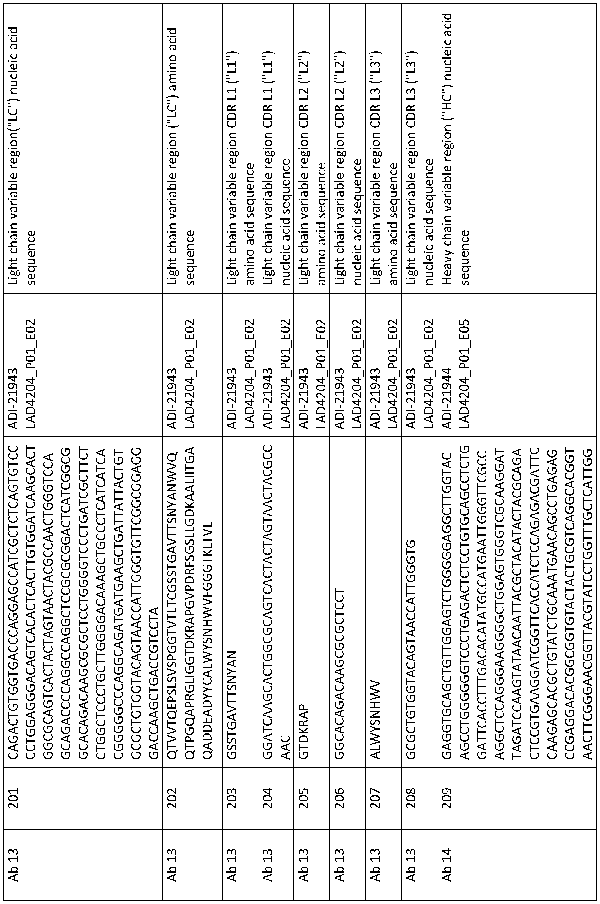


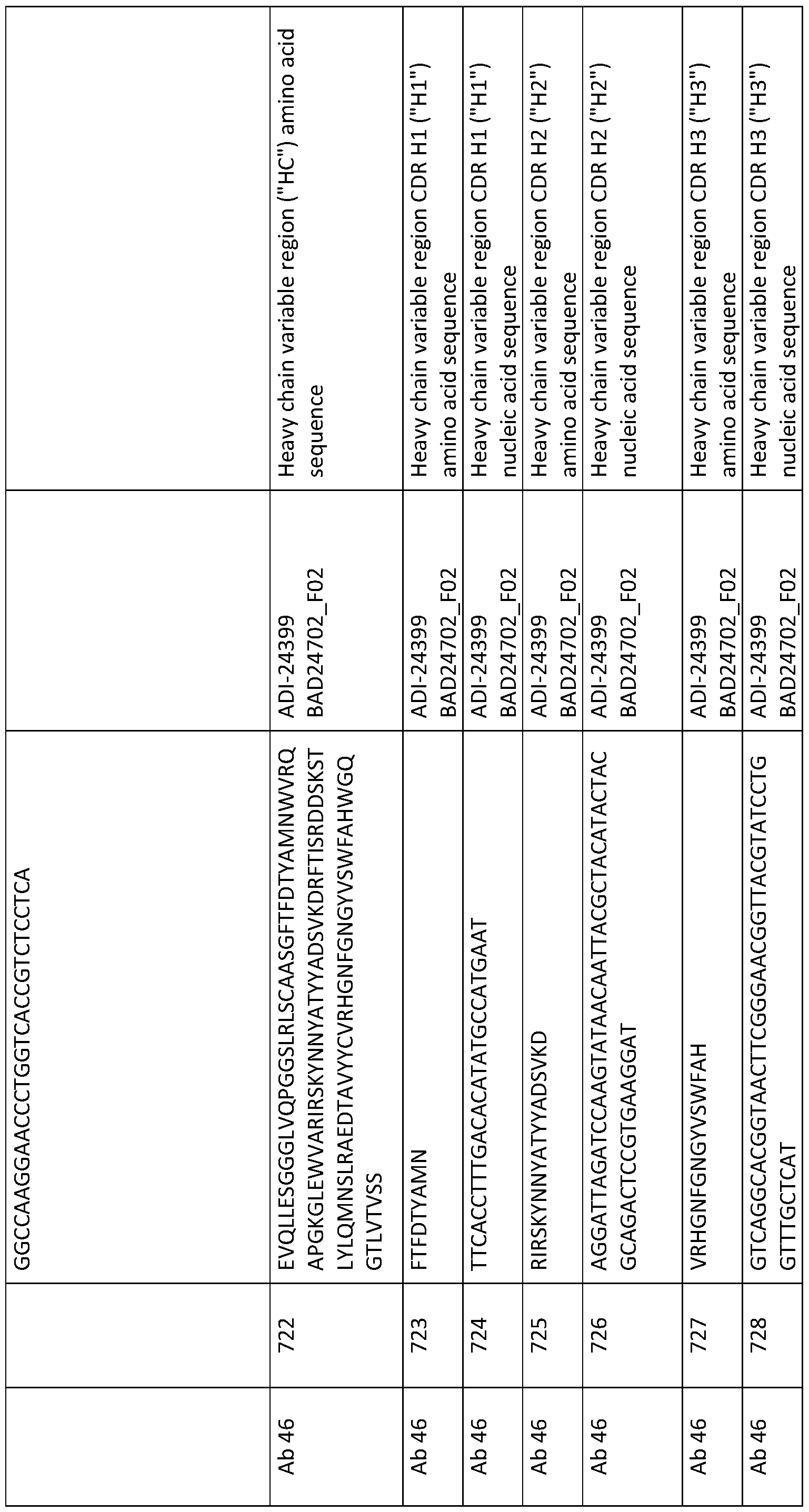


Claims
Priority Applications (17)
| Application Number | Priority Date | Filing Date | Title |
|---|---|---|---|
| CN202410287775.5A CN118184781A (en) | 2017-05-08 | 2018-05-08 | Anti-CD 3 binding domains and antibodies comprising them and methods of their production and use |
| SG11201910096R SG11201910096RA (en) | 2017-05-08 | 2018-05-08 | Anti-cd3-binding domains and antibodies comprising them, and methods for their generation and use |
| EP18798898.5A EP3621641A4 (en) | 2017-05-08 | 2018-05-08 | Anti-cd3-binding domains and antibodies comprising them, and methods for their generation and use |
| US16/611,832 US11542330B2 (en) | 2017-05-08 | 2018-05-08 | Anti-CD3-binding domains and antibodies comprising them, and methods for their generation and use |
| KR1020197036219A KR20200053437A (en) | 2017-05-08 | 2018-05-08 | Anti-CD3-binding domains and antibodies comprising them, and methods of generating and using them |
| CN202410287712.XA CN118562001A (en) | 2017-05-08 | 2018-05-08 | Anti-CD 3 binding domains and antibodies comprising them and methods of their production and use |
| BR112019023111-0A BR112019023111A2 (en) | 2017-05-08 | 2018-05-08 | anti-cd3 binding domains and antibodies comprising the same, and methods for their generation and use |
| JP2019561267A JP7393207B2 (en) | 2017-05-08 | 2018-05-08 | Anti-CD3 binding domain and antibodies containing the domain, and methods of making and using the same |
| AU2018266711A AU2018266711B2 (en) | 2017-05-08 | 2018-05-08 | Anti-CD3-binding domains and antibodies comprising them, and methods for their generation and use |
| CN202410287656.XA CN118894936A (en) | 2017-05-08 | 2018-05-08 | Anti-CD3 binding domains and antibodies comprising the same and methods of producing and using the same |
| CA3062825A CA3062825A1 (en) | 2017-05-08 | 2018-05-08 | Anti-cd3-binding domains and antibodies comprising them, and methods for their generation and use |
| RU2019139334A RU2807040C2 (en) | 2017-05-08 | 2018-05-08 | Binding domains of anti-cd3 antibodies and antibodies containing them, as well as methods of their preparation and application |
| CN201880045633.0A CN110913895B (en) | 2017-05-08 | 2018-05-08 | anti-CD 3 binding domains and antibodies comprising them and methods of their production and use |
| MX2019013184A MX2019013184A (en) | 2017-05-08 | 2018-05-08 | Anti-cd3-binding domains and antibodies comprising them, and methods for their generation and use. |
| US18/054,588 US20230151097A1 (en) | 2017-05-08 | 2022-11-11 | Anti-cd3-binding domains and antibodies comprising them, and methods for their generation and use |
| JP2022193173A JP2023014324A (en) | 2017-05-08 | 2022-12-01 | Anti-CD3 Binding Domains and Antibodies Containing Such Domains, and Methods of Making and Using Them |
| JP2024014044A JP2024032937A (en) | 2017-05-08 | 2024-02-01 | Anti-CD3 binding domain and antibodies containing the domain, and methods of making and using the same |
Applications Claiming Priority (2)
| Application Number | Priority Date | Filing Date | Title |
|---|---|---|---|
| US201762503315P | 2017-05-08 | 2017-05-08 | |
| US62/503,315 | 2017-05-08 |
Related Child Applications (2)
| Application Number | Title | Priority Date | Filing Date |
|---|---|---|---|
| US16/611,832 A-371-Of-International US11542330B2 (en) | 2017-05-08 | 2018-05-08 | Anti-CD3-binding domains and antibodies comprising them, and methods for their generation and use |
| US18/054,588 Division US20230151097A1 (en) | 2017-05-08 | 2022-11-11 | Anti-cd3-binding domains and antibodies comprising them, and methods for their generation and use |
Publications (1)
| Publication Number | Publication Date |
|---|---|
| WO2018208864A1 true WO2018208864A1 (en) | 2018-11-15 |
Family
ID=64104990
Family Applications (1)
| Application Number | Title | Priority Date | Filing Date |
|---|---|---|---|
| PCT/US2018/031705 WO2018208864A1 (en) | 2017-05-08 | 2018-05-08 | Anti-cd3-binding domains and antibodies comprising them, and methods for their generation and use |
Country Status (11)
| Country | Link |
|---|---|
| US (2) | US11542330B2 (en) |
| EP (1) | EP3621641A4 (en) |
| JP (3) | JP7393207B2 (en) |
| KR (1) | KR20200053437A (en) |
| CN (4) | CN118894936A (en) |
| AU (1) | AU2018266711B2 (en) |
| BR (1) | BR112019023111A2 (en) |
| CA (1) | CA3062825A1 (en) |
| MX (2) | MX2019013184A (en) |
| SG (2) | SG11201910096RA (en) |
| WO (1) | WO2018208864A1 (en) |
Cited By (13)
| Publication number | Priority date | Publication date | Assignee | Title |
|---|---|---|---|---|
| WO2020247932A1 (en) * | 2019-06-07 | 2020-12-10 | Adimab, Llc | Engineered ph-dependent anti-cd3 antibodies, and methods for their generation and use |
| WO2020247929A1 (en) * | 2019-06-07 | 2020-12-10 | Adimab, Llc | High affinity anti-cd3 antibodies, and methods for their generation and use |
| WO2021030688A1 (en) | 2019-08-15 | 2021-02-18 | Igm Biosciences, Inc. | Immunostimulatory multimeric binding molecules |
| WO2021154534A1 (en) * | 2020-01-28 | 2021-08-05 | Promab Biotechnologies, Inc. | Plap-cd3 epsilon bispecific antibodies |
| WO2021222315A3 (en) * | 2020-04-27 | 2021-12-09 | Twist Bioscience Corporation | Variant nucleic acid libraries for coronavirus |
| WO2022076474A3 (en) * | 2020-10-07 | 2022-05-27 | Amgen Inc. | Rational selection of building blocks for the assembly of multispecific antibodies |
| CN114867532A (en) * | 2019-12-18 | 2022-08-05 | 豪夫迈·罗氏有限公司 | Bispecific anti-CCL2 antibody |
| WO2023060277A1 (en) * | 2021-10-08 | 2023-04-13 | Arbele Limited | Composition of multispecific antibodies targeting cdh17-expressing tumors and method of making and using thereof |
| WO2023056556A1 (en) * | 2021-10-07 | 2023-04-13 | National Research Council Of Canada | Anti-cd3 monoclonal antibodies and therapeutic constructs |
| EP4031568A4 (en) * | 2019-09-20 | 2023-09-20 | Adimab, LLC | Fc variants with reduced effector function |
| WO2023186112A1 (en) * | 2022-04-02 | 2023-10-05 | 普米斯生物技术(珠海)有限公司 | Multispecific antibody targeting ceacam and cd3 and use thereof |
| WO2024102948A1 (en) | 2022-11-11 | 2024-05-16 | Celgene Corporation | Fc receptor-homolog 5 (fcrh5) specific binding molecules and bispecific t-cell engaging antibodies including same and related methods |
| RU2831840C2 (en) * | 2019-06-07 | 2024-12-16 | Адимаб, Ллк | High-affinity anti-cd3 antibodies and methods for production and use thereof |
Families Citing this family (7)
| Publication number | Priority date | Publication date | Assignee | Title |
|---|---|---|---|---|
| CN111826400A (en) * | 2020-07-21 | 2020-10-27 | 中科宝承生物医学科技有限公司 | Preparation method of bispecific antibody NK cell, cell and application thereof |
| CN114539415B (en) * | 2020-11-24 | 2024-02-02 | 普米斯生物技术(珠海)有限公司 | anti-PD-L1/VEGF/TGF-beta multi-specific antibody and application thereof |
| CN112578117B (en) * | 2021-02-22 | 2021-05-25 | 信纳克(北京)生化标志物检测医学研究有限责任公司 | Antibody composition and application thereof in screening post-transplantation lymphocyte proliferative diseases |
| CN112698044B (en) * | 2021-03-23 | 2021-06-22 | 信纳克(北京)生化标志物检测医学研究有限责任公司 | Device and method for evaluating immune state after targeted therapy |
| CN114573705B (en) * | 2022-03-17 | 2024-05-14 | 杭州师范大学 | Bispecific antibodies for specifically initiating anti-hepatitis B virus T cell immunity and their application |
| CN114605560B (en) * | 2022-03-30 | 2024-02-20 | 江苏蒙彼利生物科技有限公司 | CAR-NK cell and preparation method and application thereof |
| WO2024206568A2 (en) * | 2023-03-31 | 2024-10-03 | Ibio, Inc. | Anti-cd3 antibodies |
Citations (5)
| Publication number | Priority date | Publication date | Assignee | Title |
|---|---|---|---|---|
| US7728114B2 (en) * | 2004-06-03 | 2010-06-01 | Novimmune S.A. | Anti-CD3 antibodies and methods of use thereof |
| WO2012158818A2 (en) * | 2011-05-16 | 2012-11-22 | Fabion Pharmaceuticals, Inc. | Multi-specific fab fusion proteins and methods of use |
| WO2013049254A1 (en) * | 2011-09-26 | 2013-04-04 | Jn Biosciences Llc | Hybrid constant regions |
| US20150166661A1 (en) * | 2013-12-17 | 2015-06-18 | Genentech, Inc. | Anti-cd3 antibodies and methods of use |
| WO2016180721A1 (en) * | 2015-05-08 | 2016-11-17 | Miltenyi Biotec Gmbh | Humanized antibody or fragment thereof specific for cd3 |
Family Cites Families (5)
| Publication number | Priority date | Publication date | Assignee | Title |
|---|---|---|---|---|
| DK1629011T3 (en) * | 2003-05-31 | 2010-05-03 | Micromet Ag | Human anti-human DC3 binding molecules |
| US9180186B2 (en) * | 2010-01-11 | 2015-11-10 | Alexion Pharmaceuticals, Inc. | Biomarkers of immunomodulatory effects in humans treated with anti-CD200 antibodies |
| SG10201702396QA (en) * | 2012-03-28 | 2017-04-27 | Sanofi Sa | Antibodies to bradykinin b1 receptor ligands |
| MY184889A (en) * | 2014-11-20 | 2021-04-29 | Hoffmann La Roche | T cell activating bispecific antigen binding molecules |
| TW202130662A (en) * | 2015-01-23 | 2021-08-16 | 法商賽諾菲公司 | Anti-cd3 antibodies, anti-cd123 antibodies and bispecific antibodies specifically binding to cd3 and/or cd123 |
-
2018
- 2018-05-08 AU AU2018266711A patent/AU2018266711B2/en active Active
- 2018-05-08 EP EP18798898.5A patent/EP3621641A4/en active Pending
- 2018-05-08 BR BR112019023111-0A patent/BR112019023111A2/en unknown
- 2018-05-08 SG SG11201910096R patent/SG11201910096RA/en unknown
- 2018-05-08 WO PCT/US2018/031705 patent/WO2018208864A1/en unknown
- 2018-05-08 SG SG10201912494TA patent/SG10201912494TA/en unknown
- 2018-05-08 JP JP2019561267A patent/JP7393207B2/en active Active
- 2018-05-08 CN CN202410287656.XA patent/CN118894936A/en active Pending
- 2018-05-08 CN CN202410287775.5A patent/CN118184781A/en active Pending
- 2018-05-08 CN CN201880045633.0A patent/CN110913895B/en active Active
- 2018-05-08 CA CA3062825A patent/CA3062825A1/en active Pending
- 2018-05-08 KR KR1020197036219A patent/KR20200053437A/en not_active Application Discontinuation
- 2018-05-08 US US16/611,832 patent/US11542330B2/en active Active
- 2018-05-08 MX MX2019013184A patent/MX2019013184A/en unknown
- 2018-05-08 CN CN202410287712.XA patent/CN118562001A/en active Pending
-
2019
- 2019-11-05 MX MX2024005009A patent/MX2024005009A/en unknown
-
2022
- 2022-11-11 US US18/054,588 patent/US20230151097A1/en active Pending
- 2022-12-01 JP JP2022193173A patent/JP2023014324A/en active Pending
-
2024
- 2024-02-01 JP JP2024014044A patent/JP2024032937A/en active Pending
Patent Citations (5)
| Publication number | Priority date | Publication date | Assignee | Title |
|---|---|---|---|---|
| US7728114B2 (en) * | 2004-06-03 | 2010-06-01 | Novimmune S.A. | Anti-CD3 antibodies and methods of use thereof |
| WO2012158818A2 (en) * | 2011-05-16 | 2012-11-22 | Fabion Pharmaceuticals, Inc. | Multi-specific fab fusion proteins and methods of use |
| WO2013049254A1 (en) * | 2011-09-26 | 2013-04-04 | Jn Biosciences Llc | Hybrid constant regions |
| US20150166661A1 (en) * | 2013-12-17 | 2015-06-18 | Genentech, Inc. | Anti-cd3 antibodies and methods of use |
| WO2016180721A1 (en) * | 2015-05-08 | 2016-11-17 | Miltenyi Biotec Gmbh | Humanized antibody or fragment thereof specific for cd3 |
Non-Patent Citations (2)
| Title |
|---|
| CARPENTER ET AL.: "A humanized non-FcR-binding anti- CD 3 antibody, visilizumab, for treatment of steroid -refractory acute graft-versus-host disease", BLOOD, vol. 99, no. 8, 15 April 2002 (2002-04-15), pages 2712 - 2719, XP002349421 * |
| CHATENOUD ET AL.: "Anti- CD 3 antibody induces long-term remission of overt autoimmunity in nonobese diabetic mice", PROC NATL ACAD SCI USA, vol. 91, no. 1, 4 January 1994 (1994-01-04), pages 123 - 127, XP055552783 * |
Cited By (20)
| Publication number | Priority date | Publication date | Assignee | Title |
|---|---|---|---|---|
| WO2020247932A1 (en) * | 2019-06-07 | 2020-12-10 | Adimab, Llc | Engineered ph-dependent anti-cd3 antibodies, and methods for their generation and use |
| WO2020247929A1 (en) * | 2019-06-07 | 2020-12-10 | Adimab, Llc | High affinity anti-cd3 antibodies, and methods for their generation and use |
| RU2831840C2 (en) * | 2019-06-07 | 2024-12-16 | Адимаб, Ллк | High-affinity anti-cd3 antibodies and methods for production and use thereof |
| RU2832079C2 (en) * | 2019-06-07 | 2024-12-18 | Адимаб, Ллк | ENGINEERED pH-DEPENDENT ANTI-CD3 ANTIBODIES, AS WELL AS METHODS FOR PRODUCTION AND USE THEREOF |
| CN114144437A (en) * | 2019-06-07 | 2022-03-04 | 阿迪马布有限责任公司 | Engineered pH-dependent anti-CD 3 antibodies and methods of making and using the same |
| CN114174339A (en) * | 2019-06-07 | 2022-03-11 | 阿迪马布有限责任公司 | High affinity anti-CD 3 antibodies and methods of making and using the same |
| EP3973000A4 (en) * | 2019-06-07 | 2023-09-06 | Adimab, LLC | High affinity anti-cd3 antibodies, and methods for their generation and use |
| WO2021030688A1 (en) | 2019-08-15 | 2021-02-18 | Igm Biosciences, Inc. | Immunostimulatory multimeric binding molecules |
| EP4031568A4 (en) * | 2019-09-20 | 2023-09-20 | Adimab, LLC | Fc variants with reduced effector function |
| CN114867532A (en) * | 2019-12-18 | 2022-08-05 | 豪夫迈·罗氏有限公司 | Bispecific anti-CCL2 antibody |
| US12103967B2 (en) | 2019-12-18 | 2024-10-01 | Hoffmann-La Roche Inc. | Bispecific anti-CCL2 antibodies |
| CN115052897A (en) * | 2020-01-28 | 2022-09-13 | 普洛迈博生物技术公司 | PLAP-CD3 epsilon bispecific antibodies |
| WO2021154534A1 (en) * | 2020-01-28 | 2021-08-05 | Promab Biotechnologies, Inc. | Plap-cd3 epsilon bispecific antibodies |
| WO2021222315A3 (en) * | 2020-04-27 | 2021-12-09 | Twist Bioscience Corporation | Variant nucleic acid libraries for coronavirus |
| US12018065B2 (en) | 2020-04-27 | 2024-06-25 | Twist Bioscience Corporation | Variant nucleic acid libraries for coronavirus |
| WO2022076474A3 (en) * | 2020-10-07 | 2022-05-27 | Amgen Inc. | Rational selection of building blocks for the assembly of multispecific antibodies |
| WO2023056556A1 (en) * | 2021-10-07 | 2023-04-13 | National Research Council Of Canada | Anti-cd3 monoclonal antibodies and therapeutic constructs |
| WO2023060277A1 (en) * | 2021-10-08 | 2023-04-13 | Arbele Limited | Composition of multispecific antibodies targeting cdh17-expressing tumors and method of making and using thereof |
| WO2023186112A1 (en) * | 2022-04-02 | 2023-10-05 | 普米斯生物技术(珠海)有限公司 | Multispecific antibody targeting ceacam and cd3 and use thereof |
| WO2024102948A1 (en) | 2022-11-11 | 2024-05-16 | Celgene Corporation | Fc receptor-homolog 5 (fcrh5) specific binding molecules and bispecific t-cell engaging antibodies including same and related methods |
Also Published As
| Publication number | Publication date |
|---|---|
| EP3621641A4 (en) | 2021-03-03 |
| CA3062825A1 (en) | 2018-11-15 |
| JP2023014324A (en) | 2023-01-26 |
| BR112019023111A2 (en) | 2020-07-28 |
| US20230151097A1 (en) | 2023-05-18 |
| AU2018266711B2 (en) | 2025-02-06 |
| MX2019013184A (en) | 2020-02-07 |
| SG10201912494TA (en) | 2020-02-27 |
| RU2019139334A (en) | 2021-06-09 |
| MX2024005009A (en) | 2024-05-10 |
| SG11201910096RA (en) | 2019-11-28 |
| CN118562001A (en) | 2024-08-30 |
| CN118184781A (en) | 2024-06-14 |
| RU2019139334A3 (en) | 2021-08-25 |
| CN110913895B (en) | 2024-04-02 |
| EP3621641A1 (en) | 2020-03-18 |
| JP7393207B2 (en) | 2023-12-06 |
| US11542330B2 (en) | 2023-01-03 |
| JP2024032937A (en) | 2024-03-12 |
| AU2018266711A1 (en) | 2019-11-28 |
| CN118894936A (en) | 2024-11-05 |
| KR20200053437A (en) | 2020-05-18 |
| US20200190189A1 (en) | 2020-06-18 |
| CN110913895A (en) | 2020-03-24 |
| JP2020518650A (en) | 2020-06-25 |
Similar Documents
| Publication | Publication Date | Title |
|---|---|---|
| US20230151097A1 (en) | Anti-cd3-binding domains and antibodies comprising them, and methods for their generation and use | |
| TWI779010B (en) | ANTI-MYOSTATIN ANTIBODIES, POLYPEPTIDES CONTAINING VARIANT Fc REGIONs, AND METHODS OF USE | |
| JP6718560B1 (en) | Anti-CD137 antigen binding molecule and uses thereof | |
| CA3141914A1 (en) | Engineered ph-dependent anti-cd3 antibodies, and methods for their generation and use | |
| AU2020288880A1 (en) | High affinity anti-CD3 antibodies, and methods for their generation and use | |
| WO2023044402A1 (en) | Anti-cd3 antibodies | |
| CA3167808A1 (en) | Anti-cd137 antigen-binding molecule for use in cancer treatment | |
| RU2807040C2 (en) | Binding domains of anti-cd3 antibodies and antibodies containing them, as well as methods of their preparation and application | |
| TW202446798A (en) | Anti-cd28 antibodies and methods related thereto | |
| RU2829536C2 (en) | Anti-cd137 antigen-binding molecules and use thereof | |
| WO2023097219A2 (en) | Anti-idiotype antibodies | |
| WO2024086617A2 (en) | Ph-dependent anti-cd3 antibodies and methods relating thereto | |
| EA046075B1 (en) | ANTI-CD137 ANTIGEN-BINDING MOLECULES AND THEIR APPLICATIONS |
Legal Events
| Date | Code | Title | Description |
|---|---|---|---|
| 121 | Ep: the epo has been informed by wipo that ep was designated in this application |
Ref document number: 18798898 Country of ref document: EP Kind code of ref document: A1 |
|
| ENP | Entry into the national phase |
Ref document number: 3062825 Country of ref document: CA Ref document number: 2019561267 Country of ref document: JP Kind code of ref document: A |
|
| NENP | Non-entry into the national phase |
Ref country code: DE |
|
| REG | Reference to national code |
Ref country code: BR Ref legal event code: B01A Ref document number: 112019023111 Country of ref document: BR |
|
| ENP | Entry into the national phase |
Ref document number: 2018266711 Country of ref document: AU Date of ref document: 20180508 Kind code of ref document: A |
|
| ENP | Entry into the national phase |
Ref document number: 2018798898 Country of ref document: EP Effective date: 20191209 |
|
| ENP | Entry into the national phase |
Ref document number: 112019023111 Country of ref document: BR Kind code of ref document: A2 Effective date: 20191104 |