WO2018182016A1 - Cell culturing using nanofibers - Google Patents
Cell culturing using nanofibers Download PDFInfo
- Publication number
- WO2018182016A1 WO2018182016A1 PCT/JP2018/013977 JP2018013977W WO2018182016A1 WO 2018182016 A1 WO2018182016 A1 WO 2018182016A1 JP 2018013977 W JP2018013977 W JP 2018013977W WO 2018182016 A1 WO2018182016 A1 WO 2018182016A1
- Authority
- WO
- WIPO (PCT)
- Prior art keywords
- cells
- nanofibers
- adherent cells
- water
- medium
- Prior art date
Links
Images


Classifications
-
- C—CHEMISTRY; METALLURGY
- C12—BIOCHEMISTRY; BEER; SPIRITS; WINE; VINEGAR; MICROBIOLOGY; ENZYMOLOGY; MUTATION OR GENETIC ENGINEERING
- C12M—APPARATUS FOR ENZYMOLOGY OR MICROBIOLOGY; APPARATUS FOR CULTURING MICROORGANISMS FOR PRODUCING BIOMASS, FOR GROWING CELLS OR FOR OBTAINING FERMENTATION OR METABOLIC PRODUCTS, i.e. BIOREACTORS OR FERMENTERS
- C12M25/00—Means for supporting, enclosing or fixing the microorganisms, e.g. immunocoatings
- C12M25/16—Particles; Beads; Granular material; Encapsulation
-
- C—CHEMISTRY; METALLURGY
- C12—BIOCHEMISTRY; BEER; SPIRITS; WINE; VINEGAR; MICROBIOLOGY; ENZYMOLOGY; MUTATION OR GENETIC ENGINEERING
- C12N—MICROORGANISMS OR ENZYMES; COMPOSITIONS THEREOF; PROPAGATING, PRESERVING, OR MAINTAINING MICROORGANISMS; MUTATION OR GENETIC ENGINEERING; CULTURE MEDIA
- C12N5/00—Undifferentiated human, animal or plant cells, e.g. cell lines; Tissues; Cultivation or maintenance thereof; Culture media therefor
- C12N5/06—Animal cells or tissues; Human cells or tissues
- C12N5/0602—Vertebrate cells
- C12N5/0652—Cells of skeletal and connective tissues; Mesenchyme
- C12N5/0662—Stem cells
- C12N5/0667—Adipose-derived stem cells [ADSC]; Adipose stromal stem cells
-
- A—HUMAN NECESSITIES
- A61—MEDICAL OR VETERINARY SCIENCE; HYGIENE
- A61K—PREPARATIONS FOR MEDICAL, DENTAL OR TOILETRY PURPOSES
- A61K35/00—Medicinal preparations containing materials or reaction products thereof with undetermined constitution
- A61K35/12—Materials from mammals; Compositions comprising non-specified tissues or cells; Compositions comprising non-embryonic stem cells; Genetically modified cells
- A61K35/28—Bone marrow; Haematopoietic stem cells; Mesenchymal stem cells of any origin, e.g. adipose-derived stem cells
-
- A—HUMAN NECESSITIES
- A61—MEDICAL OR VETERINARY SCIENCE; HYGIENE
- A61L—METHODS OR APPARATUS FOR STERILISING MATERIALS OR OBJECTS IN GENERAL; DISINFECTION, STERILISATION OR DEODORISATION OF AIR; CHEMICAL ASPECTS OF BANDAGES, DRESSINGS, ABSORBENT PADS OR SURGICAL ARTICLES; MATERIALS FOR BANDAGES, DRESSINGS, ABSORBENT PADS OR SURGICAL ARTICLES
- A61L27/00—Materials for grafts or prostheses or for coating grafts or prostheses
- A61L27/14—Macromolecular materials
- A61L27/20—Polysaccharides
-
- A—HUMAN NECESSITIES
- A61—MEDICAL OR VETERINARY SCIENCE; HYGIENE
- A61L—METHODS OR APPARATUS FOR STERILISING MATERIALS OR OBJECTS IN GENERAL; DISINFECTION, STERILISATION OR DEODORISATION OF AIR; CHEMICAL ASPECTS OF BANDAGES, DRESSINGS, ABSORBENT PADS OR SURGICAL ARTICLES; MATERIALS FOR BANDAGES, DRESSINGS, ABSORBENT PADS OR SURGICAL ARTICLES
- A61L27/00—Materials for grafts or prostheses or for coating grafts or prostheses
- A61L27/36—Materials for grafts or prostheses or for coating grafts or prostheses containing ingredients of undetermined constitution or reaction products thereof, e.g. transplant tissue, natural bone, extracellular matrix
- A61L27/38—Materials for grafts or prostheses or for coating grafts or prostheses containing ingredients of undetermined constitution or reaction products thereof, e.g. transplant tissue, natural bone, extracellular matrix containing added animal cells
- A61L27/3804—Materials for grafts or prostheses or for coating grafts or prostheses containing ingredients of undetermined constitution or reaction products thereof, e.g. transplant tissue, natural bone, extracellular matrix containing added animal cells characterised by specific cells or progenitors thereof, e.g. fibroblasts, connective tissue cells, kidney cells
- A61L27/3834—Cells able to produce different cell types, e.g. hematopoietic stem cells, mesenchymal stem cells, marrow stromal cells, embryonic stem cells
-
- A—HUMAN NECESSITIES
- A61—MEDICAL OR VETERINARY SCIENCE; HYGIENE
- A61L—METHODS OR APPARATUS FOR STERILISING MATERIALS OR OBJECTS IN GENERAL; DISINFECTION, STERILISATION OR DEODORISATION OF AIR; CHEMICAL ASPECTS OF BANDAGES, DRESSINGS, ABSORBENT PADS OR SURGICAL ARTICLES; MATERIALS FOR BANDAGES, DRESSINGS, ABSORBENT PADS OR SURGICAL ARTICLES
- A61L27/00—Materials for grafts or prostheses or for coating grafts or prostheses
- A61L27/40—Composite materials, i.e. containing one material dispersed in a matrix of the same or different material
-
- A—HUMAN NECESSITIES
- A61—MEDICAL OR VETERINARY SCIENCE; HYGIENE
- A61L—METHODS OR APPARATUS FOR STERILISING MATERIALS OR OBJECTS IN GENERAL; DISINFECTION, STERILISATION OR DEODORISATION OF AIR; CHEMICAL ASPECTS OF BANDAGES, DRESSINGS, ABSORBENT PADS OR SURGICAL ARTICLES; MATERIALS FOR BANDAGES, DRESSINGS, ABSORBENT PADS OR SURGICAL ARTICLES
- A61L27/00—Materials for grafts or prostheses or for coating grafts or prostheses
- A61L27/50—Materials characterised by their function or physical properties, e.g. injectable or lubricating compositions, shape-memory materials, surface modified materials
- A61L27/56—Porous materials, e.g. foams or sponges
-
- A—HUMAN NECESSITIES
- A61—MEDICAL OR VETERINARY SCIENCE; HYGIENE
- A61L—METHODS OR APPARATUS FOR STERILISING MATERIALS OR OBJECTS IN GENERAL; DISINFECTION, STERILISATION OR DEODORISATION OF AIR; CHEMICAL ASPECTS OF BANDAGES, DRESSINGS, ABSORBENT PADS OR SURGICAL ARTICLES; MATERIALS FOR BANDAGES, DRESSINGS, ABSORBENT PADS OR SURGICAL ARTICLES
- A61L27/00—Materials for grafts or prostheses or for coating grafts or prostheses
- A61L27/50—Materials characterised by their function or physical properties, e.g. injectable or lubricating compositions, shape-memory materials, surface modified materials
- A61L27/58—Materials at least partially resorbable by the body
-
- A—HUMAN NECESSITIES
- A61—MEDICAL OR VETERINARY SCIENCE; HYGIENE
- A61P—SPECIFIC THERAPEUTIC ACTIVITY OF CHEMICAL COMPOUNDS OR MEDICINAL PREPARATIONS
- A61P43/00—Drugs for specific purposes, not provided for in groups A61P1/00-A61P41/00
-
- C—CHEMISTRY; METALLURGY
- C12—BIOCHEMISTRY; BEER; SPIRITS; WINE; VINEGAR; MICROBIOLOGY; ENZYMOLOGY; MUTATION OR GENETIC ENGINEERING
- C12N—MICROORGANISMS OR ENZYMES; COMPOSITIONS THEREOF; PROPAGATING, PRESERVING, OR MAINTAINING MICROORGANISMS; MUTATION OR GENETIC ENGINEERING; CULTURE MEDIA
- C12N5/00—Undifferentiated human, animal or plant cells, e.g. cell lines; Tissues; Cultivation or maintenance thereof; Culture media therefor
- C12N5/06—Animal cells or tissues; Human cells or tissues
- C12N5/0602—Vertebrate cells
- C12N5/0652—Cells of skeletal and connective tissues; Mesenchyme
- C12N5/0653—Adipocytes; Adipose tissue
-
- C—CHEMISTRY; METALLURGY
- C12—BIOCHEMISTRY; BEER; SPIRITS; WINE; VINEGAR; MICROBIOLOGY; ENZYMOLOGY; MUTATION OR GENETIC ENGINEERING
- C12Y—ENZYMES
- C12Y302/00—Hydrolases acting on glycosyl compounds, i.e. glycosylases (3.2)
- C12Y302/01—Glycosidases, i.e. enzymes hydrolysing O- and S-glycosyl compounds (3.2.1)
- C12Y302/01014—Chitinase (3.2.1.14)
-
- A—HUMAN NECESSITIES
- A61—MEDICAL OR VETERINARY SCIENCE; HYGIENE
- A61L—METHODS OR APPARATUS FOR STERILISING MATERIALS OR OBJECTS IN GENERAL; DISINFECTION, STERILISATION OR DEODORISATION OF AIR; CHEMICAL ASPECTS OF BANDAGES, DRESSINGS, ABSORBENT PADS OR SURGICAL ARTICLES; MATERIALS FOR BANDAGES, DRESSINGS, ABSORBENT PADS OR SURGICAL ARTICLES
- A61L2400/00—Materials characterised by their function or physical properties
- A61L2400/12—Nanosized materials, e.g. nanofibres, nanoparticles, nanowires, nanotubes; Nanostructured surfaces
-
- C—CHEMISTRY; METALLURGY
- C12—BIOCHEMISTRY; BEER; SPIRITS; WINE; VINEGAR; MICROBIOLOGY; ENZYMOLOGY; MUTATION OR GENETIC ENGINEERING
- C12N—MICROORGANISMS OR ENZYMES; COMPOSITIONS THEREOF; PROPAGATING, PRESERVING, OR MAINTAINING MICROORGANISMS; MUTATION OR GENETIC ENGINEERING; CULTURE MEDIA
- C12N2513/00—3D culture
-
- C—CHEMISTRY; METALLURGY
- C12—BIOCHEMISTRY; BEER; SPIRITS; WINE; VINEGAR; MICROBIOLOGY; ENZYMOLOGY; MUTATION OR GENETIC ENGINEERING
- C12N—MICROORGANISMS OR ENZYMES; COMPOSITIONS THEREOF; PROPAGATING, PRESERVING, OR MAINTAINING MICROORGANISMS; MUTATION OR GENETIC ENGINEERING; CULTURE MEDIA
- C12N2533/00—Supports or coatings for cell culture, characterised by material
- C12N2533/70—Polysaccharides
- C12N2533/72—Chitin, chitosan
Definitions
- the present invention relates to the use of nanofibers composed of water-insoluble polysaccharides as carriers in cell culture, storage, transport or transplantation.
- somatic stem cells such as mesenchymal stem cells and neural stem cells that have a low risk of canceration, and precursor cells such as preadipocytes and progenitor cardiomyocytes that can shorten the differentiation induction period are attracting attention again.
- mesenchymal stem cells and preadipocytes that have a property of being easily differentiated into fat, cartilage, and the like have advantages of being easily available, and thus are used in various studies.
- somatic stem cells and progenitor cells there is a high need for using primary cultured cells that are directly collected from tissues.
- primary hepatocytes prepared from human liver can be seeded on a cell plate after sorting, transported, or stocked as frozen cells. It contributes greatly to.
- primary cancer cells prepared by digesting cancer tissue isolated from cancer patients by enzyme treatment are used not only for cancer research but also for anticancer drug sensitivity tests and the like, and there is a great need.
- mesenchymal stem cells include dental pulp-derived mesenchymal stem cells, adipose tissue-derived mesenchymal stem cells isolated together with adipocytes during liposuction Is done. These mesenchymal stem cells can be easily increased by monolayer culture in a petri dish and further differentiated into fat, cartilage, and bone. Recently, it has also been clarified that these mesenchymal stem cells can differentiate into cells constituting the liver, pancreas and the like. Further, not only transplantation of cells differentiated from mesenchymal stem cells but also transplantation of mesenchymal stem cells themselves has been started.
- cytokines and exosomes that are secreted extracellularly by mesenchymal stem cells themselves have been focused on.
- Bioactive substances secreted into the culture supernatant after culturing mesenchymal stem cells attached to microcarriers Attempts have also been made to isolate and apply to pharmaceuticals and cosmetics.
- Preadipocytes are cells isolated together with adipocytes at the time of liposuction, and can be easily separated in large quantities from a living body, like the adipose tissue-derived mesenchymal stem cells described above. Preadipocytes also have the property of being able to differentiate not only into adipocytes but also into chondrocytes and the like, and their application to not only adipocyte transplantation but also cartilage transplantation in joints has been studied. In addition, it has been reported that the culture supernatant of adipocytes contains a component having a protective effect on the skin, and the use of a physiologically active substance isolated from the culture supernatant in pharmaceuticals and cosmetics is also examined. Has been. Adipocytes themselves are less proliferative and have the advantage of being less susceptible to cancer, and can be transplanted to sites that are easy to transplant, such as subcutaneously. By doing so, a disease caused by the gene mutation can be treated.
- mesenchymal stem cells, preadipocytes, and the like are usually prepared in large quantities from one individual, frozen, and cryopreserved cells are used for various purposes.
- mesenchymal stem cells, preadipocytes, and the like are usually prepared in large quantities from one individual, frozen, and cryopreserved cells are used for various purposes.
- the current transport technology that assumes cell freezing is suitable for transplantation. It is difficult to supply a large amount of cells in a stable state in a stable manner.
- cryopreserved cells must be thawed, cultured, and differentiated to the target cells as necessary, and a large number of cells in good condition must be provided.
- the transplantation facility is forced to obtain frozen cells from the cell bank that stores and manages the cells, and to perform large-scale culture on their own. Therefore, it is necessary for the transplanting facility to have a large-scale cell culture facility (for example, a cell processing center (CPC)), and it is difficult to perform transplantation medical care in a facility that does not have the facility. .
- CPC cell processing center
- cell banks usually have technology and equipment for culturing cells in large quantities.
- transplantation medicine can be performed even in a facility that does not have a large-scale cell culture facility.
- a technique for transporting a large number of cells while maintaining a good state without freezing is indispensable.
- a technique for culturing and proliferating mesenchymal stem cells, etc. in a state of being attached to a microcarrier, transplanting the obtained cells, and recovering a physiologically active substance released in the culture supernatant has been studied. Yes.
- currently available microcarriers will settle in the culture medium under static conditions, and need to be stirred during the culture.
- cell death occurs due to collision between carriers during the stirring.
- damage to cells and reduction of survival due to this protease are also problems. is there.
- microcarriers are not biodegradable, in order to apply cultured cells to transplantation, it is necessary to detach the cells from the microcarriers and collect them as needed. It must be held on a separate biodegradable carrier. These cell treatments are complicated and costly.
- the present inventors have developed a medium composition for cultivating animal and plant cells and / or tissues in a floating state using nanofibers such as polysaccharides having improved dispersibility in water (Patent Literature). 1).
- An object of the present invention is to provide a technique for transporting a large amount of adherent cells such as mesenchymal stem cells and preadipocytes under non-freezing conditions while maintaining a good state.
- adherent cells such as mesenchymal stem cells and preadipocytes under non-freezing conditions
- conventional microcarriers such as cell death due to collision of microcarriers during agitation, cell damage due to proteolytic enzyme treatment during passage, and complicated cell processing required when cells on microcarriers are used for transplantation are used. It is an object to provide a new carrier that can solve various problems in the suspension culture of the used adherent cells.
- nanofibers made of polysaccharides such as chitin which can be digested by enzyme without necessity, as a carrier.
- adherent cells such as mesenchymal stem cells and preadipocytes attached to the nanofibers
- the cells float in a stationary state. It was possible to culture. Under suspension culture conditions, the cells attached to the nanofibers showed long-term viability.
- Nanofibers made of polysaccharides are biodegradable, cells adhered to the nanofibers can be formed into a sheet form that can be easily transplanted together with the nanofibers while still attached.
- nanofibers composed of polysaccharides are required for mesenchymal stem cells, preadipocytes, primary cells, etc., for cell transport, culture, differentiation induction, transplantation, and production of physiologically active substances under non-freezing conditions. It was found that a plurality of operations selected from the group consisting of cell culture, differentiation induction, cell transport, transplantation, and bioactive substance production can be continuously performed by using this carrier. Based on these findings, the present inventors have further studied and completed the present invention.
- the present invention is as follows.
- the carrier according to [1] or [2] which is used for continuously subjecting the adherent cells to the treatments (1) to (3).
- adherent cells are stem cells or progenitor cells.
- adherent cells are mesenchymal stem cells or preadipocytes.
- water-insoluble polysaccharide is chitin or chitosan.
- adherent cell processing methods (1) suspension culture, (2) Non-frozen storage and / or transport, and (3) transplantation.
- the adherent cells attached to the nanofibers composed of the water-insoluble polysaccharide are treated with a degrading enzyme of the water-insoluble polysaccharide to decompose the nanofibers, and the adherent cells are detached from the nanofibers.
- a method for recovering the adhesive cells from the adhesive cells attached to the nanofibers comprising a water-insoluble polysaccharide.
- the water-insoluble polysaccharide is chitin or chitosan.
- the degrading enzyme is chitinase, chitobiase, chitosanase or ⁇ -1,3-glucanase.
- adherent cells such as mesenchymal stem cells and preadipocytes in a state of being adhered to nanofibers made of water-insoluble polysaccharides.
- Suspension culture can be performed in a stationary state without requiring operations such as shaking and rotation. Since suspension culture of adherent cells (three-dimensional (3D) culture) is possible, a larger number of cells can be cultured with a smaller amount of medium compared to single-layer culture (two-dimensional (2D) culture). can do.
- adherent cells such as mesenchymal stem cells and preadipocytes can be maintained and cultured while maintaining their characteristics.
- undifferentiated cells such as mesenchymal stem cells and preadipocytes can be differentiated into specific cells by culturing under specific differentiation conditions.
- the medium composition of the present invention since the cells are suspended in a state of being adhered to the nanofiber made of a water-insoluble polysaccharide, the cells adhered to the nanofiber can be easily obtained by centrifugation or the like. And the culture supernatant can be recovered.
- the medium composition of the present invention is useful for recovering and purifying a useful physiologically active substance released by adherent cells such as mesenchymal stem cells and preadipocytes into the culture medium.
- animal-derived proteolytic enzymes can be obtained by treating with a water-insoluble polysaccharide (eg, chitin) degrading enzyme (eg, chitinase) constituting nanofibers.
- a water-insoluble polysaccharide eg, chitin
- degrading enzyme eg, chitinase
- the cells adhered on the nanofibers can be collected without using. Therefore, in the present invention, the passaging operation of adhesive cells such as mesenchymal stem cells and preadipocytes is carried out while minimizing the risk of infection due to contamination of the cells by proteolytic enzymes and contamination of animal components. Cells that have been or have adhered to the nanofibers can be detached from the nanofibers and collected.
- Adherent cells such as mesenchymal stem cells and preadipocytes show good viability for a long period of time while adhered to nanofibers made of water-insoluble polysaccharides. Therefore, the culture medium composition of the present invention is useful for storing and transporting the above-mentioned adherent cells under non-freezing conditions. For example, when cells are adherently cultured on a plate and transported as it is, the original function of the cell may be reduced due to the separation of the cell from the plate due to vibration during transport.
- the cells adhered to the nanofibers can be held in a floating state, so that damage to the cells due to peeling from the plate due to vibration during transportation is avoided, and the original function of the cells is maintained
- the cells can be stored and transported under non-freezing conditions.
- a larger number of cells can be stored and transported with a smaller amount of medium and space than when stored or transported in the state of monolayer culture.
- nanofibers made of water-insoluble polysaccharides are biodegradable, cells adhered to the nanofibers can be transplanted into the living body together with the nanofibers in the adhered state. Therefore, after culturing adherent cells such as mesenchymal stem cells and preadipocytes in the medium composition of the present invention and differentiating them to a desired differentiation stage, the obtained cells are not detached from the nanofibers. It can be transplanted in vivo with nanofibers. Therefore, cells in a good state can be used for transplantation while avoiding the risk of cell damage and loss of function due to the cell detachment operation.
- nanofibers composed of water-insoluble polysaccharides are i) culture of adherent cells such as mesenchymal stem cells and preadipocytes, ii) induction of differentiation, iii) transport and storage under non-freezing conditions, Since it can be used as a common carrier in various operations such as iv) transplantation, v) recovery of physiologically active substances from the culture supernatant, i) culture and ii) differentiation induction described above by using the carrier.
- nanofibers made of water-insoluble polysaccharides as a common carrier, adherent cells are i) cultured, ii) differentiation induced, iii) transport and storage under non-freezing conditions, iv) transplantation, v) on culture
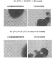
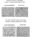
- Fig. 2 shows the migration ability and proliferation ability of human preadipocytes maintained for 7 days at 37 ° C or 25 ° C on chitin nanofibers.
- the migration ability and proliferation ability of human adipose-derived mesenchymal stem cells maintained at 37 ° C. or 25 ° C. for 7 days on chitin nanofibers are shown.
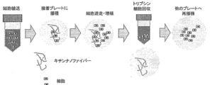
- the conceptual diagram of the cell transport method using chitin nanofiber is shown.
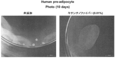
- the state of the human preadipocyte sheet produced by culturing on chitin nanofibers at 37 ° C. for 10 days is shown.
- the present invention provides the use of nanofibers composed of water-insoluble polysaccharides as carriers in various culture methodologies for adherent cells.
- Adhesive cells are attached to nanofibers composed of water-insoluble polysaccharides, and are subjected to various treatments such as (1) suspension culture, (2) storage and / or transport in a non-frozen state, and (3) transplantation. be able to. Therefore, the present invention provides (1) suspension culture, (2) non-freezing storage and / or transport, (3) transplantation, with adherent cells attached to nanofibers made of a water-insoluble polysaccharide. It can also be regarded as a method for treating adherent cells, including subjecting to treatment such as the above.
- a cell is the most basic unit constituting an animal or a plant, and has a cytoplasm and various organelles inside a cell membrane as its elements. At this time, the nucleus containing DNA may or may not be contained inside the cell.
- Examples of animals include, but are not limited to, fish, amphibians, reptiles, birds, pan-crustaceans, hexapods, mammals, and the like, with mammals being preferred.
- Examples of mammals include, but are not limited to, rat, mouse, rabbit, guinea pig, squirrel, hamster, vole, platypus, dolphin, whale, dog, cat, goat, cow, horse, sheep, pig, elephant, Common marmoset, squirrel monkey, rhesus monkey, chimpanzee, human and the like.
- the plant is not particularly limited as long as the collected cells can be cultured in liquid.
- plants that produce crude drugs eg, saponins, alkaloids, berberine, scoporin, plant sterols, etc.
- plants that produce crude drugs eg, saponins, alkaloids, berberine, scoporin, plant sterols, etc.
- plants that produce crude drugs eg, saponins, alkaloids, berberine, scoporin, plant sterols, etc.
- Pigs and polysaccharides that are used as cosmetics and food ingredients
- Plant eg, blueberry, safflower, saffron, saffron, etc.
- that produces the body eg, anthocyanin, safflower pigment, akane pigment, saffron pigment, flavone, etc.
- mammalian cells are preferably used.
- adherent cells are used.
- Adhesive cells are cells that require a scaffold such as a container wall for survival and proliferation.
- adherent cells adhere to nanofibers composed of a water-insoluble polysaccharide, thereby achieving good suspension culture of the cells, storage and / or transport in a non-frozen state, transplantation, and the like.
- the adhesive cells used in the present invention are not particularly limited, and examples thereof include stem cells, progenitor cells, somatic non-stem cells, primary cultured cells, cell lines, cancer cells and the like.
- a stem cell is a cell that has the ability to replicate itself and the ability to differentiate into other multiple lineage cells.
- Examples of adhesive stem cells include, but are not limited to, for example, mesenchymal stem cells, neural stem cells, hematopoietic stem cells, hepatic stem cells, pancreatic stem cells, muscle stem cells, reproductive stem cells, intestinal stem cells, cancer stem cells, Examples include somatic stem cells such as hair follicle stem cells.
- a mesenchymal stem cell is a stem cell capable of differentiating into all or some of bone cells, chondrocytes and adipocytes.
- Mesenchymal stem cells are present at low frequency in tissues such as bone marrow, peripheral blood, umbilical cord blood, and adipose tissue, and can be isolated from these tissues by a known method.
- a progenitor cell is a cell that is in the process of being differentiated from the stem cell into a specific somatic cell or germ cell.
- adhesive precursor cells include, but are not limited to, for example, preadipocytes, precursor cardiomyocytes, precursor endothelial cells, neural precursor cells, liver precursor cells, pancreatic precursor cells, kidney precursor cells, etc. Can be mentioned.
- adherent somatic non-stem cells include, but are not limited to, fibroblasts, bone cells, periosteum cells, keratinocytes, adipocytes, mesenchymal cells, epithelial cells, epidermal cells , Endothelial cells, vascular endothelial cells, hepatocytes, chondrocytes, cumulus cells, neural cells, glial cells, neurons, oligodendrocytes, microglia, astrocytes, heart cells, esophageal cells, muscle cells (eg Smooth muscle cells or skeletal muscle cells), pancreatic beta cells, melanocytes and the like.
- a primary cultured cell refers to a cell in a state of culture until cells or tissues separated from a living body are seeded and the first passage is performed.
- Primary cultured cells are, for example, skin, kidney, spleen, adrenal gland, liver, lung, ovary, pancreas, uterus, stomach, colon, small intestine, large intestine, spleen, bladder, prostate, testis, thymus, muscle, connective tissue, bone, cartilage , Cells taken from any tissue such as vascular tissue, blood, heart, eye, brain or nerve tissue.
- a cell line refers to a cell that has acquired infinite proliferative capacity by artificial manipulation in vitro.
- the adherent cells used in the present invention are preferably stem cells or progenitor cells, and more preferably mesenchymal stem cells or preadipocytes.
- Nanofiber The nanofibers used in the present invention exhibit an effect of allowing cells (preferably adherent cells) dispersed in a liquid medium and suspended in the liquid medium to float in the liquid medium.
- nanofiber means a fiber having an average fiber diameter (D) of 0.001 to 1.00 ⁇ m.
- the average fiber diameter of the nanofiber used in the present invention is preferably 0.005 to 0.50 ⁇ m, more preferably 0.01 to 0.05 ⁇ m, and still more preferably 0.01 to 0.02 ⁇ m.
- the aspect ratio (L / D) of the nanofiber used in the present invention is obtained from the average fiber length / average fiber diameter and is not particularly limited, but is usually 2 to 500, preferably 5 to 300, and more preferably. Is from 10 to 250.
- the average fiber diameter (D) of the nanofiber is determined as follows. First, the collodion support membrane manufactured by Oken Shoji Co., Ltd. was hydrophilized with an ion cleaner (JIC-410) manufactured by JEOL Ltd. for 3 minutes, and several nanofiber dispersions to be evaluated (diluted with ultrapure water) were used. Add dropwise and dry at room temperature. This was observed with a transmission electron microscope (TEM, H-8000) (10,000 times) manufactured by Hitachi, Ltd. at an accelerating voltage of 200 kV. Using the obtained images, the number of samples: 200 to 250 The fiber diameter of each nanofiber is measured and the number average value is defined as the average fiber diameter (D).
- TEM transmission electron microscope
- the average fiber length (L) is obtained as follows.
- the nanofiber dispersion liquid to be evaluated is diluted with pure water to 100 ppm, and the nanofibers are uniformly dispersed using an ultrasonic cleaner.
- This nanofiber dispersion is cast onto a silicon wafer whose surface has been hydrophilized with concentrated sulfuric acid in advance, and dried at 110 ° C. for 1 hour to prepare a sample.
- a scanning electron microscope SEM, JSM-7400F
- the number of specimens 150 to 250 nanofibers one by one
- the fiber length of the book is measured, and the number average value is defined as the average fiber length (L).
- the nanofibers used in the present invention are dispersed uniformly in the liquid while maintaining the primary fiber diameter when mixed with a liquid medium, without substantially increasing the viscosity of the liquid. It has the effect of substantially retaining the cells attached to the nanofibers and preventing their sedimentation.
- the fact that the viscosity of the liquid is not substantially increased means that the viscosity of the liquid does not exceed 8 mPa ⁇ s.
- the viscosity of the liquid (that is, the viscosity of the following medium composition of the present invention) is 8 mPa ⁇ s or less, preferably 4 mPa ⁇ s or less, more preferably 2 mPa ⁇ s or less.
- the viscosity of the liquid containing nanofibers can be evaluated, for example, using a tuning fork vibration type viscosity measurement (SV-1A, A & D Company Ltd.) under 25 ° C. conditions.
- the nanofiber used in the present invention is composed of a water-insoluble polysaccharide.
- the saccharide means a saccharide polymer in which 10 or more monosaccharides (for example, triose, tetrose, pentose, hexose, heptose, etc.) are polymerized.
- water-insoluble polysaccharides include, but are not limited to, celluloses such as cellulose and hemicellulose; and chitins such as chitin and chitosan.
- the water-insoluble polysaccharide is preferably chitin or chitosan, more preferably chitin.
- Cellulose is a natural polymer compound in which D-glucopyranose, which is a 6-membered ring of glucose, is linked by ⁇ -1,4 glucoside.
- raw materials include cellulose derived from plants such as wood, bamboo, hemp, jute, kenaf, cotton, crops and food residues, or microorganisms such as bacterial cellulose, Shiogusa (Cladophora), gray plants (Glaucocystis), Valonia, squirt cellulose, etc.
- Production or animal production cellulose can be used.
- Plant-derived cellulose is a bundle of very thin fibers called microfibrils, and forms a higher-order structure step by step with fibrils, lamellae, and fiber cells.
- Bacterial cellulose has a fine network structure in which the microfibrils of cellulose secreted from fungal cells have the same thickness.
- high-purity cellulose raw materials such as cotton and bacterial cellulose can be used as raw materials, but other plant-derived cellulose and the like are preferably isolated and purified.
- the cellulose suitably used in the present invention is cotton cellulose, bacterial cellulose, kraft pulp cellulose, microcrystalline cellulose or the like.
- Chitin is one or more carbohydrates selected from the group consisting of chitin and chitosan.
- the main sugar units constituting chitin and chitosan are N-acetylglucosamine and glucosamine, respectively.
- chitin and glucosamine have a high N-acetylglucosamine content and are hardly soluble in acidic aqueous solutions.
- a substance having a high content and soluble in an acidic aqueous solution is regarded as chitosan.
- the ratio of N-acetylglucosamine in the constituent sugars is called 50% or more of chitin, and less than 50% is called chitosan.
- the proportion of N-acetylglucosamine in the saccharide units constituting chitin is preferably 80% or more, more preferably 90% or more, still more preferably 98% or more, and most preferably 100%.
- chitin As a raw material for chitin, for example, many biological resources such as shrimp, crab, insect, shellfish, mushroom can be used.
- the chitin used in the present invention may be a chitin having an ⁇ -type crystal structure such as chitin derived from crab shell or shrimp shell, or a chitin having a ⁇ -type crystal structure such as chitin derived from squid shell.
- Crab and shrimp shells are often treated as industrial waste and are preferred as raw materials because they are readily available and effective.
- deproteinization and decalcification are necessary to remove proteins and ash contained as impurities. A process is required. Therefore, in the present invention, it is preferable to use purified chitin that has already been subjected to dematrixing. Purified chitin is commercially available.
- nanofibers composed of the water-insoluble polysaccharide can be obtained.
- a strong shearing force such as a high-pressure homogenizer, a grinder (stone mill), or a medium agitation mill such as a bead mill can be obtained in order to reduce the fiber diameter and fiber length, which will be described later, to meet the purpose of the present invention. Is preferred.
- the raw material is pulverized by injecting and colliding the dispersion liquid in which the raw material is dispersed from a pair of nozzles at a high pressure, for example, Starburst System (manufactured by Sugino Machine Co., Ltd.). It can be carried out by using a high-pressure crusher) or Nano perenniala (a high-pressure crusher from Yoshida Kikai Kogyo Co., Ltd.).
- the degree of refinement and homogenization depends on the pressure fed to the ultra-high pressure chamber of the high-pressure homogenizer and the number of times it passes through the ultra-high pressure chamber (number of treatments). And the concentration of the raw material in the aqueous dispersion.
- the pumping pressure is not particularly limited, but is usually 50 to 250 MPa, preferably 150 to 245 MPa.
- the concentration of the raw material in the aqueous dispersion during the micronization treatment is not particularly limited, but is usually 0.1% by mass to 30% by mass, preferably 1% by mass to 10% by mass.
- concentration of the raw material in the aqueous dispersion is not particularly limited, but is usually 0.1% by mass to 30% by mass, preferably 1% by mass to 10% by mass.
- the viscosity of the aqueous dispersion during the micronization treatment is not particularly limited.
- the viscosity of the aqueous dispersion is 1 to 100 mPa ⁇ S, preferably 1 85 mPa ⁇ S (by tuning fork vibration type viscosity measurement (SV-1A, A & D Company Ltd.) at 25 ° C.).
- the particle diameter of the water-insoluble polysaccharide in the aqueous dispersion during the micronization treatment is not particularly limited.
- the average particle of ⁇ -chitin in the aqueous dispersion is 0.5 to 200 ⁇ m, preferably 30 to 150 ⁇ m (according to laser diffraction / scattering particle size distribution measuring apparatus LA-960 (Horiba Seisakusho)).
- the liquid containing the nanofibers is used.
- the medium composition also referred to herein as the medium composition of the present invention.
- nanofibers made of a water-insoluble polysaccharide are uniformly dispersed, and the adherent cells attached to the nanofibers are suspended in the liquid medium.
- cell floating means a state in which cells do not adhere to a culture container (non-adhesion). Furthermore, in the present invention, when cells are cultured, stored, or transported, the cells can be transferred to the liquid medium composition without any external pressure or vibration applied to the liquid medium composition, or shaking or rotating operation in the composition.
- the state of being uniformly dispersed in the composition and in a floating state is referred to as “floating stationary”, and culturing cells and / or tissues in this state is referred to as “floating stationary culture”.
- the period during which it can be floated in “floating standing” is at least 5 minutes or more, preferably 1 hour or more, 24 hours or more, 48 hours or more, 6 days or more, 21 days or more, but is kept floating. As long as it is not limited to these periods.
- the medium composition of the present invention can float and stand cells at least at one point in the temperature range (eg, 0 to 40 ° C.) in which cells can be cultured, stored or transported.
- the medium composition of the present invention is capable of allowing cells to float and float at preferably at least one point in the temperature range of 25 to 37 ° C, most preferably at 37 ° C.
- Whether or not floating standing is possible is determined by, for example, uniformly dispersing polystyrene beads (Size 500-600 ⁇ m, manufactured by Polysciences Inc.) in the medium composition to be evaluated, and standing at 25 ° C., at least 5 It can be evaluated by observing whether the floating state of the cell is maintained for a minute or longer (preferably 24 hours or longer, 48 hours or longer).
- the concentration of nanofibers composed of a water-insoluble polysaccharide in the medium composition of the present invention is such that when the adherent cells are subjected to suspension culture, storage or transport in a non-frozen state using the medium composition, It can be set as appropriate so that the viability of the adherent cells can be improved, or the adherent cells can be suspended (preferably left floating).
- the concentration of chitin nanofibers in the medium composition is, for example, 0.0001% (weight / volume) or more, preferably 0.001% (weight / volume) or more.
- the upper limit value of the chitin nanofiber concentration in the medium composition is, for example, 1.0% (weight / volume) or less, preferably 0.1% (weight / volume) or less.
- the medium contained in the medium composition of the present invention can be appropriately selected depending on the type of adherent cells used, for example, for the purpose of culture, storage or transport of mammalian adherent cells, A medium generally used for culturing mammalian cells can be used as a medium contained in the medium composition of the present invention.
- Examples of the medium for mammalian cells include Dulbecco's Modified Eagle Medium (Dulbecco's Modified Eagles's Medium; DMEM), Ham F12 Medium (Ham's Nutrient Mixture F12), DMEM / F12 Medium, McCoy 5A Medium (McCoy 5A Medium (McCoy'A) s 5A medium), Eagle MEM medium (Eggles's Minimum Essential Medium; EMEM), ⁇ MEM medium (Alpha Modified Eagles's Minum Essential Medium; ⁇ MEM medium, RM medium 16) Iscove's Modify d Dulbecco's Medium (IMDM), MCDB131 medium, William medium E, IPL41 medium, Fischer's medium, StemPro34 (manufactured by Invitrogen), X-VIVO 10 (manufactured by Cambridge), X-VIVO 15 (Kenbrex) ), HPGM (manufactured by Cambridge), StemSpan H3000 (manufactured by
- Those skilled in the art may freely add sodium, potassium, calcium, magnesium, phosphorus, chlorine, various amino acids, various vitamins, antibiotics, serum, fatty acids, sugars and the like to the above medium according to the purpose.
- those skilled in the art can add one or more other chemical components or biological components in combination according to the purpose.
- Components that can be added to the medium for mammalian cells include fetal bovine serum, human serum, horse serum, insulin, transferrin, lactoferrin, cholesterol, ethanolamine, sodium selenite, monothioglycerol, 2-mercaptoethanol, bovine serum
- fetal bovine serum human serum
- horse serum insulin
- transferrin lactoferrin
- cholesterol ethanolamine
- sodium selenite sodium selenite
- monothioglycerol 2-mercaptoethanol
- bovine serum examples include albumin, sodium pyruvate, polyethylene glycol, various vitamins, various amino acids, agar, agarose, collagen, methylcellulose, various cytokines, various hormones, various growth factors, various extracellular matrices, and various cell adhesion molecules.
- cytokines examples include interleukin-1 (IL-1), interleukin-2 (IL-2), interleukin-3 (IL-3), interleukin-4 (IL-4), Interleukin-5 (IL-5), interleukin-6 (IL-6), interleukin-7 (IL-7), interleukin-8 (IL-8), interleukin-9 (IL-9), Interleukin-10 (IL-10), interleukin-11 (IL-11), interleukin-12 (IL-12), interleukin-13 (IL-13), interleukin-14 (IL-14), Interleukin-15 (IL-15), Interleukin-18 (IL-18), Interleukin-21 (IL-21), Interleukin Ron- ⁇ (IFN- ⁇ ), interferon- ⁇ (IFN- ⁇ ), interferon- ⁇ (IFN- ⁇ ), granulocyte colony stimulating factor (G-CSF), monocyte colony stimulating factor (M-CSF), granule Sphere-macrophage colony stimulating factor (GM-CSF), stem cell factor (S), interleukin-1 (
- Hormones that can be added to the medium include melatonin, serotonin, thyroxine, triiodothyronine, epinephrine, norepinephrine, dopamine, anti-Muellerian hormone, adiponectin, adrenocorticotropic hormone, angiotensinogen and angiotensin, antidiuretic hormone, atrium Natriuretic peptide, calcitonin, cholecystokinin, corticotropin releasing hormone, erythropoietin, follicle stimulating hormone, gastrin, ghrelin, glucagon, gonadotropin releasing hormone, growth hormone releasing hormone, human chorionic gonadotropin, human placental lactogen, growth hormone, inhibin Insulin, insulin-like growth factor, leptin, luteinizing hormone, melanocyte stimulating hormone, oxytocin, parathyroid hormone , Prolactin, secret
- Growth factors that can be added to the medium include transforming growth factor- ⁇ (TGF- ⁇ ), transforming growth factor- ⁇ (TGF- ⁇ ), macrophage inflammatory protein-1 ⁇ (MIP-1 ⁇ ), epidermal growth factor ( EGF), fibroblast growth factor-1, 2, 3, 4, 5, 6, 7, 8, or 9 (FGF-1, 2, 3, 4, 5, 6, 7, 8, 9), nerve Cell growth factor (NGF), hepatocyte growth factor (HGF), leukemia inhibitory factor (LIF), protease nexin I, protease nexin II, platelet derived growth factor (PDGF), cholinergic differentiation factor (CDF), chemokine , Notch ligand (such as Delta1), Wnt protein, Angiopoietin-like protein 2, 3, 5 or 7 (Angpt2, 3, 5, 7), insulin-like growth factor ( GF), insulin-like growth factor binding protein (IGFBP), but such pleiotrophin (Pleiotrophin), and the like, but is not limited to these.
- artificially modified amino acid sequences of these cytokines and growth factors can be added by gene recombination techniques.
- examples thereof include IL-6 / soluble IL-6 receptor complex or Hyper IL-6 (a fusion protein of IL-6 and soluble IL-6 receptor).
- Examples of various extracellular matrices and various cell adhesion molecules include collagen I to XIX, fibronectin, vitronectin, laminin-1 to 12, nitogen, tenascin, thrombospondin, von Willebrand factor, osteopontin, fibrinogen, Various elastins, various proteoglycans, various cadherins, desmocollins, desmogleins, various integrins, E-selectin, P-selectin, L-selectin, immunoglobulin superfamily, matrigel, poly-D-lysine, poly-L-lysine, chitin, Examples include chitosan, sepharose, hyaluronic acid, alginic acid gel, various hydrogels, and cut fragments thereof.
- antibiotics examples include sulfa, penicillin, pheneticillin, methicillin, oxacillin, cloxacillin, dicloxacillin, flucloxacillin, nafcillin, ampicillin, penicillin, amoxicillin, cyclacillin, carbenicillin, ticarcillin, piperacillin, Mecuzurocillin, mecillinam, andinocillin, cephalosporin and its derivatives, oxophosphoric acid, amifloxacin, temafloxacin, nalidixic acid, pyromido acid, ciprofloxane, sinoxacin, norfloxacin, perfloxacin, rosoxacin, ofloxacin, enoxacin, pipexamic acid, sulbactam acid, sulbactam acid, sulbactam acid, sulbactam acid , ⁇ -bromopenicillanic acid
- the medium composition of the present invention is mixed with an appropriate liquid medium so that the concentration is such that the adherent cells can be suspended (preferably suspended). Can be manufactured.
- the medium composition of the present invention is prepared by mixing a dispersion of nanofibers comprising the above water-insoluble polysaccharide in a physiological aqueous solvent and a liquid medium.
- the dispersion may be sterilized (autoclave, gamma ray sterilization, etc.).
- the dispersion and a liquid medium (aqueous medium solution) prepared by dissolving a powder medium in water may be mixed before sterilization. Sterilization of the dispersion and liquid medium may be performed separately before mixing.
- aqueous solvents include, but are not limited to water, dimethyl sulfoxide (DMSO) and the like.
- DMSO dimethyl sulfoxide
- the nanofiber dispersion is useful as a medium additive for preparing the medium composition of the present invention.
- the mixing ratio is not particularly limited, but the dispersion ratio of nanofibers: liquid medium (aqueous solution of medium) (volume ratio) is usually 1:99 to 99: 1, preferably 10:90 to 90:10. More preferably, it is 20:80 to 80:20.
- the present invention provides the use of nanofibers composed of water-insoluble polysaccharides as carriers in various culture methodologies for adherent cells.
- Adhesive cells are attached to nanofibers composed of water-insoluble polysaccharides, and are subjected to various treatments such as (1) suspension culture, (2) storage and / or transport in a non-frozen state, and (3) transplantation. be able to. Therefore, the present invention provides (1) suspension culture, (2) non-freezing storage and / or transport, (3) transplantation, with adherent cells attached to nanofibers made of a water-insoluble polysaccharide. It can also be regarded as a method for treating adherent cells, including subjecting to treatment such as the above.
- (1) Suspension culture By culturing adherent cells in a state where they adhere to nanofibers made of a water-insoluble polysaccharide, the adherent cells can be suspended.
- the suspension culture can be carried out by culturing adherent cells in the medium composition of the present invention.
- Nanofibers composed of a water-insoluble polysaccharide exhibit an effect of suspending cells attached to the nanofiber in a medium (preferably an effect of floating and standing).
- nanofibers composed of water-insoluble polysaccharides are uniformly dispersed. Therefore, when adherent cells are cultured in the medium composition, the adherent cells adhere to the nanofibers, and It floats in the medium composition.
- a medium containing a conventional gel substrate has a high viscosity and it may be difficult to exchange the medium.
- the medium composition of the present invention has a low viscosity, the medium can be easily used with a pipette or a pump. Can be expected to replace.
- adherent cells When adherent cells are cultured in suspension using nanofibers composed of water-insoluble polysaccharides, add separately prepared adherent cells to the medium composition of the present invention, and mix them so that they are uniformly dispersed. Good.
- the mixing method in that case is not particularly limited, and examples thereof include manual mixing such as pipetting, and mixing using equipment such as a stirrer, a vortex mixer, a microplate mixer, and a shaker. After mixing, the obtained cell suspension may be cultivated in a stationary state, or may be cultured while rotating, shaking or stirring as necessary. The number of rotations and frequency may be appropriately set according to the purpose of those skilled in the art.
- the adherent cells are recovered from the subculture and dispersed to a single cell or a state close thereto using an appropriate cell dissociation solution, and the dispersed adherent cells are dispersed in the medium composition of the present invention.
- the suspension is then subjected to suspension culture (preferably suspension stationary culture).
- the temperature at which the cells are cultured is usually 25 to 39 ° C., preferably 33 to 39 ° C. (eg, 37 ° C.) for animal cells.
- the CO 2 concentration is usually 4 to 10% by volume in the culture atmosphere, and preferably 4 to 6% by volume.
- the culture period may be appropriately set according to the purpose of culture.
- the culture of the adherent cells in the medium composition of the present invention is performed using a petri dish, a flask, a plastic bag, a Teflon (registered trademark) bag, a dish, a petri dish, a tissue culture dish, a multi-cell, which are generally used for cell culture. It can be carried out using culture containers such as dishes, microplates, microwell plates, multiplates, multiwell plates, chamber slides, tubes, trays, culture bags, roller bottles and the like. It is desirable for these culture containers to have low cell adhesion so that the adherent cells attached to the nanofibers do not adhere to the culture containers.
- the surface of the culture container is not artificially treated (for example, coated with an extracellular matrix) for the purpose of improving adhesion to cells
- a surface whose surface is artificially treated for the purpose of reducing adhesion to cells can be used.
- the cells are separated by centrifugation or filtration, and then a fresh medium or the medium composition of the present invention may be added to the cells.
- a fresh medium or the medium composition of the present invention may be added to this concentrated solution.
- the gravitational acceleration (G) at the time of centrifugation is 100 to 400 G
- the pore size of the filter used for the filtration treatment is 10 ⁇ m to 100 ⁇ m, but is not limited thereto.
- Adhesive cells are cultured in a closed environment under mechanical control by automatically performing cell seeding, medium exchange, cell image acquisition, and cell culture collection, while controlling pH, temperature, oxygen concentration, etc. It can also be carried out by a bioreactor capable of high-density culture or an automatic culture device.
- Adhesive cells proliferate efficiently when suspended in a state where they are attached to nanofibers composed of a water-insoluble polysaccharide. It is an excellent method for maintaining or growing adherent cells.
- the adhering cells are not unevenly distributed only on the bottom of the culture vessel, but spread and spread in a three-dimensional manner. Is promoted.
- adherent cells adhere to chitin nanofibers and grow strongly as a scaffold, and as a result, the proliferated cells are connected to nanofibers in the shape of grape tufts.
- the medium composition contains nanofibers at a concentration sufficient to float the adherent cells (that is, avoid adhesion of the adherent cells to the culture vessel) It is essential to be able to float and float (ie, the cells are evenly dispersed and suspended in the liquid medium composition without external pressure, vibration, shaking, rotation, etc.) is not.
- concentration is 0.0001% (weight / volume) or more sufficient for expression of floating action, 0.03% (weight / volume) that enables stable floating stationary culture is set.
- the concentration is lower (eg, 0.025% (weight / volume) or less, 0.02% (weight / volume) or less), the growth promoting effect is exhibited.
- adherent cells are maintained at a higher density than when the cells are attached to the bottom of the culture vessel and cultured in a single layer. Can be propagated.
- the adherent cells are cultured in suspension in a state of being attached to nanofibers composed of a water-insoluble polysaccharide and the cells are maintained or proliferated.
- a medium capable of maintaining or growing the cells while maintaining the character of the adherent cells is used.
- the culture medium can be appropriately selected by those skilled in the art depending on the type of adherent cells.
- mammalian stem cells eg, mesenchymal stem cells
- progenitor cells eg, preadipocytes
- suspension culture in a state where they are attached to chitin nanofibers.
- mammalian stem cells eg, mesenchymal stem cells
- progenitor cells eg, preadipocytes
- their traits eg, differentiation potential
- the fresh medium or the medium composition of the present invention can be used without any cell detachment operation from the culture vessel.
- Adherent cells can be passaged by simply adding them to the suspension culture or by adding all or part of the suspension culture to a fresh medium or a medium composition of the present invention.
- the adherent cells can be subcultured without performing the detachment operation of the cells from the culture vessel.
- the culture scale of adherent cells can be expanded without performing a cell peeling operation from the culture vessel.
- Examples of the operation for detaching cells from the culture container include treatment with a chelating agent (eg, EDTA) and / or a proteolytic enzyme (eg, trypsin, collagenase).
- a chelating agent eg, EDTA
- a proteolytic enzyme eg, trypsin, collagenase.
- the above-mentioned subculture method is used for the passage of adherent cells that are highly sensitive to cell detachment from a culture vessel (for example, adherent cells whose viability is reduced by the detachment operation, or adhesive cells whose characteristics are easily changed by the detachment operation). It is advantageous for subculture.
- adherent cells that are highly sensitive to cell detachment from culture vessels include stem cells (eg, mesenchymal stem cells), progenitor cells (eg, preadipocytes), primary cultured cells, and the like, but are not limited thereto. Not.
- Differentiation induction conditions can be set as appropriate according to the type of adherent cells and desired differentiated cells.
- a person skilled in the art can differentiate specific adherent cells into desired cells by applying various known differentiation-inducing conditions to the suspension culture.
- a specific differentiation-inducing factor is added to the above-described medium composition of the present invention, and the differentiation into desired cells is induced by performing suspension culture of the adherent cells in the presence of the differentiation-inducing factor. be able to.
- the differentiation induction conditions that can be applied to the present invention are not particularly limited, and examples thereof include the following.
- Differentiation of mesenchymal stem cells into adipocytes Mesenchymal stem cells are cultured in the presence of these factors using isobutylmethylxanthine (IBMX), dexamethasone, insulin and indomethacin as adipocyte differentiation inducing factors.
- IBMX isobutylmethylxanthine
- dexamethasone dexamethasone
- insulin and indomethacin as adipocyte differentiation inducing factors.
- -Differentiation of mesenchymal stem cells into bone cells Dexamethasone, ascorbic acid and ⁇ -glycerophosphate are used as bone cell differentiation-inducing factors, and mesenchymal stem cells are cultured in the presence of these factors.
- chondrocyte differentiation inducing factors Insulin, TGF- ⁇ 3 and ascorbic acid are used as chondrocyte differentiation inducing factors, and mesenchymal stem cells are cultured in the presence of these factors.
- the conditions for adipocyte differentiation, bone cell differentiation or chondrocyte differentiation of mesenchymal stem cells are described in, for example, the following papers.
- Crisan M Yap S, CasteillaL, et al., Cell Stem Cell 2008; (3): 301-13.
- Republici M Le Blanc K, Mueller I, Slaper-Cortenbach I, et al., Cytother2006; 8 (4): 315-7.
- Caplan AI Caplan AI., Cell Stem Cell 2008; 3 (3): 229-30.
- IBMX isobutylmethylxanthine
- dexamethasone insulin and indomethacin
- IBMX isobutylmethylxanthine
- insulin and indomethacin are used as adipocyte differentiation inducing factors, and the preadipocytes are cultured in the presence of these factors.
- the conditions for differentiation of preadipocytes into adipocytes are described, for example, in the following papers. [5] Hutley LJ, Newell FM, et al., Eur J ClinInvest. 2003; 33 (7): 574-81. [6] Yin Y, Yuan H, et al., Mol Endocrinol. 2006; 20 (2): 268-78. [7] Rival Y, StennevinA, et al., J Pharmacol Exp Ther. 2004; 311 (2): 467-75.
- Suspension culture of adherent cells under differentiation-inducing conditions is performed until desired differentiated cells appear. Appearance of a desired differentiated cell can be confirmed by examining expression of a differentiation marker of the cell. As a result of the suspension culture, desired differentiated cells can be obtained in a state of being attached to nanofibers made of a water-insoluble polysaccharide.
- mammalian mesenchymal stem cells are attached to chitosan nanofibers and subjected to suspension culture under conditions for inducing differentiation into adipocytes, bone cells, or chondrocytes.
- Adipocytes, bone cells or chondrocytes are obtained by carrying out suspension culture until adipocytes, bone cells or chondrocytes appear.
- Adipocytes, bone cells or chondrocytes can be obtained in a state attached to chitosan nanofibers.
- mammalian preadipocytes are attached to chitosan nanofibers and subjected to suspension culture under conditions for inducing differentiation into adipocytes.
- Adipocytes are obtained by suspension culture until adipocytes appear.
- Adipocytes can be obtained while attached to chitosan nanofibers.
- Adhesive cells can be cultured at a high density by suspension culture while attached to nanofibers made of water-insoluble polysaccharides.
- the suspension culture is useful for production of humoral factors by in vitro cell culture.
- Adhesive cells that produce the desired humoral factor are subjected to suspension culture while attached to nanofibers composed of water-insoluble polysaccharides, and the desired humoral properties are obtained from the culture (eg, culture supernatant). By isolating the factor, the humoral factor can be obtained.
- Humoral factors include antibodies, enzymes (such as urokinase), hormones (such as insulin), cytokines (such as interferon, interleukin, tumor necrosis factor, colony stimulating factor, growth factor), vaccine antigens, and other physiologically active substances ( Protein, peptide, etc.), but is not limited thereto.
- Cells that produce humoral factors include non-transformed cells such as skin cells, chondrocytes, hepatocytes, pancreatic cells, kidney cells, mesenchymal stem cells, adipocytes, genes encoding the humoral factors, and useful Transformed cells into which genes involved in the biosynthesis of substances have been introduced are included.
- the cell that produces the desired humoral factor is preferably a cell that secretes the humoral factor out of the cell.
- Specific examples of cells that produce humoral factors include HEK293, CHO-K1, BHK-21, MDCK into which genes encoding the humoral factors and genes involved in the biosynthesis of the humoral factors have been introduced. , Vero, HepG2, MCF-7 and the like, but are not limited thereto.
- Cells used for the production of humoral factors such as recombinant proteins are well known to those skilled in the art, and these cells can be used in the method of the present invention.
- the expansion of the culture scale may be performed using the adhesion cell maintenance or collection method described in (1-1) above.
- the medium is also attached with the adherent cells attached to the nanofibers made of a water-insoluble polysaccharide. Since it is floating in the composition, cells can be removed by a simple method such as centrifugation or filtration. In addition, nanofibers in the medium composition can also be removed by a simple method such as centrifugation or filtration.
- Methods for isolating humoral factors from cultures are well known to those skilled in the art, such as chromatography (eg, chromatography such as ion exchange chromatography, hydrophobic chromatography, affinity chromatography, reverse phase chromatography, etc.), etc.
- the biochemical separation and purification method for physiologically active substances can be applied.
- mammalian mesenchymal stem cells, preadipocytes, or adipocytes are attached to chitosan nanofibers and subjected to suspension culture. From the obtained culture supernatant, these cells are extracellular. Useful substances such as cytokines or exosomes secreted into the body are isolated.
- the adipocytes may be derived from mammalian mesenchymal stem cells or preadipocytes by the method (1-2) above.
- Adherent cells exhibit high viability when attached to nanofibers composed of water-insoluble polysaccharides. Moreover, it can concentrate at high density, without isolate
- the medium composition of the present invention used for storage and / or transportation may contain various components having a cell life-prolonging effect when cells and tissues are stored in a non-frozen state.
- the components include saccharides (excluding polysaccharides) (eg, monosaccharides and disaccharides), antioxidants (eg, SOD, vitamin E or glutathione), hydrophilic polymers (eg, polyvinylpyrrolidone), chelating agents (Eg, EDTA), sugar alcohol (eg, mannitol, sorbitol), glycerol and the like.
- desired adherent cells are dispersed in the medium composition of the present invention, and the adherent cells are attached to nanofibers composed of a water-insoluble polysaccharide, so that the water-insoluble A suspension of adherent cells attached to nanofibers consisting of saccharides is obtained.
- the culture containing adherent cells attached to nanofibers composed of a water-insoluble polysaccharide obtained as a result of the suspension culture of (1) above may be subjected to storage and / or transportation as it is.
- the adherent cells attached to the nanofibers composed of the water-insoluble polysaccharide are recovered, and this is put into an appropriate liquid medium.
- the concentration of adherent cells in the suspension is not particularly limited, but is usually 1 ⁇ 10 2 to 5 ⁇ 10 6 cells / mL, preferably 1 ⁇ 10 4 to 1 ⁇ 10 6 cells / mL.
- the above suspension of adherent cells is placed in a sealable container.
- the container include, but are not limited to, a flask, a plastic bag, a Teflon (registered trademark) bag, a tube, and a culture bag.
- the container containing the suspension of adherent cells is preferably sealed in order to avoid leakage of contents and contamination of bacteria from the outside during storage or transportation.
- the temperature during storage and / or transportation is not particularly limited as long as the survival of the adherent cells is maintained, but is usually 37 ° C. or lower. Lower temperatures can avoid loss of cell viability during storage and / or transport, but usually at temperatures above the melting point of the media composition of the present invention so that the cells do not freeze.
- the temperature during storage and / or transportation is usually maintained at ⁇ 5 to 42 ° C., preferably 1 to 37 ° C., more preferably 4 to 32 ° C., and still more preferably 18 to 30 ° C.
- the period of storage and / or transport is not particularly limited as long as the target adherent cells can be maintained in a viable state, but is usually 1 hour or longer and within 10 days, preferably 1 to 8 days, more preferably 1 to 3 days.
- the adherent cells are preferably maintained in a floating stationary state in the medium composition of the present invention while attached to the nanofibers composed of the water-insoluble polysaccharide.
- the adherent cells can be stored and / or transported in a non-frozen state and maintained in a floating state without being detached from the scaffold. Therefore, it is possible to preserve and / or transport adhesive cells while maintaining their original functions while minimizing damage caused by cell freezing, detachment of adherent cells from the scaffold, and aggregation due to sedimentation. can do.
- mammalian mesenchymal stem cells, preadipocytes, adipocytes, bone cells, or chondrocytes are subjected to storage and / or transport in a non-frozen state while attached to chitosan nanofibers.
- the adipocytes, bone cells or chondrocytes can be derived from mammalian mesenchymal stem cells or preadipocytes by the method (1-2) above.
- the adherent cells When collecting adherent cells that have been stored and / or transported, the adherent cells are treated with a chelating agent (eg, EDTA) and / or a proteolytic enzyme (eg, trypsin, collagenase), so that the adherent cells are nano-sized.
- a chelating agent eg, EDTA
- a proteolytic enzyme eg, trypsin, collagenase
- the adherent cells attached to the nanofibers are transferred to a cell-adhesive incubator and subsequently cultured.
- the cell-adhesive incubator can be coated with any cell-supporting substrate such as an extracellular matrix (ECM) for the purpose of improving adhesion with cells on the surface of the incubator.
- ECM extracellular matrix
- the cell support substrate include, but are not limited to, collagen, gelatin, poly-L-lysine, poly-D-lysine, laminin, and fibronectin, and mixtures thereof such as matrigel.
- adherent cells attached to nanofibers When adherent cells attached to nanofibers are cultured in a cell-adhesive incubator, the adherent cells move from the nanofibers to the surface of the cell-adhesive incubator and grow in a state of being attached to the surface of the incubator. .
- Cells adhered to the surface of this incubator may be treated with a chelating agent (eg, EDTA) and / or a proteolytic enzyme (eg, trypsin, collagenase), detached from the surface, and recovered.
- a chelating agent eg, EDTA
- a proteolytic enzyme eg, trypsin, collagenase
- adherent cells exhibit high viability in a state where they adhere to nanofibers made of a water-insoluble polysaccharide. Moreover, it can concentrate at a high density, maintaining a favorable function, without isolate
- Treating diseases and disorders in the patient by transplanting an effective amount of the adherent cell to a patient in need of treatment with the adherent cell in a state of being attached to the nanofiber made of a water-insoluble polysaccharide. can do.
- the disease or disorder can be due to deletion, deficiency, or dysfunction of the adherent cells.
- transplanting an effective amount of human adherent cells to a human patient in need of treatment with the adherent cells while attached to nanofibers composed of a water-insoluble polysaccharide Can treat diseases and disorders.
- the adherent cells to be transplanted may be obtained by the suspension culture of (1) above or the storage and / or transportation in the non-frozen state of (2) above.
- a mesenchymal stem cell a preadipocyte, an adipocyte, a bone cell or a mesenchymal stem cell, a preadipocyte, an adipocyte, an osteocyte or a chondrocyte attached to a chitosan nanofiber
- the disease or disorder may be due to mesenchymal stem cells, preadipocytes, adipocytes, bone cells or chondrocytes being deleted, deficient, or dysfunctional.
- the mammal is preferably a human.
- the mesenchymal stem cells or preadipocytes to be transplanted can be those maintained or expanded by the method of (1-1) above.
- the adipocytes, bone cells or chondrocytes can be derived from mammalian mesenchymal stem cells or preadipocytes by the method (1-2) above.
- nanofibers composed of water-insoluble polysaccharides are (1) suspension culture (maintenance or proliferation, differentiation induction, or humoral factor production) of adherent cells such as mesenchymal stem cells and preadipocytes, (2) Since it can be used as a common carrier in various operations such as storage and / or transportation in a non-frozen state and (3) transplantation, by using nanofibers made of water-insoluble polysaccharides, (1) Suspension culture ((1-1) maintenance or growth, (1-2) differentiation induction, or (1-3) humoral factor production), (2) storage and / or transport in a non-frozen state, and (3) A plurality of treatments selected from transplantation can be performed continuously.
- Continuous means that the adherent cells attached to the nanofiber made of a water-insoluble polysaccharide obtained by a specific operation are subjected to the next operation while maintaining the attached state.
- the combination of processing and the order thereof are not particularly limited, and can be set as appropriate according to the purpose. Moreover, you may perform the same process continuously or discontinuously several times. Although the example of the combination of a process and its order is shown below, it is not limited to these.
- the nanofiber made of a water-insoluble polysaccharide is preferably chitin nanofiber.
- the adherent cells that are maintained or proliferated are preferably mesenchymal stem cells, preadipocytes, adipocytes, bone cells, or chondrocytes of mammals. is there.
- the operation (1-2) is more effective.
- the adherent cells that are maintained or proliferated are preferably mammalian mesenchymal stem cells or preadipocytes.
- the adhesiveness maintained or proliferated in the operation (1-1) performed after the operation (1-2) The cell is an adipocyte, bone cell or chondrocyte.
- the operation (1-2) is preferably induction of differentiation from a mesenchymal stem cell or a preadipocyte of a mammal into an adipocyte, bone cell or chondrocyte.
- the present invention decomposes the nanofibers by treating the adherent cells attached to the nanofibers composed of the water-insoluble polysaccharide with a degrading enzyme of the water-insoluble polysaccharide, and the adhesive cells are separated from the nanofibers. It is intended to provide a method for recovering the adherent cells from the adherent cells attached to nanofibers made of a water-insoluble polysaccharide, including peeling.
- a chelating agent eg, EDTA
- proteolytic enzyme eg, trypsin, collagenase
- the extracellular matrix structure of adherent cells is destroyed, and the direct action of chelating agents and proteolytic enzymes on adherent cells damages adherent cells and impairs their viability and function.
- a chelating agent or a proteolytic enzyme it is necessary to remove the medium and wash the cells with PBS or the like in order to remove divalent ions or proteins contained in the medium. This washing operation may damage the cells.
- stem cells eg, mesenchymal stem cells
- progenitor cells eg, preadipocytes
- primary cultured cells and the like are highly sensitive to peeling operations using the above-described chelating agents and proteolytic enzymes.
- the water-insoluble polysaccharide constituting the nanofiber to which the adherent cells are attached is decomposed. This minimizes the disruption of the structure of the extracellular matrix of adherent cells.
- cells such as mammals usually do not contain a water-insoluble polysaccharide as a constituent component, even if the cell is treated with a water-insoluble polysaccharide degrading enzyme, it has a direct effect on the cell.
- the adhesive cells can be detached from the nanofibers and recovered while minimizing damage to the adhesive cells.
- the degrading enzyme is appropriately selected according to the type of the water-insoluble polysaccharide.
- the degrading enzyme when chitin or chitosan is used as the water-insoluble polysaccharide, chitinase, chitobiase, chitosanase, ⁇ -1,3-glucanase, or the like can be used as its degrading enzyme.
- chitinase chitobiase
- chitosanase chitosanase
- ⁇ -1,3-glucanase or the like
- cellulose is used as the water-insoluble polysaccharide
- cellulase endoglucanase (EC 3.2.1.4) (“EG”)
- exoglucanase or cellobiohydrolase EC 3.2.1.
- CBH ⁇ -glucosidase
- BG ⁇ -glucoside glucohydrolase
- xylanase or mannanase as an enzyme for decomposing hemicellulose in addition to cellulase.
- a plurality of degrading enzymes may be used in combination.
- chitin or chitosan when used as the water-insoluble polysaccharide, a mixture of 2, 3 or 4 enzymes selected from the group consisting of chitinase, chitobiase, chitosanase, and ⁇ -1,3-glucanase as its degrading enzyme May be used.
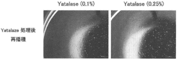
- Yatalase (Takara Bio) is a mixture containing chitinase, chitobiase, chitosanase, and ⁇ -1,3-glucanase, and can be suitably used in the recovery method of the present invention as a degrading enzyme of chitin or chitosan.
- the degrading enzyme of the water-insoluble polysaccharides is added, and the mixture is left for a time sufficient for detaching the adhesive cells, Incubate.
- a chelating agent (EDTA) and a proteolytic enzyme (trypsin, collagenase) are not necessary for the peeling. Therefore, in one embodiment, the chelating agent (EDTA) and / or a proteolytic enzyme (ED
- a water-insoluble polysaccharide degrading enzyme is added without adding trypsin or collagenase).
- washing for removing divalent ions and / or proteins in the medium in which the adherent cells are suspended for example, the concentration is 1/10 or less.
- a water-insoluble polysaccharide degrading enzyme is added to the medium.
- the incubation temperature of the water-insoluble polysaccharide with a degrading enzyme is usually 20 ° C. to 37 ° C.
- the incubation time is usually 5 to 60 minutes, although it depends on the type of enzyme.
- the detached adherent cells can be recovered by subjecting the suspension to centrifugation.
- the adhesive cells collected in this way can be suitably used for functional analysis, transplantation and the like because damage is suppressed to a minimum.
- the adhesive cells may be detached from the nanofibers and recovered by the recovery method of the present invention.
- the adhesive cells After the above continuous operation (for example, (a) to (l)) is performed, the adhesive cells may be detached from the nanofibers and recovered by the recovery method of the present invention.
- cultured human preadipocytes (subcutaneous, # CAS02s05a, manufactured by Toyobo Co., Ltd.) were suspended in each medium composition to 33333 cells / mL, and then a 96-well flat bottom ultra-low adhesion surface microplate. (Corning, # 3474) was seeded at 150 ⁇ L / well.
- the cells were cultured in a CO 2 incubator (37 ° C., 5% CO 2 ) in a stationary state for 10 days.
- 150 ⁇ L of ATP reagent CellTiter-Glo TM Luminescent Cell Viability Assay, Promega
- FlexStation 3 Molecular Luminescence intensity (RLU value) was measured by Devices, and the luminescence value of the medium alone was subtracted to calculate the number of viable cells (average value of 4 points).
- mesenchymal stem cell growth medium (C-28009, manufactured by Takara Bio Inc.), which is a low serum medium) w / v), 0.01% (w / v), 0.03% (w / v)), and a non-added medium composition not containing the substrate.
- cultured human adipose-derived mesenchymal stem cells (C-12777, manufactured by Takara Bio Inc.) were suspended in each medium composition so as to be 13333 cells / mL, and then a 96-well flat bottom ultra-low adhesion surface A microplate (Corning, # 3474) was seeded at 150 ⁇ L / well.
- the cells were cultured in a CO 2 incubator (37 ° C., 5% CO 2 ) in a stationary state for 10 days.
- ATP reagent CellTiter-Glo TM Luminescent Cell Viability Assay, Promega
- FlexStation 3 Molecular Luminescence intensity (RLU value) was measured by Devices, and the luminescence value of the medium alone was subtracted to calculate the number of viable cells (average value of 4 points).
- a medium composition (final concentration: 0.03% (w / v)) in which chitin nanofibers were added to human preadipocyte growth medium (# CAS11K500, manufactured by Toyobo Co., Ltd.) was prepared. Subsequently, cultured human preadipocytes (subcutaneous, # CAS02s05a, manufactured by Toyobo Co., Ltd.) were suspended in the above medium composition so as to be 33333 cells / mL, and then a 96-well flat bottom ultra-low adhesion surface microplate ( Corning, # 3474) was seeded at 150 ⁇ L / well. The cells were cultured in a CO 2 incubator (37 ° C., 5% CO 2 ) for 3 days, and allowed to adhere to the chitin nanofibers.
- Yatalase manufactured by Takara Bio Inc., T017 containing chitinolytic enzyme was added to each well so that the final concentration was 0.1% (w / v) or 0.25% (w / v). And left at 37 ° C. for about 1 hour. Thereafter, a 4-well cell suspension containing chitin nanofiber degradation products and human preadipocytes was collected in a 15 mL tube and centrifuged at 1500 rpm.
- human preadipocytes when human preadipocytes are cultured in a medium composition supplemented with chitin nanofibers, human preadipocytes adhere to chitin nanofibers, and when chitin nanofibers are digested using Yatarose, they adhere to chitin nanofibers. It was found that human preadipocytes that had been isolated can be isolated in a single cell-like manner. The photograph of the cell after Yatalase treatment is shown in FIG.
- a medium composition (final concentration: 0.03% (w / v)) in which chitin nanofibers were added to human preadipocyte growth medium (# CAS11K500, manufactured by Toyobo Co., Ltd.) was prepared. Subsequently, cultured human preadipocytes (subcutaneous, # CAS02s05a, manufactured by Toyobo Co., Ltd.) were suspended in the above medium composition to 33333 cells / mL, and a 96-well flat bottom ultra-low adhesion surface microplate (Corning) And # 3474) at 150 ⁇ L / well. The cells were cultured in a CO 2 incubator (37 ° C., 5% CO 2 ) for 3 days, and allowed to adhere to the chitin nanofibers.
- Yatalase manufactured by Takara Bio Inc., T017 containing chitinolytic enzyme was added to each well so that the final concentration was 0.1% (w / v) or 0.25% (w / v). And left at 37 ° C. for about 1 hour. Thereafter, a 4-well cell suspension containing chitin nanofiber degradation products and human preadipocytes was collected in a 15 mL tube and centrifuged at 1500 rpm.
- human preadipocyte growth medium # CAS11K500, manufactured by Toyobo Co., Ltd.
- a 96-well flat bottom microplate (Corning, # 3585) at 150 ⁇ L / well, and the degree of cell proliferation and induction of differentiation into adipocytes was evaluated.
- Cell proliferation was evaluated using the ATP value measurement method. After reseeding, the cells were cultured in a CO 2 incubator (37 ° C., 5% CO 2 ) in a stationary state for up to 7 days. 150 ⁇ L of ATP reagent (CellTiter-Glo TM Luminescent Cell Viability Assay, Promega) was added to the culture solution at 0, 3, and 7 days after re-seeding, and allowed to stand at room temperature for about 10 minutes, then FlexStation 3 (Molecular Luminescence intensity (RLU value) was measured by Devices, and the luminescence value of the medium alone was subtracted to calculate the number of viable cells (average value of 4 points).
- ATP reagent CellTiter-Glo TM Luminescent Cell Viability Assay, Promega
- adipocyte differentiation medium (# CAS11D250, manufactured by Toyobo) was replaced with 150 ⁇ L / well, and the cells were further replaced with a CO 2 incubator (37 ° C., 5 ° C.). % CO 2 ) and left to stand for 10 days.
- a reverse transcription reaction was performed on GeneAmp PCR System 9700 (Applied Biosystems) using PrimeScript TM RT Master Mix (manufactured by Takara Bio Inc.) to synthesize cDNA from the total RNA.
- Each of the obtained cDNA samples diluted 1/10 with sterilized water was used as a PCR template.
- the dispensed and mixed cDNA was used as a sample for preparing a calibration curve.
- the calibration curve was set in a quantitative range from 1/3 to 1/243 dilution at a 3-fold common ratio.
- PCR was carried out with 7500 Real Time PCR System (Applied Biosystems) using each cDNA sample, calibration sample, Premix Ex Taq TM (Takara Bio) and various Taqman probes (Applied Biosystems).
- PPIA cyclophilin B
- PPAR gamma mRNA or lipoprotein lipase (LPL) mRNA was corrected by the value of PPIA.
- the probes used (Applied Biosystems) are shown below.
- LPL lipoprotein lipase
- human preadipocytes when human preadipocytes are cultured in a medium composition supplemented with chitin nanofibers, human preadipocytes adhere to chitin nanofibers, and when chitin nanofibers are digested using Yatarose, they adhere to chitin nanofibers. It was found that human preadipocytes that had been isolated can be isolated in a single cell-like manner. Further, the isolated cells retained the ability to grow on a 96-well flat bottom microplate and the ability to differentiate into adipocytes. Table 3 shows RLU values (ATP measurement, luminescence intensity) relating to cell proliferation evaluation, and Tables 4 and 5 show mRNA expression values of PPAR gamma and LPL relating to adipocyte differentiation evaluation.
- RLU values ATP measurement, luminescence intensity
- a medium composition (final concentration: 0.03% (w / v)) in which chitin nanofibers were added to a human adipocyte differentiation medium (# CAS11D250, manufactured by Toyobo Co., Ltd.) was prepared.
- cultured human preadipocytes (subcutaneous, # CAS02s05a, manufactured by Toyobo Co., Ltd.) were suspended in the above medium composition to 33333 cells / mL, and a 96-well flat bottom ultra-low adhesion surface microplate (Corning) And # 3474) at 150 ⁇ L / well.
- the cells were cultured in a CO 2 incubator (37 ° C., 5% CO 2 ) in a stationary state for a maximum of 14 days.
- the evaluation of cell viability was performed using the ATP value measurement method.
- 150 ⁇ L of ATP reagent CellTiter-Glo TM Luminescent Cell Viability Assay, manufactured by Promega
- CellTiter-Glo TM Luminescent Cell Viability Assay manufactured by Promega
- FlexStation 3 Molecular Devices
- the luminescence intensity (RLU value) was measured by a company, and the luminescence value of the medium alone was subtracted to calculate the number of viable cells (average value of 4 points).
- RNA expression values of adipocyte markers were evaluated using mRNA expression values of adipocyte markers.
- Total RNA was extracted from the cell suspension using RNeasy Mini kit (manufactured by QIAGEN) at the time of seeding human preadipocytes into the medium composition (day 0) and on days 4, 8, and 14.
- a reverse transcription reaction was performed on GeneAmp PCR System 9700 (Applied Biosystems) using PrimeScript TM RT Master Mix (manufactured by Takara Bio Inc.) to synthesize cDNA from the total RNA.
- Each of the obtained cDNA samples diluted 1/10 with sterilized water was used as a PCR template.
- the dispensed and mixed cDNA was used as a sample for preparing a calibration curve.
- the calibration curve was set in a quantitative range from 1/3 to 1/243 dilution at a 3-fold common ratio.
- PCR was carried out with 7500 Real Time PCR System (Applied Biosystems) using each cDNA sample, calibration sample, Premix Ex Taq TM (Takara Bio) and various Taqman probes (Applied Biosystems). The probes used (Applied Biosystems) are shown below.
- PPIA HS99999994 PPAR gamma: HS011155134
- LPL HS00173425
- FABP4 HS01086177
- Human preadipocyte growth medium (# CAS11K500, manufactured by Toyobo) or 10% FBS (# 35 Medium composition (final concentration: 0.01% (w / v)) in which chitin nanofibers are added to DMEM medium (# 044-29765, manufactured by Wako Pure Chemical Industries, Ltd.) containing -015-CV (manufactured by Corning), and A non-added medium composition containing no chitin nanofibers was prepared.
- cultured human preadipocytes (subcutaneous, # CAS02s05a, manufactured by Toyobo Co., Ltd.) were suspended in each medium composition so as to be 90000 cells / mL, and then a 6-well flat bottom ultra-low adhesion surface microplate. (# 3471, Corning) was seeded at 5 mL / well.
- the cells were cultured in a CO 2 incubator (37 ° C., 5% CO 2 ) in a stationary state for 3 days.
- the chitin nanofibers with human preadipocytes attached were collected in a 50 mL tube, centrifuged, and then the human preadipocyte growth medium (# CAS11K500, Toyobo) Or DMEM medium (# 044-29765, manufactured by Wako Pure Chemical Industries) containing 10% FBS (# 35-015-CV, manufactured by Corning) was added and suspended, and a 6-well flat bottom ultra-low adhesion surface microplate (Corning) No. 3471) or 15 mL tube.
- human preadipocyte growth medium # CAS11K500, Toyobo
- DMEM medium # 044-29765, manufactured by Wako Pure Chemical Industries
- FBS # 35-015-CV
- the 6-well flat bottom ultra-low adhesion surface microplate was cultured in a CO 2 incubator (37 ° C., 5% CO 2 ) for 7 days in a stationary state, and the 15 mL tube was allowed to stand in a 25 ° C. incubator for 7 days.
- Human preadipocytes attached to chitin nanofibers maintained in a CO 2 incubator (37 ° C., 5% CO 2 ) or 25 ° C. incubator for 7 days are re-seeded in a 100 mm culture dish (# 430167, Corning) The cells were further cultured for 7 days in a CO 2 incubator (37 ° C., 5% CO 2 ), and human preadipocytes that migrated and proliferated from chitin nanofibers were observed with a microscope (FIG. 3). ).
- human preadipocytes and chitin nanofibers adhering to the dish were washed with PBS (-) (# 045-29795, manufactured by Wako Pure Chemical Industries), and then trypsin (# CA090K, Toyobo) Only human preadipocytes were collected.
- the collected human preadipocytes are added to human preadipocyte growth medium (# CAS11K500, manufactured by Toyobo Co., Ltd.) or DMEM medium (# 0429-7655, manufactured by Corning) containing 10% FBS (# 04-29765, Wako Jun).
- adipocyte differentiation medium # CA811D250, manufactured by Toyobo Co., Ltd.
- the culture was further continued for 17 days, and differentiation induction into adipocytes was observed with a microscope (FIG. 4).
- ADSC medium # C-28009, manufactured by Takara
- DMEM medium # 044-29765, manufactured by Wako Pure Chemical Industries
- FBS # 35-015-CV, manufactured by Corning
- cultured human adipose tissue-derived mesenchymal stem cells (# C-12977, manufactured by Takara) were suspended in each medium composition so as to be 90000 cells / mL, and then 6-well flat bottom ultra-low adhesion It seed
- the cells were cultured in a CO 2 incubator (37 ° C., 5% CO 2 ) in a stationary state for 3 days.
- the chitin nanofibers to which the human adipose-derived mesenchymal stem cells are adhered are collected in a 50 mL tube, and after centrifugation, a new ADSC medium (#C) is added to the chitin nanofibers to which the human adipose-derived mesenchymal stem cells are adhered.
- a new ADSC medium (#C) is added to the chitin nanofibers to which the human adipose-derived mesenchymal stem cells are adhered.
- -28009, manufactured by Takara) or DMEM medium (# 044-29765, manufactured by Wako Pure Chemical Industries) containing 10% FBS (# 35-015-CV, manufactured by Corning) was added and suspended, and a 6-well flat bottom ultra-low adhesion Transferred to a surface microplate (Corning, # 3471) or 15 mL tube.
- the 6-well flat bottom ultra-low adhesion surface microplate was incubated in
- Human fat-derived mesenchymal stem cells adhered to chitin nanofibers maintained in a CO 2 incubator (37 ° C., 5% CO 2 ) or 25 ° C. incubator for 7 days are cultured in a 100 mm culture dish (# 430167, Corning). Cultivated in a CO 2 incubator (37 ° C., 5% CO 2 ) for an additional 7 days, and migrated and proliferated from chitin nanofibers under a microscope with human adipose-derived mesenchymal stem cells was observed (FIG. 5).
- the medium was removed, and the human adipose-derived mesenchymal stem cells and chitin nanofibers adhered to the dish were washed with PBS ( ⁇ ) (# 045-29795, manufactured by Wako Pure Chemical Industries), and then trypsin (# C-41210 (manufactured by Takara) was added to collect only human adipose-derived mesenchymal stem cells.
- PBS ⁇
- trypsin # C-41210 (manufactured by Takara) was added to collect only human adipose-derived mesenchymal stem cells.
- the collected human adipose-derived mesenchymal stem cells were added to ADSC medium (# C-28009, manufactured by Takara) or DMEM medium (# 0429-7655, Japanese) containing 10% FBS (# 35-015-CV, manufactured by Corning).
- the suspension was suspended at 6 ml flat bottom ultra-low adhesion surface microplate (Corning, # 3471) at 5 mL / well and cultured for 2 days. After culturing for 2 days, the medium was changed to an adipocyte differentiation medium (# C-28016, manufactured by Takara), and the culture was further continued for 8 days, and differentiation induction into adipocytes was observed with a microscope (FIG. 6).
- adipocyte differentiation medium # C-28016, manufactured by Takara
- new cells can be transported in a state where they are attached to chitin nanofibers, re-seeded on the dish as it is, migrated and grown on the dish, and collected with trypsin, etc.
- Established transportation method The model is shown in FIG.
- chitosan nanofibers and ⁇ -chitin nanofibers were suspended in ultrapure water (Milli-Q water) to 1% (w / v), and then dissolved by stirring while heating at 90 ° C. The aqueous solution was autoclaved at 121 ° C. for 20 minutes.
- the nanofibers used were ⁇ -chitin nanofibers (sample 1) that were made into nanofibers at 200 MPa, pass 10 times, and ⁇ -chitin nanofibers (sample 2) that were made into nanofibers at 200 MPa, pass once.
- Example 3 Chitosan nanofiber (sample 3) nanofiberized under defibration conditions of 200 MPa, pass 10 times, and ⁇ -chitin nanofiber (sample 4) nanofiberized under conditions of defibration of 200 MPa, pass 10 times.
- a medium composition was prepared by adding each nanofiber sample (1 to 4) having a final concentration of 0.03% (w / v) to serum-free medium KBM220 medium (manufactured by Kojin Bio).
- the cultured canine kidney tubular epithelial cell line MDCK (manufactured by DS Pharma Biomedical) was seeded in a medium composition to which each of the above nanofiber samples (1 to 4) was added so as to be 66666 cells / mL. Then, it dispensed so that it might become 30 mL to a 125 mL flask (Thermo Scientific, 4115-0125). Each flask was cultured in a CO 2 incubator (37 ° C., 5% CO 2 ) under shaking (cfg 50 rpm) and continued for 6 days.
- a CO 2 incubator 37 ° C., 5% CO 2
- shaking cfg 50 rpm
- each nanofiber sample (1 to 4) and MDCK cells adhering to the nanofiber were collected in a 50 mL tube, and after centrifugation (cfg 1000 rpm, 3 minutes), only the medium was removed. After each new nanofiber sample and MDCK cells were suspended by adding new KBM220 medium, the whole suspension was returned to the original 125 mL flask, and the culture was continued in a shaking state (cfg 50 rpm). The above medium exchange operation was also performed on the 9th and 13th days.
- the culture solution at 0, 6, 13, and 17 days is suspended by pipetting, and 100 ⁇ L of ATP reagent (CellTiter-Glo TM Luminescent Cell Viability Assay, manufactured by Promega) is added to and reacted with 100 ⁇ L of the cell suspension. Then, after standing at room temperature for about 10 minutes, the luminescence intensity (RLU value) was measured with FlexStation 3 (Molecular Devices), and the number of viable cells was measured by subtracting the luminescence value of the medium alone as the average value of 4 points. .
- ATP reagent CellTiter-Glo TM Luminescent Cell Viability Assay, manufactured by Promega
- the ⁇ -chitin nanofibers used were 200MPa, ⁇ -chitin nanofibers converted to nanofibers under 10 pass conditions (sample 1), ⁇ -chitin nanofibers converted to nanofibers under 200MPa, pass 1-time conditions (sample 2), 100MPa, ⁇ -chitin nanofibers (sample 5) nanofiberized with 1 pass condition, ⁇ -chitin nanofibers (sample 6) nanofiberized with 200 MPa, pass 3 conditions, ⁇ nanofiberized with 100 MPa, pass 5 times condition Chitin nanofiber (sample 7), ⁇ -chitin raw material (sample 8).
- a non-added medium composition (Sample 9) was prepared.
- Each flask was cultured in a CO 2 incubator (37 ° C., 5% CO 2 ) under shaking (cfg 50 rpm) and continued for 7 days.
- the culture solution on days 0, 3, and 7 is suspended by pipetting, and 100 ⁇ L of the cell suspension is added with 100 ⁇ L of ATP reagent (CellTiter-Glo TM Luminescent Cell Viability Assay, manufactured by Promega) and reacted.
- the luminescence intensity (RLU value) was measured with FlexStation 3 (Molecular Devices), and the number of viable cells was measured by subtracting the luminescence value of the medium alone as the average value of 4 points.
- the ⁇ -chitin nanofibers used were 200MPa, nanofibers made with nanofibers under 10 pass conditions (sample 1), ⁇ chitin nanofibers made with nanofibers under 200MPa, pass once conditions (sample 2), 200MPa, pass ⁇ -chitin nanofiber (sample 10) nanofiberized under 5 conditions.
- cultured human bone marrow-derived mesenchymal stem cells (C-12974, manufactured by Takara Bio Inc.) were seeded in the above-described medium composition to which chitin nanofibers were added or to an unadded medium composition at 20000 cells / mL. Then, it dispensed so that it might become 150 microliters per well to the well of 96 well flat bottom ultra-low adhesion surface microplate (Corning company make, # 3474). Each plate was cultured in a CO 2 incubator (37 ° C., 5% CO 2 ) in a stationary state and continued for 10 days.
- CO 2 incubator 37 ° C., 5% CO 2
- ATP reagent CellTiter-Glo TM Luminescent Cell Viability Assay, manufactured by Promega
- FlexStation 3 Molecular The intensity of luminescence (RLU value) was measured by Devices, and the number of viable cells was measured by subtracting the luminescence value of the medium alone as an average value of 4 points.
- the ⁇ -chitin nanofibers used were 200MPa, ⁇ -chitin nanofibers that were converted to nanofibers under one pass condition (sample 2), ⁇ -chitin nanofibers that were converted into nanofibers under 200MPa, pass 20 times conditions (sample 11), 200MPa, ⁇ -chitin nanofibers (sample 12) nanofiberized with 50 pass conditions, 200MPa, ⁇ -chitin nanofibers nanosampled with 100 pass conditions (sample 13), ⁇ ficinized nanofiber with 200MPa, pass 200 conditions It is chitin nanofiber (sample 14).
- each nanofiber sample (2, 11-14) having a final concentration of 0.006% (w / v), 0.02%, and 0.06% was added to serum-free medium KBM220 medium (manufactured by Kojin Bio) And a non-added medium composition (sample 9) containing no substrate.
- the canine kidney tubular epithelial cell line MDCK (DS Pharma Biomedical) was seeded in the above-described medium composition with or without chitin nanofiber added to 33333 cells / mL.
- a well flat bottom ultra-low adhesion surface microplate (Corning, # 3474) was dispensed at 150 ⁇ L per well.
- Each plate was cultured in a stationary state in a CO 2 incubator (37 ° C., 5% CO 2 ) and continued for 6 days.
- 150 ⁇ L of ATP reagent CellTiter-Glo TM Luminescent Cell Viability Assay, manufactured by Promega
- FlexStation 3 Molecular Devices
- Luminescence intensity RLU value
- sample 11 was higher than sample 2.
- the sample (12-14) with an increased defibration showed only a weaker promoting action.
- Table 13 shows the RLU values (ATP measurement, luminescence intensity) in each culture.
- the ⁇ -chitin nanofibers used were ⁇ -chitin nanofibers (sample 15) that were converted to nanofibers at 200 MPa, pass 5 times, ⁇ -chitin nanofibers (sample 11) that were converted to nanofibers at 200 MPa, pass 20 times, 200 MPa, ⁇ -chitin nanofibers (sample 12) nanofiberized with 50 pass conditions, 200MPa, ⁇ -chitin nanofibers nanosampled with 100 pass conditions (sample 13), ⁇ ficinized nanofiber with 200MPa, pass 200 conditions It is chitin nanofiber (sample 14).
- cultured human adipose-derived mesenchymal stem cells (C-12977, manufactured by Takara Bio Inc.) were suspended in each medium composition so as to be 13333 cells / mL, and then a 96-well flat bottom ultra-low adhesion surface A microplate (Corning, # 3474) was seeded at 150 ⁇ L / well.
- the cells were cultured in a CO 2 incubator (37 ° C., 5% CO 2 ) in a stationary state for 10 days.
- ATP reagent CellTiter-Glo TM Luminescent Cell Viability Assay, manufactured by Promega
- FlexStation 3 Molecular Luminescence intensity (RLU value) was measured by Devices, and the luminescence value of the medium alone was subtracted to calculate the number of viable cells (average value of 4 points).
- Test Examples 8 to 13 The physical properties (measured concentration, viscosity, median particle size, average particle size) of ⁇ -chitin dispersions prepared under various defibrating conditions (pressure and number of pulverizations) used in Test Examples 8 to 13 are as follows. Table 16 shows.
- Nanofibers consisting of water-insoluble polysaccharides are i) culture, ii) induction of differentiation, iii) transport and storage under non-freezing conditions, iv) transplantation, mesenchymal stem cells, preadipocytes, etc. v) Since it can be used as a common carrier in various operations such as recovery of physiologically active substances from the culture supernatant, by using the carrier of the present invention, the above-mentioned i) culture, ii) differentiation induction, iii A plurality of operations selected from a) transport and storage under non-freezing conditions, iv) transplantation, and v) recovery of a physiologically active substance from the culture supernatant can be performed continuously.
Landscapes
- Health & Medical Sciences (AREA)
- Life Sciences & Earth Sciences (AREA)
- Engineering & Computer Science (AREA)
- Chemical & Material Sciences (AREA)
- Biomedical Technology (AREA)
- General Health & Medical Sciences (AREA)
- Zoology (AREA)
- Organic Chemistry (AREA)
- Bioinformatics & Cheminformatics (AREA)
- Veterinary Medicine (AREA)
- Animal Behavior & Ethology (AREA)
- Public Health (AREA)
- Medicinal Chemistry (AREA)
- Wood Science & Technology (AREA)
- Epidemiology (AREA)
- Genetics & Genomics (AREA)
- Biotechnology (AREA)
- Cell Biology (AREA)
- Dermatology (AREA)
- Oral & Maxillofacial Surgery (AREA)
- Transplantation (AREA)
- Developmental Biology & Embryology (AREA)
- Biochemistry (AREA)
- General Engineering & Computer Science (AREA)
- Microbiology (AREA)
- Rheumatology (AREA)
- Chemical Kinetics & Catalysis (AREA)
- Immunology (AREA)
- Hematology (AREA)
- Pharmacology & Pharmacy (AREA)
- Botany (AREA)
- Dispersion Chemistry (AREA)
- Urology & Nephrology (AREA)
- Materials Engineering (AREA)
- General Chemical & Material Sciences (AREA)
- Nuclear Medicine, Radiotherapy & Molecular Imaging (AREA)
- Composite Materials (AREA)
- Virology (AREA)
- Sustainable Development (AREA)
- Micro-Organisms Or Cultivation Processes Thereof (AREA)
Abstract
Description
本発明者らは、これらの知見に基づき、さらに検討を進め、本発明を完成した。 As a result of intensive efforts to solve the above-mentioned problems, the present inventors have found that an insoluble in medium, floating in the medium, having an activity of adhering cells, biodegradable, and proteolytic enzyme It has been found that the above-described various problems can be solved once by using nanofibers made of polysaccharides such as chitin, which can be digested by enzyme without necessity, as a carrier. By mixing nanofibers composed of polysaccharides in a liquid medium and culturing adherent cells such as mesenchymal stem cells and preadipocytes attached to the nanofibers, the cells float in a stationary state. It was possible to culture. Under suspension culture conditions, the cells attached to the nanofibers showed long-term viability. Mesenchymal stem cells and preadipocytes grew well on nanofibers while maintaining their traits. When cultured in a differentiation-inducing medium, the preadipocytes adhering onto the nanofibers differentiated into adipocytes, and it was also found that cell differentiation on the nanofibers can be induced. By treating the polysaccharides (eg, chitin) that make up the nanofibers, cells that adhere to the nanofibers can be treated without using proteolytic enzymes while minimizing damage to the cells. It was possible to recover. Since nanofibers made of polysaccharides are biodegradable, cells adhered to the nanofibers can be formed into a sheet form that can be easily transplanted together with the nanofibers while still attached. Since the cells adhered to the nanofibers easily settled by centrifugation or the like, the culture supernatant could be easily recovered from the culture of the cells adhered to the nanofibers. That is, nanofibers composed of polysaccharides are required for mesenchymal stem cells, preadipocytes, primary cells, etc., for cell transport, culture, differentiation induction, transplantation, and production of physiologically active substances under non-freezing conditions. It was found that a plurality of operations selected from the group consisting of cell culture, differentiation induction, cell transport, transplantation, and bioactive substance production can be continuously performed by using this carrier.
Based on these findings, the present inventors have further studied and completed the present invention.
[1]非水溶性多糖類からなるナノファイバーを含む、接着性細胞を、以下の(1)~(3)からなる群から選択される2以上の処理に連続的に供するためのキャリア:
(1)浮遊培養、
(2)非凍結状態での保存及び/又は輸送、並びに
(3)移植。
[2]浮遊培養が、接着性細胞の維持又は増殖、接着性細胞の分化誘導、及び/又は接着性細胞による液性因子生産のためのものである、[1]に記載のキャリア。
[3]接着性細胞を、(1)~(3)の処理に連続的に供するためのものである、[1]又は[2]に記載のキャリア。
[4]接着性細胞が、幹細胞又は前駆細胞である、[1]~[3]のいずれかに記載のキャリア。
[5]接着性細胞が、間葉系幹細胞又は前駆脂肪細胞である、[1]~[3]のいずれかに記載のキャリア。
[6]非水溶性多糖類が、キチン又はキトサンである、[1]~[5]のいずれかに記載のキャリア。
[7]接着性細胞を、非水溶性多糖類からなるナノファイバーに付着した状態で、以下の(1)~(3)からなる群から選択される2以上の処理に連続的に供することを含む、接着性細胞の処理方法:
(1)浮遊培養、
(2)非凍結状態での保存及び/又は輸送、並びに
(3)移植。
[8]浮遊培養が、接着性細胞の維持又は増殖、接着性細胞の分化誘導、及び/又は接着性細胞による液性因子生産のためのものである、[7]に記載の方法。
[9]接着性細胞を、(1)~(3)の処理に連続的に供する、[7]又は[8]に記載の方法。
[10]接着性細胞が、幹細胞又は前駆細胞である、[7]~[9]のいずれかに記載の方法。
[11]接着性細胞が、間葉系幹細胞又は前駆脂肪細胞である、[7]~[9]のいずれかに記載の方法。
[12]非水溶性多糖類が、キチン又はキトサンである、[7]~[11]のいずれかに記載の方法。
[13]非水溶性多糖類からなるナノファイバーに付着した接着性細胞を、該非水溶性多糖類の分解酵素で処理することにより該ナノファイバーを分解し、該接着性細胞を該ナノファイバーから剥離し、該接着性細胞を回収することを更に含む、[7]~[12]のいずれかに記載の方法。
[14]非水溶性多糖類からなるナノファイバーに付着した接着性細胞を、該非水溶性多糖類の分解酵素で処理することにより該ナノファイバーを分解し、該接着性細胞を該ナノファイバーから剥離することを含む、非水溶性多糖類からなるナノファイバーに付着した接着性細胞から該接着性細胞を回収する方法。
[15]非水溶性多糖類が、キチン又はキトサンである、[14]に記載の方法。
[16]分解酵素が、キチナーゼ、キトビアーゼ、キトサナーゼ又はβ-1,3-グルカナーゼである、[15]に記載の方法。 That is, the present invention is as follows.
[1] A carrier for continuously subjecting adherent cells containing nanofibers composed of a water-insoluble polysaccharide to two or more treatments selected from the group consisting of the following (1) to (3):
(1) suspension culture,
(2) Non-frozen storage and / or transport, and (3) transplantation.
[2] The carrier according to [1], wherein the suspension culture is for maintenance or proliferation of adherent cells, induction of differentiation of adherent cells, and / or production of humoral factors by adherent cells.
[3] The carrier according to [1] or [2], which is used for continuously subjecting the adherent cells to the treatments (1) to (3).
[4] The carrier according to any one of [1] to [3], wherein the adherent cells are stem cells or progenitor cells.
[5] The carrier according to any one of [1] to [3], wherein the adherent cells are mesenchymal stem cells or preadipocytes.
[6] The carrier according to any one of [1] to [5], wherein the water-insoluble polysaccharide is chitin or chitosan.
[7] Continuously subjecting the adherent cells to two or more treatments selected from the group consisting of the following (1) to (3) while attached to nanofibers composed of a water-insoluble polysaccharide. Including adherent cell processing methods:
(1) suspension culture,
(2) Non-frozen storage and / or transport, and (3) transplantation.
[8] The method according to [7], wherein the suspension culture is for maintenance or growth of adherent cells, induction of adherent cell differentiation, and / or humoral factor production by adherent cells.
[9] The method according to [7] or [8], wherein the adherent cells are continuously subjected to the treatments (1) to (3).
[10] The method according to any one of [7] to [9], wherein the adherent cells are stem cells or progenitor cells.
[11] The method according to any one of [7] to [9], wherein the adherent cells are mesenchymal stem cells or preadipocytes.
[12] The method according to any one of [7] to [11], wherein the water-insoluble polysaccharide is chitin or chitosan.
[13] The adherent cells attached to the nanofibers composed of the water-insoluble polysaccharide are treated with a degrading enzyme of the water-insoluble polysaccharide to decompose the nanofibers, and the adherent cells are detached from the nanofibers. The method according to any one of [7] to [12], further comprising recovering the adherent cells.
[14] The adherent cells attached to the nanofibers composed of the water-insoluble polysaccharide are treated with a degrading enzyme of the water-insoluble polysaccharide to decompose the nanofibers, and the adherent cells are detached from the nanofibers. A method for recovering the adhesive cells from the adhesive cells attached to the nanofibers comprising a water-insoluble polysaccharide.
[15] The method according to [14], wherein the water-insoluble polysaccharide is chitin or chitosan.
[16] The method according to [15], wherein the degrading enzyme is chitinase, chitobiase, chitosanase or β-1,3-glucanase.
細胞とは、動物或いは植物を構成する最も基本的な単位であり、その要素として細胞膜の内部に細胞質と各種の細胞小器官をもつものである。この際、DNAを内包する核は、細胞内部に含まれても含まれなくてもよい。 [Adherent cells]
A cell is the most basic unit constituting an animal or a plant, and has a cytoplasm and various organelles inside a cell membrane as its elements. At this time, the nucleus containing DNA may or may not be contained inside the cell.
本発明において使用するナノファイバーは、液体培地中に分散し、ナノファイバーに付着した細胞(好ましくは、接着性細胞)を、該液体培地中に浮遊させる効果を示すものである。 [Nanofiber]
The nanofibers used in the present invention exhibit an effect of allowing cells (preferably adherent cells) dispersed in a liquid medium and suspended in the liquid medium to float in the liquid medium.
本発明において、接着性細胞を非水溶性多糖類からなるナノファイバーに付着した状態で、該細胞を浮遊培養、或いは非凍結状態での保存及び/又は輸送に供する場合、該ナノファイバーを含む液状の培地組成物(本明細書において、本発明の培地組成物ともいう。)が使用される。 [Medium composition containing nanofibers]
In the present invention, when the adherent cells are attached to nanofibers composed of a water-insoluble polysaccharide and the cells are subjected to suspension culture or storage and / or transport in a non-frozen state, the liquid containing the nanofibers is used. The medium composition (also referred to herein as the medium composition of the present invention) is used.
本発明は、非水溶性多糖類からなるナノファイバーの、接着性細胞の多様な培養方法論におけるキャリアとしての使用を提供する。接着性細胞を非水溶性多糖類からなるナノファイバーに付着した状態で、(1)浮遊培養、(2)非凍結状態での保存及び/又は輸送、(3)移植等の様々な処理に供することができる。従って、本発明は、接着性細胞を、非水溶性多糖類からなるナノファイバーに付着した状態で、(1)浮遊培養、(2)非凍結状態での保存及び/又は輸送、(3)移植等の処理に供することを含む、接着性細胞の処理方法とも捉えることができる。 [Treatment of adherent cells using nanofibers composed of water-insoluble polysaccharides]
The present invention provides the use of nanofibers composed of water-insoluble polysaccharides as carriers in various culture methodologies for adherent cells. Adhesive cells are attached to nanofibers composed of water-insoluble polysaccharides, and are subjected to various treatments such as (1) suspension culture, (2) storage and / or transport in a non-frozen state, and (3) transplantation. be able to. Therefore, the present invention provides (1) suspension culture, (2) non-freezing storage and / or transport, (3) transplantation, with adherent cells attached to nanofibers made of a water-insoluble polysaccharide. It can also be regarded as a method for treating adherent cells, including subjecting to treatment such as the above.
接着性細胞を非水溶性多糖類からなるナノファイバーに付着した状態で培養することにより、接着性細胞を浮遊培養することができる。該浮遊培養は、上記本発明の培地組成物中で、接着性細胞を培養することにより実施することができる。非水溶性多糖類からなるナノファイバーは、該ナノファイバーに付着した細胞を培地中で浮遊させる効果(好ましくは浮遊静置させる効果)を示す。本発明の培地組成物において、非水溶性多糖類からなるナノファイバーは、均一に分散するので、該培地組成物中で接着性細胞を培養すると、接着性細胞は該ナノファイバーに付着し、該培地組成物中に浮遊する。当該浮遊効果により、単層培養に比べて、一定体積あたりの細胞数を増やして培養することが可能である。また、従来の回転や振とう操作を伴う浮遊培養においては、細胞に対するせん断力が働くため、細胞の増殖率や回収率が低い、或いは細胞の機能が損なわれてしまう場合があるが、本発明の培地組成物を用いることにより振とう等の操作を要することなく細胞を分散した状態で培養し得るので、目的とする接着性細胞を細胞機能の損失無く容易かつ大量に浮遊培養することが期待できる。また、従来のゲル基材を含む培地において細胞を浮遊培養する際、細胞の観察や回収が困難であったり、回収の際にその機能を損なったりする場合があるが、本発明の培地組成物を用いることにより、浮遊培養した細胞を、その機能を損なうこと無く観察し、回収することが期待できる。また、従来のゲル基材を含む培地は、粘度が高く培地の交換が困難である場合があるが、本発明の培地組成物は、低粘度であるためピペットやポンプ等を用いて容易に培地を交換することが期待できる。 (1) Suspension culture By culturing adherent cells in a state where they adhere to nanofibers made of a water-insoluble polysaccharide, the adherent cells can be suspended. The suspension culture can be carried out by culturing adherent cells in the medium composition of the present invention. Nanofibers composed of a water-insoluble polysaccharide exhibit an effect of suspending cells attached to the nanofiber in a medium (preferably an effect of floating and standing). In the medium composition of the present invention, nanofibers composed of water-insoluble polysaccharides are uniformly dispersed. Therefore, when adherent cells are cultured in the medium composition, the adherent cells adhere to the nanofibers, and It floats in the medium composition. Due to the floating effect, it is possible to increase the number of cells per fixed volume as compared with the monolayer culture. In addition, in the conventional suspension culture with rotation and shaking operations, since shear force acts on the cells, the cell growth rate and recovery rate may be low, or the function of the cells may be impaired. By using this medium composition, cells can be cultured in a dispersed state without the need for shaking or the like, so that the desired adherent cells are expected to be easily and in large quantities suspended without loss of cell function. it can. In addition, when cells are suspended and cultured in a medium containing a conventional gel base material, it may be difficult to observe or recover the cells, or the function may be impaired during the recovery. It is expected that cells cultured in suspension can be observed and recovered without impairing their functions. In addition, a medium containing a conventional gel substrate has a high viscosity and it may be difficult to exchange the medium. However, since the medium composition of the present invention has a low viscosity, the medium can be easily used with a pipette or a pump. Can be expected to replace.
接着性細胞を、非水溶性多糖類からなるナノファイバーに付着した状態で、浮遊培養すると、接着性細胞が効率よく増殖するため、該浮遊培養は、接着性細胞の維持又は増殖方法として優れている。接着性細胞を、非水溶性多糖類からなるナノファイバーに付着した状態で、浮遊培養すると、接着性細胞は、培養容器の底面のみに偏在せずに、三次元的な広がりをもって分散し、増殖が促進される。特に、ナノファイバーとしてキチンナノファイバーを用いると、接着性細胞がキチンナノファイバーに付着し、そこを足場として強力に増殖し、その結果、増殖した細胞が、ぶどうの房状にナノファイバー上に連なる状態となる。この増殖促進効果には、接着性細胞を浮遊させる(即ち、接着性細胞の培養容器への接着を回避する)のに十分な濃度のナノファイバーが培地組成物中に含まれていればよく、浮遊静置(即ち、外部からの圧力、振動、振とう、回転操作等を伴わずに細胞が液体培地組成物中で均一に分散し尚且つ浮遊状態にあること)が可能であることは必須ではない。例えば、キチンナノファイバーの場合、浮遊作用発現に十分な0.0001%(重量/容量)以上の濃度であれば、安定した浮遊静置培養を可能にする0.03%(重量/容量)を下回る濃度(例、0.025%(重量/容量)以下、0.02%(重量/容量)以下)であっても、増殖促進効果が奏される。また、接着性細胞を、非水溶性多糖類からなるナノファイバーに付着した状態で、浮遊培養すると、該細胞を培養容器の底面へ接着させて単層培養した場合よりも、高い密度で維持し増殖させることができる。 (1-1) Maintenance or growth of adherent cells Adhesive cells proliferate efficiently when suspended in a state where they are attached to nanofibers composed of a water-insoluble polysaccharide. It is an excellent method for maintaining or growing adherent cells. When adhering cells are attached to nanofibers made of water-insoluble polysaccharides and suspended in culture, the adhering cells are not unevenly distributed only on the bottom of the culture vessel, but spread and spread in a three-dimensional manner. Is promoted. In particular, when chitin nanofibers are used as nanofibers, adherent cells adhere to chitin nanofibers and grow strongly as a scaffold, and as a result, the proliferated cells are connected to nanofibers in the shape of grape tufts. It becomes a state. For this growth promoting effect, it is sufficient that the medium composition contains nanofibers at a concentration sufficient to float the adherent cells (that is, avoid adhesion of the adherent cells to the culture vessel) It is essential to be able to float and float (ie, the cells are evenly dispersed and suspended in the liquid medium composition without external pressure, vibration, shaking, rotation, etc.) is not. For example, in the case of chitin nanofibers, if the concentration is 0.0001% (weight / volume) or more sufficient for expression of floating action, 0.03% (weight / volume) that enables stable floating stationary culture is set. Even if the concentration is lower (eg, 0.025% (weight / volume) or less, 0.02% (weight / volume) or less), the growth promoting effect is exhibited. In addition, when suspended cells are attached to nanofibers composed of water-insoluble polysaccharides, adherent cells are maintained at a higher density than when the cells are attached to the bottom of the culture vessel and cultured in a single layer. Can be propagated.
接着性細胞を、非水溶性多糖類からなるナノファイバーに付着した状態で、浮遊培養する際に、該浮遊培養を適切な分化誘導条件下で行うことにより、該接着性細胞の所望の細胞への分化を誘導することができる。上述の通り、接着性細胞を、非水溶性多糖類からなるナノファイバーに付着した状態で、浮遊培養すると、その増殖が促進されるので、該浮遊培養を適切な分化誘導条件下で行うことにより、該接着細胞の所望の細胞への良好な分化誘導が期待できる。 (1-2) Differentiation induction of adherent cells When suspension cells are attached to nanofibers composed of water-insoluble polysaccharides and suspended in culture, the suspension culture is performed under appropriate differentiation-inducing conditions. Thus, differentiation of the adherent cells into desired cells can be induced. As described above, when suspension cells are attached to nanofibers made of water-insoluble polysaccharides, the growth of the adherent cells is promoted. Therefore, by performing the suspension culture under appropriate differentiation-inducing conditions, Thus, good differentiation induction of the adherent cells into desired cells can be expected.
・間葉系幹細胞の脂肪細胞への分化:イソブチルメチルキサンチン(IBMX)、デキサメタゾン、インスリン及びインドメタシンを、脂肪細胞分化誘導因子として使用し、これらの因子の存在下で間葉系幹細胞を培養する。
・間葉系幹細胞の骨細胞への分化:デキサメタゾン、アスコルビン酸及びβグリセロリン酸を、骨細胞分化誘導因子として使用し、これらの因子の存在下で間葉系幹細胞を培養する。
・間葉系幹細胞の軟骨細胞への分化:インスリン、TGF-β3及びアスコルビン酸を、軟骨細胞分化誘導因子として使用し、これらの因子の存在下で間葉系幹細胞を培養する。
間葉系幹細胞の脂肪細胞分化、骨細胞分化又は軟骨細胞分化の条件については、例えば、以下の論文に記載されている。
[1] da Silva Meirelles L, Caplan AI, NardiNB., Stem Cells 2008; 26(9):2287-99.
[2] Crisan M, Yap S, CasteillaL, et al., Cell Stem Cell 2008; (3):301-13.
[3] Dominici M, Le Blanc K, Mueller I, Slaper-Cortenbach I, et al., Cytother2006; 8(4):315-7.
[4] Caplan AI., Cell Stem Cell 2008; 3(3):229-30. The differentiation induction conditions that can be applied to the present invention are not particularly limited, and examples thereof include the following.
Differentiation of mesenchymal stem cells into adipocytes: Mesenchymal stem cells are cultured in the presence of these factors using isobutylmethylxanthine (IBMX), dexamethasone, insulin and indomethacin as adipocyte differentiation inducing factors.
-Differentiation of mesenchymal stem cells into bone cells: Dexamethasone, ascorbic acid and β-glycerophosphate are used as bone cell differentiation-inducing factors, and mesenchymal stem cells are cultured in the presence of these factors.
Differentiation of mesenchymal stem cells into chondrocytes: Insulin, TGF-β3 and ascorbic acid are used as chondrocyte differentiation inducing factors, and mesenchymal stem cells are cultured in the presence of these factors.
The conditions for adipocyte differentiation, bone cell differentiation or chondrocyte differentiation of mesenchymal stem cells are described in, for example, the following papers.
[1] da Silva Meirelles L, Caplan AI, NardiNB., Stem Cells 2008; 26 (9): 2287-99.
[2] Crisan M, Yap S, CasteillaL, et al., Cell Stem Cell 2008; (3): 301-13.
[3] Dominici M, Le Blanc K, Mueller I, Slaper-Cortenbach I, et al., Cytother2006; 8 (4): 315-7.
[4] Caplan AI., Cell Stem Cell 2008; 3 (3): 229-30.
前駆脂肪細胞の脂肪細胞への分化の条件については、例えば、以下の論文に記載されている。
[5]Hutley LJ, Newell FM, et al., Eur J ClinInvest. 2003; 33(7): 574-81.
[6]Yin Y, Yuan H, et al.,Mol Endocrinol.2006 ;20(2):268-78.
[7]Rival Y, StennevinA, et al., J Pharmacol Exp Ther. 2004 ;311(2): 467-75. Differentiation of preadipocytes into adipocytes: isobutylmethylxanthine (IBMX), dexamethasone, insulin and indomethacin are used as adipocyte differentiation inducing factors, and the preadipocytes are cultured in the presence of these factors.
The conditions for differentiation of preadipocytes into adipocytes are described, for example, in the following papers.
[5] Hutley LJ, Newell FM, et al., Eur J ClinInvest. 2003; 33 (7): 574-81.
[6] Yin Y, Yuan H, et al., Mol Endocrinol. 2006; 20 (2): 268-78.
[7] Rival Y, StennevinA, et al., J Pharmacol Exp Ther. 2004; 311 (2): 467-75.
・前駆脂肪細胞から骨細胞への分化:
[8]Shirakawa K, Maeda S, et al.,Mol Cell Biol. 2006;26(16):6105-16. In addition, the following differentiation induction conditions are known and applicable to the present invention.
-Differentiation of preadipocytes into bone cells:
[8] Shirakawa K, Maeda S, et al., Mol Cell Biol. 2006; 26 (16): 6105-16.
接着性細胞を、非水溶性多糖類からなるナノファイバーに付着した状態で、浮遊培養すると、高い密度で接着性細胞を培養することができ、また接着性細胞を効率よく増殖することが可能なので、当該浮遊培養は、インビトロ細胞培養による液性因子の生産に有用である。所望の液性因子を産生する接着性細胞を、非水溶性多糖類からなるナノファイバーに付着した状態で、浮遊培養に付し、培養物(例、培養上清)から、目的とする液性因子を単離することにより、当該液性因子を得ることが出来る。液性因子としては、抗体、酵素(ウロキナーゼ等)、ホルモン(インシュリン等)、サイトカイン(インターフェロン、インターロイキン、腫瘍壊死因子、コロニー刺激因子、成長因子等)、ワクチンの抗原、その他の生理活性物質(タンパク質、ペプチド等)を挙げることができるが、これらに限定されない。液性因子を産生する細胞には、皮膚細胞、軟骨細胞、肝細胞、膵臓細胞、腎細胞、間葉系幹細胞、脂肪細胞等の非形質転換細胞や、当該液性因子をコードする遺伝子や有用物質の生合成に関与する遺伝子を導入した形質転換細胞が含まれる。所望の液性因子を産生する細胞は、好適には、当該液性因子を細胞外へ分泌する細胞である。液性因子を産生する細胞としては、具体的には、当該液性因子をコードする遺伝子や当該液性因子の生合成に関与する遺伝子を導入した、HEK293,CHO-K1、BHK-21、MDCK、Vero、HepG2、MCF-7等を挙げることができるが、これらに限定されない。組み換えタンパク質等の液性因子の生産に使用される細胞は当業者に周知であり、これらの細胞を本発明の方法において用いることが出来る。培養スケールの拡大は、上記(1-1)に記載した接着性細胞の維持又は蔵書億方法を用いて行ってもよい。液性因子を培養物から単離するにあたり、培養物から細胞を除く必要があるが、上記浮遊培養を用いると、また接着性細胞が非水溶性多糖類からなるナノファイバーに付着した状態で培地組成物中に浮遊しているので、遠心分離やろ過処理等の簡便な方法で細胞を除去することができる。また、培地組成物中のナノファイバーも、遠心分離やろ過処理等の簡便な方法で除去することができる。液性因子を培養物から単離する方法は、当業者に周知であり、例えばクロマトグラフィー(例、イオン交換クロマトグラフィー、疎水性クロマトグラフィー、アフィニティークロマトグラフィー、逆相クロマトグラフィー等のクロマトグラフィー)等の、生理活性物質の生化学的な分離精製方法を適用可能である。 (1-3) Production of humoral factors by adherent cells Adhesive cells can be cultured at a high density by suspension culture while attached to nanofibers made of water-insoluble polysaccharides. In addition, since the adherent cells can be efficiently propagated, the suspension culture is useful for production of humoral factors by in vitro cell culture. Adhesive cells that produce the desired humoral factor are subjected to suspension culture while attached to nanofibers composed of water-insoluble polysaccharides, and the desired humoral properties are obtained from the culture (eg, culture supernatant). By isolating the factor, the humoral factor can be obtained. Humoral factors include antibodies, enzymes (such as urokinase), hormones (such as insulin), cytokines (such as interferon, interleukin, tumor necrosis factor, colony stimulating factor, growth factor), vaccine antigens, and other physiologically active substances ( Protein, peptide, etc.), but is not limited thereto. Cells that produce humoral factors include non-transformed cells such as skin cells, chondrocytes, hepatocytes, pancreatic cells, kidney cells, mesenchymal stem cells, adipocytes, genes encoding the humoral factors, and useful Transformed cells into which genes involved in the biosynthesis of substances have been introduced are included. The cell that produces the desired humoral factor is preferably a cell that secretes the humoral factor out of the cell. Specific examples of cells that produce humoral factors include HEK293, CHO-K1, BHK-21, MDCK into which genes encoding the humoral factors and genes involved in the biosynthesis of the humoral factors have been introduced. , Vero, HepG2, MCF-7 and the like, but are not limited thereto. Cells used for the production of humoral factors such as recombinant proteins are well known to those skilled in the art, and these cells can be used in the method of the present invention. The expansion of the culture scale may be performed using the adhesion cell maintenance or collection method described in (1-1) above. When isolating the humoral factor from the culture, it is necessary to remove the cells from the culture. However, when the above suspension culture is used, the medium is also attached with the adherent cells attached to the nanofibers made of a water-insoluble polysaccharide. Since it is floating in the composition, cells can be removed by a simple method such as centrifugation or filtration. In addition, nanofibers in the medium composition can also be removed by a simple method such as centrifugation or filtration. Methods for isolating humoral factors from cultures are well known to those skilled in the art, such as chromatography (eg, chromatography such as ion exchange chromatography, hydrophobic chromatography, affinity chromatography, reverse phase chromatography, etc.), etc. The biochemical separation and purification method for physiologically active substances can be applied.
接着性細胞は、非水溶性多糖類からなるナノファイバーに付着した状態で、高い生存性を示す。また、接着性細胞を足場から切り離すことなく、高い密度で濃縮することが出来る。従って、非水溶性多糖類からなるナノファイバーは、接着性細胞を非凍結状態で保存及び/又は輸送するためのキャリアとして使用することができる。該ナノファイバーを使用した接着性細胞の非凍結状態での保存及び/又は輸送は、上記本発明の培地組成物を使用し、接着性細胞を、非水溶性多糖類からなるナノファイバーに付着した状態で、該培地組成物中に浮遊した状態で(好ましくは、浮遊静置状態で)、保存及び/又は輸送することにより、実施することができる。 (2) Storage and / or transport in non-frozen state Adherent cells exhibit high viability when attached to nanofibers composed of water-insoluble polysaccharides. Moreover, it can concentrate at high density, without isolate | separating an adhesive cell from a scaffold. Accordingly, nanofibers composed of water-insoluble polysaccharides can be used as carriers for storing and / or transporting adherent cells in a non-frozen state. Storage and / or transportation of the adherent cells using the nanofibers in a non-frozen state was performed by using the medium composition of the present invention and attaching the adherent cells to the nanofibers composed of a water-insoluble polysaccharide. In the state, it can be carried out by storing and / or transporting in a state of being suspended in the medium composition (preferably in a state of being left floating).
上述のように、接着性細胞は、非水溶性多糖類からなるナノファイバーに付着した状態で、高い生存性を示す。また、接着性細胞を足場から切り離すことなく、良好な機能を維持したまま、高い密度で濃縮することが出来る。更に、キチン等の非水溶性多糖類は生分解性なので、体内に移植すると、分解されて消失する。従って、非水溶性多糖類からなるナノファイバーは、接着性細胞を生体内に移植するためのキャリアとして使用することができる。接着性細胞の有効量を、非水溶性多糖類からなるナノファイバーに付着した状態で、該接着性細胞による治療を必要としている患者に対して移植することにより、該患者における疾患や障害を治療することができる。該疾患又は障害は、当該接着性細胞の欠失、不足、又は機能不全に起因するものであり得る。例えば、ヒト接着性細胞の有効量を、非水溶性多糖類からなるナノファイバーに付着した状態で、該接着性細胞による治療を必要としているヒト患者に対して移植することにより、該ヒト患者における疾患や障害を治療することができる。移植をする接着性細胞は、上記(1)の浮遊培養、又は上記(2)の非凍結状態での保存及び/又は輸送により獲得されたものであり得る。 (3) Transplantation As described above, adherent cells exhibit high viability in a state where they adhere to nanofibers made of a water-insoluble polysaccharide. Moreover, it can concentrate at a high density, maintaining a favorable function, without isolate | separating an adhesive cell from a scaffold. Furthermore, since water-insoluble polysaccharides such as chitin are biodegradable, they are decomposed and lost when transplanted into the body. Therefore, the nanofiber made of a water-insoluble polysaccharide can be used as a carrier for transplanting adherent cells into a living body. Treating diseases and disorders in the patient by transplanting an effective amount of the adherent cell to a patient in need of treatment with the adherent cell in a state of being attached to the nanofiber made of a water-insoluble polysaccharide. can do. The disease or disorder can be due to deletion, deficiency, or dysfunction of the adherent cells. For example, by transplanting an effective amount of human adherent cells to a human patient in need of treatment with the adherent cells while attached to nanofibers composed of a water-insoluble polysaccharide, Can treat diseases and disorders. The adherent cells to be transplanted may be obtained by the suspension culture of (1) above or the storage and / or transportation in the non-frozen state of (2) above.
上述のように、非水溶性多糖類からなるナノファイバーは、間葉系幹細胞、前駆脂肪細胞等の接着性細胞の(1)浮遊培養(維持又は増殖、分化誘導、又は液性因子生産)、(2)非凍結状態での保存及び/又は輸送、並びに(3)移植等の種々の操作における共通のキャリアとして使用することができるので、非水溶性多糖類からなるナノファイバーを用いることにより、(1)浮遊培養((1-1)維持又は増殖、(1-2)分化誘導、又は(1-3)液性因子生産)、(2)非凍結状態での保存及び/又は輸送、並びに(3)移植から選択される複数の処理を、連続的に実施することができる。「連続的」とは、特定の操作により得られた非水溶性多糖類からなるナノファイバーに付着した接着性細胞を、その付着状態を維持したまま、次の操作に供することを意味する。接着性細胞を足場から剥離することなく、複数の操作を連続的に行うことにより、接着性細胞の生存性及び機能を良好な状態で維持することができる。 [Continuous processing]
As described above, nanofibers composed of water-insoluble polysaccharides are (1) suspension culture (maintenance or proliferation, differentiation induction, or humoral factor production) of adherent cells such as mesenchymal stem cells and preadipocytes, (2) Since it can be used as a common carrier in various operations such as storage and / or transportation in a non-frozen state and (3) transplantation, by using nanofibers made of water-insoluble polysaccharides, (1) Suspension culture ((1-1) maintenance or growth, (1-2) differentiation induction, or (1-3) humoral factor production), (2) storage and / or transport in a non-frozen state, and (3) A plurality of treatments selected from transplantation can be performed continuously. “Continuous” means that the adherent cells attached to the nanofiber made of a water-insoluble polysaccharide obtained by a specific operation are subjected to the next operation while maintaining the attached state. By continuously performing a plurality of operations without detaching the adherent cells from the scaffold, the viability and function of the adherent cells can be maintained in a good state.
(b) (1-1)→(2)→(1-1)
(c) (1-1)→(2)→(1-2)
(d) (1-1)→(2)→(1-1)→(1-2)
(e) (1-1)→(2)→(1-2)→(1-1)
(f) (1-1)→(2)→(1-1)→(1-2)→(1-1)
(g) (1-1)→(1-2)→(2)
(h) (1-1)→(1-2)→(1-1)→(2)
(i) (1-1)→(1-2)→(2)→(1-1)
(j) (1-1)→(1-2)→(1-1)→(2)→(1-1)
(k) (1-1)→(1-2)
(l) (1-1)→(1-2)→(1-1)
(m) 前記(a)~(l)のいずれかの操作(例えば、(a)、(b)、(c)又は(k))の後に、連続的に(3)
(n) 前記(a)~(l)のいずれかの操作(例えば、(a)、(b)、(c)又は(k))の後に、連続的に(1-3) (A) (1-1) → (2)
(B) (1-1) → (2) → (1-1)
(C) (1-1) → (2) → (1-2)
(D) (1-1) → (2) → (1-1) → (1-2)
(E) (1-1) → (2) → (1-2) → (1-1)
(F) (1-1) → (2) → (1-1) → (1-2) → (1-1)
(G) (1-1) → (1-2) → (2)
(H) (1-1) → (1-2) → (1-1) → (2)
(I) (1-1) → (1-2) → (2) → (1-1)
(J) (1-1) → (1-2) → (1-1) → (2) → (1-1)
(K) (1-1) → (1-2)
(L) (1-1) → (1-2) → (1-1)
(M) After any of the operations (a) to (l) (for example, (a), (b), (c) or (k)), (3)
(N) After any one of the operations (a) to (l) (for example, (a), (b), (c) or (k)), continuously (1-3)
本発明は、非水溶性多糖類からなるナノファイバーに付着した接着性細胞を、該非水溶性多糖類の分解酵素で処理することにより該ナノファイバーを分解し、該接着性細胞を該ナノファイバーから剥離することを含む、非水溶性多糖類からなるナノファイバーに付着した接着性細胞から該接着性細胞を回収する方法を提供するものである。 [Recovery of adherent cells attached to nanofibers]
The present invention decomposes the nanofibers by treating the adherent cells attached to the nanofibers composed of the water-insoluble polysaccharide with a degrading enzyme of the water-insoluble polysaccharide, and the adhesive cells are separated from the nanofibers. It is intended to provide a method for recovering the adherent cells from the adherent cells attached to nanofibers made of a water-insoluble polysaccharide, including peeling.
キチンナノファイバー(バイオマスナノファイバー BiNFi-S(ビンフィス) 2質量%、株式会社スギノマシン、Lot.GG30-G30)および脱アシル化ジェランガム(DAG)(KELCOGEL CG-LA、三晶株式会社製)を1%(w/v)となるように、それぞれ、超純水(Milli-Q水)に懸濁した後、90℃にて加熱しながらの撹拌により分散し、本水溶液を121℃で20分オートクレーブ滅菌した。ヒト前駆脂肪細胞増殖培地(#CAS11K500、東洋紡社製)にキチンナノファイバーを添加した培地組成物(終濃度:0.003%(w/v)、0.01%(w/v)、0.03%(w/v))、脱アシル化ジェランガムを添加した培地組成物(終濃度:0.015%(w/v)、0.03%(w/v))、そして上記基材を含まない未添加培地組成物を調製した。引き続き、培養したヒト前駆脂肪細胞(皮下由来、#CAS02s05a、東洋紡社製)を、33333細胞/mLとなるように上記の各培地組成物に懸濁した後、96ウェル平底超低接着表面マイクロプレート(コーニング社製、#3474)に150μL/ウェルで播種した。細胞をCO2インキュベーター(37℃、5%CO2)内にて静置状態で10日間培養した。3、7、10日目の培養液に対してATP試薬150μL(CellTiter-GloTM Luminescent Cell Viability Assay, Promega社製)を添加し懸濁させ、約10分間室温で静置した後、FlexStation3(Molecular Devices社製)にて発光強度(RLU値)を測定し、培地のみの発光値を差し引き、生細胞の数(4点の平均値)を算出した。 (Test Example 1: Proliferation of human preadipocytes in 3D culture using chitin nanofibers)
Chitin nanofibers (biomass nanofiber BiNFi-S (binfis) 2 mass%, Sugino Machine Co., Ltd., Lot. GG30-G30) and deacylated gellan gum (DAG) (KELCOGEL CG-LA, manufactured by Sanki Co., Ltd.) % (W / v), each suspended in ultrapure water (Milli-Q water), dispersed by stirring while heating at 90 ° C., and the aqueous solution was autoclaved at 121 ° C. for 20 minutes. Sterilized. Medium composition (final concentration: 0.003% (w / v), 0.01% (w / v), 0.1% (w / v)) added to human preadipocyte growth medium (# CAS11K500, manufactured by Toyobo Co., Ltd.). 03% (w / v)), medium composition supplemented with deacylated gellan gum (final concentrations: 0.015% (w / v), 0.03% (w / v)), and including the substrate A non-added medium composition was prepared. Subsequently, cultured human preadipocytes (subcutaneous, # CAS02s05a, manufactured by Toyobo Co., Ltd.) were suspended in each medium composition to 33333 cells / mL, and then a 96-well flat bottom ultra-low adhesion surface microplate. (Corning, # 3474) was seeded at 150 μL / well. The cells were cultured in a CO 2 incubator (37 ° C., 5% CO 2 ) in a stationary state for 10 days. 150 μL of ATP reagent (CellTiter-Glo ™ Luminescent Cell Viability Assay, Promega) was added to and suspended in the culture solution on

キチンナノファイバー(バイオマスナノファイバー BiNFi-S(ビンフィス) 2質量%、株式会社スギノマシン、Lot.GG30-G30)を1%(w/v)となるように超純水(Milli-Q水)に懸濁した後、90℃にて加熱しながらの撹拌により分散し、本水溶液を121℃で20分オートクレーブ滅菌した。低血清培地である間葉系幹細胞増殖培地(C-28009、タカラバイオ社製)にキチンナノファイバーを添加した培地組成物(終濃度:0.001%(w/v)、0.003%(w/v)、0.01%(w/v)、0.03%(w/v))、そして上記基材を含まない未添加培地組成物を調製した。引き続き、培養したヒト脂肪由来間葉系幹細胞(C-12977、タカラバイオ社製)を、13333細胞/mLとなるように上記の各培地組成物に懸濁した後、96ウェル平底超低接着表面マイクロプレート(コーニング社製、#3474)に150μL/ウェルで播種した。細胞をCO2インキュベーター(37℃、5%CO2)内にて静置状態で10日間培養した。3、7、10日目の培養液に対してATP試薬150μL(CellTiter-GloTM Luminescent Cell Viability Assay, Promega社製)を添加し懸濁させ、約10分間室温で静置した後、FlexStation3(Molecular Devices社製)にて発光強度(RLU値)を測定し、培地のみの発光値を差し引き、生細胞の数(4点の平均値)を算出した。 (Test Example 2: Proliferation of human adipose tissue-derived mesenchymal stem cells in 3D culture using chitin nanofibers)
Chitin nanofiber (biomass nanofiber BiNFi-S (binfis) 2% by mass, Sugino Machine Co., Ltd., Lot. GG30-G30) in ultrapure water (Milli-Q water) to 1% (w / v) After suspending, it was dispersed by stirring while heating at 90 ° C., and the aqueous solution was autoclaved at 121 ° C. for 20 minutes. Medium composition (final concentrations: 0.001% (w / v), 0.003% (final concentration) in mesenchymal stem cell growth medium (C-28009, manufactured by Takara Bio Inc.), which is a low serum medium) w / v), 0.01% (w / v), 0.03% (w / v)), and a non-added medium composition not containing the substrate. Subsequently, cultured human adipose-derived mesenchymal stem cells (C-12777, manufactured by Takara Bio Inc.) were suspended in each medium composition so as to be 13333 cells / mL, and then a 96-well flat bottom ultra-low adhesion surface A microplate (Corning, # 3474) was seeded at 150 μL / well. The cells were cultured in a CO 2 incubator (37 ° C., 5% CO 2 ) in a stationary state for 10 days. 150 μL of ATP reagent (CellTiter-Glo ™ Luminescent Cell Viability Assay, Promega) was added to and suspended in the culture solution on

キチンナノファイバー(バイオマスナノファイバー BiNFi-S(ビンフィス) 2質量%、株式会社スギノマシン、Lot.GG30-G30)を1%(w/v)となるように超純水(Milli-Q水)に懸濁した後、90℃にて加熱しながらの撹拌により分散し、本水溶液を121℃で20分オートクレーブ滅菌した。ヒト前駆脂肪細胞増殖培地(#CAS11K500、東洋紡社製)にキチンナノファイバーを添加した培地組成物(終濃度:0.03%(w/v))を調製した。引き続き、培養したヒト前駆脂肪細胞(皮下由来、#CAS02s05a、東洋紡社製)を、33333細胞/mLとなるように上記の培地組成物に懸濁した後、96ウェル平底超低接着表面マイクロプレート(コーニング社製、#3474)に150μL/ウェルで播種した。細胞をCO2インキュベーター(37℃、5%CO2)内にて静置状態で3日間培養し、細胞をキチンナノファイバーに接着させた。 (Test Example 3: Recovery of human preadipocytes adhered to chitin nanofibers)
Chitin nanofiber (biomass nanofiber BiNFi-S (binfis) 2% by mass, Sugino Machine Co., Ltd., Lot. GG30-G30) in ultrapure water (Milli-Q water) to 1% (w / v) After suspending, it was dispersed by stirring while heating at 90 ° C., and the aqueous solution was autoclaved at 121 ° C. for 20 minutes. A medium composition (final concentration: 0.03% (w / v)) in which chitin nanofibers were added to human preadipocyte growth medium (# CAS11K500, manufactured by Toyobo Co., Ltd.) was prepared. Subsequently, cultured human preadipocytes (subcutaneous, # CAS02s05a, manufactured by Toyobo Co., Ltd.) were suspended in the above medium composition so as to be 33333 cells / mL, and then a 96-well flat bottom ultra-low adhesion surface microplate ( Corning, # 3474) was seeded at 150 μL / well. The cells were cultured in a CO 2 incubator (37 ° C., 5% CO 2 ) for 3 days, and allowed to adhere to the chitin nanofibers.
キチンナノファイバー(バイオマスナノファイバー BiNFi-S(ビンフィス) 2質量%、株式会社スギノマシン、Lot.GG30-G30)を1%(w/v)となるように超純水(Milli-Q水)に懸濁した後、90℃にて加熱しながらの撹拌により分散し、本水溶液を121℃で20分オートクレーブ滅菌した。ヒト前駆脂肪細胞増殖培地(#CAS11K500、東洋紡社製)にキチンナノファイバーを添加した培地組成物(終濃度:0.03%(w/v))を調製した。引き続き、培養したヒト前駆脂肪細胞(皮下由来、#CAS02s05a、東洋紡社製)を、33333細胞/mLとなるように上記の培地組成物に懸濁し、96ウェル平底超低接着表面マイクロプレート(コーニング社製、#3474)に150μL/ウェルで播種した。細胞をCO2インキュベーター(37℃、5%CO2)内にて静置状態で3日間培養し、細胞をキチンナノファイバーに接着させた。 (Test Example 4: Recovery and reseeding of human preadipocytes adhered to chitin nanofibers)
Chitin nanofiber (biomass nanofiber BiNFi-S (binfis) 2% by mass, Sugino Machine Co., Ltd., Lot. GG30-G30) in ultrapure water (Milli-Q water) to 1% (w / v) After suspending, it was dispersed by stirring while heating at 90 ° C., and the aqueous solution was autoclaved at 121 ° C. for 20 minutes. A medium composition (final concentration: 0.03% (w / v)) in which chitin nanofibers were added to human preadipocyte growth medium (# CAS11K500, manufactured by Toyobo Co., Ltd.) was prepared. Subsequently, cultured human preadipocytes (subcutaneous, # CAS02s05a, manufactured by Toyobo Co., Ltd.) were suspended in the above medium composition to 33333 cells / mL, and a 96-well flat bottom ultra-low adhesion surface microplate (Corning) And # 3474) at 150 μL / well. The cells were cultured in a CO 2 incubator (37 ° C., 5% CO 2 ) for 3 days, and allowed to adhere to the chitin nanofibers.
PPIA:HS99999904
PPAR gamma: HS011155134
LPL: HS00173425 Total RNA was extracted from the adherent cells using RNeasy Mini kit (QIAGEN) at the time of replacement with differentiation medium (Day 0) and
PPIA: HS99999994
PPAR gamma: HS011155134
LPL: HS00173425


キチンナノファイバー(バイオマスナノファイバー BiNFi-S(ビンフィス) 2質量%、株式会社スギノマシン、Lot.GG30-G30)を1%(w/v)となるように超純水(Milli-Q水)に懸濁した後、90℃にて加熱しながらの撹拌により分散し、本水溶液を121℃で20分オートクレーブ滅菌した。本溶液を用いてヒト脂肪細胞分化培地(#CAS11D250、東洋紡社製)にキチンナノファイバーを添加した培地組成物(終濃度:0.03%(w/v))を調製した。引き続き、培養したヒト前駆脂肪細胞(皮下由来、#CAS02s05a、東洋紡社製)を、33333細胞/mLとなるように上記の培地組成物に懸濁し、96ウェル平底超低接着表面マイクロプレート(コーニング社製、#3474)に150μL/ウェルで播種した。細胞をCO2インキュベーター(37℃、5%CO2)内にて静置状態で、最大14日間培養した。 (Test Example 5: Differentiation induction of human preadipocytes adhered to chitin nanofibers)
Chitin nanofiber (biomass nanofiber BiNFi-S (binfis) 2% by mass, Sugino Machine Co., Ltd., Lot. GG30-G30) in ultrapure water (Milli-Q water) to 1% (w / v) After suspending, it was dispersed by stirring while heating at 90 ° C., and the aqueous solution was autoclaved at 121 ° C. for 20 minutes. Using this solution, a medium composition (final concentration: 0.03% (w / v)) in which chitin nanofibers were added to a human adipocyte differentiation medium (# CAS11D250, manufactured by Toyobo Co., Ltd.) was prepared. Subsequently, cultured human preadipocytes (subcutaneous, # CAS02s05a, manufactured by Toyobo Co., Ltd.) were suspended in the above medium composition to 33333 cells / mL, and a 96-well flat bottom ultra-low adhesion surface microplate (Corning) And # 3474) at 150 μL / well. The cells were cultured in a CO 2 incubator (37 ° C., 5% CO 2 ) in a stationary state for a maximum of 14 days.
PPIA:HS99999904
PPAR gamma:HS011155134
LPL:HS00173425
FABP4:HS01086177 Differentiation into adipocytes was evaluated using mRNA expression values of adipocyte markers. Total RNA was extracted from the cell suspension using RNeasy Mini kit (manufactured by QIAGEN) at the time of seeding human preadipocytes into the medium composition (day 0) and on days 4, 8, and 14. A reverse transcription reaction was performed on GeneAmp PCR System 9700 (Applied Biosystems) using PrimeScript ™ RT Master Mix (manufactured by Takara Bio Inc.) to synthesize cDNA from the total RNA. Each of the obtained cDNA samples diluted 1/10 with sterilized water was used as a PCR template. In addition, the dispensed and mixed cDNA was used as a sample for preparing a calibration curve. The calibration curve was set in a quantitative range from 1/3 to 1/243 dilution at a 3-fold common ratio. PCR was carried out with 7500 Real Time PCR System (Applied Biosystems) using each cDNA sample, calibration sample, Premix Ex Taq ™ (Takara Bio) and various Taqman probes (Applied Biosystems). The probes used (Applied Biosystems) are shown below.
PPIA: HS99999994
PPAR gamma: HS011155134
LPL: HS00173425
FABP4: HS01086177


キチンナノファイバー(バイオマスナノファイバー BiNFi-S(ビンフィス) 2質量%、株式会社スギノマシン、Lot.GG30-G30)を1%(w/v)となるように、それぞれ、超純水(Milli-Q水)に懸濁した後、90℃にて加熱しながらの撹拌により分散し、本水溶液(又は懸濁液)を121℃で20分オートクレーブ滅菌した。ヒト前駆脂肪細胞増殖培地(#CAS11K500、東洋紡社製)あるいは10%FBS(#35
-015-CV、コーニング製)を含有したDMEM培地(#044-29765、和光純薬製)にキチンナノファイバーを添加した培地組成物(終濃度:0.01%(w/v))、及びキチンナノファイバーを含まない未添加培地組成物を調製した。引き続き、培養したヒト前駆脂肪細胞(皮下由来、#CAS02s05a、東洋紡社製)を、90000細胞/mLとなるように上記の各培地組成物に懸濁した後、6ウェル平底超低接着表面マイクロプレート(#3471、コーニング社製)に5mL/ウェルで播種した。細胞をCO2インキュベーター(37℃、5%CO2)内にて静置状態で3日間培養した。培養後、ヒト前駆脂肪細胞が接着したキチンナノファイバーを50mLチューブに回収し、遠心後、ペレットとなったヒト前駆脂肪細胞が接着したキチンナノファイバーに新しいヒト前駆脂肪細胞増殖培地(#CAS11K500、東洋紡社製)あるいは10%FBS(#35-015-CV、コーニング製)含有したDMEM培地(#044-29765、和光純薬製)を添加・懸濁し、6ウェル平底超低接着表面マイクロプレート(コーニング社製、#3471)あるいは15mLチューブに移した。6ウェル平底超低接着表面マイクロプレートはCO2インキュベーター(37℃、5%CO2)内にて静置状態で7日間培養し、15mLチューブは25℃インキュベーター内にて7日間静置した。 (Test Example 6: Transport test of human preadipocytes adhered to chitin nanofibers)
Chitin nanofiber (biomass nanofiber BiNFi-S (Binfis) 2% by mass, Sugino Machine Co., Ltd., Lot. GG30-G30) 1% (w / v), respectively, ultrapure water (Milli-Q The aqueous solution (or suspension) was sterilized by autoclaving at 121 ° C. for 20 minutes. Human preadipocyte growth medium (# CAS11K500, manufactured by Toyobo) or 10% FBS (# 35
Medium composition (final concentration: 0.01% (w / v)) in which chitin nanofibers are added to DMEM medium (# 044-29765, manufactured by Wako Pure Chemical Industries, Ltd.) containing -015-CV (manufactured by Corning), and A non-added medium composition containing no chitin nanofibers was prepared. Subsequently, cultured human preadipocytes (subcutaneous, # CAS02s05a, manufactured by Toyobo Co., Ltd.) were suspended in each medium composition so as to be 90000 cells / mL, and then a 6-well flat bottom ultra-low adhesion surface microplate. (# 3471, Corning) was seeded at 5 mL / well. The cells were cultured in a CO 2 incubator (37 ° C., 5% CO 2 ) in a stationary state for 3 days. After cultivation, the chitin nanofibers with human preadipocytes attached were collected in a 50 mL tube, centrifuged, and then the human preadipocyte growth medium (# CAS11K500, Toyobo) Or DMEM medium (# 044-29765, manufactured by Wako Pure Chemical Industries) containing 10% FBS (# 35-015-CV, manufactured by Corning) was added and suspended, and a 6-well flat bottom ultra-low adhesion surface microplate (Corning) No. 3471) or 15 mL tube. The 6-well flat bottom ultra-low adhesion surface microplate was cultured in a CO 2 incubator (37 ° C., 5% CO 2 ) for 7 days in a stationary state, and the 15 mL tube was allowed to stand in a 25 ° C. incubator for 7 days.
キチンナノファイバー(バイオマスナノファイバー BiNFi-S(ビンフィス) 2質量%、株式会社スギノマシン、Lot.GG30-G30)を1%(w/v)となるように、それぞれ、超純水(Milli-Q水)に懸濁した後、90℃にて加熱しながらの撹拌により分散し、本水溶液(又は懸濁液)を121℃で20分オートクレーブ滅菌した。ADSC培地(#C-28009、タカラ社製)あるいは10%FBS(#35-015-CV、コーニング製)を含有したDMEM培地(#044-29765、和光純薬製)にキチンナノファイバーを添加した培地組成物(終濃度:0.01%(w/v))、及びキチンナノファイバーを含まない未添加培地組成物を調製した。引き続き、培養したヒト脂肪組織由来間葉系幹細胞(#C-12977、タカラ社製)を、90000細胞/mLとなるように上記の各培地組成物に懸濁した後、6ウェル平底超低接着表面マイクロプレート(コーニング社製、#3471)に5mL/ウェルで播種した。細胞をCO2インキュベーター(37℃、5%CO2)内にて静置状態で3日間培養した。培養後、ヒト脂肪由来間葉系幹細胞が接着したキチンナノファイバーを50mLチューブに回収し、遠心後、ペレットとなったヒト脂肪由来間葉系幹細胞が接着したキチンナノファイバーに新しいADSC培地(#C-28009、タカラ社製)又は10%FBS(#35-015-CV、コーニング製)を含有したDMEM培地(#044-29765、和光純薬製)を添加・懸濁し、6ウェル平底超低接着表面マイクロプレート(コーニング社製、#3471)又は15mLチューブに移した。6ウェル平底超低接着表面マイクロプレートはCO2インキュベーター(37℃、5%CO2)内にて静置状態で7日間インキュベートし、15mLチューブは25℃インキュベーター内にて7日間静置した。 (Test Example 7: Transport test of human adipose-derived mesenchymal stem cells adhered to chitin nanofibers)
Chitin nanofiber (biomass nanofiber BiNFi-S (Binfis) 2% by mass, Sugino Machine Co., Ltd., Lot. GG30-G30) 1% (w / v), respectively, ultrapure water (Milli-Q The aqueous solution (or suspension) was sterilized by autoclaving at 121 ° C. for 20 minutes. Add chitin nanofibers to ADSC medium (# C-28009, manufactured by Takara) or DMEM medium (# 044-29765, manufactured by Wako Pure Chemical Industries) containing 10% FBS (# 35-015-CV, manufactured by Corning) A medium composition (final concentration: 0.01% (w / v)) and a medium composition not containing chitin nanofibers were prepared. Subsequently, cultured human adipose tissue-derived mesenchymal stem cells (# C-12977, manufactured by Takara) were suspended in each medium composition so as to be 90000 cells / mL, and then 6-well flat bottom ultra-low adhesion It seed | inoculated at 5 mL / well to the surface microplate (Corning company make, # 3471). The cells were cultured in a CO 2 incubator (37 ° C., 5% CO 2 ) in a stationary state for 3 days. After the culture, the chitin nanofibers to which the human adipose-derived mesenchymal stem cells are adhered are collected in a 50 mL tube, and after centrifugation, a new ADSC medium (#C) is added to the chitin nanofibers to which the human adipose-derived mesenchymal stem cells are adhered. -28009, manufactured by Takara) or DMEM medium (# 044-29765, manufactured by Wako Pure Chemical Industries) containing 10% FBS (# 35-015-CV, manufactured by Corning) was added and suspended, and a 6-well flat bottom ultra-low adhesion Transferred to a surface microplate (Corning, # 3471) or 15 mL tube. The 6-well flat bottom ultra-low adhesion surface microplate was incubated in a CO 2 incubator (37 ° C., 5% CO 2 ) for 7 days, and the 15 mL tube was left in a 25 ° C. incubator for 7 days.
WO 2015/111686 A1に準じて調製したαキチンナノファイバー(バイオマスナノファイバー BiNFi-S(ビンフィス) 2質量%、株式会社スギノマシン)を1%(w/v)となるように超純水(Milli-Q水)に懸濁した後、90℃にて加熱しながらの撹拌により分散し、本水溶液を121℃で20分オートクレーブ滅菌した。さらにキトサンナノファイバーおよびβキチンナノファイバーを1%(w/v)となるように超純水(Milli-Q水)に懸濁した後、90℃にて加熱しながらの撹拌により溶解し、本水溶液を121℃で20分オートクレーブ滅菌した。用いたナノファイバーは、200MPa, pass 10回の解繊度条件でナノファイバー化したαキチンナノファイバー(サンプル1)、200MPa, pass 1回の解繊度条件でナノファイバー化したαキチンナノファイバー(サンプル2)、200MPa, pass 10回の解繊度条件でナノファイバー化したキトサンナノファイバー(サンプル3)、200MPa, pass 10回の解繊度条件でナノファイバー化したβキチンナノファイバー(サンプル4)である。
無血清培地KBM220培地(コージンバイオ社製)に終濃度0.03%(w/v)の各ナノファイバーサンプル(1~4)を添加した培地組成物を調製した。引き続き、培養したイヌ腎臓尿細管上皮細胞株MDCK(DSファーマバイオメディカル社製)を、66666細胞/mLとなるように上記の各ナノファイバーサンプル(1~4)を添加した培地組成物に播種した後、125mL フラスコ(Thermo Scientific, 4115-0125)に30mLになるように分注した。各フラスコはCO2インキュベーター(37℃、5%CO2)内にて振盪状態(cfg50rpm)で培養し、6日間継続した。また6日目において、各ナノファイバーサンプル(1~4)およびナノファイバーに接着しているMDCK細胞を50mLチューブに回収し、遠心後(cfg1000rpm、3分間)培地のみを除外した。残存している各ナノファイバーサンプルおよびMDCK細胞に新しいKBM220培地を添加し懸濁した後、懸濁液をすべて元の125mLフラスコに戻し、振盪状態(cfg50rpm)で培養を継続した。以上の培地交換の操作は、9日目と13日目にも実施した。
0、6、13、17日目の培養液をピペッティングで懸濁し、その細胞懸濁液100μLに対してATP試薬100μL(CellTiter-GloTM Luminescent Cell Viability Assay、Promega社製)を添加し反応させ、約10分間室温で静置した後、FlexStation3(Molecular Devices社製)にて発光強度(RLU値)を測定し、培地のみの発光値を差し引き4点の平均値として生細胞の数を測定した。 (Test Example 8: MDCK cell proliferation effect in 3D culture using α-chitin nanofiber, β-chitin nanofiber, and chitosan nanofiber)
WO 2015/111686 α-chitin nanofibers prepared according to A1 (biomass nanofiber BiNFi-S (binfis) 2 mass%, Sugino Machine Co., Ltd.) ultrapure water (Milli) to 1% (w / v) -Q water) and then dispersed by stirring while heating at 90 ° C., and the aqueous solution was autoclaved at 121 ° C. for 20 minutes. Furthermore, chitosan nanofibers and β-chitin nanofibers were suspended in ultrapure water (Milli-Q water) to 1% (w / v), and then dissolved by stirring while heating at 90 ° C. The aqueous solution was autoclaved at 121 ° C. for 20 minutes. The nanofibers used were α-chitin nanofibers (sample 1) that were made into nanofibers at 200 MPa, pass 10 times, and α-chitin nanofibers (sample 2) that were made into nanofibers at 200 MPa, pass once. ), Chitosan nanofiber (sample 3) nanofiberized under defibration conditions of 200 MPa, pass 10 times, and β-chitin nanofiber (sample 4) nanofiberized under conditions of defibration of 200 MPa, pass 10 times.
A medium composition was prepared by adding each nanofiber sample (1 to 4) having a final concentration of 0.03% (w / v) to serum-free medium KBM220 medium (manufactured by Kojin Bio). Subsequently, the cultured canine kidney tubular epithelial cell line MDCK (manufactured by DS Pharma Biomedical) was seeded in a medium composition to which each of the above nanofiber samples (1 to 4) was added so as to be 66666 cells / mL. Then, it dispensed so that it might become 30 mL to a 125 mL flask (Thermo Scientific, 4115-0125). Each flask was cultured in a CO 2 incubator (37 ° C., 5% CO 2 ) under shaking (cfg 50 rpm) and continued for 6 days. On the sixth day, each nanofiber sample (1 to 4) and MDCK cells adhering to the nanofiber were collected in a 50 mL tube, and after centrifugation (cfg 1000 rpm, 3 minutes), only the medium was removed. After each new nanofiber sample and MDCK cells were suspended by adding new KBM220 medium, the whole suspension was returned to the original 125 mL flask, and the culture was continued in a shaking state (cfg 50 rpm). The above medium exchange operation was also performed on the 9th and 13th days.
The culture solution at 0, 6, 13, and 17 days is suspended by pipetting, and 100 μL of ATP reagent (CellTiter-Glo ™ Luminescent Cell Viability Assay, manufactured by Promega) is added to and reacted with 100 μL of the cell suspension. Then, after standing at room temperature for about 10 minutes, the luminescence intensity (RLU value) was measured with FlexStation 3 (Molecular Devices), and the number of viable cells was measured by subtracting the luminescence value of the medium alone as the average value of 4 points. .

WO 2015/111686 A1に準じて調製したαキチンナノファイバー(バイオマスナノファイバー BiNFi-S(ビンフィス) 2質量%、株式会社スギノマシン)を1%(w/v)となるように超純水(Milli-Q水)に懸濁した後、90℃にて加熱しながらの撹拌により分散し、本水溶液を121℃で20分オートクレーブ滅菌した。用いたαキチンナノファイバーは、200MPa, pass 10回条件でナノファイバー化したαキチンナノファイバー(サンプル1)、200MPa, pass 1回条件でナノファイバー化したαキチンナノファイバー(サンプル2)、100MPa, pass 1回条件でナノファイバー化したαキチンナノファイバー(サンプル5)、200MPa, pass 3回条件でナノファイバー化したαキチンナノファイバー(サンプル6)、100MPa, pass 5回条件でナノファイバー化したαキチンナノファイバー(サンプル7)、αキチン原料(サンプル8)、である。
無血清培地KBM220培地(コージンバイオ社製)に終濃度0.03%(w/v)の各ナノファイバーサンプル(1, 2、5~8)を添加した培地組成物、そして上記基材を含まない未添加培地組成物(サンプル9)を調製した。引き続き、培養したイヌ腎臓尿細管上皮細胞株MDCK(DSファーマバイオメディカル社製)を、66666細胞/mLとなるように上記の各ナノファイバーサンプル(1, 2、5~8)を添加した培地組成物に播種した後、125mL フラスコ(Thermo Scientific, 4115-0125)に30mLになるように分注した。各フラスコはCO2インキュベーター(37℃、5%CO2)内にて振盪状態(cfg50rpm)で培養し、7日間継続した。
0、3、7日目の培養液をピペッティングで懸濁し、その細胞懸濁液100μLに対してATP試薬100μL(CellTiter-GloTM Luminescent Cell Viability Assay、Promega社製)を添加し反応させ、約10分間室温で静置した後、FlexStation3(Molecular Devices社製)にて発光強度(RLU値)を測定し、培地のみの発光値を差し引き4点の平均値として生細胞の数を測定した。 (Test Example 9: MDCK cell proliferation effect in 3D culture using α-chitin nanofibers under various defibration conditions)
WO 2015/111686 α-chitin nanofibers prepared according to A1 (biomass nanofiber BiNFi-S (binfis) 2 mass%, Sugino Machine Co., Ltd.) ultrapure water (Milli) to 1% (w / v) -Q water) and then dispersed by stirring while heating at 90 ° C., and the aqueous solution was autoclaved at 121 ° C. for 20 minutes. The α-chitin nanofibers used were 200MPa, α-chitin nanofibers converted to nanofibers under 10 pass conditions (sample 1), α-chitin nanofibers converted to nanofibers under 200MPa, pass 1-time conditions (sample 2), 100MPa, α-chitin nanofibers (sample 5) nanofiberized with 1 pass condition, α-chitin nanofibers (sample 6) nanofiberized with 200 MPa, pass 3 conditions, α nanofiberized with 100 MPa, pass 5 times condition Chitin nanofiber (sample 7), α-chitin raw material (sample 8).
A medium composition in which each nanofiber sample (1, 2, 5-8) having a final concentration of 0.03% (w / v) is added to a serum-free medium KBM220 medium (manufactured by Kojin Bio Inc.), and the above-mentioned base material is included A non-added medium composition (Sample 9) was prepared. Subsequently, the culture composition of the cultured canine kidney tubular epithelial cell line MDCK (DS Pharma Biomedical) to which each nanofiber sample (1, 2, 5-8) described above was added to 66666 cells / mL. After seeding the product, it was dispensed to a 125 mL flask (Thermo Scientific, 4115-0125) to 30 mL. Each flask was cultured in a CO 2 incubator (37 ° C., 5% CO 2 ) under shaking (cfg 50 rpm) and continued for 7 days.
The culture solution on days 0, 3, and 7 is suspended by pipetting, and 100 μL of the cell suspension is added with 100 μL of ATP reagent (CellTiter-Glo ™ Luminescent Cell Viability Assay, manufactured by Promega) and reacted. After standing at room temperature for 10 minutes, the luminescence intensity (RLU value) was measured with FlexStation 3 (Molecular Devices), and the number of viable cells was measured by subtracting the luminescence value of the medium alone as the average value of 4 points.

WO 2015/111686 A1に準じて調製したαキチンナノファイバー(バイオマスナノファイバー BiNFi-S(ビンフィス) 2質量%、株式会社スギノマシン)を1%(w/v)となるように超純水(Milli-Q水)に懸濁した後、90℃にて加熱しながらの撹拌により分散し、本水溶液を121℃で20分オートクレーブ滅菌した。用いたαキチンナノファイバーは、200MPa, pass 10回条件でナノファイバー化したαキチンナノファイバー(サンプル1)、200MPa, pass 1回条件でナノファイバー化したαキチンナノファイバー(サンプル2)、200MPa, pass 5回条件でナノファイバー化したαキチンナノファイバー(サンプル10)である。
間葉系幹細胞増殖培地2培地(タカラバイオ社製)に終濃度0.001%(w/v)、0.003%、0.01%の各ナノファイバーサンプル(1, 2、10)を添加した培地組成物、そして上記基材を含まない未添加培地組成物(サンプル9)を調製した。引き続き、培養したヒト骨髄由来間葉系幹細胞(C-12974、タカラバイオ社製)を、20000細胞/mLとなるように上記のキチンナノファイバー添加した培地組成物あるいは未添加培地組成物に播種した後、96ウェル平底超低接着表面マイクロプレート(コーニング社製、#3474)のウェルに1ウェル当たり150μLになるように分注した。各プレートはCO2インキュベーター(37℃、5%CO2)内にて静置状態で培養し、10日間継続した。3,7,10日目の培養液に対してATP試薬150μL(CellTiter-GloTM Luminescent Cell Viability Assay, Promega社製)を添加し縣濁させ、約10分間室温で静置した後、FlexStation3(Molecular Devices社製)にて発光強度(RLU値)を測定し、培地のみの発光値を差し引き4点の平均値として生細胞の数を測定した。 (Test Example 10: Proliferation effect of human adipose-derived mesenchymal stem cells in 3D culture using α-chitin nanofibers under various defibration conditions)
WO 2015/111686 α-chitin nanofibers prepared according to A1 (biomass nanofiber BiNFi-S (binfis) 2 mass%, Sugino Machine Co., Ltd.) ultrapure water (Milli) to 1% (w / v) -Q water) and then dispersed by stirring while heating at 90 ° C., and the aqueous solution was autoclaved at 121 ° C. for 20 minutes. The α-chitin nanofibers used were 200MPa, nanofibers made with nanofibers under 10 pass conditions (sample 1), αchitin nanofibers made with nanofibers under 200MPa, pass once conditions (sample 2), 200MPa, pass α-chitin nanofiber (sample 10) nanofiberized under 5 conditions.
Add nanofiber samples (1, 2, 10) with final concentrations of 0.001% (w / v), 0.003%, and 0.01% to Mesenchymal Stem Cell Growth Medium 2 (Takara Bio Inc.) And an unadded medium composition (sample 9) not containing the above-mentioned base material were prepared. Subsequently, cultured human bone marrow-derived mesenchymal stem cells (C-12974, manufactured by Takara Bio Inc.) were seeded in the above-described medium composition to which chitin nanofibers were added or to an unadded medium composition at 20000 cells / mL. Then, it dispensed so that it might become 150 microliters per well to the well of 96 well flat bottom ultra-low adhesion surface microplate (Corning company make, # 3474). Each plate was cultured in a CO 2 incubator (37 ° C., 5% CO 2 ) in a stationary state and continued for 10 days. 150 μL of ATP reagent (CellTiter-Glo ™ Luminescent Cell Viability Assay, manufactured by Promega) was added to the culture solution on

WO 2015/111686 A1に準じて調製したαキチンナノファイバー(バイオマスナノファイバー BiNFi-S(ビンフィス) 2質量%、株式会社スギノマシン)を1%(w/v)となるように超純水(Milli-Q水)に懸濁した後、90℃にて加熱しながらの撹拌により分散し、本水溶液を121℃で20分オートクレーブ滅菌した。用いたαキチンナノファイバーは、200MPa, pass 1回条件でナノファイバー化したαキチンナノファイバー(サンプル2)、200MPa, pass 20回条件でナノファイバー化したαキチンナノファイバー(サンプル11)、200MPa, pass 50回条件でナノファイバー化したαキチンナノファイバー(サンプル12)、200MPa, pass 100回条件でナノファイバー化したαキチンナノファイバー(サンプル13)、200MPa, pass 200回条件でナノファイバー化したαキチンナノファイバー(サンプル14)である。
無血清培地KBM220培地(コージンバイオ社製)に終濃度0.006%(w/v)、0.02%、0.06%の各ナノファイバーサンプル(2、11-14)を添加した培地組成物、、そして上記基材を含まない未添加培地組成物(サンプル9)を調製した。引き続き、イヌ腎臓尿細管上皮細胞株MDCK(DSファーマバイオメディカル社製)を、33333細胞/mLとなるように上記のキチンナノファイバー添加した培地組成物あるいは未添加培地組成物に播種した後、96ウェル平底超低接着表面マイクロプレート(コーニング社製、#3474)のウェルに1ウェル当たり150μLになるように分注した。各プレートはCO2インキュベーター(37℃、5%CO2)内にて静置状態で培養し、6日間継続した。3,6日目の培養液に対してATP試薬150μL(CellTiter-GloTM Luminescent Cell Viability Assay, Promega社製)を添加し縣濁させ、約10分間室温で静置した後、FlexStation3(Molecular Devices社製)にて発光強度(RLU値)を測定し、培地のみの発光値を差し引き4点の平均値として生細胞の数を測定した。 (Test Example 11: Proliferation effect of MDCK cells in 3D culture using α-chitin nanofibers under high defibration conditions)
WO 2015/111686 α-chitin nanofibers prepared according to A1 (biomass nanofiber BiNFi-S (binfis) 2 mass%, Sugino Machine Co., Ltd.) ultrapure water (Milli) to 1% (w / v) -Q water) and then dispersed by stirring while heating at 90 ° C., and the aqueous solution was autoclaved at 121 ° C. for 20 minutes. The α-chitin nanofibers used were 200MPa, α-chitin nanofibers that were converted to nanofibers under one pass condition (sample 2), α-chitin nanofibers that were converted into nanofibers under 200MPa, pass 20 times conditions (sample 11), 200MPa, α-chitin nanofibers (sample 12) nanofiberized with 50 pass conditions, 200MPa, α-chitin nanofibers nanosampled with 100 pass conditions (sample 13), αficinized nanofiber with 200MPa, pass 200 conditions It is chitin nanofiber (sample 14).
Medium composition in which each nanofiber sample (2, 11-14) having a final concentration of 0.006% (w / v), 0.02%, and 0.06% was added to serum-free medium KBM220 medium (manufactured by Kojin Bio) And a non-added medium composition (sample 9) containing no substrate. Subsequently, the canine kidney tubular epithelial cell line MDCK (DS Pharma Biomedical) was seeded in the above-described medium composition with or without chitin nanofiber added to 33333 cells / mL. A well flat bottom ultra-low adhesion surface microplate (Corning, # 3474) was dispensed at 150 μL per well. Each plate was cultured in a stationary state in a CO 2 incubator (37 ° C., 5% CO 2 ) and continued for 6 days. 150 μL of ATP reagent (CellTiter-Glo ™ Luminescent Cell Viability Assay, manufactured by Promega) was added to the culture solution on the 3rd and 6th days, suspended, and allowed to stand at room temperature for about 10 minutes, followed by FlexStation 3 (Molecular Devices) Luminescence intensity (RLU value) was measured, and the number of viable cells was measured by subtracting the luminescence value of the medium alone as an average of 4 points.

製造例1で調製したキチンナノファイバー(バイオマスナノファイバー BiNFi-S(ビンフィス) 2質量%、株式会社スギノマシン、Lot. GG30-G30)を1%(w/v)となるように超純水(Milli-Q水)に懸濁した後、90℃にて加熱しながらの撹拌により分散し、本水溶液を121℃で20分オートクレーブ滅菌した。ヒト前駆脂肪細胞増殖培地(#CAS11K500、東洋紡社製)に終濃度0.01%(w/v)のキチンナノファイバーを添加した培地組成物、そして上記基材を含まない未添加培地組成物を調製した。引き続き、培養したヒト前駆脂肪細胞(皮下由来、#CAS02s05a、東洋紡社製)を、33333細胞/mLとなるように上記のキチンナノファイバーを添加した培地組成物あるいは未添加培地組成物に播種した後、96ウェル平底超低接着表面マイクロプレート(コーニング社製、#3474)のウェルに1ウェル当たり150μLになるように分注した。各プレートはCO2インキュベーター(37℃、5%CO2)内にて静置状態で培養し、10日間継続した。10日目にヒト前駆脂肪細胞を観察し、写真撮影を実施した。 (Test Example 12: Production of human preadipocyte sheet adhered to chitin nanofiber)
Chitin nanofibers prepared in Production Example 1 (biomass nanofiber BiNFi-S (Binfis) 2% by mass, Sugino Machine Co., Ltd., Lot. GG30-G30) ultrapure water (1) (w / v) to be 1% (w / v) After being suspended in Milli-Q water), it was dispersed by stirring while heating at 90 ° C., and the aqueous solution was autoclaved at 121 ° C. for 20 minutes. A medium composition in which chitin nanofibers having a final concentration of 0.01% (w / v) are added to a human preadipocyte growth medium (# CAS11K500, manufactured by Toyobo Co., Ltd.) and an unadded medium composition not containing the above-mentioned base material Prepared. Subsequently, after seeding cultured human preadipocytes (subcutaneous, # CAS02s05a, manufactured by Toyobo Co., Ltd.) in a medium composition with or without the above chitin nanofibers added to 33333 cells / mL The wells of a 96-well flat bottom ultra-low adhesion surface microplate (Corning, # 3474) were dispensed at 150 μL per well. Each plate was cultured in a CO 2 incubator (37 ° C., 5% CO 2 ) in a stationary state and continued for 10 days. On
WO 2015/111686 A1に準じて調製したαキチンナノファイバー(バイオマスナノファイバー BiNFi-S(ビンフィス) 2質量%、株式会社スギノマシン)を1%(w/v)となるように超純水(Milli-Q水)に懸濁した後、90℃にて加熱しながらの撹拌により分散し、本水溶液を121℃で20分オートクレーブ滅菌した。用いたαキチンナノファイバーは、200MPa, pass 5回条件でナノファイバー化したαキチンナノファイバー(サンプル15)、200MPa, pass 20回条件でナノファイバー化したαキチンナノファイバー(サンプル11)、200MPa, pass 50回条件でナノファイバー化したαキチンナノファイバー(サンプル12)、200MPa, pass 100回条件でナノファイバー化したαキチンナノファイバー(サンプル13)、200MPa, pass 200回条件でナノファイバー化したαキチンナノファイバー(サンプル14)である。低血清培地である間葉系幹細胞増殖培地(C-28009、タカラバイオ社製)に各キチンナノファイバーを添加した培地組成物(終濃度:0.002%(w/v)、0.006%(w/v)、0.02%(w/v))、そして上記基材を含まない未添加培地組成物(サンプル9)を調製した。引き続き、培養したヒト脂肪由来間葉系幹細胞(C-12977、タカラバイオ社製)を、13333細胞/mLとなるように上記の各培地組成物に懸濁した後、96ウェル平底超低接着表面マイクロプレート(コーニング社製、#3474)に150μL/ウェルで播種した。細胞をCO2インキュベーター(37℃、5%CO2)内にて静置状態で10日間培養した。4、8、11日目の培養液に対してATP試薬150μL(CellTiter-GloTM Luminescent Cell Viability Assay, Promega社製)を添加し縣濁させ、約10分間室温で静置した後、FlexStation3(Molecular Devices社製)にて発光強度(RLU値)を測定し、培地のみの発光値を差し引き、生細胞の数(4点の平均値)を算出した。 (Test Example 13: Proliferation of human adipose tissue-derived mesenchymal stem cells in 3D culture using chitin nanofibers)
WO 2015/111686 α-chitin nanofibers prepared according to A1 (biomass nanofiber BiNFi-S (binfis) 2 mass%, Sugino Machine Co., Ltd.) ultrapure water (Milli) to 1% (w / v) -Q water) and then dispersed by stirring while heating at 90 ° C., and the aqueous solution was autoclaved at 121 ° C. for 20 minutes. The α-chitin nanofibers used were α-chitin nanofibers (sample 15) that were converted to nanofibers at 200 MPa, pass 5 times, α-chitin nanofibers (sample 11) that were converted to nanofibers at 200 MPa, pass 20 times, 200 MPa, α-chitin nanofibers (sample 12) nanofiberized with 50 pass conditions, 200MPa, α-chitin nanofibers nanosampled with 100 pass conditions (sample 13), αficinized nanofiber with 200MPa, pass 200 conditions It is chitin nanofiber (sample 14). Medium composition (final concentration: 0.002% (w / v), 0.006%) in which each chitin nanofiber is added to a mesenchymal stem cell growth medium (C-28009, manufactured by Takara Bio Inc.) which is a low serum medium (W / v), 0.02% (w / v)), and a non-added medium composition (sample 9) not containing the substrate was prepared. Subsequently, cultured human adipose-derived mesenchymal stem cells (C-12977, manufactured by Takara Bio Inc.) were suspended in each medium composition so as to be 13333 cells / mL, and then a 96-well flat bottom ultra-low adhesion surface A microplate (Corning, # 3474) was seeded at 150 μL / well. The cells were cultured in a CO 2 incubator (37 ° C., 5% CO 2 ) in a stationary state for 10 days. 150 μL of ATP reagent (CellTiter-Glo ™ Luminescent Cell Viability Assay, manufactured by Promega) was added to the culture solution on the 4th, 8th, and 11th days, suspended, allowed to stand at room temperature for about 10 minutes, and then FlexStation 3 (Molecular Luminescence intensity (RLU value) was measured by Devices, and the luminescence value of the medium alone was subtracted to calculate the number of viable cells (average value of 4 points).


本出願は、2017年3月30日付で日本国に出願された特願2017-68377及び2017年9月11日付で日本国に出願された特願2017-174237を基礎としており、ここで言及することによりそれらの内容は全て本明細書に包含される。 (Display of related applications)
This application is based on Japanese Patent Application No. 2017-68377 filed in Japan on March 30, 2017 and Japanese Patent Application No. 2017-174237 filed in Japan on September 11, 2017. All of which are hereby incorporated by reference.
Claims (16)
- 非水溶性多糖類からなるナノファイバーを含む、接着性細胞を、以下の(1)~(3)からなる群から選択される2以上の処理に連続的に供するためのキャリア:
(1)浮遊培養、
(2)非凍結状態での保存及び/又は輸送、並びに
(3)移植。 A carrier for continuously subjecting adherent cells containing nanofibers composed of a water-insoluble polysaccharide to two or more treatments selected from the group consisting of the following (1) to (3):
(1) suspension culture,
(2) Non-frozen storage and / or transport, and (3) transplantation. - 浮遊培養が、接着性細胞の維持又は増殖、接着性細胞の分化誘導、及び/又は接着性細胞による液性因子生産のためのものである、請求項1に記載のキャリア。 The carrier according to claim 1, wherein the suspension culture is for maintenance or proliferation of adherent cells, induction of differentiation of adherent cells, and / or production of humoral factors by adherent cells.
- 接着性細胞を、(1)~(3)の処理に連続的に供するためのものである、請求項1又は2に記載のキャリア。 3. The carrier according to claim 1 or 2, which is used for continuously supplying the adherent cells to the treatments (1) to (3).
- 接着性細胞が、幹細胞又は前駆細胞である、請求項1~3のいずれか1項に記載のキャリア。 The carrier according to any one of claims 1 to 3, wherein the adherent cells are stem cells or progenitor cells.
- 接着性細胞が、間葉系幹細胞又は前駆脂肪細胞である、請求項1~3のいずれか1項に記載のキャリア。 The carrier according to any one of claims 1 to 3, wherein the adherent cells are mesenchymal stem cells or preadipocytes.
- 非水溶性多糖類が、キチン又はキトサンである、請求項1~5のいずれか1項に記載のキャリア。 The carrier according to any one of claims 1 to 5, wherein the water-insoluble polysaccharide is chitin or chitosan.
- 接着性細胞を、非水溶性多糖類からなるナノファイバーに付着した状態で、以下の(1)~(3)からなる群から選択される2以上の処理に連続的に供することを含む、接着性細胞の処理方法:
(1)浮遊培養、
(2)非凍結状態での保存及び/又は輸送、並びに
(3)移植。 Including continuously subjecting the adherent cells to two or more treatments selected from the group consisting of the following (1) to (3) in a state of being attached to nanofibers comprising a water-insoluble polysaccharide. Sex cell processing methods:
(1) suspension culture,
(2) Non-frozen storage and / or transport, and (3) transplantation. - 浮遊培養が、接着性細胞の維持又は増殖、接着性細胞の分化誘導、及び/又は接着性細胞による液性因子生産のためのものである、請求項7に記載の方法。 The method according to claim 7, wherein the suspension culture is for maintenance or proliferation of adherent cells, induction of differentiation of adherent cells, and / or humoral factor production by adherent cells.
- 接着性細胞を、(1)~(3)の処理に連続的に供する、請求項7又は8に記載の方法。 The method according to claim 7 or 8, wherein the adherent cells are continuously subjected to the treatments (1) to (3).
- 接着性細胞が、幹細胞又は前駆細胞である、請求項7~9のいずれか1項に記載の方法。 The method according to any one of claims 7 to 9, wherein the adherent cells are stem cells or progenitor cells.
- 接着性細胞が、間葉系幹細胞又は前駆脂肪細胞である、請求項7~9のいずれか1項に記載の方法。 The method according to any one of claims 7 to 9, wherein the adherent cells are mesenchymal stem cells or preadipocytes.
- 非水溶性多糖類が、キチン又はキトサンである、請求項7~11のいずれか1項に記載の方法。 The method according to any one of claims 7 to 11, wherein the water-insoluble polysaccharide is chitin or chitosan.
- 非水溶性多糖類からなるナノファイバーに付着した接着性細胞を、該非水溶性多糖類の分解酵素で処理することにより該ナノファイバーを分解し、該接着性細胞を該ナノファイバーから剥離し、該接着性細胞を回収することを更に含む、請求項7~12のいずれか1項に記載の方法。 Adhesive cells attached to nanofibers composed of a water-insoluble polysaccharide are treated with a degrading enzyme of the water-insoluble polysaccharide to decompose the nanofibers, and the adhesive cells are detached from the nanofibers. The method according to any one of claims 7 to 12, further comprising recovering adherent cells.
- 非水溶性多糖類からなるナノファイバーに付着した接着性細胞を、該非水溶性多糖類の分解酵素で処理することにより該ナノファイバーを分解し、該接着性細胞を該ナノファイバーから剥離することを含む、非水溶性多糖類からなるナノファイバーに付着した接着性細胞から該接着性細胞を回収する方法。 Treating the adherent cells attached to the nanofibers composed of a water-insoluble polysaccharide with a degrading enzyme of the water-insoluble polysaccharide, decomposing the nanofibers, and peeling the adhesive cells from the nanofibers. A method for recovering the adherent cells from the adherent cells attached to nanofibers comprising a water-insoluble polysaccharide.
- 非水溶性多糖類が、キチン又はキトサンである、請求項14に記載の方法。 The method according to claim 14, wherein the water-insoluble polysaccharide is chitin or chitosan.
- 分解酵素が、キチナーゼ、キトビアーゼ、キトサナーゼ又はβ-1,3-グルカナーゼである、請求項15に記載の方法。 The method according to claim 15, wherein the degrading enzyme is chitinase, chitobiase, chitosanase or β-1,3-glucanase.
Priority Applications (8)
| Application Number | Priority Date | Filing Date | Title |
|---|---|---|---|
| KR1020197032194A KR20190137119A (en) | 2017-03-30 | 2018-03-30 | Cell Culture Using Nanofibers |
| JP2019509429A JPWO2018182016A1 (en) | 2017-03-30 | 2018-03-30 | Cell culture using nanofibers |
| KR1020217042876A KR20220003137A (en) | 2017-03-30 | 2018-03-30 | Cell culturing using nanofibers |
| KR1020237018235A KR20230086796A (en) | 2017-03-30 | 2018-03-30 | Cell culturing using nanofibers |
| EP18774219.2A EP3597729A4 (en) | 2017-03-30 | 2018-03-30 | Cell culturing using nanofibers |
| US16/499,594 US20200040304A1 (en) | 2017-03-30 | 2018-03-30 | Cell culturing using nanofibers |
| CN201880023523.4A CN110475856B (en) | 2017-03-30 | 2018-03-30 | Cell culture using nanofibers |
| JP2023147808A JP2023175783A (en) | 2017-03-30 | 2023-09-12 | Cell culturing using nanofibers |
Applications Claiming Priority (4)
| Application Number | Priority Date | Filing Date | Title |
|---|---|---|---|
| JP2017068377 | 2017-03-30 | ||
| JP2017-068377 | 2017-03-30 | ||
| JP2017174237 | 2017-09-11 | ||
| JP2017-174237 | 2017-09-11 |
Publications (1)
| Publication Number | Publication Date |
|---|---|
| WO2018182016A1 true WO2018182016A1 (en) | 2018-10-04 |
Family
ID=63676668
Family Applications (1)
| Application Number | Title | Priority Date | Filing Date |
|---|---|---|---|
| PCT/JP2018/013977 WO2018182016A1 (en) | 2017-03-30 | 2018-03-30 | Cell culturing using nanofibers |
Country Status (7)
| Country | Link |
|---|---|
| US (1) | US20200040304A1 (en) |
| EP (1) | EP3597729A4 (en) |
| JP (2) | JPWO2018182016A1 (en) |
| KR (3) | KR20190137119A (en) |
| CN (1) | CN110475856B (en) |
| TW (1) | TW201839125A (en) |
| WO (1) | WO2018182016A1 (en) |
Cited By (4)
| Publication number | Priority date | Publication date | Assignee | Title |
|---|---|---|---|---|
| WO2021002448A1 (en) | 2019-07-04 | 2021-01-07 | 日産化学株式会社 | Method for producing culture medium composition for suspension culturing adherent cells |
| WO2023063418A1 (en) * | 2021-10-15 | 2023-04-20 | 日産化学株式会社 | Method for controlling sphere size and/or number of adhesive cells |
| JP7508048B2 (en) | 2019-04-04 | 2024-07-01 | 日産化学株式会社 | Composition for promoting secretion of extracellular vesicles |
| KR20240110803A (en) | 2021-10-15 | 2024-07-16 | 닛산 가가쿠 가부시키가이샤 | Method for suspension culture of adherent cells involving agitation |
Families Citing this family (4)
| Publication number | Priority date | Publication date | Assignee | Title |
|---|---|---|---|---|
| CN111411078A (en) * | 2020-04-21 | 2020-07-14 | 清远市人民医院 | Method for rapidly obtaining hair follicle cell source exosomes |
| CN111690596A (en) * | 2020-06-17 | 2020-09-22 | 广东源心再生医学有限公司 | Novel 3D culture medium for human iPS differentiated cardiac muscle cells and preparation method and application thereof |
| EP4123018A1 (en) * | 2021-06-11 | 2023-01-25 | Fullstem Co., Ltd. | Exosome recovery method |
| CN114099547B (en) * | 2021-12-07 | 2023-11-21 | 洛阳市三启生物技术有限公司 | Stem cell exosome preparation for skin repair |
Citations (7)
| Publication number | Priority date | Publication date | Assignee | Title |
|---|---|---|---|---|
| JPS5232976B1 (en) | 1976-01-16 | 1977-08-25 | ||
| JP2005270891A (en) | 2004-03-26 | 2005-10-06 | Tetsuo Kondo | Wet crushing method of polysaccharide |
| JP2013541956A (en) * | 2010-10-27 | 2013-11-21 | ユー ピー エム キュンメネ コーポレーション | Plant-derived cell culture material |
| WO2015111686A1 (en) | 2014-01-23 | 2015-07-30 | 日産化学工業株式会社 | Culture medium composition |
| JP2015530104A (en) * | 2012-09-25 | 2015-10-15 | ユー ピー エム キュンメネ コーポレーション | 3D cell culture |
| JP2017068377A (en) | 2015-09-28 | 2017-04-06 | 東芝テック株式会社 | Information processing apparatus and program |
| JP2017174237A (en) | 2016-03-24 | 2017-09-28 | 富士通株式会社 | Schedule guide device, schedule guide method and schedule guide program |
Family Cites Families (5)
| Publication number | Priority date | Publication date | Assignee | Title |
|---|---|---|---|---|
| SE456164B (en) * | 1980-08-20 | 1988-09-12 | Kjell Nilsson | PROCEDURE FOR IMMOBILIZATION, CULTURE AND SUBSEQUENT RELEASE OF ANIMAL CELLS AND MICROBELLERS OF GELATIN WITH ABSORBED ANIMAL CELLS |
| FR2689139B1 (en) * | 1992-03-24 | 1994-11-25 | Inst Nat Sante Rech Med | Means for the preservation and transport of cells and for their use. |
| JP6024047B2 (en) * | 2012-06-04 | 2016-11-09 | 国立大学法人京都大学 | Method for culturing pluripotent stem cells and substrate therefor |
| US20140193374A1 (en) * | 2012-12-07 | 2014-07-10 | Arteriocyte Inc. | Methods and compositions for culturing cells |
| US10316292B2 (en) * | 2014-01-23 | 2019-06-11 | Nissan Chemical Industries, Ltd. | Material for undifferentiated state-maintaining culture |
-
2018
- 2018-03-30 US US16/499,594 patent/US20200040304A1/en active Pending
- 2018-03-30 WO PCT/JP2018/013977 patent/WO2018182016A1/en unknown
- 2018-03-30 KR KR1020197032194A patent/KR20190137119A/en not_active Application Discontinuation
- 2018-03-30 JP JP2019509429A patent/JPWO2018182016A1/en active Pending
- 2018-03-30 KR KR1020237018235A patent/KR20230086796A/en not_active Application Discontinuation
- 2018-03-30 TW TW107111422A patent/TW201839125A/en unknown
- 2018-03-30 EP EP18774219.2A patent/EP3597729A4/en active Pending
- 2018-03-30 KR KR1020217042876A patent/KR20220003137A/en not_active Application Discontinuation
- 2018-03-30 CN CN201880023523.4A patent/CN110475856B/en active Active
-
2023
- 2023-09-12 JP JP2023147808A patent/JP2023175783A/en active Pending
Patent Citations (7)
| Publication number | Priority date | Publication date | Assignee | Title |
|---|---|---|---|---|
| JPS5232976B1 (en) | 1976-01-16 | 1977-08-25 | ||
| JP2005270891A (en) | 2004-03-26 | 2005-10-06 | Tetsuo Kondo | Wet crushing method of polysaccharide |
| JP2013541956A (en) * | 2010-10-27 | 2013-11-21 | ユー ピー エム キュンメネ コーポレーション | Plant-derived cell culture material |
| JP2015530104A (en) * | 2012-09-25 | 2015-10-15 | ユー ピー エム キュンメネ コーポレーション | 3D cell culture |
| WO2015111686A1 (en) | 2014-01-23 | 2015-07-30 | 日産化学工業株式会社 | Culture medium composition |
| JP2017068377A (en) | 2015-09-28 | 2017-04-06 | 東芝テック株式会社 | Information processing apparatus and program |
| JP2017174237A (en) | 2016-03-24 | 2017-09-28 | 富士通株式会社 | Schedule guide device, schedule guide method and schedule guide program |
Non-Patent Citations (9)
| Title |
|---|
| CRISAN MYAP SCASTEILLAL ET AL., CELL STEM CELL, vol. 3, no. 3, 2008, pages 229 - 30 |
| DA SILVA MEIRELLES LCAPLAN AINARDINB., STEM CELLS, vol. 26, no. 9, 2008, pages 2287 - 99 |
| DOMINICI MLE BLANC KMUELLER ISLAPER-CORTENBACH I ET AL., CYTOTHER, vol. 8, no. 4, 2006, pages 315 - 7 |
| HUTLEY L JNEWELL F M ET AL., EUR J CLIN INVEST., vol. 33, no. 7, 2003, pages 574 - 81 |
| RIVAL YSTENNEVIN A ET AL., J PHARMACOL EXP THER., vol. 311, no. 2, 2004, pages 467 - 75 |
| See also references of EP3597729A4 |
| SHIRAKAWA KMAEDA S ET AL., MOL CELL BIOL., vol. 26, no. 16, 2006, pages 6105 - 16 |
| SUZUKI SHUNTARO ET AL.: "Z17-03-1445 Micro formation of nanochitin-based cell culture scaffold material by inkjet and cell exfoliation using glycolytic enzyme", ABSTRACTS OF THE ANNUAL MEETING OF JAPAN WOOD RESEARCH SOCIETY, vol. 67, 27 February 2017 (2017-02-27), pages 157, XP009516867, ISSN: 0549-3994 * |
| YIN YYUAN H ET AL., MOL ENDOCRINOL., vol. 20, no. 2, 2006, pages 268 - 78 |
Cited By (6)
| Publication number | Priority date | Publication date | Assignee | Title |
|---|---|---|---|---|
| JP7508048B2 (en) | 2019-04-04 | 2024-07-01 | 日産化学株式会社 | Composition for promoting secretion of extracellular vesicles |
| WO2021002448A1 (en) | 2019-07-04 | 2021-01-07 | 日産化学株式会社 | Method for producing culture medium composition for suspension culturing adherent cells |
| CN114040964A (en) * | 2019-07-04 | 2022-02-11 | 日产化学株式会社 | Method for producing medium composition for suspension culture of adherent cells |
| KR20220038364A (en) | 2019-07-04 | 2022-03-28 | 닛산 가가쿠 가부시키가이샤 | Method for preparing a medium composition for suspending adherent cells |
| WO2023063418A1 (en) * | 2021-10-15 | 2023-04-20 | 日産化学株式会社 | Method for controlling sphere size and/or number of adhesive cells |
| KR20240110803A (en) | 2021-10-15 | 2024-07-16 | 닛산 가가쿠 가부시키가이샤 | Method for suspension culture of adherent cells involving agitation |
Also Published As
| Publication number | Publication date |
|---|---|
| JPWO2018182016A1 (en) | 2020-02-06 |
| JP2023175783A (en) | 2023-12-12 |
| KR20220003137A (en) | 2022-01-07 |
| CN110475856B (en) | 2024-05-24 |
| TW201839125A (en) | 2018-11-01 |
| KR20190137119A (en) | 2019-12-10 |
| EP3597729A1 (en) | 2020-01-22 |
| US20200040304A1 (en) | 2020-02-06 |
| CN110475856A (en) | 2019-11-19 |
| EP3597729A4 (en) | 2020-04-08 |
| KR20230086796A (en) | 2023-06-15 |
Similar Documents
| Publication | Publication Date | Title |
|---|---|---|
| JP7248166B2 (en) | Medium composition | |
| WO2018182016A1 (en) | Cell culturing using nanofibers | |
| WO2020045620A1 (en) | Medium composition for suspension culture of adhesive cells | |
| WO2017175751A1 (en) | Protein production method | |
| WO2021002448A1 (en) | Method for producing culture medium composition for suspension culturing adherent cells | |
| KR20240110803A (en) | Method for suspension culture of adherent cells involving agitation |
Legal Events
| Date | Code | Title | Description |
|---|---|---|---|
| 121 | Ep: the epo has been informed by wipo that ep was designated in this application |
Ref document number: 18774219 Country of ref document: EP Kind code of ref document: A1 |
|
| ENP | Entry into the national phase |
Ref document number: 2019509429 Country of ref document: JP Kind code of ref document: A |
|
| NENP | Non-entry into the national phase |
Ref country code: DE |
|
| ENP | Entry into the national phase |
Ref document number: 2018774219 Country of ref document: EP Effective date: 20191018 |
|
| ENP | Entry into the national phase |
Ref document number: 20197032194 Country of ref document: KR Kind code of ref document: A |