WO2018168829A1 - Method for producing helper t cells from pluripotent stem cells - Google Patents
Method for producing helper t cells from pluripotent stem cells Download PDFInfo
- Publication number
- WO2018168829A1 WO2018168829A1 PCT/JP2018/009661 JP2018009661W WO2018168829A1 WO 2018168829 A1 WO2018168829 A1 WO 2018168829A1 JP 2018009661 W JP2018009661 W JP 2018009661W WO 2018168829 A1 WO2018168829 A1 WO 2018168829A1
- Authority
- WO
- WIPO (PCT)
- Prior art keywords
- cells
- cell
- pluripotent stem
- ips
- cd40l
- Prior art date
Links
- 210000001778 pluripotent stem cell Anatomy 0.000 title claims abstract description 52
- 210000002443 helper t lymphocyte Anatomy 0.000 title claims abstract description 15
- 238000004519 manufacturing process Methods 0.000 title claims abstract description 14
- 210000004027 cell Anatomy 0.000 claims abstract description 257
- 210000001744 T-lymphocyte Anatomy 0.000 claims abstract description 91
- 108010029697 CD40 Ligand Proteins 0.000 claims abstract description 79
- 102100032937 CD40 ligand Human genes 0.000 claims abstract description 79
- 238000000034 method Methods 0.000 claims abstract description 66
- 108090000623 proteins and genes Proteins 0.000 claims abstract description 45
- 102000003812 Interleukin-15 Human genes 0.000 claims abstract description 26
- 108090000172 Interleukin-15 Proteins 0.000 claims abstract description 26
- 108010002350 Interleukin-2 Proteins 0.000 claims abstract description 23
- 101150075764 CD4 gene Proteins 0.000 claims abstract description 21
- 101000716102 Homo sapiens T-cell surface glycoprotein CD4 Proteins 0.000 claims description 117
- 102100036011 T-cell surface glycoprotein CD4 Human genes 0.000 claims description 117
- 210000004443 dendritic cell Anatomy 0.000 claims description 76
- 108091007433 antigens Proteins 0.000 claims description 55
- 102000036639 antigens Human genes 0.000 claims description 54
- 239000000427 antigen Substances 0.000 claims description 53
- 238000012258 culturing Methods 0.000 claims description 31
- 239000003814 drug Substances 0.000 claims description 24
- 210000003958 hematopoietic stem cell Anatomy 0.000 claims description 23
- 239000013598 vector Substances 0.000 claims description 22
- 210000004698 lymphocyte Anatomy 0.000 claims description 19
- 210000003819 peripheral blood mononuclear cell Anatomy 0.000 claims description 16
- 108010002586 Interleukin-7 Proteins 0.000 claims description 15
- 239000001963 growth medium Substances 0.000 claims description 13
- 239000003226 mitogen Substances 0.000 claims description 11
- 102100031573 Hematopoietic progenitor cell antigen CD34 Human genes 0.000 claims description 10
- 101000777663 Homo sapiens Hematopoietic progenitor cell antigen CD34 Proteins 0.000 claims description 10
- 230000001939 inductive effect Effects 0.000 claims description 10
- 230000006698 induction Effects 0.000 claims description 9
- 229940124597 therapeutic agent Drugs 0.000 claims description 9
- 230000003213 activating effect Effects 0.000 claims description 8
- 239000012830 cancer therapeutic Substances 0.000 claims description 7
- 102000005789 Vascular Endothelial Growth Factors Human genes 0.000 claims description 6
- 108010019530 Vascular Endothelial Growth Factors Proteins 0.000 claims description 6
- 210000002536 stromal cell Anatomy 0.000 claims description 4
- 102100020715 Fms-related tyrosine kinase 3 ligand protein Human genes 0.000 claims description 3
- 101710162577 Fms-related tyrosine kinase 3 ligand protein Proteins 0.000 claims description 3
- 239000012634 fragment Substances 0.000 claims description 3
- 238000000338 in vitro Methods 0.000 claims description 3
- 230000001177 retroviral effect Effects 0.000 claims description 3
- 102100020880 Kit ligand Human genes 0.000 claims 1
- 101710177504 Kit ligand Proteins 0.000 claims 1
- 230000014509 gene expression Effects 0.000 abstract description 48
- 239000007788 liquid Substances 0.000 abstract 1
- 108090000765 processed proteins & peptides Proteins 0.000 description 69
- 239000002609 medium Substances 0.000 description 49
- 210000001151 cytotoxic T lymphocyte Anatomy 0.000 description 29
- 102100021936 C-C motif chemokine 27 Human genes 0.000 description 21
- 101000897494 Homo sapiens C-C motif chemokine 27 Proteins 0.000 description 21
- 102000000588 Interleukin-2 Human genes 0.000 description 20
- 210000000130 stem cell Anatomy 0.000 description 20
- 108091008874 T cell receptors Proteins 0.000 description 19
- 102000016266 T-Cell Antigen Receptors Human genes 0.000 description 19
- 210000003359 CD4-positive helper T lymphocyte Anatomy 0.000 description 17
- 206010028980 Neoplasm Diseases 0.000 description 17
- 230000000638 stimulation Effects 0.000 description 17
- 102000004127 Cytokines Human genes 0.000 description 14
- 108090000695 Cytokines Proteins 0.000 description 14
- 238000002474 experimental method Methods 0.000 description 14
- 108010047620 Phytohemagglutinins Proteins 0.000 description 11
- 230000004913 activation Effects 0.000 description 11
- 230000003013 cytotoxicity Effects 0.000 description 11
- 231100000135 cytotoxicity Toxicity 0.000 description 11
- 108700014844 flt3 ligand Proteins 0.000 description 11
- 230000001885 phytohemagglutinin Effects 0.000 description 11
- 150000001413 amino acids Chemical group 0.000 description 10
- 239000007640 basal medium Substances 0.000 description 10
- 230000006870 function Effects 0.000 description 10
- 239000000047 product Substances 0.000 description 10
- 239000000243 solution Substances 0.000 description 10
- 210000001082 somatic cell Anatomy 0.000 description 10
- 241000699670 Mus sp. Species 0.000 description 9
- 229940079593 drug Drugs 0.000 description 9
- 238000001727 in vivo Methods 0.000 description 9
- 230000035800 maturation Effects 0.000 description 9
- 210000000822 natural killer cell Anatomy 0.000 description 9
- 239000006144 Dulbecco’s modified Eagle's medium Substances 0.000 description 8
- 102100037850 Interferon gamma Human genes 0.000 description 8
- 108010074328 Interferon-gamma Proteins 0.000 description 8
- 201000011510 cancer Diseases 0.000 description 8
- NOESYZHRGYRDHS-UHFFFAOYSA-N insulin Chemical compound N1C(=O)C(NC(=O)C(CCC(N)=O)NC(=O)C(CCC(O)=O)NC(=O)C(C(C)C)NC(=O)C(NC(=O)CN)C(C)CC)CSSCC(C(NC(CO)C(=O)NC(CC(C)C)C(=O)NC(CC=2C=CC(O)=CC=2)C(=O)NC(CCC(N)=O)C(=O)NC(CC(C)C)C(=O)NC(CCC(O)=O)C(=O)NC(CC(N)=O)C(=O)NC(CC=2C=CC(O)=CC=2)C(=O)NC(CSSCC(NC(=O)C(C(C)C)NC(=O)C(CC(C)C)NC(=O)C(CC=2C=CC(O)=CC=2)NC(=O)C(CC(C)C)NC(=O)C(C)NC(=O)C(CCC(O)=O)NC(=O)C(C(C)C)NC(=O)C(CC(C)C)NC(=O)C(CC=2NC=NC=2)NC(=O)C(CO)NC(=O)CNC2=O)C(=O)NCC(=O)NC(CCC(O)=O)C(=O)NC(CCCNC(N)=N)C(=O)NCC(=O)NC(CC=3C=CC=CC=3)C(=O)NC(CC=3C=CC=CC=3)C(=O)NC(CC=3C=CC(O)=CC=3)C(=O)NC(C(C)O)C(=O)N3C(CCC3)C(=O)NC(CCCCN)C(=O)NC(C)C(O)=O)C(=O)NC(CC(N)=O)C(O)=O)=O)NC(=O)C(C(C)CC)NC(=O)C(CO)NC(=O)C(C(C)O)NC(=O)C1CSSCC2NC(=O)C(CC(C)C)NC(=O)C(NC(=O)C(CCC(N)=O)NC(=O)C(CC(N)=O)NC(=O)C(NC(=O)C(N)CC=1C=CC=CC=1)C(C)C)CC1=CN=CN1 NOESYZHRGYRDHS-UHFFFAOYSA-N 0.000 description 8
- 102000004196 processed proteins & peptides Human genes 0.000 description 8
- 239000000126 substance Substances 0.000 description 8
- 229940088594 vitamin Drugs 0.000 description 8
- 229930003231 vitamin Natural products 0.000 description 8
- 239000011782 vitamin Substances 0.000 description 8
- 235000013343 vitamin Nutrition 0.000 description 8
- DGVVWUTYPXICAM-UHFFFAOYSA-N β‐Mercaptoethanol Chemical compound OCCS DGVVWUTYPXICAM-UHFFFAOYSA-N 0.000 description 8
- WEVYNIUIFUYDGI-UHFFFAOYSA-N 3-[6-[4-(trifluoromethoxy)anilino]-4-pyrimidinyl]benzamide Chemical compound NC(=O)C1=CC=CC(C=2N=CN=C(NC=3C=CC(OC(F)(F)F)=CC=3)C=2)=C1 WEVYNIUIFUYDGI-UHFFFAOYSA-N 0.000 description 7
- CIWBSHSKHKDKBQ-JLAZNSOCSA-N Ascorbic acid Chemical compound OC[C@H](O)[C@H]1OC(=O)C(O)=C1O CIWBSHSKHKDKBQ-JLAZNSOCSA-N 0.000 description 7
- 108010013476 HLA-A24 Antigen Proteins 0.000 description 7
- 239000007760 Iscove's Modified Dulbecco's Medium Substances 0.000 description 7
- ZDXPYRJPNDTMRX-VKHMYHEASA-N L-glutamine Chemical compound OC(=O)[C@@H](N)CCC(N)=O ZDXPYRJPNDTMRX-VKHMYHEASA-N 0.000 description 7
- 229930182816 L-glutamine Natural products 0.000 description 7
- 238000003556 assay Methods 0.000 description 7
- 238000000684 flow cytometry Methods 0.000 description 7
- 208000032839 leukemia Diseases 0.000 description 7
- 102000004169 proteins and genes Human genes 0.000 description 7
- 230000004044 response Effects 0.000 description 7
- 210000002966 serum Anatomy 0.000 description 7
- 238000011282 treatment Methods 0.000 description 7
- 102100038077 CD226 antigen Human genes 0.000 description 6
- 108010011042 Envoplakin Proteins 0.000 description 6
- -1 Fbx15 Proteins 0.000 description 6
- 108010029172 HLA-DR9 antigen Proteins 0.000 description 6
- 101000884298 Homo sapiens CD226 antigen Proteins 0.000 description 6
- BUGBHKTXTAQXES-UHFFFAOYSA-N Selenium Chemical compound [Se] BUGBHKTXTAQXES-UHFFFAOYSA-N 0.000 description 6
- 102000004338 Transferrin Human genes 0.000 description 6
- 108090000901 Transferrin Proteins 0.000 description 6
- 238000004458 analytical method Methods 0.000 description 6
- 239000011324 bead Substances 0.000 description 6
- 230000016396 cytokine production Effects 0.000 description 6
- 230000001472 cytotoxic effect Effects 0.000 description 6
- 108010086652 phytohemagglutinin-P Proteins 0.000 description 6
- 235000018102 proteins Nutrition 0.000 description 6
- 239000011669 selenium Substances 0.000 description 6
- 229910052711 selenium Inorganic materials 0.000 description 6
- 235000011649 selenium Nutrition 0.000 description 6
- 239000012581 transferrin Substances 0.000 description 6
- 108091032973 (ribonucleotides)n+m Proteins 0.000 description 5
- 102100027253 Envoplakin Human genes 0.000 description 5
- 108700018351 Major Histocompatibility Complex Proteins 0.000 description 5
- IQFYYKKMVGJFEH-XLPZGREQSA-N Thymidine Chemical compound O=C1NC(=O)C(C)=CN1[C@@H]1O[C@H](CO)[C@@H](O)C1 IQFYYKKMVGJFEH-XLPZGREQSA-N 0.000 description 5
- 235000001014 amino acid Nutrition 0.000 description 5
- 230000000719 anti-leukaemic effect Effects 0.000 description 5
- 210000000612 antigen-presenting cell Anatomy 0.000 description 5
- 230000029918 bioluminescence Effects 0.000 description 5
- 238000005415 bioluminescence Methods 0.000 description 5
- 238000003501 co-culture Methods 0.000 description 5
- 150000001875 compounds Chemical class 0.000 description 5
- 239000012228 culture supernatant Substances 0.000 description 5
- 230000009696 proliferative response Effects 0.000 description 5
- 230000008672 reprogramming Effects 0.000 description 5
- 230000020382 suppression by virus of host antigen processing and presentation of peptide antigen via MHC class I Effects 0.000 description 5
- 238000004114 suspension culture Methods 0.000 description 5
- 238000010361 transduction Methods 0.000 description 5
- 230000026683 transduction Effects 0.000 description 5
- 230000004614 tumor growth Effects 0.000 description 5
- 239000003981 vehicle Substances 0.000 description 5
- 108010088751 Albumins Proteins 0.000 description 4
- 102000009027 Albumins Human genes 0.000 description 4
- 101150011813 DLL1 gene Proteins 0.000 description 4
- 238000002965 ELISA Methods 0.000 description 4
- 239000006145 Eagle's minimal essential medium Substances 0.000 description 4
- 108010037362 Extracellular Matrix Proteins Proteins 0.000 description 4
- 102000010834 Extracellular Matrix Proteins Human genes 0.000 description 4
- 108010055966 HLA-DR53 Proteins 0.000 description 4
- 101001109501 Homo sapiens NKG2-D type II integral membrane protein Proteins 0.000 description 4
- 102000004877 Insulin Human genes 0.000 description 4
- 108090001061 Insulin Proteins 0.000 description 4
- 102000004388 Interleukin-4 Human genes 0.000 description 4
- 108090000978 Interleukin-4 Proteins 0.000 description 4
- 102100022680 NKG2-D type II integral membrane protein Human genes 0.000 description 4
- LCTONWCANYUPML-UHFFFAOYSA-M Pyruvate Chemical compound CC(=O)C([O-])=O LCTONWCANYUPML-UHFFFAOYSA-M 0.000 description 4
- 239000012980 RPMI-1640 medium Substances 0.000 description 4
- 210000004102 animal cell Anatomy 0.000 description 4
- 239000003242 anti bacterial agent Substances 0.000 description 4
- 229940088710 antibiotic agent Drugs 0.000 description 4
- 239000003963 antioxidant agent Substances 0.000 description 4
- 235000006708 antioxidants Nutrition 0.000 description 4
- 239000000872 buffer Substances 0.000 description 4
- 239000011248 coating agent Substances 0.000 description 4
- 235000014113 dietary fatty acids Nutrition 0.000 description 4
- 230000000694 effects Effects 0.000 description 4
- 238000005516 engineering process Methods 0.000 description 4
- 235000020776 essential amino acid Nutrition 0.000 description 4
- 239000003797 essential amino acid Substances 0.000 description 4
- 210000002744 extracellular matrix Anatomy 0.000 description 4
- 239000000194 fatty acid Substances 0.000 description 4
- 229930195729 fatty acid Natural products 0.000 description 4
- 150000004665 fatty acids Chemical class 0.000 description 4
- 108020001507 fusion proteins Proteins 0.000 description 4
- 102000037865 fusion proteins Human genes 0.000 description 4
- 239000003102 growth factor Substances 0.000 description 4
- 238000003384 imaging method Methods 0.000 description 4
- 230000002998 immunogenetic effect Effects 0.000 description 4
- 238000010348 incorporation Methods 0.000 description 4
- 229940125396 insulin Drugs 0.000 description 4
- 150000002632 lipids Chemical class 0.000 description 4
- 230000035755 proliferation Effects 0.000 description 4
- 238000000746 purification Methods 0.000 description 4
- 150000003839 salts Chemical class 0.000 description 4
- 230000004083 survival effect Effects 0.000 description 4
- 239000011573 trace mineral Substances 0.000 description 4
- 235000013619 trace mineral Nutrition 0.000 description 4
- 150000003722 vitamin derivatives Chemical class 0.000 description 4
- 230000003442 weekly effect Effects 0.000 description 4
- 241000701022 Cytomegalovirus Species 0.000 description 3
- ZZZCUOFIHGPKAK-UHFFFAOYSA-N D-erythro-ascorbic acid Natural products OCC1OC(=O)C(O)=C1O ZZZCUOFIHGPKAK-UHFFFAOYSA-N 0.000 description 3
- 102100028972 HLA class I histocompatibility antigen, A alpha chain Human genes 0.000 description 3
- 108010075704 HLA-A Antigens Proteins 0.000 description 3
- 101001049181 Homo sapiens Killer cell lectin-like receptor subfamily B member 1 Proteins 0.000 description 3
- 102100023678 Killer cell lectin-like receptor subfamily B member 1 Human genes 0.000 description 3
- 239000002211 L-ascorbic acid Substances 0.000 description 3
- 241000699666 Mus <mouse, genus> Species 0.000 description 3
- 102000016971 Proto-Oncogene Proteins c-kit Human genes 0.000 description 3
- 108010014608 Proto-Oncogene Proteins c-kit Proteins 0.000 description 3
- 206010070834 Sensitisation Diseases 0.000 description 3
- 102000036693 Thrombopoietin Human genes 0.000 description 3
- 108010041111 Thrombopoietin Proteins 0.000 description 3
- 241000700605 Viruses Species 0.000 description 3
- 229930003268 Vitamin C Natural products 0.000 description 3
- 229960005070 ascorbic acid Drugs 0.000 description 3
- 210000000601 blood cell Anatomy 0.000 description 3
- 238000000576 coating method Methods 0.000 description 3
- 239000002299 complementary DNA Substances 0.000 description 3
- 210000002242 embryoid body Anatomy 0.000 description 3
- 210000002950 fibroblast Anatomy 0.000 description 3
- 238000003018 immunoassay Methods 0.000 description 3
- 230000003993 interaction Effects 0.000 description 3
- 238000001638 lipofection Methods 0.000 description 3
- 239000003550 marker Substances 0.000 description 3
- 238000000520 microinjection Methods 0.000 description 3
- 238000012544 monitoring process Methods 0.000 description 3
- 239000013642 negative control Substances 0.000 description 3
- 238000003068 pathway analysis Methods 0.000 description 3
- 239000013612 plasmid Substances 0.000 description 3
- 230000008313 sensitization Effects 0.000 description 3
- 238000007619 statistical method Methods 0.000 description 3
- 238000012360 testing method Methods 0.000 description 3
- 210000001519 tissue Anatomy 0.000 description 3
- 238000012546 transfer Methods 0.000 description 3
- 239000011718 vitamin C Substances 0.000 description 3
- 235000019154 vitamin C Nutrition 0.000 description 3
- MZOFCQQQCNRIBI-VMXHOPILSA-N (3s)-4-[[(2s)-1-[[(2s)-1-[[(1s)-1-carboxy-2-hydroxyethyl]amino]-4-methyl-1-oxopentan-2-yl]amino]-5-(diaminomethylideneamino)-1-oxopentan-2-yl]amino]-3-[[2-[[(2s)-2,6-diaminohexanoyl]amino]acetyl]amino]-4-oxobutanoic acid Chemical compound OC[C@@H](C(O)=O)NC(=O)[C@H](CC(C)C)NC(=O)[C@H](CCCN=C(N)N)NC(=O)[C@H](CC(O)=O)NC(=O)CNC(=O)[C@@H](N)CCCCN MZOFCQQQCNRIBI-VMXHOPILSA-N 0.000 description 2
- FWMNVWWHGCHHJJ-SKKKGAJSSA-N 4-amino-1-[(2r)-6-amino-2-[[(2r)-2-[[(2r)-2-[[(2r)-2-amino-3-phenylpropanoyl]amino]-3-phenylpropanoyl]amino]-4-methylpentanoyl]amino]hexanoyl]piperidine-4-carboxylic acid Chemical compound C([C@H](C(=O)N[C@H](CC(C)C)C(=O)N[C@H](CCCCN)C(=O)N1CCC(N)(CC1)C(O)=O)NC(=O)[C@H](N)CC=1C=CC=CC=1)C1=CC=CC=C1 FWMNVWWHGCHHJJ-SKKKGAJSSA-N 0.000 description 2
- 102000017420 CD3 protein, epsilon/gamma/delta subunit Human genes 0.000 description 2
- 108050005493 CD3 protein, epsilon/gamma/delta subunit Proteins 0.000 description 2
- PHEDXBVPIONUQT-UHFFFAOYSA-N Cocarcinogen A1 Natural products CCCCCCCCCCCCCC(=O)OC1C(C)C2(O)C3C=C(C)C(=O)C3(O)CC(CO)=CC2C2C1(OC(C)=O)C2(C)C PHEDXBVPIONUQT-UHFFFAOYSA-N 0.000 description 2
- 108010062580 Concanavalin A Proteins 0.000 description 2
- IGXWBGJHJZYPQS-SSDOTTSWSA-N D-Luciferin Chemical compound OC(=O)[C@H]1CSC(C=2SC3=CC=C(O)C=C3N=2)=N1 IGXWBGJHJZYPQS-SSDOTTSWSA-N 0.000 description 2
- 108090000379 Fibroblast growth factor 2 Proteins 0.000 description 2
- 102000003974 Fibroblast growth factor 2 Human genes 0.000 description 2
- 102000053187 Glucuronidase Human genes 0.000 description 2
- 108010060309 Glucuronidase Proteins 0.000 description 2
- 108010017213 Granulocyte-Macrophage Colony-Stimulating Factor Proteins 0.000 description 2
- 102100039620 Granulocyte-macrophage colony-stimulating factor Human genes 0.000 description 2
- 102100040505 HLA class II histocompatibility antigen, DR alpha chain Human genes 0.000 description 2
- 108010058597 HLA-DR Antigens Proteins 0.000 description 2
- 102000006354 HLA-DR Antigens Human genes 0.000 description 2
- 108010067802 HLA-DR alpha-Chains Proteins 0.000 description 2
- 101000589305 Homo sapiens Natural cytotoxicity triggering receptor 2 Proteins 0.000 description 2
- 101000581981 Homo sapiens Neural cell adhesion molecule 1 Proteins 0.000 description 2
- 101001100327 Homo sapiens RNA-binding protein 45 Proteins 0.000 description 2
- 102000018682 Interleukin Receptor Common gamma Subunit Human genes 0.000 description 2
- 108010066719 Interleukin Receptor Common gamma Subunit Proteins 0.000 description 2
- 150000000996 L-ascorbic acids Chemical class 0.000 description 2
- 108010085895 Laminin Proteins 0.000 description 2
- 102000007547 Laminin Human genes 0.000 description 2
- 241000713666 Lentivirus Species 0.000 description 2
- 241001465754 Metazoa Species 0.000 description 2
- 241000711408 Murine respirovirus Species 0.000 description 2
- 102100032870 Natural cytotoxicity triggering receptor 1 Human genes 0.000 description 2
- 102100032851 Natural cytotoxicity triggering receptor 2 Human genes 0.000 description 2
- 102100027347 Neural cell adhesion molecule 1 Human genes 0.000 description 2
- KHGNFPUMBJSZSM-UHFFFAOYSA-N Perforine Natural products COC1=C2CCC(O)C(CCC(C)(C)O)(OC)C2=NC2=C1C=CO2 KHGNFPUMBJSZSM-UHFFFAOYSA-N 0.000 description 2
- 206010036790 Productive cough Diseases 0.000 description 2
- 102100038823 RNA-binding protein 45 Human genes 0.000 description 2
- 241000714474 Rous sarcoma virus Species 0.000 description 2
- FAPWRFPIFSIZLT-UHFFFAOYSA-M Sodium chloride Chemical compound [Na+].[Cl-] FAPWRFPIFSIZLT-UHFFFAOYSA-M 0.000 description 2
- 238000000692 Student's t-test Methods 0.000 description 2
- 230000006052 T cell proliferation Effects 0.000 description 2
- 102100027208 T-cell antigen CD7 Human genes 0.000 description 2
- 108020004440 Thymidine kinase Proteins 0.000 description 2
- 108091023040 Transcription factor Proteins 0.000 description 2
- 102000040945 Transcription factor Human genes 0.000 description 2
- 108060008682 Tumor Necrosis Factor Proteins 0.000 description 2
- 102000000852 Tumor Necrosis Factor-alpha Human genes 0.000 description 2
- 208000008383 Wilms tumor Diseases 0.000 description 2
- MIJPAVRNWPDMOR-UHFFFAOYSA-N [2-(1,2-dihydroxyethyl)-3-hydroxy-5-oxo-2h-furan-4-yl] dihydrogen phosphate Chemical compound OCC(O)C1OC(=O)C(OP(O)(O)=O)=C1O MIJPAVRNWPDMOR-UHFFFAOYSA-N 0.000 description 2
- 239000002671 adjuvant Substances 0.000 description 2
- 230000000890 antigenic effect Effects 0.000 description 2
- 210000004507 artificial chromosome Anatomy 0.000 description 2
- 210000001106 artificial yeast chromosome Anatomy 0.000 description 2
- 230000015572 biosynthetic process Effects 0.000 description 2
- 210000004369 blood Anatomy 0.000 description 2
- 239000008280 blood Substances 0.000 description 2
- 230000024245 cell differentiation Effects 0.000 description 2
- 230000032823 cell division Effects 0.000 description 2
- 230000004663 cell proliferation Effects 0.000 description 2
- 230000001413 cellular effect Effects 0.000 description 2
- 210000000349 chromosome Anatomy 0.000 description 2
- 230000001143 conditioned effect Effects 0.000 description 2
- 230000000139 costimulatory effect Effects 0.000 description 2
- 238000002784 cytotoxicity assay Methods 0.000 description 2
- 231100000263 cytotoxicity test Toxicity 0.000 description 2
- 230000001419 dependent effect Effects 0.000 description 2
- 239000012636 effector Substances 0.000 description 2
- 210000002889 endothelial cell Anatomy 0.000 description 2
- 230000003203 everyday effect Effects 0.000 description 2
- 238000002825 functional assay Methods 0.000 description 2
- 230000004927 fusion Effects 0.000 description 2
- IPCSVZSSVZVIGE-UHFFFAOYSA-N hexadecanoic acid Chemical compound CCCCCCCCCCCCCCCC(O)=O IPCSVZSSVZVIGE-UHFFFAOYSA-N 0.000 description 2
- 210000000688 human artificial chromosome Anatomy 0.000 description 2
- 210000004964 innate lymphoid cell Anatomy 0.000 description 2
- 239000003446 ligand Substances 0.000 description 2
- 108010082117 matrigel Proteins 0.000 description 2
- 238000005259 measurement Methods 0.000 description 2
- 230000007246 mechanism Effects 0.000 description 2
- 239000000203 mixture Substances 0.000 description 2
- 201000008026 nephroblastoma Diseases 0.000 description 2
- 229930192851 perforin Natural products 0.000 description 2
- 230000002093 peripheral effect Effects 0.000 description 2
- PHEDXBVPIONUQT-RGYGYFBISA-N phorbol 13-acetate 12-myristate Chemical compound C([C@]1(O)C(=O)C(C)=C[C@H]1[C@@]1(O)[C@H](C)[C@H]2OC(=O)CCCCCCCCCCCCC)C(CO)=C[C@H]1[C@H]1[C@]2(OC(C)=O)C1(C)C PHEDXBVPIONUQT-RGYGYFBISA-N 0.000 description 2
- 239000002504 physiological saline solution Substances 0.000 description 2
- 229920002338 polyhydroxyethylmethacrylate Polymers 0.000 description 2
- 238000002360 preparation method Methods 0.000 description 2
- 230000008569 process Effects 0.000 description 2
- 238000012545 processing Methods 0.000 description 2
- 230000002062 proliferating effect Effects 0.000 description 2
- RXWNCPJZOCPEPQ-NVWDDTSBSA-N puromycin Chemical compound C1=CC(OC)=CC=C1C[C@H](N)C(=O)N[C@H]1[C@@H](O)[C@H](N2C3=NC=NC(=C3N=C2)N(C)C)O[C@@H]1CO RXWNCPJZOCPEPQ-NVWDDTSBSA-N 0.000 description 2
- 238000003753 real-time PCR Methods 0.000 description 2
- 230000008707 rearrangement Effects 0.000 description 2
- 102000005962 receptors Human genes 0.000 description 2
- 108020003175 receptors Proteins 0.000 description 2
- 239000011780 sodium chloride Substances 0.000 description 2
- 239000002904 solvent Substances 0.000 description 2
- 210000003802 sputum Anatomy 0.000 description 2
- 208000024794 sputum Diseases 0.000 description 2
- 238000010186 staining Methods 0.000 description 2
- 238000012353 t test Methods 0.000 description 2
- 101150061166 tetR gene Proteins 0.000 description 2
- KIUKXJAPPMFGSW-DNGZLQJQSA-N (2S,3S,4S,5R,6R)-6-[(2S,3R,4R,5S,6R)-3-Acetamido-2-[(2S,3S,4R,5R,6R)-6-[(2R,3R,4R,5S,6R)-3-acetamido-2,5-dihydroxy-6-(hydroxymethyl)oxan-4-yl]oxy-2-carboxy-4,5-dihydroxyoxan-3-yl]oxy-5-hydroxy-6-(hydroxymethyl)oxan-4-yl]oxy-3,4,5-trihydroxyoxane-2-carboxylic acid Chemical compound CC(=O)N[C@H]1[C@H](O)O[C@H](CO)[C@@H](O)[C@@H]1O[C@H]1[C@H](O)[C@@H](O)[C@H](O[C@H]2[C@@H]([C@@H](O[C@H]3[C@@H]([C@@H](O)[C@H](O)[C@H](O3)C(O)=O)O)[C@H](O)[C@@H](CO)O2)NC(C)=O)[C@@H](C(O)=O)O1 KIUKXJAPPMFGSW-DNGZLQJQSA-N 0.000 description 1
- 108020004463 18S ribosomal RNA Proteins 0.000 description 1
- BVYZDBLMCKSKAT-UHFFFAOYSA-N 2,2,3,3-tetrahexyldecanoic acid Chemical compound CCCCCCCC(CCCCCC)(CCCCCC)C(CCCCCC)(CCCCCC)C(O)=O BVYZDBLMCKSKAT-UHFFFAOYSA-N 0.000 description 1
- ZSZRUEAFVQITHH-UHFFFAOYSA-N 2-(2-methylprop-2-enoyloxy)ethyl 2-(trimethylazaniumyl)ethyl phosphate Chemical compound CC(=C)C(=O)OCCOP([O-])(=O)OCC[N+](C)(C)C ZSZRUEAFVQITHH-UHFFFAOYSA-N 0.000 description 1
- 206010002091 Anaesthesia Diseases 0.000 description 1
- 102100022983 B-cell lymphoma/leukemia 11B Human genes 0.000 description 1
- 108060000903 Beta-catenin Proteins 0.000 description 1
- 102000015735 Beta-catenin Human genes 0.000 description 1
- 102100025279 C-X-C motif chemokine 11 Human genes 0.000 description 1
- 102100036170 C-X-C motif chemokine 9 Human genes 0.000 description 1
- 101150013553 CD40 gene Proteins 0.000 description 1
- 102100035793 CD83 antigen Human genes 0.000 description 1
- 101100257372 Caenorhabditis elegans sox-3 gene Proteins 0.000 description 1
- 108010035532 Collagen Proteins 0.000 description 1
- 102000008186 Collagen Human genes 0.000 description 1
- 102100026234 Cytokine receptor common subunit gamma Human genes 0.000 description 1
- 101710189311 Cytokine receptor common subunit gamma Proteins 0.000 description 1
- 108020004414 DNA Proteins 0.000 description 1
- CYCGRDQQIOGCKX-UHFFFAOYSA-N Dehydro-luciferin Natural products OC(=O)C1=CSC(C=2SC3=CC(O)=CC=C3N=2)=N1 CYCGRDQQIOGCKX-UHFFFAOYSA-N 0.000 description 1
- 241000702421 Dependoparvovirus Species 0.000 description 1
- 108010053187 Diphtheria Toxin Proteins 0.000 description 1
- 206010059866 Drug resistance Diseases 0.000 description 1
- 108010014258 Elastin Proteins 0.000 description 1
- 102000016942 Elastin Human genes 0.000 description 1
- 101150099612 Esrrb gene Proteins 0.000 description 1
- 108090000386 Fibroblast Growth Factor 1 Proteins 0.000 description 1
- 102100031706 Fibroblast growth factor 1 Human genes 0.000 description 1
- 102100037362 Fibronectin Human genes 0.000 description 1
- 108010067306 Fibronectins Proteins 0.000 description 1
- BJGNCJDXODQBOB-UHFFFAOYSA-N Fivefly Luciferin Natural products OC(=O)C1CSC(C=2SC3=CC(O)=CC=C3N=2)=N1 BJGNCJDXODQBOB-UHFFFAOYSA-N 0.000 description 1
- 108700007698 Genetic Terminator Regions Proteins 0.000 description 1
- 102000008214 Glutamate decarboxylase Human genes 0.000 description 1
- 108091022930 Glutamate decarboxylase Proteins 0.000 description 1
- 102100040485 HLA class II histocompatibility antigen, DRB1 beta chain Human genes 0.000 description 1
- 108010039343 HLA-DRB1 Chains Proteins 0.000 description 1
- 241000282412 Homo Species 0.000 description 1
- 101000903697 Homo sapiens B-cell lymphoma/leukemia 11B Proteins 0.000 description 1
- 101000858060 Homo sapiens C-X-C motif chemokine 11 Proteins 0.000 description 1
- 101000947172 Homo sapiens C-X-C motif chemokine 9 Proteins 0.000 description 1
- 101000946856 Homo sapiens CD83 antigen Proteins 0.000 description 1
- 101001076418 Homo sapiens Interleukin-1 receptor type 1 Proteins 0.000 description 1
- 101001003142 Homo sapiens Interleukin-12 receptor subunit beta-1 Proteins 0.000 description 1
- 101001043809 Homo sapiens Interleukin-7 receptor subunit alpha Proteins 0.000 description 1
- 101000608935 Homo sapiens Leukosialin Proteins 0.000 description 1
- 101000589301 Homo sapiens Natural cytotoxicity triggering receptor 1 Proteins 0.000 description 1
- 101000738771 Homo sapiens Receptor-type tyrosine-protein phosphatase C Proteins 0.000 description 1
- 101100377226 Homo sapiens ZBTB16 gene Proteins 0.000 description 1
- 101001074035 Homo sapiens Zinc finger protein GLI2 Proteins 0.000 description 1
- XQFRJNBWHJMXHO-RRKCRQDMSA-N IDUR Chemical compound C1[C@H](O)[C@@H](CO)O[C@H]1N1C(=O)NC(=O)C(I)=C1 XQFRJNBWHJMXHO-RRKCRQDMSA-N 0.000 description 1
- 102100026016 Interleukin-1 receptor type 1 Human genes 0.000 description 1
- 102100020790 Interleukin-12 receptor subunit beta-1 Human genes 0.000 description 1
- 102000013691 Interleukin-17 Human genes 0.000 description 1
- 102100030703 Interleukin-22 Human genes 0.000 description 1
- 108090001005 Interleukin-6 Proteins 0.000 description 1
- 102000000704 Interleukin-7 Human genes 0.000 description 1
- 102100021593 Interleukin-7 receptor subunit alpha Human genes 0.000 description 1
- 108010002335 Interleukin-9 Proteins 0.000 description 1
- 102000000585 Interleukin-9 Human genes 0.000 description 1
- PIWKPBJCKXDKJR-UHFFFAOYSA-N Isoflurane Chemical compound FC(F)OC(Cl)C(F)(F)F PIWKPBJCKXDKJR-UHFFFAOYSA-N 0.000 description 1
- 101150072501 Klf2 gene Proteins 0.000 description 1
- 108700021430 Kruppel-Like Factor 4 Proteins 0.000 description 1
- MLSJBGYKDYSOAE-DCWMUDTNSA-N L-Ascorbic acid-2-glucoside Chemical compound OC[C@H](O)[C@H]1OC(=O)C(O[C@@H]2[C@@H]([C@@H](O)[C@H](O)[C@@H](CO)O2)O)=C1O MLSJBGYKDYSOAE-DCWMUDTNSA-N 0.000 description 1
- 235000000069 L-ascorbic acid Nutrition 0.000 description 1
- 102100039564 Leukosialin Human genes 0.000 description 1
- 108060001084 Luciferase Proteins 0.000 description 1
- DDWFXDSYGUXRAY-UHFFFAOYSA-N Luciferin Natural products CCc1c(C)c(CC2NC(=O)C(=C2C=C)C)[nH]c1Cc3[nH]c4C(=C5/NC(CC(=O)O)C(C)C5CC(=O)O)CC(=O)c4c3C DDWFXDSYGUXRAY-UHFFFAOYSA-N 0.000 description 1
- 206010058467 Lung neoplasm malignant Diseases 0.000 description 1
- 108700005092 MHC Class II Genes Proteins 0.000 description 1
- 229930192392 Mitomycin Natural products 0.000 description 1
- 101000686985 Mouse mammary tumor virus (strain C3H) Protein PR73 Proteins 0.000 description 1
- 101100310657 Mus musculus Sox1 gene Proteins 0.000 description 1
- 101100310648 Mus musculus Sox17 gene Proteins 0.000 description 1
- 101100257376 Mus musculus Sox3 gene Proteins 0.000 description 1
- 101710135898 Myc proto-oncogene protein Proteins 0.000 description 1
- 102100038895 Myc proto-oncogene protein Human genes 0.000 description 1
- NWIBSHFKIJFRCO-WUDYKRTCSA-N Mytomycin Chemical compound C1N2C(C(C(C)=C(N)C3=O)=O)=C3[C@@H](COC(N)=O)[C@@]2(OC)[C@@H]2[C@H]1N2 NWIBSHFKIJFRCO-WUDYKRTCSA-N 0.000 description 1
- 102000055056 N-Myc Proto-Oncogene Human genes 0.000 description 1
- 108700026495 N-Myc Proto-Oncogene Proteins 0.000 description 1
- 101150072008 NR5A2 gene Proteins 0.000 description 1
- 108010004217 Natural Cytotoxicity Triggering Receptor 1 Proteins 0.000 description 1
- 108010004222 Natural Cytotoxicity Triggering Receptor 3 Proteins 0.000 description 1
- 102100032852 Natural cytotoxicity triggering receptor 3 Human genes 0.000 description 1
- 102100037369 Nidogen-1 Human genes 0.000 description 1
- 101100117565 Oryza sativa subsp. japonica DRB4 gene Proteins 0.000 description 1
- 235000021314 Palmitic acid Nutrition 0.000 description 1
- 101710117971 Peptide Y Proteins 0.000 description 1
- 240000007643 Phytolacca americana Species 0.000 description 1
- 235000009074 Phytolacca americana Nutrition 0.000 description 1
- 108010033737 Pokeweed Mitogens Proteins 0.000 description 1
- 108010039918 Polylysine Proteins 0.000 description 1
- 108700003766 Promyelocytic Leukemia Zinc Finger Proteins 0.000 description 1
- 108010067787 Proteoglycans Proteins 0.000 description 1
- 102000016611 Proteoglycans Human genes 0.000 description 1
- 238000003559 RNA-seq method Methods 0.000 description 1
- 101100247004 Rattus norvegicus Qsox1 gene Proteins 0.000 description 1
- 102100037422 Receptor-type tyrosine-protein phosphatase C Human genes 0.000 description 1
- 108700008625 Reporter Genes Proteins 0.000 description 1
- 101150086694 SLC22A3 gene Proteins 0.000 description 1
- 241000700584 Simplexvirus Species 0.000 description 1
- 101150001847 Sox15 gene Proteins 0.000 description 1
- 241000193996 Streptococcus pyogenes Species 0.000 description 1
- 230000006044 T cell activation Effects 0.000 description 1
- 230000005867 T cell response Effects 0.000 description 1
- 108010092262 T-Cell Antigen Receptors Proteins 0.000 description 1
- 101150111019 Tbx3 gene Proteins 0.000 description 1
- 102100038126 Tenascin Human genes 0.000 description 1
- 108010008125 Tenascin Proteins 0.000 description 1
- 206010043276 Teratoma Diseases 0.000 description 1
- 102000006601 Thymidine Kinase Human genes 0.000 description 1
- 101710150448 Transcriptional regulator Myc Proteins 0.000 description 1
- 108700019146 Transgenes Proteins 0.000 description 1
- 102100040245 Tumor necrosis factor receptor superfamily member 5 Human genes 0.000 description 1
- 241000021375 Xenogenes Species 0.000 description 1
- 102100040314 Zinc finger and BTB domain-containing protein 16 Human genes 0.000 description 1
- 102100035558 Zinc finger protein GLI2 Human genes 0.000 description 1
- 239000002253 acid Substances 0.000 description 1
- 101150063416 add gene Proteins 0.000 description 1
- 239000000654 additive Substances 0.000 description 1
- 230000000996 additive effect Effects 0.000 description 1
- 230000001464 adherent effect Effects 0.000 description 1
- 210000001789 adipocyte Anatomy 0.000 description 1
- 230000000735 allogeneic effect Effects 0.000 description 1
- 229960000723 ampicillin Drugs 0.000 description 1
- AVKUERGKIZMTKX-NJBDSQKTSA-N ampicillin Chemical compound C1([C@@H](N)C(=O)N[C@H]2[C@H]3SC([C@@H](N3C2=O)C(O)=O)(C)C)=CC=CC=C1 AVKUERGKIZMTKX-NJBDSQKTSA-N 0.000 description 1
- 230000037005 anaesthesia Effects 0.000 description 1
- 230000000259 anti-tumor effect Effects 0.000 description 1
- 239000002246 antineoplastic agent Substances 0.000 description 1
- 230000005975 antitumor immune response Effects 0.000 description 1
- 210000004436 artificial bacterial chromosome Anatomy 0.000 description 1
- 229940067599 ascorbyl glucoside Drugs 0.000 description 1
- 125000003289 ascorbyl group Chemical group [H]O[C@@]([H])(C([H])([H])O*)[C@@]1([H])OC(=O)C(O*)=C1O* 0.000 description 1
- 210000003651 basophil Anatomy 0.000 description 1
- 239000012503 blood component Substances 0.000 description 1
- 210000001185 bone marrow Anatomy 0.000 description 1
- 210000004271 bone marrow stromal cell Anatomy 0.000 description 1
- 210000004958 brain cell Anatomy 0.000 description 1
- 239000013592 cell lysate Substances 0.000 description 1
- 210000000170 cell membrane Anatomy 0.000 description 1
- 230000008859 change Effects 0.000 description 1
- 238000006243 chemical reaction Methods 0.000 description 1
- 230000004186 co-expression Effects 0.000 description 1
- 229920001436 collagen Polymers 0.000 description 1
- 210000004748 cultured cell Anatomy 0.000 description 1
- 238000004163 cytometry Methods 0.000 description 1
- 231100000433 cytotoxic Toxicity 0.000 description 1
- 230000007812 deficiency Effects 0.000 description 1
- 238000000432 density-gradient centrifugation Methods 0.000 description 1
- 210000005258 dental pulp stem cell Anatomy 0.000 description 1
- 238000009795 derivation Methods 0.000 description 1
- 238000001514 detection method Methods 0.000 description 1
- 238000011161 development Methods 0.000 description 1
- 230000018109 developmental process Effects 0.000 description 1
- 229960003722 doxycycline Drugs 0.000 description 1
- XQTWDDCIUJNLTR-CVHRZJFOSA-N doxycycline monohydrate Chemical compound O.O=C1C2=C(O)C=CC=C2[C@H](C)[C@@H]2C1=C(O)[C@]1(O)C(=O)C(C(N)=O)=C(O)[C@@H](N(C)C)[C@@H]1[C@H]2O XQTWDDCIUJNLTR-CVHRZJFOSA-N 0.000 description 1
- 229920002549 elastin Polymers 0.000 description 1
- 238000004520 electroporation Methods 0.000 description 1
- 239000003623 enhancer Substances 0.000 description 1
- 238000006911 enzymatic reaction Methods 0.000 description 1
- 210000003979 eosinophil Anatomy 0.000 description 1
- 210000002919 epithelial cell Anatomy 0.000 description 1
- 210000003743 erythrocyte Anatomy 0.000 description 1
- 150000002148 esters Chemical class 0.000 description 1
- 210000002907 exocrine cell Anatomy 0.000 description 1
- 210000004700 fetal blood Anatomy 0.000 description 1
- 230000001605 fetal effect Effects 0.000 description 1
- 108060002895 fibrillin Proteins 0.000 description 1
- 102000013370 fibrillin Human genes 0.000 description 1
- 108091006047 fluorescent proteins Proteins 0.000 description 1
- 102000034287 fluorescent proteins Human genes 0.000 description 1
- 230000002496 gastric effect Effects 0.000 description 1
- 230000002068 genetic effect Effects 0.000 description 1
- 210000004602 germ cell Anatomy 0.000 description 1
- 230000012010 growth Effects 0.000 description 1
- 210000002768 hair cell Anatomy 0.000 description 1
- 210000003494 hepatocyte Anatomy 0.000 description 1
- 238000010842 high-capacity cDNA reverse transcription kit Methods 0.000 description 1
- 229920002674 hyaluronan Polymers 0.000 description 1
- 229960003160 hyaluronic acid Drugs 0.000 description 1
- 210000002865 immune cell Anatomy 0.000 description 1
- 230000036737 immune function Effects 0.000 description 1
- 230000028993 immune response Effects 0.000 description 1
- 238000009169 immunotherapy Methods 0.000 description 1
- 230000001976 improved effect Effects 0.000 description 1
- 210000004263 induced pluripotent stem cell Anatomy 0.000 description 1
- 230000031261 interleukin-10 production Effects 0.000 description 1
- 230000024949 interleukin-17 production Effects 0.000 description 1
- 108010074108 interleukin-21 Proteins 0.000 description 1
- 230000017306 interleukin-6 production Effects 0.000 description 1
- 230000000968 intestinal effect Effects 0.000 description 1
- 229960002725 isoflurane Drugs 0.000 description 1
- 238000002955 isolation Methods 0.000 description 1
- SBUJHOSQTJFQJX-NOAMYHISSA-N kanamycin Chemical compound O[C@@H]1[C@@H](O)[C@H](O)[C@@H](CN)O[C@@H]1O[C@H]1[C@H](O)[C@@H](O[C@@H]2[C@@H]([C@@H](N)[C@H](O)[C@@H](CO)O2)O)[C@H](N)C[C@@H]1N SBUJHOSQTJFQJX-NOAMYHISSA-N 0.000 description 1
- 229960000318 kanamycin Drugs 0.000 description 1
- 229930027917 kanamycin Natural products 0.000 description 1
- 229930182823 kanamycin A Natural products 0.000 description 1
- 210000003292 kidney cell Anatomy 0.000 description 1
- 210000000265 leukocyte Anatomy 0.000 description 1
- 101150111214 lin-28 gene Proteins 0.000 description 1
- 239000002502 liposome Substances 0.000 description 1
- 238000011068 loading method Methods 0.000 description 1
- 201000005202 lung cancer Diseases 0.000 description 1
- 210000005265 lung cell Anatomy 0.000 description 1
- 208000020816 lung neoplasm Diseases 0.000 description 1
- 239000006166 lysate Substances 0.000 description 1
- 238000007898 magnetic cell sorting Methods 0.000 description 1
- 210000004962 mammalian cell Anatomy 0.000 description 1
- 210000001161 mammalian embryo Anatomy 0.000 description 1
- 239000000463 material Substances 0.000 description 1
- 210000003593 megakaryocyte Anatomy 0.000 description 1
- 210000002901 mesenchymal stem cell Anatomy 0.000 description 1
- 230000005012 migration Effects 0.000 description 1
- 238000013508 migration Methods 0.000 description 1
- 229960004857 mitomycin Drugs 0.000 description 1
- 210000001616 monocyte Anatomy 0.000 description 1
- 210000000663 muscle cell Anatomy 0.000 description 1
- 229940028444 muse Drugs 0.000 description 1
- 208000025113 myeloid leukemia Diseases 0.000 description 1
- WQEPLUUGTLDZJY-UHFFFAOYSA-N n-Pentadecanoic acid Natural products CCCCCCCCCCCCCCC(O)=O WQEPLUUGTLDZJY-UHFFFAOYSA-N 0.000 description 1
- 210000001178 neural stem cell Anatomy 0.000 description 1
- 210000000440 neutrophil Anatomy 0.000 description 1
- 108010008217 nidogen Proteins 0.000 description 1
- 239000002736 nonionic surfactant Substances 0.000 description 1
- 239000002773 nucleotide Substances 0.000 description 1
- 125000003729 nucleotide group Chemical group 0.000 description 1
- QIQXTHQIDYTFRH-UHFFFAOYSA-N octadecanoic acid Chemical compound CCCCCCCCCCCCCCCCCC(O)=O QIQXTHQIDYTFRH-UHFFFAOYSA-N 0.000 description 1
- 238000001543 one-way ANOVA Methods 0.000 description 1
- 210000005259 peripheral blood Anatomy 0.000 description 1
- 239000011886 peripheral blood Substances 0.000 description 1
- 230000002085 persistent effect Effects 0.000 description 1
- 210000004214 philadelphia chromosome Anatomy 0.000 description 1
- 229920001992 poloxamer 407 Polymers 0.000 description 1
- 229920000724 poly(L-arginine) polymer Polymers 0.000 description 1
- 230000008488 polyadenylation Effects 0.000 description 1
- 108010011110 polyarginine Proteins 0.000 description 1
- 229920000656 polylysine Polymers 0.000 description 1
- 229920005862 polyol Polymers 0.000 description 1
- 150000003077 polyols Chemical class 0.000 description 1
- 108010055896 polyornithine Proteins 0.000 description 1
- 229920002714 polyornithine Polymers 0.000 description 1
- 238000010149 post-hoc-test Methods 0.000 description 1
- 238000000513 principal component analysis Methods 0.000 description 1
- 230000002035 prolonged effect Effects 0.000 description 1
- 230000001737 promoting effect Effects 0.000 description 1
- 229950010131 puromycin Drugs 0.000 description 1
- 230000005855 radiation Effects 0.000 description 1
- 230000008929 regeneration Effects 0.000 description 1
- 238000011069 regeneration method Methods 0.000 description 1
- 230000001105 regulatory effect Effects 0.000 description 1
- 230000003252 repetitive effect Effects 0.000 description 1
- 230000004043 responsiveness Effects 0.000 description 1
- 230000002441 reversible effect Effects 0.000 description 1
- 238000000926 separation method Methods 0.000 description 1
- 238000012163 sequencing technique Methods 0.000 description 1
- 230000011664 signaling Effects 0.000 description 1
- 210000004927 skin cell Anatomy 0.000 description 1
- 210000001988 somatic stem cell Anatomy 0.000 description 1
- 210000004989 spleen cell Anatomy 0.000 description 1
- 239000000758 substrate Substances 0.000 description 1
- 231100000617 superantigen Toxicity 0.000 description 1
- 101150024821 tetO gene Proteins 0.000 description 1
- 230000006433 tumor necrosis factor production Effects 0.000 description 1
- 241000701161 unidentified adenovirus Species 0.000 description 1
- 241001430294 unidentified retrovirus Species 0.000 description 1
- 230000035899 viability Effects 0.000 description 1
- 229920003169 water-soluble polymer Polymers 0.000 description 1
Images


Classifications
-
- C—CHEMISTRY; METALLURGY
- C12—BIOCHEMISTRY; BEER; SPIRITS; WINE; VINEGAR; MICROBIOLOGY; ENZYMOLOGY; MUTATION OR GENETIC ENGINEERING
- C12N—MICROORGANISMS OR ENZYMES; COMPOSITIONS THEREOF; PROPAGATING, PRESERVING, OR MAINTAINING MICROORGANISMS; MUTATION OR GENETIC ENGINEERING; CULTURE MEDIA
- C12N5/00—Undifferentiated human, animal or plant cells, e.g. cell lines; Tissues; Cultivation or maintenance thereof; Culture media therefor
- C12N5/06—Animal cells or tissues; Human cells or tissues
- C12N5/0602—Vertebrate cells
- C12N5/0634—Cells from the blood or the immune system
- C12N5/0636—T lymphocytes
-
- A—HUMAN NECESSITIES
- A61—MEDICAL OR VETERINARY SCIENCE; HYGIENE
- A61K—PREPARATIONS FOR MEDICAL, DENTAL OR TOILETRY PURPOSES
- A61K35/00—Medicinal preparations containing materials or reaction products thereof with undetermined constitution
- A61K35/12—Materials from mammals; Compositions comprising non-specified tissues or cells; Compositions comprising non-embryonic stem cells; Genetically modified cells
- A61K35/14—Blood; Artificial blood
- A61K35/17—Lymphocytes; B-cells; T-cells; Natural killer cells; Interferon-activated or cytokine-activated lymphocytes
-
- A—HUMAN NECESSITIES
- A61—MEDICAL OR VETERINARY SCIENCE; HYGIENE
- A61K—PREPARATIONS FOR MEDICAL, DENTAL OR TOILETRY PURPOSES
- A61K35/00—Medicinal preparations containing materials or reaction products thereof with undetermined constitution
- A61K35/66—Microorganisms or materials therefrom
- A61K35/76—Viruses; Subviral particles; Bacteriophages
-
- A—HUMAN NECESSITIES
- A61—MEDICAL OR VETERINARY SCIENCE; HYGIENE
- A61K—PREPARATIONS FOR MEDICAL, DENTAL OR TOILETRY PURPOSES
- A61K39/00—Medicinal preparations containing antigens or antibodies
- A61K39/46—Cellular immunotherapy
- A61K39/461—Cellular immunotherapy characterised by the cell type used
- A61K39/4611—T-cells, e.g. tumor infiltrating lymphocytes [TIL], lymphokine-activated killer cells [LAK] or regulatory T cells [Treg]
-
- A—HUMAN NECESSITIES
- A61—MEDICAL OR VETERINARY SCIENCE; HYGIENE
- A61K—PREPARATIONS FOR MEDICAL, DENTAL OR TOILETRY PURPOSES
- A61K39/00—Medicinal preparations containing antigens or antibodies
- A61K39/46—Cellular immunotherapy
- A61K39/464—Cellular immunotherapy characterised by the antigen targeted or presented
- A61K39/4643—Vertebrate antigens
- A61K39/4644—Cancer antigens
- A61K39/464452—Transcription factors, e.g. SOX or c-MYC
- A61K39/464453—Wilms tumor 1 [WT1]
-
- A—HUMAN NECESSITIES
- A61—MEDICAL OR VETERINARY SCIENCE; HYGIENE
- A61P—SPECIFIC THERAPEUTIC ACTIVITY OF CHEMICAL COMPOUNDS OR MEDICINAL PREPARATIONS
- A61P35/00—Antineoplastic agents
-
- A—HUMAN NECESSITIES
- A61—MEDICAL OR VETERINARY SCIENCE; HYGIENE
- A61P—SPECIFIC THERAPEUTIC ACTIVITY OF CHEMICAL COMPOUNDS OR MEDICINAL PREPARATIONS
- A61P35/00—Antineoplastic agents
- A61P35/02—Antineoplastic agents specific for leukemia
-
- A—HUMAN NECESSITIES
- A61—MEDICAL OR VETERINARY SCIENCE; HYGIENE
- A61P—SPECIFIC THERAPEUTIC ACTIVITY OF CHEMICAL COMPOUNDS OR MEDICINAL PREPARATIONS
- A61P37/00—Drugs for immunological or allergic disorders
- A61P37/02—Immunomodulators
- A61P37/04—Immunostimulants
-
- A—HUMAN NECESSITIES
- A61—MEDICAL OR VETERINARY SCIENCE; HYGIENE
- A61P—SPECIFIC THERAPEUTIC ACTIVITY OF CHEMICAL COMPOUNDS OR MEDICINAL PREPARATIONS
- A61P43/00—Drugs for specific purposes, not provided for in groups A61P1/00-A61P41/00
-
- C—CHEMISTRY; METALLURGY
- C12—BIOCHEMISTRY; BEER; SPIRITS; WINE; VINEGAR; MICROBIOLOGY; ENZYMOLOGY; MUTATION OR GENETIC ENGINEERING
- C12N—MICROORGANISMS OR ENZYMES; COMPOSITIONS THEREOF; PROPAGATING, PRESERVING, OR MAINTAINING MICROORGANISMS; MUTATION OR GENETIC ENGINEERING; CULTURE MEDIA
- C12N15/00—Mutation or genetic engineering; DNA or RNA concerning genetic engineering, vectors, e.g. plasmids, or their isolation, preparation or purification; Use of hosts therefor
- C12N15/09—Recombinant DNA-technology
- C12N15/63—Introduction of foreign genetic material using vectors; Vectors; Use of hosts therefor; Regulation of expression
- C12N15/79—Vectors or expression systems specially adapted for eukaryotic hosts
- C12N15/85—Vectors or expression systems specially adapted for eukaryotic hosts for animal cells
- C12N15/86—Viral vectors
-
- C—CHEMISTRY; METALLURGY
- C12—BIOCHEMISTRY; BEER; SPIRITS; WINE; VINEGAR; MICROBIOLOGY; ENZYMOLOGY; MUTATION OR GENETIC ENGINEERING
- C12N—MICROORGANISMS OR ENZYMES; COMPOSITIONS THEREOF; PROPAGATING, PRESERVING, OR MAINTAINING MICROORGANISMS; MUTATION OR GENETIC ENGINEERING; CULTURE MEDIA
- C12N15/00—Mutation or genetic engineering; DNA or RNA concerning genetic engineering, vectors, e.g. plasmids, or their isolation, preparation or purification; Use of hosts therefor
- C12N15/09—Recombinant DNA-technology
- C12N15/87—Introduction of foreign genetic material using processes not otherwise provided for, e.g. co-transformation
- C12N15/90—Stable introduction of foreign DNA into chromosome
-
- C—CHEMISTRY; METALLURGY
- C12—BIOCHEMISTRY; BEER; SPIRITS; WINE; VINEGAR; MICROBIOLOGY; ENZYMOLOGY; MUTATION OR GENETIC ENGINEERING
- C12N—MICROORGANISMS OR ENZYMES; COMPOSITIONS THEREOF; PROPAGATING, PRESERVING, OR MAINTAINING MICROORGANISMS; MUTATION OR GENETIC ENGINEERING; CULTURE MEDIA
- C12N5/00—Undifferentiated human, animal or plant cells, e.g. cell lines; Tissues; Cultivation or maintenance thereof; Culture media therefor
- C12N5/06—Animal cells or tissues; Human cells or tissues
- C12N5/0602—Vertebrate cells
- C12N5/0634—Cells from the blood or the immune system
- C12N5/0647—Haematopoietic stem cells; Uncommitted or multipotent progenitors
-
- A—HUMAN NECESSITIES
- A61—MEDICAL OR VETERINARY SCIENCE; HYGIENE
- A61K—PREPARATIONS FOR MEDICAL, DENTAL OR TOILETRY PURPOSES
- A61K2239/00—Indexing codes associated with cellular immunotherapy of group A61K39/46
- A61K2239/31—Indexing codes associated with cellular immunotherapy of group A61K39/46 characterized by the route of administration
-
- A—HUMAN NECESSITIES
- A61—MEDICAL OR VETERINARY SCIENCE; HYGIENE
- A61K—PREPARATIONS FOR MEDICAL, DENTAL OR TOILETRY PURPOSES
- A61K2239/00—Indexing codes associated with cellular immunotherapy of group A61K39/46
- A61K2239/38—Indexing codes associated with cellular immunotherapy of group A61K39/46 characterised by the dose, timing or administration schedule
-
- A—HUMAN NECESSITIES
- A61—MEDICAL OR VETERINARY SCIENCE; HYGIENE
- A61K—PREPARATIONS FOR MEDICAL, DENTAL OR TOILETRY PURPOSES
- A61K2239/00—Indexing codes associated with cellular immunotherapy of group A61K39/46
- A61K2239/46—Indexing codes associated with cellular immunotherapy of group A61K39/46 characterised by the cancer treated
- A61K2239/48—Blood cells, e.g. leukemia or lymphoma
-
- A—HUMAN NECESSITIES
- A61—MEDICAL OR VETERINARY SCIENCE; HYGIENE
- A61K—PREPARATIONS FOR MEDICAL, DENTAL OR TOILETRY PURPOSES
- A61K35/00—Medicinal preparations containing materials or reaction products thereof with undetermined constitution
- A61K35/12—Materials from mammals; Compositions comprising non-specified tissues or cells; Compositions comprising non-embryonic stem cells; Genetically modified cells
- A61K35/48—Reproductive organs
- A61K35/54—Ovaries; Ova; Ovules; Embryos; Foetal cells; Germ cells
- A61K35/545—Embryonic stem cells; Pluripotent stem cells; Induced pluripotent stem cells; Uncharacterised stem cells
-
- C—CHEMISTRY; METALLURGY
- C12—BIOCHEMISTRY; BEER; SPIRITS; WINE; VINEGAR; MICROBIOLOGY; ENZYMOLOGY; MUTATION OR GENETIC ENGINEERING
- C12N—MICROORGANISMS OR ENZYMES; COMPOSITIONS THEREOF; PROPAGATING, PRESERVING, OR MAINTAINING MICROORGANISMS; MUTATION OR GENETIC ENGINEERING; CULTURE MEDIA
- C12N15/00—Mutation or genetic engineering; DNA or RNA concerning genetic engineering, vectors, e.g. plasmids, or their isolation, preparation or purification; Use of hosts therefor
-
- C—CHEMISTRY; METALLURGY
- C12—BIOCHEMISTRY; BEER; SPIRITS; WINE; VINEGAR; MICROBIOLOGY; ENZYMOLOGY; MUTATION OR GENETIC ENGINEERING
- C12N—MICROORGANISMS OR ENZYMES; COMPOSITIONS THEREOF; PROPAGATING, PRESERVING, OR MAINTAINING MICROORGANISMS; MUTATION OR GENETIC ENGINEERING; CULTURE MEDIA
- C12N2501/00—Active agents used in cell culture processes, e.g. differentation
- C12N2501/10—Growth factors
- C12N2501/125—Stem cell factor [SCF], c-kit ligand [KL]
-
- C—CHEMISTRY; METALLURGY
- C12—BIOCHEMISTRY; BEER; SPIRITS; WINE; VINEGAR; MICROBIOLOGY; ENZYMOLOGY; MUTATION OR GENETIC ENGINEERING
- C12N—MICROORGANISMS OR ENZYMES; COMPOSITIONS THEREOF; PROPAGATING, PRESERVING, OR MAINTAINING MICROORGANISMS; MUTATION OR GENETIC ENGINEERING; CULTURE MEDIA
- C12N2501/00—Active agents used in cell culture processes, e.g. differentation
- C12N2501/10—Growth factors
- C12N2501/165—Vascular endothelial growth factor [VEGF]
-
- C—CHEMISTRY; METALLURGY
- C12—BIOCHEMISTRY; BEER; SPIRITS; WINE; VINEGAR; MICROBIOLOGY; ENZYMOLOGY; MUTATION OR GENETIC ENGINEERING
- C12N—MICROORGANISMS OR ENZYMES; COMPOSITIONS THEREOF; PROPAGATING, PRESERVING, OR MAINTAINING MICROORGANISMS; MUTATION OR GENETIC ENGINEERING; CULTURE MEDIA
- C12N2501/00—Active agents used in cell culture processes, e.g. differentation
- C12N2501/20—Cytokines; Chemokines
- C12N2501/23—Interleukins [IL]
- C12N2501/2302—Interleukin-2 (IL-2)
-
- C—CHEMISTRY; METALLURGY
- C12—BIOCHEMISTRY; BEER; SPIRITS; WINE; VINEGAR; MICROBIOLOGY; ENZYMOLOGY; MUTATION OR GENETIC ENGINEERING
- C12N—MICROORGANISMS OR ENZYMES; COMPOSITIONS THEREOF; PROPAGATING, PRESERVING, OR MAINTAINING MICROORGANISMS; MUTATION OR GENETIC ENGINEERING; CULTURE MEDIA
- C12N2501/00—Active agents used in cell culture processes, e.g. differentation
- C12N2501/20—Cytokines; Chemokines
- C12N2501/23—Interleukins [IL]
- C12N2501/2315—Interleukin-15 (IL-15)
-
- C—CHEMISTRY; METALLURGY
- C12—BIOCHEMISTRY; BEER; SPIRITS; WINE; VINEGAR; MICROBIOLOGY; ENZYMOLOGY; MUTATION OR GENETIC ENGINEERING
- C12N—MICROORGANISMS OR ENZYMES; COMPOSITIONS THEREOF; PROPAGATING, PRESERVING, OR MAINTAINING MICROORGANISMS; MUTATION OR GENETIC ENGINEERING; CULTURE MEDIA
- C12N2501/00—Active agents used in cell culture processes, e.g. differentation
- C12N2501/20—Cytokines; Chemokines
- C12N2501/26—Flt-3 ligand (CD135L, flk-2 ligand)
-
- C—CHEMISTRY; METALLURGY
- C12—BIOCHEMISTRY; BEER; SPIRITS; WINE; VINEGAR; MICROBIOLOGY; ENZYMOLOGY; MUTATION OR GENETIC ENGINEERING
- C12N—MICROORGANISMS OR ENZYMES; COMPOSITIONS THEREOF; PROPAGATING, PRESERVING, OR MAINTAINING MICROORGANISMS; MUTATION OR GENETIC ENGINEERING; CULTURE MEDIA
- C12N2501/00—Active agents used in cell culture processes, e.g. differentation
- C12N2501/998—Proteins not provided for elsewhere
-
- C—CHEMISTRY; METALLURGY
- C12—BIOCHEMISTRY; BEER; SPIRITS; WINE; VINEGAR; MICROBIOLOGY; ENZYMOLOGY; MUTATION OR GENETIC ENGINEERING
- C12N—MICROORGANISMS OR ENZYMES; COMPOSITIONS THEREOF; PROPAGATING, PRESERVING, OR MAINTAINING MICROORGANISMS; MUTATION OR GENETIC ENGINEERING; CULTURE MEDIA
- C12N2501/00—Active agents used in cell culture processes, e.g. differentation
- C12N2501/999—Small molecules not provided for elsewhere
-
- C—CHEMISTRY; METALLURGY
- C12—BIOCHEMISTRY; BEER; SPIRITS; WINE; VINEGAR; MICROBIOLOGY; ENZYMOLOGY; MUTATION OR GENETIC ENGINEERING
- C12N—MICROORGANISMS OR ENZYMES; COMPOSITIONS THEREOF; PROPAGATING, PRESERVING, OR MAINTAINING MICROORGANISMS; MUTATION OR GENETIC ENGINEERING; CULTURE MEDIA
- C12N2502/00—Coculture with; Conditioned medium produced by
- C12N2502/13—Coculture with; Conditioned medium produced by connective tissue cells; generic mesenchyme cells, e.g. so-called "embryonic fibroblasts"
- C12N2502/1394—Bone marrow stromal cells; whole marrow
-
- C—CHEMISTRY; METALLURGY
- C12—BIOCHEMISTRY; BEER; SPIRITS; WINE; VINEGAR; MICROBIOLOGY; ENZYMOLOGY; MUTATION OR GENETIC ENGINEERING
- C12N—MICROORGANISMS OR ENZYMES; COMPOSITIONS THEREOF; PROPAGATING, PRESERVING, OR MAINTAINING MICROORGANISMS; MUTATION OR GENETIC ENGINEERING; CULTURE MEDIA
- C12N2502/00—Coculture with; Conditioned medium produced by
- C12N2502/45—Artificially induced pluripotent stem cells
-
- C—CHEMISTRY; METALLURGY
- C12—BIOCHEMISTRY; BEER; SPIRITS; WINE; VINEGAR; MICROBIOLOGY; ENZYMOLOGY; MUTATION OR GENETIC ENGINEERING
- C12N—MICROORGANISMS OR ENZYMES; COMPOSITIONS THEREOF; PROPAGATING, PRESERVING, OR MAINTAINING MICROORGANISMS; MUTATION OR GENETIC ENGINEERING; CULTURE MEDIA
- C12N2506/00—Differentiation of animal cells from one lineage to another; Differentiation of pluripotent cells
- C12N2506/45—Differentiation of animal cells from one lineage to another; Differentiation of pluripotent cells from artificially induced pluripotent stem cells
-
- C—CHEMISTRY; METALLURGY
- C12—BIOCHEMISTRY; BEER; SPIRITS; WINE; VINEGAR; MICROBIOLOGY; ENZYMOLOGY; MUTATION OR GENETIC ENGINEERING
- C12N—MICROORGANISMS OR ENZYMES; COMPOSITIONS THEREOF; PROPAGATING, PRESERVING, OR MAINTAINING MICROORGANISMS; MUTATION OR GENETIC ENGINEERING; CULTURE MEDIA
- C12N2510/00—Genetically modified cells
Definitions
- the present invention relates to a method for producing helper T cells from pluripotent stem cells, and a medicament containing helper T cells produced from pluripotent stem cells.
- Immunosurveillance functions for tumors include cytotoxic T cells (CTL) consisting mainly of CD8 positive T cells that directly injure tumors, and helper T cells that enhance the function of CTL consisting mainly of CD4 positive T cells ( (Th cells).
- CTL cytotoxic T cells
- Th cells helper T cells that enhance the function of CTL consisting mainly of CD4 positive T cells ( (Th cells).
- DCs dendritic cells
- Th cells are thought to be able to activate CTL via DC activation and exert antitumor effects.
- a tumor antigen-specific Th cell can be induced from a pluripotent stem cell such as an induced pluripotent stem (iPS) cell, it can be administered in vivo to induce a strong antitumor immune response. It is thought to lead to the development of immunotherapy, and so far, a method has been reported in which iPS cells (iPSCs) are produced from antigen-specific CD8-positive CTLs and induced to differentiate again into CD8-positive CTLs (Non-patent Document 1 and Patents). Reference 1). In this method, the CD8 positive CTL T cell receptor (TCR) is consistently inherited, and therefore shows the same antigen specificity as the cell from which the CD8 positive CTL derived from iPS cells are also derived.
- iPS induced pluripotent stem
- T-cell co-receptors (CD8 molecules in CD8-positive CTLs and CD4 molecules in CD4-positive Th cells) effectively enhance the signal input into the cell when the TCR recognizes the antigen, resulting in T The antigen-specific immune response of the cell is effectively induced.
- the method described in Non-Patent Document 1 can induce cells expressing CD8 molecules, it is difficult to produce cells expressing CD4 molecules. Therefore, cells derived from iPS cells derived from CD4-positive Th cells lack CD4 molecules and cannot exhibit sufficient helper functions.
- An object of the present invention is to efficiently produce CD4-positive Th cells from pluripotent stem cells.
- a further object of the present invention is to provide a medicament such as an anticancer agent using the CD4-positive Th cells obtained by the method.
- the present inventors introduced a CD4 gene into T cells derived from pluripotent stem cells, and obtained CD4 positive T cells were cultured in a culture solution containing IL-2 and IL-15. We found a cell population with increased CD40L expression when cultured. Furthermore, when a CD40L-positive cell population was selected and separated, it was found that Th cells that efficiently activate dendritic cells were obtained, and the present invention was completed.
- a method for producing helper T cells comprising the following steps; (I) culturing a T cell derived from a pluripotent stem cell and introduced with the CD4 gene or its gene product in a culture medium containing IL-2 and IL-15, and (ii) (i) Selecting CD40L-expressing T cells from the cells obtained in step 1 above. [2] The method according to [1], wherein the concentration of IL-2 is 10 to 500 IU / mL, and the concentration of IL-15 is 1 to 50 ng / mL.
- step (1) is a step of co-culturing pluripotent stem cells with C3H10T1 / 2 and then co-culturing with C3H10T1 / 2 in the presence of VEGF, FLT3L and SCF.
- step (2) is a step of co-culturing the CD34-positive hematopoietic progenitor cells with stromal cells.
- the pluripotent stem cell is a pluripotent stem cell having a reconstituted desired TCR sequence.
- the pluripotent stem cells are human iPS cells derived from lymphocytes that recognize a desired antigen.
- the lymphocyte recognizing the desired antigen is a lymphocyte recognizing BCR / ABL.
- helper T cells containing CD4 positive and CD40L-expressing T cells produced by the method according to any one of [1] to [11] and isolated dendritic cells are treated with an antigen.
- a method of activating dendritic cells comprising a step of contacting in the presence.
- a medicament comprising the helper T cell according to [13].
- the medicament according to [14] further comprising dendritic cells.
- the medicament according to [14] or [15], further comprising an antigen [17]
- the medicament according to [16], wherein the antigen is a BCR / ABL fragment.
- the medicament according to any of [14] to [17] which is a cancer therapeutic agent.
- the CD4 gene or gene product is introduced into T cells derived from pluripotent stem cells, cultured in a culture medium containing IL-2 and IL-15, and then CD40L highly expressing cells are selected. It is possible to produce functional CD4-positive helper T cells. Furthermore, according to the present invention, it is possible to activate dendritic cells using the CD4 positive helper T cells. Therefore, according to the present invention, it is possible to produce CD4 positive helper T cells from pluripotent stem cells, and cancer treatment that activates immune function including CD4 positive helper T cells derived from pluripotent stem cells. Medicines such as drugs can be provided.
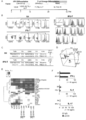
- FIG. 1 The figure which shows the experimental scheme and experimental result about the re-differentiation of T series cell from CD4 + Th1 clone origin iPSC.
- A Culture protocol for redifferentiation of T-lineage cells from CD4 + Th clone-derived iPSCs.
- B Representative flow cytometry profile of the displayed molecule in regenerated T cells (iPS-T cells) 14 days after stimulation with the original CD4 + Th clone (SK) and phytohemagglutinin (PHA) -P.
- C Original CD4 + Th1 clone (SK) and iPS-T cell TCR gene usage and V-
- D -J junction region sequence.
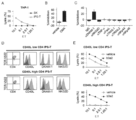
- the original CD4 + Th1 clone (SK) and iPS-T cells were stimulated for 24 hours with control IgG or anti-CD3 monoclonal antibody (10 ⁇ g / ml) bound to the plate. The indicated cytokines in the culture supernatant were measured by ELISA. Data shown are the mean ⁇ SD of 3 cultures and represent 3 independent triplicate experiments. The figure which shows the effect of CD4 introduction
- A, B CD40L expression of iPS-T cells 13 days after PHA-P stimulation.
- CD40L positive cells Mock iPS-T cells or CD4 + iPS-T cells were stimulated with PHA-P and cultured in the presence of each cytokine.
- the frequency of CD40L positive cells is shown in the upper right corner of each panel.
- C, D Expression of CD40L, CD4, and TCR-Vb22 in their subpopulations is shown.
- CD40L high and CD40L low populations when cultured in media containing IL-2 and IL-15 are separated from mock iPS-T cells and CD4 + iPS-T cells by flow cytometry sorting and proliferated by PHA-P stimulation did.
- E, F Surface expression of CD40L in various subpopulations stimulated with control IgG or anti-CD3 monoclonal antibody (10 ⁇ g / ml) bound to the plate.
- the original CD4 + Th1 clone (SK) was used as a control.
- the relative fluorescence intensity (RFI) is shown in the upper right corner of each panel.
- CF CD40L (red) and isotype matched control (grey) are shown.
- G Cytokine production by the indicated populations stimulated with control IgG or anti-CD3 monoclonal antibody (10 ⁇ g / ml) bound to the plate.
- the original CD4 + Th1 clone (SK) was used as a control.
- DCs pulsed with vehicle or b3a2 peptide were cultured for 24 hours with each CD4 + iPS-T cell at a DC / CD4 + iPS-T cell ratio of 5: 1.
- OK432 (10 ⁇ g / ml) matured DC and media control DC were used as controls. Each surface molecule (red) and an isotype matched control (gray) are shown.
- B Cytokine production by DC co-cultured with each CD4 + iPS-T cell. Cytokines in the culture supernatant were measured by bead-based multiplex immunoassay.
- CD4 + iPS-T cell (1 ⁇ 10 4 cells) was co-cultured with autologous DC (2.5 ⁇ 10 4 cells) pre-pulsed with b3a2 peptide (10 ⁇ M) for 24 hours.
- the original CD4 + Th1 clone (SK) was used as a control.
- Data shown are the mean ⁇ SD of 3 cultures and represent 3 independent triplicate experiments.
- A Cytotoxic activity of iPS-T cells against THP-1 cells.
- B, C Cytotoxicity of iPS-T cells to THP-1 cells at an effector / target (E: T) ratio of 2.5: 1.
- FIG. 1 The figure which shows the mechanism and analysis result about induction
- A Mechanism of WT1-specific CTL sensitization.
- pWT1 is a WT1 peptide and b3a2 is a b3a2 peptide.
- iPS-T cells recognize the b3a2 peptide presented by DC, activated iPS-T cells increase the expression of CD40L.
- DC maturation is induced by CD40 ligation with CD40L.
- Activation of WT1 peptide-specific CTL is promoted by increased expression of costimulatory molecules and increased cytokine production by DC.
- BD CD8 + T cell proliferative response.
- iPS-T cell 5 ⁇ 10 3 cells
- DC (1 ⁇ 10 4 cells) were first co-cultured with or without b3a2 peptide (5 ⁇ M) for 5 hours to mature those DCs, The DCs and iPS-T cells were then irradiated and cultured with autologous CD8 + T cells (5 ⁇ 10 4 cells) in the presence of WT1 peptide (5 ⁇ M).
- Proliferative response (day 7) was measured as [ 3 H] -thymidine incorporation. Data shown are the mean ⁇ SD of 3 cultures and represent 3 independent triplicate experiments.
- A In vivo anti-leukemic action (photo). NSG mice were injected subcutaneously with a mixture of K562-A24-Luc-WT1 minigene cells and saline or WT1-specific CTL. Tumor loading was measured weekly by bioluminescence imaging.
- the method for producing CD4 positive helper T cells of the present invention comprises (i) a T cell derived from a pluripotent stem cell and introduced with a CD4 gene or gene product in a culture solution containing IL-2 and IL-15. And (ii) selecting CD40L-expressing T cells from the cells obtained in (i).
- T cells derived from pluripotent stem cells and introduced with a CD4 gene or gene product will be described.
- a pluripotent stem cell is a stem cell having pluripotency that can be differentiated into many cells existing in a living body and also having proliferative ability, and at least used in the present invention. Any cell derived from a hematopoietic progenitor cell is included.
- pluripotent stem cells include, but are not limited to, embryonic stem (ES) cells, cloned embryo-derived embryonic stem (ntES) cells obtained by nuclear transfer, sperm stem cells (“GS cells”), embryonic Examples include germ cells (“EG cells”), induced pluripotent stem (iPS) cells, cultured fibroblasts and bone marrow stem cell-derived pluripotent cells (Muse cells).
- IPS cell production methods are known in the art and can be produced by introducing reprogramming factors into any somatic cells.
- the reprogramming factor is, for example, Oct3 / 4, Sox2, Sox1, Sox3, Sox15, Sox17, Klf4, Klf2, c-Myc, N-Myc, L-Myc, Nanog, Lin28, Fbx15, ERas, ECAT15 -2, Tcl1, beta-catenin, Lin28b, Sall1, Sall4, Esrrb, Nr5a2, Tbx3 or Glis1, etc. genes or gene products are exemplified, and these reprogramming factors may be used alone or in combination. Also good.
- Somatic cells include, but are not limited to, fetal (pup) somatic cells, neonatal (pup) somatic cells, and mature healthy or diseased somatic cells. , Passage cells, and established cell lines.
- somatic cells are, for example, (1) tissue stem cells (somatic stem cells) such as neural stem cells, hematopoietic stem cells, mesenchymal stem cells, dental pulp stem cells, (2) tissue progenitor cells, (3) blood cells (peripheral) Blood cells, umbilical cord blood cells, etc.), lymphocytes, epithelial cells, endothelial cells, muscle cells, fibroblasts (skin cells, etc.), hair cells, hepatocytes, gastric mucosal cells, intestinal cells, spleen cells, pancreatic cells (pancreatic exocrine cells) Etc.), differentiated cells such as brain cells, lung cells, kidney cells and fat cells.
- tissue stem cells such as neural stem cells, hematopoietic stem cells, mesenchymal stem
- lymphocytes preferably T cells
- TCR T cell receptor
- the lymphocytes are stimulated with anti-CD3 antibody and anti-CD28 antibody in the presence of interleukin-2 (IL-2), or with a desired antigen peptide It is preferable to stimulate and activate.
- IL-2 interleukin-2
- Such stimulation can be performed, for example, by adding IL-2, anti-CD3 antibody and anti-CD28 antibody to the medium and culturing the lymphocytes for a certain period.
- the anti-CD3 antibody and the anti-CD28 antibody may be ones to which magnetic beads or the like are bound. Further, instead of adding these antibodies to the medium, the culture in which the anti-CD3 antibody and the anti-CD28 antibody are bound to the surface. Stimulation may be performed by culturing the T cells for a certain period on a dish. Further, stimulation may be given by adding an antigen peptide recognized by human T cells to the medium.
- An antigen peptide is a peptide consisting of an amino acid sequence of at least 9 or more constituting a desired antigen protein.
- peptides include peptides consisting of 9 or more amino acid sequences constituting the b3a2 subtype of p210 of the BCR / ABL chimeric gene (also simply referred to as b3a2).
- lymphocytes that recognize the antigen peptide can be selectively proliferated.
- the CD4-positive T cells used in the present invention preferably have a desired antigen specificity. Therefore, it is desirable that the lymphocytes that are the source of iPS cells have the desired antigen specificity, and the lymphocytes are specifically isolated by purification using an affinity column or the like on which the desired antigen is immobilized. Also good. In this purification, MHC (major histocompatibility complex) to which a desired antigen is bound is tetramerized (so-called “MHC tetramer”) and has a desired antigen specificity from human tissues. A method of purifying lymphocytes can also be employed.
- the mammalian individual from which somatic cells are collected is not particularly limited, but is preferably a human.
- somatic cells that are the source of iPS cells from the viewpoint of easily matching the type of human leukocyte antigen (HLA) to the patient being transfused Is preferably isolated from a subject transfused with CD4 positive helper T cells.
- Methods for inducing T Cells from Pluripotent Stem Cells can be performed using known methods.
- the method includes the following steps (1) and (2), preferably (1) to (3); (Step 1) Inducing CD34 positive hematopoietic progenitor cells from pluripotent stem cells, (Step 2) a step of culturing the CD34 positive hematopoietic progenitor cell obtained in the step (1) in the presence of FLT3L and IL-7, and (step 3) the cell obtained in the step (2) is treated with IL- Co-culturing with peripheral blood mononuclear cells in the presence of 7 and IL-15.
- T cell means a cell having TCR on the cell surface, and may further have CD4 and CD8 on the cell surface. Therefore, from the viewpoint of inducing T cells, the above (Step 1) and (Step 2) may be performed, but in order to increase the T cell content efficiency, the above (Step 3) is further included. desirable.
- Step 1 Step of inducing hematopoietic progenitor cells from pluripotent stem cells
- Hematopoietic progenitor cells can be differentiated into blood cells such as lymphocytes, eosinophils, neutrophils, basophils, erythrocytes, megakaryocytes, etc. It is a cell and can be recognized, for example, by being positive for the surface antigens CD34 or CD34 and CD43.
- hematopoietic progenitor cells As a method of inducing hematopoietic progenitor cells from pluripotent stem cells, net-like obtained by coculturing pluripotent stem cells with C3H10T1 / 2 and then coculturing with C3H10T1 / 2 in the presence of VEGF, FLT3L and SCF Examples thereof include a method for preparing hematopoietic progenitor cells from a structure (also referred to as ES-sac or iPS-sac). At this time, vitamin Cs may be further added and cultured.
- a structure also referred to as ES-sac or iPS-sac
- the “net-like structure” is a three-dimensional sac-like structure (with space inside) derived from pluripotent stem cells, which is formed by an endothelial cell population and the like, and contains hematopoietic progenitor cells inside. It is a structure. In addition, it can be obtained by culturing pluripotent stem cells on C3H10T1 / 2 in the presence of VEGF according to the method described in Takayama N., et al. J Exp Med. 2817-2830 (2010) Hematopoietic progenitor cells can be prepared from the net-like structure.
- Step 2 Step of culturing hematopoietic progenitor cells in the presence of FLT3L and IL-7
- the culture medium used in Step 2 is not particularly limited, but the medium used for culturing animal cells is changed to a basal medium.
- FLT3L and IL-7 Can be prepared.
- Iscove's Modified Dulbecco's Medium (IMDM) medium Medium 199 medium, Eagle's Minimum Essential Medium (EMEM) medium, ⁇ MEM medium, Dulbecco's modified Eagle's Medium (DMEM) medium, Ham's F12 medium, RPMI 1640 medium, Fischer's medium , Neurobasal Medium (Life Technologies) and mixed media thereof.
- IMDM Iscove's Modified Dulbecco's Medium
- EMEM Eagle's Minimum Essential Medium
- DMEM Dulbecco's modified Eagle's Medium
- Ham's F12 medium Ham's F12 medium
- RPMI 1640 medium Fischer's medium
- Serum may be contained in the medium, or serum-free may be used.
- the basal medium can be, for example, albumin, insulin, transferrin, selenium, fatty acids, trace elements, 2-mercaptoethanol, thiolglycerol, lipids, amino acids, L-glutamine, non-essential amino acids, vitamins, growth factors, low It may also contain one or more substances such as molecular compounds, antibiotics, antioxidants, pyruvate, buffers, inorganic salts, cytokines and the like.
- a preferred basal medium is an ⁇ MEM medium supplemented with serum, L-glutamine, transferrin, and selenium.
- the concentration of IL-7 in the culture medium used in step 2 is usually 0.1 ng / ml to 50 ng / ml, for example, 0.1 ng / ml, 0.2 ng / ml, 0.3 ng / ml, 0.4 ng / ml, 0.5 ng / ml, 0.6 ng / ml, 0.7 ng / ml, 0.8 ng / ml, 0.9 ng / ml, 1 ng / ml, 2 ng / ml, 3 ng / ml, 4 ng / ml, 5 ng / ml, 10 ng / ml, 20 ng / ml, 30 ng / ml, 40 ng / ml or 50 ng / ml.
- it is 1 ng / ml.
- the concentration of FLT3L in the culture medium used in Step 2 is usually 1 ng / ml to 100 ng / ml, for example, 1 ng / ml, 2 ng / ml, 3 ng / ml, 4 ng / ml, 5 ng / ml, 6 ng / ml, 7 ng / ml, 8 ng / ml, 9 ng / ml, 10 ng / ml, 20 ng / ml, 50 ng / ml or 100 ng / ml.
- it is 10 ng / ml.
- Vitamin Cs mean L-ascorbic acid and its derivatives
- L-ascorbic acid derivatives mean those that become vitamin C by an enzymatic reaction in vivo.
- L-ascorbic acid derivatives include vitamin C phosphate, ascorbyl glucoside, ascorbyl ethyl, vitamin C ester, ascovir tetrahexyldecanoate, ascovir stearate and ascorbyl 2-phosphate-6 palmitic acid.
- Vitamin C phosphate is preferable, and for example, phosphoric acid-L ascorbic acid Na or phosphoric acid-L-ascorbic acid Mg. Vitamin Cs are preferably added separately every 4 days, every 3 days, every 2 days, or every day during the culture period, more preferably every day.
- the vitamin C is usually added in an amount corresponding to 5 ng / ml to 500 ng / ml in the culture solution.
- the amount corresponds to 5 ng / ml, 10 ng / ml, 25 ng / ml, 50 ng / ml, 100 ng / ml, 200 ng / ml, 300 ng / ml, 400 ng / ml, or 500 ng / ml.
- the concentration of SCF used in the production of hematopoietic progenitor cells in the culture solution is 10 ng / ml to 100 ng / ml, such as 10 ng / ml, 20 ng / ml, 30 ng / ml, 40 ng / ml, 50 ng / ml, 60 ng / ml, 70 ng / ml, 80 ng / ml, 90 ng / ml, 100 ng / ml, 150 ng / ml, 200 ng / ml, or 500 ng / ml.
- the concentration of TPO used in the production of hematopoietic progenitor cells in the culture medium is 10 ng / ml to 100 ng / ml, such as 10 ng / ml, 20 ng / ml, 30 ng / ml, 40 ng / ml, 50 ng / ml, 60 ng / ml, 70 ng / ml, 80 ng / ml, 90 ng / ml, 100 ng / ml, 150 ng / ml, 200 ng / ml, or 500 ng / ml.
- hematopoietic progenitor cells may be subjected to adhesion culture or suspension culture, and in the case of adhesion culture, the culture vessel may be coated and used, or may be co-cultured with feeder cells and the like.
- the feeder cells to be co-cultured stromal cells are preferable, and specific examples include bone marrow stromal cell line OP9 cells (available from RIKEN BioResource Center).
- the OP9 cells may preferably be OP9-DL1 cells that constitutively express Delta-like-1 (Dll1) (Holmes R1 and Zuniga-Pflucker JC. Cold Spring Harb Protoc. 2009 (2)).
- OP9 cells When OP9 cells are used as feeder cells, it can also be carried out by adding separately prepared fusion proteins such as Dll1 or Dll1 and Fc to the culture medium.
- Dll1 as NCBI accession numbers, NM_005618 for humans, NM_007865 for mice, proteins encoded by the gene having the nucleotide sequence described in NM_007865, and high sequence identity (for example, 90% or more) with these And naturally occurring variants having equivalent functions are included.
- the feeder cells are preferably exchanged as appropriate during culture. The exchange of the feeder cells can be performed by transferring the target cells in culture onto the feeder cells previously seeded. The exchange can occur every 5 days, every 4 days, every 3 days, or every 2 days.
- Step 2 when culture is performed by suspension culture, it is desirable to culture cells by forming aggregates (also referred to as spheres) in a non-adherent state in the culture vessel, and such culture is not particularly limited.
- culture vessels that have not been artificially treated (for example, coating with an extracellular matrix or the like) for the purpose of improving adhesion to cells, or treatments that artificially suppress adhesion (for example, polyhydroxyethyl methacrylate) Coating with acid (poly-HEMA), nonionic surfactant polyol (Pluronic® F-127, etc.) or phospholipid-like structure (for example, water-soluble polymer (Lipidure) based on 2-methacryloyloxyethyl phosphorylcholine)
- This can be done by using a treated culture vessel, or when performing step 2 in suspension culture, Huijsk ens MJ et al, J Leukoc Biol. 96: 1165-1175, 2014.
- Step 2 when adhesion culture is performed, it can be performed by culturing using a culture vessel coated with an extracellular matrix.
- the coating treatment can be performed by placing a solution containing an extracellular matrix in a culture container and then removing the solution as appropriate.
- the extracellular matrix is a supramolecular structure existing outside the cell, and may be naturally derived or an artificial product (recombinant). Examples include substances such as polylysine, polyornithine, collagen, proteoglycan, fibronectin, hyaluronic acid, tenascin, entactin, elastin, fibrillin, laminin and fragments thereof.
- These extracellular substrates may be used in combination, for example, preparations from cells such as BD Matrigel TM.
- the culture temperature conditions for culturing hematopoietic progenitor cells in step 2 are not particularly limited, but for example, about 37 ° C to about 42 ° C, preferably about 37 to about 39 ° C are preferable.
- the culture period can be appropriately determined by those skilled in the art while monitoring the number of cells.
- the number of days is not particularly limited, and is, for example, at least 10 days, 12 days, 14 days, 16 days, 18 days, 20 days, 21 days, and preferably 14 days.
- the cells obtained in step 2 may be stimulated with mitogen.
- Mitogen means a substance that promotes cell division of T cells. Examples of such substances include pokeweed ⁇ mitogen, anti-CD3 antibody, anti-CD28 antibody, phytohemagglutinin (PHA), concanavalin A (ConA), superantigen. , Fallball esters such as phorbol-12-myristate-13-acetate (PMA).
- Step 3 of co-culturing with peripheral blood mononuclear cells in the presence of IL-7 and IL-15 is the step of isolating the cells obtained in step 2 above and co-culturing with peripheral blood mononuclear cells. It is a process to do.
- the peripheral blood mononuclear cells used in step 3 are desirably allogenic (same type) as the pluripotent stem cells used in step 1. Therefore, when human pluripotent stem cells are used, the peripheral blood mononuclear cells It is also desirable that human peripheral blood mononuclear cells. In addition, it is desirable to take measures to prevent the peripheral blood mononuclear cells from proliferating. Examples of such measures include irradiation and mitomycin treatment.
- the culture medium used in step 3 is not particularly limited, but can be prepared by adding IL-7 and IL-15 to a basal medium as a medium used for culturing animal cells.
- Basal media include, for example, Iscove's'Modified Dulbecco's Medium (IMDM), Medium 199, Eagle's Minimum Essential Medium (EMEM), ⁇ MEM, Dulbecco's modified Eagle's Medium (DMEM), Ham's F1 ', , Neurobasal Medium (Life Technologies) and mixed media thereof. Serum may be contained in the medium, or serum-free may be used.
- the basal medium can be, for example, albumin, insulin, transferrin, selenium, fatty acids, trace elements, 2-mercaptoethanol, thiolglycerol, lipids, amino acids, L-glutamine, non-essential amino acids, vitamins, growth factors, low It may also contain one or more substances such as molecular compounds, antibiotics, antioxidants, pyruvate, buffers, inorganic salts, cytokines and the like.
- a preferred basal medium is RPMI 1640 medium supplemented with serum and L-glutamine.
- the concentration of IL-7 in the culture medium used in step 3 is 1 ng / ml to 100 ng / ml, for example, 1 ng / ml, 2 ng / ml, 3 ng / ml, 4 ng / ml, 5 These are ng / ml, 6 ng / ml, 7 ng / ml, 8 ng / ml, 9 ng / ml, 10 ng / ml, 20 ng / ml, 50 ng / ml, 100 ng / ml. Preferably, it is 10 ng / ml.
- the concentration of IL-15 in the culture medium used in step 3 is 1 ng / ml to 100 ng / ml, such as 1 ng / ml, 2 ng / ml, 3 ng / ml, 4 ng / ml, 5 These are ng / ml, 6 ng / ml, 7 ng / ml, 8 ng / ml, 9 ng / ml, 10 ng / ml, 20 ng / ml, 50 ng / ml, 100 ng / ml. Preferably, it is 10 ng to 20 ng / ml.
- mitogen may be further added to the culture solution.
- the mitogen can be the same as described above.
- the culture temperature condition in this step 3 is not particularly limited, but for example, about 37 ° C to about 42 ° C, preferably about 37 to about 39 ° C is preferable.
- the culture period can be appropriately determined by those skilled in the art while monitoring the number of cells.
- the number of days is not particularly limited, and is, for example, at least 10 days, 12 days, 14 days, 16 days, 18 days, 20 days, 21 days, and preferably 14 days.
- the method for introducing the CD4 gene or gene product into T cells derived from pluripotent stem cells is not particularly limited.
- the gene product is exemplified by RNA or protein.
- the CD4 gene is preferably a human gene, and has, for example, the amino acid sequence of GenBank Accession No. AAH25782 (SEQ ID NO: 3) or 80% or more, preferably 90% or more, more preferably 95% or more identical thereto. Examples include genes that encode proteins that can activate dendritic cells when introduced into T cells.
- vectors such as viruses, plasmids, and artificial chromosomes can be introduced into pluripotent stem cells by techniques such as lipofection, liposome, and microinjection.
- virus vectors include retrovirus vectors, lentivirus vectors, adenovirus vectors, adeno-associated virus vectors, Sendai virus vectors, and the like.
- artificial chromosome vectors include human artificial chromosomes (HAC), yeast artificial chromosomes (YAC), and bacterial artificial chromosomes (BAC, PAC).
- HAC human artificial chromosomes
- YAC yeast artificial chromosomes
- BAC bacterial artificial chromosomes
- plasmid a plasmid for mammalian cells can be used.
- the vector can contain regulatory sequences such as a promoter, enhancer, ribosome binding sequence, terminator, polyadenylation site, etc. so that the CD4 gene can be expressed, and, if necessary, a drug resistance gene (for example, kanamycin). Resistance marker, ampicillin resistance gene, puromycin resistance gene, etc.), thymidine kinase gene, diphtheria toxin gene and other selectable marker sequences, fluorescent protein, ⁇ -glucuronidase (GUS), FLAG and other reporter gene sequences.
- regulatory sequences such as a promoter, enhancer, ribosome binding sequence, terminator, polyadenylation site, etc. so that the CD4 gene can be expressed, and, if necessary, a drug resistance gene (for example, kanamycin). Resistance marker, ampicillin resistance gene, puromycin resistance gene, etc.), thymidine kinase gene, diphtheria toxin gene and other selectable marker sequences, fluorescent protein, ⁇ -glucuronidas
- SV40 promoter As promoters, SV40 promoter, LTR promoter, CMV (cytomegalovirus) promoter, RSV (Rous sarcoma virus) promoter, MoMuLV (Moloney mouse leukemia virus) LTR, HSV-TK (herpes simplex virus thymidine kinase) promoter, EF- ⁇ promoter, CAG Examples include promoters and TRE promoters (CMV minimal promoter having a Tet response sequence in which tetO sequence is continuous seven times).
- a vector having a TRE promoter and capable of expressing a fusion protein of reverse tetR (rtetR) and VP16AD is referred to as a drug-responsive induction vector.
- the above-mentioned vector has a transposon sequence before and after this expression cassette in order to incorporate an expression cassette consisting of a promoter and a CD4 gene that binds to it into the chromosome of a pluripotent cell and further excise it if necessary. But you can. Although it does not specifically limit as a transposon arrangement
- the CD4 gene When using a drug-responsive induction vector, the CD4 gene may be introduced into pluripotent stem cells. In this case, the CD4 gene can be expressed by adding the corresponding drug into the medium. Therefore, when the drug responsive induction vector is used, it can be rephrased as introduction of the CD4 gene by adding the corresponding drug into the medium. Examples of the corresponding drug include doxycycline and the like.
- a method of stopping expression by introducing Cre into a cell after a desired period of time is exemplified.
- RNA When introduced in the form of RNA, it may be introduced into pluripotent stem cells by techniques such as electroporation, lipofection, and microinjection.
- RNA When introduced in the form of RNA, it may be introduced into pluripotent stem cells by techniques such as electroporation, lipofection, and microinjection.
- protein When introduced in the form of protein, it may be introduced into pluripotent stem cells by techniques such as lipofection, fusion with cell membrane permeable peptides (eg, HIV-derived TAT and polyarginine), and microinjection.
- cell membrane permeable peptides eg, HIV-derived TAT and polyarginine
- the half-life of the gene product is short, so the introduction may be performed multiple times.
- the number of introductions refer to the half-life from the period when CD4 gene expression is required And can be calculated as appropriate. Examples of the number of introductions are 3, 4, 5, 6 or more times.
- Step of culturing CD4-positive T cells in a culture solution containing IL-2 and IL-15 The culture solution used in this step is not particularly limited, but the medium used for culturing animal cells is changed to IL-2. And IL-15 can be added.
- Basal media include, for example, Iscove's Modified Dulbecco's Medium (IMDM) medium, Medium 199 medium, Eagle's Minimum Essential Medium (EMEM) medium, ⁇ MEM medium, Dulbecco's modified Eagle's Medium (DMEM) medium, Ham's F12 medium, RPMI 1640 medium, Fischer's medium Media, Neurobasal Medium (Life Technologies), and mixed media thereof are included. Serum may be contained in the medium, or serum-free may be used.
- IMDM Iscove's Modified Dulbecco's Medium
- EMEM Eagle's Minimum Essential Medium
- DMEM Dulbecco's modified Eagle's Medium
- Ham's F12 medium Ham's F12 medium
- the basal medium can be, for example, albumin, insulin, transferrin, selenium, fatty acids, trace elements, 2-mercaptoethanol, thiolglycerol, lipids, amino acids, L-glutamine, non-essential amino acids, vitamins, growth factors, low It may also contain one or more substances such as molecular compounds, antibiotics, antioxidants, pyruvate, buffers, inorganic salts, cytokines and the like.
- a preferred basal medium is an ⁇ MEM medium supplemented with serum, L-glutamine, transferrin, and selenium.
- the concentration of IL-2 in the culture medium is usually from 10 to IU / ml to 500 to IU / ml, for example, 10 to IU / ml, 20 to IU / ml, 30 to IU / ml, 40 to IU / ml, 50 to IU / ml.
- the concentration of IL-15 in the culture is usually 1 ng / ml to 50 ng / ml, for example, 1 ng / ml, 2 ng / ml, 3 ng / ml, 4 ng / ml, 5 ng / ml 6 ng / ml, 7 ng / ml, 8 ng / ml, 9 ng / ml, 10 ng / ml, 20 ng / ml, 25 ng / ml or 50 ng / ml.
- it is 5 ng / ml.
- CD4 positive T cells may be subjected to adhesion culture or suspension culture, but suspension culture is preferred.
- the culture temperature conditions for culturing CD4-positive T cells are not particularly limited, but for example, about 37 ° C to about 42 ° C, preferably about 37 to about 39 ° C are preferable.
- the culture period can be appropriately determined by those skilled in the art while monitoring the number of cells.
- the number of days is not particularly limited, and is, for example, at least about 1 day, about 2 days, about 3 days, and about 5 days.
- CD40L high-expressing T cells T cells obtained by culturing CD4-positive T cells in a culture medium containing IL-2 and IL-15 are sorted based on the expression level of CD40L.
- CD40L highly expressing T cells CD40LhighCD4 + iPS-T cells are obtained.
- the CD40L highly expressing T cell means a T cell that expresses CD40L at a predetermined level or higher, for example, a T cell that expresses CD40L at a level higher than a level detectable by an anti-CD40L antibody, or a negative control cell (
- T cells that express CD40L at a level higher than the expression level in Fluorescence Minus One (FMO) Control) or higher than the upper limit when there is a range of expression levels in negative control cells anti-CD40L Selection (purification) can be performed using antibodies.
- FMO Fluorescence Minus One
- CD40L-positive cells can be performed by methods well known to those skilled in the art, and is not particularly limited, but can be performed by magnetic beads carrying CD40L antibody or flow cytometry using CD40L antibody.
- the obtained CD40L highly expressing T cells may be stimulated with mitogen or the like.
- mitogens include pokeweed mitogen, anti-CD3 antibody, anti-CD28 antibody, and phytohemagglutinin (PHA).
- Dendritic cells can be activated by contacting CD4 positive helper T cells produced by the above-described method with dendritic cells in the presence of antigen in vitro.
- Dendritic cells are cells having a function of taking up an antigen and binding to MHC molecules to present the antigen. Since it can be activated by contacting with a CD4 positive helper T cell produced by the method of the present invention, it may be an immature dendritic cell.
- Activating dendritic cells means acquiring a function capable of activating T cells specific for the antigen, and more preferably acquiring a function capable of activating CD8 positive T cells specific for the antigen. That is. The activation can also be performed by confirming the expression of CD83 or CD86. Furthermore, activating a dendritic cell can be paraphrased as maturation of an immature dendritic cell into a mature dendritic cell.
- the dendritic cells used are dendritic cells isolated from a donor by a blood component separation device and density gradient centrifugation, and preferably possess the same MHC molecules as CD4 positive helper T cells. .
- the antigen used in the activation of dendritic cells is a peptide having at least a continuous 9 amino acid sequence of a protein specifically recognized by CD4 positive helper T cells produced by the method of the present invention.
- an antigen for example, when a CD4-positive T cell possesses a TCR that specifically recognizes a b3a2 peptide, the b3a2 peptide is used as the antigen.
- the b3a2 peptide is a part of a protein encoded by the b3a2 type BCR / ABL chimeric gene, and is a peptide having at least a continuous 9 amino acid sequence that fits in the peptide-containing groove of the HLA-class II molecule.
- the b3a2 type BCR / ABL chimeric gene means a fusion gene of BCR and ABL containing B3 exon from the M-BCR site of BCR and A2 exon from ABL.
- a medium used for activating dendritic cells a medium used for culturing animal cells can be used.
- the basal medium include Iscove's Modified Dulbecco's Medium (IMDM) medium, Medium 199 medium, Eagle's Minimum Essential Medium (EMEM) medium, ⁇ MEM medium, Dulbecco's modified Eagle's Medium (DMEM) medium, Ham's F12 medium, RPMI 1640 medium, Fischer's medium. Media, Neurobasal Medium (Life Technologies), and mixed media thereof are included. Serum may be contained in the medium, or serum-free may be used.
- the basal medium can be, for example, albumin, insulin, transferrin, selenium, fatty acids, trace elements, 2-mercaptoethanol, thiolglycerol, lipids, amino acids, L-glutamine, non-essential amino acids, vitamins, growth factors, low It may also contain one or more substances such as molecular compounds, antibiotics, antioxidants, pyruvate, buffers, inorganic salts, cytokines and the like.
- the time required for the activity of dendritic cells is not particularly limited, but may be several hours, and examples thereof include 2 hours, 3 hours, 4 hours, 5 hours, 6 hours, 12 hours, and 24 hours. Preferably, it is 5 hours.
- the present invention relates to a CD4 positive helper T cell (helper T cell derived from a pluripotent stem cell and consisting of a CD4 positive and CD40L highly expressing T cell) produced by the method described above, and / or Alternatively, a medicament such as a dendritic cell activated by the above-described method and a cancer therapeutic agent containing an antigen peptide is provided.
- a CD4 positive helper T cell helper T cell derived from a pluripotent stem cell and consisting of a CD4 positive and CD40L highly expressing T cell
- the cancer to be treated is a cancer cell that expresses an antigen that is specifically recognized by CD4-positive T cells. It is desirable.
- the treatment target is a cancer cell that expresses b3a2, and is caused by a chromosome (Philadelphia chromosome) that has a BCR / ABL chimeric gene And leukemia.
- Dendritic cells activated by CD4-positive helper T cells are lymphocytes that recognize the antigen by presenting them, even if other than the antigen recognized by the CD4-positive helper T cell (for example, CD8-positive T cells) ) Can be activated. Therefore, the cancer which is the target of the cancer therapeutic agent containing dendritic cells is not particularly limited.
- the activated dendritic cell may be used as the therapeutic agent as it is, but for cancer treatment, in order to present the tumor antigen to the dendritic cell, the cell of the cancer cell Dendritic cells that have been mixed with lysate sputum (cell lysate) sputum, contacted with peptides, or presented with an antigen such as by introducing a tumor antigen gene may be used as a cancer therapeutic agent.
- a CD4 positive helper T cell produced and / or a dendritic cell activated by the above-mentioned method is suspended in physiological saline or the like, and the patient Or a method in which dendritic cells are suspended in physiological saline and injected intravenously.
- HLA-DR9 (DRB1 * 09: 01) restricted b3a2 type BCR-ABL junction peptide (ATGFKQSSKALQRPVAS: SEQ ID NO: 1) HLA-A24 (A * 24: 02) -restricted modified Wilms Tumor (Wilms tumor) 1 (WT1) 235 ⁇ 243 epitope peptide (CYTWNQMNL: SEQ ID NO: 2) HLA-DR53 (DRB4 * 01: 03) restricted glutamic acid decarboxylase 65 (GAD65) 113 ⁇ 131 peptide (DVMNILLQYVVKSFDRSTK: SEQ ID NO: 4)
- the modified WT1 235 ⁇ 243 peptide Y to M of the native WT1 235 ⁇ 243 peptide amino acid position 2 is substituted (CYTWNQMNL: SEQ ID NO: 2) Recombinant human (rh) type IL-2, rh
- PBMC Cell peripheral blood mononuclear cells
- Human monocyte-derived dendritic cells were induced as described in J Immunol 183: 201-208., 2009.
- Commercially available human CML cell line K562, human myeloid leukemia cell line THP-1, and human lung cancer cell line PC9 were used.
- Mouse L fibroblasts transfected with HLA class II genes were used (Hum Immunol 59: 549-560.1998). Informed consent was obtained from all donors to use cells isolated from healthy adults. All studies were conducted in accordance with the Declaration of Helsinki and with the approval of the appropriate institutional ethics committee.
- iPSC clones are negative for the persistent transgene, exhibit pluripotency characterized by the expression of pluripotency-related molecules and teratoma formation in immunodeficient mice, and have a normal karyotype Confirmed (not shown).
- T cell differentiation from iPSCs iPSCs were differentiated into T cells by the method described in Cell Stem Cell. 12 (1): 114-126, 2013. Specifically, the iPSC mass was transferred onto C3H10T1 / 2 feeder cells and cultured in an EB (embryoid body) medium containing rh-type VEGF. On day 7, rh-type SCF and rh-type FLT3L were added. On day 14, hematopoietic progenitor cells were collected, transferred onto OP9-DL1 cells, and co-cultured in OP9 medium containing rh-type IL-7 and rh-type FLT3L.
- EB embryoid body
- PBMC peripheral blood mononuclear cells
- APC antigen-presenting cells
- HLA-A * 24: 02 / WT1 235 ⁇ 243 tetramer can be used for the detection of WT1 peptide-specific cytotoxic T lymphocytes (CTL), HLA- a * 24: 02 / HIVEnv 584 ⁇ 592 tetramer was used as a negative control.
- Stained cell samples were analyzed using a FACSCalibur flow cytometer and a FACSAria II flow cytometer (BD Biosciences) and the data processed using the FlowJo software program (Tree Star, Ashland, Oreg., USA). .
- Relative fluorescence intensity (RFI) was calculated as the ratio of the MFI of a particular marker to the mean fluorescence intensity (MFI) of the isotype control.
- a cDNA encoding the transfectant HLA-DR9 (DRB1 * 09: 01) has been described previously (Ueda N, Cell Mol Immunol. 2016).
- CDNA encoding BCR-ABL p210 was purchased from Addgene (Cambridge, Massachusetts, USA).
- Lentiviral transduction was performed as described in Zhang, R., Cancer Immunol Res 2015.
- a lentiviral vector for expressing HLA-A * 24: 02 and a lentiviral vector for expressing a minigene encoding a modified WT1 235 to 243 epitope are expressed in the luciferase gene-expressing K562 strain (K562-Luc).
- Transduced K562-Luc-A24-WT1 minigene cells.
- Lentiviral vectors for expressing HLA-DRA * 01: 01 and HLA-DRB1 * 09: 01 and / or BCR-ABL p200 were transduced into THP-1 strains (THP-1-DR9 cells, THP1-DR9 -BCRABL cells).
- T cell functional assay [ 3 H] -thymidine incorporation assay. Cytotoxic activity was measured using a 51 Cr release assay. Cytokine levels in the culture supernatant were assessed by enzyme-linked immunosorbent assay (ELISA; hIFN- ⁇ : eBiosciences) or bead-based multiplex immunoassay (BD cytometric bead array; BD Biosciences).
- ELISA enzyme-linked immunosorbent assay
- BD cytometric bead array BD Biosciences
- T cell antigen receptor TCR
- V, D, and J segments of the rearranged TCR- ⁇ and TCR- ⁇ chains of T cells or iPS-T cells are expressed as J Identified as described in Immunol 170: 947-960.2003.
- the gene segment nomenclature used is in accordance with the ImmunoGeneTics (IMGT) convention. These V, D, and J segments are identified by comparing the resulting sequences against the IMGT database (http://www.imgt.org/) with the online tool (IMGT / V-QUEST) It was done.
- Data analysis software R version 3.2.2 org. Hs. eg.
- the hypergeometric test was performed using db 3.2.3 and the annotation package GO.
- Use GOstats 2.36.0 (gene ontology analysis) in conjunction with db 3.2.2 and the annotation package KEGG.
- KEGG profile 1.12.0 (KEGG pathway analysis) was used in conjunction with db 3.2.2.
- CD8 + T cells and DCs were obtained from the same donor used to establish the b3a2-specific T cell clone (SK). Those cells were used to induce antigen-specific CTL to avoid an alloreactive response.
- SK b3a2-specific T cell clone
- Those cells were used to induce antigen-specific CTL to avoid an alloreactive response.
- SK Th1 clone
- CD8 + T cells with WT1 235 ⁇ 243 peptide After irradiation with 30 Gy. On day 7, 1 ⁇ Ci of [ 3 H] -thymidine was added to the cultures and the proliferative response of CD8 + T cells was assessed by using a [ 3 H] -thymidine incorporation assay after 16 hours of culture. On the 10th day, the experiment was repeated in the same manner. And determining the frequency of WT1 peptide-specific CTL by staining with HLA-A * 2402 / WT1 235 ⁇ 243 tetramer.
- CD8 + T cells obtained by WT1 235 ⁇ 243 peptide in the presence of autologous PBMC irradiated with radiation of 35 Gy. These cells were later used for cytotoxicity assays and in vivo experiments.
- mice were purchased from Charles River (Yokohama, Japan), and K562-Luc-A24-WT1 minigene cells (1.0 ⁇ 10 5 cells) and saline Alternatively, a mixture with either WT1-specific CTL (1.0 ⁇ 10 6 cells) was inoculated subcutaneously (sc) into the shaved left shin. The mice were monitored for tumor growth and survival. Tumor growth was monitored weekly for 4 weeks by bioluminescence imaging and tumor growth was monitored weekly by outer caliper measurements until the mice died or were sacrificed when the tumor diameter exceeded 25 mm.
- iPS-T cells were divided into four subpopulations and total RNA expression of those subpopulations was isolated from peripheral blood NK cells, type 1 ILC (ILC1), type 2 ILC ( ILC2), type 3 ILC (ILC3), ⁇ -T cells, and ⁇ -T cells.
- ILC1 type 1 ILC
- ILC2 type 2 ILC
- ILC3 type 3 ILC
- iPS-T cells had genetic characteristics more consistent with ILC1, NK cells, and ⁇ -T cells than peripheral ⁇ -T cells (not shown).
- ILC1-related genes such as NCAM1, NCR1, NCR2, ICOS, and IL12RB, but expressed low levels of IL7RA and IL1R expressed in all ILCs except NK cells (FIG. 1E).
- TCR-independent NK cell-like cytotoxicity was observed in iPS-T cells (not shown).
- iPS-T cells like the original CD4 + Th1 clone (SK), produce high levels of IFN- ⁇ , produce relatively low levels of IL-4, and produce IL-17. (FIG. 1F).
- iPS-T cells can proliferate thousands of times upon two phytohemagglutinin (PHA) -P stimulations (not shown). These data suggest that iPS-T cells generated from CD4 + Th1 clones have group 1 ILC-like properties despite their TCR expression.
- PHA phytohemagglutinin
- CD4 transduction enhances TCR signaling 30-300 fold during CD4 binding to HLA class II during T cell activation by HLA class II / peptide complexes that expand the b3a2-specific response in iPSC-derived T cells This suggests that CD4 is important for complete activation of Th cells. Since our iPS-T cells expressed HLA class II-restricted TCR from the original CD4 + Th1 clone (SK), transduction of the CD4 gene in iPS-T cells increased helper T cell response by peptide stimulation I hypothesized that Therefore, we transduced the CD4 gene with the retroviral vector pDON-AI2 (Takara Bio) when T cells were stimulated with PHA to maximize proliferation (FIG. 2A).
- CD4 transduced iPS-T cells (CD4 + iPS-T cells) showed an antigen-dependent proliferative response and HLA-DR9-restricted cytokine production (FIGS. 2B-D).
- the antigen specificity and HLA class II restriction of CD4 + iPS-T cells was consistent with the original CD4 + Th1 clone (SK) (not shown).
- iPS-T cells that were not transduced with CD4 (mock iPS-T cells) showed reduced proliferation and IFN- ⁇ production (FIGS. 2B, C).
- CD4 effect of the introduction was confirmed in iPS-T cells derived from HLA-DR53 restricted GAD65 113 ⁇ 131 peptide-specific CD4 + Th clone (SA32.5) ( Figure not shown).
- the total gene expression profiles of mock iPS-T cells and CD4 + iPS-T cells stimulated with THP-1-DR9 cells loaded with b3a2 peptide or vehicle were analyzed.
- the gene expression profile of CD4 + iPS-T cells stimulated by b3a2 peptide was distinguished from the gene expression profile of cells not stimulated by b3a2 peptide and / or not transduced with CD4 (FIG. 2E) .
- CD4 gene transfer provides iPS-T cells with an expanded ability to express b3a2 peptide-specific responses and HLA class II restricted responses.
- CD40L expression on specific activated CD4 + Th cells of the CD40L high population that efficiently exerts T helper (Th) function is important for DC maturation, and mature DC is an effect of antigen-specific CD8 + T cells. Provides costimulatory signals for general activation and increased viability.
- IL-2 receptor subunit ⁇ common ⁇ chain
- ligand IL-2, IL-4, IL-7, IL-9, IL-15, IL-21
- the IL-2 / 15-induced CD40L high and CD40L low populations were separated and expanded by phytohemagglutinin (PHA) -P stimulation.
- PHA phytohemagglutinin
- Each of the CD40L high and CD40L low populations derived from mock iPS-T cells and CD4 + iPS-T cells express TCR-Vb22 in the presence of IL-2 and IL-15, and the CD40L of those populations Expression levels were maintained (FIGS. 3C, D).
- Only CD40L high iPS-T cells increased CD40L expression when stimulated with anti-CD3 antibody (FIGS. 3E, F).
- CD40L high CD4 + iPS-T cells produced higher levels of IFN- ⁇ and TNF- ⁇ than those produced by CD40L low CD4 + iPS-T cells, but these two populations of IL-2, IL-4 IL-6, IL-10, and IL-17 production were at low levels (FIG. 3G).
- CD40L high CD4 + iPS-T cells also have the ability to proliferate against repetitive stimuli without affecting CD3 expression, CD5 dim expression, CD7 expression, and CD8 ⁇ expression (not shown) . Similar findings associated with CD40L expression was also obtained in iPS-T cells derived from HLA-DR53 restricted GAD65 113 ⁇ 131 peptide-specific CD4 + Th clone (SA32.5) ( Figure not shown). In summary, a CD4 + iPS-T cell population was identified that was able to increase CD40L expression by TCR stimulation and had an excellent ability to respond to antigenic peptide stimulation.
- CD40L high CD4 + iPS-T cells also increased production of IL-12p70, CXCL9, and CXCL11, which are soluble factors important for NK cell, Th cell, and CTL activation and migration (FIG. 4B). These data indicate that CD40L high CD4 + iPS-T cells have an excellent ability to induce DC maturation.
- CD40L high CD4 + iPSC-derived T cells have reduced TCR-independent cytotoxicity
- CD4 + Th1 clone (SK) -derived iPS-T cells show antigen-independent cytotoxicity against the THP-1 strain (FIG. 5A). Its cytotoxicity against the THP-1 strain was partially dependent on perforin and DNAM-1 (FIGS. 5B and 5C).
- CD40L high CD4 + iPS-T cells acclimated by IL-2 / IL-15 showed reduced expression of DNAM-1 and NKG2D compared to CD40L low CD4 + iPS-T cells (FIG. 5D).
- CD40L high CD4 + iPS-T cells maintain b3a2-specific cytotoxicity against THP-1-DR9 cells while maintaining NK cell-like cytotoxicity.
- Reduced FIG. 5E.
- CD40L high CD4 + iPSC-derived T cells efficiently induce primary proliferation of leukemia antigen-specific CTL CD4 + Th cells support HLA class I-restricted CD8 + CTL sensitization via DC activation.
- DCs loaded with solvent alone or b3a2 peptide are cultured with CD40L high CD4 + iPS-T cells, these acclimated been the WT1 235 ⁇ 243 peptide loaded onto DC, irradiated and cultured along with then CD8 + T cells (Fig. 6A).
- CD8 + T cell proliferation was assessed after 7 days of culture.
- CD40L high CD4 + iPS-T cells / b3a2 peptides acclimation DC co-cultures were used as APC, the CD8 + T cells were significantly improved when stimulated with WT1 235 ⁇ 243 peptide growth was observed ( FIG. 6B).
- CD40L low CD4 + iPS-T cells / b3a2 conditioned DC co-culture nor CD40L low mock iPS-T cells / b3a2 conditioned DC co-culture induced CD8 + T cell proliferation (FIG. 6C, D).
- CD40L high WT1-specific CTL stimulated by the interaction of CD4 + iPSC-derived T cells and DC exert anti-leukemic action in vivo Does sensitized WT1 peptide-specific CTL exert anti-leukemic action in vivo?
- the leukemia antigen-specific CTL stimulated by the interaction between CD40L high CD4 + iPS-T cells and DC can exert an effective anti-leukemic action.
Abstract
A production method for helper T cells, the method including: (i) a step in which T cells that have been induced from pluripotent stem cells and have had the CD4 gene or the product of said gene introduced thereinto are cultured in a culture liquid that includes IL-2 and IL-15; and (ii) a step in which T cells that have high CD40L expression are selected from the cells obtained in (i).
Description
本発明は多能性幹細胞からヘルパーT細胞を製造する方法、および多能性幹細胞から製造されたヘルパーT細胞を含む医薬に関する。
The present invention relates to a method for producing helper T cells from pluripotent stem cells, and a medicament containing helper T cells produced from pluripotent stem cells.
腫瘍に対する免疫監視機能は、主に直接的に腫瘍を傷害するCD8陽性T細胞からなる細胞傷害性T細胞(CTL)と、主にCD4陽性T細胞からなるCTLの機能を増強するヘルパーT細胞(Th細胞)によって成り立っている。一方で、樹状細胞(DC)は他の免疫細胞の動態を調整する司令塔的な役割を持つ。Th細胞はDCの活性化を介してCTLを活性化して抗腫瘍効果を発揮することができると考えられている。
Immunosurveillance functions for tumors include cytotoxic T cells (CTL) consisting mainly of CD8 positive T cells that directly injure tumors, and helper T cells that enhance the function of CTL consisting mainly of CD4 positive T cells ( (Th cells). On the other hand, dendritic cells (DCs) have a commander role that regulates the dynamics of other immune cells. Th cells are thought to be able to activate CTL via DC activation and exert antitumor effects.
人工多能性幹(iPS)細胞などの多能性幹細胞から腫瘍抗原特異的Th細胞の誘導が可能となれば、これを生体内に投与して強い抗腫瘍免疫応答を惹起するような新規細胞免疫療法の開発につながると考えられ、これまでに抗原特異的なCD8陽性CTLからiPS細胞(iPSC)を作り、再びCD8陽性CTLに分化誘導する方法が報告されている(非特許文献1および特許文献1)。この方法ではCD8陽性CTLのT細胞受容体(TCR)が一貫して受け継がれるため、iPS細胞から誘導されたCD8陽性CTLも由来となった細胞と同じ抗原特異性を示す。
If a tumor antigen-specific Th cell can be induced from a pluripotent stem cell such as an induced pluripotent stem (iPS) cell, it can be administered in vivo to induce a strong antitumor immune response. It is thought to lead to the development of immunotherapy, and so far, a method has been reported in which iPS cells (iPSCs) are produced from antigen-specific CD8-positive CTLs and induced to differentiate again into CD8-positive CTLs (Non-patent Document 1 and Patents). Reference 1). In this method, the CD8 positive CTL T cell receptor (TCR) is consistently inherited, and therefore shows the same antigen specificity as the cell from which the CD8 positive CTL derived from iPS cells are also derived.
T細胞共受容体(CD8陽性CTLにおいてはCD8分子、CD4陽性Th細胞においてはCD4分子)は、TCRが抗原を認識した際に細胞内に入力するシグナルを効果的に増強し、その結果、T細胞の抗原特異的な免疫反応が効果的に誘導される。しかし、非特許文献1に記載の方法ではCD8分子を発現した細胞の誘導は可能であるが、CD4分子を発現した細胞の作製は難しい。従って、CD4陽性Th細胞由来のiPS細胞から誘導した細胞ではCD4分子を欠くため十分なヘルパー機能を発揮できない。
T-cell co-receptors (CD8 molecules in CD8-positive CTLs and CD4 molecules in CD4-positive Th cells) effectively enhance the signal input into the cell when the TCR recognizes the antigen, resulting in T The antigen-specific immune response of the cell is effectively induced. However, although the method described in Non-Patent Document 1 can induce cells expressing CD8 molecules, it is difficult to produce cells expressing CD4 molecules. Therefore, cells derived from iPS cells derived from CD4-positive Th cells lack CD4 molecules and cannot exhibit sufficient helper functions.
本発明の目的は、多能性幹細胞からCD4陽性Th細胞を効率よく製造することにある。さらなる本発明の目的は、当該方法で得られたCD4陽性Th細胞を用いて抗癌剤などの医薬を提供することにある。
An object of the present invention is to efficiently produce CD4-positive Th cells from pluripotent stem cells. A further object of the present invention is to provide a medicament such as an anticancer agent using the CD4-positive Th cells obtained by the method.
本発明者らは、上記目的を達成すべく、多能性幹細胞から誘導されたT細胞にCD4遺伝子を導入し、得られたCD4陽性T細胞をIL-2およびIL-15を含む培養液で培養するとCD40L発現が上昇する細胞集団を見出した。さらに、CD40L陽性の細胞集団を選別・分離したところ、効率よく樹状細胞を活性化するTh細胞が得られることを見出し、本発明を完成するに至った。
In order to achieve the above object, the present inventors introduced a CD4 gene into T cells derived from pluripotent stem cells, and obtained CD4 positive T cells were cultured in a culture solution containing IL-2 and IL-15. We found a cell population with increased CD40L expression when cultured. Furthermore, when a CD40L-positive cell population was selected and separated, it was found that Th cells that efficiently activate dendritic cells were obtained, and the present invention was completed.
すなわち、本発明は、以下の発明を提供するものである。
[1]ヘルパーT細胞の製造方法であって、下記の工程を含む方法;
(i)多能性幹細胞から誘導され、かつ、CD4遺伝子またはその遺伝子産物が導入されたT細胞をIL-2およびIL-15を含む培養液中で培養する工程、および
(ii)(i)で得られた細胞からCD40L高発現T細胞を選別する工程。
[2]IL-2の濃度が10~500 IU/mLであり、IL-15の濃度が1~50 ng/mLである、[1]に記載の方法。
[3]前記多能性幹細胞からのT細胞の誘導は、次の(1)及び(2)の工程を含む方法により行われた、[1]または[2]に記載の方法;
(1)多能性幹細胞からCD34陽性造血前駆細胞を誘導する工程、
(2)前記工程(1)で得られたCD34陽性造血前駆細胞をFLT3L(Flt3 Ligand)およびIL-7の存在下で培養する工程。
[4]前記工程(1)が多能性幹細胞をC3H10T1/2と共培養した後、VEGF、FLT3LおよびSCFの存在下でC3H10T1/2と共培養する工程である、[3]に記載の方法。
[5]前記工程(2)が、前記CD34陽性造血前駆細胞をストローマ細胞と共培養する工程である、[3]または[4]に記載の方法。
[6]前記多能性幹細胞からのT細胞の誘導は、下記(3)の工程をさらに含む方法により行われた、[3]~[5]のいずれかに記載の方法;
(3)前記工程(2)で得られた細胞をIL-7およびIL-15の存在下で末梢血単核球と共培養する工程。
[7]前記多能性幹細胞からのT細胞の誘導は、前記工程(2)で得られた細胞をマイトジェンと接触させる工程および/または前記工程(3)で得られた細胞をマイトジェンと接触させる工程をさらに含む方法により行われた、[3]~[6]のいずれかに記載の方法。
[8]前記CD4遺伝子の導入はレトロウイルスベクターを用いて行われた、[1]~[7]のいずれかに記載の方法。
[9]前記多能性幹細胞は再構成された所望のTCR配列を有する多能性幹細胞である、[1]から[8]のいずれかに記載の方法。
[10]前記多能性幹細胞は所望の抗原を認識するリンパ球から誘導されたヒトiPS細胞である、[9]に記載の方法。
[11]前記所望の抗原を認識するリンパ球がBCR/ABLを認識するリンパ球である、[10]に記載の方法。
[12]in vitroで、[1]から[11]のいずれかに記載の方法で製造されたCD4陽性かつCD40L高発現T細胞を含むヘルパーT細胞と単離された樹状細胞とを抗原の存在下で接触させる工程を含む、樹状細胞を活性化する方法。
[13]多能性幹細胞から誘導されたヘルパーT細胞であって、CD4陽性かつCD40L高発現T細胞を含むヘルパーT細胞。
[14][13]に記載のヘルパーT細胞を含む医薬。
[15]樹状細胞をさらに含む、[14]に記載の医薬。
[16]抗原をさらに含む、[14]または[15]に記載の医薬。
[17]前記抗原が、BCR/ABLの断片である、[16]に記載の医薬。
[18]がん治療剤である、[14]~[17]のいずれかに記載の医薬。 That is, the present invention provides the following inventions.
[1] A method for producing helper T cells, comprising the following steps;
(I) culturing a T cell derived from a pluripotent stem cell and introduced with the CD4 gene or its gene product in a culture medium containing IL-2 and IL-15, and (ii) (i) Selecting CD40L-expressing T cells from the cells obtained instep 1 above.
[2] The method according to [1], wherein the concentration of IL-2 is 10 to 500 IU / mL, and the concentration of IL-15 is 1 to 50 ng / mL.
[3] The method according to [1] or [2], wherein the induction of T cells from the pluripotent stem cells was performed by a method comprising the following steps (1) and (2):
(1) a step of inducing CD34 positive hematopoietic progenitor cells from pluripotent stem cells,
(2) A step of culturing the CD34-positive hematopoietic progenitor cell obtained in the step (1) in the presence of FLT3L (Flt3 Ligand) and IL-7.
[4] The method according to [3], wherein the step (1) is a step of co-culturing pluripotent stem cells with C3H10T1 / 2 and then co-culturing with C3H10T1 / 2 in the presence of VEGF, FLT3L and SCF. .
[5] The method according to [3] or [4], wherein the step (2) is a step of co-culturing the CD34-positive hematopoietic progenitor cells with stromal cells.
[6] The method according to any one of [3] to [5], wherein the induction of T cells from the pluripotent stem cells was performed by a method further comprising the following step (3):
(3) A step of co-culturing the cells obtained in the step (2) with peripheral blood mononuclear cells in the presence of IL-7 and IL-15.
[7] Inducing T cells from the pluripotent stem cells is a step of bringing the cells obtained in the step (2) into contact with a mitogen and / or bringing the cells obtained in the step (3) into contact with a mitogen. The method according to any one of [3] to [6], which is carried out by a method further comprising a step.
[8] The method according to any one of [1] to [7], wherein the introduction of the CD4 gene was performed using a retroviral vector.
[9] The method according to any one of [1] to [8], wherein the pluripotent stem cell is a pluripotent stem cell having a reconstituted desired TCR sequence.
[10] The method according to [9], wherein the pluripotent stem cells are human iPS cells derived from lymphocytes that recognize a desired antigen.
[11] The method according to [10], wherein the lymphocyte recognizing the desired antigen is a lymphocyte recognizing BCR / ABL.
[12] In vitro, helper T cells containing CD4 positive and CD40L-expressing T cells produced by the method according to any one of [1] to [11] and isolated dendritic cells are treated with an antigen. A method of activating dendritic cells, comprising a step of contacting in the presence.
[13] A helper T cell derived from a pluripotent stem cell, comprising a CD4 positive and CD40L highly expressing T cell.
[14] A medicament comprising the helper T cell according to [13].
[15] The medicament according to [14], further comprising dendritic cells.
[16] The medicament according to [14] or [15], further comprising an antigen.
[17] The medicament according to [16], wherein the antigen is a BCR / ABL fragment.
[18] The medicament according to any of [14] to [17], which is a cancer therapeutic agent.
[1]ヘルパーT細胞の製造方法であって、下記の工程を含む方法;
(i)多能性幹細胞から誘導され、かつ、CD4遺伝子またはその遺伝子産物が導入されたT細胞をIL-2およびIL-15を含む培養液中で培養する工程、および
(ii)(i)で得られた細胞からCD40L高発現T細胞を選別する工程。
[2]IL-2の濃度が10~500 IU/mLであり、IL-15の濃度が1~50 ng/mLである、[1]に記載の方法。
[3]前記多能性幹細胞からのT細胞の誘導は、次の(1)及び(2)の工程を含む方法により行われた、[1]または[2]に記載の方法;
(1)多能性幹細胞からCD34陽性造血前駆細胞を誘導する工程、
(2)前記工程(1)で得られたCD34陽性造血前駆細胞をFLT3L(Flt3 Ligand)およびIL-7の存在下で培養する工程。
[4]前記工程(1)が多能性幹細胞をC3H10T1/2と共培養した後、VEGF、FLT3LおよびSCFの存在下でC3H10T1/2と共培養する工程である、[3]に記載の方法。
[5]前記工程(2)が、前記CD34陽性造血前駆細胞をストローマ細胞と共培養する工程である、[3]または[4]に記載の方法。
[6]前記多能性幹細胞からのT細胞の誘導は、下記(3)の工程をさらに含む方法により行われた、[3]~[5]のいずれかに記載の方法;
(3)前記工程(2)で得られた細胞をIL-7およびIL-15の存在下で末梢血単核球と共培養する工程。
[7]前記多能性幹細胞からのT細胞の誘導は、前記工程(2)で得られた細胞をマイトジェンと接触させる工程および/または前記工程(3)で得られた細胞をマイトジェンと接触させる工程をさらに含む方法により行われた、[3]~[6]のいずれかに記載の方法。
[8]前記CD4遺伝子の導入はレトロウイルスベクターを用いて行われた、[1]~[7]のいずれかに記載の方法。
[9]前記多能性幹細胞は再構成された所望のTCR配列を有する多能性幹細胞である、[1]から[8]のいずれかに記載の方法。
[10]前記多能性幹細胞は所望の抗原を認識するリンパ球から誘導されたヒトiPS細胞である、[9]に記載の方法。
[11]前記所望の抗原を認識するリンパ球がBCR/ABLを認識するリンパ球である、[10]に記載の方法。
[12]in vitroで、[1]から[11]のいずれかに記載の方法で製造されたCD4陽性かつCD40L高発現T細胞を含むヘルパーT細胞と単離された樹状細胞とを抗原の存在下で接触させる工程を含む、樹状細胞を活性化する方法。
[13]多能性幹細胞から誘導されたヘルパーT細胞であって、CD4陽性かつCD40L高発現T細胞を含むヘルパーT細胞。
[14][13]に記載のヘルパーT細胞を含む医薬。
[15]樹状細胞をさらに含む、[14]に記載の医薬。
[16]抗原をさらに含む、[14]または[15]に記載の医薬。
[17]前記抗原が、BCR/ABLの断片である、[16]に記載の医薬。
[18]がん治療剤である、[14]~[17]のいずれかに記載の医薬。 That is, the present invention provides the following inventions.
[1] A method for producing helper T cells, comprising the following steps;
(I) culturing a T cell derived from a pluripotent stem cell and introduced with the CD4 gene or its gene product in a culture medium containing IL-2 and IL-15, and (ii) (i) Selecting CD40L-expressing T cells from the cells obtained in
[2] The method according to [1], wherein the concentration of IL-2 is 10 to 500 IU / mL, and the concentration of IL-15 is 1 to 50 ng / mL.
[3] The method according to [1] or [2], wherein the induction of T cells from the pluripotent stem cells was performed by a method comprising the following steps (1) and (2):
(1) a step of inducing CD34 positive hematopoietic progenitor cells from pluripotent stem cells,
(2) A step of culturing the CD34-positive hematopoietic progenitor cell obtained in the step (1) in the presence of FLT3L (Flt3 Ligand) and IL-7.
[4] The method according to [3], wherein the step (1) is a step of co-culturing pluripotent stem cells with C3H10T1 / 2 and then co-culturing with C3H10T1 / 2 in the presence of VEGF, FLT3L and SCF. .
[5] The method according to [3] or [4], wherein the step (2) is a step of co-culturing the CD34-positive hematopoietic progenitor cells with stromal cells.
[6] The method according to any one of [3] to [5], wherein the induction of T cells from the pluripotent stem cells was performed by a method further comprising the following step (3):
(3) A step of co-culturing the cells obtained in the step (2) with peripheral blood mononuclear cells in the presence of IL-7 and IL-15.
[7] Inducing T cells from the pluripotent stem cells is a step of bringing the cells obtained in the step (2) into contact with a mitogen and / or bringing the cells obtained in the step (3) into contact with a mitogen. The method according to any one of [3] to [6], which is carried out by a method further comprising a step.
[8] The method according to any one of [1] to [7], wherein the introduction of the CD4 gene was performed using a retroviral vector.
[9] The method according to any one of [1] to [8], wherein the pluripotent stem cell is a pluripotent stem cell having a reconstituted desired TCR sequence.
[10] The method according to [9], wherein the pluripotent stem cells are human iPS cells derived from lymphocytes that recognize a desired antigen.
[11] The method according to [10], wherein the lymphocyte recognizing the desired antigen is a lymphocyte recognizing BCR / ABL.
[12] In vitro, helper T cells containing CD4 positive and CD40L-expressing T cells produced by the method according to any one of [1] to [11] and isolated dendritic cells are treated with an antigen. A method of activating dendritic cells, comprising a step of contacting in the presence.
[13] A helper T cell derived from a pluripotent stem cell, comprising a CD4 positive and CD40L highly expressing T cell.
[14] A medicament comprising the helper T cell according to [13].
[15] The medicament according to [14], further comprising dendritic cells.
[16] The medicament according to [14] or [15], further comprising an antigen.
[17] The medicament according to [16], wherein the antigen is a BCR / ABL fragment.
[18] The medicament according to any of [14] to [17], which is a cancer therapeutic agent.
本発明によれば、多能性幹細胞から誘導したT細胞へCD4遺伝子または遺伝子産物を導入し、IL-2およびIL-15を含む培養液で培養したのち、CD40L高発現細胞を選別することで、機能的なCD4陽性ヘルパーT細胞を製造することが可能である。さらに、本発明によれば、当該CD4陽性ヘルパーT細胞を用いて樹状細胞を活性化することが可能である。従って、本発明によれば、多能性幹細胞からCD4陽性ヘルパーT細胞を生産することが可能であり、また、多能性幹細胞由来のCD4陽性ヘルパーT細胞を含む免疫機能を賦活するがん治療薬等の医薬を提供することができる。
According to the present invention, the CD4 gene or gene product is introduced into T cells derived from pluripotent stem cells, cultured in a culture medium containing IL-2 and IL-15, and then CD40L highly expressing cells are selected. It is possible to produce functional CD4-positive helper T cells. Furthermore, according to the present invention, it is possible to activate dendritic cells using the CD4 positive helper T cells. Therefore, according to the present invention, it is possible to produce CD4 positive helper T cells from pluripotent stem cells, and cancer treatment that activates immune function including CD4 positive helper T cells derived from pluripotent stem cells. Medicines such as drugs can be provided.
本発明のCD4陽性ヘルパーT細胞の製造方法は、(i)多能性幹細胞から誘導され、かつ、CD4遺伝子または遺伝子産物が導入されたT細胞をIL-2およびIL-15を含む培養液中で培養する工程、および(ii)(i)で得られた細胞からCD40L高発現T細胞を選別する工程を含む。
The method for producing CD4 positive helper T cells of the present invention comprises (i) a T cell derived from a pluripotent stem cell and introduced with a CD4 gene or gene product in a culture solution containing IL-2 and IL-15. And (ii) selecting CD40L-expressing T cells from the cells obtained in (i).
まず、多能性幹細胞から誘導され、かつ、CD4遺伝子または遺伝子産物が導入されたT細胞について説明する。
First, T cells derived from pluripotent stem cells and introduced with a CD4 gene or gene product will be described.
多能性幹細胞
本発明において多能性幹細胞とは、生体に存在する多くの細胞に分化可能である多能性を有し、かつ、増殖能をも併せもつ幹細胞であり、少なくとも本発明で使用される造血前駆細胞に誘導される任意の細胞が包含される。多能性幹細胞には、特に限定されないが、例えば、胚性幹(ES)細胞、核移植により得られるクローン胚由来の胚性幹(ntES)細胞、精子幹細胞(「GS細胞」)、胚性生殖細胞(「EG細胞」)、人工多能性幹(iPS)細胞、培養線維芽細胞や骨髄幹細胞由来の多能性細胞(Muse細胞)などが含まれる。 Pluripotent stem cell In the present invention, a pluripotent stem cell is a stem cell having pluripotency that can be differentiated into many cells existing in a living body and also having proliferative ability, and at least used in the present invention. Any cell derived from a hematopoietic progenitor cell is included. Examples of pluripotent stem cells include, but are not limited to, embryonic stem (ES) cells, cloned embryo-derived embryonic stem (ntES) cells obtained by nuclear transfer, sperm stem cells (“GS cells”), embryonic Examples include germ cells (“EG cells”), induced pluripotent stem (iPS) cells, cultured fibroblasts and bone marrow stem cell-derived pluripotent cells (Muse cells).
本発明において多能性幹細胞とは、生体に存在する多くの細胞に分化可能である多能性を有し、かつ、増殖能をも併せもつ幹細胞であり、少なくとも本発明で使用される造血前駆細胞に誘導される任意の細胞が包含される。多能性幹細胞には、特に限定されないが、例えば、胚性幹(ES)細胞、核移植により得られるクローン胚由来の胚性幹(ntES)細胞、精子幹細胞(「GS細胞」)、胚性生殖細胞(「EG細胞」)、人工多能性幹(iPS)細胞、培養線維芽細胞や骨髄幹細胞由来の多能性細胞(Muse細胞)などが含まれる。 Pluripotent stem cell In the present invention, a pluripotent stem cell is a stem cell having pluripotency that can be differentiated into many cells existing in a living body and also having proliferative ability, and at least used in the present invention. Any cell derived from a hematopoietic progenitor cell is included. Examples of pluripotent stem cells include, but are not limited to, embryonic stem (ES) cells, cloned embryo-derived embryonic stem (ntES) cells obtained by nuclear transfer, sperm stem cells (“GS cells”), embryonic Examples include germ cells (“EG cells”), induced pluripotent stem (iPS) cells, cultured fibroblasts and bone marrow stem cell-derived pluripotent cells (Muse cells).
iPS細胞の製造方法は当該分野で公知であり、任意の体細胞へ初期化因子を導入することによって製造され得る。ここで、初期化因子とは、例えば、Oct3/4、Sox2、Sox1、Sox3、Sox15、Sox17、Klf4、Klf2、c-Myc、N-Myc、L-Myc、Nanog、Lin28、Fbx15、ERas、ECAT15-2、Tcl1、beta-catenin、Lin28b、Sall1、Sall4、Esrrb、Nr5a2、Tbx3またはGlis1等の遺伝子または遺伝子産物が例示され、これらの初期化因子は、単独で用いても良く、組み合わせて用いてもよい。初期化因子の組み合わせとしては、WO2007/069666、WO2008/118820、WO2009/007852、WO2009/032194、WO2009/058413、WO2009/057831、WO2009/075119、WO2009/079007、WO2009/091659、WO2009/101084、WO2009/101407、WO2009/102983、WO2009/114949、WO2009/117439、WO2009/126250、WO2009/126251、WO2009/126655、WO2009/157593、WO2010/009015、WO2010/033906、WO2010/033920、WO2010/042800、WO2010/050626、WO 2010/056831、WO2010/068955、WO2010/098419、WO2010/102267、WO 2010/111409、WO2010/111422、WO2010/115050、WO2010/124290、WO2010/147395、WO2010/147612、Huangfu D, et al. (2008), Nat. Biotechnol., 26: 795-797、Shi Y, et al. (2008), Cell Stem Cell, 2: 525-528、Eminli S, et al. (2008), Stem Cells. 26:2467-2474、Huangfu D, et al. (2008), Nat. Biotechnol. 26:1269-1275、Shi Y, et al. (2008), Cell Stem Cell, 3, 568-574、Zhao Y, et al. (2008), Cell Stem Cell, 3:475-479、Marson A, (2008), Cell Stem Cell, 3, 132-135、Feng B, et al. (2009), Nat. Cell Biol. 11:197-203、R.L. Judson et al., (2009), Nat. Biotechnol., 27:459-461、Lyssiotis CA, et al. (2009), Proc Natl Acad Sci U S A. 106:8912-8917、Kim JB, et al. (2009), Nature. 461:649-643、Ichida JK, et al. (2009), Cell Stem Cell. 5:491-503、Heng JC, et al. (2010), Cell Stem Cell. 6:167-74、Han J, et al. (2010), Nature. 463:1096-100、Mali P, et al. (2010), Stem Cells. 28:713-720、Maekawa M, et al. (2011), Nature. 474:225-9.に記載の組み合わせが例示される。
IPS cell production methods are known in the art and can be produced by introducing reprogramming factors into any somatic cells. Here, the reprogramming factor is, for example, Oct3 / 4, Sox2, Sox1, Sox3, Sox15, Sox17, Klf4, Klf2, c-Myc, N-Myc, L-Myc, Nanog, Lin28, Fbx15, ERas, ECAT15 -2, Tcl1, beta-catenin, Lin28b, Sall1, Sall4, Esrrb, Nr5a2, Tbx3 or Glis1, etc. genes or gene products are exemplified, and these reprogramming factors may be used alone or in combination. Also good. As combinations of reprogramming factors, WO2007 / 069666, WO2008 / 118820, WO2009 / 007852, WO2009 / 032194, WO2009 / 058413, WO2009 / 057831, WO2009 / 075119, WO2009 / 079007, WO2009 / 091659, WO2009 / 101084, WO2009 / 101407, WO2009 / 102983, WO2009 / 114949, WO2009 / 117439, WO2009 / 126250, WO2009 / 126251, WO2009 / 126655, WO2009 / 157593, WO2010 / 009015, WO2010 / 033906, WO2010 / 033920, WO2010 / 042800, WO2010 / 050626, WO 2010/056831, WO2010 / 068955, WO2010 / 098419, WO2010 / 102267, WO 2010/111409, WO2010 / 111422, WO2010 / 115050, WO2010 / 124290, WO2010 / 147395, WO2010 / 147612, Huangfu D, et al. (2008 ), Nat. Biotechnol., 26: 795-797, Shi Y, et al. (2008), Cell Stem Cell, 2: 525-528, Eminli S, et al. (2008), Stem Cells. 26: 2467- 2474, Huangfu D, et al. (2008), Nat. Biotechnol. 26: 1269-1275, Shi Y, et al. (2008), Cell Stem Cell, 3, 568-574, Zhao Y, et al. (2008 ), Cell Stem Cell, 3: 475-479, Marson A, (2008), Cell Stem Cell, 3, 132-135 , Feng B, et al. (2009), Nat. Cell Biol. 11: 197-203, RL Judson et al., (2009), Nat. Biotechnol., 27: 459-461, Lyssiotis CA, et al. ( 2009), Proc Natl Acad Sci U S A. 106: 8912-8917, Kim89JB, 17et al. (2009), Nature. 461: 649-643, Ichida JK, et al. (2009), Cell Stem Cell. 5 : 491-503, Heng JC, et al. (2010), Cell Stem Cell. 6: 167-74, Han J, et al. (2010), Nature. 463: 1096-100, Mali P, et al. ( 2010), Stem Cells. 28: 713-720, Maekawa M, et al. (2011), Nature. 474: 225-9.
体細胞には、非限定的に、胎児(仔)の体細胞、新生児(仔)の体細胞、および成熟した健全なもしくは疾患性の体細胞のいずれも包含されるし、また、初代培養細胞、継代細胞、および株化細胞のいずれも包含される。具体的には、体細胞は、例えば(1)神経幹細胞、造血幹細胞、間葉系幹細胞、歯髄幹細胞等の組織幹細胞(体性幹細胞)、(2)組織前駆細胞、(3)血液細胞(末梢血細胞、臍帯血細胞等)、リンパ球、上皮細胞、内皮細胞、筋肉細胞、線維芽細胞(皮膚細胞等)、毛細胞、肝細胞、胃粘膜細胞、腸細胞、脾細胞、膵細胞(膵外分泌細胞等)、脳細胞、肺細胞、腎細胞および脂肪細胞等の分化した細胞などが例示される。
Somatic cells include, but are not limited to, fetal (pup) somatic cells, neonatal (pup) somatic cells, and mature healthy or diseased somatic cells. , Passage cells, and established cell lines. Specifically, somatic cells are, for example, (1) tissue stem cells (somatic stem cells) such as neural stem cells, hematopoietic stem cells, mesenchymal stem cells, dental pulp stem cells, (2) tissue progenitor cells, (3) blood cells (peripheral) Blood cells, umbilical cord blood cells, etc.), lymphocytes, epithelial cells, endothelial cells, muscle cells, fibroblasts (skin cells, etc.), hair cells, hepatocytes, gastric mucosal cells, intestinal cells, spleen cells, pancreatic cells (pancreatic exocrine cells) Etc.), differentiated cells such as brain cells, lung cells, kidney cells and fat cells.
CD4陽性T細胞を製造する目的に使用するためには、T細胞受容体(T cell receptor、TCR)の遺伝子再編成が行われたリンパ球(好ましくは、T細胞)を体細胞として用いてiPS細胞を製造することが好ましい。リンパ球を体細胞として用いる場合、初期化の工程に先立ち当該リンパ球をインターロイキン-2(IL-2)の存在下にて抗CD3抗体及び抗CD28抗体によって刺激、または、所望の抗原ペプチドで刺激して活性化することが好ましい。かかる刺激は、例えば、培地中に、IL-2、抗CD3抗体及び抗CD28抗体を添加して前記リンパ球を一定期間培養することによって行うことができる。抗CD3抗体及び抗CD28抗体は磁性ビーズ等が結合されているものであってもよく、さらにこれらの抗体を培地中に添加する代わりに、抗CD3抗体及び抗CD28抗体を表面に結合させた培養ディッシュ上で前記T細胞を一定期間培養することによって刺激を与えてもよい。また、ヒトT細胞が認識する抗原ペプチドを培地中に添加することによって刺激を与えてもよい。抗原ペプチドとは、所望の抗原タンパク質を構成する少なくとも9個以上のアミノ酸配列から成るペプチドである。このようなペプチドとして、例えば、BCR/ABLキメラ遺伝子のp210のb3a2サブタイプ(単にb3a2ともいう)を構成する9個以上のアミノ酸配列から成るペプチドが例示される。このように、抗原ペプチドを添加した培地中でリンパ細胞を培養することで、当該抗原ペプチドを認識するリンパ細胞が選択的に増殖させることが可能である。
In order to use for the purpose of producing CD4-positive T cells, iPS cells using lymphocytes (preferably T cells), which have undergone gene rearrangement of T cell receptor (TCR), as somatic cells. It is preferred to produce cells. When using lymphocytes as somatic cells, prior to the initialization step, the lymphocytes are stimulated with anti-CD3 antibody and anti-CD28 antibody in the presence of interleukin-2 (IL-2), or with a desired antigen peptide It is preferable to stimulate and activate. Such stimulation can be performed, for example, by adding IL-2, anti-CD3 antibody and anti-CD28 antibody to the medium and culturing the lymphocytes for a certain period. The anti-CD3 antibody and the anti-CD28 antibody may be ones to which magnetic beads or the like are bound. Further, instead of adding these antibodies to the medium, the culture in which the anti-CD3 antibody and the anti-CD28 antibody are bound to the surface. Stimulation may be performed by culturing the T cells for a certain period on a dish. Further, stimulation may be given by adding an antigen peptide recognized by human T cells to the medium. An antigen peptide is a peptide consisting of an amino acid sequence of at least 9 or more constituting a desired antigen protein. Examples of such peptides include peptides consisting of 9 or more amino acid sequences constituting the b3a2 subtype of p210 of the BCR / ABL chimeric gene (also simply referred to as b3a2). Thus, by culturing lymphocytes in a medium supplemented with an antigen peptide, lymphocytes that recognize the antigen peptide can be selectively proliferated.
本発明において使用されるCD4陽性T細胞は、所望の抗原特異性を有することが好ましい。従って、iPS細胞の元となるリンパ球は、所望の抗原特異性を有することが望ましく、当該リンパ球は、所望の抗原を固定化したアフィニティカラム等を用いて精製により特異的に単離されてもよい。この精製では、所望の抗原を結合させたMHC(主要組織適合遺伝子複合体)を4量体化させたもの(いわゆる「MHCテトラマー」)を用いて、ヒトの組織より所望の抗原特異性を有するリンパ球を精製する方法も採用することができる。
The CD4-positive T cells used in the present invention preferably have a desired antigen specificity. Therefore, it is desirable that the lymphocytes that are the source of iPS cells have the desired antigen specificity, and the lymphocytes are specifically isolated by purification using an affinity column or the like on which the desired antigen is immobilized. Also good. In this purification, MHC (major histocompatibility complex) to which a desired antigen is bound is tetramerized (so-called “MHC tetramer”) and has a desired antigen specificity from human tissues. A method of purifying lymphocytes can also be employed.
体細胞を採取する由来となる哺乳動物個体は特に制限されないが、好ましくはヒトである。本発明の方法によって調製されたCD4陽性ヘルパーT細胞を輸血に使用する場合、輸血される患者とヒト白血球型抗原(HLA)の型を適合させ易いという観点から、iPS細胞の元となる体細胞は、CD4陽性ヘルパーT細胞を輸血される対象から単離されることが好ましい。
The mammalian individual from which somatic cells are collected is not particularly limited, but is preferably a human. When CD4 positive helper T cells prepared by the method of the present invention are used for blood transfusion, somatic cells that are the source of iPS cells from the viewpoint of easily matching the type of human leukocyte antigen (HLA) to the patient being transfused Is preferably isolated from a subject transfused with CD4 positive helper T cells.
多能性幹細胞からT細胞の誘導方法
多能性幹細胞からT細胞の誘導方法は、公知の方法を用いて行うことができる。例えば、次の(1)および(2)、好ましくは(1)から(3)の工程を含む方法が挙げられる;
(工程1)多能性幹細胞からCD34陽性造血前駆細胞を誘導する工程、
(工程2)前記工程(1)で得られたCD34陽性造血前駆細胞をFLT3LおよびIL-7の存在下で培養する工程、および
(工程3)前記工程(2)で得られた細胞をIL-7およびIL-15の存在下で末梢血単核球と共培養する工程。 Methods for Inducing T Cells from Pluripotent Stem Cells Methods for inducing T cells from pluripotent stem cells can be performed using known methods. For example, the method includes the following steps (1) and (2), preferably (1) to (3);
(Step 1) Inducing CD34 positive hematopoietic progenitor cells from pluripotent stem cells,
(Step 2) a step of culturing the CD34 positive hematopoietic progenitor cell obtained in the step (1) in the presence of FLT3L and IL-7, and (step 3) the cell obtained in the step (2) is treated with IL- Co-culturing with peripheral blood mononuclear cells in the presence of 7 and IL-15.
T細胞とはTCRを細胞表面に有している細胞を意味し、さらにCD4およびCD8を細胞表面に有していてもよい。従って、T細胞を誘導するという観点からは、上記(工程1)および(工程2)を行えばよいが、T細胞の含有効率を上昇させるためにはさらに、上記(工程3)を含むことが望ましい。
T cell means a cell having TCR on the cell surface, and may further have CD4 and CD8 on the cell surface. Therefore, from the viewpoint of inducing T cells, the above (Step 1) and (Step 2) may be performed, but in order to increase the T cell content efficiency, the above (Step 3) is further included. desirable.
(工程1)多能性幹細胞から造血前駆細胞を誘導する工程
造血前駆細胞とは、リンパ球、好酸球、好中球、好塩基球、赤血球、巨核球等の血球系細胞に分化可能な細胞であり、例えば、表面抗原であるCD34陽性、またはCD34およびCD43が陽性であることによって認識できる。 (Step 1) Step of inducing hematopoietic progenitor cells from pluripotent stem cells Hematopoietic progenitor cells can be differentiated into blood cells such as lymphocytes, eosinophils, neutrophils, basophils, erythrocytes, megakaryocytes, etc. It is a cell and can be recognized, for example, by being positive for the surface antigens CD34 or CD34 and CD43.
造血前駆細胞とは、リンパ球、好酸球、好中球、好塩基球、赤血球、巨核球等の血球系細胞に分化可能な細胞であり、例えば、表面抗原であるCD34陽性、またはCD34およびCD43が陽性であることによって認識できる。 (Step 1) Step of inducing hematopoietic progenitor cells from pluripotent stem cells Hematopoietic progenitor cells can be differentiated into blood cells such as lymphocytes, eosinophils, neutrophils, basophils, erythrocytes, megakaryocytes, etc. It is a cell and can be recognized, for example, by being positive for the surface antigens CD34 or CD34 and CD43.
多能性幹細胞から造血前駆細胞を誘導する方法として、多能性幹細胞をC3H10T1/2と共培養した後、VEGF、FLT3LおよびSCFの存在下でC3H10T1/2と共培養することで得られるネット様構造物(ES-sac又はiPS-sacとも称する)から造血前駆細胞を調製する方法が挙げられる。このとき、さらにビタミンC類を添加して培養してもよい。ここで、「ネット様構造物」とは、多能性幹細胞由来の立体的な嚢状(内部に空間を伴うもの)構造体で、内皮細胞集団などで形成され、内部に造血前駆細胞を含む構造体である。この他にも、Takayama N., et al. J Exp Med. 2817-2830 (2010)に記載の方法にしたがって、多能性幹細胞をVEGFの存在下でC3H10T1/2上で培養することで得られるネット様構造物から造血前駆細胞を調製することができる。 また、多能性幹細胞からの造血前駆細胞の製造方法として、胚様体の形成とサイトカインの添加による方法(Chadwick et al. Blood 2003, 102: 906-15、Vijayaragavan et al. Cell Stem Cell 2009, 4: 248-62、Saeki et al. Stem Cells 2009, 27: 59-67)または異種由来のストローマ細胞との共培養法(Niwa A et al. J Cell Physiol. 2009 Nov;221(2):367-77.)、サイトカインの添加とコーティング剤(マトリゲルまたはラミニン断片)の組み合わせによる方法(WO2011/115308)等が例示される。
As a method of inducing hematopoietic progenitor cells from pluripotent stem cells, net-like obtained by coculturing pluripotent stem cells with C3H10T1 / 2 and then coculturing with C3H10T1 / 2 in the presence of VEGF, FLT3L and SCF Examples thereof include a method for preparing hematopoietic progenitor cells from a structure (also referred to as ES-sac or iPS-sac). At this time, vitamin Cs may be further added and cultured. Here, the “net-like structure” is a three-dimensional sac-like structure (with space inside) derived from pluripotent stem cells, which is formed by an endothelial cell population and the like, and contains hematopoietic progenitor cells inside. It is a structure. In addition, it can be obtained by culturing pluripotent stem cells on C3H10T1 / 2 in the presence of VEGF according to the method described in Takayama N., et al. J Exp Med. 2817-2830 (2010) Hematopoietic progenitor cells can be prepared from the net-like structure. In addition, as a method for producing hematopoietic progenitor cells from pluripotent stem cells, embryoid body formation and cytokine addition (Chadwick et al. Blood 2003, 102: 906-15, Vijayaragavanayaet al. Cell Stem Cell 2009, 4: 248-62, Saeki et al. Stem Cells 2009, 27: 59-67) or co-culture with heterologous stromal cells (Niwa A et al. J Cell Physiol. 2009 Nov; 221 (2): 367 -77.), A method using a combination of cytokine and a coating agent (Matrigel or laminin fragment) (WO2011 / 115308).
(工程2)造血前駆細胞をFLT3LおよびIL-7の存在下で培養する工程
工程2において用いる培養液は、特に限定されないが、動物細胞の培養に用いられる培地を基礎培地へFLT3LおよびIL-7を添加して調製することができる。基礎培地には、例えばIscove's Modified Dulbecco's Medium(IMDM)培地、Medium 199培地、Eagle's Minimum Essential Medium (EMEM)培地、αMEM培地、Dulbecco's modified Eagle's Medium (DMEM)培地、Ham's F12培地、RPMI 1640培地、Fischer's培地、Neurobasal Medium(ライフテクノロジーズ)およびこれらの混合培地などが包含される。培地には、血清が含有されていてもよいし、あるいは無血清を使用してもよい。必要に応じて、基礎培地は、例えば、アルブミン、インスリン、トランスフェリン、セレン、脂肪酸、微量元素、2-メルカプトエタノール、チオールグリセロール、脂質、アミノ酸、L-グルタミン、非必須アミノ酸、ビタミン、増殖因子、低分子化合物、抗生物質、抗酸化剤、ピルビン酸、緩衝剤、無機塩類、サイトカインなどの1つ以上の物質も含有し得る。本工程2において、好ましい基礎培地は、血清、L-グルタミン、トランスフェリン、セレンを添加したαMEM培地である。 (Step 2) Step of culturing hematopoietic progenitor cells in the presence of FLT3L and IL-7 The culture medium used inStep 2 is not particularly limited, but the medium used for culturing animal cells is changed to a basal medium. FLT3L and IL-7 Can be prepared. For example, Iscove's Modified Dulbecco's Medium (IMDM) medium, Medium 199 medium, Eagle's Minimum Essential Medium (EMEM) medium, αMEM medium, Dulbecco's modified Eagle's Medium (DMEM) medium, Ham's F12 medium, RPMI 1640 medium, Fischer's medium , Neurobasal Medium (Life Technologies) and mixed media thereof. Serum may be contained in the medium, or serum-free may be used. If necessary, the basal medium can be, for example, albumin, insulin, transferrin, selenium, fatty acids, trace elements, 2-mercaptoethanol, thiolglycerol, lipids, amino acids, L-glutamine, non-essential amino acids, vitamins, growth factors, low It may also contain one or more substances such as molecular compounds, antibiotics, antioxidants, pyruvate, buffers, inorganic salts, cytokines and the like. In this step 2, a preferred basal medium is an αMEM medium supplemented with serum, L-glutamine, transferrin, and selenium.
工程2において用いる培養液は、特に限定されないが、動物細胞の培養に用いられる培地を基礎培地へFLT3LおよびIL-7を添加して調製することができる。基礎培地には、例えばIscove's Modified Dulbecco's Medium(IMDM)培地、Medium 199培地、Eagle's Minimum Essential Medium (EMEM)培地、αMEM培地、Dulbecco's modified Eagle's Medium (DMEM)培地、Ham's F12培地、RPMI 1640培地、Fischer's培地、Neurobasal Medium(ライフテクノロジーズ)およびこれらの混合培地などが包含される。培地には、血清が含有されていてもよいし、あるいは無血清を使用してもよい。必要に応じて、基礎培地は、例えば、アルブミン、インスリン、トランスフェリン、セレン、脂肪酸、微量元素、2-メルカプトエタノール、チオールグリセロール、脂質、アミノ酸、L-グルタミン、非必須アミノ酸、ビタミン、増殖因子、低分子化合物、抗生物質、抗酸化剤、ピルビン酸、緩衝剤、無機塩類、サイトカインなどの1つ以上の物質も含有し得る。本工程2において、好ましい基礎培地は、血清、L-グルタミン、トランスフェリン、セレンを添加したαMEM培地である。 (Step 2) Step of culturing hematopoietic progenitor cells in the presence of FLT3L and IL-7 The culture medium used in
工程2において用いる培養液中におけるIL-7の濃度は、通常0.1 ng/mlから50 ng/mlであり、例えば、0.1 ng/ml、0.2 ng/ml、0.3 ng/ml、0.4 ng/ml、0.5 ng/ml、0.6 ng/ml、0.7 ng/ml、0.8 ng/ml、0.9 ng/ml、1 ng/ml、2 ng/ml、3 ng/ml、4 ng/ml、5 ng/ml、10ng/ml、20 ng/ml、30 ng/ml、40 ng/mlまたは50 ng/mlである。好ましくは、1 ng/mlである。
The concentration of IL-7 in the culture medium used in step 2 is usually 0.1 ng / ml to 50 ng / ml, for example, 0.1 ng / ml, 0.2 ng / ml, 0.3 ng / ml, 0.4 ng / ml, 0.5 ng / ml, 0.6 ng / ml, 0.7 ng / ml, 0.8 ng / ml, 0.9 ng / ml, 1 ng / ml, 2 ng / ml, 3 ng / ml, 4 ng / ml, 5 ng / ml, 10 ng / ml, 20 ng / ml, 30 ng / ml, 40 ng / ml or 50 ng / ml. Preferably, it is 1 ng / ml.
工程2において用いる培養液中におけるFLT3Lの濃度は、通常1 ng/mlから100 ng/mlであり、例えば、1 ng/ml、2 ng/ml、3 ng/ml、4 ng/ml、5 ng/ml、6 ng/ml、7 ng/ml、8ng/ml、9 ng/ml、10 ng/ml、20 ng/ml、50 ng/mlまたは100 ng/mlである。好ましくは、10 ng/mlである。
The concentration of FLT3L in the culture medium used in Step 2 is usually 1 ng / ml to 100 ng / ml, for example, 1 ng / ml, 2 ng / ml, 3 ng / ml, 4 ng / ml, 5 ng / ml, 6 ng / ml, 7 ng / ml, 8 ng / ml, 9 ng / ml, 10 ng / ml, 20 ng / ml, 50 ng / ml or 100 ng / ml. Preferably, it is 10 ng / ml.
工程2に用いる培養液へ、ビタミンC類、SCF、TPO(トロンボポエチン)からなる添加物をさらに添加して調製してもよい。
ビタミンC類とは、L-アスコルビン酸およびその誘導体を意味し、L-アスコルビン酸誘導体とは、生体内で酵素反応によりビタミンCとなるものを意味する。L-アスコルビン酸の誘導体として、リン酸ビタミンC、アスコルビン酸グルコシド、アスコルビルエチル、ビタミンCエステル、テトラヘキシルデカン酸アスコビル、ステアリン酸アスコビルおよびアスコルビン酸-2リン酸-6パルミチン酸が例示される。好ましくは、リン酸ビタミンCであり、例えば、リン酸-Lアスコルビン酸Naまたはリン酸-L-アスコルビン酸Mgが挙げられる。ビタミンC類は、培養期間中、4日毎、3日毎、2日毎、または毎日、別途添加することが好ましく、より好ましくは毎日である。当該ビタミンC類は、培養液において、通常5ng/mlから500ng/mlに相当する量を添加する。好ましくは、5ng/ml、10ng/ml、25ng/ml、50ng/ml、100ng/ml、200ng/ml、300ng/ml、400ng/ml、または500ng/mlに相当する量である。
造血前駆細胞の製造に用いるSCFの培養液中における濃度は、10 ng/mlから100 ng/mlであり、例えば、10 ng/ml、20 ng/ml、30 ng/ml、40 ng/ml、50 ng/ml、60 ng/ml、70 ng/ml、80 ng/ml、90 ng/ml、100 ng/ml、150 ng/ml、200 ng/ml、または500 ng/mlである。
造血前駆細胞の製造に用いるTPOの培養液中における濃度は、10 ng/mlから100 ng/mlであり、例えば、10 ng/ml、20 ng/ml、30 ng/ml、40 ng/ml、50 ng/ml、60 ng/ml、70 ng/ml、80 ng/ml、90 ng/ml、100 ng/ml、150 ng/ml、200 ng/ml、または500 ng/mlである。 You may prepare by further adding the additive which consists of vitamin Cs, SCF, and TPO (thrombopoietin) to the culture solution used for theprocess 2.
Vitamin Cs mean L-ascorbic acid and its derivatives, and L-ascorbic acid derivatives mean those that become vitamin C by an enzymatic reaction in vivo. Examples of L-ascorbic acid derivatives include vitamin C phosphate, ascorbyl glucoside, ascorbyl ethyl, vitamin C ester, ascovir tetrahexyldecanoate, ascovir stearate and ascorbyl 2-phosphate-6 palmitic acid. Vitamin C phosphate is preferable, and for example, phosphoric acid-L ascorbic acid Na or phosphoric acid-L-ascorbic acid Mg. Vitamin Cs are preferably added separately every 4 days, every 3 days, every 2 days, or every day during the culture period, more preferably every day. The vitamin C is usually added in an amount corresponding to 5 ng / ml to 500 ng / ml in the culture solution. Preferably, the amount corresponds to 5 ng / ml, 10 ng / ml, 25 ng / ml, 50 ng / ml, 100 ng / ml, 200 ng / ml, 300 ng / ml, 400 ng / ml, or 500 ng / ml.
The concentration of SCF used in the production of hematopoietic progenitor cells in the culture solution is 10 ng / ml to 100 ng / ml, such as 10 ng / ml, 20 ng / ml, 30 ng / ml, 40 ng / ml, 50 ng / ml, 60 ng / ml, 70 ng / ml, 80 ng / ml, 90 ng / ml, 100 ng / ml, 150 ng / ml, 200 ng / ml, or 500 ng / ml.
The concentration of TPO used in the production of hematopoietic progenitor cells in the culture medium is 10 ng / ml to 100 ng / ml, such as 10 ng / ml, 20 ng / ml, 30 ng / ml, 40 ng / ml, 50 ng / ml, 60 ng / ml, 70 ng / ml, 80 ng / ml, 90 ng / ml, 100 ng / ml, 150 ng / ml, 200 ng / ml, or 500 ng / ml.
ビタミンC類とは、L-アスコルビン酸およびその誘導体を意味し、L-アスコルビン酸誘導体とは、生体内で酵素反応によりビタミンCとなるものを意味する。L-アスコルビン酸の誘導体として、リン酸ビタミンC、アスコルビン酸グルコシド、アスコルビルエチル、ビタミンCエステル、テトラヘキシルデカン酸アスコビル、ステアリン酸アスコビルおよびアスコルビン酸-2リン酸-6パルミチン酸が例示される。好ましくは、リン酸ビタミンCであり、例えば、リン酸-Lアスコルビン酸Naまたはリン酸-L-アスコルビン酸Mgが挙げられる。ビタミンC類は、培養期間中、4日毎、3日毎、2日毎、または毎日、別途添加することが好ましく、より好ましくは毎日である。当該ビタミンC類は、培養液において、通常5ng/mlから500ng/mlに相当する量を添加する。好ましくは、5ng/ml、10ng/ml、25ng/ml、50ng/ml、100ng/ml、200ng/ml、300ng/ml、400ng/ml、または500ng/mlに相当する量である。
造血前駆細胞の製造に用いるSCFの培養液中における濃度は、10 ng/mlから100 ng/mlであり、例えば、10 ng/ml、20 ng/ml、30 ng/ml、40 ng/ml、50 ng/ml、60 ng/ml、70 ng/ml、80 ng/ml、90 ng/ml、100 ng/ml、150 ng/ml、200 ng/ml、または500 ng/mlである。
造血前駆細胞の製造に用いるTPOの培養液中における濃度は、10 ng/mlから100 ng/mlであり、例えば、10 ng/ml、20 ng/ml、30 ng/ml、40 ng/ml、50 ng/ml、60 ng/ml、70 ng/ml、80 ng/ml、90 ng/ml、100 ng/ml、150 ng/ml、200 ng/ml、または500 ng/mlである。 You may prepare by further adding the additive which consists of vitamin Cs, SCF, and TPO (thrombopoietin) to the culture solution used for the
Vitamin Cs mean L-ascorbic acid and its derivatives, and L-ascorbic acid derivatives mean those that become vitamin C by an enzymatic reaction in vivo. Examples of L-ascorbic acid derivatives include vitamin C phosphate, ascorbyl glucoside, ascorbyl ethyl, vitamin C ester, ascovir tetrahexyldecanoate, ascovir stearate and ascorbyl 2-phosphate-6 palmitic acid. Vitamin C phosphate is preferable, and for example, phosphoric acid-L ascorbic acid Na or phosphoric acid-L-ascorbic acid Mg. Vitamin Cs are preferably added separately every 4 days, every 3 days, every 2 days, or every day during the culture period, more preferably every day. The vitamin C is usually added in an amount corresponding to 5 ng / ml to 500 ng / ml in the culture solution. Preferably, the amount corresponds to 5 ng / ml, 10 ng / ml, 25 ng / ml, 50 ng / ml, 100 ng / ml, 200 ng / ml, 300 ng / ml, 400 ng / ml, or 500 ng / ml.
The concentration of SCF used in the production of hematopoietic progenitor cells in the culture solution is 10 ng / ml to 100 ng / ml, such as 10 ng / ml, 20 ng / ml, 30 ng / ml, 40 ng / ml, 50 ng / ml, 60 ng / ml, 70 ng / ml, 80 ng / ml, 90 ng / ml, 100 ng / ml, 150 ng / ml, 200 ng / ml, or 500 ng / ml.
The concentration of TPO used in the production of hematopoietic progenitor cells in the culture medium is 10 ng / ml to 100 ng / ml, such as 10 ng / ml, 20 ng / ml, 30 ng / ml, 40 ng / ml, 50 ng / ml, 60 ng / ml, 70 ng / ml, 80 ng / ml, 90 ng / ml, 100 ng / ml, 150 ng / ml, 200 ng / ml, or 500 ng / ml.
工程2において、造血前駆細胞を接着培養または浮遊培養してもよく、接着培養の場合、培養容器をコーティングして用いてもよく、またフィーダー細胞等と共培養してもよい。共培養するフィーダー細胞として、ストローマ細胞が好ましく、具体的には骨髄間質細胞株OP9細胞(理研BioResource Centerより入手可能)が例示される。当該OP9細胞は、好ましくは、Delta-like 1(Dll1)を恒常的に発現するOP9-DL1細胞であってもよい(Holmes R1 and Zuniga-Pflucker JC. Cold Spring Harb Protoc. 2009(2))。フィーダー細胞としてOP9細胞を用いる場合、別途用意したDll1またはDll1とFc等の融合タンパク質を適宜培養液に添加することによっても行い得る。Dll1には、NCBIのアクセッション番号として、ヒトの場合、NM_005618、マウスの場合、NM_007865に記載されたヌクレオチド配列を有する遺伝子にコードされるタンパク質、ならびにこれらと高い配列同一性(例えば90%以上)を有し、同等の機能を有する天然に存在する変異体が包含される。当該フィーダー細胞は培養中適宜交換することが好ましい。フィーダー細胞の交換は、予め播種したフィーダー細胞上へ培養中の対象細胞を移すことによって行い得る。当該交換は、5日毎、4日毎、3日毎、または2日毎にて行い得る。
In step 2, hematopoietic progenitor cells may be subjected to adhesion culture or suspension culture, and in the case of adhesion culture, the culture vessel may be coated and used, or may be co-cultured with feeder cells and the like. As the feeder cells to be co-cultured, stromal cells are preferable, and specific examples include bone marrow stromal cell line OP9 cells (available from RIKEN BioResource Center). The OP9 cells may preferably be OP9-DL1 cells that constitutively express Delta-like-1 (Dll1) (Holmes R1 and Zuniga-Pflucker JC. Cold Spring Harb Protoc. 2009 (2)). When OP9 cells are used as feeder cells, it can also be carried out by adding separately prepared fusion proteins such as Dll1 or Dll1 and Fc to the culture medium. In Dll1, as NCBI accession numbers, NM_005618 for humans, NM_007865 for mice, proteins encoded by the gene having the nucleotide sequence described in NM_007865, and high sequence identity (for example, 90% or more) with these And naturally occurring variants having equivalent functions are included. The feeder cells are preferably exchanged as appropriate during culture. The exchange of the feeder cells can be performed by transferring the target cells in culture onto the feeder cells previously seeded. The exchange can occur every 5 days, every 4 days, every 3 days, or every 2 days.
工程2において、浮遊培養によって培養が行われる場合、細胞を培養容器へ非接着の状態で凝集体(スフェアとも言う)を形成させて培養することが望ましく、このような培養は、特に限定はされないが、細胞との接着性を向上させる目的で人工的に処理(例えば、細胞外マトリックス等によるコーティング処理)されていない培養容器、若しくは、人工的に接着を抑制する処理(例えば、ポリヒドロキシエチルメタクリル酸(poly-HEMA)、非イオン性の界面活性ポリオール(Pluronic F-127等)またはリン脂質類似構造物(例えば、2-メタクリロイルオキシエチルホスホリルコリンを構成単位とする水溶性ポリマー(Lipidure))によるコーティング処理した培養容器を使用することによって行うことができる。工程2を浮遊培養で行う場合、Huijskens MJ et al, J Leukoc Biol. 96: 1165-1175, 2014を参照して行うことができる。
In Step 2, when culture is performed by suspension culture, it is desirable to culture cells by forming aggregates (also referred to as spheres) in a non-adherent state in the culture vessel, and such culture is not particularly limited. However, culture vessels that have not been artificially treated (for example, coating with an extracellular matrix or the like) for the purpose of improving adhesion to cells, or treatments that artificially suppress adhesion (for example, polyhydroxyethyl methacrylate) Coating with acid (poly-HEMA), nonionic surfactant polyol (Pluronic® F-127, etc.) or phospholipid-like structure (for example, water-soluble polymer (Lipidure) based on 2-methacryloyloxyethyl phosphorylcholine) This can be done by using a treated culture vessel, or when performing step 2 in suspension culture, Huijsk ens MJ et al, J Leukoc Biol. 96: 1165-1175, 2014.
工程2において、接着培養が行われる場合、細胞外基質をコーティング処理された培養容器を用いて培養することによって行うことができる。コーティング処理は、細胞外基質を含有する溶液を培養容器に入れた後、当該溶液を適宜除くことによって行い得る。ここで、細胞外基質とは、細胞の外に存在する超分子構造体であり、天然由来であっても、人工物(組換え体)であってもよい。例えば、ポリリジン、ポリオルニチン、コラーゲン、プロテオグリカン、フィブロネクチン、ヒアルロン酸、テネイシン、エンタクチン、エラスチン、フィブリリン、ラミニンといった物質およびこれらの断片が挙げられる。これらの細胞外基質は、組み合わせて用いられてもよく、例えば、BD Matrigel(商標)などの細胞からの調製物であってもよい。
In Step 2, when adhesion culture is performed, it can be performed by culturing using a culture vessel coated with an extracellular matrix. The coating treatment can be performed by placing a solution containing an extracellular matrix in a culture container and then removing the solution as appropriate. Here, the extracellular matrix is a supramolecular structure existing outside the cell, and may be naturally derived or an artificial product (recombinant). Examples include substances such as polylysine, polyornithine, collagen, proteoglycan, fibronectin, hyaluronic acid, tenascin, entactin, elastin, fibrillin, laminin and fragments thereof. These extracellular substrates may be used in combination, for example, preparations from cells such as BD Matrigel ™.
工程2における、造血前駆細胞を培養する際の培養温度条件は、特に限定されないが、例えば、約37℃~約42℃程度、約37~約39℃程度が好ましい。また、培養期間については、当業者であれば細胞数などをモニターしながら、適宜決定することが可能である。日数は特に限定されないが、例えば、少なくとも10日間以上、12日以上、14日以上、16日以上、18日以上、20日以上、21日以上であり、好ましくは14日である。
The culture temperature conditions for culturing hematopoietic progenitor cells in step 2 are not particularly limited, but for example, about 37 ° C to about 42 ° C, preferably about 37 to about 39 ° C are preferable. The culture period can be appropriately determined by those skilled in the art while monitoring the number of cells. The number of days is not particularly limited, and is, for example, at least 10 days, 12 days, 14 days, 16 days, 18 days, 20 days, 21 days, and preferably 14 days.
工程2にて得られた細胞を、マイトジェンで刺激してもよい。マイトジェンとは、T細胞の細胞分裂を促進する物質を意味し、このような物質として、例えば、pokeweed mitogen、抗CD3抗体、抗CD28抗体、フィトヘマグルチニン(PHA)、コンカナバリンA (ConA)、スーパー抗原、フォールボールエステル(例えば、ホルボール-12-ミリスタート-13-アセタート(PMA))が挙げられる。
The cells obtained in step 2 may be stimulated with mitogen. Mitogen means a substance that promotes cell division of T cells. Examples of such substances include pokeweed 、 mitogen, anti-CD3 antibody, anti-CD28 antibody, phytohemagglutinin (PHA), concanavalin A (ConA), superantigen. , Fallball esters such as phorbol-12-myristate-13-acetate (PMA).
(工程3)IL-7およびIL-15の存在下で末梢血単核球と共培養する工程
工程3は、上述の工程2で得られた細胞を単離し、末梢血単核球と共培養する工程である。 (Step 3) Step 3 of co-culturing with peripheral blood mononuclear cells in the presence of IL-7 and IL-15 is the step of isolating the cells obtained instep 2 above and co-culturing with peripheral blood mononuclear cells. It is a process to do.
工程3は、上述の工程2で得られた細胞を単離し、末梢血単核球と共培養する工程である。 (Step 3) Step 3 of co-culturing with peripheral blood mononuclear cells in the presence of IL-7 and IL-15 is the step of isolating the cells obtained in
工程3で用いる末梢血単核球は、工程1で用いた多能性幹細胞とアロジェニック(同種)であることが望ましく、従って、ヒト多能性幹細胞を用いた場合、当該末梢血単核球もヒト末梢血単核球であることが望ましい。また、当該末梢血単核球は、自己増殖を防止する措置を取ることが望ましく、このような措置として、放射線照射やマイトマイシン処理が例示される。
The peripheral blood mononuclear cells used in step 3 are desirably allogenic (same type) as the pluripotent stem cells used in step 1. Therefore, when human pluripotent stem cells are used, the peripheral blood mononuclear cells It is also desirable that human peripheral blood mononuclear cells. In addition, it is desirable to take measures to prevent the peripheral blood mononuclear cells from proliferating. Examples of such measures include irradiation and mitomycin treatment.
工程3において用いる培養液は、特に限定されないが、動物細胞の培養に用いられる培地を基礎培地へIL-7およびIL-15を添加して調製することができる。基礎培地には、例えばIscove's Modified Dulbecco's Medium(IMDM)培地、Medium 199培地、Eagle's Minimum Essential Medium (EMEM)培地、αMEM培地、Dulbecco's modified Eagle's Medium (DMEM)培地、Ham's F12培地、RPMI 1640培地、Fischer's培地、Neurobasal Medium(ライフテクノロジーズ)およびこれらの混合培地などが包含される。培地には、血清が含有されていてもよいし、あるいは無血清を使用してもよい。必要に応じて、基礎培地は、例えば、アルブミン、インスリン、トランスフェリン、セレン、脂肪酸、微量元素、2-メルカプトエタノール、チオールグリセロール、脂質、アミノ酸、L-グルタミン、非必須アミノ酸、ビタミン、増殖因子、低分子化合物、抗生物質、抗酸化剤、ピルビン酸、緩衝剤、無機塩類、サイトカインなどの1つ以上の物質も含有し得る。工程2において、好ましい基礎培地は、血清、L-グルタミン、を添加したRPMI 1640培地である。
The culture medium used in step 3 is not particularly limited, but can be prepared by adding IL-7 and IL-15 to a basal medium as a medium used for culturing animal cells. Basal media include, for example, Iscove's'Modified Dulbecco's Medium (IMDM), Medium 199, Eagle's Minimum Essential Medium (EMEM), αMEM, Dulbecco's modified Eagle's Medium (DMEM), Ham's F1 ', , Neurobasal Medium (Life Technologies) and mixed media thereof. Serum may be contained in the medium, or serum-free may be used. If necessary, the basal medium can be, for example, albumin, insulin, transferrin, selenium, fatty acids, trace elements, 2-mercaptoethanol, thiolglycerol, lipids, amino acids, L-glutamine, non-essential amino acids, vitamins, growth factors, low It may also contain one or more substances such as molecular compounds, antibiotics, antioxidants, pyruvate, buffers, inorganic salts, cytokines and the like. In Step 2, a preferred basal medium is RPMI 1640 medium supplemented with serum and L-glutamine.
工程3において用いる培養液中におけるIL-7の濃度は、1 ng/mlから100 ng/mlであり、例えば、1 ng/ml、2 ng/ml、3 ng/ml、4 ng/ml、5 ng/ml、6 ng/ml、7 ng/ml、8 ng/ml、9 ng/ml、10 ng/ml、20 ng/ml、50 ng/ml、100 ng/mlである。好ましくは、10 ng/mlである。
The concentration of IL-7 in the culture medium used in step 3 is 1 ng / ml to 100 ng / ml, for example, 1 ng / ml, 2 ng / ml, 3 ng / ml, 4 ng / ml, 5 These are ng / ml, 6 ng / ml, 7 ng / ml, 8 ng / ml, 9 ng / ml, 10 ng / ml, 20 ng / ml, 50 ng / ml, 100 ng / ml. Preferably, it is 10 ng / ml.
工程3において用いる培養液中におけるIL-15の濃度は、1 ng/mlから100 ng/mlであり、例えば、1 ng/ml、2 ng/ml、3 ng/ml、4 ng/ml、5 ng/ml、6 ng/ml、7 ng/ml、8 ng/ml、9 ng/ml、10 ng/ml、20 ng/ml、50 ng/ml、100 ng/mlである。好ましくは、10 ng~20 ng /mlである。
The concentration of IL-15 in the culture medium used in step 3 is 1 ng / ml to 100 ng / ml, such as 1 ng / ml, 2 ng / ml, 3 ng / ml, 4 ng / ml, 5 These are ng / ml, 6 ng / ml, 7 ng / ml, 8 ng / ml, 9 ng / ml, 10 ng / ml, 20 ng / ml, 50 ng / ml, 100 ng / ml. Preferably, it is 10 ng to 20 ng / ml.
T細胞の細胞分裂を促進する目的で、培養液へマイトジェンをさらに添加して調製してもよい。マイトジェンは、上述したものと同様のものを用いることができる。
For the purpose of promoting cell division of T cells, mitogen may be further added to the culture solution. The mitogen can be the same as described above.
本工程3における、培養温度条件は、特に限定されないが、例えば、約37℃~約42℃程度、約37~約39℃程度が好ましい。また、培養期間については、当業者であれば細胞数などをモニターしながら、適宜決定することが可能である。日数は特に限定されないが、例えば、少なくとも10日間以上、12日以上、14日以上、16日以上、18日以上、20日以上、21日以上であり、好ましくは14日である。
The culture temperature condition in this step 3 is not particularly limited, but for example, about 37 ° C to about 42 ° C, preferably about 37 to about 39 ° C is preferable. The culture period can be appropriately determined by those skilled in the art while monitoring the number of cells. The number of days is not particularly limited, and is, for example, at least 10 days, 12 days, 14 days, 16 days, 18 days, 20 days, 21 days, and preferably 14 days.
CD4遺伝子または遺伝子産物を導入する方法
CD4遺伝子または遺伝子産物を多能性幹細胞から誘導されたT細胞に導入する方法は特に限定されないが、例えば、以下の方法を用いることができる。本発明において、遺伝子産物とは、RNAまたはタンパク質が例示される。CD4遺伝子はヒトの遺伝子が好ましく、例えば、GenBank Accession No. AAH25782のアミノ酸配列(配列番号3)またはそれと80%以上、好ましくは90%以上、より好ましくは95%以上同一なアミノ酸配列を有し、T細胞に導入されたときに樹状細胞を活性化可能なタンパク質をコードする遺伝子が例示される。 Method for Introducing CD4 Gene or Gene Product The method for introducing the CD4 gene or gene product into T cells derived from pluripotent stem cells is not particularly limited. For example, the following method can be used. In the present invention, the gene product is exemplified by RNA or protein. The CD4 gene is preferably a human gene, and has, for example, the amino acid sequence of GenBank Accession No. AAH25782 (SEQ ID NO: 3) or 80% or more, preferably 90% or more, more preferably 95% or more identical thereto. Examples include genes that encode proteins that can activate dendritic cells when introduced into T cells.
CD4遺伝子または遺伝子産物を多能性幹細胞から誘導されたT細胞に導入する方法は特に限定されないが、例えば、以下の方法を用いることができる。本発明において、遺伝子産物とは、RNAまたはタンパク質が例示される。CD4遺伝子はヒトの遺伝子が好ましく、例えば、GenBank Accession No. AAH25782のアミノ酸配列(配列番号3)またはそれと80%以上、好ましくは90%以上、より好ましくは95%以上同一なアミノ酸配列を有し、T細胞に導入されたときに樹状細胞を活性化可能なタンパク質をコードする遺伝子が例示される。 Method for Introducing CD4 Gene or Gene Product The method for introducing the CD4 gene or gene product into T cells derived from pluripotent stem cells is not particularly limited. For example, the following method can be used. In the present invention, the gene product is exemplified by RNA or protein. The CD4 gene is preferably a human gene, and has, for example, the amino acid sequence of GenBank Accession No. AAH25782 (SEQ ID NO: 3) or 80% or more, preferably 90% or more, more preferably 95% or more identical thereto. Examples include genes that encode proteins that can activate dendritic cells when introduced into T cells.
遺伝子(DNA)の形態で導入する場合、例えば、ウイルス、プラスミド、人工染色体などのベクターをリポフェクション、リポソーム、マイクロインジェクションなどの手法によって多能性幹細胞内に導入することができる。ウイルスベクターとしては、レトロウイルスベクター、レンチウイルスベクター、アデノウイルスベクター、アデノ随伴ウイルスベクター、センダイウイルスベクターなどが例示される。また、人工染色体ベクターとしては、例えばヒト人工染色体(HAC)、酵母人工染色体(YAC)、細菌人工染色体(BAC、PAC)などが含まれる。プラスミドとしては、哺乳動物細胞用プラスミドを使用しうる。ベクターには、CD4遺伝子が発現可能なように、プロモーター、エンハンサー、リボゾーム結合配列、ターミネーター、ポリアデニル化サイトなどの制御配列を含むことができるし、さらに、必要に応じて、薬剤耐性遺伝子(例えばカナマイシン耐性遺伝子、アンピシリン耐性遺伝子、ピューロマイシン耐性遺伝子など)、チミジンキナーゼ遺伝子、ジフテリアトキシン遺伝子などの選択マーカー配列、蛍光タンパク質、βグルクロニダーゼ(GUS)、FLAGなどのレポーター遺伝子配列などを含むことができる。プロモーターとして、SV40プロモーター、 LTRプロモーター、CMV (cytomegalovirus)プロモーター、RSV (Rous sarcoma virus)プロモーター、MoMuLV (Moloney mouse leukemia virus) LTR、HSV-TK (herpes simplex virus thymidine kinase)プロモーター、EF-αプロモーター、CAGプロモーターおよびTREプロモーター(tetO 配列が7回連続したTet応答配列をもつCMV 最小プロモーター)が例示される。TREプロモーターを用いた場合、同一の細胞において、tetRおよびVP16ADとの融合タンパク質またはreverse tetR (rtetR)およびVP16ADとの融合タンパク質を同時に発現させることが望ましい。ここで、TREプロモーターを有しreverse tetR (rtetR)およびVP16ADとの融合タンパク質を発現させることが可能なベクターを薬剤応答性誘導ベクターと称する。また、上記ベクターには、多能性細胞の染色体へプロモーターとそれに結合するCD4遺伝子からなる発現カセットを取り込み、さらに必要に応じて切除するために、この発現カセットの前後にトランスポゾン配列を有していでもよい。トランスポゾン配列として特に限定されないが、piggyBacが例示される。他の態様として、発現カセットを除去する目的のため、発現カセットの前後にLoxP配列を有してもよい。
In the case of introduction in the form of a gene (DNA), for example, vectors such as viruses, plasmids, and artificial chromosomes can be introduced into pluripotent stem cells by techniques such as lipofection, liposome, and microinjection. Examples of virus vectors include retrovirus vectors, lentivirus vectors, adenovirus vectors, adeno-associated virus vectors, Sendai virus vectors, and the like. Examples of artificial chromosome vectors include human artificial chromosomes (HAC), yeast artificial chromosomes (YAC), and bacterial artificial chromosomes (BAC, PAC). As a plasmid, a plasmid for mammalian cells can be used. The vector can contain regulatory sequences such as a promoter, enhancer, ribosome binding sequence, terminator, polyadenylation site, etc. so that the CD4 gene can be expressed, and, if necessary, a drug resistance gene (for example, kanamycin). Resistance marker, ampicillin resistance gene, puromycin resistance gene, etc.), thymidine kinase gene, diphtheria toxin gene and other selectable marker sequences, fluorescent protein, β-glucuronidase (GUS), FLAG and other reporter gene sequences. As promoters, SV40 promoter, LTR promoter, CMV (cytomegalovirus) promoter, RSV (Rous sarcoma virus) promoter, MoMuLV (Moloney mouse leukemia virus) LTR, HSV-TK (herpes simplex virus thymidine kinase) promoter, EF-α promoter, CAG Examples include promoters and TRE promoters (CMV minimal promoter having a Tet response sequence in which tetO sequence is continuous seven times). When a TRE promoter is used, it is desirable to simultaneously express a fusion protein with tetR and VP16AD or a fusion protein with reverseettetR (rtetR) and VP16AD in the same cell. Here, a vector having a TRE promoter and capable of expressing a fusion protein of reverse tetR (rtetR) and VP16AD is referred to as a drug-responsive induction vector. In addition, the above-mentioned vector has a transposon sequence before and after this expression cassette in order to incorporate an expression cassette consisting of a promoter and a CD4 gene that binds to it into the chromosome of a pluripotent cell and further excise it if necessary. But you can. Although it does not specifically limit as a transposon arrangement | sequence, piggyBac is illustrated. In another embodiment, for the purpose of removing the expression cassette, it may have LoxP sequences before and after the expression cassette.
薬剤応答性誘導ベクターを用いる場合、CD4遺伝子の導入は、多能性幹細胞において導入してもよく、この場合、対応する薬剤を培地中へ添加することでCD4遺伝子を発現することができる。従って、当該薬剤応答性誘導ベクターを用いる場合、対応する薬剤を培地中へ添加することをもって、CD4遺伝子の導入と言い換えることができる。対応する薬剤としては、ドキシサイクリンなどが例示される。LoxP配列を有するベクターを用いる場合、所望の期間経過後、Creを細胞内に導入することで発現を停止する方法などが例示される。
When using a drug-responsive induction vector, the CD4 gene may be introduced into pluripotent stem cells. In this case, the CD4 gene can be expressed by adding the corresponding drug into the medium. Therefore, when the drug responsive induction vector is used, it can be rephrased as introduction of the CD4 gene by adding the corresponding drug into the medium. Examples of the corresponding drug include doxycycline and the like. In the case of using a vector having a LoxP sequence, a method of stopping expression by introducing Cre into a cell after a desired period of time is exemplified.
RNAの形態で導入する場合、例えばエレクトロポレーション、リポフェクション、マイクロインジェクションなどの手法によって多能性幹細胞内に導入してもよい。
タンパク質の形態で導入する場合、例えばリポフェクション、細胞膜透過性ペプチド(例えば、HIV由来のTATおよびポリアルギニン)との融合、マイクロインジェクションなどの手法によって多能性幹細胞内に導入してもよい。 When introduced in the form of RNA, it may be introduced into pluripotent stem cells by techniques such as electroporation, lipofection, and microinjection.
When introduced in the form of protein, it may be introduced into pluripotent stem cells by techniques such as lipofection, fusion with cell membrane permeable peptides (eg, HIV-derived TAT and polyarginine), and microinjection.
このような遺伝子産物の形態で導入する場合、遺伝子産物の半減期が短いことから、当該導入を複数回にわたって行ってもよい、導入回数は、CD4遺伝子の発現が必要な期間から半減期を参照し、適宜算出することができる。導入回数として、3回、4回、5回、6回またはそれ以上が例示される。
When introducing in the form of such gene products, the half-life of the gene product is short, so the introduction may be performed multiple times. For the number of introductions, refer to the half-life from the period when CD4 gene expression is required And can be calculated as appropriate. Examples of the number of introductions are 3, 4, 5, 6 or more times.
CD4陽性T細胞をIL-2およびIL-15を含む培養液中で培養する工程
この工程において用いられる培養液は、特に限定されないが、動物細胞の培養に用いられる培地を基礎培地へIL-2およびIL-15を添加して調製することができる。基礎培地には、例えばIscove's Modified Dulbecco's Medium(IMDM)培地、Medium 199培地、Eagle'sMinimum Essential Medium (EMEM)培地、αMEM培地、Dulbecco's modified Eagle's Medium (DMEM)培地、Ham's F12培地、RPMI 1640培地、Fischer's培地、Neurobasal Medium(ライフテクノロジーズ)およびこれらの混合培地などが包含される。培地には、血清が含有されていてもよいし、あるいは無血清を使用してもよい。必要に応じて、基礎培地は、例えば、アルブミン、インスリン、トランスフェリン、セレン、脂肪酸、微量元素、2-メルカプトエタノール、チオールグリセロール、脂質、アミノ酸、L-グルタミン、非必須アミノ酸、ビタミン、増殖因子、低分子化合物、抗生物質、抗酸化剤、ピルビン酸、緩衝剤、無機塩類、サイトカインなどの1つ以上の物質も含有し得る。本工程において、好ましい基礎培地は、血清、L-グルタミン、トランスフェリン、セレンを添加したαMEM培地である。 Step of culturing CD4-positive T cells in a culture solution containing IL-2 and IL-15 The culture solution used in this step is not particularly limited, but the medium used for culturing animal cells is changed to IL-2. And IL-15 can be added. Basal media include, for example, Iscove's Modified Dulbecco's Medium (IMDM) medium, Medium 199 medium, Eagle's Minimum Essential Medium (EMEM) medium, αMEM medium, Dulbecco's modified Eagle's Medium (DMEM) medium, Ham's F12 medium, RPMI 1640 medium, Fischer's medium Media, Neurobasal Medium (Life Technologies), and mixed media thereof are included. Serum may be contained in the medium, or serum-free may be used. If necessary, the basal medium can be, for example, albumin, insulin, transferrin, selenium, fatty acids, trace elements, 2-mercaptoethanol, thiolglycerol, lipids, amino acids, L-glutamine, non-essential amino acids, vitamins, growth factors, low It may also contain one or more substances such as molecular compounds, antibiotics, antioxidants, pyruvate, buffers, inorganic salts, cytokines and the like. In this step, a preferred basal medium is an αMEM medium supplemented with serum, L-glutamine, transferrin, and selenium.
この工程において用いられる培養液は、特に限定されないが、動物細胞の培養に用いられる培地を基礎培地へIL-2およびIL-15を添加して調製することができる。基礎培地には、例えばIscove's Modified Dulbecco's Medium(IMDM)培地、Medium 199培地、Eagle'sMinimum Essential Medium (EMEM)培地、αMEM培地、Dulbecco's modified Eagle's Medium (DMEM)培地、Ham's F12培地、RPMI 1640培地、Fischer's培地、Neurobasal Medium(ライフテクノロジーズ)およびこれらの混合培地などが包含される。培地には、血清が含有されていてもよいし、あるいは無血清を使用してもよい。必要に応じて、基礎培地は、例えば、アルブミン、インスリン、トランスフェリン、セレン、脂肪酸、微量元素、2-メルカプトエタノール、チオールグリセロール、脂質、アミノ酸、L-グルタミン、非必須アミノ酸、ビタミン、増殖因子、低分子化合物、抗生物質、抗酸化剤、ピルビン酸、緩衝剤、無機塩類、サイトカインなどの1つ以上の物質も含有し得る。本工程において、好ましい基礎培地は、血清、L-グルタミン、トランスフェリン、セレンを添加したαMEM培地である。 Step of culturing CD4-positive T cells in a culture solution containing IL-2 and IL-15 The culture solution used in this step is not particularly limited, but the medium used for culturing animal cells is changed to IL-2. And IL-15 can be added. Basal media include, for example, Iscove's Modified Dulbecco's Medium (IMDM) medium, Medium 199 medium, Eagle's Minimum Essential Medium (EMEM) medium, αMEM medium, Dulbecco's modified Eagle's Medium (DMEM) medium, Ham's F12 medium, RPMI 1640 medium, Fischer's medium Media, Neurobasal Medium (Life Technologies), and mixed media thereof are included. Serum may be contained in the medium, or serum-free may be used. If necessary, the basal medium can be, for example, albumin, insulin, transferrin, selenium, fatty acids, trace elements, 2-mercaptoethanol, thiolglycerol, lipids, amino acids, L-glutamine, non-essential amino acids, vitamins, growth factors, low It may also contain one or more substances such as molecular compounds, antibiotics, antioxidants, pyruvate, buffers, inorganic salts, cytokines and the like. In this step, a preferred basal medium is an αMEM medium supplemented with serum, L-glutamine, transferrin, and selenium.
培養液中におけるIL-2の濃度は、通常10 IU/mlから500 IU/mlであり、例えば、10 IU/ml、20 IU/ml、30 IU/ml、40 IU/ml、50 IU/ml、60 IU/ml、70 IU/ml、80 IU/ml、90 IU/ml、100 IU/ml、150 IU/ml、200 IU/ml、250 IU/ml、300 IU/ml、350 IU/ml、400 IU/ml、450 IU/mlまたは500 IU/mlである。好ましくは、100 IU/mlである。
The concentration of IL-2 in the culture medium is usually from 10 to IU / ml to 500 to IU / ml, for example, 10 to IU / ml, 20 to IU / ml, 30 to IU / ml, 40 to IU / ml, 50 to IU / ml. 60 IU / ml, 70 IU / ml, 80 IU / ml, 90 IU / ml, 100 IU / ml, 150 IU / ml, 200 IU / ml, 250 IU / ml, 300 IU / ml, 350 IU / ml 400 IU / ml, 450 IU / ml or 500 IU / ml. 100 IU / ml is preferable.
培養液中におけるIL-15の濃度は、通常1 ng/mlから50 ng/mlであり、例えば、1 ng/ml、2 ng/ml、3 ng/ml、4 ng/ml、5 ng/ml、6 ng/ml、7 ng/ml、8ng/ml、9 ng/ml、10 ng/ml、20 ng/ml、25 ng/mlまたは50 ng/mlである。好ましくは、5 ng/mlである。
The concentration of IL-15 in the culture is usually 1 ng / ml to 50 ng / ml, for example, 1 ng / ml, 2 ng / ml, 3 ng / ml, 4 ng / ml, 5 ng / ml 6 ng / ml, 7 ng / ml, 8 ng / ml, 9 ng / ml, 10 ng / ml, 20 ng / ml, 25 ng / ml or 50 ng / ml. Preferably, it is 5 ng / ml.
この工程においては、CD4陽性T細胞を接着培養または浮遊培養してもよいが、浮遊培養が好ましい。
In this step, CD4 positive T cells may be subjected to adhesion culture or suspension culture, but suspension culture is preferred.
CD4陽性T細胞を培養する際の培養温度条件は、特に限定されないが、例えば、約37℃~約42℃程度、約37~約39℃程度が好ましい。また、培養期間については、当業者であれば細胞数などをモニターしながら、適宜決定することが可能である。日数は特に限定されないが、例えば、少なくとも1日間以上、2日以上、3日以上、5日以下程度である。
The culture temperature conditions for culturing CD4-positive T cells are not particularly limited, but for example, about 37 ° C to about 42 ° C, preferably about 37 to about 39 ° C are preferable. The culture period can be appropriately determined by those skilled in the art while monitoring the number of cells. The number of days is not particularly limited, and is, for example, at least about 1 day, about 2 days, about 3 days, and about 5 days.
CD40L高発現T細胞を選別する工程
CD4陽性T細胞をIL-2およびIL-15を含む培養液中で培養して得られたT細胞について、CD40Lの発現量に基づいて選別(sorting)を行い、CD40L高発現T細胞(CD40LhighCD4+iPS-T細胞)を得る。ここでCD40L高発現T細胞とはCD40Lを所定のレベル以上発現するT細胞を意味し、例えば、抗CD40L抗体で検出し得るレベル以上の発現レベルでCD40Lを発現するT細胞、又は陰性コントロール細胞(例えば、Fluorescence Minus One (FMO) Control)における発現レベルよりも高いレベル、陰性コントロール細胞での発現レベルに幅があるときはその上限よりも高いレベルでCD40Lを発現するT細胞が挙げられ、抗CD40L抗体を用いて選別(精製)することができる。
CD40L陽性細胞の精製は、当業者に周知の方法で行うことができ、特に限定されないが、CD40L抗体を担持した磁気ビーズやCD40L抗体を用いたフローサイトメトリーにより行うことができる。
なお、得られたCD40L高発現T細胞を、マイトジェン等で刺激してもよい。マイトジェンとしては、例えば、pokeweed mitogen、抗CD3抗体、抗CD28抗体、フィトヘマグルチニン(PHA)が挙げられる。 Selection of CD40L high-expressing T cells T cells obtained by culturing CD4-positive T cells in a culture medium containing IL-2 and IL-15 are sorted based on the expression level of CD40L. CD40L highly expressing T cells (CD40LhighCD4 + iPS-T cells) are obtained. Here, the CD40L highly expressing T cell means a T cell that expresses CD40L at a predetermined level or higher, for example, a T cell that expresses CD40L at a level higher than a level detectable by an anti-CD40L antibody, or a negative control cell ( For example, T cells that express CD40L at a level higher than the expression level in Fluorescence Minus One (FMO) Control) or higher than the upper limit when there is a range of expression levels in negative control cells, anti-CD40L Selection (purification) can be performed using antibodies.
Purification of CD40L-positive cells can be performed by methods well known to those skilled in the art, and is not particularly limited, but can be performed by magnetic beads carrying CD40L antibody or flow cytometry using CD40L antibody.
The obtained CD40L highly expressing T cells may be stimulated with mitogen or the like. Examples of mitogens include pokeweed mitogen, anti-CD3 antibody, anti-CD28 antibody, and phytohemagglutinin (PHA).
樹状細胞を活性化する方法
上述した方法で製造されたCD4陽性ヘルパーT細胞をin vitroで抗原の存在下で樹状細胞と接触させることにより、樹状細胞を活性化することができる。 Method for Activating Dendritic Cells Dendritic cells can be activated by contacting CD4 positive helper T cells produced by the above-described method with dendritic cells in the presence of antigen in vitro.
上述した方法で製造されたCD4陽性ヘルパーT細胞をin vitroで抗原の存在下で樹状細胞と接触させることにより、樹状細胞を活性化することができる。 Method for Activating Dendritic Cells Dendritic cells can be activated by contacting CD4 positive helper T cells produced by the above-described method with dendritic cells in the presence of antigen in vitro.
樹状細胞とは、抗原を取り込み、MHC分子と結合して当該抗原を提示することができる機能を有する細胞である。本発明の方法で製造されたCD4陽性ヘルパーT細胞と接触させることで活性化できることから、未熟樹状細胞であってもよい。
Dendritic cells are cells having a function of taking up an antigen and binding to MHC molecules to present the antigen. Since it can be activated by contacting with a CD4 positive helper T cell produced by the method of the present invention, it may be an immature dendritic cell.
樹状細胞を活性化するとは、抗原に特異的なT細胞を活性化できる機能を獲得することを意味し、より好ましくは、抗原に特異的なCD8陽性T細胞を活性化できる機能を獲得することである。当該活性化は、CD83、またはCD86の発現を確認することによっても行い得る。さらに、樹状細胞を活性化するとは、未熟樹状細胞を成熟樹状細胞へと成熟させると言い換えることもできる。
Activating dendritic cells means acquiring a function capable of activating T cells specific for the antigen, and more preferably acquiring a function capable of activating CD8 positive T cells specific for the antigen. That is. The activation can also be performed by confirming the expression of CD83 or CD86. Furthermore, activating a dendritic cell can be paraphrased as maturation of an immature dendritic cell into a mature dendritic cell.
使用される樹状細胞は、ドナーから血液成分分離装置および密度勾配遠心分離法にて単離された樹状細胞であり、CD4陽性ヘルパーT細胞と同一のMHC分子を所有していることが好ましい。
The dendritic cells used are dendritic cells isolated from a donor by a blood component separation device and density gradient centrifugation, and preferably possess the same MHC molecules as CD4 positive helper T cells. .
樹状細胞の活性化において用いる抗原は、本発明の方法で製造されたCD4陽性ヘルパーT細胞によって特異的に認識されるタンパク質の少なくとも連続する9アミノ酸配列を有するペプチドである。このような抗原として、例えば、CD4陽性T細胞がb3a2ペプチドを特異的に認識するTCRを保有する場合、当該b3a2ペプチドを抗原として用いる。
The antigen used in the activation of dendritic cells is a peptide having at least a continuous 9 amino acid sequence of a protein specifically recognized by CD4 positive helper T cells produced by the method of the present invention. As such an antigen, for example, when a CD4-positive T cell possesses a TCR that specifically recognizes a b3a2 peptide, the b3a2 peptide is used as the antigen.
b3a2ペプチドとは、b3a2型のBCR/ABLキメラ遺伝子によってコードされるタンパク質の一部であり、HLA-クラスII分子のペプチド収容溝に収まる少なくとも連続する9アミノ酸配列を有するペプチドである。b3a2型のBCR/ABLキメラ遺伝子とは、BCRのM-BCR部位よりのB3エクソンと、ABLよりのA2エクソンを含むBCRとABLの融合遺伝子を意味する。
The b3a2 peptide is a part of a protein encoded by the b3a2 type BCR / ABL chimeric gene, and is a peptide having at least a continuous 9 amino acid sequence that fits in the peptide-containing groove of the HLA-class II molecule. The b3a2 type BCR / ABL chimeric gene means a fusion gene of BCR and ABL containing B3 exon from the M-BCR site of BCR and A2 exon from ABL.
樹状細胞を活性化するために用いる培地は、動物細胞の培養に用いられる培地を用いることができる。当該基礎培地には、例えばIscove's Modified Dulbecco's Medium(IMDM)培地、Medium 199培地、Eagle's Minimum Essential Medium (EMEM)培地、αMEM培地、Dulbecco's modified Eagle's Medium (DMEM)培地、Ham's F12培地、RPMI 1640培地、Fischer's培地、Neurobasal Medium(ライフテクノロジーズ)およびこれらの混合培地などが包含される。培地には、血清が含有されていてもよいし、あるいは無血清を使用してもよい。必要に応じて、基礎培地は、例えば、アルブミン、インスリン、トランスフェリン、セレン、脂肪酸、微量元素、2-メルカプトエタノール、チオールグリセロール、脂質、アミノ酸、L-グルタミン、非必須アミノ酸、ビタミン、増殖因子、低分子化合物、抗生物質、抗酸化剤、ピルビン酸、緩衝剤、無機塩類、サイトカインなどの1つ以上の物質も含有し得る。
樹状細胞の活性に要する時間は特に限定されないが、数時間でよく、例えば、2時間、3時間、4時間、5時間、6時間、12時間、24時間などが例示される。好ましくは、5時間である。 As a medium used for activating dendritic cells, a medium used for culturing animal cells can be used. Examples of the basal medium include Iscove's Modified Dulbecco's Medium (IMDM) medium, Medium 199 medium, Eagle's Minimum Essential Medium (EMEM) medium, αMEM medium, Dulbecco's modified Eagle's Medium (DMEM) medium, Ham's F12 medium, RPMI 1640 medium, Fischer's medium. Media, Neurobasal Medium (Life Technologies), and mixed media thereof are included. Serum may be contained in the medium, or serum-free may be used. If necessary, the basal medium can be, for example, albumin, insulin, transferrin, selenium, fatty acids, trace elements, 2-mercaptoethanol, thiolglycerol, lipids, amino acids, L-glutamine, non-essential amino acids, vitamins, growth factors, low It may also contain one or more substances such as molecular compounds, antibiotics, antioxidants, pyruvate, buffers, inorganic salts, cytokines and the like.
The time required for the activity of dendritic cells is not particularly limited, but may be several hours, and examples thereof include 2 hours, 3 hours, 4 hours, 5 hours, 6 hours, 12 hours, and 24 hours. Preferably, it is 5 hours.
医薬
本発明は、上述の方法によって製造されたCD4陽性ヘルパーT細胞(多能性幹細胞から誘導されたヘルパーT細胞であって、CD4陽性かつCD40L高発現T細胞からなるヘルパーT細胞)、および/または上述の方法によって活性化された樹状細胞、さらには抗原ペプチドを含むがん治療剤などの医薬を提供する。 The present invention relates to a CD4 positive helper T cell (helper T cell derived from a pluripotent stem cell and consisting of a CD4 positive and CD40L highly expressing T cell) produced by the method described above, and / or Alternatively, a medicament such as a dendritic cell activated by the above-described method and a cancer therapeutic agent containing an antigen peptide is provided.
本発明は、上述の方法によって製造されたCD4陽性ヘルパーT細胞(多能性幹細胞から誘導されたヘルパーT細胞であって、CD4陽性かつCD40L高発現T細胞からなるヘルパーT細胞)、および/または上述の方法によって活性化された樹状細胞、さらには抗原ペプチドを含むがん治療剤などの医薬を提供する。 The present invention relates to a CD4 positive helper T cell (helper T cell derived from a pluripotent stem cell and consisting of a CD4 positive and CD40L highly expressing T cell) produced by the method described above, and / or Alternatively, a medicament such as a dendritic cell activated by the above-described method and a cancer therapeutic agent containing an antigen peptide is provided.
医薬がCD4陽性ヘルパーT細胞を含むがん治療剤である場合、治療対象となるがんは、がん細胞において、CD4陽性T細胞に特異的に認識される抗原を発現するがん細胞であることが望ましい。例えば、CD4陽性ヘルパーT細胞がb3a2を特異的に認識するTCRを保有する場合、治療対象は、b3a2を発現するがん細胞であり、BCR/ABLキメラ遺伝子を有する染色体(フィラデルフィア染色体)を原因とする白血病である。
When the drug is a cancer therapeutic agent containing CD4-positive helper T cells, the cancer to be treated is a cancer cell that expresses an antigen that is specifically recognized by CD4-positive T cells. It is desirable. For example, when a CD4 positive helper T cell possesses a TCR that specifically recognizes b3a2, the treatment target is a cancer cell that expresses b3a2, and is caused by a chromosome (Philadelphia chromosome) that has a BCR / ABL chimeric gene And leukemia.
CD4陽性ヘルパーT細胞により活性化された樹状細胞は、当該CD4陽性ヘルパーT細胞によって認識される抗原以外であっても、提示することによって当該抗原を認識するリンパ球(例えば、CD8陽性T細胞)を活性化することが可能である。従って、樹状細胞を含むがん治療剤の対象となるがんは特に限定されない。
Dendritic cells activated by CD4-positive helper T cells are lymphocytes that recognize the antigen by presenting them, even if other than the antigen recognized by the CD4-positive helper T cell (for example, CD8-positive T cells) ) Can be activated. Therefore, the cancer which is the target of the cancer therapeutic agent containing dendritic cells is not particularly limited.
樹状細胞を含む治療剤である場合、活性化された樹状細胞をそのまま治療剤としてもよいが、がん治療に関しては、樹状細胞に腫瘍抗原を提示させるために、がん細胞のcell lysate (細胞溶解物) と混合、ペプチドで接触、または腫瘍抗原遺伝子を導入する方法などにより抗原を提示させた樹状細胞をがん治療剤として用いてもよい。
In the case of a therapeutic agent containing a dendritic cell, the activated dendritic cell may be used as the therapeutic agent as it is, but for cancer treatment, in order to present the tumor antigen to the dendritic cell, the cell of the cancer cell Dendritic cells that have been mixed with lysate sputum (cell lysate) sputum, contacted with peptides, or presented with an antigen such as by introducing a tumor antigen gene may be used as a cancer therapeutic agent.
当該がん治療剤の患者への投与方法としては、例えば、製造されたCD4陽性ヘルパーT細胞、および/または上述の方法によって活性化された樹状細胞を生理食塩水等に懸濁させ、患者の筋組織に直接移植する方法、または、樹状細胞を生理食塩水等に懸濁させ、静脈注射する方法などが挙げられる。
As a method for administering the cancer therapeutic agent to a patient, for example, a CD4 positive helper T cell produced and / or a dendritic cell activated by the above-mentioned method is suspended in physiological saline or the like, and the patient Or a method in which dendritic cells are suspended in physiological saline and injected intravenously.
本発明を以下の実施例でさらに具体的に説明するが、本発明の範囲はそれら実施例に限定されないものとする。
The present invention will be described more specifically in the following examples, but the scope of the present invention is not limited to these examples.
材料と方法
ペプチド、サイトカイン、および化合物
HLA-DR9(DRB1*09:01)拘束性b3a2タイプBCR-ABL接合部ペプチド(ATGFKQSSKALQRPVAS:配列番号1)
HLA-A24(A*24:02)拘束性改変型Wilms tumor(ウィルムス腫瘍)1(WT1)235~243エピトープペプチド(CYTWNQMNL:配列番号2)
HLA-DR53(DRB4*01:03)拘束性グルタミン酸デカルボキシラーゼ65(GAD65)113~131ペプチド(DVMNILLQYVVKSFDRSTK:配列番号4)
改変型WT1235~243ペプチドでは天然型WT1235~243ペプチドのアミノ酸位置2のMにYが置換された(CYTWNQMNL:配列番号2)
組換えヒト(rh)型IL-2、rh型IL-4、およびrh型顆粒球マクロファージコロニー刺激因子(GM-CSF)(プライミューン)
rh型IL-7、rh型IL-15、およびrh型FMS様チロシンキナーゼ3リガンド(FLT3L)(Peprotec)
rh型塩基性線維芽細胞増殖因子(bFGF)およびフィトヘマグルチニン-P(PHA-P)(和光純薬工業)
rh型血管内皮細胞増殖因子(VEGF)およびrh型幹細胞因子(SCF)(R&D systems)
ペニシリン殺滅ストレプトコッカス・ピオゲネス(Streptococcus pyogenes)(OK432)(中外製薬) Materials and methods
Peptides, cytokines and compounds HLA-DR9 (DRB1 * 09: 01) restricted b3a2 type BCR-ABL junction peptide (ATGFKQSSKALQRPVAS: SEQ ID NO: 1)
HLA-A24 (A * 24: 02) -restricted modified Wilms Tumor (Wilms tumor) 1 (WT1) 235 ~ 243 epitope peptide (CYTWNQMNL: SEQ ID NO: 2)
HLA-DR53 (DRB4 * 01: 03) restricted glutamic acid decarboxylase 65 (GAD65) 113 ~ 131 peptide (DVMNILLQYVVKSFDRSTK: SEQ ID NO: 4)
The modified WT1 235 ~ 243 peptide Y to M of the native WT1 235 ~ 243 peptideamino acid position 2 is substituted (CYTWNQMNL: SEQ ID NO: 2)
Recombinant human (rh) type IL-2, rh type IL-4, and rh type granulocyte-macrophage colony stimulating factor (GM-CSF) (Prime Mune)
rh-type IL-7, rh-type IL-15, and rh-type FMS-like tyrosine kinase 3 ligand (FLT3L) (Peprotec)
rh-type basic fibroblast growth factor (bFGF) and phytohemagglutinin-P (PHA-P) (Wako Pure Chemical Industries, Ltd.)
rh-type endothelial cell growth factor (VEGF) and rh-type stem cell factor (SCF) (R & D systems)
Penicillin-killed Streptococcus pyogenes (OK432) (Chugai)
ペプチド、サイトカイン、および化合物
HLA-DR9(DRB1*09:01)拘束性b3a2タイプBCR-ABL接合部ペプチド(ATGFKQSSKALQRPVAS:配列番号1)
HLA-A24(A*24:02)拘束性改変型Wilms tumor(ウィルムス腫瘍)1(WT1)235~243エピトープペプチド(CYTWNQMNL:配列番号2)
HLA-DR53(DRB4*01:03)拘束性グルタミン酸デカルボキシラーゼ65(GAD65)113~131ペプチド(DVMNILLQYVVKSFDRSTK:配列番号4)
改変型WT1235~243ペプチドでは天然型WT1235~243ペプチドのアミノ酸位置2のMにYが置換された(CYTWNQMNL:配列番号2)
組換えヒト(rh)型IL-2、rh型IL-4、およびrh型顆粒球マクロファージコロニー刺激因子(GM-CSF)(プライミューン)
rh型IL-7、rh型IL-15、およびrh型FMS様チロシンキナーゼ3リガンド(FLT3L)(Peprotec)
rh型塩基性線維芽細胞増殖因子(bFGF)およびフィトヘマグルチニン-P(PHA-P)(和光純薬工業)
rh型血管内皮細胞増殖因子(VEGF)およびrh型幹細胞因子(SCF)(R&D systems)
ペニシリン殺滅ストレプトコッカス・ピオゲネス(Streptococcus pyogenes)(OK432)(中外製薬) Materials and methods
Peptides, cytokines and compounds HLA-DR9 (DRB1 * 09: 01) restricted b3a2 type BCR-ABL junction peptide (ATGFKQSSKALQRPVAS: SEQ ID NO: 1)
HLA-A24 (A * 24: 02) -restricted modified Wilms Tumor (Wilms tumor) 1 (WT1) 235 ~ 243 epitope peptide (CYTWNQMNL: SEQ ID NO: 2)
HLA-DR53 (DRB4 * 01: 03) restricted glutamic acid decarboxylase 65 (GAD65) 113 ~ 131 peptide (DVMNILLQYVVKSFDRSTK: SEQ ID NO: 4)
The modified WT1 235 ~ 243 peptide Y to M of the native WT1 235 ~ 243 peptide
Recombinant human (rh) type IL-2, rh type IL-4, and rh type granulocyte-macrophage colony stimulating factor (GM-CSF) (Prime Mune)
rh-type IL-7, rh-type IL-15, and rh-type FMS-like tyrosine kinase 3 ligand (FLT3L) (Peprotec)
rh-type basic fibroblast growth factor (bFGF) and phytohemagglutinin-P (PHA-P) (Wako Pure Chemical Industries, Ltd.)
rh-type endothelial cell growth factor (VEGF) and rh-type stem cell factor (SCF) (R & D systems)
Penicillin-killed Streptococcus pyogenes (OK432) (Chugai)
細胞
末梢血単核細胞(PBMC)は健康なドナーから単離されたものを用いた(Eur J Immunol 38:1012-1023., 2008)。ヒト単球由来樹状細胞(DC)をJ Immunol 183:201-208., 2009の記載に従って誘導した。ヒトCML細胞株K562、ヒト骨髄性白血病細胞株THP-1、およびヒト肺ガン細胞株PC9を商業的に入手可能なものを用いた。HLAクラスII遺伝子が形質移入されたマウスL線維芽細胞を使用した(Hum Immunol 59:549-560.1998)。健康な成人から単離された細胞を使用するために全てのドナーからインフォームドコンセントを得た。ヘルシンキ宣言に従い、且つ、適切な各施設の倫理委員会の承認を得て全研究を実施した。 Cell peripheral blood mononuclear cells (PBMC) were isolated from healthy donors (Eur J Immunol 38: 1012-1023., 2008). Human monocyte-derived dendritic cells (DC) were induced as described in J Immunol 183: 201-208., 2009. Commercially available human CML cell line K562, human myeloid leukemia cell line THP-1, and human lung cancer cell line PC9 were used. Mouse L fibroblasts transfected with HLA class II genes were used (Hum Immunol 59: 549-560.1998). Informed consent was obtained from all donors to use cells isolated from healthy adults. All studies were conducted in accordance with the Declaration of Helsinki and with the approval of the appropriate institutional ethics committee.
末梢血単核細胞(PBMC)は健康なドナーから単離されたものを用いた(Eur J Immunol 38:1012-1023., 2008)。ヒト単球由来樹状細胞(DC)をJ Immunol 183:201-208., 2009の記載に従って誘導した。ヒトCML細胞株K562、ヒト骨髄性白血病細胞株THP-1、およびヒト肺ガン細胞株PC9を商業的に入手可能なものを用いた。HLAクラスII遺伝子が形質移入されたマウスL線維芽細胞を使用した(Hum Immunol 59:549-560.1998)。健康な成人から単離された細胞を使用するために全てのドナーからインフォームドコンセントを得た。ヘルシンキ宣言に従い、且つ、適切な各施設の倫理委員会の承認を得て全研究を実施した。 Cell peripheral blood mononuclear cells (PBMC) were isolated from healthy donors (Eur J Immunol 38: 1012-1023., 2008). Human monocyte-derived dendritic cells (DC) were induced as described in J Immunol 183: 201-208., 2009. Commercially available human CML cell line K562, human myeloid leukemia cell line THP-1, and human lung cancer cell line PC9 were used. Mouse L fibroblasts transfected with HLA class II genes were used (Hum Immunol 59: 549-560.1998). Informed consent was obtained from all donors to use cells isolated from healthy adults. All studies were conducted in accordance with the Declaration of Helsinki and with the approval of the appropriate institutional ethics committee.
HLAクラスII拘束性抗原特異的CD4 + T細胞クローンに由来するiPSCの作製
Cell Stem Cell 12:114-126.2013の記載に従ってpSeV[KOSM302L](産業技術総合研究所(AIST)の中西博士より入手)を使用するセンダイウイルスシステムによるリプログラミング因子の形質導入によってHLA-DR9拘束性b3a2特異的CD4+Th1クローン(SK)(Ueda N, Cell Mol Immunol.2016)およびHLA-DR53拘束性GAD65113~131ペプチド特異的CD4+Thクローン(SA32.5)(Hum Immunol 59:549-560.1998)をiPSCに再プログラム化した。なお、これらのiPSCクローンは残留性導入遺伝子に関して陰性であり、免疫不全マウスにおける多能性関連分子の発現とテラトーマ形成を特徴とする多能性を示し、且つ、正常な核型を有することが確認された(図は示さず)。 Preparation of iPSCs derived from HLA class II-restricted antigen-specific CD4 + T cell clones pSeV [KOSM302L] (obtained from Dr. Nakanishi, AIST) as described in Cell Stem Cell 12: 114-126.2013 used Sendai virus system according reprogramming factor HLA-DR9-restricted b3a2 specific CD4 + Th1 clone by transduction of (SK) (Ueda N, Cell Mol Immunol.2016) and HLA-DR53 restricted GAD65 113 ~ 131 peptide-specific CD4 + Th clone (SA32.5) (Hum Immunol 59: 549-560.1998) was reprogrammed into iPSCs. These iPSC clones are negative for the persistent transgene, exhibit pluripotency characterized by the expression of pluripotency-related molecules and teratoma formation in immunodeficient mice, and have a normal karyotype Confirmed (not shown).
Cell Stem Cell 12:114-126.2013の記載に従ってpSeV[KOSM302L](産業技術総合研究所(AIST)の中西博士より入手)を使用するセンダイウイルスシステムによるリプログラミング因子の形質導入によってHLA-DR9拘束性b3a2特異的CD4+Th1クローン(SK)(Ueda N, Cell Mol Immunol.2016)およびHLA-DR53拘束性GAD65113~131ペプチド特異的CD4+Thクローン(SA32.5)(Hum Immunol 59:549-560.1998)をiPSCに再プログラム化した。なお、これらのiPSCクローンは残留性導入遺伝子に関して陰性であり、免疫不全マウスにおける多能性関連分子の発現とテラトーマ形成を特徴とする多能性を示し、且つ、正常な核型を有することが確認された(図は示さず)。 Preparation of iPSCs derived from HLA class II-restricted antigen-specific CD4 + T cell clones pSeV [KOSM302L] (obtained from Dr. Nakanishi, AIST) as described in Cell Stem Cell 12: 114-126.2013 used Sendai virus system according reprogramming factor HLA-DR9-restricted b3a2 specific CD4 + Th1 clone by transduction of (SK) (Ueda N, Cell Mol Immunol.2016) and HLA-DR53 restricted GAD65 113 ~ 131 peptide-specific CD4 + Th clone (SA32.5) (Hum Immunol 59: 549-560.1998) was reprogrammed into iPSCs. These iPSC clones are negative for the persistent transgene, exhibit pluripotency characterized by the expression of pluripotency-related molecules and teratoma formation in immunodeficient mice, and have a normal karyotype Confirmed (not shown).
iPSCからのT細胞分化
iPSCをCell Stem Cell. 12(1):114-126, 2013に記載の方法によってT細胞に分化させた。具体的には、iPSC塊をC3H10T1/2フィーダー細胞上に移し、rh型VEGFを含むEB(胚様体)培地中で培養した。7日目にrh型SCFとrh型FLT3Lを添加した。14日目に造血前駆細胞を回収し、OP9-DL1細胞上に移し、rh型IL-7とrh型FLT3Lが存在するOP9培地中で共培養した。35日目に同種PBMCを抗原提示細胞(APC)として使用してrh型IL-7(10ng/ml)とrh型IL-15(10ng/ml)の存在下で、T細胞をPHA-Pによって14日間隔で刺激した。 T cell differentiation from iPSCs iPSCs were differentiated into T cells by the method described in Cell Stem Cell. 12 (1): 114-126, 2013. Specifically, the iPSC mass was transferred onto C3H10T1 / 2 feeder cells and cultured in an EB (embryoid body) medium containing rh-type VEGF. Onday 7, rh-type SCF and rh-type FLT3L were added. On day 14, hematopoietic progenitor cells were collected, transferred onto OP9-DL1 cells, and co-cultured in OP9 medium containing rh-type IL-7 and rh-type FLT3L. On day 35, allogeneic PBMC were used as antigen-presenting cells (APC) in the presence of rh-type IL-7 (10 ng / ml) and rh-type IL-15 (10 ng / ml), and T cells were treated with PHA-P. Stimulation at 14 day intervals.
iPSCをCell Stem Cell. 12(1):114-126, 2013に記載の方法によってT細胞に分化させた。具体的には、iPSC塊をC3H10T1/2フィーダー細胞上に移し、rh型VEGFを含むEB(胚様体)培地中で培養した。7日目にrh型SCFとrh型FLT3Lを添加した。14日目に造血前駆細胞を回収し、OP9-DL1細胞上に移し、rh型IL-7とrh型FLT3Lが存在するOP9培地中で共培養した。35日目に同種PBMCを抗原提示細胞(APC)として使用してrh型IL-7(10ng/ml)とrh型IL-15(10ng/ml)の存在下で、T細胞をPHA-Pによって14日間隔で刺激した。 T cell differentiation from iPSCs iPSCs were differentiated into T cells by the method described in Cell Stem Cell. 12 (1): 114-126, 2013. Specifically, the iPSC mass was transferred onto C3H10T1 / 2 feeder cells and cultured in an EB (embryoid body) medium containing rh-type VEGF. On
フローサイトメトリーと機能アッセイに使用された抗体
HLA-A*24:02/WT1235~243四量体がWT1ペプチド特異的細胞傷害性Tリンパ球(CTL)の検出のために使用され、HLA-A*24:02/HIVEnv584~592四量体が陰性対照として使用された。FACSCaliburフローサイトメーターとFACSAria IIフローサイトメーター(BD Biosciences)を使用して染色済みの細胞試料を分析し、FlowJoソフトウェアプログラム(Tree Star、アシュランド、オレゴン州、米国)を使用してデータを処理した。アイソタイプ対照の平均蛍光強度(MFI)に対する特定のマーカーのMFIの比率として相対蛍光強度(RFI)を算出した。 Flow cytometer antibodies were used cytometry and functional assays HLA-A * 24: 02 / WT1 235 ~ 243 tetramer can be used for the detection of WT1 peptide-specific cytotoxic T lymphocytes (CTL), HLA- a * 24: 02 / HIVEnv 584 ~ 592 tetramer was used as a negative control. Stained cell samples were analyzed using a FACSCalibur flow cytometer and a FACSAria II flow cytometer (BD Biosciences) and the data processed using the FlowJo software program (Tree Star, Ashland, Oreg., USA). . Relative fluorescence intensity (RFI) was calculated as the ratio of the MFI of a particular marker to the mean fluorescence intensity (MFI) of the isotype control.
HLA-A*24:02/WT1235~243四量体がWT1ペプチド特異的細胞傷害性Tリンパ球(CTL)の検出のために使用され、HLA-A*24:02/HIVEnv584~592四量体が陰性対照として使用された。FACSCaliburフローサイトメーターとFACSAria IIフローサイトメーター(BD Biosciences)を使用して染色済みの細胞試料を分析し、FlowJoソフトウェアプログラム(Tree Star、アシュランド、オレゴン州、米国)を使用してデータを処理した。アイソタイプ対照の平均蛍光強度(MFI)に対する特定のマーカーのMFIの比率として相対蛍光強度(RFI)を算出した。 Flow cytometer antibodies were used cytometry and functional assays HLA-A * 24: 02 / WT1 235 ~ 243 tetramer can be used for the detection of WT1 peptide-specific cytotoxic T lymphocytes (CTL), HLA- a * 24: 02 / HIVEnv 584 ~ 592 tetramer was used as a negative control. Stained cell samples were analyzed using a FACSCalibur flow cytometer and a FACSAria II flow cytometer (BD Biosciences) and the data processed using the FlowJo software program (Tree Star, Ashland, Oreg., USA). . Relative fluorescence intensity (RFI) was calculated as the ratio of the MFI of a particular marker to the mean fluorescence intensity (MFI) of the isotype control.
形質移入体
HLA-DR9(DRB1*09:01)をコードするcDNAがこれまでに記載されている(Ueda N, Cell Mol Immunol.2016)。BCR-ABL p210をコードするcDNAをAddgene(ケンブリッジ、マサチューセッツ州、米国)から購入した。BCR-ABL p210、HLA-A24(A*24:02)、HLA-DRA、またはHLA-DR9をコードするcDNA、またはHLA-A24拘束性改変型WT1235~243エピトープをコードするミニ遺伝子をレンチウイルスベクターCSII-EF-MCS(理研バイオリソースセンター、筑波、日本)に挿入した。レンチウイルス形質導入をZhang, R., Cancer Immunol Res 2015の記載にしたがって実施した。HLA-A*24:02を発現させるためのレンチウイルスベクター、および改変型WT1235~243エピトープをコードするミニ遺伝子を発現させるためのレンチウイルスベクターをルシフェラーゼ遺伝子発現性K562株(K562-Luc)に形質導入した(K562-Luc-A24-WT1ミニ遺伝子細胞)。HLA-DRA*01:01とHLA-DRB1*09:01および/またはBCR-ABL p200を発現させるためのレンチウイルスベクターをTHP-1株に形質導入した(THP-1-DR9細胞、THP1-DR9-BCRABL細胞)。 A cDNA encoding the transfectant HLA-DR9 (DRB1 * 09: 01) has been described previously (Ueda N, Cell Mol Immunol. 2016). CDNA encoding BCR-ABL p210 was purchased from Addgene (Cambridge, Massachusetts, USA). BCR-ABL p210, HLA-A24 (A * 24: 02), HLA-DRA or HLA-DR9 encoding cDNA or HLA-A24 mini gene lentivirus encoding a restricted modified WT1 235 ~ 243 epitope,, It was inserted into Vector CSII-EF-MCS (RIKEN BioResource Center, Tsukuba, Japan). Lentiviral transduction was performed as described in Zhang, R., Cancer Immunol Res 2015. A lentiviral vector for expressing HLA-A * 24: 02 and a lentiviral vector for expressing a minigene encoding a modified WT1 235 to 243 epitope are expressed in the luciferase gene-expressing K562 strain (K562-Luc). Transduced (K562-Luc-A24-WT1 minigene cells). Lentiviral vectors for expressing HLA-DRA * 01: 01 and HLA-DRB1 * 09: 01 and / or BCR-ABL p200 were transduced into THP-1 strains (THP-1-DR9 cells, THP1-DR9 -BCRABL cells).
HLA-DR9(DRB1*09:01)をコードするcDNAがこれまでに記載されている(Ueda N, Cell Mol Immunol.2016)。BCR-ABL p210をコードするcDNAをAddgene(ケンブリッジ、マサチューセッツ州、米国)から購入した。BCR-ABL p210、HLA-A24(A*24:02)、HLA-DRA、またはHLA-DR9をコードするcDNA、またはHLA-A24拘束性改変型WT1235~243エピトープをコードするミニ遺伝子をレンチウイルスベクターCSII-EF-MCS(理研バイオリソースセンター、筑波、日本)に挿入した。レンチウイルス形質導入をZhang, R., Cancer Immunol Res 2015の記載にしたがって実施した。HLA-A*24:02を発現させるためのレンチウイルスベクター、および改変型WT1235~243エピトープをコードするミニ遺伝子を発現させるためのレンチウイルスベクターをルシフェラーゼ遺伝子発現性K562株(K562-Luc)に形質導入した(K562-Luc-A24-WT1ミニ遺伝子細胞)。HLA-DRA*01:01とHLA-DRB1*09:01および/またはBCR-ABL p200を発現させるためのレンチウイルスベクターをTHP-1株に形質導入した(THP-1-DR9細胞、THP1-DR9-BCRABL細胞)。 A cDNA encoding the transfectant HLA-DR9 (DRB1 * 09: 01) has been described previously (Ueda N, Cell Mol Immunol. 2016). CDNA encoding BCR-ABL p210 was purchased from Addgene (Cambridge, Massachusetts, USA). BCR-ABL p210, HLA-A24 (A * 24: 02), HLA-DRA or HLA-DR9 encoding cDNA or HLA-A24 mini gene lentivirus encoding a restricted modified WT1 235 ~ 243 epitope,, It was inserted into Vector CSII-EF-MCS (RIKEN BioResource Center, Tsukuba, Japan). Lentiviral transduction was performed as described in Zhang, R., Cancer Immunol Res 2015. A lentiviral vector for expressing HLA-A * 24: 02 and a lentiviral vector for expressing a minigene encoding a modified WT1 235 to 243 epitope are expressed in the luciferase gene-expressing K562 strain (K562-Luc). Transduced (K562-Luc-A24-WT1 minigene cells). Lentiviral vectors for expressing HLA-DRA * 01: 01 and HLA-DRB1 * 09: 01 and / or BCR-ABL p200 were transduced into THP-1 strains (THP-1-DR9 cells, THP1-DR9 -BCRABL cells).
T細胞の機能アッセイ
[3H]-チミジン取込みアッセイによって細胞増殖を評価した。51Cr放出アッセイを用いて細胞傷害活性を測定した。培養上清中のサイトカインレベルを酵素結合免疫吸着アッセイ(ELISA;hIFN-γ:eBiosciences)またはビーズベースマルチプレックス免疫アッセイ(BDサイトメトリック・ビーズアレイ;BD Biosciences)で評価した。 Cell proliferation was assessed by T cell functional assay [ 3 H] -thymidine incorporation assay. Cytotoxic activity was measured using a 51 Cr release assay. Cytokine levels in the culture supernatant were assessed by enzyme-linked immunosorbent assay (ELISA; hIFN-γ: eBiosciences) or bead-based multiplex immunoassay (BD cytometric bead array; BD Biosciences).
[3H]-チミジン取込みアッセイによって細胞増殖を評価した。51Cr放出アッセイを用いて細胞傷害活性を測定した。培養上清中のサイトカインレベルを酵素結合免疫吸着アッセイ(ELISA;hIFN-γ:eBiosciences)またはビーズベースマルチプレックス免疫アッセイ(BDサイトメトリック・ビーズアレイ;BD Biosciences)で評価した。 Cell proliferation was assessed by T cell functional assay [ 3 H] -thymidine incorporation assay. Cytotoxic activity was measured using a 51 Cr release assay. Cytokine levels in the culture supernatant were assessed by enzyme-linked immunosorbent assay (ELISA; hIFN-γ: eBiosciences) or bead-based multiplex immunoassay (BD cytometric bead array; BD Biosciences).
T細胞クローンのT細胞抗原受容体(TCR)遺伝子再構成の分析
T細胞またはiPS-T細胞の再構成されたTCR-α鎖とTCR-β鎖のVセグメント、Dセグメント、およびJセグメントをJ Immunol 170:947-960.2003の記載に従って特定した。使用した遺伝子セグメント命名法はImMunoGeneTics(IMGT)慣用法に従っている。それらのVセグメント、Dセグメント、およびJセグメントは、得られた配列をオンラインツール(IMGT/V-QUEST)でIMGTデータベース(http://www.imgt.org/)に対して比較することにより特定された。 Analysis of T cell antigen receptor (TCR) gene rearrangement of T cell clones The V, D, and J segments of the rearranged TCR-α and TCR-β chains of T cells or iPS-T cells are expressed as J Identified as described in Immunol 170: 947-960.2003. The gene segment nomenclature used is in accordance with the ImmunoGeneTics (IMGT) convention. These V, D, and J segments are identified by comparing the resulting sequences against the IMGT database (http://www.imgt.org/) with the online tool (IMGT / V-QUEST) It was done.
T細胞またはiPS-T細胞の再構成されたTCR-α鎖とTCR-β鎖のVセグメント、Dセグメント、およびJセグメントをJ Immunol 170:947-960.2003の記載に従って特定した。使用した遺伝子セグメント命名法はImMunoGeneTics(IMGT)慣用法に従っている。それらのVセグメント、Dセグメント、およびJセグメントは、得られた配列をオンラインツール(IMGT/V-QUEST)でIMGTデータベース(http://www.imgt.org/)に対して比較することにより特定された。 Analysis of T cell antigen receptor (TCR) gene rearrangement of T cell clones The V, D, and J segments of the rearranged TCR-α and TCR-β chains of T cells or iPS-T cells are expressed as J Identified as described in Immunol 170: 947-960.2003. The gene segment nomenclature used is in accordance with the ImmunoGeneTics (IMGT) convention. These V, D, and J segments are identified by comparing the resulting sequences against the IMGT database (http://www.imgt.org/) with the online tool (IMGT / V-QUEST) It was done.
リアルタイムPCR
RNeasy Microキット(Qiagen、バレンシア、カリフォルニア州)を使用してiPSCから全RNAを抽出した。6merのランダムプライマーと共にハイキャパシティーcDNAリバーストランスクリプションキット(Applied Biosystems、フォスターシティ、カリフォルニア州、米国)を使用してcDNAを合成し、続いてExTaq HS(Takara、滋賀、日本)を使用してRT-PCRを行い、TaqManアレイ・ヒューマンステムセル・プルリポテンシーカード(Applied Biosystems)を使用して定量的PCRを行った。個々のPCR反応を18S rRNAに対して標準化した。 Real-time PCR
Total RNA was extracted from iPSCs using the RNeasy Micro kit (Qiagen, Valencia, CA). CDNA was synthesized using a high-capacity cDNA reverse transcription kit (Applied Biosystems, Foster City, Calif., USA) with 6-mer random primers, followed by RT using ExTaq HS (Takara, Shiga, Japan). PCR was performed and quantitative PCR was performed using TaqMan Array Human Stem Cell Pull Repotency Card (Applied Biosystems). Individual PCR reactions were normalized to 18S rRNA.
RNAシーケンシング
IlluminaシーケンシングHV用SMARTerウルトラローインプットRNAキット(Clontech、マウンテンビュー、カリフォルニア州、米国)を使用してcDNAを合成し、その後でローインプットライブラリー・プレップキット(Clontech)を使用してIlluminaライブラリーを調製した。101サイクル・シングルリードモードでHiSeq 2500を使用してそれらのライブラリーをシーケンシングした。CASAVA 1.8.2パイプライン中のBCL2FASTQコンバージョンソフトウェア 1.8.4を使用して全ての配列リードをFASTQフォーマットで抽出した。2012年12月10日にダウンロードされたhg19基準ゲノムに対してTopHat v2.0.8bを使用してそれらの配列リードをマップし、RPKMforGenesを使用してそれらの配列リードを定量した。そのデータはNCBIジーンエクスプレッションオムニバスに預託されている(http://www.ncbi.nlm.nih.gov/geo/、受託番号GSE94332)。CD161とc-Kitの発現に基づいてiPS-T細胞の亜集団を獲得し、それらの亜集団の遺伝子発現プロファイルをNK細胞、ILC1、ILC2、ILC3、αβ‐T細胞、およびγδ‐T細胞に対して比較した。NK細胞、ILC1、ILC2、ILC3、αβ‐T細胞、およびγδ‐T細胞は健康なドナーのPBMCから分離された。パスウェイ解析のために平均発現レベルの倍率変化を計算することによって差次的に発現する遺伝子を明らかにした(|log2FC|≧1)。データ解析ソフトRバージョン3.2.2のorg.Hs.eg.db 3.2.3を使用して超幾何分布検定を実施し、アノテーションパッケージGO.db 3.2.2と併せてGOstats 2.36.0(ジーンオントロジー解析)を使用し、およびアノテーションパッケージKEGG.db 3.2.2と併せてKEGGprofile 1.12.0(KEGGパスウェイ解析)を使用した。 RNA Sequencing Using the SMARTER Ultra Low Input RNA Kit for Illumina Sequencing HV (Clontech, Mountain View, Calif., USA), cDNA was then synthesized using the Low Input Library Prep Kit (Clontech) An Illumina library was prepared. The libraries were sequenced using HiSeq 2500 in 101 cycle single read mode. All sequence reads were extracted in FASTQ format using BCL2FASTQ conversion software 1.8.4 in CASAVA 1.8.2 pipeline. Their sequence reads were mapped using ToppHat v2.0.8b against the hg19 reference genome downloaded on December 10, 2012 and quantified using RPKMforGenes. The data has been deposited with the NCBI Gene Expression omnibus (http://www.ncbi.nlm.nih.gov/geo/ accession number GSE94332). Acquire a subpopulation of iPS-T cells based on the expression of CD161 and c-Kit and transfer the gene expression profiles of those subpopulations to NK cells, ILC1, ILC2, ILC3, αβ-T cells, and γδ-T cells Compared to. NK cells, ILC1, ILC2, ILC3, αβ-T cells, and γδ-T cells were isolated from healthy donor PBMCs. Differentially expressed genes were revealed by calculating the fold change in average expression level for pathway analysis (| log2FC | ≧ 1). Data analysis software R version 3.2.2 org. Hs. eg. The hypergeometric test was performed using db 3.2.3 and the annotation package GO. Use GOstats 2.36.0 (gene ontology analysis) in conjunction with db 3.2.2 and the annotation package KEGG. KEGG profile 1.12.0 (KEGG pathway analysis) was used in conjunction with db 3.2.2.
IlluminaシーケンシングHV用SMARTerウルトラローインプットRNAキット(Clontech、マウンテンビュー、カリフォルニア州、米国)を使用してcDNAを合成し、その後でローインプットライブラリー・プレップキット(Clontech)を使用してIlluminaライブラリーを調製した。101サイクル・シングルリードモードでHiSeq 2500を使用してそれらのライブラリーをシーケンシングした。CASAVA 1.8.2パイプライン中のBCL2FASTQコンバージョンソフトウェア 1.8.4を使用して全ての配列リードをFASTQフォーマットで抽出した。2012年12月10日にダウンロードされたhg19基準ゲノムに対してTopHat v2.0.8bを使用してそれらの配列リードをマップし、RPKMforGenesを使用してそれらの配列リードを定量した。そのデータはNCBIジーンエクスプレッションオムニバスに預託されている(http://www.ncbi.nlm.nih.gov/geo/、受託番号GSE94332)。CD161とc-Kitの発現に基づいてiPS-T細胞の亜集団を獲得し、それらの亜集団の遺伝子発現プロファイルをNK細胞、ILC1、ILC2、ILC3、αβ‐T細胞、およびγδ‐T細胞に対して比較した。NK細胞、ILC1、ILC2、ILC3、αβ‐T細胞、およびγδ‐T細胞は健康なドナーのPBMCから分離された。パスウェイ解析のために平均発現レベルの倍率変化を計算することによって差次的に発現する遺伝子を明らかにした(|log2FC|≧1)。データ解析ソフトRバージョン3.2.2のorg.Hs.eg.db 3.2.3を使用して超幾何分布検定を実施し、アノテーションパッケージGO.db 3.2.2と併せてGOstats 2.36.0(ジーンオントロジー解析)を使用し、およびアノテーションパッケージKEGG.db 3.2.2と併せてKEGGprofile 1.12.0(KEGGパスウェイ解析)を使用した。 RNA Sequencing Using the SMARTER Ultra Low Input RNA Kit for Illumina Sequencing HV (Clontech, Mountain View, Calif., USA), cDNA was then synthesized using the Low Input Library Prep Kit (Clontech) An Illumina library was prepared. The libraries were sequenced using HiSeq 2500 in 101 cycle single read mode. All sequence reads were extracted in FASTQ format using BCL2FASTQ conversion software 1.8.4 in CASAVA 1.8.2 pipeline. Their sequence reads were mapped using ToppHat v2.0.8b against the hg19 reference genome downloaded on December 10, 2012 and quantified using RPKMforGenes. The data has been deposited with the NCBI Gene Expression omnibus (http://www.ncbi.nlm.nih.gov/geo/ accession number GSE94332). Acquire a subpopulation of iPS-T cells based on the expression of CD161 and c-Kit and transfer the gene expression profiles of those subpopulations to NK cells, ILC1, ILC2, ILC3, αβ-T cells, and γδ-T cells Compared to. NK cells, ILC1, ILC2, ILC3, αβ-T cells, and γδ-T cells were isolated from healthy donor PBMCs. Differentially expressed genes were revealed by calculating the fold change in average expression level for pathway analysis (| log2FC | ≧ 1). Data analysis software R version 3.2.2 org. Hs. eg. The hypergeometric test was performed using db 3.2.3 and the annotation package GO. Use GOstats 2.36.0 (gene ontology analysis) in conjunction with db 3.2.2 and the annotation package KEGG. KEGG profile 1.12.0 (KEGG pathway analysis) was used in conjunction with db 3.2.2.
CTL感作アッセイと細胞傷害性アッセイ
b3a2特異的T細胞クローン(SK)の樹立に使用したドナーと同じドナーからCD8+T細胞およびDCを得た。アロ反応性応答を回避するためにそれらの細胞を使用して抗原特異的CTLを誘導した。CD8+T細胞単離キット(Miltenyi Biotec)を使用することにより負の磁気細胞選別を介してPBMCからCD8+T細胞を単離した。96ウェル丸底プレート中、b3a2ペプチドの存在、非存在下でDCを3時間にわたって培養し、その後、元のCD4+Th1クローン(SK)または再分化させたSKをその培養物に添加し、5時間にわたって培養した。30Gyでの放射線照射の後にWT1235~243ペプチドと共にCD8+T細胞を添加した。7日目に1μCiの[3H]-チミジンをそれらの培養物に添加し、16時間の培養の後に[3H]-チミジン取込みアッセイを用いることによりCD8+T細胞の増殖応答を評価した。10日目に同じ様に実験をもう一度実施した。HLA-A*2402/WT1235~243四量体で染色することによりWT1ペプチド特異的CTLの頻度を決定した。35Gyの放射線を照射した自家PBMCの存在下でWT1235~243ペプチドで得られたCD8+T細胞を再刺激した。後にそれらの細胞を細胞傷害性アッセイとインビボ実験に使用した。 CTL sensitization assay and cytotoxicity assay CD8 + T cells and DCs were obtained from the same donor used to establish the b3a2-specific T cell clone (SK). Those cells were used to induce antigen-specific CTL to avoid an alloreactive response. Was isolated CD8 + T cells from PBMC via negative magnetic cell sorting by using the CD8 + T cell isolation kit (Miltenyi Biotec). DCs were cultured in 96 well round bottom plates in the presence or absence of b3a2 peptide for 3 hours, after which the original CD4 + Th1 clone (SK) or redifferentiated SK was added to the culture and 5 Incubated over time. It was added CD8 + T cells with WT1 235 ~ 243 peptide after irradiation with 30 Gy. On day 7, 1 μCi of [ 3 H] -thymidine was added to the cultures and the proliferative response of CD8 + T cells was assessed by using a [ 3 H] -thymidine incorporation assay after 16 hours of culture. On the 10th day, the experiment was repeated in the same manner. And determining the frequency of WT1 peptide-specific CTL by staining with HLA-A * 2402 / WT1 235 ~ 243 tetramer. We were restimulated CD8 + T cells obtained by WT1 235 ~ 243 peptide in the presence of autologous PBMC irradiated with radiation of 35 Gy. These cells were later used for cytotoxicity assays and in vivo experiments.
b3a2特異的T細胞クローン(SK)の樹立に使用したドナーと同じドナーからCD8+T細胞およびDCを得た。アロ反応性応答を回避するためにそれらの細胞を使用して抗原特異的CTLを誘導した。CD8+T細胞単離キット(Miltenyi Biotec)を使用することにより負の磁気細胞選別を介してPBMCからCD8+T細胞を単離した。96ウェル丸底プレート中、b3a2ペプチドの存在、非存在下でDCを3時間にわたって培養し、その後、元のCD4+Th1クローン(SK)または再分化させたSKをその培養物に添加し、5時間にわたって培養した。30Gyでの放射線照射の後にWT1235~243ペプチドと共にCD8+T細胞を添加した。7日目に1μCiの[3H]-チミジンをそれらの培養物に添加し、16時間の培養の後に[3H]-チミジン取込みアッセイを用いることによりCD8+T細胞の増殖応答を評価した。10日目に同じ様に実験をもう一度実施した。HLA-A*2402/WT1235~243四量体で染色することによりWT1ペプチド特異的CTLの頻度を決定した。35Gyの放射線を照射した自家PBMCの存在下でWT1235~243ペプチドで得られたCD8+T細胞を再刺激した。後にそれらの細胞を細胞傷害性アッセイとインビボ実験に使用した。 CTL sensitization assay and cytotoxicity assay CD8 + T cells and DCs were obtained from the same donor used to establish the b3a2-specific T cell clone (SK). Those cells were used to induce antigen-specific CTL to avoid an alloreactive response. Was isolated CD8 + T cells from PBMC via negative magnetic cell sorting by using the CD8 + T cell isolation kit (Miltenyi Biotec). DCs were cultured in 96 well round bottom plates in the presence or absence of b3a2 peptide for 3 hours, after which the original CD4 + Th1 clone (SK) or redifferentiated SK was added to the culture and 5 Incubated over time. It was added CD8 + T cells with WT1 235 ~ 243 peptide after irradiation with 30 Gy. On
インビボ実験
全てのインビボ動物実験は京都大学の動物実験委員会により承認された。6週齢のメスのNOD-SCID IL2Rγcnull(NSG)マウスをチャールズリバー(横浜、日本)から購入し、K562-Luc-A24-WT1ミニ遺伝子細胞(1.0×10⁵細胞)と生理食塩水またはWT1特異的CTL(1.0×10⁶細胞)のどちらかとの混合物を毛刈りした左の脛に皮下(s.c.)接種した。腫瘍成長と生存についてそれらのマウスをモニターした。腫瘍成長を4週間にわたって生物発光イメージングにより毎週モニターし、且つ、マウスが死ぬか、または腫瘍の直径が25mmを超えたときに殺処理されるまで外側キャリパー測定により腫瘍成長を毎週モニターした。 In vivo experiments All in vivo animal experiments were approved by the Animal Experiment Committee of Kyoto University. Six-week-old female NOD-SCID IL2Rγc null (NSG) mice were purchased from Charles River (Yokohama, Japan), and K562-Luc-A24-WT1 minigene cells (1.0 × 10 5 cells) and saline Alternatively, a mixture with either WT1-specific CTL (1.0 × 10 6 cells) was inoculated subcutaneously (sc) into the shaved left shin. The mice were monitored for tumor growth and survival. Tumor growth was monitored weekly for 4 weeks by bioluminescence imaging and tumor growth was monitored weekly by outer caliper measurements until the mice died or were sacrificed when the tumor diameter exceeded 25 mm.
全てのインビボ動物実験は京都大学の動物実験委員会により承認された。6週齢のメスのNOD-SCID IL2Rγcnull(NSG)マウスをチャールズリバー(横浜、日本)から購入し、K562-Luc-A24-WT1ミニ遺伝子細胞(1.0×10⁵細胞)と生理食塩水またはWT1特異的CTL(1.0×10⁶細胞)のどちらかとの混合物を毛刈りした左の脛に皮下(s.c.)接種した。腫瘍成長と生存についてそれらのマウスをモニターした。腫瘍成長を4週間にわたって生物発光イメージングにより毎週モニターし、且つ、マウスが死ぬか、または腫瘍の直径が25mmを超えたときに殺処理されるまで外側キャリパー測定により腫瘍成長を毎週モニターした。 In vivo experiments All in vivo animal experiments were approved by the Animal Experiment Committee of Kyoto University. Six-week-old female NOD-SCID IL2Rγc null (NSG) mice were purchased from Charles River (Yokohama, Japan), and K562-Luc-A24-WT1 minigene cells (1.0 × 10 5 cells) and saline Alternatively, a mixture with either WT1-specific CTL (1.0 × 10 6 cells) was inoculated subcutaneously (sc) into the shaved left shin. The mice were monitored for tumor growth and survival. Tumor growth was monitored weekly for 4 weeks by bioluminescence imaging and tumor growth was monitored weekly by outer caliper measurements until the mice died or were sacrificed when the tumor diameter exceeded 25 mm.
インビボ生物発光イメージング
腫瘍担持マウスに2%吸入イソフルラン麻酔下で200μlのD-ルシフェリン(15mg/ml、VivoGloルシフェリン;Promega、マジソン、ウィスコンシン州、米国)を注入し、Living Imageソフトウェア 3.2とIVIS Lumina II(Xenogen、アラメダ、カリフォルニア州、米国)を使用して生物発光画像を得た。 In Vivo Bioluminescence Imaging Tumor-bearing mice were injected with 200 μl D-luciferin (15 mg / ml, VivoGlo Luciferin; Promega, Madison, Wis., USA) under 2% inhaled isoflurane anesthesia, Living Image software 3.2 and IVIS Lumina Bioluminescence images were obtained using II (Xenogen, Alameda, California, USA).
腫瘍担持マウスに2%吸入イソフルラン麻酔下で200μlのD-ルシフェリン(15mg/ml、VivoGloルシフェリン;Promega、マジソン、ウィスコンシン州、米国)を注入し、Living Imageソフトウェア 3.2とIVIS Lumina II(Xenogen、アラメダ、カリフォルニア州、米国)を使用して生物発光画像を得た。 In Vivo Bioluminescence Imaging Tumor-bearing mice were injected with 200 μl D-luciferin (15 mg / ml, VivoGlo Luciferin; Promega, Madison, Wis., USA) under 2% inhaled isoflurane anesthesia, Living Image software 3.2 and IVIS Lumina Bioluminescence images were obtained using II (Xenogen, Alameda, California, USA).
統計分析
STATAバージョン13.0(StataCorp LP、カレッジステーション、テキサス州、米国)を全ての統計分析に使用した。複数の実験群を比較するためにボンフェローニのポストホック検定と共に一元配置分散分析を用いて有意性を評価した。2つの実験群を比較するためには独立t検定(両側型)を用いた。カプランマイヤー生存曲線の統計分析のためにログランク(マンテル・コックス)検定を用いてP値を算出した。0.05未満のP値を統計学的に有意と考え、図の中で星印によって示している。 Statistical analysis STATA version 13.0 (StataCorp LP, College Station, Texas, USA) was used for all statistical analyses. Significance was assessed using one-way analysis of variance with Bonferroni's post-hoc test to compare multiple experimental groups. Independent t-test (two-sided) was used to compare the two experimental groups. P values were calculated using the Logrank (Mantel Cox) test for statistical analysis of Kaplan-Meier survival curves. P values less than 0.05 are considered statistically significant and are indicated by asterisks in the figure.
STATAバージョン13.0(StataCorp LP、カレッジステーション、テキサス州、米国)を全ての統計分析に使用した。複数の実験群を比較するためにボンフェローニのポストホック検定と共に一元配置分散分析を用いて有意性を評価した。2つの実験群を比較するためには独立t検定(両側型)を用いた。カプランマイヤー生存曲線の統計分析のためにログランク(マンテル・コックス)検定を用いてP値を算出した。0.05未満のP値を統計学的に有意と考え、図の中で星印によって示している。 Statistical analysis STATA version 13.0 (StataCorp LP, College Station, Texas, USA) was used for all statistical analyses. Significance was assessed using one-way analysis of variance with Bonferroni's post-hoc test to compare multiple experimental groups. Independent t-test (two-sided) was used to compare the two experimental groups. P values were calculated using the Logrank (Mantel Cox) test for statistical analysis of Kaplan-Meier survival curves. P values less than 0.05 are considered statistically significant and are indicated by asterisks in the figure.
結果
CD4 + Th1クローン由来iPSCから分化したT系列細胞の自然リンパ球(ILC)
様特性
T細胞再生プロトコル(図1A)を使用してCD4+Th1クローン(SK)由来iPSCからCD3+CD45+CD5dim+CD7+CD8αdim+CD8β-の細胞を得た(図1B左のパネル)。それらの細胞は細胞処理期間を通してCD4を発現せず、CD56、CD161、NKG2D、c-Kit、NKp30、NKp44、NKp46、およびDNAM-1を含む幾つかのILCマーカーを不均一に発現した(図1B、右のパネル)。それらのILCマーカーの不均一な発現にもかかわらず、それらの細胞は元のCD4+Th1クローン(SK)と同じTCRを一様に発現した(図1C)。CD161とc-Kitの発現に基づいてiPS-T細胞を4つの亜集団に分け、それらの亜集団の全RNA発現を末梢血から単離されたNK細胞、タイプ1ILC(ILC1)、タイプ2ILC(ILC2)、タイプ3ILC(ILC3)、αβ‐T細胞、およびγδ‐T細胞と比較した。その結果、iPS-T細胞は末梢αβ‐T細胞よりもILC1、NK細胞、およびγδ‐T細胞と一致する遺伝的特性を有した(図は示さず)。T細胞およびiPS-T細胞におけるILC機能に関連する遺伝子の発現はNK細胞またはILC1における発現と同様であった(図1D)。ジーンオントロジー解析とKEGGパスウェイ解析によりiPS-T細胞、NK細胞、およびILC1における「NK細胞関連細胞傷害性」関連遺伝子の濃縮が明らかになった(図は示さず)。全ての亜集団のiPS-T細胞がαβ‐T細胞と比較して、T細胞分化の必須転写因子であるBCL11Bを相対的に低レベルで発現したが、ILCのための転写因子であるID2とPLZFを相対的に高レベルで発現した(図は示さず)。各亜集団はタイプ1、タイプ2、およびタイプ3のILCの関連遺伝子の統合的発現を示した。全ての亜集団が共通してNCAM1、NCR1、NCR2、ICOS、およびIL12RBなどのILC1関連遺伝子を発現したが、NK細胞を除く全てのILCで発現するIL7RAとIL1Rを低レベルで発現した(図1E)。TCR非依存性NK細胞様細胞傷害性が細胞傷害性がiPS-T細胞において観察された(図は示さず)。それらのiPS-T細胞は元のCD4+Th1クローン(SK)と同様に高レベルのIFN-γを産生し、比較的に低レベルのIL-4を産生し、且つ、IL-17を産生しなかった(図1F)。さらに、iPS-T細胞は2回のフィトヘマグルチニン(PHA)-P刺激によって数千倍まで増殖し得る(図は示さず)。これらのデータはCD4+Th1クローンから生成されたiPS-T細胞がそれらの細胞のTCR発現にもかかわらずグループ1ILC様特性を有していることを示唆している。 result
Natural lymphocytes (ILCs) of T-lineage cells differentiated from iPSCs derived from CD4 + Th1 clones
Like characteristics T cell regeneration protocol (FIG. 1A) using a CD4 + Th1 clone (SK) derived iPSC from CD3 + CD45 + CD5 dim + CD7 + CD8α dim + CD8β - was obtained in cells (Fig. 1B left panel). Those cells did not express CD4 throughout the cell treatment period and heterogeneously expressed several ILC markers including CD56, CD161, NKG2D, c-Kit, NKp30, NKp44, NKp46, and DNAM-1 (FIG. 1B). , Right panel). Despite the heterogeneous expression of their ILC markers, the cells uniformly expressed the same TCR as the original CD4 + Th1 clone (SK) (FIG. 1C). Based on the expression of CD161 and c-Kit, iPS-T cells were divided into four subpopulations and total RNA expression of those subpopulations was isolated from peripheral blood NK cells,type 1 ILC (ILC1), type 2 ILC ( ILC2), type 3 ILC (ILC3), αβ-T cells, and γδ-T cells. As a result, iPS-T cells had genetic characteristics more consistent with ILC1, NK cells, and γδ-T cells than peripheral αβ-T cells (not shown). Expression of genes related to ILC function in T cells and iPS-T cells was similar to that in NK cells or ILC1 (FIG. 1D). Gene ontology analysis and KEGG pathway analysis revealed enrichment of “NK cell-related cytotoxicity” -related genes in iPS-T cells, NK cells, and ILC1 (not shown). All subpopulations of iPS-T cells expressed BCL11B, which is an essential transcription factor for T cell differentiation, compared to αβ-T cells, but with ID2 as a transcription factor for ILC. PLZF was expressed at a relatively high level (not shown). Each subpopulation showed integrated expression of type 1, type 2, and type 3 ILC related genes. All subpopulations commonly expressed ILC1-related genes such as NCAM1, NCR1, NCR2, ICOS, and IL12RB, but expressed low levels of IL7RA and IL1R expressed in all ILCs except NK cells (FIG. 1E). ). TCR-independent NK cell-like cytotoxicity was observed in iPS-T cells (not shown). These iPS-T cells, like the original CD4 + Th1 clone (SK), produce high levels of IFN-γ, produce relatively low levels of IL-4, and produce IL-17. (FIG. 1F). Furthermore, iPS-T cells can proliferate thousands of times upon two phytohemagglutinin (PHA) -P stimulations (not shown). These data suggest that iPS-T cells generated from CD4 + Th1 clones have group 1 ILC-like properties despite their TCR expression.
CD4 + Th1クローン由来iPSCから分化したT系列細胞の自然リンパ球(ILC)
様特性
T細胞再生プロトコル(図1A)を使用してCD4+Th1クローン(SK)由来iPSCからCD3+CD45+CD5dim+CD7+CD8αdim+CD8β-の細胞を得た(図1B左のパネル)。それらの細胞は細胞処理期間を通してCD4を発現せず、CD56、CD161、NKG2D、c-Kit、NKp30、NKp44、NKp46、およびDNAM-1を含む幾つかのILCマーカーを不均一に発現した(図1B、右のパネル)。それらのILCマーカーの不均一な発現にもかかわらず、それらの細胞は元のCD4+Th1クローン(SK)と同じTCRを一様に発現した(図1C)。CD161とc-Kitの発現に基づいてiPS-T細胞を4つの亜集団に分け、それらの亜集団の全RNA発現を末梢血から単離されたNK細胞、タイプ1ILC(ILC1)、タイプ2ILC(ILC2)、タイプ3ILC(ILC3)、αβ‐T細胞、およびγδ‐T細胞と比較した。その結果、iPS-T細胞は末梢αβ‐T細胞よりもILC1、NK細胞、およびγδ‐T細胞と一致する遺伝的特性を有した(図は示さず)。T細胞およびiPS-T細胞におけるILC機能に関連する遺伝子の発現はNK細胞またはILC1における発現と同様であった(図1D)。ジーンオントロジー解析とKEGGパスウェイ解析によりiPS-T細胞、NK細胞、およびILC1における「NK細胞関連細胞傷害性」関連遺伝子の濃縮が明らかになった(図は示さず)。全ての亜集団のiPS-T細胞がαβ‐T細胞と比較して、T細胞分化の必須転写因子であるBCL11Bを相対的に低レベルで発現したが、ILCのための転写因子であるID2とPLZFを相対的に高レベルで発現した(図は示さず)。各亜集団はタイプ1、タイプ2、およびタイプ3のILCの関連遺伝子の統合的発現を示した。全ての亜集団が共通してNCAM1、NCR1、NCR2、ICOS、およびIL12RBなどのILC1関連遺伝子を発現したが、NK細胞を除く全てのILCで発現するIL7RAとIL1Rを低レベルで発現した(図1E)。TCR非依存性NK細胞様細胞傷害性が細胞傷害性がiPS-T細胞において観察された(図は示さず)。それらのiPS-T細胞は元のCD4+Th1クローン(SK)と同様に高レベルのIFN-γを産生し、比較的に低レベルのIL-4を産生し、且つ、IL-17を産生しなかった(図1F)。さらに、iPS-T細胞は2回のフィトヘマグルチニン(PHA)-P刺激によって数千倍まで増殖し得る(図は示さず)。これらのデータはCD4+Th1クローンから生成されたiPS-T細胞がそれらの細胞のTCR発現にもかかわらずグループ1ILC様特性を有していることを示唆している。 result
Natural lymphocytes (ILCs) of T-lineage cells differentiated from iPSCs derived from CD4 + Th1 clones
Like characteristics T cell regeneration protocol (FIG. 1A) using a CD4 + Th1 clone (SK) derived iPSC from CD3 + CD45 + CD5 dim + CD7 + CD8α dim + CD8β - was obtained in cells (Fig. 1B left panel). Those cells did not express CD4 throughout the cell treatment period and heterogeneously expressed several ILC markers including CD56, CD161, NKG2D, c-Kit, NKp30, NKp44, NKp46, and DNAM-1 (FIG. 1B). , Right panel). Despite the heterogeneous expression of their ILC markers, the cells uniformly expressed the same TCR as the original CD4 + Th1 clone (SK) (FIG. 1C). Based on the expression of CD161 and c-Kit, iPS-T cells were divided into four subpopulations and total RNA expression of those subpopulations was isolated from peripheral blood NK cells,
CD4導入はiPSC由来T細胞におけるb3a2特異的応答を拡大する
HLAクラスII/ペプチド複合体によるT細胞活性化の間にHLAクラスIIへのCD4の結合がTCRシグナル伝達を30~300倍に促進し、このことによってCD4がTh細胞の完全活性化に重要であると考えられる。我々のiPS-T細胞は元のCD4+Th1クローン(SK)のときからHLAクラスII拘束性TCRを発現したので、iPS-T細胞におけるCD4遺伝子の形質導入によってペプチド刺激によるヘルパーT細胞応答が増加するという仮説を立てた。それ故、我々はT細胞をPHA刺激して増殖が最大となった時点でレトロウイルスベクター pDON-AI2(タカラバイオ)によってCD4遺伝子を形質導入した(図2A)。 CD4 transduction enhances TCR signaling 30-300 fold during CD4 binding to HLA class II during T cell activation by HLA class II / peptide complexes that expand the b3a2-specific response in iPSC-derived T cells This suggests that CD4 is important for complete activation of Th cells. Since our iPS-T cells expressed HLA class II-restricted TCR from the original CD4 + Th1 clone (SK), transduction of the CD4 gene in iPS-T cells increased helper T cell response by peptide stimulation I hypothesized that Therefore, we transduced the CD4 gene with the retroviral vector pDON-AI2 (Takara Bio) when T cells were stimulated with PHA to maximize proliferation (FIG. 2A).
HLAクラスII/ペプチド複合体によるT細胞活性化の間にHLAクラスIIへのCD4の結合がTCRシグナル伝達を30~300倍に促進し、このことによってCD4がTh細胞の完全活性化に重要であると考えられる。我々のiPS-T細胞は元のCD4+Th1クローン(SK)のときからHLAクラスII拘束性TCRを発現したので、iPS-T細胞におけるCD4遺伝子の形質導入によってペプチド刺激によるヘルパーT細胞応答が増加するという仮説を立てた。それ故、我々はT細胞をPHA刺激して増殖が最大となった時点でレトロウイルスベクター pDON-AI2(タカラバイオ)によってCD4遺伝子を形質導入した(図2A)。 CD4 transduction enhances TCR signaling 30-300 fold during CD4 binding to HLA class II during T cell activation by HLA class II / peptide complexes that expand the b3a2-specific response in iPSC-derived T cells This suggests that CD4 is important for complete activation of Th cells. Since our iPS-T cells expressed HLA class II-restricted TCR from the original CD4 + Th1 clone (SK), transduction of the CD4 gene in iPS-T cells increased helper T cell response by peptide stimulation I hypothesized that Therefore, we transduced the CD4 gene with the retroviral vector pDON-AI2 (Takara Bio) when T cells were stimulated with PHA to maximize proliferation (FIG. 2A).
CD4形質導入iPS-T細胞(CD4+iPS-T細胞)は抗原依存性増殖応答とHLA-DR9拘束性サイトカイン産生を示した(図2B~D)。CD4+iPS-T細胞のその抗原特異性とHLAクラスII拘束は元のCD4+Th1クローン(SK)と一致した(図は示さず)。対照的に、CD4が形質導入されていないiPS-T細胞(モックiPS-T細胞)は増殖とIFN-γ産生の減少を示した(図2B、C)。CD4導入の効果はHLA-DR53拘束性GAD65113~131ペプチド特異的CD4+Thクローン(SA32.5)に由来するiPS-T細胞においても確認された(図は示さず)。次にb3a2ペプチドまたはベヒクルが負荷されたTHP-1-DR9細胞で刺激されたモックiPS-T細胞とCD4+iPS-T細胞の全遺伝子発現プロファイルを分析した。b3a2ペプチドによって刺激されたCD4+iPS-T細胞の遺伝子発現プロファイルが、b3a2ペプチドによって刺激されておらず、且つ/またはCD4が形質導入されていない細胞の遺伝子発現プロファイルと区別された(図2E)。b3a2刺激モックiPS-T細胞と比較してb3a2刺激CD4+iPS-T細胞において細胞増殖のカテゴリーが著しく発現上昇することがジーンオントロジー解析により明らかになった(図は示さず)。これらのデータは、b3a2ペプチド特異的応答およびHLAクラスII拘束性応答を発現する拡大された能力がCD4遺伝子導入によってiPS-T細胞に与えられることを示している。
CD4 transduced iPS-T cells (CD4 + iPS-T cells) showed an antigen-dependent proliferative response and HLA-DR9-restricted cytokine production (FIGS. 2B-D). The antigen specificity and HLA class II restriction of CD4 + iPS-T cells was consistent with the original CD4 + Th1 clone (SK) (not shown). In contrast, iPS-T cells that were not transduced with CD4 (mock iPS-T cells) showed reduced proliferation and IFN-γ production (FIGS. 2B, C). CD4 effect of the introduction was confirmed in iPS-T cells derived from HLA-DR53 restricted GAD65 113 ~ 131 peptide-specific CD4 + Th clone (SA32.5) (Figure not shown). Next, the total gene expression profiles of mock iPS-T cells and CD4 + iPS-T cells stimulated with THP-1-DR9 cells loaded with b3a2 peptide or vehicle were analyzed. The gene expression profile of CD4 + iPS-T cells stimulated by b3a2 peptide was distinguished from the gene expression profile of cells not stimulated by b3a2 peptide and / or not transduced with CD4 (FIG. 2E) . Gene ontology analysis revealed that the cell proliferation category was markedly elevated in b3a2 stimulated CD4 + iPS-T cells compared to b3a2 stimulated mock iPS-T cells (not shown). These data indicate that CD4 gene transfer provides iPS-T cells with an expanded ability to express b3a2 peptide-specific responses and HLA class II restricted responses.
効率的にTヘルパー(Th)機能を発揮するCD40L high 集団の特定
活性化CD4+Th細胞上でのCD40L発現はDCの成熟に重要であり、成熟したDCは抗原特異的CD8+T細胞の効果的な活性化と生存度の上昇のための共刺激シグナルを提供する。我々はリガンド(IL-2、IL-4、IL-7、IL-9、IL-15、IL-21)特異的受容体とペアになるIL-2受容体サブユニットγ(共通γ鎖)で刺激されることによりiPS-T細胞がCD40Lを発現する可能性があるという仮説を立て、共通γ鎖サイトカインの幾つかの組合せでiPS-T細胞を培養した結果、IL-2(添加濃度100 IU/ml)とIL-15(添加濃度5ng/ml)の条件がCD40L発現を増加させることを発見した(図3A、B)。 CD40L expression on specific activated CD4 + Th cells of the CD40L high population that efficiently exerts T helper (Th) function is important for DC maturation, and mature DC is an effect of antigen-specific CD8 + T cells. Provides costimulatory signals for general activation and increased viability. We have IL-2 receptor subunit γ (common γ chain) paired with ligand (IL-2, IL-4, IL-7, IL-9, IL-15, IL-21) specific receptors It was hypothesized that iPS-T cells may express CD40L upon stimulation, and as a result of culturing iPS-T cells with several combinations of common γ-chain cytokines, IL-2 (addedconcentration 100 IU) / ml) and IL-15 (added concentration 5 ng / ml) were found to increase CD40L expression (FIGS. 3A, B).
活性化CD4+Th細胞上でのCD40L発現はDCの成熟に重要であり、成熟したDCは抗原特異的CD8+T細胞の効果的な活性化と生存度の上昇のための共刺激シグナルを提供する。我々はリガンド(IL-2、IL-4、IL-7、IL-9、IL-15、IL-21)特異的受容体とペアになるIL-2受容体サブユニットγ(共通γ鎖)で刺激されることによりiPS-T細胞がCD40Lを発現する可能性があるという仮説を立て、共通γ鎖サイトカインの幾つかの組合せでiPS-T細胞を培養した結果、IL-2(添加濃度100 IU/ml)とIL-15(添加濃度5ng/ml)の条件がCD40L発現を増加させることを発見した(図3A、B)。 CD40L expression on specific activated CD4 + Th cells of the CD40L high population that efficiently exerts T helper (Th) function is important for DC maturation, and mature DC is an effect of antigen-specific CD8 + T cells. Provides costimulatory signals for general activation and increased viability. We have IL-2 receptor subunit γ (common γ chain) paired with ligand (IL-2, IL-4, IL-7, IL-9, IL-15, IL-21) specific receptors It was hypothesized that iPS-T cells may express CD40L upon stimulation, and as a result of culturing iPS-T cells with several combinations of common γ-chain cytokines, IL-2 (added
IL-2/15誘導性のCD40Lhigh集団とCD40Llow集団を分離し、フィトヘマグルチニン(PHA)-P刺激によって増殖させた。モックiPS-T細胞とCD4+iPS-T細胞に由来するCD40Lhigh集団とCD40Llow集団の各々がIL-2とIL-15の存在下でTCR-Vb22を発現し、且つ、それらの集団のCD40L発現レベルを維持した(図3C、D)。抗CD3抗体で刺激されるとCD40LhighiPS-T細胞だけがCD40Lの発現を上昇させた(図3E、F)。CD40LhighCD4+iPS-T細胞はCD40LlowCD4+iPS-T細胞が産生するレベルよりも高いレベルのIFN-γとTNF-αを産生したが、それらの2集団のIL-2、IL-4、IL-6、IL-10、およびIL-17産生は低レベルであった(図3G)。
The IL-2 / 15-induced CD40L high and CD40L low populations were separated and expanded by phytohemagglutinin (PHA) -P stimulation. Each of the CD40L high and CD40L low populations derived from mock iPS-T cells and CD4 + iPS-T cells express TCR-Vb22 in the presence of IL-2 and IL-15, and the CD40L of those populations Expression levels were maintained (FIGS. 3C, D). Only CD40L high iPS-T cells increased CD40L expression when stimulated with anti-CD3 antibody (FIGS. 3E, F). CD40L high CD4 + iPS-T cells produced higher levels of IFN-γ and TNF-α than those produced by CD40L low CD4 + iPS-T cells, but these two populations of IL-2, IL-4 IL-6, IL-10, and IL-17 production were at low levels (FIG. 3G).
b3a2ペプチド負荷DCで刺激されるとIFN-γとTNF-αの産生がCD4とCD40Lの共発現によって相乗的に増加し、追加的なTh1に偏ったサイトカインプロファイルが示された(図は示さず)。さらに、BCR-ABL p210タンパク質を発現するTHP-1-DR9細胞とCD40LhighCD4+iPS-T細胞を共培養すると、それらのiPS-T細胞はIFN-γとTNF-αを産生し、自然界でプロセッシングを受けたBCR-ABL p210エピトープに応答するそれらの細胞の能力が示唆された(図3H、I)。また、CD40LhighCD4+iPS-T細胞はCD3発現、CD5dim発現、CD7発現、およびCD8α発現に影響せずに反復性の刺激に対して増殖する能力を有している(図は示さず)。CD40L発現に関連する同様の発見がHLA-DR53拘束性GAD65113~131ペプチド特異的CD4+Thクローン(SA32.5)に由来するiPS-T細胞においても得られた(図は示さず)。まとめると、TCR刺激によってCD40L発現を増加させることが可能であり、且つ、抗原ペプチド刺激に応答する優れた能力を有するCD4+iPS-T細胞集団が特定された。
When stimulated with b3a2 peptide-loaded DCs, IFN-γ and TNF-α production increased synergistically by co-expression of CD4 and CD40L, indicating an additional Th1-biased cytokine profile (not shown) ). Furthermore, when THP-1-DR9 cells expressing BCR-ABL p210 protein and CD40L high CD4 + iPS-T cells are co-cultured, these iPS-T cells produce IFN-γ and TNF-α, and in nature These cells' ability to respond to the processed BCR-ABL p210 epitope was suggested (Figure 3H, I). CD40L high CD4 + iPS-T cells also have the ability to proliferate against repetitive stimuli without affecting CD3 expression, CD5 dim expression, CD7 expression, and CD8α expression (not shown) . Similar findings associated with CD40L expression was also obtained in iPS-T cells derived from HLA-DR53 restricted GAD65 113 ~ 131 peptide-specific CD4 + Th clone (SA32.5) (Figure not shown). In summary, a CD4 + iPS-T cell population was identified that was able to increase CD40L expression by TCR stimulation and had an excellent ability to respond to antigenic peptide stimulation.
次にDC成熟を誘導するCD4+iPS-T細胞の細胞性アジュバント機能を分析した。b3a2ペプチドで予備パルス処理された未成熟DCとCD40LhighCD4+iPS-T細胞を共培養すると、このiPS-T細胞はOK432誘導性成熟と同等の完全成熟をDCに誘導した(図4A)。対照的に、CD40LlowCD4+iPS-T細胞によるDC成熟は機能しなかった(図4A)。さらに、CD40LlowモックiPS-T細胞は、おそらくはCD4の不在に起因するHLAクラスII/ペプチド複合体認識不全によってDC成熟の誘導に失敗した(図4A)。CD40LhighCD4+iPS-T細胞は、NK細胞、Th細胞、およびCTLの活性化と遊走にとって重要な可溶性因子であるIL-12p70、CXCL9、およびCXCL11の産生も増加させた(図4B)。CD40LhighCD4+iPS-T細胞はDC成熟の誘導に優れた能力を有することがこれらのデータによって示されている。
Next, the cellular adjuvant function of CD4 + iPS-T cells inducing DC maturation was analyzed. When co-cultured with immature DC pre-pulsed with b3a2 peptide and CD40L high CD4 + iPS-T cells, the iPS-T cells induced full maturation equivalent to OK432-induced maturation in DC (FIG. 4A). In contrast, DC maturation by CD40L low CD4 + iPS-T cells did not work (FIG. 4A). Furthermore, CD40L low mock iPS-T cells failed to induce DC maturation, probably due to HLA class II / peptide complex recognition deficiency, probably due to the absence of CD4 (FIG. 4A). CD40L high CD4 + iPS-T cells also increased production of IL-12p70, CXCL9, and CXCL11, which are soluble factors important for NK cell, Th cell, and CTL activation and migration (FIG. 4B). These data indicate that CD40L high CD4 + iPS-T cells have an excellent ability to induce DC maturation.
CD40L high CD4 + のiPSC由来T細胞はTCR非依存性細胞傷害性を減少させた
CD4+Th1クローン(SK)由来iPS-T細胞は抗原非依存性細胞傷害性をTHP-1株に対して示した(図5A)。THP-1株に対するその細胞傷害性はパーフォリンとDNAM-1に部分的に依存した(図5B、5C)。IL-2/IL-15によって順化されたCD40LhighCD4+iPS-T細胞はCD40LlowCD4+iPS-T細胞と比較して低下したDNAM-1とNKG2Dの発現を示した(図5D)。そのDNAM-1とNKG2Dの発現低下と一致して、CD40LhighCD4+iPS-T細胞はTHP-1-DR9細胞に対してb3a2特異的細胞傷害性を維持する一方でNK細胞様細胞傷害性を低下させた(図5E)。CD40LhighCD4+iPS-T細胞はDNAM-1発現を減少させることにより抗原非依存性細胞傷害性を低下させたことがこれらのデータによって示されている。 CD40L high CD4 + iPSC-derived T cells have reduced TCR-independent cytotoxicity CD4 + Th1 clone (SK) -derived iPS-T cells show antigen-independent cytotoxicity against the THP-1 strain (FIG. 5A). Its cytotoxicity against the THP-1 strain was partially dependent on perforin and DNAM-1 (FIGS. 5B and 5C). CD40L high CD4 + iPS-T cells acclimated by IL-2 / IL-15 showed reduced expression of DNAM-1 and NKG2D compared to CD40L low CD4 + iPS-T cells (FIG. 5D). Consistent with the reduced expression of DNAM-1 and NKG2D, CD40L high CD4 + iPS-T cells maintain b3a2-specific cytotoxicity against THP-1-DR9 cells while maintaining NK cell-like cytotoxicity. Reduced (FIG. 5E). These data indicate that CD40L high CD4 + iPS-T cells reduced antigen-independent cytotoxicity by reducing DNAM-1 expression.
CD4+Th1クローン(SK)由来iPS-T細胞は抗原非依存性細胞傷害性をTHP-1株に対して示した(図5A)。THP-1株に対するその細胞傷害性はパーフォリンとDNAM-1に部分的に依存した(図5B、5C)。IL-2/IL-15によって順化されたCD40LhighCD4+iPS-T細胞はCD40LlowCD4+iPS-T細胞と比較して低下したDNAM-1とNKG2Dの発現を示した(図5D)。そのDNAM-1とNKG2Dの発現低下と一致して、CD40LhighCD4+iPS-T細胞はTHP-1-DR9細胞に対してb3a2特異的細胞傷害性を維持する一方でNK細胞様細胞傷害性を低下させた(図5E)。CD40LhighCD4+iPS-T細胞はDNAM-1発現を減少させることにより抗原非依存性細胞傷害性を低下させたことがこれらのデータによって示されている。 CD40L high CD4 + iPSC-derived T cells have reduced TCR-independent cytotoxicity CD4 + Th1 clone (SK) -derived iPS-T cells show antigen-independent cytotoxicity against the THP-1 strain (FIG. 5A). Its cytotoxicity against the THP-1 strain was partially dependent on perforin and DNAM-1 (FIGS. 5B and 5C). CD40L high CD4 + iPS-T cells acclimated by IL-2 / IL-15 showed reduced expression of DNAM-1 and NKG2D compared to CD40L low CD4 + iPS-T cells (FIG. 5D). Consistent with the reduced expression of DNAM-1 and NKG2D, CD40L high CD4 + iPS-T cells maintain b3a2-specific cytotoxicity against THP-1-DR9 cells while maintaining NK cell-like cytotoxicity. Reduced (FIG. 5E). These data indicate that CD40L high CD4 + iPS-T cells reduced antigen-independent cytotoxicity by reducing DNAM-1 expression.
CD40L high CD4 + のiPSC由来T細胞は白血病抗原特異的CTLの一次増殖を効率的に誘導する
CD4+Th細胞はDC活性化を介したHLAクラスI拘束性CD8+CTLの感作を支援する。CD40LhighCD4+iPS-T細胞が白血病抗原特異的CTL応答を誘導する能力を有するかを決定するため、溶媒のみまたはb3a2ペプチドが負荷されたDCをCD40LhighCD4+iPS-T細胞と共に培養し、これらの順化されたDCにWT1235~243ペプチドを負荷し、放射線照射し、その後でCD8+T細胞と共に培養した(図6A)。7日間の培養の後にCD8+T細胞の増殖を評価した。CD40LhighCD4+iPS-T細胞/b3a2ペプチド順化DC共培養物がAPCとして使用されたとき、WT1235~243ペプチドで刺激されると顕著に向上したCD8+T細胞の増殖が観察された(図6B)。対照的に、CD40LlowCD4+iPS-T細胞/b3a2順化DC共培養物もCD40LlowモックiPS-T細胞/b3a2順化DC共培養物もCD8+T細胞の増殖を誘導しなかった(図6C、D)。CD40LhighCD4+iPS-T細胞/b3a2ペプチド順化DC共培養物によって刺激されたそれらのCTLはWT1四量体陽性T細胞を高頻度で含んでいた(図6E)。WT1ペプチド特異的CTLが3回のWT1ペプチドによる反復刺激によってさらに増殖した(図6F)。それらの増殖したWT1特異的CTLは細胞傷害活性をWT1ペプチド負荷HLA-A24発現性K562細胞に対して示したが、溶媒のみ負荷細胞に対しては示さなかった(図6G)。これらのデータは細胞性アジュバント特性を発揮するCD40LhighCD4+iPS-T細胞のb3a2ペプチドによる活性化によってDC活性化を介して白血病抗原特異的CTL応答を増加させることができることを示しており、このことは元のCD4+Th1クローン(SK)のTヘルパー機能と一致する(図は示さず)。 CD40L high CD4 + iPSC-derived T cells efficiently induce primary proliferation of leukemia antigen-specific CTL CD4 + Th cells support HLA class I-restricted CD8 + CTL sensitization via DC activation. To determine whether CD40L high CD4 + iPS-T cells have the ability to induce a leukemia antigen-specific CTL response, DCs loaded with solvent alone or b3a2 peptide are cultured with CD40L high CD4 + iPS-T cells, these acclimated been the WT1 235 ~ 243 peptide loaded onto DC, irradiated and cultured along with then CD8 + T cells (Fig. 6A). CD8 + T cell proliferation was assessed after 7 days of culture. When CD40L high CD4 + iPS-T cells / b3a2 peptides acclimation DC co-cultures were used as APC, the CD8 + T cells were significantly improved when stimulated with WT1 235 ~ 243 peptide growth was observed ( FIG. 6B). In contrast, neither CD40L low CD4 + iPS-T cells / b3a2 conditioned DC co-culture nor CD40L low mock iPS-T cells / b3a2 conditioned DC co-culture induced CD8 + T cell proliferation (FIG. 6C, D). Those CTLs stimulated by CD40L high CD4 + iPS-T cells / b3a2 peptide-adapted DC co-cultures frequently contained WT1 tetramer positive T cells (FIG. 6E). WT1 peptide-specific CTL further proliferated by repeated stimulation with three WT1 peptides (FIG. 6F). These proliferated WT1-specific CTLs showed cytotoxic activity against WT1-peptide-loaded HLA-A24 expressing K562 cells, but not solvent-only cells (FIG. 6G). These data indicate that activation of CD40L high CD4 + iPS-T cells that exert cellular adjuvant properties by b3a2 peptide can increase leukemia antigen-specific CTL response via DC activation, This is consistent with the T helper function of the original CD4 + Th1 clone (SK) (not shown).
CD4+Th細胞はDC活性化を介したHLAクラスI拘束性CD8+CTLの感作を支援する。CD40LhighCD4+iPS-T細胞が白血病抗原特異的CTL応答を誘導する能力を有するかを決定するため、溶媒のみまたはb3a2ペプチドが負荷されたDCをCD40LhighCD4+iPS-T細胞と共に培養し、これらの順化されたDCにWT1235~243ペプチドを負荷し、放射線照射し、その後でCD8+T細胞と共に培養した(図6A)。7日間の培養の後にCD8+T細胞の増殖を評価した。CD40LhighCD4+iPS-T細胞/b3a2ペプチド順化DC共培養物がAPCとして使用されたとき、WT1235~243ペプチドで刺激されると顕著に向上したCD8+T細胞の増殖が観察された(図6B)。対照的に、CD40LlowCD4+iPS-T細胞/b3a2順化DC共培養物もCD40LlowモックiPS-T細胞/b3a2順化DC共培養物もCD8+T細胞の増殖を誘導しなかった(図6C、D)。CD40LhighCD4+iPS-T細胞/b3a2ペプチド順化DC共培養物によって刺激されたそれらのCTLはWT1四量体陽性T細胞を高頻度で含んでいた(図6E)。WT1ペプチド特異的CTLが3回のWT1ペプチドによる反復刺激によってさらに増殖した(図6F)。それらの増殖したWT1特異的CTLは細胞傷害活性をWT1ペプチド負荷HLA-A24発現性K562細胞に対して示したが、溶媒のみ負荷細胞に対しては示さなかった(図6G)。これらのデータは細胞性アジュバント特性を発揮するCD40LhighCD4+iPS-T細胞のb3a2ペプチドによる活性化によってDC活性化を介して白血病抗原特異的CTL応答を増加させることができることを示しており、このことは元のCD4+Th1クローン(SK)のTヘルパー機能と一致する(図は示さず)。 CD40L high CD4 + iPSC-derived T cells efficiently induce primary proliferation of leukemia antigen-specific CTL CD4 + Th cells support HLA class I-restricted CD8 + CTL sensitization via DC activation. To determine whether CD40L high CD4 + iPS-T cells have the ability to induce a leukemia antigen-specific CTL response, DCs loaded with solvent alone or b3a2 peptide are cultured with CD40L high CD4 + iPS-T cells, these acclimated been the WT1 235 ~ 243 peptide loaded onto DC, irradiated and cultured along with then CD8 + T cells (Fig. 6A). CD8 + T cell proliferation was assessed after 7 days of culture. When CD40L high CD4 + iPS-T cells / b3a2 peptides acclimation DC co-cultures were used as APC, the CD8 + T cells were significantly improved when stimulated with WT1 235 ~ 243 peptide growth was observed ( FIG. 6B). In contrast, neither CD40L low CD4 + iPS-T cells / b3a2 conditioned DC co-culture nor CD40L low mock iPS-T cells / b3a2 conditioned DC co-culture induced CD8 + T cell proliferation (FIG. 6C, D). Those CTLs stimulated by CD40L high CD4 + iPS-T cells / b3a2 peptide-adapted DC co-cultures frequently contained WT1 tetramer positive T cells (FIG. 6E). WT1 peptide-specific CTL further proliferated by repeated stimulation with three WT1 peptides (FIG. 6F). These proliferated WT1-specific CTLs showed cytotoxic activity against WT1-peptide-loaded HLA-A24 expressing K562 cells, but not solvent-only cells (FIG. 6G). These data indicate that activation of CD40L high CD4 + iPS-T cells that exert cellular adjuvant properties by b3a2 peptide can increase leukemia antigen-specific CTL response via DC activation, This is consistent with the T helper function of the original CD4 + Th1 clone (SK) (not shown).
CD40L high CD4 + のiPSC由来T細胞とDCの相互作用によって刺激されたWT1特異的CTLは抗白血病作用をインビボで発揮する
感作済みのWT1ペプチド特異的CTLが抗白血病作用をインビボで発揮するか調べるため、WT1ペプチド特異的CTLを伴って、または伴わずにWT1エピトープ発現性K562細胞をNSGマウスに皮下注入した。腫瘍成長を生物発光イメージングと外側キャリパー測定によって毎週モニターした。WT1ペプチド特異的CTLの存在下では腫瘍成長が著しく抑制され(図7A、B)、マウスの生存が著しく延長した(図7C)。CD40LhighCD4+iPS-T細胞とDCの相互作用によって刺激された白血病抗原特異的CTLは効果的な抗白血病作用を発揮することができる。 CD40L high WT1-specific CTL stimulated by the interaction of CD4 + iPSC-derived T cells and DC exert anti-leukemic action in vivo Does sensitized WT1 peptide-specific CTL exert anti-leukemic action in vivo? To investigate, NSG mice were injected subcutaneously with WT1 epitope-expressing K562 cells with or without WT1 peptide-specific CTL. Tumor growth was monitored weekly by bioluminescence imaging and outer caliper measurements. In the presence of WT1 peptide-specific CTL, tumor growth was markedly suppressed (FIGS. 7A, B) and mouse survival was significantly prolonged (FIG. 7C). The leukemia antigen-specific CTL stimulated by the interaction between CD40L high CD4 + iPS-T cells and DC can exert an effective anti-leukemic action.
感作済みのWT1ペプチド特異的CTLが抗白血病作用をインビボで発揮するか調べるため、WT1ペプチド特異的CTLを伴って、または伴わずにWT1エピトープ発現性K562細胞をNSGマウスに皮下注入した。腫瘍成長を生物発光イメージングと外側キャリパー測定によって毎週モニターした。WT1ペプチド特異的CTLの存在下では腫瘍成長が著しく抑制され(図7A、B)、マウスの生存が著しく延長した(図7C)。CD40LhighCD4+iPS-T細胞とDCの相互作用によって刺激された白血病抗原特異的CTLは効果的な抗白血病作用を発揮することができる。 CD40L high WT1-specific CTL stimulated by the interaction of CD4 + iPSC-derived T cells and DC exert anti-leukemic action in vivo Does sensitized WT1 peptide-specific CTL exert anti-leukemic action in vivo? To investigate, NSG mice were injected subcutaneously with WT1 epitope-expressing K562 cells with or without WT1 peptide-specific CTL. Tumor growth was monitored weekly by bioluminescence imaging and outer caliper measurements. In the presence of WT1 peptide-specific CTL, tumor growth was markedly suppressed (FIGS. 7A, B) and mouse survival was significantly prolonged (FIG. 7C). The leukemia antigen-specific CTL stimulated by the interaction between CD40L high CD4 + iPS-T cells and DC can exert an effective anti-leukemic action.
Claims (18)
- ヘルパーT細胞の製造方法であって、下記の工程を含む方法;
(i)多能性幹細胞から誘導され、かつ、CD4遺伝子またはその遺伝子産物が導入されたT細胞をIL-2およびIL-15を含む培養液中で培養する工程、および
(ii)(i)で得られた細胞からCD40L高発現T細胞を選別する工程。 A method for producing helper T cells, comprising the following steps:
(I) culturing a T cell derived from a pluripotent stem cell and introduced with the CD4 gene or its gene product in a culture medium containing IL-2 and IL-15, and (ii) (i) Selecting CD40L-expressing T cells from the cells obtained in step 1 above. - IL-2の濃度が10~500 IU/mLであり、IL-15の濃度が1~50 ng/mLである、請求項1に記載の方法。 The method according to claim 1, wherein the concentration of IL-2 is 10 to 500 IU / mL and the concentration of IL-15 is 1 to 50 ng / mL.
- 前記多能性幹細胞からのT細胞の誘導は、次の(1)及び(2)の工程を含む方法により行われた、請求項1または2に記載の方法;
(1)多能性幹細胞からCD34陽性造血前駆細胞を誘導する工程、
(2)前記工程(1)で得られたCD34陽性造血前駆細胞をFLT3LおよびIL-7の存在下で培養する工程。 The method according to claim 1 or 2, wherein the induction of T cells from the pluripotent stem cells was performed by a method comprising the following steps (1) and (2):
(1) a step of inducing CD34 positive hematopoietic progenitor cells from pluripotent stem cells,
(2) A step of culturing the CD34-positive hematopoietic progenitor cell obtained in the step (1) in the presence of FLT3L and IL-7. - 前記工程(1)が多能性幹細胞をC3H10T1/2と共培養した後、VEGF、FLT3LおよびSCFの存在下でC3H10T1/2と共培養する工程である、請求項3に記載の方法。 The method according to claim 3, wherein the step (1) is a step of co-culturing pluripotent stem cells with C3H10T1 / 2 and then co-culturing with C3H10T1 / 2 in the presence of VEGF, FLT3L and SCF.
- 前記工程(2)が、前記CD34陽性造血前駆細胞をストローマ細胞と共培養する工程である、請求項3または4に記載の方法。 The method according to claim 3 or 4, wherein the step (2) is a step of co-culturing the CD34-positive hematopoietic progenitor cells with stromal cells.
- 前記多能性幹細胞からのT細胞の誘導は、下記(3)の工程をさらに含む方法により行われた、請求項3~5のいずれか一項に記載の方法;
(3)前記工程(2)で得られた細胞をIL-7およびIL-15の存在下で末梢血単核球と共培養する工程。 The method according to any one of claims 3 to 5, wherein the induction of T cells from the pluripotent stem cells was performed by a method further comprising the following step (3);
(3) A step of co-culturing the cells obtained in the step (2) with peripheral blood mononuclear cells in the presence of IL-7 and IL-15. - 前記多能性幹細胞からのT細胞の誘導は、前記工程(2)で得られた細胞をマイトジェンと接触させる工程および/または前記工程(3)で得られた細胞をマイトジェンと接触させる工程をさらに含む方法により行われた、請求項3~6のいずれか一項に記載の方法。 The induction of T cells from the pluripotent stem cells further comprises a step of contacting the cells obtained in the step (2) with a mitogen and / or a step of contacting the cells obtained in the step (3) with a mitogen. The method according to any one of claims 3 to 6, carried out by a method comprising:
- 前記CD4遺伝子の導入はレトロウイルスベクターを用いて行われた、請求項1~7のいずれか1項に記載の方法。 The method according to any one of claims 1 to 7, wherein the introduction of the CD4 gene was performed using a retroviral vector.
- 前記多能性幹細胞は再構成された所望のTCR配列を有する多能性幹細胞である、請求項1から8のいずれか1項に記載の方法。 The method according to any one of claims 1 to 8, wherein the pluripotent stem cell is a pluripotent stem cell having a rearranged desired TCR sequence.
- 前記多能性幹細胞は所望の抗原を認識するリンパ球から誘導されたヒトiPS細胞である、請求項9に記載の方法。 The method according to claim 9, wherein the pluripotent stem cells are human iPS cells derived from lymphocytes that recognize a desired antigen.
- 前記所望の抗原を認識するリンパ球がBCR/ABLを認識するリンパ球である、請求項10に記載の方法。 The method according to claim 10, wherein the lymphocyte recognizing a desired antigen is a lymphocyte recognizing BCR / ABL.
- in vitroで、請求項1から11のいずれか1項に記載の方法で製造されたCD4陽性かつCD40L高発現T細胞を含むヘルパーT細胞と単離された樹状細胞とを抗原の存在下で接触させる工程を含む、樹状細胞を活性化する方法。 In vitro, helper T cells produced by the method according to any one of claims 1 to 11 and containing CD4 positive and CD40L highly expressing T cells and isolated dendritic cells in the presence of an antigen. A method for activating dendritic cells, comprising a step of contacting.
- 多能性幹細胞から誘導されたヘルパーT細胞であって、CD4陽性かつCD40L高発現T細胞を含むヘルパーT細胞。 Helper T cells derived from pluripotent stem cells, including CD4 positive and CD40L highly expressing T cells.
- 請求項13に記載のヘルパーT細胞を含む医薬。 A medicament comprising the helper T cell according to claim 13.
- 樹状細胞をさらに含む、請求項14に記載の医薬。 The medicament according to claim 14, further comprising dendritic cells.
- 抗原をさらに含む、請求項14または15に記載の医薬。 The medicament according to claim 14 or 15, further comprising an antigen.
- 前記抗原が、BCR/ABLの断片である、請求項16に記載の医薬。 The medicament according to claim 16, wherein the antigen is a fragment of BCR / ABL.
- がん治療剤である、請求項14~17のいずれか一項に記載の医薬。 The medicament according to any one of claims 14 to 17, which is a cancer therapeutic agent.
Priority Applications (5)
| Application Number | Priority Date | Filing Date | Title |
|---|---|---|---|
| JP2019506036A JP7171055B2 (en) | 2017-03-14 | 2018-03-13 | Method for producing helper T cells from pluripotent stem cells |
| EP18766784.5A EP3597734A4 (en) | 2017-03-14 | 2018-03-13 | Method for producing helper t cells from pluripotent stem cells |
| US16/494,265 US11559548B2 (en) | 2017-03-14 | 2018-03-13 | Method for producing helper T cells from pluripotent stem cells |
| JP2022171680A JP2022191501A (en) | 2017-03-14 | 2022-10-26 | Method for producing helper T cells from pluripotent stem cells |
| US18/145,476 US20230210904A1 (en) | 2017-03-14 | 2022-12-22 | Pharmaceutical containing helper t cells induced from pluripotent stem cells |
Applications Claiming Priority (2)
| Application Number | Priority Date | Filing Date | Title |
|---|---|---|---|
| JP2017-049244 | 2017-03-14 | ||
| JP2017049244 | 2017-03-14 |
Related Child Applications (2)
| Application Number | Title | Priority Date | Filing Date |
|---|---|---|---|
| US16/494,265 A-371-Of-International US11559548B2 (en) | 2017-03-14 | 2018-03-13 | Method for producing helper T cells from pluripotent stem cells |
| US18/145,476 Division US20230210904A1 (en) | 2017-03-14 | 2022-12-22 | Pharmaceutical containing helper t cells induced from pluripotent stem cells |
Publications (1)
| Publication Number | Publication Date |
|---|---|
| WO2018168829A1 true WO2018168829A1 (en) | 2018-09-20 |
Family
ID=63523075
Family Applications (1)
| Application Number | Title | Priority Date | Filing Date |
|---|---|---|---|
| PCT/JP2018/009661 WO2018168829A1 (en) | 2017-03-14 | 2018-03-13 | Method for producing helper t cells from pluripotent stem cells |
Country Status (4)
| Country | Link |
|---|---|
| US (2) | US11559548B2 (en) |
| EP (1) | EP3597734A4 (en) |
| JP (2) | JP7171055B2 (en) |
| WO (1) | WO2018168829A1 (en) |
Cited By (1)
| Publication number | Priority date | Publication date | Assignee | Title |
|---|---|---|---|---|
| WO2021256522A1 (en) | 2020-06-17 | 2021-12-23 | 国立大学法人京都大学 | Chimeric antigen receptor-expressing immunocompetent cells |
Families Citing this family (1)
| Publication number | Priority date | Publication date | Assignee | Title |
|---|---|---|---|---|
| CN113088490A (en) * | 2021-04-22 | 2021-07-09 | 河北森朗生物科技有限公司 | Mixed innate lymphocytes, methods of preparation and uses thereof |
Citations (34)
| Publication number | Priority date | Publication date | Assignee | Title |
|---|---|---|---|---|
| WO2007069666A1 (en) | 2005-12-13 | 2007-06-21 | Kyoto University | Nuclear reprogramming factor |
| WO2008118820A2 (en) | 2007-03-23 | 2008-10-02 | Wisconsin Alumni Research Foundation | Somatic cell reprogramming |
| WO2009007852A2 (en) | 2007-06-15 | 2009-01-15 | Izumi Bio, Inc | Multipotent/pluripotent cells and methods |
| WO2009032194A1 (en) | 2007-08-31 | 2009-03-12 | Whitehead Institute For Biomedical Research | Wnt pathway stimulation in reprogramming somatic cells |
| WO2009058413A1 (en) | 2007-10-29 | 2009-05-07 | Shi-Lung Lin | Generation of human embryonic stem-like cells using intronic rna |
| WO2009057831A1 (en) | 2007-10-31 | 2009-05-07 | Kyoto University | Nuclear reprogramming method |
| WO2009075119A1 (en) | 2007-12-10 | 2009-06-18 | Kyoto University | Effective nucleus initialization method |
| WO2009079007A1 (en) | 2007-12-17 | 2009-06-25 | Gliamed, Inc. | Stem-like cells and method for reprogramming adult mammalian somatic cells |
| WO2009091659A2 (en) | 2008-01-16 | 2009-07-23 | Shi-Lung Lin | Generation of tumor-free embryonic stem-like pluripotent cells using inducible recombinant rna agents |
| WO2009101407A2 (en) | 2008-02-11 | 2009-08-20 | Cambridge Enterprise Limited | Improved reprogramming of mammalian cells, and the cells obtained |
| WO2009101084A1 (en) | 2008-02-13 | 2009-08-20 | Fondazione Telethon | Method for reprogramming differentiated cells |
| WO2009102983A2 (en) | 2008-02-15 | 2009-08-20 | President And Fellows Of Harvard College | Efficient induction of pluripotent stem cells using small molecule compounds |
| WO2009117439A2 (en) | 2008-03-17 | 2009-09-24 | The Scripps Research Institute | Combined chemical and genetic approaches for generation of induced pluripotent stem cells |
| WO2009114949A1 (en) | 2008-03-20 | 2009-09-24 | UNIVERSITé LAVAL | Methods for deprogramming somatic cells and uses thereof |
| WO2009126250A2 (en) | 2008-04-07 | 2009-10-15 | Nupotential, Inc. | Reprogramming a cell by inducing a pluripotent gene through rna interference |
| WO2009157593A1 (en) | 2008-06-27 | 2009-12-30 | Kyoto University | Method of efficiently establishing induced pluripotent stem cells |
| WO2010009015A2 (en) | 2008-07-14 | 2010-01-21 | Oklahoma Medical Research Foundation | Production of pluripotent cells through inhibition of bright/arid3a function |
| WO2010033906A2 (en) | 2008-09-19 | 2010-03-25 | President And Fellows Of Harvard College | Efficient induction of pluripotent stem cells using small molecule compounds |
| WO2010033920A2 (en) | 2008-09-19 | 2010-03-25 | Whitehead Institute For Biomedical Research | Compositions and methods for enhancing cell reprogramming |
| WO2010042800A1 (en) | 2008-10-10 | 2010-04-15 | Nevada Cancer Institute | Methods of reprogramming somatic cells and methods of use for such cells |
| WO2010050626A1 (en) | 2008-10-30 | 2010-05-06 | Kyoto University | Method for producing induced pluripotent stem cells |
| WO2010056831A2 (en) | 2008-11-12 | 2010-05-20 | Nupotential, Inc. | Reprogramming a cell by inducing a pluripotent gene through use of an hdac modulator |
| WO2010068955A2 (en) | 2008-12-13 | 2010-06-17 | Dna Microarray | MICROENVIRONMENT NICHE ASSAY FOR CiPS SCREENING |
| WO2010098419A1 (en) | 2009-02-27 | 2010-09-02 | Kyoto University | Novel nuclear reprogramming substance |
| WO2010102267A2 (en) | 2009-03-06 | 2010-09-10 | Ipierian, Inc. | Tgf-beta pathway inhibitors for enhancement of cellular reprogramming of human cells |
| WO2010111409A2 (en) | 2009-03-25 | 2010-09-30 | The Salk Institute For Biological Studies | Pluripotent stem cells |
| WO2010111422A2 (en) | 2009-03-25 | 2010-09-30 | The Salk Institute For Biological Studies | Induced pluripotent stem cell generation using two factors and p53 inactivation |
| WO2010115050A2 (en) | 2009-04-01 | 2010-10-07 | The Regents Of The University Of California | Embryonic stem cell specific micrornas promote induced pluripotency |
| WO2010124290A2 (en) | 2009-04-24 | 2010-10-28 | Whitehead Institute For Biomedical Research | Compositions and methods for deriving or culturing pluripotent cells |
| WO2010147395A2 (en) | 2009-06-16 | 2010-12-23 | Korea Research Institute Of Bioscience And Biotechnology | Medium composition comprising neuropeptide y for the generation, maintenance, prologned undifferentiated growth of pluripotent stem cells and method of culturing pluripotent stem cell using the same |
| WO2010147612A1 (en) | 2009-06-18 | 2010-12-23 | Lixte Biotechnology, Inc. | Methods of modulating cell regulation by inhibiting p53 |
| WO2011096482A1 (en) | 2010-02-03 | 2011-08-11 | 国立大学法人東京大学 | Method for reconstructing immune system using pluripotent stem cells |
| WO2011115308A1 (en) | 2010-03-18 | 2011-09-22 | Kyoto University | Method for inducing differentiation of pluripotent stem cells into mesodermal cells |
| WO2017065288A1 (en) * | 2015-10-15 | 2017-04-20 | 国立大学法人京都大学 | Method for producing cd4-positive t cells from pluripotent stem cells |
Family Cites Families (2)
| Publication number | Priority date | Publication date | Assignee | Title |
|---|---|---|---|---|
| US9206394B2 (en) | 2010-02-03 | 2015-12-08 | The University Of Tokyo | Method for reconstructing immune function using pluripotent stem cells |
| WO2013176197A1 (en) | 2012-05-22 | 2013-11-28 | 国立大学法人 東京大学 | Method for producing antigen-specific t cells |
-
2018
- 2018-03-13 EP EP18766784.5A patent/EP3597734A4/en active Pending
- 2018-03-13 JP JP2019506036A patent/JP7171055B2/en active Active
- 2018-03-13 WO PCT/JP2018/009661 patent/WO2018168829A1/en unknown
- 2018-03-13 US US16/494,265 patent/US11559548B2/en active Active
-
2022
- 2022-10-26 JP JP2022171680A patent/JP2022191501A/en active Pending
- 2022-12-22 US US18/145,476 patent/US20230210904A1/en active Pending
Patent Citations (37)
| Publication number | Priority date | Publication date | Assignee | Title |
|---|---|---|---|---|
| WO2007069666A1 (en) | 2005-12-13 | 2007-06-21 | Kyoto University | Nuclear reprogramming factor |
| WO2008118820A2 (en) | 2007-03-23 | 2008-10-02 | Wisconsin Alumni Research Foundation | Somatic cell reprogramming |
| WO2009007852A2 (en) | 2007-06-15 | 2009-01-15 | Izumi Bio, Inc | Multipotent/pluripotent cells and methods |
| WO2009032194A1 (en) | 2007-08-31 | 2009-03-12 | Whitehead Institute For Biomedical Research | Wnt pathway stimulation in reprogramming somatic cells |
| WO2009058413A1 (en) | 2007-10-29 | 2009-05-07 | Shi-Lung Lin | Generation of human embryonic stem-like cells using intronic rna |
| WO2009057831A1 (en) | 2007-10-31 | 2009-05-07 | Kyoto University | Nuclear reprogramming method |
| WO2009075119A1 (en) | 2007-12-10 | 2009-06-18 | Kyoto University | Effective nucleus initialization method |
| WO2009079007A1 (en) | 2007-12-17 | 2009-06-25 | Gliamed, Inc. | Stem-like cells and method for reprogramming adult mammalian somatic cells |
| WO2009091659A2 (en) | 2008-01-16 | 2009-07-23 | Shi-Lung Lin | Generation of tumor-free embryonic stem-like pluripotent cells using inducible recombinant rna agents |
| WO2009101407A2 (en) | 2008-02-11 | 2009-08-20 | Cambridge Enterprise Limited | Improved reprogramming of mammalian cells, and the cells obtained |
| WO2009101084A1 (en) | 2008-02-13 | 2009-08-20 | Fondazione Telethon | Method for reprogramming differentiated cells |
| WO2009102983A2 (en) | 2008-02-15 | 2009-08-20 | President And Fellows Of Harvard College | Efficient induction of pluripotent stem cells using small molecule compounds |
| WO2009117439A2 (en) | 2008-03-17 | 2009-09-24 | The Scripps Research Institute | Combined chemical and genetic approaches for generation of induced pluripotent stem cells |
| WO2009114949A1 (en) | 2008-03-20 | 2009-09-24 | UNIVERSITé LAVAL | Methods for deprogramming somatic cells and uses thereof |
| WO2009126250A2 (en) | 2008-04-07 | 2009-10-15 | Nupotential, Inc. | Reprogramming a cell by inducing a pluripotent gene through rna interference |
| WO2009126655A2 (en) | 2008-04-07 | 2009-10-15 | Nupotential, Inc. | Reprogramming a cell by inducing a pluripotent gene through use of a small molecule modulator |
| WO2009126251A2 (en) | 2008-04-07 | 2009-10-15 | Nupotential, Inc. | Reprogramming a cell by inducing a pluripotent gene through use of an hdac modulator |
| WO2009157593A1 (en) | 2008-06-27 | 2009-12-30 | Kyoto University | Method of efficiently establishing induced pluripotent stem cells |
| WO2010009015A2 (en) | 2008-07-14 | 2010-01-21 | Oklahoma Medical Research Foundation | Production of pluripotent cells through inhibition of bright/arid3a function |
| WO2010033906A2 (en) | 2008-09-19 | 2010-03-25 | President And Fellows Of Harvard College | Efficient induction of pluripotent stem cells using small molecule compounds |
| WO2010033920A2 (en) | 2008-09-19 | 2010-03-25 | Whitehead Institute For Biomedical Research | Compositions and methods for enhancing cell reprogramming |
| WO2010042800A1 (en) | 2008-10-10 | 2010-04-15 | Nevada Cancer Institute | Methods of reprogramming somatic cells and methods of use for such cells |
| WO2010050626A1 (en) | 2008-10-30 | 2010-05-06 | Kyoto University | Method for producing induced pluripotent stem cells |
| WO2010056831A2 (en) | 2008-11-12 | 2010-05-20 | Nupotential, Inc. | Reprogramming a cell by inducing a pluripotent gene through use of an hdac modulator |
| WO2010068955A2 (en) | 2008-12-13 | 2010-06-17 | Dna Microarray | MICROENVIRONMENT NICHE ASSAY FOR CiPS SCREENING |
| WO2010098419A1 (en) | 2009-02-27 | 2010-09-02 | Kyoto University | Novel nuclear reprogramming substance |
| WO2010102267A2 (en) | 2009-03-06 | 2010-09-10 | Ipierian, Inc. | Tgf-beta pathway inhibitors for enhancement of cellular reprogramming of human cells |
| WO2010111409A2 (en) | 2009-03-25 | 2010-09-30 | The Salk Institute For Biological Studies | Pluripotent stem cells |
| WO2010111422A2 (en) | 2009-03-25 | 2010-09-30 | The Salk Institute For Biological Studies | Induced pluripotent stem cell generation using two factors and p53 inactivation |
| WO2010115050A2 (en) | 2009-04-01 | 2010-10-07 | The Regents Of The University Of California | Embryonic stem cell specific micrornas promote induced pluripotency |
| WO2010124290A2 (en) | 2009-04-24 | 2010-10-28 | Whitehead Institute For Biomedical Research | Compositions and methods for deriving or culturing pluripotent cells |
| WO2010147395A2 (en) | 2009-06-16 | 2010-12-23 | Korea Research Institute Of Bioscience And Biotechnology | Medium composition comprising neuropeptide y for the generation, maintenance, prologned undifferentiated growth of pluripotent stem cells and method of culturing pluripotent stem cell using the same |
| WO2010147612A1 (en) | 2009-06-18 | 2010-12-23 | Lixte Biotechnology, Inc. | Methods of modulating cell regulation by inhibiting p53 |
| WO2011096482A1 (en) | 2010-02-03 | 2011-08-11 | 国立大学法人東京大学 | Method for reconstructing immune system using pluripotent stem cells |
| JP2016136955A (en) * | 2010-02-03 | 2016-08-04 | 国立大学法人 東京大学 | Method for reconstructing immune system using pluripotent stem cells |
| WO2011115308A1 (en) | 2010-03-18 | 2011-09-22 | Kyoto University | Method for inducing differentiation of pluripotent stem cells into mesodermal cells |
| WO2017065288A1 (en) * | 2015-10-15 | 2017-04-20 | 国立大学法人京都大学 | Method for producing cd4-positive t cells from pluripotent stem cells |
Non-Patent Citations (33)
| Title |
|---|
| "GenBank", Database accession no. AAH25782 |
| "NCBI", Database accession no. NM 007865 |
| BALLHAUSEN W.G. ET AL.: "Acquisition of an additional antigen specificity after mouse CD 4 gene transfer into a T helper hybri", J.EXP.MED, vol. 167, 1988, pages 1493 - 8, XP055377911, DOI: 10.1084/jem.167.4.1493 * |
| BULFONE-PAUS SILVIA ET AL.: "DIFFERENTIAL REGULATION OF HUMAN T LYMPHOBLAST FUNCTIONS BY IL -2 AND IL -15", CYTOKINE, vol. 9, no. 7, 1997, pages 507 - 513, XP002230804, DOI: 10.1006/cyto.1996.0194 * |
| CHADWICK ET AL., BLOOD, vol. 102, 2003, pages 906 - 15 |
| COHEN GEORGE B. ET AL.: "Isolation of viable antigen-specific CD 4 T cells by CD 40L surface trapping", JOURNAL OF IMMUNOLOGICAL METHODS, vol. 302, no. 1-2, 1 July 2005 (2005-07-01), pages 103 - 115, XP027659184, ISSN: 0022-1759 * |
| DUSTIN MICHAEL L. ET AL.: "Help to go: T cells transfer CD 40L to antigen-presenting B cells", EUROPEAN JOURNAL OF IMMUNOLOGY, vol. 47, no. 1, 22 December 2016 (2016-12-22), pages 31 - 34, XP055632617, ISSN: 0014-2980, DOI: 10.1002/eji.201646786 * |
| EMINLI S ET AL., STEM CELLS, vol. 26, 2008, pages 2467 - 2474 |
| EUR J IMMUNOL, vol. 38, 2008, pages 1012 - 1023 |
| FENG B ET AL., NAT. CELL BIOL., vol. 11, 2009, pages 197 - 203 |
| HAN J ET AL., NATURE, vol. 463, 2010, pages 1096 - 100 |
| HENG JC ET AL., CELL STEM CELL, vol. 6, 2010, pages 167 - 74 |
| HOLMES R1ZUNIGA-PFLUCKER JC, COLD SPRING HARB PROTOC., vol. 2, 2009 |
| HUANGFU D ET AL., NAT. BIOTECHNOL., vol. 26, 2008, pages 1269 - 1275 |
| HUIJ SKENS MJ ET AL., J LEUKOC BIOL., vol. 96, 2014, pages 1165 - 1175 |
| HUM IMMUNOL, vol. 59, 1998, pages 549 - 560 |
| J IMMUNOL, vol. 170, 2003, pages 947 - 960 |
| J IMMUNOL, vol. 183, 2009, pages 201 - 208 |
| KIM JB ET AL., NATURE, vol. 461, 2009, pages 649 - 643 |
| LYSSIOTIS CA ET AL., PROC NATL ACAD SCI USA., vol. 106, 2009, pages 8912 - 8917 |
| MAEKAWA M ET AL., NATURE, vol. 474, 2011, pages 225 - 9 |
| MALI P ET AL., STEM CELLS, vol. 28, 2010, pages 713 - 720 |
| MIRO F. ET AL.: "T cell -dependent activation of dendritic cells requires IL -12 and IFN-gamma signaling in T cells", J.IMMUNOL, vol. 177, 2006, pages 3625 - 3634, XP055377916, DOI: 10.4049/jimmunol.177.6.3625 * |
| NISHIMURA T ET AL., CELL STEM CELL, vol. 12, no. 1, 2013, pages 114 - 126 |
| NIWA A ET AL., J CELL PHYSIOL., vol. 221, no. 2, November 2009 (2009-11-01), pages 367 - 77 |
| R.L. JUDSON ET AL., NAT. BIOTECHNOL., vol. 27, 2009, pages 459 - 461 |
| SAEKI ET AL., STEM CELLS, vol. 27, 2009, pages 59 - 67 |
| TAKAYAMA N. ET AL., J EXP MED., 2010, pages 2817 - 2830 |
| UEDA N, CELL MOL IMMUNOL., 2016 |
| UEDA N. ET AL., 3424 GENERATION OF BCR-ABL REACTIVE CD 4+ HELPER CELLS BY REPROGRAMING AND REDIFFERENTIATION, Retrieved from the Internet <URL:http://ash.confex.com/ash/2015/webprogramscheduler/Paper78687.html> [retrieved on 20180517] * |
| VIJAYARAGAVAN ET AL., CELL STEM CELL, vol. 4, 2009, pages 248 - 503 |
| ZHANG, R., CANCER IMMUNOL RES, 2015 |
| ZHAO Y ET AL., CELL STEM CELL, vol. 3, 2008, pages 132 - 135 |
Cited By (1)
| Publication number | Priority date | Publication date | Assignee | Title |
|---|---|---|---|---|
| WO2021256522A1 (en) | 2020-06-17 | 2021-12-23 | 国立大学法人京都大学 | Chimeric antigen receptor-expressing immunocompetent cells |
Also Published As
| Publication number | Publication date |
|---|---|
| US11559548B2 (en) | 2023-01-24 |
| JPWO2018168829A1 (en) | 2020-02-06 |
| US20200129551A1 (en) | 2020-04-30 |
| JP7171055B2 (en) | 2022-11-15 |
| US20230210904A1 (en) | 2023-07-06 |
| EP3597734A4 (en) | 2021-03-03 |
| EP3597734A1 (en) | 2020-01-22 |
| JP2022191501A (en) | 2022-12-27 |
Similar Documents
| Publication | Publication Date | Title |
|---|---|---|
| US11578310B2 (en) | Method for producing CD4/CD8 double-positive T cells | |
| JP6948072B2 (en) | How to induce CD8 positive T cells | |
| JP6736003B2 (en) | Method for inducing T cells from pluripotent stem cells | |
| JP6229958B2 (en) | Immune function reconstruction using pluripotent stem cells | |
| KR102654784B1 (en) | Method for inducing t cells for cell-based immunotherapy from pluripotent stem cells | |
| JP2022163165A (en) | Methods for producing antigen specific t cells | |
| Jing et al. | EZH1 repression generates mature iPSC-derived CAR T cells with enhanced antitumor activity | |
| JP2022078215A (en) | Methods for directed differentiation of pluripotent stem cells to hla homozygous immune cells | |
| US20210292713A1 (en) | Method for producing natural killer cells from pluripotent stem cells | |
| JP7136454B2 (en) | Method for producing CD8α+β+ cytotoxic T cells | |
| US20230210904A1 (en) | Pharmaceutical containing helper t cells induced from pluripotent stem cells | |
| Ueda et al. | Generation of TCR-expressing innate lymphoid-like helper cells that induce cytotoxic T cell-mediated anti-leukemic cell response | |
| JP7017008B2 (en) | Method for Producing CD4 Positive T Cells from Pluripotent Stem Cells | |
| WO2015099134A1 (en) | Immunotherapy using t precursor cells derived from pluripotent stem cells having rearranged t cell receptor genes | |
| TW202235094A (en) | Methods for making, compositions comprising and methods of using rejuvenated t cells |
Legal Events
| Date | Code | Title | Description |
|---|---|---|---|
| 121 | Ep: the epo has been informed by wipo that ep was designated in this application |
Ref document number: 18766784 Country of ref document: EP Kind code of ref document: A1 |
|
| ENP | Entry into the national phase |
Ref document number: 2019506036 Country of ref document: JP Kind code of ref document: A |
|
| NENP | Non-entry into the national phase |
Ref country code: DE |
|
| ENP | Entry into the national phase |
Ref document number: 2018766784 Country of ref document: EP Effective date: 20191014 |