WO2018030671A1 - 프라이머리 실리아를 조절하는 모발 흑화 물질 스크리닝 방법 - Google Patents
프라이머리 실리아를 조절하는 모발 흑화 물질 스크리닝 방법 Download PDFInfo
- Publication number
- WO2018030671A1 WO2018030671A1 PCT/KR2017/007950 KR2017007950W WO2018030671A1 WO 2018030671 A1 WO2018030671 A1 WO 2018030671A1 KR 2017007950 W KR2017007950 W KR 2017007950W WO 2018030671 A1 WO2018030671 A1 WO 2018030671A1
- Authority
- WO
- WIPO (PCT)
- Prior art keywords
- melanin
- primary cilia
- hair
- formation
- degree
- Prior art date
- Legal status (The legal status is an assumption and is not a legal conclusion. Google has not performed a legal analysis and makes no representation as to the accuracy of the status listed.)
- Ceased
Links
Images








Classifications
-
- G—PHYSICS
- G01—MEASURING; TESTING
- G01N—INVESTIGATING OR ANALYSING MATERIALS BY DETERMINING THEIR CHEMICAL OR PHYSICAL PROPERTIES
- G01N33/00—Investigating or analysing materials by specific methods not covered by groups G01N1/00 - G01N31/00
- G01N33/48—Biological material, e.g. blood, urine; Haemocytometers
- G01N33/50—Chemical analysis of biological material, e.g. blood, urine; Testing involving biospecific ligand binding methods; Immunological testing
- G01N33/5005—Chemical analysis of biological material, e.g. blood, urine; Testing involving biospecific ligand binding methods; Immunological testing involving human or animal cells
- G01N33/5008—Chemical analysis of biological material, e.g. blood, urine; Testing involving biospecific ligand binding methods; Immunological testing involving human or animal cells for testing or evaluating the effect of chemical or biological compounds, e.g. drugs, cosmetics
- G01N33/5044—Chemical analysis of biological material, e.g. blood, urine; Testing involving biospecific ligand binding methods; Immunological testing involving human or animal cells for testing or evaluating the effect of chemical or biological compounds, e.g. drugs, cosmetics involving specific cell types
-
- G—PHYSICS
- G01—MEASURING; TESTING
- G01N—INVESTIGATING OR ANALYSING MATERIALS BY DETERMINING THEIR CHEMICAL OR PHYSICAL PROPERTIES
- G01N33/00—Investigating or analysing materials by specific methods not covered by groups G01N1/00 - G01N31/00
- G01N33/48—Biological material, e.g. blood, urine; Haemocytometers
- G01N33/50—Chemical analysis of biological material, e.g. blood, urine; Testing involving biospecific ligand binding methods; Immunological testing
-
- G—PHYSICS
- G01—MEASURING; TESTING
- G01N—INVESTIGATING OR ANALYSING MATERIALS BY DETERMINING THEIR CHEMICAL OR PHYSICAL PROPERTIES
- G01N33/00—Investigating or analysing materials by specific methods not covered by groups G01N1/00 - G01N31/00
- G01N33/48—Biological material, e.g. blood, urine; Haemocytometers
- G01N33/50—Chemical analysis of biological material, e.g. blood, urine; Testing involving biospecific ligand binding methods; Immunological testing
- G01N33/68—Chemical analysis of biological material, e.g. blood, urine; Testing involving biospecific ligand binding methods; Immunological testing involving proteins, peptides or amino acids
-
- G—PHYSICS
- G01—MEASURING; TESTING
- G01N—INVESTIGATING OR ANALYSING MATERIALS BY DETERMINING THEIR CHEMICAL OR PHYSICAL PROPERTIES
- G01N33/00—Investigating or analysing materials by specific methods not covered by groups G01N1/00 - G01N31/00
- G01N33/48—Biological material, e.g. blood, urine; Haemocytometers
- G01N33/50—Chemical analysis of biological material, e.g. blood, urine; Testing involving biospecific ligand binding methods; Immunological testing
- G01N33/68—Chemical analysis of biological material, e.g. blood, urine; Testing involving biospecific ligand binding methods; Immunological testing involving proteins, peptides or amino acids
- G01N33/6893—Chemical analysis of biological material, e.g. blood, urine; Testing involving biospecific ligand binding methods; Immunological testing involving proteins, peptides or amino acids related to diseases not provided for elsewhere
-
- G—PHYSICS
- G01—MEASURING; TESTING
- G01N—INVESTIGATING OR ANALYSING MATERIALS BY DETERMINING THEIR CHEMICAL OR PHYSICAL PROPERTIES
- G01N2500/00—Screening for compounds of potential therapeutic value
- G01N2500/10—Screening for compounds of potential therapeutic value involving cells
-
- G—PHYSICS
- G01—MEASURING; TESTING
- G01N—INVESTIGATING OR ANALYSING MATERIALS BY DETERMINING THEIR CHEMICAL OR PHYSICAL PROPERTIES
- G01N2800/00—Detection or diagnosis of diseases
- G01N2800/50—Determining the risk of developing a disease
-
- G—PHYSICS
- G01—MEASURING; TESTING
- G01N—INVESTIGATING OR ANALYSING MATERIALS BY DETERMINING THEIR CHEMICAL OR PHYSICAL PROPERTIES
- G01N2800/00—Detection or diagnosis of diseases
- G01N2800/70—Mechanisms involved in disease identification
- G01N2800/7004—Stress
-
- G—PHYSICS
- G01—MEASURING; TESTING
- G01N—INVESTIGATING OR ANALYSING MATERIALS BY DETERMINING THEIR CHEMICAL OR PHYSICAL PROPERTIES
- G01N2800/00—Detection or diagnosis of diseases
- G01N2800/70—Mechanisms involved in disease identification
- G01N2800/7042—Aging, e.g. cellular aging
Definitions
- the present disclosure relates to a method for screening a hair blackening material that controls primary cilia, and specifically, to screen the material that exhibits hair blackening efficacy by confirming the degree of formation of primary cilia that are organelles of cells. It is about how to.
- Melanin is a biopolymer of phenols that is a complex of black pigment and protein. It is the browning that occurs when the cut surface of apples, potatoes, and bananas is exposed to the air, or the brown feathers, skin, hair, and eyes of animals. Is observed. When melanin is overproduced and deposited, blemishes, freckles, and the like are formed and directly linked to blackening of the hair, and melanin may promote skin or hair aging and cause skin cancer.
- Melanocyte stimulating hormone is secreted by UV rays, inflammation, and hormones, and MSH reacts with receptors to enhance cAMP in melanocytes to synthesize melanin, and the synthesized melanin synthesizes melanin-forming cells. It is secreted to the outside to protect the skin or hair from ultraviolet rays. Synthesis of melanin is mainly regulated by ⁇ -MSH, and MITF, TYR, TRP1, TRP2 and the like are known as proteins involved in the synthesis of melanin.
- Hair whitening which is recognized as a phenomenon of physiological aging, is caused by a decrease in the number of hair melanin forming cells and a decrease in melanin production due to deterioration of melanin forming cells.
- dyeing is currently used as a solution for hair whitening, dyeing must be re-dyed by the growth of white hair in a temporary manner, and the components contained in the dyes of hair dyes become irritants, causing contact dermatitis or skin allergy. It is raised and is a problem.
- Primary cilia are organs that protrude from the cell body and are known to detect various external stimuli. However, there is no known relationship between hair blackening or melanin-related diseases and primary cilia.
- the present invention provides a novel method for screening a hair blackening potency substance and a melanin-lowering disease substance through the primary cilia of a cell, a hair blackening potency substance, and a melanin-lowering disease substance. It is an object of the present invention to propose a kit that can be used, and a method for providing information necessary for diagnosing a hair condition or melanin lowering disease using primary cilia.
- the present invention relates to a method for screening a hair blackening potent substance, comprising checking the relative degree of formation of primary cilia before and after test substance treatment.
- the present invention also relates to a method for screening a melanin-lowering disease prevention and treatment efficacy substance comprising the step.
- the present invention also relates to a kit for screening a hair blackening efficacy substance or a melanin-lowering disease prevention and treatment efficacy substance, including a measuring unit indicating a relative degree of formation of primary cilia before and after treatment with a test substance. .
- the present invention may be a method for diagnosing hair condition or a method for diagnosing melanin-lowering disease, including determining a degree of formation of primary cilia, and providing a method for providing information necessary for the diagnosis. It may be about.
- the present invention provides a method for screening a hair blackening effect material comprising the step of identifying the relative degree of formation of primary cilia in melanin-forming cells in one aspect, thereby more effectively treating hair blackening material and melanin-lowering disease treatment material.
- the screening and hair blackening substances and melanin-related diseases which have not been found by conventional methods can be identified, which can greatly contribute to industrial development in related fields.
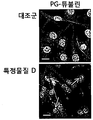
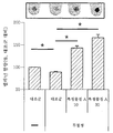
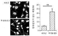
- Figure 1a is a confocal laser microscope to observe the change in the expression of polyglutamylated-tubulin protein (PG-tubulin) according to the specific substance D treatment (scale bar 10 ⁇ m).
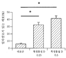
- PG-tubulin polyglutamylated-tubulin protein
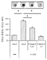
- Figure 1c confirms the effect of ⁇ -MSH and specific substance D treatment on melanin production.
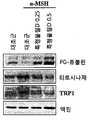
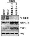
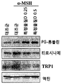
- 1D shows the effect of ⁇ -MSH and specific substance D treatment on the expression of PG-tubulin, tyrosinase, and Tyrosinase-related protein 1 (TRP1) protein in Western blot.
- Figure 2a is confirmed by confocal laser microscopy whether melanin-forming cells (melan-a cells) primary cilia formed by serum starvation, whether the primary cilia formed by a specific substance A treatment.
- Figure 2b shows whether melanin forming cells (melan-a cells) in the primary cilia due to serum starvation, whether the inhibition of the primary cilia due to specific substance A treatment.
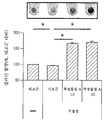
- Figure 2c confirms the effect of serum starvation and specific substance A on the melanin content in the cell.
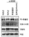
- Figure 2d shows the effect of serum starvation and specific substance A on PG-tubulin, tyrosinase and TRP1 protein expression in Western blot.
- 3A confirms whether specific substance D treatment increases primary cilia in B16F1 melanoma cell line.
- Figure 3b shows the effect of ⁇ -MSH and specific substance D treatment on melanin production in B16F1 melanoma cell line.
- Figure 3c confirms the effect of ⁇ -MSH and specific substance D treatment on the expression of PG-tubulin, tyrosinase, and TRP1 protein in B16F1 melanoma cell line.
- 3d shows whether primary cilia are formed by serum starvation in B16F1 melanoma cell line and whether primary cilia are inhibited by specific substance A treatment.
- Figure 3e shows the effect of serum starvation and specific substance A on the melanin content in the B16F1 melanoma cell line.
- Figure 3f confirms the effect of serum starvation and specific substance A on PG-tubulin, tyrosinase and TRP1 protein expression in Western blot in B16F1 melanoma cell line.
- Figure 4a shows the change in primary cilia expression when B16F1 cells were treated with specific substance S.
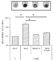
- Figure 4b shows the change in melanin content when B16F1 cells treated with ⁇ -MSH, specific substance S, arbutin.
- Figure 4c confirms the effect on the expression of PG-tubulin, tyrosinase, TRP1 when B16F1 cells treated with ⁇ -MSH, specific substance S, arbutin.
- 5A shows that primary cilia formation induced by specific substance D or serum starvation in melan-a cells are markedly inhibited by GLI2 knockdown.
- 5B shows that melanin content is significantly increased by GLI2 knockdown in melan-a cells.
- 5C is Western blot data showing a significant increase in tyrosinase, TRP1 by GLI2 knockdown in melan-a cells.
- 5D shows that primary cilia formation induced by specific substance D or serum starvation in B16F1 cells are markedly inhibited by GLI2 knockdown.
- 5E shows that melanin content is significantly increased by GLI2 knockdown in B16F1 cells.
- 5F is Western blot data showing a significant increase in tyrosinase, TRP1 by GLI2 knockdown in B16F1 cells.
- FIG. 6A shows the increase in primary cilia by specific material D treatment in NHEMs.
- Figure 6b shows that melanin synthesis is reduced by specific substance D treatment in NHEMs.
- Figure 6c shows the change in brightness after the treatment of the specific substance S and the specific substance D in the skin model.
- FIG. 6D illustrates the change in brightness with a colorimeter after treatment with a specific substance S and a specific substance D in a skin model.
- hair refers to a hairy structure consisting of keratinized epithelial cells and emerging on the surface of the skin. Specifically, the term “hair” refers to the broadest concept including hair and body hair.
- blackening means that the colored pigment of the skin or hair is increased, and in particular, it may mean a phenomenon in which the amount of the melanin pigment is increased so that the skin or the hair shows a dark color. Hair blackening, for example, involves the blackening of hair.
- whitening means a decrease in the color pigment of the skin or hair, and in particular, may mean a phenomenon in which the amount of the melanin pigment is reduced to make the skin or the hair pale.
- Hair whitening or white hair for example, includes the phenomenon that the hair turns white.
- primary cilia also called primary cilium (plural primary cilia) are organelles that protrude from the cell body and include various external sources, including chemical stimuli, physical stimuli, light, osmotic pressure, fluid flow, and gravity signals. Refers to organelles that sense stimuli.
- the degree of relative formation refers to the formation, expression, number, quantity, and quality of the formation or expression when comparing the degree of formation or expression of primary cilia in cells under or without any conditions. It may be a degree indicating the difference. In addition, the degree of formation may include, for example, the number or amount of cells that form or express primary cilia.
- hair blackening effect substance refers to a substance that functions to darken the brightness of the hair or to induce, improve and enhance the deposition of colored pigments, and refers to a single compound, polymer, DNA, RNA, peptide, protein, and the like. And the like, but are not limited thereto.
- melanin-lowering disease includes a disease in which melanin deposition is abnormally suppressed due to abnormalities in the synthesis or degradation of the melanin pigment.
- melanin deposition is abnormally suppressed due to abnormalities in the synthesis or degradation of the melanin pigment.
- leukoplakia Albinism
- leukoderma partial leukemia, rash
- vitiligo quadrichrome vitiligo
- vitiligo ponctue idiopathic red hypopigmentation Syndrome
- mumps leukemia syndromic Albinism
- Alezzandrinisyndrome Hermansky-Pudlak syndrome, Chediak-Higashi syndrome, Griselly Griscelli syndrome (Elejalde syndrome, Griscelli syndrome type 2 and Griscelli syndrome type 3), Waardenburg syndrome, Tischen syndrome (Tietz syndrome), Cross-MuKusick-Breen syndrome, ABCD syndrome, Albinism-deafness syndrome and Vogt-Goyanagi-Harada syndrome (Vo) gt-Koyanagi-Harada syndrome), oculocutaneous albinism, hypomelanosis (idiopathic guttate hypomelanosis, leafy hypomelanosis, progressive ectopic hypomelanosis) (progressive macular hypomelanosis), ecchymoses, pibaldism, nevus depigmentosus, post-inflammatory hypopigmentation, white pityriasis alba Vagabond's leukomelanoderma, Yemenite deaf
- melanin-lowering disease prevention and treatment efficacy substance is a substance which prevents or inhibits the disease of melanin-lowering disease in advance, a substance which alleviates or alleviates the symptoms of the diseased melanin-lowering disease, melanin-lowering disease It includes a substance that eliminates the cause of the substance, a substance that promotes melanin synthesis, a substance that inhibits the inhibition of melanin synthesis, and a substance that inhibits the decomposition of melanin, and darkens the brightness of the entire skin or a specific skin area or deposits of melanin pigment. It refers to a substance that acts to induce, ameliorate, and enhance the compound, and may include, but is not limited to, a single compound, polymer, DNA, RNA, peptide, protein, and the like.
- a method for screening a hair blackening efficacy substance comprising the step of confirming the relative degree of formation of primary cilia before and after treatment of the test substance.
- melanin-forming cells treated with the test substance it may be a method for screening melanin-lowering disease prevention and treatment-efficacy substances comprising the step of confirming the relative degree of formation of primary cilia before and after treatment of the test substance.
- the melanin-lowering disease is in the group consisting of bleaching, vitiligo, depigmentation nevus, white nasal ganglia, rash, post-inflammatory bleaching, ecchymoses, partial leukemia, idiopathic hypopigmentation and mumps leukemia It may be one or more selected.
- the screening method may include determining the test substance as a hair blackening efficacy substance or a melanin-lowering disease prevention and treatment efficacy substance when the primary cilia formed after the test substance treatment are less than the primary cilia before the treatment. It may further include.
- the relative degree of formation of the primary cilia may be determined by one or more of comparing the number of melanin forming cells expressing the primary cilia and comparing the amount of the primary cilia expressed in one cell.
- the relative degree of formation of the primary cilia may be to confirm the expression level of polyglutamylated-tubulin protein. For example, it may be determined by comparing the number of melanin-forming cells expressing the polyglutamylated-tubulin protein or by comparing the amount of polyglutamylated-tubulin protein expressed in one cell, and the above test.
- the test substance can be judged as a hair blackening efficacy substance or a melanin-lowering disease prevention and treatment efficacy substance.
- the relative degree of formation of the primary cilia may be to confirm the expression or activity of GLI (GLI family zinc finger) transcription factor
- the GLI transcription factor may be a GLI family zinc finger 2 (GLI2) transcription factor have.
- GLI or GLI2 transcription factor is less expressed or the activation is inhibited than after the test substance treatment
- the amount of primary cilia formed may be seen to be less and the test substance may be a hair blackening efficacy substance, or melanin. It can be judged as a substance for preventing and treating a lowering disease.
- test substance Even when the number of cells expressed or activated by the GLI transcription factor or GLI2 transcription factor is decreased, the amount of primary cilia formed may be reduced, and the test substance may be used as a hair blackening efficacy substance or a melanin-lowering disease prevention and treatment substance. It can be determined.
- the relative degree of formation of the primary cili may be to determine the degree of expression of tyrosinase (tyrosinase) or TRP1 (Tyrosinase-related protein 1). For example, if the expression of tyrosinase or TRP1 after treatment of the test substance is greater than before treatment of the test substance, the amount of primary cilia formed may be considered to be less, and the test substance may be used for preventing and treating hair blackening efficacy substances, or melanin-lowering diseases. It can be determined as an agonist.
- the amount of primary cilia formed may be considered to be small, and the test substance may be determined as a hair blackening efficacy substance or a melanin-lowering disease prevention and treatment substance.
- the relative degree of primary cilia is known in the art, such as immunofluorescence analysis, western blot, dot blot, ELISA, northern blot, PCR, RT- Those skilled in the art may select appropriately, such as qPCR, GC-MS, LC-MS, and NMR, but is not limited thereto.
- the present invention provides a kit for screening hair blackening efficacy material, which includes a measuring unit for confirming or indicating the relative degree of formation of primary cilia before and after treatment with a test substance.
- the present invention may be a kit for screening melanin-lowering disease prevention and therapeutically effective substance, including a measuring unit indicating a relative degree of formation of primary cilia before and after treatment with a test substance.
- the kit further comprises a melanocyte-forming cell and instructions, the instructions may be that the screening method is described.
- the present invention provides a method of providing information necessary for diagnosing hair condition, comprising the step of confirming the degree of formation of primary cilia.
- the present invention provides a method for providing information necessary for diagnosing melanin-lowering disease, comprising determining a degree of formation of primary cilia.
- the primary cilia may be primary cilia of melanin forming cells of the entire skin, specific skin regions, or hair follicle sites.
- the hair state may be a hair state associated with melanin.
- 'melanin-related hair state' refers to a state of hair that is directly or indirectly related to melanin pigment or melanocyte-forming cells.
- the hair state is highly sensitive to a stimulus that forms melanin. Hair that is low or low, future hair may be more or less likely to be melanin, and hair that is at high or low risk of hair disease associated with melanin, dark or light hair.
- the diagnosis related to melanin-lowering disease is to determine that melanin production is likely to be reduced due to reduced melanin production, to determine the prognosis of the individual affected by melanin-lowering disease, melanin And determining that the depressive disease is likely to be ameliorated or treated.
- confirming the degree of primary cilia formation may be comparing the degree of formation of primary cilia before and after stimulation to form or inhibit melanin is applied.
- the determination of the degree of formation of the primary cilia may be performed by collecting melanin forming cells or measuring the degree of formation of the primary cilia directly from the individual without separating the cells. The collection or measurement may be by any method known to those skilled in the art.
- the hair condition diagnosis may be to determine whether the hair is highly sensitive to a specific stimulus that forms or inhibits melanin.
- the stimulus may be one or more of an exogenous stimulus and an endogenous stimulus.
- the exogenous stimulus may include stimulation caused by ultraviolet light, physical pressure, wounds, heavy metals, chemicals, microbial infection, and stress.
- the endogenous stimulation is oxidative stress changes in the body, aging, inflammation, genetic abnormalities, hormonal changes, digestive system abnormalities, including liver, metabolic abnormalities, nutritional status abnormalities, vitamin and mineral overload It may be to include.
- the degree of formation of the primary cilia may be compared to the degree of formation of the primary cilia in the hair follicle region of an individual at two specific time points.
- melanin forming cells may be collected from the hair follicle site, or the degree of formation of primary cilia may be directly measured from an individual without separating the cells.
- the collection or measurement can be by any method known in the art.
- the hair condition diagnosis may be to determine that there is a possibility that the hair blackening or whitening progresses due to the increase or decrease of melanin deposition in the hair follicle site.
- hair blackening or black hair includes a phenomenon in which various hairs or irritation occur or disappear so that the hair of the entire hair or a specific part turns dark or black.
- hair whitening or “white hair” as used herein includes a phenomenon in which the hair of the entire hair or a specific part becomes bright or white due to various causes or stimuli occurring or disappearing.
- the hair condition diagnosis may be to determine the grade regarding the possibility of blackening or whitening of the hair according to the degree of primary cilia formed.
- the grade may be a reference value based on data on the degree of formation of primary cilia obtained from a hair follicle melanin forming cell sample obtained from a certain number of individuals, and a grade divided by the intervals based thereon.
- the sample may be a hair follicle cell or a hair sample, and specifically, may include melanocyte-forming cells of the hair follicle or hair.
- the present invention confirmed that melanin formation by treating a melanin-forming cell with a substance that induces melanin formation is inhibited by a specific substance D, which is a substance that induces primary cilia.
- a specific substance D which is a substance that induces primary cilia.
- this inhibitory effect was not seen in cells knocked down GLI2 via specific siRNAs.
- the specific substance S activates the sonic hedgehog (SHH) -smoothened (Smo) signaling pathway and expresses or activates the GLI transcription factor, which induces the formation of primary cilia, resulting in melanin-forming cells.
- the present inventors confirmed that the specific substance A significantly inhibits primary cilia formation, and it was confirmed that melanin formation is promoted in melanocytes when the primary cilia are inhibited by the specific substance A.
- the present inventors confirmed that formation of primary cilia is induced through Smo-GLI2 signaling system, and melanin synthesis inhibition occurs due to primary cilia formation. On the contrary, the inhibition of primary cilia formation promotes melanin synthesis. .
- the present invention finds a method for screening substances that exhibit hair blackening efficacy and substances that exhibit prophylactic and therapeutic efficacy of melanin-lowering diseases using this relationship. That is, substances that promote or inhibit primary cilia formation promote or inhibit melanin synthesis of melanocytes, so that such substances will act the same in melanocytes of hair follicles, and through these substances can control hair blackening or whitening. have.
- the substance that controls primary cilia formation can regulate primary cilia formation in the skin of an individual suffering from melanin-lowering diseases, thereby promoting melanin synthesis by melanin-forming cells, thereby preventing and preventing melanin-lowering diseases. Treatment is possible. Therefore, whether or not a specific substance promotes primary cilia formation of melanin forming cells in the entire skin where melanin forming cells are distributed and in the skin region in which the symptom is caused by the disease is effective in preventing and treating melanin lowering diseases.
- the material can be screened.
- kits including a measuring unit that can confirm or indicate the degree of formation of the primary cilia.
- the present invention is to find a method for providing information necessary for evaluating or diagnosing melanin-related hair condition by checking the degree of formation of primary cilia.
- the degree of formation of primary cilia may be confirmed to obtain information necessary for evaluating or diagnosing melanin-lowering diseases. It can be predicted and predicted that melanin synthesis will change if the formation of primary cilia in the melanocytes of the skin or hair follicles changes relatively, so that the future condition of hair can be predicted, and the prognosis and treatment of melanin-lowering diseases This is because the possibility and the therapeutic effect can be judged.
- Melan-A a melanogenesis cell derived from C57BL / 6 J (black, a / a) mice, obtained from normal human epidermal melanocytes (NHEMs, Cascade Biologics, Portland, OR, USA) -A) Cell lines were obtained from Dr. Dorothy C. Bennett (St. George's Hospital Medical School, London, UK). B16F1 mouse melanoma cell line was obtained from ATCC (Manassas, VA, USA).
- Human epidermal melanocyte forming cells were maintained under Human Melanocyte Growth Supplements (HMGS) (Cascade Biologics, Inc., Mansfield, UK) in M-254 medium.
- Melan-A cells were maintained under 10% (v / v) fetal bovine serum, 1% (v / v) penicillin-streptomycin, and 0.2 ⁇ M phorbol 12-myristate 13-acetate in RPMI 1640 medium.
- B16F1 mouse melanoma cell lines were cultured in DMEM with 10% (v / v) fetal bovine serum and 1% (v / v) penicillin-streptomycin.
- Three-dimensional human skin substitutes (MelanoDerm TM, MEL-312-B, MatTek, Seoul, Korea) were obtained, followed by keratinocyte growth factor (KGF), fibroblast growth factor (b-FGF), and It was maintained according to the manufacturer's instructions in EPI-100-NMM-113 medium optimized with alpha-melanocyte stimulating hormones ( ⁇ -MSH).
- KGF keratinocyte growth factor
- b-FGF fibroblast growth factor
- siRNAs targeting mouse GLI2 GLI family zinc finger 2 # 1, # 2, and negative control siRNA (scrambled control siRNA, siNC) (5'-CCUACGCCACCAAUUUCGU-3 ') were generated by Genolution, Seoul. , South Korea).
- the siRNA # 1 and siRNA # 2 is a siRNA that targets GLI2 to inhibit GLI2 expression, which can be easily obtained by those skilled in the art, can be obtained in the future, can be manufactured, or can be prepared in the future. Can also be used.
- SiRNA was transfected into Melan-a and B16F1 cells cultured in 60 mm dishes using Lipofectamine ® RNAi MAX (Invitrogen, Carlsbad, Calif., USA). After incubation with siRNA for 24 hours, cells were treated with each compound (reagent) and maintained for 48 hours.
- Anti-polyglutamylated-tubulin antibody (AG-20B-0020, 1: 1000) was obtained from Adipogene (San Diego, CA, USA) and an anti-GLI2 antibody (anti -GLI2 antibody, 18989-1-AP, 1: 1000) was obtained from Proteintech (Chicago, IL, USA), and anti-actin antibody (MAB1501, 1: 10,000) was obtained from Millipore (Temecula, CA, USA), anti- ⁇ PEP7 (tyrosinase) antibody (anti- ⁇ PEP7 (tyrosinase) antibody, 1: 1000) and anti- ⁇ PEP1 (TRP1) antibody (anti- ⁇ PEP1 (TRP1) antibody, 1: 1000) was obtained from VJ Hearing (NIH, Bethesda, MD). For protein detection, the membrane was incubated with an HRP- conjugated secondary antibody gated signal was measured using a SuperSignal ® West Dura HRP Detection Kit ( Pierce Inc., Rockford,
- NHEMs, Melan-a, and B16F1 cells were incubated using Lab-Tek TM glass chamber slides (Thermo Scientific, Waltham, Mass., USA), then washed with cold PBS and 4% (w / v) for 20 minutes. ) was fixed with paraformaldehyde and washed twice with PBS again. And incubated for 3 min in PBS containing 0.1% (v / v) Triton TM X-100 (PBS-T). The cells were washed three more times with PBS-T and diluted with PBS-T containing 5% (v / v) goat serum (1: 100 dilution, Adipogene).
- Primary cilia are intracellular organs with polarized structures in which the cilia's binding (cilia formation) and degradation are interconnected by a variety of causes, including cell cycle, drift, polarity, spinal development, and genetic diseases.
- Primary cilia are known as tubulin-based cellular structures.
- Tubulin undergoes multiple post-translational modifications (PTMs), and polyglutamylation is believed to be related to the activity of tubulin on the primary cilia. Therefore, to assess primary cilia formation, the expression levels of polyglutamylated tubulin protein were analyzed.
- PTMs post-translational modifications
- the specific substance D is a substance for inducing primary cilia formation, and includes all substances known in the art or to be discovered in the future to induce primary cilia formation of cells regardless of their type, shape, and properties.
- specific substance A was treated with melan-a cells in serum-free conditions and incubated for 48 hours.
- the specific substance A is a substance that inhibits primary cilia formation, and includes all substances known in the art or to be discovered in the future to inhibit primary cilia formation of cells regardless of their type, shape, and properties.
- B16F1 cells were cultured in serum starvation medium for 48 hours after treatment with specific substance A (10 ⁇ M, 30 ⁇ M). After the immunostaining, the percentage of primary cilia formed cells was analyzed (FIG. 3D), and cell melanin content was measured (FIG. 3E). PG-tubulin, tyrosinase, and TRP1 were analyzed by Western blot (FIG. 3F). .
- primary cilia formation affects melanin production in melanocytes and melanoma cell lines.
- inducing the formation of melanin-forming primary cilia decreases melanin production, and on the contrary, inhibiting the formation of melanin-forming cilia increases melanin production.
- Inhibiting the formation of primary cilia increased melanin production and confirmed that hair blackening occurred.
- melanin production may be inhibited, leading to browning or whitening of the hair. This could be used to screen for materials that have a hair blackening efficacy.
- melanin-depressive disease inhibiting melanin-forming primary cilia formation increases melanogenesis, thereby preventing and treating melanin-lowering disease.
- melanin-lowering disease By utilizing this, it is possible to screen for substances that are effective in preventing and treating melanin-lowering diseases. Using the above points, it is possible to develop related kit products, and to diagnose hair blackening and melanin-lowering diseases.
- Activated Smo (Smoothened) proteins enhance the expression and activity of GLI transcription factors, which are genes related to the cell cycle and gene expression downstream of the sonic hedgehog (SHH) signaling pathway. Adjust
- the present inventors studied the relationship between Smo and GLI2 transcription factors in the regulation of melanin formation through primary cilia formation. Specifically, B16F1 cells were treated with 0.5 or 1 ⁇ M of a specific substance S, and after 24 hours, immunostaining was performed to measure cells expressing primary cilia, indicating that primary cilia-expressing cells increased. (FIG. 4A).
- the specific substance S is a substance that activates or enhances expression or activation of GLI or GLI2 transcription factors, and specifically, is known to those skilled in the art as a substance that activates or enhances Smo-GLI or Smo-GLI2 signaling pathways. It includes all substances that will be lost or discovered in the future.
- B16F1 cells were pretreated with ⁇ -MSH (1 ⁇ M) and after 24 hours treated with either specific substance S (5 ⁇ M) or arbutin (500 ⁇ M). After 24 hours, the cellular melanin content, tyrosinase and TRP1 protein expression levels were analyzed by Western blot. As a result, induction of primary cilia by specific substance S inhibited melanin production in B16F1 cells cultured with ⁇ -MSH (FIG. 4B). The expression level of tyrosinase and TRP1 protein also decreased when primary ciliary cell formation was activated by specific substance S (FIG. 4C).
- siRNA-related information to obtain two types of siRNA (# 1, # 2) for GLI2, and then Melan-a cells were siRNA (# 1, # 2) or scrambled control siRNA for GLI2.
- siNC 48 days after transfection with control siRNA, siNC), either specific substance D (0.5 ⁇ M) treatment or serum-free conditions induce primary cilia formation for 2 days. Then, it was measured by fluorescence microscopy.
- treatment with specific material D or incubation under serum-free conditions strongly induced primary cilia formation in the control (siNC).
- primary cilia formation induced by stimulation (specific substance D or serum starvation) were significantly inhibited when GLI2 was knocked down (# 1, # 2) (FIG. 5A).
- GLI2 knockdown cells were cultured under specific substance D or under serum-free conditions to determine cellular melanin content. Treatment of specific substance D or culture under serum-free conditions on cells treated with control siRNA slightly inhibited melanin content. GLI2 knockdown cells were cultured under specific substance D or under serum-free conditions, resulting in a significant increase in melanin content (FIG. 5B). Experiments were also performed on melanin forming enzyme expression on GLI2 knockdown cells. Western blot analysis revealed that KLI2 knockdown in melan-a cells significantly increased tyrosinase and TRP1 protein levels compared to wild-type cells (FIG. 5C). Taken together, it can be seen that melanogenesis inhibition through specific substance D or serum starvation is mediated through primary cilia induction via the Smo-GLI2 signaling pathway.
- siNC and GLI2 siRNA were transfected to B16F1 cells and treated with specific substance D (0.5 ⁇ M) or serum-free medium one day later, and then, two days later, primary cilia were formed by fluorescence microscopy. Analyzed. GLI2, tyrosinase, TRP1 were analyzed by Western blot. As a result, it was found that primary cilia formation induced by specific substance D or serum starvation were inhibited in GLI2 knockdown B16F1 mouse melanoma cells. Melanin forming protein expression and melanin content in GLI2 knockdown B16F1 cells were increased as in melan-a cells (FIGS. 5D-5F).
- melanin production can be controlled by primary cilia formation by Smo-GLI2 mediated signaling system. That is, it was found that inducing primary cilia through activation of the Smo-GLI2 signal pathway inhibits melanin production, and melanogenesis is enhanced by inhibiting primary cilia formation by inhibiting activation of the Smo-GLI2 signaling pathway. Therefore, inhibiting the formation of primary cilia in melanocytes of hair follicles promotes melanin production, which may lead to blackening of hair. To whitening. These properties can be used to screen hair blackening efficacy substances.
- inhibiting the activation of the Smo-GLI2 signaling pathway to inhibit primary cilia in melanin-lowering diseases also increases melanogenesis, thereby preventing and treating melanin-lowering diseases.
- the regulation of melanin production through primary cilia was examined using NHEMs and three-dimensional human skin substitutes (reconstituted epidermal models including normal human epidermal keratinocytes and NHEMs).
- NHEMs were treated with specific substance D (0.5 ⁇ M) and after 2 days immunization against acetylated tubulin (activated form of tubulin, green) and ARL13B (a small ciliary G protein of the Ras superfamily, red) Primary cilia of NHEMs were observed by staining.
- ARL13B IFT88 (Intra flagellar transport protein 88), CP110 (a 110 kDa centriolar protein) and BBS2 (Bardet-Biedl syndrome (BBS) protein 2) may be used as protein markers.
- NHEMs were treated with specific substance D (0.5 ⁇ M and 1 ⁇ M) and 48 hours later, the cell melanin content was confirmed.
- melanin-forming cells are also distributed in hair follicles, a person skilled in the art can recognize that the same result will be obtained in artificial hair or actual hair including melanin-forming cells of hair follicles.
- primary cilia can be used to screen for substances that are effective in the treatment and prophylaxis of melanin-lowering diseases, substances that are effective for darkening the hair, and development of related kit products and skin and hair-related diagnosis will be possible.
Landscapes
- Life Sciences & Earth Sciences (AREA)
- Health & Medical Sciences (AREA)
- Engineering & Computer Science (AREA)
- Biomedical Technology (AREA)
- Immunology (AREA)
- Urology & Nephrology (AREA)
- Hematology (AREA)
- Chemical & Material Sciences (AREA)
- Molecular Biology (AREA)
- Cell Biology (AREA)
- Physics & Mathematics (AREA)
- Pathology (AREA)
- Food Science & Technology (AREA)
- Medicinal Chemistry (AREA)
- Biotechnology (AREA)
- Analytical Chemistry (AREA)
- Biochemistry (AREA)
- General Health & Medical Sciences (AREA)
- General Physics & Mathematics (AREA)
- Microbiology (AREA)
- Proteomics, Peptides & Aminoacids (AREA)
- Bioinformatics & Cheminformatics (AREA)
- Toxicology (AREA)
- Tropical Medicine & Parasitology (AREA)
- Investigating Or Analysing Biological Materials (AREA)
- Medicines That Contain Protein Lipid Enzymes And Other Medicines (AREA)
- Measuring Or Testing Involving Enzymes Or Micro-Organisms (AREA)
- Cosmetics (AREA)
Abstract
본 발명은 일측면에서, 멜라닌 형성 세포(melanocyte)의 일차 섬모(primary cilia)의 상대적 형성 정도를 확인하는 단계를 포함하는, 모발 흑화 효능 물질, 또는 멜라닌 저하성 질환 예방 및 치료 효능 물질의 스크리닝 방법에 관한 것으로서, 본 발명의 일측면을 통하여 모발 흑화 물질, 멜라닌 저하성 질환 예방 및 치료 물질의 효과적인 스크리닝 및 발굴이 가능하며, 이를 통해 관련 산업의 비약적 발달을 도모할 수 있다.
Description
본 명세서는 일차 섬모(프라이머리 실리아, primary cilia)를 조절하는 모발 흑화 물질을 스크리닝하는 방법에 관한 것으로서, 구체적으로 세포의 소기관인 일차 섬모의 형성 정도를 확인하여 모발 흑화 효능을 발휘하는 물질을 스크리닝 하는 방법에 관한 것이다.
멜라닌(melanin)은 검은 색소와 단백질의 복합체 형태를 지니는 페놀류의 생체 고분자 물질로서, 사과, 감자, 바나나의 잘린 표면이 공기 중에 노출될 때 발생하는 갈변 또는 동물의 외피 깃털, 피부, 모발, 눈 등에서 관찰된다. 멜라닌이 과잉 생산되어 침착되는 경우 기미, 주근깨 등이 형성되고 모발 흑화 등과도 직결되며, 멜라닌으로 인해 피부 또는 모발 노화도 촉진되고 피부암도 유발될 수 있다.
자외선, 염증, 호르몬 등에 의해서 멜라닌 형성 세포 자극 호르몬(melanocyte stimulating hormone, MSH)이 분비되고, MSH는 수용체와 반응하여 멜라닌 형성 세포 내에 cAMP를 향상시켜 멜라닌을 합성하며, 합성된 멜라닌은 멜라닌 형성 세포의 외부로 분비되어 자외선 등으로부터 피부 또는 모발을 보호하는 역할을 하게 된다. 멜라닌의 합성은 주로 α-MSH에 의해 조절되고, 멜라닌의 합성에 관여하는 단백질로는 MITF, TYR, TRP1, TRP2 등이 알려져 있다.
모발에 있어서의 멜라닌은 모발의 색상을 결정하는 중요한 인자로서, 모낭에 존재하는 멜라닌 형성 세포 내에서 생성된 멜라닌은 모발피질세포 케라티노사이트에 전달된 후, 모발의 성장과 함께 위쪽으로 이동한다. 생리적 노화의 한 현상으로서 인식되는 모발 백화는 모발 멜라닌 형성 세포의 수적 감소 및 멜라닌 형성 세포의 기능저하 등에 의한 멜라닌 생성의 감소에 의해 유발된다. 현재 모발 백화의 해결방안으로 염색 시술이 사용되고 있으나, 염색은 일시적인 방법으로 백발의 성장에 의해 다시 염색하지 않으면 안되고, 모발 염색약의 염료 속에 포함되어 있는 성분이 자극성분이 되어 접촉성 피부염 또는 피부 알러지를 일으켜 문제시 되고 있다.
또한, 멜라닌 생합성에 이상이 생기면 비정상적인 착색에 의해 피부의 비정상적 백화 등이 나타나는데, 이로 인해 백화증(백색증, albinism), 백반증 등의 질병이 발생하기도 한다.
일차 섬모(primary cilia)는 세포체에서 돌출된 기관으로서, 다양한 외부자극을 감지하는 기관으로 알려져 있는데, 모발 흑화나 멜라닌과 관련된 질환과 일차 섬모 간의 관련성에 대해서는 아직 알려진 바가 없다.
본 발명은 일측면에서, 세포의 일차 섬모(primary cilia)를 통해 모발 흑화 효능 물질 및 멜라닌 저하성 질환 치료용 물질을 효과적으로 스크리닝하는 새로운 방법, 모발 흑화 효능 물질 및 멜라닌 저하성 질환 치료 효능 물질을 스크리닝할 수 있는 키트, 및 일차 섬모를 이용하여 모발 상태나 멜라닌 저하성 질환 관련 진단에 필요한 정보를 제공하는 방법을 제안하기 위한 목적을 가진다.
본 발명은 일측면에서,
멜라닌 형성 세포(melanocyte)에 시험 물질을 처리하는 단계;
상기 시험 물질을 처리한 멜라닌 형성 세포에서, 시험 물질 처리 전과 후의 일차 섬모(primary cilia)의 상대적 형성 정도를 확인하는 단계를 포함하는, 모발 흑화 효능 물질의 스크리닝 방법에 관한 것이다.
본 발명은 다른 측면에서, 상기 단계를 포함하는 멜라닌 저하성 질환 예방 및 치료 효능 물질의 스크리닝 방법에 관한 것이기도 하다.
본 발명은 또 다른 측면에서, 시험 물질 처리 전과 후의 일차 섬모의 상대적 형성 정도를 나타내는 측정부를 포함하는, 모발 흑화 효능 물질 또는 멜라닌 저하성 질환 예방 및 치료 효능 물질의 스크리닝을 위한 키트에 관한 것이기도 하다.
또한, 본 발명은 또 다른 측면에서, 일차 섬모의 형성 정도를 확인하는 단계를 포함하는, 모발 상태 진단 방법 또는 멜라닌 저하성 질환 관련 진단 방법일 수 있고, 상기 진단을 위해 필요한 정보를 제공하는 방법에 대한 것일 수 있다.
본 발명은 일측면에서 멜라닌 형성 세포 내 일차 섬모(primary cilia)의 상대적 형성 정도를 확인하는 단계를 포함하는 모발 흑화 효능 물질 스크리닝 방법을 제공함으로써, 보다 효과적으로 모발 흑화 물질 및 멜라닌 저하성 질환 치료 물질을 스크리닝할 수 있도록 하고 기존의 방법으로 발견하지 못했던 모발 흑화 물질 및 멜라닌 관련 질환 치료 물질도 확인이 가능하게 하므로, 관련 분야의 산업발전에 크게 기여할 수 있다.
도 1a는 특정 물질 D 처리에 따른 폴리글루타밀레이티드-튜불린 단백질(polyglutamylated-tubulin protein, PG-tubulin)의 발현 변화를 공초점 레이저 현미경으로 관찰한 것이다(스케일바는 10μm).
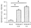
도 1b는 특정 물질 D 처리가 일차 섬모를 증가시키는지 여부를 확인한 것이다.
도 1c는 α-MSH 및 특정 물질 D 처리가 멜라닌 생성량에 미치는 영향을 확인한 것이다.
도 1d는 α-MSH 및 특정 물질 D 처리가 PG-tubulin, 타이로시나제(tyrosinase), 및 TRP1(Tyrosinase-related protein 1) 단백질의 발현에 미치는 영향을 웨스턴 블럿으로 나타낸 것이다.
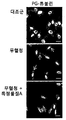
도 2a는 멜라닌 형성 세포(멜란-a 세포)에서 혈청 기아로 인한 일차 섬모 형성 여부, 특정 물질 A 처리로 인한 일차 섬모형성 억제 여부를 공초점 레이저 현미경으로 확인한 것이다.
도 2b는 멜라닌 형성 세포(멜란-a 세포)에서 혈청 기아로 인한 일차 섬모 형성 여부, 특정 물질 A 처리로 인한 일차 섬모형성 억제 여부를 나타낸 것이다.
도 2c는 혈청 기아 및 특정 물질 A가 세포 내 멜라닌 함량에 미치는 영향을 확인한 것이다.
도 2d는 혈청 기아 및 특정 물질 A가 PG-tubulin, 타이로시나제 및 TRP1 단백질 발현 에 미치는 영향을 웨스턴 블럿으로 나타낸 것이다.
도 3a는 B16F1 흑색종 세포주에서 특정 물질 D 처리가 일차 섬모를 증가시키는지 여부를 확인한 것이다.
도 3b는 B16F1 흑색종 세포주에서 α-MSH 및 특정 물질 D 처리가 멜라닌 생성량에 미치는 영향을 나타낸 것이다.
도 3c는 B16F1 흑색종 세포주에서 α-MSH 및 특정 물질 D 처리가 PG-tubulin, 타이로시나제(tyrosinase), 및 TRP1 단백질의 발현에 미치는 영향을 확인한 것이다.
도 3d는 B16F1 흑색종 세포주에서 혈청 기아로 인한 일차 섬모 형성 여부, 특정 물질 A 처리로 인한 일차 섬모형성 억제 여부를 나타낸 것이다.
도 3e는 B16F1 흑색종 세포주에서 혈청 기아 및 특정 물질 A가 세포 내 멜라닌 함량에 미치는 영향을 나타낸 것이다.
도 3f는 B16F1 흑색종 세포주에서 혈청 기아 및 특정 물질 A가 PG-tubulin, 타이로시나제 및 TRP1 단백질 발현에 미치는 영향을 웨스턴 블럿으로 확인한 것이다.
도 4a는 B16F1 세포에 특정 물질 S를 처리한 경우 일차 섬모 발현 변화를 나타낸 것이다.
도 4b는 B16F1 세포에 α-MSH, 특정 물질 S, 알부틴을 처리한 경우 멜라닌 함량의 변화를 나타낸 것이다.
도 4c는 B16F1 세포에 α-MSH, 특정 물질 S, 알부틴을 처리한 경우 PG-튜불린, 타이로시나제, TRP1의 발현에 미치는 영향을 확인한 것이다.
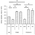
도 5a는 멜란-a 세포에서 특정 물질 D 또는 혈청 기아로 유도된 일차 섬모 형성이 GLI2 넉다운에 의해 현저히 억제됨을 나타낸 것이다.
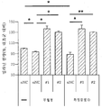
도 5b는 멜란-a 세포에서 GLI2 넉다운에 의해 멜라닌 함량이 현저하게 증가됨을 나타낸 것이다.
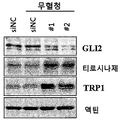
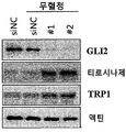
도 5c는 멜란-a 세포에서 GLI2 넉다운에 의해 타이로시나제, TRP1이 현저하게 증가됨을 나타낸 웨스턴 블럿 자료이다.
도 5d는 B16F1 세포에서 특정 물질 D 또는 혈청 기아로 유도된 일차 섬모 형성이 GLI2 넉다운에 의해 현저히 억제됨을 나타낸 것이다.
도 5e는 B16F1 세포에서 GLI2 넉다운에 의해 멜라닌 함량이 현저하게 증가됨을 나타낸 것이다.
도 5f는 B16F1 세포에서 GLI2 넉다운에 의해 타이로시나제, TRP1이 현저하게 증가됨을 나타낸 웨스턴 블럿 자료이다.
도 6a는 NHEMs에서 특정 물질 D 처리에 의해 일차 섬모가 증가함을 나타낸 도면이다.
도 6b는 NHEMs에서 특정 물질 D 처리에 의해 멜라닌 합성이 감소함을 나타낸 것이다.
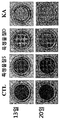
도 6c는 피부 모델에 특정 물질 S 및 특정 물질 D 를 처리한 뒤 밝기 변화를 확인한 것이다.
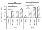
도 6d는 피부 모델에 특정 물질 S 및 특정 물질 D 를 처리한 뒤 색도계로 밝기 변화를 평가한 것이다.
이하, 본 명세서에 대하여 상세히 설명한다.
본 명세서에서 "모발"이라 함은, 각질화된 상피세포로 이루어져 피부 표면으로 나오는 실 모양으로 된 구조물로서, 구체적으로 신체의 털을 의미하며, 머리카락 및 체모를 포함하는 최광의의 개념이다.
본 명세서에서 “흑화”라 함은, 피부 또는 모발의 유색 색소가 증가되는 것을 의미하며, 특히 멜라닌 색소의 양이 증가되어 피부 또는 모발이 짙은 색을 나타내게 되는 현상을 의미할 수 있다. 예를 들어 모발 흑화는 머리카락이 검게 변하는 현상을 포함한다.
본 명세서에서 “백화”라 함은, 피부 또는 모발의 유색 색소가 감소하는 것을 의미하며, 특히 멜라닌 색소의 양이 감소되어 피부 또는 모발이 옅은 색을 나타내게 되는 현상을 의미할 수 있다. 예를 들어 모발 백화 또는 백발은 머리카락이 희게 변하는 현상을 포함한다.
본 명세서에서 "일차 섬모"는, primary cilium(복수일 경우 primary cilia)라고도 하며, 세포체에서 돌출된 소기관으로서, 화학적 자극, 물리적 자극, 빛, 삼투압, 유체 흐름, 및 중력 신호 등을 포함하는 다양한 외부자극을 감지하는 세포 소기관을 말한다.
본 명세서에서 "상대적 형성 정도"는, 어떠한 조건에 처하거나 처하지 않은 세포에서의 일차 섬모(primary cilia)의 형성 또는 발현 정도를 비교하였을 때, 형성 또는 발현의 여부, 수, 양, 질(quality)의 차이를 나타내는 정도일 수 있다. 또한, 상기 형성 정도는, 예컨대, 일차 섬모를 형성 또는 발현하는 세포의 수나 양을 포함할 수 있다.
본 명세서에서 "모발 흑화 효능 물질"은, 모발의 밝기를 어둡게 하거나 유색 색소의 침착을 유도, 개선 및 증진시키는 등의 작용을 하는 물질을 총칭하며, 단일 화합물, 중합체, DNA, RNA, 펩티드, 단백질 등을 포함할 수 있으나 이에 제한되지 않는다.
본 명세서에서 “멜라닌 저하성 질환”은, 멜라닌 색소의 합성 또는 분해의 이상에 따라 비정상적으로 멜라닌 침착이 억제되는 질환을 포함한다. 예를 들어, 백화증(백색증, Albinism), 백색피부증(leukoderma), 부분 백피증, 어루러기, 백반증(vitiligo), 쿼디크롬 백반증(quadrichrome vitiligo), 반점성 백반증(vitiligo ponctue), 특발성 적상 저색소증, 점상 백피증, 백색증 증후군(syndromic Albinism)(예컨대, 알레잔드리니 증후군(Alezzandrinisyndrome), 헤르만스키-푸들라크 증후군(Hermansky-Pudlak syndrome), 세디아크-히가시 증후군(Chediak-Higashi syndrome), 그리셀리 증후군(Griscelli syndrome)(엘레잘데 증후군(Elejalde syndrome), 그리셀리 증후군 타입 2(Griscelli syndrome type 2) 및 그리셀리 증후군 타입 3(Griscelli syndrome type 3)), 바르덴부르크 증후군(Waardenburg syndrome), 티쩨 증후군(Tietz syndrome), 크로스-먹큐직-브린 증후군(Cross-MuKusick-Breen syndrome), ABCD 증후군(ABCD syndrome), 백색증-청각장애 증후군(Albinism-deafness syndrome) 및 보그트-고야나기-하라다 증후군(Vogt-Koyanagi-Harada syndrome)), 눈 피부 백색증(oculocutaneous albinism), 저멜라닌증(hypomelanosis)(물방울모양 저멜라닌증(idiopathic guttate hypomelanosis), 잎모양 저멜라닌증(phylloid hypomelanosis), 진행성 반상 저멜라닌증(progressive macular hypomelanosis)), 반상 경피증, 얼룩백색증(piebaldism), 탈색소 모반(nevus depigmentosus), 염증 후 색소침착저하증(염증 후 탈색증, postinflammatory hypopigmentation), 백색 비강진(pityriasis alba), 바가본드 백반흑피증(Vagabond's leukomelanoderma), 예맨 시청각장애 색소침착저하 증후군(Yemenite deaf-blind hypopigmentation syndrome), 웬드-바우쿠스 증후군(Wende-Bauckus syndrome), 워로노프 반지증(Woronoff's ring), 멜라닌결핍증(amelanism), 및 루시즘(Leucism)을 포함한다.
본 명세서에서 "멜라닌 저하성 질환 예방 및 치료 효능 물질"은, 멜라닌 저하성 질환의 이환을 사전에 방지 또는 억제하는 물질, 이환된 멜라닌 저하성 질환의 증상을 경감 또는 완화시키는 물질, 멜라닌 저하성 질환의 원인을 해소시키는 물질, 멜라닌 합성을 증진시키는 물질, 멜라닌 합성의 억제를 방지하는 물질, 멜라닌의 분해를 억제시키는 물질을 포함하는 것으로서, 전체 피부 또는 특정 피부 부위의 밝기를 어둡게 하거나 멜라닌 색소의 침착을 유도, 개선 및 증진시키는 등의 작용을 하는 물질을 총칭하며, 단일 화합물, 중합체, DNA, RNA, 펩티드, 단백질 등을 포함할 수 있으나 이에 제한되지 않는다.
본 발명은 일 측면에서,
멜라닌 형성 세포(melanocyte)에 시험 물질을 처리하는 단계;
상기 시험 물질을 처리한 멜라닌 형성 세포에서, 시험 물질 처리 전과 후의 일차 섬모(primary cilia)의 상대적 형성 정도를 확인하는 단계를 포함하는, 모발 흑화 효능 물질의 스크리닝 방법을 제공한다.
본 발명은 다른 측면에 의하면,
멜라닌 형성 세포(melanocyte)에 시험 물질을 처리하는 단계;
상기 시험 물질을 처리한 멜라닌 형성 세포에서, 시험 물질 처리 전과 후의 일차 섬모(primary cilia)의 상대적 형성 정도를 확인하는 단계를 포함하는 멜라닌 저하성 질환 예방 및 치료 효능 물질의 스크리닝 방법일 수 있다.
일 구현 예에서, 상기 멜라닌 저하성 질환은 백화증, 백반증, 탈색소 모반, 백색 비강진, 어루러기, 염증후 탈색증, 반상 경피증, 부분 백피증, 특발성 적상 저색소증 및 점상 백피증으로 이루어진 군에서 선택된 하나 이상일 수 있다.
일 구현 예에 의하면, 상기 스크리닝 방법은 상기 시험 물질 처리 후에 형성된 일차 섬모가 처리 전의 일차 섬모에 비해 적을 경우 상기 시험 물질을 모발 흑화 효능 물질, 또는 멜라닌 저하성 질환 예방 및 치료 효능 물질로 판단하는 단계를 더 포함할 수 있다.
다른 구현 예에 의하면, 상기 일차 섬모의 상대적 형성 정도는 일차 섬모가 발현된 멜라닌 형성 세포의 수의 비교 및 한 세포 내에 발현된 일차 섬모 양의 비교 중 하나 이상을 통하여 판정하는 것일 수 있다.
또다른 구현 예에 의하면, 상기 일차 섬모의 상대적 형성 정도는 폴리글루타밀레이티드-튜불린 단백질(polyglutamylated-tubulin protein)의 발현 정도로 확인하는 것일 수 있다. 예컨대, 폴리글루타밀레이티드-튜불린 단백질이 발현된 멜라닌 형성 세포의 수를 비교하거나, 한 세포 내에 발현된 폴리글루타밀레이티드-튜불린 단백질의 양을 비교하여 판정하는 것일 수 있고, 상기 시험 물질 처리 후의 폴리글루타밀레이티드-튜불린 단백질이 발현된 멜라닌 형성 세포의 수나 한 세포 내에 발현된 폴리글루타밀레이티드-튜불린 단백질의 양이 시험 물질 처리 전보다 적을 경우, 형성된 일차 섬모의 양이 적어진 것으로 볼 수 있고 상기 시험 물질을 모발 흑화 효능 물질, 또는 멜라닌 저하성 질환 예방 및 치료 효능 물질로 판정할 수 있다.
일 구현 예에서, 상기 일차 섬모의 상대적 형성 정도는 GLI(GLI family zinc finger) 전사인자의 발현 또는 활성 정도로 확인하는 것일 수 있고, 상기 GLI 전사인자는 GLI2(GLI family zinc finger 2) 전사인자일 수 있다. 예컨대, 상기 시험 물질 처리 후 상기 GLI 또는 GLI2 전사인자가 시험 물질 처리 전보다 적게 발현되거나 활성화가 억제된 경우, 형성된 일차 섬모의 양이 적어진 것으로 볼 수 있고 상기 시험 물질을 모발 흑화 효능 물질, 또는 멜라닌 저하성 질환 예방 및 치료 효능 물질로 판정할 수 있다. GLI 전사인자 또는 GLI2 전사인자가 발현되거나 활성화된 세포수가 적어진 경우에도 형성된 일차 섬모의 양이 적어진 것으로 볼 수 있고 상기 시험 물질을 모발 흑화 효능 물질, 또는 멜라닌 저하성 질환 예방 및 치료 효능 물질로 판정할 수 있다.
일 구현 예에 의하면, 상기 일차 섬모의 상대적 형성 정도는 티로시나제(tyrosinase) 또는 TRP1(Tyrosinase-related protein 1)의 발현 정도로 확인하는 것일 수 있다. 예컨대, 상기 시험 물질 처리 후 티로시나제 또는 TRP1의 발현이 시험 물질 처리 전보다 많아진 경우, 형성된 일차 섬모의 양이 적어져서 그런 것으로 볼 수 있고 상기 시험 물질을 모발 흑화 효능 물질, 또는 멜라닌 저하성 질환 예방 및 치료 효능 물질로 판정할 수 있다. 티로시나제 또는 TRP1이 발현된 세포수가 많아진 경우에도 형성된 일차 섬모의 양이 적어져서 그런 것으로 볼 수 있고 상기 시험 물질을 모발 흑화 효능 물질, 또는 멜라닌 저하성 질환 예방 및 치료 효능 물질로 판정할 수 있다.
일 구현 예에서, 일차 섬모의 상대적 형성 정도는, 공지의 기술, 예컨대, 면역형광 분석, 웨스턴 블롯(western blot), 닷 블롯(dot blot), ELISA, 노던 블롯(northern blot), PCR, RT-qPCR, GC-MS, LC-MS, NMR 등 당업자가 적절히 선택할 수 있으며, 이에 제한되는 것은 아니다.
본 발명은 다른 측면에서, 시험 물질 처리 전과 후의 일차 섬모의 상대적 형성 정도를 확인하거나 나타내는 측정부를 포함하는, 모발 흑화 효능 물질 스크리닝을 위한 키트를 제공한다.
본 발명의 다른 측면에 따르면, 본 발명은 시험 물질 처리 전과 후의 일차 섬모의 상대적 형성 정도를 나타내는 측정부를 포함하는, 멜라닌 저하성 질환 예방 및 치료 효능 물질 스크리닝을 위한 키트일 수 있다.
일 구현 예에 의하면, 상기 키트는 멜라닌 형성 세포 및 지시서를 더 포함하고, 상기 지시서에는 상기 스크리닝 방법이 기재되어 있는 것일 수 있다.
본 발명은 또 다른 측면에서, 일차 섬모의 형성 정도를 확인하는 단계를 포함하는, 모발 상태 진단을 위해 필요한 정보를 제공하는 방법을 제공한다.
본 발명은 또 다른 측면에서, 일차 섬모의 형성 정도를 확인하는 단계를 포함하는, 멜라닌 저하성 질환 관련 진단을 위해 필요한 정보를 제공하는 방법을 제공한다.
일 구현 예에 의하면, 상기 일차 섬모는 전체 피부, 특정 피부 부위, 또는 모낭 부위의 멜라닌 형성 세포의 일차 섬모일 수 있다.
다른 구현 예에 의하면, 상기 모발 상태는 멜라닌과 관련된 모발 상태일 수 있다. 본 명세서에서 '멜라닌과 관련된 모발 상태'는 멜라닌 색소 또는 멜라닌 형성 세포와 직접적 또는 간접적으로 관계가 있는 모발의 상태를 뜻하는 것이며, 예를 들어 상기 모발 상태는 멜라닌을 형성시키는 자극에 대하여 민감도가 높거나 낮은 모발, 향후 멜라닌이 더 침착될 가능성이 있거나 억제될 가능성이 있는 모발, 멜라닌과 관련된 모발질환에 걸릴 위험이 높거나 낮은 모발, 색이 어둡거나 밝은 모발을 포함한다.
일 구현 예에 의하면, 상기 멜라닌 저하성 질환 관련 진단은 멜라닌 생성이 감소하여 멜라닌 저하성 질환에 이환될 가능성이 있는 것으로 판정하는 것, 멜라닌 저하성 질환에 이환된 개체의 예후를 판단하는 것, 멜라닌 저하성 질환이 개선되거나 치료될 가능성이 있는 것으로 판정하는 것을 포함한다.
또 다른 구현 예에 의하면, 일차 섬모 형성 정도의 확인은, 멜라닌을 형성 또는 억제시키는 자극이 가해지기 전과 후의 일차 섬모의 형성 정도를 비교하는 것일 수 있다. 예를 들어, 상기 일차 섬모의 형성 정도의 확인은 멜라닌 형성 세포를 채취하거나, 세포를 분리하지 않고 개체로부터 직접 일차 섬모의 형성 정도를 측정하여 비교하는 것일 수 있다. 상기 채취 또는 측정은 당업자에게 알려진 어떠한 방법으로도 가능하다.
또 다른 구현 예에 의하면, 상기 모발 상태 진단은 멜라닌을 형성 또는 억제시키는 특정 자극에 대하여 민감도가 높은 모발인지 여부를 판정하는 것일 수 있다.
본 발명의 일 측면에 따르면, 상기 자극은 외인성 자극 및 내인성 자극 중 하나 이상일 수 있다.
본 발명의 일 구현 예에 의하면, 상기 외인성 자극은 자외선, 물리적 압력, 상처, 중금속, 화학 물질, 미생물 감염, 및 스트레스에 의한 자극을 포함하는 것일 수 있다.
본 발명의 다른 구현 예에 의하면, 상기 내인성 자극은 체내 산화적 스트레스 변화, 노화, 염증, 유전자 이상, 호르몬 변화, 간을 포함한 소화기관 기능 이상, 대사기능 이상, 영양상태 이상, 비타민 및 무기질의 과부족을 포함하는 것일 수 있다.
본 발명의 또 다른 구현 예에 의하면, 상기 일차 섬모의 형성 정도 확인은 특정한 두 시점에, 한 개체의 모낭 부위에서 일차 섬모의 형성 정도를 비교하는 것일 수 있다. 예를 들어, 모낭 부위에서 멜라닌 형성 세포를 채취하거나, 세포를 분리하지 않고 개체로부터 직접 일차 섬모의 형성 정도를 측정하여 비교하는 것일 수 있다. 상기 채취 또는 측정은 당업계에 알려진 어떠한 방법으로도 가능하다.
또 다른 구현 예에 의하면, 상기 모발 상태 진단은 모낭 부위에서 멜라닌 침착이 증가 또는 감소하여 모발 흑화 또는 백화가 진행될 가능성이 있다고 판정하는 것일 수 있다. 본 명세서에서 “모발 흑화” 또는 흑발은 다양한 원인 또는 자극이 발생하거나 없어져서 모발 전체 또는 특정 부위의 모발이 어둡거나 검게 변하는 현상을 포함한다. 또한 본 명세서에서 “모발 백화” 또는 “백발”은 다양한 원인 또는 자극이 발생하거나 없어져서 모발 전체 또는 특정 부위의 모발이 밝거나 희게 변하는 현상을 포함한다.
본 발명의 다른 측면에 따르면, 상기 모발 상태 진단은 일차 섬모가 형성된 정도에 따라 모발의 흑화 또는 백화 가능성에 관한 등급을 판정하는 것일 수 있다.
일 구현 예에 따르면, 상기 등급은 일정한 수의 개체에게서 구한 모낭 멜라닌 형성 세포 시료로부터 얻은 일차 섬모의 형성 정도에 관한 자료를 기초로 정한 기준값, 및 이를 근거로 구간을 나누어 정한 등급일 수 있다. 상기 시료는 모낭 세포 또는 모발 시료일 수 있고, 구체적으로 모낭 또는 모발의 멜라닌 형성 세포를 포함할 수 있다.
모발 색소침착, 멜라닌 저하성 질환과 세포 내 일차 섬모 간의 관계에 대해서는 현재까지 알려진 바가 없는데, 본 발명은 일 측면에서 그러한 관계를 규명한 것이다. 본 발명은 일측면에서, 멜라닌 형성 세포에 멜라닌 형성을 유도하는 물질을 처리하여 멜라닌을 형성하는 과정이 일차 섬모 형성을 유도하는 물질인 특정 물질 D에 의해 억제됨을 확인하였다. 그러나, 이러한 억제 효과는 특정 siRNA를 통하여 GLI2를 넉다운시킨 세포에서는 나타나지 않았다. 나아가, 특정 물질 S에 의해 소닉 헷지호그(sonic hedgehog, SHH)-smoothened(Smo) 신호 경로(signaling pathway)가 활성화되고 GLI 전사인자가 발현되거나 활성화됨으로써, 일차 섬모의 형성이 유도되어 멜라닌 형성 세포에서 멜라닌 생성 및 색소 침착이 억제됨을 확인하였다. 한편, 본 발명자들은 특정 물질 A가 일차 섬모 형성을 현저하게 억제함을 확인하였고, 특정물질 A에 의해 일차 섬모 형성을 억제할 경우 멜라닌 형성 세포에서 멜라닌 형성이 촉진됨을 확인하였다.
이러한 결과를 통해 본 발명자들은 Smo-GLI2 신호체계를 거쳐 일차 섬모의 형성이 유도되고, 일차 섬모 형성으로 인해 멜라닌 합성 억제가 일어남을 확인한 것이며, 반대로 일차 섬모 형성을 억제시키면 멜라닌 합성이 증진됨을 확인한 것이다. 본 발명은 이러한 관계를 이용하여 모발 흑화 효능을 발휘하는 물질과 멜라닌 저하성 질환의 예방 및 치료 효능을 발휘하는 물질을 스크리닝하는 방법을 찾아냈다. 즉, 일차 섬모 형성을 증진 또는 억제시키는 물질은 멜라닌 형성 세포의 멜라닌 합성을 증진 또는 억제시키므로, 그러한 물질은 모낭의 멜라닌 형성 세포에서도 같은 작용을 할 것이고, 그러한 물질을 통해 모발 흑화 또는 백화를 조절할 수 있다. 따라서, 멜라닌 형성 세포가 분포하는 피부 또는 모낭에서 멜라닌 형성 세포의 일차 섬모 형성을 증진시키는지 여부를 확인하여 모발 흑화 효능이 있는 물질을 스크리닝할 수 있는 것이다. 모발의 흑화는 모낭의 멜라닌 형성 세포에서 멜라닌이 생성되어 모발에 침착되는 현상을 포함하기 때문이다.
또한, 일차 섬모 형성을 조절하는 물질은 멜라닌 저하성 질환에 이환된 개체의 피부에서도 일차 섬모 형성을 조절할 수 있고, 이를 통해 멜라닌 형성 세포에 의한 멜라닌 합성을 증진시킬 수 있으므로 멜라닌 저하성 질환의 예방 및 치료가 가능하다. 따라서, 특정 물질이 멜라닌 형성 세포가 분포하는 전체 피부, 상기 질환에 의한 증상이 나타나는 피부 부위에서 멜라닌 형성 세포의 일차 섬모 형성을 증진시키는지 여부를 확인하여 멜라닌 저하성 질환의 예방 및 치료 효능이 있는 물질을 스크리닝할 수 있는 것이다.
또한, 이러한 점을 응용하여 일차 섬모의 형성 정도를 확인할 수 있거나 나타낼 수 있는 측정부를 포함하는 키트의 제작이 가능하다.
아울러, 본 발명은 일 측면에서, 일차 섬모의 형성 정도를 확인하여 멜라닌과 관련된 모발 상태를 평가하거나 진단하는데 필요한 정보를 제공하는 방법 등을 찾아낸 것이다. 본 발명의 다른 측면에 따르면, 일차 섬모의 형성 정도를 확인하여 멜라닌 저하성 질환과 관련된 평가를 하거나 진단하는데 필요한 정보를 얻을 수도 있다. 피부나 모낭의 멜라닌 형성 세포에서 일차 섬모의 형성이 상대적으로 변화될 경우 멜라닌 합성도 변화될 것임을 확인 및 예상할 수 있으므로, 모발의 향후 상태에 대해 예상할 수 있고, 멜라닌 저하성 질환의 예후, 치료 가능성 및 치료 효과에 대해서도 판단이 가능하기 때문이다.
이하, 실시예, 실험예를 통하여, 본 발명 일측면의 구성 및 효과를 보다 구체적으로 설명한다. 그러나 아래 예는 본 발명 일 측면에 대한 이해를 돕기 위해 예시의 목적으로만 제공된 것일 뿐 본 발명의 범주 및 범위가 그에 의해 제한되는 것은 아니다.
[실시예 1]
세포 및 피부 모델 준비
인간 표피 멜라닌 형성 세포(Normal human epidermal melanocytes, NHEMs, Cascade Biologics, Portland, OR, USA)를 입수하고, C57BL/6 J (black, a/a) 마우스에서 유래한 멜라닌 형성 세포인 멜란-A(Melan-A) 세포주를 Dorothy C. Bennett 박사(St. George's Hospital Medical School, London, UK)로부터 입수하였다. B16F1 마우스 흑색종(melanoma) 세포주는 ATCC (Manassas, VA, USA)에서 입수하였다.
인간 표피 멜라닌 형성 세포는 M-254 배지에서 인간 멜라닌 형성 세포 성장 보충제(Human Melanocyte Growth Supplements, HMGS) (Cascade Biologics, Inc., Mansfield, UK)하에서 유지시켰다. Melan-A세포는 RPMI 1640배지에서 10%(v/v) 우태아혈정, 1%(v/v) 페니실린-스트렙토마이신, 및 0.2 μM phorbol 12-myristate 13-acetate 하에서 유지시켰다. B16F1 마우스 흑색종 세포주는 10% (v/v) 우태아혈청 및 1% (v/v) 페니실린-스트렙토마이신을 첨가한 DMEM에서 배양하였다.
3차원 인간 피부 대체재(Three-dimensional human skin substitute, MelanoDerm™, MEL-312-B, MatTek사, Seoul, Korea)는 입수한 뒤 KGF(Keratinocyte Growth Factor), b-FGF(Fibroblast Growth Factor), 및 α-MSH(alpha-melanocyte stimulating hormones)로 최적화한 EPI-100-NMM-113 배지에서 제조자의 지시에 따라 유지하였다.
상기 피부모델의 밝기 변화(ΔL) 지수는 색도계(colorimeter, CR-300, Konica Minolta사, Tokyo, Japan)에 의해 측정된 L 지수(L value, lightness index)로 계산하였고, ΔL = 13일차 또는 20일차의 L (처치 피부) - L (대조군 피부) 로 계산하였다. ΔL 지수의 증가는 피부에서 화합물 유도에 의한 색소의 과소침착을 나타낸다(Lee et al., 2013).
시약 및 siRNA 트랜스펙션(transfection)
특정 물질 D, 특정 물질 A, α-MSH, 알부틴(arbutin), 및 코지산(kojic acid)은 Sigma-Aldrich사 (St. Louis, MO, USA)에서 입수하였다. 특정 물질 S는 Calbiochem (San Diego, CA, USA)에서 입수하였다. 또한, 마우스 GLI2(GLI family zinc finger 2)를 타겟팅하는 siRNA인 #1, #2, 및 음성 대조군 siRNA (scrambled control siRNA, siNC)(5'-CCUACGCCACCAAUUUCGU-3')는 제놀루션사(Genolution, 서울, 대한민국)에서 입수하였다. 상기 siRNA #1 및 siRNA #2는 GLI2를 타겟팅하여 GLI2 발현을 억제하는 siRNA로서, 당업자가 용이하게 입수할 수 있는 것, 향후 입수가능한 것, 제조할 수 있는 것, 또는 향후 제조가능한 것 중 어떤 것이라도 사용할 수 있다.
Lipofectamine® RNAi MAX (Invitrogen, Carlsbad, CA, USA)를 이용하여 60 mm 디쉬에서 배양한 멜란-a(Melan-a) 및 B16F1 세포에 siRNA를 트랜스펙션시켰다. siRNA와 함께 24시간 동안 배양한 뒤, 세포에 대해 각각의 화합물(시약)로 처리하고 48시간 동안 유지하였다.
멜라닌 에세이
세포를 트립신/EDTA로 처리 후 1000 x g에서 5분간 원심분리하고, PBS로 2회 세척하였다. 튜브 내의 최종 세포 펠렛을 사진으로 찍고, 세포 펠렛은 1 N NaOH에 용해시켰다. 균질화시킨 세포 추출물을 96-웰(well) 플레이트에 옮기고, ELISA 플레이트 리더를 이용하여 흡광도 405 nm에서 상대적 멜라닌 양을 측정하였다.
웨스턴 블럿 분석
모든 용해물은 2 × 램라이 시료 완충액(2 × Laemmli sample buffer, 62.5 mM Tris-HCl, pH 6.8, 25% [v/v] glycerol, 2% [w/v] SDS, 5% [v/v] β-mercaptoethanol, 및 0.01% [w/v] bromophenol blue) (Bio-Rad, Hercules사, CA, USA)을 이용하여 준비하였다. 모든 세포 단백질은 Bradford solution (Bio-Rad)을 이용하여 측정하였다. 그 후, 상기 시료는 SDS-PAGE로 분리하고, PVDF 멤브레인(Bio-Rad)으로 이전하였다. TBST(25 mM Tris, 140 mM NaCl, 및 0.05% [v/v] Tween® 20) 내의 4% (w/v) 스킴 밀크(skim milk)로 블로킹한 뒤, 멤브레인을 특정 1차 항체들과 함께 오버나이트(overnight) 배양시켰다.
항-폴리글루타밀레이티드-튜불린 항체(Anti-polyglutamylated-tubulin antibody, AG-20B-0020, 1:1000)는 Adipogene사 (San Diego, CA, USA)에서 입수하였고, 항-GLI2 항체(anti-GLI2 antibody, 18989-1-AP, 1:1000)는 Proteintech사 (Chicago, IL, USA)에서 입수하였으며, 항-액틴 항체(anti-actin antibody, MAB1501, 1:10,000)는 Millipore사 (Temecula, CA, USA)에서 입수하였고, 항-αPEP7(타이로시나제) 항체(anti-αPEP7(tyrosinase) antibody, 1:1000)와 항-αPEP1(TRP1) 항체(anti-αPEP1(TRP1) antibody, 1:1000)는 V. J. Hearing (NIH, Bethesda, MD)로부터 입수하였다. 단백질 검출을 위하여, 상기 멤브레인은 HRP-컨쥬게이티드 이차 항체를 이용하여 배양하였고, 신호는 SuperSignal® West Dura HRP Detection Kit (Pierce사, Rockford, IL, USA)를 이용하여 측정하였다.
공초점 레이저 현미경(confocal microscopy)을 이용한 면역형광 분석
NHEMs, Melan-a, 및 B16F1 세포는 Lab-Tek™ 유리 챔버 슬라이드 (Thermo Scientific사, Waltham, MA, USA)를 이용하여 배양한 뒤, 차가운 PBS로 씻어내고, 20 분 동안 4% (w/v) 파라포름알데히드(paraformaldehyde)로 고정시켰으며, 다시 PBS로 두번 씻어냈다. 그리고 0.1% (v/v) Triton™ X-100 (PBS-T)를 함유하는 PBS에서 3분 동안 배양시켰다. 상기 세포들은 PBS-T를 이용하여 3 회 더 씻어냈고 5% (v/v) 염소혈청을 포함하는 PBS-T로 희석한 항-폴리글루타밀레이티드-튜불린 항체(1:100 희석, Adipogene, AG-20B-0020), 항-아세틸레이티드-튜불린 항체(1:1000 희석, Sigma-Aldrich), 및 항-ARL13B 항체 (1:200 희석, Proteintech)와 함께 실온에서 1시간 동안 배양하였다. PBS로 씻어낸 뒤, 상기 세포들은 컨쥬게이트된 이차 항체와 배양시켰고, 그 후 공초점 레이저 스캔 현미경(confocal laser scanning microscope, model LSM510, Carl Zeiss Microimaging Inc., Thornwood, NY, USA)으로 형광을 관찰하기 위해 유리 슬라이드 위에 상기 세포들을 올렸다.
통계 분석
각 데이터는 적어도 3회의 독립적인 실험을 거쳐서 얻었고, 평균±SE(표준오차)로 나타내었다. 결과의 통계적 평가는 일원분산분석(1-way ANOVA)를 이용하여 수행하였다(* p < 0.05, ** p < 0.01).
[실험예 1] 일차 섬모(primary cilia) 형성에 의한 멜라닌 생성 조절
일차 섬모는 양극화된 구조를 가진 세포내 기관인데, 섬모의 결합(섬모 형성) 및 분해가 세포주기, 유주, 극성, 척추 발달, 및 유전적 질환 등 다양한 원인에 의해 상호연결되어 있다. 일차 섬모는 튜불린-기초의 세포 구조로 알려진 바 있다. 튜불린은 번역후 다중 변형(multiple post-translational modifications, PTMs)을 겪게 되는데, 이러한 변형 과정 중 폴리글루타밀화(polyglutamylation)는 일차 섬모 상 튜불린의 활성상태와 관계되는 것으로 여겨진다. 따라서, 일차 섬모 형성을 평가하기 위하여, 폴리글루타밀화된 튜불린 단백질의 발현 수준을 분석하였다.
일차 섬모를 유도하는 특정 물질이 마우스 멜라닌 형성 세포에 미치는 영향을 평가하기 위하여, 멜란-a(Melan-a) 세포를 특정 물질 D로 처리하고 48시간 뒤 일차 섬모 형성을 공초점 레이저 현미경으로 분석하였다. 상기 특정 물질 D는 일차 섬모 형성을 유도하는 물질로서, 그 종류, 형태 및 성질 등에 관계없이 세포의 일차 섬모 형성을 유도하는 것으로 당업계에 알려지거나 향후 발견될 물질을 모두 포함한다.
그 결과, 특정 물질 D를 처리할 경우 폴리글루타밀레이티드-튜불린 단백질(polyglutamylated-tubulin protein, PG-tubulin, 녹색)이 유도됨을 알 수 있었다(도 1a, CTL은 대조군(control), 스케일바 = 10μm). 또한, 특정 물질 D를 처리하지 않은 CTL(대조군, control)에 비해 특정 물질 D를 0.25μM로 처리한 군(특정 물질 D 0.25)과 특정 물질 D를 0.5μM로 처리한 군(특정 물질 D 0.5)에서 일차 섬모가 있는 세포의 양(세포수)이 현저하게 증가하였다(도 1b). 즉, 특정 물질 D를 처리한 경우 일차 섬모의 수준이 매우 증가함을 확인하였다.
α-MSH (1 μM)로 24시간 동안 사전 처리한 멜란-a 세포에 특정 물질 D(0.25 및 0.5μM)로 처리 뒤 세포의 멜라닌 함량을 측정하였고, PG-tubulin, 타이로시나제(tyrosinase), 및 TRP1 단백질 발현 수준을 웨스턴 블럿으로 확인하였다. 그 결과, α-MSH를 처리할 경우 멜라닌 합성이 증가하고 PG-tubulin의 발현 수준이 감소하였는데, 특정 물질 D를 처리한 경우에는 그러한 α-MSH에 의한 멜라닌 합성이 현저하게 감소하였고(도 1c), PG-tubulin 발현 또한 현저히 증가되었다(도 1d).
또한, 특정 물질 D에 의한 멜라닌 생성 효소의 발현 변화여부를 확인하였는데, α-MSH에 의해 증진된 타이로시나제(tyrosinase) 및 TRP1 단백질의 수준이 특정 물질 D처리에 의해 현저하게 감소하였다(도 1d).
혈청 기아(serum starvation)가 일차 섬모 형성을 유도하는지 여부를 확인하기 위하여, 멜라닌 형성 세포를 무혈정 배지에서 배양하였다. 그 결과, 무혈청(serum-free) 배양은 일차 섬모 형성을 강하게 유도하는 것을 확인하였다(도 2a 및 도 2b).
특정 물질 A가 멜라닌 형성 세포에서 일차 섬모 형성(assembly)에 미치는 영향을 확인하기 위하여, 무혈청 조건에서 멜란-a 세포에 특정 물질 A(10μM)를 처리하고 48시간 동안 배양하였다. 상기 특정 물질 A는 일차 섬모 형성을 억제하는 물질로서, 그 종류, 형태 및 성질 등에 관계없이 세포의 일차 섬모 형성을 억제하는 것으로 당업계에 알려지거나 향후 발견될 물질을 모두 포함한다.
상기한 바와 같이 면역염색한 뒤 PG-tubulin 항체(녹색)에 의해 염색된 일차 섬모를 공초점 레이저 현미경으로 관찰하였다(도 2a, 스케일바=10μm). 또한, 특정 물질 A를 10μM, 30μM로 각각 처리하고 일차 섬모가 형성된 세포를 확인하였다.
그 결과, 특정 물질 A는 혈청 기아에 의해 유도된 일차 섬모 형성을 현저하게 억제함을 확인하였다(도 2a 및 도 2b). 또한, 웨스턴 블럿으로 분석한 결과, 혈청 기아는 멜란-a 세포 내 멜라닌 함량, 타이로시나제 및 TRP1 단백질 발현을 다소 감소시켰으나, 이후 특정 물질 A 처리한 결과 멜라닌 합성 증가, 타이로시나제 및 TRP1 단백질 발현 증진이 확인되었다(도 2c 및 도 2d). 혈청 기아에 의해 증가한 폴리글루타밀레이티드 튜불린(polyglutamylated tubulin)은 특정 물질 A 처리에 의해 매우 감소하였다(도 2d). 이러한 결과를 종합하여, 멜라닌 형성은 멜라닌 형성 세포에서 일차 섬모에 의해 조절된다는 것을 알 수 있었다.
이러한 현상이 B16F1 마우스 흑색종(melanoma) 세포주에서도 나타나는지 확인하였다. 구체적으로, B16F1 세포에 사이토칼라신 D(0.25μM, 0.5μM)을 처리 후 48시간 배양한 뒤 일차 섬모가 형성된 세포의 비율을 면역염색법으로 측정하였다(도 3a). B16F1 세포를 α-MSH(1μM)로 선 처리한 뒤, 사이토칼라신 D(0.25μM, 0.5μM)를 처리하여 48시간 후 세포 멜라닌 양을 측정하였다(도 3b). 또한, PG-tubulin, 타이로시나제, 및 TRP1를 웨스턴 블롯으로 분석하였다(도 3c). 한편, B16F1 세포를 혈청 기아 배지에서 특정 물질 A(10μM, 30μM) 처리 후 48시간 동안 배양하였다. 이후 면역염색을 통해 일차 섬모가 형성된 세포의 비율을 분석하였고(도 3d) 세포 멜라닌 함량을 측정하였으며(도 3e) PG-tubulin, 타이로시나제, 및 TRP1은 웨스턴 블롯으로 분석하였다(도 3f).
그 결과, B16F1 세포주에서도 특정 물질 D 처리에 의해 일차 섬모 형성(PG-튜불린 발현)이 증진되고 그에 따라 멜라닌 함량이 감소하였으며, 타이로시나제와 TRP1의 발현을 억제시킴을 알 수 있었다(도 3a 내지 도 3c). 또한, B16F1에서도 특정 물질 A에 의한 일차 섬모 형성의 감소로 인해 멜라닌 합성 및 멜라닌 생성 효소의 발현이 유도되었다(도 3d 내지 도 3f).
종합하면, 일차 섬모 형성은 멜라닌 형성 세포 및 흑색종 세포주에서 멜라닌 생성에 영향을 미친다는 것을 확인한 것이다. 즉, 멜라닌 형성 세포의 일차 섬모의 형성을 유도하면 멜라닌 생성이 줄어들고, 반대로 멜라닌 형성 세포의 일차 섬모의 형성을 억제하면 멜라닌 생성이 증가함을 규명한 것이고, 이를 통해, 모낭의 멜라닌 형성 세포에 대해서도 일차 섬모의 형성을 억제할 경우 멜라닌 생성이 증가하여 모발 흑화가 일어남을 확인한 것이다. 또한, 모낭의 멜라닌 형성 세포에 대해 일차 섬모의 형성을 증진시킬 경우 멜라닌 생성이 억제되어 모발의 갈변이나 백화를 유도할 수도 있게 된다. 이러한 점을 활용하여, 모발 흑화 효능이 있는 물질을 스크리닝할 수 있을 것이다. 마찬가지로, 멜라닌 저하성 질환에 대해서도 멜라닌 형성 세포의 일차 섬모 형성을 억제하면 멜라닌 생성이 증가하여 이를 통해 멜라닌 저하성 질환의 예방 및 치료가 가능하게 된다. 이를 활용하여, 멜라닌 저하성 질환 예방 및 치료 효능이 있는 물질을 스크리닝할 수 있게 되는 것이다. 상기와 같은 점을 이용하여 관련 키트 제품 개발이 가능하고, 모발 흑화 및 멜라닌 저하성 질환 관련 진단도 가능해진다.
[실험예 2] Smo-GLI2 신호 체계에 의한 멜라닌 형성 조절
활성화된 Smo (Smoothened) 단백질은 GLI 전사인자의 발현 및 활성을 증진시키는데, GLI 전사인자는 세포주기에 관한 유전자 및 소닉 헷지호그(sonic hedgehog, SHH) 신호 경로의 다운스트림(downstream)에 관한 유전자 발현을 조절한다.
본 발명자들은 일차 섬모 형성을 통한 멜라닌 형성 조절 면에서 Smo 와 GLI2 전사인자와의 관계를 연구하였다. 구체적으로, B16F1세포에 특정 물질 S를 0.5 또는 1 μM 처리하고, 24시간 후 상기한 바와 같이 면역염색을 하여 일차 섬모가 발현된 세포를 측정한 결과, 일차 섬모가 발현된 세포가 증가하였음을 확인하였다(도 4a). 상기 특정 물질 S는 GLI 또는 GLI2 전사인자의 발현 또는 활성화 시키거나 발현 또는 활성화를 증진시키는 물질로서, 구체적으로 Smo-GLI 또는 Smo-GLI2 신호경로를 활성화시키거나 활성화를 증진시키는 물질로 당업계에 알려지거나 향후 발견될 물질을 모두 포함한다.
B16F1세포를 α-MSH(1μM)로 사전처리하고 24시간 후 특정 물질 S(5 μM) 또는 알부틴(arbutin, 500 μM)을 처리하였다. 24시간 뒤 세포 멜라닌 함량과 타이로시나제, TRP1 단백질의 발현 수준을 웨스턴블럿으로 분석하였다. 그 결과, 특정 물질 S에 의한 일차 섬모 형성 유도는 α-MSH를 처리하여 배양한 B16F1세포에서 멜라닌 생성을 억제시켰다(도 4b). 특정물질 S에 의해 세포의 일차 섬모 형성이 활성화된 경우 타이로시나제, TRP1 단백질의 발현 수준도 감소하였다(도 4c).
siRNA 관련 정보를 스크리닝하여 GLI2에 대한 siRNA 두 종류(#1, #2)를 구한 뒤, 멜란-a(Melan-a) 세포를 GLI2에 대한 siRNA(#1, #2) 또는 스크램블 대조군 siRNA(scrambled control siRNA, siNC)로 트랜스펙션 시키고 48시간 후 특정 물질 D(0.5μM) 처리 또는 무혈청 조건 하에 두어 2일간 일차 섬모 형성을 유도하였다. 그 뒤, 형광현미 기술(fluorescence microscopy)로 측정하였다. 도 5a 내지 도 5f를 통해 알 수 있듯, 특정 물질 D의 처리 또는 무혈청 조건 하의 배양은 대조군(siNC)에서 일차 섬모 형성을 강하게 유도하였다. 그러나, 자극(특정 물질 D 또는 혈청 기아)으로 유도된 일차 섬모 형성은 GLI2가 넉다운된 경우(#1, #2) 현저히 억제되었다(도 5a).
또한, GLI2 넉다운 세포를 특정 물질 D의 처리 또는 무혈청 조건 하의 배양하여 세포 멜라닌 함량을 측정하였다. 대조군 siRNA로 처리한 세포에 대한 특정 물질 D의 처리 또는 무혈청 조건 하의 배양은 멜라닌 함량을 다소 억제시켰다. GLI2 넉다운 세포를 특정 물질 D의 처리 또는 무혈청 조건 하의 배양한 결과, 멜라닌의 함량이 현저하게 증가하였다(도 5b). GLI2 넉다운 세포에 대해 멜라닌 형성 효소 발현에 대해서도 실험을 실시하였다. 웨스턴블럿 분석 결과, 멜란-a세포에서 GLI2를 넉다운 시킨 경우 야생형 세포와 비교했을 때 타이로시나제 및 TRP1 단백질 수준이 현저하게 증가되었다(도 5c). 이를 종합하여, 특정 물질 D 또는 혈청 기아를 통한 멜라닌 생성 억제는 Smo-GLI2 신호 경로를 통한 일차 섬모 유도를 통해 매개된다는 것을 알 수 있었다.
또한, B16F1 세포에 대해서도 siNC, GLI2 siRNA를 트랜스펙션 시키고, 1일 후 특정 물질 D(0.5 μM) 처리 또는 무혈청 배지 처리를 하여 2일 뒤 일차 섬모 형성 양상을 형광현미 기술(fluorescence microscopy)로 분석하였다. GLI2, 타이로시나제, TRP1은 웨스턴 블럿으로 분석하였다. 그 결과, 특정 물질 D 또는 혈청 기아로 유도된 일차 섬모 형성은 GLI2 넉다운 B16F1 마우스 흑색종 세포에서 억제됨을 알 수 있었다. GLI2 넉다운 B16F1 세포에서 멜라닌 형성 단백질 발현 및 멜라닌 함량은, 멜란-a세포에서와 마찬가지로, 증가되었다(도 5d 내지 도 5f). 이를 통해, Smo-GLI2 매개 신호체계에 의한 일차 섬모 형성 조절로 멜라닌 생성을 조절할 수 있음을 확인하였다. 즉, Smo-GLI2 신호 경로의 활성화를 통해 일차 섬모 형성을 유도하면 멜라닌 생성이 억제되고, Smo-GLI2 신호 경로의 활성화를 억제하여 일차 섬모 형성을 억제하면 멜라닌 생성이 증진됨을 규명한 것이다. 따라서, 모낭의 멜라닌 형성 세포에서도 일차섬모의 형성을 억제하면 멜라닌 생성이 증진되어 모발의 흑화가 실현 가능하고 반대로, 모낭의 멜라닌 형성 세포에서 일차섬모의 형성을 증진시키면 멜라닌 생성이 억제되어 모발의 갈변 내지 백화를 유도할 수도 있을 것이다. 이러한 특성을 이용하여 모발 흑화 효능 물질을 스크리닝할 수 있다.
마찬가지로, 멜라닌 저하성 질환에 대해서도 Smo-GLI2 신호 경로의 활성화를 억제하여 일차 섬모 형성을 억제하면 멜라닌 생성이 증가하여 이를 통해 멜라닌 저하성 질환의 예방 및 치료가 가능하게 된다. 이를 활용하여, 멜라닌 저하성 질환 예방 및 치료 효능이 있는 물질을 스크리닝할 수 있게 되는 것이다.
상기와 같은 점을 이용하여 관련 키트 제품 개발이 가능하고, 모발 흑화 및 멜라닌 저하성 질환 관련 진단도 가능해진다.
[실험예3] 정상 인간 표피 멜라닌 형성 세포(normal human epidermal melanocytes, NHEMs) 및 인공 피부 실험
NHEMs와 3차원 인간 피부 대체재(정상 인간 표피 각질세포 및 NHEMs를 포함하는 재구성 표피 모델)를 이용하여 일차 섬모 형성을 통한 멜라닌 생성의 조절을 실험하였다.
구체적으로, NHEMs에 특정 물질 D(0.5μM)를 처리하고, 2일 후 아세틸화 튜불린(튜불린의 활성화 형태, 녹색) 및 ARL13B (a small ciliary G protein of the Ras superfamily, 적색)에 대한 면역염색을 통해 NHEMs의 일차 섬모를 관찰하였다. ARL13B 대신에 IFT88(Intra flagellar transport protein 88), CP110(a 110 kDa centriolar protein), BBS2(Bardet-Biedl syndrome (BBS) protein 2)을 단백질 마커로 할 수도 있다. 또한 NHEMs에 특정 물질 D(0.5 μM 및 1 μM)를 처리하고 48시간 후 세포 멜라닌 함량을 확인하였다.
그 결과, NHEMs에서도 특정 물질 D 처리에 의해 일차 섬모 증가 및 멜라닌 합성 감소가 유도됨을 확인하였다(도 6a 및 도 6b, 스케일바=10μm).
또한, 특정 물질 S(5μM), 특정물질 D(0.5μM), 코지산(kojic acid, KA, 5mM)를 3일에 한번씩 3차원 인간 피부 대체재에 13일 간 및 20일 간 투여하고 색소침착에 미치는 일차 섬모의 영향을 평가하였다. 색소침착은 색도계(colorimeter)로 평가하였고 도 6d에서 ΔL는 대조군과 물질 처리군 간의 밝기 차이를 계산한 값이다.
그 결과, 일차 섬모 형성을 촉진시키는 물질에 의해 피부 색소침착이 감소하였다(도 6c 및 도 6d). 이를 통해, 일차 섬모 형성이 NHEMs과 3차원 인간 피부 대체재에서도 멜라닌 형성을 조절하고 색소침착을 조절함을 확인하였다. 상기 내용에 의하면, 피부 대체재에서 일차 섬모 형성을 억제시키는 물질에 의해서 피부 멜라닌 침착이 증가할 것임이 명확하므로, 일차 섬모 형성을 억제시키는 물질을 이용하면 멜라닌 저하성 질환의 치료 및 예방이 가능하다는 것을 규명한 것이기도 하다.
한편, 모낭에도 멜라닌 형성 세포가 분포하므로, 통상의 기술자는 모낭의 멜라닌 형성 세포를 포함하는 인공모발이나 실제 모발에서도 같은 결과가 나타날 것임을 알 수 있다.
따라서, 일차 섬모 형성을 이용하여, 멜라닌 저하성 질환의 치료 및 예방 효능이 있는 물질, 모발 흑화 효능이 있는 물질을 스크리닝할 수 있는 것이고, 관련 키트 제품의 개발, 피부 및 모발 관련 진단이 가능해지는 것이다.
이상, 본 발명내용의 일 측면을 상세히 기술하였는 바, 당업계의 통상의 지식을 가진 자에게 있어서, 이러한 구체적인 기술은 단지 바람직한 실시태양일 뿐이며, 이에 의해 본 발명의 범위가 제한되는 것이 아닌 점은 명백할 것이다. 따라서 본 발명의 실질적인 범위는 첨부된 청구항들과 그것들의 등가물에 의해 정의된다고 할 것이다.
Claims (27)
- 멜라닌 형성 세포(melanocyte)에 시험 물질을 처리하는 단계;상기 시험 물질을 처리한 멜라닌 형성 세포에서, 시험 물질 처리 전과 후의 일차 섬모(primary cilia)의 상대적 형성 정도를 확인하는 단계를 포함하는, 모발 흑화 효능 물질의 스크리닝 방법.
- 제1항에 있어서, 상기 시험 물질 처리 후에 형성된 일차 섬모가 처리 전의 일차 섬모에 비해 적을 경우 상기 시험 물질을 모발 흑화 효능 물질로 판단하는 단계를 더 포함하는, 모발 흑화 효능 물질의 스크리닝 방법.
- 제1항에 있어서, 상기 일차 섬모의 상대적 형성 정도는 일차 섬모가 발현된 멜라닌 형성 세포의 수의 비교 및 한 세포 내에 발현된 일차 섬모 양의 비교 중 하나 이상을 통하여 판정하는 것인, 모발 흑화 효능 물질의 스크리닝 방법.
- 제1항에 있어서, 상기 일차 섬모의 상대적 형성 정도는 폴리글루타밀레이티드-튜불린 단백질(polyglutamylated-tubulin protein)의 발현 정도로 확인하는 것인, 모발 흑화 효능 물질의 스크리닝 방법.
- 제1항에 있어서, 상기 일차 섬모의 상대적 형성 정도는 GLI(GLI family zinc finger) 전사인자의 발현 정도로 확인하는 것인, 모발 흑화 효능 물질의 스크리닝 방법.
- 제5항에 있어서, 상기 GLI 전사인자는 GLI2(GLI family zinc finger 2) 전사인자인, 모발 흑화 효능 물질의 스크리닝 방법.
- 제1항에 있어서, 상기 일차 섬모의 상대적 형성 정도는 티로시나제(tyrosinase) 또는 TRP1(Tyrosinase-related protein 1)의 발현 정도로 확인하는 것인, 모발 흑화 효능 물질의 스크리닝 방법.
- 멜라닌 형성 세포(melanocyte)에 시험 물질을 처리하는 단계;상기 시험 물질을 처리한 멜라닌 형성 세포에서, 시험 물질 처리 전과 후의 일차 섬모(primary cilia)의 상대적 형성 정도를 확인하는 단계를 포함하는, 멜라닌 저하성 질환 예방 및 치료 효능 물질의 스크리닝 방법.
- 제8항에 있어서, 상기 멜라닌 저하성 질환은 백화증, 백반증, 탈색소 모반, 백색 비강진, 어루러기, 염증후 탈색증, 반상 경피증, 부분 백피증, 특발성 적상 저색소증 및 점상 백피증으로 이루어진 군에서 선택된 하나 이상인, 멜라닌 저하성 질환 예방 및 치료 효능 물질의 스크리닝 방법.
- 시험 물질 처리 전과 후의 일차 섬모의 상대적 형성 정도를 나타내는 측정부를 포함하는, 모발 흑화 효능 물질 스크리닝을 위한 키트.
- 제10항에 있어서, 멜라닌 형성 세포 및 지시서를 더 포함하고, 상기 지시서에는 제1항 내지 제7항 중 어느 한 항의 방법이 기재되어 있는, 모발 흑화 효능 물질 스크리닝을 위한 키트.
- 시험 물질 처리 전과 후의 일차 섬모의 상대적 형성 정도를 나타내는 측정부를 포함하는, 멜라닌 저하성 질환 예방 및 치료 효능 물질 스크리닝을 위한 키트.
- 일차 섬모의 형성 정도를 확인하는 단계를 포함하는, 모발 상태 진단을 위해 필요한 정보를 제공하는 방법.
- 제13항에 있어서, 상기 일차 섬모는 모낭 부위의 멜라닌 형성 세포의 일차 섬모인, 모발 상태 진단을 위해 필요한 정보를 제공하는 방법.
- 제13항에 있어서, 상기 모발 상태는 멜라닌과 관련된 모발 상태인, 모발 상태 진단을 위해 필요한 정보를 제공하는 방법.
- 제13항에 있어서, 일차 섬모 형성 정도의 확인은, 멜라닌을 형성시키는 특정 자극이 가해지기 전과 후의 일차 섬모의 형성 정도를 비교하는 것인, 모발 상태 진단을 위해 필요한 정보를 제공하는 방법.
- 제16항에 있어서, 상기 모발 상태 진단은 멜라닌을 형성시키는 자극에 대하여 민감도가 높은 모발인지 여부를 판정하는 것인, 모발 상태 진단을 위해 필요한 정보를 제공하는 방법.
- 제16항에 있어서, 상기 자극은 외인성 자극 및 내인성 자극 중 하나 이상인 것인, 모발 상태 진단을 위해 필요한 정보를 제공하는 방법.
- 제18항에 있어서, 상기 외인성 자극은 자외선, 중금속, 화학 물질, 미생물 감염, 및 스트레스에 의한 자극을 포함하는 것인, 모발 상태 진단을 위해 필요한 정보를 제공하는 방법.
- 제18항에 있어서, 상기 내인성 자극은 체내 산화적 스트레스 변화, 노화, 염증, 호르몬 변화, 간을 포함한 소화기관 기능 이상, 유전자 이상, 대사기능 이상, 영양상태 이상, 비타민 및 무기질의 과부족을 포함하는 것인, 모발 상태 진단을 위해 필요한 정보를 제공하는 방법.
- 제13항에 있어서, 상기 일차 섬모의 형성 정도 확인은 특정한 두 시점에, 한 개체의 모낭 부위에서 일차 섬모의 형성 정도를 비교하는 것인, 모발 상태 진단을 위해 필요한 정보를 제공하는 방법.
- 제21항에 있어서, 상기 모발 상태 진단은 멜라닌 침착이 증가 또는 감소하여 모발 흑화 또는 백화가 진행될 가능성이 있는 모발로 판정하는 것인, 모발 상태 진단을 위해 필요한 정보를 제공하는 방법.
- 제13항에 있어서, 상기 모발 상태 진단은 일차 섬모가 형성된 정도에 따라 모발의 흑화 또는 백화 가능성에 관한 등급을 판정하는 것인, 모발 상태 진단을 위해 필요한 정보를 제공하는 방법.
- 제23항에 있어서, 상기 등급은 일정한 수의 개체에게서 구한 모낭 멜라닌 형성 세포 시료로부터 얻은 일차 섬모의 형성 정도에 관한 자료를 기초로 정한 기준값, 및 이를 근거로 구간을 나누어 정한 등급인, 모발 상태 진단을 위해 필요한 정보를 제공하는 방법.
- 일차 섬모의 형성 정도를 확인하는 단계를 포함하는, 멜라닌 저하성 질환 관련 진단을 위해 필요한 정보를 제공하는 방법.
- 제25항에 있어서, 상기 진단은 멜라닌 생성이 감소하여 멜라닌 저하성 질환에 이환될 가능성이 있는 것으로 판정하는 것인, 멜라닌 저하성 질환 관련 진단을 위해 필요한 정보를 제공하는 방법.
- 제25항에 있어서, 상기 진단은 멜라닌 생성이 증가하는지 여부를 확인하여 멜라닌 저하성 질환에 이환된 개체의 예후를 판단하는 것인, 멜라닌 저하성 질환 관련 진단을 위해 필요한 정보를 제공하는 방법.
Applications Claiming Priority (2)
| Application Number | Priority Date | Filing Date | Title |
|---|---|---|---|
| KR1020160101970A KR102541258B1 (ko) | 2016-08-10 | 2016-08-10 | 프라이머리 실리아를 조절하는 모발 흑화 물질 스크리닝 방법 |
| KR10-2016-0101970 | 2016-08-10 |
Publications (1)
| Publication Number | Publication Date |
|---|---|
| WO2018030671A1 true WO2018030671A1 (ko) | 2018-02-15 |
Family
ID=61163363
Family Applications (1)
| Application Number | Title | Priority Date | Filing Date |
|---|---|---|---|
| PCT/KR2017/007950 Ceased WO2018030671A1 (ko) | 2016-08-10 | 2017-07-24 | 프라이머리 실리아를 조절하는 모발 흑화 물질 스크리닝 방법 |
Country Status (2)
| Country | Link |
|---|---|
| KR (1) | KR102541258B1 (ko) |
| WO (1) | WO2018030671A1 (ko) |
Citations (1)
| Publication number | Priority date | Publication date | Assignee | Title |
|---|---|---|---|---|
| CN105256378A (zh) * | 2015-04-13 | 2016-01-20 | 卢彦平 | 一种用于初级纤毛相关疾病致病基因筛查的基因芯片 |
-
2016
- 2016-08-10 KR KR1020160101970A patent/KR102541258B1/ko active Active
-
2017
- 2017-07-24 WO PCT/KR2017/007950 patent/WO2018030671A1/ko not_active Ceased
Patent Citations (1)
| Publication number | Priority date | Publication date | Assignee | Title |
|---|---|---|---|---|
| CN105256378A (zh) * | 2015-04-13 | 2016-01-20 | 卢彦平 | 一种用于初级纤毛相关疾病致病基因筛查的基因芯片 |
Non-Patent Citations (5)
| Title |
|---|
| CHOI, HYUNJUNG: "Primary cilia negatively regulate melanogenesis in melanocytes and pigmentation in a human skin model", PLOS ONE, vol. 11, no. 12, 12 December 2016 (2016-12-12), pages 1 - 13, XP055603520, DOI: 10.1371/journal.pone.0168025 * |
| EZRATTY, ELLEN J.: "A role for the primary cilium in Notch signaling and epidermal differentiation during skin development", CELL, vol. 145, 2011, pages 1129 - 1141, XP028230599, DOI: doi:10.1016/j.cell.2011.05.030 * |
| LE COZ, MADELEINE: "Quiescent melanocytes form primary cilia", EXPERIMENTAL DERMATOLOGY, vol. 23, no. 6, 2014, pages 426 - 427, XP055603514, ISSN: 0906-6705, DOI: 10.1111/exd.12426 * |
| LEHMAN, JONATHAN M.: "An essential role for dermal primary cilia in hair follicle morphogenesis", JOURNAL OF INVESTIGATIVE DERMATOLOGY, vol. 129, no. 2, 2009, pages 438 - 448, XP055603510, DOI: 10.1038/jid.2008.279 * |
| SNEDECOR, ELIZABETH R.: "Loss of primary cilia in melanoma cells is likely independent of proliferation and cell cycle progression", THE JOURNAL OF INVESTIGATIVE DERMATOLOGY, vol. 135, no. 5, 2015, pages 1456 - 1458, XP055603516, ISSN: 0022-202X, DOI: 10.1038/jid.2015.22 * |
Also Published As
| Publication number | Publication date |
|---|---|
| KR102541258B1 (ko) | 2023-06-12 |
| KR20180017710A (ko) | 2018-02-21 |
Similar Documents
| Publication | Publication Date | Title |
|---|---|---|
| Hearing | Biogenesis of pigment granules: a sensitive way to regulate melanocyte function | |
| Martic et al. | A new model to investigate UVB-induced cellular senescence and pigmentation in melanocytes | |
| Chen et al. | Transient receptor potential vanilloid 4 ion channel functions as a pruriceptor in epidermal keratinocytes to evoke histaminergic itch | |
| Costin et al. | Tyrosinase processing and intracellular trafficking is disrupted in mouse primary melanocytes carrying the underwhite (uw) mutation. A model for oculocutaneous albinism (OCA) type 4 | |
| Commo et al. | Melanocyte subpopulation turnover during the human hair cycle: an immunohistochemical study | |
| Skoczyńska et al. | Melanin and lipofuscin as hallmarks of skin aging | |
| Rouach et al. | Carbenoxolone blockade of neuronal network activity in culture is not mediated by an action on gap junctions | |
| Zhang et al. | Silencing of GPNMB by siRNA inhibits the formation of melanosomes in melanocytes in a MITF-independent fashion | |
| Lee et al. | Whitening efficacy of ginsenoside F1 through inhibition of melanin transfer in cocultured human melanocytes–keratinocytes and three-dimensional human skin equivalent | |
| WO2017213456A2 (ko) | 멜라닌 생성 억제 활성을 갖는 펩타이드와 이를 포함하는 조성물 | |
| Santos et al. | Aminochrome induces microglia and astrocyte activation | |
| WO2020262970A1 (ko) | 미토콘드리아 기능이상으로 인한 질환의 예방 또는 치료용 조성물 | |
| Lee et al. | 4‐n‐butylresorcinol enhances proteolytic degradation of tyrosinase in B16F10 melanoma cells | |
| WO2018038471A1 (ko) | 일차 섬모를 조절하는 피부 미백 물질 스크리닝 방법 | |
| WO2020262971A1 (ko) | 미토콘드리아 기능이상으로 인한 질환의 예방 또는 치료용 조성물 | |
| Justice et al. | Disruption of KV2. 1 somato-dendritic clusters prevents the apoptogenic increase of potassium currents | |
| WO2022231294A1 (ko) | 피부 색소침착의 예방, 개선, 또는 치료용 조성물 | |
| Sun et al. | Silencing of PMEL attenuates melanization via activating lysosomes and degradation of tyrosinase by lysosomes | |
| Terrinoni et al. | Connexin 26 (GJB2) mutations as a cause of the KID syndrome with hearing loss | |
| Kim et al. | N-Nicotinoyl dopamine inhibits skin pigmentation by suppressing of melanosome transfer | |
| WO2018030671A1 (ko) | 프라이머리 실리아를 조절하는 모발 흑화 물질 스크리닝 방법 | |
| Tiede et al. | UDP-GlcNAc-1-Phosphotransferase is a clinically important regulator of human and mouse hair pigmentation | |
| Hall et al. | Accumulation of tyrosinase in the endolysosomal compartment is induced by U18666A | |
| Park et al. | Methylanthranilate, a food fragrance attenuates skin pigmentation through downregulation of melanogenic enzymes by cAMP suppression | |
| Chen et al. | Dopamine signaling regulates the projection patterns in the mouse chiasm |
Legal Events
| Date | Code | Title | Description |
|---|---|---|---|
| 121 | Ep: the epo has been informed by wipo that ep was designated in this application |
Ref document number: 17839689 Country of ref document: EP Kind code of ref document: A1 |
|
| NENP | Non-entry into the national phase |
Ref country code: DE |
|
| 122 | Ep: pct application non-entry in european phase |
Ref document number: 17839689 Country of ref document: EP Kind code of ref document: A1 |