WO2018027039A1 - Compositions and methods for modulating lair signal transduction - Google Patents
Compositions and methods for modulating lair signal transduction Download PDFInfo
- Publication number
- WO2018027039A1 WO2018027039A1 PCT/US2017/045310 US2017045310W WO2018027039A1 WO 2018027039 A1 WO2018027039 A1 WO 2018027039A1 US 2017045310 W US2017045310 W US 2017045310W WO 2018027039 A1 WO2018027039 A1 WO 2018027039A1
- Authority
- WO
- WIPO (PCT)
- Prior art keywords
- lair
- seq
- amino acid
- antibody
- acid sequence
- Prior art date
Links
- 238000000034 method Methods 0.000 title claims abstract description 115
- 239000000203 mixture Substances 0.000 title claims abstract description 68
- 230000019491 signal transduction Effects 0.000 title claims description 29
- 108010025001 leukocyte-associated immunoglobulin-like receptor 1 Proteins 0.000 claims abstract description 348
- 102100020943 Leukocyte-associated immunoglobulin-like receptor 1 Human genes 0.000 claims abstract description 346
- 108090000765 processed proteins & peptides Proteins 0.000 claims abstract description 206
- 102100020858 Leukocyte-associated immunoglobulin-like receptor 2 Human genes 0.000 claims abstract description 204
- 101001138059 Homo sapiens Leukocyte-associated immunoglobulin-like receptor 2 Proteins 0.000 claims abstract description 203
- 102000004196 processed proteins & peptides Human genes 0.000 claims abstract description 175
- 229920001184 polypeptide Polymers 0.000 claims abstract description 166
- 230000027455 binding Effects 0.000 claims abstract description 158
- 239000003795 chemical substances by application Substances 0.000 claims abstract description 122
- 239000003446 ligand Substances 0.000 claims abstract description 95
- 102000037865 fusion proteins Human genes 0.000 claims abstract description 90
- 108020001507 fusion proteins Proteins 0.000 claims abstract description 90
- 230000014509 gene expression Effects 0.000 claims abstract description 76
- 230000028993 immune response Effects 0.000 claims abstract description 64
- 230000002519 immonomodulatory effect Effects 0.000 claims abstract description 58
- 230000011664 signaling Effects 0.000 claims abstract description 44
- 238000004132 cross linking Methods 0.000 claims abstract description 26
- 230000000903 blocking effect Effects 0.000 claims abstract description 17
- 230000003213 activating effect Effects 0.000 claims abstract description 4
- 210000004027 cell Anatomy 0.000 claims description 206
- 239000012634 fragment Substances 0.000 claims description 164
- 125000003275 alpha amino acid group Chemical group 0.000 claims description 117
- 239000000427 antigen Substances 0.000 claims description 105
- 108091007433 antigens Proteins 0.000 claims description 105
- 102000036639 antigens Human genes 0.000 claims description 105
- 206010028980 Neoplasm Diseases 0.000 claims description 100
- 150000007523 nucleic acids Chemical group 0.000 claims description 94
- 102000039446 nucleic acids Human genes 0.000 claims description 80
- 108020004707 nucleic acids Proteins 0.000 claims description 80
- 150000001413 amino acids Chemical class 0.000 claims description 73
- 210000001744 T-lymphocyte Anatomy 0.000 claims description 67
- 108010047041 Complementarity Determining Regions Proteins 0.000 claims description 60
- 201000011510 cancer Diseases 0.000 claims description 51
- 208000037265 diseases, disorders, signs and symptoms Diseases 0.000 claims description 41
- 239000003814 drug Substances 0.000 claims description 41
- 230000001965 increasing effect Effects 0.000 claims description 38
- 230000006870 function Effects 0.000 claims description 31
- 108060003951 Immunoglobulin Proteins 0.000 claims description 30
- 102000018358 immunoglobulin Human genes 0.000 claims description 30
- 201000010099 disease Diseases 0.000 claims description 27
- 229940124597 therapeutic agent Drugs 0.000 claims description 27
- 208000015181 infectious disease Diseases 0.000 claims description 26
- 239000008194 pharmaceutical composition Substances 0.000 claims description 24
- 208000032839 leukemia Diseases 0.000 claims description 23
- 208000023275 Autoimmune disease Diseases 0.000 claims description 20
- 208000031261 Acute myeloid leukaemia Diseases 0.000 claims description 18
- 230000002829 reductive effect Effects 0.000 claims description 18
- 230000004069 differentiation Effects 0.000 claims description 17
- 108091028043 Nucleic acid sequence Proteins 0.000 claims description 16
- 208000033776 Myeloid Acute Leukemia Diseases 0.000 claims description 15
- 210000004443 dendritic cell Anatomy 0.000 claims description 15
- 210000001616 monocyte Anatomy 0.000 claims description 15
- 230000007423 decrease Effects 0.000 claims description 13
- 210000002540 macrophage Anatomy 0.000 claims description 13
- 230000004936 stimulating effect Effects 0.000 claims description 11
- 230000004083 survival effect Effects 0.000 claims description 11
- 230000003247 decreasing effect Effects 0.000 claims description 10
- 210000004072 lung Anatomy 0.000 claims description 10
- 230000035755 proliferation Effects 0.000 claims description 10
- 206010061218 Inflammation Diseases 0.000 claims description 9
- 210000004408 hybridoma Anatomy 0.000 claims description 9
- 230000004054 inflammatory process Effects 0.000 claims description 9
- 206010025323 Lymphomas Diseases 0.000 claims description 8
- 206010033128 Ovarian cancer Diseases 0.000 claims description 8
- 206010061535 Ovarian neoplasm Diseases 0.000 claims description 8
- 230000003915 cell function Effects 0.000 claims description 8
- 206010039073 rheumatoid arthritis Diseases 0.000 claims description 8
- 208000035473 Communicable disease Diseases 0.000 claims description 7
- 210000000612 antigen-presenting cell Anatomy 0.000 claims description 7
- 239000012636 effector Substances 0.000 claims description 7
- 230000002708 enhancing effect Effects 0.000 claims description 7
- 210000000066 myeloid cell Anatomy 0.000 claims description 7
- 230000016396 cytokine production Effects 0.000 claims description 6
- 230000005975 antitumor immune response Effects 0.000 claims description 3
- 229960005486 vaccine Drugs 0.000 claims description 3
- 206010017993 Gastrointestinal neoplasms Diseases 0.000 claims description 2
- 208000020816 lung neoplasm Diseases 0.000 claims description 2
- 230000002611 ovarian Effects 0.000 claims description 2
- 230000001737 promoting effect Effects 0.000 claims description 2
- 206010058467 Lung neoplasm malignant Diseases 0.000 claims 1
- 201000005202 lung cancer Diseases 0.000 claims 1
- 210000004986 primary T-cell Anatomy 0.000 claims 1
- MWUXSHHQAYIFBG-UHFFFAOYSA-N Nitric oxide Chemical compound O=[N] MWUXSHHQAYIFBG-UHFFFAOYSA-N 0.000 description 168
- 108090000623 proteins and genes Proteins 0.000 description 94
- 102000004169 proteins and genes Human genes 0.000 description 83
- 235000018102 proteins Nutrition 0.000 description 82
- 235000001014 amino acid Nutrition 0.000 description 78
- 229940024606 amino acid Drugs 0.000 description 72
- 238000011282 treatment Methods 0.000 description 45
- 239000005557 antagonist Substances 0.000 description 44
- 230000000694 effects Effects 0.000 description 41
- 230000002401 inhibitory effect Effects 0.000 description 37
- 230000004927 fusion Effects 0.000 description 34
- 102000008186 Collagen Human genes 0.000 description 32
- 108010035532 Collagen Proteins 0.000 description 32
- 229920001436 collagen Polymers 0.000 description 32
- 101100519207 Mus musculus Pdcd1 gene Proteins 0.000 description 28
- 238000003556 assay Methods 0.000 description 27
- 241000699670 Mus sp. Species 0.000 description 26
- 125000000539 amino acid group Chemical group 0.000 description 26
- 108010076504 Protein Sorting Signals Proteins 0.000 description 23
- 108010007127 Pulmonary Surfactant-Associated Protein D Proteins 0.000 description 23
- 102100027845 Pulmonary surfactant-associated protein D Human genes 0.000 description 23
- 210000003819 peripheral blood mononuclear cell Anatomy 0.000 description 23
- 239000000556 agonist Substances 0.000 description 21
- -1 antibodies Proteins 0.000 description 21
- 239000013598 vector Substances 0.000 description 21
- 238000009472 formulation Methods 0.000 description 19
- 238000006467 substitution reaction Methods 0.000 description 18
- 241000699666 Mus <mouse, genus> Species 0.000 description 17
- 102100040678 Programmed cell death protein 1 Human genes 0.000 description 17
- 101710089372 Programmed cell death protein 1 Proteins 0.000 description 17
- 210000002865 immune cell Anatomy 0.000 description 16
- 238000001727 in vivo Methods 0.000 description 16
- 102000005962 receptors Human genes 0.000 description 16
- 108020003175 receptors Proteins 0.000 description 16
- 238000012216 screening Methods 0.000 description 16
- 230000001225 therapeutic effect Effects 0.000 description 16
- 108020004414 DNA Proteins 0.000 description 15
- 230000004048 modification Effects 0.000 description 15
- 238000012986 modification Methods 0.000 description 15
- 239000002773 nucleotide Substances 0.000 description 15
- 125000003729 nucleotide group Chemical group 0.000 description 15
- 210000003289 regulatory T cell Anatomy 0.000 description 15
- 239000013604 expression vector Substances 0.000 description 14
- 238000012546 transfer Methods 0.000 description 14
- 208000035475 disorder Diseases 0.000 description 13
- 241000282412 Homo Species 0.000 description 12
- 208000009956 adenocarcinoma Diseases 0.000 description 12
- 210000004369 blood Anatomy 0.000 description 12
- 239000008280 blood Substances 0.000 description 12
- 238000004519 manufacturing process Methods 0.000 description 12
- 230000004044 response Effects 0.000 description 12
- 208000024893 Acute lymphoblastic leukemia Diseases 0.000 description 11
- 108010029485 Protein Isoforms Proteins 0.000 description 11
- 102000001708 Protein Isoforms Human genes 0.000 description 11
- 230000000295 complement effect Effects 0.000 description 11
- 229940079593 drug Drugs 0.000 description 11
- 102100024216 Programmed cell death 1 ligand 1 Human genes 0.000 description 10
- 230000006907 apoptotic process Effects 0.000 description 10
- 230000006698 induction Effects 0.000 description 10
- 230000005764 inhibitory process Effects 0.000 description 10
- 230000001404 mediated effect Effects 0.000 description 10
- 201000001441 melanoma Diseases 0.000 description 10
- 208000014697 Acute lymphocytic leukaemia Diseases 0.000 description 9
- DHMQDGOQFOQNFH-UHFFFAOYSA-N Glycine Chemical compound NCC(O)=O DHMQDGOQFOQNFH-UHFFFAOYSA-N 0.000 description 9
- OUYCCCASQSFEME-QMMMGPOBSA-N L-tyrosine Chemical compound OC(=O)[C@@H](N)CC1=CC=C(O)C=C1 OUYCCCASQSFEME-QMMMGPOBSA-N 0.000 description 9
- 208000006664 Precursor Cell Lymphoblastic Leukemia-Lymphoma Diseases 0.000 description 9
- DNIAPMSPPWPWGF-UHFFFAOYSA-N Propylene glycol Chemical compound CC(O)CO DNIAPMSPPWPWGF-UHFFFAOYSA-N 0.000 description 9
- 235000018417 cysteine Nutrition 0.000 description 9
- 230000001939 inductive effect Effects 0.000 description 9
- 229920000642 polymer Polymers 0.000 description 9
- 206010041823 squamous cell carcinoma Diseases 0.000 description 9
- 201000009030 Carcinoma Diseases 0.000 description 8
- 108010021625 Immunoglobulin Fragments Proteins 0.000 description 8
- 102000008394 Immunoglobulin Fragments Human genes 0.000 description 8
- 108010087870 Mannose-Binding Lectin Proteins 0.000 description 8
- 102000009112 Mannose-Binding Lectin Human genes 0.000 description 8
- 230000004071 biological effect Effects 0.000 description 8
- 238000005516 engineering process Methods 0.000 description 8
- 230000036737 immune function Effects 0.000 description 8
- 230000002757 inflammatory effect Effects 0.000 description 8
- 238000002347 injection Methods 0.000 description 8
- 239000007924 injection Substances 0.000 description 8
- 230000007246 mechanism Effects 0.000 description 8
- 238000003752 polymerase chain reaction Methods 0.000 description 8
- 239000000126 substance Substances 0.000 description 8
- 102000002110 C2 domains Human genes 0.000 description 7
- 108050009459 C2 domains Proteins 0.000 description 7
- 108091026890 Coding region Proteins 0.000 description 7
- CMSMOCZEIVJLDB-UHFFFAOYSA-N Cyclophosphamide Chemical compound ClCCN(CCCl)P1(=O)NCCCO1 CMSMOCZEIVJLDB-UHFFFAOYSA-N 0.000 description 7
- DCXYFEDJOCDNAF-REOHCLBHSA-N L-asparagine Chemical compound OC(=O)[C@@H](N)CC(N)=O DCXYFEDJOCDNAF-REOHCLBHSA-N 0.000 description 7
- 241001465754 Metazoa Species 0.000 description 7
- 241001529936 Murinae Species 0.000 description 7
- 108091034117 Oligonucleotide Proteins 0.000 description 7
- 241000700605 Viruses Species 0.000 description 7
- 230000001154 acute effect Effects 0.000 description 7
- 230000010056 antibody-dependent cellular cytotoxicity Effects 0.000 description 7
- 210000003719 b-lymphocyte Anatomy 0.000 description 7
- 238000006243 chemical reaction Methods 0.000 description 7
- 238000011161 development Methods 0.000 description 7
- 230000018109 developmental process Effects 0.000 description 7
- 238000001943 fluorescence-activated cell sorting Methods 0.000 description 7
- 238000002825 functional assay Methods 0.000 description 7
- 238000001990 intravenous administration Methods 0.000 description 7
- 239000011159 matrix material Substances 0.000 description 7
- 210000000822 natural killer cell Anatomy 0.000 description 7
- 208000024891 symptom Diseases 0.000 description 7
- 102100033793 ALK tyrosine kinase receptor Human genes 0.000 description 6
- 101710168331 ALK tyrosine kinase receptor Proteins 0.000 description 6
- 210000001266 CD8-positive T-lymphocyte Anatomy 0.000 description 6
- 102000010834 Extracellular Matrix Proteins Human genes 0.000 description 6
- 108010037362 Extracellular Matrix Proteins Proteins 0.000 description 6
- ROHFNLRQFUQHCH-YFKPBYRVSA-N L-leucine Chemical compound CC(C)C[C@H](N)C(O)=O ROHFNLRQFUQHCH-YFKPBYRVSA-N 0.000 description 6
- KDXKERNSBIXSRK-UHFFFAOYSA-N Lysine Natural products NCCCCC(N)C(O)=O KDXKERNSBIXSRK-UHFFFAOYSA-N 0.000 description 6
- 230000004913 activation Effects 0.000 description 6
- 230000003042 antagnostic effect Effects 0.000 description 6
- 230000005809 anti-tumor immunity Effects 0.000 description 6
- 230000015572 biosynthetic process Effects 0.000 description 6
- 150000001945 cysteines Chemical class 0.000 description 6
- 230000001086 cytosolic effect Effects 0.000 description 6
- 238000012217 deletion Methods 0.000 description 6
- 230000037430 deletion Effects 0.000 description 6
- 238000010586 diagram Methods 0.000 description 6
- 229940088598 enzyme Drugs 0.000 description 6
- 239000000499 gel Substances 0.000 description 6
- 210000000987 immune system Anatomy 0.000 description 6
- 238000000338 in vitro Methods 0.000 description 6
- 208000027866 inflammatory disease Diseases 0.000 description 6
- 230000003993 interaction Effects 0.000 description 6
- 230000035772 mutation Effects 0.000 description 6
- 230000037361 pathway Effects 0.000 description 6
- 229920001223 polyethylene glycol Polymers 0.000 description 6
- 239000000843 powder Substances 0.000 description 6
- 125000002924 primary amino group Chemical group [H]N([H])* 0.000 description 6
- 150000003384 small molecules Chemical class 0.000 description 6
- 210000004988 splenocyte Anatomy 0.000 description 6
- 210000002784 stomach Anatomy 0.000 description 6
- 238000013518 transcription Methods 0.000 description 6
- 230000035897 transcription Effects 0.000 description 6
- 238000012384 transportation and delivery Methods 0.000 description 6
- OUYCCCASQSFEME-UHFFFAOYSA-N tyrosine Natural products OC(=O)C(N)CC1=CC=C(O)C=C1 OUYCCCASQSFEME-UHFFFAOYSA-N 0.000 description 6
- 230000003612 virological effect Effects 0.000 description 6
- XLYOFNOQVPJJNP-UHFFFAOYSA-N water Substances O XLYOFNOQVPJJNP-UHFFFAOYSA-N 0.000 description 6
- 108091032973 (ribonucleotides)n+m Proteins 0.000 description 5
- 241000894006 Bacteria Species 0.000 description 5
- 108091035707 Consensus sequence Proteins 0.000 description 5
- 102000053602 DNA Human genes 0.000 description 5
- 102000004190 Enzymes Human genes 0.000 description 5
- 108090000790 Enzymes Proteins 0.000 description 5
- 201000008808 Fibrosarcoma Diseases 0.000 description 5
- 101001138062 Homo sapiens Leukocyte-associated immunoglobulin-like receptor 1 Proteins 0.000 description 5
- 108091054729 IRF family Proteins 0.000 description 5
- 102000016854 Interferon Regulatory Factors Human genes 0.000 description 5
- CKLJMWTZIZZHCS-REOHCLBHSA-N L-aspartic acid Chemical compound OC(=O)[C@@H](N)CC(O)=O CKLJMWTZIZZHCS-REOHCLBHSA-N 0.000 description 5
- 101001138061 Mus musculus Leukocyte-associated immunoglobulin-like receptor 1 Proteins 0.000 description 5
- 108010058846 Ovalbumin Proteins 0.000 description 5
- 108700030875 Programmed Cell Death 1 Ligand 2 Proteins 0.000 description 5
- 241000700159 Rattus Species 0.000 description 5
- 241000283984 Rodentia Species 0.000 description 5
- 206010039491 Sarcoma Diseases 0.000 description 5
- KZSNJWFQEVHDMF-UHFFFAOYSA-N Valine Natural products CC(C)C(N)C(O)=O KZSNJWFQEVHDMF-UHFFFAOYSA-N 0.000 description 5
- JLCPHMBAVCMARE-UHFFFAOYSA-N [3-[[3-[[3-[[3-[[3-[[3-[[3-[[3-[[3-[[3-[[3-[[5-(2-amino-6-oxo-1H-purin-9-yl)-3-[[3-[[3-[[3-[[3-[[3-[[5-(2-amino-6-oxo-1H-purin-9-yl)-3-[[5-(2-amino-6-oxo-1H-purin-9-yl)-3-hydroxyoxolan-2-yl]methoxy-hydroxyphosphoryl]oxyoxolan-2-yl]methoxy-hydroxyphosphoryl]oxy-5-(5-methyl-2,4-dioxopyrimidin-1-yl)oxolan-2-yl]methoxy-hydroxyphosphoryl]oxy-5-(6-aminopurin-9-yl)oxolan-2-yl]methoxy-hydroxyphosphoryl]oxy-5-(6-aminopurin-9-yl)oxolan-2-yl]methoxy-hydroxyphosphoryl]oxy-5-(6-aminopurin-9-yl)oxolan-2-yl]methoxy-hydroxyphosphoryl]oxy-5-(6-aminopurin-9-yl)oxolan-2-yl]methoxy-hydroxyphosphoryl]oxyoxolan-2-yl]methoxy-hydroxyphosphoryl]oxy-5-(5-methyl-2,4-dioxopyrimidin-1-yl)oxolan-2-yl]methoxy-hydroxyphosphoryl]oxy-5-(4-amino-2-oxopyrimidin-1-yl)oxolan-2-yl]methoxy-hydroxyphosphoryl]oxy-5-(5-methyl-2,4-dioxopyrimidin-1-yl)oxolan-2-yl]methoxy-hydroxyphosphoryl]oxy-5-(5-methyl-2,4-dioxopyrimidin-1-yl)oxolan-2-yl]methoxy-hydroxyphosphoryl]oxy-5-(6-aminopurin-9-yl)oxolan-2-yl]methoxy-hydroxyphosphoryl]oxy-5-(6-aminopurin-9-yl)oxolan-2-yl]methoxy-hydroxyphosphoryl]oxy-5-(4-amino-2-oxopyrimidin-1-yl)oxolan-2-yl]methoxy-hydroxyphosphoryl]oxy-5-(4-amino-2-oxopyrimidin-1-yl)oxolan-2-yl]methoxy-hydroxyphosphoryl]oxy-5-(4-amino-2-oxopyrimidin-1-yl)oxolan-2-yl]methoxy-hydroxyphosphoryl]oxy-5-(6-aminopurin-9-yl)oxolan-2-yl]methoxy-hydroxyphosphoryl]oxy-5-(4-amino-2-oxopyrimidin-1-yl)oxolan-2-yl]methyl [5-(6-aminopurin-9-yl)-2-(hydroxymethyl)oxolan-3-yl] hydrogen phosphate Polymers Cc1cn(C2CC(OP(O)(=O)OCC3OC(CC3OP(O)(=O)OCC3OC(CC3O)n3cnc4c3nc(N)[nH]c4=O)n3cnc4c3nc(N)[nH]c4=O)C(COP(O)(=O)OC3CC(OC3COP(O)(=O)OC3CC(OC3COP(O)(=O)OC3CC(OC3COP(O)(=O)OC3CC(OC3COP(O)(=O)OC3CC(OC3COP(O)(=O)OC3CC(OC3COP(O)(=O)OC3CC(OC3COP(O)(=O)OC3CC(OC3COP(O)(=O)OC3CC(OC3COP(O)(=O)OC3CC(OC3COP(O)(=O)OC3CC(OC3COP(O)(=O)OC3CC(OC3COP(O)(=O)OC3CC(OC3COP(O)(=O)OC3CC(OC3COP(O)(=O)OC3CC(OC3COP(O)(=O)OC3CC(OC3COP(O)(=O)OC3CC(OC3CO)n3cnc4c(N)ncnc34)n3ccc(N)nc3=O)n3cnc4c(N)ncnc34)n3ccc(N)nc3=O)n3ccc(N)nc3=O)n3ccc(N)nc3=O)n3cnc4c(N)ncnc34)n3cnc4c(N)ncnc34)n3cc(C)c(=O)[nH]c3=O)n3cc(C)c(=O)[nH]c3=O)n3ccc(N)nc3=O)n3cc(C)c(=O)[nH]c3=O)n3cnc4c3nc(N)[nH]c4=O)n3cnc4c(N)ncnc34)n3cnc4c(N)ncnc34)n3cnc4c(N)ncnc34)n3cnc4c(N)ncnc34)O2)c(=O)[nH]c1=O JLCPHMBAVCMARE-UHFFFAOYSA-N 0.000 description 5
- 238000007792 addition Methods 0.000 description 5
- 230000005784 autoimmunity Effects 0.000 description 5
- 230000008859 change Effects 0.000 description 5
- 230000004540 complement-dependent cytotoxicity Effects 0.000 description 5
- 238000013270 controlled release Methods 0.000 description 5
- 229960004397 cyclophosphamide Drugs 0.000 description 5
- 210000001151 cytotoxic T lymphocyte Anatomy 0.000 description 5
- 238000004520 electroporation Methods 0.000 description 5
- 239000000839 emulsion Substances 0.000 description 5
- 239000003623 enhancer Substances 0.000 description 5
- 238000000684 flow cytometry Methods 0.000 description 5
- 238000009169 immunotherapy Methods 0.000 description 5
- 238000011081 inoculation Methods 0.000 description 5
- 206010025135 lupus erythematosus Diseases 0.000 description 5
- 229930182817 methionine Natural products 0.000 description 5
- 239000011859 microparticle Substances 0.000 description 5
- 244000052769 pathogen Species 0.000 description 5
- 230000026731 phosphorylation Effects 0.000 description 5
- 238000006366 phosphorylation reaction Methods 0.000 description 5
- 238000002360 preparation method Methods 0.000 description 5
- 239000000047 product Substances 0.000 description 5
- 230000000069 prophylactic effect Effects 0.000 description 5
- 108700015048 receptor decoy activity proteins Proteins 0.000 description 5
- 238000013519 translation Methods 0.000 description 5
- 208000010839 B-cell chronic lymphocytic leukemia Diseases 0.000 description 4
- 108020004635 Complementary DNA Proteins 0.000 description 4
- 102000004127 Cytokines Human genes 0.000 description 4
- 108090000695 Cytokines Proteins 0.000 description 4
- 238000002965 ELISA Methods 0.000 description 4
- 241000725303 Human immunodeficiency virus Species 0.000 description 4
- 108010009817 Immunoglobulin Constant Regions Proteins 0.000 description 4
- 102000009786 Immunoglobulin Constant Regions Human genes 0.000 description 4
- 108010054477 Immunoglobulin Fab Fragments Proteins 0.000 description 4
- 102000001706 Immunoglobulin Fab Fragments Human genes 0.000 description 4
- 102100030703 Interleukin-22 Human genes 0.000 description 4
- QNAYBMKLOCPYGJ-REOHCLBHSA-N L-alanine Chemical compound C[C@H](N)C(O)=O QNAYBMKLOCPYGJ-REOHCLBHSA-N 0.000 description 4
- WHUUTDBJXJRKMK-VKHMYHEASA-N L-glutamic acid Chemical compound OC(=O)[C@@H](N)CCC(O)=O WHUUTDBJXJRKMK-VKHMYHEASA-N 0.000 description 4
- COLNVLDHVKWLRT-QMMMGPOBSA-N L-phenylalanine Chemical compound OC(=O)[C@@H](N)CC1=CC=CC=C1 COLNVLDHVKWLRT-QMMMGPOBSA-N 0.000 description 4
- 241000124008 Mammalia Species 0.000 description 4
- 101100407308 Mus musculus Pdcd1lg2 gene Proteins 0.000 description 4
- 241000283973 Oryctolagus cuniculus Species 0.000 description 4
- 102100038394 Platelet glycoprotein VI Human genes 0.000 description 4
- 101710194982 Platelet glycoprotein VI Proteins 0.000 description 4
- 239000004698 Polyethylene Substances 0.000 description 4
- 102100024213 Programmed cell death 1 ligand 2 Human genes 0.000 description 4
- ONIBWKKTOPOVIA-UHFFFAOYSA-N Proline Natural products OC(=O)C1CCCN1 ONIBWKKTOPOVIA-UHFFFAOYSA-N 0.000 description 4
- 108020004511 Recombinant DNA Proteins 0.000 description 4
- 108060008682 Tumor Necrosis Factor Proteins 0.000 description 4
- 102100040247 Tumor necrosis factor Human genes 0.000 description 4
- 208000036142 Viral infection Diseases 0.000 description 4
- RJURFGZVJUQBHK-UHFFFAOYSA-N actinomycin D Natural products CC1OC(=O)C(C(C)C)N(C)C(=O)CN(C)C(=O)C2CCCN2C(=O)C(C(C)C)NC(=O)C1NC(=O)C1=C(N)C(=O)C(C)=C2OC(C(C)=CC=C3C(=O)NC4C(=O)NC(C(N5CCCC5C(=O)N(C)CC(=O)N(C)C(C(C)C)C(=O)OC4C)=O)C(C)C)=C3N=C21 RJURFGZVJUQBHK-UHFFFAOYSA-N 0.000 description 4
- 239000002671 adjuvant Substances 0.000 description 4
- 235000004279 alanine Nutrition 0.000 description 4
- 238000004458 analytical method Methods 0.000 description 4
- 230000000692 anti-sense effect Effects 0.000 description 4
- 239000002246 antineoplastic agent Substances 0.000 description 4
- 230000033228 biological regulation Effects 0.000 description 4
- 238000010804 cDNA synthesis Methods 0.000 description 4
- 125000003178 carboxy group Chemical group [H]OC(*)=O 0.000 description 4
- 230000001413 cellular effect Effects 0.000 description 4
- 230000001684 chronic effect Effects 0.000 description 4
- 239000002299 complementary DNA Substances 0.000 description 4
- 150000001875 compounds Chemical class 0.000 description 4
- 238000004163 cytometry Methods 0.000 description 4
- 229940127089 cytotoxic agent Drugs 0.000 description 4
- 230000002950 deficient Effects 0.000 description 4
- 230000029087 digestion Effects 0.000 description 4
- 238000006471 dimerization reaction Methods 0.000 description 4
- 230000012010 growth Effects 0.000 description 4
- 230000036541 health Effects 0.000 description 4
- 229920003132 hydroxypropyl methylcellulose phthalate Polymers 0.000 description 4
- 229940031704 hydroxypropyl methylcellulose phthalate Drugs 0.000 description 4
- 230000001900 immune effect Effects 0.000 description 4
- 230000036039 immunity Effects 0.000 description 4
- 230000002163 immunogen Effects 0.000 description 4
- 206010022000 influenza Diseases 0.000 description 4
- 210000003734 kidney Anatomy 0.000 description 4
- 239000002502 liposome Substances 0.000 description 4
- 210000003563 lymphoid tissue Anatomy 0.000 description 4
- 210000004962 mammalian cell Anatomy 0.000 description 4
- 108020004999 messenger RNA Proteins 0.000 description 4
- 239000004005 microsphere Substances 0.000 description 4
- 230000003472 neutralizing effect Effects 0.000 description 4
- 210000001672 ovary Anatomy 0.000 description 4
- 239000013612 plasmid Substances 0.000 description 4
- 230000003389 potentiating effect Effects 0.000 description 4
- 230000001105 regulatory effect Effects 0.000 description 4
- 230000003248 secreting effect Effects 0.000 description 4
- 238000010186 staining Methods 0.000 description 4
- 235000000346 sugar Nutrition 0.000 description 4
- 201000000596 systemic lupus erythematosus Diseases 0.000 description 4
- 230000008685 targeting Effects 0.000 description 4
- 210000001519 tissue Anatomy 0.000 description 4
- 230000014616 translation Effects 0.000 description 4
- 241001430294 unidentified retrovirus Species 0.000 description 4
- 230000009385 viral infection Effects 0.000 description 4
- 229920001817 Agar Polymers 0.000 description 3
- 239000004475 Arginine Substances 0.000 description 3
- DCXYFEDJOCDNAF-UHFFFAOYSA-N Asparagine Natural products OC(=O)C(N)CC(N)=O DCXYFEDJOCDNAF-UHFFFAOYSA-N 0.000 description 3
- 208000003950 B-cell lymphoma Diseases 0.000 description 3
- WVDDGKGOMKODPV-UHFFFAOYSA-N Benzyl alcohol Chemical compound OCC1=CC=CC=C1 WVDDGKGOMKODPV-UHFFFAOYSA-N 0.000 description 3
- 108020004705 Codon Proteins 0.000 description 3
- 229920002307 Dextran Polymers 0.000 description 3
- AOJJSUZBOXZQNB-TZSSRYMLSA-N Doxorubicin Chemical compound O([C@H]1C[C@@](O)(CC=2C(O)=C3C(=O)C=4C=CC=C(C=4C(=O)C3=C(O)C=21)OC)C(=O)CO)[C@H]1C[C@H](N)[C@H](O)[C@H](C)O1 AOJJSUZBOXZQNB-TZSSRYMLSA-N 0.000 description 3
- LYCAIKOWRPUZTN-UHFFFAOYSA-N Ethylene glycol Chemical compound OCCO LYCAIKOWRPUZTN-UHFFFAOYSA-N 0.000 description 3
- 229920003134 Eudragit® polymer Polymers 0.000 description 3
- 108091006020 Fc-tagged proteins Proteins 0.000 description 3
- 208000032612 Glial tumor Diseases 0.000 description 3
- 206010018338 Glioma Diseases 0.000 description 3
- 239000004471 Glycine Substances 0.000 description 3
- 241000228402 Histoplasma Species 0.000 description 3
- AGPKZVBTJJNPAG-WHFBIAKZSA-N L-isoleucine Chemical compound CC[C@H](C)[C@H](N)C(O)=O AGPKZVBTJJNPAG-WHFBIAKZSA-N 0.000 description 3
- FFEARJCKVFRZRR-BYPYZUCNSA-N L-methionine Chemical compound CSCC[C@H](N)C(O)=O FFEARJCKVFRZRR-BYPYZUCNSA-N 0.000 description 3
- QIVBCDIJIAJPQS-VIFPVBQESA-N L-tryptophane Chemical compound C1=CC=C2C(C[C@H](N)C(O)=O)=CNC2=C1 QIVBCDIJIAJPQS-VIFPVBQESA-N 0.000 description 3
- KZSNJWFQEVHDMF-BYPYZUCNSA-N L-valine Chemical compound CC(C)[C@H](N)C(O)=O KZSNJWFQEVHDMF-BYPYZUCNSA-N 0.000 description 3
- ROHFNLRQFUQHCH-UHFFFAOYSA-N Leucine Natural products CC(C)CC(N)C(O)=O ROHFNLRQFUQHCH-UHFFFAOYSA-N 0.000 description 3
- 208000031422 Lymphocytic Chronic B-Cell Leukemia Diseases 0.000 description 3
- 239000004472 Lysine Substances 0.000 description 3
- 108700018351 Major Histocompatibility Complex Proteins 0.000 description 3
- 102000002274 Matrix Metalloproteinases Human genes 0.000 description 3
- 108010000684 Matrix Metalloproteinases Proteins 0.000 description 3
- VVQNEPGJFQJSBK-UHFFFAOYSA-N Methyl methacrylate Chemical compound COC(=O)C(C)=C VVQNEPGJFQJSBK-UHFFFAOYSA-N 0.000 description 3
- 108091007491 NSP3 Papain-like protease domains Proteins 0.000 description 3
- 102220488128 Olfactory receptor 2A12_R62A_mutation Human genes 0.000 description 3
- 208000007571 Ovarian Epithelial Carcinoma Diseases 0.000 description 3
- 102000007079 Peptide Fragments Human genes 0.000 description 3
- 108010033276 Peptide Fragments Proteins 0.000 description 3
- 208000007452 Plasmacytoma Diseases 0.000 description 3
- 201000010208 Seminoma Diseases 0.000 description 3
- MTCFGRXMJLQNBG-UHFFFAOYSA-N Serine Natural products OCC(N)C(O)=O MTCFGRXMJLQNBG-UHFFFAOYSA-N 0.000 description 3
- 230000006052 T cell proliferation Effects 0.000 description 3
- AYFVYJQAPQTCCC-UHFFFAOYSA-N Threonine Natural products CC(O)C(N)C(O)=O AYFVYJQAPQTCCC-UHFFFAOYSA-N 0.000 description 3
- 239000004473 Threonine Substances 0.000 description 3
- 108700029229 Transcriptional Regulatory Elements Proteins 0.000 description 3
- 102000004887 Transforming Growth Factor beta Human genes 0.000 description 3
- 108090001012 Transforming Growth Factor beta Proteins 0.000 description 3
- QIVBCDIJIAJPQS-UHFFFAOYSA-N Tryptophan Natural products C1=CC=C2C(CC(N)C(O)=O)=CNC2=C1 QIVBCDIJIAJPQS-UHFFFAOYSA-N 0.000 description 3
- 241000700618 Vaccinia virus Species 0.000 description 3
- 239000008272 agar Substances 0.000 description 3
- 230000000890 antigenic effect Effects 0.000 description 3
- 238000013459 approach Methods 0.000 description 3
- ODKSFYDXXFIFQN-UHFFFAOYSA-N arginine Natural products OC(=O)C(N)CCCNC(N)=N ODKSFYDXXFIFQN-UHFFFAOYSA-N 0.000 description 3
- 235000009582 asparagine Nutrition 0.000 description 3
- 229960001230 asparagine Drugs 0.000 description 3
- 201000003710 autoimmune thrombocytopenic purpura Diseases 0.000 description 3
- 230000001580 bacterial effect Effects 0.000 description 3
- 230000009286 beneficial effect Effects 0.000 description 3
- 230000008901 benefit Effects 0.000 description 3
- 230000003115 biocidal effect Effects 0.000 description 3
- 210000000481 breast Anatomy 0.000 description 3
- 239000001506 calcium phosphate Substances 0.000 description 3
- 229910000389 calcium phosphate Inorganic materials 0.000 description 3
- 235000011010 calcium phosphates Nutrition 0.000 description 3
- 239000000969 carrier Substances 0.000 description 3
- 238000004113 cell culture Methods 0.000 description 3
- 208000032852 chronic lymphocytic leukemia Diseases 0.000 description 3
- 229960004316 cisplatin Drugs 0.000 description 3
- DQLATGHUWYMOKM-UHFFFAOYSA-L cisplatin Chemical compound N[Pt](N)(Cl)Cl DQLATGHUWYMOKM-UHFFFAOYSA-L 0.000 description 3
- 230000021615 conjugation Effects 0.000 description 3
- 230000008878 coupling Effects 0.000 description 3
- 238000010168 coupling process Methods 0.000 description 3
- 238000005859 coupling reaction Methods 0.000 description 3
- XUJNEKJLAYXESH-UHFFFAOYSA-N cysteine Natural products SCC(N)C(O)=O XUJNEKJLAYXESH-UHFFFAOYSA-N 0.000 description 3
- 230000001419 dependent effect Effects 0.000 description 3
- 230000000779 depleting effect Effects 0.000 description 3
- 201000001981 dermatomyositis Diseases 0.000 description 3
- 230000002068 genetic effect Effects 0.000 description 3
- 210000004602 germ cell Anatomy 0.000 description 3
- 239000005090 green fluorescent protein Substances 0.000 description 3
- HNDVDQJCIGZPNO-UHFFFAOYSA-N histidine Natural products OC(=O)C(N)CC1=CN=CN1 HNDVDQJCIGZPNO-UHFFFAOYSA-N 0.000 description 3
- 230000013632 homeostatic process Effects 0.000 description 3
- HOMGKSMUEGBAAB-UHFFFAOYSA-N ifosfamide Chemical compound ClCCNP1(=O)OCCCN1CCCl HOMGKSMUEGBAAB-UHFFFAOYSA-N 0.000 description 3
- 229960001101 ifosfamide Drugs 0.000 description 3
- 230000016784 immunoglobulin production Effects 0.000 description 3
- 230000008676 import Effects 0.000 description 3
- 230000015788 innate immune response Effects 0.000 description 3
- 210000000936 intestine Anatomy 0.000 description 3
- 229960000310 isoleucine Drugs 0.000 description 3
- AGPKZVBTJJNPAG-UHFFFAOYSA-N isoleucine Natural products CCC(C)C(N)C(O)=O AGPKZVBTJJNPAG-UHFFFAOYSA-N 0.000 description 3
- 238000001638 lipofection Methods 0.000 description 3
- 201000008968 osteosarcoma Diseases 0.000 description 3
- COLNVLDHVKWLRT-UHFFFAOYSA-N phenylalanine Natural products OC(=O)C(N)CC1=CC=CC=C1 COLNVLDHVKWLRT-UHFFFAOYSA-N 0.000 description 3
- 210000001236 prokaryotic cell Anatomy 0.000 description 3
- 229940044551 receptor antagonist Drugs 0.000 description 3
- 239000002464 receptor antagonist Substances 0.000 description 3
- 230000009467 reduction Effects 0.000 description 3
- 230000028327 secretion Effects 0.000 description 3
- 238000002415 sodium dodecyl sulfate polyacrylamide gel electrophoresis Methods 0.000 description 3
- 241000894007 species Species 0.000 description 3
- 210000000952 spleen Anatomy 0.000 description 3
- 239000007921 spray Substances 0.000 description 3
- 238000010561 standard procedure Methods 0.000 description 3
- 239000000758 substrate Substances 0.000 description 3
- 230000020382 suppression by virus of host antigen processing and presentation of peptide antigen via MHC class I Effects 0.000 description 3
- 239000000725 suspension Substances 0.000 description 3
- 230000009885 systemic effect Effects 0.000 description 3
- ZRKFYGHZFMAOKI-QMGMOQQFSA-N tgfbeta Chemical compound C([C@H](NC(=O)[C@H](C(C)C)NC(=O)CNC(=O)[C@H](CCC(O)=O)NC(=O)[C@H](CCCNC(N)=N)NC(=O)[C@H](CC(N)=O)NC(=O)[C@H](CC(C)C)NC(=O)[C@H]([C@@H](C)O)NC(=O)[C@H](CCC(O)=O)NC(=O)[C@H]([C@@H](C)O)NC(=O)[C@H](CC(C)C)NC(=O)CNC(=O)[C@H](C)NC(=O)[C@H](CO)NC(=O)[C@H](CCC(N)=O)NC(=O)[C@@H](NC(=O)[C@H](C)NC(=O)[C@H](C)NC(=O)[C@@H](NC(=O)[C@H](CC(C)C)NC(=O)[C@@H](N)CCSC)C(C)C)[C@@H](C)CC)C(=O)N[C@@H]([C@@H](C)O)C(=O)N[C@@H](C(C)C)C(=O)N[C@@H](CC=1C=CC=CC=1)C(=O)N[C@@H](C)C(=O)N1[C@@H](CCC1)C(=O)N[C@@H]([C@@H](C)O)C(=O)N[C@@H](CC(N)=O)C(=O)N[C@@H](CCC(O)=O)C(=O)N[C@@H](C)C(=O)N[C@@H](CC=1C=CC=CC=1)C(=O)N[C@@H](CCCNC(N)=N)C(=O)N[C@@H](C)C(=O)N[C@@H](CC(C)C)C(=O)N1[C@@H](CCC1)C(=O)N1[C@@H](CCC1)C(=O)N[C@@H](CCCNC(N)=N)C(=O)N[C@@H](CCC(O)=O)C(=O)N[C@@H](CCCNC(N)=N)C(=O)N[C@@H](CO)C(=O)N[C@@H](CCCNC(N)=N)C(=O)N[C@@H](CC(C)C)C(=O)N[C@@H](CC(C)C)C(O)=O)C1=CC=C(O)C=C1 ZRKFYGHZFMAOKI-QMGMOQQFSA-N 0.000 description 3
- 238000002560 therapeutic procedure Methods 0.000 description 3
- QORWJWZARLRLPR-UHFFFAOYSA-H tricalcium bis(phosphate) Chemical compound [Ca+2].[Ca+2].[Ca+2].[O-]P([O-])([O-])=O.[O-]P([O-])([O-])=O QORWJWZARLRLPR-UHFFFAOYSA-H 0.000 description 3
- 229960000875 trofosfamide Drugs 0.000 description 3
- UMKFEPPTGMDVMI-UHFFFAOYSA-N trofosfamide Chemical compound ClCCN(CCCl)P1(=O)OCCCN1CCCl UMKFEPPTGMDVMI-UHFFFAOYSA-N 0.000 description 3
- 210000004881 tumor cell Anatomy 0.000 description 3
- 241000701161 unidentified adenovirus Species 0.000 description 3
- 241001529453 unidentified herpesvirus Species 0.000 description 3
- 239000004474 valine Substances 0.000 description 3
- 239000013603 viral vector Substances 0.000 description 3
- BFSVOASYOCHEOV-UHFFFAOYSA-N 2-diethylaminoethanol Chemical compound CCN(CC)CCO BFSVOASYOCHEOV-UHFFFAOYSA-N 0.000 description 2
- STQGQHZAVUOBTE-UHFFFAOYSA-N 7-Cyan-hept-2t-en-4,6-diinsaeure Natural products C1=2C(O)=C3C(=O)C=4C(OC)=CC=CC=4C(=O)C3=C(O)C=2CC(O)(C(C)=O)CC1OC1CC(N)C(O)C(C)O1 STQGQHZAVUOBTE-UHFFFAOYSA-N 0.000 description 2
- 206010000830 Acute leukaemia Diseases 0.000 description 2
- QGZKDVFQNNGYKY-UHFFFAOYSA-N Ammonia Chemical compound N QGZKDVFQNNGYKY-UHFFFAOYSA-N 0.000 description 2
- 201000003076 Angiosarcoma Diseases 0.000 description 2
- CIWBSHSKHKDKBQ-JLAZNSOCSA-N Ascorbic acid Chemical compound OC[C@H](O)[C@H]1OC(=O)C(O)=C1O CIWBSHSKHKDKBQ-JLAZNSOCSA-N 0.000 description 2
- 206010003571 Astrocytoma Diseases 0.000 description 2
- 241000501754 Astronotus ocellatus Species 0.000 description 2
- 108010074708 B7-H1 Antigen Proteins 0.000 description 2
- 206010004146 Basal cell carcinoma Diseases 0.000 description 2
- 241000335423 Blastomyces Species 0.000 description 2
- 229940045513 CTLA4 antagonist Drugs 0.000 description 2
- UXVMQQNJUSDDNG-UHFFFAOYSA-L Calcium chloride Chemical compound [Cl-].[Cl-].[Ca+2] UXVMQQNJUSDDNG-UHFFFAOYSA-L 0.000 description 2
- 241000222122 Candida albicans Species 0.000 description 2
- 108010022366 Carcinoembryonic Antigen Proteins 0.000 description 2
- 102100025475 Carcinoembryonic antigen-related cell adhesion molecule 5 Human genes 0.000 description 2
- 201000000274 Carcinosarcoma Diseases 0.000 description 2
- 102000000844 Cell Surface Receptors Human genes 0.000 description 2
- 108010001857 Cell Surface Receptors Proteins 0.000 description 2
- 229920000623 Cellulose acetate phthalate Polymers 0.000 description 2
- 241000498849 Chlamydiales Species 0.000 description 2
- 241000272194 Ciconiiformes Species 0.000 description 2
- 241000223203 Coccidioides Species 0.000 description 2
- 108010048623 Collagen Receptors Proteins 0.000 description 2
- 102000012422 Collagen Type I Human genes 0.000 description 2
- 108010022452 Collagen Type I Proteins 0.000 description 2
- 102100039498 Cytotoxic T-lymphocyte protein 4 Human genes 0.000 description 2
- 102220497767 DNA dC->dU-editing enzyme APOBEC-3A_N69A_mutation Human genes 0.000 description 2
- 102000004163 DNA-directed RNA polymerases Human genes 0.000 description 2
- 108090000626 DNA-directed RNA polymerases Proteins 0.000 description 2
- 108010092160 Dactinomycin Proteins 0.000 description 2
- 241000224431 Entamoeba Species 0.000 description 2
- 241000588724 Escherichia coli Species 0.000 description 2
- 208000001640 Fibromyalgia Diseases 0.000 description 2
- 241000224466 Giardia Species 0.000 description 2
- WHUUTDBJXJRKMK-UHFFFAOYSA-N Glutamic acid Natural products OC(=O)C(N)CCC(O)=O WHUUTDBJXJRKMK-UHFFFAOYSA-N 0.000 description 2
- 102000005720 Glutathione transferase Human genes 0.000 description 2
- 108010070675 Glutathione transferase Proteins 0.000 description 2
- 108010043121 Green Fluorescent Proteins Proteins 0.000 description 2
- 102000004144 Green Fluorescent Proteins Human genes 0.000 description 2
- 101710154606 Hemagglutinin Proteins 0.000 description 2
- 208000001258 Hemangiosarcoma Diseases 0.000 description 2
- 208000035186 Hemolytic Autoimmune Anemia Diseases 0.000 description 2
- 208000017604 Hodgkin disease Diseases 0.000 description 2
- 208000010747 Hodgkins lymphoma Diseases 0.000 description 2
- 101000889276 Homo sapiens Cytotoxic T-lymphocyte protein 4 Proteins 0.000 description 2
- 101000946889 Homo sapiens Monocyte differentiation antigen CD14 Proteins 0.000 description 2
- 101000581981 Homo sapiens Neural cell adhesion molecule 1 Proteins 0.000 description 2
- 101000992377 Homo sapiens Osteoclast-associated immunoglobulin-like receptor Proteins 0.000 description 2
- 101000617285 Homo sapiens Tyrosine-protein phosphatase non-receptor type 6 Proteins 0.000 description 2
- XQFRJNBWHJMXHO-RRKCRQDMSA-N IDUR Chemical compound C1[C@H](O)[C@@H](CO)O[C@H]1N1C(=O)NC(=O)C(I)=C1 XQFRJNBWHJMXHO-RRKCRQDMSA-N 0.000 description 2
- 102100037850 Interferon gamma Human genes 0.000 description 2
- 108010074328 Interferon-gamma Proteins 0.000 description 2
- 108010050904 Interferons Proteins 0.000 description 2
- 102000014150 Interferons Human genes 0.000 description 2
- 102000013691 Interleukin-17 Human genes 0.000 description 2
- 108050003558 Interleukin-17 Proteins 0.000 description 2
- 108010065637 Interleukin-23 Proteins 0.000 description 2
- 108010070015 LAIR-2 receptor Proteins 0.000 description 2
- 208000018142 Leiomyosarcoma Diseases 0.000 description 2
- 102220605728 Leukocyte immunoglobulin-like receptor subfamily B member 4_R59A_mutation Human genes 0.000 description 2
- 208000016604 Lyme disease Diseases 0.000 description 2
- 208000010190 Monoclonal Gammopathy of Undetermined Significance Diseases 0.000 description 2
- 102100035877 Monocyte differentiation antigen CD14 Human genes 0.000 description 2
- 206010057269 Mucoepidermoid carcinoma Diseases 0.000 description 2
- 241000186359 Mycobacterium Species 0.000 description 2
- 201000003793 Myelodysplastic syndrome Diseases 0.000 description 2
- 102000003945 NF-kappa B Human genes 0.000 description 2
- 108010057466 NF-kappa B Proteins 0.000 description 2
- 102100027347 Neural cell adhesion molecule 1 Human genes 0.000 description 2
- 206010029260 Neuroblastoma Diseases 0.000 description 2
- 102100032159 Osteoclast-associated immunoglobulin-like receptor Human genes 0.000 description 2
- 101710093908 Outer capsid protein VP4 Proteins 0.000 description 2
- 101710135467 Outer capsid protein sigma-1 Proteins 0.000 description 2
- 229940124060 PD-1 antagonist Drugs 0.000 description 2
- 108090000526 Papain Proteins 0.000 description 2
- 241001537205 Paracoccidioides Species 0.000 description 2
- 206010034277 Pemphigoid Diseases 0.000 description 2
- 208000037581 Persistent Infection Diseases 0.000 description 2
- 208000007641 Pinealoma Diseases 0.000 description 2
- 241000233870 Pneumocystis Species 0.000 description 2
- 239000002202 Polyethylene glycol Substances 0.000 description 2
- 229920001213 Polysorbate 20 Polymers 0.000 description 2
- 102000007066 Prostate-Specific Antigen Human genes 0.000 description 2
- 108010072866 Prostate-Specific Antigen Proteins 0.000 description 2
- 239000004365 Protease Substances 0.000 description 2
- 101710176177 Protein A56 Proteins 0.000 description 2
- RADKZDMFGJYCBB-UHFFFAOYSA-N Pyridoxal Chemical compound CC1=NC=C(CO)C(C=O)=C1O RADKZDMFGJYCBB-UHFFFAOYSA-N 0.000 description 2
- 208000006265 Renal cell carcinoma Diseases 0.000 description 2
- 241000606701 Rickettsia Species 0.000 description 2
- 241001149962 Sporothrix Species 0.000 description 2
- 230000005867 T cell response Effects 0.000 description 2
- 208000031981 Thrombocytopenic Idiopathic Purpura Diseases 0.000 description 2
- IQFYYKKMVGJFEH-XLPZGREQSA-N Thymidine Chemical compound O=C1NC(=O)C(C)=CN1[C@@H]1O[C@H](CO)[C@@H](O)C1 IQFYYKKMVGJFEH-XLPZGREQSA-N 0.000 description 2
- 241000223105 Trypanosoma brucei Species 0.000 description 2
- 102100021657 Tyrosine-protein phosphatase non-receptor type 6 Human genes 0.000 description 2
- 208000027418 Wounds and injury Diseases 0.000 description 2
- 230000002159 abnormal effect Effects 0.000 description 2
- 238000002835 absorbance Methods 0.000 description 2
- YRKCREAYFQTBPV-UHFFFAOYSA-N acetylacetone Chemical compound CC(=O)CC(C)=O YRKCREAYFQTBPV-UHFFFAOYSA-N 0.000 description 2
- RJURFGZVJUQBHK-IIXSONLDSA-N actinomycin D Chemical compound C[C@H]1OC(=O)[C@H](C(C)C)N(C)C(=O)CN(C)C(=O)[C@@H]2CCCN2C(=O)[C@@H](C(C)C)NC(=O)[C@H]1NC(=O)C1=C(N)C(=O)C(C)=C2OC(C(C)=CC=C3C(=O)N[C@@H]4C(=O)N[C@@H](C(N5CCC[C@H]5C(=O)N(C)CC(=O)N(C)[C@@H](C(C)C)C(=O)O[C@@H]4C)=O)C(C)C)=C3N=C21 RJURFGZVJUQBHK-IIXSONLDSA-N 0.000 description 2
- 230000001270 agonistic effect Effects 0.000 description 2
- 230000003321 amplification Effects 0.000 description 2
- 230000033115 angiogenesis Effects 0.000 description 2
- 239000003242 anti bacterial agent Substances 0.000 description 2
- 230000000845 anti-microbial effect Effects 0.000 description 2
- 230000002141 anti-parasite Effects 0.000 description 2
- 239000004599 antimicrobial Substances 0.000 description 2
- 229940009098 aspartate Drugs 0.000 description 2
- 230000001363 autoimmune Effects 0.000 description 2
- 201000004983 autoimmune atherosclerosis Diseases 0.000 description 2
- 208000010928 autoimmune thyroid disease Diseases 0.000 description 2
- 210000002469 basement membrane Anatomy 0.000 description 2
- 206010006007 bone sarcoma Diseases 0.000 description 2
- 210000004899 c-terminal region Anatomy 0.000 description 2
- 239000001110 calcium chloride Substances 0.000 description 2
- 229910001628 calcium chloride Inorganic materials 0.000 description 2
- 238000002619 cancer immunotherapy Methods 0.000 description 2
- 229940095731 candida albicans Drugs 0.000 description 2
- 239000002775 capsule Substances 0.000 description 2
- 150000001718 carbodiimides Chemical class 0.000 description 2
- 230000015556 catabolic process Effects 0.000 description 2
- 230000006037 cell lysis Effects 0.000 description 2
- 230000011748 cell maturation Effects 0.000 description 2
- 230000004663 cell proliferation Effects 0.000 description 2
- 229940081734 cellulose acetate phthalate Drugs 0.000 description 2
- 238000007385 chemical modification Methods 0.000 description 2
- 210000004978 chinese hamster ovary cell Anatomy 0.000 description 2
- 238000004587 chromatography analysis Methods 0.000 description 2
- 238000000975 co-precipitation Methods 0.000 description 2
- 238000000576 coating method Methods 0.000 description 2
- 108010077026 collagen-related peptide Proteins 0.000 description 2
- 210000001072 colon Anatomy 0.000 description 2
- 230000009827 complement-dependent cellular cytotoxicity Effects 0.000 description 2
- 238000006482 condensation reaction Methods 0.000 description 2
- 238000012790 confirmation Methods 0.000 description 2
- 208000010247 contact dermatitis Diseases 0.000 description 2
- 239000012228 culture supernatant Substances 0.000 description 2
- YPHMISFOHDHNIV-FSZOTQKASA-N cycloheximide Chemical compound C1[C@@H](C)C[C@H](C)C(=O)[C@@H]1[C@H](O)CC1CC(=O)NC(=O)C1 YPHMISFOHDHNIV-FSZOTQKASA-N 0.000 description 2
- 230000009089 cytolysis Effects 0.000 description 2
- 230000001472 cytotoxic effect Effects 0.000 description 2
- 229960000640 dactinomycin Drugs 0.000 description 2
- 230000006378 damage Effects 0.000 description 2
- 229960000975 daunorubicin Drugs 0.000 description 2
- STQGQHZAVUOBTE-VGBVRHCVSA-N daunorubicin Chemical compound O([C@H]1C[C@@](O)(CC=2C(O)=C3C(=O)C=4C=CC=C(C=4C(=O)C3=C(O)C=21)OC)C(C)=O)[C@H]1C[C@H](N)[C@H](O)[C@H](C)O1 STQGQHZAVUOBTE-VGBVRHCVSA-N 0.000 description 2
- 230000007123 defense Effects 0.000 description 2
- 238000006731 degradation reaction Methods 0.000 description 2
- 238000001514 detection method Methods 0.000 description 2
- 229910003460 diamond Inorganic materials 0.000 description 2
- 239000010432 diamond Substances 0.000 description 2
- 206010012818 diffuse large B-cell lymphoma Diseases 0.000 description 2
- 238000009792 diffusion process Methods 0.000 description 2
- 239000003085 diluting agent Substances 0.000 description 2
- 230000006806 disease prevention Effects 0.000 description 2
- 230000002222 downregulating effect Effects 0.000 description 2
- 229960004679 doxorubicin Drugs 0.000 description 2
- 239000003937 drug carrier Substances 0.000 description 2
- 230000008030 elimination Effects 0.000 description 2
- 238000003379 elimination reaction Methods 0.000 description 2
- 239000003995 emulsifying agent Substances 0.000 description 2
- 238000005538 encapsulation Methods 0.000 description 2
- 206010014599 encephalitis Diseases 0.000 description 2
- 230000007515 enzymatic degradation Effects 0.000 description 2
- 230000002255 enzymatic effect Effects 0.000 description 2
- 210000003527 eukaryotic cell Anatomy 0.000 description 2
- 210000002744 extracellular matrix Anatomy 0.000 description 2
- 238000001914 filtration Methods 0.000 description 2
- 230000002538 fungal effect Effects 0.000 description 2
- 238000001415 gene therapy Methods 0.000 description 2
- 229930195712 glutamate Natural products 0.000 description 2
- 125000000291 glutamic acid group Chemical group N[C@@H](CCC(O)=O)C(=O)* 0.000 description 2
- 230000013595 glycosylation Effects 0.000 description 2
- 238000006206 glycosylation reaction Methods 0.000 description 2
- 244000000013 helminth Species 0.000 description 2
- 210000002443 helper t lymphocyte Anatomy 0.000 description 2
- 239000000185 hemagglutinin Substances 0.000 description 2
- 201000005787 hematologic cancer Diseases 0.000 description 2
- 208000024200 hematopoietic and lymphoid system neoplasm Diseases 0.000 description 2
- 208000006454 hepatitis Diseases 0.000 description 2
- 231100000283 hepatitis Toxicity 0.000 description 2
- 229940088597 hormone Drugs 0.000 description 2
- 239000005556 hormone Substances 0.000 description 2
- 210000005260 human cell Anatomy 0.000 description 2
- 239000000017 hydrogel Substances 0.000 description 2
- 230000008629 immune suppression Effects 0.000 description 2
- 230000003053 immunization Effects 0.000 description 2
- 238000002649 immunization Methods 0.000 description 2
- 229940124452 immunizing agent Drugs 0.000 description 2
- 229940127121 immunoconjugate Drugs 0.000 description 2
- 229940072221 immunoglobulins Drugs 0.000 description 2
- 230000004957 immunoregulator effect Effects 0.000 description 2
- 230000001024 immunotherapeutic effect Effects 0.000 description 2
- 230000001771 impaired effect Effects 0.000 description 2
- 238000002513 implantation Methods 0.000 description 2
- 229940060367 inert ingredients Drugs 0.000 description 2
- 230000028709 inflammatory response Effects 0.000 description 2
- 108091008042 inhibitory receptors Proteins 0.000 description 2
- 208000014674 injury Diseases 0.000 description 2
- 238000003780 insertion Methods 0.000 description 2
- 230000037431 insertion Effects 0.000 description 2
- NOESYZHRGYRDHS-UHFFFAOYSA-N insulin Chemical compound N1C(=O)C(NC(=O)C(CCC(N)=O)NC(=O)C(CCC(O)=O)NC(=O)C(C(C)C)NC(=O)C(NC(=O)CN)C(C)CC)CSSCC(C(NC(CO)C(=O)NC(CC(C)C)C(=O)NC(CC=2C=CC(O)=CC=2)C(=O)NC(CCC(N)=O)C(=O)NC(CC(C)C)C(=O)NC(CCC(O)=O)C(=O)NC(CC(N)=O)C(=O)NC(CC=2C=CC(O)=CC=2)C(=O)NC(CSSCC(NC(=O)C(C(C)C)NC(=O)C(CC(C)C)NC(=O)C(CC=2C=CC(O)=CC=2)NC(=O)C(CC(C)C)NC(=O)C(C)NC(=O)C(CCC(O)=O)NC(=O)C(C(C)C)NC(=O)C(CC(C)C)NC(=O)C(CC=2NC=NC=2)NC(=O)C(CO)NC(=O)CNC2=O)C(=O)NCC(=O)NC(CCC(O)=O)C(=O)NC(CCCNC(N)=N)C(=O)NCC(=O)NC(CC=3C=CC=CC=3)C(=O)NC(CC=3C=CC=CC=3)C(=O)NC(CC=3C=CC(O)=CC=3)C(=O)NC(C(C)O)C(=O)N3C(CCC3)C(=O)NC(CCCCN)C(=O)NC(C)C(O)=O)C(=O)NC(CC(N)=O)C(O)=O)=O)NC(=O)C(C(C)CC)NC(=O)C(CO)NC(=O)C(C(C)O)NC(=O)C1CSSCC2NC(=O)C(CC(C)C)NC(=O)C(NC(=O)C(CCC(N)=O)NC(=O)C(CC(N)=O)NC(=O)C(NC(=O)C(N)CC=1C=CC=CC=1)C(C)C)CC1=CN=CN1 NOESYZHRGYRDHS-UHFFFAOYSA-N 0.000 description 2
- 230000002452 interceptive effect Effects 0.000 description 2
- 229940079322 interferon Drugs 0.000 description 2
- 108010074108 interleukin-21 Proteins 0.000 description 2
- 108010074109 interleukin-22 Proteins 0.000 description 2
- 238000004255 ion exchange chromatography Methods 0.000 description 2
- 238000005304 joining Methods 0.000 description 2
- 230000003902 lesion Effects 0.000 description 2
- 210000000265 leukocyte Anatomy 0.000 description 2
- 206010024627 liposarcoma Diseases 0.000 description 2
- 239000007788 liquid Substances 0.000 description 2
- 230000007774 longterm Effects 0.000 description 2
- 210000004698 lymphocyte Anatomy 0.000 description 2
- 125000003588 lysine group Chemical group [H]N([H])C([H])([H])C([H])([H])C([H])([H])C([H])([H])C([H])(N([H])[H])C(*)=O 0.000 description 2
- 230000014759 maintenance of location Effects 0.000 description 2
- 230000003211 malignant effect Effects 0.000 description 2
- 239000000463 material Substances 0.000 description 2
- 230000010534 mechanism of action Effects 0.000 description 2
- 210000004379 membrane Anatomy 0.000 description 2
- 239000012528 membrane Substances 0.000 description 2
- 125000002496 methyl group Chemical group [H]C([H])([H])* 0.000 description 2
- 230000000813 microbial effect Effects 0.000 description 2
- 239000003094 microcapsule Substances 0.000 description 2
- 201000005328 monoclonal gammopathy of uncertain significance Diseases 0.000 description 2
- 238000010172 mouse model Methods 0.000 description 2
- 210000004877 mucosa Anatomy 0.000 description 2
- 238000002703 mutagenesis Methods 0.000 description 2
- 231100000350 mutagenesis Toxicity 0.000 description 2
- 201000009240 nasopharyngitis Diseases 0.000 description 2
- 239000006199 nebulizer Substances 0.000 description 2
- 208000007538 neurilemmoma Diseases 0.000 description 2
- 238000003199 nucleic acid amplification method Methods 0.000 description 2
- 239000003921 oil Substances 0.000 description 2
- 235000019198 oils Nutrition 0.000 description 2
- 210000000056 organ Anatomy 0.000 description 2
- 229940092253 ovalbumin Drugs 0.000 description 2
- 230000002018 overexpression Effects 0.000 description 2
- 210000000496 pancreas Anatomy 0.000 description 2
- 235000019834 papain Nutrition 0.000 description 2
- 229940055729 papain Drugs 0.000 description 2
- 230000036961 partial effect Effects 0.000 description 2
- 239000002245 particle Substances 0.000 description 2
- 230000006320 pegylation Effects 0.000 description 2
- VPAWVRUHMJVRHU-VGDKGRGNSA-N perfosfamide Chemical compound OO[C@@H]1CCO[P@@](=O)(N(CCCl)CCCl)N1 VPAWVRUHMJVRHU-VGDKGRGNSA-N 0.000 description 2
- 201000000317 pneumocystosis Diseases 0.000 description 2
- 229920002704 polyhistidine Polymers 0.000 description 2
- 102000040430 polynucleotide Human genes 0.000 description 2
- 108091033319 polynucleotide Proteins 0.000 description 2
- 239000002157 polynucleotide Substances 0.000 description 2
- 239000000256 polyoxyethylene sorbitan monolaurate Substances 0.000 description 2
- 235000010486 polyoxyethylene sorbitan monolaurate Nutrition 0.000 description 2
- 235000010482 polyoxyethylene sorbitan monooleate Nutrition 0.000 description 2
- 229920000053 polysorbate 80 Polymers 0.000 description 2
- 229940100467 polyvinyl acetate phthalate Drugs 0.000 description 2
- 239000003755 preservative agent Substances 0.000 description 2
- 230000002265 prevention Effects 0.000 description 2
- 239000013615 primer Substances 0.000 description 2
- 230000000861 pro-apoptotic effect Effects 0.000 description 2
- 230000001681 protective effect Effects 0.000 description 2
- NGVDGCNFYWLIFO-UHFFFAOYSA-N pyridoxal 5'-phosphate Chemical compound CC1=NC=C(COP(O)(O)=O)C(C=O)=C1O NGVDGCNFYWLIFO-UHFFFAOYSA-N 0.000 description 2
- 230000010076 replication Effects 0.000 description 2
- 230000003362 replicative effect Effects 0.000 description 2
- 238000011160 research Methods 0.000 description 2
- 239000011347 resin Substances 0.000 description 2
- 229920005989 resin Polymers 0.000 description 2
- 238000012552 review Methods 0.000 description 2
- 201000009410 rhabdomyosarcoma Diseases 0.000 description 2
- 238000002741 site-directed mutagenesis Methods 0.000 description 2
- 210000003491 skin Anatomy 0.000 description 2
- 239000007787 solid Substances 0.000 description 2
- 239000000243 solution Substances 0.000 description 2
- 239000002904 solvent Substances 0.000 description 2
- 230000007480 spreading Effects 0.000 description 2
- 238000003892 spreading Methods 0.000 description 2
- 208000017572 squamous cell neoplasm Diseases 0.000 description 2
- 210000000130 stem cell Anatomy 0.000 description 2
- 230000002459 sustained effect Effects 0.000 description 2
- 208000011580 syndromic disease Diseases 0.000 description 2
- 206010042863 synovial sarcoma Diseases 0.000 description 2
- 238000003786 synthesis reaction Methods 0.000 description 2
- 229920001059 synthetic polymer Polymers 0.000 description 2
- 239000003826 tablet Substances 0.000 description 2
- 238000012360 testing method Methods 0.000 description 2
- 210000001685 thyroid gland Anatomy 0.000 description 2
- 238000011200 topical administration Methods 0.000 description 2
- 239000003053 toxin Substances 0.000 description 2
- 231100000765 toxin Toxicity 0.000 description 2
- 238000010361 transduction Methods 0.000 description 2
- 230000026683 transduction Effects 0.000 description 2
- 206010044412 transitional cell carcinoma Diseases 0.000 description 2
- 238000002054 transplantation Methods 0.000 description 2
- 230000004614 tumor growth Effects 0.000 description 2
- 210000003932 urinary bladder Anatomy 0.000 description 2
- 239000000080 wetting agent Substances 0.000 description 2
- MZOFCQQQCNRIBI-VMXHOPILSA-N (3s)-4-[[(2s)-1-[[(2s)-1-[[(1s)-1-carboxy-2-hydroxyethyl]amino]-4-methyl-1-oxopentan-2-yl]amino]-5-(diaminomethylideneamino)-1-oxopentan-2-yl]amino]-3-[[2-[[(2s)-2,6-diaminohexanoyl]amino]acetyl]amino]-4-oxobutanoic acid Chemical compound OC[C@@H](C(O)=O)NC(=O)[C@H](CC(C)C)NC(=O)[C@H](CCCN=C(N)N)NC(=O)[C@H](CC(O)=O)NC(=O)CNC(=O)[C@@H](N)CCCCN MZOFCQQQCNRIBI-VMXHOPILSA-N 0.000 description 1
- FPVKHBSQESCIEP-UHFFFAOYSA-N (8S)-3-(2-deoxy-beta-D-erythro-pentofuranosyl)-3,6,7,8-tetrahydroimidazo[4,5-d][1,3]diazepin-8-ol Natural products C1C(O)C(CO)OC1N1C(NC=NCC2O)=C2N=C1 FPVKHBSQESCIEP-UHFFFAOYSA-N 0.000 description 1
- FDKXTQMXEQVLRF-ZHACJKMWSA-N (E)-dacarbazine Chemical compound CN(C)\N=N\c1[nH]cnc1C(N)=O FDKXTQMXEQVLRF-ZHACJKMWSA-N 0.000 description 1
- 125000003088 (fluoren-9-ylmethoxy)carbonyl group Chemical group 0.000 description 1
- WNXJIVFYUVYPPR-UHFFFAOYSA-N 1,3-dioxolane Chemical compound C1COCO1 WNXJIVFYUVYPPR-UHFFFAOYSA-N 0.000 description 1
- NFGXHKASABOEEW-UHFFFAOYSA-N 1-methylethyl 11-methoxy-3,7,11-trimethyl-2,4-dodecadienoate Chemical compound COC(C)(C)CCCC(C)CC=CC(C)=CC(=O)OC(C)C NFGXHKASABOEEW-UHFFFAOYSA-N 0.000 description 1
- OWEGMIWEEQEYGQ-UHFFFAOYSA-N 100676-05-9 Natural products OC1C(O)C(O)C(CO)OC1OCC1C(O)C(O)C(O)C(OC2C(OC(O)C(O)C2O)CO)O1 OWEGMIWEEQEYGQ-UHFFFAOYSA-N 0.000 description 1
- MXHRCPNRJAMMIM-SHYZEUOFSA-N 2'-deoxyuridine Chemical compound C1[C@H](O)[C@@H](CO)O[C@H]1N1C(=O)NC(=O)C=C1 MXHRCPNRJAMMIM-SHYZEUOFSA-N 0.000 description 1
- CKTSBUTUHBMZGZ-SHYZEUOFSA-N 2'‐deoxycytidine Chemical compound O=C1N=C(N)C=CN1[C@@H]1O[C@H](CO)[C@@H](O)C1 CKTSBUTUHBMZGZ-SHYZEUOFSA-N 0.000 description 1
- NHJVRSWLHSJWIN-UHFFFAOYSA-N 2,4,6-trinitrobenzenesulfonic acid Chemical compound OS(=O)(=O)C1=C([N+]([O-])=O)C=C([N+]([O-])=O)C=C1[N+]([O-])=O NHJVRSWLHSJWIN-UHFFFAOYSA-N 0.000 description 1
- QKNYBSVHEMOAJP-UHFFFAOYSA-N 2-amino-2-(hydroxymethyl)propane-1,3-diol;hydron;chloride Chemical compound Cl.OCC(N)(CO)CO QKNYBSVHEMOAJP-UHFFFAOYSA-N 0.000 description 1
- UVJWCNFWEYPYSP-SNKMQCGQSA-N 2-aminoacetic acid;(2s,4r)-4-hydroxypyrrolidine-2-carboxylic acid;(2s)-pyrrolidine-2-carboxylic acid Chemical compound NCC(O)=O.OC(=O)[C@@H]1CCCN1.O[C@H]1CN[C@H](C(O)=O)C1 UVJWCNFWEYPYSP-SNKMQCGQSA-N 0.000 description 1
- NDMPLJNOPCLANR-UHFFFAOYSA-N 3,4-dihydroxy-15-(4-hydroxy-18-methoxycarbonyl-5,18-seco-ibogamin-18-yl)-16-methoxy-1-methyl-6,7-didehydro-aspidospermidine-3-carboxylic acid methyl ester Natural products C1C(CC)(O)CC(CC2(C(=O)OC)C=3C(=CC4=C(C56C(C(C(O)C7(CC)C=CCN(C67)CC5)(O)C(=O)OC)N4C)C=3)OC)CN1CCC1=C2NC2=CC=CC=C12 NDMPLJNOPCLANR-UHFFFAOYSA-N 0.000 description 1
- VJINKBZUJYGZGP-UHFFFAOYSA-N 3-(1-aminopropylideneamino)propyl-trimethylazanium Chemical compound CCC(N)=NCCC[N+](C)(C)C VJINKBZUJYGZGP-UHFFFAOYSA-N 0.000 description 1
- AOJJSUZBOXZQNB-VTZDEGQISA-N 4'-epidoxorubicin Chemical compound O([C@H]1C[C@@](O)(CC=2C(O)=C3C(=O)C=4C=CC=C(C=4C(=O)C3=C(O)C=21)OC)C(=O)CO)[C@H]1C[C@H](N)[C@@H](O)[C@H](C)O1 AOJJSUZBOXZQNB-VTZDEGQISA-N 0.000 description 1
- FWMNVWWHGCHHJJ-SKKKGAJSSA-N 4-amino-1-[(2r)-6-amino-2-[[(2r)-2-[[(2r)-2-[[(2r)-2-amino-3-phenylpropanoyl]amino]-3-phenylpropanoyl]amino]-4-methylpentanoyl]amino]hexanoyl]piperidine-4-carboxylic acid Chemical group C([C@H](C(=O)N[C@H](CC(C)C)C(=O)N[C@H](CCCCN)C(=O)N1CCC(N)(CC1)C(O)=O)NC(=O)[C@H](N)CC=1C=CC=CC=1)C1=CC=CC=C1 FWMNVWWHGCHHJJ-SKKKGAJSSA-N 0.000 description 1
- PMMYEEVYMWASQN-SRBOSORUSA-N 4-hydroxy-D-proline Chemical compound OC1CN[C@@H](C(O)=O)C1 PMMYEEVYMWASQN-SRBOSORUSA-N 0.000 description 1
- LUCHPKXVUGJYGU-XLPZGREQSA-N 5-methyl-2'-deoxycytidine Chemical compound O=C1N=C(N)C(C)=CN1[C@@H]1O[C@H](CO)[C@@H](O)C1 LUCHPKXVUGJYGU-XLPZGREQSA-N 0.000 description 1
- WYWHKKSPHMUBEB-UHFFFAOYSA-N 6-Mercaptoguanine Natural products N1C(N)=NC(=S)C2=C1N=CN2 WYWHKKSPHMUBEB-UHFFFAOYSA-N 0.000 description 1
- VVIAGPKUTFNRDU-UHFFFAOYSA-N 6S-folinic acid Natural products C1NC=2NC(N)=NC(=O)C=2N(C=O)C1CNC1=CC=C(C(=O)NC(CCC(O)=O)C(O)=O)C=C1 VVIAGPKUTFNRDU-UHFFFAOYSA-N 0.000 description 1
- 208000030507 AIDS Diseases 0.000 description 1
- 208000010962 ALK-positive anaplastic large cell lymphoma Diseases 0.000 description 1
- 108010066676 Abrin Proteins 0.000 description 1
- 241000235389 Absidia Species 0.000 description 1
- 241000224422 Acanthamoeba Species 0.000 description 1
- 241000224424 Acanthamoeba sp. Species 0.000 description 1
- QTBSBXVTEAMEQO-UHFFFAOYSA-M Acetate Chemical compound CC([O-])=O QTBSBXVTEAMEQO-UHFFFAOYSA-M 0.000 description 1
- 241000589291 Acinetobacter Species 0.000 description 1
- 206010000599 Acromegaly Diseases 0.000 description 1
- 241000186046 Actinomyces Species 0.000 description 1
- 208000036762 Acute promyelocytic leukaemia Diseases 0.000 description 1
- 241000321096 Adenoides Species 0.000 description 1
- 102100034540 Adenomatous polyposis coli protein Human genes 0.000 description 1
- 208000008190 Agammaglobulinemia Diseases 0.000 description 1
- 102000002260 Alkaline Phosphatase Human genes 0.000 description 1
- 108020004774 Alkaline Phosphatase Proteins 0.000 description 1
- GUBGYTABKSRVRQ-XLOQQCSPSA-N Alpha-Lactose Chemical compound O[C@@H]1[C@@H](O)[C@@H](O)[C@@H](CO)O[C@H]1O[C@@H]1[C@@H](CO)O[C@H](O)[C@H](O)[C@H]1O GUBGYTABKSRVRQ-XLOQQCSPSA-N 0.000 description 1
- 241000192542 Anabaena Species 0.000 description 1
- 208000001446 Anaplastic Thyroid Carcinoma Diseases 0.000 description 1
- 206010073478 Anaplastic large-cell lymphoma Diseases 0.000 description 1
- 206010002240 Anaplastic thyroid cancer Diseases 0.000 description 1
- 206010002556 Ankylosing Spondylitis Diseases 0.000 description 1
- 206010059313 Anogenital warts Diseases 0.000 description 1
- 208000003343 Antiphospholipid Syndrome Diseases 0.000 description 1
- 206010003445 Ascites Diseases 0.000 description 1
- 108010002913 Asialoglycoproteins Proteins 0.000 description 1
- 241000228212 Aspergillus Species 0.000 description 1
- 206010003827 Autoimmune hepatitis Diseases 0.000 description 1
- 208000000659 Autoimmune lymphoproliferative syndrome Diseases 0.000 description 1
- 102100024222 B-lymphocyte antigen CD19 Human genes 0.000 description 1
- 102100022005 B-lymphocyte antigen CD20 Human genes 0.000 description 1
- 108010035053 B7-1 Antigen Proteins 0.000 description 1
- 102000038504 B7-1 Antigen Human genes 0.000 description 1
- 208000032791 BCR-ABL1 positive chronic myelogenous leukemia Diseases 0.000 description 1
- 241000223836 Babesia Species 0.000 description 1
- 241000223848 Babesia microti Species 0.000 description 1
- 241000193830 Bacillus <bacterium> Species 0.000 description 1
- 241000193738 Bacillus anthracis Species 0.000 description 1
- 241000606125 Bacteroides Species 0.000 description 1
- 241001235574 Balantidium Species 0.000 description 1
- 241001235572 Balantioides coli Species 0.000 description 1
- 241000604933 Bdellovibrio Species 0.000 description 1
- 208000009137 Behcet syndrome Diseases 0.000 description 1
- 208000009299 Benign Mucous Membrane Pemphigoid Diseases 0.000 description 1
- 208000035821 Benign schwannoma Diseases 0.000 description 1
- 208000008439 Biliary Liver Cirrhosis Diseases 0.000 description 1
- 208000033222 Biliary cirrhosis primary Diseases 0.000 description 1
- 108010006654 Bleomycin Proteins 0.000 description 1
- 208000010392 Bone Fractures Diseases 0.000 description 1
- 206010073106 Bone giant cell tumour malignant Diseases 0.000 description 1
- 241000588807 Bordetella Species 0.000 description 1
- 241000589968 Borrelia Species 0.000 description 1
- 241000283690 Bos taurus Species 0.000 description 1
- 208000003508 Botulism Diseases 0.000 description 1
- 208000003174 Brain Neoplasms Diseases 0.000 description 1
- 206010006143 Brain stem glioma Diseases 0.000 description 1
- 206010006187 Breast cancer Diseases 0.000 description 1
- 208000026310 Breast neoplasm Diseases 0.000 description 1
- COVZYZSDYWQREU-UHFFFAOYSA-N Busulfan Chemical compound CS(=O)(=O)OCCCCOS(C)(=O)=O COVZYZSDYWQREU-UHFFFAOYSA-N 0.000 description 1
- 102000003930 C-Type Lectins Human genes 0.000 description 1
- 108090000342 C-Type Lectins Proteins 0.000 description 1
- 201000002829 CREST Syndrome Diseases 0.000 description 1
- 108010021064 CTLA-4 Antigen Proteins 0.000 description 1
- 102000008203 CTLA-4 Antigen Human genes 0.000 description 1
- 241000589876 Campylobacter Species 0.000 description 1
- 241000222120 Candida <Saccharomycetales> Species 0.000 description 1
- 241000222178 Candida tropicalis Species 0.000 description 1
- 241000282472 Canis lupus familiaris Species 0.000 description 1
- GAGWJHPBXLXJQN-UORFTKCHSA-N Capecitabine Chemical compound C1=C(F)C(NC(=O)OCCCCC)=NC(=O)N1[C@H]1[C@H](O)[C@H](O)[C@@H](C)O1 GAGWJHPBXLXJQN-UORFTKCHSA-N 0.000 description 1
- GAGWJHPBXLXJQN-UHFFFAOYSA-N Capecitabine Natural products C1=C(F)C(NC(=O)OCCCCC)=NC(=O)N1C1C(O)C(O)C(C)O1 GAGWJHPBXLXJQN-UHFFFAOYSA-N 0.000 description 1
- 241000283707 Capra Species 0.000 description 1
- 229920002134 Carboxymethyl cellulose Polymers 0.000 description 1
- 206010007275 Carcinoid tumour Diseases 0.000 description 1
- 208000031229 Cardiomyopathies Diseases 0.000 description 1
- 102000014914 Carrier Proteins Human genes 0.000 description 1
- 241000863012 Caulobacter Species 0.000 description 1
- 241000700198 Cavia Species 0.000 description 1
- 241000282693 Cercopithecidae Species 0.000 description 1
- 206010008342 Cervix carcinoma Diseases 0.000 description 1
- 241000606161 Chlamydia Species 0.000 description 1
- 241000191366 Chlorobium Species 0.000 description 1
- 206010008631 Cholera Diseases 0.000 description 1
- 208000005243 Chondrosarcoma Diseases 0.000 description 1
- 201000009047 Chordoma Diseases 0.000 description 1
- 208000006332 Choriocarcinoma Diseases 0.000 description 1
- 241000190831 Chromatium Species 0.000 description 1
- 206010008874 Chronic Fatigue Syndrome Diseases 0.000 description 1
- PTOAARAWEBMLNO-KVQBGUIXSA-N Cladribine Chemical compound C1=NC=2C(N)=NC(Cl)=NC=2N1[C@H]1C[C@H](O)[C@@H](CO)O1 PTOAARAWEBMLNO-KVQBGUIXSA-N 0.000 description 1
- 241000193403 Clostridium Species 0.000 description 1
- 101100007328 Cocos nucifera COS-1 gene Proteins 0.000 description 1
- 108700010070 Codon Usage Proteins 0.000 description 1
- 208000015943 Coeliac disease Diseases 0.000 description 1
- 208000011038 Cold agglutinin disease Diseases 0.000 description 1
- 206010009868 Cold type haemolytic anaemia Diseases 0.000 description 1
- 206010009900 Colitis ulcerative Diseases 0.000 description 1
- 102000009268 Collagen Receptors Human genes 0.000 description 1
- 102000004405 Collectins Human genes 0.000 description 1
- 108090000909 Collectins Proteins 0.000 description 1
- 208000000907 Condylomata Acuminata Diseases 0.000 description 1
- 241000186216 Corynebacterium Species 0.000 description 1
- 208000009798 Craniopharyngioma Diseases 0.000 description 1
- 241000699800 Cricetinae Species 0.000 description 1
- 241000699802 Cricetulus griseus Species 0.000 description 1
- 208000011231 Crohn disease Diseases 0.000 description 1
- 208000019707 Cryoglobulinemic vasculitis Diseases 0.000 description 1
- 201000007336 Cryptococcosis Diseases 0.000 description 1
- 241001337994 Cryptococcus <scale insect> Species 0.000 description 1
- 241000221204 Cryptococcus neoformans Species 0.000 description 1
- 241000223935 Cryptosporidium Species 0.000 description 1
- 241000295636 Cryptosporidium sp. Species 0.000 description 1
- UHDGCWIWMRVCDJ-CCXZUQQUSA-N Cytarabine Chemical compound O=C1N=C(N)C=CN1[C@H]1[C@@H](O)[C@H](O)[C@@H](CO)O1 UHDGCWIWMRVCDJ-CCXZUQQUSA-N 0.000 description 1
- 241000701022 Cytomegalovirus Species 0.000 description 1
- 241000605056 Cytophaga Species 0.000 description 1
- FBPFZTCFMRRESA-KVTDHHQDSA-N D-Mannitol Chemical compound OC[C@@H](O)[C@@H](O)[C@H](O)[C@H](O)CO FBPFZTCFMRRESA-KVTDHHQDSA-N 0.000 description 1
- 150000008574 D-amino acids Chemical class 0.000 description 1
- HMFHBZSHGGEWLO-SOOFDHNKSA-N D-ribofuranose Chemical compound OC[C@H]1OC(O)[C@H](O)[C@@H]1O HMFHBZSHGGEWLO-SOOFDHNKSA-N 0.000 description 1
- 239000003155 DNA primer Substances 0.000 description 1
- 230000007023 DNA restriction-modification system Effects 0.000 description 1
- 230000006820 DNA synthesis Effects 0.000 description 1
- 108010014303 DNA-directed DNA polymerase Proteins 0.000 description 1
- 102000016928 DNA-directed DNA polymerase Human genes 0.000 description 1
- 241000192093 Deinococcus Species 0.000 description 1
- CKTSBUTUHBMZGZ-UHFFFAOYSA-N Deoxycytidine Natural products O=C1N=C(N)C=CN1C1OC(CO)C(O)C1 CKTSBUTUHBMZGZ-UHFFFAOYSA-N 0.000 description 1
- 241000702421 Dependoparvovirus Species 0.000 description 1
- 201000004624 Dermatitis Diseases 0.000 description 1
- BWGNESOTFCXPMA-UHFFFAOYSA-N Dihydrogen disulfide Chemical compound SS BWGNESOTFCXPMA-UHFFFAOYSA-N 0.000 description 1
- 102000016607 Diphtheria Toxin Human genes 0.000 description 1
- 108010053187 Diphtheria Toxin Proteins 0.000 description 1
- 206010059866 Drug resistance Diseases 0.000 description 1
- 206010058314 Dysplasia Diseases 0.000 description 1
- 101150029707 ERBB2 gene Proteins 0.000 description 1
- LVGKNOAMLMIIKO-UHFFFAOYSA-N Elaidinsaeure-aethylester Natural products CCCCCCCCC=CCCCCCCCC(=O)OCC LVGKNOAMLMIIKO-UHFFFAOYSA-N 0.000 description 1
- 201000009051 Embryonal Carcinoma Diseases 0.000 description 1
- 206010014733 Endometrial cancer Diseases 0.000 description 1
- 206010014759 Endometrial neoplasm Diseases 0.000 description 1
- 102100031780 Endonuclease Human genes 0.000 description 1
- 241000224432 Entamoeba histolytica Species 0.000 description 1
- 206010014967 Ependymoma Diseases 0.000 description 1
- YQYJSBFKSSDGFO-UHFFFAOYSA-N Epihygromycin Natural products OC1C(O)C(C(=O)C)OC1OC(C(=C1)O)=CC=C1C=C(C)C(=O)NC1C(O)C(O)C2OCOC2C1O YQYJSBFKSSDGFO-UHFFFAOYSA-N 0.000 description 1
- HTIJFSOGRVMCQR-UHFFFAOYSA-N Epirubicin Natural products COc1cccc2C(=O)c3c(O)c4CC(O)(CC(OC5CC(N)C(=O)C(C)O5)c4c(O)c3C(=O)c12)C(=O)CO HTIJFSOGRVMCQR-UHFFFAOYSA-N 0.000 description 1
- 208000031637 Erythroblastic Acute Leukemia Diseases 0.000 description 1
- 208000036566 Erythroleukaemia Diseases 0.000 description 1
- 241000588722 Escherichia Species 0.000 description 1
- 208000000461 Esophageal Neoplasms Diseases 0.000 description 1
- 241000206602 Eukaryota Species 0.000 description 1
- 208000006168 Ewing Sarcoma Diseases 0.000 description 1
- 241000282326 Felis catus Species 0.000 description 1
- GHASVSINZRGABV-UHFFFAOYSA-N Fluorouracil Chemical compound FC1=CNC(=O)NC1=O GHASVSINZRGABV-UHFFFAOYSA-N 0.000 description 1
- 206010016935 Follicular thyroid cancer Diseases 0.000 description 1
- 241000589601 Francisella Species 0.000 description 1
- 101150074355 GS gene Proteins 0.000 description 1
- 208000022072 Gallbladder Neoplasms Diseases 0.000 description 1
- 108010010803 Gelatin Proteins 0.000 description 1
- 208000021309 Germ cell tumor Diseases 0.000 description 1
- 208000007465 Giant cell arteritis Diseases 0.000 description 1
- 206010018404 Glucagonoma Diseases 0.000 description 1
- 108090000288 Glycoproteins Proteins 0.000 description 1
- 102000003886 Glycoproteins Human genes 0.000 description 1
- 206010072579 Granulomatosis with polyangiitis Diseases 0.000 description 1
- 208000003807 Graves Disease Diseases 0.000 description 1
- 208000015023 Graves' disease Diseases 0.000 description 1
- 241000606790 Haemophilus Species 0.000 description 1
- 241000205062 Halobacterium Species 0.000 description 1
- 208000030836 Hashimoto thyroiditis Diseases 0.000 description 1
- 241000589989 Helicobacter Species 0.000 description 1
- 108010034145 Helminth Proteins Proteins 0.000 description 1
- 208000005176 Hepatitis C Diseases 0.000 description 1
- 241000238631 Hexapoda Species 0.000 description 1
- 241001272567 Hominoidea Species 0.000 description 1
- 101000924577 Homo sapiens Adenomatous polyposis coli protein Proteins 0.000 description 1
- 101000980825 Homo sapiens B-lymphocyte antigen CD19 Proteins 0.000 description 1
- 101000897405 Homo sapiens B-lymphocyte antigen CD20 Proteins 0.000 description 1
- 101000984186 Homo sapiens Leukocyte immunoglobulin-like receptor subfamily B member 4 Proteins 0.000 description 1
- 101000878605 Homo sapiens Low affinity immunoglobulin epsilon Fc receptor Proteins 0.000 description 1
- 101000917858 Homo sapiens Low affinity immunoglobulin gamma Fc region receptor III-A Proteins 0.000 description 1
- 101000917839 Homo sapiens Low affinity immunoglobulin gamma Fc region receptor III-B Proteins 0.000 description 1
- 101001117312 Homo sapiens Programmed cell death 1 ligand 2 Proteins 0.000 description 1
- 101000914514 Homo sapiens T-cell-specific surface glycoprotein CD28 Proteins 0.000 description 1
- 101000914484 Homo sapiens T-lymphocyte activation antigen CD80 Proteins 0.000 description 1
- 241000701044 Human gammaherpesvirus 4 Species 0.000 description 1
- 241000701806 Human papillomavirus Species 0.000 description 1
- 241000430519 Human rhinovirus sp. Species 0.000 description 1
- PMMYEEVYMWASQN-DMTCNVIQSA-N Hydroxyproline Chemical compound O[C@H]1CN[C@H](C(O)=O)C1 PMMYEEVYMWASQN-DMTCNVIQSA-N 0.000 description 1
- VSNHCAURESNICA-UHFFFAOYSA-N Hydroxyurea Chemical compound NC(=O)NO VSNHCAURESNICA-UHFFFAOYSA-N 0.000 description 1
- 206010020983 Hypogammaglobulinaemia Diseases 0.000 description 1
- XDXDZDZNSLXDNA-TZNDIEGXSA-N Idarubicin Chemical compound C1[C@H](N)[C@H](O)[C@H](C)O[C@H]1O[C@@H]1C2=C(O)C(C(=O)C3=CC=CC=C3C3=O)=C3C(O)=C2C[C@@](O)(C(C)=O)C1 XDXDZDZNSLXDNA-TZNDIEGXSA-N 0.000 description 1
- XDXDZDZNSLXDNA-UHFFFAOYSA-N Idarubicin Natural products C1C(N)C(O)C(C)OC1OC1C2=C(O)C(C(=O)C3=CC=CC=C3C3=O)=C3C(O)=C2CC(O)(C(C)=O)C1 XDXDZDZNSLXDNA-UHFFFAOYSA-N 0.000 description 1
- 201000009794 Idiopathic Pulmonary Fibrosis Diseases 0.000 description 1
- 206010061598 Immunodeficiency Diseases 0.000 description 1
- 208000029462 Immunodeficiency disease Diseases 0.000 description 1
- 108010091135 Immunoglobulin Fc Fragments Proteins 0.000 description 1
- 102000018071 Immunoglobulin Fc Fragments Human genes 0.000 description 1
- 108700005091 Immunoglobulin Genes Proteins 0.000 description 1
- 102000012745 Immunoglobulin Subunits Human genes 0.000 description 1
- 108010079585 Immunoglobulin Subunits Proteins 0.000 description 1
- 102000016844 Immunoglobulin-like domains Human genes 0.000 description 1
- 108050006430 Immunoglobulin-like domains Proteins 0.000 description 1
- 208000005726 Inflammatory Breast Neoplasms Diseases 0.000 description 1
- 206010021980 Inflammatory carcinoma of the breast Diseases 0.000 description 1
- 241001500351 Influenzavirus A Species 0.000 description 1
- 102000004877 Insulin Human genes 0.000 description 1
- 108090001061 Insulin Proteins 0.000 description 1
- 102100025305 Integrin alpha-2 Human genes 0.000 description 1
- 102000005755 Intercellular Signaling Peptides and Proteins Human genes 0.000 description 1
- 108010070716 Intercellular Signaling Peptides and Proteins Proteins 0.000 description 1
- 108010002350 Interleukin-2 Proteins 0.000 description 1
- 102000013264 Interleukin-23 Human genes 0.000 description 1
- 108090001005 Interleukin-6 Proteins 0.000 description 1
- 102000004889 Interleukin-6 Human genes 0.000 description 1
- 208000037396 Intraductal Noninfiltrating Carcinoma Diseases 0.000 description 1
- 206010073086 Iris melanoma Diseases 0.000 description 1
- 208000009164 Islet Cell Adenoma Diseases 0.000 description 1
- 208000003456 Juvenile Arthritis Diseases 0.000 description 1
- 206010059176 Juvenile idiopathic arthritis Diseases 0.000 description 1
- 208000007766 Kaposi sarcoma Diseases 0.000 description 1
- 208000008839 Kidney Neoplasms Diseases 0.000 description 1
- 241000588748 Klebsiella Species 0.000 description 1
- 150000008575 L-amino acids Chemical class 0.000 description 1
- LEVWYRKDKASIDU-IMJSIDKUSA-N L-cystine Chemical compound [O-]C(=O)[C@@H]([NH3+])CSSC[C@H]([NH3+])C([O-])=O LEVWYRKDKASIDU-IMJSIDKUSA-N 0.000 description 1
- FBOZXECLQNJBKD-ZDUSSCGKSA-N L-methotrexate Chemical compound C=1N=C2N=C(N)N=C(N)C2=NC=1CN(C)C1=CC=C(C(=O)N[C@@H](CCC(O)=O)C(O)=O)C=C1 FBOZXECLQNJBKD-ZDUSSCGKSA-N 0.000 description 1
- GUBGYTABKSRVRQ-QKKXKWKRSA-N Lactose Natural products OC[C@H]1O[C@@H](O[C@H]2[C@H](O)[C@@H](O)C(O)O[C@@H]2CO)[C@H](O)[C@@H](O)[C@H]1O GUBGYTABKSRVRQ-QKKXKWKRSA-N 0.000 description 1
- 241001245510 Lambia <signal fly> Species 0.000 description 1
- 208000032004 Large-Cell Anaplastic Lymphoma Diseases 0.000 description 1
- 241000589248 Legionella Species 0.000 description 1
- 208000007764 Legionnaires' Disease Diseases 0.000 description 1
- 241000713666 Lentivirus Species 0.000 description 1
- 102100025578 Leukocyte immunoglobulin-like receptor subfamily B member 4 Human genes 0.000 description 1
- 102220465523 Leukocyte-associated immunoglobulin-like receptor 1_E63D_mutation Human genes 0.000 description 1
- 102220465508 Leukocyte-associated immunoglobulin-like receptor 2_G78S_mutation Human genes 0.000 description 1
- 102220465509 Leukocyte-associated immunoglobulin-like receptor 2_H87R_mutation Human genes 0.000 description 1
- 241000186781 Listeria Species 0.000 description 1
- 241000186779 Listeria monocytogenes Species 0.000 description 1
- GQYIWUVLTXOXAJ-UHFFFAOYSA-N Lomustine Chemical compound ClCCN(N=O)C(=O)NC1CCCCC1 GQYIWUVLTXOXAJ-UHFFFAOYSA-N 0.000 description 1
- 102100038007 Low affinity immunoglobulin epsilon Fc receptor Human genes 0.000 description 1
- 102100029185 Low affinity immunoglobulin gamma Fc region receptor III-B Human genes 0.000 description 1
- 206010064281 Malignant atrophic papulosis Diseases 0.000 description 1
- GUBGYTABKSRVRQ-PICCSMPSSA-N Maltose Natural products O[C@@H]1[C@@H](O)[C@H](O)[C@@H](CO)O[C@@H]1O[C@@H]1[C@@H](CO)OC(O)[C@H](O)[C@H]1O GUBGYTABKSRVRQ-PICCSMPSSA-N 0.000 description 1
- 241000489569 Mandevilla x amabilis Species 0.000 description 1
- 229930195725 Mannitol Natural products 0.000 description 1
- 208000009018 Medullary thyroid cancer Diseases 0.000 description 1
- 208000000172 Medulloblastoma Diseases 0.000 description 1
- 208000027530 Meniere disease Diseases 0.000 description 1
- XOGTZOOQQBDUSI-UHFFFAOYSA-M Mesna Chemical compound [Na+].[O-]S(=O)(=O)CCS XOGTZOOQQBDUSI-UHFFFAOYSA-M 0.000 description 1
- 206010027406 Mesothelioma Diseases 0.000 description 1
- 102000005741 Metalloproteases Human genes 0.000 description 1
- 108010006035 Metalloproteases Proteins 0.000 description 1
- 206010054949 Metaplasia Diseases 0.000 description 1
- 206010027476 Metastases Diseases 0.000 description 1
- 241000202974 Methanobacterium Species 0.000 description 1
- 241000192041 Micrococcus Species 0.000 description 1
- 229930192392 Mitomycin Natural products 0.000 description 1
- 208000003250 Mixed connective tissue disease Diseases 0.000 description 1
- 208000003445 Mouth Neoplasms Diseases 0.000 description 1
- 206010073101 Mucinous breast carcinoma Diseases 0.000 description 1
- 241000235395 Mucor Species 0.000 description 1
- 241000235388 Mucorales Species 0.000 description 1
- 208000012192 Mucous membrane pemphigoid Diseases 0.000 description 1
- 208000034578 Multiple myelomas Diseases 0.000 description 1
- 208000000112 Myalgia Diseases 0.000 description 1
- 101710135898 Myc proto-oncogene protein Proteins 0.000 description 1
- 102100038895 Myc proto-oncogene protein Human genes 0.000 description 1
- 241000204031 Mycoplasma Species 0.000 description 1
- 241000202934 Mycoplasma pneumoniae Species 0.000 description 1
- 208000033761 Myelogenous Chronic BCR-ABL Positive Leukemia Diseases 0.000 description 1
- NWIBSHFKIJFRCO-WUDYKRTCSA-N Mytomycin Chemical compound C1N2C(C(C(C)=C(N)C3=O)=O)=C3[C@@H](COC(N)=O)[C@@]2(OC)[C@@H]2[C@H]1N2 NWIBSHFKIJFRCO-WUDYKRTCSA-N 0.000 description 1
- 241000863420 Myxococcus Species 0.000 description 1
- ZDZOTLJHXYCWBA-VCVYQWHSSA-N N-debenzoyl-N-(tert-butoxycarbonyl)-10-deacetyltaxol Chemical compound O([C@H]1[C@H]2[C@@](C([C@H](O)C3=C(C)[C@@H](OC(=O)[C@H](O)[C@@H](NC(=O)OC(C)(C)C)C=4C=CC=CC=4)C[C@]1(O)C3(C)C)=O)(C)[C@@H](O)C[C@H]1OC[C@]12OC(=O)C)C(=O)C1=CC=CC=C1 ZDZOTLJHXYCWBA-VCVYQWHSSA-N 0.000 description 1
- 125000000729 N-terminal amino-acid group Chemical group 0.000 description 1
- 241000224436 Naegleria Species 0.000 description 1
- 241000224438 Naegleria fowleri Species 0.000 description 1
- 108091061960 Naked DNA Proteins 0.000 description 1
- 241000588653 Neisseria Species 0.000 description 1
- 241000588650 Neisseria meningitidis Species 0.000 description 1
- 208000034176 Neoplasms, Germ Cell and Embryonal Diseases 0.000 description 1
- 241000605159 Nitrobacter Species 0.000 description 1
- 241000187678 Nocardia asteroides Species 0.000 description 1
- 206010029488 Nodular melanoma Diseases 0.000 description 1
- 201000010133 Oligodendroglioma Diseases 0.000 description 1
- 108020005187 Oligonucleotide Probes Proteins 0.000 description 1
- 241000192497 Oscillatoria Species 0.000 description 1
- 208000010191 Osteitis Deformans Diseases 0.000 description 1
- 102000016979 Other receptors Human genes 0.000 description 1
- 229910019142 PO4 Inorganic materials 0.000 description 1
- 206010053869 POEMS syndrome Diseases 0.000 description 1
- 229930012538 Paclitaxel Natural products 0.000 description 1
- 208000027868 Paget disease Diseases 0.000 description 1
- 241000282579 Pan Species 0.000 description 1
- 206010061902 Pancreatic neoplasm Diseases 0.000 description 1
- 206010033701 Papillary thyroid cancer Diseases 0.000 description 1
- 241000606860 Pasteurella Species 0.000 description 1
- 241001494479 Pecora Species 0.000 description 1
- 201000011152 Pemphigus Diseases 0.000 description 1
- 102000057297 Pepsin A Human genes 0.000 description 1
- 108090000284 Pepsin A Proteins 0.000 description 1
- 102000035195 Peptidases Human genes 0.000 description 1
- 108091005804 Peptidases Proteins 0.000 description 1
- 108091093037 Peptide nucleic acid Proteins 0.000 description 1
- 208000031845 Pernicious anaemia Diseases 0.000 description 1
- 208000009565 Pharyngeal Neoplasms Diseases 0.000 description 1
- 102000004160 Phosphoric Monoester Hydrolases Human genes 0.000 description 1
- 108090000608 Phosphoric Monoester Hydrolases Proteins 0.000 description 1
- 206010050487 Pinealoblastoma Diseases 0.000 description 1
- 208000010067 Pituitary ACTH Hypersecretion Diseases 0.000 description 1
- 208000007913 Pituitary Neoplasms Diseases 0.000 description 1
- 208000020627 Pituitary-dependent Cushing syndrome Diseases 0.000 description 1
- 206010035148 Plague Diseases 0.000 description 1
- 206010035226 Plasma cell myeloma Diseases 0.000 description 1
- 241000224016 Plasmodium Species 0.000 description 1
- 241000223960 Plasmodium falciparum Species 0.000 description 1
- 241000223810 Plasmodium vivax Species 0.000 description 1
- 206010065159 Polychondritis Diseases 0.000 description 1
- 229920002873 Polyethylenimine Polymers 0.000 description 1
- 229920000954 Polyglycolide Polymers 0.000 description 1
- 108010039918 Polylysine Proteins 0.000 description 1
- 206010036105 Polyneuropathy Diseases 0.000 description 1
- 229920000037 Polyproline Polymers 0.000 description 1
- 239000004372 Polyvinyl alcohol Substances 0.000 description 1
- 208000012654 Primary biliary cholangitis Diseases 0.000 description 1
- 241000288906 Primates Species 0.000 description 1
- 241000192141 Prochloron Species 0.000 description 1
- 102000003946 Prolactin Human genes 0.000 description 1
- 108010057464 Prolactin Proteins 0.000 description 1
- 208000000236 Prostatic Neoplasms Diseases 0.000 description 1
- 241000588769 Proteus <enterobacteria> Species 0.000 description 1
- 241000589516 Pseudomonas Species 0.000 description 1
- 241000589517 Pseudomonas aeruginosa Species 0.000 description 1
- 101000762949 Pseudomonas aeruginosa (strain ATCC 15692 / DSM 22644 / CIP 104116 / JCM 14847 / LMG 12228 / 1C / PRS 101 / PAO1) Exotoxin A Proteins 0.000 description 1
- 201000004681 Psoriasis Diseases 0.000 description 1
- 108010092799 RNA-directed DNA polymerase Proteins 0.000 description 1
- 241000711798 Rabies lyssavirus Species 0.000 description 1
- 208000012322 Raynaud phenomenon Diseases 0.000 description 1
- 102000007056 Recombinant Fusion Proteins Human genes 0.000 description 1
- 108010008281 Recombinant Fusion Proteins Proteins 0.000 description 1
- 208000015634 Rectal Neoplasms Diseases 0.000 description 1
- 208000033464 Reiter syndrome Diseases 0.000 description 1
- 241000725643 Respiratory syncytial virus Species 0.000 description 1
- 201000000582 Retinoblastoma Diseases 0.000 description 1
- 241000219061 Rheum Species 0.000 description 1
- PYMYPHUHKUWMLA-LMVFSUKVSA-N Ribose Natural products OC[C@@H](O)[C@@H](O)[C@@H](O)C=O PYMYPHUHKUWMLA-LMVFSUKVSA-N 0.000 description 1
- 108010039491 Ricin Proteins 0.000 description 1
- 241000606726 Rickettsia typhi Species 0.000 description 1
- 239000006146 Roswell Park Memorial Institute medium Substances 0.000 description 1
- 240000004808 Saccharomyces cerevisiae Species 0.000 description 1
- 208000004337 Salivary Gland Neoplasms Diseases 0.000 description 1
- 241000607142 Salmonella Species 0.000 description 1
- 241000242680 Schistosoma mansoni Species 0.000 description 1
- 206010039710 Scleroderma Diseases 0.000 description 1
- 241000607720 Serratia Species 0.000 description 1
- 241000607768 Shigella Species 0.000 description 1
- 208000021386 Sjogren Syndrome Diseases 0.000 description 1
- 208000000453 Skin Neoplasms Diseases 0.000 description 1
- 206010041067 Small cell lung cancer Diseases 0.000 description 1
- 208000004346 Smoldering Multiple Myeloma Diseases 0.000 description 1
- 208000021712 Soft tissue sarcoma Diseases 0.000 description 1
- 102000005157 Somatostatin Human genes 0.000 description 1
- 108010056088 Somatostatin Proteins 0.000 description 1
- 241000605008 Spirillum Species 0.000 description 1
- 241000589973 Spirochaeta Species 0.000 description 1
- 241000191940 Staphylococcus Species 0.000 description 1
- 241000191967 Staphylococcus aureus Species 0.000 description 1
- 206010072148 Stiff-Person syndrome Diseases 0.000 description 1
- 208000005718 Stomach Neoplasms Diseases 0.000 description 1
- 241000194017 Streptococcus Species 0.000 description 1
- 241000193998 Streptococcus pneumoniae Species 0.000 description 1
- 241000193996 Streptococcus pyogenes Species 0.000 description 1
- 241000187747 Streptomyces Species 0.000 description 1
- 241000205101 Sulfolobus Species 0.000 description 1
- 206010042553 Superficial spreading melanoma stage unspecified Diseases 0.000 description 1
- 230000017274 T cell anergy Effects 0.000 description 1
- 230000024932 T cell mediated immunity Effects 0.000 description 1
- 206010042971 T-cell lymphoma Diseases 0.000 description 1
- 208000027585 T-cell non-Hodgkin lymphoma Diseases 0.000 description 1
- 102100027213 T-cell-specific surface glycoprotein CD28 Human genes 0.000 description 1
- 102100027222 T-lymphocyte activation antigen CD80 Human genes 0.000 description 1
- 208000001106 Takayasu Arteritis Diseases 0.000 description 1
- WFWLQNSHRPWKFK-UHFFFAOYSA-N Tegafur Chemical compound O=C1NC(=O)C(F)=CN1C1OCCC1 WFWLQNSHRPWKFK-UHFFFAOYSA-N 0.000 description 1
- 206010043276 Teratoma Diseases 0.000 description 1
- 208000024313 Testicular Neoplasms Diseases 0.000 description 1
- 206010043376 Tetanus Diseases 0.000 description 1
- 241000204667 Thermoplasma Species 0.000 description 1
- 241000605118 Thiobacillus Species 0.000 description 1
- FOCVUCIESVLUNU-UHFFFAOYSA-N Thiotepa Chemical compound C1CN1P(N1CC1)(=S)N1CC1 FOCVUCIESVLUNU-UHFFFAOYSA-N 0.000 description 1
- 208000024770 Thyroid neoplasm Diseases 0.000 description 1
- 241000723873 Tobacco mosaic virus Species 0.000 description 1
- 102000002689 Toll-like receptor Human genes 0.000 description 1
- 108020000411 Toll-like receptor Proteins 0.000 description 1
- IVTVGDXNLFLDRM-HNNXBMFYSA-N Tomudex Chemical compound C=1C=C2NC(C)=NC(=O)C2=CC=1CN(C)C1=CC=C(C(=O)N[C@@H](CCC(O)=O)C(O)=O)S1 IVTVGDXNLFLDRM-HNNXBMFYSA-N 0.000 description 1
- 241000223996 Toxoplasma Species 0.000 description 1
- 241000223997 Toxoplasma gondii Species 0.000 description 1
- 108700009124 Transcription Initiation Site Proteins 0.000 description 1
- 101710150448 Transcriptional regulator Myc Proteins 0.000 description 1
- YCPOZVAOBBQLRI-WDSKDSINSA-N Treosulfan Chemical compound CS(=O)(=O)OC[C@H](O)[C@@H](O)COS(C)(=O)=O YCPOZVAOBBQLRI-WDSKDSINSA-N 0.000 description 1
- 241000589886 Treponema Species 0.000 description 1
- 241000224527 Trichomonas vaginalis Species 0.000 description 1
- 241000209140 Triticum Species 0.000 description 1
- 235000021307 Triticum Nutrition 0.000 description 1
- 241000223104 Trypanosoma Species 0.000 description 1
- 241000223109 Trypanosoma cruzi Species 0.000 description 1
- 206010073104 Tubular breast carcinoma Diseases 0.000 description 1
- 206010067584 Type 1 diabetes mellitus Diseases 0.000 description 1
- 201000006704 Ulcerative Colitis Diseases 0.000 description 1
- 102220482675 Uncharacterized protein EXOC3-AS1_R65A_mutation Human genes 0.000 description 1
- 208000007097 Urinary Bladder Neoplasms Diseases 0.000 description 1
- 208000006105 Uterine Cervical Neoplasms Diseases 0.000 description 1
- 208000002495 Uterine Neoplasms Diseases 0.000 description 1
- 201000005969 Uveal melanoma Diseases 0.000 description 1
- 206010046851 Uveitis Diseases 0.000 description 1
- 208000009311 VIPoma Diseases 0.000 description 1
- 206010046865 Vaccinia virus infection Diseases 0.000 description 1
- 206010047115 Vasculitis Diseases 0.000 description 1
- 208000014070 Vestibular schwannoma Diseases 0.000 description 1
- 241000607598 Vibrio Species 0.000 description 1
- JXLYSJRDGCGARV-WWYNWVTFSA-N Vinblastine Natural products O=C(O[C@H]1[C@](O)(C(=O)OC)[C@@H]2N(C)c3c(cc(c(OC)c3)[C@]3(C(=O)OC)c4[nH]c5c(c4CCN4C[C@](O)(CC)C[C@H](C3)C4)cccc5)[C@@]32[C@H]2[C@@]1(CC)C=CCN2CC3)C JXLYSJRDGCGARV-WWYNWVTFSA-N 0.000 description 1
- 208000012544 Viral Skin disease Diseases 0.000 description 1
- 206010047642 Vitiligo Diseases 0.000 description 1
- 206010047741 Vulval cancer Diseases 0.000 description 1
- 208000004354 Vulvar Neoplasms Diseases 0.000 description 1
- 208000033559 Waldenström macroglobulinemia Diseases 0.000 description 1
- 208000008383 Wilms tumor Diseases 0.000 description 1
- 206010052428 Wound Diseases 0.000 description 1
- 241000607734 Yersinia <bacteria> Species 0.000 description 1
- 241000607479 Yersinia pestis Species 0.000 description 1
- 208000012018 Yolk sac tumor Diseases 0.000 description 1
- 239000002253 acid Substances 0.000 description 1
- 208000004064 acoustic neuroma Diseases 0.000 description 1
- 208000017733 acquired polycythemia vera Diseases 0.000 description 1
- 206010000583 acral lentiginous melanoma Diseases 0.000 description 1
- 230000009471 action Effects 0.000 description 1
- 230000017488 activation-induced cell death of T cell Effects 0.000 description 1
- 239000011149 active material Substances 0.000 description 1
- 208000021841 acute erythroid leukemia Diseases 0.000 description 1
- 208000036676 acute undifferentiated leukemia Diseases 0.000 description 1
- 230000010933 acylation Effects 0.000 description 1
- 238000005917 acylation reaction Methods 0.000 description 1
- 210000005006 adaptive immune system Anatomy 0.000 description 1
- 239000000654 additive Substances 0.000 description 1
- 210000002534 adenoid Anatomy 0.000 description 1
- 208000002517 adenoid cystic carcinoma Diseases 0.000 description 1
- 201000008395 adenosquamous carcinoma Diseases 0.000 description 1
- 208000020990 adrenal cortex carcinoma Diseases 0.000 description 1
- 201000005188 adrenal gland cancer Diseases 0.000 description 1
- 208000024447 adrenal gland neoplasm Diseases 0.000 description 1
- 208000007128 adrenocortical carcinoma Diseases 0.000 description 1
- 239000000443 aerosol Substances 0.000 description 1
- 238000001042 affinity chromatography Methods 0.000 description 1
- NDAUXUAQIAJITI-UHFFFAOYSA-N albuterol Chemical compound CC(C)(C)NCC(O)C1=CC=C(O)C(CO)=C1 NDAUXUAQIAJITI-UHFFFAOYSA-N 0.000 description 1
- 125000000217 alkyl group Chemical group 0.000 description 1
- 208000004631 alopecia areata Diseases 0.000 description 1
- HMFHBZSHGGEWLO-UHFFFAOYSA-N alpha-D-Furanose-Ribose Natural products OCC1OC(O)C(O)C1O HMFHBZSHGGEWLO-UHFFFAOYSA-N 0.000 description 1
- 230000004075 alteration Effects 0.000 description 1
- 230000009435 amidation Effects 0.000 description 1
- 238000007112 amidation reaction Methods 0.000 description 1
- 229910021529 ammonia Inorganic materials 0.000 description 1
- 238000002266 amputation Methods 0.000 description 1
- XCPGHVQEEXUHNC-UHFFFAOYSA-N amsacrine Chemical compound COC1=CC(NS(C)(=O)=O)=CC=C1NC1=C(C=CC=C2)C2=NC2=CC=CC=C12 XCPGHVQEEXUHNC-UHFFFAOYSA-N 0.000 description 1
- 229960001220 amsacrine Drugs 0.000 description 1
- 238000010171 animal model Methods 0.000 description 1
- 208000025009 anogenital human papillomavirus infection Diseases 0.000 description 1
- 201000004201 anogenital venereal wart Diseases 0.000 description 1
- 230000008485 antagonism Effects 0.000 description 1
- 230000003172 anti-dna Effects 0.000 description 1
- 230000000843 anti-fungal effect Effects 0.000 description 1
- 230000002942 anti-growth Effects 0.000 description 1
- 229940121363 anti-inflammatory agent Drugs 0.000 description 1
- 239000002260 anti-inflammatory agent Substances 0.000 description 1
- 230000003110 anti-inflammatory effect Effects 0.000 description 1
- 230000000840 anti-viral effect Effects 0.000 description 1
- 229940088710 antibiotic agent Drugs 0.000 description 1
- 229940121375 antifungal agent Drugs 0.000 description 1
- 239000003963 antioxidant agent Substances 0.000 description 1
- 235000006708 antioxidants Nutrition 0.000 description 1
- 239000003096 antiparasitic agent Substances 0.000 description 1
- 229940125687 antiparasitic agent Drugs 0.000 description 1
- 239000003443 antiviral agent Substances 0.000 description 1
- 229940121357 antivirals Drugs 0.000 description 1
- 239000007864 aqueous solution Substances 0.000 description 1
- 239000003125 aqueous solvent Substances 0.000 description 1
- 239000008135 aqueous vehicle Substances 0.000 description 1
- 206010003230 arteritis Diseases 0.000 description 1
- 206010003246 arthritis Diseases 0.000 description 1
- 235000010323 ascorbic acid Nutrition 0.000 description 1
- 229960005070 ascorbic acid Drugs 0.000 description 1
- 239000011668 ascorbic acid Substances 0.000 description 1
- 125000000613 asparagine group Chemical group N[C@@H](CC(N)=O)C(=O)* 0.000 description 1
- 235000003704 aspartic acid Nutrition 0.000 description 1
- 210000001130 astrocyte Anatomy 0.000 description 1
- 230000002238 attenuated effect Effects 0.000 description 1
- 201000000448 autoimmune hemolytic anemia Diseases 0.000 description 1
- 208000037979 autoimmune inflammatory disease Diseases 0.000 description 1
- 208000027625 autoimmune inner ear disease Diseases 0.000 description 1
- 208000036923 autoimmune primary adrenal insufficiency Diseases 0.000 description 1
- VSRXQHXAPYXROS-UHFFFAOYSA-N azanide;cyclobutane-1,1-dicarboxylic acid;platinum(2+) Chemical compound [NH2-].[NH2-].[Pt+2].OC(=O)C1(C(O)=O)CCC1 VSRXQHXAPYXROS-UHFFFAOYSA-N 0.000 description 1
- 201000008680 babesiosis Diseases 0.000 description 1
- 208000007456 balantidiasis Diseases 0.000 description 1
- 210000003651 basophil Anatomy 0.000 description 1
- 208000003373 basosquamous carcinoma Diseases 0.000 description 1
- 235000019445 benzyl alcohol Nutrition 0.000 description 1
- WPIHMWBQRSAMDE-YCZTVTEBSA-N beta-D-galactosyl-(1->4)-beta-D-galactosyl-N-(pentacosanoyl)sphingosine Chemical compound CCCCCCCCCCCCCCCCCCCCCCCCC(=O)N[C@@H](CO[C@@H]1O[C@H](CO)[C@H](O[C@@H]2O[C@H](CO)[C@H](O)[C@H](O)[C@H]2O)[C@H](O)[C@H]1O)[C@H](O)\C=C\CCCCCCCCCCCCC WPIHMWBQRSAMDE-YCZTVTEBSA-N 0.000 description 1
- OQFSQFPPLPISGP-UHFFFAOYSA-N beta-carboxyaspartic acid Natural products OC(=O)C(N)C(C(O)=O)C(O)=O OQFSQFPPLPISGP-UHFFFAOYSA-N 0.000 description 1
- 108091008324 binding proteins Proteins 0.000 description 1
- 230000000975 bioactive effect Effects 0.000 description 1
- 230000008827 biological function Effects 0.000 description 1
- 230000008512 biological response Effects 0.000 description 1
- 229960001561 bleomycin Drugs 0.000 description 1
- OYVAGSVQBOHSSS-UAPAGMARSA-O bleomycin A2 Chemical compound N([C@H](C(=O)N[C@H](C)[C@@H](O)[C@H](C)C(=O)N[C@@H]([C@H](O)C)C(=O)NCCC=1SC=C(N=1)C=1SC=C(N=1)C(=O)NCCC[S+](C)C)[C@@H](O[C@H]1[C@H]([C@@H](O)[C@H](O)[C@H](CO)O1)O[C@@H]1[C@H]([C@@H](OC(N)=O)[C@H](O)[C@@H](CO)O1)O)C=1N=CNC=1)C(=O)C1=NC([C@H](CC(N)=O)NC[C@H](N)C(N)=O)=NC(N)=C1C OYVAGSVQBOHSSS-UAPAGMARSA-O 0.000 description 1
- 230000037396 body weight Effects 0.000 description 1
- 208000018420 bone fibrosarcoma Diseases 0.000 description 1
- 201000007476 breast mucinous carcinoma Diseases 0.000 description 1
- 201000000135 breast papillary carcinoma Diseases 0.000 description 1
- 208000003362 bronchogenic carcinoma Diseases 0.000 description 1
- 239000000872 buffer Substances 0.000 description 1
- 239000007975 buffered saline Substances 0.000 description 1
- 208000000594 bullous pemphigoid Diseases 0.000 description 1
- 229960002092 busulfan Drugs 0.000 description 1
- 102220363900 c.182A>C Human genes 0.000 description 1
- 208000035269 cancer or benign tumor Diseases 0.000 description 1
- 229960004117 capecitabine Drugs 0.000 description 1
- 229960004562 carboplatin Drugs 0.000 description 1
- 239000001768 carboxy methyl cellulose Substances 0.000 description 1
- 235000010948 carboxy methyl cellulose Nutrition 0.000 description 1
- 150000001244 carboxylic acid anhydrides Chemical class 0.000 description 1
- 239000008112 carboxymethyl-cellulose Substances 0.000 description 1
- 231100000504 carcinogenesis Toxicity 0.000 description 1
- 208000002458 carcinoid tumor Diseases 0.000 description 1
- 238000006555 catalytic reaction Methods 0.000 description 1
- 230000008568 cell cell communication Effects 0.000 description 1
- 230000030833 cell death Effects 0.000 description 1
- 230000024245 cell differentiation Effects 0.000 description 1
- 230000010261 cell growth Effects 0.000 description 1
- 210000000170 cell membrane Anatomy 0.000 description 1
- 238000001516 cell proliferation assay Methods 0.000 description 1
- 108091092328 cellular RNA Proteins 0.000 description 1
- 230000036755 cellular response Effects 0.000 description 1
- 230000005754 cellular signaling Effects 0.000 description 1
- 229920002301 cellulose acetate Polymers 0.000 description 1
- WZNRVWBKYDHTKI-UHFFFAOYSA-N cellulose, acetate 1,2,4-benzenetricarboxylate Chemical compound OC1C(O)C(O)C(CO)OC1OC1C(CO)OC(O)C(O)C1O.OC1C(O)C(O)C(CO)OC1OC1C(CO)OC(O)C(O)C1O.CC(=O)OCC1OC(OC(C)=O)C(OC(C)=O)C(OC(C)=O)C1OC1C(OC(C)=O)C(OC(C)=O)C(OC(C)=O)C(COC(C)=O)O1.CC(=O)OCC1OC(OC(C)=O)C(OC(C)=O)C(OC(C)=O)C1OC1C(OC(C)=O)C(OC(C)=O)C(OC(C)=O)C(COC(C)=O)O1.OC(=O)C1=CC(C(=O)O)=CC=C1C(=O)OCC1C(OC2C(C(OC(=O)C=3C(=CC(=CC=3)C(O)=O)C(O)=O)C(OC(=O)C=3C(=CC(=CC=3)C(O)=O)C(O)=O)C(COC(=O)C=3C(=CC(=CC=3)C(O)=O)C(O)=O)O2)OC(=O)C=2C(=CC(=CC=2)C(O)=O)C(O)=O)C(OC(=O)C=2C(=CC(=CC=2)C(O)=O)C(O)=O)C(OC(=O)C=2C(=CC(=CC=2)C(O)=O)C(O)=O)C(OC(=O)C=2C(=CC(=CC=2)C(O)=O)C(O)=O)O1 WZNRVWBKYDHTKI-UHFFFAOYSA-N 0.000 description 1
- 210000003169 central nervous system Anatomy 0.000 description 1
- 208000001088 cerebrotendinous xanthomatosis Diseases 0.000 description 1
- 201000010881 cervical cancer Diseases 0.000 description 1
- 210000003679 cervix uteri Anatomy 0.000 description 1
- 238000012512 characterization method Methods 0.000 description 1
- 238000012412 chemical coupling Methods 0.000 description 1
- 238000010382 chemical cross-linking Methods 0.000 description 1
- 238000001311 chemical methods and process Methods 0.000 description 1
- 239000003153 chemical reaction reagent Substances 0.000 description 1
- 238000002512 chemotherapy Methods 0.000 description 1
- 229960004630 chlorambucil Drugs 0.000 description 1
- JCKYGMPEJWAADB-UHFFFAOYSA-N chlorambucil Chemical compound OC(=O)CCCC1=CC=C(N(CCCl)CCCl)C=C1 JCKYGMPEJWAADB-UHFFFAOYSA-N 0.000 description 1
- 208000006990 cholangiocarcinoma Diseases 0.000 description 1
- 208000024207 chronic leukemia Diseases 0.000 description 1
- 201000010002 cicatricial pemphigoid Diseases 0.000 description 1
- 229960002436 cladribine Drugs 0.000 description 1
- 208000029664 classic familial adenomatous polyposis Diseases 0.000 description 1
- 229960000928 clofarabine Drugs 0.000 description 1
- WDDPHFBMKLOVOX-AYQXTPAHSA-N clofarabine Chemical compound C1=NC=2C(N)=NC(Cl)=NC=2N1[C@@H]1O[C@H](CO)[C@@H](O)[C@@H]1F WDDPHFBMKLOVOX-AYQXTPAHSA-N 0.000 description 1
- 239000011248 coating agent Substances 0.000 description 1
- 206010009887 colitis Diseases 0.000 description 1
- 208000029742 colonic neoplasm Diseases 0.000 description 1
- 238000011220 combination immunotherapy Methods 0.000 description 1
- 238000002648 combination therapy Methods 0.000 description 1
- 238000012875 competitive assay Methods 0.000 description 1
- 230000024203 complement activation Effects 0.000 description 1
- 230000004154 complement system Effects 0.000 description 1
- 238000004590 computer program Methods 0.000 description 1
- 238000010276 construction Methods 0.000 description 1
- 230000001276 controlling effect Effects 0.000 description 1
- 238000007796 conventional method Methods 0.000 description 1
- 229920001577 copolymer Polymers 0.000 description 1
- 239000002285 corn oil Substances 0.000 description 1
- 235000005687 corn oil Nutrition 0.000 description 1
- 230000000139 costimulatory effect Effects 0.000 description 1
- 229950006799 crisantaspase Drugs 0.000 description 1
- 201000003278 cryoglobulinemia Diseases 0.000 description 1
- 238000012258 culturing Methods 0.000 description 1
- 208000035250 cutaneous malignant susceptibility to 1 melanoma Diseases 0.000 description 1
- 150000003972 cyclic carboxylic anhydrides Chemical class 0.000 description 1
- 125000000151 cysteine group Chemical group N[C@@H](CS)C(=O)* 0.000 description 1
- 229960003067 cystine Drugs 0.000 description 1
- 229960000684 cytarabine Drugs 0.000 description 1
- 238000007822 cytometric assay Methods 0.000 description 1
- 231100000433 cytotoxic Toxicity 0.000 description 1
- 229960003901 dacarbazine Drugs 0.000 description 1
- 230000007812 deficiency Effects 0.000 description 1
- 230000002939 deleterious effect Effects 0.000 description 1
- 230000003210 demyelinating effect Effects 0.000 description 1
- 238000001212 derivatisation Methods 0.000 description 1
- 238000013461 design Methods 0.000 description 1
- MXHRCPNRJAMMIM-UHFFFAOYSA-N desoxyuridine Natural products C1C(O)C(CO)OC1N1C(=O)NC(=O)C=C1 MXHRCPNRJAMMIM-UHFFFAOYSA-N 0.000 description 1
- 239000003599 detergent Substances 0.000 description 1
- 206010012601 diabetes mellitus Diseases 0.000 description 1
- 238000003745 diagnosis Methods 0.000 description 1
- 230000009699 differential effect Effects 0.000 description 1
- 239000000539 dimer Substances 0.000 description 1
- LOKCTEFSRHRXRJ-UHFFFAOYSA-I dipotassium trisodium dihydrogen phosphate hydrogen phosphate dichloride Chemical compound P(=O)(O)(O)[O-].[K+].P(=O)(O)([O-])[O-].[Na+].[Na+].[Cl-].[K+].[Cl-].[Na+] LOKCTEFSRHRXRJ-UHFFFAOYSA-I 0.000 description 1
- 229940042399 direct acting antivirals protease inhibitors Drugs 0.000 description 1
- 208000037765 diseases and disorders Diseases 0.000 description 1
- 238000006073 displacement reaction Methods 0.000 description 1
- 238000010494 dissociation reaction Methods 0.000 description 1
- 230000005593 dissociations Effects 0.000 description 1
- NAGJZTKCGNOGPW-UHFFFAOYSA-N dithiophosphoric acid Chemical group OP(O)(S)=S NAGJZTKCGNOGPW-UHFFFAOYSA-N 0.000 description 1
- 229960003668 docetaxel Drugs 0.000 description 1
- 239000002552 dosage form Substances 0.000 description 1
- 231100000673 dose–response relationship Toxicity 0.000 description 1
- 238000012377 drug delivery Methods 0.000 description 1
- 208000028715 ductal breast carcinoma in situ Diseases 0.000 description 1
- 210000001198 duodenum Anatomy 0.000 description 1
- 210000003162 effector t lymphocyte Anatomy 0.000 description 1
- 230000001804 emulsifying effect Effects 0.000 description 1
- 208000001991 endodermal sinus tumor Diseases 0.000 description 1
- 201000003914 endometrial carcinoma Diseases 0.000 description 1
- 229940007078 entamoeba histolytica Drugs 0.000 description 1
- 239000002702 enteric coating Substances 0.000 description 1
- 238000009505 enteric coating Methods 0.000 description 1
- 230000006862 enzymatic digestion Effects 0.000 description 1
- 210000003979 eosinophil Anatomy 0.000 description 1
- 229960001904 epirubicin Drugs 0.000 description 1
- 208000037828 epithelial carcinoma Diseases 0.000 description 1
- 230000008029 eradication Effects 0.000 description 1
- LVGKNOAMLMIIKO-QXMHVHEDSA-N ethyl oleate Chemical compound CCCCCCCC\C=C/CCCCCCCC(=O)OCC LVGKNOAMLMIIKO-QXMHVHEDSA-N 0.000 description 1
- 229940093471 ethyl oleate Drugs 0.000 description 1
- 229960005420 etoposide Drugs 0.000 description 1
- VJJPUSNTGOMMGY-MRVIYFEKSA-N etoposide Chemical compound COC1=C(O)C(OC)=CC([C@@H]2C3=CC=4OCOC=4C=C3[C@@H](O[C@H]3[C@@H]([C@@H](O)[C@@H]4O[C@H](C)OC[C@H]4O3)O)[C@@H]3[C@@H]2C(OC3)=O)=C1 VJJPUSNTGOMMGY-MRVIYFEKSA-N 0.000 description 1
- 230000007717 exclusion Effects 0.000 description 1
- 238000002474 experimental method Methods 0.000 description 1
- 201000006569 extramedullary plasmacytoma Diseases 0.000 description 1
- 208000024519 eye neoplasm Diseases 0.000 description 1
- 230000002349 favourable effect Effects 0.000 description 1
- 108060002894 fibrillar collagen Proteins 0.000 description 1
- 102000013373 fibrillar collagen Human genes 0.000 description 1
- 201000008825 fibrosarcoma of bone Diseases 0.000 description 1
- 239000000796 flavoring agent Substances 0.000 description 1
- 229960000390 fludarabine Drugs 0.000 description 1
- GIUYCYHIANZCFB-FJFJXFQQSA-N fludarabine phosphate Chemical compound C1=NC=2C(N)=NC(F)=NC=2N1[C@@H]1O[C@H](COP(O)(O)=O)[C@@H](O)[C@@H]1O GIUYCYHIANZCFB-FJFJXFQQSA-N 0.000 description 1
- 229960002949 fluorouracil Drugs 0.000 description 1
- VVIAGPKUTFNRDU-ABLWVSNPSA-N folinic acid Chemical compound C1NC=2NC(N)=NC(=O)C=2N(C=O)C1CNC1=CC=C(C(=O)N[C@@H](CCC(O)=O)C(O)=O)C=C1 VVIAGPKUTFNRDU-ABLWVSNPSA-N 0.000 description 1
- 235000008191 folinic acid Nutrition 0.000 description 1
- 239000011672 folinic acid Substances 0.000 description 1
- 230000003325 follicular Effects 0.000 description 1
- 201000003444 follicular lymphoma Diseases 0.000 description 1
- 230000005714 functional activity Effects 0.000 description 1
- ZZUFCTLCJUWOSV-UHFFFAOYSA-N furosemide Chemical compound C1=C(Cl)C(S(=O)(=O)N)=CC(C(O)=O)=C1NCC1=CC=CO1 ZZUFCTLCJUWOSV-UHFFFAOYSA-N 0.000 description 1
- 230000002496 gastric effect Effects 0.000 description 1
- 208000015419 gastrin-producing neuroendocrine tumor Diseases 0.000 description 1
- 201000000052 gastrinoma Diseases 0.000 description 1
- 210000001035 gastrointestinal tract Anatomy 0.000 description 1
- 238000001641 gel filtration chromatography Methods 0.000 description 1
- 239000008273 gelatin Substances 0.000 description 1
- 229920000159 gelatin Polymers 0.000 description 1
- 235000019322 gelatine Nutrition 0.000 description 1
- 235000011852 gelatine desserts Nutrition 0.000 description 1
- SDUQYLNIPVEERB-QPPQHZFASA-N gemcitabine Chemical compound O=C1N=C(N)C=CN1[C@H]1C(F)(F)[C@H](O)[C@@H](CO)O1 SDUQYLNIPVEERB-QPPQHZFASA-N 0.000 description 1
- 229960005277 gemcitabine Drugs 0.000 description 1
- 238000003500 gene array Methods 0.000 description 1
- 238000010353 genetic engineering Methods 0.000 description 1
- BRZYSWJRSDMWLG-CAXSIQPQSA-N geneticin Natural products O1C[C@@](O)(C)[C@H](NC)[C@@H](O)[C@H]1O[C@@H]1[C@@H](O)[C@H](O[C@@H]2[C@@H]([C@@H](O)[C@H](O)[C@@H](C(C)O)O2)N)[C@@H](N)C[C@H]1N BRZYSWJRSDMWLG-CAXSIQPQSA-N 0.000 description 1
- 235000013922 glutamic acid Nutrition 0.000 description 1
- 239000004220 glutamic acid Substances 0.000 description 1
- ZDXPYRJPNDTMRX-UHFFFAOYSA-N glutamine Natural products OC(=O)C(N)CCC(N)=O ZDXPYRJPNDTMRX-UHFFFAOYSA-N 0.000 description 1
- 125000000404 glutamine group Chemical group N[C@@H](CCC(N)=O)C(=O)* 0.000 description 1
- HHLFWLYXYJOTON-UHFFFAOYSA-N glyoxylic acid Chemical compound OC(=O)C=O HHLFWLYXYJOTON-UHFFFAOYSA-N 0.000 description 1
- 239000008187 granular material Substances 0.000 description 1
- 238000000227 grinding Methods 0.000 description 1
- 239000003102 growth factor Substances 0.000 description 1
- 239000003966 growth inhibitor Substances 0.000 description 1
- 230000003394 haemopoietic effect Effects 0.000 description 1
- 201000009277 hairy cell leukemia Diseases 0.000 description 1
- 238000003306 harvesting Methods 0.000 description 1
- 238000010438 heat treatment Methods 0.000 description 1
- 208000025750 heavy chain disease Diseases 0.000 description 1
- 201000002222 hemangioblastoma Diseases 0.000 description 1
- 230000002607 hemopoietic effect Effects 0.000 description 1
- 208000002672 hepatitis B Diseases 0.000 description 1
- 208000006359 hepatoblastoma Diseases 0.000 description 1
- 206010073071 hepatocellular carcinoma Diseases 0.000 description 1
- 231100000844 hepatocellular carcinoma Toxicity 0.000 description 1
- 239000003667 hormone antagonist Substances 0.000 description 1
- 230000008348 humoral response Effects 0.000 description 1
- 238000009396 hybridization Methods 0.000 description 1
- 239000001257 hydrogen Substances 0.000 description 1
- 229910052739 hydrogen Inorganic materials 0.000 description 1
- 238000004191 hydrophobic interaction chromatography Methods 0.000 description 1
- 125000002887 hydroxy group Chemical group [H]O* 0.000 description 1
- 229960001330 hydroxycarbamide Drugs 0.000 description 1
- 238000012872 hydroxylapatite chromatography Methods 0.000 description 1
- 230000003463 hyperproliferative effect Effects 0.000 description 1
- 229960000908 idarubicin Drugs 0.000 description 1
- 210000003405 ileum Anatomy 0.000 description 1
- 150000002463 imidates Chemical class 0.000 description 1
- 230000005965 immune activity Effects 0.000 description 1
- 230000008102 immune modulation Effects 0.000 description 1
- 230000006058 immune tolerance Effects 0.000 description 1
- 238000003018 immunoassay Methods 0.000 description 1
- 230000007813 immunodeficiency Effects 0.000 description 1
- 239000002955 immunomodulating agent Substances 0.000 description 1
- 229940121354 immunomodulator Drugs 0.000 description 1
- 230000002584 immunomodulator Effects 0.000 description 1
- 230000001506 immunosuppresive effect Effects 0.000 description 1
- 230000001976 improved effect Effects 0.000 description 1
- 238000000099 in vitro assay Methods 0.000 description 1
- 238000005462 in vivo assay Methods 0.000 description 1
- 238000010348 incorporation Methods 0.000 description 1
- 230000006882 induction of apoptosis Effects 0.000 description 1
- 239000003701 inert diluent Substances 0.000 description 1
- 230000001524 infective effect Effects 0.000 description 1
- 201000004653 inflammatory breast carcinoma Diseases 0.000 description 1
- 238000001802 infusion Methods 0.000 description 1
- 230000000977 initiatory effect Effects 0.000 description 1
- 210000005007 innate immune system Anatomy 0.000 description 1
- 238000007689 inspection Methods 0.000 description 1
- 229940125396 insulin Drugs 0.000 description 1
- 206010022498 insulinoma Diseases 0.000 description 1
- 208000036971 interstitial lung disease 2 Diseases 0.000 description 1
- 210000004347 intestinal mucosa Anatomy 0.000 description 1
- 230000003834 intracellular effect Effects 0.000 description 1
- 238000007918 intramuscular administration Methods 0.000 description 1
- 238000007912 intraperitoneal administration Methods 0.000 description 1
- 239000007928 intraperitoneal injection Substances 0.000 description 1
- 238000010253 intravenous injection Methods 0.000 description 1
- 230000009545 invasion Effects 0.000 description 1
- 201000002696 invasive tubular breast carcinoma Diseases 0.000 description 1
- 150000002500 ions Chemical class 0.000 description 1
- 229960005386 ipilimumab Drugs 0.000 description 1
- 229960004768 irinotecan Drugs 0.000 description 1
- UWKQSNNFCGGAFS-XIFFEERXSA-N irinotecan Chemical compound C1=C2C(CC)=C3CN(C(C4=C([C@@](C(=O)OC4)(O)CC)C=4)=O)C=4C3=NC2=CC=C1OC(=O)N(CC1)CCC1N1CCCCC1 UWKQSNNFCGGAFS-XIFFEERXSA-N 0.000 description 1
- 230000001678 irradiating effect Effects 0.000 description 1
- 201000002529 islet cell tumor Diseases 0.000 description 1
- 230000000366 juvenile effect Effects 0.000 description 1
- 229930027917 kanamycin Natural products 0.000 description 1
- 229960000318 kanamycin Drugs 0.000 description 1
- SBUJHOSQTJFQJX-NOAMYHISSA-N kanamycin Chemical compound O[C@@H]1[C@@H](O)[C@H](O)[C@@H](CN)O[C@@H]1O[C@H]1[C@H](O)[C@@H](O[C@@H]2[C@@H]([C@@H](N)[C@H](O)[C@@H](CO)O2)O)[C@H](N)C[C@@H]1N SBUJHOSQTJFQJX-NOAMYHISSA-N 0.000 description 1
- 229930182823 kanamycin A Natural products 0.000 description 1
- 208000017169 kidney disease Diseases 0.000 description 1
- 210000000244 kidney pelvis Anatomy 0.000 description 1
- 239000008101 lactose Substances 0.000 description 1
- 208000003849 large cell carcinoma Diseases 0.000 description 1
- 210000002429 large intestine Anatomy 0.000 description 1
- 238000002386 leaching Methods 0.000 description 1
- 206010024217 lentigo Diseases 0.000 description 1
- 229960001691 leucovorin Drugs 0.000 description 1
- 108020001756 ligand binding domains Proteins 0.000 description 1
- 238000007834 ligase chain reaction Methods 0.000 description 1
- 239000008297 liquid dosage form Substances 0.000 description 1
- 210000004185 liver Anatomy 0.000 description 1
- 208000014018 liver neoplasm Diseases 0.000 description 1
- 229960002247 lomustine Drugs 0.000 description 1
- 239000006210 lotion Substances 0.000 description 1
- 239000007937 lozenge Substances 0.000 description 1
- 238000004020 luminiscence type Methods 0.000 description 1
- 208000037829 lymphangioendotheliosarcoma Diseases 0.000 description 1
- 208000012804 lymphangiosarcoma Diseases 0.000 description 1
- 239000006166 lysate Substances 0.000 description 1
- 229920002521 macromolecule Polymers 0.000 description 1
- 230000036210 malignancy Effects 0.000 description 1
- 201000004593 malignant giant cell tumor Diseases 0.000 description 1
- 208000015486 malignant pancreatic neoplasm Diseases 0.000 description 1
- 208000027202 mammary Paget disease Diseases 0.000 description 1
- 238000007726 management method Methods 0.000 description 1
- 239000000594 mannitol Substances 0.000 description 1
- 235000010355 mannitol Nutrition 0.000 description 1
- 239000003550 marker Substances 0.000 description 1
- 229940127554 medical product Drugs 0.000 description 1
- 208000030163 medullary breast carcinoma Diseases 0.000 description 1
- 208000023356 medullary thyroid gland carcinoma Diseases 0.000 description 1
- SGDBTWWWUNNDEQ-LBPRGKRZSA-N melphalan Chemical compound OC(=O)[C@@H](N)CC1=CC=C(N(CCCl)CCCl)C=C1 SGDBTWWWUNNDEQ-LBPRGKRZSA-N 0.000 description 1
- 229960001924 melphalan Drugs 0.000 description 1
- 210000003071 memory t lymphocyte Anatomy 0.000 description 1
- 206010027191 meningioma Diseases 0.000 description 1
- GLVAUDGFNGKCSF-UHFFFAOYSA-N mercaptopurine Chemical compound S=C1NC=NC2=C1NC=N2 GLVAUDGFNGKCSF-UHFFFAOYSA-N 0.000 description 1
- 229960001428 mercaptopurine Drugs 0.000 description 1
- 229960004635 mesna Drugs 0.000 description 1
- 230000002503 metabolic effect Effects 0.000 description 1
- 239000002207 metabolite Substances 0.000 description 1
- 230000015689 metaplastic ossification Effects 0.000 description 1
- 230000009401 metastasis Effects 0.000 description 1
- 229940071648 metered dose inhaler Drugs 0.000 description 1
- MYWUZJCMWCOHBA-VIFPVBQESA-N methamphetamine Chemical compound CN[C@@H](C)CC1=CC=CC=C1 MYWUZJCMWCOHBA-VIFPVBQESA-N 0.000 description 1
- 229960000485 methotrexate Drugs 0.000 description 1
- RMAHPRNLQIRHIJ-UHFFFAOYSA-N methyl carbamimidate Chemical compound COC(N)=N RMAHPRNLQIRHIJ-UHFFFAOYSA-N 0.000 description 1
- NEGQCMNHXHSFGU-UHFFFAOYSA-N methyl pyridine-2-carboximidate Chemical compound COC(=N)C1=CC=CC=N1 NEGQCMNHXHSFGU-UHFFFAOYSA-N 0.000 description 1
- 230000011987 methylation Effects 0.000 description 1
- 238000007069 methylation reaction Methods 0.000 description 1
- 244000000010 microbial pathogen Species 0.000 description 1
- 244000005700 microbiome Species 0.000 description 1
- 210000000274 microglia Anatomy 0.000 description 1
- 238000000520 microinjection Methods 0.000 description 1
- 230000005012 migration Effects 0.000 description 1
- 238000013508 migration Methods 0.000 description 1
- 229960004857 mitomycin Drugs 0.000 description 1
- 229960001156 mitoxantrone Drugs 0.000 description 1
- KKZJGLLVHKMTCM-UHFFFAOYSA-N mitoxantrone Chemical compound O=C1C2=C(O)C=CC(O)=C2C(=O)C2=C1C(NCCNCCO)=CC=C2NCCNCCO KKZJGLLVHKMTCM-UHFFFAOYSA-N 0.000 description 1
- 238000010369 molecular cloning Methods 0.000 description 1
- 210000005087 mononuclear cell Anatomy 0.000 description 1
- 125000004573 morpholin-4-yl group Chemical group N1(CCOCC1)* 0.000 description 1
- 201000006417 multiple sclerosis Diseases 0.000 description 1
- 210000003205 muscle Anatomy 0.000 description 1
- 208000029766 myalgic encephalomeyelitis/chronic fatigue syndrome Diseases 0.000 description 1
- 206010028417 myasthenia gravis Diseases 0.000 description 1
- 208000001611 myxosarcoma Diseases 0.000 description 1
- 239000002105 nanoparticle Substances 0.000 description 1
- 229920005615 natural polymer Polymers 0.000 description 1
- 239000013642 negative control Substances 0.000 description 1
- 210000000440 neutrophil Anatomy 0.000 description 1
- 201000000032 nodular malignant melanoma Diseases 0.000 description 1
- 208000002154 non-small cell lung carcinoma Diseases 0.000 description 1
- 238000000033 nuclear magnetic resonance titration Methods 0.000 description 1
- 238000001668 nucleic acid synthesis Methods 0.000 description 1
- 239000002777 nucleoside Substances 0.000 description 1
- 125000003835 nucleoside group Chemical group 0.000 description 1
- 229950005751 ocrelizumab Drugs 0.000 description 1
- 201000002575 ocular melanoma Diseases 0.000 description 1
- 229960002450 ofatumumab Drugs 0.000 description 1
- 239000002674 ointment Substances 0.000 description 1
- 239000002751 oligonucleotide probe Substances 0.000 description 1
- 239000004006 olive oil Substances 0.000 description 1
- 235000008390 olive oil Nutrition 0.000 description 1
- 238000011275 oncology therapy Methods 0.000 description 1
- 239000008184 oral solid dosage form Substances 0.000 description 1
- 150000002894 organic compounds Chemical class 0.000 description 1
- 150000002895 organic esters Chemical class 0.000 description 1
- 230000003204 osmotic effect Effects 0.000 description 1
- 201000009234 osteosclerotic myeloma Diseases 0.000 description 1
- DWAFYCQODLXJNR-BNTLRKBRSA-L oxaliplatin Chemical compound O1C(=O)C(=O)O[Pt]11N[C@@H]2CCCC[C@H]2N1 DWAFYCQODLXJNR-BNTLRKBRSA-L 0.000 description 1
- 229960001756 oxaliplatin Drugs 0.000 description 1
- 238000004806 packaging method and process Methods 0.000 description 1
- 229960001592 paclitaxel Drugs 0.000 description 1
- 201000002528 pancreatic cancer Diseases 0.000 description 1
- 208000008443 pancreatic carcinoma Diseases 0.000 description 1
- 208000021255 pancreatic insulinoma Diseases 0.000 description 1
- 208000022102 pancreatic neuroendocrine neoplasm Diseases 0.000 description 1
- 208000004019 papillary adenocarcinoma Diseases 0.000 description 1
- 201000010198 papillary carcinoma Diseases 0.000 description 1
- 208000003154 papilloma Diseases 0.000 description 1
- 230000003071 parasitic effect Effects 0.000 description 1
- 238000007911 parenteral administration Methods 0.000 description 1
- 230000001717 pathogenic effect Effects 0.000 description 1
- 239000008188 pellet Substances 0.000 description 1
- 229960005079 pemetrexed Drugs 0.000 description 1
- QOFFJEBXNKRSPX-ZDUSSCGKSA-N pemetrexed Chemical compound C1=N[C]2NC(N)=NC(=O)C2=C1CCC1=CC=C(C(=O)N[C@@H](CCC(O)=O)C(O)=O)C=C1 QOFFJEBXNKRSPX-ZDUSSCGKSA-N 0.000 description 1
- 201000001976 pemphigus vulgaris Diseases 0.000 description 1
- 230000035515 penetration Effects 0.000 description 1
- 229960002340 pentostatin Drugs 0.000 description 1
- FPVKHBSQESCIEP-JQCXWYLXSA-N pentostatin Chemical compound C1[C@H](O)[C@@H](CO)O[C@H]1N1C(N=CNC[C@H]2O)=C2N=C1 FPVKHBSQESCIEP-JQCXWYLXSA-N 0.000 description 1
- 229940111202 pepsin Drugs 0.000 description 1
- 239000000137 peptide hydrolase inhibitor Substances 0.000 description 1
- 229950009351 perfosfamide Drugs 0.000 description 1
- 239000002304 perfume Substances 0.000 description 1
- 210000005259 peripheral blood Anatomy 0.000 description 1
- 239000011886 peripheral blood Substances 0.000 description 1
- 230000002093 peripheral effect Effects 0.000 description 1
- 210000001428 peripheral nervous system Anatomy 0.000 description 1
- 230000002085 persistent effect Effects 0.000 description 1
- 238000002823 phage display Methods 0.000 description 1
- 239000000546 pharmaceutical excipient Substances 0.000 description 1
- 229940124531 pharmaceutical excipient Drugs 0.000 description 1
- 230000000144 pharmacologic effect Effects 0.000 description 1
- NBIIXXVUZAFLBC-UHFFFAOYSA-K phosphate Chemical compound [O-]P([O-])([O-])=O NBIIXXVUZAFLBC-UHFFFAOYSA-K 0.000 description 1
- 239000010452 phosphate Substances 0.000 description 1
- 239000002953 phosphate buffered saline Substances 0.000 description 1
- 125000002467 phosphate group Chemical group [H]OP(=O)(O[H])O[*] 0.000 description 1
- 150000008300 phosphoramidites Chemical class 0.000 description 1
- SXADIBFZNXBEGI-UHFFFAOYSA-N phosphoramidous acid Chemical compound NP(O)O SXADIBFZNXBEGI-UHFFFAOYSA-N 0.000 description 1
- 239000006187 pill Substances 0.000 description 1
- 201000003113 pineoblastoma Diseases 0.000 description 1
- 206010035059 pineocytoma Diseases 0.000 description 1
- 208000031223 plasma cell leukemia Diseases 0.000 description 1
- 201000006292 polyarteritis nodosa Diseases 0.000 description 1
- 208000037244 polycythemia vera Diseases 0.000 description 1
- 239000004633 polyglycolic acid Substances 0.000 description 1
- 229920000656 polylysine Polymers 0.000 description 1
- 208000005987 polymyositis Diseases 0.000 description 1
- 230000007824 polyneuropathy Effects 0.000 description 1
- 239000000244 polyoxyethylene sorbitan monooleate Substances 0.000 description 1
- 108010026466 polyproline Proteins 0.000 description 1
- 229940068977 polysorbate 20 Drugs 0.000 description 1
- 229940068968 polysorbate 80 Drugs 0.000 description 1
- 229920002744 polyvinyl acetate phthalate Polymers 0.000 description 1
- 229920002451 polyvinyl alcohol Polymers 0.000 description 1
- 229920000036 polyvinylpyrrolidone Polymers 0.000 description 1
- 239000001267 polyvinylpyrrolidone Substances 0.000 description 1
- 235000013855 polyvinylpyrrolidone Nutrition 0.000 description 1
- 230000003449 preventive effect Effects 0.000 description 1
- 208000016800 primary central nervous system lymphoma Diseases 0.000 description 1
- CPTBDICYNRMXFX-UHFFFAOYSA-N procarbazine Chemical compound CNNCC1=CC=C(C(=O)NC(C)C)C=C1 CPTBDICYNRMXFX-UHFFFAOYSA-N 0.000 description 1
- 229960000624 procarbazine Drugs 0.000 description 1
- 230000008569 process Effects 0.000 description 1
- 229940002612 prodrug Drugs 0.000 description 1
- 239000000651 prodrug Substances 0.000 description 1
- 238000004393 prognosis Methods 0.000 description 1
- 229940097325 prolactin Drugs 0.000 description 1
- 230000002062 proliferating effect Effects 0.000 description 1
- 230000002035 prolonged effect Effects 0.000 description 1
- 210000002307 prostate Anatomy 0.000 description 1
- 235000019833 protease Nutrition 0.000 description 1
- 108020001580 protein domains Proteins 0.000 description 1
- 230000005180 public health Effects 0.000 description 1
- 230000002685 pulmonary effect Effects 0.000 description 1
- 238000000746 purification Methods 0.000 description 1
- 229960003581 pyridoxal Drugs 0.000 description 1
- 235000008164 pyridoxal Nutrition 0.000 description 1
- 239000011674 pyridoxal Substances 0.000 description 1
- 235000007682 pyridoxal 5'-phosphate Nutrition 0.000 description 1
- 239000011589 pyridoxal 5'-phosphate Substances 0.000 description 1
- 229960001327 pyridoxal phosphate Drugs 0.000 description 1
- 230000002285 radioactive effect Effects 0.000 description 1
- 229960004432 raltitrexed Drugs 0.000 description 1
- 208000002574 reactive arthritis Diseases 0.000 description 1
- 229920013730 reactive polymer Polymers 0.000 description 1
- 230000009257 reactivity Effects 0.000 description 1
- 238000003259 recombinant expression Methods 0.000 description 1
- 238000010188 recombinant method Methods 0.000 description 1
- 238000011084 recovery Methods 0.000 description 1
- 230000007115 recruitment Effects 0.000 description 1
- 230000012121 regulation of immune response Effects 0.000 description 1
- 230000027425 release of sequestered calcium ion into cytosol Effects 0.000 description 1
- 208000015347 renal cell adenocarcinoma Diseases 0.000 description 1
- 230000004043 responsiveness Effects 0.000 description 1
- 108091008146 restriction endonucleases Proteins 0.000 description 1
- 230000000717 retained effect Effects 0.000 description 1
- 210000001995 reticulocyte Anatomy 0.000 description 1
- 230000001177 retroviral effect Effects 0.000 description 1
- 201000003068 rheumatic fever Diseases 0.000 description 1
- 238000007363 ring formation reaction Methods 0.000 description 1
- 229960004641 rituximab Drugs 0.000 description 1
- 108010038196 saccharide-binding proteins Proteins 0.000 description 1
- 201000007416 salivary gland adenoid cystic carcinoma Diseases 0.000 description 1
- 150000003839 salts Chemical class 0.000 description 1
- 201000000306 sarcoidosis Diseases 0.000 description 1
- 229960005399 satraplatin Drugs 0.000 description 1
- 190014017285 satraplatin Chemical compound 0.000 description 1
- 206010039667 schwannoma Diseases 0.000 description 1
- 238000007423 screening assay Methods 0.000 description 1
- 201000008407 sebaceous adenocarcinoma Diseases 0.000 description 1
- 210000002966 serum Anatomy 0.000 description 1
- 238000003567 signal transduction assay Methods 0.000 description 1
- 230000007781 signaling event Effects 0.000 description 1
- 230000009131 signaling function Effects 0.000 description 1
- 208000000587 small cell lung carcinoma Diseases 0.000 description 1
- 210000000813 small intestine Anatomy 0.000 description 1
- 208000010721 smoldering plasma cell myeloma Diseases 0.000 description 1
- HRZFUMHJMZEROT-UHFFFAOYSA-L sodium disulfite Chemical compound [Na+].[Na+].[O-]S(=O)S([O-])(=O)=O HRZFUMHJMZEROT-UHFFFAOYSA-L 0.000 description 1
- 229940001584 sodium metabisulfite Drugs 0.000 description 1
- 235000010262 sodium metabisulphite Nutrition 0.000 description 1
- 239000007909 solid dosage form Substances 0.000 description 1
- 239000012453 solvate Substances 0.000 description 1
- 238000000935 solvent evaporation Methods 0.000 description 1
- 238000000638 solvent extraction Methods 0.000 description 1
- 230000000392 somatic effect Effects 0.000 description 1
- NHXLMOGPVYXJNR-ATOGVRKGSA-N somatostatin Chemical compound C([C@H]1C(=O)N[C@H](C(N[C@@H](CO)C(=O)N[C@@H](CSSC[C@@H](C(=O)N[C@@H](CCCCN)C(=O)N[C@@H](CC(N)=O)C(=O)N[C@@H](CC=2C=CC=CC=2)C(=O)N[C@@H](CC=2C=CC=CC=2)C(=O)N[C@@H](CC=2C3=CC=CC=C3NC=2)C(=O)N[C@@H](CCCCN)C(=O)N[C@H](C(=O)N1)[C@@H](C)O)NC(=O)CNC(=O)[C@H](C)N)C(O)=O)=O)[C@H](O)C)C1=CC=CC=C1 NHXLMOGPVYXJNR-ATOGVRKGSA-N 0.000 description 1
- 229960000553 somatostatin Drugs 0.000 description 1
- 230000003393 splenic effect Effects 0.000 description 1
- 230000002269 spontaneous effect Effects 0.000 description 1
- 238000001694 spray drying Methods 0.000 description 1
- 239000008223 sterile water Substances 0.000 description 1
- 239000003206 sterilizing agent Substances 0.000 description 1
- 230000000638 stimulation Effects 0.000 description 1
- 229940031000 streptococcus pneumoniae Drugs 0.000 description 1
- 229960001052 streptozocin Drugs 0.000 description 1
- ZSJLQEPLLKMAKR-GKHCUFPYSA-N streptozocin Chemical compound O=NN(C)C(=O)N[C@H]1[C@@H](O)O[C@H](CO)[C@@H](O)[C@@H]1O ZSJLQEPLLKMAKR-GKHCUFPYSA-N 0.000 description 1
- 238000010254 subcutaneous injection Methods 0.000 description 1
- 239000007929 subcutaneous injection Substances 0.000 description 1
- 230000019635 sulfation Effects 0.000 description 1
- 238000005670 sulfation reaction Methods 0.000 description 1
- 230000010741 sumoylation Effects 0.000 description 1
- 208000030457 superficial spreading melanoma Diseases 0.000 description 1
- 239000006228 supernatant Substances 0.000 description 1
- 230000000153 supplemental effect Effects 0.000 description 1
- 239000000375 suspending agent Substances 0.000 description 1
- 201000010965 sweat gland carcinoma Diseases 0.000 description 1
- 239000003765 sweetening agent Substances 0.000 description 1
- 239000006188 syrup Substances 0.000 description 1
- 235000020357 syrup Nutrition 0.000 description 1
- 238000007910 systemic administration Methods 0.000 description 1
- 238000012385 systemic delivery Methods 0.000 description 1
- RCINICONZNJXQF-MZXODVADSA-N taxol Chemical compound O([C@@H]1[C@@]2(C[C@@H](C(C)=C(C2(C)C)[C@H](C([C@]2(C)[C@@H](O)C[C@H]3OC[C@]3([C@H]21)OC(C)=O)=O)OC(=O)C)OC(=O)[C@H](O)[C@@H](NC(=O)C=1C=CC=CC=1)C=1C=CC=CC=1)O)C(=O)C1=CC=CC=C1 RCINICONZNJXQF-MZXODVADSA-N 0.000 description 1
- 206010043207 temporal arteritis Diseases 0.000 description 1
- NRUKOCRGYNPUPR-QBPJDGROSA-N teniposide Chemical compound COC1=C(O)C(OC)=CC([C@@H]2C3=CC=4OCOC=4C=C3[C@@H](O[C@H]3[C@@H]([C@@H](O)[C@@H]4O[C@@H](OC[C@H]4O3)C=3SC=CC=3)O)[C@@H]3[C@@H]2C(OC3)=O)=C1 NRUKOCRGYNPUPR-QBPJDGROSA-N 0.000 description 1
- 229960001278 teniposide Drugs 0.000 description 1
- 208000001608 teratocarcinoma Diseases 0.000 description 1
- 238000011285 therapeutic regimen Methods 0.000 description 1
- 150000007970 thio esters Chemical class 0.000 description 1
- 125000003396 thiol group Chemical group [H]S* 0.000 description 1
- RYYWUUFWQRZTIU-UHFFFAOYSA-K thiophosphate Chemical compound [O-]P([O-])([O-])=S RYYWUUFWQRZTIU-UHFFFAOYSA-K 0.000 description 1
- 229960001196 thiotepa Drugs 0.000 description 1
- 201000002510 thyroid cancer Diseases 0.000 description 1
- 208000030901 thyroid gland follicular carcinoma Diseases 0.000 description 1
- 208000030045 thyroid gland papillary carcinoma Diseases 0.000 description 1
- 208000019179 thyroid gland undifferentiated (anaplastic) carcinoma Diseases 0.000 description 1
- 229960003087 tioguanine Drugs 0.000 description 1
- MNRILEROXIRVNJ-UHFFFAOYSA-N tioguanine Chemical compound N1C(N)=NC(=S)C2=NC=N[C]21 MNRILEROXIRVNJ-UHFFFAOYSA-N 0.000 description 1
- UCFGDBYHRUNTLO-QHCPKHFHSA-N topotecan Chemical compound C1=C(O)C(CN(C)C)=C2C=C(CN3C4=CC5=C(C3=O)COC(=O)[C@]5(O)CC)C4=NC2=C1 UCFGDBYHRUNTLO-QHCPKHFHSA-N 0.000 description 1
- 229960000303 topotecan Drugs 0.000 description 1
- 230000002463 transducing effect Effects 0.000 description 1
- 238000001890 transfection Methods 0.000 description 1
- 238000003151 transfection method Methods 0.000 description 1
- 230000009466 transformation Effects 0.000 description 1
- 230000009261 transgenic effect Effects 0.000 description 1
- 230000014621 translational initiation Effects 0.000 description 1
- 230000032258 transport Effects 0.000 description 1
- 230000008733 trauma Effects 0.000 description 1
- 238000011269 treatment regimen Methods 0.000 description 1
- 229950007217 tremelimumab Drugs 0.000 description 1
- 229960003181 treosulfan Drugs 0.000 description 1
- 125000005591 trimellitate group Chemical group 0.000 description 1
- 239000013638 trimer Substances 0.000 description 1
- 208000029729 tumor suppressor gene on chromosome 11 Diseases 0.000 description 1
- 238000010798 ubiquitination Methods 0.000 description 1
- 230000004222 uncontrolled growth Effects 0.000 description 1
- 241000701447 unidentified baculovirus Species 0.000 description 1
- 241001515965 unidentified phage Species 0.000 description 1
- 230000003827 upregulation Effects 0.000 description 1
- 238000011144 upstream manufacturing Methods 0.000 description 1
- 208000037965 uterine sarcoma Diseases 0.000 description 1
- 210000004291 uterus Anatomy 0.000 description 1
- 208000007089 vaccinia Diseases 0.000 description 1
- 208000013139 vaginal neoplasm Diseases 0.000 description 1
- 210000003556 vascular endothelial cell Anatomy 0.000 description 1
- 235000015112 vegetable and seed oil Nutrition 0.000 description 1
- 239000008158 vegetable oil Substances 0.000 description 1
- 229940070384 ventolin Drugs 0.000 description 1
- 208000008662 verrucous carcinoma Diseases 0.000 description 1
- 229960003048 vinblastine Drugs 0.000 description 1
- JXLYSJRDGCGARV-XQKSVPLYSA-N vincaleukoblastine Chemical compound C([C@@H](C[C@]1(C(=O)OC)C=2C(=CC3=C([C@]45[C@H]([C@@]([C@H](OC(C)=O)[C@]6(CC)C=CCN([C@H]56)CC4)(O)C(=O)OC)N3C)C=2)OC)C[C@@](C2)(O)CC)N2CCC2=C1NC1=CC=CC=C21 JXLYSJRDGCGARV-XQKSVPLYSA-N 0.000 description 1
- 229960004528 vincristine Drugs 0.000 description 1
- OGWKCGZFUXNPDA-XQKSVPLYSA-N vincristine Chemical compound C([N@]1C[C@@H](C[C@]2(C(=O)OC)C=3C(=CC4=C([C@]56[C@H]([C@@]([C@H](OC(C)=O)[C@]7(CC)C=CCN([C@H]67)CC5)(O)C(=O)OC)N4C=O)C=3)OC)C[C@@](C1)(O)CC)CC1=C2NC2=CC=CC=C12 OGWKCGZFUXNPDA-XQKSVPLYSA-N 0.000 description 1
- OGWKCGZFUXNPDA-UHFFFAOYSA-N vincristine Natural products C1C(CC)(O)CC(CC2(C(=O)OC)C=3C(=CC4=C(C56C(C(C(OC(C)=O)C7(CC)C=CCN(C67)CC5)(O)C(=O)OC)N4C=O)C=3)OC)CN1CCC1=C2NC2=CC=CC=C12 OGWKCGZFUXNPDA-UHFFFAOYSA-N 0.000 description 1
- 229960004355 vindesine Drugs 0.000 description 1
- UGGWPQSBPIFKDZ-KOTLKJBCSA-N vindesine Chemical compound C([C@@H](C[C@]1(C(=O)OC)C=2C(=CC3=C([C@]45[C@H]([C@@]([C@H](O)[C@]6(CC)C=CCN([C@H]56)CC4)(O)C(N)=O)N3C)C=2)OC)C[C@@](C2)(O)CC)N2CCC2=C1N=C1[C]2C=CC=C1 UGGWPQSBPIFKDZ-KOTLKJBCSA-N 0.000 description 1
- GBABOYUKABKIAF-GHYRFKGUSA-N vinorelbine Chemical compound C1N(CC=2C3=CC=CC=C3NC=22)CC(CC)=C[C@H]1C[C@]2(C(=O)OC)C1=CC([C@]23[C@H]([C@]([C@H](OC(C)=O)[C@]4(CC)C=CCN([C@H]34)CC2)(O)C(=O)OC)N2C)=C2C=C1OC GBABOYUKABKIAF-GHYRFKGUSA-N 0.000 description 1
- 229960002066 vinorelbine Drugs 0.000 description 1
- 210000001835 viscera Anatomy 0.000 description 1
- 239000000341 volatile oil Substances 0.000 description 1
- 201000005102 vulva cancer Diseases 0.000 description 1
- 230000004584 weight gain Effects 0.000 description 1
- 235000019786 weight gain Nutrition 0.000 description 1
- 210000005253 yeast cell Anatomy 0.000 description 1
Classifications
-
- A—HUMAN NECESSITIES
- A61—MEDICAL OR VETERINARY SCIENCE; HYGIENE
- A61P—SPECIFIC THERAPEUTIC ACTIVITY OF CHEMICAL COMPOUNDS OR MEDICINAL PREPARATIONS
- A61P35/00—Antineoplastic agents
-
- A—HUMAN NECESSITIES
- A61—MEDICAL OR VETERINARY SCIENCE; HYGIENE
- A61K—PREPARATIONS FOR MEDICAL, DENTAL OR TOILETRY PURPOSES
- A61K39/00—Medicinal preparations containing antigens or antibodies
- A61K39/0005—Vertebrate antigens
- A61K39/0008—Antigens related to auto-immune diseases; Preparations to induce self-tolerance
-
- A—HUMAN NECESSITIES
- A61—MEDICAL OR VETERINARY SCIENCE; HYGIENE
- A61K—PREPARATIONS FOR MEDICAL, DENTAL OR TOILETRY PURPOSES
- A61K38/00—Medicinal preparations containing peptides
- A61K38/16—Peptides having more than 20 amino acids; Gastrins; Somatostatins; Melanotropins; Derivatives thereof
-
- A—HUMAN NECESSITIES
- A61—MEDICAL OR VETERINARY SCIENCE; HYGIENE
- A61K—PREPARATIONS FOR MEDICAL, DENTAL OR TOILETRY PURPOSES
- A61K39/00—Medicinal preparations containing antigens or antibodies
-
- A—HUMAN NECESSITIES
- A61—MEDICAL OR VETERINARY SCIENCE; HYGIENE
- A61K—PREPARATIONS FOR MEDICAL, DENTAL OR TOILETRY PURPOSES
- A61K39/00—Medicinal preparations containing antigens or antibodies
- A61K39/395—Antibodies; Immunoglobulins; Immune serum, e.g. antilymphocytic serum
-
- A—HUMAN NECESSITIES
- A61—MEDICAL OR VETERINARY SCIENCE; HYGIENE
- A61K—PREPARATIONS FOR MEDICAL, DENTAL OR TOILETRY PURPOSES
- A61K39/00—Medicinal preparations containing antigens or antibodies
- A61K39/395—Antibodies; Immunoglobulins; Immune serum, e.g. antilymphocytic serum
- A61K39/39533—Antibodies; Immunoglobulins; Immune serum, e.g. antilymphocytic serum against materials from animals
- A61K39/3955—Antibodies; Immunoglobulins; Immune serum, e.g. antilymphocytic serum against materials from animals against proteinaceous materials, e.g. enzymes, hormones, lymphokines
-
- A—HUMAN NECESSITIES
- A61—MEDICAL OR VETERINARY SCIENCE; HYGIENE
- A61K—PREPARATIONS FOR MEDICAL, DENTAL OR TOILETRY PURPOSES
- A61K45/00—Medicinal preparations containing active ingredients not provided for in groups A61K31/00 - A61K41/00
- A61K45/06—Mixtures of active ingredients without chemical characterisation, e.g. antiphlogistics and cardiaca
-
- A—HUMAN NECESSITIES
- A61—MEDICAL OR VETERINARY SCIENCE; HYGIENE
- A61P—SPECIFIC THERAPEUTIC ACTIVITY OF CHEMICAL COMPOUNDS OR MEDICINAL PREPARATIONS
- A61P19/00—Drugs for skeletal disorders
- A61P19/02—Drugs for skeletal disorders for joint disorders, e.g. arthritis, arthrosis
-
- A—HUMAN NECESSITIES
- A61—MEDICAL OR VETERINARY SCIENCE; HYGIENE
- A61P—SPECIFIC THERAPEUTIC ACTIVITY OF CHEMICAL COMPOUNDS OR MEDICINAL PREPARATIONS
- A61P29/00—Non-central analgesic, antipyretic or antiinflammatory agents, e.g. antirheumatic agents; Non-steroidal antiinflammatory drugs [NSAID]
-
- A—HUMAN NECESSITIES
- A61—MEDICAL OR VETERINARY SCIENCE; HYGIENE
- A61P—SPECIFIC THERAPEUTIC ACTIVITY OF CHEMICAL COMPOUNDS OR MEDICINAL PREPARATIONS
- A61P31/00—Antiinfectives, i.e. antibiotics, antiseptics, chemotherapeutics
-
- A—HUMAN NECESSITIES
- A61—MEDICAL OR VETERINARY SCIENCE; HYGIENE
- A61P—SPECIFIC THERAPEUTIC ACTIVITY OF CHEMICAL COMPOUNDS OR MEDICINAL PREPARATIONS
- A61P35/00—Antineoplastic agents
- A61P35/02—Antineoplastic agents specific for leukemia
-
- A—HUMAN NECESSITIES
- A61—MEDICAL OR VETERINARY SCIENCE; HYGIENE
- A61P—SPECIFIC THERAPEUTIC ACTIVITY OF CHEMICAL COMPOUNDS OR MEDICINAL PREPARATIONS
- A61P37/00—Drugs for immunological or allergic disorders
- A61P37/02—Immunomodulators
-
- C—CHEMISTRY; METALLURGY
- C07—ORGANIC CHEMISTRY
- C07K—PEPTIDES
- C07K14/00—Peptides having more than 20 amino acids; Gastrins; Somatostatins; Melanotropins; Derivatives thereof
- C07K14/435—Peptides having more than 20 amino acids; Gastrins; Somatostatins; Melanotropins; Derivatives thereof from animals; from humans
- C07K14/705—Receptors; Cell surface antigens; Cell surface determinants
- C07K14/70503—Immunoglobulin superfamily
-
- C—CHEMISTRY; METALLURGY
- C07—ORGANIC CHEMISTRY
- C07K—PEPTIDES
- C07K16/00—Immunoglobulins [IGs], e.g. monoclonal or polyclonal antibodies
- C07K16/18—Immunoglobulins [IGs], e.g. monoclonal or polyclonal antibodies against material from animals or humans
- C07K16/28—Immunoglobulins [IGs], e.g. monoclonal or polyclonal antibodies against material from animals or humans against receptors, cell surface antigens or cell surface determinants
- C07K16/2803—Immunoglobulins [IGs], e.g. monoclonal or polyclonal antibodies against material from animals or humans against receptors, cell surface antigens or cell surface determinants against the immunoglobulin superfamily
- C07K16/2818—Immunoglobulins [IGs], e.g. monoclonal or polyclonal antibodies against material from animals or humans against receptors, cell surface antigens or cell surface determinants against the immunoglobulin superfamily against CD28 or CD152
-
- A—HUMAN NECESSITIES
- A61—MEDICAL OR VETERINARY SCIENCE; HYGIENE
- A61K—PREPARATIONS FOR MEDICAL, DENTAL OR TOILETRY PURPOSES
- A61K39/00—Medicinal preparations containing antigens or antibodies
- A61K2039/505—Medicinal preparations containing antigens or antibodies comprising antibodies
-
- A—HUMAN NECESSITIES
- A61—MEDICAL OR VETERINARY SCIENCE; HYGIENE
- A61K—PREPARATIONS FOR MEDICAL, DENTAL OR TOILETRY PURPOSES
- A61K39/00—Medicinal preparations containing antigens or antibodies
- A61K2039/505—Medicinal preparations containing antigens or antibodies comprising antibodies
- A61K2039/507—Comprising a combination of two or more separate antibodies
-
- A—HUMAN NECESSITIES
- A61—MEDICAL OR VETERINARY SCIENCE; HYGIENE
- A61K—PREPARATIONS FOR MEDICAL, DENTAL OR TOILETRY PURPOSES
- A61K2300/00—Mixtures or combinations of active ingredients, wherein at least one active ingredient is fully defined in groups A61K31/00 - A61K41/00
-
- C—CHEMISTRY; METALLURGY
- C07—ORGANIC CHEMISTRY
- C07K—PEPTIDES
- C07K2317/00—Immunoglobulins specific features
- C07K2317/30—Immunoglobulins specific features characterized by aspects of specificity or valency
- C07K2317/32—Immunoglobulins specific features characterized by aspects of specificity or valency specific for a neo-epitope on a complex, e.g. antibody-antigen or ligand-receptor
-
- C—CHEMISTRY; METALLURGY
- C07—ORGANIC CHEMISTRY
- C07K—PEPTIDES
- C07K2317/00—Immunoglobulins specific features
- C07K2317/50—Immunoglobulins specific features characterized by immunoglobulin fragments
- C07K2317/51—Complete heavy chain or Fd fragment, i.e. VH + CH1
-
- C—CHEMISTRY; METALLURGY
- C07—ORGANIC CHEMISTRY
- C07K—PEPTIDES
- C07K2317/00—Immunoglobulins specific features
- C07K2317/50—Immunoglobulins specific features characterized by immunoglobulin fragments
- C07K2317/515—Complete light chain, i.e. VL + CL
-
- C—CHEMISTRY; METALLURGY
- C07—ORGANIC CHEMISTRY
- C07K—PEPTIDES
- C07K2317/00—Immunoglobulins specific features
- C07K2317/50—Immunoglobulins specific features characterized by immunoglobulin fragments
- C07K2317/52—Constant or Fc region; Isotype
-
- C—CHEMISTRY; METALLURGY
- C07—ORGANIC CHEMISTRY
- C07K—PEPTIDES
- C07K2317/00—Immunoglobulins specific features
- C07K2317/50—Immunoglobulins specific features characterized by immunoglobulin fragments
- C07K2317/56—Immunoglobulins specific features characterized by immunoglobulin fragments variable (Fv) region, i.e. VH and/or VL
- C07K2317/565—Complementarity determining region [CDR]
-
- C—CHEMISTRY; METALLURGY
- C07—ORGANIC CHEMISTRY
- C07K—PEPTIDES
- C07K2317/00—Immunoglobulins specific features
- C07K2317/70—Immunoglobulins specific features characterized by effect upon binding to a cell or to an antigen
-
- C—CHEMISTRY; METALLURGY
- C07—ORGANIC CHEMISTRY
- C07K—PEPTIDES
- C07K2317/00—Immunoglobulins specific features
- C07K2317/90—Immunoglobulins specific features characterized by (pharmaco)kinetic aspects or by stability of the immunoglobulin
- C07K2317/92—Affinity (KD), association rate (Ka), dissociation rate (Kd) or EC50 value
-
- C—CHEMISTRY; METALLURGY
- C07—ORGANIC CHEMISTRY
- C07K—PEPTIDES
- C07K2319/00—Fusion polypeptide
-
- C—CHEMISTRY; METALLURGY
- C07—ORGANIC CHEMISTRY
- C07K—PEPTIDES
- C07K2319/00—Fusion polypeptide
- C07K2319/30—Non-immunoglobulin-derived peptide or protein having an immunoglobulin constant or Fc region, or a fragment thereof, attached thereto
Definitions
- the invention is generally related to the field of immunomodulation, and more particularly to compositions and methods for modulating LAIR-1 signaling to increase or decrease an immune response and for the treatment of leukemias by direct regulation of leukemia cell survival and self-renewal.
- LAIR-1 Leukocyte-associated immunoglobulin-like receptor- 1
- LAIR-1 is an inhibitory cell surface receptor that is expressed on many immune cells and exerts inhibitory signaling through two cytoplasmic immunoreceptor tyrosine- based inhibitory motif ( ⁇ ) domains (Verbrugge et al., 2006, J. Leukoc. Biol. 79:828-836).
- LAIR-1 is cross-linked by multiple coUagens, complement component Clq, and surfactant protein D (SP-D) to induce negative signaling that inhibits immune cell maturation, proliferation and degranulation (Lebbink et al., 2009, Matrix Biol 28:202-210; Meyaard., 2008, J. Leukoc.
- the human, but not mouse, genome encodes a soluble LAIR-2 protein (Sun et al., 2014, Gene 552: 140-145).
- the LAIR-2 protein binds the same ligands as LAIR-1, and thus may function as a decoy to reduce inhibitory signals through LAIR-1.
- LAIR-1 inhibitory signaling may prevent autoimmune diseases such as lupus erythematosus, rheumatoid arthritis, autoimmune thyroid disease and atherosclerosis as well as contact hypersensitivity (Sun et al., 2014, Gene 552:140-145).
- Reduced expression of LAIR- 1 on chronic lymphocytic leukemia cells is associated with increased disease (Poggi et al., 2008, Leukemia 22:980- 988; Perbellini et al., 2014, Haematolagica 99:881-887).
- LAIR-1 has also been shown to be expressed on epithelial ovarian cancer cells and other human tumors, although the function of LAIR-1 expressed on solid tumors remains unclear (Meyaard et al., 1997, Immunity 7:283-290; Cao et al., 2015, Biochem. Biophys. Res. Cammun. 458:399-404).
- LAIR-1 expression on acute myeloid leukemia (AML) cells and potentially acute lymphoblastic leukemia (ALL) cells has been shown to be essential for their growth through a phosphatase independent LAIR-l-SHP-1- CAMK1-CREB signaling pathway that inhibits apoptosis and differentiation of leukemia stem cells to retain the self-renewal capacity or 'sternness' of these cells (Kang et al., 2015, Nat. Cell Biol 17:665-677).
- LAIR-1 accumulated data on LAIR-1 indicates an important role in immune homeostasis, however, there remains a need for compositions and methods of modulating LAIR-1 for the treatment of diseases and disorders.
- compositions that increase LAIR-1 negative signaling and methods of use thereof for the treatment of inflammatory diseases and disorders and autoimmune diseases are provided.
- compositions and methods of use thereof for modulating LAIR- 1 are provided.
- immunomodulatory agents are provided that reduce LAIR-1 expression, ligand binding, crosslinking, signal transduction, or a combination thereof.
- LAIR-1 can be expressed by, for example, a myeloid cell, a T cell, a Natural Killer (NK) cell, or a combination thereof.
- the myeloid cell can be an antigen-presenting cell, for example, a monocyte, macrophage, or dendritic cell.
- the compositions specifically target one or more of the foregoing cell types.
- the disclosed agents can be used to increase an immune response in a subject in need thereof.
- An immune response can be, for example, a primary immune response to an antigen or an increase in effector cell function such as increasing antigen-specific proliferation of T cells, enhancing cytokine production by T cells, stimulating differentiation, or a combination thereof.
- Exemplary agents include (i) a soluble LAIR-2 polypeptide or fusion protein,
- the immunomodulatory agent is an antagonist of LAIR-1.
- the agent is a LAIR-2 fusion protein, for example a fusion protein that includes an extracellular domain of LAIR-2 or functional variant thereof linked to an immunoglobulin domain.
- An exemplary fusion protein includes the amino acid sequence of SEQ ID NO: 16.
- the agent is a LAIR-2 protein or a functional fragment or variant thereof.
- the LAIR-2 protein or functional fragment or variant thereof can have at least 80%, 90%, 95%, or 100% sequence identity to SEQ ID NO:6.
- the agent can be a LAIR-1 fusion protein, for example a fusion protein that includes an extracellular domain of LAIR-1 or functional variant thereof linked to an immunoglobulin domain.
- An exemplary LAIR-1 fusion protein includes the amino acid sequence of SEQ ID NO:9.
- the agent is a soluble LAIR-1 protein or a functional fragment or variant thereof.
- the soluble LAIR-1 protein can consist of an extracellular domain of LAIR-1 or a functional fragment or variant thereof.
- An exemplary soluble protein has at least 80%, 90%, 95%, or
- Methods of increasing an immune response in a subject typically include administering to a subject in need thereof an effective amount of an immunomodulatory agent that reduces LAIR-1 expression, ligand binding, crosslinking, signal transduction, or a combination thereof.
- the subject can have, for example, cancer or an infectious disease.
- the subject, the cancer, or the disease is characterized by increased expression of LAIR-1, increased expression of a LAIR-1 ligand, decreased expression of LAIR-2, or a combination thereof.
- the cancer is an ovarian, lung, gastrointestinal cancer, acute myeloid leukemia (AML) or acute lymphoid leukemia (ALL).
- AML acute myeloid leukemia
- ALL acute lymphoid leukemia
- the agent can be administered contemporaneously or in combination as a single composition with a vaccine or a component thereof.
- Immunomodulatory agents are also provided that increase LAIR-1 expression, ligand binding, crosslinking, negative signaling, or a combination thereof. Such agents can be used to reduce an immune response in a subject in need thereof.
- Exemplary agents include: CO a function activating anti -LAIR-1 antibody, (ii) a function blocking anti-LAIR-2 antibody, and (iii) a combination thereof.
- One embodiment provides an anti-LAIR antibody produced by a hybridoma selected from the group consisting of 1E11, 1G7, 4B3, SA6, SE1, 6B2, 6T4, 6G6, 7G3, 9H6, 11B3, 12E10a, and 12E10b.
- Another embodiment provides an anti-LAIR antibody having at least one light chain or at least one heavy chain of the antibody produced by one or more of the hybridomas selected from the group consisting of 1E11, 1G7, 4B3, SA6, SE1, 6B2, 6F4, 6G6, 7G3, 9H6, 11B3, 12E10a, and 12E10b.
- Another embodiment provides an anti-LAIR antibody having a variable light chain having at least SO, 60, 70, 80, 85, 90, 95, 96, 97, 98, 99, or 100% sequence identity to a variable light chain having an amino acid sequence according to SEQ JD NO: 19, 27, 35, 43, 51, 59, 67, 75, 83, 91, 99, 107, or 115.
- Another embodiment provides an anti-LAIR antibody having a variable heavy chain having at least 50, 60, 70, 80, 85, 90, 95, 96, 97, 98, 99, or 100% sequence identity to a variable heavy chain having an amino acid sequence according to SEQ JD NO: 23, 31, 39, 47, 55, 63, 71, 79, 87, 95, 103, or 111.
- an anti-LAIR antibody having a variable light chain having at least SO, 60, 70, 80, 85, 90, 95, 96, 97, 98, 99, or 100% sequence identity to a variable light chain having an amino acid sequence according to SEQ ID NO: 19, 27, 35, 43, 51, 59, 67, 75, 83, 91, 99, 107, or 115, and a variable heavy chain having at least 50, 60, 70, 80, 85, 90, 95, 96, 97, 98, 99, or 100% sequence identity to an amino acid sequence according to SEQ ID NO: 23, 31, 39, 47, 55, 63, 71, 79, 87, 95, 103, or 111.
- Another embodiment provides an anti-LAIR antibody having a complementarity-determining region (CDR) selected from the group of CDRs having an amino acid sequence selected from the group consisting of SEQ ID NOs: 20-22, 24-26, 28-30, 32-34, 36-38, 40-42, 44-46, 48-50, 52-54, 56-58, 60- 62, 64-66, 68-70, 72-74, 76-78, 80-82, 84-86, 88-90, 92-94, 96-98, 100-102, 104-106, 108-110, 112-114, and 116-118.
- CDR complementarity-determining region
- Another embodiment provides an anti-LAIR antibody having a plurality of CDRs selected from the group consisting of SEQ ID NOs: 20-22, 24-26, 28- 30, 32-34, 36-38, 40-42, 44-46, 48-50, 52-54, 56-58, 60-62, 64-66, 68-70, 72- 74, 76-78, 80-82, 84-86, 88-90, 92-94, 96-98, 100-102, 104-106, 108-110, 112- 114, and 116-117.
- the plurality of CDRs can be from 2 -12.
- Another embodiment provides a chimeric antibody having a heavy chain with an amino acid sequence at least 50, 60, 70, 80, 85, 90, 95, 96, 97, 98, 99, or 100% sequence identity to the amino acid sequence set forth in SEQ ID NO: 120 or SEQ ID NO: 122 and/or a light chain with an amino acid sequence at least 50, 60, 70, 80, 85, 90, 95, 96, 97, 98, 99, or 100% sequence identity to the amino acid sequence set forth in SEQ ID NO: 124.
- Another embodiment provides a chimeric antibody having a heavy chain with an amino acid sequence at least 50, 60, 70, 80, 85, 90, 95, 96, 97, 98, 99, or 100% sequence identity to the amino acid sequence set forth in SEQ ID NO: 126 or SEQ ID NO: 128 and/or a light chain with an amino acid sequence at least 50, 60, 70, 80, 85, 90, 95, 96, 97, 98, 99, or 100% sequence identity to the amino acid sequence set forth in SEQ ID NO: 130.
- Another embodiment provides a chimeric antibody having a heavy chain with an amino acid sequence at least SO, 60, 70, 80, 85, 90, 95, 96, 97, 98, 99, or 100% sequence identity to the amino acid sequence set forth in SEQ ID NO: 132 or SEQ ID NO: 134 and/or a light chain with an amino acid sequence at least 50, 60, 70, 80, 85, 90, 95, 96, 97, 98, 99, or 100% sequence identity to the amino acid sequence set forth in SEQ ID NO: 136.
- Another embodiment provides a chimeric antibody having a heavy chain with an amino acid sequence at least 50, 60, 70, 80, 85, 90, 95, 96, 97, 98, 99, or 100% sequence identity to the amino acid sequence set forth in SEQ ID NO: 138 or SEQ ID NO: 140 and/or a light chain with an amino acid sequence at least 50, 60, 70, 80, 85, 90, 95, 96, 97, 98, 99, or 100% sequence identity to the amino acid sequence set forth in SEQ ID NO: 142.
- Another embodiment provides a nucleic acid sequence encoding a variable light chain according to SEQ ID NOs 19, 27, 35, 43, 51, 59, 67, 75, 83, 91, 99, 107, or 115 and/or a variable heavy chain according to SEQ ID NOs 23, 31, 39, 47, 55, 63, 71, 79, 87, 95, 103, or 111.
- the nucleic acids encoding the light chain and/or heavy chain can be part of an expression vector.
- the nucleic acids can be expressed by cell. Expression can be inducible or constitutive.
- Another embodiment provides a cell constitutively- or inducibly-expressing an antibody or antigen binding fragment thereof that specifically binds to LAIR-1, wherein the cell has a nucleic acid or nucleic acids encoding an amino acid sequence according to SEQ ID NOs: 19, 27, 35, 43, 51, 59, 67, 75, 83, 91, 99, 107, 115, 23, 31, 39, 47, 55, 63, 71, 79, 87, 95, 103, 111, or a combination thereof.
- Another embodiment provides a cell constitutively- or inducibly-expressing an antibody or antigen binding fragment thereof that specifically binds to LAIR-1, wherein the cell has a nucleic acid or nucleic acids encoding an amino acid a sequence according to SEQ ID NOs: 120, 122, 124, 126, 128, 130, 132, 134, 136, 138, 140, 142, or a combination thereof.
- immunomodulatory agent that increases LAIR-1 expression, ligand binding, crosslinlring, negative signaling, or a combination thereof.
- the subject has inflammation, an autoimmune disorder.
- the subject has rheumatoid arthritis.
- the subject or the disease or condition is characterized by reduced expression of LAIR-1, reduced expression of a LAIR- 1 ligand, increased expression of LAIR-2, or a combination thereof.
- Any of the disclosed methods can include administering to the subject an immunomodulatory agent alone or in combination with one or more additional therapeutic agents.
- One embodiment provides a method for treating leukemia in a subject in need thereof by administering to the subject an effective amount of a pharmaceutical composition comprising a LAIR-1 monoclonal antibody, soluble LAIR-1 polypeptide, soluble LAIR-2 polypeptide, LAIR-1 fusion protein, LAIR-2 fusion protein, or combinations thereof to inhibit or reduce LAIR-1 signal transduction in leukemia cells and thereby inhibit leukemia cell survival or promote an anti-tumor immune response to the leukemia cells.
- the leukemia can be acute myeloid leukemia.
- One embodiment provides a method for assessing or predicting the efficacy of a treatment using an anti-LAIR binding moiety by assaying the cells of a subject in need of treatment to determine whether the cells express LAIR, binding partners of LAIR, or both.
- Exemplary cells to be assayed include, but are not limited to cancer cells obtained from the subjected.
- Exemplar cancer cells include but are not limited to acute myeloid leukemia (AML) cells.
- LAIR binding partners include, but are not limited to transmembrane coUagens ( ⁇ , XVII and ⁇ ) and LILRB4.
- Figure 3 shows a predicted outcome of treatment based on the presence of LAIR- 1 or binding partners of LAIR- 1 on cancer cells.
- One embodiment provides a fusion protein according to SEQ ID NO: 9, 10, 11, 12, 13, 14, 15, 16, 17, or 18.
- Figure 1 A is a table showing the amino acid sequence of variable light chains in anti-LAIR -1 antibodies from clones 1E11, 1G7, 4B3, 5A6, SE1, 6B2, 6F4, 6G6, 7G3, 9H6, 11B3, 12E10a, and 12E10b.
- Figure IB is a table showing the amino acid sequence of variable heavy chains in anti-LAIR -1 antibodies from clones 1E11, 1G7, 4B3, SA6, SE1, 6B2, 6F4, 6G6, 7G3, 9H6, 11B3, and 12E10 (a andb).
- Figure 2A is a multiple way alignment graph of variable light chains from the indicated hybridomas.
- Figure 2B is a multiple way alignment graph of variable heavy chains from the indicated hybridomas.
- Figure 3 is a table show projected treatment outcomes based on expressing of LAIR- 1, LAIR-1 binding partners, or combinations thereof.
- Figure 4 is a schematic diagram illustrating LAIR regulation of immune function and homeostasis.
- FIG. 5 is a schematic diagram of LAIR-2 Fc and LAIR-1 monoclonal antibodies (mAbs) as therapeutics.
- Figure 6A shows an SDS PAGE gel of LAIR-2 Fc.
- Figure 6B shows a size exclusion chromatogram of LAIR-2 Fc.
- Figure 7A is a fluorescence-activated cell sorting (FACS) histogram of K562-CoU7 cells stained with LAIR-2 Fc followed by staining with anti-hlg- PE.
- Figure 7B is a FACS histogram of control cells stained with LAIR-2 Fc followed by staining with anti-hlg-PE.
- Figure 7C is a FACS histogram of control cells stained with LAIR-1 Fc followed by staining with anti-hlg-PE.
- Figure 7D is a FACS histogram of control cells stained with LAIR-1 Fc followed by staining with anti-hlg-PE.
- Figure 7E is a non-reducing SDS-PAGE gel of LAIR- 1 Fc and LAIR-2 Fc.
- Figure 7F is a reducing SDS-PAGE gel LAIR-1 Fc and LAIR-2 Fc.
- Figure 8A is a FACS histogram of Jurkat T cells stained with a-LAIR-1.
- Figure 8B is a FACS histogram of Jurkat T cells stained with LAIR-1 Fc.
- Figure 9A is a line graph of % NF-kB-GFP+ versus anti-CD3 fog/ml).
- LAIR-2 Fc ( ⁇ ), Control Fc ( ⁇ ), and Media A.
- Figure 9B is a line graph of NFAT-Lucia (RLU) versus protein fog/ml).
- Figure 9C is a bar graph of IL-2 (pg/ml) versus protein at 10 ⁇ g/m; for LAIR-2 Fc on the left and control Fc on the right.
- Figure 9D is a bar graph of TNF-a (pg/ml) versus protein at 10 ⁇ ; for LAIR-2 Fc on the left and control Fc on the right.
- Figure 10A is a line graph of MFI versus protein ⁇ g/ml) showing that LAIR-2 Fc ( ⁇ ) binds to THP-1 cells in a dose dependent manner compared to control Fc ( ⁇ ).
- Figure 10B is a bar graph.
- Figure 10B is a bar graph of IRF (interferon regulatory factor) Induction - RLU for THP-1 cells treated with 1 ⁇ g/ml LPS and LAIR-2 Fc (right column of each pair) or control Fc (left column of each pair).
- Figure IOC is a bar graph of IRF Induction - RLU for THP-1 cells treated with RPMI media and LAIR-2 Fc (right column of each pair) or control Fc (left column of each pair).
- Figure 11 A is a line graph of % T cell proliferation measured by CFSE diluted cell population versus OKT3 ⁇ g/ml) for CD4+ T cells treated with LAIR-2 Fc ( ⁇ ) or control Fc ( ⁇ ).
- Figure 1 IB is a line graph of % T cell proliferation CFSE proliferation versus OKT3 (ug/ml) for CD8+ cells treated with LAHR-Fc ( ⁇ ) or control Fc ( ⁇ ).
- Figure 12A is a histogram of Donor 1710 CD3+CD4+ T cells isolated from peripheral blood mononuclear cells (PBMCs) treated with LAIR-2 Fc.
- PBMCs peripheral blood mononuclear cells
- Figure 12B is a histogram of Donor 1710 CD3+CD8+ T cells isolated from peripheral blood mononuclear cells (PBMCs) treated with LAIR-2 Fc.
- Figure 12C is a histogram of Donor 1710 CD3-CD16+CD56+ NKC cells isolated from peripheral blood mononuclear cells (PBMCs) treated with LAIR-2 Fc.
- Figure 12D is a histogram of Donor 1710 CD14+ monocytes isolated from peripheral blood mononuclear cells (PBMCs) treated with LAIR-2 Fc.
- Figure 12E is a histogram of Donor 1711 CD3+CD4+ T cells isolated from peripheral blood mononuclear cells (PBMCs) treated with LAIR-2 Fc.
- Figure 12F is a histogram of Donor 1711 CD3+CD8+ T cells isolated from peripheral blood mononuclear cells (PBMCs) treated with LAIR-2 Fc.
- Figure 12G is a histogram of Donor
- FIG. 12H is a histogram of Donor 1711 CD14+ monocytes isolated from peripheral blood mononuclear cells (PBMCs) treated with LAIR-2 Fc.
- Figure 121 is a histogram of Donor
- FIG. 12J is a histogram of Donor 1712 CD3+CD4+ T cells isolated from peripheral blood mononuclear cells (PBMCs) treated with LAIR-2 Fc.
- Figure 12J is a histogram of Donor 1712 CD3+CD8+ T cells isolated from peripheral blood mononuclear cells (PBMCs) treated with LAIR-2 Fc.
- Figure 12CK is a histogram of Donor 1712 CD3- CD16+CDS6+ NKC cells isolated from peripheral blood mononuclear cells (PBMCs) treated with LAIR-2 Fc.
- Figure 12L is a histogram of Donor 1712 CD 14+ monocytes isolated from peripheral blood mononuclear cells (PBMCs) treated with LAIR-2 Fc.
- Figure 13 is a diagram of a treatment for OT-1 T cell expansion in vivo.
- Figure 13B is a line graph of % OT-1 (donor) of total CD8+ T cells versus days of OT-1 transplantation for cells treated with LAIR-2 Fc ( ⁇ ) or control Fc (o).
- Figure 14A is a flow cytometry histogram showing the pre-injection assessment of CFSE levels and ratio in mice challenged with CD4S.1 splenocytes without ovalbumin (OVA) peptide.
- Figure 14B is a flow cytometry histogram showing the pre-injection assessment of CFSE levels and ratio in mice challenged with CD 5.1 splenocytes loaded with OVA peptide (CFSE hi).
- Figure 14C is a cytometry histogram showing the pre-injection assessment of CFSE levels and ratio in mice challenged with CD4S.1 splenocytes loaded without OVA peptide (CFSE lo) and with OVA peptide (CFSE hi).
- Figure 14D is a cytometry histogram 48 hours after splenocyte transfer: assessment of labeled cells in the spleen (gated on CD45.1+ cells) (host cells are CD45.2) with naive controls (no OT-1).
- Figure 14E is a cytometry histogram 48 hours after splenocyte transfer assessment of labeled cells in the spleen (gated on CD45.1+ cells) (host cells are CD45.2) treated with OT-1 control Fc.
- Figure 14F is a cytometry histogram 48 hours after splenocyte transfer assessment of labeled cells in the spleen (gated on CD45.1+ cells) (host cells are CD45.2) treated with OT-1 LAIR-2 Fc.
- Figure 15 A is a diagram of a treatment model with 5e6 ID8-OVA tumor cells intraperitoneal injection (ip) injected on day 0.
- ip intraperitoneal injection
- OT-I T cells were injected ip 3 weeks after tumor.
- Treatment with 200 ug of LAIR-2 Fc or control Fc began one day after T cell transfer and every 4 days for a total of 5 doses.
- Figure 1 SB is a line graph of percentage of weight gain (tumor burden) versus days post ID8.0VA inoculation, control (o); LAIR-2 Fc ( ⁇ ).
- Figure ISC is a line graph of percent survival versus days post ID8.0VA inoculation, control (broken line), LAIR-2 Fc (solid line).
- Figure 16 is a line graph of survival versus days post ID8.0VA inoculation in mice injected with ID8-OVA tumor cells and treated with 200 ug of LAIR-2 Fc or control Fc began one day after T cell transfer and every 4 days for a total of S doses or with the indicate treatment.
- the data show LAIR-2 Fc with anti-PD-1 is an effective combination immunotherapy in ovarian cancer.
- Figure 17 A is a diagram of an exemplary lymphoma treatment regime with LAIR-2 Fc.
- Figure 17B is a line graph of tumor volume versus days post tumor inoculation in mice treated with control Fc (o) or LAIR-2 Fc ( ⁇ ).
- Figure 17C is a line graph of percent survival versus days post tumor inoculation in mice treated with control Fc (broken line) or LAIR-2 Fc (solid line).
- Figure 18 is a bar graph of percent positive for binding of the indicated (monoclonal antibody) mAb to, from left to right for each group the following cell lines: HL-60 (LAIR-1 +), MV-4-11 (LAIR-1 +) K562-LAIR-1 transfected), and U266B1 (Negative Control).
- Figure 19 is a bar graph of absorb ance (450 urn) for the indicated mAb showing blockade or enhancement of LAIR-1 Fc binding to collagen I and ⁇ .
- Collagen I is the left column and collagen ⁇ is the right column for each mAb.
- Figure 20 is a bar graph of absorb ance (450 nm) of the indicated mAbs for binding to Clq and SP-D.
- the left column for each set is Clq and the right column is SP-D.
- Figure 21 is a table showing the results of LAIR-1 chimeric mAb epitope binning assay. Single underlined antibodies show no blocking. Double underlined antibodies show blocking. mAbs that bind the same epitope will be blocked from binding LAIR-1 Fc due to binding saturation, as can be observed when the same mAb is used as the first and second mAb (no underlined, stipled background).
- Figure 22 is a diagram of LAIR-1 chimeric mAb binning map. This map demonstrates that there is significant overlap of mAbs, while many occupy distinct sites on the LAIR-1 molecule.
- Figure 23 is a table showing optimized affinity assessments for the indicated LAIR-1 mAb performed using an Octet RED96 instrument (ForteBio).
- Figure 24A is a bar graph of IRF induction - RLU for indicated LAIR-1 mAbs showing differential induction of interferon reporter activity in THP-l cells when the mAbs are coated to the plate.
- Figure 24B is a bar graph of IRF induction - RLU for indicated LAIR-1 mAbs showing differential induction of interferon reporter activity in THP-l cells when the mAbs are added as soluble proteins.
- Figure 2SA is a bar graph of NFAT induction - RLU for the indicated mAbs showing induction of Jurkat T cell reporter activity when plates are coated with anti-CD3 (OKT3 at 0.S ⁇ .
- LAIR-1 mAbs or LAIR-2 Fc and control Fc were coated for 24 hours at 10 ug/ml.
- Jurkat T cells were plated at 50,000 cells/well in 200 ul total volume. At 48 hours, 10 ul of supernatant was removed and transferred to a separate plate.
- Quanti-Luc (Inivivogen) was added according to protocol. Luminescence was assessed using a Peridn Elmer Envision plate reader.
- Figure 2SB is a bar graph of NFAT induction - RLU for the indicated mAbs showing induction of Jurkat T cell reporter activity as in Figure 25a when LAIR-1 mAbs, LAIR-2 Fc, and control Fc are added in soluble form.
- a molecule is said to be able to "immunospecifically bind" a second molecule if such binding exhibits the specificity and affinity of an antibody to its cognate antigen.
- Antibodies are said to be capable of immunospecifically binding to a target region or conformation ("epitope") of an antigen if such binding involves the antigen recognition site of the
- an antibody that immunospecifically binds to a particular antigen may bind to other antigens with lower affinity if the other antigen has some sequence or conformational similarity that is recognized by the antigen recognition site as determined by, e.g., immunoassays, BIACORE® assays, or other assays known in the art, but would not bind to a totally unrelated antigen. In some embodiments, however, antibodies (and their antigen binding fragments) will not cross-react with other antigens. Antibodies may also bind to other molecules in a way that is not immunospecific, such as to FcR receptors, by virtue of binding domains in other regions/domains of the molecule that do not involve the antigen recognition site, such as the Fc region.
- a molecule is said to "physiospecifically bind" a second molecule if such binding exhibits the specificity and affinity of a receptor to its cognate binding ligand.
- a molecule can be capable of physiospecifically binding to more than one other molecule.
- antibody is intended to denote an
- variable region is intended to distinguish such domain of the immunoglobulin from domains that are broadly shared by antibodies (such as an antibody Fc domain).
- the variable region includes a "hypervariable region” whose residues are responsible for antigen binding.
- the hypervariable region includes amino acid residues from a "Complementarity Determining Region” or "CDR” (i.e., typically at approximately residues 24-34 (LI), 50-56 (L2) and 89- 97 (L3) in the light chain variable domain and at approximately residues 27-35 (HI), 50-65 (H2) and 95-102 (H3) in the heavy chain variable domain; Kabat et al, Sequences of Proteins of Immunological Interest, 5th Ed. Public Health Service, National Institutes of Health, Bethesda, MD.
- CDR Constantarity Determining Region
- residues from a "hypervariable loop” i.e., residues 26-32 (LI), 50-52 (L2) and 91-96 (L3) in the light chain variable domain and 26-32 (HI), 53-55 (H2) and 96-101 (H3) in the heavy chain variable domain; Chothia and Lesk, 1987, J. Mol. Biol 196:901-917).
- "Framework Region” or "FR" residues are those variable domain residues other than the hypervariable region residues as herein defined.
- antibody includes monoclonal antibodies, multi-specific antibodies, human antibodies, humanized antibodies, synthetic antibodies, chimeric antibodies, camelized antibodies (See e.g., Muyldermans etal, 2001, Trends Biochem Sci. 26:230; Nuttall etal, 2000, Cur. Pharm. Biotech. 1:253; Reichmann and Muyldermans, 1999, J. Immunol. Meth. 231:25; International Publication Nos. WO 94/04678 and WO 94/25591; U.S. Patent No. 6,005,079), single-chain Fvs (scFv) (see, e.g., see Pluckthun in The Pharmacology of Monoclonal Antibodies, vol.
- antibodies include immunoglobulin molecules of any type ⁇ e.g., IgG, IgE, IgM, IgD, IgA and IgY), class (e.g., IgGi, IgG 2 , IgG 3 , IgG*, IgAi and IgA 2 ) or subclass.
- the term "antigen binding fragment" of an antibody refers to one or more portions of an antibody that contain the antibody's Complementarity Determining Regions ("CDRs") and optionally the framework residues that include the antibody's "variable region'' antigen recognition site, and exhibit an ability to immunospecifically bind antigen.
- CDRs Complementarity Determining Regions
- Such fragments include Fab 1 , F(ab')2, Fv, single chain (ScFv), and mutants thereof, naturally occurring variants, and fusion proteins including the antibody's "variable region” antigen recognition site and a heterologous protein (e.g., a toxin, an antigen recognition site for a different antigen, an enzyme, a receptor or receptor ligand, etc.).
- fragmentf refers to a peptide or polypeptide including an amino acid sequence of at least S contiguous amino acid residues, at least 10 contiguous amino acid residues, at least IS contiguous amino acid residues, at least 20 contiguous amino acid residues, at least 25 contiguous amino acid residues, at least 40 contiguous amino acid residues, at least SO contiguous amino acid residues, at least 60 contiguous amino residues, at least 70 contiguous amino acid residues, at least 80 contiguous amino acid residues, at least 90 contiguous amino acid residues, at least 100 contiguous amino acid residues, at least 12S contiguous amino acid residues, at least ISO contiguous amino acid residues, at least 17S contiguous amino acid residues, at least 200 contiguous amino acid residues, or at least 2S0 contiguous amino acid residues.
- modulate'' relates to a capacity to alter an effect, result, or activity (e.g., signal transduction).
- modulation can be agonistic or antagonistic.
- Antagonistic modulation can be partial (i.e., attenuating, but not abolishing) or it can completely abolish such activity (e.g., neutralizing).
- Modulation can include internalization of a receptor following binding of an antibody or a reduction in expression of a receptor on the target cell.
- Agonistic modulation can enhance or otherwise increase or enhance an activity (e.g., signal transduction).
- such modulation can alter the nature of the interaction between a ligand and its cognate receptor so as to alter the nature of the elicited signal transduction.
- the molecules can, by binding to the ligand or receptor, alter the ability of such molecules to bind to other ligands or receptors and thereby alter their overall activity.
- such modulation will provide at least a 10% change in a measurable immune system activity, at least a 50% change in such activity, or at least a 2-fold, 5-fold, 10-fold, or at least a 100-fold change in such activity.
- substantially as used in the context of binding or exhibited effect, is intended to denote that the observed effect is physiologically or therapeutically relevant.
- a molecule is able to substantially block an activity of a ligand or receptor if the extent of blockage is
- a molecule is said to have substantially the same immunospecificity and/or characteristic as another molecule, if such immunospecificities and
- characteristics are greater than 60% identical, greater than 70% identical, greater than 75% identical, greater than 80% identical, greater than 85% identical, greater than 90% identical, greater than 95% identical, or greater than 97% identical).
- co-stimulatory signals encompass positive co- stimulatory signals (e.g., signals that result in enhancing an activity) and negative co-stimulatory signals (e.g. , signals that result in inhibiting an activity).
- derivative refers to an antibody or antigen-binding fragment thereof that immunospecifically binds to the same target of a parent or reference antibody but which differs in amino acid sequence from the parent or reference antibody or antigen binding fragment thereof by including one, two, three, four, five or more amino acid substitutions, additions, deletions or modifications relative to the parent or reference antibody or antigen binding fragment thereof. In some embodiments such derivatives will have substantially the same immunospecifi city and/or characteristics, or the same immunospecificity and characteristics as the parent or reference antibody or antigen binding fragment thereof.
- the amino acid substitutions or additions of such derivatives can include naturally occurring ⁇ i.e., DNA-encoded) or non-naturally occurring amino acid residues.
- derivative encompasses, for example, chimeric or humanized variants, as well as variants having altered CHI, hinge, CH2, CH3 or CH4 regions, so as to form, for example antibodies, etc., having variant Fc regions that exhibit enhanced or impaired effector or binding characteristics.
- a "chimeric antibody” is a molecule in which different portions of the antibody are derived from different immunoglobulin molecules such as antibodies having a variable region derived from a non-human antibody and a human immunoglobulin constant region.
- humanized antibody refers to an
- immunoglobulin including a human framework region and one or more CDR' s from a non-human (usually a mouse or rat) immunoglobulin.
- the non-human immunoglobulin providing the CDR's is called the "donor” and the human immunoglobulin providing the framework is called the "acceptor.”
- Constant regions need not be present, but if they are, they should be substantially identical to human immunoglobulin constant regions, i.e., at least about 85-99%, or about 95% or more identical.
- all parts of a humanized immunoglobulin, except possibly the CDR's are substantially identical to corresponding parts of natural human immunoglobulin sequences.
- a humanized antibody is an antibody including a humanized light chain and a humanized heavy chain
- a humanized antibody would not encompass a typical chimeric antibody, because, e.g., the entire variable region of a chimeric antibody is non-human.
- endogenous concentration refers to the level at which a molecule is natively expressed (i.e., in the absence of expression vectors or recombinant promoters) by a cell (which cell can be a normal cell, a cancer cell or an infected cell).
- a cell which cell can be a normal cell, a cancer cell or an infected cell.
- a therapeutic use refers to the elimination, reduction or amelioration of one or more symptoms of a disease or disorder.
- a “therapeutically effective amount” refers to that amount of a therapeutic agent sufficient to mediate a clinically relevant elimination, reduction or amelioration of such symptoms. An effect is clinically relevant if its magnitude is sufficient to impact the health or prognosis of a recipient subject.
- a therapeutically effective amount may refer to the amount of therapeutic agent sufficient to delay or minimize the onset of disease, e.g., delay or minimize the spread of cancer.
- a therapeutically effective amount may also refer to the amount of the therapeutic agent that provides a therapeutic benefit in the treatment or management of a disease.
- prophylactic agent refers to an agent that can be used in the prevention of a disorder or disease prior to the detection of any symptoms of such disorder or disease.
- a “prophylactically effective” amount is the amount of prophylactic agent sufficient to mediate such protection.
- a prophylactically effective amount may also refer to the amount of the prophylactic agent that provides a prophylactic benefit in the prevention of disease.
- cancer refers to a neoplasm or tumor resulting from abnormal uncontrolled growth of cells.
- cancer explicitly includes, leukemias and lymphomas.
- the term “cancer” refers to a disease involving cells that have the potential to metastasize to distal sites and exhibit phenotypic traits that differ from those of non-cancer cells, for example, formation of colonies in a three-dimensional substrate such as soft agar or the formation of tubular networks or web-like matrices in a three-dimensional basement membrane or extracellular matrix preparation. Non-cancer cells do not form colonies in soft agar and form distinct sphere-like structures in three- dimensional basement membrane or extracellular matrix preparations.
- an "immune cell” refers to any cell from the hemopoietic origin including, but not limited to, T cells, B cells, monocytes, dendritic cells, and macrophages.
- inflammatory molecules refer to molecules that result in inflammatory responses including, but not limited to, cytokines and metalloproteases such as including, but not limited to, IL- ⁇ , TNF- ⁇ , TGF-beta, and MMPs.
- valency refers to the number of binding sites available per molecule.
- immunological response is the development of a beneficial humoral (antibody mediated) and/or a cellular (mediated by antigen-specific T cells or their secretion products) response directed against a peptide in a recipient patient.
- Such a response can be an active response induced by administration of immunogen or a passive response induced by administration of antibody or primed T-cells.
- a cellular immune response is elicited by the presentation of polypeptide epitopes in association with Class I or Class ⁇ MHC molecules to activate antigen-specific CD4 + T helper cells and/or CD8 + cytotoxic T cells.
- the response may also involve activation of monocytes, macrophages, NK cells, basophils, dendritic cells, astrocytes, microglia cells, eosinophils, activation or recruitment of neutrophils or other components of innate immunity.
- the presence of a cell-mediated immunological response can be determined by proliferation assays (CD4 + T cells) or CTL (cytotoxic T lymphocyte) assays.
- proliferation assays CD4 + T cells
- CTL cytotoxic T lymphocyte
- an “immunogenic agent” or “immunogen” is capable of inducing an immunological response against itself on administration to a mammal, optionally in conjunction with an adjuvant.
- an adjuvant As used herein, the terms “individual,' 1 "host,” “subject,” and “patient” are used interchangeably herein, and refer to a mammal, including, but not limited to, humans, rodents, such as mice and rats, and other laboratory animals.
- polypeptide refers to a chain of amino acids of any length, regardless of modification (e.g., phosphorylation or
- polypeptide includes proteins and fragments thereof.
- the polypeptides can be "exogenous,” meaning that they are “heterologous,” i.e., foreign to the host cell being utilized, such as human polypeptide produced by a bacterial cell.
- Polypeptides are disclosed herein as amino acid residue sequences. Those sequences are written left to right in the direction from the amino to the carboxy terminus.
- amino acid residue sequences are denominated by either a three letter or a single letter code as indicated as follows: Alanine (Ala, A), Arginine (Arg, R), Asparagine (Asn, N), Aspartic Acid (Asp, D), Cysteine (Cys, C), CHutamine (Gin, Q), Glutamic Acid (du, E), Glycine (Gly, G), Histidine (His, H),
- variable refers to a polypeptide
- a typical variant of a polypeptide differs in amino acid sequence from another, reference polypeptide. Generally, differences are limited so that the sequences of the reference polypeptide and the variant are closely similar overall and, in many regions, identical.
- a variant and reference polypeptide may differ in amino acid sequence by one or more modifications (e.g., substitutions, additions, and/or deletions).
- a substituted or inserted amino acid residue may or may not be one encoded by the genetic code.
- a variant of a polypeptide may be naturally occurring such as an allelic variant, or it may be a variant that is not known to occur naturally.
- Modifications and changes can be made in the structure of the polypeptides of the disclosure and still obtain a molecule having similar characteristics as the polypeptide ⁇ e.g., a conservative amino acid substitution).
- certain amino acids can be substituted for other amino acids in a sequence without appreciable loss of activity. Because it is the interactive capacity and nature of a polypeptide that defines that polypeptide's biological functional activity, certain amino acid sequence substitutions can be made in a polypeptide sequence and nevertheless obtain a polypeptide with like properties.
- the hydropathic index of amino acids can be considered.
- the importance of the hydropathic amino acid index in conferring interactive biologic function on a polypeptide is generally understood in the art. It is known that certain amino acids can be substituted for other amino acids having a similar hydropathic index or score and still result in a polypeptide with similar biological activity. Each amino acid has been assigned a hydropathic index on the basis of its hydrophobicity and charge characteristics.
- Those indices are: isoleucine (+4.S); valine (+4.2); leucine (+3.8); phenylalanine (+2.8); cysteine/cystine (+2.5); methionine (+1.9); alanine (+1.8); glycine (-0.4); threonine (-0.7); serine (-0.8); tryptophan (-0.9); tyrosine (-1.3); proline (-1.6); histidine (-3.2); glutamate (-3.5); glutamine (-3.5); aspartate (-3.5); asparagine (- 3.5); lysine (-3.9); and arginine (-4.5).
- the relative hydropathic character of the amino acid determines the secondary structure of the resultant polypeptide, which in turn defines the interaction of the polypeptide with other molecules, such as enzymes, substrates, receptors, antibodies, antigens, and cofactors. It is known in the art that an amino acid can be substituted by another amino acid having a similar hydropathic index and still obtain a functionally equivalent polypeptide. In such changes, the substitution of amino acids whose hydropathic indices are within ⁇ 2 is preferred, those within ⁇ 1 are particularly preferred, and those within ⁇ 0.5 are even more particularly preferred.
- hydrophilicity can also be made on the basis of hydrophilicity, particularly where the biological functional equivalent polypeptide or peptide thereby created is intended for use in immunological embodiments.
- the following hydrophilicity values have been assigned to amino acid residues: arginine (+3.0); lysine (+3.0); aspartate (+3.0 ⁇ 1); glutamate (+3.0 ⁇ 1); serine (+0.3); asparagine (+0.2); glutamnine (+0.2); glycine (0); proline (-0.5 ⁇ 1); threonine (-0.4); alanine (-0.5); histidine (-0.5); cysteine (- 1.0); methionine (-1.3); valine (-1.5); leucine (-1.8); isoleucine (-1.8); tyrosine (- 2.3); phenylalanine (-2.5); tryptophan (-3.4).
- an amino acid can be substituted for another having a similar hydrophilicity value and still obtain a biologically equivalent, and in particular, an immunologically equivalent polypeptide.
- substitution of amino acids whose hydrophilicity values are within ⁇ 2 is preferred, those within ⁇ 1 are particularly preferred, and those within ⁇ 0.5 are even more particularly preferred.
- amino acid substitutions are generally based on the relative similarity of the amino acid side-chain substituents, for example, their hydrophobicity, hydrophilicity, charge, size, and the like.
- Exemplary substitutions that take various foregoing characteristics into consideration are well known to those of skill in the art and include (original residue: exemplary substitution): (Ala: (Hy, Ser), (Arg: Lys), (Asn: Gin, His), (Asp: du, Cys, Ser), (Gin: Asn), (Glu: Asp), (Gly: Ala), (His: Asn, Gin), (lie: Leu, Val), (Leu: lie, Val), (Lys: Arg), (Met: Leu, Tyr), (Sen Thr), (Thr: Ser), (Tip: Tyr), (Tyr: Trp, Phe), and (Val: He, Leu).
- Embodiments of this disclosure thus contemplate functional or biological equivalents of a polypeptide as set forth above.
- embodiments of the polypeptides can include variants having about 50%, 60%, 70%, 80%, 90%, 95%, 96%, 97%, 98%, 99%, or more sequence identity to the polypeptide of interest
- percent (%) sequence identity is defined as the percentage of nucleotides or amino acids in a candidate sequence that are identical with the nucleotides or amino acids in a reference nucleic acid sequence, after aligning the sequences and introducing gaps, if necessary, to achieve the maximum percent sequence identity. Alignment for purposes of determining percent sequence identity can be achieved in various ways that are within the skill in the art, for instance, using publicly available computer software such as BLAST, BLAST-2, ALIGN, ALIGN-2 or Megalign (DNASTAR) software. Appropriate parameters for measuring alignment, including any algorithms needed to achieve maximal alignment over the full-length of the sequences being compared can be determined by known methods.
- % sequence identity of a given nucleotides or amino acids sequence C to, with, or against a given nucleic acid sequence D is calculated as follows:
- the term "pharmaceutically acceptable carrier” encompasses any of the standard pharmaceutical carriers, such as a phosphate buffered saline solution, water and emulsions such as an oil/water or water/oil emulsion, and various types of wetting agents.
- LAIR binding moieties are provided that are useful for modulating signal transduction through LAIR proteins.
- Figure 4 shows LAIR regulation of immune function and homeostasis.
- Figure 5 shows how LAIR-1 mAbs and LAIR-2 Fc can be used as therapeutics.
- LAIR-1 and LAIR-2 polypeptides are disclosed.
- the polypeptides can include an amino acid sequence of full-length LAIR-1 or LAIR-2 proteins, or a fragment or variant thereof, or a fusion protein thereof.
- LAIR-1 is an inhibitory cell surface receptor that is expressed on many immune cells and exerts inhibitory signaling through two cytoplasmic immunoreceptor tyrosine-based inhibitory motifs (ITlMs) (Verbrugge et al., 2006) ( Figure 4).
- IlMs immunoreceptor tyrosine-based inhibitory motifs
- Human and non-human primate, but not mouse, genomes encode a LAIR-2 gene that is a soluble homolog of LAIR- 1 (Sun et al., 2014).
- LAIR-2 lacks transmembrane and cytoplasmic domains, but binds the same ligands as LAIR-1, and thus may function as a decoy to reduce inhibitory signals through LAIR-1 (Meyaard, 2008).
- LAIR-1 and LAIR-2 interact with multiple collagens, including fibrillar collagens I and ⁇ , and transmembrane collagens 13, 17 and 23, but may also bind other collagens to varying extents due to recognition of canonical (Hy-Pro-Hyp hydroxyproline repeat (Meyaard, 2008). LAIR-1 and LAIR-2 also bind to complement component Clq, surfactant protein D (SP-D) and mannose binding lectin (MBL). Cross-linking of these molecules with LAIR-1 on the leukocyte plasma membrane induce negative signaling that inhibits immune cell maturation, proliferation and degranulation (Lebbink et al., 2009), Meyaard., (2008)).
- SP-D surfactant protein D
- MBL mannose binding lectin
- LAIR-1 inhibitory signaling may prevent autoimmune diseases such as lupus erythematosus, rheumatoid arthritis, autoimmune thyroid disease and atherosclerosis as well as contact hypersensitivity (Sun et al., 2014). Meanwhile, over expression of LAIR-2 may promote autoimmunity through decoy binding of LAIR-1 ligands. LAIR-2 binding of LAIR-1 ligands can essentially reduce the cell surface cross-linking of LAIR-1, delimiting inhibitory signaling pathways leading to over-reactive immune function. Conversely, it hypothesized that increased levels of LAIR-2 may promote anti-tumor immunity through the same mechanism.
- LAIR-1 chronic lymphocytic leukemia
- CLL chronic lymphocytic leukemia
- LAIR-1 has also been shown to be expressed on epithelial ovarian cancer cells and other human tumors, although the function of LAIR-1 expressed on solid tumors remains unclear (Meyaard et al., 1997; Cao et al., 2015).
- Tumor microenvironments are often rich in extracellular matrix proteins
- LAIR-1 expressing cells localized to tumor microenvironments may be particularly suppressed through collagen cross-linking of Lair- 1 and subsequent inhibitory signaling.
- both collagen and Clq have been shown to limit or alter antigen-presenting cell (monocyte/macrophage/DC) differentiation and activation through LAIR-1.
- monocyte/macrophage/DC antigen-presenting cell
- LAIR-1 Fc decoy proteins or blocking (antagonist) LAIR-1 mAbs may be effective for cancer immunotherapy to enhance immune function by preventing signaling through cell surface LAIR-1 ( Figure 5).
- agonist LAIR-1 mAbs, or blockade of LAIR-2 by mAbs could be utilized for treatment of autoimmune disease, as this would essentially increase signaling through LAIR- 1, thus downregulating immune responses.
- Sequences for LAIR-1 are provided.
- the leading methionine amino acid is cleaved in the post-translation form of the protein.
- LAIR- lb (also known as isoform 2) is missing amino acids 122-138 relative to SEQ ID NO: 1.
- LAIR-lc (also known as isoform 3) is missing amino acids 23-23 and 122-138 relative to SEQ ID NO: 1.
- LAIR- Id (also known as isoform 4) is missing amino acids 210-287 relative to SEQ ID NO:l.
- an extracellular domain for human LAIR-1 can be any extracellular domain for human LAIR-1.
- SEQ ID NO:2 amino acids 28-80 of SEQ ID NO:2, illustrated in italics.
- LAIR-1 Known variants and mutants of LAIR-1 include E63D, Y251F, and Y251F, relative to SEQ ID NO:l.
- Evidence shows that Y215F reduced tyrosine phosphorylation and loss of binding to PTPN6 and CSK as well as complete loss of inhibitory activity, as well as loss of phosphorylation and of inhibition of calcium mobilization when associated with F-281 (Xu, et al., J. Biol. Chem. 275:17440-17446 (2000), Verbrugge, et al., Int. Immunol, 15:1349-1358 (2003), Verbrugge, et al., Eur. J. Immunol., 36:190-198 (2006)).
- Y281F shows reduced tyrosine phosphorylation and loss of binding to PTPN6, and partial inhibition of cytotoxic activity.
- LAIR-1 is broadly expressed on human immune cells.
- An examination of actual flow cytometry expression data in research papers shows that LAIR-1 is much more highly expressed on myeloid lineage cells such as monocytes, macrophages and dendritic cells, than on T cells and NK cells (Meyaard et al., 1997, Immunity 7:283-290).
- B cells differentially express high levels of LAIR-1 during differentiation (van der Vuurst de Vries et al., 1999, Eur. J. Immunol. 29:3160-3167).
- LAIR-1 has also been found to be expressed on acute myeloid leukemia cells, acute lymphoblastic leukemia cells and chronic lymphocytic leukemia cells (van der Vuurst de Vries et al., 1999, Eur. J. Immunol. 29:3160- 3167; Poggi et al., 2000, Eur. J. Immunol. 30:2751-2758; Zocchi et al., 2001, Eur. J. Immunol. 31:3667-3675; Perbellini et al., 2014, Haematologica, 99:881- 887; (Kang et al., 2015, Nat. Cell Biol. 17:665-677).
- LAIR-1 was shown be expressed on several human tumor cell lines (Meyaard et al., 1997, Immunity 7:283-290; Cao et al., 2015, Biochem. Biophys, Res. Commun. 458:399-404; (Kang et al., 2015, Nat. Cell Biol. 17:665-677).
- LAIR-1 binds several types of collagen with high affinity (Meyaard, 2008, J. Leukoc. Biol. 83 : 799-803 and Meyaard, 2010,
- LAIR-1 has also been shown to bind the complement component Clq (Son et al., 2012, Proc. Natl. Acad. Sci. USA 109:E3160-3167) and the collagenous C-type lectin, surfactant protein-D (SP- D), a collagenous carbohydrate binding glycoprotein (collectin) that plays important roles in the lung' s innate immune response to microbial and antigenic challenge (Olde Nordkamp et al., 2014, J. Leukoc. Biol. 96: 105-111). The ability of murine LAIR-1 to bind Clq and SP-D has not been examined.
- SLE Systemic lupus erythematosus
- SLE Systemic lupus erythematosus
- An immunosuppressive role for Clq is supported by the high percentage of Clq-deficient individuals who develop SLE.
- Dr. Son and her Feinstein Institute collaborators Dr. Betty Diamond, Dr. Frances Santiago-Schwarz and Dr. Yousef Al-Abed have shown that Clq is a functional ligand for LAIR-1, which is known as a universal collagen receptor. They have further shown that Clq promotes tolerance by inhibition of monocyte to DC differentiation and pDC activation through engagement of LAIR-1 (Son and Diamond, Mol.
- GPO triplets are an almost exclusive feature of coUagens and allow the formation of the characteristic triplehelical collagen structure.
- the ectodomains of the known immunoglobulin superfamily collagen receptors, LAIR-1, GPVI, and OSCAR, consist of 1 or 2 immunoglobulin-like domains.
- GPVI and LAIR-1 are functionally different, they are similar in their collagen-binding properties.
- the three proteins have very different collagen recognition sites.
- GPVI binds most strongly to the model collagen-related peptide with sequence glycine-proline- hydroxyproline (GPO)-rich tracts of collagen ⁇ (GPO)io and to a few Toolkit peptides ( ⁇ -1, ⁇ -30, and ⁇ -40), which contain 1 or 2 GPO triplets (Jarvis, et al., Blood, 111(10):4986-4996 (2008)).
- LAIR-1 also displays high affinity for ⁇ -30 and for a subset of amino acid-rich peptides, but binds ⁇ -1 and collagen related peptide less tightly (Lebbink , et al., Matrix Biol, 28(4):202-210 (2009)).
- amino acids 145-165 are a transmembrane domain
- amino acids 166-263 are a cytoplasmic domain.
- Amino acids 27-115 form an Ig-like C2-domain.
- Amino acids 226-231 and 255-260 form ⁇ motif 1 and 2, respectively.
- mLAIR-lb (also known as isoform 2) is missing amino acids 124-133 relative to SEQ ID NO:3.
- Isoform 3 has amino acids 25-56
- mLAIR-ld (also known as isoform 5) is missing amino acids 24-172 relative to SEQ ID NO:3.
- mLAIR-1 e (also known as isoform 6) is missing amino acids 134-172.
- an extracellular domain for murine LAIR-1 can be any extracellular domain for murine LAIR-1.
- SEQ ID NO:4 or a fragment thereof; for example, the Ig-like C2-domain (underlined amino acids 6-94 of SEQ ID NO:4), or the region framed by the cysteines that form the disulfide bond between amino acids 49-99 of SEQ ID NO:3 (amino acids 28-78 of SEQ ID NO:4, illustrated in italics).
- Ig-like C2-domain underlined amino acids 6-94 of SEQ ID NO:4
- cysteines that form the disulfide bond between amino acids 49-99 of SEQ ID NO:3 (amino acids 28-78 of SEQ ID NO:4, illustrated in italics).
- An exemplary a lignment of the human and mouse extracellular domains is shown below:
- Query 1 is SEQ ID NO:2 and Sbjct is SEQ ID NO:4.
- LAIR- 1 Known variants and mutants of LAIR- 1 include IYI ⁇ MYM at amino acid positions 143-145, V149G, L154P, and H263R relative to SEQ ID NO:3.
- Meyaard (2008, J. Leukoc. Biol. 83:799-803) indicates broad expression of LAIR- 1 on mouse immune cells, with one major difference being that LAIR- 1 appears negative on B cells, as opposed to being highly expressed on subsets of human B cells.
- LAIR-1 is highly expressed on monocytes, macrophages and DCs, while T cells, NK cells and Gr-1+ cells express LAIR-1 at relatively lower levels (Lebbink et al., 2007 InL Immunol. 19:1011-1019; Tang et al., 2012, J. Immunol 188:548- 558).
- LAIR-1 KO mice may not be indicative of the role of LAIR-1 in humans due to differential expression and the presence of soluble LAIR-2 in humans. Differences between LAIR-1 genetic pathways in murine and human internal organs are discussed in Sun, et al., Gene, 552:14-145 (2014), and can be accounted for when designing and evaluating experiments utilizing a mouse model.
- Sequences for LAIR-2 and fusion proteins thereof are provided.
- the leading methionine amino acid is cleaved in the post- translation form of the protein.
- Leukocyte-associated immunoglobulin-like receptor 2 domain Amino acids 29-117 form an Ig-like C2-domain.
- LAIR-2b also known as isoform 2 is missing amino acids 122-138 relative to SEQ ID NO:5.
- a Leukocyte-associated immunoglobulin-like receptor 2 domain for human LAIR-2 can be
- SEQ ID NO:6 or a fragment thereof, for example, the Ig-like C2-domain (underlined amino acids 8-96 of SEQ ID NO:6), or the region framed by the cysteines that form the disulfide bond between amino acids 49-101 of SEQ ID NO: 1 (amino acids 28-80 of SEQ ID NO:6, illustrated in italics).
- LAIR-2 in humans has been shown to bind collagen and SP-D with higher affinity than LAIR-1 (Meyaard, 2008, J. Leukoc. Biol 83:799-803). Dr. Linde Meyaard has demonstrated that LAIR-2 also binds Clq and mannose- binding lectin (MBL), both of which contain collagen-like domains (Olde Nordkamp et al., J. Innate Immun., 2014, 6(3):284-92). This finding confirms evidence by Son et al that LAIR-2 binds Clq (Son et al., 2012, Proc.
- Query is SEQ ID NO: 2 and Sbjct is SEQ ID NO:6.
- Immunomodulatory agents including agonists and antagonists of LAIR-1 and antagonists of LAIR-2 are provided.
- An agonist of LAIR-1 typically induces, potentiates, or activates LAIR-1 negative signaling.
- An antagonist of LAIR-1 typically prevents, reduces, or blocks LAIR-1 negative signaling.
- An antagonist of LAIR-2 typically reduces or prevents the ability of LAIR-2 to bind a ligand thereof, including ligands shared by LAIR-1 and LAIR-2.
- the compositions and methods can be used to modulate LAIR-1 negative signaling on, for example, myeloid cells including antigen-presenting cells (e.g., monocyte, macrophage, or dendritic cell), T cells, Natural Killer (NK) cells, or a combination thereof.
- myeloid cells including antigen-presenting cells (e.g., monocyte, macrophage, or dendritic cell), T cells, Natural Killer (NK) cells, or a combination thereof.
- compositions are specifically targeted one or more cells types.
- Exemplary molecules that can be an agonist or antagonist of LAIR-1 and an antagonist of LAIR-2 are discussed in more detail below.
- the LAIR-1 antagonists including anti-LAIR-1 function blocking antibodies, bind to or otherwise interfere with a collagen binding domain, a Clq binding domain, a SP-D binding domain, or a combination thereof of LAIR-1.
- the LAIR- 1 antagonist binds to, blocks, creates a conformations change, or otherwise interferes with LAIR-1 residue R59, E61, R62, E63, R6S, S66, Y68, N69, 1102, R100, W109, El 11, Ql 12, and Yl IS or any combination of any of the foregoing.
- a LAIR-1 function blocking antibody or functional fragment thereof specifically binds to an epitope including one or more of R59, E61, R62, E63, R65, S66, Y68, N69, 1102, R100, W109, El 11, Ql 12, and Yl IS (e.g., relative to SEQ ID NO: 1).
- the immunomodulatory agent does not interfere with OSCAR binding or activity, GPVI binding or activity, or a combination thereof.
- the immunomodulatory agent can be an antibody. Suitable antibodies are known in the art or can be prepared by one of skill in the art. Nucleic acid and polypeptide sequences for LAIR-1 and LAIR-2 are known in the art, and exemplary protein sequences are provided above. The sequences can be used, as discussed in more detail below, by one of skill in the art to prepare an antibody or antigen binding fragment thereof specific for LAIR-1 or LAIR-2. The antibody, or antigen binding fragment therefore, can be an agonist or antagonist of LAIR-1 or an antagonist of LAIR-2.
- the activity (ie., agonist or antagonist) of an antibody or antigen binding fragment thereof that is specific for LAIR-1 or LAIR-2 can be determined using functional assays that are known in the art, and include the assays discussed below.
- the assays include determining if the antibody or antigen binding fragment thereof increases (i.e., agonist) or decreases (i.e., antagonist) signaling through LAIR-1.
- the assay includes
- the disclosed antibodies and antigen binding fragments thereof immunospecifically bind to LAIR-1 or LAIR-2 (e.g., any one of SEQ ID NO: 1-6). In some embodiments, the antibody binds to an
- molecules are provided that can immunospecifically bind to LAIR-1:
- VI arrayed on the surface of a live cell, wherein the cell is a myeloid cell including antigen-presenting cells (e.g., monocyte, macrophage, or dendritic cell), a T cell, a Natural Killer (NK) cell, or a combination thereof;
- antigen-presenting cells e.g., monocyte, macrophage, or dendritic cell
- T cell e.g., TGF-producing cells
- NK Natural Killer
- IX arrayed on the surface of a live myeloid or lymphoid derived cancer cells (AML or ALL), and enhances apoptosis and differentiation resulting in reduced self-renewal of cancer stem cells.
- Molecules are also provided that can immunospecifically bind to soluble endogenous LAIR-2.
- the molecules reduce or prevent the LAIR-2 from binding or otherwise interacting with its ligand.
- the molecules are capable of inducing antibody dependent cell cytotoxicity (ADCC), complement dependent cytotoxicity (CDC) or cellular apoptosis through other mechanisms, of LAIR-1 expressing cell.
- ADCC antibody dependent cell cytotoxicity
- CDC complement dependent cytotoxicity
- cellular apoptosis through other mechanisms, of LAIR-1 expressing cell.
- an antibody or antigen binding fragment thereof that specifically binds to LAIR-1 or LAIR-2, purified proteins, polypeptides, fragments, fusions, or epitopes to LAIR-1 or LAIR-2, or polypeptides expressed from nucleic acid sequences thereof, can be used.
- the antibodies or antigen binding fragments thereof can be prepared using any suitable methods known in the art such as those discussed in more detail below.
- non-human antibodies e.g., those derived from mice, rats, or rabbits
- non-human antibodies are naturally antigenic in humans, and thus can give rise to undesirable immune responses when administered to humans. Therefore, the use of human or humanized antibodies in the methods serves to lessen the chance that an antibody administered to a human will evoke an undesirable immune response.
- Transgenic animals e.g., mice
- J(H) antibody heavy chain joining region
- the antibodies are generated in other species and
- Humanized forms of non-human (e.g., murine) antibodies are chimeric immunoglobulins, immunoglobulin chains or fragments thereof (such as Fv, Fab, Fab', F(ab')2, or other antigen-binding subsequences of antibodies) which contain minimal sequence derived from non- human immunoglobulin.
- Humanized antibodies include human
- immunoglobulins in which residues from a complementarity determining region (CDR) of the recipient antibody are replaced by residues from a CDR of a non-human species (donor antibody) such as mouse, rat or rabbit having the desired specificity, affinity and capacity.
- CDR complementarity determining region
- donor antibody non-human species
- Fv framework residues of the human immunoglobulin are replaced by corresponding non-human residues.
- Humanized antibodies may also contain residues that are found neither in the recipient antibody nor in the imported CDR or framework sequences.
- the humanized antibody will contain substantially all of at least one, and typically two, variable domains, in which all or substantially all, of the CDR regions correspond to those of a non-human immunoglobulin and all or substantially all of the FR regions are those of a human immunoglobulin consensus sequence.
- the humanized antibody optimally also will contain at least a portion of an immunoglobulin constant region (Fc), typically that of a human immunoglobulin.
- Humanization can be essentially performed by substituting rodent CDRs or CDR sequences for the corresponding sequences of a human antibody.
- a humanized form of a nonhuman antibody (or a fragment thereof) is a chimeric antibody or fragment, wherein substantially less than an intact human variable domain has been substituted by the corresponding sequence from a non-human species.
- humanized antibodies are typically human antibodies in which some CDR residues and possibly some FR residues are substituted by residues from analogous sites in rodent antibodies.
- variable domains both light and heavy
- the choice of human variable domains, both light and heavy, to be used in making the humanized antibodies is very important in order to reduce antigenicity.
- the sequence of the variable domain of a rodent antibody is screened against the entire library of known human variable domain sequences.
- the human sequence which is closest to that of the rodent is then accepted as the human framework (FR) for the humanized antibody.
- FR human framework
- Another method uses a particular framework derived from the consensus sequence of all human antibodies of a particular subgroup of light or heavy chains. The same framework may be used for several different humanized antibodies.
- humanized antibodies can be prepared by a process of analysis of the parental sequences and various conceptual humanized products using three dimensional models of the parental and humanized sequences.
- Three dimensional immunoglobulin models are commonly available and are familiar to those skilled in the art.
- Computer programs are available which illustrate and display probable three-dimensional conformational structures of selected candidate immunoglobulin sequences. Inspection of these displays permits analysis of the likely role of the residues in the functioning of the candidate immunoglobulin sequence, i.e., the analysis of residues that influence the ability of me candidate immunoglobulin to bind its antigen.
- FR residues can be selected and combined from the consensus and import sequence so that the desired antibody characteristic, such as increased affinity for the target antigen(s), is achieved.
- the CDR residues are directly and most substantially involved in influencing antigen binding.
- the antibody can be bound to a substrate or labeled with a detectable moiety or both bound and labeled.
- detectable moieties contemplated with the present compositions include fluorescent, enzymatic and radioactive markers.
- a single chain antibody is created by fusing together the variable domains of the heavy and light chains using a short peptide linker, thereby reconstituting an antigen binding site on a single molecule.
- Single-chain antibody variable fragments scFvs
- the linker is chosen to permit the heavy chain and light chain to bind together in their proper conformational orientation.
- Fvs lack the constant regions (Fc) present in the heavy and light chains of the native antibody.
- In vitro methods are also suitable for preparing monovalent antibodies.
- Digestion of antibodies to produce fragments thereof, particularly, Fab fragments can be accomplished using routine techniques known in the art. For instance, digestion can be performed using papain. Papain digestion of antibodies typically produces two identical antigen binding fragments, called Fab fragments, each with a single antigen binding site, and a residual Fc fragment. Pepsin treatment yields a fragment, called the F(ab')2 fragment, that has two antigen combining sites and is still capable of cross-linking antigen.
- the Fab fragments produced in the antibody digestion also contain the constant domains of the light chain and the first constant domain of the heavy chain.
- Fab' fragments differ from Fab fragments by the addition of a few residues at the carboxy terminus of the heavy chain domain including one or more cysteines from the antibody hinge region.
- the F(ab')2 fragment is a bivalent fragment comprising two Fab' fragments linked by a disulfide bridge at the hinge region.
- Fab'-SH is the designation herein for Fab' in which the cysteine residues) of the constant domains bear a free thiol group.
- Antibody fragments originally were produced as pairs of Fab' fragments which have hinge cysteines between them. Other chemical couplings of antibody fragments are also known.
- the antibody can be a hybrid antibody.
- hybrid antibodies one heavy and light chain pair is homologous to that found in an antibody raised against one epitope, while the other heavy and light chain pair is homologous to a pair found in an antibody raised against another epitope. This results in the property of multi-functional valency, i.e., ability to bind at least two different epitopes simultaneously.
- Such hybrids can be formed by fusion of hybridomas producing the respective component antibodies, or by recombinant techniques. Such hybrids may, of course, also be formed using chimeric chains.
- the targeting function of the antibody can be used therapeutically by coupling the antibody or a fragment thereof with a therapeutic agent. Such coupling of the antibody or fragment (e.g., at least a portion of an
- immunoglobulin constant region (Fc)) with the therapeutic agent can be achieved by making an immunoconj ugate or by making a fusion protein, comprising the antibody or antibody fragment and the therapeutic agent.
- Such coupling of the antibody or fragment with the therapeutic agent can be achieved by making an immunoconj ugate or by making a fusion protein, or by linking the antibody or fragment to a nucleic acid such as an siKNA, comprising the antibody or antibody fragment and the therapeutic agent.
- the antibody is modified to alter its half-life. In some embodiments, it is desirable to increase the half-life of the antibody so that it is present in the circulation or at the site of treatment for longer periods of time. For example, it may be desirable to maintain titers of the antibody in the circulation or in the location to be treated for extended periods of time.
- Antibodies can be engineered with Fc variants that extend half-life, e.g., using XtendTM antibody half-life prolongation technology (Xencor, Monrovia, CA). In other embodiments, the half-life of the anti-DNA antibody is decreased to reduce potential side effects.
- the conjugates disclosed can be used for modifying a given biological response.
- the drug moiety is not to be construed as limited to classical chemical therapeutic agents.
- the drug moiety may be a protein or polypeptide possessing a desired biological activity.
- proteins may include, for example, a toxin such as abrin, ricin A, pseudomonas exotoxin, or diphtheria toxin.
- One embodiment provides an anti-LAIR antibody produced by a hybridoma selected from the group consisting of 1E11, 1G7, 4B3, SA6, SE1, 6B2, 6T4, 6G6, 7G3, 9H6, 11B3, 12E10a, and 12E10b.
- Another embodiment provides an anti-LAIR antibody having at least one light chain or at least one heavy chain of the antibody produced by one or more of the hybridomas selected from the group consisting of 1E11, 1G7, 4B3, SA6, SE1, 6B2, 6F4, 6G6, 7G3, 9H6, 11B3, 12E10a, and 12E10b.
- Another embodiment provides an anti-LAIR antibody having a variable light chain having at least SO, 60, 70, 80, 85, 90, 95, 96, 97, 98, 99, or 100% sequence identity to a variable light chain having an amino acid sequence according to SEQ ID NO: 19, 27, 35, 43, 51, 59, 67, 75, 83, 91, 99, 107, or 115.
- Another embodiment provides an anti-LAIR antibody having a variable heavy chain having at least 50, 60, 70, 80, 85, 90, 95, 96, 97, 98, 99, or 100% sequence identity to a variable heavy chain having an amino acid sequence according to SEQ ID NO: 23, 31, 39, 47, 55, 63, 71, 79, 87, 95, 103, or 111.
- an anti-LAIR antibody having a variable light chain having at least 50, 60, 70, 80, 85, 90, 95, 96, 97, 98, 99, or 100% sequence identity to a variable light chain having an amino acid sequence according to SEQ ID NO: 19, 27, 35, 43, 51, 59, 67, 75, 83, 91, 99, 107, or 115, and a variable heavy chain having at least 50, 60, 70, 80, 85, 90, 95, 96, 97, 98, 99, or 100% sequence identity to an amino acid sequence according to SEQ ID NO: 23, 31, 39, 47, 55, 63, 71, 79, 87, 95, 103, or 111.
- Another embodiment provides an anti-LAIR antibody having a complementarity-determining region (CDR) selected from the group of CDRs having an amino acid sequence selected from the group consisting of SEQ ID NOs: 20-22, 24-26, 28-30, 32-34, 36-38, 40-42, 44-46, 48-50, 52-54, 56-58, 60- 62, 64-66, 68-70, 72-74, 76-78, 80-82, 84-86, 88-90, 92-94, 96-98, 100-102, 104-106, 108-110, 112-114, and 116-118.
- CDR complementarity-determining region
- Another embodiment provides an anti-LAIR antibody having a plurality of CDRs selected from the group consisting of SEQ ID NOs: 20-22, 24-26, 28- 30, 32-34, 36-38, 40-42, 44-46, 48-50, 52-54, 56-58, 60-62, 64-66, 68-70, 72- 74, 76-78, 80-82, 84-86, 88-90, 92-94, 96-98, 100-102, 104-106, 108-110, 112- 114, and 116-117.
- the plurality of CDRs can be from 2 -12.
- Another embodiment provides a chimeric antibody having a heavy chain with an amino acid sequence at least SO, 60, 70, 80, 85, 90, 95, 96, 97, 98, 99, or 100% sequence identity to the amino acid sequence set forth in SEQ ID NO: 120 or SEQ ID NO: 122 and/or a light chain with an amino acid sequence at least 50, 60, 70, 80, 85, 90, 95, 96, 97, 98, 99, or 100% sequence identity to the amino acid sequence set forth in SEQ ID NO: 124.
- Another embodiment provides a chimeric antibody having a heavy chain with an amino acid sequence at least 50, 60, 70, 80, 85, 90, 95, 96, 97, 98, 99, or 100% sequence identity to the amino acid sequence set forth in SEQ ID NO: 126 or SEQ ID NO: 128 and/or a light chain with an amino acid sequence at least 50, 60, 70, 80, 85, 90, 95, 96, 97, 98, 99, or 100% sequence identity to the amino acid sequence set forth in SEQ ID NO: 130.
- Another embodiment provides a chimeric antibody having a heavy chain with an amino acid sequence at least 50, 60, 70, 80, 85, 90, 95, 96, 97, 98, 99, or 100% sequence identity to the amino acid sequence set forth in SEQ ID NO: 132 or SEQ ID NO: 134 and/or a light chain with an amino acid sequence at least 50, 60, 70, 80, 85, 90, 95, 96, 97, 98, 99, or 100% sequence identity to the amino acid sequence set forth in SEQ ID NO: 136.
- Another embodiment provides a chimeric antibody having a heavy chain with an amino acid sequence at least 50, 60, 70, 80, 85, 90, 95, 96, 97, 98, 99, or 100% sequence identity to the amino acid sequence set forth in SEQ ID NO: 138 or SEQ ID NO: 140 and/or a light chain with an amino acid sequence at least 50, 60, 70, 80, 85, 90, 95, 96, 97, 98, 99, or 100% sequence identity to the amino acid sequence set forth in SEQ ID NO: 142.
- Another embodiment provides a nucleic acid sequence encoding a variable light chain according to SEQ ID NOs 19, 27, 35, 43, 51 , 59, 67, 75, 83, 91, 99, 107, or 115 and/or a variable heavy chain according to SEQ ID NOs 23, 31, 39, 47, 55, 63, 71, 79, 87, 95, 103, or 111.
- the nucleic acids encoding the light chain and/or heavy chain can be part of an expression vector.
- the nucleic acids can be expressed by cell. Expression can be inducible or constitutive.
- the immunomodulatory agent can be a protein, polypeptide, or fusion protein.
- the immunomodulatory agent can be an isolated or recombinant protein or polypeptide, or functional fragment, variant, or fusion protein thereof of LAIR- 1 or LAIR-2.
- the protein or polypeptide, or functional fragment, variant, or fusion protein thereof can be an agonist or an antagonist.
- an antagonist of LAIR- 1 is a LAIR-1 or LAIR-2 polypeptide or a fragment or fusion protein thereof that binds to a ligand of LAIR-1.
- the polypeptide can be a soluble fragment, for example the extracellular domain of LAIR-1 or LAIR-2, or a functional fragment thereof, or a fusion protein thereof.
- a soluble ligand of LAIR-1 may serve as an agonist, increasing signal transduction through LAIR-1.
- the activity (Le., agonist or antagonist) of a protein or polypeptide of LAIR-1 or LAIR-2, or any fragment, variant or fusion protein thereof can be determined using functional assays that are known in the art, and include the assays discussed below.
- the assays include determining if the protein, polypeptide or fragment, variant or fusion protein thereof increases (i.e., agonist) or decreases (i.e., antagonist) signaling through the LAIR-1 receptor.
- the assay includes determining if the protein, polypeptide or fragment, variant, or fusion protein thereof increases (i.e. , agonist) or decreases (i.e., antagonist) the immune response (i.e., costimulatory or coinhibitory) associated with LAIR-1.
- the assays include determining if the protein, polypeptide or fragment, variant, or fusion protein thereof increases (i.e., agonist) or decreases (Le., antagonist) signaling through LAIR-1.
- the assay includes determining if the protein, polypeptide or fragment, variant, or fusion protein thereof decreases (i.e., agonist) or increases (i.e., antagonist) an immune response negatively regulated by LAIR-1.
- the assay includes determining if the protein, polypeptide or fragment, variant, or fusion protein thereof increases (i.e., antagonist) the apoptosis and differentiation of acute myeloid leukemia cells and acute lymphoblastic leukemia cells resulting in reduced self-renewal capacity of AML and ALL stem cells.
- Nucleic acid and polypeptide sequences for LAIR-1 and LAIR-2 are known in the art and exemplary protein and peptide sequences are provided above. The sequences can be used, as discussed in more detail below, by one of skill in the art to prepare any protein or polypeptide of LAIR-1 or LAIR-2, or any fragment, variant, or fusion protein thereof. Generally, the proteins, polypeptides, fragments, variants, and fusions thereof of LAIR-1 and LAIR-2 are expressed from nucleic acids that include sequences that encode a signal sequence. The signal sequence is generally cleaved from the immature polypeptide to produce the mature polypeptide lacking the signal sequence.
- the signal sequence can be replaced by the signal sequence of another polypeptide using standard molecule biology techniques to affect the expression levels, secretion, solubility, or other property of the polypeptide.
- LAIR-1 and LAIR-2 both with and without a signal sequence are disclosed. It is understood that in some cases, the mature protein as it is known or described in the ait, i.e., the protein sequence without the signal sequence, is a putative mature protein. During normal cell expression, a signal sequence can be removed by a cellular peptidase to yield a mature protein. The sequence of the mature protein can be determined or confirmed using methods that are known in the art.
- a fragment of LAIR-1 or LAIR-2 refers to any subset of the polypeptide that is at least one amino acid shorter than full length protein. Useful fragments include those that retain the ability to bind to their natural ligand or ligands.
- a polypeptide that is a fragment of any full-length LAIR-1 or LAIR-2 typically has at least 20 percent, 30 percent, 40 percent, SO percent, 60 percent, 70 percent, 80 percent, 90 percent, 95 percent, 98 percent, 99 percent, 100 percent, or even more than 100 percent of the ability to bind its natural ligand respectively as compared to the full-length protein.
- Fragments of LAIR- 1 and LAIR-2 include cell free fragments.
- Cell free polypeptide can be fragments of full-length, transmembrane, polypeptides that may be shed, secreted or otherwise extracted from the producing cells.
- Cell free fragments of polypeptides can include some or all of the extracellular domain of the polypeptide, and lack some or all of the intracellular and/or transmembrane domains of the full-length protein.
- polypeptide fragments include the entire extracellular domain of the full-length protein.
- the cell free fragments of the polypeptides include fragments of the extracellular domain that retain biological activity of full-length protein.
- the extracellular domain can include 1, 2, 3, 4, or 5 contiguous amino acids from the transmembrane domain, and/or 1, 2, 3, 4, or 5 contiguous amino acids from the signal sequence.
- the extracellular domain can have 1, 2, 3, 4, 5 or more amino acids removed from the C-terminus, N-terminus, or both.
- the extracellular domain is the only functional domain of the fragment (e.g., the ligand binding domain).
- variants of LAIR- 1 and LAIR-2, and fragments thereof are also provided.
- the variant is at least SO, 60, 70, 80, 85, 90, 95, 96, 97, 98, or 99 percent identical to any one of SEQ ID NO: 1-6.
- Useful variants include those that increase biological activity, as indicated by any of the assays described herein, or that increase half-life or stability of the protein.
- the protein and polypeptides of LAIR- 1 or LAIR-2, and fragments, variants, and fusion proteins thereof can be engineered to increase biological activity.
- a LAIR-2 polypeptide, protein, or fragment, variant or fusion thereof has been modified with at least one amino acid substitution, deletion, or insertion that increases a function thereof.
- variants are those that are engineered to selectively bind to one or more type of LAIR- 1 and/or LAIR-2 ligands versus other LAIR- land/or LAIR-2 ligands.
- the variants can be engineered to bind preferentially to one or more coUagens, SP-D,Clq or MBL, or a specific combination thereof.
- Preferential binding refers to binding that is at least 10%, 20%, 30%, 40%, 50%, 60%, 70%, 80%, 90%, 95%, or greater for one type of ligand over another type of ligand.
- variants can be engineered to have reduced binding to one ligand compared to another. These variants can be used in combination with variants having stronger binding properties to modulate the immune response with a moderate impact.
- the variants can be engineered to have reduced binding to one or more collagen binds sites relative others.
- mutation of residues with LAIR-1 can have differential effect on binding to different collagen ligands.
- adhesion to immobilized coUagens I, m, and IV was significantly reduced in the R59A, E61 A, R6SA, and El 11A mutants, although the magnitude of the effect depended on the type of collagen tested.
- adhesion was somewhat reduced for mutants R62A and N69A.
- the variant is mutated at one or more of R59, E61, R62, E63, R65, S66, Y68, N69, 1102, R100, W109, El 11, Ql 12, and Yl 15 relative to SEQ ID NO: 1.
- the variant is mutated at one or more of R59, E61, R65, Elll, R62A, andN69A.
- the mutation(s) is substitution with an alanine.
- variant polypeptides can be engineered to have an increased half-life relative to wildtype. These variants typically are modified to resist enzymatic degradation. Exemplary modifications include modified amino acid residues and modified peptide bonds that resist enzymatic degradation. Various modifications to achieve this are known in the art. The variants can be modified to adjust for effects of affinity for the receptor on the half-life of proteins, polypeptides, fragments, or fusions thereof at serum and endosomal pH.
- Fusion polypeptides have a first fusion partner comprising all or a part of a polypeptide LAIR-1 or Liar-2 fused to a second polypeptide directly or via a linker peptide sequence that is fused to the second polypeptide.
- the fusion proteins optionally contain a domain that functions to dimerize or multimerize two or more fusion proteins.
- the peptide/polypeptide linker domain can either be a separate domain, or alternatively can be contained within one of the other domains (first polypeptide or second polypeptide) of the fusion protein.
- the domain that functions to dimerize or multimerize the fusion proteins can either be a separate domain, or alternatively can be contained within one of the other domains (first polypeptide, second polypeptide or peptide/polypeptide linker domain) of the fusion protein.
- the dimerization/multimerization domain and the peptide/polypeptide linker domain are the same.
- Fusion proteins disclosed herein are of formula I:
- N represents the N-terminus of the fusion protein
- C represents the C-terminus of the fusion protein.
- Ri is a polypeptide or protein of LAIR- 1 or Liar-2, or fragment or variant thereof
- R2 is an optional peptide/polypeptide linker domain
- R3 is a second polypeptide.
- R3 may be a polypeptide or protein of LAIR- 1 or Liar-2, or fragment or variant thereof and Ri may be a second polypeptide.
- the LAIR-1 or Liar-2 polypeptide is the extracellular domain or a fragment thereof such as the Ig-like C2-domain, or the region framed by the cysteines that form a disulfide bond as discussed above.
- Dimerization or multimerization can occur between or among two or more fusion proteins through dimerization or multimerization domains.
- dimerization or multimerization of fusion proteins can occur by chemical crosslinking.
- the dimers or multimers that are formed can be homodimeric/homomultimeric or heterodimeric/heteromultimeric.
- the fusion protein includes the extracellular domain of LAIR-1 or LAIR-2, or a fragment or variant thereof, fused to an Ig Fc region.
- Recombinant Ig fusion proteins can be prepared by fusing the coding region of the extracellular domain of an extracellular domain or a fragment or variant thereof to the Fc region of human IgGl, IgG2, IgG3 or IgG4 or mouse IgG2a, or other suitable Ig domain, as described previously (Chapoval, et al., Methods Mol. Med, 45:247-255 (2000)).
- Exemplary fusion proteins are provided below.
- the signal sequence is indicated by double underlining, the LAIR-1 or LAIR-2 extracellular domain by single underlining, and the Ig domain by italics.
- the signal sequence is typically removed in the mature protein. Additionally, signal peptides from other polypeptides or organisms can be used (e.g., substituted) to enhance the secretion of the fusion protein from a host during manufacture.
- a human LAIRl-hlg fusion protein (hlgGl) (hLAIRl.hGl) has at least 80%, 85%, 90%, 95%, 99%, or 100% sequence identity to the amino acid sequence:
- Human LAHU-hlg (hlgGl) (hLAIRl.hGl) can be an antagonist for LAIR-1 signaling by serving as a decoy for LAIR-1 ligands, and can be utilized for the treatment of cancer or an infectious disease.
- hLAIRl .hGl can also be a control for testing the activity of hLAIR2.hGl.
- human LAIRl-mlg fusion protein (mIgG2a) (hLAIRl .mG2a) has at least 80%, 85%, 90%, 95%, 99%, or 100% sequence identity to the amino acid sequence:
- human LAIRl-mlg fusion protein (mIgG2a) (hLAIRl .mG2a) is used to generate anti-hLAIRl antibodies.
- the antibodies can be, for example, LAIR-1 antagonist antibodies (e.g., mAb, or fragments thereof), which can be used for the treatment of cancer, or LAIR-1 agonist antibodies (e.g., mAb, or fragments thereof), which can be used for the treatment of autoimmune diseases.
- (mLAIRl.mG2a) has at least 80%, 85%, 90%, 95%, 99%, or 100% sequence identity to the amino acid sequence:
- mouse LAIRl-mlg fusion protein is the mouse analog used for in vivo studies in a mouse model.
- human LAIR2-hIg fusion protein (hlgGl) (hLAIR2.hGl) has at least 80%, 85%, 90%, 95%, 99%, or 100% sequence identity to the amino acid sequence:
- Human LAIR2-hIg fusion protein (hlgGl) (hLAIR2.hGl) can be an antagonist for Liar-1 signaling by serving as a decoy for LAIR-1 ligands, and can be utilized for the treatment of cancer or an infectious disease.
- human LAIR2.mIg fusion protein (mIgG2a) has at least 80%, 85%, 90%, 95%, 99%, or 100% sequence identity to the amino acid sequence:
- SEQ ID NO: 17 with or without the signal sequence.
- SEQ ID NO: 17 without the signal sequence is
- Human LAIR2.mIg fusion protein (mIgG2a) can be used to generate antagonistic anti-LAIR2 (e.g., mAb, or fragments thereof), which can be used for the treatment of autoimmune diseases.
- antagonistic anti-LAIR2 e.g., mAb, or fragments thereof
- polypeptides and fusion proteins may be modified by chemical moieties that may be present in polypeptides in a normal cellular environment, for example, phosphorylation, methylation, amidation, sulfation, acylation, glycosylation, sumoylation and ubiquitylation. Fusion proteins may also be modified with a label capable of providing a detectable signal, either directly or indirectly, including, but not limited to, radioisotopes and fluorescent compounds.
- polypeptides and fusion proteins may also be modified by chemical moieties that are not normally added to polypeptides in a cellular environment.
- the disclosed fusion proteins may also be modified by covalent attachment of polymer chains, including, but not limited to, polyethylene glycol polymer (PEG) chains (i.e., pegylation).
- PEG polyethylene glycol polymer
- Conjugation of macromolecules to PEG has emerged recently as an effective strategy to alter the pharmacokinetic (PK) profiles of a variety of drugs, and thereby to improve their therapeutic potential.
- PEG conjugation increases retention of drugs in the circulation by protecting against enzymatic digestion, slowing filtration by the kidneys and reducing the generation of neutralizing antibodies.
- PEG conjugates can be used to allow multimerization of the fusion proteins.
- Modifications may be introduced into the molecule by reacting targeted amino acid residues of the polypeptide with an organic derivatizing agent that is capable of reacting with selected side chains or terminal residues. Another modification is cyclization of the protein.
- Examples of chemical derivatives of the polypeptides include lysinyl and amino terminal residues derivatized with succinic or other carboxylic acid anhydrides. Derivatization with a cyclic carboxylic anhydride has the effect of reversing the charge of the lysinyl residues.
- Other suitable reagents for derivatizing amino-containing residues include imidoesters such as methyl picolinimidate; pyridoxal phosphate; pyridoxal; chloroborohydride;
- Fusion proteins may also include one or more D-amino acids that are substituted for one or more L-amino acids.
- Binding properties of the proteins, polypeptides, fragments, variants and fusions thereof are relevant to the dose and dose regimen to be administered.
- the disclosed the proteins, polypeptides, fragments, variants and fusions thereof have binding properties to a LAIR-1 ligand that demonstrate a higher term, or higher percentage, of occupancy of a binding site (e.g., on the ligand) relative to other receptor molecules that bind thereto.
- the disclosed proteins, polypeptides, fragments, variants and fusions thereof have reduced binding affinity to a LAIR-1 ligand relative to wildtype protein.
- the proteins, polypeptides, fragments, variants and fusions thereof have a relatively high affinity for LAIR-1 ligand, and may therefore have a relatively slow off rate.
- the proteins polypeptides, fragments, variants and fusions thereof are administered intermittently over a period of days, weeks or months to dampen immune responses which are allowed to recover prior to the next administration, which may serve to alter the immune response without completely turning the immune response on or off and may avoid long term side effects.
- isolated nucleic acid sequences encoding the proteins, polypeptides, fragments, variants and fusions thereof are disclosed herein.
- isolated nucleic acid refers to a nucleic acid that is separated from other nucleic acid molecules that are present in a mammalian genome, including nucleic acids that normally flank one or both sides of the nucleic acid in a mammalian genome.
- isolated as used herein with respect to nucleic acids also includes the combination with any non-naturally-occurring nucleic acid sequence, since such non-naturally-occurring sequences are not found in nature and do not have immediately contiguous sequences in a naturally- occurring genome.
- an isolated nucleic acid can be, for example, a DNA molecule, provided one of the nucleic acid sequences normally found immediately flanking that DNA molecule in a naturdly-occurring genome is removed or absent.
- an isolated nucleic acid includes, without limitation, a DNA molecule that exists as a separate molecule independent of other sequences (e.g., a chemically synthesized nucleic acid, or a cDNA or genomic DNA fragment produced by PCR or restriction endonuclease treatment), as well as recombinant DNA that is incorporated into a vector, an autonomously replicating plasmid, a virus (e.g., a retrovirus, lenti virus, adenovirus, or herpes virus), or into the genomic DNA of a prokaryote or eukaryote.
- a virus e.g., a retrovirus, lenti virus, adenovirus, or herpes virus
- an isolated nucleic acid can include an engineered nucleic acid such as a recombinant DNA molecule that is part of a hybrid or fusion nucleic acid.
- an engineered nucleic acid such as a recombinant DNA molecule that is part of a hybrid or fusion nucleic acid.
- Nucleic acids encoding the proteins, polypeptides, fragments, variants and fusions thereof may be optimized for expression in the expression host of choice. Codons may be substituted with alternative codons encoding the same amino acid to account for differences in codon usage between the mammal from which the nucleic acid sequence is derived and the expression host. In this manner, the nucleic acids may be synthesized using expression host-preferred codons.
- Nucleic acids can be in sense or anti sense orientation, or can be complementary to a reference sequence encoding a polypeptide or protein of LAIR-1 or LAIR-2.
- Nucleic acids can be DNA, RNA, or nucleic acid analogs.
- Nucleic acid analogs can be modified at the base moiety, sugar moiety, or phosphate backbone. Such modification can improve, for example, stability, hybridization, or solubility of the nucleic acid. Modifications at the base moiety can include deoxyuridine for deoxythymidine, and 5-methyl-2'-deoxycytidine or S-bromo-2 , -deoxycytidine for deoxycytidine.
- Modifications of the sugar moiety can include modification of the 2' hydroxy!
- the deoxyribose phosphate backbone can be modified to produce morpholino nucleic acids, in which each base moiety is linked to a six membered, morpholino ring, or peptide nucleic acids, in which the deoxyphosphate backbone is replaced by a pseudopeptide backbone and the four bases are retained. See, for example, Summerton and Weller (1997) Antisense Nucleic Acid Drug Dev. 7:187-195; and Hyrup etal. (1996) Bioorgan. Med Chem. 4:5-23.
- the deoxyphosphate backbone can be replaced with, for example, a phosphorothioate or phosphorodithioate backbone, a phosphoroamidite, or an alkyl phosphotriester backbone.
- Nucleic acids encoding polypeptides can be administered to subjects in need thereof. Nucleic delivery involves introduction of "foreign" nucleic acids into a cell and ultimately, into a live animal. Compositions and methods for delivering nucleic acids to a subject are known in the art (see Understanding Gene Therapy, Lemoine, NR., ed., BIOS Scientific Publishers, Oxford, 2008).
- Vectors encoding the proteins, polypeptides, fragments, variants and fusions thereof are also provided. Nucleic acids, such as those described above, can be inserted into vectors for expression in cells.
- a "vector” is a replicon, such as a plasmid, phage, virus or cosmid, into which another DNA segment may be inserted so as to bring about the replication of the inserted segment.
- Vectors can be expression vectors.
- An "expression vector” is a vector that includes one or more expression control sequences, and an “expression control sequence” is a DNA sequence that controls and regulates the
- Nucleic acids in vectors can be operably linked to one or more expression control sequences.
- "operably linked” means incorporated into a genetic construct so that expression control sequences effectively control expression of a coding sequence of interest.
- expression control sequences include promoters, enhancers, and transcription terminating regions.
- a promoter is an expression control sequence composed of a region of a DNA molecule, typically within 100 nucleotides upstream of the point at which transcription starts (generally near the initiation site for RNA polymerase ⁇ ). To bring a coding sequence under the control of a promoter, it is necessary to position the translation initiation site of the translational reading frame of the polypeptide between one and about fifty nucleotides downstream of the promoter.
- Enhancers provide expression specificity in terms of time, location, and level. Unlike promoters, enhancers can function when located at various distances from the transcription site. An enhancer also can be located downstream from the transcription initiation site.
- a coding sequence is
- RNA polymerase is able to transcribe the coding sequence into mRNA, which then can be translated into the protein encoded by the coding sequence.
- Suitable expression vectors include, without limitation, plasmids and viral vectors derived from, for example, bacteriophage, baculovi ruses, tobacco mosaic virus, herpes viruses, cytomegalo virus, retroviruses, vaccinia viruses, adenoviruses, and adeno-associated viruses.
- Numerous vectors and expression systems are commercially available from such corporations as Novagen (Madison, WI), Clontech (Palo Alto, CA), Stratagene (La Jolla, CA), and Invitrogen Life Technologies (Carlsbad, CA).
- An expression vector can include a tag sequence.
- Tag sequences are typically expressed as a fusion with the encoded polypeptide. Such tags can be inserted anywhere within the polypeptide including at either the carboxyl or amino terminus. Examples of useful tags include, but are not limited to, green fluorescent protein (GFP), glutathione S-transf erase (GST), polyhistidine, c- myc, hemagglutinin, FlagTM tag (Kodak, New Haven, CT), maltose E binding protein and protein A.
- GFP green fluorescent protein
- GST glutathione S-transf erase
- polyhistidine polyhistidine
- c- myc hemagglutinin
- FlagTM tag Kodak, New Haven, CT
- maltose E binding protein and protein A maltose E binding protein and protein A.
- a nucleic acid molecule encoding one of the disclosed polypeptides is present in a vector containing nucleic acids that encode one or more domains of an Ig heavy chain constant region, for example, having an amino acid sequence corresponding to the hinge, CH2 and CH3 regions of a human immunoglobulin Cyl chain.
- Vectors containing nucleic acids to be expressed can be transferred into host cells.
- the term "host cell” is intended to include prokaryotic and eukaryotic cells into which a recombinant expression vector can be introduced.
- transformed and “transfected” encompass the introduction of a nucleic acid molecule (e.g., a vector) into a cell by one of a number of techniques. Although not limited to a particular technique, a number of these techniques are well established within the art.
- Prokaryotic cells can be transformed with nucleic acids by, for example, electroporation or calcium chloride mediated transformation.
- Nucleic acids can be transfected into mammalian cells by techniques including, for example, calcium phosphate co- precipitation, DEAE-dextran-mediated transfection, lipofection, electroporation, or microinjection.
- Host cells e.g., a prokaryotic cell or a eukaryotic cell such as a CHO cell
- the vectors described can be used to express the proteins, polypeptides, fragments, variants and fusions thereof in cells.
- An exemplary vector includes, but is not limited to, an adenoviral vector.
- One approach includes nucleic acid transfer into primary cells in culture followed by autologous transplantation of the ex vivo transformed cells into the host, either systemically or into a particular organ or tissue.
- Ex vivo methods can include, for example, the steps of harvesting cells from a subject, culturing the cells, transducing them with an expression vector, and maintaining the cells under conditions suitable for expression of the encoded polypeptides. These methods are known in the art of molecular biology.
- the transduction step can be accomplished by any standard means used for ex vivo gene therapy, including, for example, calcium phosphate, lipofection, electroporation, viral infection, and biolistic gene transfer.
- liposomes or polymeric microparticles can be used.
- Cells that have been successfully transduced then can be selected, for example, for expression of the coding sequence or of a drug resistance gene.
- the cells then can be lethally irradiated (if desired) and injected or implanted into the subject.
- expression vectors containing nucleic acids encoding fusion proteins are transfected into cells that are administered to a subject in need thereof.
- nucleic acid therapy can be accomplished by direct transfer of a functionally active DNA into mammalian somatic tissue or organ in vivo.
- nucleic acids encoding polypeptides disclosed herein can be administered directly to lymphoid tissues.
- lymphoid tissue specific targeting can be achieved using lymphoid tissue-specific transcriptional regulatory elements (TREs) such as a B lymphocyte-, T lymphocyte-, or dendritic cell-specific TRE. Lymphoid tissue specific TREs are known in the art.
- TREs lymphoid tissue-specific transcriptional regulatory elements
- Nucleic acids may also be administered in vivo by viral means.
- Nucleic acid molecules encoding fusion proteins may be packaged into retrovirus vectors using packaging cell lines that produce replication-defective retroviruses, as is well-known in the art.
- Other virus vectors may also be used, including recombinant adenoviruses and vaccinia virus, which can be rendered non- replicating.
- engineered bacteria may be used as vectors.
- Nucleic acids may also be delivered by other carriers, including liposomes, polymeric micro- and nanoparticles and polycations such as asialoglycoprotein/polylysine.
- One embodiment provides a cell constitutively- or inducibly-expressing an antibody or antigen binding fragment thereof that specifically binds to LAIR- 1, wherein the cell has a nucleic acid or nucleic acids encoding an amino acid sequence according to SEQ ID NOs: 19, 27, 35, 43, 51, 59, 67, 75, 83, 91, 99, 107, 115, 23, 31, 39, 47, 55, 63, 71, 79, 87, 95, 103, 111, or a combination thereof.
- the immunomodulatory agent can be a small molecule.
- Small molecules agonists and antagonists LAIR-1 and antagonists of LAIR-2 are known in the art or can be identified using routine screening methods.
- screening assays can include random screening of large libraries of test compounds. Alternatively, the assays may be used to focus on particular classes of compounds suspected of modulating the level of LAIR-1 or LAIR-2. Assays can include determinations of LAIR-1 signaling activity, or inhibitory response mediated LAIR-1. Other assays can include determinations of nucleic acid transcription or translation, mRNA levels, mRNA stability, mRNA degradation, transcription rates, and translation rates.
- compositions including the disclosed
- compositions containing the immunomodulatory agent can be for administration by parenteral (intramuscular, intraperitoneal, intravenous (IV) or subcutaneous injection), transdermal (either passively or using iontophoresis or electroporation), or transmucosal (nasal, vaginal, rectal, or sublingual) routes of administration or using bioerodible inserts and can be formulated in dosage forms appropriate for each route of administration.
- compositions disclosed herein are administered to a subject in a therapeutically effective amount.
- effective amount or “therapeutically effective amount” means a dosage sufficient to treat, inhibit, or alleviate one or more symptoms of the disorder being treated or to otherwise provide a desired pharmacologic and/or physiologic effect.
- the precise dosage will vary according to a variety of factors such as subject-dependent variables (e g-, age, immune system health, etc.), the disease, and the treatment being effected.
- the disclosed immunomodulatory agents As further studies are conducted, information will emerge regarding appropriate dosage levels for treatment of various conditions in various patients, and the ordinary skilled worker, considering the therapeutic context, age, and general health of the recipient, will be able to ascertain proper dosing.
- the selected dosage depends upon the desired therapeutic effect, on the route of administration, and on the duration of the treatment desired.
- dosage levels of 0.001 to 20 mg/kg of body weight daily are administered to mammals. Generally, for intravenous injection or infusion, dosage may be lower.
- the immunomodulatory agent is administered locally, for example by injection directly into a site to be treated.
- the injection causes an increased localized concentration of the immunomodulatory agent composition which is greater than that which can be achieved by systemic administration.
- the immunomodulatory agent compositions can be combined with a matrix as described above to assist in creating an increased localized concentration of the polypeptide compositions by reducing the passive diffusion of the polypeptides out of the site to be treated.
- compositions disclosed herein are administered in an aqueous solution, by parenteral injection.
- the formulation may also be in the form of a suspension or emulsion.
- pharmaceutical compositions are provided including effective amounts of a peptide or polypeptide, and optionally include pharmaceutically acceptable diluents, preservatives, solubilizers, emulsifiers, adjuvants and/or carriers.
- compositions optionally include one or more for the following: diluents, sterile water, buffered saline of various buffer content (e.g., Tris-HCl, acetate, phosphate), pH and ionic strength; and additives such as detergents and solubilizing agents (e.g., TWEEN 20 (polysorbate-20), TWEEN 80 (polysorbate-80)), anti-oxidants (e.g., ascorbic acid, sodium metabi sulfite), and preservatives (e.g., Thimersol, benzyl alcohol) and bulking substances (e.g., lactose, mannitol).
- diluents sterile water, buffered saline of various buffer content (e.g., Tris-HCl, acetate, phosphate), pH and ionic strength
- additives such as detergents and solubilizing agents (e.g., TWEEN 20 (polysorbate-20), TW
- non-aqueous solvents or vehicles examples include propylene glycol, polyethylene glycol, vegetable oils, such as olive oil and corn oil, gelatin, and injectable organic esters such as ethyl oleate.
- the formulations may be lyophilized and redissolved/re suspended immediately before use.
- the formulation may be sterilized by, for example, filtration through a bacteria retaining filter, by incorporating sterilizing agents into the compositions, by irradiating the compositions, or by heating the compositions.
- compositions are formulated for oral delivery.
- Oral solid dosage forms are described generally in Remington's Pharmaceutical
- Solid dosage forms include tablets, capsules, pills, troches or lozenges, cachets, pellets, powders, or granules or incorporation of the material into particulate preparations of polymeric compounds such as polylactc acid, polyglycolic acid, etc. or into liposomes.
- Such compositions may influence the physical state, stability, rate of in vivo release, and rate of in vivo clearance of the disclosed. See, e.g., Remington's Pharmaceutical Sciences, 18th Ed. (1990, Mack
- compositions may be prepared in liquid form, or may be in dried powder (e.g., lyophilized) form.
- Liposomal or proteinoid encapsulation may be used to formulate the compositions.
- Liposomal encapsulation may be used and the liposomes may be derivatized with various polymers (e.g., U.S. Patent No. 5,013,556). See also Marshall, K. In: Modern Pharmaceutics Edited by G. S. Banker and C. T. Rhodes Chapter 10, 1979.
- the formulation will include the peptide (or chemically modified forms thereof) and inert ingredients which protect peptide in the stomach environment, and release of the biologically active material in the intestine.
- the agents can be chemically modified so that oral delivery of the derivative is efficacious.
- the chemical modification contemplated is the attachment of at least one moiety to the component molecule itself, where the moiety permits uptake into the blood stream from the stomach or intestine, or uptake directly into the intestinal mucosa.
- the increase in overall stability of the component or components and increase in circulation time in the body is also desired.
- PEGylation is an exemplary chemical modification for pharmaceutical usage.
- moieties that may be used include: propylene glycol, copolymers of ethylene glycol and propylene glycol, carboxymethyl cellulose, dextran, polyvinyl alcohol, polyvinyl pyrrolidone, polyproline, poly- 1,3-dioxolane and poly-l,3,6-tioxocane [see, e.g., Abuchowski and Davis (1981) "Soluble Polymer-Enzyme Adducts," in Enzymes as Drugs. Hocenberg and Roberts, eds. (Wiley-Interscience: New York, N.Y.) pp. 367-383; and Newmark, et al. (1982) J. Appl Biochem. 4:185-189].
- emulsions including pharmaceutically acceptable emulsions, solutions, suspensions, and syrups, which may contain other components including inert diluents; adjuvants such as wetting agents, emulsifying and suspending agents; and sweetening, flavoring, and perfuming agents.
- Controlled release oral formulations may be desirable.
- the agent can be incorporated into an inert matrix which permits release by either diffusion or leaching mechanisms, e.g., gums.
- Slowly degenerating matrices may also be incorporated into the formulation.
- Another form of a controlled release is based on the Oros therapeutic system (Alza Corp.), i.e., the drug is enclosed in a semipermeable membrane which allows water to enter and push drug out through a single small opening due to osmotic effects.
- the location of release may be the stomach, the small intestine (the duodenum, the jejunem, or the ileum), or the large intestine.
- the release will avoid the deleterious effects of the stomach environment, either by protection of the agent (or derivative) or by release of the agent (or derivative) beyond the stomach environment, such as in the intestine.
- a coating impermeable to at least pH S.O is essential. Examples of the more common inert ingredients that are used as enteric coatings are cellulose acetate trimellitate (CAT),
- HPMCP hydroxy propylmethylcellulose phthalate
- HPMCP SO HPMCP SO
- HPMCP 55 polyvinyl acetate phthalate
- PVAP polyvinyl acetate phthalate
- Eudragit L30DTM AquatericTM
- CAP cellulose acetate phthalate
- Eudragit LTM Eudragit STM
- ShellacTM hydroxy propylmethylcellulose phthalate
- the disclosed immunomodulatory agents can be applied topically.
- Topical administration does not work well for most peptide formulations, although it can be effective especially if applied to the lungs, nasal, oral (sublingual, buccal), vaginal, or rectal mucosa.
- Compositions can be delivered to the lungs while inhaling and traverse across the lung epithelial lining to the blood stream when delivered either as an aerosol or spray dried particles having an aerodynamic diameter of less than about S microns.
- nebulizers metered dose inhalers
- powder inhalers all of which are familiar to those skilled in the art
- Some specific examples of commercially available devices are the Ultravent nebulizer (Mallinckrodt Inc., St. Louis, Mo.); the Acorn ⁇ nebulizer (Marquest Medical Products, Englewood, Colo.); the Ventolin metered dose inhaler (Glaxo Inc., Research Triangle Park, N.C.); and the
- Formulations for administration to the mucosa will typically be spray dried drug particles, which may be incorporated into a tablet, gel, capsule, suspension or emulsion. Standard pharmaceutical excipients are available from any formulator.
- Transdermal formulations may also be prepared. These will typically be ointments, lotions, sprays, or patches, all of which can be prepared using standard technology. Transdermal formulations may require the inclusion of penetration enhancers.
- Controlled release polymeric devices can be made for long term release systemically following implantation of a polymeric device (rod, cylinder, film, disk) or injection (microparticles).
- the matrix can be in the form of microparticles such as microspheres, where the agent is dispersed within a solid polymeric matrix or microcapsules, where the core is of a different material than the polymeric shell, and the peptide is dispersed or suspended in the core, which may be liquid or solid in nature.
- microparticles, microspheres, and microcapsules are used interchangeably.
- the polymer may be cast as a thin slab or film, ranging from nanometers to four centimeters, a powder produced by grinding or other standard techniques, or even a gel such as a hydrogel.
- Either non-biodegradable or biodegradable matrices can be used for delivery of fusion polypeptides or nucleic acids encoding the fusion
- polypeptides although in some embodiments biodegradable matrices are preferred. These may be natural or synthetic polymers, although synthetic polymers are preferred in some embodiments due to the better characterization of degradation and release profiles.
- the polymer is selected based on the period over which release is desired. In some cases linear release may be most useful, although in others a pulse release or "bulk release” may provide more effective results.
- the polymer may be in the form of a hydrogel (typically in absorbing up to about 90% by weight of water), and can optionally be crosslinked with multivalent ions or polymers.
- the matrices can be formed by solvent evaporation, spray drying, solvent extraction and other methods known to those skilled in the art.
- Bioerodible microspheres can be prepared using any of the methods developed for making microspheres for drug delivery, for example, as described by Mathiowitz and Langer, J. Controlled Release, 5:13-22 (1987); Mathiowitz, et al., Reactive Polymers, 6:275-283 (1987); and Mathiowitz, et al., J. Appl.
- the devices can be formulated for local release to treat the area of implantation or injection - which will typically deliver a dosage that is much less than the dosage for treatment of an entire body - or systemic delivery. These can be implanted or injected subcutaneously, into the muscle, fat, or swallowed.
- the antibodies can be generated in cell culture, in phage, or in various animals, including but not limited to cows, rabbits, goats, mice, rats, hamsters, guinea pigs, sheep, dogs, cats, monkeys, chimpanzees, apes. Therefore, in one embodiment, an antibody is a mammalian antibody. Phage techniques can be used to isolate an initial antibody or to generate variants with altered specificity or avidity characteristics. Such techniques are routine and well known in the art. In one embodiment, the antibody is produced by recombinant means known in the art For example, a recombinant antibody can be produced by transfecting a host cell with a vector comprising a DNA sequence encoding the antibody.
- One or more vectors can be used to transfect the DNA sequence expressing at least one VL and one VH region in the host cell.
- Exemplary descriptions of recombinant means of antibody generation and production include Delves, Antibody Production: Essential Techniques (Wiley, 1997); Shepherd, et al., Monoclonal Antibodies (Oxford University Press, 2000); Goding, Monoclonal Antibodies: Principles And Practice (Academic Press, 1993); Current Protocols In Immunology (John Wiley & Sons, most recent edition).
- the disclosed antibodies can be modified by recombinant means to increase greater efficacy of the antibody in mediating the desired function.
- antibodies can be modified by substitutions using recombinant means.
- the substitutions will be conservative substitutions.
- at least one amino acid in the constant region of the antibody can be replaced with a different residue. See, e.g., U.S. Pat. No. 5,624,821, U.S. Pat. No. 6,194,551, Application No. WO 9958572; and Angal, et ⁇ ., ⁇ Immunol. 30:105-08 (1993).
- the modification in amino acids includes deletions, additions, and substitutions of amino acids.
- the antibodies are labeled by joining, either covalentiy or non-covalentiy, a substance which provides for a detectable signal.
- covalentiy or non-covalentiy a substance which provides for a detectable signal.
- conjugation techniques are known and are reported extensively in both the scientific and patent literature. These antibodies can be screened for binding to proteins, polypeptides, or fusion proteins of LAIR- 1 or LAIR-2. See, e.g., Antibody Engineering: A Practical Approach (Oxford University Press, 1996).
- suitable antibodies with the desired biologic activities can be identified using in vitro assays including but not limited to: proliferation, migration, adhesion, soft agar growth, angiogenesis, cell-cell communication, apoptosis, transport, signal transduction, and in vivo assays such as the inhibition of tumor growth.
- the antibodies provided herein can also be useful in diagnostic applications. As capture or non-neutralizing antibodies, they can be screened for the ability to bind to the specific antigen without inhibiting the receptor-binding or biological activity of the antigen. As neutralizing antibodies, the antibodies can be useful in competitive binding assays.
- Antibodies that can be used in the disclosed compositions and methods include whole immunoglobulin (i.e., an intact antibody) of any class, fragments thereof, and synthetic proteins containing at least the antigen binding variable domain of an antibody.
- the variable domains differ in sequence among antibodies and are used in the binding and specificity of each particular antibody for its particular antigen. However, the variability is not usually evenly distributed through the variable domains of antibodies. It is typically
- variable domains are concentrated in three segments called complementarity determining regions (CDRs) or hypervariable regions both in the light chain and the heavy chain variable domains.
- CDRs complementarity determining regions
- FR framework
- the variable domains of native heavy and light chains each comprise four FR regions, largely adopting a beta-sheet
- CDRs connected by three CDRs, which form loops connecting, and in some cases forming part of, the beta-sheet structure.
- the CDRs in each chain are held together in close proximity by the FR regions and, with the CDRs from the other chain, contribute to the formation of the antigen binding site of antibodies.
- fragments of antibodies which have bioactivity.
- the fragments whether attached to other sequences or not, include insertions, deletions, substitutions, or other selected modifications of particular regions or specific amino acids residues, provided the activity of the fragment is not significantly altered or impaired compared to the nonmodified antibody or antibody fragment.
- a single chain antibody can be created by fusing together the variable domains of the heavy and light chains using a short peptide linker, thereby reconstituting an antigen binding site on a single molecule.
- Single-chain antibody variable fragments scFvs
- the linker is chosen to permit the heavy chain and light chain to bind together in their proper conformational orientation.
- Divalent single-chain variable fragments can be engineered by linking two scFvs. This can be done by producing a single peptide chain with two VH and two VL regions, yielding tandem scFvs. ScFvs can also be designed with linker peptides that are too short for the two variable regions to fold together (about five amino acids), forcing scFvs to dimerize. This type is known as diabodies. Diabodies have been shown to have dissociation constants up to 40-fold lower than corresponding scFvs, meaning that they have a much higher affinity to their target. Still shorter linkers (one or two amino acids) lead to the formation of trimers (triabodies or tribodies). Tetrabodies have also been produced. They exhibit an even higher affinity to their targets than diabodies.
- a monoclonal antibody is obtained from a substantially homogeneous population of antibodies, i.e., the individual antibodies within the population are identical except for possible naturally occurring mutations that may be present in a small subset of the antibody molecules.
- Monoclonal antibodies include "chimeric" antibodies in which a portion of the heavy and/or light chain is identical with or homologous to corresponding sequences in antibodies derived from a particular species or belonging to a particular antibody class or subclass, while the remainder of the chain(s) is identical with or homologous to corresponding sequences in antibodies derived from another species or belonging to another antibody class or subclass, as well as fragments of such antibodies, as long as they exhibit the desired antagonistic activity.
- Monoclonal antibodies can be made using any procedure which produces monoclonal antibodies.
- a mouse or other appropriate host animal is typically immunized with an immunizing agent to elicit lymphocytes that produce or are capable of producing antibodies that will specifically bind to the immunizing agent.
- the lymphocytes may be immunized in vitro.
- Antibodies may also be made by recombinant DNA methods.
- DNA encoding the disclosed antibodies can be readily isolated and sequenced using conventional procedures (e.g., by using oligonucleotide probes that are capable of binding specifically to genes encoding the heavy and light chains of murine antibodies).
- Libraries of antibodies or active antibody fragments can also be generated and screened using phage display techniques.
- One method of producing proteins comprising the antibodies is to link two or more peptides or polypeptides together by protein chemistry techniques.
- peptides or polypeptides can be chemically synthesized using currently available laboratory equipment using either Fmoc (9- fiuorenylmethyloxycarbonyl) or Boc (tert -butyloxycarbonoyl) chemistry.
- a peptide or polypeptide corresponding to the antibody can be synthesized by standard chemical reactions.
- a peptide or polypeptide can be synthesized and not cleaved from its synthesis resin whereas the other fragment of an antibody can be synthesized and subsequently cleaved from the resin, thereby exposing a terminal group which is functionally blocked on the other fragment.
- peptide condensation reactions these two fragments can be covalently joined via a peptide bond at their carboxyl and amino termini, respectively, to form an antibody, or fragment thereof.
- the peptide or polypeptide is independently synthesized in vivo as described above. Once isolated, these independent peptides or polypeptides may be linked to form an antibody or antigen binding fragment thereof via similar peptide condensation reactions.
- enzymatic ligation of cloned or synthetic peptide segments allow relatively short peptide fragments to be joined to produce larger peptide fragments, polypeptides or whole protein domains.
- native chemical ligation of synthetic peptides can be utilized to synthetically construct large peptides or polypeptides from shorter peptide fragments.
- This method consists of a two-step chemical reaction. The first step is the chemoselective reaction of an unprotected synthetic peptide-alpha-thioester with another unprotected peptide segment containing an amino-terminal Cys residue to give a thioester-linked intennediate as the initial covalent product. Without a change in the reaction conditions, this intermediate undergoes spontaneous, rapid intramolecular reaction to form a native peptide bond at the ligation site.
- the disclosed proteins, polypeptides, fragments, variants and fusions thereof can be manufactured using conventional techniques that are known in the art.
- Isolated fusion proteins can be obtained by, for example, chemical synthesis or by recombinant production in a host cell.
- a nucleic acid containing a nucleotide sequence encoding the protein, polypeptide, fragment, variant or fusion thereof can be used to transform, transduce, or transfect a bacterial or eukaryotic host cell (e.g., an insect, yeast, or mammalian cell).
- nucleic acid constructs include a regulatory sequence operably linked to a nucleotide sequence encoding the protein, polypeptide, fragment, variant or fusion thereof.
- Regulatory sequences also referred to herein as expression control sequences
- Useful prokaryotic and eukaryotic systems for expressing and producing polypeptides are well known in the art include, for example, Escherichia coli strains such as BL-21, and cultured mammalian cells such as CHO cells.
- viral-based expression systems can be utilized to express fusion proteins.
- Viral based expression systems are well known in the art and include, but are not limited to, baculoviral, SV40, retroviral, or vaccinia based viral vectors.
- Mammalian cell lines that stably express proteins, polypeptides, fragments, variants or fusions thereof, can be produced using expression vectors with appropriate control elements and a selectable marker.
- the eukaryotic expression vectors pCR3.1 (Invitrogen Life Technologies) and p91023(B) are suitable for expression of proteins, polypeptides, fragments, variants or fusions thereof, in, for example, Chinese hamster ovary (CHO) cells, COS-1 cells, human embryonic kidney 293 cells, NIH3T3 cells, BHK21 cells, MDCK cells, and human vascular endothelial cells (HUVEC).
- Additional suitable expression systems include the GS Gene Expression SystemTM available through Lonza Group Ltd.
- stable cell lines can be selected (e.g., by metabolic selection, or antibiotic resistance to G418, kanamycin, or hygromycin).
- the transfected cells can be cultured such that the polypeptide of interest is expressed, and the polypeptide can be recovered from, for example, the cell culture supernatant or from lysed cells.
- a protein, polypeptide, fragment, variant or fusion thereof can be produced by (a) ligating amplified sequences into a mammalian expression vector such as pcDNA3 (Invitrogen Life Technologies), and (b) transcribing and translating in vitro using wheat germ extract or rabbit reticulocyte lysate.
- a mammalian expression vector such as pcDNA3 (Invitrogen Life Technologies)
- pcDNA3 Invitrogen Life Technologies
- Proteins, polypeptides, fragments, variants or fusions thereof can be isolated using, for example, chromatographic methods such as affinity chromatography, ion exchange chromatography, hydrophobic interaction chromatography, DEAE ion exchange, gel filtration, and hydroxylapatite chromatography.
- Proteins, polypeptides, fragments, variants or fusions thereof can be engineered to contain an additional domain containing amino acid sequence that allows the polypeptides to be captured onto an affinity matrix.
- an Fc-fusion polypeptide in a cell culture supernatant or a cytoplasmic extract can be isolated using a protein A column.
- a tag such as c-myc, hemagglutinin, polyhistidine, or FlagTM
- Proteins, polypeptides, fragments, variants or fusions thereof can then conveniently be isolated from the cell media.
- Isolated nucleic acid molecules can be produced by standard techniques, including, without limitation, common molecular cloning and chemical nucleic acid synthesis techniques. For example, polymerase chain reaction (PCR) techniques can be used to obtain an isolated nucleic acid encoding a variant polypeptide. PCR is a technique in which target nucleic acids are enzymatically amplified. Typically, sequence information from the ends of the region of interest or beyond can be employed to design oligonucleotide primers that are identical in sequence to opposite strands of the template to be amplified. PCR can be used to amplify specific sequences from DNA as well as RNA, including sequences from total genomic DNA or total cellular RNA.
- PCR polymerase chain reaction
- Primers typically are 14 to 40 nucleotides in length, but can range from 10 nucleotides to hundreds of nucleotides in length.
- General PCR techniques are described, for example in PCR Primer: A Laboratory Manual, ed. by Dieffenbach and Dveksler, Cold Spring Harbor Laboratory Press, 1995.
- reverse transcriptase can be used to synthesize a complementary DNA (cDNA) strand.
- Ligase chain reaction, strand displacement amplification, self- sustained sequence replication or nucleic acid sequence-based amplification also can be used to obtain isolated nucleic acids. See, for example, Lewis (1992) Genetic Engineering News 12: 1; Guatelli et al. (1990) Proc. Natl. Acad. Sci. USA 87:1874-1878; and Weiss (1991) Science 254:1292-1293.
- Isolated nucleic acids can be chemically synthesized, either as a single nucleic acid molecule or as a series of oligonucleotides (e.g., using
- oligonucleotides e.g., >100 nucleotides
- each pair containing a short segment of complementarity e.g., about IS nucleotides
- DNA polymerase can be used to extend the oligonucleotides, resulting in a single, double-stranded nucleic acid molecule per oligonucleotide pair, which then can be ligated into a vector.
- Isolated nucleic acids can also obtained by
- Protein-encoding nucleic acids can be mutated using standard techniques, including oligonucleotide-directed mutagenesis and/or site-directed mutagenesis through PCR. See, Short Protocols in Molecular Biology. Chapter 8, Green Publishing Associates and John Wiley & Sons, edited by Ausubel et al, 1992.
- LAIR-2 Fc fusion protein (“LAIR-2-Fc") for cancer therapy bypasses the need for development and screening of LAIR- 1 mAbs. Because LAIR-2 has greater affinity than LAIR-1, in some embodiments, LAIR- 2-Fc is selected as a therapeutic treatment over a LAIR-1 Fc fusion protein (“LAIR-l-Fc"). In some embodiments, LAIR-l-Fc may be utilized in mouse pre-clinical models because LAIR-2 does not exist in the mouse.
- LAIR-2-Fc Confirmation of the ability of LAIR-2-Fc to inhibit binding of multiple coUagens, SP-D and Clq to LAIR-1. This can be tested by: 1) ELISA competition assays, and 2) flow cytometry using LAIR-1 transfected cells incubated in the presence of titrated amounts of LAIR-2-Fc and fiuorescently labeled LAIR ligands.
- LAIR-2 blocking mAbs Because LAIR-2 is not present in mice, wild type mice can be utilized for the generation of high affinity mAbs against LAIR-2. Because LAIR-2 is soluble and binds to soluble ligands, screening of transfected cells is not an option for this molecule.
- Phase I screening mAb binding to LAIR-2, but not
- LAIR-1 by ELISA. These mAbs should be highly specific for LAIR-2.
- LAIR-2 specific mAbs must block the binding of LAIR-2 to multiple collagens, SP-D, Clq and MBL. It is possible that no single mAb will block binding of all three ligands, and therefore, multiple LAIR-2 mAbs may have to be used simultaneously as a formulation to block all interactions for maximal therapeutic effect.
- Phase ⁇ screening Functional assays to confirm that LAIR-2 mAbs or combination of mAbs increases LAIR-1 signaling in the presence of titrated amounts of soluble LAIR-2 in the presence of titrated amounts of multiple collagens, SP-D, Clq and MBL. (i.e. LAIR-
- 2 mAbs should block ligand access to soluble LAIR-2, thus resulting in binding of ligands to LAIR-1 on cell surface and inducing negative signaling pathways).
- Phase ⁇ and ⁇ assays can be used to predict the concentrations of LAIR-2 mAb(s) required to block physiological levels of ligands in vivo.
- LAIR-1 deficient mice or wild type mice can be utilized for the generation of high affinity mAbs against LAIR-1 using proprietary immunization techniques.
- Phase I screening mAb binding to LAIR-1, but not LAIR-2 by ELISA. mAb binding to cell lines transfected to express cell surface LAIR-1. Additionally, mAbs should have the capacity to bind endogenously expressed LAIR-1 on the surface of primary human cell subsets. These mAbs should be highly specific for LAIR-1.
- Phase II screening LAIR-1 specific mAbs should block the binding of LAIR-1 to one or more of multiple collagens, SP-D and CI q. It is possible that no single mAb will block binding of all three ligands, and therefore, multiple LAIR-1 mAbs may have to be used simultaneously as a formulation to block all interactions for maximal therapeutic effect.
- Phase III screening Functional assays to confirm that LAIR-1 mAbs or combination of mAbs decreases LAIR-1 signaling in the presence of titrated amounts of multiple collagens, SP-D and Clq (antagonist or blocking mAbs). These assays will utilize cell lines that express endogenous LAIR-1, such as THP-1 cells, or primary cells such as human monocytes, macrophages and dendritic cell subsets to assess function in the presence of LAIR-1 mAbs. Additionally, reporter cells lines may be used to determine if signaling pathways such as NFkB (NFkB reporter) or NF AT (NFAT reporter) are altered following culture with LAIR-1 mAbs.
- NFkB reporter NFkB reporter
- NFAT reporter NFAT reporter
- Phase IV screening Functional assays to determine if LAIR-1 mAbs are capable of inducing antibody dependent cell cytotoxicity (ADCC), complement dependent cytotoxicity (CDC) or cellular apoptosis through other mechanisms, of LAIR-1 expressing cell lines.
- ADCC antibody dependent cell cytotoxicity
- CDC complement dependent cytotoxicity
- LAIR-1 mAbs will be tested for the ability to deplete through one of these methods leukemia cell lines, such as THP-1, known to express LAIR-1 on the cell surface.
- LAIR-1 mAbs may also be engineered to deplete LAIR-1 expressing cells and tested as described later in this document through known methods.
- Phase V screening Functional assays to determine if LAIR-1 mAbs are capable of delivering or inducing a negative signal (agonist) via LAIR-1 into LAIR-1 expressing cells to inhibit cellular function.
- Cell lines such as THP-1 or U937 that endogenously express LAIR-1, or transfectants of cell lines such as KS62 will be assessed for inhibition following culture with LAIR-1 mAbs.
- reporter cell lines will be used to determine in LAIR-1 mAbs dampen positive signaling pathways such as NF-kB (NF-kB reporter) or other known cell signaling reporters. Induction of apoptosis in cell lines such as THP-1 and U937 will be also be evaluated.
- Phase II and III assays can be used to predict the concentrations of LAIR-1 mAb(s) required to block physiological levels of ligands in vivo.
- LAIR-1 cell surface receptor a cell surface receptor
- LAIR-2 antagonized the function of LAIR-1 indirectly by binding identical ligands as LAIR-1, thus serving essentially as a decoy receptor.
- Tumor microenvironments are often rich in extracellular matrix proteins (ECMs), including the LAIR-1 ligand collagen (Rygjel et al., 2011, Mol. Immunol 49:402-406). Therefore, LAIR-1 expressing cells localized to tumor microenvironments may be particularly suppressed through collagen cross-linking of LAIR-1 and subsequent inhibitory signaling.
- ECMs extracellular matrix proteins
- Increased LAIR-1 expression and signaling has been shown to inhibit the proliferation, differentiation and function of several immune cell subsets, and thus is believed to suppress anti-tumor immunity, particularly in tumor microenvironments with high levels of the LAIR-1 ligands collagen, Clq and SP-D.
- LAIR-1 has been shown to limit or alter antigen- presenting cell (monocyte/macrophage/dendritic cell (DC)) differentiation and activation through LAIR-1.
- DC antigen-presenting cell
- LAIR-1 has also been found to be expressed on NK and T cells, but at much lower levels than on APCs. Nevertheless, studies have indicated that cross-linking LAIR-1 on NK cells and T cells can inhibit proliferation and function. Thus it is believed that reducing LAIR-1
- LAIR-1 and soluble LAIR-2 including LAIR-1 and LAIR-2 polypeptides and LAIR-1 and LAIR-2 fusion proteins, can be utilized for therapy of human diseases.
- LAIR-2FC proteins can be used for cancer immunotherapy to enhance immune function by preventing ligand binding to LAIR-1. This strategy is particularly promising because LAIR-2 binds ligands with a higher affinity than LAIR-1.
- LAIR-1 signaling through LAIR-1 on AML cancer cells that express high levels of LAIR-1 sustain the self-renewal capacity, or 'sternness', of AML cells by inhibiting apoptosis and differentiation through a unique LAIR-l-SHP- 1-CAMKl-CREB pathway (Kang et al., 2015, Nat Cell Biol. 17:665-677).
- LAIR-1 signaling leads to AML cell death. Therefore, blockade (i.e. antagonism) of LAIR-1 signaling on leukemias is thought to be a treatment for the eradication of leukemias.
- blockade of LAIR-1 signaling with LAIR-1 monoclonal antibodies, or with soluble LAIR-1 and soluble LAIR-2 including LAIR-1 and LAIR-2 polypeptides and LAIR-1 and LAIR-2 fusion proteins may be utilized for the treatment of leukemias by direct inhibition of cancer cell survival, as well as by promoting the anti-tumor immune response.
- LAIR-2 may promote autoimmunity through decoy binding of LAIR-1 ligands.
- LAIR-2 binding of LAIR-1 ligands can essentially reduce the cell surface cross-linking of LAIR-1, delimiting inhibitory signaling pathways leading to over-reactive immune function.
- increasing LAIR-1 cros slinking can decrease an overactive or inappropriate immune response, for example in cases of autoimmune disease or inflammation.
- blockade of LAIR-2 by mAbs could be utilized for treatment of autoimmune disease, as this would increase ligand binding to LAIR-1, thus downregulating immune responses.
- Targeting LAIR-2 would be particularly effective in diseases in which there is an imbalance between the expression of cell surface LAIR-1 and soluble LAIR- 2, as has been shown for rheumatoid arthritis (Lebbink et al., 2008, J. Immunol 180:1662-1669).
- the methods include administering a subject an effective amount of immunomodulatory agent, or cells primed ex vivo with the immunomodulatory agent.
- the immune response can be, for example, a primary immune response to an antigen or an increase effector cell function such as increasing antigen-specific proliferation of T cells, enhancing cytokine production by T cells, stimulating differentiation, or a combination thereof.
- the agent can increase the development of naive T cells into Thl, Thl7, Th22, or other cells that secrete, or cause other cells to secrete, inflammatory molecules, including, but not limited to, IL- ⁇ , TNF-a, TGF-beta, IFN- ⁇ , IL-17, IL-6, IL-23, IL-22, IL-21, and MMPs.
- the agent can reduce or inhibit the activity of Tregs, reduce the production of cytokines such as IL-10 from Tregs, reduce the differentiation of Tregs, reduce the number of Tregs, reduce the ratio of Tregs within an immune cell population, or reduce the survival of Tregs.
- the immunomodulatory agent can be administered to a subject in need thereof in an effective amount to overcome T cell exhaustion and/or T cell energy. Overcoming T cell exhaustion or T cell energy can be determined by measuring T cell function using known techniques.
- the methods can be used in vivo or ex vivo as immune response- stimulating therapeutic applications.
- the agent, or nucleic acid encoding the agent is administered directly to the subject.
- the agent or nucleic acid encoding the agent is contacted with cells (e.g., immune cells) ex vivo, and the treat cells are administered to the subject (e.g. adoptive transfer).
- the disclosed immunomodulatory agents can be used for treating a subject having or being predisposed to any disease or disorder to which the subject's immune system mounts an immune response. The agents can enable a more robust immune response to be possible.
- the disclosed compositions are useful to stimulate or enhance immune responses involving T cells.
- the immunomodulatory agents utilized for increasing an immune response are typically those that reduce LAIR- 1 expression, ligand binding, crosslinking, negative signaling, or a combination thereof.
- the agent can be an antagonist of LAIR- 1, such as an antagonist (blocking) anti- LAIR-1 antibody or antigen binding fragment thereof.
- the antagonist binds to a LAIR-1 collagen binding domain (see, e.g., Brondijk, et al., Blood, 18(115): 1364-73 (2010), and Zhou, et al., Blood, 127(5):529-537 (2016) and its supplemental information, which are specifically incorporated by reference in their entireties).
- a LAIR-1 antagonist such as a function blocking antibody or functional fragment thereof specifically binds to an epitope including one or more of R59, E61, R62, E63, R6S, S66, Y68, N69, 1102, R100, W109, El 11, Q112, and Y115 of LAIR-1 (e.g., relative to SEQ ID NO:l).
- the agent can also be a LAIR-1 polypeptide, for example, a soluble polypeptide, or fusion protein thereof that can serve as a decoy receptor for one or more LAIR-1 ligands.
- the agent can also be LAIR-2 or a functional fragment or fusion protein thereof that can serve as a decoy receptor for one or more LAIR-1 ligands.
- an effective amount of a LAIR-2 fusion protein for example LAIR-2-Fc
- a subject with cancer or an infection is administered to a subject with cancer or an infection.
- Treating patients with LAIR-2-Fc would result in decreased cross-linking of the LAIR-1 receptor, and subsequently, decreased inhibitory signaling of LAIR-1 + cells and improved immune function.
- Tipping the ratio towards increased levels of soluble LAIR-2 in comparison to cell surface LAIR- 1, particularly in tumor microenvironments where the expression of LAIR- 1/2 ligands are highly expressed, would favor enhanced anti-tumor immunity.
- LAIR-1 Tumor microenvironments with high levels of both coUagens, SP-D and/or Clq, and with immune infiltrates that express high levels of LAIR-1 would be ideal for the disclosed immunotherapies, (e.g., LAIR-2-Fc
- LAIR-2-Fc soluble LAIR-2, soluble LAIR-1, or a LAIR-1 fusion protein (e.g., LAIR-l-Fc) is utilized.
- LAIR-2-based molecules may be selected because LAIR-2 binds ligands with a higher affinity than LAIR-1.
- LAIR-1 blockade for example using function blocking anti -LAIR-1 antibodies, can be an alternative agent or complementary agent to soluble LAIR- 1 and LAIR-2 polypeptides and fusion proteins.
- LAIR-1 blockade and is combined with a decoy receptor such as soluble LAIR-1 or LAIR-2 or fusion protein thereof.
- the combined treatment e.g., LAIR-2-Fc and LAIR-1 blockade
- LAIR-2-Fc and LAIR-1 blockade may be complementary.
- immune response stimulating therapy (e.g., in the treatment of cancer or infections) includes depletion of LAIR-1 + cells.
- LAIR-1 is highly expressed in mouse and human ovarian cancer ascites. The
- LAIR-1 upregulation of LAIR-1 is restricted to immunoregulatory macrophages and F4/80+ DCs, both of which coexpress high levels of PD-L1. Therefore, targeting the depletion of LAIR-1 expressing cells would improve the overall condition of the tumor microenvironment by removal of immunoregulatory populations. While expression of LAIR-1 on other cell subsets in ovarian cancer have not been observed, because LAIR-1 is universally inhibitory, depletion of other LAIR-1+ cells would also have the effect of decreasing immune inhibition and improving anti-tumor immunity. LAIR-1 has also been shown to be expressed on the surface of, and is crucial for the development of acute myeloid leukemia cancers (Kang et al, Nature Cell Biology, Vol 17, No S, 2015; pp 665-679). Therefore, depletion of hematopoietic ('blood') cancers with LAIR-1 depleting mAbs would have the direct effect of reducing or eradicating LAIR-1 positive cancers.
- LAIR-1 depleting mAbs can be carried out according to known construction and screening methods including those discussed herein. See, for example, Reff, et al, Blood Vol83, No 2, 1994: pp 435-445, which describes preparation of an anti-CD20 chimeric antibody that binds to human Clq, and mediates complement-dependent cell lysis (CDCC) in the presence of human complement, and anti-body-dependent cellular cytotoxicity (ADCC) with human effector cells.
- CDC complement-dependent cell lysis
- ADCC anti-body-dependent cellular cytotoxicity
- Rituximab destroys B cells and is therefore used to treat diseases which are characterized by overactive, dysfunctional, or excessive numbers of B cells.
- Other B cell-depleting antibodies include ocrelizumab and ofatumumab.
- CD3 Abs can preferentially target and deplete activated effector T cells while preserving CD4*Foxp3 + Tregs.
- the antibodies transiently deplete T cells although they display no or little complement-dependent and antibody-dependent cellular cytotoxicity. Redirected cell lysis due to the ability to crosslink CD3 molecules expressed by two different cells (cytotoxic CD8+ T cells on one side and other target T cells on the other side) has been shown, however, T cell depletion mostly results from AICD (reviewed in You, Front Immunol. 2015; 6: 242).
- the cell-depleting antibodies reduce the number of
- LAIR-1 positive macrophages F4/80+ DCs, cancer cells or a combination thereof.
- compositions and methods can be used to treat cancer.
- the agents are used to stimulate or enhance an immune response to cancer in the subject by administering to the subject an amount of an
- the method can reduce or more symptoms of the cancer.
- LAIR-1 is an inhibitory receptor on the surface of several immune cell (leukocyte) subsets that prevents optimal immune responses.
- LAIR-2 is a soluble homolog that functions as a decoy to block LAIR-1 mediated inhibition.
- LAIR-2 Fc fusion protein promotes immune responses in vitro and in vivo.
- LAIR-2 Fc reduces tumor growth and promotes survival.
- LAIR-2 Fc promotes anti-PD-1 immunotherapy.
- LAIR-1 mAbs have in vitro activity in human T cell and myeloid cell lines, showing specific agonist and antagonist activity for specific mAb clones. These findings demonstrate the potential LAIR-1 pathway modulation by LAIR-2 Fc or LAIR-1 mAbs for immunotherapeutic intervention in cancer and other diseases.
- LAIR-Fc increases primary human T cell responsiveness to TCR stimulation.
- LAIR-2 Fc increases antigen specific T cell responses in vivo.
- Cancer cells acquire a characteristic set of functional capabilities during their development, albeit through various mechanisms. Such capabilities include evading apoptosis, self-sufficiency in growth signals, insensitivity to anti-growth signals, tissue invasion/metastasis, limitless explicative potential, and sustained angiogenesis.
- the term "cancer cell” is meant to encompass both pre-malignant and malignant cancer cells.
- cancer refers to a benign tumor, which has remained localized.
- cancer refers to a malignant tumor, which has invaded and destroyed neighboring body structures and spread to distant sites.
- the cancer is associated with a specific cancer antigen (e.g., pan-carcinoma antigen (KS 1/4), ovarian carcinoma antigen (CA12S), prostate specific antigen (PSA), carcinoembryonic antigen (CEA), CD19, CD20, HER2/neu, etc.).
- a specific cancer antigen e.g., pan-carcinoma antigen (KS 1/4), ovarian carcinoma antigen (CA12S), prostate specific antigen (PSA), carcinoembryonic antigen (CEA), CD19, CD20, HER2/neu, etc.
- carcinoma including that of the bladder, breast, colon, kidney, liver, lung, ovary, pancreas, stomach, cervix, thyroid and skin; including squamous cell carcinoma; hematopoietic tumors of lymphoid lineage, including leukemia, acute lymphocytic leukemia, acute lymphoblastic leukemia, B-cell lymphoma, T-cell lymphoma, Berketts lymphoma; hematopoietic tumors of myeloid lineage, including acute and chronic myelogenous leukemias and promyelocytic leukemia; tumors of mesenchymal origin, including fibrosarcoma and rhabdomyoscarcoma; other tumors, including melanoma, seminoma, tetratocarcinoma, neuroblastoma and gli
- mesenchymal origin including fibrosarcoma, rhabdomyoscarama, and osteosarcoma
- other tumors including melanoma, xenoderma pegmentosum, keratoactanthoma, seminoma, thyroid follicular cancer and teratocarcinoma.
- Cancers caused by aberrations in apoptosis can also be treated by the disclosed methods and compositions.
- Such cancers may include, but are not be limited to, follicular lymphomas, carcinomas with pS3 mutations, hormone dependent tumors of the breast, prostate and ovary, and precancerous lesions such as familial adenomatous polyposis, and myelodysplastic syndromes.
- malignancy or dysproliferative changes (such as metaplasias and dysplasias), or hyperproliferative disorders, are treated or prevented by the methods and compositions in the ovary, bladder, breast, colon, lung, skin, pancreas, or uterus.
- sarcoma, melanoma, or leukemia is treated or prevented by the methods and
- compositions and methods are particularly useful for the treatment of cancers that are associated with cells that express abnormally high levels of LAIR- 1, high levels of LAIR- 1 ligand, low levels of LAIR-2, or a combination thereof.
- leukemias including, but not limited to, acute leukemia, acute lymphocytic leukemia, acute myelocytic leukemias such as myeloblastic, promyelocytic, myelomonocytic, monocytic, erythroleukemia leukemias and myelodysplastic syndrome, chronic leukemias such as but not limited to, chronic myelocytic (granulocytic) leukemia, chronic lymphocytic leukemia, hairy cell leukemia; polycythemia vera; lymphomas such as, but not limited to, Hodgkin's disease or non-Hodgkin's disease lymphomas (e.g., diffuse anaplastic lymphoma kinase (ALK) negative, large B-cell lymphoma (DLBCL); diffuse anaplastic lymphoma kinase (ALK) positive,
- ALK diffuse anaplastic lymphoma kinase
- DLBCL large B-cell lymph
- rhabdomyosarcoma synovial sarcoma
- brain tumors including but not limited to, glioma, astrocytoma, brain stem glioma, ependymoma, oligodendroglioma, nonglial tumor, acoustic neurinoma, craniopharyngioma, medulloblastoma, meningioma, pineocytoma, pineoblastoma, primary brain lymphoma
- breast cancer including, but not limited to, adenocarcinoma, lobular (small cell) carcinoma, intraductal carcinoma, medullary breast cancer, mucinous breast cancer, tubular breast cancer, papillary breast cancer, Paget' s disease, and inflammatory breast cancer, adrenal cancer, including but not limited to, pheochromocytom and adrenocortical carcinoma
- thyroid cancer such as but not limited to papillary or follicular thyroid cancer, medull
- vaginal cancers including, but not limited to, squamous cell carcinoma, adenocarcinoma, and melanoma
- vulvar cancer including but not limited to, squamous cell carcinoma, melanoma, adenocarcinoma, basal cell carcinoma, sarcoma, and Pagef s disease
- cervical cancers including, but not limited to, squamous cell carcinoma, and adenocarcinoma
- uterine cancers including, but not limited to, endometrial carcinoma and uterine sarcoma;
- ovarian cancers including, but not limited to, ovarian epithelial carcinoma, borderline tumor, germ cell tumor, and stromal tumor
- esophageal cancers including, but not limited to, squamous cancer, adenocarcinoma, adenoid cyctic carcinoma, mucoepi dermoid carcinoma, adenosquamous carcinoma, sarcoma, melanoma, plasmacytoma, verrucous carcinoma, and oat cell (small cell) carcinoma
- stomach cancers including, but not limited to, adenocarcinoma, fungating (polypoid), ulcerating, superficial spreading, diffusely spreading, malignant lymphoma, liposarcoma, fibrosarcoma, and carcinosarcoma
- colon cancers rectal cancers
- liver cancers including, but not limited to, hepatocellular carcinoma and hepatoblastoma, gallbladder cancers including, but not limited to, aden
- adenocarcinoma large-cell carcinoma and small-cell lung cancer
- testicular cancers including, but not limited to, germinal tumor, seminoma, anaplastic, classic (typical), sperm atocytic, nonseminoma, embryonal carcinoma, teratoma carcinoma, choriocarcinoma (yolk-sac tumor), prostate cancers including, but not limited to, adenocarcinoma, leiomyosarcoma, and rhabdomyosarcoma; penal cancers; oral cancers including, but not limited to, squamous cell carcinoma; basal cancers; salivary gland cancers including, but not limited to,
- adenocarcinoma adenocarcinoma, mucoepidermoid carcinoma, and adenoidcystic carcinoma
- pharynx cancers including, but not limited to, squamous cell cancer, and verrucous
- skin cancers including, but not limited to, basal cell carcinoma, squamous cell carcinoma and melanoma, superficial spreading melanoma, nodular melanoma, lentigo malignant melanoma, acral lentiginous melanoma
- kidney cancers including, but not limited to, renal cell cancer, adenocarcinoma, hypernephroma, fibrosarcoma, transitional cell cancer (renal pelvis and/or uterer); Wilms' tumor, bladder cancers including, but not limited to, transitional cell carcinoma, squamous cell cancer, adenocarcinoma, carcinosarcoma.
- cancers include myxosarcoma, osteogenic
- endotheliosarcoma lymphangioendotheliosarcoma, mesothelioma, synovioma, hemangioblastoma, epithelial carcinoma, cy stadenocarcinoma, bronchogenic carcinoma, sweat gland carcinoma, sebaceous gland carcinoma, papillary carcinoma and papillary adenocarcinomas (for a review of such disorders, see Fishman et al., 198S, Medicine, 2d Ed., J.B. Lippincott Co., Philadelphia and Murphy et al., 1997, Informed Decisions: The Complete Book of Cancer Diagnosis, Treatment, and Recovery, Viking Penguin, Penguin Books U.S.A., Inc., United States of America).
- compositions and methods can be used to treat infections and infectious diseases.
- the agents are used to stimulate or enhance an immune response to infection causing agent in the subject by administering to the subject an amount of an immunomodulatory agent that reduces LAIR-1 expression, ligand binding, cros slinking, negative signaling, or a combination thereof.
- the method can reduce one or more symptoms of the infection.
- soluble LAIR-1 and/or LAIR-2 can bind complement factor Clq, subjects that express abnormally high levels of soluble LAIR-1 or LAIR-2 may have reduced complement-mediated immune clearance of infections in some cases.
- subjects with abnormally high levels of LAIR-1 or LAIR-2, and found to exhibit decreased complement function can be administered agents such as LAIR-1 and LAIR-2 mAbs that block LAIR-1 and LAIR-2 binding to complement factor Clq, respectively, in order to improve the complement cascade and thus, improve the innate immune response to infection.
- agents such as LAIR-1 and LAIR-2 mAbs that block LAIR-1 and LAIR-2 binding to complement factor Clq, respectively, in order to improve the complement cascade and thus, improve the innate immune response to infection.
- the infection or disease can be caused by a bacterium, virus, protozoan, helminth, or other microbial pathogen that enters intracellularly and is attacked, i.e., by cytotoxic T lymphocytes.
- the infection or disease can be acute or chronic.
- An acute infection is typically an infection of short duration.
- immune cells begin expressing immunomodulatory receptors. Accordingly, in some embodiments, the method includes increasing an immune stimulatory response against an acute infection.
- the infection can be caused by, for example, but not limited to Candida albicans, Listeria monocytogenes, Streptococcus pyogenes, Streptococcus pneumoniae, Neisseria meningitidis, Staphylococcus aureus, Escherichia coli, Acinetobacter baumarmii, Pseudomonas aeruginosa or Mycobacterium.
- the disclosed compositions are used to treat chronic infections, for example infections in which T cell exhaustion or T cell anergy has occurred causing the infection to remain with the host over a prolonged period of time.
- Exemplary infections to be treated are chronic infections cause by a hepatitis virus, a human immunodeficiency virus (HIV), a human T- lymphotrophic virus (HTLV), a herpes virus, an Epstein-Barr virus, or a human papilloma virus.
- HIV human immunodeficiency virus
- HTLV human T- lymphotrophic virus
- herpes virus an Epstein-Barr virus
- Epstein-Barr virus Epstein-Barr virus
- compositions can be administered for the treatment of local or systemic viral infections, including, but not limited to, immunodeficiency (e.g., HIV), papilloma (e.g., HPV), herpes (e.g., HSV), encephalitis, influenza (e.g., human influenza virus A), and common cold (e.g., human rhinovirus) and other viral infections, caused by, for example, HTLV, hepatitis virus, respiratory syncytial virus, vaccinia virus, and rabies virus.
- immunodeficiency e.g., HIV
- papilloma e.g., HPV
- herpes e.g., HSV
- encephalitis e.g., influenza virus A
- common cold e.g., human rhinovirus
- the molecules can be administered topically to treat viral skin diseases such as herpes lesions or shingles, or genital warts.
- the molecules can also be administered systemically to treat systemic viral diseases, including, but not limited to, AIDS, influenza, the common cold, or encephalitis.
- infections that can be treated include but are not limited to infections cause by microoganisms including, but not limited to,
- Meningococcus A, B and C Methanobacterium, Micrococcus, Mycobacterium, Mycoplasma, Myxococcus, Neisseria, Nitrobacter, Oscillatoria, Prochloron, Proteus, Pseudomonas, Phodospirilhan, Rickettsia, Salmonella, Shigella, Spirillum, Spirochaeta, Staphylococcus, Streptococcus, Streptomyces,
- Candida tropicalis Nocardia asteroides, Rickettsia ricketsii, Rickettsia typhi, Mycoplasma pneumoniae. Chlamydial psittaci, Chlamydial trachomatis, Plasmodium falciparum, Trypanosoma brucei, Entamoeba histolytica,
- Toxoplasma gondii Trichomonas vaginalis and Schistosoma mansoni.
- microorganisms mat can be treated using the disclosed methods
- compositions and methods include, bacteria, such as those of Klebsiella, Serratia, Pasteurella; pathogens associated with cholera, tetanus, botulism, anthrax, plague, and Lyme disease; or fungal or parasitic pathogens, such as Candida (albicans, krusei, glabrata, tropicalis, etc.), Cryptococcus, Aspergillus (fumigatus, niger, etc.), Genus Mucorales (mucor, absidia, rhizophus),
- Entamoeba histolytica, Balantidium coli, Naegleria fowleri, Acanthamoeba sp., Giardia lambia, Cryptosporidium sp., Pneumocystis carimi, Plasmodium vivax, Babesia microti, Trypanosoma brucei, Trypanosoma cruzi, Toxoplasma gondi, etc.), Sporothrix, Blastomyces, Paracoccidioides, Coccidioides, Histoplasma, Entamoeba, Histolytica, Balantidium, Naegleria, Acanthamoeba, Giardia, Cryptosporidium, Pneumocystis, Plasmodium, Babesia, or Trypanosoma, etc.
- Methods of reducing or inhibiting an immune response in a subject include administering a subject an effective amount of immunomodulatory agent, or cells primed ex vivo with the immunomodulatory agent.
- the immune response can be, for example, a primary immune response to an antigen or an increase effector cell function such as increasing antigen-specific proliferation of T cells, enhancing cytokine production by T cells, stimulating differentiation, or a combination thereof.
- the agent reduces T cell proliferation, T cell cytokine production, T cell differentiation, or a combination thereof.
- the agent can reduce the development of naive T cells into Thl, Thl7, Th22, or other cells that secrete, or cause other cells to secrete, inflammatory molecules, including, but not limited to, IL- ⁇ , TNF-a, TGF-beta, IFN- ⁇ , IL-17, JL-6, IL-23, IL-22, IL-21, and MMPs.
- the agent can increase or promote the activity of Tregs, increase the production of cytokines such as IL-10 from Tregs, increase the differentiation of Tregs, increase the number of Tregs, increase the ratio of Tregs within an immune cell population, or increase the survival of Tregs.
- the methods can be used in vivo or ex vivo as immune response- inhibiting therapeutic applications.
- the agent, or nucleic acid encoding the agent is administered directly to the subject.
- the agent or nucleic acid encoding the agent is contacted with cells (e.g., immune cells) ex vivo, and the treat cells are administered to the subject (e.g. adoptive transfer).
- the disclosed immunomodulatory agents can be used for treating a subject having or being predisposed to any disease or disorder to which the subject's immune system mounts an overactive or inappropriate immune response.
- the agents can enable a less robust immune response to be possible.
- the disclosed compositions are useful to reduce or inhibit immune responses involving T cells.
- the immunomodulatory agents utilized for reducing an immune response are typically those that increase LAIR-1 expression, ligand binding,
- the agent can be an agonist of LAIR-1, such as an agonist (stimulating) anti -LAIR-1 antibody or antigen binding fragment thereof.
- the agent can also be an antagonist of LAIR-2, such as an antagonistic (blocking) anti-LAIR-2 antibody, that reduces the ability of LAIR-2 to serve as a decoy receptor for one or more LAIR-1 ligands.
- an effective amount of an agent that induces LAIR-2 blockade is administered to a subject with an autoimmune disease or inflammatory disease or disorder.
- LAIR-1 signaling functions to delimit immune responses, which is particularly important to prevent autoimmune and immune autoreactive manifestations.
- LAIR-1 expression may result in increased autoimmunity.
- increased levels of LAIR-2 can promote autoimmunity by preventing ligand cross-linking and inhibitory signaling by LAIR-1.
- increased levels of LAIR-2 have been reported in patients with rheumatoid arthritis (Olde
- LAIR-2 mAb immunotherapy is utilized to treat a subject with rheumatoid arthritis.
- compositions and methods can be used to treat
- the agents are used to reduce or inhibit an immune response in the subject by administering to the subject an amount of an immunomodulatory agent that increases LAIR-1 expression, ligand binding, crosslinking, negative signaling, or a combination thereof.
- the method can reduce or more symptoms of the inflammation.
- inflammation can be acute, chronic, or persistent inflammation.
- the immunomodulatory agents slow down the immune system.
- agent can be used to control hyper-inflammatory response causing damage healthy tissues.
- the agents are administered to a subject undergoing a hyper-inflammatory response. In such cases, controlling excessive immune responses can be beneficial to the subject.
- inflammatory or autoimmune diseases/disorders include, but are not limited to, rheumatoid arthritis, systemic lupus erythematosus, alopecia areata, ankylosing spondylitis, antiphospholipid syndrome, autoimmune Addison's disease, autoimmune hemolytic anemia, autoimmune hepatitis, autoimmune inner ear disease, autoimmune lymphoproliferative syndrome (alps), autoimmune thrombocytopenic purpura (ATP), Behcet's disease, bullous pemphigoid, cardiomyopathy, celiac sprue-dermatitis, chronic fatigue syndrome immune deficiency, syndrome (CFIDS), chronic inflammatory demyelinating
- polyneuropathy cicatricial pemphigoid, cold agglutinin disease, Crest syndrome, Crohn's disease, Dego's disease, dermatomyositis, dermatomyositis - juvenile, discoid lupus, essential mixed cryoglobulinemia, fibromyalgia - fibromyositis, grave's disease, guillain-barre, hashimoto's thyroiditis, idiopathic pulmonary fibrosis, idiopathic thrombocytopenia purpura ( ⁇ ), Iga
- nephropathy insulin dependent diabetes (Type I), juvenile arthritis, Meniere's disease, mixed connective tissue disease, multiple sclerosis, myasthenia gravis, pemphigus vulgaris, pernicious anemia, polyarteritis nodosa, polychondritis, polyglancular syndromes, polymyalgia rheurnatica, polymyositis and dermatomyositis, primary agammaglobulinemia, primary biliary cirrhosis, psoriasis, Raynaud's phenomenon, Reiter's syndrome, rheumatic fever, sarcoidosis, scleroderma, Sjogren's syndrome, stiff-man syndrome, Takayasu arteritis, temporal arteritis/gjant cell arteritis, ulcerative colitis, uveitis, vasculitis, vitiligo, and Wegener's granulomatosis.
- the inflammation or autoimmune disease is caused by a pathogen, or is the result of an infection.
- the disclosed immunomodulatory agents can be administered to a subject in need thereof alone or in combination with one or more additional therapeutic agents.
- the immunomodulatory agent and the additional therapeutic agent are administered separately, but simultaneously.
- the immunomodulatory agent and the additional therapeutic agent can also be administered as part of the same composition.
- the immunomodulatory agent and the second therapeutic agent are administered separately and at different times, but as part of the same treatment regime.
- the subject can be administered a first therapeutic agent 1, 2, 3, 4, S, 6, or more hours, or 1, 2, 3, 4, 5, 6, 7, or more days before administration of a second therapeutic agent.
- the subject can be administered one or more doses of the first agent every 1, 2, 3, 4, S, 67, 14, 21, 28, 35, or 48 days prior to a first administration of second agent.
- the immunomodulatory agent can be the first or the second therapeutic agent.
- the immunomodulatory agent and the additional therapeutic agent can be administered as part of a therapeutic regimen.
- a first therapeutic agent can be administered to a subject every fourth day
- the second therapeutic agent can be administered on the first, second, third, or fourth day, or combinations thereof.
- the first therapeutic agent or second therapeutic agent may be repeatedly administered throughout the entire treatment regimen.
- Exemplary molecules include, but are not limited to, cytokines, chemotherapeutic agents, radionuclides, other immunotherapeutics, enzymes, antibiotics, antivirals (especially protease inhibitors alone or in combination with nucleosides for treatment of HIV or Hepatitis B or C), anti-parasites (helminths, protozoans), growth factors, growth inhibitors, hormones, hormone antagonists, antibodies and bioactive fragments thereof (including humanized, single chain, and chimeric antibodies), antigen and vaccine formulations (including adjuvants), peptide drugs, anti-inflammatories, ligands that bind to Toll-Like Receptors (including but not limited to CpG oligonucleotides) to activate the innate immune system, molecules that mobilize and optimize the adaptive immune system, other molecules that activate or up-regulate the action of cytotoxic T lymphocytes, natural killer cells and helper T-cells, and other molecules that deactivate or down-regulate suppressor or regulatory
- the additional therapeutic agents are selected based on the condition, disorder or disease to be treated.
- the immunomodulatory agent can be co-administered with one or more additional agents that function to enhance or promote an immune response or reduce or inhibit an immune response.
- a LAIR-1 or LAIR-2 immunomodulatory agent can be used in a preventive or prophylactic role in the treatment and prevention of disease as discussed above, and also in the context of severe trauma injuries like major burn, open bone fracture, accidental amputation or other wounds.
- the LAIR-1 or LAIR-2 immunomodulatory agents can be any immunomodulatory agents. Therefore, the LAIR-1 or LAIR-2 immunomodulatory agents can be any immunomodulatory agents.
- an antimicrobial such as an antibiotic, an antifungal, an antiviral, an antiparasitics, or essential oil.
- the subject is administered the LAIR-1 or LAIR-2 immunomodulatory agent and/or the antimicrobial at time of admission to the hospital to prevent further bacterial, fungal or viral complications.
- the antibiotic can target pathogens and the LAIR-1 or LAIR-2 immunomodulatory agent can stimulate the immune system to provide an enhanced response to treat or prevent further infection or disease.
- the LAIR-1 or LAIR-2 immunomodulatory agents can be combined with one or more chemotherapeutic agents and pro-apoptotic agents.
- chemotherapeutic agents include, but are not limited to amsacrine, bleomycin, busulfan, capecitabine, carboplatin, cannustine, chlorambucil, cisplatin, cladribine, clofarabine, crisantaspase,
- cyclophosphamide cytarabine, dacarbazine, dactinomycin, daunorubicin, docetaxel, doxorubicin, epirubicin, etoposide, fludarabine, fluorouracil, gemcitabine, hydroxy carbamide, idarubicin, ifosfamide, irinotecan, leucovorin, liposomal doxorubicin, liposomal daunorubicin, lomustine, melphalan, mercaptopurine, mesna, methotrexate, mitomycin, mitoxantrone, oxaliplatin, paclitaxel, pemetrexed, pentostatin, procarbazine, raltitrexed, satraplatin, streptozocin, tegafur-uracil, temozolornide, teniposide, thiotepa, tioguanine,
- LAIR-1 or LAIR-2 immunomodulatory agents are co-administered with a PD-1 antagonist.
- Programmed Death- 1 is a member of the CD28 family of receptors that delivers a negative immune response when induced on T cells.
- Contact between PD-1 and one of its ligands (B7-H1 or B7-DC) induces an inhibitory response that decreases T cell multiplication and/or the strength and/or duration of a T cell response.
- Suitable PD-1 antagonists are described in U.S. Patent Nos.
- 8,114,845, 8,609,089, and 8,709,416, which are specifically incorporated by reference herein in their entities, and include compounds or agents that either bind to and block a ligand of PD- 1 to interfere with or inhibit the binding of the ligand to the PD- 1 receptor, or bind directly to and block the PD-1 receptor without inducing inhibitory signal transduction through the PD-1 receptor.
- the PD-1 receptor antagonist binds directly to the PD-1 receptor without triggering inhibitory signal transduction and also binds to a ligand of the PD-1 receptor to reduce or inhibit the ligand from triggering signal transduction through the PD-1 receptor.
- PD-1 signaling is driven by binding to a PD-1 ligand (such as B7-H 1 or B7-DC) in close proximity to a peptide antigen presented by major histocompatibility complex (MHC) (see, for example, Freeman, Proc. Natl. Acad Sci. U. S. A, 105:10275-10276 (2008)). Therefore, proteins, antibodies or small molecules that prevent co-ligation of PD-1 and TCR on the T cell membrane are also useful PD-1 antagonists.
- MHC major histocompatibility complex
- the PD-1 receptor antagonists are small molecule antagonists or antibodies that reduce or interfere with PD-1 receptor signal transduction by binding to ligands of PD-1 or to PD-1 itself, especially where co-ligation of PD-1 with TCR does not follow such binding, thereby not triggering inhibitory signal transduction through the PD-1 receptor.
- PD-1 antagonists contemplated by the methods of this invention include antibodies that bind to PD-1 or ligands of PD-1, and other antibodies.
- Suitable anti-PD-1 antibodies include, but are not limited to, those described in the following publications:
- anti-PD-1 antibody A specific example of an anti-PD-1 antibody is an antibody described in Kosak, US 20070166281 (pub. 19 July 2007) at par. 42), a human anti-PD-1 antibody, which in some embodiments is administered at a dose of 3 mg/kg.
- anti-B7-Hl antibodies include, but are not limited to, those described in the following publications:
- PCT/US06/022423 (WO/2006/133396, pub. 14 December 2006)
- PCT/US07/088851 (WO/2008/083174, pub. 10 July 2008)
- US 2006/0110383 (pub. 25 May 2006)
- anti-B7-Hl antibody A specific example of an anti-B7-Hl antibody is an antibody described (WO/2007/005874, published 11 January 2007)), a human anti-B7-Hl antibody.
- exemplary PD-1 receptor antagonists include, but are not limited to B7-DC polypeptides, including homologs and variants of these, as well as active fragments of any of the foregoing, and fusion proteins that incorporate any of these.
- the fusion protein includes the soluble portion of B7-DC coupled to the Fc portion of an antibody, such as human IgG, and does not incorporate all or part of the transmembrane portion of human B7-DC.
- the PD-1 antagonist can also be a fragment of a mammalian B7-H1, for example from mouse or primate, such as a human, wherein the fragment binds to and blocks PD-1 but does not result in inhibitory signal transduction through PD-1.
- the fragments can also be part of a fusion protein, for example an Ig fusion protein.
- PD-1 antagonists include those that bind to the ligands of the PD-1 receptor. These include the PD-1 receptor protein, or soluble fragments thereof, which can bind to the PD-1 ligands, such as B7-H1 or B7- DC, and prevent binding to the endogenous PD-1 receptor, thereby preventing inhibitory signal transduction. B7-H1 has also been shown to bind the protein B7.1 (Butte et al., Immunity, Vol. 27, pp. 111-122, (2007)).
- Such fragments also include the soluble ECD portion of the PD-1 protein that includes mutations, such as the A99L mutation, that increases binding to the natural ligands (Molnar et al., PNAS, 105:10483-10488 (2008)).
- B7-1 or soluble fragments thereof which can bind to the B7-H1 ligand and prevent binding to the endogenous PD- 1 receptor, thereby preventing inhibitory signal transduction, are also useful.
- PD-1 and B7-H1 anti-sense nucleic acids can also be PD-1 antagonists.
- Such anti-sense molecules prevent expression of PD-1 on T cells as well as production of T cell ligands, such as B7-H1, PD-L1 and/or PD-L2.
- T cell ligands such as B7-H1, PD-L1 and/or PD-L2.
- siKNA for example, of about 21 nucleotides in length, which is specific for the gene encoding PD-1, or encoding a PD-1 ligand, and which oligonucleotides can be readily purchased commercially
- carriers such as poly ethyl eneimine
- the molecule is an antagonist of CTLA4, for example an antagonistic anti-CTLA4 antibody.
- An example of an anti-CTLA4 antibody contemplated for use in the methods of the invention includes an antibody as described in PCT/US2006/043690 (Fischkoff et al., WO/2007/056539).
- Dosages for anti-PD-1, anti-B7-Hl, and anti-CTLA4 antibody are known in the art and can be in the range of, for example, 0.1 to 100 mg/kg, or with shorter ranges of 1 to 50 mg/kg, or 10 to 20 mg/kg.
- An appropriate dose for a human subject can be between 5 and 15 mg/kg, with 10 mg/kg of antibody (for example, human anti-PD-1 antibody) being a specific embodiment.
- an anti-CTLA4 antibody useful in the methods of the invention are Ipilimumab, a human anti-CTLA4 antibody, administered at a dose of, for example, about 10 mg/kg, and Tremelimumab a human anti-CTLA4 antibody, administered at a dose of, for example, about 15 mg/kg. See also Sammartino, et al., Clinical Kidney Journal, 3(2): 135-137 (2010), published online December 2009.
- the antagonist is a small molecule.
- a series of small organic compounds have been shown to bind to the B7-1 ligand to prevent binding to CTLA4 (see Elbe et al., J. Biol Chem. , 277:7363-7368 (2002). Such small organics could be administered alone or together with an anti-CTLA4 antibody to reduce inhibitory signal transduction of T cells.
- additional therapeutic agents include a potentiating agent.
- the potentiating agent acts to increase efficacy the immune response up-regulator, possibly by more than one mechanism, although the precise mechanism of action is not essential to the broad practice of the present invention.
- the potentiating agent is cyclophosphamide.
- Cyclophosphamide (CTX, Cytoxan", or Neosar ® ) is an oxazahosphorine drug and analogs include ifosf amide (IFO, If ex), perfosf amide, trophosphamide (trofosf amide; Ixoten), and pharmaceutically acceptable salts, solvates, prodrugs and metabolites thereof (US patent application 20070202077 which is incorporated in its entirety).
- Ifosf amide ( ⁇ ® ) is a structural analog of cyclophosphamide and its mechanism of action is considered to be identical or substantially similar to that of cyclophosphamide.
- Perfosf amide (4- hydroperoxy cyclophosphamide) and trophosphamide are also alkylating agents, which are structurally related to cyclophosphamide.
- perfosf amide alkylates DNA, thereby inhibiting DNA replication and RNA and protein synthesis.
- New oxazaphosphorines derivatives have been designed and evaluated with an attempt to improve the selectivity and response with reduced host toxicity (Liang J, Huang M, Duan W, Yu XQ, Zhou S. Design of new oxazaphosphorine anticancer drugs. CurrPharm Des. 2007; 13 (9): 963 -78.
- Mafosf amide is an oxazaphosphorine analog that is a chemically stable 4-thioethane sulfonic acid salt of 4-hydroxy-CPA.
- dufosfamide is IFO derivative in which the isophosphoramide mustard, the alkylating metabolite of IFO, is glycosidically linked to a beta-D-glucose molecule. Additional cyclophosphamide analogs are described in US patent 5,190,929 entitled “Cyclophosphamide analogs useful as anti-tumor agents” which is incorporated herein by reference in its entirety.
- CTX While CTX itself is nontoxic, some of its metabolites are cytotoxic alkylating agents that induce DNA cros slinking and, at higher doses, strand breaks. Many cells are resistant to CTX because they express high levels of the detoxifying enzyme aldehyde dehydrogenase (ALDH). CTX targets proliferating
- lymphocytes as lymphocytes (but not hematopoietic stem cells) express only low levels of ALDH, and cycling cells are most sensitive to DNA alkylation agents.
- CTX ⁇ 200 mg/kg
- These low doses are sub-therapeutic and do not have a direct anti-tumor activity.
- high doses of CTX inhibit the anti-tumor response.
- CTX Several mechanisms may explain the role of CTX in potentiation of anti-tumor immune response: (a) depletion of CD4+CD25+FoxP3+ Treg (and specifically proliferating Treg, which may be especially suppressive), (b) depletion of B lymphocytes; (c) induction of nitric oxide (NO), resulting in suppression of tumor cell growth; (d) mobilization and expansion of CD1 lb+Gr-l+ MDSC. These primary effects have numerous secondary effects; for example following Treg depletion macrophages produce more IFN- ⁇ and less IL-10. CTX has also been shown to induce type I IFN expression and promote homeostatic proliferation of lymphocytes.
- Treg depletion is most often cited as the mechanism by which CTX potentiates the anti-tumor immune response. This conclusion is based in part by the results of adoptive transfer experiments.
- CTX treatment at Day 9 gives a 75% cure rate. Transfer of purified Treg at Day 12 almost completely inhibited the CTX response (van der Most et al. Cancer Immunol. Immunother. 58:1219-1228 (2009).
- a similar result was observed in the HHD2 tumor model: adoptive transfer of CD4+CD25+ Treg after CTX pretreatment eliminated therapeutic response to vaccine (Taieb, J. J. Immunol. 176:2722-2729 (2006)).
- CTX is a safe, well-tolerated, and effective agent for promoting anti-tumor immune responses
- the optimal dose for CTX to potentiate an anti-tumor immune response is one that lowers overall T cell counts by lowering Treg levels below the normal range but is subtherapeutic (see Machiels et al. Cancer Res. 61 :3689- 3697 (2001)).
- a dose of 300 mg/m 2 has usually been used.
- 300 mg/m 2 is 8 mg/kg, or 624 mg of total protein.
- efficacy has been seen at doses ranging from 15 - 150 mg/kg, which relates to 0.45 - 4.5 mg of total protein in a 30g mouse (Machiels et al. Cancer Res. 61:3689-3697 (2001), Hengst et al Cancer Res. 41:2163-2167 (1981), Hengst Cancer Res. 40:2135-2141 (1980)).
- mg/m 2 doses may be used but unit doses administered over a finite time interval may also be used.
- unit doses may be administered on a daily basis for a finite time period, such as up to 3 days, or up to 5 days, or up to 7 days, or up to 10 days, or up to 15 days or up to 20 days or up to 25 days, are all specifically contemplated by the invention.
- the same regimen may be applied for the other potentiating agents recited herein.
- the potentiating agent is an agent that reduces activity and/or number of regulatory T lymphocytes (T-regs), such as Sunitinib (SUTENT ® ), anti-TGFp or Imatinib (GLEEVAC ® ).
- T-regs regulatory T lymphocytes
- the recited treatment regimen may also include administering an adjuvant.
- Useful potentiating agents also include mitosis inhibitors, such as paclitaxol, aromatase inhibitors (e.g. Letrozole) and angiogenesis inhibitors (VEGF inhibitors e.g. Avastin, VEGF-Trap) (see, for example, Li et al.,
- Vascular endothelial growth factor blockade reduces intratumoral regulatory T cells and enhances the efficacy of a GM-CSF-secreting cancer immunotherapy. Clin Cancer Res. 2006 Nov IS; 12(22):6808-16.), anthracyclines, oxaliplatin, doxorubicin, TLR4 antagonists, and IL-18 antagonists.
- the immune response or
- Immunosuppressive agents include, but are not limited to antibodies against other lymphocyte surface markers (e.g., CD40, alpha-4 integrin) or against cytokines), fusion proteins (e.g., CTLA-4-Ig
- TNF-a blockers such as Enbrel, Remicade, Cimzia and Humira, cyclophosphamide (CTX) (i.e., Endoxan®, Cytoxan®, Neosar®, Procytox®, RevimmuneTM), methotrexate (MTX) (i.e., Rheumatrex®, Trexall®), belimumab (i.e., Benlysta®), or other immunosuppressive drugs (e.g., cyclosporin A, FK506-like compounds, rapamycin compounds, or steroids), anti-proliferatives, cytotoxic agents, or other compounds that may assist in immunosuppression.
- CX cyclophosphamide
- MTX methotrexate
- MTX methotrexate
- belimumab i.e., Benlysta®
- immunosuppressive drugs e.g., cyclosporin A, FK506-like compounds,
- the therapeutic agent can be a CTLA-4 fusion protein, such as CTLA-4-
- CTLA-4-Ig fusion proteins compete with the co-stimulatory receptor, CD28, on T cells for binding to CD80/CD86 (B7-1/B7-2) on antigen presenting cells, and thus function to inhibit T cell activation.
- the therapeutic agent is a CTLA-4-Ig fusion protein known as belatacept. Belatacept contains two amino acid substitutions (L104E and A29Y) that markedly increase its avidity to CD86 in vivo.
- the therapeutic agent is Maxy-4.
- the therapeutic agent is cyclophosphamide
- Cyclophosphamide (the generic name for Endoxan®, Cytoxan®, Neosar®, Procytox®, RevimmuneTM), also known as cytophosphane, is a nitrogen mustard alkylating agent from the oxazophorines group. It is used to treat various types of cancer and some autoimmune disorders.
- Cyclophosphamide is the primary drug used for diffuse proliferative glomerulonephritis in patients with renal lupus.
- the therapeutic agent can be administered in an effective amount to reduce the blood or serum levels of anti-double stranded DNA (anti-ds DNA) auto antibodies and/or to reduce proteinuria in a patient in need thereof.
- anti-ds DNA anti-double stranded DNA
- the therapeutic agent increases the amount of adenosine in the serum, see, for example, WO 08/147482.
- the second therapeutic agent can be CD73-Ig, recombinant CD73, or another agent
- the therapeutic agent is Interferon-beta.
- the therapeutic agent can be a small molecule that inhibits or reduces differentiation, proliferation, activity, and/or cytokine production and/or secretion by Thl , Thl7, Th22, and/or other cells that secrete, or cause other cells to secrete, inflammatory molecules, including, but not limited to, IL- ⁇ , TNF-a,
- the therapeutic agent is a small molecule that interacts with Tregs, enhances Treg activity, promotes or enhances IL-10 secretion by
- Tregs increases the number of Tregs, increases the suppressive capacity of
- Tregs or combinations thereof.
- the composition increases Treg activity or production.
- Treg enhancing agents include but are not limited to glucocorticoid fluticasone, salmeteroal, antibodies to IL-12, IFN- ⁇ , and IL-4; vitamin D3, and dexamethasone, and combinations thereof.
- the therapeutic agent is an antibody, for example, a functions blocking antibody against a proinflammatory molecule such as IL-6, IL-23, IL-22 or IL-21.
- rapamycin compound includes the neutral tricyclic compound rapamycin, rapamycin derivatives, rapamycin analogs, and other macrolide compounds which are thought to have the same mechanism of action as rapamycin (e.g., inhibition of cytokine function).
- rapamycin compounds includes compounds with structural similarity to rapamycin, e.g., compounds with a similar macrocyclic structure, which have been modified to enhance their therapeutic effectiveness.
- Rapamycin compounds are known in the art (See, e.g. W095122972, WO 95116691, WO 95104738, U.S. Patent No. 6,015,809; 5,989,591; U.S. Patent No. 5,567,709; 5,559,112; 5,530,006; 5,484,790; 5,385,908; 5,202,332;
- FK506-like compounds includes FK506, and FK506 derivatives and analogs, e.g., compounds with structural similarity to FK506, e.g., compounds with a similar macrocyclic structure which have been modified to enhance their therapeutic effectiveness.
- FK506-like compounds include, for example, those described in WO 00101385.
- rapamycin compound as used herein does not include FK506- like compounds.
- Suitable therapeutic agents include, but are not limited to, antiinflammatory agents.
- the anti-inflammatory agent can be non-steroidal, steroidal, or a combination thereof.
- One embodiment provides oral
- compositions containing about 1% (w w) to about 5% (w w), typically about 2.5 % (w/w) or an anti-inflammatory agent include, without limitation, oxicams, such as piroxicam, isoxicam, tenoxicam, sudoxicam; salicylates, such as aspirin, disalcid, benorylate, trilisate, safapryn, solprin, diflunisal, and fendosal; acetic acid derivatives, such as diclofenac, fenclofenac, indomethacin, sulindac, tolmetin, isoxepac, furofenac, tiopinac, zidometacin, acematacin, fentiazac, zomepirac, clindanac, oxepinac, felbinac, and ketorolac; fenamates, such as mefenamic, meclo
- propionic acid derivatives such as ibuprofen, naproxen, benoxaprofen, flurbiprofen, ketoprofen, fenoprofen, fenbufen, indopropfen, pirprofen, carprofen, oxaprozin, pranoprofen, miroprofen, tioxaprofen, suprofen, alminoprofen, and tiaprofenic; pyrazoles, such as phenylbutazone,
- steroidal anti-inflammatory drugs include, without limitation, corticosteroids such as hydrocortisone, hydroxyl- triamcinolone, alpha-methyl dexamethasone, dexamethasone-phosphate, beclomethasone dipropionates, clobetasol valerate, desonide, desoxymethasone, desoxy corti coster one acetate, dexamethasone, dichlorisone, diflorasone di acetate, diflucortolone valerate, fluadrenolone, fluclorolone acetonide, fludrocortisone, flumethasone pivalate, fluosinolone acetonide, fluocinonide, flucortine butylesters, fluocortolone, fluprednidene (fluprednylidene) acetate, flurandrenolone, halcinonide, hydrocortisone acetate, hydrocortisone buty
- fludrocortisone diflurosone diacetate, fluradrenolone acetonide, medrysone, amcinafel, amcinafide, betamethasone and the balance of its esters,
- chloroprednisone chlorprednisone acetate, clocortelone, clescinolone, dichlorisone, diflurprednate, flucloronide, flunisolide, fluoromethalone, fluperolone, fluprednisolone, hydrocortisone valerate, hydrocortisone cyclopentylpropionate, hydrocortamate, meprednisone, paramethasone, prednisolone, prednisone, beclomethasone dipropionate, triamcinolone, and mixtures thereof.
- One embodiment provides a method for assessing or predicting the efficacy of a treatment using an anti-LAIR binding moiety by assaying the cells of a subject in need of treatment to determine whether the cells express LAIR, binding partners of LAIR, or both.
- Exemplary cells to be assayed include, but are not limited to cancer cells obtained f om the subjected.
- Exemplar cancer cells include but are not limited to acute myeloid leukemia (AML) cells.
- LAIR binding partners include, but are not limited to transmembrane coUagens ( ⁇ , XVII and ⁇ III) and LILRB4.
- Figure 3 shows a predicted outcome of treatment based on the presence of LAIR- 1 or binding partners of LAIR- 1 on cancer cells.
- the disclosed LAIR-1 and LAIR-2 immunomodulatory agents can be packaged in a hermetically sealed container, such as an ampoule or sachette, indicating the quantity.
- the agent can be supplied as a dry sterilized lyophilized powder or water free concentrate in a hermetically sealed container and can be reconstituted, e.g., with water or saline to the appropriate concentration for administration to a subject.
- the agent can be supplied as a dry sterile lyophilized powder in a hermetically sealed container at a unit dosage of at least S mg, or at least 10 mg, at least IS mg, at least 25 mg, at least 35 mg, at least 45 mg, at least 50 mg, or at least 75 mg.
- the lyophilized agent can be stored at between 2 and 8°C in their original container and are typically administered within 12 hours, or within 6 hours, or within 5 hours, or within 3 hours, or within 1 hour after being reconstituted.
- agent supplied in liquid form in a hermetically sealed container indicating the quantity and concentration.
- the liquid form of the agent supplied in a hermetically sealed container including at least 1 mg ml, or at least 2.5 mg/ml, at least 5 mg/ml, at least 8 mg/ml, at least 10 mg/ml, at least 15 mg/ml, at least 25 mg/ml, at least 50 mg/ml, at least 100 mg/ml, at least 150 mg ml, at least 200 mg/ml of the agent.
- compositions including one or more containers filled with agent are also provided. Additionally, one or more other prophylactic or therapeutic agents useful for the treatment of a disease can also be included in the pharmaceutical pack or kit.
- the pharmaceutical pack or kit can also include one or more containers filled with one or more of the ingredients of the disclosed pharmaceutical compositions.
- Optionally associated with such containers) can be a notice in the form prescribed by a governmental agency regulating the manufacture, use or sale of pharmaceuticals or biological products, which notice reflects approval by the agency of manufacture, use or sale for human administration.
- Kits designed for the above-described methods are also provided.
- Embodiments typically include one or more LAIR- 1 and/or LAIR-2
- kits also includes one or more other prophylactic or therapeutic agents useful for the treatment of cancer, in one or more containers.
- a kit also includes one or more anti-inflammatory agents useful for the treatment inflammatory and autoimmune diseases, in one or more containers.
- mice were immunized with soluble human LAIR-1 (soluble LAIR-1 refers to the extracellular domain of LAIR-1) fused to a murine G2a Fc (SEQ ID NO: 10). Mice were challenged with the same immunogen 2 weeks later. Mice received a 3 rd dose of antigen two weeks later. Three days after the final boost, mouse splenocytes were harvested and resuspended in RPMI supplemented with 10% FBS and glutamine, and later fused to form hybridomas.
- soluble LAIR-1 refers to the extracellular domain of LAIR-1 fused to a murine G2a Fc (SEQ ID NO: 10). Mice were challenged with the same immunogen 2 weeks later. Mice received a 3 rd dose of antigen two weeks later. Three days after the final boost, mouse splenocytes were harvested and resuspended in RPMI supplemented with 10% FBS and glutamine, and later fused to form hybridomas.
- RACE Rapid Amplification of cDNA Ends identification of the heavy and light chains was performed according to the following protocol: (1) mRNA denaturing, (2) cDNA synthesis, (3) 5 'RACE Reaction, (4) analyzed PCR results (on an agarose gel to visualize the amplified DNA fragment - the correct antibody variable region DNA fragments should have a size between 500-700 base pairs, (5) TOPO cloned PCR positive bands; (6) PCR-amplified TOPO clones, followed by gel electrophoresis and recovery from agarose gel, (7) sequenced 218 clones in total, (8) performed CDR analysis using sequencing data (CDR regions were defined using VBASE2 available through vbase2.org).
- Antibodies were cloned using RACE methods. Antibody sequence analysis identified one variable heavy chain and one variable light chain for 13 antibody hybridomas referred to herein as 1E11, 1G7, 4B3, 5A6, SE1, 6B2, 6F4, 6G6, 7G3, 9H6, 11B3, 12E10a, and 12E10b. The sequences are provided below and above. Heavy and light chain sequences and CDRs are provided above, below, and illustrated in Figures 1 A and IB.
- the amino acid of CDR1 of 1E11 VL includes
- the amino acid of CDR2 of 1E11 VL includes
- the amino acid of CDR3 of 1E11 VL includes
- CDRl of 1E11 VH The amino acid of CDR1 of 1E11VH includes SYDIN (SEQ ID NO:24).
- the amino acid of CDR2 of 1E11 VH includes
- the amino acid of CDR3 of 1E1 lVH includes GGYYDYDGY (SEQ NO:26).
- the amino acid of CDR1 of 1G7 VL includes
- the amino acid of CDR2 of 1G7 VL includes AATSLAD (SEQ ID NO:29).
- the amino acid of CDR3 of 1 G7 VL includes QQLYSTPLT fSEO ID NO:30Y
- the amino acid of CDR1 of 1G7 VH includes TNAMY fSEO ID NO:32Y
- the amino acid of CDR2 of 1G7 VH includes
- the amino acid of CDR3 of 1 G7 VH includes GGSGFFAY (SEP IDNP:34Y
- the amino acid of CDR1 of 4B3 VL includes
- the amino acid of CDR2 of 4B3 VL includes GASTRES (SEP ID NO:3T).
- the amino acid of CDR3 of 4B3 VL includes QNDHSYPFT fSEP ID NP:38V
- the amino acid of CDR1 of 4B3 VH includes
- the amino acid of CDR2 of 4B3 VH includes
- the amino acid of CDR3 of 4B3 VH includes SA6 Sequences
- the amino acid of CDR1 of 5 A6 VL includes RASQDIGSSLN (SEP ID NO:44Y
- the amino acid of CDR2 of 5 A6 VL includes ATSSLDS (SEO ID NO:45Y
- the amino acid of CDR3 of 5 A6 VL includes
- the amino acid of CDR1 of 5 A6 VH includes SYGIS (SEO IDNO:48Y
- the amino acid of CDR2 of SA6 VH includes EIYPRRGNTYYNEKFKG (SEO ID NO:49Y
- the amino acid of CDR3 of 5 A6 VH includes OLFAY (SEP ID NQ:50Y
- the amino acid sequence for CDR1 of SE1 VL includes
- the amino acid sequence for CDR2 of SE1 VL includes YTSRLHS (SEQ ID NO:53).
- the amino acid sequence for CDR3 of SE1 VL includes QQGNTLPRT (SEQ JD NO:54).
- the amino acid sequence for CDR1 of SE1 VH includes GYFMN (SEQ JD NO:56).
- the amino acid sequence for CDR2 of SE1 VH includes EJHPSTGSJTYNQKFKA (SEQ JD NO:57).
- the amino acid sequence for CDR3 of SE1 VH includes FDYSNSFAY (SEQ JD NO:58).
- CDR1 of6B2 VL The amino acid sequence of CDR1 for 6B2 VL includes LASQTIGTWLA (SEQ JD NO:60).
- the amino acid sequence of CDR2 for 6B2 VL includes AATSLAD (SEQ JD NO:61).
- the amino acid sequence of CDR3 for 6B2 VL includes QQLYSTPLT (SEQ JD NO:62).
- the amino acid sequence for CDR1 of 6B2 VH includes INAMN (SEQ ID NO:64).
- the amino acid sequence for CDR2 of 6B2 VH includes RIRSKSNNYETYYAD SVKD (SEQ JD NO:65).
- the amino acid sequence for CDR3 of 6B2 VH includes
- the amino acid sequence of CDR1 for 6F4 includes KASQDHNSYLS (SEQ ID NO:68).
- the amino acid sequence of CDR2 for 6F4 includes RANRLVD (SEQ JD NO:69).
- the amino acid sequence of CDR3 for 6F4 includes LQYDEFPPYT (SEQ JD NO:70).
- the amino acid sequence for CDR1 of 6F4 VH includes
- the amino acid sequence for CDR2 of 6F4 VH includes YINPFSGHTKYNQKFKD (SEQ JD NO:73).
- the amino acid sequence for CDR3 of 6F4 VH includes NFDQ (SEQ ID NO:74).
- the amino acid sequence for CDR1 of 6G6 VL includes KASQNVGTAVA (SEQ ID NO:76).
- the amino acid sequence for CDR2 of 6G6 VL includes WASIRHT (SEQ ID NO:77).
- the amino acid sequence for CDR3 of 6G6 VL includes
- the amino acid sequence for CDR1 of 6G6 VH includes TYYMN (SEQ ID NO:80).
- the amino acid sequence for CDR2 of 6G6 VH includes
- the amino acid sequence for CDR3 of 6G6 VH includes GKSLAY (SEQ ID NO:82).
- the amino acid sequence for CDR1 of 7G3 VL includes
- RSSKSLLHSNGITYLY (SEQ ID NO:84).
- the amino acid sequence for CDR2 of 7G3 VL includes QMSNLAS (SEQ ID NO:85).
- the amino acid sequence for CDR3 of 7G3 VL includes AQNLEFPLT (SEQ ID NO:86).
- the amino acid sequence for CDR1 of 7G3 VH includes TYDIN (SEQ ID NO:88).
- the amino acid sequence for CDR2 of 7G3 VH includes
- the amino acid sequence for CDR3 of 7G3 VH includes GGYYDYDGY (SEQ JD NO:90).
- the amino acid sequence for CDR1 of 9H6 VL includes LASQ ⁇ GTWLA (SEQ JD NO:92).
- the amino acid sequence for CDR2 of 9H6 VL includes AATSLAD (SEQ JD NO:93).
- the amino acid sequence for CDR3 of 9H6 VL includes QQLYSTPFT (SEQ JD NO:94).
- the amino acid sequence for CDR1 of 9H6 VH includes THAMN (SEQ ID NO:96).
- CDR2 of 9H6 VH The amino acid sequence for CDR2 of 9H6 VH includes RIRTKSNNYATYYADSVKD (SEQ ID NO:97).
- the amino acid sequence for CDR3 of 9H6 VH includes LRGGFLDY (SEQ ID NO:98).
- the amino acid sequence for CDR1 of 11B3 VL includes KASEDIYIRLA (SEQ JD NO: 100).
- the amino acid sequence for CDR2 of 11B3 VL includes
- TATSLET (SEQ ID NO: 101).
- the amino acid sequence for CDR3 of 11B3 VL includes QQYWSTPYT (SEQ ID NO: 102).
- the amino acid sequence for CDR1 of 11B3 VH includes
- TYAMH (SEQ ID NO: 104).
- the amino acid sequence for CDR2 of 11 B3 VH includes RIRTKSNNYATYYADSVKD (SEQ ID NO: 105).
- the amino acid sequence for CDR3 of 11B3 VH includes DRYGGAMDY (SEQ ID NO: 106).
- Clone 12E10 was found to have two light chains (12E10a and 12E10b). mb
- the amino acid sequence of CDR1 of 12E10a VL includes KASQNVRSAVA (SEQ JD NO: 108).
- the amino acid sequence of CDR2 of 12E10a VL includes LASNRHT (SEQ JD NO: 109).
- the amino acid sequence of CDR3 of 12E10a VL includes
- the amino acid sequence of CDR1 for 12E10 VH includes SCGLS (SEQ JD NO: 112).
- the amino acid sequence of CDR2 for 12E10 VH includes
- EIYPSNGNSYYSDKVKD (SEQ ID NO: 113).
- the amino acid sequence of CDR3 for 12E10 VH includes AYYTNGYYAMDY (SEQ ID NO: 114).
- TKLELK (SEQ JD NO: 115).
- the amino acid sequence for CDR1 of 12E1 Ob VL includes
- the amino acid sequence for CDR2 of 12E10b VL includes
- the amino acid sequence for CDR3 of 12E10b VL includes
- LAIR-2 Fc LAIR-2 hlgGl
- LAIR-2 Fc was purified by protein A chromatography and assessed by SDS-PAGE ( Figure 6A) and Size-Exclusion chromatogram for purity ( Figure 6B).
- LAJR-1 Fc was prepared similarly as a control LAIR-2 Fc was purified by size-exclusion chromatography and visualized using SDS PAGE.
- Figure 6A is an SDS-PAGE gel showing LAIR-2 Fc under reducing and non-reducing conditions.
- Figure 6B is a chromatogram showing a single, major peak at 38.550 minutes. The data confirm the expected size of LAIR-2 Fc, and the high level of purity of the LAIR-2 Fc protein used in studies described here.
- Example 3 LAIR-2Fc binds to collagen
- K562 AML cell line with stable expression of collagen 17 or controls lacking collagen 17 were stained with 1 ug of LAIR-2 Fc and LAHR-1 Fc, incubated for 30 minutes on ice, followed by washing of cells in FACS buffer (PBS + 1% FBS), then stained with 0.05 ug anti-hlgG-PE for 30 minutes on ice. Cells were washed, resuspended in FACS fixing buffer (3% paraformaldehyde in PBS) and assessed by flow cytometry.
- LAIR-2 Fc ( Figures 7A and 7B) and LAIR-1 Fc ( Figures 7C and 7D) functionality was assessed by ability to bind endogenous transmembrane ligand collagen 17 expressed on the surface of K562 cells. SDS-PAGE analysis of LAIR-2 Fc and LAIR-1 Fc was used to assess purity of proteins used in this and following studies ( Figures 7E and 7F). Greater than 95% purity of LAIR-2 Fc and LAIR-1 Fc was standard for all data shown here.
- Example 4 LAIR-1 expression on AML cell lines and LAIR-1 Fc and LAIR-2 Fc binding
- Jurkat T cells, KS62 Col 17 cells, and THP-1 cells were stained with 10 mg/mL anti-LAIR-l-PE (eBioscience, NKTA255) or with 10 mg/mL
- LAIR-1 is confirmed to be highly expressed on specific AML cells lines.
- LAIR-2 Fc binds to unknown molecules on the surface of specific AML cell lines, whereas LAIR-1 Fc binding was not observed.
- Figures 8A-8I are histograms of flow cytometry of the indicated cell line treated with anti-LAIR-1, LAIR-1 Fc, or LAIR-2 Fc.
- Figures 8A-8C show Jurkat cells treated with anti-LAIR-1, LAIR-1 Fc, and LAIR-2 Fc, respectively.
- Figures 8D-8F show KS62 Col 17 cells treated with anti-LAIR-1, LAIR- 1 Fc, and LAIR-2 Fc, respectively.
- FIGS 8G-8I show THP-1 cells treated with anti-LAIR-1, LAIR-1 Fc, and LAIR-2 Fc, respectively.
- Example 5 LAIR-2 Fc induces NF-kB and NFAT signaling in Jurkat T cells.
- 96-well flat-bottom plates were coated with titrated amounts of anti-CD3 (OKT3) overnight followed by aspiration prior to addition of cells.
- Jurkat T cells with NF-kB-GFP pathway reporter were plated at 50,000 cells/well/200 ul
- Jurkat T cells with an NFAT pathway reporter were cultured in the presence of 0.S ug/mL coated anti-CD3 and titrated amounts of soluble LAIR-2
- LAIR-2 Fc or control Fc were assessed for IL-2 and TNF levels by MSD cytokine analysis.
- LAIR-2 Fc is capable of inducing activity in hematopoietic derived leukemia cell lines.
- Jurkat T cells with an NF-kB-GFP pathway reporter were cultured with titrated concentrations of coated anti-CD3 in the presence of 10 ug/ml of LAIR-2 Fc, control Fc, or media control and assessed for NF-kB induction by analysis of percent GFP+ cells by flow cytometry (Figure 9A).
- This assay shows that LAIR-2 Fc promotes the induction of NF-kB signaling in Jurkat T cells. This effect occurs without appreciable binding of LAIR-2 Fc to Jurkat T cells, thus indicating that LAIR-2 Fc acts as a decoy for ligand binding to LAIR-1 expressed on the plasma membrane of Jurkat T cells.
- LAIR-2 Fc cultured Jurkat T cells displayed higher levels of both cytokines, consistent with pathway reporter induction. Because LAIR-2 Fc was not shown to bind directly to Jurkat T cells, it is posited that LAIR-2 Fc is disrupting LAIR-1 interactions with a soluble factor, or disruption of LAIR-1 inhibitory signaling through other mechanisms in order to enhance Jurkat reporter activity.
- Example 6 LAIR-2 Fc binds to THP-l cells and induces reporter activity.
- THP-l cells were added to 96-well flat bottom plates at 50,000 cells/well. 1 ug/ml final concentration of LPS, or RPMI-complete media, was added followed by 10 ug/ml final concentration of LAIR-2 Fc or control Fc. All wells contained a final volume of 200 ul. Cells were incubated for indicated number of days followed by analysis of supernatant for secreted Lucia according to protocol (Invivogen). Readings were performed using a Perkin-Elmer Envision plate reader.
- THP-1 pathway reporter cells also assessed for response in the presence of LAIR-2 Fc or control Fc. Interestingly , it was found that in addition to LAIR-1 expression, LAIR-2 Fc binds to THP-1 cells in a dose dependent fashion (Figure 10A). This is likely because THP-1 cells express
- transmembrane coUagens that are known LAIR-2 ligands.
- THP-1 cells were culture with or without a Toll-Like Receptor ligand, LPS, that is known to induce the Interferon pathway in THP-1 cells.
- LPS Toll-Like Receptor ligand
- IRF Interferon Regulatory Factor
- LAIR-2 Fc was capable of inducing interferon signaling induction even in the absence of LPS, showing a direct effect on THP-1 cells without the need for cosignaling (Figure IOC).
- the mechanism of action is likely blockade of LAIR-1 binding to transmembrane coUagens, as it remains unlikely that LAIR-2 Fc can induce signaling through transmembrane collagens.
- Example 7 LAIR-2 Fc enhances primary T cell proliferation.
- Pan T cells including CD4+ and CD8+ T cells were isolated from healthy donor PBMCs.
- CD4+ T cells and CD8+ T cells were isolated from total PBMCs by MACS magnetic bead enrichment (Miltenyi), labeled with 1 uM CFSE (LifeTechnologies) and added to 96-well plates precoated with titrated amounts of anti-CD3 (OKT3) overnight at 4 degrees.
- LAIR-2 Fc or control Fc was added at 10 ug/ml and cells were culture for 72 hours followed by analysis by flow cytometry. For flow cytometric analysis of specific T cell subsets, cells were stained with anti-CD4 and anti-CD8 mAbs. CD4 and CD8 T cell subsets were thus gated and assessed for CFSE dilution as a measure of proliferation. Results
- LAIR-2 Fc was next assessed for effects on primary human T cells. Results indicate increased proliferation by both CD4+ T cells and CD8+ T cells in the presence of LAIR-2 Fc ( Figures 11 A and 1 IB).
- Example 8 LAIR-2 Fc does not bind directly to human FBMC cell subsets.
- Fresh human PBMCs from three normal healthy donor were stained with CD3+CD4+ T cells, CD3+CD8+ T cells, CD3-CD16+CD56+ NK cells, and CD 14+ monocytes, and analyzed with flow cytometry.
- FIG. 12A-12D show CD4, CD8, NK cells, and monocytes respectively form Donor 1710 treated with LAIR-2 Fc.
- Figures 12E-12H show CD4, CD8, NK cells, and monocytes respectively form Donor 1711 treated with LAIR-2 Fc.
- Figures 121- 12L show CD4, CD8, NK cells, and monocytes respectively form Donor 1712 treated with LAIR-2 Fc.
- LAIR-2 Fc did not bind directly to human T cells. Moreover, LAIR-2 Fc does not appear to bind any PBMC cell subsets at substantial levels. As such, it is likely that LAIR-2 Fc is disrupting LAIR- 1 interaction with LAIR ligands present or expressed in this culture system.
- LAIR-2 Fc should not have any effect on hematopoietic cell depletion in vivo, while cells expression transmembrane LAIR ligands could potentially be depleted or directly affected in other ways yet to be investigated.
- Example 9 LAIR-2 Fc promotes antigen-specifk CD8+ T cell expansion in vivo.
- CD8+ T cells that are specific for a model antigen, chicken egg ovalbumin (OVA), in the context of murine C57BL/6 MHC class I (H-2Kb) were used.
- OVA chicken egg ovalbumin
- H-2Kb murine C57BL/6 MHC class I
- the CD8+ OT-I T cells recognize the OVA peptide SUNFEKL(SEQ ID NO: 119) when bound to H-2Kb.
- OT-I T cells undergo an expansion phase, followed by a contraction phase.
- OVA and polyI:C adjuvant were used to determine if OT-I expansion was increased and/or whether contraction was delayed.
- FIG. 13 A The experimental design is illustrated in Figure 13 A.Ten mice/group were treated on day -2 and 5 with 500 ug of control Fc or LAIR-2 Fc.
- CD8+ T cells were isolated from OT-I x LyS.1 Fl mice by MACS separation (Miltenyi) and 2eS cells were injected ip.
- 100 ug of SHNFEKL (SEQ ID NO: 119) peptide (Peptides International) and ISO ug of polyLC adjuvant (Invivogen) were injected ip in a total volume of 300 ul/mouse. Mice were bled on day 0 prior to immunization, and on days 1, 3, 5, 7, 10 and 14 and OT-I T cells in blood were analyzed by flow cytometry by gating on TCR
- LAIR-2 Fc was next tested in vivo to determine whether LAIR-2 Fc would enhance antigen-specific T cell responses.
- Figure 13B shows that the percentage of OT-I of total CD8+ T cells increases significantly by day S relative to control Fc. Results indicate that OT-I T cells undergo significantly enhanced expansion in the presence of LAIR-2 Fc in comparison to mice treated with control Fc ( Figure 13B). These results are directly relevant to OVA expressing tumor models to assess tumor antigen specific T cell responses.
- Example 10 LAIR-2 Fc treated mice have significantly improved antigen- specific recall response.
- mice were challenged with equal numbers (le6 each) of CD45.1 splenocytes either loaded with OVA peptide (CFSE hi) or without peptide (CFSElo). 48 hours later, mice were euthanized and splenocytes were assessed by flow cytometry for ratio of unloaded vs OVA loaded splenocytes as a measure of OT-I mediated antigen-specific memory CTL killing activity.
- mice from the OT-I expansion experiment were rested for -10 weeks and then assessed for antigen-specific memory CTL recall responses to determine if LAIR-2 Fc enhanced expansion translated into an enhanced functional recall response illustrated by CTL killing of antigen loaded splenocytes.
- Non-injected controls were assessed for CFSE peak gating and starting ratio (Figure Figures 14A to 14C). Representative examples of CFSE hi to lo ratios in each group are shown in Figures 14D to 14F. Results show that mice treated with LAIR-2 Fc during initial expansion of OT-I T cells (note that no treatment was administered during recall response) have a significantly better recall response calculated as specific lysis of OVA loaded splenocytes ( Figure 14C).
- Example 11 LAIR-2 Fc controls 1D8-OVA ovarian cancer growth and prolongs survival.
- FIG. ISA is a diagram of an exemplary treatment regimen 5e6 ID8-OVA tumor cells were ip injection on day 0.
- Example 12 LAIR-2 Fc with anti-PD-1 is an effective combination immunotherapy in ovarian cancer.
- mice were treated as in Figure ISA except that more (6e6) ID8-OVA tumor cells were ip injected on day 0, and fewer OT-I T cells (SeS) were adoptively transferred at 3 weeks.
- Treatment of 200 ug of LAIR-2 Fc or control Fc began one day after T cell transfer and every 4 days for a total of S doses. Mice weight was monitored every 2-3 days, and mice were euthanized when ascites production was observed and mice had a 50% increased from starting weight.
- Example 13 LAIR-2 Fc delays tumor growth and increases survival in a subcutaneous lymphoma model.
- FIG. 17A is an exemplary treatment regimen using the A20 tumor model.
- Example 14 Generation, selection and characterization of LAIR- 1 mAbs.
- Anti-human LAIR-1 hybridoma clone supernatants were incubated with cell lines known to express LAIR-1 (HL-60, MV-4-11), A cell line negative for LAIR-1 that was transfected for LAIR-1 (K562-LAIR-1) to test for specificity, and a cell line known to be negative for LAIR-1 expression (U266B1). Briefly, SO ul of supernatant was incubated with leS cells in 96- well round bottom plates for 30 minutes. Cells were washes with FACS buffer (PBS+1% FBS), and stained with 0.0S ug anti-mouse IgG-PE secondary antibody (Ab) for 30 minutes. Cells were washed and fixed in 100 ul 3% paraformaldehyde in PBS for flow cytometry analysis. Data shown is the percentage of cells staining above background media + secondary staining control (last column).
- Supernatants from hybridomas have differential levels of binding to specific cell types. This may be due to varying levels of mAbs in cell supernatant, or varying strength (avidity) of binding to LAIR-1 on the plasma membrane. However, because some supernatants bound at higher levels to LAIR-1 on certain cell types, it remains possible that LAIR-1 mAb clones are binding to specific glycoforms or otherwise modified forms of LAIR-1 on tumor cell lines.
- bold and underlined text represents the leader sequence. In some embodiments, the leader sequence is removed.
- the amino acid sequence for hGl heavy chain is N-(2-aminoethyl)-2-aminoethyl-N-(2-aminoethyl)-2-aminoethyl-N-(2-aminoethyl)-2-aminoethyl-N-(2-aminoethyl)-2-aminoethyl-N-(2-aminoethyl)-2-aminoethyl heavy chain
- the nucleic acid sequence for hGl heavy chain is
- the amino acid sequence for hG4P heavy chain is:
- the nucleic acid sequence for hG4P is:
- amino acid sequence for the light chain for chimeric 1E11 is:
- the nucleic acid sequence for light chain for chimeric 1E11 is:
- the amino acid sequence for hGl heavy chain is:
- the nucleic acid sequence for hGl heavy chain is:
- the amino acid sequence for hG4P heavy chain is:
- the nucleic acid sequence for hG4P heavy chain is:
- amino acid sequence for the light chain for chimeric SE1 is:
- nucleic acid sequence for the light chain for chimeric 1ES is:
- hGl heavy chain The amino acid sequence for the hGl heavy chain is:
- the nucleic acid sequence for the hGl heavy chain is:
- the amino acid sequence for hG4P heavy chain is:
- the nucleic acid sequence for hG4P heavy chain is:
- the amino acid for the light chain is:
- the nucleic acid sequence for the light chain is:
- the amino acid sequence for hGl heavy chain is:
- the nucleic acid sequence for hGl heavy chain is:
- the amino acid sequence for hG4P heavy chain is:
- the nucleic acid sequence for hG4P heavy chain is: Light Chain
- amino acid sequence for the light chain is:
- the nucleic acid sequence for the light chain is:
- the chimeric antibodies are produced by combining the light chain with one of the disclosed heavy chains.
- Example IS Screening of LAIR-1 hybridoma supernatants for blockade or enhancement of LAIR-1 Fc binding to collagen I and ⁇ .
- Figure 19 shows the results of the LAIR-1 hybridoma screening.
- Clones P1-A4, P1-D6, and P6-F4 were identified as enhancers of binding of LAIR-1 Fc binding to collagen I and m.
- Clones Pl-El 1, P2-A10, P4-B3, P5-A6, PS-El, P6-B2, P6-G6, P7-G3, P9-H6, P10-G7, PI 1-B3 and P12-E10 were found to be blockers of LAIR-1 Fc binding to collagen I and ⁇ .
- Example 16 Screening of LAIR-1 hybridoma supernatants for blockade or enhancement of LAIR-1 Fc binding to Clq and SP-D.
- Figure 20 shows the results of the screening assay.
- Clones P1-A4, Pl- D6, and P6-F4 were found to enhance LAIR-1 Fc binding to Clq and SP-D.
- Clones Pl-El 1, P2-A10, P4-B3, P5-A6, P5-E1, P6-B2, P6-G6, P7-G3, P9-H6, P10-G7, PI 1-B3, and P12-E10 were found to be blockers of LAIR-1 Fc binding to Clq and SP-D.
- Example 17 LAIR-1 chimeric mAb binning.
- LAIR-1 Fc (hlgGl) was bound to anti-human IgG sensor. After confirming stability of LAIR-1 Fc on sensor, the sensor was dipped in a well with the first mAb listed on the left side column. Binding of first mAb was saturated by binding excess mAb to LAIR-1 Fc. Next, the sensor was placed in a well containing the 2nd mAb, as shown across the top row. mAbs that bind the same epitope will be blocked from binding LAIR-1 Fc due to binding saturation, as can be observed when the same mAb is used as the first and second mAb ( Figure 21, no underlining and stippled cells).
- Figure 21 shows the results of LAIR-1 chimeric Ab binning. No blocking is indicated by single underline. Blocking is indicated by double underline.
- Example 18 LAIR-1 chimeric mAb binning map.
- Example 19 Optimized affinity assessments were performed using an Octet RED96 instrument (ForteBio).
- Anti-human IgG capture sensors were used to bind to chimeric LAIR-1 mAbs at a density to ensure 1 : 1 binding to monomelic LAIR-1 -His (R&D Systems) in solution at various concentrations.
- Assay buffer was PBS with 0.05% Tween-20 and regeneration buffer was 10 mM glycine pH 1.5.
- chimeric LAIR-1 mAbs were loaded to sensor. This was followed by association step with LAIR-1 -His, and a dissociation step in a separate LAIR-1 -His free well. Data was processed using ForteBio data analysis software 9.0. A global fit was used following subtraction of reference wells.
- the reported average KD values are from at least three independent Octet runs with high confidence of accuracy based on X2 and R2 values. Note the two chimeric versions of 12E10 were tested. Both had relatively low affinity, while the 12E10V2 mAb was removed from usage due to very low affinity.
- Figure 23 shows the results of the affinity assessments.
- the dissociation constant (Kd) is shown in nM values in the third column based on the dissociation (Kdis) and association (Kon) rates.
- Most mAbs had very strong association rates, but also relatively fast dissociation rates.
- 1E11, 7G3 and 11B3, 5E1 and 6G6 all had Kd values of ⁇ 5nM, indicating very strong affinity to LAIR-1.
- Example 20 LAIR-1 mAbs demonstrate differential induction of interferon reporter activity in ⁇ -1 cells.
- THP-l-Dual cells (Invivogen) with an interferon regulatory factor (IRF) reporter were plated at 25,000 cells/well of a 96-well plate to either plates that had been pre-coated with LAIR-1 mAbs (10 ug/ml) ( Figure 24A) or left uncoated (Fgure 24B).
- LAIR-1 mAbs were added as soluble proteins at 10 ug/ml.
- LAIR-2 Fc or control Fc was also coated ( Figure 24A) or added as soluble proteins ( Figure 24B) as positive and negative controls.
- LPS was added as 1 ug/ml for low level IRF induction in order to test whether LAIR-1 mAbs enhanced or inhibited IRF pathway induction.
- 10 ul of supernatant was removed from assay plates and transferred to a separate plate for analysis.
- Quanti-luc Invivogen was added according to protocol and luminescence was measured with a PerkinElmer Envision
- Plate bound LAIR-1 mAbs indicated that three mAbs, 11B3, 6G6 and 5E1 may enhance IRF signaling, whereas two mAbs, 1E11 and 7G3 had little effect or may actually inhibit IRF induction (Figure 24 A). The mAbs had a similar effect in soluble form ( Figure 24B). Interestingly, 1E11 and 7G3, which fall into the same bin, appear to have similar function. Conversely, 11B3, 6G6 and 5E1 fall into separate bins, but appear to have similar function, which contrasts with clones 1E11 and 7G3.
- Example 21 LAIR-1 mAbs Screening for Induction of Jurkat T Cell Reporter Activity.
Abstract
Description

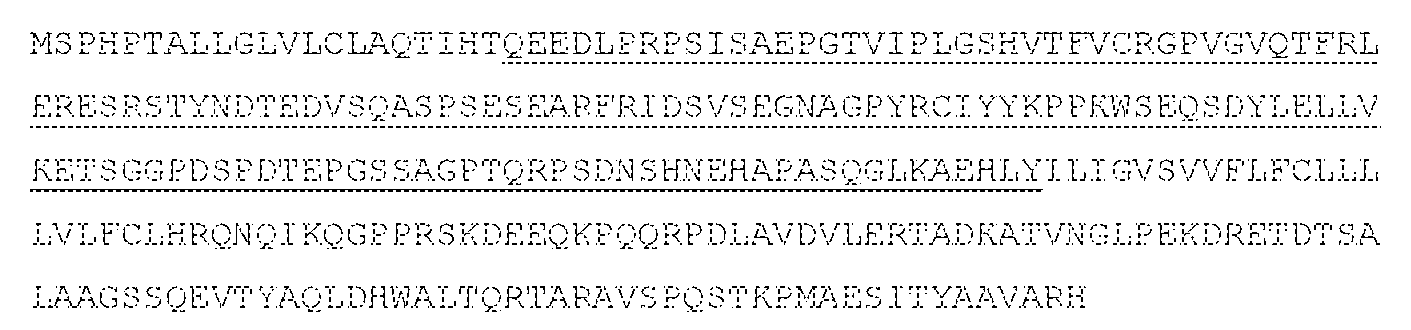


Claims
Priority Applications (11)
| Application Number | Priority Date | Filing Date | Title |
|---|---|---|---|
| KR1020197005926A KR20190044070A (en) | 2016-08-03 | 2017-08-03 | Composition and method for modulating LAIR signal transduction |
| KR1020247006978A KR20240035633A (en) | 2016-08-03 | 2017-08-03 | Compositions and methods for modulating lair signal transduction |
| BR112019002127A BR112019002127A2 (en) | 2016-08-03 | 2017-08-03 | fusion protein, vector, cell, pharmaceutical composition, and fusion protein use |
| US16/321,726 US20210363240A1 (en) | 2016-08-03 | 2017-08-03 | Compositions and methods for modulating lair signal transduction |
| EP17837690.1A EP3496746A4 (en) | 2016-08-03 | 2017-08-03 | Compositions and methods for modulating lair signal transduction |
| CA3032826A CA3032826A1 (en) | 2016-08-03 | 2017-08-03 | Compositions and methods for modulating lair signal transduction |
| AU2017306560A AU2017306560A1 (en) | 2016-08-03 | 2017-08-03 | Compositions and methods for modulating lair signal transduction |
| JP2019527776A JP2019528311A (en) | 2016-08-03 | 2017-08-03 | Compositions and methods for modulating LAIR signaling |
| RU2019105294A RU2757394C2 (en) | 2016-08-03 | 2017-08-03 | Compositions and methods for modulating the transmission of a lair signal |
| CN201780060801.9A CN109789197A (en) | 2016-08-03 | 2017-08-03 | For adjusting the composition and method of LAIR signal transduction |
| JP2023088687A JP2023109983A (en) | 2016-08-03 | 2023-05-30 | Composition and method for adjusting lair signal transmission |
Applications Claiming Priority (4)
| Application Number | Priority Date | Filing Date | Title |
|---|---|---|---|
| US201662370334P | 2016-08-03 | 2016-08-03 | |
| US62/370,334 | 2016-08-03 | ||
| US201762450300P | 2017-01-25 | 2017-01-25 | |
| US62/450,300 | 2017-01-25 |
Publications (2)
| Publication Number | Publication Date |
|---|---|
| WO2018027039A1 true WO2018027039A1 (en) | 2018-02-08 |
| WO2018027039A8 WO2018027039A8 (en) | 2019-03-07 |
Family
ID=61074186
Family Applications (1)
| Application Number | Title | Priority Date | Filing Date |
|---|---|---|---|
| PCT/US2017/045310 WO2018027039A1 (en) | 2016-08-03 | 2017-08-03 | Compositions and methods for modulating lair signal transduction |
Country Status (10)
| Country | Link |
|---|---|
| US (1) | US20210363240A1 (en) |
| EP (1) | EP3496746A4 (en) |
| JP (2) | JP2019528311A (en) |
| KR (2) | KR20240035633A (en) |
| CN (1) | CN109789197A (en) |
| AU (1) | AU2017306560A1 (en) |
| BR (1) | BR112019002127A2 (en) |
| CA (1) | CA3032826A1 (en) |
| RU (1) | RU2757394C2 (en) |
| WO (1) | WO2018027039A1 (en) |
Cited By (6)
| Publication number | Priority date | Publication date | Assignee | Title |
|---|---|---|---|---|
| WO2021002735A1 (en) * | 2019-07-04 | 2021-01-07 | 주식회사 굳티셀 | Cell surface antigen of t cell and various uses thereof |
| WO2022133326A1 (en) * | 2020-12-18 | 2022-06-23 | Cullinan Amber Corp. | Bi-functional linear fusion collagen-localized immunomodulatory molecules and methods thereof |
| EP3877399A4 (en) * | 2018-11-06 | 2022-10-19 | Alsatech, Inc. | Cell-based gene therapy for neurodegenerative diseases |
| WO2023122618A1 (en) * | 2021-12-21 | 2023-06-29 | Ngm Biopharmaceuticals, Inc. | Combinational use of an anti-ilt3 antibody and an anti-lair-1 antibody |
| WO2023173091A1 (en) | 2022-03-11 | 2023-09-14 | Ngm Biopharmaceuticals, Inc. | Osteoclast-associated ig-like receptor (oscar) and methods of use thereof |
| EP4000633A4 (en) * | 2019-07-12 | 2024-02-21 | Univ Kyoto | Neutralizing antibody for tooth regeneration treatment targeting usag-1 molecule |
Families Citing this family (2)
| Publication number | Priority date | Publication date | Assignee | Title |
|---|---|---|---|---|
| WO2024022495A1 (en) * | 2022-07-28 | 2024-02-01 | Concept To Medicine Biotech Co., Ltd. | Anti-MerTK ANTIBODIES AND USES THEREOF |
| WO2024067864A1 (en) * | 2022-09-30 | 2024-04-04 | Shanghai Junshi Biosciences Co., Ltd. | Anti-lair1 antibodies and their uses |
Citations (4)
| Publication number | Priority date | Publication date | Assignee | Title |
|---|---|---|---|---|
| US20090304686A1 (en) * | 2006-03-08 | 2009-12-10 | Universotair Medisch Centrum Utrecht | Interfering in activation of an immune cell by influencing interaction of lair and collagen |
| WO2010078580A2 (en) * | 2009-01-05 | 2010-07-08 | The Johns Hopkins University | Immunotherapy for contact dermatitis using co-signal regulation |
| US20120156191A1 (en) * | 2006-11-24 | 2012-06-21 | Pierre Fabre Medicament | Anti-jam-a antibodies |
| US20150315294A1 (en) * | 2009-06-25 | 2015-11-05 | Esbatech - A Novartis Company Llc | Acceptor framework for cdr grafting |
Family Cites Families (3)
| Publication number | Priority date | Publication date | Assignee | Title |
|---|---|---|---|---|
| EP2036570A1 (en) * | 2007-09-13 | 2009-03-18 | UMC Utrecht Holding B.V. | Means and methods for influencing platelet-collagen interaction |
| US20110159023A1 (en) * | 2008-08-25 | 2011-06-30 | Solomon Langermann | Pd-1 antagonists and methods for treating infectious disease |
| MY172863A (en) * | 2012-09-13 | 2019-12-13 | Bristol Myers Squibb Co | Fibronectin based scaffold domain proteins that bind to myostatin |
-
2017
- 2017-08-03 EP EP17837690.1A patent/EP3496746A4/en active Pending
- 2017-08-03 KR KR1020247006978A patent/KR20240035633A/en active Application Filing
- 2017-08-03 RU RU2019105294A patent/RU2757394C2/en active
- 2017-08-03 AU AU2017306560A patent/AU2017306560A1/en active Pending
- 2017-08-03 CN CN201780060801.9A patent/CN109789197A/en active Pending
- 2017-08-03 BR BR112019002127A patent/BR112019002127A2/en unknown
- 2017-08-03 WO PCT/US2017/045310 patent/WO2018027039A1/en unknown
- 2017-08-03 KR KR1020197005926A patent/KR20190044070A/en not_active Application Discontinuation
- 2017-08-03 CA CA3032826A patent/CA3032826A1/en active Pending
- 2017-08-03 US US16/321,726 patent/US20210363240A1/en active Pending
- 2017-08-03 JP JP2019527776A patent/JP2019528311A/en active Pending
-
2023
- 2023-05-30 JP JP2023088687A patent/JP2023109983A/en active Pending
Patent Citations (4)
| Publication number | Priority date | Publication date | Assignee | Title |
|---|---|---|---|---|
| US20090304686A1 (en) * | 2006-03-08 | 2009-12-10 | Universotair Medisch Centrum Utrecht | Interfering in activation of an immune cell by influencing interaction of lair and collagen |
| US20120156191A1 (en) * | 2006-11-24 | 2012-06-21 | Pierre Fabre Medicament | Anti-jam-a antibodies |
| WO2010078580A2 (en) * | 2009-01-05 | 2010-07-08 | The Johns Hopkins University | Immunotherapy for contact dermatitis using co-signal regulation |
| US20150315294A1 (en) * | 2009-06-25 | 2015-11-05 | Esbatech - A Novartis Company Llc | Acceptor framework for cdr grafting |
Non-Patent Citations (2)
| Title |
|---|
| See also references of EP3496746A4 * |
| ZOCCHI ET AL.: "Leukocyte-associated Ig-like receptor-1 prevents granulocyte-monocyte colony stimulating factor-dependent proliferation and Akt1/PKB alpha activation in primary acute myeloid leukemia cells", EUR J IMMUNOL, vol. 31, no. 12, 1 December 2001 (2001-12-01), pages 3667 - 3675, XP055460519 * |
Cited By (6)
| Publication number | Priority date | Publication date | Assignee | Title |
|---|---|---|---|---|
| EP3877399A4 (en) * | 2018-11-06 | 2022-10-19 | Alsatech, Inc. | Cell-based gene therapy for neurodegenerative diseases |
| WO2021002735A1 (en) * | 2019-07-04 | 2021-01-07 | 주식회사 굳티셀 | Cell surface antigen of t cell and various uses thereof |
| EP4000633A4 (en) * | 2019-07-12 | 2024-02-21 | Univ Kyoto | Neutralizing antibody for tooth regeneration treatment targeting usag-1 molecule |
| WO2022133326A1 (en) * | 2020-12-18 | 2022-06-23 | Cullinan Amber Corp. | Bi-functional linear fusion collagen-localized immunomodulatory molecules and methods thereof |
| WO2023122618A1 (en) * | 2021-12-21 | 2023-06-29 | Ngm Biopharmaceuticals, Inc. | Combinational use of an anti-ilt3 antibody and an anti-lair-1 antibody |
| WO2023173091A1 (en) | 2022-03-11 | 2023-09-14 | Ngm Biopharmaceuticals, Inc. | Osteoclast-associated ig-like receptor (oscar) and methods of use thereof |
Also Published As
| Publication number | Publication date |
|---|---|
| RU2019105294A (en) | 2020-09-04 |
| JP2023109983A (en) | 2023-08-08 |
| BR112019002127A2 (en) | 2019-09-17 |
| KR20190044070A (en) | 2019-04-29 |
| RU2757394C2 (en) | 2021-10-14 |
| CA3032826A1 (en) | 2018-02-08 |
| EP3496746A4 (en) | 2020-02-12 |
| WO2018027039A8 (en) | 2019-03-07 |
| EP3496746A1 (en) | 2019-06-19 |
| KR20240035633A (en) | 2024-03-15 |
| AU2017306560A1 (en) | 2019-02-21 |
| JP2019528311A (en) | 2019-10-10 |
| RU2019105294A3 (en) | 2020-09-21 |
| CN109789197A (en) | 2019-05-21 |
| US20210363240A1 (en) | 2021-11-25 |
Similar Documents
| Publication | Publication Date | Title |
|---|---|---|
| JP7382444B2 (en) | Antibodies against Siglec-15 and methods of using the same | |
| US20210363240A1 (en) | Compositions and methods for modulating lair signal transduction | |
| AU2011272941B2 (en) | C10RF32 for the treatment of multiple sclerosis, rheumatoid arthritis and other autoimmune disorders | |
| US20240076379A1 (en) | Antibodies to programmed cell death protein 1 | |
| JP2019536470A6 (en) | Antibodies to Siglec-15 and methods of using the same | |
| US20210032346A1 (en) | B7-h4 antibodies and methods of use thereof | |
| JP2013503205A (en) | Methods and compositions for inhibiting transplant rejection | |
| US20210002373A1 (en) | KLRG1 Binding Compositions and Methods of Use Thereof | |
| US20230357384A1 (en) | Compositions and methods for modulating flrt3 mediated signal transduction | |
| RU2809243C2 (en) | Antibodies b7-h4 and methods for their application | |
| WO2021258040A2 (en) | Compositions and methods for modulating flrt3 mediated signal transduction | |
| EP4360714A2 (en) | Antibodies for siglec-15 and methods of use thereof |
Legal Events
| Date | Code | Title | Description |
|---|---|---|---|
| 121 | Ep: the epo has been informed by wipo that ep was designated in this application |
Ref document number: 17837690 Country of ref document: EP Kind code of ref document: A1 |
|
| ENP | Entry into the national phase |
Ref document number: 3032826 Country of ref document: CA Ref document number: 2019527776 Country of ref document: JP Kind code of ref document: A |
|
| NENP | Non-entry into the national phase |
Ref country code: DE |
|
| REG | Reference to national code |
Ref country code: BR Ref legal event code: B01A Ref document number: 112019002127 Country of ref document: BR |
|
| ENP | Entry into the national phase |
Ref document number: 2017306560 Country of ref document: AU Date of ref document: 20170803 Kind code of ref document: A |
|
| ENP | Entry into the national phase |
Ref document number: 20197005926 Country of ref document: KR Kind code of ref document: A |
|
| ENP | Entry into the national phase |
Ref document number: 2017837690 Country of ref document: EP Effective date: 20190304 |
|
| REG | Reference to national code |
Ref country code: BR Ref legal event code: B01E Ref document number: 112019002127 Country of ref document: BR Free format text: EXPLIQUE A DIVERGENCIA NO NOME DE UM DOS INVENTORES (SOLOMON LANGERMANN) QUE CONSTA NA PUBLICACAO INTERNACIONAL WO 2018/027039 E O CONSTANTE DA PETICAO INICIAL NO 870190010973 |
|
| ENP | Entry into the national phase |
Ref document number: 112019002127 Country of ref document: BR Kind code of ref document: A2 Effective date: 20190201 |