WO2017214092A1 - Combination therapy - Google Patents
Combination therapy Download PDFInfo
- Publication number
- WO2017214092A1 WO2017214092A1 PCT/US2017/036075 US2017036075W WO2017214092A1 WO 2017214092 A1 WO2017214092 A1 WO 2017214092A1 US 2017036075 W US2017036075 W US 2017036075W WO 2017214092 A1 WO2017214092 A1 WO 2017214092A1
- Authority
- WO
- WIPO (PCT)
- Prior art keywords
- binding
- epitope
- domain
- cell
- molecule
- Prior art date
Links
Classifications
-
- C—CHEMISTRY; METALLURGY
- C07—ORGANIC CHEMISTRY
- C07K—PEPTIDES
- C07K16/00—Immunoglobulins [IGs], e.g. monoclonal or polyclonal antibodies
- C07K16/18—Immunoglobulins [IGs], e.g. monoclonal or polyclonal antibodies against material from animals or humans
- C07K16/28—Immunoglobulins [IGs], e.g. monoclonal or polyclonal antibodies against material from animals or humans against receptors, cell surface antigens or cell surface determinants
- C07K16/2803—Immunoglobulins [IGs], e.g. monoclonal or polyclonal antibodies against material from animals or humans against receptors, cell surface antigens or cell surface determinants against the immunoglobulin superfamily
- C07K16/2809—Immunoglobulins [IGs], e.g. monoclonal or polyclonal antibodies against material from animals or humans against receptors, cell surface antigens or cell surface determinants against the immunoglobulin superfamily against the T-cell receptor (TcR)-CD3 complex
-
- A—HUMAN NECESSITIES
- A61—MEDICAL OR VETERINARY SCIENCE; HYGIENE
- A61P—SPECIFIC THERAPEUTIC ACTIVITY OF CHEMICAL COMPOUNDS OR MEDICINAL PREPARATIONS
- A61P31/00—Antiinfectives, i.e. antibiotics, antiseptics, chemotherapeutics
-
- A—HUMAN NECESSITIES
- A61—MEDICAL OR VETERINARY SCIENCE; HYGIENE
- A61P—SPECIFIC THERAPEUTIC ACTIVITY OF CHEMICAL COMPOUNDS OR MEDICINAL PREPARATIONS
- A61P35/00—Antineoplastic agents
-
- C—CHEMISTRY; METALLURGY
- C07—ORGANIC CHEMISTRY
- C07K—PEPTIDES
- C07K16/00—Immunoglobulins [IGs], e.g. monoclonal or polyclonal antibodies
- C07K16/18—Immunoglobulins [IGs], e.g. monoclonal or polyclonal antibodies against material from animals or humans
- C07K16/28—Immunoglobulins [IGs], e.g. monoclonal or polyclonal antibodies against material from animals or humans against receptors, cell surface antigens or cell surface determinants
- C07K16/2803—Immunoglobulins [IGs], e.g. monoclonal or polyclonal antibodies against material from animals or humans against receptors, cell surface antigens or cell surface determinants against the immunoglobulin superfamily
- C07K16/2806—Immunoglobulins [IGs], e.g. monoclonal or polyclonal antibodies against material from animals or humans against receptors, cell surface antigens or cell surface determinants against the immunoglobulin superfamily against CD2
-
- C—CHEMISTRY; METALLURGY
- C07—ORGANIC CHEMISTRY
- C07K—PEPTIDES
- C07K16/00—Immunoglobulins [IGs], e.g. monoclonal or polyclonal antibodies
- C07K16/18—Immunoglobulins [IGs], e.g. monoclonal or polyclonal antibodies against material from animals or humans
- C07K16/28—Immunoglobulins [IGs], e.g. monoclonal or polyclonal antibodies against material from animals or humans against receptors, cell surface antigens or cell surface determinants
- C07K16/2803—Immunoglobulins [IGs], e.g. monoclonal or polyclonal antibodies against material from animals or humans against receptors, cell surface antigens or cell surface determinants against the immunoglobulin superfamily
- C07K16/2815—Immunoglobulins [IGs], e.g. monoclonal or polyclonal antibodies against material from animals or humans against receptors, cell surface antigens or cell surface determinants against the immunoglobulin superfamily against CD8
-
- C—CHEMISTRY; METALLURGY
- C07—ORGANIC CHEMISTRY
- C07K—PEPTIDES
- C07K16/00—Immunoglobulins [IGs], e.g. monoclonal or polyclonal antibodies
- C07K16/18—Immunoglobulins [IGs], e.g. monoclonal or polyclonal antibodies against material from animals or humans
- C07K16/28—Immunoglobulins [IGs], e.g. monoclonal or polyclonal antibodies against material from animals or humans against receptors, cell surface antigens or cell surface determinants
- C07K16/2803—Immunoglobulins [IGs], e.g. monoclonal or polyclonal antibodies against material from animals or humans against receptors, cell surface antigens or cell surface determinants against the immunoglobulin superfamily
- C07K16/2818—Immunoglobulins [IGs], e.g. monoclonal or polyclonal antibodies against material from animals or humans against receptors, cell surface antigens or cell surface determinants against the immunoglobulin superfamily against CD28 or CD152
-
- C—CHEMISTRY; METALLURGY
- C07—ORGANIC CHEMISTRY
- C07K—PEPTIDES
- C07K16/00—Immunoglobulins [IGs], e.g. monoclonal or polyclonal antibodies
- C07K16/18—Immunoglobulins [IGs], e.g. monoclonal or polyclonal antibodies against material from animals or humans
- C07K16/28—Immunoglobulins [IGs], e.g. monoclonal or polyclonal antibodies against material from animals or humans against receptors, cell surface antigens or cell surface determinants
- C07K16/2803—Immunoglobulins [IGs], e.g. monoclonal or polyclonal antibodies against material from animals or humans against receptors, cell surface antigens or cell surface determinants against the immunoglobulin superfamily
- C07K16/2827—Immunoglobulins [IGs], e.g. monoclonal or polyclonal antibodies against material from animals or humans against receptors, cell surface antigens or cell surface determinants against the immunoglobulin superfamily against B7 molecules, e.g. CD80, CD86
-
- C—CHEMISTRY; METALLURGY
- C07—ORGANIC CHEMISTRY
- C07K—PEPTIDES
- C07K16/00—Immunoglobulins [IGs], e.g. monoclonal or polyclonal antibodies
- C07K16/18—Immunoglobulins [IGs], e.g. monoclonal or polyclonal antibodies against material from animals or humans
- C07K16/28—Immunoglobulins [IGs], e.g. monoclonal or polyclonal antibodies against material from animals or humans against receptors, cell surface antigens or cell surface determinants
- C07K16/2803—Immunoglobulins [IGs], e.g. monoclonal or polyclonal antibodies against material from animals or humans against receptors, cell surface antigens or cell surface determinants against the immunoglobulin superfamily
- C07K16/283—Immunoglobulins [IGs], e.g. monoclonal or polyclonal antibodies against material from animals or humans against receptors, cell surface antigens or cell surface determinants against the immunoglobulin superfamily against Fc-receptors, e.g. CD16, CD32, CD64
-
- C—CHEMISTRY; METALLURGY
- C07—ORGANIC CHEMISTRY
- C07K—PEPTIDES
- C07K16/00—Immunoglobulins [IGs], e.g. monoclonal or polyclonal antibodies
- C07K16/18—Immunoglobulins [IGs], e.g. monoclonal or polyclonal antibodies against material from animals or humans
- C07K16/28—Immunoglobulins [IGs], e.g. monoclonal or polyclonal antibodies against material from animals or humans against receptors, cell surface antigens or cell surface determinants
- C07K16/2866—Immunoglobulins [IGs], e.g. monoclonal or polyclonal antibodies against material from animals or humans against receptors, cell surface antigens or cell surface determinants against receptors for cytokines, lymphokines, interferons
-
- C—CHEMISTRY; METALLURGY
- C07—ORGANIC CHEMISTRY
- C07K—PEPTIDES
- C07K16/00—Immunoglobulins [IGs], e.g. monoclonal or polyclonal antibodies
- C07K16/18—Immunoglobulins [IGs], e.g. monoclonal or polyclonal antibodies against material from animals or humans
- C07K16/28—Immunoglobulins [IGs], e.g. monoclonal or polyclonal antibodies against material from animals or humans against receptors, cell surface antigens or cell surface determinants
- C07K16/2878—Immunoglobulins [IGs], e.g. monoclonal or polyclonal antibodies against material from animals or humans against receptors, cell surface antigens or cell surface determinants against the NGF-receptor/TNF-receptor superfamily, e.g. CD27, CD30, CD40, CD95
-
- C—CHEMISTRY; METALLURGY
- C07—ORGANIC CHEMISTRY
- C07K—PEPTIDES
- C07K16/00—Immunoglobulins [IGs], e.g. monoclonal or polyclonal antibodies
- C07K16/18—Immunoglobulins [IGs], e.g. monoclonal or polyclonal antibodies against material from animals or humans
- C07K16/28—Immunoglobulins [IGs], e.g. monoclonal or polyclonal antibodies against material from animals or humans against receptors, cell surface antigens or cell surface determinants
- C07K16/30—Immunoglobulins [IGs], e.g. monoclonal or polyclonal antibodies against material from animals or humans against receptors, cell surface antigens or cell surface determinants from tumour cells
-
- A—HUMAN NECESSITIES
- A61—MEDICAL OR VETERINARY SCIENCE; HYGIENE
- A61K—PREPARATIONS FOR MEDICAL, DENTAL OR TOILETRY PURPOSES
- A61K39/00—Medicinal preparations containing antigens or antibodies
- A61K2039/505—Medicinal preparations containing antigens or antibodies comprising antibodies
-
- A—HUMAN NECESSITIES
- A61—MEDICAL OR VETERINARY SCIENCE; HYGIENE
- A61K—PREPARATIONS FOR MEDICAL, DENTAL OR TOILETRY PURPOSES
- A61K39/00—Medicinal preparations containing antigens or antibodies
- A61K2039/505—Medicinal preparations containing antigens or antibodies comprising antibodies
- A61K2039/507—Comprising a combination of two or more separate antibodies
-
- A—HUMAN NECESSITIES
- A61—MEDICAL OR VETERINARY SCIENCE; HYGIENE
- A61K—PREPARATIONS FOR MEDICAL, DENTAL OR TOILETRY PURPOSES
- A61K39/00—Medicinal preparations containing antigens or antibodies
- A61K2039/545—Medicinal preparations containing antigens or antibodies characterised by the dose, timing or administration schedule
-
- A—HUMAN NECESSITIES
- A61—MEDICAL OR VETERINARY SCIENCE; HYGIENE
- A61K—PREPARATIONS FOR MEDICAL, DENTAL OR TOILETRY PURPOSES
- A61K39/00—Medicinal preparations containing antigens or antibodies
- A61K2039/58—Medicinal preparations containing antigens or antibodies raising an immune response against a target which is not the antigen used for immunisation
- A61K2039/585—Medicinal preparations containing antigens or antibodies raising an immune response against a target which is not the antigen used for immunisation wherein the target is cancer
-
- C—CHEMISTRY; METALLURGY
- C07—ORGANIC CHEMISTRY
- C07K—PEPTIDES
- C07K2317/00—Immunoglobulins specific features
- C07K2317/30—Immunoglobulins specific features characterized by aspects of specificity or valency
- C07K2317/31—Immunoglobulins specific features characterized by aspects of specificity or valency multispecific
-
- C—CHEMISTRY; METALLURGY
- C07—ORGANIC CHEMISTRY
- C07K—PEPTIDES
- C07K2317/00—Immunoglobulins specific features
- C07K2317/60—Immunoglobulins specific features characterized by non-natural combinations of immunoglobulin fragments
- C07K2317/62—Immunoglobulins specific features characterized by non-natural combinations of immunoglobulin fragments comprising only variable region components
- C07K2317/622—Single chain antibody (scFv)
-
- C—CHEMISTRY; METALLURGY
- C07—ORGANIC CHEMISTRY
- C07K—PEPTIDES
- C07K2317/00—Immunoglobulins specific features
- C07K2317/60—Immunoglobulins specific features characterized by non-natural combinations of immunoglobulin fragments
- C07K2317/62—Immunoglobulins specific features characterized by non-natural combinations of immunoglobulin fragments comprising only variable region components
- C07K2317/626—Diabody or triabody
Definitions
- the present invention is directed to a combination therapy for the treatment of cancer and pathogen-associated diseases, that comprises the administration of: (1) a molecule ⁇ e.g., a diabody, an scFv, an antibody, a TandAb, etc) capable of binding PD-1 or a natural ligand of PD-1, and (2) a molecule ⁇ e.g., a diabody, a BiTe, a bispecific antibody, a CAR, etc.) capable of mediating the redirected killing of a target cell ⁇ e.g., a cancer cell or a pathogen-infected cell, etc) expressing a Disease Antigen.
- a target cell e.g., a cancer cell or a pathogen-infected cell, etc
- the invention particularly concerns the embodiment in which the molecule capable of mediating the redirected killing of the target cell is a bispecific binding molecule that comprises a first epitope-binding site capable of immunospecifically binding an epitope of a cell surface molecule of an effector cell and a second epitope-binding site that is capable of immunospecifically binding an epitope of such target cells ⁇ i.e., a Disease Antigen such as a Cancer Antigen or a Pathogen- Associated Antigen).
- a Disease Antigen such as a Cancer Antigen or a Pathogen- Associated Antigen.
- the present invention is also directed to pharmaceutical compositions that comprise such molecule(s).
- the mammalian immune system serves as a defense against a variety of conditions, including, e.g., injury, infection and neoplasia.
- the efficiency with which humans and other mammals develop an immunological response to pathogens, foreign substances and cancer antigens rests on two characteristics: the extraordinar specificity of the immune response for antigen recognition, and the immunological memory that allows for faster and more vigorous responses upon re-activation with the same antigen (Portoles, P. et al. (2009) "The TCRJCD3 Complex: Opening the Gate to Successful Vaccination " Current Pharmaceutical Design 15 :3290-3300; Guy, C.S. et al. (2009) "Organization of Proximal Signal Initiation at the TCR. CD3 Complex " Immunol Rev. 232(1):7-21 ; Topalian, S.L. et al. (2015) “Immune Checkpoint Blockade: A Common Denominator Approach to Cancer Therapy " Cancer Cell 27:450-461).
- the immune system In healthy individuals, the immune system is in a quiescent state, inhibited by a repertoire of diverse inhibitory receptors and receptor ligands. Upon recognition of a cancer antigen, microbial pathogen, or an allergen, an array of activating receptors and receptor ligands are triggered to induce the activation of the immune system. Such activation leads to the activation of macrophages, Natural Killer (NK) cells and antigen- specific, cytotoxic, T-cells, and promotes the release of various cytokines, all of which act to counter the perceived threat to the health of the subject (Dong, C. et al. (2003) "Immune Regulation by Novel Costimulatory Molecules " Immunolog. Res.
- NK Natural Killer
- the immune system is capable of returning to its normal quiescent state when the countervailing inhibitory immune signals outweigh the activating immune signals.
- the disease state of cancer may be considered to reflect a failure to adequately activate a subject's immune system. Such failure may reflect an inadequate presentation of activating immune signals, or it may reflect an inadequate ability to alleviate inhibitory immune signals in the subject.
- researchers have determined that cancer cells can co-opt the immune system to evade being detected by the immune system (Topalian, S.L. et al. (2015) "Immune Checkpoint Blockade: A Common Denominator Approach to Cancer Therapy " Cancer Cell 27:450-461).
- the mammalian immune system is mediated by two separate but interrelated systems: the humoral immune system and the cellular immune system.
- the humoral system is mediated by soluble molecules (antibodies or immunoglobulins) produced by B Cells.
- B Cells Such molecules have the ability to combine with and neutralize antigens that have been recognized as being foreign to the body.
- the cellular immune system involves the mobilization of certain cells, termed "T Cells,” that serve a variety of therapeutic roles. T Cells are lymphocytes that mature in the thymus and circulate between the tissues, lymphatic system and the circulatory system. In response to the presence and recognition of foreign structures (antigens), T Cells become "activated" to initiate an immune response.
- T Cells do not themselves secrete antibodies, they are usually required for antibody secretion by the second class of lymphocytes, B Cells (which derive from bone marrow).
- B Cells which derive from bone marrow.
- T Cells exhibit extraordinary immunological specificity so as to be capable of discerning one antigen from another).
- T Cell activation Two interactions are required for T Cell activation (Viglietta, V. et al. (2007) “Modulating Co-Stimulation " Neurotherapeutics 4:666-675; Korman, A.J. et al. (2007) Checkpoint Blockade in Cancer Immunotherapy " Adv. Immunol. 90:297-339).
- MHC Major Histocompatibility Complex
- T Cells experiencing both stimulatory signals are then capable of responding to cytokines (such as Interleukin-2 and Interleukin-12).
- cytokines such as Interleukin-2 and Interleukin-12
- T Cells enter a functionally unresponsive state, referred to as clonal anergy (Khawli, L.A. et al. (2008) “Cytokine, Chemokine, and Co-Stimulatory Fusion Proteins for the Immunotherapy of Solid Tumors " Exp. Pharmacol. 181 :291-328).
- T Cells are the key players of various organ-specific autoimmune diseases, such as type I diabetes, rheumatoid arthritis, and multiple sclerosis (Dong, C. et al. (2003) "Immune Regulation by Novel Costimulatory Molecules," Immunol og. Res. 28(l):39-48).
- This immune "checkpoint" pathway is important in maintaining self-tolerance (i.e., in preventing a subject from mounting an immune system attack against his/her own cells (an "autoimmune" reaction) and in limiting collateral tissue damage during antimicrobial or anti-allergic immune responses.
- an "autoimmune" reaction Where contact of a T Cell results in the generation of only one of two required signals, the T Cell does not become activated and an adaptive immune response does not occur.
- the "two signal” mechanism of T Cell activation thus provides a way for the immune system to avoid undesired responses, such as responses to self-antigens that would otherwise result in an immune system attack against a subject's own cells (an "autoimmune" reaction).
- the cells of the immune system are characterized by their expression of specialized glycoprotein cell surface molecules. Interactions between such molecules and molecules of other cells triggers, maintains or dampens the immune response.
- all T Cells are characterized by their expression of CD3.
- CD3 is a T cell co-receptor composed of four distinct chains (Wucherpfennig, K.W. et al. (2010) " Structural Biology Of The T-Cell Receptor: Insights into Receptor Assembly, Ligand Recognition, And Initiation of Signaling " Cold Spring Harb. Perspect. Biol. 2(4):a005140; pages 1-14; Chetty, R. et al.
- CD3 Structure, Function, And Role Of mmunostaining In Clinical Practice " J. Pathol. 173(4):303-307; Guy, C.S. et al. (2009) “Organization Of Proximal Signal Initiation At The TCR:CD3 Complex,” Immunol. Rev. 232(1):7-21).
- the complex contains a CD3y chain, a CD35 chain, and two CD3e chains. These chains associate with the TCR in order to generate an activation signal in T lymphocytes (Smith-Garvin, J.E. et al. (2009) “ Cell Activation,” Annu. Rev. Immunol. 27:591-619). In the absence of CD3, TCRs do not assemble properly and are degraded (Thomas, S. et al. (2010) "Molecular Immunology Lessons From Therapeutic T- Cell Receptor Gene Transfer," Immunology 129(2): 170-177). CD3 is found bound to the membranes of all mature T cells, and in virtually no other cell type (see, Janeway, C.A. et al.
- the invariant CD3e signaling component of the TCR complex on T cells has been used as a target to force the formation of an immunological synapse between T cells and cancer cells.
- Co-engagement of CD3 and the tumor antigen activates the T cells, triggering lysis of cancer cells expressing the tumor antigen (Baeuerle et al. (2011) “Bispecific T Cell Engager For Cancer Therapy " In: BlSPECIFIC ANTIBODIES, Kontermann, R E. (Ed.) Springer- Verlag; 201 1 :273-287).
- a first subset of T Cells is characterized by the expression of the CD4 ⁇ i.e., they are "CD4 + ").
- CD4 + T Cells are the essential organizers of most mammalian immune and autoimmune responses (Dong, C. et al. (2003) “Immune Regulation by Novel Costimulatory Molecules " Immunolog. Res. 28(l):39-48).
- CD4 + T Cells The activation of CD4 + T Cells has been found to be mediated through co-stimulatory interactions between an antigen:major histocompability class II (MHC II) molecule complex that is arrayed on the surface of an Antigen-Presenting Cell (such as a B Cell, a macrophage or a dendritic cell) and a complex of two molecules, the TCR and a CD3 cell- surface receptor ligand, both of which are arrayed on the surface of a naive CD4 + T Cell.
- Activated T helper cells are capable of proliferating into Thl cells that are capable of mediating an inflammatory response to the target cell.
- cytotoxic T Cells A second subset of T Cells, known as “cytotoxic T Cells,” are characterized by the expression of CD8 (i.e., they are “CD8+” as well as CD3 + ).
- CD8 is a T-cell co- receptor composed of two distinct chains (Leahy, D.J. (1995) "A Structural View of CD 4 and CD8 " FASEB J. 9: 17-25) that is expressed on Cytotoxic T-cells.
- CD8 + T Cells The activation of CD8 + T Cells has been found to be mediated through co-stimulatory interactions between an antigen:major histocompability class I (MHC I) molecule complex that is arrayed on the surface of a target cell and a complex of CD8 and the T Cell Receptor, that are arrayed on surface of the CD8 + T Cell ((Gao, G. et al. (2000) "Molecular Interactions Of Coreceptor CD8 And MHC Class I: The Molecular Basis For Functional Coordination With The T-Cell Receptor, " Immunol. Today 21 :630-636). Unlike major histocompability class II (MHC ⁇ ) molecules, which are expressed by only certain immune system cells, MHC I molecules are very widely expressed.
- MHC I major histocompability class II
- cytotoxic T Cells are capable of binding a wide variety of cell types.
- Activated cytotoxic T Cells mediate cell killing through their release of the cytotoxins perforin, granzymes, and granulysin.
- perforin granzymes enter the cytoplasm of the target cell and their serine protease function triggers the caspase cascade, which is a series of cysteine proteases that eventually lead to apoptosis (programmed cell death) of targeted cells.
- CD2 is a cell adhesion molecule found on the surface of T-cells and natural killer (NK) cells.
- CD2 enhances NK cell cytotoxicity, possibly as a promoter of NK cell nanotube formation (Mace, E.M. et al. (2014) "Cell Biological Steps and Checkpoints in Accessing NK Cell Cytotoxicity ⁇ " Immunol. Cell. Biol. 92(3):245-255; Comerci, C.J. et al. (2012) "CD2 Promotes Human Natural Killer Cell Membrane Nanotube Formation," PLoS One 7(10):e47664: l-12).
- TCR T Cell Receptor
- TCR The T Cell Receptor
- CD4+ or CD8+ T cells are natively expressed by CD4+ or CD8+ T cells, and permits such cells to recognize antigenic peptides that are bound and presented by class I or class II MHC proteins of antigen-presenting cells.
- Recognition of a pMHC (peptide- MHC) complex by a TCR initiates the propagation of a cellular immune response that leads to the production of cytokines and the lysis of the Antigen-Presenting Cell (see, e.g. , Armstrong, K M. et al. (2008) " onformational Changes And Flexibility In T-Cell Receptor Recognition OfPeptide-MHC Complexes " Biochem. J.
- CD3 is the receptor that binds to the TCR (Thomas, S.
- the TCR and CD3 complex, along with the CD3 ⁇ chain zeta chain (also known as T Cell receptor T3 zeta chain or CD247) comprise the "TCR complex” (van der Merwe, P. A. etc. (epub Dec. 3, 2010) "Mechanisms For T Cell Receptor Triggering " Nat. Rev. Immunol. 11 :47-55; Wucherpfennig, K.W. et al. (2010) "Structural Biology of the T Cell Receptor: Insights into Receptor Assembly, Ligand Recognition, and Initiation of Signaling " Cold Spring Harb. Perspect. Biol. 2:a005140).
- the complex is particularly significant since it contains a large number (ten) of immunoreceptor tyrosine-based activation motifs (ITAMs).
- ITAMs immunoreceptor tyrosine-based activation motifs
- the Fc Receptors CD16, CD32 and CD64
- natural IgG antibodies are composed of four polypeptide chains: two identical "light” chains and two identical “heavy” chains.
- the Heavy Chains contain C-terminal "CH2" and “CH3" domains, and the association of the two Heavy Chains creates an "Fc Domain” that is capable of li gating (binding) to receptors (singularly referred to as an "Fc gamma receptor" "FcyR,” and collectively as “FcyRs”) found on the surfaces of multiple types of immune system cells ⁇ e.g., B lymphocytes, follicular dendritic cells, natural killer cells, macrophages, neutrophils, eosinophils, basophils and mast cells).
- B lymphocytes follicular dendritic cells
- natural killer cells e.g., neutrophils, eosinophils, basophils and mast cells.
- Such receptors have an "extracellular” portion (which is thus capable of ligating to an Fc Domain), a “transmembrane” portion (which extends through the cellular membrane), and a “cytoplasmic” portion (positioned inside the cell).
- Multiple types of FcyRs have been identified: CD16A (FcyRIIIA), CD16B (FcyRIIIB), CD32A (FcyRIIA), CD32B (FcyRIIB), and CD64 (FcyRI)
- CD16A FcyRIIIA
- CD16B FcyRIIIB
- CD32A FcyRIIA
- CD32B FcyRIIB
- CD64 FcyRI
- CD 16 is expressed by neutrophils, eosinophils, natural killer (NK) cells, and tissue macrophages that bind aggregated but not monomelic human IgG (Peltz, G.A. et al. (1989) "Human Fc Gamma RIII: Cloning, Expression, And Identification Of The Chromosomal Locus Of Two Fc Receptors For IgG," Proc. Natl. Acad. Sci. (U.S.A.) 86(3): 1013-1017; Bachanova, V. et al. (2014) "NK Cells In Therapy Of Cancer," Crit. Rev. Oncog. 19(1-2): 133-141; Miller, J.S.
- ADCC antibody-dependent cell-mediated cytotoxicity
- CD32A (FcyRIIA) (Brandsma, A.M. (2015) “Fc Receptor Inside-Out Signaling And Possible Impact On Antibody Therapy” Immunol Rev. 268(l):74-87; van Sorge, N.M. etal. (2003) “FcgammaR Polymorphisms: Implications For Function, Disease Susceptibility And Immunotherapy " Tissue Antigens 61(3): 189-202; Selvaraj, P. et al. (2004) “Functional Regulation Of Human Neutrophil Fc Gamma Receptors " Immunol. Res. 29(l-3):219-230) and CD64 (FcyRI) (Lu, S. et al.
- CD32B FcyRIIB
- B lymphocytes macrophages, neutrophils, eosinophils and dendritic cells
- ITAM immunoreceptor tyrosine-based activation motif
- ITIM immunoreceptor tyrosine- based inhibitory motif
- ITAM- containing FcyRs include FcyRI, FcyRIIA, FcyRIIIA, and activate the immune system when bound to Fc Domains ⁇ e.g. , aggregated Fc Domains present in an immune complex).
- FcyRIIB is the only currently known natural ITEVI-containing FcyR; it acts to dampen or inhibit the immune system when bound to aggregated Fc Domains.
- the Natural Killer Group 2D (“NKG2D”) receptor is expressed on all human (and other mammalian) Natural Killer cells (Bauer, S. et al. (1999) "Activation OfNK Cells And T Cells By NKG2D, A Receptor For Stress-Inducible MICA,” Science 285(5428):727- 729; Jamieson, A.M. et al. (2002) “The Role Of The NKG2D Immunoreceptor In Immune Cell Activation And Natural Killing," Immunity 17(1): 19-29) as well as on all CD8 + T cells (Groh, V. et al.
- NKG2D ligands are completely absent, or are present only at low levels, on the surfaces of normal cells, but they are overexpressed by infected, transformed, senescent or stressed cells.
- binding ligands include the histocompatibility 60 (H60) molecule, the product of the retinoic acid early inducible gene-1 (RAE-1), and the murine UL16-binding protein-like transcript 1 (MULT1) (Raulet D.H. (2003) “Roles Of The NKG2D Immunoreceptor And Its Ligands," Nature Rev. Immunol. 3 :781-790; Coudert, J.D. et al. (2005) "Altered NKG2D Function In NK Cells Induced By Chronic Exposure To Altered NKG2D Ligand-Expressing Tumor Cells," Blood 106: 1711-1717).
- H60 histocompatibility 60
- RAE-1 retinoic acid early inducible gene-1
- MULT1 murine UL16-binding protein-like transcript 1
- Binding between the B7.1 (CD80) and B7.2 (CD86) ligands of Antigen- Presenting Cells and the CD28 and CTLA-4 receptors of CD4 + T lymphocytes is of particular importance to the required second interaction of the immune response (Sharpe, A.H. et al. (2002) "The B7-CD28 Superfamily " Nature Rev. Immunol. 2: 116-126; Dong, C. etal. (2003) "Immune Regulation by Novel Costimulatory Molecules " Immunolog. Res. 28(l):39-48; Lindley, P S et al. (2009) “Th Clinical Utility Of Inhibiting CD28-Mediated Costimulation " Immunol. Rev. 229:307-321).
- Binding of B7.1 or of B7.2 to CD28 stimulates T-cell activation; binding of B7.1 or B7.2 to CTLA-4 inhibits such activation (Dong, C. et al. (2003) "Immune Regulation by Novel Costimulatory Molecules " Immunolog. Res. 28(l):39-48; Lindley, P.S. et al. (2009) "The Clinical Utility Of Inhibiting CD28-Mediated Costimulation," Immunol. Rev. 229:307-321; Greenwald, RJ. etal. (2005) “The B7 Family Revisited " Ann. Rev. Immunol. 23:515-548).
- CD28 is constitutively expressed on the surface of T-cells (Gross, J., et al.
- CTLA-4 is the higher affinity receptor (Sharpe, A H. etal. (2002) “The B7-CD28 Superfamily ,” Nature Rev. Immunol. 2: 116-126; Topalian, S.L. et al.
- PD-1 Programmed Death-1
- CD279 type I membrane protein member of the extended CD28/CTLA-4 family of T-cell regulators that broadly negatively regulates immune responses
- Ishida, Y. et al. (1992) "Induced Expression Of PD-1, A Novel Member Of The Immunoglobulin Gene Superfamily, Upon Programmed Cell Death EMBO J. 11 :3887-3895; United States Patent Application Publications No. 2007/0202100; 2008/0311 1 17; 2009/001 10667; United States Patents No. 6,808,710; 7, 101,550; 7,488,802; 7,635,757, 7,722,868; PCT Publication No. WO 01/14557).
- PD-1 and CTLA-4 both provide inhibitory immune signals
- the signals provided by PD-1 are mounted later in the course of the disease, and can profoundly diminish the immune response by limiting the initial production ("burst") of disease- responsive T-cells.
- burst initial production
- PD-1 can partially convert a potentially effective T-cell response into one of tolerance (Topalian, S.L. et al. (2015) "Immune Checkpoint Blockade: A Common Denominator Approach to Cancer Therapy " Cancer Cell 27:450-461).
- PD-1 receptor-ligand interactions of the PD-1 system appear to be even more complex than those of the CD28/CTLA-4 system.
- PD-1 is expressed on the cell surface of activated T-cells, B-cells, and monocytes (Agata, Y. et al. (1996) "Expression Of The PD- 1 Antigen On The Surface Of Stimulated Mouse T And B Lymphocytes " Int. Immunol. 8(5):765-772; Yamazaki, T. et al. (2002) “Expression Of Programmed Death 1 Ligands By Murine T-Cells And APC ' ,” J. Immunol.
- the extracellular region of PD-1 consists of a single immunoglobulin (Ig)V domain with 23% identity to the equivalent domain in CTLA-4 (Martin-Orozco, N. et al. (2007) "Inhibitory Costimulation And Anti-Tumor Immunity " Semin. Cancer Biol. 17(4):288-298).
- the extracellular IgV domain is followed by a transmembrane region and an intracellular tail.
- the intracellular tail contains two phosphorylation sites located in an immunoreceptor tyrosine-based inhibitory motif and an immunoreceptor tyrosine-based switch motif, which suggests that PD-1 negatively regulates TCR signals (Ishida, Y et al.
- PD-1 mediates its inhibition of the immune system by binding B7-H1 and B7- DC (also known as PD-L1 and PD-L2) (Flies, D.B. et al.
- B7-H1 and B7-DC are broadly expressed on the surfaces of many types of human and murine tissues, such as heart, placenta, muscle, fetal liver, spleen, lymph nodes, and thymus as well as murine liver, lung, kidney, islets cells of the pancreas and small intestine (Martin-Orozco, N. et al. (2007) “Inhibitory Costimulation And Anti-Tumor Immunity " Semin. Cancer Biol. 17(4):288-298).
- B7-H1 protein expression has been found in human endothelial cells (Chen, Y. et al.
- the present invention is directed to a combination therapy for the treatment of cancer and pathogen-associated diseases, that comprises the administration of: (1) a molecule (e.g., a diabody, an scFv, an antibody, a TandAb, etc.) capable of binding PD-1 or a natural ligand of PD-1, and (2) a molecule (e.g., a diabody, a BiTe, a bispecific antibody, a CAR, etc.) capable of mediating the redirected killing of a target cell (e.g., a cancer cell or a pathogen-infected cell, etc.) expressing a Disease Antigen.
- a target cell e.g., a cancer cell or a pathogen-infected cell, etc.
- the invention particularly concerns the embodiment in which the molecule capable of mediating the redirected killing of the target cell is a bispecific binding molecule that comprises a first epitope-binding site capable of immunospecifically binding an epitope of a cell surface molecule of an effector cell and a second epitope-binding site that is capable of immunospecifically binding an epitope of such target cells (i.e., a Disease Antigen such as a Cancer Antigen or a Pathogen- Associated Antigen).
- a Disease Antigen such as a Cancer Antigen or a Pathogen- Associated Antigen.
- the present invention is also directed to pharmaceutical compositions that comprise such molecule(s).
- the invention provides a method for the treatment of cancer or a pathogen-associated disease, comprising administering to a subject in need thereof a therapeutically effective amount of:
- the invention particularly concerns the embodiment of such method wherein the molecule capable of binding PD-1 or a natural ligand of PD-1 is capable of inhibiting binding between PD-1 and a natural ligand of PD-1.
- the invention further concerns the embodiment of such method, wherein the method comprises administration of two binding molecules that cumulatively comprise three epitope-binding domains, the two binding molecules being: (A) a binding molecule that comprises an epitope-binding domain of an antibody that is capable of binding PD- 1 , or an epitope-binding domain of an antibody that is capable of binding a natural ligand of PD-1 , and
- an epitope-binding domain of an antibody that that is capable of binding the Cancer Antigen or the Pathogen Antigen of the target cell wherein the epitope-binding domain of the binding molecule (A) is capable of binding PD-1 or a natural ligand of PD-1, and the epitope-binding domains (1) and (2) of the binding molecule (B) are capable of mediating the redirected killing of the target cell.
- the invention further concerns the embodiment of such method, wherein the binding molecule capable of binding PD-1 or a natural ligand of PD-1 comprises a diabody, scFv, antibody or TandAb, and the binding molecule (B) comprises a bispecific diabody, a CAR, a BiTe, or bispecific antibody.
- the invention further concerns the embodiment of such methods wherein the binding molecule capable of binding PD-1 or a natural ligand of PD-1 comprises an epitope- binding domain of an antibody that binds to PD-1.
- the invention further concerns the embodiment of such methods wherein the binding molecule capable of binding PD-1 or a natural ligand of PD-1 comprises an epitope- binding domain of an antibody that binds to a natural ligand of PD-1.
- the invention further concerns the embodiment of such methods wherein the binding molecule capable of binding PD-1 or a natural ligand of PD-1 comprises a second epitope-binding domain capable of binding PD-1, wherein such epitope-binding domains:
- the invention further concerns the embodiment of such methods wherein the PD-1 -epitope-binding domains are capable of simultaneous binding to the same PD-1 molecule.
- the invention further concerns the embodiment of such methods wherein the binding molecule capable of binding PD-1 or a natural ligand of PD-1 comprises a second epitope-binding domain capable of binding the natural ligand of PD-1, wherein such epitope-binding domains:
- the invention further concerns the embodiment of such methods wherein the PD-1 ligand-epitope-binding domains are capable of simultaneous binding the same molecule of the natural ligand of PD-1.
- the invention further concerns the embodiment of such methods wherein the binding molecule capable of binding PD-1 or a natural ligand of PD-1 comprises a second epitope-binding domain capable of binding an epitope of a molecule that is not PD-1 or a natural ligand of PD-1.
- the invention further concerns the embodiment of such methods wherein in the second epitope-binding domain binds an epitope of CD 137, LAG-3, OX40, TIGIT, TDVI-3, or VISTA.
- the invention further concerns the embodiment of such methods wherein the binding molecule capable of mediating the redirected killing of the target cell comprises a third epitope-binding domain capable of binding a cell surface molecule of the effector cell.
- the invention further concerns the embodiment of such methods wherein the third epitope-binding-domain of the binding molecule capable of mediating the redirected killing of the target cell is capable of binding a different cell surface molecule of the effector cell, such that the binding molecule capable of mediating the redirected killing is capable of binding two different cell surface molecules of the effector cell.
- the invention further concerns the embodiment of such methods wherein the binding molecule capable of mediating the redirected killing of the target cell comprises a third epitope-binding domain capable of binding to a Cancer Antigen or a Pathogen- Associated Antigen of the target cell.
- the invention further concerns the embodiment of such methods wherein the third epitope-binding-domain of the binding molecule capable of mediating the redirected killing of the target cell is capable of binding a different Cancer Antigen or a different Pathogen Antigen of the target cell, such that the binding molecule capable of mediating the redirected killing is capable of binding to two different Cancer Antigens or two different Pathogen Antigens of the target cell.
- the invention further concerns the embodiment of such methods wherein the cell surface molecule of the effector cell is selected from the group consisting of: CD2, CD3, CD8, CD 16, TCR, and KG2D.
- the Cancer Antigen is selected from the group consisting of the Cancer Antigens: 19.9, 4.2, A33, ADAM-9, AH6, ALCAM, B l, B7-H3, BAGE, beta-catenin, blood group ALe /Le y , Burkitt's lymphoma antigen-38.13, C14, CA125, Carboxypeptidase M, CD5, CD19, CD20, CD22, CD23, CD25, CD27, CD28, CD33, CD36, CD40/CD154, CD45, CD56, CD46, CD52, CD56, CD79a/CD79b, CD103, CD123, CD317, CDK4, CEA, CEACAM5/CEACAM6, C017-1A, CO-43, CO-514, CTA-1, CTLA-4, Cytokeratin 8, Dl .
- the Cancer Antigen is selected from the group consisting of the Cancer Antigens: 19.9, 4.2, A33, ADAM-9, AH6, ALCAM, B l
- the invention further concerns the embodiment of such methods wherein the method comprises the administration of the pharmaceutical composition, and wherein the Pathogen-Associated Antigen is selected from the group consisting of the Pathogen-Associated Antigens: Herpes Simplex Virus infected cell protein (ICP)47, Herpes Simplex Virus gD, Epstein-Barr Virus LMP-1, Epstein-Barr Virus LMP-2A, Epstein-Barr Virus LMP-2B, Human Immunodeficiency Virus gpl60, Human Immunodeficiency Virus gpl20, Human Immunodeficiency Virus gp41, etc.), Human Papillomavirus E6, Human Papillomavirus E7, human T-cell leukemia virus gp64, human T-cell leukemia virus gp46, and human T-cell leukemia virus gp21
- the Pathogen-Associated Antigen is selected from the group consisting of the Pathogen-Associated Antigens: Herpes Simplex Virus inf
- the invention further provides a pharmaceutical composition that comprises:
- the invention further concerns the embodiment of such pharmaceutical composition wherein the pharmaceutical composition comprises two binding molecules that cumulatively comprise three epitope-binding domains, the two binding molecules being:
- the epitope-binding domain of the binding molecule (A) is capable of binding PD- 1 or a natural ligand of PD-1, and the epitope-binding domains (1) and (2) of the binding molecule (B) are capable of mediating the redirected killing of the target cell.
- the invention further concerns the embodiment of such pharmaceutical compositions wherein the binding molecule (A) comprises a diabody, scFv, antibody, or TandAb, and the binding molecule (B) comprises a diabody, a CAR, a BiTe, or bispecific antibody.
- the invention further concerns the embodiment of such pharmaceutical compositions wherein the molecule capable of binding PD-1 or a natural ligand of PD-1 comprises an epitope-binding domain of an antibody that binds to PD-1
- the invention further concerns the embodiment of such pharmaceutical compositions wherein the molecule capable of binding PD-1 or a natural ligand of PD-1 comprises an epitope-binding domain of an antibody that binds to a natural ligand of PD-1.
- the invention further concerns the embodiment of such pharmaceutical compositions wherein the molecule capable of binding PD-1 or a natural ligand of PD-1 comprises a second epitope-binding domain capable of binding PD-1, wherein such PD-1- epitope-binding domains:
- the invention further concerns the embodiment of such pharmaceutical compositions wherein the PD-1 -epitope-binding domains are capable of simultaneous binding the same PD-1 molecule.
- the invention further concerns the embodiment of such pharmaceutical compositions wherein the binding molecule capable of binding PD-1 or a natural ligand of PD-1 comprises a second epitope-binding domain capable of binding the natural ligand of PD-1, wherein such epitope-binding domains:
- the invention further concerns the embodiment of such pharmaceutical compositions wherein the PD-1 ligand-epitope-binding domains are capable of simultaneous binding the same molecule of the natural ligand of PD-1.
- the invention further concerns the embodiment of such pharmaceutical compositions wherein the binding molecule capable of binding PD-1 or a natural ligand of PD-1 comprises a second epitope-binding domain capable of binding an epitope of a molecule that is not PD-1 or a natural ligand of PD-1.
- the invention further concerns the embodiment of such pharmaceutical compositions wherein the second epitope-binding domain binds an epitope of CD 137, LAG- 3, OX40, TIGIT, TIM-3, or VISTA.
- the invention further concerns the embodiment of such pharmaceutical compositions wherein the molecule capable of mediating the redirected killing of the target cell comprises a third epitope-binding domain, wherein such three epitope-binding domains are capable of simultaneous binding, and wherein the third epitope-binding site is capable of binding an epitope of a cell surface molecule of the effector cell.
- the invention further concerns the embodiment of such pharmaceutical compositions wherein the third epitope-binding-domain of the binding molecule capable of mediating the redirected killing of the target cell is capable of binding a different cell surface molecule of the effector cell, such that the binding molecule capable of mediating the redirected killing is capable of binding two different cell surface molecules of the effector cell.
- the invention further concerns the embodiment of such pharmaceutical compositions wherein the binding molecule capable of mediating the redirected killing of the target cell comprises a third epitope-binding domain capable of binding to a Cancer Antigen or a Pathogen- Associated Antigen of the target cell.
- the invention further concerns the embodiment of such pharmaceutical compositions wherein the third epitope-binding-domain of the binding molecule capable of mediating the redirected killing of the target cell is capable of binding a different Cancer Antigen or a different Pathogen- Associated Antigen of the target cell, such that the binding molecule capable of mediating the redirected killing is capable of binding to two different Cancer Antigens or two different Pathogen-Associated Antigens of the target cell.
- the invention further concerns the embodiment of such pharmaceutical compositions wherein the cell surface molecule of the effector cell is selected from the group consisting of: CD2, CD3, CD8, CD16, TCR, and KG2D.
- the invention further concerns the embodiment of such pharmaceutical compositions wherein the Cancer Antigen is selected from the group consisting of the Cancer Antigens: 19.9, 4.2, A33, ADAM-9, AH6, ALCAM, Bl, B7-H3, BAGE, beta- catenin, blood group ALe b /Le y , Burkitt's lymphoma antigen-38.13, C14, CA125, Carboxypeptidase M, CD5, CD19, CD20, CD22, CD23, CD25, CD27, CD28, CD33, CD36, CD40/CD154, CD45, CD56, CD46, CD52, CD56, CD79a/CD79b, CD103, CD123, CD317, CDK4, CEA, CEACAM5/CEACAM6, C017-1A, CO-43, CO-514, CTA-1, CTLA-4, Cytokeratin 8, Dl .
- the Cancer Antigen is selected from the group consisting of the Cancer Antigens: 19.9, 4.2, A33
- the invention further concerns the embodiment of such pharmaceutical compositions wherein the Pathogen-Associated Antigen is selected from the group consisting of the Pathogen Antigens: Herpes Simplex Virus infected cell protein (ICP)47, Herpes Simplex Virus gD, Epstein-Barr Virus LMP-1, Epstein-Barr Virus LMP-2A, Epstein-Barr Virus LMP-2B, Human Immunodeficiency Virus gpl60, Human Immunodeficiency Virus gpl20, Human Immunodeficiency Virus gp41, etc), Human Papillomavirus E6, Human Papillomavirus E7, human T-cell leukemia virus gp64, human T-cell leukemia virus gp46, and human T-cell leukemia virus gp21.
- the Pathogen-Associated Antigen is selected from the group consisting of the Pathogen Antigens: Herpes Simplex Virus infected cell protein (ICP)47, Herpes Simplex
- the invention further provides a kit comprising any of the above-described pharmaceutical compositions, wherein the binding molecules thereof are compartmentalized in one or more containers.
- Figure 1 provides a schematic of a representative covalently bonded diabody having two epitope-binding domains composed of two polypeptide chains, each having an E-coil or K-coil Heterodimer-Promoting Domain (alternative Heterodimer-Promoting Domains are provided below).
- a cysteine residue may be present in a linker and/or in the Heterodimer-Promoting Domain as shown in Figure 3B.
- VL and VH Domains that recognize the same epitope are shown using the same shading or fill pattern.
- Figure 2 provides a schematic of a representative covalently bonded diabody molecule having two epitope-binding domains composed of two polypeptide chains, each having a CH2 and CH3 Domain, such that the associated chains form all or part of an Fc Domain. VL and VH Domains that recognize the same epitope are shown using the same shading or fill pattern.
- Figures 3A-3C provide schematics showing representative covalently bonded tetravalent diabodies having four epitope-binding domains composed of two pairs of polypeptide chains (i.e., four polypeptide chains in all).
- One polypeptide of each pair possesses a CH2 and CH3 Domain, such that the associated chains form all or part of an Fc Domain.
- VL and VH Domains that recognize the same epitope are shown using the same shading or fill pattern.
- the two pairs of polypeptide chains may be same.
- the resulting molecule possesses four epitope-binding domains and is bispecific and bivalent with respect to each bound epitope.
- the VL and VH Domains recognize the same epitope (e.g., the same VL Domain CDRs and the same VH Domain CDRs are used on both chains) the resulting molecule possesses four epitope-binding domains and is monospecific and tetravalent with respect to a single epitope.
- the two pairs of polypeptides may be different.
- FIG. 3A shows an Fc Domain-containing diabody which contains a peptide Heterodimer-Promoting Domain comprising a cysteine residue.
- Figure 3B shows an Fc Domain-containing diabody, which contains E-coil and K-coil Heterodimer-Promoting Domains comprising a cysteine residue and a linker (with an optional cysteine residue).
- Figure 3C shows an Fc Domain-Containing diabody, which contains antibody CHI and CL domains.
- Figures 4A-4B provide schematics of a representative covalently bonded diabody molecule having two epitope-binding domains composed of three polypeptide chains. Two of the polypeptide chains possess a CH2 and CH3 Domain, such that the associated chains form all or part of an Fc Domain.
- the polypeptide chains comprising the VL and VH Domain further comprise a Heterodimer-Promoting Domain. VL and VH Domains that recognize the same epitope are shown using the same shading or fill pattern.
- Figure 5 provides the schematics of a representative covalently bonded diabody molecule having four epitope-binding domains composed of five polypeptide chains. Two of the polypeptide chains possess a CH2 and CH3 Domain, such that the associated chains form an Fc Domain that comprises all or part of an Fc Domain.
- the polypeptide chains comprising the linked VL and VH Domains further comprise a Heterodimer-Promoting Domain. VL and VH Domains that recognize the same epitope are shown using the same shading or fill pattern.
- Figures 6A-6F provide schematics of representative Fc Domain-containing trivalent binding molecules having three epitope-binding domains.
- Figures 6A and 6B respectively, illustrate schematically the domains of trivalent binding molecules comprising two diabody-type binding domains and a Fab-Type Binding Domain having different domain orientations in which the diabody-type binding domains are N-terminal or C- terminal to an Fc Domain.
- the molecules in Figures 6A and 6B comprise four chains.
- FIGS 6C and 6D respectively, illustrate schematically the domains of trivalent binding molecules comprising two diabody-type binding domains N-terminal to an Fc Domain, and a Fab-Type Binding Domain in which the Light Chain and Heavy Chain are linked via a polypeptide spacer, or an scFv-type binding domain.
- the trivalent binding molecules in Figures 6E and 6F respectively, illustrate schematically the domains of trivalent binding molecules comprising two diabody-type binding domains C-terminal to an Fc Domain, and a Fab-Type Binding Domain in which the Light Chain and Heavy Chain are linked via a polypeptide spacer, or an scFv-type binding domain.
- the trivalent binding molecules in Figures 6C-6F comprise three chains. VL and VH Domains that recognize the same epitope are shown using the same shading or fill pattern.
- Figure 7 shows the result of providing MHO "7" mice that had received 5 x 10 6
- LOX-F VI human metastatic melanoma cancer cells ID
- 10 6 human PBMC IP
- the humanized anti-human PD-1 antibody hPD-1 mAb7 (1.2) IgG4(P)
- the CD3 x B7-H3 bispecific diabody DART-A
- DART-A both hPD-1 mAb7 (1.2) IgG4(P) and DART-A, or with vehicle alone (control).
- Figures 8A-8B show the result of providing MHO '7' mice that had received 5 x 10 6 Detroit562 human metastatic pharyngeal carcinoma cancer cells (ED) and 10 6 human PBMC (IP) with the humanized anti -human PD-1 antibody, hPD-1 mAb7 (1.2) IgG4(P), the CD3 x B7-H3 bispecific diabody, DART-A, with both hPD-1 mAb7 (1.2) IgG4(P) and DART-A, or with vehicle alone (control).
- ED human metastatic pharyngeal carcinoma cancer cells
- IP human PBMC
- Figure 8A shows the results for Vehicle Control, hPD-1 mAb7 (1.2) IgG4(P) (Q7Dx5), DART-A (Q7Dx5), and hPD-1 mAb7 (1.2) IgG4(P) + DART-A (Q7Dx5).
- Figure 8B shows the results for Vehicle Control, hPD-1 mAb7 (1.2) IgG4(P) (Q7Dx5), DART-A (Q7Dx5), hPD-1 mAb7 (1.2) IgG4(P) + DART- A (Q7Dx5) and hPD-1 mAb7 (1.2) IgG4(P) + DART-A (Q14Dx3).
- Figure 9 shows the results of a study on the effect of the administration of the combination therapy of the present invention.
- the results show an enhancement of the immune response of recipient animals as determined by an increase in the concentration of their CD3 + cells.
- Figures 10A-10B show the results of a study on the effect of the combination therapy of the present invention on T-cell signaling in a luciferase reporter assay.
- MDA- MB-231 tumor target cells expressing PD-1 and B7-H3 were mixed with MNFAT-luc2/PD- 1 Jurkat T-cells at an effectontarget cell ratio of 1 : 1 ( Figure 10A) or 3 : 1 ( Figure 10B) and cultured alone or with a fixed concentration (12.5 nM) of the PD-1 binding molecules hPD- 1 niAb7 (1.2) IgG4(P), DART-1, or control antibody (hlgG), in the presence of increasing concentations of DART-A.
- IgG4(P) IgG4(P
- DART-1 DART-1
- control antibody hlgG
- Figures 11A-11B show that administration of the combination therapy of the present invention reduces tumor recurrence in the presensence of anergic T-cells.
- NOG mice that had received 5 x 10 6 A375 INFy treated melanoma cells and 5 x 10 6 activated or anergic human T-cells with vehicle alone, 0.5 mg/kg DART-2 (Q7Dx4), 0.5 mg/kg DART- B (QDxl), or both 0.5 mg/kg DART-2 (Q7Dx4) and 0.5 mg/kg DART-B (QDxl).
- Figure 11A shows the results for mice that received activated T-cells
- Figure 11B shows the results for mice that received anergic T-cells.
- Figures 12A-12H demonstrate the unexpected benefit of the combined therapy of a molecule capable of binding PD-1 and a molecule capable of mediating the redirected killing of a target cell relative to administration of either molecule alone.
- Tumor volume caused by A375 melanoma cells was measured as a function of time and is plotted in Figures 12A-12H.
- Figure 12A shows the results for Groups 1, 2, 5 and 6 through day 50;
- Figures 12B-12H show the spider plots, through day 80, for the individual animals in Group 2 ( Figure 12B), Group 5 (Figure 12C), Group 6 (Figure 12D), Group 3 ( Figure 12E), Group 7 (Figure 12F), Group 4 ( Figure 12G), and Group 8 (Figure 12H).
- the present invention is directed to a combination therapy for the treatment of cancer and pathogen-associated diseases, that comprises the administration of: (1) a molecule (e.g., a diabody, an scFv, an antibody, a TandAb, etc.) capable of binding PD-1 or a natural ligand of PD-1, and (2) a molecule (e.g., a diabody, a BiTe, a bispecific antibody, a CAR, etc.) capable of mediating the redirected killing of a target cell (e.g., a cancer cell or a pathogen-infected cell, etc.) expressing a Disease Antigen.
- a target cell e.g., a cancer cell or a pathogen-infected cell, etc.
- the invention particularly concerns the embodiment in which the molecule capable of mediating the redirected killing of the target cell is a bispecific binding molecule that comprises a first epitope-binding site capable of immunospecifically binding an epitope of a cell surface molecule of an effector cell and a second epitope-binding site that is capable of immunospecifically binding an epitope of such target cells (i.e., a Disease Antigen such as a Cancer Antigen or a Pathogen- Associated Antigen).
- a Disease Antigen such as a Cancer Antigen or a Pathogen- Associated Antigen.
- the present invention is also directed to pharmaceutical compositions that comprise such molecule(s).
- the binding domains of the molecules of the present invention bind epitopes in an "immunospecific" manner.
- an antibody, diabody or other epitope- binding molecule is said to "immunospecifically” bind a region of another molecule (i.e. , an epitope) if it reacts or associates more frequently, more rapidly, with greater duration and/or with greater affinity with that epitope relative to alternative epitopes.
- an antibody that immunospecifically binds to a viral epitope is an antibody that binds this viral epitope with greater affinity, avidity, more readily, and/or with greater duration than it immunospecifically binds to other viral epitopes or non-viral epitopes.
- an antibody (or moiety or epitope) that immunospecifically binds to a first target may or may not specifically or preferentially bind a second target.
- immunospecific binding does not necessarily require (although it can include) exclusive binding.
- reference to binding means “immunospecific” binding. Two molecules are said to be capable of binding one another in a “physiospecific” manner, if such binding exhibits the specificity with which receptors bind their respective ligands.
- the therapeutic molecules of the present invention particularly include bispecific binding molecules that comprises an epitope-binding site capable of immunospecifically binding an epitope of a cell surface molecule of an effector cell and also an epitope-binding site that is capable of immunospecifically binding an epitope of a target cell that expresses a Disease Antigen.
- Disease Antigen denotes an antigen that is expressed on the surface of an abnormal or infected cell and that is characteristic of such abnormality of infection, or that is expressed on the surface of a foreign cell and that is characteristic of such foreign origin.
- a cell that expresses a Disease Antigen on its cell surface, and that may therefore become bound by the therapeutic molecules of the present invention and thereby targeted for killing by such therapeutic molecules is a "target cell.”
- Disease Antigens that are "Cancer Antigens” or "Pathogen-Associated Antigens.”
- the binding molecules of the present invention may be antibodies.
- Antibodies are immunoglobulin molecules capable of specific binding a target, such as a carbohydrate, polynucleotide, lipid, polypeptide, etc., through at least one antigen recognition site, located in the Variable Domain of the immunoglobulin molecule.
- antibody refers to monoclonal antibodies, multispecific antibodies, human antibodies, humanized antibodies, synthetic antibodies, chimeric antibodies, polyclonal antibodies, camelized antibodies, single-chain Fvs (scFv), single-chain antibodies, Fab fragments, F(ab') fragments, disulfide-linked bispecific Fvs (sdFv), intrabodies, and epitope-binding fragments of any of the above.
- scFv single-chain Fvs
- Fab fragments F(ab') fragments
- disulfide-linked bispecific Fvs sdFv
- intrabodies and epitope-binding fragments of any of the above.
- antibody includes immunoglobulin molecules and immunologically active fragments of immunoglobulin molecules, i.e. , molecules that contain an epitope-binding site.
- Immunoglobulin molecules can be of any type (e.g., IgG, IgE, IgM, IgD, IgA and IgY), class (e.g., IgGi, IgG2, IgG3, IgG4, IgAi and IgA2) or subclass.
- Antibodies are capable of "immunospecifically binding" to a polypeptide or protein or a non-protein molecule due to the presence on such molecule of a particular domain or moiety or conformation (an "epitope").
- An epitope-containing molecule may have immunogenic activity, such that it elicits an antibody production response in an animal; such molecules are termed "antigens.”
- antigens immunogenic activity
- the last few decades have seen a revival of interest in the therapeutic potential of antibodies, and antibodies have become one of the leading classes of biotechnology-derived drugs (Chan, C.E. et al. (2009) “The Use Of Antibodies In The Treatment Of Infectious Diseases " Singapore Med. J. 50(7): 663 -666). Over 200 antibody -based drugs have been approved for use or are under development.
- monoclonal antibody refers to a homogeneous antibody population wherein the monoclonal antibody is comprised of amino acids (naturally occurring or non-naturally occurring) that are involved in the selective binding of an antigen. Monoclonal antibodies are highly specific, being directed against a single epitope (or antigenic site).
- monoclonal antibody encompasses not only intact monoclonal antibodies and full-length monoclonal antibodies, but also fragments thereof (such as Fab, Fab', F(ab') 2 , Fv fragments, etc), single-chain (scFv) binding molecules and mutants thereof, fusion proteins comprising an antibody portion, humanized monoclonal antibodies, chimeric monoclonal antibodies, and any other modified configuration of the immunoglobulin molecule that comprises an antigen recognition site of the required specificity and the ability to bind an antigen.
- fragments thereof such as Fab, Fab', F(ab') 2 , Fv fragments, etc
- scFv single-chain binding molecules and mutants thereof
- fusion proteins comprising an antibody portion, humanized monoclonal antibodies, chimeric monoclonal antibodies, and any other modified configuration of the immunoglobulin molecule that comprises an antigen recognition site of the required specificity and the ability to bind an antigen.
- the antibodies are produced by immunizing an animal with an immunogenic amount of cells, cell extracts, or protein preparations that contain the desired epitope.
- the immunogen can be, but is not limited to, primary cells, cultured cell lines, cancerous cells, proteins, peptides, nucleic acids, or tissue.
- Cells used for immunization may be cultured for a period of time ⁇ e.g., at least 24 hours) prior to their use as an immunogen.
- Cells may be used as immunogens by themselves or in combination with a non-denaturing adjuvant, such as Ribi (see, e.g., Jennings, V.M. (1995) "Review of Selected Adjuvants Used in Antibody Production," ILAR J. 37(3): 1 19-125).
- cells should be kept intact and preferably viable when used as immunogens. Intact cells may allow antigens to be better detected than ruptured cells by the immunized animal. Use of denaturing or harsh adjuvants, e.g., Freund's adjuvant, may rupture cells and therefore is discouraged.
- the immunogen may be administered multiple times at periodic intervals such as, bi weekly, or weekly, or may be administered in such a way as to maintain viability in the animal (e.g., in a tissue recombinant).
- existing monoclonal antibodies and any other equivalent antibodies that are immunospecific for a desired pathogenic epitope can be sequenced and produced recombinantly by any means known in the art
- such an antibody is sequenced and the polynucleotide sequence is then cloned into a vector for expression or propagation.
- the sequence encoding the antibody of interest may be maintained in a vector in a host cell and the host cell can then be expanded and frozen for future use.
- the polynucleotide sequence of such antibodies may be used for genetic manipulation to generate the monospecific or multispecific (e.g., bispecific, trispecific and tetraspecific) molecules of the invention as well as an affinity optimized, a chimeric antibody, a humanized antibody, and/or a caninized antibody, to improve the affinity, or other characteristics of the antibody.
- the general principle in humanizing an antibody involves retaining the basic sequence of the antigen-binding portion of the antibody, while swapping the non-human remainder of the antibody with human antibody sequences.
- Natural antibodies are composed of two “Light Chains” complexed with two "Heavy Chains.” Each Light Chain contains a Variable Domain (“VL”) and a Constant Domain (“CL”). Each Heavy Chain contains a Variable Domain (“VH”), three Constant Domains ("CHI,” “CH2” and “CH3”), and a “Hinge” Region (“H”) located between the CHI and CH2 Domains.
- VL Variable Domain
- CL Constant Domain
- H Hinge” Region
- scFvs are single chain molecules made by linking Light and Heavy Chain Variable Domains together via a short linking peptide.
- the basic structural unit of naturally occurring immunoglobulins is thus a tetramer having two Light Chains and two Heavy Chains, usually expressed as a glycoprotein of about 150,000 Da.
- the amino-terminal (“N-terminal") portion of each chain includes a Variable Domain of about 100 to 1 10 or more amino acids primarily responsible for antigen recognition.
- the carboxy-terminal (“C-terminal”) portion of each chain defines a constant region, with Light Chains having a single Constant Domain and Heavy Chains usually having three Constant Domains and a Hinge Domain.
- the structure of the Light Chains of an IgG molecule is n-VL-CL-c and the structure of the IgG Heavy Chains is n-VH-CHl-H-CH2-CH3-c (where n and c represent, respectively, the N- terminus and the C-terminus of the polypeptide).
- the Variable Domains of an IgG molecule consist of the complementarity determining regions ("CDR"), which contain the residues in contact with epitope, and non- CDR segments, referred to as framework segments ("FR"), which in general maintain the structure and determine the positioning of the CDR loops so as to permit such contacting (although certain framework residues may also contact antigen).
- CDR complementarity determining regions
- FR framework segments
- the VL and VH Domains have the structure n-FRl-CDRl-FR2-CDR2-FR3-CDR3-FR4-c.
- Polypeptides that are (or may serve as) the first, second and third CDR of the Light Chain of an antibody are herein respectively designated as: CDRLI Domain, CDRL2 Domain, and CDRL3 Domain.
- polypeptides that are (or may serve as) the first, second and third CDR of the Heavy Chain of an antibody are herein respectively designated as: CDRHI Domain, CDRH2 Domain, and CDRH3 Domain.
- CDRLI Domain, CDRL2 Domain, CDRL3 Domain, CDRHI Domain, CDRH2 Domain, and CDRH3 Domain are directed to polypeptides that when incorporated into a protein cause that protein to be able to bind a specific epitope regardless of whether such protein is an antibody having light and Heavy Chains or is a diabody or a single-chain binding molecule (e.g. , an scFv, a BiTe, etc.), or is another type of protein.
- epitope-binding fragment denotes a fragment of a molecule capable of immunospecifically binding an epitope.
- An epitope-binding fragment may contain any 1, 2, 3, 4, or 5 the CDR Domains of an antibody, or may contain all 6 of the CDR Domains of an antibody and, although capable of immunospecifically binding such epitope, may exhibit an immunospecificity, affinity or selectivity towards such epitope that differs from that of such antibody.
- an epitope-binding fragment will contain all 6 of the CDR Domains of such antibody.
- An epitope-binding fragment of an antibody may be a single polypeptide chain (e.g.
- an scFv may comprise two or more polypeptide chains, each having an amino terminus and a carboxy terminus (e.g., a diabody, a Fab fragment, an Fab 2 fragment, etc.).
- a diabody e.g., a Fab fragment, an Fab 2 fragment, etc.
- the order of domains of the protein molecules described herein is in the "N-terminal to C-terminal" direction.
- the invention also particularly encompasses epitope-binding molecules that comprise a VL and/or VH Domain of a humanized antibody.
- humanized antibody refers to a chimeric molecule, generally prepared using recombinant techniques, having an epitope-binding site of an immunoglobulin from a non-human species and a remaining immunoglobulin structure of the molecule that is based upon the structure and /or sequence of a human immunoglobulin.
- the polynucleotide sequence of the Variable Domains of such antibodies may be used for genetic manipulation to generate such derivatives and to improve the affinity, or other characteristics of such antibodies
- the general principle in humanizing an antibody involves retaining the basic sequence of the epitope-binding portion of the antibody, while swapping the non-human remainder of the antibody with human antibody sequences.
- the epitope-binding site may comprise either a complete Variable Domain fused onto Constant Domains or only the complementarity determining regions (CDRs) of such Variable Domain grafted to appropriate framework regions.
- Epitope-binding domains may be wild-type or modified by one or more amino acid substitutions. This eliminates the constant region as an immunogen in human individuals, but the possibility of an immune response to the foreign Variable Domain remains (LoBuglio, A.F. et al. (1989) ' ' ' ' Mouse/Human Chimeric Monoclonal Antibody In Man: Kinetics And Immune Response '' Proc. Natl. Acad. Sci. (U.S.A.) 86:4220-4224).
- Variable Domains of both heavy and Light Chains contain three complementarity determining regions (CDRs) which vary in response to the antigens in question and determine binding capability, flanked by four framework regions (FRs) which are relatively conserved in a given species and which putatively provide a scaffolding for the CDRs.
- CDRs complementarity determining regions
- FRs framework regions
- the Variable Domains can be "reshaped” or “humanized” by grafting CDRs derived from non-human antibody on the FRs present in the human antibody to be modified.
- humanized antibodies preserve all CDR sequences (for example, a humanized mouse antibody which contains all six CDRs from the mouse antibodies). In other embodiments, humanized antibodies have one or more CDRs (one, two, three, four, five, or six) which differ in sequence relative to the original antibody.
- a number of humanized antibody molecules comprising an epitope-binding site derived from a non-human immunoglobulin have been described, including chimeric antibodies having rodent or modified rodent Variable Domain and their associated complementarity determining regions (CDRs) fused to human constant domains (see, for example, Winter et al. (1991) "Man-made Antibodies " Nature 349:293-299; Lobuglio et al. (1989) "Mouse/Human Chimeric Monoclonal Antibody In Man: Kinetics And Immune Response," Proc. Natl. Acad. Sci. (U.S.A.) 86:4220-4224 (1989), Shaw et al.
- CDRs complementarity determining regions
- the numbering of the residues in the constant region of an IgG Heavy Chain is that of the EU index as in Kabat et al., SEQUENCES OF PROTEINS OF IMMUNOLOGICAL INTEREST, 5 th Ed. Public Health Service, NH1, MD (1991) ("Kabat”), expressly incorporated herein by reference.
- EU index as in Kabat refers to the numbering of the constant domains of human IgGl EU antibody. Amino acids from the Variable Domains of the mature heavy and Light Chains of immunoglobulins are designated by the position of an amino acid in the chain.
- Kabat described numerous amino acid sequences for antibodies, identified an amino acid consensus sequence for each subgroup, and assigned a residue number to each amino acid, and the CDRs are identified as defined by Kabat (it will be understood that CDRHI as defined by Chothia, C. & Lesk, A. M. ((1987) "Canonical structures for the hypervariable regions of immunoglobulins " J. Mol. Biol. 196:901 -917) begins five residues earlier).
- Rabat' s numbering scheme is extendible to antibodies not included in his compendium by aligning the antibody in question with one of the consensus sequences in Kabat by reference to conserved amino acids.
- An exemplary CHI Domain is a human IgGl CHI Domain.
- the amino acid sequence of an exemplary human IgGl CHI Domain is (SEQ ID NO:l):
- An exemplary CHI Domain is a human IgG2 CHI Domain.
- the amino acid sequence of an exemplary human IgG2 CHI Domain is (SEQ ID NO:2):
- An exemplary CHI Domain is a human IgG4 CHI Domain.
- the amino acid sequence of an exemplary human IgG4 CHI Domain is (SEQ ID NO:3):
- One exemplary Hinge Domain is a human IgGl Hinge Domain.
- the amino acid sequence of an exemplary human IgGl Hinge Domain is (SEQ ID NO:4):
- EPKSCDKTHTCPPCP EPKSCDKTHTCPPCP .
- Another exemplary Hinge Domain is a human IgG2 Hinge Domain.
- the amino acid sequence of an exemplary human IgG2 Hinge Domain is (SEQ ID NO:5):
- Another exemplary Hinge Domain is a human IgG4 Hinge Domain.
- the amino acid sequence of an exemplary human IgG4 Hinge Domain is (SEQ ID NO:6): ESKYGPPCPSCP.
- an IgG4 Hinge Domain may comprise a stabilizing mutation such as the S228P substitution.
- the amino acid sequence of an exemplary S228P- stabilized human IgG4 Hinge Domain is (SEQ ID NO:7): ESKYGPPCPPCP.
- the CH2 and CH3 Domains of the two Heavy Chains of an antibody interact to form an "Fc Domain,” which is a domain that is recognized by cellular Fc Receptors, including but not limited to Fc gamma Receptors (FcyRs).
- Fc Domain is used to define a C-terminal region of an IgG Heavy Chain.
- An Fc Domain is said to be of a particular IgG isotype, class or subclass if its amino acid sequence is most homologous to that isotype relative to other IgG isotypes.
- antibodies have been shown to be useful as therapeutic agents.
- IgGl is (SEQ ID NO: 8)
- amino acid sequence of the CH2-CH3 Domain of an exemplary human IgG2 is (SEQ ID NO: 9)
- Polymorphisms have been observed at a number of different positions within antibody constant regions ⁇ e.g. , Fc positions, including but not limited to positions 270, 272, 312, 315, 356, and 358 as numbered by the EU index as set forth in Kabat), and thus slight differences between the presented sequence and sequences in the prior art can exist. Polymorphic forms of human immunoglobulins have been well-characterized.
- Gm Glm (1, 2, 3, 17) or Glm (a, x, f, z), G2m (23) or G2m (n), G3m (5, 6, 10, 1 1, 13, 14, 15, 16, 21, 24, 26, 27, 28) or G3m (bl, c3, b3, bO, b3, b4, s, t, gl, c5, u, v, g5)
- Glm 1, 2, 3, 17
- Glm a, x, f, z
- G2m G2m (23) or G2m (n)
- G3m 5, 6, 10, 1 1, 13, 14, 15, 16, 21, 24, 26, 27, 28
- G3m bl, c3, b3, bO, b3, b4, s, t, gl, c5, u, v, g5)
- Lefranc, et al. "The Human IgG Subclasses: Molecular Analysis Of Structure, Function And Regulation.” Pergamon, Oxford, pp.
- the antibodies of the present invention may incorporate any allotype, isoallotype, or haplotype of any immunoglobulin gene, and are not limited to the allotype, isoallotype or haplotype of the sequences provided herein.
- the C-terminal amino acid residue (bolded above) of the CH3 Domain may be post-translationally removed. Accordingly, the C-terminal residue of the CH3 Domain is an optional amino acid residue in the binding molecules of the invention.
- binding molecules lacking the C-terminal residue of the CH3 Domain are also specifically encompassed by the instant invention are such constructs comprising the C-terminal lysine residue of the CH3 Domain.
- each Light Chain of an antibody contains a Variable Domain ("VL”) and a Constant Domain (“CL").
- VL Variable Domain
- CL Constant Domain
- a preferred CL Domain is a human IgG CL Kappa Domain.
- the amino acid sequence of an exemplary human CL Kappa Domain is (SEQ ID NO: 12):
- an exemplary CL Domain is a human IgG CL Lambda Domain.
- amino acid sequence of an exemplary human CL Lambda Domain is (SEQ ID NO: 13):
- the binding molecules of the present invention that are capable of mediating the redirected killing of a target cell ⁇ i.e., a cancer cell, a pathogen-infected cell, etc) may alternatively be monospecific single-chain molecules such Chimeric Antigen Receptors ("CARs") incorporating a single chain variable fragment (scFv) capable of binding a Cancer Antigen or a Pathogen-Associated Antigen.
- CARs Chimeric Antigen Receptors
- scFv single chain variable fragment
- First-generation CA s typically had the intracellular domain from the CD3 ⁇ - chain, which is the primary transmitter of signals from endogenous TCRs.
- Second-generation CARs possessed additional intracellular signaling domains from various costimulatory protein receptors (e.g. , CD28, 41BB, ICOS, etc.) to the cytoplasmic tail of the CAR in order to provide additional signals to the T-cell.
- Third-generation CARs combine multiple signaling domains, such as CD3z-CD28-41BB or CD3z-CD28-OX40, in order to further augment potency (Tettamanti, S. et al. (2013) "Targeting Of Acute Myeloid Leukaemia By Cytokine- Induced Killer Cells Redirected With A Novel CD123-Specific Chimeric Antigen Receptor " Br. J. Haematol.
- the intracellular domain of the CARs of the present invention is preferably selected from the intracellular domain of any of: 41 ⁇ 3 ⁇ , b2c-CD3 ⁇ CD28, CD28-4- 1 ⁇ 3 ⁇ , CD28-CD3 ⁇ CD28-FcsRIy, CD28mut-CD3 ⁇ , CD28-OX40-CD3 ⁇ , CD28- OX40-CD3 , CD3 ⁇ , CD4-CD3 ⁇ , CD4-FceRIy, CD8-CD3 ⁇ , FcsRfy, FcsRIyCAIX, ⁇ .-13- ⁇ 3 ⁇ or Ly49H-CD3C (Tettamanti, S. et al.
- an antibody to bind an epitope of an antigen depends upon the presence and amino acid sequence of the antibody' s VL and VH Domains. Interaction of an antibody's Light Chain and Heavy Chain and, in particular, interaction of its VL and VH Domains forms one of the two epitope-binding domains of a natural antibody, such as an IgG. Natural antibodies are capable of binding only one epitope species (i.e., they are monospecific), although they can bind multiple copies of that species (i.e., exhibiting bivalency or multivalency).
- antibodies can be enhanced by generating multispecific antibody-based molecules that can simultaneously bind two separate and distinct antigens (or different epitopes of the same antigen) and/or by generating antibody-based molecule having higher valency (i.e., more than two binding sites) for the same epitope and/or antigen.
- WO 2013/174873, WO 201 1/133886 and WO 2010/136172 disclose a trispecific antibody in which the CL and CHI Domains are switched from their respective natural positions and the VL and VH Domains have been diversified (WO 2008/027236; WO 2010/108127) to allow them to bind more than one antigen.
- PCT Publications Nos. WO 2013/163427 and WO 2013/119903 disclose modifying the CH2 Domain to contain a fusion protein adduct comprising a binding domain.
- WO 2010/028797, WO2010028796 and WO 2010/028795 disclose recombinant antibodies whose Fc Domains have been replaced with additional VL and VH Domains, so as to form trivalent binding molecules.
- PCT Publications Nos. WO 2003/025018 and WO2003012069 disclose recombinant diabodies whose individual chains contain scFv Domains.
- PCT Publication Nos. WO 2013/006544 discloses multivalent Fab molecules that are synthesized as a single polypeptide chain and then subjected to proteolysis to yield heterodimeric structures.
- WO 2014/022540, WO 2013/003652, WO 2012/162583, WO 2012/156430, WO 2011/086091, WO 2008/024188, WO 2007/024715, WO 2007/075270, WO 1998/002463, WO 1992/022583 and WO 1991/003493 disclose adding additional binding domains or functional groups to an antibody or an antibody portion (e.g., adding a diabody to the antibody's Light Chain, or adding additional VL and VH Domains to the antibody' s light and Heavy Chains, or adding a heterologous fusion protein or chaining multiple Fab Domains to one another).
- the design of a diabody is based on the structure of the single-chain Variable Domain fragment (scFv), in which Light and Heavy Chain Variable Domains are linked to one another using a short linking peptide.
- scFv Single-chain Variable Domain fragment
- Bird et al. (1 88) ⁇ 'Single-Chain Antigen-Binding Proteins," Science 242:423-426) describes example of linking peptides which bridge approximately 3.5 nm between the carboxy terminus of one Variable Domain and the amino terminus of the other Variable Domain.
- Linkers of other sequences have been designed and used (Bird et al. (1988) " Single-Chain Antigen-Binding Proteins " Science 242:423-426).
- Linkers can in turn be modified for additional functions, such as attachment of drugs or attachment to solid supports.
- the single-chain variants can be produced either recombinantly or synthetically.
- an automated synthesizer can be used for synthetic production of scFv.
- a suitable plasmid containing polynucleotide that encodes the scFv can be introduced into a suitable host cell, either eukaryotic, such as yeast, plant, insect or mammalian cells, or prokaryotic, such as E. coli.
- Polynucleotides encoding the scFv of interest can be made by routine manipulations such as ligation of polynucleotides.
- the resultant scFv can be isolated using standard protein purification techniques known in the art.
- bispecific binding molecules e.g. , non-monospecific diabodies
- a "trans" binding capability sufficient to co-ligate and/or co-localize different cells that express different epitopes
- a "cis” binding capability sufficient to co-ligate and/or co- localize different molecules expressed by the same cell.
- Bispecific binding molecules e.g. , non-monospecific diabodies
- Bispecific binding molecules thus have wide-ranging applications including therapy and immunodiagnosis.
- Bispecificity allows for great flexibility in the design and engineering of the diabody in various applications, providing enhanced avidity to multimeric antigens, the cross-linking of differing antigens, and directed targeting to specific cell types relying on the presence of both target antigens. Due to their increased valency, low dissociation rates and rapid clearance from the circulation (for diabodies of small size, at or below ⁇ 50 kDa), diabody molecules known in the art have also shown particular use in the field of tumor imaging (Fitzgerald et al. (1997) "Improved Tumour Targeting By Disulphide Stabilized Diabodies Expressed In Pichia pastoris, " Protein Eng. 10: 1221-1225).
- bispecific (or tri- or multispecific) diabodies can be used (in "cis") to co-ligate molecules, such as receptors, etc., that are present on the surface of the same cell. Co-ligation of different cells and/or receptors is useful to modulate effector functions and/or immune cell signaling.
- Multispecific molecules e.g. , bispecific diabodies
- comprising epitope-binding domains may be directed to a surface determinant of any immune cell such as CD2, CD3, CD8, CD 16, TCR, NKG2D, etc., which are expressed on T lymphocytes, Natural Killer (NK) cells, Antigen-Presenting Cells or other mononuclear cells.
- epitope- binding domains directed to a cell surface receptor that is present on immune effector cells are useful in the generation of multispecific binding molecules capable of mediating redirected cell killing.
- bispecific diabodies come at a salient cost.
- the formation of such non-monospecific diabodies requires the successful assembly of two or more distinct and different polypeptides (i.e., such formation requires that the diabodies be formed through the heterodimerization of different polypeptide chain species). This fact is in contrast to monospecific diabodies, which are formed through the homodimerization of identical polypeptide chains. Because at least two dissimilar polypeptides (i.e. , two polypeptide species) must be provided in order to form a non- monospecific diabody, and because homodimerization of such polypeptides leads to inactive molecules (Takemura, S. et al.
- bispecific diabodies composed of non- covalently associated polypeptides are unstable and readily dissociate into non-functional monomers (see, e.g., Lu, D. et al. (2005) "A Fully Human Recombinant IgG-Like Bispecific Antibody To Both The Epidermal Growth Factor Receptor And The Insulin-Like Growth Factor Receptor For Enhanced Antitumor Activity ,” J. Biol. Chem. 280(20): 19665-19672).
- DART® Dual- Affinity Re-Targeting diabodies
- US Patent Publication Nos. 2013- 0295121 ; 2010-0174053 and 2009-0060910 European Patent Publication No. EP 2714079; EP 2601216; EP 2376109; EP 2158221 and PCT Publication Nos. WO 2012/162068; WO 2012/018687; WO 2010/080538; and Sloan, D.D. et al.
- Such diabodies comprise two or more covalently complexed polypeptides and involve engineering one or more cysteine residues into each of the employed polypeptide species that permit disulfide bonds to form and thereby covalently bond one or more pairs of such polypeptide chains to one another.
- cysteine residues For example, the addition of a cysteine residue to the C-terminus of such constructs has been shown to allow disulfide bonding between the involved polypeptide chains, stabilizing the resulting diabody without interfering with the diabody' s binding characteristics.
- BiTEs are formed from a single polypeptide chain comprising tandem linked scFvs, while TandAbs are formed by the homo-dimerization of two identical polypeptide chains, each possessing a VHl, VL2, VH2, and VL2 Domain.
- the present invention provides bispecific binding molecules that are capable of mediating the redirected killing of a target cell (e.g., a cancer cell or a pathogen-infected cell, etc.) expressing a Disease Antigen.
- a target cell e.g., a cancer cell or a pathogen-infected cell, etc.
- Such bispecific binding molecules are capable of binding a "first epitope” and a "second epitope,” such epitopes not being identical to one another.
- Such bispecific molecules comprise "VLl” / "VHl” domains that are capable of binding the first epitope, and "VL2" / "VH2" domains that are capable of binding the second epitope.
- VLl and VHl denote respectively, the Variable Light Chain Domain and Variable Heavy Chain Domain that bind the "first" epitope of such bispecific molecules.
- VL2 and VH2 denote respectively, the Light Chain Variable Domain and Heavy Chain Variable Domain that bind the "second" epitope of such bispecific molecules. It is irrelevant whether a particular epitope is designated as the first vs. the second epitope; such notation having relevance only with respect to the presence and orientation of domains of the polypeptide chains of the binding molecules of the present invention.
- one of such epitopes is an epitope of a molecule (e.g.
- a bispecific molecule comprises more than two epitope-binding sites.
- the instant invention particular encompasses bispecific diabodies, BiTEs, antibodies, and TandAbs produced using any of the methods provided herein.
- the diabodies of the invention are bispecific and will comprise domains capable of binding both a first and a second epitope, but will lack an Fc Domain, and thus will be unable to bind FcyR molecules.
- the first polypeptide chain of such an embodiment of bispecific diabodies comprises, in the N-terminal to C-terminal direction: an N-terminus, the VL Domain of a monoclonal antibody capable of binding either the first or second epitope (i.e., either VLEpitope 1 or VLE P itope 2), a first intervening spacer peptide (Linker 1), a VH Domain of a monoclonal antibody capable of binding the second epitope (if such first polypeptide chain contains VLEpitope ⁇ ) or a VH Domain of a monoclonal antibody capable of binding the first epitope (if such first polypeptide chain contains VLEpitope 2), a second intervening spacer peptide (Linker 2) optionally containing a cyst
- the second polypeptide chain of this embodiment of bispecific diabodies comprises, in the N-terminal to C-terminal direction: an N-terminus, the VL Domain of a monoclonal antibody capable of binding the first or second epitope (i.e., VLEpitope 1 or VLEpitope 2, and being the VL Domain not selected for inclusion in the first polypeptide chain of the diabody), an intervening spacer peptide (Linker 1), a VH Domain of a monoclonal antibody capable of binding either the first or second epitope (i.e.
- VHEpitope 1 or VHEpitope 2 and being the VH Domain not selected for inclusion in the first polypeptide chain of the diabody), a second intervening spacer peptide (Linker 2) optionally containing a cysteine residue, a Heterodimer-Promoting Domain and a C-terminus ( Figure 1).
- Linker 2 optionally containing a cysteine residue, a Heterodimer-Promoting Domain and a C-terminus
- the employed VL and VH Domains specific for a particular epitope are preferably obtained or derived from the same monoclonal antibody. However, such domains may be derived from different monoclonal antibodies provided that they associate to form a functional binding site capable of immunospecifically binding such epitope. Such different antibodies are referred to herein as being "corresponding" antibodies.
- the VL Domain of the first polypeptide chain interacts with the VH Domain of the second polypeptide chain to form a first functional epitope-binding site that is specific for one of the epitopes (e.g. , the first epitope).
- the VL Domain of the second polypeptide chain interacts with the VH Domain of the first polypeptide chain in order to form a second functional epitope-binding site that is specific for the other epitope (i.e., the second epitope).
- VL and VH Domains of the first and second polypeptide chains is "coordinated," such that the two polypeptide chains of the diabody collectively comprise VL and VH Domains capable of binding both the first epitope and the second epitope (i.e. , they collectively comprise VLEpitope l/VHEpitope 1 and
- the length of the intervening spacer peptide is selected to substantially or completely prevent the VL and VH Domains of the polypeptide chain from binding one another (for example consisting of from 0, 1, 2, 3, 4, 5, 6, 7, 8 or 9 intervening linker amino acid residues).
- the VL and VH Domains of the first polypeptide chain are substantially or completely incapable of binding one another.
- the VL and VH Domains of the second polypeptide chain are substantially or completely incapable of binding one another.
- a preferred intervening spacer peptide (Linker 1) has the sequence (SEQ ID NO:14): GGGSGGGG.
- the length and composition of the second intervening spacer peptide (“Linker 2") is selected based on the choice of one or more polypeptide domains that promote such dimerization (i.e., a "Heterodimer-Promoting Domain").
- the second intervening spacer peptide (Linker 2) will comprise 3-20 amino acid residues.
- a cysteine-containing second intervening spacer peptide (Linker 2) is utilized.
- a cysteine-containing second intervening spacer peptide (Linker 2) will contain 1, 2, 3 or more cysteines.
- a preferred cysteine-containing spacer peptide has the sequence GGCGGG (SEQ ID NO: 15).
- Linker 2 does not comprise a cysteine (e.g. , GGG , GGGS (SEQ ID NO:16), LGGGSG (SEQ ID NO: 17), GGGS GGGS GGG (SEQ ID NO: 18), AS KG (SEQ ID NO: 19), LEPKS S (SEQ ID NO:20), APS S S (SEQ ID NO:21), etc.) and a cysteine-containing Heterodimer-Promoting Domain, as described below is used.
- a cysteine-containing Linker 2 and a cysteine-containing Heterodimer- Promoting Domain are used.
- the Heterodimer-Promoting Domains may be GVE PKS C (SEQ ID NO:22) or VEPKS C ( SEQ ID NO:23) or AE PKS C (SEQ ID NO:24) on one polypeptide chain and GFNRGEC (SEQ ID NO:25) or FNRGEC (SEQ ID NO:26) on the other polypeptide chain (US2007/0004909).
- the Heterodimer-Promoting Domains will comprise tandemly repeated coil domains of opposing charge for example, an "E-coil” Heterodimer-Promoting Domain (SEQ ID NO:27: E VAALE K -E VAALE K -E VAALE K - EVAALEK), whose glutamate residues will form a negative charge at pH 7, or a "K-coil” Heterodimer-Promoting Domain (SEQ ID NO:28: KVAALKE -KVAALKE -KVAALKE - KVAALKE), whose lysine residues will form a positive charge at pH 7.
- E-coil Heterodimer-Promoting Domain
- SEQ ID NO:28 KVAALKE -KVAALKE -KVAALKE - KVAALKE
- Heterodimer-Promoting Domains that comprise modifications of the above-described E-coil and K-coil sequences so as to include one or more cysteine residues may be utilized.
- the presence of such cysteine residues permits the coil present on one polypeptide chain to become covalently bonded to a complementary coil present on another polypeptide chain, thereby covalently bonding the polypeptide chains to one another and increasing the stability of the diabody.
- Heterodimer-Promoting Domains include a Modified E-Coil having the amino acid sequence EVAACEK-EVAALEK-EVAALEK-EVAALEK (SEQ ID NO:29), and a modified K-coil having the amino acid sequence KVAACKE -KVAALKE -KVAALKE - KVAALKE (SEQ ID NO:30)
- a diabody in order to improve the in vivo pharmacokinetic properties of diabodies, may be modified to contain a polypeptide portion of a serum-binding protein at one or more of the termini of the diabody. Most preferably, such polypeptide portion of a serum-binding protein will be installed at the C-terminus of a polypeptide chain of the diabody.
- Albumin is the most abundant protein in plasma and has a half-life of 19 days in humans. Albumin possesses several small molecule binding sites that permit it to non-covalently bind other proteins and thereby extend their serum half-lives.
- the Albumin-Binding Domain 3 (ABD3) of protein G of Streptococcus strain G148 consists of 46 amino acid residues forming a stable three-helix bundle and has broad albumin-binding specificity (Johansson, M.U. et al. (2002) Structure, Specificity, And Mode Of Interaction For Bacterial Albumin-Binding Modules " J. Biol. Chem. 277(10):8114-8120).
- a particularly preferred polypeptide portion of a serum-binding protein for improving the in vivo pharmacokinetic properties of a diabody is the Albumin- Binding Domain (ABD) from streptococcal protein G, and more preferably, the Albumin- Binding Domain 3 (ABD3) of protein G of Streptococcus strain G148 (SEQ ID NO:31): LAEAKVLANR ELDKYGVSDY YKNLIDNAKS AEGVKALIDE ILAALP.
- deimmunized variants of SEQ ID NO:31 have the ability to attenuate or eliminate MHC class II binding. Based on combinational mutation results, the following combinations of substitutions are considered to be preferred substitutions for forming such a deimmunized ABD: 66D/70S +71A; 66S/70S +71A; 66S/70S +79A; 64A/65A/71A; 64A/65A/71A+66S; 64A/65A/71A+66D; 64A/65A/71A+66E; 64A/65A/79A+66S; 64A/65A/79A+66D; 64A/65 A/79A+66E.
- Variant ABDs having the modifications L64A, I65A and D79A or the modifications N66S, T70S and D79A.
- Variant deimmunized ABD having the amino acid sequence:
- the first polypeptide chain of such a diabody having an ABD contains a third linker (Linker 3) preferably positioned C- terminally to the E-coil (or K-coil) Domain of such polypeptide chain so as to intervene between the E-coil (or K-coil) Domain and the ABD (which is preferably a deimmunized ABD).
- Linker 3 is SEQ ID NO:16: GGGS .
- One embodiment of the present invention relates to multispecific diabodies (e.g. , bispecific, trispecific, tetraspecific, etc.) capable of simultaneously binding a first and to a second epitope (i.e. , a different epitope of the same antigen molecule or an epitope of a molecule that is a different antigen) that comprise an Fc Domain.
- the Fc Domain of such molecules may be of any isotype (e.g., IgGl, IgG2, IgG3, or IgG4).
- the molecules may further comprise a CHI Domain and/or a Hinge Domain.
- the CHI Domain and/or Hinge Domain may be of any isotype (e.g., IgGl, IgG2, IgG3, or IgG4), and is preferably of the same isotype as the desired Fc Domain.
- an IgG CH2-CH3 Domain to one or both of the diabody polypeptide chains, such that the complexing of the diabody chains results in the formation of an Fc Domain, increases the biological half-life and/or alters the valency of the diabody.
- Such diabodies comprise, two or more polypeptide chains whose sequences permit the polypeptide chains to covalently bind each other to form a covalently associated diabody that is capable of simultaneously binding a first epitope and to a second epitope.
- Incorporating an IgG CH2-CH3 Domains onto both of the diabody polypeptides will permit a two-chain bispecific Fc Region-containing diabody to form ( Figure 2).
- Figure 3C shows a representative four-chain diabody possessing the Constant Light (CL) Domain and the Constant Heavy CHI Domain, however fragments of such domains as well as other polypeptides may alternatively be employed (see, e.g., Figures 3A and 3B, United States Patent Publication Nos. 2013- 0295121 ; 2010-0174053 and 2009-0060910; European Patent Publication No. EP 2714079; EP 2601216; EP 2376109; EP 2158221 and PCT Publication Nos.
- CHI Domain a peptide having the amino acid sequence GVE PKS C (SEQ ID NO:22), VEPKS C ( SEQ ID NO:23), or AE PKSC (SEQ ID NO:24), derived from the Hinge Domain of a human IgG, and in lieu of the CL Domain, one may employ the C-terminal 6 amino acids of the human kappa Light Chain, GFNRGEC (SEQ ID NO:25) or FNRGEC (SEQ ID NO:26).
- GFNRGEC human kappa Light Chain
- SEQ ID NO:26 A representative peptide containing four-chain diabody is shown in Figure 3A.
- a peptide comprising tandem coil domains of opposing charge such as the "E-coil” helical domains (SEQ ID NO:27: EVAALEK- EVAALEK-EVAALEK-EVAALEK or SEQ ID NO:29: E VAACE K -E VAALE K -E VAALE K - EVAALEK); and the "K-coil” domains (SEQ ID NO:28: KVAALKE -KVAALKE - KVAALKE -KVAALKE or SEQ ID NO:30: KVAACKE -KVAALKE -KVAALKE -KVAALKE).
- a representative coil domain containing four-chain diabody is shown in Figure 3B.
- Fc Domain-containing diabody molecules of the present invention may include additional intervening spacer peptides (Linkers), generally such Linkers will be incorporated between a Heterodimer-Promoting Domain (e.g. , an E-coil or K-coil) and a CH2-CH3 Domain and/or between a CH2-CH3 Domain and a Variable Domain (i.e., VH or VL).
- the additional Linkers will comprise 3-20 amino acid residues and may optionally contain all or a portion of an IgG Hinge Domain (preferably a cysteine-containing portion of an IgG Hinge Domain).
- Linkers that may be employed in the bispecific Fc Domain-containing diabody molecules of the present invention include: GGGS (SEQ ID NO: 16), LGGGSG (SEQ ID NO:17), GGGSGGGSGGG (SEQ ID NO: 18), AST KG (SEQ ID NO: 19), LEPKSS (SEQ ID NO:20), APSS S (SEQ ID NO:21), APSS SPME (SEQ ID NO:35), VEPKSADKTHTCPPCP (SEQ ID NO:36), LEPKSADKTHTCPPCP ( SEQ ID NO:37), DKTHTCPPCP (SEQ ⁇ ) NO:38), GGC, and GGG.
- GGGS SEQ ID NO: 16
- LGGGSG SEQ ID NO:17
- GGGSGGGSGGG SEQ ID NO: 18
- AST KG SEQ ID NO: 19
- LEPKSS SEQ ID NO:20
- APSS S SEQ ID NO:21
- APSS SPME SEQ ID NO:35
- LEPKS S (SEQ ID NO:20) may be used in lieu of GGG or GGC for ease of cloning. Additionally, the amino acids GGG, or LEPKSS (SEQ ID NO:20) may be immediately followed by DKTHTCPPCP ( SEQ ID NO:38) to form the alternate linkers: GGGDKTHTCPPCP (SEQ ID NO:39); and LEPKS SDKTHTCPPCP (SEQ ID NO:40).
- Bispecific Fc Domain-containing molecules of the present invention may incorporate an IgG Hinge Domain in addition to or in place of a linker.
- Exemplary Hinge Domains include: EPKSCDKTHTCPPCP (SEQ ID NO:4) from IgGl, ERKCCVECPPCP (SEQ ID NO:5) from IgG2, ESKYGPPCPSCP (SEQ JD NO:6) from IgG4, and ESKYGPPCPPCP (SEQ ID NO:7) an IgG4 Hinge variant comprising a stabilizing S228P substitution (as numbered by the EU index as set forth in Kabat) to reduce strand exchange.
- Fc Domain-containing diabodies of the invention may comprise four chains.
- the first and third polypeptide chains of such a diabody contain three domains: (i) a VL1 -containing Domain, (ii) a VH2-containing Domain, (iii) a Heterodimer-Promoting Domain, and (iv) a Domain containing a CH2-CH3 sequence.
- the second and fourth polypeptide chains contain: (i) a VL2-containing Domain, (ii) a VH1 -containing Domain, and (iii) a Heterodimer-Promoting Domain, where the Heterodimer-Promoting Domains promote the dimerization of the first/third polypeptide chains with the second/fourth polypeptide chains.
- the VL and/or VH Domains of the third and fourth polypeptide chains, and VL and/or VH Domains of the first and second polypeptide chains may be the same or different so as to permit tetravalent binding that is either monospecific, bispecific or tetraspecific.
- VL3 and VH3 denote respectively, the Light Chain Variable Domain and Variable Heavy Chain Domain that bind a "third" epitope of such diabody.
- VL4 and VH4 denote respectively, the Light Chain Variable Domain and Variable Heavy Chain Domain that bind a "fourth” epitope of such diabody.
- Table 1 The general structure of the polypeptide chains of a representative four-chain bispecific Fc Domain-containing diabodies of invention is provided in Table 1:
- diabodies of the present invention are bispecific, tetravalent ⁇ i.e., possess four epitope-binding domains), Fc-containing diabodies that are composed of four total polypeptide chains ( Figures 3A-3C).
- the bispecific, tetravalent, Fc-containing diabodies of the invention comprise two first epitope-binding domains and two second epitope-binding domains.
- the Fc Domain-containing diabodies of the present invention may comprise three polypeptide chains.
- the first polypeptide of such a diabody contains three domains: (i) a VLl -containing Domain, (ii) a VH2-containing Domain and (iii) a Domain containing a CH2-CH3 sequence.
- the second polypeptide of such a diabody contains: (i) a VL2-containing Domain, (ii) a VH1 -containing Domain and (iii) a Domain that promotes heterodimerization and covalent bonding with the diabody's first polypeptide chain.
- the third polypeptide of such a diabody comprises a CH2-CH3 sequence.
- the first and second polypeptide chains of such a diabody associate together to form a VL1/VH1 epitope-binding site that is capable of binding either the first or second epitope, as well as a VL2/VH2 epitope-binding site that is capable of binding the other of such epitopes.
- the first and second polypeptides are bonded to one another through a disulfide bond involving cysteine residues in their respective Third Domains.
- the first and third polypeptide chains complex with one another to form an Fc Domain that is stabilized via a disulfide bond.
- Such bispecific diabodies have enhanced potency.
- Figures 4A and 4B illustrate the structures of such diabodies.
- Such Fc Region-containing diabodies may have either of two orientations (Table 2):
- diabodies of the present invention are bispecific, bivalent (i.e., possess two epitope-binding domains), Fc-containing diabodies that are composed of three total polypeptide chains ( Figures 4A-4B).
- the bispecific, bivalent Fc- containing diabodies of the invention comprise one epitope-binding site immunospecific for either the first or second epitope, as well as a VL2/VH2 epitope-binding site that is capable of binding the other of such epitopes.
- the Fc Domain-containing diabodies may comprise a total of five polypeptide chains.
- two of the five polypeptide chains have the same amino acid sequence.
- the first polypeptide chain of such a diabody contains: (i) a VH1 -containing Domain, (ii) a CHI -containing Domain, and (iii) a Domain containing a CH2-CH3 sequence.
- the first polypeptide chain may be the Heavy Chain of an antibody that contains a VH1 and a Heavy Chain constant region.
- the second and fifth polypeptide chains of such a diabody contain: (i) a VL1 -containing Domain, and (ii) a CL- containing Domain.
- the second and/or fifth polypeptide chains of such a diabody may be Light Chains of an antibody that contains a VL1 complementary to the VH1 of the first/third polypeptide chain.
- the first, second and/or fifth polypeptide chains may be isolated from a naturally occurring antibody Alternatively, they may be constructed recombinantly.
- the third polypeptide chain of such a diabody contains: (i) a VH1 -containing Domain, (ii) a CHI -containing Domain, (iii) a Domain containing a CH2-CH3 sequence, (iv) a VL2- containing Domain, (v) a VH3 -containing Domain and (vi) a Heterodimer-Promoting Domain, where the Heterodimer-Promoting Domains promote the dimerization of the third chain with the fourth chain.
- the fourth polypeptide of such diabodies contains: (i) a VL3- containing Domain, (ii) a VH2-containing Domain and (iii) a Domain that promotes heterodimerization and covalent bonding with the diabody' s third polypeptide chain.
- the first and second, and the third and fifth, polypeptide chains of such diabodies associate together to form two VL1/VH1 epitope-binding domains capable of binding a first epitope.
- the third and fourth polypeptide chains of such diabodies associate together to form a VL2/VH2 epitope-binding site that is capable of binding a second epitope, as well as a VL3/VH3 binding site that is capable of binding a third epitope.
- the first and third polypeptides are bonded to one another through a disulfide bond involving cysteine residues in their respective constant regions.
- the first and third polypeptide chains complex with one another to form an Fc Domain.
- Such multispecific diabodies have enhanced potency.
- Figure 5 illustrates the structure of such diabodies. It will be understood that the VL1/VH1, VL2/VH2, and VL3/VH3 Domains may be the same or different so as to permit binding that is monospecific, bispecific or trispecific.
- VL and VH Domains of the polypeptide chains are selected so as to form VL/VH binding sites specific for a desired epitope.
- the VL/VH binding sites formed by the association of the polypeptide chains may be the same or different so as to permit tetravalent binding that is monospecific, bispecific, trispecific or tetraspecific.
- VL and VH Domains may be selected such that a multivalent diabody may comprise two binding sites for a first epitope and two binding sites for a second epitope, or three binding sites for a first epitope and one binding site for a second epitope, or two binding sites for a first epitope, one binding site for a second epitope and one binding site for a third epitope (as depicted in Figure 5).
- the general structure of the polypeptide chains of representative five-chain Fc Domain-containing diabodies of invention is provided in Table 3:
- diabodies of the present invention are bispecific, tetravalent ⁇ i.e., possess four epitope-binding domains), Fc-containing diabodies that are composed of five total polypeptide chains having two epitope-binding domains immunospecific for the first epitope, and two epitope-binding domains specific for the second epitope.
- the bispecific, tetravalent, Fc-containing diabodies of the invention comprise three epitope-binding domains immunospecific for the first epitope and one epitope-binding site specific for the second epitope.
- the VL and VH Domains may be selected to permit trispecific binding.
- the invention also encompasses trispecific, tetravalent, Fc-containing diabodies.
- the trispecific, tetravalent, Fc-containing diabodies of the invention comprise two epitope- binding domains immunospecific for the first epitope, one epitope-binding site immunospecific for the second molecule, and one epitope-binding site immunospecific for the third epitope.
- effector functions such as antibody-dependent cytotoxicity, mast cell degranulation, and phagocytosis to immunomodulatory signals such as regulating lymphocyte proliferation and antibody secretion.
- FcyRI CD64
- FcyRII CD32
- FcyRIII CD 16
- FcyRI CD64
- FcyRIIA CD32A
- FcyRIII CD16
- FcyRIIB CD32B
- FcRn neonatal Fc Receptor
- Modification of the Fc Domain may lead to an altered phenotype, for example altered serum half-life, altered stability, altered susceptibility to cellular enzymes or altered effector function. It may therefore be desirable to modify an Fc Domain-containing binding molecule of the present invention with respect to effector function, for example, so as to enhance the effectiveness of such molecule in treating cancer. Reduction or elimination of Fc Domain-mediated effector function is desirable in certain cases, for example in the case of antibodies whose mechanism of action involves blocking or antagonism, but not killing of the cells bearing a target antigen.
- Increased effector function is generally desirable when directed to undesirable cells, such as tumor and foreign cells, where the FcyRs are expressed at low levels, for example, tumor-specific B cells with low levels of FcyRIIB (e.g., non- Hodgkin' s lymphoma, CLL, and Burkitt' s lymphoma).
- Molecules of the invention possessing such conferred or altered effector function activity are useful for the treatment and/or prevention of a disease, disorder or infection in which an enhanced efficacy of effector function activity is desired.
- the Fc Domain of the Fc Domain- containing molecules of the present invention may be an engineered variant Fc Domain.
- the Fc Domain of the bispecific Fc Domain-containing molecules of the present invention may possess the ability to bind one or more Fc receptors (e.g., FcyR(s)), more preferably such variant Fc Domain have altered binding FcyRIA (CD64), FcyRJIA (CD32A), FcyRIIB (CD32B), FcyRIIIA (CD 16a) or FcyRIIIB (CD16b) (relative to the binding exhibited by a wild-type Fc Domain), e.g., will have enhanced binding an activating receptor and/or will have substantially reduced or no ability to bind inhibitory receptor(s).
- the Fc Domain of the Fc Domain-containing molecules of the present invention may include some or all of the CH2 Domain and/or some or all of the CH3 Domain of a complete Fc Domain, or may comprise a variant CH2 and/or a variant CH3 sequence (that may include, for example, one or more insertions and/or one or more deletions with respect to the CH2 or CH3 domains of a complete Fc Domain).
- Such Fc Domains may comprise non- Fc polypeptide portions, or may comprise portions of non-naturally complete Fc Domains, or may comprise non-naturally occurring orientations of CH2 and/or CH3 Domains (such as, for example, two CH2 Domains or two CH3 Domains, or in the N-terminal to C-terminal direction, a CH3 Domain linked to a CH2 Domain, etc.).
- Fc Domain modifications identified as altering effector function are known in the art, including modifications that increase binding activating receptors (e.g. , FcyRJIA (CD16A) and reduce binding inhibitory receptors (e.g. , FcyRIIB (CD32B) (see, e.g. , Stavenhagen, J.B. et al. (2007) "Fc Optimization Of Therapeutic Antibodies Enhances Their Ability To Kill Tumor Cells In Vitro And Controls Tumor Expansion In Vivo Via Low- Affinity Activating Fcgamma Receptors," Cancer Res. 57(18):8882-8890).
- modifications that increase binding activating receptors e.g. , FcyRJIA (CD16A) and reduce binding inhibitory receptors (e.g. , FcyRIIB (CD32B)
- FcyRIIB CD32B
- Table 4 lists exemplary single, double, triple, quadruple and quintuple substitutions (numbering (according to the EU index) and substitutions are relative to the amino acid sequence of SEQ ID NO:8 as presented above) of exemplary modification that increase binding activating receptors and/or reduce binding inhibitory receptors.
- ⁇ numbering is according to the EU index as in Kabat
- Exemplary variants of human IgGl Fc Domains with reduced binding CD32B and/or increased binding CD16A contain F243L, R292P, Y300L, V305I or P296L substitutions. These amino acid substitutions may be present in a human IgGl Fc Domain in any combination.
- the variant human IgGl Fc Domain contains a F243L, R292P and Y300L substitution.
- the variant human IgGl Fc Domain contains a F243L, R292P, Y300L, V305I and P296L substitution.
- the Fc Domains of the Fc Domain- containing binding molecules of the present invention it is preferred for the Fc Domains of the Fc Domain- containing binding molecules of the present invention to exhibit decreased (or substantially no) binding FcyRIA (CD64), FcyRIIA (CD32A), FcyRIIB (CD32B), FcyRIIIA (CD16a) or FcyRIITB (CD 16b) (relative to the binding exhibited by the wild-type IgGl Fc Domain (SEQ ID NO:8).
- the Fc Domain-containing binding molecules of the present invention comprise an IgG Fc Domain that exhibits reduced ADCC effector function.
- the CH2-CH3 Domains of such binding molecules include any 1, 2, 3, or 4 of the substitutions: L234A, L235A, D265A, N297Q, and N297G.
- the CH2-CH3 Domains contain an N297Q substitution, an N297G substitution, L234A and L235A substitutions or a D265A substitution, as these mutations abolish FcR binding.
- a CH2-CH3 Domain of a naturally occurring Fc Domain that inherently exhibits decreased (or substantially no) binding FcyRIIIA (CD 16a) and/or reduced effector function (relative to the binding and effector function exhibited by the wild-type IgGl Fc Domain (SEQ ID NO:8)) is utilized.
- the Fc Domain-containing binding molecules of the present invention comprise an IgG2 Fc Domain (SEQ ID NO:9) or an IgG4 Fc Domain (SEQ ID NO: 11).
- an IgG4 Fc Domain is utilized, the instant invention also encompasses the introduction of a stabilizing mutation, such as the Hinge Region S228P substitution described above (see, e.g., SEQ ID NO:7). Since the N297G, N297Q, L234A, L235A and D265A substitutions abolish effector function, in circumstances in which effector function is desired, these substitutions would preferably not be employed.
- a preferred IgGl sequence for the CH2 and CH3 Domains of the Fc Domain- containing molecules of the present invention having reduced or abolished effector function will comprise the substitutions L234A/L235A (SEQ ID NO:41):
- X is a lysine (K) or is absent.
- the serum half-life of proteins comprising Fc Domains may be increased by increasing the binding affinity of the Fc Domain for FcRn.
- the term "half-life" as used herein means a pharmacokinetic property of a molecule that is a measure of the mean survival time of the molecules following their administration.
- Half-life can be expressed as the time required to eliminate fifty percent (50%) of a known quantity of the molecule from a subj ect' s body ⁇ e.g. , a human patient or other mammal) or a specific compartment thereof, for example, as measured in serum, i.e., circulating half-life, or in other tissues.
- an increase in half-life results in an increase in mean residence time (MRT) in circulation for the molecule administered.
- MRT mean residence time
- the Fc Domain-containing binding molecules of the present invention comprise a variant Fc Domain that comprises at least one amino acid modification relative to a wild-type Fc Domain, such that the molecule has an increased half-life (relative to such molecule if comprising a wild-type Fc Domain).
- the Fc Domain-containing binding molecules of the present invention comprise a variant IgG Fc Domain that comprises a half-life extending amino acid substitution at one or more positions selected from the group consisting of 238, 250, 252, 254, 256, 257, 256, 265, 272, 286, 288, 303, 305, 307, 308, 309, 311, 312, 317, 340, 356, 360, 362, 376, 378, 380, 382, 413, 424, 428, 433, 434, 435, and 436.
- Numerous mutations capable of increasing the half-life of an Fc Domain-containing molecule are known in the art and include, for example M252Y, S254T, T256E, and combinations thereof.
- the Fc Domain-containing binding molecules of the present invention exhibiting enhanced half-life possess a variant Fc Domain comprising substitutions at two or more of Fc Domain residues 250, 252, 254, 256, 257, 288, 307, 308, 309, 31 1, 378, 428, 433, 434, 435 and 436.
- two or more substitutions selected from: T250Q, M252Y, S254T, T256E, K288D, T307Q, V308P, A378V, M428L, N434A, H435K, and Y436I.
- such molecules may possess a variant IgG Fc Domain comprising the substitution:
- an Fc Domain-containing binding molecule of the present invention possesses a variant IgG Fc Domain comprising any 1, 2, or 3 of the substitutions: M252Y, S254T and T256E.
- the invention further encompasses such binding molecules that possess a variant Fc Domain comprising:
- B one or more mutations which extend serum half-life.
- diabodies and trivalent binding molecules that are desired to have Fc-Domain-containing polypeptide chains of differing amino acid sequence (e.g. , whose Fc Domain-containing first and third polypeptide chains are desired to not be identical), it is desirable to reduce or prevent homodimerization from occurring between the CH2-CH3 Domains of two first polypeptide chains or between the CH2-CH3 Domains of two third polypeptide chains.
- the CH2 and/or CH3 Domains of such polypeptide chains need not be identical in sequence, and advantageously are modified to foster complexing between the two polypeptide chains.
- an amino acid substitution (preferably a substitution with an amino acid comprising a bulky side group forming a "knob", e.g., tryptophan) can be introduced into the CH2 or CH3 Domain such that steric interference will prevent interaction with a similarly mutated domain and will obligate the mutated domain to pair with a domain into which a complementary, or accommodating mutation has been engineered, i.e., "the hole” (e.g., a substitution with glycine).
- the hole e.g., a substitution with glycine
- a preferred knob is created by modifying an IgG Fc Domain to contain the modification T366W.
- a preferred hole is created by modifying an IgG Fc Domain to contain the modification T366S, L368A and Y407V.
- the protein A binding site of the hole-bearing CH2 and CH3 Domains of the third polypeptide chain is preferably mutated by amino acid substitution at position 435 (H435R).
- the hole-bearing third polypeptide chain homodimer will not bind protein A, whereas the bispecific heterodimer will retain its ability to bind protein A via the protein A binding site on the first polypeptide chain.
- the hole-bearing third polypeptide chain may incorporate amino acid substitutions at positions 434 and 435 (N434A/N435K).
- a preferred IgG amino acid sequence for the CH2 and CH3 Domains of the first polypeptide chain of an Fc Domain-containing molecule of the present invention will have the "knob-bearing" sequence (SEQ ID NO:42):
- X is a lysine (K) or is absent.
- a preferred IgG amino acid sequence for the CH2 and CH3 Domains of the second polypeptide chain of an Fc Domain-containing molecule of the present invention having two polypeptide chains (or the third polypeptide chain of an Fc Domain-containing molecule having three, four, or five polypeptide chains) will have the "hole-bearing" sequence (SEQ ID NO:43):
- X is a lysine (K) or is absent.
- Fc Domains include a substitution at position 234 with alanine and 235 with alanine, and thus form an Fc Domain exhibit decreased (or substantially no) binding FcyRIA (CD64), FcyRIIA (CD32A), FcyRIIB (CD32B), FcyRIIIA (CD 16a) or FcyRIIIB (CD 16b) (relative to the binding exhibited by the wild-type Fc Domain (SEQ ID NO:8).
- the invention also encompasses such CH2-CH3 Domains, which comprise the wild-type alanine residues, alternative and/or additional substitutions which modify effector function and/or FyR binding activity of the Fc Domain.
- the invention also encompasses such CH2-CH3 Domains, which further comprise one or more half-live extending amino acid substitutions.
- the invention encompasses such hole-bearing and such knob-bearing CH2- CH3 Domains which further comprise the M252Y/S254T/T256E.
- the first polypeptide chain will have a "knob-bearing" CH2- CH3 sequence, such as that of SEQ ID NO:42.
- a "hole- bearing" CH2-CH3 Domain e.g., SEQ ID NO:43 could be employed in the first polypeptide chain, in which case, a "knob-bearing" CH2-CH3 Domain (e.g., SEQ ID NO:42) would be employed in the second polypeptide chain of an Fc Domain-containing molecule of the present invention having two polypeptide chains (or in the third polypeptide chain of an Fc Domain-containing molecule having three, four, or five polypeptide chains).
- the invention encompasses Fc Domain-containing binding molecules comprising CH2 and/or CH3 Domains that have been engineered to favor heterodimerization over homodimerization using mutations known in the art, such as those disclosed in PCT Publication No. WO 2007/1 10205; WO 201 1/143545; WO 2012/058768; WO 2013/06867, all of which are incorporated herein by reference in their entirety.
- a further embodiment of the present invention relates to trivalent binding molecules comprising an Fc Domain capable of simultaneously binding a first epitope, a second epitope and a third epitope, wherein at least one of such epitopes is not identical to another.
- Such trivalent binding molecules comprise three epitope-binding domains, two of which are Diabody-Type Binding Domains, which provide binding Site A and binding Site B, and one of which is a Fab-Type Binding Domain, or an scFv-Type Binding Domain, which provides binding Site C (see, e.g., Figures 6A-6F, PCT Publication Nos. WO 2015/184207 and WO 2015/184203).
- Such trivalent binding molecules thus comprise "VL1" / "VH1” domains that are capable of binding the first epitope and "VL2" / “VH2” domains that are capable of binding the second epitope and "VL3" and “VH3” domains that are capable of binding the "third" epitope of such trivalent binding molecule.
- a “Diabody- Type Binding Domain” is the type of epitope-binding site present in a diabody, as described above.
- Fab-Type Binding Domains are epitope-binding domains that are formed by the interaction of the VL Domain of an immunoglobulin Light Chain and a complementing VH Domain of an immunoglobulin Heavy Chain.
- Fab-Type Binding Domains differ from Diabody-Type Binding Domains in that the two polypeptide chains that form a Fab-Type Binding Domain comprise only a single epitope-binding site, whereas the two polypeptide chains that form a Diabody-Type Binding Domain comprise at least two epitope-binding domains.
- scFv-Type Binding Domains also differ from Diabody-Type Binding Domains in that they comprise only a single epitope-binding site.
- Fab- Type, and scFv-Type Binding Domains are distinct from Diabody-Type Binding Domains.
- the trivalent binding molecules of the present invention will comprise four different polypeptide chains (see Figures 6A-6B), however, the molecules may comprise fewer or greater numbers of polypeptide chains, for example by fusing such polypeptide chains to one another (e.g., via a peptide bond) or by dividing such polypeptide chains to form additional polypeptide chains, or by associating fewer or additional polypeptide chains via disulfide bonds.
- Figures 6C-6F illustrate this aspect of the present invention by schematically depicting such molecules having three polypeptide chains.
- the trivalent binding molecules of the present invention may have alternative orientations in which the Diabody-Type Binding Domains are N-terminal ( Figures 6A, 6C and 6D) or C-terminal ( Figures 6B, 6E and 6F) to an Fc Domain.
- CH2 and CH3 Domains useful for the generation of trivalent binding molecules are provided above and include knob-bearing and hole-bearing domains.
- the first polypeptide chain of such trivalent binding molecules of the present invention contains: (i) a VL1 -containing Domain, (ii) a VH2- containing Domain, (iii) a Heterodimer-Promoting Domain, and (iv) a Domain containing a CH2-CH3 sequence.
- the VL1 and VL2 Domains are located N-terminal or C-terminal to the CH2-CH3 -containing domain as presented in Table 4 (also see, Figures 6A and 6B).
- the second polypeptide chain of such embodiments contains: (i) a VL2-containing Domain, (ii) a VH1 -containing Domain, and (iii) a Heterodimer-Promoting Domain.
- the third polypeptide chain of such embodiments contains: (i) a VH3 -containing Domain, (ii) a CHI- containing Domain and (iii) a Domain containing a CH2-CH3 sequence.
- the third polypeptide chain may be the Heavy Chain of an antibody that contains a VH3 and a Heavy Chain constant region, or a polypeptide that contains such domains.
- the fourth polypeptide of such embodiments contains: (i) a VL3 -containing Domain and (ii) a CL-containing Domain.
- the fourth polypeptide chains may be a Light Chain of an antibody that contains a VL3 complementary to the VH3 of the third polypeptide chain, or a polypeptide that contains such domains.
- the third or fourth polypeptide chains may be isolated from naturally occurring antibodies. Alternatively, they may be constructed recombinantly, synthetically or by other means. [00171]
- the Light Chain Variable Domain of the first and second polypeptide chains are separated from the Heavy Chain Variable Domains of such polypeptide chains by an intervening spacer peptide having a length that is too short to permit their VL1/VH2 (or their VL2/VH1) domains to associate together to form epitope-binding site capable of binding either the first or second epitope.
- a preferred intervening spacer peptide (Linker 1) for this purpose has the sequence (SEQ ID NO: 14): GGGSGGGG.
- Other Domains of the trivalent binding molecules may be separated by one or more intervening spacer peptides (Linkers), optionally comprising a cysteine residue.
- Linkers will typically be incorporated between Variable Domains ⁇ i.e., VH or VL) and peptide Heterodimer-Promoting Domains ⁇ e.g., an E-coil or K-coil) and between such peptide Heterodimer-Promoting Domains ⁇ e.g. , an E-coil or K-coil) and CH2-CH3 Domains.
- Exemplary linkers useful for the generation of trivalent binding molecules are provided above and are also provided in PCT Application Nos: PCT/US15/33081 ; and PCT US 15/33076.
- the first and second polypeptide chains of such trivalent binding molecules associate together to form a VLl/VHl binding site capable of binding a first epitope, as well as a VL2/VH2 binding site that is capable of binding a second epitope.
- the third and fourth polypeptide chains of such trivalent binding molecules associate together to form a VL3/VH3 binding site that is capable of binding a third epitope.
- the trivalent binding molecules of the present invention may comprise three polypeptides.
- Trivalent binding molecules comprising three polypeptide chains may be obtained by linking the domains of the fourth polypeptide N- terminal to the VH3 -containing Domain of the third polypeptide ⁇ e.g. , using an intervening spacer peptide (Linker 4)).
- a third polypeptide chain of a trivalent binding molecule of the invention containing the following domains is utilized: (i) a VL3 -containing Domain, (ii) a VH3 -containing Domain, and (iii) a Domain containing a CH2-CH3 sequence, wherein the VL3 and VH3 are spaced apart from one another by an intervening spacer peptide that is sufficiently long (at least 9 or more amino acid residues) so as to allow the association of these domains to form an epitope-binding site.
- an intervening spacer peptide for this purpose has the sequence: GGGGSGGGGSGGGGS (SEQ ID NO:44).
- VLl/VHl, VL2/VH2, and VL3/VH3 Domains of such trivalent binding molecules may be different so as to permit binding that is monospecific, bispecific or trispecific.
- the VL and VH Domains may be selected such that a trivalent binding molecule comprises two binding sites for a first epitope and one binding sites for a second epitope, or one binding site for a first epitope and two binding sites for a second epitope, or one binding site for a first epitope, one binding site for a second epitope and one binding site for a third epitope.
- such trivalent binding molecules may comprise three, four, five, or more polypeptide chains.
- the present invention is directed to a combination therapy for the treatment of cancer that comprises the administration of:
- a molecule e.g., a diabody, a BiTe, a bispecific antibody, etc.
- the present invention is also directed to pharmaceutical compositions that comprise such molecule(s).
- the term "administration” relates to the provision of such molecules at a relative dosage and in temporal proximity so as to provide a recipient with both binding of PD-1 or a natural ligand of PD-1, and the redirected killing of the target cell (e.g., a cancer cell or a pathogen-infected cell).
- a target cell e.g., a cancer cell or a pathogen-infected cell.
- the invention particularly concerns the embodiment in which such molecule possesses the ability to immunospecifically bind an epitope of PD-1 so as to inhibit (i.e., block or interfere with) the inhibitory activity of PD-1.
- a molecule may bind PD- 1 thereby inhibit cell signaling and/or inhibit binding between PD-1 and a natural ligand of PD-1.
- such molecule may bind a natural ligand of PD-1 (e.g., B7-H1 or B7- DC) so as to inhibit (i.e., block or interfere with) the inhibitory activity of such natural ligand.
- such a molecule may bind a natural ligand of PD-1 to thereby inhibit cell signaling and/or binding between such ligand and PD-1.
- such molecules will be monospecific so as to possess the ability to bind only a single epitope (e.g. , an epitope of PD-1 or an epitope of a natural ligand of PD-1).
- such molecules may be multispecific, i.e., capable of binding two, or more than two, epitopes of PD-1 (e.g., 2, 3, 4, or more than 4 epitopes of PD-1), or capable of binding two, or more than two (e.g., 2, 3, 4, or more than 4) epitopes of one or more natural ligand(s) of PD-1, or be capable of binding at least one epitope of PD-1 and at least one epitope of a natural ligand of PD-1.
- such multispecific molecules are capable of binding at least one epitope of PD-1 and binding at least one epitope of a different molecule that is not PD-1, or capable of binding at least one epitope of a natural ligand of PD-1 and at least one epitope of a different molecule that is not a natural ligand of PD-1.
- the epitope of the different molecule is an epitope of a molecule involved in regulating an immune check point present on the surface of an immune cell (e.g.
- such molecule may bind: (1) a single epitope of PD-1 ;
- the invention particularly concerns the embodiment in which such molecule comprises a first epitope-binding site capable of immunospecifically binding an epitope of a cell surface molecule of an effector cell and a second epitope-binding site that is capable of immunospecifically binding an epitope of a Disease Antigen that is arrayed on the surface of such target cell.
- such molecules possess the ability to bind only a single epitope of a cell surface molecule of an effector cell and only to a single epitope of a Disease Antigen that is arrayed on the surface of the target cell.
- such molecules may be capable of binding one, two, or more than two, epitopes of cell surface molecule(s) of the effector cell, and be capable of binding one, two, or more than two epitopes of Disease Antigen(s)
- such molecule may bind:
- effector cell and one, two, or more than two, epitopes of such Disease Antigen and one, two, or more than two, epitopes of such different Disease Antigen;
- the invention contemplates a binding molecule that comprises a first epitope-binding site capable of immunospecifically binding an epitope of CD3 (as the cell surface molecule of an effector cell); a second epitope-binding site that is capable of immunospecifically binding an epitope of a Disease Antigen that is arrayed on the surface of such target cell; and a third epitope-binding site capable of immunospecifically binding an epitope of CD8 (as the different cell surface molecule of an effector cell).
- Table 6A illustrates possible combination binding specificities of exemplary molecules of the invention capable of binding PD-1 or a natural ligand of PD-1.
- Table 6B illustrates possible combination binding specificities of exemplary multispecific molecules of the invention capable of binding PD-1 or a natural ligand of PD-1 and a molecule other than PD-1 or a natural ligand of PD-1.
- Table 7 illustrates possible combination binding specificities of exemplary molecules of the invention capable of mediating the redirected killing of a target cell.
- Antibodies that are immunospecific for PD-1 are known and may be employed or adapted to serve as a molecule (e.g., a diabody, an scFv, an antibody, a CAR, a TandAb, etc.) capable of binding PD-1 or a natural ligand of PD-1 in accordance with the present invention (see, e.g. , United States Patent Applications No. 62/198,867; 62/239,559; 62/255, 140 United States Patents No. 8,008,449; 8,552, 154; PCT Patent Publications WO 2012/135408; WO 2012/145549; and WO 2013/014668).
- a molecule e.g., a diabody, an scFv, an antibody, a CAR, a TandAb, etc.
- Preferred molecules capable of binding PD-1 or a natural ligand of PD-1 will exhibit the ability to bind a continuous or discontinuous (e.g., conformational) portion (epitope) of human PD-1 (CD279) and will preferably also exhibit the ability to bind PD-1 molecules of one or more non-human species, in particular, primate species (and especially a primate species, such as cynomolgus monkey). Additional desired antibodies may be made by isolating antibody-secreting hybridomas elicited using PD-1 or a peptide fragment thereof.
- polypeptide (NCBI Sequence NP_005009.2; including a 20 amino acid residue signal sequence, shown underlined) and the 268 amino acid residue mature protein) has the amino acid sequence (SEQ ID NO:45):
- Preferred PD-l-binding molecules that may be used to bind PD-1 are characterized by any (one or more) of the following criteria:
- non-human primate PD-1 e.g., PD-1 of cynomolgus monkey
- (9) inhibits (i.e., blocks or interferes with) the binding/the inhibitory activity) of PD-1 ligand (PD-L1/PD-L2) to PD-1;
- the preferred anti-human PD-l-binding molecules of the present invention that may be used to bind PD-1 possess humanized VH and/or VL Domains of murine anti- human PD-1 monoclonal antibodies "PD-1 mAb 1,” “PD-1 mAb 2,” “PD-1 mAb 3,” “PD- 1 mAb 4,” “PD-1 mAb 5,” “PD-1 mAb 6,” “PD-1 mAb 7,” “PD-1 mAb 8,” “PD-1 mAb 9,” “PD-1 mAb 10,” “PD-1 mAb 11,” “PD-1 mAb 12,” “PD-1 mAb 13,” “PD-1 mAb 14,” or “PD-1 mAb 15,” and more preferably possess 1, 2 or all 3 of the CDRHS of the VH Domain and/or 1, 2 or all 3 of the CDRLS of the VL Domain of such antibodies.
- the invention particularly relates
- PD-1 mAb 1 that binds, or competes for binding with, the same epitope as PD-1 mAb 1, PD-1 mAb 2, PD-1 mAb 3, PD-1 mAb 4, PD-1 mAb 5, PD-1 mAb 6, PD-1 mAb 7, PD-1 mAb 8, PD- 1 mAb 9, PD-1 mAb 10, PD-1 mAb 11, PD-1 mAb 12, PD-1 mAb 13, PD-1 mAb 14, or PD-1 mAb 15 (a) PD-1 mAb 1
- the above-described murine anti-human PD-1 antibody PD-1 mAb 1 was humanized and further deimmunized when antigenic epitopes were identified in order to demonstrate the capability of humanizing an anti-human PD-1 antibody so as to decrease its antigenicity upon administration to a human recipient.
- the humanization yielded one humanized VH Domain, designated herein as "hPD-1 mAb 1 VHl,” and one humanized VL Domain designated herein as "hPD-1 mAb 1 VL1.” Accordingly, an antibody comprising the humanized VL Domains paired with the humanized VH Domain is referred to as "hPD-1 mAb 1.”
- CDRL3 of PD- 1 mAb 2 (SEQ ID NO:63): SQTTHVPWT
- hPD-1 mAb 2 VH1 The humanization yielded one humanized VH Domain, designated herein as "hPD-1 mAb 2 VH1,” and one humanized VL Domains designated herein as “hPD-1 mAb 1 VL1.” Accordingly, any antibody comprising the humanized VL Domains paired with the humanized VH Domain is referred to as "hPD-1 mAb 2.”
- hPD-1 mAb 2 The amino acid sequence of the VH Domain of hPD-1 mAb 2 VHl
- CDRL2 of PD-1 mAb 3 (SEQ ID NO:72) KVSNRFS
- CDRL3 of PD-1 mAb 3 (SEQ ID NO:73) FQGSHLPYT
- CDRL2 of PD-1 mAb 6 (SEQ ID NO:96) PASNQGS
- CDRL3 of PD-1 mAb 6 (SEQ ID NO:97) QQSKEVPWT
- DIVLTQSPAS LAVSLGQRAT IS CRANE SVD NYGMSFMNWF QQKPGQPPKL L I HAASNQGS GVPARFSGSG FGTDFSLNIH PMEEDDAAMY FCQQSKEVPY TFGGGTKLEI K CDRLI of PD-1 mAb 7 (SEQ ID NO: 103): RA E SVDNYGMS FMN
- CDRL2 of PD-1 mAb 7 (SEQ ID NO: 104): AASNQGS
- the above-described murine anti-human PD-1 antibody PD-1 mAb 7 was humanized and further deimmunized when antigenic epitopes were identified in order to demonstrate the capability of humanizing an anti-human PD-1 antibody so as to decrease its antigenicity upon administration to a human recipient.
- the humanization yielded two humanized VH Domains, designated herein as “hPD-1 mAb 7 VHl,” and “hPD-1 mAb 7 VH2,” and three humanized VL Domains designated herein as “hPD-1 mAb 7 VL1,” “hPD-1 mAb 7 VL2,” and “hPD-1 mAb 7 VL3.” Any of the humanized VL Domains may be paired with either of the humanized VH Domains.
- any antibody comprising one of the humanized VL Domains paired with the humanized VH Domain is referred to generically as "hPD-1 mAb 7," and particular combinations of humanized VH/VL Domains are referred to by reference to the specific VH/VL Domains, for example a humanized antibody comprising hPD-1 mAb 7 VHl and hPD-1 mAb 1 VL2 is specifically referred to as “hPD-1 mAb 7(1.2) "
- the CDR L 2 of the VL Domain of hPD-1 mAb 7 VL3 comprises a glutamine to arginine amino acid substitution and has the amino acid sequence: AASNRGS (SEQ ID NO: 112), the substituted arginine is shown underlined). It is contemplated that a similar substitution may be incorporated into any of the PD-1 mAb 7 CDRL2 Domains described above.
- CDR H 3 of PD-1 mAb 8 (SEQ ID NO:116): DFDY [00215] The amino acid sequence of the VL Domain of murine anti-human PD-1 mAb
- CDRLI of PD-1 mAb 9 (SEQ ID NO: 126): RASENIYSYLA
- CDR L 2 of PD-1 mAb 9 (SEQ ID NO: 127): NAKTLAA
- the above-described murine anti-human PD-1 antibody PD-1 mAb 9 was humanized and further deimmunized when antigenic epitopes were identified in order to demonstrate the capability of humanizing an anti-human PD-1 antibody so as to decrease its antigenicity upon administration to a human recipient.
- the humanization yielded two humanized VH Domains, designated herein as "hPD-1 mAb 9 VH1,” and “hPD-1 mAb 9 VH2," and two humanized VL Domains designated herein as "hPD-1 mAb 9 VL1," and "hPD-1 mAb 9 VL2.” Any of the humanized VL Domains may be paired with the humanized VH Domains.
- any antibody comprising one of the humanized VL Domains paired with the humanized VH Domain is referred to generically as "hPD-1 mAb 9," and particular combinations of humanized VH/VL Domains are referred to by reference to the specific VH/VL Domains, for example a humanized antibody comprising hPD-1 mAb 9 VH1 and hPD-1 mAb 9 VL2 is specifically referred to as “hPD-1 mAb 9(1.2) .”
- the CDRHI of the VH Domain of hPD-1 mAb 9 VH2 comprises a serine to glycine amino acid substitution and has the amino acid sequence: SYLVG ((SEQ ID NO: 131), the substituted glycine is shown underlined). It is contemplated that a similar substitution may be incorporated into any of the PD-1 mAb 9 CDRHI Domains described above.
- the CDRLI of the VL Domain of hPD-1 mAb 9 VL2 comprises a serine to asparagine amino acid substitution and has the amino acid sequence: RASENIYNYLA (SEQ ID NO: 134), the substituted asparagine is shown underlined). It is contemplated that a similar substitution may be incorporated into any of the PD-1 mAb 9 CDRLI Domains described above.
- the CDR L 2 of the VL Domain of hPD-1 mAb 9 VL2 comprises an asparagine to aspartate amino acid substitution and has the amino acid sequence: DAKTLAA ((SEQ ID NO: 135), the substituted aspartate is shown underlined). It is contemplated that a similar substitution may be incorporated into any of the PD-1 mAb 7 CDRL2 Domains described above.
- CDRH2 of PD-1 mAb 12 (SEQ ID NO: 154): TIDPE TGGTAYNQKFKG
- CDR H 2 of PD-1 mAb 13 (SEQ ID NO: 162): TISGGGSNIYYPDSVKG
- GTKLEIK CDRLI of PD-1 mAb 14 (SEQ ID NO: 173): KASQSVGT VA
- CDRL2 of PD-1 mAb 14 (SEQ ID NO:174) SASSRFS
- CDRL2 of PD-1 mAb 15 (SEQ ID NO:182) AATSLAD
- the above-described murine anti -human PD-1 antibody PD-1 mAb 15 was humanized and further deimmunized when antigenic epitopes were identified in order to demonstrate the capability of humanizing an anti-human PD-1 antibody so as to decrease its antigenicity upon administration to a human recipient.
- hPD-1 mAb 15 VHl humanized VH Domain
- hPD-1 mAb 15 VL1 humanized VL Domain
- An antibody comprising the humanized VL Domain paired with the humanized VH Domain is referred to as "hPD-1 mAb 15.”
- the amino acid sequence of the VH Domain of hPD-1 mAb 15 VH1 (SEQ ID NO: 184) is shown below (CDRH residues are shown underlined):
- H4H9068P2 H4xH91 19P2; H4xH9120P2;
- anti-PD-1 antibodies useful in the methods and compositions of the instant inventions comprise the VL and VH Domains of any of the antibodies provided above (e.g., PD-1 mAb 1, PD-1 mAb 2, PD-1 mAb 3, PD-1 mAb 4, PD-1 mAb 5, PD-1 mAb 6, PD-1 mAb 7, PD-1 mAb 8, etc., or any of the anti-PD-1 antibodies in Table 6), a kappa CL Domain (SEQ ID NO: 12), and an IgG4 Fc Domain, optionally lacking the C-terminal lysine residue.
- PD-1 mAb 1, PD-1 mAb 2, PD-1 mAb 3, PD-1 mAb 4, PD-1 mAb 5, PD-1 mAb 6, PD-1 mAb 7, PD-1 mAb 8, etc. or any of the anti-PD-1 antibodies in Table 6
- a kappa CL Domain SEQ ID NO: 12
- IgG4 Fc Domain optionally lacking the C-
- Such antibodies will preferably comprise an IgG4 CHI Domain (SEQ ID NO:3) and a Hinge Domain, and more preferably comprise a stabilized IgG4 Hinge comprising an S228P substitution (wherein the numbering is according to the EU index as in Kabat, SEQ ID NO:7), and IgG4 CH2-CH3 Domains (SEQ m NO:7)
- hPD-1 mAb 7 (1.2) IgG4 (P) is a humanized anti-human PD-1 antibody.
- hPD-1 mAb 7(1.2) comprises the VH Domain of hPD-1 mAb 7 VHl and the VL Domain of antibody hPD-1 mAb 7 VL2
- IgG4 (P) is SEQ ID NO: 186 (CDRH residues and the S228P residue are shown underlined):
- residues 1-119 correspond to the VH Domain of hPD-1 mAb 7 VHl (SEQ ID NO:106)
- amino acid residues 120-217 correspond to the human IgG4 CHI Domain is (SEQ ID NO:3)
- amino acid residues 218-229 correspond to the human IgG4 Hinge Domain comprising the S228P substitution (SEQ ID NO:7)
- amino acid residues 230-245 correspond to the human IgG4 CH2-CH3 Domains (SEQ ID NO:ll, wherein X is absent).
- amino acid sequence of the complete Light Chain of antibody hPD-1 mAb7 (1.2) IgG4 (P) possesses a kappa constant region and is (SEQ ID NO:187):
- amino acid residues 1-11 1 correspond to the VL Domain of hPD-1 mAb 7 VL2 (SEQ ID NO: 109), and amino acid residues 1 12-218 correspond to the Light Chain kappa constant region (SEQ ID NO: 12)
- exemplary anti-PD-1 antibodies having IgG4 constant regions are nivoluniab, which is a human antibody, and pembrolizumab, which is a humanized antibody. Each comprise a kappa CL Domain, an IgG4 CHI Domain, a stabilized IgG4 Hinge, and an IgG4 CH2-CH3 Domain as described above.
- the molecule capable of binding PD-1 or a natural ligand of PD-1 may a bispecific molecule.
- bispecific molecules will preferably comprise the VL and VH Domains of any of the anti-PD-1 antibodies provided above (e.g., PD-1 mAb 1, PD-1 mAb 2, PD-1 mAb 3, PD-1 mAb 4, PD-1 mAb 5, PD-1 mAb 6, PD-1 mAb 7, PD-1 mAb 8, etc., or any of the anti-PD-1 antibodies in Table 6), and the VL and VH Domains of an antibody that binds an epitope of CD 137, LAG-3, OX40, TIGIT, TP -3, or VISTA.
- Such bispecific molecules may be diabodies, BITEs®, bispecific antibodies, or trivalent binding molecules.
- An exemplary bispecific molecule capable of binding PD-1 and LAG-3 designated "DART-1" is a diabody comprising four polypeptide chains.
- DART-1 is a bispecific, four chain, Fc Region-containing diabody having two binding sites specific for PD-1, two binding sites specific for LAG-3, a variant IgG4 Fc Region engineered for extended half-life, and cysteine-containing E/K-coil Heterodimer-Promoting Domains (see, e.g., Figure 3B).
- the first and third polypeptide chains of DART-1 comprise, in the N- terminal to C-terminal direction: an N-terminus, a VL Domain of a monoclonal antibody capable of binding to LAG-3 (underlined in SEQ JD NO:274); an intervening linker peptide (Linker 1: GGGS GGGG (SEQ ID NO:14)); a VH Domain of hPD-1 mAb 7 VH1 (SEQ ID NO: 106); a cysteine-containing intervening linker peptide (Linker 2: GGCGGG (SEQ ID NO: 15)); a cysteine-containing Heterodimer-Promoting (E-coil) Domain (EVAACEK- EVAALEK-EVAALEK-EVAALEK (SEQ ID NO:29)); a stabilized IgG4 Hinge region (SEQ DD NO:7); a variant IgG4 CH2-CH3 Domain (SEQ ID NO: 11) further comprising amino acid substitutions M
- the second and fourth polypeptide chains of DART-1 comprise, in the N- terminal to C-terminal direction: an N-terminus, a VL Domain of hPD-1 mAb 7 VL2 (SEQ ID NO: 109); an intervening linker peptide (Linker 1: GGGSGGGG (SEQ ID NO: 14)); a VH Domain of a monoclonal antibody capable of binding LAG-3 (underlined in SEQ ID NO:275); a cysteine-containing intervening linker peptide (Linker 2: GGCGGG (SEQ ID NO: 15)); a cysteine-containing Heterodimer-Promoting (K-coil) Domain (KVAACKE- KVAALKE-KVAALKE-KVAALKE (SEQ ID NO:30); and a C-terminus.
- the amino acid sequence of the second and fourth polypeptide chains of DART-1 is (SEQ ID NO:275):
- DART-2 Another exemplary bispecific molecule capable of binding PD-1 and LAG-3 designated "DART-2" has the same structure as DART-1 but incorporates alternative LAG- 3 VL and VH Domains.
- B7-H1 (PD-L1) and B7-DC (PD-L2)
- B7-H1 (PD-L1)
- B7-DC B7-DC
- NP 001254635.1 including a predicted 18 amino acid signal sequence
- SEQ ID NO: 188 has the amino acid sequence (SEQ ID NO: 188):
- NP_079515.2 including a predicted 18 amino acid signal sequence
- NP_079515.2 has the amino acid sequence (SEQ ID NO: 189):
- B7-H1 and B7-DC share 34% identity of amino acid sequence, their expression has been suggested to be differentially regulated (Youngnak, P. et al. (2003) “Differential Binding Properties Of B7-H1 And B7-DC To Programmed Death-l," Biochem. Biophys. Res. Commun. 307:672-677; Loke, P. et al. (2003) “PD-LI And PD-L2 Are Differentially Regulated By Thl And Th2 Cells,” Proc. Natl. Acad. Sci. (U.S.A.) 100:5336-5341).
- PD-LI has been suggested to play a role in tumor immunity by increasing apoptosis of antigen-specific T-cell clones (Dong et al. (2002) "Tumor-Associated B7 -HI Promotes T-Cell Apoptosis: A Potential Mechanism Of Immune Evasion," Nat Med 8:793- 800). It has also been suggested that B7-H1 might be involved in intestinal mucosal inflammation and inhibition of B7-H1 suppresses wasting disease associated with colitis (Kanai et al. (2003) "Blockade Of B7-H1 Suppresses The Development Of Chronic Intestinal Inflammation," J. Immunol. 171 :4156-4163).
- B7-H1 expression has been reported in human carcinoma of lung, ovary, and colon and in melanomas (Dong et al. (2002) "Tumor-Associated B7-H1 Promotes T-Cell Apoptosis: A Potential Mechanism Of Immune Evasion " Nat Med 8:793-800).
- the function of B7-DC in tumors remains largely unknown (Liu, X. et al. (2003) "B7-DC/PD-L2 Promotes Tumor Immunity By A PD-1 -Independent Mechanism " J. Exp. Med. 197: 1721-1730; Radhakrishnan, S. et al.
- Anti-B7-Hl antibodies may be obtained using proteins having the above- provided B7-H1 amino acid sequence as an immunogen.
- anti-B7-Hl antibodies useful in the generation of molecules capable of binding a natural ligand of PD- 1 may possess the VL and/or VH Domains of the anti-human B7-H1 antibody atezolizumab (CAS Reg No. 1380723-44-3, also known as MPDL3280A), durvalumab (CAS Reg No. 1428935-60-7, also known as MEDI-4736), avelumab, MDX1 105 (CAS Reg No. 1537032- 82-8, also known as BMS-936559), 5H1); (also see, US Patents No.
- anti-human B7-H1 antibodies that may be used in accordance with the present invention include atezolizumab, durvalumab and avelumab.
- Anti-B7-DC antibodies may likewise be obtained using proteins having the above-provided B7-DC amino acid sequence as an immunogen.
- previously described anti-B7-DC antibodies e.g. , 2C9, MIH18, etc.
- commercially available anti- B7-DC antibodies e.g., ⁇ 18, Affymetrix eBioscience
- U. S. Patent Publication No. 2015/0299322 Ritprajak, P. et al. (2012) " Antibodies against B7 '-DC With Differential Binding Properties Exert Opposite Effects;' Hybridoma (Larchmt).
- An exemplary anti-human anti-B7-DC antibody that may be used in accordance with the present invention is the commercially available anti-B7-DC antibody MIH18 (eBioscience, Inc.)
- the molecules of the present invention have the ability to mediate the redirected killing of a target cell (e.g., a cancer cell or a pathogen-infected cell) will preferably have two binding affinities. First, such molecules will have the ability to immunospecifically bind an epitope of a cell surface molecule of an effector cell. Second, such molecules will have the ability to immunospecifically bind an epitope of a Disease Antigen (e.g., a Cancer Antigen or a Pathogen-Associated Antigen) that is arrayed on the surface of the target cell.
- a Disease Antigen e.g., a Cancer Antigen or a Pathogen-Associated Antigen
- effector cell denotes a cell that directly or indirectly mediates the killing of target cells (e.g., foreign cells, infected cells or cancer cells).
- target cells e.g., foreign cells, infected cells or cancer cells.
- effector cells include helper T Cells, cytotoxic T Cells, Natural Killer (NK) cells, plasma cells (antibody-secreting B cells), macrophages and granulocytes.
- Preferred cell surface molecules of such cells include CD2, CD3, CD8, CD16, TCR, and the NKG2D receptor. Accordingly, molecules capable of immunospecifically binding an epitope of such molecules, or to other effector cell surface molecules may be used in accordance with the principles of the present invention.
- Exemplary antibodies, whose VH and VL Domains may be used to construct molecules capable of mediating the redirected killing of a target cell are provided below.
- the molecules of the present invention that are capable of mediating the redirected killing of a target cell will bind an effector cell by immunospecifically binding an epitope of CD2 present on the surface of such effector cell.
- Molecules that specifically bind CD2 include the anti-CD2 antibody "CD2 mAb Lo- CD2a .”
- the molecules of the present invention that are capable of mediating the redirected killing of a target cell will bind an effector cell by immunospecifically binding an epitope of CD3 present on the surface of such effector cell.
- Molecules that specifically binds CD3 include the anti-CD3 antibodies "CD3 mAb 1" and "OKT3.”
- the anti-CD3 antibody CD3 mAb 1 is capable of binding non-human primates (e.g., cynomolgus monkey).
- CD3 mAb 1 (D65G)
- D65G CD3 mAb 1 VH Domain having a D65G substitution (Kabat position 65, corresponding to residue 68 of SEQ ID NO: 192) and the VL Domain of CD3 mAb 1 (SEQ ID NO: 192)
- an affinity variant of CD3 mAb 1 may be employed.
- Variants include a low affinity variant designated “CD3 mAb 1 Low” and a variant having a faster off rate designated “CD3 mAb 1 Fast.”
- CD3 mAb 1 Low a low affinity variant designated "CD3 mAb 1 Low”
- CD3 mAb 1 Fast a variant having a faster off rate designated "CD3 mAb 1 Fast.”
- the amino acid sequences of the VH Domains of each of CD3 mAb 1 Low and CD3 mAbl Fast are provided below.
- CD3 mAb 1 The VL Domain of CD3 mAb 1 (SEQ ID NO:193) is common to CD3 mAb 1 Low and CD3 mAbl Fast and is provided above.
- Another anti-CD3 antibody that may be utilized is antibody Muromonab-CD3 "OKT3" (Xu et al. (2000) "In Vitro Characterization Of Five Humanized OKT3 Effector Function Variant Antibodies, " Cell. Immunol. 200: 16-26); Norman, DJ. (1995) "Mechanisms Of Action And Overview Of OKT3 " Ther. Drug Monit. 17(6):615-620; Canafax, D.M. et al. (1987) "Monoclonal Anti lymphocyte Antibody (OKT3) Treatment Of Acute Renal Allograft Rejection," Pharmacotherapy 7(4): 121-124; Swinnen, LJ. et al.
- Additional anti-CD3 antibodies that may be utilized include, but are not limited to, those described in PCT Publication Nos. WO 2008/119566; and WO 2005/118635.
- the molecules of the present invention that are capable of mediating the redirected killing of a target cell will bind an effector cell by immunospecifically binding an epitope of CD8 present on the surface of such effector cell.
- Antibodies that specifically bind CD8 include the anti-CD8 antibodies "OKT8" and "TRX2 "
- TDILHTGVPS RFSGSGSGTD FTFTISSLQP EDIATYYCYQ YNNGYTFGQG TKVEIK
- the molecules of the present invention that are capable of mediating the redirected killing of a target cell will bind an effector cell by immunospecifically binding an epitope of CD 16 present on the surface of such effector cell.
- Molecules that specifically bind CD 16 include the anti-CD 16 antibodies "3G8" and "A9.” Humanized A9 antibodies are described in PCT Publication WO 03/101485. (i) 3G8
- Additional anti-CD 19 antibodies that may be utilized include but are not limited to those described in PCT Publication Nos. WO 03/101485; and WO 2006/125668.
Abstract
Description


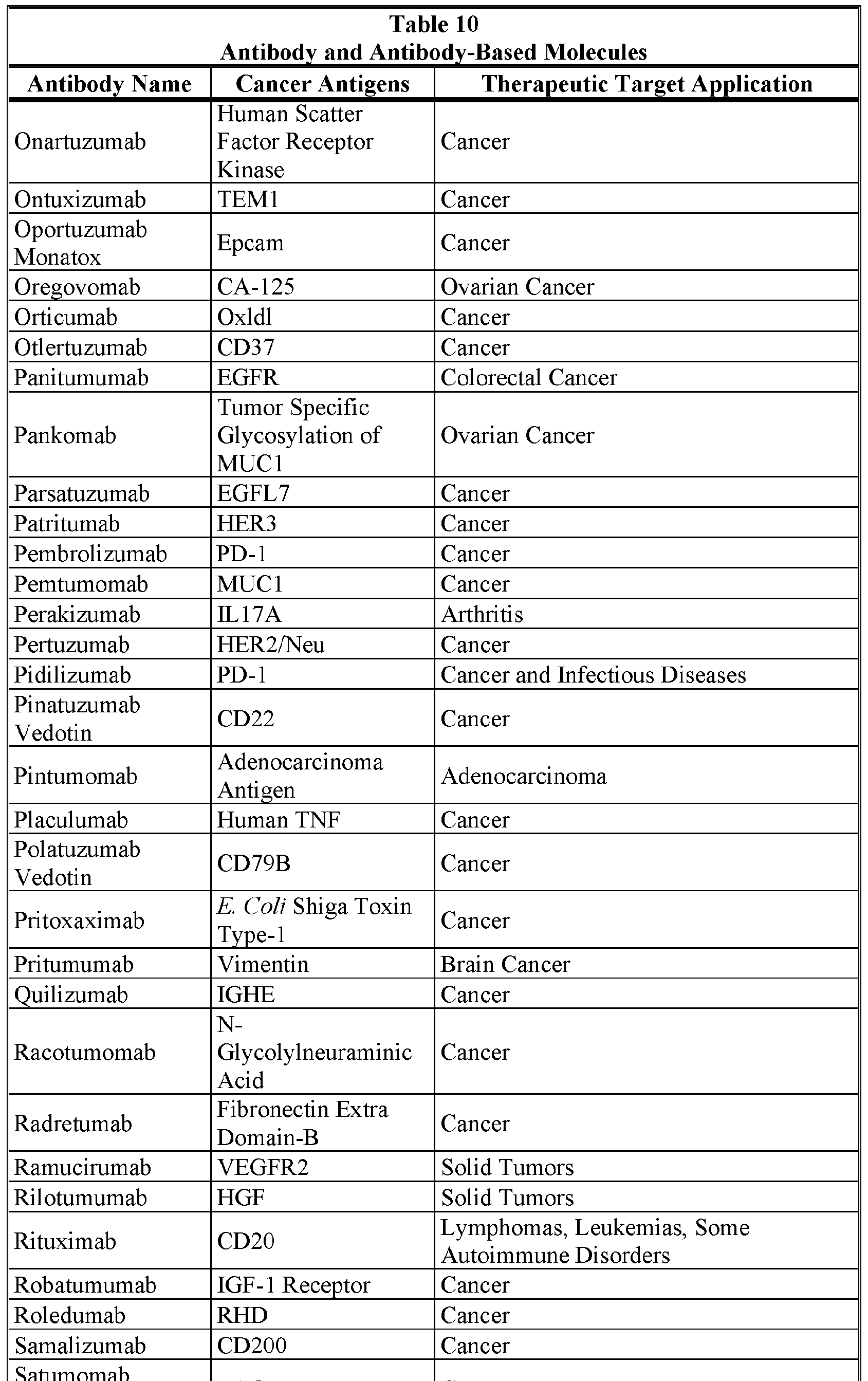


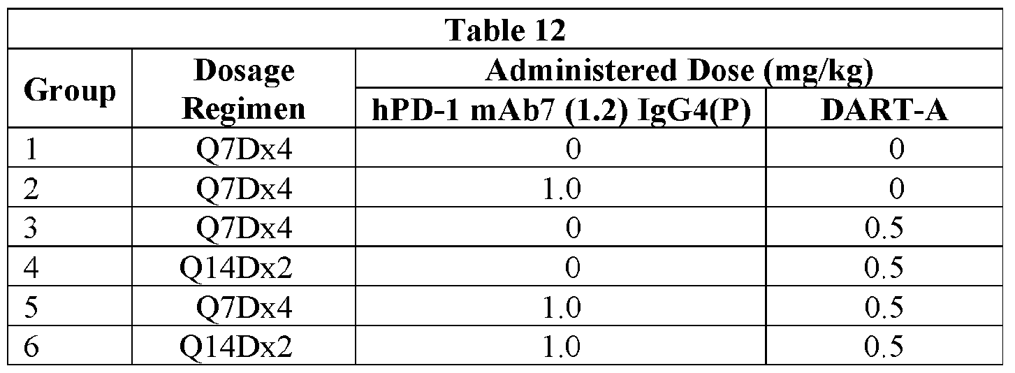


Claims
Priority Applications (11)
| Application Number | Priority Date | Filing Date | Title |
|---|---|---|---|
| KR1020197000309A KR20190015520A (en) | 2016-06-07 | 2017-06-06 | Combination Therapy |
| RU2018145961A RU2018145961A (en) | 2016-06-07 | 2017-06-06 | COMBINATION THERAPY |
| EP17810823.9A EP3463464A4 (en) | 2016-06-07 | 2017-06-06 | Combination therapy |
| AU2017278325A AU2017278325A1 (en) | 2016-06-07 | 2017-06-06 | Combination therapy |
| JP2018563801A JP2019517539A (en) | 2016-06-07 | 2017-06-06 | Combination therapy |
| CN201780035194.0A CN109310762A (en) | 2016-06-07 | 2017-06-06 | Conjoint therapy |
| SG11201810883TA SG11201810883TA (en) | 2016-06-07 | 2017-06-06 | Combination therapy |
| US16/306,882 US20200255524A1 (en) | 2016-06-07 | 2017-06-06 | Combination therapy |
| MX2018014950A MX2018014950A (en) | 2016-06-07 | 2017-06-06 | Combination therapy. |
| BR112018075198-7A BR112018075198A2 (en) | 2016-06-07 | 2017-06-06 | method for treating cancer or a pathogen-associated disease, pharmaceutical composition, and kit |
| IL263521A IL263521A (en) | 2016-06-07 | 2018-12-05 | Combination therapy |
Applications Claiming Priority (4)
| Application Number | Priority Date | Filing Date | Title |
|---|---|---|---|
| US201662346854P | 2016-06-07 | 2016-06-07 | |
| US62/346,854 | 2016-06-07 | ||
| US201662432299P | 2016-12-09 | 2016-12-09 | |
| US62/432,299 | 2016-12-09 |
Publications (1)
| Publication Number | Publication Date |
|---|---|
| WO2017214092A1 true WO2017214092A1 (en) | 2017-12-14 |
Family
ID=60578133
Family Applications (1)
| Application Number | Title | Priority Date | Filing Date |
|---|---|---|---|
| PCT/US2017/036075 WO2017214092A1 (en) | 2016-06-07 | 2017-06-06 | Combination therapy |
Country Status (14)
| Country | Link |
|---|---|
| US (1) | US20200255524A1 (en) |
| EP (1) | EP3463464A4 (en) |
| JP (1) | JP2019517539A (en) |
| KR (1) | KR20190015520A (en) |
| CN (1) | CN109310762A (en) |
| AU (1) | AU2017278325A1 (en) |
| BR (1) | BR112018075198A2 (en) |
| IL (1) | IL263521A (en) |
| MA (1) | MA45192A (en) |
| MX (1) | MX2018014950A (en) |
| RU (1) | RU2018145961A (en) |
| SG (2) | SG10201913326UA (en) |
| TW (1) | TW201742636A (en) |
| WO (1) | WO2017214092A1 (en) |
Cited By (47)
| Publication number | Priority date | Publication date | Assignee | Title |
|---|---|---|---|---|
| WO2018223002A1 (en) * | 2017-06-01 | 2018-12-06 | Xencor, Inc. | Bispecific antibodies that bind cd 123 cd3 |
| US10259887B2 (en) | 2014-11-26 | 2019-04-16 | Xencor, Inc. | Heterodimeric antibodies that bind CD3 and tumor antigens |
| WO2019165326A1 (en) * | 2018-02-23 | 2019-08-29 | REMD Biotherapeutics, Inc | Calcitonin gene-related peptide (cgrp) antagonist antibodies |
| US10428155B2 (en) | 2014-12-22 | 2019-10-01 | Xencor, Inc. | Trispecific antibodies |
| US10487155B2 (en) | 2013-01-14 | 2019-11-26 | Xencor, Inc. | Heterodimeric proteins |
| US10501543B2 (en) | 2016-10-14 | 2019-12-10 | Xencor, Inc. | IL15/IL15Rα heterodimeric Fc-fusion proteins |
| US10513558B2 (en) | 2015-07-13 | 2019-12-24 | Cytomx Therapeutics, Inc. | Anti-PD1 antibodies, activatable anti-PD1 antibodies, and methods of use thereof |
| US10519242B2 (en) | 2013-03-15 | 2019-12-31 | Xencor, Inc. | Targeting regulatory T cells with heterodimeric proteins |
| WO2020092404A1 (en) * | 2018-10-30 | 2020-05-07 | Macrogenics, Inc. | Bispecific cd123 x cd3 diabodies for the treatment of hematologic malignancies |
| US10738133B2 (en) | 2013-01-14 | 2020-08-11 | Xencor, Inc. | Heterodimeric proteins |
| US10787518B2 (en) | 2016-06-14 | 2020-09-29 | Xencor, Inc. | Bispecific checkpoint inhibitor antibodies |
| US10793632B2 (en) | 2016-08-30 | 2020-10-06 | Xencor, Inc. | Bispecific immunomodulatory antibodies that bind costimulatory and checkpoint receptors |
| WO2020216348A1 (en) * | 2019-04-26 | 2020-10-29 | Wuxi Biologics (Shanghai) Co., Ltd. | Bispecific antibodies against pd-1 and lag-3 |
| WO2020239558A1 (en) | 2019-05-24 | 2020-12-03 | Pfizer Inc. | Combination therapies using cdk inhibitors |
| US10858417B2 (en) | 2013-03-15 | 2020-12-08 | Xencor, Inc. | Heterodimeric proteins |
| US10889653B2 (en) | 2014-11-26 | 2021-01-12 | Xencor, Inc. | Heterodimeric antibodies that bind CD3 and tumor antigens |
| CN112533945A (en) * | 2018-05-18 | 2021-03-19 | 宏观基因有限公司 | Optimized gp 41-binding molecules and uses thereof |
| US10954301B2 (en) | 2015-12-14 | 2021-03-23 | Macrogenics, Inc. | Bispecific molecules having immunoreactivity with PD-1 and CTLA-4, and methods of use thereof |
| US10968276B2 (en) | 2013-03-12 | 2021-04-06 | Xencor, Inc. | Optimized anti-CD3 variable regions |
| US10968280B2 (en) | 2017-08-04 | 2021-04-06 | Genmab A/S | Binding agents binding to PD-L1 and CD137 and use thereof |
| US10982006B2 (en) | 2018-04-04 | 2021-04-20 | Xencor, Inc. | Heterodimeric antibodies that bind fibroblast activation protein |
| US10981992B2 (en) | 2017-11-08 | 2021-04-20 | Xencor, Inc. | Bispecific immunomodulatory antibodies that bind costimulatory and checkpoint receptors |
| WO2021133653A1 (en) * | 2019-12-23 | 2021-07-01 | Macrogenics, Inc. | Therapy for the treatment of cancer |
| US11053316B2 (en) | 2013-01-14 | 2021-07-06 | Xencor, Inc. | Optimized antibody variable regions |
| US11084863B2 (en) | 2017-06-30 | 2021-08-10 | Xencor, Inc. | Targeted heterodimeric Fc fusion proteins containing IL-15 IL-15alpha and antigen binding domains |
| US11091548B2 (en) | 2015-03-05 | 2021-08-17 | Xencor, Inc. | Modulation of T cells with bispecific antibodies and Fc fusions |
| EP3765041A4 (en) * | 2018-03-14 | 2021-12-22 | Seattle Children's Hospital (DBA Seattle Children's Research Institute) | Il-13 receptor alpha 2 (il13ra2) chimeric antigen receptor for tumor specific t cell immunotherapy |
| US11225521B2 (en) | 2016-06-28 | 2022-01-18 | Xencor, Inc. | Heterodimeric antibodies that bind somatostatin receptor 2 |
| US11299554B2 (en) | 2013-03-15 | 2022-04-12 | Xencor, Inc. | Heterodimeric proteins |
| US11312770B2 (en) | 2017-11-08 | 2022-04-26 | Xencor, Inc. | Bispecific and monospecific antibodies using novel anti-PD-1 sequences |
| US11319355B2 (en) | 2017-12-19 | 2022-05-03 | Xencor, Inc. | Engineered IL-2 Fc fusion proteins |
| US11352442B2 (en) | 2014-11-26 | 2022-06-07 | Xencor, Inc. | Heterodimeric antibodies that bind CD3 and CD38 |
| WO2022118197A1 (en) | 2020-12-02 | 2022-06-09 | Pfizer Inc. | Time to resolution of axitinib-related adverse events |
| US11358999B2 (en) | 2018-10-03 | 2022-06-14 | Xencor, Inc. | IL-12 heterodimeric Fc-fusion proteins |
| US11472890B2 (en) | 2019-03-01 | 2022-10-18 | Xencor, Inc. | Heterodimeric antibodies that bind ENPP3 and CD3 |
| US11505595B2 (en) | 2018-04-18 | 2022-11-22 | Xencor, Inc. | TIM-3 targeted heterodimeric fusion proteins containing IL-15/IL-15RA Fc-fusion proteins and TIM-3 antigen binding domains |
| US11524991B2 (en) | 2018-04-18 | 2022-12-13 | Xencor, Inc. | PD-1 targeted heterodimeric fusion proteins containing IL-15/IL-15Ra Fc-fusion proteins and PD-1 antigen binding domains and uses thereof |
| WO2023001987A2 (en) | 2021-07-22 | 2023-01-26 | University Of Dundee | Therapeutic muteins |
| US11591401B2 (en) | 2020-08-19 | 2023-02-28 | Xencor, Inc. | Anti-CD28 compositions |
| US11623957B2 (en) | 2015-12-07 | 2023-04-11 | Xencor, Inc. | Heterodimeric antibodies that bind CD3 and PSMA |
| WO2023057882A1 (en) | 2021-10-05 | 2023-04-13 | Pfizer Inc. | Combinations of azalactam compounds with a pd-1 axis binding antagonist for the treatment of cancer |
| WO2023079428A1 (en) | 2021-11-03 | 2023-05-11 | Pfizer Inc. | Combination therapies using tlr7/8 agonist |
| US11739144B2 (en) | 2021-03-09 | 2023-08-29 | Xencor, Inc. | Heterodimeric antibodies that bind CD3 and CLDN6 |
| US11840579B2 (en) | 2014-03-28 | 2023-12-12 | Xencor, Inc. | Bispecific antibodies that bind to CD38 and CD3 |
| US11859012B2 (en) | 2021-03-10 | 2024-01-02 | Xencor, Inc. | Heterodimeric antibodies that bind CD3 and GPC3 |
| US11919956B2 (en) | 2020-05-14 | 2024-03-05 | Xencor, Inc. | Heterodimeric antibodies that bind prostate specific membrane antigen (PSMA) and CD3 |
| EP4132581A4 (en) * | 2020-04-11 | 2024-04-10 | Univ Northwestern | HUMANIZED ANTIBODY TARGETING THE TUMOR ASSOCIATED ANTIGEN IL13Ra2 |
Families Citing this family (6)
| Publication number | Priority date | Publication date | Assignee | Title |
|---|---|---|---|---|
| CA3060618A1 (en) | 2017-05-19 | 2018-11-22 | Wuxi Biologics (Shanghai) Co. Ltd. | Novel monoclonal antibodies to cytotoxic t-lymphocyte-associated protein 4 (ctla-4) |
| AU2020319875A1 (en) * | 2019-08-01 | 2022-02-17 | Incyte Corporation | A dosing regimen for an IDO inhibitor |
| WO2021143826A1 (en) * | 2020-01-17 | 2021-07-22 | 信达生物制药(苏州)有限公司 | Recombinant anti-programmed cell death protein 1 and anti-cluster of differentiation antigen 137 bispecific antibody preparation and use thereof |
| CN112062859B (en) * | 2020-07-24 | 2022-08-09 | 沣潮医药科技(上海)有限公司 | Chimeric antigen receptor for pathogen clearance and uses thereof |
| CN114437218B (en) * | 2021-01-12 | 2022-09-30 | 北京门罗生物科技有限公司 | Chimeric antigen receptor targeting CD276 and immune cell comprising same |
| CN115611982A (en) * | 2021-07-14 | 2023-01-17 | 浙江大学 | Monoclonal antibody resisting human MICA/B alpha 3 region and application thereof |
Citations (28)
| Publication number | Priority date | Publication date | Assignee | Title |
|---|---|---|---|---|
| EP0403156A1 (en) | 1989-06-07 | 1990-12-19 | Genzyme Corporation | Improved monoclonal antibodies against the human alpha/beta t-cell receptor, their production and use |
| WO2003101485A1 (en) | 2002-05-30 | 2003-12-11 | Macrogenics, Inc. | Cd16a binding proteins and use for the treatment of immune disorders |
| WO2004001381A2 (en) | 2002-06-19 | 2003-12-31 | Raven Biotechnologies, Inc. | Novel raag10 cell surface target and a family of antibodies recognizing that target |
| WO2005118635A2 (en) | 2004-06-03 | 2005-12-15 | Novimmune S.A. | Anti-cd3 antibodies and methods of use thereof |
| WO2006125668A2 (en) | 2005-05-26 | 2006-11-30 | Affimed Therapeutics Ag | Anti-cd16 binding molecules |
| WO2008116219A2 (en) | 2007-03-22 | 2008-09-25 | Sloan-Kettering Institute For Cancer Research | Uses of monoclonal antibody 8h9 |
| WO2008119566A2 (en) | 2007-04-03 | 2008-10-09 | Micromet Ag | Cross-species-specific bispecific binders |
| WO2008146911A1 (en) | 2007-06-01 | 2008-12-04 | Sapporo Medical University | Antibody directed against il13ra2, and diagnostic/therapeutic agent comprising the antibody |
| US7666424B2 (en) | 2001-10-17 | 2010-02-23 | Sloan-Kettering Institute For Cancer Research | Methods of preparing and using single chain anti-tumor antibodies |
| US7737258B2 (en) | 2000-10-18 | 2010-06-15 | Sloan-Kettering Institute For Cancer Research | Uses of monoclonal antibody 8H9 |
| US7740845B2 (en) | 2000-10-18 | 2010-06-22 | Sloan-Kettering Institute For Cancer Research | Uses of monoclonal antibody 8H9 |
| WO2011034660A1 (en) | 2009-09-16 | 2011-03-24 | Immunomedics, Inc. | Class i anti-cea antibodies and uses thereof |
| WO2012147713A1 (en) | 2011-04-25 | 2012-11-01 | 第一三共株式会社 | Anti-b7-h3 antibody |
| WO2012162068A2 (en) | 2011-05-21 | 2012-11-29 | Macrogenics, Inc. | Deimmunized serum-binding domains and their use for extending serum half-life |
| WO2013041687A1 (en) | 2011-09-23 | 2013-03-28 | Amgen Research (Munich) Gmbh | Bispecific binding molecules for 5t4 and cd3 |
| US8414892B2 (en) | 2000-10-18 | 2013-04-09 | Sloan-Kettering Institute For Cancer Research | Uses of monoclonal antibody 8H9 |
| US8501471B2 (en) | 2000-10-18 | 2013-08-06 | Sloan-Kettering Institute For Cancer Research | Uses of monoclonal antibody 8H9 |
| WO2014072888A1 (en) | 2012-11-07 | 2014-05-15 | Pfizer Inc. | Anti-il-13 receptor alpha 2 antibodies and antibody-drug conjugates |
| US20140170149A1 (en) * | 2011-04-20 | 2014-06-19 | Genmab A/S | Bispecific antibodies against her2 and cd3 |
| WO2014159940A1 (en) | 2013-03-14 | 2014-10-02 | Macrogenics, Inc. | Bispecific molecules that are immunoreactive with immune effector cells that express an activating receptor |
| WO2015026892A1 (en) | 2013-08-23 | 2015-02-26 | Macrogenics, Inc. | Bi-specific monovalent diabodies that are capable of binding cd123 and cd3, and uses therof |
| WO2015112800A1 (en) * | 2014-01-23 | 2015-07-30 | Regeneron Pharmaceuticals, Inc. | Human antibodies to pd-1 |
| WO2015184203A1 (en) | 2014-05-29 | 2015-12-03 | Macrogenics, Inc. | Tri-specific binding molecules and methods of use thereof |
| WO2016036937A1 (en) | 2014-09-05 | 2016-03-10 | Janssen Pharmaceutica Nv | Cd123 binding agents and uses thereof |
| WO2016054101A1 (en) | 2014-09-29 | 2016-04-07 | Duke University | Bispecific molecules comprising an hiv-1 envelope targeting arm |
| WO2017011413A1 (en) | 2015-07-10 | 2017-01-19 | Duke University | Bispecific molecules comprising an hiv-1 envelope targeting arm |
| WO2017011414A1 (en) | 2015-07-10 | 2017-01-19 | Duke University | Bispecific molecules comprising an hiv-1 envelope targeting arm |
| US20170081424A1 (en) | 2014-11-26 | 2017-03-23 | Xencor, Inc. | Heterodimeric antibodies to cd3 x cd123 |
Family Cites Families (10)
| Publication number | Priority date | Publication date | Assignee | Title |
|---|---|---|---|---|
| US8802091B2 (en) * | 2010-03-04 | 2014-08-12 | Macrogenics, Inc. | Antibodies reactive with B7-H3 and uses thereof |
| CA2802344C (en) * | 2010-06-18 | 2023-06-13 | The Brigham And Women's Hospital, Inc. | Bi-specific antibodies against tim-3 and pd-1 for immunotherapy in chronic immune conditions |
| SI2714733T1 (en) * | 2011-05-21 | 2019-06-28 | Macrogenics, Inc. | Cd3-binding molecules capable of binding to human and non-human cd3 |
| US9487587B2 (en) * | 2013-03-05 | 2016-11-08 | Macrogenics, Inc. | Bispecific molecules that are immunoreactive with immune effector cells of a companion animal that express an activating receptor and cells that express B7-H3 and uses thereof |
| RS60443B1 (en) * | 2013-12-17 | 2020-07-31 | Genentech Inc | Anti-cd3 antibodies and methods of use |
| PT3083686T (en) * | 2013-12-17 | 2019-12-18 | Hoffmann La Roche | Methods of treating cancers using pd-1 axis binding antagonists and taxanes |
| WO2015112534A2 (en) * | 2014-01-21 | 2015-07-30 | Medimmune, Llc | Compositions and methods for modulating and redirecting immune responses |
| TWI693232B (en) * | 2014-06-26 | 2020-05-11 | 美商宏觀基因股份有限公司 | Covalently bonded diabodies having immunoreactivity with pd-1 and lag-3, and methods of use thereof |
| DK3221355T3 (en) * | 2014-11-20 | 2020-12-07 | Hoffmann La Roche | Combination therapy with T cell activating bispecific antigen binding molecules CD3 and folate receptor 1 (FolR1) as well as PD-1 axis binding antagonists |
| CN107614522A (en) * | 2015-01-14 | 2018-01-19 | 指南针制药有限责任公司 | Multispecific immune modulability antigen-binding constructs |
-
2017
- 2017-06-06 JP JP2018563801A patent/JP2019517539A/en active Pending
- 2017-06-06 SG SG10201913326UA patent/SG10201913326UA/en unknown
- 2017-06-06 RU RU2018145961A patent/RU2018145961A/en unknown
- 2017-06-06 CN CN201780035194.0A patent/CN109310762A/en active Pending
- 2017-06-06 KR KR1020197000309A patent/KR20190015520A/en not_active Application Discontinuation
- 2017-06-06 MA MA045192A patent/MA45192A/en unknown
- 2017-06-06 SG SG11201810883TA patent/SG11201810883TA/en unknown
- 2017-06-06 BR BR112018075198-7A patent/BR112018075198A2/en not_active IP Right Cessation
- 2017-06-06 MX MX2018014950A patent/MX2018014950A/en unknown
- 2017-06-06 US US16/306,882 patent/US20200255524A1/en not_active Abandoned
- 2017-06-06 WO PCT/US2017/036075 patent/WO2017214092A1/en unknown
- 2017-06-06 AU AU2017278325A patent/AU2017278325A1/en not_active Abandoned
- 2017-06-06 EP EP17810823.9A patent/EP3463464A4/en not_active Withdrawn
- 2017-06-07 TW TW106118940A patent/TW201742636A/en unknown
-
2018
- 2018-12-05 IL IL263521A patent/IL263521A/en unknown
Patent Citations (34)
| Publication number | Priority date | Publication date | Assignee | Title |
|---|---|---|---|---|
| EP0403156A1 (en) | 1989-06-07 | 1990-12-19 | Genzyme Corporation | Improved monoclonal antibodies against the human alpha/beta t-cell receptor, their production and use |
| US8501471B2 (en) | 2000-10-18 | 2013-08-06 | Sloan-Kettering Institute For Cancer Research | Uses of monoclonal antibody 8H9 |
| US7740845B2 (en) | 2000-10-18 | 2010-06-22 | Sloan-Kettering Institute For Cancer Research | Uses of monoclonal antibody 8H9 |
| US7737258B2 (en) | 2000-10-18 | 2010-06-15 | Sloan-Kettering Institute For Cancer Research | Uses of monoclonal antibody 8H9 |
| US9062110B2 (en) | 2000-10-18 | 2015-06-23 | Sloan-Kettering Institute For Cancer Research | Uses of monoclonial antibody 8H9 |
| US8414892B2 (en) | 2000-10-18 | 2013-04-09 | Sloan-Kettering Institute For Cancer Research | Uses of monoclonal antibody 8H9 |
| US7666424B2 (en) | 2001-10-17 | 2010-02-23 | Sloan-Kettering Institute For Cancer Research | Methods of preparing and using single chain anti-tumor antibodies |
| US8148154B2 (en) | 2001-10-17 | 2012-04-03 | Sloan-Kettering Institute For Cancer Research | Method for preparation of single chain antibodies |
| WO2003101485A1 (en) | 2002-05-30 | 2003-12-11 | Macrogenics, Inc. | Cd16a binding proteins and use for the treatment of immune disorders |
| US8779098B2 (en) | 2002-06-19 | 2014-07-15 | Macrogenics West, Inc. | B7-H3L cell surface antigen and antibodies that bind thereto |
| US7527969B2 (en) | 2002-06-19 | 2009-05-05 | Raven Biotechnologies, Inc. | RAAG10 cell surface target and a family of antibodies recognizing that target |
| WO2004001381A2 (en) | 2002-06-19 | 2003-12-31 | Raven Biotechnologies, Inc. | Novel raag10 cell surface target and a family of antibodies recognizing that target |
| WO2005118635A2 (en) | 2004-06-03 | 2005-12-15 | Novimmune S.A. | Anti-cd3 antibodies and methods of use thereof |
| WO2006125668A2 (en) | 2005-05-26 | 2006-11-30 | Affimed Therapeutics Ag | Anti-cd16 binding molecules |
| US20100143245A1 (en) | 2007-03-22 | 2010-06-10 | Sloan-Kettering Institute For Cancer Research | Uses of monoclonal antibody 8h9 |
| WO2008116219A2 (en) | 2007-03-22 | 2008-09-25 | Sloan-Kettering Institute For Cancer Research | Uses of monoclonal antibody 8h9 |
| WO2008119566A2 (en) | 2007-04-03 | 2008-10-09 | Micromet Ag | Cross-species-specific bispecific binders |
| WO2008146911A1 (en) | 2007-06-01 | 2008-12-04 | Sapporo Medical University | Antibody directed against il13ra2, and diagnostic/therapeutic agent comprising the antibody |
| WO2011034660A1 (en) | 2009-09-16 | 2011-03-24 | Immunomedics, Inc. | Class i anti-cea antibodies and uses thereof |
| US20140170149A1 (en) * | 2011-04-20 | 2014-06-19 | Genmab A/S | Bispecific antibodies against her2 and cd3 |
| WO2012147713A1 (en) | 2011-04-25 | 2012-11-01 | 第一三共株式会社 | Anti-b7-h3 antibody |
| US20130078234A1 (en) | 2011-04-25 | 2013-03-28 | Daiichi Sankyo Company, Limited | Anti b7-h3 antibody |
| WO2012162068A2 (en) | 2011-05-21 | 2012-11-29 | Macrogenics, Inc. | Deimmunized serum-binding domains and their use for extending serum half-life |
| WO2013041687A1 (en) | 2011-09-23 | 2013-03-28 | Amgen Research (Munich) Gmbh | Bispecific binding molecules for 5t4 and cd3 |
| WO2014072888A1 (en) | 2012-11-07 | 2014-05-15 | Pfizer Inc. | Anti-il-13 receptor alpha 2 antibodies and antibody-drug conjugates |
| WO2014159940A1 (en) | 2013-03-14 | 2014-10-02 | Macrogenics, Inc. | Bispecific molecules that are immunoreactive with immune effector cells that express an activating receptor |
| WO2015026892A1 (en) | 2013-08-23 | 2015-02-26 | Macrogenics, Inc. | Bi-specific monovalent diabodies that are capable of binding cd123 and cd3, and uses therof |
| WO2015112800A1 (en) * | 2014-01-23 | 2015-07-30 | Regeneron Pharmaceuticals, Inc. | Human antibodies to pd-1 |
| WO2015184203A1 (en) | 2014-05-29 | 2015-12-03 | Macrogenics, Inc. | Tri-specific binding molecules and methods of use thereof |
| WO2016036937A1 (en) | 2014-09-05 | 2016-03-10 | Janssen Pharmaceutica Nv | Cd123 binding agents and uses thereof |
| WO2016054101A1 (en) | 2014-09-29 | 2016-04-07 | Duke University | Bispecific molecules comprising an hiv-1 envelope targeting arm |
| US20170081424A1 (en) | 2014-11-26 | 2017-03-23 | Xencor, Inc. | Heterodimeric antibodies to cd3 x cd123 |
| WO2017011413A1 (en) | 2015-07-10 | 2017-01-19 | Duke University | Bispecific molecules comprising an hiv-1 envelope targeting arm |
| WO2017011414A1 (en) | 2015-07-10 | 2017-01-19 | Duke University | Bispecific molecules comprising an hiv-1 envelope targeting arm |
Non-Patent Citations (27)
Cited By (64)
| Publication number | Priority date | Publication date | Assignee | Title |
|---|---|---|---|---|
| US11053316B2 (en) | 2013-01-14 | 2021-07-06 | Xencor, Inc. | Optimized antibody variable regions |
| US11634506B2 (en) | 2013-01-14 | 2023-04-25 | Xencor, Inc. | Heterodimeric proteins |
| US10487155B2 (en) | 2013-01-14 | 2019-11-26 | Xencor, Inc. | Heterodimeric proteins |
| US11718667B2 (en) | 2013-01-14 | 2023-08-08 | Xencor, Inc. | Optimized antibody variable regions |
| US10738133B2 (en) | 2013-01-14 | 2020-08-11 | Xencor, Inc. | Heterodimeric proteins |
| US10968276B2 (en) | 2013-03-12 | 2021-04-06 | Xencor, Inc. | Optimized anti-CD3 variable regions |
| US11814423B2 (en) | 2013-03-15 | 2023-11-14 | Xencor, Inc. | Heterodimeric proteins |
| US11299554B2 (en) | 2013-03-15 | 2022-04-12 | Xencor, Inc. | Heterodimeric proteins |
| US10858417B2 (en) | 2013-03-15 | 2020-12-08 | Xencor, Inc. | Heterodimeric proteins |
| US10519242B2 (en) | 2013-03-15 | 2019-12-31 | Xencor, Inc. | Targeting regulatory T cells with heterodimeric proteins |
| US11840579B2 (en) | 2014-03-28 | 2023-12-12 | Xencor, Inc. | Bispecific antibodies that bind to CD38 and CD3 |
| US11859011B2 (en) | 2014-11-26 | 2024-01-02 | Xencor, Inc. | Heterodimeric antibodies that bind CD3 and tumor antigens |
| US11111315B2 (en) | 2014-11-26 | 2021-09-07 | Xencor, Inc. | Heterodimeric antibodies that bind CD3 and tumor antigens |
| US11673972B2 (en) | 2014-11-26 | 2023-06-13 | Xencor, Inc. | Heterodimeric antibodies that bind CD3 and tumor antigens |
| US10889653B2 (en) | 2014-11-26 | 2021-01-12 | Xencor, Inc. | Heterodimeric antibodies that bind CD3 and tumor antigens |
| US10913803B2 (en) | 2014-11-26 | 2021-02-09 | Xencor, Inc. | Heterodimeric antibodies that bind CD3 and tumor antigens |
| US11945880B2 (en) | 2014-11-26 | 2024-04-02 | Xencor, Inc. | Heterodimeric antibodies that bind CD3 and tumor antigens |
| US10259887B2 (en) | 2014-11-26 | 2019-04-16 | Xencor, Inc. | Heterodimeric antibodies that bind CD3 and tumor antigens |
| US11225528B2 (en) | 2014-11-26 | 2022-01-18 | Xencor, Inc. | Heterodimeric antibodies that bind CD3 and tumor antigens |
| US11352442B2 (en) | 2014-11-26 | 2022-06-07 | Xencor, Inc. | Heterodimeric antibodies that bind CD3 and CD38 |
| US10428155B2 (en) | 2014-12-22 | 2019-10-01 | Xencor, Inc. | Trispecific antibodies |
| US11091548B2 (en) | 2015-03-05 | 2021-08-17 | Xencor, Inc. | Modulation of T cells with bispecific antibodies and Fc fusions |
| US10513558B2 (en) | 2015-07-13 | 2019-12-24 | Cytomx Therapeutics, Inc. | Anti-PD1 antibodies, activatable anti-PD1 antibodies, and methods of use thereof |
| US11623957B2 (en) | 2015-12-07 | 2023-04-11 | Xencor, Inc. | Heterodimeric antibodies that bind CD3 and PSMA |
| US10954301B2 (en) | 2015-12-14 | 2021-03-23 | Macrogenics, Inc. | Bispecific molecules having immunoreactivity with PD-1 and CTLA-4, and methods of use thereof |
| US11840571B2 (en) | 2015-12-14 | 2023-12-12 | Macrogenics, Inc. | Methods of using bispecific molecules having immunoreactivity with PD-1 and CTLA-4 |
| US11236170B2 (en) | 2016-06-14 | 2022-02-01 | Xencor, Inc. | Bispecific checkpoint inhibitor antibodies |
| US11492407B2 (en) | 2016-06-14 | 2022-11-08 | Xencor, Inc. | Bispecific checkpoint inhibitor antibodies |
| US10787518B2 (en) | 2016-06-14 | 2020-09-29 | Xencor, Inc. | Bispecific checkpoint inhibitor antibodies |
| US11225521B2 (en) | 2016-06-28 | 2022-01-18 | Xencor, Inc. | Heterodimeric antibodies that bind somatostatin receptor 2 |
| US10793632B2 (en) | 2016-08-30 | 2020-10-06 | Xencor, Inc. | Bispecific immunomodulatory antibodies that bind costimulatory and checkpoint receptors |
| US10501543B2 (en) | 2016-10-14 | 2019-12-10 | Xencor, Inc. | IL15/IL15Rα heterodimeric Fc-fusion proteins |
| WO2018223002A1 (en) * | 2017-06-01 | 2018-12-06 | Xencor, Inc. | Bispecific antibodies that bind cd 123 cd3 |
| US11084863B2 (en) | 2017-06-30 | 2021-08-10 | Xencor, Inc. | Targeted heterodimeric Fc fusion proteins containing IL-15 IL-15alpha and antigen binding domains |
| US11459395B2 (en) | 2017-08-04 | 2022-10-04 | Genmab A/S | Binding agents binding to PD-L1 and CD137 and use thereof |
| US10968280B2 (en) | 2017-08-04 | 2021-04-06 | Genmab A/S | Binding agents binding to PD-L1 and CD137 and use thereof |
| US11312770B2 (en) | 2017-11-08 | 2022-04-26 | Xencor, Inc. | Bispecific and monospecific antibodies using novel anti-PD-1 sequences |
| US10981992B2 (en) | 2017-11-08 | 2021-04-20 | Xencor, Inc. | Bispecific immunomodulatory antibodies that bind costimulatory and checkpoint receptors |
| US11319355B2 (en) | 2017-12-19 | 2022-05-03 | Xencor, Inc. | Engineered IL-2 Fc fusion proteins |
| US11629184B2 (en) | 2018-02-23 | 2023-04-18 | Remd Biotherapeutics, Inc. | Calcitonin gene-related peptide (CGRP) antagonist antibodies |
| WO2019165326A1 (en) * | 2018-02-23 | 2019-08-29 | REMD Biotherapeutics, Inc | Calcitonin gene-related peptide (cgrp) antagonist antibodies |
| EP3765041A4 (en) * | 2018-03-14 | 2021-12-22 | Seattle Children's Hospital (DBA Seattle Children's Research Institute) | Il-13 receptor alpha 2 (il13ra2) chimeric antigen receptor for tumor specific t cell immunotherapy |
| US10982006B2 (en) | 2018-04-04 | 2021-04-20 | Xencor, Inc. | Heterodimeric antibodies that bind fibroblast activation protein |
| US11505595B2 (en) | 2018-04-18 | 2022-11-22 | Xencor, Inc. | TIM-3 targeted heterodimeric fusion proteins containing IL-15/IL-15RA Fc-fusion proteins and TIM-3 antigen binding domains |
| US11524991B2 (en) | 2018-04-18 | 2022-12-13 | Xencor, Inc. | PD-1 targeted heterodimeric fusion proteins containing IL-15/IL-15Ra Fc-fusion proteins and PD-1 antigen binding domains and uses thereof |
| CN112533945A (en) * | 2018-05-18 | 2021-03-19 | 宏观基因有限公司 | Optimized gp 41-binding molecules and uses thereof |
| US11358999B2 (en) | 2018-10-03 | 2022-06-14 | Xencor, Inc. | IL-12 heterodimeric Fc-fusion proteins |
| WO2020092404A1 (en) * | 2018-10-30 | 2020-05-07 | Macrogenics, Inc. | Bispecific cd123 x cd3 diabodies for the treatment of hematologic malignancies |
| JP2022513402A (en) * | 2018-10-30 | 2022-02-07 | マクロジェニクス,インコーポレーテッド | Bispecific CD123 x CD3 diabody for the treatment of hematological malignancies |
| CN113286633A (en) * | 2018-10-30 | 2021-08-20 | 宏观基因有限公司 | Bispecific CD123x CD3 diabodies for the treatment of hematological malignancies |
| US11472890B2 (en) | 2019-03-01 | 2022-10-18 | Xencor, Inc. | Heterodimeric antibodies that bind ENPP3 and CD3 |
| WO2020216348A1 (en) * | 2019-04-26 | 2020-10-29 | Wuxi Biologics (Shanghai) Co., Ltd. | Bispecific antibodies against pd-1 and lag-3 |
| WO2020239558A1 (en) | 2019-05-24 | 2020-12-03 | Pfizer Inc. | Combination therapies using cdk inhibitors |
| WO2021133653A1 (en) * | 2019-12-23 | 2021-07-01 | Macrogenics, Inc. | Therapy for the treatment of cancer |
| EP4132581A4 (en) * | 2020-04-11 | 2024-04-10 | Univ Northwestern | HUMANIZED ANTIBODY TARGETING THE TUMOR ASSOCIATED ANTIGEN IL13Ra2 |
| US11919956B2 (en) | 2020-05-14 | 2024-03-05 | Xencor, Inc. | Heterodimeric antibodies that bind prostate specific membrane antigen (PSMA) and CD3 |
| US11919958B2 (en) | 2020-08-19 | 2024-03-05 | Xencor, Inc. | Anti-CD28 compositions |
| US11591401B2 (en) | 2020-08-19 | 2023-02-28 | Xencor, Inc. | Anti-CD28 compositions |
| WO2022118197A1 (en) | 2020-12-02 | 2022-06-09 | Pfizer Inc. | Time to resolution of axitinib-related adverse events |
| US11739144B2 (en) | 2021-03-09 | 2023-08-29 | Xencor, Inc. | Heterodimeric antibodies that bind CD3 and CLDN6 |
| US11859012B2 (en) | 2021-03-10 | 2024-01-02 | Xencor, Inc. | Heterodimeric antibodies that bind CD3 and GPC3 |
| WO2023001987A2 (en) | 2021-07-22 | 2023-01-26 | University Of Dundee | Therapeutic muteins |
| WO2023057882A1 (en) | 2021-10-05 | 2023-04-13 | Pfizer Inc. | Combinations of azalactam compounds with a pd-1 axis binding antagonist for the treatment of cancer |
| WO2023079428A1 (en) | 2021-11-03 | 2023-05-11 | Pfizer Inc. | Combination therapies using tlr7/8 agonist |
Also Published As
| Publication number | Publication date |
|---|---|
| BR112018075198A2 (en) | 2019-03-19 |
| CN109310762A (en) | 2019-02-05 |
| IL263521A (en) | 2019-01-31 |
| TW201742636A (en) | 2017-12-16 |
| EP3463464A1 (en) | 2019-04-10 |
| KR20190015520A (en) | 2019-02-13 |
| RU2018145961A3 (en) | 2020-07-30 |
| JP2019517539A (en) | 2019-06-24 |
| RU2018145961A (en) | 2020-07-14 |
| SG11201810883TA (en) | 2019-01-30 |
| AU2017278325A1 (en) | 2019-01-24 |
| US20200255524A1 (en) | 2020-08-13 |
| SG10201913326UA (en) | 2020-02-27 |
| MX2018014950A (en) | 2019-04-25 |
| EP3463464A4 (en) | 2020-07-01 |
| MA45192A (en) | 2019-04-10 |
Similar Documents
| Publication | Publication Date | Title |
|---|---|---|
| US20200255524A1 (en) | Combination therapy | |
| TWI788327B (en) | Bispecific binding molecules that are capable of binding cd137 and tumor antigens, and uses thereof | |
| TWI691509B (en) | Pd-1-binding molecules and methods of use thereof | |
| CA2986953A1 (en) | Lag-3-binding molecules and methods of use thereof | |
| US11685781B2 (en) | Variant CD3-binding domains and their use in combination therapies for the treatment of disease | |
| WO2016122701A1 (en) | Anti-dr5 antibodies and molecules comprising dr5-binding domains thereof | |
| US11795226B2 (en) | Bispecific CD16-binding molecules and their use in the treatment of disease | |
| US20220324995A1 (en) | ADAM9-Binding Molecules, and Methods of Use Thereof | |
| WO2020041404A1 (en) | Pd-l1-binding molecules and use of the same for the treatment of disease | |
| RU2810222C2 (en) | Variant cd3 binding domains and their use in combination therapy for treatment of diseases | |
| JP2018527290A (en) | LAG-3 binding molecule and method of use thereof |
Legal Events
| Date | Code | Title | Description |
|---|---|---|---|
| 121 | Ep: the epo has been informed by wipo that ep was designated in this application |
Ref document number: 17810823 Country of ref document: EP Kind code of ref document: A1 |
|
| ENP | Entry into the national phase |
Ref document number: 2018563801 Country of ref document: JP Kind code of ref document: A |
|
| NENP | Non-entry into the national phase |
Ref country code: DE |
|
| REG | Reference to national code |
Ref country code: BR Ref legal event code: B01A Ref document number: 112018075198 Country of ref document: BR |
|
| ENP | Entry into the national phase |
Ref document number: 20197000309 Country of ref document: KR Kind code of ref document: A |
|
| ENP | Entry into the national phase |
Ref document number: 2017810823 Country of ref document: EP Effective date: 20190107 |
|
| ENP | Entry into the national phase |
Ref document number: 2017278325 Country of ref document: AU Date of ref document: 20170606 Kind code of ref document: A |
|
| ENP | Entry into the national phase |
Ref document number: 112018075198 Country of ref document: BR Kind code of ref document: A2 Effective date: 20181205 |