WO2017183458A1 - Drug monitoring device, method, and program - Google Patents
Drug monitoring device, method, and program Download PDFInfo
- Publication number
- WO2017183458A1 WO2017183458A1 PCT/JP2017/014261 JP2017014261W WO2017183458A1 WO 2017183458 A1 WO2017183458 A1 WO 2017183458A1 JP 2017014261 W JP2017014261 W JP 2017014261W WO 2017183458 A1 WO2017183458 A1 WO 2017183458A1
- Authority
- WO
- WIPO (PCT)
- Prior art keywords
- drug
- unit
- medicine
- recognition
- quantitative value
- Prior art date
Links
- 239000003814 drug Substances 0.000 title claims abstract description 397
- 229940079593 drug Drugs 0.000 title claims abstract description 224
- 238000000034 method Methods 0.000 title claims abstract description 65
- 238000012806 monitoring device Methods 0.000 title abstract 3
- 238000004364 calculation method Methods 0.000 claims abstract description 27
- 238000007689 inspection Methods 0.000 claims description 58
- 238000003708 edge detection Methods 0.000 claims description 52
- 230000008569 process Effects 0.000 claims description 45
- 238000004806 packaging method and process Methods 0.000 claims description 40
- 238000003384 imaging method Methods 0.000 claims description 25
- 238000012550 audit Methods 0.000 claims description 22
- 238000001228 spectrum Methods 0.000 claims description 14
- 238000012545 processing Methods 0.000 description 47
- 238000005286 illumination Methods 0.000 description 33
- 230000010365 information processing Effects 0.000 description 31
- 230000006870 function Effects 0.000 description 25
- 230000007723 transport mechanism Effects 0.000 description 10
- 238000000605 extraction Methods 0.000 description 9
- 238000012986 modification Methods 0.000 description 8
- 230000004048 modification Effects 0.000 description 8
- 239000003795 chemical substances by application Substances 0.000 description 6
- 238000004088 simulation Methods 0.000 description 5
- 239000000463 material Substances 0.000 description 4
- 230000007246 mechanism Effects 0.000 description 4
- 238000012856 packing Methods 0.000 description 4
- 239000002775 capsule Substances 0.000 description 3
- 238000006243 chemical reaction Methods 0.000 description 3
- 239000000126 substance Substances 0.000 description 3
- 238000012795 verification Methods 0.000 description 3
- 238000004891 communication Methods 0.000 description 2
- 238000001514 detection method Methods 0.000 description 2
- 238000010586 diagram Methods 0.000 description 2
- 230000000694 effects Effects 0.000 description 2
- 239000000284 extract Substances 0.000 description 2
- 230000010354 integration Effects 0.000 description 2
- 238000002360 preparation method Methods 0.000 description 2
- 230000003595 spectral effect Effects 0.000 description 2
- 230000032258 transport Effects 0.000 description 2
- 230000015572 biosynthetic process Effects 0.000 description 1
- 235000021152 breakfast Nutrition 0.000 description 1
- 230000008859 change Effects 0.000 description 1
- 230000000052 comparative effect Effects 0.000 description 1
- 230000000295 complement effect Effects 0.000 description 1
- 239000002131 composite material Substances 0.000 description 1
- 239000000428 dust Substances 0.000 description 1
- 238000005401 electroluminescence Methods 0.000 description 1
- 238000011156 evaluation Methods 0.000 description 1
- 239000003168 generic drug Substances 0.000 description 1
- 239000004973 liquid crystal related substance Substances 0.000 description 1
- 238000012423 maintenance Methods 0.000 description 1
- 230000007721 medicinal effect Effects 0.000 description 1
- 229910044991 metal oxide Inorganic materials 0.000 description 1
- 150000004706 metal oxides Chemical class 0.000 description 1
- 238000010606 normalization Methods 0.000 description 1
- 230000003287 optical effect Effects 0.000 description 1
- 239000005022 packaging material Substances 0.000 description 1
- 238000003672 processing method Methods 0.000 description 1
- 239000004065 semiconductor Substances 0.000 description 1
- 230000008054 signal transmission Effects 0.000 description 1
- 239000007787 solid Substances 0.000 description 1
- 238000003786 synthesis reaction Methods 0.000 description 1
- 230000009466 transformation Effects 0.000 description 1
- 238000009966 trimming Methods 0.000 description 1
- 230000000007 visual effect Effects 0.000 description 1
Images


Classifications
-
- G—PHYSICS
- G06—COMPUTING; CALCULATING OR COUNTING
- G06V—IMAGE OR VIDEO RECOGNITION OR UNDERSTANDING
- G06V20/00—Scenes; Scene-specific elements
- G06V20/60—Type of objects
- G06V20/66—Trinkets, e.g. shirt buttons or jewellery items
-
- A—HUMAN NECESSITIES
- A61—MEDICAL OR VETERINARY SCIENCE; HYGIENE
- A61J—CONTAINERS SPECIALLY ADAPTED FOR MEDICAL OR PHARMACEUTICAL PURPOSES; DEVICES OR METHODS SPECIALLY ADAPTED FOR BRINGING PHARMACEUTICAL PRODUCTS INTO PARTICULAR PHYSICAL OR ADMINISTERING FORMS; DEVICES FOR ADMINISTERING FOOD OR MEDICINES ORALLY; BABY COMFORTERS; DEVICES FOR RECEIVING SPITTLE
- A61J3/00—Devices or methods specially adapted for bringing pharmaceutical products into particular physical or administering forms
-
- A—HUMAN NECESSITIES
- A61—MEDICAL OR VETERINARY SCIENCE; HYGIENE
- A61J—CONTAINERS SPECIALLY ADAPTED FOR MEDICAL OR PHARMACEUTICAL PURPOSES; DEVICES OR METHODS SPECIALLY ADAPTED FOR BRINGING PHARMACEUTICAL PRODUCTS INTO PARTICULAR PHYSICAL OR ADMINISTERING FORMS; DEVICES FOR ADMINISTERING FOOD OR MEDICINES ORALLY; BABY COMFORTERS; DEVICES FOR RECEIVING SPITTLE
- A61J7/00—Devices for administering medicines orally, e.g. spoons; Pill counting devices; Arrangements for time indication or reminder for taking medicine
- A61J7/02—Pill counting devices
-
- G—PHYSICS
- G01—MEASURING; TESTING
- G01N—INVESTIGATING OR ANALYSING MATERIALS BY DETERMINING THEIR CHEMICAL OR PHYSICAL PROPERTIES
- G01N21/00—Investigating or analysing materials by the use of optical means, i.e. using sub-millimetre waves, infrared, visible or ultraviolet light
- G01N21/84—Systems specially adapted for particular applications
- G01N21/85—Investigating moving fluids or granular solids
-
- G—PHYSICS
- G01—MEASURING; TESTING
- G01N—INVESTIGATING OR ANALYSING MATERIALS BY DETERMINING THEIR CHEMICAL OR PHYSICAL PROPERTIES
- G01N21/00—Investigating or analysing materials by the use of optical means, i.e. using sub-millimetre waves, infrared, visible or ultraviolet light
- G01N21/84—Systems specially adapted for particular applications
- G01N21/88—Investigating the presence of flaws or contamination
- G01N21/95—Investigating the presence of flaws or contamination characterised by the material or shape of the object to be examined
- G01N21/9508—Capsules; Tablets
-
- G—PHYSICS
- G06—COMPUTING; CALCULATING OR COUNTING
- G06T—IMAGE DATA PROCESSING OR GENERATION, IN GENERAL
- G06T7/00—Image analysis
- G06T7/0002—Inspection of images, e.g. flaw detection
-
- G—PHYSICS
- G06—COMPUTING; CALCULATING OR COUNTING
- G06T—IMAGE DATA PROCESSING OR GENERATION, IN GENERAL
- G06T7/00—Image analysis
- G06T7/0002—Inspection of images, e.g. flaw detection
- G06T7/0012—Biomedical image inspection
-
- G—PHYSICS
- G06—COMPUTING; CALCULATING OR COUNTING
- G06T—IMAGE DATA PROCESSING OR GENERATION, IN GENERAL
- G06T7/00—Image analysis
- G06T7/10—Segmentation; Edge detection
- G06T7/13—Edge detection
-
- G—PHYSICS
- G16—INFORMATION AND COMMUNICATION TECHNOLOGY [ICT] SPECIALLY ADAPTED FOR SPECIFIC APPLICATION FIELDS
- G16H—HEALTHCARE INFORMATICS, i.e. INFORMATION AND COMMUNICATION TECHNOLOGY [ICT] SPECIALLY ADAPTED FOR THE HANDLING OR PROCESSING OF MEDICAL OR HEALTHCARE DATA
- G16H20/00—ICT specially adapted for therapies or health-improving plans, e.g. for handling prescriptions, for steering therapy or for monitoring patient compliance
- G16H20/10—ICT specially adapted for therapies or health-improving plans, e.g. for handling prescriptions, for steering therapy or for monitoring patient compliance relating to drugs or medications, e.g. for ensuring correct administration to patients
- G16H20/13—ICT specially adapted for therapies or health-improving plans, e.g. for handling prescriptions, for steering therapy or for monitoring patient compliance relating to drugs or medications, e.g. for ensuring correct administration to patients delivered from dispensers
-
- H—ELECTRICITY
- H04—ELECTRIC COMMUNICATION TECHNIQUE
- H04N—PICTORIAL COMMUNICATION, e.g. TELEVISION
- H04N23/00—Cameras or camera modules comprising electronic image sensors; Control thereof
- H04N23/80—Camera processing pipelines; Components thereof
-
- A—HUMAN NECESSITIES
- A61—MEDICAL OR VETERINARY SCIENCE; HYGIENE
- A61J—CONTAINERS SPECIALLY ADAPTED FOR MEDICAL OR PHARMACEUTICAL PURPOSES; DEVICES OR METHODS SPECIALLY ADAPTED FOR BRINGING PHARMACEUTICAL PRODUCTS INTO PARTICULAR PHYSICAL OR ADMINISTERING FORMS; DEVICES FOR ADMINISTERING FOOD OR MEDICINES ORALLY; BABY COMFORTERS; DEVICES FOR RECEIVING SPITTLE
- A61J7/00—Devices for administering medicines orally, e.g. spoons; Pill counting devices; Arrangements for time indication or reminder for taking medicine
- A61J7/04—Arrangements for time indication or reminder for taking medicine, e.g. programmed dispensers
- A61J7/0409—Arrangements for time indication or reminder for taking medicine, e.g. programmed dispensers with timers
- A61J7/0454—Arrangements for time indication or reminder for taking medicine, e.g. programmed dispensers with timers for dispensing of multiple drugs
-
- G—PHYSICS
- G06—COMPUTING; CALCULATING OR COUNTING
- G06T—IMAGE DATA PROCESSING OR GENERATION, IN GENERAL
- G06T2207/00—Indexing scheme for image analysis or image enhancement
- G06T2207/20—Special algorithmic details
- G06T2207/20048—Transform domain processing
- G06T2207/20056—Discrete and fast Fourier transform, [DFT, FFT]
-
- G—PHYSICS
- G06—COMPUTING; CALCULATING OR COUNTING
- G06T—IMAGE DATA PROCESSING OR GENERATION, IN GENERAL
- G06T2207/00—Indexing scheme for image analysis or image enhancement
- G06T2207/30—Subject of image; Context of image processing
- G06T2207/30168—Image quality inspection
-
- G—PHYSICS
- G06—COMPUTING; CALCULATING OR COUNTING
- G06V—IMAGE OR VIDEO RECOGNITION OR UNDERSTANDING
- G06V2201/00—Indexing scheme relating to image or video recognition or understanding
- G06V2201/06—Recognition of objects for industrial automation
-
- H—ELECTRICITY
- H04—ELECTRIC COMMUNICATION TECHNIQUE
- H04N—PICTORIAL COMMUNICATION, e.g. TELEVISION
- H04N23/00—Cameras or camera modules comprising electronic image sensors; Control thereof
- H04N23/60—Control of cameras or camera modules
Definitions
- the present invention relates to a drug inspection apparatus, method, and program, and more particularly, to a drug audit support technique for determining the type of drug from an image obtained by imaging the drug.
- the pharmacist may manually perform drug picking and setting to the packaging tray, so it is possible to mistakenly package different types or numbers of drugs from the prescription instructions. There is sex. Therefore, in order to determine whether or not the packaged medicine matches the prescription, automatic recognition of the type and number of drugs before or after packaging into a packaging bag is performed.
- an illumination unit capable of illuminating a medicine with a stamped character from a plurality of illumination directions surrounding the medicine and an illumination direction in which the illumination part illuminates the medicine in order.
- An illumination control unit that switches, an imaging unit that images the medicine illuminated by the illumination unit, an imaging unit that repeatedly captures an agent every time the illumination direction is switched, and an imaging for each illumination direction acquired by the imaging unit
- a feature image extraction unit that extracts a feature image corresponding to the shadow of each engraved character from the image, a feature image integration unit that generates an integrated image by integrating the feature images for each illumination direction extracted by the feature image extraction unit, and a feature A recognition unit that recognizes a stamped character included in the integrated image generated by the image integration unit and recognizes the type of medicine based on the recognition result of the stamped character.
- drug recognition device is understood to be a term corresponding to the drug audit device herein.
- Patent Document 1 The medicine recognition device described in Patent Document 1 is configured to determine that the recognition of the medicine has failed when the stamped character cannot be recognized, and to output the determination result to the display unit (paragraph 0053 of Patent Document 1). ).
- Patent Document 1 does not specifically disclose how to detect a case where a stamped character cannot be recognized.
- This problem is not limited to the case of recognizing stamped characters, but is also a common problem when recognizing printed characters printed on the surface of a medicine.
- This problem is not limited to drug audits in packaged dispensing, but is common to drug audits that use processing for recognizing the type of drug from an image of the drug.
- the present invention has been made in view of such circumstances, and provides a new detection technique for detecting a state in which a medicine cannot be recognized from an image obtained by photographing the medicine. It is an object of the present invention to provide a medicine inspection device, method, and program that can alert a user in some cases.
- a drug inspection device includes an image acquisition unit that acquires a captured image obtained by imaging a drug, a drug recognition unit that recognizes a drug from the captured image and identifies the type of drug, and a drug Stores a quantitative value calculation unit that calculates a quantitative value obtained by quantifying the quality of a captured image related to the success or failure of drug recognition by the recognition unit, and a threshold value of a quantitative value that is a criterion for determining whether or not the drug recognition by the drug recognition unit is possible
- the storage unit, the quantitative value calculated by the quantitative value calculation unit and the threshold value are compared, a comparison determination unit for determining whether or not the drug recognition unit can recognize the drug, and a drug by the comparison determination unit
- An alarm output unit that issues an alarm when it is determined that the recognition unit cannot recognize the medicine.
- the medicine inspection device In the medicine inspection device according to the first aspect, the relationship between the quantitative value for quantitatively evaluating the quality of the captured image and the success or failure of the medicine recognition is obtained in advance, and a threshold value for the quantitative value at which the medicine recognition is impossible is determined. Keep it. According to the first aspect, by comparing the quantitative value calculated from the photographed image with a threshold value, it is possible to detect a quality state in which drug recognition cannot be performed. A warning is generated when it is determined that the medicine cannot be recognized, so that the user can be alerted.
- the quality of the photographed image related to the success or failure of the medicine recognition by the medicine recognition unit can be configured to include the sharpness of the photographed image.
- the medicine recognition unit includes an edge detection unit that detects an edge of the medicine from the photographed image, and information on the edge detected by the edge detection unit from the photographed image. And a marking recognition unit for recognizing the marking attached to the medicine extracted based on the marking.
- the quantitative value is a quantitative value obtained by quantifying the quality of the captured image related to the success or failure of the edge detection by the edge detection unit, and the threshold value is determined by the edge detection by the edge detection unit. It is a threshold value of a quantitative value that is a criterion for determining whether or not it is possible, and whether or not drug recognition by the drug recognition unit performed by the comparison determination unit is possible is determined by whether or not edge detection is possible by the edge detection unit
- the alarm output unit can be configured to issue an alarm when it is determined by the comparison determination unit that the edge detection unit cannot detect the edge.
- the quantitative value calculating unit is a spatial frequency spectrum in the radial direction obtained by applying a two-dimensional Fourier transform process to the captured image.
- the quantitative value defined using the ratio of the sum of the absolute values of the spatial frequency spectrum at a frequency higher than 1 cycle per millimeter and the sum of the absolute values of the spatial frequency spectrum at a frequency lower than 1 cycle per millimeter is calculated. It can be configured.
- the alarm output unit outputs an alarm when the quantitative value calculated by the quantitative value calculation unit falls below the threshold value It can be.
- the alarm output unit may include a display unit that displays alarm information.
- the medicine inspection device may include a camera that photographs the medicine, and the image acquisition unit may acquire a captured image captured by the camera. .
- the photographed image is obtained by photographing a single packaged medicine in which a plurality of types of medicines are contained in a packaging bag. It can be set as the structure which is an image obtained in this way.
- the medicine inspection method includes an image acquisition process for obtaining a photographed image obtained by photographing a medicine, a medicine recognition process for recognizing a medicine from the photographed image and specifying the kind of medicine, and a medicine recognition process.
- a quantitative value calculation step for calculating a quantitative value obtained by quantifying the quality of a photographed image related to the success or failure of the drug recognition, and a threshold value for a quantitative value serving as a criterion for determining whether or not the drug recognition by the drug recognition step is possible are stored. Comparing the quantitative value calculated in the quantitative value calculating step with a threshold value to determine whether or not the drug can be recognized in the drug recognition step, and the drug recognition in the drug recognition step is performed in the comparative determination step. And an alarm output step for issuing an alarm when it is determined that it cannot be performed.
- the same matters as those specified in the second to ninth aspects can be combined as appropriate.
- the element of the means or function specified in the medicine inspection device can be grasped as the element of the corresponding process or operation (step).
- the program according to the eleventh aspect includes an image acquisition step of acquiring a captured image obtained by imaging a drug in a computer, a drug recognition step of recognizing a drug from the captured image and specifying the type of the drug, and a drug recognition step Stores a quantitative value calculation step for calculating a quantitative value obtained by quantifying the quality of a captured image related to the success or failure of drug recognition, and a threshold value for a quantitative value that is a criterion for determining whether or not the drug recognition by the drug recognition step is possible
- the comparison value determining step for comparing the quantitative value calculated in the quantitative value calculating step with the threshold value to determine whether or not the drug can be recognized by the drug recognition step, and the drug recognition in the drug recognition step by the comparison determination step.
- a warning output step for issuing a warning when it is determined that the user cannot perform the program.
- the same matters as those specified in the second to ninth aspects can be combined as appropriate.
- the means of the means or function specified in the medicine inspection device can be grasped as a program element that realizes the function of the corresponding process or operation (step).
- the present invention it is possible to detect that it is impossible to recognize a drug from an image obtained by photographing the drug. According to the present invention, when it is detected that the medicine cannot be recognized, the user can be alerted.
- FIG. 1 is a perspective view of a medicine inspection device according to an embodiment.
- FIG. 2 is a top view of the projector.
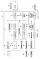
- FIG. 3 is a functional block diagram of the medicine inspection device according to the embodiment.
- FIG. 4 is a flowchart showing a flow of an audit process by the medicine inspection apparatus according to the embodiment.
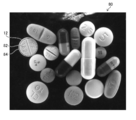
- FIG. 5 is an example of a photographed image obtained by photographing a medicine.
- FIG. 6 is another example of a photographed image obtained by photographing a medicine.
- FIG. 7 is an example of an edge extracted image extracted by performing edge detection processing on the photographed image shown in FIG.
- FIG. 8 is an example of an edge extracted image extracted by performing edge detection processing on the captured image shown in FIG.
- FIG. 1 is a perspective view of a medicine inspection device according to an embodiment.
- FIG. 2 is a top view of the projector.
- FIG. 3 is a functional block diagram of the medicine inspection device according to the embodiment.
- FIG. 4 is a flowchart showing a flow of an
- FIG. 10 is a flowchart showing a flow of processing of the information processing apparatus in the medicine inspection apparatus according to the embodiment.
- FIG. 1 is a perspective view of a medicine inspection device 10 according to the embodiment.
- the medicine inspection device 10 recognizes the type of the medicine 12 packaged in the packaging bag 11 and audits whether the medicine 12 in the packaging bag 11 matches a prescription (not shown).
- the sachet 11 is a transparent or translucent drug storage bag.
- medical agent 12 here is a solid agent, for example, is a tablet or a capsule.
- On the surface of the medicine 12, a stamped letter or a printed letter (not shown) is attached.
- stamping the identification information by the stamped character attached to the medicine surface or the printed character printed on the medicine surface is referred to as “stamping”.
- a plurality of medicines 12 are accommodated in the packaging bag 11.
- a plurality of medicines 12 accommodated in one sachet 11 may contain different kinds of medicines, or may contain the same kind of medicines.
- the medicine 12 packaged in the packaging bag 11 is referred to as a packaged medicine.
- the medicine inspection device 10 includes a tray 17, a transport mechanism 18, a projector 19, a backlight 20, a camera 21, and an information processing device 22.
- a series of sachets 11 each containing a medicine 12 for 1 sachet by a sachet device (not shown) is placed.
- the tray 17 is formed of a transparent or translucent material, and illumination light emitted from the backlight 20 passes through the tray 17.
- a transparent or translucent material is synonymous with a light-transmitting material.
- the transport mechanism 18 intermittently transports the tray 17 along the longitudinal direction of a series of sachets 11 that are continuous in a strip shape.
- the transport mechanism 18 includes a power source such as a motor (not shown).
- the longitudinal direction of the series of sachets 11 is hereinafter simply referred to as “longitudinal direction”.
- the projector 19, the backlight 20, and the camera 21 are respectively installed at fixed positions, and the tray 17 is transported by the transport mechanism 18, so that the series of packaging bags 11 are attached to the projector 19, the backlight 20, and the camera 21. On the other hand, it moves relatively in the longitudinal direction. Instead of transporting the tray 17, the projector 19, the backlight 20, and the camera 21 may be transported in the longitudinal direction of the packaging bag 11.
- the light projector 19 is disposed on the upper surface side of the tray 17 and illuminates the packaging bags 11 that are intermittently transported by the transport mechanism 18 one by one.
- the upper surface of the tray 17 is a surface on the side of the placement surface that comes into contact with the packaging bag 11 in the tray 17.
- the surface facing upward in the drawing is the upper surface of the tray 17.
- the surface of the tray 17 that faces the backlight 20 is referred to as the lower surface of the tray 17.
- the direction away from the tray 17 on the upper surface side of the tray 17 is the upward direction
- the direction away from the tray 17 on the lower surface side of the tray 17 is the downward direction.
- the light projector 19 includes a light source holding unit 24 and a plurality of point light sources 25.
- the light source holding unit 24 is a holding member that holds a plurality of point light sources 25.
- the light source holding part 24 is made of a light transmissive material.
- the light source holding part 24 is formed in a substantially dome shape covering the sachet 11 for one sachet.
- An opening window 24 a that exposes the inside of the light source holding unit 24 is formed in the ceiling portion of the light source holding unit 24. Thereby, the medicine 12 in the light source holding part 24 can be confirmed from the upper side of the light source holding part 24 through the opening window 24a.
- the drug 12 in the light source holding unit 24 refers to the drug 12 packaged in the packaging bag 11 located in the light source holding unit 24.
- FIG. 2 is a top view of the projector 19.
- point light source 25 for example, an (LED: “Light” Emitting “Diode”) light source or the like is used. Eight point light sources 25 are attached at equal intervals along the circumferential direction of the lower and upper steps of the outer surface of the light source holding unit 24. These 16 point light sources 25 irradiate illumination light toward the medicine 12 in the light source holding unit 24.
- the light projector 19 can illuminate the medicine 12 in the light source holding unit 24 from a plurality of illumination directions by each point light source 25. Moreover, the projector 19 can switch the illumination direction which illuminates the chemical
- the backlight 20 is disposed at a position on the lower surface side of the tray 17 and facing the projector 19 with the tray 17 interposed therebetween.
- the backlight 20 illuminates the medicine 12 in the light source holding part 24 through the tray 17 from behind.
- the camera 21 is an imaging apparatus equipped with an imaging device (not shown) typified by a CCD (charge-coupled device) sensor or a CMOS (complementary metal-oxide semiconductor device) sensor. It is preferable to use a color imaging device provided with a color filter as the imaging device.
- the camera 21 includes an optical system such as a photographing lens (not shown). Further, the camera 21 may include a signal processing circuit such as an analog front end circuit that processes an image signal output from the imaging device.
- the camera 21 converts the subject image into an electrical signal and generates an image signal corresponding to the subject.
- the image signal may be an analog signal or a digital signal. In this example, it is assumed that a digital image signal is output from the camera 21.
- a digital image signal is called image data.
- the camera 21 is disposed above the opening window 24a.
- the camera 21 generates the image data 28 by photographing the medicine 12 in the light source holding unit 24 through the opening window 24a.
- the camera 21 outputs image data 28 generated by shooting to the information processing apparatus 22.
- the image data 28 corresponds to a form of a captured image.
- FIG. 3 is a functional block diagram of the medicine inspection device 10.
- a personal computer can be used as the information processing apparatus 22, for example, a personal computer can be used.
- the information processing device 22 is connected to each of the transport mechanism 18, the illumination device 30, and the camera 21.
- the lighting device 30 refers to the projector 19 and the backlight 20 described in FIG. “Connection” is not limited to wired connection, but may be wireless connection. The state in which signal transmission is possible is included in the concept of “connection”.
- the information processing device 22 of this example functions as a control device of the medicine inspection device 10 and also functions as an arithmetic device that performs image processing and other various arithmetic processes.
- the information processing apparatus 22 includes a transport control unit 32, an illumination control unit 33, and a photographing control unit 34, and controls operations of the transport mechanism 18, the illumination device 30, and the camera 21.
- the information processing apparatus 22 includes an image acquisition unit 40, a memory 41, an image processing unit 42, a display control unit 44, and a display unit 46.
- the image processing unit 42 includes a quantitative value calculation unit 50, a comparison determination unit 51, a threshold value storage unit 52, and a medicine recognition unit 54.
- the medicine recognition unit 54 includes an edge detection unit 55 and a marking recognition unit 56.
- the information processing apparatus 22 further includes an operation unit 60, an overall control unit 62, a drug database storage unit 63, a dispensing information acquisition unit 64, and an inspection unit 65.
- each unit of the information processing apparatus 22 can be realized by a combination of computer hardware and software. “Software” is synonymous with “program”. Further, some or all of the processing functions in the information processing apparatus 22 can be realized by an integrated circuit or the like.
- the conveyance control unit 32 controls driving of the conveyance mechanism 18 based on a command from the overall control unit 62.
- the illumination control unit 33 controls emission of illumination light by the illumination device 30 based on a command from the overall control unit 62.
- the illumination control unit 33 can control the light emission of the point light source 25 and the backlight 20 of the projector 19 described in FIG. 1 and change the illumination condition for illuminating the medicine 12 in the light source holding unit 24.
- the imaging control unit 34 shown in FIG. 3 controls the imaging operation by the camera 21 based on a command from the overall control unit 62.
- the imaging control unit 34 causes the camera 21 to perform imaging every time intermittent conveyance by the conveyance mechanism 18 stops, that is, for each sachet 11 for one package.
- the shooting control unit 34 causes the camera 21 to execute shooting each time the lighting condition is switched.
- the image acquisition unit 40 is an interface unit that acquires a captured image captured by the camera 21.
- the image acquisition unit 40 can be configured by a data input terminal that captures image data output from the camera 21. Further, a wired or wireless communication interface unit may be employed as the image acquisition unit 40.
- the captured image acquired via the image acquisition unit 40 is stored in the memory 41.
- the memory 41 functions as a storage unit that stores captured images acquired via the image acquisition unit 40.
- the memory 41 functions as a storage unit for storing various control programs executed by the overall control unit 62 and various data necessary for control, and as a work memory for performing various calculations of the image processing unit 42. Can function.
- the image processing unit 42 recognizes the type of the drug 12 by analyzing the captured image acquired through the image acquisition unit 40. Further, the image processing unit 42 performs processing for detecting a state in which the medicine recognition by the medicine recognition unit 54 is impossible by analyzing the captured image.
- the image processing unit 42 may include a processing unit (not shown) that performs one or more of noise removal processing, color conversion processing, image rotation processing, trimming processing, or synthesis processing.
- the quantitative value calculation unit 50 calculates a quantitative value obtained by quantifying the quality of the captured image related to the success or failure of the drug recognition by the drug recognition unit 54.
- the quantitative value calculation unit 50 of the present embodiment calculates a quantitative value obtained by quantifying the sharpness of a captured image related to the success or failure of edge detection by the edge detection unit 55.
- the sharpness of the photographed image is an example of the quality of the photographed image related to the success or failure of the medicine recognition by the medicine recognition unit 54.
- the quantitative value calculation unit 50 performs a two-dimensional fast Fourier transform process on the captured image, and has a higher frequency than one cycle per millimeter [cycle / mm] in the radial frequency spectrum of the obtained two-dimensional fast Fourier transform image.
- a quantitative value defined by using the ratio of the sum of the absolute values of the spatial frequency spectrum of and the sum of the absolute values of the spatial frequency spectrum at frequencies lower than 1 cycle per millimeter [cycle / mm] is calculated.
- the threshold storage unit 52 is a storage unit that stores a threshold value of a quantitative value that is a criterion for determining whether or not drug recognition by the drug recognition unit 54 is possible.
- the threshold value is determined by examining the relationship between the quantitative value and the success or failure of drug recognition based on, for example, simulation, and determining the condition of the quantitative value at which drug recognition is impossible as the threshold value.
- the comparison determination unit 51 obtains threshold information from the threshold storage unit 52.
- the comparison determination unit 51 compares the quantitative value calculated by the quantitative value calculation unit 50 with a threshold value, and determines whether or not the drug recognition unit 54 can recognize the drug. Details of the processing method for detecting a state in which the drug cannot be recognized in the image processing unit 42 will be described later, including a specific example of how to set the threshold.
- the medicine inspection device 10 includes an alarm output unit 58 that issues an alarm when the comparison / determination unit 51 determines that the medicine recognition unit 54 cannot recognize the medicine.
- the alarm output unit 58 includes a display unit 46.
- the comparison determination unit 51 determines that the medicine cannot be recognized
- alarm information notifying that the medicine cannot be recognized is displayed on the display unit 46 via the display control unit 44.
- the alarm output unit 58 may include a warning light, a device that generates a warning sound, a device that generates sound, or an appropriate combination thereof.
- the drug recognizing unit 54 recognizes the drug from the captured image and identifies the type of drug.
- the edge detection unit 55 performs a known edge detection process on the photographed image, and extracts the outline of the medicine in the photographed image.
- a method of edge detection processing in the edge detection unit 55 for example, a method of Canny edge detection can be used.
- the stamp recognizing unit 56 detects the contours of the stamps attached to the respective drugs 12 from the drug regions extracted based on the edge information detected by the edge detecting unit 55, and recognizes the character information indicated by the stamps. To do.
- the drug database storage unit 63 stores a drug database that is a collection of drug data in which drug information including an appearance image of a drug and information on an inscription is associated with each drug type.
- the drug recognizing unit 54 can specify the type of drug by referring to the drug database from the stamp recognized by the stamp recognizing unit 56.
- the dispensing information acquisition unit 64 acquires the dispensing information 72 from the receipt computer 70.
- the receipt computer 70 holds dispensing information 72 based on the prescription.
- the pharmacist performs an operation of inputting the dispensing information 72 to the receipt computer 70 based on the dispensing information described in the prescription.
- the dispensing information 72 includes the name and age of the patient, the drug name, the amount, the usage, the dose, and the like.
- the drug name input to the receipt computer 70 is not limited to the drug name described in the prescription, and may be the drug name of a generic drug having the same component and the same medicinal effect.
- each of the drugs 12 is packaged in a series of packaging bags 11 according to the dispensing information 72 input to the receipt computer 70.
- the inspecting unit 65 can determine the type and number of medicines 12 to be packaged in each packaging bag 11 based on the dispensing information 72 acquired from the dispensing information acquisition unit 64.
- the marking recognition unit 56 can acquire information on the markings attached to the individual drugs described in the dispensing information 72 by referring to the drug database based on the dispensing information 72.
- the stamp recognition unit 56 compares the stamp information obtained by referring to the drug database from the dispensing information 72 with the contour of the stamp extracted from the shot image, and is attached to the drug 12 included in the shot image. Recognize the stamp that is.
- the medicine recognition unit 54 refers to the medicine database and identifies the type of the medicine 12 corresponding to each marking. Thereby, the kind of the medicine 12 for one minute is recognized.
- the medicine recognizing unit 54 outputs the recognition result of the type of medicine 12 for one package to the inspection unit 65.
- the auditing unit 65 collates the recognition result of the type of medicine 12 for one sachet input from the medicine recognition unit 54 with the type of medicine for one sachet recorded in the dispensing information 72. Information on the result of verification by the inspection unit 65 is displayed on the display unit 46 via the display control unit 44.
- the display control unit 44 performs control for displaying various information on the display unit 46.
- the display control unit 44 performs processing for generating a display signal for the display unit 46.
- the display unit 46 for example, display devices using various display methods such as a liquid crystal display and an organic EL (Organic Electro-Luminescence) display can be used. Operations such as inputting and setting instructions to the information processing apparatus 22 by the user can be performed using the operation unit 60 and the display unit 46.
- the combination of the display unit 46 and the operation unit 60 functions as a user interface of the medicine inspection device 10.
- the operation unit 60 is a means for a user such as a pharmacist to input various information.
- Various input devices such as a keyboard, a mouse, a touch panel, a trackball, or operation buttons can be used for the operation unit 60, and an appropriate combination thereof may be used.
- FIG. 4 is a flowchart showing a flow of an audit process by the medicine inspection apparatus 10 according to the embodiment.
- the pharmacist sets the medicine 12 in the packaging device in accordance with the dispensing information 72 input in advance to the reception computer 70, and the medicine 12 is packaged in a plurality of packaging bags 11 in this packaging device.
- a series of packaging bags 11 in which the medicine 12 is packaged by the packaging apparatus is set on the tray 17 of the medicine inspection apparatus 10 (step S11).
- the overall control unit 62 operates each unit of the drug inspection device 10 to recognize and audit the type of the medicine 12 packaged in the series of packaging bags 11. Start.
- step S13 the conveyance control unit 32 operates the conveyance mechanism 18 to convey the Nth packing bag 11 to the photographing position.
- the shooting position refers to a position inside the light source holding unit 24 that can be shot by the camera 21.
- step S14 the imaging control unit 34 performs imaging by the camera 21 and images the drug 12.
- the photographing process in step S14 includes setting of illumination conditions by the illumination device 30 and light emission operation of the illumination device 30 according to the setting. Shooting by the camera 21 is performed in a state where illumination light is irradiated by the illumination device 30.
- step S ⁇ b> 15 the information processing apparatus 22 acquires a captured image from the camera 21 via the image acquisition unit 40.
- a captured image obtained from the camera 21 is stored in the memory 41.
- step S ⁇ b> 16 the information processing apparatus 22 performs processing for extracting a drug region from the captured image by the edge detection unit 55.
- FIG. 5 is an example of the captured image 80.
- the photographed image 80 is an image obtained by photographing a single packaged medicine in which a plurality of types of medicines 12 are accommodated in the packaging bag 11.
- the surface of the medicine 12 is marked with a stamp 82.
- a drug region 84 corresponding to the image portion of the drug 12 is extracted for each of the plurality of drugs 12 included in the captured image 80.
- a circle is displayed to emphasize the drug region 84.
- the marking recognition unit 56 of the information processing apparatus 22 performs a medicine recognition process for specifying the kind of medicine in each medicine region extracted in step S16.
- the marking recognition unit 56 refers to the drug information based on the dispensing information 72 and obtains information on the markings attached to the individual drugs 12 recorded in the dispensing information 72.
- the marking recognition unit 56 determines the marking attached to the medicine in the medicine region extracted based on the edge information detected by the edge detection unit 55.
- the stamp recognition unit 56 recognizes the stamp included in the photographed image by comparing the stamp information acquired based on the dispensing information 72 with the stamp determined from the photographed image. By comparing with the dispensing information 72 in this way, when the shape of the stamp included in the photographed image is incomplete, for example, even when the stamp is partially distorted or missing, the stamp False recognition can be reduced.
- the marking recognition unit 56 refers to the drug database based on the stamps recognized from the captured images, identifies the type of each drug 12 in the Nth sachet 11, and uses the drug recognition result as the drug recognition result. Output to the auditing unit 65.
- step S18 the auditing unit 65 of the information processing apparatus 22 collates the type of medicine specified in step S17 with the dispensing information 72.
- step S19 the auditing unit 65 outputs the verification result to the display unit 46.
- the verification result by the auditing unit 65 is displayed.
- the display unit 46 displays a type name of the corresponding medicine 12 to warn a pharmacist or the like who performs a dispensing audit.
- the display unit 46 displays the determination result to warn the pharmacist who performs the dispensing audit. .
- step S20 it is determined whether or not another sachet remains.
- the process proceeds to step S21, the value of the parameter N is incremented, and the value of “N + 1” is newly set as the sachet.
- the process returns to step S13 as the value of the parameter N of the number.
- step S20 the processing of each step from step S13 to step S20 described above is repeatedly executed.
- the determination in step S20 is No, and the flowchart of FIG. 4 ends.
- Step S15 in FIG. 4 corresponds to one form of the image acquisition process.
- the combination of step S16 and step S17 corresponds to one form of the medicine recognition process.
- step S16 the process of extracting the drug region 84 of each drug 12 from the photographed image (step S16) is successful, and the stamp 82 is recognized from the extracted drug region 84. It is necessary that the type of the medicine 12 is specified by successfully performing the process (step S17).
- the medicine inspection device 10 is equipped with a function of detecting a problem state in which medicine recognition processing by the medicine recognition unit 54 becomes impossible and notifying the user.
- FIG. 6 is an example of a captured image 90 compared with FIG. FIG. 6 shows a blurred image in which the sharpness is lower than that in FIG.
- FIG. 6 is an example of an image in which the photographed image 90 is blurred and extraction of the drug region by the drug recognition unit 54, that is, the edge detection is not successful.
- FIG. 7 is an edge extraction image extracted by performing edge detection processing on the captured image 80 shown in FIG. In FIG. 7, edge detection is normally performed and a normal edge detection image is obtained.
- FIG. 8 is an edge extracted image extracted by performing edge detection processing on the captured image 90 shown in FIG. In FIG. 8, since the photographed image 90 is blurred, the outline of the medicine cannot be sufficiently extracted. For example, the extraction of the outline of the capsule located in the center of FIG. 8 is insufficient.
- the ratio of the high-frequency component corresponding to the edge and the low-frequency component other than the edge is stored as a threshold in the apparatus in advance using the processing of the two-dimensional fast Fourier transform, so that the captured image has the threshold. Depending on whether or not it exceeds, it is detected that the photographed image is blurred to a level where the medicine cannot be recognized.
- the two-dimensional fast Fourier transform process corresponds to one form of the two-dimensional Fourier transform process.
- the two-dimensional fast Fourier transform image is standardized based on Parseval's theorem, and the threshold is determined using the absolute value of the obtained spectrum. Specifically, the total value of the absolute values of the spatial frequency spectrum larger than one cycle per millimeter [cycle / mm] in the radial direction of the two-dimensional fast Fourier transform image of the photographed image and one cycle per millimeter [cycle / mm]. The ratio of the sum of absolute values of the following spatial frequency spectra was taken. This is because the high-frequency component of the photographed image depends on the number of medicines of the packaged medicine, and thus the same effect as that obtained by normalization by the number of medicines can be obtained.
- Quantitative value T defined below can be used as a value that quantitatively indicates the quality of the captured image.
- S E is the sum of absolute values of the spatial frequency spectrum larger than one cycle per millimeter [cycle / mm] in the radial direction in the two-dimensional fast Fourier transform image of the photographed image.
- S NE is the sum of absolute values of spatial frequency spectra of one cycle per millimeter [cycle / mm] or less in the radial direction in the two-dimensional fast Fourier transform image of the captured image.
- Threshold value for quantitative value T is stored in the device in advance.
- 1 cycle per millimeter [cycle / mm] is the lowest spatial frequency which forms the outline of a chemical
- S E is a value that reflects the contour of the drug
- S NE is a value that reflects the non-contour of the drug.
- the reason for taking the ratio of the spectral sum in the formula defining the quantitative value T is to standardize the quantitative value by the number of drugs in the visual field range. Note that the average value of the image is not used for the spectral sum.
- the quantitative value T is one of the indexes indicating the degree of blur of the photographed image, and can be understood as a blurred quantitative value or a sharpness quantitative value.
- the blur added by the simulation is defined by the expression Exp ( ⁇ 0.020 ⁇ ⁇ ⁇ C ⁇ f).
- Exp (x) represents the power of “e” which is the base of natural logarithm.
- C is an optional parameter.
- f is the spatial frequency [cycle / mm].
- the chart shown in FIG. 9 summarizes the value of the arbitrary parameter C, the result of whether or not the outline of the medicine can be extracted, and the value of the quantitative value T calculated under each condition.
- “A” indicates that the outline of the drug has been successfully extracted with respect to the evaluation of whether or not the drug outline can be extracted.
- “D” indicates that the extraction of the outline of the medicine has failed.
- the threshold should be set to “1.19”.
- the threshold value thus determined is stored in the threshold value storage unit 52 of FIG.
- the threshold value may be set for each edge detection method such as a method using a sobel filter or a method using a Laplacian filter without being limited to the canny method.
- the quantitative value T is calculated by the quantitative value calculation unit 50 for each captured image, and drug recognition is impossible when the quantitative value T calculated for each captured image falls below the threshold value. And an alarm is output to inform the user that drug recognition is impossible.
- the way of issuing an alarm by the alarm output unit 58 can be an alert, a screen display of the display unit 46, or a combination thereof.
- the alarm preferably includes information that tells the user why the drug recognition is impossible.
- the alarm information displayed on the display unit 46 of this example includes information that informs the user that the medicine cannot be recognized due to the low sharpness of the captured image.
- the quantitative value T is an example of a quantitative value obtained by quantifying the quality of a photographed image related to the success or failure of the edge detection by the edge detection unit 55.
- the threshold value determined from the simulation result illustrated in FIG. 9 is an example of a threshold value of a quantitative value that is a determination criterion as to whether or not the edge detection by the edge detection unit 55 is possible.
- the determination of whether or not the drug recognition by the drug recognition unit 54 performed by the comparison determination unit 51 based on the quantitative value T is equivalent to the determination of whether or not the edge detection by the edge detection unit 55 is possible.
- the comparison / determination unit 51 determines that the edge detection unit 55 cannot detect the edge, the unit 58 issues a warning to that effect.
- FIG. 10 is a flowchart showing a flow of processing of the information processing apparatus 22 in the medicine inspection apparatus 10 according to the embodiment.
- the medicine inspection method according to the embodiment will be described with reference to FIG.
- step S31 the information processing apparatus 22 acquires a captured image via the image acquisition unit 40.
- Step S31 corresponds to step S15 in the flowchart of FIG.
- the quantitative value calculation unit 50 calculates a quantitative value obtained by quantifying the quality of the captured image.
- the quantitative value calculation unit 50 calculates a quantitative value T obtained by quantifying the sharpness of the captured image.
- step S33 the comparison determination unit 51 compares the quantitative value obtained in step S32 with the threshold value stored in advance in the threshold storage unit 52 to determine whether or not the drug can be recognized.
- step S34 when the comparison determination unit 51 determines that the medicine can be recognized, the process proceeds to step S35.
- step S35 the edge detection unit 55 performs edge detection processing of the captured image. Extraction of the drug region is performed based on the processing result of the edge detection processing.
- the edge detection process of step S35 is a process corresponding to the medicine region extraction process described as step S16 of FIG.
- step S36 the marking recognition unit 56 recognizes the marking in each drug region and performs a process of specifying the type of drug.
- the stamp recognition process in step S36 is a process corresponding to the medicine recognition process described as step S17 in FIG.
- step S37 the drug recognition unit 54 outputs detection information that is information of the specified drug.
- Step S37 may be understood as a process of providing information from the drug recognition unit 54 to the auditing unit 65, or may be understood as a process of outputting information on the drug recognized by the drug recognition unit 54 to the display unit 46.
- step S ⁇ b> 37 may be understood as a process of outputting the audit result by the audit unit 65 to the display unit 46.
- step S34 when the comparison determination unit 51 determines that the medicine cannot be recognized, the process proceeds to step S38.
- step S38 the alarm output unit 58 outputs an alarm notifying that the medicine cannot be recognized.
- the flowchart shown in FIG. 10 is executed for each input photographed image.
- Step S31 in FIG. 10 corresponds to one form of the image acquisition process.
- Step S32 corresponds to one form of the quantitative value calculation step.
- Step S33 corresponds to one mode of the comparison determination process.
- the combination of step S35 and step S36 corresponds to one form of the medicine recognition process.
- Step S38 corresponds to an embodiment of an alarm output process.
- the quantitative value T is calculated from the captured image obtained by photographing the medicine, the quality of the photographed image is evaluated, and the quantitative value T is compared with the threshold value, so that the photographed image cannot be recognized by the medicine. It can be determined whether or not there is. When it is detected that the medicine cannot be recognized, the user can be alerted. Thereby, an efficient medicine inspection can be performed.
- a program for causing a computer to realize the medicine inspection function of the information processing apparatus 22 described in the above embodiment is a non-temporary tangible object such as a CD-ROM (Compact Disc Read-only Memory), a magnetic disk, or other computer-readable medium. It is possible to record the program on a simple information storage medium and provide the program through the information storage medium. Instead of providing the program by storing the program in such an information storage medium, it is also possible to provide the program signal as a download service using a communication network such as the Internet.
- a computer responsible for the functions of the conveyance control unit 32, the illumination control unit 33, and the imaging control unit 34, an image acquisition unit 40, a memory 41, an image processing unit 42, a display control unit, an alarm output unit 58, an operation unit 60, and an overall control
- the computer responsible for the functions of the control unit 62, the drug database storage unit 63, the dispensing information acquisition unit 64, and the inspection unit 65 can be configured by separate computers. Further, for example, part or all of the functions of the information processing apparatus 22 may be mounted on the receipt computer 70.
- the medicine database storage unit 63 may be an external storage device connected to the information processing device 22.
- the drug database can be stored in a storage device of a computer owned by a dispensing pharmacy.
- the drug database may be held in the receipt computer 70.
- the medicine database may be held in another computer (not shown), and the information processing apparatus 22 may acquire information from the medicine database via a network.
- the network can be a local area network, a wide area network, or a combination thereof.
- the captured image for which the quantitative value calculation unit 50 calculates the quantitative value is not limited to the image data 28 output from the camera 21, but is image data generated based on the image data 28 output from the camera 21. May be.
- an image obtained by performing processing such as noise removal processing, color conversion processing, gradation conversion processing, or a combination thereof on the image data 28 output from the camera 21 is included in the concept of the captured image. It is.
- An image obtained by processing the image data 28 output from the camera 21 is also included in the concept of the photographed image, and is a composite image obtained by combining a plurality of photographed images obtained by performing a plurality of photographings. Is also included in the concept of captured images.
- the auditing unit 65 combines the recognition result of the medicine recognition process based on the photographed image obtained from the first camera and the recognition result of the medicine recognition process based on the photographed image obtained from the second camera, and dispenses information 72. And may be verified.
- the example of the medicine audit related to the packaged dispensing is shown.
- the drug auditing function by the information processing apparatus 22 is not limited to the drug audit related to the packaged dispensing, and the agent performs the drug recognition process.
- the system configuration for recognizing the type of medicine from a photographed image obtained by photographing the medicine with a camera in the state of a PTP (press-through-pack) sheet that is a dispensing packaging unit adopts the same configuration as the information processing apparatus 22 Can do.
Abstract
Provided are a drug monitoring device, method, and program whereby it is possible to detect a state in which a drug cannot be recognized from a captured image of the drug. A drug monitoring device 10 is provided with a drug recognition unit 54 for recognizing a drug from a captured image of the drug and specifying the type of the drug, a quantitative value calculation unit 50 for calculating a quantitative value whereby the quality of the captured image with regard to success or failure of drug recognition by the drug recognition unit 54 is quantified, a comparison determination unit 51 for comparing a threshold value and the quantitative value calculated by the quantitative value calculation unit 50 and determining whether recognition of the drug is possible, and a warning output unit 58 for issuing a warning when it is determined by the comparison determination unit 51 that the drug cannot be recognized.
Description
本発明は薬剤監査装置及び方法並びにプログラムに係り、特に薬剤を撮影して得られる画像から薬剤の種類を判別する薬剤監査支援技術に関する。
The present invention relates to a drug inspection apparatus, method, and program, and more particularly, to a drug audit support technique for determining the type of drug from an image obtained by imaging the drug.
近年、病院等では、例えば服用時期が異なる複数種類の薬剤を患者に処方する際に、一回服用分の複数種の薬剤を1つの分包袋に分包する一包化調剤を行うことが多い。服用時期には、例えば、朝食後、昼食後、夕食後などがある。一包化調剤は、薬剤師が処方箋に従ってピッキングした各薬剤を分包機のトレイに一回服用分ごとにセットした後、この分包機にてトレイ内の薬剤を自動的に各分包袋に分包することにより行われる。分包機のトレイはタブレットケースとも呼ばれる。
In recent years, in hospitals and the like, for example, when prescribing a plurality of types of drugs with different dosing timings to a patient, it is possible to perform a single-packaging preparation that wraps a plurality of types of drugs for a single dose into a single packing bag. Many. Examples of the time of taking include after breakfast, after lunch, and after dinner. For single-pack dispensing, each drug picked by the pharmacist according to the prescription is set on the tray of the packaging machine once for each dose, and then the medicine in the tray is automatically packaged in each packaging bag by this packaging machine. Is done. The tray of the packaging machine is also called a tablet case.
このような一包化調剤では、薬剤のピッキングや分包トレイへのセットは薬剤師が手作業で行う場合があるので、処方箋の指示とは異なる種類や個数の薬剤が間違って分包される可能性がある。そこで、分包された薬剤が処方箋に合致するものか否かを判定するために、分包袋への分包前あるいは分包後の薬剤の種類や個数の自動認識が行われている。
In such single-pack dispensing, the pharmacist may manually perform drug picking and setting to the packaging tray, so it is possible to mistakenly package different types or numbers of drugs from the prescription instructions. There is sex. Therefore, in order to determine whether or not the packaged medicine matches the prescription, automatic recognition of the type and number of drugs before or after packaging into a packaging bag is performed.
特許文献1に記載の薬剤認識装置は、刻印文字が付された薬剤に対して薬剤の周囲を囲む複数の照明方向から照明可能な照明部と、照明部が薬剤を照明する照明方向を順番に切り替える照明制御部と、照明部により照明されている薬剤を撮影する撮影部であって、照明方向が切り替わるごとに薬剤の撮影を繰り返し行う撮影部と、撮影部により取得された照明方向ごとの撮影画像からそれぞれ刻印文字の影に対応する特徴画像を抽出する特徴画像抽出部と、特徴画像抽出部が抽出した照明方向ごとの特徴画像を統合して統合画像を生成する特徴画像統合部と、特徴画像統合部が生成した統合画像に含まれる刻印文字を認識して、刻印文字の認識結果に基づき薬剤の種類を認識する認識部と、を備える。薬剤認識装置という用語は、本明細書の薬剤監査装置に対応する用語であると理解される。
In the medicine recognition device described in Patent Document 1, an illumination unit capable of illuminating a medicine with a stamped character from a plurality of illumination directions surrounding the medicine and an illumination direction in which the illumination part illuminates the medicine in order. An illumination control unit that switches, an imaging unit that images the medicine illuminated by the illumination unit, an imaging unit that repeatedly captures an agent every time the illumination direction is switched, and an imaging for each illumination direction acquired by the imaging unit A feature image extraction unit that extracts a feature image corresponding to the shadow of each engraved character from the image, a feature image integration unit that generates an integrated image by integrating the feature images for each illumination direction extracted by the feature image extraction unit, and a feature A recognition unit that recognizes a stamped character included in the integrated image generated by the image integration unit and recognizes the type of medicine based on the recognition result of the stamped character. The term drug recognition device is understood to be a term corresponding to the drug audit device herein.
特許文献1に記載の薬剤認識装置では、刻印文字が認識できない場合に、薬剤の認識に失敗したと判定し、この判定結果を表示部に出力する構成となっている(特許文献1の段落0053)。しかし、特許文献1には、刻印文字が認識できない場合をどのように検出するのか、その検出方法に関して具体的な開示はない。
The medicine recognition device described in Patent Document 1 is configured to determine that the recognition of the medicine has failed when the stamped character cannot be recognized, and to output the determination result to the display unit (paragraph 0053 of Patent Document 1). ). However, Patent Document 1 does not specifically disclose how to detect a case where a stamped character cannot be recognized.
薬剤監査の作業を一層効率化するためには、薬剤の認識に失敗した場合の問題点を検出して、その問題点をユーザに知らせることが望ましい。この課題は、刻印文字を認識する場合に限らず、薬剤表面に印刷された印刷文字を認識する場合についても共通する課題である。また、この課題は、一包化調剤における薬剤監査に限らず、薬剤を撮影した画像から薬剤の種類を認識する処理を用いる薬剤監査に共通する課題である。
In order to further improve the efficiency of drug inspection work, it is desirable to detect a problem when drug recognition fails and notify the user of the problem. This problem is not limited to the case of recognizing stamped characters, but is also a common problem when recognizing printed characters printed on the surface of a medicine. This problem is not limited to drug audits in packaged dispensing, but is common to drug audits that use processing for recognizing the type of drug from an image of the drug.
本発明はこのような事情に鑑みてなされたもので、薬剤を撮影した画像から薬剤を認識することが不可能な状態を検出するための新たな検出技術を提供し、薬剤認識不能な状態である場合にユーザに対して注意を促すことができる薬剤監査装置及び方法並びにプログラムを提供することを目的とする。
The present invention has been made in view of such circumstances, and provides a new detection technique for detecting a state in which a medicine cannot be recognized from an image obtained by photographing the medicine. It is an object of the present invention to provide a medicine inspection device, method, and program that can alert a user in some cases.
本開示の第1態様に係る薬剤監査装置は、薬剤を撮影して得られる撮影画像を取得する画像取得部と、撮影画像から薬剤を認識して薬剤の種類を特定する薬剤認識部と、薬剤認識部による薬剤認識の成否に関わる撮影画像の品質を定量化した定量値を算出する定量値算出部と、薬剤認識部による薬剤認識が可能か否かの判定基準となる定量値の閾値を記憶しておく記憶部と、定量値算出部により算出された定量値と閾値とを比較して、薬剤認識部による薬剤の認識が可能か否かを判定する比較判定部と、比較判定部により薬剤認識部の薬剤認識ができないと判定された場合に警報を発する警報出力部と、を備える。
A drug inspection device according to a first aspect of the present disclosure includes an image acquisition unit that acquires a captured image obtained by imaging a drug, a drug recognition unit that recognizes a drug from the captured image and identifies the type of drug, and a drug Stores a quantitative value calculation unit that calculates a quantitative value obtained by quantifying the quality of a captured image related to the success or failure of drug recognition by the recognition unit, and a threshold value of a quantitative value that is a criterion for determining whether or not the drug recognition by the drug recognition unit is possible The storage unit, the quantitative value calculated by the quantitative value calculation unit and the threshold value are compared, a comparison determination unit for determining whether or not the drug recognition unit can recognize the drug, and a drug by the comparison determination unit An alarm output unit that issues an alarm when it is determined that the recognition unit cannot recognize the medicine.
撮影画像から薬剤を認識することができない原因の1つとして撮影画像の品質の問題がある。第1態様の薬剤監査装置では、撮影画像の品質を定量的に評価する定量値と薬剤認識の成否との関係を予め知得しておき、薬剤認識が不可能となる定量値の閾値を定めておく。第1態様によれば、撮影画像から算出される定量値と閾値とを比較することにより、薬剤認識ができない品質の状態を検出することができる。そして、薬剤認識ができないと判定された場合に警告が発生されることにより、ユーザに注意を促すことができる。
品質 One of the reasons why the medicine cannot be recognized from the photographed image is a problem of the quality of the photographed image. In the medicine inspection device according to the first aspect, the relationship between the quantitative value for quantitatively evaluating the quality of the captured image and the success or failure of the medicine recognition is obtained in advance, and a threshold value for the quantitative value at which the medicine recognition is impossible is determined. Keep it. According to the first aspect, by comparing the quantitative value calculated from the photographed image with a threshold value, it is possible to detect a quality state in which drug recognition cannot be performed. A warning is generated when it is determined that the medicine cannot be recognized, so that the user can be alerted.
第2態様として第1態様の薬剤監査装置において、薬剤認識部による薬剤認識の成否に関わる撮影画像の品質は、撮影画像の鮮鋭度を含む構成とすることができる。
As a second aspect, in the medicine inspection device according to the first aspect, the quality of the photographed image related to the success or failure of the medicine recognition by the medicine recognition unit can be configured to include the sharpness of the photographed image.
第3態様として第1態様又は第2態様の薬剤監査装置において、薬剤認識部は、撮影画像から薬剤のエッジを検出するエッジ検出部と、撮影画像からエッジ検出部によって検出されたエッジの情報を基に抽出された薬剤に付されている刻印を認識する刻印認識部と、を含む構成とすることができる。
As a third aspect, in the medicine inspection device according to the first aspect or the second aspect, the medicine recognition unit includes an edge detection unit that detects an edge of the medicine from the photographed image, and information on the edge detected by the edge detection unit from the photographed image. And a marking recognition unit for recognizing the marking attached to the medicine extracted based on the marking.
第4態様として第3態様の薬剤監査装置において、定量値は、エッジ検出部によるエッジ検出の成否に関わる撮影画像の品質を定量化した定量値であり、閾値は、エッジ検出部によるエッジ検出が可能か否かの判定基準となる定量値の閾値であり、比較判定部が行う薬剤認識部による薬剤の認識が可能か否かの判定は、エッジ検出部によるエッジ検出が可能か否かの判定であり、警報出力部は、比較判定部によりエッジ検出部のエッジ検出ができないと判定された場合に警報を発する構成とすることができる。
In the medicine inspection device according to the third aspect as the fourth aspect, the quantitative value is a quantitative value obtained by quantifying the quality of the captured image related to the success or failure of the edge detection by the edge detection unit, and the threshold value is determined by the edge detection by the edge detection unit. It is a threshold value of a quantitative value that is a criterion for determining whether or not it is possible, and whether or not drug recognition by the drug recognition unit performed by the comparison determination unit is possible is determined by whether or not edge detection is possible by the edge detection unit The alarm output unit can be configured to issue an alarm when it is determined by the comparison determination unit that the edge detection unit cannot detect the edge.
第5態様として第1態様から第4態様のいずれか一態様の薬剤監査装置において、定量値算出部は、撮影画像に二次元フーリエ変換の処理を適用して得られる動径方向の空間周波数スペクトルにおいて1サイクルパーミリメートルよりも高周波の空間周波数スペクトルの絶対値の総和と、1サイクルパーミリメートルよりも低周波の空間周波数スペクトルの絶対値の総和との比を用いて定義される定量値を算出する構成とすることができる。
In the medicine inspection device according to any one of the first to fourth aspects as the fifth aspect, the quantitative value calculating unit is a spatial frequency spectrum in the radial direction obtained by applying a two-dimensional Fourier transform process to the captured image. The quantitative value defined using the ratio of the sum of the absolute values of the spatial frequency spectrum at a frequency higher than 1 cycle per millimeter and the sum of the absolute values of the spatial frequency spectrum at a frequency lower than 1 cycle per millimeter is calculated. It can be configured.
第6態様として第1態様から第5態様のいずれか一態様の薬剤監査装置において、警報出力部は、定量値算出部により算出された定量値が閾値を下回った場合に、警報を出力する構成とすることができる。
In the medicine inspection device according to any one of the first to fifth aspects as the sixth aspect, the alarm output unit outputs an alarm when the quantitative value calculated by the quantitative value calculation unit falls below the threshold value It can be.
第7態様として第1態様から第6態様のいずれか一態様の薬剤監査装置において、警報出力部は、警報の情報を表示する表示部を含む構成とすることができる。
As a seventh aspect, in the medicine inspection device according to any one of the first aspect to the sixth aspect, the alarm output unit may include a display unit that displays alarm information.
第8態様として第1態様から第7態様のいずれか一態様の薬剤監査装置において、薬剤を撮影するカメラを備え、画像取得部は、カメラによって撮影した撮影画像を取得する構成とすることができる。
In the medicine inspection device according to any one of the first to seventh aspects as the eighth aspect, the medicine inspection device may include a camera that photographs the medicine, and the image acquisition unit may acquire a captured image captured by the camera. .
第9態様として第1態様から第8態様のいずれか一態様の薬剤監査装置において、撮影画像は、複数種類の薬剤が分包袋に収容された1分包分の一包化薬剤を撮影して得られる画像である構成とすることができる。
As a ninth aspect, in the medicine inspection device according to any one of the first aspect to the eighth aspect, the photographed image is obtained by photographing a single packaged medicine in which a plurality of types of medicines are contained in a packaging bag. It can be set as the structure which is an image obtained in this way.
第10態様に係る薬剤監査方法は、薬剤を撮影して得られる撮影画像を取得する画像取得工程と、撮影画像から薬剤を認識して薬剤の種類を特定する薬剤認識工程と、薬剤認識工程による薬剤認識の成否に関わる撮影画像の品質を定量化した定量値を算出する定量値算出工程と、薬剤認識工程による薬剤認識が可能か否かの判定基準となる定量値の閾値を記憶しておき、定量値算出工程により算出された定量値と閾値とを比較して、薬剤認識工程による薬剤の認識が可能か否かを判定する比較判定工程と、比較判定工程により薬剤認識工程の薬剤認識ができないと判定された場合に警報を発する警報出力工程と、を含む。
The medicine inspection method according to the tenth aspect includes an image acquisition process for obtaining a photographed image obtained by photographing a medicine, a medicine recognition process for recognizing a medicine from the photographed image and specifying the kind of medicine, and a medicine recognition process. A quantitative value calculation step for calculating a quantitative value obtained by quantifying the quality of a photographed image related to the success or failure of the drug recognition, and a threshold value for a quantitative value serving as a criterion for determining whether or not the drug recognition by the drug recognition step is possible are stored. Comparing the quantitative value calculated in the quantitative value calculating step with a threshold value to determine whether or not the drug can be recognized in the drug recognition step, and the drug recognition in the drug recognition step is performed in the comparative determination step. And an alarm output step for issuing an alarm when it is determined that it cannot be performed.
第10態様において第2態様から第9態様にて特定した事項と同様の事項を適宜組み合わせることができる。その場合、薬剤監査装置において特定される手段又は機能の要素は、これに対応する処理又は動作の工程(ステップ)の要素として把握することができる。
In the tenth aspect, the same matters as those specified in the second to ninth aspects can be combined as appropriate. In that case, the element of the means or function specified in the medicine inspection device can be grasped as the element of the corresponding process or operation (step).
第11態様に係るプログラムは、コンピュータに、薬剤を撮影して得られる撮影画像を取得する画像取得工程と、撮影画像から薬剤を認識して薬剤の種類を特定する薬剤認識工程と、薬剤認識工程による薬剤認識の成否に関わる撮影画像の品質を定量化した定量値を算出する定量値算出工程と、薬剤認識工程による薬剤認識が可能か否かの判定基準となる定量値の閾値を記憶しておき、定量値算出工程により算出された定量値と閾値とを比較して、薬剤認識工程による薬剤の認識が可能か否かを判定する比較判定工程と、比較判定工程により薬剤認識工程の薬剤認識ができないと判定された場合に警報を発する警報出力工程と、を実行させるためのプログラムである。
The program according to the eleventh aspect includes an image acquisition step of acquiring a captured image obtained by imaging a drug in a computer, a drug recognition step of recognizing a drug from the captured image and specifying the type of the drug, and a drug recognition step Stores a quantitative value calculation step for calculating a quantitative value obtained by quantifying the quality of a captured image related to the success or failure of drug recognition, and a threshold value for a quantitative value that is a criterion for determining whether or not the drug recognition by the drug recognition step is possible In addition, the comparison value determining step for comparing the quantitative value calculated in the quantitative value calculating step with the threshold value to determine whether or not the drug can be recognized by the drug recognition step, and the drug recognition in the drug recognition step by the comparison determination step. And a warning output step for issuing a warning when it is determined that the user cannot perform the program.
第11態様において第2態様から第9態様にて特定した事項と同様の事項を適宜組み合わせることができる。その場合、薬剤監査装置において特定される手段又は機能の要素は、これに対応する処理又は動作の工程(ステップ)の機能を実現するプログラム要素として把握することができる。
In the eleventh aspect, the same matters as those specified in the second to ninth aspects can be combined as appropriate. In that case, the means of the means or function specified in the medicine inspection device can be grasped as a program element that realizes the function of the corresponding process or operation (step).
本発明によれば、薬剤を撮影した画像から薬剤を認識することが不可能な状態であることを検出することができる。本発明によれば、薬剤認識不能な状態であることが検出された場合に、ユーザに対して注意を促すことができる。
According to the present invention, it is possible to detect that it is impossible to recognize a drug from an image obtained by photographing the drug. According to the present invention, when it is detected that the medicine cannot be recognized, the user can be alerted.
以下、添付図面に従って本発明の実施の形態について詳説する。
Hereinafter, embodiments of the present invention will be described in detail with reference to the accompanying drawings.
[実施形態に係る薬剤監査装置の構成]
図1は実施形態に係る薬剤監査装置10の斜視図である。薬剤監査装置10は、分包袋11に分包された薬剤12の種類を認識して、分包袋11内の薬剤12が図示しない処方箋に合致するものであるか否かを監査する。分包袋11は透明又は半透明な薬剤収容袋である。ここでいう薬剤12とは固形剤であり、例えば、錠剤又はカプセル剤である。薬剤12の表面には図示されない刻印文字又は印刷文字が付されている。本明細書では薬剤表面に付された刻印文字又は薬剤表面に印刷された印刷文字による識別情報を「刻印」という。分包袋11には複数の薬剤12が収容される。1つの分包袋11に収容される複数の薬剤12は、異なる種類の薬剤を含んでいてもよいし、同じ種類の薬剤を含んでいてもよい。分包袋11に分包された薬剤12を一包化薬剤という。 [Configuration of Drug Inspection Apparatus According to Embodiment]
FIG. 1 is a perspective view of amedicine inspection device 10 according to the embodiment. The medicine inspection device 10 recognizes the type of the medicine 12 packaged in the packaging bag 11 and audits whether the medicine 12 in the packaging bag 11 matches a prescription (not shown). The sachet 11 is a transparent or translucent drug storage bag. The chemical | medical agent 12 here is a solid agent, for example, is a tablet or a capsule. On the surface of the medicine 12, a stamped letter or a printed letter (not shown) is attached. In this specification, the identification information by the stamped character attached to the medicine surface or the printed character printed on the medicine surface is referred to as “stamping”. A plurality of medicines 12 are accommodated in the packaging bag 11. A plurality of medicines 12 accommodated in one sachet 11 may contain different kinds of medicines, or may contain the same kind of medicines. The medicine 12 packaged in the packaging bag 11 is referred to as a packaged medicine.
図1は実施形態に係る薬剤監査装置10の斜視図である。薬剤監査装置10は、分包袋11に分包された薬剤12の種類を認識して、分包袋11内の薬剤12が図示しない処方箋に合致するものであるか否かを監査する。分包袋11は透明又は半透明な薬剤収容袋である。ここでいう薬剤12とは固形剤であり、例えば、錠剤又はカプセル剤である。薬剤12の表面には図示されない刻印文字又は印刷文字が付されている。本明細書では薬剤表面に付された刻印文字又は薬剤表面に印刷された印刷文字による識別情報を「刻印」という。分包袋11には複数の薬剤12が収容される。1つの分包袋11に収容される複数の薬剤12は、異なる種類の薬剤を含んでいてもよいし、同じ種類の薬剤を含んでいてもよい。分包袋11に分包された薬剤12を一包化薬剤という。 [Configuration of Drug Inspection Apparatus According to Embodiment]
FIG. 1 is a perspective view of a
薬剤監査装置10は、トレイ17と、搬送機構18と、投光器19と、バックライト20と、カメラ21と、情報処理装置22と、を備えている。トレイ17には、図示しない分包装置により1分包分の薬剤12がそれぞれ分包された一連の分包袋11が載置される。トレイ17は透明又は半透明の材料によって形成されており、バックライト20から照射される照明光はトレイ17を透過する。透明又は半透明な材料とは、光透過性を有する材料と同義である。
The medicine inspection device 10 includes a tray 17, a transport mechanism 18, a projector 19, a backlight 20, a camera 21, and an information processing device 22. On the tray 17, a series of sachets 11 each containing a medicine 12 for 1 sachet by a sachet device (not shown) is placed. The tray 17 is formed of a transparent or translucent material, and illumination light emitted from the backlight 20 passes through the tray 17. A transparent or translucent material is synonymous with a light-transmitting material.
搬送機構18は、帯状に連なる一連の分包袋11の長手方向に沿ってトレイ17を間欠的に搬送する。搬送機構18には図示されないモータなどの動力源が含まれる。一連の分包袋11の長手方向を以下単に「長手方向」という。投光器19及びバックライト20及びカメラ21はそれぞれ固定の位置に設置されており、搬送機構18によってトレイ17が搬送されることにより、一連の分包袋11は投光器19及びバックライト20及びカメラ21に対して長手方向に相対的に移動する。なお、トレイ17を搬送する代わりに、投光器19及びバックライト20及びカメラ21を分包袋11の長手方向に搬送してもよい。
The transport mechanism 18 intermittently transports the tray 17 along the longitudinal direction of a series of sachets 11 that are continuous in a strip shape. The transport mechanism 18 includes a power source such as a motor (not shown). The longitudinal direction of the series of sachets 11 is hereinafter simply referred to as “longitudinal direction”. The projector 19, the backlight 20, and the camera 21 are respectively installed at fixed positions, and the tray 17 is transported by the transport mechanism 18, so that the series of packaging bags 11 are attached to the projector 19, the backlight 20, and the camera 21. On the other hand, it moves relatively in the longitudinal direction. Instead of transporting the tray 17, the projector 19, the backlight 20, and the camera 21 may be transported in the longitudinal direction of the packaging bag 11.
投光器19は、トレイ17の上面側に配置されており、搬送機構18により間欠的に搬送される分包袋11を1分包分ずつ順番に照明する。トレイ17の上面とは、トレイ17における分包袋11と接触する載置面側の面である。図1の記載において図面の上方向に向いた面がトレイ17の上面である。また、トレイ17のバックライト20に対面する側の面をトレイ17の下面という。なお、本実施形態の説明において、トレイ17の上面側にトレイ17から遠ざかる方向が上方向であり、トレイ17の下面側にトレイ17から遠ざかる方向が下方向である。
The light projector 19 is disposed on the upper surface side of the tray 17 and illuminates the packaging bags 11 that are intermittently transported by the transport mechanism 18 one by one. The upper surface of the tray 17 is a surface on the side of the placement surface that comes into contact with the packaging bag 11 in the tray 17. In the description of FIG. 1, the surface facing upward in the drawing is the upper surface of the tray 17. The surface of the tray 17 that faces the backlight 20 is referred to as the lower surface of the tray 17. In the description of the present embodiment, the direction away from the tray 17 on the upper surface side of the tray 17 is the upward direction, and the direction away from the tray 17 on the lower surface side of the tray 17 is the downward direction.
投光器19は、光源保持部24と複数の点光源25とを含んで構成されている。光源保持部24は、複数の点光源25を保持する保持部材である。光源保持部24は光透過性を有する材料によって形成されている。光源保持部24は1分包分の分包袋11を覆う略ドーム形状に形成されている。光源保持部24の天井部分には、光源保持部24の内部を露呈させる開口窓24aが形成されている。これにより、光源保持部24の上方側から、開口窓24aを通して光源保持部24内の薬剤12を確認することができる。光源保持部24内の薬剤12とは、光源保持部24内に位置する分包袋11内に分包された薬剤12をいう。図2は投光器19の上面図である。
The light projector 19 includes a light source holding unit 24 and a plurality of point light sources 25. The light source holding unit 24 is a holding member that holds a plurality of point light sources 25. The light source holding part 24 is made of a light transmissive material. The light source holding part 24 is formed in a substantially dome shape covering the sachet 11 for one sachet. An opening window 24 a that exposes the inside of the light source holding unit 24 is formed in the ceiling portion of the light source holding unit 24. Thereby, the medicine 12 in the light source holding part 24 can be confirmed from the upper side of the light source holding part 24 through the opening window 24a. The drug 12 in the light source holding unit 24 refers to the drug 12 packaged in the packaging bag 11 located in the light source holding unit 24. FIG. 2 is a top view of the projector 19.
点光源25は、例えば(LED: Light Emitting Diode)光源などが用いられる。点光源25は、光源保持部24の外側面の下段部及び上段部のそれぞれの周方向に沿って等間隔に8個取り付けられている。これら16個の点光源25は、光源保持部24内の薬剤12に向けて照明光を照射する。
As the point light source 25, for example, an (LED: “Light” Emitting “Diode”) light source or the like is used. Eight point light sources 25 are attached at equal intervals along the circumferential direction of the lower and upper steps of the outer surface of the light source holding unit 24. These 16 point light sources 25 irradiate illumination light toward the medicine 12 in the light source holding unit 24.
投光器19は、各点光源25により、光源保持部24内の薬剤12を複数の照明方向から照明することができる。また、投光器19は、16個の点光源の個別のオンオフを制御することにより、光源保持部24内の薬剤12を照明する照明方向を切り替えることができる。
The light projector 19 can illuminate the medicine 12 in the light source holding unit 24 from a plurality of illumination directions by each point light source 25. Moreover, the projector 19 can switch the illumination direction which illuminates the chemical | medical agent 12 in the light source holding | maintenance part 24 by controlling on / off of 16 point light sources separately.
図1に示したように、バックライト20は、トレイ17の下面側の位置であって、かつトレイ17を挟んで投光器19と対向する位置に配置されている。バックライト20は、トレイ17を通して光源保持部24内の薬剤12をその背後から照明する。
As shown in FIG. 1, the backlight 20 is disposed at a position on the lower surface side of the tray 17 and facing the projector 19 with the tray 17 interposed therebetween. The backlight 20 illuminates the medicine 12 in the light source holding part 24 through the tray 17 from behind.
カメラ21は、CCD(charge-coupled device)センサやCMOS(complementary metal-oxide semiconductor device)センサに代表される撮像デバイス(不図示)を搭載した撮影装置である。撮像デバイスにはカラーフィルタを備えたカラー撮像デバイスを用いることが好ましい。カメラ21は図示されない撮影レンズ等の光学系を含んでいる。また、カメラ21は撮像デバイスから出力される画像信号を処理するアナログフロントエンド回路などの信号処理回路を備えていてもよい。カメラ21は被写体の像を電気信号に変換して被写体に応じた画像信号を生成する。画像信号はアナログ信号であってもよいし、デジタル信号であってもよい。本例ではカメラ21からデジタル形式の画像信号が出力されるものとして説明する。デジタル形式の画像信号を画像データという。
The camera 21 is an imaging apparatus equipped with an imaging device (not shown) typified by a CCD (charge-coupled device) sensor or a CMOS (complementary metal-oxide semiconductor device) sensor. It is preferable to use a color imaging device provided with a color filter as the imaging device. The camera 21 includes an optical system such as a photographing lens (not shown). Further, the camera 21 may include a signal processing circuit such as an analog front end circuit that processes an image signal output from the imaging device. The camera 21 converts the subject image into an electrical signal and generates an image signal corresponding to the subject. The image signal may be an analog signal or a digital signal. In this example, it is assumed that a digital image signal is output from the camera 21. A digital image signal is called image data.
カメラ21は開口窓24aの上方に配置されている。カメラ21は、開口窓24aを通して光源保持部24内の薬剤12を撮影することにより、画像データ28を生成する。カメラ21は、撮影により生成した画像データ28を情報処理装置22に出力する。画像データ28は撮影画像の一形態に相当する。
The camera 21 is disposed above the opening window 24a. The camera 21 generates the image data 28 by photographing the medicine 12 in the light source holding unit 24 through the opening window 24a. The camera 21 outputs image data 28 generated by shooting to the information processing apparatus 22. The image data 28 corresponds to a form of a captured image.
なお、図1に示す第1実施形態では、トレイ17に載置された分包袋11の上面側である片側の面をカメラ21によって撮影し、上面側から撮影した撮影画像を取得する構成となっているが、かかる構成に加えて、トレイ17の下面側にも図示せぬ投光器及びカメラを配置する構成を採用し、分包袋11の下面側から撮影した撮影画像を取得する形態とすることが好ましい。
In addition, in 1st Embodiment shown in FIG. 1, the structure which acquires the picked-up image which image | photographed the surface of one side which is the upper surface side of the packaging bag 11 mounted in the tray 17 with the camera 21, and was image | photographed from the upper surface side. However, in addition to such a configuration, a configuration in which a projector and a camera (not shown) are also arranged on the lower surface side of the tray 17 is adopted, and a captured image taken from the lower surface side of the packaging bag 11 is acquired. It is preferable.
図3は薬剤監査装置10の機能ブロック図である。情報処理装置22として、例えば、パーソナルコンピュータを用いることができる。情報処理装置22は、搬送機構18、照明装置30及びカメラ21のそれぞれと接続される。照明装置30は、図1において説明した投光器19及びバックライト20を指す。「接続」は有線接続に限らず、無線接続であってもよい。信号の伝達が可能な状態は「接続」の概念に含まれる。本例の情報処理装置22は、薬剤監査装置10の制御装置として機能し、かつ、画像処理その他の各種演算処理を行う演算装置として機能する。
FIG. 3 is a functional block diagram of the medicine inspection device 10. As the information processing apparatus 22, for example, a personal computer can be used. The information processing device 22 is connected to each of the transport mechanism 18, the illumination device 30, and the camera 21. The lighting device 30 refers to the projector 19 and the backlight 20 described in FIG. “Connection” is not limited to wired connection, but may be wireless connection. The state in which signal transmission is possible is included in the concept of “connection”. The information processing device 22 of this example functions as a control device of the medicine inspection device 10 and also functions as an arithmetic device that performs image processing and other various arithmetic processes.
情報処理装置22は、搬送制御部32と、照明制御部33と、撮影制御部34とを備え、搬送機構18、照明装置30及びカメラ21の動作を制御する。また、情報処理装置22は、画像取得部40、メモリ41、画像処理部42、表示制御部44、及び表示部46を備える。画像処理部42は、定量値算出部50と、比較判定部51と、閾値記憶部52と、薬剤認識部54とを含む。薬剤認識部54は、エッジ検出部55と刻印認識部56とを含んで構成される。情報処理装置22は、さらに、操作部60、統括制御部62、薬剤データベース記憶部63、調剤情報取得部64、及び監査部65を備えている。
The information processing apparatus 22 includes a transport control unit 32, an illumination control unit 33, and a photographing control unit 34, and controls operations of the transport mechanism 18, the illumination device 30, and the camera 21. The information processing apparatus 22 includes an image acquisition unit 40, a memory 41, an image processing unit 42, a display control unit 44, and a display unit 46. The image processing unit 42 includes a quantitative value calculation unit 50, a comparison determination unit 51, a threshold value storage unit 52, and a medicine recognition unit 54. The medicine recognition unit 54 includes an edge detection unit 55 and a marking recognition unit 56. The information processing apparatus 22 further includes an operation unit 60, an overall control unit 62, a drug database storage unit 63, a dispensing information acquisition unit 64, and an inspection unit 65.
情報処理装置22の各部の機能は、コンピュータのハードウエアとソフトウエアの組み合わせによって実現することができる。「ソフトウエア」は「プログラム」と同義である。また、情報処理装置22における処理機能の一部又は全部は集積回路などによって実現することができる。
The functions of each unit of the information processing apparatus 22 can be realized by a combination of computer hardware and software. “Software” is synonymous with “program”. Further, some or all of the processing functions in the information processing apparatus 22 can be realized by an integrated circuit or the like.
搬送制御部32は、統括制御部62からの指令に基づき搬送機構18の駆動を制御する。照明制御部33は、統括制御部62からの指令に基づき照明装置30による照明光の出射を制御する。照明制御部33は、図1において説明した投光器19の点光源25及びバックライト20の発光を制御し、光源保持部24内の薬剤12を照明する照明条件を変更し得る。
The conveyance control unit 32 controls driving of the conveyance mechanism 18 based on a command from the overall control unit 62. The illumination control unit 33 controls emission of illumination light by the illumination device 30 based on a command from the overall control unit 62. The illumination control unit 33 can control the light emission of the point light source 25 and the backlight 20 of the projector 19 described in FIG. 1 and change the illumination condition for illuminating the medicine 12 in the light source holding unit 24.
図3に示した撮影制御部34は、統括制御部62からの指令に基づきカメラ21による撮影動作を制御する。撮影制御部34は、搬送機構18による間欠搬送が停止するごとに、すなわち、1包分の分包袋11ごとにカメラ21に撮影を実行させる。また、同じ分包袋11に対して照明条件を変えて複数回の撮影を行う場合には、撮影制御部34は照明条件の切り替えが実行されるごとに、カメラ21に撮影を実行させる。
The imaging control unit 34 shown in FIG. 3 controls the imaging operation by the camera 21 based on a command from the overall control unit 62. The imaging control unit 34 causes the camera 21 to perform imaging every time intermittent conveyance by the conveyance mechanism 18 stops, that is, for each sachet 11 for one package. In addition, when shooting is performed a plurality of times while changing the illumination condition for the same packaging bag 11, the shooting control unit 34 causes the camera 21 to execute shooting each time the lighting condition is switched.
画像取得部40は、カメラ21によって撮影された撮影画像を取得するインターフェース部である。画像取得部40は、カメラ21から出力される画像データを取り込むデータ入力端子によって構成することができる。また、画像取得部40として有線又は無線の通信インターフェース部を採用してもよい。画像取得部40を介して取得された撮影画像はメモリ41に記憶される。
The image acquisition unit 40 is an interface unit that acquires a captured image captured by the camera 21. The image acquisition unit 40 can be configured by a data input terminal that captures image data output from the camera 21. Further, a wired or wireless communication interface unit may be employed as the image acquisition unit 40. The captured image acquired via the image acquisition unit 40 is stored in the memory 41.
メモリ41は、画像取得部40を介して取得した撮影画像を記憶する記憶部として機能する。また、メモリ41は、統括制御部62が実行する各種制御用のプログラム及び制御に必要な各種データを記憶する記憶部として機能し、かつ、画像処理部42の各種演算を行う際のワークメモリとして機能し得る。
The memory 41 functions as a storage unit that stores captured images acquired via the image acquisition unit 40. The memory 41 functions as a storage unit for storing various control programs executed by the overall control unit 62 and various data necessary for control, and as a work memory for performing various calculations of the image processing unit 42. Can function.
画像処理部42は、画像取得部40を介して取得した撮影画像を解析することにより薬剤12の種類を認識する。また、画像処理部42は、撮影画像を解析することにより薬剤認識部54による薬剤認識が不能な状態を検出する処理を行う。また、画像処理部42は、ノイズ除去処理、色変換処理、画像回転処理、トリミング処理、若しくは合成処理のうち1つ又は複数の処理を行う処理部(不図示)を含んでいてもよい。
The image processing unit 42 recognizes the type of the drug 12 by analyzing the captured image acquired through the image acquisition unit 40. Further, the image processing unit 42 performs processing for detecting a state in which the medicine recognition by the medicine recognition unit 54 is impossible by analyzing the captured image. In addition, the image processing unit 42 may include a processing unit (not shown) that performs one or more of noise removal processing, color conversion processing, image rotation processing, trimming processing, or synthesis processing.
定量値算出部50は、薬剤認識部54による薬剤認識の成否に関わる撮影画像の品質を定量化した定量値を算出する。本実施形態の定量値算出部50は、エッジ検出部55によるエッジ検出の成否に関わる撮影画像の鮮鋭度を定量化した定量値を算出する。撮影画像の鮮鋭度は、薬剤認識部54による薬剤認識の成否に関わる撮影画像の品質の一例である。
The quantitative value calculation unit 50 calculates a quantitative value obtained by quantifying the quality of the captured image related to the success or failure of the drug recognition by the drug recognition unit 54. The quantitative value calculation unit 50 of the present embodiment calculates a quantitative value obtained by quantifying the sharpness of a captured image related to the success or failure of edge detection by the edge detection unit 55. The sharpness of the photographed image is an example of the quality of the photographed image related to the success or failure of the medicine recognition by the medicine recognition unit 54.
定量値算出部50は、撮影画像に二次元高速フーリエ変換の処理を施し、得られた二次元高速フーリエ変換画像の動径方向の空間周波数スペクトルにおいて1サイクルパーミリメートル[cycle/mm]よりも高周波の空間周波数スペクトルの絶対値の総和と、1サイクルパーミリメートル[cycle/mm]よりも低周波の空間周波数スペクトルの絶対値の総和との比を用いて定義される定量値を算出する。
The quantitative value calculation unit 50 performs a two-dimensional fast Fourier transform process on the captured image, and has a higher frequency than one cycle per millimeter [cycle / mm] in the radial frequency spectrum of the obtained two-dimensional fast Fourier transform image. A quantitative value defined by using the ratio of the sum of the absolute values of the spatial frequency spectrum of and the sum of the absolute values of the spatial frequency spectrum at frequencies lower than 1 cycle per millimeter [cycle / mm] is calculated.
閾値記憶部52は、薬剤認識部54による薬剤認識が可能か否かの判定基準となる定量値の閾値を記憶しておく記憶部である。閾値の定め方は、例えばシミュレーションなどを基に定量値と薬剤認識の成否の関係を調べ、薬剤認識が不可能となる定量値の条件を閾値として定める。
The threshold storage unit 52 is a storage unit that stores a threshold value of a quantitative value that is a criterion for determining whether or not drug recognition by the drug recognition unit 54 is possible. For example, the threshold value is determined by examining the relationship between the quantitative value and the success or failure of drug recognition based on, for example, simulation, and determining the condition of the quantitative value at which drug recognition is impossible as the threshold value.
比較判定部51は、閾値記憶部52から閾値の情報を取得する。比較判定部51は、定量値算出部50により算出された定量値と閾値とを比較して、薬剤認識部54による薬剤の認識が可能か否かを判定する。閾値の定め方の具体例を含め、画像処理部42において薬剤認識不能な状態を検出する処理の方法について詳細は後述する。
The comparison determination unit 51 obtains threshold information from the threshold storage unit 52. The comparison determination unit 51 compares the quantitative value calculated by the quantitative value calculation unit 50 with a threshold value, and determines whether or not the drug recognition unit 54 can recognize the drug. Details of the processing method for detecting a state in which the drug cannot be recognized in the image processing unit 42 will be described later, including a specific example of how to set the threshold.
薬剤監査装置10は、比較判定部51により薬剤認識部54の薬剤認識ができないと判定された場合に警報を発する警報出力部58を備えている。警報出力部58は表示部46を含んでいる。比較判定部51により薬剤認識不能な状態と判定された場合は、薬剤認識不能な状態であることを知らせる警報情報が表示制御部44を介して表示部46に表示される。警報出力部58は、表示部46の他に、警告灯、警告音を発生させる装置、若しくは音声を発生させる装置、又はこれらの適宜の組み合わせを含んで構成されてもよい。
The medicine inspection device 10 includes an alarm output unit 58 that issues an alarm when the comparison / determination unit 51 determines that the medicine recognition unit 54 cannot recognize the medicine. The alarm output unit 58 includes a display unit 46. When the comparison determination unit 51 determines that the medicine cannot be recognized, alarm information notifying that the medicine cannot be recognized is displayed on the display unit 46 via the display control unit 44. In addition to the display unit 46, the alarm output unit 58 may include a warning light, a device that generates a warning sound, a device that generates sound, or an appropriate combination thereof.
薬剤認識部54は、撮影画像から薬剤を認識して薬剤の種類を特定する。エッジ検出部55は、撮影画像に対して公知のエッジ検出処理を行い、撮影画像内の薬剤の輪郭を抽出する。エッジ検出部55におけるエッジ検出処理の手法には、例えば、キャニー(Canny)エッジ検出の手法を用いることができる。
The drug recognizing unit 54 recognizes the drug from the captured image and identifies the type of drug. The edge detection unit 55 performs a known edge detection process on the photographed image, and extracts the outline of the medicine in the photographed image. As a method of edge detection processing in the edge detection unit 55, for example, a method of Canny edge detection can be used.
刻印認識部56は、エッジ検出部55によって検出されたエッジの情報を基に抽出された薬剤領域から、それぞれの薬剤12に付されている刻印の輪郭を検出し、刻印が示す文字情報を認識する。
The stamp recognizing unit 56 detects the contours of the stamps attached to the respective drugs 12 from the drug regions extracted based on the edge information detected by the edge detecting unit 55, and recognizes the character information indicated by the stamps. To do.
薬剤データベース記憶部63には、薬剤の外観画像及び刻印の情報を含む薬剤情報が薬剤の種類ごとに対応付けられた薬剤データの集合体である薬剤データベースが記憶されている。薬剤認識部54は刻印認識部56が認識した刻印から薬剤データベースを参照することにより、薬剤の種類を特定することができる。
The drug database storage unit 63 stores a drug database that is a collection of drug data in which drug information including an appearance image of a drug and information on an inscription is associated with each drug type. The drug recognizing unit 54 can specify the type of drug by referring to the drug database from the stamp recognized by the stamp recognizing unit 56.
調剤情報取得部64は、レセプトコンピュータ70から調剤情報72を取得する。レセプトコンピュータ70には、処方箋の記載に基づく調剤情報72が保持されている。薬剤師は、調剤を行う際に、処方箋に記載されている調剤情報を基に、レセプトコンピュータ70に調剤情報72を入力する作業を行う。調剤情報72には、患者の氏名及び年齢、並びに薬剤名、分量、用法及び用量などが含まれる。なお、レセプトコンピュータ70に入力する薬剤名は、処方箋に記載された薬剤名に限らず、これと同一成分かつ同一薬効を有する後発医薬品の薬剤名であってもよい。
The dispensing information acquisition unit 64 acquires the dispensing information 72 from the receipt computer 70. The receipt computer 70 holds dispensing information 72 based on the prescription. When dispensing, the pharmacist performs an operation of inputting the dispensing information 72 to the receipt computer 70 based on the dispensing information described in the prescription. The dispensing information 72 includes the name and age of the patient, the drug name, the amount, the usage, the dose, and the like. Note that the drug name input to the receipt computer 70 is not limited to the drug name described in the prescription, and may be the drug name of a generic drug having the same component and the same medicinal effect.
分包装置では、レセプトコンピュータ70に入力された調剤情報72に従って、一連の分包袋11にそれぞれ薬剤12を分包している。監査部65は、調剤情報取得部64から取得した調剤情報72に基づき、各分包袋11に分包されるべき薬剤12の種類や個数を判別することができる。
In the packaging device, each of the drugs 12 is packaged in a series of packaging bags 11 according to the dispensing information 72 input to the receipt computer 70. The inspecting unit 65 can determine the type and number of medicines 12 to be packaged in each packaging bag 11 based on the dispensing information 72 acquired from the dispensing information acquisition unit 64.
また、刻印認識部56は、調剤情報72に基づき薬剤データベースを参照することにより、調剤情報72に記載されている個々の薬剤に付されている刻印の情報を取得することができる。刻印認識部56は調剤情報72から薬剤データベースを参照することにより得られた刻印の情報と、撮影画像から抽出した刻印の輪郭とを比較することにより、撮影画像内に含まれる薬剤12に付されている刻印を認識する。そして、薬剤認識部54は、刻印の認識結果に基づき、薬剤データベースを参照して、各刻印にそれぞれ対応する薬剤12の種類を特定する。これにより、1分包分の薬剤12の種類が認識される。薬剤認識部54は、1分包分の薬剤12の種類の認識結果を監査部65へ出力する。
Further, the marking recognition unit 56 can acquire information on the markings attached to the individual drugs described in the dispensing information 72 by referring to the drug database based on the dispensing information 72. The stamp recognition unit 56 compares the stamp information obtained by referring to the drug database from the dispensing information 72 with the contour of the stamp extracted from the shot image, and is attached to the drug 12 included in the shot image. Recognize the stamp that is. Then, based on the recognition result of the marking, the medicine recognition unit 54 refers to the medicine database and identifies the type of the medicine 12 corresponding to each marking. Thereby, the kind of the medicine 12 for one minute is recognized. The medicine recognizing unit 54 outputs the recognition result of the type of medicine 12 for one package to the inspection unit 65.
監査部65は、薬剤認識部54から入力された1分包分の薬剤12の種類の認識結果と、調剤情報72に記録されている1分包分の薬剤の種類とを照合する。監査部65による照合結果の情報は表示制御部44を介して表示部46に表示される。
The auditing unit 65 collates the recognition result of the type of medicine 12 for one sachet input from the medicine recognition unit 54 with the type of medicine for one sachet recorded in the dispensing information 72. Information on the result of verification by the inspection unit 65 is displayed on the display unit 46 via the display control unit 44.
表示制御部44は表示部46に各種情報を表示させるための制御を行う。表示制御部44は、表示部46に対する表示用の信号を生成する処理を行う。
The display control unit 44 performs control for displaying various information on the display unit 46. The display control unit 44 performs processing for generating a display signal for the display unit 46.
表示部46には、例えば、液晶ディスプレイ、有機EL(Organic Electro-Luminescence)ディスプレイなどの種々の表示方式による表示デバイスを用いることができる。ユーザによる情報処理装置22に対する指示の入力及び設定等の作業は、操作部60と表示部46を利用して行うことができる。表示部46と操作部60の組み合わせは、薬剤監査装置10のユーザインターフェースとして機能する。
For the display unit 46, for example, display devices using various display methods such as a liquid crystal display and an organic EL (Organic Electro-Luminescence) display can be used. Operations such as inputting and setting instructions to the information processing apparatus 22 by the user can be performed using the operation unit 60 and the display unit 46. The combination of the display unit 46 and the operation unit 60 functions as a user interface of the medicine inspection device 10.
操作部60は、薬剤師等のユーザが各種情報を入力する操作を行うため手段である。操作部60には、キーボード、マウス、タッチパネル、トラックボール、又は操作ボタンなど、各種の入力装置を採用することができ、これらの適宜の組み合わせであってもよい。
The operation unit 60 is a means for a user such as a pharmacist to input various information. Various input devices such as a keyboard, a mouse, a touch panel, a trackball, or operation buttons can be used for the operation unit 60, and an appropriate combination thereof may be used.
[薬剤監査装置10による監査処理の説明]
図4は実施形態に係る薬剤監査装置10による監査処理の流れを示したフローチャートである。 [Description of Audit Processing by Drug Inspection Device 10]
FIG. 4 is a flowchart showing a flow of an audit process by themedicine inspection apparatus 10 according to the embodiment.
図4は実施形態に係る薬剤監査装置10による監査処理の流れを示したフローチャートである。 [Description of Audit Processing by Drug Inspection Device 10]
FIG. 4 is a flowchart showing a flow of an audit process by the
予めレセプトコンピュータ70に入力された調剤情報72に従って薬剤師が薬剤12を分包装置にセットし、この分包装置において薬剤12が複数の分包袋11に分包される。分包装置にて薬剤12が分包された一連の分包袋11は、薬剤監査装置10のトレイ17上にセットされる(ステップS11)。操作部60にて監査開始操作がなされると、統括制御部62は、薬剤監査装置10の各部を作動して一連の分包袋11に分包されている薬剤12の種類の認識及び監査を開始する。
The pharmacist sets the medicine 12 in the packaging device in accordance with the dispensing information 72 input in advance to the reception computer 70, and the medicine 12 is packaged in a plurality of packaging bags 11 in this packaging device. A series of packaging bags 11 in which the medicine 12 is packaged by the packaging apparatus is set on the tray 17 of the medicine inspection apparatus 10 (step S11). When an audit start operation is performed at the operation unit 60, the overall control unit 62 operates each unit of the drug inspection device 10 to recognize and audit the type of the medicine 12 packaged in the series of packaging bags 11. Start.
ステップS12において、統括制御部62は、処理の対象とする分包袋11の番号を示すパラメータNにN=1を設定する。
In step S12, the overall control unit 62 sets N = 1 to the parameter N indicating the number of the sachet 11 to be processed.
ステップS13において、搬送制御部32は搬送機構18を作動させて第N番目の分包袋11を撮影位置に搬送する。撮影位置とは、カメラ21による撮影が可能な光源保持部24の内側の位置をいう。搬送機構18によってトレイ17が搬送させることにより、第N番目の分包袋が光源保持部24内にセットされる。
In step S13, the conveyance control unit 32 operates the conveyance mechanism 18 to convey the Nth packing bag 11 to the photographing position. The shooting position refers to a position inside the light source holding unit 24 that can be shot by the camera 21. When the tray 17 is transported by the transport mechanism 18, the Nth packaging bag is set in the light source holding unit 24.
ステップS14において、撮影制御部34はカメラ21による撮影を実行し、薬剤12を撮影する。ステップS14の撮影工程には、照明装置30による照明条件の設定及びその設定に従った照明装置30の発光の動作が含まれる。照明装置30によって照明光が照射された状態でカメラ21による撮影が行われる。
In step S14, the imaging control unit 34 performs imaging by the camera 21 and images the drug 12. The photographing process in step S14 includes setting of illumination conditions by the illumination device 30 and light emission operation of the illumination device 30 according to the setting. Shooting by the camera 21 is performed in a state where illumination light is irradiated by the illumination device 30.
ステップS15において、情報処理装置22は画像取得部40を介してカメラ21から撮影画像を取得する。カメラ21から得られた撮影画像はメモリ41に格納される。
In step S <b> 15, the information processing apparatus 22 acquires a captured image from the camera 21 via the image acquisition unit 40. A captured image obtained from the camera 21 is stored in the memory 41.
ステップS16において、情報処理装置22はエッジ検出部55によって撮影画像から薬剤領域を抽出する処理を行う。
In step S <b> 16, the information processing apparatus 22 performs processing for extracting a drug region from the captured image by the edge detection unit 55.
図5は撮影画像80の一例である。撮影画像80は、複数種類の薬剤12が分包袋11に収容された1分包分の一包化薬剤を撮影して得られる画像である。薬剤12の表面には刻印82が付されている。撮影画像80に含まれる複数の薬剤12の各々について薬剤12の画像部分に相当する薬剤領域84が抽出される。図5では薬剤領域84を強調するために円を表示した。
FIG. 5 is an example of the captured image 80. The photographed image 80 is an image obtained by photographing a single packaged medicine in which a plurality of types of medicines 12 are accommodated in the packaging bag 11. The surface of the medicine 12 is marked with a stamp 82. A drug region 84 corresponding to the image portion of the drug 12 is extracted for each of the plurality of drugs 12 included in the captured image 80. In FIG. 5, a circle is displayed to emphasize the drug region 84.
次いで、図4のステップS17において、情報処理装置22の刻印認識部56は、ステップS16により抽出された各薬剤領域の薬剤の種類を特定する薬剤認識処理を行う。刻印認識部56は、調剤情報72に基づき、薬剤データベースを参照して、調剤情報72に記録された個々の薬剤12に付されている刻印の情報を得る。また、刻印認識部56は、エッジ検出部55によって検出されたエッジの情報を基に抽出された薬剤領域の薬剤に付されている刻印を判別する。そして、刻印認識部56は、調剤情報72に基づいて取得した刻印の情報と、撮影画像から判別した刻印とを比較することにより、撮影画像内に含まれる刻印を認識する。このように調剤情報72との比較を行うことにより、撮影画像に含まれている刻印の形状が不完全な場合、例えば刻印の一部に歪みや欠落などが発生している場合でも、刻印の誤認識を低減させることができる。
Next, in step S17 of FIG. 4, the marking recognition unit 56 of the information processing apparatus 22 performs a medicine recognition process for specifying the kind of medicine in each medicine region extracted in step S16. The marking recognition unit 56 refers to the drug information based on the dispensing information 72 and obtains information on the markings attached to the individual drugs 12 recorded in the dispensing information 72. In addition, the marking recognition unit 56 determines the marking attached to the medicine in the medicine region extracted based on the edge information detected by the edge detection unit 55. Then, the stamp recognition unit 56 recognizes the stamp included in the photographed image by comparing the stamp information acquired based on the dispensing information 72 with the stamp determined from the photographed image. By comparing with the dispensing information 72 in this way, when the shape of the stamp included in the photographed image is incomplete, for example, even when the stamp is partially distorted or missing, the stamp False recognition can be reduced.
こうして、刻印認識部56は、撮影画像からそれぞれ認識された刻印に基づき、薬剤データベースを参照して、第N番目の分包袋11内の各薬剤12の種類を特定し、この薬剤認識結果を監査部65へ出力する。
Thus, the marking recognition unit 56 refers to the drug database based on the stamps recognized from the captured images, identifies the type of each drug 12 in the Nth sachet 11, and uses the drug recognition result as the drug recognition result. Output to the auditing unit 65.
次いで、ステップS18において、情報処理装置22の監査部65は、ステップS17により特定した薬剤の種類を調剤情報72と照合を行う。
Next, in step S18, the auditing unit 65 of the information processing apparatus 22 collates the type of medicine specified in step S17 with the dispensing information 72.
ステップS19において、監査部65は照合結果を表示部46に出力する。表示部46には、監査部65による照合結果が表示される。この際に、表示部46は、照合結果が不一致であった場合には、該当する薬剤12の種類名を表示することで、調剤監査を行う薬剤師等に警告を行う。また、表示部46は、薬剤認識部54から薬剤12の認識に失敗した旨の判定結果が入力された場合には、この判定結果を表示することで、調剤監査を行う薬剤師等に警告を行う。
In step S19, the auditing unit 65 outputs the verification result to the display unit 46. On the display unit 46, the verification result by the auditing unit 65 is displayed. At this time, if the collation result does not match, the display unit 46 displays a type name of the corresponding medicine 12 to warn a pharmacist or the like who performs a dispensing audit. In addition, when the determination result indicating that the recognition of the drug 12 has failed is input from the drug recognition unit 54, the display unit 46 displays the determination result to warn the pharmacist who performs the dispensing audit. .
以上で第N番目の分包袋11に分包されている薬剤12の種類の認識及び監査が完了する。
This completes the recognition and auditing of the type of medicine 12 packaged in the Nth packaging bag 11.
次いで、ステップS20において、他の分包袋が残っているか否かを判定する。一連の分包袋11のうち監査が未処理の分包袋11が残っている場合には、ステップS21に進み、パラメータNの値をインクリメントして、「N+1」の値を新たに分包袋番号のパラメータNの値としてステップS13に戻る。
Next, in step S20, it is determined whether or not another sachet remains. When the unprocessed sachet 11 remains in the series of sachets 11, the process proceeds to step S21, the value of the parameter N is incremented, and the value of “N + 1” is newly set as the sachet. The process returns to step S13 as the value of the parameter N of the number.
そして、前述のステップS13からステップS20までの各ステップの処理が繰り返し実行される。一連の分包袋11の全てについて薬剤12の種類の認識及び監査が完了すると、ステップS20に判定においてNo判定となり、図4のフローチャートが終了する。
And the processing of each step from step S13 to step S20 described above is repeatedly executed. When the recognition and audit of the type of the medicine 12 are completed for all of the series of sachets 11, the determination in step S20 is No, and the flowchart of FIG. 4 ends.
図4におけるステップS15は画像取得工程の一形態に相当する。ステップS16とステップS17の組み合わせが薬剤認識工程の一形態に相当する。
Step S15 in FIG. 4 corresponds to one form of the image acquisition process. The combination of step S16 and step S17 corresponds to one form of the medicine recognition process.
[薬剤認識の課題]
図4のフローチャートによって説明した監査処理を実行するためには、撮影画像から各薬剤12の薬剤領域84を抽出する処理(ステップS16)に成功し、かつ、抽出した薬剤領域84から刻印82を認識する処理(ステップS17)に成功して、薬剤12の種類が特定されることが必要である。 [Drug recognition issues]
In order to execute the audit process described with reference to the flowchart of FIG. 4, the process of extracting thedrug region 84 of each drug 12 from the photographed image (step S16) is successful, and the stamp 82 is recognized from the extracted drug region 84. It is necessary that the type of the medicine 12 is specified by successfully performing the process (step S17).
図4のフローチャートによって説明した監査処理を実行するためには、撮影画像から各薬剤12の薬剤領域84を抽出する処理(ステップS16)に成功し、かつ、抽出した薬剤領域84から刻印82を認識する処理(ステップS17)に成功して、薬剤12の種類が特定されることが必要である。 [Drug recognition issues]
In order to execute the audit process described with reference to the flowchart of FIG. 4, the process of extracting the
しかし、撮影画像がぼけている場合など、薬剤領域の抽出及び刻印認識の処理に失敗するケースがあり得る。撮影画像がぼけているとは、撮影画像の鮮鋭度が低いことを意味している。このような場合、図4のフローチャートを実施したとしても、薬剤12の認識を行うことができない。そのため、本実施形態の薬剤監査装置10では、薬剤認識部54による薬剤認識処理が不能となる問題のある状態を検出し、ユーザに知らせる機能を搭載している。
However, there may be a case where the extraction of the drug region and the process of marking recognition fail, such as when the photographed image is blurred. The blurred photographed image means that the sharpness of the photographed image is low. In such a case, even if the flowchart of FIG. 4 is implemented, the medicine 12 cannot be recognized. Therefore, the medicine inspection device 10 according to the present embodiment is equipped with a function of detecting a problem state in which medicine recognition processing by the medicine recognition unit 54 becomes impossible and notifying the user.
[撮影画像の鮮鋭度が低く薬剤認識が不可能な場合について]
図6は図5と対比される撮影画像90の例である。図6は図5に比べて鮮鋭度が低下しており、ぼけた画像となっている。 [When the sharpness of the photographed image is low and drug recognition is impossible]
FIG. 6 is an example of a captured image 90 compared with FIG. FIG. 6 shows a blurred image in which the sharpness is lower than that in FIG.
図6は図5と対比される撮影画像90の例である。図6は図5に比べて鮮鋭度が低下しており、ぼけた画像となっている。 [When the sharpness of the photographed image is low and drug recognition is impossible]
FIG. 6 is an example of a captured image 90 compared with FIG. FIG. 6 shows a blurred image in which the sharpness is lower than that in FIG.
図6は、撮影画像90がぼけてしまっており、薬剤認識部54による薬剤領域の抽出、つまり、エッジ検出がうまくいかない画像の例である。
FIG. 6 is an example of an image in which the photographed image 90 is blurred and extraction of the drug region by the drug recognition unit 54, that is, the edge detection is not successful.
図7は図5に示した撮影画像80についてエッジ検出の処理を実施して抽出されたエッジ抽出画像である。図7では正常にエッジ検出が行われ、正常なエッジ検出画像が得られている。
FIG. 7 is an edge extraction image extracted by performing edge detection processing on the captured image 80 shown in FIG. In FIG. 7, edge detection is normally performed and a normal edge detection image is obtained.
図8は図6に示した撮影画像90についてエッジ検出の処理を実施して抽出されたエッジ抽出画像である。図8では撮影画像90がぼけていることにより、薬剤の輪郭が十分に抽出できていない。例えば、図8の中央部に位置するカプセル剤の輪郭の抽出が不十分となっている。
FIG. 8 is an edge extracted image extracted by performing edge detection processing on the captured image 90 shown in FIG. In FIG. 8, since the photographed image 90 is blurred, the outline of the medicine cannot be sufficiently extracted. For example, the extraction of the outline of the capsule located in the center of FIG. 8 is insufficient.
図6のように撮影画像の鮮鋭度が低下する原因は複数考えられる。例えば、錠剤の粉塵による散乱、包装材による散乱などが考えられる。このような場合、薬剤12の検出が不可能であることをユーザに知らせることが必要となる。
As shown in FIG. 6, there are a plurality of possible causes for the decrease in the sharpness of the captured image. For example, scattering due to tablet dust, scattering due to packaging material, and the like are conceivable. In such a case, it is necessary to notify the user that the drug 12 cannot be detected.
本実施形態では、二次元高速フーリエ変換の処理を用いてエッジに対応する高周波成分とエッジ以外の低周波成分の割合を予め装置内に閾値として記憶しておくことで、撮影画像がその閾値を超えるか否かによって、薬剤認識不能なレベルに撮影画像がぼけている状態であることを検出する。二次元高速フーリエ変換の処理は二次元フーリエ変換の処理の一形態に相当する。
In the present embodiment, the ratio of the high-frequency component corresponding to the edge and the low-frequency component other than the edge is stored as a threshold in the apparatus in advance using the processing of the two-dimensional fast Fourier transform, so that the captured image has the threshold. Depending on whether or not it exceeds, it is detected that the photographed image is blurred to a level where the medicine cannot be recognized. The two-dimensional fast Fourier transform process corresponds to one form of the two-dimensional Fourier transform process.
なお、本実施形態の場合、二次元高速フーリエ変換画像はパーセバルの定理に基づいて規格化され、得られたスペクトルの絶対値を用いて閾値を定めた。具体的には、撮影画像の二次元高速フーリエ変換画像の動径方向の1サイクルパーミリメートル[cycle/mm]より大きい空間周波数スペクトルの絶対値の合計値と、1サイクルパーミリメートル[cycle/mm]以下の空間周波数スペクトルの絶対値の合計値の比を取ることとした。これは、撮影画像の高周波成分は、分包された薬剤の薬剤数に依存するため、薬剤数によって規格化するのと同様の効果が得られる。
In the case of the present embodiment, the two-dimensional fast Fourier transform image is standardized based on Parseval's theorem, and the threshold is determined using the absolute value of the obtained spectrum. Specifically, the total value of the absolute values of the spatial frequency spectrum larger than one cycle per millimeter [cycle / mm] in the radial direction of the two-dimensional fast Fourier transform image of the photographed image and one cycle per millimeter [cycle / mm]. The ratio of the sum of absolute values of the following spatial frequency spectra was taken. This is because the high-frequency component of the photographed image depends on the number of medicines of the packaged medicine, and thus the same effect as that obtained by normalization by the number of medicines can be obtained.
[定量値の具体例]
以下、キャニーエッジ検出の手法を用いる場合を例に説明する。 [Specific example of quantitative value]
Hereinafter, the case where the technique of canny edge detection is used will be described as an example.
以下、キャニーエッジ検出の手法を用いる場合を例に説明する。 [Specific example of quantitative value]
Hereinafter, the case where the technique of canny edge detection is used will be described as an example.
撮影画像の品質を定量的に示す値として、以下に定義する定量値Tを用いることができる。
Quantitative value T defined below can be used as a value that quantitatively indicates the quality of the captured image.
T=SE/SNE
SEは、撮影画像の二次元高速フーリエ変換画像における動径方向の1サイクルパーミリメートル[cycle/mm]より大きい空間周波数スペクトルの絶対値の総和である。SNEは、撮影画像の二次元高速フーリエ変換画像における動径方向の1サイクルパーミリメートル[cycle/mm]以下の空間周波数スペクトルの絶対値の総和である。 T = S E / S NE
S E is the sum of absolute values of the spatial frequency spectrum larger than one cycle per millimeter [cycle / mm] in the radial direction in the two-dimensional fast Fourier transform image of the photographed image. S NE is the sum of absolute values of spatial frequency spectra of one cycle per millimeter [cycle / mm] or less in the radial direction in the two-dimensional fast Fourier transform image of the captured image.
定量値Tについての閾値を予め装置内に保持しておく。
Threshold value for quantitative value T is stored in the device in advance.
なお、1サイクルパーミリメートル[cycle/mm]は、薬剤の輪郭を形成する最低空間周波数である。SEは薬剤の輪郭部を反映する値であり、SNEは薬剤の非輪郭部を反映する値である。
In addition, 1 cycle per millimeter [cycle / mm] is the lowest spatial frequency which forms the outline of a chemical | medical agent. S E is a value that reflects the contour of the drug, and S NE is a value that reflects the non-contour of the drug.
定量値Tを定義する式において、スペクトル総和の比を取るのは、視野範囲に入る薬剤の数によって、定量値を規格化するためである。なお、スペクトル総和には画像の平均値は用いない。
The reason for taking the ratio of the spectral sum in the formula defining the quantitative value T is to standardize the quantitative value by the number of drugs in the visual field range. Note that the average value of the image is not used for the spectral sum.
[閾値の算出例]
定量値Tは撮影画像のぼけ具合を示す指標の1つであり、ぼけ定量値、或いは、鮮鋭度定量値と理解することができる。上記で定義の定量値Tの閾値を定めるために、薬剤認識が可能な適正な撮影条件で撮影された撮影画像に対して、シミュレーションにより撮影画像にぼけを付与し、鮮鋭度を低下させた画像を作成した。シミュレーションによって付加するぼけは、Exp(-0.020×π×C×f)の式により定義した。 [Threshold calculation example]
The quantitative value T is one of the indexes indicating the degree of blur of the photographed image, and can be understood as a blurred quantitative value or a sharpness quantitative value. In order to determine the threshold value of the quantitative value T defined above, an image obtained by blurring the captured image by simulation and reducing the sharpness with respect to a captured image captured under appropriate imaging conditions capable of drug recognition. It was created. The blur added by the simulation is defined by the expression Exp (−0.020 × π × C × f).
Exp(x)の関数は自然対数の底数である「e」のx乗を表す。Cは任意のパラメータである。fは空間周波数[cycle/mm]である。
The function of Exp (x) represents the power of “e” which is the base of natural logarithm. C is an optional parameter. f is the spatial frequency [cycle / mm].
任意のパラメータCの値を振りながら、キャニーエッジ検出の手法によるエッジ検出の処理により、錠剤及びカプセル剤の輪郭が現れるかどうかを確認し、それぞれの条件における定量値Tを算出した。
¡While changing the value of the arbitrary parameter C, it was confirmed whether or not the outline of the tablet and the capsule appeared by the edge detection process by the canny edge detection method, and the quantitative value T under each condition was calculated.
図9に示す図表は、任意のパラメータCの値と、薬剤の輪郭抽出の可否の結果と、それぞれの条件において算出した定量値Tの値とをまとめたものである。表中の薬剤輪郭抽出可否の評価に関して「A」は薬剤の輪郭の抽出に成功したことを示す。「D」は薬剤の輪郭の抽出に失敗したことを示す。
The chart shown in FIG. 9 summarizes the value of the arbitrary parameter C, the result of whether or not the outline of the medicine can be extracted, and the value of the quantitative value T calculated under each condition. In the table, “A” indicates that the outline of the drug has been successfully extracted with respect to the evaluation of whether or not the drug outline can be extracted. “D” indicates that the extraction of the outline of the medicine has failed.
キャニーエッジ検出を用いる場合、図9の図表から、閾値は「1.19」に設定しておけばよいことがわかる。このようにして定めた閾値が図3の閾値記憶部52に記憶される。
When using canny edge detection, it can be seen from the chart in FIG. 9 that the threshold should be set to “1.19”. The threshold value thus determined is stored in the threshold value storage unit 52 of FIG.
また、キャニー法に限らず、ゾーベル(sobel)フィルタを用いる方法、又はラプラシアンフィルタを用いる方法など、それぞれ用いるエッジ検出法に対して、閾値を設けておけばよい。
Further, the threshold value may be set for each edge detection method such as a method using a sobel filter or a method using a Laplacian filter without being limited to the canny method.
こうして定められた閾値に対し、撮影画像ごとに定量値算出部50により定量値Tが算出され、撮影画像ごとに算出される定量値Tが閾値を下回った場合に、薬剤認識が不可能であると判定して、薬剤認識が不可能な状態であることをユーザに知らせる警報が出力される。警報出力部58による警報の出し方は、アラート若しくは表示部46の画面表示、又はこれらの組み合わせとすることができる。警報には、薬剤認識が不可能である原因をユーザに伝える情報が含まれることが好ましい。例えば、本例の表示部46に表示される警報情報には、撮影画像の鮮鋭度が低いことが原因で薬剤認識が不能である旨をユーザに伝える情報が含まれる。
With respect to the threshold value thus determined, the quantitative value T is calculated by the quantitative value calculation unit 50 for each captured image, and drug recognition is impossible when the quantitative value T calculated for each captured image falls below the threshold value. And an alarm is output to inform the user that drug recognition is impossible. The way of issuing an alarm by the alarm output unit 58 can be an alert, a screen display of the display unit 46, or a combination thereof. The alarm preferably includes information that tells the user why the drug recognition is impossible. For example, the alarm information displayed on the display unit 46 of this example includes information that informs the user that the medicine cannot be recognized due to the low sharpness of the captured image.
定量値Tは、エッジ検出部55によるエッジ検出の成否に関わる撮影画像の品質を定量化した定量値の一例である。また、図9に示したシミュレーションの結果から定めた閾値は、エッジ検出部55によるエッジ検出が可能か否かの判定基準となる定量値の閾値の一例である。
The quantitative value T is an example of a quantitative value obtained by quantifying the quality of a photographed image related to the success or failure of the edge detection by the edge detection unit 55. Further, the threshold value determined from the simulation result illustrated in FIG. 9 is an example of a threshold value of a quantitative value that is a determination criterion as to whether or not the edge detection by the edge detection unit 55 is possible.
定量値Tを基に比較判定部51が行う薬剤認識部54による薬剤認識が可能か否かの判定は、エッジ検出部55によるエッジ検出が可能か否かの判定に相当しており、警報出力部58は、比較判定部51によりエッジ検出部55のエッジ検出ができないと判定された場合に、その旨を示す警報を発することになる。
The determination of whether or not the drug recognition by the drug recognition unit 54 performed by the comparison determination unit 51 based on the quantitative value T is equivalent to the determination of whether or not the edge detection by the edge detection unit 55 is possible. When the comparison / determination unit 51 determines that the edge detection unit 55 cannot detect the edge, the unit 58 issues a warning to that effect.
[実施形態に係る薬剤監査方法]
図10は実施形態に係る薬剤監査装置10における情報処理装置22の処理の流れを示すフローチャートである。図10を用いて実施形態に係る薬剤監査方法を説明する。 [Drug Inspection Method According to Embodiment]
FIG. 10 is a flowchart showing a flow of processing of theinformation processing apparatus 22 in the medicine inspection apparatus 10 according to the embodiment. The medicine inspection method according to the embodiment will be described with reference to FIG.
図10は実施形態に係る薬剤監査装置10における情報処理装置22の処理の流れを示すフローチャートである。図10を用いて実施形態に係る薬剤監査方法を説明する。 [Drug Inspection Method According to Embodiment]
FIG. 10 is a flowchart showing a flow of processing of the
ステップS31において情報処理装置22は画像取得部40を介して撮影画像を取得する。ステップS31は図4のフローチャートにおけるステップS15に相当する。
In step S31, the information processing apparatus 22 acquires a captured image via the image acquisition unit 40. Step S31 corresponds to step S15 in the flowchart of FIG.
次いで、図10のステップS32において、定量値算出部50は、撮影画像の品質を定量化した定量値を算出する。定量値算出部50は、例えば、撮影画像の鮮鋭度を定量化した定量値Tを算出する。
Next, in step S32 of FIG. 10, the quantitative value calculation unit 50 calculates a quantitative value obtained by quantifying the quality of the captured image. For example, the quantitative value calculation unit 50 calculates a quantitative value T obtained by quantifying the sharpness of the captured image.
ステップS33において、比較判定部51は、ステップS32にて求めた定量値と、予め閾値記憶部52に記憶しておいた閾値とを比較して、薬剤の認識が可能か否かを判定する。
In step S33, the comparison determination unit 51 compares the quantitative value obtained in step S32 with the threshold value stored in advance in the threshold storage unit 52 to determine whether or not the drug can be recognized.
ステップS34において、比較判定部51が薬剤認識可能と判定した場合はステップS35に進む。
In step S34, when the comparison determination unit 51 determines that the medicine can be recognized, the process proceeds to step S35.
ステップS35において、エッジ検出部55は撮影画像のエッジ検出処理を行う。エッジ検出処理の処理結果を基に薬剤領域の抽出が行われる。ステップS35のエッジ検出工程は図4のステップS16として説明した薬剤領域抽出工程に対応する工程である。
In step S35, the edge detection unit 55 performs edge detection processing of the captured image. Extraction of the drug region is performed based on the processing result of the edge detection processing. The edge detection process of step S35 is a process corresponding to the medicine region extraction process described as step S16 of FIG.
図10のステップS35の後、ステップS36において、刻印認識部56は、各薬剤領域内の刻印を認識し、薬剤の種類を特定する処理を行う。ステップS36の刻印認識工程は図4のステップS17として説明した薬剤認識工程に対応する工程である。
After step S35 in FIG. 10, in step S36, the marking recognition unit 56 recognizes the marking in each drug region and performs a process of specifying the type of drug. The stamp recognition process in step S36 is a process corresponding to the medicine recognition process described as step S17 in FIG.
図10のステップS36により薬剤の種類が特定されると、ステップS37において、薬剤認識部54は特定した薬剤の情報である検出情報を出力する。ステップS37は、薬剤認識部54から監査部65に情報を提供する工程と理解してもよいし、薬剤認識部54が認識した薬剤の情報を表示部46に出力する工程と理解してもよい。或いは、ステップS37は監査部65による監査結果を表示部46に出力する工程と理解してもよい。
When the type of drug is specified in step S36 of FIG. 10, in step S37, the drug recognition unit 54 outputs detection information that is information of the specified drug. Step S37 may be understood as a process of providing information from the drug recognition unit 54 to the auditing unit 65, or may be understood as a process of outputting information on the drug recognized by the drug recognition unit 54 to the display unit 46. . Alternatively, step S <b> 37 may be understood as a process of outputting the audit result by the audit unit 65 to the display unit 46.
ステップS34において、比較判定部51が薬剤認識不能と判定した場合はステップS38に進む。ステップS38において警報出力部58は、薬剤認識不能な状態であることを知らせる警報を出力する。
In step S34, when the comparison determination unit 51 determines that the medicine cannot be recognized, the process proceeds to step S38. In step S38, the alarm output unit 58 outputs an alarm notifying that the medicine cannot be recognized.
図10に示したフローチャートは、入力される撮影画像ごとに実施される。
The flowchart shown in FIG. 10 is executed for each input photographed image.
図10におけるステップS31は画像取得工程の一形態に相当する。ステップS32は定量値算出工程の一形態に相当する。ステップS33は比較判定工程の一形態に相当する。ステップS35とステップS36の組み合わせは薬剤認識工程の一形態に相当する。ステップS38は警報出力工程の一形態に相当する。
Step S31 in FIG. 10 corresponds to one form of the image acquisition process. Step S32 corresponds to one form of the quantitative value calculation step. Step S33 corresponds to one mode of the comparison determination process. The combination of step S35 and step S36 corresponds to one form of the medicine recognition process. Step S38 corresponds to an embodiment of an alarm output process.
本実施形態によれば、薬剤を撮影した撮影画像から定量値Tを算出して撮影画像の品質を評価し、定量値Tと閾値とを比較することにより、撮影画像が薬剤認識不能な状態であるか否かを判定することができる。そして、薬剤認識不能な状態であることが検出された場合に、ユーザに対して注意を促すことができる。これにより、効率的な薬剤監査を行うことができる。
According to the present embodiment, the quantitative value T is calculated from the captured image obtained by photographing the medicine, the quality of the photographed image is evaluated, and the quantitative value T is compared with the threshold value, so that the photographed image cannot be recognized by the medicine. It can be determined whether or not there is. When it is detected that the medicine cannot be recognized, the user can be alerted. Thereby, an efficient medicine inspection can be performed.
[コンピュータを薬剤監査装置として機能させるプログラムについて]
上述の実施形態で説明した情報処理装置22の薬剤監査機能をコンピュータに実現させるためのプログラムをCD-ROM(Compact Disc Read-only Memory)や磁気ディスクその他のコンピュータ可読媒体である有体物たる非一時的な情報記憶媒体に記録し、情報記憶媒体を通じてプログラムを提供することが可能である。このような情報記憶媒体にプログラムを記憶させて提供する態様に代えて、インターネットなどの通信ネットワークを利用してプログラム信号をダウンロードサービスとして提供することも可能である。 [About a program that allows a computer to function as a medicine inspection device]
A program for causing a computer to realize the medicine inspection function of theinformation processing apparatus 22 described in the above embodiment is a non-temporary tangible object such as a CD-ROM (Compact Disc Read-only Memory), a magnetic disk, or other computer-readable medium. It is possible to record the program on a simple information storage medium and provide the program through the information storage medium. Instead of providing the program by storing the program in such an information storage medium, it is also possible to provide the program signal as a download service using a communication network such as the Internet.
上述の実施形態で説明した情報処理装置22の薬剤監査機能をコンピュータに実現させるためのプログラムをCD-ROM(Compact Disc Read-only Memory)や磁気ディスクその他のコンピュータ可読媒体である有体物たる非一時的な情報記憶媒体に記録し、情報記憶媒体を通じてプログラムを提供することが可能である。このような情報記憶媒体にプログラムを記憶させて提供する態様に代えて、インターネットなどの通信ネットワークを利用してプログラム信号をダウンロードサービスとして提供することも可能である。 [About a program that allows a computer to function as a medicine inspection device]
A program for causing a computer to realize the medicine inspection function of the
また、上述の実施形態で説明した情報処理装置22の薬剤監査機能の一部又は全部をアプリケーションサーバやクラウドコンピューティングによって実現し、ネットワークを通じて処理機能を提供するサービスを行うことも可能である。
It is also possible to realize a part or all of the medicine inspection function of the information processing apparatus 22 described in the above embodiment by an application server or cloud computing, and provide a service that provides a processing function through a network.
[変形例1]
上述した実施形態は、トレイ17の搬送機構18、照明装置30、カメラ21、及び情報処理装置22を備えた薬剤監査装置10のシステム形態を説明したが、搬送機構18、照明装置30及びカメラ21の具備の有無によらず、情報処理装置22における画像取得部40と画像処理部42と警報出力部58との機能部分を「薬剤監査装置」の一形態として把握することができる。また、情報処理装置22の処理機能は、一台のコンピュータで実現する形態に限らず、複数台のコンピュータで処理の機能を分担し、複数台のコンピュータの組み合わせによって情報処理装置22の処理機能を実現する形態も可能である。 [Modification 1]
In the above-described embodiment, the system configuration of themedicine inspection device 10 including the transport mechanism 18 of the tray 17, the illumination device 30, the camera 21, and the information processing device 22 has been described. However, the transport mechanism 18, the illumination device 30, and the camera 21 are described. Regardless of the presence or absence of the function, the functional parts of the image acquisition unit 40, the image processing unit 42, and the alarm output unit 58 in the information processing apparatus 22 can be grasped as one form of the “medicine inspection device”. The processing function of the information processing apparatus 22 is not limited to a form realized by a single computer, and the processing function is shared by a plurality of computers, and the processing function of the information processing apparatus 22 is combined by a combination of the plurality of computers. The form to implement | achieve is also possible.
上述した実施形態は、トレイ17の搬送機構18、照明装置30、カメラ21、及び情報処理装置22を備えた薬剤監査装置10のシステム形態を説明したが、搬送機構18、照明装置30及びカメラ21の具備の有無によらず、情報処理装置22における画像取得部40と画像処理部42と警報出力部58との機能部分を「薬剤監査装置」の一形態として把握することができる。また、情報処理装置22の処理機能は、一台のコンピュータで実現する形態に限らず、複数台のコンピュータで処理の機能を分担し、複数台のコンピュータの組み合わせによって情報処理装置22の処理機能を実現する形態も可能である。 [Modification 1]
In the above-described embodiment, the system configuration of the
例えば、搬送制御部32、照明制御部33及び撮影制御部34の機能を担うコンピュータと、画像取得部40、メモリ41、画像処理部42、表示制御部、警報出力部58、操作部60、統括制御部62、薬剤データベース記憶部63、調剤情報取得部64及び監査部65の機能を担うコンピュータとを別々のコンピュータによって構成することができる。また、例えば、情報処理装置22の機能の一部又は全部をレセプトコンピュータ70に搭載してもよい。
For example, a computer responsible for the functions of the conveyance control unit 32, the illumination control unit 33, and the imaging control unit 34, an image acquisition unit 40, a memory 41, an image processing unit 42, a display control unit, an alarm output unit 58, an operation unit 60, and an overall control The computer responsible for the functions of the control unit 62, the drug database storage unit 63, the dispensing information acquisition unit 64, and the inspection unit 65 can be configured by separate computers. Further, for example, part or all of the functions of the information processing apparatus 22 may be mounted on the receipt computer 70.
[変形例2]
薬剤データベース記憶部63は、情報処理装置22に接続された外部記憶装置であってもよい。例えば、薬剤データベースは、調剤薬局が保有しているコンピュータの記憶装置に記憶しておくことができる。薬剤データベースは、レセプトコンピュータ70に保持されていてもよい。また、薬剤データベースは、図示せぬ他のコンピュータに保持されていてもよく、情報処理装置22がネットワーク経由で薬剤データベースから情報を取得する形態も可能である。ネットワークは、ローカルエリアネットワーク、若しくはワイドエリアネットワーク、又はこれらの組み合わせとすることができる。 [Modification 2]
The medicine database storage unit 63 may be an external storage device connected to theinformation processing device 22. For example, the drug database can be stored in a storage device of a computer owned by a dispensing pharmacy. The drug database may be held in the receipt computer 70. The medicine database may be held in another computer (not shown), and the information processing apparatus 22 may acquire information from the medicine database via a network. The network can be a local area network, a wide area network, or a combination thereof.
薬剤データベース記憶部63は、情報処理装置22に接続された外部記憶装置であってもよい。例えば、薬剤データベースは、調剤薬局が保有しているコンピュータの記憶装置に記憶しておくことができる。薬剤データベースは、レセプトコンピュータ70に保持されていてもよい。また、薬剤データベースは、図示せぬ他のコンピュータに保持されていてもよく、情報処理装置22がネットワーク経由で薬剤データベースから情報を取得する形態も可能である。ネットワークは、ローカルエリアネットワーク、若しくはワイドエリアネットワーク、又はこれらの組み合わせとすることができる。 [Modification 2]
The medicine database storage unit 63 may be an external storage device connected to the
[変形例3]
定量値算出部50が定量値を算出する対象とする撮影画像は、カメラ21から出力された画像データ28に限らず、カメラ21から出力された画像データ28を基に生成された画像データであってもよい。例えば、カメラ21から出力された画像データ28に対して、ノイズ除去処理、色変換処理、若しくは階調変換処理、又はこれらの組み合わせなどの処理を実施して得られる画像は撮影画像の概念に含まれる。また、カメラ21から出力された画像データ28を加工して得られる画像も撮影画像の概念に含まれ、複数回の撮影を実施して得られる複数枚の撮影画像を合成して得られる合成画像も撮影画像の概念に含まれる。 [Modification 3]
The captured image for which the quantitativevalue calculation unit 50 calculates the quantitative value is not limited to the image data 28 output from the camera 21, but is image data generated based on the image data 28 output from the camera 21. May be. For example, an image obtained by performing processing such as noise removal processing, color conversion processing, gradation conversion processing, or a combination thereof on the image data 28 output from the camera 21 is included in the concept of the captured image. It is. An image obtained by processing the image data 28 output from the camera 21 is also included in the concept of the photographed image, and is a composite image obtained by combining a plurality of photographed images obtained by performing a plurality of photographings. Is also included in the concept of captured images.
定量値算出部50が定量値を算出する対象とする撮影画像は、カメラ21から出力された画像データ28に限らず、カメラ21から出力された画像データ28を基に生成された画像データであってもよい。例えば、カメラ21から出力された画像データ28に対して、ノイズ除去処理、色変換処理、若しくは階調変換処理、又はこれらの組み合わせなどの処理を実施して得られる画像は撮影画像の概念に含まれる。また、カメラ21から出力された画像データ28を加工して得られる画像も撮影画像の概念に含まれ、複数回の撮影を実施して得られる複数枚の撮影画像を合成して得られる合成画像も撮影画像の概念に含まれる。 [Modification 3]
The captured image for which the quantitative
[変形例4]
トレイ17に載置された分包袋11の上面側である第1面側から薬剤を撮影する第1カメラとしてのカメラ21に加え、分包袋11の下面側である第2面側から薬剤を撮影する第2カメラを備える形態の場合、第1カメラから得られる撮影画像と第2カメラから得られる撮影画像のそれぞれの撮影画像について、図10において説明したフローチャートによる処理を適用することができる。 [Modification 4]
In addition to thecamera 21 as a first camera for photographing the medicine from the first surface side which is the upper surface side of the packaging bag 11 placed on the tray 17, the medicine from the second surface side which is the lower surface side of the packaging bag 11. 10 can be applied to each of the captured images obtained from the first camera and the captured images obtained from the second camera. .
トレイ17に載置された分包袋11の上面側である第1面側から薬剤を撮影する第1カメラとしてのカメラ21に加え、分包袋11の下面側である第2面側から薬剤を撮影する第2カメラを備える形態の場合、第1カメラから得られる撮影画像と第2カメラから得られる撮影画像のそれぞれの撮影画像について、図10において説明したフローチャートによる処理を適用することができる。 [Modification 4]
In addition to the
また、監査部65は、第1カメラから得られる撮影画像に基づく薬剤認識処理の認識結果と、第2カメラから得られる撮影画像に基づく薬剤認識処理の認識結果とを総合して、調剤情報72と照合を行ってもよい。
The auditing unit 65 combines the recognition result of the medicine recognition process based on the photographed image obtained from the first camera and the recognition result of the medicine recognition process based on the photographed image obtained from the second camera, and dispenses information 72. And may be verified.
[変形例5]
撮影画像の品質を定量化した定量値の一例として定量値Tを定義する例を説明したが、定量値の定め方はこの例に限らず、様々な指標を定義することが可能である。 [Modification 5]
The example in which the quantitative value T is defined as an example of the quantitative value obtained by quantifying the quality of the captured image has been described. However, the method of determining the quantitative value is not limited to this example, and various indexes can be defined.
[変形例6]
上述の実施形態では一包化調剤に係る分包袋11に収容されている複数の薬剤を撮影する例を説明したが、分包袋11に収容される前の薬剤を認識するシステム形態についても上述した実施形態の情報処理装置22と同様の構成を採用し得る。例えば、分包装置において1分包分の薬剤の分包袋11内に投入するホッパに通じる薬剤通路の途中に、薬剤を撮影するための検査ポッドが設けられ、この検査ポッドに照明装置及びカメラを配置する形態としてもよい。 [Modification 6]
Although the above-mentioned embodiment explained the example which picturizes a plurality of medicines stored in packingbag 11 concerning packaged preparation, about the system form which recognizes the medicine before being stored in packing bag 11 A configuration similar to that of the information processing apparatus 22 of the above-described embodiment may be employed. For example, an inspection pod for photographing a medicine is provided in the middle of a medicine passage leading to a hopper to be put into the medicine packaging bag 11 for one medicine in the packaging device, and an illumination device and a camera are provided on the inspection pod. It is good also as a form which arranges.
上述の実施形態では一包化調剤に係る分包袋11に収容されている複数の薬剤を撮影する例を説明したが、分包袋11に収容される前の薬剤を認識するシステム形態についても上述した実施形態の情報処理装置22と同様の構成を採用し得る。例えば、分包装置において1分包分の薬剤の分包袋11内に投入するホッパに通じる薬剤通路の途中に、薬剤を撮影するための検査ポッドが設けられ、この検査ポッドに照明装置及びカメラを配置する形態としてもよい。 [Modification 6]
Although the above-mentioned embodiment explained the example which picturizes a plurality of medicines stored in packing
また、上述の実施形態では一包化調剤に係る薬剤監査の例を示したが、情報処理装置22による薬剤監査機能は、一包化調剤に係る薬剤監査に限らず、薬剤認識処理を行う剤監査に広く適用することができる。例えば、調剤包装単位であるPTP(press through pack)シートの状態において薬剤をカメラによって撮影して得られる撮影画像から薬剤の種類を認識するシステム形態についても、情報処理装置22と同様の構成を採用し得る。
In the above-described embodiment, the example of the medicine audit related to the packaged dispensing is shown. However, the drug auditing function by the information processing apparatus 22 is not limited to the drug audit related to the packaged dispensing, and the agent performs the drug recognition process. Can be widely applied to audit. For example, the system configuration for recognizing the type of medicine from a photographed image obtained by photographing the medicine with a camera in the state of a PTP (press-through-pack) sheet that is a dispensing packaging unit adopts the same configuration as the information processing apparatus 22 Can do.
以上、本発明の実施形態及び変形例について説明したが、本発明はこれらの実施形態及び変形例に限定されず、本発明の趣旨を逸脱しない範囲で種々の変形が可能である。
As mentioned above, although embodiment and modification of this invention were described, this invention is not limited to these embodiment and modification, A various deformation | transformation is possible in the range which does not deviate from the meaning of this invention.
10 薬剤監査装置
11 分包袋
12 薬剤
17 トレイ
18 搬送機構
19 投光器
20 バックライト
21 カメラ
22 情報処理装置
24 光源保持部
24a 開口窓
25 点光源
28 画像データ
30 照明装置
32 搬送制御部
33 照明制御部
34 撮影制御部
40 画像取得部
41 メモリ
42 画像処理部
44 表示制御部
46 表示部
50 定量値算出部
51 比較判定部
52 閾値記憶部
54 薬剤認識部
55 エッジ検出部
56 刻印認識部
58 警報出力部
60 操作部
62 統括制御部
63 薬剤データベース記憶部
64 調剤情報取得部
65 監査部
70 レセプトコンピュータ
72 調剤情報
80 撮影画像
82 刻印
84 薬剤領域
90 撮影画像
S11~S21 薬剤監査処理の工程
S31~S38 実施形態に係る薬剤監査方法の工程 DESCRIPTION OFSYMBOLS 10 Drug inspection apparatus 11 Packaging bag 12 Drug 17 Tray 18 Conveying mechanism 19 Projector 20 Backlight 21 Camera 22 Information processing apparatus 24 Light source holding part 24a Opening window 25 Point light source 28 Image data 30 Illumination apparatus 32 Conveyance control part 33 Illumination control part 34 Imaging control unit 40 Image acquisition unit 41 Memory 42 Image processing unit 44 Display control unit 46 Display unit 50 Quantitative value calculation unit 51 Comparison determination unit 52 Threshold storage unit 54 Drug recognition unit 55 Edge detection unit 56 Stamp recognition unit 58 Alarm output unit 60 Operation Unit 62 General Control Unit 63 Drug Database Storage Unit 64 Dispensing Information Acquisition Unit 65 Auditing Unit 70 Receptacle Computer 72 Dispensing Information 80 Photographed Image 82 Stamped 84 Drug Region 90 Photographed Image S11 to S21 Steps S31 to S38 of Drug Audit Processing Process of drug audit method related to
11 分包袋
12 薬剤
17 トレイ
18 搬送機構
19 投光器
20 バックライト
21 カメラ
22 情報処理装置
24 光源保持部
24a 開口窓
25 点光源
28 画像データ
30 照明装置
32 搬送制御部
33 照明制御部
34 撮影制御部
40 画像取得部
41 メモリ
42 画像処理部
44 表示制御部
46 表示部
50 定量値算出部
51 比較判定部
52 閾値記憶部
54 薬剤認識部
55 エッジ検出部
56 刻印認識部
58 警報出力部
60 操作部
62 統括制御部
63 薬剤データベース記憶部
64 調剤情報取得部
65 監査部
70 レセプトコンピュータ
72 調剤情報
80 撮影画像
82 刻印
84 薬剤領域
90 撮影画像
S11~S21 薬剤監査処理の工程
S31~S38 実施形態に係る薬剤監査方法の工程 DESCRIPTION OF
Claims (12)
- 薬剤を撮影して得られる撮影画像を取得する画像取得部と、
前記撮影画像から前記薬剤を認識して前記薬剤の種類を特定する薬剤認識部と、
前記薬剤認識部による薬剤認識の成否に関わる前記撮影画像の品質を定量化した定量値を算出する定量値算出部と、
前記薬剤認識部による薬剤認識が可能か否かの判定基準となる定量値の閾値を記憶しておく記憶部と、
前記定量値算出部により算出された定量値と前記閾値とを比較して、前記薬剤認識部による前記薬剤の認識が可能か否かを判定する比較判定部と、
前記比較判定部により前記薬剤認識部の薬剤認識ができないと判定された場合に警報を発する警報出力部と、
を備える薬剤監査装置。 An image acquisition unit for acquiring a captured image obtained by imaging the medicine;
A drug recognition unit that recognizes the drug from the captured image and identifies the type of the drug;
A quantitative value calculation unit that calculates a quantitative value quantifying the quality of the captured image related to the success or failure of drug recognition by the drug recognition unit;
A storage unit that stores a threshold value of a quantitative value that is a criterion for determining whether or not drug recognition by the drug recognition unit is possible;
A comparison determination unit that compares the quantitative value calculated by the quantitative value calculation unit with the threshold value, and determines whether or not the drug can be recognized by the drug recognition unit;
An alarm output unit that issues an alarm when it is determined by the comparison determination unit that the drug recognition unit cannot perform drug recognition;
A medicine inspection device comprising: - 前記薬剤認識部による薬剤認識の成否に関わる前記撮影画像の品質は、前記撮影画像の鮮鋭度を含む請求項1に記載の薬剤監査装置。 2. The medicine auditing apparatus according to claim 1, wherein the quality of the photographed image related to success or failure of medicine recognition by the medicine recognition unit includes a sharpness of the photographed image.
- 前記薬剤認識部は、
前記撮影画像から前記薬剤のエッジを検出するエッジ検出部と、
前記撮影画像から前記エッジ検出部によって検出されたエッジの情報を基に抽出された前記薬剤に付されている刻印を認識する刻印認識部と、
を含む請求項1又は2に記載の薬剤監査装置。 The drug recognition unit
An edge detection unit for detecting an edge of the medicine from the captured image;
A marking recognition unit for recognizing a stamp attached to the medicine extracted based on edge information detected by the edge detection unit from the captured image;
The medicine inspection device according to claim 1 or 2 containing. - 前記定量値は、前記エッジ検出部によるエッジ検出の成否に関わる前記撮影画像の品質を定量化した定量値であり、
前記閾値は、前記エッジ検出部によるエッジ検出が可能か否かの判定基準となる定量値の閾値であり、
前記比較判定部が行う前記薬剤認識部による前記薬剤の認識が可能か否かの判定は、前記エッジ検出部によるエッジ検出が可能か否かの判定であり、
前記警報出力部は、前記比較判定部により前記エッジ検出部のエッジ検出ができないと判定された場合に前記警報を発する請求項3に記載の薬剤監査装置。 The quantitative value is a quantitative value obtained by quantifying the quality of the captured image related to success or failure of edge detection by the edge detection unit,
The threshold value is a quantitative value threshold value that is a criterion for determining whether or not the edge detection by the edge detection unit is possible,
The determination of whether or not the drug recognition by the drug recognition unit performed by the comparison determination unit is a determination of whether or not edge detection by the edge detection unit is possible,
The medicine inspection device according to claim 3, wherein the warning output unit issues the warning when it is determined by the comparison determination unit that the edge detection unit cannot detect an edge. - 前記定量値算出部は、前記撮影画像に二次元フーリエ変換の処理を適用して得られる動径方向の空間周波数スペクトルにおいて1サイクルパーミリメートルよりも高周波の空間周波数スペクトルの絶対値の総和と、1サイクルパーミリメートルよりも低周波の空間周波数スペクトルの絶対値の総和との比を用いて定義される前記定量値を算出する請求項1から4のいずれか一項に記載の薬剤監査装置。 The quantitative value calculation unit includes a sum of absolute values of a spatial frequency spectrum having a frequency higher than one cycle per millimeter in a radial spatial frequency spectrum obtained by applying a two-dimensional Fourier transform process to the captured image, and 1 The medicine inspection device according to any one of claims 1 to 4, wherein the quantitative value defined by using a ratio to a sum of absolute values of spatial frequency spectra having a frequency lower than a cycle per millimeter is calculated.
- 前記警報出力部は、前記定量値算出部により算出された定量値が前記閾値を下回った場合に、前記警報を出力する請求項1から5のいずれか一項に記載の薬剤監査装置。 The medicine inspection device according to any one of claims 1 to 5, wherein the warning output unit outputs the warning when the quantitative value calculated by the quantitative value calculation unit falls below the threshold value.
- 前記警報出力部は、前記警報の情報を表示する表示部を含む請求項1から6のいずれか一項に記載の薬剤監査装置。 The medicine inspection device according to any one of claims 1 to 6, wherein the alarm output unit includes a display unit that displays information on the alarm.
- 前記薬剤を撮影するカメラを備え、
前記画像取得部は、前記カメラによって撮影した撮影画像を取得する請求項1から7のいずれか一項に記載の薬剤監査装置。 A camera for photographing the medicine;
The medicine inspection device according to any one of claims 1 to 7, wherein the image acquisition unit acquires a captured image captured by the camera. - 前記撮影画像は、複数種類の薬剤が分包袋に収容された1分包分の一包化薬剤を撮影して得られる画像である請求項1から8のいずれか一項に記載の薬剤監査装置。 The medicine inspection according to any one of claims 1 to 8, wherein the photographed image is an image obtained by photographing one packaged medicine in which a plurality of kinds of medicines are contained in a packaging bag. apparatus.
- 薬剤を撮影して得られる撮影画像を取得する画像取得工程と、
前記撮影画像から前記薬剤を認識して前記薬剤の種類を特定する薬剤認識工程と、
前記薬剤認識工程による薬剤認識の成否に関わる前記撮影画像の品質を定量化した定量値を算出する定量値算出工程と、
前記薬剤認識工程による薬剤認識が可能か否かの判定基準となる定量値の閾値を記憶しておき、前記定量値算出工程により算出された定量値と前記閾値とを比較して、前記薬剤認識工程による前記薬剤の認識が可能か否かを判定する比較判定工程と、
前記比較判定工程により前記薬剤認識工程の薬剤認識ができないと判定された場合に警報を発する警報出力工程と、
を含む薬剤監査方法。 An image acquisition step of acquiring a captured image obtained by imaging the medicine;
A drug recognition step of recognizing the drug from the captured image and identifying the type of the drug;
A quantitative value calculating step for calculating a quantitative value obtained by quantifying the quality of the captured image related to the success or failure of the drug recognition in the drug recognition step;
A threshold value of a quantitative value, which is a criterion for determining whether or not drug recognition by the drug recognition step is possible, is stored, and the quantitative value calculated by the quantitative value calculation step is compared with the threshold value, and the drug recognition is performed. A comparison and determination step of determining whether or not the drug can be recognized by the step;
An alarm output step for issuing an alarm when it is determined by the comparison determination step that the drug recognition in the drug recognition step cannot be performed;
Drug audit method including: - コンピュータに、
薬剤を撮影して得られる撮影画像を取得する画像取得工程と、
前記撮影画像から前記薬剤を認識して前記薬剤の種類を特定する薬剤認識工程と、
前記薬剤認識工程による薬剤認識の成否に関わる前記撮影画像の品質を定量化した定量値を算出する定量値算出工程と、
前記薬剤認識工程による薬剤認識が可能か否かの判定基準となる定量値の閾値を記憶しておき、前記定量値算出工程により算出された定量値と前記閾値とを比較して、前記薬剤認識工程による前記薬剤の認識が可能か否かを判定する比較判定工程と、
前記比較判定工程により前記薬剤認識工程の薬剤認識ができないと判定された場合に警報を発する警報出力工程と、
を実行させるためのプログラム。 On the computer,
An image acquisition step of acquiring a captured image obtained by imaging the medicine;
A drug recognition step of recognizing the drug from the captured image and identifying the type of the drug;
A quantitative value calculating step for calculating a quantitative value obtained by quantifying the quality of the captured image related to the success or failure of the drug recognition in the drug recognition step;
A threshold value of a quantitative value, which is a criterion for determining whether or not drug recognition by the drug recognition step is possible, is stored, and the quantitative value calculated by the quantitative value calculation step is compared with the threshold value, and the drug recognition is performed. A comparison and determination step of determining whether or not the drug can be recognized by the step;
An alarm output step for issuing an alarm when it is determined by the comparison determination step that the drug recognition in the drug recognition step cannot be performed;
A program for running - 非一時的かつコンピュータ読取可能な記録媒体であって、前記記録媒体に格納されたプログラムがコンピュータによって読み取られた場合に、コンピュータに、
薬剤を撮影して得られる撮影画像を取得する画像取得工程と、
前記撮影画像から前記薬剤を認識して前記薬剤の種類を特定する薬剤認識工程と、
前記薬剤認識工程による薬剤認識の成否に関わる前記撮影画像の品質を定量化した定量値を算出する定量値算出工程と、
前記薬剤認識工程による薬剤認識が可能か否かの判定基準となる定量値の閾値を記憶しておき、前記定量値算出工程により算出された定量値と前記閾値とを比較して、前記薬剤認識工程による前記薬剤の認識が可能か否かを判定する比較判定工程と、
前記比較判定工程により前記薬剤認識工程の薬剤認識ができないと判定された場合に警報を発する警報出力工程と、
を実行させるためのプログラムを記録する記録媒体。 A non-transitory and computer-readable recording medium, when a program stored in the recording medium is read by a computer,
An image acquisition step of acquiring a captured image obtained by imaging the medicine;
A drug recognition step of recognizing the drug from the captured image and identifying the type of the drug;
A quantitative value calculating step for calculating a quantitative value obtained by quantifying the quality of the captured image related to the success or failure of the drug recognition in the drug recognition step;
A threshold value of a quantitative value, which is a criterion for determining whether or not drug recognition by the drug recognition step is possible, is stored, and the quantitative value calculated by the quantitative value calculation step is compared with the threshold value, and the drug recognition is performed. A comparison and determination step of determining whether or not the drug can be recognized by the step;
An alarm output step for issuing an alarm when it is determined by the comparison determination step that the drug recognition in the drug recognition step cannot be performed;
A recording medium for recording a program for executing the program.
Priority Applications (3)
| Application Number | Priority Date | Filing Date | Title |
|---|---|---|---|
| EP17785800.8A EP3447478B1 (en) | 2016-04-22 | 2017-04-05 | Drug monitoring device, method, and program |
| CN201780022810.9A CN109073564B (en) | 2016-04-22 | 2017-04-05 | Drug inspection device and method, and recording medium |
| US16/165,111 US10726285B2 (en) | 2016-04-22 | 2018-10-19 | Medicine audit apparatus, method, and program |
Applications Claiming Priority (2)
| Application Number | Priority Date | Filing Date | Title |
|---|---|---|---|
| JP2016-086032 | 2016-04-22 | ||
| JP2016086032A JP6674834B2 (en) | 2016-04-22 | 2016-04-22 | Drug inspection device and method and program |
Related Child Applications (1)
| Application Number | Title | Priority Date | Filing Date |
|---|---|---|---|
| US16/165,111 Continuation US10726285B2 (en) | 2016-04-22 | 2018-10-19 | Medicine audit apparatus, method, and program |
Publications (1)
| Publication Number | Publication Date |
|---|---|
| WO2017183458A1 true WO2017183458A1 (en) | 2017-10-26 |
Family
ID=60115841
Family Applications (1)
| Application Number | Title | Priority Date | Filing Date |
|---|---|---|---|
| PCT/JP2017/014261 WO2017183458A1 (en) | 2016-04-22 | 2017-04-05 | Drug monitoring device, method, and program |
Country Status (5)
| Country | Link |
|---|---|
| US (1) | US10726285B2 (en) |
| EP (1) | EP3447478B1 (en) |
| JP (1) | JP6674834B2 (en) |
| CN (1) | CN109073564B (en) |
| WO (1) | WO2017183458A1 (en) |
Families Citing this family (10)
| Publication number | Priority date | Publication date | Assignee | Title |
|---|---|---|---|---|
| JP7092128B2 (en) * | 2017-06-28 | 2022-06-28 | 日本電気株式会社 | Medication management system, medication management method, management device and program |
| WO2019044439A1 (en) * | 2017-08-31 | 2019-03-07 | 富士フイルム株式会社 | Medicine inspection device, image processing device, image processing method, and program |
| JP6998048B2 (en) * | 2018-03-09 | 2022-01-18 | エイチエスティ・ビジョン株式会社 | Image processing system |
| WO2019193912A1 (en) * | 2018-04-03 | 2019-10-10 | 富士フイルム富山化学株式会社 | Drug determination device and drug determination method |
| JP6974595B2 (en) * | 2018-04-03 | 2021-12-01 | 富士フイルム富山化学株式会社 | Drug determination device and drug determination method |
| JP6491776B1 (en) * | 2018-04-12 | 2019-03-27 | 株式会社Windy | Tablet recognition or identification system and method |
| US10872688B2 (en) * | 2018-07-30 | 2020-12-22 | Arxium, Inc. | Visual analysis pill dispenser |
| WO2020162262A1 (en) * | 2019-02-08 | 2020-08-13 | 富士フイルム富山化学株式会社 | Medicine identification system, medicine identification device, medicine identification method, and program |
| CN116312938B (en) * | 2023-05-16 | 2023-08-04 | 西安交通大学医学院第一附属医院 | Dispensing information processing method, system and storage medium based on big data analysis |
| CN117352153B (en) * | 2023-12-04 | 2024-02-20 | 青岛量谷无线科技有限公司 | Medicine quality monitoring and real-time reporting method for medical refrigerator |
Citations (6)
| Publication number | Priority date | Publication date | Assignee | Title |
|---|---|---|---|---|
| JPH0575910A (en) * | 1991-09-17 | 1993-03-26 | Olympus Optical Co Ltd | Focusing point detector |
| JP2000180380A (en) * | 1998-12-11 | 2000-06-30 | Ckd Corp | Visual inspection apparatus and visual inspection apparatus for ptp sheet and ptp-wrapping machine |
| JP2004297751A (en) * | 2003-02-07 | 2004-10-21 | Sharp Corp | Focusing state display device and focusing state display method |
| JP2011220828A (en) * | 2010-04-09 | 2011-11-04 | Nippon Steel Corp | Focus deviation detection device, focus deviation detection method and program thereof |
| JP2015068765A (en) | 2013-09-30 | 2015-04-13 | 富士フイルム株式会社 | Drug recognition apparatus and method |
| JP2015535108A (en) * | 2012-10-31 | 2015-12-07 | メドスナップ・リミテッド・ライアビリティ・カンパニーMedsnap, LLC | Adaptive color correction system and method for tablet recognition in digital images |
Family Cites Families (14)
| Publication number | Priority date | Publication date | Assignee | Title |
|---|---|---|---|---|
| US5475429A (en) | 1991-07-25 | 1995-12-12 | Olympus Optical Co., Ltd. | In-focus sensing device for sensing an in-focus condition using a ratio of frequency components at different positions |
| US5806522A (en) * | 1995-08-15 | 1998-09-15 | Katims; Jefferson Jacob | Digital automated current perception threshold (CPT) determination device and method |
| JP2000018920A (en) * | 1998-07-06 | 2000-01-21 | Tani Denki Kogyo Kk | Measuring method, measuring apparatus using image recognition, and recording medium |
| CN101175990B (en) * | 2005-02-23 | 2011-04-20 | 松下电工株式会社 | Inspection method of particlate body and inspection device using it |
| CN101419215A (en) * | 2007-10-22 | 2009-04-29 | 互动纪元营销有限公司 | Method and interactive device thereof that medicine detects automatically |
| ES2696984T3 (en) * | 2008-07-08 | 2019-01-21 | Proteus Digital Health Inc | Ingestion event marker data infrastructure |
| US8755854B2 (en) * | 2009-07-31 | 2014-06-17 | Nellcor Puritan Bennett Ireland | Methods and apparatus for producing and using lightly filtered photoplethysmograph signals |
| CA2803169C (en) * | 2010-06-24 | 2020-09-22 | Medrad, Inc. | Modeling of pharmaceutical propagation and parameter generation for injection protocols |
| JPWO2013002291A1 (en) * | 2011-06-28 | 2015-02-23 | 大塚製薬株式会社 | Drug inspection device and drug inspection method |
| WO2013164725A1 (en) * | 2012-05-02 | 2013-11-07 | Koninklijke Philips N.V. | Spectral ct visualization of imageable drug eluting beads |
| JP6009888B2 (en) * | 2012-09-27 | 2016-10-19 | 富士フイルム株式会社 | Drug inspection apparatus and method |
| JP5916032B2 (en) * | 2012-09-27 | 2016-05-11 | 富士フイルム株式会社 | Drug test support apparatus and method |
| JP5846172B2 (en) * | 2013-08-28 | 2016-01-20 | 株式会社リコー | Image processing apparatus, image processing method, program, and imaging system |
| WO2015035229A2 (en) * | 2013-09-05 | 2015-03-12 | Cellscope, Inc. | Apparatuses and methods for mobile imaging and analysis |
-
2016
- 2016-04-22 JP JP2016086032A patent/JP6674834B2/en active Active
-
2017
- 2017-04-05 CN CN201780022810.9A patent/CN109073564B/en active Active
- 2017-04-05 WO PCT/JP2017/014261 patent/WO2017183458A1/en active Application Filing
- 2017-04-05 EP EP17785800.8A patent/EP3447478B1/en active Active
-
2018
- 2018-10-19 US US16/165,111 patent/US10726285B2/en active Active
Patent Citations (6)
| Publication number | Priority date | Publication date | Assignee | Title |
|---|---|---|---|---|
| JPH0575910A (en) * | 1991-09-17 | 1993-03-26 | Olympus Optical Co Ltd | Focusing point detector |
| JP2000180380A (en) * | 1998-12-11 | 2000-06-30 | Ckd Corp | Visual inspection apparatus and visual inspection apparatus for ptp sheet and ptp-wrapping machine |
| JP2004297751A (en) * | 2003-02-07 | 2004-10-21 | Sharp Corp | Focusing state display device and focusing state display method |
| JP2011220828A (en) * | 2010-04-09 | 2011-11-04 | Nippon Steel Corp | Focus deviation detection device, focus deviation detection method and program thereof |
| JP2015535108A (en) * | 2012-10-31 | 2015-12-07 | メドスナップ・リミテッド・ライアビリティ・カンパニーMedsnap, LLC | Adaptive color correction system and method for tablet recognition in digital images |
| JP2015068765A (en) | 2013-09-30 | 2015-04-13 | 富士フイルム株式会社 | Drug recognition apparatus and method |
Non-Patent Citations (1)
| Title |
|---|
| See also references of EP3447478A4 |
Also Published As
| Publication number | Publication date |
|---|---|
| CN109073564B (en) | 2020-12-08 |
| EP3447478A4 (en) | 2019-04-17 |
| JP6674834B2 (en) | 2020-04-01 |
| US20190050660A1 (en) | 2019-02-14 |
| EP3447478B1 (en) | 2021-01-06 |
| EP3447478A1 (en) | 2019-02-27 |
| JP2017194413A (en) | 2017-10-26 |
| US10726285B2 (en) | 2020-07-28 |
| CN109073564A (en) | 2018-12-21 |
Similar Documents
| Publication | Publication Date | Title |
|---|---|---|
| WO2017183458A1 (en) | Drug monitoring device, method, and program | |
| US10217012B2 (en) | Drug recognition device and method | |
| US9904992B2 (en) | Packeted drug inspection device and method | |
| JP4256434B2 (en) | Method and device for testing packaged medicine | |
| US9233767B2 (en) | Medicine inspection device, and medicine packaging device | |
| US9672420B2 (en) | Method for a medication dispenser to obtain information from a medication package, and medication dispenser | |
| CN110892445B (en) | Drug inspection support device, drug identification device, image processing method, and program | |
| CN111052145B (en) | Drug inspection support device, image processing device, and image processing method | |
| JP2014064836A (en) | Medicine inspection device and method | |
| EP3111912B1 (en) | Inspection device and inspection method, and program for causing computer to execute inspection method | |
| JP2010117331A (en) | Granular article kind inspection device | |
| US20210015711A1 (en) | Medicine verification device and medicine verification method | |
| CN111031995A (en) | Medicine inspection support device, image processing method, and program | |
| CN111095262A (en) | Drug inspection device, image processing method, and program | |
| JP6548584B2 (en) | Dispensing audit apparatus and method, and program | |
| US20210019886A1 (en) | Medicine verification device and medicine verification method | |
| KR20230127227A (en) | Inspection of pharmaceutical targets based on hyperspectral imaging | |
| JP7375049B2 (en) | Image processing device and method | |
| JPWO2019003972A1 (en) | Medication management system, medication management method, management device, and program | |
| JP7310045B1 (en) | Single-package inspection device, single-package inspection program, and single-package inspection method | |
| JP2018117921A (en) | Medicine inspection device and medicine inspection method | |
| JP2009110169A (en) | Medicament inspection apparatus and image processing method |
Legal Events
| Date | Code | Title | Description |
|---|---|---|---|
| DPE1 | Request for preliminary examination filed after expiration of 19th month from priority date (pct application filed from 20040101) | ||
| NENP | Non-entry into the national phase |
Ref country code: DE |
|
| WWE | Wipo information: entry into national phase |
Ref document number: 2017785800 Country of ref document: EP |
|
| ENP | Entry into the national phase |
Ref document number: 2017785800 Country of ref document: EP Effective date: 20181122 |
|
| 121 | Ep: the epo has been informed by wipo that ep was designated in this application |
Ref document number: 17785800 Country of ref document: EP Kind code of ref document: A1 |