WO2017176049A1 - 3차원 중간엽 기질세포 구상체에서 상피-간엽이행 구배의 계층화 - Google Patents
3차원 중간엽 기질세포 구상체에서 상피-간엽이행 구배의 계층화 Download PDFInfo
- Publication number
- WO2017176049A1 WO2017176049A1 PCT/KR2017/003738 KR2017003738W WO2017176049A1 WO 2017176049 A1 WO2017176049 A1 WO 2017176049A1 KR 2017003738 W KR2017003738 W KR 2017003738W WO 2017176049 A1 WO2017176049 A1 WO 2017176049A1
- Authority
- WO
- WIPO (PCT)
- Prior art keywords
- mesenchymal
- mir
- cells
- emt
- niche
- Prior art date
- Legal status (The legal status is an assumption and is not a legal conclusion. Google has not performed a legal analysis and makes no representation as to the accuracy of the status listed.)
- Ceased
Links
Images
















Classifications
-
- C—CHEMISTRY; METALLURGY
- C12—BIOCHEMISTRY; BEER; SPIRITS; WINE; VINEGAR; MICROBIOLOGY; ENZYMOLOGY; MUTATION OR GENETIC ENGINEERING
- C12N—MICROORGANISMS OR ENZYMES; COMPOSITIONS THEREOF; PROPAGATING, PRESERVING, OR MAINTAINING MICROORGANISMS; MUTATION OR GENETIC ENGINEERING; CULTURE MEDIA
- C12N5/00—Undifferentiated human, animal or plant cells, e.g. cell lines; Tissues; Cultivation or maintenance thereof; Culture media therefor
- C12N5/06—Animal cells or tissues; Human cells or tissues
- C12N5/0602—Vertebrate cells
- C12N5/0652—Cells of skeletal and connective tissues; Mesenchyme
- C12N5/0662—Stem cells
-
- C—CHEMISTRY; METALLURGY
- C12—BIOCHEMISTRY; BEER; SPIRITS; WINE; VINEGAR; MICROBIOLOGY; ENZYMOLOGY; MUTATION OR GENETIC ENGINEERING
- C12N—MICROORGANISMS OR ENZYMES; COMPOSITIONS THEREOF; PROPAGATING, PRESERVING, OR MAINTAINING MICROORGANISMS; MUTATION OR GENETIC ENGINEERING; CULTURE MEDIA
- C12N5/00—Undifferentiated human, animal or plant cells, e.g. cell lines; Tissues; Cultivation or maintenance thereof; Culture media therefor
-
- C—CHEMISTRY; METALLURGY
- C12—BIOCHEMISTRY; BEER; SPIRITS; WINE; VINEGAR; MICROBIOLOGY; ENZYMOLOGY; MUTATION OR GENETIC ENGINEERING
- C12N—MICROORGANISMS OR ENZYMES; COMPOSITIONS THEREOF; PROPAGATING, PRESERVING, OR MAINTAINING MICROORGANISMS; MUTATION OR GENETIC ENGINEERING; CULTURE MEDIA
- C12N5/00—Undifferentiated human, animal or plant cells, e.g. cell lines; Tissues; Cultivation or maintenance thereof; Culture media therefor
- C12N5/0062—General methods for three-dimensional culture
-
- C—CHEMISTRY; METALLURGY
- C12—BIOCHEMISTRY; BEER; SPIRITS; WINE; VINEGAR; MICROBIOLOGY; ENZYMOLOGY; MUTATION OR GENETIC ENGINEERING
- C12N—MICROORGANISMS OR ENZYMES; COMPOSITIONS THEREOF; PROPAGATING, PRESERVING, OR MAINTAINING MICROORGANISMS; MUTATION OR GENETIC ENGINEERING; CULTURE MEDIA
- C12N2513/00—3D culture
Definitions
- the present invention relates to the stratification of epithelial-mesenchymal transition gradients in three-dimensional mesenchymal stromal cell globules.
- BM bone marrow
- HSCs hematopoietic stem cells
- Previous studies have identified a number of niches involved in HSC regulation in BM. For example, early studies of transplanted HSCs identified bone endothelial niches consisting of N-cadherin-expressing osteoblasts, while perivascular niches were identified as niches with multiple HSCs under homeostatic conditions.
- perivascular niches Extensive efforts to identify the cellular composition of perivascular niches have identified perivascular cells and specific subsets of mesenchymal cells as major components of the nich, with additional participation of endothelial cells, megakaryocytes, and non-myelinated sympathetic nervous cells. .
- Mesenchymal niche cells maintain HSC compartments by expression of cross-talk molecules such as jagged 1, CXCL12, and angiopoietin-1; Heterogeneity was observed in the identity of these cells. For example, only subsets expressing Nestin have been found to be closely associated with BM HSCs that contribute to HSC maintenance. Similarly, mesenchymal cells expressing leptin receptor or Prx1 also show niche function. These subsets show overlap with some Nestin-expressing cells, whereas recent lineage-tracking studies have shown that neuronal-derived subsets act as HSC niches, while mesodermal-derived subsets contribute to osteochondral differentiation. There is a bar.
- cross-talk molecules such as jagged 1, CXCL12, and angiopoietin-1
- HSCs are induced by BM damage, granulocyte-colony stimulator (G-CSF), or interferon (IFN) alpha-mediated poly (I: C) production, which suggests the occurrence of mechanical behavioral switch in response to physiological stimulation. Reversibly switch between dormant and self-replicating in response to physiological stimulation.
- G-CSF granulocyte-colony stimulator
- IFN interferon alpha-mediated poly
- the BM microenvironment must respond to alternating physiological needs to modulate blood cell production to meet essential requirements.
- niche activity is regulated periodically by the sympathetic nervous system according to a 24-hour periodic rhythm to control the release of HSCs.
- mesenchymal cells show pronounced niche activity by fine tuning of canonical Wnt signaling pathways or reversible changes in niche activity by switching of culture conditions (Korean Patent Publication 10-2009-0008155). .
- the present invention has been devised to solve the above problems, and the present inventors have made diligent efforts to improve the effectiveness of mesenchymal stromal cell-based cell therapy.
- the niche activity of mesenchymal stem cells changes in the epithelial-mesenchymal transition gradient.
- the epithelial-mesenchymal transitional gradient is experimentally confirmed to be able to inhibit metastasis of cancer through controlled mesenchymal stem cells, and based on this, the present invention has been completed.
- An object of the present invention is to provide a composition for enhancing niche activity of mesenchymal stem cells and a method for enhancing niche activity of mesenchymal stem cells.
- Another object of the present invention is to provide a composition for inhibiting cancer metastasis and a method for inhibiting cancer metastasis.
- the present invention provides a composition for enhancing niche activity of mesenchymal stem cells, including epithelial-mesenchymal migration promoting factor.
- the epithelial-mesenchymal migration promoter is selected from miR-10b, miR-146a, miR-106b, miR-34a, miR-379, and miR-146b or snail, slug, twist1, zeb1 And it may be a vector containing any one gene selected from the group consisting of zeb2.
- the mesenchymal stem cells may be derived from adipose tissue, bone marrow, peripheral blood or umbilical cord blood.
- the present invention also provides a method for enhancing niche activity of mesenchymal stem cells, comprising the step of promoting epithelial-mesenchymal migration of mesenchymal stem cells.
- the step of promoting epithelial-mesenchymal migration is any one selected from miR-10b, miR-146a, miR-106b, miR-34a, miR-379, and miR-146b Stem cells, or may be introduced into the mesenchymal stem cells, the vector containing any one of the genes selected from the group consisting of snail, slug, twist1, zeb1, and zeb2.
- the present invention comprises a pharmaceutical composition for inhibiting cancer metastasis, including mesenchymal stem cells in which epithelial-mesenchymal migration is inhibited;
- a method for inhibiting cancer metastasis comprising administering to the subject a mesenchymal stem cell in which epithelial-mesenchymal migration is inhibited;
- the use of mesenchymal stem cells inhibited epithelial-mesenchymal migration for cancer metastasis inhibition comprising administering to the subject a mesenchymal stem cell in which epithelial-mesenchymal migration is inhibited.
- the mesenchymal stem cells are selected from miR-503, miR-145, miR-193b, miR-365a, miR-29b, miR-129, miR-335, and miR-675 Either one may have been processed.
- the mesenchymal stem cells may be transfected with a vector comprising any one siRNA gene selected from the group consisting of Snail1, Snail2, Zeb1, Zeb2, and Twist1.
- the change in niche activity of mesenchymal stem cells is closely related to epithelial-mesenchymal transition. Specifically, when nich activity was enhanced by 3D intercellular interaction or physiological stimulation, the gradient of epithelial-mesenchymal transition changed to more mesenchymal state and showed the opposite trend in degenerative state due to leukemia. Accordingly, the present invention is expected to enhance the efficacy of mesenchymal stem cell-based cell therapy by regulating or introducing epidermal-mesenchymal transition related factors.
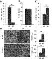
- 1 is a comparison of the characteristics of the 3D messenger sphere and 2D MSC, (A) the results of observing the aggregated MSC in the PDMS microchip with an optical microscope; (B) the size of MSCs confirmed by flow cytometry; And (C) the results of observing the morphological characteristics of the MSC under an optical microscope.
- Figure 2 is a comparison of the characteristics of the 3D messenger sphere and 2D MSC, (A) ki-67 stained cells (%) confirmed by flow cytometry; (B) propidium iodide (PI) stained cells (%) as confirmed by flow cytometry; (C) the results of confirming the frequency of CFU-F; And (D) bone formation or adipose tissue formation differentiation through mineralization (Alizarin red) and fat droplet deposition (oil red O).
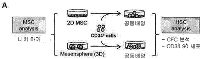
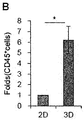
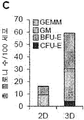
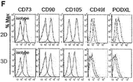
- Figures 3a to 3f is a comparison of the niche activity of the 3D messenger sphere and 2D MSC on hematopoietic stem cells, (3a) a schematic diagram of the experimental process; (3b) confirming the fold change of CD45 + cells; (3c) confirming the total number of colony-forming precursors (CFCs) inferred from 100 CD34 + cells; (3d) confirming fold change in the primitive hematopoietic precursors (CD34 + CD90 + ); (3e) confirming fold change in niche marker expression; And (3f) expression of surface markers was confirmed by flow cytometry.
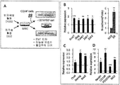
- Figure 4 is a comparison of the niche activity of 3D messenger sphere and 2D MSC for cancer stem cells, (A) schematic schematic of the experimental process; (B) representative image and quantitative analysis of infiltrated cancer cells; (C) confirming the volume change of the primary tumor; (D) the results of microscopic observation of the metabolites (arrows) in liver tissue; And (E) quantitative analysis of the degree of metastasis by evaluating the number of metabolites in liver tissue.
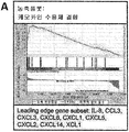
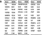
- Figures 5a and 5b confirm the transcript change of the 3D messenger sphere, (5a) enrichment plots associated with chamokine receptor binding; And (5b) confirming fold changes in the liver cytokine and growth factor genomes of the 3D messenger spheres for 2D MSCs.
- 6A to 6D are comparisons of EMT-related factors in 3D messenger peers and 2D MSCs, and (6a) confirming fold changes in EMT promoter gene expression; (6b) EMT related miRNA proyls among the top 50 miRNAs differentially expressed; (6c) changes in EMT gradients at the protein level were confirmed by liquid chromatography-mass spectrometry and IPA analysis; And (6d) relative ratios of expression amounts of differentially expressed proteins (log 2). 3D / 2D), the protein inferred to increase EMT is red, the protein inferred to inhibit EMT is shown in green.
- 7A-7F are for enhanced chromatin dynamics in 3D messenger peers, (7a) fold change in pluripotency-related gene expression in 3D messenger peers for 2D MSCs; (7b) representative images of RT-PCR analysis; (7c) Oct4 for 2D MSCs (top) and 3D messenger peer (bottom) Comparison of chromatin access to gene promoters in nuclease protection assays; (7d) fold change in chromatin remodeling genes in 3D messenger peers for 2D MSCs, as confirmed by RQ-PCR analysis; (7e) Western blot analysis of histone methylation labels in total cell lysates; And (7f) results of quantitative analysis of histone methyltransferase and dimethylase expression levels specific for each indicated histone label.
- 8a and 8b confirm the transcript change of the 3D messenger sphere, (8a) enrichment plots associated with HDAC; And (8b) RQ-PCR analysis of expression levels of HDACs between 3D messenger spheres and 2D MSCs.
- 9A to 9F confirm the effect of the EMT gradient on the niche activity of mesenchymal cells
- (9a) is a schematic diagram of this experimental procedure
- (9b) comparing the relative expression levels of the indicated master EMT-regulatory genes in 2D-MSCs transfected with EMT-driven miRNA
- (9c) comparison of relative expression levels of pluripotency genes expressed in 2D-MSCs transfected with EMT-driven miRNA
- Figure 10 confirms the effect of EMT gradient on the nitric activity of mesenchymal cells, after coculture with 234 MSCs transfected with siRNAs of Snail1, Snail2, Zeb1, Zeb2, and Twist1 with CD34 + cells, (A) EMT factor And (B) analysis of expression of the pluripotency gene by RQ-PCR; And (C) the number of hematopoietic regenerative cells (CD34 + 90 +) was compared three days after co-culture with 1 ⁇ 10 5 CD34 + cells and MSCs of each group.
- 11A to 11E confirm the hierarchical structure of the EMT gradient of MB mesenchymal cells in vivo under homeostatic conditions
- (11a) is a schematic diagram of this experimental procedure
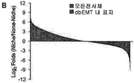
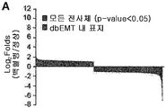
- (11b) confirming expression fold differences for genes matched to dbEMT (yellow) compared to differences in all transcripts (black);
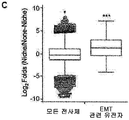
- (11c) quantitative analysis of expression levels of EMT-related genes and total transcripts between niche and non-niche cells;
- 11d confirming expression of master regulatory genes for EMT in niche cells compared to expression in non-niche cells;
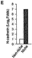
- 11e comparison of the difference in N-cadherin expression levels between niche and non-niche cells.
- Figure 12 analyzes the signaling mechanism between mesenchymal niche cells and non-niche cells, and relates to the mechanism related to the induction of EMT.
- Figure 13a is an analysis of the signaling mechanism between mesenchymal niche cells and non-niche cells, and relates to the mechanism related to TGF- ⁇ and EMT.
- Figure 13b is an analysis of the signaling mechanism between mesenchymal niche cells and non-niche cells, and relates to the mechanisms related to Wnt and EMT.
- Figure 14a is an analysis of the signaling mechanism between mesenchymal niche cells and non-niche cells, and relates to the mechanisms related to Notch and EMT.
- Figure 14b is an analysis of the signaling mechanism between mesenchymal niche cells and non-niche cells, and relates to the tyrosine kinase receptor and EMT-related mechanisms.
- 15A to 15H relate to an experiment of induction of an EMT gradient by physiological stimulation, and (15a) is a schematic diagram of this experimental procedure;
- 15b MSCs were prestimulated with poly (I: C) (50 g / mL) or substance-P (10 nM) for 2 days, then washed and co-cultured with CD34 + cells for 3 days, then confirming the fold change of hematopoietic regeneration precursor (CD34 + CD90 +) for the (stroma-free) culture group;
- FIG. 16 relates to an experiment of induction of an EMT gradient by physiological stimulation, and (A) a schematic diagram of the present experimental procedure; FIG. And (B) EMT factor and N-cadherin activity, (C) pluripotent gene, (D) expression of perivascular markers, for MSCs cultured in other serum batches (stimulatory or non-irritant) that cause niche activity. This is the result of confirming the change.
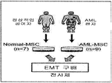
- FIG. 17 is a schematic diagram of an experimental procedure for identifying EMT gradient changes in MSCs derived from normal donors or patients with AML.
- FIG. 18 shows the results of confirming fold changes in (A) EMT factor and (B) N-cadherin expression of AML-derived MSCs relative to normal MSCs.
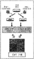
- 19 is a schematic diagram of an experimental procedure for confirming the effect of leukemia blasts on the EMT gradient of MSCs.
- 20A-20E show (20a) differences in expression folds of DEGs matched to dbEMT in leukemia CD34 + group versus normal CD34 + group compared to differences in all transcripts (black) (yellow); Confirming fold changes in (20b) master regulatory EMT factor (20c) N-cadherin and (20d) pluripotent gene expression of leukemia CD34 + group to normal CD34 + group; And (20e) Peripheral cell marker expression in the leukemia CD34 + group relative to the normal CD34 + group, compared to fold differences in the same genes in the Nich cells (Nestin + PDGFR +) for non-niche cells (Nestin-PDGFR-) (blue). This is the result of confirming the change of multiple of (red).
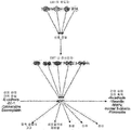
- 21 is a schematic diagram showing the effect of the EMT gradient as a major regulator of niche activity and the like.
- the present invention provides a composition for enhancing niche activity of mesenchymal stem cells, including epithelial-mesenchymal transition (EMT) promoter.
- EMT epithelial-mesenchymal transition
- mesenchymal stem cell is a cell that helps to create cartilage, bone, fat, myeloid epilepsy, muscle, nerves, etc., in adults in the bone marrow in general, but the cord blood, It exists in peripheral blood, other tissues, etc., and means the cell which can be obtained from these.
- mesenchymal stem cells are used in the same sense as mesenchymal stromal cells or stromal cells.
- the mesenchymal stem cells are human, monkey, pigs, horses, cows, sheep, dogs, cats, mice It includes all animal-derived cells, such as rabbits, but preferably cells derived from humans.
- Niche refers to a component (cell and / or substance) composed of tissues or organs that support the development and proliferation of tissue cells such as stem cells and other somatic cells, and inter-cell interactions. It is known to secrete factors necessary to induce action and to be omnipotent.
- Niche also called the microenvironment, plays a key role in maintaining the stemness that expresses all the characteristics of stem cells. Stem cells are located in a kind of microenvironment that consists of adhesion molecule growth factors, which are called niches in academia, and these areas of stem cells are located, adhesiveness, and homing. It is responsible for maintaining and coordinating homing, quiescence and activation.
- niche is a major microenvironment surrounding stem cells that regulates the differentiation of stem cells and prevents and protects them from moving to another place or suicide.
- the composition according to the present invention is intended to maintain the undifferentiated ability of all kinds of stem cells present in or outside the body and to promote self-renewal by improving such niche activity. Therefore, the composition of the present invention can be applied or applied to various target diseases according to the stem cells of interest.
- the area of active research of stem cell niches is hematopoietic stem (nitrogen) stem cells (nitrogen), where hematopoietic stem cells (resident hematopoietic stem cells) reside.
- the hematopoietic stem cell niche is known not only as a haven for hematopoietic stem cells but also to regulate the number of stem cells, the stem cells.
- the niche activity may mean maintaining the undifferentiated ability of hematopoietic stem cells and promoting self-renewal, wherein the composition of the present invention is applied to a patient in a physiological state in which hematopoietic cells are damaged
- the physiological state in which the hematopoietic cells are damaged may include acute leukemia, chronic myeloid leukemia, myelodysplastic syndrome, lymphoma, multiple myeloma, germ cell tumor, breast cancer, ovarian cancer, small cell lung cancer, neuroblastoma, aplastic anemia, erythrocytosis, Gaucher's disease, Hunter syndrome, ADA enzyme deficiency, Wiskot-Aldrich syndrome, rheumatoid arthritis, systemic lupus erythematosus, or multiple sclerosis, or may be caused by anticancer drug administration or irradiation.
- hematopoietic stem cell As used herein, the term "hematopoietic stem cell (HSC)" is an ancestral cell of undifferentiated bone marrow hematopoietic cells that produce blood cells such as red blood cells, white blood cells, platelets, etc. When used, it shows the ability to repopulate for a long time with self-renewing capacity.
- the hematopoietic stem cells include humans, monkeys, pigs, horses, cows, sheep, dogs, cats, mice, It includes cells from all animals, such as rabbits, but may preferably be cells from humans.
- self-renewal is also called self-replicating, self-replicating, and self-renewing with the ability to produce cells having the same properties and characteristics, and is one of the important features of stem cells.
- self-renewal in the present invention means the ability to continue proliferation while maintaining an undifferentiated state.
- epithelial-mesenchymal transition is a phenomenon that occurs during normal embryonic development, in which a cell loses an epithelial cell phenotype and has high mobility of mesenchymal. The process of converting to a cell phenotype.
- EMT epithelial-mesenchymal transition
- epithelial cells lose the polarity that separates the top and bottom of the cell, the cell shape changes from square to fibroblast type, epithelial cell markers decrease and mesenchymal cell markers increase.
- EMT plays various roles such as fibrosis of tissues in addition to tissue formation and differentiation in the natal period.
- mesenchymal niches have been known to regulate regenerative activity in hematopoietic stem cells in response to dynamic changes in the bone marrow microenvironment, but this process is unknown for the major mediators.
- the niche activity is improved by 3D intercellular interaction or physiological stimulation, the gradient of epithelial-mesenchymal transition is changed to the mesenchymal state, and in the degenerative state due to leukemia, the opposite trend is shown. Confirmed.
- mesenchymal niche activity can be regulated through changes in epithelial-mesenchymal transition, specifically, mesenchymal niche activity can be enhanced through epithelial-mesenchymal transition promoting factors, and mesenchymal-mesenchymal transitional inhibitors
- the technical feature is that it has been found that it can inhibit niche activity.
- the epithelial-mesenchymal transition promoting factors refer to factors that promote the above-mentioned mesenchymal transitional phenomena, and may be natural products, compounds, nucleotides, proteins, recombinant vectors, and the like, preferably miR-10b, miR-146a, miR MiRNA selected from -106b, miR-34a, miR-379, and miR-146b or a vector including any one gene selected from the group consisting of snail, slug, twist1, zeb1, and zeb2, but is not limited thereto. It doesn't happen.
- the vector included in the composition of the present invention refers to an expression vector into which the polynucleotide sequence encoding the epithelial-mesenchymal transition promoting factor is inserted, but is not limited thereto, and preferably means a plasmid, virus or other medium known in the art. .
- the polynucleotide sequence according to the present invention may be operably linked to an expression control sequence, wherein the operably linked gene sequence and the expression control sequence are one expression vector containing a selection marker and a replication origin together. It may be included within.
- “Operably linked” can be genes and expression control sequences linked in such a way as to enable gene expression when the appropriate molecule is bound to the expression control sequences.
- control sequence is meant a DNA sequence that regulates the expression of a polynucleotide sequence operably linked in a particular host cell.
- control sequence is a promoter for transcription, any for controlling transcription.
- plasmids examples include E. coli-derived plasmids (pBR322, pBR325, pUC118 and pUC119), Bacillus subtilis-derived plasmids (pUB110 and pTP5), and yeast-derived plasmids (YEp13, YEp24 and YCp50).
- Viruses selected from the group consisting of adenoviruses, vaccinia viruses, baculoviruses, herpes simplex viruses, complexes with lentiviruses, and the like can be used.
- Those skilled in the art can use a vector suitable for introducing a gene or polynucleotide of the present invention into a host cell, and preferably a vector designed to facilitate protein expression induction and isolation of the expressed protein.
- the present invention provides a method for enhancing niche activity of mesenchymal stem cells, comprising the step of promoting epithelial-mesenchymal transition (EMT) of mesenchymal stem cells.
- EMT epithelial-mesenchymal transition
- the method for enhancing niche activity of mesenchymal stem cells of the present invention uses epithelial-mesenchymal transition promoting factors described in the above-described composition for promoting niche activity of mesenchymal stem cells, the common contents among them are In order to avoid excessive complexity of the specification, the description thereof is omitted.
- Promoting the epithelial-mesenchymal migration is preferably treated with mesenchymal stem cells any one selected from miR-10b, miR-146a, miR-106b, miR-34a, miR-379, and miR-146b , snail, slug, twist1, zeb1, and zeb2 may be introduced into a mesenchymal stem cell, the vector including any one gene selected from the group, but is not limited thereto.
- the vector according to the present invention may be introduced into a cell using a method known in the art, but the method of introducing the recombinant vector according to the present invention into a host cell is not limited thereto, but calcium chloride (CaCl 2 ) Heat shock methods, silicon carbide whiskers, sonication, precipitation by polyethylenlycol (PEG), transient transfection, microinjection, transduction, Cell fusion, calcium phosphate precipitation, liposome-mediated transfection, DEAE Dextran-mediated transfection, polybrene-mediated transfection, Electroporation, gene gun, etc. may be used, and may be introduced into cells by other known methods for introducing nucleic acids into cells.
- CaCl 2 calcium chloride
- PEG polyethylenlycol
- a candidate substance for improving the expression or activity of epithelial-mesenchymal transition promoting factor or inhibiting the expression or activity of epithelial-mesenchymal transition promoting factor is used as a composition for enhancing niche activity of mesenchymal stem cells. It provides a method of screening a composition for enhancing niche activity of mesenchymal stem cells, comprising the step of selecting.
- the screening method of the composition for enhancing niche activity of mesenchymal stem cells of the present invention uses the epithelial-mesenchymal transition promoting factor described above in the composition for enhancing niche activity of mesenchymal stem cells, The contents common to are omitted in order to avoid excessive complexity of the present specification.
- expression means to express information possessed by a gene in the form of a gene product having a function of RNA or protein according to a program.
- the candidate may be a natural product, a compound, a nucleotide, a protein, a recombinant vector, etc., but is not limited thereto.
- the candidate or related protein may be labeled with a detectable label.
- the detectable label may be a chemical label (e.g. biotin), an enzyme label (e.g.
- radiolabels eg C14,, I125,, P32 and S35
- fluorescent labels eg coumarin, fluorescein, fluoresein Isothiocyanate (FITC), rhodamine 6G, rhoda Rhominamine B, TAMRA (6-carboxytetramethyl-rhodamine), Cy-3, Cy-5, Texas Red, Alexa Fluor, DAPI (4,6-diamidino-2-phenylindole), HEX, TET, Dabsyl, and FAM ], A luminescent label, a chemiluminescent label, a fluorescence resonance energy transfer (FRET) label or a metal label (eg, gold and silver).
- FRET fluorescence resonance energy transfer
- Measuring the expression or activity of the epithelial-mesenchymal transition promoting factor or epithelial-mesenchymal transition inhibitory factor according to the present invention is a variety of methods known in the art that can measure the amount of RNA or protein produced by the expression of a gene. It may be carried out through.
- the expression level of RNA may be measured by real-time PCR, microarray, northern blotting, and the like, and the expression level of protein may be measured by western blotting. It can be measured by blotting, flow cytometry, enzyme immunoassay (ELISA), or immunofluorescein (immunofluorescnce) and the like, in addition to measuring the expression or activity of proteins well known in the art can be applied.
- the present invention comprises a mesenchymal stem cell inhibited epithelial-mesenchymal transition (EMT), pharmaceutical composition for inhibiting cancer metastasis;
- EMT epithelial-mesenchymal transition
- a method for inhibiting cancer metastasis comprising administering to the subject a mesenchymal stem cell in which epithelial-mesenchymal migration is inhibited;
- mesenchymal stem cells inhibited epithelial-mesenchymal migration for cancer metastasis inhibition comprising administering to the subject a mesenchymal stem cell in which epithelial-mesenchymal migration is inhibited.
- cancer refers to the aggressive nature of cells dividing and growing, ignoring normal growth limits, the invasive nature of infiltrating surrounding tissues, and the metastatic spread to other parts of the body.
- the type of cancer may be any one selected from the group consisting of pancreatic cancer, stomach cancer, liver cancer, colon cancer, brain cancer, breast cancer, thyroid cancer, bladder cancer, esophageal cancer, uterine cancer, and lung cancer, but is not limited thereto. .
- the epithelial-mesenchymal transition inhibitory factors refer to factors that inhibit the above-described mesenchymal transitional phenomena, and may be natural products, compounds, nucleotides, proteins, recombinant vectors, and the like, preferably miR-503, miR-145, miR 193b, miR-365a, miR-29b, miR-129, miR-335, and may be any one selected from the group consisting of miR-675, but is not limited thereto.
- the mesenchymal stem cells are any one selected from miR-503, miR-145, miR-193b, miR-365a, miR-29b, miR-129, miR-335, and miR-675, or Snail1, It may be transfected with a vector containing any one siRNA gene selected from the group consisting of Snail2, Zeb1, Zeb2, and Twist1.
- CD34 + cells were purified from leukapheresis for cord blood or AML patients as described above, and cytokine mixtures (100 ng / mL human stromal cell factor [SCF]; 100 ng / mL human Flt3L; and 20 ng / mL). Irradiated for 5 days in long-term culture medium (H5100, STEMCELL Technologies, Vancouver, Canada) in the presence of human interleukin [IL] -3, IL-6, and G-CSF; ProSpec-Tany TechnoGene Ltd., Rehovot, Israel) 1,500 cGy) co-culture with either messengerspear or 2D MSCs.
- cytokine mixtures 100 ng / mL human stromal cell factor [SCF]; 100 ng / mL human Flt3L; and 20 ng / mL.
- a low cytokine mix (hSCF: 1 ⁇ g / mL; hFlt3L: 1 ⁇ g / mL; hTPO: 0.5 ⁇ g / mL) was used for coculture with miRNA-transfected MSCs.
- RNA-seq whole-transcriptome sequencing
- Paired sequencing reads were aligned on reference human genome sequence (hg19) using TopHat.
- Gene expression fragment per exon kilobase per million mapped fragments [FPKM]) was evaluated using CuffLink software.
- CuffLink software For functional analysis, and analysis of data sets that the priority of the GSEA using the log 2 (FPKM3D / FPKM2D) of the drain changes.
- miRNA sequencing a small RNA sequencing library was generated with 1 g of RNA using TruSeq Small RNA Sample Prep Kit Version 2 (Illumina, San Diego, Calif., USA).
- Illumina HCS version 1.4.8
- RTA version 1.12.4.2
- CASAVA version 1.8.2
- mice Animal testing was approved by the Catholic University of Korea's Animal Care and Use Committee.
- NOD / SCID- ⁇ c null mice were purchased from the Jackson Laboratory (Bar Harbor, ME, USA).
- a total of 5 ⁇ 10 5 MDA-MB-231 cells were injected subcutaneously in breast adipose tissue with or without 2 ⁇ 10 5 MSCs cultured under 2D or 3D conditions.
- All mice were sacrificed.
- Primary tumors and livers were fixed and sections of fixed tissues were stained with H & E.
- miRNA mimics, miRNA inhibitors, or siRNAs were identified by Bioneer Corp. It was purchased from (Daejeon, Korea). For transfection, BM-MSCs were replaced with Opti-MEM culture medium (Life Technology, Carlsbad, CA, USA) and 100 nM miRNA mimetics, miRNA inhibitors, or siRNA using Lipofectamine 2000 (Life Technology). Transfection. Scrambled miRNA was used as a negative control. The oligonucleotide sequence used is indicated in the supplementary information.
- Chromatin in MSCs Access to Oct4 gene promoter was analyzed by nuclease protection assay using chromatin accessibility assay kit according to manufacturer's instructions (Epigentek, Farmingdale, NY, USA). Briefly, cells were incubated in EpiQuik chromatin buffer with or without nucleases and genomic DNA was extracted. Chromatin accessibility was analyzed by qPCR and analyzed using the kit's analysis tool. The hemoglobin ( HBB ) gene was used as an inaccessible reference for measuring relative chromatin accessibility.
- HBB hemoglobin
- MSC gene expression was analyzed by Illumina beadchip (Illumina, San Diego, Calif.) Array hybridization analysis using a linear model for the microarray data (LIMMA) method and the statistical R-package described above.
- mesenchymal niche activity To investigate the properties of mesenchymal niche activity, first, the fact that nestin-expressing mesenchymal cell subpopulations with niche function are often characterized by the ability to form mesenchymal glomeruli (messenger) under non-adherent culture conditions. On the basis of this, a 3D meshing peer model was adopted. Thus, to investigate the functional association between messier-peer formation and niche activity, it was confirmed whether niche function could be induced by their 3D cell interaction. Accordingly, cells aggregated within 24 hours on a polydimethylsiloxane (PDMS) microchip, a platform for the formation of non-adherent 3D globules, to form homogeneous globules.
- PDMS polydimethylsiloxane
- FIG. 3A To confirm the effect of 3D interaction on niche function, the ability of the messengerspeare to support the maintenance / proliferation of hematopoietic progenitor cells was compared (FIG. 3A).
- CD34 + cells co-cultured with messense peers showed a significant increase in colony-forming precursors and CD34 + CD90 + subsets.
- Subpopulations represent long-term regenerative hematopoietic cells, demonstrating an improvement in the capacity of the messengerspeare to support HSC self-replicate / maintenance (FIGS. 3B-D).
- RNA-sequencing profiles in messespeare and 2D MSCs were reviewed to classify the top 50 and bottom 50 genes with the highest fold changes as differential expression genes. It is shown in Table 1 below (DEGs).
- GSEA Gene Set Enrichment Assay
- this set of genes upregulated in the messenger sphere is mainly associated with cytokine responses including chemokine receptor binding, chemokine activity, G-protein coupled growth factor signaling, and secretion factors in the extracellular space (FIG. 5A and 5B), which supports the results of enhanced niche activity at the level of transcription.
- cytokine responses including chemokine receptor binding, chemokine activity, G-protein coupled growth factor signaling, and secretion factors in the extracellular space (FIG. 5A and 5B), which supports the results of enhanced niche activity at the level of transcription.
- miRNAs that show significant expression changes are known to modulate EMT.
- miR-146b the major inducer of EMT, is one of the very largely induced miRNAs in the messenger peer (89-fold increase; FIG. 6B).
- the EMT inducers miR-379, miR-34a, miR-106b, miR-146a, and miR-10b are also height induced in the messenger peer.
- many down-regulated miRNAs including miR-503, miR-145, miR- 193b , m iR-365a, miR-29b, miR-129, miR-335, and miR-675 in the messenger sphere , exhibit EMT. It has been confirmed to inhibit (FIG.
- the messense peers also include histone methyltransferase (for H3K4 methylation), including increased KMT2B, KMT2C, KMT2E, KMT2F, and KMT2H; KMT1E and KMT1F (for H3K9 methylation); KMT6B (for H3K27 methylation); KMT3A (for H3K36 methylation); And KMT4 (for H3K79 methylation), but KDM5A (for H3K4 demethylation), KDM3A and KDM3C (for H3K9 demethylation), KDM6B (for H3K27 demethylation), and KDM4B (H3K36 demethylation).
- histone methyltransferase for H3K4 methylation
- KMT2B increased KMT2B, KMT2C, KMT2E, KMT2F, and KMT2H
- KMT1E and KMT1F for H3K9 methylation
- KMT6B for H
- HDAC histone deacetylase
- FIG. 9A As a result of observing the association between the EMT gradient and the improved niche activity, the effect of the EMT gradient on the niche activity of the mesenchymal cells was next explored (FIG. 9A).
- mimic mimics that can enhance EMT gradients ie mimics and EMT-inhibiting miRNAs (miR-503, miR) that mimic EMT-promoting miRNAs (miR-140b, miR-379).
- 2D MSCs were transfected with mimics that inhibited -145-5p, miR-193b, miR-36t5a, miR-29b, and miR-129).
- the miRNA-transfected 2D MSCs showed significant induction of EMT-promoting genes such as Snail1 and Twist1 , indicating that EMT progresses in cells (FIG. 9B).
- EMT-promoting genes such as Snail1 and Twist1
- the induction of EMT in these 2D MSCs resulted in significant induction of pluripotency-related genes Oct-4 , Nanog , and Sox2 and a higher frequency of clonalogenic mesenchymal precursors (CFU-F) than control MSCs ( 9C, and 9D).
- CFU-F clonalogenic mesenchymal precursors
- proliferation of significantly higher precursor and CD34 + CD90 + hematopoietic regenerative cells was observed in EMT-based MSCs than in the control (FIGS. 9E and 9F), driving EMT in MSCs as observed in the Messencepeare, It is shown to regenerate the properties of activated mesenchymal niches.
- MSCs transfected with siRNAs targeting the EMT-regulatory genes Snail1 , Slug , Zeb1 , and Twist are EMT factor and pluripotency-related genes, as evidenced by a decrease in CD34 + CD90 + hematopoietic regenerative cell proliferation.
- FIGS. 10A and 10B show that EMT gradients in mesenchymal cells are the major factor controlling their niche activity.
- FIG. 15A the change in EMT gradient during physiological stimulus-induced niche activation was examined.
- poly (I: C) -mediated IFNa signal which was previously found to induce HSC proliferation, was examined.
- co-culture of poly (I: C) -pretreated MSCs with CD34 + cells induced a significant increase in CD34 + CD90 + hematopoietic regenerative cell proliferation compared to the cell proliferation observed in control MSCs (FIG. 15B). This indicates that poly (I: C) -mediated HSC stimulation also acts as a mesenchymal niche activation model.
- poly (I: C) -treated MSCs were significant in the master EMT regulators Snail1, Slug, Zeb1, Zeb2, Twist (30, 31, 42), and N-cadherin. Upregulation was shown (FIG. 15C).
- poly (I: C) stimulation induces pluripotent gene expression ( Oct4 , Nanog , and Sox2 ) and similar induction of perivascular markers ( Nestin , NG2 , PDGFR b, and CD146 ), resulting in 3D cell interaction. Mimics the niche activation process induced (FIGS. 15D and 15E).
- FIG. 16A For detailed observations, a recent model of reversible switching of niche activity induced by treatment with different sera was reviewed (FIG. 16A). In particular, similar series of changes induced by serum conditions that resulted in higher niche activity, as induced by other stimuli for niche activation, were observed (FIGS. 16B-16D), regardless of the type of stimulus. It is shown that cellular changes for each activation model have been preserved. Overall, these results showed that the hierarchical generation of the EMT gradient is a normal cellular expression preserved in mesenchymal cells in response to physiological stimuli that cause niche activation, which is in turn responsible for mesenchymal cells in response to niche activation program regeneration stimulation. It is shown that it represents a reactive substrate change that occurred in.
- EMT gradients of mesenchymal cells for the regulation of niche activity were associated with pathological changes induced by pathological conditions as well as steady-state BM.
- the EMT gradient changed in the opposite direction under leukemia conditions, as deduced by downregulation of EMT factor and N-cadherin expression.
- the EMT factor and N-cadherin were downregulated and the amount of stem genes and perivascular markers decreased.
Landscapes
- Health & Medical Sciences (AREA)
- Life Sciences & Earth Sciences (AREA)
- Engineering & Computer Science (AREA)
- Biomedical Technology (AREA)
- Organic Chemistry (AREA)
- Chemical & Material Sciences (AREA)
- Bioinformatics & Cheminformatics (AREA)
- Genetics & Genomics (AREA)
- Biotechnology (AREA)
- Wood Science & Technology (AREA)
- Zoology (AREA)
- Microbiology (AREA)
- Biochemistry (AREA)
- General Engineering & Computer Science (AREA)
- General Health & Medical Sciences (AREA)
- Cell Biology (AREA)
- Developmental Biology & Embryology (AREA)
- Rheumatology (AREA)
- Medicines Containing Material From Animals Or Micro-Organisms (AREA)
- Micro-Organisms Or Cultivation Processes Thereof (AREA)
Abstract
본 발명은 3차원 중간엽 기질세포 구상체에서 상피-간엽이행 구배의 계층화에 관한 것이다. 본 발명에서는, 중간엽 줄기세포의 니치 활성의 변화는 상피-간엽 이행과 밀접한 관련이 있음을 규명하였다. 구체적으로, 3D 세포간 상호작용 또는 관련 생리학적 자극에 의해 니치 활성이 향상되는 경우, 상피-간엽 이행의 구배는 더욱 중간엽 상태로 변화하였고, 백혈병에 의한 퇴행성 상태에서는 이와 정반대의 경향성을 보여주었다. 따라서, 본 발명은 상피-간엽 이행 관련 인자들을 조절 또는 도입함으로써, 중간엽 줄기세포-기반 세포 치료의 효능을 향상시킬 수 있을 것으로 기대된다.
Description
본 발명은 3차원 중간엽 기질세포 구상체에서 상피-간엽이행 구배의 계층화에 관한 것이다.
골수(BM) 미세환경은 자기 복제(self-renewal), 재생(regeneration), 및 조혈 줄기세포(HSCs)의 휴지(quiescence)를 조절하고, 골수말소된(myeloablated) 수용자 내 혈액 세포의 평생 생산 및 BM 재구성(reconstitution)을 조절하는데 중요한 역할을 한다. 이전 연구에서는 BM내 HSC 조절에 연관된 다수의 니치를 확인하였다. 예를 들어, 이식된 HSCs에 관한 초기 연구에서는 N-카데린-발현 조골세포로 구성된 골 내막성 니치를 확인한 반면, 혈관주위 니치는 항상성 조건에서 HSCs가 다수로 존재하는 니치로 확인되었다. 혈관주위 니치의 세포 조성을 확인하기 위한 광범위한 노력으로, 내피 세포, 거핵 세포, 및 비수초성 교감 신경계 세포의 추가적인 참여와 함께 니치의 주요한 구성성분으로서 혈관주위 세포 및 특정 서브세트의 중간엽 세포를 확인하였다.
중간엽 니치 세포는 jagged 1, CXCL12, 및 안지오포이에틴-1과 같은 상호간섭(cross-talk) 분자의 발현에 의해 HSC 구획을 유지하나; 이들 세포의 정체성에 있어 이질성이 관찰되었다. 예를 들어, Nestin을 발현하는 서브세트만이 HSC 유지에 기여하는 BM HSCs와 긴밀한 연관성이 있는 것으로 밝혀졌다. 이와 유사하게, leptin 수용체 또는 Prx1을 발현하는 중간엽 세포 역시 니치 기능을 보여준다. 이들 서브세트는 일부 Nestin-발현 세포와의 오버랩(overlap)을 보여주는 반면, 최근 계통-추적 연구에서는 신경능 유래 서브세트는 HSC 니치로 작용하나, 중배엽 유래 서브세트는 골연골 분화에 기여함을 보여준 바 있다.
한편, HSCs는 BM 손상, 과립구-콜로니 자극인자(G-CSF), 또는 생리학적 자극에 반응하여 역학적 행동 스위치의 발생을 암시하는 인터페론(IFN) 알파-매개 폴리 (I:C) 생산에 의해 유도된 생리학적 자극에 반응하여 휴면 및 자기-복제 사이에서 가역적으로 스위칭된다. 따라서, BM 미세환경은 필수적인 요구를 만족하도록 혈액 세포 생산을 조정하기 위한 교호적인 생리학적 필요에 반응해야 한다. 예를 들어, 니치 활성은 HSCs의 방출을 제어하기 위해 24시간 주기 리듬에 따라 교감 신경계에 의해 주기적으로 조절된다. 이와 유사하게, 중간엽 세포는 정규(canonical) Wnt 신호전달 경로의 미세한 조정에 의해 뚜렷한 니치 활성을 나타내거나 배양 조건의 스위칭에 의해 니치 활성의 가역적 변화를 나타낸다(한국 공개특허 10-2009-0008155).
게다가, 최근 연구에서는, BM 중간엽 니치가 백혈병 또는 골수 증식성 질환에 의한 미세환경의 변화에 반응하여 니치 기능의 퇴행성 변화를 보여준 바 있다. 그러나, 이러한 결과에도 불구하고, 이러한 반응이 일어나는 메커니즘 및 이러한 니치 활성을 제어하는 파라미터에 관해서는 아직 명확하지 않다.
본 발명은 상기와 같은 문제점을 해결하기 위해 안출된 것으로서, 본 발명자들은 중간엽 기질세포-기반 세포 치료의 유효성를 향상시키기 위하여 예의 노력한 결과, 중간엽 줄기세포의 니치 활성은 상피-간엽 이행 구배의 변화와 밀접한 관련이 있으며, 상기 상피-간엽 이행 구배가 조절된 중간엽 줄기세포를 통해 암의 전이를 억제할 수 있음을 실험적으로 확인하고, 이에 기초하여 본 발명을 완성하였다.
본 발명의 목적은 중간엽 줄기세포의 니치 활성 증진용 조성물 및 중간엽 줄기세포의 니치 활성 증진방법을 제공하는데 있다.
본 발명의 다른 목적은 암 전이 억제용 조성물 및 암 전이 억제방법을 제공하는데 있다.
그러나 본 발명이 이루고자 하는 기술적 과제는 이상에서 언급한 과제에 제한되지 않으며, 언급되지 않은 또 다른 과제들은 아래의 기재로부터 당업자에게 명확하게 이해될 수 있을 것이다.
상기와 같은 본 발명의 목적을 달성하기 위하여, 본 발명은 상피-간엽이행 촉진 인자를 포함하는, 중간엽 줄기세포의 니치 활성 증진용 조성물을 제공한다.
본 발명의 일 구현예로서, 상기 상피-간엽이행 촉진 인자는 miR-10b, miR-146a, miR-106b, miR-34a, miR-379, 및 miR-146b로부터 선택되거나 snail, slug, twist1, zeb1, 및 zeb2로 이루어진 군으로 선택되는 어느 하나의 유전자를 포함하는 벡터일 수 있다.
본 발명의 다른 구현예로서, 상기 중간엽 줄기세포는 지방조직, 골수, 말초혈액 또는 제대혈에서 유래한 것일 수 있다.
또한, 본 발명은 중간엽 줄기세포의 상피-간엽이행을 촉진하는 단계를 포함하는, 중간엽 줄기세포의 니치 활성 증진방법을 제공한다.
본 발명의 일 구현예로서, 상기 상피-간엽이행을 촉진하는 단계는 miR-10b, miR-146a, miR-106b, miR-34a, miR-379, 및 miR-146b로부터 선택되는 어느 하나를 중간엽 줄기세포에 처리하거나, snail, slug, twist1, zeb1, 및 zeb2로 이루어진 군으로 선택되는 어느 하나의 유전자를 포함하는 벡터를 중간엽 줄기세포에 도입하는 것일 수 있다.
또한, 본 발명은 상피-간엽이행이 저해된 중간엽 줄기세포를 포함하는, 암 전이 억제용 약학적 조성물; 상피-간엽이행이 저해된 중간엽 줄기세포를 개체에 투여하는 단계를 포함하는 암 전이 억제방법; 및 암 전이 억제를 위한, 상피-간엽이행이 저해된 중간엽 줄기세포의 용도를 제공한다.
본 발명의 일 구현예로서, 상기 중간엽 줄기세포는, miR-503, miR-145, miR- 193b, miR-365a, miR-29b, miR-129, miR-335, 및 miR-675로부터 선택되는 어느 하나가 처리된 것일 수 있다.
본 발명의 다른 구현예로서, 상기 중간엽 줄기세포는 Snail1, Snail2, Zeb1, Zeb2, 및 Twist1로 이루어진 군으로부터 선택되는 어느 하나의 siRNA 유전자를 포함하는 벡터로 형질감염된 것일 수 있다.
본 발명에서는, 중간엽 줄기세포의 니치 활성의 변화는 상피-간엽 이행과 밀접한 관련이 있음을 규명하였다. 구체적으로, 3D 세포간 상호작용 또는 생리학적 자극에 의해 니치 활성이 향상되는 경우, 상피-간엽 이행의 구배는 더욱 중간엽 상태로 변화하였고, 백혈병에 의한 퇴행성 상태에서는 이와 정반대의 경향성을 보여주었다. 따라서, 본 발명은 상피-간엽 이행 관련 인자들을 조절 또는 도입함으로써, 중간엽 줄기세포-기반 세포 치료의 효능을 향상시킬 수있을 것으로 기대된다.
도 1은 3D 메센스피어와 2D MSC의 특성을 비교한 것으로서, (A) PDMS 마이크로칩 내의 응집된 MSC를 광학 현미경으로 관찰한 결과; (B) MSC의 크기를 유세포 분석을 통해 확인한 결과; 및 (C) MSC의 형태적 특성을 광학 현미경으로 관찰한 결과이다.
도 2는 3D 메센스피어와 2D MSC의 특성을 비교한 것으로서, (A) ki-67 염색된 세포 (%)를 유세포 분석을 통해 확인한 결과; (B) 요오드화 프로피디움 (PI) 염색된 세포(%)를 유세포 분석을 통해 확인한 결과; (C) CFU-F의 빈도를 확인한 결과; 및 (D) 골 형성 또는 지방조직 형성 분화를 미네랄화(Alizarin red) 및 지방 드럽렛 침착 (oil red O)을 통해 확인한 결과이다.
도 3a 내지 도 3f는 조혈 줄기세포에 대한 3D 메센스피어와 2D MSC의 니치 활성을 비교한 것으로서, (3a) 본 실험 과정의 개략적인 모식도; (3b) CD45+ 세포의 배수 변화를 확인한 결과; (3c) 100개의 CD34+ 세포로부터 추론된 전체 콜로니-형성 전구체 (CFC)의 수를 확인한 결과; (3d) 원시 조혈 전구체(CD34+CD90+)의 배수 변화를 확인한 결과; (3e) 니치 마커 발현의 배수 변화를 확인한 결과; 및 (3f) 표면 마커의 발현을 유세포 분석을 통해 확인한 결과이다.
도 4는 암 줄기세포에 대한 3D 메센스피어와 2D MSC의 니치 활성을 비교한 것으로서, (A) 본 실험 과정의 개략적인 모식도; (B) 침투한 암 세포의 대표적인 이미지 및 정량 분석한 결과; (C) 1차 종양의 용적 변화를 확인한 결과; (D) 간 조직 내 전이소(화살표)를 현미경으로 관찰한 결과; 및 (E) 간 조직 내 전이소의 수를 평가하여 전이 정도를 정량 분석한 결과이다.
도 5a 및 도 5b는 3D 메센스피어의 전사체 변화를 확인한 것으로서, (5a) 캐모카인 수용체 결합과 관련된 농축 플롯; 및 (5b) 2D MSC에 대한 3D 메센스피어의 간 사이토카인 및 성장인자 유전체의 배수 변화를 확인한 결과이다.
도 6a 내지 도 6d는 3D 메센스피어와 2D MSC에서 EMT 관련된 인자의 변화를 비교한 것으로서, (6a) EMT 촉진 유전자 발현의 배수 변화를 확인한 결과; (6b) 차등 발현된 최상위 50개의 miRNA 중 EMT 관련 miRNA 프로일; (6c) 단백질 수준에서 EMT 구배의 변화를 액체 크로마토그래피-질량 분광분석 및 IPA 분석을 통하여 확인한 결과; 및 (6d) 차등 발현하는 단백질을 발현량의 상대비(log2
3D/2D) 순으로 정렬한 것으로, EMT를 증가시키는 것으로 유추되는 단백질은 붉은색, EMT를 저해하는 것으로 유추되는 단백질은 녹색으로 표시한 결과이다.
도 7a 내지 7f는 3D 메센스피어 내 향상된 크로마틴 역학에 대한 것으로서, (7a) 2D MSCs에 대한 3D 메센스피어 내 다능성-관련 유전자 발현의 배수 변화; (7b) RT-PCR 분석의 대표적인 이미지; (7c) 2D MSCs (상부) 및 3D 메센스피어 (하부)에 대한 Oct4
유전자 프로모터로의 크로마틴 접근성을 뉴클레아제 보호 분석으로 비교한 결과; (7d) RQ-PCR 분석에 의해 확인된, 2D MSCs에 대한 3D 메센스피어 내 크로마틴 리모델링 유전자의 배수 변화; (7e) 총 세포 용해물 내 히스톤 메틸화 표지의 웨스턴 블롯 분석 결과; 및 (7f) 각각의 표시된 히스톤 표지에 특이적인 히스톤 메틸트랜스퍼라제 및 디메틸라제 발현량의 정량 분석 결과이다.
도 8a 및 도 8b는 3D 메센스피어의 전사체 변화를 확인한 것으로서, (8a) HDAC과 관련된 농축 플롯; 및 (8b) 3D 메센스피어와 2D MSC간 HDACs의 발현 수준을 RQ-PCR로 분석한 결과이다.
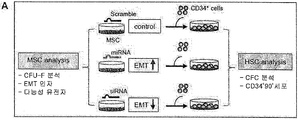
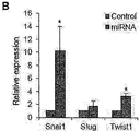
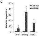
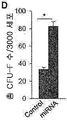
도 9a 내지 9f는 중간엽 세포의 니치 활성에 대한 EMT 구배의 영향을 확인한 것으로서, (9a) 본 실험 과정의 개략적인 모식도; (9b) EMT-구동 miRNA로 형질감염된 2D-MSCs 내에서, 표시된 마스터(master) EMT-조절 유전자의 상대적인 발현량을 비교한 결과; (9c) EMT-구동 miRNA로 형질감염된 2D-MSCs 내에서 표시된 다능성 유전자의 상대적인 발현량을 비교한 결과; (9d) EMT-구동 miRNA로 형질감염된 3000개의 MSCs로부터 얻은 CFU-F의 수를 비교한 결과; 및 EMT-구동 miRNA로 형질감염된 MSCs와 함께 CD34+ 세포를 공배양시킨지 3일 후, (9e) 각 계통의 CFCs 수 및 (9f) 원시 조혈 재생 세포(CD34+CD90+)의 수를 비교한 결과이다.
도 10은 중간엽 세포의 니치 활성에 대한 EMT 구배의 영향을 확인한 것으로서, Snail1, Snail2, Zeb1, Zeb2, 및 Twist1의 siRNA로 형질감염된 2D MSCs를 CD34+ 세포와 공배양한 후, (A) EMT 인자 및 (B) 다능성 유전자의 발현을 RQ-PCR로 분석한 결과; 및 (C) 1×10⁵개 CD34+ 세포와 각 군의 MSCs와 공배양시킨지 3일 후, 조혈 재생 세포(CD34+90+)의 수를 비교한 결과이다.
도 11a 내지 도 11e는 항상성 조건 하에서 생체 내 MB 중간엽 세포의 EMT 구배의 계층 구조를 확인한 것으로서, (11a) 본 실험 과정의 개략적인 모식도; (11b) 모든 전사체(black)에서의 차이와 비교하여, dbEMT (yellow)에 매칭된 유전자에 대한 발현 배수 차이를 확인한 결과; (11c) EMT-관련 유전자 및 니치 및 비-니치 세포간 총 전사체의 발현량에 대한 정량 분석 결과; (11d) 비-니치 세포 내 발현과 비교한 니치 세포 내 EMT에 대한 마스터 조절 유전자의 발현을 확인한 결과; 및 (11e) 니치 세포와 비-니치 세포간 N-카데린 발현량의 차이를 비교한 결과이다.
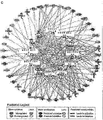
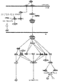
도 12는 중간엽 니치 세포와 비-니치 세포간 신호 기작을 분석한 것으로, EMT의 유도 관련 기작에 관한 것이다.
도 13a는 중간엽 니치 세포와 비-니치 세포간 신호 기작을 분석한 것으로, TGF-β와 EMT 관련 기작에 관한 것이다.
도 13b는 중간엽 니치 세포와 비-니치 세포간 신호 기작을 분석한 것으로, Wnt와 EMT 관련 기작에 관한 것이다.
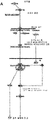
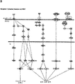
도 14a는 중간엽 니치 세포와 비-니치 세포간 신호 기작을 분석한 것으로, Notch와 EMT 관련 기작에 관한 것이다.
도 14b는 중간엽 니치 세포와 비-니치 세포간 신호 기작을 분석한 것으로, 티로신 카이네제 수용체와 EMT 관련 기작에 관한 것이다.
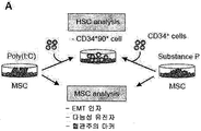
도 15a 내지 도 15h는 생리학적 자극에 의한 EMT 구배의 유도 실험에 관한 것으로서, (15a) 본 실험 과정의 개략적인 모식도; (15b) MSCs를 2 일간 폴리 (I:C) (50g/mL) 또는 substance-P (10 nM)로 전자극시킨(prestimulated) 후, 세척하고 3일간 CD34+ 세포와 공배양한 뒤, 무-기질 (stroma-free) 배양 군에 대한 조혈 재생 전구체(CD34+CD90+)의 배수 변화를 확인한 결과; 폴리 (I:C)이 처리된 MSCs의 (15c) EMT 인자 및 N-카데린의 발현, (15d) 다능성 유전자의 발현; (15e) 혈관주위세포 마커의 상대 발현을 확인한 결과; 및 substance-P이 처리된 MSCs의 (15f) EMT 인자 및 N-카데린의 발현, (15g) 다능성 유전자의 발현, 및 (15h) 혈관주위세포 마커의 상대 발현을 확인한 결과이다.
도 16은 생리학적 자극에 의한 EMT 구배의 유도 실험에 관한 것으로서, (A) 본 실험 과정의 개략적인 모식도; 및 니치 활성을 유발하는 다른 혈청 배치 (자극성 또는 비자극성)에서 배양된 MSCs에 대하여, (B) EMT 인자 및 N-카데린 활성, (C) 다능성 유전자 (D) 혈관주위세포 마커의 발현의 변화를 확인한 결과이다.
도 17은 정상적인 공여자 또는 AML이 있는 환자에서 유래된 MSCs에서의 EMT 구배 변화를 확인학 위한 실험 과정의 개략적인 모식도이다.
도 18은 정상적인 MSCs 에 대한 AML-유래 MSCs의 (A) EMT 인자 및 (B) N-카데린 발현의 배수 변화를 확인한 결과이다.
도 19는 MSCs의 EMT 구배에 대한 백혈병 아세포의 영향을 확인하기 위한 실험 과정의 개략적인 모식도이다.
도 20a 내지 도 20e는 (20a) 모든 전사체(검은색)의 차이와 비교하여 정상적인 CD34+군에 대한 백혈병 CD34+군 내 dbEMT에 매칭되는 DEGs의 발현 배수 차이를 나타낸 결과(노란색); 정상적인 CD34+군에 대한 백혈병 CD34+군의 (20b) 마스터 조절 EMT 인자 (20c) N-카데린 및 (20d) 다능성 유전자 발현의 배수 변화를 확인한 결과; 및 (20e) 비-니치 세포 (Nestin-PDGFR-) (파란색)에 대해 니치 세포(Nestin+PDGFR+)내 동일 유전자의 배수 차이와 비교하여, 정상적인 CD34+군에 대한 백혈병 CD34+군 내 혈관주위세포 마커 발현의 배수 변화를 확인한 결과이다(붉은색).
도 21은 니치 활성 등의 주요 조절자로서 EMT 구배의 효과를 개략적으로 나타낸 모식도이다.
이하, 본 발명을 상세히 설명하기로 한다.
본 발명은 상피-간엽이행(epithelial-mesenchymal transition; EMT) 촉진 인자를 포함하는, 중간엽 줄기세포의 니치 활성 증진용 조성물을 제공한다.
본 발명에서 사용되는 용어, 중간엽 줄기세포(mesenchymal stem cell; MSC)는 연골, 뼈, 지방, 골수간질, 근육, 신경 등을 만드는데 원조가 되는 세포로서, 성인에서는 일반적으로 골수에 머물러 있지만 제대혈, 말초혈액, 기타 조직 등에도 존재하며, 이들로부터 수득할 수 있는 세포를 의미한다. 본 명세서에서는, 중간엽 줄기세포는 중간엽 기질세포 또는 기질세포와 같은 의미로 사용된다. 상기 중간엽 줄기세포는 인간(human), 원숭이(monkey), 돼지(pigs), 말(horses), 소(cows), 양(sheep), 개(dogs), 고양이(cats), 생쥐(mice), 토끼(rats) 등의 모든 동물 유래의 세포를 포함하나, 바람직하게는 인간 유래의 세포일 수 있다.
본 발명에서 사용되는 용어, 니치(Niche)는 줄기세포 및 그 외 체세포와 같은 조직세포의 발달 및 증식을 지원하는 조직 또는 기관으로 구성된 구성요소(세포 및/또는 물질)를 의미하며, 세포간 상호작용을 유발하고 전능성을 갖기 위하여 필요한 인자들을 분비하는 것으로 알려져 있다. 니치는 소위 미세환경(microenvironment)이라고도 말하며, 줄기세포의 모든 특징을 표현하는 줄기세포성(stemness)을 유지하는데 있어 핵심적인 역할을 한다. 줄기세포는 점착성 분자 성장 요소(adhesion molecules growth factors)로 이루어진 일종의 미세환경에 자리 잡고 있는바, 학계에서는 이를 니치라고 명명하며, 줄기세포의 이러한 영역은 입지(location), 점착성(adhesiveness), 귀소성(homing), 휴면성(quiescence), 활동성 (activation)을 유지하고 조정하는 역할을 하고 있다. 즉, 니치는 줄기세포의 분화를 조정하며, 다른 곳으로의 이동 혹은 세포 자살을 막고 보호해주는 역할을 하는 줄기세포를 둘러싼 주요한 미세환경이라고 볼 수 있다. 본 발명에 따른 조성물은 이러한 니치 활성을 향상시킴으로써, 체내 또는 체외 존재하는 모든 종류의 줄기세포의 미분화능을 유지하고 자가 재생능을 촉진시키는 것을 목적으로 한다. 따라서, 본 발명의 조성물은 대상 줄기세포에 따라 다양한 타겟 질병에 적용 또는 응용될 수 있다. 특히, 줄기세포 니치 중 활발히 연구가 된 분야는 조혈모세포(조혈줄기세포) 니치로, 조혈모세포 니치는 조혈모세포 (조혈 줄기세포)가 거주하는 곳으로서, 골수세포의 일종인 조혈모세포는 모든 유형의 혈액 세포로 분화할 수 있는 대신, 자신의 니치 즉, 조혈모세포 니치를 벗어나면 이 같은 능력을 제대로 발휘하지 못하는 것으로 알려져 있다. 여타 다른 니치와 마찬가지로 상기 조혈모세포 니치는 단순히 조혈모세포를 위한 안식처일 뿐만 아니라 해당 줄기세포인 조혈모세포의 수까지도 조절하는 것으로 알려져 있다. 일례로서, 상기 니치 활성은 조혈 줄기세포의 미분화능을 유지하고, 자가 재생능을 촉진시키는 것을 의미할 수 있고, 이 경우, 본 발명의 조성물은 조혈 세포가 손상된 생리적 상태에 있는 환자를 대상으로 적용될 수 있으며, 상기 조혈 세포가 손상된 생리적 상태는 급성 백혈병, 만성 골수성 백혈병, 골수이형성 증후군, 림프종, 다발성 골수종, 생식세포 종양, 유방암, 난소암, 소세포폐암, 신경아세포종양, 재생불량성빈혈, 적혈구병증, 고셔씨병, 헌터 증후군, ADA 효소결핍증, Wiskot-Aldrich 증후군, 류마티스 관절염, 전신성 홍반성 낭창, 또는 다발성 경화증을 앓고 있거나, 항암제 투여 또는 방사선 조사로부터 야기되는 것 일 수 있다. 본 발명에서 사용되는 용어, "조혈줄기세포(hematopoietic stem cell: HSC)"는 적혈구, 백혈구, 혈소판 등의 혈액세포를 생산하는 미분화된 골수조혈세포의 조상세포로서, 골수가 파괴된 숙주에 이식하였을 때, 자가재생능(self-renewing capacity)을 가지고 장기간 재증식(repopulation)하는 능력을 보여준다. 상기 조혈 줄기세포는 인간(human), 원숭이(monkey), 돼지(pigs), 말(horses), 소(cows), 양(sheep), 개(dogs), 고양이(cats), 생쥐(mice), 토끼(rats) 등의 모든 동물 유래의 세포를 포함하나,바람직하게는 인간 유래의 세포일 수 있다.
본 발명에서 사용되는 용어, "자가 재생 (self-renewal)"은 동일한 성상 및 특징을 갖는 세포를 생산해내는 능력으로 자가복제, 자기복제, 자가재생이라고도 하며, 줄기세포가 가지는 중요한 특징의 하나이다. 특히, 본원 발명에서 자가 재생이란 분화되지 않은 상태를 유지하면서 증식을 계속하는 능력을 의미한다.
본 발명에서 사용되는 용어, 상피-간엽 이행(epithelial-mesenchymal transition; EMT)은 정상적인 배아 발달 과정에서 발생하는 현상으로, 세포가 상피성(epithelial) 세포 표현형을 상실하고 이동성이 높은 중간엽성(mesenchymal) 세포 표현형으로 전환하는 과정을 말한다. EMT가 일어나면 상피세포가 세포의 위쪽과 기저쪽이 구분되는 극성을 잃고 세포 모양이 정방형에서 섬유아세포형으로 바뀌고 상피 세포 마커는 감소하고 중간엽 세포 마커가 증가한다. 최근에 EMT가 태생기의 조직 형성과 분화 외에도 조직의 섬유화 등 다양한 역할을 한다는 것이 알려져 있다.
한편, 중간엽 니치는 골수 미세환경의 역학 변화에 반응 하는 조혈 줄기세포 내 재생 활성을 조절하는 것으로 알려져 왔으나, 이러한 과정은 매개하는 주요 인자에서 대해서는 알려져 있지 않은 상태이다. 본 발명에서는, 3D 세포간 상호작용 또는 생리학적 자극에 의해 니치 활성이 향상되는 경우, 상피-간엽 이행의 구배는 더욱 중간엽 상태로 변화하였고, 백혈병에 의한 퇴행성 상태에서는 이와 정반대의 경향성을 나타냄을 확인하였다. 따라서, 중간엽 니치 활성은 상피-간엽 이행의 변화를 통해 조절할 수 있으며, 구체적으로, 상피-간엽 이행 촉진 인자를 통해 중간엽 니치 활성을 향상시킬 수 있고, 상피-간엽 이행 억제 인자를 통해 중간엽 니치 활성을 저해시킬 수 있음을 규명하였다는 점에 기술적 특징이 있다.
상기 상피-간엽 이행 촉진 인자는 전술한 상기-간엽 이행의 현상을 증진시키는 인자들을 일컫는 것으로서, 천연물, 화합물, 뉴클레오타이드, 단백질, 재조합 벡터 등일 수 있으며, 바람직하게, miR-10b, miR-146a, miR-106b, miR-34a, miR-379, 및 miR-146b로부터 선택되는 miRNA 또는 snail, slug, twist1, zeb1, 및 zeb2로 이루어진 군으로 선택되는 어느 하나의 유전자를 포함하는 벡터일 수 있으나, 이에 제한되는 것은 아니다.
본 발명의 조성물에 포함되는 벡터는 상기 상피-간엽 이행 촉진 인자를 코딩하는 폴리뉴클레오티드 서열이 삽입된 발현 벡터를 말하며 이에 한정되지 않지만 바람직하게는 당업계에 공지된 플라스미드, 바이러스 또는 기타 매개체를 의미한다. 본 발명에 따른 폴리뉴클레오티드 서열은 발현 조절 서열에 작동 가능하게 연결될 수 있으며, 상기 작동 가능하게 연결된 유전자 서열과 발현 조절 서열은 선택 마커 및 복제 개시점(replication origin)을 같이 포함하고 있는 하나의 발현 벡터 내에 포함될 수 있다. "작동 가능하게 연결(operably linked)"된다는 것은 적절한 분자가 발현 조절 서열에 결합될 때 유전자 발현을 가능하게 하는 방식으로 연결된 유전자 및 발현 조절 서열일 수 있다. 상기 발현 조절 서열(expression control sequence)"이란 특정한 숙주 세포에서 작동 가능하게 연결된 폴리뉴클레오티드 서열의 발현을 조절하는 DNA 서열을 의미한다. 그러한 조절 서열은 전사를 실시하기 위한 프로모터, 전사를 조절하기 위한 임의의 오퍼레이터 서열, 적합한 mRNA 리보좀 결합 부위를 코딩하는 서열 및 전사 및 해독의 종결을 조절하는 서열을 포함한다.
상기 플라스미드의 예로는 대장균 유래 플라스미드(pBR322, pBR325, pUC118 및 pUC119), 바실러스 서브틸러스 유래 플라스미드(pUB110 및 pTP5) 및 효모 유래 플라스미드(YEp13, YEp24 및 YCp50) 등이 있으며, 상기 바이러스는 레트로바이러스, 아데노바이러스, 백시니아 바이러스, 배큘로바이러스, 헤르페스 심플렉스 바이러스 및 렌티바이러스와의 복합체 등으로 구성된 군으로부터 선택된 바이러스가 사용될 수 있다. 당업자라면 본 발명의 유전자 또는 폴리뉴클레오티드를 숙주세포에 도입시키는데 적합한 벡터를 사용할 수 있으며 바람직하게는 단백질 발현 유도 및 발현된 단백질의 분리가 용이하도록 디자인된 벡터를 사용할 수 있다.
본 발명의 다른 양태로서, 중간엽 줄기세포의 상피-간엽이행 (epithelial-mesenchymal transition; EMT)을 촉진하는 단계를 포함하는, 중간엽 줄기세포의 니치 활성 증진방법을 제공한다.
본 발명의 중간엽 줄기세포의 니치 활성 증진방법은 전술한 중간엽 줄기세포의 니치 활성 증진용 조성물에서 기술한 바 있는 상피-간엽 이행 촉진 인자 등을 이용하기 때문에, 이 들 사이에 공통된 내용은 본 명세서의 과도한 복잡성을 피하기 위하여, 그 기재를 생략한다.
상기 상피-간엽이행을 촉진하는 단계는 바람직하게, miR-10b, miR-146a, miR-106b, miR-34a, miR-379, 및 miR-146b로부터 선택되는 어느 하나를 중간엽 줄기세포에 처리하거나, snail, slug, twist1, zeb1, 및 zeb2로 이루어진 군으로 선택되는 어느 하나의 유전자를 포함하는 벡터를 중간엽 줄기세포에 도입하는 것일 수 있으나, 이에 제한되는 것은 아니다.
본 발명에 따른 상기 벡터는 당업계에 공지된 방법을 사용하여 세포에 도입할 수 있는데, 상기 본 발명에 따른 재조합 벡터를 숙주 세포에 도입하는 방법으로는 이에 한정되지는 않으나, 염화칼슘(CaCl2) 및 열 쇼크(heat shock) 방법, 실리콘 탄화물 위스커(Silicon carbide whiskers), 초음파 처리(sonication), PEG(Polyethylenglycol)에 의한 침전법, 일시적 형질감염(transient transfection), 미세주사, 형질도입(transduction), 세포융합, 칼슘 포스페이트 침전법, 리포좀 매개된 형질감염(liposome-mediated transfection), DEAE 덱스트란-매개된 형질감염(DEAE Dextran-mediated transfection), 폴리브렌-매개된 형질감염(polybrene-mediated transfection), 전기침공법(electropora tion), 유전자 총(gene gun) 방법 등을 사용할 수 있으며, 세포 내로 핵산을 유입시키기 위한 다른 공지의 방법에 의해 세포 내로 도입할 수 있다
본 발명의 또 다른 양태로서, 상피-간엽 이행 촉진 인자의 발현 또는 활성을 향상시키거나, 상피-간엽 이행 저해 인자의 발현 또는 활성을 저해하는 후보물질을 중간엽 줄기세포의 니치 활성 증진용 조성물로 선택하는 단계를 포함하는, 중간엽 줄기세포의 니치 활성 증진용 조성물의 스크리닝 방법을 제공한다.
본 발명의 중간엽 줄기세포의 니치 활성 증진용 조성물의 스크리닝 방법은 전술한 중간엽 줄기세포의 니치 활성 증진용 조성물에서 기술한 바 있는, 상피-간엽 이행 촉진 인자 등을 이용하기 때문에, 이 들 사이에 공통된 내용은 본 명세서의 과도한 복잡성을 피하기 위하여, 그 기재를 생략한다.
본 발명에서 사용되는 용어, 발현은 유전자가 가지고 있는 정보를 프로그램에 따라 RNA나 단백질의 기능을 가진 유전자 산물의 형태로 표현하는 것을 의미한다.
본 발명에서, 후보물질은 천연물, 화합물, 뉴클레오타이드, 단백질, 재조합 벡터 등일 수 있으나, 이에 제한되는 것은 아니다.
본 발명의 스크리닝 방법에 있어서, 후보물질 또는 관련 단백질 (EMT 촉진 또는 억제 관련 단백질)은 검출가능한 표지(detectable label)로 레이블링 될 수 있다. 예를 들어, 상기 검출가능한 표지(detectable label)는, 화학적 표지(예컨대, 바이오틴), 효소 표지(예컨대, 호스래디쉬 퍼옥시다아제, 알칼린 포스파타아제, 퍼옥시다아제, 루시퍼라아제, 纖갈락토시다아제 및 纖글루코시다아제), 방사능 표지(예컨대, C14, , I125, , P32 및 S35), 형광 표지[예컨대, 쿠마린, 플루오레세인, FITC(fluoresein Isothiocyanate), 로다민 6G(rhodamine 6G), 로다민 B(rhodamine B), TAMRA(6-carboxytetramethyl-rhodamine), Cy-3, Cy-5, Texas Red, Alexa Fluor, DAPI(4,6-diamidino-2-phenylindole), HEX,TET, Dabsyl 및 FAM], 발광 표지, 화학발광(chemiluminescent) 표지, FRET(fluorescence resonance energy transfer) 표지 또는 금속 표지(예컨대, 금 및 은)이다.
본 발명에 따른 상피-간엽 이행 촉진 인자 또는 상피-간엽 이행 저해 인자의 발현 또는 활성을 측정하는 단계는 유전자의 발현에 의해 생성된 RNA 또는 단백질의 양을 측정할 수 있는 당업계에 공지된 다양한 방법을 통해 실시될 수 있다. 바람직하게 RNA의 발현량은 실시간 중합효소연쇄반응(Real-time PCR), 마이크로어레이(microarray), 노던 블롯팅(northern blotting) 등을 통해 측정할 수 있고, 단백질의 발현량은 웨스턴 블롯팅 (western blotting), 유체분석(flow cytometry), 효소면역분석법(ELISA), 또는 면역염색법(immunofluorescnce) 등을 통해 측정할 수 있으며, 이 밖에도 당업계에 널리 알려진 단백질의 발현 또는 활성 측정법이 적용될 수 있다.
본 발명의 또 다른 양태로서, 본 발명은 상피-간엽이행(epithelial-mesenchymal transition; EMT)이 저해된 중간엽 줄기세포를 포함하는, 암 전이 억제용 약학적 조성물; 상피-간엽이행이 저해된 중간엽 줄기세포을 개체에 투여하는 단계를 포함하는 암 전이 억제방법; 및 암 전이 억제를 위한, 상피-간엽이행이 저해된 중간엽 줄기세포의 용도를 제공한다.
본 발명에서 사용되는 용어, 암은 세포가 정상적인 성장 한계를 무시하고 분열 및 성장하는 공격적(aggressive) 특성, 주위 조직에 침투하는 침투적(invasive) 특성 및 체내의 다른 부위로 퍼지는 전이적(metastatic) 특성을 갖는 세포에 의한 질병을 총칭하는 의미이다. 본 발명에서 암의 종류로는 췌장암, 위암, 간암, 대장암, 뇌암, 유방암, 갑상선암, 방광암, 식도암, 자궁암, 및 폐암으로 구성된 군으로부터 선택되는 어느 하나에 해당할 수 있으나, 이에 제한되는 것은 아니다.
본 발명에서는, 중간엽 줄기세포의 상피-간엽 이행의 구배는 암 줄기세포의 줄기성, 즉 전이에도 직접적인 영향을 미치는 것을 확인하였다. 따라서, 중간엽 줄기세포에 대한 상피-간엽 이행의 저해를 통해 암의 전이를 효과적으로 억제시킬 수 있음을 규명하였다는 점에 기술적 특징이 있다.
상기 상피-간엽 이행 억제 인자는 전술한 상기-간엽 이행의 현상을 억제시키는 인자들을 일컫는 것으로서, 천연물, 화합물, 뉴클레오타이드, 단백질, 재조합 벡터 등일 수 있으며, 바람직하게, miR-503, miR-145, miR- 193b, miR-365a, miR-29b, miR-129, miR-335, 및 miR-675로 이루어진 군으로 선택되는 어느 하나일 수 있으나, 이에 제한되는 것은 아니다.
상기 중간엽 줄기세포는, miR-503, miR-145, miR- 193b, miR-365a, miR-29b, miR-129, miR-335, 및 miR-675로부터 선택되는 어느 하나가 처리되거나, Snail1, Snail2, Zeb1, Zeb2, 및 Twist1로 이루어진 군으로부터 선택되는 어느 하나의 siRNA 유전자를 포함하는 벡터로 형질감염된 것일 수 있다.
이하, 본 발명의 이해를 돕기 위하여 바람직한 실시예를 제시한다. 그러나 하기의 실시예는 본 발명을 보다 쉽게 이해하기 위하여 제공되는 것일 뿐, 하기 실시예에 의해 본 발명의 내용이 한정되는 것은 아니다.
실시예
1. 실험재료 및 실험준비
1-1. 세포의 분리 및 배양
정상적인 공여자 또는 AML 환자에서 얻은 인간 BM, AML 환자의 백혈병증, 제대혈, 및 지방 조직은 한국 카톨릭대 임상시험심사위원회(institutional review board)의 승인 후 고지에 입각한 공여자의 동의(informed consent)를 얻었다. 정상적인 공여자 또는 AML 환자로부터 얻은 MSCs는 플라스틱 접시에 대한 접착 또는 부유를 통해 분리하였다. 3D 메센스피어를 24 시간 동안 3%(w/v) 소 혈청 알부민으로 코팅된 PDMS-기반 오목 마이크로칩 상에서 생성시킨 후, 저-접착 접시에 현탁하였다(Corning, Lowell, MA, USA). 모든 실험은 헬싱키 선언에 따라 진행하였다.
1-2. 정상적인 공여자 또는 백혈병
환자에서 얻은
CD34+ 세포의 시험관 내 증식
CD34+ 세포를 상술한 바와 같이 제대혈 또는 AML 환자에 대한 백혈구 분리반출술(leukapheresis)로부터 정제하고, 사이토카인 혼합물(100ng/mL 인간 기질 세포 인자 [SCF]; 100ng/mL 인간 Flt3L; 및 20ng/mL 인간 인터루킨 [IL]-3, IL-6, 및 G-CSF; ProSpec-Tany TechnoGene Ltd., Rehovot, Israel)의 존재 하에 장기 배양 배지 (H5100, STEMCELL Technologies, Vancouver, Canada)에서 5일간 조사된(1,500 cGy) 메센스피어 또는 2D MSCs와 공배양시켰다. 선택적으로, miRNA-형질감염된 MSCs과 공배양하기 위해 낮은 사이토카인 믹스(hSCF: 1μg/mL; hFlt3L: 1μg/mL; hTPO: 0.5μg/mL)를 사용하였다.
1-3. 전체-
전사체
및
miRNA
서열분석
MSCs를 전체-전사체 서열분석(RNA-seq)하였다. TopHat를 사용해 짝지은 서열분석 리드 (reads)를 참조 인간 게놈 서열(hg19) 상에 정렬하였다. CuffLink 소프트웨어를 사용해 유전자 발현 (매핑된 백만 개 단편 당 엑손(exon) 킬로베이스(kilobase) 당 단편[FPKM])을 평가하였다. 기능적 해석을 위해, 배수 변화의 log2 (FPKM3D/FPKM2D)를 사용해 GSEA의 우선순위가 있는 데이터세트를 분석하였다. miRNA 서열분석을 위해, TruSeq 스몰 RNA 샘플 프렙 키트 버전 2(Illumina, San Diego, CA, USA)를 사용해 1g의 RNA로 작은 RNA 서열분석 라이브러리를 생성하였다. 베이스-호출 및 FASTQ 포맷에서 원시적인, 역다중화된 서열분석 데이터의 생성을 위해 Illumina HCS (version 1.4.8), RTA (version 1.12.4.2) 및 CASAVA (version 1.8.2) 소프트웨어를 사용하였다.
1-4. 마우스
이종이식편
분석
동물 실험은 카톨릭대 한국 실험동물운영 위원회 (animal care and use committee)에서 승인을 받았다. NOD/SCID-γc null 마우스는 Jackson Laboratory (Bar Harbor, ME, USA)에서 구매하였다. 총 5 × 10⁵의 MDA-MB-231 세포를 2D 또는 3D 조건 하에서 배양된 2 × 10⁵ MSCs를 포함 또는 포함하지 않은 유방 지방조직에서 피하 주입하였다. 35일째에 모든 마우스를 희생시켰다. 1차 종양 및 간을 고정하고, 고정된 조직의 절편을 H&E으로 염색하였다.
1-5.
miRNA
또는
siRNAs의
형질감염
miRNA 미믹, miRNA 저해제, 또는 siRNAs (AccuTarget 인간 miRNA)를 Bioneer Corp. (Daejeon, Korea)에서 구입하였다. 형질감염을 위해, BM-MSCs를 Opti-MEM 배양 배지 (Life Technology, Carlsbad, CA, USA)로 교체하고 Lipofectamine 2000 (Life Technology)를 사용해 100 nM miRNA 미메틱 (mimetics), miRNA 저해제, 또는 siRNA로 형질감염시켰다. 스크램블 (scrambled) miRNA가 음성 대조군으로 사용되었다. 사용된 올리고뉴클레오티드 서열은 보충 정보에 표시한다.
1-6.
크로마틴
접근성 시험
MSCs에서 크로마틴의
Oct4 유전자 프로모터에 대한 접근성을 제조사 지침에 따라 크로마틴 접근성 분석 키트를 사용해 뉴클리아제 보호 분석에 의해 분석하였다 (Epigentek, Farmingdale, NY, USA). 간략하게, 세포를 뉴클리아제가 함유 또는 비함유된 EpiQuik 크로마틴 버퍼에서 인큐베이션(incubation)하고, 게놈 DNA를 추출하였다. qPCR에 의해 크로마틴 접근성을 분석하고 상기 키트의 분석 툴을 사용해 분석하였다. 헤모글로빈 (HBB) 유전자는 상대 크로마틴 접근성 측정을 위한 접근할 수 없는 참조 (inaccessible reference)로 사용되었다.
1-7. 유전자 발현 분석
MSC 유전자 발현을 Illumina 비드칩 (Illumina, San Diego, CA) 어레이 혼성화 분석에 의해 마이크로어레이 데이터 (LIMMA) 방법을 위한 직선 모델 및 상술한 통계용 R-패키지을 사용해 분석하였다.
1-8. 질량 분광분석
질량 분광분석 후, 펩티드-스펙트럼 매치를 기준으로 확인된 단백질의 상대 존재비를 산정하였다. G-값을 계산해 차등 발현된 단백질 (DEPs) 결정하고, 3.841 이상의 값을 가진 단백질을 2-분포에 따라 P < 0.05하여 유의한 것으로 간주하였다. Ingenuity Pathway 분석 (IPA; http://www.ingenuity.com)을 사용해 DEPs의 Gene ontology 분석을 실시하였다.
1-9. 통계 분석
유전자 세트 농축 분석을 위해, 유의한 GO 카테고리 (즉, 오판률 < 0.1)를 선별하기 위해 유의도 (significance)의 정상 P-값을 각 GO 카테고리에 대한 1,000 개의 유전자 순열을 대상으로 산정하였다. 모든 데이터를 SAS 버전 93 (SAS Institute Inc., Cary, NC, USA)을 사용해 분석하였으며, 유의도 레벨은 P < 0.05로 정하였다.
실시예
2. 3차원 상호작용을 통한
중간엽
세포의
니치
활성 획득
중간엽 니치 활성의 특성을 조사하기 위해, 먼저, 니치 기능을 보유한 Nestin-발현 중간엽 세포 아집단이 종종 비-부착 배양조건 하에 중간엽 구상체 (메센스피어)를 형성하는 능력으로 특징지어진다는 사실을 근거로 3D 메센스피어 모델을 채택하였다. 따라서, 메센스피어 형성 및 니치 활성 사이의 기능적 연관성을 조사하기 위해 니치 기능이 이들의 3D 세포 상호작용에 의해 유도될 수 있는지 여부를 확인하였다. 이에 따라, 세포는 비-부착성 3D 구상체의 형성을 위한 플랫폼인 폴리디메틸실록산 (PDMS) 마이크로칩 상에서 24 시간 이내에 응집시켜 균질한 구상체를 형성하였다. 흥미롭게도, 메센스피어 내 세포가 작을수록, 더 휴지되어, 이차원 (2D) 부착 배양시의 세포와 비교해, 유의하게 더 높은 콜로니-형성성, 골형성성, 및 지방생성성 (adipogenic) 잠재력을 나타내었으며 (도 1 및 도 2), 이는 이 전의 연구 결과와 일치하였다.
니치 기능에 대한 3D 상호작용의 영향을 확인하기 위해, 조혈 전구체 세포의 유지/증식을 유지(support)하는 메센스피어의 능력을 비교하였다 (도 3A). 특히, 2D 중간엽 기질 세포 (MSCs)와 공배양된 세포와 비교해, 메센스피어와 공배양된 CD34+ 세포는 콜로니-형성 전구체 및 CD34+CD90+ 서브세트에 있어 유의한 증가를 나타내었고, 이들 아집단 (subpopulations)은 장기 (long-term) 재생 조혈 세포를 나타내고, 이것으로 HSC 자기-복제/유지를 지지하는 메센스피어의 능력이 향상되었음을 입증하였다 (도 3B 내지 D). 표현형 마커 발현에 대해 검토하면, 2D MSCs와 비교해, 3D 메센스피어에서 각각 혈관주위세포 또는 원시 중간엽 세포에 특이적인 니치 마커인 Nestin 및 Prx1의 발현이 유의하게 증가하였음을 보여주었다 (도 3E). 그러나, 정규 (canonical) MSC 마커 또는 CD49f 또는 PODXL와 같은 초기 계대 (passage) MSCs에 대한 마커에서는 어떠한 유의한 변화도 관찰되지 않았으며 (도 3F), 이는 특정 니치 마커는 니치 활성이 증가되는 과정에서 획득될 수 있음을 나타낸다. 메센스피어가 다른 종류의 줄기성 (stemness)에도 유사한 효과를 발휘할 수 있는지 확인하기 위해, 암 줄기세포 내 줄기성에 대한 MSCs의 영향에 관한 이전의 연구를 기반으로 암 세포 내 줄기성에 대한 효과와 비교하였다 (도 4A). 이를 위해, 메센스피어와 공배양된 MDA-MB-231 유방 암 세포의 침투력 (invasion capacity)을 2D MSCs와 비교하였다. 예상과 같이, 더 높은 생체 내 전이 가능성을 나타내는, 뚜렷한 침투 가능성의 증가가 메센스피어와 공배양된 세포에서 관찰되었다 (도 4B). 이러한 결과를 확인하기 위해, NOD/SCID-γc 마우스 세포에 메센스피어 또는 2D MSCs와 함께 이식된 유방 암 세포의 전이를 비교하였다. 중간엽 세포 타입 중 어느 것으로 공-이식하든지 (2D 또는 3D), 1차 종양의 부피를 증가시켰다 (도 4C); 그러나, 메센스피어와 함께 이식된 마우스의 간에서 유의하게 더 많은 전이소가 발견되었으며 (도 4D 및 4E), 이는 메센스피어가 암 줄기세포 내 줄기성도 향상시킬 수 있음을 나타낸다.
전사 수준에서 니치 활성을 특성화하기 위해, 메센스피어 및 2D MSCs 내 RNA-서열분석 프로파일을 검토하여, 가장 높은 배수 변화를 보이는 상위 50개 및 하위 50개의 유전자를 차등 발현 유전자로 분류하였으며, 그 결과는 하기 표 1에 나타내었다(DEGs).
[표 1]

또한, 유전자 세트 농축 분석 (GSEA)으로 다섯 및 아홉 유전자 세트 (유전자 온톨로지 (ontology) [GO] 용어)가, 2D MSCs와 비교해, 메센스피어에서 유의하게 농축되어 (오판율 (false discovery rate) [FDR] < 0.2) DEGs가 되었음이 확인되었다(표 2).
[표 2]

유의하게, 메센스피어에서 상향조절된 상기 유전자 세트는 주로 세포외 공간에서 케모카인 수용체 결합, 케모카인 활성, G-단백질 결합된 성장인자 신호전달, 및 분비 인자를 포함하는 사이토카인 반응과 관련이 있으며 (도 5A 및 5B), 이는 전사 수준에서의 향상된 니치 활성이라는 결과를 뒷받침한다. 이와 함께, 이러한 결과는 중간엽 세포의 3D 세포 상호작용이 이들의 니치 활성을 향상시켜, 니치 기능의 유도성향을 제시함을 보여주며, 이때 니치의 표현형 및 활성은 외인성 조절 신호 (cues)에 반응하여 획득할 수 있다.
실시예
3.
니치
활성과
상피간엽이행
구배간
관련성 확인
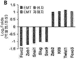
다음으로, 분자 수준에서의 메센스피어 내 향상된 니치 활성을 특성화하고자 하였다. 혈관주위 니치 세포가 분화 중 신경능에서 유래하였다는 결과에 기인하여, EMT 이질성이 출생 후 기간 (postnatal period) 중 니치 세포의 기능 성숙에 영향을 미칠 수 있는지 여부를 검토하였다. 따라서, EMT-조절 인자의 발현에 대해 메센스피어와 2D MSCs를 비교하여 메센스피어에서 Snai1, Twist, Zeb1, and Zeb2와 같은 EMT 조절인자(regulators)가 유의하게 더 많이 발현됨을 확인하였다 (도 6A).
다음으로, EMT 조절에서 microRNAs (miRNAs)의 결정적 역할에 관한 연구를 기반으로, 메센스피어 및 2D MSCs의 miRNA 발현 프로파일을 비교하여 메센스피어 내의 상위 50개의 상향조절 및 상위 50개의 하향조절된 miRNAs (log2 배수 변화 ≥ 2 or ≤ -2)를 요약하였다(표 3).
[표 3]

흥미롭게도, 현저한 발현 변화를 보여주는 많은 miRNAs는 EMT를 조절하는 것으로 알려져 있다. 예를 들어, EMT의 주요 유도물질(inducer)인 miR-146b는 메센스피어 내 매우 크게 유도된 miRNAs중 하나이다(89-배 증가; 도 6B). 이와 유사하게, EMT 유도물질 miR-379, miR-34a, miR-106b, miR-146a, 및 miR-10b 또한 메센스피어 내에서 높이 유도되어 있다. 이와 반대로, 메센스피어 내 miR-503, miR-145, miR- 193b, miR-365a, miR-29b, miR-129, miR-335, 및 miR-675를 포함하는 많은 하향조절된 miRNAs는 EMT를 저해하는 것으로 확인되어 왔다 (도 6B). 따라서, miRNA 발현 프로파일의 차이에 의해 반영된 세포 운명의 변화는, 나아가 2D 부착 MSCs와 비교해 메센스피어에서 더욱 더 EMT 진행이 이루어짐을 나타낸다. 한편, 전술한 miRNA들에 대한 서열 정보는 하기 표 4에 도시한 문헌들로부터 수득하였다.
[표 4]

이러한 결과를 기반으로, 이러한 현상이 단백질 수준에서도 비교 질량 분광분석에 의해 관찰될 수 있는지를 검토하였다. 차등 발현된 단백질(G-value > 3.841)의 경로 분석 (IPA, Ingenuity Systems, www.ingenuity.com)으로 EMT에 관련된 10개의 기능적 어노테이션 (annotations)을 확인하였다. 확인된 바와 같이, EMT-관련 기능을 활성화하기 위한 어노테이션 (pro-중간엽)은 주로 상향조절되는 반면, EMT 기능을 저해하기 위한 어노테이션 (anti-중간엽)은 하향조절되었다 (도 6C). 게다가, EMT-관련 단백질 중에서, 메센스피어에서 상향조절된 모든 단백질 (19개 단백질)은 pro-중간엽 단백질이며, 이에 반해 3D 메센스피어에서 하향조절된 모든 단백질 (10개의 단백질)은 anti-중간엽 단백질이므로, 단백질체 수준에서 EMT 구배를 나타내었다 (도 6D).
전체적으로, 이러한 결과는 중간엽 세포 내 3D 상호작용에 의해 향상된 니치 활성의 획득이, 추가적인 EMT 진행과 연관되어 중간엽 세포의 표현형적 유사 집단간 EMT 구배에 있어 계층 구조를 형성함을 나타낸다.
실시예
4.
상피간엽이행
구배와
후성유전적
가소성
차이간
관련성 확인
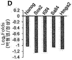
다음으로, 중간엽 세포 내 EMT 구배의 이질성에 대한 분자적 근거를 결정하고자 하였다. EMT가 크로마틴 구성 및 줄기세포-유사 특성의 후성유전적 제어에 의해 크게 조절되므로, 후성유전적 제어 시, 유사 가소성이 메센스피어 내 다른 EMT 구배를 형성할 수 있을 것으로 추측하였다. 이러한 가능성을 뒷받침하는 것으로, 2D MSCs와 비교해, 메센스피어에서는 다능성-관련 유전자 Oct4, Nanog, 및 Sox2
(36) 및 Hmga1
와 같은 줄기성-관련 유전자가 더 많이 발현되었다 (도 7A 및 7B). 메센스피어 내 다능성 유전자의 유도 역시 증가된 Oct4 프로모터 영역으로의 뉴클리아제 접근성에 의해 추론된 바와 같이 크로마틴 역학의 증가 (도 7C) 및 SMARCA 및 CHD1와 같은 크로마틴 리모델링 유전자의 유의한 유도 (도 7D)와 연관되어 있었다.
게다가, 크로마틴 표시 및 히스톤-수식 효소에 대해 메센스피어 또는 2D MSCs를 비교할 때, 메센스피어에서 H3K4, H3K36, H3K79, 및 H3K27 트리메틸화를 포함하는 활성화된 크로마틴 표시가 증가되었다 (도 7E). 흥미롭게도, 메센스피어는 또한 증가된 KMT2B, KMT2C, KMT2E, KMT2F, 및 KMT2H을 포함하는 히스톤 메틸트랜스퍼라제 (H3K4 메틸화를 위해); KMT1E 및 KMT1F (H3K9 메틸화를 위해); KMT6B (H3K27 메틸화를 위해); KMT3A (H3K36 메틸화를 위해); 및 KMT4 (H3K79 메틸화를 위해)의 발현을 보여주었으나, KDM5A (H3K4 탈메틸화를 위해), KDM3A 및 KDM3C (H3K9 탈메틸화를 위해), KDM6B (H3K27 탈메틸화를 위해), 및 KDM4B (H3K36 탈메틸화를 위해)와 같은 중화된 히스톤 디메틸라제의 발현의 동시 증가를 나타내었다 (도 7F). 그러므로, 이러한 정보는 후성유전적 히스톤 수식의 역학적 전환전환을 보여주었다. 이러한 후성유전적 가소성과 일치해, 히스톤 디아세틸라제 (HDAC) 패밀리는 전사체 분석 (transcriptomic analysis)시 메센스피어에서 뚜렷하게 하향조절된,
HDAC8, HDAC9, HDAC10, 및 HDAC11로 구성된 것을 포함하는 GO 서브세트에 포함되었다 (도 8A 및 8B).
이를 종합하면, 이러한 결과는 3D 중간엽 세포 상호작용이 역학적 크로마틴 리모델링 및 증가된 히스톤 수식 전환에 의해 특성화된 메센스피어 내 줄기세포-유사 특성을 유도하고, 줄기세포 상태의 후성유전적 가소성을 모방하여, EMT 축 내 세포의 추가적 전이가 용이해져, 표현형적으로 유사한 중간엽 세포 중 EMT 구배에 있어 계층 구조를 형성함을 제시하였다.
실시예
5.상피간엽이행
구배에
따른
니치
활성 변화 확인
EMT 구배와 향상된 니치 활성의 연관성을 관찰한 결과, 다음으로 중간엽 세포의 니치 활성에 대한 EMT 구배의 영향에 대해 탐구하였다 (도 9A). 이를 위해, 메센스피어 내에서와 같이, EMT 구배를 향상시킬 수 있는 miRNA 미믹, 즉, EMT-촉진 miRNAs (miR-140b, miR-379)을 모방한 미믹 및 EMT-저해 miRNAs (miR-503, miR-145-5p, miR-193b, miR-36t5a, miR-29b, 및 miR-129)을 저해하는 미믹으로 2D MSCs를 형질감염시켰다. 예상된 바와 같이, 상기 miRNA-형질감염된 2D MSCs는 Snail1 및 Twist1와 같은 EMT-촉진 유전자의 유의한 유도를 보여주었으며, 이는 세포에서 EMT가 진행됨을 나타낸다 (도 9B). 흥미롭게도, 상기 2D MSCs에서 EMT의 유도는 다능성-관련 유전자 Oct-4, Nanog, 및 Sox2 의 유의한 유도 및 대조군 MSCs보다 더 높은 클론원성 중간엽 전구체 (CFU-F) 빈도의 원인이 되었다 (도 9C, 및 9D). 게다가, EMT-기반 MSCs에서 대조군에서 보다 유의하게 더 높은 전구체 및 CD34+CD90+ 조혈 재생 세포의 증식이 관찰되어 (도 9E 및 9F), 메센스피어에서 관찰된 바와 같이 MSCs에서 EMT를 구동하는 것은, 활성화된 중간엽 니치의 특성을 재생함을 제시하였다.
이와 달리, EMT-조절 유전자 Snail1, Slug, Zeb1, 및 Twist를 타켓팅하는 siRNAs로 형질감염된 MSCs는, CD34+CD90+ 조혈 재생 세포 증식의 감소에 의해 입증된 바와 같이, EMT 인자 및 다능성-관련 유전자 발현의 유의한 감소 (도 10A 및 10B) 및 니치 활성의 감소를 나타내었다 (도 10C). 전체적으로, 이러한 결과는 중간엽 세포에서 EMT 구배가 이들의 니치 활성을 제어하는 주요한 인자임을 나타내었다.
실시예
6.생리적 자극에 의한
상피간엽이행
구배의
유도
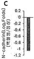
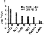
EMT 구배가 니치 활성을 제어함을 관찰한 결과, 다음으로 BM 미세환경의 생리학적 제어에 대한 관련성을 결정하고자 하였다. 먼저, 이들의 니치 기능에서의 이질성에 대한 EMT 구배 이질성 역시 BM 중간엽 세포 사이에 존재하는지 여부를 검토하였다. 이를 위해, Nestin 및 혈소판-유래 성장인자 (PDGFRα)를 발현하는 중간엽 세포 서브세트가 니치 세포로 작용된 마우스 모델 (Nestin-GFP 마우스) 및 이들 마커에 대해 음성인 모델을 채택하였다 (도 11A). 공공 RNA-seq 데이터베이스 및 dbEMT 각각을 사용한 비교 전사체 (transcriptome) 및 EMT 구배 분석은, 비-니치 세포 (Nestin-GFPPDGFRa; 도 11B)와 비교해 니치 세포 (Nestin-GFP+PDGFRa+)는 EMT-관련 유전자의 유의한 농축을 나타냄을 보여주었는데, 즉, dbEMT에 매치된 163개의 유전자 중 43개의 유전자는 니치 세포에서 유의하게 상향조절되었으나 (p < 0.05), 12개의 유전자는 하향조절되었다. 게다가, 니치 세포 역시 EMT-관련 유전자 (EMT-관련 유전자 및 전체 전사체 (transcriptome) 각각에 대해 log2 배수 변화 = 1.29 및 -0.28; p < 0.0001; 도 11C) 발현의 전반적 증가를 보여주었다. 뿐만 아니라, 이들 DEGs의 경로 분석 (IPA)은 EMT 신호전달이 비-니치 중간엽 세포와 비교해 니치 세포에서 유의하게 상향조절되었음을 보여주었다 (도 12 내지 도 14). 이와 유사하게, 마스터 EMT 조절성 인자로 확인된 유전자들이 N-카데린 발현의 유의한 증가와 함께 니치 세포에서 가장 상향조절되었으며 (도 11D 및 도 11E), 이는 EMT 구배의 변화를 나타낸다. 이러한 결과는 항상성 BM에서 중간엽 니치 능이 중간엽 서브세트간 EMT 구배의 계층구조에 의해 조절됨을 나타낸다.
다음으로, 생리학적 니치 제어를 위한 EMT 구배의 중요성을 더 조사하기 위해, 생리학적 자극-유도 니치 활성화 중 EMT 구배 변화를 검토하였다 (도 15A). 먼저, 앞서 HSC 증식을 유도하는 것으로 밝혀진 폴리 (I:C)-매개 IFNa 신호에 의한 자극을 검토하였다. 밝혀진 바와 같이, 폴리 (I:C)-전처리된 MSCs과 CD34+ 세포의 공배양은 대조군 MSCs에서 관찰된 세포 증식과 비교해 CD34+CD90+ 조혈 재생 세포 증식의 유의한 증가를 유도하였고 (도 15B), 이는 폴리 (I:C)-매개 HSC 자극 또한 중간엽 니치 활성화 모델로 작용함을 나타낸다. EMT에 대한 조사시, 폴리 (I:C)-처리된 MSCs는 마스터 EMT 조절인자 (regulators)인 Snail1, Slug, Zeb1, Zeb2, Twist (30, 31, 42), 및 N-카데린에서 유의한 상향조절을 보여주었다 (도 15C). 특히, 폴리 (I:C) 자극은 다능성 유전자 발현 (Oct4, Nanog, 및 Sox2) 및 혈관주위세포 마커 (Nestin, NG2, PDGFRb, 및 CD146)의 유사 유도를 유도하여, 3D 세포 상호작용에 의해 유도된 니치 활성화 공정을 모방한다 (도 15D 및 15E). 이와 유사하게, 조직 손상에 반응해 중간엽 세포의 재생 활성을 자극하는 substance-P에 의해 유도된 또 다른 니치 활성화 모델을 검토하였다. 이와 동일하게, substance-P로 MSCs를 전처리하는 것 역시 공배양 중 HSC 자기-복제에 의해 측정된 바와 같이 중간엽 니치 활성을 향상시켰으며 (도 15B), EMT 구배의 유사한 증가, 즉, EMT 인자 상향조절, 증가된 N-카데린 발현, 다능성-관련 유전자의 유도 (도 15F 및 15G), 및 혈관주위세포 마커 (도 15H)를 나타내었다.
세부적인 관찰을 위해, 다른 sera로 처리하여 유도된 니치 활성의 가역적 스위칭에 대한 최근 모델을 검토하였다 (도 16A). 특히, 니치 활성화를 위한 다른 자극에 의해 유도된 바와 같이, 더 높은 니치 활성을 초래하는 혈청 조건에 의해 유도된 유사한 일련의 변화가 관찰되었으며 (도 16B 내지 도 16D), 이는 자극의 종류에 상관없이 각 활성화 모델에 대한 세포적 변화가 보존되었음을 제시한다. 전체적으로, 이러한 결과는 EMT 구배의 계층 구조 생성이 니치 활성화를 일으키는 생리학적 자극에 반응하는 중간엽 세포에 보존된 통상의 세포적 발현임을 보여주었으며, 따라서 이는 니치 활성화 프로그램 재생 자극에 반응하여 중간엽 세포에서 일어난 반응성 기질 변화를 나타냄을 제시하였다.
실시예
7.병리학적
조건하에서의
상피간엽이행
구배
확인
상기 반응성 기질 변화를 관찰하여, 퇴행성 병리학적 조건 하에 EMT의 변화를 즉시 검토하였다. 최근 보고에서는 중간엽 니치가 백혈병 BM의 퇴행성 변화를 겪어 HSC가 손실되었음이 입증되었다. 따라서, 정상적 공여자 또는 급성 골수성 백혈병 (AML) 환자에서 유래된 MSCs간의 EMT 구배를 비교하였다. 특히, 정상적인 공여자의 MSCs와 비교해, AML 환자의 MSCs에서 EMT 인자 및 N-카데린의 유의한 하향조절이 관찰되었으며, 즉, 백혈병 조건 하에서는 원형적 (prototypical) 니치 활성화 프로그램과 정반대의 변화를 나타내었다 (도 17 및 도 18). 백혈병 니치에서의 이러한 뚜렷한 변화가 백혈병 세포에 의한 것인지를 직접적으로 조사하고자, 정상적인 공여자 또는 AML 환자에서 유래한 CD34+ 세포에 노출된 MSCs간의 전사체적 (transcriptomic) 변화를 비교하였다 (도 19). 놀랍게도, 생리학적 자극으로 관찰된 증가된 EMT 구배와 다르게, EMT-관련 유전자는 백혈병 미분화 세포에 노출 후 우세하게 하향조절되었다. 구체적으로, 정상적인 CD34+ 세포에 노출된 경우와 비교해, dbEMT에 매칭된 49개의 DEGs (p < 0.05)중에, 33개의 유전자는 유의하게 하향조절된 반면, 16개의 유전자는 백혈병 조건 하에 상향조절되었다 (도 20A). 게다가, EMT-유도 유전자의 빈번한 하향조절 및 감소된 N-카데린 발현도 관찰되었다 (도 20B 및 도 20C). 더욱이, 정상적인 니치 활성화 프로그램에서 이들 유전자의 증가와 극명하게 대조적으로, 줄기성 유전자 및 혈관주위세포 마커는 모두 백혈병 세포에 의해 하향조절되었다 (도 20D 및 20E). 이와 같이, 이러한 결과는 백혈병 조건이 중간엽 니치의 퇴행을 유발하는 EMT 구배가 손실되도록 EMT 구배를 변화시켰음을 제시하였다. 이를 종합하면, 이러한 결과는 EMT 구배의 생리학적 변화가 미세환경 내 재생 또는 퇴행성 신호 (cues)에 대해 그들의 니치 활성을 적응시키기 위해 중간엽 세포의 적응 기질 반응을 나타냄을 보여주는 것이다 (도 21).
본 연구에서는, 우선, 정상적인 조혈 전구체 및 암 줄기세포의 자기-복제 및 줄기성 촉진과 관련된 니치 활성을 유발하는, 3D 상호작용에 의해 유도된 일련의 세포적 변화를 확인하였다. 분자적 변화를 기반으로 검토한 결과, 가장 두드러진 변화는 EMT쪽으로의 전환으로서, EMT를 촉진하는 인자들은 3D 메센스피어에서 상향조절되는 반면, EMT를 저해하는 인자들은 하향조절되었다. 따라서, 중간엽 세포에서 생성된 EMT 구배는 향상된 니치 활성 및 암 줄기세포의 전이 억제와 높은 관계가 있음을 확인하였다. 흥미롭게도, 생리학적 자극에 의해 유도된 니치 활성화 동안 즉, 폴리 (I:C) 또는 물질 P 모델에서도, 니치 활성을 제어하는 메카니즘으로서 EMT 구배의 변화는 상기와 유사한 경향을 보였다. 또한, 고유의 혈청 효과에 의해 유도된 직접 자극 모델 (자극성 혈청 배지)에서도 중간엽 니치 활성화 중 EMT를 촉진하는 경향을 보여주었다. 이러한 결과들을 종합해 보면, 중간엽 세포의 EMT 구배는, 이들의 니치 활성을 증가시키기 위한 다양한 자극에 대한 중간엽 세포의 통상적인 반응임을 알 수 있었다.
흥미롭게도, 중간엽 니치 (niche) 활성화 중 EMT의 변화는 다능성-관련 유전자 발현의 유사한 증가 및 Nestin, PDGFR, NG2, 또는 CD146와 같은 혈관주위세포-특이적 마커의 획득을 동반하였다. 니치를 위한 혈관주위세포는 신경능에서 유래되었음을 볼 때, 이러한 결과는, 출생 후의 삶 동안 재생 자극에 반응한 기질 세포의 반응성 변화로서 요약되었다. 따라서, 니치 표현형의 획득은 EMT-구동 간엽 세포의 보존된 니치 활성화 프로그램으로 이용될 수 있다.
특히, 니치 활성의 조절을 위한 중간엽 세포의 EMT 구배는 정상 상태의 BM 뿐만 아니라, 병적 조건에 의해 유도된 병리학적 변화와도 연관되어 있었다. AML 환자의 MSCs에서, EMT 인자의 하향조절 및 N-카데린 발현에 의해 추론된 바와 같이, EMT 구배는 백혈병 조건 하에 정반대 방향으로 변화하였다. 다시 말해, EMT 인자 및 N-카데린이 하향조절되고 줄기성 유전자 및 혈관주위세포 마커의 양이 감소되었다. 이러한 결과는 재생을 위한 생리학적 자극 중에 촉발된 니치 활성화 프로그램은 퇴행성 조건 하에 정반대 방향으로 변화할 수 있으며, 이는 미세환경의 정상적 및 병리학적 변화시, EMT 구배의 중요성을 나타냄을 보여주었다.
전술한 본 발명의 설명은 예시를 위한 것이며, 본 발명이 속하는 기술분야의 통상의 지식을 가진 자는 본 발명의 기술적 사상이나 필수적인 특징을 변경하지 않고서 다른 구체적인 형태로 쉽게 변형이 가능하다는 것을 이해할 수 있을 것이다. 그러므로 이상에서 기술한 실시예들은 모든 면에서 예시적인 것이며 한정적이 아닌 것으로 이해해야만 한다.
Claims (11)
- 상피-간엽이행(epithelial-mesenchymal transition; EMT) 촉진 인자를 포함하는, 중간엽 줄기세포의 니치 활성 증진용 조성물.
- 제1항에 있어서, 상기 상피-간엽이행 촉진 인자는 miR-10b, miR-146a, miR-106b, miR-34a, miR-379, 및 miR-146b로부터 선택되는 것인, 중간엽 줄기세포의 니치 활성 증진용 조성물.
- 제1항에 있어서, 상기 상피-간엽이행 촉진 인자는 snail, slug, twist1, zeb1, 및 zeb2로 이루어진 군으로 선택되는 어느 하나의 유전자를 포함하는 벡터인 것인, 중간엽 줄기세포의 니치 활성 증진용 조성물.
- 제1항에 있어서, 상기 중간엽 줄기세포는 지방조직, 골수, 말초혈액 또는 제대혈에서 유래한 것인, 중간엽 줄기세포의 니치 활성 증진용 조성물.
- 중간엽 줄기세포의 상피-간엽이행(epithelial-mesenchymal transition; EMT)을 촉진하는 단계를 포함하는, 중간엽 줄기세포의 니치 활성 증진방법.
- 제5항에 있어서, 상기 상피-간엽이행을 촉진하는 단계는 miR-10b, miR-146a, miR-106b, miR-34a, miR-379, 및 miR-146b로부터 선택되는 어느 하나를 중간엽 줄기세포에 처리하는 것인, 중간엽 줄기세포의 니치 활성 증진방법.
- 제5항에 있어서, 상기 상피-간엽이행을 촉진하는 단계는 snail, slug, twist1, zeb1, 및 zeb2로 이루어진 군으로 선택되는 어느 하나의 유전자를 포함하는 벡터를 중간엽 줄기세포에 도입하는 것인, 중간엽 줄기세포의 니치 활성 증진방법.
- 상피-간엽이행(epithelial-mesenchymal transition; EMT)이 저해된 중간엽 줄기세포를 포함하는, 암 전이 억제용 약학적 조성물.
- 제8항에 있어서, 상기 중간엽 줄기세포는, miR-503, miR-145, miR-193b, miR-365a, miR-29b, miR-129, miR-335, 및 miR-675로부터 선택되는 어느 하나가 처리된 것인, 암 전이 억제용 약학적 조성물.
- 제8항에 있어서, 상기 중간엽 줄기세포는 Snail1, Snail2, Zeb1, Zeb2, 및 Twist1로 이루어진 군으로부터 선택되는 어느 하나의 siRNA 유전자를 포함하는 벡터로 형질감염된 것인, 암 전이 억제용 약학적 조성물.
- 제8항에 있어서, 상기 중간엽 줄기세포는 지방조직, 골수, 말초혈액 또는 제대혈에서 유래한 것인, 암 전이 억제용 약학적 조성물.
Applications Claiming Priority (2)
| Application Number | Priority Date | Filing Date | Title |
|---|---|---|---|
| KR10-2016-0042155 | 2016-04-06 | ||
| KR20160042155 | 2016-04-06 |
Publications (1)
| Publication Number | Publication Date |
|---|---|
| WO2017176049A1 true WO2017176049A1 (ko) | 2017-10-12 |
Family
ID=60001333
Family Applications (1)
| Application Number | Title | Priority Date | Filing Date |
|---|---|---|---|
| PCT/KR2017/003738 Ceased WO2017176049A1 (ko) | 2016-04-06 | 2017-04-05 | 3차원 중간엽 기질세포 구상체에서 상피-간엽이행 구배의 계층화 |
Country Status (2)
| Country | Link |
|---|---|
| KR (1) | KR102010346B1 (ko) |
| WO (1) | WO2017176049A1 (ko) |
Citations (1)
| Publication number | Priority date | Publication date | Assignee | Title |
|---|---|---|---|---|
| US20120021983A1 (en) * | 2009-04-02 | 2012-01-26 | President And Fellows Of Harvard College | Compositions and methods for diagnosis and prognosis of cancer and progression, and for screening anti-cancer agents |
-
2017
- 2017-04-05 WO PCT/KR2017/003738 patent/WO2017176049A1/ko not_active Ceased
- 2017-04-05 KR KR1020170044283A patent/KR102010346B1/ko active Active
Patent Citations (1)
| Publication number | Priority date | Publication date | Assignee | Title |
|---|---|---|---|---|
| US20120021983A1 (en) * | 2009-04-02 | 2012-01-26 | President And Fellows Of Harvard College | Compositions and methods for diagnosis and prognosis of cancer and progression, and for screening anti-cancer agents |
Non-Patent Citations (5)
| Title |
|---|
| A CONCISE REVIEW OF MOLECULAR PATHOLOGY OF BREAST CANCER: "LUQMANI, YUNUS A. et al", 25 March 2015, INTECH, article "MicroRNA in breast cancer - gene regulators and targets for novel therapies", pages: 79 - 120, XP055601376 * |
| CLARK, ELIZABETH A.: "Concise review: MicroRNA function in multipotent mesenchymal stromal cells", STEM CELLS, vol. 32, no. 5, 2014, pages 1074 - 1082, XP055205209, DOI: doi:10.1002/stem.1623 * |
| DEL POZO MARTIN, YAIZA: "Mesenchymal cancer cell -stroma crosstalk promotes niche activation, epithelial reversion, and metastatic colonization", CELL REPORTS, vol. 13, no. 11, 22 December 2015 (2015-12-22), pages 2456 - 2469, XP055601364 * |
| PEIN, MAREN: "Microenvironment in metastasis: roadblocks and supportive niches", AMERICAN JOURNAL OF PHYSIOLOGY CELL PHYSIOLOGY, vol. 309, no. 10, 2015, pages C627 - C638, XP055601385 * |
| RHEE, KI-JONG: "Mesenchymal stem cell -mediated effects of tumor support or suppression", INTERNATIONAL JOURNAL OF MOLECULAR SCIENCES, vol. 16, no. 12, 2015, pages 30015 - 30033, XP055601382 * |
Also Published As
| Publication number | Publication date |
|---|---|
| KR102010346B1 (ko) | 2019-08-14 |
| KR20170115445A (ko) | 2017-10-17 |
Similar Documents
| Publication | Publication Date | Title |
|---|---|---|
| Lynch et al. | Global hyperactivation of enhancers stabilizes human and mouse naive pluripotency through inhibition of CDK8/19 Mediator kinases | |
| Li et al. | Ground rules of the pluripotency gene regulatory network | |
| Sincennes et al. | Concise review: epigenetic regulation of myogenesis in health and disease | |
| Li et al. | miR‐10a restores human mesenchymal stem cell differentiation by repressing KLF4 | |
| Sørensen et al. | Promoter DNA methylation patterns of differentiated cells are largely programmed at the progenitor stage | |
| Xu et al. | Neural stemness contributes to cell tumorigenicity | |
| Kalgudde Gopal et al. | Wound infiltrating adipocytes are not myofibroblasts | |
| Zhu et al. | MiR‐221 and miR‐26b regulate chemotactic migration of MSCs toward HGF through activation of Akt and FAK | |
| Chen et al. | Role of micro RNA‐27a in myoblast differentiation | |
| Graham-Paquin et al. | ZMYM2 is essential for methylation of germline genes and active transposons in embryonic development | |
| Sagner et al. | Temporal patterning of the central nervous system by a shared transcription factor code | |
| Blanchart et al. | UHRF1 licensed self-renewal of active adult neural stem cells | |
| Mariani et al. | A TALE/HOX code unlocks WNT signalling response towards paraxial mesoderm | |
| Lan et al. | Gene and protein expression profiles of olfactory ensheathing cells from olfactory bulb versus olfactory mucosa | |
| Parsons | Human stem cell derivatives retain more open epigenomic landscape when derived from pluripotent cells than from tissues | |
| Xu et al. | Genome-wide CRISPR screen identifies Menin and SUZ12 as regulators of human developmental timing | |
| Pantier et al. | TET knockout cells transit between pluripotent states and exhibit precocious germline entry | |
| WO2015088162A1 (ko) | 줄기세포로부터 연골세포로의 분화 촉진용 조성물 | |
| Matsubara et al. | Epigenetic regulation of neural stem cell aging in the mouse hippocampus by Setd8 downregulation | |
| Militi et al. | RBL2-E2F-GCN5 guide cell fate decisions during tissue specification by regulating cell-cycle-dependent fluctuations of non-cell-autonomous signaling | |
| KR102010346B1 (ko) | 3차원 중간엽 기질세포 구상체에서 상피-간엽이행 구배의 계층화 | |
| US20240318134A1 (en) | In vitro generation of organized 3d cell structures including head-trunk embryo-like structures, using epigenetic remodeling factors-microfluidic platform suitable for their generation | |
| Govarthanan et al. | Epigenetic regulation− The guardian of cellular homeostasis and lineage commitment | |
| Södersten et al. | The novel BTB/POZ and zinc finger factor Zbtb45 is essential for proper glial differentiation of neural and oligodendrocyte progenitor cells | |
| EP4219685A1 (en) | In vitro generation of organized 3d cell structures including head-trunk embryo-like structures, using epigenetic remodeling factors - microfluidic platform suitable for their generation |
Legal Events
| Date | Code | Title | Description |
|---|---|---|---|
| NENP | Non-entry into the national phase |
Ref country code: DE |
|
| 121 | Ep: the epo has been informed by wipo that ep was designated in this application |
Ref document number: 17779349 Country of ref document: EP Kind code of ref document: A1 |
|
| 122 | Ep: pct application non-entry in european phase |
Ref document number: 17779349 Country of ref document: EP Kind code of ref document: A1 |