WO2017168726A1 - Anti-podocalyxin antibody that targets tumor microenvironment - Google Patents
Anti-podocalyxin antibody that targets tumor microenvironment Download PDFInfo
- Publication number
- WO2017168726A1 WO2017168726A1 PCT/JP2016/060822 JP2016060822W WO2017168726A1 WO 2017168726 A1 WO2017168726 A1 WO 2017168726A1 JP 2016060822 W JP2016060822 W JP 2016060822W WO 2017168726 A1 WO2017168726 A1 WO 2017168726A1
- Authority
- WO
- WIPO (PCT)
- Prior art keywords
- antibody
- cancer
- podocalyxin
- amino acid
- seq
- Prior art date
Links
Images


Classifications
-
- C—CHEMISTRY; METALLURGY
- C07—ORGANIC CHEMISTRY
- C07K—PEPTIDES
- C07K16/00—Immunoglobulins [IGs], e.g. monoclonal or polyclonal antibodies
- C07K16/18—Immunoglobulins [IGs], e.g. monoclonal or polyclonal antibodies against material from animals or humans
- C07K16/28—Immunoglobulins [IGs], e.g. monoclonal or polyclonal antibodies against material from animals or humans against receptors, cell surface antigens or cell surface determinants
- C07K16/2896—Immunoglobulins [IGs], e.g. monoclonal or polyclonal antibodies against material from animals or humans against receptors, cell surface antigens or cell surface determinants against molecules with a "CD"-designation, not provided for elsewhere
-
- A—HUMAN NECESSITIES
- A61—MEDICAL OR VETERINARY SCIENCE; HYGIENE
- A61K—PREPARATIONS FOR MEDICAL, DENTAL OR TOILETRY PURPOSES
- A61K39/00—Medicinal preparations containing antigens or antibodies
- A61K39/395—Antibodies; Immunoglobulins; Immune serum, e.g. antilymphocytic serum
-
- A—HUMAN NECESSITIES
- A61—MEDICAL OR VETERINARY SCIENCE; HYGIENE
- A61P—SPECIFIC THERAPEUTIC ACTIVITY OF CHEMICAL COMPOUNDS OR MEDICINAL PREPARATIONS
- A61P35/00—Antineoplastic agents
-
- C—CHEMISTRY; METALLURGY
- C07—ORGANIC CHEMISTRY
- C07K—PEPTIDES
- C07K16/00—Immunoglobulins [IGs], e.g. monoclonal or polyclonal antibodies
- C07K16/18—Immunoglobulins [IGs], e.g. monoclonal or polyclonal antibodies against material from animals or humans
- C07K16/32—Immunoglobulins [IGs], e.g. monoclonal or polyclonal antibodies against material from animals or humans against translation products of oncogenes
-
- C—CHEMISTRY; METALLURGY
- C12—BIOCHEMISTRY; BEER; SPIRITS; WINE; VINEGAR; MICROBIOLOGY; ENZYMOLOGY; MUTATION OR GENETIC ENGINEERING
- C12N—MICROORGANISMS OR ENZYMES; COMPOSITIONS THEREOF; PROPAGATING, PRESERVING, OR MAINTAINING MICROORGANISMS; MUTATION OR GENETIC ENGINEERING; CULTURE MEDIA
- C12N15/00—Mutation or genetic engineering; DNA or RNA concerning genetic engineering, vectors, e.g. plasmids, or their isolation, preparation or purification; Use of hosts therefor
- C12N15/09—Recombinant DNA-technology
-
- C—CHEMISTRY; METALLURGY
- C12—BIOCHEMISTRY; BEER; SPIRITS; WINE; VINEGAR; MICROBIOLOGY; ENZYMOLOGY; MUTATION OR GENETIC ENGINEERING
- C12N—MICROORGANISMS OR ENZYMES; COMPOSITIONS THEREOF; PROPAGATING, PRESERVING, OR MAINTAINING MICROORGANISMS; MUTATION OR GENETIC ENGINEERING; CULTURE MEDIA
- C12N5/00—Undifferentiated human, animal or plant cells, e.g. cell lines; Tissues; Cultivation or maintenance thereof; Culture media therefor
- C12N5/10—Cells modified by introduction of foreign genetic material
-
- C—CHEMISTRY; METALLURGY
- C07—ORGANIC CHEMISTRY
- C07K—PEPTIDES
- C07K2317/00—Immunoglobulins specific features
- C07K2317/40—Immunoglobulins specific features characterized by post-translational modification
- C07K2317/41—Glycosylation, sialylation, or fucosylation
Definitions
- the present invention relates to an anti-podocalyxin antibody targeting the cancer microenvironment.
- Podocalixin is a type I transmembrane protein found in renal glomerular epithelial cells (podocytes) (Non-patent Document 1).
- Podocalixin consists of 558 amino acids and has high homology with CD34, a hematopoietic stem cell marker.
- Podocalixin is highly glycosylated with an N-linked glycosylation site, glycosaminoglycan addition site in the extracellular region, and an O-linked glycosylation site (mucin domain) with a large amount of sialic acid added at the end. Sialomucin.
- Podocalixin is a glycoprotein with different molecular weights in the range of 150-200 kDa, because glycosylation varies depending on the expressed tissue. Podocalixin is involved in cell adhesion, morphogenesis, cancer progression, and the like.
- Podocalixin is negatively charged by addition of sugar chains such as sialic acid and sulfate groups, and inhibits cell adhesion.
- podocalyxin binds to cytoskeletal proteins and the like and is closely related to the filtration function of the kidney, and also functions as an adhesion molecule (Non-patent Document 2).
- Non-patent Document 3 podocalyxin is polar transported by the low molecular weight G protein Rab and its effector molecule, and is involved in lumen formation by its negative charge.
- podocalyxin is a testicular tumor (Non-patent document 4), breast cancer (Non-patent document 5), prostate cancer (Non-patent document 6), ovarian cancer (Non-patent document 7), and colon cancer (Non-patent document 8).
- pancreatic cancer Na-patent Document 9
- the sugar chain on podocalyxin expressed in cancer cells becomes a ligand for E-, P-, and L-selectin expressed on endothelial cells and is involved in adhesion, invasion, and metastasis of cancer cells (Non-patent Document 9). 10).
- TRA-1-60 and TRA-1-81 which are markers for undifferentiated cells, are antibodies whose epitopes are keratan sulfate on podocalyxin. It has been reported that when differentiation is induced, reactivity to podocalyxin disappears (Non-patent Document 11).
- BC2L-C a lectin purified from Burkholderia cenocepacia, is an undifferentiated cell marker, but specifically binds to O-type sugar chains on podocalyxin (Non-patent Document 12). From these results, it is considered that the sugar chain modification on podocalyxin reflects the differentiation of undifferentiated cells, the malignancy of cancer cells, and the like.
- Non-patent Document 13 It is known that podocalyxin is expressed in vascular cells (Non-patent Document 1).
- the anti-podocalyxin antibody developed so far shows good reactivity with this vascular endothelial cell (Non-patent Document 13).
- the stroma of cancer forms a characteristic microenvironment with fibroblasts, blood vessels, lymphatic vessels, inflammatory cells, immune cells, and connective tissue.
- the microenvironment surrounding cancer is extremely diverse. Easiness of cancer growth, invasion, and metastasis is considered to be closely related not only to the characteristics of cancer cells but also to the mutual relationship between cancer cells and the microenvironment.
- blood vessels in the microenvironment of cancer are different from normal blood vessels.
- bevacizumab an antibody drug against VEGF
- Treatment targeting abnormal blood vessels in the cancer microenvironment has attracted attention. ing.
- the problem to be solved by the present invention is to provide a novel anti-podocalyxin antibody targeting the cancer microenvironment.
- the cassumab method as a method for producing cancer-specific antibodies. Not only can the cancer-specific antibody be produced by the cassumab method, but also a three-dimensional structure-recognizing antibody of a membrane protein and an anti-glycopeptide antibody comprising both a sugar chain and a peptide as an epitope can be produced.
- the present inventors have established a novel anti-podocalyxin antibody that targets the cancer microenvironment by producing an anti-podocalyxin antibody that reacts only with abnormal blood vessels and does not react with normal blood vessels using the Casmab method. I thought it was possible. Therefore, as a result of diligent studies to solve the above problems, the present inventors targeted the cancer microenvironment by producing an anti-podocalyxin antibody that reacts only with abnormal blood vessels and does not react with normal blood vessels, A new anti-podocalyxin antibody was successfully established.
- Anti-podocalyxin antibody or antigen-binding fragment thereof targeting any of the following cancer microenvironments (i) to (iii):
- Heavy chain CDR1 GYSFTDY having at least one of the following 6 CDRs (SEQ ID NO: 2)
- Heavy chain CDR2 NPRNGG (SEQ ID NO: 3)
- Heavy chain CDR3 EAMEY (SEQ ID NO: 4)
- Light chain CDR1 KSSQSLLDSAGKTYLN (SEQ ID NO: 5)
- Light chain CDR3 WQGTHFPRT (SEQ ID NO: 7);
- at least one of heavy chain CDR1-3 and light chain CDR1-3 comprising addition, substitution, or deletion of one to several amino acids in heavy chain CDR1-3 and light chain CDR1-3 of (i)
- And (iii) at least one of heavy chain CDR1-3 and light chain CDR1-3 has at least one of heavy chain CDR1-3 and light chain
- a heavy chain comprising the amino acid sequence represented by SEQ ID NO: 10; A heavy chain comprising an amino acid sequence comprising an addition, substitution, or deletion of one to several amino acids in the amino acid sequence represented by SEQ ID NO: 10; or An anti-podocalyxin antibody or antigen-binding fragment thereof targeting a cancer microenvironment comprising a heavy chain comprising an amino acid sequence having 80% or more identity with the amino acid sequence represented by SEQ ID NO: 10.
- a light chain comprising the amino acid sequence represented by SEQ ID NO: 8; A light chain comprising an amino acid sequence comprising an addition, substitution, or deletion of one to several amino acids in the amino acid sequence represented by SEQ ID NO: 8; or An anti-podocalyxin antibody or antigen-binding fragment thereof targeting a cancer microenvironment comprising a light chain comprising an amino acid sequence having 80% or more identity with the amino acid sequence represented by SEQ ID NO: 8.
- N-linked sugar chains are bound to the Fc region, and fucose is not bound to N-acetylglucosamine at the reducing end of the N-linked sugar chain, according to any one of [1] to [3] An anti-podocalyxin antibody or antigen-binding fragment thereof targeting the described cancer microenvironment.
- [5] A nucleic acid encoding any one of the heavy chain CDR1 to 3 and the light chain CDR1 to 3 described in [1].
- [6] A nucleic acid encoding any one of the heavy chain described in [2] and the light chain described in [3].
- An expression vector comprising the nucleic acid according to [5] or [6].
- [8] A transformant comprising the expression vector according to [7].
- [9] [1] A pharmaceutical composition comprising as an active ingredient an anti-podocalyxin antibody or an antigen-binding fragment thereof targeting the cancer microenvironment according to any one of [4].
- a pharmaceutical composition comprising, as an active ingredient, an anti-podocalyxin antibody or an antigen-binding fragment thereof targeting the cancer microenvironment according to any one of [1] to [4] to which a substance having anticancer activity is bound.
- the pharmaceutical composition according to [9] or [10] which is a preventive or therapeutic agent for cancer.
- a novel anti-podocalyxin antibody targeting the cancer microenvironment can be obtained.
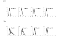
- FIG. 1A shows the results of flow cytometry for LN229, a human glioblastoma cell line, at various concentrations of PcMab-60, an anti-podocalyxin antibody that targets the cancer microenvironment against podocalyxin.
- FIG. 1B shows the results of flow cytometry for podocalyxin forced expression LN229 (LN229 / hPODXL) at each concentration of PcMab-60.
- FIG. 2A shows the results of flow cytometry for LN229 at various concentrations of human chimeric PcMab-60 (chPcMab-60), an anti-podocalyxin antibody targeting the cancer microenvironment against podocalyxin.
- FIG. 1A shows the results of flow cytometry for LN229, a human glioblastoma cell line, at various concentrations of PcMab-60, an anti-podocalyxin antibody that targets the cancer microenvironment against podocaly
- FIG. 2B shows flow cytometry results for LN229 / hPODXL at each concentration of chPcMab-60.
- FIG. 3A and FIG. 3B show the results of flow cytometry for two types of normal vascular endothelial cells, PcMab-47 and PcMab-60, which are anti-podocalyxin antibodies targeting the cancer microenvironment for podocalyxin, respectively.
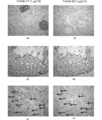
- 4A to 4D show the results of immunohistochemical staining of normal tissues of PcMab-47 and PcMab-60, which are anti-podocalyxin antibodies against podocalyxin, respectively.
- FIGS. 4E and 4F show the results of immunohistochemical staining of breast cancer tissues of PcMab-47 and PcMab-60, which are anti-podocalyxin antibodies against podocalyxin, respectively, and arrows in FIGS. 4E and 4F indicate vascular endothelial cells.
- FIG. 4A shows that normal blood vessels and glomeruli of the kidney are stained with PcMab-47
- FIG. 4B shows that normal blood vessels and glomeruli of the kidney are not stained with PcMab-60.
- FIG. 4C shows that normal blood vessels in the small intestine are stained with PcMab-47
- FIG. 4D shows that normal blood vessels in the small intestine are not stained with PcMab-60.
- FIG. 4E shows that PcMab-47 stains not only breast cancer tissue cancer cells but also surrounding blood vessels.
- PcMab-60 stains abnormal blood vessels around breast cancer tissue cancer cells. Indicates that
- the anti-podocalyxin antibody or antigen-binding fragment thereof targeting the cancer microenvironment according to the present invention is any one of (i) to (iii) below.
- Heavy chain CDR1 GYSFTDY having at least one of the following 6 CDRs (SEQ ID NO: 2)
- Heavy chain CDR2 NPRNGG (SEQ ID NO: 3)
- Heavy chain CDR3 EAMEY (SEQ ID NO: 4)
- Light chain CDR3 WQGTHFPRT (SEQ ID NO: 7);
- at least one of heavy chain CDR1-3 and light chain CDR1-3 comprising addition, substitution, or deletion of one to several amino acids in heavy chain CDR1-3 and light chain CDR1-3 of (i)
- And (iii) at least one of heavy chain CDR1-3 and light chain CDR1-3 has at least 80%
- the “antibody” has a structure in which two heavy chains (H chains) stabilized by a pair of disulfide bonds and two light chains (L chains) are associated.
- the heavy chain consists of a heavy chain variable region VH, a heavy chain constant region CH1, CH2, CH3, and a hinge region located between CH1 and CH2, and the light chain comprises a light chain variable region VL and a light chain constant region CL. Consists of.
- a variable region fragment (Fv) composed of VH and VL is a region that directly participates in antigen binding and gives diversity to antibodies.
- an antigen-binding region composed of VL, CL, VH, and CH1 is referred to as a Fab region, and a region composed of a hinge region, CH2, and CH3 is referred to as an Fc region.
- the region that directly contacts the antigen has a particularly large change and is called a complementarity-determining region (CDR).
- CDR complementarity-determining region
- the part with relatively few mutations other than CDR is called a framework region (FR).
- FR framework region
- the anti-podocalyxin antibody targeting the cancer microenvironment according to the present invention may be a monoclonal antibody or a polyclonal antibody. Further, the anti-podocalyxin antibody targeting the cancer microenvironment according to the present invention may be any isotype of IgG, IgM, IgA, IgD, and IgE. It may be prepared by immunizing non-human animals such as mice, rats, hamsters, guinea pigs, rabbits, chickens, etc., may be a recombinant antibody, a chimeric antibody, a humanized antibody, a fully humanized antibody Etc. "Chimeric antibody” means an antibody in which fragments of antibodies derived from different species are linked.
- Humanized antibody means an antibody in which the corresponding position of a human antibody is substituted with an amino acid sequence characteristic of a non-human-derived antibody. For example, a heavy chain CDR1- 3 and light chain CDR1-3, and all other regions including four framework regions (FR) of heavy chain and light chain, respectively, are derived from human antibodies. Such antibodies are sometimes referred to as CDR-grafted antibodies.
- the term “humanized antibody” may also include human chimeric antibodies.
- the “antigen-binding fragment” of an anti-podocalyxin antibody that targets the cancer microenvironment means a fragment of an anti-podocalyxin antibody that targets the cancer microenvironment and binds to podocalyxin. .
- Fab consisting of VL, VH, CL, and CH1 regions; F (ab ′) 2 in which two Fabs are linked by a disulfide bond in the hinge region; Fv consisting of VL and VH; VL and VH
- scFv which is a single chain antibody linked by an artificial polypeptide linker
- bispecific antibodies such as diabody type, scDb type, tandem scFv type, leucine zipper type and the like can be mentioned, but not limited thereto.
- One aspect of the anti-podocalyxin antibody or antigen-binding fragment thereof targeting the cancer microenvironment according to the present invention has at least one of the following six CDRs.
- These CDRs are anti-podocalyxin antibodies targeting the cancer microenvironment against podocalyxin, and are the CDR sequences of PcMab-60 obtained in the examples.
- Heavy chain CDR1 GYSFTDY (SEQ ID NO: 2) Heavy chain CDR2: NPRNGG (SEQ ID NO: 3) Heavy chain CDR3: EAMEY (SEQ ID NO: 4) Light chain CDR1: KSSQSLLDSAGKTYLN (SEQ ID NO: 5) Light chain CDR2: RLMYLVSKLA (SEQ ID NO: 6) Light chain CDR3: WQGTHFPRT (SEQ ID NO: 7
- the anti-podocalyxin antibody or antigen-binding fragment thereof targeting the cancer microenvironment according to the present invention may have at least one of the six CDRs as long as the effect of the present invention is achieved. It can be 3 or more, 4 or more, 5 or more, or 6 and it is preferable that the number of CDRs to be included is larger.
- the anti-podocalyxin antibody or antigen-binding fragment thereof targeting the cancer microenvironment according to the present invention includes addition, substitution, or deletion of one to several amino acids in the amino acid sequence represented by SEQ ID NO: 2.
- Heavy chain CDR3 comprising one amino acid addition, substitution or deletion
- light chain CDR1 comprising one to several amino acid additions, substitutions or deletions in the amino acid sequence represented by SEQ ID NO: 5; sequence A light chain CDR2 comprising 1 to several amino acid additions, substitutions or deletions in the amino acid sequence represented by number: 6; an amino acid sequence represented by SEQ ID NO: 7 having from one to several amino acids Addition, substitution, or deletion It may have at least one of the light chain CDR3 comprising.
- Heavy chain CDRs 1-3 with additions, substitutions or deletions of one to several amino acids in heavy chain CDRs 1-3 (SEQ ID NOs: 2-4) and light chain CDRs 1-3 (SEQ ID NOs: 5-7) And having at least one of light chain CDRs 1-3, at least one of heavy chain CDR1-3 and light chain CDR1-3 comprising 1 to several amino acid additions, substitutions or deletions as CDRs It is preferable to retain a function, that is, a function that reacts only to abnormal blood vessels but does not react to normal blood vessels.
- amino acid is used in its broadest sense and includes artificial amino acid variants and derivatives in addition to natural amino acids.
- Amino acids may be shown in conventional one-letter code or three-letter code.
- examples of amino acids or derivatives thereof include natural proteinaceous L-amino acids; non-natural amino acids; chemically synthesized compounds having characteristics known in the art that are characteristic of amino acids.
- examples of unnatural amino acids include ⁇ , ⁇ -disubstituted amino acids (such as ⁇ -methylalanine), N-alkyl- ⁇ -amino acids, D-amino acids, ⁇ -amino acids, ⁇ -, whose main chain structure is different from that of the natural type.
- Hydroxy acids amino acids whose side chain structure is different from the natural type (norleucine, homohistidine, etc.), amino acids with extra methylene in the side chain (“homo” amino acids, homophenylalanine, homohistidine, etc.), Examples include, but are not limited to, amino acids (such as cysteic acid) in which the carboxylic acid functional group is substituted with a sulfonic acid group.
- the number of amino acids to be deleted, substituted, etc. indicates that the resulting polypeptide retains its function as a CDR.
- the amino acid to be substituted or added may be a non-natural amino acid or an amino acid analog in addition to a natural proteinaceous amino acid.
- the position of amino acid deletion, substitution, or addition may be anywhere in the original CDR sequence so long as the function as CDR is retained.
- a heavy chain CDR1 comprising an amino acid sequence having 80% or more identity with the amino acid sequence represented by SEQ ID NO: 2;
- a heavy chain CDR2 comprising an amino acid sequence having 80% or more identity with the amino acid sequence represented by SEQ ID NO: 3; comprising an amino acid sequence having 80% or more identity to the amino acid sequence represented by SEQ ID NO: 4
- Heavy chain CDR3 Heavy chain CDR3;
- light chain CDR1 consisting of an amino acid sequence having 80% or more identity with the amino acid sequence represented by SEQ ID NO: 5; having 80% or more identity with the amino acid sequence represented by SEQ ID NO: 6
- Light chain CDR2 comprising an amino acid sequence;
- light chain CD comprising an amino acid sequence having 80% or more identity with the amino acid sequence represented by SEQ ID NO: 7 3 may have at least one.
- Heavy chain CDR1-3 (SEQ ID NO: 2-4) and light chain CDR1-3 (SEQ ID NO: 5-7) have 80% or more identity with the amino acid sequence represented by SEQ ID NO: 2-7
- at least one of heavy chain CDR1 to 3 and light chain CDR1 to 3 consisting of an amino acid sequence
- at least one of heavy chain CDR1 to 3 and light chain CDR1 to 3 having an amino acid sequence having 80% or more identity Preferably retains the function as a CDR, that is, retains the function of reacting only to abnormal blood vessels and not reacting to normal blood vessels.
- “having 80% or more identity” is common when alignment is made so that the amino acid sequences of two polypeptides each having a mutated sequence and the original sequence are maximized. It means that the number of amino acid residues is 80% or more of the number of amino acids in the original sequence.
- the identity is 80% or more, and may be any percentage as long as the CDR function is maintained. For example, 85% or more, 90% or more, 91% or more, 92% or more, 93% or more, 94% Thus, it can be 95% or more, 96% or more, 97% or more, 98% or more, or 99% or more.
- CDRs composed of amino acid sequences obtained by adding, substituting, or deleting amino acids to the amino acid sequences of heavy chain CDR1 to 3 and light chain CDR1 to 3, and the amino acid sequences of heavy chain CDR1 to 3 and light chain CDR1 to 3 CDRs composed of the amino acid sequences having the above identity can be prepared using known methods such as site-specific mutagenesis, random mutagenesis, chain shuffling, and CDR walking. According to these methods, CDRs with more affinity can be obtained by displaying antibodies or antibody fragments having various mutations in CDRs by the phage display method and screening using antigens.
- PNAS are well known to those skilled in the art (for example, Wu et al., PNAS.
- An anti-podocalyxin antibody or antigen-binding fragment thereof targeting the cancer microenvironment A light chain comprising the amino acid sequence represented by SEQ ID NO: 8; A light chain comprising an amino acid sequence comprising an addition, substitution, or deletion of one to several amino acids in the amino acid sequence represented by SEQ ID NO: 8; or It includes a light chain comprising an amino acid sequence having 80% or more identity with the amino acid sequence represented by SEQ ID NO: 8.
- the amino acid sequence represented by SEQ ID NO: 8 is the amino acid sequence of the light chain of PcMab-60.
- a heavy chain comprising the amino acid sequence represented by SEQ ID NO: 10; A heavy chain comprising an amino acid sequence comprising an addition, substitution, or deletion of one to several amino acids in the amino acid sequence represented by SEQ ID NO: 10; or It includes a heavy chain comprising an amino acid sequence represented by SEQ ID NO: 10 and having an amino acid sequence having 80% or more identity.
- the amino acid sequence represented by SEQ ID NO: 10 is the heavy chain amino acid sequence of chPcMab-60.
- the amino acid sequence of heavy chain or light chain includes addition, substitution or deletion of 1 to several amino acids
- the number of added, substituted or deleted amino acids is, for example, 1 Two, three, four, five, six, seven, eight, nine, or ten can be used. Other terms are as described above.
- the anti-podocalyxin antibody targeting the cancer microenvironment according to the present invention binds one or more N-linked sugar chains to the Fc region, and fucose to N-acetylglucosamine at the reducing end of the N-linked sugar chain. May not be bound to the antibody.
- N-linked sugar chain refers to a sugar chain that binds to Asn in the Asn-X-Ser / Thr sequence and has a common structure Man3GlcNAc2-Asn.
- ADCC activity is significantly increased when this fucose is not bound compared to when it is bound. It is known. This is described, for example, in WO 2002/031140. Since the ADCC activity is remarkably improved, the dose can be reduced when the antibody is used as a medicine, so that side effects can be reduced and the treatment cost can be reduced.
- the anti-podocalyxin antibody targeting the cancer microenvironment according to the present invention may be used by binding a substance having anticancer activity. Since the anti-podocalyxin antibody targeting the cancer microenvironment according to the present invention reacts only with abnormal blood vessels and does not react with normal blood vessels, the anti-podocalyxin antibody can be delivered only to abnormal blood vessels in the cancer microenvironment. And when the substance which has anticancer activity is couple
- the “substance having anticancer activity” refers to a decrease in tumor size (delayed or stopped), inhibition of tumor metastasis, inhibition of tumor growth (delayed or stopped), and one associated with cancer.
- toxin having anticancer activity examples include Pseudomonas aeruginosa exotoxin (PE) or a cytotoxic fragment thereof (eg, PE38), diphtheria toxin, ricin A and the like.
- PE Pseudomonas aeruginosa exotoxin
- cytotoxic fragment thereof eg, PE38
- diphtheria toxin ricin A
- ricin A cytotoxic fragment thereof
- Toxins with anti-cancer activity are toxic only to cells that take up toxins together with anti-podocalyxin antibodies, that is, cancer cells expressing podocalyxin. There is an advantage that can be obtained.
- Anticancer agents include, for example, adriamycin, daunomycin, mitomycin, cisplatin, vincristine, epirubicin, methotrexate, 5-fluorouracil, aclacinomycin, nitrogen mustard, cyclophosphamide, bleomycin, daunorubicin, doxorubicin, vincristine, vinblastine And low molecular weight compounds such as vindesine, tamoxifen, and dexamethasone, and proteins such as cytokines that activate immunocompetent cells. Examples of cytokines that activate immunocompetent cells include human interleukin 2, human granule Sphere macrophage colony stimulating factor, human macrophage colony stimulating factor, human interleukin 12 and the like.
- radioisotopes having anticancer activity examples include 32 P, 14 C, 125 I, 3 H, 131 I, 211 At, and 90 Y. Radioisotopes are also toxic to cells around which the anti-podocalyxin antibody binds, ie cells that express podocalyxin. In general, cancer cells are not uniform and not all cancer cells express podocalyxin, so radioisotopes are useful for killing surrounding podocalyxin negative cancer cells. When radioisotopes are bound, they may be bound to anti-podocalyxin antibody fragments such as Fab and scFv.
- a substance having anticancer activity can be directly bound to an anti-podocalyxin antibody targeting the cancer microenvironment by a known method.
- it may be encapsulated in a carrier such as a liposome and bound to an anti-podocalyxin antibody that targets the cancer microenvironment.
- the substance having anticancer activity is a protein or polypeptide
- a nucleic acid (described later) encoding an anti-podocalyxin antibody targeting the cancer microenvironment according to the present invention and a DNA encoding a substance having anticancer activity May be linked and inserted into an appropriate expression vector to be expressed as a fusion protein of a substance having anticancer activity and an anti-podocalyxin antibody targeting the cancer microenvironment.
- the present invention also includes a nucleic acid encoding an anti-podocalyxin antibody targeting the cancer microenvironment according to the present invention.
- the nucleic acid may be a natural nucleic acid or an artificial nucleic acid, and examples thereof include, but are not limited to, DNA, RNA, and a chimera of DNA and RNA.
- the nucleotide sequence of a nucleic acid encoding an anti-podocalyxin antibody targeting the cancer microenvironment can be determined by a person skilled in the art according to a known method or a method analogous thereto, and can be prepared by a known method or a method analogous thereto. .
- nucleic acid encoding the anti-podocalyxin antibody targeting the cancer microenvironment examples include a DNA encoding the heavy chain of chPcMab-60 represented by SEQ ID NO: 10 (SEQ ID NO: 11), sequence A DNA encoding the light chain of PcMab-60 represented by No. 8 (SEQ ID NO: 9) is exemplified, but not limited thereto. Nucleic acids encoding each of the PcMab-60 CDRs are included in the DNA sequences indicated by these SEQ ID NOs.
- the present invention includes an expression vector comprising a nucleic acid encoding an anti-podocalyxin antibody targeting the cancer microenvironment according to the present invention.
- the expression vector can be appropriately selected according to the host cell to be used. For example, plants such as plasmids, retrovirus vectors, adenovirus vectors, adeno-associated virus (AAV) vectors, cauliflower mosaic virus vectors, and tobacco mosaic virus vectors. Examples include viral vectors, cosmids, YACs, and EBV-derived episomes.
- a nucleic acid encoding an anti-podocalyxin antibody targeting the cancer microenvironment according to the present invention can be inserted into these expression vectors by a known method (a method using a restriction enzyme, etc.).
- the expression vector according to the present invention can further contain a promoter that regulates the expression of the antibody gene, a replication origin, a selection marker gene, and the like.
- the promoter and origin of replication can be appropriately selected depending on the type of host cell and expression vector.
- the present invention includes a transformant comprising the expression vector according to the present invention.
- a transformant can be obtained by transfecting an appropriate host cell with the expression vector of the present invention.
- host cells include eukaryotic cells such as mammalian cells (CHO cells, COS cells, myeloma cells, HeLa cells, and Vero cells), insect cells, plant cells, and fungal cells (such as Saccharomyces and Aspergillus). And prokaryotic cells such as E. coli and Bacillus subtilis.
- the anti-podocalyxin antibody targeting the cancer microenvironment according to the present invention is, for example, when obtaining an antibody against cancer cell-specific podocalyxin (cancer cell-specific anti-podocalyxin antibody) obtained using the Casmab method. Further, it can be produced by screening by immunohistological staining.
- the method for obtaining a cancer cell-specific anti-podocalyxin antibody is as follows: Introducing a nucleic acid encoding all or part of podocalyxin into a cell that expresses a cancer cell-specific sugar chain structure to express cancer cell-specific podocalyxin or a part thereof; Immunizing a non-human mammal with the cancer cell-specific podocalyxin or a part thereof to obtain an antibody; Purifying the antibody by primary screening using purified cancer cell-specific podocalyxin or a part thereof.
- cancer cell-specific anti-podocalyxin antibody means an antibody that is significantly more reactive with podocalyxin expressed in cancer cells than podocalyxin expressed in normal cells. In one embodiment, the “cancer cell-specific anti-podocalyxin antibody” reacts with podocalyxin expressed in cancer cells and does not react at all with podocalyxin expressed in normal cells. In one embodiment, the “cancer cell-specific anti-podocalyxin antibody” is highly reactive with podocalyxin expressed in cancer cells, but also reacts to some extent with podocalyxin expressed in normal cells.
- Podocalixin is highly expressed in testicular tumors, breast cancer, prostate cancer, ovarian cancer, colon cancer, pancreatic cancer and the like, but also expressed in normal cells.
- Human podocalyxin (BC143318, NM_001018111) is a protein represented by SEQ ID NO: 1.
- the term “podocalyxin” includes functional variants thereof.
- the “cell expressing a cancer cell-specific sugar chain structure” may be any cell as long as it expresses a cancer cell-specific sugar chain structure.
- it may be a cancer cell, or a cell artificially modified to introduce a sugar translocation enzyme necessary for non-cancer cells and express a cancer cell-specific sugar chain structure.
- Examples of “cells that express a cancer cell-specific sugar chain structure” include the following cells. -Glioblastoma cell line LN229-derived cells. The present inventors have previously confirmed that keratan sulfate modification is enhanced in brain tumors depending on the grade of malignancy (Kato Y et al., Biochem Biophys Res Commun.
- LN229 cells with high keratan sulfate modification have been discovered among brain tumor cell lines (Hayatsu N et al., Biochem Biophys Res Commun. 2008; 368 (2): 217-222.).
- podocalyxin is highly expressed in an astrotropic tumour in correlation with malignancy (Hayatsu N et al., Biochem Biophys Res Commun. 2008; 374 (2): 394-398.).
- sialic acid is added specifically to cancer cells to proteins expressed by LN229 cells (Kato Y et al., Sci Rep. 2014; 4: 5924).
- a trichostatin A-treated chicken B cell-derived DT40 cell line may be used as a cell that expresses a cancer cell-specific sugar chain structure.
- an antibody production method an antibody from a trichostatin A-treated chicken B cell-derived DT40 cell line is used.
- the Adrib method (Seo H et al., Nat. Biotechnol. 2002; 6: 731-736.)
- a non-human mammal a KM mouse, which is a mouse into which a mouse antibody gene is disrupted and a human antibody gene is introduced, may be used.
- the step of introducing and expressing a nucleic acid encoding all or part of podocalyxin in a cell expressing a cancer cell-specific sugar chain structure can be performed by a person skilled in the art according to a conventional method.
- a cancer cell-specific podocalyxin obtained by introducing a nucleic acid encoding all or part of podocalyxin into a cancer cell and forcibly expressing it, or one of them Part is used as an antigen.
- nucleic acid encoding a part of podocalyxin a nucleic acid encoding a podocalyxin part to which a cancer cell-specific sugar chain is bound can be used.
- the nucleic acid encoding the podocalyxin portion to which the cancer cell-specific sugar chain is bound include a nucleic acid encoding the extracellular region of podocalyxin.
- the nucleic acid may be any nucleic acid as long as the target protein can be expressed, and examples thereof include DNA, RNA, DNA / RNA chimera, artificial nucleic acid, and the like.
- podocalyxin is expressed as a secreted form. This can be performed by introducing a nucleic acid encoding the extracellular region of podocalyxin into a cell that expresses a cancer cell-specific sugar chain structure. Podocalixin expressed as a secreted form can be purified from the culture supernatant of cells that express a cancer cell-specific sugar chain structure. For example, podocalyxin may be expressed with an appropriate tag and purified using such a tag.
- the step of obtaining an antibody by immunizing a non-human mammal with a cancer cell-specific podocalyxin or a part thereof refers to administering the cancer cell-specific podocalyxin or a part thereof to the non-human mammal.
- Purified cancer cell-specific podocalyxin or a part thereof may be used.
- Immunization can be performed by, for example, injecting cancer cell-specific podocalyxin or a part thereof, subcutaneously, intradermally, intramuscularly, intravenously, or intraperitoneally with an adjuvant as necessary.
- the immunization step of the non-human mammal may be performed by expressing the cancer cell-specific podocalyxin as a membrane protein without expressing it as a secreted form and administering the whole cell to the non-human mammal. Immunization can be performed according to a conventional method, for example, by administering 1 ⁇ 10 7 to 1 ⁇ 10 9 cells intraperitoneally, once every 10 days, multiple times.
- the non-human mammal is typically a mouse, but is not particularly limited, and examples thereof include rats, hamsters, rabbits, cats, dogs, monkeys, goats, sheep, cows and horses.
- primary antibody screening refers to an initial screening in the process of identifying a target antibody from antibody-producing cells, for example, screening using a culture supernatant of a hybridoma producing a monoclonal antibody.
- the primary screening of the antibody preferably includes a step of obtaining a monoclonal antibody and a step of identifying a hybridoma that produces the monoclonal antibody.
- the primary screening of antibodies in the present invention is generally performed as follows. First, podocalyxin or a part thereof is expressed together with an affinity tag (FLAG tag, His tag, Myc tag, PA tag, etc.) in a cell that expresses a cancer cell-specific sugar chain structure, and purification is performed using the affinity tag. Do.
- the thus purified cancer cell-specific podocalyxin or a part thereof is immobilized on an ELISA plate, to which an antibody obtained from antibody-producing cells is added, and a reaction well is selected.
- a cancer cell specific antibody can be selected at an early stage of screening.
- the purified cancer cell-specific podocalyxin or a part thereof is not particularly limited as long as it is a purified protein or a part thereof, and may be a product obtained by purifying a forcibly expressed protein.
- the protein may be purified.
- the method for producing a cancer cell-specific anti-podocalyxin antibody compares the reactivity of an antibody to cancer cells or tissues and normal cells or tissues after the primary screening, and the reactivity to cancer cells or tissues is normal.
- a step of selecting an antibody that is significantly higher than the reactivity with respect to a cell or a tissue may be included.
- Cancer cells or tissues include brain tumor, prostate cancer, testicular tumor, kidney cancer, thyroid cancer, bladder cancer, breast cancer, ovarian cancer, colon cancer, pancreatic cancer, malignant mesothelioma, osteosarcoma
- normal cells include vascular endothelial cells and renal epithelial cells. Examples of normal tissues include systemic blood vessels and kidneys.
- adenocarcinoma lung adenocarcinoma, liver adenocarcinoma, pancreatic adenocarcinoma, lymph adenocarcinoma, uterine adenocarcinoma, seminal adenocarcinoma, gastric adenocarcinoma, etc.
- Basal cell cancer skin cancer, etc.
- Squamous cell carcinoma oral cancer, tongue cancer, pharyngeal cancer, esophageal cancer, pharyngeal cancer, cervical cancer, etc.
- Sarcoma Lymphatic sarcoma, Kaposi sarcoma, malignant osteosarcoma, etc.
- Hematopoietic tumors leukemias such as acute / chronic myeloid leukemia, acute promyelocytic leukemia, and acute / chronic lymphocytic leukemia, Hodgkin lymphoma and non-Ho
- the step of comparing the reactivity of the antibody to a cancer cell or tissue and a normal cell or tissue is a reaction between the cancer cell or tissue and the antibody obtained in the primary screening.
- it means a step of detecting the presence or absence of binding by reacting normal cells or tissues with the antibody obtained in the primary screening. This step can be performed by flow cytometry, immunohistochemical staining (IHC), immune cell staining (ICC) or the like.
- an antibody having a significantly higher reactivity to the cancer cell or tissue than the reactivity to the normal cell or tissue is selected.
- a cancer cell-specific antibody can be obtained.
- the selected cancer cell specific antibody can then be further purified.
- the method for producing an anti-podocalyxin antibody targeting the cancer microenvironment includes a step of comparing the reactivity of the antibody to abnormal blood vessels and normal blood vessels after the primary screening.
- the step of comparing the reactivity of the antibody to the abnormal blood vessel and the normal blood vessel is performed by staining the cancer cell or tissue and the antibody obtained in the primary screening by immunohistochemical staining, in the abnormal blood vessel. It means the step of detecting the presence or absence of binding, and on the other hand, staining normal cells or tissues and antibodies obtained by primary screening by immunohistological staining to examine the presence or absence of binding in normal blood vessels.
- antibodies having a significantly higher reactivity against abnormal blood vessels than that against normal blood vessels are selected. More preferably, in the production method of the present invention, an antibody that reacts with an abnormal blood vessel and does not react with a normal blood vessel is selected. Antibodies that target the selected cancer microenvironment may then be further purified.
- Angiogenesis is a physiological phenomenon in which a new blood vessel branch is branched from an existing blood vessel to construct a blood vessel network.
- angiogenesis including angiogenesis (Vasculogenesis) in which new blood vessels are created during embryogenesis, is called angiogenesis, but strictly speaking, angiogenesis and angiogenesis are distinguished.
- angiogenesis also occurs in the process of wound healing, and angiogenesis plays an important role in chronic inflammation. Therefore, in the present specification, a blood vessel generated by angiogenesis in a cancer microenvironment is defined as an abnormal blood vessel. All cancer cells require the supply of nutrients from the blood vessels, so there are abnormal blood vessels in all cancers.
- a normal blood vessel means a blood vessel other than a blood vessel caused by angiogenesis in a cancer microenvironment. Therefore, angiogenesis also occurs in the process of inflammation, wound healing, etc., but these are classified as normal blood vessels in this specification.
- a blood vessel existing in the above-described cancer cell or tissue can be used, and as a normal blood vessel, a blood vessel existing in a normal cell or tissue can be used.
- the anti-podocalyxin antibody targeting the cancer microenvironment according to the present invention can also be produced by screening by immunohistochemical staining from a cancer cell-specific anti-podocalyxin antibody obtained by the method described below.
- a cancer cell-specific anti-podocalyxin monoclonal antibody isolates antibody-producing cells from a non-human mammal immunized with a cancer cell-specific podocalyxin or a part thereof, and fuses it with myeloma cells, etc. The antibody produced and purified by this hybridoma can be obtained.
- a cancer cell-specific anti-podocalyxin polyclonal antibody can be obtained from the serum of an animal immunized with a cancer cell-specific podocalyxin or a fragment thereof.
- an anti-podocalyxin antibody targeting the cancer microenvironment according to the present invention by a genetic recombination method, for example, transforming an appropriate host with an expression vector containing the nucleic acid according to the present invention
- the antibody may be expressed by culturing under appropriate conditions, followed by isolation and purification according to a known method.
- isolation and purification methods include affinity columns using protein A / G / L and the like, other chromatography columns, filters, ultrafiltration, salting out, and dialysis, and these can be combined as appropriate.
- An antibody that binds to a predetermined epitope sequence can be prepared by a method known to those skilled in the art or a method analogous thereto.
- an antibody that specifically binds to the same epitope can be obtained by immobilizing a peptide containing the epitope sequence on a solid phase carrier and detecting the binding between the peptide and a plurality of antibodies.
- the “plurality of antibodies” those obtained by immunizing an animal with an antigen protein or a partial peptide thereof may be used, or an antibody library or an antibody fragment library prepared by a phage display method may be used. Good.
- an antibody that specifically binds to the same epitope can be obtained by immobilizing a peptide containing the epitope sequence on a solid phase carrier and repeating panning.
- Human chimeric antibodies and human CDR-grafted antibodies can be prepared by cloning an antibody gene from mRNA of a hybridoma that produces an antibody of a non-human animal and ligating it with a part of the human antibody gene by gene recombination technology. it can.
- a human chimeric antibody cDNA is synthesized by reverse transcriptase from the hybridoma mRNA producing a mouse antibody, and the heavy chain variable region (VH) and light chain variable region (LH) are cloned by PCR and sequenced. To analyze.
- a 5 ′ primer containing a leader sequence is prepared from the antibody base sequence having a high concordance rate, and PCR is performed from the cDNA to the 3 ′ end of the variable region from the above cDNA using the 5 ′ primer and the variable region 3 ′ primer.
- Cloning On the other hand, the heavy and light chain constant regions of human IgG1 were cloned, and the mouse antibody-derived variable region and the human antibody-derived constant region were linked by PCR using the Overwrapping Hanging method for amplification. To do.
- the obtained DNA can be inserted into an appropriate expression vector and transformed to obtain a human chimeric antibody.
- the human antibody variable region having the highest homology with the mouse antibody variable region to be used is selected and cloned, and the CDR base sequence is modified by site-directed mutagenesis using the megaprimer method. . If the amino acid sequence constituting the framework region cannot be specifically bound to the antigen when humanized, it is possible to convert some amino acids of the framework from human to rat.
- a CDR comprising an amino acid sequence having a deletion, substitution or addition of 1 to several, preferably 1 or 2 amino acids, or an amino acid sequence having 80% or more identity to the original sequence
- the CDR can be prepared using a known method such as a site-specific mutagenesis method, a random mutagenesis method, a chain shuffling method, or a CDR walking method. By these methods, it is possible to obtain CDRs with more affinity by displaying antibodies or antibody fragments having various mutations in CDRs on the phage surface by the phage display method and screening using antigens.
- a site-specific mutagenesis method such as a site-specific mutagenesis method, a random mutagenesis method, a chain shuffling method, or a CDR walking method.
- the invention also encompasses antibodies comprising CDRs matured by such methods.
- the antigen-binding fragment of the anti-podocalyxin antibody targeting the cancer microenvironment according to the present invention may be expressed by the above-described method using DNA encoding the fragment, or after obtaining the full-length antibody. It may be fragmented by treatment with an enzyme such as papain or pepsin.
- the anti-podocalyxin antibody targeting the cancer microenvironment according to the present invention may vary in amino acid sequence, molecular weight, isoelectric point, presence / absence of sugar chain, form, etc., depending on the production method and purification method. However, so long as the obtained antibody has an equivalent function as an anti-podocalyxin antibody targeting the cancer microenvironment, it is included in the present invention.
- the anti-podocalyxin antibody targeting the cancer microenvironment according to the present invention is expressed in prokaryotic cells such as Escherichia coli, a methionine residue is added to the N-terminus of the amino acid sequence of the original antibody.
- the invention also encompasses such antibodies.
- the anti-podocalyxin antibody targeting the cancer microenvironment according to the present invention is an antibody having an N-linked sugar chain in which fucose is not bound to N-acetylglucosamine at the reducing end
- an antibody is a known method. Or it can manufacture according to the method according to it.
- Such antibody production methods are described in, for example, International Publication Nos. 2002/031140 and JP-A-2009-225781.
- an expression vector comprising DNA encoding an anti-podocalyxin antibody targeting the cancer microenvironment according to the present invention
- the activity of an enzyme involved in the synthesis of GDP-fucose, or ⁇ -1 , 6-fucosyltransferase activity is obtained by transforming cells with reduced or deleted activity, culturing the resulting transformant, and then purifying anti-podocalyxin antibody targeting the target cancer microenvironment.
- GDP-mannose 4,6-dehydratase GMP
- GDP-keto-6-deoxymannose 3,5-epimerase 4-reductase
- GDP-beta- L-fucose pyrophosphorylase GFPP
- the cells are not particularly limited, but mammalian cells are preferable.
- CHO cells in which the enzyme activity is reduced or deleted can be used.
- the antibody composition obtained by the above method may contain an antibody in which fucose is bound to N-acetylglucosamine at the reducing end, but the ratio of the antibody to which fucose is bound is 20% by weight or less of the whole antibody. , Preferably 10% by weight or less, more preferably 5% by weight or less, and most preferably 3% by weight or less.
- An antibody having an N-linked sugar chain in which fucose is not bound to N-acetylglucosamine at the reducing end is an expression vector comprising DNA encoding an anti-podocalyxin antibody targeting the cancer microenvironment according to the present invention.
- Anti-podocalyxin antibodies that target the cancer microenvironment from the transgenic insects or their secretions. Can also be obtained by extracting.
- Silkworms can be used as insects, in which case antibodies can be extracted from the cocoons.
- the antibody composition obtained by this method may also contain an antibody in which fucose is bound to N-acetylglucosamine at the reducing end, but the ratio of the antibody to which fucose is bound is 20% by weight or less of the whole antibody. , Preferably 10% by weight or less, more preferably 5% by weight or less, and most preferably 3% by weight or less.
- the medicinal mechanism of the antibody drug is based on two biological activities of the antibody.
- One is a target antigen-specific binding activity, which is an activity to neutralize the function of the target antigen molecule by binding. Neutralization of the function of the target antigen molecule is exerted through the Fab region.
- effector activity can be induced through antibody Fc region, such as antibody-dependent cellular cytotoxicity (ADCC), complement-dependent cytotoxicity (CDC), direct induction of apoptosis, etc. Demonstrated in an embodiment.
- ADCC antibody-dependent cellular cytotoxicity
- CDC complement-dependent cytotoxicity
- the activity of the anti-podocalyxin antibody targeting the cancer microenvironment according to the present invention can be measured by the following method.
- Binding activity The binding activity of an antibody can be determined by a known method such as ELISA (enzyme-linked immunosorbent assay), EIA (enzyme immunoassay), RIA (radioimmunoassay), fluorescent antibody method, FACS method, etc. Can be measured.
- ELISA enzyme-linked immunosorbent assay
- EIA enzyme immunoassay
- RIA radioimmunoassay
- fluorescent antibody method FACS method, etc.
- ADCC activity means that when the antibody of the present invention binds to a cell surface antigen of a target cell, an Fc ⁇ receptor-bearing cell (effector cell) binds to the Fc portion via the Fc ⁇ receptor, It means activity that damages cells.
- the ADCC activity can be known by mixing the target cells expressing podocalyxin, the effector cells, and the anti-podocalyxin antibody targeting the cancer microenvironment according to the present invention, and measuring the degree of ADCC.
- effector cells for example, mouse spleen cells, monocyte nuclei isolated from human peripheral blood or bone marrow can be used.
- the target cell can be, for example, a podocalyxin positive cancer cell.
- the target cells are labeled with 51Cr or the like in advance, and the antibody of the present invention is added thereto for incubation, and then an effector cell in an appropriate ratio to the target cells is added for incubation. After incubation, the supernatant can be collected and measured by counting the label in the supernatant.
- CDC activity CDC activity means cytotoxic activity by the complement system. CDC activity can be measured by using complement in place of effector cells in a test for ADCC activity.
- Tumor growth inhibitory activity can be measured using a tumor model animal.
- a tumor is implanted subcutaneously in a mouse, and an anti-podocalyxin antibody targeting the cancer microenvironment according to the present invention is administered.
- the tumor growth inhibitory effect can be measured.
- the tumor growth inhibitory activity may be the result of suppressing the growth of individual cells or the result of inducing cell death.
- the anti-podocalyxin antibody or antigen-binding fragment thereof targeting the cancer microenvironment according to the present invention may be used for prevention or treatment of cancer expressing podocalyxin.
- One aspect of the pharmaceutical composition according to the present invention includes an anti-podocalyxin antibody or antigen-binding fragment thereof targeting the cancer microenvironment according to the present invention as an active ingredient, and further includes a pharmaceutically acceptable carrier or additive. .
- the anti-podocalyxin antibody or antigen-binding fragment thereof targeting the cancer microenvironment according to the present invention may be used for delivery of a drug targeting cancer cells.
- Another aspect of the pharmaceutical composition according to the present invention is an anti-podocalyxin antibody or antigen-binding fragment thereof targeting the cancer microenvironment to which the above-mentioned substance having anticancer activity or other anticancer agent is bound. And further pharmaceutically acceptable carriers and additives.
- Examples of carriers and additives include pharmaceutically acceptable organic solvents such as water, saline, phosphate buffer, dextrose, glycerol, ethanol, collagen, polyvinyl alcohol, polyvinyl pyrrolidone, carboxyvinyl polymer, sodium carboxymethylcellulose, Sodium polyacrylate, sodium alginate, water-soluble dextran, sodium carboxymethyl starch, pectin, methylcellulose, ethylcellulose, xanthan gum, gum arabic, casein, agar, polyethylene glycol, diglycerin, glycerin, propylene glycol, petrolatum, paraffin, stearyl alcohol , Stearic acid, human serum albumin, mannitol, sorbitol, lactose, surfactants, etc. Not a constant.
- organic solvents such as water, saline, phosphate buffer, dextrose, glycerol, ethanol, collagen, polyvinyl alcohol, polyvinyl pyrrol
- the pharmaceutical composition according to the present invention can be in various forms, for example, liquids (for example, injections), dispersions, suspensions, tablets, pills, powders, suppositories, and the like.
- a preferred embodiment is an injection, which is preferably administered parenterally (for example, intravenously, transdermally, intraperitoneally, intramuscularly).
- the pharmaceutical composition according to the present invention is effective for treatment of diseases associated with podocalyxin, particularly cancer.
- cancers associated with podocalyxin include brain tumors, prostate cancer, testicular cancer, kidney cancer, thyroid cancer, bladder cancer, breast cancer, ovarian cancer, colon cancer, pancreatic cancer, and malignant mesothelioma. Examples include, but are not limited to, osteosarcoma and the like.
- the anti-podocalyxin antibody targeting the cancer microenvironment according to the present invention is particularly useful for these cancers. Also.
- the pharmaceutical composition according to the present invention can also be used as a drug delivery preparation for the above-mentioned cancer.
- the present invention also includes a method for treating a disease associated with podocalyxin, comprising administering a therapeutically effective amount of an anti-podocalyxin antibody or antigen-binding fragment thereof targeting the cancer microenvironment according to the present invention.
- a therapeutically effective amount refers to the amount of an agent by which one or more symptoms of the disease being treated are alleviated to some extent.
- anti-cancer drugs at least: reduction of tumor size; inhibition of tumor metastasis (delay or cessation); inhibition of tumor growth (delay or cessation), and alleviation of one or more symptoms associated with cancer An amount indicating one.
- the dose of the anti-podocalyxin antibody or antigen-binding fragment thereof targeting the cancer microenvironment according to the present invention is, for example, 0.025 to 50 mg / kg, preferably 0.1 to 50 mg / kg. Yes, more preferably 0.1 to 25 mg / kg, still more preferably 0.1 to 10 mg / kg or 0.1 to 3 mg / kg, but it is not limited thereto.
- anti-podocalyxin antibodies targeting the cancer microenvironment according to the present invention are cancers, particularly brain tumors, prostate cancers, testicular tumors, kidney cancers, thyroid cancers, bladder cancers, breast cancers, ovarian cancers, It is useful for diagnosing cancers that highly express podocalyxin, such as colorectal cancer, pancreatic cancer, malignant mesothelioma, and osteosarcoma. Since the anti-podocalyxin antibody targeting the cancer microenvironment according to the present invention selectively binds to abnormal blood vessels, it is particularly useful for diagnosis.
- the present invention is directed to a cancer test agent comprising an anti-podocalyxin antibody targeting the cancer microenvironment according to the present invention, the use of an antibody for cancer testing, and the cancer microenvironment according to the present invention. It also includes a method for testing cancer using an anti-podocalyxin antibody.
- the sample used for the examination is, for example, tissue, serum, cerebrospinal fluid that is suspected of cancer collected from the subject Urine, body fluids (saliva, sweat, etc.).
- Podocalixin is a membrane protein, but is known to be secreted into serum.
- Examples of the inspection method include, but are not limited to, an immunoassay, an aggregation method, a turbidimetric method, a Western blotting method, a surface plasmon resonance (SPR) method, and the like.
- a cancer microenvironment is detected by using an antigen-antibody reaction between a detectably labeled anti-podocalyxin antibody targeting the cancer microenvironment according to the present invention and a podocalyxin targeting the cancer microenvironment in the sample.
- An immunoassay that measures the amount of targeted podocalyxin is preferred.
- the immunoassay uses an anti-podocalyxin antibody targeting a detectably labeled cancer microenvironment or an antibody (secondary antibody) against an anti-podocalyxin antibody targeting a detectably labeled cancer microenvironment.
- antibody labeling methods enzyme immunoassay (EIA or ELISA), radioimmunoassay (RIA), fluorescence immunoassay (FIA), fluorescence polarization immunoassay (FPIA), chemiluminescence immunoassay (CLIA), etc. This method can be used.
- enzymes such as peroxidase and alkaline phosphatase
- radioactive substances such as 125 I, 131 I, 35 S, and 3 H
- Fluorescent substances such as isothiocyanate and near-infrared fluorescent materials
- the CLIA method use antibodies labeled with luminescent substances such as luciferase, luciferin and aequorin.
- nanoparticles such as colloidal gold and quantum dots can also be detected.
- an anti-podocalyxin antibody targeting the cancer microenvironment can be labeled with biotin, and avidin or streptavidin labeled with an enzyme or the like can be bound and detected.
- an ELISA method using an enzyme label is preferable because it can easily and quickly measure an antigen.
- the ELISA method includes a competitive method and a sandwich method.
- an anti-podocalyxin antibody targeting a cancer microenvironment is immobilized on a solid phase carrier such as a microplate, and a sample and an enzyme-labeled cancer-specific podocalyxin are added to cause an antigen-antibody reaction. Once washed, it reacts with the enzyme substrate, develops color, and the absorbance is measured. If there is more podocalyxin in the sample, the color development becomes weaker, and if there is less podocalyxin in the sample, the color development becomes stronger. Therefore, the amount of podocalyxin can be determined using a calibration curve.
- an anti-podocalyxin antibody targeting the cancer microenvironment is immobilized on a solid phase carrier, added to the sample, reacted, and then targeted to the cancer microenvironment that recognizes another enzyme-labeled epitope.
- the anti-podocalyxin antibody is added and reacted. After washing, the amount of podocalyxin can be determined by reacting with the enzyme substrate, developing color, and measuring the absorbance.
- an anti-podocalyxin antibody targeting the cancer microenvironment immobilized on a solid phase carrier is reacted with a cancer-specific podocalyxin in the sample, and then an unlabeled antibody (primary antibody) is added, and this unlabeled
- An antibody against the antibody (secondary antibody) may be further enzyme-labeled and added.
- the enzyme substrate when the enzyme is peroxidase, 3,3′-diaminobenzidine (DAB), 3,3 ′, 5,5′-tetramethylbenzidine (TMB), o-phenylenediamine (OPD), or the like can be used.
- Alkaline phosphatase In this case, p-nitrophenyl phosphate (NPP) or the like can be used.
- the “solid phase carrier” is not particularly limited as long as it can immobilize the antibody, and is made of a microtiter plate made of glass, metal or resin, substrate, beads, nitrocellulose membrane, nylon membrane, PVDF. Examples include a membrane, and the target substance can be immobilized on these solid phase carriers according to a known method.
- an agglutination method is also preferable as a method for easily detecting a trace amount of protein.
- the aggregation method include a latex aggregation method in which latex particles are bound to an antibody.
- the concentration of the antigen can be determined by irradiating the sample with near-infrared light and quantifying the aggregate by measuring the absorbance (turbidimetric method) or the scattered light (Hipple method).
- inspection means to examine a sample collected from a subject in order to obtain information necessary for diagnosis, and the inspection method of the present invention can be performed by, for example, an inspection company.
- One aspect of the test method according to the present invention includes a step of analyzing whether or not the cancer-specific podocalyxin in the sample of the subject is greater than the cancer-specific podocalyxin of the non-cancer patient. If the amount of cancer-specific podocalyxin in the subject's sample is significantly higher than that in the non-cancer patient sample, the subject is determined to be more likely to have cancer.
- One aspect of the test method according to the present invention includes a step of measuring cancer-specific podocalyxin in a patient sample after receiving cancer treatment over time, and analyzing a variation in the amount of cancer-specific podocalyxin. Including. If the amount of podocalyxin tends to increase over time, it is determined that the patient has a high possibility of cancer recurrence or metastasis.
- the present invention relates to a diagnostic agent for cancer comprising an anti-podocalyxin antibody targeting the cancer microenvironment according to the present invention, and an anti-podocalyxin antibody targeting the cancer microenvironment according to the present invention for cancer diagnosis.
- the use includes a method for diagnosing cancer using an anti-podocalyxin antibody targeting the cancer microenvironment according to the present invention.
- diagnosis means that a medical practitioner such as a doctor determines whether a subject has cancer or that cancer has recurred or metastasized. To do.
- the kit for testing cancer according to the present invention is a kit for testing cancer using the testing method described above, and includes an anti-podocalyxin antibody targeting the cancer microenvironment.
- the test kit according to the present invention comprises reagents and devices necessary for measuring the amount of podocalyxin by immunoassay using an antigen-antibody reaction between an anti-podocalyxin antibody and a cancer-specific podocalyxin targeting the cancer microenvironment. Including.
- test kit for measuring cancer-specific podocalyxin by a sandwich method, and is labeled with a microtiter plate; an anti-podocalyxin antibody targeting a capture cancer microenvironment; alkaline phosphatase or peroxidase An anti-podocalyxin antibody targeting the cancer microenvironment, and an alkaline phosphatase substrate (such as NPP) or a peroxidase substrate (such as DAB, TMB, OPD).
- alkaline phosphatase substrate such as NPP
- peroxidase substrate such as DAB, TMB, OPD
- a capture antibody is immobilized on a microtiter plate, a sample is appropriately diluted and added thereto, and then incubated, and the sample is removed and washed. Next, the labeled antibody is added and then incubated, and the substrate is added to cause color development.
- the amount of cancer-specific podocalyxin can be determined by measuring color development using a microtiter plate reader or the like.
- test kit is for measuring cancer-specific podocalyxin by a sandwich method using a secondary antibody, a microtiter plate; an anti-podocalyxin targeting a cancer microenvironment for capture An antibody; an anti-podocalyxin antibody targeting the cancer microenvironment as a primary antibody; an anti-podocalyxin antibody targeting the cancer microenvironment labeled with an alkaline phosphatase or peroxidase as a secondary antibody; and an alkaline phosphatase (NPP, etc.) Or a substrate of peroxidase (DAB, TMB, OPD, etc.).
- the capture antibody and the primary antibody recognize different epitopes.
- a capture antibody is immobilized on a microtiter plate, a sample is appropriately diluted and added thereto, and then incubated, and the sample is removed and washed. Subsequently, the primary antibody is added to incubate and wash, and the enzyme-labeled secondary antibody is further added and incubated, and then the substrate is added to cause color development.
- the amount of cancer-specific podocalyxin can be determined by measuring color development using a microtiter plate reader or the like. By using a secondary antibody, the reaction is amplified and the detection sensitivity can be increased.
- test kit is a microtiter plate; an anti-podocalyxin antibody targeting the cancer microenvironment as a primary antibody; an anti-podocalyxin antibody targeting the cancer microenvironment labeled with alkaline phosphatase or peroxidase And a substrate for alkaline phosphatase or peroxidase.
- a microtiter plate is coated with a sample diluted to an appropriate concentration, and a primary antibody is added. After incubation and washing, an enzyme-labeled secondary antibody is added, incubation and washing are performed, and a substrate is added to cause color development.
- the amount of cancer-specific podocalyxin can be determined by measuring color development using a microtiter plate reader or the like.
- each test kit further contains necessary buffer solution, enzyme reaction stop solution, microplate reader and the like.
- Labeled antibody is not limited to an enzyme-labeled antibody, radioactive material (25 I, 131 I, 35 S, 3 H , etc.), fluorescent substance (fluorescein isothiocyanate, rhodamine, dansyl chloride, phycoerythrin, tetramethylrhodamine isothiocyanate, It may be an antibody labeled with a near-infrared fluorescent material or the like, a luminescent substance (luciferase, luciferin, aequorin, or the like), nanoparticles (gold colloid, quantum dots), or the like. Alternatively, a biotinylated antibody can be used as a labeled antibody, and labeled avidin or streptavidin can be added to the kit.
- radioactive material 25 I, 131 I, 35 S, 3 H , etc.
- fluorescent substance fluorescein isothiocyanate, rhodamine, dansyl chloride, phycoerythrin
- test kit As still another embodiment of the test kit according to the present invention, there may be mentioned one for measuring cancer-specific podocalyxin by latex agglutination method.
- This kit contains anti-podocalyxin antibody-sensitized latex targeting the cancer microenvironment, and the sample is mixed with an anti-podocalyxin antibody targeting the cancer microenvironment, and the agglomeration is quantified by an optical method. It is also preferable that an agglutination reaction plate for visualizing the agglutination reaction is included in the kit.
- test kit according to the present invention can also be used as a diagnostic kit. It should be noted that the cancer testing method, diagnostic method, cancer testing kit, and diagnostic kit according to the present invention include antigen binding in place of the anti-podocalyxin antibody targeting the cancer microenvironment according to the present invention. Fragments may be used.
- LN229 / sol-hPODXL was cultured in a large amount in DMEM medium (Wako Pure Chemical Industries, Ltd.) supplemented with 10% fetal bovine serum (FBS; Life Technology), and the supernatant was collected. The collected supernatant was filtered through a 0.22 ⁇ m filter (Millipore), and the secretory podocalyxin was purified using a PA tag system. For elution of secretory podocalyxin, 0.1 mg / mL PA tag peptide (hpp4051: 12 amino acid peptide) was used. Absorbance OD280 was measured using Nanodroplite (Thermo Scientific).
- the purified secreted podocalyxin was immunized to Balb / c mice (female, 4 weeks old; Claire) according to the following schedule.
- As the first immunization 100 ⁇ g of secreted podocalyxin suspended in 0.5 mL of PBS was mixed with 0.5 mL of ImageAlum (Thermo Scientific) as an adjuvant and administered intraperitoneally.
- As the second immunization 100 ⁇ g of secreted podocalyxin and 1 ⁇ 10 7 cells of LN229 / hPODXL suspended in 0.5 mL of PBS were intraperitoneally administered.
- LN229 / hPODXL 1 ⁇ 10 7 cells suspended in 0.5 mL of PBS were intraperitoneally administered.
- the spleen was collected from the immunized mouse and the spleen cells were extracted.
- Spleen cells were fused with mouse myeloma P3U1 cells (purchased from ATCC) and polyethylene glycol 1,500 (Sigma Aldrich), and hypoxanthine / aminopterin / thymidine (HAT; Thermo Scientific) was added 10 Culture was performed for 10 days using% FBS / RPMI medium (Wako Pure Chemical Industries, Ltd.). The secreted antibody was subjected to primary screening by ELISA.
- Secreted human podocalyxin was immobilized as an ELISA antigen.
- Secreted human podocalyxin was immobilized on Maxisorp (Thermo Scientific) at 1 ⁇ g / mL, and blocking was performed with 1% BSA / PBS.
- the hybridoma culture supernatant was used as a primary antibody solution, and anti-mouse IgG-HRP (Dako) was used as a secondary antibody solution. All antigen-antibody reactions were performed at room temperature, and the plate was washed with PBS containing 0.05% Tween-20.
- 1-Step Ultra TMB-ELISA Thermo Scientific was used, and the absorbance at 655 nm was measured with a microplate reader (Bio-Rad).
- LN229 cells in which human podocalyxin was endogenously expressed, and cells in which human podocalyxin was forcibly expressed in LN229 cells (podocalyxin forced expression LN229 cells).
- LN229 cells for the evaluation of established monoclonal antibodies, LN229 cells, podocalyxin forced expression LN229 cells, and vascular endothelial cell line (purchased from Cambrex) were used. 1 ⁇ 10 5 cells were used per reaction. The culture supernatant was added to the cells, and the primary antibody reaction was performed on ice for 1 hour.
- DNA encoding the VH region of PcMab-60 was amplified by PCR, and DNA encoding the CH1, hinge region, CH2, and CH3 regions of human IgG1 was retained. (PCAG-hIgG1hG2b / PcMab-60HVH (G418)).
- DNA encoding the VL region of PcMab-60 was amplified by PCR and incorporated into a pCAG vector (pCAG / PcMab-60L (zeocin)).
- InFs.HindIII-Pc60H CGGTATCGATAAGCTTCCAATGTCCTCTCCACAG (SEQ ID NO: 12)
- InFr.Pc60HVH-BamHI GGCCCTTGGTGGATCCGACGGTGACTGAGGTTC (SEQ ID NO: 13)
- QIAGEN HotStar HiFidelity DNA polymerase was used for the PCR reaction. The temperature conditions were 95 ° C for 5 minutes first, then 94 ° C for 15 seconds, 50 ° C for 1 minute, 72 ° C for 1 minute for 35 cycles, and finally 72 ° C for 10 minutes.
- the amplified PCR product was purified by FastGene Gel / PCR Extraction.
- the heavy chain PCR product of PcMab-60 was subjected to enzyme treatment with restriction enzymes HindIII and NotI, and subcloned into pCAG-hIgG1hG2b vector (G418) purified with FastGene Gel / PCR Extraction kit using the InFusion method. The base sequence was confirmed.
- InFs.HindIII-Pc60L CGGTATCGATAAGCTTAAAATGATGAGTCCTGCCC (SEQ ID NO: 14)
- InF.mIgCKterNotI TCTAGAGTCGCGGCCGCCTAACACTCATTCCTGT (SEQ ID NO: 15)
- QIAGEN HotStar HiFidelity DNA polymerase was used for the PCR reaction. The temperature conditions were 95 ° C for 5 minutes first, then 94 ° C for 15 seconds, 50 ° C for 1 minute, 72 ° C for 1 minute for 35 cycles, and finally 72 ° C for 10 minutes.
- the amplified PCR product was purified by FastGene Gel / PCR Extraction.
- the light chain PCR product of PcMab60 was subjected to enzyme treatment with restriction enzymes HindIII and NotI, subcloned into pCAG vector (zeocin) purified with FastGene Gel / PCR Extraction kit using the InFusion method, and the nucleotide sequence was confirmed from the vector primer. Went.
- the base sequence of the DNA encoding the heavy chain of chPcMab-60 is shown in SEQ ID NO: 11, and the base sequence of the DNA encoding the light chain of PcMab-60 is shown in SEQ ID NO: 9.
- the amino acid sequence was predicted from the base sequence.
- the heavy chain amino acid sequence of chPcMab-60 is shown in SEQ ID NO: 10
- the light chain amino acid sequence of PcMab-60 is shown in SEQ ID NO: 8.
- the CDR site was identified by the immunoglobulin sequence prediction software provided on the homepage (abYsis) of the following URL. (Http://www.bioinf.org.uk/abysis/sequence_input/key_annotation/key_annotation.html)
- the amino acid sequences of heavy chain CDR1-3 and light chain CDR1-3 of PcMab-60 were identified as shown in SEQ ID NOs: 2-7, respectively.
- transduced cells were selected in a medium containing zeocin 500 ⁇ g / mL and G418 1 mg / mL.
- the reactivity of the culture supernatant of the selected cells against podocalyxin forced expression LN229 cells (LN229 / hPODXL) was confirmed by flow cytometry.
- chPcMab-60 is composed of a heavy chain consisting of the amino acid sequence shown in SEQ ID NO: 10 and a light chain consisting of the amino acid sequence shown in SEQ ID NO: 8.
- the nucleotide sequence of the DNA encoding the heavy chain is shown in SEQ ID NO: 11, and the nucleotide sequence of the DNA encoding the light chain is shown in SEQ ID NO: 9.
- the heavy chain of chPcMab-60 consists of the VH region of PcMab-60 and CH1, the hinge region, CH2, and CH3 derived from human IgG1.
- LN229 / sol-hPODXL was cultured in a large amount in DMEM medium (Wako Pure Chemical Industries, Ltd.) supplemented with 10% fetal bovine serum (FBS; Life Technology), and the supernatant was collected. The collected supernatant was filtered through a 0.22 ⁇ m filter (Millipore), and the secretory podocalyxin was purified using a PA tag system. For elution of secretory podocalyxin, 0.1 mg / mL PA tag peptide (hpp4051: 12 amino acid peptide) was used. Absorbance 0D280 was measured using a nanodroplite (Thermo Scientific).
- the purified secretory podocalyxin was immunized to Balb / c mice (female, 5 weeks old; Claire).
- a suspension of 100 ⁇ g per time in 0.5 mL of PBS was administered intraperitoneally, and immunization was performed 5 times at intervals of 7 to 14 days. Only in the first immunization, immunization was performed by mixing with 0.5 mL of ImageAlum (Thermo Scientific) as an adjuvant. 48 hours after the final immunization, the spleen was collected from the immunized mouse and the spleen cells were extracted.
- Spleen cells were fused with mouse myeloma P3U1 cells (purchased from ATCC) using polyethylene glycol 1,500 (Sigma Aldrich), and 10% FBS supplemented with hypoxanthine / aminopterin / thymidine (HAT; Life Technology). / RPMI medium (Wako Pure Chemical Industries, Ltd.) was used for 10 days of culture. The secreted antibody was subjected to primary screening by ELISA.
- Secreted human podocalyxin was immobilized as an ELISA antigen.
- Secreted human podocalyxin was immobilized on Maxisorp (Thermo Scientific) at 1 ⁇ g / mL, and blocking was performed with 1% BSA / PBS.
- the hybridoma culture supernatant was used as a primary antibody solution, and anti-mouse IgG-HRP (Dako) was used as a secondary antibody solution. All antigen-antibody reactions were performed at room temperature, and the plate was washed with PBS containing 0.05% Tween-20.
- 1-Step Ultra TMB-ELISA Thermo Scientific was used, and the absorbance at 655 nm was measured with a microplate reader (Bio-Rad).
- LN229 cells in which human podocalyxin was endogenously expressed, and cells in which human podocalyxin was forcibly expressed in LN229 cells.
- the established monoclonal antibody was evaluated for glioblastoma cell line LN229 (purchased from ATCC), breast cancer cell line MCF-7 (purchased from ATCC), osteosarcoma cell line U2-OS (purchased from ATCC)
- a vascular endothelial cell line purchasedd from Cambrex
- 1 ⁇ 10 5 cells were used per reaction.
- the culture supernatant was added to the cells, and the primary antibody was reacted at a concentration of 10 ⁇ g / mL for 1 hour on ice. After washing with 0.1% BSA / PBS, Alexa488-labeled anti-mouse IgG antibody (1 / 1,000 dilution, Life Technology) was added, and a secondary antibody reaction was performed on ice for 30 minutes. After washing with 0.1% BSA / PBS, analysis was performed with Cell Analyzer EC800 (Sony Corporation). A clone (PcMab-47) was established that reacts with various cancer cell lines and vascular endothelial cells.
- PcMab-47 showed good reactivity not only with podocalyxin expressed in cancer cells, but also with normal blood vessels and abnormal blood vessels in the cancer microenvironment.
- PcMab-60 produced in the example did not react at all with normal vascular endothelial cells and kidney glomeruli, but reacted only with abnormal blood vessels found in the microenvironment of breast cancer tissue.
- An antibody that does not react with normal blood vessels but specifically reacts only with abnormal blood vessels in the cancer microenvironment has not been reported so far, and is a novel anti-podocalyxin antibody PcMab-60 that targets the cancer microenvironment. was established.
- the anti-podocalyxin antibody targeting the cancer microenvironment of the present invention has industrial applicability as an antibody drug.
- SEQ ID NO: 1 represents the amino acid sequence of human podocalyxin.
- SEQ ID NOs: 2 to 4 represent the amino acid sequences of the heavy chain CDRs 1 to 3 of chPcMab-60, respectively.
- SEQ ID NOs: 5-7 represent the amino acid sequences of the light chain CDRs 1-3 of PcMab-60, respectively.
- SEQ ID NOs: 8 and 9 represent the amino acid sequence and DNA sequence of the light chain of PcMab-60, respectively.
- SEQ ID NOs: 10 and 11 represent the amino acid sequence and DNA sequence of the heavy chain of chPcMab-60, respectively.
- SEQ ID NO: 12 represents the DNA sequence of primer InFs.HindIII-Pc60H.
- SEQ ID NO: 13 represents the DNA sequence of primer InFr.Pc60HVH-BamHI.
- SEQ ID NO: 14 represents the DNA sequence of primer InFs.HindIII-Pc60L.
- SEQ ID NO: 15 represents the DNA sequence of primer InF.mIgCKterNotI.
Abstract
Description
近年、MDCKII細胞において、低分子量Gタンパク質Rabとそのエフェクター分子によってポドカリキシンは極性輸送され、その負電荷によって管腔形成に関わっていることが明らかになってきた(非特許文献3)。
また、ポドカリキシンは、精巣腫瘍(非特許文献4)、乳がん(非特許文献5)、前立腺がん(非特許文献6)、卵巣がん(非特許文献7)、大腸がん(非特許文献8)、及び膵がん(非特許文献9)において高発現しており、悪性度や予後不良のマーカーであることが報告された。がん細胞に発現するポドカリキシン上の糖鎖は内皮細胞上に発現するE-、P-、L-セレクチンのリガンドとなり、がん細胞の接着・浸潤・転移に関与している(非特許文献9、10)。 Podocalixin is negatively charged by addition of sugar chains such as sialic acid and sulfate groups, and inhibits cell adhesion. On the other hand, podocalyxin binds to cytoskeletal proteins and the like and is closely related to the filtration function of the kidney, and also functions as an adhesion molecule (Non-patent Document 2).
In recent years, in MDCKII cells, it has become clear that podocalyxin is polar transported by the low molecular weight G protein Rab and its effector molecule, and is involved in lumen formation by its negative charge (Non-patent Document 3).
In addition, podocalyxin is a testicular tumor (Non-patent document 4), breast cancer (Non-patent document 5), prostate cancer (Non-patent document 6), ovarian cancer (Non-patent document 7), and colon cancer (Non-patent document 8). ) And pancreatic cancer (Non-patent Document 9), and was reported to be a marker of malignancy and poor prognosis. The sugar chain on podocalyxin expressed in cancer cells becomes a ligand for E-, P-, and L-selectin expressed on endothelial cells and is involved in adhesion, invasion, and metastasis of cancer cells (Non-patent Document 9). 10).
がんの間質は、線維芽細胞、血管、リンパ管、炎症細胞、免疫細胞、結合組織により特徴的な微小環境を構築している。がんには個性があるように、がんをとりまく微小環境もきわめて多様である。がんの増殖、浸潤、転移のしやすさは、がん細胞の持つ特性のみならず、がん細胞と微小環境との相互関係が深く関わっていると考えられる。近年、がんの微小環境における血管は、正常血管とは異なることが示唆されている。実際、VEGFに対する抗体医薬であるベバシズマブでは、がん微小環境の異常血管を正常血管に戻すことが重要な作用機序とされており、がん微小環境の異常血管を標的とした治療が注目されている。 It is known that podocalyxin is expressed in vascular cells (Non-patent Document 1). The anti-podocalyxin antibody developed so far shows good reactivity with this vascular endothelial cell (Non-patent Document 13).
The stroma of cancer forms a characteristic microenvironment with fibroblasts, blood vessels, lymphatic vessels, inflammatory cells, immune cells, and connective tissue. As cancer has individuality, the microenvironment surrounding cancer is extremely diverse. Easiness of cancer growth, invasion, and metastasis is considered to be closely related not only to the characteristics of cancer cells but also to the mutual relationship between cancer cells and the microenvironment. In recent years, it has been suggested that blood vessels in the microenvironment of cancer are different from normal blood vessels. In fact, bevacizumab, an antibody drug against VEGF, has an important mechanism of action to return abnormal blood vessels in the cancer microenvironment to normal blood vessels. Treatment targeting abnormal blood vessels in the cancer microenvironment has attracted attention. ing.
そこで、本発明者らは、上記課題を解決するために鋭意検討した結果、異常血管のみに反応し正常血管には反応しない抗ポドカリキシン抗体を作製することで、がん微小環境を標的とした、新規の抗ポドカリキシン抗体の樹立に成功した。 The present inventors have established a novel anti-podocalyxin antibody that targets the cancer microenvironment by producing an anti-podocalyxin antibody that reacts only with abnormal blood vessels and does not react with normal blood vessels using the Casmab method. I thought it was possible.
Therefore, as a result of diligent studies to solve the above problems, the present inventors targeted the cancer microenvironment by producing an anti-podocalyxin antibody that reacts only with abnormal blood vessels and does not react with normal blood vessels, A new anti-podocalyxin antibody was successfully established.
〔1〕
以下(i)~(iii)のいずれかのがん微小環境を標的とした抗ポドカリキシン抗体又はその抗原結合フラグメント:
(i)以下の6つのCDRの少なくとも1つを有する
重鎖CDR1:GYSFTDY(配列番号:2)
重鎖CDR2:NPRNGG(配列番号:3)
重鎖CDR3:EAMEY(配列番号:4)
軽鎖CDR1:KSSQSLLDSAGKTYLN(配列番号:5)
軽鎖CDR2:RLMYLVSKLA(配列番号:6)
軽鎖CDR3:WQGTHFPRT(配列番号:7);
(ii)(i)の重鎖CDR1~3及び軽鎖CDR1~3において、1から数個のアミノ酸の付加、置換、又は欠失を含む重鎖CDR1~3及び軽鎖CDR1~3の少なくとも1つを有する;並びに
(iii)重鎖CDR1~3及び軽鎖CDR1~3の少なくとも1つが、(i)の重鎖CDR1~3及び軽鎖CDR1~3のアミノ酸配列と80%以上の同一性を有するアミノ酸配列を有する。
〔2〕
配列番号:10で表されるアミノ酸配列を含む重鎖;
配列番号:10で表されるアミノ酸配列において、1から数個のアミノ酸の付加、置換、又は欠失を含むアミノ酸配列を含む重鎖;又は、
配列番号:10で表されるアミノ酸配列と80%以上の同一性を有するアミノ酸配列を含む重鎖を含むがん微小環境を標的とした抗ポドカリキシン抗体又はその抗原結合フラグメント。
〔3〕
配列番号:8で表されるアミノ酸配列を含む軽鎖;
配列番号:8で表されるアミノ酸配列において、1から数個のアミノ酸の付加、置換、又は欠失を含むアミノ酸配列を含む軽鎖;又は、
配列番号:8で表されるアミノ酸配列と80%以上の同一性を有するアミノ酸配列を含む軽鎖を含むがん微小環境を標的とした抗ポドカリキシン抗体又はその抗原結合フラグメント。
〔4〕
Fc領域に1以上のN-結合型糖鎖が結合し、該N-結合型糖鎖の還元末端のN-アセチルグルコサミンにフコースが結合していない、〔1〕から〔3〕のいずれかに記載のがん微小環境を標的とした抗ポドカリキシン抗体又はその抗原結合フラグメント。
〔5〕
〔1〕に記載された重鎖CDR1~3及び軽鎖CDR1~3のいずれか1つをコードする核酸。
〔6〕
〔2〕に記載された重鎖及び〔3〕に記載された軽鎖のいずれか1つをコードする核酸。
〔7〕
〔5〕又は〔6〕に記載の核酸を含む発現ベクター。
〔8〕
〔7〕に記載の発現ベクターを含む形質転換体。
〔9〕
〔1〕から〔4〕のいずれかに記載のがん微小環境を標的とした抗ポドカリキシン抗体又はその抗原結合フラグメントを有効成分として含む医薬組成物。
〔10〕
抗がん活性を有する物質を結合させた〔1〕から〔4〕のいずれかに記載のがん微小環境を標的とした抗ポドカリキシン抗体又はその抗原結合フラグメントを有効成分として含む医薬組成物。
〔11〕
がんの予防又は治療剤である、〔9〕又は〔10〕に記載の医薬組成物。 That is, the present invention is as follows.
[1]
Anti-podocalyxin antibody or antigen-binding fragment thereof targeting any of the following cancer microenvironments (i) to (iii):
(I) Heavy chain CDR1: GYSFTDY having at least one of the following 6 CDRs (SEQ ID NO: 2)
Heavy chain CDR2: NPRNGG (SEQ ID NO: 3)
Heavy chain CDR3: EAMEY (SEQ ID NO: 4)
Light chain CDR1: KSSQSLLDSAGKTYLN (SEQ ID NO: 5)
Light chain CDR2: RLMYLVSKLA (SEQ ID NO: 6)
Light chain CDR3: WQGTHFPRT (SEQ ID NO: 7);
(Ii) at least one of heavy chain CDR1-3 and light chain CDR1-3 comprising addition, substitution, or deletion of one to several amino acids in heavy chain CDR1-3 and light chain CDR1-3 of (i) And (iii) at least one of heavy chain CDR1-3 and light chain CDR1-3 has at least 80% identity with the amino acid sequence of heavy chain CDR1-3 and light chain CDR1-3 of (i) It has an amino acid sequence.
[2]
A heavy chain comprising the amino acid sequence represented by SEQ ID NO: 10;
A heavy chain comprising an amino acid sequence comprising an addition, substitution, or deletion of one to several amino acids in the amino acid sequence represented by SEQ ID NO: 10; or
An anti-podocalyxin antibody or antigen-binding fragment thereof targeting a cancer microenvironment comprising a heavy chain comprising an amino acid sequence having 80% or more identity with the amino acid sequence represented by SEQ ID NO: 10.
[3]
A light chain comprising the amino acid sequence represented by SEQ ID NO: 8;
A light chain comprising an amino acid sequence comprising an addition, substitution, or deletion of one to several amino acids in the amino acid sequence represented by SEQ ID NO: 8; or
An anti-podocalyxin antibody or antigen-binding fragment thereof targeting a cancer microenvironment comprising a light chain comprising an amino acid sequence having 80% or more identity with the amino acid sequence represented by SEQ ID NO: 8.
[4]
One or more N-linked sugar chains are bound to the Fc region, and fucose is not bound to N-acetylglucosamine at the reducing end of the N-linked sugar chain, according to any one of [1] to [3] An anti-podocalyxin antibody or antigen-binding fragment thereof targeting the described cancer microenvironment.
[5]
A nucleic acid encoding any one of the heavy chain CDR1 to 3 and the light chain CDR1 to 3 described in [1].
[6]
A nucleic acid encoding any one of the heavy chain described in [2] and the light chain described in [3].
[7]
An expression vector comprising the nucleic acid according to [5] or [6].
[8]
A transformant comprising the expression vector according to [7].
[9]
[1] A pharmaceutical composition comprising as an active ingredient an anti-podocalyxin antibody or an antigen-binding fragment thereof targeting the cancer microenvironment according to any one of [4].
[10]
A pharmaceutical composition comprising, as an active ingredient, an anti-podocalyxin antibody or an antigen-binding fragment thereof targeting the cancer microenvironment according to any one of [1] to [4] to which a substance having anticancer activity is bound.
[11]
The pharmaceutical composition according to [9] or [10], which is a preventive or therapeutic agent for cancer.
(i)以下の6つのCDRの少なくとも1つを有する
重鎖CDR1:GYSFTDY(配列番号:2)
重鎖CDR2:NPRNGG(配列番号:3)
重鎖CDR3:EAMEY(配列番号:4)
軽鎖CDR1:KSSQSLLDSAGKTYLN(配列番号:5)
軽鎖CDR2:RLMYLVSKLA(配列番号:6)
軽鎖CDR3:WQGTHFPRT(配列番号:7);
(ii)(i)の重鎖CDR1~3及び軽鎖CDR1~3において、1から数個のアミノ酸の付加、置換、又は欠失を含む重鎖CDR1~3及び軽鎖CDR1~3の少なくとも1つを有する;並びに
(iii)重鎖CDR1~3及び軽鎖CDR1~3の少なくとも1つが、(i)の重鎖CDR1~3及び軽鎖CDR1~3のアミノ酸配列と80%以上の同一性を有するアミノ酸配列を有する。
本発明に係るがん微小環境を標的とした抗ポドカリキシン抗体又はその抗原結合フラグメントは、異常血管のみに反応し正常血管には反応しない抗ポドカリキシン抗体である。 The anti-podocalyxin antibody or antigen-binding fragment thereof targeting the cancer microenvironment according to the present invention is any one of (i) to (iii) below.
(I) Heavy chain CDR1: GYSFTDY having at least one of the following 6 CDRs (SEQ ID NO: 2)
Heavy chain CDR2: NPRNGG (SEQ ID NO: 3)
Heavy chain CDR3: EAMEY (SEQ ID NO: 4)
Light chain CDR1: KSSQSLLDSAGKTYLN (SEQ ID NO: 5)
Light chain CDR2: RLMYLVSKLA (SEQ ID NO: 6)
Light chain CDR3: WQGTHFPRT (SEQ ID NO: 7);
(Ii) at least one of heavy chain CDR1-3 and light chain CDR1-3 comprising addition, substitution, or deletion of one to several amino acids in heavy chain CDR1-3 and light chain CDR1-3 of (i) And (iii) at least one of heavy chain CDR1-3 and light chain CDR1-3 has at least 80% identity with the amino acid sequence of heavy chain CDR1-3 and light chain CDR1-3 of (i) It has an amino acid sequence.
The anti-podocalyxin antibody or antigen-binding fragment thereof targeting the cancer microenvironment according to the present invention is an anti-podocalyxin antibody that reacts only with abnormal blood vessels but does not react with normal blood vessels.
可変領域のうち、直接抗原と接触する領域は特に変化が大きく、相補性決定領域(complementarity-determining region: CDR)と呼ばれる。CDR以外の比較的変異の少ない部分をフレームワーク(framework region:FR)と呼ぶ。軽鎖と重鎖の可変領域には、それぞれ3つのCDR(重鎖CDR1~3及び軽鎖CDR1~3)が存在する。 In the present specification, the “antibody” has a structure in which two heavy chains (H chains) stabilized by a pair of disulfide bonds and two light chains (L chains) are associated. The heavy chain consists of a heavy chain variable region VH, a heavy chain constant region CH1, CH2, CH3, and a hinge region located between CH1 and CH2, and the light chain comprises a light chain variable region VL and a light chain constant region CL. Consists of. Among these, a variable region fragment (Fv) composed of VH and VL is a region that directly participates in antigen binding and gives diversity to antibodies. Further, an antigen-binding region composed of VL, CL, VH, and CH1 is referred to as a Fab region, and a region composed of a hinge region, CH2, and CH3 is referred to as an Fc region.
Among the variable regions, the region that directly contacts the antigen has a particularly large change and is called a complementarity-determining region (CDR). The part with relatively few mutations other than CDR is called a framework region (FR). There are three CDRs (heavy chain CDR1-3 and light chain CDR1-3) in the variable region of the light chain and heavy chain, respectively.
「キメラ抗体」とは、異なる種に由来する抗体の断片が連結された抗体を意味する。
「ヒト化抗体」とは、非ヒト由来の抗体に特徴的なアミノ酸配列で、ヒト抗体の対応する位置を置換した抗体を意味し、例えば、マウスに免疫して作製した抗体の重鎖CDR1~3及び軽鎖CDR1~3を有し、重鎖及び軽鎖のそれぞれ4つのフレームワーク領域(FR)を含むその他のすべての領域がヒト抗体に由来するもの等が挙げられる。かかる抗体は、CDR移植抗体と呼ばれる場合もある。用語「ヒト化抗体」は、ヒトキメラ抗体を含む場合もある。 The anti-podocalyxin antibody targeting the cancer microenvironment according to the present invention may be a monoclonal antibody or a polyclonal antibody. Further, the anti-podocalyxin antibody targeting the cancer microenvironment according to the present invention may be any isotype of IgG, IgM, IgA, IgD, and IgE. It may be prepared by immunizing non-human animals such as mice, rats, hamsters, guinea pigs, rabbits, chickens, etc., may be a recombinant antibody, a chimeric antibody, a humanized antibody, a fully humanized antibody Etc.
"Chimeric antibody" means an antibody in which fragments of antibodies derived from different species are linked.
“Humanized antibody” means an antibody in which the corresponding position of a human antibody is substituted with an amino acid sequence characteristic of a non-human-derived antibody. For example, a heavy chain CDR1- 3 and light chain CDR1-3, and all other regions including four framework regions (FR) of heavy chain and light chain, respectively, are derived from human antibodies. Such antibodies are sometimes referred to as CDR-grafted antibodies. The term “humanized antibody” may also include human chimeric antibodies.
重鎖CDR1:GYSFTDY(配列番号:2)
重鎖CDR2:NPRNGG(配列番号:3)
重鎖CDR3:EAMEY(配列番号:4)
軽鎖CDR1:KSSQSLLDSAGKTYLN(配列番号:5)
軽鎖CDR2:RLMYLVSKLA(配列番号:6)
軽鎖CDR3:WQGTHFPRT(配列番号:7 One aspect of the anti-podocalyxin antibody or antigen-binding fragment thereof targeting the cancer microenvironment according to the present invention has at least one of the following six CDRs. These CDRs are anti-podocalyxin antibodies targeting the cancer microenvironment against podocalyxin, and are the CDR sequences of PcMab-60 obtained in the examples.
Heavy chain CDR1: GYSFTDY (SEQ ID NO: 2)
Heavy chain CDR2: NPRNGG (SEQ ID NO: 3)
Heavy chain CDR3: EAMEY (SEQ ID NO: 4)
Light chain CDR1: KSSQSLLDSAGKTYLN (SEQ ID NO: 5)
Light chain CDR2: RLMYLVSKLA (SEQ ID NO: 6)
Light chain CDR3: WQGTHFPRT (SEQ ID NO: 7
重鎖CDR1~3(配列番号:2~4)及び軽鎖CDR1~3(配列番号:5~7)において、1から数個のアミノ酸の付加、置換、又は欠失を含む重鎖CDR1~3及び軽鎖CDR1~3の少なくとも1つを有する場合、1から数個のアミノ酸の付加、置換、又は欠失を含む重鎖CDR1~3及び軽鎖CDR1~3の少なくとも1つは、CDRとしての機能を保持する、すなわち、異常血管のみに反応し正常血管には反応しない機能を保持することが好ましい。 The anti-podocalyxin antibody or antigen-binding fragment thereof targeting the cancer microenvironment according to the present invention includes addition, substitution, or deletion of one to several amino acids in the amino acid sequence represented by SEQ ID NO: 2. Chain CDR1; heavy chain CDR2 containing 1 to several amino acid additions, substitutions or deletions in the amino acid sequence represented by SEQ ID NO: 3; 1 to several in the amino acid sequence represented by SEQ ID NO: 4 Heavy chain CDR3 comprising one amino acid addition, substitution or deletion; light chain CDR1 comprising one to several amino acid additions, substitutions or deletions in the amino acid sequence represented by SEQ ID NO: 5; sequence A light chain CDR2 comprising 1 to several amino acid additions, substitutions or deletions in the amino acid sequence represented by number: 6; an amino acid sequence represented by SEQ ID NO: 7 having from one to several amino acids Addition, substitution, or deletion It may have at least one of the light chain CDR3 comprising.
Heavy chain CDRs 1-3 with additions, substitutions or deletions of one to several amino acids in heavy chain CDRs 1-3 (SEQ ID NOs: 2-4) and light chain CDRs 1-3 (SEQ ID NOs: 5-7) And having at least one of light chain CDRs 1-3, at least one of heavy chain CDR1-3 and light chain CDR1-3 comprising 1 to several amino acid additions, substitutions or deletions as CDRs It is preferable to retain a function, that is, a function that reacts only to abnormal blood vessels but does not react to normal blood vessels.
重鎖CDR1~3(配列番号:2~4)及び軽鎖CDR1~3(配列番号:5~7)において、配列番号:2~7で表されるアミノ酸配列と80%以上の同一性を有するアミノ酸配列からなる重鎖CDR1~3及び軽鎖CDR1~3の少なくとも1つを有する場合、80%以上の同一性を有するアミノ酸配列からなる重鎖CDR1~3及び軽鎖CDR1~3の少なくとも1つは、CDRとしての機能を保持する、すなわち、異常血管のみに反応し正常血管には反応しない機能を保持することが好ましい。 In the anti-podocalyxin antibody or antigen-binding fragment thereof targeting the cancer microenvironment according to the present invention, a heavy chain CDR1 comprising an amino acid sequence having 80% or more identity with the amino acid sequence represented by SEQ ID NO: 2; A heavy chain CDR2 comprising an amino acid sequence having 80% or more identity with the amino acid sequence represented by SEQ ID NO: 3; comprising an amino acid sequence having 80% or more identity to the amino acid sequence represented by SEQ ID NO: 4 Heavy chain CDR3; light chain CDR1 consisting of an amino acid sequence having 80% or more identity with the amino acid sequence represented by SEQ ID NO: 5; having 80% or more identity with the amino acid sequence represented by SEQ ID NO: 6 Light chain CDR2 comprising an amino acid sequence; light chain CD comprising an amino acid sequence having 80% or more identity with the amino acid sequence represented by SEQ ID NO: 7 3 may have at least one.
Heavy chain CDR1-3 (SEQ ID NO: 2-4) and light chain CDR1-3 (SEQ ID NO: 5-7) have 80% or more identity with the amino acid sequence represented by SEQ ID NO: 2-7 When having at least one of heavy chain CDR1 to 3 and light chain CDR1 to 3 consisting of an amino acid sequence, at least one of heavy chain CDR1 to 3 and light chain CDR1 to 3 having an amino acid sequence having 80% or more identity Preferably retains the function as a CDR, that is, retains the function of reacting only to abnormal blood vessels and not reacting to normal blood vessels.
同一性は80%以上であって、CDRとしての機能を保持する限り何%であってもよく、例えば、85%以上、90%以上、91%以上、92%以上、93%以上、94%以上、95%以上、96%以上、97%以上、98%以上、99%以上とすることができる。 In the present specification, “having 80% or more identity” is common when alignment is made so that the amino acid sequences of two polypeptides each having a mutated sequence and the original sequence are maximized. It means that the number of amino acid residues is 80% or more of the number of amino acids in the original sequence.
The identity is 80% or more, and may be any percentage as long as the CDR function is maintained. For example, 85% or more, 90% or more, 91% or more, 92% or more, 93% or more, 94% Thus, it can be 95% or more, 96% or more, 97% or more, 98% or more, or 99% or more.
配列番号:8で表されるアミノ酸配列を含む軽鎖;
配列番号:8で表されるアミノ酸配列において、1から数個のアミノ酸の付加、置換、又は欠失を含むアミノ酸配列を含む軽鎖;又は、
配列番号:8で表されるアミノ酸配列と80%以上の同一性を有するアミノ酸配列を含む軽鎖
を含む。
配列番号:8で表されるアミノ酸配列は、PcMab-60の軽鎖のアミノ酸配列である。 An anti-podocalyxin antibody or antigen-binding fragment thereof targeting the cancer microenvironment according to the present invention,
A light chain comprising the amino acid sequence represented by SEQ ID NO: 8;
A light chain comprising an amino acid sequence comprising an addition, substitution, or deletion of one to several amino acids in the amino acid sequence represented by SEQ ID NO: 8; or
It includes a light chain comprising an amino acid sequence having 80% or more identity with the amino acid sequence represented by SEQ ID NO: 8.
The amino acid sequence represented by SEQ ID NO: 8 is the amino acid sequence of the light chain of PcMab-60.
配列番号:10で表されるアミノ酸配列を含む重鎖;
配列番号:10で表されるアミノ酸配列において、1から数個のアミノ酸の付加、置換、又は欠失を含むアミノ酸配列を含む重鎖;又は、
配列番号:10で表されるアミノ酸配列と80%以上の同一性を有するアミノ酸配列を含む重鎖
を含む。
配列番号:10で表されるアミノ酸配列は、chPcMab-60の重鎖のアミノ酸配列である。
本明細書において、重鎖又は軽鎖のアミノ酸配列において、1から数個のアミノ酸の付加、置換、又は欠失を含む場合、付加、置換、又は欠失するアミノ酸の数は、例えば、1個、2個、3個、4個、5個、6個、7個、8個、9個、又は10個とすることができる。その他の用語は、上述したとおりである。 An anti-podocalyxin antibody or antigen-binding fragment thereof targeting the cancer microenvironment according to the present invention,
A heavy chain comprising the amino acid sequence represented by SEQ ID NO: 10;
A heavy chain comprising an amino acid sequence comprising an addition, substitution, or deletion of one to several amino acids in the amino acid sequence represented by SEQ ID NO: 10; or
It includes a heavy chain comprising an amino acid sequence represented by SEQ ID NO: 10 and having an amino acid sequence having 80% or more identity.
The amino acid sequence represented by SEQ ID NO: 10 is the heavy chain amino acid sequence of chPcMab-60.
In the present specification, when the amino acid sequence of heavy chain or light chain includes addition, substitution or deletion of 1 to several amino acids, the number of added, substituted or deleted amino acids is, for example, 1 Two, three, four, five, six, seven, eight, nine, or ten can be used. Other terms are as described above.
例えばIgG抗体のFc領域には、N-結合型糖鎖の結合部位が2ヶ所存在し、この部位に複合型糖鎖が結合している。N-結合型糖鎖とは、Asn-X-Ser/Thr配列のAsnに結合する糖鎖をいい、共通した構造Man3GlcNAc2-Asnを有する。非還元末端の2つのマンノース(Man)に結合する糖鎖の種類により、高マンノース型、混成型、及び複合型等に分類される。
N-結合型糖鎖の還元末端のN-アセチルグルコサミン(GlcNAc)にはフコースが結合しうるが、このフコースが結合していない場合、結合している場合に比較してADCC活性が著しく上昇することが知られている。このことは例えば、国際公開第2002/031140号に記載されている。
ADCC活性が著しく向上することにより、抗体を医薬として用いる場合に投与量を少なくすることができるので、副作用を軽減させることが可能であると共に、治療費も低減させることができる。 The anti-podocalyxin antibody targeting the cancer microenvironment according to the present invention binds one or more N-linked sugar chains to the Fc region, and fucose to N-acetylglucosamine at the reducing end of the N-linked sugar chain. May not be bound to the antibody.
For example, in the Fc region of an IgG antibody, there are two binding sites for N-linked sugar chains, and complex sugar chains are bound to these sites. The N-linked sugar chain refers to a sugar chain that binds to Asn in the Asn-X-Ser / Thr sequence and has a common structure Man3GlcNAc2-Asn. Depending on the type of sugar chain that binds to two mannose (Man) at the non-reducing end, it is classified into a high mannose type, a hybrid type, a composite type, and the like.
Although fucose can be bound to N-acetylglucosamine (GlcNAc) at the reducing end of the N-linked sugar chain, ADCC activity is significantly increased when this fucose is not bound compared to when it is bound. It is known. This is described, for example, in WO 2002/031140.
Since the ADCC activity is remarkably improved, the dose can be reduced when the antibody is used as a medicine, so that side effects can be reduced and the treatment cost can be reduced.
本発明に係るがん微小環境を標的とした抗ポドカリキシン抗体は、異常血管のみに反応し正常血管には反応しないので、がん微小環境の異常血管にのみ抗ポドカリキシン抗体を運搬することができる。そして、抗がん活性を有する物質を結合している場合には、抗がん活性を有する化合物をがん微小環境の異常血管にのみ運搬することができるため、有用である。 The anti-podocalyxin antibody targeting the cancer microenvironment according to the present invention may be used by binding a substance having anticancer activity.
Since the anti-podocalyxin antibody targeting the cancer microenvironment according to the present invention reacts only with abnormal blood vessels and does not react with normal blood vessels, the anti-podocalyxin antibody can be delivered only to abnormal blood vessels in the cancer microenvironment. And when the substance which has anticancer activity is couple | bonded, since the compound which has anticancer activity can be conveyed only to the abnormal blood vessel of a cancer microenvironment, it is useful.
本発明は、本発明に係るがん微小環境を標的とした抗ポドカリキシン抗体をコードする核酸も包含する。核酸は、天然の核酸であっても人工の核酸であってもよく、例えば、DNA、RNA、DNAとRNAのキメラが挙げられるがこれらに限定されない。がん微小環境を標的とした抗ポドカリキシン抗体をコードする核酸の塩基配列は、当業者が公知の方法又はそれに準ずる方法に従って決定することができ、公知の方法又はそれに準ずる方法で調製することができる。
本発明に係るがん微小環境を標的とした抗ポドカリキシン抗体をコードする核酸としては、例えば、配列番号:10で表されるchPcMab-60の重鎖をコードするDNA(配列番号:11)、配列番号:8で表されるPcMab-60の軽鎖をコードするDNA(配列番号:9)が挙げられるがこれらに限定されない。
PcMab-60のCDRのそれぞれをコードする核酸は、これらの配列番号で示されるDNA配列に含まれている。 (Nucleic acid)
The present invention also includes a nucleic acid encoding an anti-podocalyxin antibody targeting the cancer microenvironment according to the present invention. The nucleic acid may be a natural nucleic acid or an artificial nucleic acid, and examples thereof include, but are not limited to, DNA, RNA, and a chimera of DNA and RNA. The nucleotide sequence of a nucleic acid encoding an anti-podocalyxin antibody targeting the cancer microenvironment can be determined by a person skilled in the art according to a known method or a method analogous thereto, and can be prepared by a known method or a method analogous thereto. .
Examples of the nucleic acid encoding the anti-podocalyxin antibody targeting the cancer microenvironment according to the present invention include a DNA encoding the heavy chain of chPcMab-60 represented by SEQ ID NO: 10 (SEQ ID NO: 11), sequence A DNA encoding the light chain of PcMab-60 represented by No. 8 (SEQ ID NO: 9) is exemplified, but not limited thereto.
Nucleic acids encoding each of the PcMab-60 CDRs are included in the DNA sequences indicated by these SEQ ID NOs.
本発明は、本発明に係るがん微小環境を標的とした抗ポドカリキシン抗体をコードする核酸を含む発現ベクターを包含する。発現ベクターは、使用する宿主細胞にあわせて適宜選択することができ、例えば、プラスミド、レトロウイルスベクター、アデノウイルスベクター、アデノ随伴ウイルス(AAV)ベクター、カリフラワーモザイクウイルスベクターやタバコモザイクウイルスベクターなどの植物ウイルスベクター、コスミド、YAC、及びEBV由来エピソーム等が挙げられる。これらの発現ベクターには、公知の方法(制限酵素を利用する方法等)で、本発明に係るがん微小環境を標的とした抗ポドカリキシン抗体をコードする核酸を挿入することができる。 (Expression vector)
The present invention includes an expression vector comprising a nucleic acid encoding an anti-podocalyxin antibody targeting the cancer microenvironment according to the present invention. The expression vector can be appropriately selected according to the host cell to be used. For example, plants such as plasmids, retrovirus vectors, adenovirus vectors, adeno-associated virus (AAV) vectors, cauliflower mosaic virus vectors, and tobacco mosaic virus vectors. Examples include viral vectors, cosmids, YACs, and EBV-derived episomes. A nucleic acid encoding an anti-podocalyxin antibody targeting the cancer microenvironment according to the present invention can be inserted into these expression vectors by a known method (a method using a restriction enzyme, etc.).
本発明は、本発明に係る発現ベクターを含む形質転換体を包含する。形質転換体は、本発明に係る発現ベクターを適切な宿主細胞にトランスフェクトすることによって得ることができる。宿主細胞としては、例えば、哺乳類細胞(CHO細胞、COS細胞、ミエローマ細胞、HeLa細胞、及びVero細胞など)、昆虫細胞、植物細胞、及び真菌細胞(サッカロミセス属及びアスペルギルス属など)等の真核細胞、並びに大腸菌(E.Coli)及び枯草菌等の原核細胞を用いることができる。 (Transformant)
The present invention includes a transformant comprising the expression vector according to the present invention. A transformant can be obtained by transfecting an appropriate host cell with the expression vector of the present invention. Examples of host cells include eukaryotic cells such as mammalian cells (CHO cells, COS cells, myeloma cells, HeLa cells, and Vero cells), insect cells, plant cells, and fungal cells (such as Saccharomyces and Aspergillus). And prokaryotic cells such as E. coli and Bacillus subtilis.
がん細胞特異的抗ポドカリキシン抗体を得る方法は、
がん細胞特異的糖鎖構造を発現する細胞に、ポドカリキシンの全部又は一部をコードする核酸を導入してがん細胞特異的ポドカリキシン又はその一部を発現させる工程と、
前記がん細胞特異的ポドカリキシン又はその一部を非ヒト哺乳動物に免疫して抗体を得る工程と、
前記抗体を、精製したがん細胞特異的ポドカリキシン又はその一部を用いた一次スクリーニングで精製する工程と、を含む。 The anti-podocalyxin antibody targeting the cancer microenvironment according to the present invention is, for example, when obtaining an antibody against cancer cell-specific podocalyxin (cancer cell-specific anti-podocalyxin antibody) obtained using the Casmab method. Further, it can be produced by screening by immunohistological staining.
The method for obtaining a cancer cell-specific anti-podocalyxin antibody is as follows:
Introducing a nucleic acid encoding all or part of podocalyxin into a cell that expresses a cancer cell-specific sugar chain structure to express cancer cell-specific podocalyxin or a part thereof;
Immunizing a non-human mammal with the cancer cell-specific podocalyxin or a part thereof to obtain an antibody;
Purifying the antibody by primary screening using purified cancer cell-specific podocalyxin or a part thereof.
ヒトポドカリキシン(BC143318, NM_001018111)は、配列番号:1で表されるタンパク質であるが、本明細書において「ポドカリキシン」という場合、その機能的な変異体も含まれる。 Podocalixin is highly expressed in testicular tumors, breast cancer, prostate cancer, ovarian cancer, colon cancer, pancreatic cancer and the like, but also expressed in normal cells.
Human podocalyxin (BC143318, NM_001018111) is a protein represented by SEQ ID NO: 1. In the present specification, the term “podocalyxin” includes functional variants thereof.
-膠芽腫細胞株LN229由来細胞。
本発明者らは、これまでに、脳腫瘍では悪性度に応じてケラタン硫酸修飾が亢進することを確認し(Kato Y et al., Biochem Biophys Res Commun. 2008; 369(4): 1041-1046.)、脳腫瘍細胞株の中から高度にケラタン硫酸修飾の起こっているLN229細胞を発見している(Hayatsu N et al., Biochem Biophys Res Commun. 2008; 368(2): 217-222.)。また、astrocytic tumorにおいてポドカリキシンが悪性度と相関して高発現していることを報告している(Hayatsu N et al., Biochem Biophys Res Commun. 2008; 374(2): 394-398.)。さらに、LN229細胞により発現させたタンパク質には、シアル酸ががん細胞特異的に付加することを報告している(Kato Y et al., Sci Rep. 2014; 4: 5924)。
-膠芽腫細胞株LN464細胞に糖転移酵素のKSGal6STを遺伝子導入した細胞(Hayatsu N et al., Biochem Biophys Res Commun. 2008; 368: 217-222.)。本発明者らは、この文献において、膠芽腫細胞株LN464細胞に糖転移酵素のKSGal6STを遺伝子導入すると、脳腫瘍組織で高発現することが知られているケラタン硫酸の高発現株ができることを報告している。
-子宮頸がん細胞(HeLa細胞)や白血病細胞(Namalwa細胞)に糖転移酵素を遺伝子導入した細胞(Kimura H et al., Biochem Biophys Res Commun. 1997 Aug 8; 237(1): 131-137.)。この文献では、本発明者らが、子宮頸がん細胞(HeLa細胞)や白血病細胞(Namalwa細胞)に糖転移酵素を遺伝子導入し、どのような糖鎖を付加するかを詳細に見ている。
-Namalwa細胞に糖転移酵素を遺伝子導入した細胞(Kaneko M et al., FEBS Lett. 1999; 452(3): 237-242.)。この文献では、本発明者らが、Namalwa細胞に糖転移酵素を遺伝子導入し、どのような糖鎖を付加するかを詳細に見ている。
-サル腎臓細胞(COS1細胞)に糖転移酵素を導入した細胞(Kaneko M et al., Blood. 1997; 90(2): 839-849.)。
-ハムスター卵巣細胞(CHO-Lec1細胞)に糖転移酵素を導入した細胞(Kaneko M et al., FEBS Lett. 2003; 554(3): 515-519.)。 In the present specification, the “cell expressing a cancer cell-specific sugar chain structure” may be any cell as long as it expresses a cancer cell-specific sugar chain structure. For example, it may be a cancer cell, or a cell artificially modified to introduce a sugar translocation enzyme necessary for non-cancer cells and express a cancer cell-specific sugar chain structure. . Examples of “cells that express a cancer cell-specific sugar chain structure” include the following cells.
-Glioblastoma cell line LN229-derived cells.
The present inventors have previously confirmed that keratan sulfate modification is enhanced in brain tumors depending on the grade of malignancy (Kato Y et al., Biochem Biophys Res Commun. 2008; 369 (4): 1041-1046. ), LN229 cells with high keratan sulfate modification have been discovered among brain tumor cell lines (Hayatsu N et al., Biochem Biophys Res Commun. 2008; 368 (2): 217-222.). In addition, it has been reported that podocalyxin is highly expressed in an astrotropic tumour in correlation with malignancy (Hayatsu N et al., Biochem Biophys Res Commun. 2008; 374 (2): 394-398.). Furthermore, it has been reported that sialic acid is added specifically to cancer cells to proteins expressed by LN229 cells (Kato Y et al., Sci Rep. 2014; 4: 5924).
-Cells in which the glycosyltransferase KSGal6ST was transfected into glioblastoma cell line LN464 cells (Hayatsu N et al., Biochem Biophys Res Commun. 2008; 368: 217-222.). In this document, the present inventors have reported that when a glycosyltransferase KSGal6ST is introduced into a glioblastoma cell line LN464 cell, a high expression strain of keratan sulfate known to be highly expressed in brain tumor tissue can be obtained. is doing.
-Cells in which a glycosyltransferase has been introduced into cervical cancer cells (HeLa cells) or leukemia cells (Namalwa cells) (Kimura H et al., Biochem Biophys Res Commun. 1997 Aug 8; 237 (1): 131-137 .). In this document, the present inventors have seen in detail what kind of sugar chain is added by introducing a glycosyltransferase into cervical cancer cells (HeLa cells) and leukemia cells (Namalwa cells). .
-Cells in which glycosyltransferase has been introduced into Namalwa cells (Kaneko M et al., FEBS Lett. 1999; 452 (3): 237-242.). In this document, the present inventors have examined in detail what kind of sugar chain is added by introducing a glycosyltransferase into Namalwa cells.
-Cells in which glycosyltransferase is introduced into monkey kidney cells (COS1 cells) (Kaneko M et al., Blood. 1997; 90 (2): 839-849.).
-Cells in which glycosyltransferase was introduced into hamster ovary cells (CHO-Lec1 cells) (Kaneko M et al., FEBS Lett. 2003; 554 (3): 515-519.).
免疫は、例えば、がん細胞特異的ポドカリキシン又はその一部を、必要に応じてアジュバントと共に、皮下、皮内、筋肉内、静脈内、又は腹腔内に注射して行うことができる。 In the present specification, “the step of obtaining an antibody by immunizing a non-human mammal with a cancer cell-specific podocalyxin or a part thereof” refers to administering the cancer cell-specific podocalyxin or a part thereof to the non-human mammal. Can be done by. Purified cancer cell-specific podocalyxin or a part thereof may be used.
Immunization can be performed by, for example, injecting cancer cell-specific podocalyxin or a part thereof, subcutaneously, intradermally, intramuscularly, intravenously, or intraperitoneally with an adjuvant as necessary.
免疫は、常法に従って行うことができるが、例えば1×10⁷~1×10⁹の細胞を腹腔内に、10日に1回ずつ、複数回投与することにより行うことができる。 The immunization step of the non-human mammal may be performed by expressing the cancer cell-specific podocalyxin as a membrane protein without expressing it as a secreted form and administering the whole cell to the non-human mammal.
Immunization can be performed according to a conventional method, for example, by administering 1 × 10 7 to 1 × 10 9 cells intraperitoneally, once every 10 days, multiple times.
抗体の一次スクリーニングにおいては、モノクローナル抗体を取得する工程及びモノクローナル抗体を産生するハイブリドーマを特定する工程を含むことが好ましい。
本発明における抗体の一次スクリーニングは、概して以下のように行われる。
まず、がん細胞特異的糖鎖構造を発現する細胞にポドカリキシン又はその一部をアフィニティータグ(FLAGタグ、Hisタグ、Mycタグ、PAタグ、等)と共に発現させ、当該アフィニティータグを用いて精製を行う。こうして精製したがん細胞特異的ポドカリキシン又はその一部をELISAプレートに固相化し、ここに抗体産生細胞から得られた抗体を加え、反応するウェルを選択する。この方法により、スクリーニングの初期の段階で、がん細胞特異的抗体を選択することができる。
精製したがん細胞特異的ポドカリキシン又はその一部とは、精製タンパク質又はその一部であれば特に限定されるものではなく、強制発現されたタンパク質を精製したものであってもよく、内在性のタンパク質を精製したものであってもよい。 As used herein, “primary antibody screening” refers to an initial screening in the process of identifying a target antibody from antibody-producing cells, for example, screening using a culture supernatant of a hybridoma producing a monoclonal antibody. Say.
The primary screening of the antibody preferably includes a step of obtaining a monoclonal antibody and a step of identifying a hybridoma that produces the monoclonal antibody.
The primary screening of antibodies in the present invention is generally performed as follows.
First, podocalyxin or a part thereof is expressed together with an affinity tag (FLAG tag, His tag, Myc tag, PA tag, etc.) in a cell that expresses a cancer cell-specific sugar chain structure, and purification is performed using the affinity tag. Do. The thus purified cancer cell-specific podocalyxin or a part thereof is immobilized on an ELISA plate, to which an antibody obtained from antibody-producing cells is added, and a reaction well is selected. By this method, a cancer cell specific antibody can be selected at an early stage of screening.
The purified cancer cell-specific podocalyxin or a part thereof is not particularly limited as long as it is a purified protein or a part thereof, and may be a product obtained by purifying a forcibly expressed protein. The protein may be purified.
がん細胞又は組織としては、脳腫瘍、前立腺がん、精巣腫瘍、腎がん、甲状腺がん、膀胱がん、乳がん、卵巣がん、大腸がん、膵がん、悪性中皮腫、骨肉腫における細胞又は組織が挙げられ、正常細胞としては、血管内皮細胞及び、腎上皮細胞等が挙げられる。また、正常組織としては、全身の血管及び腎臓等が挙げられる。
がん細胞又は組織としては、1)腺がん(肺腺がん、肝臓腺がん、膵臓腺がん、リンパ腺がん、子宮腺がん、精のう腺がん、胃腺がん等)、2)基底細胞がん(皮膚がん等)、3)扁平上皮がん(口腔がん、舌がん、咽頭がん、食道がん、咽頭がん、子宮頚部がん等)、4)肉腫(リンパ管肉腫、カポジ肉腫、悪性骨肉腫等)、5)造血器腫瘍(急性・慢性骨髄性白血病、急性前骨髄性白血病、及び急性・慢性リンパ性白血病などの白血病、ホジキンリンパ腫及び非ホジキンリンパ腫などのリンパ腫、並びに多発性骨髄腫等)、6)その他、腎細胞がん等の細胞又は組織であってもよい。 The method for producing a cancer cell-specific anti-podocalyxin antibody compares the reactivity of an antibody to cancer cells or tissues and normal cells or tissues after the primary screening, and the reactivity to cancer cells or tissues is normal. A step of selecting an antibody that is significantly higher than the reactivity with respect to a cell or a tissue may be included.
Cancer cells or tissues include brain tumor, prostate cancer, testicular tumor, kidney cancer, thyroid cancer, bladder cancer, breast cancer, ovarian cancer, colon cancer, pancreatic cancer, malignant mesothelioma, osteosarcoma And normal cells include vascular endothelial cells and renal epithelial cells. Examples of normal tissues include systemic blood vessels and kidneys.
As cancer cells or tissues, 1) adenocarcinoma (lung adenocarcinoma, liver adenocarcinoma, pancreatic adenocarcinoma, lymph adenocarcinoma, uterine adenocarcinoma, seminal adenocarcinoma, gastric adenocarcinoma, etc.), 2) Basal cell cancer (skin cancer, etc.) 3) Squamous cell carcinoma (oral cancer, tongue cancer, pharyngeal cancer, esophageal cancer, pharyngeal cancer, cervical cancer, etc.) 4) Sarcoma (Lymphatic sarcoma, Kaposi sarcoma, malignant osteosarcoma, etc.) 5) Hematopoietic tumors (leukemias such as acute / chronic myeloid leukemia, acute promyelocytic leukemia, and acute / chronic lymphocytic leukemia, Hodgkin lymphoma and non-Hodgkin lymphoma) 6) and other cells or tissues such as renal cell carcinoma.
選択されたがん細胞特異的抗体は、その後さらに精製することができる。 After comparing the reactivity of the antibody with the cancer cell or tissue and the reactivity of the antibody with the normal cell or tissue, an antibody having a significantly higher reactivity to the cancer cell or tissue than the reactivity to the normal cell or tissue is selected. By doing so, a cancer cell-specific antibody can be obtained.
The selected cancer cell specific antibody can then be further purified.
本明細書において、「異常血管と、正常血管に対する前記抗体の反応性を比較する工程」は、がん細胞又は組織と一次スクリーニングで得られた抗体とを免疫組織染色により染色し、異常血管における結合の有無を検出し、一方で、正常細胞又は組織と一次スクリーニングで得られた抗体とを免疫組織染色により染色し、正常血管における結合の有無を調べる工程を意味する。
免疫組織染色により、異常血管と、正常血管に対する抗体の反応性を比較し、異常血管に対する反応性が、正常血管に対する反応性より優位に高い抗体を選択する。本発明における製造方法においてより好適には、異常血管に対して反応し、正常血管に対して反応しない抗体を選択する。
選択されたがん微小環境を標的とした抗体は、その後さらに精製してもよい。 The method for producing an anti-podocalyxin antibody targeting the cancer microenvironment according to the present invention includes a step of comparing the reactivity of the antibody to abnormal blood vessels and normal blood vessels after the primary screening.
In the present specification, “the step of comparing the reactivity of the antibody to the abnormal blood vessel and the normal blood vessel” is performed by staining the cancer cell or tissue and the antibody obtained in the primary screening by immunohistochemical staining, in the abnormal blood vessel. It means the step of detecting the presence or absence of binding, and on the other hand, staining normal cells or tissues and antibodies obtained by primary screening by immunohistological staining to examine the presence or absence of binding in normal blood vessels.
By comparing the reactivity of antibodies against abnormal blood vessels and normal blood vessels by immunohistochemical staining, antibodies having a significantly higher reactivity against abnormal blood vessels than that against normal blood vessels are selected. More preferably, in the production method of the present invention, an antibody that reacts with an abnormal blood vessel and does not react with a normal blood vessel is selected.
Antibodies that target the selected cancer microenvironment may then be further purified.
そこで、本明細書においては、がんの微小環境における血管新生によって生じた血管のことを、異常血管と定義する。すべてのがん細胞は、血管からの栄養の供給を必要とするため、すべてのがんで異常血管が存在する。
正常血管としては、がんの微小環境における血管新生によって生じた血管以外の血管を意味する。したがって、炎症や創傷治癒の過程などでも血管新生が起こるが、それらは本明細書においては、正常血管に分類される。
異常血管としては、上述のがん細胞又は組織に存在する血管を用いることができ、正常血管としては、正常細胞又は組織に存在する血管を用いることができる。 Abnormal blood vessels are caused by angiogenesis. Angiogenesis is a physiological phenomenon in which a new blood vessel branch is branched from an existing blood vessel to construct a blood vessel network. In a broad sense, angiogenesis, including angiogenesis (Vasculogenesis) in which new blood vessels are created during embryogenesis, is called angiogenesis, but strictly speaking, angiogenesis and angiogenesis are distinguished. It is known that angiogenesis also occurs in the process of wound healing, and angiogenesis plays an important role in chronic inflammation.
Therefore, in the present specification, a blood vessel generated by angiogenesis in a cancer microenvironment is defined as an abnormal blood vessel. All cancer cells require the supply of nutrients from the blood vessels, so there are abnormal blood vessels in all cancers.
A normal blood vessel means a blood vessel other than a blood vessel caused by angiogenesis in a cancer microenvironment. Therefore, angiogenesis also occurs in the process of inflammation, wound healing, etc., but these are classified as normal blood vessels in this specification.
As an abnormal blood vessel, a blood vessel existing in the above-described cancer cell or tissue can be used, and as a normal blood vessel, a blood vessel existing in a normal cell or tissue can be used.
がん細胞特異的抗ポドカリキシンモノクローナル抗体は、がん細胞特異的ポドカリキシン又はその一部を免疫した非ヒト哺乳動物から抗体産生細胞を単離し、これを骨髄腫細胞等と融合させてハイブリドーマを作製し、このハイブリドーマが産生した抗体を精製することによって得ることができる。また、がん細胞特異的抗ポドカリキシンポリクローナル抗体は、がん細胞特異的ポドカリキシン又はその断片を免疫した動物の血清から得ることができる。 The anti-podocalyxin antibody targeting the cancer microenvironment according to the present invention can also be produced by screening by immunohistochemical staining from a cancer cell-specific anti-podocalyxin antibody obtained by the method described below.
A cancer cell-specific anti-podocalyxin monoclonal antibody isolates antibody-producing cells from a non-human mammal immunized with a cancer cell-specific podocalyxin or a part thereof, and fuses it with myeloma cells, etc. The antibody produced and purified by this hybridoma can be obtained. A cancer cell-specific anti-podocalyxin polyclonal antibody can be obtained from the serum of an animal immunized with a cancer cell-specific podocalyxin or a fragment thereof.
単離精製方法としては、例えば、プロテインA/G/L等を用いたアフィニティカラム、その他のクロマトグラフィーカラム、フィルター、限外濾過、塩析、透析が挙げられ、これらを適宜組み合わせることができる。 When producing an anti-podocalyxin antibody targeting the cancer microenvironment according to the present invention by a genetic recombination method, for example, transforming an appropriate host with an expression vector containing the nucleic acid according to the present invention, The antibody may be expressed by culturing under appropriate conditions, followed by isolation and purification according to a known method.
Examples of isolation and purification methods include affinity columns using protein A / G / L and the like, other chromatography columns, filters, ultrafiltration, salting out, and dialysis, and these can be combined as appropriate.
ここで、「複数の抗体」としては、動物に抗原タンパク質又はその部分ペプチドを免疫することによって得たものを用いてもよいし、ファージディスプレイ法によって作製した抗体ライブラリ又は抗体フラグメントライブラリを用いてもよい。ファージディスプレイ法によるライブラリを用いる場合、エピトープ配列を含むペプチドを固相担体に固定しパニングを繰り返すことによって、同エピトープに特異的に結合する抗体を得ることもできる。 An antibody that binds to a predetermined epitope sequence can be prepared by a method known to those skilled in the art or a method analogous thereto. For example, an antibody that specifically binds to the same epitope can be obtained by immobilizing a peptide containing the epitope sequence on a solid phase carrier and detecting the binding between the peptide and a plurality of antibodies.
Here, as the “plurality of antibodies”, those obtained by immunizing an animal with an antigen protein or a partial peptide thereof may be used, or an antibody library or an antibody fragment library prepared by a phage display method may be used. Good. When a library by the phage display method is used, an antibody that specifically binds to the same epitope can be obtained by immobilizing a peptide containing the epitope sequence on a solid phase carrier and repeating panning.
例えば、ヒト型キメラ抗体の場合、マウス抗体を産生するハイブリドーマのmRNAから逆転写酵素によりcDNAを合成し、重鎖可変領域(VH)及び軽鎖可変領域(LH)をPCRでクローニングして配列を解析する。次に、一致率の高い抗体塩基配列から、リーダー配列を含む5’プライマーを作製し、5’プライマーと可変部3’プライマーによって上記cDNAから、シグナル配列から可変領域の3’末端までをPCRでクローニングする。一方で、ヒトIgG1の重鎖及び軽鎖の定常領域をクローニングし、重鎖と軽鎖それぞれについて、マウス抗体由来可変領域と、ヒト抗体由来定常領域とをPCRによるOverlapping Hanging法で連結し、増幅する。得られたDNAを適当な発現ベクターに挿入し、これを形質転換して、ヒト型キメラ抗体を得ることができる。 Human chimeric antibodies and human CDR-grafted antibodies can be prepared by cloning an antibody gene from mRNA of a hybridoma that produces an antibody of a non-human animal and ligating it with a part of the human antibody gene by gene recombination technology. it can.
For example, in the case of a human chimeric antibody, cDNA is synthesized by reverse transcriptase from the hybridoma mRNA producing a mouse antibody, and the heavy chain variable region (VH) and light chain variable region (LH) are cloned by PCR and sequenced. To analyze. Next, a 5 ′ primer containing a leader sequence is prepared from the antibody base sequence having a high concordance rate, and PCR is performed from the cDNA to the 3 ′ end of the variable region from the above cDNA using the 5 ′ primer and the variable region 3 ′ primer. Cloning. On the other hand, the heavy and light chain constant regions of human IgG1 were cloned, and the mouse antibody-derived variable region and the human antibody-derived constant region were linked by PCR using the Overwrapping Hanging method for amplification. To do. The obtained DNA can be inserted into an appropriate expression vector and transformed to obtain a human chimeric antibody.
元の配列において、1から数個の、好ましくは1又は2個のアミノ酸の欠失、置換又は付加を有するアミノ酸配列からなるCDRや、元の配列に80%以上の同一性を有するアミノ酸配列からなるCDRは、部位特異的変異導入法、ランダム変異導入法、チェーンシャフリング法、CDRウォーキング法等の公知の方法を用いて作製され得る。
これらの方法により、ファージディスプレイ法によってCDRに種々の変異を有する抗体又は抗体断片をファージ表面に提示させ、抗原を使用してスクリーニングすることにより、より親和性が成熟したCDRを得られることが当業者によく知られている(例えば、Wu et al., PNAS. 1998; 95: 6037-6042.; Schier R et al., J. Mol. Bio. 1996; 263: 551-567.; Schier R et al., J. Mol. Biol. 1996; 255: 28-43.; Yang WP et al., J. Mol. Biol. 1995; 254: 392-403.)。本発明は、このような方法で成熟させたCDRを含む抗体も包含する。 In the case of a CDR-grafted antibody, the human antibody variable region having the highest homology with the mouse antibody variable region to be used is selected and cloned, and the CDR base sequence is modified by site-directed mutagenesis using the megaprimer method. . If the amino acid sequence constituting the framework region cannot be specifically bound to the antigen when humanized, it is possible to convert some amino acids of the framework from human to rat.
From an original sequence, a CDR comprising an amino acid sequence having a deletion, substitution or addition of 1 to several, preferably 1 or 2 amino acids, or an amino acid sequence having 80% or more identity to the original sequence The CDR can be prepared using a known method such as a site-specific mutagenesis method, a random mutagenesis method, a chain shuffling method, or a CDR walking method.
By these methods, it is possible to obtain CDRs with more affinity by displaying antibodies or antibody fragments having various mutations in CDRs on the phage surface by the phage display method and screening using antigens. Well known to those skilled in the art (eg Wu et al., PNAS. 1998; 95: 6037-6042 .; Schier R et al., J. Mol. Bio. 1996; 263: 551-567 .; Schier R et al., J. Mol. Biol. 1996; 255: 28-43.; Yang WP et al., J. Mol. Biol. 1995; 254: 392-403.). The invention also encompasses antibodies comprising CDRs matured by such methods.
具体的には、例えば、本発明に係るがん微小環境を標的とした抗ポドカリキシン抗体をコードするDNAを含む発現ベクターを用いて、GDP-フコースの合成に関与する酵素の活性、又はα-1,6-フコシルトランスフェラーゼの活性が低下又は欠失した細胞を形質転換し、得られた形質転換体を培養した後、目的とするがん微小環境を標的とした抗ポドカリキシン抗体を精製することによって得ることができる。
GDP-フコースの合成に関与する酵素としては、例えば、GDP-mannose 4,6-dehydratase(GMP)、GDP-keto-6-deoxymannose 3,5-epimerase,4-reductase(Fx)、GDP-beta-L-fucose pyrophosphorylase(GFPP)が挙げられる。
ここで、細胞は特に限定されないが、哺乳動物細胞が好ましく、例えば上記酵素活性を低下又は欠失されたCHO細胞を用いることができる。
上記方法によって得られる抗体組成物は、還元末端のN-アセチルグルコサミンにフコースが結合している抗体を含む場合もあるが、フコースが結合している抗体の割合は、抗体全体の20重量%以下、好ましくは10重量%以下、さらに好ましくは5重量%以下、最も好ましくは3重量%以下である。 When the anti-podocalyxin antibody targeting the cancer microenvironment according to the present invention is an antibody having an N-linked sugar chain in which fucose is not bound to N-acetylglucosamine at the reducing end, such an antibody is a known method. Or it can manufacture according to the method according to it. Such antibody production methods are described in, for example, International Publication Nos. 2002/031140 and JP-A-2009-225781.
Specifically, for example, by using an expression vector comprising DNA encoding an anti-podocalyxin antibody targeting the cancer microenvironment according to the present invention, the activity of an enzyme involved in the synthesis of GDP-fucose, or α-1 , 6-fucosyltransferase activity is obtained by transforming cells with reduced or deleted activity, culturing the resulting transformant, and then purifying anti-podocalyxin antibody targeting the target cancer microenvironment. be able to.
Examples of the enzyme involved in the synthesis of GDP-fucose include GDP-mannose 4,6-dehydratase (GMP), GDP-keto-6-deoxymannose 3,5-epimerase, 4-reductase (Fx), and GDP-beta- L-fucose pyrophosphorylase (GFPP) can be mentioned.
Here, the cells are not particularly limited, but mammalian cells are preferable. For example, CHO cells in which the enzyme activity is reduced or deleted can be used.
The antibody composition obtained by the above method may contain an antibody in which fucose is bound to N-acetylglucosamine at the reducing end, but the ratio of the antibody to which fucose is bound is 20% by weight or less of the whole antibody. , Preferably 10% by weight or less, more preferably 5% by weight or less, and most preferably 3% by weight or less.
この方法によって得られる抗体組成物も、還元末端のN-アセチルグルコサミンにフコースが結合している抗体を含む場合もあるが、フコースが結合している抗体の割合は、抗体全体の20重量%以下、好ましくは10重量%以下、さらに好ましくは5重量%以下、最も好ましくは3重量%以下である。 An antibody having an N-linked sugar chain in which fucose is not bound to N-acetylglucosamine at the reducing end is an expression vector comprising DNA encoding an anti-podocalyxin antibody targeting the cancer microenvironment according to the present invention. Anti-podocalyxin antibodies that target the cancer microenvironment from the transgenic insects or their secretions. Can also be obtained by extracting. Silkworms can be used as insects, in which case antibodies can be extracted from the cocoons.
The antibody composition obtained by this method may also contain an antibody in which fucose is bound to N-acetylglucosamine at the reducing end, but the ratio of the antibody to which fucose is bound is 20% by weight or less of the whole antibody. , Preferably 10% by weight or less, more preferably 5% by weight or less, and most preferably 3% by weight or less.
抗体医薬の薬効メカニズムは、抗体が有する2つの生物活性に基づいている。1つは標的抗原特異的な結合活性であり、結合することによって標的抗原分子の機能を中和する活性である。標的抗原分子の機能の中和はFab領域を介して発揮される。 (Activity of anti-podocalyxin antibody targeting the cancer microenvironment according to the present invention)
The medicinal mechanism of the antibody drug is based on two biological activities of the antibody. One is a target antigen-specific binding activity, which is an activity to neutralize the function of the target antigen molecule by binding. Neutralization of the function of the target antigen molecule is exerted through the Fab region.
本発明に係るがん微小環境を標的とした抗ポドカリキシン抗体の活性は、以下の方法で測定することができる。 The other is the biological activity of the antibody called effector activity. Effector activity can be induced through antibody Fc region, such as antibody-dependent cellular cytotoxicity (ADCC), complement-dependent cytotoxicity (CDC), direct induction of apoptosis, etc. Demonstrated in an embodiment.
The activity of the anti-podocalyxin antibody targeting the cancer microenvironment according to the present invention can be measured by the following method.
抗体の結合活性は公知の方法、例えば、ELISA(酵素結合免疫吸着検定法)、EIA(酵素免疫測定法)、RIA(放射免疫測定法)、蛍光抗体法、FACS法等で、測定することができる。 (1) Binding activity The binding activity of an antibody can be determined by a known method such as ELISA (enzyme-linked immunosorbent assay), EIA (enzyme immunoassay), RIA (radioimmunoassay), fluorescent antibody method, FACS method, etc. Can be measured.
ADCC活性とは、標的細胞の細胞表面抗原に本発明の抗体が結合した際、そのFc部分にFcγ受容体保有細胞(エフェクター細胞)がFcγ受容体を介して結合し、標的細胞に障害を与える活性を意味する。
ADCC活性は、ポドカリキシンを発現している標的細胞とエフェクター細胞と本発明に係るがん微小環境を標的とした抗ポドカリキシン抗体を混合し、ADCCの程度を測定することによって知ることができる。エフェクター細胞としては、例えば、マウス脾細胞、ヒト末梢血や骨髄から分離した単球核を利用することができる。標的細胞は、例えばポドカリキシン陽性がん細胞とすることができる。標的細胞をあらかじめ51Cr等で標識し、これに本発明の抗体を加えてインキュベーションし、その後標的細胞に対して適切な比のエフェクター細胞を加えてインキュベーションを行う。インキュベーション後、上清を採取し、上清中の上記標識をカウントすることにより、測定することが可能である。 (2) ADCC activity ADCC activity means that when the antibody of the present invention binds to a cell surface antigen of a target cell, an Fcγ receptor-bearing cell (effector cell) binds to the Fc portion via the Fcγ receptor, It means activity that damages cells.
The ADCC activity can be known by mixing the target cells expressing podocalyxin, the effector cells, and the anti-podocalyxin antibody targeting the cancer microenvironment according to the present invention, and measuring the degree of ADCC. As effector cells, for example, mouse spleen cells, monocyte nuclei isolated from human peripheral blood or bone marrow can be used. The target cell can be, for example, a podocalyxin positive cancer cell. The target cells are labeled with 51Cr or the like in advance, and the antibody of the present invention is added thereto for incubation, and then an effector cell in an appropriate ratio to the target cells is added for incubation. After incubation, the supernatant can be collected and measured by counting the label in the supernatant.
CDC活性とは、補体系による細胞障害活性を意味する。
CDC活性は、ADCC活性の試験において、エフェクター細胞に代えて補体を用いることにより測定することができる。 (3) CDC activity CDC activity means cytotoxic activity by the complement system.
CDC activity can be measured by using complement in place of effector cells in a test for ADCC activity.
腫瘍増殖抑制活性は、腫瘍モデル動物を利用して測定することができる。例えば、マウスの皮下に腫瘍を移植し、本発明に係るがん微小環境を標的とした抗ポドカリキシン抗体を投与する。非投与群と投与群における腫瘍組織の体積を比較することにより、腫瘍増殖抑制効果を測定することができる。
なお、腫瘍増殖抑制活性は、個々の細胞の増殖を抑制する結果生じるものであっても、細胞死を誘導する結果生じるものであってもよい。 (4) Tumor growth inhibitory activity Tumor growth inhibitory activity can be measured using a tumor model animal. For example, a tumor is implanted subcutaneously in a mouse, and an anti-podocalyxin antibody targeting the cancer microenvironment according to the present invention is administered. By comparing the volume of the tumor tissue in the non-administered group and the administered group, the tumor growth inhibitory effect can be measured.
Note that the tumor growth inhibitory activity may be the result of suppressing the growth of individual cells or the result of inducing cell death.
本発明に係るがん微小環境を標的とした抗ポドカリキシン抗体又はその抗原結合フラグメントは、ポドカリキシンを発現するがんの予防又は治療に用いてもよい。本発明に係る医薬組成物の一態様は、本発明に係るがん微小環境を標的とした抗ポドカリキシン抗体又はその抗原結合フラグメントを有効成分として含み、さらに薬学的に許容できる担体や添加物を含む。 (Pharmaceutical composition)
The anti-podocalyxin antibody or antigen-binding fragment thereof targeting the cancer microenvironment according to the present invention may be used for prevention or treatment of cancer expressing podocalyxin. One aspect of the pharmaceutical composition according to the present invention includes an anti-podocalyxin antibody or antigen-binding fragment thereof targeting the cancer microenvironment according to the present invention as an active ingredient, and further includes a pharmaceutically acceptable carrier or additive. .
ポドカリキシンが関連するがんとしては、例えば、脳腫瘍、前立腺がん、精巣腫瘍、腎がん、甲状腺がん、膀胱がん、乳がん、卵巣がん、大腸がん、膵がん、悪性中皮腫、骨肉腫等が挙げられるがこれらに限定されない。本発明に係るがん微小環境を標的とした抗ポドカリキシン抗体は、これらのがんに特に有用である。
また。本発明に係る医薬組成物は、上述のがんに対するドラッグデリバリー製剤として用いることもできる。 The pharmaceutical composition according to the present invention is effective for treatment of diseases associated with podocalyxin, particularly cancer.
Examples of cancers associated with podocalyxin include brain tumors, prostate cancer, testicular cancer, kidney cancer, thyroid cancer, bladder cancer, breast cancer, ovarian cancer, colon cancer, pancreatic cancer, and malignant mesothelioma. Examples include, but are not limited to, osteosarcoma and the like. The anti-podocalyxin antibody targeting the cancer microenvironment according to the present invention is particularly useful for these cancers.
Also. The pharmaceutical composition according to the present invention can also be used as a drug delivery preparation for the above-mentioned cancer.
本明細書において、治療有効量とは、治療する疾患の一つ又は複数の症状が、それによりある程度緩和される作用物質の量を意味する。抗がん剤の場合、腫瘍サイズの低下;腫瘍の転移の阻害(遅延又は停止);腫瘍増殖の阻害(遅延又は停止)、及びがんと関連する一つ又は複数の症状の緩和、の少なくとも1つを示す量を意味する。
具体的には、本発明に係るがん微小環境を標的とした抗ポドカリキシン抗体又はその抗原結合フラグメントの投与量は、例えば、0.025~50mg/kg、好ましくは0.1~50mg/kgであり、より好ましくは0.1~25mg/kg、さらに好ましくは0.1~10mg/kg又は0.1~3mg/kgとすることができるが、これに限定されない。 The present invention also includes a method for treating a disease associated with podocalyxin, comprising administering a therapeutically effective amount of an anti-podocalyxin antibody or antigen-binding fragment thereof targeting the cancer microenvironment according to the present invention.
As used herein, a therapeutically effective amount refers to the amount of an agent by which one or more symptoms of the disease being treated are alleviated to some extent. In the case of anti-cancer drugs, at least: reduction of tumor size; inhibition of tumor metastasis (delay or cessation); inhibition of tumor growth (delay or cessation), and alleviation of one or more symptoms associated with cancer An amount indicating one.
Specifically, the dose of the anti-podocalyxin antibody or antigen-binding fragment thereof targeting the cancer microenvironment according to the present invention is, for example, 0.025 to 50 mg / kg, preferably 0.1 to 50 mg / kg. Yes, more preferably 0.1 to 25 mg / kg, still more preferably 0.1 to 10 mg / kg or 0.1 to 3 mg / kg, but it is not limited thereto.
上述のとおり、ポドカリキシンは特定のがん細胞において高発現している。従って、本発明に係るがん微小環境を標的とした抗ポドカリキシン抗体は、がん、特に脳腫瘍、前立腺がん、精巣腫瘍、腎がん、甲状腺がん、膀胱がん、乳がん、卵巣がん、大腸がん、膵がん、悪性中皮腫、骨肉腫等のポドカリキシンが高発現するがんの診断に有用である。本発明に係るがん微小環境を標的とした抗ポドカリキシン抗体は、異常血管に選択的に結合するので、診断に特に有用である。
本発明は、本発明に係るがん微小環境を標的とした抗ポドカリキシン抗体を含むがんの検査薬、がんの検査のための抗体の使用、本発明に係るがん微小環境を標的とした抗ポドカリキシン抗体を用いるがんの検査方法をも包含する。 (Test method, test drug, test kit)
As described above, podocalyxin is highly expressed in specific cancer cells. Therefore, anti-podocalyxin antibodies targeting the cancer microenvironment according to the present invention are cancers, particularly brain tumors, prostate cancers, testicular tumors, kidney cancers, thyroid cancers, bladder cancers, breast cancers, ovarian cancers, It is useful for diagnosing cancers that highly express podocalyxin, such as colorectal cancer, pancreatic cancer, malignant mesothelioma, and osteosarcoma. Since the anti-podocalyxin antibody targeting the cancer microenvironment according to the present invention selectively binds to abnormal blood vessels, it is particularly useful for diagnosis.
The present invention is directed to a cancer test agent comprising an anti-podocalyxin antibody targeting the cancer microenvironment according to the present invention, the use of an antibody for cancer testing, and the cancer microenvironment according to the present invention. It also includes a method for testing cancer using an anti-podocalyxin antibody.
中でも、検出可能に標識した本発明に係るがん微小環境を標的とした抗ポドカリキシン抗体と、サンプル中のがん微小環境を標的としたポドカリキシンとの抗原抗体反応を利用してがん微小環境を標的としたポドカリキシンの量を測定するイムノアッセイが好ましい。 Examples of the inspection method include, but are not limited to, an immunoassay, an aggregation method, a turbidimetric method, a Western blotting method, a surface plasmon resonance (SPR) method, and the like.
Among these, a cancer microenvironment is detected by using an antigen-antibody reaction between a detectably labeled anti-podocalyxin antibody targeting the cancer microenvironment according to the present invention and a podocalyxin targeting the cancer microenvironment in the sample. An immunoassay that measures the amount of targeted podocalyxin is preferred.
ELISA法では、ペルオキシダーゼ、アルカリホスファターゼ等の酵素、RIA法では、125I、131I、35S、3H等の放射性物質、FPIA法では、フルオレセインイソチオシアネート、ローダミン、ダンシルクロリド、フィコエリトリン、テトラメチルローダミンイソチオシアネート、近赤外蛍光材料等の蛍光物質、CLIA法では、ルシフェラーゼ、ルシフェリン、エクオリン等の発光物質で標識した抗体が用いられる。その他、金コロイド、量子ドット等のナノ粒子で標識した抗体を検出することもできる。
また、イムノアッセイでは、がん微小環境を標的とした抗ポドカリキシン抗体をビオチンで標識し、酵素等で標識したアビジン又はストレプトアビジンを結合させて検出することもできる。 The immunoassay uses an anti-podocalyxin antibody targeting a detectably labeled cancer microenvironment or an antibody (secondary antibody) against an anti-podocalyxin antibody targeting a detectably labeled cancer microenvironment. According to antibody labeling methods, enzyme immunoassay (EIA or ELISA), radioimmunoassay (RIA), fluorescence immunoassay (FIA), fluorescence polarization immunoassay (FPIA), chemiluminescence immunoassay (CLIA), etc. This method can be used.
In the ELISA method, enzymes such as peroxidase and alkaline phosphatase, in the RIA method, radioactive substances such as 125 I, 131 I, 35 S, and 3 H, in the FPIA method, fluorescein isothiocyanate, rhodamine, dansyl chloride, phycoerythrin, tetramethylrhodamine Fluorescent substances such as isothiocyanate and near-infrared fluorescent materials, and the CLIA method use antibodies labeled with luminescent substances such as luciferase, luciferin and aequorin. In addition, antibodies labeled with nanoparticles such as colloidal gold and quantum dots can also be detected.
In the immunoassay, an anti-podocalyxin antibody targeting the cancer microenvironment can be labeled with biotin, and avidin or streptavidin labeled with an enzyme or the like can be bound and detected.
ELISA法には競合法とサンドイッチ法がある。競合法では、マイクロプレート等の固相担体にがん微小環境を標的とした抗ポドカリキシン抗体を固定し、サンプルと酵素標識したがん特異的ポドカリキシンを添加して、抗原抗体反応を生じさせる。いったん洗浄した後、酵素基質と反応、発色させ、吸光度を測定する。サンプル中のポドカリキシンが多ければ発色は弱くなり、サンプル中のポドカリキシンが少なければ発色が強くなるので、検量線を用いてポドカリキシン量を求めることができる。 Among immunoassays, an ELISA method using an enzyme label is preferable because it can easily and quickly measure an antigen.
The ELISA method includes a competitive method and a sandwich method. In the competitive method, an anti-podocalyxin antibody targeting a cancer microenvironment is immobilized on a solid phase carrier such as a microplate, and a sample and an enzyme-labeled cancer-specific podocalyxin are added to cause an antigen-antibody reaction. Once washed, it reacts with the enzyme substrate, develops color, and the absorbance is measured. If there is more podocalyxin in the sample, the color development becomes weaker, and if there is less podocalyxin in the sample, the color development becomes stronger. Therefore, the amount of podocalyxin can be determined using a calibration curve.
酵素基質は、酵素がペルオキシダーゼの場合、3,3’-diaminobenzidine(DAB)、3,3’,5,5’-tetramethylbenzidine(TMB)、o-phenylenediamine(OPD)等を用いることができ、アルカリホスファターゼの場合、p-nitropheny phosphate(NPP)等を用いることができる。 In the sandwich method, an anti-podocalyxin antibody targeting the cancer microenvironment is immobilized on a solid phase carrier, added to the sample, reacted, and then targeted to the cancer microenvironment that recognizes another enzyme-labeled epitope. The anti-podocalyxin antibody is added and reacted. After washing, the amount of podocalyxin can be determined by reacting with the enzyme substrate, developing color, and measuring the absorbance. In the sandwich method, an anti-podocalyxin antibody targeting the cancer microenvironment immobilized on a solid phase carrier is reacted with a cancer-specific podocalyxin in the sample, and then an unlabeled antibody (primary antibody) is added, and this unlabeled An antibody against the antibody (secondary antibody) may be further enzyme-labeled and added.
As the enzyme substrate, when the enzyme is peroxidase, 3,3′-diaminobenzidine (DAB), 3,3 ′, 5,5′-tetramethylbenzidine (TMB), o-phenylenediamine (OPD), or the like can be used. Alkaline phosphatase In this case, p-nitrophenyl phosphate (NPP) or the like can be used.
ラテックス粒子にがん微小環境を標的とした抗ポドカリキシン抗体を結合させてサンプルに混合すると、がん特異的ポドカリキシンが存在すれば、抗体結合ラテックス粒子が凝集する。そこで、サンプルに近赤外光を照射して、吸光度の測定(比濁法)又は散乱光の測定(比朧法)により凝集塊を定量し、抗原の濃度を求めることができる。 In the immunoassay, an agglutination method is also preferable as a method for easily detecting a trace amount of protein. Examples of the aggregation method include a latex aggregation method in which latex particles are bound to an antibody.
When anti-podocalyxin antibody targeting the cancer microenvironment is bound to latex particles and mixed with the sample, the antibody-bound latex particles aggregate if cancer-specific podocalyxin is present. Therefore, the concentration of the antigen can be determined by irradiating the sample with near-infrared light and quantifying the aggregate by measuring the absorbance (turbidimetric method) or the scattered light (Hipple method).
本発明に係るがんの検査用キットは、上述した検査方法を使用してがんの検査を行うためのキットであり、がん微小環境を標的とした抗ポドカリキシン抗体を含む。
本発明に係る検査用キットは、がん微小環境を標的とした抗ポドカリキシン抗体とがん特異的ポドカリキシンとの抗原抗体反応を利用するイムノアッセイによって、ポドカリキシン量を測定するために必要な試薬や装置を含む。 [Cancer test kit]
The kit for testing cancer according to the present invention is a kit for testing cancer using the testing method described above, and includes an anti-podocalyxin antibody targeting the cancer microenvironment.
The test kit according to the present invention comprises reagents and devices necessary for measuring the amount of podocalyxin by immunoassay using an antigen-antibody reaction between an anti-podocalyxin antibody and a cancer-specific podocalyxin targeting the cancer microenvironment. Including.
捕獲抗体と標識抗体は、異なるエピトープを認識する。
このようなキットでは、まず、マイクロタイタープレートに捕獲抗体を固定し、ここにサンプルを適宜希釈して添加した後インキュベートし、サンプルを除去して洗浄する。次に、標識した抗体を添加した後インキュベートし、基質を加えて発色させる。マイクロタイタープレートリーダー等を用いて発色を測定することにより、がん特異的ポドカリキシン量を求めることができる。 One embodiment of the test kit is for measuring cancer-specific podocalyxin by a sandwich method, and is labeled with a microtiter plate; an anti-podocalyxin antibody targeting a capture cancer microenvironment; alkaline phosphatase or peroxidase An anti-podocalyxin antibody targeting the cancer microenvironment, and an alkaline phosphatase substrate (such as NPP) or a peroxidase substrate (such as DAB, TMB, OPD).
The capture antibody and the labeled antibody recognize different epitopes.
In such a kit, first, a capture antibody is immobilized on a microtiter plate, a sample is appropriately diluted and added thereto, and then incubated, and the sample is removed and washed. Next, the labeled antibody is added and then incubated, and the substrate is added to cause color development. The amount of cancer-specific podocalyxin can be determined by measuring color development using a microtiter plate reader or the like.
捕獲抗体と一次抗体は、異なるエピトープを認識する。
このようなキットでは、まず、マイクロタイタープレートに捕獲抗体を固定し、ここにサンプルを適宜希釈して添加した後インキュベートし、サンプルを除去して洗浄する。続いて、一次抗体を添加してインキュベート及び洗浄を行い、さらに酵素標識した二次抗体を添加してインキュベートを行った後、基質を加えて発色させる。マイクロタイタープレートリーダー等を用いて発色を測定することにより、がん特異的ポドカリキシン量を求めることができる。二次抗体を用いることにより、反応が増幅され検出感度を高めることができる。 Another embodiment of the test kit is for measuring cancer-specific podocalyxin by a sandwich method using a secondary antibody, a microtiter plate; an anti-podocalyxin targeting a cancer microenvironment for capture An antibody; an anti-podocalyxin antibody targeting the cancer microenvironment as a primary antibody; an anti-podocalyxin antibody targeting the cancer microenvironment labeled with an alkaline phosphatase or peroxidase as a secondary antibody; and an alkaline phosphatase (NPP, etc.) Or a substrate of peroxidase (DAB, TMB, OPD, etc.).
The capture antibody and the primary antibody recognize different epitopes.
In such a kit, first, a capture antibody is immobilized on a microtiter plate, a sample is appropriately diluted and added thereto, and then incubated, and the sample is removed and washed. Subsequently, the primary antibody is added to incubate and wash, and the enzyme-labeled secondary antibody is further added and incubated, and then the substrate is added to cause color development. The amount of cancer-specific podocalyxin can be determined by measuring color development using a microtiter plate reader or the like. By using a secondary antibody, the reaction is amplified and the detection sensitivity can be increased.
かかるキットによれば、まず、適当な濃度に希釈したサンプルでマイクロタイタープレートをコーティングし、一次抗体を添加する。インキュベート及び洗浄を行った後、酵素標識した二次抗体を添加し、インキュベート及び洗浄を行い、基質を加えて発色させる。
マイクロタイタープレートリーダー等を用いて発色を測定することにより、がん特異的ポドカリキシン量を求めることができる。 Another embodiment of the test kit is a microtiter plate; an anti-podocalyxin antibody targeting the cancer microenvironment as a primary antibody; an anti-podocalyxin antibody targeting the cancer microenvironment labeled with alkaline phosphatase or peroxidase And a substrate for alkaline phosphatase or peroxidase.
According to such a kit, first, a microtiter plate is coated with a sample diluted to an appropriate concentration, and a primary antibody is added. After incubation and washing, an enzyme-labeled secondary antibody is added, incubation and washing are performed, and a substrate is added to cause color development.
The amount of cancer-specific podocalyxin can be determined by measuring color development using a microtiter plate reader or the like.
ヒト膠芽腫細胞株であるLN229細胞(ATCCから購入)を用いてヒトポドカリキシンの細胞外領域(配列番号1における26番目から426番目のアミノ酸)をリポフェクション法(ライフテクノロジー社)にて導入し、G418(ライフテクノロジー社)による薬剤選抜を行って分泌型ヒトポドカリキシンの安定発現株(LN229/sol-hPODXL)を樹立した。分泌型ヒトポドカリキシンのC末端には、発明者らが開発したPA tag(Fujii Y et al., Protein Expr Purif. 2014; 95: 240-247.)を付加した。LN229/sol-hPODXLを10%ウシ胎児血清(FBS;ライフテクノロジー社)を添加したDMEM培地(和光純薬株式会社)で大量培養し、その上清を回収した。回収した上清を0.22μmフィルター(ミリポア社)でろ過し、PA tagシステムを用いて分泌型ポドカリキシンの精製を行った。分泌型ポドカリキシンの溶出には、0.1mg/mLのPA tagペプチド(hpp4051:12アミノ酸のペプチド)を用いた。吸光度OD280をナノドロップライト(サーモサイエンティフィック社)を用いて計測した。 1. Preparation of anti-podocalyxin antibody Using the human glioblastoma cell line LN229 cells (purchased from ATCC), the extracellular region of human podocalyxin (the 26th to 426th amino acids in SEQ ID NO: 1) was lipofected ( Life Technology Co., Ltd.) and drug selection by G418 (Life Technology Co., Ltd.) was performed to establish a stable expression strain of secreted human podocalyxin (LN229 / sol-hPODXL). PA tag (Fujii Y et al., Protein Expr Purif. 2014; 95: 240-247.) Developed by the inventors was added to the C-terminus of secreted human podocalyxin. LN229 / sol-hPODXL was cultured in a large amount in DMEM medium (Wako Pure Chemical Industries, Ltd.) supplemented with 10% fetal bovine serum (FBS; Life Technology), and the supernatant was collected. The collected supernatant was filtered through a 0.22 μm filter (Millipore), and the secretory podocalyxin was purified using a PA tag system. For elution of secretory podocalyxin, 0.1 mg / mL PA tag peptide (hpp4051: 12 amino acid peptide) was used. Absorbance OD280 was measured using Nanodroplite (Thermo Scientific).
1回目の免疫として、分泌型ポドカリキシンの100μgを0.5mLのPBSで懸濁したものを、アジュバントとしての0.5mLのImjectAlum(サーモサイエンティフィック社)と混合して腹腔投与した。
2回目の免疫として、分泌型ポドカリキシンの100μgとLN229/hPODXLの1x107細胞を0.5mLのPBSで懸濁したものを腹腔投与した。
3~5回目の免疫として、LN229/hPODXLの1x107細胞を0.5mLのPBSで懸濁したものを腹腔投与した。
最終免疫から48時間後に、免疫したマウスから脾臓を採取し、脾細胞を抽出した。脾細胞はマウスミエローマのP3U1細胞(ATCCから購入)とポリエチレングリコール1,500(シグマアルドリッチ社)を用いて融合させ、ヒポキサンチン・アミノプテリン・チミジン(HAT;サーモサイエンティフィック社)を添加した10%FBS/RPMI培地(和光純薬株式会社)を用いて、10日間培養を行った。分泌した抗体について、ELISA法による一次スクリーニングを行った。 The purified secreted podocalyxin was immunized to Balb / c mice (female, 4 weeks old; Claire) according to the following schedule.
As the first immunization, 100 μg of secreted podocalyxin suspended in 0.5 mL of PBS was mixed with 0.5 mL of ImageAlum (Thermo Scientific) as an adjuvant and administered intraperitoneally.
As the second immunization, 100 μg of secreted podocalyxin and 1 × 10 7 cells of LN229 / hPODXL suspended in 0.5 mL of PBS were intraperitoneally administered.
As the third to fifth immunizations, LN229 /
48 hours after the final immunization, the spleen was collected from the immunized mouse and the spleen cells were extracted. Spleen cells were fused with mouse myeloma P3U1 cells (purchased from ATCC) and polyethylene glycol 1,500 (Sigma Aldrich), and hypoxanthine / aminopterin / thymidine (HAT; Thermo Scientific) was added 10 Culture was performed for 10 days using% FBS / RPMI medium (Wako Pure Chemical Industries, Ltd.). The secreted antibody was subjected to primary screening by ELISA.
二次スクリーニングには内在的にヒトポドカリキシンの発現しているLN229細胞、及びLN229細胞にヒトポドカリキシンを強制発現させた細胞(ポドカリキシン強制発現LN229細胞)を用いて反応性を検討した。樹立したモノクローナル抗体の評価には、LN229細胞、ポドカリキシン強制発現LN229細胞、血管内皮細胞株(Cambrex社より購入)を用いた。1反応あたり1x10⁵細胞を使用した。細胞に培養上清を添加し、氷上で1時間、一次抗体反応を行った。0.1%BSA/PBSで洗浄した後、Alexa488標識抗マウスIgG抗体(1/1,000希釈、サーモサイエンティフィック社)を添加し、氷上で30分、二次抗体反応を行った。0.1%BSA/PBSで洗浄した後、Cell Analyzer EC800 (ソニー社)で解析を行った。
ポドカリキシン強制発現LN229細胞に反応し、血管内皮細胞に反応しない一次抗体を樹立した。 2. Flow cytometry In the secondary screening, reactivity was evaluated using LN229 cells in which human podocalyxin was endogenously expressed, and cells in which human podocalyxin was forcibly expressed in LN229 cells (podocalyxin forced expression LN229 cells). investigated. For the evaluation of established monoclonal antibodies, LN229 cells, podocalyxin forced expression LN229 cells, and vascular endothelial cell line (purchased from Cambrex) were used. 1 × 10 5 cells were used per reaction. The culture supernatant was added to the cells, and the primary antibody reaction was performed on ice for 1 hour. After washing with 0.1% BSA / PBS, Alexa488-labeled anti-mouse IgG antibody (1 / 1,000 dilution, Thermo Scientific) was added, and a secondary antibody reaction was performed on ice for 30 minutes. After washing with 0.1% BSA / PBS, analysis was performed with Cell Analyzer EC800 (Sony Corporation).
A primary antibody was established that responded to podocalyxin-expressed LN229 cells but did not react with vascular endothelial cells.
乳がん組織や正常組織(ヒト腎臓、ヒト小腸)のパラフィン切片を、キシレンとエタノールの系列を使って脱パラフィンした。pH6.0のクエン酸バッファー(ダコ社)を用いて、オートクレーブにより抗原賦活化を行った。3%H2O2を用いて内在性のペルオキシダーゼを不活性化した。SuperBlock(サーモフィッシャー社)を用いて、室温で10分間、ブロッキングを行い、一次抗体を1μg/mLの濃度で室温で1時間反応させた。Envision+(ダコ社)で増幅後、DAB(ダコ社)で発色した。腎臓の正常血管及び糸球体並びに小腸の正常血管が染色されず、乳がん組織のがん細胞周辺の異常血管が染色されたクローン(PcMab-60)を樹立した。 3. Immunohistochemical staining Paraffin sections of breast cancer tissue and normal tissue (human kidney, human small intestine) were deparaffinized using a series of xylene and ethanol. Antigen activation was performed by autoclaving using a pH 6.0 citrate buffer (Dako). Endogenous peroxidase was inactivated with 3% H 2 O 2 . Using SuperBlock (Thermo Fisher), blocking was performed at room temperature for 10 minutes, and the primary antibody was reacted at a concentration of 1 μg / mL for 1 hour at room temperature. After amplification with Envision + (Dako), color was developed with DAB (Dako). A clone (PcMab-60) was established in which normal blood vessels and glomeruli in the kidney and normal blood vessels in the small intestine were not stained, but abnormal blood vessels around the cancer cells of the breast cancer tissue were stained.
PcMab-60ハイブリドーマ細胞1×10⁶からQIAGEN RNeasy mini kitを使用してトータルRNAを抽出した。トータルRNA5μgからSuperScript III First-Strand Syntheses kitを使用してcDNA合成を行った。以下の実験にcDNAを鋳型として使用した。 4). Determination of amino acid sequence and base sequence of PcMab-60 Total RNA was extracted from 1 × 10 6 PcMab-60 hybridoma cells using QIAGEN RNeasy mini kit. CDNA synthesis was performed from 5 μg of total RNA using the SuperScript III First-Strand Synthesis kit. CDNA was used as a template in the following experiments.
InFs.HindIII-Pc60H:CGGTATCGATAAGCTTCCAATGTCCTCTCCACAG(配列番号:12)
InFr.Pc60HVH-BamHI:GGCCCTTGGTGGATCCGACGGTGACTGAGGTTC(配列番号:13)
PCR反応にはQIAGEN HotStar HiFidelity DNA polymeraseを使用した。温度条件は、最初に95℃5分、次に94℃15秒、50℃1分、72℃1分を35サイクル、最後に72℃10分とした。増幅したPCR産物はFastGene Gel/PCR Extractionにて精製した。PcMab-60の重鎖PCR産物は、制限酵素HindIII及びNotIにて酵素処理し、FastGene Gel/PCR Extraction kitにて精製したpCAG-hIgG1hG2b vector(G418)にInFusion法を用いてサブクローニングし、ベクタープライマーから塩基配列の確認を行った。 The following primers were used for heavy chain amplification.
InFs.HindIII-Pc60H: CGGTATCGATAAGCTTCCAATGTCCTCTCCACAG (SEQ ID NO: 12)
InFr.Pc60HVH-BamHI: GGCCCTTGGTGGATCCGACGGTGACTGAGGTTC (SEQ ID NO: 13)
QIAGEN HotStar HiFidelity DNA polymerase was used for the PCR reaction. The temperature conditions were 95 ° C for 5 minutes first, then 94 ° C for 15 seconds, 50 ° C for 1 minute, 72 ° C for 1 minute for 35 cycles, and finally 72 ° C for 10 minutes. The amplified PCR product was purified by FastGene Gel / PCR Extraction. The heavy chain PCR product of PcMab-60 was subjected to enzyme treatment with restriction enzymes HindIII and NotI, and subcloned into pCAG-hIgG1hG2b vector (G418) purified with FastGene Gel / PCR Extraction kit using the InFusion method. The base sequence was confirmed.
InFs.HindIII-Pc60L:CGGTATCGATAAGCTTAAAATGATGAGTCCTGCCC(配列番号:14)
InF.mIgCKterNotI:TCTAGAGTCGCGGCCGCCTAACACTCATTCCTGT(配列番号:15)
PCR反応にはQIAGEN HotStar HiFidelity DNA polymeraseを使用した。温度条件は、最初に95℃5分、次に94℃15秒、50℃1分、72℃1分を35サイクル、最後に72℃10分とした。増幅したPCR産物はFastGene Gel/PCR Extractionにて精製した。PcMab60の軽鎖PCR産物は、制限酵素HindIII及びNotIにて酵素処理し、FastGene Gel/PCR Extraction kitにて精製したpCAG vector(zeocin)にInFusion法を用いてサブクローニングし、ベクタープライマーから塩基配列の確認を行った。 The following primers were used for light chain amplification.
InFs.HindIII-Pc60L: CGGTATCGATAAGCTTAAAATGATGAGTCCTGCCC (SEQ ID NO: 14)
InF.mIgCKterNotI: TCTAGAGTCGCGGCCGCCTAACACTCATTCCTGT (SEQ ID NO: 15)
QIAGEN HotStar HiFidelity DNA polymerase was used for the PCR reaction. The temperature conditions were 95 ° C for 5 minutes first, then 94 ° C for 15 seconds, 50 ° C for 1 minute, 72 ° C for 1 minute for 35 cycles, and finally 72 ° C for 10 minutes. The amplified PCR product was purified by FastGene Gel / PCR Extraction. The light chain PCR product of PcMab60 was subjected to enzyme treatment with restriction enzymes HindIII and NotI, subcloned into pCAG vector (zeocin) purified with FastGene Gel / PCR Extraction kit using the InFusion method, and the nucleotide sequence was confirmed from the vector primer. Went.
塩基配列からアミノ酸配列を予測した。chPcMab-60の重鎖アミノ酸配列は配列番号:10に、PcMab-60の軽鎖アミノ酸配列は配列番号:8に示すとおりであった。 The base sequence of the DNA encoding the heavy chain of chPcMab-60 is shown in SEQ ID NO: 11, and the base sequence of the DNA encoding the light chain of PcMab-60 is shown in SEQ ID NO: 9.
The amino acid sequence was predicted from the base sequence. The heavy chain amino acid sequence of chPcMab-60 is shown in SEQ ID NO: 10, and the light chain amino acid sequence of PcMab-60 is shown in SEQ ID NO: 8.
決定した塩基配列より以下のURLのホームページ(abYsis)に提供されているイムノグロブリンの配列予測ソフトにてCDRの部位を特定した。
(http://www.bioinf.org.uk/abysis/sequence_input/key_annotation/key_annotation.html)
PcMab-60の重鎖CDR1~3及び軽鎖CDR1~3のアミノ酸配列は、それぞれ配列番号:2~7に示されるように特定された。 5). Determination of CDR From the determined nucleotide sequence, the CDR site was identified by the immunoglobulin sequence prediction software provided on the homepage (abYsis) of the following URL.
(Http://www.bioinf.org.uk/abysis/sequence_input/key_annotation/key_annotation.html)
The amino acid sequences of heavy chain CDR1-3 and light chain CDR1-3 of PcMab-60 were identified as shown in SEQ ID NOs: 2-7, respectively.
pCAG-hIgG1hG2b/PcMab-60HVH(G418)及びpCAG/PcMab-60L(zeocin)は、上記4.で調製したものを用いた。
pCAG-hIgG1hG2b/PcMab-60HVH(G418)及びpCAG/PcMab-60L(zeocin)を、それぞれ2.5μgを混合し、Lipofectamin LTXの方法に従って、CHO-S細胞5x10⁵(6ウェルプレートの1ウェルに相当)に形質導入した。24時間後からzeocin 500μg/mL、G418 1mg/mL入りの培地で形質導入細胞の選択をした。ポドカリキシン強制発現LN229細胞(LN229/hPODXL)に対して、選択細胞の培養上清の反応性をフローサイトメトリーにて確認した。 6). Preparation of human chimeric PcMab-60 (chPcMab-60) pCAG-hIgG1hG2b / PcMab-60HVH (G418) and pCAG / PcMab-60L (zeocin) What was prepared in (1) was used.
2.5 μg each of pCAG-hIgG1hG2b / PcMab-60HVH (G418) and pCAG / PcMab-60L (zeocin) was mixed, and according to the method of Lipofectamine LTX, corresponding to 1 well of CHO-S cells 5 × 10 5 (6-well plate) ). After 24 hours, transduced cells were selected in a medium containing zeocin 500 μg / mL and
chPcMab-60の重鎖は、PcMab-60のVH領域と、ヒトIgG1に由来するCH1、ヒンジ領域、CH2、CH3からなる。 A high expression strain of chPcMab-60 was cultured using a serum-free medium (Thermo Fisher), and the culture supernatant was collected. The collected supernatant was filtered through a 0.22 μm filter (Millipore) and passed through a protein G column (GE Healthcare) to purify chPcMab-60. chPcMab-60 is composed of a heavy chain consisting of the amino acid sequence shown in SEQ ID NO: 10 and a light chain consisting of the amino acid sequence shown in SEQ ID NO: 8. The nucleotide sequence of the DNA encoding the heavy chain is shown in SEQ ID NO: 11, and the nucleotide sequence of the DNA encoding the light chain is shown in SEQ ID NO: 9.
The heavy chain of chPcMab-60 consists of the VH region of PcMab-60 and CH1, the hinge region, CH2, and CH3 derived from human IgG1.
PcMab-60及びchPcMab-60が、濃度依存的にポドカリキシンに対して反応を示すことを確認した。LN229(ATCCより購入)、ヒトポドカリキシン強制発現LN229細胞(LN229/hPODXL)、血管内皮細胞株(Cambrex社より購入)を用いた。1反応あたり1x105細胞を使用した。細胞に0.01~10μg/mLの濃度の精製抗ポドカリキシン抗体を添加し、氷上で1時間、一次抗体反応を行った。0.1%BSA/PBSで洗浄した後、FITC標識抗ヒトIgG抗体(1/1,000希釈;サーモフィッシャー社)を添加し、氷上で30分、二次抗体反応を行った。0.1%BSA/PBSで洗浄した後、Cell Analyzer EC800(ソニー社)で解析を行った。結果を図1及び図2に示す。図1及び図2の結果から、今回得られたPcMab-60及びchPcMab-60は、いずれもLN229に対しては反応しないが、ヒトポドカリキシンの強制発現のLN229/hPODXLには濃度依存的に反応した。 7). Reactivity of anti-podocalyxin antibody to podocalyxin It was confirmed that PcMab-60 and chPcMab-60 respond to podocalyxin in a concentration-dependent manner. LN229 (purchased from ATCC), human podocalyxin forced expression LN229 cells (LN229 / hPODXL), and vascular endothelial cell line (purchased from Cambrex) were used. 1 × 10 5 cells were used per reaction. Purified anti-podocalyxin antibody at a concentration of 0.01 to 10 μg / mL was added to the cells, and a primary antibody reaction was performed on ice for 1 hour. After washing with 0.1% BSA / PBS, a FITC-labeled anti-human IgG antibody (1 / 1,000 dilution; Thermo Fisher) was added, and a secondary antibody reaction was performed on ice for 30 minutes. After washing with 0.1% BSA / PBS, analysis was performed with Cell Analyzer EC800 (Sony Corporation). The results are shown in FIGS. From the results of FIGS. 1 and 2, neither PcMab-60 and chPcMab-60 obtained this time react with LN229, but LN229 / hPODXL, which is a forced expression of human podocalyxin, has a concentration-dependent effect. Reacted.
ヒト脳腫瘍細胞株のLN229細胞(ATCCから購入)を用いてヒトポドカリキシンの細胞外領域(配列番号1における26番目から426番目のアミノ酸)をリポフェクション法(ライフテクノロジー社)にて導入し、G418による薬剤選抜を行って分泌型ヒトポドカリキシンの安定発現株(LN229/sol-hPODXL)を樹立した。分泌型ヒトポドカリキシンのC末端には、発明者らが開発したPA tag(Fujii Y et al., Protein Expr Purif. 2014; 95: 240-247.)を付加した。LN229/sol-hPODXLを10%ウシ胎児血清(FBS;ライフテクノロジー社)を添加したDMEM培地(和光純薬株式会社)で大量培養し、その上清を回収した。回収した上清を0.22μmフィルター(ミリポア社)でろ過し、PA tagシステムを用いて分泌型ポドカリキシンの精製を行った。分泌型ポドカリキシンの溶出には、0.1mg/mLのPA tagペプチド(hpp4051:12アミノ酸のペプチド)を用いた。吸光度0D280をナノドロップライト(サーモサイエンティフィック社)を用いて計測した。 8). Preparation of PcMab-47 Using the human brain tumor cell line LN229 cells (purchased from ATCC), the extracellular region of human podocalyxin (amino acids 26 to 426 in SEQ ID NO: 1) was transferred to the lipofection method (Life Technologies). The drug was selected by G418 and a stable expression strain of secreted human podocalyxin (LN229 / sol-hPODXL) was established. PA tag (Fujii Y et al., Protein Expr Purif. 2014; 95: 240-247.) Developed by the inventors was added to the C-terminus of secreted human podocalyxin. LN229 / sol-hPODXL was cultured in a large amount in DMEM medium (Wako Pure Chemical Industries, Ltd.) supplemented with 10% fetal bovine serum (FBS; Life Technology), and the supernatant was collected. The collected supernatant was filtered through a 0.22 μm filter (Millipore), and the secretory podocalyxin was purified using a PA tag system. For elution of secretory podocalyxin, 0.1 mg / mL PA tag peptide (hpp4051: 12 amino acid peptide) was used. Absorbance 0D280 was measured using a nanodroplite (Thermo Scientific).
また、PcMab-47及びPcMab-60の正常組織及び乳がん組織に対する免疫組織染色の結果を図4に示す。図4の結果から、PcMab-60は、がん微小環境の異常血管に対してのみ反応性が見られることがわかった。 The results of flow cytometry of PcMab-47 and PcMab-60 on two types of normal vascular endothelial cells are shown in FIG. PcMab-47 responded in a concentration-dependent manner to other normal vascular endothelial cells in different lots, whereas PcMab-60 did not respond to normal vascular endothelial cells. That is, it was confirmed that PcMab-60 is not donor-specific and does not respond to normal vascular endothelial cells.
The results of immunohistochemical staining of PcMab-47 and PcMab-60 for normal tissues and breast cancer tissues are shown in FIG. From the results of FIG. 4, it was found that PcMab-60 is reactive only to abnormal blood vessels in the cancer microenvironment.
正常血管には反応せず、がん微小環境の異常血管のみに特異的に反応する抗体は、これまで報告がなく、がん微小環境を標的とした、新規な抗ポドカリキシン抗体であるPcMab-60が樹立できた。今回作製したPcMab-60に抗がん剤や標的アイソトープなどを結合させ、がん患者に投与することにより、がん微小環境の異常血管に抗がん剤や標的アイソトープを選択的に運ぶことができる。現在、がん細胞のみを狙った治療は効果が限定的であることが指摘されており、今後、がん微小環境を狙った治療は、がん分子標的治療において重要な戦略となる。 PcMab-47 showed good reactivity not only with podocalyxin expressed in cancer cells, but also with normal blood vessels and abnormal blood vessels in the cancer microenvironment. On the other hand, PcMab-60 produced in the example did not react at all with normal vascular endothelial cells and kidney glomeruli, but reacted only with abnormal blood vessels found in the microenvironment of breast cancer tissue.
An antibody that does not react with normal blood vessels but specifically reacts only with abnormal blood vessels in the cancer microenvironment has not been reported so far, and is a novel anti-podocalyxin antibody PcMab-60 that targets the cancer microenvironment. Was established. It is possible to selectively carry anticancer drugs and target isotopes to abnormal blood vessels in the cancer microenvironment by binding anticancer drugs and target isotopes to PcMab-60 produced this time and administering them to cancer patients. it can. At present, it has been pointed out that treatment targeting only cancer cells has limited effects, and in the future, treatment targeting cancer microenvironment will be an important strategy in cancer molecular targeted therapy.
配列番号:2~4は、それぞれchPcMab-60の重鎖CDR1~3のアミノ酸配列を表す。
配列番号:5~7は、それぞれPcMab-60の軽鎖CDR1~3のアミノ酸配列を表す。
配列番号:8、9は、それぞれPcMab-60の軽鎖のアミノ酸配列とDNA配列を表す。
配列番号:10、11は、それぞれchPcMab-60の重鎖のアミノ酸配列とDNA配列を表す。
配列番号:12は、プライマーInFs.HindIII-Pc60HのDNA配列を表す。
配列番号:13は、プライマーInFr.Pc60HVH-BamHIのDNA配列を表す。
配列番号:14は、プライマーInFs.HindIII-Pc60LのDNA配列を表す。
配列番号:15は、プライマーInF.mIgCKterNotIのDNA配列を表す。 SEQ ID NO: 1 represents the amino acid sequence of human podocalyxin.
SEQ ID NOs: 2 to 4 represent the amino acid sequences of the
SEQ ID NOs: 5-7 represent the amino acid sequences of the light chain CDRs 1-3 of PcMab-60, respectively.
SEQ ID NOs: 8 and 9 represent the amino acid sequence and DNA sequence of the light chain of PcMab-60, respectively.
SEQ ID NOs: 10 and 11 represent the amino acid sequence and DNA sequence of the heavy chain of chPcMab-60, respectively.
SEQ ID NO: 12 represents the DNA sequence of primer InFs.HindIII-Pc60H.
SEQ ID NO: 13 represents the DNA sequence of primer InFr.Pc60HVH-BamHI.
SEQ ID NO: 14 represents the DNA sequence of primer InFs.HindIII-Pc60L.
SEQ ID NO: 15 represents the DNA sequence of primer InF.mIgCKterNotI.
Claims (11)
- 以下(i)~(iii)のいずれかのがん微小環境を標的とした抗ポドカリキシン抗体又はその抗原結合フラグメント:
(i)以下の6つのCDRの少なくとも1つを有する
重鎖CDR1:GYSFTDY(配列番号:2)
重鎖CDR2:NPRNGG(配列番号:3)
重鎖CDR3:EAMEY(配列番号:4)
軽鎖CDR1:KSSQSLLDSAGKTYLN(配列番号:5)
軽鎖CDR2:RLMYLVSKLA(配列番号:6)
軽鎖CDR3:WQGTHFPRT(配列番号:7);
(ii)(i)の重鎖CDR1~3及び軽鎖CDR1~3において、1から数個のアミノ酸の付加、置換、又は欠失を含む重鎖CDR1~3及び軽鎖CDR1~3の少なくとも1つを有する;並びに
(iii)重鎖CDR1~3及び軽鎖CDR1~3の少なくとも1つが、(i)の重鎖CDR1~3及び軽鎖CDR1~3のアミノ酸配列と80%以上の同一性を有するアミノ酸配列を有する。 Anti-podocalyxin antibody or antigen-binding fragment thereof targeting any of the following cancer microenvironments (i) to (iii):
(I) Heavy chain CDR1: GYSFTDY having at least one of the following 6 CDRs (SEQ ID NO: 2)
Heavy chain CDR2: NPRNGG (SEQ ID NO: 3)
Heavy chain CDR3: EAMEY (SEQ ID NO: 4)
Light chain CDR1: KSSQSLLDSAGKTYLN (SEQ ID NO: 5)
Light chain CDR2: RLMYLVSKLA (SEQ ID NO: 6)
Light chain CDR3: WQGTHFPRT (SEQ ID NO: 7);
(Ii) at least one of heavy chain CDR1-3 and light chain CDR1-3 comprising addition, substitution, or deletion of one to several amino acids in heavy chain CDR1-3 and light chain CDR1-3 of (i) And (iii) at least one of heavy chain CDR1-3 and light chain CDR1-3 has at least 80% identity with the amino acid sequence of heavy chain CDR1-3 and light chain CDR1-3 of (i) It has an amino acid sequence. - 配列番号:10で表されるアミノ酸配列を含む重鎖;
配列番号:10で表されるアミノ酸配列において、1から数個のアミノ酸の付加、置換、又は欠失を含むアミノ酸配列を含む重鎖;又は、
配列番号:10で表されるアミノ酸配列と80%以上の同一性を有するアミノ酸配列を含む重鎖を含むがん微小環境を標的とした抗ポドカリキシン抗体又はその抗原結合フラグメント。 A heavy chain comprising the amino acid sequence represented by SEQ ID NO: 10;
A heavy chain comprising an amino acid sequence comprising an addition, substitution, or deletion of one to several amino acids in the amino acid sequence represented by SEQ ID NO: 10; or
An anti-podocalyxin antibody or antigen-binding fragment thereof targeting a cancer microenvironment comprising a heavy chain comprising an amino acid sequence having 80% or more identity with the amino acid sequence represented by SEQ ID NO: 10. - 配列番号:8で表されるアミノ酸配列を含む軽鎖;
配列番号:8で表されるアミノ酸配列において、1から数個のアミノ酸の付加、置換、又は欠失を含むアミノ酸配列を含む軽鎖;又は、
配列番号:8で表されるアミノ酸配列と80%以上の同一性を有するアミノ酸配列を含む軽鎖を含むがん微小環境を標的とした抗ポドカリキシン抗体又はその抗原結合フラグメント。 A light chain comprising the amino acid sequence represented by SEQ ID NO: 8;
A light chain comprising an amino acid sequence comprising an addition, substitution, or deletion of one to several amino acids in the amino acid sequence represented by SEQ ID NO: 8; or
An anti-podocalyxin antibody or antigen-binding fragment thereof targeting a cancer microenvironment comprising a light chain comprising an amino acid sequence having 80% or more identity with the amino acid sequence represented by SEQ ID NO: 8. - Fc領域に1以上のN-結合型糖鎖が結合し、該N-結合型糖鎖の還元末端のN-アセチルグルコサミンにフコースが結合していない、請求項1から3のいずれか1項に記載のがん微小環境を標的とした抗ポドカリキシン抗体又はその抗原結合フラグメント。 4. One or more N-linked sugar chains are bound to the Fc region, and fucose is not bound to N-acetylglucosamine at the reducing end of the N-linked sugar chain. An anti-podocalyxin antibody or antigen-binding fragment thereof targeting the described cancer microenvironment.
- 請求項1に記載された重鎖CDR1~3及び軽鎖CDR1~3のいずれか1つをコードする核酸。 A nucleic acid encoding any one of the heavy chain CDR1 to 3 and the light chain CDR1 to 3 according to claim 1.
- 請求項2に記載された重鎖及び請求項3に記載された軽鎖のいずれか1つをコードする核酸。 A nucleic acid encoding any one of the heavy chain described in claim 2 and the light chain described in claim 3.
- 請求項5又は6に記載の核酸を含む発現ベクター。 An expression vector comprising the nucleic acid according to claim 5 or 6.
- 請求項7に記載の発現ベクターを含む形質転換体。 A transformant comprising the expression vector according to claim 7.
- 請求項1から4のいずれか1項に記載のがん微小環境を標的とした抗ポドカリキシン抗体又はその抗原結合フラグメントを有効成分として含む医薬組成物。 A pharmaceutical composition comprising as an active ingredient the anti-podocalyxin antibody or antigen-binding fragment thereof targeting the cancer microenvironment according to any one of claims 1 to 4.
- 抗がん活性を有する物質を結合させた請求項1から4のいずれか1項に記載のがん微小環境を標的とした抗ポドカリキシン抗体又はその抗原結合フラグメントを有効成分として含む医薬組成物。 A pharmaceutical composition comprising as an active ingredient the anti-podocalyxin antibody or antigen-binding fragment thereof targeting the cancer microenvironment according to any one of claims 1 to 4, to which a substance having anticancer activity is bound.
- がんの予防又は治療剤である、請求項9又は10に記載の医薬組成物。 The pharmaceutical composition according to claim 9 or 10, which is a preventive or therapeutic agent for cancer.
Priority Applications (4)
| Application Number | Priority Date | Filing Date | Title |
|---|---|---|---|
| EP16896943.4A EP3438130A4 (en) | 2016-03-31 | 2016-03-31 | Anti-podocalyxin antibody that targets tumor microenvironment |
| US16/088,941 US11046778B2 (en) | 2016-03-31 | 2016-03-31 | Anti-podocalyxin antibody that targets tumor microenvironment |
| JP2018508317A JP6887613B2 (en) | 2016-03-31 | 2016-03-31 | Anti-podocalyxin antibody targeting the cancer microenvironment |
| PCT/JP2016/060822 WO2017168726A1 (en) | 2016-03-31 | 2016-03-31 | Anti-podocalyxin antibody that targets tumor microenvironment |
Applications Claiming Priority (1)
| Application Number | Priority Date | Filing Date | Title |
|---|---|---|---|
| PCT/JP2016/060822 WO2017168726A1 (en) | 2016-03-31 | 2016-03-31 | Anti-podocalyxin antibody that targets tumor microenvironment |
Publications (1)
| Publication Number | Publication Date |
|---|---|
| WO2017168726A1 true WO2017168726A1 (en) | 2017-10-05 |
Family
ID=59962777
Family Applications (1)
| Application Number | Title | Priority Date | Filing Date |
|---|---|---|---|
| PCT/JP2016/060822 WO2017168726A1 (en) | 2016-03-31 | 2016-03-31 | Anti-podocalyxin antibody that targets tumor microenvironment |
Country Status (4)
| Country | Link |
|---|---|
| US (1) | US11046778B2 (en) |
| EP (1) | EP3438130A4 (en) |
| JP (1) | JP6887613B2 (en) |
| WO (1) | WO2017168726A1 (en) |
Cited By (1)
| Publication number | Priority date | Publication date | Assignee | Title |
|---|---|---|---|---|
| JP2020063195A (en) * | 2018-10-15 | 2020-04-23 | 学校法人立命館 | Novel antibodies that specifically recognize sugar chains as epitopes and uses thereof |
Citations (4)
| Publication number | Priority date | Publication date | Assignee | Title |
|---|---|---|---|---|
| WO2002031140A1 (en) * | 2000-10-06 | 2002-04-18 | Kyowa Hakko Kogyo Co., Ltd. | Cells producing antibody compositions |
| WO2015058301A1 (en) * | 2013-10-21 | 2015-04-30 | The Centre For Drug Research And Development | Anti-podocalyxin antibodies and methods of using the same |
| WO2016013597A1 (en) * | 2014-07-22 | 2016-01-28 | 国立研究開発法人産業技術総合研究所 | Hepatocellular carcinoma marker |
| JP2016508219A (en) * | 2012-12-20 | 2016-03-17 | アトラス・アンティボディーズ・アクチボラゲットAtlas Antibodies AB | PODXL in bladder cancer |
Family Cites Families (10)
| Publication number | Priority date | Publication date | Assignee | Title |
|---|---|---|---|---|
| US6946292B2 (en) | 2000-10-06 | 2005-09-20 | Kyowa Hakko Kogyo Co., Ltd. | Cells producing antibody compositions with increased antibody dependent cytotoxic activity |
| PL1674111T3 (en) * | 2004-07-09 | 2011-04-29 | Chugai Pharmaceutical Co Ltd | Anti-glypican 3 antibody |
| US20060294607A1 (en) | 2006-03-29 | 2006-12-28 | Applera Corporation | Human podocalyxin alternative-spliced forms and uses thereof |
| JP5701064B2 (en) * | 2008-01-25 | 2015-04-15 | アムジエン・インコーポレーテツド | Ferroportin antibody and method of use thereof |
| MX371455B (en) * | 2013-08-02 | 2020-01-28 | Pfizer | Anti-cxcr4 antibodies and antibody-drug conjugates. |
| US20160355573A1 (en) * | 2013-09-05 | 2016-12-08 | Cornell University | Gene therapy for alzheimer's and other neurodegenerative diseases and conditions |
| JP6473418B2 (en) | 2013-10-10 | 2019-02-20 | 幸成 加藤 | Anti-podopranin antibody |
| JP2016539926A (en) * | 2013-10-25 | 2016-12-22 | ベーリンガー インゲルハイム インターナショナル ゲゼルシャフト ミット ベシュレンクテル ハフツング | Anticoagulant antidote |
| CA2956072A1 (en) * | 2014-07-21 | 2016-01-28 | Bloodworks | Antibodies that recognize red blood cell antigens |
| JP6940732B2 (en) | 2015-02-12 | 2021-09-29 | 国立大学法人東北大学 | Cancer cell-specific anti-podocalyxin antibody and its manufacturing method |
-
2016
- 2016-03-31 US US16/088,941 patent/US11046778B2/en active Active
- 2016-03-31 JP JP2018508317A patent/JP6887613B2/en active Active
- 2016-03-31 EP EP16896943.4A patent/EP3438130A4/en active Pending
- 2016-03-31 WO PCT/JP2016/060822 patent/WO2017168726A1/en active Application Filing
Patent Citations (4)
| Publication number | Priority date | Publication date | Assignee | Title |
|---|---|---|---|---|
| WO2002031140A1 (en) * | 2000-10-06 | 2002-04-18 | Kyowa Hakko Kogyo Co., Ltd. | Cells producing antibody compositions |
| JP2016508219A (en) * | 2012-12-20 | 2016-03-17 | アトラス・アンティボディーズ・アクチボラゲットAtlas Antibodies AB | PODXL in bladder cancer |
| WO2015058301A1 (en) * | 2013-10-21 | 2015-04-30 | The Centre For Drug Research And Development | Anti-podocalyxin antibodies and methods of using the same |
| WO2016013597A1 (en) * | 2014-07-22 | 2016-01-28 | 国立研究開発法人産業技術総合研究所 | Hepatocellular carcinoma marker |
Non-Patent Citations (7)
| Title |
|---|
| BARDERAS RODRIGO ET AL.: "In-depth Characterization of the Secretome of Colorectal Cancer Metastatic Cells Identifies Key Proteins in Cell Adhesion, Migration, and Invasion", MOLECULAR & CELLULAR PROTEOMICS, vol. 12, no. 6, June 2013 (2013-06-01), pages 1602 - 1620, XP055425854 * |
| BUGANIM YOSEF ET AL.: "Transcriptional activity of ATF3 in the stromal compartment of tumors promotes cancer progression", CARCINOGENESIS, vol. 32, no. 12, 2011, pages 1749 - 1757, XP055425851 * |
| SATOSHI OGASAWARA ET AL.: "4T13a-14 Podoplanin ni Taisuru Gan Tokuiteki Kotai(CasMab) no Kaihatsu", ANNUAL MEETING OF THE JAPANESE BIOCHEMICAL SOCIETY, vol. 87, 2 October 2014 (2014-10-02), pages 222, XP009506722 * |
| SCHOPPERLE W. MICHAEL ET AL.: "Human embryonal carcinoma tumor antigen, Gp200/GCTM-2, is podocalyxin", BIOCHEMICAL AND BIOPHYSICAL RESEARCH COMMUNICATIONS, vol. 300, no. 2, January 2003 (2003-01-01), pages 285 - 290, XP002538479 * |
| See also references of EP3438130A4 * |
| SNYDER KIMBERLY A ET AL.: "Podocalyxin enhances breast tumor growth and metastasis and is a target for monoclonal antibody therapy", BREAST CANCER RESEARCH, vol. 17, no. 46, 2015, pages 1 - 14, XP021221754 * |
| TESTA JACQUELINE E. ET AL.: "Ubiquitous yet Distinct Expression of Podocalyxin on Vascular Surfaces in Normal and Tumor Tissues in the Rat", JOURNAL OF VASCULAR RESEARCH, vol. 46, 2009, pages 311 - 324, XP055548089 * |
Cited By (2)
| Publication number | Priority date | Publication date | Assignee | Title |
|---|---|---|---|---|
| JP2020063195A (en) * | 2018-10-15 | 2020-04-23 | 学校法人立命館 | Novel antibodies that specifically recognize sugar chains as epitopes and uses thereof |
| JP7181523B2 (en) | 2018-10-15 | 2022-12-01 | 学校法人立命館 | Novel antibody that specifically recognizes sugar chain as epitope and use thereof |
Also Published As
| Publication number | Publication date |
|---|---|
| US11046778B2 (en) | 2021-06-29 |
| EP3438130A1 (en) | 2019-02-06 |
| EP3438130A4 (en) | 2019-10-16 |
| JPWO2017168726A1 (en) | 2019-03-28 |
| US20190211108A1 (en) | 2019-07-11 |
| JP6887613B2 (en) | 2021-06-16 |
Similar Documents
| Publication | Publication Date | Title |
|---|---|---|
| JP6473418B2 (en) | Anti-podopranin antibody | |
| CA2855699C (en) | Anti-human trop-2 antibody having an antitumor activity in vivo | |
| JP6980075B2 (en) | Cancer cell-specific antibodies, anticancer drugs, and cancer testing methods | |
| JP5980202B2 (en) | Antibodies that can specifically recognize transferrin receptor | |
| JP6940732B2 (en) | Cancer cell-specific anti-podocalyxin antibody and its manufacturing method | |
| US11542339B2 (en) | Anti-CD43 antibody and use thereof for cancer treatment | |
| EP3543259A2 (en) | Antibody binding to carbonic anhydrase and use thereof | |
| JP6887613B2 (en) | Anti-podocalyxin antibody targeting the cancer microenvironment | |
| WO2022166876A1 (en) | Monoclonal antibody for specifically recognizing glypican-3, and application thereof | |
| JP2019050737A (en) | Cd44 positive tmem-180 positive cancer cell-derived particulate, method for selecting cancer patient that can be effectively treated by anti-tmem-180 antibody therapy using the same, anticancer agent containing anti-tmem-180 antibody for selected patient, and kit for use in the method | |
| JP6847434B2 (en) | Anti-podopranine antibody | |
| NZ716839A (en) | Anti-human trop-2 antibody having an antitumor activity in vivo | |
| NZ716839B2 (en) | Anti-human trop-2 antibody having an antitumor activity in vivo | |
| NZ623464B2 (en) | Anti-human trop-2 antibody having an antitumor activity in vivo |
Legal Events
| Date | Code | Title | Description |
|---|---|---|---|
| WWE | Wipo information: entry into national phase |
Ref document number: 2018508317 Country of ref document: JP |
|
| NENP | Non-entry into the national phase |
Ref country code: DE |
|
| WWE | Wipo information: entry into national phase |
Ref document number: 2016896943 Country of ref document: EP |
|
| ENP | Entry into the national phase |
Ref document number: 2016896943 Country of ref document: EP Effective date: 20181031 |
|
| 121 | Ep: the epo has been informed by wipo that ep was designated in this application |
Ref document number: 16896943 Country of ref document: EP Kind code of ref document: A1 |