WO2017143452A1 - Detection of microorganisms in fluids - Google Patents
Detection of microorganisms in fluids Download PDFInfo
- Publication number
- WO2017143452A1 WO2017143452A1 PCT/CA2017/050241 CA2017050241W WO2017143452A1 WO 2017143452 A1 WO2017143452 A1 WO 2017143452A1 CA 2017050241 W CA2017050241 W CA 2017050241W WO 2017143452 A1 WO2017143452 A1 WO 2017143452A1
- Authority
- WO
- WIPO (PCT)
- Prior art keywords
- filter
- nucleic acid
- acid material
- microorganisms
- fluid
- Prior art date
Links
Classifications
-
- C—CHEMISTRY; METALLURGY
- C12—BIOCHEMISTRY; BEER; SPIRITS; WINE; VINEGAR; MICROBIOLOGY; ENZYMOLOGY; MUTATION OR GENETIC ENGINEERING
- C12N—MICROORGANISMS OR ENZYMES; COMPOSITIONS THEREOF; PROPAGATING, PRESERVING, OR MAINTAINING MICROORGANISMS; MUTATION OR GENETIC ENGINEERING; CULTURE MEDIA
- C12N15/00—Mutation or genetic engineering; DNA or RNA concerning genetic engineering, vectors, e.g. plasmids, or their isolation, preparation or purification; Use of hosts therefor
- C12N15/09—Recombinant DNA-technology
- C12N15/10—Processes for the isolation, preparation or purification of DNA or RNA
- C12N15/1003—Extracting or separating nucleic acids from biological samples, e.g. pure separation or isolation methods; Conditions, buffers or apparatuses therefor
- C12N15/1017—Extracting or separating nucleic acids from biological samples, e.g. pure separation or isolation methods; Conditions, buffers or apparatuses therefor by filtration, e.g. using filters, frits, membranes
-
- C—CHEMISTRY; METALLURGY
- C12—BIOCHEMISTRY; BEER; SPIRITS; WINE; VINEGAR; MICROBIOLOGY; ENZYMOLOGY; MUTATION OR GENETIC ENGINEERING
- C12N—MICROORGANISMS OR ENZYMES; COMPOSITIONS THEREOF; PROPAGATING, PRESERVING, OR MAINTAINING MICROORGANISMS; MUTATION OR GENETIC ENGINEERING; CULTURE MEDIA
- C12N1/00—Microorganisms, e.g. protozoa; Compositions thereof; Processes of propagating, maintaining or preserving microorganisms or compositions thereof; Processes of preparing or isolating a composition containing a microorganism; Culture media therefor
- C12N1/06—Lysis of microorganisms
-
- C—CHEMISTRY; METALLURGY
- C12—BIOCHEMISTRY; BEER; SPIRITS; WINE; VINEGAR; MICROBIOLOGY; ENZYMOLOGY; MUTATION OR GENETIC ENGINEERING
- C12Q—MEASURING OR TESTING PROCESSES INVOLVING ENZYMES, NUCLEIC ACIDS OR MICROORGANISMS; COMPOSITIONS OR TEST PAPERS THEREFOR; PROCESSES OF PREPARING SUCH COMPOSITIONS; CONDITION-RESPONSIVE CONTROL IN MICROBIOLOGICAL OR ENZYMOLOGICAL PROCESSES
- C12Q1/00—Measuring or testing processes involving enzymes, nucleic acids or microorganisms; Compositions therefor; Processes of preparing such compositions
- C12Q1/68—Measuring or testing processes involving enzymes, nucleic acids or microorganisms; Compositions therefor; Processes of preparing such compositions involving nucleic acids
- C12Q1/6876—Nucleic acid products used in the analysis of nucleic acids, e.g. primers or probes
- C12Q1/6888—Nucleic acid products used in the analysis of nucleic acids, e.g. primers or probes for detection or identification of organisms
- C12Q1/689—Nucleic acid products used in the analysis of nucleic acids, e.g. primers or probes for detection or identification of organisms for bacteria
Definitions
- the present disclosure relates to methods for evaluating the quality of fluids.
- the present disclosure further relates to methods for the detection of microorganisms in water and other fluids, and more specifically to nucleic acid based methods of detecting microorganisms in water and other fluids.
- nucleic acid characterization Although several techniques for nucleic acid characterization present in water and other fluids have evolved, the heretofore known methodologies exhibit significant limitations. In many instances the shortcomings of known methodologies reflect the fact that the microbial constituency in a body of water is an inherently variable matrix. Thus when fluid conditions of a sample of which nucleic acid material is assayed deviate from those of the source fluid from which the fluid sample is drawn, the nucleic acid characterization no longer is representative of the in situ constituent microbial species or strains. Marked alterations in microbial constituency occur when fluid samples are obtained from locations where there is no access to near-by laboratory facilities, and storage and transport of the fluid sample is required.
- nucleic acid material In many known techniques aimed at characterization of nucleic acids present in fluids involve the isolation of nucleic acid material. This is frequently challenging since separation of non-microbial contaminants, for example, oil-based contaminants, minerals, suspended solids, or debris, from nucleic acid material is often difficult to achieve. The presence of contaminants subsequently interferes with the nucleic acid analysis.
- non-microbial contaminants for example, oil-based contaminants, minerals, suspended solids, or debris
- the present disclosure relates to fluid quality, including water quality.
- the present disclosure relates to methods for evaluating fluid quality by obtaining the nucleic acid constituents present in a fluid.
- the present disclosure provides, in at least one embodiment, a method for obtaining nucleic acid material from a source fluid comprising microorganisms, the method comprising:
- the present disclosure provides, in at least one embodiment, a method for evaluating the in situ quality of a fluid, the method comprising:
- obtaining a fluid sample from a source fluid comprising microorganisms filtering the fluid sample through a filter having a pore size sufficiently small to prevent passage of at least some of the microorganisms through the filter to collect microorganisms associated with the filter;
- the fluid sample is filtered under pressure.
- the fluid sample is obtained from a body of water and the method is a method for evaluating the in situ quality of the body of water.
- analyzing the nucleic acid material in the eluate is performed in a manner that permits the identification of a taxonomic order to which microorganisms present in the eluate belong, or the phylogenetic relationships between microorganisms.
- analyzing the nucleic acid material in the eluate comprises amplifying the nucleic acid material by PCR. [00017] In some embodiments, analyzing the nucleic acid material in the eluate comprises obtaining nucleic acid sequence data.
- analyzing the nucleic acid material in the eluate comprises obtaining 16S rRNA nucleic acid sequence data.
- analyzing the nucleic acid material in the eluate comprises obtaining 16S rRNA nucleic acid sequence data and comparing the obtained sequence data with 16S rRNA nucleic acid sequence data present in a database comprising known 16S rRNA sequences.
- the data are recorded and stored on a computer readable medium.
- the method further involves a step of identifying and recording one or more classes of microorganisms present in situ in the source fluid.
- the classes of microorganisms are recorded and stored on a computer readable medium.
- the present disclosure provides, in at least one embodiment, a method for obtaining nucleic acid material from a source fluid comprising microorganisms, the method comprising:
- obtaining in a fluid transfer device a fluid sample from a source fluid comprising microorganisms
- the fluid transfer device is a syringe.
- the present disclosure provides, in at least one embodiment, a kit for obtaining a nucleic acid sample, the kit comprising
- a fluid transfer device to obtain a fluid sample from a source fluid comprising microorganisms
- a filter having a pore size sufficiently small to prevent passage of at least some of the microorganisms through the filter and to permit collection of the microorganisms associated with the filter;
- a lysis reagent which when filtered through the filter is capable of lysing microorganisms collected on the filter, releasing at least some of the nucleic acid material from the microorganisms, and adhering the released nucleic acid material to the filter.
- the fluid transfer device is a syringe.
- the kit further comprises an extractant for the elution of the nucleic acid material to obtain an eluate comprising nucleic acid material.
- kits to evaluate the in situ identity of microorganisms in a fluid comprising:
- a fluid transfer device to obtain a fluid sample from a source fluid comprising microorganisms
- a filter having a pore size sufficiently small to prevent passage of at least some of the microorganisms through the filter and to permit collection of the microorganisms associated with the filter;
- a lysis reagent which when filtered through the filter is capable of lysing microorganisms collected on the filter, release of at least some of the nucleic acid material from the microorganisms, and adherence of the released nucleic acid material to the filter.
- FIG. 1 shows a perspective view of a syringe and filter in accordance with one example embodiment of the present disclosure.
- FIG. 2A - FIG. 2C show a schematic overview of certain steps performed in accordance with one embodiment of the present disclosure, notably steps involved in collection of a fluid sample using a syringe and coupling of a filter to the syringe.
- FIG. 3A - FIG. 3C show a schematic overview of certain steps performed in accordance with one embodiment of the present disclosure, notably steps involved in filtering of a fluid sample through a syringe.
- FIG. 4A - FIG. 4C show a schematic overview of certain steps performed in accordance with one embodiment of the present disclosure, notably steps involved in treating of a filter with lysis reagent.
- FIG. 5 A - FIG. 5C show a schematic overview of certain steps performed in accordance with one embodiment of the present disclosure, notably steps involved in treating of a filter with extractant.
- FIG. 6 shows a bar graph representing the results of a 16s rRNA sequence analysis of various fluid fractions (each represented by a bar] obtained from a first water sample.
- Each grey shaded box within a bar represents a different microbial genus and the size of each box is proportional to the relative quantity of a microbial genus present in a fraction.
- Identically shaded boxes signify the presence of identical genera in samples.
- the graphed line represents a plot of the PCR yield of 16s amplifications in ng/ ⁇ ., as shown on the right axis. Specific yield values for each sample are noted.
- FIG. 7 shows a graph representing the results of a principal component analysis of various fluid fractions obtained from a first water sample.
- FIG. 8 shows a bar graph representing the results of a 16s rRNA sequence analysis of various fluid fractions (each represented by a bar] obtained from a second water sample.
- Each grey shaded box within a bar represents a different microbial genus and the size of each box is proportional to the relative quantity of a microbial genus present in a fraction.
- Identically shaded boxes signify the presence of identical genera in samples.
- the graphed line represents a plot of the PCR yield of 16s amplifications in ng/ ⁇ ., as shown on the right axis. Specific yield values for each sample are noted.
- FIG. 9 shows a graph representing the results of a principal component analysis of various fluid fractions obtained from a second water sample.
- FIG. 10 shows results obtained following extraction of nucleic acid material from various samples prepared as described in Example 3, using water samples obtained from various sources as indicated in Table 5.
- FIG. 11A - FIG. 11B show bar graphs representing the results of a 16s rRNA sequence analysis of various samples (each represented by a bar]. Each grey shaded box within a bar represents a different microbial genus and the size of each box is proportional to the relative quantity of a microbial genus present in a fraction. Identically shaded boxes signify the presence of identical genera in samples.
- FIG. 11A represents results from a water sample obtained from Source ID F (see: Table 5].
- FIG. 11B represents results from a water sample obtained from Source ID H (see: Table 5]
- FIG. 12A - FIG. 12B shows a graph representing the results of a principal component analysis of various fluid fractions obtained from a first water sample.
- FIG. 12A represents results from a water sample obtained from Source ID F (see: Table 5].
- FIG. 12B represents results from a water sample obtained from Source ID H (see: Table 5]
- FIG. 13 shows results derived from 16S metagenomics data (genus level] compiled as a heat map to allow for visual comparison of sample microbial community similarity obtained from sources described in Table 5.
- the darkest grey fields denote the highest level of similarity to the original microbial community (>5 of 8 matched], while the lightest fields denote the least ( ⁇ 5 matched to original community] level of similarity.
- Aqueous fluid can refer to a fluid whose primary composition is water-based, including but not limited to fluids comprising at least approximately 95%, 96%, 97%, 98%, 99%, 99.5%, 99.6%, 99.7%, 99.8% or 99.9% water.
- Body of water can refer to any quantity of water and includes any natural or artificial quantity of water including by way of example, and not limitation, water present in any ocean, sea, bay, river, stream, creek, lake, pond, canal, ditch, reservoir, basin, pool, channel, conduit, vessel, apparatus, system or receptacle.
- the term further refers to potable and non-potable bodies of water.
- Microorganism includes but is not limited to, all bacterial cells, archaeabacterial cells, fungal cells, yeast cells, and algal cells, and microorganisms may be unicellular or multicellular in nature.
- Classfes] of microorganisms can refer to divisions in groups of microorganisms in accordance with any kind of classification scheme including, but not limited to, taxonomic order [i.e. kingdom, phylum, class, order, family, genus, species or strain], metabolism, for example, sulfate reduction, nitrogen fixation etc., pathogenicity, morphology, for example, cell shape or Gram-staining, or groups of microorganisms based any other classification scheme.
- taxonomic order i.e. kingdom, phylum, class, order, family, genus, species or strain
- metabolism for example, sulfate reduction, nitrogen fixation etc.
- pathogenicity morphology
- cell shape or Gram-staining or groups of microorganisms based any other classification scheme.
- Lysis reagent can refer to a reagent capable of dissolving the integrity of cell membranes and/or cell walls of microorganisms.
- the wording "and/or” is intended to represent an inclusive-or. That is, “X and/or Y” is intended to mean X or Y or both, for example. As a further example, “X, Y, and/or Z” is intended to mean X or Y or Z or any combination thereof.
- the present disclosure provides methods for obtaining samples of nucleic acid material from fluids.
- the methods can be of great value in overcoming barriers associated with known methods.
- the present disclosure allows the isolation of a sample of nucleic acid material of which the composition can surprisingly accurately reflect the composition of the nucleic acid constituents present in the larger body amount of fluid, such as a body of water, from which the sample is drawn.
- the results obtained accurately correspond with the nucleic acid material present in the source fluid from which the sample is drawn, and permits an accurate qualitative and/or quantitative assessment of microorganisms present in situ.
- the herein disclosed methods can be rapid and easy to perform, and may be conducted, at least in part, in close proximity of the source fluid from which the fluid sample is obtained.
- the methods of the present disclosure are particularly useful in settings where no laboratory or laboratory equipment is available for immediate analysis.
- the methods of the present disclosure do not necessarily require the transportation of water, and thus these methods can be inexpensive and safe to conduct.
- the present disclosure provides, in at least one embodiment, a method for obtaining nucleic acid material from a source fluid comprising microorganisms, the method comprising:
- obtaining a fluid sample from a source fluid comprising microorganisms filtering the fluid sample through a filter having a pore size sufficiently small to prevent passage of at least some of the microorganisms through the filter to collect microorganisms associated with the filter;
- the present disclosure in one aspect, involves providing a fluid sample obtained from a source fluid comprising microorganisms.
- a source fluid comprising microorganisms.
- the fluid sample is a water sample obtained from a large body water, including by way of example, but not limitation, a naturally occurring large body of water, such as an ocean, sea, bay, lake, river, stream, creek or channel, natural subterranean reservoir, or a man-made large body of water such as a pond, pool, reservoir, man-made subterranean reservoir, canal, or ditch.
- the body of water may be deemed potable, for example, water obtained from a municipal water drinking water system, or non-potable, for example, water obtained from a municipal sewage system, or industrial wastewater system.
- the fluid sample is obtained from a less voluminous quantity of the source fluid, for example, a fluid present in a vessel, container, tank, conduit or receptacle used in an industrial, laboratory or domestic environment.
- the fluid sample is an aqueous fluid sample, however in other embodiments, the fluid sample is nonaqueous, for example a fluid obtained from an industrial site where non aqueous fluids are employed, for example a petroleum product recovery site or a refinement site.
- any suitable fluid sampling technique and fluid sampling devices known to the art may be used.
- a container, vial, syringe, tube or receptacle capable of drawing fluid from the source fluid and containing an aliquot of the source fluid may be used, and such devices may generally be referred herein as "fluid transfer devices".
- the sampling device is contacted with the source fluid to draw and transfer an aliquot of the source fluid to the sampling device, and then the sampling device containing the fluid aliquot is separated from the source fluid to obtain a fluid sample.
- the sampling technique and device provide a fluid sample which is substantially free of larger particulate matter and debris, for example, substantially free of particulate matter and debris larger in size than 100 ⁇ , more preferably larger in size than about 10 ⁇ .
- the volume of the sample of fluid may vary, depending, for example, in part on the volume of the source fluid, and that fluid sampling techniques may vary depending on the source fluid, and may be adjusted as desirable.
- the source fluid in accordance herewith, is further characterized in that it comprises microorganisms.
- the microorganisms may be of a single taxonomic order, for example a single kingdom, phylum, class, order, family, genus, species or strain.
- the microorganisms may represent a plurality of kingdoms, phyla, classes, orders, families, genera, species or strains.
- the kingdom, phylum, class, order, family, genus, species or strains of microorganisms may vary, and will, as will readily be appreciated by those of skill in the art, depend on the source of the fluid.
- the microorganisms may be pathogenic or represent other health or safety risks to humans or animals, or the microorganisms may cause operational challenges in the performance of industrial processes.
- the microorganisms are desirable, for example, certain desirable microorganisms performing certain catabolic or anabolic processes, for example, the degradation of waste products, including, for example, in wastewater treatment facilities.
- the methods of the present disclosure involve filtering a fluid sample through a filter.
- the filter is selected to have a pore size sufficiently small to prevent passage of at least some of the microorganisms through the filter, and sufficiently large for all or substantially all of the other constituents of the fluid sample to substantially pass through the filter.
- the filter is selected to have a pore size sufficiently small to prevent passage of most, substantially all, or all of the microorganisms through the filter, and sufficiently large for all or substantially all of the other constituents of the fluid sample to substantially pass through the filter.
- the filter is selected to have a pore size sufficiently small to prevent passage of at least about 90%, 95%, 96%, 97%, 98%, 99%, 99.5%, 99,9%, 99.99% of the microorganisms present in the filtered fluid.
- Pore sizes in accordance herewith can be smaller than about 100 ⁇ , more preferably smaller than about 10 ⁇ , and most preferably in the range from about 10 ⁇ to about 0.1 ⁇ , for example, about 10 ⁇ , 9 ⁇ , 8 ⁇ , 7 ⁇ , 6 ⁇ , 5 ⁇ , 4, ⁇ , 3 ⁇ , 2 ⁇ , 1 ⁇ , 0.9 ⁇ , 0.8 ⁇ , 0.7 ⁇ , 0.6 ⁇ , 0.5 ⁇ , 0.4 ⁇ , 0.3 ⁇ , 0.2 ⁇ or about 0.1 ⁇ .
- the filter in accordance with the present disclosure comprises a porous portion, a fluid inlet portion and a fluid outlet portion.
- a portion or substantially all of the fluid sample for example, a volume in the range from about 0.5 ml to about 100 ml, is transferred from the fluid transfer device and applied to the fluid inlet portion of the filter, and then permitted to gradually pass through the porous portion of the filter and exit the filter via the fluid outlet portion.
- gravitational force alone facilitates fluid flow through the filter.
- an exogenous force is exerted on the fluid to effect fluid flow through the filter, and the fluid is filtered under pressure.
- filtered under pressure is intended to mean that an exogenous pressure is applied to the fluid to effect fluid passage through the filter.
- exogenous pressure may be applied to the fluid using any device for applying pressure to the fluid, referred to hereinafter as a "pressurizing device". Filtering the fluid under pressure may be advantageous since under such conditions the integrity of cellular membranes of the microorganisms present in the sample fluid may be weakened.
- the pressurizing device is preferably a manually operated pressurizing device, such as, for example, a syringe, as hereinafter further detailed.
- a manually operated pressurizing device such as, for example, a syringe, as hereinafter further detailed.
- Most of the fluid will pass through the filter and exit via the fluid outlet portion, after which the filtered fluid optionally may be collected. In typical examples, at least 90%, 95%, 97%, 98%, or 99% of the sample fluid is passed through the filter.
- the filter of the present disclosure can be further selected to permit adherence of nucleic acid material thereto, notably to the surface area of the filter.
- the filter comprises a material chosen from: a polymer, such as, but not limited to, for example, a polyester, a polyether, a cellulose, a polyethersulfone, a nylon, a polyacrylate, a polyalkylacrylate, a polyalkylene, a polyimide, a polycarbonate, a polyphenylene, a polynaphthalene, a polysilsesquioxance, a polysiloxane, a polysaccharide, derivatives thereof, porous variants thereof, and substituted variants thereof e.g., halogenated and alkylated variants thereof, and the like, ionomers thereof, and copolymers thereof, or a glass e.g., a silicate, a borosilicate, an
- the filter is provided in the form of a silica (Si02] fiber based matrix material, including, for example, in preferred specific embodiments, in the form of a silicate or borosilicate glass microfiber filter.
- a silica (Si02] fiber based matrix material including, for example, in preferred specific embodiments, in the form of a silicate or borosilicate glass microfiber filter.
- Such filters have been found to be particularly suitable, in that, upon treatment of these filters with the lysis reagent, nucleic acid material released from microorganisms readily adheres thereto.
- suitable filters that further may be used in accordance herewith include filters comprising cellulose acetate (CA] material, a polytetrafluorethylene (PTFE] material, a nylon material, or a polypropylene material.
- CA cellulose acetate
- PTFE polytetrafluorethylene
- the filters of the present disclosure may be provided in any suitable geometric dimension, for example, in one embodiment, as a column or, in another embodiment, in the form of a membrane, for example, a circular membrane having a thickness between about 100 ⁇ and 750 ⁇ . All geometric forms and shapes of filters are encompassed by the present disclosure.
- multiple silica microfiber membranes are stacked to form a stacked silica microfiber glass filter, for example, a stacked silicate or borosilicate glass microfiber membrane filter.
- the filter and filtering conditions can be optimized or adjusted, for example by preparing a plurality of fluid samples and a plurality of filters and/or filtering conditions, and recovering eluate as herein described, and evaluating the amount of nucleic acid material obtained in the eluate. Then, a filter and filtering conditions can be selected that provide the most desirable effect, including, for example, a filter and filtering conditions which result in adherence of all or substantially all of the nucleic acid material present in the fluid sample to the filter, and for all or substantially all of the other constituents of the fluid sample to substantially pass through the filter.
- the filter is treated with a sufficient quantity of lysis reagent to release at least some of the nucleic acid material from the microorganisms and for the released nucleic acid material to adhere to the filter.
- the filter is treated with a sufficient quantity of lysis reagent to release most, substantially all, or all of the nucleic acid material from the microorganisms and for the released nucleic acid material to adhere to the filter.
- the filter is treated with a sufficient quantity of lysis reagent to release at least about 90%, 95%, 96%, 97%, 98%, 99%, 99.5%, 99,9%, 99.99% of the total nucleic acid material from the microorganisms associated with the filter.
- the lysis reagent used in accordance with the present disclosure can vary and in some embodiments are detergents, cell-wall lysis enzymes, reducing reagents and mixtures or combinations thereof.
- the lysis reagents are preferably included in an amount effective for cell lysis, without inhibiting adherence of released nucleic acid material to the filter, or flow through of non-nucleic acid material.
- the detergent can be nonionic, ionic, or zwitterionic, but nonionic and zwitterionic detergents are preferred.
- Example detergents include Triton X-100; Triton X-114; NP-40; Brij-35; Brij-58; Tween 20; Tween 80; octyl glucoside; octyl thioglucoside; sodium dodecyl sulfate (SDS]; 3-[(3- cholamidopropyl]dimethylammonio]-l- propanesulfonate (CHAPS]; 3-[(3- cholamidopropyl]dimethylammonio]-2-hydroxy-l- propanesulfonate (CHAPSO]; alkyldimethylbenzylammonium chloride (ADBAC]; and didecyldimethylbenzylammonium chloride (DDAC].
- ADBAC alkyldimethylbenzylammonium chloride
- the cell wall lysis enzyme can be a lyase, e.g. peptidoglycan lyase; a muramidase, e.g. ⁇ - ⁇ -acetylmuramidase; or a glucanase, e.g. ⁇ -l, 3-glucanase.
- the reducing reagent can be dithioerythreitol (DTE], ⁇ -mercaptoethanol, cysteamine, sodium sulphite, and tris(2- carboxyethyl]phosphine (TCEP] and dithiothreitol (DTT].
- DTE dithioerythreitol
- TCEP tris(2- carboxyethyl]phosphine
- DTT dithiothreitol
- the lysis reagent is an aqueous solution comprising a mixture of trisodium phosphate dodecahydrate, 1-5%; sodium hydroxide, 0.1-1%; and alkyl dimethyl benzyl ammonium chloride, 0.1-1%.
- Treatment of the filter may be achieved by passing lysis reagent through the filter, or in other embodiments, by immersing and incubating the filter in lysis reagent.
- the filter can be incubated with the lysis reagent at elevated temperatures e.g. for 5 to 15 minutes at 95°C.
- the amount of lysis reagent, the composition of the lysis reagent and the lysis conditions can be optimized or adjusted, for example, by preparing a plurality of fluid samples and a plurality of lysis reagents, filtering and lysing the microorganisms using the different lysis reagents and/or different lysis conditions, and evaluating the amount of nucleic acid material obtained following extraction.
- a lysis reagent and lysis conditions can be selected that provide the most desirable effect, including, for example, a lysis reagent and lysis conditions which result in adherence of all or substantially all of the nucleic acid material present in the fluid sample to the filter, and/or a lysis reagent and lysis conditions which in the performance of subsequent steps provide an eluate comprising all or substantially all of the microbial nucleic acid material present in the fluid sample and/or a lysis reagent and lysis conditions which result in the subsequent recovery of an eluate comprising nucleic acid material substantially free of other cellular constituents.
- Nucleic acid material in the present disclosure includes, any nucleic acid material encompassed by microorganisms and includes polynucleotides and oligonucleotides, including DNA and/or RNA, mRNA, rRNA; genomic DNA; plasmids; genes and gene fragments.
- the filter is treated with an extractant to elute at least some of the nucleic acid material from the filter. Preferably most, substantially all, or all, of the nucleic acid material is eluted from the filter to obtain an eluate comprising the nucleic acid material.
- the extractant used in accordance with the present disclosure, may vary and includes, but is not limited to, water-based extractants such as for example distilled water, molecular grade water, Tris-EDTA (TE] (10 mM Tris-HCl, 1.0 mM EDTA, pH 8 - 9], Tris-HCl buffer (e.g. 10 mM, pH 8 - 9] or mixtures or combinations thereof.
- the amounts used are amounts effective at extracting and eluting preferably, most, substantially all or all of the nucleic acid material from the filter. Extraction may be accomplished by passing extractant through the filter or, in other embodiments, by immersing and incubating the filter in extractant, and eluting the nucleic acid material from the filter and thus obtain an eluate comprising nucleic acid material.
- treatment with extractant can be repeated two or more times in order to ensure elution of all or substantially all of the nucleic acid material.
- the amount of extractant, the composition of the extractant, and extraction conditions can be optimized or adjusted, for example by preparing a plurality of fluid samples and a plurality of extractants, filtering and lysing the microorganisms using the different extractants, and evaluating the amount of nucleic acid material obtained following extraction.
- an extractant may be selected that provides the most desirable effect, including, for example, an extractant and extraction conditions which result in release of all or substantially all of the nucleic acid material present in the fluid sample from the filter, and/or an extractant and extraction conditions which result in the subsequent recovery of an eluate comprising nucleic acid material substantially free of other cellular constituents.
- the eluate is treated to remove contaminating non nucleic acid constituents remaining in the eluate to thereby obtain a purified nucleic acid preparation, for example, a nucleic acid preparation containing no more than approximately 5%, 4%, 3%, 2%, 1%, 0.5%, or 0.1% (w/w] of non-nucleic acid constituents.
- protein removal techniques for example using precipitation agents, such as a flocculant [e.g. a salt, such as, ammonium acetate or magnesium chloride]; filtering techniques, for example column filtration (using e.g. a polyvinylpolypyrrolidone spin column, or size-exclusion chromatography using e.g.
- nucleic acid capture on magnetic beads can be used to obtain a further purified nucleic acid preparation.
- nucleic acid precipitation techniques e.g. ethanol precipitation
- the eluate obtained in accordance with the present disclosure comprises preferably at least about 90%, 95%, 96%, 97%, 98%, 99%, 99.5%, 99,9%, 99.99% (w/w] of the total nucleic acid material of the microorganisms initially captured on the filter.
- the nucleic acid material obtained in the eluate or purified nucleic acid preparation is substantially free of other cellular constituents and other contaminants.
- the eluate or purified nucleic acid preparation contains no more than approximately 5%, 4%, 3%, 2%, 1% (w/w] of non-nucleic acid constituents.
- the extent to which the eluate or purified nucleic acid preparation is substantially free of non-nucleic acid constituents can be evaluated in various ways, including for example spectrophotometrically by determining ultraviolet light absorption at 260 nm and 280 nm, and the ratio of absorbance at these two wavelengths (A260/280] .
- the eluate exhibits an A260/280 within the range from about 1.7 to about 2.2.
- the nucleic acid material obtained in accordance with the performance of the methods of the present disclosure is representative of the nucleic acid constituents present in situ in the source fluid.
- washing reagents that can be used in the regard include for example, a chloride salt based washing reagent, such as a sodium chloride; a guanidium chloride based washing reagent; dimethyl sulfoxide (DMSO] (60-100% v/v]; acetone [e.g. 90% v/v] or ethanol, or a combination of the foregoing.
- DMSO dimethyl sulfoxide
- the filter following treatment of the filter with lysis reagent, and prior to treatment of the filter with extractant, the filter is washed once or several times using a suitable washing reagent, including any of the herein before described washing reagents.
- Treatment of the filter with washing reagent may result in further removal of cellular debris, i.e. non-nucleic acid cellular constituents from the filter, and thus result in an eluate comprising smaller quantities of non-nucleic acid cellular constituents.
- the present disclosure relates, in another aspect, to fluid quality and methods of evaluating fluid quality, and in particular to methods of evaluating microorganisms in fluids. Accordingly, the present disclosure further provides a method for evaluating the in situ quality of a fluid, the method comprising: obtaining a fluid sample from a source fluid comprising microorganisms;
- nucleic acid material in the eluate is analyzed to thereby assess the in situ quality of the source fluid from which the fluid sample was obtained.
- a wide range of qualitative and quantitative nucleic acid analysis methodologies are encompassed in the present disclosure.
- nucleic acid material is analyzed in a manner that permits the identification of classes of microorganisms present in the eluate, for example, by identification of a taxonomic rank to which the microorganisms present in the eluate belong.
- Methods of analysis include, but are not limited to, methods permitting the identification of microorganisms belonging to different taxonomic ranks, including, for example, different kingdoms, phyla, classes, orders, families, genera, species or strains of microorganisms, or the determination of phylogenetic relationships between microorganisms, which can be visualized in the form of a phylogenetic tree.
- identification can be based on a variety of techniques, including e.g.
- polynucleotide primer based nucleic acid analysis techniques such as polymerase chain reaction (PCR] based techniques, including quantitative PCR (qPCR] based amplification techniques, isothermal amplification based techniques, as well as methods that permit the determination of nucleic acid sequence identity, such as primer based nucleic acid sequence methodologies, and ribosomal RNA (rRNA] based nucleic acid identification techniques are used.
- PCR polymerase chain reaction
- qPCR quantitative PCR
- rRNA ribosomal RNA
- the nucleic acid material in the eluate is amplified through, for example, a PCR reaction and using one or more pairs of PCR primers capable of amplifying a target sequence, in sufficient amounts for the amplification of target polynucleotides, if present.
- PCR methods are well known to those of skill in the art, for example as described in: PCR Troubleshooting and Optimization: The Essential Guide, Kennedy and Oswald (ed.s], Caister Academic Press (2011], and PCR Basics, McPherson and Moller, Taylor and Francis (New York] (2006].
- PCR methodology, reagents, and devices can vary substantially, and it is within the skill of one in the art to determine the proper conditions for an effective amplification reaction, for example, the sufficient concentration of a given primer pair, enzyme selections and amounts, temperature conditions, the number of cycles required, and the proper preparation of bacterial DNA samples.
- the nucleic acid material in the eluate is amplified using one or more primers or primer pairs capable of amplifying 16S rRNA sequences, to obtain amplified 16S rRNA material.
- any one of the primers set forth in SEQ ID NO: 1 to SEQ ID NO: 14 is used for such amplification.
- the nucleic acid material is amplified using primers capable of identifying a class of microorganisms, for example, a specific taxonomic order of microorganisms, such as a kingdom, phylum, class, orders, family, genus, species or strain.
- primers directed to a specific pathogenic species of microorganism can be used and the nucleic acid material can be evaluated for the presence of such specific pathogenic species. From the presence or absence of an amplified sequence obtained following amplification of the nucleic acid material in the eluate, the presence or absence, respectively, of a taxonomic order of microorganism, i.e. a kingdom, phylum, class, orders, family, genus, species or strain, in situ in the source fluid can be inferred.
- the nucleic acid material is amplified using primers capable of identifying classes of microorganisms capable of performing a specific metabolic function, for example, the formation of methane, the reduction of sulfate, the fixation of nitrogen etc.
- Microorganisms thus identified for example sulfate reducing bacteria, methanogens, or nitrogen fixing bacteria may belong to the same taxonomic order or to different taxonomic orders, however they can be inferred to share the capability to perform a specific metabolic function. From the presence or absence of an amplified sequence obtained following amplification of the nucleic acid material in the eluate, the presence or absence, respectively, of microorganisms capable of performing a specific metabolic function in situ in the source fluid can be inferred.
- the nucleic acid material in the eluate is used to obtain nucleic acid sequence data.
- the nucleic acid material in the eluate is used to obtain 16S rRNA sequence data using 16S rRNA based nucleic acid sequence identification techniques.
- Polynucleotide primer sequences, and one or more pairs of sequences, that can be used for amplification of 16S rRNA sequences present in the nucleic acid material in the eluate include the sequences comprising or consisting of SEQ ID NO: 1 to SEQ ID NO: 14 set forth herein.
- the obtained amplified polynucleotides can be sequenced using nucleic acid sequencing techniques, with which the skilled artisan will be familiar, and the obtained sequences can used to identify similar or identical sequences in a 16S rRNA database comprising 16S rRNA sequences of classified microbial species, for example, EzTaxon-e (http://eztaxon- e.ezbiocloud.net] , the Ribosomal Database Project (http://rdp.cme.msu.edu/), or Mothur (http://www.mothur.org: Schloss P.D.
- the methodologies of the present disclosure can be performed in close proximity of the source fluid from which the fluid sample is obtained. Thus it is possible to perform the methodologies of the present disclosure immediately upon obtaining a fluid sample from a source fluid.
- the method of the present disclosure up to and including the step comprising:
- the filter is performed within less than about 5 minutes, about 10 minutes, about 30 minutes, about 1 hour or about 2 hours from obtaining the fluid sample, and thereafter the filter is stored for at least 1, 2, 3, 4, 5, 6, 7, 8, 9, 10, 11, 12, 13 or 14 days, prior to performing the step comprising:
- the filter can be stored at room temperature, or more preferably, at a lower temperature, for example, at about 4 °C.
- the method of the present disclosure up to and including the step comprising:
- the nucleic acid material obtained in accordance with the performance of the methods of the present disclosure can be representative of the nucleic acid constituents present in situ in the source fluid.
- representation of the nucleic acid constituents present in situ in the source fluid it is meant that the nucleic acid constituents present in the sample fluid are qualitatively and quantitatively approximately similar to the nucleic acid constituents in situ in the source fluid, at the time the sample fluid was obtained.
- the source fluid in situ will equally contain approximately y/milliliter of nucleic acid constituents belonging to bacterial species "a” and z/milliliter of nucleic acid constituents belonging to bacterial species "b".
- concentrations of nucleic acid constituents can be converted to concentrations of bacterial species, and expressed in for example number of bacteria/ml of fluid.
- the methods further involve a step of identifying one or more classes of microorganisms present in situ in the source fluid, and providing information [i.e. data] related thereto, such as, but not limited to, for example, data relating to the absolute or relative quantities of microorganisms belonging to one or more classes of microorganisms present in situ in the source fluid, data relating to metabolic processes performed by the identified microorganisms, data relating to risks associated with the identified microorganisms, such as, for example, data relating to machine or facility operational risks, human or animal health risks, or data relating to human or animal diseases.
- data information
- the performance of the methods of the present disclosure can provide data with respect to the presence of microorganisms in the fluid sample, as well as in situ in a source fluid, including the classes to which microorganisms present in the fluid sample and in situ in the source fluid belong, and other herein mentioned data related thereto.
- the obtained data can be recorded and stored.
- the obtained data is received as input by a computer system, such as, for example, a desktop computer, a laptop, a tablet, a cellular phone, a smart phone, and recorded and stored on the memory of a computer system.
- the computer system further comprises a processor encoding one or more programs causing processing of the data, including, for example, programs to organize or analyze the data, programs to display the data on a computer screen, for example in the form of tables, graphs, text or figures, programs for a user to manipulate the data, programs to transmit the data over a network, or programs to print the data.
- the data obtained in accordance with the performance of methods of the present disclosure can further be recorded and stored on a computer readable medium, such as a disk (floppy diskette or hard disk], optical CD such as CD- or DVD -ROM/RAM, magnetic tape, electrical storage media such as RAM and ROM, and hybrids of these such as magnetic/optical storage media.
- the obtained data can be recorded and stored in the form of printed data, for example, in the form of data written or printed on paper.
- the present disclosure further provides, in one embodiment, a computer readable medium comprising data relating to microorganisms present in situ in a source fluid wherein the data has been obtained by analysis of nucleic acid material in an eluate obtained in accordance with the methods of the present disclosure.
- the data can include, without limitation, nucleic acid sequence data, including 16S rRNA nucleic acid sequence data, classes of microorganisms, such as the taxonomic order, e.g. the kingdom, phylum, class, order, family, genus, species or strain, the relative or absolute quantity of a microorganism, or phylogenetic relationships between two or more microorganisms.
- the present disclosure provides, in at least one specific example embodiment, a method involving the use of a fluid transfer device to obtain a fluid sample, together with a filter which can be reversibly coupled to the fluid transfer device.
- the fluid transfer device is a syringe, for example, a syringe having a fluid capacity anywhere in the range of from 1 ml to 250 ml.
- a syringe and filter assembly 10 comprising a syringe 10a and a filter unit 10b.
- the syringe 10a comprises a syringe housing 18, having at its distal end an aperture 20 through which a fluid may enter the syringe 10a or exit from the syringe 10a, the syringe housing 18 further having at its proximal end a flange 22 providing a finger rest and cooperating with the proximal handle comprising finger rest 12.
- the syringe 10a further comprises a plunger unit 14 longitudinally movable within the syringe housing 18, a piston 16 with a seal 15 providing sealing against the inside wall of the syringe housing 18, and further comprises a proximal handle with a finger rest 12.
- the diameter of the distal aperture 20 is preferably substantially narrower than the diameter of the syringe housing 18.
- Syringes comprising a seal are also known in the art as BD syringes. Syringes free of a seal are also known in the art as norm-ject syringes.
- filter unit 10b having a filter housing 28, with a glass microfiber membrane filter contained therein (not visible], and a filter outlet 26 and a filter inlet 24.
- the filter inlet 24, in accordance with this embodiment of the disclosure, is constructed to fit to syringe aperture 20 in such a manner that when filter unit 10b and syringe 10a are reversibly coupled via filter inlet 24 and syringe aperture 20, respectively, they together form a reversible joint 38 (see: FIG.
- the joint 38 is constructed in such a manner that no fluid leakage occurs during fluid communication between the syringe 10a and filter 10b.
- the reversible joint 38 can be constructed using a screw thread structure.
- the syringe 10a may be used to draw a fluid sample 32 comprising microorganisms from a vessel 31 containing a source fluid 30 into the syringe housing 18 by immersing aperture 20 of the syringe 10a into the fluid 30, while having the piston 16 positioned within the distal portion 18a of the syringe housing 18 and then moving the plunger 14 upward (see: arrow u], thus moving the piston 16 within the syringe housing 18 upward from the distal portion 18a towards the proximal portion 18b of the syringe housing 18, and thereby gradually filling the syringe housing 18 with an aliquot of the source fluid 30 to obtain sample fluid 32.
- the syringe 10a can be filled by removing the plunger 14 entirely from the syringe housing 18 and filling the syringe 10a with fluid 30 through the opening at the proximal portion 18b of the syringe 10a.
- Such an embodiment may be used when the distal portion of the syringe is too wide to access the source fluid 30.
- filter unit 10b (shown in cross section] is provided, and reversibly coupled to the syringe 10a via syringe aperture 20 of the syringe 10a and filter inlet 24.
- Filter unit 10b comprises a filter housing 28 and an interior filter chamber 34 having an upper filter chamber portion 34a separated from a lower filter chamber portion 34b by glass microfiber membrane filter 36 having a pore size sufficiently small to prevent the passage of microorganisms through the membrane filter 36.
- filter unit 10b coupled to syringe 10a through joint 38 formed by syringe aperture 20 of the syringe and filter inlet 24, and forming a contiguous assembly 39.
- FIG. 3A shown therein is an assembly 50, including syringe 10a, filter unit 10b and a sample collection unit 10c.
- Sample fluid 32 is filtered by moving plunger 14 downward (see: arrow d], thus moving the piston 16 within the syringe housing 18 downward from the proximal portion 18b towards the distal portion 18a of the syringe housing 18, and thereby gradually emptying the syringe housing 18 and pressing the fluid sample 32 via joint 38 into the upper filter chamber portion 34a, and filtering sample fluid 32b, which upon passage through glass microfiber membrane filter 36 flows through the bottom filter chamber portion 34b and exits the filter unit 1 Ob via filter outlet 26 to be collected in collection vessel 52.
- Microorganisms are generally unable to pass through filter 36 and hence upon completion of filtering, a glass microfiber membrane filter having microorganisms associated therewith 36b is obtained (as shown in FIG. 3B and FIG. 3C].
- the amount of fluid 32b in the collection vessel 52 may be recorded and used for subsequent analysis.
- Assembly 50 may be disassembled by decoupling the syringe 10a from filter 10b (see: arrow si], and separating filter unit 10b from the collection vessel 52 (see: arrow s2] containing the filtered fluid sample 32b, in order to obtain a separate syringe 10a, filter unit 10b and collection unit 10c as shown in FIG. 3C.
- the glass microfiber membrane filter having microorganisms associated therewith 36b may be processed by treating the filter with lysis reagent to lyse the cells, as illustrated in FIG. 4, and thereafter with extractant as further illustrated in FIG. 5.
- FIG. 4A shown therein is an assembly 60, containing syringe 10a, filter unit 10b, comprising a glass microfiber membrane filter having microorganisms associated therewith 36b, and a sample collection unit lOd.
- filter unit 10b comprising a glass microfiber membrane filter having microorganisms associated therewith 36b
- sample collection unit lOd a sample collection unit
- the syringe 10a is filled with a sufficient quantity of lysis reagent 62 which is applied to the filter having microorganisms associated therewith 36b by moving plunger 14 downward (see: arrow d], thus moving the piston 16 within the syringe housing 18 downward from the proximal portion 18b towards the distal portion 18a of the syringe housing 18, and thereby gradually emptying the syringe housing 18 and pressing the lysis reagent 62 via joint 38 into the upper filter chamber portion 34a, through the glass microfiber membrane filter having microorganisms associated therewith 36b, causing the microorganisms to lyse and the nucleic acid material to adhere to the glass microfiber membrane filter 36b.
- the lysis reagent and non-nucleic acid cellular constituents 62b flow through the bottom filter chamber portion 34b and exit the filter housing 28 via filter outlet 26 to be collected in collection vessel 64.
- the plunger 14 may be moved repeatedly up and down within the syringe housing 18 to facilitate drying of the glass microfiber membrane filter 36b and removal of substantially all of the lysis reagent 62 from the glass microfiber membrane filter 36b.
- Assembly 60 may be disassembled by decoupling the syringe 10a from filter 10b (see: arrow si], and separating filter unit 10b from the collection vessel 64 (see: arrow s2] containing the lysis reagent and non-nucleic acid cellular constituents 62b, in order to obtain a separate syringe 10a, filter unit 10b and collection unit lOd as shown in FIG. 4C.
- the glass microfiber membrane filter having nucleic acid material adhered thereto 36c may be processed by treating the glass microfiber filter 36c with extractant to as hereinbefore described and further illustrated in FIG. 5A - FIG. 5C.
- the filter unit 10b comprising the glass fiber membrane filter with the nucleic acid material adhered thereto 36c, or in other embodiments, the glass fiber membrane filter with the nucleic acid material adhered thereto 36c alone which, depending on the filter unit lOb's design details, may be removed from the housing 28, and is stored for further analysis. It is further noted that the steps illustrated in FIG. 2A to FIG. 4C may be rapidly conducted, requiring, for example, less than 15 minutes to complete. Thereafter filter unit 10b may, if desired, be stored for several hours, or several days, e.g. at least one 1 day, 2 days, 5 days, 10 days or even longer.
- the filter unit 10b may be packaged and transported, for example, from a site at which the steps illustrated in FIG. 2A to FIG. 4C have been performed to another site.
- the foregoing feature of this embodiment of the present disclosure is particularly beneficial in situations where a fluid sample is obtained at a site or location where laboratory facilities are not readily available, for example, when a water sample is obtained from a body of water located at a remote field site.
- the lysis reagent and non-nucleic acid cellular constituents 62b collected in sample collection vessel 64 may be used for further analysis.
- the lysis reagent and non- nucleic acid cellular constituents 62b can be used to evaluate the presence of energy metabolites, such as for example adenosine triphosphate (ATP].
- energy metabolites such as for example adenosine triphosphate (ATP).
- FIG. 5A shown therein is an assembly 70, containing syringe 10a, filter unit 10b, comprising a glass microfiber membrane filter with nucleic acid material adhered thereto 36c, and a sample collection unit lOe.
- filter unit 10b comprising a glass microfiber membrane filter with nucleic acid material adhered thereto 36c
- sample collection unit lOe a sample collection unit
- the syringe 10a is filled with extractant 72 which is applied to the filter with nucleic acid material adhered thereto 36c by moving plunger 14 downward (see: arrow d], thus moving the piston 16 within the syringe housing 18 downward from the proximal portion 18b towards the distal portion 18a of the syringe housing 18, and thereby gradually emptying the syringe housing 18 and pressing the extractant 72 via joint 38 into the upper filter chamber portion 34a, through the glass microfiber membrane filter having nucleic acid material adhered thereto 36c, causing the nucleic acid material to elute from the glass microfiber membrane filter 36c.
- the resulting nucleic acid material and extractant 72b flows through the bottom filter chamber portion 34b and exits the filter housing 28 via filter outlet 26 to be collected in collection vessel 74.
- extractant may be supplied in an extractant vessel and the syringe 10a having plunger 14 positioned within the distal portion 18a of the filter housing 18 with the filter unit 10b coupled thereto is immersed into extractant.
- extractant By moving plunger 14 in the proximal direction, extractant enters the filter unit 10b from the extractant vessel through opening 26, then passes through the glass microfiber membrane filter with nucleic acid material adhered thereto 36c, and enters the syringe housing 18 via joint 38, to be collected.
- the eluate thus collected in the syringe 10a may then be used as hereinbefore described.
- Assembly 70 may be disassembled by decoupling the syringe 10a from filter 10b (see: arrow si], and separating filter unit 10b from the collection vessel 74 (see: arrow s2] containing the eluate comprising nucleic acid material, in order to obtain a separate syringe 10a, filter unit 10b and collection unit lOe as shown in FIG. 5C.
- the glass microfiber membrane filter 36d can be substantially free of nucleic acid material.
- the eluate comprising nucleic acid material 72b is then available for nucleic acid material analysis as hereinbefore described.
- the present disclosure further provides a method for obtaining nucleic acid material from a source fluid comprising microorganisms, the method comprising:
- obtaining in a fluid transfer device a fluid sample from a source fluid comprising microorganisms
- the fluid transfer device is a syringe.
- the method further comprises an additional step following the step comprising decoupling the filter a second time, the additional step comprising treating the filter with a sufficient quantity of extractant to elute substantially all of the nucleic acid material from the filter and obtain an eluate comprising the nucleic acid material.
- the lysis reagent in order to evaluate the quality of a fluid, in addition to evaluating the nucleic acid material present in the extractant, is used for evaluation.
- the present disclosure further provides a method for evaluating the quality of a fluid, the method comprising:
- obtaining in a fluid transfer device a fluid sample obtained from a source fluid comprising microorganisms
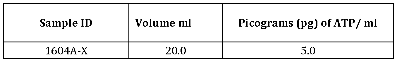
- the filtered lysis reagent is analyzed for the presence of compounds associated with microbial energy such as ATP.
- microbial energy such as ATP.
- Such analysis may, for example, be performed using a luciferase enzyme assay and the spectrophotometric detection of light, based on the following general enzymatic reaction: luciferin + ATP *- luciferyl adenylate + PPi
- kits for monitoring water quality and the presence of microorganisms in water may be used to obtain nucleic acid material.
- the present disclosure provides a kit for obtaining a nucleic acid sample, the kit comprising
- a fluid transfer device to obtain a fluid sample from a source fluid comprising microorganisms
- a filter having a pore size sufficiently small to prevent passage of at least some of the microorganisms through the filter and to permit collection of the microorganisms associated with the filter;
- a lysis reagent which when filtered through the filter is capable of lysing microorganisms collected on the filter, releasing at least some of the nucleic acid material from the microorganisms, and adhering the released nucleic acid material to the filter.
- the kit further includes an extractant capable of eluting the nucleic acid material from the filter.
- the kit in certain embodiments, can also comprise a washing reagent or instructions for making a washing reagent, in which the combination of the lysis reagent and the extractant allows capture of the microorganisms on the filter for subsequent elution and nucleic acid material analysis.
- the kit may include more than one type of sampling device, filter, lysis reagent and extractant.
- the kit comprises a syringe to obtain and transfer a fluid sample, and a filter that may reversibly be coupled to the syringe.
- the filter included in the kit is a microfiber glass membrane filter.
- the kit further comprises instructional material.
- instructional material includes printed matter, e.g., paper or cardboard, a voice or video recording, a diagram, a computer readable medium, such as a disk (floppy diskette or hard disk], optical CD such as CD- or DVD- ROM/RAM, magnetic tape, electrical storage media such as RAM and ROM, and hybrids of these such as magnetic/optical storage media, or any other medium of expression which can be used to communicate the usefulness of the disclosed methodfs] or compositionfs] for its designated use.
- a computer readable medium such as a disk (floppy diskette or hard disk], optical CD such as CD- or DVD- ROM/RAM, magnetic tape, electrical storage media such as RAM and ROM, and hybrids of these such as magnetic/optical storage media, or any other medium of expression which can be used to communicate the usefulness of the disclosed methodfs] or compositionfs] for its designated use.
- the instructional material may comprise instructions on how to collect a fluid sample, how to use the filter, how to make and/or use the lysis reagent, or how to make and/or use the extractant, thus providing guidance to a user of the kit with respect to use the kit's contents.
- the instructional material of the kit of the present disclosure may, for example, be provided within the kit, affixed to the kit, for example in the form of a label, or attached to a vial or tube containing reagents of the kit, or the instructional material may be separated from the kit but be shipped together with the kit. Alternatively, the instructional material may be shipped separately from the kit, with the intention that the instructional material and the composition be used cooperatively by the recipient.
- the present disclosure provides a use of a kit to evaluate the in situ identity of microorganisms in a fluid, the kit comprising:
- a fluid transfer device to obtain a fluid sample from a source fluid comprising microorganisms
- a filter having a pore size sufficiently small to prevent passage of at least some of the microorganisms through the filter and to permit collection of the microorganisms associated with the filter;
- a lysis reagent which when filtered through the filter is capable of lysing microorganisms collected on the filter, release of at least some of the nucleic acid material from the microorganisms, and adherence of the released nucleic acid material to the filter.
- Example 1 Evaluation of a first water sample from an oil production facility
- a first water sample was obtained from an oil production facility (sample # 1105] and treated and evaluated using the device and method described herein above with respect to FIG. 1 - FIG. 5C.
- Both filters were then treated with lysis reagent (phosphoric acid, trisodium salt, dodecahydrate, 1-5%; sodium hydroxide, 0.1-1%; alkyl dimethyl benzyl ammonium chloride, 0.1-1%] by passing the lysis reagent through the filters using the syringes. Thereafter a respective syringe volume of air was passed through the filters to remove remaining amounts of lysis reagent and to dry the filters. The filtrates were collected and used for ATP analysis using a luciferase enzyme assay.
- lysis reagent phosphoric acid, trisodium salt, dodecahydrate, 1-5%; sodium hydroxide, 0.1-1%; alkyl dimethyl benzyl ammonium chloride, 0.1-1%
- the filtrate was the same 1 ml of lysing agent and the other filtrate was diluted with the 9 ml of Reagent D, and the filters were stored at 0 °C in a self-sealing bag for further treatment and analysis. Following storage, the filters were opened and the DNA was extracted from the filters using a MoBio PowerMag® Soil DNA Isolation kit as the extractant (MoBio Catalog No 2700-4-EP] with a bead beating step. [000131] Samples representing various fluid fractions obtained in the performance of the foregoing process (as outlined in Table 1] were subsequently subjected to 16S rRNA sequencing and microbial constituents were evaluated at a genus level. Results of the evaluation are shown in FIG. 6.
- the PCR yield value is a measure of the efficiency and specificity of the PCR reaction and the DNA quality. Higher values represent higher PCR efficiency and DNA concentration, and are indicative of a more accurate sequencing result. In general, for environmental samples, a yield at least 2 ng/ ⁇ ., is considered acceptable.
- the similar values obtained for samples 1105 Whole Sample, X Filter and Dil Filter indicate that these samples contained similar amounts and quality of DNA, while samples 1105-X and 1105-D did not contain sufficient DNA to obtain accurate sequencing data. Thus the PCR yield values of the 1105-X and 1105-D samples indicate that these samples are less suitable for collecting DNA samples for sequencing.
- FIG. 7 provides a principal component analysis of the various fluid fractions.
- Principal component analysis is mathematically defined as an orthogonal linear transformation, i.e. a statistical procedure used to explore variation and highlight patterns within complex datasets. This analysis was done using Mothur, an open source program for bioinformatics analysis (Schloss, P.D., et ah, Introducing mothur: Open-source, platform- independent, community-supported software for describing and comparing microbial communities. Appl Environ Microbiol, 2009. 75(23]:7537-41].
- Table 1 Various fluid fractions used for microbial profile comparisons.
- Example 2 Evaluation of a second water sample from an oil production facility
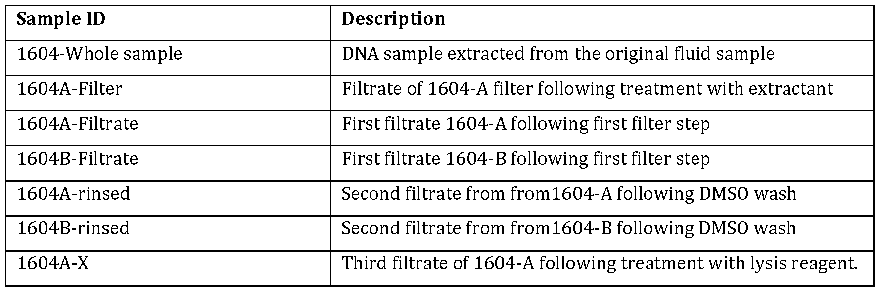
- sample #1604 A second water sample was obtained from an oil production facility (sample #1604] and treated and evaluated using the device and method described herein above with respect to FIG. 1 - FIG. 5C.
- sample #1604 It was determined that the total solids and hydrocarbon in sample #1604 exceeded 10% and/or 100,000 mg/L, respectively. It was therefore deemed desirable to wash the filter prior to treating the filter with lysis reagent.
- two sterile syringes two respective fluid samples of approximately 20 ml were filtered through two respective 0.7 ⁇ sterile filters having a diameter of approximately 3 cm (“1604-A filter" and "1604-B filter”]. The first filtrate samples were collected for analysis.
- both filters were then washed using 60-100% dimethyl sulfoxide as a washing reagent.
- the second filtrates were also collected for analysis.
- Both filters were then treated with lysis reagent (phosphoric acid, trisodium salt, dodecahydrate, 1-5%; sodium hydroxide, 0.1-1%; alkyl dimethyl benzyl ammonium chloride, 0.1-1%] by passing the lysis reagent through the filters using the respective syringes. Thereafter a syringe volume of air was passed through the filters to remove remaining amounts of lysis reagent and to dry the filters.
- lysis reagent phosphoric acid, trisodium salt, dodecahydrate, 1-5%; sodium hydroxide, 0.1-1%; alkyl dimethyl benzyl ammonium chloride, 0.1-1%
- the final and third filtrates were collected and used for ATP analysis using a luciferase enzyme assay, and the filters were stored at 0 °C in a self-sealing bag for further treatment and analysis. Following storage, the filters were opened and DNA extracted for 16S analysis directly from the surface of the filter using the same procedure as described in Example 1.
- PCR yield values are indicated in FIG. 8 .
- FIG. 9 provides a principal component analysis of the various fluid fractions, conducted using Mothur as explained in Example 1.
- Table 3 Various fluid fractions used for microbial profile comparisons.
- a total of 2 x 50 ml of samples from each source provided in Table 5 were obtained and bacterial cells were prepared by centrifugation of the sample at 4,000 rpm for 10 minutes and resuspending the cell pellet in 2 x 200 ⁇ of a 0.9% saline solution to obtain two cell suspensions.
- One of the two cell suspensions was used to directly quantify nucleic acids present therein.
- the other cell suspension was filtered, treated with lysis reagent, and thereafter filter-associated nucleic acid material was extracted. Prior to extraction, filters were stored at either 4 °C or 25 °C. Nucleic acid extractions were performed either immediately upon filtration or following storage of the filters for a period of 7, 14 or 28 days, at the noted temperatures.
- Lysis reagent, filters, and extraction reagent were constituted and employed as described in Example 1. Nucleic acid material from the original cell suspension, nucleic acid material extracted from the stored filters, and nucleic acid material from the day-0 filtrate sample was obtained and the nucleic acid concentration in each was determined using a Nanodrop 2000 (Thermo Scientific] (see: FIG. 10]. The samples were also subjected to 16S rRNA gene metagenomics sequencing on an Illumina MiSeq platform using universal 16S primers 926f and 1392r (Golby, S et al, 2014, Microbial Ecology 68: 70-80; Ramos-Padron, 2011, S.
- Sequencing reads were quality filtered and microorganisms were identified using the GreenGenes database through the Illumina Basespace 16S Metagenomics app. Ver. 1.01 (Illumina Inc.], which is itself an implementation of the Ribosomal Database Project Classifier (Wang, Q. et ah, 2007, Applied and Environmental Microbiology, 73: 5261-5267.
- results of measurements to determine the concentration of DNA in various samples are shown in FIG. 10. It is noted that substantial quantities of DNA are present in both the original cell suspension and in the day-0 filtrate sample, indicating that the day-0 filtrate sample is suitable to use for ATP analysis.
- FIG. 11 A - FIG. 11B Further results are shown in FIG. 11 A - FIG. 11B, showing bar profiles obtained from nucleic acid material from Source ID F and H samples, respectively. As noted in Example 1 similarity in bar profiles signifies that the constituent microbial genera in samples are similar, both in kind and quantity. It is noted that day-0 filtrate profiles in both Source ID F nucleic acid material and Source ID H nucleic acid material differs markedly from bar profiles obtained from any of the filter nucleic acid material.
- nucleic acid material identified in the filtrate is not representative for the nucleic acid material present in the original source sample.
- bar profiles obtained using nucleic acid material from non-stored filters are very similar to bar profiles obtained from the source sample.
- filter- associated nucleic acid material obtained immediately following sampling is similar to nucleic acid material in the sample.
- bar profiles of nucleic acid material obtained from filters stored at 4 °C remain relatively similar to bar profiles of nucleic material obtained from filters from which nucleic acid material is immediately extracted following sampling. This is in particular the case when filters are stored for 7 or 14 days.
- bar profiles of nucleic acid material obtained from filters stored at 25 °C, particularly when stored for 14 and 28 days deviate more significantly from bar profiles of nucleic acid material from filters from which nucleic acid material is immediately extracted following sampling.
- FIG. 12A - FIG. 12B provide a principal component analysis of the various fluid fractions, conducted using Mothur.
- the plotting of points close to one another indicates that the microbial profiles between two samples are similar.
- the results shown in the Mothur plots closely correspond with the findings shown in the bar profiles in FIG. 11A - FIG. 11B samples having Source ID F and H.
- filter communities specifically those stored at 4°C are on the whole adequately representative of original communities (indicated by prevalence of dark shades of grey], although the extent of similarity is source-dependent. Storage of filters at 25°C allows for microbial community profile shifts, as indicated by the prevalence of light shades of grey (1 match] and medium shades of grey (3-4 matches] for 14 and 28-day storage at this temperature.
- SEQ ID NO: 1 is a synthetic polynucleotide primer known as 8F
- SEQ ID NO: 2 is a synthetic polynucleotide primer known as U1492R
- SEQ ID NO: 3 is a synthetic polynucleotide primer known as 928F
- SEQ ID NO: 4 is a synthetic polynucleotide primer known as 336R
- SEQ ID NO: 5 is a synthetic polynucleotide primer known as HOOF
- SEQ ID NO: 6 is a synthetic polynucleotide primer known as 1100R
- SEQ ID NO: 7 is a synthetic polynucleotide primer known as 337F
- SEQ ID NO: 8 is a synthetic polynucleotide primer known as 907R
- SEQ ID NO: 9 is a synthetic polynucleotide primer known as 785F
- SEQ ID NO: 10 is a synthetic polynucleotide primer known as 805R
- SEQ ID NO: 11 is a synthetic polynucleotide primer known as 533F
- SEQ ID NO: 12 is a synthetic polynucleotide primer known as 518R
- SEQ ID NO: 13 is a synthetic polynucleotide primer known as 27F
- SEQ ID NO: 14 is a synthetic polynucleotide primer known as 1429R
Landscapes
- Chemical & Material Sciences (AREA)
- Life Sciences & Earth Sciences (AREA)
- Engineering & Computer Science (AREA)
- Health & Medical Sciences (AREA)
- Organic Chemistry (AREA)
- Zoology (AREA)
- Wood Science & Technology (AREA)
- Genetics & Genomics (AREA)
- Analytical Chemistry (AREA)
- Bioinformatics & Cheminformatics (AREA)
- Biotechnology (AREA)
- Proteomics, Peptides & Aminoacids (AREA)
- General Engineering & Computer Science (AREA)
- Biomedical Technology (AREA)
- Biochemistry (AREA)
- Microbiology (AREA)
- General Health & Medical Sciences (AREA)
- Biophysics (AREA)
- Physics & Mathematics (AREA)
- Molecular Biology (AREA)
- Immunology (AREA)
- Crystallography & Structural Chemistry (AREA)
- Plant Pathology (AREA)
- Mycology (AREA)
- Medicinal Chemistry (AREA)
- Tropical Medicine & Parasitology (AREA)
- Virology (AREA)
- Measuring Or Testing Involving Enzymes Or Micro-Organisms (AREA)
- Apparatus Associated With Microorganisms And Enzymes (AREA)
Abstract
Description


Claims
Priority Applications (4)
| Application Number | Priority Date | Filing Date | Title |
|---|---|---|---|
| EP17755689.1A EP3420081A4 (en) | 2016-02-24 | 2017-02-24 | Detection of microorganisms in fluids |
| CA3015553A CA3015553A1 (en) | 2016-02-24 | 2017-02-24 | Detection of microorganisms in fluids |
| AU2017222712A AU2017222712A1 (en) | 2016-02-24 | 2017-02-24 | Detection of microorganisms in fluids |
| US16/079,240 US20190055542A1 (en) | 2016-02-24 | 2017-02-24 | Detection of microorganisms in fluids |
Applications Claiming Priority (4)
| Application Number | Priority Date | Filing Date | Title |
|---|---|---|---|
| US201662299148P | 2016-02-24 | 2016-02-24 | |
| US62/299,148 | 2016-02-24 | ||
| US201662428742P | 2016-12-01 | 2016-12-01 | |
| US62/428,742 | 2016-12-01 |
Publications (1)
| Publication Number | Publication Date |
|---|---|
| WO2017143452A1 true WO2017143452A1 (en) | 2017-08-31 |
Family
ID=59685868
Family Applications (1)
| Application Number | Title | Priority Date | Filing Date |
|---|---|---|---|
| PCT/CA2017/050241 WO2017143452A1 (en) | 2016-02-24 | 2017-02-24 | Detection of microorganisms in fluids |
Country Status (5)
| Country | Link |
|---|---|
| US (1) | US20190055542A1 (en) |
| EP (1) | EP3420081A4 (en) |
| AU (1) | AU2017222712A1 (en) |
| CA (1) | CA3015553A1 (en) |
| WO (1) | WO2017143452A1 (en) |
Cited By (3)
| Publication number | Priority date | Publication date | Assignee | Title |
|---|---|---|---|---|
| WO2020040577A1 (en) * | 2018-08-22 | 2020-02-27 | 울산대학교 산학협력단 | Method for pathogen enrichment and nucleic acid extraction using device for point-of-care testing |
| WO2020128503A1 (en) * | 2018-12-21 | 2020-06-25 | Nature Metrics Ltd | Filter assembly, kit and methods |
| FR3101884A1 (en) * | 2019-10-11 | 2021-04-16 | Hitachi High-Tech Corporation | Microbiota trapping device, microbiota trapping device, and microbiota trapping method |
Families Citing this family (2)
| Publication number | Priority date | Publication date | Assignee | Title |
|---|---|---|---|---|
| CN109852532A (en) * | 2019-04-01 | 2019-06-07 | 中国科学院深海科学与工程研究所 | A kind of profound and subtle biotinylated nucleic acid in-situ extraction device in full sea |
| US20200340038A1 (en) * | 2019-04-25 | 2020-10-29 | Nephros Inc. | Portable System and Process Thereof to Rapidly Filter, Concentrate, and Detect Waterborne Pathogens |
Citations (2)
| Publication number | Priority date | Publication date | Assignee | Title |
|---|---|---|---|---|
| US20050115903A1 (en) * | 2002-04-09 | 2005-06-02 | Genesystems | Method and apparatus for extracting nucleic acids from a complex mixture |
| US20080113357A1 (en) * | 2006-06-29 | 2008-05-15 | Millipore Corporation | Filter device for the isolation of a nucleic acid |
Family Cites Families (4)
| Publication number | Priority date | Publication date | Assignee | Title |
|---|---|---|---|---|
| DE60135830D1 (en) * | 2000-02-08 | 2008-10-30 | Millipore Corp | METHOD FOR COUNTING AND IDENTIFICATION OF MICROORGANISMS |
| FR2885140B1 (en) * | 2005-04-29 | 2010-12-31 | Millipore Corp | METHOD FOR DETECTING AND CHARACTERIZING MICROORGARISTS ON A MEMBRANE |
| JP4874008B2 (en) * | 2005-07-01 | 2012-02-08 | 水ing株式会社 | Method and apparatus for detecting bacteria |
| US8546127B2 (en) * | 2008-06-30 | 2013-10-01 | General Electric Company | Bacteria/RNA extraction device |
-
2017
- 2017-02-24 CA CA3015553A patent/CA3015553A1/en not_active Abandoned
- 2017-02-24 WO PCT/CA2017/050241 patent/WO2017143452A1/en active Application Filing
- 2017-02-24 AU AU2017222712A patent/AU2017222712A1/en not_active Abandoned
- 2017-02-24 US US16/079,240 patent/US20190055542A1/en not_active Abandoned
- 2017-02-24 EP EP17755689.1A patent/EP3420081A4/en not_active Withdrawn
Patent Citations (2)
| Publication number | Priority date | Publication date | Assignee | Title |
|---|---|---|---|---|
| US20050115903A1 (en) * | 2002-04-09 | 2005-06-02 | Genesystems | Method and apparatus for extracting nucleic acids from a complex mixture |
| US20080113357A1 (en) * | 2006-06-29 | 2008-05-15 | Millipore Corporation | Filter device for the isolation of a nucleic acid |
Non-Patent Citations (5)
| Title |
|---|
| CHANDLER, D. ET AL.: "Profiling in situ microbial community structure with an amplification microarray", APPLIED AND ENVIRONMENTAL MICROBIOLOGY, vol. 79, no. 30, February 2013 (2013-02-01), pages 799 - 807, XP055412948 * |
| FRIAS-LOPEZ, J. ET AL.: "Microbial community gene expression in ocean surface waters", PNAS, vol. 105, no. 10, 11 March 2008 (2008-03-11), pages 3805 - 3810, XP055412955 * |
| See also references of EP3420081A4 * |
| SOMERVILLE, C. ET AL.: "Simple, rapid method for direct isolation of nucleic acids from aquatic environments", APPLIED AND ENVIRONMENTAL MICROBIOLOGY, vol. 55, no. 3, March 1989 (1989-03-01), pages 548 - 554, XP008026151 * |
| YAMANAKA, H. ET AL.: "A simple method for preserving environmental DNA in water samples at ambient temperature by addition of cationic surfactant", LIMNOLOGY, vol. 18, no. 2, 19 November 2016 (2016-11-19), pages 233 - 241, XP036200270 * |
Cited By (6)
| Publication number | Priority date | Publication date | Assignee | Title |
|---|---|---|---|---|
| WO2020040577A1 (en) * | 2018-08-22 | 2020-02-27 | 울산대학교 산학협력단 | Method for pathogen enrichment and nucleic acid extraction using device for point-of-care testing |
| JP2021534751A (en) * | 2018-08-22 | 2021-12-16 | インフュージョン テック | Pathogen concentration and nucleic acid extraction method using on-site diagnostic equipment |
| WO2020128503A1 (en) * | 2018-12-21 | 2020-06-25 | Nature Metrics Ltd | Filter assembly, kit and methods |
| GB2580099A (en) * | 2018-12-21 | 2020-07-15 | Nature Metrics Ltd | Filter assembly |
| GB2580099B (en) * | 2018-12-21 | 2022-04-13 | Nature Metrics Ltd | Filter assembly |
| FR3101884A1 (en) * | 2019-10-11 | 2021-04-16 | Hitachi High-Tech Corporation | Microbiota trapping device, microbiota trapping device, and microbiota trapping method |
Also Published As
| Publication number | Publication date |
|---|---|
| AU2017222712A1 (en) | 2018-09-20 |
| EP3420081A4 (en) | 2019-08-14 |
| US20190055542A1 (en) | 2019-02-21 |
| EP3420081A1 (en) | 2019-01-02 |
| CA3015553A1 (en) | 2017-08-31 |
Similar Documents
| Publication | Publication Date | Title |
|---|---|---|
| US20190055542A1 (en) | Detection of microorganisms in fluids | |
| Sheik et al. | Identification and removal of contaminant sequences from ribosomal gene databases: lessons from the census of deep life | |
| Dineen et al. | An evaluation of commercial DNA extraction kits for the isolation of bacterial spore DNA from soil | |
| Debeljak et al. | Extracting DNA from ocean microplastics: a method comparison study | |
| Conradie et al. | The dynamics of toxic Microcystis strains and microcystin production in two hypertrofic South African reservoirs | |
| CN103993007B (en) | The simple and easy method of a kind of high efficiency extraction DNA from pedotheque | |
| Pepper et al. | Environmental sample collection and processing | |
| CN102906262A (en) | Procedure for the specific isolation of total DNA content of bacterial germs and a kit for this purpose | |
| Oh et al. | Simultaneous quantification of cyanobacteria and Microcystis spp. using real-time PCR | |
| Deng et al. | Improving the accuracy of flow cytometric quantification of microbial populations in sediments: importance of cell staining procedures | |
| JP5610416B2 (en) | Environmental impact assessment method in bioaugmentation | |
| Abd-El-Haleem et al. | Multiplex-PCR and PCR-RFLP assays to monitor water quality against pathogenic bacteria | |
| CN102337347B (en) | Characteristic nucleotide sequence for identifying ophiocordyceps crinalis, as well as probes and method thereof | |
| Räsänen et al. | Comparison of culture and qPCR methods in detection of mycobacteria from drinking waters | |
| Stelzer et al. | Relations between DNA-and RNA-based molecular methods for cyanobacteria and microcystin concentration at Maumee Bay State Park Lakeside Beach, Oregon, Ohio, 2012 | |
| Roux et al. | When phytoplankton do not bloom: the case of the dinoflagellate Lepidodinium chlorophorum in southern Brittany (France) assessed by environmental DNA | |
| Tsesmetzis et al. | Protocols for investigating the microbial communities of oil and gas reservoirs | |
| Gobbi et al. | Quantitative and qualitative evaluation of the impact of the G2 enhancer, bead sizes and lysing tubes on the bacterial community composition during DNA extraction from recalcitrant soil core samples based on community sequencing and qPCR | |
| Pickup | Sampling and detecting bacterial populations in natural environments | |
| Colman et al. | Tectonic and geological setting influence hot spring microbiology | |
| Boundy-Mills | Methods for the isolation and investigation of the diversity of cold-adapted yeasts and their ex situ preservation in worldwide collections | |
| Masseret et al. | A simple and innovative method for species identification of phytoplankton cells on minute quantities of DNA | |
| Price | Novel DNA extraction and preservation for identification of micro-organisms on-site using novel nucleic acid extraction cards for the oil and gas industry | |
| DE102007028207B3 (en) | Detecting pathogenic Legionella microorganisms in water from a pipe system comprises performing a polymerase chain reaction on DNA from microorganisms filtered from a large volume of water | |
| Shelton et al. | Repetitive sampling and control threshold improve 16S rRNA gene sequencing results from produced waters associated with hydraulically fractured shale |
Legal Events
| Date | Code | Title | Description |
|---|---|---|---|
| WWE | Wipo information: entry into national phase |
Ref document number: 3015553 Country of ref document: CA |
|
| NENP | Non-entry into the national phase |
Ref country code: DE |
|
| ENP | Entry into the national phase |
Ref document number: 2017222712 Country of ref document: AU Date of ref document: 20170224 Kind code of ref document: A |
|
| WWE | Wipo information: entry into national phase |
Ref document number: 2017755689 Country of ref document: EP |
|
| ENP | Entry into the national phase |
Ref document number: 2017755689 Country of ref document: EP Effective date: 20180924 |
|
| 121 | Ep: the epo has been informed by wipo that ep was designated in this application |
Ref document number: 17755689 Country of ref document: EP Kind code of ref document: A1 |