WO2017142069A1 - Cell culture medium, culture method, and organoid - Google Patents
Cell culture medium, culture method, and organoid Download PDFInfo
- Publication number
- WO2017142069A1 WO2017142069A1 PCT/JP2017/005946 JP2017005946W WO2017142069A1 WO 2017142069 A1 WO2017142069 A1 WO 2017142069A1 JP 2017005946 W JP2017005946 W JP 2017005946W WO 2017142069 A1 WO2017142069 A1 WO 2017142069A1
- Authority
- WO
- WIPO (PCT)
- Prior art keywords
- cells
- culture medium
- cell culture
- cell
- epithelial
- Prior art date
Links
Images


Classifications
-
- C—CHEMISTRY; METALLURGY
- C12—BIOCHEMISTRY; BEER; SPIRITS; WINE; VINEGAR; MICROBIOLOGY; ENZYMOLOGY; MUTATION OR GENETIC ENGINEERING
- C12N—MICROORGANISMS OR ENZYMES; COMPOSITIONS THEREOF; PROPAGATING, PRESERVING, OR MAINTAINING MICROORGANISMS; MUTATION OR GENETIC ENGINEERING; CULTURE MEDIA
- C12N5/00—Undifferentiated human, animal or plant cells, e.g. cell lines; Tissues; Cultivation or maintenance thereof; Culture media therefor
- C12N5/0018—Culture media for cell or tissue culture
- C12N5/0037—Serum-free medium, which may still contain naturally-sourced components
-
- A—HUMAN NECESSITIES
- A61—MEDICAL OR VETERINARY SCIENCE; HYGIENE
- A61P—SPECIFIC THERAPEUTIC ACTIVITY OF CHEMICAL COMPOUNDS OR MEDICINAL PREPARATIONS
- A61P1/00—Drugs for disorders of the alimentary tract or the digestive system
-
- C—CHEMISTRY; METALLURGY
- C12—BIOCHEMISTRY; BEER; SPIRITS; WINE; VINEGAR; MICROBIOLOGY; ENZYMOLOGY; MUTATION OR GENETIC ENGINEERING
- C12N—MICROORGANISMS OR ENZYMES; COMPOSITIONS THEREOF; PROPAGATING, PRESERVING, OR MAINTAINING MICROORGANISMS; MUTATION OR GENETIC ENGINEERING; CULTURE MEDIA
- C12N5/00—Undifferentiated human, animal or plant cells, e.g. cell lines; Tissues; Cultivation or maintenance thereof; Culture media therefor
- C12N5/06—Animal cells or tissues; Human cells or tissues
- C12N5/0602—Vertebrate cells
- C12N5/0676—Pancreatic cells
- C12N5/0677—Three-dimensional culture, tissue culture or organ culture; Encapsulated cells
-
- C—CHEMISTRY; METALLURGY
- C12—BIOCHEMISTRY; BEER; SPIRITS; WINE; VINEGAR; MICROBIOLOGY; ENZYMOLOGY; MUTATION OR GENETIC ENGINEERING
- C12N—MICROORGANISMS OR ENZYMES; COMPOSITIONS THEREOF; PROPAGATING, PRESERVING, OR MAINTAINING MICROORGANISMS; MUTATION OR GENETIC ENGINEERING; CULTURE MEDIA
- C12N5/00—Undifferentiated human, animal or plant cells, e.g. cell lines; Tissues; Cultivation or maintenance thereof; Culture media therefor
- C12N5/06—Animal cells or tissues; Human cells or tissues
- C12N5/0602—Vertebrate cells
- C12N5/0693—Tumour cells; Cancer cells
- C12N5/0695—Stem cells; Progenitor cells; Precursor cells
-
- C—CHEMISTRY; METALLURGY
- C12—BIOCHEMISTRY; BEER; SPIRITS; WINE; VINEGAR; MICROBIOLOGY; ENZYMOLOGY; MUTATION OR GENETIC ENGINEERING
- C12N—MICROORGANISMS OR ENZYMES; COMPOSITIONS THEREOF; PROPAGATING, PRESERVING, OR MAINTAINING MICROORGANISMS; MUTATION OR GENETIC ENGINEERING; CULTURE MEDIA
- C12N5/00—Undifferentiated human, animal or plant cells, e.g. cell lines; Tissues; Cultivation or maintenance thereof; Culture media therefor
- C12N5/10—Cells modified by introduction of foreign genetic material
-
- C—CHEMISTRY; METALLURGY
- C12—BIOCHEMISTRY; BEER; SPIRITS; WINE; VINEGAR; MICROBIOLOGY; ENZYMOLOGY; MUTATION OR GENETIC ENGINEERING
- C12N—MICROORGANISMS OR ENZYMES; COMPOSITIONS THEREOF; PROPAGATING, PRESERVING, OR MAINTAINING MICROORGANISMS; MUTATION OR GENETIC ENGINEERING; CULTURE MEDIA
- C12N2501/00—Active agents used in cell culture processes, e.g. differentation
- C12N2501/10—Growth factors
- C12N2501/11—Epidermal growth factor [EGF]
-
- C—CHEMISTRY; METALLURGY
- C12—BIOCHEMISTRY; BEER; SPIRITS; WINE; VINEGAR; MICROBIOLOGY; ENZYMOLOGY; MUTATION OR GENETIC ENGINEERING
- C12N—MICROORGANISMS OR ENZYMES; COMPOSITIONS THEREOF; PROPAGATING, PRESERVING, OR MAINTAINING MICROORGANISMS; MUTATION OR GENETIC ENGINEERING; CULTURE MEDIA
- C12N2501/00—Active agents used in cell culture processes, e.g. differentation
- C12N2501/10—Growth factors
- C12N2501/117—Keratinocyte growth factors (KGF-1, i.e. FGF-7; KGF-2, i.e. FGF-12)
-
- C—CHEMISTRY; METALLURGY
- C12—BIOCHEMISTRY; BEER; SPIRITS; WINE; VINEGAR; MICROBIOLOGY; ENZYMOLOGY; MUTATION OR GENETIC ENGINEERING
- C12N—MICROORGANISMS OR ENZYMES; COMPOSITIONS THEREOF; PROPAGATING, PRESERVING, OR MAINTAINING MICROORGANISMS; MUTATION OR GENETIC ENGINEERING; CULTURE MEDIA
- C12N2501/00—Active agents used in cell culture processes, e.g. differentation
- C12N2501/10—Growth factors
- C12N2501/155—Bone morphogenic proteins [BMP]; Osteogenins; Osteogenic factor; Bone inducing factor
-
- C—CHEMISTRY; METALLURGY
- C12—BIOCHEMISTRY; BEER; SPIRITS; WINE; VINEGAR; MICROBIOLOGY; ENZYMOLOGY; MUTATION OR GENETIC ENGINEERING
- C12N—MICROORGANISMS OR ENZYMES; COMPOSITIONS THEREOF; PROPAGATING, PRESERVING, OR MAINTAINING MICROORGANISMS; MUTATION OR GENETIC ENGINEERING; CULTURE MEDIA
- C12N2501/00—Active agents used in cell culture processes, e.g. differentation
- C12N2501/40—Regulators of development
- C12N2501/415—Wnt; Frizzeled
-
- C—CHEMISTRY; METALLURGY
- C12—BIOCHEMISTRY; BEER; SPIRITS; WINE; VINEGAR; MICROBIOLOGY; ENZYMOLOGY; MUTATION OR GENETIC ENGINEERING
- C12N—MICROORGANISMS OR ENZYMES; COMPOSITIONS THEREOF; PROPAGATING, PRESERVING, OR MAINTAINING MICROORGANISMS; MUTATION OR GENETIC ENGINEERING; CULTURE MEDIA
- C12N2533/00—Supports or coatings for cell culture, characterised by material
- C12N2533/90—Substrates of biological origin, e.g. extracellular matrix, decellularised tissue
Definitions
- the present invention relates to a cell culture medium, a culture method, and an organoid.
- This application claims priority based on Japanese Patent Application No. 2016-029060 filed in Japan on February 18, 2016, the contents of which are incorporated herein by reference.
- the intestinal tract is the organ with the largest area in contact with the outside world in the human body, and has essential functions for maintaining life such as digestion and absorption. Most of the intestinal function is carried by the intestinal epithelium that covers the inner layer.
- the intestinal epithelium is composed of two compartments: villi composed of three types of differentiated cells (mucus producing cells, absorbing epithelial cells, endocrine cells) and crypts composed mainly of undifferentiated proliferating cells. In the small intestine crypts, Paneth cells that produce antimicrobial peptides are present at the bottom of the crypt.
- Lgr5-positive cells also referred to as “CBC (Crypt base column) cells
- CBC Codpt base column
- Lgr5-positive intestinal epithelial stem cells produce progenitor cells called Transit amplifying cells, but these progenitor cells have no permanent self-renewal ability and are limited to 1 to 3 lineages Is done.
- Transient proliferating cells differentiate in crypts with 2-4 divisions and terminal differentiation in villi. These differentiated cells detach at the apex of the villus and die by apoptosis.
- the intestinal epithelium is a rapidly metabolizing tissue that travels from the crypt stem cells to the apex of the villi in 4-5 days. Unlike other differentiated cells, Paneth cells move to the bottom of the crypt with differentiation and have a long cell life of 2 months.
- Wnt Wnt
- BMP bone morphogenetic protein
- intestinal epithelial stem cells are adhered to an extracellular matrix and cultured in the presence of a cell culture medium containing a basic medium for animal or human cells, to which a BMP inhibitor, a mitogenic growth factor, and a Wnt agonist are added. As a result, the intestinal epithelial stem cells have been successfully maintained for a long time (see, for example, Patent Document 1).
- Wnt protein is a supernatant obtained by culturing mouse fibroblast-derived L cells overexpressing the Wnt gene with serum (hereinafter referred to as “Wnt with serum”). Called “supernatant”). Animal serum such as fetal calf serum contained in Wnt supernatant containing serum is essential for stabilization of Wnt protein.
- Wnt protein purified from the serum-containing Wnt supernatant can be stabilized by a surfactant, but the purified Wnt protein has a significantly lower activity than the serum-containing Wnt supernatant.
- the present invention provides a cell culture medium capable of culturing epithelial stem cells, epithelial cancer cells, or tissues containing at least one of these cells without serum for a long period of time.
- the purpose is to make it possible to produce serum-free human cultured organoids, and to make it possible to cultivate from tissues that were impossible until now.
- the present inventors have found that a protein called afamin in serum contributes to the stabilization and solubilization of Wnt protein, thereby completing the present invention. It was.
- the present inventors pay attention to the fact that they can be stabilized in a water-soluble medium of Wnt protein, which is a fat-soluble protein, in the presence of afamin contained in serum, and by using a medium containing Wnt protein and afamin, serum-free
- the present invention which is a culture medium containing no surfactant and a culture method using the culture medium, has been completed.
- a complex of Wnt protein and its stabilizing substance afamin and a Wnt agonist comprising R-spondin, a mitogenic growth factor, a bone morphogenetic protein (BMP) inhibitor, and transformation
- a cell culture medium comprising at least one selected from the group consisting of a growth factor- ⁇ (TGF- ⁇ ) inhibitor and a p38 inhibitor.
- the Wnt protein consists of Wnt1, Wnt2, Wnt2b, Wnt3, Wnt3a, Wnt4, Wnt5a, Wnt5b, Wnt6, Wnt7a, Wnt7b, Wnt8a, Wnt8b, Wnt9a, Wt9a, Wt9a, Wt10
- [3] The cell culture according to [1] or [2], wherein the R-spondin is at least one selected from the group consisting of R-spondin 1, R-spondin 2, R-spondin 3, and R-spondin 4. Culture medium.
- EGF epidermal growth factor
- the BMP inhibitor is Noggin.
- the TGF- ⁇ inhibitor is A83-01.
- [8] A method for culturing an epithelial stem cell, an epithelial cancer cell, or a tissue containing at least one of these cells, an extracellular matrix preparation step of preparing an extracellular matrix, and an epithelial stem cell on the extracellular matrix
- a method for culturing epithelial stem cells or tissues containing epithelial stem cells comprising an extracellular matrix preparation step for preparing an extracellular matrix, and adhering epithelial stem cells or a tissue containing epithelial stem cells on the extracellular matrix An adhesion step; and an organoid formation step of adding the cell culture medium according to [7] after the adhesion step, culturing the epithelial stem cell or the tissue containing the epithelial stem cell, and forming an organoid.
- a culture method characterized. [10] An organoid obtained by the culture method according to [8] or [9]. [11] The organoid according to [10], which is for regenerative medicine. [12] An organoid obtained by the culture method according to [9] and used for transplantation.
- long-term epithelial stem cells derived from mammals including humans, epithelial cancer cells, tissues containing at least one of those cells, or tissues that could not be cultured conventionally are long. Can be cultured for a period of time. Moreover, an organoid can be formed with high efficiency from at least one of the cells and the tissue.
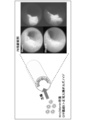
- FIG. (A) is an image showing a state after 6 days from the start of culture of primary culture (Passage 0) of intestinal stem cells in each medium in Example 1.
- FIG. (B) is the graph which quantified the area which the organoid of the intestinal stem cell occupies 6 days after the culture
- (A) is an image showing a state after 6 days from the start of culture of the first passage (Passage 1) of intestinal stem cells in each medium in Example 1.
- FIG. (B) is a graph quantifying the area occupied by organoids of intestinal stem cells in the wells 6 days after the start of culturing in the first passage (Passage 1) in each medium in Example 1.
- FIG. (A) is an image showing a state after 5 days from the start of culture of the second passage (Passage 2) of intestinal stem cells in each medium in Example 1.
- FIG. (B) is a graph quantifying the area occupied by organoids of intestinal stem cells in the wells 5 days after the start of culturing in the second passage (Passage 2) in each medium in Example 1.
- 6 is a schematic cross-sectional view and an image showing a state of exfoliation of the large intestine mucosa with the NOG mouse in Example 3.
- the figure on the left is a schematic diagram schematically showing the state of transplantation of human colon organoids into the colon mucosa exfoliation part of NOG mice in Example 3.
- the right figure is the image in the bright field and the dark field which observed the transplant site
- the upper figure is an image showing the result of staining a hematoxylin-eosin (HE) -stained tissue section of the transplantation site of NOG mice 28 days after transplantation in Example 3.
- the lower left figure shows a tissue section of the transplantation site of NOG mice 28 days after transplantation in Example 3, immunostaining with anti-human cytokeratin antibody and nuclear staining with Hoechst (registered trademark). It is an image which shows the result of having performed.
- the lower right figure shows the tissue section of the transplantation site of NOG mice 28 days after transplantation in Example 3 with detection of Lgr5 by in situ hybridization analysis and nuclear staining using Hoechst (registered trademark). It is an image which shows the result.
- the present invention provides a Wnt agonist comprising a complex of Wnt protein and its stabilizing substance afamin and R-spondin, a mitogenic growth factor, bone morphogenetic protein; BMP And a cell culture medium containing at least one selected from the group consisting of an inhibitor, a transforming growth factor- ⁇ (TGF- ⁇ ) inhibitor, and a p38 inhibitor.
- a Wnt agonist comprising a complex of Wnt protein and its stabilizing substance afamin and R-spondin, a mitogenic growth factor, bone morphogenetic protein
- BMP And a cell culture medium containing at least one selected from the group consisting of an inhibitor, a transforming growth factor- ⁇ (TGF- ⁇ ) inhibitor, and a p38 inhibitor.
- TGF- ⁇ transforming growth factor- ⁇
- the cell culture medium of this embodiment is substantially free of impurities, and can culture epithelial stem cells, epithelial cancer cells, or tissues containing at least one of these cells for a long period of time.
- an organoid can be formed with high efficiency from epithelial stem cells, epithelial cancer cells, or tissues containing at least one of these cells.
- epithelial stem cells cultured using the cell culture medium of the present invention can maintain differentiation ability for a long period of time, and the incidence of tumors is extremely low.
- epithelial stem cell means a cell having a long-term self-replicating function and pluripotency into an epithelial differentiated cell, and means a stem cell derived from epithelial tissue.
- epithelial tissues include cornea, oral mucosa, skin, conjunctiva, bladder, tubule, kidney, digestive organs (esophagus, stomach, duodenum, small intestine (including jejunum and ileum), large intestine (including colon)), liver , Pancreas, mammary gland, salivary gland, lacrimal gland, prostate, hair root, trachea, lung and the like.
- the cell culture medium of this embodiment is used for culturing epithelial stem cells derived from the digestive organs (esophagus, stomach, duodenum, small intestine (including jejunum and ileum), large intestine (including colon)), liver, pancreas, among others. It is preferable.
- the “epithelial cancer cell” means a cell in which the above-mentioned epithelial tissue-derived cell has become cancerous.
- organoid means a three-dimensional cellular tissue that is self-assembled by accumulating cells in a controlled space at high density.
- the cell culture medium of this embodiment includes any serum-free cell culture basic medium.
- the cell culture medium of this embodiment is preferably for animal cells or human cells.
- Examples of the serum-free cell culture basic medium include a defined synthetic medium that is buffered to pH 7.2 or more and pH 7.6 or less with a carbonate buffer. More specifically, Dulbecco's Modified Eagle Medium: Nutrient Mixture F-12 (DMEM / F12) supplemented with glutamine, insulin, penicillin or streptomycin, and transferrin. Can be mentioned. Also included is RPMI 1640 medium (Roswell Park Memory Institute 1640 medium) supplemented with glutamine, insulin, penicillin or streptomycin, and transferrin.
- the serum-free cell culture basic medium in the cell culture medium of this embodiment may be further supplemented with a purified natural, semi-synthetic, or synthetic growth factor.
- the growth factor include B-27 Supplement (manufactured by Thermo Fisher SCIENTIFIC), N-acetyl-L-cysteine (manufactured by Sigma), N-2 Supplement (manufactured by Thermo Fisher SCIENTIFIC), and the like. These growth factors can stimulate the growth of some cells.
- the cell culture medium of this embodiment does not substantially contain uncertain components such as fetal bovine serum (FBS) or fetal calf serum.
- FBS fetal bovine serum
- substantially free of uncertain components means that the cell culture medium of the present embodiment does not contain any uncertain components such as serum, that is, the cell culture of the present embodiment.
- the medium is a serum-free medium, or the cell culture medium of the present embodiment means a state in which an uncertain component such as serum is contained only in an amount that does not affect the cells.
- the cell culture medium of this embodiment is a serum-free medium.
- Wnt agonist means an agent that activates T-cell factor (hereinafter also referred to as TCF) / lymphoen enhancer factor (hereinafter also referred to as LEF) -mediated transcription in cells. To do.
- Wnt agonists are not limited to Wnt family proteins, but include Wnt agonists that bind to and activate Frizzled receptor family members, inhibitors of intracellular ⁇ -catenin degradation, and activators of TCF / LEF. .
- the Wnt agonist is at least 1.1 times, preferably at least 1.2 times, more preferably at least 10 times, even more preferably at least 50 times, compared to the level of Wnt activity in the absence of the Wnt agonist.
- Wnt activity is stimulated in the cell so as to achieve a Wnt activity level of at least 100-fold, most preferably at least 300-fold.
- Wnt activity can be determined by measuring the transcriptional activity of Wnt using methods known to those skilled in the art, for example by pTOPFLASH and pFOPFLASH Tcf luciferase reporter constructs (reference: Korinek et al., 1997. Science 275). : 1784-1787).
- the Wnt agonist contained in the cell culture medium of the present embodiment is preferably at least one of a complex of Wnt protein and afamin and R-spondin, and a combination of Wnt protein and afamin. More preferably, both the complex and R-spondin are included.
- the epithelial stem cells can form organoids with higher efficiency.
- Wnt protein The origin of Wnt protein which is a kind of Wnt agonist is not particularly limited, and Wnt proteins derived from various organisms can be used. Among these, a mammal-derived Wnt protein is preferable. Examples of mammals include humans, mice, rats, cows, pigs, rabbits and the like. Mammalian Wnt proteins include Wnt1, Wnt2, Wnt2b, Wnt3, Wnt3a, Wnt4, Wnt5a, Wnt5b, Wnt6, Wnt7a, Wnt7b, Wnt8a, Wnt8b, Wnt9a, Wnt9a, Wnt9a, tnt. In the cell culture medium of this embodiment, a plurality of Wnt proteins may be used in combination.
- Examples of the method for producing Wnt protein include a method for producing Wnt protein-expressing cells.
- the cell origin biological species, culture form, etc.
- Wnt protein-expressing cells include L cells that stably express mouse Wnt3a (ATCC CRL-2647), L cells that stably express mouse Wnt5a (ATCC CRL-2814), and the like.
- Wnt protein-expressing cells can be prepared using a known gene recombination technique.
- a Wnt protein-expressing cell can be prepared.
- the base sequence of the gene encoding the desired Wnt protein can be obtained from a known database such as GenBank.
- the Wnt protein expressed by the Wnt protein-expressing cell may be a fragment of the Wnt protein or may contain an amino acid sequence other than the amino acid sequence of the Wnt protein as long as it has Wnt activity.
- the amino acid sequence other than the amino acid sequence of the Wnt protein is not particularly limited, and examples thereof include an amino acid sequence of an affinity tag.
- the amino acid sequence of the Wnt protein does not need to be completely identical to the amino acid sequence that can be obtained from a known database such as GenBank, and is substantially identical to the amino acid sequence that can be obtained from the known database as long as it has Wnt activity
- GenBank a known database
- amino acid sequence substantially the same as the amino acid sequence of a Wnt protein that can be obtained from a known database such as GenBank include, for example, one to several amino acids deleted, substituted, or substituted in the amino acid sequence that can be obtained from a known database.
- Examples include added amino acid sequences.
- “Amino acid sequence in which one to several amino acids have been deleted, substituted, or added” means, for example, deletion, substitution, or addition by a known mutant peptide production method such as site-directed mutagenesis. It means that as many amino acids as possible (10 or less are preferable, 7 or less are more preferable, and 6 or less are more preferable) are deleted, substituted, or added.
- the identity with an amino acid sequence that can be obtained from a known database is at least 80% or more, preferably at least 85% or more, more preferably at least 90% or more, More preferred are amino acid sequences that are at least 92% or more, particularly preferably at least 95% or more, and most preferably at least 99% or more.
- the activity of Wnt protein can be confirmed by, for example, a TCF reporter assay.
- the TCF reporter assay introduces a luciferase gene having a binding sequence of T-cell factor (TCF), which is a transcription factor that is specifically activated when a Wnt signal enters a cell, and activates Wnt protein activity.
- TCF T-cell factor
- This is a method for simply evaluating the intensity of luciferase by luminescence of luciferase (references: Molenaar et al., Cell, 86, 391, 1996).
- R-spondin examples of R-spondin, which is a type of Wnt agonist, include the R-spondin family consisting of R-spondin 1, R-spondin 2, R-spondin 3, and R-spondin 4.
- the R-spondin family is a secreted protein and is known to be involved in activation and regulation of the Wnt signaling pathway.
- a plurality of R-spondins may be used in combination. As long as it has R-spondin activity, it may be a fragment of R-spondin or may contain an amino acid sequence other than the amino acid sequence of R-spondin.
- the concentration of Wnt protein contained in the cell culture medium of the present embodiment is 50 ng / mL or more, preferably 100 ng / mL or more and 10 ⁇ g / mL or less, more preferably 200 ng / mL or more and 1 ⁇ g / mL or less. Preferably, it is 300 ng / mL or more and 1 ⁇ g / mL or less.
- Wnt agonist is preferably added to the culture medium every two days, and preferably the cell culture medium is replaced with a fresh one every four days.
- afamin means a glycoprotein belonging to the albumin family, and is known to exist in blood or body fluid.
- the serum normally added to the medium used for cell culture contains afamin derived from the animal from which the serum was collected. Since serum contains impurities other than afamin, it is preferable to use afamin alone without using serum in the cell culture medium of this embodiment.
- the origin of the afamin contained in the cell culture medium of the present embodiment is not particularly limited, and various organism-derived afamins can be used. Among these, afamin derived from mammals is preferable. Examples of mammals include those similar to the above-mentioned [Wnt protein].
- the amino acid sequence of main mammalian afamin and the base sequence of the gene encoding it can be obtained from a known database such as GenBank, for example.
- the amino acid sequence of human afamin is registered with AAA21612, the base sequence of the gene encoding it is registered with the accession number of L32140, the amino acid sequence of usiafamin is DAA28569, and the base sequence of the gene encoding it is GJ060968 It is registered with the accession number.
- the afamin contained in the cell culture medium of the present embodiment may be a natural afamin contained in serum or the like purified by a known method, or a recombinant afamin.
- Recombinant afamin can be produced by appropriately using known gene recombination techniques.
- a method for producing recombinant afamin for example, DNA encoding afamin is inserted into a known expression vector, the obtained expression vector is introduced into an appropriate host cell to express recombinant afamin, and a known purification method is used. It can manufacture by refine
- the recombinant afamin may be an afamin to which an affinity tag is added.
- the affinity tag to be added is not particularly limited, and can be appropriately selected from known affinity tags.
- the affinity tag is preferably an affinity tag recognized by a specific antibody, and examples thereof include a FLAG tab, a MYC tag, an HA tag, and a V5 tag.
- the above-mentioned Wnt protein has strong hydrophobicity because a specific serine residue is modified with a fatty acid (palmitoleic acid). Therefore, it is widely known that Wnt protein is very difficult to purify and store because it easily aggregates or denatures in an aqueous solution. On the other hand, modification of this specific serine residue with a fatty acid is essential for the physiological activity of Wnt protein, and has been reported to be involved in binding to Frizzled receptor family members. The present inventors have found that in a water solution, Wnt protein binds to afamin in a one-to-one relationship to form a complex and solubilizes while maintaining high physiological activity.
- Wnt protein-afamin complex may be produced by culturing cells that express both Wnt protein and afamin, and by coculturing Wnt protein-expressing cells and afamin-expressing cells, Wnt protein-afamin complexes may be produced.
- the activity of the Wnt protein in the Wnt protein-afamin complex can be evaluated using the same method as the above [Wnt protein].
- the concentration of afamin contained in the cell culture medium of the present embodiment is not particularly limited, but is preferably 50 ng / mL or more and 10 ⁇ g / mL or less, more preferably 100 ng / mL or more and 1 ⁇ g / mL or less, and 300 ⁇ g. More preferably, it is 1 / g or more and 1 ⁇ g / ml or less.
- EGF epidermal growth factor
- TGF- ⁇ transforming growth factor- ⁇
- bFGF basic Fibroblast growth factor
- BDNF brain-derived neurotrophic factor
- GF keratinocyte growth factor family of GFs such as keratinocyte growth factor;
- EGF is a potent mitogen for various cultured ectoderm and mesoderm cells and has a profound effect on the specific cell differentiation of some fibroblasts.
- the EGF precursor exists as a membrane-bound molecule that is cleaved by proteolysis to produce a 53-amino acid peptide hormone that stimulates the cell.
- EGF is particularly preferable.
- the concentration of EGF contained in the cell culture medium of the present embodiment is preferably 5 ng / mL or more and 500 ng / mL or less, more preferably 10 ng / mL or more and 400 ng / mL or less, and 50 ng / mL or more and 200 ng / mL or less. More preferably, it is not more than mL.
- bFGF preferably FGF10 or FGF7
- FGF7 and FGF10 it is preferable that the total content of FGF is the said range.
- FGF7 and FGF10 it is preferable that the total content of FGF is the said range.
- Bone morphogenetic protein binds as a dimeric ligand to a receptor complex consisting of two different receptor serine / threonine kinases, type I and type II receptors. Type II receptors phosphorylate type I receptors resulting in activation of this receptor kinase. This type I receptor subsequently phosphorylates a specific receptor substrate (SMAD), which leads to transcriptional activity through signal transduction pathways.
- a BMP inhibitor is, for example, an agent that blocks or inhibits the binding of a BMP molecule to a BMP receptor and binds to the BMP molecule to form a complex that neutralizes BMP activity. is there.
- the BMP inhibitor is, for example, an agent that binds to the BMP receptor and prevents or inhibits the binding of the BMP molecule to the receptor, and acts as an antagonist or an inverse agonist.
- the BMP inhibitor preferably has an inhibition of 50% or more, more preferably 70% or more, still more preferably 80% or more, particularly preferably 90% or more compared to the level of BMP activity in the absence of the inhibitor. Has activity.
- the BMP inhibitory activity can be evaluated by measuring the transcriptional activity of BMP using a method known to those skilled in the art (reference: Zilbergberg et al., BMC Cell Biol, 8:41, 2007.). .
- the BMP inhibitor contained in the cell culture medium of the present embodiment is preferably a natural BMP-binding protein.
- chondin-like proteins such as Noggin, Chordin, and chordin domain
- follistatin (Follistatin) Follistatin-related proteins such as follistatin domains
- DAN-like proteins such as DAN and DAN cysteine-knot domains
- sclerostin / SOST decorin
- ⁇ -2 macroglobulin and the like a natural BMP-binding protein.
- Chordin-like proteins and DAN-like proteins are diffusible proteins that can bind to BMP molecules with various affinities and inhibit the access of BMP molecules to signaling receptors.
- the loss of stem cells can be prevented by adding these BMP inhibitors to the cell culture medium.
- the concentration of the BMP inhibitor contained in the cell culture medium of the present embodiment is preferably 10 ng / mL or more and 100 ng / mL or less, more preferably 20 ng / mL or more and 100 ng / mL or less, and more preferably 50 ng / mL or more. More preferably, it is 100 ng / mL or less.
- a mitogenic growth factor to the culture medium every two days, and it is preferable to replace the culture medium with a fresh one every four days.
- TGF- ⁇ Transforming growth factor- ⁇
- TGF- ⁇ is a kind of growth factor and is produced in almost all cells such as kidney, bone marrow, and platelets. There are five subtypes ( ⁇ 1 to ⁇ 5) in TGF- ⁇ . TGF- ⁇ is also known to promote the proliferation of osteoblasts and the synthesis and proliferation of connective tissues such as collagen, and acts to suppress epithelial cell proliferation and osteoclasts. ing.
- TGF- ⁇ inhibitors are, for example, those that prevent or inhibit TGF- ⁇ binding to the TGF- ⁇ receptor to form a complex that neutralizes TGF- ⁇ activity. It is a drug that binds to TGF- ⁇ .
- the TGF- ⁇ inhibitor is, for example, an agent that binds to the TGF- ⁇ receptor and prevents or inhibits the binding of TGF- ⁇ to the receptor, and acts as an antagonist or inverse agonist.
- the TGF- ⁇ inhibitor is preferably at least 50%, more preferably at least 70%, even more preferably at least 80%, particularly preferably 90% compared to the TGF- ⁇ activity level in the absence of this inhibitor. % Inhibitory activity.
- TGF- ⁇ inhibitory activity can be evaluated by methods known to those skilled in the art. Such an evaluation system includes a cell assay in which cells are stably transfected with a human PAI-1 promoter that moves a luciferase reporter gene or a reporter construct containing an Smad binding site (reference: De Gouvillevet al. , Br J Pharmacol, 145 (2): 166-177, 2005.).
- TGF- ⁇ inhibitor contained in the cell culture medium of the present embodiment examples include A83-01 (3- (6-methylpyridin-2-yl) -1-phenylthiocarbamoyl-4-quinolin-4-yl. Pyrazole), ALK5 Inhibitor I (3- (pyridin-2-yl) -4- (4-quinonyl) -1H-pyrazole), LDN193189 (4- (6- (4- (piperazin-1-yl) phenyl) pyrazolo) [1,5-a] pyrimidin-3-yl) quinoline), SB431542 (4- [4- (1,3-benzodioxol-5-yl) -5-pyridin-2-yl-1H-imidazole- 2-yl] benzamide), SB-505124 (2- (5-benzo [1,3] dioxol-5-yl-2-tert-butyl-3H-imidazole) -4-yl) -6-methylpyridine hydro
- the concentration of the TGF- ⁇ inhibitor contained in the cell culture medium of this embodiment is preferably from 100 nM to 10 ⁇ M, more preferably from 500 nM to 5 ⁇ M, and even more preferably from 500 nM to 2 ⁇ M. .
- p38 inhibitor means any inhibitor that directly or indirectly negatively regulates p38 signaling.
- a p38 inhibitor binds to and reduces the activity of, for example, p38.
- p38 protein kinase is part of the mitogen activated protein kinase (MAPK) family.
- MAPK is a serine / threonine-specific protein kinase that regulates various cellular activities such as gene expression, mitosis, differentiation, proliferation, and cell survival / apoptosis in response to extracellular stimuli such as environmental stress and inflammatory cytokines. is there.
- p38 MAPK exists as ⁇ , ⁇ , ⁇ 2, ⁇ , and ⁇ isoforms.
- a p38 inhibitor is also an agent that binds to and reduces the activity of, for example, at least one p38 isoform.
- the p38 inhibitor preferably has an inhibition of 50% or more, more preferably 70% or more, even more preferably 80% or more, particularly preferably 90% or more compared to the p38 activity level in the absence of this inhibitor. Has activity.
- the inhibitory effect by the p38 inhibitor can be evaluated by methods known to those skilled in the art. Such evaluation systems include Thr180 / Tyr182 phosphorylation site specific antibody detection method, biochemical recombinant kinase assay, tumor necrosis factor ⁇ (TNF- ⁇ ) secretion assay, DiscoverRx high throughput screening for p38 inhibitors Platform, p38 activity assay kit (eg, manufactured by Millipore, manufactured by Sigma-Aldrich), and the like.
- Examples of the p38 inhibitor contained in the cell culture medium of the present embodiment include SB-202190 (4- (4-fluorophenyl) -2- (4-hydroxyphenyl) -5- (4-pyridyl) -1H- Imidazole), SB-203580 (4- [4- (4-fluorophenyl) -2- [4- (methylsulfinyl) phenyl] -1H-imidazol-5-yl] pyridine), VX-702 (6- (N -Carbamoyl-2,6-difluoroanilino) -2- (2,4-difluorophenyl) pyridine-3-carboxamide), VX-745 (5- (2,6-dichlorophenyl) -2- [2,4 -Difluorophenyl) thio] -6H-pyrimido [1,6-b] pyridazine-6-one), PD-169316 (4- (4-fluorophenyl)
- the concentration of the p38 inhibitor contained in the cell culture medium of the present embodiment is preferably 50 nM or more and 100 ⁇ M or less, more preferably 100 nM or more and 50 ⁇ M or less, and further preferably 100 nM or more and 10 ⁇ M or less.
- the p38 inhibitor is preferably added to the culture medium every two days, and the culture medium is preferably replaced with a fresh one every four days.
- the cell culture medium of this embodiment may further contain a Rock (Rho-kinase) inhibitor.
- the Rock inhibitor include Y-27632 ((R)-(+)-trans-4- (1-aminoethyl) -N- (4-pyridyl) cyclohexanecarboxamide dihydrochloride monohydrate), fasudil (HA1077) (5- (1,4-diazepan-1-ylsulfonyl) isoquinoline), H-1152 ((S)-(+)-2-methyl-1-[(4-methyl-5-isoquinolinyl) sulfonyl ] -Hexahydro-1H-1,4-diazepine dihydrochloride) and the like.
- Y-27632 is used as a Rock inhibitor, it is preferably added during the first two days of culturing stem cells dispersed in single cells.
- Y-27632 contained in the cell culture medium of this embodiment is preferably added during
- the cell culture medium of this embodiment may further contain a Notch agonist.
- Notch signaling is known to play an important role in cell survival and proliferation.
- Examples of Notch receptor proteins include receptor proteins that can interact with many surface-bound or secretory ligands, including Delta 1, Jagged 1 and 2, and Delta-like 1, Delta-like 3, and Delta-like 4. Can be mentioned.
- the Notch receptor is activated by a continuous cleavage reaction by proteases, including members of the ADAM protease family, and transmembrane cleavage controlled by ⁇ -secretase presinillin.
- proteases including members of the ADAM protease family
- transmembrane cleavage controlled by ⁇ -secretase presinillin As a result of this, Notch's intracellular domain migrates to the nucleus where it activates transcription of downstream genes.
- the Notch agonist is more preferably a DSL peptide (reference: Dontu et al., Breast Cancer Res, 6: R605-R615, 2004.).
- the concentration is preferably 10 ng / mL or more and 100 ng / mL or less.
- the Notch agonist is preferably added to the culture medium every 2 days for the first 7 days of the stem cell culture.
- the Notch agonist is at least 10%, more preferably at least 20%, more preferably at least 30%, more preferably at least 50%, particularly preferably at least 90% compared to the level of Notch activity in the absence of this molecule. %, Most preferably 100%, molecules that stimulate Notch activity in cells. Notch activity can be assessed by measuring Notch transcriptional activity using methods known to those skilled in the art, using a 4xwtCBF1-luciferase reporter construct (references: Hsieh et al., Mol Cell Biol., 16, 952-959, 1996.).
- the cell culture medium of this embodiment may further contain a ⁇ -secretase inhibitor such as DAPT or DBZ.
- ⁇ -secretase inhibitors can influence cell fate decisions during differentiation.
- the concentration of the ⁇ -secretase inhibitor contained in the cell culture medium of the present embodiment may be, for example, from 1 ng / mL to 10 ⁇ g / mL, for example, from 1 ng / mL to 1 ⁇ g / mL, for example, 1 ng / mL It may be 100 ng / mL or less.
- the cell culture medium of the present embodiment is further added with gastrin (or a suitable alternative such as Leu15-gastrin).
- concentration of gastrin (or a suitable substitute) contained in the cell culture medium of the present embodiment may be, for example, 1 ng / mL or more and 10 ⁇ g / mL, for example, 1 ng / mL or more and 1 ⁇ g / mL or less, for example, 5 ng / ML or more and 100 ng / mL or less.
- the cell culture medium of this embodiment may further contain nicotinamide.
- nicotinamide for example, the culture efficiency and longevity of human colon organoids can be improved.
- the concentration of nicotinamide contained in the cell culture medium of the present embodiment may be, for example, from 1 mg / mL to 100 mg / mL, for example, from 5 mg / mL to 50 mg / mL, for example, from 5 mg / mL to 20 mg / mL. It may be less than mL.
- the cell culture medium of this embodiment may further contain at least one amino acid.
- amino acids contained in the cell culture medium of the present embodiment include L-alanine, L-arginine, L-asparagine, L-aspartic acid, L-cysteine, L-cystine, L-glutamic acid, L-glutamine, L -Glycine, L-histidine, L-isoleucine, L-leucine, L-lysine, L-methionine, L-phenylalanine, L-proline, L-serine, L-threonine, L-tryptophan, L-tyrosine, L-valine , And combinations thereof.
- the concentration of L-glutamine contained in the cell culture medium is 0.05 g / L or more and 1 g / L or less (usually 0.1 g / L or more and 0.75 g / L or less).
- the other amino acids contained in the cell culture medium are 0.001 g / L or more and 1 g / L (usually 0.01 g / L or more and 0.15 g / L or less). Amino acids may be synthetically derived.
- the cell culture medium of this embodiment may further contain at least one vitamin.
- Vitamins contained in the cell culture medium of the present embodiment include, for example, thiamine (vitamin B1), riboflavin (vitamin B2), niacin (vitamin B3), calcium D-pantothenate (vitamin B5), pyridoxal / pyridoxamine / pyridoxine ( Vitamin B6), folic acid (vitamin B9), cyanocobalamin (vitamin B12), ascorbic acid (vitamin C), calciferol (vitamin D2), DL- ⁇ tocopherol (vitamin E), biotin (vitamin H), menadione (vitamin K) Etc.
- the cell culture medium of this embodiment may further contain at least one inorganic salt.
- the inorganic salt contained in the cell culture medium of this embodiment is for helping to maintain the osmotic balance of the cells and for helping to regulate the membrane potential.
- Specific examples of the inorganic salt include calcium, copper, iron, magnesium, potassium, sodium, and zinc salts. Salts are usually used in the form of chlorides, phosphates, sulfates, nitrates, and bicarbonates.
- the more specific salt CaCl 2, CuSO 4 -5H 2 O, Fe (NO 3) -9H 2 O, FeSO 4 -7H 2 O, MgCl, MgSO 4, KCl, NaHCO 3, NaCl, Na 2 HPO 4 , Na 2 HPO 4 —H 2 O, ZnSO 4 -7H 2 O, and the like.
- the cell culture medium of the present embodiment may further contain at least one sugar that can be a carbon energy source.
- sugar contained in the cell culture medium of the present embodiment include glucose, galactose, maltose, fructose and the like. Among them, as the sugar, glucose is preferable, and D-glucose (dextrose) is particularly preferable.
- concentration of sugar contained in the cell culture medium of the present embodiment is preferably 1 g / L or more and 10 g / L.
- the cell culture medium of this embodiment may further contain at least one trace element.
- trace elements contained in the cell culture medium of the present embodiment include barium, bromine, cobalt, iodine, manganese, chromium, copper, nickel, selenium, vanadium, titanium, germanium, molybdenum, silicon, iron, fluorine, and silver. , Rubidium, tin, zirconium, cadmium, zinc, aluminum or ions thereof.
- the cell culture medium of this embodiment may further contain at least one additional drug.
- agents include nutrients or growth factors that have been reported to improve stem cell culture, such as cholesterol / transferrin / albumin / insulin / progesterone, putrescine, selenite / other factors, and the like.
- the present invention relates to a method for culturing a tissue containing at least one of epithelial stem cells, epithelial cancer cells, or cells, and an extracellular matrix preparation step for preparing an extracellular matrix; Adhesion step of adhering epithelial stem cells, epithelial cancer cells, or tissues containing these cells on an outer matrix, and after the adhesion step, adding the above cell culture medium, the epithelial stem cells, the epithelial cancer cells, or the above There is provided a culture method comprising culturing a tissue containing such cells and forming an organoid.
- epithelial stem cells, epithelial cancer cells, or tissues containing at least one of these cells can be cultured for a long period of time.
- an organoid can be formed with high efficiency from epithelial stem cells, epithelial cancer cells, or tissues containing at least one of these cells. The method of this embodiment will be described in detail below.
- an “extracellular matrix (ECM)” means a supramolecular structure that exists outside a cell in an organism. This ECM serves as a scaffold for the growth of epithelial stem cells, epithelial cancer cells, or tissues containing them.
- the ECM includes various polysaccharides, water, elastin, and glycoproteins. Examples of glycoproteins include collagen, entactin (Nidogen), fibronectin, laminin and the like.
- ECM preparation method examples include a method using connective tissue cells. More specifically, after culturing ECM-producing cells, for example, fibroblasts, these cells are removed and epithelial stem cells, epithelial cancer cells, or tissues containing them are added to use ECM as a scaffold. Can do.
- ECM-producing cells include chondrocytes that mainly produce collagen and proteoglycan, mainly type IV collagen, laminin, interstitial procollagen, and fibroblasts that produce fibronectin, mainly collagen (type I, type III And V type), chondroitin sulfate proteoglycan, hyaluronic acid, fibronectin, and colonic myofibroblasts producing tenascin-C.
- chondrocytes that mainly produce collagen and proteoglycan
- type IV collagen mainly laminin, interstitial procollagen
- fibroblasts that produce fibronectin
- collagen type I, type III And V type
- chondroitin sulfate proteoglycan mainly hyaluronic acid
- fibronectin hyaluronic acid
- colonic myofibroblasts producing tenascin-C.
- a commercially available ECM may be used.
- ECMs include, for example, extracellular matrix proteins (manufactured by Invitrogen), basement membrane preparations derived from Engelbreth-Holm-Swarm (EHS) mouse sarcoma cells (for example, Matrigel TM (BD Biosciences)) Etc.
- EHS Engelbreth-Holm-Swarm
- a synthetic ECM such as ProNectin (SigmaZ378666) may be used.
- a mixture of natural ECM and synthetic ECM may also be used.
- ECM When using ECM to cultivate stem cells, the long-term survival of stem cells and the continued presence of undifferentiated stem cells can be enhanced.
- the stem cell culture cannot be cultured for an extended period of time and no continuous presence of undifferentiated stem cells is observed.
- organoids that cannot be cultured in the absence of ECM can be cultured.
- the ECM usually sinks to the bottom of the dish where the cells are suspended.
- the cell culture medium described above may be added and diffused into the ECM. Cells in the medium can become anchored to the ECM by interacting with the surface structure of the ECM, for example by interacting with integrins.
- ECM may be used by coating a culture vessel or the like.
- ECM when using fibronectin ratio to be coated in a culture vessel, it is preferably 1 [mu] g / cm 2 or more 250 [mu] g / cm 2 or less, more preferably 1 [mu] g / cm 2 or more 150 [mu] g / cm 2 or less 8 ⁇ g / cm 2 or more and 125 ⁇ g / cm 2 or less is more preferable.
- a tissue containing at least one of epithelial stem cells, epithelial cancer cells, or those cells is prepared.
- epithelial stem cells or epithelial cancer cells used in the culture method of the present embodiment include those similar to the above-mentioned ⁇ cell culture medium >>.
- Examples of a method for isolating cells from epithelial tissue include methods known in the art.
- crypts can be isolated by incubating the chelator and the isolated tissue. After washing this tissue, the epithelial cell layer is peeled off from the submucosa with a glass slide and cut into small pieces. Thereafter, it is incubated in a solution containing trypsin or, preferably, at least one of EDTA and EGTA. For example, using at least one of filtration and centrifuge, undigested tissue fragments and crypt-derived Separate single cells. Instead of trypsin, other proteolytic enzymes such as collagenase and dispase I may be used. Similar methods are used to isolate pancreatic and stomach fragments.
- Stem cells express at least one of Lgr5 and Lgr6 (Lgr5 and Lgr6 belong to a large G protein-coupled receptor (GPCR) superfamily) on the surface thereof.
- GPCR G protein-coupled receptor
- a cell suspension is prepared from epithelial tissue, the cell suspension is brought into contact with a chemical substance that binds to at least one of Lgr5 and Lgr6, and at least one of Lgr5 and Lgr6 is contacted.
- a chemical substance that binds to one of the two is separated and a stem cell is isolated from the binding compound.
- the chemical substance that binds to at least one of Lgr5 and Lgr6 is, for example, an antibody, and more specifically, for example, a monoclonal antibody that specifically recognizes and binds to at least one of Lgr5 and Lgr6 (
- monoclonal antibodies including mouse and rat monoclonal antibodies).
- stem cells expressing at least one of Lgr5 and Lgr6 can be isolated using, for example, magnetic beads or through a fluorescence activated cell sorter.
- the epithelial stem cell, epithelial cancer cell, or tissue containing at least one of these cells isolated by the above-described method is seeded on the cell matrix obtained in the preparation step and allowed to stand. Seeded cells can adhere to the ECM by interacting with the surface structure of the ECM, for example by interacting with integrins.
- the above-mentioned cell culture medium is added and cultured.
- the culture temperature is preferably 30 ° C. or higher and 40 ° C. or lower, and more preferably about 37 ° C.
- the culture time can be appropriately adjusted depending on the cells used.
- Organoids can be formed after about 1 to 2 weeks from the start of the culture.
- the culture method of this embodiment maintains cells even for a long period of 3 months or longer (preferably about 10 months). It can be cultured.
- differentiation potential can be maintained, and the frequency of tumor occurrence can be suppressed to an extremely low state.
- the present invention is an epithelial stem cell or a method for culturing a tissue containing an epithelial stem cell, an extracellular matrix preparation step of preparing an extracellular matrix, and an epithelial stem cell or epithelial stem cell on the extracellular matrix
- a culture method comprising the steps.
- epithelial stem cells or tissues containing epithelial stem cells can be cultured for a long period of time.
- an organoid can be formed with high efficiency from epithelial stem cells or tissues containing epithelial stem cells. The method of this embodiment will be described in detail below.
- ECM include the same as those exemplified in [Extracellular matrix preparation step] in the above ⁇ Method for culturing epithelial stem cells, epithelial cancer cells, or tissues containing these cells >>.
- the ECM preparation method is the same as that exemplified in [Extracellular matrix preparation step] in the above ⁇ Method for culturing epithelial stem cells, epithelial cancer cells, or tissues containing these cells >>. Can be mentioned.
- ECM-producing cells include the same as those exemplified in [Extracellular matrix preparation step] in the above ⁇ Method for culturing epithelial stem cells, epithelial cancer cells, or tissues containing these cells >>. It is done.
- ECMs include those similar to those exemplified in [Extracellular matrix preparation step] in the above ⁇ Method for culturing epithelial stem cells, epithelial cancer cells, or tissues containing these cells >>. It is done. A mixture of natural ECM and synthetic ECM may also be used.
- ECM When using ECM to cultivate stem cells, the long-term survival of stem cells and the continued presence of undifferentiated stem cells can be enhanced.
- the stem cell culture cannot be cultured for an extended period of time and no continuous presence of undifferentiated stem cells is observed.
- organoids that cannot be cultured in the absence of ECM can be cultured.
- the ECM usually sinks to the bottom of the dish where the cells are suspended.
- the cell culture medium described above may be added and diffused into the ECM. Cells in the medium can become anchored to the ECM by interacting with the surface structure of the ECM, for example by interacting with integrins.
- ECM may be used by coating a culture vessel or the like.
- ECM when using fibronectin ratio to be coated in a culture vessel, it is preferably 1 [mu] g / cm 2 or more 250 [mu] g / cm 2 or less, more preferably 1 [mu] g / cm 2 or more 150 [mu] g / cm 2 or less 8 ⁇ g / cm 2 or more and 125 ⁇ g / cm 2 or less is more preferable.
- an epithelial stem cell or a tissue containing an epithelial stem cell is prepared.
- Examples of the epithelial stem cells used in the culture method of the present embodiment include the same as those described above in ⁇ cell culture medium >>.
- the method for isolating stem cells from epithelial tissue is the same as that exemplified in [Adhesion step] of the above ⁇ Method for culturing epithelial stem cells, epithelial cancer cells, or tissues containing these cells >>. A method is mentioned.
- the epithelial stem cells isolated by the above-described method or the tissue containing epithelial stem cells are seeded on the cell matrix obtained in the preparation step and allowed to stand. Seeded cells can adhere to the ECM by interacting with the surface structure of the ECM, for example by interacting with integrins.
- the cell culture medium which is the above-mentioned serum-free medium, is added and cultured before the cells dry.
- the culture temperature is preferably 30 ° C. or higher and 40 ° C. or lower, and more preferably about 37 ° C.
- the culture time can be appropriately adjusted depending on the cells used.
- Organoids can be formed after about 1 to 2 weeks from the start of the culture.
- the culture method of this embodiment maintains cells even for a long period of 3 months or longer (preferably about 10 months). It can be cultured.
- differentiation potential can be maintained, and the frequency of tumor occurrence can be suppressed to an extremely low state.
- Organoid As one embodiment, the present invention provides an organoid obtained by the culture method described above.
- the organoid of this embodiment can be applied to regenerative medicine, basic medical research of epithelial cells, drug response screening, drug discovery research using disease-derived epithelial organoids, and the like.
- the present invention provides the use of the organoids described above for drug response screening, toxicity assays, or regenerative medicine.
- the organoid is cultured in a multiwell plate such as a 96-well plate or a 384-well plate.
- a library of molecules is used to identify molecules that affect this organoid.
- the library include an antibody fragment library, a peptide phage display library, a peptide library (for example, LOPAP (trademark), manufactured by Sigma-Aldrich), a lipid library (manufactured by BioMol), and a synthetic compound library (for example, LOPAP (trademark), Sigma-Aldrich).
- a natural compound library Specs, manufactured by TimTec
- a gene library may be used.
- Examples of gene libraries include cDNA libraries, antisense libraries, siRNA, and other non-coding RNA libraries.
- Specific methods include exposing a cell to multiple concentrations of a test agent over a period of time and evaluating the culture at the end of the exposure time.
- the organoid of this embodiment specifically targets epithelial cancer cells, but can also be used to identify drugs that do not target organoids composed of normal cells.
- the organoids of this embodiment can replace the use of cell lines such as Caco-2 cells in toxicity assays for new candidate drugs or known or new dietary supplements. Furthermore, the organoids of this embodiment can be used to cultivate pathogens such as Norovirus that currently do not have a suitable tissue culture or animal model.
- the organoid of the present embodiment is used in regenerative medicine, for example, intestinal epithelium of patients suffering from inflammatory bowel diseases such as Crohn's disease and ulcerative colitis in the repair of intestinal epithelium after irradiation or after surgery. Or in the repair of the intestinal epithelium of patients suffering from short bowel syndrome.
- the organoids of this embodiment are useful in the repair of intestinal epithelium in patients with inherited diseases of the small intestine / colon.
- the organoid of the present embodiment is useful in regenerative medicine, for example, as a graft after pancreatectomy or a part thereof, or for the treatment of diabetes such as type I diabetes and type II diabetes.
- the present invention provides the use of the organoids described above for transplantation.
- the organoid obtained by ⁇ the method for culturing an epithelial stem cell or a tissue containing an epithelial stem cell >> of the present embodiment is composed of normal epithelial stem cells, maintains differentiation potential, and has an extremely high tumor occurrence frequency. Since it is low, it can be suitably used for transplantation.
- Example 1 Preparation of Wnt3a-Afamin Complex
- the Wnt3a-afamin complex utilizes the fact that siafamin is contained in fetal bovine serum.
- the secreted Wnt3a is It was prepared by utilizing an automatic stable complex formation with afamin. That is, cells expressing Wnt3a having a tag sequence at the N-terminus (W-Wnt3a / HEK) are cultured in a medium containing 10% fetal bovine serum for 5-7 days in a dish or a multi-layer flask, and the culture supernatant is obtained. It was collected.
- the collected culture supernatant was centrifuged and passed through a filter (0.22 ⁇ m). Of the collected culture supernatant, 220 mL was used. Subsequently, 3 mL of P20.1 antibody Sepharose was added to the collected 200 mL of culture supernatant, and the mixture was rotated and mixed at 4 ° C. for 3 hours, and then the medium was passed through an empty column to collect Sepharose.
- the P20.1 antibody is an antibody that specifically recognizes the Nnt terminal tag sequence of Wnt3a.
- the sepharose collected on the column was washed with 3 mL of Tris buffered saline (20 mM Tris-HCl, 150 mM NaCl, pH 7.5), and the washing operation was repeated 5 times. Subsequently, elution was performed using 3 mL of peptide solution (0.2 mg / mL PAR4-C8 peptide / TBS) per one time, and the eluate was collected. The elution operation was repeated 10 times to obtain a Wnt3a-afamin complex.
- Wnt3a The activity of Wnt3a was confirmed by TCF reporter assay using W-Wnt3a / HEK cell supernatant, and it was confirmed that the activity was 10 times higher than that of commercially available Wnt3a (R & D systems). It was.
- Wnt3a-afamin complex prepared in (1) so as to have a final concentration of 300 ng / mL or 1,000 ng / mL (hereinafter referred to as “rWnt”), or commercially available so as to have a final concentration of 300 ng / mL.
- rWnt 300 ng / mL or 1,000 ng / mL
- R & D 100 ng / mL
- Wnt ( ⁇ ) a culture derived from W-Wnt3a / HEK cultured in a medium containing no Wnt3a
- Wnt CM a medium to which the supernatant was added
- a normal mucosa should be at least 5 cm away from a colon tumor from a colon tumor patient who has obtained explanation and consent. Collected, and colon cancer tissues were collected from colon tumors. From the collected tissue, epithelial cells were extracted with EDTA or Liberase TH and embedded in Matrigel (registered trademark). Matrigel (registered trademark) containing epithelial cells (hereinafter referred to as “intestinal stem cells”) was seeded in a 24, 48, or 96 well plate and cultured with a medium. Specifically, it is as follows.
- FIG. 1 (A) is an image showing a state after 6 days from the start of the primary culture (Passage 0), and FIG.
- FIG. 2 (A) shows a state after 6 days from the start of the first passage (Passage 1).
- FIG. 3 (A) is an image showing a state after 5 days from the start of culture of the second passage (Passage 2).
- FIG. 1 (B), FIG. 2 (B), and FIG. 3 (B) are graphs in which the area occupied by organoids of intestinal stem cells in each medium in the wells is quantified.
- intestinal stem cells can be subcultured for a long period of time in a medium containing Wnt3a-afamin complex. Although data are not shown, in the medium containing Wnt3a-afamin complex, intestinal stem cells could be subcultured 18 or more times over a period of 4 months.
- EGF cell culture medium
- Thermo Fisher SCIENTIFIC commercially available Advanced DMEM / F-12 medium
- Noggin manufactured by Peprotech
- A83-01 manufactured by Tocris was added to a final concentration of 500 nM (hereinafter referred to as “ENA medium”).
- EGF manufactured by Thermo Fisher SCIENTIFIC
- Advanced DMEM / F-12 medium manufactured by Thermo Fisher SCIENTIFIC
- Noggin manufactured by Peprotech
- A83-01 manufactured by Tocris
- SB202190 manufactured by Sigma Aldrich
- Wnt3a manufactured by R & D systems
- the cell culture medium of the present invention was prepared by adding the Wnt3a-afamin complex prepared in (1) of Example 1 to the ENA medium and the ENAS medium to a final concentration of 300 ng / mL.
- an ENA medium and an ENAS medium not containing the Wnt3a-afamin complex were also prepared.
- Example 2 Culture of colorectal cancer-derived epithelial cancer cells Using the same method as in Example 1 (3), colons containing human tumor tissue-derived epithelial cancer cells were transformed with epithelial cancer cells using Liberase TH. Divided with the rest of the organization. The remaining tissue was further broken down with trypsin. Subsequently, the epithelial cancer cells, or the remaining tissue decomposed into epithelial cells and pieces were seeded together with 10 ⁇ L of Matrigel (registered trademark) (manufactured by BD Bioscience) in a 96-well plate.
- Matrigel registered trademark
- ENAS medium medium used in the conventional method
- Wnt3a the commercially available Wnt3a prepared in (2)
- 100 ⁇ L each of ENA medium and ENAS medium to which Wnt3a-afamin complex prepared in (2) was added, ENA medium and ENAS medium not containing Wnt3a were added to wells seeded with epithelial cells and the remaining disintegrated tissue. Added in increments.
- epithelial cancer cells added with commercially available ENAS medium containing Wnt3a (medium used in the conventional method), ENA medium added with Wnt3a-afamin complex prepared in (2), and epithelial cancer added with ENAS medium Cultured with cells.
- the colon cancer organoid establishment efficiency was 67.5%, whereas prepared in (2) In the ENA medium and the ENAS medium to which the Wnt3a-afamin complex was added, the colon cancer organoid establishment efficiency was 100.0%.
- the cell culture medium of the present invention can form organoids with high efficiency in colorectal cancer-derived epithelial cancer cells.
- Example 3 (1) Preparation of cell culture medium First, using the same method as in (2) of Example 2, the Wnt3a-afamin complex prepared in (1) of Example 1 to a final concentration of 300 ng / mL An ENRAS + Wnt3a-AFM medium supplemented with human recombinant R-spondin 1 (manufactured by R & D systems) was prepared to a final concentration of 1 ⁇ g / mL.
- NOG mouse NODE / Shi-scid-IL2R ⁇ null mouse
- the balloon was removed, and the epithelium that had been easily detached by EDTA was exfoliated including the bottom of the crypt by rubbing one side of the rectal epithelium with a brush (the upper right figure and the lower left and right figures in FIG. 4). Refer to the figure below.
- FIG. 6 the upper figure is an image showing the result of staining a tissue section with hematoxylin-eosin (HE).
- the figure on the lower left is an image showing the result of performing tissue staining of a tissue section using immunostaining with an anti-human cytokeratin antibody and nuclear staining using Hoechst (registered trademark).
- the figure on the lower right is an image showing the results of detecting Lgr5 by in situ hybridization analysis and nuclear staining using Hoechst (registered trademark) on the tissue section.
- the crypt formed by engraftment of human colonic organoids was identified as a larger crypt by HE staining than the mouse crypt.
- Lgr5 which is an intestinal stem cell marker, is expressed at the bottom of the crypt formed by engraftment of transplanted human colonic organoids, and is supported by the mouse stroma.
- human intestinal epithelial stem cells organoids
- long-term epithelial stem cells derived from mammals including humans, epithelial cancer cells, tissues containing at least one of those cells, or tissues that could not be cultured conventionally are long. Can be cultured for a period of time. Moreover, an organoid can be formed with high efficiency from at least one of the cells and the tissue.
- epithelial stem cells cultured using the cell culture medium of the present invention can maintain differentiation ability for a long period of time, and the incidence of tumors is extremely low.
- the organoid obtained by the culture method of the present invention can be applied to regenerative medicine, basic medical research of epithelial cells, drug response screening, drug discovery research using disease-derived epithelial organoids, and the like.
Abstract
Description
本願は、2016年2月18日に、日本に出願された特願2016-029060号に基づき優先権を主張し、その内容をここに援用する。 The present invention relates to a cell culture medium, a culture method, and an organoid.
This application claims priority based on Japanese Patent Application No. 2016-029060 filed in Japan on February 18, 2016, the contents of which are incorporated herein by reference.
[1]Wntタンパク質とその安定化物質アファミンとの複合体及びR-スポンジン(R-spondin)からなるWntアゴニストと、分裂促進増殖因子、骨形成因子(bone morphogenetic protein;BMP)阻害剤、形質転換増殖因子-β(transforming growth factor-β;TGF-β)阻害剤、及びp38阻害剤からなる群から選ばれる少なくとも一種と、を含むことを特徴とする細胞培養培地。
[2]前記Wntタンパク質が、Wnt1、Wnt2、Wnt2b、Wnt3、Wnt3a、Wnt4、Wnt5a、Wnt5b、Wnt6、Wnt7a、Wnt7b、Wnt8a、Wnt8b、Wnt9a、Wnt9b、Wnt10a、Wnt10b、Wnt11、及びWnt16からなる群から選ばれる少なくとも一種である[1]に記載の細胞培養培地。
[3]前記R-スポンジンがR-スポンジン1、R-スポンジン2、R-スポンジン3、及びR-スポンジン4からなる群から選ばれる少なくとも一種である[1]又は[2]に記載の細胞培養培地。
[4]前記分裂促進増殖因子が上皮増殖因子(Epidermal Growth Factor;EGF)である[1]~[3]のいずれか一つに記載の細胞培養培地。
[5]前記BMP阻害剤がノギン(Noggin)である[1]~[4]のいずれか一つに記載の細胞培養培地。
[6]前記TGF-β阻害剤がA83-01である[1]~[5]のいずれか一つに記載の細胞培養培地。
[7]無血清培地である[1]~[6]のいずれか一つに記載の細胞培養培地。[8]上皮幹細胞、上皮癌細胞、又はそれら細胞のうち少なくともいずれか一方を含む組織の培養方法であって、細胞外マトリクスを調製する細胞外マトリクス調製工程と、前記細胞外マトリクス上に上皮幹細胞、上皮癌細胞、又はそれら細胞のうち少なくともいずれか一方を含む組織を接着させる接着工程と、前記接着工程後に、[1]~[7]のいずれか一種に記載の細胞培養培地を添加し、前記上皮幹細胞、前記上皮癌細胞、又はそれら細胞のうち少なくともいずれか一方を含む組織を培養し、オルガノイドを形成させるオルガノイド形成工程と、を備えることを特徴とする培養方法。
[9]上皮幹細胞、又は上皮幹細胞を含む組織の培養方法であって、細胞外マトリクスを調製する細胞外マトリクス調製工程と、前記細胞外マトリクス上に上皮幹細胞、又は上皮幹細胞を含む組織を接着させる接着工程と、前記接着工程後に、[7]に記載の細胞培養培地を添加し、前記上皮幹細胞、又は前記上皮幹細胞を含む組織を培養し、オルガノイドを形成させるオルガノイド形成工程と、を備えることを特徴とする培養方法。
[10][8]又は[9]に記載の培養方法により得られることを特徴とするオルガノイド。
[11]再生医療用である[10]に記載のオルガノイド。
[12][9]に記載の培養方法により得られ、移植用であることを特徴とするオルガノイド。 That is, the present invention includes the following aspects.
[1] A complex of Wnt protein and its stabilizing substance afamin and a Wnt agonist comprising R-spondin, a mitogenic growth factor, a bone morphogenetic protein (BMP) inhibitor, and transformation A cell culture medium comprising at least one selected from the group consisting of a growth factor-β (TGF-β) inhibitor and a p38 inhibitor.
[2] The Wnt protein consists of Wnt1, Wnt2, Wnt2b, Wnt3, Wnt3a, Wnt4, Wnt5a, Wnt5b, Wnt6, Wnt7a, Wnt7b, Wnt8a, Wnt8b, Wnt9a, Wt9a, Wt9a, Wt10 The cell culture medium according to [1], which is at least one selected.
[3] The cell culture according to [1] or [2], wherein the R-spondin is at least one selected from the group consisting of R-
[4] The cell culture medium according to any one of [1] to [3], wherein the mitogenic growth factor is epidermal growth factor (EGF).
[5] The cell culture medium according to any one of [1] to [4], wherein the BMP inhibitor is Noggin.
[6] The cell culture medium according to any one of [1] to [5], wherein the TGF-β inhibitor is A83-01.
[7] The cell culture medium according to any one of [1] to [6], which is a serum-free medium. [8] A method for culturing an epithelial stem cell, an epithelial cancer cell, or a tissue containing at least one of these cells, an extracellular matrix preparation step of preparing an extracellular matrix, and an epithelial stem cell on the extracellular matrix An adhesion step of adhering an epithelial cancer cell, or a tissue containing at least one of these cells, and after the adhesion step, adding the cell culture medium according to any one of [1] to [7], An organoid formation step of culturing the epithelial stem cell, the epithelial cancer cell, or a tissue containing at least one of these cells to form an organoid.
[9] A method for culturing epithelial stem cells or tissues containing epithelial stem cells, comprising an extracellular matrix preparation step for preparing an extracellular matrix, and adhering epithelial stem cells or a tissue containing epithelial stem cells on the extracellular matrix An adhesion step; and an organoid formation step of adding the cell culture medium according to [7] after the adhesion step, culturing the epithelial stem cell or the tissue containing the epithelial stem cell, and forming an organoid. A culture method characterized.
[10] An organoid obtained by the culture method according to [8] or [9].
[11] The organoid according to [10], which is for regenerative medicine.
[12] An organoid obtained by the culture method according to [9] and used for transplantation.
一実施形態として、本発明は、Wntタンパク質とその安定化物質アファミンとの複合体及びR-スポンジン(R-spondin)からなるWntアゴニストと、分裂促進増殖因子、骨形成因子(bone morphogenetic protein;BMP)阻害剤、形質転換増殖因子-β(transforming growth factor-β;TGF-β)阻害剤、及びp38阻害剤からなる群から選ばれる少なくとも一種と、を含む細胞培養培地を提供する。 << Cell culture medium >>
In one embodiment, the present invention provides a Wnt agonist comprising a complex of Wnt protein and its stabilizing substance afamin and R-spondin, a mitogenic growth factor, bone morphogenetic protein; BMP And a cell culture medium containing at least one selected from the group consisting of an inhibitor, a transforming growth factor-β (TGF-β) inhibitor, and a p38 inhibitor.
また、本明細書において、「上皮癌細胞」とは、上述の上皮組織由来の細胞が癌化したものを意味する。 In the present specification, the “epithelial stem cell” means a cell having a long-term self-replicating function and pluripotency into an epithelial differentiated cell, and means a stem cell derived from epithelial tissue. Examples of epithelial tissues include cornea, oral mucosa, skin, conjunctiva, bladder, tubule, kidney, digestive organs (esophagus, stomach, duodenum, small intestine (including jejunum and ileum), large intestine (including colon)), liver , Pancreas, mammary gland, salivary gland, lacrimal gland, prostate, hair root, trachea, lung and the like. The cell culture medium of this embodiment is used for culturing epithelial stem cells derived from the digestive organs (esophagus, stomach, duodenum, small intestine (including jejunum and ileum), large intestine (including colon)), liver, pancreas, among others. It is preferable.
In the present specification, the “epithelial cancer cell” means a cell in which the above-mentioned epithelial tissue-derived cell has become cancerous.
本実施形態の細胞培養培地には、あらゆる無血清の細胞培養基本培地が含まれる。本実施形態の細胞培養培地は、動物細胞用又はヒト細胞用であることが好ましい。係る無血清の細胞培養基本培地としては、例えば、炭酸系の緩衝液でpH7.2以上pH7.6以下に緩衝化されている規定の合成培地等が挙げられる。より具体的には、グルタミン、インスリン、ペニシリン又はストレプトマイシン、及びトランスフェリンが補充されたダルベッコ改変イーグル培地/ハムF-12混合培地(Dulbecco’s Modified Eagle Medium:Nutrient Mixture F-12;DMEM/F12)が挙げられる。また、グルタミン、インスリン、ペニシリン又はストレプトマイシン、及びトランスフェリンが補充されたRPMI1640培地(Roswell Park Memorial Institute 1640 medium)も挙げられる。また、グルタミン及びペニシリン又はストレプトマイシンが補充されたアドバンスト-DMEM/F12、並びに、グルタミン及びペニシリン又はストレプトマイシンが補充されたアドバンストRPMI培地等も挙げられる。
また、本実施形態の細胞培養培地における、無血清の細胞培養基本培地は、さらに、精製された、天然、半合成、又は合成の増殖因子が補充されていてもよい。増殖因子としては、例えば、B-27 Supplement(Thermo Ficher SCIENTIFIC社製)、N-アセチル-L-システイン(Sigma社製)、N-2 Supplement(Thermo Ficher SCIENTIFIC社製)等が挙げられる。これらの増殖因子は、一部の細胞の増殖を刺激することができる。なお、本実施形態の細胞培養培地は、ウシ胎仔血清(fetal bovine serum(FBS)又はfetal calf serum)等の不確定な成分を実質的に含まない。
なお、本明細書において、「不確定な成分を実質的に含まない」とは、本実施形態の細胞培養培地は血清等の不確定な成分を全く含まない、すなわち、本実施形態の細胞培養培地は無血清培地である、又は本実施形態の細胞培養培地は血清等の不確定な成分を細胞に影響を与えない程度の量しか含まない状態を意味する。中でも、本実施形態のの細胞培養培地は無血清培地であることが好ましい。 <Serum-free cell culture basic medium>
The cell culture medium of this embodiment includes any serum-free cell culture basic medium. The cell culture medium of this embodiment is preferably for animal cells or human cells. Examples of the serum-free cell culture basic medium include a defined synthetic medium that is buffered to pH 7.2 or more and pH 7.6 or less with a carbonate buffer. More specifically, Dulbecco's Modified Eagle Medium: Nutrient Mixture F-12 (DMEM / F12) supplemented with glutamine, insulin, penicillin or streptomycin, and transferrin. Can be mentioned. Also included is RPMI 1640 medium (Roswell Park Memory Institute 1640 medium) supplemented with glutamine, insulin, penicillin or streptomycin, and transferrin. Further, Advanced-DMEM / F12 supplemented with glutamine and penicillin or streptomycin, Advanced RPMI medium supplemented with glutamine and penicillin or streptomycin, and the like can also be mentioned.
In addition, the serum-free cell culture basic medium in the cell culture medium of this embodiment may be further supplemented with a purified natural, semi-synthetic, or synthetic growth factor. Examples of the growth factor include B-27 Supplement (manufactured by Thermo Fisher SCIENTIFIC), N-acetyl-L-cysteine (manufactured by Sigma), N-2 Supplement (manufactured by Thermo Fisher SCIENTIFIC), and the like. These growth factors can stimulate the growth of some cells. In addition, the cell culture medium of this embodiment does not substantially contain uncertain components such as fetal bovine serum (FBS) or fetal calf serum.
In the present specification, “substantially free of uncertain components” means that the cell culture medium of the present embodiment does not contain any uncertain components such as serum, that is, the cell culture of the present embodiment. The medium is a serum-free medium, or the cell culture medium of the present embodiment means a state in which an uncertain component such as serum is contained only in an amount that does not affect the cells. Especially, it is preferable that the cell culture medium of this embodiment is a serum-free medium.
本明細書において、「Wntアゴニスト」とは、細胞内でT-cell factor(以下、TCFともいう。)/lymphoid enhancer factor(以下、LEFともいう。)介在性の転写を活性化する薬剤を意味する。よって、Wntアゴニストは、Wntファミリータンパク質に限定されず、Frizzled受容体ファミリーメンバーに結合して活性化するWntアゴニスト、細胞内β-カテニン分解の阻害剤、及びTCF/LEFの活性化物質を包含する。Wntアゴニストは、該Wntアゴニストの非存在下でのWnt活性のレベルと比較して、少なくとも1.1倍、好ましくは少なくとも1.2倍、より好ましくは少なくとも10倍、さらに好ましくは少なくとも50倍、特に好ましくは少なくとも100倍、最も好ましくは少なくとも300倍のWnt活性レベルとなるように、細胞においてWnt活性を刺激する。
Wnt活性は、当業者にとって公知の方法を用いて、例えばpTOPFLASH及びpFOPFLASH Tcfルシフェラーゼレポーターコンストラクトによって、Wntの転写活性を測定することにより調べることができる(参考文献:Korinek et al.,1997.Science 275:1784-1787)。 <Wnt agonist>
As used herein, “Wnt agonist” means an agent that activates T-cell factor (hereinafter also referred to as TCF) / lymphoen enhancer factor (hereinafter also referred to as LEF) -mediated transcription in cells. To do. Thus, Wnt agonists are not limited to Wnt family proteins, but include Wnt agonists that bind to and activate Frizzled receptor family members, inhibitors of intracellular β-catenin degradation, and activators of TCF / LEF. . The Wnt agonist is at least 1.1 times, preferably at least 1.2 times, more preferably at least 10 times, even more preferably at least 50 times, compared to the level of Wnt activity in the absence of the Wnt agonist. Particularly preferably, Wnt activity is stimulated in the cell so as to achieve a Wnt activity level of at least 100-fold, most preferably at least 300-fold.
Wnt activity can be determined by measuring the transcriptional activity of Wnt using methods known to those skilled in the art, for example by pTOPFLASH and pFOPFLASH Tcf luciferase reporter constructs (reference: Korinek et al., 1997. Science 275). : 1784-1787).
Wntアゴニストの一種であるWntタンパク質としては、由来は特に限定されず、各種生物由来のWntタンパク質を用いることができる。中でも、哺乳動物由来のWntタンパク質であることが好ましい。哺乳動物としては、例えば、ヒト、マウス、ラット、ウシ、ブタ、ウサギ等が挙げられる。哺乳動物のWntタンパク質としては、Wnt1、Wnt2、Wnt2b、Wnt3、Wnt3a、Wnt4、Wnt5a、Wnt5b、Wnt6、Wnt7a、Wnt7b、Wnt8a、Wnt8b、Wnt9a、Wnt9b、Wnt10a、Wnt10b、Wnt11、Wnt16等が挙げられる。本実施形態の細胞培養培地において、Wntタンパク質は複数種を組み合わせて用いてもよい。 [Wnt protein]
The origin of Wnt protein which is a kind of Wnt agonist is not particularly limited, and Wnt proteins derived from various organisms can be used. Among these, a mammal-derived Wnt protein is preferable. Examples of mammals include humans, mice, rats, cows, pigs, rabbits and the like. Mammalian Wnt proteins include Wnt1, Wnt2, Wnt2b, Wnt3, Wnt3a, Wnt4, Wnt5a, Wnt5b, Wnt6, Wnt7a, Wnt7b, Wnt8a, Wnt8b, Wnt9a, Wnt9a, Wnt9a, tnt. In the cell culture medium of this embodiment, a plurality of Wnt proteins may be used in combination.
Wntアゴニストの一種であるR-スポンジンとしては、R-スポンジン1、R-スポンジン2、R-スポンジン3、及びR-スポンジン4からなるR-スポンジンファミリーが挙げられる。R-スポンジンファミリーは、分泌タンパク質であり、Wntシグナル伝達経路の活性化及び制御に関わることが知られている。本実施形態の細胞培養培地において、R-スポンジンを複数種組み合わせて用いてもよい。
R-スポンジン活性を有する限り、R-スポンジンのフラグメントでもよく、R-スポンジンのアミノ酸配列以外のアミノ酸配列を含むものでもよい。 [R-spondin]
Examples of R-spondin, which is a type of Wnt agonist, include the R-spondin family consisting of R-
As long as it has R-spondin activity, it may be a fragment of R-spondin or may contain an amino acid sequence other than the amino acid sequence of R-spondin.
本実施形態の細胞培養培地に含まれるWntタンパク質の濃度は、50ng/mL以上であり、100ng/mL以上10μg/mL以下であることが好ましく、200ng/mL以上1μg/mL以下であることがより好ましく、300ng/mL以上1μg/mL以下であることがさらに好ましい。上皮幹細胞の培養中、好ましくは2日ごとにWntアゴニストを培養培地に添加し、好ましくは4日ごとに細胞培養培地を新鮮なものに交換する。 [Content]
The concentration of Wnt protein contained in the cell culture medium of the present embodiment is 50 ng / mL or more, preferably 100 ng / mL or more and 10 μg / mL or less, more preferably 200 ng / mL or more and 1 μg / mL or less. Preferably, it is 300 ng / mL or more and 1 μg / mL or less. During culturing of epithelial stem cells, Wnt agonist is preferably added to the culture medium every two days, and preferably the cell culture medium is replaced with a fresh one every four days.
本明細書において、「アファミン」とは、アルブミンファミリーに属する糖タンパク質を意味し、血液又は体液中に存在することが知られている。細胞培養に用いる培地に通常添加される血清には、当該血清を採取した動物由来のアファミンが含まれている。血清中にはアファミン以外の不純物等を含むため、本実施形態の細胞培養培地においては、血清を用いずに、アファミンを単独で使用することが好ましい。 <Afamin>
In the present specification, “afamin” means a glycoprotein belonging to the albumin family, and is known to exist in blood or body fluid. The serum normally added to the medium used for cell culture contains afamin derived from the animal from which the serum was collected. Since serum contains impurities other than afamin, it is preferable to use afamin alone without using serum in the cell culture medium of this embodiment.
一方、この特定のセリン残基の脂肪酸による修飾は、Wntタンパク質の生理活性に必須であり、Frizzled受容体ファミリーメンバーとの結合に関与することが報告されている。本発明者らは、水溶液中において、Wntタンパク質がアファミンと1対1で結合し複合体を形成し、高い生理活性を保ちながら、可溶化することを見出した。
係る知見に基づき、Wntタンパク質及びアファミンの両方を発現する細胞を培養する方法により、Wntタンパク質-アファミン複合体を製造してもよく、Wntタンパク質発現細胞とアファミン発現細胞とを共培養する方法により、Wntタンパク質-アファミン複合体を製造してもよい。Wntタンパク質-アファミン複合体中のWntタンパク質の活性は、上述の[Wntタンパク質]と同様の方法を用いて、評価することができる。 The above-mentioned Wnt protein has strong hydrophobicity because a specific serine residue is modified with a fatty acid (palmitoleic acid). Therefore, it is widely known that Wnt protein is very difficult to purify and store because it easily aggregates or denatures in an aqueous solution.
On the other hand, modification of this specific serine residue with a fatty acid is essential for the physiological activity of Wnt protein, and has been reported to be involved in binding to Frizzled receptor family members. The present inventors have found that in a water solution, Wnt protein binds to afamin in a one-to-one relationship to form a complex and solubilizes while maintaining high physiological activity.
Based on such findings, Wnt protein-afamin complex may be produced by culturing cells that express both Wnt protein and afamin, and by coculturing Wnt protein-expressing cells and afamin-expressing cells, Wnt protein-afamin complexes may be produced. The activity of the Wnt protein in the Wnt protein-afamin complex can be evaluated using the same method as the above [Wnt protein].
本実施形態の細胞培養培地に含まれる分裂促進増殖因子としては、例えば、上皮増殖因子(Epidermal Growth Factor;EGF)、形質転換増殖因子-α(Transforming Growth Factor-α;TGF-α)、塩基性繊維芽細胞増殖因子(basic Fibroblast Growth Factor;bFGF)、脳由来神経栄養因子(brain derived neurotrophic factor;BDNF)、ケラチン生成細胞増殖因子(keratinocyte growth factor;KGF)等の増殖因子のファミリーが挙げられる。これらの分裂促進増殖因子は複数種類を組み合わせて用いてもよい。
EGFは、様々な培養外胚葉性細胞及び中胚葉性細胞に対する強力な分裂促進因子であり、一部の繊維芽細胞の、特異的細胞の分化に顕著な影響を有する。EGF前駆体は、タンパク質分解により切断されて、細胞を刺激する53-アミノ酸ペプチドホルモンを生成させる、膜結合分子として存在する。
本実施形態の細胞培養培地に含まれる分裂促進増殖因子としては、中でも、EGFが好ましい。本実施形態の細胞培養培地に含まれるEGFの濃度は、5ng/mL以上500ng/mL以下であることが好ましく、10ng/mL以上400ng/mL以下であることがより好ましく、50ng/mL以200ng/mL以下であることがさらに好ましい。
また、本実施形態の細胞培養培地にbFGF(好ましくは、FGF10又はFGF7)を含む場合においても、EGFと同様の含有量であることが好ましい。複数のFGF、例えばFGF7及びFGF10を使用する場合は、FGFの総含有量が上記範囲であることが好ましい。幹細胞の培養中、2日ごとに分裂促進増殖因子を培養培地に添加することが好ましく、4日ごとに培養培地を新鮮なものに交換することが好ましい。 <Mitogenic growth factor>
Examples of the mitogenic growth factor contained in the cell culture medium of the present embodiment include epidermal growth factor (EGF), transforming growth factor-α (transforming growth factor-α; TGF-α), basic Fibroblast growth factor (bFGF), brain-derived neurotrophic factor (BDNF), keratinocyte growth factor (GF) family of GFs such as keratinocyte growth factor; These mitogenic growth factors may be used in combination.
EGF is a potent mitogen for various cultured ectoderm and mesoderm cells and has a profound effect on the specific cell differentiation of some fibroblasts. The EGF precursor exists as a membrane-bound molecule that is cleaved by proteolysis to produce a 53-amino acid peptide hormone that stimulates the cell.
As the mitogenic growth factor contained in the cell culture medium of the present embodiment, EGF is particularly preferable. The concentration of EGF contained in the cell culture medium of the present embodiment is preferably 5 ng / mL or more and 500 ng / mL or less, more preferably 10 ng / mL or more and 400 ng / mL or less, and 50 ng / mL or more and 200 ng / mL or less. More preferably, it is not more than mL.
Moreover, even when bFGF (preferably FGF10 or FGF7) is contained in the cell culture medium of this embodiment, it is preferable that it is the same content as EGF. When using several FGF, for example, FGF7 and FGF10, it is preferable that the total content of FGF is the said range. During the stem cell culture, it is preferable to add a mitogenic growth factor to the culture medium every two days, and it is preferable to replace the culture medium with a fresh one every four days.
骨形成因子(bone morphogenetic protein;BMP)は、二量体リガンドとして二種類の異なる受容体セリン/スレオニンキナーゼ、I型及びII型受容体からなる受容体複合体に結合する。II型受容体はI型受容体をリン酸化し、その結果、この受容体キナーゼが活性化される。このI型受容体は、続いて特異的な受容体基質(SMAD)をリン酸化し、その結果、シグナル伝達経路によって転写活性が導かれる。一般的に、BMP阻害剤は、例えば、BMP受容体へのBMP分子の結合を阻止又は阻害するものであって、BMP活性を中和する複合体を形成するためにBMP分子に結合する薬剤である。また、BMP阻害剤は、例えば、BMP受容体と結合し、BMP分子の受容体への結合を阻止又は阻害するものであって、アンタゴニスト又は逆アゴニストとして作用する薬剤である。 <BMP inhibitor>
Bone morphogenetic protein (BMP) binds as a dimeric ligand to a receptor complex consisting of two different receptor serine / threonine kinases, type I and type II receptors. Type II receptors phosphorylate type I receptors resulting in activation of this receptor kinase. This type I receptor subsequently phosphorylates a specific receptor substrate (SMAD), which leads to transcriptional activity through signal transduction pathways. In general, a BMP inhibitor is, for example, an agent that blocks or inhibits the binding of a BMP molecule to a BMP receptor and binds to the BMP molecule to form a complex that neutralizes BMP activity. is there. The BMP inhibitor is, for example, an agent that binds to the BMP receptor and prevents or inhibits the binding of the BMP molecule to the receptor, and acts as an antagonist or an inverse agonist.
形質転換増殖因子-β(transforming growth factor-β;TGF-β)は、増殖因子の一種であり、腎臓、骨髄、血小板等ほぼすべての細胞で産生される。TGF-βには、5種類のサブタイプ(β1~β5)が存在する。また、TGF-βは、骨芽細胞の増殖、並びに、コラーゲンのような結合組織の合成及び増殖を促進し、上皮細胞の増殖や破骨細胞に対しては抑制的に作用することが知られている。一般的に、TGF-β阻害剤は、例えば、TGF-β受容体へのTGF-βの結合を阻止又は阻害するものであって、TGF-β活性を中和する複合体を形成するためにTGF-βに結合する薬剤である。また、TGF-β阻害剤は、例えば、TGF-β受容体と結合し、TGF-βの受容体への結合を阻止又は阻害するものであって、アンタゴニスト又は逆アゴニストとして作用する薬剤である。 <TGF-β inhibitor>
Transforming growth factor-β (TGF-β) is a kind of growth factor and is produced in almost all cells such as kidney, bone marrow, and platelets. There are five subtypes (β1 to β5) in TGF-β. TGF-β is also known to promote the proliferation of osteoblasts and the synthesis and proliferation of connective tissues such as collagen, and acts to suppress epithelial cell proliferation and osteoclasts. ing. In general, TGF-β inhibitors are, for example, those that prevent or inhibit TGF-β binding to the TGF-β receptor to form a complex that neutralizes TGF-β activity. It is a drug that binds to TGF-β. The TGF-β inhibitor is, for example, an agent that binds to the TGF-β receptor and prevents or inhibits the binding of TGF-β to the receptor, and acts as an antagonist or inverse agonist.
本明細書において、「p38阻害剤」は、p38シグナル伝達を直接的又は間接的に負に調節する任意の阻害剤を意味する。一般的に、p38阻害剤は、例えば、p38に結合し、且つその活性を低減する。p38プロテインキナーゼは、マイトジェン活性化プロテインキナーゼ(MAPK)ファミリーの一部である。MAPKは、環境ストレス及び炎症サイトカイン等の細胞外刺激に応答し、遺伝子発現、有糸分裂、分化、増殖、及び細胞生存/アポトーシス等の様々な細胞活性を調節するセリン/スレオニン特異的プロテインキナーゼである。p38 MAPKは、α、β、β2、γ、及びδアイソフォームとして存在する。また、p38阻害剤は、例えば、少なくとも1つのp38アイソフォームに結合し、且つその活性を低減する薬剤でもある。 <P38 inhibitor>
As used herein, “p38 inhibitor” means any inhibitor that directly or indirectly negatively regulates p38 signaling. In general, a p38 inhibitor binds to and reduces the activity of, for example, p38. p38 protein kinase is part of the mitogen activated protein kinase (MAPK) family. MAPK is a serine / threonine-specific protein kinase that regulates various cellular activities such as gene expression, mitosis, differentiation, proliferation, and cell survival / apoptosis in response to extracellular stimuli such as environmental stress and inflammatory cytokines. is there. p38 MAPK exists as α, β, β2, γ, and δ isoforms. A p38 inhibitor is also an agent that binds to and reduces the activity of, for example, at least one p38 isoform.
本実施形態の細胞培養培地は、さらに、Rock(Rho-キナーゼ)阻害剤を含んでいてもよい。Rock阻害剤としては、例えば、Y-27632((R)-(+)-トランス-4-(1-アミノエチル)-N-(4-ピリジル)シクロヘキサンカルボキサミド二塩酸塩一水和物)、ファスジル(HA1077)(5-(1,4-ジアゼパン-1-イルスルホニル)イソキノリン)、H-1152((S)-(+)-2-メチル-1-[(4-メチル-5-イソキノリニル)スルホニル]-ヘキサヒドロ-1H-1,4-ジアゼピン二塩酸塩)等が挙げられる。Rock阻害剤として、Y-27632を用いる場合は、単細胞に分散された幹細胞の培養の最初の2日間に添加することが好ましい。本実施形態の細胞培養培地に含まれるY-27632は、10μMであることが好ましい。 <Other ingredients>
The cell culture medium of this embodiment may further contain a Rock (Rho-kinase) inhibitor. Examples of the Rock inhibitor include Y-27632 ((R)-(+)-trans-4- (1-aminoethyl) -N- (4-pyridyl) cyclohexanecarboxamide dihydrochloride monohydrate), fasudil (HA1077) (5- (1,4-diazepan-1-ylsulfonyl) isoquinoline), H-1152 ((S)-(+)-2-methyl-1-[(4-methyl-5-isoquinolinyl) sulfonyl ] -Hexahydro-1H-1,4-diazepine dihydrochloride) and the like. When Y-27632 is used as a Rock inhibitor, it is preferably added during the first two days of culturing stem cells dispersed in single cells. Y-27632 contained in the cell culture medium of this embodiment is preferably 10 μM.
本実施形態の細胞培養培地に含まれるNotchアゴニストがDSLペプチドである場合、その濃度は、10ng/mL以上100ng/mL以下であることが好ましい。特に培養の第一週中にNotchアゴニストを添加すると、培養効率が2~3倍上昇する。このNotchアゴニストは、好ましくは、この幹細胞の培養の最初の7日間、2日ごとに培養培地に添加される。 The cell culture medium of this embodiment may further contain a Notch agonist. Notch signaling is known to play an important role in cell survival and proliferation. Examples of Notch receptor proteins include receptor proteins that can interact with many surface-bound or secretory ligands, including
When the Notch agonist contained in the cell culture medium of the present embodiment is a DSL peptide, the concentration is preferably 10 ng / mL or more and 100 ng / mL or less. In particular, when a Notch agonist is added during the first week of culture, the culture efficiency is increased by 2-3 times. The Notch agonist is preferably added to the culture medium every 2 days for the first 7 days of the stem cell culture.
一実施形態として、本発明は、上皮幹細胞、上皮癌細胞、又はそれら細胞のうち少なくともいずれか一方を含む組織の培養方法であって、細胞外マトリクスを調製する細胞外マトリクス調製工程と、前記細胞外マトリクス上に上皮幹細胞、上皮癌細胞、又はそれらの細胞を含む組織を接着させる接着工程と、前記接着工程後に、上述の細胞培養培地を添加し、前記上皮幹細胞、前記上皮癌細胞、又は前記それらの細胞を含む組織を培養し、オルガノイドを形成させるオルガノイド形成工程と、を備える培養方法を提供する。 << Method for culturing epithelial stem cells, epithelial cancer cells, or tissues containing these cells >>
In one embodiment, the present invention relates to a method for culturing a tissue containing at least one of epithelial stem cells, epithelial cancer cells, or cells, and an extracellular matrix preparation step for preparing an extracellular matrix; Adhesion step of adhering epithelial stem cells, epithelial cancer cells, or tissues containing these cells on an outer matrix, and after the adhesion step, adding the above cell culture medium, the epithelial stem cells, the epithelial cancer cells, or the above There is provided a culture method comprising culturing a tissue containing such cells and forming an organoid.
本実施形態の方法について、以下に詳細に説明する。 According to the culture method of the present embodiment, epithelial stem cells, epithelial cancer cells, or tissues containing at least one of these cells can be cultured for a long period of time. Moreover, an organoid can be formed with high efficiency from epithelial stem cells, epithelial cancer cells, or tissues containing at least one of these cells.
The method of this embodiment will be described in detail below.
一般的に、「細胞外マトリクス(Extracellular Matrix;ECM)」とは、生物において細胞の外に存在する超分子構造を意味する。このECMは、上皮幹細胞、上皮癌細胞、又はそれらを含む組織が増殖するための足場となる。
ECMは、様々な多糖、水、エラスチン、及び糖タンパク質を含む。糖タンパク質としては、例えば、コラーゲン、エンタクチン(ナイドジェン)、フィブロネクチン、ラミニン等が挙げられる。 [Extracellular matrix preparation process]
In general, an “extracellular matrix (ECM)” means a supramolecular structure that exists outside a cell in an organism. This ECM serves as a scaffold for the growth of epithelial stem cells, epithelial cancer cells, or tissues containing them.
The ECM includes various polysaccharides, water, elastin, and glycoproteins. Examples of glycoproteins include collagen, entactin (Nidogen), fibronectin, laminin and the like.
続いて、上皮幹細胞、上皮癌細胞、又はそれら細胞のうち少なくともいずれか一方を含む組織を準備する。本実施形態の培養法において用いられる、上皮幹細胞又は上皮癌細胞は上述の<<細胞培養培地>>と同様のものが挙げられる。 [Adhesion process]
Subsequently, a tissue containing at least one of epithelial stem cells, epithelial cancer cells, or those cells is prepared. Examples of the epithelial stem cells or epithelial cancer cells used in the culture method of the present embodiment include those similar to the above-mentioned << cell culture medium >>.
続いて、細胞播種後、細胞が乾かないうちに、上述の細胞培養培地を添加し、培養する。培養温度は30℃以上40℃以下が好ましく、37℃程度がより好ましい。培養時間は用いる細胞によって適宜調整することができる。培養開始からから1~2週間程度後で、オルガノイドを形成させることができる。また、従来では2~3ヶ月しか細胞維持培養することができなかった細胞に対し、本実施形態の培養方法では、3ヶ月以上の長期間(好ましくは、10ヶ月程度)においても、細胞を維持培養することができる。本実施形態の培養方法を用いて、上皮幹細胞を培養した場合は、分化能を維持でき、腫瘍発生頻度を極めて低い状態に抑えることができる。 [Organoid formation process]
Subsequently, after cell seeding, before the cells are dried, the above-mentioned cell culture medium is added and cultured. The culture temperature is preferably 30 ° C. or higher and 40 ° C. or lower, and more preferably about 37 ° C. The culture time can be appropriately adjusted depending on the cells used. Organoids can be formed after about 1 to 2 weeks from the start of the culture. In contrast to cells that could conventionally be maintained and cultured only for 2 to 3 months, the culture method of this embodiment maintains cells even for a long period of 3 months or longer (preferably about 10 months). It can be cultured. When epithelial stem cells are cultured using the culture method of the present embodiment, differentiation potential can be maintained, and the frequency of tumor occurrence can be suppressed to an extremely low state.
一実施形態として、本発明は、上皮幹細胞、又は上皮幹細胞を含む組織の培養方法であって、細胞外マトリクスを調製する細胞外マトリクス調製工程と、前記細胞外マトリクス上に上皮幹細胞、又は上皮幹細胞を含む組織を接着させる接着工程と、前記接着工程後に、上述の無血清培地である細胞培養培地を添加し、前記上皮幹細胞、又は前記上皮幹細胞を含む組織を培養し、オルガノイドを形成させるオルガノイド形成工程と、を備える培養方法を提供する。 << Method for culturing epithelial stem cell or tissue containing epithelial stem cell >>
As one embodiment, the present invention is an epithelial stem cell or a method for culturing a tissue containing an epithelial stem cell, an extracellular matrix preparation step of preparing an extracellular matrix, and an epithelial stem cell or epithelial stem cell on the extracellular matrix An adhesion step for adhering a tissue containing a cell, and an organoid formation for forming an organoid by adding the cell culture medium which is the above-mentioned serum-free medium after the adhesion step and culturing the epithelial stem cell or the tissue containing the epithelial stem cell And a culture method comprising the steps.
本実施形態の方法について、以下に詳細に説明する。 According to the culture method of the present embodiment, epithelial stem cells or tissues containing epithelial stem cells can be cultured for a long period of time. Moreover, an organoid can be formed with high efficiency from epithelial stem cells or tissues containing epithelial stem cells.
The method of this embodiment will be described in detail below.
ECMとしては、上述の<<上皮幹細胞、上皮癌細胞、又はそれら細胞を含む組織を培養するための方法>>の[細胞外マトリクス調製工程]において例示されたものと同様のものが挙げられる。 [Extracellular matrix preparation process]
Examples of the ECM include the same as those exemplified in [Extracellular matrix preparation step] in the above << Method for culturing epithelial stem cells, epithelial cancer cells, or tissues containing these cells >>.
続いて、上皮幹細胞、又は上皮幹細胞を含む組織を準備する。本実施形態の培養法において用いられる、上皮幹細胞は上述の<<細胞培養培地>>と同様のものが挙げられる。 [Adhesion process]
Subsequently, an epithelial stem cell or a tissue containing an epithelial stem cell is prepared. Examples of the epithelial stem cells used in the culture method of the present embodiment include the same as those described above in << cell culture medium >>.
続いて、細胞播種後、細胞が乾かないうちに、上述の無血清培地である細胞培養培地を添加し、培養する。培養温度は30℃以上40℃以下が好ましく、37℃程度がより好ましい。培養時間は用いる細胞によって適宜調整することができる。培養開始からから1~2週間程度後で、オルガノイドを形成させることができる。また、従来では2~3ヶ月しか細胞維持培養することができなかった細胞に対し、本実施形態の培養方法では、3ヶ月以上の長期間(好ましくは、10ヶ月程度)においても、細胞を維持培養することができる。本実施形態の培養方法を用いて、上皮幹細胞を培養した場合は、分化能を維持でき、腫瘍発生頻度を極めて低い状態に抑えることができる。 [Organoid formation process]
Subsequently, after cell seeding, the cell culture medium, which is the above-mentioned serum-free medium, is added and cultured before the cells dry. The culture temperature is preferably 30 ° C. or higher and 40 ° C. or lower, and more preferably about 37 ° C. The culture time can be appropriately adjusted depending on the cells used. Organoids can be formed after about 1 to 2 weeks from the start of the culture. In contrast to cells that could conventionally be maintained and cultured only for 2 to 3 months, the culture method of this embodiment maintains cells even for a long period of 3 months or longer (preferably about 10 months). It can be cultured. When epithelial stem cells are cultured using the culture method of the present embodiment, differentiation potential can be maintained, and the frequency of tumor occurrence can be suppressed to an extremely low state.
一実施形態として、本発明は、上述の培養方法により、得られる、オルガノイドを提供する。 << Organoid >>
As one embodiment, the present invention provides an organoid obtained by the culture method described above.
一実施形態として、本発明は、薬物応答のスクリーニング、毒性アッセイ、又は再生医療のための上述のオルガノイドの使用を提供する。 <Application>
In one embodiment, the present invention provides the use of the organoids described above for drug response screening, toxicity assays, or regenerative medicine.
さらに、本実施形態のオルガノイドは、現在のところ適切な組織培養又は動物モデルがないノロウイルスなどの病原体を培養するために使用することができる。
また、本実施形態のオルガノイドは、再生医療において、例えば、放射線照射後又は術後の腸上皮の修復において、クローン病及び潰瘍性大腸炎等の炎症性腸疾患に罹患している患者の腸上皮の修復において、又は短腸症候群に罹患している患者の腸上皮の修復において、有用である。さらに、本実施形態のオルガノイドは、小腸/結腸の遺伝性疾患の患者における腸上皮の修復において、有用である。また、本実施形態のオルガノイドは、再生医療において、例えば膵臓切除後の移植片又はその一部として、又はI型糖尿病及びII型糖尿病等の糖尿病の治療のために有用である。 Furthermore, the organoids of this embodiment can replace the use of cell lines such as Caco-2 cells in toxicity assays for new candidate drugs or known or new dietary supplements.
Furthermore, the organoids of this embodiment can be used to cultivate pathogens such as Norovirus that currently do not have a suitable tissue culture or animal model.
In addition, the organoid of the present embodiment is used in regenerative medicine, for example, intestinal epithelium of patients suffering from inflammatory bowel diseases such as Crohn's disease and ulcerative colitis in the repair of intestinal epithelium after irradiation or after surgery. Or in the repair of the intestinal epithelium of patients suffering from short bowel syndrome. Furthermore, the organoids of this embodiment are useful in the repair of intestinal epithelium in patients with inherited diseases of the small intestine / colon. In addition, the organoid of the present embodiment is useful in regenerative medicine, for example, as a graft after pancreatectomy or a part thereof, or for the treatment of diabetes such as type I diabetes and type II diabetes.
(1)Wnt3a-アファミン複合体の調製
Wnt3a-アファミン複合体は、ウシ胎児血清にウシアファミンが含まれることを利用し、Wnt3aのみを遺伝子導入した細胞を血清含有培地で培養し、分泌されるWnt3aがアファミンと自動的に安定な複合体を形成することを利用して調製した。すなわち、N末端にタグ配列を有するWnt3aを発現する細胞(W-Wnt3a/HEK)を、10%ウシ胎児血清を含む培地中でディッシュ又は多層フラスコ等で5~7日間培養し、培養上清を回収した。続いて、回収した培養上清は遠心分離し、フィルター(0.22μm)を通した。集めた培養上清のうち、220mLを使用した。続いて、回収した200mLの培養上清に3mLのP20.1抗体セファロースを加え、4℃で3時間回転混和した後、培地を空のカラムに通して、セファロースを集めた。なお、P20.1抗体はWnt3aのN末端のタグ配列を特異的に認識する抗体である。続いて、カラムに集めたセファロースを3mLのTris bufferd saline(20mM Tris-HCl、150mM NaCl、pH7.5)で洗浄し、洗浄操作を5回繰り返した。続いて、1回あたり3mLのペプチド溶液(0.2mg/mL PAR4-C8 peptide/TBS)を用いて溶出を行い、溶出液を回収した。溶出操作を10回繰り返して、Wnt3a-アファミン複合体を得た。Wnt3aの活性は、W-Wnt3a/HEKの細胞上清を用いたTCFレポーターアッセイにより確認し、市販のWnt3a(R&D systems社製)と比較して、10倍以上の高い活性を有することが確かめられた。 [Example 1]
(1) Preparation of Wnt3a-Afamin Complex The Wnt3a-afamin complex utilizes the fact that siafamin is contained in fetal bovine serum. By culturing cells transfected with only Wnt3a in a serum-containing medium, the secreted Wnt3a is It was prepared by utilizing an automatic stable complex formation with afamin. That is, cells expressing Wnt3a having a tag sequence at the N-terminus (W-Wnt3a / HEK) are cultured in a medium containing 10% fetal bovine serum for 5-7 days in a dish or a multi-layer flask, and the culture supernatant is obtained. It was collected. Subsequently, the collected culture supernatant was centrifuged and passed through a filter (0.22 μm). Of the collected culture supernatant, 220 mL was used. Subsequently, 3 mL of P20.1 antibody Sepharose was added to the collected 200 mL of culture supernatant, and the mixture was rotated and mixed at 4 ° C. for 3 hours, and then the medium was passed through an empty column to collect Sepharose. The P20.1 antibody is an antibody that specifically recognizes the Nnt terminal tag sequence of Wnt3a. Subsequently, the sepharose collected on the column was washed with 3 mL of Tris buffered saline (20 mM Tris-HCl, 150 mM NaCl, pH 7.5), and the washing operation was repeated 5 times. Subsequently, elution was performed using 3 mL of peptide solution (0.2 mg / mL PAR4-C8 peptide / TBS) per one time, and the eluate was collected. The elution operation was repeated 10 times to obtain a Wnt3a-afamin complex. The activity of Wnt3a was confirmed by TCF reporter assay using W-Wnt3a / HEK cell supernatant, and it was confirmed that the activity was 10 times higher than that of commercially available Wnt3a (R & D systems). It was.
市販のAdvanced DMEM/F-12培地(Thermo Ficher SCIENTIFIC社製)に、最終濃度1μg/mLとなるようにヒト組換えR-スポンジン1(R&D systems社製)を添加し、最終濃度100ng/mLとなるようにノギン(Peprotech社製)を添加し、最終濃度500nMとなるようにA83-01(Tocris社製)を添加し、最終濃度50ng/mLとなるようにEGF(Thermo Ficher SCIENTIFIC社製)を添加し、更に最終濃度10μMとなるようにSB202190(Sigma Aldrich社製)を添加した(ENRAS培地)。さらに、最終濃度300ng/mL又は1,000ng/mLとなるように(1)で調製したWnt3a-アファミン複合体(以下、「rWnt」と呼ぶ。)、又は最終濃度300ng/mLとなるように市販のWnt3a(R&D systems社製)(以下、「R&D」と呼ぶ。)を添加した培地を用意した。さらに、コントロールとして、Wnt3aを含まない培地(以下、「Wnt(-)」と呼ぶ。)、及びWnt3aの最終濃度300ng/mLとなるように血清含有培地で培養したW-Wnt3a/HEK由来の培養上清を添加した培地(以下、「Wnt CM」と呼ぶ。)を用意した。 (2) Preparation of cell culture medium Human recombinant R-spondin 1 (manufactured by R & D systems) was added to a commercially available DMEM / F-12 medium (manufactured by Thermo Fisher SCIENTIFIC) to a final concentration of 1 μg / mL. Nogin (Peprotech) is added to a final concentration of 100 ng / mL, A83-01 (Tocris) is added to a final concentration of 500 nM, and EGF is added to a final concentration of 50 ng / mL. (Thermo Fischer SCIENTIFIC) was added, and SB202190 (Sigma Aldrich) was added to a final concentration of 10 μM (ENRAS medium). Further, the Wnt3a-afamin complex prepared in (1) so as to have a final concentration of 300 ng / mL or 1,000 ng / mL (hereinafter referred to as “rWnt”), or commercially available so as to have a final concentration of 300 ng / mL. Of Wnt3a (manufactured by R & D systems) (hereinafter referred to as “R & D”) was prepared. Furthermore, as a control, a culture derived from W-Wnt3a / HEK cultured in a medium containing no Wnt3a (hereinafter referred to as “Wnt (−)”) and a serum-containing medium so that the final concentration of Wnt3a is 300 ng / mL. A medium to which the supernatant was added (hereinafter referred to as “Wnt CM”) was prepared.
慶應義塾大学医学部倫理委員会で承認された倫理研究計画にも基づき、説明と同意を得られた大腸腫瘍患者より、大腸腫瘍から少なくとも5cm以上離れた部分を正常粘膜として採取し、また,大腸腫瘍より大腸癌組織を採取した。採取した組織はEDTA又はリベラーゼTHにより上皮細胞を抽出し、マトリゲル(登録商標)に包埋した。
上皮細胞(以下、「腸管幹細胞」と呼ぶ。)を含むマトリゲル(登録商標)は24、48、又は96ウェルプレートに播種し、培地とともに培養した。具体的には、下記の通りである。
培養した腸管幹細胞を、10μLのマトリゲル(登録商標)(BD Bioscience社製)と共に、96ウェルプレートに播種した。(2)で調製した6種類の培地(rWnt 300ng/mL、rWnt 1000ng/mL、R&D 300ng/mL、Wnt CM及びWnt(-))を各ウェルに100μLずつ添加し、37℃で培養した。培養から、2日毎に培地交換を行った。図1(A)は、初代培養(Passage0)の培養開始から6日後の様子を示す画像であり、図2(A)は1回目の継代(Passage1)の培養開始から6日後の様子を示す画像であり、図3(A)は2回目の継代(Passage2)の培養開始から5日後の様子を示す画像である。図1(B)、図2(B)、及び図3(B)は、各培地中の腸管幹細胞のオルガノイドがウェル中に占める面積を定量化したグラフである。 (3) Culture of intestinal stem cells Based on an ethical research plan approved by the Keio University School of Medicine Ethics Committee, a normal mucosa should be at least 5 cm away from a colon tumor from a colon tumor patient who has obtained explanation and consent. Collected, and colon cancer tissues were collected from colon tumors. From the collected tissue, epithelial cells were extracted with EDTA or Liberase TH and embedded in Matrigel (registered trademark).
Matrigel (registered trademark) containing epithelial cells (hereinafter referred to as “intestinal stem cells”) was seeded in a 24, 48, or 96 well plate and cultured with a medium. Specifically, it is as follows.
The cultured intestinal stem cells were seeded in a 96-well plate together with 10 μL of Matrigel (registered trademark) (manufactured by BD Bioscience). Six types of media prepared in (2) (
(1)細胞培養培地の調製
市販のAdvanced DMEM/F-12培地(Thermo Ficher SCIENTIFIC社製)に、最終濃度50ng/mLとなるようにEGF(Thermo Ficher SCIENTIFIC社製)を添加し、最終濃度100ng/mLとなるようにノギン(Peprotech社製)を添加し、最終濃度500nMとなるようにA83-01(Tocris社製)を添加した(以下、「ENA培地」と呼ぶ。)。
また、市販のAdvanced DMEM/F-12培地(Thermo Ficher SCIENTIFIC社製)に、最終濃度50ng/mLとなるようにEGF(Thermo Ficher SCIENTIFIC社製)を添加し、最終濃度100ng/mLとなるようにノギン(Peprotech社製)を添加し、最終濃度500nMとなるようにA83-01(Tocris社製)を添加し、最終濃度10μMとなるようにSB202190(Sigma Aldrich社製)を添加した(以下、「ENAS培地」と呼ぶ。)。
ENAS培地に、それぞれに最終濃度300ng/mLとなるように市販のWnt3a(R&D systems社製)を添加して、従来の方法で用いられる培地を調製した。
さらに、ENA培地及びENAS培地に、最終濃度300ng/mLとなるように実施例1の(1)で調製したWnt3a-アファミン複合体を添加して、本発明の細胞培養培地を調製した。なお、コントロールとして、Wnt3a-アファミン複合体を含まないENA培地及びENAS培地も用意した。 [Example 2]
(1) Preparation of cell culture medium EGF (manufactured by Thermo Fisher SCIENTIFIC) was added to a commercially available Advanced DMEM / F-12 medium (Thermo Fisher SCIENTIFIC) to a final concentration of 50 ng / mL, and the final concentration was 100 ng. Noggin (manufactured by Peprotech) was added so as to be / mL, and A83-01 (manufactured by Tocris) was added to a final concentration of 500 nM (hereinafter referred to as “ENA medium”).
In addition, EGF (manufactured by Thermo Fisher SCIENTIFIC) is added to a commercially available Advanced DMEM / F-12 medium (manufactured by Thermo Fisher SCIENTIFIC) to a final concentration of 50 ng / mL so that the final concentration becomes 100 ng / mL. Noggin (manufactured by Peprotech) was added, A83-01 (manufactured by Tocris) was added to a final concentration of 500 nM, and SB202190 (manufactured by Sigma Aldrich) was added to a final concentration of 10 μM (hereinafter, “ Called "ENAS medium").
Commercially available Wnt3a (manufactured by R & D systems) was added to the ENAS medium so as to have a final concentration of 300 ng / mL, respectively, to prepare a medium used in the conventional method.
Furthermore, the cell culture medium of the present invention was prepared by adding the Wnt3a-afamin complex prepared in (1) of Example 1 to the ENA medium and the ENAS medium to a final concentration of 300 ng / mL. As controls, an ENA medium and an ENAS medium not containing the Wnt3a-afamin complex were also prepared.
実施例1の(3)と同様の方法を用いて、ヒト腫瘍組織由来の上皮癌細胞を含む大腸を、リベラーゼTHを用いて、上皮癌細胞と残りの組織とに分けた。残りの組織については、トリプシンでさらにバラバラに分解した。続いて、上皮癌細胞、又は上皮細胞及びバラバラに分解した残りの組織を、10μLのマトリゲル(登録商標)(BD Bioscience社製)と共に、96ウェルプレートに播種した。上皮癌細胞を播種したウェルに、(2)で調製した市販のWnt3aを含むENAS培地(従来の方法で用いられる培地)を100μL添加した。また、上皮細胞及びバラバラに分解した残りの組織を播種したウェルに、(2)で調製したWnt3a-アファミン複合体を添加したENA培地及びENAS培地、Wnt3aを含まないENA培地及びENAS培地をそれぞれ100μLずつ添加した。 (2) Culture of colorectal cancer-derived epithelial cancer cells Using the same method as in Example 1 (3), colons containing human tumor tissue-derived epithelial cancer cells were transformed with epithelial cancer cells using Liberase TH. Divided with the rest of the organization. The remaining tissue was further broken down with trypsin. Subsequently, the epithelial cancer cells, or the remaining tissue decomposed into epithelial cells and pieces were seeded together with 10 μL of Matrigel (registered trademark) (manufactured by BD Bioscience) in a 96-well plate. 100 μL of the ENAS medium (medium used in the conventional method) containing the commercially available Wnt3a prepared in (2) was added to the wells seeded with epithelial cancer cells. In addition, 100 μL each of ENA medium and ENAS medium to which Wnt3a-afamin complex prepared in (2) was added, ENA medium and ENAS medium not containing Wnt3a were added to wells seeded with epithelial cells and the remaining disintegrated tissue. Added in increments.
(1)細胞培養培地の調製
まず、実施例2の(1)と同様の方法を用いて、最終濃度300ng/mLとなるように実施例1の(1)で調製したWnt3a-アファミン複合体と最終濃度1μg/mLとなるようにヒト組換えR-スポンジン1(R&D systems社製)とを添加したENRAS+Wnt3a-AFM培地を調製した。 [Example 3]
(1) Preparation of cell culture medium First, using the same method as in (2) of Example 2, the Wnt3a-afamin complex prepared in (1) of Example 1 to a final concentration of 300 ng / mL An ENRAS + Wnt3a-AFM medium supplemented with human recombinant R-spondin 1 (manufactured by R & D systems) was prepared to a final concentration of 1 μg / mL.
まず、先端にバルーンを貼付したゾンデを作製した(参考文献:Fukudaet al.,Genes and Development,28,1752-1757,2014)。次いで、超免疫不全マウスであるNOD/Shi-scid-IL2Rγnullマウス(NOGマウス)に対し、吸引麻酔下で、マウス肛門からゾンデを1~2cm挿入し、バルーンを膨らませることで直腸の口側をクランプした。次いで、肛門との間にできた腸管管腔に加温したEDTAを注入して(図4の上左側の図参照)、2~3分間かけて管腔を閉鎖した。次いで、バルーンを抜去し、ブラシを用いて直腸上皮の片側を擦過することで、EDTAにより剥離しやすくなった上皮を、陰窩底部を含めて剥脱した(図4の上右側の図及び下左右の図参照)。 (2) Separation of normal epithelial stem cells from NOG mice First, a sonde with a balloon attached to the tip was prepared (reference: Fukuda et al., Genes and Development, 28, 1752-1757, 2014). Next, a NODE / Shi-scid-IL2Rγ null mouse (NOG mouse), which is a hyperimmune deficient mouse, was inserted under aspiration anesthesia 1-2 cm from the anus of the mouse, and the balloon was inflated by inflating the balloon. Was clamped. Then, warmed EDTA was injected into the intestinal lumen formed between the anus (see the upper left figure in FIG. 4) and the lumen was closed for 2-3 minutes. Next, the balloon was removed, and the epithelium that had been easily detached by EDTA was exfoliated including the bottom of the crypt by rubbing one side of the rectal epithelium with a brush (the upper right figure and the lower left and right figures in FIG. 4). Refer to the figure below.
次いで、(1)で調製したENRAS+Wnt3a-AFM培地を用いて、ヒト上皮幹細胞を培養し、ヒト大腸オルガノイドを作製し、さらに3~4日間培養し、ヒト大腸オルガノイドを増殖させた。 (3) Culture of epithelial stem cells Next, human epithelial stem cells are cultured using ENRAS + Wnt3a-AFM medium prepared in (1) to produce human colon organoids, and further cultured for 3 to 4 days to proliferate human colon organoids. I let you.
次いで、約106~108の細胞を50~200μLの5~20%マトリゲルを含むPBSに懸濁した。次いで、粘膜剥脱したNOGマウスの肛門から、1匹当たり約106~108の細胞を含む懸濁液を注腸し、肛門を一時閉鎖した(図5の左側の図参照。)。なお、ヒト大腸オルガノイドは、予めGFP標識しておいた。移植から2週間後、及び16週間後のNOGマウスの移植部位を内視鏡を用いて明視野及び蛍光モードにて観察した。結果を図5に示す。 (4) Transplantation of human large intestine organoids into NOG mice Next, about 10 6 to 10 8 cells were suspended in PBS containing 50 to 200 μL of 5 to 20% Matrigel. Next, a suspension containing about 10 6 to 10 8 cells per mouse was enemaed from the anus of the NOG mouse exfoliated, and the anus was temporarily closed (see the left figure in FIG. 5). The human large intestine organoid was previously labeled with GFP. The transplantation sites of NOG mice at 2 weeks and 16 weeks after transplantation were observed using an endoscope in bright field and fluorescence mode. The results are shown in FIG.
さらに、in situ ハイブリダイゼーション分析によるLgr5の検出結果から、移植したヒト大腸オルガノイドが生着し形成した陰窩底部には、腸管幹細胞マーカーであるLgr5が発現しており、マウスの間質にサポートされながら、ヒトの腸管上皮幹細胞(オルガノイド)が機能し、長期生着し得ることが明らかとなった。 From FIG. 6, the crypt formed by engraftment of human colonic organoids was identified as a larger crypt by HE staining than the mouse crypt. In addition, it was confirmed by immunostaining using an anti-human cytokeratin antibody that the crypts formed by engraftment of human colon organoids were human cells.
Furthermore, from the detection result of Lgr5 by in situ hybridization analysis, Lgr5, which is an intestinal stem cell marker, is expressed at the bottom of the crypt formed by engraftment of transplanted human colonic organoids, and is supported by the mouse stroma. However, it was revealed that human intestinal epithelial stem cells (organoids) function and can be engrafted for a long time.
Claims (12)
- Wntタンパク質とその安定化物質アファミンとの複合体及びR-スポンジン(R-spondin)からなるWntアゴニストと、
分裂促進増殖因子、骨形成因子(bone morphogenetic protein;BMP)阻害剤、形質転換増殖因子-β(transforming growth factor-β;TGF-β)阻害剤、及びp38阻害剤からなる群から選ばれる少なくとも一種と、
を含むことを特徴とする細胞培養培地。 A Wnt agonist comprising a complex of Wnt protein and its stabilizing substance afamin and R-spondin,
At least one selected from the group consisting of a mitogenic growth factor, a bone morphogenetic protein (BMP) inhibitor, a transforming growth factor-β (TGF-β) inhibitor, and a p38 inhibitor. When,
A cell culture medium comprising: - 前記Wntタンパク質が、Wnt1、Wnt2、Wnt2b、Wnt3、Wnt3a、Wnt4、Wnt5a、Wnt5b、Wnt6、Wnt7a、Wnt7b、Wnt8a、Wnt8b、Wnt9a、Wnt9b、Wnt10a、Wnt10b、Wnt11、及びWnt16からなる群から選ばれる少なくとも一種である請求項1に記載の細胞培養培地。 The Wnt protein includes Wnt1, Wnt2, Wnt2b, Wnt3, Wnt3a, Wnt4, Wnt5a, Wnt5b, Wnt6, Wnt7a, Wnt7b, Wnt8a, Wnt8b, Wnt9a, Wnt9b, Wnt9b, Wnt9b, Wnt9b The cell culture medium according to claim 1, which is a kind.
- 前記R-スポンジンがR-スポンジン1、R-スポンジン2、R-スポンジン3、及びR-スポンジン4からなる群から選ばれる少なくとも一種である請求項1又は2に記載の細胞培養培地。 The cell culture medium according to claim 1 or 2, wherein the R-spondin is at least one selected from the group consisting of R-spondin 1, R-spondin 2, R-spondin 3, and R-spondin 4.
- 前記分裂促進増殖因子が上皮増殖因子(Epidermal Growth Factor;EGF)である請求項1~3のいずれか一項に記載の細胞培養培地。 The cell culture medium according to any one of claims 1 to 3, wherein the mitogenic growth factor is an epidermal growth factor (EGF).
- 前記BMP阻害剤がノギン(Noggin)である請求項1~4のいずれか一項に記載の細胞培養培地。 The cell culture medium according to any one of claims 1 to 4, wherein the BMP inhibitor is Noggin.
- 前記TGF-β阻害剤がA83-01である請求項1~5のいずれか一項に記載の細胞培養培地。 The cell culture medium according to any one of claims 1 to 5, wherein the TGF-β inhibitor is A83-01.
- 無血清培地である請求項1~6のいずれか一項に記載の細胞培養培地。 The cell culture medium according to any one of claims 1 to 6, which is a serum-free medium.
- 上皮幹細胞、上皮癌細胞、又はそれら細胞のうち少なくともいずれか一方を含む組織の培養方法であって、
細胞外マトリクスを調製する細胞外マトリクス調製工程と、
前記細胞外マトリクス上に上皮幹細胞、上皮癌細胞、又はそれら細胞のうち少なくともいずれか一方を含む組織を接着させる接着工程と、
前記接着工程後に、請求項1~7のいずれか一項に記載の細胞培養培地を添加し、前記上皮幹細胞、前記上皮癌細胞、又は前記それら細胞のうち少なくともいずれか一方を含む組織を培養し、オルガノイドを形成させるオルガノイド形成工程と、を備えることを特徴とする培養方法。 A method of culturing an epithelial stem cell, an epithelial cancer cell, or a tissue containing at least one of these cells,
An extracellular matrix preparation step for preparing an extracellular matrix;
An adhesion step of adhering a tissue containing at least one of epithelial stem cells, epithelial cancer cells, or cells on the extracellular matrix;
The cell culture medium according to any one of claims 1 to 7 is added after the adhesion step, and the tissue containing at least one of the epithelial stem cells, the epithelial cancer cells, or the cells is cultured. And an organoid forming step of forming an organoid. - 上皮幹細胞、又は上皮幹細胞を含む組織の培養方法であって、
細胞外マトリクスを調製する細胞外マトリクス調製工程と、
前記細胞外マトリクス上に上皮幹細胞、又は上皮幹細胞を含む組織を接着させる接着工程と、
前記接着工程後に、請求項7に記載の細胞培養培地を添加し、前記上皮幹細胞、又は前記上皮幹細胞を含む組織を培養し、オルガノイドを形成させるオルガノイド形成工程と、を備えることを特徴とする培養方法。 A method for culturing epithelial stem cells or tissues containing epithelial stem cells,
An extracellular matrix preparation step for preparing an extracellular matrix;
An adhesion step of adhering an epithelial stem cell or a tissue containing the epithelial stem cell on the extracellular matrix;
An organoid formation step of adding the cell culture medium according to claim 7 after the adhesion step, culturing the epithelial stem cell or the tissue containing the epithelial stem cell, and forming an organoid. Method. - 請求項8又は9に記載の培養方法により、得られることを特徴とするオルガノイド。 An organoid obtained by the culture method according to claim 8 or 9.
- 再生医療用である、請求項10に記載のオルガノイド。 The organoid according to claim 10, which is used for regenerative medicine.
- 請求項9に記載の培養方法により得られ、移植用であることを特徴とするオルガノイド。 An organoid obtained by the culture method according to claim 9 and used for transplantation.
Priority Applications (4)
| Application Number | Priority Date | Filing Date | Title |
|---|---|---|---|
| CN201780011546.9A CN109072179B (en) | 2016-02-18 | 2017-02-17 | Cell culture medium, culture method and organoid |
| US16/082,837 US20190100724A1 (en) | 2016-02-18 | 2017-02-17 | Cell culture medium, culture method, and organoid |
| JP2017546740A JP6376578B2 (en) | 2016-02-18 | 2017-02-17 | Cell culture medium, culture method, and organoid |
| EP17753324.7A EP3418376B1 (en) | 2016-02-18 | 2017-02-17 | Cell culture medium, culture method, and organoid |
Applications Claiming Priority (2)
| Application Number | Priority Date | Filing Date | Title |
|---|---|---|---|
| JP2016029060 | 2016-02-18 | ||
| JP2016-029060 | 2016-02-18 |
Publications (1)
| Publication Number | Publication Date |
|---|---|
| WO2017142069A1 true WO2017142069A1 (en) | 2017-08-24 |
Family
ID=59625277
Family Applications (1)
| Application Number | Title | Priority Date | Filing Date |
|---|---|---|---|
| PCT/JP2017/005946 WO2017142069A1 (en) | 2016-02-18 | 2017-02-17 | Cell culture medium, culture method, and organoid |
Country Status (5)
| Country | Link |
|---|---|
| US (1) | US20190100724A1 (en) |
| EP (1) | EP3418376B1 (en) |
| JP (1) | JP6376578B2 (en) |
| CN (1) | CN109072179B (en) |
| WO (1) | WO2017142069A1 (en) |
Cited By (11)
| Publication number | Priority date | Publication date | Assignee | Title |
|---|---|---|---|---|
| US10174289B2 (en) | 2014-05-28 | 2019-01-08 | Children's Hospital Medical Center | Methods and systems for converting precursor cells into gastric tissues through directed differentiation |
| WO2019048689A1 (en) | 2017-09-11 | 2019-03-14 | Imba - Institut Für Molekulare Biotechnologie Gmbh | Tumor organoid model |
| CN111197031A (en) * | 2020-02-21 | 2020-05-26 | 复旦大学附属肿瘤医院 | Intestinal cancer organoid culture and passage method originated from circulating tumor cells |
| US10781425B2 (en) | 2010-05-06 | 2020-09-22 | Children's Hospital Medical Center | Methods and systems for converting precursor cells into intestinal tissues through directed differentiation |
| US11066650B2 (en) | 2016-05-05 | 2021-07-20 | Children's Hospital Medical Center | Methods for the in vitro manufacture of gastric fundus tissue and compositions related to same |
| CN113308437A (en) * | 2021-05-28 | 2021-08-27 | 丹望医疗科技(上海)有限公司 | Culture medium, method and kit for rapidly culturing bone metastasis cancer organoids |
| CN114181903A (en) * | 2021-12-06 | 2022-03-15 | 浙江弘瑞医疗科技有限公司 | Colorectal cancer organoid culture medium and stent-free 3D culture method |
| WO2022118961A1 (en) | 2020-12-03 | 2022-06-09 | 学校法人慶應義塾 | Method for producing two-dimensional small intestinal organoids having villus structure |
| JP2022543042A (en) * | 2019-08-01 | 2022-10-07 | インダストリー-アカデミック コオペレイション ファウンデーション,ヨンセイ ユニバーシティ | Medium composition for enhancing Wnt protein activity |
| US11584916B2 (en) | 2014-10-17 | 2023-02-21 | Children's Hospital Medical Center | Method of making in vivo human small intestine organoids from pluripotent stem cells |
| US11767515B2 (en) | 2016-12-05 | 2023-09-26 | Children's Hospital Medical Center | Colonic organoids and methods of making and using same |
Families Citing this family (10)
| Publication number | Priority date | Publication date | Assignee | Title |
|---|---|---|---|---|
| US20200087617A1 (en) * | 2016-08-24 | 2020-03-19 | Keio University | 2d organoid for infection and culture of human diarrhea virus, and use of said 2d organoid |
| KR20200061398A (en) * | 2017-10-09 | 2020-06-02 | 바이오-테크네 코포레이션 | WNT/SFRP complexes, WNT-containing compositions, WNT-expressing cells, and methods of making, purifying, and using them |
| CN111642131B (en) * | 2018-12-29 | 2021-11-19 | 浙江大学医学院附属妇产科医院 | Method and culture medium for inducing differentiation of embryonic stem cells to form endometrial gland epithelial precursor cells |
| CN111500533A (en) * | 2019-01-31 | 2020-08-07 | 华东理工大学 | Stem cell generator and construction method thereof |
| US20220220427A1 (en) * | 2019-05-23 | 2022-07-14 | Keio University | Culture container for culturing epithelial cells and use thereof |
| CN112760286B (en) * | 2019-11-04 | 2022-12-02 | 北京基石生命科技有限公司 | Method for culturing primary cells of brain tumor solid tumor |
| EP4056683A4 (en) * | 2019-11-06 | 2024-01-10 | Jsr Corp | Brain organoid and use thereof |
| CN112176021A (en) * | 2020-10-13 | 2021-01-05 | 普罗布诺(重庆)生物技术有限公司 | Method for accurately predicting drug use of cancer patient through in-vitro construction |
| CN114736864B (en) * | 2020-12-23 | 2024-01-26 | 武汉睿健医药科技有限公司 | Neural stem cell induced differentiation culture medium and induced differentiation method |
| CN113512533A (en) * | 2021-07-30 | 2021-10-19 | 创芯国际生物科技(广州)有限公司 | Preparation method and application of high-precision tissue chip |
Citations (2)
| Publication number | Priority date | Publication date | Assignee | Title |
|---|---|---|---|---|
| JP5458112B2 (en) | 2009-02-03 | 2014-04-02 | コーニンクレッカ ネザーランド アカデミー ヴァン ウェテンシャッペン | Culture medium for epithelial stem cells and organoids containing the stem cells |
| WO2016104646A1 (en) * | 2014-12-24 | 2016-06-30 | 国立大学法人大阪大学 | Production method for wnt protein and storage method therefor |
Family Cites Families (9)
| Publication number | Priority date | Publication date | Assignee | Title |
|---|---|---|---|---|
| AT411014B (en) * | 2001-04-30 | 2003-09-25 | Vitateq Biotechnology Gmbh | PREPARATIONS OF VITAMIN E IN COMBINATION WITH AFAMINE |
| KR102134980B1 (en) * | 2007-04-06 | 2020-07-16 | 다케다 백신즈 인코포레이티드 | Methods and compositions for live attenuated viruses |
| GB201111244D0 (en) * | 2011-06-30 | 2011-08-17 | Konink Nl Akademie Van Wetenschappen Knaw | Culture media for stem cells |
| EP2412800A1 (en) * | 2010-07-29 | 2012-02-01 | Koninklijke Nederlandse Akademie van Wetenschappen | Liver organoid, uses thereof and culture method for obtaining them |
| JP6238445B2 (en) * | 2011-10-27 | 2017-11-29 | 国立大学法人 東京医科歯科大学 | Colorectal epithelial stem cell isolation and culture technology, and colorectal epithelial transplantation technology using this |
| WO2014127219A1 (en) * | 2013-02-14 | 2014-08-21 | The Cleveland Clinic Foundation | Methods for induction of cell fates from pluripotent cells |
| NL2011170C2 (en) * | 2013-07-15 | 2015-01-21 | Univ Erasmus Medical Ct | Method and culture medium for in vitro culturing of stem cells. |
| CN116445398A (en) * | 2013-10-02 | 2023-07-18 | 小利兰斯坦福大学托管委员会 | WNT composition and purification method |
| CA2949171A1 (en) * | 2014-05-16 | 2015-11-19 | Koninklijke Nederlandse Akademie Van Wetenschappen | Improved culture method for organoids |
-
2017
- 2017-02-17 JP JP2017546740A patent/JP6376578B2/en active Active
- 2017-02-17 CN CN201780011546.9A patent/CN109072179B/en active Active
- 2017-02-17 WO PCT/JP2017/005946 patent/WO2017142069A1/en active Application Filing
- 2017-02-17 US US16/082,837 patent/US20190100724A1/en active Pending
- 2017-02-17 EP EP17753324.7A patent/EP3418376B1/en active Active
Patent Citations (2)
| Publication number | Priority date | Publication date | Assignee | Title |
|---|---|---|---|---|
| JP5458112B2 (en) | 2009-02-03 | 2014-04-02 | コーニンクレッカ ネザーランド アカデミー ヴァン ウェテンシャッペン | Culture medium for epithelial stem cells and organoids containing the stem cells |
| WO2016104646A1 (en) * | 2014-12-24 | 2016-06-30 | 国立大学法人大阪大学 | Production method for wnt protein and storage method therefor |
Non-Patent Citations (13)
| Title |
|---|
| DE GOUVILLE ET AL., BR J PHARMACOL, vol. 145, no. 2, 2005, pages 166 - 177 |
| DONTU ET AL., BREAST CANCER RES, vol. 6, 2004, pages R605 - R615 |
| FUKUDA ET AL., GENES AND DEVELOPMENT, vol. 28, 2014, pages 1752 - 1757 |
| HSIEH ET AL., MOL CELL BIOL., vol. 16, 1996, pages 952 - 959 |
| KIKUCHI ET AL., EMBO J., vol. 29, 2010, pages 3470 |
| KORINEK ET AL., SCIENCE, vol. 275, 1997, pages 1784 - 1787 |
| MOLENAAR ET AL., CELL, vol. 86, 1996, pages 391 |
| SATO, T. ET AL.: "Long-term Expansion of Epithelial Organoids From Human Colon, Adenoma, Adenocarcinoma, and Barrett's Epithelium", GASTROENTEROLOGY, vol. 141, no. 5, November 2011 (2011-11-01), pages 1762 - 1772, XP028325676 * |
| See also references of EP3418376A4 |
| SHIBAMOTO ET AL., GENE TO CELLS, vol. 3, 1998, pages 659 |
| TOSHIRO SATO: "Chokan Johi Kansaibo Baiyo Gijutsu o Donoyo ni Rinsho Oyo shite Ikuka = [How to apply intestinal epithelial stem cell culture technology to clinical use]", MOLECULAR GASTROINTESTINAL MEDICINE, vol. 9, no. 2, 2012, pages 6 - 10, XP009513327, ISSN: 1348-995X * |
| YUKI OTA ET AL.: "Byori Soshiki eno Aratana Approach 1 Organoid", THE LIVER CANCER JOURNAL, vol. 7, no. 2, June 2015 (2015-06-01), pages 24 (100) - 29 (105), XP009512259, ISSN: 1883-9347 * |
| ZILBERBERG ET AL., BMC CELL BIOL, vol. 8, 2007, pages 41 |
Cited By (13)
| Publication number | Priority date | Publication date | Assignee | Title |
|---|---|---|---|---|
| US10781425B2 (en) | 2010-05-06 | 2020-09-22 | Children's Hospital Medical Center | Methods and systems for converting precursor cells into intestinal tissues through directed differentiation |
| US10174289B2 (en) | 2014-05-28 | 2019-01-08 | Children's Hospital Medical Center | Methods and systems for converting precursor cells into gastric tissues through directed differentiation |
| US11053477B2 (en) | 2014-05-28 | 2021-07-06 | Children's Hospital Medical Center | Methods and systems for converting precursor cells into gastric tissues through directed differentiation |
| US11584916B2 (en) | 2014-10-17 | 2023-02-21 | Children's Hospital Medical Center | Method of making in vivo human small intestine organoids from pluripotent stem cells |
| US11066650B2 (en) | 2016-05-05 | 2021-07-20 | Children's Hospital Medical Center | Methods for the in vitro manufacture of gastric fundus tissue and compositions related to same |
| US11767515B2 (en) | 2016-12-05 | 2023-09-26 | Children's Hospital Medical Center | Colonic organoids and methods of making and using same |
| WO2019048689A1 (en) | 2017-09-11 | 2019-03-14 | Imba - Institut Für Molekulare Biotechnologie Gmbh | Tumor organoid model |
| JP2022543042A (en) * | 2019-08-01 | 2022-10-07 | インダストリー-アカデミック コオペレイション ファウンデーション,ヨンセイ ユニバーシティ | Medium composition for enhancing Wnt protein activity |
| CN111197031A (en) * | 2020-02-21 | 2020-05-26 | 复旦大学附属肿瘤医院 | Intestinal cancer organoid culture and passage method originated from circulating tumor cells |
| WO2022118961A1 (en) | 2020-12-03 | 2022-06-09 | 学校法人慶應義塾 | Method for producing two-dimensional small intestinal organoids having villus structure |
| CN113308437A (en) * | 2021-05-28 | 2021-08-27 | 丹望医疗科技(上海)有限公司 | Culture medium, method and kit for rapidly culturing bone metastasis cancer organoids |
| CN113308437B (en) * | 2021-05-28 | 2023-03-14 | 丹望医疗科技(上海)有限公司 | Culture medium, method and kit for rapidly culturing bone metastasis cancer organoids |
| CN114181903A (en) * | 2021-12-06 | 2022-03-15 | 浙江弘瑞医疗科技有限公司 | Colorectal cancer organoid culture medium and stent-free 3D culture method |
Also Published As
| Publication number | Publication date |
|---|---|
| EP3418376A1 (en) | 2018-12-26 |
| US20190100724A1 (en) | 2019-04-04 |
| CN109072179B (en) | 2022-04-01 |
| EP3418376A4 (en) | 2019-08-07 |
| JPWO2017142069A1 (en) | 2018-02-22 |
| EP3418376B1 (en) | 2024-03-13 |
| CN109072179A (en) | 2018-12-21 |
| JP6376578B2 (en) | 2018-08-22 |
Similar Documents
| Publication | Publication Date | Title |
|---|---|---|
| JP6376578B2 (en) | Cell culture medium, culture method, and organoid | |
| JP7367980B2 (en) | Epithelial organoid culture method and epithelial organoids | |
| EP3505620B1 (en) | 2d organoid for infection and culture of human diarrhea virus, and use of said 2d organoid | |
| EP2718422B1 (en) | Culture media for stem cells | |
| JP2024028791A (en) | Culture vessel for epithelial cell culture and its use | |
| WO2024071212A1 (en) | Drug for culturing organoid in absence of extracellular matrix | |
| EP4257672A1 (en) | Method for producing two-dimensional small intestinal organoids having villus structure |
Legal Events
| Date | Code | Title | Description |
|---|---|---|---|
| ENP | Entry into the national phase |
Ref document number: 2017546740 Country of ref document: JP Kind code of ref document: A |
|
| 121 | Ep: the epo has been informed by wipo that ep was designated in this application |
Ref document number: 17753324 Country of ref document: EP Kind code of ref document: A1 |
|
| NENP | Non-entry into the national phase |
Ref country code: DE |
|
| WWE | Wipo information: entry into national phase |
Ref document number: 2017753324 Country of ref document: EP |
|
| ENP | Entry into the national phase |
Ref document number: 2017753324 Country of ref document: EP Effective date: 20180918 |