COMPOUNDS, COMPOSITIONS AND METHODS FOR CANCER PATIENT
STRATIFICATION AND CANCER TREATMENT CROSS-REFERENCE TO RELATED APPLICATIONS
This application claims priority to and the benefit of U.S. Provisional Application No. 62/291,935 filed February 5, 2015, the disclosure of which is incorporated herein by reference in its entirety.
STATEMENT OF RIGHTS TO INVENTIONS MADE UNDER FEDERALLY SPONSORED RESEARCH
This invention was made with Government support under Grant No. 3U54HG005032 awarded by the National Institutes of Health. The Government has certain rights in the invention.
BACKGROUND OF THE INVENTION
Cancer kills over 550,000 people in the United States and over 8 million people world-wide each year. New agents, including small molecules, molecules that impact tissue-specific growth requirements, and immunomodulatory agents, have been shown to benefit a subset of patients whose cancers have unique genomic mutations or other characteristics. Unfortunately, many cancer patients are still left without effective therapeutic options.
One approach to identify new anti-cancer agents is phenotypic screening to discover novel small molecules displaying strong selectivity between cancer cell lines, followed by predictive chemogenomics to identify the cell features associated with drug response. In the 1990s, Weinstein and colleagues demonstrated that the cytotoxic profile of a compound can be used to identify cellular characteristics, such as gene-expression profiles and DNA copy number, that correlate with drug sensitivity. The ability to identify the features of cancer cell lines that mediate their response to small molecules has strongly increased in recent years with automated high-throughput chemosensitivity testing of large panels of cell lines coupled with comprehensive genomic and phenotypic characterization of the cell lines. Phenotypic observations of small molecule sensitivity can be linked to expression patterns or somatic alterations, as in the case of trastuzumab-sensitive HER2-amplified breast cancer or erlotinib-sensitive EGFR-mutant lung cancer..
Savai et al (Expert Opinion on investigational Drugs, Vol. 19, issue 1, 2010, p. 117-131) stated that targeting cancer with phosphodiesterase inhibitors might be a promising approach for the treatment of cancer. However several phosphodiesterase inhibitors have been approved for clinical treatment, including PDE3 inhibitors milrinone, cilostazol, and levosimendan for cardiovascular indications and inhibition of platelet coagulation, as well as the PDE3 inhibitor anagrelide for
thrombocythemia but for no cancer indication. The most recent quality review of PDE inhibitors (Nature Reviews Drug Discovery 13, 290-314, (2014)) barely mentions cancer. From WO 2014/164704 some new PDE3 inhibitors for the treatment of cancer are known.
Methods of characterizing malignancies at a molecular level are useful for stratifying patients, thereby quickly directing them to effective therapies. Improved methods for predicting the responsiveness of subjects having cancer are urgently required.
SUMMARY OF THE INVENTION
As described below, the present invention features methods of identifying patients having a cancer (e.g. the cancer types described herein).
that is sensitive to treatment with a phosphodiesterase 3A (PDE3A) modulator (e.g., Compounds 1-6) by detecting co-expression of PDE3A and Schlafen 12 (SLFN12) polynucleotides or polypeptides and/or a lack of decrease in expression of CREB3L1 polynucleotides or polypeptides in a cancer cell derived from such patients.
In one aspect, the invention provides a method of killing or reducing the survival of a cancer cell selected as responsive to a phosphodiesterase 3A (PDE3A) modulator involving contacting the cell with one or more PDE3A modulators compound 1, compound 2, compound 3, compound 4, com ound 5, and compound 6 having the structure:
where the cell was selected as having an increase in the level of a PDE3A or Schlafen 12 (SLFN12) polypeptide or polynucleotide, or combination thereof, relative to a reference, thereby reducing the survival of the cancer cell.
In another aspect, the invention provides a method of reducing cancer cell proliferation in a subject pre-selected as having a cancer that is responsive to one or more PDE3A modulators having the structure:
comprising administering to the subject the PDE3A modulator, where the subject is pre-selected by detecting an increase in the level of a PDE3A or Schlafen 12 (SLFN12) polypeptide or polynucleotide, or combination thereof, relative to a reference, thereby reducing cancer cell proliferation in said subject.
In another aspect, the invention provides a method of identifying a subject having a cancer that is resistant to PDE3A modulation, the method comprising detecting a decrease in the level of a CREB3L1 or SLFN12 polypeptide or polynucleotide level in a biological sample of the subject relative to a reference, thereby identifying said subject as having a cancer resistant to PDE3A modulation.
In another aspect, the invention provides a method of identifying a subject having a cancer that is resistant to PDE3A modulation, the method comprising detecting a decrease in the level of a CREB3L1 polypeptide or polynucleotide level in a biological sample of the subject relative to a reference, thereby identifying said subject as having a cancer resistant to PDE3A modulation.
In another aspect, the invention provides a kit for identifying a subject having cancer that is resistant to PDE3A modulation comprising a capture reagent that binds CREB3L1 polypeptide or polynucleotide. In particular embodiments, the kit includes a capture reagent that binds SLFN12 polypeptide or polynucleotide.
In one aspect, the invention rovides a compound having the structure:
or a pharmaceutically acceptable salt, or prodrug thereof.
In another aspect, the invention provides a pharmaceutical composition containing more pharmaceutically acceptable carriers or exci ients and a compound having the structure:
or a pharmaceutically acceptable salt, or prodrug thereof.
In another aspect, the invention provides a method of treating a hyperproliferative disease, particularly cancer , comprising administering to a subject in need thereof a compound having the structure
or a pharmaceutically acceptable salt, or prodrug thereof.
In another aspect, the invention provides a method of treating a hyperproliferative disease, particularly cancer, comprising administering to a subject in need thereof a compound having the structure
or a pharmaceutically acceptable salt, or prodrug thereof, wherein said a cancer is responsive to a PDE3A modulator.
In another aspect, the invention provides a method of treating a hyperproliferative disease, particularly cancer, comprising administering to a subject in need thereof a compound having the structure
or a pharmaceutically acceptable salt, or prodrug thereof, wherein said subject has been diagnosed with a cancer responsive to a PDE3A modulator. In another aspect, the invention provides a method of treating a hyperproliferative disease, particularly cancer, comprising administering to a subject in need thereof a compound havin the structure
or a pharmaceutically acceptable salt, or prodrug thereof, wherein said cancer is a bone, breast, cervical, colon, endometrium, gastrointestinal stromal tumor (GIST), head and neck, hematopoetic, kidney, leiomyosarcoma, liver, lung, lymphoid, melanoma, ovarian, pancreas, prostate, soft-tissue sarcoma, thyroid cancer, urinary tract cancer.
In another aspect, the invention provides a kit for decreasing cancer cell proliferation in a subject pre-selected as responsive to a PDE3A modulator containing a compound having the structure:
or a pharmaceutically acceptable salt, or prodrug thereof.
In another aspect, the invention provides use of a PDE3A modulator for the manufacture of a medicament for the treatment of cancer, where the PDE3A modulator is a compound having the structure:
or a pharmaceutically acceptable salt, or prodrug thereof.
In another aspect, the invention provides a PDE3A modulator for use for the treatment of cancer, where the PDE3A modulator is a com ound having the structure:
or a pharmaceutically acceptable salt, or prodrug thereof.
In other embodiments, the invention provides a PDE3A modulator for use for the treatment of cancer, where the PDE3A modulator is a com ound having the structure:
or a pharmaceutically acceptable salt, or prodrug thereof, whereby the cancer is bone, breast, cervical, colon, endometrium, gastrointestinal stromal tumor (GIST), head and neck, hematopoetic, kidney, leiomyosarcoma, liver, lung, lymphoid, melanoma, ovarian, pancreas, prostate, soft-tissue sarcoma, thyroid cancer, urinary tract cancer.
In various embodiments of any aspect delineated herein, the PDE3A modulator reduces an activity of PDE3 A.
In various embodiments, the PDE3A modulator has the structure:
In various embodiments of any aspect delineated herein, the method involves detecting a lack of a decrease in the level of expression of CREB3L1 polypeptide or polynucleotide relative to a reference.
In various embodiments of any aspect delineated herein, the method involves detecting a decrease in the level of SLFN12.
In various embodiments of any aspect delineated herein, the biological sample is a tissue sample that includes a cancer cell.
In various embodiments, the level of the PDE3A, SLFN12, or CREB3L1 polypeptide is detected by a method selected from the group consisting of immunoblotting, mass spectrometry, and immunoprecipitation.
In various embodiments, the level of the PDE3A, SLFN12, or CREB3L1 polynucleotide is detected by a method selected from the group consisting of quantitative PCR, RNA sequencing, Northern Blot, microarray, mass spectrometry, and in situ hybridization.
In various embodiments of any aspect delineated herein, the cancer cell selected as responsive to a phosphodiesterase 3A (PDE3A) modulator expresses CREB3L1 or has no loss of CREB3L1 expression relative to a reference.
In various embodiments the cancer cell being selected as responsive to a phosphodiesterase 3A (PDE3A) modulator is a bone, breast, cervical, colon, endometrium, gastrointestinal stromal tumor (GIST), head and neck, hematopoetic, kidney, leiomyosarcoma, liver, lung, lymphoid, melanoma, ovarian, pancreas, prostate, soft-tissue sarcoma, thyroid cancer, urinary tract cancer cell.
Thus in various embodiments of any aspect delineated herein, the methods disclosed above further comprise a lack of decrease in the level of CREB3L1 polypeptide or polynucleotide relative to a reference.
In various embodiments of any aspect delineated herein, the cancer cell that is resistant to a phosphodiesterase 3A (PDE3A) modulator has decreased expression of CREB3L1 or SLFN12 or loss of CREB3L1 or SLFN12 expression relative to a reference.
In various embodiments, the cancer cell selected as responsive to a phosphodiesterase 3A (PDE3A) modulator is a melanoma, endometrium, lung, hematopoetic / lymphoid, ovarian, cervical, soft-tissue sarcoma, leiomyosarcoma, urinary tract, pancreas, thyroid, kidney, glioblastoma, or breast cancer cell. In certain embodiments, the cancer cell is not a B-cell proliferative type cancer.
In various embodiments of any aspect delineated herein, the cancer cell selected as responsive to a phosphodiesterase 3A (PDE3A) modulator has increased expression of PDE3A or Schlafen 12 (SLFN12).
In various embodiments of any aspect delineated herein, the cancer cell that is resistant to a phosphodiesterase 3A (PDE3A) modulator has decreased expression of CREB3L1 or SLFN12 or loss of CREB3L1 or SLFN12 expression relative to a Reference.
"Reference" in this context means an average expression in a representative panel of tumor cells or tumor cell lines.
In various embodiments of any aspect delineated herein, the cancer is responsive to a PDE3A modulator.
In various embodiments, the subject has been diagnosed with a cancer responsive to a PDE3A modulator.
In various embodiments, the cancer is a melanoma, endometrium, lung, hematopoetic / lymphoid, ovarian, cervical, soft-tissue sarcoma, leiomyosarcoma, urinary tract, pancreas, thyroid, kidney, glioblastoma, or breast cancer.
In various embodiments of any aspect delineated herein, the PDE3A modulator is administered orally.
In various embodiments of any aspect delineated herein, the PDE3A modulator is administered by intravenous injection.
The invention provides methods for treating subjects having cancer identified as responsive to treatment with a PDE3A modulator selected from Compounds 1-6 by detecting co-expression of PDE3A and/or Schlafen 12 (SLFN12) polynucleotides or polypeptides and/or a lack of decrease in expression of CREB3L1 polynucleotides or polypeptides in the cancer.
Consequently the invention further provides a method of detecting expression of CREB3L1 polynucleotides or polypeptides for patient stratification using expression of CREB3L1 polynucleotides or polypeptides as a biomarker.
The invention further provides a method of detecting expression of PDE3A and/or Schlafen 12 (SLFN12) polynucleotides or polypeptides for patient stratification using expression of PDE3A and/or Schlafen 12 (SLFN12) polynucleotides or polypeptides as a biomarker.
Compositions and articles defined by the invention were isolated or otherwise manufactured in connection with the examples provided below. Other features and advantages of the invention will be apparent from the detailed description, and from the claims.
Definitions
Unless defined otherwise, all technical and scientific terms used herein have the meaning commonly understood by a person skilled in the art to which this invention belongs. The following references provide one of skill with a general definition of many of the terms used in this invention: Singleton et al., Dictionary of Microbiology and Molecular Biology (2nd ed. 1994); The Cambridge Dictionary of Science and Technology (Walker ed., 1988); The Glossary of Genetics, 5th Ed., R. Rieger et al. (eds.), Springer Verlag (1991); and Hale & Marham, The Harper Collins Dictionary of Biology (1991). As used herein, the following terms have the meanings ascribed to them below, unless specified otherwise.
By "Anagrelide" (IUPAC Name 6,7-dichloro-l,5-dihydroimidazo (2,l-b)quinazolin-2(3//)- one) is meant a small molecule phosphodiesterase inhibitor having the following structure:
By "Cilostamide" (IUPAC Name N-cyclohexyl-N-methyl-4-[(2-oxo-lZ^quinolin-6- yl)oxy]butanamide) is meant a small molecule inhibitor havin the following structure:
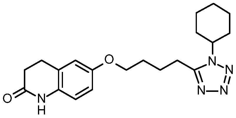
By "Cilostazol" (IUPAC Name 6-[4-(l-cyclohexyl-lii-tetrazol-5-yl)butoxy]-3,4-dihydro- 2(l /)-quinolinone) is meant a small molecule inhibitor having the following structure:
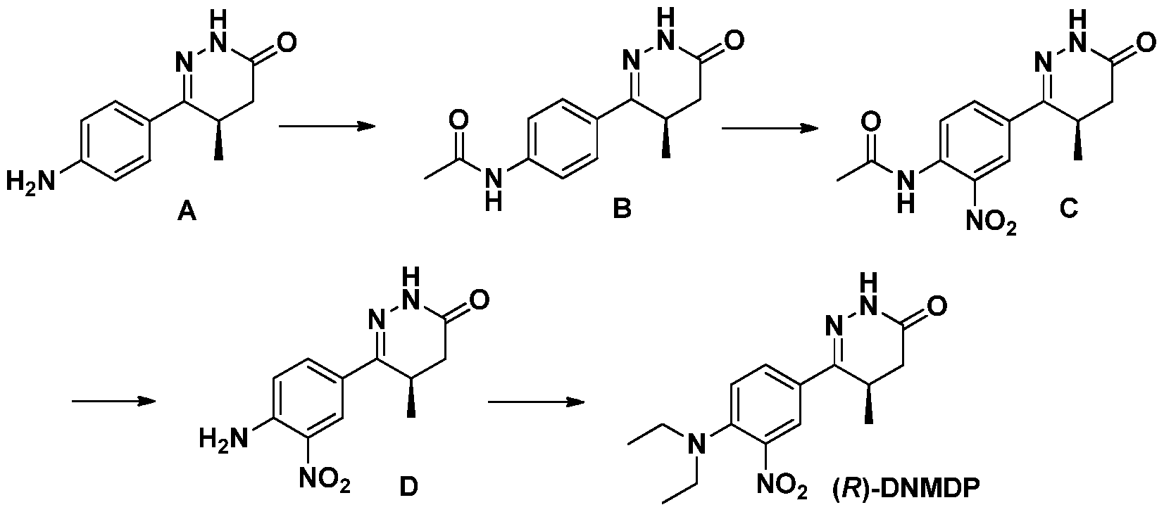
By "DNMDP" (IUPAC Name 6-(4-(diethylamino)-3-nitrophenyl)-5-methyl-4,5- dihydropyridazin-3(2H)-one) is meant a small molecule inhibitor having the following structure:
By "Forskolin" (IUPAC Name (3R,4aR,5
lS
,,6
lS
,,6 S
,,10.S
,,10aR,10b
lS
,)-6,10,10b-Trihydroxy- 3,4a,7,7,10a-pentamethyl-l-oxo-3-vinyldodecahydro-lii-benzo[ ]chromen-5-ylacetate) is meant a small molecule inhibitor having the following structure:
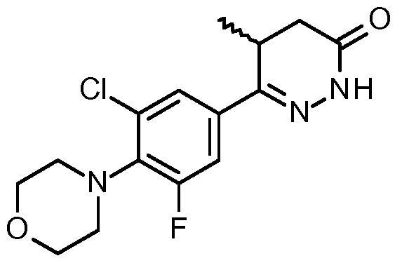
By "Levosimendan" (IUPAC Name (E)-2-cyano-l-methyl-3-(4-(4-methyl-6-oxo-l,4,5,6- tetrahydropyridazin-3-yl)phenyl)guanidine) is meant a small molecule inhibitor having the following structure:
By "Milrinone" (IUPAC Name 2-methyl-6-oxo-l,6-dihydro-3,4'-bipyridine-5-carbonitrile) is meant a small molecule inhibitor havin the following structure:
By "Papaverine" (IUPAC Name l-(3,4-dimethoxybenzyl)-6,7-dimethoxyisoquinoline) meant a small molecule inhibitor havin the following structure:
By "Siguazodan" (IUPAC Name (E)-2-cyano-l-methyl-3-(4-(4-methyl-6-oxo-l,4,5,6- tetrahydropyridazin-3-yl)phenyl)guanidine) is meant a small molecule inhibitor having the following structure:
By "Sildenafil" (IUPAC Name l-[4-ethoxy-3-(6,7-dihydro-l-methyl-7-oxo-3-propyl-l i- pyrazolo[4,3-fif]pyrimidin-5-yl)phenylsulfonyl]-4-methylpiperazine) is meant a small molecule inhibitor having the followin structure:
By "Trequinsin" (IUPAC Name 9,10-dimethoxy-3-methyl-2-(2,4,6-trimethylphenyl)imino-6,7- dihydropyrimido[6,l-a]isoquinolin-4-one) is meant a small molecule inhibitor having the following structure:
By "Vardenifil" (IUPAC Name 4-[2-ethoxy-5-(4-ethylpiperazin-l-yl)sulfonyl-phenyl]-9-methyl- 7-propyl-3,5,6,8-tetrazabicyclo[4.3.0]nona-3,7,9-trien-2-one) is meant a small molecule inhibitor having the following structure:
By "Zardaverine (IUPAC Name 3-[4-(Difluoromethoxy)-3-methoxyphenyl]-lii-pyridazin-6- one)" is meant a small molecule inhibitor havin the following structure:
By "Compound 1" is meant a small molecule inhibitor having the following structure:
By "Compound 2" is meant a small molecule inhibitor having the following structure:
By "Compound 3" is meant a small molecule inhibitor having the following structure:
By "Compound 4" is meant a small molecule inhibitor having the following structure:
By "Compound 5" is meant a small molecule inhibitor having the following structure:
By "Compound 6" is meant a small molecule inhibitor having the following structure:
Structures drawn include all permissible rotations about bonds.
In some embodiments, any one of the compounds Compound 1, Compound 2, Compound 3, Compound 4, Compound 5, and Compound 6 is a small molecule phosphodiesterase inhibitor.
In some other embodiments, any one of the compounds Cilostamide, Cilostazol, DNDMP, Forskolin, Levosimendan, Milrinone, Papaverine, Siguazodan, Sildenafil, Trequinsin, Vardenifil, and Zardaverine is a small molecule phosphodiesterase inhibitor.
In some embodiments, combinations of small molecule phosphodiesterase inhibitors or modulators may be used.
In some embodiments, any combination of Compounds 1-6 may be used.
In some embodiments combinations of small molecule phosphodiesterase inhibitors or modulators, especially compounds 1-6, more particularly compound 6, together with anticancer agents may be used.
A further aspect of the invention is a method of preparing compound 6, said method comprising the step of
reacting the racemate Compound 3 a
Compound 3 a
with NaOCl in acetic acid at a temperature range form 10-15°C (including 10° and 15°) to obtain the racemate Compound 6a
Compound 6a
and subsequently performing a separation of enantiomers of Compound 6a to obtain
Compound 6.
Compound 6.
Another aspect of the invention is a method of preparing compound 6, said method further comprising optionally in a preceding step a separation of enantiomers of compound 3a
Compound 3 a
to obtain Compound 3
Compound 3
and the reaction step with NaOCl in acetic acid at a temperature range form 10-15°C
(including 10° and 15°) is performed with the enantiomer Compound 3 and optionally further comprising a subsequent separation of enantiomers.
A further aspect of the invention is a method for the preparation of Compound 6 whereby the enantiomer Compound 3
Compound 3
is reacted to obtain enantiomer Compound 6
Another aspect of the invention is the use of com ound 3
Compound 3
for the preparation of compound 6.
Compound 6
By "CREB3L1 polypeptide" is meant a protein or fragment thereof having at least 85% amino acid sequence identity to the sequence provided at GenBank Accession No. AAH14097.1 that is cleaved upon endoplasmic reticulum stress and has transcription factor activity. The amino acid sequence provided at GenBank Accession No. AAH14097.1 is shown below.
1 mdavlepfpa drlfpgssfl dlgdlnesdf lnnahfpehl dhftenmedf sndlfssffd
61 dpvldekspl Idmeldsptp giqaehsysl sgdsapqspl vpikmedttq daehgawalg
121 hklcsimvkq eqspelpvdp laapsamaaa aamattpllg lsplsrlpip hqapgemtql
181 pvikaeplev nqflkvtped lvqmpptpps shgsdsdgsq sprslppssp vrpmarssta
241 istsplltpp hklqgtsgpl llteeekrtl iaegypiptk lpltkaeeka lkrvrrkikn
301 kisaqesrrk kkeyveclek kvetftsenn elwkkvetle nanrtllqql qklqtlvtnk
361 isrpykmaat qtgtclmvaa lcfvlvlgsl vpclpefssg sqtvkedpla adgvytasqm
421 psrsllfydd gaglwedgrs tllpmeppdg weinpggpae qrprdhlqhd hldsthettk
481 ylseawpkdg gngtspdfsh skewfhdrdl gpnttikls (SEQ ID NO. : 1)
By "CREB3L1 polynucleotide" is meant any nucleic acid molecule, including DNA and RNA, encoding a CREB3L1 polypeptide or fragment thereof. An exemplary CREB3L1 nucleic acid sequence is provided at NCBI Ref: NM_052854.3. The sequence provided at NCBI Ref: NM_052854.3 is reproduced below:
1 ccagccaggg gttcccggtt tcacagagag gaaagtgaca gaagacgtgc ggagggagac
61 gcagagacag aggagaggcc ggcagccacc cagtctcggg ggagcactta gctcccccgc
121 cccggctccc accctgtccg gggggctcct gaagccctca gccccaaccc cgggctcccc
181 atggaagcca gctgtgcccc aggaggagca ggaggaggtg gagtcggctg aatgcccacg
241 gtgcgcccgg ggcccctgag cccatcccgc tcctagccgc tgccctaagg cccccgcgcg
301 ccccgcgccc cccacccggg gccgcgccgc ctccgtccgc ccctcccccg gggcttcgcc
361 ccggacctgc cccccgcccg tttgccagcg ctcaggcagg agctctggac tgggcgcgcc
421 gccgccctgg agtgagggaa gcccagtgga agggggtccc gggagccggc tgcgatggac
481 gccgtcttgg aacccttccc ggccgacagg ctgttccccg gatccagctt cctggacttg
541 ggggatctga acgagtcgga cttcctcaac aatgcgcact ttcctgagca cctggaccac
601 tttacggaga acatggagga cttctccaat gacctgttca gcagcttctt tgatgaccct
661 gtgctggatg agaagagccc tctattggac atggaactgg actcccctac gccaggcatc
721 caggcggagc acagctactc cctgagcggc gactcagcgc cccagagccc ccttgtgccc
781 atcaagatgg aggacaccac ccaagatgca gagcatggag catgggcgct gggacacaaa
841 ctgtgctcca tcatggtgaa gcaggagcag agcccggagc tgcccgtgga ccctctggct
901 gccccctcgg ccatggctgc cgcggccgcc atggccacca ccccgctgct gggcctcagc
961 cccttgtcca ggctgcccat cccccaccag gccccgggag agatgactca gctgccagtg
1021 atcaaagcag agcctctgga ggtgaaccag ttcctcaaag tgacaccgga ggacctggtg
1081 cagatgcctc cgacgccccc cagcagccat ggcagtgaca gcgacggctc ccagagtccc
1141 cgctctctgc ccccctccag ccctgtcagg cccatggcgc gctcctccac ggccatctcc
1201 acctccccac tcctcactgc ccctcacaaa ttacagggga catcagggcc actgctcctg
1261 acagaggagg agaagcggac cctgattgct gagggctacc ccatccccac aaaactcccc
1321 ctcaccaaag ccgaggagaa ggccttgaag agagtccgga ggaaaatcaa gaacaagatc
1381 tcagcccagg agagccgtcg taagaagaag gagtatgtgg agtgtctaga aaagaaggtg
1441 gagacattta catctgagaa caatgaactg tggaagaagg tggagaccct ggagaatgcc
1501 aacaggaccc tgctccagca gctgcagaaa ctccagactc tggtcaccaa caagatctcc
1561 agaccttaca agatggccgc cacccagact gggacctgcc tcatggtggc agccttgtgc
1621 tttgttctgg tgctgggctc cctcgtgccc tgccttcccg agttctcctc cggctcccag
1681 actgtgaagg aagaccccct ggccgcagac ggcgtctaca cggccagcca gatgccctcc
1741 cgaagcctcc tattctacga tgacggggca ggcttatggg aagatggccg cagcaccctg
1801 ctgcccatgg agcccccaga tggctgggaa atcaaccccg gggggccggc agagcagcgg
1861 ccccgggacc acctgcagca tgatcacctg gacagcaccc acgagaccac caagtacctg
1921 agtgaggcct ggcctaaaga cggtggaaac ggcaccagcc ccgacttctc ccactccaag
1981 gagtggttcc acgacaggga tctgggcccc aacaccacca tcaaactctc ctaggccatg
2041 ccaagaccca ggacatagga cggacccctg gtacccagaa gaggagttct tgctcactaa
2101 cccggatccg cctcgtgccc ctgcctcctg gagcttccca ttccaggaga aaaggctcca
2161 cttcccagcc cttccttgcc cctgacattt ggactcttcc cttgggccga ccactctgtt
2221 ctcattctcc ttcccaccaa catccatccg tccttctcag acaaaccact cactgggtac
2281 cccacctcct ctctcatatg cccaacacga ccactgcctc cctgccccca cacctgcacc
2341 caaacagaca catcaacgca ccccactcac agacacccct taccccaccc ccactgtaca
2401 gagaccaaga acagaaattg tttgtaaata atgaacctta ttttttatta ttgccaatcc
2461 cctaagatat tgtattttac aaatctccct cttcccttcg cccctccctt gttttatatt 2521 ttatgaagtt agtgcgggct ttgctgctcc ctggcccagg aaagagggac tacctgaccc 2581 tcacctggca cccccctgct gctgcccaag ccgctgggcc tttttaattg ccaaactgct 2641 ctcttcatca gctcagcaca tgctttaaga aagcaaaacc aaaaaaaaaa aaaaaaagat 2701 gcagcatcaa aaaaaaaaaa aaaaaaaaaa aaaaaaaaaa a (SEQ ID NO.: 2)
By "PDE3A polypeptide" is meant a protein or fragment thereof having at least 85% amino acid sequence identity to the sequence provided at NCBI Ref No. NP_000912.3 that catalyzes the hydrolysis of cyclic adenosine monophosphate (cAMP) and cyclic guanosine monophosphate (cGMP). An exemplary human full-length PDE3A amino acid sequence is provided below:
MAVPGDAARVRDKPVHSGVSQAPTAGRDCHHRADPASPRDSGCRGCWGDLVLQPLRSSRKLSSALCAGSLSFLLA LLVRLVRGEVGCDLEQCKEAAAAEEEEAAPGAEGGVFPGPRGGAPGGGARLSPWLQPSALLFSLLCAFFWMGLYL LRAGVRLPLAVALLAACCGGEALVQIGLGVGEDHLLSLPAAGWLSCLAAATWLVLRLRLGVLMIALTSAVRTVS LI SLERFKVAWRPYLAYLAGVLGILLARYVEQILPQSAEAAPREHLGSQLIAGTKEDIPVFKRRRRSSSVVSAEM SGCSSKSHRRTSLPCIPREQLMGHSEWDHKRGPRGSQSSGTSITVDIAVMGEAHGLITDLLADPSLPPNVCTSLR AVSNLLSTQLTFQAIHKPRVNPVTSLSENYTCSDSEESSEKDKLAIPKRLRRSLPPGLLRRVSSTWTTTTSATGL PTLEPAPVRRDRSTSIKLQEAPSSSPDSWNNPVMMTLTKSRSFTSSYAISAANHVKAKKQSRPGALAKISPLSSP CSSPLQGTPASSLVSKISAVQFPESADTTAKQSLGSHRALTYTQSAPDLSPQILTPPVICSSCGRPYSQGNPADE PLERSGVATRTPSRTDDTAQVTSDYETNNNSDSSDIVQNEDETECLREPLRKASACSTYAPETMMFLDKPILAPE PLVMDNLDSIMEQLNTWNFPIFDLVENIGRKCGRILSQVSYRLFEDMGLFEAFKIPIREFMNYFHALEIGYRDIP YHNRIHATDVLHAVWYLTTQPIPGLSTVINDHGSTSDSDSDSGFTHGHMGYVFSKTYNVTDDKYGCLSGNIPALE LMALYVAAAMHDYDHPGRTNAFLVATSAPQAVLYNDRSVLENHHAAAAWNLFMSRPEYNFLINLDHVEFKHFRFL VIEAILATDLKKHFDFVAKFNGKVNDDVGIDWTNENDRLLVCQMCIKLADINGPAKCKELHLQWTDGIVNEFYEQ GDEEASLGLPISPFMDRSAPQLANLQESFI SHIVGPLCNSYDSAGLMPGKWVEDSDESGDTDDPEEEEEEAPAPN EEETCENNESPKKKTFKRRKIYCQITQHLLQNHKMWKKVIEEEQRLAGIENQSLDQTPQSHSSEQIQAIKEEEEE KGKPRGEEIPTQKPDQ (SEQ ID NO. : 3)
Three PDE3A isoforms are known: PDE3A1, PDE3A2, and PDE3A3. PDE3A1 comprises amino acids 146-1141, PDE3A2 isoform 2 comprises amino acids 299-1141, and PDE3A3 comprises amino acids 483-1141 of the full-length PDE3A amino acid sequence.
By "PDE3A polynucleotide" is meant any nucleic acid molecule, including DNA and RNA, encoding a PDE3A polypeptide or fragment thereof. An exemplary PDE3A nucleic acid sequence is provided at NCBI Ref: NM_000921.4:
1 gggggccact gggaattcag tgaagagggc accctatacc atggcagtgc ccggcgacgc 61 tgcacgagtc agggacaagc ccgtccacag tggggtgagt caagccccca cggcgggccg 121 ggactgccac catcgtgcgg accccgcatc gccgcgggac tcgggctgcc gtggctgctg 181 gggagacctg gtgctgcagc cgctccggag ctctcggaaa ctttcctccg cgctgtgcgc 241 gggctccctg tcctttctgc tggcgctgct ggtgaggctg gtccgcgggg aggtcggctg 301 tgacctggag cagtgtaagg aggcggcggc ggcggaggag gaggaagcag ccccgggagc 361 agaagggggc gtcttcccgg ggcctcgggg aggtgctccc gggggcggtg cgcggctcag
421 cccctggctg cagccctcgg cgctgctctt cagtctcctg tgtgccttct tctggatggg
481 cttgtacctc ctgcgcgccg gggtgcgcct gcctctggct gtcgcgctgc tggccgcctg
541 ctgcgggggg gaagcgctcg tccagattgg gctgggcgtc ggggaggatc acttactctc
601 actccccgcc gcgggggtgg tgctcagctg cttggccgcc gcgacatggc tggtgctgag
661 gctgaggctg ggcgtcctca tgatcgcctt gactagcgcg gtcaggaccg tgtccctcat
721 ttccttagag aggttcaagg tcgcctggag accttacctg gcgtacctgg ccggcgtgct
781 ggggatcctc ttggccaggt acgtggaaca aatcttgccg cagtccgcgg aggcggctcc
841 aagggagcat ttggggtccc agctgattgc tgggaccaag gaagatatcc cggtgtttaa
901 gaggaggagg cggtccagct ccgtcgtgtc cgccgagatg tccggctgca gcagcaagtc
961 ccatcggagg acctccctgc cctgtatacc gagggaacag ctcatggggc attcagaatg
1021 ggaccacaaa cgagggccaa gaggatcaca gtcttcagga accagtatta ctgtggacat
1081 cgccgtcatg ggcgaggccc acggcctcat taccgacctc ctggcagacc cttctcttcc
1141 accaaacgtg tgcacatcct tgagagccgt gagcaacttg ctcagcacac agctcacctt
1201 ccaggccatt cacaagccca gagtgaatcc cgtcacttcg ctcagtgaaa actatacctg
1261 ttctgactct gaagagagct ctgaaaaaga caagcttgct attccaaagc gcctgagaag
1321 gagtttgcct cctggcttgt tgagacgagt ttcttccact tggaccacca ccacctcggc
1381 cacaggtcta cccaccttgg agcctgcacc agtacggaga gaccgcagca ccagcatcaa
1441 actgcaggaa gcaccttcat ccagtcctga ttcttggaat aatccagtga tgatgaccct
1501 caccaaaagc agatccttta cttcatccta tgctatttct gcagctaacc atgtaaaggc
1561 taaaaagcaa agtcgaccag gtgccctcgc taaaatttca cctctttcat cgccctgctc
1621 ctcacctctc caagggactc ctgccagcag cctggtcagc aaaatttctg cagtgcagtt
1681 tccagaatct gctgacacaa ctgccaaaca aagcctaggt tctcacaggg ccttaactta
1741 cactcagagt gccccagacc tatcccctca aatcctgact ccacctgtta tatgtagcag
1801 ctgtggcaga ccatattccc aagggaatcc tgctgatgag cccctggaga gaagtggggt
1861 agccactcgg acaccaagta gaacagatga cactgctcaa gttacctctg attatgaaac
1921 caataacaac agtgacagca gtgacattgt acagaatgaa gatgaaacag agtgcctgag
1981 agagcctctg aggaaagcat cggcttgcag cacctatgct cctgagacca tgatgtttct
2041 ggacaaacca attcttgctc ccgaacctct tgtcatggat aacctggact caattatgga
2101 gcagctaaat acttggaatt ttccaatttt tgatttagtg gaaaatatag gaagaaaatg
2161 tggccgtatt cttagtcagg tatcttacag actttttgaa gacatgggcc tctttgaagc
2221 ttttaaaatt ccaattaggg aatttatgaa ttattttcat gctttggaga ttggatatag
2281 ggatattcct tatcataaca gaatccatgc cactgatgtt ttacatgctg tttggtatct
2341 tactacacag cctattccag gcctctcaac tgtgattaat gatcatggtt caaccagtga
2401 ttcagattct gacagtggat ttacacatgg acatatggga tatgtattct caaaaacgta
2461 taatgtgaca gatgataaat acggatgtct gtctgggaat atccctgcct tggagttgat
2521 ggcgctgtat gtggctgcag ccatgcacga ttatgatcat ccaggaagga ctaatgcttt
2581 cctggttgca actagtgctc ctcaggcggt gctatataac gatcgttcag ttttggagaa
2641 tcatcacgca gctgctgcat ggaatctttt catgtcccgg ccagagtata acttcttaat
2701 taaccttgac catgtggaat ttaagcattt ccgtttcctt gtcattgaag caattttggc
2761 cactgacctg aagaaacact ttgacttcgt agccaaattt aatggcaagg taaatgatga
2821 tgttggaata gattggacca atgaaaatga tcgtctactg gtttgtcaaa tgtgtataaa
2881 gttggctgat atcaatggtc cagctaaatg taaagaactc catcttcagt ggacagatgg
2941 tattgtcaat gaattttatg aacagggtga tgaagaggcc agccttggat tacccataag
3001 ccccttcatg gatcgttctg ctcctcagct ggccaacctt caggaatcct tcatctctca
3061 cattgtgggg cctctgtgca actcctatga ttcagcagga ctaatgcctg gaaaatgggt
3121 ggaagacagc gatgagtcag gagatactga tgacccagaa gaagaggagg aagaagcacc
3181 agcaccaaat gaagaggaaa cctgtgaaaa taatgaatct ccaaaaaaga agactttcaa
3241 aaggagaaaa atctactgcc aaataactca gcacctctta cagaaccaca agatgtggaa
3301 gaaagtcatt gaagaggagc aacggttggc aggcatagaa aatcaatccc tggaccagac
3361 ccctcagtcg cactcttcag aacagatcca ggctatcaag gaagaagaag aagagaaagg
3421 gaaaccaaga ggcgaggaga taccaaccca aaagccagac cagtgacaat ggatagaatg
3481 ggctgtgttt ccaaacagat tgacttgtca aagactctct tcaagccagc acaacattta
3541 gacacaacac tgtagaaatt tgagatgggc aaatggctat tgcattttgg gattcttcgc
3601 attttgtgtg tatattttta cagtgaggta cattgttaaa aactttttgc tcaaagaagc
3661 tttcacattg caacaccagc ttctaaggat tttttaagga gggaatatat atgtgtgtgt
3721 gtatataagc tcccacatag atacatgtaa aacatattca cacccatgca cgcacacaca
3781 tacacactga aggccacgat tgctggctcc acaatttagt aacatttata ttaagatata
3841 tatatagtgg tcactgtgat ataataaatc ataaaggaaa ccaaatcaca aaggagatgg
3901 tgtggcttag caaggaaaca gtgcaggaaa tgtaggttac caactaagca gcttttgctc
3961 ttagtactga gggatgaaag ttccagagca ttatttgaat tctgatacat cctgccaaca
4021 ctgtgtgtgt gtgtgtgtgt gtgtgtgtgt gtgtgtgtgt gtgtgaaaga gagacagaag
4081 ggaatggttt gagagggtgc ttgtgtgcat gtgtgtgcat atgtaaagag atttttgtgg
4141 tttaagtaac tcagaatagc tgtagcaaat gactgaatac atgtgaacaa acagaaggaa
4201 gttcactctg gagtgtcttt gggaggcagc cattccaaat gccctcctcc atttagcttc
4261 aataaagggc cttttgctga tggagggcac tcaagggctg ggtgagaggg ccacgtgttt
4321 ggtattacat tactgctatg caccacttga aggagctcta tcaccagcct caaacccgaa
4381 agactgaggc attttccagt ctacttgcct aatgaatgta taggaactgt ctatgagtat
4441 ggatgtcact caactaagat caaatcacca tttaagggga tggcattctt tatacctaaa
4501 cacctaagag ctgaagtcag gtcttttaat caggttagaa ttctaaatga tgccagagaa
4561 ggcttgggaa attgtacttc agcgtgatag cctgtgtctt cttaatttgc tgcaaaatat
4621 gtggtagaga aagaaaagga aacagaaaaa tcactctggg ttatatagca agagatgaag
4681 gagaatattt caacacaggg tttttgtgtt gacataggaa aagcctgatt cttggcaact
4741 gttgtagttt gtctttcagg ggtgaaggtc ccactgacaa cccctgttgt ggtgttccac
4801 acgctgtttg ttggggtagc ttccatcggc agtctggccc attgtcagtc atgcttcttc
4861 tggccgggga gattatagag agattgtttg aagattgggt tattattgaa agtctttttt
4921 tttgtttgtt ttgttttggt ttgtttgttt atctacactt gtttatgctg tgagccaaac
4981 ctctatttaa aaagttgata ctcactttca atattttatt tcatattatt atatatgtca
5041 tgatagttat cttgatgtaa atatgaagat ttttttgttt ctgtagatag taaactcttt
5101 ttttaaaaaa ggaaaaggga aacattttta taaagttata ttttaatcac catttttata
5161 cattgtagtt ctctccaagc ccagtaagag aatgatgatt catttgcatg gaggtcgatg
5221 gacaaccaat catctacctt ttctaattta aatgataatc tgatatagtt ttattgccag
5281 ttaaatgagg atgctgcaaa gcatgttttt tcactagtaa cttttgctaa ctgaatgaat
5341 tctgggtcca tatctcccag atgaaaaact gttaaccaat accatatttt atagttggtg
5401 tccatttctt tccaacactg tttgttatga ttcttccttg agtacttata tacagacctg
5461 ctcattatct aaacaatctt accttctaag taaaccttga ttgtgatttc cagtttttat
5521 tttctctgac gtagtagaaa ggaatgttta cattaaaaat acttttgttt ctcataaatg
5581 gatattgtac tccccccttt caaagcatta ttttacaata attcatggca ttttaaaaaa
5641 taaggcaaag ataatacgac aaaaaatata catggtttca aggcaaattc tccaataagt
5701 tggaaaatgt aaaaaggatc aagtggatgc agcctctacc taaataatta aaatatattt
5761 cagtatattt ctgaattaac accaggtctt cattatttag aacttactaa attgttttca
5821 ttttcttagt tttacctgtg tatctccatg tttgcaaaaa ttactataag tcaaattttg
5881 ccagtgaatt taactatttt tctttccttg caattaaggg gaaaaaagca tttatcttat
5941 cttctcatac cccttgcatc taagtactta gcaaagtcaa tattttccca ttttccaaat
6001 gcgtccatct ctaacataaa tattaattga acatagagct atgtttggag tgagtggact
6061 ggcaggacag ttggaagtcc atcacagtct attgacagtt tcatcaaagc tgtatagtcc
6121 aactagtggg gcagcttggc tactatggtg gaagtctcag caaactgcct ggttttgttt
6181 gtttgttttg ttttaaggta caggaaataa gaggaataat agtggccaaa gcaattagaa
6241 catcttcatt ccagaactgt gttcagcaat ccaggcagat tgatacattt ttctttaaaa
6301 ataaattgct attacagcta gacgtcaatt gggataaata aagggatgaa gatccactaa
6361 gtttgtgact ttcatacaca cccagtacat ctcaaaggat gctaagggac attttctgcc
6421 agtagagttc tccccctttt tggtgacagc aatattatta tgttcacatc taactccaga
6481 gcttacttcc tgtggtgcca atgtatttgt tgcaatttac tacattttta tatgagccta
6541 tttataggtg ccattaaact caggtctttc aaatgaaaga gtttctagcc cacttaggga
6601 aaaagataat tgtttagaaa accataaaat caatggtagg aaaagttgga actggttacc
6661 tggatgccat ggttctctgt taaataaagt aagagaccag gtgtattctg agtgtcatca
6721 gtgttatttt cagcatgcta ataaatgtct ttccggttat atatctatct aaattaacct
6781 ttaaaatatt ggtttccttg ataaaagcac cacttttgct tttgttagct gtaatatttt
6841 ttgtcattta gataagacct ggtttggctc tcaataaaag atgaagacag tagctctgta
6901 cagggatata tctatattag tcttcatctg atgaatgaag aaattttctc atattatgtt
6961 caagaaagta tttacttcct aaaaatagaa ttcccgattc tgtctatttt ggttgaatac
7021 cagaacaaat ctttccgttg caatcccagt aaaacgaaag aaaaggaata tcttacagac
7081 tgttcatatt agatgtatgt agactgttaa tttgcaattt ccccatattt cctgcctatc
7141 ttacccagat aactttcttt gaaggtaaaa gctgtgcaaa aggcatgaga ctcaggccta
7201 ctctttgttt aaatgatgga aaaatataaa ttattttcta agtaataaaa gtataaaaat
7261 tatcattata aataaagtct aaagtttgaa attattaatt taaaaaaaaa aaaaaaaaa
(SEQ ID NO. : 4)
By "Schlafen 12 (SLFN12) polypeptide" is meant a protein or fragment thereof having at least 85% amino acid sequence identity to the sequence provided at NCBI Ref No. NP_060512.3 that interacts with PDE3A when bound to one of the compounds described herein. An exemplary human SLFN12 amino acid sequence is provided below:
MNISVDLETNYAELVLDVGRVTLGENSRKKMKDCKLRKKQNESVSRAMCALLNSGGGVIKAEIENEDYSYTKDGI GLDLENSFSNILLFVPEYLDFMQNGNYFLIFVKSWSLNTSGLRITTLSSNLYKRDITSAKVMNATAALEFLKDMK KTRGRLYLRPELLAKRPCVDIQEENNMKALAGVFFDRTELDRKEKLTFTESTHVEIKNFSTEKLLQRIKEILPQY VSAFANTDGGYLFIGLNEDKEI IGFKAEMSDLDDLEREIEKSIRKMPVHHFCMEKKKINYSCKFLGVYDKGSLCG YVCALRVERFCCAVFAKEPDSWHVKDNRVMQLTRKEWIQFMVEAEPKFSSSYEEVI SQINTSLPAPHSWPLLEWQ RQRHHCPGLSGRITYTPENLCRKLFLQHEGLKQLICEEMDSVRKGSLIFSRSWSVDLGLQENHKVLCDALLI SQD SPPVLYTFHMVQDEEFKGYSTQTALTLKQKLAKIGGYTKKVCVMTKIFYLSPEGMTSCQYDLRSQVIYPESYYFT RRKYLLKALFKALKRLKSLRDQFSFAENLYQI IGIDCFQKNDKKMFKSCRRLT (SEQ ID NO.: 5)
By "Schlafen 12 (SLFN12) polynucleotide" is meant any nucleic acid molecule, including DNA and RNA, encoding a SLFN12 polypeptide or fragment thereof. An exemplary SLFN12 nucleic acid sequence is provided at NCBI Ref: NM_018042.4:
1 tttgtaactt cacttcagcc tcccattgat cgctttctgc aaccattcag actgatctcg
61 ggctcctatt tcatttacat tgtgtgcaca ccaagtaacc agtgggaaaa ctttagaggg
121 tacttaaacc ccagaaaatt ctgaaaccgg gctcttgagc cgctatcctc gggcctgctc
181 ccaccctgtg gagtgcactt tcgttttcaa taaatctctg cttttgttgc ttcattcttt
241 ccttgctttg tttgtgtgtt tgtccagttc tttgttcaac acgccaagaa cctggacact
301 cttcactggt aacatatttt ggcaagccaa ccaggagaaa agaatttctg cttggacact
361 gcatagctgc tgggaaaatg aacatcagtg ttgatttgga aacgaattat gccgagttgg
421 ttctagatgt gggaagagtc actcttggag agaacagtag gaaaaaaatg aaggattgta
481 aactgagaaa aaagcagaat gaaagtgtct cacgagctat gtgtgctctg ctcaattctg
541 gagggggagt gatcaaggct gaaattgaga atgaagacta tagttataca aaagatggaa
601 taggactaga tttggaaaat tcttttagta acattctgtt atttgttcct gagtacttag
661 acttcatgca gaatggtaac tactttctga tttttgtgaa gtcatggagc ttgaacacct
721 ctggtctgcg gattaccacc ttgagctcca atttgtacaa aagagatata acatctgcaa
781 aagtcatgaa tgccactgct gcactggagt tcctcaaaga catgaaaaag actagaggga
841 gattgtattt aagaccagaa ttgctggcaa agaggccctg tgttgatata caagaagaaa
901 ataacatgaa ggccttggcc ggggtttttt ttgatagaac agaacttgat cggaaagaaa
961 aattgacctt tactgaatcc acacatgttg aaattaaaaa cttctcgaca gaaaagttgt
1021 tacaacgaat taaagagatt ctccctcaat atgtttctgc atttgcaaat actgatggag
1081 gatatttgtt cattggttta aatgaagata aagaaataat tggctttaaa gcagagatga
1141 gtgacctcga tgacttagaa agagaaatcg aaaagtccat taggaagatg cctgtgcatc
1201 acttctgtat ggagaagaag aagataaatt attcatgcaa attccttgga gtatatgata
1261 aaggaagtct ttgtggatat gtctgtgcac tcagagtgga gcgcttctgc tgtgcagtgt
1321 ttgctaaaga gcctgattcc tggcatgtga aagataaccg tgtgatgcag ttgaccagga
1381 aggaatggat ccagttcatg gtggaggctg aaccaaaatt ttccagttca tatgaagagg
1441 tgatctctca aataaatacg tcattacctg ctccccacag ttggcctctt ttggaatggc
1501 aacggcagag acatcactgt ccagggctat caggaaggat aacgtatact ccagaaaacc
1561 tttgcagaaa actgttctta caacatgaag gacttaagca attaatatgt gaagaaatgg
1621 actctgtcag aaagggctca ctgatcttct ctaggagctg gtctgtggat ctgggcttgc
1681 aagagaacca caaagtcctc tgtgatgctc ttctgatttc ccaggacagt cctccagtcc
1741 tatacacctt ccacatggta caggatgagg agtttaaagg ctattctaca caaactgccc
1801 taaccttaaa gcagaagctg gcaaaaattg gtggttacac taaaaaagtg tgtgtcatga
1861 caaagatctt ctacttgagc cctgaaggca tgacaagctg ccagtatgat ttaaggtcgc
1921 aagtaattta ccctgaatcc tactatttta caagaaggaa atacttgctg aaagcccttt 1981 ttaaagcctt aaagagactc aagtctctga gagaccagtt ttcctttgca gaaaatctat 2041 accagataat cggtatagat tgctttcaga agaatgataa aaagatgttt aaatcttgtc 2101 gaaggctcac ctgatggaaa atggactggg ctactgagat atttttcatt atatatttga 2161 taacattctc taattctgtg aaaatatttc tttgaaaact ttgcaagtta agcaacttaa
2221 tgtgatgttg gataattggg ttttgtctat tttcacttct ccctaaataa tcttcacaga 2281 tattgtttga gggatattag gaaaattaat ttgttaactc gtctgtgcac agtattattt 2341 actctgtctg tagttcctga ataaattttc ttccatgctt gaactgggaa aattgcaaca 2401 cttttattct taatgacaac agtgaaaatc tcccagcata tacctagaaa acaattataa 2461 cttacaaaag attatccttg atgaaactca gaatttccac agtgggaatg aataagaagg
2521 caaaactcat (SEQ ID NO.: 6)
In some aspects, the compound is an isomer. "Isomers" are different compounds that have the same molecular formula. "Stereoisomers" are isomers that differ only in the way the atoms are arranged in space. As used herein, the term "isomer" includes any and all geometric isomers and stereoisomers. For example, "isomers" include geometric double bond cis- and trans-isomers, also termed E- and Z-isomers; R- and S-enantiomers; diastereomers, (d)-isomers and (l)-isomers, racemic mixtures thereof; and other mixtures thereof, as falling within the scope of this invention
The symbol denotes a bond that can be a single, double or triple bond as described herein. Provided herein are various geometric isomers and mixtures thereof resulting from the arrangement of substituents around a carbon-carbon double bond or arrangement of substituents around a carbocyclic ring. Substituents around a carbon-carbon double bond are designated as being in the "Z" or "E" configuration wherein the terms "Z" and "E" are used in accordance with IUPAC standards. Unless otherwise specified, structures depicting double bonds encompass both the "E" and "Z" isomers.
Substituents around a carbon-carbon double bond alternatively can be referred to as "cis" or "trans," where "cis" represents substituents on the same side of the double bond and "trans" represents substituents on opposite sides of the double bond. The arrangement of substituents around a carbocyclic ring can also be designated as "cis" or "trans." The term "cis" represents substituents on the same side of the plane of the ring, and the term "trans" represents substituents on opposite sides of the plane of the ring. Mixtures of compounds wherein the substituents are disposed on both the same and opposite sides of plane of the ring are designated "cis/trans."
The term "enantiomers" refers to a pair of stereoisomers that are non-superimposable mirror images of each other. An atom having an asymmetric set of substituents can give rise to an enantiomer. A mixture of a pair of enantiomers in any proportion can be known as a "racemic" mixture. The term "(±)" is used to designate a racemic mixture where appropriate. "Diastereoisomers" are stereoisomers that have at least two asymmetric atoms, but which are not mirror-images of each other. The absolute stereochemistry is specified according to the Cahn-Ingold-Prelog R-S system. When a compound is an enantiomer, the stereochemistry at each chiral carbon can be specified by
either R or S. Resolved compounds whose absolute configuration is unknown can be designated (+) or (-) depending on the direction (dextro- or levorotatory) which they rotate plane polarized light at the wavelength of the sodium D line. Certain of the compounds described herein contain one or more asymmetric centers and can thus give rise to enantiomers, diastereomers, and other stereoisomeric forms that can be defined, in terms of absolute stereochemistry at each asymmetric atom, as (R)- or (S)-. The present chemical entities, pharmaceutical compositions and methods are meant to include all such possible isomers, including racemic mixtures, optically substantially pure forms and intermediate mixtures.
Optically active (R)- and (S)-isomers can be prepared, for example, using chiral synthons or chiral reagents, or resolved using conventional techniques. Enantiomers can be isolated from racemic mixtures by any method known to those skilled in the art, including chiral high pressure liquid chromatography (HPLC), the formation and crystallization of chiral salts, or prepared by asymmetric syntheses.
Optical isomers can be obtained by resolution of the racemic mixtures according to conventional processes, e.g., by formation of diastereoisomeric salts, by treatment with an optically active acid or base. Examples of appropriate acids are tartaric, diacetyltartaric, dibenzoyltartaric, ditoluoyltartaric, and camphorsulfonic acid. The separation of the mixture of diastereoisomers by crystallization followed by liberation of the optically active bases from these salts affords separation of the isomers. Another method involves synthesis of covalent diastereoisomeric molecules by reacting disclosed compounds with an optically pure acid in an activated form or an optically pure isocyanate. The synthesized diastereoisomers can be separated by conventional means such as chromatography, distillation, crystallization or sublimation, and then hydrolyzed to deliver the enantiomerically enriched compound. Optically active compounds can also be obtained by using active starting materials. In some embodiments, these isomers can be in the form of a free acid, a free base, an ester or a salt.
In certain embodiments, the compound of the invention can be a tautomer. As used herein, the term "tautomer" is a type of isomer that includes two or more interconvertible compounds resulting from at least one formal migration of a hydrogen atom and at least one change in valency (e.g., a single bond to a double bond, a triple bond to a single bond, or vice versa). "Tautomerization" includes prototropic or proton-shift tautomerization, which is considered a subset of acid-base chemistry. "Prototropic tautomerization" or "proton-shift tautomerization" involves the migration of a proton accompanied by changes in bond order. The exact ratio of the tautomers depends on several factors, including temperature, solvent, and pH. Where tautomerization is possible (e.g., in solution), a chemical equilibrium of tautomers can be reached. Tautomerizations (i.e., the reaction providing a tautomeric pair) can be catalyzed by acid or base, or can occur without the action or presence of an external agent. Exemplary tautomerizations include, but are not limited to, keto-to-enol; amide-to- imide; lactam-to-lactim; enamine-to-imine; and enamine-to-(a different) enamine tautomerizations. A
specific example of keto-enol tautomerization is the interconversion of pentane-2,4-dione and 4- hydroxypent-3-en-2-one tautomers. Another example of tautomerization is phenol-keto tautomerization. A specific example of phenol-keto tautomerization is the interconversion of pyridin- 4-ol and pyridin-4(lH)-one tautomers.
All chiral, diastereomeric, racemic, and geometric isomeric forms of a structure are intended, unless specific stereochemistry or isomeric form is specifically indicated. All processes used to prepare compounds of the present invention and intermediates made therein are considered to be part of the present invention. All tautomers of shown or described compounds are also considered to be part of the present invention.
By "agent" is meant any small molecule chemical compound, antibody, nucleic acid molecule, or polypeptide, or fragments thereof.
By "ameliorate" is meant decrease, suppress, attenuate, diminish, arrest, or stabilize the development or progression of a disease.
By "alteration" is meant a change (increase or decrease) in the expression levels or activity of a gene or polypeptide as detected by standard art known methods such as those described herein. As used herein, in one embodiment an alteration includes an about 10% change in expression levels, preferably an about 25% change, more preferably an about 40% change, and most preferably an about 50% or greater change in expression levels. In certain embodiments an alteration includes a 10% or less (including 10 %) change in expression levels, preferably a 25% or less (including 25%) change, more preferably a40% or less (including 40%) change, and most preferably a 50% or less (including 50%) or greater change in expression levels. In other embodiments an alteration includes a 9% - 11% (including 9% and 11 %) change in expression levels, preferably a 10%-25% (including 10% and 25%) change, more preferably a 25% - 40% (including 25% and 40%) change, and most preferably a 40%-50% (including 40% - 50%) or greater than 50% (including 50%) change in expression levels. In other certain embodiments an alteration includes a 9% - 11% (including 9% and 11 %) change in expression levels, preferably a 22%-28% (including 22% and 28%) change, more preferably a 35% - 45% (including 35% and 45%) change, and most preferably a 45%-55% (including 45% - 55%) or a greater or equal to 55% change in expression levels
By "analog" is meant a molecule that is not identical, but has analogous functional or structural features. For example, a polypeptide analog retains the biological activity of a corresponding naturally-occurring polypeptide, while having certain biochemical modifications that enhance the analog's function relative to a naturally occurring polypeptide. Such biochemical modifications could increase the analog's protease resistance, membrane permeability, or half-life, without altering, for example, ligand binding. An analog may include an unnatural amino acid.
In this disclosure, "comprises," "comprising," "containing" and "having" and the like can have the meaning ascribed to them in U.S. Patent law and can mean " includes," "including," and the like; "consisting essentially of" or "consists essentially" likewise has the meaning ascribed in U.S. Patent
law and the term is open-ended, allowing for the presence of more than that which is recited so long as basic or novel characteristics of that which is recited is not changed by the presence of more than that which is recited, but excludes prior art embodiments.
"Detect" refers to identifying the presence, absence or amount of the analyte to be detected. In particular embodiments, the analyte is a PDE3A or SLFN12 polypeptide.
By "disease" is meant any condition or disorder that damages or interferes with the normal function of a cell, tissue, or organ. Examples of diseases include melanoma, adenocarcinoma, lung cancer, cervical cancer, liver cancer and breast cancer.
By "effective amount" is meant the amount of a compound described herein required to ameliorate the symptoms of a disease relative to an untreated patient. The effective amount of active compound(s) used to practice the present invention for therapeutic treatment of a disease varies depending upon the manner of administration, the age, body weight, and general health of the subject. Ultimately, the attending physician or veterinarian will decide the appropriate amount and dosage regimen. Such amount is referred to as an "effective" amountln still other embodiments, the PDE3A modulator is Compound 1, Compound 2, Compound 3, Compound 4, Compound 5, or Compound 6.
The invention provides a number of targets that are useful for the development of highly specific drugs to treat or a disorder characterized by the methods delineated herein. In addition, the methods of the invention provide a facile means to identify therapies that are safe for use in subjects. In addition, the methods of the invention provide a route for analyzing virtually any number of compounds for effects on a disease described herein with high-volume throughput, high sensitivity, and low complexity.
By "fragment" is meant a portion of a polypeptide or nucleic acid molecule. This portion contains, preferably, at least about 10%, about 20%, about 30%, about 40%, about 50%, about 60%, about 70%, about 80%, or about 90% of the entire length of the reference nucleic acid molecule or polypeptide. In certain embodiments this portion contains, preferably, at least 9%-l 1% (including 9% and 11%) , 18%-22% (including 18% ands 22%) , 27%-33% (including 27% and 33%), 36%-44% (including 36% and 44%), 45%-55% (including 45% and 55%), 54%-66% (including 54% and 66%), 63%-77% (including 63% and 77%), 72%-88%(including 72%and 88%), or 81%-99% (including 81% and 99%) of the entire length of the reference nucleic acid molecule or polypeptide A fragment may contain about 10, about 20, about 30, about 40, about 50, about 60, about 70, about 80, about 90, about 100, about 200, about 300, about 400, about 500, about 600, about 700, about 800, about 900, or about 1000 nucleotides or amino acids. In certain embodiments a fragment may contain 9-11, about 18-22, 27-33, 36-44, 45-55, 54-66, 63-77, 72-88, 81-99, 90-110, 180-220, 270-330, 360-440, 450- 550, 540-660, 630-770, 720-880, 810-990, or 900-1100 nucleotides or amino acids (including for each the mentioned limitation e.g. for "9-11" means including 9 and 11.
"Hematological tumors" include aggressive and indolent forms of leukemia and lymphoma, namely non-Hodgkins disease, chronic and acute myeloid leukemia (CML / AML), acute
lymphoblastic leukemia (ALL), Hodgkins disease, multiple myeloma and T-cell lymphoma. Also included are myelodysplastic syndrome, plasma cell neoplasia, paraneoplastic syndromes, and cancers of unknown primary site as well as AIDS related malignancies.
"Hybridization" means hydrogen bonding, which may be Watson-Crick, Hoogsteen or reversed Hoogsteen hydrogen bonding, between complementary nucleobases. For example, adenine and thymine are complementary nucleobases that pair through the formation of hydrogen bonds.
"Hyperproliferative disease" includes for example psoriasis, keloids and other hyperplasias which affect the skin, benign hyperproliferative diseases, hematopoietic hyperproliferative diseases, cancer (especially metastatic or malignant tumors, more specifically solid tumors and haematological tumors).
"Benign hyperproliferative diseases" include for example, endometriosis, leiomyoma and benign prostate hyperplasia.
"Hematopoietic hyperproliferative diseases" also known as myoproliferative disorders include e.g.polycythemia vera, essential thrombocytosis, thrombocytosis, primary myelofibrosis, and others.
By "marker" or "biomarker" is meant any protein or polynucleotide having an alteration in expression level or activity (e.g., at the protein or mRNA level) that is associated with a disease or disorder. In particular embodiments, a marker of the invention is PDE3A or SLFN12 or CREB3L1.
By "modulator" is meant any agent that binds to a polypeptide and alters a biological function or activity of the polypeptide. A modulator includes, without limitation, agents that reduce or eliminate a biological function or activity of a polypeptide (e.g., an "inhibitor"). For example, a modulator may inhibit a catalytic activity of a polypeptide. A modulator includes, without limitation, agents that increase or decrease binding of a polypeptide to another agent. For example, a modulator may promote binding of a polypeptide to another polypeptide. In some embodiments, a modulator of PDE3A polypeptide is DNMDP. In some other embodiments, the modulator of PDE3A polypeptide is anagrelide or zardaverine. In still other embodiments, the modulator of PDE3A polypeptide is Compound 1, Compound 2, Compound 3, Compound 4, Compound 5, or Compound 6.
The term "prodrugs" or "prodrug" designates compounds which themselves can be biologically active or inactive, but are converted (for example metabolically or hydrolytically) into compounds according to the invention during their residence time in the body. Derivatives of the compound 6 and the salts thereof which are converted into compound 6 or a salt thereof in a biological system (bioprecursors or pro-drugs) are covered by the invention. Said biological system may be, for example, a mammalian organism, particularly a human subject. The bioprecursor is, for example, converted into the compound 6 or a salt thereof by metabolic processes.
By "reference" is meant a standard or control condition.
Nucleic acid molecules useful in the methods of the invention include any nucleic acid molecule that encodes a polypeptide of the invention or a fragment thereof. Such nucleic acid
molecules need not be 100% identical with an endogenous nucleic acid sequence, but will typically exhibit substantial identity. Polynucleotides having "substantial identity" to an endogenous sequence are typically capable of hybridizing with at least one strand of a double-stranded nucleic acid molecule. Nucleic acid molecules useful in the methods of the invention include any nucleic acid molecule that encodes a polypeptide of the invention or a fragment thereof. Such nucleic acid molecules need not be 100% identical with an endogenous nucleic acid sequence, but will typically exhibit substantial identity. Polynucleotides having "substantial identity" to an endogenous sequence are typically capable of hybridizing with at least one strand of a double-stranded nucleic acid molecule.
By "hybridize" is meant pair to form a double-stranded molecule between complementary polynucleotide sequences (e.g., a gene described herein), or portions thereof, under various conditions of stringency. (See, e.g., Wahl, G. M. and S. L. Berger (1987) Methods Enzymol. 152:399; Kimmel, A. R. (1987) Methods Enzymol. 152:507).
For example, stringent salt concentration will ordinarily be less than about 750 mM NaCl and 75 mM trisodium citrate, preferably less than about 500 mM NaCl and 50 mM trisodium citrate, and more preferably less than about 250 mM NaCl and 25 mM trisodium citrate. Low stringency hybridization can be obtained in the absence of organic solvent, e.g., formamide, while high stringency hybridization can be obtained in the presence of at least about 35% formamide, and more preferably at least about 50% formamide. Stringent temperature conditions will ordinarily include temperatures of at least about 30° C, more preferably of at least about 37° C, and most preferably of at least about 42° C. Varying additional parameters, such as hybridization time, the concentration of detergent, e.g., sodium dodecyl sulfate (SDS), and the inclusion or exclusion of carrier DNA, are well known to those skilled in the art. Various levels of stringency are accomplished by combining these various conditions as needed. In a preferred: embodiment, hybridization will occur at 30° C in 750 mM NaCl, 75 mM trisodium citrate, and 1% SDS. In a more preferred embodiment, hybridization will occur at 37° C in 500 mM NaCl, 50 mM trisodium citrate, 1% SDS, 35% formamide, and 100 .mu.g/ml denatured salmon sperm DNA (ssDNA). In a most preferred embodiment, hybridization will occur at 42° C in 250 mM NaCl, 25 mM trisodium citrate, 1% SDS, 50% formamide, and 200 g/ml ssDNA. Useful variations on these conditions will be readily apparent to those skilled in the art.
For most applications, washing steps that follow hybridization will also vary in stringency. Wash stringency conditions can be defined by salt concentration and by temperature. As above, wash stringency can be increased by decreasing salt concentration or by increasing temperature. For example, stringent salt concentration for the wash steps will preferably be less than about 30 mM NaCl and 3 mM trisodium citrate, and most preferably less than about 15 mM NaCl and 1.5 mM trisodium citrate. Stringent temperature conditions for the wash steps will ordinarily include a temperature of at least about 25° C, more preferably of at least about 42° C, and even more preferably
of at least about 68° C. In a preferred embodiment, wash steps will occur at 25° C in 30 mM NaCl, 3 mM trisodium citrate, and 0.1% SDS. In a more preferred embodiment, wash steps will occur at 42 C in 15 mM NaCl, 1.5 mM trisodium citrate, and 0.1% SDS. In a more preferred embodiment, wash steps will occur at 68° C in 15 mM NaCl, 1.5 mM trisodium citrate, and 0.1% SDS. Additional variations on these conditions will be readily apparent to those skilled in the art. Hybridization techniques are well known to those skilled in the art and are described, for example, in Benton and Davis (Science 196: 180, 1977); Grunstein and Hogness (Proc. Natl. Acad. Sci., USA 72:3961, 1975); Ausubel et al. (Current Protocols in Molecular Biology, Wiley Interscience, New York, 2001); Berger and Kimmel (Guide to Molecular Cloning Techniques, 1987, Academic Press, New York); and Sambrook et al., Molecular Cloning: A Laboratory Manual, Cold Spring Harbor Laboratory Press, New York.
By "Solid tumors" include for example, tumors of the breast, the respiratory tract, the brain, the bones, the central and peripheral nervous system, the colon, the rectum, the anus, the reproductive organs (e.g., cervix, ovary, prostate), the gastrointestinal tract, the urogenital tract, the endocrine glands (e.g., thyroid and adrenal cortex), the thyroid gland, the parathyroid gland, the esophagus, the endometrium, the eye, the germ cells, the head and the neck, the kidney, the liver, the larynx and hypopharynx, the lung, the mesothelioma, the pancreas, the prostate, the rectum, the kidney, the small intestine, the skin, the soft tissue, the stomach, the testis, ureter, vagina and vulva and the connective tissue and metastases of these tumors. Malignant neoplasias include inherited cancers exemplified by Retinoblastoma and Wilms tumor.
"Breast tumors" that can be treated include, for example, mammary carcinoma with positive hormone receptor status, mammary carcinoma with negative hormone receptor status, Her-2-positive mammary carcinoma, hormone receptor- and Her-2-negative mammary carcinoma, BRCA-associated mammary carcinoma and inflammatory mammary carcinoma.
"Tumors of the respiratory tract" that can be treated include, for example, non-small-cell bronchial carcinoma and small-cell bronchial carcinoma, non-small cell lung cancer, and small cell lung cancer.
"Brain tumors" that can be treated include, for example, glioma, glioblastoma, astrocytoma, meningioma and medulloblastoma.
"Tumors of the male reproductive organs" that can be treated include, for example, prostate carcinoma, malignant epididymal tumors, malignant testicular tumors and penile carcinoma.
"Tumors of the female reproductive organs" that can be treated include, for example, endometrial carcinoma, cervical carcinoma, ovarian carcinoma, vaginal carcinoma and vulvar carcinoma.
"Tumors of the gastrointestinal tract" that can be treated include, for example, colorectal carcinoma, anal carcinoma, gastric carcinoma, pancreatic carcinoma, oesophageal carcinoma, gallbladder carcinoma, small-intestinal carcinoma, salivary gland carcinoma, neuroendocrine tumors and gastrointestinal stromal tumors.
"Tumors of the urogenital tract" that can be treated include, for example, urinary bladder carcinoma, renal cell carcinoma, and carcinoma of the renal pelvis and of the urinary tract.
"Tumors of the eye" that can be treated include, for example, retinoblastoma and intraocular melanoma.
"Tumors of the liver" that can be treated include, for example, hepatocellular carcinoma and cholangiocellular carcinoma.
"Tumors of the skin" that can be treated include, for example, malignant melanoma, basalioma, spinalioma, Kaposi's sarcoma and Merkel cell carcinoma.
"Tumors of the head and neck" that can be treated include, for example, laryngeal carcinoma and carcinoma of the pharynx and of the oral cavity.
"Sarcomas" that can be treated include, for example, soft tissue sarcoma, synovial sarcoma, rhabdoid sarcoma and osteosarcoma.
Lymphomas that can be treated include, for example, non-Hodgkin's lymphoma, Hodgkin's lymphoma, cutaneous lymphoma, lymphoma of the central nervous system and AIDS- associated lymphoma.
Leukaemias that can be treated include, for example, acute myeloid leukaemia, chronic myeloid leukaemia, acute lymphatic leukaemia, chronic lymphatic leukaemia and hair cell leukaemia.
By "substantially identical" is meant a polypeptide or nucleic acid molecule exhibiting at least 50% identity to a reference amino acid sequence (for example, any one of the amino acid sequences described herein) or nucleic acid sequence (for example, any one of the nucleic acid sequences described herein). Preferably, such a sequence is at least 60%, more preferably 80% or 85%, and more preferably 90%, 95% or even 99% identical at the amino acid level or nucleic acid to the sequence used for comparison.
Sequence identity is typically measured using sequence analysis software (for example,
Sequence Analysis Software Package of the Genetics Computer Group, University of Wisconsin Biotechnology Center, 1710 University Avenue, Madison, Wis. 53705, BLAST, BESTFIT, GAP, or PILEUP/PRETTYBOX programs). Such software matches identical or similar sequences by assigning degrees of homology to various substitutions, deletions, and/or other modifications. Conservative substitutions typically include substitutions within the following groups: glycine, alanine; valine, isoleucine, leucine; aspartic acid, glutamic acid, asparagine, glutamine; serine, threonine; lysine, arginine; and phenylalanine, tyrosine. In an exemplary approach to determining the
degree of identity, a BLAST program may be used, with a probability score between e"3 and e"100 indicating a closely related sequence.
By "subject" is meant a mammal, including, but not limited to, a human or non-human mammal, such as a bovine, equine, canine, ovine, or feline.
Ranges provided herein are understood to be shorthand for all of the values within the range.
For example, a range of 1 to 50 is understood to include any number, combination of numbers, or subrange from the group consisting 1, 2, 3, 4, 5, 6, 7, 8, 9, 10, 11, 12, 13, 14, 15, 16, 17, 18, 19, 20, 21, 22, 23, 24, 25, 26, 27, 28, 29, 30, 31, 32, 33, 34, 35, 36, 37, 38, 39, 40, 41, 42, 43, 44, 45, 46, 47, 48, 49, or 50.
As used herein, the terms "treat," treating," "treatment," and the like refer to reducing or ameliorating a disorder and/or symptoms associated therewith. It will be appreciated that, although not precluded, treating a disorder or condition does not require that the disorder, condition or symptoms associated therewith be completely eliminated.
Unless specifically stated or obvious from context, as used herein, the term "or" is understood to be inclusive. Unless specifically stated or obvious from context, as used herein, the terms "a", "an", and "the" are understood to be singular or plural.
Unless specifically stated or obvious from context, as used herein, the term "about" is understood as within a range of normal tolerance in the art, for example within 2 standard deviations of the mean. About can be understood as within 10%, 9%, 8%, 7%, 6%, 5%, 4%, 3%, 2%, 1%, 0.5%, 0.1%, 0.05%, or 0.01% of the stated value. Unless otherwise clear from context, all numerical values provided herein are modified by the term about.
Unless specifically stated or obvious from context, as used herein, if a range is provided, the upper and lower limit are always meant to be included.
The recitation of a listing of chemical groups in any definition of a variable herein includes definitions of that variable as any single group or combination of listed groups. The recitation of an embodiment for a variable or aspect herein includes that embodiment as any single embodiment or in combination with any other embodiments or portions thereof.
Any compositions or methods provided herein can be combined with one or more of any of the other compositions and methods provided herein.
BRIEF DESCRIPTION OF THE DRAWINGS
Figures 1A-1D show identification and characterization of 6-(4-(diethylamino)-3- nitrophenyl)-5-methyl-4,5-dihydropyridazin-3(2H)-one (DNMDP), a potent and selective cancer cell cytotoxic agent. Figure 1A is a scatterplot of 1924 compounds showing mean survival of TP53 mutant NCI-H1734 cells, which is a non-small cell lung cancer cell line, and TP53 wild-type A549 cells, another lung cancer cell line, after 48 hours of treatment at concentrations of 10 μΜ. DNMDP is indicated with a large arrowhead. Other compounds that selectively killed NCI-H1734 cells are
indicated with a small arrow. Positive control staurosporine is indicated with a long arrow. Figure IB is a linear graph showing a panel of cell lines that was treated with the indicated concentrations of DNMDP for 48 hours. Figure 1C is a linear graph showing the HeLa cell line that was treated with indicated concentrations of the separated enantiomers of DNMDP for 48 hours. The (R)-enantiomer had a 500-fold lower EC50 compared to the (S)-enantiomer. Figure ID is a structure of (R)-DNMDP.
Figure 2 shows that DNMDP selectively killed NCI-H1734 and did not affect cell viability in A549. NCI-H1734 and A549 cell lines were treated with indicated compounds and concentrations for 48 hours.
Figure 3 shows the synthesis scheme of (R)-6-(4-(diethylamino)-3-nitrophenyl)-5-methyl-4,5- dihydropyridazin-3(2H)-one (R)-DNMDP) and analogues. Reaction conditions are as follows: (a) Ac20, (91 %); (b) 90% HN03, H2S04, (19%); (c) NaOH, MeOH/H20, (100%), then CH3CHO, NaBH(OAc)3, (7%); (d) (BrCH2CH2)20, K2C03, DMF, (46%); (e) CH3CHO, NaBH3CN, MeOH, (82%).
Figures 4A-4C show super-critical fluid (SCF) chromatographs of 6-(4-(diethylamino)-3- nitrophenyl)-5-methyl-4,5-dihydropyridazin-3(2H)-one (DNMDP) (top to bottom: ES+, diode array, ES- traces). Figure 4 A are three chromatographs showing Peak 1 (CRO separation); Figure 4B are three chromatographs showing Peak 2 (CRO separation); Figure 4C are three chromatographs showing synthesized (R) -DNMDP (5:95 ratio peaks 1 :2 by uv).
Figures 5A-5C show that Phosphodiesterase 3A (PDE3A) expression correlated with sensitivity to 6-(4-(diethylamino)-3-nitrophenyl)-5-methyl-4,5-dihydropyridazin-3(2H)-one (DNMDP), but inhibition of PDE3A mediated cAMP hydrolysis did not correlate with cytotoxicity. Figure 5 A is a scatterplot showing correlation between DNMDP sensitivity and expression of 18,988 genes in 766 genomically characterized cell lines. Cell lines were treated for 72 hours with concentrations ranging from 66.4 μΜ - 2 nM in 2-fold step dilutions. The Z-score for Pearson correlation between PDE3A expression and sensitivity to DNMDP is 8.5. Figure 5B is a scatterplot showing results from cell lines from panel A that were treated with 480 compounds. DNMDP showed the best correlation between PDE3A expression and sensitivity. Figure 5C is a scatterplot showing published PDE3 inhibitor IC50 values and EC 50 values of HeLa cells treated with indicated compounds up to 10 μΜ for 48 hours. DNMDP IC50 concentration for PDE3A inhibition was determined in Figure 7B.
Figures 6A-6C show chemical structures of 6-(4-(diethylamino)-3-nitrophenyl)-5-methyl-4,5- dihydropyridazin-3(2H)-one (DNMDP), siguazodan and levosimendan, respectively.
Figures 7A and 7B are graphs showing determination of Phosphodiesterase 3A (PDE3A) in vitro IC50 of 6-(4-(diethylamino)-3-nitrophenyl)-5-methyl-4,5-dihydropyridazin-3(2H)-one (DNMDP). Figure 7A shows PDE3A in vitro inhibition with indicated concentrations of positive control trequinsin (IC50 curve was performed by Caliper). Figure 7B shows PDE3A in vitro inhibition with indicated concentrations of DNMDP (IC50 curve was performed by Caliper).
Figures 8A and 8B are graphs showing that induction of cAMP signaling did not phenocopy cytotoxicity induced by 6-(4-(diethylamino)-3-nitrophenyl)-5-methyl-4,5-dihydropyridazin-3(2H)-one (DNMDP). Forskolin: FSK. Figure 8A shows cAMP concentrations that were measured 1 hour after treatment with indicated compounds and concentration in HeLa cells. Figure 8B shows viability of HeLa cells that were treated with indicated compounds and concentrations for 48 hours.
Figures 9A-9C show that non-lethal Phosphodiesterase 3 (PDE3) inhibitors rescued cell death induced by 6-(4-(diethylamino)-3-nitrophenyl)-5-methyl-4,5-dihydropyridazin-3(2H)-one (DNMDP) by competing for the binding of PDE3A. Figure 9A is a scatterplot showing viability of HeLa cells that were treated with 1600 bioactive compounds at a concentration of 20 μΜ in combination with 30 nM (EC70) of DNMDP for 48 hours. The viability was calculated as a percentage of the untreated DMSO control. Figure 9B is a linear graph showing viability of HeLa cells that were treated with DNMDP in combination with indicated concentrations of non-lethal PDE3 and pan-PDE inhibitors for 48 hours. Figure 9C shows a SDS-PAGE gel depicting the result of affinity purification performed on 200 μg of HeLa cell lysate using a DNMDP linker-analogue tethered to a solid phase with the same rescue characteristic as non-lethal PDE3 inhibitors. Indicated compounds were co- incubated with the linker-analogue. The affinity purified fraction was run on an SDS-PAGE gel and probed for PDE3A.
Figures 10A and 10B show the structure and rescue phenotype of linker-compound tert-butyl (R)-(2-(2-(2-(ethyl(4-(4-methyl-6-oxo-l,4,5,6-tetrahydropyridazin-3-yl)phenyl)amino)ethoxy) ethoxy)ethyl)carbamate (DNMDP) -2L. Figure 10A shows the structure of DNMDP-2L. Figure 10B is a linear graph showing the viability of HeLa cells that were treated with indicated compounds and concentrations for 48 hours.
Figures 11 A-l 1C show that Phosphodiesterase 3A (PDE3A) was not essential in sensitive cell lines, but was required for relaying the cytotoxic signal. Figure 11A is a Western blot. HeLa cells were infected with Cas9 and indicated guide RNAs (sgRNA) against PDE3A. Western blots were probed for PDE3A at indicated time points. Figure 1 IB is a bar graph showing percent rescue of HeLa cells that were infected with indicated sgRNAs for two weeks and treated with 1 μΜ of 6-(4- (diethylamino)-3-nitrophenyl)-5-methyl-4,5-dihydropyridazin-3(2H)-one (DNMDP) for 48 hours. Percent rescue was normalized to the Cas9-only control. Figure 11C is a plot showing viability of cells infected with indicated sgRNAs and treated with various concentrations of 6-(4-(diethylamino)- 3-nitrophenyl)-5-methyl-4,5-dihydropyridazin-3(2H)-one (DNMDP).
Figures 12 A and 12B are a Western blot and a graph showing that reduction of Phosphodiesterase 3A (PDE3A) protein level caused resistance to 6-(4-(diethylamino)-3-nitrophenyl)- 5-methyl-4,5-dihydropyridazin-3(2H)-one (DNMDP). In Figure 12A HeLa cells were treated with scrambled control siRNA or a combination of four different siRNAs targeting PDE3A. Cells were lysed at indicated time-points and immunoblotted for PDE3A and Actin. Figure 12B is a linear graph
showing viability of HeLa cells that were treated with indicated concentrations of DNMDP analogue 3 for 48 hours.
Figures 13A-13C show that Phosphodiesterase 3 A (PDE3A) immunoprecipitation in the presence of 6-(4-(diethylamino)-3-nitrophenyl)-5-methyl-4,5-dihydropyridazin-3(2H)-one (DNMDP) revealed novel SIRT7 and SLFN12 interaction. Figure 13A shows a schematic overview of the affinity enrichment followed by quantitative proteomics of PDE3A performed in HeLa cells. All cells were treated for four hours prior to lysis with 10 μΜ of indicated compounds. The presence of all compounds was maintained throughout the experiment including washing steps. Figure 13B is a scatterplot showing log2 ratios for proteins that were enriched in anti-PDE3A immunoprecipitates in the DMSO treated HeLa cells compared to anti-PDE3A immunoprecipitates in the presence of blocking peptide specific to the PDE3A antibody; each dot represents a protein. Figure 13C is a scatterplot showing Log2 ratios of changes of proteins bound to PDE3A in the presence of DNMDP versus trequinsin. Each dot represents the average of two replicates per condition for an individual protein. In all cases, the data plotted passed the Bland- Altman test with 95% confidence interval for reproducibility.
Figures 14A-14C show results of replicate PDE3A-protein interaction studies using PDE3A as bait under different conditions. Each scatterplot showed log2 ratios of two replicates for proteins that were enriched by PDE3A under different conditions over enrichment by PDE3A in the presence of blocking peptide. Each dot represents the log2 ratio for that particular protein, medium gray dots correspond to a Benjamini-Hochberg adjusted p value <0.01, light gray dots represent proteins that fall outside of the Blandt- Altman test for reproducibility within a 95% confidence interval. In Figure 14A protein enrichment was accomplished by immunoprecipitation using anti-PDE3A. In Figure 14B protein enrichment was accomplished by immunoprecipitation using anti-PDE3A in the presence of DNMDP. In Figure 14C protein enrichment was accomplished by immunoprecipitation using anti- PDE3A in the presence of trequinsin.
Figures 15A-15E show that cell lines with dual expression of SLFN12 and PDE3A were significantly enriched for DNMDP-sensitive cell lines. Figure 15A is a scatterplot showing mRNA robust multichip average (RMA) expression values for PDE3A and SLFN12 from the Cancer Cell Line Encyclopedia (CCLE) database (a detailed genetic characterization of a large panel of human cancer cell lines;) with sensitive cell lines indicated (Barretina et al., Nature 483, 603-607, 2012). 21 sensitive cell lines were binned in three groups of 7 based on area under the curve (AUC) rank. Figure 15B is a bar graph showing results of a Fisher's exact test on DNMDP sensitivity of cell lines with high expression of both SLFN12 and PDE3A (RMA Log2 > 5) compared to other cell lines. The top half of the bar on the right indicates melanoma cell lines. Figure 15C is a scatterplot showing mRNA RPKM+1 expression values for PDE3A and SLFN12 from RNA sequencing data. Figure 15D is a bar graph showing qPCR expression changes of SLFN12 in HeLa cells transduced with
shSLFN12 normalized to GAPDH. Figure 15E is a plot showing viability of HeLa cells transduced with indicated shRNA reagents and treated with indicated concentrations of DNMDP for 72 hours.
Figures 16A and 16B are scatter plots showing that SLFN12 expression was amongst the top genes correlating with DNMDP sensitivity. Figure 16A shows the correlation between DNMDP sensitivity and expression of 18,988 genes in 766 genomically characterized cell lines. Cell lines were treated for 72 hours with concentrations ranging from 66.4 μΜ - 2 nM in 2-fold step dilutions. Figure 16B is a scatterplot showing a correlation between DNMDP sensitivity and expression of 18,988 genes in 766 genomically characterized cell lines. Expression levels were corrected for PDE3A expression as described earlier (Kim et al., Genetica 131, 151-156, 2007). Cell lines were treated for 72 hours with concentrations ranging from 66.4 μΜ - 2 nM in 2-fold step dilutions.
Figures 17A-7B show that DNMDP induces apoptosis in HeLa cells. Figure 17A is a plot showing viability of HeLa cells treated for 48 hours with indicated concentrations of DNMDP. Caspase-Glo represents Caspase 3/7 activity indicating induction of apoptosis. CellTiter-Glo reflects viability. Figure 17B is an immunoblot. HeLa cells were treated for 36 hours with indicated compounds and concentrations. HeLa cells were harvested and immunoblotted for PARP-cleavage products, indicative of apoptosis.
Figure 18 is a scatterplot of PDE3A mRNA expression and sensitivity to DNMDP of 766 cancer cell lines.
Figure 19 is an immunoblot showing that DNMDP induces interaction between PDE3A and SIRT7 and SLFN12 in HeLa cells. HeLa cells were transfected with indicated plasmids and treated with indicated compounds with a final concentration of 10 μΜ for four hours. Endogenous PDE3A was immunoprecipitated and immunoblotted for V5 to identify novel interaction with SIRT7 and SLFN12 (upper two panels). Immunoprecipitate input was immunoblotted for PDE3A and V5 (lower two panels). V5-SLFN12 was undetectable in whole cell lysate.
Figure 20 is an immunoblot showing confirmation of mass spectrometric results herein using affinity reagents. Figure 20 shows that SLFN12 is required for DNMDP activity. Figure 20 shows that DNMDP and (weakly) anagrelide, but not trequinsin, induced PDE3A and SFLN12 complex formation.
Figures 21A-21C show that dose-dependent PDE3A/SLFN12 complex formation correlated with cell killing potency in HeLa cells. Figure 21A is an immunoblot showing the levels of SLFN12- V5 in cells treated with Compound 7, Compond 8 and Compound 3. Figure 2 IB is a plot showing complex formation in HeLa cells, as measured by quantifying levels of SLFN12-V5 in cells treated with Compound 7, Compond 8 and Compound 3 in Figure 21 A. Figure 21C is a plot showing cell killing in HeLa cells treated with Compound 7, Compond 8 and Compound 3. The structures of the compounds are shown below:
Compound 3 Compound 7 Compound 8
Figure 22 is a set of tables showing that SLFN12 and CREB3L are lost in cells that have acquired resistance to DNMDP.
Figure 23A is a plot showing sensitization of a DNMDP-resistant HeLa cell line by expression of SLFN12, which was competed away by the PDE3A inhibitor, trequinsin.
Figure 23B is a plot showing sensitization of a DNMDP-resistant cell line (A549) by expression of SLFN12 or expression of SFLN12 and PDE3A.
Figure 24 is a scatter plot showing predicted sensitivity of Leiomyosarcomas (LMS) to PDE3A modulation based on SLFN12 and PDE3A expression level.
Table 1 shows sensitivity data of 766 cancer cell lines treated with DNMDP. Cell lines were treated for 72 hours with concentrations ranging from 66.4 μΜ - 2 nM in 2-fold step dilutions.
Table la shows IC50 values obtained by ell proliferation results measurements for compound 6.
Table 2 shows results from panel of 19 phosphodiesterase inhibition reactions performed by Caliper. DNMDP concentration was 100 nM.
Table 3 shows RPKM values of SLFN12 and PDE3A expression in multiple healthy tissue types.
Table 4 shows that Leiomyosarcomas are predicted to be sensitive to DNMDP
Table 5 shows binding of DNMDP to PDE3A(677-1141).
Compositions and articles defined by the invention were isolated or otherwise manufactured in connection with the examples provided below. Other features and advantages of the invention will be apparent from the detailed description, and from the claims.
DETAILED DESCRIPTION
As described below, the present invention features improved methods of identifying patients having cancer (e.g the cancer types described herein) that is sensitive to treatment with a phosphodiesterase 3 A (PDE3A) modulator by detecting co-expression of PDE3A and Schlafen 12 (SLFN12) polypeptides or polynucleotides in a cancer cell derived from such patients. The invention is based at least in part on the discovery that sensitivity to phosphodiesterase 3A modulators, such as 6-(4-(diethylamino)-3-nitrophenyl)-5-methyl-4,5-dihydropyridazin-3(2H)-one, or DNMDP, in 766 cancer cell lines correlated with expression of the phosphodiesterase 3 A gene, PDE3A. Like
DNMDP, a subset of PDE3A inhibitors kill selected cancer cells while others do not; these cell- sparing PDE3A inhibitors instead block DNMDP induced cytotoxicity. Furthermore, PDE3A depletion leads to DNMDP resistance. DNMDP binding to PDE3A promotes an interaction between PDE3A and Sirtuin 7 (SIRT7) and Schlafen 12 (SLFN12), suggesting a neomorphic activity, and SLFN12 and PDE3A co-expression correlated with DNMDP sensitivity. These results indicate that PDE3A modulators are promising cancer therapeutic agents and demonstrate the power of predictive chemogenomics in small-molecule discovery and target-identification.
Accordingly, the invention provides methods of selecting a subject as having a cancer that responds to a PDE3A modulator, where the selection method involves detecting co-expression of PDE3A and Schlafen 12 (SLFN12) polypeptides or polynucleotides, in a cancer cell derived from such subjects.
In one particular embodiment, expression of CREB3L1 or SLFN12 polynucleotide or polypeptide is reduced or is undetectable in a cancer cell that has acquired resistance to a PDE3A modulator.
PDE3A Modulator
The identification of PDE3A modulators was made in connection with a phenotypic screen designed to identify cytotoxic small molecules in a mutant tp53 background. A predictive chemogenomics approach complements target-driven drug development programs, which consists of extensive in vitro and in vivo target validation, and can also be referred to as reverse chemogenomics (Zheng et al., Curr Issues Mol Biol 4, 33^13, 2002). Many U.S. Food and Drug Administration (FDA) -approved targeted therapies have been developed this way, among them small-molecule kinase inhibitors that target oncogenic somatic driver mutations (Moffat et al., Nat Rev Drug Discov 13, 588-602, 2014). However, the discovery and development of targeted therapies is often hampered by limitations in knowledge of the biological function of the target, its mechanism of action, and the available chemical matter to selectively inhibit the target.
Phenotypic screening can discover novel targets for cancer therapy whose specific molecular mechanism is often elucidated by future studies (Swinney et al., Nat Rev Drug Discov 10, 507-519, 2011). In recent years, two classes of anti-cancer drugs found by unbiased phenotypic screening efforts have been approved by the FDA. Lenalidomide and pomalidomide were found to be modulators of an E3-ligase that alter the affinity of its target, leading to degradation of lineage specific transcription factors (Kronke et al., Science 343, 301-305, 2014; Lu et al., Science 343, 305- 309, 2014), whereas romidepsin and vorinostat were later identified as histone deacetylase (HDAC) inhibitors (Moffat et al., Nat Rev Drug Discov 13, 588-602, 2014; Nakajima et al., Exp. Cell Res. 241, 126-133, 1998, Marks et al., Nat Biotechnol 25, 84-90, 2007).
Tumor suppressor alterations are suitable targets for phenotypic screening as they are not directly targetable with small molecules, although synthetic lethal approaches such as olaparib
treatment of BRCA1/BRCA2 mutant cancers have proven to be effective. According to current knowledge, the tp53 tumor suppressor gene is the most frequently mutated across human cancer, with somatic mutations detected in 36% of 4742 cancers subjected to whole exome sequencing. Despite many attempts, no compounds that selectively kill tp53 mutant cells have been identified.
A phenotypic screen developed to identify small molecules causing synthetic lethality in tp53 mutant cancer cells enabled the serendipitous discovery of a class of cancer-selective cytotoxic agents which act as modulators of phosphodiesterase 3A (PDE3A), as described herein below. Cyclic nucleotide phosphodiesterases catalyze the hydrolysis of second messenger molecules cyclic adenosine monophosphate (cAMP) and cyclic guanosine monophosphate (cGMP), and are important in many physiological processes. Several phosphodiesterase inhibitors have been approved for clinical treatment, including PDE3 inhibitors milrinone, cilostazol, and levosimendan for cardiovascular indications and inhibition of platelet coagulation, as well as the PDE3 inhibitor anagrelide for thrombocythemia. Further PDE3A inhibitors are known from WO 2014/164704. PDE5 inhibitors, e.g. vardenafil, are used for smooth muscle disorders including erectile dysfunction and pulmonary arterial hypertension, and the PDE4 inhibitor roflumilast reduces exacerbations from chronic obstructive pulmonary disease (COPD).
Phosphodiesterase inhibitors act by direct inhibition of their targets or by allosteric modulation; for example, structural analysis of PDE4 has led to the design of PDE4D and PDE4B allosteric modulators (Burgin et al., Nat Biotechnol 28, 63-70, 2010; Gurney et al., Neurotherapeutics 12, 49-56, 2015). The data provided herein below indicates that the cancer cytotoxic phosphodiesterase modulator DNMDP likely acts through a similar allosteric mechanism.
Accordingly, the invention provides methods for identifying subjects that have a malignancy that is likely to respond to PDE3A modulator treatment based on the level of PDE3A and SLFN12 expression in a subject biological sample comprising a cancer cell. In some embodiments, the PDE3A modulator is DNMDP. In some other embodiments, the PDE3A modulator is anagrelide or zardaverine. In still other embodiments, the PDE3A modulator is Compound 1, Compound 2, Compound 3, Compound 4, Compound 5, or Compound 6.
In particular embodiments, the invention provides methods for identifying subjects that have a malignancy that is resistant to PDE3A modulator treatment based on a loss or reduction in the level of CREB3L1 or SLFN12 expression relative to a reference.
Compound Forms and Salts
The compounds of the present invention include the compounds themselves, as well as their salts and their prodrugs, if applicable. A salt, for example, can be formed between an anion and a positively charged substituent (e.g., amino) on a compound described herein. Suitable anions include chloride, bromide, iodide, sulfate, nitrate, phosphate, citrate, methanesulfonate, trifluoroacetate, and acetate. Likewise, a salt can also be formed between a cation and a negatively charged substituent
(e.g., carboxylate) on a compound described herein. Suitable cations include sodium ion, potassium ion, magnesium ion, calcium ion, and an ammonium cation such as tetramethylammonium ion. Examples of prodrugs include Ci-6 alkyl esters of carboxylic acid groups, which, upon administration to a subject, are capable of providing active compounds.
Pharmaceutically acceptable salts of the compounds of the present disclosure include those derived from pharmaceutically acceptable inorganic and organic acids and bases. As used herein, the term "pharmaceutically acceptable salt" refers to a salt formed by the addition of a pharmaceutically acceptable acid or base to a compound disclosed herein. As used herein, the phrase "pharmaceutically acceptable" refers to a substance that is acceptable for use in pharmaceutical applications from a toxicological perspective and does not adversely interact with the active ingredient.
A suitable pharmaceutically acceptable salt of the compounds of the present invention may be, for example, an acid-addition salt of a compound of the present invention bearing a nitrogen atom, in a chain or in a ring, for example, which is sufficiently basic, such as an acid-addition salt with an inorganic acid, or "mineral acid", such as hydrochloric, hydrobromic, hydroiodic, sulfuric, sulfamic, bisulfuric, phosphoric, or nitric acid, for example, or with an organic acid, such as formic, acetic, acetoacetic, pyruvic, trifluoroacetic, propionic, butyric, hexanoic, heptanoic, undecanoic, lauric, benzoic, salicylic, 2-(4-hydroxybenzoyl)-benzoic, camphoric, cinnamic, cyclopentanepropionic, digluconic, 3-hydroxy-2-naphthoic, nicotinic, pamoic, pectinic, 3-phenylpropionic, pivalic, 2- hydroxyethanesulfonic, itaconic, trifluoromethanesulfonic, dodecylsulfuric, ethanesulfonic, benzenesulfonic, para-toluenesulfonic, methanesulfonic, 2-naphthalenesulfonic, naphthalinedisulfonic, camphorsulfonic acid, citric, tartaric, stearic, lactic, oxalic, malonic, succinic, malic, adipic, alginic, maleic, fumaric, D-gluconic, mandelic, ascorbic, glucoheptanoic, glycerophosphoric, aspartic, sulfosalicylic, or thiocyanic acid, for example.
Further examples of suitable acid salts include acetate, adipate, alginate, aspartate, benzoate, benzenesulfonate, bisulfate, butyrate, citrate, camphorate, camphorsulfonate, digluconate, dodecylsulfate, ethanesulfonate, formate, fumarate, glucoheptanoate, glycolate, hemisulfate, heptanoate, hexanoate, hydrochloride, hydrobromide, hydroiodide, 2-hydroxyethanesulfonate, lactate, maleate, malonate, methanesulfonate, 2-naphthalenesulfonate, nicotinate, nitrate, palmoate, pectinate, persulfate, 3-phenylpropionate, phosphate, picrate, pivalate, propionate, salicylate, succinate, sulfate, tartrate, thiocyanate, tosylate and undecanoate. Other acids, such as oxalic, while not in themselves pharmaceutically acceptable, may be employed in the preparation of salts useful as intermediates in obtaining the compounds of the present invention and their pharmaceutically acceptable acid addition salts.
Further, another suitably pharmaceutically acceptable salt of a compound 1-6, especially of compound 6, which is sufficiently acidic, is an alkali metal salt, for example a sodium or potassium salt, an alkaline earth metal salt, for example a calcium, magnesium or strontium salt, or an aluminium or a zinc salt, or an ammonium salt derived from ammonia or from an organic primary,
secondary or tertiary amine having 1 to 20 carbon atoms, such as ethylamine, diethylamine, triethylamine, ethyldiisopropylamine, monoethanolamine, diethanolamine, triethanolamine, dicyclohexylamine, dimethylaminoethanol, diethylaminoethanol, tris(hydroxymethyl)aminomethane, procaine, dibenzylamine, N-methylmorpholine, arginine, lysine, 1,2-ethylenediamine, N- methylpiperidine, N-methyl-glucamine, Ν,Ν-dimethyl-glucamine, N-ethyl-glucamine, 1,6- hexanediamine, glucosamine, sarcosine, serinol, 2-amino-l,3-propanediol, 3-amino-l,2-propanediol, 4-amino-l,2,3-butanetriol, or a salt with a quarternary ammonium ion having 1 to 20 carbon atoms, such as tetramethylammonium, tetraethylammonium, tetra(n-propyl)ammonium, tetra(n- butyl)ammonium, N-benzyl-N,N,N-trimethylammonium, choline or benzalkonium.
In certain embodiments salts are derived from appropriate bases include alkali metal (e.g., sodium), alkaline earth metal (e.g., magnesium), ammonium and N-(alkyl)4+ salts. The present invention also envisions the quaternization of any basic nitrogen-containing groups of the compounds disclosed herein. Water or oil-soluble or dispersible products may be obtained by such quaternization. Salt forms of the compounds of any of the formulae herein can be amino acid salts of carboxyl groups (e.g., L-arginine, -lysine, -histidine salts).
Lists of suitable salts are found in Remington's Pharmaceutical Sciences, 17th ed., Mack Publishing Company, Easton, Pa., 1985, p. 1418; Journal of Pharmaceutical Science, 66, 2 (1977); and "Pharmaceutical Salts: Properties, Selection, and Use A Handbook; Wermuth, C. G. and Stahl, P. H. (eds.) Verlag Helvetica Chimica Acta, Zurich, 2002 [ISBN 3-906390-26-8] each of which is incorporated herein by reference in their entireties. Those skilled in the art will further recognise that it is possible for acid addition salts of the claimed compounds to be prepared by reaction of the compounds with the appropriate inorganic or organic acid via any of a number of known methods. Alternatively, alkali and alkaline earth metal salts of acidic compounds of the present invention are prepared by reacting the compounds of the present invention with the appropriate base via a variety of known methods.
The present invention includes all possible salts of the compounds of the present invention as single salts, or as any mixture of said salts, in any ratio.
The neutral forms of the compounds may be regenerated by contacting the salt with a base or acid and isolating the parent compound in the conventional manner. The parent form of the compound differs from the various salt forms in certain physical properties, such as solubility in polar solvents, but otherwise the salts are equivalent to the parent form of the compound for the purposes of the present invention.
In addition to salt forms, the present invention provides compounds which are in a prodrug form. Prodrugs of the compounds described herein are those compounds that undergo chemical changes under physiological conditions to provide the compounds of the present invention. Additionally, prodrugs can be converted to the compounds of the present invention by chemical or biochemical methods in an ex vivo environment. For example, prodrugs can be slowly converted to
the compounds of the present invention when placed in a transdermal patch reservoir with a suitable enzyme or chemical reagent. Prodrugs are often useful because, in some situations, they may be easier to administer than the parent drug. They may, for instance, be more bioavailable by oral administration than the parent drug. The prodrug may also have improved solubility in pharmacological compositions over the parent drug. A wide variety of prodrug derivatives are known in the art, such as those that rely on hydrolytic cleavage or oxidative activation of the prodrug. An example, without limitation, of a prodrug would be a compound of the present invention which is administered as an ester (the "prodrug"), but then is metabolically hydrolyzed to the carboxylic acid, the active entity. Additional examples include pep tidy 1 derivatives of a compound of the present invention.
The present invention also includes various hydrate and solvate forms of the compounds.
The compounds of the present invention may also contain unnatural proportions of atomic isotopes at one or more of the atoms that constitute such compounds. For example, the compounds may be radiolabeled with radioactive isotopes, such as for example tritium (3H), iodine-125 (125I) or carbon-14 (14C). All isotopic variations of the compounds of the present invention, whether radioactive or not, are intended to be encompassed within the scope of the present invention, particularly deuterium-containing compounds.
The term "Isotopic variant" of a compound or a reagent is defined as a compound exhibiting an unnatural proportion of one or more of the isotopes that constitute such a compound.
The expression "unnatural proportion" means a proportion of such isotope which is higher than its natural abundance. The natural abundances of isotopes to be applied in this context are described in "Isotopic Compositions of the Elements 1997", Pure Appl. Chem., 70(1), 217-235, 1998.
Examples of such isotopes include stable and radioactive isotopes of hydrogen, carbon, nitrogen, oxygen, phosphorus, sulfur, fluorine, chlorine, bromine and iodine, such as 2H (deuterium), 3H (tritium), UC, 13C, 14C, 15N, 170, 180, 32P, 33P, 33S, 34S, 35S, 36S, 18F, 36C1, 82Br, 123I, 124I, 125I, 129I and 1311, respectively.
With respect to the treatment and/or prophylaxis of the disorders specified herein the isotopic variant(s) of the compounds 1-6, especially of compound 6, preferably contain deuterium ("deuterium-containing"). Isotopic variants of the compounds 1-6, especially of compound 6, in which one or more radioactive isotopes, such as 3H or 14C, are incorporated are useful e.g. in drug and/or substrate tissue distribution studies. These isotopes are particularly preferred for the ease of their incorporation and detectability. Positron emitting isotopes such as 18F or UC may be incorporated into a compound 1-6, especially in compound 6. These isotopic variants of the compounds 1-6 are useful for in vivo imaging applications. Deuterium-containing and 13C-containing compounds 1-6 can be used in mass spectrometry analyses in the context of preclinical or clinical studies.
Isotopic variants of the compounds 1-6 can generally be prepared by methods known to a person skilled in the art, such as those described in the schemes and/or examples herein, by
substituting a reagent for an isotopic variant of said reagent, preferably for a deuterium-containing reagent. Depending on the desired sites of deuteration, in some cases deuterium from D2O can be incorporated either directly into the compounds or into reagents that are useful for synthesizing such compounds. Deuterium gas is also a useful reagent for incorporating deuterium into molecules. Catalytic deuteration of olefinic bonds and acetylenic bonds is a rapid route for incorporation of deuterium. Metal catalysts (i.e. Pd, Pt, and Rh) in the presence of deuterium gas can be used to directly exchange deuterium for hydrogen in functional groups containing hydrocarbons. A variety of deuterated reagents and synthetic building blocks are commercially available from companies such as for example C/D/N Isotopes, Quebec, Canada; Cambridge Isotope Laboratories Inc., Andover, MA, USA; and CombiPhos Catalysts, Inc., Princeton, NJ, USA.
The term "deuterium-containing compounds 1-6" is defined as a compound, in which one or more hydrogen atom(s) is/ are replaced by one or more deuterium atom(s) and in which the abundance of deuterium at each deuterated position of anyone of the compound 1-6 is higher than the natural abundance of deuterium, which is about 0.015%. Particularly, in anyone of a deuterium-containing compound 1-6 the abundance of deuterium at each deuterated position of the compound is higher than 10%, 20%, 30%, 40%, 50%, 60%, 70% or 80%, preferably higher than 90%, 95%, 96% or 97%, even more preferably higher than 98% or 99% at said position(s). It is understood that the abundance of deuterium at each deuterated position is independent of the abundance of deuterium at other deuterated position(s).
The selective incorporation of one or more deuterium atom(s) into anyone of a compound 1-6 may alter the physicochemical properties (such as for example acidity [C. L. Perrin, et al., J. Am. Chem. Soc, 2007, 129, 4490], basicity [C. L. Perrin et al., J. Am. Chem. Soc, 2005, 127, 9641], lipophilicity [B. Testa et al., Int. J. Pharm., 1984, 19(3), 271]) and/or the metabolic profile of the molecule and may result in changes in the ratio of parent compound to metabolites or in the amounts of metabolites formed. Such changes may result in certain therapeutic advantages and hence may be preferred in some circumstances. Reduced rates of metabolism and metabolic switching, where the ratio of metabolites is changed, have been reported (A. E. Mutlib et al., Toxicol. Appl. Pharmacol., 2000, 169, 102). These changes in the exposure to parent drug and metabolites can have important consequences with respect to the pharmacodynamics, tolerability and efficacy of a deuterium- containing compound of general formula (I). In some cases deuterium substitution reduces or eliminates the formation of an undesired or toxic metabolite and enhances the formation of a desired metabolite (e.g. Nevirapine: A. M. Sharma et al., Chem. Res. Toxicol., 2013, 26, 410; Efavirenz: A. E. Mutlib et al., Toxicol. Appl. Pharmacol., 2000, 169, 102). In other cases the major effect of deuteration is to reduce the rate of systemic clearance. As a result, the biological half-life of the compound is increased. The potential clinical benefits would include the ability to maintain similar systemic exposure with decreased peak levels and increased trough levels. This could result in lower side effects and enhanced efficacy, depending on the particular compound's pharmacokinetic/
pharmacodynamic relationship. ML-337 (C. J. Wenthur et al., J. Med. Chem., 2013, 56, 5208) and Odanacatib (K. Kassahun et al., WO2012/112363) are examples for this deuterium effect. Still other cases have been reported in which reduced rates of metabolism result in an increase in exposure of the drug without changing the rate of systemic clearance (e.g. Rofecoxib: F. Schneider et al., Arzneim. Forsch. / Drug. Res., 2006, 56, 295; Telaprevir: F. Maltais et al., J. Med. Chem., 2009, 52, 7993). Deuterated drugs showing this effect may have reduced dosing requirements (e.g. lower number of doses or lower dosage to achieve the desired effect) and/or may produce lower metabolite loads.
The compounds 1-6 may have multiple potential sites of attack for metabolism. To optimize the above-described effects on physicochemical properties and metabolic profile, deuterium- containing compounds 1-6 having a certain pattern of one or more deuterium-hydrogen exchange(s) can be selected. Particularly, the deuterium atom(s) of deuterium-containing compound(s) 1-6 is/are attached to a carbon atom and/or is/are located at those positions of the compound 1-6, which are sites of attack for metabolizing enzymes such as e.g. cytochrome P450. Pharmaceutical Composition
It is possible for the compounds 1-6, especially for compound 6, to have systemic and/or local activity. For this purpose, they can be administered in a suitable manner, such as, for example, via the oral, parenteral, pulmonary, nasal, sublingual, lingual, buccal, rectal, vaginal, dermal, transdermal, conjunctival, otic route or as an implant or stent.
For these administration routes, it is possible for the compounds 1-6 to be administered in suitable administration forms.
For oral administration, it is possible to formulate the compounds 1-6 to dosage forms known in the art that deliver the compounds of the invention rapidly and/or in a modified manner, such as, for example, tablets (uncoated or coated tablets, for example with enteric or controlled release coatings that dissolve with a delay or are insoluble), orally-disintegrating tablets, films/wafers, films/lyophylisates, capsules (for example hard or soft gelatine capsules), sugar-coated tablets, granules, pellets, powders, emulsions, suspensions, aerosols or solutions. It is possible to incorporate the compounds 1-6 in crystalline and/or amorphised and/or dissolved form into said dosage forms.
Parenteral administration can be effected with avoidance of an absorption step (for example intravenous, intraarterial, intracardial, intraspinal or intralumbal) or with inclusion of absorption (for example intramuscular, subcutaneous, intracutaneous, percutaneous or intraperitoneal). Administration forms which are suitable for parenteral administration are, inter alia, preparations for injection and infusion in the form of solutions, suspensions, emulsions, lyophylisates or sterile powders.
Examples which are suitable for other administration routes are pharmaceutical forms for inhalation [inter alia powder inhalers, nebulizers], nasal drops, nasal solutions, nasal sprays; tablets/films/wafers/capsules for lingual, sublingual or buccal administration; suppositories; eye drops, eye ointments, eye baths, ocular inserts, ear drops, ear sprays, ear powders, ear-rinses, ear tampons; vaginal capsules, aqueous suspensions (lotions, mixturae agitandae), lipophilic suspensions, emulsions, ointments, creams, transdermal therapeutic systems (such as, for example, patches), milk, pastes, foams, dusting powders, implants or stents.
The compounds according to the invention can be incorporated into the stated administration forms. This can be effected in a manner known per se by mixing with pharmaceutically suitable excipients. Pharmaceutically suitable excipients include, inter alia,
• fillers and carriers (for example cellulose, microcrystalline cellulose (such as, for example, Avicel®), lactose, mannitol, starch, calcium phosphate (such as, for example, Di-Cafos®)),
• ointment bases (for example petroleum jelly, paraffins, triglycerides, waxes, wool wax, wool wax alcohols, lanolin, hydrophilic ointment, polyethylene glycols),
· bases for suppositories (for example polyethylene glycols, cacao butter, hard fat),
• solvents (for example water, ethanol, isopropanol, glycerol, propylene glycol, medium chain- length triglycerides fatty oils, liquid polyethylene glycols, paraffins),
• surfactants, emulsifiers, dispersants or wetters (for example sodium dodecyl sulfate), lecithin, phospholipids, fatty alcohols (such as, for example, Lanette®), sorbitan fatty acid esters (such as, for example, Span®), polyoxyethylene sorbitan fatty acid esters (such as, for example,
Tween®), polyoxyethylene fatty acid glycerides (such as, for example, Cremophor®), polyoxethylene fatty acid esters, polyoxyethylene fatty alcohol ethers, glycerol fatty acid esters, poloxamers (such as, for example, Pluronic®),
• buffers, acids and bases (for example phosphates, carbonates, citric acid, acetic acid, hydrochloric acid, sodium hydroxide solution, ammonium carbonate, trometamol, triethanolamine),
• isotonicity agents (for example glucose, sodium chloride),
• adsorbents (for example highly-disperse silicas),
• viscosity-increasing agents, gel formers, thickeners and/or binders (for example polyvinylpyrrolidone, methylcellulose, hydroxypropylmethylcellulose, hydroxypropyl- cellulose, carboxymethylcellulose-sodium, starch, carbomers, polyacrylic acids (such as, for example, Carbopol®); alginates, gelatine),
• disintegrants (for example modified starch, carboxymethylcellulose-sodium, sodium starch glycolate (such as, for example, Explotab®), cross- linked polyvinylpyrrolidone, croscarmellose- sodium (such as, for example, AcDiSol®)),
• flow regulators, lubricants, glidants and mould release agents (for example magnesium stearate, stearic acid, talc, highly-disperse silicas (such as, for example, Aerosil®)),
• coating materials (for example sugar, shellac) and film formers for films or diffusion membranes which dissolve rapidly or in a modified manner (for example polyvinylpyrrolidones (such as, for example, Kollidon®), polyvinyl alcohol, hydroxypropylmethylcellulose, hydroxypropylcellulose, ethylcellulose, hydroxypropyl- methylcellulose phthalate, cellulose acetate, cellulose acetate phthalate, polyacrylates, polymethacrylates such as, for example, Eudragit®)),
• capsule materials (for example gelatine, hydroxypropylmethylcellulose),
• synthetic polymers (for example polylactides, polyglycolides, polyacrylates, polymethacrylates (such as, for example, Eudragit®), polyvinylpyrrolidones (such as, for example, Kollidon®), polyvinyl alcohols, polyvinyl acetates, polyethylene oxides, polyethylene glycols and their copolymers and blockcopolymers),
• plasticizers (for example polyethylene glycols, propylene glycol, glycerol, triacetine, triacetyl citrate, dibutyl phthalate),
• penetration enhancers,
• stabilisers (for example antioxidants such as, for example, ascorbic acid, ascorbyl palmitate, sodium ascorbate, butylhydroxyanisole, butylhydroxytoluene, propyl gallate),
• preservatives (for example parabens, sorbic acid, thiomersal, benzalkonium chloride, chlorhexidine acetate, sodium benzoate),
• colourants (for example inorganic pigments such as, for example, iron oxides, titanium dioxide),
• flavourings, sweeteners, flavour- and/or odour-masking agents.
The present invention furthermore relates to a pharmaceutical composition which comprise at least one compound 1-6, especially compound 6, conventionally together with one or more pharmaceutically suitable excipient(s), and to their use according to the present invention.
Combinations
In accordance with another aspect, the present invention covers pharmaceutical combinations, in particular medicaments, comprising at least one of the compounda 1-6, especially compound 6 and at least one or more further active ingredients, in particular for the treatment and/or prophylaxis of a hyperproliferative disease, especially cancer.
Particularly, the present invention covers a pharmaceutical combination, which comprises:
• one or more first active ingredients, in particular one of the compounds 1-6, especially compound 6, as defined supra, and
• one or more further active ingredients, in particular a hyperproliferative disease, especially cancer
The term "combination" in the present invention is used as known to persons skilled in the art, it being possible for said combination to be a fixed combination, a non-fixed combination or a kit-of- parts.
A "fixed combination" in the present invention is used as known to persons skilled in the art and is defined as a combination wherein, for example, a first active ingredient, such as one or more of compounds 1-6, and a further active ingredient are present together in one unit dosage or in one single entity. One example of a "fixed combination" is a pharmaceutical composition wherein a first active ingredient and a further active ingredient are present in admixture for simultaneous administration, such as in a formulation. Another example of a "fixed combination" is a pharmaceutical combination wherein a first active ingredient and a further active ingredient are present in one unit without being in admixture.
A non-fixed combination or "kit-of -parts" in the present invention is used as known to persons skilled in the art and is defined as a combination wherein a first active ingredient and a further active ingredient are present in more than one unit. One example of a non-fixed combination or kit-of-parts is a combination wherein the first active ingredient and the further active ingredient are present separately. It is possible for the components of the non-fixed combination or kit-of-parts to be administered separately, sequentially, simultaneously, concurrently or chronologically staggered.
The compounds of the present invention can be administered as the sole pharmaceutical agent or in combination with one or more other pharmaceutically active ingredients where the combination causes no unacceptable adverse effects. The present invention also covers such pharmaceutical combinations. For example, the compounds of the present invention can be combined with known anticancer agents and agents ameliorating potential side effects these anticancer agents may have. Examples of these agents include:
1311-chTNT, abarelix, abiraterone, aclarubicin, adalimumab, ado-trastuzumab emtansine, afatinib, aflibercept, aldesleukin, alectinib, alemtuzumab, alendronic acid, alitretinoin, altretamine, amifostine, aminoglutethimide, hexyl aminolevulinate, amrubicin, amsacrine, anastrozole, ancestim, anethole dithiolethione, anetumab ravtansine, angiotensin II, antithrombin III, aprepitant, arcitumomab, arglabin, arsenic trioxide, asparaginase, atezolizumab, axitinib, azacitidine, basiliximab, belotecan, bendamustine, besilesomab, belinostat, bevacizumab, bexarotene, bicalutamide, bisantrene, bleomycin, blinatumomab, bortezomib, buserelin, bosutinib, brentuximab vedotin, busulfan, cabazitaxel, cabozantinib, calcitonine, calcium folinate, calcium levofolinate, capecitabine, capromab, carbamazepine carboplatin, carboquone, carfilzomib, carmofur, carmustine, catumaxomab, celecoxib, celmoleukin, ceritinib, cetuximab, chlorambucil, chlormadinone, chlormethine, cidofovir, cinacalcet, cisplatin, cladribine, clodronic acid, clofarabine, cobimetinib, copanlisib , crisantaspase, crizotinib, cyclophosphamide, cyproterone, cytarabine, dacarbazine, dactinomycin, daratumumab, darbepoetin alfa, dabrafenib, dasatinib, daunorubicin, decitabine, degarelix, denileukin diftitox, denosumab, depreotide, deslorelin, dianhydrogalactitol, dexrazoxane, dibrospidium chloride, dianhydrogalactitol, diclofenac, dinutuximab, docetaxel, dolasetron, doxifluridine, doxorubicin, doxorubicin + estrone, dronabinol, eculizumab, edrecolomab, elliptinium acetate, elotuzumab, eltrombopag, endostatin, enocitabine, enzalutamide, epirubicin, epitiostanol, epoetin alfa, epoetin beta, epoetin zeta, eptaplatin, eribulin, erlotinib, esomeprazole, estradiol, estramustine, ethinylestradiol, etoposide, everolimus, exemestane, fadrozole, fentanyl, filgrastim, fluoxymesterone, floxuridine, fludarabine, fluorouracil, flutamide, folinic acid, formestane, fosaprepitant, fotemustine, fulvestrant, gadobutrol, gadoteridol, gadoteric acid meglumine, gadoversetamide, gadoxetic acid, gallium nitrate, ganirelix, gefitinib, gemcitabine, gemtuzumab, Glucarpidase, glutoxim, GM-CSF, goserelin, granisetron, granulocyte colony stimulating factor, histamine dihydrochloride, histrelin, hydroxycarbamide, 1-125 seeds, lansoprazole, ibandronic acid, ibritumomab tiuxetan, ibrutinib, idarubicin, ifosfamide, imatinib, imiquimod, improsulfan, indisetron, incadronic acid, ingenol mebutate, interferon alfa, interferon beta, interferon gamma, iobitridol, iobenguane (1231), iomeprol, ipilimumab, irinotecan, Itraconazole, ixabepilone, ixazomib, lanreotide, lansoprazole, lapatinib, Iasocholine, lenalidomide, lenvatinib, lenograstim, lentinan, letrozole, leuprorelin, levamisole, levonorgestrel, levothyroxine sodium, lisuride, lobaplatin, lomustine, lonidamine, masoprocol, medroxyprogesterone, megestrol, melarsoprol, melphalan, mepitiostane, mercaptopurine, mesna, methadone, methotrexate, methoxsalen, methylaminolevulinate, methylprednisolone, methyltestosterone, metirosine, mifamurtide, miltefosine, miriplatin, mitobronitol, mitoguazone, mitolactol, mitomycin, mitotane, mitoxantrone, mogamulizumab, molgramostim, mopidamol, morphine hydrochloride, morphine sulfate, nabilone, nabiximols, nafarelin, naloxone + pentazocine, naltrexone, nartograstim, necitumumab, nedaplatin, nelarabine, neridronic acid, netupitant/palonosetron, nivolumab, pentetreotide, nilotinib, nilutamide, nimorazole, nimotuzumab, nimustine, nintedanib, nitracrine, nivolumab, obinutuzumab, octreotide, ofatumumab, olaparib, olaratumab, omacetaxine
mepesuccinate, omeprazole, ondansetron, oprelvekin, orgotein, orilotimod, osimertinib, oxaliplatin, oxycodone, oxymetholone, ozogamicine, p53 gene therapy, paclitaxel, palbociclib, palifermin, palladium-103 seed, palonosetron, pamidronic acid, panitumumab, panobinostat, pantoprazole, pazopanib, pegaspargase, PEG-epoetin beta (methoxy PEG-epoetin beta), pembrolizumab, pegfilgrastim, peginterferon alfa-2b, pembrolizumab, pemetrexed, pentazocine, pentostatin, peplomycin, Perflubutane, perfosfamide, Pertuzumab, picibanil, pilocarpine, pirarubicin, pixantrone, plerixafor, plicamycin, poliglusam, polyestradiol phosphate, polyvinylpyrrolidone + sodium hyaluronate, polysaccharide-K, pomalidomide, ponatinib, porfimer sodium, pralatrexate, prednimustine, prednisone, procarbazine, procodazole, propranolol, quinagolide, rabeprazole, racotumomab, radium-223 chloride, radotinib, raloxifene, raltitrexed, ramosetron, ramucirumab, ranimustine, rasburicase, razoxane, refametinib , regorafenib, risedronic acid, rhenium- 186 etidronate, rituximab, rolapitant, romidepsin, romiplostim, romurtide, roniciclib , samarium (153Sm) lexidronam, sargramostim, satumomab, secretin, siltuximab, sipuleucel-T, sizofiran, sobuzoxane, sodium glycididazole, sonidegib, sorafenib, stanozolol, streptozocin, sunitinib, talaporfin, talimogene laherparepvec, tamibarotene, tamoxifen, tapentadol, tasonermin, teceleukin, technetium (99mTc) nofetumomab merpentan, 99mTc-HYNIC-[Tyr3] -octreotide, tegafur, tegafur + gimeracil + oteracil, temoporfin, temozolomide, temsirolimus, teniposide, testosterone, tetrofosmin, thalidomide, thiotepa, thymalfasin, thyrotropin alfa, tioguanine, tocilizumab, topotecan, toremifene, tositumomab, trabectedin, trametinib, tramadol, trastuzumab, trastuzumab emtansine, treosulfan, tretinoin, trifluridine + tipiracil, trilostane, triptorelin, trametinib, trofosfamide, thrombopoietin, tryptophan, ubenimex, valatinib, valrubicin, vandetanib, vapreotide, vemurafenib, vinblastine, vincristine, vindesine, vinflunine, vinorelbine, vismodegib, vorinostat, vorozole, yttrium-90 glass microspheres, zinostatin, zinostatin stimalamer, zoledronic acid, zorubicin.
Utility
Compound 6 is a PDE3A inhibitor and thus according to fact that targeting cancer with phosphodiesterase inhibitors might be a promissing approach compound 6 is useful for the treatment of cancer.
A further aspect of the invention is compound 6 for use in the treatment of hyperproliferative diseases.
A further aspect of the invention is the compound 6 for use in the treatment of hyperproliferative diseases are hematopoietic hyperproliferative diseases including polycythemia vera, essential thrombocytosis, primary myelofibrosis, and others.
A further aspect is the method of prophylaxis and/or treatment of hyperproliferative diseaes comprising administering an effective amount of one or more compound(s) of compound 6, especially a method of treatment of a hyperproliferative disease.
The compounds 6 are also suitable for prophylaxis and/or treatment of benign hyperproliferative diseases, for example endometriosis, leiomyoma and benign prostate hyperplasia.
Thus a further aspect is that the hyperproliferative disease is a benign hyperproliferative disease.
Another aspect of the present invention is a compound 6 for use in the treatment of cancer . They are particular useful in treating metastatic or malignant tumors.
Thus another aspect of the invention is a method of treatment of cancer comprising administering an effective amount of at least one compound 6.
A further aspect of the invention is a method of treatment of metastatic or malignant tumors comprising administering an effective amount of compound 6.
Another aspect of the invention is the use of compound 6 for the treament of solid tumors.
A further aspect of the invention is the compound 6 for use in the treatment of solid tumors.
A further aspect of the invention is a method of treatment of solid tumors comprising administering an effective amount of compound 6.
A further apsect of the invention is the use of compound 6 for the treatment of solid tumors that can be treated as tumors of the breast, the respiratory tract, the brain, the bones, the central and peripheral nervous system, the colon, the rectum, the anus, the reproductive organs (e.g., cervix, ovary, prostate), the gastrointestinal tract (including gastrointestinal stromal tumors) , the urogenital tract, the endocrine glands (e.g., thyroid and adrenal cortex), the thyroid gland, the parathyroid gland, the esophagus, the endometrium, the eye, the germ cells, the head and the neck, the kidney, the liver, the larynx and hypopharynx, the lung, the mesothelioma, the pancreas, the prostate, the rectum, the kidney, the small intestine, the skin, the soft tissue, the stomach, the testis, ureter, vagina and vulva and the connective tissue and metastases of these tumors. Malignant neoplasias include inherited cancers exemplified by Retinoblastoma and Wilms tumor.
Still another aspect of the invention is a method of treatment of the tumors mentioned above comprising administering an effective amount of compound 6.
Another aspect of the invention is the use of compound 6 for the treament of hematological tumors.
A further aspect of the invention is the compound 6 for use in the treatment of hematological tumors.
A further aspect of the invention is a method of treatment of hematological tumors comprising administering an effective amount of compound 6.
Another aspect of the invention is the use of compound 6 for the treatment of cancer whereby the cancer type is a bone, breast, cervical,colon, endometrium, gastrointestinal stromal tumor (GIST), head and neck (especially head, more specifically glioma, glioblastoma), hematopoetic, kidney,
leiomyosarcoma, liver, lung, lymphoid, melanoma ovarian, pancreas, prostate, soft-tissue sarcoma, thyroid cancer, urinary tract cancer.
Still another aspect of the invention is the use of comound 6 for the treatment of melanoma, adenocarcinoma, breast, cervical, endometrium, glioblastoma, hematopoetic / lymphoid, kidney, leiomyosarcoma,liver, lung, ovarian, pancreas, soft-tissue sarcoma, thyroid, or urinary tract cancer.
Another aspect of the invention is the use of compound 6 for the treatment of cancer whereby the cancer type is a melanoma, endometrium, lung, hematopoetic, lymphoid, ovarian, cervical, soft-tissue sarcoma, leiomyosarcoma, urinary tract, pancreas, thyroid cancer.
Yet another aspect of the invention is the use of compound 6 for the treatment of skin cancer (especially melanoma), lung cancer (especially lung adenocarcinoma) and cervical cancer.
A further aspect of the invention is the use of compound 6 for the treatment of cancer of bone, central nervous system (especially glioblastoma multiforme and glioma), colon, hematopoietic and lymphoid tissue (especially erythroleucemia and T-cell lymphoma), liver, lung (especially lung adenocarcinoma and small cell lung cancer (SCLC)), ovary, skin (especially melanoma).
Diagnostics
The present invention features diagnostic assays for the characterization of cancer. In one embodiment, levels of PDE3A, Schlafen 12 (SLFN12), or CREB3L1 polynucleotides or polypeptides are measured in a subject sample and used as an indicator of cancer that is responsive to treatment with a PDE3A modulator. In another embodiment, the level of a CREB3L1 polynucleotide or polypeptide is measured in a biological sample of the subject. A loss of or reduction in the level of CREB3L1 or SLFN12 polynucleotide or polypeptide expression in a biological sample of the subject (e.g., a biological sample comprising a cancer cell) relative to a reference indicates that the cancer is resistant to treatment with a PDE3A modulator. Levels of PDE3A, Schlafen 12 and/or CREB3L1 polynucleotides may be measured by standard methods, such as quantitative PCR, RNA sequencing, Northern Blot, microarray, mass spectrometry, and in situ hybridization. Standard methods may be used to measure levels of PDE3A, Schlafen 12, and/or CREB3L1 polypeptides in a biological sample derived from a tumor. Such methods include immunoassay, ELISA, western blotting using an antibody that binds PDE3A, Schlafen 12 and/or CREB3L1, and radioimmunoassay. Elevated levels of PDE3A and Schlafen 12 polynucleotides or polypeptides relative to a reference are considered a positive indicator of cancer that is responsive to treatment with a PDE3A modulator. Reduced levels of a CREB3L1 or SLFN12 polynucleotide or polypeptide are considered an indicator of cancer that is resistant to treatment with a PDE3A modulator. Types of biological samples
In characterizing the responsiveness of a malignancy in a subject to PDE3A modulator treatment, the level of PDE3A, SLFN12 and/or CREB3Llexpression is measured in different types of biologic samples. In one embodiment, the biologic sample is a tumor sample.
PDE3A and/or SLFN12 expression is higher in a sample obtained from a subject that is responsive to PDE3A modulator treatment than the level of expression in a non-responsive subject. In another embodiment, PDE3A and/or SLFN12 is at least about 5, 10, 20, or 30-fold higher in a subject with a malignancy than in a healthy control. Fold change values are determined using any method known in the art. In one embodiment, CREB3L1 or SLFN12 expression is reduced or undectable relative to a reference. In particular embodiments, CREB3L1 or SLFN12 expression is reduced by about 10%, 25%, 50%, 75%, 85%, 95% or more. In one embodiment, change is determined by calculating the difference in expression of PDE3A, SLFN12 and/or CREB3Llin a cancer cell vs the level present in a non-responsive cancer cell or the level present in a corresponding healthy control cell. Selection of a treatment method
As reported herein below, subjects suffering from a hyperproliferative disease may be tested for PDE3A, SLFN12 and/or CREB3L1 expression in the course of selecting a treatment method. Patients characterized as having increased PDE3A and/or SLFN12 relative to a reference level are identified as responsive to PDE3A modulator treatment. Subjects having reduced or undetectable levels of SLFN12 or CREB3L1 expression relative to a reference are identified as resistant to PDE3A modulator treatment.
Kits
The invention provides kits for characterizing the responsiveness or resistance of a subject to PDE3A modulator treatment.
Also provided herein are kits that can include a therapeutic composition containing an effective amount of a PDE3A modulator in, e.g., unit dosage form.
In one embodiment, a diagnostic kit of the invention provides a reagent for measuring relative expression of PDE3A and SLFN12. Such reagents include capture molecules (e.g., antibodies that recognize PDE3A and SLFN12 polypeptides or nucleic acid probes that hybridize with PDE3A and SLFN12 polynucleotides).
In another embodiment, a diagnostic kit includes a capture reagent (e.g., antibodies or nucleic acid probes) that binds CREB3L1 polypeptide or polynucleotide.
In some embodiments, the kit comprises a sterile container which includes a therapeutic or diagnostic composition; such containers can be boxes, ampoules, bottles, vials, tubes, bags, pouches, blister-packs, or other suitable container forms known in the art. Such containers can be made of plastic, glass, laminated paper, metal foil, or other materials suitable for holding medicaments.
In one embodiment, a kit of the invention comprises reagents for measuring PDE3A, SLFN12 and/or CREB3L1 levels. If desired, the kit further comprises instructions for measuring PDE3A and/or SLFN12 and/or instructions for administering the PDE3A modulator to a subject having a malignancy, e.g., a malignancy selected as responsive to PDE3A modulator treatment. In particular embodiments, the instructions include at least one of the following: description of the therapeutic agent; dosage schedule and administration for treatment or prevention of malignancy or symptoms thereof; precautions; warnings; indications; counter-indications; over dosage information; adverse reactions; animal pharmacology; clinical studies; and/or references. The instructions may be printed directly on the container (when present), or as a label applied to the container, or as a separate sheet, pamphlet, card, or folder supplied in or with the container.
The practice of the present invention employs, unless otherwise indicated, conventional techniques of molecular biology (including recombinant techniques), microbiology, cell biology, biochemistry and immunology, which are well within the purview of the skilled artisan. Such techniques are explained fully in the literature, such as, "Molecular Cloning: A Laboratory Manual", second edition (Sambrook, 1989); "Oligonucleotide Synthesis" (Gait, 1984); "Animal Cell Culture" (Freshney, 1987); "Methods in Enzymology" "Handbook of Experimental Immunology" (Weir, 1996); "Gene Transfer Vectors for Mammalian Cells" (Miller and Calos, 1987); "Current Protocols in Molecular Biology" (Ausubel, 1987); "PCR: The Polymerase Chain Reaction", (Mullis, 1994); "Current Protocols in Immunology" (Coligan, 1991). These techniques are applicable to the production of the polynucleotides and polypeptides of the invention, and, as such, may be considered in making and practicing the invention. Particularly useful techniques for particular embodiments will be discussed in the sections that follow.
The following examples are put forth so as to provide those of ordinary skill in the art with a complete disclosure and description of how to make and use the assay, screening, and therapeutic methods of the invention, and are not intended to limit the scope of the invention.
EXAMPLES
Example 1. Identification of a cell-selective cytotoxic small molecule
To identify anti-cancer compounds with cell-selective cytotoxic activity, an unbiased chemical screen was performed in two lung adenocarcinoma cell lines, A549 and NCI-H1734, both of which harbor oncogenic KRAS mutations and truncating STK11 mutations, and which were TP 53 wild type and mutant (R273L), respectively. 1,924 compounds were screened from the Molecular Libraries Small-Molecule Repository validation set in the A549 and NCI-H1734 cell lines at a single concentration of 10 μΜ in 384-well format in duplicate. As a proxy for cellular viability, ATP content was measured after 48 hours of compound treatment.
Three compounds showed a selective reduction in cell viability for the NCI-H1734 cell line compared to the A549 cell line, with an approximately 50% reduction in the NCI-H1734 cell line, which is > 4 median absolute deviations from the median in the negative direction, compared to a minimal change of < 1 median absolute deviations from the median in the A549 cell line (Figure 1A). Retesting the three compounds in a dose-response analysis validated that one compound, 6-(4- (diethylamino)-3-nitrophenyl)-5-methyl-4,5-dihydropyridazin-3(2 /)-one, or DNMDP, was specifically toxic to the NCI-H1734 cell line (Figure 2).
Testing of additional cell lines with DNMDP showed clear cell-selective cytotoxicity, with an EC50 between 10 and 100 nM for two additional lung adenocarcinoma cell lines, NCI-H1563 and NCI-H2122, and for HeLa cervical carcinoma cells, but an EC50 greater than 1 μΜ for A549, MCF7, and PC3 cells (Figure IB; Figure 1C). Thus one aspect of the invention is the use of DNMDP for the treament of lung adenocarcinoma and cervical cancer. Caspase activity was detected by a caspase- sensitive luciferase assay and by poly ADP ribose polymerase (PARP) cleavage in HeLa cells upon DNMDP treatment, indicating that sensitive cells undergo apoptosis after DNMDP exposure (Figures 17A-17B).
To characterize cellular sensitivity to DNMDP further, 766 genomically characterized cancer cell lines were screened for DMNDP sensitivity at concentrations ranging from 66.4 μΜ to 2 nM in 2-fold dilution steps for 72 hours (see Large-scale cell-line viability measurements described further below). From these cell lines, 22 cell lines were categorized as sensitive with a robust Z-score lower than -4, which represented multiple lineages including multiple melanoma cell lines, amongst others (Table 1).
Next, the DNMDP enantiomers were separated by chiral super-critical fluid (SCF) chromatography. One enantiomer was 500-fold more potent in HeLa cells than the other (Figures 1C and D). The (R)-enantiomer was synthesized from commercially available starting materials (Figure 3). This synthesized enantiomer had similar activity to the more potent separated material and was identical by chiral SCF chromatography, confirming stereochemistry of the active enantiomer (Figures 4A-4C). Two (R)-des-nitro analogues of DNMDP were synthesized, both of which tested similarly to (R)-DNMDP (Figure 3). Figures 4A-4C show super-critical fluid (SCF) chromatographs of 6-(4-(diethylamino)-3-nitrophenyl)-5-methyl-4,5-dihydropyridazin-3(2H)-one (DNMDP) (top to bottom: ES+, diode array, ES- traces). Figure 4A shows Peak 1 (CRO separation); Figure 4B shows Peak 2 (CRO separation); and Figure 4C shows synthesized (R)-DNMDP (5:95 ratio peaks 1 :2 by uv). Table 1: Sensitivity data of 766 cancer cell lines treated with DNMDP

COV318 OVARY 0.095838 -6.863450362
IGR37 SKIN 0.41146 -6.532158389
JHUEM1 ENDOMETRIUM 0.53468 -6.402820773
HAEMATOPOIETIC AND
HEL LYMPHOID TISSUE 0.57955 -6.355723071
CORL51 LUNG 0.59436 -6.340177786
HAEMATOPOIETIC AND
HEL9217 LYMPHOID TISSUE 0.75005 -6.176758102
NCIH1563 LUNG 1.0887 -5.821294837
SKMEL3 SKIN 1.2215 -5.681901594
NCIH2122 LUNG 1.3105 -5.58848293
RVH421 SKIN 1.4556 -5.436179018
HAEMATOPOIETIC AND
HUT78 LYMPHOID TISSUE 1.5307 -5.35735046
DKMG CENTRAL NERVOUS SYSTEM 1.7217 -5.156867709
GB1 CENTRAL NERVOUS SYSTEM 1.8269 -5.046444748
G292CLONEA141B1 BONE 1.9664 -4.900018865
HMCB SKIN 1.9762 -4.889732315
A2058 SKIN 2.0833 -4.777315024
NCIH1734 LUNG 2.2179 -4.636032415
NCIH196 LUNG 2.5263 -4.312320999
LI7 LIVER 2.5414 -4.296471315
JHOM1 OVARY 2.7006 -4.129367368
COL0741 COLON 2.7231 -4.10575029
HS578T BREAST 2.8012 -4.023772788
K029AX SKIN 2.9362 -3.88207032
HAEMATOPOIETIC AND
MONOMAC1 LYMPHOID TISSUE 2.9692 -3.847431939
HT1197 URINARY TRACT 3.0929 -3.717590492
NCIH520 LUNG 3.1351 -3.67329535
CAL78 BONE 3.1711 -3.635508025
NCIH647 LUNG 3.2187 -3.585544785
CGTHW1 THYROID 3.4296 -3.36417404
NCIH1666 LUNG 3.6097 -3.175132451
L33 PANCREAS 3.625 -3.159072838
UACC62 SKIN 3.9116 -2.858243747
CAS1 CENTRAL NERVOUS SYSTEM 3.9993 -2.766189625
CAL51 BREAST 4.0017 -2.76367047
OSRC2 KIDNEY 4.326 -2.423269652
X8505C THYROID 4.3418 -2.406685215
SH4 SKIN 4.3672 -2.380024158
NCIH1395 LUNG 4.4473 -2.29594736
SNU503 LARGE INTESTINE 4.5692 -2.16799528
HS729 SOFT TISSUE 4.6518 -2.081294362
SW579 THYROID 4.697 -2.033850277
YH13 CENTRAL NERVOUS SYSTEM 4.7007 -2.029966579
DBTRG05MG CENTRAL NERVOUS SYSTEM 4.7415 -1.987140944
HAEMATOPOIETIC AND
SEM LYMPHOID TISSUE 4.7433 -1.985251578
HS852T SKIN 4.7511 -1.977064324
SNU449 LIVER 4.752 -1.976119641
NCIH2286 LUNG 4.7782 -1.948618866
JHOS2 OVARY 4.8254 -1.899075485
UPPER AERODIGESTIVE
BICR31 TRACT 4.8356 -1.888369076
IGR1 SKIN 4.8613 -1.861393125
JHUEM3 ENDOMETRIUM 4.93 -1.789282313
SNU387 LIVER 4.9639 -1.753699249
UMUC1 URINARY TRACT 4.9933 -1.7228396
X8305C THYROID 5.0004 -1.7153871
NCIH1915 LUNG 5.0031 -1.712553051
HAEMATOPOIETIC AND
P31FUJ LYMPHOID TISSUE 5.0106 -1.704680691
COL0678 LARGE INTESTINE 5.0245 -1.690090585
HAEMATOPOIETIC AND
EOL1 LYMPHOID TISSUE 5.0478 -1.665633789
KNS42 CENTRAL NERVOUS SYSTEM 5.0791 -1.632779809
SW1783 CENTRAL NERVOUS SYSTEM 5.1161 -1.593942837
HS940T SKIN 5.1573 -1.550697343
SNU685 ENDOMETRIUM 5.206 -1.499579489
BCPAP THYROID 5.2336 -1.470609207
COL0829 SKIN 5.2432 -1.460532587
DM3 PLEURA 5.2635 -1.439224734
OCUM1 STOMACH 5.2843 -1.417392058
M059K CENTRAL NERVOUS SYSTEM 5.3059 -1.394719663
MG63 BONE 5.3943 -1.301930788
NCIH2172 LUNG 5.4245 -1.270231421
CAOV3 OVARY 5.4646 -1.228140539
HAEMATOPOIETIC AND
PEER LYMPHOID TISSUE 5.4754 -1.216804342
HS839T SKIN 5.5232 -1.166631172
CORL105 LUNG 5.5442 -1.144588566
SNU5 STOMACH 5.5498 -1.138710537
MFE296 ENDOMETRIUM 5.5618 -1.126114762
NCIH854 LUNG 5.576 -1.111209762
NCIH146 LUNG 5.5773 -1.10984522
NCIH2081 LUNG 5.5811 -1.105856558
COV644 OVARY 5.5849 -1.101867896
VCAP PROSTATE 5.5863 -1.100398388
UPPER AERODIGESTIVE
BIC 18 TRACT 5.6 -1.086018212
RH18 SOFT TISSUE 5.6283 -1.056313176
KPNYN AUTONOMIC GANGLIA 5.6717 -1.010758457
KPNSI9S AUTONOMIC GANGLIA 5.6827 -0.99921233
SKCOl LARGE INTESTINE 5.688 -0.993649196
HAEMATOPOIETIC AND
MV411 LYMPHOID TISSUE 5.6905 -0.991025076
COV362 OVARY 5.6913 -0.990185358
HAEMATOPOIETIC AND
NC02 LYMPHOID TISSUE 5.7088 -0.971816519
JHH4 LIVER 5.71 -0.970556942
NCIH2141 LUNG 5.7218 -0.958171096
LXF289 LUNG 5.734 -0.945365392
MEWO SKIN 5.738 -0.9411668
TE125T SOFT TISSUE 5.744 -0.934868913
SNU869 BILIARY TRACT 5.7543 -0.924057539
LNCAPCLONEFGC PROSTATE 5.7557 -0.922588032
NCIH2009 LUNG 5.7594 -0.918704335
SKNBE2 AUTONOMIC GANGLIA 5.7717 -0.905793666
IALM LUNG 5.775 -0.902329827
DU145 PROSTATE 5.7825 -0.894457468
HCC1419 BREAST 5.7835 -0.89340782
HAEMATOPOIETIC AND
NALM6 LYMPHOID TISSUE 5.7872 -0.889524123
UPPER AERODIGESTIVE
PECAPJ15 TRACT 5.789 -0.887634757
LU99 LUNG 5.8016 -0.874409193
HAEMATOPOIETIC AND
LAMA84 LYMPHOID TISSUE 5.8201 -0.854990707
ONCODG1 OVARY 5.8296 -0.845019051
HS888T BONE 5.8353 -0.839036058
SKNSH AUTONOMIC GANGLIA 5.8424 -0.831583558
TUHR14TKB KIDNEY 5.8451 -0.828749509
HAEMATOPOIETIC AND
PF382 LYMPHOID TISSUE 5.8519 -0.821611903
HAEMATOPOIETIC AND
ALLSIL LYMPHOID TISSUE 5.8724 -0.800094121
HAEMATOPOIETIC AND
KMS34 LYMPHOID TISSUE 5.8799 -0.792221762
UPPER AERODIGESTIVE
BICR6 TRACT 5.8837 -0.788233099
HAEMATOPOIETIC AND
GRANTA519 LYMPHOID TISSUE 5.8937 -0.77773662
HAEMATOPOIETIC AND
OCIAML2 LYMPHOID TISSUE 5.8945 -0.776896902
SUIT2 PANCREAS 5.8956 -0.775742289
BT549 BREAST 5.9226 -0.747401796
KMS28BM HAEMATOPOIETIC AND 5.9369 -0.732391831
LYMPHOID TISSUE
HCC1428 BREAST 5.9402 -0.728927992
HCC1500 BREAST 5.9451 -0.723784718
A549 LUNG 5.9509 -0.71769676
HAEMATOPOIETIC AND
KCL22 LYMPHOID TISSUE 5.9598 -0.708354893
COL0679 SKIN 5.9634 -0.704576161
SKMEL5 SKIN 5.9639 -0.704051337
HCC1395 BREAST 5.9716 -0.695969048
NCIH1435 LUNG 5.9756 -0.691770456
LOUNH91 LUNG 5.9793 -0.687886759
HAEMATOPOIETIC AND
RPMI8402 LYMPHOID TISSUE 5.9827 -0.684317956
COLO668 LUNG 5.9969 -0.669412956
SKLU1 LUNG 6.0109 -0.654717885
HAEMATOPOIETIC AND
KMS12BM LYMPHOID TISSUE 6.0135 -0.6519888
SNU1272 KIDNEY 6.0226 -0.642437004
HAEMATOPOIETIC AND
MOLM6 LYMPHOID TISSUE 6.0447 -0.619239786
EPLC272H LUNG 6.0469 -0.61693056
UPPER AERODIGESTIVE
SCC4 TRACT 6.0502 -0.613466722
LMSU STOMACH 6.0528 -0.610737638
HAEMATOPOIETIC AND
KMS20 LYMPHOID TISSUE 6.0542 -0.60926813
G402 SOFT TISSUE 6.0606 -0.602550384
KYSE410 OESOPHAGUS 6.0741 -0.588380137
HAEMATOPOIETIC AND
L540 LYMPHOID TISSUE 6.0807 -0.581452461
HAEMATOPOIETIC AND
MOLT 13 LYMPHOID TISSUE 6.084 -0.577988623
HAEMATOPOIETIC AND
L1236 LYMPHOID TISSUE 6.0853 -0.57662408
HAEMATOPOIETIC AND
LP1 LYMPHOID TISSUE 6.1029 -0.558150277
SNU620 STOMACH 6.1039 -0.557100629
MALME3M SKIN 6.112 -0.548598481
GSU STOMACH 6.1172 -0.543140312
MCF7 BREAST 6.1256 -0.53432327
COLO800 SKIN 6.1272 -0.532643833
MKN7 STOMACH 6.1453 -0.513645206
SNU119 OVARY 6.1473 -0.51154591
U118MG CENTRAL NERVOUS SYSTEM 6.1481 -0.510706192
HAEMATOPOIETIC AND
OCILY19 LYMPHOID TISSUE 6.1512 -0.507452283
RKN SOFT TISSUE 6.1579 -0.500419642
DV90 LUNG 6.1676 -0.490238057
NCIH1355 LUNG 6.171 -0.486669254
HAEMATOPOIETIC AND
KMM1 LYMPHOID TISSUE 6.1723 -0.485304712
NCIH1184 LUNG 6.1776 -0.479741578
HAEMATOPOIETIC AND
U937 LYMPHOID TISSUE 6.1777 -0.479636613
HAEMATOPOIETIC AND
EJM LYMPHOID TISSUE 6.1782 -0.479111789
C32 SKIN 6.1786 -0.47869193
NCIH23 LUNG 6.1854 -0.471554324
RERFLCAD1 LUNG 6.1862 -0.470714606
T3M10 LUNG 6.1867 -0.470189782
HAEMATOPOIETIC AND
U266B1 LYMPHOID TISSUE 6.1906 -0.466096155
CAL54 KIDNEY 6.1949 -0.461582669
HAEMATOPOIETIC AND
DND41 LYMPHOID TISSUE 6.1979 -0.458433726
PC14 LUNG 6.2003 -0.455914571
HAEMATOPOIETIC AND
KMS11 LYMPHOID TISSUE 6.2008 -0.455389747
DMS53 LUNG 6.2061 -0.449826613
UPPER AERODIGESTIVE
SNU1214 TRACT 6.2071 -0.448776965
GOS3 CENTRAL NERVOUS SYSTEM 6.2076 -0.448252141
TE8 OESOPHAGUS 6.2119 -0.443738655
ECGI10 OESOPHAGUS 6.2151 -0.440379781
HAEMATOPOIETIC AND
K052 LYMPHOID TISSUE 6.2174 -0.437965591
NCIH1793 LUNG 6.2189 -0.436391119
HAEMATOPOIETIC AND
NB4 LYMPHOID TISSUE 6.219 -0.436286155
NCIH1105 LUNG 6.2191 -0.43618119
HAEMATOPOIETIC AND
OCILY10 LYMPHOID TISSUE 6.222 -0.433137211
NCIH69 LUNG 6.2243 -0.430723021
A673 BONE 6.2304 -0.424320168
HCC4006 LUNG 6.2335 -0.42106626
UPPER AERODIGESTIVE
SCC9 TRACT 6.2351 -0.419386823
OAW28 OVARY 6.2381 -0.416237879
BXPC3 PANCREAS 6.2387 -0.415608091
ISTMES1 PLEURA 6.2389 -0.415398161
HAEMATOPOIETIC AND
SKMM2 LYMPHOID TISSUE 6.2396 -0.414663408
NCIN87 STOMACH 6.24 -0.414243548
T98G CENTRAL NERVOUS SYSTEM 6.2412 -0.412983971
GP2D LARGE INTESTINE 6.2536 -0.399968337
FTC238 THYROID 6.2564 -0.397029323
KMS27 HAEMATOPOIETIC AND 6.2607 -0.392515837
LYMPHOID TISSUE
SNU201 CENTRAL NERVOUS SYSTEM 6.2618 -0.391361224
BC3C URINARY TRACT 6.266 -0.386952703
HAEMATOPOIETIC AND
RS411 LYMPHOID TISSUE 6.2689 -0.383908724
HAEMATOPOIETIC AND
TALL1 LYMPHOID TISSUE 6.2742 -0.37834559
RT4 URINARY TRACT 6.2742 -0.37834559
SKOV3 OVARY 6.2773 -0.375091681
RERFLCAD2 LUNG 6.2783 -0.374042033
HAEMATOPOIETIC AND
KHM1B LYMPHOID TISSUE 6.2859 -0.366064709
HAEMATOPOIETIC AND
KASUMI2 LYMPHOID TISSUE 6.2904 -0.361341294
HAEMATOPOIETIC AND
MOLT 16 LYMPHOID TISSUE 6.2966 -0.354833477
HAEMATOPOIETIC AND
NUDUL1 LYMPHOID TISSUE 6.2966 -0.354833477
HAEMATOPOIETIC AND
KMS18 LYMPHOID TISSUE 6.2973 -0.354098723
MDAMB175VII BREAST 6.2981 -0.353259005
RMGI OVARY 6.3019 -0.349270343
HAEMATOPOIETIC AND
KIJK LYMPHOID TISSUE 6.305 -0.346016434
HAEMATOPOIETIC AND
OCIAML5 LYMPHOID TISSUE 6.3062 -0.344756857
KMRC20 KIDNEY 6.3063 -0.344651892
LU65 LUNG 6.3082 -0.342657561
JIMT1 BREAST 6.3087 -0.342132737
SNU8 OVARY 6.3089 -0.341922807
KALS1 CENTRAL NERVOUS SYSTEM 6.3098 -0.340978124
SCABER URINARY TRACT 6.322 -0.32817242
OVMANA OVARY 6.3268 -0.32313411
TUHR10TKB KIDNEY 6.3302 -0.319565307
HAEMATOPOIETIC AND
SUPM2 LYMPHOID TISSUE 6.3314 -0.318305729
JMSU1 URINARY TRACT 6.3317 -0.317990835
NCIH446 LUNG 6.3331 -0.316521328
COV434 OVARY 6.3341 -0.31547168
HCC38 BREAST 6.3361 -0.313372384
KMRC2 KIDNEY 6.3393 -0.310013511
SNU478 BILIARY TRACT 6.3432 -0.305919884
HAEMATOPOIETIC AND
SUDHL1 LYMPHOID TISSUE 6.3444 -0.304660306
HAEMATOPOIETIC AND
CMLT1 LYMPHOID TISSUE 6.3494 -0.299412067
UACC257 SKIN 6.3508 -0.29794256
NCIH1339 LUNG 6.3509 -0.297837595
M07E HAEMATOPOIETIC AND 6.3511 -0.297627665
LYMPHOID TISSUE
KMRC3 KIDNEY 6.3514 -0.297312771
NCIH1693 LUNG 6.3603 -0.287970905
HAEMATOPOIETIC AND
MM1S LYMPHOID TISSUE 6.3604 -0.28786594
HCC1143 BREAST 6.3611 -0.287131186
KATOIII STOMACH 6.3642 -0.283877278
MDAMB453 BREAST 6.3691 -0.278734003
J82 URINARY TRACT 6.3718 -0.275899954
UPPER AERODIGESTIVE
CAL27 TRACT 6.3725 -0.2751652
HS766T PANCREAS 6.3727 -0.274955271
HCT8 LARGE INTESTINE 6.3733 -0.274325482
NCIH1581 LUNG 6.3747 -0.272855975
HAEMATOPOIETIC AND
REH LYMPHOID TISSUE 6.3759 -0.271596397
MPP89 PLEURA 6.3817 -0.265508439
SNU761 LIVER 6.3819 -0.26529851
RH30 SOFT TISSUE 6.3841 -0.262989284
KURAMOCHI OVARY 6.3842 -0.26288432
HS936T SKIN 6.385 -0.262044601
HCC15 LUNG 6.3861 -0.260889989
HAEMATOPOIETIC AND
F36P LYMPHOID TISSUE 6.388 -0.258895657
PANC0504 PANCREAS 6.3894 -0.25742615
HAEMATOPOIETIC AND
NOMOl LYMPHOID TISSUE 6.3925 -0.254172242
SKUT1 SOFT TISSUE 6.3987 -0.247664425
CCK81 LARGE INTESTINE 6.4043 -0.241786397
NCIH211 LUNG 6.4058 -0.240211925
NH6 AUTONOMIC GANGLIA 6.4066 -0.239372206
BECKER CENTRAL NERVOUS SYSTEM 6.4161 -0.229400551
NCIH1869 LUNG 6.4177 -0.227721114
ASPC1 PANCREAS 6.4186 -0.226776431
VMCUB1 URINARY TRACT 6.4199 -0.225411889
SNU398 LIVER 6.4206 -0.224677136
HAEMATOPOIETIC AND
THP1 LYMPHOID TISSUE 6.4214 -0.223837417
HAEMATOPOIETIC AND
HS611T LYMPHOID TISSUE 6.4224 -0.222787769
ONS76 CENTRAL NERVOUS SYSTEM 6.4253 -0.21974379
LOVO LARGE INTESTINE 6.4266 -0.218379248
CMS 10 CENTRAL NERVOUS SYSTEM 6.4313 -0.213445903
RKO LARGE INTESTINE 6.4316 -0.213131009
ZR7530 BREAST 6.4339 -0.210716818
FU97 STOMACH 6.4421 -0.202109705
HAEMATOPOIETIC AND
OCILY3 LYMPHOID TISSUE 6.4442 -0.199905445
HAEMATOPOIETIC AND
BV173 LYMPHOID TISSUE 6.4448 -0.199275656
NCIH1568 LUNG 6.4489 -0.1949721
NCIH1155 LUNG 6.4497 -0.194132381
HAEMATOPOIETIC AND
JURKAT LYMPHOID TISSUE 6.4524 -0.191298332
CW2 LARGE INTESTINE 6.4567 -0.186784846
RD SOFT TISSUE 6.4567 -0.186784846
RERFLCAI LUNG 6.4571 -0.186364987
UPPER AERODIGESTIVE
YD10B TRACT 6.4579 -0.185525268
SF295 CENTRAL NERVOUS SYSTEM 6.4581 -0.185315339
HAEMATOPOIETIC AND
JJN3 LYMPHOID TISSUE 6.4585 -0.18489548
HAEMATOPOIETIC AND
EB1 LYMPHOID TISSUE 6.4633 -0.17985717
KNS60 CENTRAL NERVOUS SYSTEM 6.4642 -0.178912487
HAEMATOPOIETIC AND
X697 LYMPHOID TISSUE 6.4674 -0.175553613
TOV21G OVARY 6.4695 -0.173349353
JHH5 LIVER 6.4703 -0.172509634
OVTOKO OVARY 6.4718 -0.170935162
WM1799 SKIN 6.4744 -0.168206078
HAEMATOPOIETIC AND
PL21 LYMPHOID TISSUE 6.4754 -0.16715643
HAEMATOPOIETIC AND
CA46 LYMPHOID TISSUE 6.4772 -0.165267064
PATU8988S PANCREAS 6.479 -0.163377697
HCC44 LUNG 6.4794 -0.162957838
HAEMATOPOIETIC AND
KARPAS299 LYMPHOID TISSUE 6.4827 -0.159494
PANC0327 PANCREAS 6.4856 -0.156450021
UPPER AERODIGESTIVE
YD8 TRACT 6.4856 -0.156450021
HAEMATOPOIETIC AND
GDM1 LYMPHOID TISSUE 6.4875 -0.15445569
IM95 STOMACH 6.4877 -0.154245761
HCT15 LARGE INTESTINE 6.4918 -0.149942204
WM793 SKIN 6.4939 -0.147737944
SHP77 LUNG 6.5008 -0.140495373
X8MGBA CENTRAL NERVOUS SYSTEM 6.5012 -0.140075514
OUMS23 LARGE INTESTINE 6.5015 -0.139760619
SW1116 LARGE INTESTINE 6.5032 -0.137976218
NCIH1703 LUNG 6.5035 -0.137661324
HLF LIVER 6.5042 -0.13692657
HAEMATOPOIETIC AND
REC1 LYMPHOID TISSUE 6.5051 -0.135981887
ML1 THYROID 6.5066 -0.134407415
HOS BONE 6.5069 -0.134092521
SW837 LARGE INTESTINE 6.5072 -0.133777626
HAEMATOPOIETIC AND
EHEB LYMPHOID TISSUE 6.5124 -0.128319457
HUH28 BILIARY TRACT 6.5145 -0.126115197
MDAMB157 BREAST 6.5173 -0.123176182
CHP212 AUTONOMIC GANGLIA 6.5178 -0.122651359
RMUGS OVARY 6.52 -0.120342133
NCIH2106 LUNG 6.5249 -0.115198858
SKLMS1 SOFT TISSUE 6.5254 -0.114674034
X647V URINARY TRACT 6.5257 -0.11435914
HS294T SKIN 6.5258 -0.114254175
CHAGOK1 LUNG 6.5292 -0.110685372
NCIH2228 LUNG 6.5304 -0.109425795
HAEMATOPOIETIC AND
MHHCALL3 LYMPHOID TISSUE 6.5324 -0.107326499
TE6 OESOPHAGUS 6.5328 -0.10690664
MHHES1 BONE 6.5353 -0.10428252
X42MGBA CENTRAL NERVOUS SYSTEM 6.5397 -0.099664069
SH10TC STOMACH 6.5448 -0.094310865
HCC202 BREAST 6.5484 -0.090532132
ACHN KIDNEY 6.5518 -0.08696333
UPPER AERODIGESTIVE
SCC25 TRACT 6.5527 -0.086018646
PANC0403 PANCREAS 6.5578 -0.080665442
A2780 OVARY 6.5613 -0.076991674
EBC1 LUNG 6.5617 -0.076571815
SW620 LARGE INTESTINE 6.5658 -0.072268259
SKMEL31 SKIN 6.5659 -0.072163294
PK45H PANCREAS 6.5666 -0.07142854
NCIH2030 LUNG 6.5688 -0.069119315
SKMES1 LUNG 6.5724 -0.065340583
HAEMATOPOIETIC AND
NAMALWA LYMPHOID TISSUE 6.5738 -0.063871075
CAL12T LUNG 6.5741 -0.063556181
HAEMATOPOIETIC AND
HPBALL LYMPHOID TISSUE 6.5743 -0.063346251
HT1080 SOFT TISSUE 6.5745 -0.063136322
OE33 OESOPHAGUS 6.5749 -0.062716463
HAEMATOPOIETIC AND
SR786 LYMPHOID TISSUE 6.5751 -0.062506533
HAEMATOPOIETIC AND
NCIH929 LYMPHOID TISSUE 6.5755 -0.062086674
OVCAR4 OVARY 6.5755 -0.062086674
T47D BREAST 6.5764 -0.061141991
HCC1937 BREAST 6.5773 -0.060197308
SKHEP1 LIVER 6.5773 -0.060197308
HAEMATOPOIETIC AND
KMS26 LYMPHOID TISSUE 6.5778 -0.059672484
UPPER AERODIGESTIVE
SNU1066 TRACT 6.5779 -0.059567519
HAEMATOPOIETIC AND
SUPHD1 LYMPHOID TISSUE 6.5802 -0.057153329
HAEMATOPOIETIC AND
L428 LYMPHOID TISSUE 6.5828 -0.054424244
PLCPRF5 LIVER 6.584 -0.053164667
MST0211H PLEURA 6.5871 -0.049910758
HAEMATOPOIETIC AND
GA10 LYMPHOID TISSUE 6.59 -0.046866779
UPPER AERODIGESTIVE
HSC2 TRACT 6.59 -0.046866779
MKN74 STOMACH 6.5911 -0.045712167
HAEMATOPOIETIC AND
TOLEDO LYMPHOID TISSUE 6.5926 -0.044137695
HAEMATOPOIETIC AND
KARPAS620 LYMPHOID TISSUE 6.5931 -0.043612871
CALU6 LUNG 6.5932 -0.043507906
SNU1196 BILIARY TRACT 6.5947 -0.041933434
HGC27 STOMACH 6.595 -0.04161854
NCIH716 LARGE INTESTINE 6.5964 -0.040149033
HAEMATOPOIETIC AND
HDMYZ LYMPHOID TISSUE 6.5974 -0.039099385
HAEMATOPOIETIC AND
A3KAW LYMPHOID TISSUE 6.6031 -0.033116392
SNGM ENDOMETRIUM 6.6038 -0.032381638
CAL851 BREAST 6.6074 -0.028602906
JHUEM2 ENDOMETRIUM 6.608 -0.027973117
LN18 CENTRAL NERVOUS SYSTEM 6.6106 -0.025244032
VMRCRCZ KIDNEY 6.6107 -0.025139067
TE10 OESOPHAGUS 6.6127 -0.023039772
CAKE KIDNEY 6.614 -0.021675229
PK1 PANCREAS 6.6156 -0.019995793
TE1 OESOPHAGUS 6.6158 -0.019785863
IGR39 SKIN 6.6163 -0.019261039
NCIH1781 LUNG 6.6169 -0.01863125
A253 SALIVARY GLAND 6.6238 -0.01138868
NCIH727 LUNG 6.6253 -0.009814208
G361 SKIN 6.6284 -0.006560299
TYKNU OVARY 6.6296 -0.005300722
UPPER AERODIGESTIVE
SNU1041 TRACT 6.6307 -0.004146109
JL1 PLEURA 6.6309 -0.00393618
SNU283 LARGE INTESTINE 6.6315 -0.003306391
HCT116 LARGE INTESTINE 6.632 -0.002781567
LSI 034 LARGE INTESTINE 6.6323 -0.002466673
EF021 OVARY 6.633 -0.001731919
DMS114 LUNG 6.6335 -0.001207095
SNU1077 ENDOMETRIUM 6.6342 -0.000472342
DAOY CENTRAL NERVOUS SYSTEM 6.6343 -0.000367377
NCIH2342 LUNG 6.6346 -5.24824E-05
HAEMATOPOIETIC AND
MOLP8 LYMPHOID TISSUE 6.6347 5.24824E-05
BHT101 THYROID 6.6351 0.000472342
TE5 OESOPHAGUS 6.6355 0.000892201
PSN1 PANCREAS 6.6403 0.005930511
NCIH2170 LUNG 6.6424 0.008134771
HAEMATOPOIETIC AND
RCHACV LYMPHOID TISSUE 6.6426 0.008344701
HUH6 LIVER 6.6437 0.009499314
NCIH838 LUNG 6.6448 0.010653926
YAPC PANCREAS 6.6485 0.014537624
KYSE450 OESOPHAGUS 6.6505 0.016636919
RERFLCMS LUNG 6.6512 0.017371673
OVISE OVARY 6.6514 0.017581603
HT55 LARGE INTESTINE 6.6554 0.021780194
UPPER AERODIGESTIVE
SNU899 TRACT 6.662 0.02870787
NCIH226 LUNG 6.6624 0.02912773
X639V URINARY TRACT 6.6635 0.030282342
TE14 OESOPHAGUS 6.6652 0.032066744
MKN45 STOMACH 6.6662 0.033116392
UMUC3 URINARY TRACT 6.6662 0.033116392
HEC6 ENDOMETRIUM 6.6667 0.033641216
X253JBV URINARY TRACT 6.6694 0.036475265
SKMEL24 SKIN 6.6712 0.038364631
VMRCLCD LUNG 6.6718 0.03899442
DLD1 LARGE INTESTINE 6.6751 0.042458258
ECC12 STOMACH 6.6785 0.046027061
HAEMATOPOIETIC AND
WSUDLCL2 LYMPHOID TISSUE 6.6801 0.047706498
HAEMATOPOIETIC AND
PFEIFFER LYMPHOID TISSUE 6.6804 0.048021392
NCIH2087 LUNG 6.6806 0.048231322
NCIH2029 LUNG 6.6826 0.050330617
SJSA1 BONE 6.6844 0.052219984
A172 CENTRAL NERVOUS SYSTEM 6.6858 0.053689491
SNU1033 LARGE INTESTINE 6.6873 0.055263963
TM31 CENTRAL NERVOUS SYSTEM 6.6885 0.05652354
X2313287 STOMACH 6.6886 0.056628505
SQl LUNG 6.6945 0.062821428
HAEMATOPOIETIC AND
SUPT11 LYMPHOID TISSUE 6.695 0.063346251
NCIH2023 LUNG 6.6954 0.063766111
HCC1569 BREAST 6.6976 0.066075336
TT2609C02 THYROID 6.7014 0.070063998
SW1990 PANCREAS 6.7019 0.070588822
OVSAHO OVARY 6.7028 0.071533505
NCIH841 LUNG 6.7036 0.072373224
HAEMATOPOIETIC AND
ME1 LYMPHOID TISSUE 6.7039 0.072688118
COLO205 LARGE INTESTINE 6.7052 0.07405266
TCCSUP URINARY TRACT 6.7056 0.074472519
TE11 OESOPHAGUS 6.7063 0.075207273
TE4 OESOPHAGUS 6.707 0.075942026
NCIH1694 LUNG 6.7095 0.078566146
KP4 PANCREAS 6.7102 0.0793009
CL11 LARGE INTESTINE 6.711 0.080140618
NCIH596 LUNG 6.7123 0.08150516
HAEMATOPOIETIC AND
OCIAML3 LYMPHOID TISSUE 6.7152 0.084549139
HAEMATOPOIETIC AND
KMH2 LYMPHOID TISSUE 6.7155 0.084864034
PK59 PANCREAS 6.7163 0.085703752
HAEMATOPOIETIC AND
HDLM2 LYMPHOID TISSUE 6.7172 0.086648435
ES2 OVARY 6.7183 0.087803048
SKNDZ AUTONOMIC GANGLIA 6.7192 0.088747731
NCIH650 LUNG 6.7194 0.088957661
CAL62 THYROID 6.721 0.090637097
MDAMB231 BREAST 6.7222 0.091896675
HA A LUNG 6.7238 0.093576111
MFE319 ENDOMETRIUM 6.7242 0.093995971
LCLC103H LUNG 6.7269 0.09683002
OE19 OESOPHAGUS 6.7273 0.097249879
HT144 SKIN 6.7297 0.099769034
HEC251 ENDOMETRIUM 6.7301 0.100188893
HAEMATOPOIETIC AND
A4FUK LYMPHOID TISSUE 6.7317 0.10186833
HAEMATOPOIETIC AND
K562 LYMPHOID TISSUE 6.7319 0.102078259
HEC59 ENDOMETRIUM 6.7321 0.102288189
NCIH1341 LUNG 6.7337 0.103967626
A204 SOFT TISSUE 6.7338 0.10407259
OV7 OVARY 6.7346 0.104912309
OV90 OVARY 6.7381 0.108586076
HCC827 LUNG 6.7384 0.108900971
DU4475 BREAST 6.742 0.112679703
SKMEL1 SKIN 6.742 0.112679703
KYSE70 OESOPHAGUS 6.7428 0.113519422
CHP126 AUTONOMIC GANGLIA 6.7459 0.11677333
UPPER AERODIGESTIVE
DETROIT562 TRACT 6.7465 0.117403119
HAEMATOPOIETIC AND
CMK LYMPHOID TISSUE 6.7483 0.119292485
X769P KIDNEY 6.7486 0.11960738
HAEMATOPOIETIC AND
DEL LYMPHOID TISSUE 6.7494 0.120447098
PANC0813 PANCREAS 6.751 0.122126535
COLO201 LARGE INTESTINE 6.752 0.123176182
SKNMC BONE 6.7533 0.124540725
CALU3 LUNG 6.7536 0.124855619
UPPER AERODIGESTIVE
SNU1076 TRACT 6.7574 0.128844281
HCC78 LUNG 6.7625 0.134197486
ESS1 ENDOMETRIUM 6.7626 0.13430245
NCIH1755 LUNG 6.771 0.143119493
HPAFII PANCREAS 6.7751 0.147423049
CAKI1 KIDNEY 6.7755 0.147842908
COL0783 SKIN 6.778 0.150467028
NCIH2405 LUNG 6.7785 0.150991852
KNS81 CENTRAL NERVOUS SYSTEM 6.7793 0.15183157
HCC95 LUNG 6.7794 0.151936535
HAEMATOPOIETIC AND
HL60 LYMPHOID TISSUE 6.7796 0.152146465
UPPER AERODIGESTIVE
FADU TRACT 6.7809 0.153511007
TE617T SOFT TISSUE 6.782 0.15466562
KMBC2 URINARY TRACT 6.7837 0.156450021
HCC1171 LUNG 6.7838 0.156554986
CAPAN1 PANCREAS 6.786 0.158864211
CORL88 LUNG 6.7915 0.164637275
UPPER AERODIGESTIVE
PECAPJ49 TRACT 6.7927 0.165896852
SF126 CENTRAL NERVOUS SYSTEM 6.7933 0.166526641
GSS STOMACH 6.794 0.167261395
U87MG CENTRAL NERVOUS SYSTEM 6.7949 0.168206078
HEYA8 OVARY 6.7972 0.170620268
HT1376 URINARY TRACT 6.7994 0.172929493
COL0792 SKIN 6.7997 0.173244388
SKMEL2 SKIN 6.8019 0.175553613
NCIH460 LUNG 6.8048 0.178597592
KU1919 URINARY TRACT 6.8061 0.179962134
SNU407 LARGE INTESTINE 6.8062 0.180067099
HAEMATOPOIETIC AND
KU812 LYMPHOID TISSUE 6.8063 0.180172064
NCIH747 LARGE INTESTINE 6.8075 0.181431642
A101D SKIN 6.8089 0.182901149
PATU8988T PANCREAS 6.8099 0.183950797
HS895T SKIN 6.8118 0.185945128
HMC18 BREAST 6.8147 0.188989107
X253J URINARY TRACT 6.8153 0.189618895
TE9 OESOPHAGUS 6.8154 0.18972386
LSI 23 LARGE INTESTINE 6.8175 0.191928121
MCAS OVARY 6.8199 0.194447276
SW403 LARGE INTESTINE 6.8208 0.195391959
MDST8 LARGE INTESTINE 6.8209 0.195496924
RCM1 LARGE INTESTINE 6.8231 0.197806149
NCIH1650 LUNG 6.825 0.19980048
HAEMATOPOIETIC AND
RPMI8226 LYMPHOID TISSUE 6.8256 0.200430269
HAEMATOPOIETIC AND
SUDHL8 LYMPHOID TISSUE 6.8258 0.200640198
HEPG2 LIVER 6.8274 0.202319635
HT115 LARGE INTESTINE 6.8303 0.205363614
KYSE520 OESOPHAGUS 6.8305 0.205573544
ISHIKAWAHERAKLIO02ER ENDOMETRIUM 6.8313 0.206413262
RT112 URINARY TRACT 6.8313 0.206413262
SNU308 BILIARY TRACT 6.8314 0.206518227
HCC1806 BREAST 6.8314 0.206518227
NCIH2085 LUNG 6.8317 0.206833121
EF027 OVARY 6.832 0.207148015
NCIH2052 PLEURA 6.8321 0.20725298
UPPER AERODIGESTIVE
HSC4 TRACT 6.8327 0.207882769
KYSE140 OESOPHAGUS 6.836 0.211346607
LC1SQSF LUNG 6.8361 0.211451572
KMRC1 KIDNEY 6.8362 0.211556537
HUPT3 PANCREAS 6.837 0.212396255
NCIH1838 LUNG 6.8375 0.212921079
T24 URINARY TRACT 6.8383 0.213760797
WM115 SKIN 6.8396 0.21512534
HAEMATOPOIETIC AND
KASUMI1 LYMPHOID TISSUE 6.8439 0.219638826
GAMG CENTRAL NERVOUS SYSTEM 6.8471 0.222997699
SBC5 LUNG 6.8494 0.225411889
WM2664 SKIN 6.8521 0.228245938
D283MED CENTRAL NERVOUS SYSTEM 6.857 0.233389213
MIAPACA2 PANCREAS 6.8607 0.23727291
HAEMATOPOIETIC AND
BL70 LYMPHOID TISSUE 6.8619 0.238532488
NCIH1623 LUNG 6.862 0.238637453
UPPER AERODIGESTIVE
BHY TRACT 6.8627 0.239372206
OVCAR8 OVARY 6.8637 0.240421854
SNU840 OVARY 6.8651 0.241891361
CFPAC1 PANCREAS 6.8671 0.243990657
HS944T SKIN 6.8697 0.246719742
LK2 LUNG 6.8724 0.249553791
JHH1 LIVER 6.8737 0.250918333
OVKATE OVARY 6.8742 0.251443157
T84 LARGE INTESTINE 6.8791 0.256586432
SW1573 LUNG 6.8813 0.258895657
KYSE30 OESOPHAGUS 6.8825 0.260155235
DANG PANCREAS 6.8825 0.260155235
SU8686 PANCREAS 6.8851 0.26288432
YD15 SALIVARY GLAND 6.8858 0.263619073
COLO680N OESOPHAGUS 6.8864 0.264248862
HAEMATOPOIETIC AND
SUDHL6 LYMPHOID TISSUE 6.887 0.264878651
SNU626 CENTRAL NERVOUS SYSTEM 6.8886 0.266558087
SNU1105 CENTRAL NERVOUS SYSTEM 6.8918 0.269916961
BT20 BREAST 6.8931 0.271281503
FTC133 THYROID 6.8949 0.273170869
HAEMATOPOIETIC AND
P12ICHIKAWA LYMPHOID TISSUE 6.8951 0.273380799
NCIH292 LUNG 6.8954 0.273695693
JHH2 LIVER 6.9004 0.278943933
RCC10RGB KIDNEY 6.9009 0.279468757
JHOC5 OVARY 6.9036 0.282302806
X7860 KIDNEY 6.9057 0.284507067
AN3CA ENDOMETRIUM 6.9081 0.287026222
KP3 PANCREAS 6.909 0.287970905
HEC151 ENDOMETRIUM 6.9099 0.288915588
KE39 STOMACH 6.9103 0.289335447
HS822T BONE 6.9115 0.290595024
A375 SKIN 6.9117 0.290804954
MORCPR LUNG 6.9126 0.291749637
C2BBE1 LARGE INTESTINE 6.9144 0.293639003
NCIH2452 PLEURA 6.9169 0.296263123
TCCPAN2 PANCREAS 6.9184 0.297837595
VMRCRCW KIDNEY 6.9222 0.301826257
NCIH810 LUNG 6.9222 0.301826257
PC3 PROSTATE 6.9226 0.302246116
MDAMB435S SKIN 6.9227 0.302351081
NCIH322 LUNG 6.9254 0.30518513
HAEMATOPOIETIC AND
MOLP2 LYMPHOID TISSUE 6.928 0.307914215
HCC366 LUNG 6.9295 0.309488687
KELLY AUTONOMIC GANGLIA 6.9352 0.31547168
AGS STOMACH 6.9378 0.318200764
MDAMB468 BREAST 6.9388 0.319250412
SNUC5 LARGE INTESTINE 6.939 0.319460342
HCC1195 LUNG 6.941 0.321559638
NB1 AUTONOMIC GANGLIA 6.9466 0.327437666
NCIH2126 LUNG 6.9473 0.32817242
HAEMATOPOIETIC AND
HT LYMPHOID TISSUE 6.9476 0.328487314
SW48 LARGE INTESTINE 6.9505 0.331531293
QGP1 PANCREAS 6.9517 0.33279087
NUGC3 STOMACH 6.9527 0.333840518
SNU719 STOMACH 6.9544 0.33562492
SKES1 BONE 6.9576 0.338983793
OVK18 OVARY 6.9579 0.339298688
HEC1B ENDOMETRIUM 6.9583 0.339718547
KLE ENDOMETRIUM 6.9584 0.339823511
HEC50B ENDOMETRIUM 6.9622 0.343812174
HAEMATOPOIETIC AND
TF1 LYMPHOID TISSUE 6.9682 0.350110061
AM38 CENTRAL NERVOUS SYSTEM 6.9715 0.353573899
HCC1954 BREAST 6.9728 0.354938441
MELHO SKIN 6.9769 0.359241998
EN ENDOMETRIUM 6.9773 0.359661857
HCC2108 LUNG 6.9789 0.361341294
X22RV1 PROSTATE 6.9813 0.363860449
PATU8902 PANCREAS 6.9874 0.370263301
LN229 CENTRAL NERVOUS SYSTEM 6.9883 0.371207984
Gil CENTRAL NERVOUS SYSTEM 6.9897 0.372677491
SNU213 PANCREAS 6.9923 0.375406576
COL0684 ENDOMETRIUM 6.993 0.376141329
SNU738 CENTRAL NERVOUS SYSTEM 6.9945 0.377715801
HAEMATOPOIETIC AND
JK1 LYMPHOID TISSUE 6.9966 0.379920062
KYSE510 OESOPHAGUS 6.9987 0.382124322
NCIH1299 LUNG 6.9991 0.382544181
IGROV1 OVARY 7.0026 0.386217949
ACCMESOl PLEURA 7.0033 0.386952703
UPPER AERODIGESTIVE
BICR16 TRACT 7.0071 0.390941365
HCC2279 LUNG 7.0072 0.39104633
PANC1 PANCREAS 7.0096 0.393565485
CCFSTTG1 CENTRAL NERVOUS SYSTEM 7.0119 0.395979675
SNU668 STOMACH 7.0126 0.396714428
SW1271 LUNG 7.0143 0.39849883
HAEMATOPOIETIC AND
SUDHL4 LYMPHOID TISSUE 7.0162 0.400493161
GCT SOFT TISSUE 7.0174 0.401752738
TT THYROID 7.0183 0.402697421
DMS454 LUNG 7.019 0.403432175
LSI 80 LARGE INTESTINE 7.0225 0.407105943
SNU182 LIVER 7.0252 0.409939992
KNS62 LUNG 7.0253 0.410044957
OC314 OVARY 7.0273 0.412144253
RH41 SOFT TISSUE 7.0285 0.41340383
NCIH1373 LUNG 7.0318 0.416867668
BEN LUNG 7.0341 0.419281858
MESSA SOFT TISSUE 7.0401 0.425579746
HEC1A ENDOMETRIUM 7.0465 0.432297493
HAEMATOPOIETIC AND
L363 LYMPHOID TISSUE 7.0473 0.433137211
CAL29 URINARY TRACT 7.0497 0.435656366
HAEMATOPOIETIC AND
RAJI LYMPHOID TISSUE 7.0524 0.438490415
ZR751 BREAST 7.054 0.440169852
KYSE180 OESOPHAGUS 7.0541 0.440274817
LOXIMVI SKIN 7.058 0.444368444
UPPER AERODIGESTIVE
YD38 TRACT 7.06 0.446467739
SNU410 PANCREAS 7.0646 0.45129612
NCIH2291 LUNG 7.0654 0.452135838
PANC0203 PANCREAS 7.0662 0.452975556
NCIH1792 LUNG 7.0701 0.457069183
SW1088 CENTRAL NERVOUS SYSTEM 7.0786 0.46599119
SKMEL30 SKIN 7.079 0.46641105
KM12 LARGE INTESTINE 7.0792 0.466620979
HEC108 ENDOMETRIUM 7.0804 0.467880557
NCIH526 LUNG 7.0825 0.470084817
NCIH661 LUNG 7.0832 0.470819571
KYSE150 OESOPHAGUS 7.0859 0.47365362
TUHR4TKB KIDNEY 7.0861 0.47386355
U251MG CENTRAL NERVOUS SYSTEM 7.091 0.479006825
MKN1 STOMACH 7.0915 0.479531649
DMS273 LUNG 7.0958 0.484045135
HS683 CENTRAL NERVOUS SYSTEM 7.0975 0.485829536
HS746T STOMACH 7.1012 0.489713233
OAW42 OVARY 7.1038 0.492442318
HAEMATOPOIETIC AND
KYOl LYMPHOID TISSUE 7.1048 0.493491966
HS688AT SKIN 7.1049 0.493596931
HAEMATOPOIETIC AND
SIGM5 LYMPHOID TISSUE 7.1077 0.496535945
HUCCT1 BILIARY TRACT 7.1094 0.498320346
HS819T BONE 7.1097 0.498635241
HCC1588 LUNG 7.1149 0.50409341
KPL1 BREAST 7.1178 0.507137389
HAEMATOPOIETIC AND
KE97 LYMPHOID TISSUE 7.1187 0.508082072
HCC2218 BREAST 7.1208 0.510286332
HAEMATOPOIETIC AND
OCIM1 LYMPHOID TISSUE 7.1253 0.515009748
NCIH441 LUNG 7.1284 0.518263657
NCIH1092 LUNG 7.139 0.529389924
SKMEL28 SKIN 7.1392 0.529599854
HPAC PANCREAS 7.1394 0.529809784
SAOS2 BONE 7.1406 0.531069361
RL952 ENDOMETRIUM 7.1432 0.533798446
SKNAS AUTONOMIC GANGLIA 7.145 0.535687812
CAL148 BREAST 7.1477 0.538521861
DMS79 LUNG 7.1572 0.548493516
EFE184 ENDOMETRIUM 7.1614 0.552902038
HAEMATOPOIETIC AND
SUPT1 LYMPHOID TISSUE 7.167 0.558780066
NMCG1 CENTRAL NERVOUS SYSTEM 7.1746 0.56675739
NCIH358 LUNG 7.1753 0.567492144
TE441T SOFT TISSUE 7.1772 0.569486475
MELJUSO SKIN 7.1877 0.580507778
IPC298 SKIN 7.1984 0.59173901
SW1353 BONE 7.1985 0.591843975
UPPER AERODIGESTIVE
CAL33 TRACT 7.2038 0.597407109
SNU489 CENTRAL NERVOUS SYSTEM 7.2056 0.599296475
LCLC97TM1 LUNG 7.2086 0.602445419
UPPER AERODIGESTIVE
BICR56 TRACT 7.2108 0.604754644
NCIH508 LARGE INTESTINE 7.2176 0.61189225
UPPER AERODIGESTIVE
HSC3 TRACT 7.2237 0.618295103
SNU878 LIVER 7.2238 0.618400067
CAMA1 BREAST 7.2254 0.620079504
LS411N LARGE INTESTINE 7.2279 0.622703624
YKG1 CENTRAL NERVOUS SYSTEM 7.2376 0.632885208
JHH6 LIVER 7.2377 0.632990173
KG1C CENTRAL NERVOUS SYSTEM 7.238 0.633305068
BT474 BREAST 7.2422 0.637713589
SNU1079 BILIARY TRACT 7.2463 0.642017145
HAEMATOPOIETIC AND
KARPAS422 LYMPHOID TISSUE 7.2487 0.6445363
HEC265 ENDOMETRIUM 7.2509 0.646845526
NCIH2444 LUNG 7.2606 0.65702711
HAEMATOPOIETIC AND
NUDHL1 LYMPHOID TISSUE 7.2677 0.664479611
HAEMATOPOIETIC AND
AMOl LYMPHOID TISSUE 7.2764 0.673611547
HCC1833 LUNG 7.2887 0.686522217
SNUC4 LARGE INTESTINE 7.2927 0.690720808
HDQP1 BREAST 7.2935 0.691560527
OV56 OVARY 7.2957 0.693869752
HAEMATOPOIETIC AND
P3HR1 LYMPHOID TISSUE 7.2973 0.695549189
NUGC4 STOMACH 7.2991 0.697438555
U20S BONE 7.3013 0.69974778
SNU886 LIVER 7.3032 0.701742112
NCIH28 PLEURA 7.3081 0.706885386
SNU601 STOMACH 7.3091 0.707935034
ECC10 STOMACH 7.3182 0.71748683
LS513 LARGE INTESTINE 7.3199 0.719271232
CAL120 BREAST 7.32 0.719376196
SNU1040 LARGE INTESTINE 7.3288 0.728613098
NCIH2171 LUNG 7.3416 0.742048591
HAEMATOPOIETIC AND
SUDHL5 LYMPHOID TISSUE 7.3508 0.751705352
BFTC905 URINARY TRACT 7.3514 0.752335141
HT29 LARGE INTESTINE 7.364 0.765560705
RPMI7951 SKIN 7.375 0.777106832
HAEMATOPOIETIC AND
AML193 LYMPHOID TISSUE 7.3753 0.777421726
HAEMATOPOIETIC AND
MEC1 LYMPHOID TISSUE 7.376 0.778156479
HEP3B217 LIVER 7.4062 0.809855846
SNU475 LIVER 7.4091 0.812899825
HUH1 LIVER 7.4298 0.834627537
HUPT4 PANCREAS 7.4555 0.861603488
IMR32 AUTONOMIC GANGLIA 7.4593 0.865592151
NCIH889 LUNG 7.4952 0.903274511
HCC2935 LUNG 7.5084 0.917129863
HAEMATOPOIETIC AND
MCI 16 LYMPHOID TISSUE 7.5146 0.92363768
X5637 URINARY TRACT 7.5183 0.927521377
HAEMATOPOIETIC AND
SKM1 LYMPHOID TISSUE 7.5234 0.932874582
SKBR3 BREAST 7.5494 0.960165427
HAEMATOPOIETIC AND
EM2 LYMPHOID TISSUE 7.5755 0.987561238
HAEMATOPOIETIC AND
RI1 LYMPHOID TISSUE 7.5915 1.004355605
SIMA AUTONOMIC GANGLIA 7.6032 1.016636485
FUOV1 OVARY 7.6122 1.026083316
SNUC2A LARGE INTESTINE 7.6165 1.030596802
SNU61 LARGE INTESTINE 7.6228 1.037209584
CAPAN2 PANCREAS 7.6273 1.041933
SNU216 STOMACH 7.6319 1.04676138
HAEMATOPOIETIC AND
MOLM13 LYMPHOID TISSUE 7.646 1.061561416
HAEMATOPOIETIC AND
HUNS 1 LYMPHOID TISSUE 7.6648 1.081294796
HCC1438 LUNG 7.7264 1.145953108
NCIH2196 LUNG 7.7386 1.158758812
SNU466 CENTRAL NERVOUS SYSTEM 7.7589 1.180066665
HAEMATOPOIETIC AND
SUDHL10 LYMPHOID TISSUE 7.7977 1.220793004
UPPER AERODIGESTIVE
SNU46 TRACT 7.8035 1.226880962
CALU1 LUNG 7.8185 1.242625681
BFTC909 KIDNEY 7.9189 1.348010331
HAEMATOPOIETIC AND
JVM3 LYMPHOID TISSUE 7.961 1.392200508
HAEMATOPOIETIC AND
MHHCALL4 LYMPHOID TISSUE 8.031 1.465675862
HAEMATOPOIETIC AND
JURLMK1 LYMPHOID TISSUE 8.1126 1.551327131
HAEMATOPOIETIC AND
KE37 LYMPHOID TISSUE 8.1163 1.555210829
S117 SOFT TISSUE 8.2668 1.713182839
HAEMATOPOIETIC AND
KMS21BM LYMPHOID TISSUE 8.3309 1.780465271
KYM1 SOFT TISSUE 8.4417 1.896766259
CORL95 LUNG 8.5762 2.037943903
MHHNB 11 AUTONOMIC GANGLIA 8.8255 2.299621128
MDAMB361 BREAST 9.2909 2.788127266
Example lb
Cell proliferation measurement
The antiproliferative activity of the compounds of the general formula (I) was examined in vitro in human cancer cells. For this purpose, 1000 cells were plated in 384-well plates with appropriate growth medium and incubated at 37°C overnight. After 24 h, cells on one plate (0 h plate) were treated with 30 μΐ/cavity of CTG solution (Promega Cell Titer Glo (catalogue # G755B and G756B)) and incubated at room temperature for 10 min, and luminescence was measured by means of a VICTOR V (Perkin Elmer), in order to determine cell viability on commencement of treatment. The cells on the test plate were treated with the compounds of the general formula (I) as and incubated at 37°C for 72 h. The compounds were added to the cells by means of an HP D300 digital dispenser in a 10-step 2,5-fold dilution series . As control, the cells were treated with vehicle (DMSO at 0.3% final concentration). After 72 h, the cells were treated with 30 μΐ/cavity of CTG solution (Promega Cell Titer Glo (catalogue # G755B and G756B)) and incubated at room temperature for 10 min, and luminescence was measured by means of a VICTOR V (Perkin Elmer), in order to determine cell viability at the end of treatment. The percentage effect on cell growth and the IC50 derived therefrom were determined for each test substance using the values from the 0 h plate (= maximum inhibition) and the DMSO control (= minimum inhibition). The IC50 values were calculated using a 4-parameter fit.
For compound 6 the following IC50 were obtained:
Table la Cell proliferation results
Thus another aspect of the invention is the use of compound 6 for the treatment of skin cancer (especially melanoma), and cervical cancer.
Example 2. Application of predictive chemogenomics and identification of PDE3A as a putative target of DNMDP
Given the potent cell-selective growth inhibition by 6-(4-(diethylamino)-3-nitrophenyl)-5- methyl-4,5-dihydropyridazin-3(2H)-one (DNMDP), its mechanism of action was examined in more detail. To determine the molecular target of DNMDP, chemogenomic analysis was performed of the 766 tested cell lines, previously characterized for mutations, copy number, and gene expression features as part of the Cancer Cell Line Encyclopedia (CCLE, Barretina et al., 2012), to look for correlation between these genomic features and DNMDP sensitivity. Analysis of Pearson correlations between DNMDP sensitivity and expression of individual genes across the cell line set showed a
strong correlation with expression of the PDE3A gene, encoding phosphodiesterase 3 A (Figure 5A). The correlation between DNMDP sensitivity and PDE3A expression is not perfect (Figure 18), and it is possible that some errors are introduced due to the high-throughput nature of the cell line sensitivity characterization, as manual validation for all 766 cell lines was not logistically feasible. Mutation and copy number features, in contrast, did not correlate with DNMDP sensitivity. Conversely, of 480 compounds tested, DNMDP sensitivity was the closest correlate of PDE3A expression (Figure 5B), indicating that cancer cell lines with high PDE3A expression were more distinctly sensitive to DNMDP than to any other tested compound. In contrast to the motivation of the initial screen, there was no correlation between TP53 mutation, or other measures of p53 function, and DNMDP sensitivity.
Given these results and the clear structural similarity of DNMDP to known PDE3 inhibitors, e.g., levosimendan and siguazodan (Figures 6A-6C), biochemical analysis of DNMDP against 19 phosphodiesterases representing 11 PDE super families was performed. At a concentration of 100 nM, DNMDP specifically inhibited both PDE3A and PDE3B, weakly inhibited PDE10, and had little or no detectable effect on other phosphodiesterases (Table 2).
Because of the cellular correlation between PDE3A expression and DNMDP sensitivity, the in vitro inhibition of PDE3A and PDE3B by DNMDP, and the structural similarity of DNMDP to known PDE3 inhibitors, it was analyzed whether all PDE3 inhibitors would exhibit a similar cytotoxic profile to DNMDP. Surprisingly, there was almost no correlation between IC50 for in vitro enzymatic PDE3A inhibition and HeLa cell cytotoxicity across a series of tested compounds (Figure 5C and Figure 7A and 7B). Indeed, the potent PDE3 inhibitor trequinsin (PDE3 IC50 = 0.25 nM, Ruppert et al., Life Sci. 31, 2037-2043, 1982) did not affect HeLa cell viability in any detectable way. Despite their differential effects on HeLa cell viability, the non-cytotoxic PDE3 inhibitor trequinsin and the potent cytotoxic compound DNMDP had similar effects on intracellular cAMP levels in forskolin- treated HeLa cells (Figures 8A and 8B). This result indicates that inhibition of the cAMP and cGMP hydrolysis functions of PDE3A was not sufficient for the cytotoxic activity of DNMDP.
Table 2: Results of phosphodiesterase inhibition reactions
PDE4C1 10 14 12
PDE4D3 14 16 15
PDE4D7 19 20 20
PDE5A1 16 16 16
PDE7A 24 20 22
PDE7B 5 11 8
PDE8A1 10 12 11
PDE9A2 0 5 2
PDE10A1 61 65 63
PDE10A2 67 70 68
PDE11A 14 18 16
Example 3. Target validation of PDE3A
The complex relationship between phosphodiesterase 3A (PDE3A) inhibition and cell killing, in which 6-(4-(diethylamino)-3-nitrophenyl)-5-methyl-4,5-dihydropyridazin-3(2H)-one (DNMDP) and some PDE3 inhibitors kill HeLa and other DNMDP-sensitive cells, whereas others PDE3 inhibitors do not affect cell viability, indicated several possible interpretations including: 1) the cytotoxic activity might be PDE3-independent and due to action on a different protein though screening 234 kinases found no kinase inhibition by 10 μΜ DNMDP; 2) cytotoxic and non-cytotoxic PDE3 inhibitors might bind to different sites within the protein and exert distinct activities; or 3) the cytotoxic and non-cytotoxic PDE3 inhibitors might bind to the PDE3 active sites but have different effects on the conformation and activity of the protein. This third possibility might be unexpected, but allosteric modulators of PDE4 have been shown to bind the PDE4 active site and interact with upstream (UCR2), and downstream (CR3) regulatory domains and thereby stabilize specific inactive conformations (Burgin et al., Nat Biotechnol 28, 63-70, 2010). Most importantly, PDE4 competitive inhibitors and PDE4 allosteric modulators with similar ICsos for cAMP hydrolysis in vitro had different cellular activities and safety profiles in animal studies (Burgin et al., Nat Biotechnol 28, 63- 70, 2010). To evaluate whether PDE inhibitors or other small molecules compete with DNMDP, the PHARMAKON 1600 collection of 1600 bioactive compounds (PHARMAKON 1600 is a unique collection of 1600 known drugs from US and International Pharmacopeia) was screened to identify compounds that were able to rescue cell death induced by DNMDP. HeLa cells were co-treated with 30 nM DNMDP (the EC70 concentration) and 20 μΜ of each bioactive compound. Cell viability after 48-hour treatment was assessed by ATP consumption as described earlier. The five most potent
compounds that rescued cell death induced by DNMDP were all PDE inhibitors, and the three most potent compounds, levosimendan, milrinone, and cilostazol, were all selective PDE3 inhibitors (Figure 9A).
In follow-up experiments, it was confirmed that cilostamide, levosimendan, milrinone, and several other non-cytotoxic selective PDE3 inhibitors were able to rescue DNMDP cytotoxicity in a dose-dependent manner (Figure 9B). The most potent DNMDP competitor was trequinsin, with an "RC50" (the concentration at which it achieved 50% rescue) of < 1 nM; in contrast, PDE5 inhibitors such as sildenafil and vardenafil, as well as the pan-PDE inhibitors idubulast and dipyridamole, were not effective competitors up to 10 μΜ concentrations in this assay (Figure 9B). This indicated that non-cytotoxic PDE3 inhibitors and DNMDP compete for binding to the same molecular target that is mediating the cytotoxic phenotype.
To identify the molecular target of DNMDP, an affinity purification was performed using an (R)-des-nitro-DNMDP solid-phase tethered linker analogue (Figure 10A) incubated with HeLa cell lysate. This linker analogue had the same DNMDP cytotoxicity rescue phenotype as non-cytotoxic PDE3 inhibitors described above (Figure 10B), indicating that it too bound to the same molecular target. It was competed for the molecular target by adding either an excess of trequinsin or separate enantiomers of DNMDP, where only the (R)-enantiomer was cytotoxic. Immunoblotting for PDE3A of the affinity purified material showed that PDE3A indeed binds to the linker analogue. Binding of PDE3A to the linker analogue was blocked by both trequinsin and (R)-DNMDP, but not by the non- cytotoxic enantiomer (S)-DNMDP (Figure 9C). Thus both trequinsin and (R)-DNMDP prevented the binding of PDE3A to the tethered DNMDP analogue, and it was concluded that both molecules bind PDE3A directly.
Based on the observations that DNMDP-sensitive cells expressed high levels of PDE3A, and that DNMDP competed with non-cytotoxic inhibitors for PDE3A binding, it was hypothesized that DNMDP mediated its cytotoxic phenotype through the interaction with PDE3A and that PDE3A abundance was a direct cellular determinant of DNMDP sensitivity. To validate this hypothesis, the effect of reducing levels of PDE3A on the response to DNMDP was tested. A clustered regularly interspaced short palindromic (CRISPR)-associated CAS9 enzyme that was targeted with three guide RNAs (sgRNA) targeting three different sites in the PDE3A locus led to complete loss of PDE3A expression (Cong et al., Science 339, 819-823, 2013) sgRNA2 and sgRNA3 almost completely reduced PDE3A protein levels, whereas sgRNAl had a moderate effect on PDE3A expression (Figure 11 A). Importantly, both sgRNA2 and sgRNA3 led to significant rescue of toxicity by an active cytotoxic DNMDP analog, 3 (Figures 11A and 11B and Figures 5A-5C). Both sgRNA2 and sgRNA3 led to significant rescue of toxicity by DNMDP (Figure 11C). Changes in proliferation rate or morphology in HeLa cells with reduced PDE3A expression were not observed, indicating that PDE3A was not required for cell survival. In an independent approach using an siRNA smart-pool containing four different siRNAs targeting PDE3A, PDE3A expression was reduced in HeLa cell line with a
maximum efficiency of 70% between 24 and 72 hours after transfection. HeLa cells treated with siPDE3A had a higher EC50 to a DNMDP analog compared to the control siRNA condition (Figures 12A and 12B). Without being bound by theory it was concluded that DNMDP cytotoxicity requires PDE3A, and that DNMDP likely modulates the function of PDE3A.
Example 4. Determining the mechanism of action of DNMDP
The dependence of 6-(4-(diethylamino)-3-nitrophenyl)-5-methyl-4,5-dihydropyridazin-3(2H)- one (DNMDP) cytotoxicity on phosphodiesterase 3A (PDE3A) protein abundance indicated a possible mechanism similar to that recently observed for lenalidomide, which acts by a neomorphic or hypermorphic mechanism by stabilizing an interaction between cereblon and IKAROS Family Zinc Finger 1 (IKZF1) and IKZF3 (Kronke et al., Science 343, 301-305, 2014; Lu et al., Science 343, 305- 309, 2014). In addition, PDE4 allosteric modulators, but not competitive inhibitors, have been shown to bind and stabilize a "closed" protein conformation that has independently been shown to uniquely bind the PDE4-partner protein DISCI (Millar et al., Science 310, 1187-1191, 2005). The protein complexes in which PDE3A resides were characterized under normal conditions, and it was examined how these complexes change when PDE3A is bound to DNMDP or the non-cytotoxic PDE3 inhibitor trequinsin. PDE3A and interacting proteins from Hela cells were immunoprecipitated in the presence of DNMDP and trequinsin followed by labeling with isobaric stable isotope tags for relative abundance and quantitation by mass spectrometry (iTRAQ/MS, Figure 13A). PDE3A immunoprecipitates from HeLa cells were enriched for multiple protein phosphatase subunits including protein phosphatase 2 subunits (PPP2CA, PPP2R1A, PPP2R1B, PPP2R2A, PPP2R2D), calcineurin (PPP3R1, PPP3CA, Beca et al., Circ. Res. 112, 289-297, 2013), 14-3-3 (YWHAB, YWHAQ, YWHAG, YWHAZ, Pozuelo Rubio et al., Biochem. J. 392, 163-172, 2005), and tubulin (TUBA1C, TUBA1B) family members (Figure 13B and Figure 14A). In addition, it was found that PDE3A and PDE3B reside in the same protein complex, which has been previously reported (Malovannaya et al., Cell 145, 787-799, 2011).
Binding of DNMDP altered the composition of interacting proteins that were co- immunoprecipitated with PDE3A. Proteins that were specifically enriched in PDE3A immunoprecipitates after treatment with DNMDP included Sirtuin 7 (SIRT7) and Schlafen 12 (SLFN12) (Figure 13C and Figure 14B). These proteins specifically interacted with PDE3A in the presence of DNMDP, and were not observed in the trequinsin treated control, whereas a known PDE3B interactor, abhydrolase domain-containing protein 15 (ABHD15, Chavez et al., Biochem. Biophys. Res. Commun. 342, 1218-1222, 2006), was enriched in the immunoprecipitate from trequinsin-treated cells (Figure 13C and Figure 14C). The interaction promoted by DNMDP between PDE3A and both SIRT7 and SLFN12 was validated with affinity reagents. Immunoprecipitation of endogenous PDE3A in HeLa cells treated with DNMDP, but not DMSO or trequinsin, enhanced complex formation of ectopically expressed V5-tagged SIRT7 and SLFN12 with PDE3A, as
evidenced by coimmunoprecipitation (Figure 19). DNMDP and (weakly) anagrelide, but not trequinsin, induced PDE3A and SFLN12 complex formation (Figure 20). Without being bound to theory, PDE3A/SLFN12 complex formation correlated with cell killing (Figures 21A-21C).
Similar to PDE3A, overexpression of SLFN12 appears to have a cytotoxic effect in DNMDP sensitive cell lines, contributing to the difficulty of detecting SLFN12 in whole cell lysates.
The enhanced interaction of PDE3A with SIRT7 and SLFN12 indicated the possibility that one or more of these interacting proteins might contribute to DNMDP sensitivity. SIRT7 mRNA expression was relatively constant among all cells tested, but the co-expression of SLFN12 and PDE3A mRNA showed a strong correlation with DNMDP sensitivity; almost all DNMDP- sensitive cell lines expressed high levels of SLFN12 (Figure 15A-15C). Importantly, almost half of sensitive cell lines expressing high levels of SLFN12 and PDE3A were found to be melanoma cell lines (Figure 15B). SLFN12 expression alone was also one of the top genes correlating with sensitivity to DNMDP, corroborating the hypothesis that SLFN12 could be functionally involved in DNMDP- induced cytotoxicity (Figure 16A). Moreover, when correcting for PDE3A expression, SLFN12 expression was the top correlating gene with DNMDP sensitivity (Figure 16B). To assess whether SLFN12 is required for the cytotoxic phenotype of DMNDP, we reduced SLFN12 mRNA expression by 60% by knockdown with two shRNAs in HeLa cells (Figure 15D). Similar to reduction in PDE3A expression, reduction of SLFN12 expression did not result in cytotoxicity, and in fact decreased sensitivity to DNMDP (Figure 15E). These results show that SLFN12, like PDE3A, is required for the cytotoxic phenotype of DMNDP. Characterization of normal expression of SLFN12 and PDE3A by the GTEX consortium (Pierson, E. et al. PLoS Comput. Biol. 11, el004220 (2015)) shows low expression of SLFN12 in normal tissues, while high co-expression of both PDE3A and SLFN12 is rarely observed (Table 3). This could suggest that on-target toxicity of DNMDP and related compounds may be potentially limited.
Table 3: RPKM values of SLFN12 and PDE3A expression in multiple healthy tissue types
Bladder 1.38 0.57
Ovary 1.92 0.57
Figure 22 shows that SLFN12 and CREB3L1 are lost in cells that have acquired resistance to DNMDP. Cell lines initially sensitive to DNMDP were made resistant by persistent exposure to DNMDP and subsequently analyzed by RNA-seq. Two genes were downregulated in both HeLa and H2122: SLFN 12 and CREB3L1. Accordingly, a reduction in levels of CREB3L1 and/or SLFN 12 indicates that cells have become resistant to DNMDP and other PDE3A modulators.
Two different cell lines, HeLa and H2122, made resistant to DNMDP by prolonged exposure, have commonly downregulated expression of two genes, SLFN12 and CREB3L1 (Figure 22). Re- expression of SLFN12 restored sensitivity to DNMDP (Figure 23A). Without being bound by theory, the restored sensitivity was dependent on PDE3A, as it was competed away by the PDE3A inhibitor, trequinsin. A DNMDP-resistant cell line A549 was sensitized by expression of SLFN12 or expression of SFLN12 and PDE3A (Figure 23B). Expression of SLFN12 was sufficient to confer DNMDP sensitivity to A549 cells. Addition of PDE3A expression led to further sensitization.
Leiomyosarcomas are malignant smooth muscle tumors. Patient tumor samples from leiomyosarcomas were analyzed for PDE3A and SLFN12 expression to predict sensitivity of leiomyosarcomas (LMS) to DNMDP. Leiomyosarcomas are predicted to be sensitive to DNMDP due to prevalence among high purity TCGA samples expressing elevated levels of PDE3A and SLFN12 (Figure 24, Table 4). P value for association of biomarker expression with leiomyosarcoma lineage: 0.0001.
Table 4. Leiomyosarcomas are predicted to be sensitive to DNMDP
Predicted Predicted not
sensitive sensitive
LMS 17 31
Not LMS 38 1516
Differential scanning fluorimetry (DSF) was used to demonstrate binding of DNMDP to purified PDE3A catalytic domain, PDE3A(677-1141). In this experiment, 5 μΜ hsPDE3A(640-l 141) was incubated in the absence or presence of 100 μΜ compounds, as indicated in Table 5. Binding buffer: 20 mM Hepes pH 7.4, 100 μΜ TCEP, 1 mM MgCl2, 150 mM NaCl.
Table 5. Binding of DNMDP to PDE3A(677-1141)
Using predictive chemogenomics, a class of compounds was discovered, exemplified by DNMDP, that targeted a novel cancer dependency by small-molecule modulation of PDE3A. These compounds bound PDE3A in a mutually exclusive manner with non-cytotoxic PDE3 inhibitors and exerted a neomorphic or hypermorphic effect on the function of PDE3A, leading to a change in its protein-protein interactions. One unique protein-interaction partner, SLFN12, was highly expressed in DNMDP-sensitive cell lines, indicating a functional role in the pathway through which the cytotoxic signal was relayed. As a result, DNMDP was both selective and potent across a large panel of cancer cell lines.
Here, a novel cytotoxic compound was identified with great selectivity and low-nM potency against cancer cell lines across multiple lineages. Using gene-expression correlates for predictive chemogenomics, PDE3A was identified as the putative target of this small molecule, DNMDP. Interestingly, loss of PDE3A expression resulted in resistance to DNMDP. Moreover, PDE3A immunoprecipitation followed by isobaric stable isotope tags for relative abundance and quantitation by mass spectrometry (iTRAQ/MS) identified SLFN12 and SIRT7 as novel protein-protein interaction partners of PDE3A upon DNMDP binding, possibly due to allosteric modulation of the
function of PDE3A. Importantly, SLFN12 expression was the top correlating gene with DNMDP sensitivity when corrected for PDE3A expression. Single gene or multi-gene expression correlations have shown to help elucidate the mechanism of action and relevant signaling pathways of small molecules. A novel biochemical target for cancer treatment was identified that is unlikely to have been found by target identification approaches such as loss-of-function screens or genomic analysis.
PDE3A belongs to the superfamily of phosphodiesterases and together with PDE3B forms the PDE3 family. The PDE3 family has dual substrate affinity and hydrolyses both cAMP and cGMP. Expression of PDE3A is highest in the cardiovascular system, platelets, kidney, and oocytes (Ahmad et al., Horm Metab Res 44, 776-785, 2012). The clinical PDE3 inhibitor cilostazol has been developed to treat intermittent claudication, as PDE3A inhibition in platelets impairs activation and platelet coagulation (Bedenis et al., Cochrane Database Syst Rev 10, CD003748, 2014). Other PDE3 inhibitors, such as milrinone, amrinone, and levosimendan, are indicated to treat congestive heart failure, where the combination of vasodilation and elevated cardiac cAMP levels increases cardiac contractility (Movsesian et al., Curr Opin Pharmacol 11, 707-713, 2011). None of these clinical inhibitors were able to replicate the cytotoxic phenotype of DNMDP, indicating that cyclic nucleotide hydrolysis was not sufficient to induce cell death in DNMDP-sensitive cell lines.
Interestingly however, other PDE3 inhibitors such as zardaverine, anagrelide, and quazinone have been reported previously to have cell cytotoxic characteristics in a select number of cancer cell lines (Sun et al., PLoS ONE 9, e90627, 2014; Fryknas et al., J Biomol Screen 11, 457-468, 2006). In concordance with the present findings, other PDE3 and PDE4 inhibitors were found not to replicate the cytotoxic phenotype of zardaverine where retinoblastoma protein retinoblastoma 1 (RBI) expression was reported to separate zardaverine sensitive cell lines from non-sensitive cell lines (Sun et al., PLoS ONE 9, e90627, 2014). This finding was in contrast to the present data where a correlation between cytotoxic activities of DNMDP and copy-number or mRNA expression of RBI was not identified. Another PDE3 inhibitor, anagrelide, uniquely inhibited megakaryocyte differentiation, resulting in apoptosis. Other PDE3 inhibitors tested did not have this activity (Wang et al., Br. J. Pharmacol. 146, 324-332, 2005; Espasandin, Y. et al., J. Thromb. Haemost. n/a-n/a, 2015, doi: 10.1111/jth.12850). It was hypothesized that the reported effects of zardaverine on cell viability and anagrelide on megakaryocyte differentiation are mediated through the same PDE3A modulation as described in this study.
Multiple PDE3 inhibitors were competitive inhibitors and have been shown to occupy the catalytic binding site of cAMP and cGMP (Card et al., Structure 12, 2233-2247, 2004; Zhan et al., Mol. Pharmacol. 62, 514-520, 2002). In addition, zardaverine has been co-crystalized in a complex with PDE4D, where it occupies the cAMP-binding site, and has been modeled to bind PDE3B in a similar manner (Lee et al., FEBS Lett. 530, 53-58, 2002). Given the structural similarity of DNMDP to zardaverine and that DNMDP inhibited both PDE3A and PDE3B, it was hypothesized that the binding mode of DNMDP is very similar to that of zardaverine. This indicated that in addition to
acting as a cAMP/cGMP-competitive inhibitor, DNMDP allosterically induces a conformation that is responsible for its cytotoxic phenotype. Allosteric modulation of phosphodiesterases has been described previously for PDE4, where small molecules bound in the active site and simultaneously interacted with regulatory domains that came across the PDE4 active site. As a result, allosteric modulators stabilized a protein conformation that has been shown to differentially bind different PDE4 partner proteins (Burgin et al., Nat Biotechnol 28, 63-70, 2010).
The study of proteins associated with PDE3A might illuminate both its normal function and the way in which PDE3A modulators such as DNMDP kill cancer cells. PDE3A interacted with protein phosphatase 2 subunits, which are implicated in oncogenic viral transformation and are mutated in human cancers (Nagao et al., Int. Symp. Princess Takamatsu Cancer Res. Fund 20, 177— 184, 1989; Imielinski et al., Cell 150, 1107-1120, 2012; Lawrence et al., Nature 499, 214-218, 2013), indicating a role for PDE3A in cancer cell signaling. Even though these interactions were not induced by DNMDP binding, the importance of the protein phosphatases in cancer biology would warrant further research.
The enhanced interaction between PDE3A and SLFN12, facilitated by DNMDP binding to
PDE3A, and the correlation between sensitivity to DNMDP with SLFN12 expression strongly indicated that it is necessary to understand the functional impact of the PDE3A-SLFN12 interaction. However, little is known at this time about the functional role of SLFN12 in human physiology and cancer biology. SLFN12 is part of the schlafen gene family that diverges largely between humans and rodents. The large difference is due to rapid gene evolution and positive selection (Bustos et al., Gene 447, 1-11, 2009). Therefore, SLFN12 has no murine orthologue, preventing the study of SLFN12 in a well-understood model organism. The single publication on SLFN12 showed modulation of prostate cancer cell lines after ectopic expression of SLFN12 (Kovalenko et al., J. Surg. Res. 190, 177-184, 2014). Additional studies into the function of SLFN12 and its interaction with PDE3A could elucidate the mechanism of DNMDP cytotoxicity. Two observations indicated that DNMDP acted as a neomorph or hypermorph on PDE3A function: 1) DNMDP- sensitive cancer cell lines did not depend on PDE3A expression for survival, but rather PDE3A knock-down led to DNMDP resistance; and 2) DNMDP induced or enhanced protein-protein interactions upon binding to PDE3A. Lenalidomide was an example of a small molecule that acted as a neomorph or hypermorph rather than as an enzymatic inhibitor. Lenalidomide modulated a specific protein-protein interaction between the cereblon ubiquitin ligase and fkaros transcription factors, which were then subsequently targeted for degradation (Kronke et al., Science 343, 301-305, 2014; Lu et al., Science 343, 305-309, 2014). By analogy, DNMDP might directly stabilize a PDE3A-SLFN12 interaction, or DNMDP could allosterically stabilize a PDE3 conformation that binds SLFN12. Either of these mechanisms could result in a neo- or hypermorphic phenotype. Further characterization of the neomorphic phenotype induced by DNMDP might facilitate synthesis of small molecules that will not inhibit cyclic
nucleotide hydrolysis by PDE3A. Toxicity profiles of such small molecules should differ from PDE3 inhibitors prescribed for cardiovascular indications.
This study has uncovered a previously unknown role for PDE3A in cancer maintenance, in which its function can be modified by a subset of PDE3 inhibitors, resulting in toxicity to a subset of cancer cell lines. These data indicated that DNMDP and its analogs had a hyper- or neomorphic effect on PDE3A, leading to cellular toxicity, which was corroborated by cells becoming less sensitive to DNMDP with decreasing levels of cellular PDE3A. These observations are comparable with other reports of allosteric modulation of phosphodiesterases (Burgin et al., Nat Biotechnol 28, 63-70, 2010), indicating that DNMDP and analogues may have similar effects on PDE3A. The exact mechanism of cell-selective cytotoxicity remains unknown for now; however, further studies into the novel interactions with SLFN12, and perhaps SIRT7, might be informative.
In summary, the study herein used differential cytotoxicity screening to discover a cancer cell cytotoxic small molecule, DNMDP. Profiling of DNMDP in 766 genomically-characterized cancer cell lines revealed stereospecific nanomolar efficacy in about 3% of cell lines tested. A search for genomic features that predicted sensitivity revealed that elevated PDE3A expression strongly correlated with DNMDP response. DNMDP inhibited PDE3A and PDE3B, with little or no activity towards other PDEs. However, unexpectedly, most other PDE3A inhibitors tested did not phenocopy DNMDP, including the potent and selective PDE3A inhibitor, trequinsin. Co-treatment of DNMDP- sensitive cells with trequinsin competed away the cancer cell cytotoxic activity of DNMDP, and knockout of PDE3A rescued the otherwise sensitive cells from DNMDP-induced cytotoxicity, leading us to hypothesize that PDE3A is required for cancer cell killing by DNMDP, which induces a neomorphic alteration of PDE3A. Mass spectrometric analysis of PDE3A immunoprecipitates alone or in the presence of DNMDP or trequinsin revealed differential binding of SLFN12 and SIRT7 only in the presence of DNMDP. Similar to PDE3A, SLFN12 expression levels were elevated in DNMDP-sensitive cell lines, and knock down of SLFN12 with shRNA decreased sensitivity of cells to DNMDP, indicating that DNMDP-induced complex formation of PDE3A with SLFN12 is critical to the cancer cell cytotoxic phenotype. Results herein therefore implicate PDE3A modulators as candidate cancer therapeutic agents and demonstrate the power of predictive chemogenomics in small molecule discovery.
The experiments above were performed with the following methods and materials:
Compound library screening in NCI-H1734 and A549 cell lines
1500 NCI-H1734 or 1000 A549 cells were plated in a 384-well plate in 40 μΐ of RPMI supplemented with 10% Fetal Bovine Serum and 1% Pen/Strep. 24 hours after plating, a compound- library of 1924 small molecules was added at a concentration of 10 μΜ. Staurosporine was used a positive control for cytotoxicity at a concentration of 10 μΜ, and DMSO was used a negative control at a concentration of 1%. All compounds were incubated for 48 hours with indicated small
molecules. After 48 hours, 384-well plates were removed from the incubator and allowed to cool to room temperature for 20 minutes. Cell viability was assessed by adding 40 μΐ of a 25% CELLTITERGLO® (Promega) in PBS with a THERMO COMBI™ or multichannel-pipette and incubated for 10 minutes. The luminescence signal was read using a Perkin-Elmer En Vision. Viability percentage was calculated by normalizing to DMSO controls.
Compound sensitivity testing in cell lines
1000 HeLa (DMEM), 1000 A549 (RPMI), 500 MCF-7 (DMEM), 4000 PC3 (F12-K), 1000 NCI- H2122 (RPMI) or 1500 NCI-H1563 (RPMI) cells were plated in a 384-well plate in 40 μΐ of corresponding growth media supplemented with 10% Fetal Bovine Serum. 24 hours after plating, indicated compounds were added at indicated concentrations and incubated for 48 hours. Cell viability was assessed as described in Compound library screening in NCI-H1734 andA549 cell lines. Compound 6 was tested in the HeLa cell viability assay and its EC50 was determined to be 1.1 nM. Caspase activity in HeLa cells
1000 HeLa cells were plated in 384-well plate in 40 μΐ of corresponding growth media supplemented with 10% Fetal Bovine Serum. 24 hours after plating, indicated compounds were added at indicated concentrations and incubated for 48 hours. Caspase-Glo from Promega was added according to the manufacturers recommendations and luminescence was determined as described in Compound library screening in NCI-H1734 andA549 cell lines.
Large-scale cell-line viability measurements
The sensitivity of 777 cancer cell lines (CCLs) was measured drawn from 23 different lineages to DNMDP. Cancer cell lines are part of the Cancer Cell Line Encyclopedia and have their identities confirmed through SNP arrays and somatic DNA alterations. Each cell line was plated in its preferred media in white opaque 1536-plates at a density of 500 cells/well. After incubating overnight, DNMDP was added by acoustic transfer at 16 concentrations ranging from 66.4 μΜ - 2 nM in 2-fold steps in duplicate (Labcyte Echo 555, Labcyte Inc., Sunnyvale, CA). After 72 hours treatment, cellular ATP levels were measured as a surrogate for viability (CELLTITERGLO®, Promega Corporation, Madison, WI) according to manufacturer's protocols using a ViewLux Microplate Imager (PerkinElmer, Waltham, MA) and normalized to background (media-only) and vehicle (DMSO)-treated control wells.
Concentration response curves were fit using nonlinear fits to 2- or 3-parameter sigmoid functions through all 16 concentrations with the low-concentration asymptote set to the DMSO- normalized value, and an optimal 8-point dose curve spanning the range of compound-sensitivity was identified. The area under the 8-point dose curve (AUC) was computed by numeric integration as a metric for sensitivity for further analysis. Similar sensitivity measurements have been obtained for a
collection of 480 other compounds, enabling analyses that identify cell lines responding uniquely to DNMDP (see Broad Institute Cancer Therapeutics Response Portal, a dataset to identify comprehensively relationships between genetic and lineage features of human cancer cell lines and small-molecule sensitivities, for the complete list of compounds).
Correlation of sensitivity measurements with basal gene expression
Gene-centric robust multichip average (RM A) -normalized basal mRNA gene expression data measured on the Affymetrix GeneChip Human Genome U133 Plus 2.0 Array were downloaded from the Cancer Cell Line Encyclopedia (CCLE, a detailed genetic characterization of a large panel of human cancer cell lines; Barretina et al., Nature 483, 603-607, 2012). Pearson correlation coefficients were calculated between gene expression (18,988 transcripts) and areas under the curve (AUCs) across 760 overlapping CCLs. For comparisons across small molecules exposed to differing numbers of CCLs, correlation coefficients were transformed using Fisher's transformation. Method for PDE3A enzyme inhibition
The commercially available 3H-cAMP Scintillation Proximity Assay (SPA, Perkin Elmer) system was used for enzyme inhibition studies. For the determination of the in vitro effect of test substances on the PDE3A reactions 2 μΐ of the respective test compound solution in DMSO (serial dilutions) were placed in wells of microtiter plates (Isoplate-96/200W; Perkin Elmer). 50 μΐ of a dilution of PDE3A cell extract from Sf9 cells overexpressing human full length PDE3A (SB Drug Discovery, UK) in buffer A (50 mM Tris/HCl pH 7.5, 8.3 mM MgCl2, 1.7 mM EDTA, 0.2% BSA) was added. The dilution of the PDE3A cell extract was chosen such that the reaction kinetics was linear and less than 70% of the substrate was consumed (typical dilution 1 :5000). The reaction was started by addition of 50 μΐ (0.025 μθΐ) of 1 :2000 in buffer A w/o BSA diluted substrate [8-3H] adenosine 3', 5'-cyclic phosphate (1 μθί/μΐ; Perkin Elmer). After incubation at room temperature for 60 min, the reaction was stopped by addition of 25 μΐ of a suspension containing 18 mg/ml yttrium scintillation proximity beads (Perkin Elmer) in water. The microtiter plates were sealed and measured in a Microbeta scintillation counter (PerkinElmer Wallac). IC50 values were determined from sigmoidal curves by plotting percentage PDE3A activity vs log compound concentration. For compound 6 the IC50 values are 2.4 nM (PDE3A IC50) and 3.4 nM (PDE3B IC50) respectively.
Method for human cryo Hepatocytes:
Investigation of in vitro metabolic stability in cryopreserved human hepatocytes ( including calculation of hepatic in vivo blood clearance ( CL) and maximal oral bioavailability (Fmax))
Cryopreserved Hepatocytes (e.g. purchased from Celsis InVitroTechnologies) were briefly thawed, washed with 45 mL pre-warmed in in vitro GRO HT medium and centrifuged for 5 min at
50xg. The cell pellet was resuspended in 5 ml of Krebs-Henseleit Butter (KHB). Cell viability was determined by trypan blue exclusion.
For the metabolic stability assay liver cells were distributed in WME containing 5% FCS to glass vials at a density of 1.0 x 10⁶ vital cells/ml. The test compound was added to a final concentration of 1 μΜ. During incubation, the hepatocyte suspensions were continuously shaken at 580 rpm and aliquots were taken at 2, 8, 16, 30, 45 and 90 min, to which equal volumes of cold methanol were immediately added. Samples were frozen at -20° C over night, after subsequently centrifuged for 15 minutes at 3000 rpm and the supernatant was analyzed with an Agilent 1290 HPLC-system with LCMS/MS detection.
The half-life of a test compound was determined from the concentration- time plot. From the half-life the intrinsic clearances were calculated. Together with the additional parameters liver blood flow, amount of liver cells in vivo and in vitro. The hepatic in vivo blood clearance (CL) and the maximal oral bioavailability (Fmax) was calculated. The hepatic in vivo blood clearance (CLblood) and the maximal oral bioavailability (Fmax) was calculated using the following formulae: CL'intrinsic [ml/(min*kg)] = kel [1/min] / ((cellno / volume of incubation [ml]) * fu,inc) * (cellno / liver weight [g]) * (specific liver weight [g liver /kg body weight]); CLblood well-stirred [L/(h*kg)] = (QH [L/(h*kg)] * fu,blood * CL'intrinsic [L/(h*kg)] ) / (QH [L/(h*kg)] + fu,blood * CL'intrinsic [L/(h*kg)]); Fmax = 1-CLblood / QH and using the following parameter values: Liver blood flow - 1.32 L/h/kg human; specific liver weight - 21 g/kg body weight; liver cells in vivo- 1.1 x 10⁸ cells/g liver, liver cells in vitro - 1.0 x 10⁶/ml.; fu,inc and fu,blood is taken as 1.
(5R)-6-[3-chloro-5-fluoro-4-(morpholin-4-yl)phenyl]-5-methyl-4,5-dihydropyridazin-3(2H)- one (Compound 6, ) displays increased stability in human Hepatocytes (mean metabolic stability (Fmax) = 93%) in comparison to (5R)-6-[3-fluoro-4-(morpholin-4-yl)phenyl]-5-methyl-4,5- dihydropyridazin-3(2H)-one (Compound 3 of WO 2014/164704) (mean metabolic stability (Fmax) = 49%).
Chemistry Experimental Methods
General details
All reactions were carried out under nitrogen (N2) atmosphere. All reagents and solvents were purchased from commercial vendors and used as received. Nuclear magnetic resonance (NMR) spectra were recorded on a Bruker (300 or 400 MHz ¾, 75 or 101 MHz 13C) spectrometer. Proton and carbon chemical shifts are reported in ppm (δ) referenced to the NMR solvent. Data are reported as follows: chemical shifts, multiplicity (br = broad, s = singlet, d = doublet, t = triplet, q = quartet, m = multiplet; coupling constant(s) in Hz). Flash chromatography was performed using 40-60 μιη Silica Gel (60 A mesh) on a Teledyne Isco Combiflash Rf. Tandem Liquid Chromatography/Mass Spectrometry (LC/MS) was performed on a Waters 2795 separations module and 3100 mass detector with a Waters Symmetry C18 column (3.5 μιη, 4.6 X 100 mm) with a gradient of 0-100% CH3CN in
water over 2.5 min with constant 0.1 % formic acid. Analytical thin layer chromatography (TLC) was performed on EM Reagent 0.25 mm silica gel 60-F plates. Elemental analysis was performed by Robertson Microlit Laboratories, Ledgewood NJ. Synthesi -DNMDP
In 5 mL of acetic anhydride, 2.00 g (9.84 mmol) of (R)-6-(4-aminophenyl)-5-methyl-4,5- dihydropyridazin-3(2//)-one (A, Toronto Research Chemicals) was stirred 1 hour before addition of 30 mL water, filtration, rinsing the solids with water and drying to yield 2.20 g of product B (91%). ¾ NMR (300 MHz, DMSO-d6) δ 10.92 (s, 1H), 10.13 (s, 1H), 7.74 (d, / = 8.9, 2H), 7.65 (d, / = 8.8, 2H), 3.41 - 3.33 (m, 1H), 2.68 (dd, / = 6.8, 16.8, 1H), 2.23 (d, / = 16.7, 1H), 2.08 (s, 3H), 1.07 (d, / = 7.3, 3H). 13C NMR (75 MHz, DMSO-d6) δ 168.50, 166.27, 152.25, 140.27, 129.24, 126.24, 118.70, 33.47, 26.91, 24.02, 15.87. HPLC: Rt 0.72 min, purity > 95%. MS: 246 (M + 1).
To 3.09 g of B (15.3 mmol) dissolved in 30 mL of sulfuric acid and cooled in an ice bath was added 0.72 mL of 90% nitric acid (15 mmol) in 8 mL sulfuric acid via an addition funnel over 10 minutes. After stirring 1 hour the mixture was poured onto ice. The yellow solid was filtered off and the water was rinsed several times with EtOAc before drying and combining with the yellow solid. Chromatography with 40-60% EtOAc in hexane yielded 1.12 g (25%) of product as a yellow solid which was recrystallized from EtOAc. ¾ NMR (300 MHz, DMSO-d6) δ 11.13 (s, 1H), 10.41 (s, 1H), 8.25 (d, / = 1.8, 1H), 8.07 (dd, / = 1.8, 8.6, 1H), 7.71 (d, / = 8.6, 1H), 3.55 - 3.40 (m, 1H), 2.74 (dd, J = 6.9, 16.8, 1H), 2.27 (d, J = 16.8, 1H), 2.09 (s, 3H), 1.08 (d, J = 7.2, 3H). 13C NMR (75 MHz, DMSO-d6) δ 168.57, 166.31, 150.37, 142.19, 131.69, 131.32, 130.60, 125.07, 121.70, 33.30, 26.81, 23.44, 15.64. TLC: Rf 0.25 (1 : 1 EtOAc:hexane). HPLC: Rt 0.87 min, purity > 95%. MS: 291 (M + 1). HRMS Exact Mass (M + 1): 291.1088. Found: 291.1091
To 58 mg of C (0.20 mmol) dissolved in 10 mL of MeOH was added a solution of 48 mg NaOH (1.2 mmol) in 0.5 mL water. After 1 hour the reaction was concentrated, water was added and rinsed with EtOAc, the EtOAc was dried and concentrated to give 48 mg (93%) of product D. ¾
NMR (300 MHz, DMSO-d6) δ 10.92 (s, 1H), 8.28 (d, 7 = 2.0, 1H), 7.87 (dd, 7 = 2.1, 9.0, 1H), 7.76 (s, 2H), 7.06 (d, 7 = 9.0, 1H), 3.33 (s, 1H), 2.67 (dd, 7 = 6.8, 16.8, 1H), 2.22 (d, 7 = 16.6, 1H), 1.06 (d, 7 = 7.3, 3H). 13C NMR (75 MHz, DMSO-d6) δ 166.25, 151.12, 146.69, 132.72, 129.80, 122.57, 122.19, 119.80, 33.43, 26.70, 15.77. MS: 249 (M + 1).
To 35 mg of amine D (0.14 mmol) dissolved in 0.5 mL Dimethylformamide (DMF) was added 70 mg of acetaldehyde (1.6 mmol) and 170 mg of NaBH(OAc)3 (0.80 mmol) and 10 μΐ., (0.2 mmol) of HOAc. After stirring 3 hours, water and EtOAc were added, the EtOAc separated, dried, concentrated and chromatographed with 30-50% EtOAc in hexane to isolate 3 mg of the (R)-DNMDP (7%). The synthesized material was identical to purchased racemic material by TLC, HPLC and ¾ NMR. ¾ NMR (300 MHz, CDC13) δ 8.58 (s, 1H), 8.04 (d, 7 = 2.3, 1H), 7.84 (dd, 7 = 2.3, 9.0, 1H), 7.11 (d, 7 = 9.0, 1H), 3.30-3.36 (m, 1H), 3.26 (q, 7 = 7.1, 4H), 2.71 (dd, 7 = 6.8, 16.9, 1H), 2.48 (d, 7 = 17.0, 1H), 1.25 (d, 7 = 7.4, 3H), 1.16 (t, 7 = 7.1, 6H). TLC: Rf 0.25 (1 : 1 EtOAc:hexane). HPLC: Rt 1.27 min, purity > 95%. MS: 305 (M + 1). Exact Mass (M + 1): 305.1608 Found: 305.1616. 13C NMR (75 MHz, CDC13, purchased material) δ 166.28, 152.02, 145.24, 141.21, 129.77, 124.94, 123.94, 121.00, 46.10, 33.80, 27.81, 16.24, 12.56.
The optical purity of (R)-DNMDP was determined using chiral SCF chromatography and comparison to commercially available racemic material: Column: ChiralPak AS-H, 250 x 4.6 mm, 5μιη, Mobile Phase Modifier: 100% Methanol, Gradient: 5 to 50% Methanol over 10 minutes, Flow Rate: 4 mL/min, Back Pressure: 100 bar, Column Temperature: 40 C. UV detection was from 200- 400 nm. Retention times of separated isomers: 5.36, 6.64 minutes; retention time of (R)-DNMDP, 6.60 minutes, 1: 19 ratio of enantiomers detected.
2. To 200 mg (0.98 mmol) of A dissolved in 5 mL of MeOH was added 87 mg of acetaldehyde (2.0 mmol), 113 uL of HOAc (2.0 mmol ) and 124 mg (2.0 mmol) of NaBH3CN and the reaction was stirred overnight at room temperature. The next day the same quantity of reagents were added and the reaction stirred another 24 hours. The mixture was concentrated and partitioned between CH2CI2 and water, the CH2CI2 was separated, dried, and concentrated before chromatography with 20-40% EtOAc in hexane isolated 210 mg of product as a white solid (82%). ¾ NMR (300 MHz, CDCL) δ 8.95 (s, 1H), 7.64 (d, 7 = 8.7, 2H), 6.66 (d, 7 = 8.7, 2H), 3.37 (dd, 7 = 9.6, 16.4, 5H), 2.67 (dd, 7 = 6.5, 16.8, 1H), 2.43 (d, 7 = 16.8, 1H), 1.41 - 1.02 (m, 10H). 13C NMR (75 MHz, CDC13) δ 166.82, 154.55, 148.79, 127.32, 120.81, 111.08, 44.32, 33.92, 27.74, 16.37, 12.50. TLC: Rf 0.25
(1 : 1 EtOAc:hexane). HPLC: Rt 1.05 min, purity > 95%. MS: 260 (M + 1). HRMS Exact Mass (M + 1): 260.
3. To 200 mg (0.984 mmol) of A dissolved in 1 mL of Dimethylformamide (DMF) was added 250 μΐ^ (2.00 mmol) of bis (2-bromoethyl) ether and 400 mg of K2C03 and the mixture was stirred overnight at 60 °C. The next day another 250 μΐ^ oi bis (2-bromoethyl) ether and 170 mg of K2C03 were added. After 3 hours, EtOAc and water were added, the water was rinsed with EtOAc, the combined EtOAc washes were dried and concentrated. Chromatography with 0-4% MeOH in CH2C12 yielded 125 mg of product (46%). ¾ NMR (300 MHz, CDC13) δ 8.61 (s, 1H), 7.68 (d, J = 8.8, 2H), 6.92 (d, J = 8.8, 2H), 3.99 - 3.76 (m, 4H), 3.44 - 3.31 (m, 1H), 3.29 - 3.22 (m, 4H), 2.70 (dd, J = 6.7, 16.8, 1H), 2.46 (d, J = 16.7, 1H), 1.24 (d, J = 7.3, 3H). 13C NMR (75 MHz, CDC13) δ 166.64, 154.05, 152.18, 127.10, 125.33, 114.73, 66.69, 48.33, 33.93, 27.94, 16.36. TLC: Rf 0.1 (1 :50 MeOH:CH2Cl2). HPLC: Rt 1.05 min, purity > 95%. MS: 274 (M + 1). HRMS: calcd. 274.1556 (M + 1); found 274.1552. Anal. Calcd. for G5H19N3O2: C, 65.91 ; H, 7.01; N, 15.37; Found. 65.81, H, 6.66, N,
DNMDP-2L. To 130 mg of A (0.64 mmol) dissolved in 0.4 mL of Dimethylformamide (DMF) was added 100 mg of tert-butyl 2-(2-(2-bromoethoxy)ethoxy)-ethylcarbamate (Toronto Research Chemical, 0.32 mmol) and 90 mg of K2CO3 (64 mmol) and the mixture was stirred at 60 °C overnight. After cooling, water was added and rinsed several times with EtOAc. The combined EtOAc layers were dried, concentrated, and chromatographed with 50-70% EtOAc to yield 81 mg of product (58%). ¾ NMR (300 MHz, CDC13) δ 9.06 (s, 1H), 7.59 (d, J = 8.8 Hz, 2H), 6.62 (d, J = 8.8 Hz, 2H), 5.15 (s, 1H), 4.53 (s, 1H), 3.72 (t, J = 5.2 Hz, 2H), 3.65 (s, 4H), 3.55 (t, J = 5.2 Hz, 2H), 3.32 (m, 5H), 2.67 (dd, J = 16.8, 6.7 Hz, 1H), 2.42 (d, J = 16.4 Hz, 1H), 1.44 (s, 9H), 1.22 (d, J = 7.4 Hz, 3H). 13C NMR (75 MHz, CDC13) δ 166.83, 155.99, 154.45, 149.64, 127.33, 123.24, 1 12.58, 79.28, 70.30, 70.26, 70.22, 69.45, 43.14, 40.39, 33.96, 28.43, 27.89, 16.40; HPLC: Rt 2.50 min (7.5 min run), purity > 95%. MS: 435 (M + 1). This product (0.19 mmol) was dissolved in 1 mL MeOH and to the solution was added acetaldehyde (50 uL, 0.89 mmol ), 10 uL HOAc (0.2 mmol) and 12 mg NaBH3CN (0.19 mmol). After 1 hour, NaHC03(aq) and CH2C12 were added, the CH2C12 was
separated and the water washed twice with CH2CI2. The combined CH2CI2 was dried, concentrated, and chromatography with 60-70% EtOAc in hexane yielded 71 mg of product as a clear oil (82%). ¾ NMR (400 MHz, CDC13) δ 8.91 (s, 1H), 7.63 (d, J = 8.9 Hz, 2H), 6.69 (d, J = 8.9 Hz, 2H), 5.07 (s, 1H), 3.65 (t, J = 6.0 Hz, 2H), 3.61 (s, 4H), 3.55 (dt, J = 9.9, 5.5 Hz, 4H), 3.46 (q, J = 7.0 Hz, 2H), 3.38 - 3.22 (m, 3H), 2.67 (dd, J = 16.8, 6.7 Hz, 1H), 2.43 (d, J = 16.7 Hz, 1H), 1.45 (s, 10H), 1.23 (d, J = 7.3 Hz, 3H), 1.18 (t, J = 7.0 Hz, 3H). 13C NMR (101 MHz, CDC13) δ 166.84, 155.96, 154.46, 148.89, 127.35, 121.38, 111.28, 79.22, 70.68, 70.27, 70.24, 68.74, 49.95, 45.49, 40.32, 33.97, 28.43, 27.80, 16.43, 12.14. Rt 2.99 min (7.5 min run), purity > 95%. MS: 463 (M + 1).
Synthesis of Compound 6
A. Compound 3
Compound 3 could be obtained via two different routes:
Route 1
l-(3-Fluoro-4-morpholinophenyl)propan-l-one.
To a 1 L one-neck flask was added 40 g of 3,4-difluoropropiophenone (235 mmol), 400 mL of CH3CN, 250 mL of morpholine (2.86 mol), and 50 mL of DIPEA (360 mmol) and the solution was heated at 100 °C overnight. The next day the reaction was cooled and concentrated. The mixture was dissolved in CH2CI2 and rinsed several times with water, then brine, and was dried (MgSO t), filtered and concentrated. Most of the crude product dissolved in approx. 1 L of hot hexane and was cooled overnight. Upon filtration, more crystals appeared in the mother liquors. The mother liquors were concentrated and recrystallized from hexane. A total of 52.5 g of dry white solid was obtained (94%). 1 H NM R (400 MHz, CDCI3) 5 7.72 (dd, J = 8.4, 1 .9 Hz, 1 H), 7.66 (dd, J = 14.0, 2.0 Hz, 1 H), 6.93 (t, J = 8.5 Hz, 1 H), 3.94 - 3.85 (m, 4H), 3.26 - 3.1 7 (m, 4H), 2.94 (q, J = 7.3 Hz, 2H), 1 .23 (t, J = 7.3 Hz, 3H). 19F NMR (376 MHz, CDCI3) δ -121 .48. MS: 238 (M + 1 ).
Ethyl 4-(3-fluoro-4-morpholinophenyl)-3-methyl-4-oxobutanoate.
To a 2 L three-necked flask was added 200 mL of anhydrous THF and 200 mL of LiHMDS solution (1 M in THF) and the flask was cooled on a dry ice/isopropanol bath. Once cold, 46.5 g of 3-fluoro- 4-morpholino)propiophenone (196 mmol) dissolved in 300 mL of THF was added via cannula. After stirring 1 h, 44 mL of ethyl bromoacetate (202 mmol) dissolved in 44 mL of THF was added, and the reaction mixture was stirred overnight, warming to room temperature. The next morning, the reaction was still a little cold. To it was added NH4CI solution, followed by EtOAc and hexane. The layers were separated, the aqueous layer was rinsed twice with EtOAc, the combined organic layers were rinsed with brine, dried (MgSO t), and concentrated to 65.6 g of pale yellow oil (100%) which was carried on crude.
1 H NM R (400 MHz, CDCI3) 5 7.74 (dd, J = 8.4, 1 .8 Hz, 1 H), 7.67 (dd, J = 14.1 , 1 .9 Hz, 1 H), 6.93 (t, J = 8.5 Hz, 1 H), 4.09 (q, J = 6.9 Hz, 2H), 3.93 - 3.78 (m, 5H), 3.28 - 3.14 (m, 4H), 2.93 (dd, J = 1 6.8, 8.5 Hz, 1 H), 2.43 (dd, J = 1 6.8, 5.6 Hz, 1 H), 1 .21 (dt, J = 7.1 , 3.6 Hz, 6H). 19F NMR (376 MHz, CDCI3) δ -121 .33. Ή NM R, 19F NMR, and LC indicated impurities were 5-1 0%.
5R)-6-[3-fluoro-4-(morpholin-4-yl)phenyl]-5-methyl-4,5-dihydropyridazin-3(2H)-one
Compound 3a ( Compound 3-racemate)
The crude ethyl 4-(3-fluoro-4-morpholinophenyl)-3-methyl-4-oxobutanoate (65.6 g, 202 mmol) was dissolved in 400 mL of EtOH and to it was added 31.6 mL of hydrazine (1.01 mol) and the reaction was heated at reflux temperature overnight. The next morning, much white precipitate was present in the flask which was cooled to room temperature, the solids were filtered and rinsed with cold EtOH. The solids were put in a 1 L one-neck flask and placed on a rotary evaporator to remove residual EtOH, 42 g of clean solid was obtained (71 %).
1 H NMR (300 M Hz, CDCI3) 5 8.46 (s, 1 H), 7.50 (dd, J = 2A , 14.4, 1 H), 7.43 (dd, J = 1 .8, 8.4, 1 H), 6.93 (t, J = 8.7, 1 H), 3.95 - 3.83 (m, 4H), 3.37 - 3.23 (m, 1 H), 3.21 - 3.1 1 (m, 4H), 2.70 (dd, J = 6.8, 1 6.9,
1 H), 2.47 (d, J = 1 7.1 , 1 H), 1 .24 (d, J = 7.4, 3H). 13C NMR (75 MHz, CDCI3) δ 1 66.45, 155.33 (d, JC-F = 246.3), 152.71 , 141 .1 (d, JC-F =8.6), 128.75 (d, JC-F = 7.6), 122.20 (d, JC-F = 3.0), 1 18.09 (d, JC-F = 3.6), 1 1 3.80 (d, Jb-F = 23.0), 66.83, 50.50 (d, JC-F = 3.9), 33.84, 27.96, 16.29. 19F NMR (282 M Hz, CDCI3) δ -121 .51 . Mass: 292 (M + 1 ).
Analytical separation method
Instrument: Thar analytical SFC
Column: ChiralPak AS-H, 250x4.6mm
Mobile phase: A for C02 and B for MeOH (0.05%DEA)
Gradient: B 40%
Flow rate: 2.4mL/min
Back pressure: lOObar
Column temperature: 35°C
Wavelength: 220nm
Preparative separation method
Instrument: Thar200 preparative SFC
Column: ChiralPak AS-ΙΟμιη, 300x50mmI.D.
Mobile phase: A for C02 and B for EtOH (0.1%ΝΗ3·Η2Ο)
Gradient: B 45%
Flow rate: 200mL /min
Back pressure: lOObar
Column temperature: 38°C
Wavelength: 220nm
Sample preparation:
Compound 3a was dissolved in ethanol to ~ lOOmg/ml Injection: 5ml per injection.
Work up:
After separation, the fractions ere dried off via rotary evaporator at bath temperature 40°C to get the
desired isomer Compound 3
, which was the slower eluting enantiomer,
retention time 3.76 min (other enantiomer - Compound 3b
retention time 2.76 min).
A solution of 95 mg of enentiomerically pure Compound 3 (330 mmol) was dissolved in 2 mL HOAc. To this was added 0.13 mL of a 10-15% NaOCl (aq) solution via syringe. After ca. 30 min another 0.25 mL of 10-15% NaOCl (aq) solution was added to the reaction mixture which was stirred ca. 30 min before the addition of water and CH2CI2. The layers were separated, the CH2CI2 layer was rinsed with NaHSOs (aq), and NaHCOs (aq). The solution was dried (MgSO t), concentrated and chromatographed with 0-50% EtOAc in hexane to yield 52 mg of Compound 6 as a white solid (49%).
¾ NMR (400 MHz, CDC13) δ 9.08 (s, 1H), 7.62 - 7.55 (m, 1H), 7.40 (dd, J = 13.3, 2.1 Hz, 1H), 3.92 - 3.77 (m, 4H), 3.32 - 3.18 (m, 5H), 2.72 (dd, J = 17.0, 6.9 Hz, 1H), 2.50 (d, J = 17.0 Hz, 1H), 1.25 (d, J = 7.4 Hz, 3H). 19F NMR (376 MHz, CDCI3) δ -118.69. 13C NMR (101 MHz, CDCI3) δ 166.50 , 159.68 (d, J = 249.9 Hz), 151.35 (d, J = 2.9 Hz), 137.26 (d, J = 13.4 Hz), 133.00 (d, J = 7.3 Hz), 131.36 (d, J = 8.9 Hz), 123.41 (d, J = 2.6 Hz), 112.97 (d, J = 24.0 Hz), 67.57, 51.08 (d, J = 4.7 Hz), 33.72, 27.84, 16.23. Mass 326 (M + 1). Chiral SCF chromatography of the product showed no loss of enantiomeric purity:
Column: ChiralPak AS-H, 250x4.6 mm, 5 um,
Mobile Phase Modifier: 100% Methanol,
Gradient: 5 to 50% Methanol over 10 minutes,
Flow Rate: 4 mL/min,
Back Pressure: 100 bar,
Column Temperature: 40 °C.
UV detection was from 200-400 nm.
Retention times of separated enantiomers: 6.11 ((R)) and 8.82 ((S)) min.
Analytical separation of the enantiomers - Compound 6a.
Instrument: Waters Acquity UPC2
Column: ChiralPak AS-H, 250x4.6 mm I.D.
Mobile phase: A for C02 and B for MeOH
Gradient: 3 to 50B 5 min, 10 min run
Flow rate: 1.5 mL /min
Back pressure: 100 bar
Column temperature: 45 °C
Wa elength: 210nm
min; retention time of inactive
Compound 6 was tested in the HeLa cell viability assay and its EC50 was determined to be 1.1 nM. Attachment to resin
To a solution of 18 mg of DNMDP-2L (0.04 mmol) in 0.8 mL of CH2C12 was added 0.2 mL of trifluoroacetic acid (TFA) and the solution was stirred 2 h before concentration and dissolution in 0.5 mL DMSO. To this was added 10 uL of Et3N (0.07 mmol) and 12 mg of /V,/V'-disuccinimidyl carbonate (DSC) (0.05 mmol) and the solution was stirred overnight. LC analysis indicated the reaction was not complete, another 25 mg of Ν,Ν'-disuccinimidyl carbonate (0.1 mmol) was added. LC analysis after 2 hours showed ca. 5: 1 ratio of DSC product: amine. A 1 mL sample of Affi-Gel 102 resin was rinsed five times with DMSO with a centrifuge, then suspended in 0.5 mL DMSO. To the resin was added 30 uL of the DSC product solution and 25 uL Et3N and the mixture was swirled. After 2 days, LC analysis of the DMSO solution showed complete disappearance of the DCS adduct; the underivatized amine was still present. The DMSO was removed by centrifuge and decanted and the resin was rinsed several times with DMSO and stored in PBS buffer.
Bioactives screen to rescue DNMDP induced cytotoxicity
1000 HeLa cells were plated in a 384-well plate in 40 μΐ of DMEM supplemented with 10% Fetal Bovine Serum and 1 % Pen/Strep. 24 hours after plating, a compound-library of 1600 bioactive molecules (Pharmacon) was added at a concentration of 20 μΜ. In parallel to bioactive compound incubation, DNMDP was added to a final concentration of 30 nM and incubated for 48 hours. Cell viability was assessed as described in Compound library screening in NCI-H1734 and A549 cell lines.
Linker-affinity purification of molecular target of DNMDP and immunoblotting
HeLa cells were washed with ice-cold PBS before lysed with NP-40 lysis buffer (150 mM NaCl, 10% glycerol, 50 mM Tris-Cl pH 8.0, 50 mM MgCl2, 1% NP-40) supplemented with EDTA- free protease inhibitors (Roche) and phosphatase inhibitor mixtures I and II (Calbiochem). Cell lysates were incubated on ice for at least 2 minutes and subsequently centrifuged for 10 minutes at 4° C at 15,700 x g after which the supernatant was quantified using BCA protein assay kit (Pierce). 200 μg total HeLa cell lysate was incubated with 3 μΐ Affi-Gel 102 resin (BioRad) coupled to affinity linker DNMDP-2L in a total volume of 400 μΐ for four hours. Prior to incubation, indicated compounds were added to affinity purifications at a final concentration of 10 μΜ. Samples were washed three times with lysis buffer containing corresponding compound concentrations of 10 μΜ. Proteins bound to Affi-Gel 102 resin were reduced, denatured, and separated using Tris-Glycine gels (Novex) and transferred to nitrocellulose membranes using the iBlot transfer system (Novex). Membranes were incubated overnight at 4° C with primary antibodies against PDE3A (1 : 1000, Bethyl). Incubation with secondary antibodies (1 :20,000, LI-COR Biosciences) for two hours at room temperature and subsequent detection (Odyssey Imaging System, LI-COR Biosciences) were performed according to manufacturer's recommendations. PARP-cleavage immunoblotting
HeLa cells were treated with indicated concentration of DNMDP and staurosporine for 36 hours. HeLa cells were lysed and processed as described in Linker-affinity purification of molecular target of DNMDP and immunoblotting. Membranes were incubated with an antibody against PARP (1 : 1000, Cell Signaling #9532) and actin and subsequently imaged as described in Linker-affinity purification of molecular target of DNMDP and immunoblotting.
Targeting PDE3A locus using CRISPR
CRISPR target sites were identified using the MIT CRISPR Design Tool (online MIT CRISPR design portal). For cloning of sgRNAs, forward and reverse oligonucleotides (oligos) were annealed, phosphorylated and ligated into BsmBI-digested pXPR_BRD001. Oligo sequences are as follows:
sgRNA Forward oligo Reverse oligo
PDE3A_sgl CACCGTTTTCACTGAGCGAAGTGA AAACTCACTTCGCTCAGTGAAAAC
(SEQ ID NO.: 7) (SEQ ID NO.: 8)
PDE3A_sg2 CACCGAGACAAGCTTGCTATTCCAA AAACTTGGAATAGCAAGCTTGTCTC
(SEQ ID NO.: 9) (SEQ ID NO.: 10)
PDE3A_sg3 CACCGGCACTCTGAGTGTAAGTTA AAACTAACTTACACTCAGAGTGCC
(SEQ ID NO.: 11) (SEQ ID NO.: 12)
To produce lentivirus, 293T cells were co-transfected with pXPR_BRD001, psPAX2 and pMD2.G using calcium phosphate. Infected HeLa cells were selected with 2ug/ml of puromycin. Reduction ofPDE3A expression using siRNA
HeLa cells were plated in 96-well plates and transfected after 24 hours with PDE3A and Non- Targeting siRNA smartpools (On Target Plus, Thermo Scientific) according to the manufacturers recommendations. HeLa cell lysate was obtained 24 hours and 72 hours after transfection and immunoblotted for PDE3A and Actin (1 :20,000, Cell Signaling) as described in Linker-affinity purification of molecular target of DNMDP and immunoblotting. HeLa cells were treated for 48 hours with indicated concentrations of Compound 3. Cell viability was assessed as described in Compound library screening in NCI-H1734 and A549 cell lines.
Measuring cellular cAMP concentrations in HeLa cells
5000 HeLa cells were plated in 96-well plates. 24 hours after plating, HeLa cells were incubated for one hour with indicated compounds at indicated concentrations. cAMP levels were determined with the CAMP-GLO™ assay (Promega) according to the manufacturers recommendations. Cellular concentrations of cAMP were determined by normalizing to a standard curve generated according to the manufacturers recommendations.
Extended Proteomics Methods for PDE3A-protein interaction studies
Immunoprecipitation ofPDE3A in HeLa cells
HeLa cells were treated for four hours prior to lysis with 10 μΜ of indicated compounds: DMSO, DNMDP and trequinsin. HeLa cells were lysed with ModRipa lysis buffer (l %NP-40: 50 mM Tris-HCl, pH 7.8, 150 mM NaCl, 0.1% sodium deoxycholate, 1 mM EDTA) supplemented with protease and phosphatase inhibitors as described in Linker- affinity purification of molecular target of DNMDP and immunoblotting, and indicated compounds as described above to a final concentration of 10 μΜ. 13 mg of HeLa total cell lysate was incubated with 0.5% PDE3A antibody (Bethyl) and incubated overnight. Blocking peptide (Bethyl) against the PDE3A antibody was added simultaneously with the PDE3A antibody in the corresponding condition. Total cell lysate and antibody mixture was then incubated with 10 μΐ Protein A Plus Agarose (Fisher Scientific) for 30 minutes at 4° C. Protein A Plus Agarose was then washed two times with lysis buffer containing
indicated compounds at a concentration of 10 μΜ. Finally, Protein A Plus Agarose was washed once with lysis buffer containing no NP-40 and indicated compounds at a concentration of 10 μΜ.
On-bead digest
The beads from immunopurification were washed once with IP lysis buffer, then three times with PBS, the three different lysates of each replicate were resuspended in 90 uL digestion buffer (2M Urea, 50 mM Tris HC1), 2 ug of sequencing grade trypsin added, 1 hour shaking at 700 rpm. The supernatant was removed and placed in a fresh tube. The beads were then washed twice with 50 uL digestion buffer and combined with the supernatant. The combined supernatants were reduced (2 uL 500 mM DTT, 30 minutes, room temperature), alkylated (4 uL 500 mM IAA, 45 minutes, dark) and a longer overnight digestion performed: 2 ug (4 uL) trypsin, shake overnight. The samples were then quenched with 20 uL 10% folic acid (FA) and desalted on 10 mg SEP-PAK® columns. iTRAQ labeling of peptides and strong cation exchange (sex) fractionation
Desalted peptides were labeled with isobaric tags for relative and absolute quantification
(iTRAQ)- reagents according to the manufacturer's instructions (AB Sciex, Foster City, CA). Peptides were dissolved in 30 μΐ of 0.5 M TEAB pH 8.5 solution and labeling reagent was added in 70 ul of ethanol. After 1 hour incubation the reaction was stopped with 50 mM Tris/HCl pH 7.5. Differentially labeled peptides were mixed and subsequently desalted on 10 mg SEP-PAK® columns.
SCX fractionation of the differentially labelled and combined peptides was done as described in Rappsilber et al. (Rappsilber et al., Nat Protoc 2, 1896-1906, 2007), with 6 pH steps (buffers- all contain 25% acetonitrile) as below:
1 : ammonium acetate 50 mM pH 4.5,
2: ammonium acetate 50 mM pH 5.5,
3: ammonium acetate 50 mM pH 6.5,
4: ammonium bicarbonate 50 mM pH 8,
5: ammonium hydroxide 0.1% pH 9,
6: ammonium hydroxide 0.1 % pH 11.
Empore SCX disk used to make stop-and-go-extraction-tips (StageTips) as described in the paper.
MS analysis
Reconstituted peptides were separated on an online nanoflow EASY-NLC™ 1000 UHPLC system (Thermo Fisher Scientific) and analyzed on a benchtop Orbitrap Q EXACTIVE™ mass spectrometer (Thermo Fisher Scientific). The peptide samples were injected onto a capillary column (PICOFRIT® with 10 μιη tip opening/ 75 μιη diameter, New Objective, PF360-75-10-N-5) packed in- house with 20 cm C18 silica material (1.9 μιη REPROSIL-PUR® C18-AQ medium, Dr. Maisch GmbH, rl l9.aq). The UHPLC setup was connected with a custom-fit microadapting tee (360 μιη, IDEX Health & Science, UH-753), and capillary columns were heated to 50 °C in column heater sleeves (Phoenix-ST) to reduce backpressure during UHPLC separation. Injected peptides were separated at a flow rate of 200 nL/min with a linear 80 min gradient from 100% solvent A (3% acetonitrile, 0.1% formic acid) to 30% solvent B (90% acetonitrile, 0.1% formic acid), followed by a linear 6 min gradient from 30% solvent B to 90% solvent B. Each sample was run for 120 minutes, including sample loading and column equilibration times. The Q EXACTIVE™ instrument was operated in the data-dependent mode acquiring high-energy collisional dissociation (HCD) MS/MS scans (R=17,500) after each MSI scan (R=70,000) on the 12 top most abundant ions using an MSI ion target of 3x 10⁶ ions and an MS2 target of 5x10⁴ ions. The maximum ion time utilized for the MS/MS scans was 120 ms; the HCD-normalized collision energy was set to 27; the dynamic exclusion time was set to 20s, and the peptide match and isotope exclusion functions were enabled.
Quantification and identification of peptides and proteins
All mass spectra were processed using the Spectrum Mill software package v4.1 beta (Agilent Technologies) which includes modules developed by Applicants for isobaric tags for relative and absolute quantification (iTRAQ)-based quantification. Precursor ion quantification was done using extracted ion chromatograms (XIC's) for each precursor ion. The peak area for the XIC of each precursor ion subjected to MS/MS was calculated automatically by the Spectrum Mill software in the intervening high-resolution MSI scans of the liquid chromatography (LQ-MS/MS runs using narrow windows around each individual member of the isotope cluster. Peak widths in both the time and m/z domains were dynamically determined based on MS scan resolution, precursor charge and m/z, subject to quality metrics on the relative distribution of the peaks in the isotope cluster vs theoretical. Similar MS/MS spectra acquired on the same precursor m/z in the same dissociation mode within +/- 60 seconds were merged. MS/MS spectra with precursor charge >7 and poor quality MS/MS spectra, which failed the quality filter by not having a sequence tag length > 1 (i.e., minimum of 3 masses separated by the in-chain mass of an amino acid) were excluded from searching.
For peptide identification MS/MS spectra were searched against human Universal Protein
Resource (Uniprot) database to which a set of common laboratory contaminant proteins was appended. Search parameters included: ESI-Q EXACTIVE™ -HCD scoring parameters, trypsin
enzyme specificity with a maximum of two missed cleavages, 40% minimum matched peak intensity, +/- 20 ppm precursor mass tolerance, +/- 20 ppm product mass tolerance, and carbamidomethylation of cysteines and iTRAQ labeling of lysines and peptide n-termini as fixed modifications. Allowed variable modifications were oxidation of methionine, N-terminal acetylation, Pyroglutamic acid (N- termQ),Deamidated (N),Pyro Carbamidomethyl Cys (N-termC),with a precursor MH+ shift range of - 18 to 64 Da. Identities interpreted for individual spectra were automatically designated as valid by optimizing score and delta rankl-rank2 score thresholds separately for each precursor charge state in each liquid chromatography (LC) -MS/MS while allowing a maximum target-decoy-based false- discovery rate (FDR) of 1.0% at the spectrum level.
In calculating scores at the protein level and reporting the identified proteins, redundancy is addressed in the following manner: the protein score is the sum of the scores of distinct peptides. A distinct peptide is the single highest scoring instance of a peptide detected through an MS/MS spectrum. MS/MS spectra for a particular peptide may have been recorded multiple times, (i.e. as different precursor charge states, isolated from adjacent SCX fractions, modified by oxidation of Met) but are still counted as a single distinct peptide. When a peptide sequence >8 residues long is contained in multiple protein entries in the sequence database, the proteins are grouped together and the highest scoring one and its accession number are reported. In some cases when the protein sequences are grouped in this manner there are distinct peptides which uniquely represent a lower scoring member of the group (isoforms or family members). Each of these instances spawns a subgroup and multiple subgroups are reported and counted towards the total number of proteins. iTRAQ ratios were obtained from the protein-comparisons export table in Spectrum Mill. To obtain iTRAQ protein ratios the median was calculated over all distinct peptides assigned to a protein subgroup in each replicate. To assign interacting proteins the Limma package in the R environment was used to calculate moderated t-test p, as described previously and added Blandt-Altman testing to filter out proteins for which the CI for reproducibility was below 95% (Udeshi et al., Mol Cell Proteomics 11, 148-159, 2012).
Validation of DNMDF '-induced PDE3A protein interactions using immunoprecipitation and immunoblotting
HeLa cells were transfected with ORF overexpression constructs expressing V5-tagged
SIRT7, V5-tagged SLFN12, or V5-tagged GFP. ORF expression constructs were obtained from the TRC (clone IDs: TRCN0000468231, TRCN0000476272, ccsbBroad304_99997). At 72 hours post transfection, cells were treated with 10 μΜ DNMDP or trequinsin for 4 hours followed by lysis using the ModRipa lysis buffer and immunoprecipitation of PDE3A. For each condition, 2 mg total protein lysate was incubated with 1 μg of anti-PDE3A antibody at 4° C overnight, after which 7.5 μΐ each of Protein A- and Protein G- Dynabeads (Life Technologies 10001D and 10003D) were added and incubated for another 1 hour. Beads were washed and bound proteins were eluted with 30 μΐ of LDS
PAGE gel loading buffer. Input (-60 μg total protein lysate) and IP products were resolved on 4-12% Tris-Glycine PAGE gels and immunoblotted with an anti-V5 antibody (Life Technologies R96205, 1 :5000), the Bethyl anti-PDE3A antibody (1 : 1000), and secondary antibodies from LiCOR Biosciences (Cat.# 926-32210 and 926068021, each at 1 : 10,000). Blots were washed and imaged using a LiCOR Odyssey infrared imager.
Knockdown of SLFN12 expression using shRNA and testing for drug sensitivity
Constructs expressing shRNAs targeting SLFN12, or the control vector, were packaged into lentiviruses and delivered into HeLa cells by viral transduction. Three SLFN12 -targeting shRNAs were used, all of which were obtained from the TRC (ClonelDs: TRCN0000152141 and TRCN0000153520 ). Infected cells were selected using 1 g/ml puromycin for 3 days and then grown in non-selective media for 3 more days. Cells were then plated into 384-well assay plates and tested for drug sensitivity as described above. Knockdown of SLFN12 was validated by qPCR. Total RNA was extracted using kit reagents (RNeasy Mini Kit (Qiagen #74104) and QIAschredder (Qiagen #79656)). cDNA was generated using kit reagents (Superscript III First-Strand Synthesis System (Life Technologies #18080-051)). qPCR was performed for GAPDH and SLFN12 (Life Technologies Hs00430118_ml) according to the manufacturer's recommendations. SLFN12 expression was normalized to corresponding samples GAPDH ct- values. Other Embodiments
From the foregoing description, it will be apparent that variations and modifications may be made to the invention described herein to adopt it to various usages and conditions. Such embodiments are also within the scope of the following claims.
The recitation of a listing of elements in any definition of a variable herein includes definitions of that variable as any single element or combination (or subcombination) of listed elements. The recitation of an embodiment herein includes that embodiment as any single embodiment or in combination with any other embodiments or portions thereof.
Incorporation by Reference
All patents and publications mentioned in this specification are herein incorporated by reference to the same extent as if each independent patent and publication was specifically and individually indicated to be incorporated by reference.