WO2017104840A1 - 生体分解性腫瘍封止剤 - Google Patents
生体分解性腫瘍封止剤 Download PDFInfo
- Publication number
- WO2017104840A1 WO2017104840A1 PCT/JP2016/087676 JP2016087676W WO2017104840A1 WO 2017104840 A1 WO2017104840 A1 WO 2017104840A1 JP 2016087676 W JP2016087676 W JP 2016087676W WO 2017104840 A1 WO2017104840 A1 WO 2017104840A1
- Authority
- WO
- WIPO (PCT)
- Prior art keywords
- tumor
- glycerol
- mono
- emulsion
- biodegradable
- Prior art date
- Legal status (The legal status is an assumption and is not a legal conclusion. Google has not performed a legal analysis and makes no representation as to the accuracy of the status listed.)
- Ceased
Links
Images







Classifications
-
- A—HUMAN NECESSITIES
- A61—MEDICAL OR VETERINARY SCIENCE; HYGIENE
- A61K—PREPARATIONS FOR MEDICAL, DENTAL OR TOILETRY PURPOSES
- A61K9/00—Medicinal preparations characterised by special physical form
- A61K9/10—Dispersions; Emulsions
- A61K9/107—Emulsions ; Emulsion preconcentrates; Micelles
-
- A—HUMAN NECESSITIES
- A61—MEDICAL OR VETERINARY SCIENCE; HYGIENE
- A61K—PREPARATIONS FOR MEDICAL, DENTAL OR TOILETRY PURPOSES
- A61K45/00—Medicinal preparations containing active ingredients not provided for in groups A61K31/00 - A61K41/00
-
- A—HUMAN NECESSITIES
- A61—MEDICAL OR VETERINARY SCIENCE; HYGIENE
- A61K—PREPARATIONS FOR MEDICAL, DENTAL OR TOILETRY PURPOSES
- A61K47/00—Medicinal preparations characterised by the non-active ingredients used, e.g. carriers or inert additives; Targeting or modifying agents chemically bound to the active ingredient
- A61K47/06—Organic compounds, e.g. natural or synthetic hydrocarbons, polyolefins, mineral oil, petrolatum or ozokerite
- A61K47/08—Organic compounds, e.g. natural or synthetic hydrocarbons, polyolefins, mineral oil, petrolatum or ozokerite containing oxygen, e.g. ethers, acetals, ketones, quinones, aldehydes, peroxides
- A61K47/14—Esters of carboxylic acids, e.g. fatty acid monoglycerides, medium-chain triglycerides, parabens or PEG fatty acid esters
-
- A—HUMAN NECESSITIES
- A61—MEDICAL OR VETERINARY SCIENCE; HYGIENE
- A61K—PREPARATIONS FOR MEDICAL, DENTAL OR TOILETRY PURPOSES
- A61K49/00—Preparations for testing in vivo
-
- A—HUMAN NECESSITIES
- A61—MEDICAL OR VETERINARY SCIENCE; HYGIENE
- A61K—PREPARATIONS FOR MEDICAL, DENTAL OR TOILETRY PURPOSES
- A61K49/00—Preparations for testing in vivo
- A61K49/04—X-ray contrast preparations
- A61K49/0433—X-ray contrast preparations containing an organic halogenated X-ray contrast-enhancing agent
- A61K49/0438—Organic X-ray contrast-enhancing agent comprising an iodinated group or an iodine atom, e.g. iopamidol
-
- A—HUMAN NECESSITIES
- A61—MEDICAL OR VETERINARY SCIENCE; HYGIENE
- A61K—PREPARATIONS FOR MEDICAL, DENTAL OR TOILETRY PURPOSES
- A61K9/00—Medicinal preparations characterised by special physical form
- A61K9/0012—Galenical forms characterised by the site of application
- A61K9/0019—Injectable compositions; Intramuscular, intravenous, arterial, subcutaneous administration; Compositions to be administered through the skin in an invasive manner
-
- A—HUMAN NECESSITIES
- A61—MEDICAL OR VETERINARY SCIENCE; HYGIENE
- A61K—PREPARATIONS FOR MEDICAL, DENTAL OR TOILETRY PURPOSES
- A61K9/00—Medicinal preparations characterised by special physical form
- A61K9/10—Dispersions; Emulsions
- A61K9/127—Synthetic bilayered vehicles, e.g. liposomes or liposomes with cholesterol as the only non-phosphatidyl surfactant
- A61K9/1274—Non-vesicle bilayer structures, e.g. liquid crystals, tubules, cubic phases or cochleates; Sponge phases
-
- A—HUMAN NECESSITIES
- A61—MEDICAL OR VETERINARY SCIENCE; HYGIENE
- A61L—METHODS OR APPARATUS FOR STERILISING MATERIALS OR OBJECTS IN GENERAL; DISINFECTION, STERILISATION OR DEODORISATION OF AIR; CHEMICAL ASPECTS OF BANDAGES, DRESSINGS, ABSORBENT PADS OR SURGICAL ARTICLES; MATERIALS FOR BANDAGES, DRESSINGS, ABSORBENT PADS OR SURGICAL ARTICLES
- A61L24/00—Surgical adhesives or cements; Adhesives for colostomy devices
- A61L24/001—Use of materials characterised by their function or physical properties
-
- A—HUMAN NECESSITIES
- A61—MEDICAL OR VETERINARY SCIENCE; HYGIENE
- A61L—METHODS OR APPARATUS FOR STERILISING MATERIALS OR OBJECTS IN GENERAL; DISINFECTION, STERILISATION OR DEODORISATION OF AIR; CHEMICAL ASPECTS OF BANDAGES, DRESSINGS, ABSORBENT PADS OR SURGICAL ARTICLES; MATERIALS FOR BANDAGES, DRESSINGS, ABSORBENT PADS OR SURGICAL ARTICLES
- A61L24/00—Surgical adhesives or cements; Adhesives for colostomy devices
- A61L24/001—Use of materials characterised by their function or physical properties
- A61L24/0042—Materials resorbable by the body
-
- A—HUMAN NECESSITIES
- A61—MEDICAL OR VETERINARY SCIENCE; HYGIENE
- A61P—SPECIFIC THERAPEUTIC ACTIVITY OF CHEMICAL COMPOUNDS OR MEDICINAL PREPARATIONS
- A61P35/00—Antineoplastic agents
-
- A—HUMAN NECESSITIES
- A61—MEDICAL OR VETERINARY SCIENCE; HYGIENE
- A61L—METHODS OR APPARATUS FOR STERILISING MATERIALS OR OBJECTS IN GENERAL; DISINFECTION, STERILISATION OR DEODORISATION OF AIR; CHEMICAL ASPECTS OF BANDAGES, DRESSINGS, ABSORBENT PADS OR SURGICAL ARTICLES; MATERIALS FOR BANDAGES, DRESSINGS, ABSORBENT PADS OR SURGICAL ARTICLES
- A61L2300/00—Biologically active materials used in bandages, wound dressings, absorbent pads or medical devices
- A61L2300/20—Biologically active materials used in bandages, wound dressings, absorbent pads or medical devices containing or releasing organic materials
- A61L2300/216—Biologically active materials used in bandages, wound dressings, absorbent pads or medical devices containing or releasing organic materials with other specific functional groups, e.g. aldehydes, ketones, phenols, quaternary phosphonium groups
-
- A—HUMAN NECESSITIES
- A61—MEDICAL OR VETERINARY SCIENCE; HYGIENE
- A61L—METHODS OR APPARATUS FOR STERILISING MATERIALS OR OBJECTS IN GENERAL; DISINFECTION, STERILISATION OR DEODORISATION OF AIR; CHEMICAL ASPECTS OF BANDAGES, DRESSINGS, ABSORBENT PADS OR SURGICAL ARTICLES; MATERIALS FOR BANDAGES, DRESSINGS, ABSORBENT PADS OR SURGICAL ARTICLES
- A61L2300/00—Biologically active materials used in bandages, wound dressings, absorbent pads or medical devices
- A61L2300/20—Biologically active materials used in bandages, wound dressings, absorbent pads or medical devices containing or releasing organic materials
- A61L2300/30—Compounds of undetermined constitution extracted from natural sources, e.g. Aloe Vera
-
- A—HUMAN NECESSITIES
- A61—MEDICAL OR VETERINARY SCIENCE; HYGIENE
- A61L—METHODS OR APPARATUS FOR STERILISING MATERIALS OR OBJECTS IN GENERAL; DISINFECTION, STERILISATION OR DEODORISATION OF AIR; CHEMICAL ASPECTS OF BANDAGES, DRESSINGS, ABSORBENT PADS OR SURGICAL ARTICLES; MATERIALS FOR BANDAGES, DRESSINGS, ABSORBENT PADS OR SURGICAL ARTICLES
- A61L2300/00—Biologically active materials used in bandages, wound dressings, absorbent pads or medical devices
- A61L2300/40—Biologically active materials used in bandages, wound dressings, absorbent pads or medical devices characterised by a specific therapeutic activity or mode of action
- A61L2300/44—Radioisotopes, radionuclides
-
- A—HUMAN NECESSITIES
- A61—MEDICAL OR VETERINARY SCIENCE; HYGIENE
- A61L—METHODS OR APPARATUS FOR STERILISING MATERIALS OR OBJECTS IN GENERAL; DISINFECTION, STERILISATION OR DEODORISATION OF AIR; CHEMICAL ASPECTS OF BANDAGES, DRESSINGS, ABSORBENT PADS OR SURGICAL ARTICLES; MATERIALS FOR BANDAGES, DRESSINGS, ABSORBENT PADS OR SURGICAL ARTICLES
- A61L2400/00—Materials characterised by their function or physical properties
- A61L2400/06—Flowable or injectable implant compositions
-
- A—HUMAN NECESSITIES
- A61—MEDICAL OR VETERINARY SCIENCE; HYGIENE
- A61L—METHODS OR APPARATUS FOR STERILISING MATERIALS OR OBJECTS IN GENERAL; DISINFECTION, STERILISATION OR DEODORISATION OF AIR; CHEMICAL ASPECTS OF BANDAGES, DRESSINGS, ABSORBENT PADS OR SURGICAL ARTICLES; MATERIALS FOR BANDAGES, DRESSINGS, ABSORBENT PADS OR SURGICAL ARTICLES
- A61L2400/00—Materials characterised by their function or physical properties
- A61L2400/12—Nanosized materials, e.g. nanofibres, nanoparticles, nanowires, nanotubes; Nanostructured surfaces
Definitions
- the present invention permeates the blood vessel at the tumor site, selectively accumulates and / or adheres to the tumor tissue, settles for a certain period of time, and surrounds and / or seals the tumor, thereby enclosing the tumor tissue from the surrounding tissue including the blood vessel.
- the present invention relates to a particulate device that blocks and / or isolates a device, or a particulate device that blocks between a blood vessel at a tumor site and a tumor tissue.
- TAE trans-catheter arterial embolization
- Hepatocytes receive nutrition from the hepatic artery and portal vein, but cancerous hepatocytes receive nutrient supply only from the hepatic artery.
- normal hepatocytes account for 70% of the blood flow from the portal vein, it is said that normal cells do not die even when the hepatic artery is embolized.
- trans-catheter chemo-embolization which uses anticancer drugs in combination with hepatic artery embolization
- the hepatic artery is infused with an embolic material after injection of a turbid solution of contrast medium and anticancer drug. Embolize.
- the turbid solution may flow out to the portal vein, and there is a risk of causing hepatic infarction by embolizing the hepatic artery and portal vein.
- the contrast agent, anticancer agent and embolic material flow into the gallbladder and pancreas, cholecystitis and pancreatitis may occur.
- the anticancer agent was adsorbed and blocked by spherical beads (microspheres) having a particle size of about several hundreds ⁇ m based on a high molecular weight polymer instead of the turbid liquid as described above.
- spherical beads microspheres having a particle size of about several hundreds ⁇ m based on a high molecular weight polymer instead of the turbid liquid as described above.
- the embolic material includes a permanent embolic material that is permanently placed in the body using a polymer as a raw material, and a temporary embolic material that is decomposed and metabolized using a biodegradable substance such as starch or gelatin as a raw material.
- a biodegradable substance such as starch or gelatin
- the permanent embolization material with a relatively large embolization effect must be prepared for the risk of permanent detention.
- liver function is originally impaired such as cirrhosis, metabolic disorders occur with a decrease in liver energy level, and liver failure is likely to occur.
- recanalization of hepatic artery and portal blood flow is necessary.
- Patent Document 1 completely embolizes the target site in the blood vessel without causing clogging in the catheter or the non-target blood vessel, and the blood flow occlusion state is released after a specific time and is decomposed in the living body.
- An embolic material composed of biodegradable substances that are metabolized or excreted from the body.
- Non-Patent Document 1 in a hypoxic state, even normal cells secrete proteins that suppress the action of immune cells against cancer, and some of the immune cells go to the side of cancer. Even if cancer cells with low malignancy die due to hypoxia, cancer cells with high malignancy that infiltrate and expand into tissues have metastasized to other organs to increase the invasion ability. It is described that it appears.
- therapy using a microcatheter depends on the skill of the doctor, but it is possible to selectively embolize the tumor feeding artery from the artery near the tumor using the microcatheter. Can do.
- the typical principle of passive targeting is the EPR effect (Enhanced Permeability and Retention Effect).
- lymphatic vessels are underdeveloped in tumor tissues, and the circulation of substances is incomplete. Therefore, the substance that has permeated the tumor blood vessels will eventually stay in the vicinity of the tumor and accumulate (Retention).
- Amphiphilic compounds that are known to form liquid crystals as artificially synthesized chemicals not lamella structures that have poor bioadhesive properties such as micelle structures, hexyl structures, and dendrimer structures, but biological structures such as cubic structures and inverted hexagonal structures.
- Low-molecular liquid crystal compounds that self-assemble non-lamellar structures and the like with excellent adhesiveness have been reported (Patent Documents 2 and 3).
- Patent Documents 2 and 3 a base material for injection (Patent Document 2) and an adhesion preventing agent (Patent Document 3) utilizing bioadhesiveness have been developed.
- Patent Document 2 a base material for injection
- Patent Document 3 an adhesion preventing agent utilizing bioadhesiveness
- Non-Patent Document 2 and Patent Documents 4 to 5 disclose an embolization material for arterial embolization using a non-lamellar liquid crystal.
- the embolic agents described in Non-Patent Document 2 and Patent Documents 4 to 6 use a mixed liquid of an amphiphilic compound and a water-soluble organic solvent as a precursor of non-lamellar liquid crystal, and bulk liquid crystal gel in a blood vessel. It is an embolus limited to a blood vessel that controls the blood flow by forming a blood vessel or occludes the blood vessel, and is not intended for direct accumulation or sealing in a tumor tissue utilizing the EPR effect.
- An object of the present invention is to provide a tumor sealant that is low in invasiveness, prevents malignant cancer, and enables cancer to be cured by a therapy in which tumor cells are attacked.
- the present inventors have found that microparticles based on a predetermined amphiphilic compound (lipid) permeate the gaps between tumor vascular endothelial cells, The present inventors have found that the tumor cells adhere to the tumor tissue, block the supply of nutrients and oxygen to the tumor, and the transmission of the inducer from the tumor, thereby causing the tumor cells to become necrotic, thereby completing the present invention.
- lipid amphiphilic compound
- the present invention includes the following.
- a biodegradable tumor sealant comprising fine particles based on a low-molecular amphiphilic compound capable of forming a non-lamellar liquid crystal.
- the biodegradable tumor sealing agent according to any one of [1] to [3], wherein the amphiphilic compound is a compound represented by the following general formula (I) or a salt thereof.
- biodegradable tumor sealant according to any one of the above [1] to [5], wherein the amphiphilic compound is any of the following: Mono-O- (5,9,13-trimethyltetradecanoyl) glycerol, mono-O- (5,9,13,17-tetramethyloctadecanoyl) glycerol, mono-O- (5,9,13-trimethyltetradeca) -4-enoyl) glycerol, mono-O- (5,9,13,17-tetramethyloctadeca-4-enoyl) glycerol, mono-O- (5,9,13-trimethyltetradeca-4,8,12- Trienoyl) glycerol, or mono-O- (5,9,13,17-tetramethyloctadeca-4,8,12,16-tetraenoyl) glycerol.
- the amphiphilic compound is any of the following: Mono-O-
- biodegradable tumor sealing agent according to any one of [1] to [6], wherein the fine particles include a contrast agent and / or a dye.
- solid malignant tumors can be treated without serious side effects caused by anticancer agents or the like or major trauma caused by surgery by the therapy that uses tumor cells as a weapon.
- FIG. 1 is a diagram for explaining the concept of the present invention. It was schematically shown that fine particles that permeated tumor blood vessels by the EPR effect surrounded the tumor and blocked nutrients and the like.
- FIG. 2 (1) is a schematic diagram for explaining an emulsion containing fine particles containing the contrast agent of the present invention in one embodiment.
- FIG. 2 (2) is a schematic diagram for explaining the structure of the fine particles.
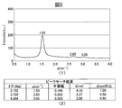
- FIG. 3 is an intensity distribution diagram of SAXS measurement regarding the gel composition prepared in Example 2.
- FIG. 4 is an intensity distribution diagram of SAXS measurement regarding the gel composition prepared in Example 3.
- FIG. 5 is an intensity distribution diagram of SAXS measurement regarding the emulsion prepared in Example 4.
- 6 is a particle size distribution diagram using a dynamic light scattering method for the emulsion prepared in Example 4.
- FIG. 1 is a diagram for explaining the concept of the present invention. It was schematically shown that fine particles that permeated tumor blood vessels by the EPR effect surrounded the tumor and blocked nutrients and the like.
- FIG. 7 is an SAXS measurement intensity distribution diagram for the emulsion prepared in Example 5.
- FIG. 8 is a particle size distribution diagram using a dynamic light scattering method for the emulsion prepared in Example 5.
- FIG. 9 is a particle size distribution diagram using a dynamic light scattering method for the emulsion prepared in Example 7.
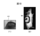
- FIG. 10 is a near-infrared fluorescence image in which a tumor-bearing mouse and its tumor site are enlarged.
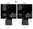
- FIG. 11 shows CT images of tumor-bearing mouse tumor sites before and after administration of the emulsion prepared in Example 4.
- FIG. 12 shows CT images of the lungs of tumor-bearing mice before administration of the 10-fold diluted solution of the emulsion prepared in Example 4, after the first administration, and after the second administration.
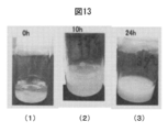
- FIG. 13 is a photographed image showing that the decomposition of the gel composition by mono-O- (5,9,13-trimethyltetradec-4-enoyl) glycerol progressed in human serum.
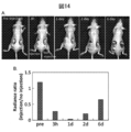
- FIG. 14A shows the effect of inhibiting blood flow at the tumor site by the emulsion using the luciferase (Luc) activity of breast cancer cells as an index.
- FIG. 14B is a graph plotting, over time, the size ratio of the tumor part in which blood flow was blocked for mice not receiving the emulsion.
- FIG. 15 is a diagram showing a time-dependent change in blood oxygen concentration at the tumor site after emulsion administration as a slice image. Blue indicates reduced hemoglobin, and red indicates oxidized hemoglobin.
- FIG. 14A shows the effect of inhibiting blood flow at the tumor site by the emulsion using the luciferase (Luc) activity of breast cancer cells as an index.
- FIG. 14B is a graph plotting, over time, the size ratio of
- FIG. 16A is a diagram showing an image of a tumor section obtained by measuring blood flow over time using indocyanine green (ICG).
- FIG. 16B is a diagram showing the results of recording changes in blood flow over time depending on whether or not an emulsion is administered. The result of having observed the change of the tumor volume at the time of administering an emulsion in multiple times over time is shown.
- ICG indocyanine green
- the biodegradable tumor sealant according to the present invention is based on a low molecular amphiphilic compound capable of forming a non-lamellar liquid crystal, and has a particle size capable of passing through a gap between tumor vascular endothelial cells. After the fine particles permeate the gap, they adhere to the tumor tissue existing in the vicinity of the gap due to high bioadhesion, thereby blocking the supply of nutrients and oxygen to the tumor and the transmission of the inducer from the tumor. The tumor cells are brought to necrosis, and then the adhered microparticles are degraded and / or metabolized in vivo.
- the biodegradable tumor sealant according to the present invention has the above-mentioned characteristics, but presses the nutrient blood vessels to the tumor to block blood flow, thereby providing blood oxygen concentration and nutrient supply at the tumor site. Therefore, the tumor sealant of the present invention also functions as a blood flow inhibitor having the above-mentioned additional effects.
- the biodegradable tumor sealant according to the present invention contains a low molecular amphiphilic compound capable of forming a non-lamellar liquid crystal.
- the “small molecule” of the present invention refers to one having a molecular weight of about 20 to 10,000. The molecular weight is preferably 50 to 5,000, more preferably 100 to 2,500, and still more preferably 200 to 1,000.
- the “amphiphilic compound” of the present invention is an amphiphilic compound having a hydrophilic group and a hydrophobic group that form a water-in-oil type non-lamellar liquid crystal in an aqueous medium.
- low-molecular amphiphilic compounds capable of forming non-lamellar liquid crystals include, but are not limited to, the following general formula (I):
- X and Y each represent a hydrogen atom or together represent an oxygen atom, preferably together represent an oxygen atom.
- N represents an integer of 0 to 2, and m represents 1 or 2.
- R in the above general formula (I) represents a hydrophilic group having two or more hydroxyl groups, and is not limited to the following, for example, glycerol, erythritol, pentaerythritol, diglycerol, glyceric acid, A hydrophilic group in which one hydroxyl group is removed from any one selected from the group consisting of triglycerol, xylose, sorbitol, ascorbic acid, glucose, galactose, mannose, dipentaerythritol, maltose, mannitol, and xylitol It is done.
- a preferred example of R is a hydrophilic group in which one hydroxyl group is removed from glycerol.
- amphiphilic compound is an E-form (cis-form) or Z-form (trans-form) of geometric isomers or a mixture thereof.
- amphiphilic compound represented by the general formula (I) include compounds in which X and Y together represent an oxygen atom, and R represents a hydrophilic group in which one hydroxyl group is removed from glycerol. Is preferred.
- amphiphilic compounds represented by the general formula (I) mono-O- (5,9,13-trimethyltetradecanoyl) glycerol, mono-O- (5,9,13,17-tetramethyloctadeca) Noyl) glycerol, mono-O- (5,9,13-trimethyltetradec-4-enoyl) glycerol, mono-O- (5,9,13,17-tetramethyloctadeca-4-enoyl) glycerol, mono-O- (5,9,13-trimethyltetradeca-4,8,12-trienoyl) glycerol or mono O- (5,9,13,17-tetramethyloctadeca-4,8,12,16-tetraenoyl) glycerol Is preferred. Furthermore, mono O- (5,9,13-trimethyltetradec-4-enoyl) glycerol is more preferred.
- amphiphilic compound of the present invention can be synthesized with reference to the description in Examples below. Or it can synthesize
- the amphiphilic compound used in the biodegradable tumor sealant according to the present invention can form a non-lamellar liquid crystal as a gel composition by self-organizing in an aqueous medium. it can.
- the non-lamellar liquid crystal formed by the amphiphilic compound used in the present invention is a structure that is not a lamellar liquid crystal and includes an L3 phase, but is a type II (water-in-oil type) that has a hydrophobic group oriented outward.
- the liquid crystal is more preferably a cubic liquid crystal or a reverse hexagonal liquid crystal.
- the above non-lamellar liquid crystal forming ability can be confirmed by analyzing the liquid crystal structure by a conventional method.
- the penetration method using a polarizing microscope it is possible to easily distinguish between type I (oil-in-water) cubic liquid crystal and type II (water-in-oil) cubic liquid crystal.
- Pn3m cubic liquid crystal, Ia3d cubic liquid crystal, Im3m cubic liquid crystal, and reverse hexagonal liquid crystal can be easily distinguished from the ratio of scattering peaks by X-ray small angle scattering (SAXS) measurement, and the peak value calculated from the SAXS data can be determined.
- SAXS X-ray small angle scattering
- the aqueous medium in which the amphiphilic compound according to the present invention can form a non-lamellar liquid crystal is not particularly limited, but water such as sterilized water, purified water, distilled water, ion-exchanged water, ultrapure water; Electrolyte aqueous solution such as saline, sodium chloride aqueous solution, calcium chloride aqueous solution, magnesium chloride aqueous solution, sodium sulfate aqueous solution, potassium sulfate aqueous solution, sodium carbonate aqueous solution, sodium acetate aqueous solution; buffer solution such as phosphate buffer solution, Tris hydrochloric acid buffer solution; glycerin Aqueous solution containing water-soluble organic substances such as ethylene glycol and ethanol; aqueous solution containing sugar molecules such as glucose, sucrose and maltose; aqueous solution containing water-soluble polymers such as polyethylene glycol and polyvinyl alcohol; octylglucoside
- the amphiphilic compound represented by the above general formula (I) exhibits high stability under a wide range of environmental conditions.
- the amphiphilic compound according to the present invention is characterized by having an isoprenoid chain as a hydrophobic group, and is different from an amphiphilic compound having a linear fatty chain such as oleic acid as a hydrophobic group. High resistance and relatively high oxidation stability.
- the amphiphilic compound according to the present invention also has a wide temperature range capable of forming a liquid crystal, a low craft temperature, and stably forms a liquid crystal even at a low temperature (6 ° C. or lower, preferably 0 ° C. or lower). be able to.
- the remarkable function of the non-lamellar liquid crystal formed by the amphiphilic compound used in the present invention is that it adheres to the surface of the living body due to the two layers of phospholipids in the cell membrane and the hydrophobic group outside the non-lamellar liquid crystal. That is.
- non-lamellar liquid crystal formed by the amphiphilic compound used in the present invention is the ability to incorporate the compound.
- a hydrophobic compound can be stably taken into the inside of the liquid crystal
- a water-soluble compound can be stably taken into the water channel constructed by the liquid crystal.
- biodegradable tumor sealant contains or consists of fine particles as a base material containing the amphiphilic compound, and the fine particles are contained in an aqueous medium. It can be prepared in the form of a dispersed emulsion.
- the aqueous medium for preparing the fine particles in the form of an emulsion is not particularly limited, but water such as sterilized water, purified water, distilled water, ion-exchanged water, ultrapure water; physiological saline, sodium chloride Aqueous solution, aqueous solution of calcium chloride, aqueous solution of magnesium chloride, aqueous solution of sodium sulfate, aqueous solution of potassium sulfate, aqueous solution of sodium carbonate, aqueous solution of sodium acetate, etc .; buffer solution such as phosphate buffer solution, Tris-HCl buffer solution; glycerin, ethylene glycol, ethanol An aqueous solution containing a water-soluble organic substance such as glucose; an aqueous solution containing a sugar molecule such as glucose, sucrose, or maltose; an aqueous solution containing a water-soluble polymer such as polyethylene glycol or polyvinyl alcohol; octyl glu
- the amphiphilic compound may be contained, but a functional substance may be further included.
- a functional substance may be further included.
- the tumor sealant of the present invention can include a contrast agent and / or a dye, it can be clinically applied to cancer diagnosis.
- contrast agent for X-ray CT, iodolipiodol, a hydrophobic contrast agent, iopamirone, an aqueous contrast agent, etc.
- iodolipiodol for X-ray CT, iodolipiodol, a hydrophobic contrast agent, iopamirone, an aqueous contrast agent, etc.
- MRI Gd (gadolinium) -based or Fe (iron) -based magnetic substance
- echo Applications include ultrasonic liposome particles. These may contain one kind or two or more kinds.
- examples of dyes that ensure visibility before and during surgery include hydrophobic coumarins, water-soluble fluorescent dyes such as fluorescein, pyranine, and cyanine, and luminescent dyes such as luciferin. These may contain one kind or two or more kinds.
- contrast agents and pigments may be contained at the same time.
- the mixing ratio of the amphiphilic compound and the contrast agent or / and the pigment may be 100: 1 to 1: 100, for example, 95: 5, 10: 1, 90:10, 10: 2, 80. : 20, 10: 3, 70:30, 60:40, 50:50, 40:60, 30:70, 20:80, 10:90, 5:95.
- the ratio is preferably 100: 1 to 50:50, more preferably 10: 1 to 10: 3.
- FIG. 2 (1) schematically shows an emulsion 20 containing microparticles 13 having various particle sizes, which constitute a biodegradable tumor sealant. Since the fine particles 13 are formed by chopping a non-lamellar liquid crystal, which will be described later, the particle size is not uniform, and various sizes are mixed as shown in FIG. There is also a fine particle 13 having a diameter size mainly (preferably 70% or more, more preferably 90% or more, and possibly 100% in the whole fine particles), and fine particles 13 of less than 20 nm or fine particles 13 of more than 500 nm. obtain.
- the fine particles having a particle size of “20 nm to 500 nm” include, for example, a particle size of “10 nm to 200 nm”. It is not intended to exclude fine particles having a particle size, fine particles having a particle size of “300 nm to 700 nm”, and the like.
- the particle sizes of “20 nm to 500 nm” are illustratively “20 nm to 400 nm”, “20 nm to 300 nm”, “20 nm to 200 nm”, “20 nm to 100 nm”, “30 nm to 500 nm”, “30 nm ⁇ 400 nm ”,“ 30 nm ⁇ 300 nm ”,“ 30 nm ⁇ 200 nm ”,“ 30 nm ⁇ 100 nm ”,“ 40 nm ⁇ 500 ”,“ 40 nm ⁇ 400 nm ”,“ 40 nm ⁇ 300 nm ”,“ 40 nm ⁇ 200 nm ”,“ 40 nm ⁇ 100 nm ” ”,“ 50 nm to 500 nm ”,“ 50 nm to 400 nm ”,“ 50 nm to 300 nm ”,“ 50 nm to 200 nm ”,“ 50 nm to 100
- the fine particles are typically one or two or more of the amphiphilic compounds or salts thereof, a contrast agent or / and a dye (when included), or one or more appropriate amounts in any order. And a suitable amount of an aqueous medium (for example, physiological saline, water for injection) and the like, and stirring and homogenizing.
- an aqueous medium for example, physiological saline, water for injection
- the internal phase of the fine particles is reverse hexagonal liquid crystal and the contrast agent or / and the dye are hydrophobic
- the fine particles 13 are indicated by the fine particles 13 in FIG. That is, the hydrophobic contrast agent or / and the dye 24 are included in the liquid crystal structure side of the reverse hexagonal liquid crystal 21.
- a water-soluble contrast agent or / and dye 24 is included in the water channel 22.
- the fine particles 13 are stably present in the aqueous medium by being covered with the outer phase 23 containing the surfactant.
- any of those used in the pharmaceutical or cosmetic field can be used, and examples thereof include, but are not limited to, for example, pluronic (for example, pluronic F127; polyoxy Ethylene polyoxypropylene (200EO) (70PO)), polysorbate 80 (polyoxyethylene sorbitan oleate; Tween 80) and the like can be used.
- pluronic for example, pluronic F127; polyoxy Ethylene polyoxypropylene (200EO) (70PO)
- polysorbate 80 polyoxyethylene sorbitan oleate; Tween 80
- the biodegradable tumor sealant of the present invention may contain the microparticles at a dispersible concentration (for example, 0.001 to 15% by weight), which is determined depending on the administration subject, the dose, and the like.
- a dispersible concentration for example, 0.001 to 15% by weight
- the structure of the fine particles can be analyzed by, for example, X-ray small angle scattering (SAXS) or cryo TEM.
- SAXS X-ray small angle scattering
- the particle size distribution of the fine particles can be measured by, for example, a zeta potential / particle size measuring device.
- the fine particles of the present invention can be prepared by mixing one or more amphiphilic compounds with a surfactant or the like, stirring and homogenizing, as shown in the above typical examples. As shown in FIG. 10, it can also be prepared using ultrasonic disruption or microfluidic technology.
- microfluidic technology is a general term for technologies for handling fluids in a minute space, and a device used in the technology is referred to as “microfluidic device”. This is an apparatus having a flow path structure having a depth and a width of typically about several ⁇ m to several 100 ⁇ m, which is manufactured by applying a semiconductor micromachining technology or a precision machining technology.
- the microparticles are produced using the microfluidic technique. However, since the microparticles having a desired particle diameter can be stably prepared, it is preferable to produce the microparticles using the technique.
- the particle diameter of the fine particles can be measured using a general method well known to those skilled in the art.
- biodegradable tumor sealant is a non-lamellar liquid crystal formed by the above-mentioned amphiphilic compound. And an emulsion dispersed in a biocompatible aqueous medium. This emulsion ensures the passage of a syringe or microcatheter.
- Tumor vascular permeability The tumor neovascularization specifically formed in the tumor tissue has decreased pericytes (vascular pericytes) covering the blood vessels, so the gap between the endothelial cells in the tumor blood vessels is normal. Spread compared to cells. There are particles that leak through the gap.
- the biodegradable tumor sealant has a particle size that is small enough to pass through the gap.
- the size of the gap varies from individual to individual, and is generally 5 nm or more and 700 nm or less, although it depends on the tumor type and site.
- Dispersibility composed of different particle diameters The space between the tumor blood vessels varies in size. Since the present biodegradable tumor sealant is an emulsion having a particle size distribution of several hundred nm rather than a single particle size, the density can be increased by permeating through any gap.
- Tumor tissue accumulation As described above, fine particles that permeate tumor blood vessels are released around the tumor, but unlike normal tissues, the lymphatic network is undeveloped in tumor tissues.
- the released microparticles that is, the present biodegradable tumor sealant
- the above (2) and (4) are collectively referred to as the EPR effect.
- Tumor tissue fixability As described above, even if accumulated in a tumor tissue, if it does not settle in tumor cells or stroma, it will flow out with the flow of body fluids and the like. Since the present biodegradable tumor sealant is a liquid crystal structural substance other than a lamellar structure, it has high adhesion to an extracellular matrix composed of cell membranes, collagen, fibroblasts, etc., and excellent fixability.
- the biodegradable tumor sealant aggregates and the gel-like tumor sealer tightly seals the tumor tissue, so that the tumor cells block nutrients and oxygen necessary for growth. To cell death (necrosis).
- Various inducers are emitted from tumor tissues and cells. For example, HIF (hypoxia-inducing factor) emitted from cancer cells under hypoxic stress is transmitted to VEGF (vascular endothelial growth factor) to promote tumor angiogenesis, and the supply of nutrients and oxygen to the tumor Increases and contributes to proliferation.
- HIF hypooxia-inducing factor
- VEGF vascular endothelial growth factor
- the present biodegradable tumor sealant can prevent malignant transformation by covering tumor tissue without gaps and preventing diffusion of transfer factors emitted from these tumors.
- the biodegradable tumor sealant undergoes degradation or solubilization by internal enzymes after a sufficient amount of time and period for necrosis of tumor cells, and partly undergoes further degradation. It is excreted outside the body.
- the biodegradable tumor sealant can also be used as an embolic material for arterial embolization.
- the embolic material as a temporary embolic material that exhibits the same embolizing effect as that of the permanent embolic material, it is possible to avoid a permanent indwelling risk and to expect a synergistic effect between the tumor sealing effect and the embolic effect.
- biodegradable tumor sealant The biodegradable tumor sealant prepared as described above can be used, for example, to diagnose and / or treat solid malignant tumors.
- solid malignant tumor includes squamous cell carcinoma, breast cancer, cutaneous lymphoma, hemangiosarcoma, hepatobiliary cancer, head and neck cancer, lung cancer, mesothelioma, mediastinum Cancer, esophageal cancer, stomach cancer, pancreatic cancer, small intestine cancer, colon cancer, colorectal cancer, colon cancer, anal cancer, kidney cancer, urethral cancer, bladder, prostate, urethral cancer, Penile cancer, testicular cancer, gynecologic cancer, ovarian cancer, endocrine cancer, skin cancer, cancer of the central nervous system including the brain; soft tissue and osteosarcoma; and skin and intraocular origin Including melanoma.
- the tumor sealant of the present invention can be applied not only to the diagnosis of cancer such as solid malignant tumors but also to the treatment of cancer, it is used as a “theranostics device”. Can be called.
- a biodegradable tumor sealant may be introduced from an artery in the vicinity of the tumor to embolize the tumor and / or prevent blood flow.
- the biodegradable tumor sealant of the present invention can be locally administered in the vicinity of the tumor using a syringe, although not limited thereto.
- the amount, concentration, frequency, frequency, and the like of the biodegradable tumor sealant at the time of introduction are determined by doctors and veterinarians in consideration of the subject's (subject) gender, age, weight, diseased state, etc. It can be adjusted as appropriate.
- the treatment target with the biodegradable tumor sealant of the present invention is preferably a mammal, and the mammal is not limited, but primates such as humans and monkeys, mice, rats, rabbits, and guinea pigs. Rodents such as cats, dogs, sheep, pigs, cows, horses, donkeys, goats, ferrets and the like.
- Example 8 100 ⁇ L of a biodegradable tumor sealant (Example 4) in the form of an emulsion is administered to a gall cancer mouse from a blood vessel in the vicinity of the tumor (specifically, injection) using a syringe. Tumor effect was obtained.
- the biodegradable tumor sealant of the present invention is 1 for human solid malignant tumors. This is equivalent to administering an amount of about 10 to 200 ml per administration from a blood vessel near the tumor.
- the biodegradable tumor sealant of the present invention can be used a plurality of times (for example, 2 to 10 times) at appropriate intervals (for example, twice a day, once a day until a desired therapeutic effect is obtained. Twice a week, once a week, once every two weeks).
- the dose specifically, the injection amount
- the administration frequency of the biodegradable tumor sealant of the present invention are not limited to the above, and those skilled in the art (for example, doctors or animals) The doctor) can make adjustments as appropriate.
- the biodegradable tumor sealant of the present invention is used as a pharmaceutical composition, in addition to the biodegradable tumor sealant as an active ingredient, a carrier, excipient, and / or stabilizer, etc. You may use it in the form, adding suitably.
- a kit including a biodegradable tumor, a solvent (such as physiological saline), a container (such as a vial) each encapsulating a carrier and the like, and an instruction manual is provided.
- the biodegradable tumor sealant of the present invention includes a medical material.
- Example 2 Formation and analysis of liquid crystal with mono-O- (5,9,13-trimethyltetradec-4-enoyl) glycerol Mono-O- (5,9,13-trimethyltetradec-4-enoyl) glycerol
- 50% by mass or more of physiological saline was added as an aqueous medium to perform a mixing operation, and the separated physiological saline was removed.
- physiological saline 50% by mass or more of physiological saline was added as an aqueous medium to perform a mixing operation, and the separated physiological saline was removed.
- a gel-like composition that was cloudy to colorless and transparent in appearance was obtained.
- the liquid crystal structure by X-ray small angle scattering (SAXS) of the gel composition was analyzed using a NANO Viewer nanoscale X-ray structure evaluation apparatus (manufactured by Rigaku).
- SAXS X-ray small angle scattering
- a graph showing the SAXS intensity distribution (FIG. 3 (1)) and peak research results (FIG. 3). 3 (2)) was obtained.
- the peak ratio showed a ratio 1: ⁇ 3: 2 peculiar to reverse hexagonal liquid crystals, confirming that the gel composition was reverse hexagonal liquid crystals. did it.
- Example 3 Formation and analysis of liquid crystal by mono-O- (5,9,13-trimethyltetradec-4-enoyl) glycerol and lipiodol Mono-O- (5,9,13-trimethyltetradec-4-enoyl) Glycerol and the contrast agent Lipiodol were mixed at a weight ratio of 10: 5, 70:30, 10: 3, 80:20, 10: 2, 90:10, 10: 1. To each mixture, 50% by mass or more of physiological saline was added as an aqueous medium to perform a mixing operation, and the separated physiological saline was removed to obtain a mixture. The mixture having a weight ratio of 10: 5 and 70:30 was a composition separated into two layers. Mixtures having a weight ratio of 10: 3, 80:20, 10: 2, 90:10, and 10: 1 each became a cloudy gel composition, and the viscosity increased as the weight ratio of lipiodol decreased.
- FIG. 4 (1) is a graph of SAXS measurement in the case of a weight ratio of 10: 3
- FIG. 4 (2) is a weight ratio of 10: 2
- FIG. 4 (3) is a weight ratio of 10: 1. All three samples were found to form non-lamellar liquid crystals. At a weight ratio of 10: 1, there are three strong scattering peaks that are characteristic of reverse hexagonal liquid crystals, whereas as the weight ratio goes to 10: 2 and weight ratio 10: 3, there are three scattering peaks. The liquid crystal structure has become less regular.
- Example 4 Preparation of Emulsion with Mono O- (5,9,13-Trimethyltetradec-4-enoyl) glycerol and Lipiodol
- Mono O- (5,9,13-trimethyltetradec-4-enoyl) glycerol and Mono-O- (5,9,13-trimethyltetradec-4-enoyl) glycerol (1.841 g) was mixed with Lipiodol (0.184 g) as a contrast agent so that the weight ratio of Lipiodol was 10: 1, and Pluronic F127 ( Aldrich P2443) 0.51 g and ethanol 0.285 g were added and dissolved until uniform.
- 12.18 g of physiological saline (Otsuka raw food injection) was added, stirred, and then homogenized with Starbust (manufactured by Sugino Machine) to prepare an emulsion containing fine particles.
- the liquid crystal structure by X-ray small angle scattering was analyzed for the above emulsion using a NANO Viewer nanoscale X-ray structure evaluation apparatus (manufactured by Rigaku).
- a graph showing the SAXS intensity distribution (FIG. 5 (1)) and a peak research result (FIG. 5). (2)) was obtained.
- the emulsion was a liquid crystal emulsion in which fine particles of reverse hexagonal liquid crystal were dispersed ( Hexasome).
- the particle size distribution of the above emulsion was measured using a Zetasizer Nano-ZS (Malvern).
- the measurement sample was prepared by diluting the above emulsion 100 times with distilled water.
- the obtained measurement data is shown in FIG.
- the average particle size (Z-Average) was 131.4 nm.
- fine particles having a particle size of around 60 nm begin to be formed, the particle size distribution rapidly increases as the particle size increases, and reaches a peak in the vicinity of the particle size of 130 nm. Thereafter, as the particle size increases, the particle size distribution decreases, and the formation of the fine particles ends at around the particle size of 350 nm. That is, the above emulsion was an emulsion in which fine particles having a particle diameter of 60 nm to 350 nm were predominant.
- Example 5 Preparation of emulsion of mono-O- (5,9,13-trimethyltetradec-4-enoyl) glycerol 5.4 g of mono-O- (5,9,13-trimethyltetradec-4-enoyl) glycerol , Pluronic F127 (Aldrich P2443) 1.36 g, ethanol 0.76 g, water for injection (Otsuka distilled water) 32.48 g were stirred and homogenized in the same manner as in Example 4 to prepare an emulsion containing fine particles. did.
- the SAXS measurement was performed on the emulsion in the same manner as in Example 4.
- a graph (FIG. 7 (1)) in which the SAXS intensity distribution was measured and a peak research result (FIG. 7 (2)) were obtained.
- the ratio of the peaks is a ratio ⁇ 2: ⁇ 3: ⁇ 4: ⁇ 6: ⁇ 8: ⁇ 9 specific to the cubic liquid crystal belonging to the crystallographic space group Pn3m.
- the emulsion is a cubic liquid crystal emulsion (cubosome) belonging to the crystallographic space group Pn3m.
- the particle size distribution was measured for the emulsion in the same manner as in Example 4.
- the measurement sample was prepared by diluting the above emulsion 100 times with distilled water. The result is shown in FIG. From FIG. 8, the average particle size (Z-Average) of the emulsion was 208.8 nm, and fine particles having a particle size of 90 nm to 600 nm accounted for the majority.
- Example 6 Preparation of emulsion of mono-O- (5,9,13-trimethyltetradec-4-enoyl) glycerol and Cy7 carboxylic acid Mono-O- (5,9,13-trimethyltetradec-4-enoyl) 3.375 g of glycerol, 0.007 g of Cy7 carboxylic acid (Lumiprobe-0550-90, Lumiprobe) as a fluorescent dye, 0.85 g of Pluronic F127 (Aldrich P2443), 0.475 g of ethanol, and saline (Otsuka raw food injection) 20 Using 0.3 g, the emulsion containing fine particles was prepared by stirring and homogenizing in the same manner as in Example 4.
- Example 7 Preparation of emulsion of mono-O- (5,9,13-trimethyltetradec-4-enoyl) glycerol, lipiodol and sodium fluorescein Mono-O- (5,9,13-trimethyltetradec-4-enoyl) ) 2.4546 g of glycerol, 0.2454 g of contrast agent Lipiodol, 0.0037 g of fluorescent dye fluorescein sodium (uranin), 0.68 g of Pluronic F127 (Aldrich P2443), 0.38 g of ethanol, physiological saline (Otsuka raw food injection) ) By using 16.2363 g, stirring and homogenizing in the same manner as in Example 4, an emulsion containing fine particles was prepared.
- the particle size distribution was measured in the same manner as in Example 4.
- the measurement sample was prepared by diluting the above emulsion 100 times with distilled water. The result is shown in FIG. From FIG. 9, the average particle size (Z-Average) of the emulsion was 124.8 nm, and fine particles having a particle size of 60 nm to 300 nm accounted for the majority.
- Example 8 Evaluation of tumor sealing effect and effect on normal tissue using emulsion (1) Preparation of tumor-bearing mice For a 1 mm subcutaneous site in the center of the back of an experimental animal mouse (C57BL / 6 albino male) Then, carcinoma LLC (mouse lung carcinoma) (LLC / luc) transplanted constitutively expressing the firefly luciferase gene (luc) was transplanted.
- Tumor-bearing mice (body weight 26.2 g) aged 10 weeks were produced by growing tumors over 36 days.
- FIG. 10 shows an image of a tumor-bearing mouse and its tumor site visualized by bioluminescence using luc.
- microparticles (biodegradable tumor sealant) of the present invention adhere specifically to tumor cells and do not adhere to normal tissues.
- Example 9 Stability evaluation of liquid crystals containing mono-O- (5,9,13-trimethyltetradec-4-enoyl) glycerol and liquid crystals containing lipiodol in human serum Mono-O- (5,9, 13-trimethyltetradec-4-enoyl) glycerol about 100 ⁇ L, or mono O- (5,9,13-trimethyltetradec-4-enoyl) glycerol and lipiodol weight ratios of 10: 1, 10: 2, 10: About 100 ⁇ L of each of the three mixed solutions was added to 2 mL of human serum (Human OTC Serum, Access Biologicals) in each vial.
- human serum Human OTC Serum, Access Biologicals
- Each vial was lightly shaken to form a gel-like composition, then left in a 37 ° C. hot water bath, and observed immediately after 10 hours and after 24 hours. As time passed, the human serum in each vial gradually became cloudy. When distilled water is used in place of human serum, the gel composition does not change in 24 hours and hardly becomes cloudy in distilled water.
- FIG. 13 shows a photographed image showing the course of a sample of about 100 ⁇ L of mono-O- (5,9,13-trimethyltetradec-4-enoyl) glycerol.
- a gel-like composition is formed and the human serum remains transparent.
- the clouding progresses.
- 24 hours after the addition (FIG. 13 (3)), although the gel composition remains, white turbidity has further progressed.
- the main component was carboxylic acid obtained by hydrolysis with eluted mono-O- (5,9,13-trimethyltetradec-4-enoyl) glycerol. (5,9,13-trimethyl-4-tetradecaenoic acid).
- the enzyme concentration and blood flow of intravascular hemoglobin at the tumor site were measured using MultiSpectroscopic Acoustic Tomography (MOST).
- MOST MultiSpectroscopic Acoustic Tomography
- the oxygenated hemoglobin concentration is usually high at the measurement site where the blood volume is recognized, while the reduced hemoglobin concentration is high at the measurement site that is deficient in oxygen, in proportion to the oxygen concentration in the blood.
- FIG. 15 shows the results of measuring the temporal change in blood oxygen concentration at the tumor site after emulsion administration.
- the signal of reduced hemoglobin (blue in the figure) increased from administration and became maximum after 1 day.
- the emulsion can block the supply of oxygen and nutrients carried by the blood to the tumor tissue as described above. Is backed up.
- ICG indocyanine green
- MSOT iThera Medical, inVision 256
- ICG fluorescence angiography is a method that is clinically used, and makes use of the characteristic that ICG bound to plasma protein in blood is excited by infrared light and emits fluorescence.
- FIG. 16A The result of imaging the change in ICG signal over time is shown in FIG. 16A, and the change over time in the corresponding signal intensity is shown in FIG. 16B.
- the ICG signal increased immediately after the administration and was maintained for a certain period of time. On the other hand, no increase in ICG signal was observed at the tumor site where the emulsion was administered. From this result, it was suggested that when the emulsion was administered to the tumor site, blood flow in the tumor blood vessel was blocked.
- microparticles of the tumor sealant of the present invention is expected to increase new options for minimally invasive therapies that prevent cancer metastasis or invasion.
- the microparticles have the advantages of degradability and visualization function as an embolizing agent, and the possibility of being used as a new embolizing agent for arterial embolization therapy, and further can include a contrast agent and / or a dye.
- Application to marking materials for cancer diagnosis and treatment is also possible.
Landscapes
- Health & Medical Sciences (AREA)
- Chemical & Material Sciences (AREA)
- General Health & Medical Sciences (AREA)
- Veterinary Medicine (AREA)
- Public Health (AREA)
- Life Sciences & Earth Sciences (AREA)
- Animal Behavior & Ethology (AREA)
- Epidemiology (AREA)
- Medicinal Chemistry (AREA)
- Pharmacology & Pharmacy (AREA)
- Engineering & Computer Science (AREA)
- Materials Engineering (AREA)
- Surgery (AREA)
- Chemical Kinetics & Catalysis (AREA)
- Dispersion Chemistry (AREA)
- General Chemical & Material Sciences (AREA)
- Dermatology (AREA)
- Oil, Petroleum & Natural Gas (AREA)
- Nuclear Medicine, Radiotherapy & Molecular Imaging (AREA)
- Organic Chemistry (AREA)
- Medicines Containing Antibodies Or Antigens For Use As Internal Diagnostic Agents (AREA)
- Medicinal Preparation (AREA)
- Acyclic And Carbocyclic Compounds In Medicinal Compositions (AREA)
- Medicines That Contain Protein Lipid Enzymes And Other Medicines (AREA)
Abstract
Description
[1] 非ラメラ液晶を形成することが可能な低分子の両親媒性化合物を基材とする微粒子を含む生体分解性腫瘍封止剤。
[2] 上記微粒子が20nm~500nmの粒子径を有する、上記[1]に記載の生体分解性腫瘍封止剤。
[3] 上記微粒子が水性媒体中に分散させたエマルションの形態で存在する、上記[1]又は[2]に記載の生体分解性腫瘍封止剤。
[4] 上記両親媒性化合物が下記一般式(I)で表される化合物又はその塩である、上記[1]~[3]のいずれかに記載の生体分解性腫瘍封止剤。


[5] 一般式(I)中、X及びYは一緒になって酸素原子を表し、Rがグリセロールから1つの水酸基が除かれた親水基を表す、上記[4]に記載の生体分解性腫瘍封止剤。
[6] 上記両親媒性化合物が、以下のいずれかである、上記[1]~[5]のいずれかに記載の生体分解性腫瘍封止剤:
モノO-(5,9,13-トリメチルテトラデカノイル)グリセロール、モノO-(5,9,13,17-テトラメチルオクタデカノイル)グリセロール、モノO-(5,9,13-トリメチルテトラデカ-4-エノイル)グリセロール、モノO-(5,9,13,17-テトラメチルオクタデカ-4-エノイル)グリセロール、モノO-(5,9,13-トリメチルテトラデカ-4,8,12-トリエノイル)グリセロール、又はモノO-(5,9,13,17-テトラメチルオクタデカ-4,8,12,16-テトラエノイル)グリセロール。
[7] 上記微粒子が造影剤及び/又は色素を包含する、上記[1]~[6]のいずれかに記載の生体分解性腫瘍封止剤。
[8] 上記両親媒性化合物がモノO-(5,9,13-トリメチルテトラデカ-4-エノイル)グリセロールであり、上記造影剤がリピオドールである、上記[7]に記載の生体分解性腫瘍封止剤。
本発明に係る生体分解性腫瘍封止剤は、非ラメラ液晶を形成することが可能な低分子の両親媒性化合物を基材とし、腫瘍血管内皮細胞の間隙を透過し得る粒子径を有する微粒子であって、この微粒子がその間隙を透過後、高い生体接着性により間隙近傍に存在する腫瘍組織に付着し、腫瘍への栄養と酸素の供給並びに腫瘍からの誘導因子の伝達を遮断することによって、その腫瘍細胞を壊死に至らしめ、その後、付着した微粒子が生体内で分解及び/又は代謝されることを特徴とする。また、本発明に係る生体分解性腫瘍封止剤は、上記特徴を有するが、腫瘍への栄養血管を圧迫して血流を阻止することにより、腫瘍部位での血中の酸素濃度及び栄養供給を減少させ、腫瘍細胞を壊死させる効果も有することから、本発明の腫瘍封止剤には、上記の付加的効果を有する血流阻止剤としても機能する。
本発明に係る生体分解性腫瘍封止剤は、非ラメラ液晶を形成することが可能な低分子の両親媒性化合物を含有する。本発明の「低分子」とは、約20~10,000の分子量を有するものをいう。分子量は、好ましくは50~5,000、より好ましくは100~2,500、さらに好ましくは200~1,000である。また、本発明の「両親媒性化合物」とは、水性媒体中で油中水型の非ラメラ液晶を形成する親水基と疎水基を持つ両親媒性化合物である。



本発明に係る生体分解性腫瘍封止剤に用いる両親媒性化合物は、水性媒体中において自己組織化することによって、ゲル状組成物として、非ラメラ液晶を形成することができる。本発明で用いる両親媒性化合物によって形成される非ラメラ液晶は、ラメラ液晶ではない構造体であって、L3相を含むが、疎水基を外側に向けて配向したII型(油中水型)の液晶であることが好ましく、具体的には、キュービック液晶又は逆ヘキサゴナル液晶であることがより好ましい。
本発明に係る生体分解性腫瘍封止剤は、上記両親媒性化合物を含有する基材とする微粒子を含み又は該微粒子からなり、上記微粒子を水性媒体中に分散させたエマルションの形態に調製され得る。
(1)シリンジ又はマイクロカテーテル通過性
本発明に係る生体分解性腫瘍封止剤は、前述したように、上記両親媒性化合物によって形成される非ラメラ液晶を微粒子化して、生体適合する水性媒体に分散したエマルションを含む。本エマルションによって、シリンジ又はマイクロカテーテルの通過性を確保している。
(2)腫瘍血管透過性
腫瘍組織に特異的に形成された腫瘍新生血管は、血管を覆うペリサイト(血管周皮細胞)が減少しているため、腫瘍血管における内皮細胞間の間隙は、正常細胞と比較して広がっている。その間隙を漏れ出る粒子が存在する。本生体分解性腫瘍封止剤は、その間隙を通過するのに十分小さな粒子径を持つ。間隙の大きさは個人差があり、腫瘍種別、部位にもよるが、一般に5nm以上700nm以下とされている。
(3)異なる粒子径で構成される分散性
前記腫瘍血管の間隙は大小様々である。本生体分解性腫瘍封止剤は、粒子径は単一ではなく数百nm幅の分布をもつエマルションのため、どの間隙からも透過することによって集積密度を増加させることができる。
(4)腫瘍組織集積性
前述のように腫瘍血管を透過した微粒子は、腫瘍周辺に放出されるが、正常組織と異なり腫瘍組織では、リンパ管網が未発達である。このため体液と共にリンパ管経由で、他の部位に運ばれることは無い。従って、放出された微粒子即ち本生体分解性腫瘍封止剤は、腫瘍部位に留まる集積性がある。前記(2)と(4)を合わせてEPR効果と呼ぶ。
(5)腫瘍組織定着性
前述のように腫瘍組織に集積しても、腫瘍細胞や間質に定着しなければ、体液等の流れと共に流出してしまう。本生体分解性腫瘍封止剤は、ラメラ構造以外の液晶構造物質であるため、細胞膜やコラーゲン、繊維芽細胞等からなる細胞外マトリクスへの接着性が高く定着性に優れる。
(6)酸素と栄養の遮断性
本生体分解性腫瘍封剤が集合合体しゲル状の瘍封剤が稠密に腫瘍組織を封止することによって、腫瘍細胞は増殖に必要な栄養と酸素が遮断され、細胞死(壊死)にいたる。
(7)伝達系転写誘導因子の伝達阻害性
腫瘍組織・細胞からは、各種誘導因子が発せられる。例えば、低酸素ストレス下のがん細胞から発せられるHIF(低酸素誘導因子)は、VEGF(血管内皮細胞増殖因子)に伝達して腫瘍血管新生を促し、腫瘍への栄養と酸素の供給量を増大させ増殖に寄与している。本生体分解性腫瘍封剤は、腫瘍組織を隙間なく覆い封じ込めることによって、これら腫瘍から発せられる伝達因子の拡散を阻止することで悪性化を食い止めることができる。
(8)速やかな代謝・排泄
本生体分解性腫瘍封剤は、腫瘍細胞が壊死するのに十分な時間・期間を経た後は、体内酵素による分解又は可溶化、一部はさらなる分解などを経て、体外へ排泄される。
(9)視認性及び可視化性
(6)までは本生体分解性腫瘍封剤の基本的な機能に限定した性質であるが、実用上の機能として、医師が使用する場面で有用な機能を付加している。
第一が、目視による視認性である。色素(蛍光色素を含む)を包含することによって、他の医薬品との取り違えリスクを軽減し、更にはマーカーとして手術部位・範囲を表示することで、間違った箇所への施術を回避できる。
第二に、造影剤を包含させることにより、術中の画像装置によるモニタリングで的確な箇所へ十分な施術が行われている或いは行われたかを確認できる。更には、造影剤のみを使用した時のように流出せずに腫瘍部位に定着するため、再度造影剤を注入する手術をすることなく、術後の腫瘍の状況を画像装置でモニタリングできる可視化性を有する。
(10)塞栓性
本生体分解性腫瘍封止剤は、動脈塞栓用塞栓材としても使用できる。塞栓材としては、永久塞栓材と同等の塞栓効果を発揮する一時塞栓材として、永久留置リスクを回避でき、更に腫瘍封止効果と塞栓効果との相乗効果を期待することもできる。
上記の通りに調製した生体分解性腫瘍封止剤は、例えば、固形悪性腫瘍を診断及び/又は治療するために使用することができる。本明細書で使用するとき、「固形悪性腫瘍」には、有棘細胞がん、乳がん、皮膚リンパ腫、血管肉腫、肝胆道系のがん、頭頸部がん、肺がん、中皮腫、縦隔がん、食道がん、胃がん、膵臓がん、小腸がん、結腸がん、結直腸がん、大腸がん、肛門がん、腎臓がん、尿道がん、膀胱、前立腺、尿道がん、陰茎がん、睾丸がん、婦人科臓器のがん、卵巣がん、内分泌系のがん、皮膚がん、脳を含む中枢神経系のがん;軟組織及び骨肉腫;並びに皮膚及び眼内起源の黒色腫を含む。使用形態としては、限定されないが、1種類又は2種類以上の両親媒性化合物を含有する基材とする微粒子を水性媒体中に分散させたエマルションの形態で使用してもよく、又は該微粒子を含む医薬組成物として使用してもよい。このように、本発明の腫瘍封止剤は、固形悪性腫瘍などのがんの診断に適用可能であるだけでなく、がんの治療に応用することもできることから、「セラノスティクス・デバイス」として称することができる。
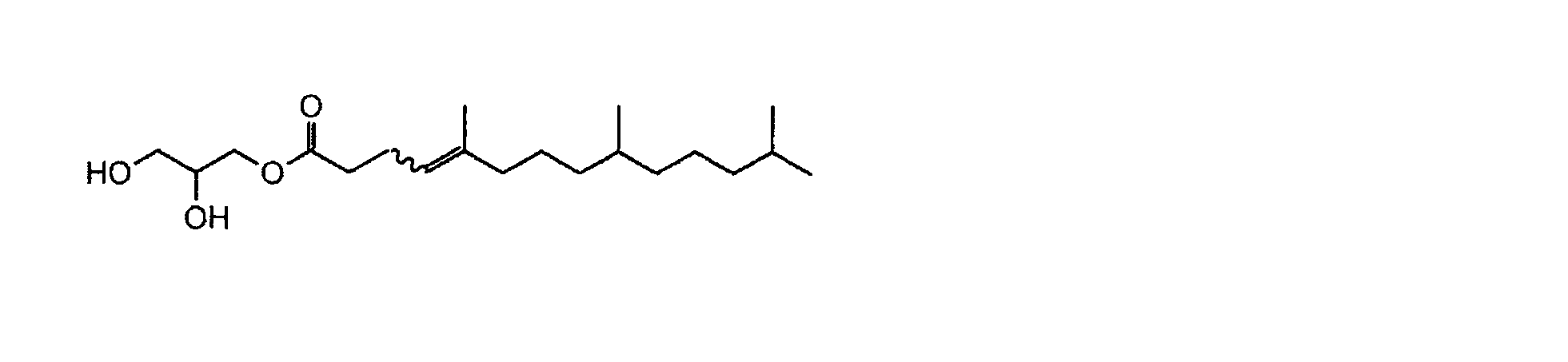
1H-NMRスペクトル(300MHz,CDCl3,TMS)δ:0.80-0.90(m,9H),1.00-1.70(m,15H),1.97(td,J=7.8,17.0Hz,2H),2.13(t,J=6.1Hz,1H,OH),2.25-2.45(m,4H),2.55(d,J=5.2Hz,1H,OH),3.50-4.00(m,3H),4.10-4.25(m,2H),5.08(t,J=6.7Hz,1H)
粘度:0.48Pa・s(せん断速度92 1/s)
モノO-(5,9,13-トリメチルテトラデカ-4-エノイル)グリセロールに対して、水性媒体として50質量%以上の生理食塩水を添加して混合操作を行い、分離した生理食塩水を除いた。その結果、外観上は白濁~無色透明なゲル状組成物を得た。
モノO-(5,9,13-トリメチルテトラデカ-4-エノイル)グリセロールと造影剤であるリピオドールを重量比10:5、70:30、10:3、80:20、10:2、90:10、10:1で混合した。各混合物に対して、水性媒体として50質量%以上の生理食塩水を添加して混合操作を行い、分離した生理食塩水を除いて、それぞれ混合物を得た。重量比10:5、70:30の混合物は、二層に分離した組成物であった。重量比10:3、80:20、10:2、90:10、10:1の混合物は、それぞれ白濁したゲル状組成物となり、リピオドールの重量比が低くなるにしたがって粘性は高まった。
モノO-(5,9,13-トリメチルテトラデカ-4-エノイル)グリセロールとリピオドールの重量比が10:1となるよう、モノO-(5,9,13-トリメチルテトラデカ-4-エノイル)グリセロール1.841gと造影剤であるリピオドール0.184gを混合し、プルロニックF127(Aldrich P2443)0.51gとエタノール0.285gを添加して、均一になるまで溶解した。さらに、生理食塩水(大塚生食注)12.18gを添加して、撹拌した後、スターバスト(スギノマシン製)でホモジナイズすることによって、微粒子を含有するエマルションを調製した。
モノO-(5,9,13-トリメチルテトラデカ-4-エノイル)グリセロール5.4g、プルロニックF127(Aldrich P2443)1.36gとエタノール0.76g、注射用水(大塚蒸留水)32.48gを用いて、実施例4と同様に攪拌・ホモジナイズすることによって、微粒子を含有するエマルションを調製した。
モノO-(5,9,13-トリメチルテトラデカ-4-エノイル)グリセロール3.375g、蛍光色素であるCy7カルボン酸(Lumiprobe-0550-90,Lumiprobe社)0.0074g、プルロニックF127(Aldrich P2443)0.85gとエタノール0.475g、生理食塩水(大塚生食注)20.3gを用いて、実施例4と同様に攪拌・ホモジナイズすることによって、微粒子を含有するエマルションを調製した。
モノO-(5,9,13-トリメチルテトラデカ-4-エノイル)グリセロール2.4546g、造影剤であるリピオドール0.2454g、蛍光色素であるフルオレセインナトリウム(ウラニン)0.0037g、プルロニックF127(Aldrich P2443)0.68gとエタノール0.38g、生理食塩水(大塚生食注)16.2363gを用いて、実施例4と同様に攪拌・ホモジナイズすることによって、微粒子を含有するエマルションを調製した。
(1)担癌マウスの作成
実験動物のマウス(C57BL/6 albino雄)の背中中央部皮下1mmの部位に対して、ホタルルシフェラーゼ遺伝子(luc)を恒常発現する癌腫LLC(mouse lung carcinoma)(LLC/luc)移植した。36日間かけて腫瘍を成長させることによって、生後週齢10週の担癌マウス(体重26.2g)を作成した。図10に、担癌マウスとその腫瘍部位をlucによる生物発光で可視化した画像を示す。
上記担癌マウスの腫瘍に対して、実施例4で調製したエマルション100μLを注射器によって、腫瘍近傍の血管より投与した。小型実験動物用3DマイクロX線CT装置(CosmoScan FX、リガク社)を用いて、投与前と投与5分後に腫瘍部位を観察した。得られたCT画像を解析した結果、エマルション中の微粒子が腫瘍血管を透過して腫瘍部位に付着していることが明らかとなった。図11に、本エマルション投与前後における担癌マウス腫瘍部位のCT画像を示す。
上記担癌マウスに対して、実施例4で調製したエマルションを生理食塩水で10倍希釈したエマルション200μLを注射器によって、尾部より10分毎に2回投与した。小型実験動物用3DマイクロX線CT装置(CosmoScan FX、リガク社)を用いて、投与前、投与1回目5分後、投与2回目5分後に担癌マウスの全体を観察した。とりわけ、最も細い毛細血管が存在する肺臓において、1回目投与後と2回目投与後のいずれにおいても、エマルション中の微粒子が付着していないことが明らかとなった。図12に、本エマルション投与前、1回目投与後、2回目投与後における担癌マウス肺臓のCT画像を示す。
モノO-(5,9,13-トリメチルテトラデカ-4-エノイル)グリセロール約100μL、又はモノO-(5,9,13-トリメチルテトラデカ-4-エノイル)グリセロールとリピオドールの重量比が10:1、10:2、10:3の各混合溶液、約100μLをそれぞれバイアル中のヒト血清(Human OTC Serum,Access Biologicals社)2mLに添加した。各バイアルを軽く揺らして、ゲル状組成物を形成させた後、37℃の湯浴中に静置して、直後、10時間後、24時間後に観察した。時間の経過によって、各バイアル中のヒト血清はいずれも徐々に白濁した。なお、ヒト血清の代わりに蒸留水を用いた場合、24時間以内にいずれも、ゲル状組成物に変化はなく、蒸留水中はほとんど白濁しない。
(1)乳癌細胞移植マウスの作製
ルシフェラーゼを安定に発現する4T1乳癌細胞(4T1-Luc)を入手し、ヌードマウスBalb/c nu/nu(日本チャールス・リバー株式会社)の皮下に2.5×107細胞/mlのPBS/Geltrex(登録商標)=1/1(vol/vol)の細胞懸濁液を0.04ml移植した。
上記の乳癌細胞を移植されたマウス腫瘍部位に、高圧ホモジナイザーを用いて作製したエマルションを局所投与した。投与後、ルシフェラーゼの基質であるD-ルシフェリンを含むリン酸緩衝生理食塩水を腹腔内投与により投与して血流循環させ、上記乳癌細胞のルシフェラーゼ(Luc)活性をIn vivoイメージング装置(IVIS)(Spectrum)で経時的に観察した。腫瘍部位では、血中のルシフェリンの存在によりLuc活性に基づいて蛍光が確認される。図14Aに示される通り、エマルションの非投与マウスでは、移植した乳癌細胞のLuc発現により継続的に蛍光を観察できるのに対して、エマルションを投与したマウスでは、投与直後から蛍光が減少したことから、エマルションの投与により、血中のD-ルシフェリンの腫瘍への送達、エマルション投与により腫瘍部位への血流が阻止されることが示唆された。また、この観察結果について、非投与マウスに対する投与マウスにおけるLuc活性の比を算出し、経時的な変化を図14Bとして示す。
腫瘍血管における血流阻止をさらに確認するために、エマルション投与1日後の担癌マウスの静脈内に蛍光物質であるインドシアニングリーン(ICG)を投与し、腫瘍部位における血流をMSOT(iThera Medical社、inVision 256)を用いてモニタリングした。このようなICG蛍光血管撮影法は、臨床的に利用されている方法であって、血中で血漿タンパクと結合したICGが赤外光で励起され、蛍光を発する特性を活かしたものである。経時的にICGシグナルの変化を撮像した結果を図16Aに示し、これに対応するシグナル強度の経時的変化を図16Bに示した。エマルションを投与していない腫瘍部位は、投与直後からICGシグナルが増加し、一定時間、維持された。一方、エマルションを投与した腫瘍部位では、ICGシグナルの増加が観察されなかった。この結果から、腫瘍部位にエマルションを投与した場合、腫瘍血管における血流が阻止されることが示唆された。
エマルションを複数回投与した場合の腫瘍体積の変化について検討した。上記(1)に記載したように、4T1-Lucを移植されたヌードマウスを用いて経時的に観察した。エマルションの投与は、初回(0日目)、2日目、3日目、及び5日目に行った。腫瘍体積の経時的な変化を図17に示す。図17から明らかなように、生理食塩水を投与した対照と比較して、エマルションを投与したマウスの腫瘍体積は減少した。これは、1回投与の場合と比較して、腫瘍体積の減少は顕著であった(データ示さず)。
11 内皮細胞間隙
12 腫瘍組織
13 微粒子
14 腫瘍細胞
15 赤血球
20 エマルション
21 逆ヘキサゴナル液晶
22 水チャネル
23 外相
24 造影剤
60 担癌マウス
61 腫瘍部
62 投与後腫瘍部
63 付着微粒子
64 肺
Claims (8)
- 非ラメラ液晶を形成することが可能な低分子の両親媒性化合物を基材とする微粒子を含む生体分解性腫瘍封止剤。
- 前記微粒子が20nm~500nmの粒子径を有する、請求項1に記載の生体分解性腫瘍封止剤。
- 前記微粒子が水性媒体中に分散させたエマルションの形態で存在する、請求項1又は2に記載の生体分解性腫瘍封止剤。
- 前記両親媒性化合物が下記一般式(I)で表される化合物又はその塩である、請求項1~3のいずれか1項に記載の生体分解性腫瘍封止剤。


- 一般式(I)中、X及びYは一緒になって酸素原子を表し、Rがグリセロールから1つの水酸基が除かれた親水基を表す、請求項4に記載の生体分解性腫瘍封止剤。
- 前記両親媒性化合物が、以下のいずれかである、請求項1~5のいずれか1項に記載の生体分解性腫瘍封止剤:
モノO-(5,9,13-トリメチルテトラデカノイル)グリセロール、モノO-(5,9,13,17-テトラメチルオクタデカノイル)グリセロール、モノO-(5,9,13-トリメチルテトラデカ-4-エノイル)グリセロール、モノO-(5,9,13,17-テトラメチルオクタデカ-4-エノイル)グリセロール、モノO-(5,9,13-トリメチルテトラデカ-4,8,12-トリエノイル)グリセロール、又はモノO-(5,9,13,17-テトラメチルオクタデカ-4,8,12,16-テトラエノイル)グリセロール。 - 前記微粒子が造影剤及び/又は色素を包含する、請求項1~6のいずれか1項に記載の生体分解性腫瘍封止剤。
- 前記両親媒性化合物がモノO-(5,9,13-トリメチルテトラデカ-4-エノイル)グリセロールであり、前記造影剤がリピオドールである、請求項7に記載の生体分解性腫瘍封止剤。
Priority Applications (4)
| Application Number | Priority Date | Filing Date | Title |
|---|---|---|---|
| US16/062,870 US20200282101A1 (en) | 2015-12-18 | 2016-12-16 | Biodegradable tumor sealant |
| JP2017556485A JP6721907B2 (ja) | 2015-12-18 | 2016-12-16 | 生体分解性腫瘍封止剤 |
| CN201680074139.8A CN108472373A (zh) | 2015-12-18 | 2016-12-16 | 生物降解性肿瘤密封剂 |
| EP16875814.2A EP3391906A4 (en) | 2015-12-18 | 2016-12-16 | BIODEGRADABLE TUMOR SEALANT |
Applications Claiming Priority (2)
| Application Number | Priority Date | Filing Date | Title |
|---|---|---|---|
| JP2015247650 | 2015-12-18 | ||
| JP2015-247650 | 2015-12-18 |
Publications (1)
| Publication Number | Publication Date |
|---|---|
| WO2017104840A1 true WO2017104840A1 (ja) | 2017-06-22 |
Family
ID=59056804
Family Applications (1)
| Application Number | Title | Priority Date | Filing Date |
|---|---|---|---|
| PCT/JP2016/087676 Ceased WO2017104840A1 (ja) | 2015-12-18 | 2016-12-16 | 生体分解性腫瘍封止剤 |
Country Status (5)
| Country | Link |
|---|---|
| US (1) | US20200282101A1 (ja) |
| EP (1) | EP3391906A4 (ja) |
| JP (1) | JP6721907B2 (ja) |
| CN (1) | CN108472373A (ja) |
| WO (1) | WO2017104840A1 (ja) |
Cited By (3)
| Publication number | Priority date | Publication date | Assignee | Title |
|---|---|---|---|---|
| WO2019082991A1 (ja) | 2017-10-25 | 2019-05-02 | メディギア・インターナショナル株式会社 | 生体分解性及び生体代謝性の腫瘍封止剤 |
| WO2020050423A1 (ja) * | 2018-09-07 | 2020-03-12 | 株式会社ファルネックス | 非ラメラ液晶形成脂質を含む外用剤 |
| WO2021060498A1 (ja) * | 2019-09-27 | 2021-04-01 | 国立大学法人京都大学 | 多孔性シリカを含むナノ粒子およびその製造方法、ならびに放射線治療用医薬組成物 |
Families Citing this family (3)
| Publication number | Priority date | Publication date | Assignee | Title |
|---|---|---|---|---|
| CN111544102A (zh) * | 2020-04-14 | 2020-08-18 | 山西省肿瘤医院(山西省第三人民医院) | 腹腔镜下防肿瘤细胞脱落隔离装置 |
| CN113144297A (zh) * | 2021-04-30 | 2021-07-23 | 中山大学孙逸仙纪念医院 | 一种用于阻断卵巢生殖细胞肿瘤扩散的薄膜材料及其制备方法 |
| EP4493313A1 (en) * | 2022-03-15 | 2025-01-22 | Mikrocaps D.O.O. | Biodegradable microcapsules based on crystalline materials and synthesis process |
Citations (6)
| Publication number | Priority date | Publication date | Assignee | Title |
|---|---|---|---|---|
| WO2006043705A1 (ja) * | 2004-10-19 | 2006-04-27 | National Institute Of Advanced Industrial Scienceand Technology | Ii型キュービック液晶組成物 |
| JP2006117553A (ja) * | 2004-10-19 | 2006-05-11 | National Institute Of Advanced Industrial & Technology | Ii型キュービック液晶組成物にリソソーム酵素を包埋した複合体 |
| WO2011078383A1 (ja) * | 2009-12-25 | 2011-06-30 | 株式会社サイトパスファインダー | 低粘度液晶化合物 |
| JP2012017318A (ja) * | 2010-06-07 | 2012-01-26 | Nikko Chemical Co Ltd | 液晶及びそれを含有する皮膚外用剤 |
| JP2014129318A (ja) * | 2012-02-23 | 2014-07-10 | Canon Inc | インドシアニングリーン含有粒子、およびそれを有する光音響イメージング用造影剤 |
| WO2014178256A1 (ja) * | 2013-05-01 | 2014-11-06 | 株式会社ファルネックス | 癒着防止剤 |
Family Cites Families (7)
| Publication number | Priority date | Publication date | Assignee | Title |
|---|---|---|---|---|
| PT942780E (pt) * | 1997-09-09 | 2003-11-28 | Lyotropic Therapeutics Inc | Particulas revestidas processos de obtencao e uso |
| US8603999B2 (en) * | 2008-12-05 | 2013-12-10 | Commonwealth Scientific And Industrial Research Organisation | Amphiphile prodrugs |
| CN101822635B (zh) * | 2010-04-15 | 2011-12-14 | 中山大学 | 组合物在制备原位液晶血管栓塞剂中的应用 |
| CN104023793B (zh) * | 2011-10-31 | 2017-11-24 | 马林克罗特有限公司 | 用于治疗癌症的联合脂质体组合物 |
| CN103040741B (zh) * | 2012-12-11 | 2014-11-05 | 中山大学 | 溶致液晶的前体混悬液及其制备方法 |
| TWI461203B (zh) * | 2013-07-04 | 2014-11-21 | Academia Sinica | 腫瘤血管栓塞劑以及金奈米粒子之用途 |
| CN103536974B (zh) * | 2013-07-05 | 2015-07-15 | 北京大学 | 磁共振成像可检测的原位液晶前体栓塞组合物及其制备和应用 |
-
2016
- 2016-12-16 JP JP2017556485A patent/JP6721907B2/ja not_active Expired - Fee Related
- 2016-12-16 WO PCT/JP2016/087676 patent/WO2017104840A1/ja not_active Ceased
- 2016-12-16 CN CN201680074139.8A patent/CN108472373A/zh active Pending
- 2016-12-16 US US16/062,870 patent/US20200282101A1/en not_active Abandoned
- 2016-12-16 EP EP16875814.2A patent/EP3391906A4/en not_active Withdrawn
Patent Citations (6)
| Publication number | Priority date | Publication date | Assignee | Title |
|---|---|---|---|---|
| WO2006043705A1 (ja) * | 2004-10-19 | 2006-04-27 | National Institute Of Advanced Industrial Scienceand Technology | Ii型キュービック液晶組成物 |
| JP2006117553A (ja) * | 2004-10-19 | 2006-05-11 | National Institute Of Advanced Industrial & Technology | Ii型キュービック液晶組成物にリソソーム酵素を包埋した複合体 |
| WO2011078383A1 (ja) * | 2009-12-25 | 2011-06-30 | 株式会社サイトパスファインダー | 低粘度液晶化合物 |
| JP2012017318A (ja) * | 2010-06-07 | 2012-01-26 | Nikko Chemical Co Ltd | 液晶及びそれを含有する皮膚外用剤 |
| JP2014129318A (ja) * | 2012-02-23 | 2014-07-10 | Canon Inc | インドシアニングリーン含有粒子、およびそれを有する光音響イメージング用造影剤 |
| WO2014178256A1 (ja) * | 2013-05-01 | 2014-11-06 | 株式会社ファルネックス | 癒着防止剤 |
Non-Patent Citations (1)
| Title |
|---|
| See also references of EP3391906A4 * |
Cited By (15)
| Publication number | Priority date | Publication date | Assignee | Title |
|---|---|---|---|---|
| JP2021102096A (ja) * | 2017-10-25 | 2021-07-15 | メディギア・インターナショナル株式会社 | 生体分解性及び生体代謝性の腫瘍封止剤 |
| JPWO2019082991A1 (ja) * | 2017-10-25 | 2019-11-14 | メディギア・インターナショナル株式会社 | 生体分解性及び生体代謝性の腫瘍封止剤 |
| JP7324965B2 (ja) | 2017-10-25 | 2023-08-14 | 武雄 田中 | 生体分解性及び生体代謝性の腫瘍封止剤 |
| JP2020058784A (ja) * | 2017-10-25 | 2020-04-16 | メディギア・インターナショナル株式会社 | 生体分解性及び生体代謝性の腫瘍封止剤 |
| CN111278477A (zh) * | 2017-10-25 | 2020-06-12 | 精密医疗国际公司 | 生物降解性和生物代谢性的肿瘤封闭剂 |
| US20200281960A1 (en) * | 2017-10-25 | 2020-09-10 | Medigear International Corporation | Biodegradable and biometabolic tumor sealant |
| JP2023107245A (ja) * | 2017-10-25 | 2023-08-02 | 武雄 田中 | 生体分解性及び生体代謝性の腫瘍封止剤 |
| WO2019082991A1 (ja) | 2017-10-25 | 2019-05-02 | メディギア・インターナショナル株式会社 | 生体分解性及び生体代謝性の腫瘍封止剤 |
| JPWO2020050423A1 (ja) * | 2018-09-07 | 2021-08-30 | 株式会社ファルネックス | 非ラメラ液晶形成脂質を含む外用剤 |
| CN112770778A (zh) * | 2018-09-07 | 2021-05-07 | 法纳克斯株式会社 | 含有非层状液晶形成脂质的外用剂 |
| WO2020050423A1 (ja) * | 2018-09-07 | 2020-03-12 | 株式会社ファルネックス | 非ラメラ液晶形成脂質を含む外用剤 |
| JP7603972B2 (ja) | 2018-09-07 | 2024-12-23 | 株式会社ファルネックス | 非ラメラ液晶形成脂質を含む外用剤 |
| JP2025029135A (ja) * | 2018-09-07 | 2025-03-05 | 株式会社ファルネックス | 非ラメラ液晶形成脂質を含む外用剤 |
| US12296014B2 (en) | 2018-09-07 | 2025-05-13 | Farnex Incorporated | External preparation comprising non-lamellar liquid crystal-forming lipid |
| WO2021060498A1 (ja) * | 2019-09-27 | 2021-04-01 | 国立大学法人京都大学 | 多孔性シリカを含むナノ粒子およびその製造方法、ならびに放射線治療用医薬組成物 |
Also Published As
| Publication number | Publication date |
|---|---|
| JPWO2017104840A1 (ja) | 2018-12-06 |
| EP3391906A1 (en) | 2018-10-24 |
| US20200282101A1 (en) | 2020-09-10 |
| CN108472373A (zh) | 2018-08-31 |
| JP6721907B2 (ja) | 2020-07-15 |
| EP3391906A4 (en) | 2019-08-28 |
Similar Documents
| Publication | Publication Date | Title |
|---|---|---|
| JP6721907B2 (ja) | 生体分解性腫瘍封止剤 | |
| Centelles et al. | Image-guided thermosensitive liposomes for focused ultrasound drug delivery: Using NIRF-labelled lipids and topotecan to visualise the effects of hyperthermia in tumours | |
| Ye et al. | Focused ultrasound-enabled delivery of radiolabeled nanoclusters to the pons | |
| Liu et al. | Enhanced primary tumor penetration facilitates nanoparticle draining into lymph nodes after systemic injection for tumor metastasis inhibition | |
| Shen et al. | Enhanced delivery of paclitaxel liposomes using focused ultrasound with microbubbles for treating nude mice bearing intracranial glioblastoma xenografts | |
| Ninomiya et al. | Ultrasound-mediated drug delivery using liposomes modified with a thermosensitive polymer | |
| JP7053463B2 (ja) | 超音波システムを使用して脳腫瘍を治療するための方法及びキット | |
| Zimmermann et al. | A pilot trial of doxorubicin containing phosphatidyldiglycerol based thermosensitive liposomes in spontaneous feline soft tissue sarcoma | |
| Wang et al. | Phototheranostic nanoparticles with aggregation-induced emission as a four-modal imaging platform for image-guided photothermal therapy and ferroptosis of tumor cells | |
| Zhao et al. | GRP78-targeted and doxorubicin-loaded nanodroplets combined with ultrasound: a potential novel theranostics for castration-resistant prostate cancer | |
| Shi et al. | Recent advances in interventional fluorescence imaging: Toward the precise visualization of transarterial mini-invasive delivery systems | |
| Telichko et al. | Therapeutic ultrasound parameter optimization for drug delivery applied to a murine model of hepatocellular carcinoma | |
| Zhang et al. | Biosynthetic gas vesicles combined with focused ultrasound for blood–brain barrier opening | |
| Liao et al. | Estimating the delivery efficiency of drug-loaded microbubbles in cancer cells with ultrasound and bioluminescence imaging | |
| RU2563369C1 (ru) | Способ ферримагнито-термохимиотерапии злокачественных опухолей комбинациями магнитоуправляемых нанопрепаратов с визуализацией онкогенеза, определением терапии, предпочтительной в режиме реального времени, и мониторингом результатов лечения в эксперименте | |
| EP4218830A1 (en) | Tumor targeted diagnostic imaging agent for diagnostic biopsy, or intraoperative tumor identification or margin assessment using near-infrared fluorescence (nirf) imaging | |
| Wang et al. | Near infrared light mediated photochemotherapy for efficiently treating deep orthotopic tumors guided by ultrasound imaging | |
| JP6620288B2 (ja) | 生体分解性及び生体代謝性の腫瘍封止剤 | |
| KR20190096244A (ko) | 수용성 항암제를 담지하는 마이크로버블을 포함하는 간동맥색전술용 조성물 및 그 제조방법 | |
| Suzuki et al. | Ultrasound irradiation in the presence of microbubbles may enhance the antitumor effect of chemotherapeutic agents against bladder cancer | |
| US20250375374A1 (en) | Shear-thinning compositions for ablation | |
| KR102935458B1 (ko) | 폴리에틸렌글라이콜 기반의 비이온성 계면활성제와 함께 안정화된 소수성약물을 담지한 초음파 감응 마이크로 버블 약물전달체 | |
| JP7829229B2 (ja) | 薬物送達制御可能なリポソーム | |
| RU2761827C1 (ru) | Средство и способ комбинированной контрастной магнитно-резонансной томографической визуализации изображений биомеханики процессов инфильтрации, инвазии и метастазирования злокачественных клеток | |
| HK40037027A (en) | Biodegradable and biometabolic tumor sealant |
Legal Events
| Date | Code | Title | Description |
|---|---|---|---|
| 121 | Ep: the epo has been informed by wipo that ep was designated in this application |
Ref document number: 16875814 Country of ref document: EP Kind code of ref document: A1 |
|
| ENP | Entry into the national phase |
Ref document number: 2017556485 Country of ref document: JP Kind code of ref document: A |
|
| NENP | Non-entry into the national phase |
Ref country code: DE |
|
| WWE | Wipo information: entry into national phase |
Ref document number: 2016875814 Country of ref document: EP |
|
| ENP | Entry into the national phase |
Ref document number: 2016875814 Country of ref document: EP Effective date: 20180718 |