WO2017060201A1 - Anti-wt1/hla-specific antibodies - Google Patents
Anti-wt1/hla-specific antibodies Download PDFInfo
- Publication number
- WO2017060201A1 WO2017060201A1 PCT/EP2016/073587 EP2016073587W WO2017060201A1 WO 2017060201 A1 WO2017060201 A1 WO 2017060201A1 EP 2016073587 W EP2016073587 W EP 2016073587W WO 2017060201 A1 WO2017060201 A1 WO 2017060201A1
- Authority
- WO
- WIPO (PCT)
- Prior art keywords
- seq
- antibody
- fragment
- region
- hla
- Prior art date
Links
Classifications
-
- C—CHEMISTRY; METALLURGY
- C07—ORGANIC CHEMISTRY
- C07K—PEPTIDES
- C07K16/00—Immunoglobulins [IGs], e.g. monoclonal or polyclonal antibodies
- C07K16/18—Immunoglobulins [IGs], e.g. monoclonal or polyclonal antibodies against material from animals or humans
- C07K16/28—Immunoglobulins [IGs], e.g. monoclonal or polyclonal antibodies against material from animals or humans against receptors, cell surface antigens or cell surface determinants
- C07K16/2803—Immunoglobulins [IGs], e.g. monoclonal or polyclonal antibodies against material from animals or humans against receptors, cell surface antigens or cell surface determinants against the immunoglobulin superfamily
- C07K16/2833—Immunoglobulins [IGs], e.g. monoclonal or polyclonal antibodies against material from animals or humans against receptors, cell surface antigens or cell surface determinants against the immunoglobulin superfamily against MHC-molecules, e.g. HLA-molecules
-
- C—CHEMISTRY; METALLURGY
- C07—ORGANIC CHEMISTRY
- C07K—PEPTIDES
- C07K16/00—Immunoglobulins [IGs], e.g. monoclonal or polyclonal antibodies
- C07K16/18—Immunoglobulins [IGs], e.g. monoclonal or polyclonal antibodies against material from animals or humans
-
- A—HUMAN NECESSITIES
- A61—MEDICAL OR VETERINARY SCIENCE; HYGIENE
- A61P—SPECIFIC THERAPEUTIC ACTIVITY OF CHEMICAL COMPOUNDS OR MEDICINAL PREPARATIONS
- A61P35/00—Antineoplastic agents
-
- A—HUMAN NECESSITIES
- A61—MEDICAL OR VETERINARY SCIENCE; HYGIENE
- A61P—SPECIFIC THERAPEUTIC ACTIVITY OF CHEMICAL COMPOUNDS OR MEDICINAL PREPARATIONS
- A61P35/00—Antineoplastic agents
- A61P35/02—Antineoplastic agents specific for leukemia
-
- C—CHEMISTRY; METALLURGY
- C07—ORGANIC CHEMISTRY
- C07K—PEPTIDES
- C07K14/00—Peptides having more than 20 amino acids; Gastrins; Somatostatins; Melanotropins; Derivatives thereof
- C07K14/435—Peptides having more than 20 amino acids; Gastrins; Somatostatins; Melanotropins; Derivatives thereof from animals; from humans
- C07K14/46—Peptides having more than 20 amino acids; Gastrins; Somatostatins; Melanotropins; Derivatives thereof from animals; from humans from vertebrates
- C07K14/47—Peptides having more than 20 amino acids; Gastrins; Somatostatins; Melanotropins; Derivatives thereof from animals; from humans from vertebrates from mammals
- C07K14/4701—Peptides having more than 20 amino acids; Gastrins; Somatostatins; Melanotropins; Derivatives thereof from animals; from humans from vertebrates from mammals not used
- C07K14/4748—Tumour specific antigens; Tumour rejection antigen precursors [TRAP], e.g. MAGE
-
- G—PHYSICS
- G01—MEASURING; TESTING
- G01N—INVESTIGATING OR ANALYSING MATERIALS BY DETERMINING THEIR CHEMICAL OR PHYSICAL PROPERTIES
- G01N33/00—Investigating or analysing materials by specific methods not covered by groups G01N1/00 - G01N31/00
- G01N33/48—Biological material, e.g. blood, urine; Haemocytometers
- G01N33/50—Chemical analysis of biological material, e.g. blood, urine; Testing involving biospecific ligand binding methods; Immunological testing
- G01N33/53—Immunoassay; Biospecific binding assay; Materials therefor
-
- G—PHYSICS
- G01—MEASURING; TESTING
- G01N—INVESTIGATING OR ANALYSING MATERIALS BY DETERMINING THEIR CHEMICAL OR PHYSICAL PROPERTIES
- G01N33/00—Investigating or analysing materials by specific methods not covered by groups G01N1/00 - G01N31/00
- G01N33/48—Biological material, e.g. blood, urine; Haemocytometers
- G01N33/50—Chemical analysis of biological material, e.g. blood, urine; Testing involving biospecific ligand binding methods; Immunological testing
- G01N33/68—Chemical analysis of biological material, e.g. blood, urine; Testing involving biospecific ligand binding methods; Immunological testing involving proteins, peptides or amino acids
- G01N33/6854—Immunoglobulins
-
- A—HUMAN NECESSITIES
- A61—MEDICAL OR VETERINARY SCIENCE; HYGIENE
- A61K—PREPARATIONS FOR MEDICAL, DENTAL OR TOILETRY PURPOSES
- A61K39/00—Medicinal preparations containing antigens or antibodies
- A61K2039/505—Medicinal preparations containing antigens or antibodies comprising antibodies
-
- C—CHEMISTRY; METALLURGY
- C07—ORGANIC CHEMISTRY
- C07K—PEPTIDES
- C07K2317/00—Immunoglobulins specific features
- C07K2317/20—Immunoglobulins specific features characterized by taxonomic origin
- C07K2317/21—Immunoglobulins specific features characterized by taxonomic origin from primates, e.g. man
-
- C—CHEMISTRY; METALLURGY
- C07—ORGANIC CHEMISTRY
- C07K—PEPTIDES
- C07K2317/00—Immunoglobulins specific features
- C07K2317/30—Immunoglobulins specific features characterized by aspects of specificity or valency
- C07K2317/32—Immunoglobulins specific features characterized by aspects of specificity or valency specific for a neo-epitope on a complex, e.g. antibody-antigen or ligand-receptor
-
- C—CHEMISTRY; METALLURGY
- C07—ORGANIC CHEMISTRY
- C07K—PEPTIDES
- C07K2317/00—Immunoglobulins specific features
- C07K2317/30—Immunoglobulins specific features characterized by aspects of specificity or valency
- C07K2317/33—Crossreactivity, e.g. for species or epitope, or lack of said crossreactivity
-
- C—CHEMISTRY; METALLURGY
- C07—ORGANIC CHEMISTRY
- C07K—PEPTIDES
- C07K2317/00—Immunoglobulins specific features
- C07K2317/30—Immunoglobulins specific features characterized by aspects of specificity or valency
- C07K2317/34—Identification of a linear epitope shorter than 20 amino acid residues or of a conformational epitope defined by amino acid residues
-
- C—CHEMISTRY; METALLURGY
- C07—ORGANIC CHEMISTRY
- C07K—PEPTIDES
- C07K2317/00—Immunoglobulins specific features
- C07K2317/50—Immunoglobulins specific features characterized by immunoglobulin fragments
- C07K2317/55—Fab or Fab'
-
- C—CHEMISTRY; METALLURGY
- C07—ORGANIC CHEMISTRY
- C07K—PEPTIDES
- C07K2317/00—Immunoglobulins specific features
- C07K2317/50—Immunoglobulins specific features characterized by immunoglobulin fragments
- C07K2317/56—Immunoglobulins specific features characterized by immunoglobulin fragments variable (Fv) region, i.e. VH and/or VL
- C07K2317/565—Complementarity determining region [CDR]
-
- C—CHEMISTRY; METALLURGY
- C07—ORGANIC CHEMISTRY
- C07K—PEPTIDES
- C07K2317/00—Immunoglobulins specific features
- C07K2317/70—Immunoglobulins specific features characterized by effect upon binding to a cell or to an antigen
- C07K2317/73—Inducing cell death, e.g. apoptosis, necrosis or inhibition of cell proliferation
- C07K2317/732—Antibody-dependent cellular cytotoxicity [ADCC]
-
- G—PHYSICS
- G01—MEASURING; TESTING
- G01N—INVESTIGATING OR ANALYSING MATERIALS BY DETERMINING THEIR CHEMICAL OR PHYSICAL PROPERTIES
- G01N2500/00—Screening for compounds of potential therapeutic value
- G01N2500/04—Screening involving studying the effect of compounds C directly on molecule A (e.g. C are potential ligands for a receptor A, or potential substrates for an enzyme A)
Definitions
- the present application relates to antibodies or antibody fragments which specifically bind to the WT1 /HLA complex.
- the invention also relates to nucleic acids, vectors and host cells capable of expressing said antibodies or fragments thereof, pharmaceutical compositions comprising said antibodies or fragments thereof and uses of said antibodies or fragments thereof for treatment of specific diseases.
- the Wilms tumor 1 (WT1 ) oncoprotein is an intracellular, oncogenic transcription factor that is overexpressed in a wide range of leukemias and solid cancers.
- WT1 is a nuclear protein and therefore not accessible for conventional antibody therapy.
- a WT1 -derived peptide named RMF amino acid sequence RMFPNAPYL (SEQ ID No. #1 )
- RMF amino acid sequence RMFPNAPYL (SEQ ID No. #1 )
- HLA-A0201 molecules is processed and presented by HLA-A0201 molecules. This peptide induces cytotoxic CD8 T cells capable of killing WT1 + tumor cells in vitro and in human T cell-based and vaccine trials (Cancer Immunol Immunother. 2010; 59:1467-1479). This provides a rationale to target the HLA-restricted peptide with antibodies.
- the present invention provides novel antibodies and antibody fragments that are superior to the antibodies known in the prior art.
- the antibodies and antibody fragments disclosed herein specifically bind to the WT1 RMF peptide/HLA-A0201 complex (RMF/HLA).
- the present application for the first time discloses antibodies and fragments thereof that specifically bind to RMF peptide/HLA-A0201 complex (RMF/HLA) and have the superior properties as disclosed herein.
- the antibodies and fragments thereof of the present disclosure do not bind to unspecific peptides complexed with HLA-A0201 .
- the antibodies and fragments thereof of the present disclosure do not bind to the PIGQ peptide (a peptide ubiquitously expressed on healthy human tissue) when complexed with HLA-A0201 .
- the antibodies or fragments thereof of the present invention bind to cells expressing the RMF/HLA complex.
- the antibodies of the present invention bind to cancer cells expressing the RMF/HLA complex.
- the antibodies and fragments thereof also show EC 5 o values and affinities in a monovalent Fab format and in a bispecific immunoglobulin format that have never been reported before.
- the antibodies or fragments thereof of the present invention are therefore improved in terms of effectiveness, and thus provide well suited and promising compounds, e.g. for clinical development.
- Antibodies with the properties as claimed were generated utilizing a sophisticated screening and counter-screening method. It is therefore possible to identify additional antibodies or fragments thereof having essentially the same or the same properties.
- the present invention provides antibodies or fragments thereof that specifically bind to the RMF/HLA complex present on the surface cells.
- the present disclosure also provides antibodies or fragments thereof that specifically bind to the RMF/HLA complex but which do not bind to the PIGQ/HLA complex.
- the present disclosure also provides antibodies or fragments thereof that bind to the RMF/HLA complex with an EC 5 o which is at least 10 times lower than the EC50 for the PIGQ/HLA complex.
- the present invention also relates to a method for identifying an antibody or fragment thereof according to the present invention, comprising (a) mixing an antibody library comprising a plurality of antibodies or fragments thereof with a PIGQ/HLA complex under conditions allowing a specific binding of antibodies or the fragments thereof to said complex, (b) removing from the antibody library those antibodies or fragments thereof that bind to said PIGQ/HLA complex, (c) mixing the depleted antibody library of step (b) with an RMF/HLA complex under conditions allowing a specific binding of antibodies or the fragments thereof to said complex, and (d) identifying those antibodies or fragments thereof that bind to the RMF/HLA complex.
- the present invention provides antibodies or fragments thereof that bind to the RMF peptide/HLA-A0201 complex (RMF/HLA) having the CDR regions according to the amino acids as listed in Table 1 .
- the present invention also provides specific antibodies or fragments thereof having a variable heavy chain and a variable light chain comprising the amino acid sequences according to Table 1 .
- the present invention also provides specific antibodies or fragments thereof having CDR regions comprising the amino acid sequences according to Table 1 .
- the present invention also provides antibodies or fragments thereof comprising a heavy chain and a light chain that is at least 60%, at least 70 %, at least 80%, at least 90% or at least 95% homologous (identical) to the ones of the antibodies as shown in Table 1 .
- the present invention also provides antibodies or fragments thereof which comprising CDR regions that are at least 60%, at least 70 %, at least 80%, at least 90% or at least 95% homologous to the CDR regions of the antibodies as shown in Table 1 .
- the present invention also provides specific antibodies or fragments thereof the binding of which competes with the specific antibodies or fragments thereof as disclosed herein.
- the present invention also provides specific antibodies or antibody fragments which bind to the same epitope as the specific antibodies or fragments thereof disclosed herein.
- the present invention also provides specific antibodies or fragments thereof that compete with the specific antibodies or fragments thereof disclosed herein and specifically bind to the RMF/HLA complex when presented on the surface of cells.
- the present invention also provides the isolated antibodies or fragments thereof of the present invention for use in medicine.
- the present invention also provides the antibodies or fragments thereof of the present invention for use in the treatment of patients suffering from a WT1 -positive disease, such as cancer.
- WT1 positive diseases and cancers include chronic myelocytic leukemia (CML), multiple myeloma (MM), acute lymphoblastic leukemia (ALL), acute myeloid/myelogenous leukemia (AML), myelodysplastic syndrome (MDS), mesothelioma, ovarian cancer, gastrointestinal cancers, breast cancer, prostate cancer and glioblastoma.
- the antibody or fragment thereof is a conjugate having a cytotoxic moiety linked thereto.
- the antibody or fragment thereof can carry certain additional modifications, such as, for example, Fc modifications.
- the present invention also provides pharmaceutical compositions comprising the isolated antibodies or fragments thereof of the present invention, and a pharmaceutically acceptable carrier.
- the present invention also provides nucleic acids encoding for the antibodies or fragments thereof of the present invention.
- the present invention also provides vectors comprising nucleic acids encoding the antibodies or fragments thereof of the present invention.
- the present invention also provides host cell comprising vector or nucleic acids encoding the antibodies or fragments thereof of the present invention.
- the invention relates to antibodies or fragments thereof that recognize, preferably specifically recognize, and thus bind to, WT1 RMF peptide/HLA-A0201 complexes (RMF/HLA).
- WT1 refers to a protein known as Wilms tumor protein. Certain synonyms of WT1 do exits, including AWT1 , GUD, NPHS4, WAGR, Wilms tumor protein, WIT-2 and WT33.
- Human WT1 has the following amino acid sequence (UniProt P19544):
- RMF and “RMF peptide” refer to the WT1 -derived peptide having the amino acid sequence RMFPNAPYL (SEQ ID NO: 1 ).
- HLA-A0201 refers to a specific HLA serotype.
- HLA-A0201 is a heterodimeric protein, comprising an alpha chain and a beta chain.
- WT1/HLA complex WT1/HLA
- RMF/HLA complex RMF/HLA complex
- RMF/HLA RMF/HLA
- RHAMM and “RHAMM peptide” refer to the peptide with the amino acid sequence ILSLELMKL (SEQ ID No. 63).
- RHAMM/HLA complex and “RHAMM/HLA” refer to a complex of said RHAMM peptide with HLA-A0201 .
- PAGQ and PAGQ peptide refer to the peptide with the amino acid sequence RMFPGEVAL (SEQ ID No. 64). This peptide occurs ubiquitously in healthy human tissue.
- PIGQ /HLA complex and "PIGQ /HLA” refer to a complex of the PIGQ peptide with HLA-A0201 .
- antibody refers to a protein comprising at least two heavy (H) chains and two light (L) chains inter-connected by disulfide bonds which interacts (e.g., by binding, steric hindrance, stabilizing spatial distribution) with an antigen.
- Each heavy chain is comprised of a heavy chain variable region (abbreviated herein as VH) and a heavy chain constant region.
- the heavy chain constant region is comprised of three domains, CH1 , CH2 and CH3.
- Each light chain is comprised of a light chain variable region (abbreviated herein as VL) and a light chain constant region.
- the light chain constant region is comprised of one domain, CL.
- VH and VL regions can be further subdivided into regions of hypervariability, termed complementarity determining regions (CDR), interspersed with regions that are more conserved, termed framework regions (FR).
- CDR complementarity determining regions
- FR framework regions
- Each VH and VL is composed of three CDRs and four FR's arranged from amino-terminus to carboxy-terminus in the following order: FR1 , CDR1 , FR2, CDR2, FR3, CDR3, and FR4.
- the variable regions of the heavy and light chains contain a binding domain that interacts with an antigen.
- the constant regions of the antibodies may mediate the binding of the immunoglobulin to host tissues or factors, including various cells of the immune system (e.g., effector cells) and the first component (Clq) of the classical complement system.
- antibody includes for example, monoclonal antibodies, human antibodies, humanized antibodies, camelised antibodies and chimeric antibodies.
- the antibodies can be of any isotype (e.g., IgG, IgE, IgM, IgD, IgA and IgY), class (e.g., lgG1 , lgG2, lgG3, lgG4, lgA1 and lgA2) or subclass. Both the light and heavy chains are divided into regions of structural and functional homology.
- immunoglobulin format refers to a full length antibody as defined herein above.
- antibody fragment refers to one or more portions of an antibody that retain(s) the ability to specifically interact with (e.g., by binding, steric hindrance, stabilizing spatial distribution) an antigen.
- binding fragments include, but are not limited to, a Fab fragment, a monovalent fragment consisting of the VL, VH, CL and CH1 domains; a F(ab)2 fragment, a bivalent fragment comprising two Fab fragments linked by a disulfide bridge at the hinge region; an Fd fragment consisting of the VH and CH1 domains; an Fv fragment consisting of the VL and VH domains of a single arm of an antibody; a dAb fragment (Ward et al., (1989) Nature 341 :544-546), which consists of a VH domain; and an isolated complementarity determining region (CDR).
- a Fab fragment a monovalent fragment consisting of the VL, VH, CL and CH1 domains
- F(ab)2 fragment a bivalent fragment comprising two Fab fragments linked by a disulfide bridge at the hinge region
- an Fd fragment consisting of the VH and CH1 domains
- an Fv fragment consisting of the
- the two domains of the Fv fragment, VL and VH are coded for by separate genes, they can be joined, using recombinant methods, by a synthetic linker that enables them to be made as a single protein chain in which the VL and VH regions pair to form monovalent molecules (known as single chain Fv (scFv); see e.g., Bird et al., (1988) Science 242:423-426; and Huston et al., (1988) Proc. Natl. Acad. Sci. 85:5879-5883).
- single chain Fv single chain Fv
- Such single chain antibodies are also intended to be encompassed within the term "antibody fragment”.
- Antibody fragments are obtained using conventional techniques known to those of skill in the art, and the fragments are screened for utility in the same manner as are intact antibodies.
- Antibody fragments can also be incorporated into single domain antibodies, maxibodies, minibodies, intrabodies, diabodies, triabodies, tetrabodies, v-NAR and bis-scFv (see, e.g., Hollinger and Hudson, (2005) Nature Biotechnology 23:1 126-1 136).
- Antibody fragments can be grafted into scaffolds based on polypeptides such as Fibronectin type III (Fn3) (see U.S. Pat. No. 6,703,199, which describes fibronectin polypeptide monobodies).
- Fn3 Fibronectin type III
- Antibody fragments can be incorporated into single chain molecules comprising a pair of tandem Fv segments (VH-CH1 -VH-CH1 ) which, together with complementary light chain polypeptides, form a pair of antigen-binding sites (Zapata et al., (1995) Protein Eng. 8:1057-1062; and U.S. Pat. No. 5,641 ,870).
- the term "Fab format" refers to a Fab fragment of an antibody.
- antigen binding site refers to the part of the antibody or antibody fragment that comprises the area or part(s) of it that specifically bind(s) to an antigen.
- An antigen binding site may be provided by one or more antibody variable domains.
- an antigen binding site is comprised within the associated VH and VL of an antibody or fragment thereof.
- a “human antibody” or “human antibody fragment”, as used herein, includes antibodies and fragment thereof having variable regions in which both the framework and CDR regions are derived from sequences of human origin. Furthermore, if the antibody contains a constant region, the constant region also is derived from such human sequences, e.g., human germline sequences, or mutated versions of human germline sequences or antibody containing consensus framework sequences derived from human framework sequences analysis, for example, as described in Knappik et al., (2000) J Mol Biol 296:57-86).
- immunoglobulin variable domains e.g., CDRs
- CDRs may be defined using well known numbering schemes, e.g., the Kabat numbering scheme, the Chothia numbering scheme, or a combination of Kabat and Chothia (see, e.g., Sequences of Proteins of Immunological Interest, U.S. Department of Health and Human Services (1991 ), eds. Kabat et al.; Lazikani et al., (1997) J. Mol. Bio. 273:927-948); Kabat et al., (1991 ) Sequences of Proteins of Immunological Interest, 5th edit., NIH Publication no. 91 -3242 U.S.
- a “humanized antibody” or “humanized antibody fragment” as defined herein is one that is (i) derived from a non-human source (e.g., a transgenic mouse which bears a heterologous immune system), which antibody is based on a human germline sequence or (ii) CDR-grafted, wherein the CDRs of the variable domain are from a non-human origin, while one or more frameworks of the variable domain are of human origin and the constant domain (if any) is of human origin.
- a non-human source e.g., a transgenic mouse which bears a heterologous immune system
- CDR-grafted wherein the CDRs of the variable domain are from a non-human origin, while one or more frameworks of the variable domain are of human origin and the constant domain (if any) is of human origin.
- chimeric antibody or “chimeric antibody fragment” is defined herein as an antibody molecule which has constant antibody regions derived from, or corresponding to, sequences found in one species and variable antibody regions derived from another species.
- the constant antibody regions are derived from, or corresponding to, sequences found in humans, e.g. in the human germ line or somatic cells
- the variable antibody regions e.g. VH, VL, CDR or FR regions
- a non-human animal e.g. a mammal, such as a mouse, rat, rabbit or hamster.
- isolated refers to a compound, which can be e.g. an antibody or antibody fragment, that is substantially free of other antibodies or antibody fragments having different antigenic specificities. Moreover, an isolated antibody or fragment thereof may be substantially free of other cellular material and/or chemicals. Thus, in some aspects, antibodies provided are isolated antibodies which have been separated from antibodies with a different specificity. An isolated antibody may be a monoclonal antibody. An isolated antibody may be a recombinant monoclonal antibody. An isolated antibody that specifically binds to an epitope, isoform or variant of a target may, however, have cross-reactivity to other related antigens, e.g., from other species (e.g., species homologs).
- recombinant antibody includes all antibodies that are prepared, expressed, created or segregated by recombinant means, such as antibodies isolated from an animal (e.g., a mouse) that is transgenic or transchromosomal for human immunoglobulin genes or a hybridoma prepared therefrom, antibodies isolated from a host cell transformed to express the antibody, antibodies selected and isolated from a recombinant, combinatorial human antibody library, and antibodies prepared, expressed, created or isolated by any other means that involve splicing of all or a portion of a human immunoglobulin gene, sequences to other DNA sequences.
- an animal e.g., a mouse
- transgenic or transchromosomal for human immunoglobulin genes or a hybridoma prepared therefrom antibodies isolated from a host cell transformed to express the antibody
- antibodies selected and isolated from a recombinant, combinatorial human antibody library and antibodies prepared, expressed, created or isolated by any other means that involve splicing of all or a portion of a human immunoglobul
- such recombinant antibodies have variable regions in which the framework and CDR regions are derived from human germline immunoglobulin sequences.
- such recombinant human antibodies can be subjected to in vitro mutagenesis (or, when an animal transgenic for human Ig sequences is used, in vivo somatic mutagenesis) and thus the amino acid sequences of the VH and VL regions of the recombinant antibodies are sequences that, while derived from and related to human germline VH and VL sequences, may not naturally exist within the human antibody germline repertoire in vivo.
- a recombinant antibody may be a monoclonal antibody.
- the antibodies and antibody fragment disclosed herein are isolated from the Ylanthia ® antibody library as disclosed in US 13/321 ,564 or US 13/299,367, which both are incorporated herein by reference in their entireties.
- monoclonal antibody refers to a preparation of antibody molecules of single molecular composition.
- a monoclonal antibody composition displays a unique binding site having a unique binding specificity and affinity for particular epitopes.
- an antibody binds specifically to”, “specifically binds to”, is “specific to/for” or “specifically recognizes” an antigen, if such antibody or fragment thereof is able to discriminate between such antigen and one or more reference antigen(s), since binding specificity is not an absolute, but a relative property.
- the reference antigen(s) may be one or more closely related antigen(s), which are used as reference points, e.g. a mutated of scrambled version of the WT1/HLA complex.
- an antibody or antibody fragment that "specifically binds to" the WTA/HLA complex does neither bind to the RHAMM/HLA complex nor to the PIGQ/HLA complex.
- a “specific binding” is referring to the ability of the antibody or fragment thereof to discriminate between an antigen of interest and an unrelated antigen, as determined, for example, in accordance with one of the following methods. Such methods comprise, but are not limited to Western blots, ELISA-, RIA-, ECL-, IRMA-tests and peptide scans.
- a standard ELISA assay can be carried out.
- the scoring may be carried out by standard color development (e.g. secondary antibody with horseradish peroxidase and tetramethyl benzidine with hydrogen peroxide).
- the reaction in certain wells is scored by the optical density, for example, at 450 nm.
- determination of binding specificity is performed by using not a single reference antigen, but a set of about three to five unrelated antigens, such as milk powder, BSA, transferrin or the like.
- “specific binding” may relate to the ability to discriminate between different parts of its target antigen, e.g. different domains or regions of the WT1/HLA complex, or between one or more key amino acid residues or stretches of amino acid residues of the WT1/HLA complex.
- the antibodies or fragment thereof disclosed herein specifically bind a WT1/HLA complex of a mammal, in particular to the human WT1/HLA complex.
- the term "avidity” is used to describe the combined strength of multiple bond interactions between proteins. Avidity is distinct from affinity which describes the strength of a single bond. As such, avidity is the combined synergistic strength of bond affinities (functional affinity) rather than the sum of bonds. Whilst each single binding interaction of the two binding sites may be readily broken (depending on the relative affinity), because many binding interactions are present at the same time, transient unbinding of a single site does not allow the molecule to diffuse away, and binding of that site is likely to be reinstated. The overall effect is a synergistic, strong binding of antigen to antibody. As used herein, the term “affinity” refers to the strength of interaction between the polypeptide and its target at a single site. Within each site, the binding region of the polypeptide interacts through weak non-covalent forces with its target at numerous sites; usually, the more interactions, the stronger the affinity.
- K D refers to the dissociation constant, which is obtained from the ratio of K d to K a (i.e. K d /K a ) and is expressed as a molar concentration (M).
- K D values for antigen binding moieties like e.g. monoclonal antibodies can be determined using methods well established in the art. Methods for determining the K D of an antigen binding moiety like e.g. a monoclonal antibody are SET (soluble equilibrium titration) or surface plasmon resonance using a biosensor system such as a Biacore® system or an Octet system (ForteBio).
- an antibody specific for the WT1/HLA complex typically has a dissociation rate constant (K D ) (koff kon) of less than 5x10 "2 M, less than 10 "2 M, less than 5x10 "3 M, less than 10 " 3 M, less than 5x10 "4 M, less than 10 "4 M, less than 5x10 "5 M, less than 10 "5 M, less than 5x10 "6 M, less than 10 "6 M, less than 5x10 "7 M, less than 10 "7 M, less than 5x10 "8 M, less than 10 “8 M, less than 5x10 "9 M, less than 10 "9 M, less than 5x10 "10 M, less than 10 "10 M, less than 10 "10 M, less than 5x10 "11 M, less than 10 "11 M, less than 5x10 "12 M, less than 10 "12 M, less than 5x10 "13 M, less than 10 "13 M, less than 5x10 "14 M, less than 10 "14 M, less than 5x10 "15 M
- Cross-competes means the ability of an antibody, antibody fragment or other antigen-binding moiety to interfere with the binding of other antibodies, antibody fragments or antigen-binding moieties to a specific antigen in a standard competitive binding assay.
- the ability or extent to which an antibody, antibody fragment or other antigen-binding moieties is able to interfere with the binding of another antibody, antibody fragment or antigen-binding moieties to a specific antigen, and, therefore whether it can be said to cross-compete according to the invention, can be determined using standard competition binding assays.
- One suitable assay involves the use of the Biacore technology (e.g.
- Cross-competition is present if the antibody or antibody fragment under investigation reduces the binding of one of the antibodies as described in Table 1 to the WT1/HLA complex by 60% or more, specifically by 70% or more and more specifically by 80% or more and if one of the antibodies as described in Table 1 reduces the binding of said antibody or antibody fragment to the WT1/HLA complex by 60% or more, specifically by 70% or more and more specifically by 80% or more.
- epitope includes any proteinaceous region which is specifically recognized by an antibody or fragment thereof or a T-cell receptor or otherwise interacts with a molecule.
- epitopes are of chemically active surface groupings of molecules such as amino acids or carbohydrate or sugar side chains and generally may have specific three-dimensional structural characteristics, as well as specific charge characteristics. As will be appreciated by one of skill in the art, practically anything to which an antibody can specifically bind could be an epitope.
- An epitope can comprise those residues to which the antibody binds and may be "linear” or “conformational.”
- linear epitope refers to an epitope wherein all of the points of interaction between the protein and the interacting molecule (such as an antibody) occur linearly along the primary amino acid sequence of the protein (continuous).
- formational epitope refers to an epitope in which discontinuous amino acids that come together in three dimensional conformations. In a conformational epitope, the points of interaction occur across amino acid residues on the protein that are separated from one another.
- “Binds the same epitope as” means the ability of an antibody, antibody fragment or other antigen-binding moiety to bind to a specific antigen and having the same epitope as the exemplified antibody.
- the epitopes of the exemplified antibody and other antibodies can be determined using epitope mapping techniques. Epitope mapping techniques are well known in the art. For example, conformational epitopes are readily identified by determining spatial conformation of amino acids such as by, e.g., hydrogen/deuterium exchange, x-ray crystallography and two-dimensional nuclear magnetic resonance. Compositions of the invention may be used for therapeutic or prophylactic applications.
- the invention therefore, includes a pharmaceutical composition containing an inventive antibody (or functional antibody fragment) and a pharmaceutically acceptable carrier or excipient therefore.
- the invention provides a method for treating an inflammatory disorder. Such method contains the steps of administering to a subject in need thereof an effective amount of the pharmaceutical composition containing an antibody according to the present invention as described or contemplated herein.
- the present invention provides therapeutic methods comprising the administration of a therapeutically effective amount of the WT1/HLA complex antibody as disclosed to a subject in need of such treatment.
- a “therapeutically effective amount” or “effective amount”, as used herein, refers to the amount of the WT1/HLA complex antibody necessary to elicit the desired biological response.
- the therapeutically effective amount is the amount of the WT1/HLA complex antibody necessary to treat and/or prevent a specific disease.
- Subject or “species”, as used in this context refers to any mammal, including rodents, such as mouse or rat, and primates, such as cynomolgus monkey (Macaca fascicularis), rhesus monkey (Macaca mulatta) or humans (Homo sapiens).
- rodents such as mouse or rat
- primates such as cynomolgus monkey (Macaca fascicularis), rhesus monkey (Macaca mulatta) or humans (Homo sapiens).
- the subject is a primate, most preferably a human.
- the present invention relates to an antibody or fragment thereof that specifically binds to the RMF/HLA complex.
- the present invention relates to an antibody or fragment thereof that specifically binds to the RMF/HLA complex as presented and/or present on the surface of cells.
- the present invention relates to an antibody or fragment thereof which specifically binds to a cell expressing the RMF/HLA complex. In another embodiment, the present invention relates to an antibody or fragment thereof which specifically binds to a cancer cell expressing the RMF/HLA complex, and preferably to a cancer cell presenting said complex on its surface.
- the present invention relates to an antibody or fragment thereof that binds to the RMF/HLA complex with an EC50 that is at least 10-times lower than the EC50 of said antibody or fragment thereof for the PIGQ/HLA complex.
- the present invention relates to an antibody or fragment thereof that does not bind to the PIGQ/HLA complex.
- the present invention relates to an antibody or fragment thereof that has an EC50 of less than 10 nM in a Fab format and in a immunoglobulin format.
- the present invention relates to an antibody or fragment thereof specific for the WT1/HLA complex, wherein said antibody or fragment thereof comprises the HCDR1 region of SEQ ID NO: 7, the HCDR2 region of SEQ ID NO: 8, the HCDR3 region of SEQ ID NO: 9, the LCDR1 region of SEQ ID NO: 10, the LCDR2 region of SEQ ID NO: 1 1 and the LCDR3 region of SEQ ID NO: 12.
- the present invention relates to an antibody or fragment thereof specific for the WT1/HLA complex, wherein said antibody or fragment thereof comprises the HCDR1 region of SEQ ID NO: 17, the HCDR2 region of SEQ ID NO: 18, the HCDR3 region of SEQ ID NO: 19, the LCDR1 region of SEQ ID NO: 20, the LCDR2 region of SEQ ID NO: 21 and the LCDR3 region of SEQ ID NO: 22.
- the present invention relates to an antibody or fragment thereof specific for the WT1/HLA complex, wherein said antibody or fragment thereof comprises the HCDR1 region of SEQ ID NO: 27, the HCDR2 region of SEQ ID NO: 28, the HCDR3 region of SEQ ID NO: 29, the LCDR1 region of SEQ ID NO: 30, the LCDR2 region of SEQ ID NO: 31 and the LCDR3 region of SEQ ID NO: 32.
- the present invention relates to an antibody or fragment thereof specific for the WT1/HLA complex, wherein said antibody or fragment thereof comprises the HCDR1 region of SEQ ID NO: 37, the HCDR2 region of SEQ ID NO: 38, the HCDR3 region of SEQ ID NO: 39, the LCDR1 region of SEQ ID NO: 40, the LCDR2 region of SEQ ID NO: 41 and the LCDR3 region of SEQ ID NO: 42.
- the present invention relates to an antibody or fragment thereof specific for the WT1/HLA complex, wherein said antibody or antibody fragment comprises the HCDR1 region of SEQ ID NO: 47, the HCDR2 region of SEQ ID NO: 48, the HCDR3 region of SEQ ID NO: 49, the LCDR1 region of SEQ ID NO: 50, the LCDR2 region of SEQ ID NO: 51 and the LCDR3 region of SEQ ID NO: 52.
- the present invention relates to an antibody or fragment thereof specific for the WT1/HLA complex, wherein said antibody or antibody fragment comprises the HCDR1 region of SEQ ID NO: 57, the HCDR2 region of SEQ ID NO: 58, the HCDR3 region of SEQ ID NO: 59, the LCDR1 region of SEQ ID NO: 60, the LCDR2 region of SEQ ID NO: 61 and the LCDR3 region of SEQ ID NO: 62.
- the antibody or fragment thereof specifically binds to the human WT1/HLA complex.
- the antibody or fragment thereof is a monoclonal antibody or fragment.
- the antibody or fragment thereof is a human, humanized or chimeric antibody or fragment thereof. In another embodiment of the present invention the antibody or fragment thereof is of the IgG isotype.
- the present invention relates to an antibody or fragment thereof specific for the WT1/HLA complex, wherein said antibody or fragment thereof comprises a heavy chain of SEQ ID NO: 5, and a light chain of SEQ ID NO: 6.
- the present invention relates to an antibody or fragment thereof specific for the WT1/HLA complex, wherein said antibody or fragment thereof comprises a heavy chain and a light chain that has at least 60%, at least 70 %, at least 80%, at least 90% or at least 95% identity to the a heavy chain of SEQ ID NO: 5 and to the light chain of SEQ ID NO: 6.
- the present invention relates to an antibody or fragment thereof specific for the WT1/HLA complex, wherein said antibody or fragment thereof comprises a heavy chain of SEQ ID NO: 15 and a light chain of SEQ ID NO: 16.
- the present invention relates to an antibody or fragment thereof specific for the WT1/HLA complex, wherein said antibody or fragment thereof comprises a heavy chain and a light chain that has at least 60%, at least 70 %, at least 80%, at least 90% or at least 95% identity to the a heavy chain of SEQ ID NO: 15 and to the light chain of SEQ ID NO: 16.
- the present invention relates to an antibody or antibody fragment specific for the WT1/HLA complex, wherein said antibody or fragment thereof comprises a heavy chain of SEQ ID NO: 25 and a light chain of SEQ ID NO: 26.
- the present invention relates to an antibody or fragment thereof specific for the WT1/HLA complex, wherein said antibody or fragment thereof comprises a heavy chain and a light chain that has at least 60%, at least 70 %, at least 80%, at least 90% or at least 95% identity to the a heavy chain of SEQ ID NO: 25 and to the light chain of SEQ ID NO: 26.
- the present invention relates to an antibody or fragment thereof specific for the WT1/HLA complex, wherein said antibody or fragment thereof comprises a heavy chain of SEQ ID NO: 35 and a light chain of SEQ ID NO: 36.
- the present invention relates to an antibody or fragment thereof specific for the WT1/HLA complex, wherein said antibody or fragment thereof comprises a heavy chain and a light chain that has at least 60%, at least 70 %, at least 80%, at least 90% or at least 95% identity to the a heavy chain of SEQ ID NO: 35 and to the light chain of SEQ ID NO: 36.
- the present invention relates to an antibody or fragment thereof specific for the WT1/HLA complex, wherein said antibody or fragment thereof comprises a heavy chain of SEQ ID NO: 45 and a light chain of SEQ ID NO: 46.
- the present invention relates to an antibody or fragment thereof specific for the WT1/HLA complex, wherein said antibody or fragment thereof comprises a heavy chain and a light chain that has at least 60%, at least 70 %, at least 80%, at least 90% or at least 95% identity to the a heavy chain of SEQ ID NO: 45 and to the light chain of SEQ ID NO: 46.
- the present invention relates to an antibody or fragment thereof specific for the WT1/HLA complex, wherein said antibody or fragment thereof comprises a heavy chain of SEQ ID NO: 55 and a light chain of SEQ ID NO: 56.
- the present invention relates to an antibody or fragment thereof specific for the WT1/HLA complex, wherein said antibody or fragment thereof comprises a heavy chain and a light chain that has at least 60%, at least 70 %, at least 80%, at least 90% or at least 95% identity to the a heavy chain of SEQ ID NO: 55 and to the light chain of SEQ ID NO: 56.
- the antibody or fragment thereof is an isolated antibody or fragment thereof.
- the antibody or fragment thereof is a recombinant antibody or fragment thereof.
- the present invention relates to an antibody or fragment thereof specific for the WT1/HLA complex for use in the treatment of a WT1 -positive disease. In another embodiment, the present invention relates to an antibody or fragment thereof specific for the WT1/HLA complex for use in the treatment of disease characterized by the undesired presence and/or expression of WT1 .
- the present invention relates to a nucleic acid composition
- a nucleic acid composition comprising a nucleic acid sequence or a plurality of nucleic acid sequences encoding an antibody or fragment thereof that is specific for the WT1/HLA complex, wherein said antibody or fragment thereof comprises the HCDR1 region of SEQ ID NO: 7, the HCDR2 region of SEQ ID NO: 8, the HCDR3 region of SEQ ID NO: 9, the LCDR1 region of SEQ ID NO: 10, the LCDR2 region of SEQ ID NO: 1 1 and the LCDR3 region of SEQ ID NO: 12; the HCDR1 region of SEQ ID NO: 17, the HCDR2 region of SEQ ID NO: 18, the HCDR3 region of SEQ ID NO: 19, the LCDR1 region of SEQ ID NO: 20, the LCDR2 region of SEQ ID NO: 21 and the LCDR3 region of SEQ ID NO: 22; the HCDR1 region of SEQ ID NO: 27, the HCDR2 region of SEQ ID NO: 28, the
- the present invention relates to a nucleic acid molecule comprising at least one of
- the present invention relates to two nucleic acid molecules, wherein
- one nucleic acid molecule comprises the DNA sequence of SEQ ID NO: 3, and the second nucleic acid molecule comprises the DNA sequence of SEQ ID NO: 4; one nucleic acid molecule comprises the DNA sequence of SEQ ID NO: 13, and the second nucleic acid molecule comprises the DNA sequence of SEQ ID NO: 14;
- one nucleic acid molecule comprises the DNA sequence of SEQ ID NO: 23, and the second nucleic acid molecule comprises the DNA sequence of SEQ ID NO: 24;
- one nucleic acid molecule comprises the DNA sequence of SEQ ID NO: 33, and the second nucleic acid molecule comprises the DNA sequence of SEQ ID NO: 34;
- one nucleic acid molecule comprises the DNA sequence of SEQ ID NO: 43, and the second nucleic acid molecule comprises the DNA sequence of SEQ ID NO: 44; or one nucleic acid molecule comprises the DNA sequence of SEQ ID NO: 53, and the second nucleic acid molecule comprises the DNA sequence of SEQ ID NO: 54.
- the present invention relates to a vector composition
- a vector composition comprising a vector or a plurality of vectors comprising the nucleic acid sequence or plurality of nucleic acid sequences encoding an antibody or fragment thereof as disclosed in Table 1 .
- the present invention relates to a cell comprising a vector composition comprising a vector or a plurality of vectors comprising the nucleic acid sequence or plurality of nucleic acid sequences encoding an antibody or fragment thereof as disclosed in Table 1 .
- the present invention relates to a pharmaceutical composition
- a pharmaceutical composition comprising an antibody or antibody fragment as disclosed in Table 1 and a pharmaceutically acceptable carrier or excipient.
- said antibody or fragment thereof specifically binds to the RMF/HLA complex and has an EC50 of less than 100nM, 75nM, 50nM, 25nM, 20nM, 10nM, 5nM, 2.5nM or 1 nM in a Fab format. In preferred embodiments, said antibody or fragment thereof specifically binds to the RMF/HLA complex and has an EC50 of less than 10nm in a Fab format.
- said antibody or fragment thereof specifically binds to the RMF/HLA complex and has an EC50 of less than 100nM, 75nM, 50nM, 25nM, 20nM, 10nM, 5nM, 2.5nM or 1 nM in an immunoglobulin format. In preferred embodiments, said antibody or fragment thereof specifically binds to the RMF/HLA complex and has an EC50 of less than 10nm in an immunoglobulin format.
- said antibody or fragment thereof specifically binds to the RMF/HLA complex and has an EC50 of less than 100nM, 75nM, 50nM, 25nM, 20nM, 10nM, 5nM, 2.5nM or 1 nM in a Fab format and in an immunoglobulin format. In preferred embodiments, said antibody or fragment thereof specifically binds to the RMF/HLA complex and has an EC50 of less than 10nm in a Fab format and in an immunoglobulin format.
- said antibody or fragment thereof binds to the RMF/HLA complex with an EC50 which is at least 25 times, at least 15 times, at least 10 times or at least 5 times lower than the EC50 for the PIGQ/HLA complex.
- said antibody or fragment thereof binds to the RMF/HLA complex with an EC50 which is at least 25 times, at least 15 times, at least 10 times or at least 5 times lower than the EC50 for the PIGQ/HLA complex in a Fab format.
- said antibody or fragment thereof binds to the RMF/HLA complex with an EC50 which is at least 25 times, at least 15 times, at least 10 times or at least 5 times lower than the EC50 for the PIGQ/HLA complex in an immunoglobulin format.
- said antibody or fragment thereof binds to the RMF/HLA complex with an EC50 which is at least 25 times, at least 15 times, at least 10 times or at least 5 times lower than the EC50 for the PIGQ/HLA complex in a Fab format and in an immunoglobulin format.
- the EC50 values of the instant applications are EC50 values as measures in ELISA assays. In certain embodiments, the EC50 values of the instant applications are EC50 values as measures in ELISA assays as exemplified herein. In one embodiment, said antibody or fragment thereof binds to the RMF/HLA complex, but does not bind to the PIGQ/HLA complex.
- the present antibody or fragment thereof specific for the RMF/HLA complex is a monoclonal antibody or antibody fragment.
- the present antibody or fragment thereof specific for the RMF/HLA complex is a human, humanized or chimeric antibody.
- said antibody or fragment thereof specific for the RMF/HLA complex is an isolated antibody or fragment thereof.
- said antibody or fragment thereof is a recombinant antibody or fragment thereof.
- said antibody or fragment thereof is a recombinant human antibody or fragment thereof.
- said recombinant human antibody or fragment thereof is an isolated recombinant human antibody or fragment thereof.
- said recombinant human antibody or fragment thereof or isolated recombinant human antibody or fragment thereof is monoclonal.
- the present antibody or fragment thereof comprises a heavy chain encoded by SEQ ID NO: 13 and a light chain encoded by SEQ ID NO: 14, or a heavy chain encoded by SEQ ID NO: 23 and a light chain encoded by SEQ ID NO: 24, or a heavy chain encoded by SEQ ID NO: 33 and a light chain encoded by SEQ ID NO: 34, or a heavy chain encoded by SEQ ID NO: 43 and a light chain encoded by SEQ ID NO: 44, or a heavy chain encoded by SEQ ID NO: 53 and a light chain encoded by SEQ ID NO: 54, or a heavy chain encoded by SEQ ID NO: 63 and a light chain encoded by SEQ ID NO: 64, or a heavy chain and a light chain that has at least 60%, at least 70 %, at least 80%, at least 90% or at least 95% homology aforementioned sequences.
- the present antibody or fragment thereof comprises a human heavy chain constant region and a human light chain constant region.
- said human heavy chain constant region comprises the amino acid sequences of SEQ ID NO: 5 and the human light chain constant region comprises the amino acid sequences of SEQ ID NO: 6, or said human heavy chain constant region comprises the amino acid sequences of SEQ ID NO: 15 and the human light chain constant region comprises the amino acid sequences of SEQ ID NO: 16, or said human heavy chain constant region comprises the amino acid sequences of SEQ ID NO: 25 and the human light chain constant region comprises the amino acid sequences of SEQ ID NO: 26, or said human heavy chain constant region comprises the amino acid sequences of SEQ ID NO: 35 and the human light chain constant region comprises the amino acid sequences of SEQ ID NO: 36, or said human heavy chain constant region comprises the amino acid sequences of SEQ ID NO: 45 and the human light chain constant region comprises the amino acid sequences of SEQ ID NO: 46, or said human heavy chain constant region comprises the amino acid sequences of SEQ ID NO: 55 and the human
- the disclosed antibody or fragment thereof is of the IgG isotype. In another embodiment said antibody is lgG1 .
- the antibody fragment is a bivalent antibody fragment.
- the present invention relates to an antibody or fragment thereof that cross-competes with an antibody described in Table 1 .
- the present invention relates to an antibody or fragment thereof, wherein said antibody or fragment thereof cross-competes with an antibody or fragment thereof comprising 6 CDRs defined by Kabat of one of the antibodies in Table 1 .
- the present invention relates to an antibody or fragment thereof, wherein said antibody or fragment thereof cross-competes with an antibody or fragment thereof comprising 6 CDRs, wherein the HCDR1 is the amino acid sequence of SEQ ID NO: 7, the HCDR2 is the amino acid sequence of SEQ ID NO: 8, the HCDR3 is the amino acid sequence of SEQ ID NO: 9, the LCDR1 is the amino acid sequence of SEQ ID NO: 10, the LCDR2 is the amino acid sequence of SEQ ID NO: 1 1 and the LCDR3 is the amino acid sequence of SEQ ID NO: 12.
- the present invention relates to an antibody or fragment thereof, wherein said antibody or fragment thereof cross-competes with an antibody or fragment thereof comprising the VH according to SEQ ID NO: 5 and the VL according to SEQ ID NO: 6.
- the present invention relates to an antibody or fragment thereof, wherein said antibody or fragment thereof cross-competes with an antibody or fragment thereof comprising 6 CDRs, wherein the HCDR1 is the amino acid sequence of SEQ ID NO: 17, the HCDR2 is the amino acid sequence of SEQ ID NO: 18, the HCDR3 is the amino acid sequence of SEQ ID NO: 19, the LCDR1 is the amino acid sequence of SEQ ID NO: 20, the LCDR2 is the amino acid sequence of SEQ ID NO: 21 and the LCDR3 is the amino acid sequence of SEQ ID NO: 22.
- the present invention relates to an antibody or fragment thereof, wherein said antibody or fragment thereof cross-competes with an antibody or fragment thereof comprising the VH according to SEQ ID NO: 15 and the VL according to SEQ ID NO:
- the present invention relates to an antibody or fragment thereof, wherein said antibody or fragment thereof cross-competes with an antibody or fragment thereof comprising 6 CDRs, wherein the HCDR1 is the amino acid sequence of SEQ ID NO: 27, the HCDR2 is the amino acid sequence of SEQ ID NO: 28, the HCDR3 is the amino acid sequence of SEQ ID NO: 29, the LCDR1 is the amino acid sequence of SEQ ID NO: 30, the LCDR2 is the amino acid sequence of SEQ ID NO: 31 and the LCDR3 is the amino acid sequence of SEQ ID NO: 32.
- the present invention relates to an antibody or fragment thereof, wherein said antibody or fragment thereof cross-competes with an antibody or fragment thereof comprising the VH according to SEQ ID NO: 25 and the VL according to SEQ ID NO: 26.
- the present invention relates to an antibody or fragment thereof, wherein said antibody or fragment thereof cross-competes with an antibody or fragment thereof comprising 6 CDRs, wherein the HCDR1 is the amino acid sequence of SEQ ID NO: 37, the HCDR2 is the amino acid sequence of SEQ ID NO: 38, the HCDR3 is the amino acid sequence of SEQ ID NO: 39, the LCDR1 is the amino acid sequence of SEQ ID NO: 40, the LCDR2 is the amino acid sequence of SEQ ID NO: 41 and the LCDR3 is the amino acid sequence of SEQ ID NO: 42.
- the present invention relates to an antibody or fragment thereof, wherein said antibody or fragment thereof cross-competes with an antibody or fragment thereof comprising the VH according to SEQ ID NO: 35 and the VL according to SEQ ID NO: 36.
- the present invention relates to an antibody or fragment thereof, wherein said antibody or fragment thereof cross-competes with an antibody or fragment thereof comprising 6 CDRs, wherein the HCDR1 is the amino acid sequence of SEQ ID NO: 47, the HCDR2 is the amino acid sequence of SEQ ID NO: 48, the HCDR3 is the amino acid sequence of SEQ ID NO: 49, the LCDR1 is the amino acid sequence of SEQ ID NO: 50, the LCDR2 is the amino acid sequence of SEQ ID NO: 51 and the LCDR3 is the amino acid sequence of SEQ ID NO: 52.
- the present invention relates to an antibody or fragment thereof, wherein said antibody or fragment thereof cross-competes with an antibody or fragment thereof comprising the VH according to SEQ ID NO: 45 and the VL according to SEQ ID NO: 46.
- the present invention relates to an antibody or fragment thereof, wherein said antibody or fragment thereof cross-competes with an antibody or fragment thereof comprising 6 CDRs, wherein the HCDR1 is the amino acid sequence of SEQ ID NO: 57, the HCDR2 is the amino acid sequence of SEQ ID NO: 58, the HCDR3 is the amino acid sequence of SEQ ID NO: 59, the LCDR1 is the amino acid sequence of SEQ ID NO: 60, the LCDR2 is the amino acid sequence of SEQ ID NO: 61 and the LCDR3 is the amino acid sequence of SEQ ID NO: 62.
- the present invention relates to an antibody or fragment thereof, wherein said antibody or fragment thereof cross-competes with an antibody or fragment thereof comprising the VH according to SEQ ID NO: 55 and the VL according to SEQ ID NO: 56.
- the invention relates to an antibody or fragment thereof that cross-competes with an antibody described in Table 1 and reduces the specific binding of one of the antibodies described in Table 1 by at least 70%, 80% or 90% in an ELISA-based cross-competition assay.
- the present invention relates to an monoclonal antibody or fragment thereof that cross-competes with an antibody described in Table 1 and reduces the specific binding of one of the antibodies described in Table 1 to the RMF/HLA complex by at least 70%, 80% or 90% in an ELISA-based cross-competition assay.
- a representative assay set-up is illustrated in Example 6 of the present invention.
- the present invention relates to an antibody or fragment thereof that binds to (e.g., by binding, stabilizing, spatial distribution) the same epitope as one of the antibodies in Table 1 .
- said antibody or fragment thereof bind to (e.g., by binding, stabilizing, spatial distribution) the same epitope as an antibody or fragment thereof comprising 6 CDRs defined by Kabat of one of the antibodies in Table 1 .
- Regions of a given polypeptide that include an epitope can be identified using any number of epitope mapping techniques, well known in the art. See, e.g., Epitope Mapping Protocols in Methods in Molecular Biology, Vol. 66 (Glenn E. Morris, Ed., 1996) Humana Press, Totowa, New Jersey.
- linear epitopes may be determined by e.g., concurrently synthesizing large numbers of peptides on solid supports, the peptides corresponding to portions of the protein molecule, and reacting the peptides with antibodies while the peptides are still attached to the supports. Such techniques are known in the art and described in, e.g., U.S. Patent No.
- Antigenic regions of proteins can also be identified using standard antigenicity and hydropathy plots, such as those calculated using, e.g., the Omiga version 1 .0 software program available from the Oxford Molecular Group.
- This computer program employs the Hopp/Woods method, Hopp et al., (1981 ) Proc. Natl. Acad. Sci USA 78:3824-3828; for determining antigenicity profiles, and the Kyte-Doolittle technique, Kyte et al., (1982) J.Mol. Biol. 157:105-132; for hydropathy plots.
- the present invention relates to an antibody or fragment thereof comprising 6 CDRs defined by Kabat of any of the antibodies in Table 1 .
- the invention pertains to an isolated monoclonal antibody or fragment thereof comprising 6 CDRs defined by Kabat of each of the antibodies in Table 1 .
- the present invention relates to antibodies or fragment thereof specific for the RMF/HLA complex, wherein said antibodies or fragment thereof have a monovalent affinity to the RMF/HLA complex with a dissociation rate constant (K D ) of less than 5x10 "2 M, less than 10 "2 M, less than 5x10 "3 M, less than 10 " 3 M, less than 5x10 "4 M, less than 10 "4 M, less than 5x10 "5 M, less than 10 "5 M, less than 5x10 "6 M, less than 10 "6 M, less than 5x10 "7 M, less than 10 "7 M, less than 5x10 "8 M, less than 10 "8 M, less than 5x10 "9 M, less than 10 "9 M, less than 5x10 "10 M, less than 10 "10 M, less than 10 "10 M, less than 5x10 "11 M, less than 10 "11 M, less than 5x10 "12 M, less than 10 "12 M, less than 5x10 "13 M, less than 10 "13 M,
- the present invention relates to the use of said pharmaceutical composition for the treatment of a disorder or condition associated with the undesired presence and/or expression of WT1 .
- said condition associated with the undesired presence and/or expression of WT1 is cancer.
- Such carriers, diluents and excipients are well known in the art, and the skilled artisan will find a formulation and a route of administration best suited to treat a subject with the RMF/HLA antibodies or antibody fragments of the present disclosure.
- the present invention relates to a method for the prophylaxis of an inflammatory disorder in a subject, said method comprising administering an RMF/HLA complex antagonist to said subject.
- prophylaxis refers to methods which aim to prevent the onset of a disease or which delay the onset of a disease.
- the subject is a human.
- the subject is a rodent, such as a rat or a mouse.
- the antibodies or fragments thereof specific for RMF/HLA of the present invention are administered subcutaneously. In other aspects the antibodies or fragments thereof specific for the RMF/HLA complex of the present invention are administered intra-venously, intra-articularly or intra-spinally.
- the invention pertains to an isolated monoclonal antibody or fragment thereof, comprising a VH and a VL of any of the antibodies in Table 1 .
- the invention relates to a nucleic acid encoding an isolated monoclonal antibody or fragment thereof, wherein the nucleic acid comprises a VH and a VL of any of the antibodies in Table 1 .
- VL SEQ ID NO: 6 GLQAEDEADYYCQTWVHSYSTPVFGGGTKLTVLGQ
- Figure 1 shows the binding of six reference antibodies to the RMF/HLA complex.
- Figure 2 shows the binding of six reference antibodies to the RHAMM/HLA complex.
- Figure 3 shows the binding of six reference antibodies to the PIGQ/HLA complex.
- Figure 4 shows the binding of the antibodies of the present invention to the RMF/HLA complex.
- Figure 5 shows the binding of the antibodies of the present invention to the RHAMM/HLA complex.
- Figure 6 shows the binding of the antibodies of the present disclosure to the PIGQ/HLA complex.
- Example 1 Generation of antigens
- the RMF/HLA complex was used as a target for antibody selections.
- the target was produced as described in Dao et al. (Sci Transl Med. 2013 March 13; 5(176): 176ra33).
- Biotinylated WT1/HLA-A0201 and RHAMM-R3/HLA_A0201 complexes were synthesized by refolding the peptides with recombinant HLA-A2 and ⁇ 2- microglobulin within the Immunology Department (as described in Altman, et al. 1996. Phenotypic analysis of antigen specific T lymphocytes. Science 274:94, and Jung, G., Ledbetter, J. A., and Muller-Eberhard, H. J. (1987). Induction of cytotoxicity in resting human T lymphocytes bound to tumor cells by antibody heteroconjugates. Proceedings of the National Academy of Sciences of the United States of America, 84(13), 461 1-4615, with small modifications).
- the RHAMM/HLA complex was generated (Dao et al., 2013).
- the RHAMM peptide has an amino sequence of ILSLELMKL (SED ID NO: 63).
- the RHAMM peptide was complexed to HLA-A0201 .
- a PIGQ/HLA complex was generated.
- the PIGQ peptide was identified via the XPRESIDENT® (immatics biotechnologies GmbH, Germany) target identification platform (amino acid sequence of RMFPGEVAL (SED ID NO: 64)). It was also complexed to HLA-A0201 .
- the PIGQ peptide occurs ubiquitously in healthy human tissue. Binding to the PIGQ/HLA complex is therefore highly undesirable. Five out of nine amino acids of the PIGQ peptide are identical to the WT1 peptide.
- Example 4 Binding of reference antibodies to antigens on cells Since binding to an isolated antigen does not necessarily coincide with binding to the antigen when presented on intact cells it was investigated whether or not the control antibodies also bound to the RMF/HLA complex on cells expressing this antigen.
- SET2 cells (DSMZ No. ACC 608) and BV173 cells (DSMZ No. ACC 20) were used as antigen-positive cancer cells (see Dao et al.; 2013). Binding was measured by flow cytometry utilizing a PE-conjugated goat anti-human IgG secondary antibody, and EC50 values were determined. Results are shown in Table 2. "++” designates binding with an EC50 of below 10 nM, "+” designates binding with an EC50 of more than 10 nM, and "— " designates no binding to the target on cells.
- the Ylanthia ® library (MorphoSys AG, Germany) was used to select Fab fragments against the WT1/HLA complex.
- the Ylanthia ® library (Tiller et al. mAbs 5:3, 1-26; May/June (2013) and US 8,728,981 ) is a commercially available phagemid library and employs the CysDisplay ® technology for displaying the Fab on the phage surface (Lohning et al., WO 2001/05950).
- different panning strategies were used (solution panning, plated-based panning). Each panning strategy comprised at least 3 individual rounds of panning against the RMF/HLA complex. The selection of unspecific binders was inhibited by pre-blocking with the PIGQ/HLA complex as a counter target.
- the isolated binders were subjected to primary screening on an Intellicyt HTFC Screening System utilizing fluorescent beads. Three different antigens (WT1/HLA complex, RHAMM/HLA complex and PIGQ/HLA complex) were tested in parallel. Hits that were positive on the WT1 -HLA complex antigen but negative on the other two antigens were isolated and subjected to a secondary screening using a more stringent ELISA assay. A selection of positive clones was converted into IgG format. Six of the most promising candidates were purified and characterized further. The sequences of the six binders are shown in Table 1 .
- the binders were further subjected to in-depth characterization. They were also directly compared to the antibodies of the prior art. The EC50 of the binders was measured in a monovalent Fab format, and as full-length immunoglobulins (lgG1 ).
- binders were subjected to an in-depth characterization. They were directly compared to the antibodies of the prior art in an ELISA assay using Neutravidin plates as described in Example 3. For selected reference antibodies and binders of the present invention, the EC50 were determined. Results are summarized in Table 3. "++” designates binding with an EC50 of below 10 nM, “+” designates binding with an EC50 of more than 10 nM, and "— " designates no binding.
- the EC50 values on RMF/HLA are significantly lower (better) than the EC50 values of the reference antibodies of the prior art. All binders tested demonstrated an EC50 which was at least 3.3-fold better than that of the best binder of the prior art. Three out of the four binders tested demonstrated an EC50 which was at least 5.5-fold better than that of the best binder of the prior art. Two out of the four binders tested demonstrated an EC50 which was at least 7.5-fold better than that of the best binder in the prior art. Furthermore, none of the binders of the present invention show any binding to the counter targets, in particular no binding to the PIGQ/HLA complex. Some degree of binding was observed for some of the reference antibodies.
- Affinities of the Fab fragments of binders Aali and Refmab #13 for the antigen RMF/HLA were also measured with an Octet system using the antigen immobilized to Streptavidin.
- the KD values were determined as 66 nM for Aali and 590 nM for Refmab #13, also confirming that the antibodies of the present invention have a higher affinity.
- Example 7 Specificity of the binders in an immunoglobulin format
- Example 4 was repeated with the antibodies of the present disclosure, i.e. binding to RNF/HLA-positive SET2 cells (DSMZ No. ACC 608) and BV173 cells (DSMZ No. ACC 20) was tested by flow cytometry. EC50 values were determined. Results are shown in Table 5. "++” designates binding with an EC50 of below 10 nM, “+” designates binding with an EC50 of more than 10 nM, and "— " designates no binding to the target on cells.
- All antibodies of the present invention show binding to WT1/HLA-expressing cells. This was also the case for most (4 out of 6) of the prior art antibodies, but most notably not for Refmab #18, the only reference antibody that does not bind to PIGQ/HLA.
- the antibodies and binders of the present invention are therefore characterized in that they specifically bind to the RMF/HLA-complex without demonstrating any cross- reactivity to the PIGQ/HLA complex.
- the antibodies of the present disclosure also do bind to WT1/HLA expressing cell lines.
- the binders of the present invention are investigated for their ability to detect the RMF epitope on primary AML cells.
- the binders are expected to bind to AML blasts of patients.
- Results are confirmed by flow cytometry analysis. Results confirm that the level of RMF/HLA-A0201 on the surface of leukemia cells is adequate to allow for a reactivity with the binders of the present invention.
- the results also confirm that the levels of the target molecule on negative healthy cells are insignificant.
- the binders of the present invention are also investigated for their potential to mediate ADCC, one of the major effector mechanisms of therapeutic antibodies in humans.
- the binders mediate a dose-dependent PBMC ADCC against T2 cells (an antigen-processing-deficient cell line, see, for example, WO 2012/135854) loaded with RMF peptide, but not T2 cells alone or T2 cells pulsed with a control peptide.
- the binders are also able to mediate ADCC against naturally presented RMF epitope by HLA-A0201 molecule on tumor cells, such as the mesothelioma cell line, JMN and the leukemia cell line BV173, but not to HLA-A2 negative cells, such as MSTO or HL-60.
- HLA-A0201 molecule on tumor cells, such as the mesothelioma cell line, JMN and the leukemia cell line BV173, but not to HLA-A2 negative cells, such as MSTO or HL-60.
- the binders of the present invention are further investigated in an in vivo NOD SCID gamma (NSG) mice xenograft model.
- Mice are xenografted intravenously 6 days previously with BV173 bcr/abl positive acute lymphoblastic leukemia.
- mice developed leukemia in their liver, spleen, and BM as visible by luciferase imaging.
- the binders of the present invention dramatically reduce the tumor burden for at least 30 days. Results are confirmed by titrating the dose of the antibody.
Landscapes
- Health & Medical Sciences (AREA)
- Life Sciences & Earth Sciences (AREA)
- Chemical & Material Sciences (AREA)
- Immunology (AREA)
- Molecular Biology (AREA)
- Engineering & Computer Science (AREA)
- Organic Chemistry (AREA)
- General Health & Medical Sciences (AREA)
- Medicinal Chemistry (AREA)
- Biochemistry (AREA)
- Hematology (AREA)
- Biomedical Technology (AREA)
- Urology & Nephrology (AREA)
- Proteomics, Peptides & Aminoacids (AREA)
- Biophysics (AREA)
- Genetics & Genomics (AREA)
- Cell Biology (AREA)
- Biotechnology (AREA)
- Pathology (AREA)
- General Physics & Mathematics (AREA)
- Analytical Chemistry (AREA)
- Physics & Mathematics (AREA)
- Food Science & Technology (AREA)
- Microbiology (AREA)
- Zoology (AREA)
- Toxicology (AREA)
- Gastroenterology & Hepatology (AREA)
- Animal Behavior & Ethology (AREA)
- Veterinary Medicine (AREA)
- Chemical Kinetics & Catalysis (AREA)
- Public Health (AREA)
- Pharmacology & Pharmacy (AREA)
- Nuclear Medicine, Radiotherapy & Molecular Imaging (AREA)
- General Chemical & Material Sciences (AREA)
- Oncology (AREA)
- Peptides Or Proteins (AREA)
- Medicines Containing Antibodies Or Antigens For Use As Internal Diagnostic Agents (AREA)
- Investigating Or Analysing Biological Materials (AREA)
- Micro-Organisms Or Cultivation Processes Thereof (AREA)
Abstract
The present invention relates to antibodies or fragments thereof binding to human WT1/HLA. In particular, the present invention relates to antibodies or fragments thereof that have combined improved and/or beneficial properties, and are therefore suited for clinical development.
Description
Anti-WT1/HLA-specific antibodies
The present application relates to antibodies or antibody fragments which specifically bind to the WT1 /HLA complex. The invention also relates to nucleic acids, vectors and host cells capable of expressing said antibodies or fragments thereof, pharmaceutical compositions comprising said antibodies or fragments thereof and uses of said antibodies or fragments thereof for treatment of specific diseases.
Background of the invention
The Wilms tumor 1 (WT1 ) oncoprotein is an intracellular, oncogenic transcription factor that is overexpressed in a wide range of leukemias and solid cancers. WT1 is a nuclear protein and therefore not accessible for conventional antibody therapy. A WT1 -derived peptide named RMF (amino acid sequence RMFPNAPYL (SEQ ID No. #1 )) is processed and presented by HLA-A0201 molecules. This peptide induces cytotoxic CD8 T cells capable of killing WT1 + tumor cells in vitro and in human T cell-based and vaccine trials (Cancer Immunol Immunother. 2010; 59:1467-1479). This provides a rationale to target the HLA-restricted peptide with antibodies.
Antibodies against the WT1 RMF peptide/HLA-A0201 complex (RMF/HLA) have been described. Dao et al., Sci Transl Med. 2013 March 13; 5(176): 176ra33; WO 2012/135854). Derivatives of these antibodies in bispecific format (WO 2015/070061 ) and with enhanced Fc-activity (WO 2015/070078) were also generated.
The present invention provides novel antibodies and antibody fragments that are superior to the antibodies known in the prior art. The antibodies and antibody fragments disclosed herein specifically bind to the WT1 RMF peptide/HLA-A0201 complex (RMF/HLA).
These antibodies and antibody fragments are particularly well suited for preclinical and clinical development and represent promising drug candidates. The antibodies are also amenable for further improvements, including those described herein.
Summary of the invention
The present application for the first time discloses antibodies and fragments thereof that specifically bind to RMF peptide/HLA-A0201 complex (RMF/HLA) and have the superior properties as disclosed herein. The antibodies and fragments thereof of the present disclosure do not bind to unspecific peptides complexed with HLA-A0201 . In particular, the antibodies and fragments thereof of the present disclosure do not bind to the PIGQ peptide (a peptide ubiquitously expressed on healthy human tissue) when complexed with HLA-A0201 . The antibodies or fragments thereof of the present invention bind to cells expressing the RMF/HLA complex. In particular, the antibodies of the present invention bind to cancer cells expressing the RMF/HLA complex. The antibodies and fragments thereof also show EC5o values and affinities in a monovalent Fab format and in a bispecific immunoglobulin format that have never been reported before. Thus, the antibodies or fragments thereof of the present invention are therefore improved in terms of effectiveness, and thus provide well suited and promising compounds, e.g. for clinical development.
Antibodies with the properties as claimed were generated utilizing a sophisticated screening and counter-screening method. It is therefore possible to identify additional antibodies or fragments thereof having essentially the same or the same properties.
The present invention provides antibodies or fragments thereof that specifically bind to the RMF/HLA complex present on the surface cells. The present disclosure also provides antibodies or fragments thereof that specifically bind to the RMF/HLA complex but which do not bind to the PIGQ/HLA complex. The present disclosure also provides antibodies or fragments thereof that bind to the RMF/HLA complex with an EC5o which is at least 10 times lower than the EC50 for the PIGQ/HLA complex.
The present invention also relates to a method for identifying an antibody or fragment thereof according to the present invention, comprising (a) mixing an antibody library comprising a plurality of antibodies or fragments thereof with a PIGQ/HLA complex under conditions allowing a specific binding of antibodies or the fragments thereof to said complex, (b) removing from the antibody library those antibodies or fragments thereof that bind to said PIGQ/HLA complex, (c) mixing the depleted antibody library
of step (b) with an RMF/HLA complex under conditions allowing a specific binding of antibodies or the fragments thereof to said complex, and (d) identifying those antibodies or fragments thereof that bind to the RMF/HLA complex.
The present invention provides antibodies or fragments thereof that bind to the RMF peptide/HLA-A0201 complex (RMF/HLA) having the CDR regions according to the amino acids as listed in Table 1 . The present invention also provides specific antibodies or fragments thereof having a variable heavy chain and a variable light chain comprising the amino acid sequences according to Table 1 . The present invention also provides specific antibodies or fragments thereof having CDR regions comprising the amino acid sequences according to Table 1 . The present invention also provides antibodies or fragments thereof comprising a heavy chain and a light chain that is at least 60%, at least 70 %, at least 80%, at least 90% or at least 95% homologous (identical) to the ones of the antibodies as shown in Table 1 . The present invention also provides antibodies or fragments thereof which comprising CDR regions that are at least 60%, at least 70 %, at least 80%, at least 90% or at least 95% homologous to the CDR regions of the antibodies as shown in Table 1 .
The present invention also provides specific antibodies or fragments thereof the binding of which competes with the specific antibodies or fragments thereof as disclosed herein. The present invention also provides specific antibodies or antibody fragments which bind to the same epitope as the specific antibodies or fragments thereof disclosed herein. The present invention also provides specific antibodies or fragments thereof that compete with the specific antibodies or fragments thereof disclosed herein and specifically bind to the RMF/HLA complex when presented on the surface of cells.
The present invention also provides the isolated antibodies or fragments thereof of the present invention for use in medicine. The present invention also provides the antibodies or fragments thereof of the present invention for use in the treatment of patients suffering from a WT1 -positive disease, such as cancer. Such WT1 positive diseases and cancers include chronic myelocytic leukemia (CML), multiple myeloma (MM), acute lymphoblastic leukemia (ALL), acute myeloid/myelogenous leukemia (AML), myelodysplastic syndrome (MDS), mesothelioma, ovarian cancer,
gastrointestinal cancers, breast cancer, prostate cancer and glioblastoma. In some embodiments, the antibody or fragment thereof is a conjugate having a cytotoxic moiety linked thereto. In other embodiments, the antibody or fragment thereof can carry certain additional modifications, such as, for example, Fc modifications.
The present invention also provides pharmaceutical compositions comprising the isolated antibodies or fragments thereof of the present invention, and a pharmaceutically acceptable carrier.
The present invention also provides nucleic acids encoding for the antibodies or fragments thereof of the present invention.
The present invention also provides vectors comprising nucleic acids encoding the antibodies or fragments thereof of the present invention.
The present invention also provides host cell comprising vector or nucleic acids encoding the antibodies or fragments thereof of the present invention.
Detailed description of the invention
The invention relates to antibodies or fragments thereof that recognize, preferably specifically recognize, and thus bind to, WT1 RMF peptide/HLA-A0201 complexes (RMF/HLA).
The term "WT1 " refers to a protein known as Wilms tumor protein. Certain synonyms of WT1 do exits, including AWT1 , GUD, NPHS4, WAGR, Wilms tumor protein, WIT-2 and WT33. Human WT1 has the following amino acid sequence (UniProt P19544):
MGSDVRDLNALLPAVPSLGGGGGCALPVSGAAQWAPVLDFAPPGASAYGSLGGPAPPPAPPP PPPPPPHSFIKQEPSWGGAEPHEEQCLSAFTVHFSGQFTGTAGACRYGPFGPPPPSQASSGQ ARMFPNAPYLPSCLESQPAIRNQGYSTVTFDGTPSYGHTPSHHAAQFPNHSFKHEDPMGQQG SLGEQQYSVPPPVYGCHTPTDSCTGSQALLLRTPYSSDNLYQMTSQLECMTWNQMNLGATLK GVAAGSSSSVKWTEGQSNHSTGYESDNHTTPILCGAQYRIHTHGVFRGIQDVRRVPGVAPTL VRSASETSEKRPFMCAYPGCNKRYFKLSHLQMHSRKHTGEKPYQCDFKDCERRFSRSDQLKR HQRRHTGVKPFQCKTCQRKFSRSDHLKTHTRTHTGKTSEKPFSCRWPSCQKKFARSDELVRH HNMHQRNMTKLQLAL (SEQ ID NO: 2)
Homologues of WT1 are also known from other species, including mouse (UniProt P22561 ), wild boar (NCBI NP_001001264.1 ), chimpanzee, cattle, red junglefowl, dog, sheep, Japanese rice fish, rat (UniProt P49952) and zebrafish. In most preferred embodiments, WT1 is human WT1 . In other preferred embodiments, WT1 is the protein according to SEQ ID NO: 2.
The terms "RMF" and "RMF peptide" refer to the WT1 -derived peptide having the amino acid sequence RMFPNAPYL (SEQ ID NO: 1 ).
The terms "HLAA0201 ", "HLA-A*02:01 " and "HLA-A0201 " refer to a specific HLA serotype. HLA-A0201 is a heterodimeric protein, comprising an alpha chain and a beta chain.
The terms "WT1/HLA complex", "WT1/HLA", "RMF/HLA complex" and "RMF/HLA" are used interchangeably, and refer to a complex of the RMF peptide with HLA- A0201 . The antibodies and fragments thereof of the present invention are specific for the WT1/HLA complex.
The terms "RHAMM" and "RHAMM peptide" refer to the peptide with the amino acid sequence ILSLELMKL (SEQ ID No. 63). The terms "RHAMM/HLA complex" and "RHAMM/HLA" refer to a complex of said RHAMM peptide with HLA-A0201 .
The terms "PIGQ" and "PIGQ peptide" refer to the peptide with the amino acid sequence RMFPGEVAL (SEQ ID No. 64). This peptide occurs ubiquitously in healthy human tissue.
The terms "PIGQ /HLA complex" and "PIGQ /HLA" refer to a complex of the PIGQ peptide with HLA-A0201 .
The term "antibody" as used herein refers to a protein comprising at least two heavy (H) chains and two light (L) chains inter-connected by disulfide bonds which interacts (e.g., by binding, steric hindrance, stabilizing spatial distribution) with an antigen. Each heavy chain is comprised of a heavy chain variable region (abbreviated herein
as VH) and a heavy chain constant region. The heavy chain constant region is comprised of three domains, CH1 , CH2 and CH3. Each light chain is comprised of a light chain variable region (abbreviated herein as VL) and a light chain constant region. The light chain constant region is comprised of one domain, CL. The VH and VL regions can be further subdivided into regions of hypervariability, termed complementarity determining regions (CDR), interspersed with regions that are more conserved, termed framework regions (FR). Each VH and VL is composed of three CDRs and four FR's arranged from amino-terminus to carboxy-terminus in the following order: FR1 , CDR1 , FR2, CDR2, FR3, CDR3, and FR4. The variable regions of the heavy and light chains contain a binding domain that interacts with an antigen. The constant regions of the antibodies may mediate the binding of the immunoglobulin to host tissues or factors, including various cells of the immune system (e.g., effector cells) and the first component (Clq) of the classical complement system. The term "antibody" includes for example, monoclonal antibodies, human antibodies, humanized antibodies, camelised antibodies and chimeric antibodies. The antibodies can be of any isotype (e.g., IgG, IgE, IgM, IgD, IgA and IgY), class (e.g., lgG1 , lgG2, lgG3, lgG4, lgA1 and lgA2) or subclass. Both the light and heavy chains are divided into regions of structural and functional homology. The term "immunoglobulin format" refers to a full length antibody as defined herein above.
The phrase "antibody fragment" or "fragment thereof, as used herein, refers to one or more portions of an antibody that retain(s) the ability to specifically interact with (e.g., by binding, steric hindrance, stabilizing spatial distribution) an antigen. Examples of binding fragments include, but are not limited to, a Fab fragment, a monovalent fragment consisting of the VL, VH, CL and CH1 domains; a F(ab)2 fragment, a bivalent fragment comprising two Fab fragments linked by a disulfide bridge at the hinge region; an Fd fragment consisting of the VH and CH1 domains; an Fv fragment consisting of the VL and VH domains of a single arm of an antibody; a dAb fragment (Ward et al., (1989) Nature 341 :544-546), which consists of a VH domain; and an isolated complementarity determining region (CDR). Furthermore, although the two domains of the Fv fragment, VL and VH, are coded for by separate genes, they can be joined, using recombinant methods, by a synthetic linker that enables them to be made as a single protein chain in which the VL and VH regions pair to form monovalent molecules (known as single chain Fv (scFv); see e.g., Bird et
al., (1988) Science 242:423-426; and Huston et al., (1988) Proc. Natl. Acad. Sci. 85:5879-5883). Such single chain antibodies are also intended to be encompassed within the term "antibody fragment". These antibody fragments are obtained using conventional techniques known to those of skill in the art, and the fragments are screened for utility in the same manner as are intact antibodies. Antibody fragments can also be incorporated into single domain antibodies, maxibodies, minibodies, intrabodies, diabodies, triabodies, tetrabodies, v-NAR and bis-scFv (see, e.g., Hollinger and Hudson, (2005) Nature Biotechnology 23:1 126-1 136). Antibody fragments can be grafted into scaffolds based on polypeptides such as Fibronectin type III (Fn3) (see U.S. Pat. No. 6,703,199, which describes fibronectin polypeptide monobodies). Antibody fragments can be incorporated into single chain molecules comprising a pair of tandem Fv segments (VH-CH1 -VH-CH1 ) which, together with complementary light chain polypeptides, form a pair of antigen-binding sites (Zapata et al., (1995) Protein Eng. 8:1057-1062; and U.S. Pat. No. 5,641 ,870). The term "Fab format" refers to a Fab fragment of an antibody.
The term "antigen binding site" refers to the part of the antibody or antibody fragment that comprises the area or part(s) of it that specifically bind(s) to an antigen. An antigen binding site may be provided by one or more antibody variable domains. Preferably, an antigen binding site is comprised within the associated VH and VL of an antibody or fragment thereof.
A "human antibody" or "human antibody fragment", as used herein, includes antibodies and fragment thereof having variable regions in which both the framework and CDR regions are derived from sequences of human origin. Furthermore, if the antibody contains a constant region, the constant region also is derived from such human sequences, e.g., human germline sequences, or mutated versions of human germline sequences or antibody containing consensus framework sequences derived from human framework sequences analysis, for example, as described in Knappik et al., (2000) J Mol Biol 296:57-86). The structures and locations of immunoglobulin variable domains, e.g., CDRs, may be defined using well known numbering schemes, e.g., the Kabat numbering scheme, the Chothia numbering scheme, or a combination of Kabat and Chothia (see, e.g., Sequences of Proteins of Immunological Interest, U.S. Department of Health and Human Services (1991 ), eds. Kabat et al.; Lazikani et
al., (1997) J. Mol. Bio. 273:927-948); Kabat et al., (1991 ) Sequences of Proteins of Immunological Interest, 5th edit., NIH Publication no. 91 -3242 U.S. Department of Health and Human Services; Chothia et al., (1987) J. Mol. Biol. 196:901 -917; Chothia et al., (1989) Nature 342:877-883; and Al-Lazikani et al., (1997) J. Mol. Biol. 273:927- 948.
A "humanized antibody" or "humanized antibody fragment" as defined herein is one that is (i) derived from a non-human source (e.g., a transgenic mouse which bears a heterologous immune system), which antibody is based on a human germline sequence or (ii) CDR-grafted, wherein the CDRs of the variable domain are from a non-human origin, while one or more frameworks of the variable domain are of human origin and the constant domain (if any) is of human origin.
The term "chimeric antibody" or "chimeric antibody fragment" is defined herein as an antibody molecule which has constant antibody regions derived from, or corresponding to, sequences found in one species and variable antibody regions derived from another species. Preferably, the constant antibody regions are derived from, or corresponding to, sequences found in humans, e.g. in the human germ line or somatic cells, and the variable antibody regions (e.g. VH, VL, CDR or FR regions) are derived from sequences found in a non-human animal, e.g. a mammal, such as a mouse, rat, rabbit or hamster.
The term "isolated" refers to a compound, which can be e.g. an antibody or antibody fragment, that is substantially free of other antibodies or antibody fragments having different antigenic specificities. Moreover, an isolated antibody or fragment thereof may be substantially free of other cellular material and/or chemicals. Thus, in some aspects, antibodies provided are isolated antibodies which have been separated from antibodies with a different specificity. An isolated antibody may be a monoclonal antibody. An isolated antibody may be a recombinant monoclonal antibody. An isolated antibody that specifically binds to an epitope, isoform or variant of a target may, however, have cross-reactivity to other related antigens, e.g., from other species (e.g., species homologs).
The term "recombinant antibody", as used herein, includes all antibodies that are prepared, expressed, created or segregated by recombinant means, such as antibodies isolated from an animal (e.g., a mouse) that is transgenic or transchromosomal for human immunoglobulin genes or a hybridoma prepared therefrom, antibodies isolated from a host cell transformed to express the antibody, antibodies selected and isolated from a recombinant, combinatorial human antibody library, and antibodies prepared, expressed, created or isolated by any other means that involve splicing of all or a portion of a human immunoglobulin gene, sequences to other DNA sequences. Preferably, such recombinant antibodies have variable regions in which the framework and CDR regions are derived from human germline immunoglobulin sequences. In certain embodiments, however, such recombinant human antibodies can be subjected to in vitro mutagenesis (or, when an animal transgenic for human Ig sequences is used, in vivo somatic mutagenesis) and thus the amino acid sequences of the VH and VL regions of the recombinant antibodies are sequences that, while derived from and related to human germline VH and VL sequences, may not naturally exist within the human antibody germline repertoire in vivo. A recombinant antibody may be a monoclonal antibody. In an embodiment, the antibodies and antibody fragment disclosed herein are isolated from the Ylanthia® antibody library as disclosed in US 13/321 ,564 or US 13/299,367, which both are incorporated herein by reference in their entireties.
The term "monoclonal antibody" as used herein refers to a preparation of antibody molecules of single molecular composition. A monoclonal antibody composition displays a unique binding site having a unique binding specificity and affinity for particular epitopes.
As used herein, an antibody "binds specifically to", "specifically binds to", is "specific to/for" or "specifically recognizes" an antigen, if such antibody or fragment thereof is able to discriminate between such antigen and one or more reference antigen(s), since binding specificity is not an absolute, but a relative property. The reference antigen(s) may be one or more closely related antigen(s), which are used as reference points, e.g. a mutated of scrambled version of the WT1/HLA complex. In particular, an antibody or antibody fragment that "specifically binds to" the WTA/HLA
complex does neither bind to the RHAMM/HLA complex nor to the PIGQ/HLA complex.
In its most general form (and unless specifically defined otherwise), a "specific binding" is referring to the ability of the antibody or fragment thereof to discriminate between an antigen of interest and an unrelated antigen, as determined, for example, in accordance with one of the following methods. Such methods comprise, but are not limited to Western blots, ELISA-, RIA-, ECL-, IRMA-tests and peptide scans. For example, a standard ELISA assay can be carried out. The scoring may be carried out by standard color development (e.g. secondary antibody with horseradish peroxidase and tetramethyl benzidine with hydrogen peroxide). The reaction in certain wells is scored by the optical density, for example, at 450 nm. Typical background (=negative reaction) may be 0.1 OD; typical positive reaction may be 1 OD. This means the difference positive/negative can be more than 10-fold. Typically, determination of binding specificity is performed by using not a single reference antigen, but a set of about three to five unrelated antigens, such as milk powder, BSA, transferrin or the like. Additionally, "specific binding" may relate to the ability to discriminate between different parts of its target antigen, e.g. different domains or regions of the WT1/HLA complex, or between one or more key amino acid residues or stretches of amino acid residues of the WT1/HLA complex. Preferably, the antibodies or fragment thereof disclosed herein specifically bind a WT1/HLA complex of a mammal, in particular to the human WT1/HLA complex.
The term "avidity" is used to describe the combined strength of multiple bond interactions between proteins. Avidity is distinct from affinity which describes the strength of a single bond. As such, avidity is the combined synergistic strength of bond affinities (functional affinity) rather than the sum of bonds. Whilst each single binding interaction of the two binding sites may be readily broken (depending on the relative affinity), because many binding interactions are present at the same time, transient unbinding of a single site does not allow the molecule to diffuse away, and binding of that site is likely to be reinstated. The overall effect is a synergistic, strong binding of antigen to antibody.
As used herein, the term "affinity" refers to the strength of interaction between the polypeptide and its target at a single site. Within each site, the binding region of the polypeptide interacts through weak non-covalent forces with its target at numerous sites; usually, the more interactions, the stronger the affinity.
The term "KD", as used herein, refers to the dissociation constant, which is obtained from the ratio of Kd to Ka (i.e. Kd/Ka) and is expressed as a molar concentration (M). KD values for antigen binding moieties like e.g. monoclonal antibodies can be determined using methods well established in the art. Methods for determining the KD of an antigen binding moiety like e.g. a monoclonal antibody are SET (soluble equilibrium titration) or surface plasmon resonance using a biosensor system such as a Biacore® system or an Octet system (ForteBio). In the present invention, an antibody specific for the WT1/HLA complex typically has a dissociation rate constant (KD) (koff kon) of less than 5x10"2M, less than 10"2M, less than 5x10"3M, less than 10" 3M, less than 5x10"4M, less than 10"4M, less than 5x10"5M, less than 10"5M, less than 5x10"6M, less than 10"6M, less than 5x10"7M, less than 10"7M, less than 5x10"8M, less than 10"8M, less than 5x10"9M, less than 10"9M, less than 5x10"10M, less than 10"10M, less than 5x10"11M, less than 10"11M, less than 5x10"12M, less than 10"12M, less than 5x10"13M, less than 10"13M, less than 5x10"14M, less than 10"14M, less than 5x10"15M, or less than 10"15M or lower.
"Cross-competing" or "cross-competes" means the ability of an antibody, antibody fragment or other antigen-binding moiety to interfere with the binding of other antibodies, antibody fragments or antigen-binding moieties to a specific antigen in a standard competitive binding assay. The ability or extent to which an antibody, antibody fragment or other antigen-binding moieties is able to interfere with the binding of another antibody, antibody fragment or antigen-binding moieties to a specific antigen, and, therefore whether it can be said to cross-compete according to the invention, can be determined using standard competition binding assays. One suitable assay involves the use of the Biacore technology (e.g. by using the BIAcore 3000 instrument (Biacore, Uppsala, Sweden)), which can measure the extent of interactions using surface plasmon resonance technology. Another assay for measuring cross-competing uses an ELISA-based approach. A high throughput process for "epitope binning" antibodies based upon their cross-competition is
described in WO 2003/48731 . Cross-competition is present if the antibody or antibody fragment under investigation reduces the binding of one of the antibodies as described in Table 1 to the WT1/HLA complex by 60% or more, specifically by 70% or more and more specifically by 80% or more and if one of the antibodies as described in Table 1 reduces the binding of said antibody or antibody fragment to the WT1/HLA complex by 60% or more, specifically by 70% or more and more specifically by 80% or more.
The term "epitope" includes any proteinaceous region which is specifically recognized by an antibody or fragment thereof or a T-cell receptor or otherwise interacts with a molecule. Generally epitopes are of chemically active surface groupings of molecules such as amino acids or carbohydrate or sugar side chains and generally may have specific three-dimensional structural characteristics, as well as specific charge characteristics. As will be appreciated by one of skill in the art, practically anything to which an antibody can specifically bind could be an epitope. An epitope can comprise those residues to which the antibody binds and may be "linear" or "conformational." The term "linear epitope" refers to an epitope wherein all of the points of interaction between the protein and the interacting molecule (such as an antibody) occur linearly along the primary amino acid sequence of the protein (continuous). The term "conformational epitope" refers to an epitope in which discontinuous amino acids that come together in three dimensional conformations. In a conformational epitope, the points of interaction occur across amino acid residues on the protein that are separated from one another.
"Binds the same epitope as" means the ability of an antibody, antibody fragment or other antigen-binding moiety to bind to a specific antigen and having the same epitope as the exemplified antibody. The epitopes of the exemplified antibody and other antibodies can be determined using epitope mapping techniques. Epitope mapping techniques are well known in the art. For example, conformational epitopes are readily identified by determining spatial conformation of amino acids such as by, e.g., hydrogen/deuterium exchange, x-ray crystallography and two-dimensional nuclear magnetic resonance.
Compositions of the invention may be used for therapeutic or prophylactic applications. The invention, therefore, includes a pharmaceutical composition containing an inventive antibody (or functional antibody fragment) and a pharmaceutically acceptable carrier or excipient therefore. In a related aspect, the invention provides a method for treating an inflammatory disorder. Such method contains the steps of administering to a subject in need thereof an effective amount of the pharmaceutical composition containing an antibody according to the present invention as described or contemplated herein.
The present invention provides therapeutic methods comprising the administration of a therapeutically effective amount of the WT1/HLA complex antibody as disclosed to a subject in need of such treatment. A "therapeutically effective amount" or "effective amount", as used herein, refers to the amount of the WT1/HLA complex antibody necessary to elicit the desired biological response. In accordance with the present invention, the therapeutically effective amount is the amount of the WT1/HLA complex antibody necessary to treat and/or prevent a specific disease.
"Subject" or "species", as used in this context refers to any mammal, including rodents, such as mouse or rat, and primates, such as cynomolgus monkey (Macaca fascicularis), rhesus monkey (Macaca mulatta) or humans (Homo sapiens). Preferably, the subject is a primate, most preferably a human.
In one embodiment, the present invention relates to an antibody or fragment thereof that specifically binds to the RMF/HLA complex.
In another embodiment, the present invention relates to an antibody or fragment thereof that specifically binds to the RMF/HLA complex as presented and/or present on the surface of cells.
In another embodiment, the present invention relates to an antibody or fragment thereof which specifically binds to a cell expressing the RMF/HLA complex.
In another embodiment, the present invention relates to an antibody or fragment thereof which specifically binds to a cancer cell expressing the RMF/HLA complex, and preferably to a cancer cell presenting said complex on its surface.
In another embodiment, the present invention relates to an antibody or fragment thereof that binds to the RMF/HLA complex with an EC50 that is at least 10-times lower than the EC50 of said antibody or fragment thereof for the PIGQ/HLA complex.
In another embodiment, the present invention relates to an antibody or fragment thereof that does not bind to the PIGQ/HLA complex.
In another embodiment, the present invention relates to an antibody or fragment thereof that has an EC50 of less than 10 nM in a Fab format and in a immunoglobulin format.
In another embodiment, the present invention relates to an antibody or fragment thereof specific for the WT1/HLA complex, wherein said antibody or fragment thereof comprises the HCDR1 region of SEQ ID NO: 7, the HCDR2 region of SEQ ID NO: 8, the HCDR3 region of SEQ ID NO: 9, the LCDR1 region of SEQ ID NO: 10, the LCDR2 region of SEQ ID NO: 1 1 and the LCDR3 region of SEQ ID NO: 12.
In another embodiment, the present invention relates to an antibody or fragment thereof specific for the WT1/HLA complex, wherein said antibody or fragment thereof comprises the HCDR1 region of SEQ ID NO: 17, the HCDR2 region of SEQ ID NO: 18, the HCDR3 region of SEQ ID NO: 19, the LCDR1 region of SEQ ID NO: 20, the LCDR2 region of SEQ ID NO: 21 and the LCDR3 region of SEQ ID NO: 22.
In another embodiment, the present invention relates to an antibody or fragment thereof specific for the WT1/HLA complex, wherein said antibody or fragment thereof comprises the HCDR1 region of SEQ ID NO: 27, the HCDR2 region of SEQ ID NO: 28, the HCDR3 region of SEQ ID NO: 29, the LCDR1 region of SEQ ID NO: 30, the LCDR2 region of SEQ ID NO: 31 and the LCDR3 region of SEQ ID NO: 32.
In another embodiment, the present invention relates to an antibody or fragment thereof specific for the WT1/HLA complex, wherein said antibody or fragment thereof comprises the HCDR1 region of SEQ ID NO: 37, the HCDR2 region of SEQ ID NO: 38, the HCDR3 region of SEQ ID NO: 39, the LCDR1 region of SEQ ID NO: 40, the LCDR2 region of SEQ ID NO: 41 and the LCDR3 region of SEQ ID NO: 42.
In another embodiment, the present invention relates to an antibody or fragment thereof specific for the WT1/HLA complex, wherein said antibody or antibody fragment comprises the HCDR1 region of SEQ ID NO: 47, the HCDR2 region of SEQ ID NO: 48, the HCDR3 region of SEQ ID NO: 49, the LCDR1 region of SEQ ID NO: 50, the LCDR2 region of SEQ ID NO: 51 and the LCDR3 region of SEQ ID NO: 52.
In another embodiment, the present invention relates to an antibody or fragment thereof specific for the WT1/HLA complex, wherein said antibody or antibody fragment comprises the HCDR1 region of SEQ ID NO: 57, the HCDR2 region of SEQ ID NO: 58, the HCDR3 region of SEQ ID NO: 59, the LCDR1 region of SEQ ID NO: 60, the LCDR2 region of SEQ ID NO: 61 and the LCDR3 region of SEQ ID NO: 62.
In another embodiment of the present invention, the antibody or fragment thereof specifically binds to the human WT1/HLA complex.
In another embodiment of the present invention, the antibody or fragment thereof is a monoclonal antibody or fragment.
In another embodiment of the present invention the antibody or fragment thereof is a human, humanized or chimeric antibody or fragment thereof. In another embodiment of the present invention the antibody or fragment thereof is of the IgG isotype.
In one embodiment, the present invention relates to an antibody or fragment thereof specific for the WT1/HLA complex, wherein said antibody or fragment thereof comprises a heavy chain of SEQ ID NO: 5, and a light chain of SEQ ID NO: 6. In another embodiment, the present invention relates to an antibody or fragment thereof specific for the WT1/HLA complex, wherein said antibody or fragment thereof comprises a heavy chain and a light chain that has at least 60%, at least 70 %, at
least 80%, at least 90% or at least 95% identity to the a heavy chain of SEQ ID NO: 5 and to the light chain of SEQ ID NO: 6.
In one embodiment, the present invention relates to an antibody or fragment thereof specific for the WT1/HLA complex, wherein said antibody or fragment thereof comprises a heavy chain of SEQ ID NO: 15 and a light chain of SEQ ID NO: 16. In another embodiment, the present invention relates to an antibody or fragment thereof specific for the WT1/HLA complex, wherein said antibody or fragment thereof comprises a heavy chain and a light chain that has at least 60%, at least 70 %, at least 80%, at least 90% or at least 95% identity to the a heavy chain of SEQ ID NO: 15 and to the light chain of SEQ ID NO: 16.
In one embodiment, the present invention relates to an antibody or antibody fragment specific for the WT1/HLA complex, wherein said antibody or fragment thereof comprises a heavy chain of SEQ ID NO: 25 and a light chain of SEQ ID NO: 26. In another embodiment, the present invention relates to an antibody or fragment thereof specific for the WT1/HLA complex, wherein said antibody or fragment thereof comprises a heavy chain and a light chain that has at least 60%, at least 70 %, at least 80%, at least 90% or at least 95% identity to the a heavy chain of SEQ ID NO: 25 and to the light chain of SEQ ID NO: 26.
In one embodiment, the present invention relates to an antibody or fragment thereof specific for the WT1/HLA complex, wherein said antibody or fragment thereof comprises a heavy chain of SEQ ID NO: 35 and a light chain of SEQ ID NO: 36. In another embodiment, the present invention relates to an antibody or fragment thereof specific for the WT1/HLA complex, wherein said antibody or fragment thereof comprises a heavy chain and a light chain that has at least 60%, at least 70 %, at least 80%, at least 90% or at least 95% identity to the a heavy chain of SEQ ID NO: 35 and to the light chain of SEQ ID NO: 36.
In one embodiment, the present invention relates to an antibody or fragment thereof specific for the WT1/HLA complex, wherein said antibody or fragment thereof comprises a heavy chain of SEQ ID NO: 45 and a light chain of SEQ ID NO: 46. In another embodiment, the present invention relates to an antibody or fragment thereof
specific for the WT1/HLA complex, wherein said antibody or fragment thereof comprises a heavy chain and a light chain that has at least 60%, at least 70 %, at least 80%, at least 90% or at least 95% identity to the a heavy chain of SEQ ID NO: 45 and to the light chain of SEQ ID NO: 46.
In one embodiment, the present invention relates to an antibody or fragment thereof specific for the WT1/HLA complex, wherein said antibody or fragment thereof comprises a heavy chain of SEQ ID NO: 55 and a light chain of SEQ ID NO: 56. In another embodiment, the present invention relates to an antibody or fragment thereof specific for the WT1/HLA complex, wherein said antibody or fragment thereof comprises a heavy chain and a light chain that has at least 60%, at least 70 %, at least 80%, at least 90% or at least 95% identity to the a heavy chain of SEQ ID NO: 55 and to the light chain of SEQ ID NO: 56.
In another embodiment of the present invention, the antibody or fragment thereof is an isolated antibody or fragment thereof.
In another embodiment of the present invention, the antibody or fragment thereof is a recombinant antibody or fragment thereof.
In one embodiment, the present invention relates to an antibody or fragment thereof specific for the WT1/HLA complex for use in the treatment of a WT1 -positive disease. In another embodiment, the present invention relates to an antibody or fragment thereof specific for the WT1/HLA complex for use in the treatment of disease characterized by the undesired presence and/or expression of WT1 .
In one embodiment, the present invention relates to a nucleic acid composition comprising a nucleic acid sequence or a plurality of nucleic acid sequences encoding an antibody or fragment thereof that is specific for the WT1/HLA complex, wherein said antibody or fragment thereof comprises the HCDR1 region of SEQ ID NO: 7, the HCDR2 region of SEQ ID NO: 8, the HCDR3 region of SEQ ID NO: 9, the LCDR1 region of SEQ ID NO: 10, the LCDR2 region of SEQ ID NO: 1 1 and the LCDR3 region of SEQ ID NO: 12; the HCDR1 region of SEQ ID NO: 17, the HCDR2 region of SEQ ID NO: 18, the HCDR3 region of SEQ ID NO: 19, the LCDR1 region of SEQ ID
NO: 20, the LCDR2 region of SEQ ID NO: 21 and the LCDR3 region of SEQ ID NO: 22; the HCDR1 region of SEQ ID NO: 27, the HCDR2 region of SEQ ID NO: 28, the HCDR3 region of SEQ ID NO: 29, the LCDR1 region of SEQ ID NO: 30, the LCDR2 region of SEQ ID NO: 31 and the LCDR3 region of SEQ ID NO: 32; the HCDR1 region of SEQ ID NO: 37, the HCDR2 region of SEQ ID NO: 38, the HCDR3 region of SEQ ID NO: 39, the LCDR1 region of SEQ ID NO: 40, the LCDR2 region of SEQ ID NO: 41 and the LCDR3 region of SEQ ID NO: 42; the HCDR1 region of SEQ ID NO: 47, the HCDR2 region of SEQ ID NO: 48, the HCDR3 region of SEQ ID NO: 49, the LCDR1 region of SEQ ID NO: 50, the LCDR2 region of SEQ ID NO: 51 and the LCDR3 region of SEQ ID NO: 52; or the HCDR1 region of SEQ ID NO: 57, the HCDR2 region of SEQ ID NO: 58, the HCDR3 region of SEQ ID NO: 59, the LCDR1 region of SEQ ID NO: 50, the LCDR2 region of SEQ ID NO: 51 and the LCDR3 region of SEQ ID NO: 52.
In one embodiment, the present invention relates to a nucleic acid molecule comprising at least one of
(a) the variable heavy chain DNA sequence of SEQ ID NO: 3 and the variable light chain DNA sequence of SEQ ID NO: 4;
(b) the variable heavy chain DNA sequence of SEQ ID NO: 13 and the variable light chain DNA sequence of SEQ ID NO: 14;
(c) the variable heavy chain DNA sequence of SEQ ID NO: 23 and the variable light chain DNA sequence of SEQ ID NO: 24;
(d) the variable heavy chain DNA sequence of SEQ ID NO: 33 and the variable light chain DNA sequence of SEQ ID NO: 34;
(e) the variable heavy chain DNA sequence of SEQ ID NO: 43 and the variable light chain DNA sequence of SEQ ID NO: 44; or
(f) the variable heavy chain DNA sequence of SEQ ID NO: 53 and the variable light chain DNA sequence of SEQ ID NO: 54.
In one embodiment, the present invention relates to two nucleic acid molecules, wherein
one nucleic acid molecule comprises the DNA sequence of SEQ ID NO: 3, and the second nucleic acid molecule comprises the DNA sequence of SEQ ID NO: 4;
one nucleic acid molecule comprises the DNA sequence of SEQ ID NO: 13, and the second nucleic acid molecule comprises the DNA sequence of SEQ ID NO: 14;
one nucleic acid molecule comprises the DNA sequence of SEQ ID NO: 23, and the second nucleic acid molecule comprises the DNA sequence of SEQ ID NO: 24;
one nucleic acid molecule comprises the DNA sequence of SEQ ID NO: 33, and the second nucleic acid molecule comprises the DNA sequence of SEQ ID NO: 34;
one nucleic acid molecule comprises the DNA sequence of SEQ ID NO: 43, and the second nucleic acid molecule comprises the DNA sequence of SEQ ID NO: 44; or one nucleic acid molecule comprises the DNA sequence of SEQ ID NO: 53, and the second nucleic acid molecule comprises the DNA sequence of SEQ ID NO: 54.
In another embodiment, the present invention relates to a vector composition comprising a vector or a plurality of vectors comprising the nucleic acid sequence or plurality of nucleic acid sequences encoding an antibody or fragment thereof as disclosed in Table 1 .
In one embodiment, the present invention relates to a cell comprising a vector composition comprising a vector or a plurality of vectors comprising the nucleic acid sequence or plurality of nucleic acid sequences encoding an antibody or fragment thereof as disclosed in Table 1 .
In another embodiment, the present invention relates to a pharmaceutical composition comprising an antibody or antibody fragment as disclosed in Table 1 and a pharmaceutically acceptable carrier or excipient.
In one embodiment, said antibody or fragment thereof specifically binds to the RMF/HLA complex and has an EC50 of less than 100nM, 75nM, 50nM, 25nM, 20nM, 10nM, 5nM, 2.5nM or 1 nM in a Fab format. In preferred embodiments, said antibody or fragment thereof specifically binds to the RMF/HLA complex and has an EC50 of less than 10nm in a Fab format.
In one embodiment, said antibody or fragment thereof specifically binds to the RMF/HLA complex and has an EC50 of less than 100nM, 75nM, 50nM, 25nM, 20nM, 10nM, 5nM, 2.5nM or 1 nM in an immunoglobulin format. In preferred embodiments,
said antibody or fragment thereof specifically binds to the RMF/HLA complex and has an EC50 of less than 10nm in an immunoglobulin format.
In one embodiment, said antibody or fragment thereof specifically binds to the RMF/HLA complex and has an EC50 of less than 100nM, 75nM, 50nM, 25nM, 20nM, 10nM, 5nM, 2.5nM or 1 nM in a Fab format and in an immunoglobulin format. In preferred embodiments, said antibody or fragment thereof specifically binds to the RMF/HLA complex and has an EC50 of less than 10nm in a Fab format and in an immunoglobulin format.
In one embodiment, said antibody or fragment thereof binds to the RMF/HLA complex with an EC50 which is at least 25 times, at least 15 times, at least 10 times or at least 5 times lower than the EC50 for the PIGQ/HLA complex.
In one embodiment, said antibody or fragment thereof binds to the RMF/HLA complex with an EC50 which is at least 25 times, at least 15 times, at least 10 times or at least 5 times lower than the EC50 for the PIGQ/HLA complex in a Fab format.
In one embodiment, said antibody or fragment thereof binds to the RMF/HLA complex with an EC50 which is at least 25 times, at least 15 times, at least 10 times or at least 5 times lower than the EC50 for the PIGQ/HLA complex in an immunoglobulin format.
In one embodiment, said antibody or fragment thereof binds to the RMF/HLA complex with an EC50 which is at least 25 times, at least 15 times, at least 10 times or at least 5 times lower than the EC50 for the PIGQ/HLA complex in a Fab format and in an immunoglobulin format.
In certain embodiments, the EC50 values of the instant applications are EC50 values as measures in ELISA assays. In certain embodiments, the EC50 values of the instant applications are EC50 values as measures in ELISA assays as exemplified herein.
In one embodiment, said antibody or fragment thereof binds to the RMF/HLA complex, but does not bind to the PIGQ/HLA complex.
In one embodiment the present antibody or fragment thereof specific for the RMF/HLA complex is a monoclonal antibody or antibody fragment.
In one embodiment the present antibody or fragment thereof specific for the RMF/HLA complex is a human, humanized or chimeric antibody. In certain embodiments, said antibody or fragment thereof specific for the RMF/HLA complex is an isolated antibody or fragment thereof. In another embodiment said antibody or fragment thereof is a recombinant antibody or fragment thereof. In a further embodiment, said antibody or fragment thereof is a recombinant human antibody or fragment thereof. In a further embodiment said recombinant human antibody or fragment thereof is an isolated recombinant human antibody or fragment thereof. In a further embodiment said recombinant human antibody or fragment thereof or isolated recombinant human antibody or fragment thereof is monoclonal.
In another embodiment the present antibody or fragment thereof comprises a heavy chain encoded by SEQ ID NO: 13 and a light chain encoded by SEQ ID NO: 14, or a heavy chain encoded by SEQ ID NO: 23 and a light chain encoded by SEQ ID NO: 24, or a heavy chain encoded by SEQ ID NO: 33 and a light chain encoded by SEQ ID NO: 34, or a heavy chain encoded by SEQ ID NO: 43 and a light chain encoded by SEQ ID NO: 44, or a heavy chain encoded by SEQ ID NO: 53 and a light chain encoded by SEQ ID NO: 54, or a heavy chain encoded by SEQ ID NO: 63 and a light chain encoded by SEQ ID NO: 64, or a heavy chain and a light chain that has at least 60%, at least 70 %, at least 80%, at least 90% or at least 95% homology aforementioned sequences.
In one embodiment the present antibody or fragment thereof comprises a human heavy chain constant region and a human light chain constant region. In a further embodiment said human heavy chain constant region comprises the amino acid sequences of SEQ ID NO: 5 and the human light chain constant region comprises the amino acid sequences of SEQ ID NO: 6, or said human heavy chain constant region comprises the amino acid sequences of SEQ ID NO: 15 and the human light
chain constant region comprises the amino acid sequences of SEQ ID NO: 16, or said human heavy chain constant region comprises the amino acid sequences of SEQ ID NO: 25 and the human light chain constant region comprises the amino acid sequences of SEQ ID NO: 26, or said human heavy chain constant region comprises the amino acid sequences of SEQ ID NO: 35 and the human light chain constant region comprises the amino acid sequences of SEQ ID NO: 36, or said human heavy chain constant region comprises the amino acid sequences of SEQ ID NO: 45 and the human light chain constant region comprises the amino acid sequences of SEQ ID NO: 46, or said human heavy chain constant region comprises the amino acid sequences of SEQ ID NO: 55 and the human light chain constant region comprises the amino acid sequences of SEQ ID NO: 56.
In one embodiment, the disclosed antibody or fragment thereof is of the IgG isotype. In another embodiment said antibody is lgG1 .
In one embodiment, the antibody fragment is a bivalent antibody fragment.
In another embodiment, the present invention relates to an antibody or fragment thereof that cross-competes with an antibody described in Table 1 . In one embodiment the present invention relates to an antibody or fragment thereof, wherein said antibody or fragment thereof cross-competes with an antibody or fragment thereof comprising 6 CDRs defined by Kabat of one of the antibodies in Table 1 .
In another embodiment the present invention relates to an antibody or fragment thereof, wherein said antibody or fragment thereof cross-competes with an antibody or fragment thereof comprising 6 CDRs, wherein the HCDR1 is the amino acid sequence of SEQ ID NO: 7, the HCDR2 is the amino acid sequence of SEQ ID NO: 8, the HCDR3 is the amino acid sequence of SEQ ID NO: 9, the LCDR1 is the amino acid sequence of SEQ ID NO: 10, the LCDR2 is the amino acid sequence of SEQ ID NO: 1 1 and the LCDR3 is the amino acid sequence of SEQ ID NO: 12. In another embodiment the present invention relates to an antibody or fragment thereof, wherein said antibody or fragment thereof cross-competes with an antibody or fragment thereof comprising the VH according to SEQ ID NO: 5 and the VL according to SEQ ID NO: 6.
In another embodiment the present invention relates to an antibody or fragment thereof, wherein said antibody or fragment thereof cross-competes with an antibody or fragment thereof comprising 6 CDRs, wherein the HCDR1 is the amino acid sequence of SEQ ID NO: 17, the HCDR2 is the amino acid sequence of SEQ ID NO: 18, the HCDR3 is the amino acid sequence of SEQ ID NO: 19, the LCDR1 is the amino acid sequence of SEQ ID NO: 20, the LCDR2 is the amino acid sequence of SEQ ID NO: 21 and the LCDR3 is the amino acid sequence of SEQ ID NO: 22. In another embodiment the present invention relates to an antibody or fragment thereof, wherein said antibody or fragment thereof cross-competes with an antibody or fragment thereof comprising the VH according to SEQ ID NO: 15 and the VL according to SEQ ID NO: 16.
In another embodiment the present invention relates to an antibody or fragment thereof, wherein said antibody or fragment thereof cross-competes with an antibody or fragment thereof comprising 6 CDRs, wherein the HCDR1 is the amino acid sequence of SEQ ID NO: 27, the HCDR2 is the amino acid sequence of SEQ ID NO: 28, the HCDR3 is the amino acid sequence of SEQ ID NO: 29, the LCDR1 is the amino acid sequence of SEQ ID NO: 30, the LCDR2 is the amino acid sequence of SEQ ID NO: 31 and the LCDR3 is the amino acid sequence of SEQ ID NO: 32. In another embodiment the present invention relates to an antibody or fragment thereof, wherein said antibody or fragment thereof cross-competes with an antibody or fragment thereof comprising the VH according to SEQ ID NO: 25 and the VL according to SEQ ID NO: 26.
In another embodiment the present invention relates to an antibody or fragment thereof, wherein said antibody or fragment thereof cross-competes with an antibody or fragment thereof comprising 6 CDRs, wherein the HCDR1 is the amino acid sequence of SEQ ID NO: 37, the HCDR2 is the amino acid sequence of SEQ ID NO: 38, the HCDR3 is the amino acid sequence of SEQ ID NO: 39, the LCDR1 is the amino acid sequence of SEQ ID NO: 40, the LCDR2 is the amino acid sequence of SEQ ID NO: 41 and the LCDR3 is the amino acid sequence of SEQ ID NO: 42. In another embodiment the present invention relates to an antibody or fragment thereof, wherein said antibody or fragment thereof cross-competes with an antibody or
fragment thereof comprising the VH according to SEQ ID NO: 35 and the VL according to SEQ ID NO: 36.
In another embodiment the present invention relates to an antibody or fragment thereof, wherein said antibody or fragment thereof cross-competes with an antibody or fragment thereof comprising 6 CDRs, wherein the HCDR1 is the amino acid sequence of SEQ ID NO: 47, the HCDR2 is the amino acid sequence of SEQ ID NO: 48, the HCDR3 is the amino acid sequence of SEQ ID NO: 49, the LCDR1 is the amino acid sequence of SEQ ID NO: 50, the LCDR2 is the amino acid sequence of SEQ ID NO: 51 and the LCDR3 is the amino acid sequence of SEQ ID NO: 52. In another embodiment the present invention relates to an antibody or fragment thereof, wherein said antibody or fragment thereof cross-competes with an antibody or fragment thereof comprising the VH according to SEQ ID NO: 45 and the VL according to SEQ ID NO: 46.
In another embodiment the present invention relates to an antibody or fragment thereof, wherein said antibody or fragment thereof cross-competes with an antibody or fragment thereof comprising 6 CDRs, wherein the HCDR1 is the amino acid sequence of SEQ ID NO: 57, the HCDR2 is the amino acid sequence of SEQ ID NO: 58, the HCDR3 is the amino acid sequence of SEQ ID NO: 59, the LCDR1 is the amino acid sequence of SEQ ID NO: 60, the LCDR2 is the amino acid sequence of SEQ ID NO: 61 and the LCDR3 is the amino acid sequence of SEQ ID NO: 62. In another embodiment the present invention relates to an antibody or fragment thereof, wherein said antibody or fragment thereof cross-competes with an antibody or fragment thereof comprising the VH according to SEQ ID NO: 55 and the VL according to SEQ ID NO: 56.
In a certain embodiment, the invention relates to an antibody or fragment thereof that cross-competes with an antibody described in Table 1 and reduces the specific binding of one of the antibodies described in Table 1 by at least 70%, 80% or 90% in an ELISA-based cross-competition assay. In a certain embodiment, the present invention relates to an monoclonal antibody or fragment thereof that cross-competes with an antibody described in Table 1 and reduces the specific binding of one of the antibodies described in Table 1 to the RMF/HLA complex by at least 70%, 80% or
90% in an ELISA-based cross-competition assay. A representative assay set-up is illustrated in Example 6 of the present invention.
In another embodiment, the present invention relates to an antibody or fragment thereof that binds to (e.g., by binding, stabilizing, spatial distribution) the same epitope as one of the antibodies in Table 1 . In a further embodiment said antibody or fragment thereof bind to (e.g., by binding, stabilizing, spatial distribution) the same epitope as an antibody or fragment thereof comprising 6 CDRs defined by Kabat of one of the antibodies in Table 1 .
Regions of a given polypeptide that include an epitope can be identified using any number of epitope mapping techniques, well known in the art. See, e.g., Epitope Mapping Protocols in Methods in Molecular Biology, Vol. 66 (Glenn E. Morris, Ed., 1996) Humana Press, Totowa, New Jersey. For example, linear epitopes may be determined by e.g., concurrently synthesizing large numbers of peptides on solid supports, the peptides corresponding to portions of the protein molecule, and reacting the peptides with antibodies while the peptides are still attached to the supports. Such techniques are known in the art and described in, e.g., U.S. Patent No. 4,708,871 ; Geysen et al., (1984) Proc. Natl. Acad. Sci. USA 8:3998-4002; Geysen et al., (1985) Proc. Natl. Acad. Sci. USA 82:78-182; Geysen et al., (1986) Mol. Immunol. 23:709-715. Similarly, conformational epitopes are readily identified by determining spatial conformation of amino acids such as by, e.g., hydrogen/deuterium exchange, x-ray crystallography and two-dimensional nuclear magnetic resonance. See, e.g., Epitope Mapping Protocols, supra. Antigenic regions of proteins can also be identified using standard antigenicity and hydropathy plots, such as those calculated using, e.g., the Omiga version 1 .0 software program available from the Oxford Molecular Group. This computer program employs the Hopp/Woods method, Hopp et al., (1981 ) Proc. Natl. Acad. Sci USA 78:3824-3828; for determining antigenicity profiles, and the Kyte-Doolittle technique, Kyte et al., (1982) J.Mol. Biol. 157:105-132; for hydropathy plots.
In one embodiment, the present invention relates to an antibody or fragment thereof comprising 6 CDRs defined by Kabat of any of the antibodies in Table 1 . In another
aspect, the invention pertains to an isolated monoclonal antibody or fragment thereof comprising 6 CDRs defined by Kabat of each of the antibodies in Table 1 .
In another embodiment, the present invention relates to antibodies or fragment thereof specific for the RMF/HLA complex, wherein said antibodies or fragment thereof have a monovalent affinity to the RMF/HLA complex with a dissociation rate constant (KD) of less than 5x10"2M, less than 10"2M, less than 5x10"3M, less than 10" 3M, less than 5x10"4M, less than 10"4M, less than 5x10"5M, less than 10"5M, less than 5x10"6M, less than 10"6M, less than 5x10"7M, less than 10"7M, less than 5x10"8M, less than 10"8M, less than 5x10"9M, less than 10"9M, less than 5x10"10M, less than 10"10M, less than 5x10"11M, less than 10"11M, less than 5x10"12M, less than 10"12M, less than 5x10"13M, less than 10"13M, less than 5x10"14M, less than 10"14M, less than 5x10"15M, or less than 10"15M and wherein said antibodies or fragment thereof in a bivalent format have an affinity (apparent affinity) to the RMF/HLA complex with a dissociation rate constant (KD) which is at least 2-fold, 5-fold, 10-fold, 100-fold, 1000-fold, 10000- fold, 100000-fold lower than the dissociation rate constant (KD) in a monovalent format. In a further embodiment the bivalent affinity of said antibodies or fragments thereof is determined in an IgG-format, wherein the monovalent affinity of said antibodies or fragments thereof is determined in a Fab-format.
In another embodiment the present invention relates to the use of said pharmaceutical composition for the treatment of a disorder or condition associated with the undesired presence and/or expression of WT1 . In another embodiment said condition associated with the undesired presence and/or expression of WT1 is cancer.
Such carriers, diluents and excipients are well known in the art, and the skilled artisan will find a formulation and a route of administration best suited to treat a subject with the RMF/HLA antibodies or antibody fragments of the present disclosure.
In another embodiment, the present invention relates to a method for the prophylaxis of an inflammatory disorder in a subject, said method comprising administering an RMF/HLA complex antagonist to said subject. "Prophylaxis" as used in this context
refers to methods which aim to prevent the onset of a disease or which delay the onset of a disease.
In some embodiments, the subject is a human. In alternative aspects, the subject is a rodent, such as a rat or a mouse.
In some embodiments, the antibodies or fragments thereof specific for RMF/HLA of the present invention are administered subcutaneously. In other aspects the antibodies or fragments thereof specific for the RMF/HLA complex of the present invention are administered intra-venously, intra-articularly or intra-spinally.
In one embodiment, the invention pertains to an isolated monoclonal antibody or fragment thereof, comprising a VH and a VL of any of the antibodies in Table 1 .
In another embodiment, the invention relates to a nucleic acid encoding an isolated monoclonal antibody or fragment thereof, wherein the nucleic acid comprises a VH and a VL of any of the antibodies in Table 1 .
Table 1 : Sequences of the antibodies of the invention
Antibody# SEQ ID NO: [aa] / DNA
CAGGTGCAGCTGGTGGAAAGCGGCGGTGGCCTGGTGAAA CCAGGCGGTAGCCTGCGCCTGAGCTGCGCCGCCAGCGGC TTTACCTTTAGCGATCATTACATTAGCTGGATTCGCCAG GCCCCAGGCAAAGGCCTGGAATGGGTTAGCTATATTAGC
VH AGCAGTGGCAGCACCACCTATTACGCCGAGAGCGTGAAA
"Aali" SEQ ID NO:3 GGCCGCTTTACCATTAGCCGCGATAACGCCAAAAACAGC
CTGTATCTGCAAATGAACAGCCTGCGGGCCGAAGATACC
(VH3-1 1 ) GCCGTGTATTATTGCGCGCGTACTTACGCATATCGTTAC
GATTTTGATCTGTGGGGCCAGGGCACCCTGGTTACTGTC TCGAGC
CAGAGCGTGCTGACCCAGCCACCAAGCGTGAGCGGTGCA CCAGGTCAGCGCGTGACCATTAGCTGCACCGGCAGCAGC AGCAACATTGGCGCAGGCTATGATGTGCATTGGTATCAG
VL CAGCTGCCAGGCACCGCACCGAAACTGCTGATTTATGGC AACAGCAATCGCCCAAGCGGTGTGCCGGATCGCTTTAGC
SEQ ID NO:4
(lambdal - GGCAGCAAAAGCGGCACCAGCGCCAGCCTGGCGATTACC
GGTCTGCAAGCCGAAGACGAAGCCGATTATTACTGCCAG
40) ACTTGGGTTCATTCTTACTCTACTCCGGTGTTTGGCGGC
GGTACCAAGCTGACCGTGCTGGGCCAG
QVQLVESGGGLVKPGGSLRLSCAASGFTFSDHYISWIRQ
VH SEQ ID NO: 5 APGKGLEWVSYISSSGSTTYYAESVKGRFTISRDNAKNS
Antibody# SEQ ID NO: [aa] / DNA
LYLQMNSLRAEDTAVYYCARTYAYRYDFDLWGQGTLVTV SS
QSVLTQPPSVSGAPGQRVTISCTGSSSNIGAGYDVHWYQ QLPGTAPKLLIYGNSNRPSGVPDRFSGSKSGTSASLAIT
VL SEQ ID NO: 6 GLQAEDEADYYCQTWVHSYSTPVFGGGTKLTVLGQ
HCDR1 SEQ ID NO: 7 DHYIS
HCDR2 SEQ ID NO: 8 YISSSGSTTYYAESVKG
HCDR3 SEQ ID NO: 9 TYAYRYDFDL
SEQ ID NO:
LCDR1 TGSSSNIGAGYDVH
10
SEQ ID NO:
LCDR2 GNSNRPS
1 1
SEQ ID NO:
LCDR3 QTWVHSYSTPV
12
GAAGTGCAGCTGGTGGAAAGCGGCGGTGGCCTGGTGAAA CCAGGCGGTAGCCTGCGCCTGAGCTGCGCCGCCAGCGGC TTTACCTTTAGCAGCTATAGCATGAACTGGGTTCGCCAG GCCCCAGGCAAAGGCCTGGAATGGGTTAGCAGCATCAGC
VH AGCAGTAGCAGCTATATCTATTACGCCGATAGCGTGAAA
SEQ ID NO:
"Bibi" GGCCGCTTTACCATTAGCCGCGATAACGCCAAAAACAGC
13 CTGTATCTGCAAATGAACAGCCTGCGGGCCGAAGATACC
(VH3-1 1 ) GCCGTGTATTATTGCGCGCGAACTGAGAGCGTTTGGCAC
CTGGGTTTCGATATTTGGGGCCAGGGCACCCTGGTTACT GTCTCGAGC
GATATTCAGATGACCCAGAGCCCGAGCAGCGTTAGCGCC AGCGTGGGCGATCGCGTGACCATTACCTGCCGCGCCAGT CAGGGCATTAGCAGCTGGCTGGCCTGGTATCAGCAGAAA
VL CCGGGCAAAGCCCCGAAACTGCTGATCTATGCCGCCAGC
SEQ ID NO: AGCCTGCAAAGCGGCGTGCCAAGTCGCTTTAGCGGCAGC
(lambdal - GGTAGCGGCACCGATTTCACCCTGACCATTAGCAGTCTG
14 CAACCGGAAGACTTTGCCACCTATTATTGCCAGCAGAAC 40) CATAAATACCCGATCACCTTCGGCCAGGGTACCAAAGTG
GAAATCAAGCGGACC
EVQLVESGGGLVKPGGSLRLSCAASGFTFSSYSMNWVRQ APGKGLEWVSSISSSSSYIYYADSVKGRF ISRDNAKNS
SEQ ID NO:
VH LYLQMNSLRAEDTAVYYCARTESVWHLGFDIWGQGTLVT
15 VSS
DIQMTQSPSSVSASVGDRVTITCRASQGISSWLAWYQQK
SEQ ID NO: PGKAPKLLIYAASSLQSGVPSRFSGSGSGTDFTLTISSL
VL QPEDFATYYCQQNHKYPITFGQGTKVEIKRT
16
SEQ ID NO:
HCDR1 SYSMN
17
HCDR2 SEQ ID NO: SISSSSSYIYYADSVKG
Antibody# SEQ ID NO: [aa] / DNA
18
SEQ ID NO:
HCDR3 TESVWHLGFDI
19
SEQ ID NO:
LCDR1 RASQGISSWLA
20
SEQ ID NO:
LCDR2 AASSLQS
21
SEQ ID NO:
LCDR3 QQNHKYPIT
22
CAGGTGCAGCTGGTGGAAAGCGGCGGTGGCCTGGTGAAA CCAGGCGGTAGCCTGCGCCTGAGCTGCGCCGCCAGCGGC TTTACCTTTAGCGATTACTACATGAGCTGGATTCGCCAG GCCCCAGGCAAAGGCCTGGAATGGGTTAGCTATATTAGC
VH AGCAGTGGCAGCACCATCTATTACGCCGATAGCGTGAAA
SEQ ID NO:
"Cyprus" GGCCGCTTTACCATTAGCCGCGATAACGCCAAAAACAGC
23 CTGTATCTGCAAATGAACAGCCTGCGGGCCGAAGATACC
(VH3-1 1 ) GCCGTGTATTATTGCGCGCGTGACGGACTGCGTTACTTC
TATGGATTTGATTACTGGGGCCAGGGCACCCTGGTTACT GTCTCGAGC
CAGAGCGTGCTGACCCAGCCGCCGAGCGTTAGCGCCGCA CCAGGCCAGAAAGTGACCATTAGCTGTAGCGGCAGCAGC AGCAACATCGGCAACAACTACGTTAGCTGGTATCAGCAG
VL CTGCCGGGCACCGCCCCGAAACTGCTGATCTATGATAAC
SEQ ID NO: AACAAACGCCCGAGCGGCATCCCGGATCGCTTTAGCGGT
(lambdal - AGCAAAAGCGGCACCAGCGCCACCCTGGGCATTACCGGC
24 CTGCAAACCGAAGACGAAGCCGATTATTACTGCCAGGCT 40) TGGGTTCATTACTCTCTGGTTCATTGGGTGTTTGGCGGC
GGTACCAAGCTGACCGTGCTGGGCCAG
QVQLVESGGGLVKPGGSLRLSCAASGFTFSDYYMSWIRQ APGKGLEWVSYISSSGSTIYYADSVKGRFTISRDNAKNS
SEQ ID NO:
VH LYLQMNSLRAEDTAVYYCARDGLRYFYGFDYWGQGTLVT
25 VSS
QSVLTQPPSVSAAPGQKVTISCSGSSSNIGNNYVSWYQQ
SEQ ID NO: LPGTAPKLLIYDNNKRPSGIPDRFSGSKSGTSATLGITG
VL LQTEDEADYYCQAWVHYSLVHWVFGGGTKLTVLGQ
26
SEQ ID NO:
HCDR1 DYYMS
27
SEQ ID NO:
HCDR2 YISSSGSTIYYADSVKG
28
SEQ ID NO:
HCDR3 DGLRYFYGFDY
29
SEQ ID NO:
LCDR1 SGSSSNIGNNYVS
Antibody# SEQ ID NO: [aa] / DNA
SEQ ID NO:
LCDR2 DNNKRPS
31
SEQ ID NO:
LCDR3 QAWVHYSLVHWV
32
CAGGTGCAGCTGGTGCAGAGCGGTGCCGAAGTGAAAAAA CCAGGCGCCAGCGTGAAAGTTAGCTGCAAAGCCAGCGGC TATACCTTCACCAGCTACTATATGCATTGGGTTCGCCAG GCCCCAGGCCAGGGTCTGGAATGGATGGGCATTATTAAC
VH CCGAGCGGCGGCAGCACCAGCTATGCACAGAAATTTCAG
SEQ ID NO:
"Daniel" GGCCGCGTGACCATGACCCGCGATACCAGCACCAGCACC
33 GTGTATATGGAACTGAGCAGCCTGCGCAGCGAAGATACC
(VH3-1 1 ) GCCGTGTATTATTGCGCGCGTGAGGGTTACACTCCTGGT
GGTAGCTACACTTTCGACATCTGGGGTCAGGGCACCCTG GTTACTGTCTCGAGC
CAGAGCGTGCTGACCCAGCCGCCGAGCGTTAGCGCCGCA CCAGGCCAGAAAGTGACCATTAGCTGTAGCGGCAGCAGC AGCAACATCGGCAACAACTACGTTAGCTGGTATCAGCAG
VL CTGCCGGGCACCGCCCCGAAACTGCTGATCTATGATAAC
SEQ ID NO: AACAAACGCCCGAGCGGCATCCCGGATCGCTTTAGCGGT
(lambdal - AGCAAAAGCGGCACCAGCGCCACCCTGGGCATTACCGGC
34 CTGCAAACCGAAGACGAAGCCGATTATTACTGCGGTTCT 40) TGGGACGGTTTCGTTTCTTCTTACTCTGTGTTTGGCGGC
GGTACCAAGCTGACCGTGCTGGGCCAG
QVQLVQSGAEVKKPGASVKVSCKASGYTFTSYYMHWVRQ APGQGLEWMGI INPSGGSTSYAQKFQGRVTMTRDTSTST
SEQ ID NO:
VH VYMELSSLRSEDTAVYYCAREGYTPGGSYTFDIWGQGTL
35 VTVSS
QSVLTQPPSVSAAPGQKVTISCSGSSSNIGNNYVSWYQQ
SEQ ID NO: LPGTAPKLLIYDNNKRPSGIPDRFSGSKSGTSATLGITG
VL LQTEDEADYYCGSWDGFVSSYSVFGGGTKLTVLGQ
36
SEQ ID NO:
HCDR1 SYYMH
37
SEQ ID NO:
HCDR2 I INPSGGSTSYAQKFQG
38
SEQ ID NO:
HCDR3 EGYTPGGSYTFDI
39
SEQ ID NO:
LCDR1 SGSSSNIGNNYVS
40
SEQ ID NO:
LCDR2 DNNKRPS
41
SEQ ID NO:
LCDR3 GSWDGFVSSYSV
42
Antibody# SEQ ID NO: [aa] / DNA
GAAGTGCAGCTGGTGCAGAGCGGTGCCGAAGTGAAAAAA CCGGGCGAAAGCCTGAAAATCAGCTGCAAAGGCAGCGGC TATAGCTTTACCAGCTATTGGATTAGCTGGGTTCGCCAG ATGCCGGGCAAAGGCCTGGAATGGATGGGCATTATCTAT
VH CCGGGCACCAGCTATACCCGCTATAGCCCGAGCTTTCAG
SEQ ID NO:
"Elfu" GGCCAGGT ACAAT AGCGCCGACAAAAGCATCAGCACC
43 GCCTATCTGCAATGGAGCAGCCTGAAAGCCAGCGATACC
(VH3-1 1 ) GCCATGTATTATTGCGCGCGAGGATACCACCTGCCTTAC
TTTGATTACTGGGGCCAGGGCACCCTGGTTACTGTCTCG AGC
CAGAGCGTGCTGACCCAGCCACCAAGCGTGAGCGGTGCA CCAGGTCAGCGCGTGACCATTAGCTGCACCGGCAGCAGC AGCAACATTGGCGCAGGCTATGATGTGCATTGGTATCAG
VL CAGCTGCCAGGCACCGCACCGAAACTGCTGATTTATGGC
SEQ ID NO: AACAGCAATCGCCCAAGCGGTGTGCCGGATCGCTTTAGC
(lambdal - GGCAGCAAAAGCGGCACCAGCGCCAGCCTGGCGATTACC
44 GGTCTGCAAGCCGAAGACGAAGCCGATTATTACTGCCAG 40) GCTTACGCTTCTCCGACTCGTGTTGTGTTTGGCGGCGGT
ACCAAGCTGACCGTGCTGGGCCAG
EVQLVQSGAEVKKPGESLKISCKGSGYSFTSYWISWVRQ MPGKGLEWMGI IYPG SYTRYSPSFQGQV ISADKSIST
SEQ ID NO:
VH AYLQWSSLKASDTAMYYCARGYHLPYFDYWGQGTLVTVS
45 S
QSVLTQPPSVSGAPGQRVTISCTGSSSNIGAGYDVHWYQ
SEQ ID NO: QLPGTAPKLLIYGNSNRPSGVPDRFSGSKSGTSASLAIT
VL GLQAEDEADYYCQAYASPTRWFGGGTKLTVLGQ
46
SEQ ID NO:
HCDR1 SYWIS
47
SEQ ID NO:
HCDR2 I IYPG SYTRYSPSFQG
48
SEQ ID NO:
HCDR3 GYHLPYFDY
49
SEQ ID NO:
LCDR1 TGSSSNIGAGYDVH
50
SEQ ID NO:
LCDR2 GNSNRPS
51
SEQ ID NO:
LCDR3 QAYASPTRW
52
GAAGTGCAGCTGGTGGAAAGCGGCGGTGGCCTGGTGAAA CCAGGCGGTAGCCTGCGCCTGAGCTGCGCCGCCAGCGGC TTTACCTTTAGCAACTATTGGATTAGCTGGGTTCGCCAG
VH GCCCCAGGCAAAGGCCTGGAATGGGTTGGCCGCATCAAA
SEQ ID NO:
"Fiwi" AGCAAAACCTATGGCGGCACCACCGATTATGCCGAGCCA
53
(VH3-1 1 ) GTGAAAGGCCGCTTTACCATTAGCCGCGACGATAGCAAA
AACACCCTGTACCTGCAAATGAACAGCCTGAAAACCGAA GATACCGCCGTGTATTATTGCGCGCGTGGTCGTTACCCT
Antibody# SEQ ID NO: [aa] / DNA
GAGCTGGGATACTTCGATCTGTGGGGCCAGGGCACCCTG GTTACTGTCTCGAGC
CAGAGCGTGCTGACCCAGCCGCCGAGCGTTAGCGCCGCA CCAGGCCAGAAAGTGACCATTAGCTGTAGCGGCAGCAGC AGCAACATCGGCAACAACTACGTTAGCTGGTATCAGCAG CTGCCGGGCACCGCCCCGAAACTGCTGATCTATGATAAC
VL AACAAACGCCCGAGCGGCATCCCGGATCGCTTTAGCGGT
SEQ ID NO: AGCAAAAGCGGCACCAGCGCCACCCTGGGCATTACCGGC
(lambdal - 54 CTGCAAACCGAAGACGAAGCCGATTATTACTGCGGTGCT 40) TGGGACTCTTACCTGTCTGTTTCTTTCGTGTTTGGCGGC
GGTACCAAGCTGACCGTGCTGGGCCAG
EVQLVESGGGLVKPGGSLRLSCAASGFTFSNYWISWVRQ APGKGLEWVGRIKSKTYGGTTDYAEPVKGRF ISRDDSK
SEQ ID NO:
VH NTLYLQMNSLKTEDTAVYYCARGRYPELGYFDLWGQGTL
55 VTVSS
QSVLTQPPSVSAAPGQKVTISCSGSSSNIGNNYVSWYQQ
SEQ ID NO: LPGTAPKLLIYDNNKRPSGIPDRFSGSKSGTSATLGITG
VL LQTEDEADYYCGAWDSYLSVSFVFGGGTKLTVLGQ
56
SEQ ID NO:
HCDR1 NYWIS
57
SEQ ID NO:
HCDR2 RIKSKTYGGTTDYAEPVKG
58
SEQ ID NO:
HCDR3 GRYPELGYFDL
59
SEQ ID NO:
LCDR1 SGSSSNIGNNYVS
60
SEQ ID NO:
LCDR2 DNNKRPS
61
SEQ ID NO:
LCDR3 GAWDSYLSVSFV
62
The present invention shall now be explained in the following examples with reference to the figures, nevertheless, without being limited thereto. For the purpose of the present invention, all references as cited herein are incorporated by reference in their entireties.
Figure 1 shows the binding of six reference antibodies to the RMF/HLA complex.
Figure 2 shows the binding of six reference antibodies to the RHAMM/HLA complex.
Figure 3 shows the binding of six reference antibodies to the PIGQ/HLA complex.
Figure 4 shows the binding of the antibodies of the present invention to the RMF/HLA complex.
Figure 5 shows the binding of the antibodies of the present invention to the RHAMM/HLA complex.
Figure 6 shows the binding of the antibodies of the present disclosure to the PIGQ/HLA complex.
Examples
Example 1 : Generation of antigens
The RMF/HLA complex was used as a target for antibody selections. The target was produced as described in Dao et al. (Sci Transl Med. 2013 March 13; 5(176): 176ra33). Biotinylated WT1/HLA-A0201 and RHAMM-R3/HLA_A0201 complexes were synthesized by refolding the peptides with recombinant HLA-A2 and β2- microglobulin within the Immunology Department (as described in Altman, et al. 1996. Phenotypic analysis of antigen specific T lymphocytes. Science 274:94, and Jung, G., Ledbetter, J. A., and Muller-Eberhard, H. J. (1987). Induction of cytotoxicity in resting human T lymphocytes bound to tumor cells by antibody heteroconjugates. Proceedings of the National Academy of Sciences of the United States of America, 84(13), 461 1-4615, with small modifications).
As a negative control, the RHAMM/HLA complex was generated (Dao et al., 2013). The RHAMM peptide has an amino sequence of ILSLELMKL (SED ID NO: 63). The RHAMM peptide was complexed to HLA-A0201 .
As another negative control, a PIGQ/HLA complex was generated. The PIGQ peptide was identified via the XPRESIDENT® (immatics biotechnologies GmbH, Germany) target identification platform (amino acid sequence of RMFPGEVAL (SED ID NO: 64)). It was also complexed to HLA-A0201 . The PIGQ peptide occurs ubiquitously in healthy human tissue. Binding to the PIGQ/HLA complex is therefore highly
undesirable. Five out of nine amino acids of the PIGQ peptide are identical to the WT1 peptide.
All antigens were produced in biotinylated and in non-biotinylated form. Purity (>95%) and monomer portion (>97-99%) of all peptides and HLA antigens was confirmed via SDS-PAGE, analytical size exclusion chromatography and dynamic light scattering.
Example 2: Generation of reference antibodies
Six RMF/HLA complex-binding antibodies are described on WO2012/135854 (Memorial Sloan-Kettering). Five of these six antibodies are also described in the Dao et al. referenced (see above). Only Refmab #18 (see below) is not described in Dao et al. (2013).
The sequences of these six antibodies (Refmab#3, Refmab#5, Refmab#13, Refmab#15, Refmab#18 and Refmab#23) were cloned, and human IgGlf antibodies were produced in HKB1 1 cells. Antibodies were purified and subjected to quality control (yield, concentration, purity and monomer content).
Example 3: Binding of reference antibodies to isolated antigens
In this experiment, the specificities of the reference antibodies were investigated. Biotinylated peptide/HLA complexes were coated on Neutravidin plates at 50nM. The isolated reference antibodies of Example 2 were titrated on the antigens: Binding was tested in ELISA utilizing an AP-conjugated goat anti-human IgG secondary antibody.
All reference antibodies bound to the RMF/HLA complex. Refmab#23 also unspecifically bound to the RHAMM-HLA complex, as has been described in the literature.
Interestingly all reference antibodies except for Refmab #18 also bound to the PIGQ/HLA complex, i.e. a peptide presented on healthy human tissue. Only Refmab#18 was specific for the WT1 -HLA complex in ELISA. Results are shown in Figures 1 -3.
Example 4: Binding of reference antibodies to antigens on cells
Since binding to an isolated antigen does not necessarily coincide with binding to the antigen when presented on intact cells it was investigated whether or not the control antibodies also bound to the RMF/HLA complex on cells expressing this antigen.
SET2 cells (DSMZ No. ACC 608) and BV173 cells (DSMZ No. ACC 20) were used as antigen-positive cancer cells (see Dao et al.; 2013). Binding was measured by flow cytometry utilizing a PE-conjugated goat anti-human IgG secondary antibody, and EC50 values were determined. Results are shown in Table 2. "++" designates binding with an EC50 of below 10 nM, "+" designates binding with an EC50 of more than 10 nM, and "— " designates no binding to the target on cells.
Table 2: Binding of the reference antibodies (IgG) to cells

As can be seen in Table 2, two out of the six reference antibodies do not bind to cells expressing the target. These antibodies (including Refmab #18) are therefore no suitable for therapeutically development.
Example 5: Identification of superior RMF/HLA complex binders
For antibody generation the Ylanthia® library (MorphoSys AG, Germany) was used to select Fab fragments against the WT1/HLA complex. The Ylanthia® library (Tiller et al. mAbs 5:3, 1-26; May/June (2013) and US 8,728,981 ) is a commercially available phagemid library and employs the CysDisplay® technology for displaying the Fab on the phage surface (Lohning et al., WO 2001/05950).
In order to isolate RMF/HLA complex-specific antibodies, different panning strategies were used (solution panning, plated-based panning). Each panning strategy comprised at least 3 individual rounds of panning against the RMF/HLA complex. The selection of unspecific binders was inhibited by pre-blocking with the PIGQ/HLA complex as a counter target.
The isolated binders were subjected to primary screening on an Intellicyt HTFC Screening System utilizing fluorescent beads. Three different antigens (WT1/HLA complex, RHAMM/HLA complex and PIGQ/HLA complex) were tested in parallel. Hits that were positive on the WT1 -HLA complex antigen but negative on the other two antigens were isolated and subjected to a secondary screening using a more stringent ELISA assay. A selection of positive clones was converted into IgG format. Six of the most promising candidates were purified and characterized further. The sequences of the six binders are shown in Table 1 .
The binders were further subjected to in-depth characterization. They were also directly compared to the antibodies of the prior art. The EC50 of the binders was measured in a monovalent Fab format, and as full-length immunoglobulins (lgG1 ).
Example 6: Characterization of the binders in the Fab format
The binders were subjected to an in-depth characterization. They were directly compared to the antibodies of the prior art in an ELISA assay using Neutravidin plates as described in Example 3. For selected reference antibodies and binders of the present invention, the EC50 were determined. Results are summarized in Table 3. "++" designates binding with an EC50 of below 10 nM, "+" designates binding with an EC50 of more than 10 nM, and "— " designates no binding.
The EC50 values on RMF/HLA are significantly lower (better) than the EC50 values of the reference antibodies of the prior art. All binders tested demonstrated an EC50 which was at least 3.3-fold better than that of the best binder of the prior art. Three out of the four binders tested demonstrated an EC50 which was at least 5.5-fold better than that of the best binder of the prior art. Two out of the four binders tested demonstrated an EC50 which was at least 7.5-fold better than that of the best binder
in the prior art. Furthermore, none of the binders of the present invention show any binding to the counter targets, in particular no binding to the PIGQ/HLA complex. Some degree of binding was observed for some of the reference antibodies.
Affinities of the Fab fragments of binders Aali and Refmab #13 for the antigen RMF/HLA were also measured with an Octet system using the antigen immobilized to Streptavidin. The KD values were determined as 66 nM for Aali and 590 nM for Refmab #13, also confirming that the antibodies of the present invention have a higher affinity.
Table 3: Summary of Fab characterization
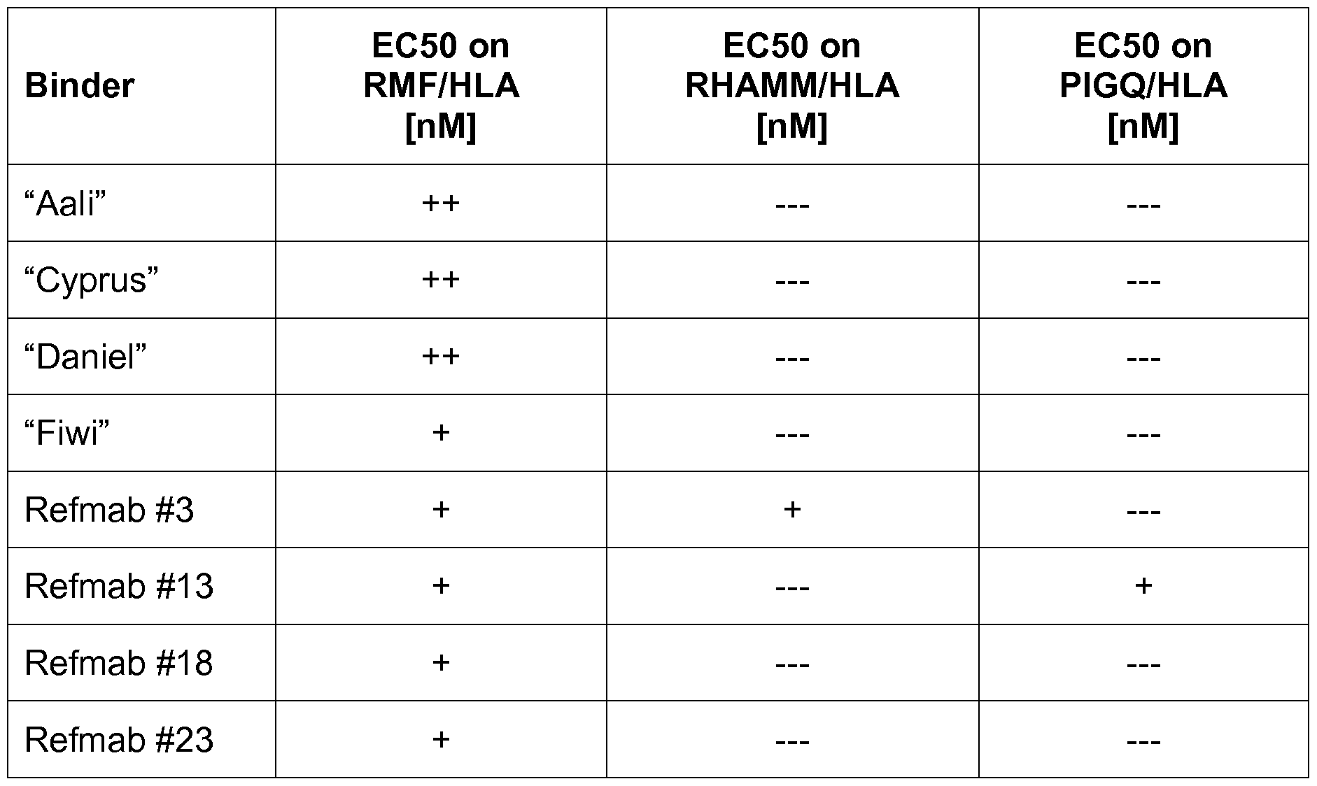
Example 7: Specificity of the binders in an immunoglobulin format
In this experiment the specificities of the antibodies of the present disclosure were investigated as described in Example 3. Results are shown in Figures 4-6.
It was found that all binders are highly specific for the RMF/HLA complex. None of the binders showed any cross-reactivity with either the RHAMM-HLA complex or the PIGQ/HLA complex. This is in striking contrast to the antibodies of the prior art (see Example 3), which all (with the exception of Refmab #18), showed cross-reactivity with a peptide/HLA-complex (PIGQ/HLA) that is expressed on healthy human tissue.
Example 8: Affinities and EC50's of the binders in an immunoglobulin format
EC50 values were determined for the binders of the present disclosure and the binders of the prior art (ELISAs were performed on Neutravidin plates). Results are summarized in Table 4. "++" designates binding with an EC50 of below 10 nM, "+" designates binding with an EC50 of more than 10 nM, and "— " designates no binding.
Table 4: Summary of EC50's of the binders (IgG) of the present invention

All binders (the binders of the present invention, as well as the binders of the prior art) showed very low EC50 values towards the RMF/HLA complex (with the exception of Refmab #3 and Refmab #5 all EC50's were in fact <1 nM). However, all binders of the prior art (except Refmab #18) also showed binding to the PIGQ/HLA counter antigens. Two binders of the prior art even showed binding to the RHAMM/HLA counter antigen.
The apparent affinity of the binders Aali and Refmab #13 was measured for the RMF/HLA antigen with an Octet system. The apparent affinity value for Refmab #13 was determined as 8.2 nM. The binding of Aali was characterized by a very slow dissociation rate, which exceeded the specification limits that were set in the system. Compared to Refmab #13 (8.2 nM), the apparent affinity of Aali is therefore <0.5nM.
Example 9: Binding of the antibodies of the present invention to cells
Example 4 was repeated with the antibodies of the present disclosure, i.e. binding to RNF/HLA-positive SET2 cells (DSMZ No. ACC 608) and BV173 cells (DSMZ No. ACC 20) was tested by flow cytometry. EC50 values were determined. Results are shown in Table 5. "++" designates binding with an EC50 of below 10 nM, "+" designates binding with an EC50 of more than 10 nM, and "— " designates no binding to the target on cells.
Table 5: Summary of IgG characterization by flow cytometry

All antibodies of the present invention show binding to WT1/HLA-expressing cells. This was also the case for most (4 out of 6) of the prior art antibodies, but most notably not for Refmab #18, the only reference antibody that does not bind to PIGQ/HLA.
The antibodies and binders of the present invention are therefore characterized in that they specifically bind to the RMF/HLA-complex without demonstrating any cross-
reactivity to the PIGQ/HLA complex. In addition, the antibodies of the present disclosure also do bind to WT1/HLA expressing cell lines.
Example 7: Binding to leukemic patient samples
The binders of the present invention are investigated for their ability to detect the RMF epitope on primary AML cells. The binders are expected to bind to AML blasts of patients. Results are confirmed by flow cytometry analysis. Results confirm that the level of RMF/HLA-A0201 on the surface of leukemia cells is adequate to allow for a reactivity with the binders of the present invention. The results also confirm that the levels of the target molecule on negative healthy cells are insignificant.
Example 8: Mediation of ADCC activity
The binders of the present invention are also investigated for their potential to mediate ADCC, one of the major effector mechanisms of therapeutic antibodies in humans. In the presence of human PBMCs, the binders mediate a dose-dependent PBMC ADCC against T2 cells (an antigen-processing-deficient cell line, see, for example, WO 2012/135854) loaded with RMF peptide, but not T2 cells alone or T2 cells pulsed with a control peptide. The binders are also able to mediate ADCC against naturally presented RMF epitope by HLA-A0201 molecule on tumor cells, such as the mesothelioma cell line, JMN and the leukemia cell line BV173, but not to HLA-A2 negative cells, such as MSTO or HL-60. These results demonstrate that the binders mediates specific ADCC against cells that naturally express RMF and HLA- A0201 complex at physiologic levels as well as on cell lines.
Example 9: Elimination of human leukemia cells in NSG mice
The binders of the present invention are further investigated in an in vivo NOD SCID gamma (NSG) mice xenograft model. Mice are xenografted intravenously 6 days previously with BV173 bcr/abl positive acute lymphoblastic leukemia. At the time of treatment, mice developed leukemia in their liver, spleen, and BM as visible by luciferase imaging. The binders of the present invention dramatically reduce the tumor burden for at least 30 days. Results are confirmed by titrating the dose of the antibody.
Claims
1 . An isolated antibody or fragment thereof that specifically binds to a complex of a peptide of SEQ ID No. 1 with HLA-A0201 on the surface of cells.
2. The antibody or fragment thereof of claim 1 , wherein said antibody or fragment thereof binds to a complex of a peptide of SEQ ID No. 1 with HLA-A0201 with an EC50 that is at least 10 times lower than the EC50 for a complex of a peptide of SEQ ID No. 64 with HLA-A0201 .
3. The antibody or fragment thereof of any one of claims 1 to 2, wherein said antibody or antibody fragment does not bind a complex of a peptide of SEQ ID No. 64 with HLA-A0201 .
4. The antibody or fragment thereof of any one of claims 1 to 3, wherein said antibody or fragment thereof has an EC50 of less than 10 nM in a Fab format and in an immunoglobulin format.
5. The antibody or fragment thereof of any one of claims 1 to 4, wherein said cell is a cell expressing said a complex of a peptide of SEQ ID No. 1 with HLA-A0201 .
6. The antibody or fragment thereof of claim 5, wherein said cell expressing said complex of a peptide of SEQ ID No. 1 with HLA-A0201 is a cancer cell.
7. The antibody or fragment thereof of any one of claims 1 to 3, wherein said antibody or fragment thereof comprises
(a) an HCDR1 region of SEQ ID NO: 7, an HCDR2 region of SEQ ID NO: 8, an HCDR3 region of SEQ ID NO: 9, an LCDR1 region of SEQ ID NO: 10, an LCDR2 region of SEQ ID NO: 1 1 and an LCDR3 region of SEQ ID NO: 12;
(b) an HCDR1 region of SEQ ID NO: 17, an HCDR2 region of SEQ ID NO: 18, an HCDR3 region of SEQ ID NO: 19, an LCDR1 region of SEQ ID NO: 20, an LCDR2 region of SEQ ID NO: 21 , and an LCDR3 region of SEQ ID NO: 22;
(c) an HCDR1 region of SEQ ID NO: 27, an HCDR2 region of SEQ ID NO: 28, an HCDR3 region of SEQ ID NO: 29, an LCDR1 region of SEQ ID NO: 30, an LCDR2 region of SEQ ID NO: 31 , and an LCDR3 region of SEQ ID NO: 32;
(d) an HCDR1 region of SEQ ID NO: 37, an HCDR2 region of SEQ ID NO: 38, an HCDR3 region of SEQ ID NO: 39, an LCDR1 region of SEQ ID NO: 40, an LCDR2 region of SEQ ID NO: 41 , and an LCDR3 region of SEQ ID NO: 42;
(e) an HCDR1 region of SEQ ID NO: 47, an HCDR2 region of SEQ ID NO: 48, an HCDR3 region of SEQ ID NO: 49, an LCDR1 region of SEQ ID NO: 50, an LCDR2 region of SEQ ID NO: 51 , and an LCDR3 region of SEQ ID NO: 52; or
(f) an HCDR1 region of SEQ ID NO: 57, an HCDR2 region of SEQ ID NO: 58, an HCDR3 region of SEQ ID NO: 59, an LCDR1 region of SEQ ID NO: 60, an LCDR2 region of SEQ ID NO: 61 , and an LCDR3 region of SEQ ID NO: 62.
8. The antibody or fragment thereof of any one of claims 1 to 4, wherein said antibody or fragment thereof comprises
(a) a variable heavy chain of SEQ ID NO: 5 and a variable light chain of SEQ ID NO: 6;
(b) a variable heavy chain of SEQ ID NO: 15 and a variable light chain of SEQ ID NO: 16;
(c) a variable heavy chain of SEQ ID NO: 25 and a variable light chain of SEQ ID NO: 26;
(d) a variable heavy chain of SEQ ID NO: 35 and a variable light chain of SEQ ID NO: 36;
(e) a variable heavy chain of SEQ ID NO: 45 and a variable light chain of SEQ ID NO: 46; or
(f) a variable heavy chain of SEQ ID NO: 55 and a variable light chain of SEQ ID NO: 56.
9. An antibody or fragment thereof which specifically binds to a complex of a peptide of SEQ ID No. 1 with HLA-A0201 wherein said antibody or fragment thereof is at least 95% homologous to an antibody according to claim 7 or 8.
10. An antibody or fragment thereof cross-competing with an antibody or fragment thereof according to any of claims 7 to 9.
1 1 . An antibody or fragment thereof according to any of claims 1 to 10 for use in medicine.
12. An antibody or fragment thereof according to claim 1 1 , wherein said use is the treatment of a WT1 -positive disease, such as cancer.
13. A nucleic acid composition, comprising a nucleic acid sequence or a plurality of nucleic acid sequences encoding the antibody or fragment thereof according to any one of claims 7 to 9.
14. A vector composition, comprising a vector or a plurality of vectors comprising the nucleic acid sequence or plurality of nucleic acid sequences of claim 13.
15. A host cell, comprising the nucleic acid composition of claim 13 or the vector composition of claim 14.
16. A pharmaceutical composition comprising the antibody or fragment thereof according to any one of claims 1 to 10, and a pharmaceutically acceptable carrier and/or excipient.
17. A method for identifying an antibody or fragment thereof according to any one of claims 1 to 10, comprising
(a) mixing an antibody library comprising a plurality of antibodies or antibody fragments with a complex of a peptide of SEQ ID No. 64 with HLA-A0201 ,
(b) removing from the antibody library those antibodies or antibody fragments that did bind to said complex of a peptide of SEQ ID No. 64 with HLA-A0201 ,
(c) mixing the depleted antibody library of step (b) with a complex of a peptide of SEQ ID No. 1 with HLA-A0201 , and
(d) identifying an antibody or fragment thereof that binds to said complex of a peptide of SEQ ID No. 1 with HLA-A0201 .
Priority Applications (3)
| Application Number | Priority Date | Filing Date | Title |
|---|---|---|---|
| JP2018518451A JP6862435B2 (en) | 2015-10-09 | 2016-10-04 | Anti-WT1 / HLA specific antibody |
| EP16782018.2A EP3359565A1 (en) | 2015-10-09 | 2016-10-04 | Anti-wt1/hla-specific antibodies |
| HK19100718.0A HK1258444A1 (en) | 2015-10-09 | 2019-01-16 | Anti-wt1/hla-specific antibodies |
Applications Claiming Priority (6)
| Application Number | Priority Date | Filing Date | Title |
|---|---|---|---|
| US201562239438P | 2015-10-09 | 2015-10-09 | |
| US62/239,438 | 2015-10-09 | ||
| GB1517913.8 | 2015-10-09 | ||
| GBGB1517913.8A GB201517913D0 (en) | 2015-10-09 | 2015-10-09 | Anti-WT1/HLA-specific antibodies |
| EP15189753 | 2015-10-14 | ||
| EP15189753.5 | 2015-10-14 |
Publications (1)
| Publication Number | Publication Date |
|---|---|
| WO2017060201A1 true WO2017060201A1 (en) | 2017-04-13 |
Family
ID=58487775
Family Applications (1)
| Application Number | Title | Priority Date | Filing Date |
|---|---|---|---|
| PCT/EP2016/073587 WO2017060201A1 (en) | 2015-10-09 | 2016-10-04 | Anti-wt1/hla-specific antibodies |
Country Status (6)
| Country | Link |
|---|---|
| US (2) | US10836829B2 (en) |
| EP (1) | EP3359565A1 (en) |
| JP (1) | JP6862435B2 (en) |
| HK (1) | HK1258444A1 (en) |
| MA (1) | MA45004A (en) |
| WO (1) | WO2017060201A1 (en) |
Cited By (5)
| Publication number | Priority date | Publication date | Assignee | Title |
|---|---|---|---|---|
| WO2018104407A1 (en) | 2016-12-08 | 2018-06-14 | Immatics Biotechnologies Gmbh | T cell receptors with improved pairing |
| WO2019122052A2 (en) | 2017-12-21 | 2019-06-27 | F. Hoffmann-La Roche Ag | Antibodies binding to hla-a2/wt1 |
| KR20200016873A (en) * | 2017-05-31 | 2020-02-17 | 칼스젠 테라퓨틱스 씨오., 엘티디. | Compositions and Methods for Cellular Immunotherapy |
| EP4317432A2 (en) | 2016-12-08 | 2024-02-07 | Immatics Biotechnologies GmbH | T cell receptors with improved pairing |
| US12098213B2 (en) | 2017-12-21 | 2024-09-24 | Hoffmann-La Roche Inc. | Antibodies binding to HLA-A2/WT1 |
Families Citing this family (21)
| Publication number | Priority date | Publication date | Assignee | Title |
|---|---|---|---|---|
| CR20200014A (en) | 2017-07-14 | 2020-06-11 | Immatics Biotechnologies Gmbh | Improved dual specificity polypeptide molecule |
| DE102017127984B4 (en) | 2017-11-27 | 2019-12-05 | Immatics US, Inc. | Method for the propagation and activation of γδ T cells |
| US11464800B2 (en) | 2018-02-09 | 2022-10-11 | Immatics US, Inc. | Methods for manufacturing T cells |
| US20200297768A1 (en) | 2019-03-19 | 2020-09-24 | Immatics US, Inc. | Cd28 t cell cultures, compositions, and methods of using thereof |
| CR20210669A (en) | 2019-05-27 | 2022-05-05 | Immatics Us Inc | Viral vectors and their use in adoptive cellular therapy |
| US20200384028A1 (en) | 2019-06-06 | 2020-12-10 | Immatics Biotechnologies Gmbh | Sorting with counter selection using sequence similar peptides |
| US20210032370A1 (en) | 2019-08-02 | 2021-02-04 | Immatics Biotechnologies Gmbh | Recruiting agent further binding an mhc molecule |
| AU2021225817A1 (en) | 2020-02-24 | 2022-10-20 | Immatics US, Inc. | Methods for expanding T cells for the treatment of cancer and related malignancies |
| DE102020106710A1 (en) | 2020-03-11 | 2021-09-16 | Immatics US, Inc. | WPRE MUTANT CONSTRUCTS, COMPOSITIONS, AND RELATED PROCEDURES |
| DE102020111571A1 (en) | 2020-03-11 | 2021-09-16 | Immatics US, Inc. | WPRE MUTANT CONSTRUCTS, COMPOSITIONS, AND RELATED PROCEDURES |
| TW202227616A (en) | 2020-08-21 | 2022-07-16 | 美商英麥提克斯股份有限公司 | Methods for isolating cd8+ selected t cells |
| TW202241938A (en) | 2020-12-31 | 2022-11-01 | 美商英麥提克斯股份有限公司 | Cd8 polypeptides, compositions, and methods of using thereof |
| WO2022233957A1 (en) | 2021-05-05 | 2022-11-10 | Immatics Biotechnologies Gmbh | Bma031 antigen binding polypeptides |
| EP4392441A1 (en) | 2021-08-24 | 2024-07-03 | Immatics US, Inc. | Selection of immune cells using peptide mhc complexes generated by conditional ligand exchange |
| WO2023044488A1 (en) | 2021-09-20 | 2023-03-23 | Immatics US, Inc. | Monocyte depletion of t cells populations for t-cell therapy |
| WO2023081925A1 (en) | 2021-11-08 | 2023-05-11 | Immatics Biotechnologies Gmbh | Adoptive cell therapy combination treatment and compositions thereof |
| WO2023212691A1 (en) | 2022-04-28 | 2023-11-02 | Immatics US, Inc. | DOMINANT NEGATIVE TGFβ RECEPTOR POLYPEPTIDES, CD8 POLYPEPTIDES, CELLS, COMPOSITIONS, AND METHODS OF USING THEREOF |
| WO2023212655A1 (en) | 2022-04-28 | 2023-11-02 | Immatics US, Inc. | Il-12 polypeptides, il-15 polypeptides, il-18 polypeptides, cd8 polypeptides, compositions, and methods of using thereof |
| WO2023212697A1 (en) | 2022-04-28 | 2023-11-02 | Immatics US, Inc. | Membrane-bound il-15, cd8 polypeptides, cells, compositions, and methods of using thereof |
| US20230355678A1 (en) | 2022-05-05 | 2023-11-09 | Immatics US, Inc. | Methods for improving t cell efficacy |
| WO2024167871A1 (en) * | 2023-02-06 | 2024-08-15 | Memorial Sloan-Kettering Cancer Center | Compositions including anti-wt-1 antibodies & antigen binding fragments and uses thereof |
Citations (2)
| Publication number | Priority date | Publication date | Assignee | Title |
|---|---|---|---|---|
| WO2012135854A2 (en) * | 2011-04-01 | 2012-10-04 | Memorial Sloan-Kettering Cancer Center | Antibodies to cytosolic peptides |
| WO2015070061A1 (en) * | 2013-11-07 | 2015-05-14 | Memorial Sloan-Kettering Cancer Center | Anti-wt1/hla bi-specific antibody |
Family Cites Families (3)
| Publication number | Priority date | Publication date | Assignee | Title |
|---|---|---|---|---|
| WO2015070078A1 (en) * | 2013-11-07 | 2015-05-14 | Memorial Sloan-Kettering Cancer Center | Fc-enhanced anti-wt1/hla antibody |
| US20140271644A1 (en) * | 2013-03-15 | 2014-09-18 | Memorial Sloan-Kettering Cancer Center | Combination/adjuvant therapy for wt-1-positive disease |
| US20160152725A1 (en) * | 2014-02-25 | 2016-06-02 | Memorial Sloan-Kettering Cancer Center | Antigen-binding proteins specific for hla-a2-restricted wilms tumor 1 peptide |
-
2016
- 2016-10-04 EP EP16782018.2A patent/EP3359565A1/en not_active Ceased
- 2016-10-04 WO PCT/EP2016/073587 patent/WO2017060201A1/en active Application Filing
- 2016-10-04 JP JP2018518451A patent/JP6862435B2/en active Active
- 2016-10-04 MA MA045004A patent/MA45004A/en unknown
- 2016-10-07 US US15/288,145 patent/US10836829B2/en active Active
-
2019
- 2019-01-16 HK HK19100718.0A patent/HK1258444A1/en unknown
-
2020
- 2020-10-13 US US17/069,136 patent/US20210024635A1/en active Pending
Patent Citations (2)
| Publication number | Priority date | Publication date | Assignee | Title |
|---|---|---|---|---|
| WO2012135854A2 (en) * | 2011-04-01 | 2012-10-04 | Memorial Sloan-Kettering Cancer Center | Antibodies to cytosolic peptides |
| WO2015070061A1 (en) * | 2013-11-07 | 2015-05-14 | Memorial Sloan-Kettering Cancer Center | Anti-wt1/hla bi-specific antibody |
Non-Patent Citations (2)
| Title |
|---|
| ZHAO Q ET AL: "Affinity maturation of T-cell receptor-like antibodies for Wilms tumor 1 peptide greatly enhances therapeutic potential", LEUKEMIA, vol. 29, no. 11, 9 June 2015 (2015-06-09), (BASINGSTOKE), pages 2238 - 2247, XP002754503 * |
| ZHAO Q ET AL: "Supplementary information to Affinity maturation of T-cell receptor-like antibodies for Wilms tumor 1 peptide greatly enhances therapeutic potential", LEUKEMIA, 9 June 2015 (2015-06-09), XP002754832 * |
Cited By (10)
| Publication number | Priority date | Publication date | Assignee | Title |
|---|---|---|---|---|
| WO2018104407A1 (en) | 2016-12-08 | 2018-06-14 | Immatics Biotechnologies Gmbh | T cell receptors with improved pairing |
| EP4317432A2 (en) | 2016-12-08 | 2024-02-07 | Immatics Biotechnologies GmbH | T cell receptors with improved pairing |
| KR20200016873A (en) * | 2017-05-31 | 2020-02-17 | 칼스젠 테라퓨틱스 씨오., 엘티디. | Compositions and Methods for Cellular Immunotherapy |
| JP2020523985A (en) * | 2017-05-31 | 2020-08-13 | 科済生物医薬(上海)有限公司Carsgen Therapeutics Co.,Ltd. | Cellular immunotherapy compositions and methods |
| EP3630846A4 (en) * | 2017-05-31 | 2021-02-24 | Carsgen Therapeutics Co., Ltd. | Compositions and methods of cellular immunotherapy |
| KR102412805B1 (en) * | 2017-05-31 | 2022-06-27 | 크라제 메디컬 씨오 리미티드 | Compositions and methods for cellular immunotherapy |
| US11419895B2 (en) | 2017-05-31 | 2022-08-23 | Crage Medical Co., Limited | Compositions and methods of cellular immunotherapy |
| WO2019122052A2 (en) | 2017-12-21 | 2019-06-27 | F. Hoffmann-La Roche Ag | Antibodies binding to hla-a2/wt1 |
| US11192957B2 (en) | 2017-12-21 | 2021-12-07 | Hoffmann-La Roche Inc. | Antibodies binding to HLA-A2/WT1 |
| US12098213B2 (en) | 2017-12-21 | 2024-09-24 | Hoffmann-La Roche Inc. | Antibodies binding to HLA-A2/WT1 |
Also Published As
| Publication number | Publication date |
|---|---|
| MA45004A (en) | 2019-03-27 |
| US20170101473A1 (en) | 2017-04-13 |
| HK1258444A1 (en) | 2019-11-15 |
| US10836829B2 (en) | 2020-11-17 |
| US20210024635A1 (en) | 2021-01-28 |
| JP6862435B2 (en) | 2021-04-21 |
| EP3359565A1 (en) | 2018-08-15 |
| JP2018536393A (en) | 2018-12-13 |
Similar Documents
| Publication | Publication Date | Title |
|---|---|---|
| US20210024635A1 (en) | Anti-wt1/hla-specific antibodies | |
| US11161904B2 (en) | Anti-PD-1 antibody and use thereof | |
| CN107531786B (en) | anti-AXL antagonist antibodies | |
| WO2020177733A1 (en) | Bifunctional fusion protein and pharmaceutical use thereof | |
| JP7257971B2 (en) | Anti-CD40 Antibodies, Antigen-Binding Fragments Thereof, and Medical Uses Thereof | |
| CN109311997B (en) | anti-AXL antagonistic antibodies | |
| WO2019141268A1 (en) | Anti-4-1bb antibody, antigen-binding fragment thereof and medical use thereof | |
| CN112243443B (en) | anti-TROP-2 antibodies, antigen-binding fragments thereof, and medical uses thereof | |
| WO2019034580A1 (en) | Humanized antibodies for cd3 | |
| CN113825771A (en) | Antibodies against claudin 18A2 and uses thereof | |
| CN113039208B (en) | anti-PD-L1 antigen binding protein and application thereof | |
| WO2019184935A1 (en) | Anti-cd27 antibody, antigen-binding fragment thereof and medical use thereof | |
| AU2017219837A1 (en) | Antibodies for IL-17C | |
| TW202132352A (en) | Guidance and navigation control proteins and method of making and using thereof | |
| WO2022100694A1 (en) | Antibody and preparation method therefor | |
| RU2779128C2 (en) | Antibody to cd40, its antigene-binding fragment and its medical use | |
| RU2774158C2 (en) | ANTIBODIES DIRECTED AGAINST Fc-RECEPTOR-LIKE PROTEIN 5 AND METHODS FOR THEIR APPLICATION | |
| CA3206543A1 (en) | Antibody-drug conjugate and medical use thereof | |
| CN116234573A (en) | anti-TROP-2 antibody, antigen binding fragment or mutant thereof, and medical application thereof | |
| CN115998900A (en) | anti-TROP-2 antibody drug conjugate and medical application thereof | |
| KR20220103961A (en) | Pharmaceutical combinations and uses thereof | |
| WO2017125601A1 (en) | Anti-bsep-specific antibodies |
Legal Events
| Date | Code | Title | Description |
|---|---|---|---|
| 121 | Ep: the epo has been informed by wipo that ep was designated in this application |
Ref document number: 16782018 Country of ref document: EP Kind code of ref document: A1 |
|
| WWE | Wipo information: entry into national phase |
Ref document number: 2018518451 Country of ref document: JP |
|
| NENP | Non-entry into the national phase |
Ref country code: DE |
|
| WWE | Wipo information: entry into national phase |
Ref document number: 2016782018 Country of ref document: EP |