WO2017057523A1 - Astrocyte-like cells and method for preparing same - Google Patents
Astrocyte-like cells and method for preparing same Download PDFInfo
- Publication number
- WO2017057523A1 WO2017057523A1 PCT/JP2016/078741 JP2016078741W WO2017057523A1 WO 2017057523 A1 WO2017057523 A1 WO 2017057523A1 JP 2016078741 W JP2016078741 W JP 2016078741W WO 2017057523 A1 WO2017057523 A1 WO 2017057523A1
- Authority
- WO
- WIPO (PCT)
- Prior art keywords
- gene
- cells
- cell
- human
- astrocyte
- Prior art date
Links
Images


Classifications
-
- C—CHEMISTRY; METALLURGY
- C12—BIOCHEMISTRY; BEER; SPIRITS; WINE; VINEGAR; MICROBIOLOGY; ENZYMOLOGY; MUTATION OR GENETIC ENGINEERING
- C12N—MICROORGANISMS OR ENZYMES; COMPOSITIONS THEREOF; PROPAGATING, PRESERVING, OR MAINTAINING MICROORGANISMS; MUTATION OR GENETIC ENGINEERING; CULTURE MEDIA
- C12N15/00—Mutation or genetic engineering; DNA or RNA concerning genetic engineering, vectors, e.g. plasmids, or their isolation, preparation or purification; Use of hosts therefor
- C12N15/09—Recombinant DNA-technology
-
- C—CHEMISTRY; METALLURGY
- C12—BIOCHEMISTRY; BEER; SPIRITS; WINE; VINEGAR; MICROBIOLOGY; ENZYMOLOGY; MUTATION OR GENETIC ENGINEERING
- C12Q—MEASURING OR TESTING PROCESSES INVOLVING ENZYMES, NUCLEIC ACIDS OR MICROORGANISMS; COMPOSITIONS OR TEST PAPERS THEREFOR; PROCESSES OF PREPARING SUCH COMPOSITIONS; CONDITION-RESPONSIVE CONTROL IN MICROBIOLOGICAL OR ENZYMOLOGICAL PROCESSES
- C12Q1/00—Measuring or testing processes involving enzymes, nucleic acids or microorganisms; Compositions therefor; Processes of preparing such compositions
- C12Q1/02—Measuring or testing processes involving enzymes, nucleic acids or microorganisms; Compositions therefor; Processes of preparing such compositions involving viable microorganisms
- C12Q1/04—Determining presence or kind of microorganism; Use of selective media for testing antibiotics or bacteriocides; Compositions containing a chemical indicator therefor
Definitions
- the present invention relates to a method for preparing in vitro astrocyte-like cells from human cells, an astrocyte-like cell prepared by the method, an in vitro neural tissue model including astrocyte-like cells and neurons, and a patient with a nervous system disease
- the present invention relates to a method for evaluating the effectiveness or toxicity of a test substance against a nervous system disease using an astrocyte-like cell, a human cell-to-astrocyte-like cell inducer, and a human cell-to-astrocyte-like cell induction kit.
- Non-Patent Document 1 Astrocytes are nervous system cells that are gradually being recognized to play a major role in the pathophysiology of many neurological diseases.
- astrocytes In order to study the pathophysiological role of astrocytes for nervous system diseases, a rapid and massive supply of human astrocytes is required. However, at present, the source of human astrocytes is limited. It is ethically impossible to obtain astrocytes by biopsy from a patient with a living nervous system disease. Although it is possible to obtain astrocytes from the postmortem brain of patients with nervous system diseases, mass supply is extremely difficult.
- Non-patent Document 2 A method for preparing astrocytes by inducing differentiation from human induced pluripotent stem cells (iPS cells) has been reported (Non-patent Document 2).
- NFIA Nuclear factor I / A
- NFIB Nuclear factor I / B
- SOX9 SRY (sex determining region Y) -box 9)
- Non-Patent Literature 4
- the efficiency of direct reprogramming from human fibroblasts to neurons can be improved by knocking down the p14ARF gene or the p16INK4 gene present in the CDKN2A locus.
- P14ARF and p16INK4 are molecules present in different signal transduction pathways, but both signal transduction pathways are involved in cell apoptosis and senescence.
- MDM2, p53 and p21 are present downstream of p14ARF in the first signal transduction pathway.
- cyclin D1, CDK4, CDK6, Rb, and E2F exist downstream of p16INK4 in the second signal transduction pathway.
- Non-Patent Document 6 the efficiency of reprogramming from cells to iPS cells can be improved. It has also been reported that the efficiency of direct reprogramming from human fibroblasts to neurons can be improved by introducing a p53 dominant negative mutant into cells.
- Non-Patent Document 3 after introducing NFIA, NFIB and SOX9 into human neonatal fibroblasts, only 2% of astrocyte-like cells are present after 3 weeks of culture. Not observed, its induction efficiency is very low.
- the present invention provides a novel method that enables the rapid and highly efficient preparation of astrocyte-like cells having the same characteristics as human astrocytes that are extremely difficult to obtain using easily obtainable human cells. Let it be an issue.
- human somatic cells have at least one of specific inducers (NICD (Notch 1 intercellular domain) and Tet2CD (TET (Ten-eleven translocation) 2 catalytic domain)), And NFIA), it was found that human astrocyte-like cells can be prepared with high efficiency in a very short period of time without preparation of iPS cells. Furthermore, the present inventors have used human astrocyte-like cells for a very short period of time even when an inducer similar to the above is introduced into pluripotent stem cells such as iPS cells and adult stem cells such as neural stem cells. And found that it can be prepared with high efficiency.
- the present invention is based on these surprising findings.
- a method for preparing in vitro astrocyte-like cells from human cells comprising the following steps: (1) introducing at least two kinds of inducers into human cells; and (2) obtaining astrocyte-like cells by inducing differentiation of the human cells, Wherein the inducer comprises NFIA and comprises at least one of NICD and Tet2CD;
- the preparation method wherein the human cells are human somatic cells (excluding astrocytes), human pluripotent stem cells or human adult stem cells.
- the inducer includes NFIA, NICD, and Tet2CD.
- the human cell is a cell derived from a patient with a nervous system disease, or a cell derived from a healthy person artificially introduced with a mutation that specifically exists in the gene of a cell derived from the patient with a nervous system disease , [1] to [4].
- the preparation method according to any one of [1] to [5], wherein the introduction of the inducer in the step (1) includes introducing at least one vector containing a nucleic acid encoding the inducer.
- step (1) inhibits the expression of at least one gene selected from p14ARF gene, p53 gene, p21 gene, p16INK4a gene and Rb gene, inhibits at least one selected from p14ARF, p53, p21, p16INK4a and Rb, Increases the expression of at least one gene selected from the MDM2 gene, cyclin D1 gene, CDK4 gene, CDK6 gene and E2F gene, or activates at least one selected from MDM2, cyclin D1, CDK4, CDK6 and E2F
- the preparation method according to any one of [1] to [6] further comprising: [8] An astrocyte-like cell prepared by the method according to any one of [1] to [7].
- a method for evaluating the effectiveness or toxicity of a test substance against a nervous system disease comprising the following steps: (1) From a cell derived from a patient with a nervous system disease, or a cell derived from a healthy person into which a mutation that is specifically present in the gene of a cell derived from a patient with the nervous system disease has been artificially introduced into a gene, [1] to [ 7] a step of preparing astrocyte-like cells by the method according to any one of the above; and (2) contacting the test substance with the astrocyte-like cells obtained in step (1), Said method comprising the step of assessing efficacy or toxicity.
- a method for evaluating the effectiveness or toxicity of a test substance against a nervous system disease comprising the following steps: (1) A step of preparing astrocyte-like cells from cells derived from healthy subjects by the method according to any one of [1] to [7]; (2) a step of knocking out or knocking down a nervous system disease-related gene in the astrocyte-like cell obtained in step (1); and (3) an astrocyte-like cell obtained in step (2) and a test substance The method comprising the steps of: contacting and evaluating the efficacy or toxicity of the test substance.
- An in vitro neural tissue model comprising the astrocyte-like cell according to [8] and a neuron.
- a method for evaluating the effectiveness or toxicity of a test substance against a nervous system disease comprising the following steps: (1) From a cell derived from a patient with a nervous system disease, or a cell derived from a healthy person into which a mutation that is specifically present in the gene of a cell derived from a patient with the nervous system disease has been artificially introduced into a gene, [1] to [ 7] a step of preparing astrocyte-like cells by the method according to any one of (2) a step of preparing a neural tissue model by co-culturing astrocyte-like cells and neurons obtained in step (1); (3) The said method including the process of contacting the nerve tissue model obtained by process (2), and a test substance, and evaluating the effectiveness or toxicity of a test substance.
- a method for evaluating the effectiveness or toxicity of a test substance against a nervous system disease comprising the following steps: (1) A step of preparing astrocyte-like cells from cells derived from healthy subjects by the method according to any one of [1] to [7]; (2) a step of knocking out or knocking down a nervous system disease-related gene in the astrocyte-like cell obtained in step (1); (3) a step of preparing a neural tissue model by co-culturing astrocyte-like cells and neurons obtained in step (2); (4) The method comprising the step of contacting the nerve tissue model obtained in step (3) with a test substance to evaluate the effectiveness or toxicity of the test substance.
- An inducer from a human cell to an astrocyte-like cell comprising at least one kind of inducer or at least one vector containing a nucleic acid encoding the inducer, wherein the inducer is NFIA And at least one of NICD and Tet2CD, and the human cell is a human somatic cell (excluding astrocytes), a human pluripotent stem cell or a human adult stem cell.
- [15] Including at least two types of inducers, or at least one vector containing a nucleic acid encoding the inducer; and an instruction manual describing the method according to any one of [1] to [7],
- a kit for inducing an astrocyte-like cell from a human cell wherein the inducer includes NFIA and includes at least one of NICD and Tet2CD, and the human cell is a human somatic cell (an astrocyte). Except), which is a human pluripotent stem cell or a human adult stem cell.
- the kit according to [15] further comprising an activator.
- the method of the present invention it becomes possible to supply human astrocytes quickly and in large quantities.
- a lentiviral vector containing a nucleic acid encoding NFIA, a lentiviral vector containing a nucleic acid encoding NICD, and a lentiviral vector containing a nucleic acid encoding Tet2CD into human fibroblasts and culturing for 11 days
- the percentage of GFAP-expressing cells made is shown.
- the vertical axis represents the ratio of the number of GFAP positive cells to the number of DAPI positive cells.
- “Fibro” indicates human fibroblasts into which no inducer has been introduced.
- “NFIA”, “Tet2CD”, and “NICD” indicate human fibroblasts introduced with NFIA, Tet2CD, and NICD, respectively.
- NFIA / Tet2CD is human fibroblasts introduced with NFIA and Tet2CD, human fibroblasts introduced with Tet2CD and NICD, NICD and NFIA, respectively. Shows human fibroblasts introduced with.
- “NIFA / NICD / Tet2CD” indicates human fibroblasts into which NFIA, NICD and Tet2CD have been introduced. After co-culture of astrocyte-like cells with neurons, an image of the neurons and a measurement result of the length of neurites extended from the neurons by a fluorescence microscope are shown. The scale bar in the image is 100 ⁇ m.
- Fluorescence microscope images were obtained by labeling neurons with anti-MAP2 antibody (Sigma).
- the vertical axis of the graph represents the average neurite length per neuron cell.
- “w / o” indicates a case where the cells are not co-cultured with astrocyte-like cells (that is, only neurons).
- “w / NHDF-Ad” indicates a case where human adult dermal fibroblasts (NHDF-Ad) and neurons are co-cultured.
- w / NHDF-Neo indicates the case where human neonatal skin fibroblasts (NHDF-Neo) and neurons are co-cultured.
- w / iAs (NHDF-Ad) indicates a case where astrocyte-like cells derived from human adult dermal fibroblasts and neurons are co-cultured.
- w / iAs (NHDF-Neo) indicates a case where astrocyte-like cells derived from human neonatal dermal fibroblasts and neurons are co-cultured.
- w / hAstrocytes indicates a case where human astrocytes and neurons are co-cultured. The measurement result of the glutamate uptake
- a retroviral vector containing a nucleic acid encoding NFIA, a retroviral vector containing a nucleic acid encoding NICD, and a retroviral vector containing a nucleic acid encoding Tet2CD are human lt-NES cells (long-term self-renewing neuroepithelial stem cells) The ratio of the induced GFAP-expressing cells after introduction into 1 and culturing for 1 week and 2 weeks is shown. The vertical axis shows the ratio of the number of GFAP positive cells to the total number of cells. In the figure, “lt-NES iA” indicates astrocyte-like cells derived from lt-NES cells.
- FIG. 2 shows fluorescence microscopic images of astrocyte-like cells derived from human adult fibroblasts and lt-NES cells using lentiviral vectors containing all of the nucleic acids encoding NFIA, NICD, and Tet2CD.
- FIG. 6 shows that the expression level of CRYAB was increased in an in vitro model of Alexander disease obtained by inducing differentiation from lt-NES cells as compared to the control group.
- the vertical axis represents the ratio of the number of GFAP-positive and CRYAB (Alpha-crystallin B) positive cells to the number of GFAP-positive cells.
- Control represents an astrocyte-like cell derived from an lt-NES cell into which no missense point mutation (C715T) has been introduced
- AxD represents an lt into which a missense point mutation (C715T) has been introduced.
- a lentiviral vector expressing a short hairpin RNA (shRNA) against MeCP2 is introduced into fibroblast-derived astrocyte-like cells and co-cultured with mouse E15.5 primary cultured cerebral cortical neurons, the neurons are examined by fluorescence microscopy. An image and the measurement result of the length of the neurite extended from the neuron are shown.
- shRNA short hairpin RNA
- the vertical axis of the graph represents the ratio of the average neurite length in the co-culture group to the average neurite length per neuron in the control group. Measurement of length of neurites extended from neurons when lentiviral vector expressing shRNA against MeCP2 is introduced into astrocyte-like cells derived from lt-NES cells and cultured with mouse E15.5 primary cultured cerebral cortical neurons Results are shown. The vertical axis represents the average neurite length per neuron cell. In the figure, “w / iA (shMeCP2)” shows the result of co-culture of neurons and astrocyte-like cells expressing shRNA against MECP2.
- w / iA (Rescue) indicates a co-cultured astrocyte-like cell and neuron expressing both shRNA against MECP2 and mutant MECP2 not suppressed by shRNA.
- transducing p16INK4a into a cell in the differentiation induction from a human fibroblast to an astrocyte-like cell is shown.
- “3 factor” is a human fibroblast introduced with a lentiviral vector containing a nucleic acid encoding NFIA, a lentiviral vector containing a nucleic acid encoding NICD, and a lentiviral vector containing a nucleic acid encoding Tet2CD. Indicates that there is.
- + p16INK4a indicates the result when a lentiviral vector containing a nucleic acid encoding p16INK4a is introduced into a cell in addition to the above-described three-encoding vector.
- + Ctl indicates the result when an empty lentiviral vector not containing a gene sequence to be expressed is used instead of a lentiviral vector containing a nucleic acid encoding p16INK4a.
- the vertical axis (% ⁇ of GFAP (+) / DAPI) in the left bar graph shows the ratio of the number of GFAP positive cells to the number of DAPI positive cells after culturing for 11 days after introducing the vector encoding the inducer into the cells (GFAP (Number of positive cells / number of DAPI positive cells) is expressed in%.
- the vertical axis (% ⁇ of GFAP (+) / Initial cells) on the right bar graph shows the number of GFAP-positive cells collected after 11 days of introduction of the vector encoding the inducer into the cells and at the start of culture. The percentage of the initial cell number (GFAP positive cell number / initial cell number) is shown in%.
- + shp53 indicates the result when a lentiviral vector containing a nucleic acid encoding a short hairpin RNA (shp53) for the p53 gene in addition to the three factors is introduced into the cell.
- + Ctl indicates that a lentiviral vector containing a nucleic acid encoding a short hairpin RNA whose target sequence is a non-human sequence is introduced into a cell instead of a lentiviral vector containing a nucleic acid encoding shp53.
- the result is shown below. The result at the time of knocking down the intracellular p14ARF gene in the differentiation induction from a human fibroblast to an astrocyte-like cell is shown.
- shp14ARF indicates the result when a lentiviral vector containing a nucleic acid encoding a short hairpin RNA (shp14ARF) for the p14ARF gene in addition to the three factors is introduced into the cell.
- + Ctl indicates a case where a lentiviral vector containing a nucleic acid encoding a short hairpin RNA whose target sequence is a non-human sequence is introduced into a cell instead of a vector containing a nucleic acid encoding shp14ARF
- the results are shown.
- the results when knocking down the intracellular p21 gene in the induction of differentiation from human fibroblasts to astrocyte-like cells are shown.
- the definitions of “3 factors”, “% of GFAP (+) / DAPI” and “% of GFAP (+) / DAPI” are the same as those in FIG.
- + shp21 indicates the result when a lentiviral vector containing a nucleic acid encoding a short hairpin RNA (shp21) for the p21 gene in addition to the three factors is introduced into the cell.
- + Ctl indicates a case where a lentiviral vector containing a nucleic acid encoding a short hairpin RNA whose target sequence is a non-human sequence is introduced into a cell instead of a vector containing a nucleic acid encoding shp21 The results are shown.
- the vertical axis represents the number of GFAP-expressing cells per cm 2 .
- the abscissa indicates the introduced inducer.
- One aspect of the present invention is a method for preparing in vitro astrocyte-like cells from human cells, the following steps: (1) introducing at least two types of inducers into human cells; and (2) Inducing differentiation of a human cell to obtain an astrocyte-like cell, wherein the inducer includes NFIA and includes at least one of NICD and Tet2CD, and the human cell is a human somatic cell (astrocytoma).
- the above preparation method is a human pluripotent stem cell or a human adult stem cell.
- the preparation method is also referred to as “the preparation method of the astrocyte-like cell of the present invention” or “the preparation method of the present invention”.
- astrocyte-like cells are cells obtained by inducing differentiation of human somatic cells (excluding astrocytes), human pluripotent stem cells or human adult stem cells in vitro, A cell that has the same characteristics as a site. Specifically, it has the following characteristics. (1) It expresses glial fibrillary acidic protein (GFAP), which is a specific marker for astrocytes. (2) Ability to support neurite outgrowth from neurons. (3) It has the ability to take in glutamic acid. In the present specification, the astrocyte-like cell is also referred to as “induced astrocyte” or “iA cell”.
- GFAP glial fibrillary acidic protein
- inducing factor refers to a protein that can induce differentiation of human somatic cells (excluding astrocytes), human pluripotent stem cells, and adult human stem cells into astrocyte-like cells.
- the inducer used in the method for preparing an astrocyte-like cell of the present invention contains NFIA (Nuclear factor I / A) and includes at least one of NICD (Notch-1 intercellular domain) and Tet2CD (Tet2 catalytic domain).
- the inducer may include NFIA and NICD, may include NFIA and Tet2CD, and may include NFIA, NICD and Tet2CD.
- the inducer comprises NFIA, NICD and Tet2CD.
- the inducer has 90% or more, preferably 95% or more, more preferably 98% or more, particularly preferably 99% or more identity with the amino acid sequence of the wild type protein. It may be a mutant protein having the same ability to induce astrocyte-like cells as the protein.
- mouse NFIA, mouse NICD and mouse Tet2CD are selected as inducers, not only their wild-type proteins (SEQ ID NO: 8, 9 and 10 respectively) but also their sequences and 90% or more, preferably 95 %, More preferably 98% or more, particularly preferably 99% or more, and the use of a mutant protein having the ability to induce astrocytic cells is also used in the preparation method of the present invention. include.
- the inducer may be in the form of a fusion protein of the protein and another protein, polypeptide or peptide.
- the other protein, polypeptide or peptide is not particularly limited, but protein membranes such as His tag, FLAG tag, Myc tag, V5 tag, PA tag and HA tag, and cell membrane such as TAT peptide derived from HIV virus Contains a permeable peptide.
- the fusion protein has the ability to induce astrocytic cells, the use of the fusion protein is also included in the use of the inducer in the preparation method of the present invention.
- the origin of the inducer used in the preparation method of the present invention is not particularly limited, but is preferably derived from a mammal, more preferably a primate (human, monkey, etc.), rodent (mouse, rat, guinea pig, etc.). ), Cats, dogs, rabbits, sheep, pigs, cows, horses, donkeys, goats or ferrets, particularly preferably mice or humans.
- the combination of inducers used in the preparation method of the present invention may be a combination of inducers derived from different species or a combination of inducers derived from the same species.
- the combination of inducers used in the preparation method of the present invention is a combination of inducers derived from the same species.
- the term “somatic cell” refers to a germline cell (eg, egg cell, sperm cell, oocyte cell, spermatogonia cell, etc. and their precursor cells) or a universal undifferentiated cell derived from an early embryo ( For example, it is a differentiated cell constituting an animal individual other than embryonic stem cells).
- the animal individual may be an adult, a fetus or an embryo.
- the somatic cell may be an established cell or a primary cultured cell isolated from a tissue, but preferably has a normal number of chromosomes. Tissues and organs from which somatic cells are derived are not particularly limited, and include skin, liver, blood and the like.
- the characteristics of somatic cells are not particularly limited as long as they are cells that have lost all of the differentiation potential of fertilized cells, and include, for example, fibroblasts, epithelial cells, hepatocytes, peripheral blood cells and the like.
- the human somatic cell used as a starting cell is not particularly limited as long as the astrocyte-like cell is induced to differentiate, but a human fibroblast or a human peripheral blood cell Is preferred.
- pluripotent stem cells include all cells having differentiation pluripotency that can be differentiated into all cells other than placenta, and include embryonic stem cells (Embryonic Stem cell (ES cells)) and artificial pluripotent cells. It contains potent stem cells (induced Pluripotent Stem cells (iPS cells)).
- embryonic stem cells Embryonic Stem cell (ES cells)
- iPS cells induced Pluripotent Stem cells
- ES cell refers to a stem cell made from an inner cell mass belonging to a part of an embryo at the blastocyst stage, which is an early stage of animal development.
- IPS cells are cells that have been reprogrammed (initialized) to have the same pluripotency as ES cells by introducing specific factors into mammalian somatic cells or undifferentiated stem cells. is there.
- iPS cells were first established by introducing four factors Oct3 / 4, Sox2, Klf4, and c-Myc into mouse fibroblasts (Takahashi K, Yamanaka S., Cell, (2006) 126: 663- 676). Subsequently, human iPS cells were also established by introducing the same four factors into human fibroblasts (Takahashi K, Yamanaka S. et al., Cell, (2007) 131: 861-872.), And c-Myc It has also succeeded in establishing a method for establishing safer iPS cells with low tumorigenesis, such as methods not included (NakagawaNM, Yamanaka S., Nature Biotechnology, (2008) 26, 101-106).
- induced pluripotent stem cells prepared by introducing different factors (OCT3 / 4, SOX2, NANOG, LIN28) into human fibroblasts have been reported (Yu (J., Thomson JA., Etc.). Science, 2007, 318: 1917-1920.).
- OCT3 / 4, SOX2, KLF4, C-MYC, hTERT, mSV40large T into skin cells has been reported (Park IH, Daley GQ. Et al., Nature , 2007, 451: 141-146).
- artificial pluripotent stem cells prepared by introducing OCT3 / 4, KLF4, and low molecular compounds into mouse neural progenitor cells (Shi Y., Ding S. et al., Cell Stem Cell, 2008, Vol3, Issue, 5,568 -574), induced pluripotent stem cells produced by introducing OCT3 / 4, KLF4 into mouse neural stem cells that endogenously express SOX2, C-MYC (Kim JB., Scholer HR. Et al., Nature, (2008) 454,646-650), artificial pluripotent stem cells prepared using Dnmt inhibitors and HDAC inhibitors without using C-MYC (Huangfu D., Melton, DA. Et al., Nature Biotechnology, ( 2008) 26, No 7, 795-797).
- an “artificial pluripotent stem cell (iPS cell)” is a known artificial pluripotent stem cell and an equivalent artificial pluripotency as long as the above definition is satisfied and the object of the present invention is not impaired.
- Including all stem cells, the cell source, introduction factor, introduction method and the like are not particularly limited.
- adult stem cells are non-differentiated cells found in differentiated tissues and have the ability to proliferate and differentiate into one or more cell types. Examples of adult stem cells include neural stem cells, hematopoietic stem cells, mesenchymal stem cells, and the like. As an adult stem cell used in the method for preparing an astrocyte-like cell of the present invention, a neural stem cell is preferable.
- the origin of the adult stem cell used in the method for preparing an astrocyte-like cell of the present invention is not particularly limited, and may be obtained from a human living body or obtained by induction from a pluripotent stem cell. It may be.
- an adult stem cell derived from a pluripotent stem cell is preferable, and a human neural stem cell derived from a human iPS cell (for example, derived from a human iPS cell). More preferred are lt-NES cells).
- the starting human cell may be a cell derived from a patient with a nervous system disease.
- a nervous system disease For example, Alzheimer's disease, amyotrophic lateral sclerosis (ALS), Parkinson's disease, autism, Alexander disease, Rett syndrome etc. are mentioned.
- the starting human cell may be a cell derived from a healthy person who does not suffer from a nervous system disease.
- the cell derived from the healthy subject may be a cell derived from a healthy subject into which a mutation specifically present in the gene of a cell derived from a patient with a nervous system disease has been introduced.
- Mutation can be introduced by methods known to those skilled in the art (eg, CRISPR-Cas9 series (Ran, FA et al., Cell, 2013, 154, 1380-1389), genome editing using ZFN (Urnov, F. et al. , Nature, 2005, 435, 646-651), or genetic modification by TALEN (Mahfouz, M et al., PNAS, 2011, 108, 2623-2628)).
- the method for introducing an inducer into a human cell as a starting cell is not particularly limited.
- the inducer itself may be directly introduced into a cell (protein introduction method).
- at least one vector containing a nucleic acid encoding an inducer may be introduced into human cells to express the inducer (gene transfer method).
- the method of introducing the inducer into human cells by introducing the protein itself is not particularly limited, and any method known to those skilled in the art may be used.
- a method using a protein introduction reagent a method using a protein introduction domain (PTD) or a cell-penetrating peptide (CPP) fusion protein, a microinjection method, an electroporation method and the like can be mentioned.
- PTD protein introduction domain
- CPP cell-penetrating peptide
- nucleic acid encoding an inducer may be DNA or RNA, or may be a DNA / RNA chimera.
- the nucleic acid may be double-stranded or single-stranded.
- the nucleic acid is double stranded DNA, in particular cDNA.
- the nucleic acid encoding the inducer may be a mutated sequence as long as the expressed protein has the same ability to induce astrocyte-like cells as the wild-type inducer.
- “at least one vector containing a nucleic acid encoding an inducer” may incorporate each of the nucleic acids on a separate vector, or two or more types, preferably 2 to 3 types, in one vector. Of the nucleic acid may be incorporated.
- the vector that can be used in the method for preparing an astrocyte-like cell of the present invention is not particularly limited, and for example, a plasmid vector, a virus vector (for example, a retrovirus vector, a lentivirus vector, an adenovirus vector, an adeno-associated virus vector, Sendai virus vector), Artificial chromosome vectors, episomal vectors and the like can be mentioned.
- the vector used in the method for preparing an astrocyte-like cell of the present invention is preferably a lentiviral vector.
- an inducer can be introduced into a human cell by introducing a vector capable of expressing the inducer into a human cell and expressing the inducer in the human cell.
- the medium to be used is not particularly limited.
- DMEM Dulbecco's modified Eagle medium
- AGM medium astrocyte growth medium (trademark), Lonza)
- the culture temperature is preferably 33 to 37 ° C, more preferably 37 ° C.
- the culture period is not particularly limited as long as astrocyte-like cells can be collected, but it can be usually collected in about 1 to 2 weeks.
- p14ARF inhibits MDM2.
- MDM2 also inhibits p53 activation by inhibiting transcriptional regulation by p53, promoting p53 degradation, and promoting p53 nuclear export.
- P53 activates p21 by transcriptional activation of the p21 gene.
- P21 promotes apoptosis and senescence of cells by regulating the expression of genes related to apoptosis and senescence.
- p16INK4a inhibits the activation of cyclin D1, CDK4 and CDK6 by inhibiting the binding between cyclin D1 and CDK4 / 6.
- the complex of cyclin D1 and CDK4 / 6 inhibits the binding between Rb and E2F by phosphorylating Rb. Since E2F is suppressed by the binding of Rb protein, it has a transcriptional activation effect by phosphorylation of Rb. E2F suppresses apoptosis and senescence of cells by regulating the expression of genes related to apoptosis and senescence.
- step (1) of the method for preparing an astrocyte-like cell of the present invention the expression of at least one gene selected from the p14ARF gene, the p53 gene, the p21 gene, the p16INK4a gene and the Rb gene is inhibited, or p14ARF, inhibits at least one selected from p53, p21, p16INK4a and Rb, or increases the expression of at least one gene selected from the MDM2 gene, cyclin D1 gene, CDK4 gene, CDK6 gene and E2F gene, or It may further include activating at least one selected from MDM2, cyclin D1, CDK4, CDK6 and E2F.
- step (1) of the method for preparing astrocyte-like cells of the present invention the expression of at least one gene selected from the p14ARF gene, p53 gene, p21 gene, p16INK4a gene and Rb gene is inhibited, or p14ARF Inhibiting at least one selected from p53, p21, p16INK4a and Rb or increasing the expression of at least one gene selected from the MDM2 gene, cyclin D1 gene, CDK4 gene, CDK6 gene and E2F gene Further, it may be included.
- the expression of at least one gene selected from the p14ARF gene, p53 gene, p21 gene, p16INK4a gene and Rb gene is inhibited, It may further comprise inhibiting at least one selected from p14ARF, p53, p21, p16INK4a and Rb. More preferably, in the step (1) of the method for preparing an astrocyte-like cell of the present invention, the expression of at least one gene selected from the p14ARF gene, p53 gene, p21 gene, p16INK4a gene and Rb gene is inhibited. Further, it may be included.
- step (1) of the method for preparing an astrocyte-like cell of the present invention may further comprise inhibiting the expression of at least one gene selected from the p14ARF gene, the p53 gene, the p21 gene and the p16INK4a gene. good.
- the method for inhibiting the expression of at least one gene selected from the p14ARF gene, the p53 gene, the p21 gene, the p16INK4a gene and the Rb gene is not particularly limited, and can be performed by any method known to those skilled in the art. For example, it may be carried out by introducing a nucleic acid (for example, one that causes RNAi, such as siRNA, shRNA and miRNA) into a human cell.
- a nucleic acid for example, one that causes RNAi, such as siRNA, shRNA and miRNA
- knockout by genome editing using CRISPR-Cas9 series Ros, FA et al., Cell, 2013, 154, 1380-1389
- gene modification using ZFN Urnov, F.
- Inhibition of the expression of the gene is preferably performed using siRNA, shRNA, or miRNA that knocks down the target gene, and more preferably using shRNA.
- shRNA When inhibition of the expression of the gene is carried out using shRNA, it is not particularly limited, but is preferably encoded by DNA having the base sequence represented by SEQ ID NO: 14 or DNA having the base sequence represented by SEQ ID NO: 15.
- RNA shRNA that knocks down the p14ARF gene
- DNA having the base sequence represented by SEQ ID NO: 16 or RNA encoded by the DNA having the base sequence represented by SEQ ID NO: 17 (shRNA that knocks down the p53 gene gene), RNA encoded by DNA having the base sequence shown by SEQ ID NO: 18 or DNA having the base sequence shown by SEQ ID NO: 19 (shRNA that knocks down the p21 gene), or has the base sequence shown by SEQ ID NO: 20 DNA or SEQ ID NO Is performed using RNA (shRNA knocking down p16INK4a gene) encoded by DNA having the nucleotide sequence represented by 1.
- the method for inhibiting at least one selected from p14ARF, p53, p21, p16INK4a and Rb is not particularly limited, and can be performed by any method known to those skilled in the art.
- p53 dominant negative mutant that inhibits p53, inactive Rb mutant, pifthrin- ⁇ (a transcription inhibitor of p53), and the like may be introduced into human cells.
- the method for increasing the expression of at least one gene selected from the MDM2 gene, the cyclin D1 gene, the CDK4 gene, the CDK6 gene and the E2F gene is not particularly limited, and can be performed by any method known to those skilled in the art.
- these genes can be forcibly expressed in human cells using an expression vector incorporating the above genes, or an inactive ZFN nuclease fused with a transcriptional activator such as VP16, VP64, VP128 (Sanchez, JP, et al., Plant Biotechnol J, 2006, 4 (1), 103-114), inactive TALEN nuclease (Perez-Pinera, P.
- the method for activating at least one selected from MDM2, cyclin D1, CDK4, CDK6, and E2F is not particularly limited, and can be performed by any method known to those skilled in the art.
- the inhibition and increase of the expression of the above gene and the inhibition and activation of the gene product of the gene may be performed simultaneously with the introduction of the inducer into human cells, or may be performed before or after that.
- One embodiment of the present invention relates to an astrocyte-like cell prepared by the preparation method of the present invention.
- One embodiment of the present invention comprises the following steps: (1) introducing at least two types of inducers into human cells; and (2) obtaining astrocyte-like cells by inducing differentiation of the human cells.
- An astrocyte-like cell prepared by a method comprising: wherein the inducer comprises NFIA and comprises at least one of NICD and Tet2CD, wherein the human cell is a human somatic cell (excluding astrocytes) ),
- the astrocyte-like cell which is a human pluripotent stem cell or a human adult stem cell.
- the astrocyte-like cell when a cell derived from a patient with a nervous system disease is used as a starting cell in the method for preparing an astrocyte-like cell of the present invention, or a mutation that specifically exists in a cell derived from a patient with a nervous system disease is artificially introduced
- the prepared astrocyte-like cells retain the genetic characteristics unique to the nervous system disease. Therefore, the astrocyte-like cell can be used as a nervous system disease model for elucidation of the onset cause and pathological progress mechanism of the nervous system disease.
- the astrocyte-like cells can be used to evaluate the effectiveness or toxicity of a test substance against the nervous system disease, and can be used for drug discovery research.
- One aspect of the present invention is a method for evaluating the effectiveness or toxicity of a test substance against a nervous system disease, which comprises the following steps: (1) a cell derived from a neurological disease patient or a cell derived from a neurological disease patient A step of preparing an astrocyte-like cell from a cell derived from a healthy subject into which a mutation that is specifically present in is introduced by the method of preparing an astrocyte-like cell of the present invention; and (2) the astrocyte-like cell; The method includes the step of contacting the test substance and evaluating the effectiveness or toxicity of the test substance.
- ALS amyotrophic lateral sclerosis
- Parkinson's disease autism
- Alexander disease Rett syndrome etc.
- a gene related to a nervous system disease may be knocked out or knocked down.
- the “neurological disease-related gene” is or is likely to cause a nervous system disease directly or indirectly by the gene not functioning normally due to mutation or the like.
- Knockout or knockdown can be performed by methods well known to those skilled in the art (for example, knockout by genome editing using CRISPR-Cas9 series (Ran, FA et al., Cell, 2013, 154, 1380-1389), gene modification using ZFN (Urnov, F.
- RNAi RNAi to be used.
- the obtained astrocyte-like cells can be used as a nervous system disease model.
- One aspect of the present invention is a method for evaluating the effectiveness or toxicity of a test substance against a nervous system disease, which comprises the following steps: (1) A method for preparing an astrocyte-like cell of the present invention from a cell derived from a healthy subject (2) a step of knocking out or knocking down a gene related to nervous system disease in the astrocyte-like cell obtained in step (1); and (3) in step (2) The method includes the step of contacting the obtained astrocyte-like cell with a test substance to evaluate the effectiveness or toxicity of the test substance.
- ALS amyotrophic lateral sclerosis
- Parkinson's disease autism
- Alexander disease Rett syndrome etc.
- One embodiment of the present invention relates to an in vitro neural tissue model including astrocyte-like cells and neurons prepared by the preparation method of the present invention.
- the neural tissue model is also referred to as “neural tissue model of the present invention”.
- the astrocyte-like cell used in the neural tissue model of the present invention may be a cell induced to differentiate from a cell derived from a patient with a nervous system disease, or may be a cell induced to differentiate from a cell derived from a healthy person. Alternatively, it may be a cell derived from a healthy person in which a mutation that specifically exists in the gene of a cell derived from a patient with a nervous system disease is artificially introduced into the gene.
- the astrocyte-like cell used in the neural tissue model of the present invention is a cell that is induced to differentiate from a cell derived from a healthy subject, and may be a cell in which a gene related to a nervous system disease is knocked out or knocked down. Good.
- the neuron used in the neural tissue model of the present invention is not particularly limited as long as it is a mammal-derived neuron, and is, for example, a mouse neuron or a human neuron.
- the neuron may be a neuron derived from a patient with a nervous system disease or a neuron derived from a healthy person, and a mutation specifically present in the gene of a cell derived from a patient with a nervous system disease is introduced into the gene. It may be a neuron derived from a healthy person.
- the neuron may be a neuron derived from a pluripotent stem cell or an adult stem cell derived from a patient with a nervous system disease, or a neuron derived from a pluripotent stem cell or an adult stem cell derived from a healthy person. Good.
- the neural tissue model of the present invention can be obtained by co-culturing astrocyte-like cells and neurons prepared by the preparation method of the present invention.
- the medium used in the co-culture is not particularly limited, but a medium in which nerve cells can survive is desirable.
- Neurobasal (registered trademark) medium Life Technologies
- N2 supplement Thermo Fisher Scientific
- B27 (registered trademark) Supplement with Gibco registered trademark
- B27 (registered trademark) Supplement Thermo Fisher Scientific)
- Added DMEM / F12 medium can be used.
- the culture temperature is preferably 33 to 37 ° C, more preferably 37 ° C.
- the culture period is not particularly limited, but it can usually be analyzed in about 1 to 2 weeks.
- One aspect of the present invention is a method for preparing a neural tissue model, comprising the following steps: (1) introducing at least two types of inducers into human cells; (2) inducing differentiation of the human cells. Obtaining astrocyte-like cells; and (3) co-culturing the astrocyte-like cells and neurons obtained in step (2), wherein the inducer comprises NFIA, and
- the preparation method includes at least one of NICD and Tet2CD, and the human cell is a human somatic cell (excluding astrocytes), a human pluripotent stem cell or a human adult stem cell.
- One aspect of the present invention is a method for evaluating the effectiveness or toxicity of a test substance against a nervous system disease, comprising the following steps: (1) a cell derived from a patient with a nervous system disease, or a cell derived from the patient with a nervous system disease A step of preparing an astrocyte-like cell from a cell derived from a healthy person artificially introduced with a mutation that specifically exists in the gene of said gene by the method for preparing an astrocyte-like cell of the present invention; (2) step A step of preparing a neural tissue model by co-culturing astrocyte-like cells obtained in (1) and neurons; (3) contacting the neural tissue model obtained in step (2) with a test substance; And the method comprising the step of evaluating the efficacy or toxicity of the test substance.
- Another embodiment of the present invention is a method for evaluating the effectiveness or toxicity of a test substance against a nervous system disease, comprising the following steps: (1) from the cells derived from a healthy person, the astrocyte-like cells of the present invention.
- a step of preparing an astrocyte-like cell by the preparation method (2) a step of knocking out or knocking down a nervous system disease-related gene in the astrocyte-like cell obtained in step (1); (3) step (2) A step of preparing a neural tissue model by co-culturing astrocyte-like cells and neurons obtained in (4); (4) contacting the neural tissue model obtained in step (3) with the test substance,
- the method comprising the step of assessing the efficacy or toxicity of
- it does not specifically limit as a nervous system disease For example, Alzheimer's disease, amyotrophic lateral sclerosis (ALS), Parkinson's disease, autism, Alexander disease, Rett syndrome etc. are mentioned.
- One aspect of the present invention is an inducer from a human cell to an astrocyte-like cell, comprising at least one inducer or at least one vector comprising a nucleic acid encoding the inducer as a component,
- the inducer comprises NFIA and comprises at least one of NICD and Tet2CD, and the human cell is a human somatic cell (excluding astrocytes), a human pluripotent stem cell or a human adult stem cell (Hereinafter also referred to as “the inducer of the present invention”).
- the inducer of the present invention can be used in the method for preparing an astrocyte-like cell of the present invention.
- the inducer For example, by bringing the inducer into contact with a human cell, introducing the inducer or at least one vector containing a nucleic acid encoding the inducer into the cell, and then culturing the cells under appropriate conditions, the cells are astrocytes. Differentiation can be induced into like-like cells.
- One aspect of the present invention includes at least two types of inducers, or at least one vector comprising a nucleic acid encoding the inducer; and an instruction manual describing the method of preparing the astrocyte-like cells of the present invention.
- a kit for inducing human cells to astrocyte-like cells wherein the inducer comprises NFIA and at least one of NICD and Tet2CD, and the human cells are human somatic cells (astrocytes). And the like, which are human pluripotent stem cells or human adult stem cells (hereinafter also referred to as “kit of the present invention”).
- the instruction manual in the kit of the present invention may be in a form provided on the web without being included in the kit of the present invention.
- the kit of the present invention is selected from an expression inhibitor of at least one gene selected from p14ARF gene, p53 gene, p21 gene, p16INK4a gene and Rb gene, and / or p14ARF, p53, p21, p16INK4a and Rb At least one inhibitor and / or expression enhancer of at least one gene selected from the MDM2 gene, cyclin D1 gene, CDK4 gene, CDK6 gene and E2F gene, and / or MDM2, cyclin D1, CDK4, CDK6 And at least one activator selected from E2F.
- An inhibitor and / or an expression enhancer of at least one gene selected from the MDM2 gene, cyclin D1 gene, CDK4 gene, CDK6 gene and E2F gene may be further included.
- One inhibitor may further be included. More preferably, it may further contain an expression inhibitor of at least one gene selected from the p14ARF gene, p53 gene, p21 gene, p16INK4a gene and Rb gene. Particularly preferably, it may further comprise an expression inhibitor of at least one gene selected from the p14ARF gene, the p53 gene, the p21 gene and the p16INK4a gene.
- the expression inhibitor of p14ARF gene, p53 gene, p21 gene, p16INK4a gene and Rb gene, and the inhibitor of the gene product of these genes are not particularly limited, but may be used to inhibit the expression of the gene.
- nucleic acids such as siRNA, shRNA (for example, RNA encoded by DNA having the nucleotide sequences shown in SEQ ID NOs: 14 to 21) and miRNA
- shRNA for example, RNA encoded by DNA having the nucleotide sequences shown in SEQ ID NOs: 14 to 21
- miRNA RNA encoded by DNA having the nucleotide sequences shown in SEQ ID NOs: 14 to 21
- Those listed above eg, p53 dominant negative mutant, inactive Rb mutant, and pifthrin- ⁇
- the expression enhancer of MDM2 gene, cyclin D1 gene, CDK4 gene, CDK6 gene and E2F gene and the activator of gene products of these genes are not particularly limited, but can be used to increase the expression of the gene As mentioned above (for example, the expression vectors of these genes, inactive TALEN nuclease, inactive TALEN nuclease and inactive Cas9 nuclease), and those that can activate the gene product Those well known to those skilled in the art can be used.
- Example 1 Induction of differentiation from human fibroblasts to astrocyte-like cells (1) Preparation of a lentiviral vector containing a nucleic acid encoding an inducer A lentiviral vector, NICD, containing a nucleic acid encoding NFIA (SEQ ID NO: 1) A lentiviral vector containing a nucleic acid encoding (SEQ ID NO: 2) and a lentiviral vector containing a nucleic acid encoding Tet2CD (SEQ ID NO: 3) were prepared.
- a lentiviral plasmid that expresses an inducer (NFIA, NICD, or Tet2CD) downstream of the CBh promoter was obtained by using pCAG-HIVgp and pCMV-VSV-G-RSV-Rev plasmids (obtained from Dr. Hiroyuki Miyoshi, RIKEN)
- the cells were introduced into 293T cells in Gibco (registered trademark) FreeStyle (trademark) 293 Expression Medium (manufactured by Invitrogen) (hereinafter also simply referred to as “Freestyle (trademark)”). 16-20 hours after introduction, the medium was replaced with fresh medium. After further culturing for 48 hours, the culture supernatant was filtered using a 0.45 ⁇ m filter. The resulting supernatant was concentrated or concentrated and then introduced into various cells as a lentiviral vector.
- an inducer NFIA, NICD, or Tet2CD
- lentiviral vector into human fibroblasts and induction of differentiation DMEM medium containing human adult fibroblasts (NHDF-Ad, Lonza) was placed at 5 ⁇ 10 4 in a 6-well plate or 10 cm dish. Cells were seeded at a density of cells / well or 3 ⁇ 10 5 cells / dish. 24 hours after seeding, the medium was replaced with a medium containing the lentiviral vector prepared in (1) and 4 ⁇ g / mL polybrene. 16-20 hours after infection, the medium was replaced with fresh medium. Cells were maintained in media for an additional 48 hours before being grown using AGM media TM (Lonza). The medium was changed every 3 days until analysis. Analysis was performed 2-3 weeks after infection.
- the cells were washed with PBS, and then treated with a diluted antibody solution containing a secondary antibody bound with a fluorescent marker at room temperature for 60 to 90 minutes, followed by washing with PBS. Nuclei were stained with DAPI or Hoechst. Stained cells were mounted on glass slides.
- Example 2 Quantification of neurite length from neurons when astrocyte-like cells and neurons are co-cultured
- a functional feature of astrocytes is their ability to support neurite growth from neurons.
- human neonatal dermal fibroblasts (NHDF-Neo) and human adult dermal fibroblasts (NHDF-Ad)
- NHDF-Neo human neonatal dermal fibroblasts
- NHDF-Ad human adult dermal fibroblasts
- Vector and a lentiviral vector incorporating Tet2CD cDNA were introduced, and astrocyte-like cells induced to differentiate by culturing for 2-3 weeks were Millicell cell culture inserts (24 wells, PCF membrane, 0.4 ⁇ m). , Purchased from Millipore) and cultured at 37 ° C. for one day.
- E15.5 mouse-derived cerebral cortical neurons were seeded on glass coverslips using DMEM / F12 medium supplemented with Gibco (registered trademark) B27 (registered trademark) Supplement, and cultured at 37 ° C for one day.
- the Millicell cell culture insert was set on a coverslip for 3 days and co-cultured at 37 ° C.
- human neonatal fibroblasts NHDF-Neo
- human adult dermal fibroblasts NHDF-Ad
- human primary cultured astrocytes HNA
- the neurite length was quantified by labeling neurons with anti-MAP2 antibody (Sigma), then acquiring images with a fluorescence microscope, and using Image J software with neuron J plug-in (Meijering et al., Journal of the International Society for Analytical Cytology, 2004, 58, 167-176). The results are shown in FIG. When astrocyte-like cells and neurons were co-cultured, neurite outgrowth promotion from neurons was significantly observed as compared to the case of co-culture with fibroblasts and neurons as a control group.
- Example 3 Measurement of glutamate uptake ability of astrocyte-like cells
- a functional feature of astrocytes includes the ability to take up glutamate.
- the following experiment was performed. Coat human adult dermal fibroblasts (NHDF-Ad), astrocyte-like cells obtained by introducing NFIA, NICD and Tet2CD in Example 1 and human primary cultured astrocytes with poly-L-Lysine (Sigma) Seeded in 24-well plates.
- Example 4 Induction of differentiation from human lt-NES cells to astrocyte-like cells
- Neural stem cells derived from human ES cells and iPS cells are known to resist differentiation into astrocyte-like cells (Falk, A. et al., PloS ONE, 2012, Vol. 7 (1), e29597). The effect by introducing the inducer used in the present invention was tested on such neural stem cells.
- lt-NES cells derived from human iPS cells (Dr. Austin Smith, obtained from Cambridge University) were used. It-NES cells maintain stable proliferative ability and properties as early neuroepithelial cells in the developing brain. lt-NES cells are known to have weaker glial properties (Falk, A. et al., PloS ONE, 2012, Vol. 7 (1), e29597).
- a retroviral vector was prepared as follows. .
- the plasmid vector was introduced into Plat-E cells in FreeStyle TM. 16-20 hours after introduction, the medium was replaced with fresh medium. After further culturing for 48 hours, the culture supernatant was filtered using a 0.45 ⁇ m filter. The obtained supernatant was used as a retrovirus vector in this example after being concentrated or concentrated. Lentiviral plasmid (Takahashi, K.
- GFAP-expressing cells after 24 hours, and then the medium was changed to an lt-NES medium containing 1% FBS to prepare astrocyte-like cells expressing GFAP.
- the percentage of GFAP-expressing cells after one week from the introduction of the retroviral vector was more than 20%. Furthermore, after 2 weeks, almost all lt-NES cells were induced into GFAP-expressing cells (FIG. 4).
- Example 5 Induction of differentiation from human iPS cells to astrocyte-like cells
- human iPS cells obtained from iPS Cell Research Institute, Kyoto University
- iMatrix-511 purchased from Nippi Co., Ltd.
- AK03 Maintenance culture was performed using a medium (purchased from Ajinomoto Co., Inc.).
- a lentiviral vector containing a nucleic acid encoding an inducer was prepared and introduced into human iPS cells.
- IPS cells into which the lentiviral vector was introduced were subcultured 72 hours after introduction into a culture vessel not coated with iMatrix-511 using AGM medium.
- the medium was changed every 3 to 4 days to prepare astrocyte-like cells expressing GFAP from human iPS cells. Note that three types of inducers were used: NFIA, NICD, and Tet2CD. The percentage of GFAP-expressing cells after 2 weeks from the introduction of the vector was about 80% of the total.
- Example 6 Preparation of astrocyte-like cells using a viral vector containing all nucleic acids encoding NFIA, NICD and Tet2CD, or an episomal vector
- Porcine teschovirus-1-derived 2A peptide from Tet2CD, NFIA and NICD Ligation was performed using cDNA.
- a lentiviral vector incorporating this sequence was introduced into human adult fibroblasts (NHDF-Ad, Lonza) and cultured for 11 days. The obtained cells were GFAP positive.
- a fluorescence micrograph is shown in FIG.
- Tet2CD and NFIA and NICD cDNAs were ligated using porcine teschovirus-1-derived 2A peptide cDNA, and an episomal vector incorporating this sequence was prepared. Thereafter, the episomal vector prepared above was introduced into the lt-NES cells and cultured for 7 days using an lt-NES medium containing 1% FBS. Thereafter, the cells were further cultured for 7 days using AGM medium. The obtained cells were GFAP positive. A fluorescence micrograph is shown in FIG.
- Example 7 Preparation of an in vitro disease model of Alexander disease Alexander disease is known to be a disease caused by GFAP gene mutation (Brenner, M. et al., Nature genetics, 2001, 27, 117-120).
- GFAP gene of Alexander disease patients arginine 239 of GFAP is mutated to cysteine due to a missense point mutation (C715T) in the gene (R239C).
- C715T missense point mutation
- R239C missense point mutation
- ⁇ B crystallin (CRYAB) is enhanced in astrocytes of Alexander disease patients (Der Perng, M. et al., American journal of human genetics, 2006, 79, 197-213).
- mutant GFAP proteins aggregate to form Rosenthal fibers in astrocytes of Alexander disease patients (Dinda, AK et al., Acta neuropathologica, 1990, 79, 456-460).
- lt-NES cells having a missense point mutation (C715T) in the GFAP gene were prepared. Thereafter, in the same manner as in Example 4, a retroviral vector containing a nucleic acid encoding NFIA, a retroviral vector containing a nucleic acid encoding NICD, and a retroviral vector containing a nucleic acid encoding Tet2CD were introduced into the cells, Astrocyte-like cells expressing GFAP were prepared.
- the obtained astrocyte-like cells In the obtained astrocyte-like cells, expression of CRYAB was significantly higher than that of the control group (astrocyte-like cells derived from lt-NES cells before introducing the missense point mutation (C715T)). (FIG. 6). Furthermore, rosental fibers were observed in the obtained astrocyte-like cells.
- the obtained astrocyte-like cells can be used as an in vitro disease model of Alexander disease for elucidation of the molecular mechanism and etiology of the disease, and screening for a therapeutic drug for the disease.
- Example 8 Generation of an in vitro disease model of Rett syndrome Rett syndrome is caused by mutations in the MECP2 gene present on the X chromosome.
- astrocytes play an important role in the development of Rett syndrome (Ballas, N et al., Nature neuroscience, 2009, 12, 311-317; Nguyen, MV et al., The Journal of neuroscience, 2012, 32, 10021-10034).
- astrocyte-like cells derived from iPS cells derived from Rett syndrome patients have been reported to adversely affect neuronal maturation (Williams et al., Hum Mol Genet. 2014 Jun 1; 23 (11 ): 2968-80).
- astrocyte-like cells of the present invention can be used in an in vitro disease model of Rett syndrome.
- MECP2 protein in astrocyte-like cells derived from human adult fibroblasts prepared by introducing NFIA, NICD and Tet2CD in Example 1 and astrocyte-like cells derived from lt-NES cells prepared in Example 4 In order to suppress the expression of shRNA, shRNA against the target sequence of MECP2 (SEQ ID NO: 24) was expressed by a lentivirus. In addition, a lentivirus expressing a mutant MECP2 that was not suppressed by the shRNA was introduced simultaneously with the shRNA. These cells were cultured for 3 days with cerebral cortical neurons derived from E15.5 mice. The results are shown in FIGS.
- Neurons seeded on astrocyte-like cells expressing shRNA against MECP2 were predominantly inhibited in neurite outgrowth compared to the control group.
- a neural tissue model in which astrocyte-like cells expressing shRNA against MECP2 and neurons are co-cultured is an in vitro disease model of Rett syndrome, for elucidation of the molecular mechanism and etiology of the disease, and screening for a therapeutic drug for the disease Can be used.
- Example 9 Effect of p16INK4a in Differentiation Induction from Human Adult Fibroblasts to Astrocyte-Like Cells
- a lentiviral vector incorporating p16INK4a cDNA (SEQ ID NO: 25) was prepared in the same manner as in Example 1 (1).
- an empty lentiviral vector not incorporating the cDNA was prepared and used as a control.
- the prepared lentiviral vector was a lentiviral vector incorporating the NFIA cDNA (SEQ ID NO: 1) prepared in Example 1 (1), a lentiviral vector incorporating the NICD cDNA (SEQ ID NO: 2), and Tet2CD. It was introduced into human adult dermal fibroblasts (NHDF-Ad) (purchased from Lonza Co., Ltd.) together with cDNA (SEQ ID NO: 3) and cultured for 11 days.
- FIG. 9 shows the ratio between the number of GFAP-positive cells and the number of DAPI-positive cells after culture, and the ratio between the number of GFAP-positive cells after culture and the initial number of cells at the start of culture.
- p16INK4a was introduced, the differentiation induction efficiency from human adult fibroblasts to astrocyte-like cells was significantly reduced compared to the control.
- Example 10 Effect of knockdown of p16INK4a gene, p53 gene, p14ARF gene and p21 gene in induction of differentiation from human adult fibroblasts to astrocyte-like cells
- Nucleic acids encoding shRNA against p14ARF gene (SEQ ID NOs: 14 and 15)
- a lentiviral vector incorporating a nucleic acid encoding a shRNA against the p53 gene (SEQ ID NOS: 16 and 17), a nucleic acid encoding a shRNA against the p21 gene (SEQ ID NO: 18 and 19)
- a lentiviral vector, a lentiviral vector incorporating a nucleic acid encoding shRNA against the p16INK4a gene (SEQ ID NOs: 20 and 21), and a nucleotide sequence that does not exist in human adult fibroblast genes
- NHDF-Ad human adult dermal fibroblasts
- Tet2CD cDNA SEQ ID NO: 3
- Retrovirus vectors containing nucleic acids encoding these inducers were prepared in the same manner as in Example 4.
- the retroviral vector was introduced into mouse embryonic fibroblasts (MEF). In the introduction, one of the retroviral vectors was used, or two or three of them were used in combination.
- GFAP expression was examined 4 days after factor introduction. The ratio of GFAP expressing cells is shown in FIG.
- NFIA For induction of differentiation from mouse fibroblasts to astrocyte-like cells, NFIA alone was sufficient as an inducer. It was also revealed that the differentiation induction efficiency is increased by combining NFIA and other factors.
- SEQ ID NO: 1 Nucleotide sequence of mouse NFIA cDNA. In the sequence, the underlined portion indicates the HA tag sequence.
- SEQ ID NO: 2 Mouse NICD cDNA sequence.
- the wavy line indicates the Myc tag sequence, and the underline indicates the linker sequence.
- SEQ ID NO: 3 base sequence of mouse Tet2CD cDNA. In the sequence, the underlined portion indicates the FLAG tag sequence.
- SEQ ID NO: 4 nucleotide sequence of cDNA of mREST / NRSF-VP16.
- the broken line portion indicates a FLAG tag sequence
- the underline portion indicates a sequence encoding mREST / NRSF
- the wavy portion indicates a linker sequence
- the italic portion indicates a sequence encoding VP16.
- SEQ ID NO: 5 Nucleotide sequence of human NFIA cDNA (NM — 001145511.1).
- SEQ ID NO: 6 nucleotide sequence of human NICD cDNA (part of NM — 017617.4).
- SEQ ID NO: 7 nucleotide sequence of human Tet2CD cDNA (part of NM_001127208.2).
- SEQ ID NO: 8 Amino acid sequence of mouse NFIA (NP_035035.1).
- SEQ ID NO: 9 Amino acid sequence of mouse NICD (part of NP_032740.3).
- SEQ ID NO: 10 Amino acid sequence of mouse Tet2CD (part of NP_001035490.2).
- SEQ ID NO: 11 amino acid sequence of human NFIA (NP_001138983.1).
- SEQ ID NO: 12 Amino acid sequence of human NICD (NP_060087.3).
- SEQ ID NO: 13 Amino acid sequence of human Tet2CD (NP_001120680.1).
- SEQ ID NO: 14 nucleotide sequence of the forward strand of DNA encoding shp14ARF In the sequence, the capital letter indicates the part complementary to the target.
- SEQ ID NO: 15 nucleotide sequence of reverse strand of DNA encoding shp14ARF In the sequence, the capital letter indicates the part complementary to the target.
- SEQ ID NO: 16 nucleotide sequence of the forward strand of DNA encoding shp53 In the sequence, the capital letter indicates the part complementary to the target.
- SEQ ID NO: 17 base sequence of reverse strand of DNA encoding shp53 In the sequence, the capital letter indicates the part complementary to the target.
- SEQ ID NO: 18 nucleotide sequence of the forward strand of DNA encoding shp21 In the sequence, the capital letter indicates the part complementary to the target.
- SEQ ID NO: 19 base sequence of reverse strand of DNA encoding shp21 In the sequence, the capital letter indicates the part complementary to the target.
- SEQ ID NO: 20 nucleotide sequence of the forward strand of DNA encoding shp16INK4a In the sequence, the capital letter indicates the part complementary to the target.
- SEQ ID NO: 21 base sequence of reverse strand of DNA encoding shp16INK4a In the sequence, the capital letter indicates the part complementary to the target.
- SEQ ID NO: 22 nucleotide sequence of the forward strand of DNA encoding shRNA used as a control in Example 10 In the sequence, the capital letter indicates the part complementary to the target.
- SEQ ID NO: 23 base sequence of reverse strand of DNA encoding shRNA used as control in Example 10 In the sequence, the capital letter indicates the part complementary to the target.
- SEQ ID NO: 24 Base sequence in MECP2 gene targeted by shRNA used in Example 8.
- SEQ ID NO: 25 base sequence of cDNA of p16INK4a (NM_000077.4).
- astrocyte-like cells having characteristics similar to those of astrocytes, which have been difficult to obtain conventionally, can be rapidly and efficiently obtained in large quantities from easily obtainable human cells.
- an in vitro disease model can be constructed using the astrocyte-like cells, and can be used for elucidation of the molecular mechanism and pathogenesis of the disease, and screening for a therapeutic drug for the disease.
Abstract
The present invention pertains to a method for preparing astrocyte-like cells from human cells in vitro, wherein the preparation method includes the following steps: (1) a step for introducing at least two inducers into human cells; and (2) a step for inducing differentiation of the human cells and obtaining astrocyte-like cells. The inducers include NFIA and at least one of NICD and Tet2CD, and the human cells are human somatic cells (excluding astrocytes), human pluripotent stem cells, or human adult stem cells.
Description
本発明は、ヒト細胞からアストロサイト様細胞をインビトロで調製する方法、当該方法で調製されるアストロサイト様細胞、アストロサイト様細胞とニューロンとを含む、インビトロの神経組織モデル、神経系疾患患者由来のアストロサイト様細胞を用いて神経系疾患に対する被験物質の有効性又は毒性を評価する方法、ヒト細胞からアストロサイト様細胞への誘導剤、及びヒト細胞からアストロサイト様細胞への誘導キットに関する。
The present invention relates to a method for preparing in vitro astrocyte-like cells from human cells, an astrocyte-like cell prepared by the method, an in vitro neural tissue model including astrocyte-like cells and neurons, and a patient with a nervous system disease The present invention relates to a method for evaluating the effectiveness or toxicity of a test substance against a nervous system disease using an astrocyte-like cell, a human cell-to-astrocyte-like cell inducer, and a human cell-to-astrocyte-like cell induction kit.
アストロサイトは、多くの神経系疾患の病理生理学における主要な役割を果たすことが徐々に認識されつつある神経系細胞である。(非特許文献1)。
Astrocytes are nervous system cells that are gradually being recognized to play a major role in the pathophysiology of many neurological diseases. (Non-Patent Document 1).
神経系疾患に対するアストロサイトの病理生理学的役割を研究するために、ヒトアストロサイトの迅速かつ大量の供給が必要とされる。しかし現時点において、ヒトアストロサイトの供給源は限られている。生きている神経系疾患患者から生検によりアストロサイトを入手することは倫理的に不可能である。神経系疾患患者の死後脳からアストロサイトを入手することが可能であるが、大量供給は極めて困難である。
In order to study the pathophysiological role of astrocytes for nervous system diseases, a rapid and massive supply of human astrocytes is required. However, at present, the source of human astrocytes is limited. It is ethically impossible to obtain astrocytes by biopsy from a patient with a living nervous system disease. Although it is possible to obtain astrocytes from the postmortem brain of patients with nervous system diseases, mass supply is extremely difficult.
ヒト人工多能性幹細胞(iPS細胞)から分化誘導してアストロサイトを調製する方法が報告されている(非特許文献2)。
A method for preparing astrocytes by inducing differentiation from human induced pluripotent stem cells (iPS cells) has been reported (Non-patent Document 2).
また、マウスの線維芽細胞に対して、NFIA (Nuclear factor I/A)、NFIB (Nuclear factor I/B)及びSOX9 (SRY (sex determining region Y)-box 9)を導入してアストロサイト様細胞を直接誘導する方法が報告されている(非特許文献3)。当該文献中において、当該3因子をヒト線維芽細胞に導入してアストロサイト様細胞へと誘導することが試みられている。
In addition, NFIA (Nuclear factor I / A), NFIB (Nuclear factor I / B), and SOX9 (SRY (sex determining region Y) -box 9) are introduced into mouse fibroblasts. There has been reported a method of directly inducing (Non-patent Document 3). In the literature, attempts have been made to introduce the three factors into human fibroblasts and induce them into astrocyte-like cells.
CDKN2A遺伝子座に存在するp14ARF遺伝子およびp16INK4遺伝子、またはp16INK4遺伝子をノックダウンすることにより、ヒト線維芽細胞からiPS細胞へのリプログラミングの効率を向上させることができることが報告されている(非特許文献4)。また、CDKN2A遺伝子座に存在するp14ARF遺伝子又はp16INK4遺伝子をノックダウンすることにより、ヒト線維芽細胞からニューロンへのダイレクト・リプログラミングの効率を向上させることができることが報告されている(非特許文献5)。
It has been reported that the reprogramming efficiency from human fibroblasts to iPS cells can be improved by knocking down the p14ARF gene and the p16INK4 gene or the p16INK4 gene present in the CDKN2A locus (Non-Patent Literature). 4). Moreover, it has been reported that the efficiency of direct reprogramming from human fibroblasts to neurons can be improved by knocking down the p14ARF gene or the p16INK4 gene present in the CDKN2A locus (Non-patent Document 5). ).
p14ARF及びp16INK4は、互いに異なるシグナル伝達経路に存在する分子であるが、いずれのシグナル伝達経路も細胞のアポトーシス及び老化に関与する。第一のシグナル伝達経路におけるp14ARFの下流には、MDM2、p53及びp21が存在する。また、第二のシグナル伝達経路におけるp16INK4の下流には、サイクリンD1、CDK4、CDK6、Rb及びE2Fが存在する。
P14ARF and p16INK4 are molecules present in different signal transduction pathways, but both signal transduction pathways are involved in cell apoptosis and senescence. MDM2, p53 and p21 are present downstream of p14ARF in the first signal transduction pathway. Further, cyclin D1, CDK4, CDK6, Rb, and E2F exist downstream of p16INK4 in the second signal transduction pathway.
上記第一のシグナル伝達経路におけるp14ARFの下流に存在するp53遺伝子をノックダウンすることにより、又はp53タンパク質の機能を阻害するp53ドミナントネガティブ変異体をヒト線維芽細胞に導入することにより、ヒト線維芽細胞からiPS細胞へのリプログラミングの効率を向上させることができることが報告されている(非特許文献6)。また、p53ドミナントネガティブ変異体を細胞に導入することにより、ヒト線維芽細胞からニューロンへのダイレクト・リプログラミングの効率を向上させることができることが報告されている(非特許文献7)。
Human fibroblasts by knocking down the p53 gene present downstream of p14ARF in the first signal transduction pathway or by introducing a p53 dominant negative mutant that inhibits the function of the p53 protein into human fibroblasts. It has been reported that the efficiency of reprogramming from cells to iPS cells can be improved (Non-Patent Document 6). It has also been reported that the efficiency of direct reprogramming from human fibroblasts to neurons can be improved by introducing a p53 dominant negative mutant into cells (Non-patent Document 7).
iPS細胞から分化誘導してアストロサイトを調製する場合、数か月もの期間が必要であり、ヒトアストロサイトの迅速な供給という要求を満たすことができない。
When preparing astrocytes by inducing differentiation from iPS cells, a period of several months is required, and the demand for rapid supply of human astrocytes cannot be satisfied.
また、非特許文献3にて報告されている方法では、ヒト新生児線維芽細胞に対してNFIA、NFIB及びSOX9を導入後、3週間の培養後においてもアストロサイト様細胞が全体の2%程度しか観察されず、その誘導効率は極めて低い。
In addition, in the method reported in Non-Patent Document 3, after introducing NFIA, NFIB and SOX9 into human neonatal fibroblasts, only 2% of astrocyte-like cells are present after 3 weeks of culture. Not observed, its induction efficiency is very low.
従って本発明は、取得が極めて困難なヒトアストロサイトと同様の特性を有するアストロサイト様細胞を、取得容易なヒト細胞を用いて迅速かつ高効率に調製することを可能とする新規手法の提供を課題とする。
Therefore, the present invention provides a novel method that enables the rapid and highly efficient preparation of astrocyte-like cells having the same characteristics as human astrocytes that are extremely difficult to obtain using easily obtainable human cells. Let it be an issue.
本発明者らは上記課題に鑑み、鋭意検討した結果、ヒト体細胞に特定の誘導因子(NICD (Notch 1 intercellular domain) 及びTet2CD (TET (Ten-eleven translocation)2 catalytic domain)の少なくとも1つ、及びNFIA)を導入することで、iPS細胞の調製を経ることなく、ヒトアストロサイト様細胞を極めて短期間で高効率に調製することができることを見出した。さらに本発明者らは、iPS細胞のような多能性幹細胞、及び神経幹細胞のような成体幹細胞に対して上記と同様の誘導因子を導入した場合においても、ヒトアストロサイト様細胞を極めて短期間で高効率に調製することができることを見出した。本発明は、これらの驚くべき知見に基づく。
As a result of intensive studies in view of the above problems, the present inventors have determined that human somatic cells have at least one of specific inducers (NICD (Notch 1 intercellular domain) and Tet2CD (TET (Ten-eleven translocation) 2 catalytic domain)), And NFIA), it was found that human astrocyte-like cells can be prepared with high efficiency in a very short period of time without preparation of iPS cells. Furthermore, the present inventors have used human astrocyte-like cells for a very short period of time even when an inducer similar to the above is introduced into pluripotent stem cells such as iPS cells and adult stem cells such as neural stem cells. And found that it can be prepared with high efficiency. The present invention is based on these surprising findings.
すなわち本発明は、以下の態様を有する。
[1]
ヒト細胞からアストロサイト様細胞をインビトロで調製する方法であって、以下の工程:
(1)少なくとも2種類の誘導因子をヒト細胞に導入する工程;及び
(2)前記ヒト細胞を分化誘導してアストロサイト様細胞を得る工程
を含み、
ここで、前記誘導因子が、NFIAを含み、かつNICD及びTet2CDの少なくとも1つを含み、
前記ヒト細胞が、ヒト体細胞(アストロサイトを除く)、ヒト多能性幹細胞又はヒト成体幹細胞である、前記調製方法。
[2]
前記誘導因子が、NFIA、NICD及びTet2CDを含む、[1]に記載の調製方法。
[3]
前記ヒト細胞が、ヒト線維芽細胞である、[1]又は[2]に記載の調製方法。
[4]
前記ヒト細胞が、ヒトiPS細胞又はヒト神経幹細胞である、[1]又は[2]に記載の調製方法。
[5]
前記ヒト細胞が、神経系疾患患者由来の細胞であるか、又は前記神経系疾患患者由来の細胞の遺伝子に特異的に存在する変異を人為的に遺伝子に導入された健常者由来の細胞である、[1]~[4]のいずれか1つに記載の調製方法。
[6]
前記工程(1)における誘導因子の導入が、前記誘導因子をコードする核酸を含む少なくとも1つのベクターを導入することを含む、[1]~[5]のいずれか1つに記載の調製方法。
[7]
前記工程(1)において、
p14ARF遺伝子、p53遺伝子、p21遺伝子、p16INK4a遺伝子及びRb遺伝子から選択される少なくとも1つの遺伝子の発現を阻害するか、
p14ARF、p53、p21、p16INK4a及びRbから選択される少なくとも1つを阻害するか、
MDM2遺伝子、サイクリンD1遺伝子、CDK4遺伝子、CDK6遺伝子及びE2F遺伝子から選択される少なくとも1つの遺伝子の発現を増加させるか、又は
MDM2、サイクリンD1、CDK4、CDK6及びE2Fから選択される少なくとも1つを活性化すること
をさらに含む、[1]~[6]のいずれか1つに記載の調製方法。
[8]
[1]~[7]のいずれか1つに記載の方法で調製される、アストロサイト様細胞。
[9]
神経系疾患に対する被験物質の有効性又は毒性を評価する方法であって、以下の工程:
(1)神経系疾患患者由来の細胞、又は前記神経系疾患患者由来の細胞の遺伝子に特異的に存在する変異を人為的に遺伝子に導入された健常者由来の細胞から、[1]~[7]のいずれか1つに記載の方法により、アストロサイト様細胞を調製する工程;及び
(2)工程(1)で得られたアストロサイト様細胞と被験物質とを接触させて、被験物質の有効性又は毒性を評価する工程
を含む、前記方法。
[10]
神経系疾患に対する被験物質の有効性又は毒性を評価する方法であって、以下の工程:
(1)健常者由来の細胞から、[1]~[7]のいずれか1つに記載の方法により、アストロサイト様細胞を調製する工程;
(2)工程(1)で得られたアストロサイト様細胞において、神経系疾患関連遺伝子をノックアウト又はノックダウンする工程;及び
(3)工程(2)で得られたアストロサイト様細胞と被験物質とを接触させて、被験物質の有効性又は毒性を評価する工程
を含む、前記方法。
[11]
[8]に記載のアストロサイト様細胞とニューロンとを含む、インビトロの神経組織モデル。
[12]
神経系疾患に対する被験物質の有効性又は毒性を評価する方法であって、以下の工程:
(1)神経系疾患患者由来の細胞、又は前記神経系疾患患者由来の細胞の遺伝子に特異的に存在する変異を人為的に遺伝子に導入された健常者由来の細胞から、[1]~[7]のいずれか1つに記載の方法により、アストロサイト様細胞を調製する工程;
(2)工程(1)で得られたアストロサイト様細胞とニューロンとを共培養して、神経組織モデルを調製する工程;
(3)工程(2)で得られた神経組織モデルと被験物質とを接触させて、被験物質の有効性又は毒性を評価する工程
を含む、前記方法。
[13]
神経系疾患に対する被験物質の有効性又は毒性を評価する方法であって、以下の工程:
(1)健常者由来の細胞から、[1]~[7]のいずれか1つに記載の方法により、アストロサイト様細胞を調製する工程;
(2)工程(1)で得られたアストロサイト様細胞において、神経系疾患関連遺伝子をノックアウト又はノックダウンする工程;
(3)工程(2)で得られたアストロサイト様細胞とニューロンとを共培養して、神経組織モデルを調製する工程;
(4)工程(3)で得られた神経組織モデルと被験物質とを接触させて、被験物質の有効性又は毒性を評価する工程
を含む、前記方法。
[14]
少なくとも2種類の誘導因子、又は前記誘導因子をコードする核酸を含む少なくとも1つのベクターを成分として含む、ヒト細胞からアストロサイト様細胞への誘導剤であって、ここで、前記誘導因子が、NFIAを含み、かつNICD及びTet2CDの少なくとも1つを含み、前記ヒト細胞が、ヒト体細胞(アストロサイトを除く)、ヒト多能性幹細胞又はヒト成体幹細胞である、前記誘導剤。
[15]
少なくとも2種類の誘導因子、又は前記誘導因子をコードする核酸を含む少なくとも1つのベクター;及び、[1]~[7]のいずれか1つに記載の方法が記載された取扱説明書を含む、ヒト細胞からアストロサイト様細胞への誘導キットであって、ここで、前記誘導因子が、NFIAを含み、かつNICD及びTet2CDの少なくとも1つを含み、前記ヒト細胞が、ヒト体細胞(アストロサイトを除く)、ヒト多能性幹細胞又はヒト成体幹細胞である、前記キット。
[16]
p14ARF遺伝子、p53遺伝子、p21遺伝子、p16INK4a遺伝子及びRb遺伝子から選択される少なくとも1つの遺伝子の発現阻害剤、及び/又は
p14ARF、p53、p21、p16INK4a及びRbから選択される少なくとも1つの阻害剤、及び/又は
MDM2遺伝子、サイクリンD1遺伝子、CDK4遺伝子、CDK6遺伝子及びE2F遺伝子から選択される少なくとも1つの遺伝子の発現増強剤、及び/又は
MDM2、サイクリンD1、CDK4、CDK6及びE2Fから選択される少なくとも1つの活性化剤
をさらに含む、[15]に記載のキット。 That is, this invention has the following aspects.
[1]
A method for preparing in vitro astrocyte-like cells from human cells comprising the following steps:
(1) introducing at least two kinds of inducers into human cells; and (2) obtaining astrocyte-like cells by inducing differentiation of the human cells,
Wherein the inducer comprises NFIA and comprises at least one of NICD and Tet2CD;
The preparation method, wherein the human cells are human somatic cells (excluding astrocytes), human pluripotent stem cells or human adult stem cells.
[2]
The preparation method according to [1], wherein the inducer includes NFIA, NICD, and Tet2CD.
[3]
The preparation method according to [1] or [2], wherein the human cell is a human fibroblast.
[4]
The preparation method according to [1] or [2], wherein the human cell is a human iPS cell or a human neural stem cell.
[5]
The human cell is a cell derived from a patient with a nervous system disease, or a cell derived from a healthy person artificially introduced with a mutation that specifically exists in the gene of a cell derived from the patient with a nervous system disease , [1] to [4].
[6]
The preparation method according to any one of [1] to [5], wherein the introduction of the inducer in the step (1) includes introducing at least one vector containing a nucleic acid encoding the inducer.
[7]
In the step (1),
inhibits the expression of at least one gene selected from p14ARF gene, p53 gene, p21 gene, p16INK4a gene and Rb gene,
inhibits at least one selected from p14ARF, p53, p21, p16INK4a and Rb,
Increases the expression of at least one gene selected from the MDM2 gene, cyclin D1 gene, CDK4 gene, CDK6 gene and E2F gene, or activates at least one selected from MDM2, cyclin D1, CDK4, CDK6 and E2F The preparation method according to any one of [1] to [6], further comprising:
[8]
An astrocyte-like cell prepared by the method according to any one of [1] to [7].
[9]
A method for evaluating the effectiveness or toxicity of a test substance against a nervous system disease, comprising the following steps:
(1) From a cell derived from a patient with a nervous system disease, or a cell derived from a healthy person into which a mutation that is specifically present in the gene of a cell derived from a patient with the nervous system disease has been artificially introduced into a gene, [1] to [ 7] a step of preparing astrocyte-like cells by the method according to any one of the above; and (2) contacting the test substance with the astrocyte-like cells obtained in step (1), Said method comprising the step of assessing efficacy or toxicity.
[10]
A method for evaluating the effectiveness or toxicity of a test substance against a nervous system disease, comprising the following steps:
(1) A step of preparing astrocyte-like cells from cells derived from healthy subjects by the method according to any one of [1] to [7];
(2) a step of knocking out or knocking down a nervous system disease-related gene in the astrocyte-like cell obtained in step (1); and (3) an astrocyte-like cell obtained in step (2) and a test substance The method comprising the steps of: contacting and evaluating the efficacy or toxicity of the test substance.
[11]
An in vitro neural tissue model comprising the astrocyte-like cell according to [8] and a neuron.
[12]
A method for evaluating the effectiveness or toxicity of a test substance against a nervous system disease, comprising the following steps:
(1) From a cell derived from a patient with a nervous system disease, or a cell derived from a healthy person into which a mutation that is specifically present in the gene of a cell derived from a patient with the nervous system disease has been artificially introduced into a gene, [1] to [ 7] a step of preparing astrocyte-like cells by the method according to any one of
(2) a step of preparing a neural tissue model by co-culturing astrocyte-like cells and neurons obtained in step (1);
(3) The said method including the process of contacting the nerve tissue model obtained by process (2), and a test substance, and evaluating the effectiveness or toxicity of a test substance.
[13]
A method for evaluating the effectiveness or toxicity of a test substance against a nervous system disease, comprising the following steps:
(1) A step of preparing astrocyte-like cells from cells derived from healthy subjects by the method according to any one of [1] to [7];
(2) a step of knocking out or knocking down a nervous system disease-related gene in the astrocyte-like cell obtained in step (1);
(3) a step of preparing a neural tissue model by co-culturing astrocyte-like cells and neurons obtained in step (2);
(4) The method comprising the step of contacting the nerve tissue model obtained in step (3) with a test substance to evaluate the effectiveness or toxicity of the test substance.
[14]
An inducer from a human cell to an astrocyte-like cell, comprising at least one kind of inducer or at least one vector containing a nucleic acid encoding the inducer, wherein the inducer is NFIA And at least one of NICD and Tet2CD, and the human cell is a human somatic cell (excluding astrocytes), a human pluripotent stem cell or a human adult stem cell.
[15]
Including at least two types of inducers, or at least one vector containing a nucleic acid encoding the inducer; and an instruction manual describing the method according to any one of [1] to [7], A kit for inducing an astrocyte-like cell from a human cell, wherein the inducer includes NFIA and includes at least one of NICD and Tet2CD, and the human cell is a human somatic cell (an astrocyte). Except), which is a human pluripotent stem cell or a human adult stem cell.
[16]
an expression inhibitor of at least one gene selected from p14ARF gene, p53 gene, p21 gene, p16INK4a gene and Rb gene, and / or at least one inhibitor selected from p14ARF, p53, p21, p16INK4a and Rb, and And / or expression enhancer of at least one gene selected from MDM2 gene, cyclin D1 gene, CDK4 gene, CDK6 gene and E2F gene, and / or at least one selected from MDM2, cyclin D1, CDK4, CDK6 and E2F The kit according to [15], further comprising an activator.
[1]
ヒト細胞からアストロサイト様細胞をインビトロで調製する方法であって、以下の工程:
(1)少なくとも2種類の誘導因子をヒト細胞に導入する工程;及び
(2)前記ヒト細胞を分化誘導してアストロサイト様細胞を得る工程
を含み、
ここで、前記誘導因子が、NFIAを含み、かつNICD及びTet2CDの少なくとも1つを含み、
前記ヒト細胞が、ヒト体細胞(アストロサイトを除く)、ヒト多能性幹細胞又はヒト成体幹細胞である、前記調製方法。
[2]
前記誘導因子が、NFIA、NICD及びTet2CDを含む、[1]に記載の調製方法。
[3]
前記ヒト細胞が、ヒト線維芽細胞である、[1]又は[2]に記載の調製方法。
[4]
前記ヒト細胞が、ヒトiPS細胞又はヒト神経幹細胞である、[1]又は[2]に記載の調製方法。
[5]
前記ヒト細胞が、神経系疾患患者由来の細胞であるか、又は前記神経系疾患患者由来の細胞の遺伝子に特異的に存在する変異を人為的に遺伝子に導入された健常者由来の細胞である、[1]~[4]のいずれか1つに記載の調製方法。
[6]
前記工程(1)における誘導因子の導入が、前記誘導因子をコードする核酸を含む少なくとも1つのベクターを導入することを含む、[1]~[5]のいずれか1つに記載の調製方法。
[7]
前記工程(1)において、
p14ARF遺伝子、p53遺伝子、p21遺伝子、p16INK4a遺伝子及びRb遺伝子から選択される少なくとも1つの遺伝子の発現を阻害するか、
p14ARF、p53、p21、p16INK4a及びRbから選択される少なくとも1つを阻害するか、
MDM2遺伝子、サイクリンD1遺伝子、CDK4遺伝子、CDK6遺伝子及びE2F遺伝子から選択される少なくとも1つの遺伝子の発現を増加させるか、又は
MDM2、サイクリンD1、CDK4、CDK6及びE2Fから選択される少なくとも1つを活性化すること
をさらに含む、[1]~[6]のいずれか1つに記載の調製方法。
[8]
[1]~[7]のいずれか1つに記載の方法で調製される、アストロサイト様細胞。
[9]
神経系疾患に対する被験物質の有効性又は毒性を評価する方法であって、以下の工程:
(1)神経系疾患患者由来の細胞、又は前記神経系疾患患者由来の細胞の遺伝子に特異的に存在する変異を人為的に遺伝子に導入された健常者由来の細胞から、[1]~[7]のいずれか1つに記載の方法により、アストロサイト様細胞を調製する工程;及び
(2)工程(1)で得られたアストロサイト様細胞と被験物質とを接触させて、被験物質の有効性又は毒性を評価する工程
を含む、前記方法。
[10]
神経系疾患に対する被験物質の有効性又は毒性を評価する方法であって、以下の工程:
(1)健常者由来の細胞から、[1]~[7]のいずれか1つに記載の方法により、アストロサイト様細胞を調製する工程;
(2)工程(1)で得られたアストロサイト様細胞において、神経系疾患関連遺伝子をノックアウト又はノックダウンする工程;及び
(3)工程(2)で得られたアストロサイト様細胞と被験物質とを接触させて、被験物質の有効性又は毒性を評価する工程
を含む、前記方法。
[11]
[8]に記載のアストロサイト様細胞とニューロンとを含む、インビトロの神経組織モデル。
[12]
神経系疾患に対する被験物質の有効性又は毒性を評価する方法であって、以下の工程:
(1)神経系疾患患者由来の細胞、又は前記神経系疾患患者由来の細胞の遺伝子に特異的に存在する変異を人為的に遺伝子に導入された健常者由来の細胞から、[1]~[7]のいずれか1つに記載の方法により、アストロサイト様細胞を調製する工程;
(2)工程(1)で得られたアストロサイト様細胞とニューロンとを共培養して、神経組織モデルを調製する工程;
(3)工程(2)で得られた神経組織モデルと被験物質とを接触させて、被験物質の有効性又は毒性を評価する工程
を含む、前記方法。
[13]
神経系疾患に対する被験物質の有効性又は毒性を評価する方法であって、以下の工程:
(1)健常者由来の細胞から、[1]~[7]のいずれか1つに記載の方法により、アストロサイト様細胞を調製する工程;
(2)工程(1)で得られたアストロサイト様細胞において、神経系疾患関連遺伝子をノックアウト又はノックダウンする工程;
(3)工程(2)で得られたアストロサイト様細胞とニューロンとを共培養して、神経組織モデルを調製する工程;
(4)工程(3)で得られた神経組織モデルと被験物質とを接触させて、被験物質の有効性又は毒性を評価する工程
を含む、前記方法。
[14]
少なくとも2種類の誘導因子、又は前記誘導因子をコードする核酸を含む少なくとも1つのベクターを成分として含む、ヒト細胞からアストロサイト様細胞への誘導剤であって、ここで、前記誘導因子が、NFIAを含み、かつNICD及びTet2CDの少なくとも1つを含み、前記ヒト細胞が、ヒト体細胞(アストロサイトを除く)、ヒト多能性幹細胞又はヒト成体幹細胞である、前記誘導剤。
[15]
少なくとも2種類の誘導因子、又は前記誘導因子をコードする核酸を含む少なくとも1つのベクター;及び、[1]~[7]のいずれか1つに記載の方法が記載された取扱説明書を含む、ヒト細胞からアストロサイト様細胞への誘導キットであって、ここで、前記誘導因子が、NFIAを含み、かつNICD及びTet2CDの少なくとも1つを含み、前記ヒト細胞が、ヒト体細胞(アストロサイトを除く)、ヒト多能性幹細胞又はヒト成体幹細胞である、前記キット。
[16]
p14ARF遺伝子、p53遺伝子、p21遺伝子、p16INK4a遺伝子及びRb遺伝子から選択される少なくとも1つの遺伝子の発現阻害剤、及び/又は
p14ARF、p53、p21、p16INK4a及びRbから選択される少なくとも1つの阻害剤、及び/又は
MDM2遺伝子、サイクリンD1遺伝子、CDK4遺伝子、CDK6遺伝子及びE2F遺伝子から選択される少なくとも1つの遺伝子の発現増強剤、及び/又は
MDM2、サイクリンD1、CDK4、CDK6及びE2Fから選択される少なくとも1つの活性化剤
をさらに含む、[15]に記載のキット。 That is, this invention has the following aspects.
[1]
A method for preparing in vitro astrocyte-like cells from human cells comprising the following steps:
(1) introducing at least two kinds of inducers into human cells; and (2) obtaining astrocyte-like cells by inducing differentiation of the human cells,
Wherein the inducer comprises NFIA and comprises at least one of NICD and Tet2CD;
The preparation method, wherein the human cells are human somatic cells (excluding astrocytes), human pluripotent stem cells or human adult stem cells.
[2]
The preparation method according to [1], wherein the inducer includes NFIA, NICD, and Tet2CD.
[3]
The preparation method according to [1] or [2], wherein the human cell is a human fibroblast.
[4]
The preparation method according to [1] or [2], wherein the human cell is a human iPS cell or a human neural stem cell.
[5]
The human cell is a cell derived from a patient with a nervous system disease, or a cell derived from a healthy person artificially introduced with a mutation that specifically exists in the gene of a cell derived from the patient with a nervous system disease , [1] to [4].
[6]
The preparation method according to any one of [1] to [5], wherein the introduction of the inducer in the step (1) includes introducing at least one vector containing a nucleic acid encoding the inducer.
[7]
In the step (1),
inhibits the expression of at least one gene selected from p14ARF gene, p53 gene, p21 gene, p16INK4a gene and Rb gene,
inhibits at least one selected from p14ARF, p53, p21, p16INK4a and Rb,
Increases the expression of at least one gene selected from the MDM2 gene, cyclin D1 gene, CDK4 gene, CDK6 gene and E2F gene, or activates at least one selected from MDM2, cyclin D1, CDK4, CDK6 and E2F The preparation method according to any one of [1] to [6], further comprising:
[8]
An astrocyte-like cell prepared by the method according to any one of [1] to [7].
[9]
A method for evaluating the effectiveness or toxicity of a test substance against a nervous system disease, comprising the following steps:
(1) From a cell derived from a patient with a nervous system disease, or a cell derived from a healthy person into which a mutation that is specifically present in the gene of a cell derived from a patient with the nervous system disease has been artificially introduced into a gene, [1] to [ 7] a step of preparing astrocyte-like cells by the method according to any one of the above; and (2) contacting the test substance with the astrocyte-like cells obtained in step (1), Said method comprising the step of assessing efficacy or toxicity.
[10]
A method for evaluating the effectiveness or toxicity of a test substance against a nervous system disease, comprising the following steps:
(1) A step of preparing astrocyte-like cells from cells derived from healthy subjects by the method according to any one of [1] to [7];
(2) a step of knocking out or knocking down a nervous system disease-related gene in the astrocyte-like cell obtained in step (1); and (3) an astrocyte-like cell obtained in step (2) and a test substance The method comprising the steps of: contacting and evaluating the efficacy or toxicity of the test substance.
[11]
An in vitro neural tissue model comprising the astrocyte-like cell according to [8] and a neuron.
[12]
A method for evaluating the effectiveness or toxicity of a test substance against a nervous system disease, comprising the following steps:
(1) From a cell derived from a patient with a nervous system disease, or a cell derived from a healthy person into which a mutation that is specifically present in the gene of a cell derived from a patient with the nervous system disease has been artificially introduced into a gene, [1] to [ 7] a step of preparing astrocyte-like cells by the method according to any one of
(2) a step of preparing a neural tissue model by co-culturing astrocyte-like cells and neurons obtained in step (1);
(3) The said method including the process of contacting the nerve tissue model obtained by process (2), and a test substance, and evaluating the effectiveness or toxicity of a test substance.
[13]
A method for evaluating the effectiveness or toxicity of a test substance against a nervous system disease, comprising the following steps:
(1) A step of preparing astrocyte-like cells from cells derived from healthy subjects by the method according to any one of [1] to [7];
(2) a step of knocking out or knocking down a nervous system disease-related gene in the astrocyte-like cell obtained in step (1);
(3) a step of preparing a neural tissue model by co-culturing astrocyte-like cells and neurons obtained in step (2);
(4) The method comprising the step of contacting the nerve tissue model obtained in step (3) with a test substance to evaluate the effectiveness or toxicity of the test substance.
[14]
An inducer from a human cell to an astrocyte-like cell, comprising at least one kind of inducer or at least one vector containing a nucleic acid encoding the inducer, wherein the inducer is NFIA And at least one of NICD and Tet2CD, and the human cell is a human somatic cell (excluding astrocytes), a human pluripotent stem cell or a human adult stem cell.
[15]
Including at least two types of inducers, or at least one vector containing a nucleic acid encoding the inducer; and an instruction manual describing the method according to any one of [1] to [7], A kit for inducing an astrocyte-like cell from a human cell, wherein the inducer includes NFIA and includes at least one of NICD and Tet2CD, and the human cell is a human somatic cell (an astrocyte). Except), which is a human pluripotent stem cell or a human adult stem cell.
[16]
an expression inhibitor of at least one gene selected from p14ARF gene, p53 gene, p21 gene, p16INK4a gene and Rb gene, and / or at least one inhibitor selected from p14ARF, p53, p21, p16INK4a and Rb, and And / or expression enhancer of at least one gene selected from MDM2 gene, cyclin D1 gene, CDK4 gene, CDK6 gene and E2F gene, and / or at least one selected from MDM2, cyclin D1, CDK4, CDK6 and E2F The kit according to [15], further comprising an activator.
本発明の方法によれば、ヒトアストロサイトを迅速かつ大量に供給することが可能となる。また、本発明の方法で得られたヒトアストロサイトを用いて、インビトロの新規疾患モデルを構築することが可能となる。
According to the method of the present invention, it becomes possible to supply human astrocytes quickly and in large quantities. In addition, it is possible to construct a new in vitro disease model using human astrocytes obtained by the method of the present invention.
本発明の一態様は、ヒト細胞からアストロサイト様細胞をインビトロで調製する方法であって、以下の工程:(1)少なくとも2種類の誘導因子をヒト細胞に導入する工程;及び(2)前記ヒト細胞を分化誘導してアストロサイト様細胞を得る工程を含み、ここで、前記誘導因子が、NFIAを含み、かつNICD及びTet2CDの少なくとも1つを含み、前記ヒト細胞が、ヒト体細胞(アストロサイトを除く)、ヒト多能性幹細胞又はヒト成体幹細胞である、前記調製方法に関する。以下で、当該調製方法を「本発明のアストロサイト様細胞の調製方法」又は「本発明の調製方法」とも呼ぶ。
One aspect of the present invention is a method for preparing in vitro astrocyte-like cells from human cells, the following steps: (1) introducing at least two types of inducers into human cells; and (2) Inducing differentiation of a human cell to obtain an astrocyte-like cell, wherein the inducer includes NFIA and includes at least one of NICD and Tet2CD, and the human cell is a human somatic cell (astrocytoma). The above preparation method is a human pluripotent stem cell or a human adult stem cell. Hereinafter, the preparation method is also referred to as “the preparation method of the astrocyte-like cell of the present invention” or “the preparation method of the present invention”.
本明細書において、「アストロサイト様細胞」とは、ヒト体細胞(アストロサイトを除く)、ヒト多能性幹細胞又はヒト成体幹細胞をインビトロにて分化誘導することによって得られる細胞であって、アストロサイトと同様の特性を有する細胞のことである。具体的には、以下の特性を有する。
(1)アストロサイトに特異的なマーカーであるグリア線維性酸性タンパク質(GFAP)を発現する。
(2)ニューロンからの神経突起の成長を支援する能力を有する。
(3)グルタミン酸を取り込む能力を有する。
なお本明細書において、アストロサイト様細胞のことを、「誘導アストロサイト細胞(induced Astrocyte)」、又は「iA細胞」とも呼ぶ。 In the present specification, “astrocyte-like cells” are cells obtained by inducing differentiation of human somatic cells (excluding astrocytes), human pluripotent stem cells or human adult stem cells in vitro, A cell that has the same characteristics as a site. Specifically, it has the following characteristics.
(1) It expresses glial fibrillary acidic protein (GFAP), which is a specific marker for astrocytes.
(2) Ability to support neurite outgrowth from neurons.
(3) It has the ability to take in glutamic acid.
In the present specification, the astrocyte-like cell is also referred to as “induced astrocyte” or “iA cell”.
本明細書において「誘導因子」とは、ヒト体細胞(アストロサイトを除く)、ヒト多能性幹細胞及びヒト成体幹細胞をアストロサイト様細胞へと分化誘導することができるタンパク質のことである。本発明のアストロサイト様細胞の調製方法において使用される誘導因子は、NFIA(Nuclear factor I/A)を含み、かつNICD(Notch 1 intercellular domain)及びTet2CD(Tet2 catalytic domain)の少なくとも1つを含む。具体的には、誘導因子は、NFIA及びNICDを含んでも良く、NFIA及びTet2CDを含んでも良く、NFIA、NICD及びTet2CDを含んでもよい。好ましくは、誘導因子は、NFIA、NICD及びTet2CDを含む。誘導因子は、野生型タンパク質の他、当該野生型タンパク質のアミノ酸配列と90%以上、好ましくは95%以上、より好ましくは98%以上、特に好ましくは99%以上の同一性を有し、野生型タンパク質と同等のアストロサイト様細胞への誘導化能力を有する変異タンパク質であってもよい。例えば、誘導因子としてマウスNFIA、マウスNICD及びマウスTet2CDを選択した場合、それらの野生型タンパク質(それぞれ配列番号8、9及び10)の使用のみならず、それらの配列と90%以上、好ましくは95%以上、より好ましくは98%以上、特に好ましくは99%以上の同一性を有し、アストロサイト様細胞への誘導化能力を有する変異タンパク質の使用も、本願発明の調製方法における誘導因子の使用に含まれる。また誘導因子は、誘導因子タンパク質それ自体の他、当該タンパク質と他のタンパク質、ポリペプチド又はペプチドなどとの融合タンパク質の形態であってもよい。当該他のタンパク質、ポリペプチド又はペプチドは特に限定されないが、例えばHisタグ、FLAGタグ、Mycタグ、V5タグ、PAタグ及びHAタグのようなプロテインタグ、及びHIVウィルス由来のTATペプチドのような細胞膜透過性ペプチドを含む。当該融合タンパク質がアストロサイト様細胞への誘導化能力を有する限り、当該融合タンパク質の使用も本願発明の調製方法の誘導因子の使用に含まれる。
As used herein, “inducing factor” refers to a protein that can induce differentiation of human somatic cells (excluding astrocytes), human pluripotent stem cells, and adult human stem cells into astrocyte-like cells. The inducer used in the method for preparing an astrocyte-like cell of the present invention contains NFIA (Nuclear factor I / A) and includes at least one of NICD (Notch-1 intercellular domain) and Tet2CD (Tet2 catalytic domain). . Specifically, the inducer may include NFIA and NICD, may include NFIA and Tet2CD, and may include NFIA, NICD and Tet2CD. Preferably, the inducer comprises NFIA, NICD and Tet2CD. In addition to the wild type protein, the inducer has 90% or more, preferably 95% or more, more preferably 98% or more, particularly preferably 99% or more identity with the amino acid sequence of the wild type protein. It may be a mutant protein having the same ability to induce astrocyte-like cells as the protein. For example, when mouse NFIA, mouse NICD and mouse Tet2CD are selected as inducers, not only their wild-type proteins (SEQ ID NO: 8, 9 and 10 respectively) but also their sequences and 90% or more, preferably 95 %, More preferably 98% or more, particularly preferably 99% or more, and the use of a mutant protein having the ability to induce astrocytic cells is also used in the preparation method of the present invention. include. In addition to the inducer protein itself, the inducer may be in the form of a fusion protein of the protein and another protein, polypeptide or peptide. The other protein, polypeptide or peptide is not particularly limited, but protein membranes such as His tag, FLAG tag, Myc tag, V5 tag, PA tag and HA tag, and cell membrane such as TAT peptide derived from HIV virus Contains a permeable peptide. As long as the fusion protein has the ability to induce astrocytic cells, the use of the fusion protein is also included in the use of the inducer in the preparation method of the present invention.
本発明の調製方法で使用される誘導因子の由来は、特に限定されないが、好ましくは哺乳動物由来であり、より好ましくは霊長類(ヒト、サルなど)、げっ歯類(マウス、ラット、モルモットなど)、ネコ、イヌ、ウサギ、ヒツジ、ブタ、ウシ、ウマ、ロバ、ヤギ又はフェレット由来であり、特に好ましくはマウス又はヒト由来である。本発明の調製方法で使用される誘導因子の組み合わせは、異種由来の誘導因子の組み合わせでもよいし、同一種由来の誘導因子の組み合わせでもよい。好ましくは、本発明の調製方法で使用される誘導因子の組み合わせは、同一種由来の誘導因子の組み合わせである。
The origin of the inducer used in the preparation method of the present invention is not particularly limited, but is preferably derived from a mammal, more preferably a primate (human, monkey, etc.), rodent (mouse, rat, guinea pig, etc.). ), Cats, dogs, rabbits, sheep, pigs, cows, horses, donkeys, goats or ferrets, particularly preferably mice or humans. The combination of inducers used in the preparation method of the present invention may be a combination of inducers derived from different species or a combination of inducers derived from the same species. Preferably, the combination of inducers used in the preparation method of the present invention is a combination of inducers derived from the same species.
本明細書において「体細胞」とは、生殖系列にある細胞(例えば、卵細胞、精子細胞、卵原細胞や精原細胞等とそれらの前駆細胞)または発生初期胚由来の万能性未分化細胞(例えば胚性幹細胞)以外の、動物個体を構成する分化細胞のことである。動物個体は、成体であっても胎児や胚であってもよい。体細胞は、株化された細胞であっても、組織から単離された初代培養細胞であってもよいが、染色体数などが正常であることが好ましい。体細胞の由来となる組織や器官も特に限定されず、皮膚、肝臓、血液などを含む。体細胞の性状としては、受精細胞が有する全分化能を一部でも失った細胞であれば特に限定されず、例えば、線維芽細胞、上皮細胞、肝細胞、末梢血細胞などを含む。
As used herein, the term “somatic cell” refers to a germline cell (eg, egg cell, sperm cell, oocyte cell, spermatogonia cell, etc. and their precursor cells) or a universal undifferentiated cell derived from an early embryo ( For example, it is a differentiated cell constituting an animal individual other than embryonic stem cells). The animal individual may be an adult, a fetus or an embryo. The somatic cell may be an established cell or a primary cultured cell isolated from a tissue, but preferably has a normal number of chromosomes. Tissues and organs from which somatic cells are derived are not particularly limited, and include skin, liver, blood and the like. The characteristics of somatic cells are not particularly limited as long as they are cells that have lost all of the differentiation potential of fertilized cells, and include, for example, fibroblasts, epithelial cells, hepatocytes, peripheral blood cells and the like.
本発明のアストロサイト様細胞の調製方法において、出発細胞として使用されるヒト体細胞としては、アストロサイト様細胞が分化誘導される細胞であれば特に限定されないが、ヒト線維芽細胞又はヒト末梢血細胞が好ましい。
In the method for preparing an astrocyte-like cell of the present invention, the human somatic cell used as a starting cell is not particularly limited as long as the astrocyte-like cell is induced to differentiate, but a human fibroblast or a human peripheral blood cell Is preferred.
本明細書において、「多能性幹細胞」とは、胎盤以外のすべての細胞に分化可能な分化多能性を有する細胞すべてを含み、胚性幹細胞(Embryonic Stem cell(ES細胞))及び人工多能性幹細胞(induced Pluripotent Stem cells (iPS細胞))を含む。
In the present specification, “pluripotent stem cells” include all cells having differentiation pluripotency that can be differentiated into all cells other than placenta, and include embryonic stem cells (Embryonic Stem cell (ES cells)) and artificial pluripotent cells. It contains potent stem cells (induced Pluripotent Stem cells (iPS cells)).
「ES細胞」とは、動物の発生初期段階である胚盤胞期の胚の一部に属する内部細胞塊より作られる幹細胞のことである。
“ES cell” refers to a stem cell made from an inner cell mass belonging to a part of an embryo at the blastocyst stage, which is an early stage of animal development.
「iPS細胞」とは、哺乳動物体細胞又は未分化幹細胞に特定の因子を導入することにより、ES細胞と同様の分化多能性を有するように再プログラミング(初期化)された細胞のことである。
“IPS cells” are cells that have been reprogrammed (initialized) to have the same pluripotency as ES cells by introducing specific factors into mammalian somatic cells or undifferentiated stem cells. is there.
iPS細胞は、マウス線維芽細胞にOct3/4、Sox2、Klf4、c-Mycの4因子を導入することにより、初めて樹立された(Takahashi K, Yamanaka S., Cell,(2006)126:663-676)。その後、同様の4因子をヒト線維芽細胞に導入することにより、ヒトiPS細胞も樹立され(Takahashi K, Yamanaka S.他, Cell,(2007)131:861-872.)、さらにc-Mycを含まない方法等(Nakagawa M, Yamanaka S., Nature Biotechnology,(2008)26,101-106)、腫瘍形成誘導が低いより安全性の高いiPS細胞を樹立する方法の確立にも成功している。
iPS cells were first established by introducing four factors Oct3 / 4, Sox2, Klf4, and c-Myc into mouse fibroblasts (Takahashi K, Yamanaka S., Cell, (2006) 126: 663- 676). Subsequently, human iPS cells were also established by introducing the same four factors into human fibroblasts (Takahashi K, Yamanaka S. et al., Cell, (2007) 131: 861-872.), And c-Myc It has also succeeded in establishing a method for establishing safer iPS cells with low tumorigenesis, such as methods not included (NakagawaNM, Yamanaka S., Nature Biotechnology, (2008) 26, 101-106).
また、上記とは異なる因子(OCT3/4、SOX2、NANOG、LIN28)をヒト線維芽細胞に導入して作製された人工多能性幹細胞が報告されている(Yu J., Thomson JA.他, Science, 2007, 318:1917-1920.)。また、皮膚細胞にOCT3/4、SOX2、KLF4、C-MYC、hTERT、mSV40large Tの6遺伝子を導入して作製した人工多能性幹細胞が報告されている(Park IH,Daley GQ.他, Nature, 2007, 451:141-146)。このほか、OCT3/4、KLF4、低分子化合物をマウス神経前駆細胞等に導入して作製された人工多能性幹細胞(Shi Y., Ding S.他, Cell Stem Cell, 2008, Vol3,Issue 5,568-574)、SOX2,C-MYCを内因性に発現しているマウス神経幹細胞にOCT3/4,KLF4を導入して作製された人工多能性幹細胞(Kim JB.,Scholer HR.他,Nature,(2008)454,646-650)、C-MYCを用いることなく、Dnmt阻害剤やHDAC阻害剤を利用して作製された人工多能性幹細胞(Huangfu D.,Melton,DA.他,Nature Biotechnology,(2008)26,No7,795-797)が報告されている。
In addition, induced pluripotent stem cells prepared by introducing different factors (OCT3 / 4, SOX2, NANOG, LIN28) into human fibroblasts have been reported (Yu (J., Thomson JA., Etc.). Science, 2007, 318: 1917-1920.). In addition, an induced pluripotent stem cell prepared by introducing 6 genes of OCT3 / 4, SOX2, KLF4, C-MYC, hTERT, mSV40large T into skin cells has been reported (Park IH, Daley GQ. Et al., Nature , 2007, 451: 141-146). In addition, artificial pluripotent stem cells prepared by introducing OCT3 / 4, KLF4, and low molecular compounds into mouse neural progenitor cells (Shi Y., Ding S. et al., Cell Stem Cell, 2008, Vol3, Issue, 5,568 -574), induced pluripotent stem cells produced by introducing OCT3 / 4, KLF4 into mouse neural stem cells that endogenously express SOX2, C-MYC (Kim JB., Scholer HR. Et al., Nature, (2008) 454,646-650), artificial pluripotent stem cells prepared using Dnmt inhibitors and HDAC inhibitors without using C-MYC (Huangfu D., Melton, DA. Et al., Nature Biotechnology, ( 2008) 26, No 7, 795-797).
本明細書において「人工多能性幹細胞(iPS細胞)」は、上記の定義を満たし、本発明の目的を損なわない限りにおいて、公知の人工多能性幹細胞、及びこれと等価な人工多能性幹細胞のすべてを含み、細胞源、導入因子、導入方法等は特に限定されない。
In the present specification, an “artificial pluripotent stem cell (iPS cell)” is a known artificial pluripotent stem cell and an equivalent artificial pluripotency as long as the above definition is satisfied and the object of the present invention is not impaired. Including all stem cells, the cell source, introduction factor, introduction method and the like are not particularly limited.
本明細書において、「成体幹細胞」とは、分化組織に見られる非分化細胞であり、増殖し、一以上の細胞種へ分化する能力を有する。成体幹細胞としては、例えば神経幹細胞、造血幹細胞、間葉系幹細胞などが挙げられる。本発明のアストロサイト様細胞の調製方法で使用される成体幹細胞としては、神経幹細胞が好ましい。なお、本発明のアストロサイト様細胞の調製方法で使用される成体幹細胞の由来は特に限定されず、ヒト生体から入手されたものであってもよいし、多能性幹細胞から誘導して得られたものであっても良い。本発明のアストロサイト様細胞の調製方法で使用される成体幹細胞としては、多能性幹細胞から誘導された成体幹細胞が好ましく、ヒトiPS細胞から誘導されたヒト神経幹細胞(例えばヒトiPS細胞から誘導されたlt-NES細胞)がより好ましい。
As used herein, “adult stem cells” are non-differentiated cells found in differentiated tissues and have the ability to proliferate and differentiate into one or more cell types. Examples of adult stem cells include neural stem cells, hematopoietic stem cells, mesenchymal stem cells, and the like. As an adult stem cell used in the method for preparing an astrocyte-like cell of the present invention, a neural stem cell is preferable. The origin of the adult stem cell used in the method for preparing an astrocyte-like cell of the present invention is not particularly limited, and may be obtained from a human living body or obtained by induction from a pluripotent stem cell. It may be. As an adult stem cell used in the method for preparing an astrocyte-like cell of the present invention, an adult stem cell derived from a pluripotent stem cell is preferable, and a human neural stem cell derived from a human iPS cell (for example, derived from a human iPS cell). More preferred are lt-NES cells).
本発明のアストロサイト様細胞の調製方法の一実施形態において、出発細胞であるヒト細胞は、神経系疾患患者由来の細胞であってもよい。当該神経系疾患としては、特に限定されないが、例えばアルツハイマー病、筋萎縮性側索硬化症(ALS)、パーキンソン病、自閉症、アレキサンダー病、レット症候群などが挙げられる。
In one embodiment of the method for preparing astrocyte-like cells of the present invention, the starting human cell may be a cell derived from a patient with a nervous system disease. Although it does not specifically limit as said nervous system disease, For example, Alzheimer's disease, amyotrophic lateral sclerosis (ALS), Parkinson's disease, autism, Alexander disease, Rett syndrome etc. are mentioned.
本発明のアストロサイト様細胞の調製方法の一実施形態において、出発細胞であるヒト細胞は、神経系疾患を患っていない健常者由来の細胞であってもよい。当該健常者由来の細胞は、神経系疾患患者由来の細胞の遺伝子に特異的に存在する変異を遺伝子に導入された、健常者由来の細胞であってもよい。変異の導入は、当業者に周知の方法(例えば、CRISPR-Cas9系 (Ran, F.A.他, Cell, 2013, 154, 1380-1389)を用いるゲノム編集法、 ZFNによる遺伝子改変(Urnov, F. 他, Nature, 2005, 435, 646-651)、又はTALENによる遺伝子改変(Mahfouz, M 他, PNAS, 2011, 108, 2623-2628))によって行うことができる。
In one embodiment of the method for preparing astrocyte-like cells of the present invention, the starting human cell may be a cell derived from a healthy person who does not suffer from a nervous system disease. The cell derived from the healthy subject may be a cell derived from a healthy subject into which a mutation specifically present in the gene of a cell derived from a patient with a nervous system disease has been introduced. Mutation can be introduced by methods known to those skilled in the art (eg, CRISPR-Cas9 series (Ran, FA et al., Cell, 2013, 154, 1380-1389), genome editing using ZFN (Urnov, F. et al. , Nature, 2005, 435, 646-651), or genetic modification by TALEN (Mahfouz, M et al., PNAS, 2011, 108, 2623-2628)).
本発明のアストロサイト様細胞の調製方法において、出発細胞であるヒト細胞への誘導因子の導入手法は特に限定されず、例えば、誘導因子自体を細胞へ直接導入してもよく(タンパク質導入法)、あるいは、誘導因子をコードする核酸を含む少なくとも1つのベクターをヒト細胞に導入して、誘導因子を発現させてもよい(遺伝子導入法)。
In the method for preparing an astrocyte-like cell of the present invention, the method for introducing an inducer into a human cell as a starting cell is not particularly limited. For example, the inducer itself may be directly introduced into a cell (protein introduction method). Alternatively, at least one vector containing a nucleic acid encoding an inducer may be introduced into human cells to express the inducer (gene transfer method).
ヒト細胞への誘導因子の導入を、タンパク質自体の導入により行う方法は、特に限定されず、当業者に周知のいずれの方法を用いてもよい。例えば、タンパク質導入試薬を用いる方法、タンパク質導入ドメイン(PTD)もしくは細胞透過性ペプチド(CPP)融合タンパク質を用いる方法、マイクロインジェクション法、エレクトロポレーション法などが挙げられる。
The method of introducing the inducer into human cells by introducing the protein itself is not particularly limited, and any method known to those skilled in the art may be used. For example, a method using a protein introduction reagent, a method using a protein introduction domain (PTD) or a cell-penetrating peptide (CPP) fusion protein, a microinjection method, an electroporation method and the like can be mentioned.
一方、誘導因子をヒト細胞へ導入するための遺伝子導入法が用いられる場合、まず当業者に周知の方法を用い、誘導因子をコードする核酸をヒト細胞で発現させるための適切なプロモーターの下流に挿入した少なくとも1つのベクターを作製する。本明細書において「誘導因子をコードする核酸」は、DNAであってもRNAであってもよく、あるいはDNA/RNAキメラであってもよい。また、当該核酸は二本鎖であっても、一本鎖であってもよい。好ましくは、当該核酸は二本鎖DNA、特にcDNAである。誘導因子をコードする核酸は、発現されるタンパク質が野生型の誘導因子と同等のアストロサイト様細胞への誘導化能力を有する限り、その変異配列であってもよい。本明細書において「誘導因子をコードする核酸を含む少なくとも1つのベクター」とは、別個のベクター上に当該核酸の各々を組み込んでもよいし、1つのベクターに2種類以上、好ましくは2~3種類の当該核酸を組み込んでもよいことを示す。
On the other hand, when a gene introduction method for introducing an inducer into a human cell is used, first, a method well known to those skilled in the art is used, and downstream of an appropriate promoter for expressing the nucleic acid encoding the inducer in a human cell. Make at least one inserted vector. In the present specification, the “nucleic acid encoding an inducer” may be DNA or RNA, or may be a DNA / RNA chimera. The nucleic acid may be double-stranded or single-stranded. Preferably, the nucleic acid is double stranded DNA, in particular cDNA. The nucleic acid encoding the inducer may be a mutated sequence as long as the expressed protein has the same ability to induce astrocyte-like cells as the wild-type inducer. In the present specification, “at least one vector containing a nucleic acid encoding an inducer” may incorporate each of the nucleic acids on a separate vector, or two or more types, preferably 2 to 3 types, in one vector. Of the nucleic acid may be incorporated.
本発明のアストロサイト様細胞の調製方法において使用され得るベクターは特に制限されず、例えばプラスミドベクター、ウィルスベクター(例えばレトロウィルスベクター、レンチウィルスベクター、アデノウィルスベクター、アデノ随伴ウィルスベクター、センダイウィルスベクター)、人工染色体ベクター、エピソーマルベクターなどが挙げられる。本発明のアストロサイト様細胞の調製方法において使用されるベクターとしては、レンチウィルスベクターが好ましい。
The vector that can be used in the method for preparing an astrocyte-like cell of the present invention is not particularly limited, and for example, a plasmid vector, a virus vector (for example, a retrovirus vector, a lentivirus vector, an adenovirus vector, an adeno-associated virus vector, Sendai virus vector), Artificial chromosome vectors, episomal vectors and the like can be mentioned. The vector used in the method for preparing an astrocyte-like cell of the present invention is preferably a lentiviral vector.
ヒト細胞へのベクターの導入方法は、例えば、エレクトロポレーション法、リン酸カルシウム法、リポフェクション法、または、ウィルス感染を介する方法等、当業者に周知のいずれの方法を用いてもよい。このように、誘導因子を発現することができるベクターをヒト細胞に導入し、そのヒト細胞で誘導因子を発現させることによって、誘導因子をヒト細胞に導入することができる。
As a method for introducing a vector into human cells, any method known to those skilled in the art, such as an electroporation method, a calcium phosphate method, a lipofection method, or a method via viral infection, may be used. Thus, an inducer can be introduced into a human cell by introducing a vector capable of expressing the inducer into a human cell and expressing the inducer in the human cell.
誘導因子が導入されたヒト細胞を培養することによって、アストロサイト様細胞へと分化誘導することができる。用いる培地は特に限定されないが、例えばダルベッコ改変イーグル培地(DMEM)、アストロサイト増殖培地(AGM培地(商標)、Lonza社)を使用することができる。培養温度は、好ましくは33~37℃であり、より好ましくは37℃である。培養期間は、アストロサイト様細胞が回収できるようになる限り、特に限定されないが、通常、1~2週間程度で回収できるようになる。
By culturing human cells into which an inducer has been introduced, differentiation into astrocyte-like cells can be induced. The medium to be used is not particularly limited. For example, Dulbecco's modified Eagle medium (DMEM) or astrocyte growth medium (AGM medium (trademark), Lonza) can be used. The culture temperature is preferably 33 to 37 ° C, more preferably 37 ° C. The culture period is not particularly limited as long as astrocyte-like cells can be collected, but it can be usually collected in about 1 to 2 weeks.
細胞のアポトーシス及び老化に関与するシグナル伝達経路として、p14ARF、MDM2、p53及びp21を介するシグナル伝達経路が存在する。具体的には、p14ARFはMDM2を阻害する。また、MDM2は、p53による転写調節を阻害したり、p53の分解を促進したり、p53の核外輸送を促進することによって、p53の活性化を阻害する。また、p53は、p21遺伝子の転写活性化によってp21を活性化する。また、p21は、アポトーシス及び老化に関連する遺伝子の発現調節することによって、細胞のアポトーシス及び老化を促進する。
There are signal transduction pathways via p14ARF, MDM2, p53, and p21 as signal transduction pathways involved in cell apoptosis and senescence. Specifically, p14ARF inhibits MDM2. MDM2 also inhibits p53 activation by inhibiting transcriptional regulation by p53, promoting p53 degradation, and promoting p53 nuclear export. P53 activates p21 by transcriptional activation of the p21 gene. P21 promotes apoptosis and senescence of cells by regulating the expression of genes related to apoptosis and senescence.
また、細胞のアポトーシス及び老化に関与する別のシグナル伝達経路として、p16INK4a、サイクリンD1、CDK4、CDK6、Rb及びE2Fを介するシグナル伝達経路が存在する。具体的には、p16INK4aは、サイクリンD1とCDK4/6との結合を阻害することで、サイクリンD1、CDK4及びCDK6の活性化を阻害する。また、サイクリンD1とCDK4/6との複合体は、Rbをリン酸化することで、RbとE2Fとの結合を阻害する。E2FはRbタンパク質の結合により抑制されているため、Rbのリン酸化により、転写活性化作用を有する。E2Fはアポトーシス及び老化に関連する遺伝子の発現調節することによって、細胞のアポトーシス及び老化を抑制する。
Further, as another signal transduction pathway involved in cell apoptosis and aging, there is a signal transduction pathway via p16INK4a, cyclin D1, CDK4, CDK6, Rb, and E2F. Specifically, p16INK4a inhibits the activation of cyclin D1, CDK4 and CDK6 by inhibiting the binding between cyclin D1 and CDK4 / 6. The complex of cyclin D1 and CDK4 / 6 inhibits the binding between Rb and E2F by phosphorylating Rb. Since E2F is suppressed by the binding of Rb protein, it has a transcriptional activation effect by phosphorylation of Rb. E2F suppresses apoptosis and senescence of cells by regulating the expression of genes related to apoptosis and senescence.
上記の2つの経路におけるp14ARFまたはp16INK4aを起点とするシグナル伝達を阻害することで、iPS細胞又はニューロンへの誘導効率が増大することが報告されている。驚くべきことに、当該シグナル伝達を阻害することでアストロサイト様細胞への誘導効率も増大することが明らかとなった。
It has been reported that the efficiency of induction into iPS cells or neurons increases by inhibiting signal transduction starting from p14ARF or p16INK4a in the above two pathways. Surprisingly, it became clear that inhibition of the signal transduction also increased the induction efficiency into astrocyte-like cells.
したがって、本発明のアストロサイト様細胞の調製方法の工程(1)において、p14ARF遺伝子、p53遺伝子、p21遺伝子、p16INK4a遺伝子及びRb遺伝子から選択される少なくとも1つの遺伝子の発現を阻害するか、p14ARF、p53、p21、p16INK4a及びRbから選択される少なくとも1つを阻害するか、MDM2遺伝子、サイクリンD1遺伝子、CDK4遺伝子、CDK6遺伝子及びE2F遺伝子から選択される少なくとも1つの遺伝子の発現を増加させるか、又はMDM2、サイクリンD1、CDK4、CDK6及びE2Fから選択される少なくとも1つを活性化することを更に含んでも良い。好ましくは、本発明のアストロサイト様細胞の調製方法の工程(1)において、p14ARF遺伝子、p53遺伝子、p21遺伝子、p16INK4a遺伝子及びRb遺伝子から選択される少なくとも1つの遺伝子の発現を阻害するか、p14ARF、p53、p21、p16INK4a及びRbから選択される少なくとも1つを阻害するか、MDM2遺伝子、サイクリンD1遺伝子、CDK4遺伝子、CDK6遺伝子及びE2F遺伝子から選択される少なくとも1つの遺伝子の発現を増加させることを更に含んでも良い。より好ましくは、本発明のアストロサイト様細胞の調製方法の工程(1)において、p14ARF遺伝子、p53遺伝子、p21遺伝子、p16INK4a遺伝子及びRb遺伝子から選択される少なくとも1つの遺伝子の発現を阻害するか、p14ARF、p53、p21、p16INK4a及びRbから選択される少なくとも1つを阻害することを更に含んでも良い。さらに好ましくは、本発明のアストロサイト様細胞の調製方法の工程(1)において、p14ARF遺伝子、p53遺伝子、p21遺伝子、p16INK4a遺伝子及びRb遺伝子から選択される少なくとも1つの遺伝子の発現を阻害することを更に含んでも良い。特に好ましくは、本発明のアストロサイト様細胞の調製方法の工程(1)において、p14ARF遺伝子、p53遺伝子、p21遺伝子及びp16INK4a遺伝子から選択される少なくとも1つの遺伝子の発現を阻害することを更に含んでも良い。
Therefore, in step (1) of the method for preparing an astrocyte-like cell of the present invention, the expression of at least one gene selected from the p14ARF gene, the p53 gene, the p21 gene, the p16INK4a gene and the Rb gene is inhibited, or p14ARF, inhibits at least one selected from p53, p21, p16INK4a and Rb, or increases the expression of at least one gene selected from the MDM2 gene, cyclin D1 gene, CDK4 gene, CDK6 gene and E2F gene, or It may further include activating at least one selected from MDM2, cyclin D1, CDK4, CDK6 and E2F. Preferably, in step (1) of the method for preparing astrocyte-like cells of the present invention, the expression of at least one gene selected from the p14ARF gene, p53 gene, p21 gene, p16INK4a gene and Rb gene is inhibited, or p14ARF Inhibiting at least one selected from p53, p21, p16INK4a and Rb or increasing the expression of at least one gene selected from the MDM2 gene, cyclin D1 gene, CDK4 gene, CDK6 gene and E2F gene Further, it may be included. More preferably, in the step (1) of the method for preparing an astrocyte-like cell of the present invention, the expression of at least one gene selected from the p14ARF gene, p53 gene, p21 gene, p16INK4a gene and Rb gene is inhibited, It may further comprise inhibiting at least one selected from p14ARF, p53, p21, p16INK4a and Rb. More preferably, in the step (1) of the method for preparing an astrocyte-like cell of the present invention, the expression of at least one gene selected from the p14ARF gene, p53 gene, p21 gene, p16INK4a gene and Rb gene is inhibited. Further, it may be included. Particularly preferably, step (1) of the method for preparing an astrocyte-like cell of the present invention may further comprise inhibiting the expression of at least one gene selected from the p14ARF gene, the p53 gene, the p21 gene and the p16INK4a gene. good.
p14ARF遺伝子、p53遺伝子、p21遺伝子、p16INK4a遺伝子及びRb遺伝子から選択される少なくとも1つの遺伝子の発現を阻害する方法は特に限定されず、当業者に周知の任意の方法により行うことができる。例えば核酸(例えば、siRNA、shRNA及びmiRNAなどの、RNAiを引き起こすもの)をヒト細胞に導入することによって行われても良い。また、CRISPR-Cas9系 (Ran, F.A.他, Cell, 2013, 154, 1380-1389)を用いるゲノム編集によるノックアウト、ZFNを用いる遺伝子改変(Urnov, F. 他, Nature, 2005, 435, 646-651)によるノックアウト、又はTALENを用いる遺伝子改変(Mahfouz, M 他, PNAS, 2011, 108, 2623-2628)) によるノックアウトにより行われても良い。上記遺伝子の発現の阻害は、好ましくは標的遺伝子をノックダウンするsiRNA、shRNA又はmiRNAを用いて行われ、より好ましくはshRNAを用いて行われる。
The method for inhibiting the expression of at least one gene selected from the p14ARF gene, the p53 gene, the p21 gene, the p16INK4a gene and the Rb gene is not particularly limited, and can be performed by any method known to those skilled in the art. For example, it may be carried out by introducing a nucleic acid (for example, one that causes RNAi, such as siRNA, shRNA and miRNA) into a human cell. In addition, knockout by genome editing using CRISPR-Cas9 series (Ran, FA et al., Cell, 2013, 154, 1380-1389), gene modification using ZFN (Urnov, F. et al., Nature, 2005, 435, 646-651 ), Or genetic modification using TALEN (Mahfouz, M et al., PNAS, 2011, 108, 2623-2628)). Inhibition of the expression of the gene is preferably performed using siRNA, shRNA, or miRNA that knocks down the target gene, and more preferably using shRNA.
上記遺伝子の発現の阻害がshRNAを用いて行われる場合、特に限定されないが、好ましくは、配列番号14で示される塩基配列を有するDNA若しくは配列番号15で示される塩基配列を有するDNAによりコードされるRNA(p14ARF遺伝子をノックダウンするshRNA)、配列番号16で示される塩基配列を有するDNA若しくは配列番号17で示される塩基配列を有するDNAによりコードされるRNA(p53遺伝子遺伝子をノックダウンするshRNA)、配列番号18で示される塩基配列を有するDNA若しくは配列番号19で示される塩基配列を有するDNAによりコードされるRNA(p21遺伝子をノックダウンするshRNA)、又は、配列番号20で示される塩基配列を有するDNA若しくは配列番号21で示される塩基配列を有するDNAによりコードされるRNA(p16INK4a遺伝子をノックダウンするshRNA)を用いて行われる。
When inhibition of the expression of the gene is carried out using shRNA, it is not particularly limited, but is preferably encoded by DNA having the base sequence represented by SEQ ID NO: 14 or DNA having the base sequence represented by SEQ ID NO: 15. RNA (shRNA that knocks down the p14ARF gene), DNA having the base sequence represented by SEQ ID NO: 16 or RNA encoded by the DNA having the base sequence represented by SEQ ID NO: 17 (shRNA that knocks down the p53 gene gene), RNA encoded by DNA having the base sequence shown by SEQ ID NO: 18 or DNA having the base sequence shown by SEQ ID NO: 19 (shRNA that knocks down the p21 gene), or has the base sequence shown by SEQ ID NO: 20 DNA or SEQ ID NO Is performed using RNA (shRNA knocking down p16INK4a gene) encoded by DNA having the nucleotide sequence represented by 1.
p14ARF、p53、p21、p16INK4a及びRbから選択される少なくとも1つを阻害する方法は特に限定されず、当業者に周知の任意の方法により行うことができる。例えば、p53を阻害するp53ドミナントネガティブ変異体、不活性型Rb変異体、及びピフィスリン-α(p53の転写阻害剤)などをヒト細胞に導入することによって行われても良い。
The method for inhibiting at least one selected from p14ARF, p53, p21, p16INK4a and Rb is not particularly limited, and can be performed by any method known to those skilled in the art. For example, p53 dominant negative mutant that inhibits p53, inactive Rb mutant, pifthrin-α (a transcription inhibitor of p53), and the like may be introduced into human cells.
MDM2遺伝子、サイクリンD1遺伝子、CDK4遺伝子、CDK6遺伝子及びE2F遺伝子から選択される少なくとも1つの遺伝子の発現を増加させる方法は特に限定されず、当業者に周知の任意の方法により行うことができる。例えば、上記遺伝子を組み込んだ発現ベクターを用いて、ヒト細胞内でこれらの遺伝子を強制発現させること、または、VP16、VP64、VP128などの転写活性化因子と融合した不活性型ZFNヌクレアーゼ(Sanchez, JP.他, Plant Biotechnol J, 2006, 4(1), 103-114)、不活性型TALENヌクレアーゼ(Perez-Pinera, P. 他, Nat Methods, 2013, 10, 239-242)もしくは不活性型Cas9ヌクレアーゼ(Konermann, S.他, Nature, 2015, 517, 583-588)を利用し、上記遺伝子の内在性の転写産物を増加させることにより行われても良い。
The method for increasing the expression of at least one gene selected from the MDM2 gene, the cyclin D1 gene, the CDK4 gene, the CDK6 gene and the E2F gene is not particularly limited, and can be performed by any method known to those skilled in the art. For example, these genes can be forcibly expressed in human cells using an expression vector incorporating the above genes, or an inactive ZFN nuclease fused with a transcriptional activator such as VP16, VP64, VP128 (Sanchez, JP, et al., Plant Biotechnol J, 2006, 4 (1), 103-114), inactive TALEN nuclease (Perez-Pinera, P. et al., Nat Methods, 2013, 10, 239-242) or inactive Cas9 It may be performed by increasing the endogenous transcripts of the above genes using nucleases (Konermann, S. et al., Nature, 2015, 517, 583-588).
MDM2、サイクリンD1、CDK4、CDK6及びE2Fから選択される少なくとも1つを活性化する方法は特に限定されず、当業者に周知の任意の方法により行うことができる。
The method for activating at least one selected from MDM2, cyclin D1, CDK4, CDK6, and E2F is not particularly limited, and can be performed by any method known to those skilled in the art.
上記の遺伝子の発現阻害及び増大、並びに当該遺伝子の遺伝子産物の阻害及び活性化は、誘導因子のヒト細胞への導入と同時に行われても良いし、またそれと前後して行われても良い。
The inhibition and increase of the expression of the above gene and the inhibition and activation of the gene product of the gene may be performed simultaneously with the introduction of the inducer into human cells, or may be performed before or after that.
本発明の一態様は、本発明の調製方法により調製される、アストロサイト様細胞に関する。本発明の一実施形態は、以下の工程:(1)少なくとも2種類の誘導因子をヒト細胞に導入する工程;及び(2)前記ヒト細胞を分化誘導してアストロサイト様細胞を得る工程、を含む方法により調製されるアストロサイト様細胞であって、ここで、前記誘導因子が、NFIAを含み、かつNICD及びTet2CDの少なくとも1つを含み、前記ヒト細胞が、ヒト体細胞(アストロサイトを除く)、ヒト多能性幹細胞又はヒト成体幹細胞である、前記アストロサイト様細胞に関する。
One embodiment of the present invention relates to an astrocyte-like cell prepared by the preparation method of the present invention. One embodiment of the present invention comprises the following steps: (1) introducing at least two types of inducers into human cells; and (2) obtaining astrocyte-like cells by inducing differentiation of the human cells. An astrocyte-like cell prepared by a method comprising: wherein the inducer comprises NFIA and comprises at least one of NICD and Tet2CD, wherein the human cell is a human somatic cell (excluding astrocytes) ), The astrocyte-like cell, which is a human pluripotent stem cell or a human adult stem cell.
例えば、本発明のアストロサイト様細胞の調製方法における出発細胞として、神経系疾患患者由来の細胞が使用される場合、又は神経系疾患患者由来の細胞に特異的に存在する変異が人為的に導入された健常者の細胞が使用される場合、調製されたアストロサイト様細胞は、当該神経系疾患固有の遺伝的特性を保持している。したがって当該アストロサイト様細胞は、神経系疾患モデルとして、当該神経系疾患の発症原因や病態進展機序の解明のために利用することができる。また当該アストロサイト様細胞を利用して、当該神経系疾患に対する被験物質の有効性又は毒性を評価することができ、創薬研究のために利用することができる。
For example, when a cell derived from a patient with a nervous system disease is used as a starting cell in the method for preparing an astrocyte-like cell of the present invention, or a mutation that specifically exists in a cell derived from a patient with a nervous system disease is artificially introduced When prepared healthy human cells are used, the prepared astrocyte-like cells retain the genetic characteristics unique to the nervous system disease. Therefore, the astrocyte-like cell can be used as a nervous system disease model for elucidation of the onset cause and pathological progress mechanism of the nervous system disease. The astrocyte-like cells can be used to evaluate the effectiveness or toxicity of a test substance against the nervous system disease, and can be used for drug discovery research.
本発明の一態様は、神経系疾患に対する被験物質の有効性又は毒性を評価する方法であって、以下の工程:(1)神経系疾患患者由来の細胞から、又は神経系疾患患者由来の細胞に特異的に存在する変異が導入された健常者由来の細胞から、本発明のアストロサイト様細胞の調製方法により、アストロサイト様細胞を調製する工程;及び、(2)前記アストロサイト様細胞と被験物質とを接触させて、被験物質の有効性又は毒性を評価する工程を含む、前記方法に関する。当該神経系疾患としては、特に限定されないが、例えばアルツハイマー病、筋萎縮性側索硬化症(ALS)、パーキンソン病、自閉症、アレキサンダー病、レット症候群などが挙げられる。
One aspect of the present invention is a method for evaluating the effectiveness or toxicity of a test substance against a nervous system disease, which comprises the following steps: (1) a cell derived from a neurological disease patient or a cell derived from a neurological disease patient A step of preparing an astrocyte-like cell from a cell derived from a healthy subject into which a mutation that is specifically present in is introduced by the method of preparing an astrocyte-like cell of the present invention; and (2) the astrocyte-like cell; The method includes the step of contacting the test substance and evaluating the effectiveness or toxicity of the test substance. Although it does not specifically limit as said nervous system disease, For example, Alzheimer's disease, amyotrophic lateral sclerosis (ALS), Parkinson's disease, autism, Alexander disease, Rett syndrome etc. are mentioned.
健常者由来の細胞から本発明の調製方法により調製されるアストロサイト様細胞は、神経系疾患関連遺伝子がノックアウト又はノックダウンされてもよい。本明細書において、「神経系疾患関連遺伝子」とは、当該遺伝子が突然変異などによって正常に機能しなくなることで、神経系疾患を直接的又は間接的に引き起こすことが明らかな、又は引き起こす可能性がある遺伝子のことである。ノックアウト又はノックダウンは、当業者に周知の方法(例えば、CRISPR-Cas9系 (Ran, F.A.他, Cell, 2013, 154, 1380-1389)を用いるゲノム編集によるノックアウト、ZFNを用いる遺伝子改変(Urnov, F. 他, Nature, 2005, 435, 646-651)によるノックアウト、TALENを用いる遺伝子改変(Mahfouz, M 他, PNAS, 2011, 108, 2623-2628)によるノックアウト、及び、siRNA、shRNA、miRNAなどを用いるRNAiによるノックダウン)によって行うことができる。得られたアストロサイト様細胞は、神経系疾患モデルとして利用することができる。
In astrocyte-like cells prepared from cells derived from healthy individuals by the preparation method of the present invention, a gene related to a nervous system disease may be knocked out or knocked down. In the present specification, the “neurological disease-related gene” is or is likely to cause a nervous system disease directly or indirectly by the gene not functioning normally due to mutation or the like. There is a gene. Knockout or knockdown can be performed by methods well known to those skilled in the art (for example, knockout by genome editing using CRISPR-Cas9 series (Ran, FA et al., Cell, 2013, 154, 1380-1389), gene modification using ZFN (Urnov, F. et al., Nature, 2005, 435, 646-651), knockout by TALEN genetic modification (Mahfouz, M et al., PNAS, 2011, 108, 2623-2628), siRNA, shRNA, miRNA, etc. (Knockdown by RNAi to be used). The obtained astrocyte-like cells can be used as a nervous system disease model.
本発明の一態様は、神経系疾患に対する被験物質の有効性又は毒性を評価する方法であって、以下の工程:(1)健常者由来の細胞から、本発明のアストロサイト様細胞の調製方法により、アストロサイト様細胞を調製する工程;(2)工程(1)で得られたアストロサイト様細胞において、神経系疾患関連遺伝子をノックアウト又はノックダウンする工程;及び(3)工程(2)で得られたアストロサイト様細胞と被験物質とを接触させて、被験物質の有効性又は毒性を評価する工程を含む、前記方法に関する。当該神経系疾患としては、特に限定されないが、例えばアルツハイマー病、筋萎縮性側索硬化症(ALS)、パーキンソン病、自閉症、アレキサンダー病、レット症候群などが挙げられる。
One aspect of the present invention is a method for evaluating the effectiveness or toxicity of a test substance against a nervous system disease, which comprises the following steps: (1) A method for preparing an astrocyte-like cell of the present invention from a cell derived from a healthy subject (2) a step of knocking out or knocking down a gene related to nervous system disease in the astrocyte-like cell obtained in step (1); and (3) in step (2) The method includes the step of contacting the obtained astrocyte-like cell with a test substance to evaluate the effectiveness or toxicity of the test substance. Although it does not specifically limit as said nervous system disease, For example, Alzheimer's disease, amyotrophic lateral sclerosis (ALS), Parkinson's disease, autism, Alexander disease, Rett syndrome etc. are mentioned.
本発明の一態様は、本発明の調製方法により調製されるアストロサイト様細胞とニューロンとを含む、インビトロの神経組織モデルに関する。以下で、当該神経組織モデルを「本発明の神経組織モデル」とも呼ぶ。
One embodiment of the present invention relates to an in vitro neural tissue model including astrocyte-like cells and neurons prepared by the preparation method of the present invention. Hereinafter, the neural tissue model is also referred to as “neural tissue model of the present invention”.
本発明の神経組織モデルにおいて使用されるアストロサイト様細胞は、神経系疾患患者由来の細胞から分化誘導された細胞あってもよく、健常者由来の細胞から分化誘導された細胞であってもよく、又は神経系疾患患者由来の細胞の遺伝子に特異的に存在する変異を人為的に遺伝子に導入された健常者由来の細胞であってもよい。
The astrocyte-like cell used in the neural tissue model of the present invention may be a cell induced to differentiate from a cell derived from a patient with a nervous system disease, or may be a cell induced to differentiate from a cell derived from a healthy person. Alternatively, it may be a cell derived from a healthy person in which a mutation that specifically exists in the gene of a cell derived from a patient with a nervous system disease is artificially introduced into the gene.
また、本発明の神経組織モデルにおいて使用されるアストロサイト様細胞は、健常者由来の細胞から分化誘導された細胞であって、神経系疾患関連遺伝子がノックアウト又はノックダウンされた細胞であってもよい。
In addition, the astrocyte-like cell used in the neural tissue model of the present invention is a cell that is induced to differentiate from a cell derived from a healthy subject, and may be a cell in which a gene related to a nervous system disease is knocked out or knocked down. Good.
本発明の神経組織モデルにおいて使用されるニューロンは、哺乳動物由来のニューロンである限り特に限定はないが、例えばマウスニューロン又はヒトニューロンである。また、ニューロンは、神経系疾患患者由来のニューロンであってもよく、健常者由来のニューロンであってもよく、神経系疾患患者由来の細胞の遺伝子に特異的に存在する変異を遺伝子に導入された健常者由来のニューロンであってもよい。またニューロンは、神経系疾患患者由来の多能性幹細胞又は成体幹細胞から分化誘導されたニューロンであってもよく、健常者由来の多能性幹細胞又は成体幹細胞から分化誘導されたニューロンであってもよい。
The neuron used in the neural tissue model of the present invention is not particularly limited as long as it is a mammal-derived neuron, and is, for example, a mouse neuron or a human neuron. The neuron may be a neuron derived from a patient with a nervous system disease or a neuron derived from a healthy person, and a mutation specifically present in the gene of a cell derived from a patient with a nervous system disease is introduced into the gene. It may be a neuron derived from a healthy person. The neuron may be a neuron derived from a pluripotent stem cell or an adult stem cell derived from a patient with a nervous system disease, or a neuron derived from a pluripotent stem cell or an adult stem cell derived from a healthy person. Good.
本発明の神経組織モデルは、本発明の調製方法により調製されるアストロサイト様細胞とニューロンとを共培養することで得られる。共培養において用いられる培地は特に限定されないが、神経細胞が生存可能な培地が望ましい。例えば、Gibco (登録商標) B27(登録商標) Supplement (Thermo Fisher Scientific社)を添加したNeurobasal (登録商標) 培地 (Life Technologies社)やN2 supplement (Thermo Fisher Scientific社)やB27 (登録商標) Supplementを添加したDMEM/F12培地を使用することができる。培養温度は、好ましくは33~37℃であり、より好ましくは37℃である。培養期間は、特に限定されないが、通常、1~2週間程度で解析可能である。
The neural tissue model of the present invention can be obtained by co-culturing astrocyte-like cells and neurons prepared by the preparation method of the present invention. The medium used in the co-culture is not particularly limited, but a medium in which nerve cells can survive is desirable. For example, Neurobasal (registered trademark) medium (Life Technologies), N2 supplement (Thermo Fisher Scientific) or B27 (registered trademark) Supplement with Gibco (registered trademark) B27 (registered trademark) Supplement (Thermo Fisher Scientific) Added DMEM / F12 medium can be used. The culture temperature is preferably 33 to 37 ° C, more preferably 37 ° C. The culture period is not particularly limited, but it can usually be analyzed in about 1 to 2 weeks.
本発明の一態様は、神経組織モデルを調製する方法であって、以下の工程:(1)少なくとも2種類の誘導因子をヒト細胞に導入する工程;(2)前記ヒト細胞を分化誘導してアストロサイト様細胞を得る工程;及び、(3)工程(2)で得られたアストロサイト様細胞とニューロンとを共培養する工程、を含み、ここで、前記誘導因子が、NFIAを含み、かつNICD及びTet2CDの少なくとも1つを含み、前記ヒト細胞が、ヒト体細胞(アストロサイトを除く)、ヒト多能性幹細胞又はヒト成体幹細胞である、前記調製方法に関する。
One aspect of the present invention is a method for preparing a neural tissue model, comprising the following steps: (1) introducing at least two types of inducers into human cells; (2) inducing differentiation of the human cells. Obtaining astrocyte-like cells; and (3) co-culturing the astrocyte-like cells and neurons obtained in step (2), wherein the inducer comprises NFIA, and The preparation method includes at least one of NICD and Tet2CD, and the human cell is a human somatic cell (excluding astrocytes), a human pluripotent stem cell or a human adult stem cell.
本発明の一態様は、神経系疾患に対する被験物質の有効性又は毒性を評価する方法であって、以下の工程:(1)神経系疾患患者由来の細胞、又は前記神経系疾患患者由来の細胞の遺伝子に特異的に存在する変異を人為的に遺伝子に導入された健常者由来の細胞から、本発明のアストロサイト様細胞の調製方法により、アストロサイト様細胞を調製する工程;(2)工程(1)で得られたアストロサイト様細胞とニューロンとを共培養して、神経組織モデルを調製する工程;(3)工程(2)で得られた神経組織モデルと被験物質とを接触させて、被験物質の有効性又は毒性を評価する工程を含む、前記方法に関する。また、本発明の一態様は、神経系疾患に対する被験物質の有効性又は毒性を評価する方法であって、以下の工程:(1)健常者由来の細胞から、本発明のアストロサイト様細胞の調製方法により、アストロサイト様細胞を調製する工程;(2)工程(1)で得られたアストロサイト様細胞において、神経系疾患関連遺伝子をノックアウト又はノックダウンする工程;(3)工程(2)で得られたアストロサイト様細胞とニューロンとを共培養して、神経組織モデルを調製する工程;(4)工程(3)で得られた神経組織モデルと被験物質とを接触させて、被験物質の有効性又は毒性を評価する工程を含む、前記方法に関する。神経系疾患としては、特に限定されないが、例えばアルツハイマー病、筋萎縮性側索硬化症(ALS)、パーキンソン病、自閉症、アレキサンダー病、レット症候群などが挙げられる。
One aspect of the present invention is a method for evaluating the effectiveness or toxicity of a test substance against a nervous system disease, comprising the following steps: (1) a cell derived from a patient with a nervous system disease, or a cell derived from the patient with a nervous system disease A step of preparing an astrocyte-like cell from a cell derived from a healthy person artificially introduced with a mutation that specifically exists in the gene of said gene by the method for preparing an astrocyte-like cell of the present invention; (2) step A step of preparing a neural tissue model by co-culturing astrocyte-like cells obtained in (1) and neurons; (3) contacting the neural tissue model obtained in step (2) with a test substance; And the method comprising the step of evaluating the efficacy or toxicity of the test substance. Another embodiment of the present invention is a method for evaluating the effectiveness or toxicity of a test substance against a nervous system disease, comprising the following steps: (1) from the cells derived from a healthy person, the astrocyte-like cells of the present invention. A step of preparing an astrocyte-like cell by the preparation method; (2) a step of knocking out or knocking down a nervous system disease-related gene in the astrocyte-like cell obtained in step (1); (3) step (2) A step of preparing a neural tissue model by co-culturing astrocyte-like cells and neurons obtained in (4); (4) contacting the neural tissue model obtained in step (3) with the test substance, The method comprising the step of assessing the efficacy or toxicity of Although it does not specifically limit as a nervous system disease, For example, Alzheimer's disease, amyotrophic lateral sclerosis (ALS), Parkinson's disease, autism, Alexander disease, Rett syndrome etc. are mentioned.
本発明の一態様は、少なくとも2種類の誘導因子、又は前記誘導因子をコードする核酸を含む少なくとも1つのベクターを成分として含む、ヒト細胞からアストロサイト様細胞への誘導剤であって、ここで、前記誘導因子が、NFIAを含み、かつNICD及びTet2CDの少なくとも1つを含み、前記ヒト細胞が、ヒト体細胞(アストロサイトを除く)、ヒト多能性幹細胞又はヒト成体幹細胞である、前記誘導剤に関する(以下、「本発明の誘導剤」とも呼ぶ)。本発明の誘導剤は、本発明のアストロサイト様細胞の調製方法において使用することができる。例えば、当該誘導剤をヒト細胞と接触させて、誘導因子、又は誘導因子をコードする核酸を含む少なくとも1つのベクターを当該細胞へ導入し、その後適当な条件で培養することで当該細胞をアストロサイト様細胞へと分化誘導することができる。
One aspect of the present invention is an inducer from a human cell to an astrocyte-like cell, comprising at least one inducer or at least one vector comprising a nucleic acid encoding the inducer as a component, The inducer comprises NFIA and comprises at least one of NICD and Tet2CD, and the human cell is a human somatic cell (excluding astrocytes), a human pluripotent stem cell or a human adult stem cell (Hereinafter also referred to as “the inducer of the present invention”). The inducer of the present invention can be used in the method for preparing an astrocyte-like cell of the present invention. For example, by bringing the inducer into contact with a human cell, introducing the inducer or at least one vector containing a nucleic acid encoding the inducer into the cell, and then culturing the cells under appropriate conditions, the cells are astrocytes. Differentiation can be induced into like-like cells.
本発明の一態様は、少なくとも2種類の誘導因子、又は前記誘導因子をコードする核酸を含む少なくとも1つのベクター;及び、本発明のアストロサイト様細胞の調製方法が記載された取扱説明書を含む、ヒト細胞からアストロサイト様細胞への誘導キットであって、ここで、前記誘導因子が、NFIAを含み、かつNICD及びTet2CDの少なくとも1つを含み、前記ヒト細胞が、ヒト体細胞(アストロサイトを除く)、ヒト多能性幹細胞又はヒト成体幹細胞である、前記キットに関する(以下、「本発明のキット」とも呼ぶ)。本発明のキットにおける取扱説明書は、本発明のキットに同梱されることなくウェブ上で提供される形態であってもよい。
One aspect of the present invention includes at least two types of inducers, or at least one vector comprising a nucleic acid encoding the inducer; and an instruction manual describing the method of preparing the astrocyte-like cells of the present invention. A kit for inducing human cells to astrocyte-like cells, wherein the inducer comprises NFIA and at least one of NICD and Tet2CD, and the human cells are human somatic cells (astrocytes). And the like, which are human pluripotent stem cells or human adult stem cells (hereinafter also referred to as “kit of the present invention”). The instruction manual in the kit of the present invention may be in a form provided on the web without being included in the kit of the present invention.
本発明のキットは、p14ARF遺伝子、p53遺伝子、p21遺伝子、p16INK4a遺伝子及びRb遺伝子から選択される少なくとも1つの遺伝子の発現阻害剤、及び/又は、p14ARF、p53、p21、p16INK4a及びRbから選択される少なくとも1つの阻害剤、及び/又は、MDM2遺伝子、サイクリンD1遺伝子、CDK4遺伝子、CDK6遺伝子及びE2F遺伝子から選択される少なくとも1つの遺伝子の発現増強剤、及び/又は、MDM2、サイクリンD1、CDK4、CDK6及びE2Fから選択される少なくとも1つの活性化剤をさらに含んでもよい。好ましくは、p14ARF遺伝子、p53遺伝子、p21遺伝子、p16INK4a遺伝子及びRb遺伝子から選択される少なくとも1つの遺伝子の発現阻害剤、及び/又は、p14ARF、p53、p21、p16INK4a及びRbから選択される少なくとも1つの阻害剤、及び/又は、MDM2遺伝子、サイクリンD1遺伝子、CDK4遺伝子、CDK6遺伝子及びE2F遺伝子から選択される少なくとも1つの遺伝子の発現増強剤をさらに含んでもよい。より好ましくは、p14ARF遺伝子、p53遺伝子、p21遺伝子、p16INK4a遺伝子及びRb遺伝子から選択される少なくとも1つの遺伝子の発現阻害剤、及び/又は、p14ARF、p53、p21、p16INK4a及びRbから選択される少なくとも1つの阻害剤をさらに含んでもよい。さらに好ましくは、p14ARF遺伝子、p53遺伝子、p21遺伝子、p16INK4a遺伝子及びRb遺伝子から選択される少なくとも1つの遺伝子の発現阻害剤をさらに含んでもよい。特に好ましくは、p14ARF遺伝子、p53遺伝子、p21遺伝子及びp16INK4a遺伝子から選択される少なくとも1つの遺伝子の発現阻害剤をさらに含んでもよい。
The kit of the present invention is selected from an expression inhibitor of at least one gene selected from p14ARF gene, p53 gene, p21 gene, p16INK4a gene and Rb gene, and / or p14ARF, p53, p21, p16INK4a and Rb At least one inhibitor and / or expression enhancer of at least one gene selected from the MDM2 gene, cyclin D1 gene, CDK4 gene, CDK6 gene and E2F gene, and / or MDM2, cyclin D1, CDK4, CDK6 And at least one activator selected from E2F. Preferably, an expression inhibitor of at least one gene selected from p14ARF gene, p53 gene, p21 gene, p16INK4a gene and Rb gene, and / or at least one selected from p14ARF, p53, p21, p16INK4a and Rb An inhibitor and / or an expression enhancer of at least one gene selected from the MDM2 gene, cyclin D1 gene, CDK4 gene, CDK6 gene and E2F gene may be further included. More preferably, an expression inhibitor of at least one gene selected from p14ARF gene, p53 gene, p21 gene, p16INK4a gene and Rb gene, and / or at least one selected from p14ARF, p53, p21, p16INK4a and Rb One inhibitor may further be included. More preferably, it may further contain an expression inhibitor of at least one gene selected from the p14ARF gene, p53 gene, p21 gene, p16INK4a gene and Rb gene. Particularly preferably, it may further comprise an expression inhibitor of at least one gene selected from the p14ARF gene, the p53 gene, the p21 gene and the p16INK4a gene.
p14ARF遺伝子、p53遺伝子、p21遺伝子、p16INK4a遺伝子及びRb遺伝子の発現阻害剤並びにこれらの遺伝子の遺伝子産物の阻害剤としては特に限定されないが、当該遺伝子の発現を阻害するために使用され得るものとして上で挙げたもの(例えば、siRNA、shRNA(例えば、配列番号14~21で示される塩基配列を有するDNAによりコードされるRNA)及びmiRNAのような核酸)、及び当該遺伝子産物を阻害するために使用され得るものとして上で挙げたもの(例えば、p53ドミナントネガティブ変異体、不活性型Rb変異体、及びピフィスリン-α)を使用することができる。
The expression inhibitor of p14ARF gene, p53 gene, p21 gene, p16INK4a gene and Rb gene, and the inhibitor of the gene product of these genes are not particularly limited, but may be used to inhibit the expression of the gene. (Eg, nucleic acids such as siRNA, shRNA (for example, RNA encoded by DNA having the nucleotide sequences shown in SEQ ID NOs: 14 to 21) and miRNA) and the gene products used Those listed above (eg, p53 dominant negative mutant, inactive Rb mutant, and pifthrin-α) can be used.
MDM2遺伝子、サイクリンD1遺伝子、CDK4遺伝子、CDK6遺伝子及びE2F遺伝子の発現増強剤並びにこれらの遺伝子の遺伝子産物の活性化剤としては、特に限定されないが、当該遺伝子の発現を増加させるために使用され得るものとして上で挙げたもの(例えば、これらの遺伝子の発現ベクター、不活性型TALENヌクレアーゼ、不活性型TALENヌクレアーゼ及び不活性型Cas9ヌクレアーゼ)、及び当該遺伝子産物の活性化を行うことができるものとして当業者に周知のものを使用することができる。
The expression enhancer of MDM2 gene, cyclin D1 gene, CDK4 gene, CDK6 gene and E2F gene and the activator of gene products of these genes are not particularly limited, but can be used to increase the expression of the gene As mentioned above (for example, the expression vectors of these genes, inactive TALEN nuclease, inactive TALEN nuclease and inactive Cas9 nuclease), and those that can activate the gene product Those well known to those skilled in the art can be used.
以下の実施例及び参考例によって本発明をより具体的に説明するが、本発明はこれらに限定されるものではない。
The present invention will be described more specifically with reference to the following examples and reference examples, but the present invention is not limited thereto.
実施例1 ヒト線維芽細胞からアストロサイト様細胞への分化誘導
(1)誘導因子をコードする核酸を含むレンチウィルスベクターの作製
NFIAをコードする核酸(配列番号1)を含むレンチウィルスベクター、NICDをコードする核酸(配列番号2)を含むレンチウィルスベクター、及びTet2CDをコードする核酸(配列番号3)を含むレンチウィルスベクターをそれぞれ作製した。具体的には、CBhプロモーター下流において誘導因子(NFIA、NICD又はTet2CD)を発現するレンチウイルスプラスミドを、pCAG-HIVgpおよびpCMV-VSV-G-RSV-Revプラスミド(理化学研究所 三好浩之 博士より入手)と共に、Gibco(登録商標)FreeStyle(商標) 293 Expression Medium(Invitrogen社製)(以下、単に「Freestyle (商標)」とも呼ぶ)中で293T細胞に導入した。導入16~20時間後、培地を新鮮な培地と交換した。さらに48時間培養した後、培養上清を0.45μmフィルターを用いて濾過した。得られた上清は原液、もしくは濃縮した後に、レンチウイルスベクターとして各種細胞に導入した。 Example 1 Induction of differentiation from human fibroblasts to astrocyte-like cells (1) Preparation of a lentiviral vector containing a nucleic acid encoding an inducer A lentiviral vector, NICD, containing a nucleic acid encoding NFIA (SEQ ID NO: 1) A lentiviral vector containing a nucleic acid encoding (SEQ ID NO: 2) and a lentiviral vector containing a nucleic acid encoding Tet2CD (SEQ ID NO: 3) were prepared. Specifically, a lentiviral plasmid that expresses an inducer (NFIA, NICD, or Tet2CD) downstream of the CBh promoter was obtained by using pCAG-HIVgp and pCMV-VSV-G-RSV-Rev plasmids (obtained from Dr. Hiroyuki Miyoshi, RIKEN) At the same time, the cells were introduced into 293T cells in Gibco (registered trademark) FreeStyle (trademark) 293 Expression Medium (manufactured by Invitrogen) (hereinafter also simply referred to as “Freestyle (trademark)”). 16-20 hours after introduction, the medium was replaced with fresh medium. After further culturing for 48 hours, the culture supernatant was filtered using a 0.45 μm filter. The resulting supernatant was concentrated or concentrated and then introduced into various cells as a lentiviral vector.
(2)ヒト線維芽細胞へのレンチウィルスベクターの導入及び分化誘導
ヒト成人線維芽細胞(NHDF-Ad, Lonza社)を含むDMEM培地を、6ウェルのプレートまたは10cmのディッシュにそれぞれ5×10⁴細胞/ウェルまたは3×105細胞/ディッシュの密度で播種した。播種の24時間後、培地を、(1)で調製したレンチウィルスベクターと4μg/mLのポリブレンを含む培地と交換した。感染の16~20時間後、培地を新鮮な培地と交換した。細胞を培地中でさらに48時間維持した後、AGM培地(商標)(Lonza社)を用いて増殖させた。当該培地は、分析するまで3日ごとに交換した。分析は、感染後2~3週間後に行った。 (2) Introduction of lentiviral vector into human fibroblasts and induction of differentiation DMEM medium containing human adult fibroblasts (NHDF-Ad, Lonza) was placed at 5 × 10 4 in a 6-well plate or 10 cm dish. Cells were seeded at a density of cells / well or 3 × 10 5 cells / dish. 24 hours after seeding, the medium was replaced with a medium containing the lentiviral vector prepared in (1) and 4 μg / mL polybrene. 16-20 hours after infection, the medium was replaced with fresh medium. Cells were maintained in media for an additional 48 hours before being grown using AGM media ™ (Lonza). The medium was changed every 3 days until analysis. Analysis was performed 2-3 weeks after infection.
ヒト成人線維芽細胞(NHDF-Ad, Lonza社)を含むDMEM培地を、6ウェルのプレートまたは10cmのディッシュにそれぞれ5×10⁴細胞/ウェルまたは3×105細胞/ディッシュの密度で播種した。播種の24時間後、培地を、(1)で調製したレンチウィルスベクターと4μg/mLのポリブレンを含む培地と交換した。感染の16~20時間後、培地を新鮮な培地と交換した。細胞を培地中でさらに48時間維持した後、AGM培地(商標)(Lonza社)を用いて増殖させた。当該培地は、分析するまで3日ごとに交換した。分析は、感染後2~3週間後に行った。 (2) Introduction of lentiviral vector into human fibroblasts and induction of differentiation DMEM medium containing human adult fibroblasts (NHDF-Ad, Lonza) was placed at 5 × 10 4 in a 6-well plate or 10 cm dish. Cells were seeded at a density of cells / well or 3 × 10 5 cells / dish. 24 hours after seeding, the medium was replaced with a medium containing the lentiviral vector prepared in (1) and 4 μg / mL polybrene. 16-20 hours after infection, the medium was replaced with fresh medium. Cells were maintained in media for an additional 48 hours before being grown using AGM media ™ (Lonza). The medium was changed every 3 days until analysis. Analysis was performed 2-3 weeks after infection.
(3)免疫細胞化学法による分析
Kohyama, J. 他,The Journal of cell biology, 2010, 189, 159-170に記載の方法にしたがって、得られたアストロサイト様細胞の免疫細胞化学法による分析を行った。具体的には、細胞を室温にて4%パラフォルムアルデヒドで固定した。固定後、リン酸緩衝生理食塩水 (PBS)で細胞を洗浄後、1次抗体入り0.3%Triton/5%FBS/PBS溶液(抗体希釈溶液)で細胞を室温で3時間、もしくは4度で一晩処理した。1次抗体処理後、細胞をPBSで洗浄したのち、蛍光マーカーを結合した2次抗体入りの抗体希釈溶液で、室温で60~90分処理し、PBS洗浄した。核はDAPIもしくはHoechstで染色した。染色した細胞はスライドガラスにマウントした。 (3) Analysis by immunocytochemistry Analysis of the obtained astrocyte-like cells by immunocytochemistry according to the method described in Kohyama, J. et al., The Journal of cell biology, 2010, 189, 159-170 went. Specifically, the cells were fixed with 4% paraformaldehyde at room temperature. After fixation, the cells were washed with phosphate buffered saline (PBS), and then the cells were washed with a primary antibody-containing 0.3% Triton / 5% FBS / PBS solution (antibody dilution solution) at room temperature for 3 hours or once at 4 degrees. Treated overnight. After the primary antibody treatment, the cells were washed with PBS, and then treated with a diluted antibody solution containing a secondary antibody bound with a fluorescent marker at room temperature for 60 to 90 minutes, followed by washing with PBS. Nuclei were stained with DAPI or Hoechst. Stained cells were mounted on glass slides.
Kohyama, J. 他,The Journal of cell biology, 2010, 189, 159-170に記載の方法にしたがって、得られたアストロサイト様細胞の免疫細胞化学法による分析を行った。具体的には、細胞を室温にて4%パラフォルムアルデヒドで固定した。固定後、リン酸緩衝生理食塩水 (PBS)で細胞を洗浄後、1次抗体入り0.3%Triton/5%FBS/PBS溶液(抗体希釈溶液)で細胞を室温で3時間、もしくは4度で一晩処理した。1次抗体処理後、細胞をPBSで洗浄したのち、蛍光マーカーを結合した2次抗体入りの抗体希釈溶液で、室温で60~90分処理し、PBS洗浄した。核はDAPIもしくはHoechstで染色した。染色した細胞はスライドガラスにマウントした。 (3) Analysis by immunocytochemistry Analysis of the obtained astrocyte-like cells by immunocytochemistry according to the method described in Kohyama, J. et al., The Journal of cell biology, 2010, 189, 159-170 went. Specifically, the cells were fixed with 4% paraformaldehyde at room temperature. After fixation, the cells were washed with phosphate buffered saline (PBS), and then the cells were washed with a primary antibody-containing 0.3% Triton / 5% FBS / PBS solution (antibody dilution solution) at room temperature for 3 hours or once at 4 degrees. Treated overnight. After the primary antibody treatment, the cells were washed with PBS, and then treated with a diluted antibody solution containing a secondary antibody bound with a fluorescent marker at room temperature for 60 to 90 minutes, followed by washing with PBS. Nuclei were stained with DAPI or Hoechst. Stained cells were mounted on glass slides.
(4)結果
NFIAをヒト線維芽細胞へと導入した場合、GFAP発現細胞は観察されなかった。なお、マウス神経線維芽細胞を用いた場合にはNFIAのみを導入することで、GFAP発現細胞へと分化誘導することができた(以下の参考例1を参照)。
NFIA及びNICDをヒト線維芽細胞へと導入した場合、NFIA及びTet2CDをヒト線維芽細胞へと導入した場合、並びにNFIA、NICD及びTet2CDをヒト線維芽細胞へと導入した場合において、GFAPを発現するアストロサイト様細胞へと分化誘導することができた。特に、3つの誘導因子を導入した場合には、45.1%と高効率でアストロサイト様細胞へと分化誘導することができた(図1)。
得られたアストロサイト様細胞は、線維芽細胞であれば通常発現している平滑筋アクチン(SMA)を発現していないことが確認された。 (4) Results When NFIA was introduced into human fibroblasts, GFAP-expressing cells were not observed. When mouse neurofibroblasts were used, differentiation into GFAP-expressing cells could be induced by introducing only NFIA (see Reference Example 1 below).
NFAP is expressed when NFIA and NICD are introduced into human fibroblasts, when NFIA and Tet2CD are introduced into human fibroblasts, and when NFIA, NICD and Tet2CD are introduced into human fibroblasts It was possible to induce differentiation into astrocyte-like cells. In particular, when three inducers were introduced, differentiation into astrocyte-like cells could be induced with a high efficiency of 45.1% (FIG. 1).
It was confirmed that the obtained astrocyte-like cells did not express normally expressed smooth muscle actin (SMA) in the case of fibroblasts.
実施例2
アストロサイト様細胞とニューロンとを共培養した場合における、ニューロンからの神経突起の長さの定量
アストロサイトの機能的特徴として、ニューロンからの神経突起の成長を支援する能力が挙げられる。実施例1で得られたアストロサイト様細胞が当該機能を有することを明らかとするために、ヒト新生児皮膚線維芽細胞(NHDF-Neo)及びヒト成人皮膚線維芽細胞(NHDF-Ad)(いずれもLonza 社より購入)を出発細胞として、実施例1に記載の方法と同様にして、NFIAのcDNA(配列番号1)を組み込んだレンチウィルスベクター、NICDのcDNA(配列番号2)を組み込んだレンチウィルスベクター、及びTet2CDのcDNA(配列番号3)を組み込んだレンチウィルスベクターを導入し、2~3週間培養して分化誘導したアストロサイト様細胞を、Millicell セルカルチャーインサート(24穴、PCF膜、0.4 μm、Millipore社より購入)内に播種し、37℃で一日培養した。また、E15.5マウス由来大脳皮質ニューロンを、Gibco(登録商標)B27(登録商標) Supplementを添加したDMEM/F12培地を用いてガラスカバースリップ上に播種し、37℃で一日培養した。アストロサイト様細胞を播種したMillicell セルカルチャーインサート内の培地を B27/ DMEM/ F12培地に置換した後、Millicell セルカルチャーインサートをカバースリップ上に3日間セットし、37℃で共培養を行った。対照群として、ヒト新生児線維芽細胞(NHDF-Neo)、ヒト成人皮膚線維芽細胞(NHDF-Ad)及びヒト初代培養アストロサイト(HNA)のそれぞれをE15.5マウス由来の大脳皮質ニューロンと共培養したものを用いた。なお、神経突起の長さの定量は、抗MAP2抗体(Sigma社)を用いてニューロンを標識した後に、蛍光顕微鏡によって画像を取得し、ニューロンJプラグインを有するImage Jソフトウエア(Meijering 他, Journal of the International Society for Analytical Cytology, 2004, 58, 167-176)を用いて行った。結果を図2に示す。アストロサイト様細胞とニューロンとを共培養した場合において、対照群である線維芽細胞とニューロンと共培養した場合と比較して、ニューロンからの神経突起の伸長促進が有意に認められた。 Example 2
Quantification of neurite length from neurons when astrocyte-like cells and neurons are co-cultured A functional feature of astrocytes is their ability to support neurite growth from neurons. In order to clarify that the astrocyte-like cells obtained in Example 1 have this function, human neonatal dermal fibroblasts (NHDF-Neo) and human adult dermal fibroblasts (NHDF-Ad) (both In the same manner as described in Example 1, the lentiviral vector incorporating the NFIA cDNA (SEQ ID NO: 1) and the lentivirus incorporating the NICD cDNA (SEQ ID NO: 2) were used. Vector and a lentiviral vector incorporating Tet2CD cDNA (SEQ ID NO: 3) were introduced, and astrocyte-like cells induced to differentiate by culturing for 2-3 weeks were Millicell cell culture inserts (24 wells, PCF membrane, 0.4 μm). , Purchased from Millipore) and cultured at 37 ° C. for one day. In addition, E15.5 mouse-derived cerebral cortical neurons were seeded on glass coverslips using DMEM / F12 medium supplemented with Gibco (registered trademark) B27 (registered trademark) Supplement, and cultured at 37 ° C for one day. After the medium in the Millicell cell culture insert seeded with astrocyte-like cells was replaced with B27 / DMEM / F12 medium, the Millicell cell culture insert was set on a coverslip for 3 days and co-cultured at 37 ° C. As a control group, human neonatal fibroblasts (NHDF-Neo), human adult dermal fibroblasts (NHDF-Ad) and human primary cultured astrocytes (HNA) were co-cultured with cerebral cortical neurons derived from E15.5 mice. What was done was used. The neurite length was quantified by labeling neurons with anti-MAP2 antibody (Sigma), then acquiring images with a fluorescence microscope, and using Image J software with neuron J plug-in (Meijering et al., Journal of the International Society for Analytical Cytology, 2004, 58, 167-176). The results are shown in FIG. When astrocyte-like cells and neurons were co-cultured, neurite outgrowth promotion from neurons was significantly observed as compared to the case of co-culture with fibroblasts and neurons as a control group.
実施例3
アストロサイト様細胞のグルタミン酸取り込み能力の測定
アストロサイトの機能的特徴として、グルタミン酸を取り込む能力が挙げられる。実施例1で得られたアストロサイト様細胞が当該機能を有することを明らかとするために、以下の実験を行った。ヒト成人皮膚線維芽細胞(NHDF-Ad)、実施例1においてNFIA、NICD及びTet2CDを導入して得られたアストロサイト様細胞およびヒト初代培養アストロサイトをpoly-L-Lysine (Sigma社)でコートした24ウェルプレートに播種した。各種細胞を、L-グルタミン酸(Nacalai社)およびトリチウム標識したL-グルタミン酸(L-[2,3,4-3H])(American Radiolabeled Chemicals Inc.社)を添加した、ナトリウムイオン含有緩衝液もしくはナトリウムイオン不含緩衝液を用いて、37℃にてインキュベートした。インキュベート後、ナトリウムイオン不含緩衝液を用いて細胞を洗浄し、0.1N NaOH 溶液を用いて細胞を溶解した。細胞溶解液を液体シンチレーションカクテル(National Diagnostics社)と混合し、液体シンチレーションカウンターを用いて放射線量を測定した。また、各種細胞の総タンパク質量をBCA法にて定量した。グルタミン酸取り込み量は、ナトリウムイオン依存性取り込み量を、総タンパク質量にて補正し、評価した。結果を図3に示す。 Example 3
Measurement of glutamate uptake ability of astrocyte-like cells A functional feature of astrocytes includes the ability to take up glutamate. In order to clarify that the astrocyte-like cells obtained in Example 1 have this function, the following experiment was performed. Coat human adult dermal fibroblasts (NHDF-Ad), astrocyte-like cells obtained by introducing NFIA, NICD and Tet2CD in Example 1 and human primary cultured astrocytes with poly-L-Lysine (Sigma) Seeded in 24-well plates. Various cells were mixed with sodium ion-containing buffer or sodium to which L-glutamic acid (Nacalai) and tritium-labeled L-glutamic acid (L- [2,3,4-3H]) (American Radiolabeled Chemicals Inc.) were added. Incubation was carried out at 37 ° C. using an ion-free buffer. After incubation, the cells were washed with a sodium ion-free buffer and lysed with 0.1N NaOH solution. The cell lysate was mixed with a liquid scintillation cocktail (National Diagnostics), and the radiation dose was measured using a liquid scintillation counter. Moreover, the total protein amount of various cells was quantified by the BCA method. The glutamic acid uptake amount was evaluated by correcting the sodium ion-dependent uptake amount with the total protein amount. The results are shown in FIG.
実施例4 ヒトlt-NES細胞からアストロサイト様細胞への分化誘導
ヒトES細胞及びiPS細胞から誘導された神経幹細胞は、アストロサイト様細胞への分化に抵抗することが知られている(Falk, A. 他, PloS ONE , 2012, Vol. 7 (1), e29597)。そのような神経幹細胞に対して、本願発明で使用される誘導因子を導入することによる効果を試験した。 Example 4 Induction of differentiation from human lt-NES cells to astrocyte-like cells Neural stem cells derived from human ES cells and iPS cells are known to resist differentiation into astrocyte-like cells (Falk, A. et al., PloS ONE, 2012, Vol. 7 (1), e29597). The effect by introducing the inducer used in the present invention was tested on such neural stem cells.
ヒトES細胞及びiPS細胞から誘導された神経幹細胞は、アストロサイト様細胞への分化に抵抗することが知られている(Falk, A. 他, PloS ONE , 2012, Vol. 7 (1), e29597)。そのような神経幹細胞に対して、本願発明で使用される誘導因子を導入することによる効果を試験した。 Example 4 Induction of differentiation from human lt-NES cells to astrocyte-like cells Neural stem cells derived from human ES cells and iPS cells are known to resist differentiation into astrocyte-like cells (Falk, A. et al., PloS ONE, 2012, Vol. 7 (1), e29597). The effect by introducing the inducer used in the present invention was tested on such neural stem cells.
出発細胞として、ヒトiPS細胞から誘導したlt-NES細胞(Austin Smith 博士、ケンブリッジ大学より入手)を用いた。lt-NES細胞は、発達している脳内で、初期神経上皮細胞として、安定した増殖可能な能力と特性を維持する。lt-NES細胞は、グリア形成特性がより弱いことが知られている(Falk, A. 他, PloS ONE , 2012, Vol. 7 (1), e29597)。
As the starting cells, lt-NES cells derived from human iPS cells (Dr. Austin Smith, obtained from Cambridge University) were used. It-NES cells maintain stable proliferative ability and properties as early neuroepithelial cells in the developing brain. lt-NES cells are known to have weaker glial properties (Falk, A. et al., PloS ONE, 2012, Vol. 7 (1), e29597).
NFIAのcDNAを組み込んだプラスミドベクター、NICDのcDNAを組み込んだプラスミドベクター、及びTet2CDのcDNAを組み込んだプラスミドベクター(Cell Biolabs, Incより入手)を用いて、以下のようにしてレトロウィルスベクターを作製した。
上記プラスミドベクターをFreeStyle(商標)中でPlat-E細胞に導入した。導入16~20時間後、培地を新鮮な培地と交換した。さらに48時間培養した後、培養上清を0.45μmフィルターを用いて濾過した。得られた上清は原液、もしくは濃縮した後に、レトロウイルスベクターとして本実施例で使用した。
UbCプロモーター下流においてマウスSlc7a1を発現するレンチウイルスプラスミド (Takahashi, K.他, Cell, 2007,131(5) :861-72.)、pCAG-HIVgpおよびpCMV-VSV-G-RSV-Revプラスミドと共にFreeStyle(商標)中で293T細胞に導入し、作成されたレンチウイルスをlt-NES細胞に導入した。
その後、当該lt-NES細胞を含む培地(1% N2 Supplement, 0.1% B27 Supplementを含むDMEM/F12培地(以下、「lt-NES用培地」とも呼ぶ))に、上記で調製したレトロウィルスベクターを加え、37℃で24時間培養し、その後1%FBSを含むlt-NES用培地へと培地交換し、GFAPを発現するアストロサイト様細胞を調製した。レトロウィルスベクターを導入して一週間経過後におけるGFAP発現細胞の割合は全体の20%超であった。さらに、2週間経過後においては、ほぼ全てのlt-NES細胞がGFAP発現細胞へと誘導された(図4)。 Using a plasmid vector incorporating NFIA cDNA, a plasmid vector incorporating NICD cDNA, and a plasmid vector incorporating Tet2CD cDNA (obtained from Cell Biolabs, Inc), a retroviral vector was prepared as follows. .
The plasmid vector was introduced into Plat-E cells in FreeStyle ™. 16-20 hours after introduction, the medium was replaced with fresh medium. After further culturing for 48 hours, the culture supernatant was filtered using a 0.45 μm filter. The obtained supernatant was used as a retrovirus vector in this example after being concentrated or concentrated.
Lentiviral plasmid (Takahashi, K. et al., Cell, 2007,131 (5): 861-72.) Expressing mouse Slc7a1 downstream of the UbC promoter, FreeStyle together with pCAG-HIVgp and pCMV-VSV-G-RSV-Rev plasmids It was introduced into 293T cells in (trademark), and the prepared lentivirus was introduced into lt-NES cells.
Thereafter, the retroviral vector prepared above was added to the medium containing the lt-NES cells (DMEM / F12 medium containing 1% N2 Supplement and 0.1% B27 Supplement (hereinafter also referred to as “the medium for lt-NES”)). In addition, the cells were cultured at 37 ° C. for 24 hours, and then the medium was changed to an lt-NES medium containing 1% FBS to prepare astrocyte-like cells expressing GFAP. The percentage of GFAP-expressing cells after one week from the introduction of the retroviral vector was more than 20%. Furthermore, after 2 weeks, almost all lt-NES cells were induced into GFAP-expressing cells (FIG. 4).
実施例5 ヒトiPS細胞からアストロサイト様細胞への分化誘導
ヒトiPS細胞(京都大学iPS細胞研究所より入手)を、iMatrix-511(株式会社ニッピより購入)を用いてコーティングした培養容器、およびAK03培地(味の素株式会社より購入)を用いて維持培養した。実施例1(1)と同様にして、誘導因子をコードする核酸を含むレンチウイルスベクターを調製し、ヒトiPS細胞に導入した。レンチウィルスベクターを導入したiPS細胞は、導入72時間後に、AGM培地を用い、iMatrix-511コーティングしていない培養容器へ継代した。培地を3~4日毎に交換し、ヒトiPS細胞からGFAPを発現するアストロサイト様細胞を調製した。なお、誘導因子としては、NFIA、NICD及びTet2CDの3種類を使用した。ベクターを導入して2週間経過後におけるGFAP発現細胞の割合は全体の約80%であった。 Example 5 Induction of differentiation from human iPS cells to astrocyte-like cells A culture vessel in which human iPS cells (obtained from iPS Cell Research Institute, Kyoto University) were coated with iMatrix-511 (purchased from Nippi Co., Ltd.), and AK03 Maintenance culture was performed using a medium (purchased from Ajinomoto Co., Inc.). In the same manner as in Example 1 (1), a lentiviral vector containing a nucleic acid encoding an inducer was prepared and introduced into human iPS cells. IPS cells into which the lentiviral vector was introduced were subcultured 72 hours after introduction into a culture vessel not coated with iMatrix-511 using AGM medium. The medium was changed every 3 to 4 days to prepare astrocyte-like cells expressing GFAP from human iPS cells. Note that three types of inducers were used: NFIA, NICD, and Tet2CD. The percentage of GFAP-expressing cells after 2 weeks from the introduction of the vector was about 80% of the total.
ヒトiPS細胞(京都大学iPS細胞研究所より入手)を、iMatrix-511(株式会社ニッピより購入)を用いてコーティングした培養容器、およびAK03培地(味の素株式会社より購入)を用いて維持培養した。実施例1(1)と同様にして、誘導因子をコードする核酸を含むレンチウイルスベクターを調製し、ヒトiPS細胞に導入した。レンチウィルスベクターを導入したiPS細胞は、導入72時間後に、AGM培地を用い、iMatrix-511コーティングしていない培養容器へ継代した。培地を3~4日毎に交換し、ヒトiPS細胞からGFAPを発現するアストロサイト様細胞を調製した。なお、誘導因子としては、NFIA、NICD及びTet2CDの3種類を使用した。ベクターを導入して2週間経過後におけるGFAP発現細胞の割合は全体の約80%であった。 Example 5 Induction of differentiation from human iPS cells to astrocyte-like cells A culture vessel in which human iPS cells (obtained from iPS Cell Research Institute, Kyoto University) were coated with iMatrix-511 (purchased from Nippi Co., Ltd.), and AK03 Maintenance culture was performed using a medium (purchased from Ajinomoto Co., Inc.). In the same manner as in Example 1 (1), a lentiviral vector containing a nucleic acid encoding an inducer was prepared and introduced into human iPS cells. IPS cells into which the lentiviral vector was introduced were subcultured 72 hours after introduction into a culture vessel not coated with iMatrix-511 using AGM medium. The medium was changed every 3 to 4 days to prepare astrocyte-like cells expressing GFAP from human iPS cells. Note that three types of inducers were used: NFIA, NICD, and Tet2CD. The percentage of GFAP-expressing cells after 2 weeks from the introduction of the vector was about 80% of the total.
実施例6 NFIA、NICD及びTet2CDをコードする核酸を全て含むウィルスベクター、もしくはエピソーマルベクターを用いた、アストロサイト様細胞の調製
(1)Tet2CD、NFIA及びNICDのcDNAをporcine teschovirus-1由来2AペプチドcDNAを用いて連結した。この配列を組み込んだレンチウイルスベクターをヒト成体線維芽細胞(NHDF-Ad, Lonza社)に導入し、11日間培養した。得られた細胞は、GFAP陽性であった。蛍光顕微鏡写真を図5に示す。
(2)Tet2CDおよびNFIA、NICDのcDNAをporcine teschovirus-1由来2AペプチドcDNAを用いて連結し、この配列を組み込んだエピソーマルベクターを調製した。
その後、当該lt-NES細胞に、上記で調製されたエピソーマルベクターを導入し、1%FBSを含むlt-NES用培地を用いて7日間培養した。その後、AGM培地を用いてさらに7日間培養した。得られた細胞はGFAP陽性であった。蛍光顕微鏡写真を図5に示す。 Example 6 Preparation of astrocyte-like cells using a viral vector containing all nucleic acids encoding NFIA, NICD and Tet2CD, or an episomal vector (1) Porcine teschovirus-1-derived 2A peptide from Tet2CD, NFIA and NICD Ligation was performed using cDNA. A lentiviral vector incorporating this sequence was introduced into human adult fibroblasts (NHDF-Ad, Lonza) and cultured for 11 days. The obtained cells were GFAP positive. A fluorescence micrograph is shown in FIG.
(2) Tet2CD and NFIA and NICD cDNAs were ligated using porcine teschovirus-1-derived 2A peptide cDNA, and an episomal vector incorporating this sequence was prepared.
Thereafter, the episomal vector prepared above was introduced into the lt-NES cells and cultured for 7 days using an lt-NES medium containing 1% FBS. Thereafter, the cells were further cultured for 7 days using AGM medium. The obtained cells were GFAP positive. A fluorescence micrograph is shown in FIG.
実施例7 アレキサンダー病のインビトロ疾患モデルの作製
アレキサンダー病は、GFAP遺伝子変異によって起こる疾患であることが知られている(Brenner, M. 他, Nature genetics, 2001, 27, 117-120)。アレキサンダー病患者のGFAP遺伝子は、当該遺伝子中のミスセンス点変異(C715T)によって、GFAPのアルギニン239がシステインへ変異している(R239C)。また、アレキサンダー病患者のアストロサイトにおいて、αBクリスタリン(CRYAB)の発現が亢進することが知られている(Der Perng, M.他, American journal of human genetics, 2006, 79, 197-213)。また、アレキサンダー病患者のアストロサイトでは、変異GFAPタンパク質が凝集してローゼンタール線維が生じることが知られている (Dinda, A.K.他, Acta neuropathologica, 1990, 79,456-460)。 Example 7 Preparation of an in vitro disease model of Alexander disease Alexander disease is known to be a disease caused by GFAP gene mutation (Brenner, M. et al., Nature genetics, 2001, 27, 117-120). In the GFAP gene of Alexander disease patients, arginine 239 of GFAP is mutated to cysteine due to a missense point mutation (C715T) in the gene (R239C). Moreover, it is known that the expression of αB crystallin (CRYAB) is enhanced in astrocytes of Alexander disease patients (Der Perng, M. et al., American journal of human genetics, 2006, 79, 197-213). Moreover, it is known that mutant GFAP proteins aggregate to form Rosenthal fibers in astrocytes of Alexander disease patients (Dinda, AK et al., Acta neuropathologica, 1990, 79, 456-460).
アレキサンダー病は、GFAP遺伝子変異によって起こる疾患であることが知られている(Brenner, M. 他, Nature genetics, 2001, 27, 117-120)。アレキサンダー病患者のGFAP遺伝子は、当該遺伝子中のミスセンス点変異(C715T)によって、GFAPのアルギニン239がシステインへ変異している(R239C)。また、アレキサンダー病患者のアストロサイトにおいて、αBクリスタリン(CRYAB)の発現が亢進することが知られている(Der Perng, M.他, American journal of human genetics, 2006, 79, 197-213)。また、アレキサンダー病患者のアストロサイトでは、変異GFAPタンパク質が凝集してローゼンタール線維が生じることが知られている (Dinda, A.K.他, Acta neuropathologica, 1990, 79,456-460)。 Example 7 Preparation of an in vitro disease model of Alexander disease Alexander disease is known to be a disease caused by GFAP gene mutation (Brenner, M. et al., Nature genetics, 2001, 27, 117-120). In the GFAP gene of Alexander disease patients, arginine 239 of GFAP is mutated to cysteine due to a missense point mutation (C715T) in the gene (R239C). Moreover, it is known that the expression of αB crystallin (CRYAB) is enhanced in astrocytes of Alexander disease patients (Der Perng, M. et al., American journal of human genetics, 2006, 79, 197-213). Moreover, it is known that mutant GFAP proteins aggregate to form Rosenthal fibers in astrocytes of Alexander disease patients (Dinda, AK et al., Acta neuropathologica, 1990, 79, 456-460).
アレキサンダー病のインビトロ疾患モデルにおいて、本願発明のアストロサイト様細胞が使用できるか否かを試験した。
CRISPR-Cas9系 (Ran, F.A.他, Cell, 2013, 154, 1380-1389)を用いて、GFAP遺伝子中にミスセンス点変異(C715T)を有するlt-NES細胞を調製した。その後当該細胞に、実施例4と同様にして、NFIAをコードする核酸を含むレトロウイルスベクター、NICDをコードする核酸を含むレトロウイルスベクター、及びTet2CDをコードする核酸を含むレトロウイルスベクターを導入し、GFAPを発現するアストロサイト様細胞を調製した。 It was tested whether or not the astrocyte-like cells of the present invention can be used in an in vitro disease model of Alexander disease.
Using the CRISPR-Cas9 system (Ran, FA et al., Cell, 2013, 154, 1380-1389), lt-NES cells having a missense point mutation (C715T) in the GFAP gene were prepared. Thereafter, in the same manner as in Example 4, a retroviral vector containing a nucleic acid encoding NFIA, a retroviral vector containing a nucleic acid encoding NICD, and a retroviral vector containing a nucleic acid encoding Tet2CD were introduced into the cells, Astrocyte-like cells expressing GFAP were prepared.
得られたアストロサイト様細胞では、対照群(ミスセンス点変異(C715T)を導入する前のlt-NES細胞から誘導されたアストロサイト様細胞)と比較して有意に高いCRYABの発現が認められた(図6)。さらに、得られたアストロサイト様細胞では、ローゼンタール線維が観察された。得られたアストロサイト様細胞は、アレキサンダー病のインビトロ疾患モデルとして、当該疾患の分子機構や病因の解明、及び当該疾患治療薬のスクリーニングのために利用することができる。
In the obtained astrocyte-like cells, expression of CRYAB was significantly higher than that of the control group (astrocyte-like cells derived from lt-NES cells before introducing the missense point mutation (C715T)). (FIG. 6). Furthermore, rosental fibers were observed in the obtained astrocyte-like cells. The obtained astrocyte-like cells can be used as an in vitro disease model of Alexander disease for elucidation of the molecular mechanism and etiology of the disease, and screening for a therapeutic drug for the disease.
実施例8 レット症候群のインビトロ疾患モデルの作製
レット症候群はX染色体上に存在するMECP2遺伝子の変異によって引き起こされる。マウス遺伝子に関する最近の知見では、アストロサイトがレット症候群の発症に重要な役割を果たしていることが示唆されている(Ballas, N 他, Nature neuroscience, 2009, 12, 311-317;Nguyen, M.V. 他, The Journal of neuroscience, 2012, 32, 10021-10034)。また、レット症候群患者に由来するiPS細胞から誘導されたアストロサイト様細胞は、ニューロンの成熟に不利な影響を与えることが報告されている(Williams他、 Hum Mol Genet. 2014 Jun 1; 23(11): 2968-80)。 Example 8 Generation of an in vitro disease model of Rett syndrome Rett syndrome is caused by mutations in the MECP2 gene present on the X chromosome. Recent findings on mouse genes suggest that astrocytes play an important role in the development of Rett syndrome (Ballas, N et al., Nature neuroscience, 2009, 12, 311-317; Nguyen, MV et al., The Journal of neuroscience, 2012, 32, 10021-10034). In addition, astrocyte-like cells derived from iPS cells derived from Rett syndrome patients have been reported to adversely affect neuronal maturation (Williams et al., Hum Mol Genet. 2014Jun 1; 23 (11 ): 2968-80).
レット症候群はX染色体上に存在するMECP2遺伝子の変異によって引き起こされる。マウス遺伝子に関する最近の知見では、アストロサイトがレット症候群の発症に重要な役割を果たしていることが示唆されている(Ballas, N 他, Nature neuroscience, 2009, 12, 311-317;Nguyen, M.V. 他, The Journal of neuroscience, 2012, 32, 10021-10034)。また、レット症候群患者に由来するiPS細胞から誘導されたアストロサイト様細胞は、ニューロンの成熟に不利な影響を与えることが報告されている(Williams他、 Hum Mol Genet. 2014 Jun 1; 23(11): 2968-80)。 Example 8 Generation of an in vitro disease model of Rett syndrome Rett syndrome is caused by mutations in the MECP2 gene present on the X chromosome. Recent findings on mouse genes suggest that astrocytes play an important role in the development of Rett syndrome (Ballas, N et al., Nature neuroscience, 2009, 12, 311-317; Nguyen, MV et al., The Journal of neuroscience, 2012, 32, 10021-10034). In addition, astrocyte-like cells derived from iPS cells derived from Rett syndrome patients have been reported to adversely affect neuronal maturation (Williams et al., Hum Mol Genet. 2014
レット症候群のインビトロ疾患モデルにおいて、本願発明のアストロサイト様細胞が使用できるか否かを試験した。
実施例1においてNFIA、NICD及びTet2CDを導入して調製されたヒト成人線維芽細胞由来のアストロサイト様細胞、及び実施例4で調製されたlt-NES細胞由来のアストロサイト様細胞において、MECP2タンパク質の発現を抑制するために、レンチウイルスによりMECP2の標的配列(配列番号24)に対するshRNAを発現させた。また、当該shRNAと同時に当該shRNAにより抑制されない変異型MECP2を発現するレンチウイルスを導入した。これらの細胞を用い、E15.5マウス由来の大脳皮質ニューロンと共に3日間培養した。結果を図7及び8に示す。 It was tested whether the astrocyte-like cells of the present invention can be used in an in vitro disease model of Rett syndrome.
MECP2 protein in astrocyte-like cells derived from human adult fibroblasts prepared by introducing NFIA, NICD and Tet2CD in Example 1 and astrocyte-like cells derived from lt-NES cells prepared in Example 4 In order to suppress the expression of shRNA, shRNA against the target sequence of MECP2 (SEQ ID NO: 24) was expressed by a lentivirus. In addition, a lentivirus expressing a mutant MECP2 that was not suppressed by the shRNA was introduced simultaneously with the shRNA. These cells were cultured for 3 days with cerebral cortical neurons derived from E15.5 mice. The results are shown in FIGS.
MECP2に対するshRNAを発現させたアストロサイト様細胞上に播種したニューロンは、対照群と比較して、優位に神経突起の伸長が阻害された。MECP2に対するshRNAを発現させたアストロサイト様細胞とニューロンとを共培養した神経組織モデルは、レット症候群のインビトロ疾患モデルとして、当該疾患の分子機構や病因の解明、及び当該疾患治療薬のスクリーニングのために利用することができる。
Neurons seeded on astrocyte-like cells expressing shRNA against MECP2 were predominantly inhibited in neurite outgrowth compared to the control group. A neural tissue model in which astrocyte-like cells expressing shRNA against MECP2 and neurons are co-cultured is an in vitro disease model of Rett syndrome, for elucidation of the molecular mechanism and etiology of the disease, and screening for a therapeutic drug for the disease Can be used.
実施例9 ヒト成人線維芽細胞からアストロサイト様細胞への分化誘導における、p16INK4aの影響
p16INK4aのcDNA(配列番号25)を組み込んだレンチウィルスベクターを、実施例1(1)と同様にして調製した。また、当該cDNAを組みこんでいない空のレンチウィルスベクターを調製してコントロールとして用いた。 Example 9 Effect of p16INK4a in Differentiation Induction from Human Adult Fibroblasts to Astrocyte-Like Cells A lentiviral vector incorporating p16INK4a cDNA (SEQ ID NO: 25) was prepared in the same manner as in Example 1 (1). . In addition, an empty lentiviral vector not incorporating the cDNA was prepared and used as a control.
p16INK4aのcDNA(配列番号25)を組み込んだレンチウィルスベクターを、実施例1(1)と同様にして調製した。また、当該cDNAを組みこんでいない空のレンチウィルスベクターを調製してコントロールとして用いた。 Example 9 Effect of p16INK4a in Differentiation Induction from Human Adult Fibroblasts to Astrocyte-Like Cells A lentiviral vector incorporating p16INK4a cDNA (SEQ ID NO: 25) was prepared in the same manner as in Example 1 (1). . In addition, an empty lentiviral vector not incorporating the cDNA was prepared and used as a control.
調製されたレンチウィルスベクターを、実施例1(1)で調製したNFIAのcDNA(配列番号1)を組み込んだレンチウィルスベクター、NICDのcDNA(配列番号2)を組み込んだレンチウィルスベクター、及びTet2CDのcDNA(配列番号3)と共にヒト成人皮膚線維芽細胞(NHDF-Ad)(Lonza 社より購入)に導入し、11日間培養した。培養後のGFAP陽性細胞数とDAPI陽性細胞数との割合、及び培養後のGFAP陽性細胞数と培養開始時の初期細胞数との割合を図9に示す。p16INK4aを導入した場合、ヒト成人線維芽細胞からアストロサイト様細胞への分化誘導効率がコントロールと比較して有意に減少した。
The prepared lentiviral vector was a lentiviral vector incorporating the NFIA cDNA (SEQ ID NO: 1) prepared in Example 1 (1), a lentiviral vector incorporating the NICD cDNA (SEQ ID NO: 2), and Tet2CD. It was introduced into human adult dermal fibroblasts (NHDF-Ad) (purchased from Lonza Co., Ltd.) together with cDNA (SEQ ID NO: 3) and cultured for 11 days. FIG. 9 shows the ratio between the number of GFAP-positive cells and the number of DAPI-positive cells after culture, and the ratio between the number of GFAP-positive cells after culture and the initial number of cells at the start of culture. When p16INK4a was introduced, the differentiation induction efficiency from human adult fibroblasts to astrocyte-like cells was significantly reduced compared to the control.
実施例10 ヒト成人線維芽細胞からアストロサイト様細胞への分化誘導における、p16INK4a遺伝子、p53遺伝子、p14ARF遺伝子及びp21遺伝子のノックダウンの影響
p14ARF遺伝子に対するshRNAをコードする核酸(配列番号14及び15)を組みこんだレンチウィルスベクター、p53遺伝子に対するshRNAをコードする核酸(配列番号16及び17)を組みこんだレンチウィルスベクター、p21遺伝子に対するshRNAをコードする核酸(配列番号18及び19)を組みこんだレンチウィルスベクター、p16INK4a遺伝子に対するshRNAをコードする核酸(配列番号20及び21)を組みこんだレンチウィルスベクター、及びヒト成人線維芽細胞の遺伝子中には存在しない塩基配列を標的とするshRNAをコードする核酸(配列番号22及び23)を組みこんだレンチウィルスベクター(コントロール)を、実施例1(1)と同様にして調製した。 Example 10 Effect of knockdown of p16INK4a gene, p53 gene, p14ARF gene and p21 gene in induction of differentiation from human adult fibroblasts to astrocyte-like cells Nucleic acids encoding shRNA against p14ARF gene (SEQ ID NOs: 14 and 15) A lentiviral vector incorporating a nucleic acid encoding a shRNA against the p53 gene (SEQ ID NOS: 16 and 17), a nucleic acid encoding a shRNA against the p21 gene (SEQ ID NO: 18 and 19) A lentiviral vector, a lentiviral vector incorporating a nucleic acid encoding shRNA against the p16INK4a gene (SEQ ID NOs: 20 and 21), and a nucleotide sequence that does not exist in human adult fibroblast genes The nucleic acid (SEQ ID NO: 22 and 23) lentiviral vector (control) incorporating the encoding shRNA to, were prepared in the same manner as in Example 1 (1).
p14ARF遺伝子に対するshRNAをコードする核酸(配列番号14及び15)を組みこんだレンチウィルスベクター、p53遺伝子に対するshRNAをコードする核酸(配列番号16及び17)を組みこんだレンチウィルスベクター、p21遺伝子に対するshRNAをコードする核酸(配列番号18及び19)を組みこんだレンチウィルスベクター、p16INK4a遺伝子に対するshRNAをコードする核酸(配列番号20及び21)を組みこんだレンチウィルスベクター、及びヒト成人線維芽細胞の遺伝子中には存在しない塩基配列を標的とするshRNAをコードする核酸(配列番号22及び23)を組みこんだレンチウィルスベクター(コントロール)を、実施例1(1)と同様にして調製した。 Example 10 Effect of knockdown of p16INK4a gene, p53 gene, p14ARF gene and p21 gene in induction of differentiation from human adult fibroblasts to astrocyte-like cells Nucleic acids encoding shRNA against p14ARF gene (SEQ ID NOs: 14 and 15) A lentiviral vector incorporating a nucleic acid encoding a shRNA against the p53 gene (SEQ ID NOS: 16 and 17), a nucleic acid encoding a shRNA against the p21 gene (SEQ ID NO: 18 and 19) A lentiviral vector, a lentiviral vector incorporating a nucleic acid encoding shRNA against the p16INK4a gene (SEQ ID NOs: 20 and 21), and a nucleotide sequence that does not exist in human adult fibroblast genes The nucleic acid (SEQ ID NO: 22 and 23) lentiviral vector (control) incorporating the encoding shRNA to, were prepared in the same manner as in Example 1 (1).
調製された上記レンチウィルスベクターをそれぞれ、実施例1(1)で調製したNFIAのcDNA(配列番号1)を組み込んだレンチウィルスベクター、NICDのcDNA(配列番号2)を組み込んだレンチウィルスベクター、及びTet2CDのcDNA(配列番号3)と共にヒト成人皮膚線維芽細胞(NHDF-Ad)(Lonza 社より購入)に導入し、11日間培養した。培養後のGFAP陽性細胞数とDAPI陽性細胞数との割合、及び培養後のGFAP陽性細胞数と培養開始時の初期細胞数との割合を図10~13に示す。コントロールのshRNAを導入した場合と比較して、p16INK4a遺伝子、p53遺伝子、p14ARF遺伝子及びp21遺伝子をノックダウンするshRNAを導入した場合において、ヒト成人線維芽細胞からアストロサイト様細胞への分化誘導効率が有意に上昇した。
Each of the prepared lentiviral vectors, a lentiviral vector incorporating the NFIA cDNA (SEQ ID NO: 1) prepared in Example 1 (1), a lentiviral vector incorporating the NICD cDNA (SEQ ID NO: 2), and It was introduced into human adult dermal fibroblasts (NHDF-Ad) (purchased from Lonza Co.) together with Tet2CD cDNA (SEQ ID NO: 3) and cultured for 11 days. The ratio between the number of GFAP positive cells and the number of DAPI positive cells after culture, and the ratio between the number of GFAP positive cells after culture and the initial number of cells at the start of culture are shown in FIGS. Compared with the case of introducing the control shRNA, when the shRNA that knocks down the p16INK4a gene, the p53 gene, the p14ARF gene and the p21 gene is introduced, the differentiation induction efficiency from human adult fibroblasts to astrocyte-like cells is improved. Significantly increased.
参考例1 マウス線維芽細胞からアストロサイト様細胞への分化誘導
誘導因子候補として、マウスNFIA、NICD、Tet2CD、REST/NRSF-VP16を選択した。これらの各誘導因子をコードする核酸(それぞれ、配列番号1~4)を含むレトロウィルスベクターを、実施例4と同様にして調製した。当該レトロウィルスベクターをマウス胚性線維芽細胞(MEF)に導入した。導入において、上記レトロウィルスベクターのうちの1種類を用いるか、又はそれらのうちの2種類又は3種類を組み合わせて用いた。GFAPの発現を因子の導入の4日後に調べた。GFAP発現細胞の割合を図14に示す。 Reference Example 1 Differentiation induction from mouse fibroblasts to astrocyte-like cells Mouse NFIA, NICD, Tet2CD, and REST / NRSF-VP16 were selected as candidate inducers. Retrovirus vectors containing nucleic acids encoding these inducers (SEQ ID NOs: 1 to 4 respectively) were prepared in the same manner as in Example 4. The retroviral vector was introduced into mouse embryonic fibroblasts (MEF). In the introduction, one of the retroviral vectors was used, or two or three of them were used in combination. GFAP expression was examined 4 days after factor introduction. The ratio of GFAP expressing cells is shown in FIG.
誘導因子候補として、マウスNFIA、NICD、Tet2CD、REST/NRSF-VP16を選択した。これらの各誘導因子をコードする核酸(それぞれ、配列番号1~4)を含むレトロウィルスベクターを、実施例4と同様にして調製した。当該レトロウィルスベクターをマウス胚性線維芽細胞(MEF)に導入した。導入において、上記レトロウィルスベクターのうちの1種類を用いるか、又はそれらのうちの2種類又は3種類を組み合わせて用いた。GFAPの発現を因子の導入の4日後に調べた。GFAP発現細胞の割合を図14に示す。 Reference Example 1 Differentiation induction from mouse fibroblasts to astrocyte-like cells Mouse NFIA, NICD, Tet2CD, and REST / NRSF-VP16 were selected as candidate inducers. Retrovirus vectors containing nucleic acids encoding these inducers (SEQ ID NOs: 1 to 4 respectively) were prepared in the same manner as in Example 4. The retroviral vector was introduced into mouse embryonic fibroblasts (MEF). In the introduction, one of the retroviral vectors was used, or two or three of them were used in combination. GFAP expression was examined 4 days after factor introduction. The ratio of GFAP expressing cells is shown in FIG.
マウス線維芽細胞からアストロサイト様細胞への分化誘導には、誘導因子としてNFIAのみで十分であった。また、NFIAと他の因子とを組み合わせることで、分化誘導効率が上昇することが明らかとなった。
For induction of differentiation from mouse fibroblasts to astrocyte-like cells, NFIA alone was sufficient as an inducer. It was also revealed that the differentiation induction efficiency is increased by combining NFIA and other factors.
以下に本願実施例及び参考例にて使用された核酸の塩基配列、並びに本願発明において使用され得るペプチドのアミノ酸配列及び核酸の塩基配列を示す。
The base sequences of the nucleic acids used in the Examples and Reference Examples of the present application, as well as the amino acid sequences of the peptides that can be used in the present invention and the base sequences of the nucleic acids are shown below.
配列番号1:マウスNFIAのcDNAの塩基配列。配列中、下線部はHAタグ配列を示す。

SEQ ID NO: 1: Nucleotide sequence of mouse NFIA cDNA. In the sequence, the underlined portion indicates the HA tag sequence.
配列番号2:マウスNICDのcDNA配列。配列中、波線部はMycタグ配列を示し、下線部はリンカー配列を示す。
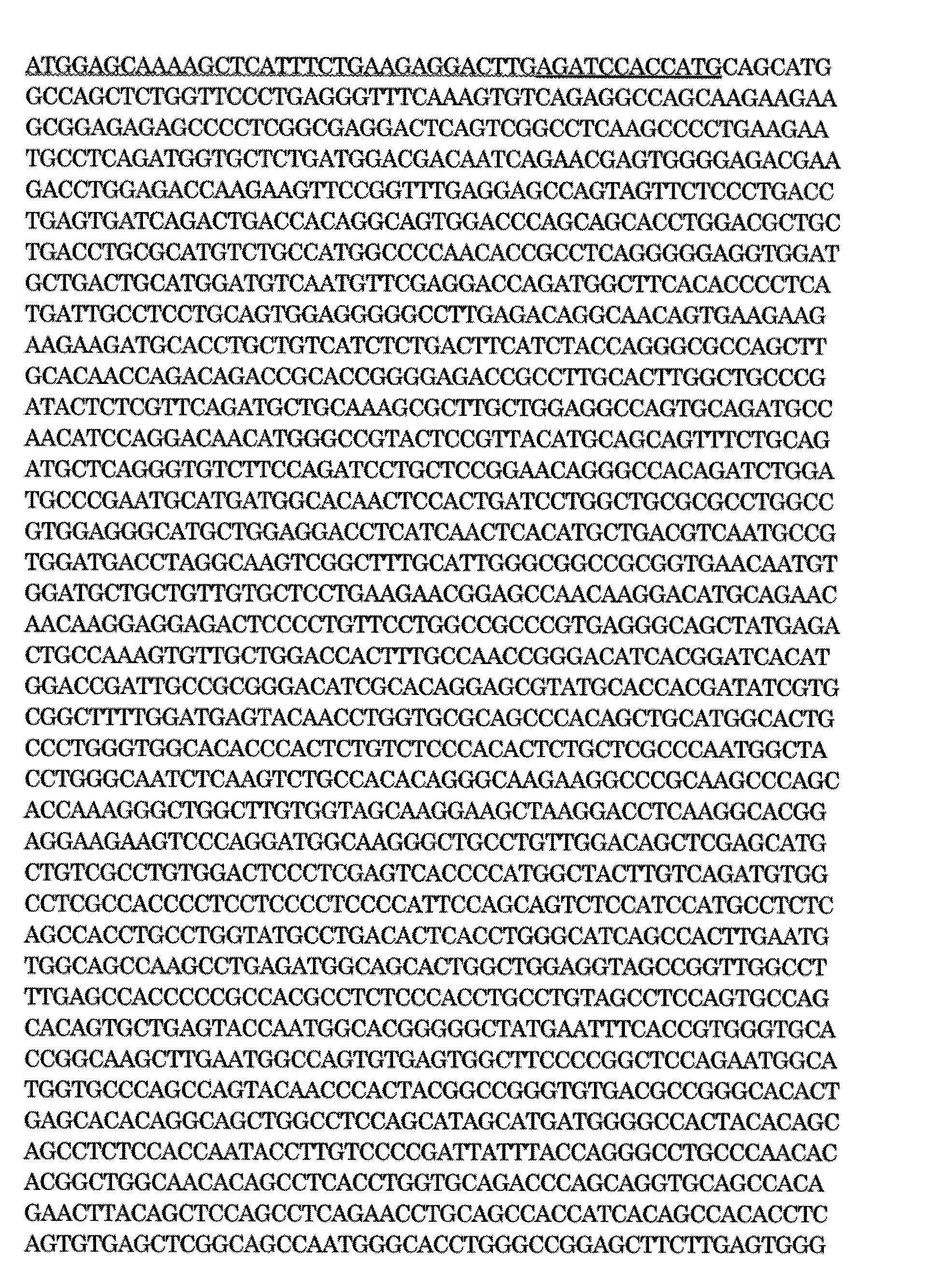

SEQ ID NO: 2: Mouse NICD cDNA sequence. In the sequence, the wavy line indicates the Myc tag sequence, and the underline indicates the linker sequence.
配列番号3:マウスTet2CDのcDNAの塩基配列。配列中、下線部はFLAGタグ配列を示す。


SEQ ID NO: 3: base sequence of mouse Tet2CD cDNA. In the sequence, the underlined portion indicates the FLAG tag sequence.
配列番号4:mREST/NRSF-VP16のcDNAの塩基配列。配列中、破線部はFLAGタグ配列を示し、下線部はmREST/NRSFをコードする配列を示し、波線部はリンカー配列を示し、イタリック部分はVP16をコードする配列を示す。

SEQ ID NO: 4: nucleotide sequence of cDNA of mREST / NRSF-VP16. In the sequence, the broken line portion indicates a FLAG tag sequence, the underline portion indicates a sequence encoding mREST / NRSF, the wavy portion indicates a linker sequence, and the italic portion indicates a sequence encoding VP16.
配列番号5:ヒトNFIAのcDNAの塩基配列(NM_001145511.1)。

SEQ ID NO: 5: Nucleotide sequence of human NFIA cDNA (NM — 001145511.1).
配列番号6:ヒトNICDのcDNAの塩基配列(NM_017617.4の一部)。


SEQ ID NO: 6: nucleotide sequence of human NICD cDNA (part of NM — 017617.4).
配列番号7:ヒトTet2CDのcDNAの塩基配列(NM_001127208.2の一部)。


SEQ ID NO: 7: nucleotide sequence of human Tet2CD cDNA (part of NM_001127208.2).
配列番号8:マウスNFIAのアミノ酸配列(NP_035035.1)。

SEQ ID NO: 8: Amino acid sequence of mouse NFIA (NP_035035.1).
配列番号9:マウスNICDのアミノ酸配列(NP_032740.3の一部)。

SEQ ID NO: 9: Amino acid sequence of mouse NICD (part of NP_032740.3).
配列番号10:マウスTet2CDのアミノ酸配列(NP_001035490.2の一部)。

SEQ ID NO: 10: Amino acid sequence of mouse Tet2CD (part of NP_001035490.2).
配列番号11:ヒトNFIAのアミノ酸配列(NP_001138983.1)。

SEQ ID NO: 11: amino acid sequence of human NFIA (NP_001138983.1).
配列番号12:ヒトNICDのアミノ酸配列(NP_060087.3)。

SEQ ID NO: 12: Amino acid sequence of human NICD (NP_060087.3).
配列番号13:ヒトTet2CDのアミノ酸配列(NP_001120680.1)。

SEQ ID NO: 13: Amino acid sequence of human Tet2CD (NP_001120680.1).
配列番号14:shp14ARFをコードするDNAのフォアード鎖の塩基配列。配列中、大文字部分が標的に対して相補的な部分を示す。

SEQ ID NO: 14: nucleotide sequence of the forward strand of DNA encoding shp14ARF In the sequence, the capital letter indicates the part complementary to the target.
配列番号15:shp14ARFをコードするDNAのリバース鎖の塩基配列。配列中、大文字部分が標的に対して相補的な部分を示す。

SEQ ID NO: 15: nucleotide sequence of reverse strand of DNA encoding shp14ARF In the sequence, the capital letter indicates the part complementary to the target.
配列番号16:shp53をコードするDNAのフォアード鎖の塩基配列。配列中、大文字部分が標的に対して相補的な部分を示す。

SEQ ID NO: 16: nucleotide sequence of the forward strand of DNA encoding shp53 In the sequence, the capital letter indicates the part complementary to the target.
配列番号17:shp53をコードするDNAのリバース鎖の塩基配列。配列中、大文字部分が標的に対して相補的な部分を示す。

SEQ ID NO: 17: base sequence of reverse strand of DNA encoding shp53 In the sequence, the capital letter indicates the part complementary to the target.
配列番号18:shp21をコードするDNAのフォアード鎖の塩基配列。配列中、大文字部分が標的に対して相補的な部分を示す。

SEQ ID NO: 18: nucleotide sequence of the forward strand of DNA encoding shp21 In the sequence, the capital letter indicates the part complementary to the target.
配列番号19:shp21をコードするDNAのリバース鎖の塩基配列。配列中、大文字部分が標的に対して相補的な部分を示す。

SEQ ID NO: 19: base sequence of reverse strand of DNA encoding shp21 In the sequence, the capital letter indicates the part complementary to the target.
配列番号20:shp16INK4aをコードするDNAのフォアード鎖の塩基配列。配列中、大文字部分が標的に対して相補的な部分を示す。

SEQ ID NO: 20: nucleotide sequence of the forward strand of DNA encoding shp16INK4a In the sequence, the capital letter indicates the part complementary to the target.
配列番号21:shp16INK4aをコードするDNAのリバース鎖の塩基配列。配列中、大文字部分が標的に対して相補的な部分を示す。

SEQ ID NO: 21: base sequence of reverse strand of DNA encoding shp16INK4a In the sequence, the capital letter indicates the part complementary to the target.
配列番号22:実施例10においてコントロールとして使用したshRNAをコードするDNAのフォアード鎖の塩基配列。配列中、大文字部分が標的に対して相補的な部分を示す。

SEQ ID NO: 22: nucleotide sequence of the forward strand of DNA encoding shRNA used as a control in Example 10 In the sequence, the capital letter indicates the part complementary to the target.
配列番号23:実施例10においてコントロールとして使用したshRNAをコードするDNAのリバース鎖の塩基配列。配列中、大文字部分が標的に対して相補的な部分を示す。

SEQ ID NO: 23: base sequence of reverse strand of DNA encoding shRNA used as control in Example 10 In the sequence, the capital letter indicates the part complementary to the target.
配列番号24:実施例8において使用したshRNAが標的とする、MECP2遺伝子中の塩基配列。

SEQ ID NO: 24: Base sequence in MECP2 gene targeted by shRNA used in Example 8.
配列番号25:p16INK4aのcDNAの塩基配列(NM_000077.4)。

SEQ ID NO: 25: base sequence of cDNA of p16INK4a (NM_000077.4).
本発明の方法によれば、従来取得困難であったアストロサイトと同様の特性を有するアストロサイト様細胞を、取得容易なヒト細胞から迅速かつ高効率で大量に取得可能である。また、当該アストロサイト様細胞を用いてインビトロ疾患モデルを構築し、疾患の分子機構や病因の解明、及び当該疾患治療薬のスクリーニングのために利用することができる。
According to the method of the present invention, astrocyte-like cells having characteristics similar to those of astrocytes, which have been difficult to obtain conventionally, can be rapidly and efficiently obtained in large quantities from easily obtainable human cells. In addition, an in vitro disease model can be constructed using the astrocyte-like cells, and can be used for elucidation of the molecular mechanism and pathogenesis of the disease, and screening for a therapeutic drug for the disease.
本明細書に引用する全ての刊行物及び特許文献は、参照により全体として本明細書中に援用される。なお、例示を目的として、本明細書の特定の実施形態を本明細書において説明したが、本発明の精神及び範囲から逸脱することなく、種々の改変が行われる場合があることは、当業者に容易に理解されるであろう。
All publications and patent literature cited herein are hereby incorporated by reference in their entirety. While specific embodiments herein have been described herein for purposes of illustration, it will be appreciated by those skilled in the art that various modifications may be made without departing from the spirit and scope of the invention. Will be easily understood.
Claims (16)
- ヒト細胞からアストロサイト様細胞をインビトロで調製する方法であって、以下の工程:
(1)少なくとも2種類の誘導因子をヒト細胞に導入する工程;及び
(2)前記ヒト細胞を分化誘導してアストロサイト様細胞を得る工程
を含み、
ここで、前記誘導因子が、NFIAを含み、かつNICD及びTet2CDの少なくとも1つを含み、
前記ヒト細胞が、ヒト体細胞(アストロサイトを除く)、ヒト多能性幹細胞又はヒト成体幹細胞である、前記調製方法。 A method for preparing in vitro astrocyte-like cells from human cells comprising the following steps:
(1) introducing at least two kinds of inducers into human cells; and (2) obtaining astrocyte-like cells by inducing differentiation of the human cells,
Wherein the inducer comprises NFIA and comprises at least one of NICD and Tet2CD;
The preparation method, wherein the human cells are human somatic cells (excluding astrocytes), human pluripotent stem cells or human adult stem cells. - 前記誘導因子が、NFIA、NICD及びTet2CDを含む、請求項1に記載の調製方法。 The preparation method according to claim 1, wherein the inducer includes NFIA, NICD, and Tet2CD.
- 前記ヒト細胞が、ヒト線維芽細胞である、請求項1又は2に記載の調製方法。 The preparation method according to claim 1 or 2, wherein the human cells are human fibroblasts.
- 前記ヒト細胞が、ヒトiPS細胞又はヒト神経幹細胞である、請求項1又は2に記載の調製方法。 The preparation method according to claim 1 or 2, wherein the human cell is a human iPS cell or a human neural stem cell.
- 前記ヒト細胞が、神経系疾患患者由来の細胞であるか、又は前記神経系疾患患者由来の細胞の遺伝子に特異的に存在する変異を人為的に遺伝子に導入された健常者由来の細胞である、請求項1~4のいずれか1項に記載の調製方法。 The human cell is a cell derived from a patient with a nervous system disease, or a cell derived from a healthy person artificially introduced with a mutation that specifically exists in the gene of a cell derived from the patient with a nervous system disease The preparation method according to any one of claims 1 to 4.
- 前記工程(1)における誘導因子の導入が、前記誘導因子をコードする核酸を含む少なくとも1つのベクターを導入することを含む、請求項1~5のいずれか1項に記載の調製方法。 The preparation method according to any one of claims 1 to 5, wherein the introduction of the inducer in the step (1) comprises introducing at least one vector containing a nucleic acid encoding the inducer.
- 前記工程(1)において、
p14ARF遺伝子、p53遺伝子、p21遺伝子、p16INK4a遺伝子及びRb遺伝子から選択される少なくとも1つの遺伝子の発現を阻害するか、
p14ARF、p53、p21、p16INK4a及びRbから選択される少なくとも1つを阻害するか、
MDM2遺伝子、サイクリンD1遺伝子、CDK4遺伝子、CDK6遺伝子及びE2F遺伝子から選択される少なくとも1つの遺伝子の発現を増加させるか、又は
MDM2、サイクリンD1、CDK4、CDK6及びE2Fから選択される少なくとも1つを活性化すること
をさらに含む、請求項1~6のいずれか1項に記載の調製方法。 In the step (1),
inhibits the expression of at least one gene selected from p14ARF gene, p53 gene, p21 gene, p16INK4a gene and Rb gene,
inhibits at least one selected from p14ARF, p53, p21, p16INK4a and Rb,
Increases the expression of at least one gene selected from the MDM2 gene, cyclin D1 gene, CDK4 gene, CDK6 gene and E2F gene, or activates at least one selected from MDM2, cyclin D1, CDK4, CDK6 and E2F The preparation method according to any one of claims 1 to 6, further comprising: - 請求項1~7のいずれか1項に記載の方法で調製される、アストロサイト様細胞。 An astrocyte-like cell prepared by the method according to any one of claims 1 to 7.
- 神経系疾患に対する被験物質の有効性又は毒性を評価する方法であって、以下の工程:
(1)神経系疾患患者由来の細胞、又は前記神経系疾患患者由来の細胞の遺伝子に特異的に存在する変異を人為的に遺伝子に導入された健常者由来の細胞から、請求項1~7のいずれか1項に記載の方法により、アストロサイト様細胞を調製する工程;及び
(2)工程(1)で得られたアストロサイト様細胞と被験物質とを接触させて、被験物質の有効性又は毒性を評価する工程
を含む、前記方法。 A method for evaluating the effectiveness or toxicity of a test substance against a nervous system disease, comprising the following steps:
(1) From a cell derived from a patient with a nervous system disease, or a cell derived from a healthy person into which a mutation that specifically exists in a gene of a cell derived from a patient with the nervous system disease has been artificially introduced into a gene, A step of preparing an astrocyte-like cell by the method according to any one of the above; and (2) contacting the astrocyte-like cell obtained in step (1) with the test substance to thereby determine the effectiveness of the test substance. Or said method comprising the step of assessing toxicity. - 神経系疾患に対する被験物質の有効性又は毒性を評価する方法であって、以下の工程:
(1)健常者由来の細胞から、請求項1~7のいずれか1項に記載の方法により、アストロサイト様細胞を調製する工程;
(2)工程(1)で得られたアストロサイト様細胞において、神経系疾患関連遺伝子をノックアウト又はノックダウンする工程;及び
(3)工程(2)で得られたアストロサイト様細胞と被験物質とを接触させて、被験物質の有効性又は毒性を評価する工程
を含む、前記方法。 A method for evaluating the effectiveness or toxicity of a test substance against a nervous system disease, comprising the following steps:
(1) a step of preparing an astrocyte-like cell from a cell derived from a healthy person by the method according to any one of claims 1 to 7;
(2) a step of knocking out or knocking down a nervous system disease-related gene in the astrocyte-like cell obtained in step (1); and (3) an astrocyte-like cell obtained in step (2) and a test substance The method comprising the steps of: contacting and evaluating the efficacy or toxicity of the test substance. - 請求項8に記載のアストロサイト様細胞とニューロンとを含む、インビトロの神経組織モデル。 An in vitro neural tissue model comprising the astrocyte-like cell according to claim 8 and a neuron.
- 神経系疾患に対する被験物質の有効性又は毒性を評価する方法であって、以下の工程:
(1)神経系疾患患者由来の細胞、又は前記神経系疾患患者由来の細胞の遺伝子に特異的に存在する変異を人為的に遺伝子に導入された健常者由来の細胞から、請求項1~7のいずれか1項に記載の方法により、アストロサイト様細胞を調製する工程;
(2)工程(1)で得られたアストロサイト様細胞とニューロンとを共培養して、神経組織モデルを調製する工程;
(3)工程(2)で得られた神経組織モデルと被験物質とを接触させて、被験物質の有効性又は毒性を評価する工程
を含む、前記方法。 A method for evaluating the effectiveness or toxicity of a test substance against a nervous system disease, comprising the following steps:
(1) From a cell derived from a patient with a nervous system disease, or a cell derived from a healthy person into which a mutation that specifically exists in a gene of a cell derived from a patient with the nervous system disease has been artificially introduced into a gene, A step of preparing an astrocyte-like cell by the method according to any one of the above;
(2) a step of preparing a neural tissue model by co-culturing astrocyte-like cells and neurons obtained in step (1);
(3) The said method including the process of contacting the nerve tissue model obtained by process (2), and a test substance, and evaluating the effectiveness or toxicity of a test substance. - 神経系疾患に対する被験物質の有効性又は毒性を評価する方法であって、以下の工程:
(1)健常者由来の細胞から、請求項1~7のいずれか1項に記載の方法により、アストロサイト様細胞を調製する工程;
(2)工程(1)で得られたアストロサイト様細胞において、神経系疾患関連遺伝子をノックアウト又はノックダウンする工程;
(3)工程(2)で得られたアストロサイト様細胞とニューロンとを共培養して、神経組織モデルを調製する工程;
(4)工程(3)で得られた神経組織モデルと被験物質とを接触させて、被験物質の有効性又は毒性を評価する工程
を含む、前記方法。 A method for evaluating the effectiveness or toxicity of a test substance against a nervous system disease, comprising the following steps:
(1) a step of preparing an astrocyte-like cell from a cell derived from a healthy person by the method according to any one of claims 1 to 7;
(2) a step of knocking out or knocking down a nervous system disease-related gene in the astrocyte-like cell obtained in step (1);
(3) a step of preparing a neural tissue model by co-culturing astrocyte-like cells and neurons obtained in step (2);
(4) The method comprising the step of contacting the nerve tissue model obtained in step (3) with a test substance to evaluate the effectiveness or toxicity of the test substance. - 少なくとも2種類の誘導因子、又は前記誘導因子をコードする核酸を含む少なくとも1つのベクターを成分として含む、ヒト細胞からアストロサイト様細胞への誘導剤であって、ここで、前記誘導因子が、NFIAを含み、かつNICD及びTet2CDの少なくとも1つを含み、前記ヒト細胞が、ヒト体細胞(アストロサイトを除く)、ヒト多能性幹細胞又はヒト成体幹細胞である、前記誘導剤。 An inducer from a human cell to an astrocyte-like cell, comprising at least one kind of inducer or at least one vector containing a nucleic acid encoding the inducer, wherein the inducer is NFIA And at least one of NICD and Tet2CD, and the human cell is a human somatic cell (excluding astrocytes), a human pluripotent stem cell or a human adult stem cell.
- 少なくとも2種類の誘導因子、又は前記誘導因子をコードする核酸を含む少なくとも1つのベクター;及び、請求項1~7のいずれか1項に記載の方法が記載された取扱説明書を含む、ヒト細胞からアストロサイト様細胞への誘導キットであって、ここで、前記誘導因子が、NFIAを含み、かつNICD及びTet2CDの少なくとも1つを含み、前記ヒト細胞が、ヒト体細胞(アストロサイトを除く)、ヒト多能性幹細胞又はヒト成体幹細胞である、前記キット。 A human cell comprising at least one inducer, or at least one vector comprising a nucleic acid encoding said inducer; and an instruction manual describing the method according to any one of claims 1-7 To astrocyte-like cells, wherein the inducer comprises NFIA and comprises at least one of NICD and Tet2CD, and the human cells are human somatic cells (excluding astrocytes) The kit, which is a human pluripotent stem cell or a human adult stem cell.
- p14ARF遺伝子、p53遺伝子、p21遺伝子、p16INK4a遺伝子及びRb遺伝子から選択される少なくとも1つの遺伝子の発現阻害剤、及び/又は
p14ARF、p53、p21、p16INK4a及びRbから選択される少なくとも1つの阻害剤、及び/又は
MDM2遺伝子、サイクリンD1遺伝子、CDK4遺伝子、CDK6遺伝子及びE2F遺伝子から選択される少なくとも1つの遺伝子の発現増強剤、及び/又は
MDM2、サイクリンD1、CDK4、CDK6及びE2Fから選択される少なくとも1つの活性化剤
をさらに含む、請求項15に記載のキット。 an expression inhibitor of at least one gene selected from p14ARF gene, p53 gene, p21 gene, p16INK4a gene and Rb gene, and / or at least one inhibitor selected from p14ARF, p53, p21, p16INK4a and Rb, and And / or expression enhancer of at least one gene selected from MDM2 gene, cyclin D1 gene, CDK4 gene, CDK6 gene and E2F gene, and / or at least one selected from MDM2, cyclin D1, CDK4, CDK6 and E2F The kit of claim 15 further comprising an activator.
Priority Applications (1)
| Application Number | Priority Date | Filing Date | Title |
|---|---|---|---|
| JP2017543528A JP6906231B2 (en) | 2015-09-29 | 2016-09-28 | Astrocyte-like cells and their preparation method |
Applications Claiming Priority (2)
| Application Number | Priority Date | Filing Date | Title |
|---|---|---|---|
| JP2015-191711 | 2015-09-29 | ||
| JP2015191711 | 2015-09-29 |
Publications (1)
| Publication Number | Publication Date |
|---|---|
| WO2017057523A1 true WO2017057523A1 (en) | 2017-04-06 |
Family
ID=58427611
Family Applications (1)
| Application Number | Title | Priority Date | Filing Date |
|---|---|---|---|
| PCT/JP2016/078741 WO2017057523A1 (en) | 2015-09-29 | 2016-09-28 | Astrocyte-like cells and method for preparing same |
Country Status (2)
| Country | Link |
|---|---|
| JP (1) | JP6906231B2 (en) |
| WO (1) | WO2017057523A1 (en) |
Cited By (3)
| Publication number | Priority date | Publication date | Assignee | Title |
|---|---|---|---|---|
| JP2020513823A (en) * | 2017-03-21 | 2020-05-21 | メモリアル スローン ケタリング キャンサー センター | Stem cell-derived astrocytes, methods of making and methods of use |
| WO2021032271A1 (en) * | 2019-08-16 | 2021-02-25 | Lu License Ab | Induction of functional astrocytes from pluripotent stem cells |
| WO2022054857A1 (en) * | 2020-09-10 | 2022-03-17 | Jsr株式会社 | Method for producing astrocyte-like cells |
Citations (1)
| Publication number | Priority date | Publication date | Assignee | Title |
|---|---|---|---|---|
| JP2003524391A (en) * | 1999-01-05 | 2003-08-19 | アバンテイス・フアルマ・エス・アー | Astrocytes, their manufacture and their use |
-
2016
- 2016-09-28 JP JP2017543528A patent/JP6906231B2/en active Active
- 2016-09-28 WO PCT/JP2016/078741 patent/WO2017057523A1/en active Application Filing
Patent Citations (1)
| Publication number | Priority date | Publication date | Assignee | Title |
|---|---|---|---|---|
| JP2003524391A (en) * | 1999-01-05 | 2003-08-19 | アバンテイス・フアルマ・エス・アー | Astrocytes, their manufacture and their use |
Non-Patent Citations (5)
| Title |
|---|
| CAIAZZO, MASSIMILIANO ET AL.: "Direct Conversion of Fibroblasts into Functional Astrocytes by Defined Transcription Factors", STEM CELL REPORTS, vol. 4, January 2015 (2015-01-01), pages 25 - 36, XP055376127 * |
| HO, CHI KENT: "Who Am I: Blurring the Lines of Cellular Identity", ELECTRONIC THESIS AND DISSERTATIONS UCLA, 2013, pages 1 - 116, XP055374476, Retrieved from the Internet <URL:http://escholarship.org/uc/item/5k61n96j> * |
| PRICE, JAMES D. ET AL.: "The Ink4a/Arf Locus Is a Barrier to Direct Neuronal Transdifferentiation", THE JOURNAL OF NEUROSCIENCE, vol. 34, no. 37, 2014, pages 12560 - 12567, XP055376124 * |
| SHALTOUKI, ATOSSA ET AL.: "Efficient Generation of Astrocytes from Human Pluripotent Stem Cells in Defined Conditions", STEM CELLS, vol. 31, 2013, pages 941 - 952 * |
| ZHANG, YANPING ET AL.: "ARF Promotes MDM2 Degradation and Stabilizes p53: ARF-INK4a Locus Deletion Impairs Both the Rb and p53 Tumor Supression Pathways", CELL, vol. 92, 1998, pages 725 - 734, XP002128766 * |
Cited By (4)
| Publication number | Priority date | Publication date | Assignee | Title |
|---|---|---|---|---|
| JP2020513823A (en) * | 2017-03-21 | 2020-05-21 | メモリアル スローン ケタリング キャンサー センター | Stem cell-derived astrocytes, methods of making and methods of use |
| JP7471084B2 (en) | 2017-03-21 | 2024-04-19 | メモリアル スローン ケタリング キャンサー センター | Stem cell derived astrocytes, methods of making and using |
| WO2021032271A1 (en) * | 2019-08-16 | 2021-02-25 | Lu License Ab | Induction of functional astrocytes from pluripotent stem cells |
| WO2022054857A1 (en) * | 2020-09-10 | 2022-03-17 | Jsr株式会社 | Method for producing astrocyte-like cells |
Also Published As
| Publication number | Publication date |
|---|---|
| JPWO2017057523A1 (en) | 2018-08-02 |
| JP6906231B2 (en) | 2021-07-21 |
Similar Documents
| Publication | Publication Date | Title |
|---|---|---|
| US20230282445A1 (en) | Method of nuclear reprogramming | |
| CA2930590C (en) | Engineering neural stem cells using homologous recombination | |
| Sánchez-Danés et al. | Efficient generation of A9 midbrain dopaminergic neurons by lentiviral delivery of LMX1A in human embryonic stem cells and induced pluripotent stem cells | |
| Addis et al. | Efficient conversion of astrocytes to functional midbrain dopaminergic neurons using a single polycistronic vector | |
| CN103562376B (en) | The method of rejuvenation cell | |
| JP5888753B2 (en) | Efficient method for establishing induced pluripotent stem cells | |
| WO2011102444A1 (en) | Method for producing induced pluripotent stem cells | |
| Liu et al. | The fused in sarcoma protein forms cytoplasmic aggregates in motor neurons derived from integration-free induced pluripotent stem cells generated from a patient with familial amyotrophic lateral sclerosis carrying the FUS-P525L mutation | |
| Oh et al. | Human-induced pluripotent stem cells generated from intervertebral disc cells improve neurologic functions in spinal cord injury | |
| Kim et al. | Effects of mechanical stimulation on the reprogramming of somatic cells into human-induced pluripotent stem cells | |
| Zhou et al. | Reprogramming non-human primate somatic cells into functional neuronal cells by defined factors | |
| JP6906231B2 (en) | Astrocyte-like cells and their preparation method | |
| US20190322981A1 (en) | Means and methods for the generation of oligodendrocytes | |
| US20110033936A1 (en) | CANINE iPS CELLS AND METHOD OF PRODUCING SAME | |
| JP2011522520A (en) | Methods for cell dedifferentiation | |
| Ohno et al. | Distinct iPS cells show different cardiac differentiation efficiency | |
| Wang et al. | A novel function of dcf1 during the differentiation of neural stem cells in vitro | |
| Shimizu et al. | Pax6-5a promotes neuronal differentiation of murine embryonic stem cells | |
| WO2021172542A1 (en) | Mature-cardiomyocyte production method | |
| WO2013124309A1 (en) | Direct reprogramming of somatic cells into neural stem cells | |
| WO2024071375A1 (en) | Human astrocyte cell mass, cell mass culture, method for producing human astrocyte cell mass, and method for evaluating test substance | |
| WO2024010085A1 (en) | Mutant oct3/4 protein, and method for producing induced pluripotent stem cells | |
| US20210010030A1 (en) | Method for reprogramming somatic cells | |
| Lovejoy | iPSC derived cerebral organoids as a model of tauopathy | |
| US20190218510A1 (en) | Compositions and methods to derive mesodermal lineage cells and mixed tissue organoids from embryonic stem cells |
Legal Events
| Date | Code | Title | Description |
|---|---|---|---|
| 121 | Ep: the epo has been informed by wipo that ep was designated in this application |
Ref document number: 16851694 Country of ref document: EP Kind code of ref document: A1 |
|
| ENP | Entry into the national phase |
Ref document number: 2017543528 Country of ref document: JP Kind code of ref document: A |
|
| NENP | Non-entry into the national phase |
Ref country code: DE |
|
| 122 | Ep: pct application non-entry in european phase |
Ref document number: 16851694 Country of ref document: EP Kind code of ref document: A1 |