WO2017053629A2 - Light-controlled gene delivery with virus vectors through incorporation of optogenetic proteins and genetic insertion of non-conformationally constrained peptides - Google Patents
Light-controlled gene delivery with virus vectors through incorporation of optogenetic proteins and genetic insertion of non-conformationally constrained peptides Download PDFInfo
- Publication number
- WO2017053629A2 WO2017053629A2 PCT/US2016/053200 US2016053200W WO2017053629A2 WO 2017053629 A2 WO2017053629 A2 WO 2017053629A2 US 2016053200 W US2016053200 W US 2016053200W WO 2017053629 A2 WO2017053629 A2 WO 2017053629A2
- Authority
- WO
- WIPO (PCT)
- Prior art keywords
- virus
- acid molecule
- nucleic acid
- gene
- group
- Prior art date
- Legal status (The legal status is an assumption and is not a legal conclusion. Google has not performed a legal analysis and makes no representation as to the accuracy of the status listed.)
- Ceased
Links
Classifications
-
- C—CHEMISTRY; METALLURGY
- C12—BIOCHEMISTRY; BEER; SPIRITS; WINE; VINEGAR; MICROBIOLOGY; ENZYMOLOGY; MUTATION OR GENETIC ENGINEERING
- C12N—MICROORGANISMS OR ENZYMES; COMPOSITIONS THEREOF; PROPAGATING, PRESERVING, OR MAINTAINING MICROORGANISMS; MUTATION OR GENETIC ENGINEERING; CULTURE MEDIA
- C12N15/00—Mutation or genetic engineering; DNA or RNA concerning genetic engineering, vectors, e.g. plasmids, or their isolation, preparation or purification; Use of hosts therefor
- C12N15/09—Recombinant DNA-technology
- C12N15/63—Introduction of foreign genetic material using vectors; Vectors; Use of hosts therefor; Regulation of expression
- C12N15/79—Vectors or expression systems specially adapted for eukaryotic hosts
- C12N15/85—Vectors or expression systems specially adapted for eukaryotic hosts for animal cells
- C12N15/86—Viral vectors
-
- C—CHEMISTRY; METALLURGY
- C07—ORGANIC CHEMISTRY
- C07K—PEPTIDES
- C07K14/00—Peptides having more than 20 amino acids; Gastrins; Somatostatins; Melanotropins; Derivatives thereof
- C07K14/005—Peptides having more than 20 amino acids; Gastrins; Somatostatins; Melanotropins; Derivatives thereof from viruses
-
- C—CHEMISTRY; METALLURGY
- C07—ORGANIC CHEMISTRY
- C07K—PEPTIDES
- C07K14/00—Peptides having more than 20 amino acids; Gastrins; Somatostatins; Melanotropins; Derivatives thereof
- C07K14/195—Peptides having more than 20 amino acids; Gastrins; Somatostatins; Melanotropins; Derivatives thereof from bacteria
-
- C—CHEMISTRY; METALLURGY
- C07—ORGANIC CHEMISTRY
- C07K—PEPTIDES
- C07K14/00—Peptides having more than 20 amino acids; Gastrins; Somatostatins; Melanotropins; Derivatives thereof
- C07K14/415—Peptides having more than 20 amino acids; Gastrins; Somatostatins; Melanotropins; Derivatives thereof from plants
-
- C—CHEMISTRY; METALLURGY
- C12—BIOCHEMISTRY; BEER; SPIRITS; WINE; VINEGAR; MICROBIOLOGY; ENZYMOLOGY; MUTATION OR GENETIC ENGINEERING
- C12N—MICROORGANISMS OR ENZYMES; COMPOSITIONS THEREOF; PROPAGATING, PRESERVING, OR MAINTAINING MICROORGANISMS; MUTATION OR GENETIC ENGINEERING; CULTURE MEDIA
- C12N15/00—Mutation or genetic engineering; DNA or RNA concerning genetic engineering, vectors, e.g. plasmids, or their isolation, preparation or purification; Use of hosts therefor
- C12N15/09—Recombinant DNA-technology
- C12N15/11—DNA or RNA fragments; Modified forms thereof; Non-coding nucleic acids having a biological activity
- C12N15/52—Genes encoding for enzymes or proenzymes
-
- C—CHEMISTRY; METALLURGY
- C12—BIOCHEMISTRY; BEER; SPIRITS; WINE; VINEGAR; MICROBIOLOGY; ENZYMOLOGY; MUTATION OR GENETIC ENGINEERING
- C12N—MICROORGANISMS OR ENZYMES; COMPOSITIONS THEREOF; PROPAGATING, PRESERVING, OR MAINTAINING MICROORGANISMS; MUTATION OR GENETIC ENGINEERING; CULTURE MEDIA
- C12N2310/00—Structure or type of the nucleic acid
- C12N2310/10—Type of nucleic acid
- C12N2310/20—Type of nucleic acid involving clustered regularly interspaced short palindromic repeats [CRISPR]
-
- C—CHEMISTRY; METALLURGY
- C12—BIOCHEMISTRY; BEER; SPIRITS; WINE; VINEGAR; MICROBIOLOGY; ENZYMOLOGY; MUTATION OR GENETIC ENGINEERING
- C12N—MICROORGANISMS OR ENZYMES; COMPOSITIONS THEREOF; PROPAGATING, PRESERVING, OR MAINTAINING MICROORGANISMS; MUTATION OR GENETIC ENGINEERING; CULTURE MEDIA
- C12N2750/00—MICROORGANISMS OR ENZYMES; COMPOSITIONS THEREOF; PROPAGATING, PRESERVING, OR MAINTAINING MICROORGANISMS; MUTATION OR GENETIC ENGINEERING; CULTURE MEDIA ssDNA viruses
- C12N2750/00011—Details
- C12N2750/14011—Parvoviridae
- C12N2750/14111—Dependovirus, e.g. adenoassociated viruses
- C12N2750/14122—New viral proteins or individual genes, new structural or functional aspects of known viral proteins or genes
-
- C—CHEMISTRY; METALLURGY
- C12—BIOCHEMISTRY; BEER; SPIRITS; WINE; VINEGAR; MICROBIOLOGY; ENZYMOLOGY; MUTATION OR GENETIC ENGINEERING
- C12N—MICROORGANISMS OR ENZYMES; COMPOSITIONS THEREOF; PROPAGATING, PRESERVING, OR MAINTAINING MICROORGANISMS; MUTATION OR GENETIC ENGINEERING; CULTURE MEDIA
- C12N2750/00—MICROORGANISMS OR ENZYMES; COMPOSITIONS THEREOF; PROPAGATING, PRESERVING, OR MAINTAINING MICROORGANISMS; MUTATION OR GENETIC ENGINEERING; CULTURE MEDIA ssDNA viruses
- C12N2750/00011—Details
- C12N2750/14011—Parvoviridae
- C12N2750/14111—Dependovirus, e.g. adenoassociated viruses
- C12N2750/14141—Use of virus, viral particle or viral elements as a vector
-
- C—CHEMISTRY; METALLURGY
- C12—BIOCHEMISTRY; BEER; SPIRITS; WINE; VINEGAR; MICROBIOLOGY; ENZYMOLOGY; MUTATION OR GENETIC ENGINEERING
- C12N—MICROORGANISMS OR ENZYMES; COMPOSITIONS THEREOF; PROPAGATING, PRESERVING, OR MAINTAINING MICROORGANISMS; MUTATION OR GENETIC ENGINEERING; CULTURE MEDIA
- C12N2750/00—MICROORGANISMS OR ENZYMES; COMPOSITIONS THEREOF; PROPAGATING, PRESERVING, OR MAINTAINING MICROORGANISMS; MUTATION OR GENETIC ENGINEERING; CULTURE MEDIA ssDNA viruses
- C12N2750/00011—Details
- C12N2750/14011—Parvoviridae
- C12N2750/14111—Dependovirus, e.g. adenoassociated viruses
- C12N2750/14141—Use of virus, viral particle or viral elements as a vector
- C12N2750/14143—Use of virus, viral particle or viral elements as a vector viral genome or elements thereof as genetic vector
-
- C—CHEMISTRY; METALLURGY
- C12—BIOCHEMISTRY; BEER; SPIRITS; WINE; VINEGAR; MICROBIOLOGY; ENZYMOLOGY; MUTATION OR GENETIC ENGINEERING
- C12N—MICROORGANISMS OR ENZYMES; COMPOSITIONS THEREOF; PROPAGATING, PRESERVING, OR MAINTAINING MICROORGANISMS; MUTATION OR GENETIC ENGINEERING; CULTURE MEDIA
- C12N2750/00—MICROORGANISMS OR ENZYMES; COMPOSITIONS THEREOF; PROPAGATING, PRESERVING, OR MAINTAINING MICROORGANISMS; MUTATION OR GENETIC ENGINEERING; CULTURE MEDIA ssDNA viruses
- C12N2750/00011—Details
- C12N2750/14011—Parvoviridae
- C12N2750/14111—Dependovirus, e.g. adenoassociated viruses
- C12N2750/14141—Use of virus, viral particle or viral elements as a vector
- C12N2750/14145—Special targeting system for viral vectors
Definitions
- Viruses are genetically encoded nanoparticles with regular geometry
- VNPs potent and controllable viral nanoparticles
- Adeno-associated virus (AAV) vectors can deliver genetic material to target cells including, but not limited to genes, RNA interference (RNAi), or CRISPR/Cas genome editing tools.
- RNAi RNA interference
- a significant rate-limiting step and major determinant of effective gene delivery using AAV is inefficient nuclear entry; although AAV is considered an efficient gene delivery vector, most virions added to host cells appear to remain outside the nucleus. Additionally, off-target gene delivery by AAV poses a significant risk of undesired side effects in in vivo applications.
- the present disclosure provides a solution that addresses both of these problems.
- a promising approach for engineering programmable nanodevices is to encode stimulus-responsive properties.
- a number of synthetic nanoparticles have been designed such that detection of a particular stimulus leads to a physiochemical change in the nanoparticle, resulting in cargo delivery.
- chemical ligands, pH, enzymatic reactions, redox reactions, temperature, and magnetic fields have served as input stimuli for various non-viral nanocarriers.
- non-viral delivery systems still display lower delivery efficiencies compared to viral vectors. For this reason, stimulus- responsive virus-based platforms that respond to pH, chemicals and extracellular proteases have been developed.
- tissue-specific stimuli may be beneficial for certain applications
- externally applied stimuli can provide a more quantitatively controllable delivery process in both space and time.
- Light represents an attractive stimulus over chemical or biological stimuli because its intensity, duration, spatial pattern, and wavelength can all be precisely modulated in real time with the proper equipment and light configuration.
- in vitro tissue models light has been used with a resolution of microns to pattern proteins that direct cell processes like migration and differentiation. Light can also non-invasively penetrate the skin and is generally considered safe for use in mammalian tissues.
- Optogenetics offers a molecular toolbox of light-switchable proteins.
- phytochrome-family proteins are powerful because they can be activated by one wavelength and deactivated by a second wavelength, allowing control over the degree of activation in live cells in space and time.
- Phytochrome B has been used for light-switchable transcription, signal cascade activation, actin nucleation, autocatalytic protein splicing, and pseudopodia elongation.
- PEF6 phytochrome interacting factor 6
- the PhyB/PIF6 system dimerizes in seconds, is amenable to fusion proteins, and is non-toxic to mammalian cells.
- 2013/0330766 Al describes another suite of tools for manipulation of the viral capsid to enhance and/or control gene delivery using viral vectors.
- U.S. 2013/0330766 Al discloses "peptide locks" where enzymatically cleavable motifs are inserted flanking a peptide or protein that has been inserted into the capsid protein of an adeno-associated virus. These protease-susceptible motifs allow for release of a "peptide lock" upon exposure to the a protease or combination of proteases which can cleave the enzymatically cleavable motifs.
- compositions, methods of making said compositions and methods for using said compositions which incorporate the advantages of viral delivery systems with the spatial and temporal control offered by optogenetic tools to offer improved gene delivery systems.
- these tools can provide improved nuclear delivery of genetic material and more specifically targeted delivery of genetic material to target cells.
- compositions, methods of making said composition and methods for using said compositions which incorporate the advantages of viral delivery systems with enzymatic cleavage sites incorporate in the viral capsid to enable surface display of peptides and proteins in a more favorable thermodynamic conformation, such as a linear conformation.
- these tools can provide for improved display of peptides and proteins inserted into the viral capsid which may facilitate improved interaction with a target and/or target cell.
- the present disclosure is directed to light-controllable, viral -based gene delivery vectors incorporating optogenetic proteins or optogenetic binding partners and methods of use of such vectors. These vectors and methods can provide improved, tunable nuclear delivery of genetic material, endosomal escape as well as improved cell binding and both spatial and temporal control of gene delivery in a cell population.
- the present disclosure also provides nucleic acids and amino acids useful in making and using such vectors as well as kits for the use of vectors herein.
- the present disclosure is also directed to viral-based gene delivery vectors incorporating an enzymatic cleavage motif for linearizing or conformationally unconstraining a peptide or protein inserted into a varial capsid to improve the efficiency of methods using the peptide or protein for binding and/or to improve cell binding, endosomal escape and nuclear localization.
- a virus which includes a capsid protein and an optogenetic binding partner, wherein at least a portion of the optogenetic binding partner is displayed on the surface of the virus, and wherein the optogenetic binding partner is linked to the capsid protein by a direct amino acid linkage or a linker.
- the virus which includes a capsid protein and an optogenetic binding partner further includes an enzymatic cleavage motif adjacent to the optogenetic binding partner, wherein the enzymatic cleavage motif does not inactivate other biologically active motifs on the surface of the virus.
- a virus which includes a capsid protein and an optogenetic protein, wherein at least a portion of the optogenetic protein is displayed on the surface of the virus and wherein the optogenetic protein is linked to the capsid protein by a direct amino acid linkage or a linker.
- the virus which includes a capsid protein and an optogenetic protein further includes an enzymatic cleavage motif adjacent to the optogenetic protein, wherein the enzymatic cleavage motif does not inactivate other biologically active motifs on the surface of the virus.
- a method for delivering a nucleic acid molecule to the nucleus of a target cell includes the steps of obtaining a virus with at least a portion of an optogenetic binding partner displayed on its surface, delivering the virus to a target cell containing an optogenetic protein capable of binding to the optogenetic binding partner and having a nuclear localization signal, and exposing the target cell to light of a sufficient wavelength to induce a conformational change in the optogenetic protein to allow binding of the optogenetic protein and the optogenetic binding partner, enhancing delivery of the virus.
- a method for delivering a nucleic acid molecule to the nucleus of a target cell includes the steps of obtaining a virus with at least a portion of an optogenetic protein displayed on its surface, delivering the virus to a target cell containing an optogenetic binding partner capable of binding to the optogenetic protein and having a nuclear localization signal, and exposing the target cell to light of a sufficient wavelength to induce a conformational change in the optogenetic protein to allow binding of the optogenetic protein and the optogenetic binding partner, enhancing delivery of the virus.
- a method for delivering a nucleic acid molecule to the nucleus of a target cell includes the steps of obtaining a virus with at least a portion of an optogenetic protein having a nuclear localization signal displayed on its surface which is either exposed or occluded based on the conformation of the optogenetic protein, delivering the virus to a target cell, and exposing the target cell to light of a sufficient wavelength to induce a conformational change in the optogenetic protein to allow exposure of the nuclear localization signal, enhancing delivery of the virus.
- a method comprises providing a virus having one or more peptides genetically encoded into the capsid so as to be at least partially exposed to the surface of the capsid and an enzymatic cleavage motif cleavable by an enzyme genetically encoded into the capsid adjacent to the one or more peptides, and treating the virus with the enzyme to cleave the enzymatic cleavage motif, allowing at least a portion of the one or more peptides to be tethered to the capsid surface at either the C-terminal or N-terminal end.
- the present disclosure also provides for nucleic acids encoding and amino acids comprising at least a portion of the viruses having an optogenetic binding partner, optogenetic protein and/or enzymatic cleavage motif.
- FIGURE 1 depicts the adeno-associated virus (AAV) particle and a graphical representation of the 4.7 kB genome of AAV with the rep and cap genes and showing the alignment of the sequences of VP1, VP2 and VP3 from the cap open-reading frame (ORF).
- AAV adeno-associated virus
- FIGURE 2A depicts certain embodiments of the invention where the nuclear uptake and expression in cells is tuned by altering the intensity of light ("tunable intensity”), controlling the timing of exposure to light (“temporal dynamics”) and controlling the area which is exposed to light (“patterning”).
- tunable intensity the intensity of light
- temporal dynamics controlling the timing of exposure to light
- patterning controlling the area which is exposed to light
- FIGURE 2B depicts the formation of the holoprotein of PhyB with its
- FIGURE 2C depicts a flow diagram showing the alternative splicing of the cap gene of AAV and leaky scanning to yield VP1, VP2 and VP3, translation of the corresponding capsid subunits which can be combined with a desired transgene of interest and allowed to self- assemble into the capsid with the transgene encapsulated.
- FIGURE 3 A depicts a peptide lock embodiment where peptide "locks" are located on the viral surface and include two enzymatically cleavable motifs that are cleavable by an enzyme for unlocking the virus.
- Figure discloses SEQ ID NO: 29.
- FIGURE 3B depicts the expected activity, based on reported specificity constants for each matrix metalloprotease (MMP) against the indicated peptide substrate, and the observed activity, as % GFP + cells for AAV with a peptide lock incorporating the cleavage motifs IPVSLRSG (SEQ ID NO: 1) or IPESLRAG (SEQ ID NO: 2).
- MMP matrix metalloprotease
- FIGURE 3C depicts an alternative peptide lock embodiment where the peptide "locks" located on the surface contain two cleavage sequences, one recognized by a protease and one recognized by a different protease, e.g. a MMP.
- Figure discloses SEQ ID NO: 30.
- FIGURE 3D depicts the alternative embodiment of FIGURE 3C where, upon pre- treatment with protease, the peptide "lock" presents as a linearized peptide, allowing the different protease, e.g. a MMP, improved access to the second cleavage site, enabling the expected activity of the protease for the substrate.
- FIGURE 3E depicts the alternative embodiment of FIGURE 3C, where each cleavage leaves at least some of the inserted amino acids on the surface of the virus.
- FIGURE 4A depicts the activity, as % GFP + cells, for several variants constructed using the alternative embodiment of FIGURE 3C and tested with or without pre-treatment with the protease and with or without treatment with the different protease, e.g. MMP-2 and MMP-7.
- FIGURE 4B depicts a silver stained gel for the ePAV4 variant from FIGURE 4A treated with or without protease and with or without MMP-2, MMP-7 or MMP-9.
- FIGURE 5A depicts a graphical alignment of the capsid proteins of AAV2 as expressed within a construct expressing native VPl, VP2 and VP3 (wt); a construct expressing VP2 independently with an optogenetic binding partner, phytochrome interacting factor 6 (PIF6) inserted at the N-terminus of VP2 with a separate construct expressing VPl and VP3 (VNP-2- PIF6), and a construct expressing VPl and VP2 with PIF6 inserted at the N-terminus of VP2 and at Ml 38 of VPl with a separate construct expressing VP3 (VNP-1,2-PIF6).
- PIF6 phytochrome interacting factor 6
- Figure 5 A also depicts a visual representation of the viral phenotypes produced from the wild-type construct and both VNP-2-PIF6 and VNP-1,2-PIF6.
- FIGURE 5B depicts western blots of wild-type, VNP-2-PIF6 and VNP- 1 ,2-PIF6 AAV2 viruses using a monoclonal anti-VPl, 2, 3 antibody after expression in HEK293T cells.
- FIGURE 5C depicts electron micrographs of wild-type, VNP-2-PIF6 and VNP- 1,2-PIF6 viruses after expression in HEK293T cells.
- Black scale bar lOOnm
- white scale bar 15nm.
- FIGURE 5D depicts the results of a heparin binding assay using wild-type AAV2 and VNP-2-PIF6.
- the y-axis represents the fraction of total viral genomes quantified by qPCR. Error bars are SEM from 2 independent experiments conducted in duplicate.
- FIGURE 5E depicts the transduction index (TI) for wtAAV2, VNP-2-PIF6 and VNP-1,2,-PIF6 in HEK293T cells at multiplicity of infection (MOI) of 1,000, 5,000 and 10,000. "**" indicates a p-value ⁇ 0.05.
- FIGURE 5F depicts the percentage of cells positive for GFP expression after exposure to wtAAV2, VNP-2-PIF6 or VNP-1,2-PIF6 at MOI of 1,000, 5,000 and 10,000.
- FIGURE 5G depicts the mean fluorescence intensity for cells after exposure to wtAAV2, VNP-2-PIF6 or VNP-1,2-PIF6 at MOI of 1,000, 5,000 and 10,000.
- FIGURE 6B depicts a Western blot of fractions of PhyB917-His 6 from nickel purification after expression in Dictyostelium discoideum.
- FIGURE 6C depicts coomassie-stained gels corresponding to the fractions of PhyB651- His 6 in FIGURE 6A.
- FIGURE 6D depicts Coomassie-stained gels corresponding to the fractions of PhyB917-His 6 in FIGURE 6B.
- FIGURE 7A depicts an in vitro binding assay strategy for assessing viral binding to PhyB proteins.
- V P-PIF6 is equivalent to V P-2-PIF6.
- Figure discloses "His 6 " as SEQ ID NO: 23.
- FIGURE 7B depicts the capture efficiency under far-red (FR) light conditions and red (R) light conditions for wtAAV2 and VNP-2-PIF6 on nickel columns loaded with PhyB651- His 6 or PhyB917-His 6 .
- "**" means the p-value ⁇ 0.01.
- FIGURE 7C depicts the capture efficiency under red light conditions for various column loadings of PhyB917-His 6 using VNP-2-PIF6.
- FIGURE 8 A depicts an experimental strategy for confirming binding of VNP-2- PIF6 to PhyB917 and dissociation upon exposure to far red light.
- Figure discloses "Hise” as SEQ ID NO: 23.
- FIGURE 8B depicts the capture efficiency for eluted VNP-2-PIF6 bound to PhyB917-His 6 that is exposed to far red light after elution (FR reversed) or kept under red light (R Only, control) based on the strategy depicted in FIGURE 8A.
- FIGURE 8C depicts the capture efficiency for PhyB917-His 6
- FIGURE 9A depicts a mechanism for decreasing or increasing nuclear uptake of a virus displaying an optogenetic binding partner (PIF6) on its surface into a target cell where an optogenetic protein (PhyB) and its associated chromophore are present to form the holoprotein (Pr and Pfr) in the cytoplasm, the optogenetic protein having a nuclear localization signal (NLS) on its surface and exposing the system to far-red (inactivating) light or red (activating light) to decrease or enhance nuclear uptake of the virus, respectively.
- PPF6 optogenetic binding partner
- FIGURE 9B depicts HeLa cell nuclei stained with Hoescht nuclear stain
- VNP-PIF6 VNP-2-PIF6
- Colocalized the co-localized image of VNP-2-PIF6 in cell nuclei
- FIGURE 9C depicts HeLa cell nuclei of cells expressing PhyB908 stained with Hoescht nuclear stain ("Nucleus”) after exposure to VNP-2-PIF6 under red (650nm) or far red (730nm) light, immunofluorescence of VNP-2-PIF6 in the cells ("VNP-PIF6") and the co- localized image of VNP-2-PIF6 in cell nuclei ("Colocalized”).
- Scale bar 20 ⁇ .
- FIGURE 9D depicts HeLa cell nuclei of cells expressing PhyB908-NLS stained with Hoescht nuclear stain ("Nucleus”) after exposure to VNP-2-PIF6 under red (650nm) or far red (730nm) light, immunofluorescence of VNP-2-PIF6 in the cells ("VNP-PIF6") and the co- localized image of VNP-2-PIF6 in cell nuclei ("Colocalized”).
- Scale bar 20 ⁇ .
- FIGURE 9E depicts the Pearson Correlation Coefficient for the images analyzed for the negative control (Neg.), PhyB908 (PhyB) and PhyB908-NLS (PhyB-NLS) cells under red (R) and far red (FR) light conditions. ** indicates statistical significance of the value (p-value ⁇ 0.001).
- FIGURE 9F depicts HeLa cell nuclei of cells expressing PhyB650-NLS stained with Hoescht nuclear stain ("Nucleus”) after exposure to VNP-2-PIF6 under red (650nm) or far red (730nm) light or wtAAV2, immunofluorescence of VNP-2-PIF6 in the cells ("VNP-PIF6") and the co-localized image of VNP-2-PIF6 in cell nuclei ("Colocalized”).
- FIGURE 11 A depicts an apparatus for applying R and FR light via LEDs to a tissue culture well with a glass bottom for control the R:FR light ratio.
- FIGURE 1 IB depicts the % of cells expressing GFP in HeLa cells expressing PhyB908 or PhyB908- LS transduced by V P-2-PIF6 at 24 hours post-transduction. Cells were exposed to different intensities ( ⁇ / ⁇ ) of red and far red light as shown on the x-axis.
- FIGURE 11C depicts the transduction index cells expressing GFP in HeLa cells expressing PhyB908 or PhyB908- LS transduced by V P-2-PIF6 at 24 hours post-transduction. Cells were exposed to different intensities of red and far red light as shown on the x-axis.
- FIGURE 1 ID depicts the % of cells expressing GFP in HeLa cells expressing PhyB908 or PhyB908- LS transduced by V P-2-PIF6 or wtAAV2 at 48 hours post- transduction. Cells were exposed to different intensities of red and far red light as shown on the x-axis.
- FIGURE 1 IE depicts the transduction index cells expressing GFP in HeLa cells expressing PhyB908 or PhyB908- LS transduced by V P-2-PIF6 or wtAAV2 at 48 hours post- transduction. Cells were exposed to different intensities of red and far red light as shown on the x-axis.
- FIGURE 1 IF depicts fluorescent micrographs of GFP expression in HeLa cells constitutively expressing PhyB- LS and treated with or without V P-2-PIF6, PCB, and red light.
- FIGURE 11G depicts the discrete transfer functions for transduction by V P-2- PIF6 in HeLa cells under increasing red light flux between 0 and 10
- FIGURE 11H depicts the full-range logarithmic transfer function of transduction index by V P-2-PIF6 facilitated by PhyB908- LS under varying R:FR ratios. Each data point is the average of 4-5 replicates from 2 independent experiments.
- FIGURE 12 depicts the fold change in transduction index for hMSC, HUVEC and 3T3 cells constitutively expressing PhyB908- LS and exposed to V P-2-PIF6 for 48 hours under red (R) light or far red (FR) light.
- FIGURE 13 A depicts the transduction index as a function of red light intensity for a fixed instensity of FR light.
- FIGURE 13B depicts the transduction index at maximum far red light intensity only (15 ⁇ / ⁇ 2 8) and maximum red light intensity only (43 ⁇ / ⁇ ).
- FIGURE 15A depicts the transduction index for an AAV virus comprising VP1 and VP3 in the viral capsid, having on its capsid surface, embedded in VP1, the LOV domain from Avena sativa phototropin 1 protein with a N-terminal Pkit nuclear export signal and a C- terminal nuclear localization signal as wells an enzymatic cleavage motif (DDDDK) susceptible to cleavage by enterokinase, with or without pre-treatment with enterokinase prior to the transduction and in the presence of varying intensities of blue light.
- DDDDK enzymatic cleavage motif
- FIGURE 15B depicts a Western blot of wild-type AAV and the virus used in FIGURE 15A with or without enterokinase (SEQ ID NO: 76) treatment.
- compositions and methods using optogenetic tools to provide tunable spatial and temporal control of gene delivery using viral vectors are provided.
- gene delivery in a cell population can be controlled to deliver analog levels of expression using activating, e.g. "low R” or “high R”, light versus deactivating, e.g. "FR”, light while the cells are exposed to a light- activable viral vector.
- activating light expression can be tuned by altering the intensity of the light, e.g. "low R” versus "high R", as shown in the top row of FIGURE 2 A ("tunable intensity").
- the medium shading in the cells in the middle panel reflect a lower level of expression while the darker shading in the cells in the right panel reflect a higher level of expression.
- Light-activable gene delivery can also be controlled by the timing of introduction of activating, e.g. "R", light as shown in the middle row of FIGURE 2A ("temporal dynamics") where the cell population, in the presence of the viral vector, is exposed to deactivating "FR" light until such time as activation is desired and the cells are exposed to activating light.
- activating e.g. "R”
- FR deactivating light
- light can also be controlled spatially, through the use of photomasks, the expression in a cell population can be spatially patterned by placing a photomask over the cell population, which is exposed to the viral vector, while exposed to activating light as shown in the bottom row of FIGURE 2A ("patterning").
- optical protein means an amino acid sequence that changes its conformation (e.g. tertiary structure) in response to light of certain wavelengths or ranges of wavelengths.
- optical binding partner means an amino acid sequence capable of binding to an optogenetic protein in at least some conformations of the optogenetic protein.
- the optogenetic binding partner is capable of binding to an optogenetic protein when the optogenetic protein is in a first conformation but is not capable of binding to the optogenetic protein when the optogenetic protein is in a second conformation.
- PIF6 (SEQ ID NO: 140) can reversibly bind to PhyB; when PhyB is exposed to red light, PIF6 binds to PhyB, however, when PhyB is exposed to far red light, PIF6 cannot bind PhyB and dissociates from PhyB due to the conformational change of PhyB in response to the wavelengths of light.
- FIGURE 2B shows the covalent association of apo-PhyB with its chromophore (PCB) to yield the photoresponsive holoprotein ("holo-PhyB (Pr)”) which can then associate (to form “holo-PhyB (Pfr)”) or dissociate from its binding partner, PIF6 (“PIF”), upon exposure to activating red (650nm) or deactivating far red (750nm) light, respectively.
- holo-PhyB (Pr) the photoresponsive holoprotein
- Pfr photoresponsive holoprotein
- PIF6 PIF6
- a first amino acid sequence is considered adjacent to a second amino acid sequence if it is located outside of the second amino acid sequence and is located at the N- or C-terminus of the second amino acid sequence.
- nucleic acid sequence location a first nucleic acid sequence is considered adjacent to a second nucleic acid sequence if it is located outside of the second nucleic acid sequence and is located at the 5' end or the 3' end of the second nucleic acid sequence. Two nucleic acid sequence are adjacent even when intervening sequences, such as linker sequences, are present between the nucleic acid sequences.
- a first amino acid sequence is considered embedded within a second amino acid sequence if it is located such that a first portion of the second amino acid sequence is located adjacent to one end (N-terminal or C- terminal) of the first amino acid sequence and a second portion of the second amino acid sequence is adjacent to the opposite end of the first amino acid sequence.
- nucleic acids includes any nucleic acid, such as, by way of example but not limitation, DNA, RNA, cDNA.
- a nucleic acid molecule is a cDNA, DNA or RNA molecule.
- the present disclosure also provides for genetic insertion of small peptides or proteins into any AAV capsid such that the peptide or protein is attached at only one end to the virus capsid.
- the peptides are presented on the capsid surface in an unconstrained conformation, in some cases linear, via enzymatic digestion, which relieves any conformational tension the peptide would otherwise experience being anchored at two ends.
- a prototype virus with peptide "locks” that are protease-susceptible and are displayed as linear substrates on the AAV capsid is provided.
- the peptide locks can initially prevent the virus' interactions with cells to prevent uptake and transduction or limit the activity of the inserted protein or other viral processes.
- Proteases upregulated in diseased sites can remove these locks to allow subsequent virus transduction and gene delivery.
- the AAV can be subjected to proteases prior to exposure to a target cell or prior to administration to a subject for gene therapy.
- pre-treatment with a protease to cleave an enzymatic cleavage motif can be combined with administration of the virus to diseased tissue where it can be cleaved by another protease, e.g. a MMP.
- Such viruses are useful for cell targeting and/or stimulus-responsive drug/gene delivery application where peptides or proteins need to be displayed on the AAV capsid in a non- conformationally constrained fashion.
- genetic insertion of peptides in the middle of AAV capsid proteins requires both ends of the peptide/protein to remain attached to the capsid protein.
- it is important for the inserted peptide to adopt its natural conformation upon insertion into the AAV capsid which is provided by the present disclosure.
- a virus which includes a capsid protein and an optogenetic binding partner, wherein at least a portion of the optogenetic binding partner is displayed on the surface of the virus, and wherein the optogenetic binding partner is linked to the capsid protein by a direct amino acid linkage or a linker.
- a virus which includes a capsid protein and an optogenetic protein, wherein at least a portion of the optogenetic protein is displayed on the surface of the virus and wherein the optogenetic protein is linked to the capsid protein by a direct amino acid linkage or a linker.
- the virus which includes a capsid protein and an optogenetic binding partner can further include an enzymatic cleavage motif adjacent to the optogenetic binding partner, wherein the enzymatic cleavage motif does not inactivate other biologically active motifs on the surface of the virus.
- the virus which includes a capsid protein and an optogenetic protein can further include an enzymatic cleavage motif adjacent to the optogenetic protein, wherein the enzymatic cleavage motif does not inactivate other biologically active motifs on the surface of the virus.
- an amino acid molecule which includes a capsid protein and an optogenetic binding partner, wherein at least a portion of the optogenetic binding partner is displayed on the surface of the capsid protein, and wherein the optogenetic binding partner is linked to the capsid protein by a direct amino acid linkage or a linker.
- an amino acid molecule is provided which includes a capsid protein and an optogenetic protein, wherein at least a portion of the optogenetic protein is displayed on the surface of the capsid protein, and wherein the optogenetic protein is linked to the capsid protein by a direct amino acid linkage or a linker.
- the amino acid molecule which includes a capsid protein and an optogenetic binding partner can further include an enzymatic cleavage motif adjacent to the optogenetic binding partner.
- the amino acid molecule which includes a capsid protein and an optogenetic protein can further include an enzymatic cleavage motif adjacent to the optogenetic protein.
- a nucleic acid molecule which encodes a capsid protein of a virus and an optogenetic binding partner that is linked to the capsid protein by at least one amino acid linkage or linker, and wherein at least a portion of the optogenetic binding partner is displayed on the surface of the capsid protein.
- a nucleic acid molecule which encodes a capsid protein of a virus and an optogenetic protein that is linked to the capsid protein by at least one amino acid linkage or linker, and wherein the optogenetic protein is displayed on the surface of the capsid protein.
- the nucleic acid molecule which encodes a capsid protein and an optogenetic binding partner can further encode an enzymatic cleavage sequence which encodes an enzymatic cleavage motif adjacent to the optogenetic binding partner.
- the amino acid molecule which encodes a capsid protein and an optogenetic protein can further encode an enzymatic cleavage sequence which encodes an an enzymatic cleavage motif adjacent to the optogenetic protein.
- a method for delivering a nucleic acid molecule to the nucleus of a target cell includes the steps of obtaining a virus as described in the present disclosure having an optogenetic protein on the capsid surface with a nuclear localization signal; delivering the virus to the target cell; and exposing the target cell to a light of a sufficient wavelength to induce a conformational change in the optogenetic protein that exposes the nuclear localization signal, resulting in enhancement of the delivery of the nucleic acid molecule to the nucleus of the target cell as compared to without exposure to the light of a sufficient wavelength to induce a conformational change in the optogenetic protein.
- a method for delivering a nucleic acid molecule to the nucleus of a target cell includes the steps of obtaining a virus as described in the present disclosure having an optogenetic binding partner on the capsid surface; delivering the virus to a target cell containing an optogenetic protein which further comprises a nuclear localization signal and which is capable of binding the optogenetic binding partner, portion thereof or variant thereof present on the surface of the virus; and exposing the target cell to light of a sufficient wavelength to induce a conformational change in the optogenetic protein that allows the optogenetic protein to bind to the optogenetic binding partner, portion thereof or variant thereof present on the surface of the virus, thereby enhancing nuclear delivery of the virus.
- a method for delivering a nucleic acid molecule to the nucleus of a target cell includes the steps of obtaining a virus with an optogenetic protein displayed on its surface, delivering the virus to a target cell containing an optogenetic binding partner capable of binding to the optogenetic protein and having a nuclear localization signal, and exposing the target cell to light of a sufficient wavelength to induce a conformational change in the optogenetic protein to allow binding of the optogenetic protein and the optogenetic binding partner, enhancing delivery of the virus.
- the foregoing methods may be modified to enhance or decrease the nuclear delivery of a nucleic acid molecule to the nucleus of a target cell by incorporating a nuclear localization signal or nuclear export signal as described further herein and/or by using activating and de-activing wavelengths of light for the respective optogenetic protein as described further herein.
- the virus and/or capsid protein can further include an enzymatic cleavage motif, cleavable by an enzyme, and the virus can be pre-treated with the enzyme to further expose and/or allow the inserted protein— e.g. optogenetic protein or optogenetic binding partner— to adopt a more thermodynamically favorable conformation and enhance transduction efficiency.
- kits which includes a virus or nucleic acid molecule as described in the present disclosure for preparing at least a portion of the virus, where the virus has an enzymatic cleavage motif inserted into the capsid protein, and a protease for pre- treating the virus prior to use to expose a protein inserted into the capsid protein.
- Viral capsid proteins encapsidate the genetic material of viruses.
- the capsid of AAV comprises three distinct capsid subunit types, designated VP1, VP2 and VP3.
- AAV is a 25 nm, non-enveloped virus.
- the intact AAV virus capsid which contains the 4.7 kB genome of AAV which includes the rep and cap genes is comprised of VP1, VP2 and VP3 which are variants produced from the same cap ORF.
- VP1, VP2 and V3 assemble together in a 1 : 1 : 10 ratio to form a 60-mer shell of AAV.
- the single-stranded DNA genome of AAV is carried within the capsid lumen.
- the capsid subunits (VP1, VP2 and VP3) are produced from the same cap ORF by alternate mRNA splicing and alternative translation start codon usage.
- VP1 (SEQ ID NO: 50, nucleotide sequence at SEQ ID NO: 49) is a 735aa protein
- VP2 and VP3 (SEQ ID NO: 52 and SEQ ID NO: 54, respectively, nucleotide sequences at SEQ ID NOs: 51 and 53, respectively) are truncated alternative splice variants of VP1 missing the N-terminal 137 or 203 aa, respectively.
- the VP1, VP2 and VP3 subunits of AAV can self-assemble, in a ratio of 1 : 1 : 10 respectively, to form the viral capsid
- the addition of a transgene of interest or other genetic material permits the inclusion of the transgene or other genetic material into the capsid structure upon self-assembly of the capsid subunits.
- AAV naturally infects human cells with a relatively high efficiency with an absence of pathological effects associated with its infection, which has led to its widespread testing for gene delivery applications.
- AAV can infect both dividing and non-dividing cells and persist in an extrachromosomal state without integrating into the genome of the host cell.
- the AAV capsid is amenable to insertion of proteins and peptides, although the size and location of insertion may be limited due to effects on viral capsid formation and other considerations.
- any virus capable of delivering genetic material to a target cell may be used.
- the virus is AAV.
- the virus is AAV of serotype 2 (AAV2).
- AAV2 AAV of serotype 2
- Different AAV serotypes such as AAV of any of serotypes 1-12 (nucleotide sequences SEQ ID NO: 79, 82, 85, 88, 91, 94, 97, 100, 103, 104, 106 and 108 corresponding to serotypes 1, 2, 3, 4, 5, 6, 7, 8, 9, 10, 11, and 12, respectively), can be used and have varying tissue tropism.
- the rep genes of AAV viruses of serotypes 1, 2, 3, 4, 5, 6, 7, 8 and 12 can be found at SEQ ID NOs: 80, 83, 86, 89, 92, 95, 98, 101 and 109, respectively.
- the cap genes of AAV viruses of seroptypes 1, 2, 3, 4, 5, 6, 7, 8, 10, 11 and 12 can be found at SEQ ID NOs: 81, 84, 87, 90, 93, 96, 99, 102, 105, 107 and 110, respectively.
- capsid proteins useful in the present disclosure may vary according to the type of virus and the tolerance of the individual capsid proteins for insertion of peptide sequences.
- the virus is an AAV of any of serotypes 1-12
- the capsid protein may be VP1 (SEQ ID NO: 50, nucleotide sequence at SEQ ID NO: 49), VP2 (SEQ ID NO: 52, nucleotide sequence at SEQ ID NO: 51), VP3 (SEQ ID NO: 54, nucleotide sequence at SEQ ID NO: 53), portions thereof, variants thereof and combinations thereof.
- the capsid protein is VP1.
- the capsid protein is VP2.
- the capsid protein is VP3.
- the nucleotide sequence of a nucleic acid encoding the capsid protein can encode the nucleotide sequence of VP1, VP2, VP3, portions thereof, variants thereof and combinations thereof.
- Optogenetic binding partners and optogenetic proteins include a broad class of proteins which can interact under varying light conditions.
- the optogenetic binding partner can be any amino acid sequence capable of binding to an optogenetic protein in at least some conformations of the optogenetic protein.
- PIF6 can bind to PhyB under red light but cannot bind to PhyB and dissociates from PhyB, if bound, under far red light.
- Specific optogenetic binding partners that can be used in
- embodiments of the present invention include, by way of example but not limitation, PIFl (SEQ ID NO: 136 (nucleotide)), PIF2, PIF3, PIF4 (SEQ ID NO: 137 (nucleotide)), PIF5 (SEQ ID NO: 139 (nucleotide)) and PIF6 (SEQ ID NO: 140).
- the optogenetic binding partner is PIF6, a portion thereof or a variant thereof.
- the optogenetic binding partner comprises the first 100 amino acids of PIF6 (SEQ ID NO: 121, nucleotide sequence at SEQ ID NO: 120).
- the portion of PIF6 can also be SEQ ID NO: 48 (nucleotide SEQ ID NO: 47).
- the optogenetic binding partner, portion thereof or variant thereof is embedded within the amino acid sequence of the capsid protein. In other embodiments, the optogenetic binding partner, portion thereof or variant thereof is adjacent to the amino acid sequence of the capsid protein.
- the optogenetic protein can be any amino acid sequence that changes its conformation in response to light of certain wavelengths or ranges of wavelengths.
- PhyB adopts a first conformation when exposed to red light and adopts a second conformation when exposed to far red light.
- Types of optogenetic proteins that can be used in embodiments of the present invention include, by way of example but not limitation, phytochromes, light-oxygen-voltage (LOV) proteins, portions thereof and variants thereof.
- the optogenetic protein is PhyB or a variant thereof.
- the optogenetic protein is the LOV domain from Avena sativa phototropin 1 protein or a variant thereof.
- the optogenetic protein can be at least a portion or variant of PhyB (SEQ ID NO: 126), the LOV domain from Avena sativa phototropin 1 protein (SEQ ID NO: 68, nucleotide SEQ ID NO: 67), Dronpa (SEQ ID NO: 112, nucleotide SEQ ID NO: 111) or Cry2 (encoded by nucleotide SEQ ID NO: 113).
- PhyB SEQ ID NO: 126
- the LOV domain from Avena sativa phototropin 1 protein SEQ ID NO: 68, nucleotide SEQ ID NO: 67
- Dronpa SEQ ID NO: 112, nucleotide SEQ ID NO: 111
- Cry2 encoded by nucleotide SEQ ID NO: 113
- the optogenetic protein is embedded within the amino acid sequence of the capsid protein. In other embodiments, the optogenetic protein is adjacent to the amino acid sequence of the capsid protein.
- the optogenetic binding partner or optogenetic protein can be adjacent to the N-terminus of the amino acid sequence of VP2 or inserted at G316 in the amino acid sequence of SEQ ID NO: 52 (VP2).
- the virus and/or amino acid molecule comprises or the nucleic acid molecule encodes the amino acid sequence of SEQ ID NO: 46 (VNP-2-PIF6) (nucleotide sequence at SEQ ID NO: 45).
- the optogenetic binding partner or optogenetic protein can be inserted at M138 or G453 of SEQ ID NO: 50 (VPl).
- the virus and/or amino acid molecule comprises, or the nucleic acid encodes, the amino acid sequence of SEQ ID NO: 44 (VNP-1-PIF6) (nucleotide sequence at SEQ ID NO: 43). In certain embodiments, the virus and/or amino acid molecule comprises, or the nucleic acid encodes, the amino acid sequence encoded by SEQ ID NO: 114 (VNP-1,2-PIF6). In some embodiments, the virus and/or amino acid molecule comprises, or the nucleic acid molecule encodes, the amino acid sequence of SEQ ID NO: 54 (VP3). In certain embodiments, the optogenetic binding partner or optogenetic protein is inserted at G250 in amino acid sequence of SEQ ID NO: 54 (VP3). The site of insertion can vary based on the size of the insert and the tolerance of the virus and/or capsid of such insertion.
- the number of optogenetic proteins or optogenetic binding partners displayed per virus capsid can be varied. Optogenetic proteins and optogenetic binding partners can be displayed on all subunits or just a subset of subunits. Mutants of the optogenetic proteins and optogenetic binding partners can also be used to modulate the functional properties of the system.
- a virus or amino acid molecule can further comprise at least one linker between the amino acid sequence of the optogenetic binding partner or optogenetic protein and the capsid protein.
- a linker is any amino acid sequence that lies between a first amino acid sequence a second amino acid sequence, thus linking the two sequences.
- a preferred linker is GGS and can also be incorporated as (GGS) n or G n S where n is an integer number and denotes the number of GGS sequences or G residues in the linker, respectively.
- Linker sequences can also include, by way of example but not limitation, AG, GA, G or GGGS (SEQ ID NO: 4).
- n can be any integer value and can, by way of example but not limitation, be 1, 2, 3, 4, 5 , 6, 7, 8, 9 or 10.
- the virus or amino acid molecule further comprises at least one linker between the N-terminus of the amino acid sequence of the optogenetic binding partner and the amino acid sequence of the capsid protein or between the C-terminus of the amino acid sequence of the optogenetic binding partner and the amino acid sequence of the capsid protein. In some embodiments, the virus or amino acid molecule further comprises a first linker between the N-terminus of the amino acid sequence of the optogenetic binding partner and the amino acid sequence of the capsid protein and a second linker between the C-terminus of the amino acid sequence of the optogenetic binding partner and the amino acid sequence of the capsid protein.
- the virus or amino acid molecule further comprises at least one linker between the N-terminus of the amino acid sequence of the optogenetic protein, portion thereof or variant thereof and the amino acid sequence of the capsid protein or between the C-terminus of the amino acid sequence of the optogenetic protein and the amino acid sequence of the capsid protein.
- the virus or amino acid molecule further comprises a first linker between the N-terminus of the amino acid sequence of the optogenetic protein and the amino acid sequence of the capsid protein and a second linker between the C- terminus of the amino acid sequence of the optogenetic protein and the amino acid sequence of the capsid protein.
- the nucleic acid molecule encodes at least one linker between the N-terminus of the amino acid sequence of the optogenetic binding partner and the amino acid sequence of the capsid protein or between the C-terminus of the amino acid sequence of the optogenetic binding partner and the amino acid sequence of the capsid protein. In some embodiments, the nucleic acid molecule further encodes a first linker between the N-terminus of the amino acid sequence of the optogenetic binding partner and the amino acid sequence of the capsid protein and a second linker between the C-terminus of the amino acid sequence of the optogenetic binding partner and the amino acid sequence of the capsid protein.
- the nucleic acid molecule encodes at least one linker between the N-terminus of the amino acid sequence of the optogenetic protein and the amino acid sequence of the capsid protein or between the C-terminus of the amino acid sequence of the optogenetic protein and the amino acid sequence of the capsid protein. In some embodiments, the nucleic acid molecule further encodes first linker between the N-terminus of the amino acid sequence of the optogenetic protein and the amino acid sequence of the capsid protein and a second linker between the C-terminus of the amino acid sequence of the optogenetic protein and the amino acid sequence of the capsid protein.
- the virus can further include a nucleic acid molecule.
- the nucleic acid molecule can be a therapeutic nucleic acid molecule.
- the therapeutic nucleic acid molecule is selected from the group consisting a gene, a portion of a gene, RNA interference and a CRISPR/Cas genome editing tool. It may be understood that any nucleic acid desired to be delivered to a target cell can be used in the virus.
- a nuclear localization signal can be incorporated on the surface of the capsid protein or on the optogenetic protein, portion thereof or variant thereof.
- the NLS is not exposed when the optogenetic protein is in a first configuration and is exposed when the optogenetic protein is in a second configuration. In this way, using the light-responsive properties of the optogenetic protein, the exposure— and activity— of the NLS can be regulated to increase or decrease nuclear uptake.
- a nuclear export signal (NES) can be incorporated on the surface of the capsid protein or the optogenetic protein.
- Suitable NLS can include, by way of example not limitation, PKKKRKV (SEQ ID NO: 5) or TRPQRDCPTPTWQPQPRRKSW (SEQ ID NO: 6).
- Other suitable NLS include, by way of example but not limitation, SEQ ID: 143 to SEQ ID: 172.
- Suitable NES can include, by way of example, but not limitation, LQLPPLERLTL (SEQ ID NO: 7), LPPLERLTL (SEQ ID NO: 8), PSTRIQQQLGQLTLENLQ (SEQ ID NO: 9), or MLALKLAGLDI (SEQ ID NO: 10).
- Additional nuclear export signals can include, by way of example but not limitation,
- a nucleic acid molecule encoding a NLS can include, by way of example but not limitation, the nucleotide sequence(s)
- a nucleic acid molecule encoding a NES can include, by way of example but not limitation, the nucleotide sequence(s)
- CTTCCTCCTCTTGAGAGACTTACTCTT (SEQ ID NO: 14),
- the NES is present on the surface of the capsid protein or on the optogenetic protein. In some embodiments, the NES is not exposed when the optogenetic protein is in a first configuration and is exposed when the optogenetic protein is in a second configuration. In this way, using the light-responsive properties of the optogenetic protein, the exposure— and activity— of the NES can be regulated to increase or decrease nuclear uptake. In some embodiments, both a NLS and a NES are present on the capsid protein or optogenetic protein. This can help to limit background/basal levels of transduction.
- an "enzymatic cleavage motif is an amino acid sequence that is susceptible to cleavage by a protease.
- the protease is a matrix metalloprotease (MMP) or endopeptidase.
- MMP matrix metalloprotease
- the protease can be any protease which cleaves a known amino acid sequence, such as proteases used to cleave known purification tags.
- the protease can, by way of example but not limitation, be a matrix matalloproeinase (MMP), an endopeptidase, a kinase, TEV protease, Cathepsin K (CTSK), a phosphatase and combinations thereof
- FIGURE 3 A conventional peptide locks can be used to lock an adeno-associated virus-based vector by blocking binding with the cell surface receptor, thereby preventing infection.
- the lock is flanked by two MMP-cleavable sequences, so that in the presence of MMPs, the lock is cleaved off, unlocking the vector and allowing it to resume transduction.
- FIGURE 3B shows the expected activity, expressed as k cat /k M , for MMP-cleavable peptide locks with two cleavage sites for the same MMP with MMP-2, MMP-7 and MMP-9, versus the observed activity as % GFP + cells after infection. As shown, the observed activity does not correlate with the expected activity, potentially due to steric effects due to the presence of two "locked" cleavage sites.
- FIGURE 3C shows an embodiment of the present invention where the peptide lock functions similarly to block cell binding but, instead of two of the same cleavage site, contains two cleavage sequences, one recognized by protease and one by MMPs.
- the virus Prior to protease exposure, the virus is blocked from interacting with cell surface receptors.
- the virus can be pretreated with protease, to release the lock from the capsid on the side with the first cleavage site, allowing the protein to adopt a more thermodynamically favorable conformation, such as a linear conformation, which may improve the ability of the second protease to cleave the second cleavage site, thereby unlocking the virus.
- a single enzymatic cleavage site can be included, such that the virus can be pretreated with the corresponding protease which will release an inserted protein from the capsid on that end of the protein while the protein remains tethered to the capsid at the other end.
- This can allow the inserted protein to adopt a more thermodynamically favorable conformation, such as a linear conformation, which can enhance binding affinity and/or activity of the inserted protein or its target.
- FIGURES 3D and 3E similarly depict the peptide lock with two enzymatic cleavage sites, one for protease and one for MMPs.
- Figure 3D shows an embodiment where the protease cleaves a first enzymatic cleavage motif, linearizing the inserted peptide, allowing the second enzyme, e.g. a MMP to cleave the remaining enzymatic cleavage motif to unlock the virus.
- Figure 3E shows a similar embodiment, where the cleavages leave behind certain amino acids that were inserted on the surface of the capsid.
- FIGURE 4A shows the % GFP + cells (indicative of transduction activity) after infection with AAV viruses having a peptide lock with two different enzymatic cleavage sites, one cleavable by a protease and one cleavable by a MMP, with or without pre-treatment with protease and with or without MMP-2 or MMP-7.
- the results show improved activity with pre- treatment using the protease indicating that the MMP is more efficiently able to cleave the second enzymatic cleavage site.
- FIGURE 4B shows a silver stain of a gel containing virus ePAV4, which has two enzymatic cleavage sites, one for protease and one for MMPs, with or without pre-treatment with protease and with or without treatment with MMP-7 or MMP-9.
- the gel shows that intact virus is observed when the virus was treated with no proteases. N-terminal fragments were observed following treatment with any protease (indicated by "N”). Two different-size C-terminal fragments are observed that correspond to whether the MMP cleavage motif ("MMP Frag”) or the protease cleavage motif (“P Frag”) was cleaved.
- MMP Frag MMP cleavage motif
- P Frag protease cleavage motif
- MMP-2 Certain nucleotide sequences of MMP-2 can be found at SEQ ID NOs: 127-132, for MMP-7 at SEQ ID NO 133, and for MMP-9 at SEQ ID NOs: 134-135.
- the virus and/or amino acid molecule can include or the nucleic acid molecule can encode an enzymatic cleavage motif adjacent to the optogenetic binding partner wherein the enzymatic cleavage motif does not inactivate other biologically active motifs on the surface of the virus.
- the virus and/or amino acid can include or the nucleic acid molecule can encode an enzymatic cleavage motif adjacent to the optogenetic protein, portion thereof or variant thereof, wherein the enzymatic cleavage motif does not inactivate other biologically active motifs on the surface of the virus.
- a protease such as an endopeptidase or matrix metalloprotease (MMP).
- MMP matrix metalloprotease
- the endopeptidase is enterokinase of SEQ ID NO: 76 (nucleotide sequence at SEQ ID NO: 75).
- Suitable proteases can include, by way of example but not limitation, trypsin, chymotrypsin, elastase, themolysin, pepsin, glutamyl endopeptidase, TEV protease, MMP-2, MMP-7 or MMP-9.
- the enzymatic cleavage motif can comprise the amino acid sequence of SEQ ID NO: 17 (PLGLAR), SEQ ID NO: 2 (IPESLRAG), SEQ ID NO: 1 (IPVSLRSG) SEQ ID NO: 18 (VPMSMRGG), or SEQ ID NO: 19 (Glu-Asn-Leu-Tyr-Phe-Gln/Gly).
- the enzymatic cleavage motif is DDDDK (SEQ ID NO: 3) which is cleavable by enterokinase of SEQ ID NO: 76 (nucleotide sequence at SEQ ID NO: 75).
- the enzymatic cleavage motif is Glu-Asn-Leu-Tyr-Phe-Gln-Gly (SEQ ID NO: 176) which is cleavable by TEV protease.
- the optogenetic binding partner or optogenetic protein By permitting cleavage of at least one site adjacent to the optogenetic binding partner or optogenetic protein, the optogenetic binding partner or optogenetic protein can become detached from the capsid protein on that end of the optogenetic binding partner or optogenetic protein, improving the interaction the optogenetic binding partner with an optogenetic protein and vice versa.
- the enzymatic cleavage can permit the linearization of the optogenetic binding partner or optogenetic protein and can enhance the interaction of the optogenetic protein or optogenetic binding partner, respectively.
- the enzymatic cleavage motif can act as a lock which limits the activity of the optogenetic binding partner or optogenetic protein until treatment with the corresponding protease which can cleave the enzymatic cleavage motif.
- the protease can be present in vivo, such as a MMP which is tissue specific or disease-specific and "activates" the optogenetic binding partner or optogenetic protein upon delivery to the tissue or diseased tissue.
- the protease can also be applied as a pre-treatment to "activate" the optogenetic binding partner or optogenetic protein for subsequent delivery to a target cell.
- the target cell can be in a human subject.
- the present disclosure also provides for a method for linearizing or conformationally unconstraining a surface peptide that is attached to a capsid protein of virus, such as AAV, more preferably AAV2.
- a virus includes a capsid protein and one or more peptides genetically encoded into the capsid so as to be at least partially exposed to the surface of the capsid and the one or more peptides are adjacent to at least one enzymatically cleavable motif which can be cleaved by an enzyme, such as a protease.
- the one or more peptides can block biologically active domains on the virus capsid surface.
- the one or more peptides are adjacent to a first portion of the capsid protein to the N-terminal end of each peptide and a second portion of the capsid protein adjacent to the C-terminal end of each peptide.
- the one or more peptides can be inserted adjacent to the N-terminus or C-terminus of the capsid protein.
- the one or peptides and enzymatic cleavage motif can be inserted in the sequence of a capsid protein of the virus, for example, VP1 (SEQ ID NO: 50), VP2 (SEQ ID NO: 52) and/or VP3 (SEQ ID NO: 54) of AAV2.
- the site of insertion can vary based on the desired surface accessibility of the enzymatic cleavage motif. Various lengths of linkers flanking the one or more peptides may be employed to meet the desired surface accessibility as well as to provide more of less flexibility for the one or more peptides.
- the one or more peptides are attached to the capsid at both the N-terminal end and C-terminal end of the peptides, in certain embodiments, they are constrained from adopting certain conformations, even though they are exposed on the capsid surface. Through cleavage of the enzymatic cleavage motif, the one or more peptides are freed and can adopt more thermodynamically favorable conformations, such as a linear conformation.
- DDDDK SEQ ID NO: 3
- treatment with enterokinase of a virus with the one or more peptides exposed on the capsid surface with a DDDDK (SEQ ID NO: 3) enzymatic cleavage motif will liberate the end of the one or more peptides nearest to the enzymatic cleavage motif from the capsid, allowing for increased freedom for the one or more peptides to adopt favorable conformations while still tethered to the capsid surface on the other end.
- this can be achieved using various methods, such as treatment with trypsin-inhibitor agarose beads.
- the virus and/or amino acid molecule can include or the nucleic acid molecule can further encode a second enzymatic cleavage motif which is cleavable by a second enzyme that is different from the first enzyme which can cleave the first enzymatic cleavage motif.
- This second enzymatic cleavage motif can be located adjacent to the one or more peptides at the opposite end of the one more peptides from the first enzymatic cleavage motif.
- the second enzymatic cleavage motif can become more accessible to the second enzyme, such as a MMP.
- a virus with one or more peptides on the capsid surface can be pre-treated to cleave the first enzymatic cleavage motif, e.g. DDDDK (SEQ ID NO: 3), using the first enzyme, e.g. enterokinase, which can then be optionally removed, e.g.
- a second enzymatic cleavage motif e.g. cleavable by a MMP
- the peptide is a "biologically active domain" or
- biologically active motif which can alter the function of the virus, for example, by inhibiting cell binding.
- a “biologically active domain” (also known as a “biologically active motif) is understood to be a peptide, protein or portion thereof that is capable of interacting with a biological molecule, generating a biological effect, or providing a detectable signal. Examples of such peptides or proteins include, but are not limited to a protease-cleavable peptide, a cell targeting peptide, a stealth-immune invading peptide, a protease, a post-translational
- a method which includes the steps of providing an adeno-associated virus as described in the present disclosure which has an enzymatic cleavage motif incorporated and a protein exposed on the surface of the capsid protein adjacent to the enzymatic cleavage site and treating the virus with an enzyme to cleave the enzymatic cleavage motif.
- the virus, protein, enzymatic cleavage motif and enzyme can be as described in the present disclosure.
- a method for synthesizing a virus can comprise the steps of: (a) obtaining a nucleic acid molecule encoding a virus or portion thereof as described above; (b) transfecting the nucleic acid molecule into a cell to permit expression of the amino acid sequence(s) encoded by the nucleic acid molecule and assembly of the virus, wherein the virus comprises a capsid protein and an inserted protein; (c) isolating the virus from the cell.
- the virus can also include an enzymatic cleavage motif adjacent to the inserted protein and the method further comprises a step of treating the virus with an enzyme that recognizes and cleaves the enzymatic cleavage domain.
- the method can further comprise removing the enzyme. In some embodiments, the method can further include administering the virus to a target cell.
- the capsid protein, inserted protein, enzymatic cleavage motif, enzyme and methods for removing the enzyme as well as administration of the virus to a target cell are further described throughout the present disclosure.
- Example 1 Generation of a Modified-AAV2 with the Optogenetic Binding Partner PIF6
- AAV2 adeno-associated virus serotype 2
- polyethylenimine with pXX2 (SEQ ID NO: 70, rep gene at SEQ ID NO: 71, cap gene at SEQ ID NO: 72) which carries the AAV2 rep and cap genes, the adenovirus helper plasmid pXX6-80 (SEQ ID NO: 69), and pAV-GFP (SEQ ID NO: 78) encoding green fluorescent protein (GFP) driven by a cytomegalovirus (CMV) promoter.
- CMV cytomegalovirus
- AAV2 viruses with the 100 amino acid (aa) N-terminus of PIF6, which is capable of binding to activated PhyB holoprotein and which does not affect the cellular binding ability of the AAV2 virus through the heparin sulfate proteoglycan (HSPG) receptor, fused to the N-terminus of the VP2 capsid subunit (VNP-2-PIF6 (SEQ ID NO: 45, amino acid sequence at SEQ ID NO: 46), also referred to as VNP-PIF6), pXX2 (SEQ ID NO: 70) in the transfection mixture was substituted with plasmids pVP2A-PIF6 (SEQ ID NO: 73) and pRC_RR_VPl/3 (SEQ ID NO: 77) in a 4: 1 ratio following the trans-complementing AAV capsid production scheme of Warrington, et al.
- VNP-2-PIF6 SEQ ID NO: 45, amino acid sequence at SEQ ID NO: 46
- pVP2A-PIF6 contains the N-terminal 100 amino acids of PIF6 inserted at the N-terminus of VP2, flanked by MM and Eagl restriction sites and was generated using pVP2A as a starting construct.
- pVP2A has mutated VP1 and VP3 start codons to prevent their expression, and the weak VP2 start codon (CTG) is altered to a strong start (ATG).
- VNP-1,2-PIF6 A similar approach was followed for VNP-1,2-PIF6 except that pVP2A was replaced with p VP 1,2 A (SEQ ID NO: 74) to achieve fusion of the N-terminal 100 amino acids of PIF6 to both VP1 and VP2 capsid subunits— at the N-terminus of VP2 and at Ml 38 of VP 1 which does not affect the cellular binding ability of AAV2 through the HSPG receptor (SEQ ID NO: 114 for pVP-l,2A-PIF6)— and pRC_RR_VPl/3 was replaced with pRC_RR_VP3 to supplement wild-type VP3 (a VP3 construct supplying VP3 is pVP3 which can be found below under Additional Sequence Information), which is generally intolerant to insertions without compromising virus assembly and function.
- pVP2A was replaced with p VP 1,2 A (SEQ ID NO: 74) to achieve fusion of the N-terminal 100 amino acids of
- HEK293T cells were harvested 48 hours after transfection and virus was separated from cell debris by iodixanol gradient ultracentrifugation.
- Virus was purified by heparin affinity chromatography with HiTrap Heparin HP columns (GE), and for electron microscopy and cellular studies virus was then dialyzed into Dulbecco's phosphate buffered solution (DPBS) with Ca 2+ and Mg 2+ .
- DPBS Dulbecco's phosphate buffered solution
- Virus titers were measured via quantitative polymerase chain reaction (qPCR) with SYBR green (Life Technologies) reporter dye and using primers against the CMV promoter in the GFP transgene cassette,
- Table 2 Viral Titers ofwtAAV2, VNP-2-PIF6 and VNP-1.2-PIF6 Viruses
- FIGURE 5A shows the construct designs for producing wild-type (wt), VNP-2- PIF6, and VNP-1,2-PIF6 AAV2 viruses.
- Semi-circles indicate ribosomal binding site and all constructs were flanked by p5 promoter/enhancer elements.
- VPl, VP2 and VP3 are color-coded by shading as shown and PIF6 is shown in as triangles on the surface of the viral phenotype for V P-2-PIF6 and V P-1,2-PIF6.
- V P-2-PIF6 for AAV2 with PIF6 fused to the N-terminus of VP2
- V P-1,2-PIF6 for AAV2 with PIF6 fused to the N-terminus of VP2 and at inserted Ml 38 of VPl
- Blocking was performed in 5% skim milk in phosphate buffered saline (PBS) with 0.1% Tween- 20 (PBS-T) for 1 hour while rocking.
- Blots were rinsed 3 times and rocked for 20 minutes in PBS-T. Primary antibodies were applied to blots overnight at 4°C in PBS with 3% bovine serum albumin (BSA) (3% BSA-PBS) at the following dilutions: B l (monoclonal mouse anti-VPl, 2, 3 antibody from American Research Products) diluted 1 :50. After washing, goat anti-mouse (Jackson ImmunoResearch) peroxidase-conjugated secondary antibody was applied at a 1 :2,000 dilution in 5% skim milk in PBS-T for 1 hour. Blots were then washed 3 times for 15 minutes with PBS-T while rocking. Imaging was performed on a Fujifilm LAS 4000 with Lumi -Light western blotting substrate (Roche).
- BSA bovine serum albumin
- FIGURE 5B The results demonstrate the presence of VP2-PIF6 (the 100 N-terminal amino acids of PIF6 fused to the N-terminus of VP2) in both VNP-2-PIF6 and VNP-1,2-PIF6. VP1-PIF6 was not detected.
- VNP-2-PIF6 exhibits a VP stoichiometry of 1 :7:22 for
- Virus samples purified into DPBS were applied to charged 300 mesh carbon grids (Ted Pella, Redding, CA) for 5 minutes. Samples were washed and negative stained with 0.75% uranyl formate to stain viral capsids and imaged on a JEOL 2010 transmission electron microscope operating at 120kV (JEOL, Tokyo, Japan). The electron micrographs are shown in FIGURE 5C. As demonstrated, the viruses show no distinct morphological differences with both V P-2-PIF6 and V P-1,2-PIF6 resembling wild-type morphology..
- Viruses were also tested for heparin binding. Virus in iodixanol were incubated for 15 minutes with heparin-agarose beads (Sigma) resuspended in Tris-HCl with 150 mM NaCl. Sample were centrifuged at 6,000 x g for 5 minutes to pellet beads and the supernatant was collected. Beads with bound virus were then resuspended sequentially in Tris-HCl containing NaCl at 300, 500, 700 and 1000 mM, with the supernatant collected at each step. Viral genomes were collected in each fraction and were quantified by qPCR for 2 independent experiments in duplicate, the results shown in FIGURE 5D. As demonstrated, VNP-2-PIF6 has a similar heparin binding profile to wild-type AAV2 which indicates no change in native receptor binding due to PIF6 insertion.
- Transduction efficiencies for each virus were also tested.
- HEK293T cells were seeded at 1 x 10 5 cells/well on poly-L-ly sine-coated 48-well plates approximately 30 hours before virus was added to cells (at 1,000, 5,000 or 10,000 MOI) in serum-free media.
- Fresh media containing serum was added 4 hours post-transduction and cells were harvested at 48 hours for flow cytometry analysis of mean fluorescence intensities and percentage of GFP- expressing cells on a BD FACSCanto II.
- the transduction index for each virus is shown in FIGURE 5E from 2 independent experiments conducted in triplicate for wtAAV2 and VNP-2-PIF6 and 2
- VNP-1,2-PIF6 independent experiments conducted in duplicate for VNP-1,2-PIF6.
- wtAAV2 shows a higher basal level of transduction than the two mutants with PIF6 insertions.
- the percentage of cells expressing GFP and mean fluorescence intensities from 4 independent experiments conducted in duplicate are shown in FIGURES 5F and 5G.
- the reduction in TI of VNP-2-PIF6 can be advantageous because it provides a wider dynamic range for tuning transduction.
- Example 2 Binding of Mutant AA V2 with PIF6 to PhyB
- PhyB917 from Arabidopsis Thaliana was codon optimized for expression in Dictyostelium discoideum (Dd).
- Dd Dictyostelium discoideum
- a C-terminal hexahistidine tag SEQ ID NO: 23 was added via iterative golden gate ligation with Bsal sticky ends using the following primers:
- REV-2 GCTAATGGTCTCTTTTAATGATGATGATGATGATGACCACC (SEQ ID NO: 26)
- PhyB917-His 6 was cloned by golden gate litigation into expression vector pDM323 downstream of the constitutive promoter P ac ti5 - PhyB917-His 6 (SEQ ID NO: 42, nucleotide sequence at SEQ ID NO: 41) was mutated via site-directed mutagenesis (QuikChange, Agilent Genomics) to obtain PhyB917(Y276H)-His 6 (SEQ ID NO: 123, nucleotide sequence at SEQ ID NO: 122; non- His tagged sequence at SEQ ID NO: 40 with corresponding nucleotide sequence at SEQ ID NO: 39).
- PhyB651-His 6 which lacks a portion of the PHY domain, a motif conserved in all phytochromes that plays a role in the spectroscopic and photochemical properties of the protein, was cloned into a pET28a/Tev/His6 vector (SEQ ID NO: 177) was obtained from Dr. M. Rosen (UT Southeastern, TX).
- pKM216 (SEQ ID NO: 117), pKM017 (SEQ ID NO: 118), and pKM018 (SEQ ID NO: 119) encoding PhyB908 (SEQ ID NO: 36, nucleotide sequence at SEQ ID NO: 35), PhyB908-NLS (SEQ ID NO: 38, nucleotide sequence at SEQ ID NO: 37), and PhyB650-NLS (SEQ ID NO: 34, nucleotide sequence at SEQ ID NO: 33, non-NLS sequence at SEQ ID NO: 32 with corresponding nucleotide sequence at SEQ ID NO: 31), respectively, were obtained from Dr. W. Weber (University of Dortmund, Germany).
- Dd strain AX4 was transformed with plasmids pEG03 (SEQ ID NO: 124) and pEG04 (SEQ ID NO: 125) encoding PhyB917-His 6 (SEQ ID NO: 42) and PhyB917(Y276H)- His 6 (SEQ ID NO: 123), respectively, by standard electroporation protocol.
- Single transformants were harvested from Klebsiella aerogenes-SM agar plates after 3 days and transferred to liquid HL5 media.
- Axenic cultures 50 mL, 22 °C, 180 rpm) were grown to a density of 1x10 cells/mL and harvested by centrifugation (500 x g, 5 minutes).
- Binding of wtAAV2 and VNP-2-PIF6 to the expressed PhyB-His 6 was assessed using in vitro binding assays as depicted in FIGURE 7A.
- His 6 -tagged PhyB proteins (“His 6 " disclosed as SEQ ID NO: 23) can be immobilized on nickel columns 1, virus can then be flowed through the column with the wtAAV flowing through and VNP-2-PIF6 binding to the PhyB proteins 2 followed by elution of the bound VNP-2-PIF6 and PhyB protein using imidazole 3.
- PhyB651-His 6 and PhyB-917-His 6 were diluted in binding buffer (20 mM NaP0 4 , 500 mM NaCl, 20 mM imidazole, pH 7.4) and incubated for 30 minutes with phycocyanobilin (PCB) at a final concentration of 5 ⁇ under green light (500nm) to prevent chromophore bleaching, and then exposed to either 650 nm (red) or 730 nm (far-red) light.
- binding buffer (20 mM NaP0 4 , 500 mM NaCl, 20 mM imidazole, pH 7.4
- PCB phycocyanobilin
- PhyB651-His 6 and PhyB-917-His 6 were each bound to separate Ni 2+ columns (His Spintrap, GE Healthcare) via centrifugation at 100 x g for 30 seconds, and wtAAV or VNP-2-PIF6 diluted in binding buffer were added to the columns in the presence of 650 nm or 730 nm light. After a 2 minute incubation, columns were washed twice and bound viruses eluted with elution buffer (20 mM NaP0 4 , 500 mM NaCl, 500 mM imidazole, pH 7.4) as per the manufacturer's protocol. Viral genomes present in each fraction were quantified by qPCR.
- Capture efficiency was determined as viral titer in the eluted fractions divided by the total amount of virus added to the column.
- the capture efficiencies for PhyB651-His 6 and PhyB-917- His 6 (SEQ ID NO: 42) from 3 independent experiments in duplicate are shown in FIGURE 7B.
- VNP-2-PIF6 binds to PhyB917-His 6 (SEQ ID NO: 42) or PhyB651-His 6 in any appreciable amount under far-red (FR) light while VNP-2-PIF6 binds PhyB917-His 6 (SEQ ID NO: 42) 24-fold better than wtAAV2 under red (R) light, a statistically significant difference.
- VNP-2-PIF6 also binds PhyB651-His 6 17-fold more compared to wild-type virus under red light.
- PhyB917 has a broader dynamic range, capturing 3-fold more VNP-2-PIF6 than PhyB651-His 6 under red light and almost 10-fold less under far red light. Experiments were also performed using different amounts of PhyB protein, specifically PhyB917-His 6 (SEQ ID NO: 42) for column loading.
- FIGURE 7C The results of 2 independent experiments in duplicate are shown in FIGURE 7C and demonstrate that the amount of VNP-2- PIF6 captured is a function of the presence of PhyB and not nonspecific binding to the column, with 80% capture efficiency achieved at 500 ⁇ g of PhyB917-His 6 (SEQ ID NO: 42)under red light (activating) conditions (approximately 4xl0 9 genome-packaging viruses captured out of 5xl0 9 ).
- VNP-2-PIF6 can be used to facilitate increased nuclear localization over wtAAV using its light-inducible binding to PhyB.
- FIGURE 9A shows the expected mechanism for light-activable gene delivery using VNP-PIF6 in the presence of PhyB with a NLS fusion under deactivating (Far Red, left panel) or ambient light and activating (Red, right panel) light.
- the PhyB-NLS adopts a conformation capable of binding PIF6 and binds the VNP-PIF6 which enhances nuclear uptake of the virus through the NLS, while under deactivating conditions and/or ambient conditions, the PIF6 dissociates from and does not bind the PhyB-NLS, resulting in basal levels of nuclear uptake.
- PEI polyethylenimine
- a negative control group of wells were not transfected. 24 hours later, under green light (500nm), PCB at a final concentration of 15 ⁇ , and virus (VNP-2-PIF6 or wtAAV2, purified into DPBS with Mg 2 and Ca ) at an MOI of 5,000 were applied to cells in serum-free media. Cells were then incubated for 4 hours at 37°C, 5% C0 2 under R or FR light.
- PhyB908-NLS (SEQ ID NO: 38) compared to control cells, cells expressing PhyB908 without a NLS (SEQ ID NO: 36) and those exposed to far red light.
- the viruses are mostly in the cytoplasm or aggregated in the perinuclear space.
- FIGURE 9F shows the colocalization of wtAAV2 and of VNP-PIF6 in cells constitutively expressing PhyB650-NLS under red light and far red light conditions.
- VNP-2-PIF6 selectively binds to activated (under red light) PhyB908-NLS under physiological conditions, leading to more effective nuclear translocation of the virus as compared to the wtAAV2.
- Example 4 Tuning of Gene Delivery by Ratiometric Control of Red/Far Red Light
- Modulating the R:FR light ratio can tune the efficiency of gene delivery.
- a custom LED-tissue culture plate apparatus as shown in FIGURE 11 A that shields each individual well from outside light was used.
- An iPad Uno microcontroller was used to program a 6 x 4 array of optically isolated LEDs (LEDtronics, #L200CWRGB2K-4A-IL; Marubeni: L735-5AU) which can expose cells to 630 nm and 735 nm light simultaneously through the bottom of a 24- well black, glass-bottom tissue culture plate (Greiner bio-one, #662892).
- LED intensity was quantified and converted from raw chicken units by placing a fiber optic photodetector probe (StellarNet Inc., photodetector #EPP2000 UVN-SR-25 LT-16, probe #F600-UV-VIS-SR) directly into tissue culture wells and measuring light flux, in units of ⁇ 1/ ⁇ 2 8, for a range of intensities for R/FR light.
- the glass bottom of each well of the tissue culture plate was coated with poly-L-lysine and HeLa cells were seeded at a density of lxlO 5 cells per well in DMEM supplemented with 10% fetal bovine serum and 1% penicillin-streptomycin.
- FIGURES 1 IB and 11C The % of cells positive for GFP and the transduction for the cells 48 hours post-transduction for varying ratios of R:FR light, from 2 independent experiments, are shown in FIGURES 1 ID and 1 IE.
- PhyB908 without a NLS has no effect on gene delivery as compared to wtAAV2 (FIGURES 1 ID and 1 IE).
- FIGURE 1 IF depicts fluorescence micrographs of GFP expression in the HeLa cells constitutively expressing PhyB908-NLS and treated with or without VNP-2-PIF6, PCB, and red light. As demonstrated, PCB and red light in combination with VNP-2-PIF6 result in a significant increase in GFP expression, indicating an increase in transduction.
- FIGURE 11G shows the discrete transfer functions for transduction of V P-2-PIF6 at red light flux between 0 and 10 ⁇ / ⁇ with co-delivery of far red light as well as samples with no PCB, wtAAV2 instead of V P-2- PIF6 with light delivery and wtAAV2 in the dark.
- the results show increasing transduction with V P-2-PIF6 as the ratio of red light increases.
- FIGURE 11H shows a dose-response curve for V P-2-PIF6 based on the ratio of R:FR light with the response being measured as transduction index. This curve clearly demonstrates that the gene delivery efficiency of V P-2-PIF6 increases dramatically as the R:FR light ratio increases, exponentially when plotted on a logarithmic scale.
- ratiometric control of the R:FR ratio of light can provide a method to tune transduction to increase or decrease gene delivery by increasing red light or far red light, respectively.
- the maximum level of 17,796 for transduction index was achieved at a R:FR ratio of 15,950 and R:FR ratios above about 250 allow V P-2-PIF6 to more effectively transduce cells than wtAAV2. Further, the greater nuclear entry demonstrated correlates with increased transduction efficiency.
- the light-activable viral gene delivery platform can work in other cell types, including those for use in tissue engineering application such as human mesenchymal stem cells (hMSC), human umbilical vein endothelial cells (HUVEC), and 3T3 fibroblasts as show in FIGURE 12 which shows about a 2-fold increase in transduction as compared to a dark control where the cells were treated as described in the foregoing example with either red light at 10.67 ⁇ / ⁇ or far red light at 3.61 ⁇ / ⁇ for 48 hours.
- the TI values achieved were 167,399 for hMSC, 106,866 for HUVEC and 10,524 for 3T3.
- 3T3 the light-activable system improved transduction.
- FIGURE 13 A As shown in FIGURE 13 A, above a R:FR ratio of 16,000 the transduction index decreased monotonically.
- FIGURE 13B shows the maximum transduction index for maximum far red and maximum red lights only.
- R:FR ratios there may be a useful range of R:FR ratios that may be useful to increase the transduction index as compared to that for wtAAV2 depending on the optogenetic binding partner and protein used, the cell type, the growth conditions and other properties.
- Example 5 Spatial Control of Viral Gene Delivery Using R/FR Light
- V P-2-PIF6 can also provide for spatial control of gene delivery which may be an important parameter for achieving therapeutic outcomes.
- Photomask experiments were conducted following a published protocol for space-resolved gene expression. HeLa cells were cultured in a glass-bottom, poly-L-lysine-coated 24-well plate (Greiner bio-one, #662892) with opaque walls and ceilings. Photomasks were laser-etched into black nitrile sheets using a Universal X-660 laser cutter platform and placed under the wells. The photomask sheet also functioned as a gasket sealing the 24-well plate directly above the R/FR LEDs.
- HeLa cells were seeded at a density of lxlO 5 cells per well in DMEM supplemented with 10% fetal bovine serum and 1%) penicillin-streptomycin. After 24 hours, cells were transfected with PEI-DNA (pKM017 (SEQ ID NO: 118)) complexes encoding PhyB908 with a C-terminal NLS fusion. 24 hours later, under green light, PCB at a final concentration of 15 ⁇ and virus (VNP-2-PIF6) at an MOI of 1,000 were applied in DMEM supplemented with 10% serum and incubated at 37°C, 5% C0 2 .
- PEI-DNA pKM017 (SEQ ID NO: 118)
- VNP-2-PIF6 virus
- the LEDs were programmed to shine FR light (2 ⁇ / ⁇ ) for 30 minutes before switching to experiment-dependent intensities of R or R/FR light for 60 minutes. Cells remained in the dark for the remainder of 48 hours before being fixed with 4% paraformaldehyde in PBS and imaged on a Nikon Al microscope. Images were taken at 20X magnification and a 12x12 square array of images were stitched together. Image signal and brightness were processed in ImageJ using the Threshold function.
- FIGURE 14 The resulting images are shown in FIGURE 14 and demonstrate spatial control of improved transduction using the VNP-2-PIF6/PhyB908-NLS system.
- a peptide (AG-PLGLAR-G-DDDK-GA (SEQ ID NO: 27) or AG- DDDDK-G-PLGLAR-GA (SEQ ID NO: 28)) is inserted at amino acid position 586 in the AAV2 capsid which corresponds to position 586 in VP1, position 449 in VP2 and position 383 in VP3.
- PLGLAR (SEQ ID NO: 17) is a MMP-cleavable peptide motif
- DDDDK (SEQ ID NO: 3) is an enterokinase-cleavable domain.
- the MMP-cleavable motif can be changed from PLGLAR (SEQ ID NO: 17) to any suitable enzymatically cleavable motif or to a peptide of interest such that the peptide of interest is displayed on the surface of the virus but is less conformation constrained because it is only tethered to the virus at one end after pre-treatment with the enterokinase.
- Example 7 Peptide Insertion and Use of a Single Enzymatic Cleavage Motif Adjacent to the Peptide and Virus Generation
- a peptide or protein can be genetically inserted via molecular cloning into the capsid protein sequence paired with a single enterokinase recognition motif either immediately before or after the peptide/protein sequence.
- the enzymatic cleavage motif which can include DDDDK (SEQ ID NO: 3), and which is recognized and cleaved by enterokinase, is inserted adjancet to the desired peptide sequence. Plasmids encoding capsid proteins (altered or wild-type), transgene of interest, and helper proteins for virus assembly and packaging are transfected into HEK293T producer cells via polyethylenimine transfection.
- An AAV-based vims was prepared as described above using only VP1 and VP3 capsid proteins.
- the LOV domain from Avena sativa phototropin 1 protein with a C-terminal nuclear localization signal (TRPQRDCPTPTWQPQPRRKSW (SEQ ID NO: 6)) and an N- terminal nuclear export signal (MLALKLAGLDI (SEQ ID NO: 10)) was embedded in the capsid protein VP1 adjacent to an enzymatic cleavage motif (DDDDK (SEQ ID NO: 3)) (NES-LOV2- NLS encoded by nucleotide SEQ ID NO: 142, a similar nucleotide with LOV-NLS, lacing a nuclear export signal, can be found at SEQ ID NO: 141).
- the LOV domain undergoes a conformational change which exposes the NLS which is otherwise occluded.
- GFP was used as a reporter for transduction.
- a control group of HeLa cells was not treated with virus. Two experimental groups were treated with the virus at an MOI of 1,000, the first group receiving the virus without pre-treatment with enterokinase, the second group receiving the virus after a 16-18 hour pre-treatment with enterokinase to cleave the enzymatic cleavage motif.
- Enterokinase (SEQ ID NO: 76, nucleotide sequence at SEQ ID NO: 75) treatment was performed in a 10 ⁇ volume of CaCl 2 containing 1 ⁇ of enterokinase.
- the control and experimental groups were exposed to blue light of about 470 nm for 12 hours, with four sub-groups within each group receiving 0, 50, 100 or 150 ⁇ / ⁇ of the blue light.
- the cells were harvested and analyzed for GFP expression as in the previous examples.
- the results are shown in FIGURE 15A and demonstrate that pre-treatment with an enzyme to cleave the enzymatic cleavage motif results in improved transduction efficiency, especially at higher intensities of light.
- FIGURE 15B shows a Western blot of the virus and of wild-type AAV2, with or without pre-treatment with
- enterokinase for 16 hours.
- the results demonstrate that wild-type virus is unaffected by enterokinase treatment and successful incorporation of the LOV domain in VPl of the engineered virus.
- pVP3 which can be used to provide VP3 alone in viral synthesis has a nucleotide sequence of:
Landscapes
- Health & Medical Sciences (AREA)
- Life Sciences & Earth Sciences (AREA)
- Genetics & Genomics (AREA)
- Chemical & Material Sciences (AREA)
- Organic Chemistry (AREA)
- Engineering & Computer Science (AREA)
- Molecular Biology (AREA)
- Biomedical Technology (AREA)
- Biochemistry (AREA)
- Biophysics (AREA)
- General Health & Medical Sciences (AREA)
- Zoology (AREA)
- Bioinformatics & Cheminformatics (AREA)
- Wood Science & Technology (AREA)
- Biotechnology (AREA)
- General Engineering & Computer Science (AREA)
- Proteomics, Peptides & Aminoacids (AREA)
- Medicinal Chemistry (AREA)
- Gastroenterology & Hepatology (AREA)
- Plant Pathology (AREA)
- Microbiology (AREA)
- Physics & Mathematics (AREA)
- Virology (AREA)
- Botany (AREA)
- Micro-Organisms Or Cultivation Processes Thereof (AREA)
Abstract
This invention describes light-controlled delivery to the nucleus of target cells via viral vectors modified using optogenetic tools. This invention also describes tools for the display of proteins on the surface of adeno-associated virus using enzymatic tools to display the proteins in a more favorable thermodynamic configuration to enhance activity of the proteins or their targets..
Description
LIGHT-CONTROLLED GENE DELIVERY WITH VIRUS VECTORS THROUGH INCORPORATION OF OPTOGENETIC PROTEINS AND GENETIC INSERTION OF NON-CONFORMATIONALLY CONSTRAINED PEPTIDES
CROSS-REFERENCE TO RELATED APPLICATIONS
[0001] The present application claims priority to U.S. Provisional Application No.
62/222,047, filed on September 22, 2015, and to U.S. Provisional Application No. 62/221,754, filed on September 22, 2015, the entirety of each of which is incorporated herein by reference in its entirety.
SEQUENCE LISTING
[0002] The instant application contains a Sequence Listing which has been submitted electronically in ASCII format and is hereby incorporated by reference in its entirety. Said ASCII copy, created on September 22, 2016, is named 15-21018-WO_SL.txt and is 520,224 bytes in size.
BACKGROUND
[0003] Viruses are genetically encoded nanoparticles with regular geometry,
monodispersity, and self-assembly. These properties, coupled with an innate ability to infect and deliver nucleic acid cargo into host cells, have fueled efforts toward developing more potent and controllable viral nanoparticles (VNPs) for precision gene delivery application ranging from fundamental biological studies to clinical translation. However, controlling the specificity and efficiency of delivery remain as considerable challenges limiting the full potential of virus- enabled approaches. Many avenues have been pursued to improve the functionality of viruses, yielding a diverse suite of "bionic" viruses that are part natural and part synthetic; yet more advances are required to transform naturally occurring viruses into well-controlled and predictable nanodevices.
[0004] Adeno-associated virus (AAV) vectors can deliver genetic material to target cells including, but not limited to genes, RNA interference (RNAi), or CRISPR/Cas genome editing tools. A significant rate-limiting step and major determinant of effective gene delivery using AAV is inefficient nuclear entry; although AAV is considered an efficient gene delivery vector, most virions added to host cells appear to remain outside the nucleus. Additionally, off-target
gene delivery by AAV poses a significant risk of undesired side effects in in vivo applications. The present disclosure provides a solution that addresses both of these problems.
[0005] A promising approach for engineering programmable nanodevices, such as AAV, is to encode stimulus-responsive properties. A number of synthetic nanoparticles have been designed such that detection of a particular stimulus leads to a physiochemical change in the nanoparticle, resulting in cargo delivery. For example, chemical ligands, pH, enzymatic reactions, redox reactions, temperature, and magnetic fields have served as input stimuli for various non-viral nanocarriers. Despite these promising advances, non-viral delivery systems still display lower delivery efficiencies compared to viral vectors. For this reason, stimulus- responsive virus-based platforms that respond to pH, chemicals and extracellular proteases have been developed.
[0006] Although the use of tissue-specific stimuli may be beneficial for certain applications, externally applied stimuli can provide a more quantitatively controllable delivery process in both space and time. Light represents an attractive stimulus over chemical or biological stimuli because its intensity, duration, spatial pattern, and wavelength can all be precisely modulated in real time with the proper equipment and light configuration. In in vitro tissue models, light has been used with a resolution of microns to pattern proteins that direct cell processes like migration and differentiation. Light can also non-invasively penetrate the skin and is generally considered safe for use in mammalian tissues.
[0007] Optogenetics offers a molecular toolbox of light-switchable proteins. Among the photo-switchable proteins, phytochrome-family proteins are powerful because they can be activated by one wavelength and deactivated by a second wavelength, allowing control over the degree of activation in live cells in space and time. For example, Phytochrome B (PhyB) has been used for light-switchable transcription, signal cascade activation, actin nucleation, autocatalytic protein splicing, and pseudopodia elongation. The apo form of PhyB from thaliana covalently binds to the tetrapyrrole chromophore phycocyanobilin (PCB) to form the holoprotein, after which PhyB rapidly associates with and dissociates from phytochrome interacting factor 6 (PIF6) upon absorption of red (R, ληΜΧ= 650nm) photons or far-red (FR, 750nm) photons, respectively. The PhyB/PIF6 system dimerizes in seconds, is amenable to fusion proteins, and is non-toxic to mammalian cells.
[0008] U.S. Patent Application Publication No. 2013/0330766 Al describes another suite of tools for manipulation of the viral capsid to enhance and/or control gene delivery using viral vectors. U.S. 2013/0330766 Al discloses "peptide locks" where enzymatically cleavable motifs are inserted flanking a peptide or protein that has been inserted into the capsid protein of an adeno-associated virus. These protease-susceptible motifs allow for release of a "peptide lock" upon exposure to the a protease or combination of proteases which can cleave the enzymatically cleavable motifs.
[0009] This disclosure describes compositions, methods of making said compositions and methods for using said compositions which incorporate the advantages of viral delivery systems with the spatial and temporal control offered by optogenetic tools to offer improved gene delivery systems. In the context of AAV-mediated gene delivery, these tools can provide improved nuclear delivery of genetic material and more specifically targeted delivery of genetic material to target cells.
[0010] The present disclosure also provides compositions, methods of making said composition and methods for using said compositions which incorporate the advantages of viral delivery systems with enzymatic cleavage sites incorporate in the viral capsid to enable surface display of peptides and proteins in a more favorable thermodynamic conformation, such as a linear conformation. In the context of viral-mediated gene delivery, these tools can provide for improved display of peptides and proteins inserted into the viral capsid which may facilitate improved interaction with a target and/or target cell.
SUMMARY
[0011] The present disclosure is directed to light-controllable, viral -based gene delivery vectors incorporating optogenetic proteins or optogenetic binding partners and methods of use of such vectors. These vectors and methods can provide improved, tunable nuclear delivery of genetic material, endosomal escape as well as improved cell binding and both spatial and temporal control of gene delivery in a cell population. The present disclosure also provides nucleic acids and amino acids useful in making and using such vectors as well as kits for the use of vectors herein.
[0012] The present disclosure is also directed to viral-based gene delivery vectors incorporating an enzymatic cleavage motif for linearizing or conformationally unconstraining a peptide or protein inserted into a varial capsid to improve the efficiency of methods using the
peptide or protein for binding and/or to improve cell binding, endosomal escape and nuclear localization.
[0013] In an embodiment, a virus is provided which includes a capsid protein and an optogenetic binding partner, wherein at least a portion of the optogenetic binding partner is displayed on the surface of the virus, and wherein the optogenetic binding partner is linked to the capsid protein by a direct amino acid linkage or a linker.
[0014] In some embodiments, the virus which includes a capsid protein and an optogenetic binding partner further includes an enzymatic cleavage motif adjacent to the optogenetic binding partner, wherein the enzymatic cleavage motif does not inactivate other biologically active motifs on the surface of the virus.
[0015] In another embodiment, a virus is provided which includes a capsid protein and an optogenetic protein, wherein at least a portion of the optogenetic protein is displayed on the surface of the virus and wherein the optogenetic protein is linked to the capsid protein by a direct amino acid linkage or a linker.
[0016] In some embodiments, the virus which includes a capsid protein and an optogenetic protein further includes an enzymatic cleavage motif adjacent to the optogenetic protein, wherein the enzymatic cleavage motif does not inactivate other biologically active motifs on the surface of the virus.
[0017] In another embodiment, a method for delivering a nucleic acid molecule to the nucleus of a target cell includes the steps of obtaining a virus with at least a portion of an optogenetic binding partner displayed on its surface, delivering the virus to a target cell containing an optogenetic protein capable of binding to the optogenetic binding partner and having a nuclear localization signal, and exposing the target cell to light of a sufficient wavelength to induce a conformational change in the optogenetic protein to allow binding of the optogenetic protein and the optogenetic binding partner, enhancing delivery of the virus.
[0018] In another embodiment, a method for delivering a nucleic acid molecule to the nucleus of a target cell includes the steps of obtaining a virus with at least a portion of an optogenetic protein displayed on its surface, delivering the virus to a target cell containing an optogenetic binding partner capable of binding to the optogenetic protein and having a nuclear localization signal, and exposing the target cell to light of a sufficient wavelength to induce a
conformational change in the optogenetic protein to allow binding of the optogenetic protein and the optogenetic binding partner, enhancing delivery of the virus.
[0019] In still another embodiment, a method for delivering a nucleic acid molecule to the nucleus of a target cell includes the steps of obtaining a virus with at least a portion of an optogenetic protein having a nuclear localization signal displayed on its surface which is either exposed or occluded based on the conformation of the optogenetic protein, delivering the virus to a target cell, and exposing the target cell to light of a sufficient wavelength to induce a conformational change in the optogenetic protein to allow exposure of the nuclear localization signal, enhancing delivery of the virus.
[0020] In another embodiment, a method comprises providing a virus having one or more peptides genetically encoded into the capsid so as to be at least partially exposed to the surface of the capsid and an enzymatic cleavage motif cleavable by an enzyme genetically encoded into the capsid adjacent to the one or more peptides, and treating the virus with the enzyme to cleave the enzymatic cleavage motif, allowing at least a portion of the one or more peptides to be tethered to the capsid surface at either the C-terminal or N-terminal end.
[0021] The present disclosure also provides for nucleic acids encoding and amino acids comprising at least a portion of the viruses having an optogenetic binding partner, optogenetic protein and/or enzymatic cleavage motif.
[0022] This summary is provided to introduce disclosure, certain aspects, advantages and novel features of the invention in a simplified form that are further described below in the detailed description. This summary is not intended to identify key features or essential features of the claimed subject matter, nor is it intended to be used to limit the scope of the claimed subject matter.
BRIEF DESCRIPTION OF THE DRAWINGS
[0023] The summary above, as well as the following detailed description of illustrative embodiments, is better understood when read in conjunction with the appended drawings. For the purpose of illustrating the present disclosure, exemplary constructions of the disclosure are shown in the drawings. However, the disclosure is not limited to specific methods and instrumentalities disclosed herein.
[0024] All error bars shown in the figures are standard error of the mean (SEM) unless otherwise noted.
[0025] FIGURE 1 depicts the adeno-associated virus (AAV) particle and a graphical representation of the 4.7 kB genome of AAV with the rep and cap genes and showing the alignment of the sequences of VP1, VP2 and VP3 from the cap open-reading frame (ORF).
[0026] FIGURE 2A depicts certain embodiments of the invention where the nuclear uptake and expression in cells is tuned by altering the intensity of light ("tunable intensity"), controlling the timing of exposure to light ("temporal dynamics") and controlling the area which is exposed to light ("patterning").
[0027] FIGURE 2B depicts the formation of the holoprotein of PhyB with its
chromophore PCB and the association (binding) of PIF6 to the PhyB holoprotein under red light (650 nm) conditions and dissociation under far red light (750 nm) conditions.
[0028] FIGURE 2C depicts a flow diagram showing the alternative splicing of the cap gene of AAV and leaky scanning to yield VP1, VP2 and VP3, translation of the corresponding capsid subunits which can be combined with a desired transgene of interest and allowed to self- assemble into the capsid with the transgene encapsulated.
[0029] FIGURE 3 A depicts a peptide lock embodiment where peptide "locks" are located on the viral surface and include two enzymatically cleavable motifs that are cleavable by an enzyme for unlocking the virus. Figure discloses SEQ ID NO: 29.
[0030] FIGURE 3B depicts the expected activity, based on reported specificity constants for each matrix metalloprotease (MMP) against the indicated peptide substrate, and the observed activity, as % GFP+ cells for AAV with a peptide lock incorporating the cleavage motifs IPVSLRSG (SEQ ID NO: 1) or IPESLRAG (SEQ ID NO: 2).
[0031] FIGURE 3C depicts an alternative peptide lock embodiment where the peptide "locks" located on the surface contain two cleavage sequences, one recognized by a protease and one recognized by a different protease, e.g. a MMP. Figure discloses SEQ ID NO: 30.
[0032] FIGURE 3D depicts the alternative embodiment of FIGURE 3C where, upon pre- treatment with protease, the peptide "lock" presents as a linearized peptide, allowing the different protease, e.g. a MMP, improved access to the second cleavage site, enabling the expected activity of the protease for the substrate.
[0033] FIGURE 3E depicts the alternative embodiment of FIGURE 3C, where each cleavage leaves at least some of the inserted amino acids on the surface of the virus.
[0034] FIGURE 4A depicts the activity, as % GFP+ cells, for several variants constructed using the alternative embodiment of FIGURE 3C and tested with or without pre-treatment with the protease and with or without treatment with the different protease, e.g. MMP-2 and MMP-7.
[0035] FIGURE 4B depicts a silver stained gel for the ePAV4 variant from FIGURE 4A treated with or without protease and with or without MMP-2, MMP-7 or MMP-9.
[0036] FIGURE 5A depicts a graphical alignment of the capsid proteins of AAV2 as expressed within a construct expressing native VPl, VP2 and VP3 (wt); a construct expressing VP2 independently with an optogenetic binding partner, phytochrome interacting factor 6 (PIF6) inserted at the N-terminus of VP2 with a separate construct expressing VPl and VP3 (VNP-2- PIF6), and a construct expressing VPl and VP2 with PIF6 inserted at the N-terminus of VP2 and at Ml 38 of VPl with a separate construct expressing VP3 (VNP-1,2-PIF6). Figure 5 A also depicts a visual representation of the viral phenotypes produced from the wild-type construct and both VNP-2-PIF6 and VNP-1,2-PIF6. The "genotype" scale bar = 300 base pairs, while the "phenotype" scale bar = 10 nm (PIF6 not drawn to scale).
[0037] FIGURE 5B depicts western blots of wild-type, VNP-2-PIF6 and VNP- 1 ,2-PIF6 AAV2 viruses using a monoclonal anti-VPl, 2, 3 antibody after expression in HEK293T cells.
[0038] FIGURE 5C depicts electron micrographs of wild-type, VNP-2-PIF6 and VNP- 1,2-PIF6 viruses after expression in HEK293T cells. Black scale bar = lOOnm, white scale bar = 15nm.
[0039] FIGURE 5D depicts the results of a heparin binding assay using wild-type AAV2 and VNP-2-PIF6. The y-axis represents the fraction of total viral genomes quantified by qPCR. Error bars are SEM from 2 independent experiments conducted in duplicate.
[0040] FIGURE 5E depicts the transduction index (TI) for wtAAV2, VNP-2-PIF6 and VNP-1,2,-PIF6 in HEK293T cells at multiplicity of infection (MOI) of 1,000, 5,000 and 10,000. "**" indicates a p-value < 0.05.
[0041] FIGURE 5F depicts the percentage of cells positive for GFP expression after exposure to wtAAV2, VNP-2-PIF6 or VNP-1,2-PIF6 at MOI of 1,000, 5,000 and 10,000.
[0042] FIGURE 5G depicts the mean fluorescence intensity for cells after exposure to wtAAV2, VNP-2-PIF6 or VNP-1,2-PIF6 at MOI of 1,000, 5,000 and 10,000.
[0043] FIGURE 6A depicts a Western blot of fractions of PhyB651 -His6 from nickel purification after expression in E. coli. F = flow through; Wl = first wash; W2 = second wash; W3 = third wash, El = first elution; E2 = second elution.
[0044] FIGURE 6B depicts a Western blot of fractions of PhyB917-His6 from nickel purification after expression in Dictyostelium discoideum. F = flow through; Wl = first wash; W2 = second wash; W3 = third wash, El = first elution; E2 = second elution.
[0045] FIGURE 6C depicts coomassie-stained gels corresponding to the fractions of PhyB651- His6 in FIGURE 6A.
[0046] FIGURE 6D depicts Coomassie-stained gels corresponding to the fractions of PhyB917-His6 in FIGURE 6B.
[0047] FIGURE 7A depicts an in vitro binding assay strategy for assessing viral binding to PhyB proteins. V P-PIF6 is equivalent to V P-2-PIF6. Figure discloses "His6" as SEQ ID NO: 23.
[0048] FIGURE 7B depicts the capture efficiency under far-red (FR) light conditions and red (R) light conditions for wtAAV2 and VNP-2-PIF6 on nickel columns loaded with PhyB651- His6 or PhyB917-His6. "**" means the p-value < 0.01.
[0049] FIGURE 7C depicts the capture efficiency under red light conditions for various column loadings of PhyB917-His6 using VNP-2-PIF6.
[0050] FIGURE 8 A depicts an experimental strategy for confirming binding of VNP-2- PIF6 to PhyB917 and dissociation upon exposure to far red light. Figure discloses "Hise" as SEQ ID NO: 23.
[0051] FIGURE 8B depicts the capture efficiency for eluted VNP-2-PIF6 bound to PhyB917-His6 that is exposed to far red light after elution (FR reversed) or kept under red light (R Only, control) based on the strategy depicted in FIGURE 8A.
[0052] FIGURE 8C depicts the capture efficiency for PhyB917-His6 and
PhyB917(Y276)H-His6 at varying column loadings under red light conditions.
[0053] FIGURE 9A depicts a mechanism for decreasing or increasing nuclear uptake of a virus displaying an optogenetic binding partner (PIF6) on its surface into a target cell where an optogenetic protein (PhyB) and its associated chromophore are present to form the holoprotein (Pr and Pfr) in the cytoplasm, the optogenetic protein having a nuclear localization signal (NLS)
on its surface and exposing the system to far-red (inactivating) light or red (activating light) to decrease or enhance nuclear uptake of the virus, respectively.
[0054] FIGURE 9B depicts HeLa cell nuclei stained with Hoescht nuclear stain
("Nucleus") after exposure to VNP-2-PIF6 under red (650nm) or far red (730nm) light, immunofluorescence of VNP-2-PIF6 in the cells ("VNP-PIF6") and the co-localized image of VNP-2-PIF6 in cell nuclei ("Colocalized"). Scale bar = 20 μιη.
[0055] FIGURE 9C depicts HeLa cell nuclei of cells expressing PhyB908 stained with Hoescht nuclear stain ("Nucleus") after exposure to VNP-2-PIF6 under red (650nm) or far red (730nm) light, immunofluorescence of VNP-2-PIF6 in the cells ("VNP-PIF6") and the co- localized image of VNP-2-PIF6 in cell nuclei ("Colocalized"). Scale bar = 20 μιη.
[0056] FIGURE 9D depicts HeLa cell nuclei of cells expressing PhyB908-NLS stained with Hoescht nuclear stain ("Nucleus") after exposure to VNP-2-PIF6 under red (650nm) or far red (730nm) light, immunofluorescence of VNP-2-PIF6 in the cells ("VNP-PIF6") and the co- localized image of VNP-2-PIF6 in cell nuclei ("Colocalized"). Scale bar = 20 μιη.
[0057] FIGURE 9E depicts the Pearson Correlation Coefficient for the images analyzed for the negative control (Neg.), PhyB908 (PhyB) and PhyB908-NLS (PhyB-NLS) cells under red (R) and far red (FR) light conditions. ** indicates statistical significance of the value (p-value < 0.001).
[0058] FIGURE 9F depicts HeLa cell nuclei of cells expressing PhyB650-NLS stained with Hoescht nuclear stain ("Nucleus") after exposure to VNP-2-PIF6 under red (650nm) or far red (730nm) light or wtAAV2, immunofluorescence of VNP-2-PIF6 in the cells ("VNP-PIF6") and the co-localized image of VNP-2-PIF6 in cell nuclei ("Colocalized").
[0059] FIGURE 10A depicts an orthoptic nuclear slice along x-, y- and z-axes, focused on the location indicated by the crosshairs in cells that have been transduced with VNP-2-PIF6 at a MOI of 5,000 without expression of PhyB908 (left image), with expression of PhyB908-NLS under far red light conditions (middle image) or with expression of PhyB908-NLS under red light conditions (right image). Scale bar = 10 μπι.
[0060] FIGURE 10B depicts the y-axis cross-section showing cells that have been transduced with VNP-2-PIF6 at a MOI of 5,000 without expression of PhyB908 or with expression of PhyB908-NLS under far red light or red light conditions showing Hoechst and A20 signal (left images) or only A20 signal (right images). Scale bar = 4 μπι.
[0061] FIGURE 11 A depicts an apparatus for applying R and FR light via LEDs to a tissue culture well with a glass bottom for control the R:FR light ratio.
[0062] FIGURE 1 IB depicts the % of cells expressing GFP in HeLa cells expressing PhyB908 or PhyB908- LS transduced by V P-2-PIF6 at 24 hours post-transduction. Cells were exposed to different intensities (μηιοΙ/η ) of red and far red light as shown on the x-axis.
[0063] FIGURE 11C depicts the transduction index cells expressing GFP in HeLa cells expressing PhyB908 or PhyB908- LS transduced by V P-2-PIF6 at 24 hours post-transduction. Cells were exposed to different intensities of red and far red light as shown on the x-axis.
[0064] FIGURE 1 ID depicts the % of cells expressing GFP in HeLa cells expressing PhyB908 or PhyB908- LS transduced by V P-2-PIF6 or wtAAV2 at 48 hours post- transduction. Cells were exposed to different intensities of red and far red light as shown on the x-axis.
[0065] FIGURE 1 IE depicts the transduction index cells expressing GFP in HeLa cells expressing PhyB908 or PhyB908- LS transduced by V P-2-PIF6 or wtAAV2 at 48 hours post- transduction. Cells were exposed to different intensities of red and far red light as shown on the x-axis.
[0066] FIGURE 1 IF depicts fluorescent micrographs of GFP expression in HeLa cells constitutively expressing PhyB- LS and treated with or without V P-2-PIF6, PCB, and red light.
[0067] FIGURE 11G depicts the discrete transfer functions for transduction by V P-2- PIF6 in HeLa cells under increasing red light flux between 0 and 10

[0068] FIGURE 11H depicts the full-range logarithmic transfer function of transduction index by V P-2-PIF6 facilitated by PhyB908- LS under varying R:FR ratios. Each data point is the average of 4-5 replicates from 2 independent experiments.
[0069] FIGURE 12 depicts the fold change in transduction index for hMSC, HUVEC and 3T3 cells constitutively expressing PhyB908- LS and exposed to V P-2-PIF6 for 48 hours under red (R) light or far red (FR) light.
[0070] FIGURE 13 A depicts the transduction index as a function of red light intensity for a fixed instensity of FR light.
[0071] FIGURE 13B depicts the transduction index at maximum far red light intensity only (15 μΜ/Μ28) and maximum red light intensity only (43 μΜ/ηΛ).
[0072] FIGURE 14 depicts spatial patterning of GFP expression in HeLa cells using photomasks and either red light only or co-delivery of red and far red light. The photomask patterns are shown below each corresponding image. Scale bar = 2mm.
[0073] FIGURE 15A depicts the transduction index for an AAV virus comprising VP1 and VP3 in the viral capsid, having on its capsid surface, embedded in VP1, the LOV domain from Avena sativa phototropin 1 protein with a N-terminal Pkit nuclear export signal and a C- terminal nuclear localization signal as wells an enzymatic cleavage motif (DDDDK) susceptible to cleavage by enterokinase, with or without pre-treatment with enterokinase prior to the transduction and in the presence of varying intensities of blue light.
[0074] FIGURE 15B depicts a Western blot of wild-type AAV and the virus used in FIGURE 15A with or without enterokinase (SEQ ID NO: 76) treatment.
DESCRIPTION
[0075] The present invention will be described with respect to particular embodiments and with reference to certain drawings, but the invention is not limited thereto. The drawings described are only schematic and are non-limiting. In the drawings, the size of some of the elements may be exaggerated or distorted and not drawn on scale for illustrative purposes.
Where the elements of the invention are designated as "a" or "an" in first appearance and designated as "the" or "said" for second or subsequent appearances unless something else is specifically stated.
[0076] The present invention now will be described more fully here with reference to the accompanying drawings, in which some, but not all embodiments of the invention are shown. Indeed, the invention may be embodied in many different forms and should not be construed as limited to the embodiments set forth herein, rather, these embodiments are provided so that this disclosure satisfies all the legal requirements.
[0077] The present disclosure provides compositions and methods using optogenetic tools to provide tunable spatial and temporal control of gene delivery using viral vectors.
[0078] In some embodiments, as shown in FIGURE 2A, gene delivery in a cell population can be controlled to deliver analog levels of expression using activating, e.g. "low R" or "high R", light versus deactivating, e.g. "FR", light while the cells are exposed to a light- activable viral vector. Using activating light, expression can be tuned by altering the intensity of
the light, e.g. "low R" versus "high R", as shown in the top row of FIGURE 2 A ("tunable intensity"). The medium shading in the cells in the middle panel reflect a lower level of expression while the darker shading in the cells in the right panel reflect a higher level of expression. Light-activable gene delivery can also be controlled by the timing of introduction of activating, e.g. "R", light as shown in the middle row of FIGURE 2A ("temporal dynamics") where the cell population, in the presence of the viral vector, is exposed to deactivating "FR" light until such time as activation is desired and the cells are exposed to activating light. Because light can also be controlled spatially, through the use of photomasks, the expression in a cell population can be spatially patterned by placing a photomask over the cell population, which is exposed to the viral vector, while exposed to activating light as shown in the bottom row of FIGURE 2A ("patterning").
[0079] Unless otherwise defined, all technical and scientific terms used herein have the same meaning as commonly understood by one of ordinary skill in the art.
[0080] As used herein, the term "optogenetic protein" means an amino acid sequence that changes its conformation (e.g. tertiary structure) in response to light of certain wavelengths or ranges of wavelengths. For example, Phytochrome B (PhyB) (SEQ ID NO: 126) adopts a first conformation when exposed to red light (ληΜΧ= 650 nm) and adopts a second conformation when exposed to far red light (ληΜΧ= 750 nm).
[0081] The term "optogenetic binding partner" as used herein means an amino acid sequence capable of binding to an optogenetic protein in at least some conformations of the optogenetic protein. The optogenetic binding partner is capable of binding to an optogenetic protein when the optogenetic protein is in a first conformation but is not capable of binding to the optogenetic protein when the optogenetic protein is in a second conformation. For example, PIF6 (SEQ ID NO: 140) can reversibly bind to PhyB; when PhyB is exposed to red light, PIF6 binds to PhyB, however, when PhyB is exposed to far red light, PIF6 cannot bind PhyB and dissociates from PhyB due to the conformational change of PhyB in response to the wavelengths of light. FIGURE 2B shows the covalent association of apo-PhyB with its chromophore (PCB) to yield the photoresponsive holoprotein ("holo-PhyB (Pr)") which can then associate (to form "holo-PhyB (Pfr)") or dissociate from its binding partner, PIF6 ("PIF"), upon exposure to activating red (650nm) or deactivating far red (750nm) light, respectively.
[0082] As it relates to amino acid sequence location, a first amino acid sequence is considered adjacent to a second amino acid sequence if it is located outside of the second amino acid sequence and is located at the N- or C-terminus of the second amino acid sequence. Two amino acid sequences are adjacent even when intervening sequences, such as linkers, are present between the amino acid sequences. Similarly, as it relates to nucleic acid sequence location, a first nucleic acid sequence is considered adjacent to a second nucleic acid sequence if it is located outside of the second nucleic acid sequence and is located at the 5' end or the 3' end of the second nucleic acid sequence. Two nucleic acid sequence are adjacent even when intervening sequences, such as linker sequences, are present between the nucleic acid sequences.
[0083] As it relates to amino acid sequence location, a first amino acid sequence is considered embedded within a second amino acid sequence if it is located such that a first portion of the second amino acid sequence is located adjacent to one end (N-terminal or C- terminal) of the first amino acid sequence and a second portion of the second amino acid sequence is adjacent to the opposite end of the first amino acid sequence.
[0084] Throughout this disclosure, the terms peptide and protein and peptides and proteins are used interchangeably unless otherwise noted. Portions and variants of proteins recited herein are to be understood to retain the type of activity of the reference protein, although the activity may be lesser or greater than that of the reference protein.
[0085] It should be understood, that throughout this disclosure the reference to nucleic acids includes any nucleic acid, such as, by way of example but not limitation, DNA, RNA, cDNA. In some embodiments, a nucleic acid molecule is a cDNA, DNA or RNA molecule.
[0086] The present disclosure also provides for genetic insertion of small peptides or proteins into any AAV capsid such that the peptide or protein is attached at only one end to the virus capsid. The peptides are presented on the capsid surface in an unconstrained conformation, in some cases linear, via enzymatic digestion, which relieves any conformational tension the peptide would otherwise experience being anchored at two ends. Thus, a prototype virus with peptide "locks" that are protease-susceptible and are displayed as linear substrates on the AAV capsid is provided. The peptide locks can initially prevent the virus' interactions with cells to prevent uptake and transduction or limit the activity of the inserted protein or other viral processes. Proteases upregulated in diseased sites can remove these locks to allow subsequent virus transduction and gene delivery. Alternatively, the AAV can be subjected to proteases prior
to exposure to a target cell or prior to administration to a subject for gene therapy. In some instances, pre-treatment with a protease to cleave an enzymatic cleavage motif can be combined with administration of the virus to diseased tissue where it can be cleaved by another protease, e.g. a MMP.
[0087] Such viruses are useful for cell targeting and/or stimulus-responsive drug/gene delivery application where peptides or proteins need to be displayed on the AAV capsid in a non- conformationally constrained fashion. Typically, genetic insertion of peptides in the middle of AAV capsid proteins requires both ends of the peptide/protein to remain attached to the capsid protein. In order for the inserted peptides to interact with target partners/enzymes, it is important for the inserted peptide to adopt its natural conformation upon insertion into the AAV capsid, which is provided by the present disclosure.
[0088] In an embodiment, a virus is provided which includes a capsid protein and an optogenetic binding partner, wherein at least a portion of the optogenetic binding partner is displayed on the surface of the virus, and wherein the optogenetic binding partner is linked to the capsid protein by a direct amino acid linkage or a linker.
[0089] In an embodiment, a virus is provided which includes a capsid protein and an optogenetic protein, wherein at least a portion of the optogenetic protein is displayed on the surface of the virus and wherein the optogenetic protein is linked to the capsid protein by a direct amino acid linkage or a linker.
[0090] In some embodiments, the virus which includes a capsid protein and an optogenetic binding partner can further include an enzymatic cleavage motif adjacent to the optogenetic binding partner, wherein the enzymatic cleavage motif does not inactivate other biologically active motifs on the surface of the virus.
[0091] In some embodiments, the virus which includes a capsid protein and an optogenetic protein can further include an enzymatic cleavage motif adjacent to the optogenetic protein, wherein the enzymatic cleavage motif does not inactivate other biologically active motifs on the surface of the virus.
[0092] In an embodiment, an amino acid molecule is provided which includes a capsid protein and an optogenetic binding partner, wherein at least a portion of the optogenetic binding partner is displayed on the surface of the capsid protein, and wherein the optogenetic binding partner is linked to the capsid protein by a direct amino acid linkage or a linker.
[0093] In an embodiment, an amino acid molecule is provided which includes a capsid protein and an optogenetic protein, wherein at least a portion of the optogenetic protein is displayed on the surface of the capsid protein, and wherein the optogenetic protein is linked to the capsid protein by a direct amino acid linkage or a linker.
[0094] In some embodiments, the amino acid molecule which includes a capsid protein and an optogenetic binding partner can further include an enzymatic cleavage motif adjacent to the optogenetic binding partner.
[0095] In some embodiments, the amino acid molecule which includes a capsid protein and an optogenetic protein can further include an enzymatic cleavage motif adjacent to the optogenetic protein.
[0096] In an embodiment, a nucleic acid molecule is provided which encodes a capsid protein of a virus and an optogenetic binding partner that is linked to the capsid protein by at least one amino acid linkage or linker, and wherein at least a portion of the optogenetic binding partner is displayed on the surface of the capsid protein.
[0097] In an embodiment, a nucleic acid molecule is provided which encodes a capsid protein of a virus and an optogenetic protein that is linked to the capsid protein by at least one amino acid linkage or linker, and wherein the optogenetic protein is displayed on the surface of the capsid protein.
[0098] In some embodiments, the nucleic acid molecule which encodes a capsid protein and an optogenetic binding partner can further encode an enzymatic cleavage sequence which encodes an enzymatic cleavage motif adjacent to the optogenetic binding partner.
[0099] In some embodiments, the amino acid molecule which encodes a capsid protein and an optogenetic protein can further encode an enzymatic cleavage sequence which encodes an an enzymatic cleavage motif adjacent to the optogenetic protein.
[0100] In some embodiments, a method for delivering a nucleic acid molecule to the nucleus of a target cell includes the steps of obtaining a virus as described in the present disclosure having an optogenetic protein on the capsid surface with a nuclear localization signal; delivering the virus to the target cell; and exposing the target cell to a light of a sufficient wavelength to induce a conformational change in the optogenetic protein that exposes the nuclear localization signal, resulting in enhancement of the delivery of the nucleic acid molecule
to the nucleus of the target cell as compared to without exposure to the light of a sufficient wavelength to induce a conformational change in the optogenetic protein.
[0101] In some embodiments, a method for delivering a nucleic acid molecule to the nucleus of a target cell includes the steps of obtaining a virus as described in the present disclosure having an optogenetic binding partner on the capsid surface; delivering the virus to a target cell containing an optogenetic protein which further comprises a nuclear localization signal and which is capable of binding the optogenetic binding partner, portion thereof or variant thereof present on the surface of the virus; and exposing the target cell to light of a sufficient wavelength to induce a conformational change in the optogenetic protein that allows the optogenetic protein to bind to the optogenetic binding partner, portion thereof or variant thereof present on the surface of the virus, thereby enhancing nuclear delivery of the virus.
[0102] In some embodiments, a method for delivering a nucleic acid molecule to the nucleus of a target cell includes the steps of obtaining a virus with an optogenetic protein displayed on its surface, delivering the virus to a target cell containing an optogenetic binding partner capable of binding to the optogenetic protein and having a nuclear localization signal, and exposing the target cell to light of a sufficient wavelength to induce a conformational change in the optogenetic protein to allow binding of the optogenetic protein and the optogenetic binding partner, enhancing delivery of the virus.
[0103] The foregoing methods may be modified to enhance or decrease the nuclear delivery of a nucleic acid molecule to the nucleus of a target cell by incorporating a nuclear localization signal or nuclear export signal as described further herein and/or by using activating and de-activing wavelengths of light for the respective optogenetic protein as described further herein. In some instances, the virus and/or capsid protein can further include an enzymatic cleavage motif, cleavable by an enzyme, and the virus can be pre-treated with the enzyme to further expose and/or allow the inserted protein— e.g. optogenetic protein or optogenetic binding partner— to adopt a more thermodynamically favorable conformation and enhance transduction efficiency.
[0104] In some embodiments, a kit is provide which includes a virus or nucleic acid molecule as described in the present disclosure for preparing at least a portion of the virus, where the virus has an enzymatic cleavage motif inserted into the capsid protein, and a protease for pre- treating the virus prior to use to expose a protein inserted into the capsid protein.
Viruses and Capsid Proteins
[0105] Viral capsid proteins encapsidate the genetic material of viruses. For example, the capsid of AAV comprises three distinct capsid subunit types, designated VP1, VP2 and VP3.
[0106] AAV is a 25 nm, non-enveloped virus. As shown in FIGURE 1, the intact AAV virus capsid, which contains the 4.7 kB genome of AAV which includes the rep and cap genes is comprised of VP1, VP2 and VP3 which are variants produced from the same cap ORF. These three viral proteins— VP1, VP2 and V3— assemble together in a 1 : 1 : 10 ratio to form a 60-mer shell of AAV. The single-stranded DNA genome of AAV is carried within the capsid lumen. As shown in FIGURE 2C, in wild-type AAV, the capsid subunits (VP1, VP2 and VP3) are produced from the same cap ORF by alternate mRNA splicing and alternative translation start codon usage. For AAV2, VP1 (SEQ ID NO: 50, nucleotide sequence at SEQ ID NO: 49) is a 735aa protein, and VP2 and VP3 (SEQ ID NO: 52 and SEQ ID NO: 54, respectively, nucleotide sequences at SEQ ID NOs: 51 and 53, respectively) are truncated alternative splice variants of VP1 missing the N-terminal 137 or 203 aa, respectively. Because the VP1, VP2 and VP3 subunits of AAV can self-assemble, in a ratio of 1 : 1 : 10 respectively, to form the viral capsid, the addition of a transgene of interest or other genetic material permits the inclusion of the transgene or other genetic material into the capsid structure upon self-assembly of the capsid subunits. AAV naturally infects human cells with a relatively high efficiency with an absence of pathological effects associated with its infection, which has led to its widespread testing for gene delivery applications. AAV can infect both dividing and non-dividing cells and persist in an extrachromosomal state without integrating into the genome of the host cell. The AAV capsid is amenable to insertion of proteins and peptides, although the size and location of insertion may be limited due to effects on viral capsid formation and other considerations.
[0107] In embodiments of the present invention, any virus capable of delivering genetic material to a target cell may be used. In some embodiments, the virus is AAV. In certain embodiments, the virus is AAV of serotype 2 (AAV2). Different AAV serotypes, such as AAV of any of serotypes 1-12 (nucleotide sequences SEQ ID NO: 79, 82, 85, 88, 91, 94, 97, 100, 103, 104, 106 and 108 corresponding to serotypes 1, 2, 3, 4, 5, 6, 7, 8, 9, 10, 11, and 12, respectively), can be used and have varying tissue tropism. This varying tissue tropism, coupled with the light- activation of the present invention can permit for defined gene expression profiles in living animals in terms of spatial distribution and overall efficiency.
[0108] The rep genes of AAV viruses of serotypes 1, 2, 3, 4, 5, 6, 7, 8 and 12 can be found at SEQ ID NOs: 80, 83, 86, 89, 92, 95, 98, 101 and 109, respectively. The cap genes of AAV viruses of seroptypes 1, 2, 3, 4, 5, 6, 7, 8, 10, 11 and 12 can be found at SEQ ID NOs: 81, 84, 87, 90, 93, 96, 99, 102, 105, 107 and 110, respectively.
[0109] In addition, the capsid proteins useful in the present disclosure may vary according to the type of virus and the tolerance of the individual capsid proteins for insertion of peptide sequences. In some embodiments, the virus is an AAV of any of serotypes 1-12
(nucleotide sequences SEQ ID NO: 79, 82, 85, 88, 91, 94, 97, 100, 103, 104, 106 and 108, respectively). In some embodiments, where the virus is AAV2, the capsid protein may be VP1 (SEQ ID NO: 50, nucleotide sequence at SEQ ID NO: 49), VP2 (SEQ ID NO: 52, nucleotide sequence at SEQ ID NO: 51), VP3 (SEQ ID NO: 54, nucleotide sequence at SEQ ID NO: 53), portions thereof, variants thereof and combinations thereof. In certain embodiments, the capsid protein is VP1. In some embodiments, the capsid protein is VP2. In certain embodiments, the capsid protein is VP3.
[0110] The nucleotide sequence of a nucleic acid encoding the capsid protein can encode the nucleotide sequence of VP1, VP2, VP3, portions thereof, variants thereof and combinations thereof.
Optogenetic Binding Partners and Optogenetic Proteins
[0111] Optogenetic binding partners and optogenetic proteins include a broad class of proteins which can interact under varying light conditions. In embodiments of the present invention, the optogenetic binding partner can be any amino acid sequence capable of binding to an optogenetic protein in at least some conformations of the optogenetic protein. For example, PIF6 can bind to PhyB under red light but cannot bind to PhyB and dissociates from PhyB, if bound, under far red light. Specific optogenetic binding partners that can be used in
embodiments of the present invention include, by way of example but not limitation, PIFl (SEQ ID NO: 136 (nucleotide)), PIF2, PIF3, PIF4 (SEQ ID NO: 137 (nucleotide)), PIF5 (SEQ ID NO: 139 (nucleotide)) and PIF6 (SEQ ID NO: 140). In some embodiments, the optogenetic binding partner is PIF6, a portion thereof or a variant thereof. In some embodiments, the optogenetic binding partner comprises the first 100 amino acids of PIF6 (SEQ ID NO: 121, nucleotide sequence at SEQ ID NO: 120). In some embodiments, the portion of PIF6 can also be SEQ ID NO: 48 (nucleotide SEQ ID NO: 47). In some embodiments, the optogenetic binding partner,
portion thereof or variant thereof is embedded within the amino acid sequence of the capsid protein. In other embodiments, the optogenetic binding partner, portion thereof or variant thereof is adjacent to the amino acid sequence of the capsid protein.
[0112] In embodiments of the present invention that include an optogenetic protein, the optogenetic protein can be any amino acid sequence that changes its conformation in response to light of certain wavelengths or ranges of wavelengths. For example, PhyB adopts a first conformation when exposed to red light and adopts a second conformation when exposed to far red light. Types of optogenetic proteins that can be used in embodiments of the present invention include, by way of example but not limitation, phytochromes, light-oxygen-voltage (LOV) proteins, portions thereof and variants thereof. In some embodiments, the optogenetic protein is PhyB or a variant thereof. In certain embodiments, the optogenetic protein is the LOV domain from Avena sativa phototropin 1 protein or a variant thereof. In some embodiments, the optogenetic protein can be at least a portion or variant of PhyB (SEQ ID NO: 126), the LOV domain from Avena sativa phototropin 1 protein (SEQ ID NO: 68, nucleotide SEQ ID NO: 67), Dronpa (SEQ ID NO: 112, nucleotide SEQ ID NO: 111) or Cry2 (encoded by nucleotide SEQ ID NO: 113). The properties of these optogenetic proteins are shown in Table 1 below.
Table 1: Exemplary Optogenetic Proteins and Their Properties
Protein ΟΝ λ Size (aa) Chromophore Parts Photo-response
Heterodimerization,
PhyB 650 450 PCB 3
divalent
Reveals blocked
LOV 450 144 FMN 1
domain
Homodimerization,
Dronpa 500 210 none 1 multivalent;
fluorescent
Heterodimerization,
Cry2 400 350 FAD 2
divalent
[0113] In some embodiments, the optogenetic protein is embedded within the amino acid sequence of the capsid protein. In other embodiments, the optogenetic protein is adjacent to the amino acid sequence of the capsid protein.
[0114] In some embodiments, where the virus is AAV2 and the capsid protein comprises VP2, the optogenetic binding partner or optogenetic protein can be adjacent to the N-terminus of the amino acid sequence of VP2 or inserted at G316 in the amino acid sequence of SEQ ID NO: 52 (VP2). In an embodiment, the virus and/or amino acid molecule comprises or the nucleic acid molecule encodes the amino acid sequence of SEQ ID NO: 46 (VNP-2-PIF6) (nucleotide sequence at SEQ ID NO: 45). In certain embodiments, where the virus is AAV and the capsid protein comprises VPl, the optogenetic binding partner or optogenetic protein can be inserted at M138 or G453 of SEQ ID NO: 50 (VPl). In some embodiments, the virus and/or amino acid molecule comprises, or the nucleic acid encodes, the amino acid sequence of SEQ ID NO: 44 (VNP-1-PIF6) (nucleotide sequence at SEQ ID NO: 43). In certain embodiments, the virus and/or amino acid molecule comprises, or the nucleic acid encodes, the amino acid sequence encoded by SEQ ID NO: 114 (VNP-1,2-PIF6). In some embodiments, the virus and/or amino acid molecule comprises, or the nucleic acid molecule encodes, the amino acid sequence of SEQ ID NO: 54 (VP3). In certain embodiments, the optogenetic binding partner or optogenetic protein is inserted at G250 in amino acid sequence of SEQ ID NO: 54 (VP3). The site of insertion can vary based on the size of the insert and the tolerance of the virus and/or capsid of such insertion.
[0115] In any of the embodiments described herein, the number of optogenetic proteins or optogenetic binding partners displayed per virus capsid can be varied. Optogenetic proteins and optogenetic binding partners can be displayed on all subunits or just a subset of subunits. Mutants of the optogenetic proteins and optogenetic binding partners can also be used to modulate the functional properties of the system.
Linkers
[0116] In some embodiments, a virus or amino acid molecule can further comprise at least one linker between the amino acid sequence of the optogenetic binding partner or optogenetic protein and the capsid protein. A linker is any amino acid sequence that lies between a first amino acid sequence a second amino acid sequence, thus linking the two sequences. A preferred linker is GGS and can also be incorporated as (GGS)n or GnS where n is
an integer number and denotes the number of GGS sequences or G residues in the linker, respectively. Linker sequences can also include, by way of example but not limitation, AG, GA, G or GGGS (SEQ ID NO: 4). n can be any integer value and can, by way of example but not limitation, be 1, 2, 3, 4, 5 , 6, 7, 8, 9 or 10.
[0117] In certain embodiments, the virus or amino acid molecule further comprises at least one linker between the N-terminus of the amino acid sequence of the optogenetic binding partner and the amino acid sequence of the capsid protein or between the C-terminus of the amino acid sequence of the optogenetic binding partner and the amino acid sequence of the capsid protein. In some embodiments, the virus or amino acid molecule further comprises a first linker between the N-terminus of the amino acid sequence of the optogenetic binding partner and the amino acid sequence of the capsid protein and a second linker between the C-terminus of the amino acid sequence of the optogenetic binding partner and the amino acid sequence of the capsid protein.
[0118] In certain embodiments, the virus or amino acid molecule further comprises at least one linker between the N-terminus of the amino acid sequence of the optogenetic protein, portion thereof or variant thereof and the amino acid sequence of the capsid protein or between the C-terminus of the amino acid sequence of the optogenetic protein and the amino acid sequence of the capsid protein. In some embodiments, the virus or amino acid molecule further comprises a first linker between the N-terminus of the amino acid sequence of the optogenetic protein and the amino acid sequence of the capsid protein and a second linker between the C- terminus of the amino acid sequence of the optogenetic protein and the amino acid sequence of the capsid protein.
[0119] In some embodiments, the nucleic acid molecule encodes at least one linker between the N-terminus of the amino acid sequence of the optogenetic binding partner and the amino acid sequence of the capsid protein or between the C-terminus of the amino acid sequence of the optogenetic binding partner and the amino acid sequence of the capsid protein. In some embodiments, the nucleic acid molecule further encodes a first linker between the N-terminus of the amino acid sequence of the optogenetic binding partner and the amino acid sequence of the capsid protein and a second linker between the C-terminus of the amino acid sequence of the optogenetic binding partner and the amino acid sequence of the capsid protein.
[0120] In certain embodiments, the nucleic acid molecule encodes at least one linker between the N-terminus of the amino acid sequence of the optogenetic protein and the amino acid sequence of the capsid protein or between the C-terminus of the amino acid sequence of the optogenetic protein and the amino acid sequence of the capsid protein. In some embodiments, the nucleic acid molecule further encodes first linker between the N-terminus of the amino acid sequence of the optogenetic protein and the amino acid sequence of the capsid protein and a second linker between the C-terminus of the amino acid sequence of the optogenetic protein and the amino acid sequence of the capsid protein.
Nucleic Acid Molecules for Delivery
[0121] In any of viral embodiments of the present invention, the virus can further include a nucleic acid molecule. In certain embodiments, the nucleic acid molecule can be a therapeutic nucleic acid molecule. In some embodiments, the therapeutic nucleic acid molecule is selected from the group consisting a gene, a portion of a gene, RNA interference and a CRISPR/Cas genome editing tool. It may be understood that any nucleic acid desired to be delivered to a target cell can be used in the virus.
Nuclear Localizations Signals and Nuclear Export Signals
[0122] In some embodiments, a nuclear localization signal (NLS) can be incorporated on the surface of the capsid protein or on the optogenetic protein, portion thereof or variant thereof. In some embodiments, the NLS is not exposed when the optogenetic protein is in a first configuration and is exposed when the optogenetic protein is in a second configuration. In this way, using the light-responsive properties of the optogenetic protein, the exposure— and activity— of the NLS can be regulated to increase or decrease nuclear uptake. In certain embodiments, a nuclear export signal (NES) can be incorporated on the surface of the capsid protein or the optogenetic protein.
[0123] Suitable NLS can include, by way of example not limitation, PKKKRKV (SEQ ID NO: 5) or TRPQRDCPTPTWQPQPRRKSW (SEQ ID NO: 6). Other suitable NLS include, by way of example but not limitation, SEQ ID: 143 to SEQ ID: 172. Suitable NES can include, by way of example, but not limitation, LQLPPLERLTL (SEQ ID NO: 7), LPPLERLTL (SEQ ID NO: 8), PSTRIQQQLGQLTLENLQ (SEQ ID NO: 9), or MLALKLAGLDI (SEQ ID NO: 10). Additional nuclear export signals can include, by way of example but not limitation,
NLVDLQKKLEELELDEQQ (SEQ ID NO: 174) and LALKLAGLDIGGSGGSLALKLAGLDI
(SEQ ID NO: 175). In some embodiments, a nucleic acid molecule encoding a NLS can include, by way of example but not limitation, the nucleotide sequence(s)
CCCAAGAAAAAGCGGAAGGTG (SEQ ID NO: 11) or
ACGAGGCCGCAAAGAGACTGCCCGACGCCAACCTGGCAGCCGCAGCCAAGAAGAA AAAGCTGGAC (SEQ ID NO: 12). In some embodiments, a nucleic acid molecule encoding a NES can include, by way of example but not limitation, the nucleotide sequence(s)
CTTCAACTTCCTCCTCTTGAGAGACTTACTCTT (SEQ ID NO: 13),
CTTCCTCCTCTTGAGAGACTTACTCTT (SEQ ID NO: 14),
CCCAGCACCCGGATCCAGCAGCAGCTGGGCCAGCTGACCCTGGAGAACCTGCAG
(SEQ ID NO: 15), or ATGTTAGCCTTGAAATTAGCAGGTCTTGATATC (SEQ ID NO: 16).
[0124] In some embodiments, the NES is present on the surface of the capsid protein or on the optogenetic protein. In some embodiments, the NES is not exposed when the optogenetic protein is in a first configuration and is exposed when the optogenetic protein is in a second configuration. In this way, using the light-responsive properties of the optogenetic protein, the exposure— and activity— of the NES can be regulated to increase or decrease nuclear uptake. In some embodiments, both a NLS and a NES are present on the capsid protein or optogenetic protein. This can help to limit background/basal levels of transduction.
Enzymatic Cleavage Motifs
[0125] As used herein, an "enzymatic cleavage motif is an amino acid sequence that is susceptible to cleavage by a protease. In certain embodiments, the protease is a matrix metalloprotease (MMP) or endopeptidase. In some embodiments, the protease is an
endopeptidase. The protease can be any protease which cleaves a known amino acid sequence, such as proteases used to cleave known purification tags. The protease can, by way of example but not limitation, be a matrix matalloproeinase (MMP), an endopeptidase, a kinase, TEV protease, Cathepsin K (CTSK), a phosphatase and combinations thereof
[0126] As shown in FIGURE 3 A, conventional peptide locks can be used to lock an adeno-associated virus-based vector by blocking binding with the cell surface receptor, thereby preventing infection. The lock is flanked by two MMP-cleavable sequences, so that in the presence of MMPs, the lock is cleaved off, unlocking the vector and allowing it to resume transduction. FIGURE 3B shows the expected activity, expressed as kcat/kM, for MMP-cleavable
peptide locks with two cleavage sites for the same MMP with MMP-2, MMP-7 and MMP-9, versus the observed activity as % GFP+ cells after infection. As shown, the observed activity does not correlate with the expected activity, potentially due to steric effects due to the presence of two "locked" cleavage sites.
[0127] FIGURE 3C shows an embodiment of the present invention where the peptide lock functions similarly to block cell binding but, instead of two of the same cleavage site, contains two cleavage sequences, one recognized by protease and one by MMPs. Prior to protease exposure, the virus is blocked from interacting with cell surface receptors. The virus can be pretreated with protease, to release the lock from the capsid on the side with the first cleavage site, allowing the protein to adopt a more thermodynamically favorable conformation, such as a linear conformation, which may improve the ability of the second protease to cleave the second cleavage site, thereby unlocking the virus. Similarly, a single enzymatic cleavage site can be included, such that the virus can be pretreated with the corresponding protease which will release an inserted protein from the capsid on that end of the protein while the protein remains tethered to the capsid at the other end. This can allow the inserted protein to adopt a more thermodynamically favorable conformation, such as a linear conformation, which can enhance binding affinity and/or activity of the inserted protein or its target. FIGURES 3D and 3E similarly depict the peptide lock with two enzymatic cleavage sites, one for protease and one for MMPs. Figure 3D shows an embodiment where the protease cleaves a first enzymatic cleavage motif, linearizing the inserted peptide, allowing the second enzyme, e.g. a MMP to cleave the remaining enzymatic cleavage motif to unlock the virus. Figure 3E shows a similar embodiment, where the cleavages leave behind certain amino acids that were inserted on the surface of the capsid.
[0128] FIGURE 4A shows the % GFP+ cells (indicative of transduction activity) after infection with AAV viruses having a peptide lock with two different enzymatic cleavage sites, one cleavable by a protease and one cleavable by a MMP, with or without pre-treatment with protease and with or without MMP-2 or MMP-7. The results show improved activity with pre- treatment using the protease indicating that the MMP is more efficiently able to cleave the second enzymatic cleavage site. FIGURE 4B shows a silver stain of a gel containing virus ePAV4, which has two enzymatic cleavage sites, one for protease and one for MMPs, with or without pre-treatment with protease and with or without treatment with MMP-7 or MMP-9. The
gel shows that intact virus is observed when the virus was treated with no proteases. N-terminal fragments were observed following treatment with any protease (indicated by "N"). Two different-size C-terminal fragments are observed that correspond to whether the MMP cleavage motif ("MMP Frag") or the protease cleavage motif ("P Frag") was cleaved.
[0129] Certain nucleotide sequences of MMP-2 can be found at SEQ ID NOs: 127-132, for MMP-7 at SEQ ID NO 133, and for MMP-9 at SEQ ID NOs: 134-135.
[0130] In certain embodiments, the virus and/or amino acid molecule can include or the nucleic acid molecule can encode an enzymatic cleavage motif adjacent to the optogenetic binding partner wherein the enzymatic cleavage motif does not inactivate other biologically active motifs on the surface of the virus. In some embodiments, the virus and/or amino acid can include or the nucleic acid molecule can encode an enzymatic cleavage motif adjacent to the optogenetic protein, portion thereof or variant thereof, wherein the enzymatic cleavage motif does not inactivate other biologically active motifs on the surface of the virus. By locating the enzymatic cleavage motif adjacent to the optogenetic binding partner or optogenetic protein, this allows for cleavage of the enzymatic cleavage motif by a protease, such as an endopeptidase or matrix metalloprotease (MMP). In some embodiments, the endopeptidase is enterokinase of SEQ ID NO: 76 (nucleotide sequence at SEQ ID NO: 75). Suitable proteases can include, by way of example but not limitation, trypsin, chymotrypsin, elastase, themolysin, pepsin, glutamyl endopeptidase, TEV protease, MMP-2, MMP-7 or MMP-9. In certain embodiments, the enzymatic cleavage motif can comprise the amino acid sequence of SEQ ID NO: 17 (PLGLAR), SEQ ID NO: 2 (IPESLRAG), SEQ ID NO: 1 (IPVSLRSG) SEQ ID NO: 18 (VPMSMRGG), or SEQ ID NO: 19 (Glu-Asn-Leu-Tyr-Phe-Gln/Gly). In some embodiments, the enzymatic cleavage motif is DDDDK (SEQ ID NO: 3) which is cleavable by enterokinase of SEQ ID NO: 76 (nucleotide sequence at SEQ ID NO: 75). In some embodiments, the enzymatic cleavage motif is Glu-Asn-Leu-Tyr-Phe-Gln-Gly (SEQ ID NO: 176) which is cleavable by TEV protease.
[0131] By permitting cleavage of at least one site adjacent to the optogenetic binding partner or optogenetic protein, the optogenetic binding partner or optogenetic protein can become detached from the capsid protein on that end of the optogenetic binding partner or optogenetic protein, improving the interaction the optogenetic binding partner with an optogenetic protein and vice versa. In some instances, the enzymatic cleavage can permit the linearization of the optogenetic binding partner or optogenetic protein and can enhance the
interaction of the optogenetic protein or optogenetic binding partner, respectively. In certain instances, the enzymatic cleavage motif can act as a lock which limits the activity of the optogenetic binding partner or optogenetic protein until treatment with the corresponding protease which can cleave the enzymatic cleavage motif. The protease can be present in vivo, such as a MMP which is tissue specific or disease-specific and "activates" the optogenetic binding partner or optogenetic protein upon delivery to the tissue or diseased tissue. The protease can also be applied as a pre-treatment to "activate" the optogenetic binding partner or optogenetic protein for subsequent delivery to a target cell. The target cell can be in a human subject.
[0132] More broadly, the present disclosure also provides for a method for linearizing or conformationally unconstraining a surface peptide that is attached to a capsid protein of virus, such as AAV, more preferably AAV2. In such embodiments, a virus includes a capsid protein and one or more peptides genetically encoded into the capsid so as to be at least partially exposed to the surface of the capsid and the one or more peptides are adjacent to at least one enzymatically cleavable motif which can be cleaved by an enzyme, such as a protease. In some embodiments, the one or more peptides can block biologically active domains on the virus capsid surface. In some embodiments, the one or more peptides are adjacent to a first portion of the capsid protein to the N-terminal end of each peptide and a second portion of the capsid protein adjacent to the C-terminal end of each peptide. In other embodiments, the one or more peptides can be inserted adjacent to the N-terminus or C-terminus of the capsid protein. In some instances, the one or peptides and enzymatic cleavage motif can be inserted in the sequence of a capsid protein of the virus, for example, VP1 (SEQ ID NO: 50), VP2 (SEQ ID NO: 52) and/or VP3 (SEQ ID NO: 54) of AAV2. The site of insertion can vary based on the desired surface accessibility of the enzymatic cleavage motif. Various lengths of linkers flanking the one or more peptides may be employed to meet the desired surface accessibility as well as to provide more of less flexibility for the one or more peptides.
[0133] Because the one or more peptides are attached to the capsid at both the N-terminal end and C-terminal end of the peptides, in certain embodiments, they are constrained from adopting certain conformations, even though they are exposed on the capsid surface. Through cleavage of the enzymatic cleavage motif, the one or more peptides are freed and can adopt more thermodynamically favorable conformations, such as a linear conformation. For example,
treatment with enterokinase of a virus with the one or more peptides exposed on the capsid surface with a DDDDK (SEQ ID NO: 3) enzymatic cleavage motif will liberate the end of the one or more peptides nearest to the enzymatic cleavage motif from the capsid, allowing for increased freedom for the one or more peptides to adopt favorable conformations while still tethered to the capsid surface on the other end. If removal of the enterokinase is desired, this can be achieved using various methods, such as treatment with trypsin-inhibitor agarose beads.
[0134] In some embodiments, the virus and/or amino acid molecule can include or the nucleic acid molecule can further encode a second enzymatic cleavage motif which is cleavable by a second enzyme that is different from the first enzyme which can cleave the first enzymatic cleavage motif. This second enzymatic cleavage motif can be located adjacent to the one or more peptides at the opposite end of the one more peptides from the first enzymatic cleavage motif. Once the first enzymatic cleavage motif is cleaved and the one or more peptides can adopt a more natural, tertiary structure, the second enzymatic cleavage motif can become more accessible to the second enzyme, such as a MMP. Thus, a virus with one or more peptides on the capsid surface can be pre-treated to cleave the first enzymatic cleavage motif, e.g. DDDDK (SEQ ID NO: 3), using the first enzyme, e.g. enterokinase, which can then be optionally removed, e.g. using trypsin-inhibitor agarose beads, to yield a virus with the one or more peptides tethered to the capsid surface with a second enzymatic cleavage motif, e.g. cleavable by a MMP, present which can be subsequently cleaved, e.g. in vivo.
[0135] In some embodiments, the peptide is a "biologically active domain" or
"biologically active motif which can alter the function of the virus, for example, by inhibiting cell binding. A "biologically active domain" (also known as a "biologically active motif) is understood to be a peptide, protein or portion thereof that is capable of interacting with a biological molecule, generating a biological effect, or providing a detectable signal. Examples of such peptides or proteins include, but are not limited to a protease-cleavable peptide, a cell targeting peptide, a stealth-immune invading peptide, a protease, a post-translational
modification enzyme, a light-activable protein, a fluorescent protein and a therapeutic protein. In some embodiments, the peptide can block a "biologically active domain" on the surface of the virus, such as HSPG to inhibit cell binding. In some instances, it is desirable that the peptide does not inactivate other biologically active motifs on the surface of the virus.
[0136] In some embodiments, a method is provided which includes the steps of providing an adeno-associated virus as described in the present disclosure which has an enzymatic cleavage motif incorporated and a protein exposed on the surface of the capsid protein adjacent to the enzymatic cleavage site and treating the virus with an enzyme to cleave the enzymatic cleavage motif. The virus, protein, enzymatic cleavage motif and enzyme can be as described in the present disclosure.
Viral Synthesis Methods
[0137] In some embodiments, a method is provided for synthesizing a virus. The method can comprise the steps of: (a) obtaining a nucleic acid molecule encoding a virus or portion thereof as described above; (b) transfecting the nucleic acid molecule into a cell to permit expression of the amino acid sequence(s) encoded by the nucleic acid molecule and assembly of the virus, wherein the virus comprises a capsid protein and an inserted protein; (c) isolating the virus from the cell. In certain embodiments, the virus can also include an enzymatic cleavage motif adjacent to the inserted protein and the method further comprises a step of treating the virus with an enzyme that recognizes and cleaves the enzymatic cleavage domain. In some embodiments, the method can further comprise removing the enzyme. In some embodiments, the method can further include administering the virus to a target cell. The capsid protein, inserted protein, enzymatic cleavage motif, enzyme and methods for removing the enzyme as well as administration of the virus to a target cell are further described throughout the present disclosure.
EXAMPLES
Example 1: Generation of a Modified-AAV2 with the Optogenetic Binding Partner PIF6
[0138] Recombinant adeno-associated virus serotype 2 (AAV2) was prepared as described by Xiao et al. (J. Virology, 2002). HEK293T cells were transfected using
polyethylenimine with pXX2 (SEQ ID NO: 70, rep gene at SEQ ID NO: 71, cap gene at SEQ ID NO: 72) which carries the AAV2 rep and cap genes, the adenovirus helper plasmid pXX6-80 (SEQ ID NO: 69), and pAV-GFP (SEQ ID NO: 78) encoding green fluorescent protein (GFP) driven by a cytomegalovirus (CMV) promoter.
[0139] To generate AAV2 viruses with the 100 amino acid (aa) N-terminus of PIF6, which is capable of binding to activated PhyB holoprotein and which does not affect the cellular binding ability of the AAV2 virus through the heparin sulfate proteoglycan (HSPG) receptor,
fused to the N-terminus of the VP2 capsid subunit (VNP-2-PIF6 (SEQ ID NO: 45, amino acid sequence at SEQ ID NO: 46), also referred to as VNP-PIF6), pXX2 (SEQ ID NO: 70) in the transfection mixture was substituted with plasmids pVP2A-PIF6 (SEQ ID NO: 73) and pRC_RR_VPl/3 (SEQ ID NO: 77) in a 4: 1 ratio following the trans-complementing AAV capsid production scheme of Warrington, et al. (J. Virology, 2004) which allows for separate expression of VP1, VP2 and/or VP3. pVP2A-PIF6 contains the N-terminal 100 amino acids of PIF6 inserted at the N-terminus of VP2, flanked by MM and Eagl restriction sites and was generated using pVP2A as a starting construct. pVP2A has mutated VP1 and VP3 start codons to prevent their expression, and the weak VP2 start codon (CTG) is altered to a strong start (ATG).
[0140] A similar approach was followed for VNP-1,2-PIF6 except that pVP2A was replaced with p VP 1,2 A (SEQ ID NO: 74) to achieve fusion of the N-terminal 100 amino acids of PIF6 to both VP1 and VP2 capsid subunits— at the N-terminus of VP2 and at Ml 38 of VP 1 which does not affect the cellular binding ability of AAV2 through the HSPG receptor (SEQ ID NO: 114 for pVP-l,2A-PIF6)— and pRC_RR_VPl/3 was replaced with pRC_RR_VP3 to supplement wild-type VP3 (a VP3 construct supplying VP3 is pVP3 which can be found below under Additional Sequence Information), which is generally intolerant to insertions without compromising virus assembly and function.
[0141] HEK293T cells were harvested 48 hours after transfection and virus was separated from cell debris by iodixanol gradient ultracentrifugation. Virus was purified by heparin affinity chromatography with HiTrap Heparin HP columns (GE), and for electron microscopy and cellular studies virus was then dialyzed into Dulbecco's phosphate buffered solution (DPBS) with Ca2+ and Mg2+. Virus titers were measured via quantitative polymerase chain reaction (qPCR) with SYBR green (Life Technologies) reporter dye and using primers against the CMV promoter in the GFP transgene cassette,
FWD: TCACGGGGATTTCCAAGTCTC (SEQ ID NO: 21)
REV: AATGGGGCGGAGTTGTTACGAC (SEQ ID NO: 22)
[0142] The resulting titers from 3 independent virus batches for each virus with corresponding standard error measurements (SEM) are shown in Table 2 below:
Table 2: Viral Titers ofwtAAV2, VNP-2-PIF6 and VNP-1.2-PIF6 Viruses

[0143] FIGURE 5A shows the construct designs for producing wild-type (wt), VNP-2- PIF6, and VNP-1,2-PIF6 AAV2 viruses. Semi-circles indicate ribosomal binding site and all constructs were flanked by p5 promoter/enhancer elements. VPl, VP2 and VP3 are color-coded by shading as shown and PIF6 is shown in as triangles on the surface of the viral phenotype for V P-2-PIF6 and V P-1,2-PIF6.
[0144] The viruses, designated wt for wild-type, V P-2-PIF6 (or V P-PI6) for AAV2 with PIF6 fused to the N-terminus of VP2, and V P-1,2-PIF6 for AAV2 with PIF6 fused to the N-terminus of VP2 and at inserted Ml 38 of VPl, were resolved on 4-12% Bis-Tris NuPAGE gels (Life Technologies) and transferred to nitrocellulose (GE Healthcare) at 40V for 90 minutes. Blocking was performed in 5% skim milk in phosphate buffered saline (PBS) with 0.1% Tween- 20 (PBS-T) for 1 hour while rocking. Blots were rinsed 3 times and rocked for 20 minutes in PBS-T. Primary antibodies were applied to blots overnight at 4°C in PBS with 3% bovine serum albumin (BSA) (3% BSA-PBS) at the following dilutions: B l (monoclonal mouse anti-VPl, 2, 3 antibody from American Research Products) diluted 1 :50. After washing, goat anti-mouse (Jackson ImmunoResearch) peroxidase-conjugated secondary antibody was applied at a 1 :2,000 dilution in 5% skim milk in PBS-T for 1 hour. Blots were then washed 3 times for 15 minutes with PBS-T while rocking. Imaging was performed on a Fujifilm LAS 4000 with Lumi -Light western blotting substrate (Roche).
[0145] The resulting blots are shown in FIGURE 5B. The results demonstrate the presence of VP2-PIF6 (the 100 N-terminal amino acids of PIF6 fused to the N-terminus of VP2) in both VNP-2-PIF6 and VNP-1,2-PIF6. VP1-PIF6 was not detected. Western blot
densitometry indicated that VNP-2-PIF6 exhibits a VP stoichiometry of 1 :7:22 for
VP1 :VP2:VP3 suggesting around 14 copies of VP2-PIF6 per capsid.
[0146] Virus samples purified into DPBS were applied to charged 300 mesh carbon grids (Ted Pella, Redding, CA) for 5 minutes. Samples were washed and negative stained with 0.75% uranyl formate to stain viral capsids and imaged on a JEOL 2010 transmission electron microscope operating at 120kV (JEOL, Tokyo, Japan). The electron micrographs are shown in FIGURE 5C. As demonstrated, the viruses show no distinct morphological differences with both V P-2-PIF6 and V P-1,2-PIF6 resembling wild-type morphology..
[0147] Viruses were also tested for heparin binding. Virus in iodixanol were incubated for 15 minutes with heparin-agarose beads (Sigma) resuspended in Tris-HCl with 150 mM NaCl. Sample were centrifuged at 6,000 x g for 5 minutes to pellet beads and the supernatant was collected. Beads with bound virus were then resuspended sequentially in Tris-HCl containing NaCl at 300, 500, 700 and 1000 mM, with the supernatant collected at each step. Viral genomes were collected in each fraction and were quantified by qPCR for 2 independent experiments in duplicate, the results shown in FIGURE 5D. As demonstrated, VNP-2-PIF6 has a similar heparin binding profile to wild-type AAV2 which indicates no change in native receptor binding due to PIF6 insertion.
[0148] Transduction efficiencies for each virus were also tested. HEK293T cells were seeded at 1 x 10⁵ cells/well on poly-L-ly sine-coated 48-well plates approximately 30 hours before virus was added to cells (at 1,000, 5,000 or 10,000 MOI) in serum-free media. Fresh media containing serum was added 4 hours post-transduction and cells were harvested at 48 hours for flow cytometry analysis of mean fluorescence intensities and percentage of GFP- expressing cells on a BD FACSCanto II. Viral transduction ability was assessed by quantifying the transduction index (TI = % GFP+ cells x geometric mean fluorescence intensity), a linear indicator of virus activity. The transduction index for each virus is shown in FIGURE 5E from 2 independent experiments conducted in triplicate for wtAAV2 and VNP-2-PIF6 and 2
independent experiments conducted in duplicate for VNP-1,2-PIF6. As demonstrated, wtAAV2 shows a higher basal level of transduction than the two mutants with PIF6 insertions. The percentage of cells expressing GFP and mean fluorescence intensities from 4 independent experiments conducted in duplicate are shown in FIGURES 5F and 5G. The reduction in TI of VNP-2-PIF6 can be advantageous because it provides a wider dynamic range for tuning transduction.
Example 2: Binding of Mutant AA V2 with PIF6 to PhyB
[0149] For in vitro binding studies, PhyB917 from Arabidopsis Thaliana was codon optimized for expression in Dictyostelium discoideum (Dd). A C-terminal hexahistidine tag (SEQ ID NO: 23) was added via iterative golden gate ligation with Bsal sticky ends using the following primers:
FWD: GCATTAGGTCTCTAATGGTATCTGGTGTTGGTGGTTC
(SEQ ID NO: 24)
REV-1 :
ATGATGATGATGATGATGACCACCACCACCTACTGCAAGAGCTTGTTGTAATTCTGG
(SEQ ID NO: 25)
REV-2: GCTAATGGTCTCTTTTAATGATGATGATGATGATGACCACC (SEQ ID NO: 26)
PhyB917-His6 was cloned by golden gate litigation into expression vector pDM323 downstream of the constitutive promoter Pacti5 - PhyB917-His6 (SEQ ID NO: 42, nucleotide sequence at SEQ ID NO: 41) was mutated via site-directed mutagenesis (QuikChange, Agilent Genomics) to obtain PhyB917(Y276H)-His6 (SEQ ID NO: 123, nucleotide sequence at SEQ ID NO: 122; non- His tagged sequence at SEQ ID NO: 40 with corresponding nucleotide sequence at SEQ ID NO: 39). PhyB651-His6 which lacks a portion of the PHY domain, a motif conserved in all phytochromes that plays a role in the spectroscopic and photochemical properties of the protein, was cloned into a pET28a/Tev/His6 vector (SEQ ID NO: 177) was obtained from Dr. M. Rosen (UT Southwestern, TX). For studies in cells, pKM216 (SEQ ID NO: 117), pKM017 (SEQ ID NO: 118), and pKM018 (SEQ ID NO: 119) encoding PhyB908 (SEQ ID NO: 36, nucleotide sequence at SEQ ID NO: 35), PhyB908-NLS (SEQ ID NO: 38, nucleotide sequence at SEQ ID NO: 37), and PhyB650-NLS (SEQ ID NO: 34, nucleotide sequence at SEQ ID NO: 33, non-NLS sequence at SEQ ID NO: 32 with corresponding nucleotide sequence at SEQ ID NO: 31), respectively, were obtained from Dr. W. Weber (University of Freiburg, Germany).
[0150] Dd strain AX4 was transformed with plasmids pEG03 (SEQ ID NO: 124) and pEG04 (SEQ ID NO: 125) encoding PhyB917-His6 (SEQ ID NO: 42) and PhyB917(Y276H)- His6 (SEQ ID NO: 123), respectively, by standard electroporation protocol. Single transformants were harvested from Klebsiella aerogenes-SM agar plates after 3 days and transferred to liquid
HL5 media. Axenic cultures (50 mL, 22 °C, 180 rpm) were grown to a density of 1x10 cells/mL and harvested by centrifugation (500 x g, 5 minutes).
[0151] PhyB651-His6 was transformed into E. coli strain BL21(DE3) by electroporation and plated onto LB agar containing kanamycin (30 μg/mL) and chloramphenicol (34 μg/mL). Bacteria were then cultured in liquid LB containing kanamycin and chloramphenicol at 18°C. Cells were induced with 0.5 mM IPTG at OD600 = 0.04-0.06 for at least 24 hours before being harvested by centrifugation (4,000 x g, 10 minutes).
[0152] Following harvesting by centrifugation, all PhyB variants were separated from cell lysate by repeated freeze/thaw cycles to lyse cells, and centrifugation at 3,000 x g for 10 minutes in the presence of Protease Inhibitor Cocktail (Sigma). Purification from supernatant was performed by nickel affinity chromatography (His Spintrap, GE Healthcare) according to manufacturer's protocol.
[0153] PhyB651-His6 and PhyB917-His6 (SEQ ID NO: 42) after nickel purification were analyzed via Western blot as described in Example 1, using anti-His6 ("His6" disclosed as SEQ ID NO: 23) (monoclonal mouse antibody from American Research Products) diluted 1 :50 instead of B l . The resulting Western blots are shown in FIGURES 6A-6B. Corresponding coomassie stained gels showing purified Ni2+ fractions are shown in FIGURE 6C-6D. As demonstrated, highly purified PhyB651-His6 (76 kDa) and PhyB917-His6 (102 kDa) (SEQ ID NO: 42) were obtained.
[0154] Binding of wtAAV2 and VNP-2-PIF6 to the expressed PhyB-His6 was assessed using in vitro binding assays as depicted in FIGURE 7A. As shown in FIGURE 7A His6-tagged PhyB proteins ("His6" disclosed as SEQ ID NO: 23) can be immobilized on nickel columns 1, virus can then be flowed through the column with the wtAAV flowing through and VNP-2-PIF6 binding to the PhyB proteins 2 followed by elution of the bound VNP-2-PIF6 and PhyB protein using imidazole 3.
[0155] PhyB651-His6 and PhyB-917-His6 (SEQ ID NO: 42) were diluted in binding buffer (20 mM NaP04, 500 mM NaCl, 20 mM imidazole, pH 7.4) and incubated for 30 minutes with phycocyanobilin (PCB) at a final concentration of 5 μΜ under green light (500nm) to prevent chromophore bleaching, and then exposed to either 650 nm (red) or 730 nm (far-red) light. PhyB651-His6 and PhyB-917-His6 (SEQ ID NO: 42) were each bound to separate Ni2+ columns (His Spintrap, GE Healthcare) via centrifugation at 100 x g for 30 seconds, and wtAAV
or VNP-2-PIF6 diluted in binding buffer were added to the columns in the presence of 650 nm or 730 nm light. After a 2 minute incubation, columns were washed twice and bound viruses eluted with elution buffer (20 mM NaP04, 500 mM NaCl, 500 mM imidazole, pH 7.4) as per the manufacturer's protocol. Viral genomes present in each fraction were quantified by qPCR. Capture efficiency was determined as viral titer in the eluted fractions divided by the total amount of virus added to the column. The capture efficiencies for PhyB651-His6 and PhyB-917- His6 (SEQ ID NO: 42) from 3 independent experiments in duplicate are shown in FIGURE 7B. As demonstrated in FIGURE 7B, neither wtAAV2 nor VNP-2-PIF6 binds to PhyB917-His6 (SEQ ID NO: 42) or PhyB651-His6 in any appreciable amount under far-red (FR) light while VNP-2-PIF6 binds PhyB917-His6 (SEQ ID NO: 42) 24-fold better than wtAAV2 under red (R) light, a statistically significant difference. VNP-2-PIF6 also binds PhyB651-His6 17-fold more compared to wild-type virus under red light. In addition, PhyB917 has a broader dynamic range, capturing 3-fold more VNP-2-PIF6 than PhyB651-His6 under red light and almost 10-fold less under far red light. Experiments were also performed using different amounts of PhyB protein, specifically PhyB917-His6 (SEQ ID NO: 42) for column loading. The results of 2 independent experiments in duplicate are shown in FIGURE 7C and demonstrate that the amount of VNP-2- PIF6 captured is a function of the presence of PhyB and not nonspecific binding to the column, with 80% capture efficiency achieved at 500 μg of PhyB917-His6 (SEQ ID NO: 42)under red light (activating) conditions (approximately 4xl09 genome-packaging viruses captured out of 5xl09).
[0156] The reversibility of the binding of VNP-2-PIF6 to PhyB917-His6 (SEQ ID NO: 42) under far red (FR) light conditions was also assessed as shown in FIGURE 8A. Ni2+ column elution fractions containing activated PhyB917-His6 bound to VNP-2-PIF6 were diluted to 20 mM imidazole and exposed to FR light for 20 minutes 4. The FR-treated samples were then applied to a new Ni2+ column, and new flow 5 and elution 6 fractions were collected. Samples were analyzed by qPCR as above and the results of 3 independent experiments in duplicate at 100 μg PhyB917-His6 (SEQ ID NO: 42) are shown in FIGURE 8B. After inactivation with FR light, the majority of VNP-2-PIF6 was detected in flow through (Flow 2), and not in the following elution fraction (Elute 2) which indicates that VNP-2-PIF6 binding to PhyB917-His6 (SEQ ID NO: 42) is reversible with FR light exposure. Control samples which were not FR- treated resulted in a majority of viruses still bound to the column and eluting in Elute 2.
[0157] To confirm that the light-induced dissociation and binding is the result of photoactivation of the phytochrome, the PhyB917(Y276H)-His6 (SEQ ID NO: 123) mutant, which is constitutively active was tested alongside PhyB917-His6 (SEQ ID NO: 42) as described above using varying amounts of each phytochrome. The capture efficiencies for each were measured in 2 independent experiments in duplicate and the results are shown in FIGURE 8C. As demonstrated, at the two amounts tested for the Y276H mutant— 100 μg and 500 μg— the capture efficiency was comparable to PhyB917-His6 (SEQ ID NO: 42), indicating that the binding is the result of the phytochrome and not a nonspecific effect.
[0158] These results demonstrate reversible binding of AAV2 expressing the first 100 amino acids of PIF6 on the capsid surface to soluble PhyB in vitro that is light-inducible, being activated under red light conditions and deactivated under far red light conditions.
Example 3: In vivo Nuclear Localization Studies
[0159] To test whether VNP-2-PIF6 can be used to facilitate increased nuclear localization over wtAAV using its light-inducible binding to PhyB, a confocal microscopy study was performed.
[0160] FIGURE 9A shows the expected mechanism for light-activable gene delivery using VNP-PIF6 in the presence of PhyB with a NLS fusion under deactivating (Far Red, left panel) or ambient light and activating (Red, right panel) light. Under activating conditions, the PhyB-NLS adopts a conformation capable of binding PIF6 and binds the VNP-PIF6 which enhances nuclear uptake of the virus through the NLS, while under deactivating conditions and/or ambient conditions, the PIF6 dissociates from and does not bind the PhyB-NLS, resulting in basal levels of nuclear uptake.
[0161] HeLa cells were seeded onto poly-L-lysine-coated glass coverslips in a 24-well tissue culture plate at a density of 8xl04 cells per well in Dulbecco's modified Eagle's medium (DMEM) supplemented with 10% fetal bovine serum and 1% penicillin-streptomycin. After 4 hours, cells were transfected with polyethylenimine (PEI)-DNA complexes (N/P = 20) encoding PhyB908 (a variant which is analogous the PhyB917) with or without a C-terminal NLS fusion (SEQ ID NOs: 38 and 36, respectively, corresponding nucleotide sequences at SEQ ID NOs: 37 and 35, respectively). A negative control group of wells were not transfected. 24 hours later, under green light (500nm), PCB at a final concentration of 15 μΜ, and virus (VNP-2-PIF6 or
wtAAV2, purified into DPBS with Mg2 and Ca ) at an MOI of 5,000 were applied to cells in serum-free media. Cells were then incubated for 4 hours at 37°C, 5% C02 under R or FR light.
[0162] Immunofluorescence analysis was performed. Cells were washed twice with PBS and fixed with 4% paraformaldehyde for 30 minutes. Next, cells were permeabilized with warm 0.1% Triton for 10 minutes, washed twice with PBS, and blocked in 3% BSA-PBS for 30 minutes with rocking. Primary antibody A20 (monoclonal mouse anti-AAV2 intact capsid from American Research Products) diluted 1 : 125, was added and cells were incubated overnight at 4°C with gentle agitation. After washing three times with PBS and 5 minute incubations, secondary fluorescent probe donkey anti-mouse IgG-CFL (Santa Cruz Biotechnology) was added at 1 :250 dilution and cells were rocked in the dark for 2 hours. Cells were washed 3 times and stained with Hoescht nuclear stain (0.1 μg/mL) for 15 minutes with rocking in the dark. After washing twice more in PBS, cells were incubated with 4% paraformaldehyde for 15 minutes and mounted onto glass slides in 3 μΙ_, of Fluoromount-G (SouthernBiotech). Samples were imaged on a Zeiss LSM 710 confocal microscope and the resulting images, processed using Image J, are shown in FIGURE 9B through FIGURE 9D. The results demonstrate that under red light, nuclear accumulation of VNP-2-PIF6 is dramatically increased in cells expressing
PhyB908-NLS (SEQ ID NO: 38) compared to control cells, cells expressing PhyB908 without a NLS (SEQ ID NO: 36) and those exposed to far red light. In the control cells, cells expressing PhyB908 without a NLS and those exposed to far red light, the viruses are mostly in the cytoplasm or aggregated in the perinuclear space.
[0163] Image of the colocalization of the VNP-2-PIF6 signal and the nucleus signal was performed. Images were processed using Zen 2010 software (Carl Zeiss Microimaging) and ImageJ. Measurements were determined over two fields of view for each sample, with an average of 40 cells per field of view. tM (Nuc) is the proportion of all nuclear signal overlapped by virus signal. tM (Virus) is the proportion of all virus signal overlapped by nuclear signal. Nuclear and AAV signals were uniformly thresholded using the ImageJ JACoP plugin.
Qualitative colocalization images were processed using ImageJ. The Pearson correlation coefficients, from 2 independent experiments, and thresholded Manders' coefficients reveal a statistically significant higher co-localization between VNP-PIF6 and the nucleus only in cells expressing PhyB908-NLS and exposed to activating R light as shown in FIGURE 9E and Table 3.
Table 3: Virus-nucleus colocalizaton statistics
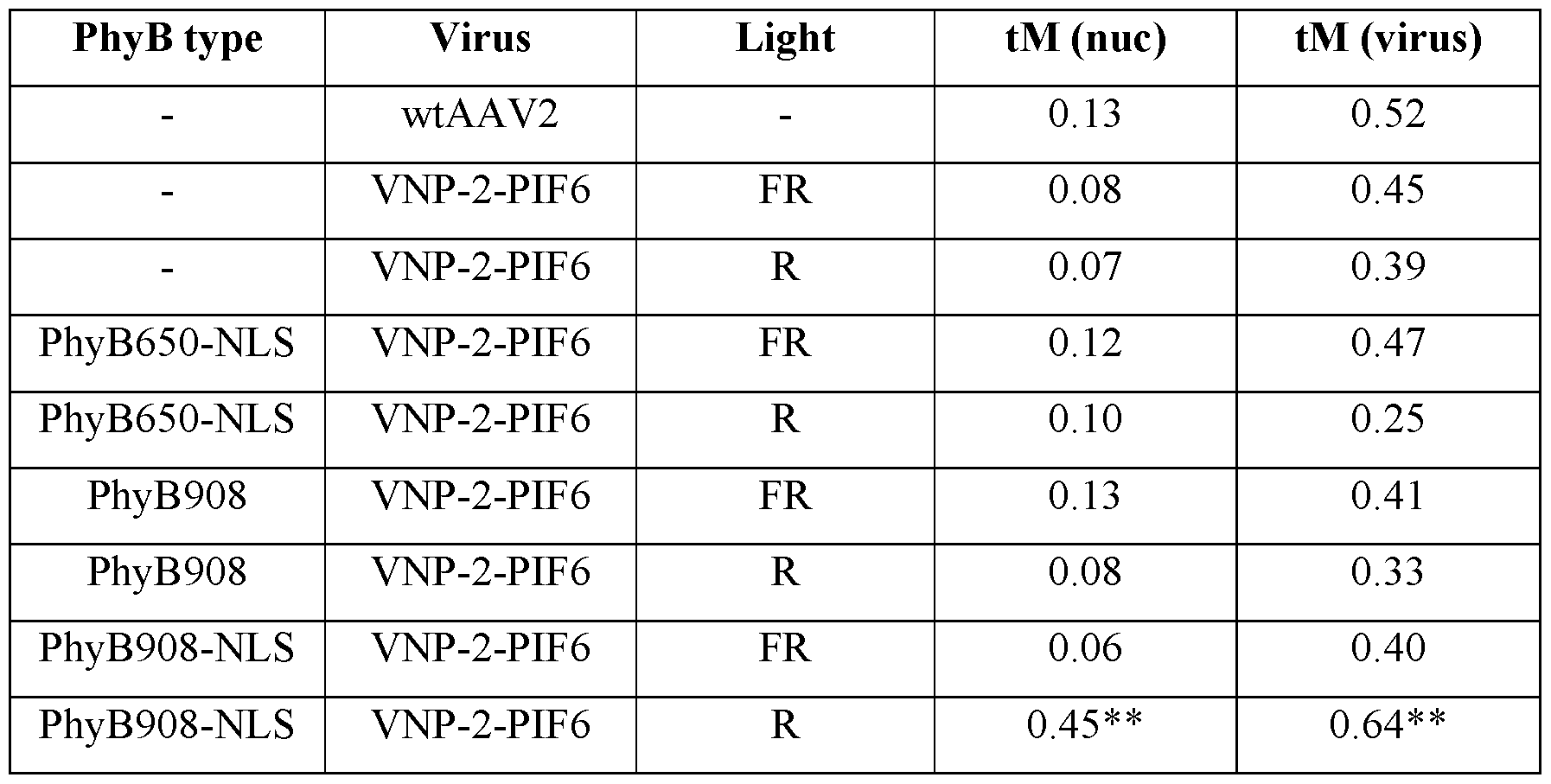
** = Differences between co-localization of VNP-2-PIF6 with PhyB908- LS and R light, and all other conditions are statistically significant (p < 0.05) by unpaired Student' s t-test.
[0164] A similar experiment was performed using PhyB650 (a variant which is analogous to PhyB651) (SEQ ID NO: 32) with or without a C-terminal NLS fusion, however, PhyB650-NLS did not affect the intracellular distribution of VNP-PIF6, potentially due to its lower binding affinity for PIF6 and partial ablation of the PhyB PAS domain which has been shown to result in weak or a complete lack of PhyB binding to PIF6. In addition, it is possible that the C-terminal NLS tag was not recognized by cellular importins due to obstruction or other steric effects. FIGURE 9F shows the colocalization of wtAAV2 and of VNP-PIF6 in cells constitutively expressing PhyB650-NLS under red light and far red light conditions.
[0165] To confirm that the nuclear localization of VNP-2-PIF6 is not a 2-dimensional artifact, three-dimensional Z-stacks were obtained with confocal microscopy. Visualizing cell nuclei slice through the x-, y- and z-axis as shown in FIGURE 10A, and closer inspection of y- axis individual channel slices as shown in FIGURE 10B confirmed higher VNP-2-PIF6 signal inside the nucleus.
[0166] In combination, these data suggest that VNP-2-PIF6 selectively binds to activated (under red light) PhyB908-NLS under physiological conditions, leading to more effective nuclear translocation of the virus as compared to the wtAAV2.
Example 4: Tuning of Gene Delivery by Ratiometric Control of Red/Far Red Light
[0167] Modulating the R:FR light ratio can tune the efficiency of gene delivery. A custom LED-tissue culture plate apparatus as shown in FIGURE 11 A that shields each individual well from outside light was used. An Arduino Uno microcontroller was used to program a 6 x 4 array of optically isolated LEDs (LEDtronics, #L200CWRGB2K-4A-IL; Marubeni: L735-5AU) which can expose cells to 630 nm and 735 nm light simultaneously through the bottom of a 24- well black, glass-bottom tissue culture plate (Greiner bio-one, #662892). LED intensity was quantified and converted from raw Arduino units by placing a fiber optic photodetector probe (StellarNet Inc., photodetector #EPP2000 UVN-SR-25 LT-16, probe #F600-UV-VIS-SR) directly into tissue culture wells and measuring light flux, in units of μιηο1/ιη28, for a range of intensities for R/FR light. The glass bottom of each well of the tissue culture plate was coated with poly-L-lysine and HeLa cells were seeded at a density of lxlO5 cells per well in DMEM supplemented with 10% fetal bovine serum and 1% penicillin-streptomycin. After 24 hours, cells were transfected with PEI-DNA (pKM017 (SEQ ID NO: 118) and pKM216 (SEQ ID NO: 117)) complexes encoding PhyB908 with or without a C-terminal NLS fusion. 24 hours later, under green light, PCB at a final concentration of 15 μΜ and virus (VNP-2-PIF6 or wtAAV2) at an MOI of 2,000 were applied in DMEM supplemented with 10% serum and incubated at 37°C, 5% C02. The LEDs were programmed to shine FR light for 5 minutes before switching to experiment-dependent intensities of R and/or FR light. Cells were harvested and prepared for flow cytometry on a BD FACSCanto II after 24 or 48 hours. The % of cells positive for GFP and the transduction index (TI), from 2 independent experiments, for the cells 24 hours post- transduction for varying ratios of R:FR light are shown in FIGURES 1 IB and 11C. The % of cells positive for GFP and the transduction for the cells 48 hours post-transduction for varying ratios of R:FR light, from 2 independent experiments, are shown in FIGURES 1 ID and 1 IE. As demonstrated, PhyB908 without a NLS has no effect on gene delivery as compared to wtAAV2 (FIGURES 1 ID and 1 IE). However, PhyB908-NLS increased gene delivery as compared to wtAAV2 as the ratio of R:FR light increased and decreased gene delivery as the ratio of R:FR light decreased (FIGURES 11B-11E). Similar results are seen in FIGURES 11F-8G. FIGURE 1 IF depicts fluorescence micrographs of GFP expression in the HeLa cells constitutively expressing PhyB908-NLS and treated with or without VNP-2-PIF6, PCB, and red light. As demonstrated, PCB and red light in combination with VNP-2-PIF6 result in a significant increase
in GFP expression, indicating an increase in transduction. FIGURE 11G shows the discrete transfer functions for transduction of V P-2-PIF6 at red light flux between 0 and 10 μΜ/ηΛ with co-delivery of far red light as well as samples with no PCB, wtAAV2 instead of V P-2- PIF6 with light delivery and wtAAV2 in the dark. The results show increasing transduction with V P-2-PIF6 as the ratio of red light increases. FIGURE 11H shows a dose-response curve for V P-2-PIF6 based on the ratio of R:FR light with the response being measured as transduction index. This curve clearly demonstrates that the gene delivery efficiency of V P-2-PIF6 increases dramatically as the R:FR light ratio increases, exponentially when plotted on a logarithmic scale. The dose-response curve can be fit as TI = AxB + C, where A = 285, B = 0.41, C = 1800 and x is the R:FR light ratio with a r2 value of 0.95.
[0168] Thus, ratiometric control of the R:FR ratio of light can provide a method to tune transduction to increase or decrease gene delivery by increasing red light or far red light, respectively. The maximum level of 17,796 for transduction index was achieved at a R:FR ratio of 15,950 and R:FR ratios above about 250 allow V P-2-PIF6 to more effectively transduce cells than wtAAV2. Further, the greater nuclear entry demonstrated correlates with increased transduction efficiency. In addition, the light-activable viral gene delivery platform can work in other cell types, including those for use in tissue engineering application such as human mesenchymal stem cells (hMSC), human umbilical vein endothelial cells (HUVEC), and 3T3 fibroblasts as show in FIGURE 12 which shows about a 2-fold increase in transduction as compared to a dark control where the cells were treated as described in the foregoing example with either red light at 10.67 μΜ/ηΛ or far red light at 3.61 μΜ/π for 48 hours. The TI values achieved were 167,399 for hMSC, 106,866 for HUVEC and 10,524 for 3T3. Thus, even in a difficult to transduce cell line, 3T3, the light-activable system improved transduction.
[0169] As shown in FIGURE 13 A, above a R:FR ratio of 16,000 the transduction index decreased monotonically. FIGURE 13B shows the maximum transduction index for maximum far red and maximum red lights only. Thus, there may be a useful range of R:FR ratios that may be useful to increase the transduction index as compared to that for wtAAV2 depending on the optogenetic binding partner and protein used, the cell type, the growth conditions and other properties.
Example 5: Spatial Control of Viral Gene Delivery Using R/FR Light
[0170] V P-2-PIF6 can also provide for spatial control of gene delivery which may be an important parameter for achieving therapeutic outcomes. Photomask experiments were conducted following a published protocol for space-resolved gene expression. HeLa cells were cultured in a glass-bottom, poly-L-lysine-coated 24-well plate (Greiner bio-one, #662892) with opaque walls and ceilings. Photomasks were laser-etched into black nitrile sheets using a Universal X-660 laser cutter platform and placed under the wells. The photomask sheet also functioned as a gasket sealing the 24-well plate directly above the R/FR LEDs. HeLa cells were seeded at a density of lxlO5 cells per well in DMEM supplemented with 10% fetal bovine serum and 1%) penicillin-streptomycin. After 24 hours, cells were transfected with PEI-DNA (pKM017 (SEQ ID NO: 118)) complexes encoding PhyB908 with a C-terminal NLS fusion. 24 hours later, under green light, PCB at a final concentration of 15μΜ and virus (VNP-2-PIF6) at an MOI of 1,000 were applied in DMEM supplemented with 10% serum and incubated at 37°C, 5% C02. The LEDs were programmed to shine FR light (2 μπιοΙ/π ) for 30 minutes before switching to experiment-dependent intensities of R or R/FR light for 60 minutes. Cells remained in the dark for the remainder of 48 hours before being fixed with 4% paraformaldehyde in PBS and imaged on a Nikon Al microscope. Images were taken at 20X magnification and a 12x12 square array of images were stitched together. Image signal and brightness were processed in ImageJ using the Threshold function.
[0171] The resulting images are shown in FIGURE 14 and demonstrate spatial control of improved transduction using the VNP-2-PIF6/PhyB908-NLS system. Using only red light (R = 0.5 μπιοΙ/π ; FR = 0.0 μπιοΙ/π ) resulted in high background noise in gene expression even at low flux. However, co-delivering far red light (R = 0.5 μιηο1/ιη28 ; FR = 0.9 μπιοΙ/ηΛ) resulted in improved signal-to-noise and better resolved patterns.
[0172] These results demonstrate that the light-activable viral delivery system can be spatially controlled by limiting the location of exposure to activating light and that co-delivery of R/FR light can improve resolution. Because activation using light can also be controlled by when the light is introduced, the system provides temporal control in addition to spatial control over gene delivery efficiency which provides a powerful tool for not only improving, but controlling, gene delivery.
[0173] More broadly, the foregoing results demonstrate the utility of the optogenetic system for improving and controlling gene delivery using viral vectors using light.
Example 6: Peptide Insertion and Use of Two Enzymatic Cleavage Motifs Adjacent to the Peptide
[0174] In an example, a peptide (AG-PLGLAR-G-DDDK-GA (SEQ ID NO: 27) or AG- DDDDK-G-PLGLAR-GA (SEQ ID NO: 28)) is inserted at amino acid position 586 in the AAV2 capsid which corresponds to position 586 in VP1, position 449 in VP2 and position 383 in VP3. PLGLAR (SEQ ID NO: 17) is a MMP-cleavable peptide motif and DDDDK (SEQ ID NO: 3) is an enterokinase-cleavable domain. Cleavage of the DDDDK (SEQ ID NO: 3)motif allows the PLGLAR (SEQ ID NO: 17) sequence to be displayed as a linearized MMP-cleavable substrate on the surface of the capsid. AG, G, and GA residues serve as linkers and cloning sites to facilitate peptide insertion using conventional molecular cloning methods. The MMP-cleavable motif can be changed from PLGLAR (SEQ ID NO: 17) to any suitable enzymatically cleavable motif or to a peptide of interest such that the peptide of interest is displayed on the surface of the virus but is less conformation constrained because it is only tethered to the virus at one end after pre-treatment with the enterokinase.
Example 7: Peptide Insertion and Use of a Single Enzymatic Cleavage Motif Adjacent to the Peptide and Virus Generation
[0175] In an example, a peptide or protein can be genetically inserted via molecular cloning into the capsid protein sequence paired with a single enterokinase recognition motif either immediately before or after the peptide/protein sequence. The enzymatic cleavage motif, which can include DDDDK (SEQ ID NO: 3), and which is recognized and cleaved by enterokinase, is inserted adjancet to the desired peptide sequence. Plasmids encoding capsid proteins (altered or wild-type), transgene of interest, and helper proteins for virus assembly and packaging are transfected into HEK293T producer cells via polyethylenimine transfection. Cells are collected after 48 hours, lysed, and the virus is separated from cell debris via density gradient ultracentrifugation. Once viruses are made, they are digested (pre-treated) with enterokinase (SEQ ID NO: 76, nucleotide sequence at SEQ ID NO: 75) to linearize and/or conformationally unconstrain the peptide on the surface the capsid. Subsequent column purification with trypsin- inhibitor agarose beads binds the enterokinase to purify the virus sample for downstream use and analysis.
Example 8: Enhancement of Transduction Efficiency By Use of and Enzymatic Cleavage Motif Adjacent to an Inserted Optogenetic Protein
[0176] An AAV-based vims was prepared as described above using only VP1 and VP3 capsid proteins. The LOV domain from Avena sativa phototropin 1 protein with a C-terminal nuclear localization signal (TRPQRDCPTPTWQPQPRRKSW (SEQ ID NO: 6)) and an N- terminal nuclear export signal (MLALKLAGLDI (SEQ ID NO: 10)) was embedded in the capsid protein VP1 adjacent to an enzymatic cleavage motif (DDDDK (SEQ ID NO: 3)) (NES-LOV2- NLS encoded by nucleotide SEQ ID NO: 142, a similar nucleotide with LOV-NLS, lacing a nuclear export signal, can be found at SEQ ID NO: 141). Under blue light of about 450 nm, the LOV domain undergoes a conformational change which exposes the NLS which is otherwise occluded. As in the previously examples, GFP was used as a reporter for transduction. A control group of HeLa cells was not treated with virus. Two experimental groups were treated with the virus at an MOI of 1,000, the first group receiving the virus without pre-treatment with enterokinase, the second group receiving the virus after a 16-18 hour pre-treatment with enterokinase to cleave the enzymatic cleavage motif. Enterokinase (SEQ ID NO: 76, nucleotide sequence at SEQ ID NO: 75) treatment was performed in a 10 μΕ volume of CaCl2 containing 1 μΕ of enterokinase. The control and experimental groups were exposed to blue light of about 470 nm for 12 hours, with four sub-groups within each group receiving 0, 50, 100 or 150 μπιοΙ/π of the blue light. After 48 hours post-transduction, the cells were harvested and analyzed for GFP expression as in the previous examples. The results are shown in FIGURE 15A and demonstrate that pre-treatment with an enzyme to cleave the enzymatic cleavage motif results in improved transduction efficiency, especially at higher intensities of light. FIGURE 15B shows a Western blot of the virus and of wild-type AAV2, with or without pre-treatment with
enterokinase for 16 hours. The results demonstrate that wild-type virus is unaffected by enterokinase treatment and successful incorporation of the LOV domain in VPl of the engineered virus.
[0177] The foregoing description of specific embodiments of the present disclosure has been presented for purpose of illustration and description. The exemplary embodiments were chosen and described in order to best explain the principles of the invention and its practical application, to thereby enable others skilled in the art to best utilize the invention and various embodiments with various modifications are suited to the particular use contemplated.
ADDITIONAL SEQUENCE INFORMATION
[0178] pVP3, which can be used to provide VP3 alone in viral synthesis has a nucleotide sequence of:
1 aattcccatc atcaataata taccttattt tggattgaag ccaatatgat aatgaggggg 61 tggagtttgt gacgtggcgc ggggcgtggg aacggggcgg gtgacgtagt agtctctaga
121 gtcctgtatt agaggtcacg tgagtgtttt gcgacatttt gcgacaccat gtggtcacgc 181 tgggtattta agcccgagtg agcacgcagg gtctccattt tgaagcggga ggtttgaacg
241 cgcagccacc acgccggggt tttacgagat tgtgattaag gtccccagcg accttgacgg
301 gcatctgccc ggcatttctg acagctttgt gaactgggtg gccgagaagg aatgggagtt
361 gccgccagat tctgacatgg atctgaatct gattgagcag gcacccctga ccgtggccga
421 gaagctgcag cgcgactttc tgacggaatg gcgccgtgtg agtaaggccc cggaggccct 481 tttctttgtg caatttgaga agggagagag ctacttccac atgcacgtgc tcgtggaaac
541 caccggggtg aaatccatgg ttttgggacg tttcctgagt cagattcgcg aaaaactgat
601 tcagagaatt taccgcggga tcgagccgac tttgccaaac tggttcgcgg tcacaaagac
661 cagaaatggc gccggaggcg ggaacaaggt ggtggatgag tgctacatcc ccaattactt 721 gctccccaaa acccagcctg agctccagtg ggcgtggact aatatggaac agtatttaag
781 cgcctgtttg aatctcacgg agcgtaaacg gttggtggcg cagcatctga cgcacgtgtc
841 gcagacgcag gagcagaaca aagagaatca gaatcccaat tctgatgcgc cggtgatcag 901 atcaaaaact tcagccaggt acatggagct ggtcgggtgg ctcgtggaca aggggattac
961 ctcggagaag cagtggatcc aggaggacca ggcctcatac atctccttca atgcggcctc
1021 caactcgcgg tcccaaatca aggctgcctt ggacaatgcg ggaaagatta tgagcctgac 1081 taaaaccgcc cccgactacc tggtgggcca gcagcccgtg gaggacattt ccagcaatcg 1141 gatttataaa attttggaac taaacgggta cgatccccaa tatgcggctt ccgtctttct 1201 gggatgggcc acgaaaaagt tcggcaagag gaacaccatc tggctgtttg ggcctgcaac 1261 taccgggaag accaacatcg cggaggccat agcccacact gtgcccttct acgggtgcgt 1321 aaactggacc aatgagaact ttcccttcaa cgactgtgtc gacaagatgg tgatctggtg
1381 ggaggagggg aagatgaccg ccaaggtcgt ggagtcggcc aaagccattc tcggaggaag 1441 caaggtgcgc gtggaccaga aatgcaagtc ctcggcccag atagacccga ctcccgtgat 1501 cgtcacctcc aacaccaaca tgtgcgccgt gattgacggg aactcaacga ccttcgaaca 1561 ccagcagccg ttgcaagacc ggatgttcaa atttgaactc acccgccgtc tggatcatga
1621 ctttgggaag gtcaccaagc aggaagtcaa agactttttc cggtgggcaa aggatcacgt
1681 ggttgaggtg gagcatgaat tctacgtcaa aaagggtgga gccaagaaaa gacccgcccc 1741 cagtgacgca gatataagtg agcccaaacg ggtgcgcgag tcagttgcgc agccatcgac 1801 gtcagacgcg gaagcttcga tcaactacgc agacaggtac caaaacaaat gttctcgtca 1861 cgtgggcatg aatctgatgc tgtttccctg cagacaatgc gagagaatga atcagaattc 1921 aaatatctgc ttcactcacg gacagaaaga ctgtttagag tgctttcccg tgtcagaatc 1981 tcaacccgtt tctgtcgtca aaaaggcgta tcagaaactg tgctacattc atcatatcat 2041 gggaaaggtg ccagacgctt gcactgcctg cgatctggtc aatgtggatt tggatgactg 2101 catctttgaa caataaatga tttaaatcag gtctggctgc cgatggttat cttccagatt 2161 ggctcgagga cactctctct gaaggaataa gacagtggtg gaagctcaaa cctggcccac 2221 caccaccaaa gcccgcagag cggcataagg acgacagcag gggtcttgtg cttcctgggt 2281 acaagtacct cggacccttc aacggactcg acaagggaga gccggtcaac gaggcagacg 2341 ccgcggccct cgagcacgac aaagcctacg accggcagct cgacagcgga gacaacccgt 2401 acctcaagta caaccacgcc gacgcggagt ttcaggagcg ccttaaagaa gatacgtctt 2461 ttgggggcaa cctcggacga gcagtcttcc aggcgaaaaa gagggttctt gaacctctgg 2521 gcctggttga ggaacctgtt aaggcggctc cgggaaaaaa gaggccggta gagcactctc 2581 ctgtggagcc agactcctcc tcgggaaccg gaaaggcggg ccagcagcct gcaagaaaaa 2641 gattgaattt tggtcagact ggagacgcag actcagtacc tgacccccag cctctcggac 2701 agccaccagc agccccctct ggtctgggaa ctaatacgat ggctacaggc agtggcgcac 2761 caatggcaga caataacgag ggcgccgacg gagtgggtaa ttcctcggga aattggcatt 2821 gcgattccac atggatgggc gacagagtca tcaccaccag cacccgaacc tgggccctgc 2881 ccacctacaa caaccacctc tacaaacaaa tttccagcca atcaggagcc tcgaacgaca 2941 atcactactt tggctacagc accccttggg ggtattttga cttcaacaga ttccactgcc 3001 acttttcacc acgtgactgg caaagactca tcaacaacaa ctggggattc cgacccaaga 3061 gactcaactt caagctcttt aacattcaag tcaaagaggt cacgcagaat gacggtacga 3121 cgacgattgc caataacctt accagcacgg ttcaggtgtt tactgactcg gagtaccagc 3181 tcccgtacgt cctcggctcg gcgcatcaag gatgcctccc gccgttccca gcagacgtct 3241 tcatggtgcc acagtatgga tacctcaccc tgaacaacgg gagtcaggca gtaggacgct 3301 cttcatttta ctgcctggag tactttcctt ctcagatgct gcgtaccgga aacaacttta 3361 ccttcagcta cacttttgag gacgttcctt tccacagcag ctacgctcac agccagagtc 3421 tggaccgtct catgaatcct ctcatcgacc agtacctgta ttacttgagc agaacaaaca 3481 ctccaagtgg aaccaccacg cagtcaaggc ttcagttttc tcaggccgga gcgagtgaca
3541 ttcgggacca gtctaggaac tggcttcctg gaccctgtta ccgccagcag cgagtatcaa 3601 agacatctgc ggataacaac aacagtgaat actcgtggac tggagctacc aagtaccacc 3661 tcaatggcag agactctctg gtgaatccgg gcccggccat ggcaagccac aaggacgatg 3721 aagaaaagtt ttttcctcag agcggggttc tcatctttgg gaagcaaggc tcagagaaaa 3781 caaatgtgga cattgaaaag gtcatgatta cagacgaaga ggaaatcagg acaaccaatc 3841 ccgtggctac ggagcagtat ggttctgtat ctaccaacct ccagagaggc aacagacaag 3901 cagctaccgc agatgtcaac acacaaggcg ttcttccagg catggtctgg caggacagag 3961 atgtgtacct tcaggggccc atctgggcaa agattccaca cacggacgga cattttcacc 4021 cctctcccct catgggtgga ttcggactta aacaccctcc tccacagatt ctcatcaaga 4081 acaccccggt acctgcgaat ccttcgacca ccttcagtgc ggcaaagttt gcttccttca 4141 tcacacagta ctccacggga caggtcagcg tggagatcga gtgggagctg cagaaggaaa 4201 acagcaaacg ctggaatccc gaaattcagt acacttccaa ctacaacaag tctgttaatg 4261 tggactttac tgtggacact aatggcgtgt attcagagcc tcgccccatt ggcaccagat 4321 acctgactcg taatctgtaa ttgcttgtta atcaataaac cgtttaattc gtttcagttg 4381 aactttggtc tctgcgtatt tctttcttat ctagtttcca tgctctagag tcctgtatta
4441 gaggtcacgt gagtgttttg cgacattttg cgacaccatg tggtcacgct gggtatttaa 4501 gcccgagtga gcacgcaggg tctccatttt gaagcgggag gtttgaacgc gcagccacca 4561 cggcggggtt ttacgagatt gtgattaagg tccccagcga ccttgacggg catctgcccg 4621 gcatttctga cagctttgtg aactgggtgg ccgagaagga atgggagttg ccgccagatt 4681 ctgacatgga tctgaatctg attgagcagg cacccctgac cgtggccgag aagctgcatc 4741 gctggcgtaa tagcgaagag gcccgcaccg atcgcccttc ccaacagttg cgcagcctga 4801 atggcgaatg gaattccaga cgattgagcg tcaaaatgta ggtatttcca tgagcgtttt 4861 tcctgttgca atggctggcg gtaatattgt tctggatatt accagcaagg ccgatagttt 4921 gagttcttct actcaggcaa gtgatgttat tactaatcaa agaagtattg cgacaacggt 4981 taatttgcgt gatggacaga ctcttttact cggtggcctc actgattata aaaacacttc 5041 tcaggattct ggcgtaccgt tcctgtctaa aatcccttta atcggcctcc tgtttagctc 5101 ccgctctgat tctaacgagg aaagcacgtt atacgtgctc gtcaaagcaa ccatagtacg 5161 cgccctgtag cggcgcatta agcgcggcgg gtgtggtggt tacgcgcagc gtgaccgcta 5221 cacttgccag cgccctagcg cccgctcctt tcgctttctt cccttccttt ctcgccacgt 5281 tcgccggctt tccccgtcaa gctctaaatc gggggctccc tttagggttc cgatttagtg 5341 ctttacggca cctcgacccc aaaaaacttg attagggtga tggttcacgt agtgggccat
5401 cgccctgata gacggttttt cgccctttga cgttggagtc cacgttcttt aatagtggac 5461 tcttgttcca aactggaaca acactcaacc ctatctcggt ctattctttt gatttataag 5521 ggattttgcc gatttcggcc tattggttaa aaaatgagct gatttaacaa aaatttaacg 5581 cgaattttaa caaaatatta acgtttacaa tttaaatatt tgcttataca atcttcctgt 5641 ttttggggct tttctgatta tcaaccgggg tacatatgat tgacatgcta gttttacgat 5701 taccgttcat cgattctctt gtttgctcca gactctcagg caatgacctg atagcctttg 5761 tagagacctc tcaaaaatag ctaccctctc cggcatgaat ttatcagcta gaacggttga 5821 atatcatatt gatggtgatt tgactgtctc cggcctttct cacccgtttg aatctttacc 5881 tacacattac tcaggcattg catttaaaat atatgagggt tctaaaaatt tttatccttg 5941 cgttgaaata aaggcttctc ccgcaaaagt attacagggt cataatgttt ttggtacaac 6001 cgatttagct ttatgctctg aggctttatt gcttaatttt gctaattctt tgccttgcct
6061 gtatgattta ttggatgttg gaattcctga tgcggtattt tctccttacg catctgtgcg 6121 gtatttcaca ccgcatatgg tgcactctca gtacaatctg ctctgatgcc gcatagttaa 6181 gccagccccg acacccgcca acacccgctg acgcgccctg acgggcttgt ctgctcccgg 6241 catccgctta cagacaagct gtgaccgtct ccgggagctg catgtgtcag aggttttcac 6301 cgtcatcacc gaaacgcgcg agacgaaagg gcctcgtgat acgcctattt ttataggtta 6361 atgtcatgat aataatggtt tcttagacgt caggtggcac ttttcgggga aatgtgcgcg 6421 gaacccctat ttgtttattt ttctaaatac attcaaatat gtatccgctc atgagacaat 6481 aaccctgata aatgcttcaa taatattgaa aaaggaagag tatgagtatt caacatttcc 6541 gtgtcgccct tattcccttt tttgcggcat tttgccttcc tgtttttgct cacccagaaa 6601 cgctggtgaa agtaaaagat gctgaagatc agttgggtgc acgagtgggt tacatcgaac 6661 tggatctcaa cagcggtaag atccttgaga gttttcgccc cgaagaacgt tttccaatga 6721 tgagcacttt taaagttctg ctatgtggcg cggtattatc ccgtattgac gccgggcaag 6781 agcaactcgg tcgccgcata cactattctc agaatgactt ggttgagtac tcaccagtca 6841 cagaaaagca tcttacggat ggcatgacag taagagaatt atgcagtgct gccataacca 6901 tgagtgataa cactgcggcc aacttacttc tgacaacgat cggaggaccg aaggagctaa 6961 ccgctttttt gcacaacatg ggggatcatg taactcgcct tgatcgttgg gaaccggagc 7021 tgaatgaagc cataccaaac gacgagcgtg acaccacgat gcctgtagca atggcaacaa 7081 cgttgcgcaa actattaact ggcgaactac ttactctagc ttcccggcaa caattaatag 7141 actggatgga ggcggataaa gttgcaggac cacttctgcg ctcggccctt ccggctggct 7201 ggtttattgc tgataaatct ggagccggtg agcgtgggtc tcgcggtatc attgcagcac
7261 tggggccaga tggtaagccc tcccgtatcg tagttatcta cacgacgggg agtcaggcaa 7321 ctatggatga acgaaataga cagatcgctg agataggtgc ctcactgatt aagcattggt 7381 aactgtcaga ccaagtttac tcatatatac tttagattga tttaaaactt catttttaat
7441 ttaaaaggat ctaggtgaag atcctttttg ataatctcat gaccaaaatc ccttaacgtg 7501 agttttcgtt ccactgagcg tcagaccccg tagaaaagat caaaggatct tcttgagatc 7561 ctttttttct gcgcgtaatc tgctgcttgc aaacaaaaaa accaccgcta ccagcggtgg 7621 tttgtttgcc ggatcaagag ctaccaactc tttttccgaa ggtaactggc ttcagcagag 7681 cgcagatacc aaatactgtc cttctagtgt agccgtagtt aggccaccac ttcaagaact 7741 ctgtagcacc gcctacatac ctcgctctgc taatcctgtt accagtggct gctgccagtg 7801 gcgataagtc gtgtcttacc gggttggact caagacgata gttaccggat aaggcgcagc 7861 ggtcgggctg aacggggggt tcgtgcacac agcccagctt ggagcgaacg acctacaccg 7921 aactgagata cctacagcgt gagctatgag aaagcgccac gcttcccgaa gggagaaagg 7981 cggacaggta tccggtaagc ggcagggtcg gaacaggaga gcgcacgagg gagcttccag 8041 ggggaaacgc ctggtatctt tatagtcctg tcgggtttcg ccacctctga cttgagcgtc 8101 gatttttgtg atgctcgtca ggggggcgga gcctatggaa aaacgccagc aacgcggcct 8161 ttttacggtt cctggccttt tgctggcctt ttgctcacat gttctttcct gcgttatccc
8221 ctgattctgt ggataaccgt attaccgcct ttgagtgagc tgataccgct cgccgcagcc 8281 gaacgaccga gcgcagcgag tcagtgagcg aggaagcgga agagcgccca atacgcaaac 8341 cgcctctccc cgcgcgttgg ccgattcatt aatgca
Claims
1. A virus comprising a capsid protein and an optogenetic binding partner, wherein at least a portion of the optogenetic binding partner is displayed on the surface of the virus, and wherein the optogenetic binding partner is linked to the capsid protein by a direct amino acid linkage or a linker.
2. The virus of claim 1, wherein the amino acid sequence of the optogenetic binding partner is embedded within the amino acid sequence of the capsid protein.
3. The virus of claim 1, wherein the amino acid sequence of the optogenetic binding partner is adjacent to the amino acid sequence of the capsid protein.
4. The virus of claim 1, further comprising at least one linker between the N- terminus of the amino acid sequence of the optogenetic binding partner and the amino acid sequence of the capsid protein or between the C-terminus of the amino acid sequence of the optogenetic binding partner and the amino acid sequence of the capsid protein.
5. The virus of claim 1, further comprising a first linker between the N-terminus of the amino acid sequence of the optogenetic binding partner and the amino acid sequence of the capsid protein and a second linker between the C-terminus of the amino acid sequence of the optogenetic binding partner and the amino acid sequence of the capsid protein.
6. The virus of claim 2, further comprising at least one linker between the N- terminus of the amino acid sequence of the optogenetic binding partner and the amino acid sequence of the capsid protein or between the C-terminus of the amino acid sequence of the optogenetic binding partner and the amino acid sequence of the capsid protein.
7. The virus of claim 2, further comprising a first linker between the N-terminus of the amino acid sequence of the optogenetic binding partner and the amino acid sequence of the capsid protein and a second linker between the C-terminus of the amino acid sequence of the optogenetic binding partner and the amino acid sequence of the capsid protein.
8. The virus of claim 3, further comprising at least one linker between the N- terminus of the amino acid sequence of the optogenetic binding partner and the amino acid sequence of the capsid protein or between the C-terminus of the amino acid sequence of the optogenetic binding partner and the amino acid sequence of the capsid protein.
9. The virus of any one of claims 4-8, wherein the amino acid sequence of the linker is selected from the group consisting of AG, GA, G, GGS, (GGS)n, GnS, wherein n is an integer number.
10. The virus of any one of claims 1-8, wherein the virus is an adeno-associated virus
(AAV).
11. The virus of claim 9, wherein the virus is an adeno-associated virus (AAV).
12. The virus of claim 10, wherein the virus is an adeno-associated virus selected from the group consisting of serotypes 1, 2, 3, 4, 5, 6, 7, 8, 9, 10, 11 and 12.
13. The virus of claim 11, wherein the virus is an adeno-associated virus selected from the group consisting of serotypes 1, 2, 3, 4, 5, 6, 7, 8, 9, 10, 11 and 12.
14. The virus of claim 12, wherein the virus is an adeno-associated virus of serotype 2 (AAV2).
15. The virus of claim 13, wherein the virus is an adeno-associated virus of serotype 2 (AAV2).
16. The virus of claim 14, wherein the capsid protein is selected from the group consisting of VPl (SEQ ID NO: 50), VP2 (SEQ ID NO: 52), VP3 (SEQ ID NO: 54), portions thereof, variants thereof and combinations thereof.
17. The virus of claim 15, wherein the capsid protein is selected from the group consisting of VPl (SEQ ID NO: 50), VP2 (SEQ ID NO: 52), VP3 (SEQ ID NO: 54), portions thereof, variants thereof and combinations thereof.
18. The virus of claim 14, wherein the capsid protein comprises at least a portion of the amino acid sequence of VPl (SEQ ID NO: 50).
19. The virus of claim 14, wherein the capsid protein comprises at least a portion of the amino acid sequence of VP2 (SEQ ID NO: 52).
20. The virus of claim 15, wherein the capsid protein comprises at least a portion of the amino acid sequence of VPl (SEQ ID NO: 50).
21. The virus of claim 15, wherein the capsid protein comprises at least a portion of the amino acid sequence of VP2 (SEQ ID NO: 52).
22. The virus of any one of claims 1-8, wherein the optogenetic binding partner is selected from the group consisting of phytochrome interacting factor 1, phytochrome interacting factor 2, phytochrome interacting factor 3, phytochrome interacting factor 4, phytochrome interacting factor 5, and phytochrome interacting factor 6, portions thereof and variants thereof.
23. The virus of claim 9, wherein the optogenetic binding partner is selected from the group consisting of phytochrome interacting factor 1, phytochrome interacting factor 2, phytochrome interacting factor 3, phytochrome interacting factor 4, phytochrome interacting factor 5, and phytochrome interacting factor 6, portions thereof and variants thereof.
24. The virus of claim 10, wherein the optogenetic binding partner is selected from the group consisting of phytochrome interacting factor 1, phytochrome interacting factor 2, phytochrome interacting factor 3, phytochrome interacting factor 4, phytochrome interacting factor 5, and phytochrome interacting factor 6, portions thereof and variants thereof.
25. The virus of claim 11, wherein the optogenetic binding partner is selected from the group consisting of phytochrome interacting factor 1, phytochrome interacting factor 2, phytochrome interacting factor 3, phytochrome interacting factor 4, phytochrome interacting factor 5, and phytochrome interacting factor 6, portions thereof and variants thereof.
26. The virus of claim 12, wherein the optogenetic binding partner is selected from the group consisting of phytochrome interacting factor 1, phytochrome interacting factor 2, phytochrome interacting factor 3, phytochrome interacting factor 4, phytochrome interacting factor 5, and phytochrome interacting factor 6, portions thereof and variants thereof.
27. The virus of claim 13, wherein the optogenetic binding partner is selected from the group consisting of phytochrome interacting factor 1, phytochrome interacting factor 2, phytochrome interacting factor 3, phytochrome interacting factor 4, phytochrome interacting factor 5, and phytochrome interacting factor 6, portions thereof and variants thereof.
28. The virus of claim 14, wherein the optogenetic binding partner is selected from the group consisting of phytochrome interacting factor 1, phytochrome interacting factor 2, phytochrome interacting factor 3, phytochrome interacting factor 4, phytochrome interacting factor 5, and phytochrome interacting factor 6, portions thereof and variants thereof.
29. The virus of claim 15, wherein the optogenetic binding partner is selected from the group consisting of phytochrome interacting factor 1, phytochrome interacting factor 2, phytochrome interacting factor 3, phytochrome interacting factor 4, phytochrome interacting factor 5, and phytochrome interacting factor 6, portions thereof and variants thereof.
30. The virus of claim 16, wherein the optogenetic binding partner is selected from the group consisting of phytochrome interacting factor 1, phytochrome interacting factor 2, phytochrome interacting factor 3, phytochrome interacting factor 4, phytochrome interacting factor 5, and phytochrome interacting factor 6, portions thereof and variants thereof.
31. The virus of claim 17, wherein the optogenetic binding partner is selected from the group consisting of phytochrome interacting factor 1, phytochrome interacting factor 2, phytochrome interacting factor 3, phytochrome interacting factor 4, phytochrome interacting factor 5, and phytochrome interacting factor 6, portions thereof and variants thereof.
32. The virus of claim 18, wherein the optogenetic binding partner is selected from the group consisting of phytochrome interacting factor 1, phytochrome interacting factor 2, phytochrome interacting factor 3, phytochrome interacting factor 4, phytochrome interacting factor 5, and phytochrome interacting factor 6, portions thereof and variants thereof.
33. The virus of claim 19, wherein the optogenetic binding partner is selected from the group consisting of phytochrome interacting factor 1, phytochrome interacting factor 2, phytochrome interacting factor 3, phytochrome interacting factor 4, phytochrome interacting factor 5, and phytochrome interacting factor 6, portions thereof and variants thereof.
34. The virus of claim 20, wherein the optogenetic binding partner is selected from the group consisting of phytochrome interacting factor 1, phytochrome interacting factor 2,
phytochrome interacting factor 3, phytochrome interacting factor 4, phytochrome interacting factor 5, and phytochrome interacting factor 6, portions thereof and variants thereof.
35. The virus of claim 21, wherein the optogenetic binding partner is selected from the group consisting of phytochrome interacting factor 1, phytochrome interacting factor 2, phytochrome interacting factor 3, phytochrome interacting factor 4, phytochrome interacting factor 5, and phytochrome interacting factor 6, portions thereof and variants thereof.
36. The virus of any one of claims 1-8, wherein the optogenetic binding partner comprises the amino acid sequence of phytochrome interacting factor 6 (PIF6), a portion thereof or a variant thereof.
37. The virus of claim 9, wherein the optogenetic binding partner comprises the amino acid sequence of phytochrome interacting factor 6 (PIF6), a portion thereof or a variant thereof.
38. The virus of claim 9, wherein the optogenetic binding partner comprises the amino acid sequence of phytochrome interacting factor 6 (PIF6), a portion thereof or a variant thereof.
39. The virus of claim 10, wherein the optogenetic binding partner comprises the amino acid sequence of phytochrome interacting factor 6 (PIF6), a portion thereof or a variant thereof.
40. The virus of claim 11, wherein the optogenetic binding partner comprises the amino acid sequence of phytochrome interacting factor 6 (PIF6), a portion thereof or a variant thereof.
41. The virus of claim 12, wherein the optogenetic binding partner comprises the amino acid sequence of phytochrome interacting factor 6 (PIF6), a portion thereof or a variant thereof.
42. The virus of claim 13, wherein the optogenetic binding partner comprises the amino acid sequence of phytochrome interacting factor 6 (PIF6), a portion thereof or a variant thereof.
43. The virus of claim 14, wherein the optogenetic binding partner comprises the amino acid sequence of phytochrome interacting factor 6 (PIF6), a portion thereof or a variant thereof.
44. The virus of claim 15, wherein the optogenetic binding partner comprises the amino acid sequence of phytochrome interacting factor 6 (PIF6), a portion thereof or a variant thereof.
45. The virus of claim 16, wherein the optogenetic binding partner comprises the amino acid sequence of phytochrome interacting factor 6 (PIF6), a portion thereof or a variant thereof.
46. The virus of claim 17, wherein the optogenetic binding partner comprises the amino acid sequence of phytochrome interacting factor 6 (PIF6), a portion thereof or a variant thereof.
47. The virus of claim 18, wherein the optogenetic binding partner comprises the amino acid sequence of phytochrome interacting factor 6 (PIF6), a portion thereof or a variant thereof.
48. The virus of claim 19, wherein the optogenetic binding partner comprises the amino acid sequence of phytochrome interacting factor 6 (PIF6), a portion thereof or a variant thereof.
49. The virus of claim 20, wherein the optogenetic binding partner comprises the amino acid sequence of phytochrome interacting factor 6 (PIF6), a portion thereof or a variant thereof.
50. The virus of claim 21, wherein the optogenetic binding partner comprises the amino acid sequence of phytochrome interacting factor 6 (PIF6), a portion thereof or a variant thereof.
51. The virus of any one of claims 1, 3-5 or 8, wherein the virus is an adeno- associated virus of serotype 2 (AAV2), and wherein the capsid protein comprises VP2 (SEQ ID NO: 52), and wherein the optogenetic binding partner is adjacent to the N-terminus of the amino acid sequence of VP2 or is inserted at G316 in the amino acid sequence of SEQ ID NO: 52.
52. The virus of claim 51, wherein the optogenetic binding partner is selected from the group consisting of phytochrome interacting factor 1, phytochrome interacting factor 2, phytochrome interacting factor 3, phytochrome interacting factor 4, phytochrome interacting factor 5, and phytochrome interacting factor 6, portions thereof and variants thereof.
53. The virus of claim 51, wherein the optogenetic binding partner comprises the amino acid sequence of phytochrome interacting factor 6 (PIF6), a portion thereof or a variant thereof.
54. The virus of claim 53, wherein the virus comprises the amino acid sequence of SEQ ID NO: 46 (VNP-2-PIF6).
55. The virus of any one of claims 2 or 6-7, wherein the virus is an adeno-associated virus of serotype 2 (AAV2), and wherein the capsid protein comprises VPl (SEQ ID NO: 50), and wherein the optogenetic binding partner is inserted at M138 or G453 of SEQ ID NO: 50.
56. The virus of claim 55, wherein the optogenetic binding partner is selected from the group consisting of phytochrome interacting factor 1, phytochrome interacting factor 2, phytochrome interacting factor 3, phytochrome interacting factor 4, phytochrome interacting factor 5, and phytochrome interacting factor 6, portions thereof and variants thereof.
57. The virus of claim 55, wherein the optogenetic binding partner comprises the amino acid sequence of phytochrome interacting factor 6 (PIF6), a portion thereof or a variant thereof.
58. The virus of claim 57, wherein the virus comprises the amino acid sequence of SEQ ID NO: 44 (VNP-1-PIF6).
59. A virus comprising an amino acid sequence (VNP-1,2-PIF6) having an amino acid sequence encoded by the sequence of SEQ ID NO: 149, wherein the virus is an adeno- associated virus of serotype 2.
60. The virus of any one of claims 2 or 6-7, wherein the virus is an adeno-associated virus of serotype 2 (AAV2), and wherein the capsid protein comprises VP3 (SEQ ID NO: 54), and wherein the optogenetic binding partner is inserted at G250 in the amino acid sequence of SEQ ID NO: 54.
61. The virus of any one of claim 1-8, further comprising a nucleic acid molecule
62. The virus of claim 9, further comprising a nucleic acid molecule.
63. The virus of claim 10, further comprising a nucleic acid molecule.
64. The virus of claim 11, further comprising a nucleic acid molecule.
65. The virus of claim 12, further comprising a nucleic acid molecule.
66. The virus of claim 13, further comprising a nucleic acid molecule.
67. The virus of claim 14, further comprising a nucleic acid molecule.
68. The virus of claim 15, further comprising a nucleic acid molecule.
69. The virus of claim 16, further comprising a nucleic acid molecule.
70. The virus of claim 17, further comprising a nucleic acid molecule.
71. The virus of claim 18, further comprising a nucleic acid molecule.
72. The virus of claim 19, further comprising a nucleic acid molecule.
73. The virus of claim 20, further comprising a nucleic acid molecule.
74. The virus of claim 21, further comprising a nucleic acid molecule.
75. The virus of claim 22, further comprising a nucleic acid molecule.
76. The virus of claim 23, further comprising a nucleic acid molecule.
77. The virus of claim 24. further comprising a nucleic acid molecule,
78. The virus of claim 25. further comprising a nucleic acid molecule,
79. The virus of claim 26. further comprising a nucleic acid molecule,
80. The virus of claim 27. further comprising a nucleic acid molecule,
81. The virus of claim 28. further comprising a nucleic acid molecule,
82. The virus of claim 29. further comprising a nucleic acid molecule,
83. The virus of claim 30. further comprising a nucleic acid molecule,
84. The virus of claim 31. further comprising a nucleic acid molecule,
85. The virus of claim 32. further comprising a nucleic acid molecule,
86. The virus of claim 33. further comprising a nucleic acid molecule,
87. The virus of claim 34. further comprising a nucleic acid molecule,
88. The virus of claim 35. further comprising a nucleic acid molecule,
89. The virus of claim 36. further comprising a nucleic acid molecule,
90. The virus of claim 37. further comprising a nucleic acid molecule,
91. The virus of claim 38. further comprising a nucleic acid molecule,
92. The virus of claim 39. further comprising a nucleic acid molecule,
93. The virus of claim 40. further comprising a nucleic acid molecule,
94. The virus of claim 41. further comprising a nucleic acid molecule,
95. The virus of claim 42. further comprising a nucleic acid molecule,
96. The virus of claim 43. further comprising a nucleic acid molecule,
97. The virus of claim 44. further comprising a nucleic acid molecule,
98. The virus of claim 45. further comprising a nucleic acid molecule,
99. The virus of claim 46. further comprising a nucleic acid molecule,
100. The virus of claim 47. further comprising a nucleic acid molecule,
101. The virus of claim 48. further comprising a nucleic acid molecule.
102. The virus of claim 49, further comprising a nucleic acid molecule.
103. The virus of claim 50, further comprising a nucleic acid molecule.
104. The virus of claim 51, further comprising a nucleic acid molecule.
105. The virus of claim 52, further comprising a nucleic acid molecule.
106. The virus of claim 53, further comprising a nucleic acid molecule.
107. The virus of claim 54, further comprising a nucleic acid molecule.
108. The virus of claim 55, further comprising a nucleic acid molecule.
109. The virus of claim 56, further comprising a nucleic acid molecule.
110. The virus of claim 57, further comprising a nucleic acid molecule.
111. The virus of claim 58, further comprising a nucleic acid molecule.
112. The virus of claim 59, further comprising a nucleic acid molecule.
113. The virus of claim 60, further comprising a nucleic acid molecule.
114. The virus of claim 61, wherein the nucleic acid molecule is a therapeutic nucleic acid molecule.
115. The virus of claim 62, wherein the nucleic acid molecule is a therapeutic nucleic acid molecule.
116. The virus of claim 63, wherein the nucleic acid molecule is a therapeutic nucleic acid molecule.
117. The virus of claim 64, wherein the nucleic acid molecule is a therapeutic nucleic acid molecule.
118. The virus of claim 65, wherein the nucleic acid molecule is a therapeutic nucleic acid molecule.
119. The virus of claim 66, wherein the nucleic acid molecule is a therapeutic nucleic acid molecule.
120. The virus of claim 67, wherein the nucleic acid molecule is a therapeutic nucleic acid molecule.
121. The virus of claim 68, wherein the nucleic acid molecule is a therapeutic nucleic acid molecule.
122. The virus of claim 69, wherein the nucleic acid molecule is a therapeutic nucleic acid molecule.
123. The virus of claim 70, wherein the nucleic acid molecule is a therapeutic nucleic acid molecule.
124. The virus of claim 71, wherein the nucleic acid molecule is a therapeutic nucleic acid molecule.
125. The virus of claim 72, wherein the nucleic acid molecule is a therapeutic nucleic acid molecule.
126. The virus of claim 73, wherein the nucleic acid molecule is a therapeutic nucleic acid molecule.
127. The virus of claim 74, wherein the nucleic acid molecule is a therapeutic nucleic acid molecule.
128. The virus of claim 75, wherein the nucleic acid molecule is a therapeutic nucleic acid molecule.
129. The virus of claim 76, wherein the nucleic acid molecule is a therapeutic nucleic acid molecule.
130. The virus of claim 77, wherein the nucleic acid molecule is a therapeutic nucleic acid molecule.
131. The virus of claim 78, wherein the nucleic acid molecule is a therapeutic nucleic acid molecule.
132. The virus of claim 79, wherein the nucleic acid molecule is a therapeutic nucleic acid molecule.
133. The virus of claim 80, wherein the nucleic acid molecule is a therapeutic nucleic acid molecule.
134. The virus of claim 81, wherein the nucleic acid molecule is a therapeutic nucleic acid molecule.
135. The virus of claim 82, wherein the nucleic acid molecule is a therapeutic nucleic acid molecule.
136. The virus of claim 83, wherein the nucleic acid molecule is a therapeutic nucleic acid molecule.
137. The virus of claim 84, wherein the nucleic acid molecule is a therapeutic nucleic acid molecule.
138. The virus of claim 85, wherein the nucleic acid molecule is a therapeutic nucleic acid molecule.
139. The virus of claim 86, wherein the nucleic acid molecule is a therapeutic nucleic acid molecule.
140. The virus of claim 87, wherein the nucleic acid molecule is a therapeutic nucleic acid molecule.
141. The virus of claim 88, wherein the nucleic acid molecule is a therapeutic nucleic acid molecule.
142. The virus of claim 89, wherein the nucleic acid molecule is a therapeutic nucleic acid molecule.
143. The virus of claim 90, wherein the nucleic acid molecule is a therapeutic nucleic acid molecule.
144. The virus of claim 91, wherein the nucleic acid molecule is a therapeutic nucleic acid molecule.
145. The virus of claim 92, wherein the nucleic acid molecule is a therapeutic nucleic acid molecule.
146. The virus of claim 93, wherein the nucleic acid molecule is a therapeutic nucleic acid molecule.
147. The virus of claim 94, wherein the nucleic acid molecule is a therapeutic nucleic acid molecule.
148. The virus of claim 95, wherein the nucleic acid molecule is a therapeutic nucleic acid molecule.
149. The virus of claim 96, wherein the nucleic acid molecule is a therapeutic nucleic acid molecule.
150. The virus of claim 97, wherein the nucleic acid molecule is a therapeutic nucleic acid molecule.
151. The virus of claim 98, wherein the nucleic acid molecule is a therapeutic nucleic acid molecule.
152. The virus of claim 99, wherein the nucleic acid molecule is a therapeutic nucleic acid molecule.
153. The virus of claim 100, wherein the nucleic acid molecule is a therapeutic nucleic acid molecule.
154. The virus of claim 101, wherein the nucleic acid molecule is a therapeutic nucleic acid molecule.
155. The virus of claim 102, wherein the nucleic acid molecule is a therapeutic nucleic acid molecule.
156. The virus of claim 103, wherein the nucleic acid molecule is a therapeutic nucleic acid molecule.
157. The virus of claim 104, wherein the nucleic acid molecule is a therapeutic nucleic acid molecule.
158. The virus of claim 105, wherein the nucleic acid molecule is a therapeutic nucleic acid molecule.
159. The virus of claim 106, wherein the nucleic acid molecule is a therapeutic nucleic acid molecule.
160. The virus of claim 107, wherein the nucleic acid molecule is a therapeutic nucleic acid molecule.
161. The virus of claim 108, wherein the nucleic acid molecule is a therapeutic nucleic acid molecule.
162. The virus of claim 109, wherein the nucleic acid molecule is a therapeutic nucleic acid molecule.
163. The virus of claim 110, wherein the nucleic acid molecule is a therapeutic nucleic acid molecule.
164. The virus of claim 111, wherein the nucleic acid molecule is a therapeutic nucleic acid molecule.
165. The virus of claim 112, wherein the nucleic acid molecule is a therapeutic nucleic acid molecule.
166. The virus of claim 113, wherein the nucleic acid molecule is a therapeutic nucleic acid molecule.
167. The virus of claim 61, wherein the nucleic acid molecule comprises a nucleotide sequence selected from the group consisting of a gene, a portion of a gene, RNA interference and a CRISPR/Cas genome editing tool.
168. The virus of claim 62, wherein the nucleic acid molecule comprises a nucleotide sequence selected from the group consisting of a gene, a portion of a gene, RNA interference and a CRISPR/Cas genome editing tool.
169. The virus of claim 63, wherein the nucleic acid molecule comprises a nucleotide sequence selected from the group consisting of a gene, a portion of a gene, RNA interference and a CRISPR/Cas genome editing tool.
170. The virus of claim 64, wherein the nucleic acid molecule comprises a nucleotide sequence selected from the group consisting of a gene, a portion of a gene, RNA interference and a CRISPR/Cas genome editing tool.
171. The virus of claim 65, wherein the nucleic acid molecule comprises a nucleotide sequence selected from the group consisting of a gene, a portion of a gene, RNA interference and a CRISPR/Cas genome editing tool.
172. The virus of claim 66, wherein the nucleic acid molecule comprises a nucleotide sequence selected from the group consisting of a gene, a portion of a gene, RNA interference and a CRISPR/Cas genome editing tool.
173. The virus of claim 67, wherein the nucleic acid molecule comprises a nucleotide sequence selected from the group consisting of a gene, a portion of a gene, RNA interference and a CRISPR/Cas genome editing tool.
174. The virus of claim 68, wherein the nucleic acid molecule comprises a nucleotide sequence selected from the group consisting of a gene, a portion of a gene, RNA interference and a CRISPR/Cas genome editing tool.
175. The virus of claim 69, wherein the nucleic acid molecule comprises a nucleotide sequence selected from the group consisting of a gene, a portion of a gene, RNA interference and a CRISPR/Cas genome editing tool.
176. The virus of claim 70, wherein the nucleic acid molecule comprises a nucleotide sequence selected from the group consisting of a gene, a portion of a gene, RNA interference and a CRISPR/Cas genome editing tool.
177. The virus of claim 71, wherein the nucleic acid molecule comprises a nucleotide sequence selected from the group consisting of a gene, a portion of a gene, RNA interference and a CRISPR/Cas genome editing tool.
178. The virus of claim 72, wherein the nucleic acid molecule comprises a nucleotide sequence selected from the group consisting of a gene, a portion of a gene, RNA interference and a CRISPR/Cas genome editing tool.
179. The virus of claim 73, wherein the nucleic acid molecule comprises a nucleotide sequence selected from the group consisting of a gene, a portion of a gene, RNA interference and a CRISPR/Cas genome editing tool.
180. The virus of claim 74, wherein the nucleic acid molecule comprises a nucleotide sequence selected from the group consisting of a gene, a portion of a gene, RNA interference and a CRISPR/Cas genome editing tool.
181. The virus of claim 75, wherein the nucleic acid molecule comprises a nucleotide sequence selected from the group consisting of a gene, a portion of a gene, RNA interference and a CRISPR/Cas genome editing tool.
182. The virus of claim 76, wherein the nucleic acid molecule comprises a nucleotide sequence selected from the group consisting of a gene, a portion of a gene, RNA interference and a CRISPR/Cas genome editing tool.
183. The virus of claim 77, wherein the nucleic acid molecule comprises a nucleotide sequence selected from the group consisting of a gene, a portion of a gene, RNA interference and a CRISPR/Cas genome editing tool.
184. The virus of claim 78, wherein the nucleic acid molecule comprises a nucleotide sequence selected from the group consisting of a gene, a portion of a gene, RNA interference and a CRISPR/Cas genome editing tool.
185. The virus of claim 79, wherein the nucleic acid molecule comprises a nucleotide sequence selected from the group consisting of a gene, a portion of a gene, RNA interference and a CRISPR/Cas genome editing tool.
186. The virus of claim 80, wherein the nucleic acid molecule comprises a nucleotide sequence selected from the group consisting of a gene, a portion of a gene, RNA interference and a CRISPR/Cas genome editing tool.
187. The virus of claim 81, wherein the nucleic acid molecule comprises a nucleotide sequence selected from the group consisting of a gene, a portion of a gene, RNA interference and a CRISPR/Cas genome editing tool.
188. The virus of claim 82, wherein the nucleic acid molecule comprises a nucleotide sequence selected from the group consisting of a gene, a portion of a gene, RNA interference and a CRISPR/Cas genome editing tool.
189. The virus of claim 83, wherein the nucleic acid molecule comprises a nucleotide sequence selected from the group consisting of a gene, a portion of a gene, RNA interference and a CRISPR/Cas genome editing tool.
190. The virus of claim 84, wherein the nucleic acid molecule comprises a nucleotide sequence selected from the group consisting of a gene, a portion of a gene, RNA interference and a CRISPR/Cas genome editing tool.
191. The virus of claim 85, wherein the nucleic acid molecule comprises a nucleotide sequence selected from the group consisting of a gene, a portion of a gene, RNA interference and a CRISPR/Cas genome editing tool.
192. The virus of claim 86, wherein the nucleic acid molecule comprises a nucleotide sequence selected from the group consisting of a gene, a portion of a gene, RNA interference and a CRISPR/Cas genome editing tool.
193. The virus of claim 87, wherein the nucleic acid molecule comprises a nucleotide sequence selected from the group consisting of a gene, a portion of a gene, RNA interference and a CRISPR/Cas genome editing tool.
194. The virus of claim 88, wherein the nucleic acid molecule comprises a nucleotide sequence selected from the group consisting of a gene, a portion of a gene, RNA interference and a CRISPR/Cas genome editing tool.
195. The virus of claim 89, wherein the nucleic acid molecule comprises a nucleotide sequence selected from the group consisting of a gene, a portion of a gene, RNA interference and a CRISPR/Cas genome editing tool.
196. The virus of claim 90, wherein the nucleic acid molecule comprises a nucleotide sequence selected from the group consisting of a gene, a portion of a gene, RNA interference and a CRISPR/Cas genome editing tool.
197. The virus of claim 91, wherein the nucleic acid molecule comprises a nucleotide sequence selected from the group consisting of a gene, a portion of a gene, RNA interference and a CRISPR/Cas genome editing tool.
198. The virus of claim 92, wherein the nucleic acid molecule comprises a nucleotide sequence selected from the group consisting of a gene, a portion of a gene, RNA interference and a CRISPR/Cas genome editing tool.
199. The virus of claim 93, wherein the nucleic acid molecule comprises a nucleotide sequence selected from the group consisting of a gene, a portion of a gene, RNA interference and a CRISPR/Cas genome editing tool.
200. The virus of claim 94, wherein the nucleic acid molecule comprises a nucleotide sequence selected from the group consisting of a gene, a portion of a gene, RNA interference and a CRISPR/Cas genome editing tool.
201. The virus of claim 95, wherein the nucleic acid molecule comprises a nucleotide sequence selected from the group consisting of a gene, a portion of a gene, RNA interference and a CRISPR/Cas genome editing tool.
202. The virus of claim 96, wherein the nucleic acid molecule comprises a nucleotide sequence selected from the group consisting of a gene, a portion of a gene, RNA interference and a CRISPR/Cas genome editing tool.
203. The virus of claim 97, wherein the nucleic acid molecule comprises a nucleotide sequence selected from the group consisting of a gene, a portion of a gene, RNA interference and a CRISPR/Cas genome editing tool.
204. The virus of claim 98, wherein the nucleic acid molecule comprises a nucleotide sequence selected from the group consisting of a gene, a portion of a gene, RNA interference and a CRISPR/Cas genome editing tool.
205. The virus of claim 99, wherein the nucleic acid molecule comprises a nucleotide sequence selected from the group consisting of a gene, a portion of a gene, RNA interference and a CRISPR/Cas genome editing tool.
206. The virus of claim 100, wherein the nucleic acid molecule comprises a nucleotide sequence selected from the group consisting of a gene, a portion of a gene, RNA interference and a CRISPR/Cas genome editing tool.
207. The virus of claim 101, wherein the nucleic acid molecule comprises a nucleotide sequence selected from the group consisting of a gene, a portion of a gene, RNA interference and a CRISPR/Cas genome editing tool.
208. The virus of claim 102, wherein the nucleic acid molecule comprises a nucleotide sequence selected from the group consisting of a gene, a portion of a gene, RNA interference and a CRISPR/Cas genome editing tool.
209. The virus of claim 103, wherein the nucleic acid molecule comprises a nucleotide sequence selected from the group consisting of a gene, a portion of a gene, RNA interference and a CRISPR/Cas genome editing tool.
210. The virus of claim 104, wherein the nucleic acid molecule comprises a nucleotide sequence selected from the group consisting of a gene, a portion of a gene, RNA interference and a CRISPR/Cas genome editing tool.
211. The virus of claim 105, wherein the nucleic acid molecule comprises a nucleotide sequence selected from the group consisting of a gene, a portion of a gene, RNA interference and a CRISPR/Cas genome editing tool.
212. The virus of claim 106, wherein the nucleic acid molecule comprises a nucleotide sequence selected from the group consisting of a gene, a portion of a gene, RNA interference and a CRISPR/Cas genome editing tool.
213. The virus of claim 107, wherein the nucleic acid molecule comprises a nucleotide sequence selected from the group consisting of a gene, a portion of a gene, RNA interference and a CRISPR/Cas genome editing tool.
214. The virus of claim 108, wherein the nucleic acid molecule comprises a nucleotide sequence selected from the group consisting of a gene, a portion of a gene, RNA interference and a CRISPR/Cas genome editing tool.
215. The virus of claim 109, wherein the nucleic acid molecule comprises a nucleotide sequence selected from the group consisting of a gene, a portion of a gene, RNA interference and a CRISPR/Cas genome editing tool.
216. The virus of claim 110, wherein the nucleic acid molecule comprises a nucleotide sequence selected from the group consisting of a gene, a portion of a gene, RNA interference and a CRISPR/Cas genome editing tool.
217. The virus of claim 111, wherein the nucleic acid molecule comprises a nucleotide sequence selected from the group consisting of a gene, a portion of a gene, RNA interference and a CRISPR/Cas genome editing tool.
218. The virus of claim 112, wherein the nucleic acid molecule comprises a nucleotide sequence selected from the group consisting of a gene, a portion of a gene, RNA interference and a CRISPR/Cas genome editing tool.
219. The virus of claim 113, wherein the nucleic acid molecule comprises a nucleotide sequence selected from the group consisting of a gene, a portion of a gene, RNA interference and a CRISPR/Cas genome editing tool.
220. The virus of any one of claims 2 or 6-7, further comprising an enzymatic cleavage motif adjacent to the optogenetic binding partner, wherein the enzymatic cleavage motif does not inactivate other biologically active motifs on the surface of the virus.
221. The virus of claim 220, wherein the virus is an adeno-associated virus (AAV).
222. The virus of claim 221, wherein the virus is an adeno-associated virus selected from the group consisting of serotypes 1, 2, 3, 4, 5, 6, 7, 8, 9, 10, 11 and 12.
223. The virus of claim 222, wherein the virus is an adeno-associated virus of serotype 2 (AAV2).
224. The virus of claim 220, wherein the enzymatic cleavage motif comprises an amino acid sequence that is cleavable by a protease selected from the group consisting of a matrix metalloproeinase (MMP), an endopeptidase, a kinase, TEV protease, Cathepsin K
(CTSK), a phosphatase and combinations thereof.
225. The virus of claim 221, wherein the enzymatic cleavage motif comprises an amino acid sequence that is cleavable by a protease selected from the group consisting of a matrix metalloproeinase (MMP), an endopeptidase, a kinase, TEV protease, Cathepsin K
(CTSK), a phosphatase and combinations thereof.
226. The virus of claim 222, wherein the enzymatic cleavage motif comprises an amino acid sequence that is cleavable by a protease selected from the group consisting of a matrix metalloproeinase (MMP), an endopeptidase, a kinase, TEV protease, Cathepsin K
(CTSK), a phosphatase and combinations thereof.
227. The virus of claim 223, wherein the enzymatic cleavage motif comprises an amino acid sequence that is cleavable by a protease selected from the group consisting of a matrix metalloproeinase (MMP), an endopeptidase, a kinase, TEV protease, Cathepsin K
(CTSK), a phosphatase and combinations thereof.
228. The virus of claim 220, wherein the enzymatic cleavage motif comprises an amino acid sequence that is cleavable by an endopeptidase.
229. The virus of claim 221, wherein the enzymatic cleavage motif comprises an amino acid sequence that is cleavable by an endopeptidase.
230. The virus of claim 222, wherein the enzymatic cleavage motif comprises an amino acid sequence that is cleavable by an endopeptidase.
231. The virus of claim 223, wherein the enzymatic cleavage motif comprises an amino acid sequence that is cleavable by an endopeptidase.
232. The virus of claim 228, wherein the endopeptidase is enterokinase of SEQ ID NO:
76.
233. The virus of claim 229, wherein the endopeptidase is enterokinase of SEQ ID NO:
76.
234. The virus of claim 230, wherein the endopeptidase is enterokinase of SEQ ID NO:
76.
235. The virus of claim 231, wherein the endopeptidase is enterokinase of SEQ ID NO:
76.
236. The virus of claim 220, wherein the enzymatic cleavage motif comprises an amino acid sequence selected from the group consisting of SEQ ID NO: 3 (DDDDK), SEQ ID NO: 176 (Glu-Asn-Leu-Tyr-Phe-Gln-Gly), SEQ ID NO: 17 (PLGLAR), SEQ ID NO: 2
(IPESLRAG), SEQ ID NO: 1 (IPVSLRSG), and SEQ ID NO: 18 (VPMSMRGG).
237. The virus of claim 221, wherein the enzymatic cleavage motif comprises an amino acid sequence selected from the group consisting of SEQ ID NO: 3 (DDDDK), SEQ ID NO: 176 (Glu-Asn-Leu-Tyr-Phe-Gln-Gly), SEQ ID NO: 17 (PLGLAR), SEQ ID NO: 2
(IPESLRAG), SEQ ID NO: 1 (IPVSLRSG), and SEQ ID NO: 18 (VPMSMRGG).
238. The virus of claim 222, wherein the enzymatic cleavage motif comprises an amino acid sequence selected from the group consisting of SEQ ID NO: 3 (DDDDK), SEQ ID NO: 176 (Glu-Asn-Leu-Tyr-Phe-Gln-Gly), SEQ ID NO: 17 (PLGLAR), SEQ ID NO: 2
(IPESLRAG), SEQ ID NO: 1 (IPVSLRSG), and SEQ ID NO: 18 (VPMSMRGG).
239. The virus of claim 223, wherein the enzymatic cleavage motif comprises an amino acid sequence selected from the group consisting of SEQ ID NO: 3 (DDDDK), SEQ ID NO: 176 (Glu-Asn-Leu-Tyr-Phe-Gln-Gly), SEQ ID NO: 17 (PLGLAR), SEQ ID NO: 2
(IPESLRAG), SEQ ID NO: 1 (IPVSLRSG), and SEQ ID NO: 18 (VPMSMRGG).
240. The virus of claim 220, further comprising a nucleic acid molecule.
241. The virus of claim 240, wherein the nucleic acid molecule is a therapeutic nucleic acid molecule.
242. The virus of claim 240, wherein the nucleic acid molecule comprises a nucleotide sequence selected from the group consisting of a gene, a portion of a gene, RNA interference and a CRISPR/Cas genome editing tool.
243. The virus of claim 221, further comprising a nucleic acid molecule.
244. The virus of claim 243, wherein the nucleic acid molecule is a therapeutic nucleic acid molecule.
245. The virus of claim 243, wherein the nucleic acid molecule comprises a nucleotide sequence selected from the group consisting of a gene, a portion of a gene, RNA interference and a CRISPR/Cas genome editing tool.
246. The virus of claim 222, further comprising a nucleic acid molecule.
247. The virus of claim 246, wherein the nucleic acid molecule is a therapeutic nucleic acid molecule.
248. The virus of claim 246, wherein the nucleic acid molecule comprises a nucleotide sequence selected from the group consisting of a gene, a portion of a gene, RNA interference and a CRISPR/Cas genome editing tool.
249. The virus of claim 223, further comprising a nucleic acid molecule.
250. The virus of claim 249, wherein the nucleic acid molecule is a therapeutic nucleic acid molecule.
251. The virus of claim 249, wherein the nucleic acid molecule comprises a nucleotide sequence selected from the group consisting of a gene, a portion of a gene, RNA interference and a CRISPR/Cas genome editing tool.
252. The virus of claim 224, further comprising a nucleic acid molecule.
253. The virus of claim 252, wherein the nucleic acid molecule is a therapeutic nucleic acid molecule.
254. The virus of claim 252, wherein the nucleic acid molecule comprises a nucleotide sequence selected from the group consisting of a gene, a portion of a gene, RNA interference and a CRISPR/Cas genome editing tool.
255. The virus of claim 225, further comprising a nucleic acid molecule.
256. The virus of claim 255, wherein the nucleic acid molecule is a therapeutic nucleic acid molecule.
257. The virus of claim 255, wherein the nucleic acid molecule comprises a nucleotide sequence selected from the group consisting of a gene, a portion of a gene, RNA interference and a CRISPR/Cas genome editing tool.
258. The virus of claim 226, further comprising a nucleic acid molecule.
259. The virus of claim 258, wherein the nucleic acid molecule is a therapeutic nucleic acid molecule.
260. The virus of claim 258, wherein the nucleic acid molecule comprises a nucleotide sequence selected from the group consisting of a gene, a portion of a gene, RNA interference and a CRISPR/Cas genome editing tool.
261. The virus of claim 227, further comprising a nucleic acid molecule.
262. The virus of claim 261, wherein the nucleic acid molecule is a therapeutic nucleic acid molecule.
263. The virus of claim 261, wherein the nucleic acid molecule comprises a nucleotide sequence selected from the group consisting of a gene, a portion of a gene, RNA interference and a CRISPR/Cas genome editing tool.
264. The virus of claim 228, further comprising a nucleic acid molecule.
265. The virus of claim 264, wherein the nucleic acid molecule is a therapeutic nucleic acid molecule.
266. The virus of claim 264, wherein the nucleic acid molecule comprises a nucleotide sequence selected from the group consisting of a gene, a portion of a gene, RNA interference and a CRISPR/Cas genome editing tool.
267. The virus of claim 229, further comprising a nucleic acid molecule.
268. The virus of claim 267, wherein the nucleic acid molecule is a therapeutic nucleic acid molecule.
269. The virus of claim 267, wherein the nucleic acid molecule comprises a nucleotide sequence selected from the group consisting of a gene, a portion of a gene, RNA interference and a CRISPR/Cas genome editing tool.
270. The virus of claim 230, further comprising a nucleic acid molecule.
271. The virus of claim 270, wherein the nucleic acid molecule is a therapeutic nucleic acid molecule.
272. The virus of claim 270, wherein the nucleic acid molecule comprises a nucleotide sequence selected from the group consisting of a gene, a portion of a gene, RNA interference and a CRISPR/Cas genome editing tool.
273. The virus of claim 231, further comprising a nucleic acid molecule.
274. The virus of claim 273, wherein the nucleic acid molecule is a therapeutic nucleic acid molecule.
275. The virus of claim 273, wherein the nucleic acid molecule comprises a nucleotide sequence selected from the group consisting of a gene, a portion of a gene, RNA interference and a CRISPR/Cas genome editing tool.
276. The virus of claim 232, further comprising a nucleic acid molecule.
277. The virus of claim 276, wherein the nucleic acid molecule is a therapeutic nucleic acid molecule.
278. The virus of claim 276, wherein the nucleic acid molecule comprises a nucleotide sequence selected from the group consisting of a gene, a portion of a gene, RNA interference and a CRISPR/Cas genome editing tool.
279. The virus of claim 233, further comprising a nucleic acid molecule.
280. The virus of claim 279, wherein the nucleic acid molecule is a therapeutic nucleic acid molecule.
281. The virus of claim 279, wherein the nucleic acid molecule comprises a nucleotide sequence selected from the group consisting of a gene, a portion of a gene, RNA interference and a CRISPR/Cas genome editing tool.
282. The virus of claim 234, further comprising a nucleic acid molecule.
283. The virus of claim 282, wherein the nucleic acid molecule is a therapeutic nucleic acid molecule.
284. The virus of claim 282, wherein the nucleic acid molecule comprises a nucleotide sequence selected from the group consisting of a gene, a portion of a gene, RNA interference and a CRISPR/Cas genome editing tool.
285. The virus of claim 235, further comprising a nucleic acid molecule.
286. The virus of claim 285, wherein the nucleic acid molecule is a therapeutic nucleic acid molecule.
287. The virus of claim 285, wherein the nucleic acid molecule comprises a nucleotide sequence selected from the group consisting of a gene, a portion of a gene, RNA interference and a CRISPR/Cas genome editing tool.
288. The virus of claim 236, further comprising a nucleic acid molecule.
289. The virus of claim 288, wherein the nucleic acid molecule is a therapeutic nucleic acid molecule.
290. The virus of claim 288, wherein the nucleic acid molecule comprises a nucleotide sequence selected from the group consisting of a gene, a portion of a gene, RNA interference and a CRISPR/Cas genome editing tool.
291. The virus of claim 237, further comprising a nucleic acid molecule.
292. The virus of claim 291, wherein the nucleic acid molecule is a therapeutic nucleic acid molecule.
293. The virus of claim 291, wherein the nucleic acid molecule comprises a nucleotide sequence selected from the group consisting of a gene, a portion of a gene, RNA interference and a CRISPR/Cas genome editing tool.
294. The virus of claim 238, further comprising a nucleic acid molecule.
295. The virus of claim 294, wherein the nucleic acid molecule is a therapeutic nucleic acid molecule.
296. The virus of claim 294, wherein the nucleic acid molecule comprises a nucleotide sequence selected from the group consisting of a gene, a portion of a gene, RNA interference and a CRISPR/Cas genome editing tool.
297. The virus of claim 239, further comprising a nucleic acid molecule.
298. The virus of claim 297, wherein the nucleic acid molecule is a therapeutic nucleic acid molecule.
299. The virus of claim 297, wherein the nucleic acid molecule comprises a nucleotide sequence selected from the group consisting of a gene, a portion of a gene, RNA interference and a CRISPR/Cas genome editing tool.
300. A virus comprising a capsid protein and an optogenetic protein, wherein at least a portion of the optogenetic protein is displayed on the surface of the virus, and wherein the optogenetic protein is linked to the capsid protein by a direct amino acid linkage or a linker.
301. The virus of claim 300, further comprising a nuclear localization signal (NLS) present on the surface of the capsid protein or present on the optogenetic protein, wherein the nuclear localization signal is not exposed when the optogenetic protein is in a first configuration and is exposed when the optogenetic protein is in a second configuration.
302. The virus of claim 300, further comprising a nuclear export signal (NES) present on the surface of the capsid protein or present on the optogenetic protein, wherein the nuclear export signal is not exposed when the optogenetic protein is in a first configuration and is exposed when the optogenetic protein is in a second configuration.
303. The virus of claim 301, further comprising a nuclear export signal (NES) present on the surface of the capsid protein or present on the optogenetic protein.
304. The virus of claim 302, further comprising a nuclear localization signal (NLS) present on the surface of the capsid protein or present on the optogenetic protein
305. The virus of claim 300, wherein the amino acid sequence of the optogenetic protein is embedded within the amino acid sequence of the capsid protein.
306. The virus of claim 301, wherein the amino acid sequence of the optogenetic protein is embedded within the amino acid sequence of the capsid protein.
307. The virus of claim 302, wherein the amino acid sequence of the optogenetic protein is embedded within the amino acid sequence of the capsid protein.
308. The virus of claim 303, wherein the amino acid sequence of the optogenetic protein is embedded within the amino acid sequence of the capsid protein.
309. The virus of claim 304, wherein the amino acid sequence of the optogenetic protein is embedded within the amino acid sequence of the capsid protein.
310. The virus of claim 300, wherein the amino acid sequence of the optogenetic protein is adjacent to the amino acid sequence of the capsid protein.
311. The virus of claim 301, wherein the amino acid sequence of the optogenetic protein is adjacent to the amino acid sequence of the capsid protein.
312. The virus of claim 302, wherein the amino acid sequence of the optogenetic protein is adjacent to the amino acid sequence of the capsid protein.
313. The virus of claim 303, wherein the amino acid sequence of the optogenetic protein is adjacent to the amino acid sequence of the capsid protein.
314. The virus of claim 304, wherein the amino acid sequence of the optogenetic protein is adjacent to the amino acid sequence of the capsid protein.
315. The virus of claim 305, further comprising a first linker between the N-terminus of the amino acid sequence of the optogenetic protein and the amino acid sequence of the capsid protein or between the C-terminus of the amino acid sequence of the optogenetic protein and the amino acid sequence of the capsid protein.
316. The virus of claim 305, further comprising a first linker between the N-terminus of the amino acid sequence of the optogenetic protein and the amino acid sequence of the capsid protein and a second linker between the C-terminus of the amino acid sequence of the optogenetic protein and the amino acid sequence of the capsid protein.
317. The virus of claim 306, further comprising a first linker between the N-terminus of the amino acid sequence of the optogenetic protein and the amino acid sequence of the capsid protein or between the C-terminus of the amino acid sequence of the optogenetic protein and the amino acid sequence of the capsid protein.
318. The virus of claim 306, further comprising a first linker between the N-terminus of the amino acid sequence of the optogenetic protein and the amino acid sequence of the capsid protein and a second linker between the C-terminus of the amino acid sequence of the optogenetic protein and the amino acid sequence of the capsid protein.
319. The virus of claim 307, further comprising a first linker between the N-terminus of the amino acid sequence of the optogenetic protein and the amino acid sequence of the capsid protein or between the C-terminus of the amino acid sequence of the optogenetic protein and the amino acid sequence of the capsid protein.
320. The virus of claim 307, further comprising a first linker between the N-terminus of the amino acid sequence of the optogenetic protein and the amino acid sequence of the capsid protein and a second linker between the C-terminus of the amino acid sequence of the optogenetic protein and the amino acid sequence of the capsid protein.
321. The virus of claim 308, further comprising a first linker between the N-terminus of the amino acid sequence of the optogenetic protein and the amino acid sequence of the capsid protein or between the C-terminus of the amino acid sequence of the optogenetic protein and the amino acid sequence of the capsid protein.
321. The virus of claim 308, further comprising a first linker between the N-terminus of the amino acid sequence of the optogenetic protein and the amino acid sequence of the capsid protein and a second linker between the C-terminus of the amino acid sequence of the optogenetic protein and the amino acid sequence of the capsid protein.
322. The virus of claim 309, further comprising a first linker between the N-terminus of the amino acid sequence of the optogenetic protein and the amino acid sequence of the capsid protein or between the C-terminus of the amino acid sequence of the optogenetic protein and the amino acid sequence of the capsid protein.
323. The virus of claim 309, further comprising a first linker between the N-terminus of the amino acid sequence of the optogenetic protein and the amino acid sequence of the capsid protein and a second linker between the C-terminus of the amino acid sequence of the optogenetic protein and the amino acid sequence of the capsid protein.
324. The virus of claim 310, further comprising a linker between the capsid protein and the optogenetic protein.
325. The virus of claim 311, further comprising a linker between the capsid protein and the optogenetic protein.
326. The virus of claim 312, further comprising a linker between the capsid protein and the optogenetic protein.
327. The virus of claim 313, further comprising a linker between the capsid protein and the optogenetic protein.
328. The virus of claim 314, further comprising a linker between the capsid protein and the optogenetic protein.
329. The virus of any one of claims 315-323, wherein the first linker and the second linker are independently selected from the group consisting of AG, GA, G, GGS, (GGS)n, GnS, wherein n is an integer number.
330. The virus of any one of claims 324-328, AG, GA, G, GGS, (GGS)n, GnS, wherein n is an integer number.
331. The virus of any one of claims 300-328, wherein the virus is an adeno-associated virus (AAV).
332. The virus of claim 329, wherein the virus is an adeno-associated virus (AAV).
333. The virus of claim 330, wherein the virus is an adeno-associated virus (AAV).
334. The virus of claim 331, wherein the virus is an adeno-associated virus selected from the group consisting of serotypes 1, 2, 3, 4, 5, 6, 7, 8, 9, 10, 11 and 12.
335. The virus of claim 332, wherein the virus is an adeno-associated virus selected from the group consisting of serotypes 1, 2, 3, 4, 5, 6, 7, 8, 9, 10, 11 and 12.
336. The virus of claim 333, wherein the virus is an adeno-associated virus selected from the group consisting of serotypes 1, 2, 3, 4, 5, 6, 7, 8, 9, 10, 11 and 12.
337. The virus of claim 329, wherein the virus is an adeno-associated virus of serotype 2 (AAV2).
338. The virus of claim 330, wherein the virus is an adeno-associated virus of serotype 2 (AAV2).
339. The virus of claim 331, wherein the virus is an adeno-associated virus of serotype 2 (AAV2).
340. The virus of any one of claims 300-328, wherein the optogenetic protein is selected from the group consisting of a phytochrome, a light-oxygen-voltage protein, Dronpa, Cry2, portions thereof, and variants thereof.
341. The virus of claim 329, wherein the optogenetic protein is selected from the group consisting of a phytochrome, a light-oxygen-voltage protein, Dronpa, Cry2, portions thereof, and variants thereof.
342. The virus of claim 330, wherein the optogenetic protein is selected from the group consisting of a phytochrome, a light-oxygen-voltage protein, Dronpa, Cry2, portions thereof, and variants thereof.
343. The virus of claim 331, wherein the optogenetic protein is selected from the group consisting of a phytochrome, a light-oxygen-voltage protein, Dronpa, Cry2, portions thereof, and variants thereof.
344. The virus of claim 332, wherein the optogenetic protein is selected from the group consisting of a phytochrome, a light-oxygen-voltage protein, Dronpa, Cry2, portions thereof, and variants thereof.
345. The virus of claim 333, wherein the optogenetic protein is selected from the group consisting of a phytochrome, a light-oxygen-voltage protein, Dronpa, Cry2, portions thereof, and variants thereof.
346. The virus of claim 334, wherein the optogenetic protein is selected from the group consisting of a phytochrome, a light-oxygen-voltage protein, Dronpa, Cry2, portions thereof, and variants thereof.
347. The virus of claim 335, wherein the optogenetic protein is selected from the group consisting of a phytochrome, a light-oxygen-voltage protein, Dronpa, Cry2, portions thereof, and variants thereof.
348. The virus of claim 336, wherein the optogenetic protein is selected from the group consisting of a phytochrome, a light-oxygen-voltage protein, Dronpa, Cry2, portions thereof, and variants thereof.
349. The virus of claim 337, wherein the optogenetic protein is selected from the group consisting of a phytochrome, a light-oxygen-voltage protein, Dronpa, Cry2, portions thereof, and variants thereof.
350. The virus of claim 338, wherein the optogenetic protein is selected from the group consisting of a phytochrome, a light-oxygen-voltage protein, Dronpa, Cry2, portions thereof, and variants thereof.
351. The virus of claim 339, wherein the optogenetic protein is selected from the group consisting of a phytochrome, a light-oxygen-voltage protein, Dronpa, Cry2, portions thereof, and variants thereof.
352. The virus of claim 337, wherein the capsid protein is selected from the group consisting of VP1, VP2, VP3, variants thereof and combinations thereof.
353. The virus of claim 338, wherein the capsid protein is selected from the group consisting of VP1, VP2, VP3, variants thereof and combinations thereof.
354. The virus of claim 339, wherein the capsid protein is selected from the group consisting of VP1, VP2, VP3, variants thereof and combinations thereof.
355. The virus of claim 340, wherein the optogenetic protein comprises at least a portion of the LOV domain from Avena sativa phototropin 1 protein or a variant thereof.
356. The virus of claim 341, wherein the optogenetic protein comprises at least a portion of the LOV domain from Avena sativa phototropin 1 protein or a variant thereof.
357. The virus of claim 342, wherein the optogenetic protein comprises at least a portion of the LOV domain from Avena sativa phototropin 1 protein or a variant thereof.
358. The virus of claim 343, wherein the optogenetic protein comprises at least a portion of the LOV domain from Avena sativa phototropin 1 protein or a variant thereof.
359. The virus of claim 344, wherein the optogenetic protein comprises at least a portion of the LOV domain from Avena sativa phototropin 1 protein or a variant thereof.
360. The virus of claim 345, wherein the optogenetic protein comprises at least a portion of the LOV domain from Avena sativa phototropin 1 protein or a variant thereof.
361. The virus of claim 346, wherein the optogenetic protein comprises at least a portion of the LOV domain from Avena sativa phototropin 1 protein or a variant thereof.
362. The virus of claim 347, wherein the optogenetic protein comprises at least a portion of the LOV domain from Avena sativa phototropin 1 protein or a variant thereof.
363. The virus of claim 348, wherein the optogenetic protein comprises at least a portion of the LOV domain from Avena sativa phototropin 1 protein or a variant thereof.
364. The virus of claim 349, wherein the optogenetic protein comprises at least a portion of the LOV domain from Avena sativa phototropin 1 protein or a variant thereof.
365. The virus of claim 350, wherein the optogenetic protein comprises at least a portion of the LOV domain from Avena sativa phototropin 1 protein or a variant thereof.
366. The virus of claim 351, wherein the optogenetic protein comprises at least a portion of the LOV domain from Avena sativa phototropin 1 protein or a variant thereof.
367. The virus of claim 352, wherein the optogenetic protein comprises at least a portion of the LOV domain from Avena sativa phototropin 1 protein or a variant thereof.
368. The virus of claim 353, wherein the optogenetic protein comprises at least a portion of the LOV domain from Avena sativa phototropin 1 protein or a variant thereof.
369. The virus of claim 354, wherein the optogenetic protein comprises at least a portion of the LOV domain from Avena sativa phototropin 1 protein or a variant thereof.
370. The virus of claim 340, wherein the optogenetic protein comprises at least a portion of Phytochrome B or a variant thereof.
371. The virus of claim 341, wherein the optogenetic protein comprises at least a portion of Phytochrome B or a variant thereof.
372. The virus of claim 342, wherein the optogenetic protein comprises at least a portion of Phytochrome B or a variant thereof.
373. The virus of claim 343, wherein the optogenetic protein comprises at least a portion of Phytochrome B or a variant thereof.
374. The virus of claim 344, wherein the optogenetic protein comprises at least a portion of Phytochrome B or a variant thereof.
375. The virus of claim 345, wherein the optogenetic protein comprises at least a portion of Phytochrome B or a variant thereof.
376. The virus of claim 346, wherein the optogenetic protein comprises at least a portion of Phytochrome B or a variant thereof.
377. The virus of claim 347, wherein the optogenetic protein comprises at least a portion of Phytochrome B or a variant thereof.
378. The virus of claim 348, wherein the optogenetic protein comprises at least a portion of Phytochrome B or a variant thereof.
379. The virus of claim 349, wherein the optogenetic protein comprises at least a portion of Phytochrome B or a variant thereof.
380. The virus of claim 350, wherein the optogenetic protein comprises at least a portion of Phytochrome B or a variant thereof.
381. The virus of claim 351, wherein the optogenetic protein comprises at least a portion of Phytochrome B or a variant thereof.
382. The virus of claim 352, wherein the optogenetic protein comprises at least a portion of Phytochrome B or a variant thereof.
383. The virus of claim 353, wherein the optogenetic protein comprises at least a portion of Phytochrome B or a variant thereof.
384. The virus of claim 354, wherein the optogenetic protein comprises at least a portion of Phytochrome B or a variant thereof.
385. The virus of any one of claims 300-328, further comprising a nucleic acid molecule.
386. The virus of claim 329, further comprising a nucleic acid molecule.
387. The virus of claim 330, further comprising a nucleic acid molecule.
388. The virus of claim 331, further comprising a nucleic acid molecule.
389. The vims of claim 332, further comprising a nucleic acid molecule,
390. The virus of claim further comprising a nucleic acid molecule,
391. The virus of claim 334, further comprising a nucleic acid molecule,
392. The virus of claim 335, further comprising a nucleic acid molecule,
393. The virus of claim 336, further comprising a nucleic acid molecule,
394. The virus of claim J J /, further comprising a nucleic acid molecule,
395. The virus of claim 338, further comprising a nucleic acid molecule,
396. The virus of claim 339, further comprising a nucleic acid molecule,
397. The virus of claim 340, further comprising a nucleic acid molecule,
398. The virus of claim 341, further comprising a nucleic acid molecule,
399. The virus of claim 342, further comprising a nucleic acid molecule,
400. The virus of claim 343, further comprising a nucleic acid molecule,
401. The virus of claim 344, further comprising a nucleic acid molecule,
402. The virus of claim 345, further comprising a nucleic acid molecule,
403. The virus of claim 346, further comprising a nucleic acid molecule,
404. The virus of claim 347, further comprising a nucleic acid molecule,
405. The virus of claim 348, further comprising a nucleic acid molecule,
406. The virus of claim 349, further comprising a nucleic acid molecule,
407. The virus of claim 350, further comprising a nucleic acid molecule,
408. The virus of claim 351, further comprising a nucleic acid molecule,
409. The virus of claim 352, further comprising a nucleic acid molecule,
410. The virus of claim 353, further comprising a nucleic acid molecule,
411. The virus of claim 354, further comprising a nucleic acid molecule,
412. The virus of claim 355, further comprising a nucleic acid molecule,
413. The virus of claim 356, further comprising a nucleic acid molecule.
414. The virus of claim 357, further comprising a nucleic acid molecule.
415. The virus of claim 358, further comprising a nucleic acid molecule.
416. The virus of claim 359, further comprising a nucleic acid molecule.
417. The virus of claim 360, further comprising a nucleic acid molecule.
418. The virus of claim 361, further comprising a nucleic acid molecule.
419. The virus of claim 362, further comprising a nucleic acid molecule.
420. The virus of claim 363, further comprising a nucleic acid molecule.
421. The virus of claim 364, further comprising a nucleic acid molecule.
422. The virus of claim 365, further comprising a nucleic acid molecule.
423. The virus of claim 366, further comprising a nucleic acid molecule.
424. The virus of claim 367, further comprising a nucleic acid molecule.
425. The virus of claim 368, further comprising a nucleic acid molecule.
426. The virus of claim 369, further comprising a nucleic acid molecule.
427. The virus of claim 370, further comprising a nucleic acid molecule.
428. The virus of claim 371, further comprising a nucleic acid molecule.
429. The virus of claim 372, further comprising a nucleic acid molecule.
430. The virus of claim 373, further comprising a nucleic acid molecule.
431. The virus of claim 374, further comprising a nucleic acid molecule.
432. The virus of claim 375, further comprising a nucleic acid molecule.
433. The virus of claim 376, further comprising a nucleic acid molecule.
434. The virus of claim 377, further comprising a nucleic acid molecule.
435. The virus of claim 378, further comprising a nucleic acid molecule.
436. The virus of claim 379, further comprising a nucleic acid molecule.
437. The virus of claim 380, further comprising a nucleic acid molecule.
438. The virus of claim 381, further comprising a nucleic acid molecule.
439. The virus of claim 382,
440. The virus of claim 383,
441. The virus of claim 384,
442. The virus of claim 385, acid molecule.
443. The virus of claim 386, acid molecule.
444. The virus of claim 387, acid molecule.
445. The virus of claim 388, acid molecule.
446. The virus of claim 389, acid molecule.
447. The virus of claim 390, acid molecule.
448. The virus of claim 391, acid molecule.
449. The virus of claim 392, acid molecule.
450. The virus of claim 393, acid molecule.
451. The virus of claim 394, acid molecule.
452. The virus of claim 395, acid molecule.
453. The virus of claim 396, acid molecule.
454. The virus of claim 397, acid molecule.
455. The virus of claim 398, acid molecule.
456. The virus of claim 399, wherein the nucleic acid molecule is a therapeutic nucleic acid molecule.
457. The virus of claim 400, wherein the nucleic acid molecule is a therapeutic nucleic acid molecule.
458. The virus of claim 401, wherein the nucleic acid molecule is a therapeutic nucleic acid molecule.
459. The virus of claim 402, wherein the nucleic acid molecule is a therapeutic nucleic acid molecule.
460. The virus of claim 403, wherein the nucleic acid molecule is a therapeutic nucleic acid molecule.
461. The virus of claim 404, wherein the nucleic acid molecule is a therapeutic nucleic acid molecule.
462. The virus of claim 405, wherein the nucleic acid molecule is a therapeutic nucleic acid molecule.
463. The virus of claim 406, wherein the nucleic acid molecule is a therapeutic nucleic acid molecule.
464. The virus of claim 407, wherein the nucleic acid molecule is a therapeutic nucleic acid molecule.
465. The virus of claim 408, wherein the nucleic acid molecule is a therapeutic nucleic acid molecule.
466. The virus of claim 409, wherein the nucleic acid molecule is a therapeutic nucleic acid molecule.
467. The virus of claim 410, wherein the nucleic acid molecule is a therapeutic nucleic acid molecule.
468. The virus of claim 411, wherein the nucleic acid molecule is a therapeutic nucleic acid molecule.
469. The virus of claim 412, wherein the nucleic acid molecule is a therapeutic nucleic acid molecule.
470. The virus of claim 413, wherein the nucleic acid molecule is a therapeutic nucleic acid molecule.
471. The virus of claim 414, wherein the nucleic acid molecule is a therapeutic nucleic acid molecule.
472. The virus of claim 415, wherein the nucleic acid molecule is a therapeutic nucleic acid molecule.
473. The virus of claim 416, wherein the nucleic acid molecule is a therapeutic nucleic acid molecule.
474. The virus of claim 417, wherein the nucleic acid molecule is a therapeutic nucleic acid molecule.
475. The virus of claim 418, wherein the nucleic acid molecule is a therapeutic nucleic acid molecule.
476. The virus of claim 419, wherein the nucleic acid molecule is a therapeutic nucleic acid molecule.
477. The virus of claim 420, wherein the nucleic acid molecule is a therapeutic nucleic acid molecule.
478. The virus of claim 421, wherein the nucleic acid molecule is a therapeutic nucleic acid molecule.
479. The virus of claim 422, wherein the nucleic acid molecule is a therapeutic nucleic acid molecule.
480. The virus of claim 423, wherein the nucleic acid molecule is a therapeutic nucleic acid molecule.
481. The virus of claim 424, wherein the nucleic acid molecule is a therapeutic nucleic acid molecule.
482. The virus of claim 425, wherein the nucleic acid molecule is a therapeutic nucleic acid molecule.
483. The virus of claim 426, wherein the nucleic acid molecule is a therapeutic nucleic acid molecule.
484. The virus of claim 427, wherein the nucleic acid molecule is a therapeutic nucleic acid molecule.
485. The virus of claim 428, wherein the nucleic acid molecule is a therapeutic nucleic acid molecule.
486. The virus of claim 429, wherein the nucleic acid molecule is a therapeutic nucleic acid molecule.
487. The virus of claim 430, wherein the nucleic acid molecule is a therapeutic nucleic acid molecule.
488. The virus of claim 431, wherein the nucleic acid molecule is a therapeutic nucleic acid molecule.
489. The virus of claim 432, wherein the nucleic acid molecule is a therapeutic nucleic acid molecule.
490. The virus of claim 433, wherein the nucleic acid molecule is a therapeutic nucleic acid molecule.
491. The virus of claim 434, wherein the nucleic acid molecule is a therapeutic nucleic acid molecule.
492. The virus of claim 435, wherein the nucleic acid molecule is a therapeutic nucleic acid molecule.
493. The virus of claim 436, wherein the nucleic acid molecule is a therapeutic nucleic acid molecule.
494. The virus of claim 437, wherein the nucleic acid molecule is a therapeutic nucleic acid molecule.
495. The virus of claim 438, wherein the nucleic acid molecule is a therapeutic nucleic acid molecule.
496. The virus of claim 439, wherein the nucleic acid molecule is a therapeutic nucleic acid molecule.
497. The virus of claim 440, wherein the nucleic acid molecule is a therapeutic nucleic acid molecule.
498. The virus of claim 441, wherein the nucleic acid molecule is a therapeutic nucleic acid molecule.
499. The virus of claim 385, wherein the nucleic acid molecule comprises a nucleotide sequence selected from the group consisting of a gene, a portion of a gene, RNA interference and a CRISPR/Cas genome editing tool.
500. The virus of claim 386, wherein the nucleic acid molecule comprises a nucleotide sequence selected from the group consisting of a gene, a portion of a gene, RNA interference and a CRISPR/Cas genome editing tool.
501. The virus of claim 387, wherein the nucleic acid molecule comprises a nucleotide sequence selected from the group consisting of a gene, a portion of a gene, RNA interference and a CRISPR/Cas genome editing tool.
502. The virus of claim 388, wherein the nucleic acid molecule comprises a nucleotide sequence selected from the group consisting of a gene, a portion of a gene, RNA interference and a CRISPR/Cas genome editing tool.
503. The virus of claim 389, wherein the nucleic acid molecule comprises a nucleotide sequence selected from the group consisting of a gene, a portion of a gene, RNA interference and a CRISPR/Cas genome editing tool.
504. The virus of claim 390, wherein the nucleic acid molecule comprises a nucleotide sequence selected from the group consisting of a gene, a portion of a gene, RNA interference and a CRISPR/Cas genome editing tool.
505. The virus of claim 391, wherein the nucleic acid molecule comprises a nucleotide sequence selected from the group consisting of a gene, a portion of a gene, RNA interference and a CRISPR/Cas genome editing tool.
506. The virus of claim 392, wherein the nucleic acid molecule comprises a nucleotide sequence selected from the group consisting of a gene, a portion of a gene, RNA interference and a CRISPR/Cas genome editing tool.
507. The virus of claim 393, wherein the nucleic acid molecule comprises a nucleotide sequence selected from the group consisting of a gene, a portion of a gene, RNA interference and a CRISPR/Cas genome editing tool.
508. The virus of claim 394, wherein the nucleic acid molecule comprises a nucleotide sequence selected from the group consisting of a gene, a portion of a gene, RNA interference and a CRISPR/Cas genome editing tool.
509. The virus of claim 395, wherein the nucleic acid molecule comprises a nucleotide sequence selected from the group consisting of a gene, a portion of a gene, RNA interference and a CRISPR/Cas genome editing tool.
510. The virus of claim 396, wherein the nucleic acid molecule comprises a nucleotide sequence selected from the group consisting of a gene, a portion of a gene, RNA interference and a CRISPR/Cas genome editing tool.
511. The virus of claim 397, wherein the nucleic acid molecule comprises a nucleotide sequence selected from the group consisting of a gene, a portion of a gene, RNA interference and a CRISPR/Cas genome editing tool.
512. The virus of claim 398, wherein the nucleic acid molecule comprises a nucleotide sequence selected from the group consisting of a gene, a portion of a gene, RNA interference and a CRISPR/Cas genome editing tool.
513. The virus of claim 399, wherein the nucleic acid molecule comprises a nucleotide sequence selected from the group consisting of a gene, a portion of a gene, RNA interference and a CRISPR/Cas genome editing tool.
514. The virus of claim 400, wherein the nucleic acid molecule comprises a nucleotide sequence selected from the group consisting of a gene, a portion of a gene, RNA interference and a CRISPR/Cas genome editing tool.
515. The virus of claim 401, wherein the nucleic acid molecule comprises a nucleotide sequence selected from the group consisting of a gene, a portion of a gene, RNA interference and a CRISPR/Cas genome editing tool.
516. The virus of claim 402, wherein the nucleic acid molecule comprises a nucleotide sequence selected from the group consisting of a gene, a portion of a gene, RNA interference and a CRISPR/Cas genome editing tool.
517. The virus of claim 403, wherein the nucleic acid molecule comprises a nucleotide sequence selected from the group consisting of a gene, a portion of a gene, RNA interference and a CRISPR/Cas genome editing tool.
518. The virus of claim 404, wherein the nucleic acid molecule comprises a nucleotide sequence selected from the group consisting of a gene, a portion of a gene, RNA interference and a CRISPR/Cas genome editing tool.
519. The virus of claim 405, wherein the nucleic acid molecule comprises a nucleotide sequence selected from the group consisting of a gene, a portion of a gene, RNA interference and a CRISPR/Cas genome editing tool.
520. The virus of claim 406, wherein the nucleic acid molecule comprises a nucleotide sequence selected from the group consisting of a gene, a portion of a gene, RNA interference and a CRISPR/Cas genome editing tool.
521. The virus of claim 407, wherein the nucleic acid molecule comprises a nucleotide sequence selected from the group consisting of a gene, a portion of a gene, RNA interference and a CRISPR/Cas genome editing tool.
522. The virus of claim 408, wherein the nucleic acid molecule comprises a nucleotide sequence selected from the group consisting of a gene, a portion of a gene, RNA interference and a CRISPR/Cas genome editing tool.
523. The virus of claim 409, wherein the nucleic acid molecule comprises a nucleotide sequence selected from the group consisting of a gene, a portion of a gene, RNA interference and a CRISPR/Cas genome editing tool.
524. The virus of claim 410, wherein the nucleic acid molecule comprises a nucleotide sequence selected from the group consisting of a gene, a portion of a gene, RNA interference and a CRISPR/Cas genome editing tool.
525. The virus of claim 411, wherein the nucleic acid molecule comprises a nucleotide sequence selected from the group consisting of a gene, a portion of a gene, RNA interference and a CRISPR/Cas genome editing tool.
526. The virus of claim 412, wherein the nucleic acid molecule comprises a nucleotide sequence selected from the group consisting of a gene, a portion of a gene, RNA interference and a CRISPR/Cas genome editing tool.
527. The virus of claim 413, wherein the nucleic acid molecule comprises a nucleotide sequence selected from the group consisting of a gene, a portion of a gene, RNA interference and a CRISPR/Cas genome editing tool.
528. The virus of claim 414, wherein the nucleic acid molecule comprises a nucleotide sequence selected from the group consisting of a gene, a portion of a gene, RNA interference and a CRISPR/Cas genome editing tool.
529. The virus of claim 415, wherein the nucleic acid molecule comprises a nucleotide sequence selected from the group consisting of a gene, a portion of a gene, RNA interference and a CRISPR/Cas genome editing tool.
530. The virus of claim 416, wherein the nucleic acid molecule comprises a nucleotide sequence selected from the group consisting of a gene, a portion of a gene, RNA interference and a CRISPR/Cas genome editing tool.
531. The virus of claim 417, wherein the nucleic acid molecule comprises a nucleotide sequence selected from the group consisting of a gene, a portion of a gene, RNA interference and a CRISPR/Cas genome editing tool.
532. The virus of claim 418, wherein the nucleic acid molecule comprises a nucleotide sequence selected from the group consisting of a gene, a portion of a gene, RNA interference and a CRISPR/Cas genome editing tool.
533. The virus of claim 419, wherein the nucleic acid molecule comprises a nucleotide sequence selected from the group consisting of a gene, a portion of a gene, RNA interference and a CRISPR/Cas genome editing tool.
534. The virus of claim 420, wherein the nucleic acid molecule comprises a nucleotide sequence selected from the group consisting of a gene, a portion of a gene, RNA interference and a CRISPR/Cas genome editing tool.
535. The virus of claim 421, wherein the nucleic acid molecule comprises a nucleotide sequence selected from the group consisting of a gene, a portion of a gene, RNA interference and a CRISPR/Cas genome editing tool.
536. The virus of claim 422, wherein the nucleic acid molecule comprises a nucleotide sequence selected from the group consisting of a gene, a portion of a gene, RNA interference and a CRISPR/Cas genome editing tool.
537. The virus of claim 423, wherein the nucleic acid molecule comprises a nucleotide sequence selected from the group consisting of a gene, a portion of a gene, RNA interference and a CRISPR/Cas genome editing tool.
538. The virus of claim 424, wherein the nucleic acid molecule comprises a nucleotide sequence selected from the group consisting of a gene, a portion of a gene, RNA interference and a CRISPR/Cas genome editing tool.
539. The virus of claim 425, wherein the nucleic acid molecule comprises a nucleotide sequence selected from the group consisting of a gene, a portion of a gene, RNA interference and a CRISPR/Cas genome editing tool.
540. The virus of claim 426, wherein the nucleic acid molecule comprises a nucleotide sequence selected from the group consisting of a gene, a portion of a gene, RNA interference and a CRISPR/Cas genome editing tool.
541. The virus of claim 427, wherein the nucleic acid molecule comprises a nucleotide sequence selected from the group consisting of a gene, a portion of a gene, RNA interference and a CRISPR/Cas genome editing tool.
542. The virus of claim 428, wherein the nucleic acid molecule comprises a nucleotide sequence selected from the group consisting of a gene, a portion of a gene, RNA interference and a CRISPR/Cas genome editing tool.
543. The virus of claim 429, wherein the nucleic acid molecule comprises a nucleotide sequence selected from the group consisting of a gene, a portion of a gene, RNA interference and a CRISPR/Cas genome editing tool.
544. The virus of claim 430, wherein the nucleic acid molecule comprises a nucleotide sequence selected from the group consisting of a gene, a portion of a gene, RNA interference and a CRISPR/Cas genome editing tool.
545. The virus of claim 431, wherein the nucleic acid molecule comprises a nucleotide sequence selected from the group consisting of a gene, a portion of a gene, RNA interference and a CRISPR/Cas genome editing tool.
546. The virus of claim 432, wherein the nucleic acid molecule comprises a nucleotide sequence selected from the group consisting of a gene, a portion of a gene, RNA interference and a CRISPR/Cas genome editing tool.
547. The virus of claim 433, wherein the nucleic acid molecule comprises a nucleotide sequence selected from the group consisting of a gene, a portion of a gene, RNA interference and a CRISPR/Cas genome editing tool.
548. The virus of claim 434, wherein the nucleic acid molecule comprises a nucleotide sequence selected from the group consisting of a gene, a portion of a gene, RNA interference and a CRISPR/Cas genome editing tool.
549. The virus of claim 435, wherein the nucleic acid molecule comprises a nucleotide sequence selected from the group consisting of a gene, a portion of a gene, RNA interference and a CRISPR/Cas genome editing tool.
550. The virus of claim 436, wherein the nucleic acid molecule comprises a nucleotide sequence selected from the group consisting of a gene, a portion of a gene, RNA interference and a CRISPR/Cas genome editing tool.
551. The virus of claim 437, wherein the nucleic acid molecule comprises a nucleotide sequence selected from the group consisting of a gene, a portion of a gene, RNA interference and a CRISPR/Cas genome editing tool.
552. The virus of claim 438, wherein the nucleic acid molecule comprises a nucleotide sequence selected from the group consisting of a gene, a portion of a gene, RNA interference and a CRISPR/Cas genome editing tool.
553. The virus of claim 439, wherein the nucleic acid molecule comprises a nucleotide sequence selected from the group consisting of a gene, a portion of a gene, RNA interference and a CRISPR/Cas genome editing tool.
554. The virus of claim 440, wherein the nucleic acid molecule comprises a nucleotide sequence selected from the group consisting of a gene, a portion of a gene, RNA interference and a CRISPR/Cas genome editing tool.
555. The virus of claim 441, wherein the nucleic acid molecule comprises a nucleotide sequence selected from the group consisting of a gene, a portion of a gene, RNA interference and a CRISPR/Cas genome editing tool.
556. The virus of any one of claims 305-309 or 315-323, further comprising an enzymatic cleavage motif adjacent to the optogenetic protein, wherein the enzymatic cleavage motif does not inactivate other biologically active motifs on the surface of the virus.
557. The virus of claim 556, wherein the virus is an adeno-associated virus (AAV).
558. The virus of claim 557, wherein the virus is an adeno-associated virus selected from the group consisting of serotypes 1, 2, 3, 4, 5, 6, 7, 8, 9, 10, 11 and 12.
559. The virus of claim 557, wherein the virus is an adeno-associated virus of serotype 2 (AAV2).
560. The virus of claim 556, wherein the enzymatic cleavage motif comprises an amino acid sequence that is cleavable by a protease selected from the group consisting of a matrix metalloproeinase (MMP), an endopeptidase, a kinase, TEV protease, Cathepsin K
(CTSK), a phosphatase and combinations thereof.
561. The virus of claim 557, wherein the enzymatic cleavage motif comprises an amino acid sequence that is cleavable by a protease selected from the group consisting of a matrix metalloproeinase (MMP), an endopeptidase, a kinase, TEV protease, Cathepsin K
(CTSK), a phosphatase and combinations thereof.
562. The virus of claim 558, wherein the enzymatic cleavage motif comprises an amino acid sequence that is cleavable by a protease selected from the group consisting of a matrix metalloproeinase (MMP), an endopeptidase, a kinase, TEV protease, Cathepsin K
(CTSK), a phosphatase and combinations thereof.
563. The virus of claim 559, wherein the enzymatic cleavage motif comprises an amino acid sequence that is cleavable by a protease selected from the group consisting of a matrix metalloproeinase (MMP), an endopeptidase, a kinase, TEV protease, Cathepsin K
(CTSK), a phosphatase and combinations thereof.
564. The virus of claim 556, wherein the enzymatic cleavage motif comprises an amino acid sequence that is cleavable by an endopeptidase.
565. The virus of claim 557, wherein the enzymatic cleavage motif comprises an amino acid sequence that is cleavable by an endopeptidase.
566. The virus of claim 558, wherein the enzymatic cleavage motif comprises an amino acid sequence that is cleavable by an endopeptidase.
567. The virus of claim 559, wherein the enzymatic cleavage motif comprises an amino acid sequence that is cleavable by an endopeptidase.
568. The virus of claim 564, wherein the endopeptidase is enterokinase of SEQ ID NO:
76.
569. The virus of claim 565, wherein the endopeptidase is enterokinase of SEQ ID NO:
76.
570. The virus of claim 566, wherein the endopeptidase is enterokinase of SEQ ID NO:
76.
571. The virus of claim 567, wherein the endopeptidase is enterokinase of SEQ ID NO:
76.
572. The virus of claim 556, wherein the enzymatic cleavage motif comprises an amino acid sequence selected from the group consisting of SEQ ID NO: 3 (DDDDK), SEQ ID NO: 176 (Glu-Asn-Leu-Tyr-Phe-Gln-Gly), SEQ ID NO: 17 (PLGLAR), SEQ ID NO: 2
(IPESLRAG), SEQ ID NO: 1 (IPVSLRSG), and SEQ ID NO: 18 (VPMSMRGG).
573. The virus of claim 557, wherein the enzymatic cleavage motif comprises an amino acid sequence selected from the group consisting of SEQ ID NO: 3 (DDDDK), SEQ ID NO: 176 (Glu-Asn-Leu-Tyr-Phe-Gln-Gly), SEQ ID NO: 17 (PLGLAR), SEQ ID NO: 2
(IPESLRAG), SEQ ID NO: 1 (IPVSLRSG), and SEQ ID NO: 18 (VPMSMRGG).
574. The virus of claim 558, wherein the enzymatic cleavage motif comprises an amino acid sequence selected from the group consisting of SEQ ID NO: 3 (DDDDK), SEQ ID
NO: 176 (Glu-Asn-Leu-Tyr-Phe-Gln-Gly), SEQ ID NO: 17 (PLGLAR), SEQ ID NO: 2 (IPESLRAG), SEQ ID NO: 1 (IPVSLRSG), and SEQ ID NO: 18 (VPMSMRGG).
575. The virus of claim 559, wherein the enzymatic cleavage motif comprises an amino acid sequence selected from the group consisting of SEQ ID NO: 3 (DDDDK), SEQ ID NO: 176 (Glu-Asn-Leu-Tyr-Phe-Gln-Gly), SEQ ID NO: 17 (PLGLAR), SEQ ID NO: 2 (IPESLRAG), SEQ ID NO: 1 (IPVSLRSG), and SEQ ID NO: 18 (VPMSMRGG).
576. The virus of claim 560, wherein the enzymatic cleavage motif comprises an amino acid sequence selected from the group consisting of SEQ ID NO: 3 (DDDDK), SEQ ID NO: 176 (Glu-Asn-Leu-Tyr-Phe-Gln-Gly), SEQ ID NO: 17 (PLGLAR), SEQ ID NO: 2 (IPESLRAG), SEQ ID NO: 1 (IPVSLRSG), and SEQ ID NO: 18 (VPMSMRGG).
577. The virus of claim 561, wherein the enzymatic cleavage motif comprises an amino acid sequence selected from the group consisting of SEQ ID NO: 3 (DDDDK), SEQ ID NO: 176 (Glu-Asn-Leu-Tyr-Phe-Gln-Gly), SEQ ID NO: 17 (PLGLAR), SEQ ID NO: 2 (IPESLRAG), SEQ ID NO: 1 (IPVSLRSG), and SEQ ID NO: 18 (VPMSMRGG).
578. The virus of claim 562, wherein the enzymatic cleavage motif comprises an amino acid sequence selected from the group consisting of SEQ ID NO: 3 (DDDDK), SEQ ID NO: 176 (Glu-Asn-Leu-Tyr-Phe-Gln-Gly), SEQ ID NO: 17 (PLGLAR), SEQ ID NO: 2 (IPESLRAG), SEQ ID NO: 1 (IPVSLRSG), and SEQ ID NO: 18 (VPMSMRGG).
579. The virus of claim 563, wherein the enzymatic cleavage motif comprises an amino acid sequence selected from the group consisting of SEQ ID NO: 3 (DDDDK), SEQ ID NO: 176 (Glu-Asn-Leu-Tyr-Phe-Gln-Gly), SEQ ID NO: 17 (PLGLAR), SEQ ID NO: 2 (IPESLRAG), SEQ ID NO: 1 (IPVSLRSG), and SEQ ID NO: 18 (VPMSMRGG).
580. The virus of claim 564, wherein the enzymatic cleavage motif comprises an amino acid sequence selected from the group consisting of SEQ ID NO: 3 (DDDDK), SEQ ID NO: 176 (Glu-Asn-Leu-Tyr-Phe-Gln-Gly), SEQ ID NO: 17 (PLGLAR), SEQ ID NO: 2 (IPESLRAG), SEQ ID NO: 1 (IPVSLRSG), and SEQ ID NO: 18 (VPMSMRGG).
581. The virus of claim 565, wherein the enzymatic cleavage motif comprises an amino acid sequence selected from the group consisting of SEQ ID NO: 3 (DDDDK), SEQ ID NO: 176 (Glu-Asn-Leu-Tyr-Phe-Gln-Gly), SEQ ID NO: 17 (PLGLAR), SEQ ID NO: 2 (IPESLRAG), SEQ ID NO: 1 (IPVSLRSG), and SEQ ID NO: 18 (VPMSMRGG).
582. The virus of claim 566, wherein the enzymatic cleavage motif comprises an amino acid sequence selected from the group consisting of SEQ ID NO: 3 (DDDDK), SEQ ID NO: 176 (Glu-Asn-Leu-Tyr-Phe-Gln-Gly), SEQ ID NO: 17 (PLGLAR), SEQ ID NO: 2 (IPESLRAG), SEQ ID NO: 1 (IPVSLRSG), and SEQ ID NO: 18 (VPMSMRGG).
583. The virus of claim 567, wherein the enzymatic cleavage motif comprises an amino acid sequence selected from the group consisting of SEQ ID NO: 3 (DDDDK), SEQ ID NO: 176 (Glu-Asn-Leu-Tyr-Phe-Gln-Gly), SEQ ID NO: 17 (PLGLAR), SEQ ID NO: 2 (IPESLRAG), SEQ ID NO: 1 (IPVSLRSG), and SEQ ID NO: 18 (VPMSMRGG).
584. The virus of claim 568, wherein the enzymatic cleavage motif comprises an amino acid sequence selected from the group consisting of SEQ ID NO: 3 (DDDDK), SEQ ID NO: 176 (Glu-Asn-Leu-Tyr-Phe-Gln-Gly), SEQ ID NO: 17 (PLGLAR), SEQ ID NO: 2
(IPESLRAG), SEQ ID NO: 1 (IPVSLRSG), and SEQ ID NO: 18 (VPMSMRGG).
585. The virus of claim 569, wherein the enzymatic cleavage motif comprises an amino acid sequence selected from the group consisting of SEQ ID NO: 3 (DDDDK), SEQ ID NO: 176 (Glu-Asn-Leu-Tyr-Phe-Gln-Gly), SEQ ID NO: 17 (PLGLAR), SEQ ID NO: 2
(IPESLRAG), SEQ ID NO: 1 (IPVSLRSG), and SEQ ID NO: 18 (VPMSMRGG).
586. The virus of claim 570, wherein the enzymatic cleavage motif comprises an amino acid sequence selected from the group consisting of SEQ ID NO: 3 (DDDDK), SEQ ID NO: 176 (Glu-Asn-Leu-Tyr-Phe-Gln-Gly), SEQ ID NO: 17 (PLGLAR), SEQ ID NO: 2
(IPESLRAG), SEQ ID NO: 1 (IPVSLRSG), and SEQ ID NO: 18 (VPMSMRGG).
587. The virus of claim 571, wherein the enzymatic cleavage motif comprises an amino acid sequence selected from the group consisting of SEQ ID NO: 3 (DDDDK), SEQ ID NO: 176 (Glu-Asn-Leu-Tyr-Phe-Gln-Gly), SEQ ID NO: 17 (PLGLAR), SEQ ID NO: 2
(IPESLRAG), SEQ ID NO: 1 (IPVSLRSG), and SEQ ID NO: 18 (VPMSMRGG).
588. The virus of claim 556, further comprising a nucleic acid molecule.
589. The virus of claim 588, wherein the nucleic acid molecule is a therapeutic nucleic acid molecule.
590. The virus of claim 588, wherein the nucleic acid molecule comprises a nucleotide sequence selected from the group consisting of a gene, a portion of a gene, RNA interference and a CRISPR/Cas genome editing tool.
591. The virus of claim 557, further comprising a nucleic acid molecule.
592. The virus of claim 591, wherein the nucleic acid molecule is a therapeutic nucleic acid molecule.
593. The virus of claim 591, wherein the nucleic acid molecule comprises a nucleotide sequence selected from the group consisting of a gene, a portion of a gene, RNA interference and a CRISPR/Cas genome editing tool.
594. The virus of claim 558, further comprising a nucleic acid molecule.
595. The virus of claim 594, wherein the nucleic acid molecule is a therapeutic nucleic acid molecule.
596. The virus of claim 594, wherein the nucleic acid molecule comprises a nucleotide sequence selected from the group consisting of a gene, a portion of a gene, RNA interference and a CRISPR/Cas genome editing tool.
597. The virus of claim 559, further comprising a nucleic acid molecule.
598. The virus of claim 597, wherein the nucleic acid molecule is a therapeutic nucleic acid molecule.
599. The virus of claim 597, wherein the nucleic acid molecule comprises a nucleotide sequence selected from the group consisting of a gene, a portion of a gene, RNA interference and a CRISPR/Cas genome editing tool.
600. The virus of claim 560, further comprising a nucleic acid molecule.
601. The virus of claim 600, wherein the nucleic acid molecule is a therapeutic nucleic acid molecule.
602. The virus of claim 600, wherein the nucleic acid molecule comprises a nucleotide sequence selected from the group consisting of a gene, a portion of a gene, RNA interference and a CRISPR/Cas genome editing tool.
603. The virus of claim 561, further comprising a nucleic acid molecule.
604. The virus of claim 603, wherein the nucleic acid molecule is a therapeutic nucleic acid molecule.
605. The virus of claim 603, wherein the nucleic acid molecule comprises a nucleotide sequence selected from the group consisting of a gene, a portion of a gene, RNA interference and a CRISPR/Cas genome editing tool.
606. The virus of claim 562, further comprising a nucleic acid molecule.
607. The virus of claim 606, wherein the nucleic acid molecule is a therapeutic nucleic acid molecule.
608. The virus of claim 606, wherein the nucleic acid molecule comprises a nucleotide sequence selected from the group consisting of a gene, a portion of a gene, RNA interference and a CRISPR/Cas genome editing tool.
609. The virus of claim 563, further comprising a nucleic acid molecule.
610. The virus of claim 609, wherein the nucleic acid molecule is a therapeutic nucleic acid molecule.
611. The virus of claim 609, wherein the nucleic acid molecule comprises a nucleotide sequence selected from the group consisting of a gene, a portion of a gene, RNA interference and a CRISPR/Cas genome editing tool.
612. The virus of claim 564, further comprising a nucleic acid molecule.
613. The virus of claim 612, wherein the nucleic acid molecule is a therapeutic nucleic acid molecule.
614. The virus of claim 612, wherein the nucleic acid molecule comprises a nucleotide sequence selected from the group consisting of a gene, a portion of a gene, RNA interference and a CRISPR/Cas genome editing tool.
615. The virus of claim 565, further comprising a nucleic acid molecule.
616. The virus of claim 615, wherein the nucleic acid molecule is a therapeutic nucleic acid molecule.
617. The virus of claim 615, wherein the nucleic acid molecule comprises a nucleotide sequence selected from the group consisting of a gene, a portion of a gene, RNA interference and a CRISPR/Cas genome editing tool.
618. The virus of claim 566, further comprising a nucleic acid molecule.
619. The virus of claim 618, wherein the nucleic acid molecule is a therapeutic nucleic acid molecule.
620. The virus of claim 618, wherein the nucleic acid molecule comprises a nucleotide sequence selected from the group consisting of a gene, a portion of a gene, RNA interference and a CRISPR/Cas genome editing tool.
621. The virus of claim 567, further comprising a nucleic acid molecule.
622. The virus of claim 621, wherein the nucleic acid molecule is a therapeutic nucleic acid molecule.
623. The virus of claim 621, wherein the nucleic acid molecule comprises a nucleotide sequence selected from the group consisting of a gene, a portion of a gene, RNA interference and a CRISPR/Cas genome editing tool.
624. The virus of claim 568, further comprising a nucleic acid molecule.
625. The virus of claim 624, wherein the nucleic acid molecule is a therapeutic nucleic acid molecule.
626. The virus of claim 624, wherein the nucleic acid molecule comprises a nucleotide sequence selected from the group consisting of a gene, a portion of a gene, RNA interference and a CRISPR/Cas genome editing tool.
627. The virus of claim 569, further comprising a nucleic acid molecule.
628. The virus of claim 627, wherein the nucleic acid molecule is a therapeutic nucleic acid molecule.
629. The virus of claim 627, wherein the nucleic acid molecule comprises a nucleotide sequence selected from the group consisting of a gene, a portion of a gene, RNA interference and a CRISPR/Cas genome editing tool.
630. The virus of claim 570, further comprising a nucleic acid molecule.
631. The virus of claim 630, wherein the nucleic acid molecule is a therapeutic nucleic acid molecule.
632. The virus of claim 630, wherein the nucleic acid molecule comprises a nucleotide sequence selected from the group consisting of a gene, a portion of a gene, RNA interference and a CRISPR/Cas genome editing tool.
633. The virus of claim 571, further comprising a nucleic acid molecule.
634. The virus of claim 633, wherein the nucleic acid molecule is a therapeutic nucleic acid molecule.
635. The virus of claim 633, wherein the nucleic acid molecule comprises a nucleotide sequence selected from the group consisting of a gene, a portion of a gene, RNA interference and a CRISPR/Cas genome editing tool.
636. The virus of claim 572, further comprising a nucleic acid molecule.
637. The virus of claim 636, wherein the nucleic acid molecule is a therapeutic nucleic acid molecule.
638. The virus of claim 636, wherein the nucleic acid molecule comprises a nucleotide sequence selected from the group consisting of a gene, a portion of a gene, RNA interference and a CRISPR/Cas genome editing tool.
639. The virus of claim 5573, further comprising a nucleic acid molecule.
640. The virus of claim 639, wherein the nucleic acid molecule is a therapeutic nucleic acid molecule.
641. The virus of claim 639, wherein the nucleic acid molecule comprises a nucleotide sequence selected from the group consisting of a gene, a portion of a gene, RNA interference and a CRISPR/Cas genome editing tool.
642. The virus of claim 574, further comprising a nucleic acid molecule.
643. The virus of claim 642, wherein the nucleic acid molecule is a therapeutic nucleic acid molecule.
644. The virus of claim 642, wherein the nucleic acid molecule comprises a nucleotide sequence selected from the group consisting of a gene, a portion of a gene, RNA interference and a CRISPR/Cas genome editing tool.
645. The virus of claim 575, further comprising a nucleic acid molecule.
646. The virus of claim 645, wherein the nucleic acid molecule is a therapeutic nucleic acid molecule.
647. The virus of claim 645, wherein the nucleic acid molecule comprises a nucleotide sequence selected from the group consisting of a gene, a portion of a gene, RNA interference and a CRISPR/Cas genome editing tool.
648. The virus of claim 576, further comprising a nucleic acid molecule.
649. The virus of claim 648, wherein the nucleic acid molecule is a therapeutic nucleic acid molecule.
650. The virus of claim 648, wherein the nucleic acid molecule comprises a nucleotide sequence selected from the group consisting of a gene, a portion of a gene, RNA interference and a CRISPR/Cas genome editing tool.
651. The virus of claim 577, further comprising a nucleic acid molecule.
652. The virus of claim 651, wherein the nucleic acid molecule is a therapeutic nucleic acid molecule.
653. The virus of claim 651, wherein the nucleic acid molecule comprises a nucleotide sequence selected from the group consisting of a gene, a portion of a gene, RNA interference and a CRISPR/Cas genome editing tool.
654. The virus of claim 578, further comprising a nucleic acid molecule.
655. The virus of claim 654, wherein the nucleic acid molecule is a therapeutic nucleic acid molecule.
656. The virus of claim 654, wherein the nucleic acid molecule comprises a nucleotide sequence selected from the group consisting of a gene, a portion of a gene, RNA interference and a CRISPR/Cas genome editing tool.
657. The virus of claim 579, further comprising a nucleic acid molecule.
658. The virus of claim 657, wherein the nucleic acid molecule is a therapeutic nucleic acid molecule.
659. The virus of claim 657, wherein the nucleic acid molecule comprises a nucleotide sequence selected from the group consisting of a gene, a portion of a gene, RNA interference and a CRISPR/Cas genome editing tool.
660. The virus of claim 580, further comprising a nucleic acid molecule.
661. The virus of claim 660, wherein the nucleic acid molecule is a therapeutic nucleic acid molecule.
662. The virus of claim 660, wherein the nucleic acid molecule comprises a nucleotide sequence selected from the group consisting of a gene, a portion of a gene, RNA interference and a CRISPR/Cas genome editing tool.
663. The virus of claim 581, further comprising a nucleic acid molecule.
664. The virus of claim 663, wherein the nucleic acid molecule is a therapeutic nucleic acid molecule.
665. The virus of claim 663, wherein the nucleic acid molecule comprises a nucleotide sequence selected from the group consisting of a gene, a portion of a gene, RNA interference and a CRISPR/Cas genome editing tool.
666. The virus of claim 582, further comprising a nucleic acid molecule.
667. The virus of claim 666, wherein the nucleic acid molecule is a therapeutic nucleic acid molecule.
668. The virus of claim 666, wherein the nucleic acid molecule comprises a nucleotide sequence selected from the group consisting of a gene, a portion of a gene, RNA interference and a CRISPR/Cas genome editing tool.
669. The virus of claim 583, further comprising a nucleic acid molecule.
670. The virus of claim 669, wherein the nucleic acid molecule is a therapeutic nucleic acid molecule.
671. The virus of claim 669, wherein the nucleic acid molecule comprises a nucleotide sequence selected from the group consisting of a gene, a portion of a gene, RNA interference and a CRISPR/Cas genome editing tool.
672. The virus of claim 584, further comprising a nucleic acid molecule.
673. The virus of claim 672, wherein the nucleic acid molecule is a therapeutic nucleic acid molecule.
674. The virus of claim 672, wherein the nucleic acid molecule comprises a nucleotide sequence selected from the group consisting of a gene, a portion of a gene, RNA interference and a CRISPR/Cas genome editing tool.
675. The virus of claim 585, further comprising a nucleic acid molecule.
676. The virus of claim 675, wherein the nucleic acid molecule is a therapeutic nucleic acid molecule.
677. The virus of claim 675, wherein the nucleic acid molecule comprises a nucleotide sequence selected from the group consisting of a gene, a portion of a gene, RNA interference and a CRISPR/Cas genome editing tool.
678. The virus of claim 586, further comprising a nucleic acid molecule.
679. The virus of claim 678, wherein the nucleic acid molecule is a therapeutic nucleic acid molecule.
680. The virus of claim 678, wherein the nucleic acid molecule comprises a nucleotide sequence selected from the group consisting of a gene, a portion of a gene, RNA interference and a CRISPR/Cas genome editing tool.
681. The virus of claim 587, further comprising a nucleic acid molecule.
682. The virus of claim 681, wherein the nucleic acid molecule is a therapeutic nucleic acid molecule.
683. The virus of claim 681, wherein the nucleic acid molecule comprises a nucleotide sequence selected from the group consisting of a gene, a portion of a gene, RNA interference and a CRISPR/Cas genome editing tool.
684. A method for delivering a nucleic acid molecule to the nucleus of a target cell, comprising:
(a) obtaining a virus according to any one of claims 61-219 or 240-299;
(b) delivering the virus to a target cell containing an optogenetic protein which further comprises a nuclear localization signal and which is capable of binding the optogenetic binding partner, portion thereof or variant thereof present on the surface of the virus; and
(c) exposing the target cell to light of a sufficient wavelength to induce a conformational change in the optogenetic protein that allows the optogenetic protein to bind to the optogenetic binding partner, portion thereof or variant thereof present on the surface of the virus, thereby enhancing nuclear delivery of the virus.
685. A method for delivering a nucleic acid molecule to the nucleus of a target cell, comprising:
(a) obtaining a virus according to any one of claims 240-299;
(b) treating the virus with an enzyme capable of cleaving the enzymatic cleavage motif;
(c) delivering the virus to a target cell containing an optogenetic protein which further comprises a nuclear localization signal and which is capable of binding the
optogenetic binding partner, portion thereof or variant thereof present on the surface of the virus; and
(d) exposing the target cell to light of a sufficient wavelength to induce a conformational change in the optogenetic protein that allows the optogenetic protein to bind to the optogenetic binding partner, portion thereof or variant thereof present on the surface of the virus, thereby enhancing nuclear delivery of the virus.
686. The method of claim 685, further comprising removing the enzyme capable of cleaving the enzymatic cleavage motif prior to delivering the virus to a target cell.
687. The method of any one of claims 684-686, wherein the optogenetic protein further comprises a nuclear export signal.
688. A method for delivering a nucleic acid molecule to the nucleus of a target cell, comprising:
(a) obtaining a virus according to any one of claims 90-103, 143-156 or 196-
209;
(b) delivering the virus to a target cell containing Phytochrome B, a portion thereof or a variant thereof, which further comprises a nuclear localization signal and which is capable of binding the optogenetic binding partner, portion thereof or variant thereof present on the surface of the virus; and
(c) exposing the target cell to red light of about 650 nm to induce a conformational change in Phytochrome B that allows the Phytochrome B to bind to the optogenetic binding partner, portion thereof or variant thereof present on the surface of the virus, thereby enhancing nuclear delivery of the virus.
689. A method for decreasing the delivery of a nucleic acid molecule to the nucleus of a target cell, comprising:
(a) obtaining a virus according to any one of claims 90-103, 143-156 or 196-
209;
(b) delivering the virus to a target cell containing Phytochrome B, a portion thereof or a variant thereof, which further comprises a nuclear localization signal and which is capable of binding the optogenetic binding partner, portion thereof or variant thereof present on the surface of the virus; and
(c) exposing the target cell to far red light of about 750 nm to induce a conformational change in Phytochrome B that causes the Phytochrome B to dissociate from and be incapable of binding the optogenetic binding partner, portion thereof or variant thereof present on the surface of the virus, thereby decreasing nuclear delivery of the virus.
690. The method of any one of claims 684-687, wherein the delivery of the nucleic acid is modulated by modulated by changing the intensity of the light of a sufficient wavelength.
691. The method of claim 688, wherein the delivery of the nucleic acid is modulated by changing the intensity of the red light of about 650 nm.
692. The method of claim 689, wherein the delivery of the nucleic acid is modulated by changing the intensity of the far red light of about 750 nm.
693. The method of any one of claims 684-692, wherein the delivery of the nucleic acid molecule is modulated by the ratiometric delivery of light of at least two wavelengths.
694. The method of claim 688, wherein far red light of about 750 nm is also delivered to the cell, and wherein the ratio of the red light intensity to the far red light intensity is between 250 and 16,000.
695. The method of any one of claims 684-694, wherein the delivery of the nucleic acid molecule is spatially controlled by the use of a photomask to block light in specific locations.
696. A method for delivering a nucleic acid molecule to the nucleus of a target cell, comprising:
(a) obtaining a virus according to any one of claims 385-555 or 588-683;
(b) delivering the virus to a target cell containing an optogenetic binding partner, portion thereof or variant thereof which further comprises a nuclear localization signal and which is capable of binding to the optogenetic protein on the surface of the virus; and
(c) exposing the target cell to light of a sufficient wavelength to induce a conformational change in the optogenetic protein that allows the optogenetic protein to bind to the optogenetic protein, portion thereof or variant thereof present on the surface of the virus.
697. A method for delivering a nucleic acid molecule to the nucleus of a target cell, comprising:
(a) obtaining a virus according to any one of claims 588-683;
(b) treating the virus with an enzyme capable of cleaving the enzymatic cleavage motif;
(c) delivering the virus to a target cell containing an optogenetic binding partner, portion thereof or variant thereof which further comprises a nuclear localization signal and which is capable of binding to the optogenetic protein on the surface of the virus; and
(d) exposing the target cell to light of a sufficient wavelength to induce a conformational change in the optogenetic protein that allows the optogenetic protein to bind to
the optogenetic binding partner, portion thereof or variant thereof present on the surface of the virus.
698. The method of claim 697, further comprising removing the enzyme capable of cleaving the enzymatic cleavage motif prior to delivering the virus to a target cell.
699. The method of any one of claims 696-698, wherein the optogenetic binding partner, portion thereof or variant thereof further comprises a nuclear export signal.
700. A method for delivering a nucleic acid molecule to the nucleus of a target cell, comprising:
(a) obtaining a virus according to any one of claims 427-441, 484-498 or 541-
555;
(b) delivering the virus to a target cell containing phytochrome interacting factor 6 (PIF6- LS), a portion thereof or a variant thereof, which further comprises a nuclear localization signal and which is capable of binding the Phytochrome B, portion thereof or variant thereof present on the surface of the virus; and
(c) exposing the target cell to red light of about 650 nm to induce a conformational change in Phytochrome B, portion thereof or variant thereof that allows the Phytochrome B, portion thereof or variant thereof to bind to the PIF6- LS, thereby enhancing nuclear delivery of the virus.
701. A method for decreasing the delivery of a nucleic acid molecule to the nucleus of a target cell, comprising:
(a) obtaining a virus according to any one of claims 427-441, 484-498 or 541-
555;
(b) delivering the virus to a target cell containing phytochrome interacting factor 6 (PIF6- LS), a portion thereof or a variant thereof, which further comprises a nuclear localization signal and which is capable of binding the Phytochrome B, portion thereof or variant thereof present on the surface of the virus; and
(c) exposing the target cell to far red light of about 750 nm to induce a conformational change in Phytochrome B, portion thereof or variant thereof that causes the Phytochrome B, portion thereof or variant thereof to dissociate from and be incapable of binding the PIF6- LS, thereby decreasing nuclear delivery of the virus.
702. The method of any one of claims 696-699, wherein the delivery of the nucleic acid is modulated by changing the intensity of the light of a sufficient wavelength.
703. The method of claim 700, wherein the delivery of the nucleic acid is modulated by changing the intensity of the red light of about 650 nm.
704. The method of claim 701, wherein the delivery of the nucleic acid is modulated by changing the intensity of the far red light of about 750 nm.
705. The method of any one of claims 696-704, wherein the delivery of the nucleic acid molecule is modulated by the ratiometric delivery of light of at least two wavelengths.
706. The method of claim 700, wherein far red light of about 750 nm is also delivered to the cell, and wherein the ratio of the red light intensity to the far red light intensity is between 250 and 16,000.
707. The method of any one of claims 696-706, wherein the delivery of the nucleic acid molecule is spatially controlled by the use of a photomask to block light in specific locations.
708. A method for delivering a nucleic acid molecule to the nucleus of a target cell, comprising:
(a) obtaining a virus according to any one of claims 385-555 or 588-683, wherein the optogenetic protein, portion thereof or variant thereof comprises a nuclear localization signal and wherein the nuclear localization signal is not exposed when the optogenetic protein is in a deactivated state but is exposed when the optogenetic protein is in an activated state;
(b) delivering the virus to a target cell;
(c) exposing the target cell to a light of a sufficient wavelength to induce a conformational change and activate the optogenetic protein to expose the nuclear localization signal, resulting in enhancement of the delivery of the nucleic acid molecule to the nucleus of the target cell as compared to without exposure to the light of a sufficient wavelength to induce a conformational change and activate the optogenetic protein.
709. A method for decreasing the delivery of a nucleic acid molecule to the nucleus of a target cell, comprising:
(a) obtaining a virus according to any one of claims 385-555 or 588-683, wherein the optogenetic protein, portion thereof or variant thereof comprises a nuclear localization signal and wherein the nuclear localization signal is not exposed when the optogenetic protein is in a deactivated state but is exposed when the optogenetic protein is in an activated state;
(b) delivering the virus to a target cell;
(c) exposing the target cell to a light of a sufficient wavelength to induce a conformational change in the optogenetic protein to deactivate the optogenetic protein to occlude the nuclear localization signal, resulting in a decrease of the delivery of the nucleic acid molecule to the nucleus of the target cell as compared to without exposure to the light of a sufficient wavelength to induce a conformational change in the optogenetic protein.
710. A method for delivering a nucleic acid molecule to the nucleus of a target cell, comprising:
(a) obtaining a virus according to any one of claims 427-441, 484-498 or 541-555, wherein the Phytochrome B, portion thereof or variant thereof comprises a nuclear localization signal and wherein the nuclear localization signal is not exposed when the Phytochrome B is in a deactivated state but is exposed when the Phytochrome B, portion thereof or variant thereof is in an activated state;
(b) delivering the virus to a target cell;
(c) exposing the target cell to red light of about 650 nm to induce a conformational change in the Phytochrome B, portion thereof or variant thereof to expose the nuclear
localization signal, resulting in enhancement of the delivery of the nucleic acid molecule to the nucleus of the target cell as compared to without exposure to red light of about 650 nm.
711. A method for decreasing the delivery of a nucleic acid molecule to the nucleus of a target cell, comprising:
(a) obtaining a virus according to any one of claims 427-441, 484-498 or 541- 555, wherein the Phytochrome B, portion thereof or variant thereof comprises a nuclear localization signal and wherein the nuclear localization signal is not exposed when the
Phytochrome B is in a deactivated state but is exposed when the Phytochrome B, portion thereof or variant thereof is in an activated state;
(b) delivering the virus to a target cell;
(c) exposing the target cell to a light of about 750 nm to induce a conformational change in the Phytochrome B, portion thereof or variant thereof to deactivate the Phytochrome B, portion thereof or variant thereof to occlude the nuclear localization signal, resulting in a decrease of the delivery of the nucleic acid molecule to the nucleus of the target cell as compared to without exposure to light of about 750 nm..
712. A method for delivering a nucleic acid molecule to the nucleus of a target cell, comprising:
(a) obtaining a virus according to any one of claims 412-426, 469-483, 526-540, wherein the LOV domain from Avena sativa phototropin 1 protein or variant thereof comprises a nuclear localization signal and wherein the nuclear localization signal is not exposed when the LOV domain from Avena sativa phototropin 1 protein or variant thereof is in a deactivated state but is exposed when the LOV domain from Avena sativa phototropin 1 protein or variant thereof is in an activated state;
(b) delivering the virus to a target cell;
(c) exposing the target cell to blue light of about 450 nm to induce a conformational change in the LOV domain from Avena sativa phototropin 1 protein or variant thereof to expose
the nuclear localization signal, resulting in enhancement of the delivery of the nucleic acid molecule to the nucleus of the target cell as compared to without exposure to blue light of about 450 nm.
713. A method for delivering a nucleic acid molecule to the nucleus of a target cell, comprising:
(a) obtaining a virus according to any one of claims 588-683, wherein the optogenetic protein, portion thereof or variant thereof comprises a nuclear localization signal and wherein the nuclear localization signal is not exposed when the optogenetic protein is in a deactivated state but is exposed when the optogenetic protein is in an activated state;
(b) treating the virus with an enzyme capable of cleaving the enzymatic cleavage motif;
(c) exposing the target cell to a light of a sufficient wavelength to induce a conformational change and activate the optogenetic protein to expose the nuclear localization signal, resulting in enhancement of the delivery of the nucleic acid molecule to the nucleus of the target cell as compared to without exposure to the light of a sufficient wavelength to induce a conformational change and activate the optogenetic protein.
714. The method of claim 713, further comprising removing the enzyme capable of cleaving the enzymatic cleavage motif prior to delivering the virus to a target cell.
715. The method of any one of claims 708-709 or 713-714, wherein the delivery of the nucleic acid is modulated by modulated by changing the intensity of the light of a sufficient wavelength.
716. The method of claim 710, wherein the delivery of the nucleic acid is modulated by modulated by changing the intensity of the red light of about 650 nm.
717. The method of claim 711, wherein the delivery of the nucleic acid is modulated by modulated by changing the intensity of the red light of about 750 nm.
718. The method of claim 712, wherein the delivery of the nucleic acid is modulated by modulated by changing the intensity of the blue light of about 450 nm.
719. The method of any one of claims 708-718, wherein the delivery of the nucleic acid molecule is modulated by the ratiometric delivery of light of at least two wavelengths.
720. The method of claim 710, wherein far red light of about 750 nm is also delivered to the cell, and wherein the ratio of the red light intensity to the far red light intensity is between 250 and 16,000.
721. The method of any one of claims 708-720, wherein the delivery of the nucleic acid molecule is spatially controlled by the use of a photomask to block light in specific locations.
722. A nucleic acid molecule encoding at least a portion of the virus of any one of claims 1-683.
723. A nucleic acid molecule encoding a capsid protein of a virus and an optogenetic protein, wherein the optogenetic binding partner is linked to the capsid protein by at least one amino acid linkage or linker, and wherein the optogenetic binding partner is displayed on the surface of the capsid protein.
724. The nucleic acid molecule of claim 723, wherein the optogenetic binding partner sequence is embedded within the capsid protein.
725. The nucleic acid molecule of claim 723, wherein the optogenetic binding partner is adjacent to the capsid protein.
726. The nucleic acid molecule of any one of claims 723-725, further encoding at least one linker between the capsid protein and the optogenetic binding partner.
727. The nucleic acid molecule of claim 724, further encoding a first linker and a second linker between the capsid protein and the optogenetic binding partner, the first linker being adjacent to the N-terminal end of the optogenetic binding partner and the second linker being adjacent to the C-terminal end of the optogenetic binding partner.
728. The nucleic acid molecule of claim 726, wherein the amino acid sequence of the at least one linker is selected from the group consisting of AG, GA, G, GGS, (GGS)n, GnS, wherein n is an integer number.
729. The nucleic acid molecule of claim 727, wherein the amino acid sequence of the first linker and the second linker are independently selected from the group consisting of AG, GA, G, GGS, (GGS)n, GnS, wherein n is an integer number.
730. The nucleic acid molecule of any of claims 723-729, wherein the virus is an adeno-associated virus (AAV).
731. The nucleic acid molecule of any of claims 723-730, wherein the virus is an adeno-associated virus selected from the group consisting of serotypes 1, 2, 3, 4, 5, 6, 7, 8, 9, 10, 11 and 12.
732. The nucleic acid molecule of claim 731, wherein the virus is an adeno-associated virus of serotype 2 (AAV2).
733. The nucleic acid molecule of claim 732, wherein the capsid protein or variant thereof is selected from the group consisting of VPl (SEQ ID NO: 50), VP2 (SEQ ID NO: 52), VP3 (SEQ ID NO: 54), portions thereof, variants thereof, and combinations thereof.
734. The nucleic acid molecule of any one of claims 732-733, wherein the optogenetic binding partner is present in the amino acid sequence encoded by the nucleic acid molecule at a
position selected from Ml 38 of VPl, the N-terminus of VP2, G453 of VPl, G316 of VP2, G250 of VP3, and combinations thereof.
735. The nucleic acid molecule of any one of claims 723-734, wherein the optogenetic binding partner is selected from the group consisting of phytochrome interacting factor 1, phytochrome interacting factor 2, phytochrome interacting factor 3, phytochrome interacting factor 4, phytochrome interacting factor 5, and phytochrome interacting factor 6, portions thereof and variants thereof.
736. The nucleic acid molecule of claim 735, wherein the optogenetic binding partner encoded by the nucleic molecule is phytochrome interacting factor 6 (PIF6), a portion thereof and a variant thereof.
737. The nucleic acid molecule of any of claims 723-736, further encoding an enzymatic cleavage motif cleavable by an enzyme adjacent to the optogenetic binding partner.
738. The nucleic acid molecule of claim 737, wherein the enzyme is a protease selected from the group consisting of a matrix metalloproeinase (MMP), an endopeptidase, a kinase, TEV protease, Cathepsin K (CTSK), a phosphatase and combinations thereof
739. The nucleic acid molecule of claim 738, wherein the enzyme is an endopeptidase.
740. The nucleic acid molecule of claim 739, wherein the endopeptidase is
enterokinase (SEQ ID NO: 76).
741. The nucleic acid molecule of claim 740, wherein the enzymatic cleavage motif is DDDDK (SEQ ID NO: 3).
742. The nucleic acid molecule of claim 737, wherein the enzymatic cleavage motif comprises an amino acid sequence selected from the group consisting of SEQ ID NO: 3
(DDDDK), SEQ ID NO: 176 (Glu-Asn-Leu-Tyr-Phe-Gln-Gly), SEQ ID NO: 17 (PLGLAR), SEQ ID NO: 2 (IPESLRAG), SEQ ID NO: 1 (IPVSLRSG), and SEQ ID NO: 18
(VPMSMRGG).
743. A nucleic acid molecule encoding a capsid protein of a virus and an optogenetic protein wherein the optogenetic protein is linked to the capsid protein by at least one amino acid linkage or linker, and wherein the optogenetic protein is displayed on the surface of the capsid protein.
744. The nucleic acid molecule of claim 743, wherein the optogenetic protein is embedded within the capsid protein.
745. The nucleic acid molecule of claim 743, wherein the optogenetic protein is adjacent to the capsid protein.
746. The nucleic acid molecule of any of claims 743-745, further encoding at least one linker sequence between the capsid protein and the optogenetic protein.
747. The nucleic acid molecule of claim 744, further encoding a first linker and a second linker between the capsid protein and the optogenetic protein, the first linker being adjacent to the N-terminal end of the optogenetic protein and the second linker being adjacent to the C-terminal end of the optogenetic protein.
748. The nucleic acid molecule of claim 746, wherein the amino acid sequence of the at least one linker is selected from the group consisting of AG, GA, G, GGS, (GGS)n, GnS, wherein n is an integer number.
749. The nucleic acid molecule of claim 747, wherein the amino acid sequence of the first linker and the second linker are independently selected from the group consisting of AG, GA, G, GGS, (GGS)n, GnS, wherein n is an integer number.
750. The nucleic acid molecule of any of claims 743-749, wherein the virus is an adeno-associated virus (AAV).
751. The nucleic acid molecule of any of claims 743-750, wherein the virus is an adeno-associated virus selected from the group consisting of serotypes 1, 2, 3, 4, 5, 6, 7, 8, 9, 10, 11 and 12.
752. The nucleic acid molecule of any one of claims 743-751, wherein the virus is an adeno-associated virus of serotype 2 (AAV2).
753. The nucleic acid molecule of claim 752, wherein the capsid protein or variant thereof is selected from the group consisting of VP1 (SEQ ID NO: 50), VP2 (SEQ ID NO: 52), VP3 (SEQ ID NO: 54), portions thereof, variants thereof, and combinations thereof.
754. The nucleic acid molecule of any one of claims 752-753, wherein the optogenetic protein is present in the amino acid sequence encoded by the nucleic acid molecule at a position selected from M138 of VP1, the N-terminus of VP2, G453 of VP1, G316 of VP2, G250 of VP3, and combinations thereof.
755. The nucleic acid molecule of any of claims 743-754, wherein the optogenetic protein is selected from the group consisting of a phytochrome, a light-oxygen-voltage protein, Dronpa, Cry2, portions thereof, and variants thereof
756. The nucleic acid molecule of claim 755, wherein the optogenetic protein comprises at least a portion of the amino acid sequence of the LOV domain from Avena sativa phototropin 1 protein or a variant thereof.
757. The nucleic acid molecule of claim 755, wherein the optogenetic protein comprises at least a portion of the amino acid sequence of Phytochrome B or a variant thereof.
758. The nucleic acid molecule of any one of claims 743-757, wherein the nucleic acid molecule further encodes a nuclear localization signal adjacent to or embedded within the capsid protein or optogenetic protein.
759. The nucleic acid molecule of claim 758, wherein the nuclear localization signal is exposed when the optogenetic protein is in a first conformation and is not exposed when the optogenetic protein is in a second conformation.
760. The nucleic acid molecule of any one of claims 743-759, wherein the nucleic acid molecule further encodes a nuclear export signal adjacent to or embedded within the capsid protein or optogenetic protein.
761. The nucleic acid molecule of claim 760, wherein the nuclear export signal is exposed when the optogenetic protein is in a first conformation and is not exposed when the optogenetic protein is in a second conformation.
762. The nucleic acid molecule of any of claims 743-761, further encoding an enzymatic cleavage motif cleavable by an enzyme adjacent to the optogenetic protein.
763. The nucleic acid molecule of claim 762, wherein the enzyme is a protease selected from the group consisting of a matrix metalloproeinase (MMP), an endopeptidase, a kinase, TEV protease, Cathepsin K (CTSK), a phosphatase and combinations thereof
764. The nucleic acid molecule of claim 763, wherein the enzyme is an endopeptidase.
765. The nucleic acid molecule of claim 764, wherein the endopeptidase is
enterokinase (SEQ ID NO: 76).
766. The nucleic acid molecule of claim 762, wherein the amino acid sequence of the enzymatic cleavage motif
766. The nucleic acid molecule of claim 762, wherein the enzymatic cleavage motif is DDDDK (SEQ ID NO: 3).
767. The nucleic acid molecule of claim 762, wherein the enzymatic cleavage motif comprises an amino acid sequence selected from the group consisting of SEQ ID NO: 3
(DDDDK), SEQ ID NO: 176 (Glu-Asn-Leu-Tyr-Phe-Gln-Gly), SEQ ID NO: 17 (PLGLAR), SEQ ID NO: 2 (IPESLRAG), SEQ ID NO: 1 (IPVSLRSG), and SEQ ID NO: 18
(VPMSMRGG).
768. An amino acid molecule comprising at least a portion of the virus of any one of claims 1-683.
769. An amino acid molecule comprising a capsid protein of a virus and an optogenetic protein, wherein the optogenetic binding partner is linked to the capsid protein by at least one amino acid linkage or linker, and wherein the optogenetic binding partner is displayed on the surface of the capsid protein.
770. The amino acid molecule of claim 769, wherein the optogenetic binding partner sequence is embedded within the capsid protein.
771. The amino acid molecule of claim 769, wherein the optogenetic binding partner is adjacent to the capsid protein.
772. The amino acid molecule of any one of claims 769-771, further comprising at least one linker between the capsid protein and the optogenetic binding partner.
773. The amino acid molecule of claim 770, further comprising a first linker and a second linker between the capsid protein and the optogenetic binding partner, the first linker being adjacent to the N-terminal end of the optogenetic binding partner and the second linker being adjacent to the C-terminal end of the optogenetic binding partner.
774. The amino acid molecule of claim 772, wherein the amino acid sequence of the at least one linker is selected from the group consisting of AG, GA, G, GGS, (GGS)n, GnS, wherein n is an integer number.
775. The amino acid molecule of claim 773, wherein the amino acid sequence of the first linker and the second linker are independently selected from the group consisting of AG, GA, G, GGS, (GGS)n, GnS, wherein n is an integer number.
776. The amino acid molecule of any of claims 769-775, wherein the virus is an adeno- associated virus (AAV).
777. The amino acid molecule of any of claims 769-776, wherein the virus is an adeno- associated virus selected from the group consisting of serotypes 1, 2, 3, 4, 5, 6, 7, 8, 9, 10, 11 and 12.
778. The amino acid molecule of claim 777, wherein the virus is an adeno-associated virus of serotype 2 (AAV2).
779. The amino acid molecule of claim 778, wherein the capsid protein or variant thereof is selected from the group consisting of VPl (SEQ ID NO: 50), VP2 (SEQ ID NO: 52), VP3 (SEQ ID NO: 54), portions thereof, variants thereof, and combinations thereof.
780. The amino acid molecule of any one of claims 776-779, wherein the optogenetic binding partner is present at a position selected from M138 of VPl, the N-terminus of VP2, G453 of VPl, G316 of VP2, G250 of VP3, and combinations thereof.
781. The amino acid molecule of any one of claims 769-780, wherein the optogenetic binding partner is selected from the group consisting of phytochrome interacting factor 1, phytochrome interacting factor 2, phytochrome interacting factor 3, phytochrome interacting factor 4, phytochrome interacting factor 5, and phytochrome interacting factor 6, portions thereof and variants thereof.
782. The amino acid molecule of claim 781, wherein the optogenetic binding partner encoded by the nucleic molecule is phytochrome interacting factor 6 (PIF6), a portion thereof and a variant thereof.
783. The amino acid molecule of any of claims 769-782, further comprising an enzymatic cleavage motif cleavable by an enzyme adjacent to the optogenetic binding partner.
784. The amino acid molecule of claim 783, wherein the enzyme is a protease selected from the group consisting of a matrix metalloproeinase (MMP), an endopeptidase, a kinase, TEV protease, Cathepsin K (CTSK), a phosphatase and combinations thereof
785. The amino acid molecule of claim 784, wherein the enzyme is an endopeptidase.
786. The amino acid molecule of claim 785, wherein the endopeptidase is enterokinase (SEQ ID NO: 76).
787. The amino acid molecule of claim 786, wherein the enzymatic cleavage motif is DDDDK (SEQ ID NO: 3).
788. The amino acid molecule of claim 783, wherein the enzymatic cleavage motif comprises an amino acid sequence selected from the group consisting of SEQ ID NO: 3
(DDDDK), SEQ ID NO: 176 (Glu-Asn-Leu-Tyr-Phe-Gln-Gly), SEQ ID NO: 17 (PLGLAR), SEQ ID NO: 2 (IPESLRAG), SEQ ID NO: 1 (IPVSLRSG), and SEQ ID NO: 18
(VPMSMRGG).
789. A amino acid molecule encoding a capsid protein of a virus and an optogenetic protein wherein the optogenetic protein is linked to the capsid protein by at least one amino acid linkage or linker, and wherein the optogenetic protein is displayed on the surface of the capsid protein.
790. The amino acid molecule of claim 789, wherein the optogenetic protein is embedded within the capsid sequence.
791. The amino acid molecule of claim 789, wherein the optogenetic protein is adjacent to the capsid protein.
792. The amino acid molecule of any of claims 789-791, further comprising at least one linker sequence between the capsid protein and the optogenetic protein.
793. The amino acid molecule of claim 790, further comprising a first linker and a second linker between the capsid protein and the optogenetic protein, the first linker being adjacent to the N-terminal end of the optogenetic protein and the second linker being adjacent to the C-terminal end of the optogenetic protein.
794. The amino acid molecule of claim 792, wherein the amino acid sequence of the at least one linker is selected from the group consisting of AG, GA, G, GGS, (GGS)n, GnS, wherein n is an integer number.
795. The amino acid molecule of claim 793, wherein the amino acid sequence of the first linker and the second linker are independently selected from the group consisting of AG, GA, G, GGS, (GGS)n, GnS, wherein n is an integer number.
796. The amino acid molecule of any of claims 789-795, wherein the virus is an adeno- associated virus (AAV).
797. The amino acid molecule of any of claims 789-796, wherein the virus is an adeno- associated virus selected from the group consisting of serotypes 1, 2, 3, 4, 5, 6, 7, 8, 9, 10, 11 and 12.
798. The amino acid molecule of any one of claims 789-797, wherein the virus is an adeno-associated virus of serotype 2 (AAV2).
799. The amino acid molecule of claim 798, wherein the capsid protein or variant thereof is selected from the group consisting of VPl (SEQ ID NO: 50), VP2 (SEQ ID NO: 52), VP3 (SEQ ID NO: 54), portions thereof, variants thereof, and combinations thereof.
800. The amino acid molecule of any one of claims 798-799, wherein the optogenetic protein is present in the amino acid sequence encoded by the nucleic acid molecule at a position selected from M138 of VPl, the N-terminus of VP2, G453 of VPl, G316 of VP2, G250 of VP3, and combinations thereof.
801. The amino acid molecule of any of claims 789-800, wherein the optogenetic protein is selected from the group consisting of a phytochrome, a light-oxygen-voltage protein, Dronpa, Cry2, portions thereof, and variants thereof
802. The amino acid molecule of claim 801, wherein the optogenetic protein comprises at least a portion of the amino acid sequence of the LOV domain from Avena sativa phototropin
1 protein or a variant thereof.
803. The amino acid molecule of claim 801, wherein the optogenetic protein comprises at least a portion of the amino acid sequence of Phytochrome B or a variant thereof.
804. The amino acid molecule of any one of claims 789-803, wherein the nucleic acid molecule further encodes a nuclear localization signal adjacent to or embedded within the capsid protein or optogenetic protein.
805. The amino acid molecule of claim 804, wherein the nuclear localization signal is exposed when the optogenetic protein is in a first conformation and is not exposed when the optogenetic protein is in a second conformation.
806. The amino acid molecule of any one of claims 789-805, wherein the nucleic acid molecule further encodes a nuclear export signal adjacent to or embedded within the capsid protein or optogenetic protein.
807. The amino acid molecule of claim 806, wherein the nuclear export signal is exposed when the optogenetic protein is in a first conformation and is not exposed when the optogenetic protein is in a second conformation.
808. The amino acid molecule of any of claims 789-807, further encoding an enzymatic cleavage motif cleavable by an enzyme adjacent to the optogenetic protein.
809. The amino acid molecule of claim 808, wherein the enzyme is a protease selected from the group consisting of a matrix metalloproeinase (MMP), an endopeptidase, a kinase, TEV protease, Cathepsin K (CTSK), a phosphatase and combinations thereof
810. The amino acid molecule of claim 809, wherein the enzyme is an endopeptidase.
811. The amino acid molecule of claim 810, wherein the endopeptidase is enterokinase (SEQ ID NO: 76).
812. The amino acid molecule of claim 808, wherein the amino acid sequence of the enzymatic cleavage motif
813. The amino acid molecule of claim 808, wherein the enzymatic cleavage motif is DDDDK (SEQ ID NO: 3).
814. The amino acid molecule of claim 808, wherein the enzymatic cleavage motif comprises an amino acid sequence selected from the group consisting of SEQ ID NO: 3
(DDDDK), SEQ ID NO: 176 (Glu-Asn-Leu-Tyr-Phe-Gln-Gly), SEQ ID NO: 17 (PLGLAR), SEQ ID NO: 2 (IPESLRAG), SEQ ID NO: 1 (IPVSLRSG), and SEQ ID NO: 18
(VPMSMRGG).
815. A method compri sing :
providing an adeno-associated virus having one or more peptides genetically encoded into the capsid so as to be at least partially exposed to the surface of the capsid and a first enzymatic cleavage motif cleavable by an enzyme genetically encoded into the capsid adjacent to each of the one or more peptides;
treating the adeno-associated virus with said enzyme to cleave the first enzymatic cleavage motif, allowing at least a portion of the one or more peptides to be tethered to the capsid surface at either the C-terminal or N-terminal end to yield an enzyme-treated virus.
816. The method of claim 815, further comprising treating the enzyme-treated virus to remove the enzyme.
817. The method of claim 816, wherein the enzyme is enterokinase and the method for treating the enzyme-treated virus to remove the enzyme comprises adding trypsin-inhibitor agarose beads to the enzyme-treated virus.
818. The method of any one of claims 815-817, wherein the virus further comprises a second enzymatic cleavage motif adjacent to the one or more peptides at the opposite end of the one or more peptides from the first enzymatic cleavage motif.
819. The method of claim 818, wherein the second enzymatic cleavage motif is cleavable by a different enzyme than the first enzymatic cleavage motif.
820. The method of any one of claims 815-819, further comprising a step of administering the enzyme-treated virus to a target cell.
821. The method of claim 818, wherein the target cell is in a human subject.
822. The method of any one of claims 815-821, wherein the transduction efficiency of the virus is increased following treatment with the enzyme as compared to before treatment with the enzyme.
823. The method of any one of claims 815-822, wherein the first enzymatic cleavage motif is selected from the group consisting of SEQ ID NO: 3 (DDDDK), SEQ ID NO: 176 (Glu- Asn-Leu-Tyr-Phe-Gln-Gly), SEQ ID NO: 17 (PLGLAR), SEQ ID NO: 2 (IPESLRAG), SEQ ID NO: 1 (IPVSLRSG), and SEQ ID NO: 18 (VPMSMRGG).
824. The method of any one of claims 815-822, wherein the enzyme is a protease selected from the group consisting of a matrix metalloproeinase (MMP), an endopeptidase, a kinase, TEV protease, Cathepsin K (CTSK), a phosphatase and combinations thereof.
825. The method of any one of claims 815-822, wherein the enzyme is enterokinase and the enzymatic cleavage motif is DDDDK (SEQ ID NO: 3).
826. The method of any one of claims 815-824, wherein the one or more peptides comprise an optogenetic binding partner.
827. The method of any one of claims 815-824, wherein the one or more peptides comprise an optogenetic protein.
828. The method of any one of claims 815-827, wherein the virus is an adeno- associated virus selected from the group consisting of serotypes 1, 2, 3, 4, 5, 6, 7, 8, 9, 10, 11 and 12.
829. The method of claim 828, wherein the virus is an adeno-associated virus of serotype 2 (AAV2).
830. A kit comprising a virus according to any one of claims 220-299 or 556-683 or nucleic acid molecule encoding at least a portion of a virus according to any one of claims 220- 299 or 556-683 and a protease capable of cleaving the enzymatic cleavage motif.
831. The kit of claim 830, wherein the protease enterokinase and the enzymatic cleavage motif is DDDDK (SEQ ID NO: 3).
832. The virus of any one of claims 220-299 or 556-683 further comprises a linker between the capsid protein and the optogenetic binding partner or optogenetic protein.
833. The virus of claim 832, wherein the linker is selected from the group consisting , G, GGS, (GGS)n, GnS, wherein n is an integer number.
Priority Applications (1)
| Application Number | Priority Date | Filing Date | Title |
|---|---|---|---|
| US15/927,798 US20190203227A1 (en) | 2015-09-22 | 2018-03-21 | Light-controlled gene delivery with virus vectors through incorporation of optogenetic proteins and genetic insertion of non-conformationally constrained peptides |
Applications Claiming Priority (4)
| Application Number | Priority Date | Filing Date | Title |
|---|---|---|---|
| US201562222047P | 2015-09-22 | 2015-09-22 | |
| US201562221754P | 2015-09-22 | 2015-09-22 | |
| US62/222,047 | 2015-09-22 | ||
| US62/221,754 | 2015-09-22 |
Related Child Applications (1)
| Application Number | Title | Priority Date | Filing Date |
|---|---|---|---|
| US15/927,798 Continuation US20190203227A1 (en) | 2015-09-22 | 2018-03-21 | Light-controlled gene delivery with virus vectors through incorporation of optogenetic proteins and genetic insertion of non-conformationally constrained peptides |
Publications (2)
| Publication Number | Publication Date |
|---|---|
| WO2017053629A2 true WO2017053629A2 (en) | 2017-03-30 |
| WO2017053629A3 WO2017053629A3 (en) | 2017-06-08 |
Family
ID=58387328
Family Applications (1)
| Application Number | Title | Priority Date | Filing Date |
|---|---|---|---|
| PCT/US2016/053200 Ceased WO2017053629A2 (en) | 2015-09-22 | 2016-09-22 | Light-controlled gene delivery with virus vectors through incorporation of optogenetic proteins and genetic insertion of non-conformationally constrained peptides |
Country Status (2)
| Country | Link |
|---|---|
| US (1) | US20190203227A1 (en) |
| WO (1) | WO2017053629A2 (en) |
Cited By (7)
| Publication number | Priority date | Publication date | Assignee | Title |
|---|---|---|---|---|
| WO2019013258A1 (en) * | 2017-07-11 | 2019-01-17 | 国立大学法人 東京大学 | Optically controlled virus protein, gene thereof, and virus vector containing said gene |
| WO2022106690A1 (en) * | 2020-11-20 | 2022-05-27 | Albert-Ludwigs-Universität Freiburg | Light-controlled viral transduction |
| EP3784697A4 (en) * | 2018-04-27 | 2022-07-06 | Spark Therapeutics, Inc. | MODIFIED AAV CAPSIDS WITH ENHANCED TROPISM AND AAV VECTORS INCLUDING THE MODIFIED CAPSIDS AND METHODS OF PREPARING THEM AND METHODS OF USE THEREOF |
| US11382988B2 (en) | 2019-11-08 | 2022-07-12 | Coave Therapeutics | Modified adeno-associated virus vectors and delivery thereof into the central nervous system |
| JP2024508463A (en) * | 2021-02-26 | 2024-02-27 | ロジックバイオ セラピューティクス, インコーポレイテッド | Production and use of recombinant AAV vectors |
| WO2024254530A1 (en) * | 2023-06-07 | 2024-12-12 | Nvelop Therapeutics, Inc. | Pleckstrin holomogy domains for delivery of protein payloads to a target cell |
| US12545929B2 (en) | 2021-05-12 | 2026-02-10 | Coave Therapeutics | Lactam-modified adeno-associated virus vectors |
Families Citing this family (2)
| Publication number | Priority date | Publication date | Assignee | Title |
|---|---|---|---|---|
| WO2020172627A2 (en) * | 2019-02-22 | 2020-08-27 | Refuge Biotechnologies, Inc. | Chimeric polypeptides and methods of using the same |
| CN110804098B (en) * | 2019-10-25 | 2022-09-16 | 中山大学 | A BK channel light control adjustment element |
Family Cites Families (4)
| Publication number | Priority date | Publication date | Assignee | Title |
|---|---|---|---|---|
| WO2002053703A2 (en) * | 2001-01-05 | 2002-07-11 | Children's Hospital, Inc. | Aav2 vectors and methods |
| US8828658B2 (en) * | 2008-05-23 | 2014-09-09 | The Regents Of The University Of California | Spatio-temporal control of protein interactions using phytochromes |
| WO2013048185A2 (en) * | 2011-09-30 | 2013-04-04 | 한국과학기술원 | Method for forming a reversible protein nanocluster using light in a cell |
| US10047128B2 (en) * | 2012-04-27 | 2018-08-14 | William Marsh Rice University | Encryption of adeno-associated viruses with enzymatically decoded peptide locks |
-
2016
- 2016-09-22 WO PCT/US2016/053200 patent/WO2017053629A2/en not_active Ceased
-
2018
- 2018-03-21 US US15/927,798 patent/US20190203227A1/en not_active Abandoned
Cited By (13)
| Publication number | Priority date | Publication date | Assignee | Title |
|---|---|---|---|---|
| US11753448B2 (en) | 2017-07-11 | 2023-09-12 | The University Of Tokyo | Optically controlled virus protein, gene thereof, and virus vector containing said gene |
| CN111094569A (en) * | 2017-07-11 | 2020-05-01 | 国立大学法人东京大学 | Light-controlled viral protein, gene thereof, and viral vector containing same |
| JPWO2019013258A1 (en) * | 2017-07-11 | 2020-05-07 | 国立大学法人 東京大学 | Photoregulated viral protein, its gene, and viral vector containing the gene |
| WO2019013258A1 (en) * | 2017-07-11 | 2019-01-17 | 国立大学法人 東京大学 | Optically controlled virus protein, gene thereof, and virus vector containing said gene |
| JP7298906B2 (en) | 2017-07-11 | 2023-06-27 | 国立大学法人 東京大学 | Photoregulatory viral protein, its gene, and viral vector containing the gene |
| EP3784697A4 (en) * | 2018-04-27 | 2022-07-06 | Spark Therapeutics, Inc. | MODIFIED AAV CAPSIDS WITH ENHANCED TROPISM AND AAV VECTORS INCLUDING THE MODIFIED CAPSIDS AND METHODS OF PREPARING THEM AND METHODS OF USE THEREOF |
| US11382988B2 (en) | 2019-11-08 | 2022-07-12 | Coave Therapeutics | Modified adeno-associated virus vectors and delivery thereof into the central nervous system |
| WO2022106690A1 (en) * | 2020-11-20 | 2022-05-27 | Albert-Ludwigs-Universität Freiburg | Light-controlled viral transduction |
| JP2023550143A (en) * | 2020-11-20 | 2023-11-30 | アルベルト-ルートヴィヒ-ウニヴェルジテート フライブルク | Introduction of light-controlled virus |
| JP2024508463A (en) * | 2021-02-26 | 2024-02-27 | ロジックバイオ セラピューティクス, インコーポレイテッド | Production and use of recombinant AAV vectors |
| EP4298229A4 (en) * | 2021-02-26 | 2025-06-25 | Logicbio Therapeutics, Inc. | MANUFACTURE AND USE OF RECOMBINANT AAV VECTORS |
| US12545929B2 (en) | 2021-05-12 | 2026-02-10 | Coave Therapeutics | Lactam-modified adeno-associated virus vectors |
| WO2024254530A1 (en) * | 2023-06-07 | 2024-12-12 | Nvelop Therapeutics, Inc. | Pleckstrin holomogy domains for delivery of protein payloads to a target cell |
Also Published As
| Publication number | Publication date |
|---|---|
| US20190203227A1 (en) | 2019-07-04 |
| WO2017053629A3 (en) | 2017-06-08 |
Similar Documents
| Publication | Publication Date | Title |
|---|---|---|
| US20190203227A1 (en) | Light-controlled gene delivery with virus vectors through incorporation of optogenetic proteins and genetic insertion of non-conformationally constrained peptides | |
| TWI900553B (en) | Redirection of tropism of aav capsids | |
| AU2018284960B2 (en) | AADC polynucleotides for the treatment of Parkinson's disease | |
| AU2016321244B2 (en) | Treatment of retinitis pigmentosa using engineered meganucleases | |
| AU2020418026A1 (en) | Method for treating usher syndrome and composition thereof | |
| KR20190111966A (en) | Mutants of Adeno-associated Virus (AAV) Capsid Proteins | |
| JPS6332488A (en) | Glyphosate resistant plant | |
| CN108368491B (en) | Algal mutants with improved lipid productivity | |
| CN117106824B (en) | A dual-carrier system for treating hearing loss and its application | |
| CN111118017B (en) | Carrier for treating Leber congenital amaurosis and application thereof | |
| KR20210019996A (en) | Composition and method for the treatment of Parkinson's disease | |
| KR20090006239A (en) | Rescue of Canine Measles Virus from CDNA | |
| KR101645642B1 (en) | Kvac recombinant vaccinia virus derived from kvac strain | |
| CN108823139B (en) | Escherichia coli for producing heparinase and construction method and application thereof | |
| CN108607103B (en) | Application of FOXD1 in the preparation of products for the treatment and/or prevention of osteoarthritis in animals | |
| CN113735978A (en) | Chimeric antigen receptor targeting CD19, preparation method and application thereof | |
| AU1698401A (en) | Method for transfer of molecular substances with prokaryontic nucleic acid-binding proteins | |
| JPH04504357A (en) | Vaccinia vector, vaccinia gene and its expression product | |
| CA2176236C (en) | Tyrosinase-activator protein fusion enzyme | |
| CN109811004B (en) | Application of expression vector in producing brown yellow fiber by specifically expressing GhPSY2 gene in secondary wall development stage of cotton | |
| CN108904781B (en) | Application of DGCR8 in preparation of product for treating and/or preventing animal osteoarthritis | |
| CN108743920B (en) | Application of YAP in preparation of product for treating and/or preventing animal osteoarthritis | |
| CN113122577A (en) | Methods and compositions for treating Usher syndrome | |
| JP3422963B2 (en) | Peptides and their uses | |
| US6602689B1 (en) | Fused DNA sequence, fused protein expressed from said fused DNA sequence and method for expressing said fused protein |
Legal Events
| Date | Code | Title | Description |
|---|---|---|---|
| 121 | Ep: the epo has been informed by wipo that ep was designated in this application |
Ref document number: 16849635 Country of ref document: EP Kind code of ref document: A2 |
|
| NENP | Non-entry into the national phase |
Ref country code: DE |
|
| 122 | Ep: pct application non-entry in european phase |
Ref document number: 16849635 Country of ref document: EP Kind code of ref document: A2 |