WO2017018382A1 - 細胞の評価方法、高機能細胞又は薬剤をスクリーニングする方法、及び医薬組成物 - Google Patents
細胞の評価方法、高機能細胞又は薬剤をスクリーニングする方法、及び医薬組成物 Download PDFInfo
- Publication number
- WO2017018382A1 WO2017018382A1 PCT/JP2016/071740 JP2016071740W WO2017018382A1 WO 2017018382 A1 WO2017018382 A1 WO 2017018382A1 JP 2016071740 W JP2016071740 W JP 2016071740W WO 2017018382 A1 WO2017018382 A1 WO 2017018382A1
- Authority
- WO
- WIPO (PCT)
- Prior art keywords
- extract
- mitochondrial
- cells
- cell
- cancer
- Prior art date
- Legal status (The legal status is an assumption and is not a legal conclusion. Google has not performed a legal analysis and makes no representation as to the accuracy of the status listed.)
- Ceased
Links
Images






Classifications
-
- C—CHEMISTRY; METALLURGY
- C12—BIOCHEMISTRY; BEER; SPIRITS; WINE; VINEGAR; MICROBIOLOGY; ENZYMOLOGY; MUTATION OR GENETIC ENGINEERING
- C12Q—MEASURING OR TESTING PROCESSES INVOLVING ENZYMES, NUCLEIC ACIDS OR MICROORGANISMS; COMPOSITIONS OR TEST PAPERS THEREFOR; PROCESSES OF PREPARING SUCH COMPOSITIONS; CONDITION-RESPONSIVE CONTROL IN MICROBIOLOGICAL OR ENZYMOLOGICAL PROCESSES
- C12Q1/00—Measuring or testing processes involving enzymes, nucleic acids or microorganisms; Compositions therefor; Processes of preparing such compositions
- C12Q1/02—Measuring or testing processes involving enzymes, nucleic acids or microorganisms; Compositions therefor; Processes of preparing such compositions involving viable microorganisms
-
- C—CHEMISTRY; METALLURGY
- C12—BIOCHEMISTRY; BEER; SPIRITS; WINE; VINEGAR; MICROBIOLOGY; ENZYMOLOGY; MUTATION OR GENETIC ENGINEERING
- C12Q—MEASURING OR TESTING PROCESSES INVOLVING ENZYMES, NUCLEIC ACIDS OR MICROORGANISMS; COMPOSITIONS OR TEST PAPERS THEREFOR; PROCESSES OF PREPARING SUCH COMPOSITIONS; CONDITION-RESPONSIVE CONTROL IN MICROBIOLOGICAL OR ENZYMOLOGICAL PROCESSES
- C12Q1/00—Measuring or testing processes involving enzymes, nucleic acids or microorganisms; Compositions therefor; Processes of preparing such compositions
- C12Q1/02—Measuring or testing processes involving enzymes, nucleic acids or microorganisms; Compositions therefor; Processes of preparing such compositions involving viable microorganisms
- C12Q1/04—Determining presence or kind of microorganism; Use of selective media for testing antibiotics or bacteriocides; Compositions containing a chemical indicator therefor
- C12Q1/06—Quantitative determination
-
- C—CHEMISTRY; METALLURGY
- C12—BIOCHEMISTRY; BEER; SPIRITS; WINE; VINEGAR; MICROBIOLOGY; ENZYMOLOGY; MUTATION OR GENETIC ENGINEERING
- C12Q—MEASURING OR TESTING PROCESSES INVOLVING ENZYMES, NUCLEIC ACIDS OR MICROORGANISMS; COMPOSITIONS OR TEST PAPERS THEREFOR; PROCESSES OF PREPARING SUCH COMPOSITIONS; CONDITION-RESPONSIVE CONTROL IN MICROBIOLOGICAL OR ENZYMOLOGICAL PROCESSES
- C12Q1/00—Measuring or testing processes involving enzymes, nucleic acids or microorganisms; Compositions therefor; Processes of preparing such compositions
- C12Q1/26—Measuring or testing processes involving enzymes, nucleic acids or microorganisms; Compositions therefor; Processes of preparing such compositions involving oxidoreductase
- C12Q1/32—Measuring or testing processes involving enzymes, nucleic acids or microorganisms; Compositions therefor; Processes of preparing such compositions involving oxidoreductase involving dehydrogenase
-
- Y—GENERAL TAGGING OF NEW TECHNOLOGICAL DEVELOPMENTS; GENERAL TAGGING OF CROSS-SECTIONAL TECHNOLOGIES SPANNING OVER SEVERAL SECTIONS OF THE IPC; TECHNICAL SUBJECTS COVERED BY FORMER USPC CROSS-REFERENCE ART COLLECTIONS [XRACs] AND DIGESTS
- Y02—TECHNOLOGIES OR APPLICATIONS FOR MITIGATION OR ADAPTATION AGAINST CLIMATE CHANGE
- Y02A—TECHNOLOGIES FOR ADAPTATION TO CLIMATE CHANGE
- Y02A50/00—TECHNOLOGIES FOR ADAPTATION TO CLIMATE CHANGE in human health protection, e.g. against extreme weather
- Y02A50/30—Against vector-borne diseases, e.g. mosquito-borne, fly-borne, tick-borne or waterborne diseases whose impact is exacerbated by climate change
Definitions
- the present invention relates to a cell evaluation method, a method for screening high-functional cells or drugs, and a pharmaceutical composition.
- mitochondrial dysfunction and mitochondrial activity may be reduced in cells due to genetic background, aging, and environmental factors.
- intracellular mitochondrial function may be reduced in various diseases such as inflammatory diseases and mitochondrial diseases (see Non-Patent Document 1).
- mitochondrial disease is a general term for diseases caused by mitochondrial dysfunction. Representative examples include chronic progressive extraocular palsy syndrome, red rag fiber / myoclonic epilepsy syndrome, and mitochondrial encephalomyopathy / lactic acidosis / stroke Like syndrome. These diseases do not cause abnormalities uniformly in all cells in the living body, and their pathological conditions are diverse, and are intractable diseases that do not have a fundamental cure.
- the present invention is a method for evaluating whether or not a cell is effective for treating a disease associated with intracellular mitochondrial dysfunction or a decrease in mitochondrial activity, and for recovering symptoms associated with aging, etc. It is an object of the present invention to provide a method for obtaining a highly functional cell judged to be a drug and a drug capable of inducing such a highly functional cell.
- this cell evaluation method can also be used as a screening method for highly functional cells, agents that can induce highly functional cells, agents that restore cells to a healthier state, and the like.
- a pharmaceutical composition containing a high-functional cell and / or drug obtained by this screening method can be suitably used for treatment of a disease, recovery of symptoms associated with aging, and the like.
- the gist of the present invention is as described below.
- [1] A method for evaluating cells, wherein cells are evaluated using mitochondrial transfer ability as an index.
- [2] The method according to [1], further using mitochondrial activity as an index.
- the mitochondrial activity is at least one selected from the group consisting of mitochondrial dehydrogenase activity, mitochondrial content, and mitochondrial membrane potential.
- a method for evaluating a cell comprising a step of measuring a mitochondrial transfer ability of a cell, and a step of comparing the obtained mitochondrial transfer ability with a reference cell mitochondrial transfer ability.
- the mitochondrial activity is at least one selected from the group consisting of mitochondrial dehydrogenase activity, mitochondrial content, and mitochondrial membrane potential.
- the cells are mesenchymal stem cells, epidermal stem cells, corneal epithelial stem cells, conjunctival epithelial stem cells, or cells derived therefrom.
- the mesenchymal stem cells are derived from umbilical cord or adipose tissue.
- [9] A method for screening a high-function cell or a drug capable of inducing a high-function cell using the method according to any one of [1] to [8].
- Heart failure such as heart failure, myocardial infarction, stroke, aortic stenosis, renal failure, engraftment failure after kidney transplantation, lupus, pancreatitis, allergy, atopic dermatitis, fibrosis, anemia, atherosclerosis, etc.
- Sclerosis restenosis, chemotherapy / radiation-related complications, type I diabetes, type II diabetes, pneumonia, autoimmune hepatitis, hepatitis C, primary biliary cirrhosis, primary sclerosing cholangitis, fulminant hepatitis, celiac Disease, non-specific colitis, allergic conjunctivitis, diabetic retinopathy, Sjogren's syndrome, uveitis allergic rhinitis, asthma, asbestosis, silicosis, chronic obstructive pulmonary disease, chronic granulomatous inflammation, cystic Fibrosis, sarcoidosis, glomerulonephritis, vasculitis, dermatitis, HIV-related cachexia, cerebral malaria, ankylosing spondylitis, leprosy, pulmonary fibrosis, fibromyalgia, esophageal cancer, gastroesophageal reflux disease, Barrett Esophagus, stomach cancer, duodenal cancer, small intestin
- the cell evaluation method of the present invention by using mitochondrial transfer ability as an index, it is possible to efficiently evaluate the therapeutic effect on the disease of the cell, the recovery effect of symptoms associated with aging, the health state of the cell itself, and the like. it can.
- a highly functional cell having a therapeutic effect on a disease, a recovery effect of symptoms associated with aging, etc., a drug capable of inducing such a highly functional cell, and a cell are restored to a healthier state Drugs, drugs that control mitochondrial transfer to other cells, and the like can be efficiently screened.
- a pharmaceutical composition containing a high-functional cell or drug screened by the method of the present invention can be widely used for diseases associated with mitochondrial dysfunction, decreased mitochondrial activity, or symptoms associated with aging. .
- ⁇ Cell evaluation method> With the mitochondrial transfer ability as an index, the method of the present invention can efficiently evaluate the therapeutic effect of a cell on a disease, the recovery effect of symptoms associated with aging, and the like. Furthermore, in order to improve the accuracy and efficiency of evaluation, it is preferable to use mitochondrial activity as an index in addition to mitochondrial transfer ability. Specifically, a step of measuring mitochondrial transfer ability to other cells (hereinafter also referred to as “step 1”), and a step of comparing the obtained mitochondrial transfer ability with a reference mitochondrial ability (hereinafter “step 2”). ”). Furthermore, it is preferable to include a step of measuring mitochondrial activity (step 3) and a step of comparing the obtained mitochondrial activity with reference mitochondrial activity (step 4). Moreover, in the range which does not impair the effect of this invention, other processes other than the above may be included.
- mitochondria transfer ability refers to the ability of cells to transfer mitochondria to other cells, or the ability of mitochondria themselves to transfer to other cells. The degree is expressed as a percentage (%) of cells receiving mitochondrial transfer out of all recipient cells.
- the mitochondria transferred here include the mitochondria itself, mitochondrial genes (DNA, RNA) contained in the mitochondria, various proteins, and complexes of these with other organs such as other proteins and endoplasmic reticulum. Can be mentioned.
- mitochondrial transfer ability is defined as mitochondrial transfer activity.
- mitochondrial activity refers to various activities of intracellular mitochondria themselves. Specific mitochondrial activity includes intracellular mitochondrial dehydrogenase activity, intracellular mitochondrial membrane potential, intracellular oxidative stress level, mitochondrial respiratory activity, ATP synthase activity, etc. An amount can also be included.
- high-function cell refers to a cell that can exert a therapeutic, ameliorative, or preventive effect on at least one disease or symptom.
- it refers to a cell that is excellent in the function of improving a symptom associated with a disease associated with mitochondrial dysfunction, a decrease in mitochondrial activity, or aging.
- highly functional cells are anti-inflammatory function, immunostimulatory function, immunosuppressive function, anti-tumor function, anti-precancerous symptom function, anti-neurodegenerative disease function, metabolism promoting function, cardiovascular disease / bone disease / gastrointestinal disease / It is a cell excellent in the therapeutic function for lung disease, liver disease, kidney disease, etc., and the function of recovering the function of cells whose function has been reduced due to aging or temporary stress. Furthermore, cells that have recovered functions that have decreased due to aging, temporary stress, and the like and that exhibit a better health state are also included in the highly functional cells of the present invention.
- the degree of high function in the high function cell is relatively determined in the cell evaluation method of the present invention, and is interpreted as being limited as long as it has a relatively high function with respect to the cell to be compared. It is not something.
- a cell having a high ability to transfer mitochondria to other cells for example, a cell having a high ability to transfer mitochondria to other cells, a cell having a large amount of mitochondria in the cell, or a high potential of mitochondria itself and a high energy state.
- a cell having a high level (a cell having a high membrane potential, a cell having a high dehydrogenase activity, a cell having a low oxidative stress level, etc.) and the like can be evaluated and selected as a highly functional cell.
- Step 1 Measuring mitochondrial transfer ability to other cells
- mitochondrial transfer ability to other cells is measured.
- the above mitochondrial transfer is a phenomenon in which mitochondria and the like move from one cell to another. This transition is performed, for example, through a tunnel called a tunneling nanotube that connects cells, but the transition path is not particularly limited, and may be a path through exocytosis and endocytosis.
- Examples of the method for measuring the mitochondrial transfer ability include a method for calculating the mitochondrial transfer rate as follows. That is, cells that serve as the mitochondrial supply side are stained with a mitochondrial staining reagent, and cells that serve as the mitochondrial receiving side are stained with a cytoplasmic staining reagent. Staining of cells with these reagents is performed, for example, by incubating at 37 ° C. for a predetermined time.
- mitochondrial staining reagents CytoPainter Cell Tracking Staining Kit-Green Fluorescence (manufactured by Abcam), MitoTracker Mitochondition-Selective Green (manufactured by Molecular Probes, etc.) can be used.
- cytoplasmic staining reagent for example, CellTrace Violet Cell Proliferation Kit, for flow cytometry (manufactured by Lifetech.), CellTrace Red Cell Proliferation Kit, for flow cytometer (manufactured by Formelite) can be used. After staining, the cells are washed with HBSS (+), etc., detached with an enzyme such as trypsin, EDTA, etc., washed with HBSS (+), etc., and both cells are seeded on a plate (96-well plate, etc.) Then, co-culture is performed for about 4 to 24 hours. Thereafter, a nuclear staining reagent is added and incubated at 37 ° C.
- a confocal laser image system such as ImageXpress.
- the nuclear staining reagent for example, DAPI, DRAQ5 (manufactured by biostatus), Hoechst 33342 (manufactured by Dojindo) and the like can be used. Analysis can be performed using a cell count program or the like, and the mitochondrial transfer rate can be calculated.
- the mitochondrial transfer rate (%) can be calculated by the following formula. Further, the cytotoxicity of the drug can be evaluated by measuring the number of cells in (1). Formula: (number of cells in (2) and (3) / number of cells in (2)) ⁇ 100
- Step 2 Step of comparing the obtained mitochondrial transfer ability with the reference mitochondrial transfer ability
- the measured value (mitochondrial transfer rate) of the mitochondrial transfer ability of the cell obtained in step 1 is the measured value (mitochondrial transfer rate) of the cell mitochondrial transfer ability determined as the standard in the test system to be performed.
- the mitochondrial transfer rate of the obtained cell is compared with the mitochondrial transfer rate of the cell determined as a reference, and evaluation can be performed according to the function to be evaluated. In many cases, when the mitochondrial transfer rate of the obtained cell is higher than the mitochondrial transfer rate of the cell determined as the reference, it is judged that the cell is a highly functional cell. On the other hand, depending on the function or cell to be evaluated, if the mitochondrial transfer rate of the obtained cell is lower than the mitochondrial transfer rate of the cell determined as the reference, the cell is a cell with higher function than the reference cell It may be judged.
- the cell evaluation method of the present invention can accurately and easily evaluate and select cells having higher or lower mitochondrial transfer ability than the standard in this step. Therefore, it is possible to particularly efficiently select a cell having an excellent function of improving a mitochondrial dysfunction, a disease associated with a decrease in mitochondrial activity, or a symptom associated with aging through mitochondrial transfer.
- Step 3 Measuring mitochondrial activity
- the mitochondrial activity of the cell is measured.
- the mitochondrial activity include intracellular mitochondrial dehydrogenase activity, intracellular mitochondrial content, intracellular mitochondrial membrane potential, intracellular oxidative stress level, mitochondrial respiratory activity, ATP synthase activity and the like.
- this step and step 4 to be described later are used to evaluate mitondria activity as an index, so that not only cells with high function can be selected by mitochondrial transfer ability, but also the activity of mitochondria itself is high.
- cells having a high content can be selected, or conversely, cells having low mitochondria activity and a low content can be selected according to the function or cell to be evaluated.
- one kind of mitochondrial activity may be measured, or a plurality of kinds of mitochondrial activities may be measured, but by combining a plurality of kinds, it is possible to evaluate cells more accurately.
- Mitochondrial dehydrogenase activity refers to the activity of a reductase that is located in the inner mitochondrial inner membrane and transfers electrons from NADH to ubiquinone to contribute to ATP synthesis and membrane potential maintenance. Mitochondrial dehydrogenase activity is determined by the method using tetrazolium salts such as MTT, XTT, MTS, WST, etc. (formazan formed by permeating through the cell membrane, accumulating in the mitochondria, and being reduced by the coenzyme riboflavin. It can be measured by a measuring method by quantification).
- tetrazolium salts such as MTT, XTT, MTS, WST, etc.
- the mitochondrial content and mitochondrial membrane potential of a cell can be quantified by labeling the mitochondrion in the cell with a mitochondrial indicator.
- the absolute value of the mitochondrial indicator-derived signal reflecting the mitochondrial content and the mitochondrial membrane potential varies depending on the type of cells used and the exposure conditions such as the type of labeling reagent and the exposure time. Therefore, in the present invention, the determination is based not on the absolute value of the mitochondrial indicator-derived signal amount but on the relative value of the mitochondrial indicator-derived signal amount between cells to be compared.
- the range of mitochondrial indicator-derived signal amount of cells to be selected is classified using mitochondrial content and / or mitochondrial membrane potential as an index, for example, relatively high or low mitochondrial content And can be set to collect cells that exhibit mitochondrial membrane potential.
- the mitochondrial indicator is a substance capable of specifically labeling mitochondria in living cells and measuring its abundance, a substance capable of specifically labeling mitochondria in living cells and measuring mitochondrial membrane potential
- any substance can be used as long as it can specifically label mitochondria in living cells and can measure both the abundance and mitochondrial membrane potential.
- a substance that has fluorescence and binds to a mitochondrial structural substance for example, a protein, lipid, sugar chain, nucleic acid, or metabolite thereof
- examples include substances that can be converted into substances that emit fluorescence by the action of mitochondrial structural substances; substances that cannot be diffused outside the mitochondria by the action of mitochondrial structural substances.
- mitochondrial indicators include Nonyl Acidine Orange (A1372), 3,3′-Dihexyloxacarbocyanine Iodide (D273, D378), 4-Di-1-ASP (D288), 2- (4- (dimethylamino) styryl ) -N-Ethylpyridinium Iodide (D426), Dihydrorhodamine 123 (D632, D23806, R302, R22420), dihydrorhodamine 6G (D633), JC-9 Dye Mitochondrial Membrane Potential Probe (D22421), Lucigenin (bis-N-Methylacridinium Nitrate ) (L6868), MitoFluor Green (M7502), Orange CMMTros (M7510), Orange CM-H 2 TMRos (M7511), Red CMXRos (M7512), Red CM-H 2 Xros (M7513), BenzoMazoF14 (M224
- the mitochondrial content is particularly preferable as the mitochondrial activity measured in this step.
- Step 4 Step of comparing the obtained mitochondrial activity with reference mitochondrial activity
- the mitochondrial activity of the cell obtained in step 3 is compared with the mitochondrial activity of the cell determined as the standard in the test system to be performed.
- the cell is judged to be a highly functional cell.
- the mitochondrial activity of the obtained cell is lower than the mitochondrial activity of the cell determined as the reference, the cell is judged to be a cell having a lower function than the reference cell.
- the mitochondrial activity of the obtained cell is lower than the standard mitochondrial activity of the cell, it is judged that the cell is a high-function cell There is also.
- the cell to which the evaluation method of the present invention can be applied is not particularly limited, but is a keratinocyte, which is a cell having a high ability to cause mitochondrial transfer to a cell at an inflammatory site, a cell whose function is reduced due to aging, stress, etc.
- Pigment cells melanocytes, adipocytes, hair matrix cells, dermal papilla cells, vascular endothelial cells, neurons, brown adipocytes, Langerhans cells, Merkel cells, skin (dermis) fibroblasts, skin epithelial cells, immune cells (macrophages, macrophages, Dendritic cells, etc.), muscle cells, corneal epithelial cells, conjunctival epithelial cells, goblet cells, embryonic cells, embryonic stem (ES) cells, iPS cells, germline stem cells, eggs, sperm, hematopoietic stem cells, epidermal stem cells, corneal epithelial stem cells -Stem cells such as conjunctival epithelial stem cells / corneal stem cells / conjunctival stem cells / mesenchymal stem cells are preferred, epidermal stem cells / corneal epithelial stem cells / conjunctival epithelial stem cells Stem cells such as mesenchy
- umbilical cord-derived mesenchymal stem cells examples include umbilical cord-derived umbilical cord-derived mesenchymal stem cells derived from umbilical cord blood, umbilical vein subendothelium, or walton glue-like umbilical cord. Stem cells are preferred.
- the present invention also includes a screening method for a high-function cell and a drug capable of inducing the high-function cell.
- the above-described cell evaluation method of the present invention is used.
- the mitochondrial transfer ability is used as an indicator, cells excellent in the function of improving them for diseases associated with mitochondrial dysfunction, diseases associated with decreased mitochondrial activity and aging, Alternatively, an agent capable of inducing such a cell and an agent capable of further promoting the function of the cell can be selected.
- mitochondrial activity is preferably used as an index. As the mitochondrial activity, the same activity as that employed as an index in the cell evaluation method can be employed.
- Step 1 and Step 2 it is carried out by the same method as Step 1 and Step 2, and Step 3 and Step 4 in the above-described cell evaluation method.
- cells are treated with various screening agents before Step 1 or Step 3, respectively, and mitochondrial transfer of the treated cells in Step 1 or Step 3 is performed. Measuring potency or mitochondrial activity.
- various drugs for screening may coexist.
- highly functional cells with higher mitochondrial transfer ability or mitochondrial activity and agents capable of inducing such cells can be selected.
- the cell may be determined to be a high-function cell. In that case, cells having low mitochondrial transfer ability or mitochondrial activity and agents capable of inducing such cells can be selected.
- These selected cells or drugs have an effect of improving them against diseases associated with mitochondrial dysfunction, decreased mitochondrial activity, and symptoms associated with aging.
- the present invention also includes a pharmaceutical composition comprising a high-functional cell selected by the screening method of the present invention and / or an agent capable of inducing a high-functional cell. Since the pharmaceutical composition of the present invention contains the above-mentioned high-function cells and / or drugs capable of inducing high-function cells, mitochondrial dysfunction, diseases associated with decreased mitochondrial activity, etc. Has an excellent therapeutic, preventive or ameliorating effect.
- the pharmaceutical composition of the present invention is excellent for symptoms associated with diseases and aging by transferring mitochondria with high mitochondrial activity to cells in which mitochondrial dysfunction, decreased mitochondrial activity, etc. occurs. There is an effect. That is, the pharmaceutical composition of the present invention can be suitably used as a mitochondrial transfer agent.
- oolong tea extract diluted 1/10 to 1 / 1000,000
- yarrow extract diluted 1/10 to 1 / 10,000
- Dipotassium glycyrrhizinate final concentration 1 ⁇ g / ml to 100 mg / ml
- Ashitaba leaf / stem extract Achacha extract, Turmeric rhizome extract, Zeni mushroom flower extract, Hikikoshi leaf / stem extract, Yellowfin bark extract, Auren root extract, Grapefruit fruit extract, Cassia bark extract, Saccharomyces cerevisiae extract, Burdock root extract , Rice bran extract, Usubasaicin root extract, Gardenia fruit extract, Matababi fruit extract, Horsetail extract, Horseshoe leaf / stem extract, Assembly extract, Fuyuminatsutake mushroom extract, Tomato fruit extract, Cypress water, Buddle extract, Butcher's bloom root extract , Mulberry fruit extract, ume fruit extract, Yaeyama Aoki fruit juice, peach leaf extract, persimmon skin extract, eelgrass extract, water eggplant extract, pearl barley extract, and berberine.
- mitochondrial transfer ability of cells can be enhanced.
- oolong tea extract, dipotassium glycyrrhizinate, berberine, yellowfin bark extract, lauren root extract, pearl barley extract, and persimmon peel extract are preferred from the viewpoint of being superior in enhancing the mitochondrial transfer ability of cells.
- a commercial item can be used as said chemical
- various cells treated with these drugs are excellent in mitochondrial transfer ability, and by transferring mitochondria to cells in which mitochondrial dysfunction, decreased mitochondrial activity, etc. occur, each disease, aging / stress Excellent effect on symptoms associated with the above.
- the extract is not particularly limited as long as the component is extracted from a plant in a solvent. Although it will not be limited if it is a component which can be extracted from a plant as said component, Usually, the component which melt
- the extraction method of the extract is not limited, and a known method can be used as appropriate according to the type of plant.
- the solvent for extraction include water, ethanol, organic solvents other than ethanol, and fats and oils.
- the extract is used as an active ingredient of the pharmaceutical composition of the present invention, it is preferable to use a highly safe solvent such as water and ethanol.
- a solvent for example, it can obtain by heating and pressing a plant.
- the pharmaceutical composition of the present invention is effective in that it can contain general drugs, quasi-drugs and the like within the range not impairing the effects of the present invention, in addition to drugs capable of inducing high-function cells and / or high-function cells. Components other than the components can be included.
- the pharmaceutical composition of the present invention can be produced by mixing the above components with a high-function cell and / or a drug capable of inducing a high-function cell.
- Examples of the disease for which the pharmaceutical composition of the present invention is used include inflammatory disease, immune disease, cancer, precancerous condition, neurodegenerative disease, metabolic disease, cardiovascular disease, bone disease, gastrointestinal disease, lung disease, liver Examples include diseases and kidney diseases.
- the pharmaceutical composition of the present invention can also be used effectively for various symptoms associated with aging, aging, stress, and the like.
- diseases for which the pharmaceutical composition of the present invention is used include cardiomyocytes, alveolar epithelial cells, renal tubular cells, astrocytes, vascular smooth muscle cells, vascular endothelial cells, immune cells (macrophages and the like), epidermal stem cells, Examples include diseases involving dermal fibroblasts, keratoconjunctival epithelial stem cells, and the like.
- diseases and symptoms include cartilage degradation, rheumatoid arthritis, psoriatic arthritis, spondyloarthritis, osteoarthritis, gout, psoriasis, multiple sclerosis, amyotrophic lateral sclerosis, Alzheimer's disease, Parkinson's disease , Central nervous system diseases, heart failure such as congestive heart failure, myocardial infarction, stroke, aortic stenosis, renal failure, engraftment failure after kidney transplantation, lupus, pancreatitis, allergy, atopic dermatitis, fibrosis, anemia, atheroma Arteriosclerosis such as atherosclerosis, restenosis, chemotherapy / radiation related complications, type I diabetes, type II diabetes, pneumonia, autoimmune hepatitis, hepatitis C, primary biliary cirrhosis, primary sclerosing bile duct Inflammation, fulminant hepatitis, celiac disease,
- diseases involving cardiomyocytes such as mitochondrial disease, myocardial infarction and heart failure; diseases involving alveolar epithelial cells such as chronic obstructive pulmonary disease and pneumonia; renal failure, especially engraftment failure after kidney transplantation, etc.
- An eyedrop containing berberine which is one embodiment of the pharmaceutical composition of the present invention, induces and promotes mitochondrial transfer from a corneal epithelial stem cell to a corneal epithelial cell, and from a conjunctival epithelial stem cell to a conjunctival epithelial cell more naturally present in a living body.
- it has excellent effects on the corneal epithelium / conjunctival epithelium such as proliferation of corneal epithelial cells / conjunctival epithelial cells, promotion of wound healing of corneal epithelium / conjunctival epithelium, and protection of corneal epithelium / conjunctival epithelium.
- diseases that expect such an effect include corneal epithelial disorder / conjunctival epithelial disorder, dry eye, keratitis / conjunctivitis, lacrimation, and lacrimal adenitis.
- suppression of eye inflammation reduction of eye fat, antibacterial effect, secretion of tear fluid, antioxidant or anti-glycation effect, reduction of damage to cornea due to ultraviolet rays, prevention of eye diseases such as cataracts and glaucoma, blurred eyes and
- Various effects can be expected, such as reducing eyestrain, allergic symptoms to pollen, reducing conjunctival redness, and reducing foreign body sensation.
- Corneal epithelial stem cell exhaustion and conjunctival epithelial stem cell exhaustion are regarded as problems.
- a high therapeutic effect can be expected by the method of administering the pharmaceutical composition containing the highly functional cells of the present invention.
- mitochondrial transfer is induced by activating corneal epithelial stem cells and conjunctival epithelial stem cells that are present in the living body by instillation with the above-mentioned berberine combination eye drops, and easily treats corneal epithelial stem cell exhaustion and conjunctival epithelial stem cell exhaustion It is also possible.
- the pharmaceutical composition of the present invention can be made into an appropriate preparation by a conventional method.
- the pharmaceutical composition of the present invention contains highly functional cells, components contained in general cell preparations can be blended.
- the pharmaceutical composition of the present invention contains a high-function cell inducer
- the dosage form of the preparation may be a solid preparation such as a powder or a granule, but an excellent preventive / therapeutic effect is obtained.
- it is preferable to use a solution such as a solution, an emulsion, or a suspension.
- Preferred examples of the method for producing the solution include a method of mixing a high-function cell inducer with a solvent, and a method of further mixing a suspending agent and an emulsifier.
- an appropriate pharmaceutically acceptable carrier such as an excipient, a binder, a solvent, a solubilizing agent, a suspension, etc.
- Turbidizer, emulsifier, tonicity agent, buffer, stabilizer, soothing agent, preservative, antioxidant, colorant, lubricant, disintegrant, wetting agent, adsorbent, sweetener, diluent An arbitrary component such as can be blended.
- the administration method of the pharmaceutical composition of the present invention is not particularly limited, but intravascular administration (preferably intravenous administration), intraperitoneal administration, intestinal administration, subcutaneous administration, etc. are preferable, and intravascular administration is more preferable.
- intravascular administration preferably intravenous administration
- intraperitoneal administration preferably intraperitoneal administration
- intestinal administration preferably intraperitoneal administration
- subcutaneous administration etc.
- intravascular administration is more preferable.
- eye diseases and eye symptoms it is preferably administered as eye drops, eye washes or eye ointments.
- the dose of the pharmaceutical composition of the present invention may vary depending on the type of disease, the degree of symptoms, the dosage form, the body weight of the administration subject, etc., but a high-function cell inducer is, for example, 0. Those containing 1 ⁇ g / kg to 10 g / kg can be preferably exemplified, and among these, the range of 1 ⁇ g / kg to 1 g / kg can be more suitably exemplified.
- the pharmaceutical composition of the present invention contains high-function cells, the high-function cells can be administered in a range of 1 ⁇ 10 3 to 1 ⁇ 10 9 cells per day. These administrations may be performed once to several times a day.
- the pharmaceutical composition of the present invention may be administered once or continuously.
- continuous administration for example, it can be administered twice or more at a frequency of once every three days, and in particular, continuously administered at least three times at a frequency of once or more every two days It is preferable to administer continuously 4 times or more at a frequency of 1 or more times a day.
- Mammals to be administered with the pharmaceutical composition of the present invention are not particularly limited, but include humans, monkeys, mice, rats, hamsters, guinea pigs, cows, pigs, horses, rabbits, sheep, goats, cats, dogs and the like. Among them, human is more preferable.
- the highly functional cells contained in the pharmaceutical composition of the present invention are consistent with the type of mammal to which the pharmaceutical composition of the present invention is administered, and thus more stable and excellent prevention against diseases and It is preferable from the viewpoint of obtaining a therapeutic effect.
- the present invention can provide an external composition containing an agent capable of inducing high-function cells selected by the screening method of the present invention. Since the composition for external use of the present invention contains a drug capable of inducing the above-mentioned high-function cells, excellent treatment, prevention, etc. for symptoms associated with diseases or aging associated with mitochondrial dysfunction, decreased mitochondrial activity, etc. Or it has an improvement effect.
- the composition for external use of the present invention can treat skin epithelial cells, epidermal stem cells, dermal fibroblasts, etc. so that mitochondria can be transferred by acting on cells in which mitochondrial dysfunction, decreased mitochondrial activity, etc. occur due to aging, etc. Since it can be a highly functional cell, it can be widely used as an external composition for cosmetics.
- Example 1 [Screening of highly functional cells and drugs using mitochondrial transfer ability as an index] Cryopreserved UC-MSC cells or A549 cells were asleep and seeded in multiple T-25 flasks. (1 ⁇ 10 5 cells / flask). For cells that require Rotenone treatment, Rotenone was added to the flask to a final concentration of 100 nM and treated for 12 hours. The mitochondrial staining reagent was added to the cell flask serving as the mitochondrial supply side, and the cytoplasm staining reagent was added to the cell flask serving as the mitochondrial receiving side, and incubated at 37 ° C. for 2 hours.
- the above Rotenone treatment is a treatment for inducing damaged cells (mocking a disease site).
- MitoTracker was used as a mitochondrial staining reagent
- the incubation time was 45 minutes.
- the cells were washed 3 times with HBSS (+), and the cells were detached with trypsin-EDTA.
- the detached cells were washed with HBSS (+), and both cells were seeded on a plate (96 well plate) so as to be 2,000 cells / well, and co-culture was started.
- a screening agent was added at the same time.
- the nuclear staining reagent was diluted with HBSS (+) and replaced with the medium in the well. After incubating at 37 ° C. for 1.5 hours, images were taken with ImageXpress (9 fields / well). The cell count program * was used for analysis and the mitochondrial transfer rate was calculated.
- cell (1) Umbilical cord-derived mesenchymal stem cells (UC-MSC; Umbilical Cord-Derived Messenical Stem Cells Wharton's Jelly (HMSC-WJ), FC-0020, LifeLine) (2) A549 cells (human alveolar basal epithelial adenocarcinoma cells)
- UC-MSC Umbilical cord-derived mesenchymal stem cells
- HMSC-WJ Umbilical Cord-Derived Messenical Stem Cells Wharton's Jelly
- FC-0020 FC-0020, LifeLine
- A549 cells human alveolar basal epithelial adenocarcinoma cells
- Culture medium (1) Stemlife MSC Comp Kit (Lifeline, LL-0034) medium (when UC-MSC is asleep, UC culture) (2) DMEM / F-12 (Gibco) medium (+ 10% FCS, + 1% AB * ) (when A549 is cultured or co-cultured) AB * : Antibiotic-Antimycotic (Gibco)
- Rotenone (R8875-1G, Sigma-Aldrich) (used at a final concentration of 100 nM)
- Dipotassium glycyrrhizinate used at a final concentration of 1 mg / ml
- Oolong tea extract Extract is used at 1 / 10,000
- Yarrow extract uses extract at 1/500
- Staining reagent (Mitochondrial staining reagent) (1) CytoPainter Cell Tracking Staining Kit-Green Fluorescence (500 times: ab112143, 200 times: ab138891, abcam) (used at a final concentration of 5 uM) (2) MitoTracker Mitochondrion-Selective Probes Green FM (M7514, Molecular Probes) (used at final concentrations of 100-1000 nM)
- a drug that enhances mitochondrial transfer ability can be efficiently selected.
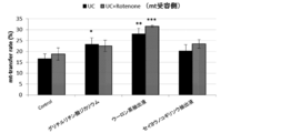
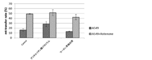
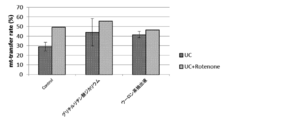
- dipotassium glycyrrhizinate and oolong tea extract were found as agents that enhance the mitochondrial transfer ability between cells.
- Example 2 Mt-TF from UC-MSC to BSMC Cryopreserved UC-MSC cells or BSMC-COPD (human bronchial smooth muscle cell) were asleep and seeded in multiple T-25 flasks. (2 ⁇ 10 5 cells / flask). For cells that require Rotenone treatment, Rotenone was added to the flask to a final concentration of 1 ⁇ M and treated for 12 hours. Mitochondrial staining reagent was added to the flask of UC-MSC cells serving as the mitochondrial supply side, and cytoplasmic staining reagent was added to the flask of BSMC-COPD cells serving as the mitochondrial receiving side, and incubated at 37 ° C. for 2 hours.
- the above Rotenone treatment is a treatment for inducing damaged cells (mocking a disease site).
- MitoTracker was used as a mitochondrial staining reagent, the incubation time was 30 minutes. After staining, the cells were washed 3 times with HBSS (+), and the cells were detached with trypsin-EDTA. The detached cells were washed with HBSS (+), and both cells were seeded on a plate (96 well plate) so as to be 1,000 cells / well, respectively, and co-culture was started. Here, a screening agent was added at the same time. The next day, the nuclear staining reagent was diluted in PBS ( ⁇ ) and replaced with the medium in the well. After 15 minutes at room temperature, images were taken with ImageXpress (16 fields / well). Analysis was performed with the same cell count program as in Example 1, and the mitochondrial transfer rate was calculated.
- Example 2 The cells, medium, and drugs used in Example 2 are shown below. The same staining reagent as in Example 1 was used.
- cell (1) Umbilical cord-derived mesenchymal stem cells (UC-MSC): The same ones as in Example 1 were used.
- BSMC-COPD cells human bronchial smooth muscle cells of chronic obstructive pulmonary disease; Bronchial Smooth Muscular Chronic Observatory Pulmonary Disease (Lonza 00195274)
- Culture medium (1) Stemlife MSC Comp Kit (Lifeline, LL-0034) medium (when UC-MSC wakes up, during UC-MSC culture, during co-culture) (2) SmGM TM-2 medium + Bullet Kit (Lonza CC-3182) + 10% FCS + 1% AB * (when culturing BSMC-COPD cells) AB * : Antibiotic-Antimycotic (Gibco)
- Rotenone (R8875-1G, Sigma-Aldrich) (used at a final concentration of 1 ⁇ M)
- Dipotassium glycyrrhizinate (GR2K: used at a final concentration of 0.5 mg / ml or 1 mg / ml)
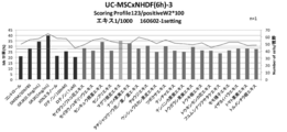
- Example 3 Mt-TF from UC-MSC to NHDF Cryopreserved UC-MSC cells or NHDF (human skin fibroblasts) were asleep and seeded in multiple T-25 flasks. (2 ⁇ 10 5 cells / flask). For cells that require Rotenone treatment, Rotenone was added to the flask to a final concentration of 1 ⁇ M and treated for 12 hours. Mitochondrial staining reagent was added to the flask of UC-MSC cells serving as the mitochondrial supply side, and cytoplasmic staining reagent was added to the flask of NHDF cells serving as the mitochondrial receiving side, and incubated at 37 ° C. for 1 hour.
- the above Rotenone treatment is a treatment for inducing damaged cells (mocking a disease site).
- MitoTracker was used as a mitochondrial staining reagent, the incubation time was 30 minutes. After staining, the cells were washed 3 times with HBSS (+), and the cells were detached with trypsin-EDTA. The detached cells were washed with HBSS (+), and both cells were seeded on a plate (96 well plate) so as to be 2,000 cells / well, and co-culture was started. Here, a screening agent was added at the same time. The next day, the nuclear staining reagent was diluted in PBS ( ⁇ ) and replaced with the medium in the well. After 15 minutes at room temperature, images were taken with ImageXpress (16 fields / well). Analysis was performed with the same cell count program as in Example 1, and the mitochondrial transfer rate was calculated.
- Example 3 The cells, medium, and drugs used in Example 3 are shown below. The same staining reagent as in Example 1 was used.
- cell (1) Umbilical cord-derived mesenchymal stem cells (UC-MSC): The same ones as in Example 1 were used.
- NHDF cells human skin fibroblasts: Human Dermal Fibroblast (KURABO KF-4009)
- Culture medium (1) Stemlife MSC Comp Kit (Lifeline, LL-0034) medium (when UC-MSC is asleep, UC-MSC is cultured) (2) DMEM (Gibco) medium + 10% FCS + 1% AB * (during NHDF cell culture and co-culture) AB * : Antibiotic-Antimycotic (Gibco)
- Rotenone (R8875-1G, Sigma-Aldrich) (used at a final concentration of 1 ⁇ M)
- Dipotassium glycyrrhizinate (GR2K: used at a final concentration of 0.5 mg / ml or 1 mg / ml)
- Example 4 Mt-TF from UC-MSC, AD-MSC to HCM (Human Cardiomyocytes) Cryopreserved UC-MSC cells, AD-MSC cells, and HCM cells (human cardiomyocytes) were asleep and seeded in multiple T-25 flasks. (2 ⁇ 10 5 cells / flask).
- HCM Human Cardiomyocytes
- the mitochondrial staining reagent is placed in the flask of UC-MSC cells or AD-MSC cells serving as the mitochondrial supply side, and the cytoplasm in the flask of HCM cells serving as the mitochondrial recipient Staining reagent was added and incubated for 1 hour at 37 ° C.
- MitoTracker was used as a mitochondrial staining reagent, the incubation time was 30 minutes. After staining, the cells were washed 3 times with HBSS (+), and the cells were detached with trypsin-EDTA.
- the detached cells were washed with HBSS (+), and both cells were seeded on a plate (96 well plate) so as to be 1,000 cells / well, respectively, and co-culture was started.
- a screening agent was added at the same time.
- the nuclear staining reagent was diluted in PBS ( ⁇ ) and replaced with the medium in the well. After 15 minutes of incubation at room temperature, images were taken with ImageXpress (16 fields / well). Analysis was performed with the same cell count program as in Example 1, and the mitochondrial transfer rate was calculated.
- Example 4 The cells, medium and drug used in Example 4 are shown below. The same staining reagent as in Example 1 was used.
- cell (1) Umbilical cord-derived mesenchymal stem cells (UC-MSC): The same ones as in Example 1 were used.
- AD-MSC Adipose-derived mesenchymal stem cell
- CEM cells human cardiomyocytes: Human Cardiac Myocytes (HCM (PromoCell)
- Culture medium (1) Stemlife MSC Comp Kit (Lifeline, LL-0034) medium (when UC-MSC and AD-MSC are asleep, during UC-MSC and AD-MSC culture) (2) Myocyte Basal Medium (PromoCell, C-22170), Myocy Growth Medium Supplement Pack (PromoCell, C-39270) (in HCM cell culture and co-culture)
- Rotenone (R8875-1G, Sigma-Aldrich) (used at a final concentration of 1 ⁇ M)
- Dipotassium glycyrrhizinate (GR2K: final concentration 1 mg / ml)
- Oolong tea extract (Extract is used at 1/1000)
- UC-MSC UC-MSC
- AD-MSC AD-MSC
- mitochondrial transfer occurs from UC-MSC and AD-MSC to HCM, and by adding dipotassium glycyrrhizinate, UC-MSC and AD -It was found that mitochondrial transfer from MSC to HCM is promoted.
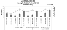
- Example 5 Effect of berberine: Mt-TF from UC-MSC, AD-MSC to HCE (human corneal epithelial cells) Cryopreserved UC-MSC cells, AD-MSC cells, and HCE cells (human corneal epithelial cells) were asleep and seeded in multiple T-25 flasks. (1 ⁇ 10 5 cells / flask).
- a mitochondrial staining reagent is applied to the flask of the UC-MSC cell or AD-MSC cell serving as the mitochondrial supply side, and a cytoplasmic staining is performed to the flask of the HCE cell serving as the mitochondrial receiving side. Reagents were added and incubated for 2 hours at 37 ° C. In addition, when MitoTracker was used as a mitochondrial staining reagent, the incubation time was 45 minutes. After staining, the cells were washed 3 times with HBSS (+), and the cells were detached with trypsin-EDTA.
- the detached cells were washed with HBSS (+), and UC-MSC or AD-MSC cells were seeded on a plate (96-well plate) so as to become 1,000 cells / well and HCE cells at 2,000 cells / well. Started. Here, a screening agent was added at the same time. The next day, the nuclear staining reagent was diluted in PBS ( ⁇ ) and replaced with the medium in the well. After 15 minutes at room temperature, images were taken with ImageXpress (16 fields / well). Analysis was performed with the same cell count program as in Example 1, and the mitochondrial transfer rate was calculated.
- Example 5 The cells, media, and drugs used in Example 5 are shown below. The same staining reagent as in Example 1 was used.
- cell (1) Umbilical cord-derived mesenchymal stem cells (UC-MSC): The same ones as in Example 1 were used.
- Adipose-derived mesenchymal stem cells (AD-MSC): The same ones as in Example 4 were used.
- HCE cells human corneal epithelial cells: Human corneal epithelial cell line HCE-T (RIKEN BioResource Center)
- Culture medium (1) Stemlife MSC Comp Kit (Lifeline, LL-0034) medium (when UC-MSC and AD-MSC are asleep, during UC-MSC and AD-MSC culture) (2) DMEM F12 (gibco) + 5% FCS (DS Pharma), 0.5% DMSO (Wako), 5 ⁇ g / mL insulin (sigma), 10 ng / mL hEGF (R & D) (in HCE cell culture) At the time of co-culture, a medium in which the medium (1) and the medium (2) were mixed at 1: 1 was used.
- Rotenone (R8875-1G, Sigma-Aldrich) (used at a final concentration of 100 nM)
- Dipotassium glycyrrhizinate (GR2K: final concentration 1 mg / ml)
- Berberine (berberine sulfate; CAS No. 316-41-6; used at a final concentration of 0.001%)
- FIG. 15 As shown in FIG. 15 (UC-MSC) and FIG. 16 (AD-MSC), it was found that the addition of berberine during the co-culture of MSC and HCE significantly promotes mitochondrial transfer from MSC to HCE.
- an agent that promotes mitochondrial transfer from cell to cell an agent that induces a cell with high mitochondrial transfer ability, or a cell with high mitochondrial transfer ability can be efficiently evaluated and screened.
- the high-functional cells and drugs screened by the method of the present invention can be widely used for diseases associated with mitochondrial dysfunction, decreased mitochondrial activity, etc., and symptoms associated with aging, etc., and have excellent effects. Play.
Landscapes
- Chemical & Material Sciences (AREA)
- Life Sciences & Earth Sciences (AREA)
- Organic Chemistry (AREA)
- Health & Medical Sciences (AREA)
- Engineering & Computer Science (AREA)
- Zoology (AREA)
- Wood Science & Technology (AREA)
- Proteomics, Peptides & Aminoacids (AREA)
- Molecular Biology (AREA)
- Bioinformatics & Cheminformatics (AREA)
- Immunology (AREA)
- Microbiology (AREA)
- Biophysics (AREA)
- Analytical Chemistry (AREA)
- Physics & Mathematics (AREA)
- Genetics & Genomics (AREA)
- Biochemistry (AREA)
- Biotechnology (AREA)
- General Engineering & Computer Science (AREA)
- General Health & Medical Sciences (AREA)
- Toxicology (AREA)
- Pharmaceuticals Containing Other Organic And Inorganic Compounds (AREA)
- Medicines Containing Material From Animals Or Micro-Organisms (AREA)
- Investigating Or Analysing Biological Materials (AREA)
- Measuring Or Testing Involving Enzymes Or Micro-Organisms (AREA)
- Micro-Organisms Or Cultivation Processes Thereof (AREA)
- Medicines That Contain Protein Lipid Enzymes And Other Medicines (AREA)
Abstract
本発明は、細胞内のミトコンドリア機能障害やミトコンドリア活性の低下が関与する疾患等の治療、老化等に伴う症状の回復に有効な細胞であるか否かを評価する方法、またこの方法により有効であると判断される高機能細胞、そのような高機能細胞を誘導できる薬剤を得る方法を提供することを課題とする。 本発明は、他の細胞へのミトコンドリアトランスファー能を指標にして細胞を評価する、細胞の評価方法である。さらに、ミトコンドリア活性を指標とすることが好ましい。上記ミトコンドリア活性は、ミトコンドリア脱水素酵素活性、ミトコンドリア含有量及びミトコンドリア膜電位からなる群より選択される少なくとも1種である。
Description
本発明は、細胞の評価方法、高機能細胞又は薬剤をスクリーニングする方法、及び医薬組成物に関する。
遺伝的背景、老化、環境要因等により、細胞内でミトコンドリア機能障害やミトコンドリア活性の低下が起こる場合があることが知られている。また、炎症性疾患、ミトコンドリア病等の種々の疾患においても、細胞内のミトコンドリア機能の低下が起こる場合があることも知られている(非特許文献1参照)。例えばミトコンドリア病は、ミトコンドリアの機能低下を原因とする疾患の総称であり、代表的なものとして慢性進行性外眼麻痺症候群、赤色ぼろ線維・ミオクローヌスてんかん症候群、及びミトコンドリア脳筋症・乳酸アシドーシス・脳卒中様症候群等がある。これらの疾患は、生体内全ての細胞において一様に異常が発生するわけではなく、その病態は多彩であり、根本的な治療法の無い難病である。
Nature、 2011 Jan 13;469(7329):p.221-5
このような情況の中、細胞内のミトコンドリア機能障害やミトコンドリア活性の低下が関与する疾患等の治療、老化等に伴う症状の回復に有効な薬剤等が求められている。そこで本発明は、細胞内のミトコンドリア機能障害やミトコンドリア活性の低下が関与する疾患等の治療、老化等に伴う症状の回復に有効な細胞であるか否かを評価する方法、またこの方法により有効であると判断される高機能細胞、そのような高機能細胞を誘導できる薬剤を得る方法を提供することを課題とする。
上記課題を解決するために、本発明者が種々検討した結果、細胞のミトコンドリアトランスファー能を指標とすることで、細胞の、疾患に対する治療効果、老化等に伴う症状の回復効果、細胞自体の健康状態等を効率よく評価することができることを見出した。また、この細胞を評価する方法は、高機能細胞、高機能細胞を誘導できる薬剤、細胞をより健康な状態に回復させる薬剤等のスクリーニング方法としても用いることができる。さらに、このスクリーニング方法により得られた高機能細胞及び/又は薬剤を含む医薬組成物は、疾患に対する治療、老化等に伴う症状の回復のために好適に使用され得る。
即ち、本発明の要旨は、以下に記載する通りである。
〔1〕ミトコンドリアトランスファー能を指標にして細胞を評価する、細胞の評価方法。
〔2〕さらに、ミトコンドリア活性を指標とする、〔1〕記載の方法。
〔3〕上記ミトコンドリア活性が、ミトコンドリア脱水素酵素活性、ミトコンドリア含有量及びミトコンドリア膜電位からなる群より選択される少なくとも1種である、〔2〕記載の方法。
〔4〕細胞のミトコンドリアトランスファー能を測定する工程、及び
得られたミトコンドリアトランスファー能を、基準となる細胞のミトコンドリアトランスファー能と比較する工程
を含む、細胞の評価方法。
〔5〕細胞のミトコンドリア活性を測定する工程、及び
得られたミトコンドリア活性を、基準となる細胞のミトコンドリア活性と比較する工程
をさらに含む、〔4〕記載の方法。
〔6〕上記ミトコンドリア活性が、ミトコンドリア脱水素酵素活性、ミトコンドリア含有量及びミトコンドリア膜電位からなる群より選択される少なくとも1種である、〔5〕記載の方法。
〔7〕上記細胞が、間葉系幹細胞、表皮幹細胞、角膜上皮幹細胞、結膜上皮幹細胞又はこれらに由来する細胞である、〔1〕から〔6〕のいずれか記載の方法。
〔8〕上記間葉系幹細胞が、臍帯由来又は脂肪組織由来である、〔7〕記載の方法。
〔9〕〔1〕から〔8〕のいずれか記載の方法を用いた、高機能細胞又は高機能細胞を誘導できる薬剤をスクリーニングする方法。
〔10〕〔9〕記載の方法により得られた、ウーロン茶抽出液、グリチルリチン酸ジカリウム、セイヨウノコギリソウ抽出液、アシタバ葉/茎エキス、アマチャエキス、ウコン根茎エキス、ゼニアオイ花エキス、ヒキオコシ葉/茎エキス、キハダ樹皮エキス、オウレン根エキス、グレープフルーツ果実エキス、カシア樹皮エキス、サッカロミセスセレビシアエエキス、ゴボウ根エキス、コメヌカエキス、ウスバサイシン根エキス、クチナシ果実エキス、マタタビ果実エキス、スギナエキス、セイヨウキズタ葉/茎エキス、センブリエキス、フユムシナツクサタケエキス、トマト果実エキス、ヒノキ水、ブクリョウエキス、ブッチャーブルーム根エキス、ムクロジ果皮エキス、ウメ果実エキス、ヤエヤマアオキ果汁、モモ葉エキス、筍の皮エキス、アマモエキス、水ナスエキス、ハトムギエキス、マグワ根皮エキス 、ハマメリス葉エキス、センチフォリアバラ花エキス、マドンナリリー根エキス、ヒノキ水、ローズマリー葉エキス、セージエキス2、セイヨウオオバコ種子エキス、レモングラス抽出液、クララエキス及びベルベリンからなる群より選択される少なくとも一種の薬剤で処理したことを特徴とする高機能細胞を含む医薬組成物。
〔11〕〔9〕記載の方法により得られた、ウーロン茶抽出液、グリチルリチン酸ジカリウム、セイヨウノコギリソウ抽出液、アシタバ葉/茎エキス、アマチャエキス、ウコン根茎エキス、ゼニアオイ花エキス、ヒキオコシ葉/茎エキス、キハダ樹皮エキス、オウレン根エキス、グレープフルーツ果実エキス、カシア樹皮エキス、サッカロミセスセレビシアエエキス、ゴボウ根エキス、コメヌカエキス、ウスバサイシン根エキス、クチナシ果実エキス、マタタビ果実エキス、スギナエキス、セイヨウキズタ葉/茎エキス、センブリエキス、フユムシナツクサタケエキス、トマト果実エキス、ヒノキ水、ブクリョウエキス、ブッチャーブルーム根エキス、ムクロジ果皮エキス、ウメ果実エキス、ヤエヤマアオキ果汁、モモ葉エキス、筍の皮エキス、アマモエキス、水ナスエキス、ハトムギエキス、マグワ根皮エキス 、ハマメリス葉エキス、センチフォリアバラ花エキス、マドンナリリー根エキス、ヒノキ水、ローズマリー葉エキス、セージエキス2、セイヨウオオバコ種子エキス、レモングラス抽出液、クララエキス及びベルベリンからなる群より選択される少なくとも一種の薬剤を含む医薬組成物。
〔12〕軟骨分解、関節リウマチ、乾癬性関節炎、脊椎関節炎、変形性関節症、痛風、乾癬、多発性硬化症、筋萎縮性側索硬化症、アルツハイマー病、パーキンソン病、中枢神経疾患、うっ血性心不全等の心不全、心筋梗塞、脳卒中、大動脈弁狭窄症、腎不全、腎臓移植後の生着不全、狼瘡、膵炎、アレルギー、アトピー性皮膚炎、線維症、貧血、アテローム性動脈硬化症等の動脈硬化、再狭窄、化学療法/放射線関連合併症、I型糖尿病、II型糖尿病、肺炎、自己免疫性肝炎、C型肝炎、原発性胆汁性肝硬変、原発性硬化性胆管炎、劇症肝炎、セリアック病、非特異性大腸炎、アレルギー性結膜炎、糖尿病性網膜症、シェーグレン症候群、ブドウ膜炎アレルギー性鼻炎、喘息、石綿症、珪肺、慢性閉塞性肺疾患、慢性肉芽腫性炎症、嚢胞性線維症、サルコイドーシス、糸球体腎炎、脈管炎、皮膚炎、HIV関連悪液質、大脳マラリア、強直性脊椎炎、らい病、肺線維症、線維筋痛、食道癌、胃食道逆流症、バレット食道、胃癌、十二指腸癌、小腸癌、虫垂癌、大腸癌、結腸癌、直腸癌、肛門癌、膵臓癌、肝臓癌、胆嚢癌、脾臓癌、腎癌、膀胱癌、前立腺癌、精巣癌、子宮癌、卵巣癌、乳癌、肺癌、甲状腺癌、慢性疲労症候群、てんかん、心筋症、高血圧、糖尿病、肥満、腎障害、不妊、難聴、慢性進行性外眼麻痺症候群、赤色ぼろ線維・ミオクローヌスてんかん症候群、リー脳症、レーバー病、ミトコンドリア糖尿病、パーソン病(汎血球減少症)及びミトコンドリア脳筋症・乳酸アシドーシス・脳卒中様症候群、皮膚老化(しわ、たるみ、コラーゲン減少等)、ターンノーバー鈍化、表皮ひ薄化、角結膜上皮障害、ドライアイ、角結膜炎、涙液分泌不全、涙腺炎のいずれかの処置のために用いられる、〔10〕又は〔11〕に記載の医薬組成物。
〔13〕ベルベリン配合点眼剤である、〔11〕又は〔12〕記載の医薬組成物。
〔14〕ミトコンドリアトランスファー剤又はミトコンドリアトランスファー促進剤である、〔10〕から〔13〕のいずれか記載の医薬組成物。
〔1〕ミトコンドリアトランスファー能を指標にして細胞を評価する、細胞の評価方法。
〔2〕さらに、ミトコンドリア活性を指標とする、〔1〕記載の方法。
〔3〕上記ミトコンドリア活性が、ミトコンドリア脱水素酵素活性、ミトコンドリア含有量及びミトコンドリア膜電位からなる群より選択される少なくとも1種である、〔2〕記載の方法。
〔4〕細胞のミトコンドリアトランスファー能を測定する工程、及び
得られたミトコンドリアトランスファー能を、基準となる細胞のミトコンドリアトランスファー能と比較する工程
を含む、細胞の評価方法。
〔5〕細胞のミトコンドリア活性を測定する工程、及び
得られたミトコンドリア活性を、基準となる細胞のミトコンドリア活性と比較する工程
をさらに含む、〔4〕記載の方法。
〔6〕上記ミトコンドリア活性が、ミトコンドリア脱水素酵素活性、ミトコンドリア含有量及びミトコンドリア膜電位からなる群より選択される少なくとも1種である、〔5〕記載の方法。
〔7〕上記細胞が、間葉系幹細胞、表皮幹細胞、角膜上皮幹細胞、結膜上皮幹細胞又はこれらに由来する細胞である、〔1〕から〔6〕のいずれか記載の方法。
〔8〕上記間葉系幹細胞が、臍帯由来又は脂肪組織由来である、〔7〕記載の方法。
〔9〕〔1〕から〔8〕のいずれか記載の方法を用いた、高機能細胞又は高機能細胞を誘導できる薬剤をスクリーニングする方法。
〔10〕〔9〕記載の方法により得られた、ウーロン茶抽出液、グリチルリチン酸ジカリウム、セイヨウノコギリソウ抽出液、アシタバ葉/茎エキス、アマチャエキス、ウコン根茎エキス、ゼニアオイ花エキス、ヒキオコシ葉/茎エキス、キハダ樹皮エキス、オウレン根エキス、グレープフルーツ果実エキス、カシア樹皮エキス、サッカロミセスセレビシアエエキス、ゴボウ根エキス、コメヌカエキス、ウスバサイシン根エキス、クチナシ果実エキス、マタタビ果実エキス、スギナエキス、セイヨウキズタ葉/茎エキス、センブリエキス、フユムシナツクサタケエキス、トマト果実エキス、ヒノキ水、ブクリョウエキス、ブッチャーブルーム根エキス、ムクロジ果皮エキス、ウメ果実エキス、ヤエヤマアオキ果汁、モモ葉エキス、筍の皮エキス、アマモエキス、水ナスエキス、ハトムギエキス、マグワ根皮エキス 、ハマメリス葉エキス、センチフォリアバラ花エキス、マドンナリリー根エキス、ヒノキ水、ローズマリー葉エキス、セージエキス2、セイヨウオオバコ種子エキス、レモングラス抽出液、クララエキス及びベルベリンからなる群より選択される少なくとも一種の薬剤で処理したことを特徴とする高機能細胞を含む医薬組成物。
〔11〕〔9〕記載の方法により得られた、ウーロン茶抽出液、グリチルリチン酸ジカリウム、セイヨウノコギリソウ抽出液、アシタバ葉/茎エキス、アマチャエキス、ウコン根茎エキス、ゼニアオイ花エキス、ヒキオコシ葉/茎エキス、キハダ樹皮エキス、オウレン根エキス、グレープフルーツ果実エキス、カシア樹皮エキス、サッカロミセスセレビシアエエキス、ゴボウ根エキス、コメヌカエキス、ウスバサイシン根エキス、クチナシ果実エキス、マタタビ果実エキス、スギナエキス、セイヨウキズタ葉/茎エキス、センブリエキス、フユムシナツクサタケエキス、トマト果実エキス、ヒノキ水、ブクリョウエキス、ブッチャーブルーム根エキス、ムクロジ果皮エキス、ウメ果実エキス、ヤエヤマアオキ果汁、モモ葉エキス、筍の皮エキス、アマモエキス、水ナスエキス、ハトムギエキス、マグワ根皮エキス 、ハマメリス葉エキス、センチフォリアバラ花エキス、マドンナリリー根エキス、ヒノキ水、ローズマリー葉エキス、セージエキス2、セイヨウオオバコ種子エキス、レモングラス抽出液、クララエキス及びベルベリンからなる群より選択される少なくとも一種の薬剤を含む医薬組成物。
〔12〕軟骨分解、関節リウマチ、乾癬性関節炎、脊椎関節炎、変形性関節症、痛風、乾癬、多発性硬化症、筋萎縮性側索硬化症、アルツハイマー病、パーキンソン病、中枢神経疾患、うっ血性心不全等の心不全、心筋梗塞、脳卒中、大動脈弁狭窄症、腎不全、腎臓移植後の生着不全、狼瘡、膵炎、アレルギー、アトピー性皮膚炎、線維症、貧血、アテローム性動脈硬化症等の動脈硬化、再狭窄、化学療法/放射線関連合併症、I型糖尿病、II型糖尿病、肺炎、自己免疫性肝炎、C型肝炎、原発性胆汁性肝硬変、原発性硬化性胆管炎、劇症肝炎、セリアック病、非特異性大腸炎、アレルギー性結膜炎、糖尿病性網膜症、シェーグレン症候群、ブドウ膜炎アレルギー性鼻炎、喘息、石綿症、珪肺、慢性閉塞性肺疾患、慢性肉芽腫性炎症、嚢胞性線維症、サルコイドーシス、糸球体腎炎、脈管炎、皮膚炎、HIV関連悪液質、大脳マラリア、強直性脊椎炎、らい病、肺線維症、線維筋痛、食道癌、胃食道逆流症、バレット食道、胃癌、十二指腸癌、小腸癌、虫垂癌、大腸癌、結腸癌、直腸癌、肛門癌、膵臓癌、肝臓癌、胆嚢癌、脾臓癌、腎癌、膀胱癌、前立腺癌、精巣癌、子宮癌、卵巣癌、乳癌、肺癌、甲状腺癌、慢性疲労症候群、てんかん、心筋症、高血圧、糖尿病、肥満、腎障害、不妊、難聴、慢性進行性外眼麻痺症候群、赤色ぼろ線維・ミオクローヌスてんかん症候群、リー脳症、レーバー病、ミトコンドリア糖尿病、パーソン病(汎血球減少症)及びミトコンドリア脳筋症・乳酸アシドーシス・脳卒中様症候群、皮膚老化(しわ、たるみ、コラーゲン減少等)、ターンノーバー鈍化、表皮ひ薄化、角結膜上皮障害、ドライアイ、角結膜炎、涙液分泌不全、涙腺炎のいずれかの処置のために用いられる、〔10〕又は〔11〕に記載の医薬組成物。
〔13〕ベルベリン配合点眼剤である、〔11〕又は〔12〕記載の医薬組成物。
〔14〕ミトコンドリアトランスファー剤又はミトコンドリアトランスファー促進剤である、〔10〕から〔13〕のいずれか記載の医薬組成物。
本発明の細胞の評価方法によると、ミトコンドリアトランスファー能を指標にすることで、その細胞の疾患に対する治療効果、老化等に伴う症状の回復効果、細胞自体の健康状態等を効率よく評価することができる。また、この評価方法を用いることで、疾患に対する治療効果、老化等に伴う症状の回復効果等を有する高機能細胞、このような高機能細胞を誘導できる薬剤、細胞をより健康な状態に回復させる薬剤、他の細胞へのミトコンドリアトランスファーを制御する薬剤等を、効率よくスクリーニングすることができる。また、本発明の方法によってスクリーニングされた高機能細胞や薬剤を含む医薬組成物は、ミトコンドリア機能障害、ミトコンドリア活性の低下等が関連する疾患や老化等に伴う症状に対して、広く用いることができる。
以下、本発明について詳細に説明する。
<細胞の評価方法>
本発明の方法は、ミトコンドリアトランスファー能を指標にして、その細胞の、疾患に対する治療効果、老化等に伴う症状の回復効果等を効率よく評価することができる。さらに評価の精度や効率を向上させるために、ミトコンドリアトランスファー能に加えて、ミトコンドリア活性をも指標とすることが好ましい。具体的には、他の細胞へのミトコンドリアトランスファー能を測定する工程(以下「工程1」ともいう)、及び得られたミトコンドリアトランスファー能を、基準となるミトコンドリア能と比較する工程(以下「工程2」ともいう)を含む。さらに、ミトコンドリア活性を測定する工程(工程3)、及び得られたミトコンドリア活性を、基準となるミトコンドリア活性と比較する工程(工程4)を含むことが好ましい。また、本発明の効果を損なわない範囲で、上記以外のその他の工程を含んでいてもよい。
本発明の方法は、ミトコンドリアトランスファー能を指標にして、その細胞の、疾患に対する治療効果、老化等に伴う症状の回復効果等を効率よく評価することができる。さらに評価の精度や効率を向上させるために、ミトコンドリアトランスファー能に加えて、ミトコンドリア活性をも指標とすることが好ましい。具体的には、他の細胞へのミトコンドリアトランスファー能を測定する工程(以下「工程1」ともいう)、及び得られたミトコンドリアトランスファー能を、基準となるミトコンドリア能と比較する工程(以下「工程2」ともいう)を含む。さらに、ミトコンドリア活性を測定する工程(工程3)、及び得られたミトコンドリア活性を、基準となるミトコンドリア活性と比較する工程(工程4)を含むことが好ましい。また、本発明の効果を損なわない範囲で、上記以外のその他の工程を含んでいてもよい。
本発明において「ミトコンドリアトランスファー能」とは、細胞がミトコンドリアを他の細胞に移行させることができる能力、或いはミトコンドリア自身が他の細胞に移行する能力をいい、複数の細胞を共培養した場合に、受容側の細胞全体のうちミトコンドリアのトランスファーを受けた細胞の割合(%)でその程度を表す。なお、ここでトランスファーされるミトコンドリアとしては、ミトコンドリア自体の他、ミトコンドリアが含むミトコンドリア遺伝子(DNA、RNA)、及び各種タンパク、並びにこれらと他のタンパクや小胞体等の他の器官との複合体も挙げられる。ミトコンドリアトランスファー能を指標として評価することで、各種疾患に対する治療効果、老化等に伴う症状の回復効果等が高い高機能細胞を選択することができる。なお、本発明において、ミトコンドリアトランスファー能の測定値をミトコンドリアトランスファー活性としている。
本発明において「ミトコンドリア活性」とは、細胞内のミトコンドリア自身が有する種々の活性をいう。具体的なミトコンドリア活性としては、細胞内のミトコンドリア脱水素酵素活性、細胞内のミトコンドリア膜電位、細胞内酸化ストレスレベル、ミトコンドリア呼吸活性、ATP合成酵素活性等が挙げられ、さらには細胞内のミトコンドリア含有量も含めることができる。
本発明において「高機能細胞」とは、少なくとも1種の疾患や症状に対して、治療、改善、予防効果を発揮することができる細胞をいう。特に、ミトコンドリア機能障害、ミトコンドリア活性の低下等が関連する疾患や老化等に伴う症状に対して、それらを改善等する機能に優れる細胞のことを指す。例えば、高機能細胞は、抗炎症機能、免疫賦活機能、免疫抑制機能、抗腫瘍機能、抗前癌性症状機能、抗神経変性疾患機能、代謝促進機能、心血管疾患・骨疾患・胃腸疾患・肺疾患・肝疾患・腎疾患等に対する治療機能、加齢や一時的なストレス等に伴って機能が低下した細胞の機能を回復させる機能等に優れる細胞である。さらには、加齢や一時的なストレス等に伴って低下した機能が回復し、より優れた健康状態を示す細胞も、本発明における高機能細胞に含まれる。高機能細胞における高機能の程度は、本発明の細胞の評価方法において相対的に判断され、比較対象とする細胞に対して相対的に高い機能を有するものであれば、特に限定されて解釈されるものではない。
従って、本発明の細胞の評価方法によると、例えばミトコンドリアを他の細胞にトランスファーする能力の高い細胞、さらには、細胞内のミトコンドリアの量が多い細胞、又はミトコンドリア自体のポテンシャルが高くエネルギー状態が高いレベルの細胞(膜電位が高い細胞、脱水素酵素活性が高い、酸化ストレスレベルが低い細胞等)等を高機能細胞と評価して選択することができる。
以下に、各工程について詳細に説明する。
[工程1:他の細胞へのミトコンドリアトランスファー能を測定する工程]
本工程においては、他の細胞へのミトコンドリアトランスファー能を測定する。
本工程においては、他の細胞へのミトコンドリアトランスファー能を測定する。
上記ミトコンドリアトランスファーとは、ある細胞から別の細胞へとミトコンドリア等が移行する現象を言う。この移行は、例えば、細胞と細胞をつなぐトンネリングナノチューブと呼ばれるトンネルの中を通って行われるが、移行経路は特に限定されず、エキソサイトーシス及びエンドサイトーシスを介した経路等でもよい。
ミトコンドリアトランスファー能の測定方法としては、例えば、以下のようにしてミトコンドリアトランスファー率を算出する方法が挙げられる。即ち、ミトコンドリアの供給側となる細胞をミトコンドリア染色試薬で染色し、ミトコンドリアの受容側となる細胞を細胞質染色試薬で染色する。これらの試薬による細胞の染色は、例えば、37℃で所定の時間インキュベートすることにより行われる。ミトコンドリア染色試薬としては、CytoPainter Cell Tracking Staining Kit-Green Fluorescence(abcam社製)、MitoTracker Mitochondrion-Selective Probes Green FM(Molecular Probes社製)等を用いることができる。また、細胞質染色試薬としては、例えば、CellTrace Violet Cell Proliferation Kit, for flow cytometry(Life tech.社製)、CellTrace Red Cell Proliferation Kit,for flow cytometry(Life tech.社製)を用いることができる。染色後、細胞をHBSS(+)等で洗浄し、トリプシン等の酵素、EDTA等にて細胞を剥離して、HBSS(+)等で洗浄後、両方の細胞をプレート(96well plate等)に播種して、4時間~24時間程度共培養を行う。その後、核染色試薬を添加して所定時間、37℃でインキュベートし、ImageXpress等の共焦点レーザーイメージシステムで画像撮影する。上記核染色試薬としては、例えばDAPI、DRAQ5(biostatus社製)、Hoechst33342(Dojindo社製)等を使用することができる。細胞カウントプログラム等を用いて解析し、ミトコンドリアトランスファー率を計算することができる。
上記細胞カウントプログラムでの解析は以下のように行うことができる。
(1)Cy5フィルター、DAPIフィルター等の核染色を認識するフィルターで観察し、赤いドット等で識別できる核を認識、計測させる(=細胞数の測定)。
(2)DAPIフィルター等の受容側細胞染色を認識するフィルターで観察し、ミトコンドリアを受け取る側の細胞(受容側)である細胞質を認識、計測させる(例えば、violet染色された細胞=受容側)。
(3)GFPフィルター等のミトコンドリア染色を認識するフィルターで観察し、ミトコンドリアを持つ細胞(供給側)を認識、計測させる(例えば、green染色された細胞、又はtracker染色された細胞=供給側)
(4)下記計算式により、ミトコンドリアトランスファー率(%)を算出することができる。また、(1)の細胞数を計測することで薬剤の細胞毒性を評価することができる。
計算式:((2)かつ(3)の細胞数/(2)の細胞数)×100
(1)Cy5フィルター、DAPIフィルター等の核染色を認識するフィルターで観察し、赤いドット等で識別できる核を認識、計測させる(=細胞数の測定)。
(2)DAPIフィルター等の受容側細胞染色を認識するフィルターで観察し、ミトコンドリアを受け取る側の細胞(受容側)である細胞質を認識、計測させる(例えば、violet染色された細胞=受容側)。
(3)GFPフィルター等のミトコンドリア染色を認識するフィルターで観察し、ミトコンドリアを持つ細胞(供給側)を認識、計測させる(例えば、green染色された細胞、又はtracker染色された細胞=供給側)
(4)下記計算式により、ミトコンドリアトランスファー率(%)を算出することができる。また、(1)の細胞数を計測することで薬剤の細胞毒性を評価することができる。
計算式:((2)かつ(3)の細胞数/(2)の細胞数)×100
[工程2:得られたミトコンドリアトランスファー能を、基準となるミトコンドリアトランスファー能と比較する工程]
本工程においては、工程1で得られた、細胞のミトコンドリアトランスファー能の測定値(ミトコンドリアトランスファー率)を、実施する試験系において基準と決めた細胞のミトコンドリアトランスファー能の測定値(ミトコンドリアトランスファー率)と比較する。得られた細胞のミトコンドリアトランスファー率を基準と決めた細胞のミトコンドリアトランスファー率と比較し、評価する機能に合わせて評価を行うことができる。多くの場合には、得られた細胞のミトコンドリアトランスファー率が基準と決めた細胞のミトコンドリアトランスファー率より高い場合に、その細胞は高機能細胞であると判断される。逆に、評価する機能や細胞によっては、得られた細胞のミトコンドリアトランスファー率が基準と決めた細胞のミトコンドリアトランスファー率よりも低い場合、その細胞は基準の細胞と比較して機能が高い細胞であると判断される場合もある。
本工程においては、工程1で得られた、細胞のミトコンドリアトランスファー能の測定値(ミトコンドリアトランスファー率)を、実施する試験系において基準と決めた細胞のミトコンドリアトランスファー能の測定値(ミトコンドリアトランスファー率)と比較する。得られた細胞のミトコンドリアトランスファー率を基準と決めた細胞のミトコンドリアトランスファー率と比較し、評価する機能に合わせて評価を行うことができる。多くの場合には、得られた細胞のミトコンドリアトランスファー率が基準と決めた細胞のミトコンドリアトランスファー率より高い場合に、その細胞は高機能細胞であると判断される。逆に、評価する機能や細胞によっては、得られた細胞のミトコンドリアトランスファー率が基準と決めた細胞のミトコンドリアトランスファー率よりも低い場合、その細胞は基準の細胞と比較して機能が高い細胞であると判断される場合もある。
本発明の細胞の評価方法は、本工程において、ミトコンドリアトランスファー能が基準より高い細胞又は低い細胞を、精度よく、かつ容易に評価し、選択することができる。そのため、ミトコンドリア機能障害、ミトコンドリア活性の低下等が関連する疾患や老化等に伴う症状に対して、ミトコンドリアトランスファーを介してそれらを改善等する機能に優れる細胞を特に効率よく選択することができる。
[工程3:ミトコンドリア活性を測定する工程]
本工程においては、細胞のミトコンドリア活性を測定する。ミトコンドリア活性としては、細胞内のミトコンドリア脱水素酵素活性、細胞内のミトコンドリア含有量、細胞内のミトコンドリア膜電位、細胞内の酸化ストレスレベル、ミトコンドリア呼吸活性、ATP合成酵素活性等が挙げられる。工程1及び2に加えて、本工程及び後述する工程4によってミトンドリア活性をも指標として評価を行うことにより、ミトコンドリアトランスファー能によって機能の高い細胞を選択できるだけでなく、さらに、ミトコンドリア自体の活性が高く、かつその含有量も多い細胞を選択したり、逆に評価する機能や細胞に応じて、ミトコンドリア自体の活性が低く、かつその含有量も少ない細胞を選択ことができる。これにより、さらに高機能な細胞であるか否かの評価を精度よく行うことができる。本工程においては、1種のミトコンドリア活性を測定してもよいし、複数種のミトコンドリア活性を測定してもよいが、複数種を組み合わせることで、より精度よく細胞の評価を実施することができる。
本工程においては、細胞のミトコンドリア活性を測定する。ミトコンドリア活性としては、細胞内のミトコンドリア脱水素酵素活性、細胞内のミトコンドリア含有量、細胞内のミトコンドリア膜電位、細胞内の酸化ストレスレベル、ミトコンドリア呼吸活性、ATP合成酵素活性等が挙げられる。工程1及び2に加えて、本工程及び後述する工程4によってミトンドリア活性をも指標として評価を行うことにより、ミトコンドリアトランスファー能によって機能の高い細胞を選択できるだけでなく、さらに、ミトコンドリア自体の活性が高く、かつその含有量も多い細胞を選択したり、逆に評価する機能や細胞に応じて、ミトコンドリア自体の活性が低く、かつその含有量も少ない細胞を選択ことができる。これにより、さらに高機能な細胞であるか否かの評価を精度よく行うことができる。本工程においては、1種のミトコンドリア活性を測定してもよいし、複数種のミトコンドリア活性を測定してもよいが、複数種を組み合わせることで、より精度よく細胞の評価を実施することができる。
ミトコンドリア脱水素酵素活性とは、細胞内のミトコンドリア内膜に位置し、NADHからユビキノンへ電子を転移させてATP合成、膜電位の維持に寄与する還元酵素の活性をいう。ミトコンドリア脱水素酵素活性は、MTT、XTT、MTS、WST等のテトラゾリウム塩を用いる方法(テトラゾリウム塩が細胞膜を透過し、ミトコンドリアに集積して、補酵素であるリボフラビンにより還元されて形成されるホルマザンを定量することによる測定方法)等により測定することができる。
細胞のミトコンドリア含有量及びミトコンドリア膜電位は、ミトコンドリア指示薬を用いて細胞内のミトコンドリアを標識することにより、定量化することができる。ここで、ミトコンドリア含有量及びミトコンドリア膜電位を反映するミトコンドリア指示薬由来シグナルの絶対値は、用いる細胞の種類により、また標識試薬の種類、暴露時間などの暴露条件により変化する。そのため、本発明においては、ミトコンドリア指示薬由来シグナル量の絶対値ではなく、比較する細胞間でのミトコンドリア指示薬由来シグナル量の相対値で判断される。具体的には、予備的実験において、ミトコンドリア含有量及び/又はミトコンドリア膜電位を指標として、選択すべき細胞のミトコンドリア指示薬由来シグナル量の範囲を区分けし、例えば、相対的に高い又は低いミトコンドリア含有量及びミトコンドリア膜電位を示す細胞を回収するように設定することができる。
本発明において、ミトコンドリア指示薬とは、生細胞中のミトコンドリアを特異的に標識でき、その存在量を測定しうる物質、生細胞中のミトコンドリアを特異的に標識できてミトコンドリア膜電位を測定しうる物質、又は生細胞中のミトコンドリアを特異的に標識できてその存在量とミトコンドリア膜電位の両方を測定しうる物質であれば、どのような物質であってもよい。例えば、蛍光を有し、ミトコンドリア構造物質(例えば、タンパク質、脂質、糖鎖、核酸又はこれらの代謝産物等)と結合する物質;蛍光を有し、ミトコンドリアの膜電位によってミトコンドリア内に選択的に取り込まれる物質;ミトコンドリア構造物質の作用により蛍光を発する物質に変換される物質;ミトコンドリア構造物質の作用によりミトコンドリア外に拡散できなくなる物質等が挙げられる。
ミトコンドリア指示薬の市販品の具体例として、Nonyl Acridine Orange(A1372)、3,3′-Dihexyloxacarbocyanine Iodide(D273、D378)、4-Di-1-ASP(D288)、2-(4-(dimethylamino)styryl)-N-Ethylpyridinium Iodide(D426)、Dihydrorhodamine 123(D632、D23806、R302、R22420)、dihydrorhodamine 6G(D633)、JC-9 Dye Mitochondrial Membrane Potential Probe(D22421)、Lucigenin (bis-N-Methylacridinium Nitrate)(L6868)、MitoFluor Green(M7502)、Orange CMTMRos(M7510)、Orange CM-H2TMRos(M7511)、Red CMXRos(M7512)、Red CM-H2Xros(M7513)、Benzoxazolium(M7514)、Red FM(M22425)、Deep Red FM(M22426)、Rhodamine 6G Chloride(R634)、Rhodamine B, Hexyl Ester, Perchlorate(R648)、Red CC-1(R14060)、SYBR Green(S7563)、Tetramethylrhodamine, Methyl Ester, Perchlorate(T668)、Tetramethylrhodamine, Ethyl Ester, Perchlorate(T669)又は5,5′,6,6′-tetrachloro-1,1′,3,3′-tetraethylbenzimidazolylcarbocyanine iodide(T3168)等(以上、Molecular Probe社製品番号)、タカラ社、Life technologies、Abcam社等の上記同等品を使用することができるが、これらのものに限定されない。
ミトコンドリア機能障害、ミトコンドリア活性の低下等が関連する疾患や老化等に伴う症状に対して、ミトコンドリアトランスファーを介してそれらを改善等する機能に優れる細胞であるか否かを効率よく評価することができるという観点から、本工程において測定するミトコンドリア活性としては、中でもミトコンドリア含有量が好ましい。
[工程4:得られたミトコンドリア活性を、基準となるミトコンドリア活性と比較する工程]
本工程においては、工程3で得られた、細胞のミトコンドリア活性を、実施する試験系において基準と決めた細胞のミトコンドリア活性と比較する。多くの場合、得られた細胞のミトコンドリア活性が基準と決めた細胞のミトコンドリア活性よりも高い場合、その細胞は高機能細胞であると判断される。逆に、得られた細胞のミトコンドリア活性が基準と決めた細胞のミトコンドリア活性よりも低い場合、その細胞は基準の細胞と比較して機能が低い細胞であると判断される。但し、評価する機能や細胞によっては、上記とは反対に、得られた細胞のミトコンドリア活性が基準と決めた細胞のミトコンドリア活性よりも低い場合、その細胞は高機能細胞であると判断される場合もある。
本工程においては、工程3で得られた、細胞のミトコンドリア活性を、実施する試験系において基準と決めた細胞のミトコンドリア活性と比較する。多くの場合、得られた細胞のミトコンドリア活性が基準と決めた細胞のミトコンドリア活性よりも高い場合、その細胞は高機能細胞であると判断される。逆に、得られた細胞のミトコンドリア活性が基準と決めた細胞のミトコンドリア活性よりも低い場合、その細胞は基準の細胞と比較して機能が低い細胞であると判断される。但し、評価する機能や細胞によっては、上記とは反対に、得られた細胞のミトコンドリア活性が基準と決めた細胞のミトコンドリア活性よりも低い場合、その細胞は高機能細胞であると判断される場合もある。
本発明の評価方法を適用できる細胞としては、特に限定されないが、炎症部位の細胞、老化・ストレス等により機能の低下した細胞に対してミトコンドリアトランスファーを起こす能力の高い細胞である、角化細胞、色素細胞、メラノサイト、脂肪細胞、毛母細胞、毛乳頭細胞、血管内皮細胞、神経細胞、褐色脂肪細胞、ランゲルハンス細胞、メルケル細胞、皮膚(真皮)線維芽細胞、皮膚上皮細胞、免疫細胞(マクロファージ、樹状細胞等)、筋細胞、角膜上皮細胞、結膜上皮細胞、杯細胞、胚細胞・胚性幹(ES)細胞・iPS細胞・生殖幹細胞・卵子・精子・造血幹細胞・表皮幹細胞・角膜上皮幹細胞・結膜上皮幹細胞・角膜幹細胞・結膜幹細胞・間葉系幹細胞等の幹細胞が好ましく、表皮幹細胞・角膜上皮幹細胞・結膜上皮幹細胞・間葉系幹細胞等の幹細胞がより好ましく、間葉系幹細胞がさらに好ましく、骨髄、脂肪、筋肉、神経、皮膚、羊膜、胎盤、絨毛膜、脱落膜、臍帯由来の間葉系幹細胞が特に好ましく、臍帯由来間葉系幹細胞、脂肪由来間葉系幹細胞が最も好ましい。上記臍帯由来間葉系幹細胞としては、例えば、臍帯血、臍帯静脈内皮下層、又はワルトン膠様質由来の臍帯由来間葉系幹細胞が挙げられるが、中でもワルトン膠様質由来の臍帯由来間葉系幹細胞が好ましい。
[高機能細胞のスクリーニング方法、及び高機能細胞の誘導剤のスクリーニング方法]
本発明は、高機能細胞、及び高機能細胞を誘導できる薬剤のスクリーニング方法も含む。本発明のスクリーニング方法においては、上述した本発明の細胞の評価方法が用いられる。本発明のスクリーニング方法においては、ミトコンドリアトランスファー能を指標にするため、ミトコンドリア機能障害、ミトコンドリア活性の低下等が関連する疾患や老化等に伴う症状に対して、それらを改善等する機能に優れる細胞、又はこのような細胞を誘導できる薬剤、さらにその細胞の機能を促進することができる薬剤を選択することができる。本発明のスクリーニング方法においては、このミトコンドリアトランスファー能に加えて、ミトコンドリア活性も指標とすることが好ましい。ミトコンドリア活性としては、細胞の評価方法において指標として採用した活性と同様の活性を採用することができる。
本発明は、高機能細胞、及び高機能細胞を誘導できる薬剤のスクリーニング方法も含む。本発明のスクリーニング方法においては、上述した本発明の細胞の評価方法が用いられる。本発明のスクリーニング方法においては、ミトコンドリアトランスファー能を指標にするため、ミトコンドリア機能障害、ミトコンドリア活性の低下等が関連する疾患や老化等に伴う症状に対して、それらを改善等する機能に優れる細胞、又はこのような細胞を誘導できる薬剤、さらにその細胞の機能を促進することができる薬剤を選択することができる。本発明のスクリーニング方法においては、このミトコンドリアトランスファー能に加えて、ミトコンドリア活性も指標とすることが好ましい。ミトコンドリア活性としては、細胞の評価方法において指標として採用した活性と同様の活性を採用することができる。
具体的には、上述の細胞の評価方法における工程1及び工程2、並びに工程3及び工程4と同様の方法で行われる。但し、高機能細胞の誘導剤のスクリーニングを行う場合には、工程1又は工程3の前に、細胞をスクリーニング用の各種薬剤でそれぞれ処理し、工程1又は工程3においてその処理した細胞のミトコンドリアトランスファー能又はミトコンドリア活性を測定する。或いは、工程1又は工程3においてミトコンドリアトランスファー能又はミトコンドリア活性を測定する際に、スクリーニング用の各種薬剤を共存させてもよい。その結果、よりミトコンドリアトランスファー能又はミトコンドリア活性の高い、高機能な細胞や、そのような細胞を誘導できる薬剤を、選択することができる。但し、評価する機能や細胞によっては、上記とは反対に、ミトコンドリアトランスファー能又はミトコンドリア活性が低い場合、その細胞は高機能細胞であると判断される場合もある。その場合には、ミトコンドリアトランスファー能又はミトコンドリア活性の低い細胞や、そのような細胞を誘導できる薬剤を、選択することができる。これら選択された細胞又は薬剤は、ミトコンドリア機能障害、ミトコンドリア活性の低下等が関連する疾患や老化等に伴う症状に対して、それらを改善等する効果を奏するものである。
[医薬組成物]
本発明は、本発明のスクリーニング方法により選択された高機能細胞及び/又は高機能細胞を誘導できる薬剤を含む医薬組成物も含む。本発明の医薬組成物は、上述の高機能細胞及び/又は高機能細胞を誘導できる薬剤を含むため、ミトコンドリア機能障害、ミトコンドリア活性の低下等が関連する疾患や老化等に伴う症状に対して、優れた治療、予防、又は改善効果を有する。本発明の医薬組成物は、ミトコンドリア機能障害、ミトコンドリア活性の低下等が起こっている細胞に対してミトコンドリア活性の高いミトコンドリアをトランスファーすることによって、それぞれの疾患や老化等に伴う症状に対して優れた効果を奏する。即ち、本発明の医薬組成物は、ミトコンドリアトランスファー剤として好適に用いることができる。
本発明は、本発明のスクリーニング方法により選択された高機能細胞及び/又は高機能細胞を誘導できる薬剤を含む医薬組成物も含む。本発明の医薬組成物は、上述の高機能細胞及び/又は高機能細胞を誘導できる薬剤を含むため、ミトコンドリア機能障害、ミトコンドリア活性の低下等が関連する疾患や老化等に伴う症状に対して、優れた治療、予防、又は改善効果を有する。本発明の医薬組成物は、ミトコンドリア機能障害、ミトコンドリア活性の低下等が起こっている細胞に対してミトコンドリア活性の高いミトコンドリアをトランスファーすることによって、それぞれの疾患や老化等に伴う症状に対して優れた効果を奏する。即ち、本発明の医薬組成物は、ミトコンドリアトランスファー剤として好適に用いることができる。
本発明のスクリーニング方法により、高機能細胞を誘導できる薬剤として、ウーロン茶抽出液(1/10~1/1,000,000希釈)、セイヨウノコギリソウ抽出液(1/10~1/10,0000希釈)、グリチルリチン酸ジカリウム(最終濃度1μg/ml~100mg/ml)、を選択することができる。さらに、アシタバ葉/茎エキス、アマチャエキス、ウコン根茎エキス、ゼニアオイ花エキス、ヒキオコシ葉/茎エキス、キハダ樹皮エキス、オウレン根エキス、グレープフルーツ果実エキス、カシア樹皮エキス、サッカロミセスセレビシアエエキス、ゴボウ根エキス、コメヌカエキス、ウスバサイシン根エキス、クチナシ果実エキス、マタタビ果実エキス、スギナエキス、セイヨウキズタ葉/茎エキス、センブリエキス、フユムシナツクサタケエキス、トマト果実エキス、ヒノキ水、ブクリョウエキス、ブッチャーズ・ブルーム根エキス、ムクロジ果皮エキス、ウメ果実エキス、ヤエヤマアオキ果汁、モモ葉エキス、筍の皮エキス、アマモエキス、水ナスエキス、ハトムギエキス、ベルベリンを選択することができる。これらの薬剤を添加することにより(1/10~1,000,000希釈)、細胞のミトコンドリアトランスファー能を増強することができる。これらのうち、細胞のミトコンドリアトランスファー能の増強効果により優れる観点から、ウーロン茶抽出液、グリチルリチン酸ジカリウム、ベルベリン、キハダ樹皮エキス、オウレン根エキス、ハトムギエキス、筍の皮エキスが好ましい。なお、上記薬剤としては、市販品を用いることができる。また、これらの薬剤で処理された各種細胞は、ミトコンドリアトランスファー能に優れ、ミトコンドリア機能障害、ミトコンドリア活性の低下等が起こっている細胞に対してミトコンドリアをトランスファーすることによって、それぞれの疾患や老化・ストレス等に伴う症状に対して優れた効果を奏する。
本発明において抽出液とは、植物から溶媒中にその成分が抽出されたものであれば特に限定されない。上記成分としては、植物からの抽出が可能な成分であれば限定されないが、通常、溶媒に溶解する成分を意味する。また、溶媒に溶解しなくとも、単独で液状である成分であれば、それらも抽出液に含み得る。また、本発明において抽出液とエキスとは同義で用いられる。
本発明において、上記抽出液の抽出方法は限定されず、植物の種類等に応じて適宜公知の方法を用いることができる。具体的には、抽出するための溶媒としては水、エタノール、エタノール以外の有機溶剤、油脂類等が挙げられる。抽出液が本発明の医薬組成物の有効成分として用いられることを考慮すると、水、エタノール等の安全性の高い溶媒を用いることが好ましい。また、溶媒を用いずに抽出することも可能であり、例えば、植物を加熱してプレス等するによって得ることが出来る。
本発明の医薬組成物は、高機能細胞及び/又は高機能細胞を誘導できる薬剤以外に、本発明の効果を損なわない範囲で、一般的な医薬品、医薬部外品等が含むことができる有効成分以外の成分を含むことができる。本発明の医薬組成物は、高機能細胞及び/又は高機能細胞を誘導できる薬剤に、上記成分を混合して製造することができる。
本発明の医薬組成物が用いられる疾患としては、例えば、炎症性疾患、免疫疾患、癌、前癌性症状、神経変性疾患、代謝疾患、心血管疾患、骨疾患、胃腸疾患、肺疾患、肝疾患、腎疾患等が挙げられる。また、本発明の医薬組成物は、加齢・老化・ストレス等に伴う各種の症状に対しても有効に用いることができる。
本発明の医薬組成物が用いられる疾患としては、例えば心筋細胞、肺胞上皮細胞、腎尿細管細胞、アストロサイト、血管平滑筋細胞、血管内皮細胞、免疫性細胞(マクロファージ等)、表皮幹細胞、真皮線維芽細胞、角結膜上皮幹細胞等が関与する疾患が挙げられる。
上記疾患、症状の具体例としては、軟骨分解、関節リウマチ、乾癬性関節炎、脊椎関節炎、変形性関節症、痛風、乾癬、多発性硬化症、筋萎縮性側索硬化症、アルツハイマー病、パーキンソン病、中枢神経疾患、うっ血性心不全等の心不全、心筋梗塞、脳卒中、大動脈弁狭窄症、腎不全、腎臓移植後の生着不全、狼瘡、膵炎、アレルギー、アトピー性皮膚炎、線維症、貧血、アテローム性動脈硬化症等の動脈硬化、再狭窄、化学療法/放射線関連合併症、I型糖尿病、II型糖尿病、肺炎、自己免疫性肝炎、C型肝炎、原発性胆汁性肝硬変、原発性硬化性胆管炎、劇症肝炎、セリアック病、非特異性大腸炎、アレルギー性結膜炎、糖尿病性網膜症、シェーグレン症候群、ブドウ膜炎アレルギー性鼻炎、喘息、石綿症、珪肺、慢性閉塞性肺疾患、慢性肉芽腫性炎症、嚢胞性線維症、サルコイドーシス、糸球体腎炎、脈管炎、皮膚炎、HIV関連悪液質、大脳マラリア、強直性脊椎炎、らい病、肺線維症、線維筋痛、食道癌、胃食道逆流症、バレット食道、胃癌、十二指腸癌、小腸癌、虫垂癌、大腸癌、結腸癌、直腸癌、肛門癌、膵臓癌、肝臓癌、胆嚢癌、脾臓癌、腎癌、膀胱癌、前立腺癌、精巣癌、子宮癌、卵巣癌、乳癌、肺癌、甲状腺癌、慢性疲労症候群、てんかん、心筋症、高血圧、糖尿病、肥満、腎障害、不妊、難聴、慢性進行性外眼麻痺症候群、赤色ぼろ線維・ミオクローヌスてんかん症候群、リー脳症、レーバー病、ミトコンドリア糖尿病、パーソン病(汎血球減少症)及びミトコンドリア脳筋症・乳酸アシドーシス・脳卒中様症候群、皮膚老化(しわ、たるみ、コラーゲン減少等)、ターンノーバー鈍化、表皮ひ薄化、角結膜上皮障害、ドライアイ、角結膜炎、涙液分泌不全、涙腺炎等が挙げられる。
これらのうち、ミトコンドリア病、心筋梗塞、心不全等の心筋細胞が関与する疾患;慢性閉塞性肺疾患、肺炎等の肺胞上皮細胞が関与する疾患;腎不全、特に腎臓移植後の生着不全等の腎尿細管細胞が関与する疾患;アルツハイマー病、中枢神経疾患等のアストロサイトが関与する疾患;動脈硬化等の血管平滑筋細胞・血管内皮細胞・免疫性細胞(マクロファージ等)が関与する疾患;皮膚老化、ターンノーバー鈍化、表皮ひ薄化等の表皮幹細胞が関与する症状;しわ、たるみ、コラーゲン減少等の真皮線維芽細胞が関与する疾患;角結膜上皮障害、ドライアイ、角結膜炎、涙液分泌不全、涙腺炎等の眼病等に対しては、本発明の医薬組成物が好適に用いられる。
本発明の医薬組成物の一実施形態であるベルベリン配合点眼剤は、生体にもとより存在する角膜上皮幹細胞から角膜上皮細胞へ、また結膜上皮幹細胞から結膜上皮細胞へのミトコンドリアトランスファーを誘導、促進させることで、角膜上皮細胞・結膜上皮細胞の増殖、角膜上皮・結膜上皮の創傷治癒促進、角膜上皮・結膜上皮の保護などの角膜上皮・結膜上皮への優れた効果を奏する。このような効果を期待する疾患としては、角膜上皮障害・結膜上皮障害、ドライアイ、角膜炎・結膜炎、涙液分泌不全、涙腺炎が挙げられる。他にも、眼炎症の抑制、眼脂減少、抗菌効果、涙液成分の分泌、抗酸化もしくは抗糖化作用、紫外線などによる角膜へのダメージの軽減、白内障や緑内障など眼病に対する予防、かすみ目や疲れ眼の軽減、花粉などに対するアレルギー症状の緩和、結膜充血の軽減、異物感の軽減など様々な効果が期待できる。
また、近年では、上記のような主に外因性の疾患だけでなく内因性の疾患も注目されており、角膜上皮幹細胞、結膜上皮幹細胞自体の異常により角膜上皮細胞、結膜上皮細胞の供給が滞る角膜上皮幹細胞疲弊症、結膜上皮幹細胞疲弊症が問題視されている。この疾患に対しては、本発明の高機能細胞を含む医薬組成物を投与する方法により、高い治療効果が期待できる。さらには、上記ベルベリン配合点眼剤の点眼により、生体にもとより存在する角膜上皮幹細胞、結膜上皮幹細胞を活性化することでミトコンドリアトランスファーが誘導され、角膜上皮幹細胞疲弊、結膜上皮幹細胞疲弊を簡易に治療することも可能となる。
本発明の医薬組成物は、常法によって適宜の製剤とすることができる。本発明の医薬組成物が高機能細胞を含む場合には、一般的な細胞製剤が含む成分を配合することができる。また、本発明の医薬組成物が高機能細胞の誘導剤を含む場合には、製剤の剤型としては散剤、顆粒剤などの固形製剤であってもよいが、優れた予防・治療効果を得る観点からは、溶液剤、乳剤、懸濁剤などの液剤とすることが好ましい。上記液剤の製造方法としては、例えば高機能細胞の誘導剤を溶剤と混合する方法や、さらに懸濁化剤や乳化剤を混合する方法を好適に例示することができる。また、眼疾患、眼の症状に対しては、点眼剤、洗眼剤、眼軟膏、注射剤、貼布剤、ゲル、挿入剤とすることが好ましい。以上のように、本発明における医薬組成物の製剤においては、製剤上の必要に応じて、適宜の薬学的に許容される担体、例えば、賦形剤、結合剤、溶剤、溶解補助剤、懸濁化剤、乳化剤、等張化剤、緩衝剤、安定化剤、無痛化剤、防腐剤、抗酸化剤、着色剤、滑沢剤、崩壊剤、湿潤剤、吸着剤、甘味剤、希釈剤などの任意成分を配合することができる。
本発明の医薬組成物の投与方法としては特に制限されないが、血管内投与(好ましくは静脈内投与)、腹腔内投与、腸管内投与、皮下投与等が好ましく、中でも、血管内投与がより好ましい。また、眼疾患、眼の症状に対しては、点眼剤、洗眼剤又は眼軟膏として投与することが好ましい。
本発明の医薬組成物の投与量としては、疾患の種類や、その症状の度合い、剤型、投与対象の体重等によって変わり得るが、高機能細胞の誘導剤が、例えば、1日当たり、0.1μg/kg~10g/kgの範囲で含むものを好適に例示することができ、中でも、1μg/kg~1g/kgの範囲をより好適に例示することができる。また、本発明の医薬組成物が高機能細胞を含む場合には、1日当たり、高機能細胞を1X103個~1X109個の範囲で投与することができる。なお、これらの投与は、1日のうち1~複数回に分けて行ってもよい。また、本発明の医薬組成物の投与は、単回投与でもよいし、継続的に行ってもよい。継続的に行う場合は、例えば、3日に1回以上の頻度で、2回以上継続して投与することができ、中でも、2日に1回以上の頻度で、3回以上継続して投与することが好ましく、1日に1回以上の頻度で4回以上継続して投与することがより好ましい。
本発明の医薬組成物の投与対象となる哺乳動物としては、特に制限されないが、ヒト、サル、マウス、ラット、ハムスター、モルモット、ウシ、ブタ、ウマ、ウサギ、ヒツジ、ヤギ、ネコ、イヌ等が好ましく、中でもヒトがより好ましい。また、本発明の医薬組成物に含まれる高機能細胞は、本発明の医薬組成物の投与対象となる哺乳動物の種類と一致していることが、疾患に対する、より安定して優れた予防及び/又は治療効果を得る観点から好ましい。
[外用組成物]
本発明は、本発明のスクリーニング方法により選択された高機能細胞を誘導できる薬剤を含む外用組成物を提供することができる。本発明の外用組成物は、上述の高機能細胞を誘導できる薬剤を含むため、ミトコンドリア機能障害、ミトコンドリア活性の低下等が関連する疾患や老化等に伴う症状に対して、優れた治療、予防、又は改善効果を有する。本発明の外用組成物は、老化等によりミトコンドリア機能障害、ミトコンドリア活性の低下等が起こっている細胞に作用して、ミトコンドリアをトランスファーできるように、皮膚上皮細胞、表皮幹細胞、真皮線維芽細胞等を高機能細胞とすることができるので、化粧品用の外用組成物としても広く用いることができる。
本発明は、本発明のスクリーニング方法により選択された高機能細胞を誘導できる薬剤を含む外用組成物を提供することができる。本発明の外用組成物は、上述の高機能細胞を誘導できる薬剤を含むため、ミトコンドリア機能障害、ミトコンドリア活性の低下等が関連する疾患や老化等に伴う症状に対して、優れた治療、予防、又は改善効果を有する。本発明の外用組成物は、老化等によりミトコンドリア機能障害、ミトコンドリア活性の低下等が起こっている細胞に作用して、ミトコンドリアをトランスファーできるように、皮膚上皮細胞、表皮幹細胞、真皮線維芽細胞等を高機能細胞とすることができるので、化粧品用の外用組成物としても広く用いることができる。
以下に本発明を実施例に基づいて更に詳細に説明するが、本発明はこれら実施例に限定されるものではない。
《実施例1》
[ミトコンドリアトランスファー能を指標とした高機能細胞及び薬剤のスクリーニング]
凍結保存のUC-MSC細胞又はA549細胞を起眠し、複数のT-25フラスコに播種した。(1x10⁵cells/フラスコ)。Rotenone処理が必要な細胞に対しては、最終濃度100nMとなるように、上記フラスコにRotenoneを添加し、12時間処理した。ミトコンドリアの供給側となる細胞のフラスコにミトコンドリア染色試薬を、ミトコンドリアの受容側となる細胞のフラスコに細胞質染色試薬を添加し2時間、37℃でインキュベートした。上記Rotenone処理は、障害を受けた細胞(疾患部位を模倣)を誘導する処理である。なお、ミトコンドリア染色試薬としてMitoTrackerを用いた場合は、インキュベート時間を45分とした。染色後、HBSS(+)で3回洗浄し、トリプシン-EDTAにて細胞を剥離した。剥離した細胞をHBSS(+)で洗浄し、両方の細胞をそれぞれ2,000cells/wellとなるようにプレート(96well plate)に播種して共培養を開始した。ここで、同時にスクリーニング薬剤を添加した。翌日、核染色試薬をHBSS(+)に希釈し、well内培地と置換した。1.5時間、37℃でインキュベートした後、ImageXpressで画像撮影した(9視野/well)。細胞カウントプログラム*で解析し、ミトコンドリアトランスファー率を計算した。
[ミトコンドリアトランスファー能を指標とした高機能細胞及び薬剤のスクリーニング]
凍結保存のUC-MSC細胞又はA549細胞を起眠し、複数のT-25フラスコに播種した。(1x10⁵cells/フラスコ)。Rotenone処理が必要な細胞に対しては、最終濃度100nMとなるように、上記フラスコにRotenoneを添加し、12時間処理した。ミトコンドリアの供給側となる細胞のフラスコにミトコンドリア染色試薬を、ミトコンドリアの受容側となる細胞のフラスコに細胞質染色試薬を添加し2時間、37℃でインキュベートした。上記Rotenone処理は、障害を受けた細胞(疾患部位を模倣)を誘導する処理である。なお、ミトコンドリア染色試薬としてMitoTrackerを用いた場合は、インキュベート時間を45分とした。染色後、HBSS(+)で3回洗浄し、トリプシン-EDTAにて細胞を剥離した。剥離した細胞をHBSS(+)で洗浄し、両方の細胞をそれぞれ2,000cells/wellとなるようにプレート(96well plate)に播種して共培養を開始した。ここで、同時にスクリーニング薬剤を添加した。翌日、核染色試薬をHBSS(+)に希釈し、well内培地と置換した。1.5時間、37℃でインキュベートした後、ImageXpressで画像撮影した(9視野/well)。細胞カウントプログラム*で解析し、ミトコンドリアトランスファー率を計算した。
細胞カウントプログラム*での解析は以下のように行った。
(1)Cy5フィルターで観察し、赤いドットを認識、計測させた(=細胞数の測定)。
(2)DAPIフィルターで観察し、ミトコンドリアを受け取る側の細胞(受容側)である青色の細胞を認識、計測させた(violet染色された細胞=受容側)。
(3)GFPフィルターで観察し、ミトコンドリアを持つ細胞(供給側)である緑色の細胞を認識、計測させた(green染色された細胞、又はtracker染色された細胞=供給側)。
(4)3枚の画像を重ね合わせ(ドットの色で、細胞の状態を表示)、赤の細胞数と、ピンクの細胞数を計測する。赤の細胞はミトコンドリアを受容する側の細胞の内、ミトコンドリアを受容していない細胞、ピンクの細胞は、ミトコンドリアを受容する側の細胞の内、ミトコンドリアを受容した細胞を示す。
(5)下記計算式により、ミトコンドリアトランスファー率(%)を算出した。
計算式:(ピンクの細胞数/ピンク+赤の細胞数)×100
(1)Cy5フィルターで観察し、赤いドットを認識、計測させた(=細胞数の測定)。
(2)DAPIフィルターで観察し、ミトコンドリアを受け取る側の細胞(受容側)である青色の細胞を認識、計測させた(violet染色された細胞=受容側)。
(3)GFPフィルターで観察し、ミトコンドリアを持つ細胞(供給側)である緑色の細胞を認識、計測させた(green染色された細胞、又はtracker染色された細胞=供給側)。
(4)3枚の画像を重ね合わせ(ドットの色で、細胞の状態を表示)、赤の細胞数と、ピンクの細胞数を計測する。赤の細胞はミトコンドリアを受容する側の細胞の内、ミトコンドリアを受容していない細胞、ピンクの細胞は、ミトコンドリアを受容する側の細胞の内、ミトコンドリアを受容した細胞を示す。
(5)下記計算式により、ミトコンドリアトランスファー率(%)を算出した。
計算式:(ピンクの細胞数/ピンク+赤の細胞数)×100
上記実験に使用した細胞、培地、薬剤、染色試薬を以下に示す。
細胞:
(1)臍帯由来間葉系幹細胞(UC-MSC;Umbilical Cord derived Mesenchymal Stem Cells Wharton’s Jelly(HMSC-WJ)、FC-0020、LifeLine社)
(2)A549細胞(ヒト肺胞基底上皮腺癌細胞)
細胞:
(1)臍帯由来間葉系幹細胞(UC-MSC;Umbilical Cord derived Mesenchymal Stem Cells Wharton’s Jelly(HMSC-WJ)、FC-0020、LifeLine社)
(2)A549細胞(ヒト肺胞基底上皮腺癌細胞)
培地:
(1)Stemlife MSC Comp Kit (Lifeline、LL-0034)培地(UC-MSC起眠時、UC培養時)
(2)DMEM/F-12(Gibco)培地(+10%FCS、+1%AB*)(A549培養時、共培養時)
AB*:Antibiotic-Antimycotic(Gibco社)
(1)Stemlife MSC Comp Kit (Lifeline、LL-0034)培地(UC-MSC起眠時、UC培養時)
(2)DMEM/F-12(Gibco)培地(+10%FCS、+1%AB*)(A549培養時、共培養時)
AB*:Antibiotic-Antimycotic(Gibco社)
薬剤:
(1)Rotenone(R8875-1G、Sigma-Aldrich)(最終濃度100nMで使用)
(2)グリチルリチン酸ジカリウム(最終濃度1mg/mlで使用)
(3)ウーロン茶抽出液(エキスを1/10,000で使用)
(4)セイヨウノコギリソウ抽出液(エキスを1/500で使用)
(1)Rotenone(R8875-1G、Sigma-Aldrich)(最終濃度100nMで使用)
(2)グリチルリチン酸ジカリウム(最終濃度1mg/mlで使用)
(3)ウーロン茶抽出液(エキスを1/10,000で使用)
(4)セイヨウノコギリソウ抽出液(エキスを1/500で使用)
染色試薬:
(ミトコンドリア染色試薬)
(1)CytoPainter Cell Tracking Staining Kit-Green Fluorescence(500回分:ab112143、200回分:ab138891、abcam)(最終濃度5uMで使用)
(2)MitoTracker Mitochondrion-Selective Probes Green FM(M7514、Molecular Probes)(最終濃度100-1000nMで使用)
(ミトコンドリア染色試薬)
(1)CytoPainter Cell Tracking Staining Kit-Green Fluorescence(500回分:ab112143、200回分:ab138891、abcam)(最終濃度5uMで使用)
(2)MitoTracker Mitochondrion-Selective Probes Green FM(M7514、Molecular Probes)(最終濃度100-1000nMで使用)
(細胞質染色試薬)
(1)CellTrace Violet Cell Proliferation Kit, for flow cytometry(C34557、Life tech.)
(2)CellTrace Red Cell Proliferation Kit,for flow cytometry(C34564、Life tech.)(最終濃度5uMで使用)
(1)CellTrace Violet Cell Proliferation Kit, for flow cytometry(C34557、Life tech.)
(2)CellTrace Red Cell Proliferation Kit,for flow cytometry(C34564、Life tech.)(最終濃度5uMで使用)
(核染色試薬)
(1)DRAQ5(DR50050、biostatus)
(2)Hoechst33342(346-07951、Dojindo)
(1)DRAQ5(DR50050、biostatus)
(2)Hoechst33342(346-07951、Dojindo)

図1(試験例1~8)に示すように、ミトコンドリアの供給側及び受容側の細胞がどちらも無処理のUC-MSCである場合、グリチルリチン酸ジカリウム、ウーロン茶抽出液の添加で、有意にミトコンドリアトランスファー率が上昇した。また、受容側のUC-MSCがRotenone処理細胞である場合も、同様にグリチルリチン酸ジカリウム、ウーロン茶抽出液の添加で、有意にミトコンドリアトランスファー率が上昇した。
図2(試験例9~14)に示すように、ミトコンドリアの供給側及び受容側の細胞がどちらも無処理のA549である場合、グリチルリチン酸ジカリウムの添加で、有意にミトコンドリアトランスファー率が上昇した。しかし、受容側のUC-MSCがRotenone処理細胞である場合は、スクリーニング薬剤を入れていないコントロールのA549でも、十分に高いミトコンドリアトランスファー率を示したためか、スクリーニング薬剤の添加による効果が確認できなかった。
図3(試験例15~20)に示すように、ミトコンドリアの供給側がA549であり、受容側が無処理のUC-MSCである場合、グリチルリチン酸ジカリウムの添加で、ミトコンドリアトランスファー率の上昇が見られた。また、受容側のUC-MSCがRotenone処理細胞である場合は、グリチルリチン酸ジカリウムの添加で、ミトコンドリアトランスファー率の上昇が確認できたものの、スクリーニング薬剤を入れていないコントロールのUC-MSCでも十分に高いミトコンドリアトランスファー率を示したためか、スクリーニング薬剤の添加による効果がわずかしか確認できなかった。
図4(試験例21~26)に示すように、ミトコンドリアの供給側がUC-MSCであり、受容側が無処理のA549である場合、受容側のA549がRotenone処理されている場合は、処理されていない場合に比べて、常にミトコンドリアトランスファー率が高かった。しかし、スクリーニング薬剤による促進効果は確認できなかった。
このように、本発明の評価方法/スクリーニング方法によると、ミトコンドリアトランスファー能を増強させる薬剤を、効率よく選択することができる。例えば、本発明の評価方法/スクリーニング方法によって、細胞間のミトコンドリアトランスファー能を増強する薬剤として、例えばグリチルリチン酸ジカリウム、ウーロン茶抽出液を見出すことができた。また、本発明の評価方法/スクリーニング方法によると、ミトコンドリアの供給側の細胞のうち、ミトコンドリアトランスファー率が高い高機能細胞を選択することができ、このような細胞は、障害を受けた細胞や、疾患部位の細胞に対して、ミトコンドリアをトランスファーすることで、細胞の機能回復や、疾患の治療に有効であると考えられる。
《実施例2》UC-MSCからBSMCへのMt-TF
凍結保存のUC-MSC細胞又はBSMC-COPD(ヒト気管支平滑筋細胞細胞)を起眠し、複数のT-25フラスコに播種した。(2x10⁵cells/フラスコ)。Rotenone処理が必要な細胞に対しては、最終濃度1μMとなるように、上記フラスコにRotenoneを添加し、12時間処理した。ミトコンドリアの供給側となるUC-MSC細胞のフラスコにミトコンドリア染色試薬を、ミトコンドリアの受容側となるBSMC-COPD細胞のフラスコに細胞質染色試薬を添加し2時間、37℃でインキュベートした。上記Rotenone処理は、障害を受けた細胞(疾患部位を模倣)を誘導する処理である。なお、ミトコンドリア染色試薬としてMitoTrackerを用いた場合は、インキュベート時間を30分とした。染色後、HBSS(+)で3回洗浄し、トリプシン-EDTAにて細胞を剥離した。剥離した細胞をHBSS(+)で洗浄し、両方の細胞をそれぞれ1,000cells/wellとなるようにプレート(96well plate)に播種して共培養を開始した。ここで、同時にスクリーニング薬剤を添加した。翌日、核染色試薬をPBS(-)に希釈し、well内培地と置換した。15分、室温でおいた後、ImageXpressで画像撮影した(16視野/well)。実施例1と同様の細胞カウントプログラムで解析し、ミトコンドリアトランスファー率を計算した。
凍結保存のUC-MSC細胞又はBSMC-COPD(ヒト気管支平滑筋細胞細胞)を起眠し、複数のT-25フラスコに播種した。(2x10⁵cells/フラスコ)。Rotenone処理が必要な細胞に対しては、最終濃度1μMとなるように、上記フラスコにRotenoneを添加し、12時間処理した。ミトコンドリアの供給側となるUC-MSC細胞のフラスコにミトコンドリア染色試薬を、ミトコンドリアの受容側となるBSMC-COPD細胞のフラスコに細胞質染色試薬を添加し2時間、37℃でインキュベートした。上記Rotenone処理は、障害を受けた細胞(疾患部位を模倣)を誘導する処理である。なお、ミトコンドリア染色試薬としてMitoTrackerを用いた場合は、インキュベート時間を30分とした。染色後、HBSS(+)で3回洗浄し、トリプシン-EDTAにて細胞を剥離した。剥離した細胞をHBSS(+)で洗浄し、両方の細胞をそれぞれ1,000cells/wellとなるようにプレート(96well plate)に播種して共培養を開始した。ここで、同時にスクリーニング薬剤を添加した。翌日、核染色試薬をPBS(-)に希釈し、well内培地と置換した。15分、室温でおいた後、ImageXpressで画像撮影した(16視野/well)。実施例1と同様の細胞カウントプログラムで解析し、ミトコンドリアトランスファー率を計算した。
実施例2に使用した細胞、培地、薬剤を以下に示す。染色試薬は実施例1と同様のものを使用した。
細胞:
(1)臍帯由来間葉系幹細胞(UC-MSC):実施例1と同様のものを使用した。
(2)BSMC-COPD細胞(慢性閉塞性肺疾患のヒト気管支平滑筋細胞;Bronchial Smooth Muscle cell Chronic Obstructive Pulmonary Disease(Lonza 00195274))
細胞:
(1)臍帯由来間葉系幹細胞(UC-MSC):実施例1と同様のものを使用した。
(2)BSMC-COPD細胞(慢性閉塞性肺疾患のヒト気管支平滑筋細胞;Bronchial Smooth Muscle cell Chronic Obstructive Pulmonary Disease(Lonza 00195274))
培地:
(1)Stemlife MSC Comp Kit (Lifeline、LL-0034)培地(UC-MSC起眠時、UC-MSC培養時、共培養時)
(2)SmGMTM-2培地+BulletKit(Lonza CC-3182)+10%FCS+1%AB*(BSMC-COPD細胞培養時)
AB*:Antibiotic-Antimycotic(Gibco社)
(1)Stemlife MSC Comp Kit (Lifeline、LL-0034)培地(UC-MSC起眠時、UC-MSC培養時、共培養時)
(2)SmGMTM-2培地+BulletKit(Lonza CC-3182)+10%FCS+1%AB*(BSMC-COPD細胞培養時)
AB*:Antibiotic-Antimycotic(Gibco社)
薬剤:
(1)Rotenone(R8875-1G、Sigma-Aldrich)(最終濃度1μMで使用)
(2)グリチルリチン酸ジカリウム(GR2K:最終濃度0.5mg/ml又は1mg/mlで使用)
(1)Rotenone(R8875-1G、Sigma-Aldrich)(最終濃度1μMで使用)
(2)グリチルリチン酸ジカリウム(GR2K:最終濃度0.5mg/ml又は1mg/mlで使用)
(3)スクリーニングに供した薬剤
(図5;終濃度1/1000で使用)
ブドウ葉エキス、アシタバ葉/茎エキス、アセンヤクエキス、アマチャエキス、アルテア根エキス、アルニカ花エキス、アロエフェロックス葉エキス、イチョウ葉エキス、セイヨウイラクサ葉エキス、カワラヨモギ花エキス、ウイキョウ果実エキス、チャ葉エキス、ウコン根茎エキス、ゼニアオイ花エキス、ノイバラ果実エキス、ヒキオコシ葉/茎エキス、オウゴン根エキス、キハダ樹皮エキス、オウレン根エキス、セイヨウオトギリソウ花/葉/茎エキス、オノニスエキス、オリーブ葉エキス、オレンジ果汁、ヒバマタエキス、ヒジキエキス、ワカメエキス、ヨモギ葉エキス、カミツレ花エキス、カンゾウ根エキス、カンゾウ葉エキス、キイチゴエキス、キナノキ樹皮エキス、キュウリ果実エキス、アンズ種子エキス
(図5;終濃度1/1000で使用)
ブドウ葉エキス、アシタバ葉/茎エキス、アセンヤクエキス、アマチャエキス、アルテア根エキス、アルニカ花エキス、アロエフェロックス葉エキス、イチョウ葉エキス、セイヨウイラクサ葉エキス、カワラヨモギ花エキス、ウイキョウ果実エキス、チャ葉エキス、ウコン根茎エキス、ゼニアオイ花エキス、ノイバラ果実エキス、ヒキオコシ葉/茎エキス、オウゴン根エキス、キハダ樹皮エキス、オウレン根エキス、セイヨウオトギリソウ花/葉/茎エキス、オノニスエキス、オリーブ葉エキス、オレンジ果汁、ヒバマタエキス、ヒジキエキス、ワカメエキス、ヨモギ葉エキス、カミツレ花エキス、カンゾウ根エキス、カンゾウ葉エキス、キイチゴエキス、キナノキ樹皮エキス、キュウリ果実エキス、アンズ種子エキス
(図6;終濃度1/1000で使用)
キラヤ樹脂エキス液、スイカズラ花エキス、クマザサ葉エキス、クララ根エキス、グレープフルーツ果実エキス、カシア樹皮エキス、ゲットウ葉エキス、ゲンチアナ根茎/根エキス、ゲンノショウコ花/葉/茎エキス、チャ葉エキス、サッカロミセスセレビシアエエキス、ゴボウ根エキス、コメヌカエキス、ウスバサイシン根エキス、サボンソウ葉エキス、セージ葉エキス、サンザシエキス、クチナシ果実エキス、サンショウ果皮エキス、シイタケエキス、ジオウ根エキス、シソ葉エキス、フユボダイジュ花エキス、シャクヤク根エキス、ドクダミエキス、ワレモコウエキス、ショウガ根茎エキス、ショウブ根エキス、ヨーロッパシラカバ樹皮エキス、マタタビ果実エキス、加水分解コンキオリン、スギナエキス、ゴレンシ葉エキス、セイヨウキズタ葉/茎エキス
キラヤ樹脂エキス液、スイカズラ花エキス、クマザサ葉エキス、クララ根エキス、グレープフルーツ果実エキス、カシア樹皮エキス、ゲットウ葉エキス、ゲンチアナ根茎/根エキス、ゲンノショウコ花/葉/茎エキス、チャ葉エキス、サッカロミセスセレビシアエエキス、ゴボウ根エキス、コメヌカエキス、ウスバサイシン根エキス、サボンソウ葉エキス、セージ葉エキス、サンザシエキス、クチナシ果実エキス、サンショウ果皮エキス、シイタケエキス、ジオウ根エキス、シソ葉エキス、フユボダイジュ花エキス、シャクヤク根エキス、ドクダミエキス、ワレモコウエキス、ショウガ根茎エキス、ショウブ根エキス、ヨーロッパシラカバ樹皮エキス、マタタビ果実エキス、加水分解コンキオリン、スギナエキス、ゴレンシ葉エキス、セイヨウキズタ葉/茎エキス
(図7;終濃度1/1000で使用)
セイヨウニワトコ花エキス、セイヨウノコギリソウエキス、センキュウ根茎エキス、センブリエキス、マグワ根皮エキス、ナツメ果実エキス、タチジャコウソウ花/葉/茎エキス、チョウジエキス、ウンシュウミカン果皮エキス、ツボクサエキス、テンチャエキス、テンニンカ果実エキス、トウガラシ果実エキス、トウキ根エキス、トウキンセンカ花エキス、フユムシナツクサタケエキス、モモ種子エキス、イザヨイバラエキス、トマト果実エキス、トルメンチラ根エキス
セイヨウニワトコ花エキス、セイヨウノコギリソウエキス、センキュウ根茎エキス、センブリエキス、マグワ根皮エキス、ナツメ果実エキス、タチジャコウソウ花/葉/茎エキス、チョウジエキス、ウンシュウミカン果皮エキス、ツボクサエキス、テンチャエキス、テンニンカ果実エキス、トウガラシ果実エキス、トウキ根エキス、トウキンセンカ花エキス、フユムシナツクサタケエキス、モモ種子エキス、イザヨイバラエキス、トマト果実エキス、トルメンチラ根エキス
(図8;終濃度1/1000で使用)
オタネニンジン葉エキス、スイカズラエキス、パセリエキス、ハマメリス葉エキス、センチフォリアバラ花エキス、マドンナリリー根エキス、ヒノキ水、オニイチゴ根エキス、ビワ葉エキス、フキタンポポ花エキス、ブクリョウエキス、ブッチャーブルーム根エキス、プルーン分解物、ヘチマエキス、ベニバナ花エキス、セイヨウハッカ葉エキス、ボタンエキス、ホップ花エキス、マヨラナ葉エキス、セイヨウトチノキ種子エキス、ムクロジ果皮エキス、メリッサ葉エキス、モモ葉エキス、ヤグルマギク花エキス、ユーカリ葉エキス、ユキノシタエキス、ユズ果実エキス、ハトムギ種子エキス、ライム果汁、ラベンダー花エキス、チャ葉エキス、リンゴ果実エキス、ルイボスエキス、レイシ柄エキス、レタス葉エキス、レモン果実エキス、カニナバラ果実エキス、ローズマリー葉エキス、ローヤルゼリーエキス、ワイルドタイムエキス、ウメ果実エキス、ヤエヤマアオキ果汁
オタネニンジン葉エキス、スイカズラエキス、パセリエキス、ハマメリス葉エキス、センチフォリアバラ花エキス、マドンナリリー根エキス、ヒノキ水、オニイチゴ根エキス、ビワ葉エキス、フキタンポポ花エキス、ブクリョウエキス、ブッチャーブルーム根エキス、プルーン分解物、ヘチマエキス、ベニバナ花エキス、セイヨウハッカ葉エキス、ボタンエキス、ホップ花エキス、マヨラナ葉エキス、セイヨウトチノキ種子エキス、ムクロジ果皮エキス、メリッサ葉エキス、モモ葉エキス、ヤグルマギク花エキス、ユーカリ葉エキス、ユキノシタエキス、ユズ果実エキス、ハトムギ種子エキス、ライム果汁、ラベンダー花エキス、チャ葉エキス、リンゴ果実エキス、ルイボスエキス、レイシ柄エキス、レタス葉エキス、レモン果実エキス、カニナバラ果実エキス、ローズマリー葉エキス、ローヤルゼリーエキス、ワイルドタイムエキス、ウメ果実エキス、ヤエヤマアオキ果汁
(図9;終濃度1/1000で使用)
セージエキス1、セージエキス2、オウゴンエキス、クロレラエキス、モモ葉エキス、カモミラエキス、アマチャヅルエキス、サイシンエキス、サンショウエキス、ジオウエキス、シソエキス、セージエキス3、チョウジエキス、ドクダミエキス、トルメンチラエキス、シナノキエキス、ボタンエキス、ダイズエキス、サクラ葉抽出液、ニーム葉エキス、ヒメフロウエキス、チンピエキス、セイヨウオオバコ種子エキス、レモングラス抽出液、クララエキス
セージエキス1、セージエキス2、オウゴンエキス、クロレラエキス、モモ葉エキス、カモミラエキス、アマチャヅルエキス、サイシンエキス、サンショウエキス、ジオウエキス、シソエキス、セージエキス3、チョウジエキス、ドクダミエキス、トルメンチラエキス、シナノキエキス、ボタンエキス、ダイズエキス、サクラ葉抽出液、ニーム葉エキス、ヒメフロウエキス、チンピエキス、セイヨウオオバコ種子エキス、レモングラス抽出液、クララエキス
(図10;終濃度1/1000で使用)
筍の皮エキス、キンメダイエキス、ゲンチアナの根エキス、アマモエキス1、コンブエキス1、アマモエキス2、オタネニンジンエキス、精白米エキス、米ヌカエキス、黒大豆エキス、アッケシソウエキス、サンパギータエキス、アブラナ科の種子エキス、水ナスエキス、パウダルコ樹皮エキス、ユリの蕾エキス、コンブエキス、コンブ/キリンサイ/アオサ/カミツレ混合エキス、ハイビスカスの萼エキス、ハトムギエキス、豆乳エキス、カミツレエキス、ヘチマエキス、党参エキス、桑の根皮エキス、コンブ/キリンサイ混合エキス、コンブ/キリンサイ/アオサ/ゲンチアナ混合エキス、イネの葉エキス、ロイヤルゼリー、コンブエキス2、紫蘭の根エキス
筍の皮エキス、キンメダイエキス、ゲンチアナの根エキス、アマモエキス1、コンブエキス1、アマモエキス2、オタネニンジンエキス、精白米エキス、米ヌカエキス、黒大豆エキス、アッケシソウエキス、サンパギータエキス、アブラナ科の種子エキス、水ナスエキス、パウダルコ樹皮エキス、ユリの蕾エキス、コンブエキス、コンブ/キリンサイ/アオサ/カミツレ混合エキス、ハイビスカスの萼エキス、ハトムギエキス、豆乳エキス、カミツレエキス、ヘチマエキス、党参エキス、桑の根皮エキス、コンブ/キリンサイ混合エキス、コンブ/キリンサイ/アオサ/ゲンチアナ混合エキス、イネの葉エキス、ロイヤルゼリー、コンブエキス2、紫蘭の根エキス
図5~図10に示す結果より、アシタバ葉/茎エキス、アマチャエキス、ウコン根茎エキス、ゼニアオイ花エキス、ヒキオコシ葉/茎エキス、キハダ樹皮エキス、オウレン根エキス、グレープフルーツ果実エキス、カシア樹皮エキス、サッカロミセスセレビシアエエキス、ゴボウ根エキス、コメヌカエキス、ウスバサイシン根エキス、クチナシ果実エキス、マタタビ果実エキス、スギナエキス、セイヨウキズタ葉/茎エキス、センブリエキス、フユムシナツクサタケエキス、トマト果実エキス、ヒノキ水、ブクリョウエキス、ブッチャーブルーム根エキス、ムクロジ果皮エキス、ウメ果実エキス、ヤエヤマアオキ果汁、モモ葉エキス、筍の皮エキス、アマモエキス1、アマモエキス2、水ナスエキス、ハトムギエキスで処理したUC-MSCは、未処理のUC-MSC(コントロール)と比較して、BSMC-COPDに対するミトコンドリアトランスファー能が増強していることがわかった。
上記ミトコンドリアトランスファー能の増強効果を示したエキスについて、複数の濃度でのミトコンドリアトランスファー能の増強効果を確認する実験を行ったところ、キハダ樹皮エキス、オウレン根エキス、ハトムギエキスに、特に顕著なミトコンドリアトランスファー能の増強効果があることがわかった。
《実施例3》UC-MSCからNHDFへのMt-TF
凍結保存のUC-MSC細胞又はNHDF(ヒト皮膚線維芽細胞)を起眠し、複数のT-25フラスコに播種した。(2x10⁵cells/フラスコ)。Rotenone処理が必要な細胞に対しては、最終濃度1μMとなるように、上記フラスコにRotenoneを添加し、12時間処理した。ミトコンドリアの供給側となるUC-MSC細胞のフラスコにミトコンドリア染色試薬を、ミトコンドリアの受容側となるNHDF細胞のフラスコに細胞質染色試薬を添加し1時間、37℃でインキュベートした。上記Rotenone処理は、障害を受けた細胞(疾患部位を模倣)を誘導する処理である。なお、ミトコンドリア染色試薬としてMitoTrackerを用いた場合は、インキュベート時間を30分とした。染色後、HBSS(+)で3回洗浄し、トリプシン-EDTAにて細胞を剥離した。剥離した細胞をHBSS(+)で洗浄し、両方の細胞をそれぞれ2,000cells/wellとなるようにプレート(96well plate)に播種して共培養を開始した。ここで、同時にスクリーニング薬剤を添加した。翌日、核染色試薬をPBS(-)に希釈し、well内培地と置換した。15分、室温でおいた後、ImageXpressで画像撮影した(16視野/well)。実施例1と同様の細胞カウントプログラムで解析し、ミトコンドリアトランスファー率を計算した。
凍結保存のUC-MSC細胞又はNHDF(ヒト皮膚線維芽細胞)を起眠し、複数のT-25フラスコに播種した。(2x10⁵cells/フラスコ)。Rotenone処理が必要な細胞に対しては、最終濃度1μMとなるように、上記フラスコにRotenoneを添加し、12時間処理した。ミトコンドリアの供給側となるUC-MSC細胞のフラスコにミトコンドリア染色試薬を、ミトコンドリアの受容側となるNHDF細胞のフラスコに細胞質染色試薬を添加し1時間、37℃でインキュベートした。上記Rotenone処理は、障害を受けた細胞(疾患部位を模倣)を誘導する処理である。なお、ミトコンドリア染色試薬としてMitoTrackerを用いた場合は、インキュベート時間を30分とした。染色後、HBSS(+)で3回洗浄し、トリプシン-EDTAにて細胞を剥離した。剥離した細胞をHBSS(+)で洗浄し、両方の細胞をそれぞれ2,000cells/wellとなるようにプレート(96well plate)に播種して共培養を開始した。ここで、同時にスクリーニング薬剤を添加した。翌日、核染色試薬をPBS(-)に希釈し、well内培地と置換した。15分、室温でおいた後、ImageXpressで画像撮影した(16視野/well)。実施例1と同様の細胞カウントプログラムで解析し、ミトコンドリアトランスファー率を計算した。
実施例3に使用した細胞、培地、薬剤を以下に示す。染色試薬は実施例1と同様のものを使用した。
細胞:
(1)臍帯由来間葉系幹細胞(UC-MSC):実施例1と同様のものを使用した。
(2)NHDF細胞(ヒト皮膚線維芽細胞):Human Dermal Fibroblast(KURABO KF-4009)
細胞:
(1)臍帯由来間葉系幹細胞(UC-MSC):実施例1と同様のものを使用した。
(2)NHDF細胞(ヒト皮膚線維芽細胞):Human Dermal Fibroblast(KURABO KF-4009)
培地:
(1)Stemlife MSC Comp Kit (Lifeline、LL-0034)培地(UC-MSC起眠時、UC-MSC培養時)
(2)DMEM(Gibco)培地+10%FCS+1%AB*(NHDF細胞培養時、共培養時)
AB*:Antibiotic-Antimycotic(Gibco社)
(1)Stemlife MSC Comp Kit (Lifeline、LL-0034)培地(UC-MSC起眠時、UC-MSC培養時)
(2)DMEM(Gibco)培地+10%FCS+1%AB*(NHDF細胞培養時、共培養時)
AB*:Antibiotic-Antimycotic(Gibco社)
薬剤:
(1)Rotenone(R8875-1G、Sigma-Aldrich)(最終濃度1μMで使用)
(2)グリチルリチン酸ジカリウム(GR2K:最終濃度0.5mg/ml又は1mg/mlで使用)
(1)Rotenone(R8875-1G、Sigma-Aldrich)(最終濃度1μMで使用)
(2)グリチルリチン酸ジカリウム(GR2K:最終濃度0.5mg/ml又は1mg/mlで使用)
(3)スクリーニングに供した薬剤
(図11;終濃度1/1000で使用)
ブドウ葉エキス、アシタバ葉/茎エキス、アセンヤクエキス、アマチャエキス、アルテア根エキス、アルニカ花エキス、アロエフェロックス葉エキス、イチョウ葉エキス、セイヨウイラクサ葉エキス、カワラヨモギ花エキス、ウイキョウ果実エキス、チャ葉エキス、ウコン根茎エキス、ゼニアオイ花エキス、ノイバラ果実エキス、ヒキオコシ葉/茎エキス、オウゴン根エキス、キハダ樹皮エキス、オウレン根エキス、セイヨウオトギリソウ花/葉/茎エキス、オノニスエキス、オリーブ葉エキス、オレンジ果汁、ヒバマタエキス、ヒジキエキス、ワカメエキス、ヨモギ葉エキス、カミツレ花エキス、カンゾウ根エキス、カンゾウ葉エキス、キイチゴエキス、キナノキ樹皮エキス、キュウリ果実エキス、アンズ種子エキス
(図11;終濃度1/1000で使用)
ブドウ葉エキス、アシタバ葉/茎エキス、アセンヤクエキス、アマチャエキス、アルテア根エキス、アルニカ花エキス、アロエフェロックス葉エキス、イチョウ葉エキス、セイヨウイラクサ葉エキス、カワラヨモギ花エキス、ウイキョウ果実エキス、チャ葉エキス、ウコン根茎エキス、ゼニアオイ花エキス、ノイバラ果実エキス、ヒキオコシ葉/茎エキス、オウゴン根エキス、キハダ樹皮エキス、オウレン根エキス、セイヨウオトギリソウ花/葉/茎エキス、オノニスエキス、オリーブ葉エキス、オレンジ果汁、ヒバマタエキス、ヒジキエキス、ワカメエキス、ヨモギ葉エキス、カミツレ花エキス、カンゾウ根エキス、カンゾウ葉エキス、キイチゴエキス、キナノキ樹皮エキス、キュウリ果実エキス、アンズ種子エキス
(図12;終濃度1/1000で使用)
スイカズラ花エキス、クマザサ葉エキス、クララ根エキス、グレープフルーツ果実エキス、カシア樹皮エキス、ゲットウ葉エキス、ゲンチアナ根茎/根エキス、ゲンノショウコ花/葉/茎エキス、チャ葉エキス、サッカロミセスセレビシアエエキス、ゴボウ根エキス
コメヌカエキス、ウスバサイシン根エキス、サボンソウ葉エキス、セージ葉エキス、サンザシエキス、クチナシ果実エキス、サンショウ果皮エキス、シイタケエキス、ジオウ根エキス、シソ葉エキス、フユボダイジュ花エキス、シャクヤク根エキス、ドクダミエキス、ワレモコウエキス、ショウガ根茎エキス、ショウブ根エキス、ヨーロッパシラカバ樹皮エキス、マタタビ果実エキス、加水分解コンキオリン、スギナエキス、ゴレンシ葉エキス、セイヨウキズタ葉/茎エキス
スイカズラ花エキス、クマザサ葉エキス、クララ根エキス、グレープフルーツ果実エキス、カシア樹皮エキス、ゲットウ葉エキス、ゲンチアナ根茎/根エキス、ゲンノショウコ花/葉/茎エキス、チャ葉エキス、サッカロミセスセレビシアエエキス、ゴボウ根エキス
コメヌカエキス、ウスバサイシン根エキス、サボンソウ葉エキス、セージ葉エキス、サンザシエキス、クチナシ果実エキス、サンショウ果皮エキス、シイタケエキス、ジオウ根エキス、シソ葉エキス、フユボダイジュ花エキス、シャクヤク根エキス、ドクダミエキス、ワレモコウエキス、ショウガ根茎エキス、ショウブ根エキス、ヨーロッパシラカバ樹皮エキス、マタタビ果実エキス、加水分解コンキオリン、スギナエキス、ゴレンシ葉エキス、セイヨウキズタ葉/茎エキス
(図13;終濃度1/1000で使用)
セイヨウニワトコ花エキス、セイヨウノコギリソウエキス、センキュウ根茎エキス、センブリエキス、マグワ根皮エキス、ナツメ果実エキス、タチジャコウソウ花/葉/茎エキス、チョウジエキス、ウンシュウミカン果皮エキス、ツボクサエキス、テンチャエキス、テンニンカ果実エキス、トウガラシ果実エキス、トウキ根エキス、トウキンセンカ花エキス、フユムシナツクサタケエキス、モモ種子エキス、イザヨイバラエキス、トマト果実エキス、トルメンチラ根エキス
セイヨウニワトコ花エキス、セイヨウノコギリソウエキス、センキュウ根茎エキス、センブリエキス、マグワ根皮エキス、ナツメ果実エキス、タチジャコウソウ花/葉/茎エキス、チョウジエキス、ウンシュウミカン果皮エキス、ツボクサエキス、テンチャエキス、テンニンカ果実エキス、トウガラシ果実エキス、トウキ根エキス、トウキンセンカ花エキス、フユムシナツクサタケエキス、モモ種子エキス、イザヨイバラエキス、トマト果実エキス、トルメンチラ根エキス
(図14;終濃度1/1000で使用)
オタネニンジン葉エキス、スイカズラエキス、パセリエキス、ハマメリス葉エキス、センチフォリアバラ花エキス、マドンナリリー根エキス、ヒノキ水、オニイチゴ根エキス、ビワ葉エキス、フキタンポポ花エキス、ブクリョウエキス、ブッチャーブルーム根エキス、プルーン分解物、ヘチマエキス、ベニバナ花エキス、セイヨウハッカ葉エキス、ボタンエキス、ホップ花エキス、マヨラナ葉エキス、セイヨウトチノキ種子エキス、ムクロジ果皮エキス、メリッサ葉エキス、モモ葉エキス、ヤグルマギク花エキス、ユーカリ葉エキス、ユキノシタエキス、ユズ果実エキス、ハトムギ種子エキス、ライム果汁、ラベンダー花エキス、チャ葉エキス、リンゴ果実エキス、ルイボスエキス、レイシ柄エキス、レタス葉エキス、レモン果実エキス、カニナバラ果実エキス、ローズマリー葉エキス、ローヤルゼリーエキス、ワイルドタイムエキス、ウメ果実エキス、ヤエヤマアオキ果汁
オタネニンジン葉エキス、スイカズラエキス、パセリエキス、ハマメリス葉エキス、センチフォリアバラ花エキス、マドンナリリー根エキス、ヒノキ水、オニイチゴ根エキス、ビワ葉エキス、フキタンポポ花エキス、ブクリョウエキス、ブッチャーブルーム根エキス、プルーン分解物、ヘチマエキス、ベニバナ花エキス、セイヨウハッカ葉エキス、ボタンエキス、ホップ花エキス、マヨラナ葉エキス、セイヨウトチノキ種子エキス、ムクロジ果皮エキス、メリッサ葉エキス、モモ葉エキス、ヤグルマギク花エキス、ユーカリ葉エキス、ユキノシタエキス、ユズ果実エキス、ハトムギ種子エキス、ライム果汁、ラベンダー花エキス、チャ葉エキス、リンゴ果実エキス、ルイボスエキス、レイシ柄エキス、レタス葉エキス、レモン果実エキス、カニナバラ果実エキス、ローズマリー葉エキス、ローヤルゼリーエキス、ワイルドタイムエキス、ウメ果実エキス、ヤエヤマアオキ果汁
(図15;終濃度1/1000で使用)
セージエキス1、セージエキス2、オウゴンエキス、クロレラエキス、モモ葉エキス、カモミラエキス、アマチャヅルエキス、サイシンエキス、サンショウエキス、ジオウエキス、シソエキス、セージエキス3、チョウジエキス、ドクダミエキス、トルメンチラエキス、シナノキエキス、ボタンエキス、ダイズエキス、サクラ葉抽出液、ニーム葉エキス、ヒメフロウエキス、チンピエキス、セイヨウオオバコ種子エキス、レモングラス抽出液、クララエキス、筍の皮エキス
セージエキス1、セージエキス2、オウゴンエキス、クロレラエキス、モモ葉エキス、カモミラエキス、アマチャヅルエキス、サイシンエキス、サンショウエキス、ジオウエキス、シソエキス、セージエキス3、チョウジエキス、ドクダミエキス、トルメンチラエキス、シナノキエキス、ボタンエキス、ダイズエキス、サクラ葉抽出液、ニーム葉エキス、ヒメフロウエキス、チンピエキス、セイヨウオオバコ種子エキス、レモングラス抽出液、クララエキス、筍の皮エキス
図11~図15に示す結果より、キハダ樹皮エキス、マグワ根皮エキス、ハマメリス葉エキス、センチフォリアバラ花エキス、マドンナリリー根エキス、ヒノキ水、ローズマリー葉エキス、セージエキス2、セイヨウオオバコ種子エキス、レモングラスエキス、クララエキス、筍の皮エキスで処理したUC-MSCは、未処理のUC-MSC(コントロール)と比較して、NHDFに対するミトコンドリアトランスファー能が増強していることがわかった。
上記ミトコンドリアトランスファー能の増強効果を示したエキスについて、複数の濃度でのミトコンドリアトランスファー能の増強効果を確認する実験を行ったところ、キハダ樹皮エキス、筍の皮抽出液に、特に顕著なミトコンドリアトランスファー能の増強効果があることがわかった。
《実施例4》UC-MSC、AD-MSCからHCM(ヒト心筋細胞)へのMt-TF
凍結保存のUC-MSC細胞、AD-MSC細胞、HCM細胞(ヒト心筋細胞)を起眠し、複数のT-25フラスコに播種した。(2x10⁵cells/フラスコ)。各細胞をグリチルリチン酸ジカリウム又はウーロン茶エキスで前処理した後、ミトコンドリアの供給側となるUC-MSC細胞又はAD-MSC細胞のフラスコにミトコンドリア染色試薬を、ミトコンドリアの受容側となるHCM細胞のフラスコに細胞質染色試薬を添加し1時間、37℃でインキュベートした。なお、ミトコンドリア染色試薬としてMitoTrackerを用いた場合は、インキュベート時間を30分とした。染色後、HBSS(+)で3回洗浄し、トリプシン-EDTAにて細胞を剥離した。剥離した細胞をHBSS(+)で洗浄し、両方の細胞をそれぞれ1,000cells/wellとなるようにプレート(96well plate)に播種して共培養を開始した。ここで、同時にスクリーニング薬剤を添加した。翌日、核染色試薬をPBS(-)に希釈し、well内培地と置換した。15分、室温でインキュベートした後、ImageXpressで画像撮影した(16視野/well)。実施例1と同様の細胞カウントプログラムで解析し、ミトコンドリアトランスファー率を計算した。
凍結保存のUC-MSC細胞、AD-MSC細胞、HCM細胞(ヒト心筋細胞)を起眠し、複数のT-25フラスコに播種した。(2x10⁵cells/フラスコ)。各細胞をグリチルリチン酸ジカリウム又はウーロン茶エキスで前処理した後、ミトコンドリアの供給側となるUC-MSC細胞又はAD-MSC細胞のフラスコにミトコンドリア染色試薬を、ミトコンドリアの受容側となるHCM細胞のフラスコに細胞質染色試薬を添加し1時間、37℃でインキュベートした。なお、ミトコンドリア染色試薬としてMitoTrackerを用いた場合は、インキュベート時間を30分とした。染色後、HBSS(+)で3回洗浄し、トリプシン-EDTAにて細胞を剥離した。剥離した細胞をHBSS(+)で洗浄し、両方の細胞をそれぞれ1,000cells/wellとなるようにプレート(96well plate)に播種して共培養を開始した。ここで、同時にスクリーニング薬剤を添加した。翌日、核染色試薬をPBS(-)に希釈し、well内培地と置換した。15分、室温でインキュベートした後、ImageXpressで画像撮影した(16視野/well)。実施例1と同様の細胞カウントプログラムで解析し、ミトコンドリアトランスファー率を計算した。
実施例4に使用した細胞、培地、薬剤を以下に示す。染色試薬は実施例1と同様のものを使用した。
細胞:
(1)臍帯由来間葉系幹細胞(UC-MSC):実施例1と同様のものを使用した。
(2)脂肪由来間葉系幹細胞(AD-MSC):Adipose-derived mesenchymal stem cell(LifeLine)
(3)CEM細胞(ヒト心筋細胞):Human Cardiac Myocytes(HCM(PromoCell))
細胞:
(1)臍帯由来間葉系幹細胞(UC-MSC):実施例1と同様のものを使用した。
(2)脂肪由来間葉系幹細胞(AD-MSC):Adipose-derived mesenchymal stem cell(LifeLine)
(3)CEM細胞(ヒト心筋細胞):Human Cardiac Myocytes(HCM(PromoCell))
培地:
(1)Stemlife MSC Comp Kit (Lifeline、LL-0034)培地(UC-MSC及びAD-MSC起眠時、UC-MSC及びAD-MSC培養時)
(2)Myocyte Basal Medium(PromoCell,C-22170),Myocyte Growth Medium Supplement Pack(PromoCell,C-39270)(HCM細胞培養時、共培養時)
(1)Stemlife MSC Comp Kit (Lifeline、LL-0034)培地(UC-MSC及びAD-MSC起眠時、UC-MSC及びAD-MSC培養時)
(2)Myocyte Basal Medium(PromoCell,C-22170),Myocyte Growth Medium Supplement Pack(PromoCell,C-39270)(HCM細胞培養時、共培養時)
薬剤:
(1)Rotenone(R8875-1G、Sigma-Aldrich)(最終濃度1μMで使用)
(2)グリチルリチン酸ジカリウム(GR2K:最終濃度1mg/ml)
(3)ウーロン茶抽出液(エキスを1/1,000で使用)
(1)Rotenone(R8875-1G、Sigma-Aldrich)(最終濃度1μMで使用)
(2)グリチルリチン酸ジカリウム(GR2K:最終濃度1mg/ml)
(3)ウーロン茶抽出液(エキスを1/1,000で使用)
図13(UC-MSC)、図14(AD-MSC)に示す通り、UC-MSC及びAD-MSCからHCMへミトコンドリアトランスファーが起こること、またグリチルリチン酸ジカリウムを添加することで、UC-MSC及びAD-MSCからHCMへのミトコンドリアトランスファーが促進されることがわかった。
《実施例5》ベルベリンの効果:UC-MSC、AD-MSCからHCE(ヒト角膜上皮細胞)へのMt-TF
凍結保存のUC-MSC細胞、AD-MSC細胞、HCE細胞(ヒト角膜上皮細胞)を起眠し、複数のT-25フラスコに播種した。(1x10⁵cells/フラスコ)。各細胞をグリチルリチン酸ジカリウム又はベルベリンで前処理した後、ミトコンドリアの供給側となるUC-MSC細胞又はAD-MSC細胞のフラスコにミトコンドリア染色試薬を、ミトコンドリアの受容側となるHCE細胞のフラスコに細胞質染色試薬を添加し2時間、37℃でインキュベートした。なお、ミトコンドリア染色試薬としてMitoTrackerを用いた場合は、インキュベート時間を45分とした。染色後、HBSS(+)で3回洗浄し、トリプシン-EDTAにて細胞を剥離した。剥離した細胞をHBSS(+)で洗浄し、UC-MSC又はAD-MSC細胞を1,000cells/well、HCE細胞を2,000cells/wellとなるようにプレート(96well plate)に播種して共培養を開始した。ここで、同時にスクリーニング薬剤を添加した。翌日、核染色試薬をPBS(-)に希釈し、well内培地と置換した。15分、室温でおいた後、ImageXpressで画像撮影した(16視野/well)。実施例1と同様の細胞カウントプログラムで解析し、ミトコンドリアトランスファー率を計算した。
凍結保存のUC-MSC細胞、AD-MSC細胞、HCE細胞(ヒト角膜上皮細胞)を起眠し、複数のT-25フラスコに播種した。(1x10⁵cells/フラスコ)。各細胞をグリチルリチン酸ジカリウム又はベルベリンで前処理した後、ミトコンドリアの供給側となるUC-MSC細胞又はAD-MSC細胞のフラスコにミトコンドリア染色試薬を、ミトコンドリアの受容側となるHCE細胞のフラスコに細胞質染色試薬を添加し2時間、37℃でインキュベートした。なお、ミトコンドリア染色試薬としてMitoTrackerを用いた場合は、インキュベート時間を45分とした。染色後、HBSS(+)で3回洗浄し、トリプシン-EDTAにて細胞を剥離した。剥離した細胞をHBSS(+)で洗浄し、UC-MSC又はAD-MSC細胞を1,000cells/well、HCE細胞を2,000cells/wellとなるようにプレート(96well plate)に播種して共培養を開始した。ここで、同時にスクリーニング薬剤を添加した。翌日、核染色試薬をPBS(-)に希釈し、well内培地と置換した。15分、室温でおいた後、ImageXpressで画像撮影した(16視野/well)。実施例1と同様の細胞カウントプログラムで解析し、ミトコンドリアトランスファー率を計算した。
実施例5に使用した細胞、培地、薬剤を以下に示す。染色試薬は実施例1と同様のものを使用した。
細胞:
(1)臍帯由来間葉系幹細胞(UC-MSC):実施例1と同様のものを使用した。
(2)脂肪由来間葉系幹細胞(AD-MSC):実施例4と同様のものを使用した。
(3)HCE細胞(ヒト角膜上皮細胞):ヒト角膜上皮細胞株 HCE-T(理化学研究所バイオリソースセンター)
細胞:
(1)臍帯由来間葉系幹細胞(UC-MSC):実施例1と同様のものを使用した。
(2)脂肪由来間葉系幹細胞(AD-MSC):実施例4と同様のものを使用した。
(3)HCE細胞(ヒト角膜上皮細胞):ヒト角膜上皮細胞株 HCE-T(理化学研究所バイオリソースセンター)
培地:
(1)Stemlife MSC Comp Kit (Lifeline、LL-0034)培地(UC-MSC及びAD-MSC起眠時、UC-MSC及びAD-MSC培養時)
(2)DMEM F12(gibco)+ 5% FCS(DSファーマ)、0.5% DMSO(Wako)、5μg/mL insulin(sigma)、10ng/mL hEGF(R&D)(HCE細胞培養時)
なお、共培養時は、(1)の培地と(2)の培地を1:1で混合した培地を用いた。
(1)Stemlife MSC Comp Kit (Lifeline、LL-0034)培地(UC-MSC及びAD-MSC起眠時、UC-MSC及びAD-MSC培養時)
(2)DMEM F12(gibco)+ 5% FCS(DSファーマ)、0.5% DMSO(Wako)、5μg/mL insulin(sigma)、10ng/mL hEGF(R&D)(HCE細胞培養時)
なお、共培養時は、(1)の培地と(2)の培地を1:1で混合した培地を用いた。
薬剤:
(1)Rotenone(R8875-1G、Sigma-Aldrich)(最終濃度100nMで使用)
(2)グリチルリチン酸ジカリウム(GR2K:最終濃度1mg/ml)
(3)ベルベリン(ベルベリン硫酸塩;CAS.No.316-41-6;最終濃度0.001%で使用)
(1)Rotenone(R8875-1G、Sigma-Aldrich)(最終濃度100nMで使用)
(2)グリチルリチン酸ジカリウム(GR2K:最終濃度1mg/ml)
(3)ベルベリン(ベルベリン硫酸塩;CAS.No.316-41-6;最終濃度0.001%で使用)
図15(UC-MSC)、図16(AD-MSC)に示す通り、MSCとHCEの共培養時にベルベリンを添加すると、MSCからHCEへのミトコンドリアトランスファーが顕著に促進されることがわかった。
本発明によると、細胞から細胞へのミトコンドリアトランスファーを促進する薬剤、ミトコンドリアトランスファー能の高い細胞を誘導する薬剤、又はミトコンドリアトランスファー能の高い細胞を、効率よく評価し、スクリーニングすることができる。また、本発明の方法によってスクリーニングされた高機能細胞や薬剤は、ミトコンドリア機能障害、ミトコンドリア活性の低下等が関連する疾患や老化等に伴う症状に対して、広く用いることができ、優れた効果を奏する。
Claims (14)
- ミトコンドリアトランスファー能を指標にして細胞を評価する、細胞の評価方法。
- さらに、ミトコンドリア活性を指標とする、請求項1記載の方法。
- 上記ミトコンドリア活性が、ミトコンドリア脱水素酵素活性、ミトコンドリア含有量及びミトコンドリア膜電位からなる群より選択される少なくとも1種である、請求項2記載の方法。
- 細胞のミトコンドリアトランスファー能を測定する工程、及び
得られたミトコンドリアトランスファー能を、基準となる細胞のミトコンドリアトランスファー能と比較する工程
を含む、細胞の評価方法。 - 細胞のミトコンドリア活性を測定する工程、及び
得られたミトコンドリア活性を、基準となる細胞のミトコンドリア活性と比較する工程
をさらに含む、請求項4記載の方法。 - 上記ミトコンドリア活性が、ミトコンドリア脱水素酵素活性、ミトコンドリア含有量及びミトコンドリア膜電位からなる群より選択される少なくとも1種である、請求項5記載の方法。
- 上記細胞が、間葉系幹細胞、表皮幹細胞、角膜上皮幹細胞、結膜上皮幹細胞又はこれらに由来する細胞である、請求項1から6のいずれか1項記載の方法。
- 上記間葉系幹細胞が、臍帯由来又は脂肪組織由来である、請求項7記載の方法。
- 請求項1から8のいずれか1項記載の方法を用いた、高機能細胞又は高機能細胞を誘導できる薬剤をスクリーニングする方法。
- 請求項9記載の方法により得られた、ウーロン茶抽出液、グリチルリチン酸ジカリウム、セイヨウノコギリソウ抽出液、アシタバ葉/茎エキス、アマチャエキス、ウコン根茎エキス、ゼニアオイ花エキス、ヒキオコシ葉/茎エキス、キハダ樹皮エキス、オウレン根エキス、グレープフルーツ果実エキス、カシア樹皮エキス、サッカロミセスセレビシアエエキス、ゴボウ根エキス、コメヌカエキス、ウスバサイシン根エキス、クチナシ果実エキス、マタタビ果実エキス、スギナエキス、セイヨウキズタ葉/茎エキス、センブリエキス、フユムシナツクサタケエキス、トマト果実エキス、ヒノキ水、ブクリョウエキス、ブッチャーブルーム根エキス、ムクロジ果皮エキス、ウメ果実エキス、ヤエヤマアオキ果汁、モモ葉エキス、筍の皮エキス、アマモエキス、水ナスエキス、ハトムギエキス、マグワ根皮エキス 、ハマメリス葉エキス、センチフォリアバラ花エキス、マドンナリリー根エキス、ヒノキ水、ローズマリー葉エキス、セージエキス2、セイヨウオオバコ種子エキス、レモングラス抽出液、クララエキス及びベルベリンからなる群より選択される少なくとも一種の薬剤で処理したことを特徴とする高機能細胞を含む医薬組成物。
- 請求項9記載の方法により得られた、ウーロン茶抽出液、グリチルリチン酸ジカリウム、セイヨウノコギリソウ抽出液、アシタバ葉/茎エキス、アマチャエキス、ウコン根茎エキス、ゼニアオイ花エキス、ヒキオコシ葉/茎エキス、キハダ樹皮エキス、オウレン根エキス、グレープフルーツ果実エキス、カシア樹皮エキス、サッカロミセスセレビシアエエキス、ゴボウ根エキス、コメヌカエキス、ウスバサイシン根エキス、クチナシ果実エキス、マタタビ果実エキス、スギナエキス、セイヨウキズタ葉/茎エキス、センブリエキス、フユムシナツクサタケエキス、トマト果実エキス、ヒノキ水、ブクリョウエキス、ブッチャーブルーム根エキス、ムクロジ果皮エキス、ウメ果実エキス、ヤエヤマアオキ果汁、モモ葉エキス、筍の皮エキス、アマモエキス、水ナスエキス、ハトムギエキス、マグワ根皮エキス 、ハマメリス葉エキス、センチフォリアバラ花エキス、マドンナリリー根エキス、ヒノキ水、ローズマリー葉エキス、セージエキス2、セイヨウオオバコ種子エキス、レモングラス抽出液、クララエキス及びベルベリンからなる群より選択される少なくとも一種の薬剤を有効成分として含む医薬組成物。
- 軟骨分解、関節リウマチ、乾癬性関節炎、脊椎関節炎、変形性関節症、痛風、乾癬、多発性硬化症、筋萎縮性側索硬化症、アルツハイマー病、パーキンソン病、中枢神経疾患、うっ血性心不全等の心不全、心筋梗塞、脳卒中、大動脈弁狭窄症、腎不全、腎臓移植後の生着不全、狼瘡、膵炎、アレルギー、アトピー性皮膚炎、線維症、貧血、アテローム性動脈硬化症等の動脈硬化、再狭窄、化学療法/放射線関連合併症、I型糖尿病、II型糖尿病、肺炎、自己免疫性肝炎、C型肝炎、原発性胆汁性肝硬変、原発性硬化性胆管炎、劇症肝炎、セリアック病、非特異性大腸炎、アレルギー性結膜炎、糖尿病性網膜症、シェーグレン症候群、ブドウ膜炎アレルギー性鼻炎、喘息、石綿症、珪肺、慢性閉塞性肺疾患、慢性肉芽腫性炎症、嚢胞性線維症、サルコイドーシス、糸球体腎炎、脈管炎、皮膚炎、HIV関連悪液質、大脳マラリア、強直性脊椎炎、らい病、肺線維症、線維筋痛、食道癌、胃食道逆流症、バレット食道、胃癌、十二指腸癌、小腸癌、虫垂癌、大腸癌、結腸癌、直腸癌、肛門癌、膵臓癌、肝臓癌、胆嚢癌、脾臓癌、腎癌、膀胱癌、前立腺癌、精巣癌、子宮癌、卵巣癌、乳癌、肺癌、甲状腺癌、慢性疲労症候群、てんかん、心筋症、高血圧、糖尿病、肥満、腎障害、不妊、難聴、慢性進行性外眼麻痺症候群、赤色ぼろ線維・ミオクローヌスてんかん症候群、リー脳症、レーバー病、ミトコンドリア糖尿病、パーソン病(汎血球減少症)及びミトコンドリア脳筋症・乳酸アシドーシス・脳卒中様症候群、皮膚老化(しわ、たるみ、コラーゲン減少等)、ターンノーバー鈍化、表皮ひ薄化、角結膜上皮障害、ドライアイ、角結膜炎、涙液分泌不全、涙腺炎のいずれかの処置のために用いられる、請求項10又は11に記載の医薬組成物。
- ベルベリン配合点眼剤である、請求項11又は12記載の医薬組成物。
- ミトコンドリアトランスファー剤又はミトコンドリアトランスファー促進剤である、請求項10から13のいずれか1項記載の医薬組成物。
Priority Applications (1)
| Application Number | Priority Date | Filing Date | Title |
|---|---|---|---|
| JP2017530859A JPWO2017018382A1 (ja) | 2015-07-24 | 2016-07-25 | 細胞の評価方法、高機能細胞又は薬剤をスクリーニングする方法、及び医薬組成物 |
Applications Claiming Priority (2)
| Application Number | Priority Date | Filing Date | Title |
|---|---|---|---|
| US201562196346P | 2015-07-24 | 2015-07-24 | |
| US62/196,346 | 2015-07-24 |
Publications (1)
| Publication Number | Publication Date |
|---|---|
| WO2017018382A1 true WO2017018382A1 (ja) | 2017-02-02 |
Family
ID=57884370
Family Applications (1)
| Application Number | Title | Priority Date | Filing Date |
|---|---|---|---|
| PCT/JP2016/071740 Ceased WO2017018382A1 (ja) | 2015-07-24 | 2016-07-25 | 細胞の評価方法、高機能細胞又は薬剤をスクリーニングする方法、及び医薬組成物 |
Country Status (2)
| Country | Link |
|---|---|
| JP (1) | JPWO2017018382A1 (ja) |
| WO (1) | WO2017018382A1 (ja) |
Cited By (8)
| Publication number | Priority date | Publication date | Assignee | Title |
|---|---|---|---|---|
| WO2017170571A1 (ja) * | 2016-03-28 | 2017-10-05 | 国立大学法人大阪大学 | 眼科疾患のバイオマーカー |
| CN110438062A (zh) * | 2019-08-01 | 2019-11-12 | 北京林业大学 | 一种毛竹笋高活性线粒体的提取纯化及鉴定方法 |
| CN110517776A (zh) * | 2019-08-16 | 2019-11-29 | 四川大学华西医院 | 一种老年人健康风险评估方法及其应用 |
| CN111991391A (zh) * | 2020-10-13 | 2020-11-27 | 厦门大学 | 黄连素在治疗或预防非感染性眼表炎症中的用途 |
| WO2020262970A1 (ko) * | 2019-06-25 | 2020-12-30 | 동아대학교 산학협력단 | 미토콘드리아 기능이상으로 인한 질환의 예방 또는 치료용 조성물 |
| JP2022095884A (ja) * | 2018-02-02 | 2022-06-28 | パイアン バイオテクノロジ- インコーポレイテッド | 単離ミトコンドリアを含む関節リウマチの予防または治療のための医薬組成物 |
| CN118010601A (zh) * | 2024-04-07 | 2024-05-10 | 中国医学科学院北京协和医院 | 一种用于诊断原发性胆汁性胆管炎的系统 |
| CN118416308A (zh) * | 2024-04-28 | 2024-08-02 | 广州悦清再生医学科技有限公司 | 一种干态生物羊膜材料及其制备方法与在作为眼科材料中的应用 |
Citations (2)
| Publication number | Priority date | Publication date | Assignee | Title |
|---|---|---|---|---|
| JP2007029074A (ja) * | 2005-06-24 | 2007-02-08 | Institute Of Physical & Chemical Research | 膜系細胞小器官のデヒドロゲナーゼ活性の測定方法 |
| JP2009095291A (ja) * | 2007-10-17 | 2009-05-07 | Hamamatsu Photonics Kk | 細胞障害のモニター方法及び装置 |
-
2016
- 2016-07-25 WO PCT/JP2016/071740 patent/WO2017018382A1/ja not_active Ceased
- 2016-07-25 JP JP2017530859A patent/JPWO2017018382A1/ja active Pending
Patent Citations (2)
| Publication number | Priority date | Publication date | Assignee | Title |
|---|---|---|---|---|
| JP2007029074A (ja) * | 2005-06-24 | 2007-02-08 | Institute Of Physical & Chemical Research | 膜系細胞小器官のデヒドロゲナーゼ活性の測定方法 |
| JP2009095291A (ja) * | 2007-10-17 | 2009-05-07 | Hamamatsu Photonics Kk | 細胞障害のモニター方法及び装置 |
Non-Patent Citations (4)
| Title |
|---|
| CAICEDO A. ET AL.: "MitoCeption as a new tool to assess the effects of mesenchymal stem/stromal cell mitochondria on cancer cell metabolism and function", SCIENTIFIC REPORTS, vol. 5, March 2015 (2015-03-01), pages 9073, XP055349992 * |
| LEE-HUANG S. ET AL.: "Live- cell real-time imaging reveals role of mitochondria in cell - to- cell transmission of HIV-1", BIOCHEMICAL AND BIOPHYSICAL RESEARCH COMMUNICATIONS, vol. 415, 2011, pages 384 - 389, XP028113720 * |
| SPEES J. L. ET AL.: "Mitochondrial transfer between cells can rescue aerobic respiration", PROC. NATL. ACAD. SCI., vol. 103, no. 5, 2006, pages 1283 - 1288, XP055349990, ISSN: 0027-8424 * |
| SUGIMOTO Y. ET AL.: "Development of the method to evaluate mitochondrial movement in the soma", BIOPHYSICS, vol. 54, no. suppl.1 to 2, 2014, pages S302, 3P324 * |
Cited By (10)
| Publication number | Priority date | Publication date | Assignee | Title |
|---|---|---|---|---|
| WO2017170571A1 (ja) * | 2016-03-28 | 2017-10-05 | 国立大学法人大阪大学 | 眼科疾患のバイオマーカー |
| JP2022095884A (ja) * | 2018-02-02 | 2022-06-28 | パイアン バイオテクノロジ- インコーポレイテッド | 単離ミトコンドリアを含む関節リウマチの予防または治療のための医薬組成物 |
| WO2020262970A1 (ko) * | 2019-06-25 | 2020-12-30 | 동아대학교 산학협력단 | 미토콘드리아 기능이상으로 인한 질환의 예방 또는 치료용 조성물 |
| CN110438062A (zh) * | 2019-08-01 | 2019-11-12 | 北京林业大学 | 一种毛竹笋高活性线粒体的提取纯化及鉴定方法 |
| CN110517776A (zh) * | 2019-08-16 | 2019-11-29 | 四川大学华西医院 | 一种老年人健康风险评估方法及其应用 |
| CN110517776B (zh) * | 2019-08-16 | 2023-06-16 | 四川大学华西医院 | 一种老年人健康风险评估方法及其应用 |
| CN111991391A (zh) * | 2020-10-13 | 2020-11-27 | 厦门大学 | 黄连素在治疗或预防非感染性眼表炎症中的用途 |
| WO2022078275A1 (zh) * | 2020-10-13 | 2022-04-21 | 厦门大学 | 黄连素在治疗或预防非感染性眼表炎症中的用途 |
| CN118010601A (zh) * | 2024-04-07 | 2024-05-10 | 中国医学科学院北京协和医院 | 一种用于诊断原发性胆汁性胆管炎的系统 |
| CN118416308A (zh) * | 2024-04-28 | 2024-08-02 | 广州悦清再生医学科技有限公司 | 一种干态生物羊膜材料及其制备方法与在作为眼科材料中的应用 |
Also Published As
| Publication number | Publication date |
|---|---|
| JPWO2017018382A1 (ja) | 2018-07-12 |
Similar Documents
| Publication | Publication Date | Title |
|---|---|---|
| WO2017018382A1 (ja) | 細胞の評価方法、高機能細胞又は薬剤をスクリーニングする方法、及び医薬組成物 | |
| Liu et al. | Mesenchymal stem cells transfer mitochondria into cerebral microvasculature and promote recovery from ischemic stroke | |
| Leng et al. | Molecular imaging for assessment of mesenchymal stem cells mediated breast cancer therapy | |
| Ling et al. | Effect of heparin on the biological properties and molecular signature of human mesenchymal stem cells | |
| Pascucci et al. | Membrane vesicles mediate pro-angiogenic activity of equine adipose-derived mesenchymal stromal cells | |
| Wang et al. | Bone marrow mesenchymal stem cell-derived exosomal KLF4 alleviated ischemic stroke through inhibiting N6-methyladenosine modification level of Drp1 by targeting lncRNA-ZFAS1 | |
| Zheng et al. | Comparison of developmental toxicity of different surface modified CdSe/ZnS QDs in zebrafish embryos | |
| Li et al. | Neurovascular recovery via cotransplanted neural and vascular progenitors leads to improved functional restoration after ischemic stroke in rats | |
| Saito et al. | Peptide-modified substrate enhances cell migration and migrasome formation | |
| Shu et al. | Human bone marrow mesenchymal stem cells-derived exosomes protect against nerve injury via regulating immune microenvironment in neonatal hypoxic-ischemic brain damage model | |
| WO2015060430A1 (ja) | 筋幹細胞又は筋芽細胞、及びそれを用いた代謝変換に関与する物質のスクリーニング方法、並びにスクリーニング方法によって得られた物質を含む医薬組成物 | |
| Su et al. | Stem cells from human exfoliated deciduous teeth differentiate into functional hepatocyte-like cells by herbal medicine | |
| Tiemeier et al. | Closing the mitochondrial permeability transition pore in hiPSC-derived endothelial cells induces glycocalyx formation and functional maturation | |
| Scuteri et al. | Adult mesenchymal stem cells rescue dorsal root ganglia neurons from dying | |
| Su et al. | Astragalus polysaccharide promotes sheep satellite cell differentiation by regulating miR-133a through the MAPK/ERK signaling pathway | |
| De Giusti et al. | Exosomal non-coding RNAs (Exo-ncRNAs) in cardiovascular health | |
| Ding et al. | Cobweb‐Inspired Microenvironment‐Targeting Nanosystem with Sequential Multiple‐Stage Stimulus‐Response Capacity for Ischaemic Tissue Repair | |
| JP2017096785A (ja) | 組織線維化スクリーニング方法および抗組織線維化薬剤ならびに機能性食品 | |
| JP6948660B2 (ja) | 外来物質の細胞内への導入方法ならびに該方法に用いる材料 | |
| Mohammdzadeh et al. | What Happens When Methamphetamine Is Added to Nutrients of Cell Culture Medium? In Vitro Assessment of Morphological, Growth and Differential Potential of Wharton’s Jelly Stem Cells | |
| Zhao et al. | Safrole oxide induced neuronal differentiation of rat bone-marrow mesenchymal stem cells by elevating Hsp70 | |
| Nimsanor et al. | Overexpression of anti-fibrotic factors ameliorates anti-fibrotic properties of Wharton's jelly derived mesenchymal stem cells under oxidative damage | |
| Sharma et al. | Inhibition of non-muscle myosin II leads to G0/G1 arrest of Wharton's jelly-derived mesenchymal stromal cells | |
| Kuwar et al. | An integrin-binding array platform identifies αvβ8 and α5β1 integrins on rat primary cortical neurons to support their survival and growth | |
| Haralampieva et al. | Injected Human Muscle Precursor Cells Overexpressing PGC‐1α Enhance Functional Muscle Regeneration after Trauma |
Legal Events
| Date | Code | Title | Description |
|---|---|---|---|
| 121 | Ep: the epo has been informed by wipo that ep was designated in this application |
Ref document number: 16830487 Country of ref document: EP Kind code of ref document: A1 |
|
| ENP | Entry into the national phase |
Ref document number: 2017530859 Country of ref document: JP Kind code of ref document: A |
|
| NENP | Non-entry into the national phase |
Ref country code: DE |
|
| 122 | Ep: pct application non-entry in european phase |
Ref document number: 16830487 Country of ref document: EP Kind code of ref document: A1 |