WO2016204564A1 - 전이성 타우 단백질 및 이의 제조 방법 - Google Patents
전이성 타우 단백질 및 이의 제조 방법 Download PDFInfo
- Publication number
- WO2016204564A1 WO2016204564A1 PCT/KR2016/006470 KR2016006470W WO2016204564A1 WO 2016204564 A1 WO2016204564 A1 WO 2016204564A1 KR 2016006470 W KR2016006470 W KR 2016006470W WO 2016204564 A1 WO2016204564 A1 WO 2016204564A1
- Authority
- WO
- WIPO (PCT)
- Prior art keywords
- tau
- aggregates
- metastatic
- protein
- tau protein
- Prior art date
- Legal status (The legal status is an assumption and is not a legal conclusion. Google has not performed a legal analysis and makes no representation as to the accuracy of the status listed.)
- Ceased
Links
Images

Classifications
-
- C—CHEMISTRY; METALLURGY
- C07—ORGANIC CHEMISTRY
- C07K—PEPTIDES
- C07K14/00—Peptides having more than 20 amino acids; Gastrins; Somatostatins; Melanotropins; Derivatives thereof
- C07K14/435—Peptides having more than 20 amino acids; Gastrins; Somatostatins; Melanotropins; Derivatives thereof from animals; from humans
- C07K14/46—Peptides having more than 20 amino acids; Gastrins; Somatostatins; Melanotropins; Derivatives thereof from animals; from humans from vertebrates
- C07K14/47—Peptides having more than 20 amino acids; Gastrins; Somatostatins; Melanotropins; Derivatives thereof from animals; from humans from vertebrates from mammals
-
- C—CHEMISTRY; METALLURGY
- C12—BIOCHEMISTRY; BEER; SPIRITS; WINE; VINEGAR; MICROBIOLOGY; ENZYMOLOGY; MUTATION OR GENETIC ENGINEERING
- C12N—MICROORGANISMS OR ENZYMES; COMPOSITIONS THEREOF; PROPAGATING, PRESERVING, OR MAINTAINING MICROORGANISMS; MUTATION OR GENETIC ENGINEERING; CULTURE MEDIA
- C12N15/00—Mutation or genetic engineering; DNA or RNA concerning genetic engineering, vectors, e.g. plasmids, or their isolation, preparation or purification; Use of hosts therefor
- C12N15/09—Recombinant DNA-technology
- C12N15/63—Introduction of foreign genetic material using vectors; Vectors; Use of hosts therefor; Regulation of expression
- C12N15/79—Vectors or expression systems specially adapted for eukaryotic hosts
- C12N15/85—Vectors or expression systems specially adapted for eukaryotic hosts for animal cells
-
- G—PHYSICS
- G01—MEASURING; TESTING
- G01N—INVESTIGATING OR ANALYSING MATERIALS BY DETERMINING THEIR CHEMICAL OR PHYSICAL PROPERTIES
- G01N33/00—Investigating or analysing materials by specific methods not covered by groups G01N1/00 - G01N31/00
- G01N33/48—Biological material, e.g. blood, urine; Haemocytometers
- G01N33/50—Chemical analysis of biological material, e.g. blood, urine; Testing involving biospecific ligand binding methods; Immunological testing
Definitions
- the present invention relates to metastatic tau proteins and methods for their preparation.
- the tau protein is a type of microtubule protein having a molecular weight of 50,000 to 70,000 and exhibits significant molecular diversity by phosphorylation.
- the tau is composed of six types of isoforms, including the insertion of 29 or 58 amino acid residues at the N-terminus and 6 or 3 repeats at the C-terminus (called microtubule-binding sites). Isomorphism is generated.
- tau protein was thought to be specific to the central nervous system and predominantly present in axons, but now it is expressed in a relatively large number of tissues, as well as astrocytes and oligo-resin neuroglial cells other than neurons. It is known.
- Tau contributes to the stability of microtubules, and excessive tau protein aggregation by phosphorylation causes a variety of cranial nerve diseases.
- central nervous system diseases such as forehead temporal lobar degeneration (FTLD), pick's disease, corticobasal degeneration (CBD), and progressive supranuclear palsy (PSP), including Alzheimer's disease.
- FTLD forehead temporal lobar degeneration
- CBD corticobasal degeneration
- PPP progressive supranuclear palsy
- tau Tau proteins
- tauropathies Aggregates of tau protein found in patients with tau disease are found primarily in the cell bodies and dendrites of neurons, which are called neurofibrillary tangles (NFTs) and neuropil threads.
- tau protein In neurofibrillary vines, the tau protein is composed of paired helical filaments (PHFs), which are aggregated and hyperphosphorylated unlike normal tau protein.
- PHFs paired helical filaments
- tau protein plays an important role in light of the significant correlation between fibervine formation and loss of cognition.
- tau protein like the prion protein, transfers between cells to induce tau protein aggregation in normal cells.
- studies on which part of the tau protein is metastatic and how the transfer mechanism works are insufficient. Recalling that tau protein aggregates contribute to various tauopathy diseases, the metastasis of tau protein aggregates can accelerate the metastasis and progression of degenerative brain diseases. Therefore, there is an urgent need to identify the mechanism of transfer of tau protein aggregates and to secure a technique for early control thereof.
- the team identified which of these tau protein aggregates were contagious, and isolated the tau protein aggregates with metastasis.
- the developed metastatic tau protein aggregates are expected to reveal the transfer mechanism of tau protein aggregates and contribute to the development of candidates for metastasis inhibition.
- the present invention has been made to solve the problems of the prior art, and relates to a metastatic tau protein and a method for producing the same.
- tau protein is a type of microtubule protein having a molecular weight of 50,000-70,000. Aggregation of abnormal tau protein (tau) in cranial nerve cells is a major feature in Alzheimer's disease (AD) and many other neurodegenerative diseases (collectively called tauopathies) (Biochimica. Et biophysica acta. 1739: 331-354). In healthy nerves, tau stabilizes microtubules by promoting growth from axons and polarization of nerve cells. In the case of pathological hyperphosphorylation, the tau separates from the microtubules to produce insoluble aggregates (Mol. Neurodegener. 4: 13).
- tau aggregation Over the years, a structural framework for tau aggregation has been proposed, with evidence that insoluble filaments form from ten soluble monomers and that these filaments bind into high-dimensional structures called neurofibrillary tangles (NFTs). It has been proposed. However, little is known about the pathophysiological significance of neurofibrillary tangles in Tau disease, the causes that cause these processes, and the molecular mechanisms that exist for reliable methods for monitoring tau aggregation under physiological conditions. Because it does not. Most studies on tau aggregation have been performed under nonphysiological conditions using purified tau or tau fragments. Furthermore, because tau is extremely soluble, tau aggregation is induced artificially by adding cofactors such as heparin. For this reason, cell-based models that can monitor tau assembly in living cells can be useful tools for studying tau pathology and developing methods to prevent and reverse the process.
- NFTs neurofibrillary tangles
- the protein translocation is that the protein produced in one cell is transferred to another cell.
- the tau protein which is a type of nerve protein
- ECM extracellular matrix
- Protein transfer can be regulated through inhibition of expression of the target gene or inhibition of the activity of the protein encoded by the target gene.
- the bimolecular fluorescence complementation technique is a method for visualizing protein-protein interactions based on the technique of forming fluorescent protein complexes from non-fluorescent components attached to a protein of interest (Annu. Rev. Biophys. 37: 465-487).
- Green fluorescent protein (GFP) complementary techniques have been used to quantify tau aggregation, which fuses the tau into smaller GFP fragments (GFP 11) and the larger GFP fragment (GFP 1). -10) in cells. If the tau is present as a monomer or a small aggregate, the large GFP fragment can access the small GFP fragment fused to the tau, resulting in the binding of GFP with fluorescent activity.
- This technique is characterized by direct visual recognition of protein-to-protein interactions in living cells, ie, in the optimal physical and chemical environment in which protein-to-protein interactions can occur and be maintained. This technology allows us to identify the location of protein interactions within cells as well as their movement information.
- Venus protein in one embodiment of the present invention is a kind of yellow fluorescence protein (YFP), which is (1) fast and effective maturation, (2) self-assembly The rate is lower than other BiFC pairs, and (3) the fluorescence intensity of Venus-based BiFC is 10 times higher than that of EYFP-based BiFC (Biotechniques 40: 61-66 .; Biotechniques 49: 793-805). . Therefore, it can be usefully used for analysis of intracellular proteins that are difficult to analyze in time and space such as tau protein.
- YFP yellow fluorescence protein
- soluble aggregate means a protein aggregate having a hydrophilic substrate.
- the soluble aggregate is a tau protein aggregate of a hydrophilic substrate, and a tau aggregate is prepared by stirring K18-wt or K18-P301L including a microtubule-associated domain portion among all tau proteins at 240 rpm for 4 days at 37 ° C. After that, the prepared tau aggregates may be prepared by centrifugation at 14,000 rpm for 20 minutes. After the above centrifugation, the dissolved aggregates as supernatant and the insoluble aggregates as pellets can be separated.
- reduced dimer is an aggregated form, such as a non-aggregated form, a hydrophilic aggregate form, or a hydrophobic aggregate form, such as K18-wt or K18-P301L including a microtubule-associated domain portion of a total tau protein. It is a reducing dimer which has a disulfide crosslinking (SS) bond of about 25 kDa separated from the above. Reducing dimer herein can be defined as two tau monomers having only one disulfide crosslink by the 291th or 322th amino acid cysteine (Cysteine), a tau protein soluble aggregate of 10-14% It can be separated using SDS-PAGE gradient gels.
- SS disulfide crosslinking
- a metastatic soluble aggregate comprising a protein represented by SEQ ID NO: 1 or 2, wherein the metastatic soluble aggregate provides a metastatic soluble aggregate which is a tau reduced dimer, wherein the tau reduced dimer is two Tau monomers are disulfide crosslinked by the 291th or 322th amino acid cysteine from the N terminus, wherein the crosslinked disulfide bonds are 291-291, 291-322, and 322 in two tau monomers. It provides a transitional soluble aggregates occurring between amino acids No. -322, the tau reducing dimers provide a transitional soluble aggregates having a molecular weight of 20kDa to 30kDa.
- the metastatic soluble aggregates comprising the protein represented by SEQ ID NO: 1 or 2
- the metastatic soluble aggregates are metastatic soluble aggregates, which are tau reducing dimers, and the tau reducing dimers have two tau monomers from the N terminus.
- Metastatic disulfide aggregates that are disulfide-linked by the 291th or 322th amino acid cysteine, wherein the disulfide bonds occur between the amino acids 291-291, 291-322, and 322-322 in two tau monomers.
- the soluble aggregate, and the tau reducing dimer provide a transformant comprising any one of the metastatic soluble aggregates having a molecular weight of 20 kDa to 30 kDa, and the transformant provides a transformant that is an animal except human.
- the preparation of the tau aggregate provides a method of producing a transitional dissolving aggregate, which is stirred at 200 to 300 rpm
- the preparation of the tau aggregate provides a method of producing a transitional dissolving aggregate, which is stirred for 1 to 10 days
- the tau aggregate Centrifugation provides a process for preparing metastatic soluble aggregates which is centrifuged at 10,000-20,000 rpm.
- the metastatic soluble aggregates comprising the protein represented by SEQ ID NO: 1 or 2
- the metastatic soluble aggregates are tau reduced dimers
- soluble aggregates are two tau monomers
- N terminal Metastatic disulfide aggregates that are disulfide-linked by the 291th or 322th amino acid cysteine, wherein the disulfide bonds occur between amino acids 291-291, 291-322, and 322-322 in two tau monomers.
- Metastatic lytic aggregates, and tau reducing dimers comprising: injecting metastatic lytic aggregates according to any one of the metastatic lytic aggregates having a molecular weight of 20 kDa to 30 kDa into neurons; Evaluating tau protein aggregation in the neuron; Treating the neuron with a tau protein aggregation inhibitor candidate material; Evaluating tau protein aggregation in neurons treated with the inhibitory candidates; And comparing the degree of tau protein aggregation before and after treatment with the inhibition candidate.
- Metastatic soluble aggregates comprising a protein represented by SEQ ID NO: 1 or 2 in the neuronal cell, the metastatic soluble aggregates are metastatic soluble aggregates that are tau reducing dimers, and the tau reducing dimers have two tau monomers 291 or 322 from the N-terminus.
- Metastatic disulfide aggregates that are disulfide-linked by the first amino acid cysteine, wherein the disulfide bonds are metastatic dissolution aggregates that occur between amino acids 291-291, 291-322, and 322-322 in two tau monomers, and
- the tau reducing dimer is injected with a transitional dissolution aggregate according to any one of the transitional dissolution aggregates having a molecular weight of 20 kDa to 30 kDa; Evaluating tau protein aggregation in the neuron; And comparing the degree of tau protein aggregation in the neuronal cells treated with the prophylactic candidates with the control group.
- the present invention relates to a metastatic tau protein and a method for producing the same, which is expected to be able to reveal the transfer mechanism of the tau protein aggregate and contribute to the development of a candidate for inhibiting metastasis.
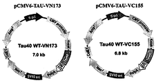
- FIG. 1 is a view showing a schematic diagram of the Tau40 WT-VN173 and Tau40 WT-VC155 vector according to an embodiment of the present invention.
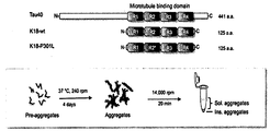
- FIG. 2 is a view illustrating a process of preparing non-aggregated tau, tau aggregates, dissolved aggregates, and non-agglomerated aggregates according to an embodiment of the present invention.
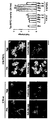
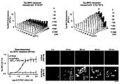
- FIG 3 is a view showing the tau aggregation transfer effect of tau aggregates in HEK293 tau aggregate cells according to an embodiment of the present invention.
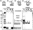
- FIG. 4 is a view showing the results of small and polymer separation of the dissolved aggregates using SDS-PAGE according to an embodiment of the present invention.
- FIG. 5 is a view showing the results of fluorescence expression of HEK293 tau aggregate cells upon treatment with reduced dimers by concentration according to an embodiment of the present invention.
- Figure 6 is a view showing the change in survival rate of HEK293 tau aggregate cells upon treatment with reduced dimers by concentration according to an embodiment of the present invention.
- FIG. 7 is a view showing the results of the degree of tau protein aggregation in HEK293 tau aggregate cells, according to an embodiment of the present invention.
- FIG. 8 is a view showing the effect of tau aggregation transfer in supercultured neurons according to an embodiment of the present invention.
- FIG. 9 is a view showing the results of measurement of neurodegeneration due to tau aggregation in the primary cultured neurons according to an embodiment of the present invention.
- FIG. 10 is a view showing the effect of tau aggregation transfer in the animal model, according to an embodiment of the present invention.
- FIG. 11 is a view showing the results of GFAP staining in the animal model, according to an embodiment of the present invention.
- a tau-BiFC vector was produced using a Venus-based BiFC system.
- the mammalian expression vector pCMV6-hTau40-GFP was used in the US OriGene Technologies Inc. (Rockville, MD) and full length human tau (441 amino acids) were fused to Venus N-terminal fragment (1-172 amino acids, VN173) and C-terminal fragment (155-238 amino acids, VC155).
- pBiFC-VN173 and pBiFC-VC155 were purchased from Addgene (Cambridge, Mass.) And then amplified using PCR primers with XhoI / PmeI restriction enzyme sequences. Each front and back primer sequence is shown in Table 1.
- pCMV6-hTau40-VN173 and pCMV6-hTau40-VC155 were constructed by cutting and conjugating pCMV6-hTau40-GFP and PCR amplified inserts using XhoI / PmeI.
- the linker peptide and Venus sequences used for pCMV6-hTau40-VN173 and pCMV6-hTau40-VC155 are shown in Tables 2 and 3.
- FIG. 1 A schematic diagram of pCMV6-hTau40-VN173 comprising the full length tau, VN173 linker peptide and VN173 peptide and pCMV6-hTau40-VC155 vector comprising the full length tau, VC155 linker peptide and VC155 peptide are shown in FIG. 1.
- HEK293 cells were purchased from ATCC (Manassas, VA) and 37 ° C., 5% with Dulbecco's modified eagle (DMEM) medium containing 10,000% / ml penicillin and 10,000 ⁇ g / ml streptomycin containing 10% fetal calf serum and 10,000 unit / ml penicillin. Cultured under CO 2 containing humidified atmospheric conditions. On the day before transformation, HEK-293 cells were seeded at 4.25-6.25 ⁇ 10 5 per well in 6-well culture dishes, with cell densities adjusted to 30-50%.
- DMEM Dulbecco's modified eagle
- tau aggregate transferability was analyzed using the tau-K18 region containing the microtubule binding domain portion of the total tau protein.
- Four types of tau aggregates were prepared as shown in Table 4 to secure various forms of tau aggregates from K18-P301L and K18-wt as controls.
- Non-aggregated tau Tau pre-aggregates Tau aggregates Tau Aggregates Generated Using DTT and Heparin Dissolved aggregates Soluble aggregates after centrifugation of tau aggregates Non-agglomerated aggregates Pellets after centrifugation of tau aggregates (insoluble aggregates)
- the non-aggregated tau was stirred at 240 rpm for 4 days at 37 ° C to prepare tau aggregates, and the prepared tau aggregates were centrifuged at 14,000 rpm for 20 minutes to separate dissolved aggregates as supernatants and insoluble aggregates as pellets.
- the manufacturing process of the four types of tau aggregates is shown in FIG.
- HEK293 tau aggregated cells of Example 1 were used to observe changes in tau inside cells by tau aggregates treated externally.
- HEK293 tau aggregated cells express fluorescence when the heterologous fluorescent proteins are conjugated to the tau inside the cell, thereby inducing tau aggregation.
- 10 ⁇ g / ml of four kinds of tau aggregates of K18-wt and K18-P301L each prepared in the method of Example 2 were treated with HEK293 tau aggregated cells together with the cell culture and further incubated for 24 hours.
- the results of tau aggregation inside cells by externally treated tau aggregates were analyzed by fluorescence microscopy (Operetta® High Content Imaging System).
- Example 3 Based on the results of Example 3, it was confirmed that the soluble aggregates formed in K18-P301L cause tau protein aggregation in normal cells. Subsequently, non-reducing SDS-PAGE (non-reducing SDS-PAGE) experiments were performed to determine whether any of the small, polymers in the soluble tau aggregates were delivered to normal cells to cause intracellular tau aggregation. SDS-PAGE was used a gradient gel of 10-14%. Through this experiment, tau oligomers can be classified from monomers to pentamers. As a result, it was confirmed that reduced dimers composed of crosslinked disulfide bonds in the non-aggregated tau, tau aggregates, and dissolved aggregates of K18-P301L commonly occupy a large part. The results are shown in FIG.
- the soluble tau aggregates of K18-wt and K18-P301L were treated to HEK293 tau aggregated cells by concentration, and then the fluorescence changes over time. Observed. In the case of K18-wt, no fluorescence change of BiFC was observed at almost all concentrations tested. However, when the highest concentration of 1000 nM reduction dimer was treated, it was confirmed that the fluorescence increased after 55 hours. Dissolved aggregates of K18-wt also had reduced dimers of crosslinked disulfide bonds, but their concentration was lower than that of K18-P301L.
- the MTS assay was performed to determine whether tau-BiFC fluorescence reduction and cell morphology change at high concentration of the reduced dimer were caused by cell death. As a measure of cell proliferation and viability, the MTS assay was purchased according to the manufacturer's protocol by purchasing Promega's product (CellTiter 96® AQueous One Solution Cell Proliferation Kit). After treatment of K18-wt and K18-P301L lysed aggregates to HEK293 tau aggregated cells, the MTS assay was performed using the cells at 55 hours and the cell activity was decreased at high concentrations. It can be thought that tau aggregation inside the cell is induced by external tau aggregates and further causes cell death. The results are shown in FIG.
- HEK293 tau aggregated cells treated with the HEK293 tau aggregated cells treated with the reduced dimers of K18-wt and K18-P301L were treated with Okadaic acid, which induces superphosphorylation of tau protein as a positive control.
- One HEK293 tau aggregated cells were used. Reactivity with two types of tau phosphorylated antibodies (pS199 / 202, pS396) in four types of cells revealed that HEK293 tau aggregated cells treated with K18-P301L reduced dimer were significantly higher in tau than the negative control untreated.
- Example 5 In primary cultured neurons Tau Confirmation of coagulation transition effect
- tau lesions were caused by reduced dimers of artificially disulfide bonds.
- MAPTP301L transgenic mice
- Tau transgenic mice show tau lesions as they age, but at eight weeks of age, the lesions have not yet begun.
- the fusion aggregates of K18-wt and K18-P301L were injected into the hippocampus of the transgenic mice, and two weeks later, it was confirmed by immunohistochemical staining that phosphorylation of tau in neurons was induced around the injected peripheral region.
- tau phosphorylated antibody pS199 / 202
- mice injected with PBS as a control did not induce any tau phosphorylation, but the mice injected with soluble aggregates of K18-P301L showed that tau phosphorylation was induced in neurons around the injection region.
- the phosphorylation of tau was observed when K18-wt dissolved aggregates were injected, but it did not show a significantly changed result. The results are shown in FIG.
- GFAP Gaal fibrillary acidic protein staining was also performed based on the injection region. This is a method of confirming the activity of the cells that react as physical damage is applied, PBS, K18-wt and K18-P301L all showed the same reactivity without difference. Therefore, it can be concluded that the induction of tau lesions of neurons is a reduced dimer of externally injected disulfide crosslinks.
- the GFAP staining results are shown in FIG. 11.
Landscapes
- Health & Medical Sciences (AREA)
- Life Sciences & Earth Sciences (AREA)
- Chemical & Material Sciences (AREA)
- Engineering & Computer Science (AREA)
- Genetics & Genomics (AREA)
- Biomedical Technology (AREA)
- Organic Chemistry (AREA)
- Molecular Biology (AREA)
- Biotechnology (AREA)
- Zoology (AREA)
- General Health & Medical Sciences (AREA)
- Biochemistry (AREA)
- Bioinformatics & Cheminformatics (AREA)
- Immunology (AREA)
- Urology & Nephrology (AREA)
- General Engineering & Computer Science (AREA)
- Wood Science & Technology (AREA)
- Medicinal Chemistry (AREA)
- Hematology (AREA)
- Physics & Mathematics (AREA)
- Microbiology (AREA)
- Biophysics (AREA)
- Toxicology (AREA)
- Food Science & Technology (AREA)
- Proteomics, Peptides & Aminoacids (AREA)
- Analytical Chemistry (AREA)
- General Physics & Mathematics (AREA)
- Pathology (AREA)
- Gastroenterology & Hepatology (AREA)
- Plant Pathology (AREA)
- Cell Biology (AREA)
- Peptides Or Proteins (AREA)
- Neurology (AREA)
- Tropical Medicine & Parasitology (AREA)
Abstract
본 발명은 전이성 타우 단백질 및 이의 제조 방법에 관한 것이다. 신경세포내 타우 단백질 응집은 알츠하이머, 측두엽 변성증, 픽병, 피질기조퇴행 및 퇴행성 핵산 마비 등 다양한 타우병증 질환을 야기한다. 최근, 타우 단백질이 프리온 단백질처럼 세포간에 전이되어 정상 세포내의 타우 단백질 응집을 유도한다는 보고가 이어지고 있으나, 타우 단백질 내에서 어느 부분이 전이성을 가지는지, 전이 메커니즘이 어떻게 이루어지는가에 대한 연구는 미비한 실정이다. 본 발명은 전이성을 가지는 타우 단백질 응집체의 분리에 관한 것으로, 타우 단백질 응집체의 전이 메카니즘을 밝히고, 전이 억제 후보 물질의 개발에 기여할 수 있을 것으로 기대된다.
Description
본 발명은 전이성 타우 단백질 및 이의 제조 방법에 관한 것이다.
타우 단백질이란, 분자량이 50,000~70,000을 나타내는 미소관결합단백질의 일종으로, 인산화에 의해 현저한 분자 다양성을 나타낸다. 사람의 경우 타우는 6종류의 동형으로 구성되어 있는데, N말단부분의 29 또는 58아미노산 잔기의 삽입, C말단의 3개 또는 4개의 반복구조(미소관 결합부위라고 함)의 이어맞추기에서 6개의 동형이 생성된다. 이전에는 타우 단백질이 중추신경계에 특이적이고 주로 축삭에 존재한다고 생각했지만 현재는 비교적 많은 조직에, 또한 신경세포 외의 성상세포, 올리고수지신경교에 발현하고 있다는 것, 또한 축삭만이 아니라 수상돌기에도 존재한다는 것이 알려졌다. 타우는 미소관의 안정성에 기여하고, 인산화에 의한 과도한 타우 단백질 응집은 다양한 뇌신경 질환을 유발한다. 즉, 알츠하이머 병을 포함한 이마 측두엽 변성증(Frontotemporal lobar degeneration, FTLD), 픽병 (Pick’s disease), 피질기조퇴행 (Corticobasal degeneration, CBD), 퇴행성 핵상 마비 (progressive supranuclear palsy, PSP) 등의 중추 신경계 질환들은 타우 (Tau) 단백질로 구성된 응집체를 포함하는 것으로 알려졌다. 이러한 병리학적인 특징 때문에 이 신경질환들을 통틀어 타우병증(tauopathies)이라고 부른다. 타우병증 환자에게서 보이는 타우 단백질의 응집체는 주로 신경세포의 세포체와 수상돌기에서 발견되며, 이를 신경원섬유덩굴 (Neurofibrillary tangles, NFT)과 신경그물 실(neuropil threads)이라 부른다. 신경원섬유덩굴을 살펴보면 타우 단백질이 가는 실 같이 엉켜있는 이중 나선 섬유(paired helical filaments, PHFs)로 이루어지며 이는 정상적인 타우 단백질과는 다르게 응집되고 과인산화가 일어나 있다. 비록 타우병증에서 보이는 비정상적인 타우 단백질의 응집현상이 질병 심화 단계에서 어떤 역할을 하는지 정확히 알져지진 않았지만 헌팅턴, 파킨슨, 크로이츠펠트-야콥병 (광우병) 등 퇴행성 뇌질환에서 공통적으로 나타나는 응집현상과 비슷하며, 신경원섬유덩굴 형성과 인지 능력상실 사이에 유의미한 상관 관계를 가지는 점 등을 비추어 볼 때 타우 단백질이 중요한 역할을 하는 것은 분명하다.
최근, 이러한 타우 응집이 전이성을 가진다는 보고가 이어지고 있다(Nat Cell Biol. 2009 July 11(7): 909?913.; Neuron. 2014 Jun 18;82(6):1271-88.). 타우 단백질이 프리온 단백질처럼 세포간에 전이되어 정상 세포내의 타우 단백질 응집을 유도한다는 것이다. 그러나 타우 단백질 내에서 어느 부분이 전이성을 가지는지, 전이 메커니즘이 어떻게 이루어지는가에 대한 연구는 미비한 실정이다. 타우 단백질 응집체가 다양한 타우병증 질환의 원인이 된다는 것을 상기할 때, 타우 단백질 응집체의 전이성은 퇴행성 뇌질환의 전이 및 진행을 가속화할 수 있다. 따라서 타우 단백질 응집체의 전이 메카니즘을 밝히고 이를 조기에 제어하는 기술의 확보가 시급히 요구되고 있다.
이에 따라 본 연구팀은 타우 단백질 응집체들 중에서 어떠한 응집체가 이러한 전염성을 가지는지 확인하고, 전이성을 가지는 타우 단백질 응집체를 분리하였다. 상기 개발된 전이성 타우 단백질 응집체는 타우 단백질 응집체의 전이 메카니즘을 밝히고, 전이 억제 후보 물질의 개발에 기여할 수 있을 것으로 기대된다.
본 발명은 상기와 같은 종래의 기술상의 문제점을 해결하기 위해 안출된 것으로, 전이성 타우 단백질 및 이의 제조 방법에 관한 것이다.
그러나 본 발명이 이루고자 하는 기술적 과제는 이상에서 언급한 과제에 제한되지 않으며, 언급되지 않은 또 다른 과제들은 아래의 기재로부터 당 업계에서 통상의 지식을 가진 자에게 명확하게 이해될 수 있을 것이다.
이하, 본원에 기재된 다양한 구체예가 도면을 참조로 기재된다. 하기 설명에서, 본 발명의 완전한 이해를 위해서, 다양한 특이적 상세사항, 예컨대, 특이적 형태, 조성물 및 공정 등이 기재되어 있다. 그러나, 특정의 구체예는 이들 특이적 상세 사항 중 하나 이상 없이, 또는 다른 공지된 방법 및 형태와 함께 실행될 수 있다. 다른 예에서, 공지된 공정 및 제조 기술은 본 발명을 불필요하게 모호하게 하지 않게 하기 위해서, 특정의 상세사항으로 기재되지 않는다. "한 가지 구체예" 또는 "구체예"에 대한 본 명세서 전체를 통한 참조는 구체예와 결부되어 기재된 특별한 특징, 형태, 조성 또는 특성이 본 발명의 하나 이상의 구체예에 포함됨을 의미한다. 따라서, 본 명세서 전체에 걸친 다양한 위치에서 표현된 "한 가지 구체예에서" 또는 "구체예"의 상황은 반드시 본 발명의 동일한 구체예를 나타내지는 않는다. 추가로, 특별한 특징, 형태, 조성, 또는 특성은 하나 이상의 구체예에서 어떠한 적합한 방법으로 조합될 수 있다.
명세서에서 특별한 정의가 없으면 본 명세서에 사용된 모든 과학적 및 기술적인 용어는 본 발명이 속하는 기술분야에서 당업자에 의하여 통상적으로 이해되는 것과 동일한 의미를 가진다.
본 발명의 일 구체예에서 타우 단백질(Tau)이란, 분자량이 50,000~70,000을 나타내는 미소관결합단백질의 일종이다. 뇌신경 세포 내의 비정상적인 타우 단백질(tau)의 응집은 알츠하이머병 (Alzheimer's disease, AD) 및 다른 여러 신경퇴행성 질병들 (집합적으로, 타우병증(tauopathies)이라 부름)에 있어서 주된 특징이다 (Biochimica. et biophysica. acta. 1739: 331-354). 건강한 신경에서, 타우는 축색돌기(axon)로부터의 성장 및 신경 세포 분극화(polarization)를 촉진함으로써 마이크로세관(microtubules)을 안정화한다. 병리학적으로 과인산화(hyperphosphorylation)되는 경우에, 타우는 마이크로세관으로부터 분리되어 불용성 응집물을 생성한다(Mol. Neurodegener. 4: 13). 여러 해에 걸쳐서, 타우 응집에 대한 구조적 골격이 제안된 바 있으며, 10개의 가용성 모노머들로부터 불용성 필라멘트가 형성되고, 이러한 필라멘트가 신경섬유매듭(neurofibrillary tangles, NFTs)이라 불리우는 고차원 구조로 결합된다는 증거들이 제안된 바 있다. 그러나, 아직까지도 타우병증에 있어서 신경섬유매듭들의 병리생리학적 중요성, 이러한 과정을 유발하는 원인 및 분자적 메카니즘에 대해서는 그다지 알려진 바가 없는데, 이는 생리학적 조건 하에서 타우 응집을 모니터링하기 위한 신뢰성 있는 방법이 존재하지 않는다는 점 때문이다. 타우 응집에 대한 대부분의 연구들은 정제된 타우 또는 타우 단편을 사용하여 비생리학적 조건 하에서 수행되었다. 더 나아가, 타우는 용해도가 극히 높기 때문에, 타우 응집은 헤파린과 같은 보조인자를 첨가함으로써 인위적으로 유도된다. 이러한 연유로, 살아있는 세포 내에서 타우 어셈블리를 모니터링할 수 있는 세포 기반 모델은 타우 병리학을 연구하고, 그 과정을 방지 및 되돌릴 수 있는 방법들을 개발하는데 유용한 도구가 될 수 있다.
본 발명의 일 구체예에서 단백질 전이란, 하나의 세포 내에서 생성된 단백질이 다른 세포로 이동되어 가는 것이다. 신경 단백질의 일종인 타우 단백질의 경우, 명확한 기전이 밝혀진 것은 아니나, 신경세포내 마이크로듀불 또는 세포 외부의 세포외기질복합체(extracellular matrix, ECM)를 타고 점핑하여 이동하는 것으로 생각되고 있다. 단백질 전이는 타겟 유전자의 발현 억제 또는 타겟 유전자가 코딩하는 단백질의 활성 억제를 통해 조절될 수 있다.
본 발명의 일 구체예에서 이분자 형광 상보 기법(BiFC)란, 관심 대상이 되는 단백질에 부착된 비형광성 구성요소로부터 형광 단백질 복합체를 형성하는 기술에 기반을 둔 단백질-단백질 상호작용을 가시화하기 위한 방법이다(Annu. Rev. Biophys. 37: 465-487). 종래에는 분할 녹색 형광 단백질 (green fluorescent protein, GFP) 상보 기법이 타우 응집을 정량화하기 위해 사용되었는데, 이러한 방법은 타우를 더 작은 GFP 단편들에 융합시키고(GFP 11), 더 큰 GFP 단편(GFP 1-10)을 갖는 세포들에서 발현시켰다. 타우가 모노머 또는 작은 응집체로 존재하는 경우에는, 큰 GFP 단편이 타우에 융합된 작은 GFP 단편에 접근할 수 있으며, 이로 인해서 형광 활성을 갖는 GFP의 결합이 이루어진다. 그러나, 타우가 응집하는 경우에는 활성 GFP의 재구성이 억제되고, 세포들에서 GFP 형광이 감소하게 된다. 따라서 타우의 응집을 정량화하는 방법으로서 상기 분할-GFP 분석법이 제안되었지만, 분해능이 낮고, 형광의 감소를 측정할 수 있는 범위가 제한적이므로, 신경 독성의 원인이 되는 타우 올리고머의 생성과정을 모니터링하기에는 적합하지 않은 문제점이 있었다. 그러나 최근의 비너스 단백질을 이용한 BiFC는 서로 다른 두 표적 단백질들이 상호작용을 하기 위해 근접하게 되면 표적 단백질에 연결된 형광단백질 절편 또한 가까워지게 되고, 그 결과 형광 절편 단백질 사이에 재구성(reconstruction)이 일어나게 되어 형광을 발하게 되는 것이다. 이로써 두 표적 단백질간에 상호작용이 일어났음을 시각적으로 확인할 수 있는 기술이다. 이 기술은 살아있는 세포 내에서, 즉 단백질간 상호작용이 일어나고 유지될 수 있는 최적의 물리·화학적인 환경하에서, 단백질간 상호작용을 직접 눈으로 확인할 수 있는 특징을 지니고 있다. 이 기술은 세포 내에서 단백질 상호작용이 일어나는 위치뿐만 아니라 이들의 움직임 정보까지도 확인 가능하게 한다.
본 발명의 일 구체예에서 비너스(Venus) 단백질이란, 황색 형광 단백질 (yellow fluorescence protein, YFP)의 일종으로서, (1) 빠르고 효과적으로 성숙되고 (maturation), (2) 자가-조립 (self-assembly) 속도가 다른 BiFC 쌍들에 비해서 낮으며, (3) 비너스계 BiFC의 형광 강도가 EYFP계 BiFC의 형광 강도보다 10배 더 높은 특징을 보인다(Biotechniques 40: 61-66.; Biotechniques 49: 793-805). 따라서 타우 단백질과 같이 시공간적으로 분석하기 어려운 세포내 단백질 분석에 유용하게 사용될 수 있다.
본 발명의 일 구체예에서 용해 응집체란, 친수성 기질을 갖는 단백질 응집체를 의미한다. 본 명세서에 있어서 용해 응집체는 친수성 기질의 타우 단백질 응집체이며, 전체 타우 단백질 중에서 미세소관 결 도메인 부분을 포함하는 K18-wt 또는 K18-P301L 등을 37℃에서 4일간 240 rpm으로 교반하여 타우 응집체를 제조한 후, 제조된 타우 응집체를 14,000 rpm에서 20분간 원심분리하여 제조할 수 있다. 상기의 원심분리 실시 후 상층액으로서의 용해 응집체와 펠렛으로서의 비용해 응집체를 분리할 수 있다.
본 발명의 일 구체예에서 환원 이합체(Reduced dimer)란, 전체 타우 단백질 중에서 미세소관 결 도메인 부분을 포함하는 K18-wt 또는 K18-P301L 등의 응집 형태, 비응집 형태, 친수성 응집체 형태 또는 소수성 응집체 형태로부터 분리한 분자량 25kDa 정도의 이황화교차(S-S)결합을 갖는 환원성 이량체이다. 본 명세서에 있어서 환원 이합체는 두 타우 단량체가 291번째, 혹은 322번째 아미노산 시스테인 (Cysteine, C)에 의해서 단 하나의 이황화 교차 결합을 가지는 것으로 정의될 수 있고, 타우 단백질 용해 응집체를 10-14%의 SDS-PAGE 농도경사 겔(gradient gel)을 사용하여 분리할 수 있다.
본 발명의 일 구체예에서, 서열번호 1 또는 2로 표시되는 단백질을 포함하는 전이성 용해 응집체를 제공하고, 상기 전이성 용해 응집체는 타우 환원 이합체인 전이성 용해 응집체를 제공하며, 상기 타우 환원 이합체는 두 개의 타우 단량체가 N말단으로부터 291번째 또는 322번째 아미노산 시스테인에 의해서 이황화교차 결합되는 전이성 용해 응집체를 제공하며, 상기 이황화교차 결합은 두 개의 타우 단량체에서 291번-291번, 291번-322번, 및 322번-322번 아미노산 간에 발생하는 전이성 용해 응집체를 제공하며, 상기 타우 환원 이합체는 분자량이 20kDa 내지 30kDa인 전이성 용해 응집체를 제공한다.
본 발명의 다른 구체예에서, 서열번호 1 또는 2로 표시되는 단백질을 포함하는 전이성 용해 응집체, 상기 전이성 용해 응집체는 타우 환원 이합체인 전이성 용해 응집체, 상기 타우 환원 이합체는 두 개의 타우 단량체가 N말단으로부터 291번째 또는 322번째 아미노산 시스테인에 의해서 이황화교차 결합되는 전이성 용해 응집체, 상기 이황화교차 결합은 두 개의 타우 단량체에서 291번-291번, 291번-322번, 및 322번-322번 아미노산 간에 발생하는 전이성 용해 응집체, 및 상기 타우 환원 이합체는 분자량이 20kDa 내지 30kDa인 전이성 용해 응집체 중 어느 하나를 포함하는 형질전환체를 제공하고, 상기 형질전환체는 사람을 제외한 동물인 형질전환체를 제공한다.
본 발명의 또 다른 구체예에서, 서열번호 1 또는 2로 표시되는 단백질을 교반하여 타우 응집체를 제조하는 단계; 상기 타우 응집체를 원심분리하는 단계; 및 원심분리 후 상층액으로서의 전이성 용해 응집체를 분리하는 단계를 포함하는 전이성 용해 응집체의 제조 방법을 제공하고, 상기 타우 응집체의 제조는 30℃ 내지 45℃ 에서 수행하는 전이성 용해 응집체의 제조 방법을 제공하며, 상기 타우 응집체의 제조는 200 내지 300 rpm으로 교반하는 전이성 용해 응집체의 제조 방법을 제공하며, 상기 타우 응집체의 제조는 1일 내지 10일간 교반하는 전이성 용해 응집체의 제조 방법을 제공하며, 상기 타우 응집체를 원심분리하는 단계는 10,000 내지 20,000 rpm으로 원심분리하는 전이성 용해 응집체의 제조 방법을 제공한다.
본 발명의 또 다른 구체예에서, 서열번호 1 또는 2로 표시되는 단백질을 포함하는 전이성 용해 응집체, 상기 전이성 용해 응집체는 타우 환원 이합체인 전이성 용해 응집체, 상기 타우 환원 이합체는 두 개의 타우 단량체가 N말단으로부터 291번째 또는 322번째 아미노산 시스테인에 의해서 이황화교차 결합되는 전이성 용해 응집체, 상기 이황화교차 결합은 두 개의 타우 단량체에서 291번-291번, 291번-322번, 및 322번-322번 아미노산 간에 발생하는 전이성 용해 응집체, 및 상기 타우 환원 이합체는 분자량이 20kDa 내지 30kDa인 전이성 용해 응집체 중 어느 하나에 따른 전이성 용해 응집체를 신경 세포에 주입하는 단계; 상기 신경 세포에서 타우 단백질 응집 정도를 평가하는 단계; 상기 신경 세포에 타우 단백질 응집 저해 후보 물질을 처리하는 단계; 상기 저해 후보 물질이 처리된 신경 세포에서의 타우 단백질 응집 정도를 평가하는 단계; 및 저해 후보 물질 처리 전후의 타우 단백질 응집 정도를 비교하는 단계를 포함하는 타우 단백질 전이 저해 후보 물질의 스크리닝 방법을 제공한다.
본 발명의 또 다른 구체예에서, 신경 세포에 타우 단백질 전이 예방 후보 물질을 처리하는 단계; 상기 신경 세포에 서열번호 1 또는 2로 표시되는 단백질을 포함하는 전이성 용해 응집체, 상기 전이성 용해 응집체는 타우 환원 이합체인 전이성 용해 응집체, 상기 타우 환원 이합체는 두 개의 타우 단량체가 N말단으로부터 291번째 또는 322번째 아미노산 시스테인에 의해서 이황화교차 결합되는 전이성 용해 응집체, 상기 이황화교차 결합은 두 개의 타우 단량체에서 291번-291번, 291번-322번, 및 322번-322번 아미노산 간에 발생하는 전이성 용해 응집체, 및 상기 타우 환원 이합체는 분자량이 20kDa 내지 30kDa인 전이성 용해 응집체 중 어느 하나에 따른 전이성 용해 응집체를 주입하는 단계; 상기 신경 세포에서의 타우 단백질 응집 정도를 평가하는 단계; 및 예방 후보 물질이 처리된 신경 세포에서의 타우 단백질 응집 정도를 대조군과 비교하는 단계를 포함하는 타우 단백질 전이 예방 후보 물질의 스크리닝 방법을 제공한다.
이하 상기 본 발명을 단계별로 상세히 설명한다.
본 발명은 전이성 타우 단백질 및 이의 제조 방법에 관한 것으로, 타우 단백질 응집체의 전이 메카니즘을 밝히고, 전이 억제 후보 물질의 개발에 기여할 수 있을 것으로 기대된다.
도 1은 본 발명의 일 실시예에 따른, Tau40 WT-VN173 및 Tau40 WT-VC155 벡터의 모식도를 나타낸 도면이다.
도 2는 본 발명의 일 실시예에 따른, 비응집 타우, 타우 응집체, 용해 응집체 및 비용해 응집체의 제조 과정을 나타낸 도면이다.
도 3은 본 발명의 일 실시예에 따른, HEK293 타우 응집 세포에서 타우 응집체의 타우 응집 전이 효과를 나타낸 도면이다.
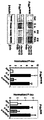
도 4는 본 발명의 일 실시예에 따른, SDS-PAGE을 이용한 용해 응집체의 소,중합체 분리 결과를 나타낸 도면이다.
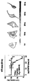
도 5는 본 발명의 일 실시예에 따른, 농도별 환원 이합체 처리시 HEK293 타우 응집 세포의 형광 발현 결과를 나타낸 도면이다.
도 6은 본 발명의 일 실시예에 따른, 농도별 환원 이합체 처리시 HEK293 타우 응집 세포의 생존율 변화를 나타낸 도면이다.
도 7은 본 발명의 일 실시예에 따른, HEK293 타우 응집 세포 내의 타우 단백질 응집 정도 결과를 나타낸 도면이다.
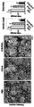
도 8은 본 발명의 일 실시예에 따른, 초대배양 신경세포에서의 타우 응집 전이 효과를 나타낸 도면이다.
도 9는 본 발명의 일 실시예에 따른, 초대배양 신경세포에서의 타우 응집으로 인한 신경퇴화 측정 결과를 나타낸 도면이다.
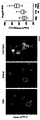
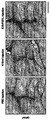
도 10은 본 발명의 일 실시예에 따른, 동물 모델에서의 타우 응집 전이 효과를 나타낸 도면이다.
도 11은 본 발명의 일 실시예에 따른, 동물 모델에서의 GFAP 염색 결과를 나타낸 도면이다.
이하, 실시예를 통하여 본 발명을 더욱 상세히 설명하고자 한다. 이들 실시예는 오로지 본 발명을보다 구체적으로 설명하기 위한 것으로서, 본 발명의 요지에 따라 본 발명의 범위가 이들 실시예에 의해 제한되지 않는다는 것은 당업계에서 통상의 지식을 가진 자에게 있어서 자명할 것이다.
실시예 1. HEK293 타우 응집 세포주의 제조
실시예 1-1. 타우-BiFC 벡터의 제작
본 발명에서는 비너스 (Venus) 기반의 BiFC 시스템을 사용하여 타우-BiFC 벡터를 제작하였다. 이를 위해, 포유류 발현 벡터 pCMV6-hTau40-GFP를 미국 OriGene Technologies Inc. (Rockville, MD)로부터 구입하였고, 전장 인간 타우 (441개 아미노산)를 비너스의 N-말단 단편 (1-172 아미노산, VN173) 및 C-말단 단편 (155-238 아미노산, VC155)에 융합시켰다. 구체적으로, GFP를 BiFC 부분으로 치환하기 위해서, pBiFC-VN173 및 pBiFC-VC155를 Addgene (Cambridge, MA)으로부터 구입 후, XhoI/PmeI 제한효소 서열을 갖는 PCR 프라이머를 사용하여 증폭시켰다. 각각의 전방 및 후방 프라이머 서열은 표 1에 나타내었다.
| (VC155-F) | 5'-AATTCGGTCG ACCGAGATCT CTCGAGGTAC-3' |
| (VC155-R) | 5'-CTAGTTGTGG TTTGTTTAAA CTCATCAATG TATC-3' |
| (VN173-F) | 5'-ATGACGACAA GCTCGAGGCC GCGAATTCAT CG-3' |
| (VN173-R) | 5'-CTAGTTGTGG TTTGTTTAAA CTCATCAATG TATC-3' |
pCMV6-hTau40-GFP 및 PCR 증폭된 인써어트를 XhoI/PmeI를 사용하여 절단하고 접합시킴으로써 본 발명에 따른 pCMV6-hTau40-VN173 및 pCMV6-hTau40-VC155를 제작하였다. pCMV6-hTau40-VN173 및 pCMV6-hTau40-VC155에 사용한 링커 펩타이드 및 비너스 서열을 표 2와 표 3에 나타내었다.
| pCMV6-hTau40-VN173 | |
| 링커 펩타이드 | ACG CGT ACG CGG CCG CTC GAG TCT AGA AGA TCC ATC GCC ACC |
| 비너스(VN173) | ATG GTG AGC AAG GGC GAG GAG CTG TTC ACC GGG GTG GTG CCC ATC CTG GTC GAG CTG GAC GGC GAC GTA AAC GGC CAC AAG TTC AGC GTG TCC GGC GAG GGC GAG GGC GAT GCC ACC TAC GGC AAG CTG ACC CTG AAG CTG ATC TGC ACC ACC GGC AAG CTG CCC GTG CCC TGG CCC ACC CTC GTG ACC ACC CTG GGC TAC GGC CTG CAG TGC TTC GCC CGC TAC CCC GAC CAC ATG AAG CAG CAC GAC TTC TTC AAG TCC GCC ATG CCC GAA GGC TAC GTC CAG GAG CGC ACC ATC TTC TTC AAG GAC GAC GGC AAC TAC AAG ACC CGC GCC GAG GTG AAG TTC GAG GGC GAC ACC CTG GTG AAC CGC ATC GAG CTG AAG GGC ATC GAC TTC AAG GAG GAC GGC AAC ATC CTG GGG CAC AAG CTG GAG TAC AAC TAC AAC AGC CAC AAC GTC TAT ATC ACC GCC GAC AAG CAG AAG AAC GGC ATC AAG GCC AAC TTC AAG ATC CGC CAC AAC ATC GAG TAG |
| pCMV6-hTau40-VC155 | |
| 링커 펩타이드 | ACG CGT ACG CGG CCG CTC GAG AAG |
| 비너스(VC155) | CAGAAGAACGGCATCAAGGCCAACTTCAAGATCCGCCACAACATCGAGGACGGCGGCGTGCAGCTCGCCGACCACTACCAGCAGAACACCCCCATCGGCGACGGCCCCGTGCTGCTGCCCGACAACCACTACCTGAGCTACCAGTCCAAACTGAGCAAAGACCCCAACGAGAAGCGCGATCACATGGTCCTGCTGGAGTTCGTGACCGCCGCCGGGATCACTCTCGGCATGGACGAGCTGTACAAGTAA |
상기의 전장타우, VN173 링커 펩타이드 및 VN173 펩타이드를 포함하는 pCMV6-hTau40-VN173과 전장타우, VC155 링커 펩타이드 및 VC155 펩타이드를 포함하는 pCMV6-hTau40-VC155 벡터의 모식도를 도 1에 나타내었다.
실시예 1-2. 타우-BiFC 벡터를 이용한 HEK293 세포의 형질전환
HEK293 세포는 ATCC (Manassas, VA)로부터 구입하였으며, 10% 우태아 혈청 및 10,000 unit/㎖의 페니실린을 함유하는 DMEM(Dulbecco's modified eagle) 배지 및 10,000 μg/㎖의 스트렙토마이신과 함께 37 ℃, 5% CO2 함유 가습 대기 조건 하에서 배양하였다. 형질전환 전날에, HEK-293 세포들을 6-웰 배양 접시에 웰당 4.25-6.25x10⁵ 씩 접종하였고, 이 때의 세포 치밀도는 30-50 %가 되도록 조절하였다. 형질전환을 위해서 2.5 μg의 타우-BiFC 벡터 DNA 또는 0.75-1.75 ㎕의 리포펙타민 2000 시약(Lipofectamine2000 Reagent, Invitrogen, Carlsbad, CA)을 각각 100 ㎕의 무혈청 Opti-MEM® 배양액에 희석하여 준비하였고, 상기 두 혼합액을 천천히 섞고 상온에서 20분간 반응시킨 후, 세포가 있는 배양접시에 천천히 첨가하였다. 형질전환 시행된 세포는 이후 18-24 시간 동안 37 ℃, 5% CO2 함유 가습 대기 조건 하에서 추가로 배양하였다. 상기의 방법으로 제조된 형질전환 완료 타우-BiFC HEK293 세포를 KCTC 미생물자원센터에 기탁하였다(기탁번호 KCTC 12746BP).
실시예 2. 타우 단백질 K18-wt 및 K18-P301L의 다양한 응집체 제작
타우 응집체들 중에서 어떠한 응집체가 전이성을 가지는 지 확인하기 위하여, 전체 타우 단백질 중에서 미세소관 결 도메인 부분을 포함하는 타우-K18 영역을 이용하여 타우 응집 전이성을 분석하였다. 타우병증을 가지고 있는 환자에게서 발견되고 있는 타우-K18의 돌연변이 형태인 K18-P301L(서열번호 1)은 자연형인 K18-wt(서열번호 2)에서 301번째 아미노산 프롤린(Proline, P)이 류신(Leucine, L)로 치환된 것이다. K18-P301L 및 대조군으로서의 K18-wt 으로부터 타우 응집체의 다양한 형태를 확보하기 위해서 표 4에 기재한 바와 같이 각각 4종류의 타우 응집체를 제작하였다.
| 비응집 타우 | 타우가 응집과정을 거치기 전(pre-aggregates) |
| 타우 응집체 | DTT와 헤파린을 이용하여 생성한 타우 응집체(aggregates) |
| 용해 응집체 | 타우 응집체를 원심분리한 후의 상층액(soluble aggregates) |
| 비용해 응집체 | 타우 응집체를 원심분리한 후의 펠렛(insoluble aggregates) |
먼저, 비응집 타우를 37℃에서 4일간 240 rpm으로 교반하여 타우 응집체를 제조하였고, 제조된 타우 응집체를 14,000 rpm에서 20분간 원심분리하여 상층액으로서의 용해 응집체와 펠렛으로서의 비용해 응집체를 분리하였다. 상기 4종류의 타우 응집체의 제조 과정을 도 2에 나타내었다.
실시예
3.
타우
응집체의
타우
응집 전이 효과 확인
외부에서 처리된 타우 응집체에 의한 세포 내부의 타우의 변화를 관찰하기 위해 실시예 1의 HEK293 타우 응집 세포를 이용하였다. HEK293 타우 응집 세포는 세포 내부의 타우에 이분화된 형광 단백질이 각각 접합되어 타우의 응집이 유도되었을 경우 형광을 발현하게 된다. HEK293 타우 응집 세포에 실시예 2의 방법으로 제조된 K18-wt 및 K18-P301L 각각의 4종류의 타우 응집체 10 μg/㎖를 세포 배양액과 함께 처리하고, 24시간 동안 추가로 배양하였다. 외부에서 처리된 타우 응집체에 의한 세포 내부의 타우 응집 결과를 형광 현미경(Operetta® High Content Imaging System)을 이용하여 분석한 결과, K18-P301L의 경우, 비응집 타우 및 타우 응집체 모두 형광변화를 유도하였으나, K18-wt은 어떠한 형태의 응집체도 BiFC의 형광변화를 유도하지 못하였다. 또한 용해 응집체와 비용해 응집체를 비교시, K18-P301L의 경우 용해 응집체만이 BiFC의 형광변화를 유도하였으며, 비용해 응집체는 어떠한 형광변화도 일으키지 못하였다. 상기 결과를 도 3에 나타내었다.
실시예
4. 용해성
타우
응집체 중
소,중합체의
응집 전이 효과 확인
실시예 3의 결과를 바탕으로 K18-P301L에서 형성되는 용해 응집체가 정상 세포내의 타우 단백질 응집을 일으킨다는 것을 확인하였다. 이후, 용해성 타우 응집체 중에서 어떠한 소,중합체들이 정상 세포로 전달되어 세포 내 타우 응집을 일으키는지 확인하기 위하여 이황화교차(S-S)결합 비절단 SDS-PAGE(non-reducing SDS-PAGE) 실험을 실시하였다. SDS-PAGE은 10-14%의 농도경사 겔(gradient gel)을 사용하였다. 이 실험을 통해서 타우 올리고머는 작게는 단량체(Monomer)부터 오량체(Pentamer)까지 구분될 수 있다. 실험 결과, K18-P301L의 비응집 타우, 타우 응집체, 및 용해 응집체에서 이황화교차 결합으로 이루어진 환원 이합체(Reduced dimer)가 공통적으로 많은 부분을 차지하는 것을 확인할 수 있었다. 그 결과를 도 4에 나타내었다.
상기의 이황화교차 결합으로 이루어진 환원 이합체(Reduced dimer)의 프리온 성질을 확인하기 위하여, K18-wt과 K18-P301L의 용해성 타우 응집체를 농도별로 HEK293 타우 응집 세포에 처리한 후, 시간 별로 형광의 변화를 관찰하였다. K18-wt의 경우, 시험된 거의 모든 농도에서 BiFC의 형광변화가 관찰되지 않았다. 다만 가장 높은 농도인 1000 nM의 환원 이합체를 처리하였을 때, 55시간이 지난 후에 형광이 증가되는 것을 확인할 수 있었다. K18-wt의 용해 응집체 역시 이황화교차 결합의 환원 이합체를 가지고 있었지만 K18-P301L에 비하여 그 농도가 낮았다. 그러므로 농도가 높아지면서 환원 이합체의 농도도 조금씩 높아져 tau-BiFC의 형광변화를 유도했을 것으로 해석된다. 이에 반해 K18-P301L의 경우, 처리한 모든 농도에서 HEK293 타우 응집 세포의 형광이 증가되는 것을 확인할 수 있었다. 28 시간에서의 형광 값들을 이용하여 EC50 농도를 계산하였을 때, 23.2 nM의 환원 이합체가 반 최대 유효 농도(half maximal effective concentration)임을 알 수 있었다. 하지만 시간이 지남에 따라 120 nM 이상의 환원 이합체가 처리된 HEK293 타우 응집 세포의 형광이 처리 후 39시간부터 형광 레벨이 감소되는 것으로 나타났다. 농도 별로는 보다 높은 농도가 처리되었을 때, 더 빠른 속도로 형광이 감소되는 양상을 보였으며, HEK293 타우 응집 세포의 형태 역시 변화되는 것을 관찰하였다. 상기 결과를 도 5에 나타내었다.
상기 환원 이합체 고농도에서의 tau-BiFC 형광 감소 및 세포 형태 변화가 세포 사멸에 의한 것인지를 확인하기 위하여 MTS 어세이를 진행하였다. 세포 증식 및 생존율 측정으로서 MTS 어쎄이는 프로메가사의 제품(CellTiter 96® AQueous One Solution Cell Proliferation Kit)을 구입하여 제조사의 프로토콜에 따라 실시하였다. HEK293 타우 응집 세포에 K18-wt과 K18-P301L 용해 응집체를 처리한 후 55 시간 때의 세포를 이용하여 MTS 어세이를 진행한 결과 높은 농도에서 세포의 활성도가 감소되는 것을 확인할 수 있었다. 이는 외부의 타우 응집체에 의해 세포 내부의 타우 응집이 유도 되고 더 나아가 세포 사멸을 야기하는 것으로 생각될 수 있다. 그 결과를 도 6에 나타내었다.
또한, HEK293 타우 응집 세포 내의 타우의 응집 정도를 확인하기 위해서, 웨스턴 블랏팅(Western blotting) 실험을 실시하였다. K18-wt 및 K18-P301L의 환원 이합체가 처리된 HEK293 타우 응집 세포의 음성대조군으로 아무것도 처리하지 않은 HEK293 타우 응집 세포를, 양성대조군으로 타우 단백질의 과인산화를 유도하는 오카다익산(Okadaic acid)을 처리한 HEK293 타우 응집 세포를 사용하였다. 4종류의 세포에서 두 가지 종류의 타우 인산화 항체(pS199/202, pS396)와의 반응성을 확인한 결과, K18-P301L 환원 이합체가 처리된 HEK293 타우 응집 세포가 아무것도 처리되지 않은 음성대조군에 비해서 유의성 있게 높은 타우 인산화 항체들과의 반응성을 나타냈으며, 이는 양성대조군인 오카다익산 처리 세포와 유사한 값을 보였다. 그러므로 세포 외부의 이황화 결합으로 이루어진 환원 이합체는 세포 내의 타우 단백질의 인산화 과정을 유도하고 응집시킬 뿐만 아니라, 세포 사멸까지 일으킨다는 것을 확인하였다. 상기 HEK293 타우 응집 세포 내의 타우 단백질 응집 정도 결과를 도 7에 나타내었다.
실시예
5. 초대배양 신경세포에서의
타우
응집 전이 효과 확인
실제 타우 단백질을 가진 정상 세포에서도 외부에서 주입된 이황화 결합의 환원 이합체에 의해 세포 내 타우 단백질이 변화를 일으키는지 확인하기 위하여, 초대배양 신경세포(primary neuron)을 이용한 실험을 진행하였다. 랫드의 배아에서 배양한 해마 초대배양 신경세포(hippocampal primary neuron)에 K18-wt과 K18-P301L의 용해 응집체를 처리한 후, 타우 인산화를 확인하기 위한 면역 형광 염색을 진행하였다. 면역 형광 염색을 위한 항체는 타우 인산화 항체(pS199/202)를 사용하였다. 실험 결과, K18-P301L의 이황화 결합의 환원 이합체가 초대배양 신경세포 내의 타우 단백질의 인산화를 유도시켰음을 확인할 수 있었다. 그 결과를 도 8에 나타내었다.
또한 신경세포를 살아있는 세포를 인식하는 형광 프로브 NeuO를 이용하여 신경퇴화(neurodegeneration)를 확인하였다. 이를 확인하기 위한 파라미터로 신경세포의 체세포(soma)에서 뻗어 나오는 신경돌기(neurite) 길이 값과 그 신경돌기에서 갈라져 나가는 사지(extrimities)의 수를 이미지 분석 프로그램을 이용하여 계산, 비교 하였다. 그 결과 K18-wt 용해 응집체를 처리한 뉴런의 경우 PBS를 처리한 대조군 그룹과 비교하였을 때 다소 감소한 경향은 보이지만 유의성은 없었으며, K18-P301L 용해 응집체를 처리한 그룹에서는 신경돌기의 길이와 사지의 개수 모두 유의성 있게 감소한 것을 확인할 수 있었다. 이 결과를 바탕으로 실제로 뉴런 밖에 존재하는 이황화 결합의 환원 이합체가 신경 세포 내의 타우 병변을 일으킴을 확인할 수 있었다. 상기 결과를 도 9에 나타내었다.
실시예
6. 동물 모델에서의
타우
응집 전이 효과 확인
동물 모델에서도 마찬가지로 인위적으로 주입된 이황화 결합의 환원 이합체에 의해서 타우 병변이 일어나는지 확인하였다. 타우 병변의 잠재성을 가진 형질변환 쥐(MAPTP301L)를 이용하여 실험하였다. 타우 형질변환 쥐는 나이가 듦에 따라 타우 병변을 나타내지만, 8주령의 경우에는 아직 병변이 시작되지 않은 시기이다. 상기 형질 변환 쥐의 해마부위에 K18-wt과 K18-P301L의 용해 응집체를 주입하고, 2 주 뒤 주입한 주변 영역을 중심으로 신경세포 내 타우의 인산화가 유도 되었는지 면역조직화학염색법을 통하여 확인하였다. 면역 염색을 위한 항체는 타우 인산화 항체(pS199/202)를 사용하였다. 그 결과, 대조군으로 PBS가 주입된 쥐는 어떠한 타우 인산화도 유도되자 않았지만, K18-P301L의 용해 응집체가 주입된 쥐는 주입 영역 주변의 신경세포에서 타우 인산화가 유도된 것을 확인할 수 있었다. 마찬가지로 K18-wt 용해 응집체가 주입된 경우에도 타우의 인산화 반응이 관찰되었으나, 유의성 있게 변화된 결과를 나타내지는 못하였다. 그 결과를 도 10에 나타내었다.
주입 영역을 기준으로 GFAP(Glial fibrillary acidic protein) 염색도 함께 진행하였다. 이는 물리적 손상이 가해짐에 따라 반응 되는 세포의 활성을 확인하는 방법으로, PBS, K18-wt 및 K18-P301L 모두 차이 없이 동등한 반응성을 보였다. 그러므로 신경 세포의 타우 병변을 유도한 것은 외부에서 주입된 이황화 교차결합의 환원 이합체라는 결론을 내릴 수 있다. 상기 GFAP 염색 결과를 도 11에 나타내었다.
[수탁번호]
기탁기관명 : 한국미생물자원센터
수탁번호 : KCTC12746BP
수탁일자 : 20150119

Claims (14)
- 서열번호 1 또는 2로 표시되는 단백질을 포함하는, 전이성 용해 응집체.
- 제 1항에 있어서,전이성 용해 응집체는 타우 환원 이합체인, 전이성 용해 응집체.
- 제 2항에 있어서,타우 환원 이합체는 두 개의 타우 단량체가 N말단으로부터 291번째 또는 322번째 아미노산 시스테인에 의해서 이황화교차 결합되는, 전이성 용해 응집체.
- 제 3항에 있어서,이황화교차 결합은 두 개의 타우 단량체에서 291번-291번, 291번-322번, 및 322번-322번 아미노산 간에 발생하는, 전이성 용해 응집체.
- 제 2항에 있어서,타우 환원 이합체는 분자량이 20kDa 내지 30kDa인, 전이성 용해 응집체.
- 제 1항 내지 5항 중 어느 한 항을 포함하는, 형질전환체.
- 제 6항에 있어서,형질전환체는 사람을 제외한 동물인, 형질전환체.
- 서열번호 1 또는 2로 표시되는 단백질을 교반하여 타우 응집체를 제조하는 단계;상기 타우 응집체를 원심분리하는 단계; 및원심분리 후 상층액으로서의 전이성 용해 응집체를 분리하는 단계를 포함하는, 전이성 용해 응집체의 제조 방법.
- 제 8항에 있어서,타우 응집체의 제조는 30℃ 내지 45℃ 에서 수행하는, 전이성 용해 응집체의 제조 방법.
- 제 8항에 있어서,타우 응집체의 제조는 200 내지 300 rpm으로 교반하는, 전이성 용해 응집체의 제조 방법.
- 제 8항에 있어서,타우 응집체의 제조는 1일 내지 10일간 교반하는, 전이성 용해 응집체의 제조 방법.
- 제 8항에 있어서,타우 응집체를 원심분리하는 단계는 10,000 내지 20,000 rpm으로 원심분리하는, 전이성 용해 응집체의 제조 방법.
- 제 1항 내지 5항 중 어느 한 항에 따른 전이성 용해 응집체를 신경 세포에 주입하는 단계;상기 신경 세포에서 타우 단백질 응집 정도를 평가하는 단계;상기 신경 세포에 타우 단백질 응집 저해 후보 물질을 처리하는 단계;상기 저해 후보 물질이 처리된 신경 세포에서의 타우 단백질 응집 정도를 평가하는 단계; 및저해 후보 물질 처리 전후의 타우 단백질 응집 정도를 비교하는 단계를 포함하는, 타우 단백질 전이 저해 후보 물질의 스크리닝 방법.
- 신경 세포에 타우 단백질 전이 예방 후보 물질을 처리하는 단계;상기 신경 세포에 제 1항 내지 5항 중 어느 한 항에 따른 전이성 용해 응집체를 주입하는 단계;상기 신경 세포에서의 타우 단백질 응집 정도를 평가하는 단계; 및예방 후보 물질이 처리된 신경 세포에서의 타우 단백질 응집 정도를 대조군과 비교하는 단계를 포함하는, 타우 단백질 전이 예방 후보 물질의 스크리닝 방법.
Applications Claiming Priority (2)
| Application Number | Priority Date | Filing Date | Title |
|---|---|---|---|
| KR10-2015-0087599 | 2015-06-19 | ||
| KR1020150087599A KR101722930B1 (ko) | 2015-06-19 | 2015-06-19 | 전이성 타우 단백질 및 이의 제조 방법 |
Publications (1)
| Publication Number | Publication Date |
|---|---|
| WO2016204564A1 true WO2016204564A1 (ko) | 2016-12-22 |
Family
ID=57546676
Family Applications (1)
| Application Number | Title | Priority Date | Filing Date |
|---|---|---|---|
| PCT/KR2016/006470 Ceased WO2016204564A1 (ko) | 2015-06-19 | 2016-06-17 | 전이성 타우 단백질 및 이의 제조 방법 |
Country Status (2)
| Country | Link |
|---|---|
| KR (1) | KR101722930B1 (ko) |
| WO (1) | WO2016204564A1 (ko) |
Citations (2)
| Publication number | Priority date | Publication date | Assignee | Title |
|---|---|---|---|---|
| US20110027817A1 (en) * | 2007-09-07 | 2011-02-03 | Ottavio Arancio | Tau protein screening assay |
| US8278298B2 (en) * | 1995-03-27 | 2012-10-02 | Wista Laboratories Ltd. | Inhibition of tau-tau-association |
-
2015
- 2015-06-19 KR KR1020150087599A patent/KR101722930B1/ko active Active
-
2016
- 2016-06-17 WO PCT/KR2016/006470 patent/WO2016204564A1/ko not_active Ceased
Patent Citations (2)
| Publication number | Priority date | Publication date | Assignee | Title |
|---|---|---|---|---|
| US8278298B2 (en) * | 1995-03-27 | 2012-10-02 | Wista Laboratories Ltd. | Inhibition of tau-tau-association |
| US20110027817A1 (en) * | 2007-09-07 | 2011-02-03 | Ottavio Arancio | Tau protein screening assay |
Non-Patent Citations (3)
| Title |
|---|
| DATABASE NCBI [O] 27 September 2004 (2004-09-27), CHUN: "microtubule-associated tau protein [Homo sapiens]", XP055343844, retrieved from ncbi Database accession no. AAU45390.1 * |
| HAQUE ET AL.: "Inhibition of Tau Aggregation by a Rosamine Derivative that blocks Tau Intermolecular Disulfide Cross-linking", AMYLOID, vol. 21, no. 3, 2014, pages 185 - 190, XP055343842 * |
| WALKER ET AL.: "Using Intramolecular Disulfide Bonds in Tau Protein to Deduce Structural Features of Aggregation-resistant Conformations", THE JOURNAL OF BIOLOGICAL CHEMISTRY, vol. 287, no. 12, 2012, pages 9591 - 9600, XP055343843 * |
Also Published As
| Publication number | Publication date |
|---|---|
| KR20160149854A (ko) | 2016-12-28 |
| KR101722930B1 (ko) | 2017-04-04 |
Similar Documents
| Publication | Publication Date | Title |
|---|---|---|
| Staahl et al. | Kinetic analysis of npBAF to nBAF switching reveals exchange of SS18 with CREST and integration with neural developmental pathways | |
| Kaneko et al. | Musashi1: an evolutionally conserved marker for CNS progenitor cells including neural stem cells | |
| Benson et al. | Compartmentation of alpha-internexin and neurofilament triplet proteins in cultured hippocampal neurons | |
| Hasegawa et al. | Spatiotemporal distribution of SUMOylation components during mouse brain development | |
| US9624279B2 (en) | Method for producing insoluble aggregate of neurodegenerative-disease-related protein | |
| Varderidou-Minasian et al. | Deciphering the proteome dynamics during development of neurons derived from induced pluripotent stem cells | |
| JP2013165715A (ja) | タンパク質重合体の重合核となりうるタンパク質又はその重合体が導入された細胞及びその製造法 | |
| Liu et al. | Specific detection and deletion of the sigma‐1 receptor widely expressed in neurons and glial cells in vivo | |
| Nørgaard Hansen et al. | Two-color double-labeling in situ hybridization of whole-mount Hydra using RNA probes for five different Hydra neuropeptide preprohormones: evidence for colocalization | |
| Nagata et al. | ECEL1 mutation implicates impaired axonal arborization of motor nerves in the pathogenesis of distal arthrogryposis | |
| WO2013125878A1 (ko) | 메탈로프로테이즈에 의한 도파민성 신경세포로의 분화조절 | |
| Mirzaei et al. | Astrocyte reactivity in Alzheimer's disease: Therapeutic opportunities to promote repair | |
| Levy et al. | Gtf2i-encoded transcription factor Tfii-i regulates myelination via Sox10 and Mbp regulatory elements | |
| WO2013176503A1 (ko) | 타우 단백질 매개 신경 퇴행성 질환 치료제 | |
| Baudouin et al. | Dendrite-selective redistribution of the chemokine receptor CXCR4 following agonist stimulation | |
| KR101722930B1 (ko) | 전이성 타우 단백질 및 이의 제조 방법 | |
| Gong et al. | BRI 3 associates with SCG10 and attenuates NGF-induced neurite outgrowth in PC12 cells | |
| TWI886877B (zh) | 短胜肽、包含該短胜肽之藥學組合物及其於治療神經性疾病的用途 | |
| JP7530723B2 (ja) | 新規タウオパチーモデル動物の作製方法 | |
| Götz | Characterization of Pathogenic Variants in HID1 and PLXNA4 | |
| KR20160120146A (ko) | 외상성 자극에 반응하는 타우 응집 세포주 | |
| Douthit et al. | R7 photoreceptor axon targeting requires matching levels of the Lost and found protein in R7 and its synaptic partners | |
| WO2023171828A1 (ko) | 신규한 알츠하이머 모델, 및 이의 제조 방법 | |
| Peregrina et al. | LRRTM4 controls cell migration via Glypican interactions | |
| Kragness et al. | An endogenous Rtn4/Nogo-interacting micropeptide that modulates synaptic plasticity with age. |
Legal Events
| Date | Code | Title | Description |
|---|---|---|---|
| 121 | Ep: the epo has been informed by wipo that ep was designated in this application |
Ref document number: 16811977 Country of ref document: EP Kind code of ref document: A1 |
|
| NENP | Non-entry into the national phase |
Ref country code: DE |
|
| 122 | Ep: pct application non-entry in european phase |
Ref document number: 16811977 Country of ref document: EP Kind code of ref document: A1 |