WO2016148231A1 - 大腸菌b2-st131クローンの検査方法 - Google Patents
大腸菌b2-st131クローンの検査方法 Download PDFInfo
- Publication number
- WO2016148231A1 WO2016148231A1 PCT/JP2016/058463 JP2016058463W WO2016148231A1 WO 2016148231 A1 WO2016148231 A1 WO 2016148231A1 JP 2016058463 W JP2016058463 W JP 2016058463W WO 2016148231 A1 WO2016148231 A1 WO 2016148231A1
- Authority
- WO
- WIPO (PCT)
- Prior art keywords
- amino acid
- coli
- polynucleotide
- clone
- alanine
- Prior art date
- Legal status (The legal status is an assumption and is not a legal conclusion. Google has not performed a legal analysis and makes no representation as to the accuracy of the status listed.)
- Ceased
Links
Images


Classifications
-
- C—CHEMISTRY; METALLURGY
- C07—ORGANIC CHEMISTRY
- C07K—PEPTIDES
- C07K16/00—Immunoglobulins [IG], e.g. monoclonal or polyclonal antibodies
- C07K16/12—Immunoglobulins [IG], e.g. monoclonal or polyclonal antibodies against material from bacteria
-
- C—CHEMISTRY; METALLURGY
- C12—BIOCHEMISTRY; BEER; SPIRITS; WINE; VINEGAR; MICROBIOLOGY; ENZYMOLOGY; MUTATION OR GENETIC ENGINEERING
- C12N—MICROORGANISMS OR ENZYMES; COMPOSITIONS THEREOF; PROPAGATING, PRESERVING, OR MAINTAINING MICROORGANISMS; MUTATION OR GENETIC ENGINEERING; CULTURE MEDIA
- C12N15/00—Mutation or genetic engineering; DNA or RNA concerning genetic engineering, vectors, e.g. plasmids, or their isolation, preparation or purification; Use of hosts therefor
- C12N15/09—Recombinant DNA-technology
-
- C—CHEMISTRY; METALLURGY
- C12—BIOCHEMISTRY; BEER; SPIRITS; WINE; VINEGAR; MICROBIOLOGY; ENZYMOLOGY; MUTATION OR GENETIC ENGINEERING
- C12Q—MEASURING OR TESTING PROCESSES INVOLVING ENZYMES, NUCLEIC ACIDS OR MICROORGANISMS; COMPOSITIONS OR TEST PAPERS THEREFOR; PROCESSES OF PREPARING SUCH COMPOSITIONS; CONDITION-RESPONSIVE CONTROL IN MICROBIOLOGICAL OR ENZYMOLOGICAL PROCESSES
- C12Q1/00—Measuring or testing processes involving enzymes, nucleic acids or microorganisms; Compositions therefor; Processes of preparing such compositions
- C12Q1/68—Measuring or testing processes involving enzymes, nucleic acids or microorganisms; Compositions therefor; Processes of preparing such compositions involving nucleic acids
-
- G—PHYSICS
- G01—MEASURING; TESTING
- G01N—INVESTIGATING OR ANALYSING MATERIALS BY DETERMINING THEIR CHEMICAL OR PHYSICAL PROPERTIES
- G01N33/00—Investigating or analysing materials by specific methods not covered by groups G01N1/00 - G01N31/00
- G01N33/48—Biological material, e.g. blood, urine; Haemocytometers
- G01N33/50—Chemical analysis of biological material, e.g. blood, urine; Testing involving biospecific ligand binding methods; Immunological testing
- G01N33/53—Immunoassay; Biospecific binding assay; Materials therefor
-
- G—PHYSICS
- G01—MEASURING; TESTING
- G01N—INVESTIGATING OR ANALYSING MATERIALS BY DETERMINING THEIR CHEMICAL OR PHYSICAL PROPERTIES
- G01N33/00—Investigating or analysing materials by specific methods not covered by groups G01N1/00 - G01N31/00
- G01N33/48—Biological material, e.g. blood, urine; Haemocytometers
- G01N33/50—Chemical analysis of biological material, e.g. blood, urine; Testing involving biospecific ligand binding methods; Immunological testing
- G01N33/53—Immunoassay; Biospecific binding assay; Materials therefor
- G01N33/569—Immunoassay; Biospecific binding assay; Materials therefor for microorganisms, e.g. protozoa, bacteria, viruses
Definitions
- the present invention relates to a test method for identifying an E. coli B2-ST131 clone, a polynucleotide or an antibody for identifying an E. coli B2-ST131 clone, a reagent composition containing the polynucleotide or antibody, and a reagent containing the reagent composition
- the invention relates to kits and biomarkers for identifying E. coli B2-ST131 clones.
- Escherichia coli producing a substrate-specific extended-spectrum- ⁇ -lactamase (hereinafter simply referred to as “ESBL”) is markedly increased.
- the spread has been confirmed not only in the hospital but also in the city.
- the main cause of the increase in ESBL-producing E. coli is the pandemic of the B2-ST131 clone of ESBL-producing E. coli.
- the ESBL-producing Escherichia coli B2-ST131 clone is known as a causative bacterium of urinary tract infection, and when symptoms worsen, it exhibits severe symptoms such as sepsis and may result in death.
- Escherichia coli B2-ST131 clone is almost 100% resistant to fluoroquinolones and is also resistant to aminoglycoside antibiotics, making it a highly resistant and highly pathogenic strain. (Benjamin A. Rogers, Hanna E. Sidjabat and David L. Paterson, 2011. J. Antimicrob. Chemother. 66: 1-14).
- MLST multilocus sequence typing
- Prompt response is essential for medical-related infections, etc., but clinically, a test method for rapidly identifying the E. coli B2-ST131 clone is desired, but it has not been developed yet. If a test method for rapidly identifying the E. coli B2-ST131 clone is developed, it will be an innovative tool for deciding on an infectious disease treatment strategy, and has the potential to control a global pandemic.
- An object of the present invention is to provide an inspection method for quickly and easily identifying an E. coli B2-ST131 clone and a detection means such as a polynucleotide or an antibody for inspection.
- the present inventors have found that the mutation of 34th glutamic acid of SEQ ID NO: 1 to alanine in the amino acid sequence constituting the YahO protein is the E. coli B2-ST131 clone.
- Escherichia coli B2-ST131 clone can be detected quickly and efficiently by detecting the mutation of the amino acid.
- this invention consists of the following. 1.
- a test method for identifying an Escherichia coli B2-ST131 clone which comprises detecting a mutation of the 34th amino acid to an alanine in the amino acid sequence shown in SEQ ID NO: 1 among the amino acid sequences constituting the YahO protein. 2.
- the detection of the mutation to the 34th alanine is performed by any one selected from the following i) to iii).
- polynucleotide according to item 4 or 5 wherein the polynucleotide is a primer that can specifically amplify a partial sequence containing a base showing a mutation of the 34th amino acid to alanine or a sequence complementary to the partial sequence.
- Polynucleotide. 7). 7. The polynucleotide according to any one of items 5 to 6, wherein the polynucleotide comprises the base sequence of 5′-GAATTTGAAAARGTYGC-3 ′ shown in SEQ ID NO: 13. 8).
- a primer set comprising the polynucleotide according to any one of items 4 to 7 as a primer. 9.
- amino acid sequences constituting the YahO protein specifically recognizes the polypeptide containing the 34th amino acid for detecting a mutation of the 34th amino acid to alanine in the amino acid sequence shown in SEQ ID NO: 1. ,antibody. 10.
- the antibody according to item 9 wherein the antibody specifically recognizes either a polypeptide containing alanine as the 34th amino acid or a polypeptide containing glutamic acid as the 34th amino acid.
- a reagent composition for identifying an Escherichia coli B2-ST131 clone comprising the polynucleotide according to any one of items 4 to 7, the primer set according to item 8, or the antibody according to any one of items 9 to 10. 12 An E.
- coli B2-ST131 clone identification reagent kit comprising the E. coli B2-ST131 clone identification reagent composition according to item 11. 13 The polynucleotide according to any one of items 4 to 7, the primer set according to item 8, the antibody according to any one of items 9 to 10, and the reagent composition for identifying an E. coli B2-ST131 clone according to item 11 Alternatively, using the E.
- a test method for identifying an Escherichia coli B2-ST131 clone characterized by comprising: 14 14.
- the E. coli B2-ST131 clone can be quickly and easily identified.
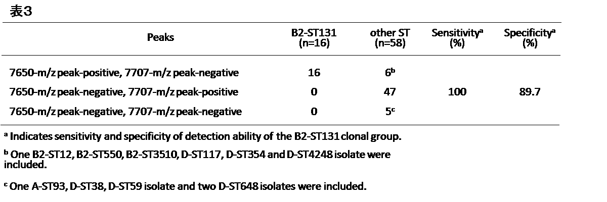
- the sensitivity and specificity for the ESBL-producing Escherichia coli B2-ST131 clone according to the present invention are extremely high, 100% and 89.7%, respectively.
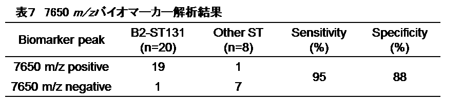
- the sensitivity and specificity for the carbapenemase-producing E. coli B2-ST131 clone according to the present invention is also extremely high, 95% and 88%, respectively. According to the present invention, it is possible to accurately detect the E. coli B2-ST131 clone.
- Example 2 It is a figure which shows alignment of the base sequence of (A) YahO gene and the amino acid sequence of (B) YahO protein derived from ESBL-producing Escherichia coli B2-ST131 clone group and other ST clone groups. The arrow indicates the substitution site.
- Example 3 It is a figure which shows the result of having detected ESBL production colon_bacillus
- Example 4 (1) It is a figure which shows the result of having detected ESBL production colon_bacillus
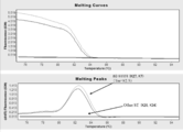
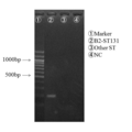
- Example 4 (2)
- the present invention relates to a test method for identifying an Escherichia coli B2-ST131 clone.
- the 34th amino acid in the amino acid sequence shown in SEQ ID NO: 1 is changed to alanine. It is characterized by detecting.
- Escherichia coli is classified by serotype (classification based on immunological reaction to O antigen and H antigen) and phylogenetic group analysis.
- the E. coli B2-ST131 clone identified in the test method of the present invention is classified into the O25: H4 serotype and the B2 group.
- final identification of E. coli B2-ST131 clone has been performed by determining the sequence type (ST) by MLST analysis.
- MLST analysis is performed by decoding 400 to 600 bases of housekeeping genes in 5-7 regions of bacteria and comparing these sequences (Bonzo Kimura, Modern Media, Vol. 52, Vol. 7, No. 7, 2006, Molecule).
- the E. coli B2-ST131 clone is a group of strains classified by the above classification method, and includes not only the same strain but also a plurality of strains.
- the E. coli B2-ST131 clone can also be referred to as the E. coli B2-ST131 clone group.
- “E. coli B2-ST131 clone” may be referred to as “B2-ST131 clone” or “B2-ST131 clone group”.
- the E. coli B2-ST131 clone identified in the test method of the present invention is an E. coli B2-ST131 clone exhibiting drug resistance.
- the Escherichia coli B2-ST131 clone exhibiting drug resistance is a strain highly resistant to drugs and highly pathogenic, and is particularly wary of multidrug resistant Escherichia coli.
- E. coli B2-ST131 clones that exhibit drug resistance include E. coli that produces ⁇ -lactamase.
- E. coli producing ⁇ -lactamase includes E. coli producing ESBL and E. coli producing carbapenemase. ESBL degrades antibiotics with a ⁇ -lactam ring such as penicillin.
- ESBL-producing bacteria have produced ⁇ -lactamase with expanded types of degradable drugs due to mutations in ⁇ -lactamase-producing genes (TEM type, SHV type, CTX-M type). Shows tolerance. Since the gene producing ESBL is encoded on a drug plasmid (R plasmid), it is transmitted not only between the same species but also between different species of Enterobacteriaceae. It has spread to bacterial species. Carbapenemase-producing bacteria degrade almost all ⁇ -lactam antibiotics including carbapenem antibiotics.
- carbapenemase producing bacteria Six types are known, mainly IMP type, VIM type, NDM type, KPC type, GES type and OXA-48 type, and all show high multidrug resistance. Carbapenemase can degrade carbapenem antibiotics and is located on a transmissible plasmid in the same way as ESBL, so there is a risk of rapid spread to various bacterial species in the future.
- a protein is extracted from an E. coli B2-ST131 clone and an ST clone other than E. coli B2-ST131 (sometimes simply referred to as “other ST clone” in this specification), and a mass spectrometer is extracted.
- a peak 7,650 m / z specific to the E. coli B2-ST131 clone was detected.
- a protein exhibiting a peak of 7,650 m / z was identified as a YahO protein whose function has not yet been determined.
- the present invention detects Escherichia coli B2- by detecting the substitution of (A) to the alanine of the 34th glutamic acid (E) of SEQ ID NO: 1 in the amino acid sequence constituting the YahO protein. This identifies the ST131 clone.
- the full-length amino acid sequence of the YahO protein derived from E. coli (E. coli O157, K-12 strain) is shown in SEQ ID NO: 1 below.
- the 34th amino acid is glutamic acid, but in the E. coli B2-ST131 clone, the 34th glutamic acid is substituted with alanine.
- Sequence number 1 MKIISKMLVGALALAVTNVYAAELMTKAEFEKVESQYEKIGDISTSNEMSTADAKEDLIKKADEKGADVLVLTSGQTDNKIHGTANIYKKK (UniProtKB Accession No. P75694)
- SEQ ID NO: 2 The base sequence encoding the amino acid sequence shown in SEQ ID NO: 1 is shown in SEQ ID NO: 2 below.
- the 100th to 102nd codons encoding glutamic acid of SEQ ID NO: 2 are replaced with codons encoding alanine.
- Detection of the E34A mutation in SEQ ID NO: 1 may be performed by analyzing the full length of the YahO protein and the YahO gene, but it is convenient and preferable to analyze by partial fragments.
- the E34A mutation may be detected by any method, for example, by any one selected from i) to iii) below. i) Analyzing the base sequence of the polynucleotide encoding the polypeptide containing the 34th amino acid of the test sample. ii) Analyzing the amino acid sequence of the polypeptide containing the 34th amino acid of the amino acid sequence shown in SEQ ID NO: 1 for the test sample. iii) Measuring the molecular weight of the polypeptide containing the 34th amino acid of the test sample.
- polypeptide refers to peptides and proteins formed by binding two or more amino acids.
- the “polynucleotide” in the present invention is a concept including an oligonucleotide, refers to a biopolymer in which two or more nucleotides composed of a base, a sugar and a phosphate are linked by a phosphate ester, and includes nucleic acids such as DNA and RNA.
- the length of the polypeptide or polynucleotide only needs to be a level that can be detected by an analysis technique used for the test.
- the length of the polypeptide may be such that the antibody can specifically recognize the amino acid sequence, and is at least about 3 to 15 amino acids. Any length is acceptable.
- the size of the polypeptide only needs to be such that the mass spectrometer can measure. For example, when MALDI-TOF MS is used as the mass spectrometer, a polypeptide of 2000 Da to 20000 Da is used. I just need it.
- the length of the polynucleotide only needs to be long enough to be a template for the amplification reaction, and is about 100 to 1000 bases long. It is.
- the analysis of the base sequence of i) may be performed by any method as long as it is a gene mutation analysis method capable of detecting the base sequence encoding the 34th amino acid.
- sequence analysis for example, sequence analysis, PCR method, LAMP method, etc. Nucleic acid amplification reaction, hybridization method and the like.
- a polynucleotide for detecting the base showing the 34th amino acid substitution in the amino acid sequence shown in SEQ ID NO: 1 among the amino acid sequences constituting the YahO protein can be used.
- the polynucleotide has a length of at least 15 nucleotides (bases), preferably 17 nucleotides (bases).
- the polynucleotide functions as a primer or a probe.
- the polynucleotide is preferably a polynucleotide capable of specifically hybridizing with a partial sequence of a base sequence encoding the polypeptide containing the 34th amino acid or a sequence complementary to the partial sequence
- the polynucleotide is preferably a polynucleotide that can specifically hybridize to a partial sequence containing a base showing a mutation of the 34th amino acid to alanine or a sequence complementary to the partial sequence.
- the base sequence of the polynucleotide need not be a partial sequence containing a base showing a mutation of the 34th amino acid to alanine or a sequence that is 100% complementary to a sequence that is complementary to the partial sequence.
- polynucleotide is a primer, it is preferably about 17 to 25 nucleotides, and when it is a probe, it is preferably about 10 to 30 nucleotides.
- Primers and probes can be designed using known primer or probe design software.
- known software for example, OLIGO Primer Analysis Software (Molecular Biology Insights), Beacon Designer (PREMIER Biosoft), Primer Express Software (Life Technologies) can be used.
- Polynucleotides can be synthesized by a method known in the art as a method for synthesizing polynucleotides, for example, a phosphotriethyl method, a phosphodiester method, or the like, using a commonly used automatic DNA synthesizer.
- Sequence analysis can be performed by a method known per se. For example, it is possible to perform a sequencing reaction using a primer designed at the position of several tens of bases on the 5 ′ side of a base showing amino acid substitution, and determine the type of base at the corresponding position from the analysis result it can.
- a nucleic acid amplification reaction such as PCR.
- Any primer can be used for the nucleic acid amplification reaction as long as the object can be achieved.
- the following primers are exemplified.
- forward primer 5'-TAGTCGGTGCGTTAGCGTT-3 '(SEQ ID NO: 11)
- reverse primer 5'-TGCCGTGGATCTTATTGTCA-3 '(SEQ ID NO: 12)
- PCR-SSCP single-strand conformation polymorphs method
- RFLP restriction enzyme
- a nucleic acid amplification reaction such as PCR method, LANP method (Japanese Patent No. 3313358), NASBA method (Nucleic Acid Sequence-Based Amplification; Japanese Patent No. 2843586), ICAN method (Japanese Patent Laid-Open No. 2002-233379) etc. It can also be analyzed by examining the presence or absence. Analysis using a nucleic acid amplification reaction can be performed by the following steps. (1) A step of extracting a polynucleotide from a test sample.
- the polynucleotide is preferably a polynucleotide that can be specifically annealed to a partial sequence containing a base showing the 34th amino acid substitution (preferably a mutation to alanine) or a sequence complementary to the partial sequence.
- an amino acid is designated by a triplet codon consisting of three bases.
- the “base showing amino acid substitution” is at least one base, preferably two bases or more in the triplet codon.
- a primer that can specifically anneal to a region containing a base showing amino acid substitution and a primer that can be paired with the primer to amplify a nucleic acid fragment are prepared as a set.
- PCR can be performed using such a primer set, and the presence or absence of a base showing amino acid substitution can be detected based on the presence or absence of an amplification product. Further, by performing PCR using a primer set capable of amplifying a fragment containing a base showing amino acid substitution and analyzing the amplified product, the presence or absence of a base showing amino acid substitution can be detected.
- a primer that can specifically hybridize to a partial sequence that includes a base that shows amino acid substitution may be a forward primer or a reverse primer.
- a forward primer composed of the following oligonucleotides is used. be able to.
- forward primer 5'-GAATTTGAAAARGTYGC-3 '(SEQ ID NO: 13)
- the presence or absence of an amplification product is 100% consistent with the analysis result by sequence analysis.
- the specificity for the mutation by the primer of SEQ ID NO: 13 is extremely high, and accurate detection can be performed.
- Detection of the presence or absence of an amplification product by PCR can be performed by separating the amplification product by agarose gel electrophoresis and staining with an intercalator such as ethidium bromide or SYBR Green I.
- an intercalator such as ethidium bromide or SYBR Green I.
- the presence or absence of an amplification product can be automatically confirmed in a short time.
- SYBR Green I can be used to monitor the amount of amplified product for each cycle from the fluorescence value emitted when SYBR Green I binds to double-stranded DNA.
- the forward primer of SEQ ID NO: 13 can also be applied to real-time PCR, and can detect the presence or absence of E34A in about 30 minutes.
- the present invention also encompasses a polynucleotide, primer, probe, or primer set that enables detection of the base showing the 34th amino acid substitution.
- the present invention also includes a reagent composition containing the polynucleotide, primer, probe or primer set.
- the present invention also includes a reagent kit containing the reagent composition.
- the reagent composition or reagent kit of the present invention is for identifying the E. coli B2-ST131 clone, and is used in the test method of the present invention.
- the polynucleotide, primer, probe, or primer set can be provided as a reagent composition as a solid in a dry state or alcohol precipitated state, or can be provided as water, physiological saline, or an appropriate buffer (eg, TE It can be provided as a reagent composition in a dissolved state in a buffer solution or the like.
- the reagent composition may contain additives (eg, carriers, excipients, diluents, etc.), stabilizers, and the like.
- Stabilizers include monosaccharides such as glucose, disaccharides such as saccharose and maltose, sugar alcohols such as mannitol and sorbitol, neutral salts such as sodium chloride, amino acids such as glycine, polyethylene glycol, polyoxyethylene-polyoxy Nonionic surfactants such as propylene copolymer (pluronic) and polyoxyethylene sorbitan fatty acid ester (tween), human albumin and the like are exemplified, and it is preferable that about 1 to 10 w / v% is added.
- the reagent kit is exemplified by a reagent kit containing the polynucleotide, primer, probe or primer set.
- the primer set may be any as long as it can perform base sequence analysis, but preferably includes at least two kinds of primers and used for nucleic acid amplification reaction.
- the primer set is more preferably a set including a primer that can specifically hybridize to a region containing a base indicating a mutation site encoding the amino acid sequence containing the 34th amino acid.
- the reagent kit may contain one or more reagents selected from nucleic acid amplification reactions, such as DNA synthase, buffer, dNTP mix, intercalator, magnesium chloride, and instructions for use.
- a probe labeled with a fluorescent substance or the like may be included.
- the polynucleotide can be provided in a state immobilized on an appropriate solid phase.
- the solid phase include, but are not limited to, glass, silicon, plastic, nitrocellulose, nylon, polyvinylidene difluoride, and the like.
- immobilization means functional groups such as amino groups, aldehyde groups, SH groups, and biotin are introduced into nucleic acids in advance, and functional groups capable of reacting with the nucleic acids on solid phases (eg, aldehydes).
- amino acid sequence of ii) above may be performed by any method as long as it can detect the 34th amino acid, but it may be an antigen-antibody reaction detection method such as Western blotting, ELISA, and latex agglutination immunization. Alternatively, it can be carried out by amino acid sequencing.
- an antibody that specifically recognizes the polypeptide containing the 34th amino acid preferably the polypeptide in which the 34th amino acid is alanine can be used.
- the antibody can be produced by an existing general production method using a polypeptide containing the 34th amino acid or a partial peptide having the antigenicity of the polypeptide as an immunogen.
- An immunogen for antibody production can be prepared by separating and purifying the polypeptide containing the 34th amino acid from bacteria.
- a recombinant polypeptide produced by genetic engineering techniques can also be used as an immunogen.
- a peptide fragment or a synthetic peptide obtained by treating the immunogen obtained above with a protease such as trypsin can also be used as the immunogen.
- a polyclonal antibody specific for the polypeptide containing the 34th amino acid of the present invention can be prepared by the following method. Mammals such as rabbits, mice, and rats are immunized using the polypeptide containing the 34th amino acid prepared and purified as described above as an antigen (immunogen). As the administration means, intraperitoneal injection, intravenous injection, subcutaneous injection and the like are adopted, and in some cases, intradermal injection is also adopted. The booster immunization is repeated several times, and mammals are bled 3 to 10 days after the final immunization to obtain antiserum (polyclonal antibody).
- the antiserum and the related protein of the polypeptide containing the 34th amino acid are mixed to react with the related protein.
- the polyclonal antibody specific for the polypeptide containing the 34th amino acid can be prepared.
- a polypeptide in which the 34th amino acid is glutamic acid can be used as a related protein.
- the monoclonal antibody that specifically recognizes and binds to the polypeptide containing the 34th amino acid of the present invention is a method of Kohler and Milstein (Kohler, G. and Milstein, C., Nature, 256, 495-497, 1975). It can produce by well-known methods, such as.
- the polypeptide containing the 34th amino acid can be used as an immunogen.
- the immunogen is desirably administered with a suitable adjuvant, such as a commercially available Freund's complete or incomplete adjuvant.
- mice, rats, guinea pigs and the like can be used as immunized animals, but mice are generally used.
- the prepared immunogen can be administered through any route such as subcutaneous, intraperitoneal, intravenous, intramuscular, or intradermal routes of animals.
- the interval between immunizations is not particularly limited.
- immunization is preferably performed 2 to 5 times at intervals of 1 to 2 weeks.
- the dose per administration is not limited, but for example, the immunogen prepared as described above may be mixed with an appropriate adjuvant and administered in an amount of several tens to several hundreds of ⁇ l.
- antibody-producing cells are collected from the immunized animal.
- Examples of antibody-producing cells include spleen cells, lymph node cells, thymocytes, peripheral blood cells, etc., but spleen cells are generally used.
- the prepared antibody-producing cells are fused with myeloma cells.
- myeloma cells cell lines available to those skilled in the art can be used.
- 107-108 cells / ml of myeloma cells and antibody-producing cells are mixed in an animal cell culture medium at a mixing ratio of 1: 1 to 1:10 using a fusion promoter, a fusion virus, etc. It can be carried out efficiently by bringing cells into contact at 30 to 37 ° C. for 1 to 3 minutes.
- cell culture is performed using a selective medium to select hybridomas.
- the selected hybridoma is cloned by limiting dilution method, soft agar method, fibrin gel method, fluorescence excitation cell sorter method or the like.
- the cell culture supernatant is tested for the production of antibodies against the polypeptide containing the 34th amino acid, and a monoclonal antibody that specifically recognizes the polypeptide containing the 34th amino acid is produced.
- To screen for hybridomas As a primary screening for monoclonal antibodies, a culture supernatant containing a monoclonal antibody that reacts with and binds to the polypeptide containing the 34th amino acid is selected.
- a culture supernatant containing a monoclonal antibody that does not react with a related protein is selected.
- a hybridoma that produces a monoclonal antibody that specifically recognizes the polypeptide containing the 34th amino acid can be screened.
- a polypeptide in which the 34th amino acid is glutamic acid can be used as a related protein.
- a monoclonal antibody can be collected from a monoclonal antibody-producing hybridoma that specifically recognizes the polypeptide containing the 34th amino acid by a conventional cell culture method.
- a monoclonal antibody-producing hybridoma is cultured in animal cell culture medium under normal culture conditions (for example, 37 ° C., 5% CO 2 concentration) for 2 to 14 days.
- a monoclonal antibody-producing hybridoma is administered into the abdominal cavity of an animal of the same type as the animal derived from myeloma cells, the hybridoma is proliferated in large quantities, and ascites or blood is collected 1 to 4 weeks later to collect the antibody. Can be acquired.
- a gene encoding a monoclonal antibody can be isolated from a hybridoma and prepared as a recombinant protein by genetic engineering techniques.
- known methods such as ammonium sulfate salting-out, ion exchange chromatography using anion exchangers such as DEAE cellulose, molecular sieve chromatography based on molecular weight and structure, hydroxyapatite chromatography, etc. The method can be appropriately selected, or can be purified by combining them.
- the polypeptide containing the 34th amino acid can be detected using an antibody that specifically reacts with the polypeptide containing the 34th amino acid.
- the labeling substance is not limited. Specifically, insoluble carriers such as enzymes, radioisotopes, fluorescent dyes, biotin, dye sols, colloidal gold, and latex particles can be used.
- the labeling can be performed by a known method, but a labeling substance may be bound to the antibody.
- the present invention relates to a monoclonal antibody or a polyclonal antibody that specifically recognizes the polypeptide containing the 34th amino acid, which enables detection of the polypeptide containing the 34th amino acid, and any one of these antibodies.
- a reagent kit containing the reagent composition containing the reagent composition.
- the reagent composition or reagent kit of the present invention is for identifying the E. coli B2-ST131 clone, and is used in the test method of the present invention.
- the antibody may be provided as a reagent composition in a state diluted with physiological saline, a buffer solution or the like, or may be provided as a reagent composition that can be stored for a long period of time, such as a lyophilized form.
- the antibody may be lyophilized after being dissolved in a solution containing an additive (eg, carrier, excipient, diluent, etc.), stabilizer, and the like. Specific examples of the stabilizer are as described above.
- the reagent kit containing the antibody may contain a blocking solution, a reaction solution, a reaction stop solution, a reagent for treating the sample, and the like as appropriate.
- the molecular weight of the polypeptide iii) can be measured by any technique, for example, by using a publicly known mass spectrometer or electrophoresis.
- Mass spectrometers include matrix-assisted laser desorption / ionization time-of-flight mass spectrometer (MALDI-TOF MS), surface-enhanced laser desorption / ionization time-of-flight mass spectrometer (SELDI-TOF-MS), liquid chromatography mass spectrometry Equipment, tandem mass spectrometer (MS-MS), electrospray ionization mass spectrometer (ESI-MS), desorption electrospray ionization mass spectrometer (DESI) can be used, but matrix-assisted laser desorption ionization time of flight
- a mass spectrometer (MALDI-TOF) MS) is used.
- MALDI-TOF-MS that can be used is not particularly limited, and examples thereof include MALDI-Biotyper
- the E. coli B2-ST131 clone is detected as a test sample or Indicates that it is contained in the sample.
- the 7650 Da polypeptide is not detected, it indicates that the E. coli B2-ST131 clone is not contained in the test sample or specimen.
- ST clones other than E. coli B2-ST131 clone are included in the test sample or specimen. Indicates that
- the test sample to be examined may be the specimen itself, but is preferably prepared from the specimen.
- the specimen is not particularly limited as long as it can contain E. coli B2-ST131 clone.
- the sample may be a sample collected from a living body such as a subject including a patient, or may be a sample collected from a medical instrument, medical device, food, or the like that can mediate a medical-related infection.
- specimens collected from a living body include exudates such as urine, feces, and pus, sputum, lymph nodes, tonsils, skin, lymph, blood, mucous membranes, organs and tissues such as bladder and large intestine, and the like.
- the test sample is preferably prepared by removing human-derived cells or proteins other than strain-derived proteins from the specimen.
- the test sample may be prepared by any method.
- a test sample may be prepared by isolating a strain from a specimen and culturing it in a medium.
- LB medium, broth, plate medium and the like known per se can be used.
- a polypeptide or polynucleotide may be further extracted from a test sample and prepared for use in analysis.
- the method for extracting a polypeptide or polynucleotide may be a nucleic acid extraction method or protein extraction method known per se.
- the present invention also includes a biomarker for identifying an E. coli B2-ST131 clone selected from the following a) or b).
- a biomarker comprising a polypeptide comprising an amino acid sequence in which the 34th amino acid in the amino acid sequence shown in SEQ ID NO: 1 is mutated to alanine among the amino acid sequences constituting the YahO protein.
- the polypeptide of a) should just contain the amino acid sequence by which the 34th amino acid in the amino acid sequence shown to sequence number 1 among the amino acid sequences which comprise YahO protein was mutated to alanine, and is a partial fragment of YahO protein. Or it may be the full length.
- the polynucleotide of b) only needs to contain a base showing the 34th amino acid substitution in the base sequence of the gene encoding YahO protein, whether it is a partial fragment or full length. Good.
- MLST seven housekeeping genes (adk, fumC, gyrB, icd, mdh, purA, and recA) were subjected to PCR direct sequencing (PCR-direct sequencing). Moreover, PFGE using restriction enzyme XbaI (Roche Diagnostics, Inc., Tokyo, Japan) was performed with CHEF-Dr II system (Bio-Rad Laboratories, Inc., Hercules, CA). Based on the obtained results, a phylogenetic tree was created by Fingerprinting Plus Version 1.12, and when it showed 80% or more similarity (UPGMA algorithm on Dice similarity coefficients), it was determined that they were the same PFGE cluster.
- XbaI Roche Diagnostics, Inc., Tokyo, Japan
- CHEF-Dr II system Bio-Rad Laboratories, Inc., Hercules, CA
- Example 1 Analysis by MALDI-TOF MS (measurement by MALDI-TOF MS)
- Strains derived from clinical specimens similar to those in Reference Example 1 were cultured at 37 ° C. for 16 to 24 hours in Columbia blood agar containing 5% [v / v] sheep blood. Protein was extracted from the cultured strain by the ethanol-formic acid extraction method. After suspending the strain in 300 ⁇ L of milli-Q water, 900 ⁇ L of ethanol was added, centrifuged at 13,000 g for 2 minutes, the supernatant was discarded, and the pellet was dried.
- MALDI-TOF MS analysis was performed using MALDI Biotyper (Bruker Daltonik, GmbH) and MALDI Biotyper v3.1 software. As a matrix, ⁇ -cyano-4-hydroxy-cinnnamic acid (CHCA) was used.
- CHCA ⁇ -cyano-4-hydroxy-cinnnamic acid
- Example 2 Identification and analysis of protein serving as biomarker
- the 7650 Da protein specified in Example 1 was identified by performing N-terminal amino acid sequence analysis by bottom-up proteomics and Edman degradation.
- proteins were purified from the same B2-ST131 clone group derived from clinical specimens as in Reference Example 1 as follows. First, the strain was grown in LB medium, recovered and sonicated, and purified stepwise by 80% ammonium sulfate precipitation, open column DEAE cellulose, and open column CM cellulose, and then HPLC (TSKgel Protein C4-300 ( Tosoh Corporation, Tokyo, Japan)).
- each fraction was dried and concentrated, and confirmed by MALDI-TOF MS to identify the fraction from which the 7650 Da protein was eluted.
- the protein eluted in the identified fraction was subjected to Tricine SDS-PAGE, and after silver staining, the gel was cut out.
- the excised gel was digested with trypsin and analyzed using LCMS-IT-TOF (Shimazu Corporation, Kyoto, Japan).
- the obtained spectrum was analyzed by Mascot MS / MS Ion Search (Matrix Science KK, Tokyo, Japan), and the peptide was identified by SwissProt database.
- N-terminal amino acid sequence analysis was performed, and the protein was identified together with the peptide identification result by bottom-up proteomics.
- LCMS-IT-TOF results of LCMS-IT-TOF are shown in Table 4 below. From the results of Table 4 and the results of N-terminal amino acid sequence analysis, it was found that the 7650 Da protein is a YahO protein with unknown function (FIG. 2).
- the underlined portion in FIG. 2 is an amino acid sequence obtained by LCMS-IT-TOF, and the portion surrounded by a square is the amino acid sequence obtained by N-terminal amino acid sequence analysis.
- Example 3 Analysis of biomarker gene From the amino acid sequence identified in Example 2, forward primer: 5'-TAGTCGGTGCGTTAGCGTT-3 '(SEQ ID NO: 11) and reverse primer: 5'-TGCCGTGGATCTTATTGTCA-3' (SEQ ID NO: 12) was designed, and the base sequence and amino acid sequence of the B2-ST131 clone group and other ST clone groups were compared by PCR direct sequencing.
- B2-ST131 clone group strains derived from clinical specimens were grown in a medium and then collected. After recovering the strain, DNA was extracted according to a conventional method.
- the extracted DNA was subjected to PCR using the primers of SEQ ID NO: 11 and SEQ ID NO: 12.
- Applied Biosystems Veriti 96 Thermal Cycler (Life Technologies) was used for PCR, and Applied Biosystems 3730xl DNA Analyzer (Thermo Fisher Scientific) was used for DNA sequencing.
- the theoretical molecular weight (Mw) was calculated using Compute pI / Mw tool (http://web.expasy.org/compute_pi/).
- Example 4 Detection of ESBL-producing Escherichia coli B2-ST131 using PCR (1) First, a strain derived from a clinical specimen similar to that in Reference Example 1 was grown in a medium and then collected. After recovering the strain, DNA was extracted according to a conventional method. The extracted DNA was subjected to real-time PCR using the following primer set.
- forward primer 5'-GAATTTGAAAARGTYGC-3 '(SEQ ID NO: 13) reverse primer: 5'-TGCCGTGGATCTTATTGTCA-3 '(SEQ ID NO: 12)
- the forward primer is designed to anneal to the 85th to 101st positions of the base sequence mutated so that SEQ ID NO: 2 encodes E34A
- the reverse primer is designed to anneal to the 23rd to 250th positions of SEQ ID NO: 2.
- PCR was performed using a LightCycler 2.0 system (Roche Diagnostics, Inc., Tokyo, Japan) and a reagent using LightCycler FastStart DNA Master SYBR GreenI (Roche) as follows.
- PCR was performed using the forward primer of SEQ ID NO: 13 and the reverse primer of SEQ ID NO: 12, and the presence or absence of the amplified product was confirmed by electrophoresis.
- the PCR cycle is the same as described in (1) above. Electrophoresis with agarose gel using a Mupid minigel electrophoresis tank (ADVANCE), staining the gel with ethidium bromide solution, and confirming the presence or absence of the target amplification band. confirmed.
- ADVANCE Mupid minigel electrophoresis tank
- NC is a negative control. It was found that the B2-ST131 clone group can be detected by performing PCR using the above primers and confirming the amplified product by agarose gel electrophoresis.
- Example 5 Detection of carbapenemase-producing Escherichia coli B2-ST131 using MALDI-TOF MS 28 strains of carbapenemase-producing Escherichia coli (E. coli) isolated from clinical samples possessed by Tenri Yorozu Consultation Hospital B2-ST131 clone 20 strain, other ST clone 8 strain) were used.
- strains were analyzed as a phylogenetic group using triplex PCR for the three genes chuA, yjaA, and TSPE4.C2, MLST, and seven housekeeping genes (adk, fumC, gyrB, icd, mdh, purA, and recA) using PCR direct sequencing, PFGE using restriction enzyme XbaI (Roche Diagnostics, Inc., Tokyo, Japan), CHEF-Dr II system (Bio-Rad Laboratories) , Inc., Hercules, CA), and based on the results obtained, a phylogenetic tree was created by Fingerprinting Plus Version 1.12, showing 80% similarity (UPGMA algorithm on Dice similarity coefficients) It is determined that they are the same PFGE cluster and is classified.
- XbaI Roche Diagnostics, Inc., Tokyo, Japan
- CHEF-Dr II system Bio-Rad Laboratories
- a phylogenetic tree was created by Fingerprinting Plus Version 1.12,
- Example 6 Detection of carbapenemase-producing Escherichia coli B2-ST131 using PCR method
- the strain was grown and collected in a medium in the same manner as in Example 4, and the DNA was recovered according to a conventional method. Extraction was performed, and real-time PCR was performed on the extracted DNA.
- Table 8 shows the sensitivity and specificity for detecting the mutation of the YahO gene by real-time PCR.
- the real-time PCR revealed that the carbapenemase-producing B2-ST131 clone group could be detected with a high value of 95% sensitivity and 88% specificity.
- the present invention makes it possible to identify E. coli B2-ST131 clones quickly and easily.
- the test method of the present invention is excellent in sensitivity and specificity, and can quickly and efficiently identify B2-ST131 clones.
- the B2-ST131 clone can be quickly and accurately identified according to the present invention, so that a cause such as a medical-related infection can be quickly identified, which is very useful as a tool for determining an initial treatment for infection.
- the test method of the present invention may contribute to global pandemic control, and it is expected to dramatically advance the treatment of infectious diseases.
Landscapes
- Health & Medical Sciences (AREA)
- Life Sciences & Earth Sciences (AREA)
- Chemical & Material Sciences (AREA)
- Engineering & Computer Science (AREA)
- Immunology (AREA)
- Organic Chemistry (AREA)
- Molecular Biology (AREA)
- Biomedical Technology (AREA)
- Biochemistry (AREA)
- Biotechnology (AREA)
- Genetics & Genomics (AREA)
- General Health & Medical Sciences (AREA)
- Microbiology (AREA)
- Physics & Mathematics (AREA)
- Urology & Nephrology (AREA)
- Zoology (AREA)
- Hematology (AREA)
- Wood Science & Technology (AREA)
- Bioinformatics & Cheminformatics (AREA)
- Analytical Chemistry (AREA)
- Proteomics, Peptides & Aminoacids (AREA)
- Medicinal Chemistry (AREA)
- Biophysics (AREA)
- General Engineering & Computer Science (AREA)
- General Physics & Mathematics (AREA)
- Pathology (AREA)
- Cell Biology (AREA)
- Food Science & Technology (AREA)
- Plant Pathology (AREA)
- Tropical Medicine & Parasitology (AREA)
- Virology (AREA)
- Measuring Or Testing Involving Enzymes Or Micro-Organisms (AREA)
- Peptides Or Proteins (AREA)
Abstract
本発明は、YahOタンパク質を構成するアミノ酸配列のうち、配列番号1に示すアミノ酸配列における第34番目のアミノ酸のアラニンへの変異を検出することを特徴とする、ESBL産生大腸菌B2-ST131クローンを識別するための検査方法に関する。また本発明は、当該第34番目のアミノ酸のアラニンへの変異を検出するためのポリヌクレオチド又は抗体、当該ポリヌクレオチド又は抗体のいずれかを含む試薬組成物、当該試薬組成物を含む試薬キットに関する。さらに、本発明は大腸菌B2-ST131クローンを識別するためのバイオマーカーに関する。
Description
本発明は、大腸菌B2-ST131クローンを識別するための検査方法、大腸菌B2-ST131クローンを識別するためのポリヌクレオチド又は抗体、当該ポリヌクレオチド又は抗体を含む試薬組成物、当該試薬組成物を含む試薬キット、および大腸菌B2-ST131クローンを識別するためのバイオマーカーに関する。
本出願は、参照によりここに援用されるところの日本出願、特願2015-053351号優先権を請求する。
近年、さまざまな薬剤耐性菌が次々と出現する事態が生じており、薬剤耐性菌の対策に対する注目度が国際的に増している。特に腸内細菌科は経口感染によりヒト腸管内に定着しやすく、医療環境や食品環境等での感染拡大が懸念される。腸内細菌科による感染症治療には、βラクタム系であるカルバペネムまたはセフタジジム、フルオロキノン系およびアミノグリコシド系の薬剤が最も汎用性の高い抗菌薬として使用されている。しかしながら、これらの抗菌薬に対する耐性を示す株について多数報告がされており、薬剤耐性菌の伝播の制御が極めて困難な状況となりつつある。
薬剤耐性菌として、基質特異性拡張型βラクタマーゼ(Extended-spectrum-β-lactamase)(以下単に「ESBL」と称する。)を産生する大腸菌(Escherichia coli)の増加が著しいことが報告されている。病院内のみならず、市中においても蔓延が確認されている。ESBL産生大腸菌の増加の主たる原因は、ESBL産生大腸菌のB2-ST131クローンのパンデミックである。ESBL産生大腸菌B2-ST131クローンは尿路感染症の起因菌として知られており、症状が悪化した場合は敗血症などの重篤な症状を呈し、死亡に至ることもある。ESBL産生大腸菌B2-ST131クローンは、フルオロキノロン系薬剤に対してほぼ100%耐性を示し、アミノグリコシド系抗生物質に対しても耐性を示すため、高耐性かつ高病原性の株とされ、疫学上問題視されている(Benjamin A. Rogers, Hanna E. Sidjabat and David L. Paterson, 2011. J. Antimicrob. Chemother. 66: 1-14)。
またカルバペネマーゼを産生する大腸菌についても報告がある。日本においては、まだ1%未満の分離頻度でしか報告されていないが、欧州等の一部の地域・施設においてはアウトブレイクも多く報告されている。カルバペネマーゼを産生する大腸菌は、2013年以降急増し、世界レベルでは蔓延しつつある薬剤耐性菌として知られている。カルバペネマーゼ産生菌は、ESBL産生株には効果を示すカルバペネム系抗菌薬にも耐性を示し、肺炎や尿路感染症などを引き起す。その症状の重篤化、致死率の高さから、カルバペネマーゼ産生菌は世界中で脅威となっている。またカルバペネマーゼを産生する大腸菌のおよそ半分を、大腸菌B2-ST131クローンが占めているとの報告がされている(中村彰宏ら.当院における過去4年間のカルバペネマーゼ産生腸内細菌科の検出状況と分子疫学的特徴.第26回日本臨床微生物学会総会.2015年1月31日.東京.)。
大腸菌B2-ST131クローンの同定には、multilocus sequence typing(以下単に「MLST」と称する)解析が用いられている。しかしながら、MLST解析は操作が煩雑かつ高価な検査法であり、加えて検査結果が得られるまで長時間を要する。よってMLST解析による大腸菌B2-ST131クローンの同定は、感染症初期治療決定ツールとしては実用性が乏しいものである。医療関連感染などでは迅速な対応が必須であり、臨床上、大腸菌B2-ST131クローンを迅速に識別する検査方法が切望されているものの、未だ開発はなされていない。大腸菌B2-ST131クローンを迅速に識別する検査方法が開発されれば、感染症治療の方針決定のための革新的ツールになると考えられ、世界的なパンデミックを制御できる可能性も秘めている。
非特許文献1(Matsumura Y, Yamamoto M, Nagao M, Tanaka M, Machida K, Ito Y, Takakura S, Ichiyama S. 2014. J. Clin. Microbiol. 52: 1034-1040.)には、ESBL産生大腸菌B2-ST131クローンをMALDI-TOF MSを用いて検出する方法が開示されているが、7,650m/zのピークについての開示はない。特許文献1および2(国際公開第WO2010/22400号および米国特許第8160819号)には、大腸菌のペリプラズムタンパク質として、34番目のアミノ酸がグルタミン酸であるYahOタンパク質を検出したことが開示されている。具体的には、大腸菌(E.coli O157、K-12株)由来のYahOタンパク質のアミノ酸配列とそのMSフラグメント(7,707m/z)が開示されている。また、34番目のアミノ酸がアラニンであるYahOタンパク質(GenBank Accession No. WP_000691950)が大腸菌から単離されたことが報告されている(非特許文献2(URL:http://www.ncbi.nlm.nih.gov/gquery/?term=WP_000691950))。しかしながら、YahOタンパク質における34番目のアミノ酸のアラニンへの置換と、大腸菌B2-ST131クローンとの関連性については、開示されていない。
Matsumura Y, Yamamoto M, Nagao M, Tanaka M, Machida K, Ito Y, Takakura S, Ichiyama S. 2014. J. Clin. Microbiol. 52: 1034-1040.
URL:http://www.ncbi.nlm.nih.gov/gquery/?term=WP_000691950
本発明は、大腸菌B2-ST131クローンを迅速かつ簡便に識別する検査方法や検査のためのポリヌクレオチドや抗体といった検出手段を提供することを課題とする。
本発明者らは、上記課題を解決するために鋭意検討を重ねた結果、YahOタンパク質を構成するアミノ酸配列のうち、配列番号1の34番目のグルタミン酸のアラニンへの変異が、大腸菌B2-ST131クローンに特異的であることに着目し、当該アミノ酸の変異を検出することにより、大腸菌B2-ST131クローンを迅速かつ効率的に検出可能であること見出し、本発明を完成した。
すなわち本発明は、以下よりなる。
1.YahOタンパク質を構成するアミノ酸配列のうち、配列番号1に示すアミノ酸配列における第34番目のアミノ酸のアラニンへの変異を検出することを特徴とする、大腸菌B2-ST131クローンを識別するための検査方法。
2.前記第34番目のアラニンへの変異の検出が、以下のi)~iii)より選択されるいずれかにより行われる、前項1に記載の検査方法:
i)被検試料について、前記第34番目のアミノ酸を含むポリペプチドをコードするポリヌクレオチドの塩基配列を解析すること;
ii)被検試料について、配列番号1に示すアミノ酸配列のうち、第34番目のアミノ酸を含むポリペプチドのアミノ酸配列を解析すること;
iii)被検試料について、前記第34番目のアミノ酸を含むポリペプチドの分子量を測定すること。
3.前記第34番目のアラニンへの変異の検出が、以下のi)~iii)より選択されるいずれかにより行われる、前項2に記載の検査方法。
i)被検試料について、前記第34番目のアミノ酸を含むポリペプチドをコードするポリヌクレオチドの塩基配列を、核酸増幅反応により解析すること;
ii)被検試料について、配列番号1に示すアミノ酸配列のうち、第34番目のアミノ酸を含むポリペプチドのアミノ酸配列を、免疫学的手法により解析すること;
iii)被検試料について、前記第34番目のアミノ酸を含むポリペプチドの分子量を、質量分析計を用いて測定すること。
4.YahOタンパク質を構成するアミノ酸配列のうち、配列番号1に示すアミノ酸配列における第34番目のアミノ酸のアラニンへの変異を示す塩基を検出するための、少なくとも15ヌクレオチドの長さを有するポリヌクレオチド。
5.前記ポリヌクレオチドが、前記第34番目のアミノ酸を含むポリペプチドをコードする塩基配列の部分配列又は当該部分配列に相補的な配列とハイブリダイズし得る、前項4に記載のポリヌクレオチド。
6.前記ポリヌクレオチドが、前記第34番目のアミノ酸のアラニンへの変異を示す塩基を含む部分配列又は当該部分配列に相補的な配列を特異的に増幅し得るプライマーである、前項4又は5に記載のポリヌクレオチド。
7.前記ポリヌクレオチドが、配列番号13に示す5'-GAATTTGAAAARGTYGC-3'の塩基配列を含むものである、前項5~6のいずれか1に記載のポリヌクレオチド。
8.前項4~7のいずれか1に記載のポリヌクレオチドをプライマーとして含む、プライマーセット。
9.YahOタンパク質を構成するアミノ酸配列のうち、配列番号1に示すアミノ酸配列における第34番目のアミノ酸のアラニンへの変異を検出するための、前記第34番目のアミノ酸を含むポリペプチドを特異的に認識する、抗体。
10.前記抗体が、前記第34番目のアミノ酸としてアラニンを含むポリペプチド又は前記第34番目のアミノ酸としてグルタミ酸を含むポリペプチドのいずれかを特異的に認識する、前項9に記載の抗体。
11.前項4~7のいずれか1に記載のポリヌクレオチド、前項8に記載のプライマーセット、又は前項9~10のいずれか1に記載の抗体を含む、大腸菌B2-ST131クローン識別用試薬組成物。
12.前項11に記載の大腸菌B2-ST131クローン識別用試薬組成物を含む、大腸菌B2-ST131クローン識別用試薬キット。
13.前項4~7のいずれか1に記載のポリヌクレオチド、前項8に記載のプライマーセット、前項9~10のいずれか1に記載の抗体、前項11に記載の大腸菌B2-ST131クローン識別用試薬組成物、又は前項12に記載の大腸菌B2-ST131クローン識別用試薬キットを用いて、YahOタンパク質を構成するアミノ酸配列のうち、配列番号1に示すアミノ酸配列における第34番目のアミノ酸のアラニンへの変異を検出することを特徴とする、大腸菌B2-ST131クローンを識別するための検査方法。
14.大腸菌B2-ST131クローンが、基質特異性拡張型βラクタマーゼ(ESBL)産生大腸菌又はカルバペネマーゼ産生大腸菌を含む、前項1~3及び前項13のいずれか1に記載の検査方法。
15.以下のa)またはb)から選択される大腸菌B2-ST131クローンを識別するためのバイオマーカー:
a)YahOタンパク質を構成するアミノ酸配列のうち、配列番号1に示すアミノ酸配列における第34番目のアミノ酸がアラニンに変異したアミノ酸配列を含むポリペプチドからなるバイオマーカー;
b)a)に記載の前記第34番目のアミノ酸がアラニンに変異したアミノ酸配列をコードする塩基配列を含むポリヌクレオチドからなるバイオマーカー。
1.YahOタンパク質を構成するアミノ酸配列のうち、配列番号1に示すアミノ酸配列における第34番目のアミノ酸のアラニンへの変異を検出することを特徴とする、大腸菌B2-ST131クローンを識別するための検査方法。
2.前記第34番目のアラニンへの変異の検出が、以下のi)~iii)より選択されるいずれかにより行われる、前項1に記載の検査方法:
i)被検試料について、前記第34番目のアミノ酸を含むポリペプチドをコードするポリヌクレオチドの塩基配列を解析すること;
ii)被検試料について、配列番号1に示すアミノ酸配列のうち、第34番目のアミノ酸を含むポリペプチドのアミノ酸配列を解析すること;
iii)被検試料について、前記第34番目のアミノ酸を含むポリペプチドの分子量を測定すること。
3.前記第34番目のアラニンへの変異の検出が、以下のi)~iii)より選択されるいずれかにより行われる、前項2に記載の検査方法。
i)被検試料について、前記第34番目のアミノ酸を含むポリペプチドをコードするポリヌクレオチドの塩基配列を、核酸増幅反応により解析すること;
ii)被検試料について、配列番号1に示すアミノ酸配列のうち、第34番目のアミノ酸を含むポリペプチドのアミノ酸配列を、免疫学的手法により解析すること;
iii)被検試料について、前記第34番目のアミノ酸を含むポリペプチドの分子量を、質量分析計を用いて測定すること。
4.YahOタンパク質を構成するアミノ酸配列のうち、配列番号1に示すアミノ酸配列における第34番目のアミノ酸のアラニンへの変異を示す塩基を検出するための、少なくとも15ヌクレオチドの長さを有するポリヌクレオチド。
5.前記ポリヌクレオチドが、前記第34番目のアミノ酸を含むポリペプチドをコードする塩基配列の部分配列又は当該部分配列に相補的な配列とハイブリダイズし得る、前項4に記載のポリヌクレオチド。
6.前記ポリヌクレオチドが、前記第34番目のアミノ酸のアラニンへの変異を示す塩基を含む部分配列又は当該部分配列に相補的な配列を特異的に増幅し得るプライマーである、前項4又は5に記載のポリヌクレオチド。
7.前記ポリヌクレオチドが、配列番号13に示す5'-GAATTTGAAAARGTYGC-3'の塩基配列を含むものである、前項5~6のいずれか1に記載のポリヌクレオチド。
8.前項4~7のいずれか1に記載のポリヌクレオチドをプライマーとして含む、プライマーセット。
9.YahOタンパク質を構成するアミノ酸配列のうち、配列番号1に示すアミノ酸配列における第34番目のアミノ酸のアラニンへの変異を検出するための、前記第34番目のアミノ酸を含むポリペプチドを特異的に認識する、抗体。
10.前記抗体が、前記第34番目のアミノ酸としてアラニンを含むポリペプチド又は前記第34番目のアミノ酸としてグルタミ酸を含むポリペプチドのいずれかを特異的に認識する、前項9に記載の抗体。
11.前項4~7のいずれか1に記載のポリヌクレオチド、前項8に記載のプライマーセット、又は前項9~10のいずれか1に記載の抗体を含む、大腸菌B2-ST131クローン識別用試薬組成物。
12.前項11に記載の大腸菌B2-ST131クローン識別用試薬組成物を含む、大腸菌B2-ST131クローン識別用試薬キット。
13.前項4~7のいずれか1に記載のポリヌクレオチド、前項8に記載のプライマーセット、前項9~10のいずれか1に記載の抗体、前項11に記載の大腸菌B2-ST131クローン識別用試薬組成物、又は前項12に記載の大腸菌B2-ST131クローン識別用試薬キットを用いて、YahOタンパク質を構成するアミノ酸配列のうち、配列番号1に示すアミノ酸配列における第34番目のアミノ酸のアラニンへの変異を検出することを特徴とする、大腸菌B2-ST131クローンを識別するための検査方法。
14.大腸菌B2-ST131クローンが、基質特異性拡張型βラクタマーゼ(ESBL)産生大腸菌又はカルバペネマーゼ産生大腸菌を含む、前項1~3及び前項13のいずれか1に記載の検査方法。
15.以下のa)またはb)から選択される大腸菌B2-ST131クローンを識別するためのバイオマーカー:
a)YahOタンパク質を構成するアミノ酸配列のうち、配列番号1に示すアミノ酸配列における第34番目のアミノ酸がアラニンに変異したアミノ酸配列を含むポリペプチドからなるバイオマーカー;
b)a)に記載の前記第34番目のアミノ酸がアラニンに変異したアミノ酸配列をコードする塩基配列を含むポリヌクレオチドからなるバイオマーカー。
本発明によれば、大腸菌B2-ST131クローンを迅速かつ簡便に識別することが可能となる。また本発明によるESBL産生大腸菌B2-ST131クローンに対する感度および特異度は、各々100%および89.7%と極めて高い。さらに本発明によるカルバペネマーゼ産生大腸菌B2-ST131クローンに対する感度および特異度も、各々95%および88%と極めて高い。本発明により、大腸菌B2-ST131クローンを正確に検出することが可能となる。
本発明は、大腸菌B2-ST131クローンを識別するための検査方法に関するものであり、YahOタンパク質を構成するアミノ酸配列のうち、配列番号1に示すアミノ酸配列における第34番目のアミノ酸のアラニンへの変異を検出することを特徴とする。
大腸菌は、血清型(O抗原及びH抗原への免疫学的反応による分類)や、系統発生群解析(phylogenetic groups)により分類される。本発明の検査方法において識別される大腸菌B2-ST131クローンはO25:H4血清型、B2群に分類される。従来、大腸菌B2-ST131クローンの最終的な同定は、MLST解析により、Sequence type(ST)を決定することにより行われていた。MLST解析は、細菌の5~7領域のハウスキーピング遺伝子についてそれぞれ400~600塩基を解読し、これらの配列を比較することでタイピングを行うものである(木村凡 モダンメディア 52 巻 7 号 2006[分子疫学] 209、http://www.eiken.co.jp/modern_media/backnumber/pdf/MM0607-01.pdf)。大腸菌B2-ST131クローンは、上記分類方法により分類される一群の菌株であり、同一株のみならず、複数種の株を含む。大腸菌B2-ST131クローンは、大腸菌B2-ST131クローン群と称することもできる。なお以下「大腸菌B2-ST131クローン」を「B2-ST131クローン」もしくは「B2-ST131クローン群」と称することもある。
本発明の検査方法において識別される大腸菌B2-ST131クローンは、薬剤耐性を示す大腸菌B2-ST131クローンである。薬剤耐性を示す大腸菌B2-ST131クローンは、薬剤に対して高耐性かつ高病原性の株であり、多剤耐性大腸菌として特に警戒されている。薬剤耐性を示す大腸菌B2-ST131クローンには、βラクタマーゼを産生する大腸菌が含まれる。βラクタマーゼを産生する大腸菌には、ESBLを産生する大腸菌や、カルバペネマーゼを産生する大腸菌が含まれる。ESBLは、ペニシリンなどのβラクタム環を持つ抗生物質を分解する。ESBL産生菌は、βラクタマーゼ産生遺伝子(TEM型、SHV型、CTX-M型)の突然変異により、分解可能な薬剤の種類が拡大したβラクタマーゼを産生するようになったものであり、多剤耐性を示す。ESBLを産生する遺伝子は、薬剤プラスミド(Rプラスミド)上にコードされているため、同菌種間はもとより、腸内細菌科の異なる菌種間に伝達されることから、大腸菌のみならず、多菌種に広がってきている。またカルバペネマーゼ産生菌は、カルバペネム系抗菌薬を含むほぼすべてのβラクタム系抗菌薬を分解する。カルバペネマーゼ産生菌は、主にIMP型、VIM型、NDM型、KPC型、GES型およびOXA-48型の6種類が知られており、いずれも高度な多剤耐性を示す。カルバペネマーゼはカルバペネム系抗菌薬を分解することができ、ESBLと同様に伝達性のプラスミド上に位置することから、今後様々な菌種に急速に拡散する危険性がある。
本発明においては、大腸菌B2-ST131クローンと、大腸菌B2-ST131以外の他のSTクローン(本明細書において単に「他のSTクローン」と称することもある)から、タンパク質を抽出し、質量分析計を用いて網羅的に解析を行った結果、大腸菌B2-ST131クローンに特異的なピーク7,650m/zが検出されることが見出された。さらにプロテオミクス解析と遺伝子解析とを併せて研究を進めた結果、ピーク7,650m/zを示すタンパク質は、機能が未決定であるYahOタンパク質と同定された。一方、他のSTクローンにおいては、7,650m/zが検出されず、7,707m/zのピークが検出された。両者のピークのm/z差の57m/zは分子量の差であり、YahOタンパク質のアミノ酸配列の34番目のグルタミン酸のアラニンへの置換(E34A)に起因することを明らかにした。すなわち、YahOタンパク質のアミノ酸配列の34番目のグルタミン酸のアラニンへの置換(E34A)は、大腸菌B2-ST131クローンに特異的であることを明らかにした。
本発明は、YahOタンパク質を構成するアミノ酸配列のうち、配列番号1の34番目のグルタミン酸(E)のアラニンへ(A)の置換(以下「E34A」と示す)を検出することにより、大腸菌 B2-ST131クローンを識別するものである。大腸菌(E.coli O157、K-12株)由来のYahOタンパク質の全長アミノ酸配列を以下の配列番号1に示す。配列番号1のアミノ酸配列では、34番目のアミノ酸はグルタミン酸であるが、大腸菌B2-ST131クローンにおいては、かかる34番目のグルタミン酸がアラニンに置換している。
配列番号1:MKIISKMLVGALALAVTNVYAAELMTKAEFEKVESQYEKIGDISTSNEMSTADAKEDLIKKADEKGADVLVLTSGQTDNKIHGTANIYKKK(UniProtKB Accession No. P75694)
配列番号1:MKIISKMLVGALALAVTNVYAAELMTKAEFEKVESQYEKIGDISTSNEMSTADAKEDLIKKADEKGADVLVLTSGQTDNKIHGTANIYKKK(UniProtKB Accession No. P75694)
また、配列番号1に示すアミノ酸配列をコードする塩基配列を、以下の配列番号2に示す。大腸菌B2-ST131クローンにおいては、配列番号2のグルタミン酸をコードする100~102番目のコドンが、アラニンをコードするコドンに置換している。
配列番号2:ATGAAAATAA TCTCTAAAAT GTTAGTCGGT GCGTTAGCGT TAGCCGTTAC CAATGTCTAT GCCGCTGAAT TGATGACCAA AGCGGAATTT GAAAAAGTTG AATCGCAGTA TGAAAAAATA GGTGATATTT CAACCAGCAA TGAAATGTCG ACTGCAGATG CAAAAGAAGA TTTGATCAAA AAAGCGGATG AAAAAGGGGC TGATGTGTTG GTACTGACCT CCGGTCAAAC TGACAATAAG ATCCACGGCA CGGCAAATAT TTATAAGAAG AAGTAA(GenBank Accession No. U00096)
配列番号2:ATGAAAATAA TCTCTAAAAT GTTAGTCGGT GCGTTAGCGT TAGCCGTTAC CAATGTCTAT GCCGCTGAAT TGATGACCAA AGCGGAATTT GAAAAAGTTG AATCGCAGTA TGAAAAAATA GGTGATATTT CAACCAGCAA TGAAATGTCG ACTGCAGATG CAAAAGAAGA TTTGATCAAA AAAGCGGATG AAAAAGGGGC TGATGTGTTG GTACTGACCT CCGGTCAAAC TGACAATAAG ATCCACGGCA CGGCAAATAT TTATAAGAAG AAGTAA(GenBank Accession No. U00096)
配列番号1におけるE34A変異の検出は、YahOタンパク質およびYahO遺伝子の全長を解析してもよいが、部分断片を解析することにより行うのが簡便であり好ましい。E34A変異の検出は、いかなる手法により行ってもよいが、例えば以下のi)~iii)より選択されるいずれかにより行うことができる。
i)被検試料について、前記第34番目のアミノ酸を含むポリペプチドをコードするポリヌクレオチドの塩基配列を解析すること。
ii)被検試料について、配列番号1に示すアミノ酸配列のうち、第34番目のアミノ酸を含むポリペプチドのアミノ酸配列を解析すること。
iii)被検試料について、前記第34番目のアミノ酸を含むポリペプチドの分子量を測定すること。
i)被検試料について、前記第34番目のアミノ酸を含むポリペプチドをコードするポリヌクレオチドの塩基配列を解析すること。
ii)被検試料について、配列番号1に示すアミノ酸配列のうち、第34番目のアミノ酸を含むポリペプチドのアミノ酸配列を解析すること。
iii)被検試料について、前記第34番目のアミノ酸を含むポリペプチドの分子量を測定すること。
なお本発明において「ポリペプチド」とは、2以上のアミノ酸が結合してなるペプチドおよびタンパク質を指す。また本発明における「ポリヌクレオチド」とはオリゴヌクレオチドを含む概念であり、塩基・糖・リン酸からなるヌクレオチドが2以上リン酸エステル結合した生体高分子を指し、DNAおよびRNA等の核酸を含む。ポリペプチドまたはポリヌクレオチドの長さは、検査に用いる解析手法により検出可能な程度であればよい。例えば、抗体の結合特異性を利用してアミノ酸配列を解析する場合、ポリペプチドの長さは抗体がアミノ酸配列を特異的に認識し得る程度の長さであればよく、少なくとも3~15アミノ酸程度の長さであればよい。質量分析計により分子量を測定する場合、ポリペプチドの大きさは質量分析計が測定可能な程度であればよく、例えば質量分析計としてMALDI-TOF MSを用いる場合は、2000Da~20000Daのポリペプチドであればよい。PCR法やLAMP法等の核酸増幅反応を用いて塩基配列を解析する場合は、ポリヌクレオチドの長さは増幅反応の鋳型となり得る程度の長さであればよく、100~1000塩基程度の長さである。
上記i)の塩基配列の解析は、34番目のアミノ酸をコードする塩基配列を検出し得る遺伝子変異解析方法であればいかなる手法により行ってもよいが、例えば、シークエンス解析、PCR法やLAMP法等の核酸増幅反応、ハイブリダイゼーション法などが挙げられる。
塩基配列の解析には、YahOタンパク質を構成するアミノ酸配列のうち、配列番号1に示すアミノ酸配列における第34番目のアミノ酸置換を示す塩基を検出するための、ポリヌクレオチドを用いることができる。当該ポリヌクレオチドは、少なくとも15ヌクレオチド(塩基)、好ましくは17ヌクレオチド(塩基)の長さを有するものである。当該ポリヌクレオチドはプライマー又はプローブとして機能するものである。当該ポリヌクレオチドは、前記第34番目のアミノ酸を含むポリペプチドをコードする塩基配列の部分配列又は当該部分配列に相補的な配列と特異的にハイブリダイズし得るポリヌクレオチドであることが好ましく、さらに、前記第34番目のアミノ酸のアラニンへの変異を示す塩基を含む部分配列又は当該部分配列に相補的な配列と特異的にハイブリダイズし得るポリヌクレオチドであることが好ましい。当該ポリヌクレオチドの塩基配列は、前記第34番目のアミノ酸のアラニンへの変異を示す塩基を含む部分配列又は当該部分配列に相補的な配列に100%相補的な配列である必要はなく、1塩基~5塩基ほど(好ましくは1~4塩基ほど、より好ましくは1~2塩基ほど)欠失していてもよいし、相補的でない塩基が置換、挿入又は付加されていてもよい。また、ポリヌクレオチドがプライマーである場合は17~25ヌクレオチド程度とすることが好ましく、プローブである場合は10~30ヌクレオチド程度とすることが好ましい。
プライマー及びプローブは、公知のプライマー又はプローブ設計ソフトウェアを用いて設計することができる。公知のソフトウェアとしては例えば、OLIGO Primer Analysis Software(Molecular Biology Insights社)、Beacon Designer(PREMIER Biosoft社)、Primer Expressソフトウェア(ライフテクノロジーズ社)等を利用することができる。
またポリヌクレオチドは、ポリヌクレオチドの合成法として当技術分野で公知の方法、例えば、ホスホトリエチル法、ホスホジエステル法等により、通常用いられるDNA自動合成装置を利用して合成することができる。
シークエンス解析は、自体公知の手法により行うことができる。例えば、アミノ酸置換を示す塩基の5'側数十塩基の位置に設計したプライマーを使用してシークエンス反応を行い、その解析結果から、該当する位置がどの種類の塩基であるかを決定することができる。なお、シークエンス解析を行う場合には、予めアミノ酸置換を示す塩基を含む断片をPCR法などの核酸増幅反応によって増幅しておくことが好ましい。核酸増幅反応に用いるプライマーは、目的を達成し得るものであればいかなるものであってもよいが、例えば以下のプライマーが例示される。
forward primer:5'-TAGTCGGTGCGTTAGCGTT-3'(配列番号11)
reverse primer:5'-TGCCGTGGATCTTATTGTCA-3'(配列番号12)
forward primer:5'-TAGTCGGTGCGTTAGCGTT-3'(配列番号11)
reverse primer:5'-TGCCGTGGATCTTATTGTCA-3'(配列番号12)
アミノ酸置換を示す塩基を含むポリヌクレオチドを増幅し、増幅産物の電気泳動における移動度の違いによって当該塩基の種類を確認することもできる。例えば、PCR-SSCP(single-strand conformation polymorphs法)(Genomics. 1992 Jan 1; 12(1): 139-146.)を用いることができる。アミノ酸置換を示す塩基が制限酵素認識配列に含まれる場合は、制限酵素による切断の有無によって解析することもできる(RFLP法)。さらにハイブリダイゼーション法では、アミノ酸置換を示す塩基に対応するプローブを用いて、当該プローブにハイブリダイズするか否かを調べることによって、塩基の種類を解析することも可能である。
また、PCR法、LANP法(特許第3313358号)、NASBA法(Nucleic Acid Sequence-Based Amplification;特許2843586号)、ICAN法(特開2002-233379号公報)などの核酸増幅反応を行い、増幅の有無を調べることにより解析することもできる。核酸増幅反応を用いた解析は以下の工程により行うことができる。
(1)被検試料から、ポリヌクレオチドを抽出する工程。
(2)抽出したポリヌクレオチドを鋳型として、34番目のアミノ酸置換を示す塩基を含む塩基配列を増幅し得るプライマーにより、核酸増幅反応を行う工程。
(3)増幅産物を検出する工程。
(1)被検試料から、ポリヌクレオチドを抽出する工程。
(2)抽出したポリヌクレオチドを鋳型として、34番目のアミノ酸置換を示す塩基を含む塩基配列を増幅し得るプライマーにより、核酸増幅反応を行う工程。
(3)増幅産物を検出する工程。
例えばPCR法では、前記第34番目のアミノ酸置換(好ましくはアラニンへの変異)を示す塩基を含む部分配列又は当該部分配列に相補的な配列と特異的にアニーリングし得るポリヌクレオチドであることが好ましい。一般的にアミノ酸は、3つの塩基からなるトリプレットコドンにより指定されるが、本明細書における「アミノ酸置換を示す塩基」とはトリプレットコドンのうち、少なくとも1塩基以上、好ましくは2塩基以上である。PCR法では、アミノ酸置換を示す塩基を含む領域に特異的にアニーリングし得るプライマーと、当該プライマーと対になって核酸断片を増幅し得るプライマーとをセットで用意する。かかるプライマーセットを用いてPCRを行い、増幅産物の有無によってアミノ酸置換を示す塩基の有無を検出することができる。また、アミノ酸置換を示す塩基を含む断片を増幅可能なプライマーセットを用いてPCRを行い、増幅産物を解析することにより、アミノ酸置換を示す塩基の有無を検出することができる。アミノ酸置換を示す塩基を含む領域である部分配列に特異的にハイブリダイズし得るプライマーはforwardプライマーであっても、reverseプライマーであってもよいが、例えば以下に示すオリゴヌクレオチドからなるforwardプライマーを用いることができる。
forward primer:5'-GAATTTGAAAARGTYGC-3'(配列番号13)
配列番号13のforwardプライマーを含むプライマーセットを用いてPCRを行った場合、増幅産物の有無はシークエンス解析による解析結果と100%一致する。配列番号13のプライマーによる変異に対する特異度は極めて高く、正確な検出を行うことができる。
forward primer:5'-GAATTTGAAAARGTYGC-3'(配列番号13)
配列番号13のforwardプライマーを含むプライマーセットを用いてPCRを行った場合、増幅産物の有無はシークエンス解析による解析結果と100%一致する。配列番号13のプライマーによる変異に対する特異度は極めて高く、正確な検出を行うことができる。
PCR法による増幅産物の有無の検出は、増幅産物をアガロースゲル電気泳動により分離し、エチジウムブロマイド、SYBR Green Iなどのインターカレーターにより染色することにより行うことができる。配列番号13のforwardプライマーを用いた検出では、PCR増幅産物のバンドが観察された場合に、被検試料中に大腸菌B2-ST131クローンが存在することを意味し、PCR増幅産物のバンドが観察されない場合に、被検試料中に大腸菌B2-ST131クローンが存在しないことを意味する。
リアルタイムPCR法を用いて、E34Aの検出を行うことも可能である。公知のリアルタイムPCR装置を用いることにより自動的に短時間で増幅産物の有無を確認することができる。リアルタイムPCR法では、SYBR Green Iを用いて、SYBR Green Iが二本鎖DNAに結合したときに発する蛍光値から増幅産物量をサイクル毎にモニターすることが可能である。さらに、PCR後に融解曲線分析を行うことにより増幅産物が目的の増幅産物であるかどうかを確認できる。配列番号13のforwardプライマーは、リアルタイムPCRにも適用可能であり、約30分程度でE34Aの有無を検出することができる。
リアルタイムPCR法を用いて、E34Aの検出を行うことも可能である。公知のリアルタイムPCR装置を用いることにより自動的に短時間で増幅産物の有無を確認することができる。リアルタイムPCR法では、SYBR Green Iを用いて、SYBR Green Iが二本鎖DNAに結合したときに発する蛍光値から増幅産物量をサイクル毎にモニターすることが可能である。さらに、PCR後に融解曲線分析を行うことにより増幅産物が目的の増幅産物であるかどうかを確認できる。配列番号13のforwardプライマーは、リアルタイムPCRにも適用可能であり、約30分程度でE34Aの有無を検出することができる。
本発明は、前記第34番目のアミノ酸置換を示す塩基の検出を可能にするポリヌクレオチド、プライマー、プローブ、又はプライマーセットも包含する。また、本発明は当該ポリヌクレオチド、プライマー、プローブ又はプライマーセットを含む試薬組成物をも包含する。さらに本発明は当該試薬組成物を含む試薬キットも包含する。本発明の試薬組成物又は試薬キットは、大腸菌B2-ST131クローンの識別のためのものであり、本発明の検査方法に用いられるものである。
上記ポリヌクレオチド、プライマー、プローブ又はプライマーセットは、乾燥した状態もしくはアルコール沈澱の状態で、固体として試薬組成物として提供することもできるし、水、生理食塩水、もしくは適当な緩衝液(例:TE緩衝液等)中に溶解した状態で、試薬組成物として提供することができる。試薬組成物には、添加剤(例えば担体、賦形剤、希釈剤等)、安定化剤等が含有されていてもよい。安定化剤としては、グルコース等の単糖類、サッカロース、マルトース等の二糖類、マンニトール、ソルビトール等の糖アルコール、塩化ナトリウム等の中性塩、グリシン等のアミノ酸、ポリエチレングリコール、ポリオキシエチレン-ポリオキシプロピレン共重合体(プルロニック)、ポリオキシエチレンソルビタン脂肪酸エステル(トゥイーン)等の非イオン系界面活性剤、ヒトアルブミン等が例示され、1~10w/v%程度が添加されていることが好ましい。
また上記試薬キットは、上記ポリヌクレオチド、プライマー、プローブ又はプライマーセットを含む試薬キット等が例示される。プライマーセットは塩基配列解析を行うことができるものであればいかなるものであってもよいが、少なくとも2種以上のプライマーを含み、核酸増幅反応に用いられるものが好ましい。プライマーセットは前記第34番目のアミノ酸を含むアミノ酸配列をコードする変異部位を示す塩基を含む領域に特異的にハイブリダイズし得るプライマーを含むセットがより好ましい。当該試薬キットには、核酸増幅反応に必要な試薬、例えば、DNA合成酵素、緩衝液、dNTPミックスや、インターカレーター、塩化マグネシウム、使用説明書より選択される1つ以上を含めることができる。またリアルタイムPCRによる増幅産物の検出を行うために、蛍光物質等で標識されたプローブを含んでもよい。あるいは、当該ポリヌクレオチドは、適当な固相に固定化された状態で提供することもできる。固相としては、例えば、ガラス、シリコン、プラスチック、ニトロセルロース、ナイロン、ポリビニリデンジフロリド等が挙げられるが、これらに限定されない。また、固定化手段としては、予め核酸にアミノ基、アルデヒド基、SH基、ビオチンなどの官能基を導入しておき、一方、固相上にも該核酸と反応し得る官能基(例:アルデヒド基、アミノ基、SH基、ストレプトアビジンなど)を導入し、両官能基間の共有結合で固相と核酸を架橋したり、ポリアニオン性の核酸に対して、固相をポリカチオンコーティングして静電結合を利用してポリヌクレオチドを固定化するなどの方法が挙げられるが、これらに限定されない。
上記ii)のアミノ酸配列の解析は、34番目のアミノ酸を検出し得るものであればいかなる手法により行ってもよいが、ウェスタンブロッティング法、ELISA法、及びラテックス凝集免疫法等の抗原抗体反応検出法や、アミノ酸配列決定法により行うことができる。
アミノ酸配列の解析には、前記第34番目のアミノ酸を含むポリペプチド、好ましくは前記第34番目のアミノ酸がアラニンであるポリペプチドを特異的に認識する抗体を用いることができる。当該抗体は、前記第34番目のアミノ酸を含むポリペプチドや当該ポリペプチドの抗原性を有する部分ペプチドを免疫原として用い、既存の一般的な製造方法によって製造することができる。
抗体作製のための免疫原は、前記第34番目のアミノ酸を含むポリペプチドを菌から分離精製して調製することができる。また、前記以外に遺伝子工学的手法により作製した組換えポリペプチドを免疫原として用いることもできる。さらに、前記で得られた免疫原をトリプシンなどのプロテアーゼで処理して得られるペプチドフラグメントや合成ペプチドを免疫原として用いることもできる。
本発明の前記第34番目のアミノ酸を含むポリペプチドに特異的なポリクローナル抗体は、以下の方法により作製することができる。ウサギ、マウス、ラットなどの哺乳動物を、上記のようにして作製して精製した前記第34番目のアミノ酸を含むポリペプチドを、抗原(免疫原)として用いて免疫する。投与手段としては、腹腔内注射、静脈注射、皮下注射などが採用され、場合により皮内注射も採用される。追加免疫を数回繰り返し、最終免疫後3~10日目に哺乳動物の採血を行い、抗血清(ポリクローナル抗体)を得る。上記工程により前記第34番目のアミノ酸を含むポリペプチドを用いて抗血清を得た後、それら抗血清と前記第34番目のアミノ酸を含むポリペプチドの類縁タンパクとを混合して、類縁タンパクと反応する抗体を除去し、前記第34番目のアミノ酸を含むポリペプチドに対して特異的なポリクローナル抗体を作製することができる。前記第34番目のアミノ酸がアラニンであるポリペプチドに対して特異的なポリクローナル抗体を作製する場合は、類縁タンパクとして前記第34番目のアミノ酸がグルタミン酸であるポリペプチドを用いることができる。
本発明の前記第34番目のアミノ酸を含むポリペプチドを特異的に認識し結合するモノクローナル抗体は、ケーラーとミルステインの方法(Kohler,G.and Milstein,C.,Nature,256,495-497,1975)等の公知の方法により作製し得る。ポリクローナル抗体の作製と同様にして、前記第34番目のアミノ酸を含むポリペプチドを免疫原として用いることができる。免疫原は、市販のフロイント完全アジュバント又はフロイント不完全アジュバント等の適当なアジュバントと共に投与するのが望ましい。また、被免疫動物としては、マウス、ラット、モルモット等が利用可能であるが、通常はマウスが汎用される。調製した免疫原は、動物の皮下、腹腔内、静脈内、筋肉内、皮内等いずれのルートを通しても投与することができる。免疫の間隔は特に限定されないが、例えば1~2週間隔で、2~5回免疫するのが望ましい。また、1回当たりの投与量も限定されないが、例えば上述のように調製した免疫原を適当なアジュバントと混合し、数十μl~数百μlの量を投与すればよい。
最終免疫後、3~10日後に、被免疫動物から抗体産生細胞を採取する。抗体産生細胞としては、脾臓細胞、リンパ節細胞、胸腺細胞、末梢血細胞等が挙げられるが、脾臓細胞を用いるのが一般的である。調製した抗体産生細胞を骨髄腫細胞と細胞融合させる。骨髄腫細胞としては、当業者が入手可能な株化細胞を用いることができ、Sp2/O-Ag14[ATCC CRL-1581;Nature,276,271(1978)]、P3X63Ag8[ATCC TIB-9;Nature、265、495-497(1978)]、P3X63 Ag8U.1(P3U1)[ATCC CRL-1580;Current Topics in Microbiology and Immunology、81、1-7(1978)]、P3X63Ag8.653[ATCC TIB-18;European J.Immunology、6,511-519(1976)]、P2/NSI/1-Ag4-1[ATCC CRL-1581;Nature、276、269-270(1978)]等のマウス骨髄腫細胞株が挙げられる。細胞融合は、動物細胞培養用培地中で107~108細胞/mlの骨髄腫細胞と抗体産生細胞とを、混合比1:1~1:10で、融合促進剤や融合ウイルス等を用いて、30~37℃で1~3分間細胞同士を接触させることにより効率的に行うことができる。
細胞融合処理後に選択培地を用いて細胞培養を行い、ハイブリドーマを選別する。選別されたハイブリドーマについて、限界希釈法、軟寒天法、フィブリンゲル法、蛍光励起セルソーター法等によりクローニングを行う。細胞の培養上清について、前記第34番目のアミノ酸を含むポリペプチドに対する抗体の産生があるか否かを検定し、前記第34番目のアミノ酸を含むポリペプチドを特異的に認識するモノクローナル抗体を産生するハイブリドーマをスクリーニングする。モノクローナル抗体について1次スクリーニングとして、前記第34番目のアミノ酸を含むポリペプチドと反応し結合するモノクローナル抗体を含む培養上清を選別し、さらに2次スクリーニングとして類縁タンパクと反応しないモノクローナル抗体を含む培養上清を選別し、前記第34番目のアミノ酸を含むポリペプチドを特異的に認識するモノクローナル抗体を産生するハイブリドーマをスクリーニングすることができる。前記第34番目のアミノ酸がアラニンであるポリペプチドに対して特異的なモノクローナル抗体を作製する場合は、類縁タンパクとして前記第34番目のアミノ酸がグルタミン酸であるポリペプチドを用いることができる。
前記第34番目のアミノ酸を含むポリペプチドを特異的に認識するモノクローナル抗体産生ハイブリドーマから通常の細胞培養法により、モノクローナル抗体を採取することができる。細胞培養法においては、モノクローナル抗体産生ハイブリドーマを動物細胞培養用培地中で、通常の培養条件(例えば、37℃、5%CO2濃度)で2~14日間培養し、その培養上清から抗体を取得することができる。また腹水形成法においては、骨髄腫細胞由来の動物と同種の動物の腹腔内にモノクローナル抗体産生ハイブリドーマを投与し、ハイブリドーマを大量に増殖させ、1~4週間後に腹水または血液を採集して抗体を取得することができる。 また、モノクローナル抗体をコードする遺伝子をハイブリドーマから単離し、遺伝子工学的手法により組換えタンパク質として作製することもできる。抗体の精製が必要とされる場合は、硫安塩析法、DEAEセルロース等の陰イオン交換体を利用するイオン交換クロマトグラフィー、分子量や構造によってふるいわける分子ふるいクロマトグラフィー、ヒドロキシアパタイトクロマトグラフィー等の公知の方法を適宜に選択して、またはこれらを組み合わせることにより精製することができる。
前記第34番目のアミノ酸を含むポリペプチドを特異的に反応する抗体を用いて、前記第34番目のアミノ酸を含むポリペプチドを検出することが可能である。抗体を検出試薬に用いるために標識を行う場合、標識物質は限定されない。具体的には、酵素、放射性同位元素、蛍光色素、ビオチン、染料ゾルおよび金コロイドやラテックス粒子等の不溶性担体を用いることができる。また、標識は公知の方法で行うことができるが、標識物質を抗体に結合させていてもよい。
本発明は、前記第34番目のアミノ酸を含むポリペプチドの検出を可能にする前記第34番目のアミノ酸を含むポリペプチドを特異的に認識するモノクローナル抗体、又はポリクローナル抗体、これらの抗体のいずれか1つを含む試薬組成物、当該試薬組成物を含む試薬キットも包含する。本発明の試薬組成物又は試薬キットは、大腸菌B2-ST131クローンの識別のためのものであり、本発明の検査方法に用いられるものである。
上記抗体は生理食塩水、緩衝液等で希釈した状態で試薬組成物として提供してもよいし、凍結乾燥形態等の長期間保存可能な試薬組成物として提供してもよい。上記抗体は、添加剤(例えば担体、賦形剤、希釈剤等)、安定化剤等を含有する溶液に溶解した後、凍結乾燥されてもよい。安定化剤の具体例は上述した通りである。また上記抗体を含む試薬キットは、適宜、ブロッキング溶液、反応溶液、反応停止液、試料を処理するための試薬等を含んでいてもよい。
上記iii)のポリペプチドの分子量の測定は、いかなる手法により行ってもよいが、例えば自体公知の質量分析計や電気泳動法を用いることにより行うことができる。質量分析計としては、マトリックス支援レーザー脱離イオン化飛行時間型質量分析装置(MALDI-TOF MS)、表面増強レーザー脱離イオン化飛行時間型質量分析装置(SELDI-TOF-MS)、液体クロマトグラフィー質量分析装置、タンデム質量分析装置(MS-MS)、エレクトロスプレーイオン化質量分析装置(ESI-MS)、脱離エレクトロスプレーイオン化質量分析装置(DESI)を用いることができるが、マトリックス支援レーザー脱離イオン化飛行時間型質量分析装置(MALDI-TOF MS)を用いることが好ましい。使用可能なMALDI-TOF MSは特に限定されないが、Burker社製MALDI Biotyperや、Applied Biosystems社製Voyager-DE-1000等が例示される。
質量分析計を用いた測定手法により、7650Daのポリペプチドが検出された場合(実施例1の手法により7,650m/zのピークが検出された場合)は、大腸菌B2-ST131クローンが被検試料または検体に含まれていることを示す。一方、7650Daのポリペプチドが検出されない場合は、大腸菌B2-ST131クローンが被検試料または検体に含まれていないことを示す。なお、7707Daのポリペプチド(実施例1の手法により検出される7,707m/zのピーク)が検出された場合は、大腸菌B2-ST131クローン以外の他のSTクローンが被検試料または検体に含まれていることを示す。
本発明において、検査の対象となる被検試料は、検体そのものであってもよいが、検体から調製されたものであることが好ましい。検体は、大腸菌B2-ST131クローンの含有可能性のあるものであれば特に限定されない。検体は、患者を含む被検者等の生体から採取した検体であってもよいし、医療関連感染を媒介し得る医療器具、医療機器、食物等から採取した検体であってもよい。生体から採取した検体としては、例えば尿、糞便、膿などの滲出物、喀痰、リンパ節、扁桃、皮膚、リンパ液、血液、粘膜、膀胱や大腸等の臓器や組織などが例示される。本発明において被検試料は、好ましくは、検体からヒト由来細胞や、菌株由来のタンパク質以外のタンパク質を除去して調製されたものである。被検試料の調製はいかなる方法により行ってもよいが、例えば検体から菌株を単離して培地で培養したものを被検試料とすることができる。培地は、LB培地、ブロス、平板培地など自体公知のものを用いることができる。本発明の検査方法においては、被検試料からさらに、ポリペプチドまたはポリヌクレオチドを抽出して調製して解析に用いてもよい。ポリペプチドまたはポリヌクレオチドの抽出方法は自体公知の核酸抽出方法、タンパク質抽出方法により行えばよい。
本発明は、以下のa)またはb)から選択される大腸菌B2-ST131クローンを識別するためのバイオマーカーをも包含する。
a)YahOタンパク質を構成するアミノ酸配列のうち、配列番号1に示すアミノ酸配列における第34番目のアミノ酸がアラニンに変異したアミノ酸配列を含むポリペプチドからなるバイオマーカー。
b)a)に記載の前記第34番目のアミノ酸がアラニンに変異したアミノ酸配列をコードする塩基配列を含むポリヌクレオチドからなるバイオマーカー。
a)のポリペプチドは、YahOタンパク質を構成するアミノ酸配列のうち、配列番号1に示すアミノ酸配列における第34番目のアミノ酸がアラニンに変異したアミノ酸配列を含むものであればよく、YahOタンパク質の部分断片であっても、全長であってもよい。同様に、b)のポリヌクレオチドは、YahOタンパク質をコードする遺伝子の塩基配列のうち、34番目のアミノ酸置換を示す塩基を含むものであればよく、部分断片であっても、全長であってもよい。
a)YahOタンパク質を構成するアミノ酸配列のうち、配列番号1に示すアミノ酸配列における第34番目のアミノ酸がアラニンに変異したアミノ酸配列を含むポリペプチドからなるバイオマーカー。
b)a)に記載の前記第34番目のアミノ酸がアラニンに変異したアミノ酸配列をコードする塩基配列を含むポリヌクレオチドからなるバイオマーカー。
a)のポリペプチドは、YahOタンパク質を構成するアミノ酸配列のうち、配列番号1に示すアミノ酸配列における第34番目のアミノ酸がアラニンに変異したアミノ酸配列を含むものであればよく、YahOタンパク質の部分断片であっても、全長であってもよい。同様に、b)のポリヌクレオチドは、YahOタンパク質をコードする遺伝子の塩基配列のうち、34番目のアミノ酸置換を示す塩基を含むものであればよく、部分断片であっても、全長であってもよい。
本発明の理解を助けるために、以下に参考例および実施例を示して具体的に本発明を説明するが、本発明はこれらに限定されるものでないことはいうまでもない。なお以下の参考例および実施例は、天理よろづ相談所病院の倫理委員会の承認を得て行なったものである。
(参考例1)臨床検体由来の菌株の解析
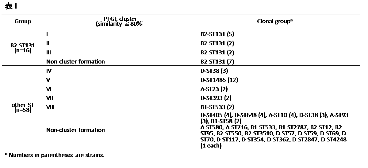
2011年に天理よろづ相談所病院にて、臨床検体より分離されたESBL産生大腸菌(E. coli)74株(B2-ST131クローン16株、他のSTクローン58株)を使用した。これらの株についてまず、系統学的グループの解析を行い、MLSTとパルスフィールドゲル電気泳動(PFGE)を行った。
系統学的グループの解析として、chuA、yjaA、およびTSPE4.C2の3つの遺伝子について、トリプレックスPCR(triplex PCR)を行った。MLSTとしては、7つのハウスキーピング遺伝子(adk、fumC、gyrB、icd、mdh、purA、およびrecA)をPCRダイレクトシークエンス(PCR-direct sequencing)を行った。また、制限酵素XbaI(Roche Diagnostics, Inc., Tokyo, Japan)を用いたPFGEを、CHEF-Dr II system(Bio-Rad Laboratories, Inc., Hercules, CA)にて行った。
得られた結果に基づき、Fingerprinting Plus Version 1.12,により系統樹を作成し、80%以上の類似性(UPGMA algorithm on Dice similarity coefficients)を示す場合に、同一PFGEクラスタであると判断した。
2011年に天理よろづ相談所病院にて、臨床検体より分離されたESBL産生大腸菌(E. coli)74株(B2-ST131クローン16株、他のSTクローン58株)を使用した。これらの株についてまず、系統学的グループの解析を行い、MLSTとパルスフィールドゲル電気泳動(PFGE)を行った。
系統学的グループの解析として、chuA、yjaA、およびTSPE4.C2の3つの遺伝子について、トリプレックスPCR(triplex PCR)を行った。MLSTとしては、7つのハウスキーピング遺伝子(adk、fumC、gyrB、icd、mdh、purA、およびrecA)をPCRダイレクトシークエンス(PCR-direct sequencing)を行った。また、制限酵素XbaI(Roche Diagnostics, Inc., Tokyo, Japan)を用いたPFGEを、CHEF-Dr II system(Bio-Rad Laboratories, Inc., Hercules, CA)にて行った。
得られた結果に基づき、Fingerprinting Plus Version 1.12,により系統樹を作成し、80%以上の類似性(UPGMA algorithm on Dice similarity coefficients)を示す場合に、同一PFGEクラスタであると判断した。
これらの解析から得られた結果を表1に示す。
(実施例1)MALDI-TOF MSによる解析
(MALDI-TOF MSによる測定)
上記参考例1と同様の臨床検体由来の菌株を、5% [v/v]ヒツジ血液含有コロンビア血液寒天にて、37℃で16~24時間培養した。培養後の菌株から、エタノール‐ギ酸抽出法により、タンパク質を抽出した。300μLのミリQ水に菌株を懸濁後、900μLのエタノールを加え、13,000gで2分間遠心し、上清を捨ててペレットを乾燥させた。乾燥したペレットに20μLの70%ギ酸を加え、ボルテックス後、20μLのアセトニトリルを加え、再度ボルテックス後、13,000gで2分間遠心した。上清1μLをターゲットプレートにアプライし、MALDI-TOF MS解析を行った。MALDI-TOF MS解析は、MALDI Biotyper(Bruker Daltonik, GmbH)およびMALDI Biotyper v3.1 softwareを用いて行った。マトリックスはα-cyano-4-hydroxy-cinnnamic acid(CHCA)を使用した。 菌株同定に推奨されている設定(linear positive mode, 20-Hz laser frequency, 20-kV acceleration voltage, 18.5-kV IS2 voltage, 250 ns extraction delay, and 2000 to 20,000-m/z range)により、解析を行った。キャリブリーターとして、Bruker Bacterial Test Standard (Bruker)を用いた。また上述と同様のタンパク質抽出方法により抽出したサンプルについて、同様のマトリックスを使用し、MALDI Biotyperの代わりに他の質量分析器Voyager-DE-1000(Applied Biosystems社製)を用いて、Linear modeにより測定を行った。
(MALDI-TOF MSによる測定)
上記参考例1と同様の臨床検体由来の菌株を、5% [v/v]ヒツジ血液含有コロンビア血液寒天にて、37℃で16~24時間培養した。培養後の菌株から、エタノール‐ギ酸抽出法により、タンパク質を抽出した。300μLのミリQ水に菌株を懸濁後、900μLのエタノールを加え、13,000gで2分間遠心し、上清を捨ててペレットを乾燥させた。乾燥したペレットに20μLの70%ギ酸を加え、ボルテックス後、20μLのアセトニトリルを加え、再度ボルテックス後、13,000gで2分間遠心した。上清1μLをターゲットプレートにアプライし、MALDI-TOF MS解析を行った。MALDI-TOF MS解析は、MALDI Biotyper(Bruker Daltonik, GmbH)およびMALDI Biotyper v3.1 softwareを用いて行った。マトリックスはα-cyano-4-hydroxy-cinnnamic acid(CHCA)を使用した。 菌株同定に推奨されている設定(linear positive mode, 20-Hz laser frequency, 20-kV acceleration voltage, 18.5-kV IS2 voltage, 250 ns extraction delay, and 2000 to 20,000-m/z range)により、解析を行った。キャリブリーターとして、Bruker Bacterial Test Standard (Bruker)を用いた。また上述と同様のタンパク質抽出方法により抽出したサンプルについて、同様のマトリックスを使用し、MALDI Biotyperの代わりに他の質量分析器Voyager-DE-1000(Applied Biosystems社製)を用いて、Linear modeにより測定を行った。
(MALDI-TOF MSにより得られたデータの解析)
MALDI-TOF MSにより得られたスペクトルを、B2-ST131クローン群と他のSTクローン群に分け、ClinProTools v2.2とFlexAnalysis v3.4 software (Bruker)を用いて多変量解析を行い、B2-ST131クローン群のバイオマーカーを探索した。
MALDI-TOF MSにより得られたスペクトルを、B2-ST131クローン群と他のSTクローン群に分け、ClinProTools v2.2とFlexAnalysis v3.4 software (Bruker)を用いて多変量解析を行い、B2-ST131クローン群のバイオマーカーを探索した。
MALDI-TOF MSの解析結果を以下の表2に示す。
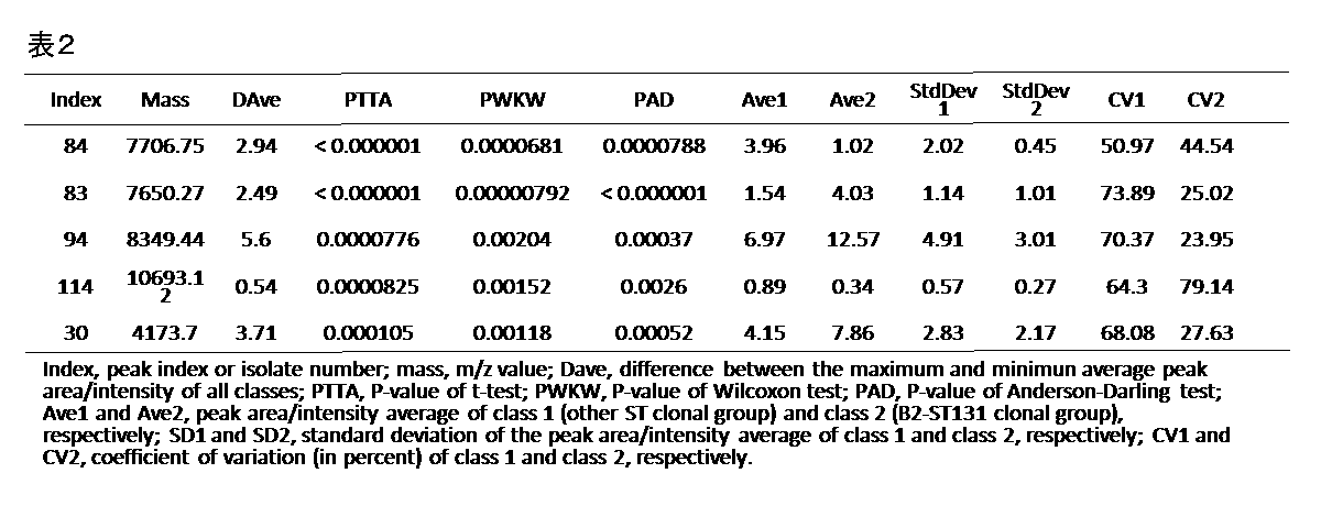
解析の結果、B2-ST131クローン群に特異的なピーク7,650m/zを検出した(図1(A))。他のSTクローン群には、ピーク7,650m/zは検出されず、7,707m/zが検出された。またMALDI Biotyperの代わりに、Voyager-DE-1000を用いた場合も、同様のピークが確認された(図1(B))。7,650m/zをバイオマーカーとした場合、表3に示す通り、感度は100%、特異度は89.7%と高い値が得られた。
(実施例2)バイオマーカーとなるタンパク質の同定と解析
実施例1において特定された7650Daのタンパク質を、bottom-up proteomicsおよびエドマン分解によるN末端アミノ酸配列解析を行うことにより同定した。まず、参考例1と同様の臨床検体由来のB2-ST131クローン群の菌株から、以下のようにしてタンパク質を精製した。まず、菌株をLB培地で増殖させた後、回収して超音波破砕し、80%硫安沈殿、オープンカラムDEAEセルロース、オープンカラムCMセルロールにより段階的に精製し、その後HPLC(TSKgel Protein C4-300 (Tosoh Corporation, Tokyo, Japan))により分取した。分取後、各画分を乾燥させて濃縮し、MALDI-TOF MSにより確認を行い、7650Daのタンパク質が溶出した画分を特定した。特定した画分に溶出したタンパク質について、Tricine SDS-PAGEを行い、silver染色後、ゲルを切り出した。切り出したゲルは、トリプシンにより消化後、LCMS-IT-TOF (Shimazu Corporation, Kyoto, Japan)を用いて、解析した。得られたスペクトルを、Mascot MS/MS Ion Search (Matrix Science K.K., Tokyo, Japan)により解析し、SwissProt databaseによりペプチドを同定した。同時に、N末端アミノ酸配列解析を行い、bottom-up proteomicsによるペプチドの同定結果と併せて、タンパク質を同定した。
実施例1において特定された7650Daのタンパク質を、bottom-up proteomicsおよびエドマン分解によるN末端アミノ酸配列解析を行うことにより同定した。まず、参考例1と同様の臨床検体由来のB2-ST131クローン群の菌株から、以下のようにしてタンパク質を精製した。まず、菌株をLB培地で増殖させた後、回収して超音波破砕し、80%硫安沈殿、オープンカラムDEAEセルロース、オープンカラムCMセルロールにより段階的に精製し、その後HPLC(TSKgel Protein C4-300 (Tosoh Corporation, Tokyo, Japan))により分取した。分取後、各画分を乾燥させて濃縮し、MALDI-TOF MSにより確認を行い、7650Daのタンパク質が溶出した画分を特定した。特定した画分に溶出したタンパク質について、Tricine SDS-PAGEを行い、silver染色後、ゲルを切り出した。切り出したゲルは、トリプシンにより消化後、LCMS-IT-TOF (Shimazu Corporation, Kyoto, Japan)を用いて、解析した。得られたスペクトルを、Mascot MS/MS Ion Search (Matrix Science K.K., Tokyo, Japan)により解析し、SwissProt databaseによりペプチドを同定した。同時に、N末端アミノ酸配列解析を行い、bottom-up proteomicsによるペプチドの同定結果と併せて、タンパク質を同定した。
LCMS-IT-TOFの結果を以下の表4に示す。

表4の結果と、N末端アミノ酸配列解析の結果により、7650Daのタンパク質が、機能不明のYahOタンパク質であることがわかった(図2)。図2中の下線部は、LCMS-IT-TOFにより得られたアミノ酸配列であり、四角で囲った部分はN末端アミノ酸配列解析により得られたアミノ酸配列である。
(実施例3) バイオマーカーとなる遺伝子についての解析
実施例2において同定されたアミノ酸配列から、forward primer:5'-TAGTCGGTGCGTTAGCGTT-3'(配列番号11)とreverse primer:5'-TGCCGTGGATCTTATTGTCA-3'(配列番号12)を設計し、 B2-ST131クローン群と他のSTクローン群について、PCRダイレクトシークエンスによって塩基配列とアミノ酸配列の比較を行った。
まず実施例1と同様にして、臨床検体由来のB2-ST131クローン群の菌株を培地で増殖させた後、回収した。菌株を回収後、定法に従ってDNAを抽出した。抽出したDNAについて、上記配列番号11と配列番号12のプライマーを用いて、PCRを行った。PCRにはApplied Biosystems Veriti 96 Thermal Cycler (Life Technologies)を用い、DNA配列決定にはApplied Biosystems 3730xl DNA Analyzer (Thermo Fisher Scientific)を用いた。また理論上の分子量(Mw)をCompute pI/Mw tool (http://web.expasy.org/compute_pi/)を用いて算出した。
実施例2において同定されたアミノ酸配列から、forward primer:5'-TAGTCGGTGCGTTAGCGTT-3'(配列番号11)とreverse primer:5'-TGCCGTGGATCTTATTGTCA-3'(配列番号12)を設計し、 B2-ST131クローン群と他のSTクローン群について、PCRダイレクトシークエンスによって塩基配列とアミノ酸配列の比較を行った。
まず実施例1と同様にして、臨床検体由来のB2-ST131クローン群の菌株を培地で増殖させた後、回収した。菌株を回収後、定法に従ってDNAを抽出した。抽出したDNAについて、上記配列番号11と配列番号12のプライマーを用いて、PCRを行った。PCRにはApplied Biosystems Veriti 96 Thermal Cycler (Life Technologies)を用い、DNA配列決定にはApplied Biosystems 3730xl DNA Analyzer (Thermo Fisher Scientific)を用いた。また理論上の分子量(Mw)をCompute pI/Mw tool (http://web.expasy.org/compute_pi/)を用いて算出した。
その結果、B2-ST131クローン群に特異的なピーク7,650m/zと、他のSTクローン群に確認されるピーク7,707m/zとの分子量の差は、E34Aのアミノ酸変異が原因であることが明らかとなった(図3)。また、E34A検出によるESBL産生大腸菌B2-ST131クローン検出の感度および特異度は下記の表5に示すとおりであった。

(実施例4) PCR法を用いたESBL産生大腸菌B2-ST131の検出
(1)まず、上記参考例1と同様の臨床検体由来の菌株を培地で増殖させた後、回収した。菌株を回収後、定法に従ってDNAを抽出した。抽出したDNAについて、以下のプライマーセットを用いて、リアルタイムPCRを行った。
forward primer:5'-GAATTTGAAAARGTYGC-3'(配列番号13)
reverse primer:5'-TGCCGTGGATCTTATTGTCA-3'(配列番号12)
forward primerは配列番号2がE34Aをコードするよう変異した塩基配列の85~101番目、reverse primerは配列番号2の231~250番目にアニーリングするよう設計されたものである。
PCR機器はLightCycler 2.0 system(Roche Diagnostics, Inc., Tokyo, Japan)、試薬はLightCycler FastStart DNA Master SYBR GreenI(Roche)を用いて、次のように行った。すなわち、初期変性を95℃で7分間行った後、95℃で1秒間、63~53℃(step size 0.5℃/cycle)で5秒間、及び72℃で8秒間、蛍光測定(PCRによる増幅産物の測定)を1サイクルとし、これを40サイクル行い、続いて増幅産物の融解曲線分析のために、95℃で1秒、75℃で15秒後、0.15℃/秒の速度で95℃まで昇温させると同時に連続的な蛍光測定により各温度に対する増幅産物の二本鎖DNA量を測定し、最後に40℃まで降温させた。PCR終了後、融解曲線解析を同PCR装置のソフトウェアで行ない、増幅産物のTm値を求めることで特異的な増幅産物の有無を判定した。
(1)まず、上記参考例1と同様の臨床検体由来の菌株を培地で増殖させた後、回収した。菌株を回収後、定法に従ってDNAを抽出した。抽出したDNAについて、以下のプライマーセットを用いて、リアルタイムPCRを行った。
forward primer:5'-GAATTTGAAAARGTYGC-3'(配列番号13)
reverse primer:5'-TGCCGTGGATCTTATTGTCA-3'(配列番号12)
forward primerは配列番号2がE34Aをコードするよう変異した塩基配列の85~101番目、reverse primerは配列番号2の231~250番目にアニーリングするよう設計されたものである。
PCR機器はLightCycler 2.0 system(Roche Diagnostics, Inc., Tokyo, Japan)、試薬はLightCycler FastStart DNA Master SYBR GreenI(Roche)を用いて、次のように行った。すなわち、初期変性を95℃で7分間行った後、95℃で1秒間、63~53℃(step size 0.5℃/cycle)で5秒間、及び72℃で8秒間、蛍光測定(PCRによる増幅産物の測定)を1サイクルとし、これを40サイクル行い、続いて増幅産物の融解曲線分析のために、95℃で1秒、75℃で15秒後、0.15℃/秒の速度で95℃まで昇温させると同時に連続的な蛍光測定により各温度に対する増幅産物の二本鎖DNA量を測定し、最後に40℃まで降温させた。PCR終了後、融解曲線解析を同PCR装置のソフトウェアで行ない、増幅産物のTm値を求めることで特異的な増幅産物の有無を判定した。
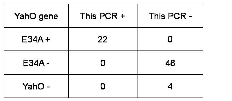
結果を図4に示す。増幅産物がある場合は、Tm値として82.5℃付近ピークを示すことがわかった。また、以下の表6にリアルタイムPCRによるYahO遺伝子の変異検出の特異度を示す。表6において、「This PCR+」はTm値のピークが認められたもの、「This PCR-」は、Tm値のピークが認められなかったものである。
(2)また上記配列番号13のforward primerと、配列番号12のreverse primerを用いてPCRを行い、増幅産物の有無について電気泳動による確認を行った。PCRのサイクルは上述の(1)と同様であり、Mupidミニゲル泳動槽(ADVANCE社)を用いてアガロースゲルにより電気泳動を行い、エチジウムブロマイド溶液でゲルを染色して、目的の増幅バンドの有無を確認した。
結果を、図5に示す。図5中、NCはネガティブコントロールである。上記プライマーを用いてPCRを行い、増幅産物をアガロースゲル電気泳動により確認することにより、B2-ST131クローン群を検出可能であることがわかった。
(実施例5)MALDI-TOF MSを用いたカルバペネマーゼ産生大腸菌B2-ST131の検出
天理よろづ相談所病院にて保有している、臨床検体より分離されたカルバペネマーゼ産生大腸菌(E. coli)28株(B2-ST131クローン20株、他のSTクローン8株)を使用した。これらの株は、系統学的グループの解析として、chuA、yjaA、およびTSPE4.C2の3つの遺伝子についてのトリプレックスPCR(triplex PCR)、MLSTとして、7つのハウスキーピング遺伝子(adk、fumC、gyrB、icd、mdh、purA、およびrecA)をPCRダイレクトシークエンス(PCR-direct sequencing)、制限酵素XbaI(Roche Diagnostics, Inc., Tokyo, Japan)を用いたPFGEを、CHEF-Dr II system(Bio-Rad Laboratories, Inc., Hercules, CA)にて行い、得られた結果に基づき、Fingerprinting Plus Version 1.12,により系統樹を作成し、80%以上の類似性(UPGMA algorithm on Dice similarity coefficients)を示す場合に、同一PFGEクラスタであると判断し、分類を行ったものである。
天理よろづ相談所病院にて保有している、臨床検体より分離されたカルバペネマーゼ産生大腸菌(E. coli)28株(B2-ST131クローン20株、他のSTクローン8株)を使用した。これらの株は、系統学的グループの解析として、chuA、yjaA、およびTSPE4.C2の3つの遺伝子についてのトリプレックスPCR(triplex PCR)、MLSTとして、7つのハウスキーピング遺伝子(adk、fumC、gyrB、icd、mdh、purA、およびrecA)をPCRダイレクトシークエンス(PCR-direct sequencing)、制限酵素XbaI(Roche Diagnostics, Inc., Tokyo, Japan)を用いたPFGEを、CHEF-Dr II system(Bio-Rad Laboratories, Inc., Hercules, CA)にて行い、得られた結果に基づき、Fingerprinting Plus Version 1.12,により系統樹を作成し、80%以上の類似性(UPGMA algorithm on Dice similarity coefficients)を示す場合に、同一PFGEクラスタであると判断し、分類を行ったものである。
上記臨床検体由来の菌株28株について、実施例1と同様にして、菌株を培養し、タンパク質を抽出し、MALDI-TOF MSによりスペクトルを得た。その結果を、以下の表7に示す。
カルバペネマーゼ産生B2-ST131クローン群には、特異的なピーク7,650m/zが検出され、他のSTクローン群には、ピーク7,650m/zは検出されず、7,707m/zが検出された。感度は95%、特異度は88%と高い値であった。
カルバペネマーゼ産生B2-ST131クローン群には、特異的なピーク7,650m/zが検出され、他のSTクローン群には、ピーク7,650m/zは検出されず、7,707m/zが検出された。感度は95%、特異度は88%と高い値であった。
(実施例6) PCR法を用いたカルバペネマーゼ産生大腸菌B2-ST131の検出
参考例5と同様の菌株について、実施例4と同様にして、菌株を培地にて増殖させて回収し、定法に従ってDNAを抽出し、抽出したDNAについてリアルタイムPCRを行った。
参考例5と同様の菌株について、実施例4と同様にして、菌株を培地にて増殖させて回収し、定法に従ってDNAを抽出し、抽出したDNAについてリアルタイムPCRを行った。
結果として、表8にリアルタイムPCRによるYahO遺伝子の変異検出の感度と特異度を示す。上記リアルタイムPCRにより、カルバペネマーゼ産生B2-ST131クローン群を感度95%、特異度88%と高い値で検出可能であることがわかった。

本発明は、迅速かつ簡便に大腸菌B2-ST131クローンを識別することを可能とするものである。本発明の検査方法は、感度および特異度も優れており、迅速かつ効率的にB2-ST131クローンを識別することができる。臨床において、本発明によりB2-ST131クローンを迅速かつ正確に識別することができることから、医療関連感染等の原因を速やかに特定することができ、感染症初期治療決定ツールとして非常に有用である。さらに本発明の検査方法は、世界的なパンデミックの制御に寄与する可能性もあり、感染症治療を飛躍的に前進させることが期待される。
Claims (15)
- YahOタンパク質を構成するアミノ酸配列のうち、配列番号1に示すアミノ酸配列における第34番目のアミノ酸のアラニンへの変異を検出することを特徴とする、大腸菌B2-ST131クローンを識別するための検査方法。
- 前記第34番目のアラニンへの変異の検出が、以下のi)~iii)より選択されるいずれかにより行われる、請求項1に記載の検査方法:
i)被検試料について、前記第34番目のアミノ酸を含むポリペプチドをコードするポリヌクレオチドの塩基配列を解析すること;
ii)被検試料について、配列番号1に示すアミノ酸配列のうち、第34番目のアミノ酸を含むポリペプチドのアミノ酸配列を解析すること;
iii)被検試料について、前記第34番目のアミノ酸を含むポリペプチドの分子量を測定すること。 - 前記第34番目のアラニンへの変異の検出が、以下のi)~iii)より選択されるいずれかにより行われる、請求項2に記載の検査方法:
i)被検試料について、前記第34番目のアミノ酸を含むポリペプチドをコードするポリヌクレオチドの塩基配列を、核酸増幅反応により解析すること;
ii)被検試料について、配列番号1に示すアミノ酸配列のうち、第34番目のアミノ酸を含むポリペプチドのアミノ酸配列を、免疫学的手法により解析すること;
iii)被検試料について、前記第34番目のアミノ酸を含むポリペプチドの分子量を、質量分析計を用いて測定すること。 - YahOタンパク質を構成するアミノ酸配列のうち、配列番号1に示すアミノ酸配列における第34番目のアミノ酸のアラニンへの変異を示す塩基を検出するための、少なくとも15ヌクレオチドの長さを有するポリヌクレオチド。
- 前記ポリヌクレオチドが、前記第34番目のアミノ酸を含むポリペプチドをコードする塩基配列の部分配列又は当該部分配列に相補的な配列とハイブリダイズし得る、請求項4に記載のポリヌクレオチド。
- 前記ポリヌクレオチドが、前記第34番目のアミノ酸のアラニンへの変異を示す塩基を含む部分配列又は当該部分配列に相補的な配列を特異的に増幅し得るプライマーである、請求項4又は5に記載のポリヌクレオチド。
- 前記ポリヌクレオチドが、配列番号13に示す5'-GAATTTGAAAARGTYGC-3'の塩基配列を含むものである、請求項5~6のいずれか1に記載のポリヌクレオチド。
- 請求項4~7のいずれか1に記載のポリヌクレオチドをプライマーとして含む、プライマーセット。
- YahOタンパク質を構成するアミノ酸配列のうち、配列番号1に示すアミノ酸配列における第34番目のアミノ酸のアラニンへの変異を検出するための、前記第34番目のアミノ酸を含むポリペプチドを特異的に認識する、抗体。
- 前記抗体が、前記第34番目のアミノ酸としてアラニンを含むポリペプチド又は前記第34番目のアミノ酸としてグルタミ酸を含むポリペプチドのいずれかを特異的に認識する、請求項9に記載の抗体。
- 請求項4~7のいずれか1に記載のポリヌクレオチド、請求項8に記載のプライマーセット、又は請求項9~10のいずれか1に記載の抗体を含む、大腸菌B2-ST131クローン識別用試薬組成物。
- 請求項11に記載の大腸菌B2-ST131クローン識別用試薬組成物を含む、大腸菌B2-ST131クローン識別用試薬キット。
- 請求項4~7のいずれか1に記載のポリヌクレオチド、請求項8に記載のプライマーセット、請求項9~10のいずれか1に記載の抗体、請求項11に記載の大腸菌B2-ST131クローン識別用試薬組成物、又は請求項12に記載の大腸菌B2-ST131クローン識別用試薬キットを用いて、YahOタンパク質を構成するアミノ酸配列のうち、配列番号1に示すアミノ酸配列における第34番目のアミノ酸のアラニンへの変異を検出することを特徴とする、大腸菌B2-ST131クローンを識別するための検査方法。
- 大腸菌B2-ST131クローンが、基質特異性拡張型βラクタマーゼ(ESBL)産生大腸菌又はカルバペネマーゼ産生大腸菌を含む、請求項1~3及び請求項13のいずれか1に記載の検査方法。
- 以下のa)またはb)から選択される大腸菌B2-ST131クローンを識別するためのバイオマーカー:
a)YahOタンパク質を構成するアミノ酸配列のうち、配列番号1に示すアミノ酸配列における第34番目のアミノ酸がアラニンに変異したアミノ酸配列を含むポリペプチドからなるバイオマーカー;
b)a)に記載の前記第34番目のアミノ酸がアラニンに変異したアミノ酸配列をコードする塩基配列を含むポリヌクレオチドからなるバイオマーカー。
Priority Applications (1)
| Application Number | Priority Date | Filing Date | Title |
|---|---|---|---|
| JP2017506605A JP6748987B2 (ja) | 2015-03-17 | 2016-03-17 | 大腸菌b2−st131クローンの検査方法 |
Applications Claiming Priority (2)
| Application Number | Priority Date | Filing Date | Title |
|---|---|---|---|
| JP2015053351 | 2015-03-17 | ||
| JP2015-053351 | 2015-03-17 |
Publications (1)
| Publication Number | Publication Date |
|---|---|
| WO2016148231A1 true WO2016148231A1 (ja) | 2016-09-22 |
Family
ID=56919050
Family Applications (1)
| Application Number | Title | Priority Date | Filing Date |
|---|---|---|---|
| PCT/JP2016/058463 Ceased WO2016148231A1 (ja) | 2015-03-17 | 2016-03-17 | 大腸菌b2-st131クローンの検査方法 |
Country Status (2)
| Country | Link |
|---|---|
| JP (1) | JP6748987B2 (ja) |
| WO (1) | WO2016148231A1 (ja) |
-
2016
- 2016-03-17 JP JP2017506605A patent/JP6748987B2/ja not_active Expired - Fee Related
- 2016-03-17 WO PCT/JP2016/058463 patent/WO2016148231A1/ja not_active Ceased
Non-Patent Citations (3)
| Title |
|---|
| DATABASE NCBI [o] 11 June 2010 (2010-06-11), "Escherichia coli strain RM2008 hypothetical protein (yahO) gene", Database accession no. GU391860 * |
| FAGERQUIST C.K. ET AL.: "Rapid Identification of Protein Biomarkers of Escherichia coli 0157:H7 by Matrix-Assisted Laser Desorption Ionization- Time-of-Flight-Time-of-Flight Mass Spectrometry and Top-Down Proteomics.", ANAL. CHEM., vol. 82, no. 7, 2010, pages 2717 - 2725, XP055313107, Retrieved from the Internet <URL:http://pubs.acs.org/doi/abs/10.1021/ac902455d> * |
| MATSUMURA Y. ET AL.: "Detection of extended- spectrum-beta-lactamase-producing Escherichia coli ST131 and ST405 clonal groups by matrix- assisted laser desorption ionization-time of flight mass spectrometry.", J. CLIN. MICROBIOL., vol. 52, no. 4, 1 April 2014 (2014-04-01), pages 1034 - 1040, XP055313112 * |
Also Published As
| Publication number | Publication date |
|---|---|
| JPWO2016148231A1 (ja) | 2018-01-18 |
| JP6748987B2 (ja) | 2020-09-02 |
Similar Documents
| Publication | Publication Date | Title |
|---|---|---|
| AU2010217204B2 (en) | Method of diagnosis of infection by Mycobacteria and reagents therefor | |
| Jarraud et al. | Identification of Legionella in clinical samples | |
| Liu et al. | Clinical and microbiological characteristics of patients with bacteremia caused by Campylobacter species with an emphasis on the subspecies of C. fetus | |
| EP2508887B1 (en) | Method for detecting microorganisms belonging to mycoplasma pneumoniae and/or mycoplasma genitalium | |
| US20240369553A1 (en) | Biomarkers for early diagnosis and differentiation of mycobacterial infection | |
| Lanotte et al. | Proteomic biomarkers associated with Streptococcus agalactiae invasive genogroups | |
| WO2017187178A1 (en) | Method for detecting carbapenemase-producing bacteria | |
| KR101943487B1 (ko) | 감염성 폐질환 진단 마커 및 이의 용도 | |
| US20210132064A1 (en) | Biomarkers for detecting microbial infection | |
| EP2192184A1 (en) | Detection of bacterium belonging to genus campylobacter which targets cytolethal distending toxin | |
| US20040137463A1 (en) | Method of identifying target organisms by determining the characteristics of their intronic region nucleic acids | |
| JP6748987B2 (ja) | 大腸菌b2−st131クローンの検査方法 | |
| US20060003350A1 (en) | Methods and compositions to detect bacteria using multiplex PCR | |
| WO2011001449A2 (en) | Primers, pcr reaction mixture and methods thereof | |
| JP4576168B2 (ja) | 膀胱尿管逆流症または間質性膀胱炎の検査方法 | |
| Peker | Diagnosis of bloodstream infections from positive blood cultures: recent developments in molecular approaches | |
| Peker | Exploring next-generation sequencing in clinical microbiology: identification, characterization and typing of pathogens | |
| CN120967019A (zh) | 一种用于单管快速筛查微生物的多种万古霉素耐药基因的方法、二维pcr反应体系和试剂盒 | |
| Vinasco Pacheco | The role of host and microbial lactate in gastric adenocarcinoma. | |
| JP2012187008A (ja) | 新規カスガマイシンアセチルトランスフェラーゼ及びその検出方法 | |
| CA2805306A1 (en) | Diagnostic method for colitis | |
| WO2019136529A1 (en) | Diagnostic and therapeutic for ibd | |
| HK1164942A (en) | Method of diagnosis of infection by mycobacteria and reagents therefor |
Legal Events
| Date | Code | Title | Description |
|---|---|---|---|
| 121 | Ep: the epo has been informed by wipo that ep was designated in this application |
Ref document number: 16765060 Country of ref document: EP Kind code of ref document: A1 |
|
| ENP | Entry into the national phase |
Ref document number: 2017506605 Country of ref document: JP Kind code of ref document: A |
|
| NENP | Non-entry into the national phase |
Ref country code: DE |
|
| 122 | Ep: pct application non-entry in european phase |
Ref document number: 16765060 Country of ref document: EP Kind code of ref document: A1 |