WO2016143700A1 - Method for stabilizing dna aptamer - Google Patents
Method for stabilizing dna aptamer Download PDFInfo
- Publication number
- WO2016143700A1 WO2016143700A1 PCT/JP2016/056805 JP2016056805W WO2016143700A1 WO 2016143700 A1 WO2016143700 A1 WO 2016143700A1 JP 2016056805 W JP2016056805 W JP 2016056805W WO 2016143700 A1 WO2016143700 A1 WO 2016143700A1
- Authority
- WO
- WIPO (PCT)
- Prior art keywords
- aptamer
- dna aptamer
- nucleic acid
- dna
- base
- Prior art date
Links
Images


Classifications
-
- C—CHEMISTRY; METALLURGY
- C12—BIOCHEMISTRY; BEER; SPIRITS; WINE; VINEGAR; MICROBIOLOGY; ENZYMOLOGY; MUTATION OR GENETIC ENGINEERING
- C12N—MICROORGANISMS OR ENZYMES; COMPOSITIONS THEREOF; PROPAGATING, PRESERVING, OR MAINTAINING MICROORGANISMS; MUTATION OR GENETIC ENGINEERING; CULTURE MEDIA
- C12N15/00—Mutation or genetic engineering; DNA or RNA concerning genetic engineering, vectors, e.g. plasmids, or their isolation, preparation or purification; Use of hosts therefor
- C12N15/09—Recombinant DNA-technology
- C12N15/11—DNA or RNA fragments; Modified forms thereof; Non-coding nucleic acids having a biological activity
- C12N15/115—Aptamers, i.e. nucleic acids binding a target molecule specifically and with high affinity without hybridising therewith ; Nucleic acids binding to non-nucleic acids, e.g. aptamers
-
- C—CHEMISTRY; METALLURGY
- C12—BIOCHEMISTRY; BEER; SPIRITS; WINE; VINEGAR; MICROBIOLOGY; ENZYMOLOGY; MUTATION OR GENETIC ENGINEERING
- C12N—MICROORGANISMS OR ENZYMES; COMPOSITIONS THEREOF; PROPAGATING, PRESERVING, OR MAINTAINING MICROORGANISMS; MUTATION OR GENETIC ENGINEERING; CULTURE MEDIA
- C12N15/00—Mutation or genetic engineering; DNA or RNA concerning genetic engineering, vectors, e.g. plasmids, or their isolation, preparation or purification; Use of hosts therefor
- C12N15/09—Recombinant DNA-technology
- C12N15/10—Processes for the isolation, preparation or purification of DNA or RNA
-
- A—HUMAN NECESSITIES
- A61—MEDICAL OR VETERINARY SCIENCE; HYGIENE
- A61K—PREPARATIONS FOR MEDICAL, DENTAL OR TOILETRY PURPOSES
- A61K31/00—Medicinal preparations containing organic active ingredients
- A61K31/70—Carbohydrates; Sugars; Derivatives thereof
- A61K31/7088—Compounds having three or more nucleosides or nucleotides
-
- A—HUMAN NECESSITIES
- A61—MEDICAL OR VETERINARY SCIENCE; HYGIENE
- A61K—PREPARATIONS FOR MEDICAL, DENTAL OR TOILETRY PURPOSES
- A61K48/00—Medicinal preparations containing genetic material which is inserted into cells of the living body to treat genetic diseases; Gene therapy
-
- C—CHEMISTRY; METALLURGY
- C12—BIOCHEMISTRY; BEER; SPIRITS; WINE; VINEGAR; MICROBIOLOGY; ENZYMOLOGY; MUTATION OR GENETIC ENGINEERING
- C12N—MICROORGANISMS OR ENZYMES; COMPOSITIONS THEREOF; PROPAGATING, PRESERVING, OR MAINTAINING MICROORGANISMS; MUTATION OR GENETIC ENGINEERING; CULTURE MEDIA
- C12N15/00—Mutation or genetic engineering; DNA or RNA concerning genetic engineering, vectors, e.g. plasmids, or their isolation, preparation or purification; Use of hosts therefor
- C12N15/09—Recombinant DNA-technology
-
- C—CHEMISTRY; METALLURGY
- C12—BIOCHEMISTRY; BEER; SPIRITS; WINE; VINEGAR; MICROBIOLOGY; ENZYMOLOGY; MUTATION OR GENETIC ENGINEERING
- C12N—MICROORGANISMS OR ENZYMES; COMPOSITIONS THEREOF; PROPAGATING, PRESERVING, OR MAINTAINING MICROORGANISMS; MUTATION OR GENETIC ENGINEERING; CULTURE MEDIA
- C12N2310/00—Structure or type of the nucleic acid
- C12N2310/10—Type of nucleic acid
- C12N2310/16—Aptamers
-
- C—CHEMISTRY; METALLURGY
- C12—BIOCHEMISTRY; BEER; SPIRITS; WINE; VINEGAR; MICROBIOLOGY; ENZYMOLOGY; MUTATION OR GENETIC ENGINEERING
- C12N—MICROORGANISMS OR ENZYMES; COMPOSITIONS THEREOF; PROPAGATING, PRESERVING, OR MAINTAINING MICROORGANISMS; MUTATION OR GENETIC ENGINEERING; CULTURE MEDIA
- C12N2310/00—Structure or type of the nucleic acid
- C12N2310/30—Chemical structure
- C12N2310/33—Chemical structure of the base
- C12N2310/334—Modified C
-
- C—CHEMISTRY; METALLURGY
- C12—BIOCHEMISTRY; BEER; SPIRITS; WINE; VINEGAR; MICROBIOLOGY; ENZYMOLOGY; MUTATION OR GENETIC ENGINEERING
- C12N—MICROORGANISMS OR ENZYMES; COMPOSITIONS THEREOF; PROPAGATING, PRESERVING, OR MAINTAINING MICROORGANISMS; MUTATION OR GENETIC ENGINEERING; CULTURE MEDIA
- C12N2310/00—Structure or type of the nucleic acid
- C12N2310/30—Chemical structure
- C12N2310/33—Chemical structure of the base
- C12N2310/336—Modified G
-
- C—CHEMISTRY; METALLURGY
- C12—BIOCHEMISTRY; BEER; SPIRITS; WINE; VINEGAR; MICROBIOLOGY; ENZYMOLOGY; MUTATION OR GENETIC ENGINEERING
- C12N—MICROORGANISMS OR ENZYMES; COMPOSITIONS THEREOF; PROPAGATING, PRESERVING, OR MAINTAINING MICROORGANISMS; MUTATION OR GENETIC ENGINEERING; CULTURE MEDIA
- C12N2310/00—Structure or type of the nucleic acid
- C12N2310/50—Physical structure
- C12N2310/51—Physical structure in polymeric form, e.g. multimers, concatemers
-
- C—CHEMISTRY; METALLURGY
- C12—BIOCHEMISTRY; BEER; SPIRITS; WINE; VINEGAR; MICROBIOLOGY; ENZYMOLOGY; MUTATION OR GENETIC ENGINEERING
- C12N—MICROORGANISMS OR ENZYMES; COMPOSITIONS THEREOF; PROPAGATING, PRESERVING, OR MAINTAINING MICROORGANISMS; MUTATION OR GENETIC ENGINEERING; CULTURE MEDIA
- C12N2310/00—Structure or type of the nucleic acid
- C12N2310/50—Physical structure
- C12N2310/53—Physical structure partially self-complementary or closed
- C12N2310/531—Stem-loop; Hairpin
-
- C—CHEMISTRY; METALLURGY
- C12—BIOCHEMISTRY; BEER; SPIRITS; WINE; VINEGAR; MICROBIOLOGY; ENZYMOLOGY; MUTATION OR GENETIC ENGINEERING
- C12N—MICROORGANISMS OR ENZYMES; COMPOSITIONS THEREOF; PROPAGATING, PRESERVING, OR MAINTAINING MICROORGANISMS; MUTATION OR GENETIC ENGINEERING; CULTURE MEDIA
- C12N2320/00—Applications; Uses
- C12N2320/50—Methods for regulating/modulating their activity
- C12N2320/51—Methods for regulating/modulating their activity modulating the chemical stability, e.g. nuclease-resistance
-
- C—CHEMISTRY; METALLURGY
- C12—BIOCHEMISTRY; BEER; SPIRITS; WINE; VINEGAR; MICROBIOLOGY; ENZYMOLOGY; MUTATION OR GENETIC ENGINEERING
- C12N—MICROORGANISMS OR ENZYMES; COMPOSITIONS THEREOF; PROPAGATING, PRESERVING, OR MAINTAINING MICROORGANISMS; MUTATION OR GENETIC ENGINEERING; CULTURE MEDIA
- C12N2320/00—Applications; Uses
- C12N2320/50—Methods for regulating/modulating their activity
- C12N2320/52—Methods for regulating/modulating their activity modulating the physical stability, e.g. GC-content
Definitions
- the present invention relates to a method for designing a base sequence that enhances the ability of a DNA aptamer to bind to a target molecule and / or stabilizes the DNA aptamer, and a DNA aptamer having such properties.
- nucleic acid drugs and the like have attracted attention as pharmaceuticals or diagnostic agents, and research and development of various nucleic acid drugs and the like have been promoted in various countries around the world for their medical applications.
- nucleic acids usually have a problem that they are easily degraded in vivo by nucleases such as nucleases. Stabilization of nucleic acids in vivo has become an indispensable issue for allowing the pharmacological effects of nucleic acid pharmaceuticals to act efficiently and continuously.
- Non-patent Documents 1 and 2, and Patent Documents a technique for chemically modifying the sugar or phosphate moiety serving as the backbone of the nucleic acid molecule is generally used.
- these modifications may also affect the higher-order structure and physical properties of nucleic acids, not only reducing the ability to bind to target molecules and increasing toxicity in vivo, but also increasing production costs.
- Peng CG, Masad, J., Damha, MJ, G-quadruplex induced stabilization by 2′-deoxy-2′-fluoro-D-arabinonucleic acids (2′F-ANA), 2007, Nucl. Acids Res., 35 , Pp. 4977-4988.
- An object of the present invention is to provide a simple and low-cost method for increasing the stability and / or target molecule binding ability of a DNA aptamer, and a DNA aptamer obtained by the method.
- the inventor of the present application replaces an internal hairpin structure of a DNA aptamer with a structure called a mini hairpin structure, and optionally replaces an AT base pair of the stem structure of the DNA aptamer with a GC base pair, thereby replacing an existing DNA aptamer. It was found that the stability and / or the ability to bind to a target molecule can be enhanced, and the present invention has been completed.
- the present invention includes the following aspects. (1) A method for designing a base sequence that enhances a target molecule-binding ability of an existing DNA aptamer and / or stabilizes the aptamer, wherein the aptamer includes one or more stem structures and one or more loop structures.
- nucleic acid region (A) a first nucleic acid region consisting of 2 to 5 arbitrary nucleotides, (B) gna or gnna (where each n is independently g, t, a or c, a base analog or a modified base)), a second nucleic acid region comprising a base sequence, and (C) a third nucleic acid region comprising a base sequence complementary to the first nucleic acid region, and The first nucleic acid region and the third nucleic acid region are base paired with each other.
- the design method constituted by arm portion and loop portion of a second nucleic acid region, said design method.
- the DNA aptamer includes one or more base analogs and / or modified bases.
- the base analog is 7- (2-thienyl) -3H-imidazo [4,5-b] pyridine-3-yl.
- the method includes increasing the GC pair by 1 to 5 pairs at the end of the DNA aptamer. The method according to any one.
- a stabilized DNA aptamer with enhanced target molecule binding ability obtained by a production method comprising a step of designing a base sequence of a DNA aptamer and a step of producing a DNA aptamer based on the designed base sequence, The step comprises replacing a hairpin structure composed of a set of stem structures and loop structures with a mini hairpin structure in a DNA aptamer comprising one or more stem structures and one or more loop structures, wherein the mini hairpin structure comprises: The following nucleic acid regions (A) to (C) linked in order from the 5 ′ end to the 3 ′ end: (A) a first nucleic acid region consisting of 2 to 5 arbitrary nucleotides, ( B) a second nucleic acid region comprising a base sequence of gna or gnna (where each n is independently g, t, a or c, a base analog or a modified base), and (C) Base sequence complementary to the first nucleic acid region Ranaru and a third nucle
- the DNA aptamer according to (8) comprising one or more base analogs and / or modified bases.
- the design step includes increasing the number of GC pairs by 1 to 5 at the end of the DNA aptamer.
- nucleic acid regions of the following (A) to (C) are sequentially linked from the 5 ′ end to the 3 ′ end: (A) a first nucleic acid region consisting of 2 to 5 arbitrary nucleotides, (B) a second nucleic acid region comprising a base sequence of gna or gnna (where each n is independently g, t, a or c, a base analog or a modified base), and (C ) Consists of a third nucleic acid region consisting of a base sequence complementary to the first nucleic acid region, and the first nucleic acid region and the third nucleic acid region are composed of a stem portion and a loop portion consisting of the second nucleic acid region.
- the DNA aptamer (15) The DNA aptamer according to (14), wherein a total GC content in the one or more stem structures is 75% or more. (16) The DNA aptamer according to (14) or (15), further comprising a hairpin structure defined in (14) at one end.
- a DNA aptamer for IFN- ⁇ comprising the nucleotide sequence shown in any of SEQ ID NOs: 6 and 8-11.
- a DNA aptamer for VEGF comprising the base sequence represented by any of SEQ ID NOs: 19 to 22.
- a DNA aptamer for vWF comprising the base sequence represented by any of SEQ ID NOs: 14 to 16. (20) A pharmaceutical composition comprising the DNA aptamer according to any one of (8) to (19).
- the method of the present invention it is possible to impart stability to a DNA aptamer, ensure high stability in vivo, and / or enhance target molecule binding ability by a low-cost and simple preparation method. it can. Thereby, the DNA aptamer can exert its pharmacological effect continuously over a long period of time.
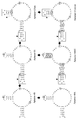
- FIG. 1 shows an example of the first step of the present invention.
- A shows an example of replacing a hairpin structure with a mini hairpin structure
- B shows an example of replacing a hairpin structure including one bulge structure in the stem structure with a mini hairpin structure
- C shows a stem structure. Shows an example of replacing a hairpin structure including one internal loop structure with a mini hairpin structure.
- 101 is a stem structure
- 102 is a loop structure
- 103 is a hairpin structure
- 104 is a first nucleic acid region
- 105 is a second nucleic acid region
- 106 is a third nucleic acid region
- 107 is a mini hairpin structure.
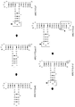
- FIG. 2 shows an example of the second step of the present invention.
- (A) is an example of adding the same number of bases as the base constituting the bulge structure to the other strand
- (B) is an example of adding fewer bases than the base constituting the bulge structure to the other strand Indicates.
- S represents G or C
- S ′ represents the complementary base of S.
- FIG. 3 is an example of the second step of the present invention and shows an example in which A or T in the bulge structure is substituted with G or C, and then a complementary base of the substituted base is added.
- W represents A or T
- S represents G or C
- S ′ represents a complementary base of S.
- FIG. 4 shows an example of the second step of the present invention.
- A is an example of replacing all of the bases constituting an internal loop with a GC pair
- B is an example of replacing some of the bases and replacing the end of the internal loop structure.
- C shows an example of substituting a part of the bases and substituting the internal loop structure.
- N represents A, T, G, or C
- S represents G or C
- S ′ represents a complementary base of S.
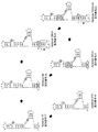
- FIG. 5 shows an example of the second step of the present invention.
- FIG. 6 is a diagram showing the sequence and secondary structure of each aptamer for IFN- ⁇ prepared in Example.
- the position of the artificial base Ds is indicated by a round frame.
- the part where the hairpin is replaced with the mini hairpin and the part where the mini hairpin is added to the original Aptamer 49 are indicated by a square frame with an arrowhead.
- FIG. 7 is a diagram showing the results of a competition test of each IFN- ⁇ aptamer with Aptamer49.
- various aptamers added together with P-labeled Aptamer49 and the presence or absence of addition of IFN- ⁇ are shown.
- a complex of P-labeled Aptamer49 and IFN- ⁇ and a band of free P-labeled Aptamer49 are shown.
- FIG. 8 is a diagram showing the results of measuring the binding of each IFN- ⁇ aptamer to human IFN- ⁇ by surface plasmon resonance.
- the horizontal axis indicates time (seconds), the vertical axis indicates resonance units (resonance unit: RU), and the numerical values in the figure indicate calculated Kd values.
- FIG. 9 is a diagram showing the results of gel electrophoresis confirming the stability of each IFN- ⁇ aptamer in human serum.
- FIG. 10 is a diagram showing measurement results of Tm values of each IFN- ⁇ aptamer.
- the horizontal axis indicates temperature, and the vertical axis indicates standardized absorbance.
- the horizontal axis represents temperature, and the vertical axis represents the first derivative of standardized absorbance.
- FIG. 11 is a diagram showing the results of measuring the IFN- ⁇ stimulation response inhibitory effect of each IFN- ⁇ aptamer by FACS using cultured cells.
- FIG. 1 is a schematic diagram showing that STAT1 is phosphorylated via an IFN- ⁇ receptor by IFN ⁇ stimulation.
- B shows the results of FACS analysis using phosphorylated STAT antibody. The horizontal axis indicates the fluorescence intensity, and the vertical axis indicates the number of cells. Unstimulated cells are shown in shaded areas. Unstimulated cells that have not been treated with anti-phosphorylated STAT-1 antibody solution are indicated by lines.
- C shows FACS analysis of cells treated with IFN- ⁇ using phosphorylated STAT antibody. The horizontal axis indicates the fluorescence intensity, and the vertical axis indicates the number of cells. Unstimulated cells are shown in gray, and cells stimulated with IFN- ⁇ are shown as lines.
- FIG. 12 shows the results of measuring the IFN- ⁇ stimulation response inhibitory effect of each IFN- ⁇ aptamer by FACS using cultured cells.
- the horizontal axis indicates the fluorescence intensity, and the vertical axis indicates the number of cells.
- the upper panel shows the inhibitory effect of Aptamer 49, and the lower panel shows the inhibitory effect of Aptamer 58.
- FIG. 13 is a diagram showing the results of measuring the IFN- ⁇ stimulation response inhibitory effect of each IFN- ⁇ aptamer by FACS using cultured cells.
- the horizontal axis indicates the fluorescence intensity, and the vertical axis indicates the number of cells.
- FIG. 14 is a diagram showing the sequence and secondary structure of each aptamer for IFN- ⁇ consisting only of a natural base, prepared in Example.
- FIG. 15 is a diagram showing the results of a competition test for Aptamer49. In the upper row, various aptamers added together with P-labeled Aptamer49 and the presence or absence of addition of IFN- ⁇ (-: no addition; +: with addition) are shown.
- FIG. 16 is a diagram showing the measurement results of the Tm value of each IFN- ⁇ aptamer consisting only of natural bases.
- the horizontal axis represents temperature and the vertical axis represents standardized absorbance.
- the horizontal axis represents temperature, and the vertical axis represents the first derivative of standardized absorbance.
- FIG. 17 is a diagram showing the sequence and secondary structure of each aptamer for vWF A1 domain prepared in Example.
- the part replaced when the hairpin is replaced with the mini hairpin and the part added with the mini hairpin with respect to the original ARC1172 (wt) are indicated by a square frame with an arrowhead.
- the position of inverted dT added to the end is indicated by a round frame.
- FIG. 18 is a diagram showing the results of a competition test of each vWF A1 domain aptamer for ARC1172 (wt). In the upper row, various aptamers added together with P-labeled ARC1172 (wt) and the presence / absence of addition of vWF A1 (-: no addition; +: with addition) are shown.
- the middle panel shows a complex of P-labeled ARC1172 (wt) and vWF A1, and a free P-labeled ARC1172 (wt) band.
- the bottom row shows the gel shift rate, the inhibition rate by each aptamer of binding of ARC1172 (wt) to the vWF A1 domain calculated from the gel shift rate, and the relative binding rate calculated by comparing with the competitive inhibition ability of ARC1172 (wt) itself.
- FIG. 19 is a diagram showing the results of confirming the stability of aptamers for vWF A1 domain in human serum by gel electrophoresis. The position of the unresolved aptamer band is indicated by a black triangle.
- FIG. 20 is a diagram showing measurement results of Tm values of aptamers for vWF A1 domain.
- the horizontal axis represents temperature and the vertical axis represents standardized absorbance.
- the horizontal axis represents temperature, and the vertical axis represents the first derivative of standardized absorbance.
- FIG. 21 shows the sequence and secondary structure of each aptamer for VEGF165 prepared in the Example.
- FIG. 22 is a diagram showing the results of a competition test of each aptamer for VEGF165 to Aptamer47. In the upper row, various aptamers added together with P-labeled Aptamer47 and the presence or absence of addition of VEGF165 (-: no addition; +: with addition) are shown.
- the middle panel shows a complex of P-labeled Aptamer47 and VEGF165, and a free P-labeled Aptamer47 band.
- the lower row shows the gel shift rate, the inhibition rate of Aptamer47 binding to VEGF165 calculated from the gel shift rate by each aptamer, and the relative binding rate calculated by comparing with the competitive inhibition ability of Aptamer47 itself.
- FIG. 23 shows the results of gel electrophoresis confirming the stability of each aptamer for VEGF165 in human serum. The position of the unresolved aptamer band is indicated by a black triangle.
- FIG. 24 is a diagram showing measurement results of Tm values of aptamers for VEGF165.
- the horizontal axis represents temperature and the vertical axis represents standardized absorbance.
- the horizontal axis represents temperature, and the vertical axis represents the first derivative of standardized absorbance.
- the graph of (C) is an enlargement of the portion up to 50 ° C of the graph of (A).
- nucleic acid or “nucleic acid molecule” refers to a biopolymer composed of nucleotides as structural units and linked by phosphodiester bonds.
- natural nucleotide refers to a nucleotide that exists in nature and is composed of a deoxyribonucleotide having a natural base of any one of adenine, guanine, cytosine and thymine, and adenine, guanine, Examples thereof include RNA composed of ribonucleotides having a natural base of either cytosine or uracil, or a combination thereof.
- a nucleic acid (molecule) composed only of natural nucleotides is referred to as a natural nucleic acid (molecule).
- non-natural nucleotide refers to a nucleotide that does not exist in nature, the base of which is an artificial base.
- the phosphate group and sugar constituting the non-natural nucleotide in the present invention are structurally identical to the phosphate group and sugar of the natural nucleotide.
- the “base analog” or “artificial base” is an artificially constructed chemical substance having properties similar to the natural base constituting the natural nucleotide, and is similar to the natural base. And a base analog (hereinafter referred to as “complementary artificial base”) that can form an artificial base pairing.
- “artificial base pairing” refers to base pairing formed by a pair of complementary artificial bases such as adenine and thymine, adenine and uracil, or guanine and cytosine, which are natural bases.
- Artificial base pairing is based on chemical bonds via hydrogen bonds found in base pairing between natural bases, physical bonds via fitting based on the molecular structure between artificial bases, and hydrophobic interactions. Including stacking effect.
- the “characteristics similar to natural bases” possessed by artificial bases include the ability to replicate or transcribe nucleic acids (including reverse transcription) by complementation by artificial base pairing.
- Artificial bases like natural bases, have exclusive selectivity in artificial base pairing. Therefore, if a non-natural nucleotide having a pair of complementary artificial bases exists in a substrate nucleotide, even a nucleic acid molecule containing a non-natural nucleotide can be accurately detected in the same manner as a natural nucleotide due to complementarity between artificial bases. Can be duplicated and transcribed. Therefore, it is possible to amplify DNA molecules by a nucleic acid amplification method such as PCR while containing non-natural nucleotides.
- the artificial base include Ds (7- (2-thienyl) imidazo [4,5-b] pyridine; referred to herein as “Ds”), Pn (2-nitropyrrole-1-yl; this specification "Pn”), Pa (2-formyl-1H-pyrrole-1-yl; referred to herein as “Pa”), P (2-amino-imidazo [1,2-a] -1 , 3,5-triazin-4 (8H) -one; referred to herein as “P”), Z (6-amino-5-nitro-2 (1H) -pyridone; referred to herein as “Z”.
- 5SICS (6-methylisoquinoline-1 (2H) -thione; referred to herein as “5SICS”), NaM (3-methoxynaphthalen-2-yl; referred to herein as “NaM”), and MMO2 (2-methoxy-4-methylphenyl; referred to herein as “MMO2”).
- complementary artificial bases for Ds include Pn and Pa.
- a complementary artificial base of P includes Z.
- 5SICS complementary artificial bases include NaM and MMO2.
- An artificial base is a natural base that is structurally and / or close in nature to a complementary artificial base when the substrate does not contain a non-natural nucleotide having a complementary artificial base during replication or transcription.
- base pairing can be performed.
- the non-natural nucleotide in the template nucleic acid molecule is replaced with the natural nucleotide after replication or transcription.
- Ds it is known that it is replaced with A or T.
- modified base means a base that has been chemically chemically modified.
- Modified bases include, for example, modified pyrimidines (eg, 5-hydroxycytosine, 5-fluorouracil, 4-thiouracil, 5- (3-indole-2-ethyl) uracil, 5- (4-hydroxyphenyl-2-ethyl) Uracil), modified purines (eg 6-methyladenine, 6-thioguanosine) and other heterocyclic bases.
- modified pyrimidines eg, 5-hydroxycytosine, 5-fluorouracil, 4-thiouracil, 5- (3-indole-2-ethyl) uracil, 5- (4-hydroxyphenyl-2-ethyl) Uracil
- modified purines eg 6-methyladenine, 6-thioguanosine
- nucleic acid aptamer is an aptamer composed of nucleic acids, and is formed by a three-dimensional structure formed on the basis of the secondary structure and further the tertiary structure of a single-stranded nucleic acid molecule through hydrogen bonding or the like.
- inhibition of target molecule function refers to inhibition of biological functions such as catalytic function or gene expression control function (including control of transcription, translation, transport, etc.) and apoptosis control function of target molecule. Or to suppress.
- nucleic acid aptamer an RNA aptamer composed only of RNA and a DNA aptamer composed only of DNA are generally known, but the nucleic acid constituting the nucleic acid aptamer in the present specification is DNA.
- target molecule refers to a substance that can be a binding target of a DNA aptamer.
- the type of target molecule is not particularly limited as long as it is a biological substance to which a DNA aptamer can bind. Examples include peptides (oligopeptides or polypeptides), nucleic acids, lipids, sugars (including sugar chains), and low molecular compounds. Preferably it is a peptide, more preferably a polypeptide, ie a protein. Moreover, any of naturally derived substances, chemically synthesized substances, genetically modified substances, cells, viruses and the like may be used. 2.
- the present invention relates to a method for designing a base sequence that enhances the target molecule binding ability of an existing DNA aptamer and / or stabilizes the aptamer.
- the present invention relates to a method that makes it possible to enhance and / or stabilize the binding ability, in particular based on information on the secondary structure level of DNA aptamers.
- the base sequence design method according to the present invention includes the first step as an essential step.
- the base sequence design method according to the present invention includes one or more of the second step, the third step, and the fourth step as an optional step. When these arbitrary steps are included, the order of each step is not limited.
- the fourth step is performed after the third step.
- the “first step” means a hairpin structure composed of a pair of stem structures and loop structures in an existing DNA aptamer including one or more stem structures and one or more loop structures. This is a process of substituting a mini hairpin structure.
- the “stem structure” means that a part of constituent bases, preferably two or more consecutive bases, for example, 3 or more, 4 or more, or 5 or more bases are completely or partially base-paired with each other.
- “completely” base pairing means base pairing with a base corresponding to all of two or more consecutive bases between DNA aptamers, for example, three or more, four or more, or five or more.
- “partially” base pairing means that 1 or 2 or more, for example, 3 or more, 4 or more, or 5 or more base pairs are formed between the base sequences of the stem structure that is completely base-paired. Is meant to contain no base.
- internal loop structure means a loop structure in a stem structure that occurs when one or more bases that do not form a base pair are present at corresponding positions in both of the double strands that form the stem structure.
- bulge structure means a protruding structure in a stem structure that occurs when one or more bases that do not form a base pair are present only at one of the corresponding positions of the double strand that forms the stem structure. Point to.
- the “loop structure” means a loop-like structure inside a nucleic acid that is located between the two strands constituting the stem structure and is formed by the formation of the stem structure and does not base pair.
- hairpin structure or “stem loop structure” means a structure composed of one stem structure and one loop structure (a set of stem structure and loop structure).
- mini-hairpin structure means that three DNA nucleic acid regions of a first nucleic acid region, a second nucleic acid region, and a third nucleic acid region are linked in order from the 5 ′ end to the 3 ′ end. Has a structured.
- the “first nucleic acid region” is a nucleic acid region consisting of 2 to 5 arbitrary nucleotides.
- the nucleotide refers to a deoxyribonucleotide having a base of guanine (g), adenine (a), cytosine (c) or thymine (t).
- the base of this nucleic acid region is preferably guanine or cytosine. This is because when the stem structure is formed with the third nucleic acid region described later, the Tm value increases as the gc content increases, and the stem structure can be stably maintained. Therefore, it is most preferable that the entire base sequence of the first nucleic acid region is composed of g and / or c.
- the “second nucleic acid region” is a nucleic acid region consisting of a 5′-gna-3 ′ or 5′-gnna-3 ′ base sequence. Each n in the sequence is independently composed of a natural base (g, a, t, or c), the base analog, or a modified base.
- the “third nucleic acid region” is a nucleic acid region having a base sequence complementary to the first nucleic acid region. Therefore, the base sequence of the third nucleic acid region is determined by the base sequence of the first nucleic acid region, and the first nucleic acid region and the third nucleic acid region form a base pair in the molecule. As a result, the first nucleic acid region and the third nucleic acid region have completely stem-paired stem portions, and the second nucleic acid region existing between the first and third nucleic acid regions has a loop portion. As a whole, for example, a mini hairpin type DNA consisting of 7 to 14 nucleotides having the base sequence of SEQ ID NO: 1, 2, 23, or 24 is formed.
- the target DNA aptamer in the first step is an existing DNA aptamer containing one or more stem structures and one or more loop structures.
- the “existing DNA aptamer” is a known DNA aptamer and a DNA aptamer obtained by a method known in the art, the nucleotide sequence of which is known, or the nucleotide sequence thereof. It means what can be clarified.
- a DNA aptamer for example, it can be produced by in vitro selection using a modified SELEX method to which a known SELEX (systematic evolution of ligands by exponential enrichment) method is applied.
- a nucleic acid molecule bound to a target molecule is selected from a pool consisting of a random sequence region and nucleic acid molecules having primer binding regions at both ends, amplified after recovery, and then transferred to the nucleic acid pool of the next round.
- This is a method of selecting a nucleic acid having a stronger binding force with respect to a target molecule by repeating a series of cycles of several to several tens of rounds.
- the modified SELEX method includes a step of immobilizing a complex obtained by mixing a nucleic acid pool and a target molecule on a solid phase carrier in addition to the steps of the conventional SELEX method.
- the DNA molecule finally obtained by such a method can be used as a DNA aptamer.
- the base sequence constituting the stem structure and the loop structure can be easily identified by those skilled in the art by performing secondary structure prediction based on the base sequence.
- secondary structure prediction For the prediction of the secondary structure based on the nucleic acid base sequence, see, for example, Zuker M., “Mfold web server for nucleic acid folding and hybridization prediction”, Nucleic Acids Res. 2003, 31, pp. 3406-3415.
- a library in which mutations are introduced into the base sequence of a DNA aptamer is created, and the SELEX method is used to comprehensively analyze the sequence that binds to the target. Knowing the secondary structure can be estimated (eg Kimoto M., et al., “Generation of high-affinity DNA aptamer using an expanded genetic alphabet” Nat. Biotechnol., 2013, 31, pp. 453-457). Moreover, in the existing DNA aptamer, the secondary structure of a DNA aptamer can also be estimated using the structural information by X-ray crystal structure analysis or NMR.
- the base length of the existing DNA aptamer is not particularly limited. What is necessary is just to determine suitably in the range in which a DNA aptamer can exhibit the target binding ability.
- Existing DNA aptamers may contain one or more base analogs and / or modified bases.
- the content of base analogs and / or modified bases in the DNA aptamer of the present invention may be 20% or less, preferably 15% or less, more preferably 10% or less of the total number of nucleotides constituting the nucleic acid aptamer.
- the hairpin structure to be replaced in this step is approximately the same size as the mini hairpin structure after replacement.
- “the same size” means that the difference in nucleotide length between the substituted hairpin structure and the substituted mini hairpin structure is preferably 5 or less, for example, 4 or less, 3 or less, 2 or less, or 1 It means that the nucleotide length of the hairpin structure to be substituted or the substituted mini hairpin structure is the same.
- the mini hairpin structure is composed of 7 to 14 bases
- the substituted hairpin structure is preferably composed of 7 to 19 bases, for example.
- the stem structure constituting the hairpin structure to be replaced in this step may have 1 to 3 internal loop structures and / or 1 to 3 bulge structures.
- the number of bases constituting the internal loop structure in the stem structure is, for example, 10 or less, preferably 6 or less, 5 or less, 4 or less, 3 or less, 2 or less, for both strands. It is a piece.
- the number of bases constituting the bulge structure in the stem structure is, for example, 10 or less, preferably 6 or less, 5 or less, 4 or less, 3 or less, 2 or less, or 1.
- a hairpin structure including an internal loop structure and a stem structure not including a bulge structure, a stem structure including one internal loop structure, and a stem structure including one bulge structure is converted into a mini hairpin structure.
- the substitution is shown in FIGS. 1 (A)-(C), respectively.
- the replacement of the hairpin structure with the mini-hairpin structure may be performed by changing a part of the base sequence constituting the hairpin structure, or by changing the entire base sequence constituting the hairpin structure. .
- two or more hairpin structures When two or more hairpin structures are present in an existing DNA aptamer, two or more hairpin structures may be replaced with a mini hairpin structure, for example, all hairpin structures can be replaced.
- the hairpin structure to be substituted in this step is preferably a hairpin structure that is not involved in the binding of the DNA aptamer to the target molecule or has a low contribution to the binding to the target molecule.
- hairpin structures that are not involved in the binding of the DNA aptamer to the target molecule or have a low contribution to the binding to the target molecule for hairpin structures that are not involved in the binding of the DNA aptamer to the target molecule or have a low contribution to the binding to the target molecule are performed. Thus, those skilled in the art can easily identify the situation.
- the DNA aptamer when obtained by a screening method such as a modified SELEX method, it is not involved in binding to the target molecule from the sequence conserved among the selected DNA aptamers, or to the target molecule. It is also possible to infer a hairpin structure that has a low contribution to binding. For example, by creating a library (Doped library) in which mutations are introduced into the base sequence of a DNA aptamer, using this, perform a SELEX method, comprehensively analyze the sequence that binds to the target, and a site where base pairs are conserved By knowing, it is possible to infer a hairpin structure that is not involved in the binding to the target molecule or has a low contribution to the binding to the target molecule. 2-2.
- a library Doped library
- the “second step” is a step of increasing GC pairs in one or more stem structures when an existing DNA aptamer has two or more stem structures. This step can be performed by replacing one or more AT pairs in the stem structure with GC pairs and / or adding GC pairs in the stem structure.
- the part of the target stem structure in the second step is not particularly limited as long as it is not a stem structure constituting a mini hairpin structure.
- the number of GC pairs to be increased is, for example, 10 pairs or less, preferably 5 pairs or less, 4 pairs or less, 3 pairs or less, 2 pairs or less, or 1 pair.
- all AT pairs in the stem structure may be replaced with GC pairs, or the AT pairs in the stem structure are, for example, 10 pairs or less, preferably 5 pairs or less, preferably 4 pairs or less, 3 pairs Hereinafter, it may be replaced with 2 pairs or less, or 1 pair or GC pair.
- the GC pair is added to the stem structure, for example, 10 pairs or less, preferably 5 pairs or less, 4 pairs or less, 3 pairs or less, 2 pairs or less, or 1 pair. , May be added.
- the site to which the GC pair is added may be at the end of the stem structure or inside the stem structure.
- the DNA aptamer has a bulge structure or internal
- the number of GC pairs in the stem structure may be increased by performing substitution and / or addition to the base constituting the bulge structure or the internal loop structure.
- the base pair constituting the stem structure is perfectly paired and the bulge structure disappears.
- a smaller number of bases than the base constituting the bulge structure may be added to the other strand.
- the base pair constituting the stem structure is partially base-paired, and the bulge structure is Remains.
- the number of additions of G or C is not particularly limited as long as it is less than the number of bases constituting the bulge structure, but is, for example, 10 or less, preferably 5 or less, 4 or less, 3 or less, 2 or less, or 1 or less.
- Fig. 2 shows an example of adding the same number of bases as the base constituting the bulge structure to the other strand and adding a smaller number of bases than the base constituting the bulge structure to the other strand.
- B shows an example of adding the same number of bases as the base constituting the bulge structure to the other strand and adding a smaller number of bases than the base constituting the bulge structure to the other strand.
- B shows an example of adding the same number of bases as the base constituting the bulge structure to the other strand and adding a smaller number of bases than the base constituting the bulge structure to the other strand.
- B shows an example of adding the same number of bases as the base constituting the bulge structure to the other strand and adding a smaller number of bases than the base constituting the bulge structure to the other strand.
- B shows an example of adding the same number of bases as the base constituting the bulge structure to the other strand and adding a smaller number of bases than the base constituting the bulge structure to the other strand.
- B
- both bases constituting the internal loop present in the stem structure are both The number of GC pairs in the stem structure is increased by substituting the same number, for example, 10 bases or less, preferably 5 bases or less, 4 bases or less, 3 bases or less, 2 bases or less, or 1 base at a time. Can be made. All of the bases constituting the internal loop may be replaced with a GC pair, or a part of the bases may be replaced with a GC pair.
- the GC pair in the stem structure may be further increased by replacing and / or adding the newly formed bulge structure and / or internal loop structure according to (i) to (iv). Good.
- FIGS. 4A to 4C An example of replacing the internal loop structure is shown in FIGS. 4A to 4C, respectively.
- a base that does not form base pairing present in the stem structure are replaced by the same number from both strands, for example, each 10 bases or less, preferably 5 bases or less, 4 bases or less, 3 bases or less, 2 bases or less, or 1 base each in a GC pair. Pairs can be increased.
- the description is omitted.
- the number of bases that have not formed base pairing is replaced with the same number of GC pairs from both strands, a bulge structure is finally formed on one strand.
- the number of GC pairs in the stem structure may be further increased by performing substitution and / or addition to the formed structure in accordance with the above (i) or (ii).
- the “third step” is a step of increasing the GC pair at the end of the DNA aptamer when at least one end of the DNA aptamer forms a stem structure.
- the number of GC pairs added is not limited as long as the binding ability of the DNA aptamer to the target molecule is not lowered, but is, for example, 5 pairs or less, preferably 3 pairs or less, 2 pairs or less, or 1 pair.
- This step can be performed by adding a GC pair, ie, G and C, or C and G, to the 5 ′ end and 3 ′ end, respectively.
- a GC pair ie, G and C, or C and G
- the base constituting the protruding end is changed to This can be done by adding G or C to the other strand to form base pairing.
- the “fourth step” is a step of adding a mini hairpin structure to one end of the DNA aptamer.
- the mini hairpin structure is added to the 5 ′ end or the 3 ′ end, but it is preferable to add the mini hairpin structure to the 3 ′ end. 2-5. Effect According to the method for designing a base sequence of the present invention, it becomes possible to enhance the target molecule binding ability of an existing DNA aptamer and / or to stabilize the aptamer.
- target molecule binding ability means binding ability to a target molecule. By strengthening the binding ability to the target molecule, the ability to specifically inhibit or suppress the function of the DNA aptamer such as the physiological activity of the target molecule can be improved.
- the present invention provides a step of designing a base sequence of a DNA aptamer based on the base sequence design method described in the first embodiment, And a method for producing a stabilized DNA aptamer with enhanced target molecule binding ability, comprising a step of producing a DNA aptamer based on the designed base sequence. Since the design process is as described in the first embodiment, the description is omitted here.
- the “target molecule binding ability-enhanced stabilized DNA aptamer” means that the target molecule binding ability is enhanced with respect to the existing DNA aptamer used in the base sequence design method described in the first embodiment. And / or a stabilized DNA aptamer.
- the process for producing the DNA aptamer is not particularly limited. Any method known in the art may be used.
- the DNA aptamer of the present invention can be chemically synthesized according to a known solid phase synthesis method based on the sequence of the DNA aptamer designed as described above. See, for example, Current-Protocols-in-Nucleic-Acid-Chemistry, Volume 1, and Section 3 for the chemical synthesis of nucleic acids.
- many life science manufacturers for example, Takara Bio Inc., Life Technologies Corporation, Sigma-Aldrich Corporation, etc.
- a DNA aptamer may be prepared by synthesizing several fragments based on the designed DNA aptamer sequence and then ligating the fragments by intramolecular annealing, ligase ligation, or the like.
- the DNA aptamer of the present invention after chemical synthesis is preferably purified by a method known in the art before use.
- the purification method include a gel purification method, an affinity column purification method, and an HPLC method. 4).
- DNA aptamer In a third embodiment, the present invention provides a DNA aptamer having one or more stem structures and loop structures, wherein the hairpin structure present at the non-terminal portion of the DNA aptamer is a mini hairpin structure. About.
- the DNA aptamer of the present invention may be obtained by the production method described in the second embodiment.
- the “non-terminal portion” is not particularly limited as long as it is a place other than the 5 ′ end and 3 ′ end of the DNA aptamer.
- the non-terminal portion of the DNA aptamer is preferably a site separated from the terminal portion of the DNA by 2 bases or more, for example, 3 bases, 4 bases, or 5 bases or more.
- the DNA aptamer of the present invention may further contain a mini hairpin structure at the end in addition to the non-terminal mini hairpin structure. Although it does not restrict
- the DNA aptamer of the present invention may further have one or more stem structures and / or one or more loop structures. In this case, the stem structure of the DNA aptamer may have one or more base mismatch sites or one or more bulge structures therein.
- the base length of the DNA aptamer of the present invention is not particularly limited. What is necessary is just to determine suitably in the range which a DNA aptamer can exhibit the function.
- the DNA aptamer of the present invention may contain one or more base analogs and / or modified bases.
- the content of base analogs and / or modified bases in the DNA aptamer of the present invention may be 20% or less, preferably 15% or less, more preferably 10% or less of the total number of nucleotides constituting the nucleic acid aptamer.
- the DNA aptamer of the present invention preferably has a high GC content in one or more, for example, one, two, three, or all stem structures.
- the GC content means the ratio of GC pairs in the base pairs forming the stem structure contained in the DNA aptamer, and one or more, for example, one, two, three, or all stem structures of the DNA aptamer of the present invention.
- the GC content in is, for example, 50% or more, 75% or more, or 90% or more. All base pairs in one or more, for example one, two, three or all stem structures may be GC pairs.
- the base pair at the end is preferably a GC pair. 5.
- a fourth embodiment of the present invention is a DNA aptamer for interferon- ⁇ (abbreviated herein as “IFN- ⁇ ”).
- the DNA aptamer for IFN- ⁇ of the present invention has the configuration of the DNA aptamer described in the third embodiment.
- the DNA aptamer for IFN- ⁇ of the present invention is composed of a single-stranded DNA molecule that binds firmly and specifically with IFN- ⁇ as a target substance and suppresses cytotoxic T cell-inducing activity of IFN- ⁇ . It is a DNA aptamer (hereinafter referred to as “DNA aptamer for IFN- ⁇ ”).
- the biological species from which the target molecule of the DNA aptamer for IFN- ⁇ of the present invention is derived is not limited, for example, mammals, for example, primates such as humans and chimpanzees, laboratory animals such as rats and mice, pigs, Examples include domestic animals such as cattle, horses, sheep and goats, and pets such as dogs and cats, preferably human IFN- ⁇ .
- the DNA aptamer for IFN- ⁇ of the present invention consists of the base sequence shown in any of SEQ ID NOs: 6 and 8-11.
- the DNA aptamer for IFN- ⁇ of the present invention consists of the base sequence shown in SEQ ID NO: 8 or 9. 6).
- DNA Aptamer for VEGF A fifth embodiment of the present invention is a DNA aptamer for vascular endothelial growth factor (abbreviated as “VEGF” in the present specification).
- VEGF is a growth factor that functions as a pro-angiogenic factor, and is known as one of the causative factors of age-related macular degeneration (AMD).
- AMD age-related macular degeneration
- Age-related macular degeneration is a progressive retinal disease that causes serious symptoms such as decreased visual function and blindness in adults, and it has been found that the pathological condition worsens and becomes severe with the progress of neovascularization in the retina. (Martin A. et al., 2003, Medicinal Research Reviews, Vol. 23, No. 2: 117-145; Ferris III, FL et al., 1984, Archives of Ophthalmology, Vol. 102, Issue 11: 1640-1642 ).
- the DNA aptamer for VEGF of the present invention is a DNA aptamer composed of a single-stranded DNA molecule that binds firmly and specifically with VEGF as a target substance and suppresses the angiogenic function of VEGF (hereinafter, “DNA aptamer for VEGF”). ”).
- the biological species from which VEGF as a target molecule of the DNA aptamer for VEGF of the present invention is derived does not matter. For example, mammals, primates such as humans and chimpanzees, laboratory animals such as rats and mice, pigs, cows, horses, Examples include livestock animals such as sheep and goats, and pets such as dogs and cats, preferably human VEGF.
- the DNA aptamer for VEGF of the present invention consists of the base sequence shown in any of SEQ ID NOs: 19-22.
- the DNA aptamer for VEGF of the present invention consists of the base sequence shown in SEQ ID NO: 20 or 22. 7).
- DNA aptamer for vWF The sixth embodiment of the present invention is a DNA aptamer for von Willebrand factor (abbreviated herein as “vWF”), particularly a DNA aptamer for the A1 domain of vWF. .
- the DNA aptamer for vWF of the present invention is a DNA aptamer composed of a single-stranded DNA molecule that binds firmly and specifically with vWF, in particular, the A1 domain of vWF (hereinafter referred to as “DNA aptamer for vWF”). It is.
- the biological species from which vWF as a target molecule of the DNA aptamer for vWF of the present invention is derived does not matter. For example, mammals, primates such as humans and chimpanzees, laboratory animals such as rats and mice, pigs, cows, horses, Examples include livestock animals such as sheep and goats, and pet animals such as dogs and cats, preferably human vWF.
- the vWF of the present invention in particular, the DNA aptamer for the A1 domain of vWF consists of the base sequence shown in any of SEQ ID NOs: 14 to 16.
- the vWF of the present invention particularly the DNA aptamer for the A1 domain of vWF, consists of the base sequence shown in SEQ ID NO: 16. 8).
- Pharmaceutical composition A seventh embodiment of the present invention is a pharmaceutical composition. 8-1. Structure
- the pharmaceutical composition of the present invention contains at least one DNA aptamer described in any of Embodiments 3 to 6 as an active ingredient.
- the pharmaceutical composition of the present invention can also contain a pharmaceutically acceptable carrier.
- “Pharmaceutically acceptable carrier” refers to its action in order to facilitate the formulation of a pharmaceutical composition ordinarily used in the field of pharmaceutical technology and application to a living body, and to maintain the effect of the target substance function inhibitor. A substance added within a range not inhibiting or suppressing. Carriers include, for example, excipients, binders, disintegrants, fillers, emulsifiers, flow control agents, lubricants or surfactants.
- excipient examples include sugars such as monosaccharides, disaccharides, cyclodextrins and polysaccharides (specifically, but not limited to, glucose, sucrose, lactose, raffinose, mannitol, sorbitol, inositol, Including dextrin, maltodextrin, starch and cellulose), metal salts (eg, sodium phosphate or calcium phosphate, calcium sulfate, magnesium sulfate), citric acid, tartaric acid, glycine, low, medium, high molecular weight polyethylene glycol (PEG), Pluronics, or combinations thereof.
- sugars such as monosaccharides, disaccharides, cyclodextrins and polysaccharides (specifically, but not limited to, glucose, sucrose, lactose, raffinose, mannitol, sorbitol, inositol, Including dextrin, mal
- binder examples include starch paste using corn, wheat, rice, or potato starch, gelatin, tragacanth, methylcellulose, hydroxypropylmethylcellulose, sodium carboxymethylcellulose, and / or polyvinylpyrrolidone.
- disintegrant examples include the starch, carboxymethyl starch, cross-linked polyvinyl pyrrolidone, agar, alginic acid, sodium alginate or salts thereof.
- filler examples include the sugar and / or calcium phosphate (for example, tricalcium phosphate or calcium hydrogen phosphate).
- emulsifier examples include sorbitan fatty acid ester, glycerin fatty acid ester, sucrose fatty acid ester, and propylene glycol fatty acid ester.
- Examples of the “flow additive modifier” and “lubricant” include silicate, talc, stearate or polyethylene glycol.
- the drug composition of the present invention comprises a flavoring agent, a solubilizing agent (solubilizing agent), a suspending agent, a diluent, a surfactant, a stabilizer, an absorption accelerator (if necessary)
- a flavoring agent for example, quaternary ammonium salts, sodium lauryl sulfate, etc., extenders, moisturizers, humectants (eg, glycerin, starch, etc.), adsorbents (eg, starch, lactose, kaolin, bentonite, colloidal silicic acid, etc.
- Disintegration inhibitors eg, sucrose, stearin, cocoa butter, hydrogenated oil, etc.
- coating agents colorants, preservatives, antioxidants, fragrances, flavoring agents, sweetening agents, buffering agents, and the like.
- surfactant examples include lignosulfonic acid, naphthalene sulfonic acid, phenol sulfonic acid, alkali metal salt, alkaline earth metal salt and ammonium salt of dibutyl naphthalene sulfonic acid, alkyl aryl sulfonate, alkyl sulfate, alkyl sulfonate.
- the pharmaceutical composition of the present embodiment can include one or more of the above carriers in one pharmaceutical composition.
- the pharmaceutical composition of the present invention can also contain other drugs as long as the pharmacological effect of the nucleic acid of the present invention is not lost.
- a predetermined amount of antibiotics may be contained.
- the dosage form of the pharmaceutical composition of the present invention is not particularly limited as long as it is a form that does not inactivate the active ingredient and can exhibit its pharmacological effect in vivo after administration. Usually, it varies depending on the administration method and / or prescription conditions.
- dosage forms suitable for oral administration include solid preparations (including tablets, pills, sublinguals, capsules, drops, troches), granules, powders, powders, liquids, and the like.
- the solid preparation can be made into a dosage form known in the art, for example, a sugar-coated tablet, a gelatin-encapsulated tablet, an enteric tablet, a film-coated tablet, a double tablet, or a multilayer tablet, if necessary. .
- Parenteral administration is subdivided into systemic administration and local administration, and local administration is further subdivided into intra-tissue administration, transepidermal administration, transmucosal administration, and rectal administration.
- the dosage form can be made suitable for.
- dosage forms suitable for systemic or intra-tissue administration include injections that are liquids.
- Suitable dosage forms for transepidermal or transmucosal administration include, for example, liquids (including coating agents, eye drops, nasal drops, and inhalants), suspensions (including emulsions and creams), and powders (points). Nasal and suction agents), pastes, gels, ointments, plasters and the like.
- Examples of dosage forms suitable for rectal administration include suppositories.
- the dosage form of the pharmaceutical composition may be liquid, solid (including semi-solid), or a combination thereof. Solutions, oily dispersions, emulsions, suspensions, powders, powders, pastes, gels, pellets, tablets and granules can be used.
- each dosage form are not particularly limited as long as each dosage form is within the range of dosage forms known in the art. 8-2.
- Production Method In order to produce the pharmaceutical composition of the present invention, a formulation method known in the art may be applied in principle. For example, reference can be made to the method described in Remington's Pharmaceutical Sciences (Merck Publishing Co., Easton, Pa.).
- the DNA aptamer according to any of the third to sixth embodiments is dissolved in a pharmaceutically acceptable solvent, and a pharmaceutically acceptable carrier is added as necessary, which is conventionally used in the art. It can be manufactured by the method.
- the pharmaceutical composition of the present embodiment can be administered to a living body in a pharmaceutically effective amount for treatment or prevention of a target disease or the like.
- the living body to be administered is a vertebrate, preferably a mammal, more preferably a human.
- the pharmaceutical composition of the present invention may be either systemic administration or local administration. It can select suitably according to the kind of disease, onset location, or a progression degree. If the onset site is a local disease, local administration directly administered to and around the onset site by injection or the like is preferable. This is because a sufficient amount of the DNA aptamer of the present invention can be administered to a site (tissue or organ) to be treated, and other tissues are hardly affected. On the other hand, when the treatment site cannot be specified or the onset is a systemic disease, systemic administration by intravenous injection or the like is preferable, although there is no limitation. This is because the DNA aptamer of the present invention is distributed throughout the body through the bloodstream, so that it can be administered even to a lesion that cannot be detected by diagnosis.
- the pharmaceutical composition of the present invention can be administered by any appropriate method that does not deactivate the active ingredient.
- it may be parenteral (for example, injection, aerosol, application, eye drop, nose drop) or oral.
- parenteral for example, injection, aerosol, application, eye drop, nose drop
- oral Preferably, it is an injection.
- the injection site is not particularly limited. Any site may be used as long as the DNA aptamer, which is an active ingredient, can bind to the target substance and suppress its function.
- Intravenous injection such as intravenous injection or intraarterial injection is preferable. This is because the pharmaceutical composition of the present invention can be distributed throughout the body through the blood stream as described above, and is less invasive. 9.
- the eighth embodiment of the present invention is a method for detecting a target substance using the DNA aptamer according to any one of Embodiments 3 to 6. 9-1. Since the DNA aptamer according to any of the third to sixth embodiments can bind to the target substance very firmly and specifically, the target substance present in the sample using the properties of the DNA aptamer. Can be detected.
- a known detection method may be used as the detection method itself.
- SPR method quartz crystal microbalance method, turbidimetric method, colorimetric method, or fluorescent method can be used.
- SPR surface plasmon resonance
- the SPR method is a measurement method that utilizes this phenomenon, and can measure the adsorbate on the surface of the metal thin film that is the sensor portion with high sensitivity.
- the DNA aptamer according to any of the third to sixth embodiments is immobilized on the surface of the metal thin film in advance, and a sample is passed over the surface of the metal thin film, so that the DNA aptamer and the target substance are The target substance in the sample can be detected by detecting the difference in adsorbate on the metal surface before and after passing through the sample caused by the binding.
- a substitution method, an indirect competition method, and the like are known, and any of them may be used.
- the QCM (Quartz Crystal Microbalance) method uses a phenomenon in which when a substance is adsorbed on the surface of an electrode attached to a crystal resonator, the resonance frequency of the crystal resonator decreases according to its mass. .
- a QCM sensor using this method can quantitatively capture an extremely small amount of adsorbate based on the amount of change in the water resonance frequency.
- a DNA aptamer is immobilized on the electrode surface in the same manner as in the SPR method, and the amount of change in the water resonance frequency caused by the binding between the DNA aptamer and the target substance is brought into contact with the sample surface.
- the target substance in the sample can be detected quantitatively.
- This technique is well known in the art. For example, see Christopher J., et al. (2005) Self-Assembled Monolayers of a Form of Nanotechnology, Chemical Review, 105: 1103-1169.
- the turbidimetric method irradiates a solution with light and optically measures the attenuation of scattered light scattered by a substance suspended in the solution or transmitted light that has passed through the solution using a colorimeter or the like. It is a method of measuring the amount of substance in it.
- the target substance in the sample can be quantitatively detected by measuring the absorbance before and after adding the DNA aptamer of any of the third to sixth embodiments to the sample.
- the target substance can be detected by using it together with an antibody against the target substance.
- a method using an ELISA sandwich method may be used. In this method, first, the DNA aptamer according to any of the third to sixth embodiments is immobilized on a solid phase carrier, and then a sample is added to bind the target substance present in the sample and the DNA aptamer. . Subsequently, after washing the sample, an anti-target substance antibody is added to bind to the target substance. After washing, the target substance in the sample can be detected by detecting the anti-target substance antibody using a secondary antibody with an appropriate label.
- beads made of materials such as polystyrene, polycarbonate, polyvinyltoluene, polypropylene, polyethylene, polyvinyl chloride, nylon, polymethacrylate, latex, gelatin, agarose, cellulose, sepharose, glass, metal, ceramics or magnetic materials
- Insoluble carriers in the form of microplates, test tubes, sticks or test pieces can be used.
- Example 1 Preparation of DNA aptamer containing mini hairpin sequence
- GC of AT base pairs in the stem sequence GC of AT base pairs in the stem sequence
- various nucleic acid fragments were first designed and prepared for human IFN- ⁇ DNA aptamers.
- the sequence of the aptamer prepared in this example and its secondary structure are shown in FIG.
- Aptamer 49 (SEQ ID NO: 4) is a previously reported IFN- ⁇ binding DNA aptamer found by the inventors.
- Aptamer 49A (SEQ ID NO: 5) is obtained by replacing the artificial base Ds of Aptamer 49 with the natural base A.
- Aptamer 58 (SEQ ID NO: 6) is obtained by binding a mini hairpin structure (short DNA fragment comprising the sequence of CGCGAAGCG (SEQ ID NO: 1)) to the 3 ′ end of Aptamer 49.
- Aptamer 57mh (SEQ ID NO: 9) is obtained by replacing the hairpin in the sequence of Aptamer 58 with a mini hairpin structure (short DNA fragment comprising the sequence of CGCGAAGCG (SEQ ID NO: 1)).
- Aptamer 58GC (SEQ ID NO: 7) and Aptamer 57GCmh (SEQ ID NO: 8) are obtained by replacing two AT base pairs in the stem structures of Aptamertam58 and Aptamer 57mh with G-C base pairs.
- Aptamer 49, Aptamer 58, Aptamer 58GC, Aptamer 57mh, Aptamer 57 GCmh, and Aptamer 49A were prepared by chemical synthesis of nucleic acids having the base sequences shown in the figure, and then purified by denaturing acrylamide gel. Each nucleic acid fragment was reconstituted in phosphate buffer solution (pH 7.4), heated at 95 ° C for 3 minutes, allowed to cool at room temperature for 10 minutes, and allowed to stand on ice for 5 minutes. Prepared.
- Example 2 Analysis of binding activity of various IFN- ⁇ aptamers to human IFN- ⁇
- a competition experiment using Aptamer 49 containing the artificial base Ds was performed.
- the inhibition rate was calculated as a value obtained by subtracting 100 from the percentage obtained by dividing the gel shift rate in the presence of each competitive aptamer by the gel shift rate in the absence of the competitive aptamer.
- the relative binding rate was calculated by dividing the inhibition rate of each aptamer by the inhibition rate of Aptamer 49.
- aptamers 49, Aptamer 58, Aptamer 57GCmh were picked up from the sequences prepared in Example 1, and human IFN was measured by measuring surface plasmon resonance (SPR) using BIAcoreT200 of GE Healthcare. The binding ability to - ⁇ was analyzed.
- nucleic acid fragments having various sequences whose structure ends are biotinylated are chemically synthesized, and then each nucleic acid fragment is purified on a denaturing acrylamide gel in the same manner as in Example 1, and then in a phosphate buffer (pH 7.4).
- the sample was reconstituted by heating at 95 ° C. and then slowly cooling to 25 ° C.
- the SPR sensor chip was an SA chip coated with streptavidin (GE Healthcare).
- the DNA fragment was irreversibly immobilized on the chip by the following method, and then the binding to human IFN- ⁇ was analyzed.
- SPR was measured using a running buffer (1 ⁇ PBS, +50 mM NaCl (final NaCl concentration 205 mM), 0.05% Nonidet P-40) at a set temperature of 25 ° C.
- the DNA solution diluted with PBS solution to 25 nM is folded (95 ° C, heat-denatured for 3 minutes, then slowly cooled to 25 ° C) and 0.5 nM with running buffer.
- Nonidet P-40 was added to a final concentration of 0.05%, and this DNA solution was injected at 40 ⁇ L (equivalent to 8 minutes) at a flow rate of 5 ⁇ L / min.
- a 50 mM NaOH solution was injected (5 ⁇ L, 5 times) at a flow rate of 20 ⁇ L / min to wash DNA fragments adsorbed nonspecifically on the SA chip.
- the interaction between the immobilized DNA fragment and human IFN- ⁇ can be detected using Kinetic Injection mode with 1.25nM, 2.5nM, 5nM, 10nM, 20nM, 30nM and 50nM human IFN- ⁇ solutions (diluted with running buffer). Monitored by injection.
- the measurement conditions are a flow rate of 100 ⁇ L / min and a protein injection time of 150 seconds.
- Chip regeneration binding protein dissociation and DNA refolding was performed by injecting a 50 mM NaOH solution in 5 ⁇ L (equivalent to 15 seconds) and then running a running buffer for 10 minutes. In order to subtract the response value due to the bulk effect and nonspecific adsorption to the sensor chip, the sensor value of each DNA fragment is taken as the reference cell, and the response value is calculated from the sensorgram of each DNA fragment. deducted. ⁇ Result> The binding inhibition ratios of various aptamer mutants to Aptamer 49 are shown in FIG.
- the relative binding rates of aptamer mutants containing a mini hairpin structure are 0.05 (Aptamer 49A), 1.29 (Aptamer 58), 0.26 (Aptamer 58GC), 1.53 ( Aptamer 57GCmh) and 1.40 (Aptamer 57mh). It was found that Aptamer 58, Aptamer 57mh, and Aptamer 57GCmh containing a mini hairpin had significantly higher binding strength than Aptamer 49 as a control.
- DNA aptamers (Aptamer 49, Aptamer 58, Aptamer 57mh, Aptamer 57GCmh, final concentration 2 ⁇ M) were mixed at a human serum concentration of 96% and incubated at 37 ° C. After 0 hour, 1 hour, 6 hours, 24 hours, 48 hours, 72 hours, 10 ⁇ L was taken from the mixed solution and mixed with 110 ⁇ L of 1 ⁇ TBE, 10M Urea solution to stop the decomposition reaction. . After the reaction, the sample was separated by denaturing 15% polyacrylamide gel electrophoresis, and then the gel was stained with SYBR GOLD to detect single-stranded nucleic acid.
- DNA aptamers (Aptamer 58, Aptamer 57mh, Aptamer 57GCmh) containing mini hairpin DNA still have a band pattern that more than 30% of the total amount is retained in the full-length product state even after 72 hours in the presence of human serum.
- Aptamer 57GCmh in which a mini hairpin is added to the 3 ′ end, the internal hairpin part is replaced with mini hairpin DNA, and the AT base pair in the stem part is replaced with GC base pair, is 72 hours after human serum is present.
- Example 4 Thermal stability analysis of aptamers for various IFN- ⁇ ] ⁇ Method> The thermal stability of various DNA aptamers (Aptamer 49, Aptamer 49A, Aptamer 58, Aptamer 58GC, Aptamer 57mh, Aptamer 57GCmh, final concentration 2 ⁇ M) was examined by measuring Tm values.
- the absorbance change of the DNA aptamer at a temperature rise change was measured with an ultraviolet-visible spectrophotometer UV-2450 (Shimadzu), and the Tm value, which is the melting temperature, was calculated from the first derivative.
- the results are shown in FIG.
- the Tm value of the control DNA aptamer (Aptamer 49) was 37.8 ° C.
- the Tm value of the DNA aptamer containing no artificial base was 33.4 ° C.
- the Tm values of the Ds aptamer mutants containing the mini hairpin structure were 43.9 ° C (Aptamer 58), 72.0 ° C (Aptamer 58GC), 51.1 ° C (Aptamer 57mh), and 64.2 ° C (Aptamer 57GCmh), respectively.
- the addition of mini hairpin DNA to the DNA aptamer significantly increased the Tm value, indicating that the thermal stability was improved. It was also found that the Tm value was further increased by replacing the AT base pair in the stem sequence with a GC base pair.
- Example 5 Analysis of IFN- ⁇ stimulation response suppression effect in various Ds aptamer cultured cells
- DMEM medium Dulbecco's Minimal Essential Medium, Corning cellgro
- FBS fetal bovine serum
- streptomycin penicillin, streptomycin
- the test was carried out using a 100-fold L-glutamine solution (Gibco). 2) Stimulation treatment with IFN- ⁇
- 10% FBS-containing DMEM medium containing 10 6 / mL MDA-MB-231 cells is placed in a round tube made of polystyrene (Falcon). Incubated at 37 degrees for 15 minutes. The cells were then collected by centrifugation at 1200 g for 5 minutes, then resuspended in DMEM medium containing 2 ng / mL of the recombinant protein human IFN- ⁇ (Peprotech), incubated at 37 degrees for 10 minutes, and IFN -Stimulated with ⁇ .
- the cells collected by centrifugation were washed twice with 0.5 mL of FACS analysis buffer (containing 1 ⁇ PBS pH 7.4, 0.1% sodium azide and 0.1% BSA), and then 7.5 ⁇ L of anti-phosphorylated STAT -1 antibody solution (BD Phosflow PE mouse anti-Stat-1 pY701) in 0.1 mL of FACS analysis buffer, incubate for 30 to 60 minutes under 4 times shading conditions, then 0.5 mL of FACS analysis buffer Washed twice with liquid. The washed cells were suspended in 0.5 mL of FACS analysis buffer and analyzed by flow cytometry.
- FACS analysis buffer containing 1 ⁇ PBS pH 7.4, 0.1% sodium azide and 0.1% BSA
- anti-phosphorylated STAT -1 antibody solution BD Phosflow PE mouse anti-Stat-1 pY701
- Example 6 Analysis of binding activity of human base IFN- ⁇ aptamer consisting of natural bases to human IFN- ⁇
- a nucleic acid having the base sequence shown in FIG. 14 was prepared by chemical synthesis.
- Aptamer 49 is a previously reported IFN- ⁇ -binding DNA aptamer found by the inventors
- Aptamer 49A (SEQ ID NO: 5) is the natural base Ds of Aptamer 49.
- Type base A is a product in which a mini hairpin structure is attached to the 3 ′ end of Aptamer 49A, and the hairpin inside the sequence is replaced with a mini hairpin structure (short DNA fragment consisting of the sequence of CGCGAAGCG (SEQ ID NO: 1)). is there.
- Aptamer 57AGCmh (SEQ ID NO: 10) is obtained by replacing two A-T base pairs in the stem structure of Aptamer 57Amh with G-C base pairs.
- Nucleic acids having the above base sequences were chemically synthesized in the same manner as in Example 1, purified and reconstructed to prepare various DNA aptamers.
- Aptamer 49 which is a wild-type aptamer containing an artificial base Ds.
- Aptamer 49 (final concentration 20 nM) labeled with [ ⁇ - 32 P] ATP, 1000 times unlabeled nucleic acid (Aptamer 49A, Aptamer 57AGCmh) in 20 ⁇ L reaction solution (1 ⁇ PBS, 0.005% Nonidet P-40) , Aptamer 57Amh) (final concentration 20 ⁇ M) and human IFN- ⁇ (20 nM, Peprotech) were mixed and incubated at 37 ° C. for 30 minutes.
- the binding inhibition ratio of Aptamer 49A as a control was 1.00
- the binding inhibition ratio (relative binding ratio) of the aptamer mutants containing the mini hairpin structure was 3.00 (Aptamer 57AGCmh) and 3.09 (Aptamer 57Amh), respectively. It was found that Aptamer 57AGCmh and Aptamer 57Amh containing a mini hairpin had significantly improved binding strength compared to Aptamer49A as a control.
- Example 7 Thermal stability analysis of aptamer for IFN- ⁇ made of natural base
- ⁇ Method> The thermal stability of various natural base DNA aptamers (Aptamer 49A, Aptamer 57Amh, Aptamer 57AGCmh, final concentration 2 ⁇ M) was examined by measuring Tm values.
- the absorbance change of the DNA aptamer at a temperature rise change (0.5 ° C./min) was measured with an ultraviolet-visible spectrophotometer UV-2450 (Shimadzu), and the Tm value, which is the melting temperature, was calculated from the first derivative.
- UV-2450 ultraviolet-visible spectrophotometer
- the Tm value of the control DNA aptamer (Aptamer 49A) was 33.4 ° C, whereas the Tm values of the aptamer mutants consisting of natural bases containing the mini hairpin structure were 45.4 ° C (Aptamer 57Amh) and 61.6 ° C, respectively. (Aptamer 57AGCmh).
- the addition of mini hairpin DNA to the DNA aptamer significantly increased the Tm value, indicating that the thermal stability was improved. From this result, it was found that mini hairpin addition and GC addition improve the thermal stability of the DNA aptamer regardless of the presence or absence of the artificial base Ds.
- Example 8 Analysis of binding activity of vWF aptamer to vWF A1 domain
- ARC1172 (wt) (SEQ ID NO: 12) is a known vWF A1 domain-binding DNA aptamer.
- ARC1172 (wt) -iT (SEQ ID NO: 13) is obtained by adding inverted dT to the 3 ′ end, which is a conventional nucleic acid stabilization technique, to ARC1172 (wt).
- ARC1172-F (SEQ ID NO: 14) is obtained by replacing the hairpin in the sequence with a mini hairpin structure (short DNA fragment comprising the sequence of GCCGAAGGC (SEQ ID NO: 2)).
- ARC1172-F-iT (SEQ ID NO: 15) is obtained by adding inverted dT to the 3 ′ end, which is a conventional nucleic acid stabilization technique, to ARC1172-F.
- ARC1172-G (SEQ ID NO: 16) is obtained by binding a mini hairpin structure (short DNA fragment comprising the sequence of CGCGAAGCG (SEQ ID NO: 1)) to the 3 'end of ARC1172-F.
- Nucleic acids having the above base sequences were chemically synthesized in the same manner as in Example 1, purified and reconstructed to prepare various DNA aptamers.
- ARC1172 In order to examine the binding of various prepared nucleic acids to the vWF A1 domain, competition experiments using various mutant nucleic acids were performed on ARC1172 reported as an aptamer that binds to the vWF A1 domain.
- ARC1172 final concentration 100 nM labeled with [ ⁇ - 32 P] ATP, unlabeled nucleic acid (ARC1172 (wt), ARC1172 (wt) -iT) in 20 ⁇ L reaction solution (1 ⁇ PBS, 0.1 mg / mL BSA) ARC1172-F, ARC1172-F-iT, ARC1172-G) (final concentration 100 nM) and vWF A1 domain (100 nM, U-ProteinExpress) were mixed and incubated at 37 ° C.
- the binding inhibition ratio of unlabeled ARC1172 (wt) as a control was 1.00
- the binding inhibition ratio (relative binding ratio) of the 3′-terminal inverted dT addition and the mini-hairpin DNA addition aptamer mutant was 1.12 (ARC1172), respectively.
- Example 9 Stability analysis of aptamer for vWF against nucleolytic enzymes
- the stability of a DNA aptamer to which a mini hairpin DNA was added to a DNA aptamer composed only of natural bases to nucleolytic enzymes contained in human serum was examined.
- the conventional nucleic acid stabilization technique which is the addition of inverted dT to the 3 ′ end.
- DNA aptamer with and without mini hairpin DNA (ARC1172 (wt), ARC1172 (wt) -iT, ARC1172-F, ARC1172-F-iT, ARC1172-G, final concentration 2 ⁇ M)
- ARC1172 (wt) -iT DNA aptamer with and without mini hairpin DNA
- ARC1172-F DNA aptamer with and without mini hairpin DNA
- ARC1172-F ARC1172-F-iT
- ARC1172-G final concentration 2 ⁇ M
- 10 ⁇ L was taken from the mixed solution and mixed with 110 ⁇ L of 1 ⁇ TBE, 10M Urea solution to stop the decomposition reaction. .
- the sample after the reaction was separated by denaturing 15% polyacrylamide gel electrophoresis, and then the gel was stained with SYBR GOLD to detect single-stranded nucleic acid.
- inverted dT-added DNA aptamer (ARC1172 (wt) -iT), which is a conventional nucleic acid stabilization technology, has improved stability against nucleolytic enzymes in human serum compared to ARC1172 (wt).
- the band corresponding to the full-length product was degraded to 47% after 24 hours in the presence of human serum, and the band corresponding to the full-length product was degraded to 27% after 72 hours.
- the DNA aptamer (ARC1172-G) with the mini hairpin DNA attached to the 3 'end and inside is 70% after 24 hours in the presence of human serum, and 42% of the total amount is still in the full-length product state after 72 hours. It was confirmed from the band pattern that it was retained.
- the stability against nucleolytic enzymes contained in human serum is remarkably increased by the addition of mini hairpin DNA.
- the effect of improving the stability of nucleolytic enzymes contained in human serum by adding a mini hairpin DNA to the 3 'end may be higher than the conventional ds addition of inverted dT to the 3' end, which is a conventional nucleic acid stabilization technology. I understood.
- Example 10 Thermal stability analysis of aptamer for vWF] ⁇ Method> The thermal stability of various DNA aptamers (ARC1172 (wt), ARC1172 (wt) -iT, ARC1172-F, ARC1172-F-iT, ARC1172-G, final concentration 2 ⁇ M) was examined by measuring the Tm value. The absorbance change of the DNA aptamer at a temperature rise change (0.5 ° C./min) was measured with an ultraviolet-visible spectrophotometer UV-2450 (Shimadzu), and the Tm value, which is the melting temperature, was calculated from the first derivative. ⁇ Result> The results are shown in FIG.
- the Tm value of the control DNA aptamer (ARC1172 (wt)) was 63.8 ° C.
- the DNA aptamer (ARC1172) to which the inverted dT addition to the 3 ′ end, which is a conventional nucleic acid stabilization technique, was performed.
- the Tm value of (wt) -iT) was 63.2 ° C.
- the Tm values of the Ds aptamer mutants containing the mini hairpin structure were 64.3 ° C. (ARC1172-F), 64.4 ° C. (ARC1172-F-iT), and 61.9 ° C. (ARC1172-G), respectively.
- Aptamer 47 (SEQ ID NO: 17) is a previously reported VEGF-binding DNA aptamer found by the inventors.
- Aptamer 47A (SEQ ID NO: 18) is obtained by replacing the artificial base Ds of Aptamer 47 with the natural base A.
- Aptamer 49 (SEQ ID NO: 19) is a mutant obtained by extending one G-C base pair to the stem structure on the terminal side of Aptamer 47.
- Aptamer 58mh (SEQ ID NO: 20) is obtained by further binding a mini hairpin structure (short DNA fragment consisting of the sequence of CGCGAAGCG (SEQ ID NO: 1)) to the 3 'end of Aptamer 49.
- Aptamer 49GC (SEQ ID NO: 21) and Aptamer 58GCmh (SEQ ID NO: 22) are obtained by replacing two AT base pairs in the stem structure of Aptamer 49 and Aptamer 58mh with G-C base pairs.
- Example 12 Analysis of binding activity of aptamer for VEGF to target protein
- the scale is 20 ⁇ L, Aptamer 47 (final concentration 100 nM) labeled with [ ⁇ - 32 P] ATP in phosphate buffer (pH 7.4) containing 0.005% NonidetP-40, various unlabeled variants ( Final concentration 100 nM) and VEGF165 protein (Peprotech, final concentration 100 nM) were mixed and incubated at 37 ° C. for 30 minutes. Immediately after addition of 5 ⁇ l of 25% glycerol, labeled Aptamer 47 bound to VEGF165 protein and released Aptamer 47 were separated by 10% non-denaturing polyacrylamide gel electrophoresis, the gel was dried, and the bioimage analyzer And the radioactivity was measured.
- the gel shift rate, inhibition rate, and relative binding rate were calculated according to Example 2. ⁇ Result> The results are shown in FIG. As shown in FIG. 22, the aptamer 47 in which Aptamer 47 was replaced with A (Aptamer 47A) did not show any inhibition of binding, whereas Aptamer 49, which was obtained by extending the stem region of Aptamer 47, had a relative binding rate. It became 1.53, and it was found that the binding power was significantly improved. Aptamer 49GC in which AT base pairs in the stem region of Aptamer 49 were replaced with two GC base pairs had a relative binding rate of 1.50.
- Aptamer with mini hairpin structure (Aptamer 58mh, Aptamer 58GCmh) and aptamer without aptamer (Aptamer 47, Aptamer 49, Aptamer 49GC) final concentration 2 ⁇ M and human serum (Millipore) final concentration 96% are mixed together. Incubated at 0 ° C. After 0 hour, 1 hour, 6 hours, 24 hours, 48 hours, and 72 hours, dispense 10 ⁇ L from the mixed solution and mix 110 ⁇ L of 10M urea / 1 ⁇ TBE to stop the decomposition reaction. It was.
- Aptamers that do not contain a mini hairpin structure had less than 50% of bands corresponding to the full-length product after 24 hours, whereas aptamers that contain a mini hairpin structure (Aptamer 58mh, Aptamer 58GCmh), even after 72 hours, more than 50% of the band corresponding to the full-length product was retained. From this result, it was found that the mini hairpin structure is stabilized against the degradation of aptamers by nucleolytic enzymes in human serum.
- the Tm value of the control DNA aptamer was 69.5 ° C, while the Tm values of various mutants without GC base pair substitution were 60.0 ° C (Aptamer 47A) and 67.7 ° C (Aptamer 49). ), 65.6 ° C. (Aptamer 58mh), and the Tm value itself was not significantly changed by the addition of the mini hairpin structure and GC base pair.
Abstract
The present invention addresses the problem of providing a simple and low cost method for improving the stability and/or ability to bind to a target molecule of a DNA aptamer and a DNA aptamer obtained by the method. According to the present invention, the above problem is solved by substituting a hairpin structure (stem-loop structure) within a DNA aptamer by a structure that is called a mini-hairpin structure and arbitrarily increasing GC pairs in the stem part of the DNA aptamer.
Description
本発明は、DNAアプタマーの標的分子結合能を強化及び/又はDNAアプタマーを安定化する塩基配列の設計方法並びに該性質を有するDNAアプタマーに関する。
The present invention relates to a method for designing a base sequence that enhances the ability of a DNA aptamer to bind to a target molecule and / or stabilizes the DNA aptamer, and a DNA aptamer having such properties.
近年、siRNA、核酸アプタマー及びデコイ核酸のような機能性核酸が医薬又は診断薬として注目され、その医療応用に向けて様々な核酸医薬品等の研究及び開発が世界各国で進められている。
In recent years, functional nucleic acids such as siRNA, nucleic acid aptamers and decoy nucleic acids have attracted attention as pharmaceuticals or diagnostic agents, and research and development of various nucleic acid drugs and the like have been promoted in various countries around the world for their medical applications.
しかし、核酸は、通常、生体内でヌクレアーゼ等の核酸分解酵素により分解され易いという問題がある。生体内での核酸の安定化は、核酸医薬品の薬理効果を効率的に、かつ継続的に作用させる上で不可欠な課題となっている。
However, nucleic acids usually have a problem that they are easily degraded in vivo by nucleases such as nucleases. Stabilization of nucleic acids in vivo has become an indispensable issue for allowing the pharmacological effects of nucleic acid pharmaceuticals to act efficiently and continuously.
核酸アプタマーの核酸分解酵素に対する安定性を向上させるためには、核酸分子のバックボーンとなる糖やリン酸部分への化学修飾を施す手法が一般的である(非特許文献1及び2、並びに特許文献1)。しかし、これらの修飾は、核酸の高次構造や物性にも影響も及ぼす可能性があり、標的分子との結合能の低下や生体内での毒性が高まるのみならず、製造コストも増加する可能性がある等の問題を有している。したがって、化学修飾による核酸アプタマーの医薬に向けての実用化には、修飾可能な部位を修飾して網羅的にスクリーニングし、それぞれ解析しなければならないのが現状である。また、このような化学修飾によって、標的分子への結合能を向上させることは一般的には困難であり、報告例もほとんどない。
In order to improve the stability of a nucleic acid aptamer to a nucleolytic enzyme, a technique for chemically modifying the sugar or phosphate moiety serving as the backbone of the nucleic acid molecule is generally used (Non-patent Documents 1 and 2, and Patent Documents). 1). However, these modifications may also affect the higher-order structure and physical properties of nucleic acids, not only reducing the ability to bind to target molecules and increasing toxicity in vivo, but also increasing production costs. Have problems such as Therefore, in order to put a nucleic acid aptamer into a pharmaceutical product by chemical modification, it is necessary to modify the site that can be modified, perform comprehensive screening, and analyze each. In addition, it is generally difficult to improve the binding ability to the target molecule by such chemical modification, and there are almost no reports.
したがって、核酸アプタマーの安定性を高め、さらには標的分子への結合能を向上させるための簡便かつ低コストな方法が求められている。
Therefore, there is a need for a simple and low-cost method for improving the stability of nucleic acid aptamers and further improving the binding ability to target molecules.
本発明は、DNAアプタマーの安定性及び/又は標的分子結合能を高めるための簡便かつ低コストな方法、並びに該方法により得られるDNAアプタマーを提供することを課題とする。
An object of the present invention is to provide a simple and low-cost method for increasing the stability and / or target molecule binding ability of a DNA aptamer, and a DNA aptamer obtained by the method.
本願発明者は、DNAアプタマーの内部のヘアピン構造をミニヘアピン構造と呼ばれる構造に置換すること、さらに任意にDNAアプタマーのステム構造のA-T塩基対をG-C塩基対に置換することにより、既存のDNAアプタマーの安定性及び/又は標的分子結合能を高めることができることを見出し、本願発明を完成させた。
The inventor of the present application replaces an internal hairpin structure of a DNA aptamer with a structure called a mini hairpin structure, and optionally replaces an AT base pair of the stem structure of the DNA aptamer with a GC base pair, thereby replacing an existing DNA aptamer. It was found that the stability and / or the ability to bind to a target molecule can be enhanced, and the present invention has been completed.
したがって、本願発明は以下の態様を包含する。
(1)既存のDNAアプタマーの標的分子結合能を強化、及び/又は該アプタマーを安定化する塩基配列の設計方法であって、一以上のステム構造及び一以上のループ構造を含むDNAアプタマーにおいて、一組のステム構造及びループ構造で構成されるヘアピン構造をミニヘアピン構造に置換することを含み、前記ミニヘアピン構造が、5’末端側から3’末端側に向かって順番に連結された以下の(A)~(C)の核酸領域:(A)2~5個の任意のヌクレオチドからなる第1核酸領域、(B)gna又はgnna(ここで、各nは、独立に、g、t、a若しくはc、塩基類似体又は修飾塩基のいずれかである)の塩基配列からなる第2核酸領域、及び(C)第1核酸領域に相補的な塩基配列からなる第3核酸領域からなり、かつ、第1核酸領域及び第3核酸領域が互いに塩基対合したステム部分と第2核酸領域からなるループ部分によって構成される、前記設計方法。
(2)前記DNAアプタマーが、一以上の塩基類似体及び/又は修飾塩基を含む、(1)に記載の方法。
(3)前記塩基類似体が、7-(2-thienyl)-3H-imidazo[4,5-b]pyridine-3-ylである、(2)に記載の方法。
(4)ミニヘアピンを構成するステム構造以外のステム構造中のGC対を増加させることを含む、(1)~(3)のいずれかに記載の方法。
(5)前記一以上のステム構造の一つがDNAアプタマーの末端を構成する場合に、該DNAアプタマーの末端においてGC対を1対~5対増加させることを含む、(1)~(4)のいずれかに記載の方法。
(6)前記DNAアプタマーの一方の末端に、(1)に規定されるミニヘアピン構造を付加することを含む、(1)~(5)のいずれかに記載の方法。
(7)(1)~(6)のいずれかに記載の方法に従ってDNAアプタマーの塩基配列を設計する工程、及び設計された塩基配列に基づきDNAアプタマーを生産する工程を含む、標的分子結合能強化安定化型DNAアプタマーの製造方法。
(8)DNAアプタマーの塩基配列を設計する工程、及び設計された塩基配列に基づきDNAアプタマーを生産する工程を含む製造法によって得られる標的分子結合能強化安定化型DNAアプタマーであって、前記設計工程が、一以上のステム構造及び一以上のループ構造を含むDNAアプタマーにおいて、一組のステム構造及びループ構造で構成されるヘアピン構造をミニヘアピン構造に置換することを含み、前記ミニヘアピン構造が、5’末端側から3’末端側に向かって順番に連結された以下の(A)~(C)の核酸領域:(A)2~5個の任意のヌクレオチドからなる第1核酸領域、(B)gna又はgnna(ここで、各nは、独立に、g、t、a若しくはc、塩基類似体又は修飾塩基のいずれかである)の塩基配列からなる第2核酸領域、及び(C)第1核酸領域に相補的な塩基配列からなる第3核酸領域からなり、かつ、第1核酸領域及び第3核酸領域が互いに塩基対合したステム部分と第2核酸領域からなるループ部分によって構成される、前記DNAアプタマー。
(9)一以上の塩基類似体及び/又は修飾塩基を含む、(8)に記載のDNAアプタマー。
(10)前記塩基類似体が、7-(2-thienyl)-3H-imidazo[4,5-b]pyridine-3-ylである、(9)に記載のDNAアプタマー。
(11)前記設計工程が、ミニヘアピンを構成するステム構造以外のステム構造中のGC対を増加させることを含む、(8)~(10)のいずれかに記載のDNAアプタマー。
(12)前記一以上のステム構造の一つがDNAアプタマーの末端を構成する場合に、前記設計工程が、該DNAアプタマーの末端においてGC対を1対~5対増加させることを含む、(8)~(11)のいずれかに記載のDNAアプタマー。
(13)前記設計工程が、前記DNAアプタマーの一方の末端に、(8)に規定されるミニヘアピン構造を付加することを含む、(8)~(12)のいずれかに記載のDNAアプタマー。
(14)一以上のステム構造及び一以上のループ構造を有するDNAアプタマーであって、該DNAアプタマーの非末端部に存在し、一組のステム構造及びループ構造で構成される一以上のヘアピン構造が、5’末端側から3’末端側に向かって順番に連結された以下の(A)~(C)の核酸領域:(A)2~5個の任意のヌクレオチドからなる第1核酸領域、(B)gna又はgnna(ここで、各nは、独立に、g、t、a若しくはc、塩基類似体又は修飾塩基のいずれかである)の塩基配列からなる第2核酸領域、及び(C)第1核酸領域に相補的な塩基配列からなる第3核酸領域からなり、かつ第1核酸領域及び第3核酸領域が互いに塩基対合したステム部分と第2核酸領域からなるループ部分によって構成される、前記DNAアプタマー。
(15)前記一以上のステム構造における総GC含量が75%以上である、(14)に記載のDNAアプタマー。
(16)(14)に規定されるヘアピン構造を一方の末端にさらに含む、(14)又は(15)に記載のDNAアプタマー。
(17)配列番号6及び8~11のいずれかに示される塩基配列からなる、IFN-γ用DNAアプタマー。
(18)配列番号19~22のいずれかに示される塩基配列からなる、VEGF用DNAアプタマー。
(19)配列番号14~16のいずれかに示される塩基配列からなる、vWF用DNAアプタマー。
(20)(8)~(19)のいずれかに記載のDNAアプタマーを含む医薬組成物。 Accordingly, the present invention includes the following aspects.
(1) A method for designing a base sequence that enhances a target molecule-binding ability of an existing DNA aptamer and / or stabilizes the aptamer, wherein the aptamer includes one or more stem structures and one or more loop structures. Comprising replacing a hairpin structure composed of a pair of stem structure and loop structure with a minihairpin structure, wherein the minihairpin structure is connected in order from the 5 ′ end side to the 3 ′ end side, (A) to (C) nucleic acid region: (A) a first nucleic acid region consisting of 2 to 5 arbitrary nucleotides, (B) gna or gnna (where each n is independently g, t, a or c, a base analog or a modified base)), a second nucleic acid region comprising a base sequence, and (C) a third nucleic acid region comprising a base sequence complementary to the first nucleic acid region, and The first nucleic acid region and the third nucleic acid region are base paired with each other. Constituted by arm portion and loop portion of a second nucleic acid region, said design method.
(2) The method according to (1), wherein the DNA aptamer includes one or more base analogs and / or modified bases.
(3) The method according to (2), wherein the base analog is 7- (2-thienyl) -3H-imidazo [4,5-b] pyridine-3-yl.
(4) The method according to any one of (1) to (3), comprising increasing the number of GC pairs in the stem structure other than the stem structure constituting the mini hairpin.
(5) When one of the one or more stem structures constitutes the end of a DNA aptamer, the method includes increasing the GC pair by 1 to 5 pairs at the end of the DNA aptamer. The method according to any one.
(6) The method according to any one of (1) to (5), comprising adding a mini hairpin structure defined in (1) to one end of the DNA aptamer.
(7) Enhancing the binding ability of the target molecule, comprising the steps of designing the base sequence of the DNA aptamer according to the method according to any one of (1) to (6) and producing the DNA aptamer based on the designed base sequence A method for producing a stabilized DNA aptamer.
(8) A stabilized DNA aptamer with enhanced target molecule binding ability obtained by a production method comprising a step of designing a base sequence of a DNA aptamer and a step of producing a DNA aptamer based on the designed base sequence, The step comprises replacing a hairpin structure composed of a set of stem structures and loop structures with a mini hairpin structure in a DNA aptamer comprising one or more stem structures and one or more loop structures, wherein the mini hairpin structure comprises: The following nucleic acid regions (A) to (C) linked in order from the 5 ′ end to the 3 ′ end: (A) a first nucleic acid region consisting of 2 to 5 arbitrary nucleotides, ( B) a second nucleic acid region comprising a base sequence of gna or gnna (where each n is independently g, t, a or c, a base analog or a modified base), and (C) Base sequence complementary to the first nucleic acid region Ranaru and a third nucleic acid region, and constituted by a stem portion and a loop portion comprising a second nucleic acid region in which the first nucleic acid region and the third nucleic acid region is base paired to one another, the DNA aptamer.
(9) The DNA aptamer according to (8), comprising one or more base analogs and / or modified bases.
(10) The DNA aptamer according to (9), wherein the base analog is 7- (2-thienyl) -3H-imidazo [4,5-b] pyridine-3-yl.
(11) The DNA aptamer according to any one of (8) to (10), wherein the design step includes increasing GC pairs in a stem structure other than the stem structure constituting the mini hairpin.
(12) When one of the one or more stem structures constitutes an end of a DNA aptamer, the design step includes increasing the number of GC pairs by 1 to 5 at the end of the DNA aptamer. (8) The DNA aptamer according to any one of to (11).
(13) The DNA aptamer according to any one of (8) to (12), wherein the design step includes adding a mini hairpin structure defined in (8) to one end of the DNA aptamer.
(14) A DNA aptamer having one or more stem structures and one or more loop structures, wherein the one or more hairpin structures are present at a non-terminal portion of the DNA aptamer and are composed of a pair of stem structures and loop structures. Wherein the nucleic acid regions of the following (A) to (C) are sequentially linked from the 5 ′ end to the 3 ′ end: (A) a first nucleic acid region consisting of 2 to 5 arbitrary nucleotides, (B) a second nucleic acid region comprising a base sequence of gna or gnna (where each n is independently g, t, a or c, a base analog or a modified base), and (C ) Consists of a third nucleic acid region consisting of a base sequence complementary to the first nucleic acid region, and the first nucleic acid region and the third nucleic acid region are composed of a stem portion and a loop portion consisting of the second nucleic acid region. The DNA aptamer.
(15) The DNA aptamer according to (14), wherein a total GC content in the one or more stem structures is 75% or more.
(16) The DNA aptamer according to (14) or (15), further comprising a hairpin structure defined in (14) at one end.
(17) A DNA aptamer for IFN-γ comprising the nucleotide sequence shown in any of SEQ ID NOs: 6 and 8-11.
(18) A DNA aptamer for VEGF comprising the base sequence represented by any of SEQ ID NOs: 19 to 22.
(19) A DNA aptamer for vWF comprising the base sequence represented by any of SEQ ID NOs: 14 to 16.
(20) A pharmaceutical composition comprising the DNA aptamer according to any one of (8) to (19).
本明細書は本願の優先権の基礎となる日本国特許出願番号2015-045266号の開示内容を包含する。
This specification includes the disclosure of Japanese Patent Application No. 2015-045266, which is the basis of the priority of this application.
本発明の方法によれば、低コストで、かつ簡便な調製方法により、DNAアプタマーに安定性を付与し、生体内での高い安定性を確保させ、及び/又は標的分子結合能を高めることができる。それによって、前記DNAアプタマーが、長期間にわたり継続的にその薬理効果を発揮することが可能となる。
According to the method of the present invention, it is possible to impart stability to a DNA aptamer, ensure high stability in vivo, and / or enhance target molecule binding ability by a low-cost and simple preparation method. it can. Thereby, the DNA aptamer can exert its pharmacological effect continuously over a long period of time.
1.定義
本明細書で使用する一般的な用語の定義について以下で説明する。 1. Definitions Definitions of common terms used herein are described below.
本明細書で使用する一般的な用語の定義について以下で説明する。 1. Definitions Definitions of common terms used herein are described below.
本明細書において「核酸」又は「核酸分子」とは、原則としてヌクレオチドを構成単位とし、それらがホスホジエステル結合によって連結した生体高分子をいう。
As used herein, “nucleic acid” or “nucleic acid molecule” refers to a biopolymer composed of nucleotides as structural units and linked by phosphodiester bonds.
本明細書において「天然型ヌクレオチド」とは、自然界に存在するヌクレオチドであって、アデニン、グアニン、シトシン及びチミンのいずれかの天然型塩基を有するデオキシリボヌクレオチドから構成されるDNA、及びアデニン、グアニン、シトシン及びウラシルのいずれかの天然型塩基を有するリボヌクレオチドから構成されるRNA、又はそれらの組み合わせが挙げられる。天然型ヌクレオチドのみで構成される核酸(分子)を、本明細書では天然型核酸(分子)とする。
As used herein, the term “natural nucleotide” refers to a nucleotide that exists in nature and is composed of a deoxyribonucleotide having a natural base of any one of adenine, guanine, cytosine and thymine, and adenine, guanine, Examples thereof include RNA composed of ribonucleotides having a natural base of either cytosine or uracil, or a combination thereof. In this specification, a nucleic acid (molecule) composed only of natural nucleotides is referred to as a natural nucleic acid (molecule).
本明細書において「非天然型ヌクレオチド」とは、その塩基が人工塩基で構成されている自然界に存在しないヌクレオチドをいう。本発明における非天然型ヌクレオチドを構成するリン酸基及び糖は、天然型ヌクレオチドのリン酸基及び糖と構造的に同一である。
As used herein, “non-natural nucleotide” refers to a nucleotide that does not exist in nature, the base of which is an artificial base. The phosphate group and sugar constituting the non-natural nucleotide in the present invention are structurally identical to the phosphate group and sugar of the natural nucleotide.
本明細書において「塩基類似体」又は「人工塩基」とは、天然型ヌクレオチドを構成する天然型塩基に類似の性質を有する人工的に構築された化学物質であって、天然型塩基と同様に、人工塩基対合を形成することのできる相手となる塩基類似体(本明細書では、以降「相補的人工塩基」とする)を有する。本明細書において「人工塩基対合」とは、天然型塩基のアデニンとチミン、アデニンとウラシル、又はグアニンとシトシンのように、一対の相補的人工塩基が互いに形成する塩基対合をいう。人工塩基対合は、天然型塩基間の塩基対合に見られる水素結合を介した化学的結合、人工塩基間の分子構造に基づく嵌合を介した物理的結合、及び疎水性相互作用を介したスタッキング効果を含む。
In the present specification, the “base analog” or “artificial base” is an artificially constructed chemical substance having properties similar to the natural base constituting the natural nucleotide, and is similar to the natural base. And a base analog (hereinafter referred to as “complementary artificial base”) that can form an artificial base pairing. As used herein, “artificial base pairing” refers to base pairing formed by a pair of complementary artificial bases such as adenine and thymine, adenine and uracil, or guanine and cytosine, which are natural bases. Artificial base pairing is based on chemical bonds via hydrogen bonds found in base pairing between natural bases, physical bonds via fitting based on the molecular structure between artificial bases, and hydrophobic interactions. Including stacking effect.
人工塩基が有する「天然型塩基に類似の性質」には、人工塩基対合による相補性によって核酸の複製又は転写(逆転写を含む)が可能な性質を含む。人工塩基は、天然型塩基と同様に、人工塩基対合において排他的選択性を有する。したがって、基質ヌクレオチドに一対の相補的人工塩基を有する非天然型ヌクレオチドが存在すれば、非天然型ヌクレオチドを含む核酸分子であっても、人工塩基間の相補性によって、天然型ヌクレオチドと同様に正確な複製や転写ができる。それ故、非天然型ヌクレオチドを含みながら、PCR等の核酸増幅法によるDNA分子の増幅等が可能となる。
The “characteristics similar to natural bases” possessed by artificial bases include the ability to replicate or transcribe nucleic acids (including reverse transcription) by complementation by artificial base pairing. Artificial bases, like natural bases, have exclusive selectivity in artificial base pairing. Therefore, if a non-natural nucleotide having a pair of complementary artificial bases exists in a substrate nucleotide, even a nucleic acid molecule containing a non-natural nucleotide can be accurately detected in the same manner as a natural nucleotide due to complementarity between artificial bases. Can be duplicated and transcribed. Therefore, it is possible to amplify DNA molecules by a nucleic acid amplification method such as PCR while containing non-natural nucleotides.
上記人工塩基の具体例として、Ds(7-(2-thienyl)imidazo[4,5-b]pyridine;本明細書では「Ds」とする)、Pn(2-nitropyrrole-1-yl;本明細書では「Pn」とする)、Pa(2-formyl-1H-pyrrole-1-yl;本明細書では「Pa」とする)、P(2-amino-imidazo[1,2-a]-1,3,5-triazin-4(8H)-one;本明細書では「P」とする)、Z(6-amino-5-nitro-2(1H)-pyridone;本明細書では「Z」とする)、5SICS(6-methylisoquinoline-1(2H)-thione;本明細書では「5SICS」とする)、NaM(3-methoxynaphthalen-2-yl;本明細書では「NaM」とする)、及びMMO2(2-methoxy-4-methylphenyl;本明細書では「MMO2」とする)が挙げられる。これらの人工塩基において、Dsの相補的人工塩基には、Pn、及びPaが挙げられる。Pの相補的人工塩基には、Zが挙げられる。5SICSの相補的人工塩基には、NaM、及びMMO2が挙げられる。
Specific examples of the artificial base include Ds (7- (2-thienyl) imidazo [4,5-b] pyridine; referred to herein as “Ds”), Pn (2-nitropyrrole-1-yl; this specification "Pn"), Pa (2-formyl-1H-pyrrole-1-yl; referred to herein as "Pa"), P (2-amino-imidazo [1,2-a] -1 , 3,5-triazin-4 (8H) -one; referred to herein as “P”), Z (6-amino-5-nitro-2 (1H) -pyridone; referred to herein as “Z”. 5SICS (6-methylisoquinoline-1 (2H) -thione; referred to herein as “5SICS”), NaM (3-methoxynaphthalen-2-yl; referred to herein as “NaM”), and MMO2 (2-methoxy-4-methylphenyl; referred to herein as “MMO2”). In these artificial bases, complementary artificial bases for Ds include Pn and Pa. A complementary artificial base of P includes Z. 5SICS complementary artificial bases include NaM and MMO2.
なお、人工塩基は、複製や転写の際、相補的人工塩基を有する非天然型ヌクレオチドが基質に含まれない場合には、相補的人工塩基と構造的に及び/又は性質的に近い天然型塩基と代替的に塩基対合することができる。この場合、鋳型となった核酸分子における非天然型ヌクレオチドは、複製又は転写後に天然型ヌクレオチドに置換されることとなる。例えば、Dsの場合、A又はTに置換されることが知られている。
An artificial base is a natural base that is structurally and / or close in nature to a complementary artificial base when the substrate does not contain a non-natural nucleotide having a complementary artificial base during replication or transcription. Alternatively, base pairing can be performed. In this case, the non-natural nucleotide in the template nucleic acid molecule is replaced with the natural nucleotide after replication or transcription. For example, in the case of Ds, it is known that it is replaced with A or T.
本明細書において、「修飾塩基」とは、人工的に化学修飾された塩基を意味する。修飾塩基として、例えば、修飾化ピリミジン(例えば、5-ヒドロキシシトシン、5-フルオロウラシル、4-チオウラシル、5-(3-インドール-2-エチル)ウラシル、5-(4-ヒドロキシフェニル-2-エチル)ウラシル)、修飾化プリン(例えば、6-メチルアデニン、6-チオグアノシン)及び他の複素環塩基等が挙げられる。
In this specification, “modified base” means a base that has been chemically chemically modified. Modified bases include, for example, modified pyrimidines (eg, 5-hydroxycytosine, 5-fluorouracil, 4-thiouracil, 5- (3-indole-2-ethyl) uracil, 5- (4-hydroxyphenyl-2-ethyl) Uracil), modified purines (eg 6-methyladenine, 6-thioguanosine) and other heterocyclic bases.
本明細書において、「核酸アプタマー」とは、核酸で構成されるアプタマーであって、水素結合等を介した一本鎖核酸分子の二次構造、さらに三次構造に基づいて形成される立体構造によって標的分子と強固、かつ特異的に結合し、標的分子の生理活性等の機能を特異的に阻害又は抑制する能力を持つリガンド分子をいう。したがって、核酸アプタマーは、標的分子の機能阻害剤となり得る。本明細書において「標的分子の機能阻害」とは、標的分子が有する触媒機能又は遺伝子発現制御機能(転写、翻訳、輸送等の制御を含む)、アポトーシス制御機能のような生物学的機能を阻害又は抑制することをいう。
In the present specification, a “nucleic acid aptamer” is an aptamer composed of nucleic acids, and is formed by a three-dimensional structure formed on the basis of the secondary structure and further the tertiary structure of a single-stranded nucleic acid molecule through hydrogen bonding or the like. A ligand molecule that binds strongly and specifically to a target molecule and has the ability to specifically inhibit or suppress functions such as physiological activity of the target molecule. Therefore, the nucleic acid aptamer can be a function inhibitor of the target molecule. As used herein, “inhibition of target molecule function” refers to inhibition of biological functions such as catalytic function or gene expression control function (including control of transcription, translation, transport, etc.) and apoptosis control function of target molecule. Or to suppress.
核酸アプタマーは、一般に、RNAのみで構成されるRNAアプタマーとDNAのみで構成されるDNAアプタマーが知られているが、本明細書における核酸アプタマーを構成する核酸は、DNAである。
As the nucleic acid aptamer, an RNA aptamer composed only of RNA and a DNA aptamer composed only of DNA are generally known, but the nucleic acid constituting the nucleic acid aptamer in the present specification is DNA.
本明細書において「標的分子」とは、DNAアプタマーの結合対象となり得る物質をいう。標的分子の種類は、DNAアプタマーが結合し得る生体物質であれば、特に制限はしない。例えば、ペプチド(オリゴペプチド、又はポリペプチド)、核酸、脂質、糖(糖鎖を含む)、低分子化合物が挙げられる。好ましくはペプチド、より好ましくはポリペプチド、すなわちタンパク質である。また、天然由来の物質、化学合成物質、遺伝子組換え物質、細胞、ウイルス等いずれであってもよい。
2.DNAアプタマーの設計方法
第1の実施形態において、本発明は、既存のDNAアプタマーの標的分子結合能を強化、及び/又は該アプタマーを安定化する塩基配列の設計方法に関する。本発明は、特にDNAアプタマーの二次構造レベルの情報に基づいて、結合能の強化及び/又は安定化を可能にする方法に関する。本発明に従う塩基配列の設計方法は、必須の工程として、第1工程を含む。また、本発明に従う塩基配列の設計方法は、第2工程、第3工程、及び第4工程、の1つ以上を任意の工程として含む。これらの任意の工程が含まれる場合、各工程の順序は制限しない。ただし、第3工程及び第4工程の両方が含まれる場合には、第4工程は第3工程の後に行われる。本発明の塩基配列の設計方法を構成する各工程を、以下に説明する。
2-1.第1工程
本明細書において、「第1工程」とは、一以上のステム構造及び一以上のループ構造を含む既存のDNAアプタマーにおいて、一組のステム構造及びループ構造で構成されるヘアピン構造をミニヘアピン構造に置換する工程である。 As used herein, “target molecule” refers to a substance that can be a binding target of a DNA aptamer. The type of target molecule is not particularly limited as long as it is a biological substance to which a DNA aptamer can bind. Examples include peptides (oligopeptides or polypeptides), nucleic acids, lipids, sugars (including sugar chains), and low molecular compounds. Preferably it is a peptide, more preferably a polypeptide, ie a protein. Moreover, any of naturally derived substances, chemically synthesized substances, genetically modified substances, cells, viruses and the like may be used.
2. DNA Aptamer Design Method In the first embodiment, the present invention relates to a method for designing a base sequence that enhances the target molecule binding ability of an existing DNA aptamer and / or stabilizes the aptamer. The present invention relates to a method that makes it possible to enhance and / or stabilize the binding ability, in particular based on information on the secondary structure level of DNA aptamers. The base sequence design method according to the present invention includes the first step as an essential step. In addition, the base sequence design method according to the present invention includes one or more of the second step, the third step, and the fourth step as an optional step. When these arbitrary steps are included, the order of each step is not limited. However, when both the third step and the fourth step are included, the fourth step is performed after the third step. Each process which comprises the design method of the base sequence of this invention is demonstrated below.
2-1. First Step In this specification, the “first step” means a hairpin structure composed of a pair of stem structures and loop structures in an existing DNA aptamer including one or more stem structures and one or more loop structures. This is a process of substituting a mini hairpin structure.
本明細書において、「ステム構造」とは、構成塩基の一部、好ましくは連続する2以上、例えば3以上、4以上、又は5以上の塩基が互いに完全に又は部分的に塩基対合することによって形成される二本鎖構造を意味する。本明細書において、「完全に」塩基対合するとは、DNAアプタマーの塩基配列間において、連続する2以上、例えば3以上、4以上、又は5以上の塩基の全てが対応する塩基と塩基対合していることを意味する。また、「部分的に」塩基対合するとは、完全に塩基対合するステム構造の塩基配列間に、1又は2以上、例えば3以上、4以上、又は5以上の塩基対合を形成していない塩基を含むことを意味する。したがって、この場合、ステム構造の内部に、一以上のインターナルループ構造及び/又は一以上のバルジ構造が形成される。本明細書において、「インターナルループ構造」とは、ステム構造を形成する二本鎖双方の対応する位置に、塩基対を形成しない一以上の塩基が存在する場合に生じるステム構造内のループ構造を指す。本明細書において、「バルジ構造」とは、ステム構造を形成する二本鎖の対応する位置において一方のみに、塩基対を形成しない一以上の塩基が存在する場合に生じるステム構造内の突出構造を指す。
In the present specification, the “stem structure” means that a part of constituent bases, preferably two or more consecutive bases, for example, 3 or more, 4 or more, or 5 or more bases are completely or partially base-paired with each other. Means a double-stranded structure formed by In the present specification, “completely” base pairing means base pairing with a base corresponding to all of two or more consecutive bases between DNA aptamers, for example, three or more, four or more, or five or more. Means that In addition, “partially” base pairing means that 1 or 2 or more, for example, 3 or more, 4 or more, or 5 or more base pairs are formed between the base sequences of the stem structure that is completely base-paired. Is meant to contain no base. Therefore, in this case, one or more internal loop structures and / or one or more bulge structures are formed inside the stem structure. In this specification, “internal loop structure” means a loop structure in a stem structure that occurs when one or more bases that do not form a base pair are present at corresponding positions in both of the double strands that form the stem structure. Point to. In this specification, the “bulge structure” means a protruding structure in a stem structure that occurs when one or more bases that do not form a base pair are present only at one of the corresponding positions of the double strand that forms the stem structure. Point to.
本明細書において、「ループ構造」とは、ステム構造を構成する二本鎖間に位置し、ステム構造の形成によって生じる、塩基対合しない核酸内部のループ状の構造を意味する。
In the present specification, the “loop structure” means a loop-like structure inside a nucleic acid that is located between the two strands constituting the stem structure and is formed by the formation of the stem structure and does not base pair.
本明細書において、「ヘアピン構造」又は「ステムループ構造」は、一つのステム構造及び一つのループ構造(一組のステム構造及びループ構造)からなる構造を意味する。
In this specification, “hairpin structure” or “stem loop structure” means a structure composed of one stem structure and one loop structure (a set of stem structure and loop structure).
本明細書において、「ミニヘアピン構造」は、第1核酸領域、第2核酸領域、及び第3核酸領域の3つのDNA核酸領域がそれぞれ5’末端側から3’末端側に向かって順番に連結された構造を有する。
In this specification, “mini-hairpin structure” means that three DNA nucleic acid regions of a first nucleic acid region, a second nucleic acid region, and a third nucleic acid region are linked in order from the 5 ′ end to the 3 ′ end. Has a structured.
「第1核酸領域」とは、2~5個の任意のヌクレオチドからなる核酸領域である。前記ヌクレオチドは、グアニン(g)、アデニン(a)、シトシン(c)又はチミン(t)の塩基を有するデオキシリボヌクレオチドをいう。本核酸領域の塩基は、好ましくはグアニン又はシトシンである。これは、後述する第3核酸領域との間でステム構造を形成するにあたり、gc含量が多いほどTm値も増加し、前記ステム構造を安定的に保持することができるからである。したがって、第1核酸領域の全塩基配列がg及び/又はcで構成されていることがもっとも好ましい。
The “first nucleic acid region” is a nucleic acid region consisting of 2 to 5 arbitrary nucleotides. The nucleotide refers to a deoxyribonucleotide having a base of guanine (g), adenine (a), cytosine (c) or thymine (t). The base of this nucleic acid region is preferably guanine or cytosine. This is because when the stem structure is formed with the third nucleic acid region described later, the Tm value increases as the gc content increases, and the stem structure can be stably maintained. Therefore, it is most preferable that the entire base sequence of the first nucleic acid region is composed of g and / or c.
「第2核酸領域」とは、5'‐gna‐3’又は5'‐gnna‐3’の塩基配列からなる核酸領域である。配列中の各nは、独立に、天然型塩基(g、a、t、若しくはc)、前記塩基類似体又は修飾塩基のいずれかからなる。
The “second nucleic acid region” is a nucleic acid region consisting of a 5′-gna-3 ′ or 5′-gnna-3 ′ base sequence. Each n in the sequence is independently composed of a natural base (g, a, t, or c), the base analog, or a modified base.
「第3核酸領域」とは、第1核酸領域に対して相補的な塩基配列を有する核酸領域である。したがって、第3核酸領域の塩基配列は、第1核酸領域の塩基配列によって定まり、また第1核酸領域と第3核酸領域は、分子内で塩基対を形成する。その結果、第1核酸領域と第3核酸領域は、互いに完全に塩基対合したステム部分を、また第1核酸領域と第3核酸領域の間に存在する第2核酸領域は、ループ部分をそれぞれ構成し、全体として、例えば、配列番号1、2、23、又は24の塩基配列を有する7~14個のヌクレオチドからなるミニヘアピン型DNAが形成される。
The “third nucleic acid region” is a nucleic acid region having a base sequence complementary to the first nucleic acid region. Therefore, the base sequence of the third nucleic acid region is determined by the base sequence of the first nucleic acid region, and the first nucleic acid region and the third nucleic acid region form a base pair in the molecule. As a result, the first nucleic acid region and the third nucleic acid region have completely stem-paired stem portions, and the second nucleic acid region existing between the first and third nucleic acid regions has a loop portion. As a whole, for example, a mini hairpin type DNA consisting of 7 to 14 nucleotides having the base sequence of SEQ ID NO: 1, 2, 23, or 24 is formed.
第1工程の対象DNAアプタマーは、一以上のステム構造及び一以上のループ構造を含む既存のDNAアプタマーである。本明細書において、「既存のDNAアプタマー」は、公知のDNAアプタマー、並びに当該分野で公知の方法により得られるDNAアプタマーであって、その塩基配列が明らかとなっているもの、又はその塩基配列を明らかとすることができるものを意味する。DNAアプタマーを作製する場合、例えば、公知のSELEX(systematic evolution of ligands by exponential enrichment)法を応用した改変SELEX法を用いて試験管内選別により作製することができる。SELEX法とは、ランダム配列領域とその両末端にプライマー結合領域を有する核酸分子で構成されるプールから標的分子に結合した核酸分子を選択し、回収後に増幅した後、次のラウンドの核酸プールにするという一連のサイクルを数~数十ラウンド繰り返して、標的分子に対してより結合力の強い核酸を選択する方法である。改変型SELEX法とは、従来のSELEX法のステップに加えて、核酸プールと標的分子との混合によって得られる複合体を固相担体に固定化するステップを含むものである。改変型SELEX法の詳細についてはWO2013/073602を参照されたい。このような方法によって最終的に得られたDNA分子をDNAアプタマーとして利用することができる。
The target DNA aptamer in the first step is an existing DNA aptamer containing one or more stem structures and one or more loop structures. In the present specification, the “existing DNA aptamer” is a known DNA aptamer and a DNA aptamer obtained by a method known in the art, the nucleotide sequence of which is known, or the nucleotide sequence thereof. It means what can be clarified. When producing a DNA aptamer, for example, it can be produced by in vitro selection using a modified SELEX method to which a known SELEX (systematic evolution of ligands by exponential enrichment) method is applied. In the SELEX method, a nucleic acid molecule bound to a target molecule is selected from a pool consisting of a random sequence region and nucleic acid molecules having primer binding regions at both ends, amplified after recovery, and then transferred to the nucleic acid pool of the next round. This is a method of selecting a nucleic acid having a stronger binding force with respect to a target molecule by repeating a series of cycles of several to several tens of rounds. The modified SELEX method includes a step of immobilizing a complex obtained by mixing a nucleic acid pool and a target molecule on a solid phase carrier in addition to the steps of the conventional SELEX method. For details of the modified SELEX method, refer to WO2013 / 073602. The DNA molecule finally obtained by such a method can be used as a DNA aptamer.
DNAアプタマーにおいて、ステム構造及びループ構造を構成する塩基配列は、その塩基配列に基づいて二次構造予測を行うことにより、当業者であれば容易に特定できる。核酸の塩基配列に基づく二次構造の予測については、例えばZuker M., “Mfold web server for nucleic acid folding and hybridization prediction”, Nucleic Acids Res. 2003, 31 , pp. 3406-3415を参照されたい。
In a DNA aptamer, the base sequence constituting the stem structure and the loop structure can be easily identified by those skilled in the art by performing secondary structure prediction based on the base sequence. For the prediction of the secondary structure based on the nucleic acid base sequence, see, for example, Zuker M., “Mfold web server for nucleic acid folding and hybridization prediction”, Nucleic Acids Res. 2003, 31, pp. 3406-3415.
あるいは、DNAアプタマーの塩基配列に変異を導入したライブラリ(Doped library)を作成し、これを用いてSELEX法を行い、標的に結合する配列を網羅的に解析し、塩基対が保存されている部位を知ることにより、二次構造を推定することが出来る(例えば、Kimoto M., et al., “Generation of high-affinity DNA aptamer using an expanded genetic alphabet” Nat. Biotechnol., 2013, 31, pp. 453-457を参照されたい)。また、既存のDNAアプタマーでは、それらのX線結晶構造解析やNMRによる構造情報を利用して、DNAアプタマーの二次構造を推定することもできる。
Alternatively, a library (Doped library) in which mutations are introduced into the base sequence of a DNA aptamer is created, and the SELEX method is used to comprehensively analyze the sequence that binds to the target. Knowing the secondary structure can be estimated (eg Kimoto M., et al., “Generation of high-affinity DNA aptamer using an expanded genetic alphabet” Nat. Biotechnol., 2013, 31, pp. 453-457). Moreover, in the existing DNA aptamer, the secondary structure of a DNA aptamer can also be estimated using the structural information by X-ray crystal structure analysis or NMR.
既存のDNAアプタマーの塩基長は、特に限定はしない。DNAアプタマーがその標的結合能を発揮し得る範囲で適宜定めればよい。
The base length of the existing DNA aptamer is not particularly limited. What is necessary is just to determine suitably in the range in which a DNA aptamer can exhibit the target binding ability.
既存のDNAアプタマーは、一以上の塩基類似体及び/又は修飾塩基を含んでよい。本発明のDNAアプタマーにおける塩基類似体及び/又は修飾塩基の含有率は、その核酸アプタマーを構成する全ヌクレオチド数の20%以下、好ましくは15%以下、より好ましくは10%以下であればよい。
Existing DNA aptamers may contain one or more base analogs and / or modified bases. The content of base analogs and / or modified bases in the DNA aptamer of the present invention may be 20% or less, preferably 15% or less, more preferably 10% or less of the total number of nucleotides constituting the nucleic acid aptamer.
本工程において置換されるヘアピン構造は、置換後のミニヘアピン構造と同程度の大きさであることが好ましい。ここで、「同程度の大きさ」とは、置換されるヘアピン構造と、置換後のミニヘアピン構造のヌクレオチド長の差が、好ましくは5以下、例えば4以下、3以下、2以下、若しくは1以下、又は、置換されるヘアピン構造と、置換後のミニヘアピン構造のヌクレオチド長が同一であることを意味する。上記の通り、ミニヘアピン構造は7~14塩基からなるため、置換されるヘアピン構造は、例えば7~19塩基からなることが好ましい。
It is preferable that the hairpin structure to be replaced in this step is approximately the same size as the mini hairpin structure after replacement. Here, “the same size” means that the difference in nucleotide length between the substituted hairpin structure and the substituted mini hairpin structure is preferably 5 or less, for example, 4 or less, 3 or less, 2 or less, or 1 It means that the nucleotide length of the hairpin structure to be substituted or the substituted mini hairpin structure is the same. As described above, since the mini hairpin structure is composed of 7 to 14 bases, the substituted hairpin structure is preferably composed of 7 to 19 bases, for example.
本工程において置換されるヘアピン構造を構成するステム構造は、一~三個のインターナルループ構造及び/又は一~三個のバルジ構造を有してよい。ここで、ステム構造中のインターナルループ構造を構成する塩基の数は、双方の鎖を合わせて、例えば10個以下、好ましくは6個以下、5個以下、4個以下、3個以下、2個である。また、ステム構造中のバルジ構造を構成する塩基の数は、例えば10個以下、好ましくは6個以下、5個以下、4個以下、3個以下、2個以下、又は1個である。
The stem structure constituting the hairpin structure to be replaced in this step may have 1 to 3 internal loop structures and / or 1 to 3 bulge structures. Here, the number of bases constituting the internal loop structure in the stem structure is, for example, 10 or less, preferably 6 or less, 5 or less, 4 or less, 3 or less, 2 or less, for both strands. It is a piece. Further, the number of bases constituting the bulge structure in the stem structure is, for example, 10 or less, preferably 6 or less, 5 or less, 4 or less, 3 or less, 2 or less, or 1.
本工程の例として、インターナルループ構造及びバルジ構造を含まないステム構造、一つのインターナルループ構造を含むステム構造、並びに一つのバルジ構造を含むステム構造、を含むヘアピン構造のミニヘアピン構造への置換を、図1(A)~(C)にそれぞれ示す。
As an example of this process, a hairpin structure including an internal loop structure and a stem structure not including a bulge structure, a stem structure including one internal loop structure, and a stem structure including one bulge structure is converted into a mini hairpin structure. The substitution is shown in FIGS. 1 (A)-(C), respectively.
ヘアピン構造のミニヘアピン構造への置換は、ヘアピン構造を構成する塩基配列の一部を変更することにより行ってもよいし、ヘアピン構造を構成する塩基配列の全部を変更することにより行ってもよい。
The replacement of the hairpin structure with the mini-hairpin structure may be performed by changing a part of the base sequence constituting the hairpin structure, or by changing the entire base sequence constituting the hairpin structure. .
既存のDNAアプタマーに二以上のヘアピン構造が存在する場合には、二以上のヘアピン構造をミニヘアピン構造に置換してもよく、例えば全てのヘアピン構造を置換することができる。
When two or more hairpin structures are present in an existing DNA aptamer, two or more hairpin structures may be replaced with a mini hairpin structure, for example, all hairpin structures can be replaced.
本工程において置換されるヘアピン構造は、DNAアプタマーの標的分子への結合に関与していない、又は標的分子への結合に対する寄与度が低いヘアピン構造であることが好ましい。DNAアプタマーの標的分子への結合に関与していない、又は標的分子への結合に対する寄与度が低いヘアピン構造は、例えばDNAアプタマーと標的分子との複合体のX線結晶構造解析やNMR分析を行うことにより、当業者であれば容易に特定することができる。また、DNAアプタマーを改変型SELEX法などのスクリーニング方法により得た場合には、選抜されたDNAアプタマー間で保存されている配列から、標的分子への結合に関与していない、又は標的分子への結合に対する寄与度が低いヘアピン構造を推測することもできる。例えば、DNAアプタマーの塩基配列に変異を導入したライブラリ(Doped library)を作成し、これを用いてSELEX法を行い、標的に結合する配列を網羅的に解析し、塩基対が保存されている部位を知ることにより、標的分子への結合に関与していない、又は標的分子への結合に対する寄与度が低いヘアピン構造を推測することができる。
2-2.第2工程
本明細書において、「第2工程」とは、既存のDNAアプタマーがステム構造を二つ以上有する場合に、一以上のステム構造中のGC対を増加させる工程である。本工程は、ステム構造中の一以上のAT対をGC対に置換すること、及び/又はステム構造中にGC対を追加することにより行うことができる。第2の工程の対象ステム構造の部位は、ミニヘアピン構造を構成するステム構造でない限り、特に制限しない。増加させるGC対の数は、例えば10対以下、好ましくは5対以下、4対以下、3対以下、2対以下、又は1対である。例えば、置換の場合、ステム構造中の全てのAT対をGC対に置換してもよいし、ステム構造中のAT対を、例えば10対以下、好ましくは5対以下、4対以下、3対以下、2対以下、又は1対、GC対に置換してもよい。同様に、ステム構造中にGC対を追加する場合には、ステム構造中にGC対を、例えば10対以下、好ましくは5対以下、4対以下、3対以下、2対以下、又は1対、付加してもよい。GC対を付加する部位は、ステム構造の末端でもよいし、ステム構造の内部であってもよい。 The hairpin structure to be substituted in this step is preferably a hairpin structure that is not involved in the binding of the DNA aptamer to the target molecule or has a low contribution to the binding to the target molecule. For hairpin structures that are not involved in the binding of the DNA aptamer to the target molecule or have a low contribution to the binding to the target molecule, for example, X-ray crystal structure analysis and NMR analysis of the complex of the DNA aptamer and the target molecule are performed. Thus, those skilled in the art can easily identify the situation. In addition, when the DNA aptamer is obtained by a screening method such as a modified SELEX method, it is not involved in binding to the target molecule from the sequence conserved among the selected DNA aptamers, or to the target molecule. It is also possible to infer a hairpin structure that has a low contribution to binding. For example, by creating a library (Doped library) in which mutations are introduced into the base sequence of a DNA aptamer, using this, perform a SELEX method, comprehensively analyze the sequence that binds to the target, and a site where base pairs are conserved By knowing, it is possible to infer a hairpin structure that is not involved in the binding to the target molecule or has a low contribution to the binding to the target molecule.
2-2. Second Step In the present specification, the “second step” is a step of increasing GC pairs in one or more stem structures when an existing DNA aptamer has two or more stem structures. This step can be performed by replacing one or more AT pairs in the stem structure with GC pairs and / or adding GC pairs in the stem structure. The part of the target stem structure in the second step is not particularly limited as long as it is not a stem structure constituting a mini hairpin structure. The number of GC pairs to be increased is, for example, 10 pairs or less, preferably 5 pairs or less, 4 pairs or less, 3 pairs or less, 2 pairs or less, or 1 pair. For example, in the case of substitution, all AT pairs in the stem structure may be replaced with GC pairs, or the AT pairs in the stem structure are, for example, 10 pairs or less, preferably 5 pairs or less, preferably 4 pairs or less, 3 pairs Hereinafter, it may be replaced with 2 pairs or less, or 1 pair or GC pair. Similarly, when a GC pair is added to the stem structure, the GC pair is added to the stem structure, for example, 10 pairs or less, preferably 5 pairs or less, 4 pairs or less, 3 pairs or less, 2 pairs or less, or 1 pair. , May be added. The site to which the GC pair is added may be at the end of the stem structure or inside the stem structure.
ステム構造中の一以上のAT対をGC対に置換すること、及び/又はステム構造中にGC対を追加することに加えて、又は代えて、DNAアプタマーがステム構造中にバルジ構造又はインターナルループ構造を含む場合には、バルジ構造又はインターナルループ構造を構成する塩基に対して置換及び/又は付加を行うことによりステム構造中のGC対を増加させてもよい。(i)バルジ構造がG又はCの塩基を含む場合、(ii)バルジ構造がA又はTの塩基を含む場合、(iii)インターナルループ構造を形成するヌクレオチドの数が、一方の鎖ともう一方の鎖において等しい場合、及び(iv)インターナルループ構造を形成するヌクレオチドの数が、一方の鎖ともう一方の鎖において異なる場合の4つの場合に分けて、以下説明する。
(i)バルジ構造がG又はCの塩基を含む場合
この場合には、バルジ構造を構成する塩基に対して塩基対合を形成するように、他方の鎖にG又はCを付加することにより、ステム構造中のGC対を増加させることができる。バルジ構造を構成する塩基と同数の塩基を他方の鎖に付加してもよく、この場合、塩基の付加後、ステム構造を構成する塩基対は完全に対合し、バルジ構造はなくなる。バルジ構造を構成する塩基よりも少ない数の塩基を他方の鎖に付加してもよく、この場合、塩基の付加後、ステム構造を構成する塩基対は部分的に塩基対合し、バルジ構造が残る。G又はCの付加数は、バルジ構造を構成する塩基の数より少ない限り特に限定しないが、例えば10以下、好ましくは5以下、4以下、3以下、2以下、又は1以下である。 In addition to or in place of replacing one or more AT pairs in the stem structure with a GC pair and / or adding a GC pair in the stem structure, the DNA aptamer has a bulge structure or internal When a loop structure is included, the number of GC pairs in the stem structure may be increased by performing substitution and / or addition to the base constituting the bulge structure or the internal loop structure. (I) when the bulge structure contains a G or C base, (ii) when the bulge structure contains an A or T base, (iii) the number of nucleotides forming the internal loop structure is different from that of one strand This will be described below in four cases: when equal in one strand, and (iv) when the number of nucleotides forming the internal loop structure is different in one strand and the other.
(I) When the bulge structure contains a base of G or C In this case, by adding G or C to the other chain so as to form base pairing with the base constituting the bulge structure, The number of GC pairs in the stem structure can be increased. The same number of bases as the base constituting the bulge structure may be added to the other strand. In this case, after the addition of the base, the base pair constituting the stem structure is perfectly paired and the bulge structure disappears. A smaller number of bases than the base constituting the bulge structure may be added to the other strand. In this case, after the addition of the base, the base pair constituting the stem structure is partially base-paired, and the bulge structure is Remains. The number of additions of G or C is not particularly limited as long as it is less than the number of bases constituting the bulge structure, but is, for example, 10 or less, preferably 5 or less, 4 or less, 3 or less, 2 or less, or 1 or less.
バルジ構造を構成する塩基と同数の塩基を他方の鎖に付加する場合、及びバルジ構造を構成する塩基よりも少ない数の塩基を他方の鎖に付加する場合の一例を、それぞれ図2(A)及び(B)に示す。
(ii)バルジ構造がA又はTの塩基を含む場合
この場合には、バルジ構造を構成するA又はTをG又はCに置換した後に、この塩基に対して塩基対合を形成するように、他方の鎖にG又はCを付加することにより、ステム構造中のGC対を増加させることができる。バルジ構造を構成するA又はTをG又はCに置換した後に、上記(i)に従ってG又はCを付加する。バルジ構造を構成する全てのA又はTの塩基に対して置換・付加を行ってもよいし、一部のA又はTの塩基に対して置換・付加を行ってもよい。 Fig. 2 (A) shows an example of adding the same number of bases as the base constituting the bulge structure to the other strand and adding a smaller number of bases than the base constituting the bulge structure to the other strand. And (B).
(Ii) When the bulge structure contains a base of A or T In this case, after substituting A or T constituting the bulge structure with G or C, base pairing is formed with respect to this base. By adding G or C to the other strand, the GC pairs in the stem structure can be increased. After replacing A or T constituting the bulge structure with G or C, G or C is added according to (i) above. Substitution / addition may be performed on all A or T bases constituting the bulge structure, or substitution / addition may be performed on a part of A or T bases.
本工程の一例を、図3に示す。
(iii)インターナルループ構造を形成するヌクレオチドの数が、一方の鎖ともう一方の鎖において等しい場合
この場合には、ステム構造中に存在するのインターナルループを構成している塩基を、双方の鎖から同数、例えばそれぞれ10塩基以下、好ましくは5塩基以下、4塩基以下、3塩基以下、2塩基以下、又は1塩基ずつ、GC対に置換することにより、ステム構造中のGC対を増加させることができる。インターナルループを構成する塩基の全てをGC対に置換してもよいし、一部の塩基をGC対に置換してもよい。一部の塩基を置換する場合には、インターナルループ構造の末端、すなわちインターナルループがステム構造と接している部分においてGC対への置換を行うことが好ましい。インターナルループ構造の内部、すなわちインターナルループがステム構造と接していない部分においてGC対へ置換を行う場合には、ステム構造内に、さらにバルジ構造及び/又はインターナルループ構造が形成され得る。この場合、新たに形成されたバルジ構造及び/又はインターナルループ構造に対して、(i)~(iv)に従って置換及び/又は付加を行うことによりさらにステム構造中のGC対を増加させてもよい。 An example of this process is shown in FIG.
(Iii) When the number of nucleotides forming the internal loop structure is the same in one strand and the other strand In this case, both bases constituting the internal loop present in the stem structure are both The number of GC pairs in the stem structure is increased by substituting the same number, for example, 10 bases or less, preferably 5 bases or less, 4 bases or less, 3 bases or less, 2 bases or less, or 1 base at a time. Can be made. All of the bases constituting the internal loop may be replaced with a GC pair, or a part of the bases may be replaced with a GC pair. When substituting a part of the base, it is preferable to perform substitution to the GC pair at the end of the internal loop structure, that is, the part where the internal loop is in contact with the stem structure. In the case where substitution with a GC pair is performed inside the internal loop structure, that is, in a portion where the internal loop is not in contact with the stem structure, a bulge structure and / or an internal loop structure can be further formed in the stem structure. In this case, the GC pair in the stem structure may be further increased by replacing and / or adding the newly formed bulge structure and / or internal loop structure according to (i) to (iv). Good.
インターナルループを構成する塩基の全てをGC対に置換する場合、一部の塩基を置換する場合であってインターナルループ構造の末端を置換する場合、及び一部の塩基を置換する場合であってインターナルループ構造の内部を置換する場合の一例を、それぞれ図4(A)~(C)に示す。
(iv)インターナルループ構造を形成するヌクレオチドの数が、一方の鎖ともう一方の鎖において異なる場合
(iii)の場合と同様に、ステム構造中に存在する塩基対合を形成していない塩基を、双方の鎖から同数、例えばそれぞれ10塩基以下、好ましくは5塩基以下、4塩基以下、3塩基以下、2塩基以下、又は1塩基ずつ、GC対に置換することにより、ステム構造中のGC対を増加させることができる。置換を行う部位については、(iii)の場合と同様であるため記載を省略する。ただし、(iv)の場合、塩基対合を形成していない塩基を、双方の鎖から同数ずつGC対へ置換する数を増やすと、最終的に一方の鎖にバルジ構造が生じる。この場合、形成された構造に対して、上記(i)又は(ii)にしたがって、置換及び/又は付加を行うことによりさらにステム構造中のGC対を増加させてもよい。 When replacing all of the bases constituting an internal loop with a GC pair, when replacing some bases and replacing the end of the internal loop structure, and when replacing some bases. An example of replacing the internal loop structure is shown in FIGS. 4A to 4C, respectively.
(Iv) When the number of nucleotides forming the internal loop structure is different between one strand and the other strand, as in (iii), a base that does not form base pairing present in the stem structure Are replaced by the same number from both strands, for example, each 10 bases or less, preferably 5 bases or less, 4 bases or less, 3 bases or less, 2 bases or less, or 1 base each in a GC pair. Pairs can be increased. Since the site for substitution is the same as in the case of (iii), the description is omitted. However, in the case of (iv), when the number of bases that have not formed base pairing is replaced with the same number of GC pairs from both strands, a bulge structure is finally formed on one strand. In this case, the number of GC pairs in the stem structure may be further increased by performing substitution and / or addition to the formed structure in accordance with the above (i) or (ii).
本工程の一例を、図5に示す。
2-3.第3工程
本明細書において、「第3工程」とは、DNAアプタマーの少なくとも一方の末端がステム構造を構成している場合に、DNAアプタマーの末端においてGC対を増加させる工程である。GC対の付加数は、DNAアプタマーの標的分子への結合性が低下しない限り制限しないが、例えば5対以下、好ましくは3対以下、2対以下、又は1対である。 An example of this process is shown in FIG.
2-3. Third Step In the present specification, the “third step” is a step of increasing the GC pair at the end of the DNA aptamer when at least one end of the DNA aptamer forms a stem structure. The number of GC pairs added is not limited as long as the binding ability of the DNA aptamer to the target molecule is not lowered, but is, for example, 5 pairs or less, preferably 3 pairs or less, 2 pairs or less, or 1 pair.
本工程は、5'末端及び3'末端に、GC対、すなわちG及びC、又はC及びGをそれぞれ付加することによって行うことができる。また、本工程は、DNAアプタマーの末端を構成するステム構造を形成する一方の鎖の末端が突出しており、突出末端がG又はCの塩基を含む場合には、この突出末端を構成する塩基に対して塩基対合を形成するように他方の鎖にG又はCを付加することにより行うことができる。また、DNAアプタマーの末端を構成するステム構造を形成する一方の鎖の末端が突出しており、突出末端がA又はTの塩基を含む場合には、この塩基をG又はCに置換した後に、この突出末端を構成する塩基に対して塩基対合を形成するように他方の鎖にG又はCを付加することにより行うことができる。
2-4.第4工程
本明細書において、「第4工程」とは、DNAアプタマーの一方の末端に、ミニヘアピン構造を付加する工程である。5'末端及び3'末端のどちらの末端にミニヘアピン構造を付加するかは特に制限はないが、3'末端にミニヘアピン構造を付加することが好ましい。
2-5.効果
本発明の塩基配列の設計方法によれば、既存のDNAアプタマーの標的分子結合能を強化、及び/又は該アプタマーを安定化することが可能となる。 This step can be performed by adding a GC pair, ie, G and C, or C and G, to the 5 ′ end and 3 ′ end, respectively. In addition, in this step, when the end of one strand forming the stem structure constituting the end of the DNA aptamer protrudes and the protruding end contains a base of G or C, the base constituting the protruding end is changed to This can be done by adding G or C to the other strand to form base pairing. In addition, when the end of one strand forming the stem structure constituting the end of the DNA aptamer protrudes and the protruding end contains a base of A or T, this base is replaced with G or C, This can be done by adding G or C to the other strand so as to form base pairing with the base constituting the protruding end.
2-4. Fourth Step In the present specification, the “fourth step” is a step of adding a mini hairpin structure to one end of the DNA aptamer. There is no particular limitation as to whether the mini hairpin structure is added to the 5 ′ end or the 3 ′ end, but it is preferable to add the mini hairpin structure to the 3 ′ end.
2-5. Effect According to the method for designing a base sequence of the present invention, it becomes possible to enhance the target molecule binding ability of an existing DNA aptamer and / or to stabilize the aptamer.
本明細書において、「標的分子結合能」とは、標的分子に対する結合能力を意味する。標的分子に対する結合能力を強化することによって、DNAアプタマーの標的分子の生理活性等の機能を特異的に阻害又は抑制する能力を向上させ得る。
In this specification, “target molecule binding ability” means binding ability to a target molecule. By strengthening the binding ability to the target molecule, the ability to specifically inhibit or suppress the function of the DNA aptamer such as the physiological activity of the target molecule can be improved.
本明細書において、「安定化」とは、熱に対する安定性の増加及び/又は核酸分解酵素に対する安定性の増加を意味する。熱に対する安定性及び核酸分解酵素に対する安定性を高めることで、生体内での安定性を高めることが可能となる。
3.標的分子結合能強化安定化型DNAアプタマーの製造方法
第2の実施形態において、本発明は、第1の実施形態に記載の塩基配列の設計方法に基づいてDNAアプタマーの塩基配列を設計する工程、及び設計された塩基配列に基づきDNAアプタマーを生産する工程を含む、標的分子結合能強化安定化型DNAアプタマーの製造方法に関する。設計工程は第1の実施形態に記載の通りであるからここでは記載を省略する。 As used herein, “stabilization” means an increase in stability to heat and / or an increase in stability to nucleolytic enzymes. By increasing the stability to heat and the stability to nucleolytic enzymes, it becomes possible to increase the in vivo stability.
3. In the second embodiment, the present invention provides a step of designing a base sequence of a DNA aptamer based on the base sequence design method described in the first embodiment, And a method for producing a stabilized DNA aptamer with enhanced target molecule binding ability, comprising a step of producing a DNA aptamer based on the designed base sequence. Since the design process is as described in the first embodiment, the description is omitted here.
本明細書において、「標的分子結合能強化安定化型DNAアプタマー」とは、第1の実施形態に記載の塩基配列の設計方法に用いた既存のDNAアプタマーに対して、標的分子結合能が強化及び/又は安定化されたDNAアプタマーを意味する。
In the present specification, the “target molecule binding ability-enhanced stabilized DNA aptamer” means that the target molecule binding ability is enhanced with respect to the existing DNA aptamer used in the base sequence design method described in the first embodiment. And / or a stabilized DNA aptamer.
DNAアプタマーを生産する工程は、特に限定しない。本分野で公知の方法を用いればよい。例えば、本発明のDNAアプタマーは、上記の通り設計したDNAアプタマーの配列に基づいて公知の固相合成法に従って化学合成することができる。核酸の化学合成法については、例えば、Current Protocols in Nucleic Acid Chemistry, Volume 1, Section 3を参照されたい。また、このような化学合成については、多くのライフサイエンス系メーカー(例えば、タカラバイオ株式会社、Life Technologies Corporation、Sigma-Aldrich Corporation等)が受託製造サービスを行っており、それらを利用することもできる。設計したDNAアプタマーの配列に基づいて幾つかの断片を合成した後に、分子内アニールやリガーゼによるライゲーション等によって断片を連結することによりDNAアプタマーを作製してもよい。
The process for producing the DNA aptamer is not particularly limited. Any method known in the art may be used. For example, the DNA aptamer of the present invention can be chemically synthesized according to a known solid phase synthesis method based on the sequence of the DNA aptamer designed as described above. See, for example, Current-Protocols-in-Nucleic-Acid-Chemistry, Volume 1, and Section 3 for the chemical synthesis of nucleic acids. In addition, for such chemical synthesis, many life science manufacturers (for example, Takara Bio Inc., Life Technologies Corporation, Sigma-Aldrich Corporation, etc.) provide contract manufacturing services and can also use them. . A DNA aptamer may be prepared by synthesizing several fragments based on the designed DNA aptamer sequence and then ligating the fragments by intramolecular annealing, ligase ligation, or the like.
化学合成後の本発明のDNAアプタマーは、使用前に当該分野で公知の方法によって精製することが好ましい。精製方法としては、例えば、ゲル精製法、アフィニティーカラム精製法、HPLC法等が挙げられる。
4.DNAアプタマー
第3の実施形態において、本発明は、一以上のステム構造及びループ構造を有するDNAアプタマーであって、該DNAアプタマーの非末端部に存在するヘアピン構造が、ミニヘアピン構造であるDNAアプタマーに関する。本発明のDNAアプタマーは、第2の実施形態に記載の製造方法によって得てもよい。 The DNA aptamer of the present invention after chemical synthesis is preferably purified by a method known in the art before use. Examples of the purification method include a gel purification method, an affinity column purification method, and an HPLC method.
4). DNA aptamer In a third embodiment, the present invention provides a DNA aptamer having one or more stem structures and loop structures, wherein the hairpin structure present at the non-terminal portion of the DNA aptamer is a mini hairpin structure. About. The DNA aptamer of the present invention may be obtained by the production method described in the second embodiment.
本明細書において、「非末端部」は、DNAアプタマーの5'末端及び3'末端以外の場所であれば、特に制限しない。例えば、DNAアプタマーの非末端部は、DNAの末端部分から好ましくは2塩基以上、例えば3塩基、4塩基、若しくは5塩基以上離れた部位である。
In the present specification, the “non-terminal portion” is not particularly limited as long as it is a place other than the 5 ′ end and 3 ′ end of the DNA aptamer. For example, the non-terminal portion of the DNA aptamer is preferably a site separated from the terminal portion of the DNA by 2 bases or more, for example, 3 bases, 4 bases, or 5 bases or more.
本発明のDNAアプタマーは、非末端部のミニヘアピン構造以外に、ミニヘアピン構造を末端部にさらに含んでもよい。どちらの末端にミニヘアピン構造を含むかは特に制限ないが、特に3'末端にミニヘアピン構造を含むことが好ましい。また、本発明のDNAアプタマーは、ミニヘアピン構造以外に、さらに一以上のステム構造及び/又は一以上のループ構造を有してよい。この場合、DNAアプタマーのステム構造は、その内部に一以上の塩基のミスマッチ部位、又は一以上のバルジ構造を有していてもよい。
The DNA aptamer of the present invention may further contain a mini hairpin structure at the end in addition to the non-terminal mini hairpin structure. Although it does not restrict | limit in particular which terminal contains a mini hairpin structure, It is preferable to contain a mini hairpin structure especially in 3 'terminal. In addition to the mini hairpin structure, the DNA aptamer of the present invention may further have one or more stem structures and / or one or more loop structures. In this case, the stem structure of the DNA aptamer may have one or more base mismatch sites or one or more bulge structures therein.
本発明のDNAアプタマーの塩基長は、特に限定はしない。DNAアプタマーがその機能を発揮し得る範囲で適宜定めればよい。
The base length of the DNA aptamer of the present invention is not particularly limited. What is necessary is just to determine suitably in the range which a DNA aptamer can exhibit the function.
本発明のDNAアプタマーは、一以上の塩基類似体及び/又は修飾塩基を含んでよい。本発明のDNAアプタマーにおける塩基類似体及び/又は修飾塩基の含有率は、その核酸アプタマーを構成する全ヌクレオチド数の20%以下、好ましくは15%以下、より好ましくは10%以下であればよい。
The DNA aptamer of the present invention may contain one or more base analogs and / or modified bases. The content of base analogs and / or modified bases in the DNA aptamer of the present invention may be 20% or less, preferably 15% or less, more preferably 10% or less of the total number of nucleotides constituting the nucleic acid aptamer.
本発明のDNAアプタマーは、一以上、例えば一つ、二つ、三つ、又は全てのステム構造におけるGC含量が高いことが好ましい。GC含量は、DNAアプタマーに含まれるステム構造を形成する塩基対中のGC対の割合を意味し、本発明のDNAアプタマーの一以上、例えば一つ、二つ、三つ、又は全てのステム構造におけるGC含量は、例えば50%以上、75%以上、又は90%以上である。一以上、例えば一つ、二つ、三つ、又は全てのステム構造における全ての塩基対がGC対であってもよい。本発明のDNAアプタマーの末端がステム構造である場合、末端部の塩基対はGC対であることが好ましい。
5.IFN-γ用DNAアプタマー
本発明の第4の実施形態は、インターフェロン-γ(本明細書では、「IFN-γ」と略称する)用DNAアプタマーである。本発明のIFN-γ用DNAアプタマーは、上記第3の実施形態で記載したDNAアプタマーの構成を有する。 The DNA aptamer of the present invention preferably has a high GC content in one or more, for example, one, two, three, or all stem structures. The GC content means the ratio of GC pairs in the base pairs forming the stem structure contained in the DNA aptamer, and one or more, for example, one, two, three, or all stem structures of the DNA aptamer of the present invention. The GC content in is, for example, 50% or more, 75% or more, or 90% or more. All base pairs in one or more, for example one, two, three or all stem structures may be GC pairs. When the end of the DNA aptamer of the present invention has a stem structure, the base pair at the end is preferably a GC pair.
5. DNA Aptamer for IFN-γ A fourth embodiment of the present invention is a DNA aptamer for interferon-γ (abbreviated herein as “IFN-γ”). The DNA aptamer for IFN-γ of the present invention has the configuration of the DNA aptamer described in the third embodiment.
本発明のIFN-γ用DNAアプタマーは、IFN-γを標的物質として強固かつ特異的に結合し、IFN-γの有する細胞傷害性T細胞誘導活性を抑制する一本鎖DNA分子で構成されるDNAアプタマー(以下、「IFN-γ用DNAアプタマー」とする)である。本発明のIFN-γ用DNAアプタマーの標的分子となるIFN-γが由来する生物種は問わないが、例えば哺乳動物、例えばヒト及びチンパンジーなどの霊長類、ラット及びマウス等の実験動物、ブタ、ウシ、ウマ、ヒツジ、及びヤギ等の家畜動物、並びにイヌ及びネコ等の愛玩動物、好ましくはヒトのIFN-γが挙げられる。
The DNA aptamer for IFN-γ of the present invention is composed of a single-stranded DNA molecule that binds firmly and specifically with IFN-γ as a target substance and suppresses cytotoxic T cell-inducing activity of IFN-γ. It is a DNA aptamer (hereinafter referred to as “DNA aptamer for IFN-γ”). The biological species from which the target molecule of the DNA aptamer for IFN-γ of the present invention is derived is not limited, for example, mammals, for example, primates such as humans and chimpanzees, laboratory animals such as rats and mice, pigs, Examples include domestic animals such as cattle, horses, sheep and goats, and pets such as dogs and cats, preferably human IFN-γ.
本発明のIFN-γ用DNAアプタマーは、配列番号6及び8~11のいずれかに示される塩基配列からなる。特に、本発明のIFN-γ用DNAアプタマーは、配列番号8又は9に示される塩基配列からなる。
6.VEGF用DNAアプタマー
本発明の第5の実施形態は、血管内皮細胞増殖因子(Vascular Endothelial Growth Factor:本明細書では、「VEGF」と略称する)用DNAアプタマーである。 The DNA aptamer for IFN-γ of the present invention consists of the base sequence shown in any of SEQ ID NOs: 6 and 8-11. In particular, the DNA aptamer for IFN-γ of the present invention consists of the base sequence shown in SEQ ID NO: 8 or 9.
6). DNA Aptamer for VEGF A fifth embodiment of the present invention is a DNA aptamer for vascular endothelial growth factor (abbreviated as “VEGF” in the present specification).
VEGFは、血管新生促進因子として機能する増殖因子であって、加齢黄斑変性(age-related macular degeneration:AMD)の原因因子の1つとして知られている。
VEGF is a growth factor that functions as a pro-angiogenic factor, and is known as one of the causative factors of age-related macular degeneration (AMD).
加齢黄斑変性は、成人において視機能の低下や中途失明等の深刻な症状をもたらす進行性網膜疾患で、網膜における血管新生の進行に伴い病態が悪化し、重症化することが判明している(Martin A. et al., 2003, Medicinal Research Reviews, Vol. 23, No. 2: 117-145;Ferris III, F.L. et al., 1984, Archives of Ophthalmology, Vol. 102, Issue 11: 1640-1642)。
Age-related macular degeneration is a progressive retinal disease that causes serious symptoms such as decreased visual function and blindness in adults, and it has been found that the pathological condition worsens and becomes severe with the progress of neovascularization in the retina. (Martin A. et al., 2003, Medicinal Research Reviews, Vol. 23, No. 2: 117-145; Ferris III, FL et al., 1984, Archives of Ophthalmology, Vol. 102, Issue 11: 1640-1642 ).
本発明のVEGF用DNAアプタマーは、VEGFを標的物質として強固かつ特異的に結合し、VEGFの有する血管新生機能を抑制する一本鎖DNA分子で構成されるDNAアプタマー(以下、「VEGF用DNAアプタマー」とする)である。本発明のVEGF用DNAアプタマーの標的分子となるVEGFが由来する生物種は問わないが、例えば哺乳動物、例えばヒト及びチンパンジーなどの霊長類、ラット及びマウス等の実験動物、ブタ、ウシ、ウマ、ヒツジ、及びヤギ等の家畜動物、並びにイヌ及びネコ等の愛玩動物、好ましくはヒトのVEGFが挙げられる。
The DNA aptamer for VEGF of the present invention is a DNA aptamer composed of a single-stranded DNA molecule that binds firmly and specifically with VEGF as a target substance and suppresses the angiogenic function of VEGF (hereinafter, “DNA aptamer for VEGF”). ”). The biological species from which VEGF as a target molecule of the DNA aptamer for VEGF of the present invention is derived does not matter. For example, mammals, primates such as humans and chimpanzees, laboratory animals such as rats and mice, pigs, cows, horses, Examples include livestock animals such as sheep and goats, and pets such as dogs and cats, preferably human VEGF.
本発明のVEGF用DNAアプタマーは、配列番号19~22のいずれかに示される塩基配列からなる。特に、本発明のVEGF用DNAアプタマーは、配列番号20又は22に示される塩基配列からなる。
7.vWF用DNAアプタマー
本発明の第6の実施形態は、ヴォン・ヴィレブランド因子(von Willebrand factor:本明細書では、「vWF」と略称する)用DNAアプタマー、特にvWFのA1ドメイン用DNAアプタマーである。 The DNA aptamer for VEGF of the present invention consists of the base sequence shown in any of SEQ ID NOs: 19-22. In particular, the DNA aptamer for VEGF of the present invention consists of the base sequence shown in SEQ ID NO: 20 or 22.
7). DNA aptamer for vWF The sixth embodiment of the present invention is a DNA aptamer for von Willebrand factor (abbreviated herein as “vWF”), particularly a DNA aptamer for the A1 domain of vWF. .
本発明のvWF用DNAアプタマーは、vWF、特にvWFのA1ドメインを標的物質として強固かつ特異的に結合する一本鎖DNA分子で構成されるDNAアプタマー(以下、「vWF用DNAアプタマー」とする)である。本発明のvWF用DNAアプタマーの標的分子となるvWFが由来する生物種は問わないが、例えば哺乳動物、例えばヒト及びチンパンジーなどの霊長類、ラット及びマウス等の実験動物、ブタ、ウシ、ウマ、ヒツジ、及びヤギ等の家畜動物、並びにイヌ及びネコ等の愛玩動物、好ましくはヒトのvWFが挙げられる。
The DNA aptamer for vWF of the present invention is a DNA aptamer composed of a single-stranded DNA molecule that binds firmly and specifically with vWF, in particular, the A1 domain of vWF (hereinafter referred to as “DNA aptamer for vWF”). It is. The biological species from which vWF as a target molecule of the DNA aptamer for vWF of the present invention is derived does not matter. For example, mammals, primates such as humans and chimpanzees, laboratory animals such as rats and mice, pigs, cows, horses, Examples include livestock animals such as sheep and goats, and pet animals such as dogs and cats, preferably human vWF.
本発明のvWF、特にvWFのA1ドメイン用DNAアプタマーは、配列番号14~16のいずれかに示される塩基配列からなる。特に、本発明のvWF特にvWFのA1ドメイン用DNAアプタマーは、配列番号16に示される塩基配列からなる。
8.医薬組成物
本発明の第7の実施形態は、医薬組成物である。
8-1.構成
本発明の医薬組成物は、前記実施形態3~6のいずれかに記載のDNAアプタマーを有効成分として少なくとも一つ含有する。また、本発明の医薬組成物は、製薬上許容可能な担体を含むことができる。「製薬上許容可能な担体」とは、製剤技術分野において通常使用する医薬組成物の製剤化や生体への適用を容易にし、前記標的物質機能抑制剤の効果を維持するために、その作用を阻害又は抑制しない範囲で添加される物質をいう。担体には、例えば、賦形剤、結合剤、崩壊剤、充填剤、乳化剤、流動添加調節剤、潤滑沢剤又は界面活性剤が挙げられる。 The vWF of the present invention, in particular, the DNA aptamer for the A1 domain of vWF consists of the base sequence shown in any of SEQ ID NOs: 14 to 16. In particular, the vWF of the present invention, particularly the DNA aptamer for the A1 domain of vWF, consists of the base sequence shown in SEQ ID NO: 16.
8). Pharmaceutical composition A seventh embodiment of the present invention is a pharmaceutical composition.
8-1. Structure The pharmaceutical composition of the present invention contains at least one DNA aptamer described in any ofEmbodiments 3 to 6 as an active ingredient. The pharmaceutical composition of the present invention can also contain a pharmaceutically acceptable carrier. “Pharmaceutically acceptable carrier” refers to its action in order to facilitate the formulation of a pharmaceutical composition ordinarily used in the field of pharmaceutical technology and application to a living body, and to maintain the effect of the target substance function inhibitor. A substance added within a range not inhibiting or suppressing. Carriers include, for example, excipients, binders, disintegrants, fillers, emulsifiers, flow control agents, lubricants or surfactants.
8.医薬組成物
本発明の第7の実施形態は、医薬組成物である。
8-1.構成
本発明の医薬組成物は、前記実施形態3~6のいずれかに記載のDNAアプタマーを有効成分として少なくとも一つ含有する。また、本発明の医薬組成物は、製薬上許容可能な担体を含むことができる。「製薬上許容可能な担体」とは、製剤技術分野において通常使用する医薬組成物の製剤化や生体への適用を容易にし、前記標的物質機能抑制剤の効果を維持するために、その作用を阻害又は抑制しない範囲で添加される物質をいう。担体には、例えば、賦形剤、結合剤、崩壊剤、充填剤、乳化剤、流動添加調節剤、潤滑沢剤又は界面活性剤が挙げられる。 The vWF of the present invention, in particular, the DNA aptamer for the A1 domain of vWF consists of the base sequence shown in any of SEQ ID NOs: 14 to 16. In particular, the vWF of the present invention, particularly the DNA aptamer for the A1 domain of vWF, consists of the base sequence shown in SEQ ID NO: 16.
8). Pharmaceutical composition A seventh embodiment of the present invention is a pharmaceutical composition.
8-1. Structure The pharmaceutical composition of the present invention contains at least one DNA aptamer described in any of
「賦形剤」としては、例えば、単糖、二糖類、シクロデキストリン及び多糖類のような糖(具体的には、限定はしないが、グルコース、スクロース、ラクトース、ラフィノース、マンニトール、ソルビトール、イノシトール、デキストリン、マルトデキストリン、デンプン及びセルロースを含む)、金属塩(例えば、リン酸ナトリウム若しくはリン酸カルシウム、硫酸カルシウム、硫酸マグネシウム)、クエン酸、酒石酸、グリシン、低、中、高分子量のポリエチレングリコール(PEG)、プルロニック、或いはそれらの組み合わせが挙げられる。
Examples of the “excipient” include sugars such as monosaccharides, disaccharides, cyclodextrins and polysaccharides (specifically, but not limited to, glucose, sucrose, lactose, raffinose, mannitol, sorbitol, inositol, Including dextrin, maltodextrin, starch and cellulose), metal salts (eg, sodium phosphate or calcium phosphate, calcium sulfate, magnesium sulfate), citric acid, tartaric acid, glycine, low, medium, high molecular weight polyethylene glycol (PEG), Pluronics, or combinations thereof.
「結合剤」としては、例えば、トウモロコシ、コムギ、コメ、若しくはジャガイモのデンプンを用いたデンプン糊、ゼラチン、トラガカント、メチルセルロース、ヒドロキシプロピルメチルセルロース、カルボキシメチルセルロースナトリウム及び/又はポリビニルピロリドン等が挙げられる。
Examples of the “binder” include starch paste using corn, wheat, rice, or potato starch, gelatin, tragacanth, methylcellulose, hydroxypropylmethylcellulose, sodium carboxymethylcellulose, and / or polyvinylpyrrolidone.
「崩壊剤」としては、例えば、前記デンプンや、カルボキシメチルデンプン、架橋ポリビニルピロリドン、アガー、アルギン酸若しくはアルギン酸ナトリウム又はそれらの塩が挙げられる。
Examples of the “disintegrant” include the starch, carboxymethyl starch, cross-linked polyvinyl pyrrolidone, agar, alginic acid, sodium alginate or salts thereof.
「充填剤」としては、例えば、前記糖及び/又はリン酸カルシウム(例えば、リン酸三カルシウム、若しくはリン酸水素カルシウム)が挙げられる。
Examples of the “filler” include the sugar and / or calcium phosphate (for example, tricalcium phosphate or calcium hydrogen phosphate).
「乳化剤」としては、例えば、ソルビタン脂肪酸エステル、グリセリン脂肪酸エステル、ショ糖脂肪酸エステル、プロピレングリコール脂肪酸エステルが挙げられる。
Examples of the “emulsifier” include sorbitan fatty acid ester, glycerin fatty acid ester, sucrose fatty acid ester, and propylene glycol fatty acid ester.
「流動添加調節剤」及び「滑沢剤」としては、例えば、ケイ酸塩、タルク、ステアリン酸塩又はポリエチレングリコールが挙げられる。
Examples of the “flow additive modifier” and “lubricant” include silicate, talc, stearate or polyethylene glycol.
このような担体は、必要に応じて適宜使用すればよい。本発明の薬物組成物は、上記の添加剤の他、必要に応じて矯味矯臭剤、溶解補助剤(可溶化剤)、懸濁剤、希釈剤、界面活性剤、安定剤、吸収促進剤(例えば、第4級アンモニウム塩類、ラウリル硫酸ナトリウム等)、増量剤、付湿剤、保湿剤(例えば、グリセリン、澱粉等)、吸着剤(例えば、澱粉、乳糖、カオリン、ベントナイト、コロイド状ケイ酸等)、崩壊抑制剤(例えば、白糖、ステアリン、カカオバター、水素添加油等)、コーティング剤、着色剤、保存剤、抗酸化剤、香料、風味剤、甘味剤、緩衝剤等を含むこともできる。
Such a carrier may be appropriately used as necessary. In addition to the above-mentioned additives, the drug composition of the present invention comprises a flavoring agent, a solubilizing agent (solubilizing agent), a suspending agent, a diluent, a surfactant, a stabilizer, an absorption accelerator (if necessary) For example, quaternary ammonium salts, sodium lauryl sulfate, etc., extenders, moisturizers, humectants (eg, glycerin, starch, etc.), adsorbents (eg, starch, lactose, kaolin, bentonite, colloidal silicic acid, etc. ), Disintegration inhibitors (eg, sucrose, stearin, cocoa butter, hydrogenated oil, etc.), coating agents, colorants, preservatives, antioxidants, fragrances, flavoring agents, sweetening agents, buffering agents, and the like. .
「界面活性剤」としては、例えば、リグノスルホン酸、ナフタレンスルホン酸、フェノールスルホン酸、ジブチルナフタレンスルホン酸のアルカリ金属塩、アルカリ土類金属塩及びアンモニウム塩、アルキルアリールスルホネート、アルキルスルフェート、アルキルスルホネート、脂肪アルコールスルフェート、脂肪酸及び硫酸化脂肪アルコールグリコールエーテル、さらに、スルホン化ナフタレン及びナフタレン誘導体とホルムアルデヒドの縮合物、ナフタレン又はナフタレンスルホン酸とフェノール及びホルムアルデヒドの縮合物、ポリオキシエチレンオクチルフェニルエーテル、エトキシル化イソオクチルフェノール、オクチルフェノール、ノニルフェノール、アルキルフェニルポリグリコールエーテル、トリブチルフェニルポリグリコールエーテル、トリステアリルフェニルポリグリコールエーテル、アルキルアリールポリエーテルアルコール、アルコール及び脂肪アルコール/エチレンオキシドの縮合物、エトキシル化ヒマシ油、ポリオキシエチレンアルキルエーテル、エトキシル化ポリオキシプロピレン、ラウリルアルコールポリグリコールエーテルアセタール、ソルビトールエステル、リグノ亜硫酸廃液、及びメチルセルロースが該当する。
Examples of the “surfactant” include lignosulfonic acid, naphthalene sulfonic acid, phenol sulfonic acid, alkali metal salt, alkaline earth metal salt and ammonium salt of dibutyl naphthalene sulfonic acid, alkyl aryl sulfonate, alkyl sulfate, alkyl sulfonate. , Fatty alcohol sulfates, fatty acids and sulfated fatty alcohol glycol ethers, sulfonated naphthalene and naphthalene derivatives and formaldehyde condensates, naphthalene or naphthalenesulfonic acid and phenol and formaldehyde condensates, polyoxyethylene octylphenyl ether, ethoxyl Isooctylphenol, octylphenol, nonylphenol, alkylphenyl polyglycol ether, tributylphenyl polyglycol Coal ether, tristearyl phenyl polyglycol ether, alkylaryl polyether alcohol, alcohol and fatty alcohol / ethylene oxide condensate, ethoxylated castor oil, polyoxyethylene alkyl ether, ethoxylated polyoxypropylene, lauryl alcohol polyglycol ether acetal, This includes sorbitol esters, lignosulfite waste liquors, and methylcellulose.
本実施形態の医薬組成物は、一医薬組成物中に上記担体を一以上包含することが可能である。
The pharmaceutical composition of the present embodiment can include one or more of the above carriers in one pharmaceutical composition.
さらに、本発明の医薬組成物は、本発明の核酸が有する薬理効果を失わない範囲において、他の薬剤を含有することもできる。例えば、抗生物質を所定量含有していてもよい。
Furthermore, the pharmaceutical composition of the present invention can also contain other drugs as long as the pharmacological effect of the nucleic acid of the present invention is not lost. For example, a predetermined amount of antibiotics may be contained.
本発明の医薬組成物の剤形は、有効成分を不活化させない形態であって、投与後、生体内でその薬理効果を発揮し得る形態であれば特に限定しない。通常は、投与方法及び/又は処方条件によって異なる。
The dosage form of the pharmaceutical composition of the present invention is not particularly limited as long as it is a form that does not inactivate the active ingredient and can exhibit its pharmacological effect in vivo after administration. Usually, it varies depending on the administration method and / or prescription conditions.
例えば、経口投与に適した剤形としては、固形剤(錠剤、丸剤、舌下剤、カプセル剤、ドロップ剤、トローチ剤を含む)、顆粒剤、粉剤、散剤、液剤等を挙げることができる。さらに固形剤は、必要に応じ、当該分野で公知の剤皮を施した剤形、例えば、糖衣錠、ゼラチン被包錠、腸溶錠、フィルムコーティング錠、二重錠、多層錠とすることができる。
For example, dosage forms suitable for oral administration include solid preparations (including tablets, pills, sublinguals, capsules, drops, troches), granules, powders, powders, liquids, and the like. Furthermore, the solid preparation can be made into a dosage form known in the art, for example, a sugar-coated tablet, a gelatin-encapsulated tablet, an enteric tablet, a film-coated tablet, a double tablet, or a multilayer tablet, if necessary. .
非経口投与は、全身投与及び局所投与に細分され、局所投与は、組織内投与、経表皮投与、経粘膜投与及び経直腸的投与にさらに細分されるが、医薬組成物も、それぞれの投与方法に適した剤形にすることができる。全身又は組織内投与に適した剤形としては、例えば、液剤である注射剤が挙げられる。経表皮投与又は経粘膜投与に適した剤形としては、例えば、液剤(塗布剤、点眼剤、点鼻剤、吸引剤を含む)、懸濁剤(乳剤、クリーム剤を含む)、粉剤(点鼻剤、吸引剤を含む)、ペースト剤、ゲル剤、軟膏剤、硬膏剤等を挙げることができる。経直腸的投与に適した剤形としては、例えば、坐剤等を挙げることができる。
Parenteral administration is subdivided into systemic administration and local administration, and local administration is further subdivided into intra-tissue administration, transepidermal administration, transmucosal administration, and rectal administration. The dosage form can be made suitable for. Examples of dosage forms suitable for systemic or intra-tissue administration include injections that are liquids. Suitable dosage forms for transepidermal or transmucosal administration include, for example, liquids (including coating agents, eye drops, nasal drops, and inhalants), suspensions (including emulsions and creams), and powders (points). Nasal and suction agents), pastes, gels, ointments, plasters and the like. Examples of dosage forms suitable for rectal administration include suppositories.
薬物を植物に投与する場合には、薬剤組成物の剤形は、液体、固体(半固体を含む)又はその組み合わせが挙げられる。溶液剤、油性分散液剤、エマルション剤、懸濁製剤、粉剤、散剤、ペースト剤、ゲル剤、ペレット剤、錠剤及び粒剤とすることができる。
When a drug is administered to a plant, the dosage form of the pharmaceutical composition may be liquid, solid (including semi-solid), or a combination thereof. Solutions, oily dispersions, emulsions, suspensions, powders, powders, pastes, gels, pellets, tablets and granules can be used.
なお、上記各剤形の具体的な形状、大きさについては、いずれもそれぞれの剤形において当該分野で公知の剤形の範囲内にあればよく、特に限定はしない。
8-2.製造方法
本発明の医薬組成物を製造するには、原則として当該分野で公知の製剤化方法を応用すればよい。例えば、Remington’s Pharmaceutical Sciences(Merck Publishing Co.,Easton,Pa.)に記載の方法を参照することができる。 The specific shape and size of each dosage form are not particularly limited as long as each dosage form is within the range of dosage forms known in the art.
8-2. Production Method In order to produce the pharmaceutical composition of the present invention, a formulation method known in the art may be applied in principle. For example, reference can be made to the method described in Remington's Pharmaceutical Sciences (Merck Publishing Co., Easton, Pa.).
例えば、注射剤の場合には、第3~6の実施形態いずれかのDNAアプタマーを製薬上許容可能な溶媒に溶解し、必要に応じて製薬上許容可能な担体を加え、当該分野で慣用されている方法により製造することができる。
For example, in the case of an injection, the DNA aptamer according to any of the third to sixth embodiments is dissolved in a pharmaceutically acceptable solvent, and a pharmaceutically acceptable carrier is added as necessary, which is conventionally used in the art. It can be manufactured by the method.
「薬学的に許容可能な溶媒」としては、例えば、水、エタノール、プロピレングリコール、エトキシ化イソステアリルアルコール、ポリオキシ化イソステアリルアルコール、ポリオキシエチレンソルビタン脂肪酸エステル類等が挙げられる。これらは、殺菌されていることが望ましく、必要に応じて血液と等張に調整されていることが好ましい。
8-3.投与方法
本実施形態の医薬組成物は、目的とする疾患等の治療又は予防のために製薬上有効な量を生体に投与することができる。投与する対象となる生体は、脊椎動物、好ましくは哺乳動物、より好ましくはヒトである。 Examples of the “pharmaceutically acceptable solvent” include water, ethanol, propylene glycol, ethoxylated isostearyl alcohol, polyoxylated isostearyl alcohol, polyoxyethylene sorbitan fatty acid esters and the like. These are preferably sterilized and preferably adjusted to be isotonic with blood as necessary.
8-3. Administration Method The pharmaceutical composition of the present embodiment can be administered to a living body in a pharmaceutically effective amount for treatment or prevention of a target disease or the like. The living body to be administered is a vertebrate, preferably a mammal, more preferably a human.
本発明の医薬組成物は、全身投与又は局所的投与のいずれであってもよい。疾患の種類、発症箇所又は進行度等に応じて適宜選択することができる。発症箇所が局部的な疾患であれば、注射などにより発症箇所及びその周辺に直接投与する局所的投与が好ましい。治療すべき箇所(組織又は器官)に本発明のDNAアプタマーを十分量投与することができ、また他の組織に影響を及ぼしにくいからである。一方、治療箇所を特定できない場合や発症が全身性の疾患の場合には、限定はしないが、静脈注射等による全身投与が好ましい。血流を介して本発明のDNAアプタマーを全身に行き渡らせることで、診断で発見できない病変部にも投与が可能となるからである。
The pharmaceutical composition of the present invention may be either systemic administration or local administration. It can select suitably according to the kind of disease, onset location, or a progression degree. If the onset site is a local disease, local administration directly administered to and around the onset site by injection or the like is preferable. This is because a sufficient amount of the DNA aptamer of the present invention can be administered to a site (tissue or organ) to be treated, and other tissues are hardly affected. On the other hand, when the treatment site cannot be specified or the onset is a systemic disease, systemic administration by intravenous injection or the like is preferable, although there is no limitation. This is because the DNA aptamer of the present invention is distributed throughout the body through the bloodstream, so that it can be administered even to a lesion that cannot be detected by diagnosis.
本発明の医薬組成物は、有効成分が失活しないあらゆる適当な方法で投与することができる。例えば、非経口(例えば、注射、エアロゾル、塗布、点眼、点鼻)又は経口のいずれであってもよい。好ましくは、注射である。
The pharmaceutical composition of the present invention can be administered by any appropriate method that does not deactivate the active ingredient. For example, it may be parenteral (for example, injection, aerosol, application, eye drop, nose drop) or oral. Preferably, it is an injection.
注射による投与の場合、注入部位は、特に限定しない。有効成分であるDNAアプタマーが標的物質に結合してその機能を抑制することができれば、いずれの部位であってもよい。例えば、静脈内、動脈内、肝臓内、筋肉内、関節内、骨髄内、髄腔内、心室内、経肺、経皮、皮下、皮内、腹腔内、鼻腔内、腸内又は舌下等が挙げられる。好ましくは、静脈内注射又は動脈内注射等の血管内への注射である。前述のように血流を介して本発明の医薬組成物を全身に行き渡らせることが可能であり、また侵襲性も比較的低いからである。
9.標的物質検出方法
本発明の第8の実施形態は、前記実施形態3~6のいずれかに記載のDNAアプタマーを用いた標的物質の検出方法である。
9-1.構成
第3~6の実施形態いずれかに記載のDNAアプタマーは、その標的物質と極めて強固に、かつ特異的に結合し得るため、DNAアプタマーのその性質を利用して試料中に存在する標的物質を検出することができる。 In the case of administration by injection, the injection site is not particularly limited. Any site may be used as long as the DNA aptamer, which is an active ingredient, can bind to the target substance and suppress its function. For example, intravenous, intraarterial, intrahepatic, intramuscular, intraarticular, intramedullary, intrathecal, intraventricular, transpulmonary, subcutaneous, intradermal, intraperitoneal, intranasal, intestinal or sublingual Is mentioned. Intravenous injection such as intravenous injection or intraarterial injection is preferable. This is because the pharmaceutical composition of the present invention can be distributed throughout the body through the blood stream as described above, and is less invasive.
9. Target substance detection method The eighth embodiment of the present invention is a method for detecting a target substance using the DNA aptamer according to any one ofEmbodiments 3 to 6.
9-1. Since the DNA aptamer according to any of the third to sixth embodiments can bind to the target substance very firmly and specifically, the target substance present in the sample using the properties of the DNA aptamer. Can be detected.
9.標的物質検出方法
本発明の第8の実施形態は、前記実施形態3~6のいずれかに記載のDNAアプタマーを用いた標的物質の検出方法である。
9-1.構成
第3~6の実施形態いずれかに記載のDNAアプタマーは、その標的物質と極めて強固に、かつ特異的に結合し得るため、DNAアプタマーのその性質を利用して試料中に存在する標的物質を検出することができる。 In the case of administration by injection, the injection site is not particularly limited. Any site may be used as long as the DNA aptamer, which is an active ingredient, can bind to the target substance and suppress its function. For example, intravenous, intraarterial, intrahepatic, intramuscular, intraarticular, intramedullary, intrathecal, intraventricular, transpulmonary, subcutaneous, intradermal, intraperitoneal, intranasal, intestinal or sublingual Is mentioned. Intravenous injection such as intravenous injection or intraarterial injection is preferable. This is because the pharmaceutical composition of the present invention can be distributed throughout the body through the blood stream as described above, and is less invasive.
9. Target substance detection method The eighth embodiment of the present invention is a method for detecting a target substance using the DNA aptamer according to any one of
9-1. Since the DNA aptamer according to any of the third to sixth embodiments can bind to the target substance very firmly and specifically, the target substance present in the sample using the properties of the DNA aptamer. Can be detected.
第3~6の実施形態いずれかに記載のDNAアプタマーと標的物質間の結合を利用した方法であれば、検出方法自体は公知の検出方法を用いればよい。例えば、SPR法、水晶振動子マイクロバランス法、比濁法、比色法又は蛍光法を利用することができる。
As long as the method uses the binding between the DNA aptamer and the target substance described in any of the third to sixth embodiments, a known detection method may be used as the detection method itself. For example, SPR method, quartz crystal microbalance method, turbidimetric method, colorimetric method, or fluorescent method can be used.
SPR(表面プラズモン共鳴)は、金属薄膜にレーザー光を照射すると特定の入射角度(共鳴角)において反射光強度が著しく減衰する現象をいう。SPR法は、この現象を利用した測定方法で、センサ部である金属薄膜表面上の吸着物を高感度に測定することができる。本発明においては、例えば、予め第3~6の実施形態いずれかのDNAアプタマーを金属薄膜表面上に固定化しておき、その金属薄膜表面上に試料を通過させ、当該DNAアプタマーと標的物質との結合によって生じる試料通過前後の金属表面上の吸着物の差を検出することにより試料中の標的物質を検出することができる。SPR法には、置換法、間接競合法等が知られるがいずれを用いてもよい。
SPR (surface plasmon resonance) is a phenomenon in which the intensity of reflected light is significantly attenuated at a specific incident angle (resonance angle) when a metal thin film is irradiated with laser light. The SPR method is a measurement method that utilizes this phenomenon, and can measure the adsorbate on the surface of the metal thin film that is the sensor portion with high sensitivity. In the present invention, for example, the DNA aptamer according to any of the third to sixth embodiments is immobilized on the surface of the metal thin film in advance, and a sample is passed over the surface of the metal thin film, so that the DNA aptamer and the target substance are The target substance in the sample can be detected by detecting the difference in adsorbate on the metal surface before and after passing through the sample caused by the binding. As the SPR method, a substitution method, an indirect competition method, and the like are known, and any of them may be used.
QCM(水晶振動子マイクロバランス:Quartz Crystal Microbalance)法は、水晶振動子に取り付けた電極表面に物質が吸着すると、その質量に応じて水晶振動子の共振周波数が減少する現象を利用した方法である。この方法を用いたQCMセンサは、水共振周波数の変化量によって極微量な吸着物を定量的に捕らえることができる。本発明においては、電極表面に、前記SPR法と同様に予めDNAアプタマーを固定化しておき、試料を電極表面に接触させることによって、DNAアプタマーと標的物質との結合により生じる水共振周波数の変化量から試料中の標的物質を定量的に検出することができる。本技術は、当該分野において周知である。例えば、Christopher J., et al.(2005) Self-Assembled Monolayers of a Form of Nanotechnology, Chemical Review,105:1103-1169を参照すればよい。
The QCM (Quartz Crystal Microbalance) method uses a phenomenon in which when a substance is adsorbed on the surface of an electrode attached to a crystal resonator, the resonance frequency of the crystal resonator decreases according to its mass. . A QCM sensor using this method can quantitatively capture an extremely small amount of adsorbate based on the amount of change in the water resonance frequency. In the present invention, a DNA aptamer is immobilized on the electrode surface in the same manner as in the SPR method, and the amount of change in the water resonance frequency caused by the binding between the DNA aptamer and the target substance is brought into contact with the sample surface. Thus, the target substance in the sample can be detected quantitatively. This technique is well known in the art. For example, see Christopher J., et al. (2005) Self-Assembled Monolayers of a Form of Nanotechnology, Chemical Review, 105: 1103-1169.
比濁法は、溶液に光を照射し、溶液中に浮遊する物質によって散乱する散乱光の減衰又はその溶液を通過した透過光を、比色計等を用いて光学的に計測することにより溶液中の物質量を測定する方法である。本発明においては、試料中に第3~6の実施形態いずれかのDNAアプタマーを添加する前後の吸光度を計測することによって、試料中の標的物質を定量的に検出することができる。
The turbidimetric method irradiates a solution with light and optically measures the attenuation of scattered light scattered by a substance suspended in the solution or transmitted light that has passed through the solution using a colorimeter or the like. It is a method of measuring the amount of substance in it. In the present invention, the target substance in the sample can be quantitatively detected by measuring the absorbance before and after adding the DNA aptamer of any of the third to sixth embodiments to the sample.
また、標的物質に対する抗体と併用することにより標的物質を検出することもできる。例えば、ELISA法のサンドイッチ法を応用した方法を用いてもよい。この方法では、まず、固相担体に第3~6の実施形態いずれかのDNAアプタマーを固定しておき、次に試料を加えて、試料中に存在する標的物質と前記DNAアプタマーとを結合させる。続いて、試料を洗い流した後、抗標的物質抗体を加えて標的物質に結合させる。洗浄後、適当な標識をした二次抗体を用いて抗標的物質抗体を検出することにより、試料中の標的物質を検出することができる。固相担体としては、ポリスチレン、ポリカーボネート、ポリビニルトルエン、ポリプロピレン、ポリエチレン、ポリ塩化ビニル、ナイロン、ポリメタクリレート、ラテックス、ゼラチン、アガロース、セルロース、セファロース、ガラス、金属、セラミックス又は磁性体等の材質よりなるビーズ、マイクロプレート、試験管、スティック又は試験片等の形状の不溶性担体を用いることができる。
Also, the target substance can be detected by using it together with an antibody against the target substance. For example, a method using an ELISA sandwich method may be used. In this method, first, the DNA aptamer according to any of the third to sixth embodiments is immobilized on a solid phase carrier, and then a sample is added to bind the target substance present in the sample and the DNA aptamer. . Subsequently, after washing the sample, an anti-target substance antibody is added to bind to the target substance. After washing, the target substance in the sample can be detected by detecting the anti-target substance antibody using a secondary antibody with an appropriate label. As solid phase carriers, beads made of materials such as polystyrene, polycarbonate, polyvinyltoluene, polypropylene, polyethylene, polyvinyl chloride, nylon, polymethacrylate, latex, gelatin, agarose, cellulose, sepharose, glass, metal, ceramics or magnetic materials Insoluble carriers in the form of microplates, test tubes, sticks or test pieces can be used.
[実施例1:ミニヘアピン配列を含むDNAアプタマーの調製]
ミニヘアピン構造(GCGNNACGC(配列番号23)又はGCGNAGC(配列番号24)(N=A、T、G、C)等の配列からなる短いDNA断片)の導入、及びステム配列中のA-T塩基対のG-C塩基対への置換によるDNAアプタマーの安定性への効果を調べるために、まずヒトIFN-γ用DNAアプタマーについて、各種核酸断片を設計して調製した。本実施例において調製したアプタマーの配列とその二次構造は図6に示した。 [Example 1: Preparation of DNA aptamer containing mini hairpin sequence]
Introduction of a mini hairpin structure (GCGNNACGC (SEQ ID NO: 23) or GCGNAGC (SEQ ID NO: 24) (short DNA fragment consisting of sequences such as N = A, T, G, C)), and GC of AT base pairs in the stem sequence In order to investigate the effect of substitution to base pairs on the stability of DNA aptamers, various nucleic acid fragments were first designed and prepared for human IFN-γ DNA aptamers. The sequence of the aptamer prepared in this example and its secondary structure are shown in FIG.
Aptamer 49(配列番号4)は、発明者らによって見出された、既報のIFN-γ結合DNAアプタマーである。Aptamer 49A(配列番号5)は、Aptamer 49の人工塩基Dsを天然型塩基Aに置換したものである。Aptamer 58(配列番号6)は、Aptamer 49の3’末端に、ミニヘアピン構造 (CGCGAAGCG(配列番号1)の配列からなる短いDNA断片) を結合したものである。Aptamer 57mh(配列番号9)はAptamer 58の配列内部のヘアピンをミニヘアピン構造(CGCGAAGCG(配列番号1)の配列からなる短いDNA断片)に置換したものである。Aptamer 58GC (配列番号7)及びAptamer 57GCmh(配列番号8)は、Aptamer 58及びAptamer 57mhのステム構造中の2か所のA-T塩基対をG-C塩基対へ置換したものである。
Aptamer 49 (SEQ ID NO: 4) is a previously reported IFN-γ binding DNA aptamer found by the inventors. Aptamer 49A (SEQ ID NO: 5) is obtained by replacing the artificial base Ds of Aptamer 49 with the natural base A. Aptamer 58 (SEQ ID NO: 6) is obtained by binding a mini hairpin structure (short DNA fragment comprising the sequence of CGCGAAGCG (SEQ ID NO: 1)) to the 3 ′ end of Aptamer 49. Aptamer 57mh (SEQ ID NO: 9) is obtained by replacing the hairpin in the sequence of Aptamer 58 with a mini hairpin structure (short DNA fragment comprising the sequence of CGCGAAGCG (SEQ ID NO: 1)). Aptamer 58GC (SEQ ID NO: 7) and Aptamer 57GCmh (SEQ ID NO: 8) are obtained by replacing two AT base pairs in the stem structures of Aptamertam58 and Aptamer 57mh with G-C base pairs.
Aptamer 49、及びAptamer 58、Aptamer 58GC、Aptamer 57mh、Aptamer 57 GCmh、Aptamer 49Aは、それぞれ図中に示される塩基配列を有する核酸を化学合成によって調製し、その後、変性アクリルアミドゲルにて精製した。それぞれの核酸断片はリン酸緩衝液(pH7.4)にて、95℃で3分加熱後、室温で10分間静置することで徐冷し、氷上で5分間静置することで再構築させ調製した。
[実施例2:各種IFN-γ用アプタマーのヒトIFN-γに対する結合活性解析]
実施例1で調製した各種核酸のヒトIFN-γへの結合を調べるために、人工塩基Dsを含むAptamer 49を用いた競合実験を行った。20μLの反応液(1×PBS、0.005% Nonidet P-40)中で、[γ-32P]ATPにより標識したAptamer 49 (最終濃度200nM)、実施例1で調製したアプタマー(最終濃度200nM)、及びヒトIFN-γ(最終濃度200nM、Peprotech社)が含まれるように調製し、37℃で30分間インキュベートした。その後、ブロモフェノールブルーを含む25%グリセロールを終濃度5%グリセロールとなるように加え、10%非変性ポリアクリルアミドゲル電気泳動によってヒトIFN-γに結合した標識化Aptamer 49と遊離状態の標識化Aptamer 49とを分離し、ゲルを乾燥させ、バイオイメージアナライザーFLA-7000(富士フィルム)によって可視化して、放射活性を測定した。ゲルシフト率、阻害率、相対結合率は、以下の通り算出した。ゲルシフト率は、複合体の放射活性を、遊離および複合体の放射活性の総和で割った数値の百分率として算出した。阻害率は、各競合アプタマー存在下でのゲルシフト率を競合アプタマー非存在下でのゲルシフト率で割った値の百分率を100から引いた値として算出した。相対結合率は、各アプタマーの阻害率をAptamer 49の阻害率で割ることにより算出した。Aptamer 49, Aptamer 58, Aptamer 58GC, Aptamer 57mh, Aptamer 57 GCmh, and Aptamer 49A were prepared by chemical synthesis of nucleic acids having the base sequences shown in the figure, and then purified by denaturing acrylamide gel. Each nucleic acid fragment was reconstituted in phosphate buffer solution (pH 7.4), heated at 95 ° C for 3 minutes, allowed to cool at room temperature for 10 minutes, and allowed to stand on ice for 5 minutes. Prepared.
[Example 2: Analysis of binding activity of various IFN-γ aptamers to human IFN-γ]
In order to examine the binding of various nucleic acids prepared in Example 1 to human IFN-γ, a competitionexperiment using Aptamer 49 containing the artificial base Ds was performed. Aptamer 49 (final concentration 200 nM) labeled with [γ- 32 P] ATP in the 20 μL reaction solution (1 × PBS, 0.005% Nonidet P-40), aptamer prepared in Example 1 (final concentration 200 nM), And human IFN-γ (final concentration 200 nM, Peprotech) and incubated at 37 ° C. for 30 minutes. Subsequently, 25% glycerol containing bromophenol blue was added to a final concentration of 5% glycerol, and labeled Aptamer 49 bound to human IFN-γ and free labeled Aptamer by 10% non-denaturing polyacrylamide gel electrophoresis. 49 was separated, the gel was dried, and visualized with a bioimage analyzer FLA-7000 (Fuji Film) to measure radioactivity. The gel shift rate, inhibition rate, and relative binding rate were calculated as follows. The gel shift rate was calculated as a percentage of the value obtained by dividing the radioactivity of the complex by the sum of the free and complex radioactivity. The inhibition rate was calculated as a value obtained by subtracting 100 from the percentage obtained by dividing the gel shift rate in the presence of each competitive aptamer by the gel shift rate in the absence of the competitive aptamer. The relative binding rate was calculated by dividing the inhibition rate of each aptamer by the inhibition rate of Aptamer 49.
[実施例2:各種IFN-γ用アプタマーのヒトIFN-γに対する結合活性解析]
実施例1で調製した各種核酸のヒトIFN-γへの結合を調べるために、人工塩基Dsを含むAptamer 49を用いた競合実験を行った。20μLの反応液(1×PBS、0.005% Nonidet P-40)中で、[γ-32P]ATPにより標識したAptamer 49 (最終濃度200nM)、実施例1で調製したアプタマー(最終濃度200nM)、及びヒトIFN-γ(最終濃度200nM、Peprotech社)が含まれるように調製し、37℃で30分間インキュベートした。その後、ブロモフェノールブルーを含む25%グリセロールを終濃度5%グリセロールとなるように加え、10%非変性ポリアクリルアミドゲル電気泳動によってヒトIFN-γに結合した標識化Aptamer 49と遊離状態の標識化Aptamer 49とを分離し、ゲルを乾燥させ、バイオイメージアナライザーFLA-7000(富士フィルム)によって可視化して、放射活性を測定した。ゲルシフト率、阻害率、相対結合率は、以下の通り算出した。ゲルシフト率は、複合体の放射活性を、遊離および複合体の放射活性の総和で割った数値の百分率として算出した。阻害率は、各競合アプタマー存在下でのゲルシフト率を競合アプタマー非存在下でのゲルシフト率で割った値の百分率を100から引いた値として算出した。相対結合率は、各アプタマーの阻害率をAptamer 49の阻害率で割ることにより算出した。
[Example 2: Analysis of binding activity of various IFN-γ aptamers to human IFN-γ]
In order to examine the binding of various nucleic acids prepared in Example 1 to human IFN-γ, a competition
さらに、実施例1で調製した配列の中から3つのアプタマー(Aptamer 49、Aptamer 58、Aptamer 57GCmh)をピックアップし、GEヘルスケア社のBIAcoreT200を用いた表面プラズモン共鳴(SPR)の測定により、ヒトIFN-γへの結合能を解析した。
Furthermore, three aptamers (Aptamer 49, Aptamer 58, Aptamer 57GCmh) were picked up from the sequences prepared in Example 1, and human IFN was measured by measuring surface plasmon resonance (SPR) using BIAcoreT200 of GE Healthcare. The binding ability to -γ was analyzed.
まず、各種配列で構造の末端がビオチン化された核酸断片を化学合成し、その後、各核酸断片を実施例1と同様に変性アクリルアミドゲルにて精製し、リン酸緩衝液(pH7.4)中にて、95℃で加熱後25℃まで徐冷することで再構築させ調製した。SPRのセンサーチップにはストレプトアビジンがコートされたSAチップ(GEヘルスケア)を用い、以下の方法によりDNA断片をチップへ不可逆的に固定化後、ヒトIFN-γへの結合を解析した。なお、SPRは、ランニングバッファーを用いて(1×PBS、+50mM NaCl(最終NaCl濃度205mM)、0.05% Nonidet P-40)、設定温度 25℃で測定した。各DNA断片のセンサーチップへの固定化は、25nMとなるようにPBS溶液で希釈したDNA溶液をフォールディング処理(95℃、3分間加熱変性後、25℃まで徐冷)し、ランニングバッファーにより0.5nMに希釈した後、最終濃度0.05%となるようにNonidet P-40を加え、このDNA溶液を流速5μL/minで40μL(8分間相当)インジェクションすることにより行った。また、固定化後、流速20μL/minで、50mM NaOH溶液をインジェクション(5μL、5回)することにより、SAチップに非特異的に吸着されているDNA断片を洗浄した。固定化されたDNA断片とヒトIFN-γとの相互作用検出は、1.25nM、2.5nM、5nM、10nM、20nM、30nM及び50nMのヒトIFN-γ溶液(ランニングバッファーで希釈)をKinetic Injectionモードによってインジェクションすることでモニターした。測定条件は流速100μL/min、タンパク質インジェクション時間は150秒である。チップの再生(結合タンパク質の解離及びDNAのリフォールディング)は50mM NaOHの溶液を5μL(15秒相当)インジェクション後、10分間ランニングバッファーを流すことで行った。各DNA断片のセンサーグラムは、センサーチップに対するバルク効果や非特異的吸着によるレスポンス値を差し引くために、DNAを固定化していないセルをリファレンスのセルとして、そのレスポンス値を各DNA断片のセンサーグラムから差し引いた。
<結果>
各種アプタマー変異体のAptamer 49に対する結合阻害比率を図7に示した。コントロールとなるAptamer 49の結合阻害比率を1.00とした場合、ミニヘアピン構造を含むアプタマー変異体の相対結合率はそれぞれ、0.05(Aptamer 49A)、1.29(Aptamer 58)、0.26(Aptamer 58GC)、1.53(Aptamer 57GCmh)、1.40(Aptamer 57mh)であった。コントロールとなるAptamer 49と比べ、ミニヘアピンを含むAptamer 58、Aptamer 57mh、及びAptamer 57GCmhは有意に結合力が高いことがわかった。なかでも3'末端にミニヘアピンを付加し、内部のヘアピン部分をミニヘアピンに置換し、さらにステム部分のA-T塩基対をG-C塩基対に置換したAptamer 57GCmhは特に結合力が高かった。 First, nucleic acid fragments having various sequences whose structure ends are biotinylated are chemically synthesized, and then each nucleic acid fragment is purified on a denaturing acrylamide gel in the same manner as in Example 1, and then in a phosphate buffer (pH 7.4). The sample was reconstituted by heating at 95 ° C. and then slowly cooling to 25 ° C. The SPR sensor chip was an SA chip coated with streptavidin (GE Healthcare). The DNA fragment was irreversibly immobilized on the chip by the following method, and then the binding to human IFN-γ was analyzed. SPR was measured using a running buffer (1 × PBS, +50 mM NaCl (final NaCl concentration 205 mM), 0.05% Nonidet P-40) at a set temperature of 25 ° C. To immobilize each DNA fragment on the sensor chip, the DNA solution diluted with PBS solution to 25 nM is folded (95 ° C, heat-denatured for 3 minutes, then slowly cooled to 25 ° C) and 0.5 nM with running buffer. Then, Nonidet P-40 was added to a final concentration of 0.05%, and this DNA solution was injected at 40 μL (equivalent to 8 minutes) at a flow rate of 5 μL / min. After immobilization, a 50 mM NaOH solution was injected (5 μL, 5 times) at a flow rate of 20 μL / min to wash DNA fragments adsorbed nonspecifically on the SA chip. The interaction between the immobilized DNA fragment and human IFN-γ can be detected using Kinetic Injection mode with 1.25nM, 2.5nM, 5nM, 10nM, 20nM, 30nM and 50nM human IFN-γ solutions (diluted with running buffer). Monitored by injection. The measurement conditions are a flow rate of 100 μL / min and a protein injection time of 150 seconds. Chip regeneration (binding protein dissociation and DNA refolding) was performed by injecting a 50 mM NaOH solution in 5 μL (equivalent to 15 seconds) and then running a running buffer for 10 minutes. In order to subtract the response value due to the bulk effect and nonspecific adsorption to the sensor chip, the sensor value of each DNA fragment is taken as the reference cell, and the response value is calculated from the sensorgram of each DNA fragment. deducted.
<Result>
The binding inhibition ratios of various aptamer mutants toAptamer 49 are shown in FIG. Assuming that the control inhibition ratio of Aptamer 49 is 1.00, the relative binding rates of aptamer mutants containing a mini hairpin structure are 0.05 (Aptamer 49A), 1.29 (Aptamer 58), 0.26 (Aptamer 58GC), 1.53 ( Aptamer 57GCmh) and 1.40 (Aptamer 57mh). It was found that Aptamer 58, Aptamer 57mh, and Aptamer 57GCmh containing a mini hairpin had significantly higher binding strength than Aptamer 49 as a control. In particular, Aptamer 57GCmh, in which a mini hairpin was added to the 3 ′ end, the internal hairpin part was replaced with a mini hairpin, and the AT base pair in the stem part was replaced with a GC base pair, showed particularly high binding power.
<結果>
各種アプタマー変異体のAptamer 49に対する結合阻害比率を図7に示した。コントロールとなるAptamer 49の結合阻害比率を1.00とした場合、ミニヘアピン構造を含むアプタマー変異体の相対結合率はそれぞれ、0.05(Aptamer 49A)、1.29(Aptamer 58)、0.26(Aptamer 58GC)、1.53(Aptamer 57GCmh)、1.40(Aptamer 57mh)であった。コントロールとなるAptamer 49と比べ、ミニヘアピンを含むAptamer 58、Aptamer 57mh、及びAptamer 57GCmhは有意に結合力が高いことがわかった。なかでも3'末端にミニヘアピンを付加し、内部のヘアピン部分をミニヘアピンに置換し、さらにステム部分のA-T塩基対をG-C塩基対に置換したAptamer 57GCmhは特に結合力が高かった。 First, nucleic acid fragments having various sequences whose structure ends are biotinylated are chemically synthesized, and then each nucleic acid fragment is purified on a denaturing acrylamide gel in the same manner as in Example 1, and then in a phosphate buffer (pH 7.4). The sample was reconstituted by heating at 95 ° C. and then slowly cooling to 25 ° C. The SPR sensor chip was an SA chip coated with streptavidin (GE Healthcare). The DNA fragment was irreversibly immobilized on the chip by the following method, and then the binding to human IFN-γ was analyzed. SPR was measured using a running buffer (1 × PBS, +50 mM NaCl (final NaCl concentration 205 mM), 0.05% Nonidet P-40) at a set temperature of 25 ° C. To immobilize each DNA fragment on the sensor chip, the DNA solution diluted with PBS solution to 25 nM is folded (95 ° C, heat-denatured for 3 minutes, then slowly cooled to 25 ° C) and 0.5 nM with running buffer. Then, Nonidet P-40 was added to a final concentration of 0.05%, and this DNA solution was injected at 40 μL (equivalent to 8 minutes) at a flow rate of 5 μL / min. After immobilization, a 50 mM NaOH solution was injected (5 μL, 5 times) at a flow rate of 20 μL / min to wash DNA fragments adsorbed nonspecifically on the SA chip. The interaction between the immobilized DNA fragment and human IFN-γ can be detected using Kinetic Injection mode with 1.25nM, 2.5nM, 5nM, 10nM, 20nM, 30nM and 50nM human IFN-γ solutions (diluted with running buffer). Monitored by injection. The measurement conditions are a flow rate of 100 μL / min and a protein injection time of 150 seconds. Chip regeneration (binding protein dissociation and DNA refolding) was performed by injecting a 50 mM NaOH solution in 5 μL (equivalent to 15 seconds) and then running a running buffer for 10 minutes. In order to subtract the response value due to the bulk effect and nonspecific adsorption to the sensor chip, the sensor value of each DNA fragment is taken as the reference cell, and the response value is calculated from the sensorgram of each DNA fragment. deducted.
<Result>
The binding inhibition ratios of various aptamer mutants to
また、SPR測定の結果を図8に示す。各オリゴのKD値は、46pM(Aptamer 49)、38pM(Aptamer 58)、33pM(Aptamer 57GCmh)であった。この結果は、ミニヘアピンDNAを3’末端に付加し、さらに内部のヘアピンをミニヘアピンDNAに置換し、さらにステム部分のA-T塩基対をG-C塩基対に置換することで、結合力を向上させることができることを示している。
[実施例3:各種IFN-γ用アプタマーの核酸分解酵素などに対する安定性解析]
<方法>
血清中に含まれる核酸分解酵素は核酸の生物学的応用において問題となるDNA分解の主な原因の一つであるため、ミニヘアピン構造を含むDNAアプタマーのヒト血清中に含まれる核酸分解酵素に対する安定性を調べた。 The results of SPR measurement are shown in FIG. The KD values of each oligo were 46 pM (Aptamer 49), 38 pM (Aptamer 58), and 33 pM (Aptamer 57GCmh). This result shows that by adding mini-hairpin DNA to the 3 'end, replacing the internal hairpin with mini-hairpin DNA, and further replacing the stem base AT base pair with GC base pair, the binding power is improved. It shows that you can.
[Example 3: Stability analysis of various IFN-γ aptamers against nucleolytic enzymes]
<Method>
Nucleolytic enzymes contained in serum are one of the main causes of DNA degradation, which is a problem in the biological application of nucleic acids. Therefore, DNA aptamers containing mini-hairpin structures are resistant to nucleolytic enzymes contained in human serum. Stability was examined.
各種DNAアプタマー(Aptamer 49、Aptamer 58、Aptamer 57mh、Aptamer 57GCmh、最終濃度 2μM)を、ヒト血清濃度が96%となるように混合し、37℃でインキュベートした。0時間後、1時間後、6時間後、24時間後、48時間後、72時間後に混合溶液から10μLを分取し、110μLの1×TBE、10M Urea溶液と混合して分解反応を止めた。反応後のサンプルを変性15%ポリアクリルアミドゲル電気泳動で分離した後、ゲルをSYBR GOLDで染色することで1本鎖核酸を検出した。ヒト血清中の核酸分解酵素による分解産物のバンドパターンをバイオイメージャー LAS-4000(富士フィルム)で解析し、未分解のアプタマーに相当するバンドの濃さから、残存DNA量を推定した。
<結果>
結果を図9に示す。コントロールのDNAアプタマー(Aptamer 49)は、ヒト血清存在下6時間後で全長産物に相当するバンドは55%まで分解された。そして、24時間後には全長産物に相当するバンドは14%まで分解されて、さらに72時間後には9%まで分解された。一方、ミニヘアピンDNAを含むDNAアプタマー(Aptamer 58、Aptamer 57mh、Aptamer 57GCmh)はヒト血清存在下72時間後でもまだ全量のうちの30%以上が全長産物の状態で保持されていることがバンドパターンから確認できた。なかでも、3’末端にミニヘアピンを付加し、内部のヘアピン部分をミニヘアピンDNAに置換し、さらにステム部分のA-T塩基対をG-C塩基対に置換したAptamer 57GCmhは、ヒト血清存在下72時間後でも80%以上が全長を維持しており、ミニヘアピンDNAの付加等によりヒト血清中に含まれる核酸分解酵素などに対する安定性が顕著に上昇することがわかった。
[実施例4:各種IFN-γ用アプタマーの熱安定性解析]
<方法>
各種DNAアプタマー(Aptamer 49、Aptamer 49A、Aptamer 58、Aptamer 58GC、Aptamer 57mh、Aptamer 57GCmh、最終濃度2μM)の熱安定性を、Tm値の測定により検討した。温度上昇変化(0.5℃/min)におけるDNAアプタマーの吸光度変化を、紫外可視分光光度計 UV-2450(島津)により測定し、その一次微分から融解温度であるTm値を算出した。
<結果>
結果を図10に示す。コントロールのDNAアプタマー(Aptamer 49)のTm値は、37.8℃であったのに対して、人工塩基を含まないDNAアプタマー(Aptamer 49A)のTm値は、33.4℃であった。ミニヘアピン構造を含むDsアプタマー変異体のTm値はそれぞれ、43.9℃(Aptamer 58)、72.0℃(Aptamer 58GC)、51.1℃(Aptamer 57mh)、64.2℃(Aptamer 57GCmh)であった。ミニヘアピンDNAをDNAアプタマーに付加することでTm値が顕著に上昇しており、熱安定性が向上していることがわかった。また、ステム配列中のA-T塩基対をG-C塩基対に置換することでさらにTm値が上昇することがわかった。
[実施例5:各種Dsアプタマー培養細胞でのIFN-γ刺激応答抑制効果の解析]
本実施例では、調製した各種のDNAアプタマーの存在下で、IFN-γ刺激応答によるSTAT-1のリン酸化の程度を解析することにより、IFN-γとIFN-γ受容体との相互作用に対する、各種アプタマーの阻害効果を調べた。
<方法>
1)細胞培養
MDA-MB-231細胞の培養は、10%ウシ胎児血清(FBS、Atlanta Biologicals社)を含むDMEM培地(Dulbecco’s Minimal Essential Medium、Corning cellgro社)に、抗生物質(ペニシリン、ストレプトマイシン)とL-glutamineの100倍溶液(Gibco社)を添加したものを用いて行った。
2)IFN-γによる刺激処理
IFN-γによる刺激の前処理として、106 /mLのMDA-MB-231細胞を含む10%FBS含有DMEM培地をポリスチレン製のラウンドチューブ(Falcon社)に入れて、37度で15分間インキュベートした。その後、1200gで5分間遠心することで細胞を回収した後、リコンビナントタンパク質のヒトIFN-γ(Peprotech社)を2 ng/mL含むDMEM培地に再懸濁し、37度で10分間インキュベートして、IFN-γによる刺激を行った。その後、遠心して細胞を回収した後、PBSで一回洗浄することで刺激を終結し、FACS解析用の細胞とした。
3)アプタマー存在下でのIFN-γによる刺激処理
上記2)同様、刺激の前処理として、106 /mLのMDA-MB-231細胞を含む10%FBS含有DMEM培地をポリスチレン製のラウンドチューブ(Falcon社)に入れて、37度で15分間インキュベートした。その後、1200gで5分間遠心することで細胞を回収した後、アプタマーを含む血清含有培地で細胞を再懸濁して一晩室温又は37℃でインキュベートした後、翌朝IFNγを添加して、37度で10分間インキュベートすることで刺激を行った。その後、遠心して細胞を回収した後、PBSで一回洗浄することで刺激を終結し、FACS解析用の細胞とした。
4)FACS解析用の細胞の調製
IFN-γによる刺激処理を行い、PBSで洗浄した上述の細胞を、1mLの2%パラホルムアルデヒド溶液(Electron Microscopy Science社)に懸濁し、10分間室温でインキュベートした後に、遠心により細胞を回収した。回収した細胞は、冷却した90%メタノール、1 mlに再懸濁し、4度遮光条件下で、30分間から60分間、もしくは一晩インキュベートした。遠心して回収した細胞は、0.5 mLのFACS解析用緩衝液(1×PBS pH 7.4、0.1%のアジ化ナトリウム及び0.1%のBSAを含む)で2回洗浄した後、7.5μLの抗リン酸化STAT-1抗体溶液(BD Phosflow PE mouse anti-Stat-1 pY701)を含む0.1mLのFACS解析用緩衝液中にて、4度遮光条件下30分間から60分間インキュベートし、0.5 mLのFACS解析用緩衝液で2回洗浄した。洗浄後の細胞を0.5mLのFACS解析用緩衝液に懸濁した後、フローサイトメトリーで解析した。
<結果>
MDA-MB-231細胞に2ng/mLの濃度で37℃、10分間IFN-γを反応させ、抗リン酸化STAT-1抗体を用いてFACS解析したところ、IFN-γによる刺激応答によって抗リン酸化STAT-1抗体による蛍光強度の明らかな増加を確認した(図11)。 Various DNA aptamers (Aptamer 49, Aptamer 58, Aptamer 57mh, Aptamer 57GCmh, final concentration 2 μM) were mixed at a human serum concentration of 96% and incubated at 37 ° C. After 0 hour, 1 hour, 6 hours, 24 hours, 48 hours, 72 hours, 10 μL was taken from the mixed solution and mixed with 110 μL of 1 × TBE, 10M Urea solution to stop the decomposition reaction. . After the reaction, the sample was separated by denaturing 15% polyacrylamide gel electrophoresis, and then the gel was stained with SYBR GOLD to detect single-stranded nucleic acid. The band pattern of the degradation products of nucleolytic enzymes in human serum was analyzed with Bioimager LAS-4000 (Fuji Film), and the amount of residual DNA was estimated from the intensity of the band corresponding to the undegraded aptamer.
<Result>
The results are shown in FIG. In the control DNA aptamer (Aptamer 49), the band corresponding to the full-length product was degraded to 55% after 6 hours in the presence of human serum. After 24 hours, the band corresponding to the full-length product was degraded to 14%, and after 72 hours, it was further degraded to 9%. On the other hand, DNA aptamers (Aptamer 58, Aptamer 57mh, Aptamer 57GCmh) containing mini hairpin DNA still have a band pattern that more than 30% of the total amount is retained in the full-length product state even after 72 hours in the presence of human serum. We were able to confirm from. Among them, Aptamer 57GCmh, in which a mini hairpin is added to the 3 ′ end, the internal hairpin part is replaced with mini hairpin DNA, and the AT base pair in the stem part is replaced with GC base pair, is 72 hours after human serum is present. However, 80% or more maintained the full length, and it was found that the stability against nucleolytic enzymes contained in human serum markedly increased by the addition of mini hairpin DNA.
[Example 4: Thermal stability analysis of aptamers for various IFN-γ]
<Method>
The thermal stability of various DNA aptamers (Aptamer 49, Aptamer 49A, Aptamer 58, Aptamer 58GC, Aptamer 57mh, Aptamer 57GCmh, final concentration 2 μM) was examined by measuring Tm values. The absorbance change of the DNA aptamer at a temperature rise change (0.5 ° C./min) was measured with an ultraviolet-visible spectrophotometer UV-2450 (Shimadzu), and the Tm value, which is the melting temperature, was calculated from the first derivative.
<Result>
The results are shown in FIG. The Tm value of the control DNA aptamer (Aptamer 49) was 37.8 ° C., whereas the Tm value of the DNA aptamer containing no artificial base (Aptamer 49A) was 33.4 ° C. The Tm values of the Ds aptamer mutants containing the mini hairpin structure were 43.9 ° C (Aptamer 58), 72.0 ° C (Aptamer 58GC), 51.1 ° C (Aptamer 57mh), and 64.2 ° C (Aptamer 57GCmh), respectively. The addition of mini hairpin DNA to the DNA aptamer significantly increased the Tm value, indicating that the thermal stability was improved. It was also found that the Tm value was further increased by replacing the AT base pair in the stem sequence with a GC base pair.
[Example 5: Analysis of IFN-γ stimulation response suppression effect in various Ds aptamer cultured cells]
In this example, by analyzing the degree of phosphorylation of STAT-1 by IFN-γ-stimulated responses in the presence of various prepared DNA aptamers, the interaction between IFN-γ and IFN-γ receptors was analyzed. The inhibitory effects of various aptamers were examined.
<Method>
1) Cell culture MDA-MB-231 cells were cultured in DMEM medium (Dulbecco's Minimal Essential Medium, Corning cellgro) containing 10% fetal bovine serum (FBS, Atlanta Biologicals) and antibiotics (penicillin, streptomycin). The test was carried out using a 100-fold L-glutamine solution (Gibco).
2) Stimulation treatment with IFN-γ As a pretreatment for stimulation with IFN-γ, 10% FBS-containing DMEM medium containing 10 6 / mL MDA-MB-231 cells is placed in a round tube made of polystyrene (Falcon). Incubated at 37 degrees for 15 minutes. The cells were then collected by centrifugation at 1200 g for 5 minutes, then resuspended in DMEM medium containing 2 ng / mL of the recombinant protein human IFN-γ (Peprotech), incubated at 37 degrees for 10 minutes, and IFN -Stimulated with γ. Thereafter, the cells were collected by centrifugation and then washed once with PBS to terminate the stimulation, and the cells were used for FACS analysis.
3) Stimulation treatment with IFN-γ in the presence of aptamer As in 2) above, as a pretreatment for stimulation, 10% FBS-containing DMEM medium containing 10 6 / mL MDA-MB-231 cells was used as a round tube made of polystyrene ( Falcon) and incubated at 37 degrees for 15 minutes. Then, after collecting the cells by centrifugation at 1200 g for 5 minutes, resuspend the cells in a serum-containing medium containing aptamer and incubate overnight at room temperature or 37 ° C, then add IFNγ the next morning at 37 ° C. Stimulation was performed by incubating for 10 minutes. Thereafter, the cells were collected by centrifugation and then washed once with PBS to terminate the stimulation, and the cells were used for FACS analysis.
4) Preparation of cells for FACS analysis The above cells that had been stimulated with IFN-γ and washed with PBS were suspended in 1 mL of 2% paraformaldehyde solution (Electron Microscopy Science) and incubated at room temperature for 10 minutes. Later, the cells were collected by centrifugation. The collected cells were resuspended in 1 ml of chilled 90% methanol and incubated for 30 to 60 minutes or overnight under 4 times shading conditions. The cells collected by centrifugation were washed twice with 0.5 mL of FACS analysis buffer (containing 1 × PBS pH 7.4, 0.1% sodium azide and 0.1% BSA), and then 7.5 μL of anti-phosphorylated STAT -1 antibody solution (BD Phosflow PE mouse anti-Stat-1 pY701) in 0.1 mL of FACS analysis buffer, incubate for 30 to 60 minutes under 4 times shading conditions, then 0.5 mL of FACS analysis buffer Washed twice with liquid. The washed cells were suspended in 0.5 mL of FACS analysis buffer and analyzed by flow cytometry.
<Result>
MDA-MB-231 cells were reacted with IFN-γ at a concentration of 2 ng / mL for 10 minutes at 37 ° C, and FACS analysis was performed using anti-phosphorylated STAT-1 antibody. A clear increase in fluorescence intensity due to the STAT-1 antibody was confirmed (FIG. 11).
<結果>
結果を図9に示す。コントロールのDNAアプタマー(Aptamer 49)は、ヒト血清存在下6時間後で全長産物に相当するバンドは55%まで分解された。そして、24時間後には全長産物に相当するバンドは14%まで分解されて、さらに72時間後には9%まで分解された。一方、ミニヘアピンDNAを含むDNAアプタマー(Aptamer 58、Aptamer 57mh、Aptamer 57GCmh)はヒト血清存在下72時間後でもまだ全量のうちの30%以上が全長産物の状態で保持されていることがバンドパターンから確認できた。なかでも、3’末端にミニヘアピンを付加し、内部のヘアピン部分をミニヘアピンDNAに置換し、さらにステム部分のA-T塩基対をG-C塩基対に置換したAptamer 57GCmhは、ヒト血清存在下72時間後でも80%以上が全長を維持しており、ミニヘアピンDNAの付加等によりヒト血清中に含まれる核酸分解酵素などに対する安定性が顕著に上昇することがわかった。
[実施例4:各種IFN-γ用アプタマーの熱安定性解析]
<方法>
各種DNAアプタマー(Aptamer 49、Aptamer 49A、Aptamer 58、Aptamer 58GC、Aptamer 57mh、Aptamer 57GCmh、最終濃度2μM)の熱安定性を、Tm値の測定により検討した。温度上昇変化(0.5℃/min)におけるDNAアプタマーの吸光度変化を、紫外可視分光光度計 UV-2450(島津)により測定し、その一次微分から融解温度であるTm値を算出した。
<結果>
結果を図10に示す。コントロールのDNAアプタマー(Aptamer 49)のTm値は、37.8℃であったのに対して、人工塩基を含まないDNAアプタマー(Aptamer 49A)のTm値は、33.4℃であった。ミニヘアピン構造を含むDsアプタマー変異体のTm値はそれぞれ、43.9℃(Aptamer 58)、72.0℃(Aptamer 58GC)、51.1℃(Aptamer 57mh)、64.2℃(Aptamer 57GCmh)であった。ミニヘアピンDNAをDNAアプタマーに付加することでTm値が顕著に上昇しており、熱安定性が向上していることがわかった。また、ステム配列中のA-T塩基対をG-C塩基対に置換することでさらにTm値が上昇することがわかった。
[実施例5:各種Dsアプタマー培養細胞でのIFN-γ刺激応答抑制効果の解析]
本実施例では、調製した各種のDNAアプタマーの存在下で、IFN-γ刺激応答によるSTAT-1のリン酸化の程度を解析することにより、IFN-γとIFN-γ受容体との相互作用に対する、各種アプタマーの阻害効果を調べた。
<方法>
1)細胞培養
MDA-MB-231細胞の培養は、10%ウシ胎児血清(FBS、Atlanta Biologicals社)を含むDMEM培地(Dulbecco’s Minimal Essential Medium、Corning cellgro社)に、抗生物質(ペニシリン、ストレプトマイシン)とL-glutamineの100倍溶液(Gibco社)を添加したものを用いて行った。
2)IFN-γによる刺激処理
IFN-γによる刺激の前処理として、106 /mLのMDA-MB-231細胞を含む10%FBS含有DMEM培地をポリスチレン製のラウンドチューブ(Falcon社)に入れて、37度で15分間インキュベートした。その後、1200gで5分間遠心することで細胞を回収した後、リコンビナントタンパク質のヒトIFN-γ(Peprotech社)を2 ng/mL含むDMEM培地に再懸濁し、37度で10分間インキュベートして、IFN-γによる刺激を行った。その後、遠心して細胞を回収した後、PBSで一回洗浄することで刺激を終結し、FACS解析用の細胞とした。
3)アプタマー存在下でのIFN-γによる刺激処理
上記2)同様、刺激の前処理として、106 /mLのMDA-MB-231細胞を含む10%FBS含有DMEM培地をポリスチレン製のラウンドチューブ(Falcon社)に入れて、37度で15分間インキュベートした。その後、1200gで5分間遠心することで細胞を回収した後、アプタマーを含む血清含有培地で細胞を再懸濁して一晩室温又は37℃でインキュベートした後、翌朝IFNγを添加して、37度で10分間インキュベートすることで刺激を行った。その後、遠心して細胞を回収した後、PBSで一回洗浄することで刺激を終結し、FACS解析用の細胞とした。
4)FACS解析用の細胞の調製
IFN-γによる刺激処理を行い、PBSで洗浄した上述の細胞を、1mLの2%パラホルムアルデヒド溶液(Electron Microscopy Science社)に懸濁し、10分間室温でインキュベートした後に、遠心により細胞を回収した。回収した細胞は、冷却した90%メタノール、1 mlに再懸濁し、4度遮光条件下で、30分間から60分間、もしくは一晩インキュベートした。遠心して回収した細胞は、0.5 mLのFACS解析用緩衝液(1×PBS pH 7.4、0.1%のアジ化ナトリウム及び0.1%のBSAを含む)で2回洗浄した後、7.5μLの抗リン酸化STAT-1抗体溶液(BD Phosflow PE mouse anti-Stat-1 pY701)を含む0.1mLのFACS解析用緩衝液中にて、4度遮光条件下30分間から60分間インキュベートし、0.5 mLのFACS解析用緩衝液で2回洗浄した。洗浄後の細胞を0.5mLのFACS解析用緩衝液に懸濁した後、フローサイトメトリーで解析した。
<結果>
MDA-MB-231細胞に2ng/mLの濃度で37℃、10分間IFN-γを反応させ、抗リン酸化STAT-1抗体を用いてFACS解析したところ、IFN-γによる刺激応答によって抗リン酸化STAT-1抗体による蛍光強度の明らかな増加を確認した(図11)。 Various DNA aptamers (
<Result>
The results are shown in FIG. In the control DNA aptamer (Aptamer 49), the band corresponding to the full-length product was degraded to 55% after 6 hours in the presence of human serum. After 24 hours, the band corresponding to the full-length product was degraded to 14%, and after 72 hours, it was further degraded to 9%. On the other hand, DNA aptamers (
[Example 4: Thermal stability analysis of aptamers for various IFN-γ]
<Method>
The thermal stability of various DNA aptamers (
<Result>
The results are shown in FIG. The Tm value of the control DNA aptamer (Aptamer 49) was 37.8 ° C., whereas the Tm value of the DNA aptamer containing no artificial base (
[Example 5: Analysis of IFN-γ stimulation response suppression effect in various Ds aptamer cultured cells]
In this example, by analyzing the degree of phosphorylation of STAT-1 by IFN-γ-stimulated responses in the presence of various prepared DNA aptamers, the interaction between IFN-γ and IFN-γ receptors was analyzed. The inhibitory effects of various aptamers were examined.
<Method>
1) Cell culture MDA-MB-231 cells were cultured in DMEM medium (Dulbecco's Minimal Essential Medium, Corning cellgro) containing 10% fetal bovine serum (FBS, Atlanta Biologicals) and antibiotics (penicillin, streptomycin). The test was carried out using a 100-fold L-glutamine solution (Gibco).
2) Stimulation treatment with IFN-γ As a pretreatment for stimulation with IFN-γ, 10% FBS-containing DMEM medium containing 10 6 / mL MDA-MB-231 cells is placed in a round tube made of polystyrene (Falcon). Incubated at 37 degrees for 15 minutes. The cells were then collected by centrifugation at 1200 g for 5 minutes, then resuspended in DMEM medium containing 2 ng / mL of the recombinant protein human IFN-γ (Peprotech), incubated at 37 degrees for 10 minutes, and IFN -Stimulated with γ. Thereafter, the cells were collected by centrifugation and then washed once with PBS to terminate the stimulation, and the cells were used for FACS analysis.
3) Stimulation treatment with IFN-γ in the presence of aptamer As in 2) above, as a pretreatment for stimulation, 10% FBS-containing DMEM medium containing 10 6 / mL MDA-MB-231 cells was used as a round tube made of polystyrene ( Falcon) and incubated at 37 degrees for 15 minutes. Then, after collecting the cells by centrifugation at 1200 g for 5 minutes, resuspend the cells in a serum-containing medium containing aptamer and incubate overnight at room temperature or 37 ° C, then add IFNγ the next morning at 37 ° C. Stimulation was performed by incubating for 10 minutes. Thereafter, the cells were collected by centrifugation and then washed once with PBS to terminate the stimulation, and the cells were used for FACS analysis.
4) Preparation of cells for FACS analysis The above cells that had been stimulated with IFN-γ and washed with PBS were suspended in 1 mL of 2% paraformaldehyde solution (Electron Microscopy Science) and incubated at room temperature for 10 minutes. Later, the cells were collected by centrifugation. The collected cells were resuspended in 1 ml of chilled 90% methanol and incubated for 30 to 60 minutes or overnight under 4 times shading conditions. The cells collected by centrifugation were washed twice with 0.5 mL of FACS analysis buffer (containing 1 × PBS pH 7.4, 0.1% sodium azide and 0.1% BSA), and then 7.5 μL of anti-phosphorylated STAT -1 antibody solution (BD Phosflow PE mouse anti-Stat-1 pY701) in 0.1 mL of FACS analysis buffer, incubate for 30 to 60 minutes under 4 times shading conditions, then 0.5 mL of FACS analysis buffer Washed twice with liquid. The washed cells were suspended in 0.5 mL of FACS analysis buffer and analyzed by flow cytometry.
<Result>
MDA-MB-231 cells were reacted with IFN-γ at a concentration of 2 ng / mL for 10 minutes at 37 ° C, and FACS analysis was performed using anti-phosphorylated STAT-1 antibody. A clear increase in fluorescence intensity due to the STAT-1 antibody was confirmed (FIG. 11).
次に、Aptamer 49、及び3'末端にミニヘアピンDNAを付加したAptamer58を用いて、室温もしくは37℃の反応温度、100ng/mLの核酸濃度でMDA-MB-231細胞に添加し、一晩インキュベートした後、10分間IFN-γを反応させ、抗リン酸化STAT-1抗体を用いてFACS解析した。室温においてはAptamer 49、Aptamer 58共にSTAT1のリン酸化が阻害されていることが確認された(図12A)。これによって、ミニヘアピンDNAを3'末端に付加しても、アプタマーのIFN-γ刺激阻害活性が維持されることが明らかとなった。また、37℃において、Aptamer49は、培地中に含まれる核酸分解酵素等によって分解されてしまい、IFN-γ刺激阻害効果が消失してしまっていたが、Aptamer 58はIFN-γ刺激阻害効果が維持されていることがわかった(図12B)。これによって、ミニヘアピンDNA付加によって、培地中の血清に含まれる核酸分解酵素に対する安定性が顕著に向上していることが確認された。
Next, using Aptamer 49 and Aptamer58 with mini hairpin DNA added to the 3 ′ end, add to MDA-MB-231 cells at room temperature or 37 ° C reaction temperature and nucleic acid concentration of 100 ng / mL and incubate overnight Then, IFN-γ was reacted for 10 minutes, and FACS analysis was performed using an anti-phosphorylated STAT-1 antibody. It was confirmed that phosphorylation of STAT1 was inhibited in both Aptamer 49 and Aptamer 58 at room temperature (Fig. 12A). As a result, it was revealed that even when a mini hairpin DNA was added to the 3 ′ end, the IFN-γ stimulation inhibitory activity of the aptamer was maintained. At 37 ° C, Aptamer49 was degraded by nucleolytic enzymes contained in the medium, and the IFN-γ stimulation inhibitory effect disappeared, but Aptamer 58 maintained the IFN-γ stimulation inhibitory effect. (Figure 12B). Thus, it was confirmed that the stability against the nucleolytic enzyme contained in the serum in the medium was significantly improved by the addition of the mini hairpin DNA.
次に、各種DNAアプタマー(Aptamer 49、Aptamer 49A、Aptamer 58、Aptamer 57mh、Aptamer 57GCmh)、及びコントロールとして既存のIFN-γに対するDNAアプタマー(Aptamer 26)を用いて、IFN-γ刺激阻害効果を調べた。100-200ng/mLの濃度で種々のアプタマーを添加し、37℃、一晩の反応を行った後、抗リン酸化STAT-1抗体を用いてFACS解析した。コントロールのAptamer 26、及びミニヘアピンDNAを付加していないAptamer 49、Aptamer 49Aは、IFN-γ刺激阻害効果がほとんど見られないのに対して、ミニヘアピンDNAを付加したAptamer 58、Aptamer 57mh、Aptamer 57GCmhは、IFN-γ刺激阻害効果を示していることが確認された。特に、ミニヘアピンDNAを3'末端に付加し、内部のヘアピン部分を置換したAptamer 57mh、Aptamer 57GCmhは、IFN-γ刺激阻害効果が顕著に維持されており、ミニヘアピンDNA付加によって明らかに安定性が向上していることが判明した(図13)。
[実施例6:天然塩基からなるIFN-γ用アプタマーのヒトIFN-γに対する結合活性解析]
本実施例では、人工塩基を含まない天然塩基のみで構成されるDNAアプタマーに対してもミニヘアピンDNAの付加及びステム構造へのGC対の付加によって結合活性が向上するかどうかを調べた。
<方法>
図14に示される塩基配列を有する核酸を化学合成によって調製した。 Next, using various DNA aptamers (Aptamer 49, Aptamer 49A, Aptamer 58, Aptamer 57mh, Aptamer 57GCmh) and the existing DNA aptamer (Aptamer 26) against IFN-γ as a control, the IFN-γ stimulation inhibitory effect was examined. It was. Various aptamers were added at a concentration of 100-200 ng / mL, and the reaction was performed at 37 ° C. overnight, and then FACS analysis was performed using an anti-phosphorylated STAT-1 antibody. The control Aptamer 26, and Aptamer 49 and Aptamer 49A to which no mini-hairpin DNA was added showed almost no inhibitory effect on IFN-γ stimulation, whereas Aptamer 58, Aptamer 57mh and Aptamer to which mini-hairpin DNA was added 57GCmh was confirmed to show an IFN-γ stimulation inhibitory effect. In particular, Aptamer 57mh and Aptamer 57GCmh, in which mini-hairpin DNA is added to the 3 'end and the internal hairpin part is replaced, maintain the remarkable inhibitory effect on IFN-γ stimulation, and are clearly stable by adding mini-hairpin DNA. Was found to improve (Figure 13).
[Example 6: Analysis of binding activity of human base IFN-γ aptamer consisting of natural bases to human IFN-γ]
In this example, it was examined whether or not the binding activity of a DNA aptamer composed only of a natural base containing no artificial base is improved by adding a mini hairpin DNA and adding a GC pair to the stem structure.
<Method>
A nucleic acid having the base sequence shown in FIG. 14 was prepared by chemical synthesis.
[実施例6:天然塩基からなるIFN-γ用アプタマーのヒトIFN-γに対する結合活性解析]
本実施例では、人工塩基を含まない天然塩基のみで構成されるDNAアプタマーに対してもミニヘアピンDNAの付加及びステム構造へのGC対の付加によって結合活性が向上するかどうかを調べた。
<方法>
図14に示される塩基配列を有する核酸を化学合成によって調製した。 Next, using various DNA aptamers (
[Example 6: Analysis of binding activity of human base IFN-γ aptamer consisting of natural bases to human IFN-γ]
In this example, it was examined whether or not the binding activity of a DNA aptamer composed only of a natural base containing no artificial base is improved by adding a mini hairpin DNA and adding a GC pair to the stem structure.
<Method>
A nucleic acid having the base sequence shown in FIG. 14 was prepared by chemical synthesis.
上記の通り、Aptamer 49(配列番号4)は、発明者らによって見出された、既報のIFN-γ結合DNAアプタマーであり、Aptamer 49A(配列番号5)は、Aptamer 49の人工塩基Dsを天然型塩基Aに置換したものである。Aptamer 57Amh(配列番号11)はAptamer 49Aの3’末端にミニヘアピン構造を結合し、配列内部のヘアピンをミニヘアピン構造(CGCGAAGCG(配列番号1)の配列からなる短いDNA断片)に置換したものである。Aptamer 57AGCmh(配列番号10)はAptamer 57Amhのステム構造中の2か所のA-T塩基対をG-C塩基対へ置換したものである。
As described above, Aptamer 49 (SEQ ID NO: 4) is a previously reported IFN-γ-binding DNA aptamer found by the inventors, and Aptamer 49A (SEQ ID NO: 5) is the natural base Ds of Aptamer 49. Type base A. Aptamer 57Amh (SEQ ID NO: 11) is a product in which a mini hairpin structure is attached to the 3 ′ end of Aptamer 49A, and the hairpin inside the sequence is replaced with a mini hairpin structure (short DNA fragment consisting of the sequence of CGCGAAGCG (SEQ ID NO: 1)). is there. Aptamer 57AGCmh (SEQ ID NO: 10) is obtained by replacing two A-T base pairs in the stem structure of Aptamer 57Amh with G-C base pairs.
以上の塩基配列からなる核酸を、実施例1と同様に化学合成し、精製し、再構築させて各種DNAアプタマーを調製した。
Nucleic acids having the above base sequences were chemically synthesized in the same manner as in Example 1, purified and reconstructed to prepare various DNA aptamers.
調製した各種核酸のヒトIFN-γへの結合を調べるために、人工塩基Dsを含む野生型アプタマーであるAptamer 49用いた競合実験を行った。20μLの反応液(1×PBS、0.005% Nonidet P-40)中で、[γ-32P]ATPにより標識したAptamer 49 (最終濃度20nM)、1000倍量の非標識核酸(Aptamer 49A、Aptamer 57AGCmh、Aptamer 57Amh)(最終濃度20μM)、及びヒトIFN-γ(20nM、Peprotech社)を混合し、37℃で30分間インキュベートした。その後、ブロモフェノールブルーを含む25%グリセロールを終濃度5%グリセロールとなるように加え、10%非変性ポリアクリルアミドゲル電気泳動にてヒトIFN-γに結合した標識化Aptamer49と遊離状態の標識化Aptamer 49とを分離し、ゲルを乾燥させ、バイオイメージアナライザーFLA-7000によって可視化して、放射活性を測定した。ゲルシフト率、阻害率、相対結合率は、実施例2に従って算出した。
<結果>
各種アプタマー変異体のAptamer 49に対する結合阻害比率は図15に示した。コントロールとなるAptamer 49Aの結合阻害比率を1.00とした場合、ミニヘアピン構造を含むアプタマー変異体の結合阻害比率(相対結合率)はそれぞれ、3.00(Aptamer 57AGCmh)、3.09(Aptamer 57Amh)であった。コントロールとなるAptamer49Aと比べ、ミニヘアピンを含むAptamer 57AGCmh 、Aptamer 57Amhは有意に結合力が向上していることがわかった。
[実施例7:天然塩基からなるIFN-γ用アプタマーの熱安定性解析]
<方法>
各種天然塩基DNAアプタマー(Aptamer 49A、Aptamer 57Amh、Aptamer 57AGCmh、最終濃度2 μM)の熱安定性を、Tm値の測定により検討した。温度上昇変化(0.5℃/min)におけるDNAアプタマーの吸光度変化を、紫外可視分光光度計 UV-2450(島津)により測定し、その一次微分から融解温度であるTm値を算出した。
<結果>
結果を図16に示す。コントロールのDNAアプタマー(Aptamer 49A)のTm値は、33.4℃であったのに対して、ミニヘアピン構造を含む天然塩基からなるアプタマー変異体のTm値はそれぞれ、45.4℃(Aptamer 57Amh)、61.6℃(Aptamer 57AGCmh)であった。ミニヘアピンDNAをDNAアプタマーに付加することでTm値が顕著に上昇しており、熱安定性が向上していることがわかった。この結果により、ミニヘアピン付加及びGC付加は、人工塩基Dsの有無に関係無くDNAアプタマーの熱安定性を向上させるものであることがわかった。
[実施例8:vWF用アプタマーのvWF A1ドメインに対する結合活性解析]
IFN-γ以外のタンパクに対するDNAアプタマーに対してもミニヘアピンDNAを付加することで結合活性が向上するかどうかを調べた。
<方法>
図17に示される塩基配列を有する核酸を化学合成によって調製した。 In order to examine the binding of various prepared nucleic acids to human IFN-γ, a competition experiment was performed usingAptamer 49, which is a wild-type aptamer containing an artificial base Ds. Aptamer 49 (final concentration 20 nM) labeled with [γ- 32 P] ATP, 1000 times unlabeled nucleic acid (Aptamer 49A, Aptamer 57AGCmh) in 20 μL reaction solution (1 × PBS, 0.005% Nonidet P-40) , Aptamer 57Amh) (final concentration 20 μM) and human IFN-γ (20 nM, Peprotech) were mixed and incubated at 37 ° C. for 30 minutes. Then, 25% glycerol containing bromophenol blue was added to a final concentration of 5% glycerol, and labeled Aptamer49 bound to human IFN-γ and free labeled Aptamer by 10% non-denaturing polyacrylamide gel electrophoresis. 49, and the gel was dried and visualized with a bioimage analyzer FLA-7000 to measure radioactivity. The gel shift rate, inhibition rate, and relative binding rate were calculated according to Example 2.
<Result>
The binding inhibition ratios of various aptamer mutants toAptamer 49 are shown in FIG. When the binding inhibition ratio of Aptamer 49A as a control was 1.00, the binding inhibition ratio (relative binding ratio) of the aptamer mutants containing the mini hairpin structure was 3.00 (Aptamer 57AGCmh) and 3.09 (Aptamer 57Amh), respectively. It was found that Aptamer 57AGCmh and Aptamer 57Amh containing a mini hairpin had significantly improved binding strength compared to Aptamer49A as a control.
[Example 7: Thermal stability analysis of aptamer for IFN-γ made of natural base]
<Method>
The thermal stability of various natural base DNA aptamers (Aptamer 49A, Aptamer 57Amh, Aptamer 57AGCmh, final concentration 2 μM) was examined by measuring Tm values. The absorbance change of the DNA aptamer at a temperature rise change (0.5 ° C./min) was measured with an ultraviolet-visible spectrophotometer UV-2450 (Shimadzu), and the Tm value, which is the melting temperature, was calculated from the first derivative.
<Result>
The results are shown in FIG. The Tm value of the control DNA aptamer (Aptamer 49A) was 33.4 ° C, whereas the Tm values of the aptamer mutants consisting of natural bases containing the mini hairpin structure were 45.4 ° C (Aptamer 57Amh) and 61.6 ° C, respectively. (Aptamer 57AGCmh). The addition of mini hairpin DNA to the DNA aptamer significantly increased the Tm value, indicating that the thermal stability was improved. From this result, it was found that mini hairpin addition and GC addition improve the thermal stability of the DNA aptamer regardless of the presence or absence of the artificial base Ds.
[Example 8: Analysis of binding activity of vWF aptamer to vWF A1 domain]
Whether DNA binding aptamers for proteins other than IFN-γ also improve binding activity by adding mini-hairpin DNA was investigated.
<Method>
A nucleic acid having the base sequence shown in FIG. 17 was prepared by chemical synthesis.
<結果>
各種アプタマー変異体のAptamer 49に対する結合阻害比率は図15に示した。コントロールとなるAptamer 49Aの結合阻害比率を1.00とした場合、ミニヘアピン構造を含むアプタマー変異体の結合阻害比率(相対結合率)はそれぞれ、3.00(Aptamer 57AGCmh)、3.09(Aptamer 57Amh)であった。コントロールとなるAptamer49Aと比べ、ミニヘアピンを含むAptamer 57AGCmh 、Aptamer 57Amhは有意に結合力が向上していることがわかった。
[実施例7:天然塩基からなるIFN-γ用アプタマーの熱安定性解析]
<方法>
各種天然塩基DNAアプタマー(Aptamer 49A、Aptamer 57Amh、Aptamer 57AGCmh、最終濃度2 μM)の熱安定性を、Tm値の測定により検討した。温度上昇変化(0.5℃/min)におけるDNAアプタマーの吸光度変化を、紫外可視分光光度計 UV-2450(島津)により測定し、その一次微分から融解温度であるTm値を算出した。
<結果>
結果を図16に示す。コントロールのDNAアプタマー(Aptamer 49A)のTm値は、33.4℃であったのに対して、ミニヘアピン構造を含む天然塩基からなるアプタマー変異体のTm値はそれぞれ、45.4℃(Aptamer 57Amh)、61.6℃(Aptamer 57AGCmh)であった。ミニヘアピンDNAをDNAアプタマーに付加することでTm値が顕著に上昇しており、熱安定性が向上していることがわかった。この結果により、ミニヘアピン付加及びGC付加は、人工塩基Dsの有無に関係無くDNAアプタマーの熱安定性を向上させるものであることがわかった。
[実施例8:vWF用アプタマーのvWF A1ドメインに対する結合活性解析]
IFN-γ以外のタンパクに対するDNAアプタマーに対してもミニヘアピンDNAを付加することで結合活性が向上するかどうかを調べた。
<方法>
図17に示される塩基配列を有する核酸を化学合成によって調製した。 In order to examine the binding of various prepared nucleic acids to human IFN-γ, a competition experiment was performed using
<Result>
The binding inhibition ratios of various aptamer mutants to
[Example 7: Thermal stability analysis of aptamer for IFN-γ made of natural base]
<Method>
The thermal stability of various natural base DNA aptamers (
<Result>
The results are shown in FIG. The Tm value of the control DNA aptamer (
[Example 8: Analysis of binding activity of vWF aptamer to vWF A1 domain]
Whether DNA binding aptamers for proteins other than IFN-γ also improve binding activity by adding mini-hairpin DNA was investigated.
<Method>
A nucleic acid having the base sequence shown in FIG. 17 was prepared by chemical synthesis.
ARC1172(wt)(配列番号12)は、公知のvWF A1ドメイン結合DNAアプタマーである。ARC1172(wt)-iT(配列番号13)は、ARC1172(wt)に対し、従来の核酸安定化技術である3'末端へのinverted dT付加を行ったものである。ARC1172-F(配列番号14)は、配列内部のヘアピンをミニヘアピン構造(GCCGAAGGC (配列番号2)の配列からなる短いDNA断片)に置換したものである。ARC1172-F-iT(配列番号15)は、ARC1172-Fに対し、従来の核酸安定化技術である3'末端へのinverted dT付加を行ったものである。ARC1172-G(配列番号16)は、ARC1172-Fの3’末端にミニヘアピン構造(CGCGAAGCG(配列番号1)の配列からなる短いDNA断片)を結合したものである。
ARC1172 (wt) (SEQ ID NO: 12) is a known vWF A1 domain-binding DNA aptamer. ARC1172 (wt) -iT (SEQ ID NO: 13) is obtained by adding inverted dT to the 3 ′ end, which is a conventional nucleic acid stabilization technique, to ARC1172 (wt). ARC1172-F (SEQ ID NO: 14) is obtained by replacing the hairpin in the sequence with a mini hairpin structure (short DNA fragment comprising the sequence of GCCGAAGGC (SEQ ID NO: 2)). ARC1172-F-iT (SEQ ID NO: 15) is obtained by adding inverted dT to the 3 ′ end, which is a conventional nucleic acid stabilization technique, to ARC1172-F. ARC1172-G (SEQ ID NO: 16) is obtained by binding a mini hairpin structure (short DNA fragment comprising the sequence of CGCGAAGCG (SEQ ID NO: 1)) to the 3 'end of ARC1172-F.
以上の塩基配列からなる核酸を、実施例1と同様に化学合成し、精製し、再構築させて各種DNAアプタマーを調製した。
Nucleic acids having the above base sequences were chemically synthesized in the same manner as in Example 1, purified and reconstructed to prepare various DNA aptamers.
調製した各種核酸のvWF A1ドメインへの結合を調べるために、vWF A1ドメインに結合するアプタマーとして報告されているARC1172に対して、各種変異体核酸を用いた競合実験を行った。20μLの反応液(1×PBS、0.1mg/mL BSA)中で、[γ-32P]ATPにより標識したARC1172 (最終濃度100nM)、非標識核酸(ARC1172(wt)、ARC1172(wt)-iT、ARC1172-F、ARC1172-F-iT、ARC1172-G)(最終濃度100nM)、及びvWF A1ドメイン(100nM、U-ProteinExpress社)を混合し、37℃で30分間インキュベートした。その後、ブロモフェノールブルーを含む25%グリセロールを終濃度5%グリセロールとなるように加え、10%非変性ポリアクリルアミドゲル電気泳動にてvWF A1ドメインに結合した標識化ARC1172(wt)と遊離状態の標識化ARC1172とを分離し、ゲルを乾燥させ、バイオイメージアナライザーFLA-7000によって可視化して、放射活性を測定した。ゲルシフト率、阻害率、相対結合率は、実施例2に従って算出した。
<結果>
各種アプタマー変異体の標識化ARC1172(wt)に対する結合阻害比率は図18に示した。コントロールとなる非標識化ARC1172(wt)の結合阻害比率を1.00とした場合、3'末端inverted dT付加、及びミニヘアピンDNA付加アプタマー変異体の結合阻害比率(相対結合率)はそれぞれ、1.12(ARC1172(wt)-iT)、1.37(ARC1172-F)、1.43(ARC1172-F-iT)、1.61(ARC1172-G)であった。コントロールとなるARC1172と比べ、ヘアピン構造をミニヘアピン構造に置換し、さらにミニヘアピンDNAを3’末端に付加したARC1172-Gが最も結合活性が向上していることがわかった。この結果から、天然塩基から構成される従来のDNAアプタマーに対して、ミニヘアピンDNAを付加することで結合力を向上させることができることがわかった。
[実施例9:vWF用アプタマーの核酸分解酵素などに対する安定性解析]
本実施例では、天然塩基のみからなるDNAアプタマーに対して、ミニヘアピンDNAを付加したDNAアプタマーのヒト血清中に含まれる核酸分解酵素に対する安定性を調べた。また、従来の核酸安定化技術である3'末端へのinverted dT付加と比較検討した。
<方法>
ミニヘアピンDNAを含むDNAアプタマーと含まないDNAアプタマー(ARC1172(wt)、ARC1172(wt)-iT、ARC1172-F、ARC1172-F-iT、ARC1172-G、最終濃度2 μM)を、ヒト血清濃度が96%となるように混合し、この溶液を37℃でインキュベートした。0時間後、1時間後、6時間後、24時間後、48時間後、72時間後に混合溶液から10μLを分取し、110μLの1×TBE、10M Urea溶液と混合して分解反応を止めた。反応後のサンプルを変性15%ポリアクリルアミドゲル電気泳動で分離した後、ゲルをSYBR GOLDで染めることで1本鎖核酸を検出した。ヒト血清中の核酸分解酵素による分解産物のバンドパターンをバイオイメージャー LAS-4000(富士フィルム)で解析し、未分解のアプタマーに相当するバンドの濃さから、残存DNA量を推定した。
<結果>
結果を図19に示す。コントロールのDNAアプタマー(ARC1172(wt))は、ヒト血清存在下24時間後では全長産物に相当するバンドは38%まで分解された。そして、72時間後には全長産物に相当するバンドは19%まで分解された。また、従来の核酸安定化技術であるinverted dT付加したDNAアプタマー(ARC1172(wt)-iT)は、ARC1172(wt)と比較すると、ヒト血清中における核酸分解酵素に対する安定性は向上しているものの、ヒト血清存在下24時間後では全長産物に相当するバンドは47%まで分解され、72時間後には全長産物に相当するバンドは27%まで分解された。一方、ミニヘアピンDNAを3'末端及び内部に付与したDNAアプタマー(ARC1172-G)はヒト血清存在下24時間後で70%、72時間後でもまだ全量のうちの42%が全長産物の状態で保持されていることがバンドパターンから確認できた。これにより、ミニヘアピンDNAの付加によりヒト血清中に含まれる核酸分解酵素などに対する安定性が顕著に上昇することがわかった。また、3'末端へのミニヘアピンDNA付加によるヒト血清中に含まれる核酸分解酵素などに対する安定性向上効果は、従来の核酸安定化技術である3'末端へのinverted dT付加よりも高いことが分かった。
[実施例10:vWF用アプタマーの熱安定性解析]
<方法>
各種DNAアプタマー(ARC1172(wt)、ARC1172(wt)-iT、ARC1172-F、ARC1172-F-iT、ARC1172-G、最終濃度2 μM)の熱安定性を、Tm値の測定により検討した。温度上昇変化(0.5℃/min)におけるDNAアプタマーの吸光度変化を、紫外可視分光光度計 UV-2450(島津)により測定し、その一次微分から融解温度であるTm値を算出した。
<結果>
結果を図20に示す。コントロールのDNAアプタマー(ARC1172(wt))のTm値は、63.8℃であったのに対して、従来の核酸安定化技術である3'末端へのinverted dT付加を行ったDNAアプタマー(ARC1172(wt)-iT)のTm値は63.2℃であった。ミニヘアピン構造を含むDsアプタマー変異体のTm値はそれぞれ、64.3℃(ARC1172-F)、64.4℃(ARC1172-F-iT)、61.9℃(ARC1172-G)であった。ミニヘアピンDNAをDNAアプタマーに付加することでTm値には大きな変化はみられず、ミニヘアピンDNA付加による熱安定性の大きな減少はないことがわかった。
[実施例11:VEGF用アプタマーのデザインと調製]
ミニヘアピン構造及びGC対の付加等のVEGF結合DNAアプタマーの特性に与える影響を調べるため、図21に示す各種Anti-VEGF165 DNA aptamer変異体をデザインして、調製した。 In order to examine the binding of various prepared nucleic acids to the vWF A1 domain, competition experiments using various mutant nucleic acids were performed on ARC1172 reported as an aptamer that binds to the vWF A1 domain. ARC1172 (final concentration 100 nM) labeled with [γ- 32 P] ATP, unlabeled nucleic acid (ARC1172 (wt), ARC1172 (wt) -iT) in 20 μL reaction solution (1 × PBS, 0.1 mg / mL BSA) ARC1172-F, ARC1172-F-iT, ARC1172-G) (final concentration 100 nM) and vWF A1 domain (100 nM, U-ProteinExpress) were mixed and incubated at 37 ° C. for 30 minutes. Then, 25% glycerol containing bromophenol blue was added to a final concentration of 5% glycerol, and labeled ARC1172 (wt) bound to the vWF A1 domain and labeled in a free state by 10% non-denaturing polyacrylamide gel electrophoresis And ARC1172 were separated, the gel was dried, and visualized with a bioimage analyzer FLA-7000 to measure radioactivity. The gel shift rate, inhibition rate, and relative binding rate were calculated according to Example 2.
<Result>
The binding inhibition ratios of various aptamer mutants to labeled ARC1172 (wt) are shown in FIG. When the binding inhibition ratio of unlabeled ARC1172 (wt) as a control was 1.00, the binding inhibition ratio (relative binding ratio) of the 3′-terminal inverted dT addition and the mini-hairpin DNA addition aptamer mutant was 1.12 (ARC1172), respectively. (wt) -iT), 1.37 (ARC1172-F), 1.43 (ARC1172-F-iT), 1.61 (ARC1172-G). It was found that ARC1172-G, in which the hairpin structure was replaced with a minihairpin structure and minihairpin DNA was added to the 3 ′ end, had the most improved binding activity compared to the control ARC1172. From this result, it was found that the binding force can be improved by adding a mini hairpin DNA to a conventional DNA aptamer composed of natural bases.
[Example 9: Stability analysis of aptamer for vWF against nucleolytic enzymes]
In this example, the stability of a DNA aptamer to which a mini hairpin DNA was added to a DNA aptamer composed only of natural bases to nucleolytic enzymes contained in human serum was examined. In addition, it was compared with the conventional nucleic acid stabilization technique, which is the addition of inverted dT to the 3 ′ end.
<Method>
DNA aptamer with and without mini hairpin DNA (ARC1172 (wt), ARC1172 (wt) -iT, ARC1172-F, ARC1172-F-iT, ARC1172-G, final concentration 2 μM) Mix to 96% and incubate this solution at 37 ° C. After 0 hour, 1 hour, 6 hours, 24 hours, 48 hours, 72 hours, 10 μL was taken from the mixed solution and mixed with 110 μL of 1 × TBE, 10M Urea solution to stop the decomposition reaction. . The sample after the reaction was separated by denaturing 15% polyacrylamide gel electrophoresis, and then the gel was stained with SYBR GOLD to detect single-stranded nucleic acid. The band pattern of the degradation products of nucleolytic enzymes in human serum was analyzed with Bioimager LAS-4000 (Fuji Film), and the amount of residual DNA was estimated from the intensity of the band corresponding to the undegraded aptamer.
<Result>
The results are shown in FIG. In the control DNA aptamer (ARC1172 (wt)), the band corresponding to the full-length product was degraded to 38% after 24 hours in the presence of human serum. After 72 hours, the band corresponding to the full-length product was degraded to 19%. In addition, inverted dT-added DNA aptamer (ARC1172 (wt) -iT), which is a conventional nucleic acid stabilization technology, has improved stability against nucleolytic enzymes in human serum compared to ARC1172 (wt). The band corresponding to the full-length product was degraded to 47% after 24 hours in the presence of human serum, and the band corresponding to the full-length product was degraded to 27% after 72 hours. On the other hand, the DNA aptamer (ARC1172-G) with the mini hairpin DNA attached to the 3 'end and inside is 70% after 24 hours in the presence of human serum, and 42% of the total amount is still in the full-length product state after 72 hours. It was confirmed from the band pattern that it was retained. As a result, it was found that the stability against nucleolytic enzymes contained in human serum is remarkably increased by the addition of mini hairpin DNA. In addition, the effect of improving the stability of nucleolytic enzymes contained in human serum by adding a mini hairpin DNA to the 3 'end may be higher than the conventional ds addition of inverted dT to the 3' end, which is a conventional nucleic acid stabilization technology. I understood.
[Example 10: Thermal stability analysis of aptamer for vWF]
<Method>
The thermal stability of various DNA aptamers (ARC1172 (wt), ARC1172 (wt) -iT, ARC1172-F, ARC1172-F-iT, ARC1172-G, final concentration 2 μM) was examined by measuring the Tm value. The absorbance change of the DNA aptamer at a temperature rise change (0.5 ° C./min) was measured with an ultraviolet-visible spectrophotometer UV-2450 (Shimadzu), and the Tm value, which is the melting temperature, was calculated from the first derivative.
<Result>
The results are shown in FIG. The Tm value of the control DNA aptamer (ARC1172 (wt)) was 63.8 ° C., whereas the DNA aptamer (ARC1172) to which the inverted dT addition to the 3 ′ end, which is a conventional nucleic acid stabilization technique, was performed. The Tm value of (wt) -iT) was 63.2 ° C. The Tm values of the Ds aptamer mutants containing the mini hairpin structure were 64.3 ° C. (ARC1172-F), 64.4 ° C. (ARC1172-F-iT), and 61.9 ° C. (ARC1172-G), respectively. Addition of mini hairpin DNA to DNA aptamer showed no significant change in Tm value, and it was found that there was no significant decrease in thermal stability due to addition of mini hairpin DNA.
[Example 11: Design and preparation of aptamer for VEGF]
In order to examine the influence on the properties of the VEGF-binding DNA aptamer such as the mini hairpin structure and the addition of GC pair, various Anti-VEGF165 DNA aptamer mutants shown in FIG. 21 were designed and prepared.
<結果>
各種アプタマー変異体の標識化ARC1172(wt)に対する結合阻害比率は図18に示した。コントロールとなる非標識化ARC1172(wt)の結合阻害比率を1.00とした場合、3'末端inverted dT付加、及びミニヘアピンDNA付加アプタマー変異体の結合阻害比率(相対結合率)はそれぞれ、1.12(ARC1172(wt)-iT)、1.37(ARC1172-F)、1.43(ARC1172-F-iT)、1.61(ARC1172-G)であった。コントロールとなるARC1172と比べ、ヘアピン構造をミニヘアピン構造に置換し、さらにミニヘアピンDNAを3’末端に付加したARC1172-Gが最も結合活性が向上していることがわかった。この結果から、天然塩基から構成される従来のDNAアプタマーに対して、ミニヘアピンDNAを付加することで結合力を向上させることができることがわかった。
[実施例9:vWF用アプタマーの核酸分解酵素などに対する安定性解析]
本実施例では、天然塩基のみからなるDNAアプタマーに対して、ミニヘアピンDNAを付加したDNAアプタマーのヒト血清中に含まれる核酸分解酵素に対する安定性を調べた。また、従来の核酸安定化技術である3'末端へのinverted dT付加と比較検討した。
<方法>
ミニヘアピンDNAを含むDNAアプタマーと含まないDNAアプタマー(ARC1172(wt)、ARC1172(wt)-iT、ARC1172-F、ARC1172-F-iT、ARC1172-G、最終濃度2 μM)を、ヒト血清濃度が96%となるように混合し、この溶液を37℃でインキュベートした。0時間後、1時間後、6時間後、24時間後、48時間後、72時間後に混合溶液から10μLを分取し、110μLの1×TBE、10M Urea溶液と混合して分解反応を止めた。反応後のサンプルを変性15%ポリアクリルアミドゲル電気泳動で分離した後、ゲルをSYBR GOLDで染めることで1本鎖核酸を検出した。ヒト血清中の核酸分解酵素による分解産物のバンドパターンをバイオイメージャー LAS-4000(富士フィルム)で解析し、未分解のアプタマーに相当するバンドの濃さから、残存DNA量を推定した。
<結果>
結果を図19に示す。コントロールのDNAアプタマー(ARC1172(wt))は、ヒト血清存在下24時間後では全長産物に相当するバンドは38%まで分解された。そして、72時間後には全長産物に相当するバンドは19%まで分解された。また、従来の核酸安定化技術であるinverted dT付加したDNAアプタマー(ARC1172(wt)-iT)は、ARC1172(wt)と比較すると、ヒト血清中における核酸分解酵素に対する安定性は向上しているものの、ヒト血清存在下24時間後では全長産物に相当するバンドは47%まで分解され、72時間後には全長産物に相当するバンドは27%まで分解された。一方、ミニヘアピンDNAを3'末端及び内部に付与したDNAアプタマー(ARC1172-G)はヒト血清存在下24時間後で70%、72時間後でもまだ全量のうちの42%が全長産物の状態で保持されていることがバンドパターンから確認できた。これにより、ミニヘアピンDNAの付加によりヒト血清中に含まれる核酸分解酵素などに対する安定性が顕著に上昇することがわかった。また、3'末端へのミニヘアピンDNA付加によるヒト血清中に含まれる核酸分解酵素などに対する安定性向上効果は、従来の核酸安定化技術である3'末端へのinverted dT付加よりも高いことが分かった。
[実施例10:vWF用アプタマーの熱安定性解析]
<方法>
各種DNAアプタマー(ARC1172(wt)、ARC1172(wt)-iT、ARC1172-F、ARC1172-F-iT、ARC1172-G、最終濃度2 μM)の熱安定性を、Tm値の測定により検討した。温度上昇変化(0.5℃/min)におけるDNAアプタマーの吸光度変化を、紫外可視分光光度計 UV-2450(島津)により測定し、その一次微分から融解温度であるTm値を算出した。
<結果>
結果を図20に示す。コントロールのDNAアプタマー(ARC1172(wt))のTm値は、63.8℃であったのに対して、従来の核酸安定化技術である3'末端へのinverted dT付加を行ったDNAアプタマー(ARC1172(wt)-iT)のTm値は63.2℃であった。ミニヘアピン構造を含むDsアプタマー変異体のTm値はそれぞれ、64.3℃(ARC1172-F)、64.4℃(ARC1172-F-iT)、61.9℃(ARC1172-G)であった。ミニヘアピンDNAをDNAアプタマーに付加することでTm値には大きな変化はみられず、ミニヘアピンDNA付加による熱安定性の大きな減少はないことがわかった。
[実施例11:VEGF用アプタマーのデザインと調製]
ミニヘアピン構造及びGC対の付加等のVEGF結合DNAアプタマーの特性に与える影響を調べるため、図21に示す各種Anti-VEGF165 DNA aptamer変異体をデザインして、調製した。 In order to examine the binding of various prepared nucleic acids to the vWF A1 domain, competition experiments using various mutant nucleic acids were performed on ARC1172 reported as an aptamer that binds to the vWF A1 domain. ARC1172 (
<Result>
The binding inhibition ratios of various aptamer mutants to labeled ARC1172 (wt) are shown in FIG. When the binding inhibition ratio of unlabeled ARC1172 (wt) as a control was 1.00, the binding inhibition ratio (relative binding ratio) of the 3′-terminal inverted dT addition and the mini-hairpin DNA addition aptamer mutant was 1.12 (ARC1172), respectively. (wt) -iT), 1.37 (ARC1172-F), 1.43 (ARC1172-F-iT), 1.61 (ARC1172-G). It was found that ARC1172-G, in which the hairpin structure was replaced with a minihairpin structure and minihairpin DNA was added to the 3 ′ end, had the most improved binding activity compared to the control ARC1172. From this result, it was found that the binding force can be improved by adding a mini hairpin DNA to a conventional DNA aptamer composed of natural bases.
[Example 9: Stability analysis of aptamer for vWF against nucleolytic enzymes]
In this example, the stability of a DNA aptamer to which a mini hairpin DNA was added to a DNA aptamer composed only of natural bases to nucleolytic enzymes contained in human serum was examined. In addition, it was compared with the conventional nucleic acid stabilization technique, which is the addition of inverted dT to the 3 ′ end.
<Method>
DNA aptamer with and without mini hairpin DNA (ARC1172 (wt), ARC1172 (wt) -iT, ARC1172-F, ARC1172-F-iT, ARC1172-G, final concentration 2 μM) Mix to 96% and incubate this solution at 37 ° C. After 0 hour, 1 hour, 6 hours, 24 hours, 48 hours, 72 hours, 10 μL was taken from the mixed solution and mixed with 110 μL of 1 × TBE, 10M Urea solution to stop the decomposition reaction. . The sample after the reaction was separated by denaturing 15% polyacrylamide gel electrophoresis, and then the gel was stained with SYBR GOLD to detect single-stranded nucleic acid. The band pattern of the degradation products of nucleolytic enzymes in human serum was analyzed with Bioimager LAS-4000 (Fuji Film), and the amount of residual DNA was estimated from the intensity of the band corresponding to the undegraded aptamer.
<Result>
The results are shown in FIG. In the control DNA aptamer (ARC1172 (wt)), the band corresponding to the full-length product was degraded to 38% after 24 hours in the presence of human serum. After 72 hours, the band corresponding to the full-length product was degraded to 19%. In addition, inverted dT-added DNA aptamer (ARC1172 (wt) -iT), which is a conventional nucleic acid stabilization technology, has improved stability against nucleolytic enzymes in human serum compared to ARC1172 (wt). The band corresponding to the full-length product was degraded to 47% after 24 hours in the presence of human serum, and the band corresponding to the full-length product was degraded to 27% after 72 hours. On the other hand, the DNA aptamer (ARC1172-G) with the mini hairpin DNA attached to the 3 'end and inside is 70% after 24 hours in the presence of human serum, and 42% of the total amount is still in the full-length product state after 72 hours. It was confirmed from the band pattern that it was retained. As a result, it was found that the stability against nucleolytic enzymes contained in human serum is remarkably increased by the addition of mini hairpin DNA. In addition, the effect of improving the stability of nucleolytic enzymes contained in human serum by adding a mini hairpin DNA to the 3 'end may be higher than the conventional ds addition of inverted dT to the 3' end, which is a conventional nucleic acid stabilization technology. I understood.
[Example 10: Thermal stability analysis of aptamer for vWF]
<Method>
The thermal stability of various DNA aptamers (ARC1172 (wt), ARC1172 (wt) -iT, ARC1172-F, ARC1172-F-iT, ARC1172-G, final concentration 2 μM) was examined by measuring the Tm value. The absorbance change of the DNA aptamer at a temperature rise change (0.5 ° C./min) was measured with an ultraviolet-visible spectrophotometer UV-2450 (Shimadzu), and the Tm value, which is the melting temperature, was calculated from the first derivative.
<Result>
The results are shown in FIG. The Tm value of the control DNA aptamer (ARC1172 (wt)) was 63.8 ° C., whereas the DNA aptamer (ARC1172) to which the inverted dT addition to the 3 ′ end, which is a conventional nucleic acid stabilization technique, was performed. The Tm value of (wt) -iT) was 63.2 ° C. The Tm values of the Ds aptamer mutants containing the mini hairpin structure were 64.3 ° C. (ARC1172-F), 64.4 ° C. (ARC1172-F-iT), and 61.9 ° C. (ARC1172-G), respectively. Addition of mini hairpin DNA to DNA aptamer showed no significant change in Tm value, and it was found that there was no significant decrease in thermal stability due to addition of mini hairpin DNA.
[Example 11: Design and preparation of aptamer for VEGF]
In order to examine the influence on the properties of the VEGF-binding DNA aptamer such as the mini hairpin structure and the addition of GC pair, various Anti-VEGF165 DNA aptamer mutants shown in FIG. 21 were designed and prepared.
Aptamer 47(配列番号17)は、発明者らによって見出された、既報のVEGF結合DNAアプタマーである。Aptamer 47A(配列番号18)は、Aptamer 47の人工塩基Dsを天然型塩基Aに置換したものである。Aptamer 49(配列番号19)は、Aptamer 47の末端側のステム構造にG-C塩基対を一つ伸ばした変異体である。Aptamer 58mh(配列番号20)は、さらにAptamer 49の3’末端に、ミニヘアピン構造 (CGCGAAGCG(配列番号1)の配列からなる短いDNA断片) を結合したものである。Aptamer 49GC (配列番号21)及びAptamer 58GCmh(配列番号22)は、Aptamer 49及びAptamer 58mhのステム構造中の2か所のA-T塩基対をG-C塩基対へ置換したものである。
Aptamer 47 (SEQ ID NO: 17) is a previously reported VEGF-binding DNA aptamer found by the inventors. Aptamer 47A (SEQ ID NO: 18) is obtained by replacing the artificial base Ds of Aptamer 47 with the natural base A. Aptamer 49 (SEQ ID NO: 19) is a mutant obtained by extending one G-C base pair to the stem structure on the terminal side of Aptamer 47. Aptamer 58mh (SEQ ID NO: 20) is obtained by further binding a mini hairpin structure (short DNA fragment consisting of the sequence of CGCGAAGCG (SEQ ID NO: 1)) to the 3 'end of Aptamer 49. Aptamer 49GC (SEQ ID NO: 21) and Aptamer 58GCmh (SEQ ID NO: 22) are obtained by replacing two AT base pairs in the stem structure of Aptamer 49 and Aptamer 58mh with G-C base pairs.
各種変異体は、化学合成後、実施例1と同様にポリアクリルアミド電気泳動により精製し、リン酸緩衝液(pH7.4)中にて、95℃で加熱後室温まで徐冷して再構築した後に、以下の実施例において使用した。
[実施例12:VEGF用アプタマーのターゲットタンパク質への結合活性解析]
<方法>
実施例11で調製した各種アプタマーのヒト由来VEGF165タンパク質への結合活性を比較するために、競合実験による結合活性解析を行った。スケールは20μLで行い、0.005%のNonidetP-40を含むリン酸緩衝液(pH 7.4)中で、[γ-32P]ATPにより標識したAptamer 47 (最終濃度100nM)、非標識の各種改変体(最終濃度100nM)、VEGF165タンパク質(ぺプロテック、最終濃度100nM)を混合し、37℃で30分間インキュベートした。その後、5μlの25%グリセロールを添加してすぐ、10%非変性ポリアクリルアミドゲル電気泳動にてVEGF165 タンパク質に結合した標識Aptamer 47と遊離したAptamer 47とを分離し、ゲルを乾燥させ、バイオイメージアナライザーによって可視化し、放射活性を測定した。ゲルシフト率、阻害率、相対結合率は、実施例2に従って算出した。
<結果>
結果を、図22に示した。図22に示したとおり、Aptamer 47をAに置換したアプタマー(Aptamer 47A)は、結合阻害を全く示さなかったのに対し、Aptamer 47のステム領域を一塩基対伸ばしたAptamer 49は相対結合率が1.53となり、有意に結合力が向上していることがわかった。Aptamer 49のステム領域中のA-T塩基対を2か所G-C塩基対に置換したAptamer 49GCは相対結合率が1.50であった。一方、ミニヘアピンを含むAptamer 58mh、 Aptamer 58GCmhについては、それぞれ相対結合率が1.58、1.59となり、Aptamer 49と同様にAptamer 47よりも結合力が向上していることを確認できた。
[実施例13:VEGF用アプタマーのヒト血清中での安定性解析]
<方法>
本実施例では、実施例11で調製したDNAアプタマーの各種変異体を用いて、ヒト血清中での安定性を調べた。 Various mutants were purified by polyacrylamide electrophoresis in the same manner as in Example 1 after chemical synthesis, reconstituted by heating at 95 ° C in a phosphate buffer (pH 7.4) and then gradually cooling to room temperature. Later used in the following examples.
[Example 12: Analysis of binding activity of aptamer for VEGF to target protein]
<Method>
In order to compare the binding activities of the various aptamers prepared in Example 11 to the human-derived VEGF165 protein, binding activities were analyzed by competition experiments. The scale is 20 μL, Aptamer 47 (final concentration 100 nM) labeled with [γ- 32 P] ATP in phosphate buffer (pH 7.4) containing 0.005% NonidetP-40, various unlabeled variants ( Final concentration 100 nM) and VEGF165 protein (Peprotech, final concentration 100 nM) were mixed and incubated at 37 ° C. for 30 minutes. Immediately after addition of 5 μl of 25% glycerol, labeled Aptamer 47 bound to VEGF165 protein and released Aptamer 47 were separated by 10% non-denaturing polyacrylamide gel electrophoresis, the gel was dried, and the bioimage analyzer And the radioactivity was measured. The gel shift rate, inhibition rate, and relative binding rate were calculated according to Example 2.
<Result>
The results are shown in FIG. As shown in FIG. 22, theaptamer 47 in which Aptamer 47 was replaced with A (Aptamer 47A) did not show any inhibition of binding, whereas Aptamer 49, which was obtained by extending the stem region of Aptamer 47, had a relative binding rate. It became 1.53, and it was found that the binding power was significantly improved. Aptamer 49GC in which AT base pairs in the stem region of Aptamer 49 were replaced with two GC base pairs had a relative binding rate of 1.50. On the other hand, Aptamer 58mh and Aptamer 58GCmh containing mini hairpins had relative binding ratios of 1.58 and 1.59, respectively, and it was confirmed that the binding force was improved as compared to Aptamer 47 as in Aptamer 49.
[Example 13: Stability analysis of aptamer for VEGF in human serum]
<Method>
In this example, the stability in human serum was examined using various mutants of the DNA aptamer prepared in Example 11.
[実施例12:VEGF用アプタマーのターゲットタンパク質への結合活性解析]
<方法>
実施例11で調製した各種アプタマーのヒト由来VEGF165タンパク質への結合活性を比較するために、競合実験による結合活性解析を行った。スケールは20μLで行い、0.005%のNonidetP-40を含むリン酸緩衝液(pH 7.4)中で、[γ-32P]ATPにより標識したAptamer 47 (最終濃度100nM)、非標識の各種改変体(最終濃度100nM)、VEGF165タンパク質(ぺプロテック、最終濃度100nM)を混合し、37℃で30分間インキュベートした。その後、5μlの25%グリセロールを添加してすぐ、10%非変性ポリアクリルアミドゲル電気泳動にてVEGF165 タンパク質に結合した標識Aptamer 47と遊離したAptamer 47とを分離し、ゲルを乾燥させ、バイオイメージアナライザーによって可視化し、放射活性を測定した。ゲルシフト率、阻害率、相対結合率は、実施例2に従って算出した。
<結果>
結果を、図22に示した。図22に示したとおり、Aptamer 47をAに置換したアプタマー(Aptamer 47A)は、結合阻害を全く示さなかったのに対し、Aptamer 47のステム領域を一塩基対伸ばしたAptamer 49は相対結合率が1.53となり、有意に結合力が向上していることがわかった。Aptamer 49のステム領域中のA-T塩基対を2か所G-C塩基対に置換したAptamer 49GCは相対結合率が1.50であった。一方、ミニヘアピンを含むAptamer 58mh、 Aptamer 58GCmhについては、それぞれ相対結合率が1.58、1.59となり、Aptamer 49と同様にAptamer 47よりも結合力が向上していることを確認できた。
[実施例13:VEGF用アプタマーのヒト血清中での安定性解析]
<方法>
本実施例では、実施例11で調製したDNAアプタマーの各種変異体を用いて、ヒト血清中での安定性を調べた。 Various mutants were purified by polyacrylamide electrophoresis in the same manner as in Example 1 after chemical synthesis, reconstituted by heating at 95 ° C in a phosphate buffer (pH 7.4) and then gradually cooling to room temperature. Later used in the following examples.
[Example 12: Analysis of binding activity of aptamer for VEGF to target protein]
<Method>
In order to compare the binding activities of the various aptamers prepared in Example 11 to the human-derived VEGF165 protein, binding activities were analyzed by competition experiments. The scale is 20 μL, Aptamer 47 (
<Result>
The results are shown in FIG. As shown in FIG. 22, the
[Example 13: Stability analysis of aptamer for VEGF in human serum]
<Method>
In this example, the stability in human serum was examined using various mutants of the DNA aptamer prepared in Example 11.
ミニヘアピン構造を含むアプタマー(Aptamer 58mh, Aptamer 58GCmh)と含まないアプタマー(Aptamer 47、Aptamer 49、Aptamer 49GC)最終濃度2μMと、ヒト血清(ミリポア社)最終濃度96%を混合し、この溶液を37℃でインキュベートした。0時間後、1時間後、6時間後、24時間後、48時間後、及び72時間後に混合溶液から10μLを分取し、110μLの10M尿素/1×TBEを混合して、分解反応を止めた。反応後のサンプルを、7M尿素を含む変性15%ポリアクリルアミドゲル電気泳動で分離した後、ゲルをSYBR GOLDで染色することでDNA断片を検出した。DNAのバンドパターンをバイオイメージャーLAS4000(富士フィルム)で解析し、未分解のアプタマーに相当するバンドの濃さから、残存DNA量を推定した。
<結果>
結果を図23に示す。ミニヘアピン構造を含まないアプタマー(Aptamer 47、Aptamer 49、Aptamer 49GC)は、24時間後ですでに全長産物に相当するバンドは50%以下であったのに対し、ミニヘアピン構造を含むアプタマー(Aptamer 58mh, Aptamer 58GCmh)は72時間後でも、全長産物に相当するバンドが、50%以上が保持されていた。この結果より、ミニヘアピン構造がアプタマーのヒト血清中の核酸分解酵素による分解に対して安定化することがわかった。
[実施例14:VEGF用アプタマーの溶液中での熱安定性解析]
<方法>
各種変異体(Aptamer 47, Aptamer 47A, Aptamer 49, Aptamer 58mh, Aptamer 49GC, Aptamer 58GCmh、最終濃度2μM、リン酸緩衝液pH 7.4中)の熱安定性を、Tm値の測定により検討した。温度上昇変化(0.5℃/min)におけるDNAアプタマーの吸光度変化を、紫外可視分光光度計 UV-2450(島津)により測定し、その一次微分から融解温度であるTm値を算出した。
<結果>
結果を図24に示す。コントロールのDNAアプタマー(Aptamer 47)のTm値は、69.5℃であったのに対して、G-C塩基対の置換がない各種変異体のTm値は、60.0℃(Aptamer 47A)、67.7℃(Aptamer 49)、65.6℃(Aptamer 58mh)であり、ミニヘアピン構造及びG-C塩基対の付加でTm値自身には大きな変化はみられなかった。G-C塩基対置換したAptamer 49GC, Aptamer 58GCmhについては、融解曲線が2段階になり、それぞれのTm値はAptamer 49GCでは、54.6℃と78.7℃、Aptamer 58GCmhでは62.6℃と80.8℃であった。さらに詳細に、G-C塩基対延長、置換及びミニヘアピン構造付加による影響を調べるために、図24の下段に、50度までの融解曲線を拡大して表示した。吸光度の変化は、G-C塩基対延長(Aptamer 49)、ミニヘアピン構造付加(Aptamer 58mh)により抑えられ、構造が安定化されていること、さらにはG-C塩基対置換(Aptamer 49GC、Aptamer 58GCmh)によりさらに安定化されていることが確認できた。 Aptamer with mini hairpin structure (Aptamer 58mh, Aptamer 58GCmh) and aptamer without aptamer (Aptamer 47, Aptamer 49, Aptamer 49GC) final concentration 2μM and human serum (Millipore) final concentration 96% are mixed together. Incubated at 0 ° C. After 0 hour, 1 hour, 6 hours, 24 hours, 48 hours, and 72 hours, dispense 10 μL from the mixed solution and mix 110 μL of 10M urea / 1 × TBE to stop the decomposition reaction. It was. The sample after the reaction was separated by denaturing 15% polyacrylamide gel electrophoresis containing 7M urea, and then the gel was stained with SYBR GOLD to detect a DNA fragment. The DNA band pattern was analyzed with a bioimager LAS4000 (Fuji Film), and the amount of remaining DNA was estimated from the intensity of the band corresponding to the undegraded aptamer.
<Result>
The results are shown in FIG. Aptamers that do not contain a mini hairpin structure (Aptamer 47, Aptamer 49, Aptamer 49GC) had less than 50% of bands corresponding to the full-length product after 24 hours, whereas aptamers that contain a mini hairpin structure (Aptamer 58mh, Aptamer 58GCmh), even after 72 hours, more than 50% of the band corresponding to the full-length product was retained. From this result, it was found that the mini hairpin structure is stabilized against the degradation of aptamers by nucleolytic enzymes in human serum.
[Example 14: Thermal stability analysis of aptamer for VEGF in solution]
<Method>
The thermal stability of various mutants (Aptamer 47, Aptamer 47A, Aptamer 49, Aptamer 58mh, Aptamer 49GC, Aptamer 58GCmh, final concentration 2 μM, in phosphate buffer pH 7.4) was examined by measuring the Tm value. The absorbance change of the DNA aptamer at a temperature rise change (0.5 ° C./min) was measured with an ultraviolet-visible spectrophotometer UV-2450 (Shimadzu), and the Tm value, which is the melting temperature, was calculated from the first derivative.
<Result>
The results are shown in FIG. The Tm value of the control DNA aptamer (Aptamer 47) was 69.5 ° C, while the Tm values of various mutants without GC base pair substitution were 60.0 ° C (Aptamer 47A) and 67.7 ° C (Aptamer 49). ), 65.6 ° C. (Aptamer 58mh), and the Tm value itself was not significantly changed by the addition of the mini hairpin structure and GC base pair. For Aptamer 49GC and Aptamer 58GCmh with GC base pair substitution, the melting curve was in two stages, and Tm values for Aptamer 49GC were 54.6 ° C and 78.7 ° C, and for Aptamer 58GCmh were 62.6 ° C and 80.8 ° C. In more detail, in order to examine the effects of GC base pair extension, substitution, and addition of a mini hairpin structure, a melting curve up to 50 degrees was displayed in the lower part of FIG. The change in absorbance is suppressed by GC base pair extension (Aptamer 49) and mini hairpin structure addition (Aptamer 58mh), the structure is stabilized, and further by GC base pair substitution (Aptamer 49GC, Aptamer 58GCmh) It was confirmed that it was stabilized.
<結果>
結果を図23に示す。ミニヘアピン構造を含まないアプタマー(Aptamer 47、Aptamer 49、Aptamer 49GC)は、24時間後ですでに全長産物に相当するバンドは50%以下であったのに対し、ミニヘアピン構造を含むアプタマー(Aptamer 58mh, Aptamer 58GCmh)は72時間後でも、全長産物に相当するバンドが、50%以上が保持されていた。この結果より、ミニヘアピン構造がアプタマーのヒト血清中の核酸分解酵素による分解に対して安定化することがわかった。
[実施例14:VEGF用アプタマーの溶液中での熱安定性解析]
<方法>
各種変異体(Aptamer 47, Aptamer 47A, Aptamer 49, Aptamer 58mh, Aptamer 49GC, Aptamer 58GCmh、最終濃度2μM、リン酸緩衝液pH 7.4中)の熱安定性を、Tm値の測定により検討した。温度上昇変化(0.5℃/min)におけるDNAアプタマーの吸光度変化を、紫外可視分光光度計 UV-2450(島津)により測定し、その一次微分から融解温度であるTm値を算出した。
<結果>
結果を図24に示す。コントロールのDNAアプタマー(Aptamer 47)のTm値は、69.5℃であったのに対して、G-C塩基対の置換がない各種変異体のTm値は、60.0℃(Aptamer 47A)、67.7℃(Aptamer 49)、65.6℃(Aptamer 58mh)であり、ミニヘアピン構造及びG-C塩基対の付加でTm値自身には大きな変化はみられなかった。G-C塩基対置換したAptamer 49GC, Aptamer 58GCmhについては、融解曲線が2段階になり、それぞれのTm値はAptamer 49GCでは、54.6℃と78.7℃、Aptamer 58GCmhでは62.6℃と80.8℃であった。さらに詳細に、G-C塩基対延長、置換及びミニヘアピン構造付加による影響を調べるために、図24の下段に、50度までの融解曲線を拡大して表示した。吸光度の変化は、G-C塩基対延長(Aptamer 49)、ミニヘアピン構造付加(Aptamer 58mh)により抑えられ、構造が安定化されていること、さらにはG-C塩基対置換(Aptamer 49GC、Aptamer 58GCmh)によりさらに安定化されていることが確認できた。 Aptamer with mini hairpin structure (Aptamer 58mh, Aptamer 58GCmh) and aptamer without aptamer (
<Result>
The results are shown in FIG. Aptamers that do not contain a mini hairpin structure (
[Example 14: Thermal stability analysis of aptamer for VEGF in solution]
<Method>
The thermal stability of various mutants (
<Result>
The results are shown in FIG. The Tm value of the control DNA aptamer (Aptamer 47) was 69.5 ° C, while the Tm values of various mutants without GC base pair substitution were 60.0 ° C (
本明細書で引用した全ての刊行物、特許及び特許出願はそのまま引用により本明細書に組み入れられるものとする。
All publications, patents and patent applications cited in this specification are incorporated herein by reference in their entirety.
Claims (20)
- 既存のDNAアプタマーの標的分子結合能を強化、及び/又は該アプタマーを安定化する塩基配列の設計方法であって、
一以上のステム構造及び一以上のループ構造を含むDNAアプタマーにおいて、一組のステム構造及びループ構造で構成されるヘアピン構造をミニヘアピン構造に置換することを含み、
前記ミニヘアピン構造が、
5’末端側から3’末端側に向かって順番に連結された以下の(A)~(C)の核酸領域:
(A)2~5個の任意のヌクレオチドからなる第1核酸領域、
(B)gna又はgnna(ここで、各nは、独立に、g、t、a若しくはc、塩基類似体又は修飾塩基のいずれかである)の塩基配列からなる第2核酸領域、及び
(C)第1核酸領域に相補的な塩基配列からなる第3核酸領域
からなり、
かつ、第1核酸領域及び第3核酸領域が互いに塩基対合したステム部分と第2核酸領域からなるループ部分によって構成される、
前記設計方法。 A method for designing a base sequence that enhances the target molecule binding ability of an existing DNA aptamer and / or stabilizes the aptamer,
A DNA aptamer comprising one or more stem structures and one or more loop structures, comprising replacing a hairpin structure composed of a set of stem structures and loop structures with a mini hairpin structure;
The mini hairpin structure is
The following nucleic acid regions (A) to (C) linked in order from the 5 ′ end to the 3 ′ end:
(A) a first nucleic acid region consisting of 2 to 5 arbitrary nucleotides,
(B) a second nucleic acid region comprising a base sequence of gna or gnna (where each n is independently g, t, a or c, a base analog or a modified base), and (C ) Consisting of a third nucleic acid region consisting of a base sequence complementary to the first nucleic acid region,
And, the first nucleic acid region and the third nucleic acid region are constituted by a stem portion and a loop portion consisting of the second nucleic acid region, which are base paired with each other,
The design method. - 前記DNAアプタマーが、一以上の塩基類似体及び/又は修飾塩基を含む、請求項1に記載の方法。 The method according to claim 1, wherein the DNA aptamer comprises one or more base analogs and / or modified bases.
- 前記塩基類似体が、7-(2-thienyl)-3H-imidazo[4,5-b]pyridine-3-ylである、請求項2に記載の方法。 The method according to claim 2, wherein the base analog is 7- (2-thienyl) -3H-imidazo [4,5-b] pyridine-3-yl.
- ミニヘアピンを構成するステム構造以外のステム構造中のGC対を増加させることを含む、請求項1~3のいずれか一項に記載の方法。 The method according to any one of claims 1 to 3, comprising increasing GC pairs in a stem structure other than the stem structure constituting the mini hairpin.
- 前記一以上のステム構造の一つがDNAアプタマーの末端を構成する場合に、該DNAアプタマーの末端においてGC対を1対~5対増加させることを含む、請求項1~4のいずれか一項に記載の方法。 The method according to any one of claims 1 to 4, further comprising increasing the number of GC pairs by 1 to 5 pairs at the end of the DNA aptamer when one of the one or more stem structures constitutes the end of the DNA aptamer. The method described.
- 前記DNAアプタマーの一方の末端に、請求項1に規定されるミニヘアピン構造を付加することを含む、請求項1~5のいずれか一項に記載の方法。 The method according to any one of claims 1 to 5, comprising adding a mini hairpin structure defined in claim 1 to one end of the DNA aptamer.
- 請求項1~6のいずれか一項に記載の方法に従ってDNAアプタマーの塩基配列を設計する工程、及び
設計された塩基配列に基づきDNAアプタマーを生産する工程
を含む、標的分子結合能強化安定化型DNAアプタマーの製造方法。 A target molecule binding ability-enhanced and stabilized type comprising the steps of designing a DNA aptamer base sequence according to the method of any one of claims 1 to 6, and producing a DNA aptamer based on the designed base sequence A method for producing a DNA aptamer. - DNAアプタマーの塩基配列を設計する工程、及び設計された塩基配列に基づきDNAアプタマーを生産する工程を含む製造法によって得られる標的分子結合能強化安定化型DNAアプタマーであって、前記設計工程が、
一以上のステム構造及び一以上のループ構造を含むDNAアプタマーにおいて、一組のステム構造及びループ構造で構成されるヘアピン構造をミニヘアピン構造に置換することを含み、前記ミニヘアピン構造が、
5’末端側から3’末端側に向かって順番に連結された以下の(A)~(C)の核酸領域:
(A)2~5個の任意のヌクレオチドからなる第1核酸領域、
(B)gna又はgnna(ここで、各nは、独立に、g、t、a若しくはc、塩基類似体又は修飾塩基のいずれかである)の塩基配列からなる第2核酸領域、及び
(C)第1核酸領域に相補的な塩基配列からなる第3核酸領域
からなり、
かつ、第1核酸領域及び第3核酸領域が互いに塩基対合したステム部分と第2核酸領域からなるループ部分によって構成される、
前記DNAアプタマー。 A target DNA binding ability-enhanced stabilized DNA aptamer obtained by a production method comprising a step of designing a base sequence of a DNA aptamer and a step of producing a DNA aptamer based on the designed base sequence, wherein the design step comprises:
In a DNA aptamer including one or more stem structures and one or more loop structures, the hairpin structure composed of a set of stem structures and loop structures is replaced with a mini hairpin structure, and the mini hairpin structure includes:
The following nucleic acid regions (A) to (C) linked in order from the 5 ′ end to the 3 ′ end:
(A) a first nucleic acid region consisting of 2 to 5 arbitrary nucleotides,
(B) a second nucleic acid region comprising a base sequence of gna or gnna (where each n is independently g, t, a or c, a base analog or a modified base), and (C ) Consisting of a third nucleic acid region consisting of a base sequence complementary to the first nucleic acid region,
And, the first nucleic acid region and the third nucleic acid region are constituted by a stem portion and a loop portion consisting of the second nucleic acid region, which are base paired with each other,
The DNA aptamer. - 一以上の塩基類似体及び/又は修飾塩基を含む、請求項8に記載のDNAアプタマー。 The DNA aptamer according to claim 8, comprising one or more base analogs and / or modified bases.
- 前記塩基類似体が、7-(2-thienyl)-3H-imidazo[4,5-b]pyridine-3-ylである、請求項9に記載のDNAアプタマー。 The DNA aptamer according to claim 9, wherein the base analog is 7- (2-thienyl) -3H-imidazo [4,5-b] pyridine-3-yl.
- 前記設計工程が、ミニヘアピンを構成するステム構造以外のステム構造中のGC対を増加させることを含む、請求項8~10のいずれか一項に記載のDNAアプタマー。 The DNA aptamer according to any one of claims 8 to 10, wherein the design step includes increasing GC pairs in a stem structure other than the stem structure constituting the mini hairpin.
- 前記一以上のステム構造の一つがDNAアプタマーの末端を構成する場合に、前記設計工程が、該DNAアプタマーの末端においてGC対を1対~5対増加させることを含む、請求項8~11のいずれか一項に記載のDNAアプタマー。 The method of claim 8-11, wherein when one of the one or more stem structures constitutes the end of a DNA aptamer, the design step comprises increasing the GC pair by 1 to 5 pairs at the end of the DNA aptamer. DNA aptamer as described in any one.
- 前記設計工程が、前記DNAアプタマーの一方の末端に、請求項8に規定されるミニヘアピン構造を付加することを含む、請求項8~12のいずれか一項に記載のDNAアプタマー。 The DNA aptamer according to any one of claims 8 to 12, wherein the design step includes adding a mini hairpin structure defined in claim 8 to one end of the DNA aptamer.
- 一以上のステム構造及び一以上のループ構造を有するDNAアプタマーであって、
該DNAアプタマーの非末端部に存在し、一組のステム構造及びループ構造で構成される一以上のヘアピン構造が、
5’末端側から3’末端側に向かって順番に連結された以下の(A)~(C)の核酸領域:
(A)2~5個の任意のヌクレオチドからなる第1核酸領域、
(B)gna又はgnna(ここで、各nは、独立に、g、t、a若しくはc、塩基類似体又は修飾塩基のいずれかである)の塩基配列からなる第2核酸領域、及び
(C)第1核酸領域に相補的な塩基配列からなる第3核酸領域
からなり、かつ
第1核酸領域及び第3核酸領域が互いに塩基対合したステム部分と第2核酸領域からなるループ部分によって構成される、
前記DNAアプタマー。 A DNA aptamer having one or more stem structures and one or more loop structures,
One or more hairpin structures, which are present at the non-terminal portion of the DNA aptamer and are composed of a set of stem and loop structures,
The following nucleic acid regions (A) to (C) linked in order from the 5 ′ end to the 3 ′ end:
(A) a first nucleic acid region consisting of 2 to 5 arbitrary nucleotides,
(B) a second nucleic acid region comprising a base sequence of gna or gnna (where each n is independently g, t, a or c, a base analog or a modified base), and (C ) Consists of a third nucleic acid region consisting of a base sequence complementary to the first nucleic acid region, and the first nucleic acid region and the third nucleic acid region are composed of a stem portion and a loop portion consisting of the second nucleic acid region. The
The DNA aptamer. - 前記一以上のステム構造における総GC含量が75%以上である、請求項14に記載のDNAアプタマー。 The DNA aptamer according to claim 14, wherein a total GC content in the one or more stem structures is 75% or more.
- 請求項14に規定されるヘアピン構造を一方の末端にさらに含む、請求項14又は15に記載のDNAアプタマー。 The DNA aptamer according to claim 14 or 15, further comprising a hairpin structure defined in claim 14 at one end.
- 配列番号6及び8~11のいずれかに示される塩基配列からなる、IFN-γ用DNAアプタマー。 A DNA aptamer for IFN-γ consisting of the base sequence shown in any of SEQ ID NOs: 6 and 8-11.
- 配列番号19~22のいずれかに示される塩基配列からなる、VEGF用DNAアプタマー。 A DNA aptamer for VEGF consisting of the base sequence shown in any of SEQ ID NOs: 19-22.
- 配列番号14~16のいずれかに示される塩基配列からなる、vWF用DNAアプタマー。 A DNA aptamer for vWF consisting of the base sequence shown in any of SEQ ID NOs: 14 to 16.
- 請求項8~19のいずれか一項に記載のDNAアプタマーを含む医薬組成物。 A pharmaceutical composition comprising the DNA aptamer according to any one of claims 8 to 19.
Priority Applications (4)
| Application Number | Priority Date | Filing Date | Title |
|---|---|---|---|
| US15/555,496 US10550393B2 (en) | 2015-03-06 | 2016-03-04 | Method for stabilizing DNA aptamers |
| JP2017505304A JP6544750B2 (en) | 2015-03-06 | 2016-03-04 | Stabilization method of DNA aptamer |
| EP16761670.5A EP3266871B1 (en) | 2015-03-06 | 2016-03-04 | Method for stabilizing dna aptamers |
| US16/520,594 US11104904B2 (en) | 2015-03-06 | 2019-07-24 | Method for stabilizing DNA aptamers |
Applications Claiming Priority (2)
| Application Number | Priority Date | Filing Date | Title |
|---|---|---|---|
| JP2015045266 | 2015-03-06 | ||
| JP2015-045266 | 2015-03-06 |
Related Child Applications (2)
| Application Number | Title | Priority Date | Filing Date |
|---|---|---|---|
| US15/555,496 A-371-Of-International US10550393B2 (en) | 2015-03-06 | 2016-03-04 | Method for stabilizing DNA aptamers |
| US16/520,594 Division US11104904B2 (en) | 2015-03-06 | 2019-07-24 | Method for stabilizing DNA aptamers |
Publications (1)
| Publication Number | Publication Date |
|---|---|
| WO2016143700A1 true WO2016143700A1 (en) | 2016-09-15 |
Family
ID=56879590
Family Applications (1)
| Application Number | Title | Priority Date | Filing Date |
|---|---|---|---|
| PCT/JP2016/056805 WO2016143700A1 (en) | 2015-03-06 | 2016-03-04 | Method for stabilizing dna aptamer |
Country Status (4)
| Country | Link |
|---|---|
| US (2) | US10550393B2 (en) |
| EP (1) | EP3266871B1 (en) |
| JP (1) | JP6544750B2 (en) |
| WO (1) | WO2016143700A1 (en) |
Cited By (6)
| Publication number | Priority date | Publication date | Assignee | Title |
|---|---|---|---|---|
| JP2020522566A (en) * | 2017-05-19 | 2020-07-30 | バンド セラピューティクス エルエルシーBand Therapeutics,Llc | Compositions and methods for the treatment of complications and disorders associated with von Willebrand factor |
| CN112813070A (en) * | 2021-01-27 | 2021-05-18 | 复旦大学 | High-affinity aptamer combined with tetrodotoxin and obtaining method and application thereof |
| WO2021117729A1 (en) | 2019-12-09 | 2021-06-17 | アステラス製薬株式会社 | Antisense guide rna with added functional region for editing target rna |
| WO2023063242A1 (en) * | 2021-10-11 | 2023-04-20 | タグシクス・バイオ株式会社 | AUTOIMMUNE DISEASE THERAPEUTIC AGENT INCLUDING OLIGONUCLEOTIDE THAT SELECTIVELY BINDS TO IFN-γ, AND SAID OLIGONUCLEOTIDE |
| WO2024029560A1 (en) * | 2022-08-03 | 2024-02-08 | タグシクス・バイオ株式会社 | THERAPEUTIC AGENT FOR HUNNER-TYPE INTERSTITIAL CYSTITIS CONTAINING DNA OLIGONUCLEOTIDE SELECTIVELY BINDING TO IFN-γ |
| WO2024029597A1 (en) * | 2022-08-03 | 2024-02-08 | タグシクス・バイオ株式会社 | DRY EYE REMEDY CONTAINING DNA OLIGONUCLEOTIDE SELECTIVELY BINDING TO IFN-γ |
Families Citing this family (3)
| Publication number | Priority date | Publication date | Assignee | Title |
|---|---|---|---|---|
| SG11201803631YA (en) | 2015-10-30 | 2018-05-30 | Tagcyx Biotechnologies | DNA aptamer that binds to vWF |
| CN113567684B (en) * | 2021-09-26 | 2021-12-21 | 瑞博奥(广州)生物科技股份有限公司 | IFNg recognition method based on aptamer probe and IFNg detection kit |
| CN114606239B (en) * | 2022-03-15 | 2023-08-25 | 暨南大学 | Aptamer specifically recognizing vascular endothelial growth factor and application thereof |
Citations (4)
| Publication number | Priority date | Publication date | Assignee | Title |
|---|---|---|---|---|
| WO2006033854A2 (en) * | 2004-09-07 | 2006-03-30 | Archemix Corp. | Aptamers to von willebrand factor and their use as thrombotic disease therapeutics |
| WO2011132672A1 (en) * | 2010-04-19 | 2011-10-27 | 独立行政法人理化学研究所 | Method for stabilizing functional nucleic acids |
| WO2013058306A1 (en) * | 2011-10-19 | 2013-04-25 | 独立行政法人理化学研究所 | Activation of functional nucleic acid by specific modification |
| WO2013073602A1 (en) * | 2011-11-18 | 2013-05-23 | 独立行政法人理化学研究所 | Nucleic acid fragment binding to target protein |
Family Cites Families (6)
| Publication number | Priority date | Publication date | Assignee | Title |
|---|---|---|---|---|
| WO2008150495A2 (en) | 2007-06-01 | 2008-12-11 | Archemix Corp. | Vwf aptamer formulations and methods for use |
| WO2009011855A2 (en) | 2007-07-16 | 2009-01-22 | California Institute Of Technology | Selection of nucleic acid-based sensor domains within nucleic acid switch platform |
| EP2260103A4 (en) * | 2008-03-12 | 2012-06-13 | Univ Syracuse | Direct selection of structurally defined aptamers |
| US20110306653A1 (en) * | 2010-05-14 | 2011-12-15 | Tagcyx Biotechnologies | Stabilization method of functional nucleic acid |
| EP3222727A1 (en) | 2011-08-12 | 2017-09-27 | TagCyx Biotechnologies | Method for preparing nucleic acid aptamer |
| SG11201803631YA (en) * | 2015-10-30 | 2018-05-30 | Tagcyx Biotechnologies | DNA aptamer that binds to vWF |
-
2016
- 2016-03-04 WO PCT/JP2016/056805 patent/WO2016143700A1/en active Application Filing
- 2016-03-04 US US15/555,496 patent/US10550393B2/en active Active
- 2016-03-04 EP EP16761670.5A patent/EP3266871B1/en active Active
- 2016-03-04 JP JP2017505304A patent/JP6544750B2/en active Active
-
2019
- 2019-07-24 US US16/520,594 patent/US11104904B2/en active Active
Patent Citations (4)
| Publication number | Priority date | Publication date | Assignee | Title |
|---|---|---|---|---|
| WO2006033854A2 (en) * | 2004-09-07 | 2006-03-30 | Archemix Corp. | Aptamers to von willebrand factor and their use as thrombotic disease therapeutics |
| WO2011132672A1 (en) * | 2010-04-19 | 2011-10-27 | 独立行政法人理化学研究所 | Method for stabilizing functional nucleic acids |
| WO2013058306A1 (en) * | 2011-10-19 | 2013-04-25 | 独立行政法人理化学研究所 | Activation of functional nucleic acid by specific modification |
| WO2013073602A1 (en) * | 2011-11-18 | 2013-05-23 | 独立行政法人理化学研究所 | Nucleic acid fragment binding to target protein |
Non-Patent Citations (5)
Cited By (9)
| Publication number | Priority date | Publication date | Assignee | Title |
|---|---|---|---|---|
| JP2020522566A (en) * | 2017-05-19 | 2020-07-30 | バンド セラピューティクス エルエルシーBand Therapeutics,Llc | Compositions and methods for the treatment of complications and disorders associated with von Willebrand factor |
| JP7317805B2 (en) | 2017-05-19 | 2023-07-31 | バンド セラピューティクス エルエルシー | Compositions and methods for treatment of complications and disorders associated with von Willebrand factor |
| WO2021117729A1 (en) | 2019-12-09 | 2021-06-17 | アステラス製薬株式会社 | Antisense guide rna with added functional region for editing target rna |
| CN112813070A (en) * | 2021-01-27 | 2021-05-18 | 复旦大学 | High-affinity aptamer combined with tetrodotoxin and obtaining method and application thereof |
| CN112813070B (en) * | 2021-01-27 | 2023-01-06 | 复旦大学 | High-affinity aptamer combined with tetrodotoxin and obtaining method and application thereof |
| WO2023063242A1 (en) * | 2021-10-11 | 2023-04-20 | タグシクス・バイオ株式会社 | AUTOIMMUNE DISEASE THERAPEUTIC AGENT INCLUDING OLIGONUCLEOTIDE THAT SELECTIVELY BINDS TO IFN-γ, AND SAID OLIGONUCLEOTIDE |
| JP7359457B2 (en) | 2021-10-11 | 2023-10-11 | タグシクス・バイオ株式会社 | Autoimmune disease treatment drugs |
| WO2024029560A1 (en) * | 2022-08-03 | 2024-02-08 | タグシクス・バイオ株式会社 | THERAPEUTIC AGENT FOR HUNNER-TYPE INTERSTITIAL CYSTITIS CONTAINING DNA OLIGONUCLEOTIDE SELECTIVELY BINDING TO IFN-γ |
| WO2024029597A1 (en) * | 2022-08-03 | 2024-02-08 | タグシクス・バイオ株式会社 | DRY EYE REMEDY CONTAINING DNA OLIGONUCLEOTIDE SELECTIVELY BINDING TO IFN-γ |
Also Published As
| Publication number | Publication date |
|---|---|
| US20190345499A1 (en) | 2019-11-14 |
| US20180346912A1 (en) | 2018-12-06 |
| US11104904B2 (en) | 2021-08-31 |
| EP3266871A4 (en) | 2018-08-15 |
| US10550393B2 (en) | 2020-02-04 |
| JP6544750B2 (en) | 2019-07-17 |
| JPWO2016143700A1 (en) | 2017-10-26 |
| EP3266871A1 (en) | 2018-01-10 |
| EP3266871B1 (en) | 2021-04-28 |
Similar Documents
| Publication | Publication Date | Title |
|---|---|---|
| WO2016143700A1 (en) | Method for stabilizing dna aptamer | |
| RU2633510C2 (en) | Aptamer against ngf and its application | |
| US9857362B2 (en) | Method for preparing nucleic acid aptamer | |
| JP6721189B2 (en) | DNA aptamer that binds to vWF | |
| Jain et al. | Nucleic acid therapeutics: a focus on the development of aptamers | |
| JP5899550B2 (en) | Aptamer against FGF2 and use thereof | |
| WO2013186857A1 (en) | Aptamer to fgf2 and use thereof | |
| JP2022061987A (en) | Dna aptamer binding to cancer cell | |
| Yang et al. | Aptamers: selection, modification and application to nervous system diseases | |
| JP6634554B2 (en) | Aptamers to FGF2 and uses thereof | |
| US20220098591A1 (en) | Stem-loop compositions and methods for inhibiting vascular endothelial growtn factor | |
| WO2019222344A1 (en) | Stem-loop compositions and methods for inhibiting interleukin-8 |
Legal Events
| Date | Code | Title | Description |
|---|---|---|---|
| 121 | Ep: the epo has been informed by wipo that ep was designated in this application |
Ref document number: 16761670 Country of ref document: EP Kind code of ref document: A1 |
|
| ENP | Entry into the national phase |
Ref document number: 2017505304 Country of ref document: JP Kind code of ref document: A |
|
| NENP | Non-entry into the national phase |
Ref country code: DE |
|
| REEP | Request for entry into the european phase |
Ref document number: 2016761670 Country of ref document: EP |