WO2016140364A1 - 固定生体組織内での活性型低分子量gtp結合蛋白質検出方法 - Google Patents
固定生体組織内での活性型低分子量gtp結合蛋白質検出方法 Download PDFInfo
- Publication number
- WO2016140364A1 WO2016140364A1 PCT/JP2016/056890 JP2016056890W WO2016140364A1 WO 2016140364 A1 WO2016140364 A1 WO 2016140364A1 JP 2016056890 W JP2016056890 W JP 2016056890W WO 2016140364 A1 WO2016140364 A1 WO 2016140364A1
- Authority
- WO
- WIPO (PCT)
- Prior art keywords
- active
- gtpase
- rac1
- probe
- biological tissue
- Prior art date
- Legal status (The legal status is an assumption and is not a legal conclusion. Google has not performed a legal analysis and makes no representation as to the accuracy of the status listed.)
- Ceased
Links
Images






Classifications
-
- G—PHYSICS
- G01—MEASURING; TESTING
- G01N—INVESTIGATING OR ANALYSING MATERIALS BY DETERMINING THEIR CHEMICAL OR PHYSICAL PROPERTIES
- G01N1/00—Sampling; Preparing specimens for investigation
- G01N1/28—Preparing specimens for investigation including physical details of (bio-)chemical methods covered elsewhere, e.g. G01N33/50, C12Q
- G01N1/38—Diluting, dispersing or mixing samples
-
- G—PHYSICS
- G01—MEASURING; TESTING
- G01N—INVESTIGATING OR ANALYSING MATERIALS BY DETERMINING THEIR CHEMICAL OR PHYSICAL PROPERTIES
- G01N33/00—Investigating or analysing materials by specific methods not covered by groups G01N1/00 - G01N31/00
- G01N33/48—Biological material, e.g. blood, urine; Haemocytometers
-
- G—PHYSICS
- G01—MEASURING; TESTING
- G01N—INVESTIGATING OR ANALYSING MATERIALS BY DETERMINING THEIR CHEMICAL OR PHYSICAL PROPERTIES
- G01N33/00—Investigating or analysing materials by specific methods not covered by groups G01N1/00 - G01N31/00
- G01N33/48—Biological material, e.g. blood, urine; Haemocytometers
- G01N33/50—Chemical analysis of biological material, e.g. blood, urine; Testing involving biospecific ligand binding methods; Immunological testing
- G01N33/53—Immunoassay; Biospecific binding assay; Materials therefor
-
- G—PHYSICS
- G01—MEASURING; TESTING
- G01N—INVESTIGATING OR ANALYSING MATERIALS BY DETERMINING THEIR CHEMICAL OR PHYSICAL PROPERTIES
- G01N33/00—Investigating or analysing materials by specific methods not covered by groups G01N1/00 - G01N31/00
- G01N33/48—Biological material, e.g. blood, urine; Haemocytometers
- G01N33/50—Chemical analysis of biological material, e.g. blood, urine; Testing involving biospecific ligand binding methods; Immunological testing
- G01N33/53—Immunoassay; Biospecific binding assay; Materials therefor
- G01N33/566—Immunoassay; Biospecific binding assay; Materials therefor using specific carrier or receptor proteins as ligand binding reagents where possible specific carrier or receptor proteins are classified with their target compounds
-
- G—PHYSICS
- G01—MEASURING; TESTING
- G01N—INVESTIGATING OR ANALYSING MATERIALS BY DETERMINING THEIR CHEMICAL OR PHYSICAL PROPERTIES
- G01N33/00—Investigating or analysing materials by specific methods not covered by groups G01N1/00 - G01N31/00
- G01N33/48—Biological material, e.g. blood, urine; Haemocytometers
- G01N33/50—Chemical analysis of biological material, e.g. blood, urine; Testing involving biospecific ligand binding methods; Immunological testing
- G01N33/53—Immunoassay; Biospecific binding assay; Materials therefor
- G01N33/573—Immunoassay; Biospecific binding assay; Materials therefor for enzymes or isoenzymes
Definitions
- the present invention relates to a method for detecting a low molecular weight guanosine triphosphate (GTP) binding protein (hereinafter sometimes abbreviated as GTPase) which is an active form in a fixed biological tissue.
- GTPase a low molecular weight guanosine triphosphate binding protein
- Low molecular weight GTPase consists of Ras family, Rho family, Rab family, Ran family, etc. and functions as an important molecular switch in the control of various signal transduction systems such as cell proliferation, cell movement, cytoskeleton, intracellular transport, nuclear transport, etc. To do.
- the low molecular weight GTPase cycles between an active form bound to GTP and an inactive form bound to guanosine diphosphate (GDP).
- GTP-bound form binds to a specific target protein and signals downstream. To communicate.
- the activation of low molecular weight GTPase is regulated by each specific guanine nucleotide exchange factor (GEF) and GTP hydrolysis-enzyme (GTPase-activating protein: GAP).
- GEF specific guanine nucleotide exchange factor
- GAP GTP hydrolysis-enzyme
- Rho family controls cellular responses such as cell movement, invasion, and adhesion.
- the Rho family is composed of 14 factors represented by RhoA, Rac1, and Cdc42.
- RhoA RhoA
- Rac1 produces a driving force during cell movement by forming actin-rich projections called lamellipodia.
- lamellipodia actin-rich projections
- activation state of Ras is an index for evaluating the proliferative ability of cancer and the sensitivity of molecular target therapeutic agents.
- ERBITUX anti-EGFR chimerized monoclonal antibody
- the effect of this drug is low in patients with active KRas gene mutations, so sequence analysis prior to ERBITUX administration By KRas genetic testing.
- the following methods exist as conventional methods for examining the degree of activation of low molecular weight GTPase in cells.
- Radioisotope 32 Pi labeling method In this method, cells are labeled with 32 Pi, low molecular weight GTPase is purified, and GTP-bound and GDP-bound are separated and detected by thin layer chromatography (Non-patent Document 1).
- Bos method This is a method in which a complex of an active GTPase and a target molecule is pulled down using glutathione agarose beads, then separated by SDS-PAGE gel and detected by immunoblotting (Non-patent Document 2).
- Fluorescence resonance energy transfer There is an advantage that spatiotemporal information of activated GTPase in living cells can be obtained (Michiyuki Matsuda, Patent Document 1: WO02 / 014373).
- Patent Document 1 WO02 / 014373 The entire description of Patent Document 1 is specifically incorporated herein by reference.
- Non-patent document 1 Satoh, T. and Kaziro Y. Measurement of Ras-bound guanine nucleotide in stimulated hematopoietic cells. Method. Enzymol. 255: 149-155. 1995.
- Non-Patent Document 2 Franke, B., Akkerman JWN, and Bos JL Rapid Ca2 + -mediated activation of Rap1 in human platelets. EMBO J. 15: 252-259. 1997. The entire descriptions of Non-Patent Documents 1 and 2 are specifically incorporated herein by reference.
- Non-Patent Document 1 of (1) is the most direct method and a highly reliable method. However, since an isotope is used, it cannot be frequently used on a daily basis.
- Non-Patent Document 2 of (2) since the GTP binding type shows a high binding affinity to the target protein, only the active GTPase can be selectively recovered. It is simple and has high detection sensitivity, and is the most frequently used technique. On the other hand, in this method, it is necessary to solubilize cells, and it is impossible to obtain differences in the degree of activation between cells and positional information.
- Patent Document 1 of (3) requires the introduction of an indicator expression plasmid fused with a fluorescent protein such as YFP or CRP for measurement, and detects the activation state of endogenous GTPase. It is difficult to do. Preparation of specific probes for various GTPases requires experience, technology, and time, and the detection system requires relatively expensive equipment.
- the immunoprecipitation method and immunostaining using the anti-active Rac1 antibody of (4) have problems with respect to detection sensitivity and specificity (verified later in the experimental method and data column).
- An object of the present invention is to provide a method for enabling detection of endogenous active GTPase on a tissue specimen fixed with formalin or the like, and in particular, a method for enabling detection of endogenous active GTPase by immunostaining. Is to provide.
- the present invention is as follows.
- a method for detecting an active low molecular weight GTP binding protein (hereinafter abbreviated as GTPase) in a fixed biological tissue A step (a) of fixing a living tissue to be detected; Contacting a fixed biological tissue with a probe that specifically binds to active GTPase (b), Detecting a probe bound to active GTPase (c), The method according to claim 1, wherein the contact between the fixed living tissue and the probe in step (b) is performed using electric field non-contact stirring.
- the probe that binds to the active GTPase is a protein having an active GTPase binding region.
- the active GTPase binding domain (GTPase-binding domain: GBD) is at least one selected from the group consisting of Cdc42 / Rac-interactive-binding (CRIB) motif, Ras-binding domain (RBD), and Rho-binding domain (RBD).
- Cdc42 Cdc42 / Rac-interactive-binding (CRIB) motif
- Ras-binding domain RBD
- Rho-binding domain RBD
- the method according to [2] which is a seed domain.
- the active GTPase is an active GTPase belonging to the Rac subfamily of the Rho family, and the probe that specifically binds to the active GTPase is PAK-GST that specifically binds to the active Rac1 [1].
- the active GTPase is an active GTPase belonging to the Ras family, and a probe that specifically binds to the active GTPase is Raf-1-GST that specifically binds to the active Ras [1] to [ 4].
- endogenous active GTPase on a tissue specimen fixed with formalin or the like which has been difficult in the past, can be detected by immunostaining.
- the activation region and activation amount of endogenous GTPase in a cancer pathological specimen can be measured, and detailed activation sites in cells can be identified. This is expected to be useful for elucidating the onset mechanism, selecting optimal molecular target drugs, and developing new diagnostic techniques and treatments in various diseases including cancer.
- the detection result of active Rac1 using a cell block is shown.
- the arrowed cells show a representative active Rac1 stained image.
- the detection result of activated Rac1 in Rac1V12 overexpression 293T cell cell block is shown.
- the detection result of activated Rac1 in the mouse xenograft tissue of a colon cancer cell line is shown.
- the detection result of activated Rac1 in a colon cancer tissue is shown.
- the detection result of activated Rac1 in a colon cancer tissue is shown.
- the figure and the photograph of a typical example are schematically shown in the staining pattern (Membrane pattern, Diffuse pattern, Mixed (Membrane + Diffuse) pattern, Polarity pattern) of activated Rac1 in colorectal cancer.
- the result of the enhancement of Rac1 activity in a colon cancer infiltrating part is shown.
- the analysis method of activated Rac1 in colorectal cancer TMA is shown.
- the comparison result of Rac1 activity in a large intestine normal mucosa and a tumor part is shown.
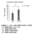
- the relationship between Rac1 activity and lymphatic vessel invasion in colorectal cancer is shown.
- the relationship between Rac1 activity and venous invasion in colorectal cancer is shown.
- the relationship between Rac1 activity and lymph node metastasis in colorectal cancer is shown.
- the detection result of activated Rac1 in 2 cases of breast cancer, colon cancer and brain tumor is shown.
- the present invention is a method for detecting an active low molecular weight GTP-binding protein (GTPase) in a fixed biological tissue.
- the method of the present invention includes the following steps (a) to (c), wherein the contact between the fixed living tissue and the probe in step (b) is performed using electric field non-contact stirring.
- step (a) the living tissue to be detected is fixed.
- the biological tissue can be fixed using, for example, formalin or PAXgene® Tissue® System (trade name, Qiagen).
- the biological tissue can be, for example, a biopsy material or a surgical material. From the viewpoint of obtaining a good detection result, it is advantageous that the formalin fixation time of the biological tissue is relatively short, for example, within 24 hours.
- the biological tissue fixed in step (a) can be subjected to step (b) after being sectioned by a conventional method.
- a probe that specifically binds to active GTPase is brought into contact with the fixed living tissue prepared in step (a).
- the contact between the fixed biological tissue and the probe is performed in an appropriate solvent (for example, PBS (phosphate buffered saline).
- the contact between the fixed biological tissue and the probe is performed using electric field non-contact stirring.
- Electric field non-contact stirring can be performed by applying a high-voltage AC electric field with a change in electric field wave to a small amount of liquid droplets in a non-contact manner, more specifically for non-contact stirring.
- Rapid-immunohistochemistry (R-IHC) which is a stirrer for immunostaining, and a fixed biological tissue that is a pre-stage of immunostaining. It is not known to be used for contact with probes.
- Non-electric field agitation in this device is to vibrate water droplets without contacting a stirrer such as a stirrer by vibrating a water molecule by applying a low-frequency voltage with a stationary liquid sandwiched between upper and lower electrodes.
- a stirrer such as a stirrer by vibrating a water molecule by applying a low-frequency voltage with a stationary liquid sandwiched between upper and lower electrodes.
- it exerts a remarkable effect on the stirring of micro liquids of the order of ⁇ l, and the droplet during application of an electric field actively moves in the vertical direction and outer / inner peripheral direction without contacting the upper and lower electrodes and is stirred uniformly (special Kaihei 2010-119388).
- the probe that binds to the active GTPase is, for example, a protein having an active GTPase binding region, and examples of the active GTPase binding region include Cdc42 / Rac-interactive-binding (CRIB) motif, Ras-binding domain (RBD ), Rho-binding domain (RBD) and the like.
- Cdc42 / Rac-interactive-binding (CRIB) motif Ras-binding domain (RBD ), Rho-binding domain (RBD) and the like.
- the low molecular weight GTPase is composed of Ras family, Rho family, Rab family, Ran family, Sar / ARF family and the like, and in the present invention, any active GTPase belonging to any family and subfamily can be detected.
- detection results for active GTPases belonging to the Rho family (Rac subfamily) and the Ras family are shown.
- the Rab family see Mol. Cell Proteomics 2008: 7 (6); 1031-42, Traffic 2010: 11 (4); 491-507
- Ran family Cell Mol Med. 2011: 11 (8); 686- 95
- the Sar / ARF family see Annu Rev Biochem. 2012: 81; 637-59.
- the like can also detect active GTPases in the same manner.
- PAK1-GST that specifically binds to active Rac1 is used as a probe that specifically binds to active GTPase. be able to.
- PAK1-GST is a combination of Glutathione S-transferase (GST) and P21-activated kinase (PAK1: GenBank / EMBL accession number: NM002576), a common effector of Rac and Cdc42 (Annu.Rev Biochem.
- GTPase-binding domain containing a Cdc42 / Rac-interactive-binding domain (CRIB) motif consisting of amino acid sequence numbers corresponding to positions 75 to 88 is bound.
- Raf-1-GST that specifically binds to active Ras may be used as a probe that specifically binds to active GTPase. it can.
- Raf-1-GST is a combination of Ras-1 (Ras-1), an effector of Ras (GenBank / EMBL accession number: X03484), and a Ras- consisting of an amino acid sequence number corresponding to positions 56 to 131.
- a probe bound to active GTPase is detected.
- Detection of the probe bound to the active GTPase in step (c) can be performed, for example, by immunostaining.
- the immunostaining method can be appropriately performed using a primary antibody and a secondary antibody corresponding to the type of probe.
- a probe specifically bound to GTP-binding GTPase is detected by an immunostaining method using an anti-GST antibody. can do.
- An anti-GST antibody can be easily obtained as a commercial product.
- protein-protein interaction of a target molecule that specifically binds to active GTPase is used.
- a GTPase binding region such as a CRIB domain
- PAK-GST for active Rac1 and Raf-1-GST for active Ras.
- Active GTPase can be detected on a tissue section by detecting a probe specifically bound to GTP-bound GTPase by immunostaining using an anti-GST antibody.
- the following explanatory diagram shows the principle of detecting active Rac1 on tissue sections by immunostaining.
- PAK-GST protein is reacted with the tissue section after fixation using a rapid immunostaining device.
- PAK-GST selectively binds to GTP-bound Rac1 (including GTP-bound Cdc42).
- the bound PAK-GST can then be detected by immunostaining using an anti-GST antibody.
- a technique using the same principle was previously detectable in the fluorescent immunostaining method using cultured animal cells, but it can be activated in the tissue after fixation with formalin such as biopsy material or surgical material. It was impossible to detect. It is suggested that this is because the bound GTP is detached at the time of immobilization, the accurate three-dimensional structure of the target protein is difficult to maintain, and the binding affinity between the active low molecular weight GTPase and the target molecule is lowered.
- the present invention is the first in the world to visualize active GTPase in a tissue sample after fixation by significantly increasing the frequency of coupling between the “active low molecular weight GTPase and target molecule” by using electric field non-contact stirring. It is a thing.
- Antibody active Rac1 antibody (NewEast Bioscience, #: 26903, mouse monoclonal antibody)
- non-transfected 293T cells and constitutively inactive Rac1 (Rac1N17, pCXN2-Flag-Rac1N17) overexpressing 293T cells were prepared.
- 1 ⁇ g of the anti-active Rac1 antibody was used per liter of cell lysate.
- the commonly used pull-down method was simultaneously performed.
- Example 1 [Usefulness of the present invention (detection of active GTP-bound Rac1 by rapid immunostaining using R-IHC)]
- GST-PAK recombinant protein was prepared using BL21 Escherichia coli, and the optimum concentration (2, 5, 10 ⁇ g / ml) used for immunostaining was examined.
- Example 2 [Establishment of activated Rac1 detection technology in formalin-fixed specimens] 1) Cell block activation of Rac1V12-overexpressing 293T cells In order to examine Rac1-immune conditions, cell blocks of Rac1V12-overexpressing 293T cells were prepared.
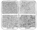
- FIG. 2 shows the detection results of activated Rac1 in the Rac1V12 overexpressing 293T cell block. This is the result when overexpressing Rac1 activated V12 in 293T cells. No activation, GST-PAK concentration is 2 ⁇ g / ml, anti-GST antibody concentration is diluted 50 times, and formalin fixation time is 30 minutes to 1 day. It is considered that the activation state of Rac1 superior to the cell membrane could be stained even compared with the control. Moreover, as a result of carrying out the whole process by hand dyeing, it was found that only non-specific staining was observed.
- Mouse xenograft tumor tissue of human colon cancer cell line Mouse Xenograft tumor tissues of human colon cancer cell lines such as HCT116 and WiDr were prepared.
- a colon cancer cell line (HCT116 and WiDr), which is known to have high Rac1 activation by GST pull-down assay, was used in 6-8 week old female nude mice (Balb / cA Jcl nu / nu, CLEA JAPAN) , Japan). After tumor formation, the mice were euthanized, the tumors were fixed in formalin or treated with PAXgene Tissue System (trade name, Qiagen, Japan), and activated Rac1 was stained by the protocol.
- PAXgene is suitable for both stabilization of biomolecules and preservation of tissue morphology, and allows tissue analysis and high-quality RNA, miRNA, and DNA purification in the same tissue without forming cross-links.
- FIG. 3 shows the detection results of activated Rac1 in mouse xenograft tissue of a colon cancer cell line.
- diffuse activated Rac1 in the cytoplasm was detected.
- Clear activation of Rac1 at the cell membrane was also detected in tissues treated with PAXgene Tissue System.
- PAXgene Tissue System Currently, most of the pathological specimens are formalin-fixed, but the usefulness of the PAXgene Tissue System is expected in the future.
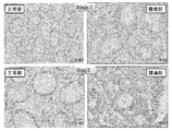
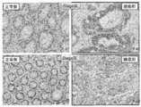
- FIGS. 4 and 5 show the detection results of activated Rac1 in colorectal cancer tissues.
- activated Rac1 was negative in any stage.
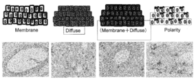
- the staining patterns were roughly divided into four (Membrane pattern, Diffuse pattern, MixedM (Membrane + Diffuse) pattern, Polarity pattern).
- FIG. 6 shows a schematic diagram of the staining pattern of activated Rac1 in colon cancer and a photograph of a typical example.
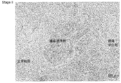
- FIG. 7 shows the results of enhancement of Rac1 activity in the colon cancer infiltrating part. Higher activation of Rac1 was detected in the tumor infiltrating part of human colon cancer cases than in the central part of the tumor.
- the photograph is a representative example (Stage II). In normal colorectal mucosa, activated Rac1 was negative.
- TMA Tissue Micro Array
- FIG. 8 shows a method for analyzing activated Rac1 in colorectal cancer TMA. HistoQuest is suitable for quantifying the staining intensity of pathological images. The result of actually measuring the DAB color intensity is shown in the lower left. A plot (bottom right) is obtained along with the numerical data of the measured range, and the darkest and lightly stained places can be confirmed.
- FIG. 9 shows the comparison results (33 cases) of Rac1 activity between the normal colonic mucosa and the tumor site using HistoQuest. Since the TMA specimen is divided into a normal part and a tumor part, each characteristic part was selected and analyzed. Thirty of the 33 cases showed higher staining intensity in the tumor area, and only 3 cases had higher intensity in the normal area.
- FIGS. 10 The relationship between Rac1 activity and vascular invasion in colorectal cancer is shown in FIGS. Based on the data analyzed above, the correlation with the data of pathologic factors lymphatic vessel invasion (FIG. 10), venous invasion (FIG. 11), and lymph node metastasis (FIG. 12) was obtained. As a result, regarding the relationship between lymphatic vessel invasion and Rac1 activation (FIG. 10), a significant difference (P ⁇ 0.05) was observed in Ly0 vs Ly1-3. Although there was no significant difference in venous invasion (FIG. 11) and lymph node metastasis (FIG. 12), activation of Rac1 tended to increase with increased venous invasion and lymph node metastasis.
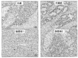
- Example 4 In the same manner as the detection of Rac1 activity in the tumor part in Example 3, it was possible to detect activated Rac1 in FFPE specimens of stomach cancer, lung cancer, breast cancer, brain tumor and pancreatic cancer in addition to colon cancer. Representative photographs (breast cancer, colon cancer, brain tumor 2 cases) are shown in FIG.
- the present invention is useful in the technical field related to the detection of low molecular weight GTPase.
Landscapes
- Health & Medical Sciences (AREA)
- Life Sciences & Earth Sciences (AREA)
- Immunology (AREA)
- Engineering & Computer Science (AREA)
- Chemical & Material Sciences (AREA)
- Molecular Biology (AREA)
- Biomedical Technology (AREA)
- Hematology (AREA)
- Urology & Nephrology (AREA)
- Analytical Chemistry (AREA)
- Physics & Mathematics (AREA)
- Biochemistry (AREA)
- General Health & Medical Sciences (AREA)
- General Physics & Mathematics (AREA)
- Pathology (AREA)
- Food Science & Technology (AREA)
- Medicinal Chemistry (AREA)
- Microbiology (AREA)
- Cell Biology (AREA)
- Biotechnology (AREA)
- Peptides Or Proteins (AREA)
- Investigating Or Analysing Biological Materials (AREA)
Abstract
本発明は、ホルマリンなどで固定した組織標本上での内在性活性型GTPaseを検出可能にする方法、特に、内在性活性型GTPaseを免疫染色で検出可能にする方法を提供する。本発明は、固定した生体組織内で、活性型の低分子量GTP結合蛋白質(GTPase)を検出する方法に関する。検出対象である生体組織を固定するステップ(a)、固定した生体組織に、活性型GTPaseに特異的に結合するプローブを接触させるステップ(b)、活性型GTPaseと結合したプローブを検出するステップ(c)を含む。ステップ(b)における固定した生体組織とプローブとの接触を、電界非接触攪拌を用いて行う。
Description
本発明は、固定生体組織内での活性型である低分子量グアノシン三リン酸(GTP)結合蛋白質(以下、GTPaseと略記することがある)検出方法に関する。
関連出願の相互参照
本出願は、2015年3月5日出願の日本特願2015-043171号の優先権を主張し、その全記載は、ここに特に開示として援用される。
関連出願の相互参照
本出願は、2015年3月5日出願の日本特願2015-043171号の優先権を主張し、その全記載は、ここに特に開示として援用される。
細胞内の複雑なシグナル伝達ネットワークを理解するためには、遺伝子発現情報だけではなく、蛋白質の活性化状態を解明することが必須である。この解明には、活性型蛋白質の量的解析に加えて位置情報を含む空間的解析が重要となる。低分子量GTPaseは、Rasファミリー、Rhoファミリー、Rabファミリー、Ranファミリーなどから成り、細胞増殖、細胞運動、細胞骨格、細胞内輸送、核輸送など多様なシグナル伝達系の制御において重要な分子スイッチとして機能する。低分子量GTPaseは、GTPが結合した活性型とグアノシン二リン酸(GDP)が結合した不活性型との間をサイクルしており、GTP結合型は、特異的な標的蛋白質に結合して下流シグナルを伝達する。低分子量GTPaseの活性化は、各特異的なグアニンヌクレオチド交換因子(guanine nucleotide exchange factor:GEF)およびGTP水解促進酵素(GTPase-activating protein: GAP)によって調節されている。
低分子量GTPaseのうち、Rhoファミリーは細胞運動・浸潤・接着などの細胞応答を制御している。RhoファミリーはRhoA、Rac1、Cdc42に代表される14の因子で構成され、中でもRac1は葉状仮足というアクチンに富んだ突起を形成することで細胞運動の際の駆動力を産み出す。特に癌において、Rac1の細胞内および腫瘍組織内での活性化量や活性化領域を解明することは、癌の生物学的特性や悪性度(浸潤能、転移能、生命予後)を理解する上で極めて意義深い。一方、Rasファミリーは細胞増殖や生存などを制御しており、Rasの活性化状態は、癌の増殖能ならびに分子標的治療薬の感受性評価の指標となる。近年、大腸癌の分子標的治療としてアービタックス(抗EGFRキメラ化モノクローナル抗体)が適用されているが、活性型KRas遺伝子変異を有する患者においては本薬剤の効果が低いことから、アービタックス投与前にシーケンス解析によってKRas遺伝子検査を実施している。
細胞内での低分子量GTPaseの活性化程度を調べる従来の方法としては、下記の手法が存在する。
(1)ラジオアイソトープ32Pi標識による方法:
細胞を32Piで標識後、低分子量GTPaseを精製し、薄層クロマトグラフィーでGTP結合型とGDP結合型を分離検出する方法である(非特許文献1)。
細胞を32Piで標識後、低分子量GTPaseを精製し、薄層クロマトグラフィーでGTP結合型とGDP結合型を分離検出する方法である(非特許文献1)。
(2)プルダウン法(Bos method):
活性型GTPaseと標的分子の複合体をグルタチオンアガロースビーズを用いてpull-down後、SDS-PAGEゲルにて分離し、イムノブロッティングにて検出する方法である(非特許文献2)。
活性型GTPaseと標的分子の複合体をグルタチオンアガロースビーズを用いてpull-down後、SDS-PAGEゲルにて分離し、イムノブロッティングにて検出する方法である(非特許文献2)。
(3)蛍光共鳴エネルギー移動(FRET):
生細胞における活性化GTPaseの時空間情報を取得できる利点がある(松田道行、特許文献1:WO02/014373)。
生細胞における活性化GTPaseの時空間情報を取得できる利点がある(松田道行、特許文献1:WO02/014373)。
(4)抗active Rac1抗体を用いた免疫沈降法および免疫染色
活性型Rac1に関しては、これを認識する特異的抗体(NewEast Bioscience社、#26903)が販売されており、免疫沈降法や免疫染色に適用可能である。
活性型Rac1に関しては、これを認識する特異的抗体(NewEast Bioscience社、#26903)が販売されており、免疫沈降法や免疫染色に適用可能である。
特許文献1:WO02/014373
特許文献1の全記載は、ここに特に開示として援用される。
特許文献1の全記載は、ここに特に開示として援用される。
非特許文献1:Satoh, T. and Kaziro Y. Measurement of Ras-bound guanine nucleotide in stimulated hematopoietic cells. Method. Enzymol. 255: 149-155. 1995.
非特許文献2:Franke, B., Akkerman J.W.N., and Bos J.L. Rapid Ca2+-mediated activation of Rap1 in human platelets. EMBO J. 15: 252-259. 1997.
非特許文献1~2の全記載は、ここに特に開示として援用される。
非特許文献2:Franke, B., Akkerman J.W.N., and Bos J.L. Rapid Ca2+-mediated activation of Rap1 in human platelets. EMBO J. 15: 252-259. 1997.
非特許文献1~2の全記載は、ここに特に開示として援用される。
(1)の非特許文献1に記載の方法は、最も直接的な方法で信頼性が高い方法である。しかし、アイソトープを用いるため日常的には頻用できない。
(2)の非特許文献2に記載の方法は、GTP結合型が標的蛋白質に高い結合アフィニティーを示すため、活性型GTPaseのみを選択的に回収することができる。簡便且つ検出感度が高く、最も頻用されている手法である。一方、本方法は細胞を可溶化する必要があり、細胞間での活性化程度の差異や位置的情報を取得することは不可能である。
(3)の特許文献1に記載の方法は、計測にはYFPやCRPなどの蛍光蛋白質を融合させたインディケーターの発現プラスミドを遺伝子導入する必要性があり、内在性GTPaseの活性化状態を検出することは困難である。各種GTPaseに対する特異的プローブの作製には経験・技術・時間を要し、また検出系には比較的高価な機材を必要とする
(4)の抗active Rac1抗体を用いた免疫沈降法および免疫染色は、検出感度および特異性に関しては問題がある(後述、実験方法とデータの欄で検証)。
本発明の目的は、ホルマリン等で固定した組織標本上での内在性活性型GTPaseを検出可能にする方法を提供することであり、特に、内在性活性型GTPaseを免疫染色で検出可能にする方法を提供することである。
ホルマリン等で固定した組織標本上での内在性活性型GTPaseの検出は、当該分野において長年に渡り望まれていたことであったが、実現できていなかった方法である。本発明者らは、活性型低分子量GTPaseと標的蛋白質とを電界非接触攪拌することで、両者を免疫染色で検出可能な程度に結合させることができることを見出して、本発明を完成させた。
本発明は、以下の通りである。
[1]
固定した生体組織内で、活性型の低分子量GTP結合蛋白質(以下、GTPaseと略記する)を検出する方法であって、
検出対象である生体組織を固定するステップ(a)、
固定した生体組織に、活性型GTPaseに特異的に結合するプローブを接触させるステップ(b)、
活性型GTPaseと結合したプローブを検出するステップ(c)を含み、
ステップ(b)における固定した生体組織とプローブとの接触を、電界非接触攪拌を用いて行うことを特徴とする前記方法。
[2]
活性型GTPaseと結合するプローブは、活性型GTPase結合領域を有するタンパク質である[1]に記載の方法。
[3]
活性型GTPase結合領域(GTPase-binding domain: GBD)は、Cdc42/Rac-interactive-binding(CRIB)motif、Ras-binding domain (RBD)、Rho-binding domain (RBD)から成る群から選ばれる少なくとも1種のドメインである[2]に記載の方法。
[4]
活性型GTPaseは、Rhoファミリー又はRasファミリーに属する活性型GTPaseである[1]~[3]のいずれかに記載の方法。
[5]
活性型GTPaseは、RhoファミリーのRacサブファミリーに属する活性型GTPaseであり、活性型GTPaseに特異的に結合するプローブが、活性型Rac1に対して特異的に結合するPAK-GSTである[1]~[4]のいずれかに記載の方法。
[6]
活性型GTPaseは、Rasファミリーに属する活性型GTPaseであり、活性型GTPaseに特異的に結合するプローブが、活性型Rasに対して特異的に結合するRaf-1-GSTである[1]~[4]のいずれかに記載の方法。
[7]
生体組織の固定は、ホルマリン又はPAXgene Tissue Systemを用いて行う[1]~[6]のいずれかに記載の方法。
[8]
ステップ(a)における生体組織のホルマリン固定は、24時間以内とする[7]に記載の方法。
[9]
ステップ(c)における活性型GTPaseと結合したプローブの検出は、免疫染色法により行う[1]~[8]のいずれかに記載の方法。
[10]
ステップ(c)における活性型GTPaseと結合したプローブの検出は、GTP結合型GTPaseに特異的に結合したプローブを、抗GST抗体を用いて免疫染色法により検出する[1]~[8]のいずれかに記載の方法。
[11]
ステップ(a)で固定した生体組織を、切片とした後にステップ(b)に供する[1]~[10]のいずれかに記載の方法。
[12]
生体組織が生検材料又は手術材料である[1]~[11]のいずれかに記載の方法。
[1]
固定した生体組織内で、活性型の低分子量GTP結合蛋白質(以下、GTPaseと略記する)を検出する方法であって、
検出対象である生体組織を固定するステップ(a)、
固定した生体組織に、活性型GTPaseに特異的に結合するプローブを接触させるステップ(b)、
活性型GTPaseと結合したプローブを検出するステップ(c)を含み、
ステップ(b)における固定した生体組織とプローブとの接触を、電界非接触攪拌を用いて行うことを特徴とする前記方法。
[2]
活性型GTPaseと結合するプローブは、活性型GTPase結合領域を有するタンパク質である[1]に記載の方法。
[3]
活性型GTPase結合領域(GTPase-binding domain: GBD)は、Cdc42/Rac-interactive-binding(CRIB)motif、Ras-binding domain (RBD)、Rho-binding domain (RBD)から成る群から選ばれる少なくとも1種のドメインである[2]に記載の方法。
[4]
活性型GTPaseは、Rhoファミリー又はRasファミリーに属する活性型GTPaseである[1]~[3]のいずれかに記載の方法。
[5]
活性型GTPaseは、RhoファミリーのRacサブファミリーに属する活性型GTPaseであり、活性型GTPaseに特異的に結合するプローブが、活性型Rac1に対して特異的に結合するPAK-GSTである[1]~[4]のいずれかに記載の方法。
[6]
活性型GTPaseは、Rasファミリーに属する活性型GTPaseであり、活性型GTPaseに特異的に結合するプローブが、活性型Rasに対して特異的に結合するRaf-1-GSTである[1]~[4]のいずれかに記載の方法。
[7]
生体組織の固定は、ホルマリン又はPAXgene Tissue Systemを用いて行う[1]~[6]のいずれかに記載の方法。
[8]
ステップ(a)における生体組織のホルマリン固定は、24時間以内とする[7]に記載の方法。
[9]
ステップ(c)における活性型GTPaseと結合したプローブの検出は、免疫染色法により行う[1]~[8]のいずれかに記載の方法。
[10]
ステップ(c)における活性型GTPaseと結合したプローブの検出は、GTP結合型GTPaseに特異的に結合したプローブを、抗GST抗体を用いて免疫染色法により検出する[1]~[8]のいずれかに記載の方法。
[11]
ステップ(a)で固定した生体組織を、切片とした後にステップ(b)に供する[1]~[10]のいずれかに記載の方法。
[12]
生体組織が生検材料又は手術材料である[1]~[11]のいずれかに記載の方法。
本発明によれば、従来困難であったホルマリン等で固定した組織標本上での内在性活性型GTPaseを免疫染色で検出できる。特に癌病理検体における内在性GTPaseの活性化領域および活性化量を計測できると共に、細胞内における詳細な活性化部位も同定可能となる。これにより、癌を始めとする種々の疾患において、その発症機構の解明や最適分子標的薬剤の選定、新規診断技術や治療法の開発に有用となると期待される。
本発明は、固定した生体組織内で、活性型の低分子量GTP結合蛋白質(GTPase)を検出する方法である。本発明の方法は、下記ステップ(a)~(c)を含み、ステップ(b)における固定した生体組織とプローブとの接触を、電界非接触攪拌を用いて行うことを特徴とする。
検出対象である生体組織を固定するステップ(a)、
固定した生体組織に、活性型GTPaseに特異的に結合するプローブを接触させるステップ(b)、
活性型GTPaseと結合したプローブを検出するステップ(c)。
検出対象である生体組織を固定するステップ(a)、
固定した生体組織に、活性型GTPaseに特異的に結合するプローブを接触させるステップ(b)、
活性型GTPaseと結合したプローブを検出するステップ(c)。
ステップ(a)では、検出対象である生体組織を固定する。検出対象である生体組織の種類及び固定方法及び条件は特に制限はない。生体組織の固定は、例えば、ホルマリン又はPAXgene Tissue System(商品名、キアゲン)を用いて行うことができる。生体組織は、例えば、生検材料又は手術材料であることができる。良好な検出結果を得るという観点から、生体組織のホルマリン固定の時間は、比較的短い方が有利であり、例えば、24時間以内とすることができる。ステップ(a)で固定した生体組織は、常法により切片とした後にステップ(b)に供することもできる。
ステップ(b)では、ステップ(a)で作成した固定した生体組織に、活性型GTPaseに特異的に結合するプローブを接触させる。固定した生体組織とプローブとの接触は、適当な溶媒(例えば、PBS(リン酸緩衝生理食塩水)中で行う。さらに、固定した生体組織とプローブとの接触は、電界非接触攪拌を用いて行う。電界非接触攪拌は、電界の波に変化をもたせた高電圧交流電界を微少量液滴に非接触に与えるようにすることで行うことができる。より具体的には、非接触撹拌用の装置である迅速免疫染色装置(Rapid-immunohistochemistry:R-IHC)を用いることで実施できる。この装置は、免疫染色のための撹拌装置であり、免疫染色の前段階である固定した生体組織とプローブとの接触のために用いられることは知られていない。
当該装置における電界非接触攪拌は、静置した液を上下の電極で挟んで低周波電圧を印加することで水分子そのものを振動させ、スターラーなどの撹拌子を接触せずに、液滴を活発に撹拌する。特にμlオーダーの微量液の撹拌に著しい効果を発揮し、電界印加中の液滴は、上下電極に接触することなく、上下方向や外内周方向に活発に動き、均一に撹拌される(特開平2010-119388号公報)。電界非接触攪拌を用いることで、標本上の活性型低分子量GTPaseと標的蛋白質との結合頻度は、静置時の1200倍に上昇することが、検討の結果判明した。加えて、電界非接触攪拌では、撹拌時に熱を発生しない。活性型GTPaseと標的蛋白質との結合は正確な立体構造に依存するため、撹拌時に熱が発生する他の攪拌装置を用いる攪拌では容易にこの立体構造が変化し、検出不可能となる。このように電界非接触攪拌は、1200倍の会合スピードの上昇と、熱を発生しないという二大特徴から、活性型低分子量GTPaseと標的蛋白質との結合促進し、ホルマリンまたはPAXgene Tissue Systemで固定した生体組織内での活性型低分子量GTP結合蛋白質の検出を初めて可能とした。
活性型GTPaseと結合するプローブは、例えば、活性型GTPase結合領域を有するタンパク質であり、活性型GTPase結合領域としては、例えば、Cdc42/Rac-interactive-binding (CRIB) motif、Ras-binding domain (RBD)、Rho-binding domain (RBD)等を挙げることができる。活性型GTPaseには、複数のファミリー及びサブファミリーがあり、プローブが有する活性型GTPase結合領域は、各ファミリー及びサブファミリーに応じて、適宜選択される。
低分子量GTPaseは、Rasファミリー、Rhoファミリー、Rabファミリー、Ranファミリー、Sar/ARFファミリーなどから成り、本発明においてはどのファミリー及びサブファミリーに属する活性型GTPaseについても検出することができる。実施例においては、Rhoファミリー(Racサブファミリー)及びRasファミリーに属する活性型GTPaseについての検出結果を示す。しかし、Rabファミリー(Mol. Cell Proteomics 2008:7(6);1031-42、Traffic 2010:11(4);491-507参照)、Ranファミリー(Curr Mol Med.2011:11(8);686-95.参照)、Sar/ARFファミリー(Annu Rev Biochem.2012:81;637-59.参照)などについても同様に活性型GTPaseを検出することは可能である。
活性型GTPaseが、RhoファミリーのRacサブファミリーに属する活性型GTPaseである場合には、活性型GTPaseに特異的に結合するプローブとして、活性型Rac1に対して特異的に結合するPAK1-GSTを用いることができる。PAK1-GSTは、Glutathione S-transferase(GST)に、RacとCdc42の共通のエフェクターであるP21-activated kinase(PAK1:GenBank/EMBLアクセッション番号:NM002576)を結合させたものであり(Annu.Rev.Biochem.2003:72;743-81参照)、75~88位に相当するアミノ酸配列番号からなるCdc42/Rac-interactive-binding (CRIB)モチーフを含むGTPase-binding domain (GBD)を結合させる。
活性型GTPaseが、Rasファミリーに属する活性型GTPaseである場合には、活性型GTPaseに特異的に結合するプローブとして、活性型Rasに対して特異的に結合するRaf-1-GSTを用いることができる。Raf-1-GSTは、GSTに、RasのエフェクターであるRaf-1(GenBank/EMBLアクセッション番号:X03484)を結合させたものであり、56~131位に相当するアミノ酸配列番号からなるRas-binding domain (RBD)を含むGTPase-binding domain (GBD)を結合させる。
ステップ(c)では、活性型GTPaseと結合したプローブを検出する。ステップ(c)における活性型GTPaseと結合したプローブの検出は、例えば、免疫染色法により行うことができる。免疫染色法は、プローブの種類に応じた一次抗体及び二次抗体を用いて適宜実施でき、例えば、GTP結合型GTPaseに特異的に結合したプローブを、抗GST抗体を用いて免疫染色法により検出することができる。抗GST抗体は、市販品を容易に入手することができる。
本発明の方法では、活性型GTPaseと特異的に結合する標的分子の蛋白質間相互作用を利用する。具体的には、GDP結合型(不活性型)には結合せず、GTP結合型(活性化型)にのみ結合するプローブとして、標的蛋白質中のGTPase結合領域(CRIBドメインなど)を使用する(活性型Rac1に対してPAK-GST(PAK1およびPAK2を含む)、活性型Rasに対してRaf-1-GST)。GTP結合型GTPaseに特異的に結合したプローブを、抗GST抗体を用いて免疫染色法により検出することで、活性型GTPaseを組織切片上で検出することが可能となる。
下記説明図に、組織切片上において免疫染色法により活性型Rac1を検出する原理示す。固定後の組織切片に迅速免疫染色装置を用いてPAK-GST蛋白質を反応させる。PAK-GSTは、GTP結合型Rac1に選択的に結合する(GTP結合型Cdc42も含む)。結合したPAK-GSTは、その後、抗GST抗体を用いた免疫染色法により検出することができる。

同様の原理を用いた手法は、従前、動物培養細胞を用いた蛍光免疫染色法においては検出可能であったが、生検材料や手術材料などホルマリンなどでの固定後組織内での活性化を検出することは不可能であった。この原因は、固定時に結合GTPが外れたり、標的蛋白質の正確な立体構造維持が困難であり、活性型低分子量GTPaseと標的分子との結合アフィニティーが低下するためと示唆される。本発明は、この「活性型低分子量GTPase-標的分子」間の介合頻度を、電界非接触撹拌を用いることで著しく亢進させ、固定後の組織試料において活性型GTPaseの可視化に世界で初めて成功したものである。
以下、本発明を実施例に基づいて更に詳細に説明する。但し、実施例は本発明の例示であって、本発明は実施例に限定される意図ではない。
[材料と方法]
比較例1
[従前技術(抗Active Rac1抗体を用いた活性型GTP結合Rac1検出)の有用性検討]
比較例1
[従前技術(抗Active Rac1抗体を用いた活性型GTP結合Rac1検出)の有用性検討]
(i)従前技術の有用性検討:免疫組織染色法(immunohistochemistry: IHC)において活性型Rac1を検出する従前技術としては、現在、抗active Rac1抗体を用いた方法が存在する。下記の工程により、当該従前技術の特異性ならびに検出感度からその有用性を検討した。
(ii)抗体:active Rac1抗体(NewEast Bioscience社、#:26903、マウスモノクローナル抗体)
適用:免疫沈降法、免疫組織染色法(IHC)
適用:免疫沈降法、免疫組織染色法(IHC)
(iii)免疫沈降法による活性型Rac1の検出:293T細胞に恒常的活性型Rac1変異体であるRac1V12発現プラスミド(pCXN2-Flag-Rac1V12)を遺伝子導入(トランスフェクション)し、活性型Rac1を一過性に過剰発現させた。293T細胞は、ヒト胎児腎細胞にSV40のlarge T抗原を発現させ株化した細胞であり、トランスフェクション効率が高く生化学実験等に頻用される。トランスフェクション試薬にはFugene HD(Promega)を使用し、規定のプロトコールに従って遺伝子導入した。陰性コントロールとして、非トランスフェクション293T細胞、ならびに恒常的不活性型Rac1(Rac1N17、pCXN2-Flag-Rac1N17)過剰発現293T細胞を準備した。免疫沈降には、細胞溶解液1 mgに対して当該抗active Rac1抗体1μgを使用した。活性化型Rac1検出効率の陽性コントロールとして、通常頻用されているPull-down法を同時に施行した。
(iv)蛍光免疫染色法による活性型Rac1の検出:293T細胞を予めタイプIコラーゲンが塗布されたガラスベースディッシュに播種した。上記(iii)と同様に、トランスフェクションにより恒常的活性型ならびに不活性型Rac1を一過性過剰発現させた。蛍光免疫染色は、発明者らが通常行っているプロトコールに従って施行した。当該抗体(抗active Rac1抗体)は500倍希釈で使用した。
実施例1
[本発明(R-IHCを用いた迅速免疫染色法による活性型GTP結合Rac1検出)の有用性検討]
[本発明(R-IHCを用いた迅速免疫染色法による活性型GTP結合Rac1検出)の有用性検討]
(1)セルブロックを用いた免疫染色による活性型GTP結合Rac1蛋白質の検出
(i)材料:Rac1V12過剰発現293T細胞:活性型Rac1染色の陽性コントロールとして、下記(ii)の工程により恒常的活性型Rac1変異体(Rac1V12)過剰発現293T細胞を作製した。
(i)材料:Rac1V12過剰発現293T細胞:活性型Rac1染色の陽性コントロールとして、下記(ii)の工程により恒常的活性型Rac1変異体(Rac1V12)過剰発現293T細胞を作製した。
(ii)トランスフェクションおよびセルブロックの作製:293T細胞にRac1V12発現プラスミド(pCXN2-Flag-Rac1V12)を遺伝子導入(トランスフェクション)し、活性化型Rac1を一過性に過剰発現させた。48時間後に293T細胞をトリプシンで剥がした後、PBSで1回洗浄、1.5 mlチューブに移して3,000回転で5分間遠心することにより細胞を沈殿させた(セルペレット)。PBS除去後、ホルマリン1 mlを静かに重層して固定した。下記(vii)の条件でホルマリン固定時間を検討後、セルペレットをカセットに移し、パラフィンを充填してセルブロックを作製した。セルブロックは、ミクロトームを用いて4μm厚の切片に薄切した。
(iii)活性型GTP結合Rac1蛋白質の免疫組織染色:下記の工程により、R-IHCを用いて活性型Rac1の免疫染色を施行した。
1. 脱パラ
2. 内因性ペルオキシダーゼ(3%過酸化水素)処理:室温5分。その後PBSで3回洗浄。
3. 標的蛋白質(GST-PAK)の反応:R-IHC装置を用いて室温5分間反応。その後PBSで洗浄。
4. 1次抗体:抗GSTマウスモノクローナル抗体。37度30分処理後、室温10分処理。その後PBSで洗浄20分間。
5. ポリマー試薬:37度30分処理。その後PBSで洗浄20分間。
6. DAB発色。水洗。
7. 核染:Mayerのヘマトキシリンで核染色。3分。色出し。
8. 脱水
9. 透徹
10. 封入
1. 脱パラ
2. 内因性ペルオキシダーゼ(3%過酸化水素)処理:室温5分。その後PBSで3回洗浄。
3. 標的蛋白質(GST-PAK)の反応:R-IHC装置を用いて室温5分間反応。その後PBSで洗浄。
4. 1次抗体:抗GSTマウスモノクローナル抗体。37度30分処理後、室温10分処理。その後PBSで洗浄20分間。
5. ポリマー試薬:37度30分処理。その後PBSで洗浄20分間。
6. DAB発色。水洗。
7. 核染:Mayerのヘマトキシリンで核染色。3分。色出し。
8. 脱水
9. 透徹
10. 封入
(iv)抗原賦活化方法の検討:抗原賦活化の検討には、通常の免疫組織染色において頻用されているpressure cooker(PC、圧力鍋、pH6.0クエン酸バッファー)を用いた賦活化の有用性を検討した。
(v)標的蛋白質(GST-PAK)濃度の検討: BL21大腸菌を用いてGST-PAKリコンビナント蛋白質を作製し、免疫染色時に使用する最適濃度(2, 5, 10μg/ml)を検討した。
(vi)標的蛋白質(GST-PAK)の結合特異性の検討:活性型Rac1に対する標的蛋白質(GST-PAK)の結合特異性を検討するために、低分子量GTP結合蛋白質の他のファミリーRAFに対する結合蛋白質Ral-GSTの結合程度を検討した。
(vii)ホルマリン固定時間の検討:ホルマリン固定時間が活性型Rac1と標的蛋白質(GST-PAK)間の結合アフィニティーに与える影響を検討するため、ホルマリン固定時間(30分、2時間、1日、2日、3日)を検討した。
(viii)1次抗体の希釈倍率の検討:標的蛋白質に対する1次抗体(抗GSTマウスモノクローナル抗体)の希釈倍率(50倍、100倍)を検討した。
(ix)R-IHC装置の有用性の検討:上記全ての項目において最適染色条件が決定した後、活性化Rac1と標的蛋白質(GST-PAK)の反応において手染め法とR-IHC装置による染色結果を比較し、当該染色装置の新規性・優位性・有用性を検討した。手染め法において、標的蛋白質(GST-PAK)との反応条件は、37度30分とした。
(2)ホルマリン固定パラフィン包埋標本を用いた活性型GTP結合蛋白質の検出
上記セルブロックで至適化された染色条件を用いて、ヒト癌症例(乳癌、大腸癌、脳腫瘍、膵臓癌、胃癌)において活性型GTP結合Rac1を染色・検出した。
上記セルブロックで至適化された染色条件を用いて、ヒト癌症例(乳癌、大腸癌、脳腫瘍、膵臓癌、胃癌)において活性型GTP結合Rac1を染色・検出した。
[結果]
比較例1の結果
[従前技術(抗Active Rac1抗体を用いた活性型GTP結合Rac1の検出)の有用性検討]
従前に存在した抗Active Rac1抗体を用いて、活性型Rac1の検出感度および特異性を検討した。
比較例1の結果
[従前技術(抗Active Rac1抗体を用いた活性型GTP結合Rac1の検出)の有用性検討]
従前に存在した抗Active Rac1抗体を用いて、活性型Rac1の検出感度および特異性を検討した。
(i)免疫沈降法による活性型Rac1の検出:
従来のプルダウン法と比較検討して、当該抗active Rac1抗体を用いた免疫沈降法では活性型Rac1の検出感度が著しく低下した。
従来のプルダウン法と比較検討して、当該抗active Rac1抗体を用いた免疫沈降法では活性型Rac1の検出感度が著しく低下した。
(ii)蛍光免疫染色法による活性型Rac1の検出:
恒常的活性型Rac1および不活性型Rac1の過剰発現細胞を用いて、蛍光免疫染色法により当該抗active Rac1抗体の特異性ならびに検出感度を検討した。恒常的不活性型Rac1過剰発現細胞においても染色性が確認されると共に、恒常的活性型Rac1過剰発現細胞においても、特異的な強い染色性は確認されなかった。以上より、従前技術である抗active Rac1抗体は、活性型Rac1の検出において、感度および特異性共に良好な結果は得られなかった。
恒常的活性型Rac1および不活性型Rac1の過剰発現細胞を用いて、蛍光免疫染色法により当該抗active Rac1抗体の特異性ならびに検出感度を検討した。恒常的不活性型Rac1過剰発現細胞においても染色性が確認されると共に、恒常的活性型Rac1過剰発現細胞においても、特異的な強い染色性は確認されなかった。以上より、従前技術である抗active Rac1抗体は、活性型Rac1の検出において、感度および特異性共に良好な結果は得られなかった。
実施例1の結果
[本発明(R-IHCを用いた迅速免疫染色法による活性型GTP結合Rac1検出)の有用性検討]
[本発明(R-IHCを用いた迅速免疫染色法による活性型GTP結合Rac1検出)の有用性検討]
(1)セルブロックを用いた免疫染色による活性型GTP結合蛋白質の検出
(i)抗原賦活化方法の検討: pH6.0のクエン酸バッファー中でpressure cooker(圧力鍋)を用いて抗原の賦活化を行った所、完全に染色性が失われた。一方、抗原賦活化を行わなかったサンプルに関しては、良好な染色性が得られた。
(i)抗原賦活化方法の検討: pH6.0のクエン酸バッファー中でpressure cooker(圧力鍋)を用いて抗原の賦活化を行った所、完全に染色性が失われた。一方、抗原賦活化を行わなかったサンプルに関しては、良好な染色性が得られた。
(ii)標的蛋白質(GST-PAK)濃度の検討: GST-PAKリコンビナント蛋白質の濃度は、検討した最低濃度2μg/mlでも良好な染色性が得られた。
(iii)標的蛋白質(GST-PAK)の結合特異性の検討:活性型Rac1を発現している当該セルブロックに対して、活性型RAFに対する標的蛋白質(GST-Ral-GDS)を反応させても、全く染色性は認められなかったことから、活性型Rac1とGST-PAKとの結合特異性が証明された。
(iv)ホルマリン固定時間の検討:ホルマリン固定時間は、30分、2時間、1日までは極めて良好な染色性が得られたが、2日および3日処理では染色性が低下した。
(v)1次抗体の希釈倍率の検討:標的蛋白質に対する1次抗体(抗GST抗体)の希釈倍率は、50倍希釈で細胞蛍光免疫染色時と同程度の良好な染色性を維持した。
(vi)R-IHC装置の有用性の検討:上記全ての最適染色条件を使用して手染め法とR-IHC法を比較したところ、手染め法では非特異的反応が進み、活性化型Rac1を識別できなかったのに対し、R-IHC法ではRac1V12過剰発現細胞において細胞膜優位に特異的な活性型Rac1が染色された(図1)。
上記実施例では、Rac1V12過剰発現293T細胞を用いて活性型低分子量GTP結合蛋白質である活性化型Rac1が検出できることを示した。この結果は、本発明の方法が臨床検体における活性型低分子量GTP結合蛋白質の検出にも応用可能であることを示す物である。
実施例2
[ホルマリン固定標本における活性化型Rac1検出技術の確立]
1) Rac1V12過剰発現293T細胞のセルブロック
活性化Rac1免染条件を検討するために、Rac1V12過剰発現 293T細胞のセルブロックを作製した。
[ホルマリン固定標本における活性化型Rac1検出技術の確立]
1) Rac1V12過剰発現293T細胞のセルブロック
活性化Rac1免染条件を検討するために、Rac1V12過剰発現 293T細胞のセルブロックを作製した。
Rac1V12過剰発現293T細胞のセルブロック作製条件
・抗原賦活化の有無:Presser Cooker (PC)、pH6.0 クエン酸バッファー
・GST-PAK濃度:2、5、10 μg/ml
・一次抗体(αGSTAb)濃度:×50、×100希釈
・ホルマリン固定時間:30分、2時間、1日、2日、3日
・GST-PAKの結合特異性:GST-Ral-GDS(陰性コントロール)
・抗原賦活化の有無:Presser Cooker (PC)、pH6.0 クエン酸バッファー
・GST-PAK濃度:2、5、10 μg/ml
・一次抗体(αGSTAb)濃度:×50、×100希釈
・ホルマリン固定時間:30分、2時間、1日、2日、3日
・GST-PAKの結合特異性:GST-Ral-GDS(陰性コントロール)
Rac1V12過剰発現293T細胞セルブロックでの活性化型Rac1の検出結果を図2に示す。
293T細胞でRac1の活性化型V12を過剰発現させた場合の結果である。賦活化なし、GST-PAK濃度は2 μg/ml、抗GST抗体濃度50倍希釈、ホルマリン固定時間は30分~1日までが至適条件でありことがわかる。コントロールと比較しても細胞膜に優位なRac1の活性化状態を染色できたと考えられる。また全行程手染めで行った結果、非特異的な染色しか見られないことがわかった。
293T細胞でRac1の活性化型V12を過剰発現させた場合の結果である。賦活化なし、GST-PAK濃度は2 μg/ml、抗GST抗体濃度50倍希釈、ホルマリン固定時間は30分~1日までが至適条件でありことがわかる。コントロールと比較しても細胞膜に優位なRac1の活性化状態を染色できたと考えられる。また全行程手染めで行った結果、非特異的な染色しか見られないことがわかった。
2) ヒト大腸癌細胞株のマウスxenograft腫瘍組織
HCT116、WiDrのようなヒト大腸癌細胞株のマウスXenograft腫瘍組織を作製した。
GST pull-down assayにより予めRac1の活性化が高いことが判明している大腸癌細胞株(HCT116及びWiDr)を6~8週齢のメスのヌードマウス(Balb/cA Jcl nu/nu、日本クレア、日本)に皮下接種した。腫瘤形成後にマウスは安楽死され、腫瘍はホルマリン固定あるいはPAXgene Tissue System処理(商品名、キアゲン、日本)され、当該プロトコールにより活性化型Rac1を染色した。PAXgeneは生体分子の安定化と組織形態の保存の両者に適しており、クロスリンクを形成することなく同一組織で組織解析および高品質なRNA、miRNA、DNAの精製が可能である。
HCT116、WiDrのようなヒト大腸癌細胞株のマウスXenograft腫瘍組織を作製した。
GST pull-down assayにより予めRac1の活性化が高いことが判明している大腸癌細胞株(HCT116及びWiDr)を6~8週齢のメスのヌードマウス(Balb/cA Jcl nu/nu、日本クレア、日本)に皮下接種した。腫瘤形成後にマウスは安楽死され、腫瘍はホルマリン固定あるいはPAXgene Tissue System処理(商品名、キアゲン、日本)され、当該プロトコールにより活性化型Rac1を染色した。PAXgeneは生体分子の安定化と組織形態の保存の両者に適しており、クロスリンクを形成することなく同一組織で組織解析および高品質なRNA、miRNA、DNAの精製が可能である。
大腸癌細胞株のマウスxenograft組織での活性化型Rac1の検出結果を図3に示す。
ホルマリン3日固定腫瘍組織において、細胞質に瀰漫性の活性化型Rac1が検出された。PAXgene Tissue System処理組織においても細胞膜での明瞭なRac1の活性化が検出された。現在、病理標本のほとんどはホルマリン固定されているが、将来的にPAXgene Tissue Systemの有用性が期待される。
ホルマリン3日固定腫瘍組織において、細胞質に瀰漫性の活性化型Rac1が検出された。PAXgene Tissue System処理組織においても細胞膜での明瞭なRac1の活性化が検出された。現在、病理標本のほとんどはホルマリン固定されているが、将来的にPAXgene Tissue Systemの有用性が期待される。
実施例3
[ヒト大腸癌組織における活性化型Rac1の検出]
1)FFPE (Formalin-Fixed Paraffin-Embedded)検体:50症例
[ヒト大腸癌組織における活性化型Rac1の検出]
1)FFPE (Formalin-Fixed Paraffin-Embedded)検体:50症例
ヒト大腸がん検体50症例(Stage 0: 3症例、StageI: 10症例、Stage II: 10症例、Stage IIIA: 10症例、Stage IIIB: 7症例、Stage IV: 10症例)において、正常粘膜(コントロール)と腫瘍部での活性化型Rac1を染色した。大腸癌組織における活性化型Rac1の検出結果を図4および5に示す。大腸正常粘膜では、いずれのStageにおいても活性化型Rac1は陰性だった。尚、染色パターンは4つに大別された(Membraneパターン、Diffuseパターン、Mixed (Membrane+Diffuse)パターン、Polarityパターン)。この大腸癌における活性化型Rac1の染色パターンを模式化した図と典型例の写真を図6に示す。
大腸癌浸潤部におけるRac1活性の亢進結果を図7に示す。
ヒト大腸がん症例の腫瘍浸潤部では腫瘍中心部と較べてRac1の高い活性化が検出された。写真は代表的な一例(StageII)。大腸正常粘膜では、活性化型Rac1は陰性であった。
ヒト大腸がん症例の腫瘍浸潤部では腫瘍中心部と較べてRac1の高い活性化が検出された。写真は代表的な一例(StageII)。大腸正常粘膜では、活性化型Rac1は陰性であった。
2)Tissue Micro Array(TMA)
ヒト大腸がんTMA(tissue microarray)検体33症例において、当該プロトコールにより活性化型Rac1を染色した。正常粘膜と腫瘍部において、Rac1の活性化を示すDABの発色スコアは、Histoquestソフトウェア(Novel Science Co., Ltd,東京、日本)を使用して解析した。
ヒト大腸がんTMA(tissue microarray)検体33症例において、当該プロトコールにより活性化型Rac1を染色した。正常粘膜と腫瘍部において、Rac1の活性化を示すDABの発色スコアは、Histoquestソフトウェア(Novel Science Co., Ltd,東京、日本)を使用して解析した。
大腸癌TMAにおける活性化型Rac1の解析方法を図8に示す。
HistoQuestは病理画像の染色強度を数値化することに適している。実際にDAB発色強度を測定した結果を左下に示す。測定した範囲の数値データと共にプロット図(右下)が得られ、一番濃く染色された場所、薄く染色された場所を確認することができる。
HistoQuestは病理画像の染色強度を数値化することに適している。実際にDAB発色強度を測定した結果を左下に示す。測定した範囲の数値データと共にプロット図(右下)が得られ、一番濃く染色された場所、薄く染色された場所を確認することができる。
HistoQuestを用いた大腸正常粘膜と腫瘍部でのRac1活性の比較結果(33症例)を図9に示す。TMA検体が正常部と腫瘍部に分かれているため、それぞれの特徴的な部位を選び、解析した。33症例中30症例は染色強度が腫瘍部で高く、3症例のみ正常部の方が高かった。
大腸癌におけるRac1活性と脈管侵襲との関係を図10~12に示す。
上記で解析したデータを元に病理学的因子であるリンパ管侵襲(図10)、静脈侵襲(図11)、リンパ節転移(図12)のデータとの相関を求めた。その結果、リンパ管侵襲とRac1活性化との関連性(図10)についてはLy0 vs Ly1-3において有意差(P < 0.05)が認められた。静脈侵襲(図11)、リンパ節転移(図12)については有意差は出なかったものの、静脈侵襲やリンパ節転移の亢進と共にRac1の活性化が亢進している傾向となった。
上記で解析したデータを元に病理学的因子であるリンパ管侵襲(図10)、静脈侵襲(図11)、リンパ節転移(図12)のデータとの相関を求めた。その結果、リンパ管侵襲とRac1活性化との関連性(図10)についてはLy0 vs Ly1-3において有意差(P < 0.05)が認められた。静脈侵襲(図11)、リンパ節転移(図12)については有意差は出なかったものの、静脈侵襲やリンパ節転移の亢進と共にRac1の活性化が亢進している傾向となった。
以上の結果から、
1)ヒト大腸癌組織におけるRac1の活性は大腸正常粘膜と比較して亢進しており、腫瘍浸潤部では特に高い活性が認められた。
2)Rac1の活性化は、脈管侵襲やリンパ節転移と正の相関を示す傾向が認められた。
Rac1の活性化を検出することで、腺腫と癌の鑑別、癌細胞の浸潤能や転移能の評価に有用と期待される。
1)ヒト大腸癌組織におけるRac1の活性は大腸正常粘膜と比較して亢進しており、腫瘍浸潤部では特に高い活性が認められた。
2)Rac1の活性化は、脈管侵襲やリンパ節転移と正の相関を示す傾向が認められた。
Rac1の活性化を検出することで、腺腫と癌の鑑別、癌細胞の浸潤能や転移能の評価に有用と期待される。
実施例4
実施例3における腫瘍部でのRac1活性の検出と同様の方法で、大腸癌に加えて、胃癌、肺癌、乳癌、脳腫瘍、膵癌のFFPE検体において活性化型Rac1の検出をすることでできた。代表的な写真(乳癌、大腸癌、脳腫瘍2例)を図13に示す。
実施例3における腫瘍部でのRac1活性の検出と同様の方法で、大腸癌に加えて、胃癌、肺癌、乳癌、脳腫瘍、膵癌のFFPE検体において活性化型Rac1の検出をすることでできた。代表的な写真(乳癌、大腸癌、脳腫瘍2例)を図13に示す。
本発明は、低分子量GTPaseの検出に関係する技術分野に有用である。
Claims (12)
- 固定した生体組織内で、活性型の低分子量GTP結合蛋白質(以下、GTPaseと略記する)を検出する方法であって、
検出対象である生体組織を固定するステップ(a)、
固定した生体組織に、活性型GTPaseに特異的に結合するプローブを接触させるステップ(b)、
活性型GTPaseと結合したプローブを検出するステップ(c)を含み、
ステップ(b)における固定した生体組織とプローブとの接触を、電界非接触攪拌を用いて行うことを特徴とする前記方法。 - 活性型GTPaseと結合するプローブは、活性型GTPase結合領域を有するタンパク質である請求項1に記載の方法。
- 活性型GTPase結合領域(GTPase-binding domain: GBD)は、Cdc42/Rac-interactive-binding(CRIB)motif、Ras-binding domain (RBD)、Rho-binding domain (RBD)から成る群から選ばれる少なくとも1種のドメインである請求項2に記載の方法。
- 活性型GTPaseは、Rhoファミリー又はRasファミリーに属する活性型GTPaseである請求項1~3のいずれかに記載の方法。
- 活性型GTPaseは、RhoファミリーのRacサブファミリーに属する活性型GTPaseであり、活性型GTPaseに特異的に結合するプローブが、活性型Rac1に対して特異的に結合するPAK-GSTである請求項1~4のいずれかに記載の方法。
- 活性型GTPaseは、Rasファミリーに属する活性型GTPaseであり、活性型GTPaseに特異的に結合するプローブが、活性型Rasに対して特異的に結合するRaf-1-GSTである請求項1~4のいずれかに記載の方法。
- 生体組織の固定は、ホルマリン又はPAXgene Tissue Systemを用いて行う請求項1~6のいずれかに記載の方法。
- ステップ(a)における生体組織のホルマリン固定は、24時間以内とする請求項7に記載の方法。
- ステップ(c)における活性型GTPaseと結合したプローブの検出は、免疫染色法により行う請求項1~8のいずれかに記載の方法。
- ステップ(c)における活性型GTPaseと結合したプローブの検出は、GTP結合型GTPaseに特異的に結合したプローブを、抗GST抗体を用いて免疫染色法により検出する請求項1~8のいずれかに記載の方法。
- ステップ(a)で固定した生体組織を、切片とした後にステップ(b)に供する請求項1~10のいずれかに記載の方法。
- 生体組織が生検材料又は手術材料である請求項1~11のいずれかに記載の方法。
Priority Applications (1)
| Application Number | Priority Date | Filing Date | Title |
|---|---|---|---|
| JP2017503742A JP6712810B2 (ja) | 2015-03-05 | 2016-03-04 | 固定生体組織内での活性型低分子量gtp結合蛋白質検出方法 |
Applications Claiming Priority (2)
| Application Number | Priority Date | Filing Date | Title |
|---|---|---|---|
| JP2015-043171 | 2015-03-05 | ||
| JP2015043171 | 2015-03-05 |
Publications (1)
| Publication Number | Publication Date |
|---|---|
| WO2016140364A1 true WO2016140364A1 (ja) | 2016-09-09 |
Family
ID=56849038
Family Applications (1)
| Application Number | Title | Priority Date | Filing Date |
|---|---|---|---|
| PCT/JP2016/056890 Ceased WO2016140364A1 (ja) | 2015-03-05 | 2016-03-04 | 固定生体組織内での活性型低分子量gtp結合蛋白質検出方法 |
Country Status (2)
| Country | Link |
|---|---|
| JP (1) | JP6712810B2 (ja) |
| WO (1) | WO2016140364A1 (ja) |
Citations (1)
| Publication number | Priority date | Publication date | Assignee | Title |
|---|---|---|---|---|
| JP2012013598A (ja) * | 2010-07-02 | 2012-01-19 | Akita Univ | 免疫組織染色方法および免疫組織染色装置 |
-
2016
- 2016-03-04 JP JP2017503742A patent/JP6712810B2/ja active Active
- 2016-03-04 WO PCT/JP2016/056890 patent/WO2016140364A1/ja not_active Ceased
Patent Citations (1)
| Publication number | Priority date | Publication date | Assignee | Title |
|---|---|---|---|---|
| JP2012013598A (ja) * | 2010-07-02 | 2012-01-19 | Akita Univ | 免疫組織染色方法および免疫組織染色装置 |
Non-Patent Citations (4)
| Title |
|---|
| IEGUCHI, K. ET AL.: "Role of the Guanine Nucleotide Exchange Factor Ost in Negative Regulation of Receptor Endocytosis by the Small GTPase Racl", THE JOURNAL OF BIOLOGICAL CHEMISTRY, vol. 282, no. 32, pages 23296 - 23305 * |
| RYUTA NAKAMURA ET AL.: "Investigation of the mechanism for rapid antigen-antibody reaction using noncontact stirring method under high voltage AC electric field (2nd report", THE JAPAN SOCIETY FOR PRECISION ENGINEERING GAKUJUTSU KOENKAI KOEN RONBUNSHU, 1 September 2014 (2014-09-01), pages 449 - 450 * |
| UEDA, S. ET AL.: "Role of the Sec14-like domain of Dbl family exchange factors in the regulation of Rho family GTPases in different subcellular sites", CELLULAR SIGNALLING, vol. 16, pages 899 - 906 * |
| YOSHIKAWA, Y. ET AL.: "The M-Ras-RA-GEF-2-Rapl Pathway Mediates Tumor Necrosis Factor-a- dependent Regulation of Integrin Activation in Splenocytes", MOLECULAR BIOLOGY OF THE CELL, vol. 18, August 2007 (2007-08-01), pages 2949 - 2959, XP002456545, DOI: doi:10.1091/mbc.E07-03-0250 * |
Also Published As
| Publication number | Publication date |
|---|---|
| JPWO2016140364A1 (ja) | 2017-12-14 |
| JP6712810B2 (ja) | 2020-06-24 |
Similar Documents
| Publication | Publication Date | Title |
|---|---|---|
| Zhang et al. | Reversible phase separation of HSF1 is required for an acute transcriptional response during heat shock | |
| Li et al. | Snail-induced claudin-11 prompts collective migration for tumour progression | |
| JP6122776B2 (ja) | 腫瘍細胞由来微小胞 | |
| Garg et al. | Sperm-associated antigen 9, a novel cancer testis antigen, is a potential target for immunotherapy in epithelial ovarian cancer | |
| Métais et al. | hScrib interacts with ZO-2 at the cell–cell junctions of epithelial cells | |
| Liu et al. | Clinical significance of S100A2 expression in gastric cancer | |
| Russo et al. | E-cadherin interacts with EGFR resulting in hyper-activation of ERK in multiple models of breast cancer | |
| US20070238194A1 (en) | Nuclear matrix protein alterations associated with colon cancer and colon metastasis to the liver and uses thereof | |
| Maruthappu et al. | Rhomboid family member 2 regulates cytoskeletal stress-associated Keratin 16 | |
| Almgren et al. | Nucleoside diphosphate kinase A/nm 23-H1 promotes metastasis of NB69-derived human neuroblastoma | |
| Zhang et al. | Interaction with CCNH/CDK7 facilitates CtBP2 promoting esophageal squamous cell carcinoma (ESCC) metastasis via upregulating epithelial-mesenchymal transition (EMT) progression | |
| Sala et al. | Discoidin Domain Receptor 2 orchestrates melanoma resistance combining phenotype switching and proliferation | |
| Crupi et al. | GGA3-mediated recycling of the RET receptor tyrosine kinase contributes to cell migration and invasion | |
| Zhao et al. | Clinical investigation of TROP-2 as an independent biomarker and potential therapeutic target in colon cancer | |
| JP6645981B2 (ja) | サーマルシフトアッセイを使用して薬物反応の減少の指標となるバイオマーカーを識別するための方法 | |
| Sue et al. | Intestine-specific homeobox (ISX) induces intestinal metaplasia and cell proliferation to contribute to gastric carcinogenesis | |
| Lin et al. | Differential expression of human Dlg in cervical intraepithelial neoplasias | |
| Yao et al. | RETRACTED ARTICLE: MTA1 promotes proliferation and invasion in human gastric cancer cells | |
| SA111330026B1 (ar) | أداة تشخيص ورم سرطاني استخدامتها | |
| Turan et al. | The expression of HER-2/neu (c-erbB2), survivin and cycline D1 in serous ovarian neoplasms: their correlation with clinicopathological variables | |
| Rezaei et al. | Identification of antibody reactive proteins in pancreatic cancer using 2D immunoblotting and mass spectrometry | |
| Han et al. | Nuclear Receptor Interaction Protein (NRIP) expression assay using human tissue microarray and immunohistochemistry technology confirming nuclear localization | |
| Peng et al. | URG11 predicts poor prognosis of pancreatic cancer by enhancing epithelial–mesenchymal transition-driven invasion | |
| Hu et al. | RETRACTED ARTICLE: Effect of long non-coding RNA AOC4P on gastrointestinal stromal tumor cells | |
| JP6712810B2 (ja) | 固定生体組織内での活性型低分子量gtp結合蛋白質検出方法 |
Legal Events
| Date | Code | Title | Description |
|---|---|---|---|
| 121 | Ep: the epo has been informed by wipo that ep was designated in this application |
Ref document number: 16759056 Country of ref document: EP Kind code of ref document: A1 |
|
| DPE1 | Request for preliminary examination filed after expiration of 19th month from priority date (pct application filed from 20040101) | ||
| ENP | Entry into the national phase |
Ref document number: 2017503742 Country of ref document: JP Kind code of ref document: A |
|
| NENP | Non-entry into the national phase |
Ref country code: DE |
|
| 122 | Ep: pct application non-entry in european phase |
Ref document number: 16759056 Country of ref document: EP Kind code of ref document: A1 |