WO2016138471A1 - Process and kit for predicting antibiotic resistance and susceptibility of bacteria - Google Patents
Process and kit for predicting antibiotic resistance and susceptibility of bacteria Download PDFInfo
- Publication number
- WO2016138471A1 WO2016138471A1 PCT/US2016/019922 US2016019922W WO2016138471A1 WO 2016138471 A1 WO2016138471 A1 WO 2016138471A1 US 2016019922 W US2016019922 W US 2016019922W WO 2016138471 A1 WO2016138471 A1 WO 2016138471A1
- Authority
- WO
- WIPO (PCT)
- Prior art keywords
- coli
- snp
- fimh
- clonotyping
- clonal
- Prior art date
Links
Classifications
-
- C—CHEMISTRY; METALLURGY
- C12—BIOCHEMISTRY; BEER; SPIRITS; WINE; VINEGAR; MICROBIOLOGY; ENZYMOLOGY; MUTATION OR GENETIC ENGINEERING
- C12Q—MEASURING OR TESTING PROCESSES INVOLVING ENZYMES, NUCLEIC ACIDS OR MICROORGANISMS; COMPOSITIONS OR TEST PAPERS THEREFOR; PROCESSES OF PREPARING SUCH COMPOSITIONS; CONDITION-RESPONSIVE CONTROL IN MICROBIOLOGICAL OR ENZYMOLOGICAL PROCESSES
- C12Q1/00—Measuring or testing processes involving enzymes, nucleic acids or microorganisms; Compositions therefor; Processes of preparing such compositions
- C12Q1/68—Measuring or testing processes involving enzymes, nucleic acids or microorganisms; Compositions therefor; Processes of preparing such compositions involving nucleic acids
- C12Q1/6876—Nucleic acid products used in the analysis of nucleic acids, e.g. primers or probes
- C12Q1/6888—Nucleic acid products used in the analysis of nucleic acids, e.g. primers or probes for detection or identification of organisms
- C12Q1/689—Nucleic acid products used in the analysis of nucleic acids, e.g. primers or probes for detection or identification of organisms for bacteria
-
- C—CHEMISTRY; METALLURGY
- C12—BIOCHEMISTRY; BEER; SPIRITS; WINE; VINEGAR; MICROBIOLOGY; ENZYMOLOGY; MUTATION OR GENETIC ENGINEERING
- C12Q—MEASURING OR TESTING PROCESSES INVOLVING ENZYMES, NUCLEIC ACIDS OR MICROORGANISMS; COMPOSITIONS OR TEST PAPERS THEREFOR; PROCESSES OF PREPARING SUCH COMPOSITIONS; CONDITION-RESPONSIVE CONTROL IN MICROBIOLOGICAL OR ENZYMOLOGICAL PROCESSES
- C12Q2600/00—Oligonucleotides characterized by their use
- C12Q2600/106—Pharmacogenomics, i.e. genetic variability in individual responses to drugs and drug metabolism
-
- C—CHEMISTRY; METALLURGY
- C12—BIOCHEMISTRY; BEER; SPIRITS; WINE; VINEGAR; MICROBIOLOGY; ENZYMOLOGY; MUTATION OR GENETIC ENGINEERING
- C12Q—MEASURING OR TESTING PROCESSES INVOLVING ENZYMES, NUCLEIC ACIDS OR MICROORGANISMS; COMPOSITIONS OR TEST PAPERS THEREFOR; PROCESSES OF PREPARING SUCH COMPOSITIONS; CONDITION-RESPONSIVE CONTROL IN MICROBIOLOGICAL OR ENZYMOLOGICAL PROCESSES
- C12Q2600/00—Oligonucleotides characterized by their use
- C12Q2600/156—Polymorphic or mutational markers
-
- C—CHEMISTRY; METALLURGY
- C12—BIOCHEMISTRY; BEER; SPIRITS; WINE; VINEGAR; MICROBIOLOGY; ENZYMOLOGY; MUTATION OR GENETIC ENGINEERING
- C12Q—MEASURING OR TESTING PROCESSES INVOLVING ENZYMES, NUCLEIC ACIDS OR MICROORGANISMS; COMPOSITIONS OR TEST PAPERS THEREFOR; PROCESSES OF PREPARING SUCH COMPOSITIONS; CONDITION-RESPONSIVE CONTROL IN MICROBIOLOGICAL OR ENZYMOLOGICAL PROCESSES
- C12Q2600/00—Oligonucleotides characterized by their use
- C12Q2600/158—Expression markers
-
- C—CHEMISTRY; METALLURGY
- C12—BIOCHEMISTRY; BEER; SPIRITS; WINE; VINEGAR; MICROBIOLOGY; ENZYMOLOGY; MUTATION OR GENETIC ENGINEERING
- C12Q—MEASURING OR TESTING PROCESSES INVOLVING ENZYMES, NUCLEIC ACIDS OR MICROORGANISMS; COMPOSITIONS OR TEST PAPERS THEREFOR; PROCESSES OF PREPARING SUCH COMPOSITIONS; CONDITION-RESPONSIVE CONTROL IN MICROBIOLOGICAL OR ENZYMOLOGICAL PROCESSES
- C12Q2600/00—Oligonucleotides characterized by their use
- C12Q2600/16—Primer sets for multiplex assays
Definitions
- the present disclosure provides a PCR-based test kit and nucleic acid amplification process for identification of multiple clonal sub-species lineages of infectious bacteria, such as uropathogenic E. coli causing cystitis, pyelonephritis and urosepsis, for the purposes of predicting antibiotic resistance of the bacteria. More specifically, the present disclosure provides a SNP (single nucleotide polymorphism) identification process that simultaneously detects compilations of the presence of absence of predictive SNPs within mutated loci of infectious bacterial clonal subspecies variants, such as the fumC/fimH loci of E. coli bacterium. The disclosure provides a nucleic acid amplification detection kit incorporating a SNP compilation that forms a BFC (Binary Footprint Code) that allows for rapid
- the present disclosure provides a clonotyping method for clonal typing E. coli and predicting antibiotic susceptibility, comprising (a) providing forward primers and reverse primers for at least seven SNPs (single nucleotide polymorphisms) selected from the group consisting of fumC-63, fumC-248, fumC-380, fimH-162, fimH-233, fimH-483, and fimH- 483, (b) measuring the presence or absence of each SNP, and (c) determining antibiotic susceptibility from Lookup Table 1.
- the disclosed clonotyping test and kits provided herewith can rapidly identify clonal types of E. coli directly from urine specimens, demonstrating the ability to better predict antibiotic resistance using a clonal diagnostics approach in a point-of-care setting.
- Urinary tract infections are the most common bacterial infections in women and are caused primarily by E. coli.
- E. coli is a leading bacterial pathogen that, in developed countries, causes mainly UTI and bloodstream infections, resulting in millions of infections and tens of thousands of deaths each year in the United States alone.
- E. coli is a clonal species, with the pathogenic strains belonging to a limited number of genetically related lineages (i.e., clonotypes).
- clonotypes are known to have distinctive antimicrobial susceptibility patterns, the use of clonotyping as a general predictive marker for antimicrobial susceptibility has not been introduced into clinical practice.
- Urinary tract infections are the most common bacterial infections in women and elders that are caused primarily by E. coli and, in USA, results in millions of infections and tens of thousands of deaths (mostly from urosepsis) each year (Foxman, Nat. Rev. Urol. 2010 Dec;7(12):653-60; and Russo and Johnson, 2003 Microbes Infect. 5:449-456.).
- E. coli is a clonal species, with the pathogenic strains belonging to a limited number of genetically related lineages (i.e., clonotypes) that have distinctive antimicrobial susceptibility patterns (Wright et al., 2013. Am. J. Infect. Control 41:33-38; Peterson et al., 2012. Infect Control Hosp. Epidemiol. 33:790-795; Wright et al., Infect.
- any medical treatment center such as a hospital emergency care facility
- patients presenting with bacterial infections need urgent treatment so as to prevent and treat any infection before the patient becomes septic.
- the choice of treatment with an antibiotic will depend on whether the infecting bacterial organism is resistant or susceptible to a particular antibiotic.
- the answer to that question has historically been done by culturing the infecting organism on an agar plate and adding antibiotic-soaked paper to the surface of the agar.
- the information which antibiotic is resistant or not can be achieved in a few days. But the treating physician does not have a few days to wait to find the correct answer.
- the treating physician has to guess which antibiotic(s) will work and balance the likelihood of resistance with side effect profiles of each antibiotic. Therefore, there is a significant need in the art for a process and test kit that can rapidly (i.e., within an hour) provide a better prediction of treatment choice based on the specific clonal subspecies of bacteria causing a patient's infection.
- the present disclosure provides a test kit and process to address that need.
- Multilocus sequence typing is often based on sequencing 5-8 housekeeping loci in a bacterial chromosome to provide descriptions of the bacterial species present.
- MLST Multilocus sequence typing
- E. coli Wirth et al., Mol. Microbiol. 60: 1136-1151, 2006.
- Certain E. coli sequence types are epidemiologically associated with specific extra-intestinal syndromes, such as ST127 and ST73 with pyelonephritis (Johnson et al., /. Clin. Microbiol. 46:417-422, 2008; and Johnson et al.
- the present disclosure provides a PCR-based test kit and PCR process for identification of multiple clonal sub-species lineages of infectious bacteria, such as uropathogenic E. coli causing cystitis, pyelonephritis and urosepsis, for the purposes of predicting antibiotic resistance of the bacteria. More specifically, the present disclosure provides a SNP (single nucleotide polymorphism) identification process that simultaneously detects the presence of absence of predictive SNPs within mutated loci of infectious bacterial clonal subspecies variants, such as the fumC/fimH loci of the E. coli bacterium.
- SNP single nucleotide polymorphism
- This disclosure provides a PCR detection kit incorporating a seven SNP compilation that forms a BFC (Binary Footprint Code) that allows for rapid identification of multiple infectious bacterial clonotypes based on their SNP footprint.
- the present disclosure provides a clonotyping (specifically called 7t, CLT or SNP-7 herein) method for clonal typing E.
- coli and predicting antibiotic susceptibility comprising (a) providing forward primers and reverse primers for at least seven SNPs (single nucleotide polymorphisms) selected from the group consisting of fumC-63, fumC-248, fumC-380, fimH-162, fimH-233, fimH-483, and fimH-483, (b) measuring the presence or absence of each SNP, and (c) determining antibiotic susceptibility from a Lookup Table.
- the Lookup Table is Lookup Table 1.
- the present disclosure provides a kit for clonotyping E. coli and predicting antibiotic susceptibility, comprising (a) forward primers and reverse primers for at least seven SNPs (single nucleotide polymorphisms) selected from the group consisting of fumC-63, fumC- 248, fumC-380, fimH-162, fimH-233, fimH-483, and fimH-483, and (b) a Lookup Table.
- the Lookup Table is Lookup Table 1.
- the present disclosure provides a PCR-based test kit and PCR process for identification of multiple clonal sub-species lineages of uropathogenic E. coli causing cystitis, pyelonephritis and urosepsis for the purposes of predicting antibiotic resistance of the bacteria. More specifically, the present disclosure provides a SNP (single nucleotide polymorphism) identification process that are simultaneously detected within the fumC/fimH loci of the bacterium. The disclosure provides a PCR detection kit incorporating a SNP compilation that forms a BFC (Binary Footprint Code) that allows for rapid identification of multiple E. coli clonotypes based on their SNP footprint. Commercial implementation of clonal diagnostics will improve patient care by moving toward personalized medicine strategies, decreasing 'drug-bug' mismatches and exposure to last-line antibiotics, and limiting persistent and severe infections.
- BFC Binary Footprint Code
- the present disclosure provides a binary typing scheme for specific SNP
- the nucleic acid amplification protocols are an isothermal method. More preferably, the present disclosure provides a method for determining which drugs a particular infection will be susceptible to or resistant to, comprising:
- the sample of infecting bacteria is taken from a bodily fluid source selected from the group consisting of urine, blood, saliva, tears and a skin swipe. More preferably, the bodily fluid sample is from urine from a patient suspected of a urinary tract infection. Most preferably, the urine sample is first fractionated to separate bacterial components from other nucleic acids, ureas and solids from a urine sample. Preferably, the fractionated bacteria are then lysed to obtain bacterial nucleic acid for further analysis.
- the multiplex PCR reactions investigate SNPs within a gene locus, wherein the gene is selected from the group consisting of fimbrial adhesion (fimH),fumC, adk, gryB, icd, mdh, purA, recA, and combinations of genes thereof.
- the multiplex PCR reaction utilizes primers to find SNPs in the z ' mH gene comprising 3'- CACTCAGGGAACCATTCAGGCA-3 ' (SEQ ID NO. 1) and 5'- CTTATTGATAAACAAAGTCAC-3 ' (SEQ ID NO. 2).
- the sample is a urine sample from a patient with a urinary tract infection.
- the present disclosure further provides a process for typing a sample for clonotyping a clinical sample, comprising:
- Figure 1 shows a comparison of diversity detected by the disclosed SNP clonotyping typing process (A) or conventional multi-locus sequence typing (MLST) (B).
- a reference set of 2,599 clinical E. coli was split into clonotypes using either the disclosed SNP-based clonotyping method or conventional MLST.
- the segments of the doughnuts represent individual clonotypes; their size reflects their prevalence within the population. Clonotypes are sorted in descending order of prevalence. All non- minor clonotypes (> 0.5% of population) are labeled.
- Figure 2 shows a clonotype- specific antibacterial resistance profile.
- the reference set of 2,599 Escherichia coli isolates was split into clonotypes by computer sequence analysis.
- Figure 3 shows a 7-SNP typing-based detection of taxa with divergent antimicrobial resistance phenotypes.
- the reference set of 2,599 Escherichia coli isolates was analyzed as shown in Figure 2, namely, SNP clonotyping was used to split the set into individual clonotypes, for which the level of resistance prevalence to a set of tested antibiotics was calculated.
- Each bar represents the whole reference set of isolates analyzed by the disclosed SNP based clonotyping process.
- Each bar is split into three areas: isolates belonging to clonotypes which have significantly lower than average resistance prevalence to an antibiotic (green), to clonotypes with significantly higher resistance prevalence than the average (red), and to clonotypes with resistance prevalence not statistically different from the average (grey). Numbers in parentheses denote the level of antibiotic resistance prevalence within a respective fraction. Antibiotics are listed on the right side of the graph with the respective average resistance prevalence within the reference set (A/C, amoxicillin/clavulanate, T/S, trimethoprim/sulfamethoxazole, CZ, cefazolin, CIP, ciprofloxacin, NIT, nitrofurantoin, CTR, ceftriaxone).
- Figure 4 shows detection of E. coli in urine by the disclosed SNP based clonotyping method versus standard culturing.
- a total of 77 urine samples that had positive for E. coli growth are plotted, with the E. coli load determined by the disclosed SNP-based test kit in qPCR on the Y-axis and culture-derived E. coli load on the Y-axis.
- the size of bubbles was directly proportional to the number of urine samples with each combination of determined cfu/ml.
- Figure 5 shows examples of singleplex and multiplex PCR and qPCR profiles for the disclosed clonotyping SNP-based test kit.
- A Seven singleplex PCR reactions detecting presence of SNPs; the eighth reaction is the uidA positive control of E. coli chromosomal DNA.
- B Seven qPCR profiles combining the uidA positive control (grey) with SNP-positive (thick black) and SNP-negative (thin black) reactions.
- C Two triplex and one duplex SNP- positive and uidA -positive PCR reactions.
- PCR products are loaded on 2% agarose gel, with left lane containing 100 bp DNA ladder. A random E.
- Figure 6 shows a majority of clinically significant samples were well below a 22- minute PCR reaction time. When combined with an 8 min-long 1 st sample preparation step would constitute about 30 minutes to run the whole test.
- Figure 7 shows 327 samples positive for either CLT test or culturing or both.
- the size of the bubble represents the number of samples in each group.
- the present disclosure is based on first creating a lookup table by correlating the clonal subtypes of various bacterial isolates, such as E. coli isolates with the susceptibility tests achieved for such isolates.
- the disclosed kit comprises a combination of Forward and Reverse primers allowed for identification of three SNP's in fumC gene- (1) SNP at position 63, (2) SNP at position 248, (3) SNP at position 380, and four SNP's iafimH gene - (4) SNP at position 108, (5) SNP at position 162, (6) SNP at position 233, (7) SNP at position 488.
- E. coli was again used to calculate the cumulative antibiotic susceptibility (CAS) of each 7-type to a set of 7 antibiotics representing all major groups of antimicrobials used to treat E. coli infections: ampicillin (AMP), amoxicillin/clavulanate (AMC), cefazolin (CZ), ceftriaxone (CTR), trimethoprim/sulfamethoxazole (T/S), ciprofloxacin (CIP) and nitrofurantoin (NIT).
- AMP ampicillin
- AMC amoxicillin/clavulanate
- CZ cefazolin
- CTR ceftriaxone
- T/S trimethoprim/sulfamethoxazole
- CIP ciprofloxacin
- NIT nitrofurantoin
- each antibiotic for every 7-type was judged as either allowed or rejected for use based on the CAS for this 7-type, e.g., if particular 7-type had CAS ⁇ 80% to ciprofloxacin, use of fluorquinolones is not
- Table 1 Primer sequences and 5x primer mixes for typing reactions.
- An expanded fumC/fimH sequence database containing defined major clonotypes of interest determines a Binary Footprint Codes BFCs, by using an algorithm designed specifically for clonotype calling based on unique combination of informative SNPs.
- the candidate barcode loci are provided for determining optimally predictive BBCs by using a proprietary script using the following general algorithm. To extract a BBC with high- resolution power, all candidate SNPs are considered as specific 'features'. The goal is to select 6-10 features with a sufficiently large number of binary (presence/absence) combinations to distinguish all or, at least, the most resistant diagnostic clonotypes. This fits as a problem in statistical pattern recognition, with a goal to represent existing patterns in the reduced number of dimensions (i.e. features).
- the first step is feature selection using a filter method to produce loci with the highest variance (resolving ability) between all clonotypes (n), with 2/n the lowest and n 2 the highest value possible.
- the method employs principal component analysis, calculating variance of each feature in binary form (gene or SNP presence/absence).
- the second step follows the wrapper method where the learning algorithm is wrapped into the selection of the best candidate that is maximally unlinked to any other feature already chosen. If a selected feature has more than two possibilities (e.g. A/C/G in same nucleotide position), the one with the best resolving power will be considered.
- Variable SNPs are based on CH clonotyping, such as on fumC/fimH sequence information.
- SNP-specific primers are designed to identify the selected SNPs at CH clonotyping gene regions, that are suitable for use in alternative isothermal amplification protocols (as well as RT-PCR).
- One preferred method is a loop-mediated isothermal amplification (LAMP) protocol that includes 2 or 3 layers (depending on the number of primer pairs used) of specificity control. It is also very robust and can use colorimetry (double-stranded DNA dyes) and/or simple turbidity (Mg2P20 7 precipitation) for the reaction read-out.
- Other isothermal amplification methods include recombinase polymerase amplification (RPA) and helicase- dependent amplification (HAD). Both methods utilize colorimetry for detection, using essentially the same instrumentation platforms as LAMP.
- Positive (+) or negative (-) amplification indicates at the presence or absence of specific SNP.
- Combination of presence/absence data for all 7 SNP's provides unique 7-type (first column on the left).
- Each 7-SNP type is assigned the probability of an isolate that belongs to it to be sensitive or resistant to different antibiotics on a scale from 0 to 100, with 0 being completely resistant and 100 being completely sensitive. If 90-100% bacteria that belong to this 7-SNP type are sensitive to particular antibiotic, the respective cell in the Lookup Table is colored green, and this antibiotic is recommended to be used for treatment; pale green indicates 80-90% sensitivity level, and treatment is allowed too. Yellow (75-80%) and orange (70-75%) indicate that treatment is still allowed but with caution, and switching to a different antibiotic is recommended.
- Primers are designed for both single-plex (8 tubes; suited for one-channel, no-probe platforms) and multiplex ( ⁇ 8 tubes; for multi-channel/-probes platforms) kit options.
- SNP-specific primers are evaluated first using selected representatives of clonotypes included in the assay (1-3 isolates each, up to 100 total) to ensure they prime as expected. Primers that pass these initial screens are validated more extensively by using a wide range of clinical isolates (up to 2,000), representing the BFC- covered clonotypes and more, to rigorously test primer specificity and sensitivity. Bacterial DNA isolation from urine was performed by using commercial methods, based on chelex beads, pore filters, or columns.
- primer testing results can be evaluated by naked eye (based on turbidity) or using UV-light (SYBR-Green dye)
- additional instruments are an ESE-Quant Tube Scanner (Qiagen, Inc).
- a Genie IITM Pro-Lab Diagnostics, Inc. is a multifunctional, one-channel isothermal platform accommodates two 8-well strips for single-plex reactions, or a Rotor-Gene Q instrument for RT-PCR tests.
- the disclosure provides a rapid molecular diagnostics test kit that allows high- resolution clonal (sub-species) typing of E. coli that cause urinary tract infection (UTI) - cystitis, pyelonephritis, and urosepsis.
- the clonotyping test is used for prediction of antibiotic resistance of the bacteria and will be based on a proprietary compilation of clonotyping markers -fumC and fimH gene loci, and a binary SNP-typing technology.
- PCR 8-12 tube strips are functionalized for simultaneous detection of the presence/absence of multiple single nucleotide polymorphisms (SNPs) within fumC/fimH loci.
- SNPs single nucleotide polymorphisms
- These specific SNPs set comprise Binary Barcode Combination (BBC) that allows identification of a large number of E. coli clonal lineages (clonotypes) based on their unique sequence footprints (see Figure 1).
- BBC Binary
- the multiplex PCR reaction detects compilations of SNPs at the E. coli fumC gene (allele 4) wherein with the position of a preferred forward primer highlighted in bold letters with SNP position indicated with a number in parentheses.
- fimH gene (SEQ ID NO. 4) wherein with the position of a preferred forward primer highlighted in bold letters with SNP position indicated with a number in parentheses fimH (allele 27) 489 bp fragment30
- This example shows a method for using the disclosed kit to test a sample for clonotyping an E. coli sample to determine antibiotic susceptibility.
- This example illustrates a high-resolution fumC/fimH (CH) clonotyping scheme for E. coli based on sequence variations within these highly-variable omnipresent genes for fumarase and fimbrial adhesin of E. coli, respectively.
- CH fumC/fimH
- This example illustrates implementation of clonal testing in a healthcare community microbiology laboratory.
- Antibiotic resistance of these two clonotypes is distinctive from E. coli in general with greater than 40% resistant to trimethoprim/sulfamethoxazone (TMS) (ST69 and ST131) and fluoroquinolonwa (FQ) for ST131.
- TMS trimethoprim/sulfamethoxazone
- FQ fluoroquinolonwa
- the entire test protocol took 45-90 minutes to run and detected down to 10 2 cfu/ml of urine, with specificity and sensitivity of greater than 95%.
- the overall prevalence of the two clonotypes was 15% of the total samples with ST131 at 10.2% (63/619) and ST69 at 5.0% (31/619).
- Table 4 shows the age and gender distribution of the study group by E. coli clonal status and the study group was primarily women. Patients with ST131 infection were generally older (75% were age 60 or older).
- This example illustrates setting up a reference database consisting of 2,559 random single-patient clinical E. coli isolates of primarily extra-intestinal origin, at eight international clinical microbiology laboratories.
- the major source of isolates was urine (67.1%), followed by feces (6.8%) and blood (4.6%), with the rest of isolates originating from wound, abdominal and other swabs of extra-intestinal compartments.
- Conventional MLST multi- locus sequence typing
- 7 MLST loci asdk,fumC, gyrB, icd, mdh, pur A, and recA
- septatypes Composition of septatypes. All 54 septatypes identified within the 2,599 E. coli clinical isolates are listed in the table in the descending order according to their size. Within each septatype two major ST-H subclones and major phylogroups are listed if they comprise at least 5% of a septatype. Homogenous major septatypes which consist of primarily (more than 89%) of one ST-H subclone are in bold.
- STs are not individual STs but subgroups of several linked STs identified by Eburst software.
- the ST listed in the table is the primary subgroup founder, whereas the actual composition of this subgroup is as follows (individual STs are listed according their size in descending order; STs that contain only 1-2 isolates are not listed): (1) ST58 (13 isolates out of 50 total within this ST58 (H32/31) subgroup), ST162 (11 out of 50), ST1056 (5 out of 50), ST155 (4 out of 50), ST448 (4 out of 50), ST533 (3 out of 50), etc.; (2) ST224 (14 isolates out of 40 total within this ST58 (H38/86) subgroup), ST297 (12 out of 40), ST58 (3 out of 40), ST155 (3 out of 40), etc.; (3) ST10 (34 isolates out of 50 total within this ST10 (H23/27) subgroup), ST617 (6 out of 50), ST93 (5 out of 50), etc.
- Each of these is a complex of three STs which differ by only one MLST allele out of seven (so called single-locus variants, or SLVs, identified by Eburst software).
- the ST listed in the table is the primary subgroup founder (identified by Eburst), whereas the actual composition of this subgroup is as follows (individual STs are listed according their size in descending order: (1) ST410 (18 isolates out of 29 total within this ST88 (H23/27) complex), ST90 (6 out of 29), ST88 (5 out of 29); (2) ST404 (50 isolates out of 98 total within this ST14 (H27/64) complex), ST1193 (34 out of 98), ST14 (14 out of 98).
- fumC and fimH were selected as the gene targets for development of the SNP-based E. coli clonotyping method because of the previously demonstrated power oifumC/fimH clonotyping to predict ST-based clonal groups and subdivide them into smaller subclones.
- a proprietary algorithm was used to select seven SNPs that included 3 SNPs iromfumC (positions 63, 248, and 380) and 4 from fimH (positions 108, 162, 233, and 483).
- the 7-SNP format allowed the test performance in PCR using 8-tube/-well
- the 7 SNPs were split into three groups.
- the first group included three SNPs (fumC-63, fumC-248, fumC-380), the second group another three SNPs (fimH-lOS, fimH-162, fimH-233), and the third group the remaining SNP (fimH-483).
- Presence of the 1 st SNP within a group was scored as 1, presence of the 2 nd SNP as 2, and presence of the 3 rd SNP as 4, while SNP absence was scored as 0.
- Each isolate was assigned a score based on the sum of the scores for each SNP group (Table 6). For example, the 101-011-1 binary combination (where T and '0' is SNP presence and absence, respectively) was recorded as "561", 010-111-0 as "270", etc.
- Example A CH40-30 (ST131) belongs to septatype 561
- Example B CH35-27 (ST69) belongs to septatype 271
- R-M same as (R-S) 129 a R-S, reverse primer used in singleplex reaction
- R-M reverse primer used in multiplex reaction
- singleplex refers to both conventional PCR and qPCR.
- b Length of PCR product for singleplex and multiplex reaction is denoted in respective rows.
- c Primers specific for the wmC-380 SNP are designed for the antisense DNA strand; all other primers are designed for the sense DNA strand.
- Singleplex 7-SNP typing consisted of 8 independent reactions using the above-described primers. Multiplex 7-SNP typing used some of the primer pairs combined into three total reactions, as shown in Table 7.
- the first triplex reaction contained three SNP-specific forward primers (fumC-63 , fumC-248, and fimH-483) and two common reverse primers (one each for fumC and fimH).
- the second triplex reaction contained the uidA primers, two SNP-specific forward primers (fimH-108 and fimH-162), and a common reverse primer for fimH.
- the duplex reaction contained the same two primer pairs as used in singleplex PCR (for/wmC-380 and fimH-233).
- 310 Underwent full 7-SNP typing test (including the uidA control), which was done using a single format for 180 isolates, two formats for 121 isolates, and all three formats for 24 isolates. The remaining 272 test isolates were screened using primers for individual SNPs. If the SNP test failed to detect correct septatype of a particular fimH or fumC allele, the test was repeated to confirm or refute this failure.
- Test reactions used JumpStart PCR master mix (Sigma) and the following conditions: 2 min initial denaturation at 95 °C, followed by 27 cycles of 15 seconds at 95 °C, 15 seconds at 57 °C, and 30 seconds at 72 °C. Amplification products were analyzed by 2% agarose gel electrophoresis.
- the qPCR 7-SNP typing test was performed by real-time quantitative PCR on a Rotor-Gene® Q MDx instrument (QIAGEN) using the SYBR-Green PCR Kit
- the qPCR reaction conditions were as follows: 3 min at 95 °C, followed by 30 cycles of 5 seconds at 95 °C, 10 seconds at 57 °C, and 10 seconds at 72 °C, with signal acquisition at the elongation step.
- the qPCR 7-SNP typing test consisted of 8 independent reactions.
- coli in urine was determined from the uidA threshold cycle based on a standard calibration curve (there were no uidA signal in sterile urine samples). In parallel, 10 ⁇ L of urine were plated on McConkey agar to detect the growth of E. coli. Cultured E. coli isolates were further subjected to clonotyping as a control.
- This example shows the results of a computer-based analysis of high-resolution of E. coli clonotypes using the/wmC and z ' mH nucleotide sequences of 2,556 isolates from eight clinical microbiology labs in the US, Germany, Tru and Russia.
- the 2,559 isolates were divided among 54 unique binary septatypes (Figure 1A).
- Six 'major' septatypes included more than 5% of isolates and comprised 47% of all isolates.
- Twenty- three 'intermediate' septatypes each included 0.5-5% of isolates and together comprised 50% of the isolates.
- the remaining 25 septatypes were 'minor' with each including ⁇ 0.5% of isolates.
- septatypes (4 major, 10 intermediate, 15 minor) more than 90% of the isolates (97.2% on average) belonged to a single MLST-based clonal group.
- Such clonally homogeneous septatypes accounted for 54.1% of all isolates.
- a total of 17 STs could be predicted by the homogeneous septatypes, including multidrug-resistant ST131, ST69, ST38 and relatively susceptible ST95, ST73, ST14, ST12, ST569, and ST117.
- several major STs like ST131, ST95, and ST73 were split by 7-SNP typing into smaller sub-ST clonal groups.
- ST131 was split into/zmH-based H30, H41, and H22 subclones (12), ST95 into its H41, H15, and H30 subclones, and ST73 into its H9, H10, and H30 subclones (13).
- the remaining 27 septatypes were clonally heterogeneous, i.e., ⁇ 90% of each septatype was of the same ST.
- Such major STs as drug-resistant ST58, ST88, ST354, ST648 and susceptible ST127, ST141, ST10, ST1876 were either split among different septatypes or could not be identified with the high probability.
- This example provides results providing using the disclosed clonotyping tests for determining antibiotic resistance and susceptibility.
- 7-SNP clonotyping sorted isolates into groups with distinctive antibacterial susceptibility profiles.
- the prevalence of resistance to six antibiotics most commonly used for empirical treatment UTI was as follows: amoxicillin/clavulanate and trimethoprim/ sulfamethoxazole, 28% each; cefazolin, 23%; ciprofloxacin, 21%; nitrofurantoin, 11%; and ceftriaxone, 9%.
- Septatype 561 (corresponding to ST131-H30 subclone) was highly resistant to ciprofloxacin (90.6%), while septatype 560 (ST131-H41) and 510 (ST131-H22) were almost entirely sensitive. At the same time, both 561 and 560, but to a much lesser extent 510, were highly resistant to trimethoprim/sulfamethoxazole.
- variable fumC or finiH alleles In only a few of the variable fumC or finiH alleles, the presence/absence of the targeted SNP could not be identified correctly by PCR, and these error-causing alleles were very rare in the reference set of 2,559 isolates.
- the newly designed SNP-specific primers demonstrated a robust ability to distinguish the targeted SNPs despite some background sequence variation in the primer- annealing regions.
- the disclosed 7-SNP typing test can identify E. coli clonotypes directly from urine specimens.
- the qPCR-based 7-SNP typing test was performed on bacterial DNA obtained from clinical urine samples - 77 were positive and 83 negative for E. coli as determined by culture.
- the 7-SNP test was positive in 2 urines (98% specificity).
- E. coli culture-positive urines 74 were positive in the 7-SNP typing test, (96% sensitivity), and the identified clonotype matched that of the cultured isolate in all but one sample.
- the disclosed 7-SNP typing test can detect E. coli in urine samples and reliably identify the corresponding clonotype, performing essentially as well as the standard culture procedure quantitatively.
- This example provides the results of a clinical field trial showing the performance of the disclosed 7 SNP-based clonotyping test (CLT test) conducted at an HMO (health maintenance organization, Group Health Urgent Care).
- CLT test 7 SNP-based clonotyping test
- HMO health maintenance organization, Group Health Urgent Care
- the disclosed test kit and process was conducted on urinalysis-positive urine samples on-site at the HMO urgent care clinical laboratory. All urine samples obtained at Urgent Care facility were tested by the 7-SNP-based CLT(clonotyping) test and by a standard E. coli culturing test. Out of total of 147 urine samples, 90 were positive in urinalysis test, according to the HMO facility. Out of those, 35 samples were positive for E. coli culture, and 34 of those were positive in CLT test (94%).
- Negative CLT c 2 (5.6%) 1 (3.0%) 0 (0.0%)
- a sample is E. co/i-positive >10 2 copies DNA
- a sample is E. co/i-positive >10 4 copies DNA
- Step 1 initial processing_of urine sample using a Chelex procedure. Briefly, the urine sample was treated with Chelex beads to isolate DNA. This step on took 8 min on average.
- Step 2 ® on Rotorgene Q instrument (60 seconds per cycle).
- qPCR can vary in length depending on what is the load of bacteria in the initial sample (cfu/mL), which translates into the load of E. coli DNA in the Chelex-purified sample
- Time to tell positive results also varied depending on the "dirtiness" of the urine specimen.
- "Dirtiness" of the sample includes presence of cell debris, high protein, mucus etc., as well as chromogenic substances that can interfere with either PCR reaction or fluorescent read-out.
- the E. coli clonotypes identified in the field study were compared to a reference collection of E. coli to (1) compare the clonotypes distribution, (2) compare the overall resistance profile, and (3) use the reference collection to predict if antibiotics can or cannot be allowed for use in each case in the field study (as a Lookup Table). Further, the prediction from (3) was compared to actual resistance of individual isolates and to clinical data regarding the prescription of antibiotics in each case. From this we can see if the implementation of the CLT test would improve the choice of antibacterial therapy.
- E. coli included 1227 isolates that were from the same HMO (only from Group Health), only from urine samples, unique (mostly one isolate per patient; 10 patients had mixed E. coli cultures with different septatypes/clonotypes and antibiotic resistance profiles), and isolated in the period from 2010-2013. Both septatypes/clonotypes and antimicrobial profiles were identified.
- E. coli - positive urine samples were infected with bacteria of a previously described septatype, which allows for clone-based prediction of antibacterial resistance based on a reference Lookup Table.
- septatype the species-specific (was overall for E. coli) resistance is used for prediction of an antibiotic.
- Table 11 shows the overall resistance levels were comparable for all antibiotics, although there was a significant increase in resistance to Fluorqinolones (P ⁇ .05).
- P ⁇ .05 Fluorqinolones
- Table 12 shows a total of 750 cases were analyzed; 291 patients had E. coli identified in urine by CLT test, confirmed by culture results. Overall, 82% of patients positive for E. coli in urine (236/291) were prescribed antibiotics at the day of the visit. The most often prescribed group of antibiotics were fluorquinolones (51.7%), followed by Bactrim (28.4%).
- the CLT test was highly efficient in identification of cultivable E. coli in urine samples, especially at clinically significant levels.
- the CLT test can be performed at the point-of-care facility within the time limits of a patient's visit (less than 1 hour).
- the CLT test allowed for efficient prediction of which antibiotic can be used for treatment, thus reducing the chance of a drug-bug mismatch more than 4 fold over guessing.
Abstract
There is disclosed a PCR-based test kit and PCR process for identification of multiple clonal sub-species lineages of infectious bacteria, such as uropathogenic E. coli causing cystitis, pyelonephritis and urosepsis, for the purposes of predicting antibiotic resistance of the bacteria. More specifically, there is further disclosed a SNP (single nucleotide polymorphism) identification process that simultaneously detect compilations of the presence of absence of predictive SNPs within mutated loci of infectious bacterial clonal subspecies variants, such as the fumC/fimH loci of the E. coli bacterium. This disclosure provides a PCR detection kit incorporating a SNP compilation that forms a BFC (Binary Footprint Code) that allows for rapid identification of multiple infectious bacterial clonotypes based on their SNP footprint.
Description
Process and Kit for Predicting Antibiotic Resistance and Susceptibility of Bacteria
Technical Field
The present disclosure provides a PCR-based test kit and nucleic acid amplification process for identification of multiple clonal sub-species lineages of infectious bacteria, such as uropathogenic E. coli causing cystitis, pyelonephritis and urosepsis, for the purposes of predicting antibiotic resistance of the bacteria. More specifically, the present disclosure provides a SNP (single nucleotide polymorphism) identification process that simultaneously detects compilations of the presence of absence of predictive SNPs within mutated loci of infectious bacterial clonal subspecies variants, such as the fumC/fimH loci of E. coli bacterium. The disclosure provides a nucleic acid amplification detection kit incorporating a SNP compilation that forms a BFC (Binary Footprint Code) that allows for rapid
identification of multiple infectious bacterial clonotypes based on their SNP footprint. More specifically the present disclosure provides a clonotyping method for clonal typing E. coli and predicting antibiotic susceptibility, comprising (a) providing forward primers and reverse primers for at least seven SNPs (single nucleotide polymorphisms) selected from the group consisting of fumC-63, fumC-248, fumC-380, fimH-162, fimH-233, fimH-483, and fimH- 483, (b) measuring the presence or absence of each SNP, and (c) determining antibiotic susceptibility from Lookup Table 1. The disclosed clonotyping test and kits provided herewith can rapidly identify clonal types of E. coli directly from urine specimens, demonstrating the ability to better predict antibiotic resistance using a clonal diagnostics approach in a point-of-care setting.
Cross Reference to Related Application
This patent application claims priority from United States provisional patent application 62/121,481 filed 26 February 2015, and United States provisional patent application 62/279,643 Filed 15 January 2016. The present patent application was made, in part, with the support of NIH grant RO1AI106007 to the University of Washington, and NIH grant R41AI116114 to ID Genomics, Inc. The federal government has certain rights to this invention.
Background
The increasing prevalence of antimicrobial-resistant pathogens is one of the greatest challenges in clinical medicine today. Current culture-based approaches typically require 2-3 days to produce a susceptibility profile. Thus, the choice of empirical antimicrobial therapy is based on the most likely causative species and the species' most recent cumulative
antibiogram for the region or hospital. Unfortunately, the empirical treatment now leads to potential 'drug-bug' mismatches in up to 25% of prescriptions and it is estimated that up to 50% of antibiotics are used inappropriately (Antibiotic Resistance Threats in the United States of America. CDC Report 2013; and Tchesnokova et al., /. Clin. Microbiol. 2013 Sep;51(9):2991-2999.). Rapid molecular tools have been explored as a way to refine this process by targeting the genetic markers of resistance (Kalashnikov et al., Lab Chip 2012; Romero-Gomez /. Infect. 2012; Koser et al., PLoS thog. 2012;8:el002824; and Schofield et al., /. Microbiol. Methods 2012;90:80-82). However, resistance to the same drug within same species very often depends on presence (and proper expression) of a wide range of specific genes or mutant variants (Arias et al., N. Engl. J. Med. 2009;360:439-443; and Chenia et al., /. Antimicrob. Chemother. 2006;58:1274-1278). Thus, it still remains unfeasible to predict resistance and, especially, susceptibility to multiple clinically relevant antibiotics by a single test that is based on the gene markers approach. Therefore, there is an urgent need to introduce novel tests and approaches to improve near-patient empirical treatment decisions to lower the risks associated with inappropriate antimicrobial use.
The increasing prevalence of antimicrobial-resistant pathogens is one of the greatest challenges in clinical medicine today (Alanis, Arch. Med. Res. 36:697-705, 2005; and Spellberg et al., Clin. Infect. Dis. 46: 155-164, 2008.). Since current culture-based approaches typically require 1.5-3 days to produce a susceptibility profile, the patient's treatment usually must begin before the provider knows whether the antibiotic is likely to work or the treatment will be optimal with respect to cost, duration, and/or antimicrobial spectrum. The choice of empirical antimicrobial therapy must be based on the type of infection, the most likely causative species, and the species' typical susceptibility profiles (Jenkins and Schuetz. Mayo Clin. Proc. 87:290-308, 2012; and Dellit et al., Clin. Infect. Dis. 44: 159-177, 2007).
However, preferred antibiotics now encounter potential 'drug-bug' mismatches in up to 25% of prescriptions (Tchesnokova et al., /. Clin. Microbiol. 51(9):2991-2999, Sept 2013) and it is estimated that up to 50% of antibiotics are used inappropriately. Thus, there is an urgent need to provide physicians with rapid antimicrobial assays that guide appropriate treatment decisions to minimize risks associated with inappropriate or ineffective antimicrobial use.
Urinary tract infections are the most common bacterial infections in women and are caused primarily by E. coli. E. coli is a leading bacterial pathogen that, in developed countries, causes mainly UTI and bloodstream infections, resulting in millions of infections and tens of thousands of deaths each year in the United States alone. Like most bacterial pathogens, E. coli is a clonal species, with the pathogenic strains belonging to a limited
number of genetically related lineages (i.e., clonotypes). Although certain E. coli clonotypes are known to have distinctive antimicrobial susceptibility patterns, the use of clonotyping as a general predictive marker for antimicrobial susceptibility has not been introduced into clinical practice. The main reason for this is that the most-commonly used clonal typing methods, multilocus sequence typing (MLST) and pulsed-field gel electrophoresis (PFGE), are not suited for diagnostics purposes due to their high costs, slow turnaround, and low prognostic values.
Urinary tract infections are the most common bacterial infections in women and elders that are caused primarily by E. coli and, in USA, results in millions of infections and tens of thousands of deaths (mostly from urosepsis) each year (Foxman, Nat. Rev. Urol. 2010 Dec;7(12):653-60; and Russo and Johnson, 2003 Microbes Infect. 5:449-456.). Like most bacterial pathogens, E. coli is a clonal species, with the pathogenic strains belonging to a limited number of genetically related lineages (i.e., clonotypes) that have distinctive antimicrobial susceptibility patterns (Wright et al., 2013. Am. J. Infect. Control 41:33-38; Peterson et al., 2012. Infect Control Hosp. Epidemiol. 33:790-795; Wright et al., Infect.
Control Hosp. Epidemiol. 32:635-640, 2011 ; Johnson et al., /. Infect. Dis. 207:919-928, 2013; Am. J. Infect. Control 38:350-353, 2010; and La Forgia Am. J. Infect. Control 38:259- 263, 2010). However, the most-commonly used clonal typing methods, multilocus sequence typing (MLST) . and pulsed-field gel electrophoresis (PFGE) are not suited for diagnostics purposes due to their high costs, slow turnaround, and low prognostic values.
Others have tried, without much success, to develop rapid molecular tools as a way to refine this process (Kalashnikov et al. Lab Chip 2012; Romero-Gomez et al., /. Infect. 2012; Koser et al., PLoS Pathog. 2012;8:el002824; and Schofield et al. /. Microbiol. Methods 90:80-82, 2012), but since a wide range of genes and point mutations can confer resistance to the same drug, even within same species (Arias and Murray, N. Engl. J. Med. 360:439-443, 2009; and Chenia et al. /. Antimicrob. Chemother. 2006;58: 1274-8, 2006), detection of the broad scope of resistance determinants in one test remains unfeasible for routine clinical diagnostics.
In any medical treatment center, such as a hospital emergency care facility, patients presenting with bacterial infections need urgent treatment so as to prevent and treat any infection before the patient becomes septic. However, the choice of treatment with an antibiotic will depend on whether the infecting bacterial organism is resistant or susceptible to a particular antibiotic. The answer to that question has historically been done by culturing the infecting organism on an agar plate and adding antibiotic-soaked paper to the surface of the agar. The information which antibiotic is resistant or not can be achieved in a few days.
But the treating physician does not have a few days to wait to find the correct answer.
Instead, the treating physician has to guess which antibiotic(s) will work and balance the likelihood of resistance with side effect profiles of each antibiotic. Therefore, there is a significant need in the art for a process and test kit that can rapidly (i.e., within an hour) provide a better prediction of treatment choice based on the specific clonal subspecies of bacteria causing a patient's infection. The present disclosure provides a test kit and process to address that need.
Multilocus sequence typing (MLST) is often based on sequencing 5-8 housekeeping loci in a bacterial chromosome to provide descriptions of the bacterial species present.
However, even strains with identical MLST profiles (known as sequence types or STs) may possess distinct genotypes, which enable different eco- or pathotypic lifestyles. Multilocus sequence typing (MLST) is a method for characterizing relatedness of strains within bacterial species (Maiden et al., Proc. Natl. Acad. Sci. USA 95:3140-3145, 1998). Standardized MLST schemes have been established for human pathogens, including E. coli (Wirth et al., Mol. Microbiol. 60: 1136-1151, 2006). Certain E. coli sequence types are epidemiologically associated with specific extra-intestinal syndromes, such as ST127 and ST73 with pyelonephritis (Johnson et al., /. Clin. Microbiol. 46:417-422, 2008; and Johnson et al.
Microbes Infect. 8: 1702-1713, 2006). Others have shown emerging antimicrobial resistance properties, such as ST69 with trimethoprimsulfamethoxozole resistance (Manges et al., N. Engl. J. Med. 345: 1007-1013, 2001) and ST131 with fluoroquinolone resistance and extended-spectrum beta-lactamase production (Nicolas-Chanione et al., /. Antimicrob.
Chemother. 61,273-281, 2008).
Summary
The present disclosure provides a PCR-based test kit and PCR process for identification of multiple clonal sub-species lineages of infectious bacteria, such as uropathogenic E. coli causing cystitis, pyelonephritis and urosepsis, for the purposes of predicting antibiotic resistance of the bacteria. More specifically, the present disclosure provides a SNP (single nucleotide polymorphism) identification process that simultaneously detects the presence of absence of predictive SNPs within mutated loci of infectious bacterial clonal subspecies variants, such as the fumC/fimH loci of the E. coli bacterium. This disclosure provides a PCR detection kit incorporating a seven SNP compilation that forms a BFC (Binary Footprint Code) that allows for rapid identification of multiple infectious bacterial clonotypes based on their SNP footprint.
The present disclosure provides a clonotyping (specifically called 7t, CLT or SNP-7 herein) method for clonal typing E. coli and predicting antibiotic susceptibility, comprising (a) providing forward primers and reverse primers for at least seven SNPs (single nucleotide polymorphisms) selected from the group consisting of fumC-63, fumC-248, fumC-380, fimH-162, fimH-233, fimH-483, and fimH-483, (b) measuring the presence or absence of each SNP, and (c) determining antibiotic susceptibility from a Lookup Table. Preferably, the Lookup Table is Lookup Table 1.
The present disclosure provides a kit for clonotyping E. coli and predicting antibiotic susceptibility, comprising (a) forward primers and reverse primers for at least seven SNPs (single nucleotide polymorphisms) selected from the group consisting of fumC-63, fumC- 248, fumC-380, fimH-162, fimH-233, fimH-483, and fimH-483, and (b) a Lookup Table. Preferably, the Lookup Table is Lookup Table 1.
The present disclosure provides a PCR-based test kit and PCR process for identification of multiple clonal sub-species lineages of uropathogenic E. coli causing cystitis, pyelonephritis and urosepsis for the purposes of predicting antibiotic resistance of the bacteria. More specifically, the present disclosure provides a SNP (single nucleotide polymorphism) identification process that are simultaneously detected within the fumC/fimH loci of the bacterium. The disclosure provides a PCR detection kit incorporating a SNP compilation that forms a BFC (Binary Footprint Code) that allows for rapid identification of multiple E. coli clonotypes based on their SNP footprint. Commercial implementation of clonal diagnostics will improve patient care by moving toward personalized medicine strategies, decreasing 'drug-bug' mismatches and exposure to last-line antibiotics, and limiting persistent and severe infections.
The present disclosure provides a binary typing scheme for specific SNP
identifications for diagnostic clonotyping that can be adapted for various nucleic acid amplification protocols. Preferably, the nucleic acid amplification protocols are an isothermal method. More preferably, the present disclosure provides a method for determining which drugs a particular infection will be susceptible to or resistant to, comprising:
(a) obtaining a sample of infecting bacteria;
(b) determining the clonal-type of the infecting bacteria by performing multiplex
PCR reactions with SNP-specific primers from binary foot print codes (BFC)-covered clonotypes to determine which SNPs are present or absent; and
(c) matching the results of which SNPs are present or absent to a look-up table for the bacterial species to determine the therapeutic agents the bacteria will be susceptible to or resistant to.
Preferably, the sample of infecting bacteria is taken from a bodily fluid source selected from the group consisting of urine, blood, saliva, tears and a skin swipe. More preferably, the bodily fluid sample is from urine from a patient suspected of a urinary tract infection. Most preferably, the urine sample is first fractionated to separate bacterial components from other nucleic acids, ureas and solids from a urine sample. Preferably, the fractionated bacteria are then lysed to obtain bacterial nucleic acid for further analysis.
Preferably, the multiplex PCR reactions investigate SNPs within a gene locus, wherein the gene is selected from the group consisting of fimbrial adhesion (fimH),fumC, adk, gryB, icd, mdh, purA, recA, and combinations of genes thereof. Preferably, the multiplex PCR reaction utilizes primers to find SNPs in the z'mH gene comprising 3'- CACTCAGGGAACCATTCAGGCA-3 ' (SEQ ID NO. 1) and 5'- CTTATTGATAAACAAAGTCAC-3 ' (SEQ ID NO. 2). Preferably, the sample is a urine sample from a patient with a urinary tract infection.
The present disclosure further provides a process for typing a sample for clonotyping a clinical sample, comprising:
(a) obtaining a sample of infecting bacteria;
(b) determining the clonal-type of the infecting bacteria by performing multiplex
PCR reactions with SNP-specific primers from binary foot print codes (BFC)-covered clonotypes to determine which SNPs are present or absent; and
(c) matching the results of which SNPs are present or absent to a look-up table for the bacterial species to determine the therapeutic agents the bacteria will be susceptible to or resistant to.
Brief Description of the Figures
Figure 1 shows a comparison of diversity detected by the disclosed SNP clonotyping typing process (A) or conventional multi-locus sequence typing (MLST) (B). A reference set of 2,599 clinical E. coli was split into clonotypes using either the disclosed SNP-based clonotyping method or conventional MLST. The segments of the doughnuts represent individual clonotypes; their size reflects their prevalence within the population. Clonotypes are sorted in descending order of prevalence. All non- minor clonotypes (> 0.5% of population) are labeled.
Figure 2 shows a clonotype- specific antibacterial resistance profile. The reference set of 2,599 Escherichia coli isolates was split into clonotypes by computer sequence analysis. The prevalence of resistance within individual non- minor clonotyping (> 0.5% of isolates each) to amoxicillin/clavulanate (A/C), trimethoprim/sulfamethoxazole (T/S), cefazolin (CZ), ciprofloxacin (CIP), nitrofurantoin (NIT), and ceftriaxone (CTR) is plotted as vertical columns. Columns are colored-coded to indicate whether, compared with the total population, the prevalence of resistance in this clonotype is significantly (P < 0.05) higher (red) or lower (green), or is not significantly different (gray). The graph inserted to the lower right of the main graph shows the number of isolates in individual clonotypes.
Figure 3 shows a 7-SNP typing-based detection of taxa with divergent antimicrobial resistance phenotypes. The reference set of 2,599 Escherichia coli isolates was analyzed as shown in Figure 2, namely, SNP clonotyping was used to split the set into individual clonotypes, for which the level of resistance prevalence to a set of tested antibiotics was calculated. Each bar represents the whole reference set of isolates analyzed by the disclosed SNP based clonotyping process. Each bar is split into three areas: isolates belonging to clonotypes which have significantly lower than average resistance prevalence to an antibiotic (green), to clonotypes with significantly higher resistance prevalence than the average (red), and to clonotypes with resistance prevalence not statistically different from the average (grey). Numbers in parentheses denote the level of antibiotic resistance prevalence within a respective fraction. Antibiotics are listed on the right side of the graph with the respective average resistance prevalence within the reference set (A/C, amoxicillin/clavulanate, T/S, trimethoprim/sulfamethoxazole, CZ, cefazolin, CIP, ciprofloxacin, NIT, nitrofurantoin, CTR, ceftriaxone).
Figure 4 shows detection of E. coli in urine by the disclosed SNP based clonotyping method versus standard culturing. A total of 77 urine samples that had positive for E. coli growth are plotted, with the E. coli load determined by the disclosed SNP-based test kit in qPCR on the Y-axis and culture-derived E. coli load on the Y-axis. The size of bubbles was directly proportional to the number of urine samples with each combination of determined cfu/ml. The regression line represents the linear least square fit, with β = 0.97, R2 = 0.88 and P value < 0.0001.
Figure 5 shows examples of singleplex and multiplex PCR and qPCR profiles for the disclosed clonotyping SNP-based test kit. (A) Seven singleplex PCR reactions detecting presence of SNPs; the eighth reaction is the uidA positive control of E. coli chromosomal
DNA. (B) Seven qPCR profiles combining the uidA positive control (grey) with SNP-positive (thick black) and SNP-negative (thin black) reactions. (C) Two triplex and one duplex SNP- positive and uidA -positive PCR reactions. In panels (A) and (C) PCR products are loaded on 2% agarose gel, with left lane containing 100 bp DNA ladder. A random E. coli isolate with the 771 clonotype from the reference collection was chosen to demonstrate the presence of all seven SNPs tested; the absence of individual SNPs in qPCR panel (B) was demonstrated using similarly random reference E. coli isolates with clonotypes 510 (for SNPs 248, 108, 233 and 483) and 251 (for SNPs 63, 380 and 162).
Figure 6 shows the time it took for a positive result in each case against the actual load of E. coli in urine determined by the disclosed SNP-based test kit and process (N = 177 urine samples analyzed here). Using this test, we detected as low as 102 DNA copies/mL, whereas the standard culturing technique in the same HMO urgent care lab (and other clinical labs as well) detected only 103 cfu/ml. A clinically significant level is considered to be 104 cfu/ml and higher. Figure 6 shows a majority of clinically significant samples were well below a 22- minute PCR reaction time. When combined with an 8 min-long 1st sample preparation step would constitute about 30 minutes to run the whole test. There were few samples from the high-load group that required, surprisingly, longer time for a positive answer (circled in red on the Figure). Some of them were so-called "dirty" samples in that they contained additional substances that interfered with the PCR reaction, making the read-out difficult, thus requiring longer time to process.
Figure 7 shows 327 samples positive for either CLT test or culturing or both. The size of the bubble represents the number of samples in each group. The straight line represents the fitted values for a simple linear regression on all samples with valid data (N = 736). Red lines show the cutoff for clinically-significant levels of bacterial load. Grey line represents the least square fit for simple linear regression. From this fit we estimate that for every 1 log increase detected by the disclosed test kit, the culturing will detect on average 0.97 log increase (95% confidence intervals from 0.92 to 1 log, P<.0001, Pearson's correlation coefficient R2=0.85). Detailed Description
The present disclosure is based on first creating a lookup table by correlating the clonal subtypes of various bacterial isolates, such as E. coli isolates with the susceptibility tests achieved for such isolates. There is an urgent need to provide physicians with rapid antimicrobial assays that guide appropriate treatment decisions to minimize the risks associated with inappropriate or ineffective antimicrobial use. In Tchesnokova et al., (Journal of Clinical Microbiology, 2013), commercial implementation of clonal diagnostics will
improve patient care by moving toward personalized medicine strategies, decreasing 'drug- bug' mismatches and exposure to last-line antibiotics, and limiting persistent and severe infections. However, earlier such studies have used difficult and expensive sequencing techniques to identify clonal subtypes. A clonal differentiation of E. coli study was performed by Sanger sequencing, pyrosequencing, or gene-specific real-time PCR, all of which are high-complexity, labor intensive and/or low clonotype-coverage protocols (Niemz et al., Trends Biotechnol. 29:240-250, 2011). Instead, the present disclosure provides a simple binary typing scheme that is adopted using established DNA amplification protocols that use simpler instruments (than sequencing) and are suitable for rapid point-of-care use.
Sequences of all primers used in the disclosed SNP-based clonotyping method are listed in Table 2. The disclosed kit comprises a combination of Forward and Reverse primers allowed for identification of three SNP's in fumC gene- (1) SNP at position 63, (2) SNP at position 248, (3) SNP at position 380, and four SNP's iafimH gene - (4) SNP at position 108, (5) SNP at position 162, (6) SNP at position 233, (7) SNP at position 488.
A large set (around 2,000) of Escherichia coli isolated from independent patients' samples in the last 5 years was used to determine the combination of SNPs (single nucleotide polymorphisms) in two genes (fumC and fimH) that produced the greatest variability and diversity of resulting 7-types.
The same set of E. coli was again used to calculate the cumulative antibiotic susceptibility (CAS) of each 7-type to a set of 7 antibiotics representing all major groups of antimicrobials used to treat E. coli infections: ampicillin (AMP), amoxicillin/clavulanate (AMC), cefazolin (CZ), ceftriaxone (CTR), trimethoprim/sulfamethoxazole (T/S), ciprofloxacin (CIP) and nitrofurantoin (NIT). Further, each antibiotic for every 7-type was judged as either allowed or rejected for use based on the CAS for this 7-type, e.g., if particular 7-type had CAS<80% to ciprofloxacin, use of fluorquinolones is not
recommended.
Table 1: Primer sequences and 5x primer mixes for typing reactions.

NO. 8
3 fumC CAGGACGCCACGCCGCTC 5 AGTTCCGCTACGTG 5 85 -380 ACG SEQ ID NO. 9 AGGCAGG SEQ ID
CAGGACGCGACGCCGCTC 2.5 NO. 10
ACG SEQ ID NO. 11
CAGGATGCGACGCCGCTC 2.5
ACG SEQ ID NO. 12
4 fimH- GTGGAGCAAAACCTGGTC 5 AGGGAAAGGATAG 5 90 108 TTG SEQ ID NO. 13 CTACTGCC SEQ ID
NO. 14
5 fimH- TATCCGGAAACCATTACA 2.5 TCAAATAAAGCGCC 2.5 95 162 GAC SEQ ID NO. 15 ACCGGCC SEQ ID
NO. 16
6 fimH- TTCCGAGACCGTAAAATA 2.5 TCAAATAAAGCGCC 2.5 95 233 TAG SEQ ID NO. 17 ACCGGCC SEQ ID
NO. 18
7 fimH- GTGGTGGCTACTGGCGGC 2.5 TCTGCGGTTGTGCC 2.5 95 483 AGC SEQ ID NO. 19 GGATAGG SEQ ID
NO. 20
8 uidA TCTTGCCGTTTTCGTCGGT 2.5 CACGCCGTATGTTA 2.5 95 contr A SEQ ID NO. 21 TTGCCG SEQ ID NO.
ol 22
An expanded fumC/fimH sequence database containing defined major clonotypes of interest, determines a Binary Footprint Codes BFCs, by using an algorithm designed specifically for clonotype calling based on unique combination of informative SNPs. The candidate barcode loci are provided for determining optimally predictive BBCs by using a proprietary script using the following general algorithm. To extract a BBC with high- resolution power, all candidate SNPs are considered as specific 'features'. The goal is to select 6-10 features with a sufficiently large number of binary (presence/absence) combinations to distinguish all or, at least, the most resistant diagnostic clonotypes. This fits as a problem in statistical pattern recognition, with a goal to represent existing patterns in the reduced number of dimensions (i.e. features). The first step is feature selection using a filter method to produce loci with the highest variance (resolving ability) between all clonotypes (n), with 2/n the lowest and n 2 the highest value possible. The method employs principal component analysis, calculating variance of each feature in binary form (gene or SNP presence/absence). The second step follows the wrapper method where the learning algorithm is wrapped into the selection of the best candidate that is maximally unlinked to any other feature already chosen. If a selected feature has more than two possibilities (e.g. A/C/G in same nucleotide position), the one with the best resolving power will be considered.
Variable SNPs are based on CH clonotyping, such as on fumC/fimH sequence information. Thus, we select limited combinations of SNPs that allow a binary approach (that is, SNP presence/absence) used to identify different clonotypes based on their unique SNP combinations. To adapt the test for use with standard strip tubes, one can select as few as 8 informative SNPs to comprise BFCs, which contain enough unique SNP combinations (up to 256) to distinguish most or, at least, a good portion of clonotypes from each other. The BFC is adapted for 8 single-plex or, as few as 2 four-plex reactions (i.e. 8 or 2 tube strips). Figure 3 shows BFC's for the 20 major clonotypes.
SNP-specific primers are designed to identify the selected SNPs at CH clonotyping gene regions, that are suitable for use in alternative isothermal amplification protocols (as well as RT-PCR). One preferred method is a loop-mediated isothermal amplification (LAMP) protocol that includes 2 or 3 layers (depending on the number of primer pairs used) of specificity control. It is also very robust and can use colorimetry (double-stranded DNA dyes) and/or simple turbidity (Mg2P207 precipitation) for the reaction read-out. Other isothermal amplification methods include recombinase polymerase amplification (RPA) and helicase- dependent amplification (HAD). Both methods utilize colorimetry for detection, using essentially the same instrumentation platforms as LAMP.
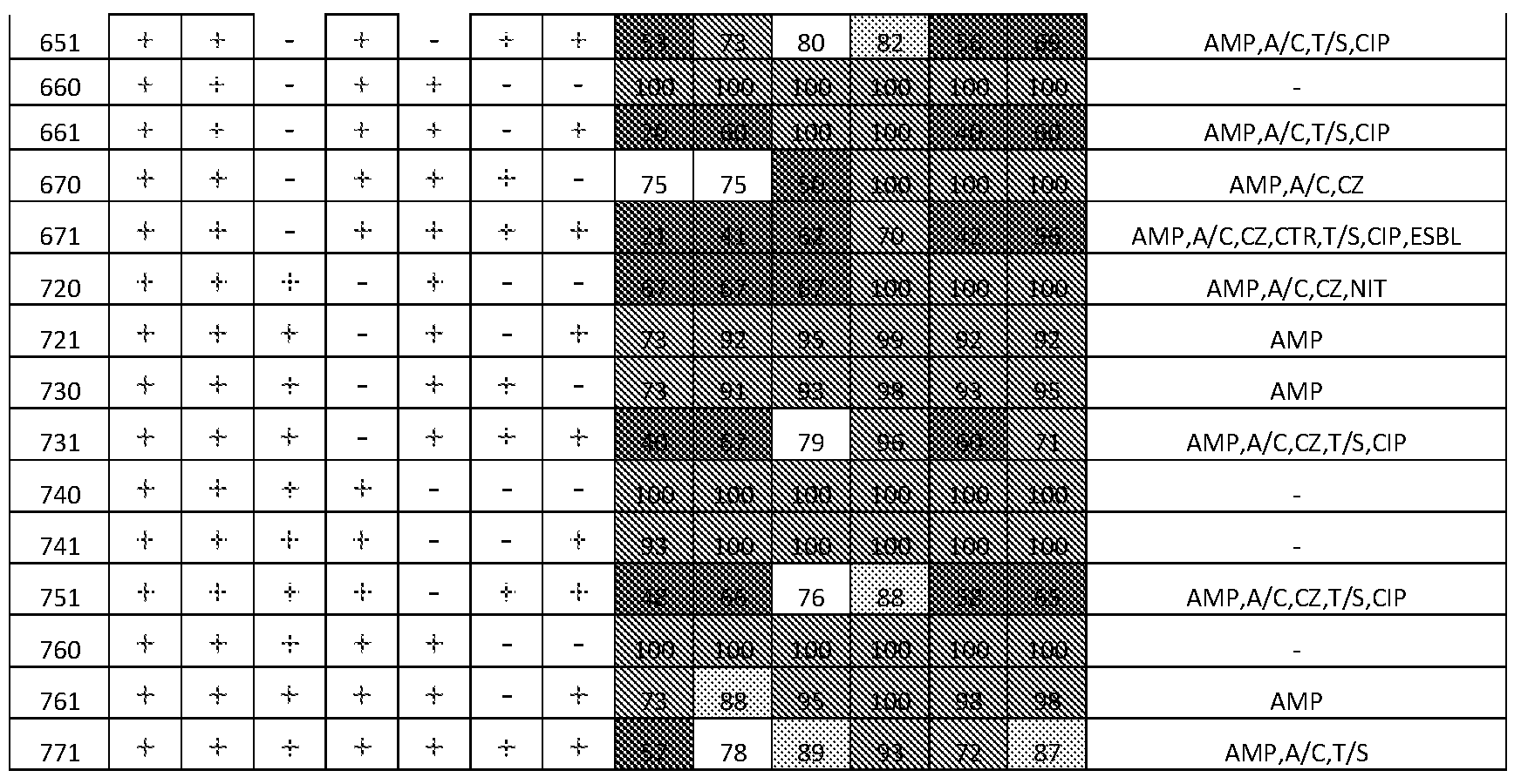
Lookup Table
Positive (+) or negative (-) amplification indicates at the presence or absence of specific SNP. Combination of presence/absence data for all 7 SNP's provides unique 7-type (first column on the left). Each 7-SNP type is assigned the probability of an isolate that belongs to it to be sensitive or resistant to different antibiotics on a scale from 0 to 100, with 0 being completely resistant and 100 being completely sensitive. If 90-100% bacteria that belong to this 7-SNP type are sensitive to particular antibiotic, the respective cell in the Lookup Table is colored green, and this antibiotic is recommended to be used for treatment; pale green indicates 80-90% sensitivity level, and treatment is allowed too. Yellow (75-80%) and orange (70-75%) indicate that treatment is still allowed but with caution, and switching to a different antibiotic is recommended. Red indicates that more than 30% of bacteria are resistant to this antibiotic, and the latter should be rejected as a choice for treatment. Six representatives of most widely used classes of antibiotics are listed in the Table: A/C, amoxicillin/clavulanate, CZ, cefazolin (1st generation cephalosporin), CTR, ceftriaxone (3rd generation cephalosporin, bacteria resistant to it tend to produce ESBL's), T/S, trimethoprim sulfamethoxazole, CIP, ciprofloxacin (fluorquinolones), and NIT, nitrofurantoin.
Lookup Table 1 : Lookup table for cumulative antibiotic susceptibility of individual 7- types.

Primers are designed for both single-plex (8 tubes; suited for one-channel, no-probe platforms) and multiplex (<8 tubes; for multi-channel/-probes platforms) kit options.
Specificity of the designed SNP-specific primers are evaluated first using selected representatives of clonotypes included in the assay (1-3 isolates each, up to 100 total) to ensure they prime as expected. Primers that pass these initial screens are validated more extensively by using a wide range of clinical isolates (up to 2,000), representing the BFC- covered clonotypes and more, to rigorously test primer specificity and sensitivity. Bacterial DNA isolation from urine was performed by using commercial methods, based on chelex beads, pore filters, or columns.
Although the primer testing results can be evaluated by naked eye (based on turbidity) or using UV-light (SYBR-Green dye), additional instruments are an ESE-Quant Tube Scanner (Qiagen, Inc). Additionally, a Genie II™ (Pro-Lab Diagnostics, Inc.) is a multifunctional, one-channel isothermal platform accommodates two 8-well strips for single-plex reactions, or a Rotor-Gene Q instrument for RT-PCR tests.
The disclosure provides a rapid molecular diagnostics test kit that allows high- resolution clonal (sub-species) typing of E. coli that cause urinary tract infection (UTI) - cystitis, pyelonephritis, and urosepsis. The clonotyping test is used for prediction of antibiotic resistance of the bacteria and will be based on a proprietary compilation of clonotyping markers -fumC and fimH gene loci, and a binary SNP-typing technology. In a preferred embodiment, PCR 8-12 tube strips are functionalized for simultaneous detection of the presence/absence of multiple single nucleotide polymorphisms (SNPs) within fumC/fimH loci. These specific SNPs set comprise Binary Barcode Combination (BBC) that allows
identification of a large number of E. coli clonal lineages (clonotypes) based on their unique sequence footprints (see Figure 1).
We have designed and validated (by PCR) BBC comprised of 7 SNPs that can be used in 8-tube strip single-plex configuration and allows separation of E. coli on 56 clonotypes. These are the E. colifumC gene (SEQ ID NO. 3) at least at positions 63, 248, 380, and combinations thereof, and the fimH gene (SEQ ID NO. 4) at least at positions 108, 162, 233, 483, and combinations thereof. The BBCs are used in a rapid test based on Real-Time (RT-) PCR or isothermal (isoT) amplification instrumentation platforms in on-site clinical laboratories in/nearby emergency rooms, urgent care clinics and hospitals. The test is performed directly on the clinical specimen (patient urine), in a timely (<30 min) manner.
Preferably, the multiplex PCR reaction detects compilations of SNPs at the E. coli fumC gene (allele 4) wherein with the position of a preferred forward primer highlighted in bold letters with SNP position indicated with a number in parentheses.
fumC (allele 4) 469 bp fragment 40.
5' -
CGAGCGCCATTCGGCAGGCGGCGGATGAAGTACTGGCAGGACAGCATGACGACGAATTCCCGC(63lTGGCT
ATCTGGCAGACCGGCTCCGGCACGCAAAGTAACATGAACATGAACGAAGTGCTGGCTAACCGGGCCAGTGAA TTACTCGGCGGCGTGCGCGGGATGGAACGTAAAGTTCACCCTAACGACGACGTGAACAAAAGCCAAAGTTCC AACGATGTCTTTCCGACGGCGATGCACGTTGCGGCG(248)CTGCTGGCGCTGCGCAAGCAACTCATTCCGCAG CTTAAAACCCTGACACAGACACTGAGTGAAAAATCGCGTGCATTTGCCGATATCGTCAAAATCGGTCGAACCC ACTTGCAGGACGCCACGCCGCTAACA(380)CTAGGGCAGGAGATTTCCGGCTGGGTAGCGATGCTCGAGCAT AATCTCAAACATATCG AATACAGCCTG CCTC ACGTAG CG G AACTG G CTCTG G G CG GTACAGCG GTG G GTACT GGACTAAATACCCATCCGGAATATGCGCGTCGCGTAGCAGATGAACTGGCAGTCATTACCTGTGCACCGTTTG TTACCGCGCCGAACAAATTTGAAGCGCTGGCGACCTGTGATGCCCTGGTCAGGCGCACGGCGCATTGAAAGG GTTGGCTGCGTCACTGATGAAAATTGCCAATGATGTCCGCTGGCTGCTCTGGCCCGCGCTGCGGAATTGGTGA AATCTCAATCCCGGAAAATGAGCCGGGCAGCTCAATCATGCCAGGAAAGTGAACCCAACACAGTGCGAAGCA TTAACCATGC CTGCTGTCAGGTGATGGGGAACGACGTGGGATCAACATGGGTGGCGC rCCGGTAACTTTG AACTGAACGT rTCCGTCCGATGGTGATCCATAATTTCCGCAATCGGTGCGCTTGCTGGCAGATGGCATGGAA AGTTTCAACAAACACTGTGCAGTGGGCATTGAACCGAATCG -3' (SEQ ID NO. 3)
The fimH gene (SEQ ID NO. 4) wherein with the position of a preferred forward primer highlighted in bold letters with SNP position indicated with a number in parentheses fimH (allele 27) 489 bp fragment30
5'-
TTCG CCTGTAAAACCG CCAATG GTACCG CTATCCCTATTGG CG GTG G CAG CG CCAATGTTTATGTAAACCTTG C
GCCCGTCGTGAATGTGGGGCAAAACCTGGTCGTG(108)GATCTTTCGACGCAAATCTTTTGCCATAACGATTA TCCGGAAACCATTACAGAC(162)TATGTCACACTGCAACGAGGCTCGGCTTATGGCGGCGTGTTATCTAATTT TTCCGGGACCGTAAAATATAG(233)TGGCAGTAGCTATCCATTTCCTACCACCAGCGAAACGCCGCGCGTTGT TTATAATTCGAGAACGGATAAGCCGTGGCCGGTGGCGCTTTATTTGACGCCTGTGAGCAGTGCGGGCGGGGT GGCGATTAAAGCTGGCTCATTAATTGCCGTGCTTATTTTGCGACAGACCAACAACTATAACAGCGATGATTTCC AGTTTGTGTGGAATATTTACGCCAATAATGATGTGGTGGTGCCTACTGGCGGCTGC(483)GATGCTTCTGCTC GTGATGTCACCGTTACTCTGCCGGACTACCCTGGTTCAGTGCCGATTCCTCTTACCGTTTATTGTGCGAAAAGC CAAAACCTGGGGTATTACCTATCCGGCACAACCGCAGATGCGGGCAACTCGATTTTCACCAATACCGCGTCGT TTTCACCCG CGCAGGGCGTCGG CGTACAGTTG ACG CG CAACG GTACG ATTATTCCAG CG AATAACACG GTATC GTTAGGAGCAGTAGGGACTTCGGCGGTAAGTCTGGGATTAACGGCAAATTACGCACGTACCGGAGGGCAGG TGACTGCAGGGAATGTGCAATCGATTATTGGCGTGACTTTTGTTTATCAATAA - 3' (SEQ ID NO. 4)
Example 1
This example shows a method for using the disclosed kit to test a sample for clonotyping an E. coli sample to determine antibiotic susceptibility.
1) Prepare 8 master mixes for qPCR (see Tables 1 and 2)
2) Add Ιμΐ of template DNA to 9 μΐ of master mix solution of each 8 reaction
3) Run qPCR reaction of Rotorgene® Q instrument as follows:
1. 3 min denaturation at 95 °C
2. 5 sec at 95 °C
3. 5 sec at 57 °C
4. 10 sec at 72 °C (acquisition at green channel)
5. Repeat steps 2-4 40 times
6. Perform HRM (high resolution melt) over 70-90 °C range
4) Analyze resulting curves and melting peaks to assign positive or negative result
5) Determine resulting 7-type and lookup the respective CAS in the Lookup Table.
Table 2: Master mix for 7-type qPCR

Example 2
This example illustrates a high-resolution fumC/fimH (CH) clonotyping scheme for E. coli based on sequence variations within these highly-variable omnipresent genes for
fumarase and fimbrial adhesin of E. coli, respectively. We correlated CH clonotypes with antibiotic susceptibility profiles among 1,600 urine E. coli isolates from clinical microbiology laboratories in Seattle (Group Health, UW, Harborview, and Children's Hospitals),
Minneapolis (VA Medical Center), and Munster, Germany (University Clinic).
A total of 222 distinct CH clonotypes were identified, with the top 20 clonotypes comprising two-thirds of isolates. Importantly, within each of the major clonotypes the prevalence of resistance differed by 2-fold (higher or lower) from the overall population value for at least one antimicrobial. Additionally, clonotype resistance was similar (stable) across all laboratories.
We next determined how knowledge of cumulative clonotype vs. overall (species) antibiogram could reduce potential 'drug-bug' mismatches during empirical antibiotic selection. We used the IDSA-recommended 80% susceptibility cutoff level to allow the use of specific antibiotic for each clonotype. Among the top 4 antibiotics used against E. coli - fluoroquinolones (CIP), trimethoprim- sulfamethoxazole (T/S), cefazolin (CZ), and amoxicillin-clavulanate (A/K) - the drug allowance coverage is 48-79% and potential decrease in drug-bug mismatch 45% to 78%, if the empirical choice is guided by the clonotyping, not species identity alone (Table 3).
Table 3
Table 1. Decrease in potential 'drug-bug' mismatch based
on CH clonotyping of E. coli (as % resistant in 'Allowed' for
treatment vs. total resistant).
Clonotype-based treatment choice
Anti- Total n/ _ .—— -——— —— ;
biotic resistant % ReJected / % Allowed / Improve- % Resistant % Resistant ment
T/S 26.9% 42.0 / 50.1 58.0 / 10.1 62.4%
A/K 25.5% 51 .9 / 36.5 48.1 / 13.5 46.8%
CZ 19.7% 42.0 / 32.0 58.0 / 10.7 45.4%
CIP 17.1 % 20.6 / 68.7 79.4 / 3.7 78.1 % Example3
This example illustrates implementation of clonal testing in a healthcare community microbiology laboratory. We assessed the presence of E. coli sub-strains ST131 and ST69 (n=619) E. coli positive cultures. Antibiotic resistance of these two clonotypes is distinctive from E. coli in general with greater than 40% resistant to trimethoprim/sulfamethoxazone (TMS) (ST69 and ST131) and fluoroquinolonwa (FQ) for ST131. The tests were conducted to identify ST69 of ST131 genes specific to each clonotype on bacterial DNA isolated from patient urine specimens using RT-PCR instrumentation. The entire test protocol took 45-90
minutes to run and detected down to 102 cfu/ml of urine, with specificity and sensitivity of greater than 95%. The overall prevalence of the two clonotypes was 15% of the total samples with ST131 at 10.2% (63/619) and ST69 at 5.0% (31/619). Table 4 shows the age and gender distribution of the study group by E. coli clonal status and the study group was primarily women. Patients with ST131 infection were generally older (75% were age 60 or older).
Table 4

Resistance to TMS was 15% and resistance to FQs was 11% (Figure 3). Overall, 8% of patients were prescribed antibiotic therapy for which the isolate was resistant (a drug-bug mismatch). However, the treatment mismatch was significantly higher in patients infected with either ST69 of ST131, 17% and 22%, respectively. ST69 and ST131 together comprised 40% (19/48) of patients with drug-bug treatment mismatch. In 18% (113/619) patients, the initial antibiotic treatment course was changed (switched). Treatment switching occurred in 34% of patients infected with ST69 and 41% of patients of those infected with ST131. These two clonotypes together comprised 33% of all treatment switch cases.
Only 20% of patients who were treated with the correct antibiotic has a follow-up encounter. Almost 90% of patients with a drug-bug mismatch had a follow-up. Overall, drug- bug mismatches contributed to 28% of all follow-up encounters. These data show that a correction of the original antibiotic treatment based on fast diagnosis of ST69 of ST131 would potentially reduce overall drug-bug mismatches by 37%, resulting in 2.6 fewer follow- ups per 100 patients. Further, if the disclosed test kit and process were used for all major clonotypes, the reduction in drug-bug mismatch would be 63%, resulting in 4.4 fewer follow- up per 100 patients.
Example 4
This example illustrates setting up a reference database consisting of 2,559 random single-patient clinical E. coli isolates of primarily extra-intestinal origin, at eight international clinical microbiology laboratories. The major source of isolates was urine (67.1%), followed by feces (6.8%) and blood (4.6%), with the rest of isolates originating from wound, abdominal and other swabs of extra-intestinal compartments. Conventional MLST (multi- locus sequence typing) was performed either in full using the standard scheme of 7 MLST loci (adk,fumC, gyrB, icd, mdh, pur A, and recA) (Wirth et al., 2006. Sex and virulence in Escherichia coli: an evolutionary perspective. Mol, Microbiol, 60: 1136-1151.) or as a combination of partial MLST and fumC/fimH typing, as described previously (Tchesnokova et al. 2013. Predictive diagnostics for Escherichia coli infections based on the clonal association of antimicrobial resistance and clinical outcome. /. Clin. Microbiol. 51 :2991- 2999). Some ST complexes, as identified by the eBURST v3 software, were defined operationally as a single 'ST' and were designated based on the ST identified as the cluster's founder (see Table 5) (Feil et al. 2004. eBURST: inferring patterns of evolutionary descent among clusters of related bacterial genotypes from multilocus sequence typing data. /.
Bacteriol. 186: 1518-1530). Such complexes accounted collectively for 17.5% of isolates, with most comprised of minor groups.
Table 5 Composition of septatypes. All 54 septatypes identified within the 2,599 E. coli clinical isolates are listed in the table in the descending order according to their size. Within each septatype two major ST-H subclones and major phylogroups are listed if they comprise at least 5% of a septatype. Homogenous major septatypes which consist of primarily (more than 89%) of one ST-H subclone are in bold.
Major ST (H) subclones Prevalent phylogroups
Septatype
First Second First Second
Nam %
% a Name % b Name % b Name % b Name e b
Major septatypes (>0.5% of the population)
99.
561 »* ST131 (H30) 95 na B2 na < 5
2
97.
620 9.0 ST73 (H9/10) ii < 5 B2 100 na 0
8
73. 15.
530 7.6 ST127 (H2) ST141 (H14) B2 96 na < 5
2 1
ST58 (H32/31) 23. 10. 19.
361 7.0 c ST354 (H58) Bl 50 F
9 8 6
90.
760 6.9 ST95 (H41) 90 < 5 B2 na < 5
7
92.
271 6.7 ST69 (H27) na < 5 D 100 na 0
8
ST88 (H23/27) 25. ST58 23. 24.
371 4.4 d Bl 35 C
2 (H25/27) 3 8
ST58 (H38/86) 34. 13. 62. 14.
360 4.4 ST88 (H39) Bl C
1 2 1 7
ST14 (H27/64) 97.
571 3.8 d ii < 5 B2 100 na 0
1
ST10 (H23/27) 51. 45. 52. 47.
771 3.6 ST95 (H27) A B2
3 8 1 9
93. 93.
721 3.0 ST95 (H15) ST10 (H15) 5.2 B2 A 6.3
9 7
2.6 92.
131 ST12 (H5) Tig) <5 B2 100 na 0 2 7
2.5 43. 15.
531 ST141 (H5) ST491 (H5) B2 94 na < 5
4 1 3
2.5
560 ST131 (H41) 100 < 5 B2 100 na < 5 1
2.3 17.
751 ST10 (H54) 75 ST95 (H54) 8 A 81 B2
9 5
2.2 92.
661 ST73 (H30) Tig ^ "S B2 100 na 0
8 7
2.2 93.
731 ST569 (H5) 90 /■a < 5 B2 na < 5
8 3
2.0 89.
510 ST131 (H22) ST428 (H22) 8.9 B2 100 na 0
9 3
2.0 ST1876 56. 20.
511 ST429 (H20) B2 100 na 0 1 (H20/21) 8 9
2.0 41. 35. 43. 37.
761 ST10 (H30/31) ST95 (H30) A B2
1 8 7 4 7
1.7 28. 44. 24.
351 ST69 (H54) ST58 (H54) 20 D Bl
1 9 4 4
1.4 70. ST636
520 ST372 (H9/12) 8.1 B2 100 na 0 0 3 (H9/75)
1.2 ST394 21. 90.
261 ST69 (H47) 28 D na < 5 1 (H35/47) 9 6
1.1
260 ST38 (H65) 43 ST59 (H41) 30 D 70 F 30
4
0.9 89.
570 ST117 (H97) na < 5 F 92 na < 5 5 2
0.9 ST73
630 ST80 (HI) 84 16 B2 100 na 0 5 (H2/154)
0.9
231 ST38 (H5) 100 na 0 D 100 na 0 1
0.8
171 ST12 (H27) 100 na 0 B2 100 na 0 7
0.5
711 ST676 (H21) 43 ST2551 (H20) 21 B2 100 na 0
3
Minor septatypes (<0.5% of the population)
0.4 ST2711 90. Clade
350 ST405 (H56) 91 9 D 9.1 2 (Hil l) 9 I
0.2
551 ST144 (H54) 57 ST2086 (H57) 43 B2 100 na 0
7
0.1 Clade
330 ST58 (H2) 40 ST2250 (H2) 20 Bl 40 40
9 I
0.1
331 ST648 (H5) 60 ST38 (H5) 20 F 60 na < 5
9
0.1
550 ST2015 (HI 97) 100 na 0 B2 100 na 0
9
0.1
160 ST12 (H41) ST1011
50 25 B2 50 Sh-I 25
5 (H108)
0.1
251 ST38 (H54) 25 ST69 (H54) 25 D 75 F 25
5
0.1
321 ST58 (H15) 25 ST648 (H15) 25 F 50 Bl 25
5
0.1 ST1882
370 ST991 (H123) 25 25 D 75 Bl 25
5 (H123)
0.1
651 ST706 (H229) 50 ST73 (H54) 50 B2 100 na 0
5
0.1
671 ST73 (H27) 25 ST706 (H25) 25 B2 100 na 0
5
0.1 33.
720 ST2622 (H75) 67 ST2474 (H9) B2 100 na 0 1 3
0.1 33. 33. 66. Clade 33.
730 ST73 (H154) ST 1444 (H3) B2
1 3 3 7 I 3
0.0 Clade
300 ST2141 (HO) 50 ST2711 (HO) 50 F 50 50
8 I
0.0
611 ST583 (H21) 100 na 0 B2 100 na 0
8
0.0
710 ST803 (H88) 100 na 0 B2 100 na 0
8
0.0
200 ST59 (HO) 100 na 0 F 100 na 0
8
0.0
121 ST12 (H15) 100 na 0 B2 100 na 0
4
0.0
220 ST73 (H10) 100 na 0 B2 100 na 0
4
0.0
221 ST38 (H15) 100 na 0 D 100 na 0
4
0.0
310 ST399 (H22) 100 na 0 C 100 na 0
4
0.0 Clade
320 4 ST10309 (H9) 100 na 0 T 100 na 0
υ.υ
500 ST636 (HO) 100 na 0 B2 100 na 0
4
0.0
521 ST636 (H15) 100 na 0 B2 100 na 0
4
0.0
660 ST73 (H41) 100 na 0 B2 100 na 0
4
a Percent of the E. coli population within a septatype
b Percent of ST-H subclone of a phylogroup within a septatype
c These are not individual STs but subgroups of several linked STs identified by Eburst software. The ST listed in the table is the primary subgroup founder, whereas the actual composition of this subgroup is as follows (individual STs are listed according their size in descending order; STs that contain only 1-2 isolates are not listed): (1) ST58 (13 isolates out of 50 total within this ST58 (H32/31) subgroup), ST162 (11 out of 50), ST1056 (5 out of 50), ST155 (4 out of 50), ST448 (4 out of 50), ST533 (3 out of 50), etc.; (2) ST224 (14 isolates out of 40 total within this ST58 (H38/86) subgroup), ST297 (12 out of 40), ST58 (3 out of 40), ST155 (3 out of 40), etc.; (3) ST10 (34 isolates out of 50 total within this ST10 (H23/27) subgroup), ST617 (6 out of 50), ST93 (5 out of 50), etc.
d Each of these is a complex of three STs which differ by only one MLST allele out of seven (so called single-locus variants, or SLVs, identified by Eburst software). The ST listed in the table is the primary subgroup founder (identified by Eburst), whereas the actual composition of this subgroup is as follows (individual STs are listed according their size in descending order: (1) ST410 (18 isolates out of 29 total within this ST88 (H23/27) complex), ST90 (6 out of 29), ST88 (5 out of 29); (2) ST404 (50 isolates out of 98 total within this ST14 (H27/64) complex), ST1193 (34 out of 98), ST14 (14 out of 98).
fumC and fimH were selected as the gene targets for development of the SNP-based E. coli clonotyping method because of the previously demonstrated power oifumC/fimH clonotyping to predict ST-based clonal groups and subdivide them into smaller subclones. To guide SNP selection, a proprietary algorithm was used to select seven SNPs that included 3 SNPs iromfumC (positions 63, 248, and 380) and 4 from fimH (positions 108, 162, 233, and 483). The 7-SNP format allowed the test performance in PCR using 8-tube/-well
configuration, with the eighth tube or well used for a control. The 7 SNPs were split into three groups. The first group included three SNPs (fumC-63, fumC-248, fumC-380), the second group another three SNPs (fimH-lOS, fimH-162, fimH-233), and the third group the remaining SNP (fimH-483). Presence of the 1st SNP within a group was scored as 1, presence of the 2nd SNP as 2, and presence of the 3rd SNP as 4, while SNP absence was scored as 0. Each isolate was assigned a score based on the sum of the scores for each SNP group (Table 6). For example, the 101-011-1 binary combination (where T and '0' is SNP presence and absence, respectively) was recorded as "561", 010-111-0 as "270", etc.
Table 6 The septatype index scored. 7 SNPs were divided into two groups of 3 SNPs and one group of 1 SNP. When the SNP was present at positions 1 or 4, it was scored a
numerical value of 1, at positions 2 or 5 it was scored 2, and at positions 3, 6 or 7 it was scored 4. Scores were summed within groups resulting in a three-digit septatype index.
Gene fumC fimH
SNP # 63 248 380 108 162 233 483
SNP position 1 2 3 4 5 6 7
Numerical key assigned if SNP is present 1 2 4 1 2 4 1
Numerical key assigned if SNP is absent 0 0 0 0 0 0 0
Example A: CH40-30 (ST131) belongs to septatype 561
SNP present? yes no yes no yes yes yes
Binary code 1 0 1 0 1 1 1
Numerical key assigned 1 0 4 0 2 4 1 septatype number 5 6 1
Example B: CH35-27 (ST69) belongs to septatype 271
SNP present? no yes no yes yes yes yes
Binary code 0 1 0 1 1 1 1
Numerical key assigned 0 2 0 1 2 4 1 septatype number 2 7 1
For the 7-SNP clonotyping test, 7 primer pairs were designed and optimized to detect the SNPs, with uidA locus-specific primers added as an E. coli positive control (Table 7). The performance of the primers was tested using isolates known to contain fumC and fimH variant alleles with any polymorphisms in the primer-binding region identified in our reference set of 2,559 E. coli (Table 7). Primer sequences and product length of seven SNP-specific reactions and uidA E. co/z'-specific control.
„. T_ _ . ._, 1.„ Product length
SNP name Primer sequence (5 -3 ) ^ ^ b
fumC-63 (F) AGCATGACGACGAATTCCTGC
(R-S) GTCGTCGTTAGGGTGAACTTT
149
SEQ ID NO. 5
(R-M) same as J mC-248 (R-S) 423
(F) ACGGCGATGCACGTTGCGTCG
SEQ ID NO. 7
fumC-248 (R-S) AGTTCCGCTACGTGAGGCAGG
239
SEQ ID NO. 8
(R-M) same as (R-S) 239
(F) CCGGAAATCTCCTGCCCAAGC
f mC-380 c (R-S) CATTCCGCAGCTTAAAACCCT 121
(R-M) same as (R-S) 121
(F) GTGGAGCAAAACCTGGTCTTG
fimH-108 (R-S) AGGGAAAGGATAGCTACTGCC 168
(R-M) same as fimH-233 (R-S) 241
(F) TATCCGGAAACCATTACAGAC
fimH-162 (R-S) same as fimH-233 (R-S) 187
(R-M) same as fimH-233 (R-S) 187
(F) TTCCGAGACCGTAAAATATAG
fimH-233 (R-S) TCAAATAAAGCGCCACCGGCC 116
(R-M) same as fimH-483 (R-S) 396
(F) GTGGTGGCTACTGGCGGCAGC
fimH-483 (R-S) TCTGCGGTTGTGCCGGATAGG 146
(R-M) same as (R-S) 146
(F) TCTTGCCGTTTTCGTCGGTA
uidA (R-S) CACGCCGTATGTTATTGCCG 129
(R-M) same as (R-S) 129 a R-S, reverse primer used in singleplex reaction; R-M, reverse primer used in multiplex reaction; singleplex refers to both conventional PCR and qPCR.
b Length of PCR product for singleplex and multiplex reaction is denoted in respective rows. c Primers specific for the wmC-380 SNP are designed for the antisense DNA strand; all other primers are designed for the sense DNA strand.
Testing was done in three different formats: singleplex or multiplex conventional PCR, or singleplex qPCR. Singleplex 7-SNP typing consisted of 8 independent reactions using the above-described primers. Multiplex 7-SNP typing used some of the primer pairs combined into three total reactions, as shown in Table 7. The first triplex reaction contained three SNP- specific forward primers (fumC-63 , fumC-248, and fimH-483) and two common reverse primers (one each for fumC and fimH). The second triplex reaction contained the uidA primers, two SNP-specific forward primers (fimH-108 and fimH-162), and a common reverse primer for fimH. The duplex reaction contained the same two primer pairs as used in singleplex PCR (for/wmC-380 and fimH-233).
Of the 582 isolates that represented fumC/fimH diversity that were used to test primer performance, 310 underwent full 7-SNP typing test (including the uidA control), which was done using a single format for 180 isolates, two formats for 121 isolates, and all three formats
for 24 isolates. The remaining 272 test isolates were screened using primers for individual SNPs. If the SNP test failed to detect correct septatype of a particular fimH or fumC allele, the test was repeated to confirm or refute this failure.
Test reactions used JumpStart PCR master mix (Sigma) and the following conditions: 2 min initial denaturation at 95 °C, followed by 27 cycles of 15 seconds at 95 °C, 15 seconds at 57 °C, and 30 seconds at 72 °C. Amplification products were analyzed by 2% agarose gel electrophoresis. The qPCR 7-SNP typing test was performed by real-time quantitative PCR on a Rotor-Gene® Q MDx instrument (QIAGEN) using the SYBR-Green PCR Kit
(QIAGEN). Unless stated otherwise, the qPCR reaction conditions were as follows: 3 min at 95 °C, followed by 30 cycles of 5 seconds at 95 °C, 10 seconds at 57 °C, and 10 seconds at 72 °C, with signal acquisition at the elongation step. As in the singleplex test, the qPCR 7-SNP typing test consisted of 8 independent reactions.
7-SNP typing of E. coli from urine samples. To validate the performance of the 7- SNP typing test, 160 random urine samples were obtained from the clinical microbiology laboratory at the Harborview Medical Center (Seattle, WA). Each sample was processed as follows: 1 mL urine was added to 50 \\L of a 20% suspension of Chelex (BioRad) in sterile water and centrifuged 1 min at 12,000 rpm. The pellet with Chelex was re-suspended in 100 \\L of sterile water, heated for 5 min at 96 °C and centrifuged again, with the supernatant used as template for the qPCR-based 7-SNP typing test. The load of E. coli in urine was determined from the uidA threshold cycle based on a standard calibration curve (there were no uidA signal in sterile urine samples). In parallel, 10 \\L of urine were plated on McConkey agar to detect the growth of E. coli. Cultured E. coli isolates were further subjected to clonotyping as a control.
Susceptibility to seven antibiotics was determined using a standardized disk diffusion method according to Clinical and Laboratory Standards Institute guidelines.
Statistical analysis was performed with a Simpson's diversity index was calculated using the formula: D = l-∑ [n*(n-l)/N*(N-l)], where n is the number of E. coli isolates in a particular clonotype, and N is the total number of isolates. Comparisons of proportions were tested using a two-tailed Fisher's exact test. Bacterial loads detected in urine by the 7-SNP typing test vs culture were compared using the two-sided paired t-test.
Example 5
This example shows the results of a computer-based analysis of high-resolution of E. coli clonotypes using the/wmC and z'mH nucleotide sequences of 2,556 isolates from eight
clinical microbiology labs in the US, Germany, Poland and Russia. According to the analysis, the 2,559 isolates were divided among 54 unique binary septatypes (Figure 1A). Six 'major' septatypes included more than 5% of isolates and comprised 47% of all isolates. Twenty- three 'intermediate' septatypes each included 0.5-5% of isolates and together comprised 50% of the isolates. The remaining 25 septatypes were 'minor' with each including <0.5% of isolates. The overall diversity of major and intermediate septatypes was similar to that of MLST (Figure IB), which identified 6 major and 19 intermediate STs of the same size as defined by 7-SNP typing. Although there were nearly three times as many STs as septatypes (175 vs. 54), this difference was due entirely to the greater number of minor STs (150 in total) that together comprised only 13% of the isolates. This is reflected in comparable Simpson's diversity index values for 7-SNP typing and MLST (0.949 and 0.934,
respectively).
Overall, in 27 septatypes (4 major, 10 intermediate, 15 minor) more than 90% of the isolates (97.2% on average) belonged to a single MLST-based clonal group. Such clonally homogeneous septatypes accounted for 54.1% of all isolates. A total of 17 STs could be predicted by the homogeneous septatypes, including multidrug-resistant ST131, ST69, ST38 and relatively susceptible ST95, ST73, ST14, ST12, ST569, and ST117. Moreover, several major STs like ST131, ST95, and ST73, were split by 7-SNP typing into smaller sub-ST clonal groups. For example, ST131 was split into/zmH-based H30, H41, and H22 subclones (12), ST95 into its H41, H15, and H30 subclones, and ST73 into its H9, H10, and H30 subclones (13).
The remaining 27 septatypes were clonally heterogeneous, i.e., < 90% of each septatype was of the same ST. Such major STs as drug-resistant ST58, ST88, ST354, ST648 and susceptible ST127, ST141, ST10, ST1876 were either split among different septatypes or could not be identified with the high probability.
Thus, binary combinations of seven disclosed SNPs selected from two highly variable genes, fimH and fumC, can genotype E. coli isolates with high resolution by splitting them into a large number of clonotypes. In most isolates, 7-SNP typing predicted the ST and, often, the sub-ST of origin with high accuracy. Accordingly, the disclosed clonotyping test described herein is a useful tool for both epidemiological analysis and clinical diagnostics.
Example 6
This example provides results providing using the disclosed clonotyping tests for determining antibiotic resistance and susceptibility. 7-SNP clonotyping sorted isolates into groups with distinctive antibacterial susceptibility profiles. In the total E. coli study
population, the prevalence of resistance to six antibiotics most commonly used for empirical treatment UTI was as follows: amoxicillin/clavulanate and trimethoprim/ sulfamethoxazole, 28% each; cefazolin, 23%; ciprofloxacin, 21%; nitrofurantoin, 11%; and ceftriaxone, 9%.
For each major and intermediate septatype, the prevalence of resistance to at least one antibiotic was significantly lower ('susceptible' septatype) or higher ('resistant' septatype) than in the total population (Figure 2). Several prominent multi-drug resistant STs were split by 7-SNP typing into smaller clonal groups that had more distinct antimicrobial resistance profiles than both the average and the corresponding ST (Table 6). For example, the most dominant multi-drug resistant clonal group in E. coli, ST131, was split into 3 septatypes. Septatype 561 (corresponding to ST131-H30 subclone) was highly resistant to ciprofloxacin (90.6%), while septatype 560 (ST131-H41) and 510 (ST131-H22) were almost entirely sensitive. At the same time, both 561 and 560, but to a much lesser extent 510, were highly resistant to trimethoprim/sulfamethoxazole. In ST69 (aka clonal group A) that is notorious for its resistance to trimethoprim/ sulfamethoxazole, 7-SNP test identified septatype 351 that is almost entirely resistant to ciprofloxacin and 261 that is highly susceptible to most antibiotics, including trimethoprim/sulfamethoxazole. Similar splits that were informative from a resistance perspective were obtained by the 7-SNP test applied to ST58.
Overall, relative to the species average, the proportion of either resistant or susceptible septatypes was the largest for ciprofloxacin, for which 92% of isolates belonged to either resistant (34%) or susceptible (59%) septatypes (Figure 3), with the average resistance prevalence in the former group being 51% and in the latter one only 4.4%. For other antibiotics, differences in the proportion of resistant and susceptible isolates were not as pronounced, but for all antibiotics except nitrofurantoin, most of the isolates were in septatypes with a resistance profile distinct from the average. Also, for all antibiotics except amoxicillin/clavulanate, the average resistance in the susceptible septatypes was below 10%.
Overall, 7-SNP typing performed well in splitting E. coli clinical isolates into clonotypes with distinctive antibacterial resistance profiles.
7 pairs of SNP-specific primers and a pair of Mzd!A-specific primers (for detection of E. coli) (Table 7) that, in conventional or quantitative PCR, yielded the predicted band sizes (Figure 5A and 5C) or signal (Figure 5B), respectively, against the target DNA. We evaluated the ability of the SNP primers to recognize the corresponding SNPs in the background of 582 isolates with 60 fumC alleles and 156 fimH alleles that encompassed all naturally-occurring combinations of base pairs that are variable within the primer- annealing regions (see Example 4).
In only a few of the variable fumC or finiH alleles, the presence/absence of the targeted SNP could not be identified correctly by PCR, and these error-causing alleles were very rare in the reference set of 2,559 isolates. The total rate of erroneous 7 SNP typing test due to at least one of the 7 SNPs not detected correctly was projected to be only 2.1%.
To confirm the specificity of designed primers towards E. coli, we additionally tested them on isolates of various Enterobacteriaceae species: Klebsiella pneumoniae (10 isolates), Klebsiella oxytoca (4), Citrobacter freundii (5) and Citrobacter koserii (2), Enterobacter aerogenes (12), Proteus mirabilis (6), Pseudomonas aeruginosa (3), Serratia marsenscens (3) and Morhanella morganii (5). Both the SNP-specific reactions and the uidA control produced negative results in all non-E. coli isolates with the exception of one isolate designated by 16S typing as E. aerogenes.
Thus, the newly designed SNP-specific primers demonstrated a robust ability to distinguish the targeted SNPs despite some background sequence variation in the primer- annealing regions.
The disclosed 7-SNP typing test can identify E. coli clonotypes directly from urine specimens. In addition to the validation using pure bacterial cultures, the qPCR-based 7-SNP typing test was performed on bacterial DNA obtained from clinical urine samples - 77 were positive and 83 negative for E. coli as determined by culture. In the E. coli culture-negative samples, the 7-SNP test was positive in 2 urines (98% specificity). In E. coli culture-positive urines, 74 were positive in the 7-SNP typing test, (96% sensitivity), and the identified clonotype matched that of the cultured isolate in all but one sample. Furthermore, the 7-SNP test was positive and detected the same clonotype as was isolated from culture in all 49 urine samples that had clinically significant levels of E. coli (at least 104 cfu/ml). Overall, there was a strong correlation between the bacterial load determined by culture and that predicted by the qPCR test based on the E. co/z'-specific uidA probe (Figure 4). In alab is 11 three qPCR- negative but culture-positive urine samples, the bacterial load was low - 102 cfu/ml (in two samples) or 103 cfu/ml (in one sample). Overall, multi-is. coli specimen occurred in <1%.
Thus, the disclosed 7-SNP typing test can detect E. coli in urine samples and reliably identify the corresponding clonotype, performing essentially as well as the standard culture procedure quantitatively.
Example 7
This example provides the results of a clinical field trial showing the performance of the disclosed 7 SNP-based clonotyping test (CLT test) conducted at an HMO (health maintenance organization, Group Health Urgent Care). The disclosed test kit and process was
conducted on urinalysis-positive urine samples on-site at the HMO urgent care clinical laboratory. All urine samples obtained at Urgent Care facility were tested by the 7-SNP-based CLT(clonotyping) test and by a standard E. coli culturing test. Out of total of 147 urine samples, 90 were positive in urinalysis test, according to the HMO facility. Out of those, 35 samples were positive for E. coli culture, and 34 of those were positive in CLT test (94%). Additionally, 2 samples were positive for the CLT test, but culture was negative for E. coli. Out of 57 urinalysis -negative samples only 1 was positive for E. coli culture and CLT test both, and 1 more sample was positive only for CLT test. In summary, a positive CLT test was an excellent predictor of positive E. coli growth (34/36 vs 3/111, P<.0001), and positive urinalysis was excellent predictor of both positive E. co/z'_growth (35/90 vs 1/57, P<.0001) and positive CLT test (35/90 vs 2/57, P<.0001). Table 8 summarizes the results achieved. Table 8
Urinalysis
Total
(N = 147) Positive Negative
(N = 90) (N = 57)
Total 3 36 (24.5%) 35 (39%) 1 (1.8%)
Positive E. coli
growth Positive CLT b 34 (94.4%) 33 (94.3%) 1 (100%)
Negative CLT c 2 (5.6%) 1 (3.0%) 0 (0.0%)
Total 3 111 (75%) 55 (61%) 56 (98%)
Negative E. coli
growth Positive CLT b 3 (2.7%) 2 (3.6%) 1 (1.8%)
Negative CLT c 108 (97%) 109 (98%) 55 (98%)
In the period from April until December 2015, two technicians worked at the urgent care clinical lab performing the CLT test on urine samples. They timed how long it took them to process each sample and to at which cycle (1 cycle = 1 min) of the reaction they were able to tell if there was E. coli DNA in the sample, how much, and what septatype. The timing of the reaction was analyzed in Table 9 below Figure 6. Figure 6 shows the time it took for a positive result in each case against the actual load of E. coli in urine determined by the disclosed SNP-7 test kit and process (N = 177 urine samples analyzed here). Using this test we detected as low as 102 DNA copies/mL, whereas the standard culturing technique in the
same HMO urgent care lab (and other clinical labs as well) detected only 103 cfu/ml. A clinically significant level is considered to be 104 cfu/ml and higher. Figure 6 shows a majority of clinically significant samples were well below a 22-minute cutoff. When combined with an 8 min-long 1st sample preparation step would constitute about 30 minutes to run the whole test.
There were few samples from the high-load group that required, surprisingly, longer time for a positive answer (circled in red on the Figure). Some of them were so-called "dirty" samples in that they contained additional substances that interfered with the PCR reaction, making the read-out difficult, thus requiring longer time to process.
Table 9
Percentiles:
CLT reaction time in Rotorgene® Q Mean ± SD Min ÷ Max
25%, 50%, 75% to tell if: (min) (min)
(min)
A sample is E. co/i-positive >102 copies DNA
19.3 + 4.1 11 ÷ 32 16, 18, 23 per 1 mL of urine (N = 177)
A sample is E. co/i-positive >104 copies DNA
18.1 + 3.3 11 ÷ 27 16, 18, 20 per 1 mL of urine (N = 146)
Septatype for any E. co/i-positive sample (N =
23.8 + 3.9 16 ÷ 35 21, 23, 27
177)
Septatype for >10Λ4 E. co/i-positive sample (N
22.7 + 3.3 16 ÷ 35 21, 22, 24
= 146)
The procedure used was: Step 1 : initial processing_of urine sample using a Chelex procedure. Briefly, the urine sample was treated with Chelex beads to isolate DNA. This step on took 8 min on average. Step 2: ® on Rotorgene Q instrument (60 seconds per cycle).
qPCR, can vary in length depending on what is the load of bacteria in the initial sample (cfu/mL), which translates into the load of E. coli DNA in the Chelex-purified sample
(copies/mL). The presence of E. coli can be detected as early as after 11 min (11+8=19 min for whole test), with average detection time 19.3+4.1 min (27.3 min for the whole test).
The determination of clonotype for E. coli required an additional 3-5 min on average because the reliability of the result depended on how advanced the reaction curves were.
Time to tell positive results also varied depending on the "dirtiness" of the urine specimen. "Dirtiness" of the sample includes presence of cell debris, high protein, mucus etc., as well as
chromogenic substances that can interfere with either PCR reaction or fluorescent read-out.
There was a correlation detected between E. coli levels detected between standard culture tests (taking 2-3 days) and the disclosed SNP-7 test. 327 samples positive for either CLT test or culturing or both are plotted in Figure 7. The size of the bubble represents the number of samples in each group. The straight line represents the fitted values for a simple linear regression on all samples with valid data (N = 736). Red lines show the cutoff for clinically-significant levels of bacterial load.
In total, 750 urinalysis-positive samples from the HMO urgent care lab were processed in CLT test and standard culturing test. Out of those, 409 were negative for E. coli presence both in CLT test and in culture. 14 more samples were excluded from further analysis due to no data logged on the E. coli load. The rest, 327 culture- and CLT-test-positive samples, are plotted in Figure 7. These 327 samples exhibited strong correlation between the E. coli levels determined in CLT test and in culturing. From the simple linear regression, we estimate that with each 10-times (logio=l) increase in the CLT-determined level of E. coli the increase in E. coli growth will be on average 9.8 times (CI 95% from 9.5 to 10.0, P<.0001). This means that there is an extremely good correlation between the E. coli levels in urine detected by both methods. Overall, when comparing the results of CLT test to the standard culture test, the sensitivity of the CLT test was 98.4% for all detection level and 100% for detection of E. coli in urine at the level considered clinically significant (104 cfu/ml and above).
The E. coli clonotypes identified in the field study (out of 750 samples, see above) were compared to a reference collection of E. coli to (1) compare the clonotypes distribution, (2) compare the overall resistance profile, and (3) use the reference collection to predict if antibiotics can or cannot be allowed for use in each case in the field study (as a Lookup Table). Further, the prediction from (3) was compared to actual resistance of individual isolates and to clinical data regarding the prescription of antibiotics in each case. From this we can see if the implementation of the CLT test would improve the choice of antibacterial therapy.
A reference collection of E. coli included 1227 isolates that were from the same HMO (only from Group Health), only from urine samples, unique (mostly one isolate per patient; 10 patients had mixed E. coli cultures with different septatypes/clonotypes and antibiotic resistance profiles), and isolated in the period from 2010-2013. Both septatypes/clonotypes and antimicrobial profiles were identified.
The antibiotic profiles of major septatypes/clonotypes from the reference E. coli collection (Lookup Table) is provided in Table 10.
Table 10 Lookup table used

In total, 1227 E. coli isolated from the HMO (Group Health) clinical lab in 2010-2013 were analyzed. All had their clonotype type determined and resistance to antibiotics tested. Septatypes were deduced from CH types (clonotypes). The average resistance across isolates within each septatypes is given in Table 10. Red indicates resistance >30%. Yellow: more than 15% but less than 30%. Green: less than 15%.
Out of 750 samples, 308 isolates from 306 samples had unique clonotypes (2 patients had mixed culture in urine). Out of 308 isolates, 303 (98.4%) belonged to septatypes that had been described previously for the reference set of E. coli. Altogether, 308 isolates were distributed into 36 septatypes, of which 32 were previously described, and 4 were newly identified.
More than 98% of E. coli - positive urine samples were infected with bacteria of a previously described septatype, which allows for clone-based prediction of antibacterial resistance based on a reference Lookup Table. In case of a new (unknown") septatype, the species-specific (was overall for E. coli) resistance is used for prediction of an antibiotic.
When comparing antibiotic resistance in E. coli isolates from this Field Trial with the Reference Lookup-Table resistance, Table 11 shows the overall resistance levels were comparable for all antibiotics, although there was a significant increase in resistance to Fluorqinolones (P<.05). For clone-based prediction of bacterial resistance of field study isolates we used a 15% resistance as a cutoff for rejection of an antibiotic.
Table 11


For antibiotic prescriptions, Table 12 shows a total of 750 cases were analyzed; 291 patients had E. coli identified in urine by CLT test, confirmed by culture results. Overall, 82% of patients positive for E. coli in urine (236/291) were prescribed antibiotics at the day
of the visit. The most often prescribed group of antibiotics were fluorquinolones (51.7%), followed by Bactrim (28.4%).
Table 12

CLT test and culturing, and an antibiotic was prescribed around the index visit day, in 41 (17.4%) cases, the E. coli isolate was resistant to the prescribed antibiotic. This is called a drug-bug mismatch. The rate of drug-bug mismatches did not differ significantly from the overall resistance rate to this antibiotics, confirming that the choice of the therapy was indeed most likely empirical. This is shown in Table 13.
Table 13


For further analysis we included all 291 cases when E. coli was identified both by disclosed clonotyping test kit and by culture, including 236 cases where antibiotic was prescribed at the visit day, and the rest 55 cases. Based on the identified clonotype we assigned to each case a value for each antibiotic - if the resistance in this clonotype to this antibiotic was less than 15%, it was allowed for use; otherwise it was rejected. From the Table 14, only ampicillin group of antibiotics was almost always rejected, based on the clonotype resistance (1.7% allowed cases). For the rest of antibiotics, they were allowed in 34.7% cases for Bactrim (aka, Trim-Sulfa®), 75.6% cases for fluorquionolones.
Table 14


For drug-bug mismatch in antibiotics prescribed based on CLT test, in each case for each allowed antibiotic, we checked if the actual E. coli isolate was sensitive to this antibiotic. Then, for each antibiotic we counted the number of resistant isolates as percentage from total allowed isolates - deducing the drug-bug mismatch rate after the CLT test. For all antibiotics the drug-bug mismatch rate was significantly reduced by using the CLT test results for prediction, with the overall rate reduction from 17.4% to 4.3% - more than 4 times. This is shown in Table 15.
Table 15

Claims
1. A PCR-based test kit and nucleic acid amplification process for identification of multiple clonal sub-species lineages of infectious bacteria, comprising a SNP compilation that forms a BFC (Binary Footprint Code) that predicts antibiotic resistance of the bacteria.
2. The PCR-based test kit and nucleic acid amplification process for
identification of multiple clonal sub-species lineages of infectious bacteria of claim 1, wherein the infectious bacteria is a uropathogenic E. coli causing cystitis, pyelonephritis and urosepsis.
3. A clonotyping method for clonal typing E. coli and predicting antibiotic susceptibility, comprising (a) providing forward primers and reverse primers for at least seven SNPs (single nucleotide polymorphisms) selected from the group consisting of fumC- 63, fumC-248, fumC-380, fimH-162, fimH-233, fimH-483, and fimH-483, (b) measuring the presence or absence of each SNP, and (c) determining antibiotic susceptibility from a Lookup Table.
4. The clonotyping method for clonal typing E. coli and predicting antibiotic susceptibility of claim 3, wherein the Lookup Table is Lookup Table 1.
5. A kit for clonotyping E. coli and predicting antibiotic susceptibility, comprising (a) forward primers and reverse primers for at least seven SNPs (single nucleotide polymorphisms) selected from the group consisting of fumC-63, fumC-248, fumC-380, fimH-162, fimH-233, fimH-483, and fimH-483, and (b) a Lookup Table.
6. The kit for clonotyping E. coli and predicting antibiotic susceptibility of claim 5, wherein the Lookup Table is Lookup Table 1.
7. The clonotyping method for clonal typing E. coli and predicting antibiotic susceptibility of claim 3, wherein step (b) furthers comprises the steps of:
(1) obtaining a sample of infecting bacteria; and
(2) determining the clonal-type of the infecting bacteria by performing multiplex nucleic acid amplification process reactions with SNP-specific primers from binary foot print codes (BFC)-covered clonotypes to determine which SNPs are present or absent.
8. The clonotyping method for clonal typing E. coli and predicting antibiotic susceptibility of claim 7, wherein the sample of infecting bacteria is taken from a bodily fluid source selected from the group consisting of urine, blood, saliva, tears and a skin swipe.
9. The clonotyping method for clonal typing E. coli and predicting antibiotic susceptibility of claim 8, wherein the bodily fluid sample is from urine from a patient suspected of a urinary tract infection.
10. The clonotyping method for clonal typing E. coli and predicting antibiotic susceptibility of claim 9, wherein the urine sample is first fractionated to separate bacterial components from other nucleic acids, ureas and solids from a urine sample.
11. The clonotyping method for clonal typing E. coli and predicting antibiotic susceptibility of claim 7, wherein the fractionated bacteria are then lysed to obtain bacterial nucleic acid for further analysis.
12. The clonotyping method for clonal typing E. coli and predicting antibiotic susceptibility of claim 7, wherein the multiplex PCR reactions investigate SNPs within a gene locus, wherein the gene is selected from the group consisting of fimbrial adhesion (jimH), fumC, adk, gryB, icd, mdh, pur A, recA, and combinations thereof.
13. The clonotyping method for clonal typing E. coli and predicting antibiotic susceptibility of claim 12, wherein the multiplex PCR reaction utilizes primers to find SNPs in the fimH gene comprising 3 ' -CACTCAGGGAACCATTCAGGCA-3 ' (SEQ ID NO. 1) and 5 ' -CTTATTGATAA ACAAAGTC AC-3 ' (SEQ ID NO. 2).
Priority Applications (2)
| Application Number | Priority Date | Filing Date | Title |
|---|---|---|---|
| CA2977984A CA2977984A1 (en) | 2015-02-26 | 2016-02-26 | Process and kit for predicting antibiotic resistance and susceptibility of bacteria |
| EP16756508.4A EP3262198A4 (en) | 2015-02-26 | 2016-02-26 | Process and kit for predicting antibiotic resistance and susceptibility of bacteria |
Applications Claiming Priority (4)
| Application Number | Priority Date | Filing Date | Title |
|---|---|---|---|
| US201562121481P | 2015-02-26 | 2015-02-26 | |
| US62/121,481 | 2015-02-26 | ||
| US201662279643P | 2016-01-15 | 2016-01-15 | |
| US62/279,643 | 2016-01-15 |
Publications (1)
| Publication Number | Publication Date |
|---|---|
| WO2016138471A1 true WO2016138471A1 (en) | 2016-09-01 |
Family
ID=56789969
Family Applications (1)
| Application Number | Title | Priority Date | Filing Date |
|---|---|---|---|
| PCT/US2016/019922 WO2016138471A1 (en) | 2015-02-26 | 2016-02-26 | Process and kit for predicting antibiotic resistance and susceptibility of bacteria |
Country Status (4)
| Country | Link |
|---|---|
| US (1) | US9970063B2 (en) |
| EP (1) | EP3262198A4 (en) |
| CA (1) | CA2977984A1 (en) |
| WO (1) | WO2016138471A1 (en) |
Families Citing this family (7)
| Publication number | Priority date | Publication date | Assignee | Title |
|---|---|---|---|---|
| WO2019191304A2 (en) * | 2018-03-27 | 2019-10-03 | Id Genomics, Inc. | Compositions and methods for predicting antibiotic resistance and susceptibility of bacteria |
| CA3099424A1 (en) * | 2018-05-07 | 2019-11-14 | Id Genomics, Inc. | Methods and tools for determining clonal relatedness and predicting clonal traits |
| CA3111751A1 (en) * | 2018-09-03 | 2020-03-12 | Visby Medical, Inc. | Devices and methods for antibiotic susceptibility testing |
| CN112111584A (en) * | 2019-06-20 | 2020-12-22 | 清华大学 | Method for rapidly detecting escherichia coli in water |
| US11441167B2 (en) | 2019-11-20 | 2022-09-13 | Roche Molecular Systems, Inc. | Compositions and methods for rapid identification and phenotypic antimicrobial susceptibility testing of bacteria and fungi |
| US11352675B2 (en) | 2020-01-03 | 2022-06-07 | Visby Medical, Inc. | Devices and methods for antibiotic susceptability testing |
| PL441490A1 (en) | 2022-06-15 | 2023-12-18 | Gdański Uniwersytet Medyczny | System for measuring the absorbance of a sample of biological material in liquid form and method of discriminating urinary tract infections carried out using this system |
Citations (2)
| Publication number | Priority date | Publication date | Assignee | Title |
|---|---|---|---|---|
| WO2014008312A2 (en) * | 2012-07-02 | 2014-01-09 | Price Lance B | Primers, assays and methods for detecting an e. coli subtype |
| US20140193824A1 (en) * | 2013-01-04 | 2014-07-10 | Evgeni V. SOKURENKO | High-Resolution Clonal Typing of Escherichia coli |
-
2016
- 2016-02-26 WO PCT/US2016/019922 patent/WO2016138471A1/en active Application Filing
- 2016-02-26 EP EP16756508.4A patent/EP3262198A4/en not_active Withdrawn
- 2016-02-26 CA CA2977984A patent/CA2977984A1/en not_active Abandoned
- 2016-02-26 US US15/055,376 patent/US9970063B2/en active Active
Patent Citations (2)
| Publication number | Priority date | Publication date | Assignee | Title |
|---|---|---|---|---|
| WO2014008312A2 (en) * | 2012-07-02 | 2014-01-09 | Price Lance B | Primers, assays and methods for detecting an e. coli subtype |
| US20140193824A1 (en) * | 2013-01-04 | 2014-07-10 | Evgeni V. SOKURENKO | High-Resolution Clonal Typing of Escherichia coli |
Non-Patent Citations (6)
Also Published As
| Publication number | Publication date |
|---|---|
| CA2977984A1 (en) | 2016-09-01 |
| US9970063B2 (en) | 2018-05-15 |
| EP3262198A1 (en) | 2018-01-03 |
| EP3262198A4 (en) | 2018-07-04 |
| US20160251702A1 (en) | 2016-09-01 |
Similar Documents
| Publication | Publication Date | Title |
|---|---|---|
| US9970063B2 (en) | Compositions and methods for identifying bacterial clonotypes and detecting antibiotic susceptibility | |
| AU2008242250B2 (en) | Methods, compositions and kits for detection and analysis of antibiotic-resistant bacteria | |
| DK2430188T5 (en) | METHOD AND KIT FOR DETECTING ANTIBIOTIC RESISTANT BACTERIA | |
| AU2015330738B2 (en) | Polymerase chain reaction primers and probes for Mycobacterium tuberculosis | |
| Tchesnokova et al. | A novel 7-single nucleotide polymorphism-based clonotyping test allows rapid prediction of antimicrobial susceptibility of extraintestinal Escherichia coli directly from urine specimens | |
| Goji et al. | A new pyrosequencing assay for rapid detection and genotyping of Shiga toxin, intimin and O157-specific rfbE genes of Escherichia coli | |
| US20140193824A1 (en) | High-Resolution Clonal Typing of Escherichia coli | |
| Debruyne et al. | Comparative performance of different PCR assays for the identification of Campylobacter jejuni and Campylobacter coli | |
| Nakayama et al. | Up to species-level community analysis of human gut microbiota by 16S rRNA amplicon pyrosequencing | |
| CN110546279A (en) | detection and Classification of microorganisms Using ILV3 Gene | |
| AU2006315715A1 (en) | Identification of USA300 community-associated methicillin-resistant staphylococcus aureus | |
| WO2019191304A2 (en) | Compositions and methods for predicting antibiotic resistance and susceptibility of bacteria | |
| Legouge et al. | Rapid, simple multi-locus variable number tandem repeat analysis: a reliable tool for Klebsiella pneumoniae outbreak screening | |
| Al-Mana | Detection of Methicillin-Resistant Staphylococcus aureus (MRSA) Using Loop Mediated Isothermal Amplification (LAMP) | |
| WO2023141347A2 (en) | Single-loci and multi-loci targeted single point amplicon fragment sequencing | |
| Lehmann et al. | Nucleic Acid Amplification Techniques |
Legal Events
| Date | Code | Title | Description |
|---|---|---|---|
| 121 | Ep: the epo has been informed by wipo that ep was designated in this application |
Ref document number: 16756508 Country of ref document: EP Kind code of ref document: A1 |
|
| ENP | Entry into the national phase |
Ref document number: 2977984 Country of ref document: CA |
|
| NENP | Non-entry into the national phase |
Ref country code: DE |
|
| REEP | Request for entry into the european phase |
Ref document number: 2016756508 Country of ref document: EP |