WO2016136835A1 - 免疫沈降用の内部標準分子及び免疫沈降方法 - Google Patents
免疫沈降用の内部標準分子及び免疫沈降方法 Download PDFInfo
- Publication number
- WO2016136835A1 WO2016136835A1 PCT/JP2016/055515 JP2016055515W WO2016136835A1 WO 2016136835 A1 WO2016136835 A1 WO 2016136835A1 JP 2016055515 W JP2016055515 W JP 2016055515W WO 2016136835 A1 WO2016136835 A1 WO 2016136835A1
- Authority
- WO
- WIPO (PCT)
- Prior art keywords
- protein
- internal standard
- dna
- immunoprecipitation
- standard molecule
- Prior art date
- Legal status (The legal status is an assumption and is not a legal conclusion. Google has not performed a legal analysis and makes no representation as to the accuracy of the status listed.)
- Ceased
Links
Images





Classifications
-
- C—CHEMISTRY; METALLURGY
- C12—BIOCHEMISTRY; BEER; SPIRITS; WINE; VINEGAR; MICROBIOLOGY; ENZYMOLOGY; MUTATION OR GENETIC ENGINEERING
- C12Q—MEASURING OR TESTING PROCESSES INVOLVING ENZYMES, NUCLEIC ACIDS OR MICROORGANISMS; COMPOSITIONS OR TEST PAPERS THEREFOR; PROCESSES OF PREPARING SUCH COMPOSITIONS; CONDITION-RESPONSIVE CONTROL IN MICROBIOLOGICAL OR ENZYMOLOGICAL PROCESSES
- C12Q1/00—Measuring or testing processes involving enzymes, nucleic acids or microorganisms; Compositions therefor; Processes of preparing such compositions
- C12Q1/68—Measuring or testing processes involving enzymes, nucleic acids or microorganisms; Compositions therefor; Processes of preparing such compositions involving nucleic acids
-
- G—PHYSICS
- G01—MEASURING; TESTING
- G01N—INVESTIGATING OR ANALYSING MATERIALS BY DETERMINING THEIR CHEMICAL OR PHYSICAL PROPERTIES
- G01N33/00—Investigating or analysing materials by specific methods not covered by groups G01N1/00 - G01N31/00
- G01N33/48—Biological material, e.g. blood, urine; Haemocytometers
- G01N33/50—Chemical analysis of biological material, e.g. blood, urine; Testing involving biospecific ligand binding methods; Immunological testing
- G01N33/53—Immunoassay; Biospecific binding assay; Materials therefor
- G01N33/543—Immunoassay; Biospecific binding assay; Materials therefor with an insoluble carrier for immobilising immunochemicals
-
- C—CHEMISTRY; METALLURGY
- C07—ORGANIC CHEMISTRY
- C07K—PEPTIDES
- C07K14/00—Peptides having more than 20 amino acids; Gastrins; Somatostatins; Melanotropins; Derivatives thereof
- C07K14/435—Peptides having more than 20 amino acids; Gastrins; Somatostatins; Melanotropins; Derivatives thereof from animals; from humans
- C07K14/46—Peptides having more than 20 amino acids; Gastrins; Somatostatins; Melanotropins; Derivatives thereof from animals; from humans from vertebrates
- C07K14/47—Peptides having more than 20 amino acids; Gastrins; Somatostatins; Melanotropins; Derivatives thereof from animals; from humans from vertebrates from mammals
-
- C—CHEMISTRY; METALLURGY
- C12—BIOCHEMISTRY; BEER; SPIRITS; WINE; VINEGAR; MICROBIOLOGY; ENZYMOLOGY; MUTATION OR GENETIC ENGINEERING
- C12N—MICROORGANISMS OR ENZYMES; COMPOSITIONS THEREOF; PROPAGATING, PRESERVING, OR MAINTAINING MICROORGANISMS; MUTATION OR GENETIC ENGINEERING; CULTURE MEDIA
- C12N15/00—Mutation or genetic engineering; DNA or RNA concerning genetic engineering, vectors, e.g. plasmids, or their isolation, preparation or purification; Use of hosts therefor
- C12N15/09—Recombinant DNA-technology
Definitions
- the present invention relates to an internal standard molecule for immunoprecipitation and an immunoprecipitation method.
- an antibody that specifically binds to a specific protein is widely used as a research tool in the life science field.
- the immunoprecipitation method is a very utilized method for separating and purifying a specific protein, and various reagents have been developed.
- co-immunoprecipitation has been developed to separate and purify proteins and protein complexes present in cell lysates, and various co-immunoprecipitation tools have been developed.
- Chromatation using an antibody against a specific protein such as a transcription factor is used (Patent Document 1).
- the ChIP-seq method which combines this chromatin immunoprecipitation method and DNA sequencing using a next-generation sequencer, is also widely used.
- the nucleotide sequence of a DNA fragment obtained by immunoprecipitation using an antibody that binds to a specific protein such as a transcription factor is determined using a next-generation sequencer.
- the DNA region forms a complex with more proteins recognized by the antibody by that specific substance, and expression control It is evaluated that a change has occurred.
- Non-Patent Documents 1 and 2 There is a risk of failing to output a certain difference, and the fact is that it is close to semi-quantitative rather than quantitative. This is due to the development of means to properly correct the error factors that affect the data obtained in the ChIP-seq method, the acquisition of appropriately corrected quantitative data by that means, and multiple experiments. Appropriate quantitative comparative analysis with appropriately corrected quantitative data indicates that the observed variation is necessary and important to understand that the variation is biologically meaningful.
- Patent Document 2 As a method for quantifying a target protein contained in a biological sample, a method using an internal standard peptide has been developed (Patent Document 2). In this method, a peptide that binds to a target protein is synthesized, and a known amount is added to a biological sample as an internal standard. Thereafter, the protein is digested into peptides with a protease, and then the peptides are separated and analyzed with a mass spectrometer. Then, the quantification is performed by comparing the signal areas of the known amount of the internal standard peptide and the protein-derived peptide.
- this method is not a method applicable to the above-described immunoprecipitation method, co-immunoprecipitation method, or ChIP-seq method.
- ChIP-seq method for example, an immunoprecipitation system using an anti-histone H3 antibody is used as a positive control experiment, and a ChIP-seq method kit containing an anti-histone H3 antibody is also commercially available.
- the results obtained by immunoprecipitation by the binding of histone H3 and anti-histone H3 antibody are for confirming that there is no problem in the chip-seq method or a kit therefor, and are used for the correction that is the subject of the present invention. It is not possible.
- the first reason is that it cannot be considered that the crossover rate of histone H3 and anti-histone H3 antibody is equal to the crossover rate of the analysis target, for example, transcription factor protein.
- the difference in crossover rate also affects the sedimentation efficiency. That is, ChIP-seq data using an antibody against a protein different from the analysis target cannot be used for correction of data which is a problem of the present invention.
- An object of the present invention is to provide a new internal standard molecule for comparison between lots or samples having different conditions in quantification of a target substance using an immunoprecipitation method.
- nucleic acid having a specific base sequence that can be detected after immunoprecipitation binds to a protein containing an epitope recognized by the antibody used in the immunoprecipitation or a partial peptide thereof. It has been found that the above problem can be solved by using an internal standard molecule.
- the present invention is as follows.
- An internal standard molecule for immunoprecipitation An internal standard molecule comprising a protein containing an epitope recognized by an antibody used in immunoprecipitation or a partial peptide thereof, and a nucleic acid bound thereto.
- the immunoprecipitation is chromatin immunoprecipitation, The protein is a protein that binds to DNA on chromatin;
- the internal standard molecule according to (1), wherein the nucleic acid sequence is a sequence that is not included in the genome of the biological species from which the test sample is derived.
- the internal standard molecule according to (3) wherein the sequence of the nucleic acid derived from the yeast is represented by any one of SEQ ID NOs: 1 to 4.
- the nucleic acid is labeled with biotin, the protein or a partial peptide thereof is a fusion protein or peptide of avidin or streptavidin, and the nucleic acid becomes the protein or the protein by interaction of biotin and avidin or streptavidin.
- the internal standard molecule according to any one of (1) to (4), which is bound to a partial peptide.
- the nucleic acid is labeled with SMCC (succinimidyl-4- (N-maleimidomethyl) cyclohexane-1-carboxylate) via amine modification to the nucleic acid, and the protein or its partial peptide is the protein or its partial peptide
- SMCC succinimidyl-4- (N-maleimidomethyl) cyclohexane-1-carboxylate
- the protein or its partial peptide is the protein or its partial peptide
- a cysteine-added protein in which a cysteine residue is added to the end of the peptide or a partial peptide thereof, wherein the nucleic acid is bound to the protein or a partial peptide thereof by the interaction between SMCC and the cysteine residue
- the internal standard molecule according to any one of (4).
- the transcription factor or transcription control factor is IRF4 protein, p53 protein, NF- ⁇ B, Rb protein, BRCA1 protein, MYC protein, or Pax5 protein.
- An immunoprecipitation method comprising the following steps. Mixing the test sample with the internal standard molecule according to any one of (1) to (8); An immunoprecipitation step, and Normalizing immunoprecipitation results by detecting the nucleic acid portion of the internal standard molecule.
- a chromatin immunoprecipitation method comprising the following steps. Mixing the test sample with the internal standard molecule according to any one of (1) to (8); Chromatin immunoprecipitation process, Amplifying the target DNA and the DNA portion of the internal standard molecule in the sample obtained in the chromatin immunoprecipitation step; and A step of correcting the amount of amplified DNA of the target fragment DNA in the sample, using the amount of amplified DNA of the DNA portion of the internal standard molecule obtained in the amplification step as an index.
- a chromatin immunoprecipitation method comprising the following steps.
- the agarose gel electrophoresis photograph which shows the result of PCR using each of the supernatant after precipitating non-biotinylated DNA and biotinylated DNA with streptavidin.
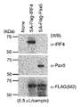
- the photograph which shows the confirmation result by the Western blot of fusion protein.
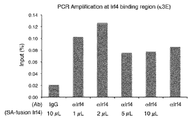
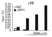
- the figure which shows the result of quantitative PCR which used the supernatant and precipitation when performing immunoprecipitation using each sample of Table 1 as a template.
- the figure which shows the amount of sedimentation of the target DNA immunoprecipitated when the quantity of an internal standard is changed.
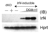
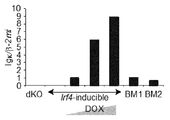
- the figure which shows the result of the Western blot which shows the induction
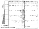
- medical agent The figure (halftone image) which shows the mapping result of the arrangement
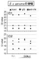
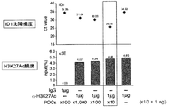
- medical agent The figure which shows the collection amount of yeast DNA of ID1 or ID2 by immunoprecipitation.
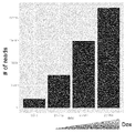
- the figure (halftone image) which shows the result of having counted the number of reads in a binding peak range based on the map in the case of each doxycycline (DOX) density
- the figure (halftone image) which shows the result of the correction
- the present invention includes an invention related to an internal standard molecule for immunoprecipitation (first invention) and an invention related to an immunoprecipitation method (second invention).
- the first invention of the present invention is: An internal standard molecule for immunoprecipitation, An internal standard molecule comprising a protein containing an epitope recognized by an antibody used in the immunoprecipitation or a partial peptide thereof and a nucleic acid bound thereto.
- Immunoprecipitation is a technique known in the art. In short, it is a technique for detecting, separating, and / or purifying an antigen using an immunoprecipitation reaction in which a soluble antigen is specifically reacted with an antigen-specific antibody and precipitated.
- the immunoprecipitation in the present invention is not particularly limited, and examples thereof include general immunoprecipitation, co-immunoprecipitation, and chromatin immunoprecipitation.
- other experimental systems may be assembled before and after immunoprecipitation, and an immunoprecipitation system may be assembled as one of the entire experimental systems.
- the immunoprecipitation in the present invention is preferably chromatin immunoprecipitation.
- correction for comparison between lots or samples with different conditions is performed more accurately than in the past in the quantification of the target substance. be able to.
- proteins with large dissociation constants or proteins with unstable binding such as proteins such as transcription factors that bind to DNA on chromatin, are targeted between lots or samples with different conditions. The effect of being able to perform correction for comparison of the above more accurately than in the past is great.
- the internal standard molecule of the first invention of the present invention is an internal standard molecule for immunoprecipitation, and is a combination of a nucleic acid and a protein containing an epitope recognized by the antibody used in the immunoprecipitation or a partial peptide thereof. Details of the internal standard molecule of the first invention of the present invention are described below.
- the internal standard molecule of the first invention of the present invention contains a nucleic acid.
- nucleic acid examples include DNA, RNA, and artificial nucleic acid. From the viewpoints of ease of synthesis, relevance to other experimental systems, and stability that is difficult to decompose, DNA is preferable, and double-stranded DNA is more preferable. It may contain a modified base.
- the nucleotide sequence of the nucleic acid is not particularly limited as long as it can be detected by hybridization or PCR, but in order to minimize the interaction such as binding to the substance in the test sample, the species of the species from which the test sample is derived is used. It preferably consists of a sequence not contained in the genome. That is, when used for chromatin immunoprecipitation, it is preferable to use a sequence not included on the chromosome of the sample. Furthermore, in order to minimize the interaction such as the binding with a substance in the test sample, it is preferable not to include a protein binding sequence such as a gene regulatory sequence of a biological species from which the test sample is derived.
- Examples thereof include sequences derived from fungi such as yeast, bacteria such as Escherichia coli, and fruit fly. Among them, when a mammalian sample is used as the test sample, a sequence derived from a bacterium such as yeast or Escherichia coli is preferable, and a sequence derived from yeast is more preferable.
- Examples of the nucleic acid sequence derived from yeast include the sequences represented by SEQ ID NO: 1 to SEQ ID NO: 4. An artificial sequence not derived from any species may be used.
- the type, GC content, length of base sequence, etc. are not particularly limited, but the GC content is preferably 30% or more, more Preferably it is 40% or more, Preferably it is 70% or less, More preferably, it is 60% or less.
- the GC content is preferably 30% or more, more Preferably it is 40% or more, Preferably it is 70% or less, More preferably, it is 60% or less.
- the length of the base sequence of the nucleic acid is not particularly limited as long as DNA is amplified by PCR, which will be described later, but usually 15 bases or more, preferably 20 bases or more, more preferably 25 bases or more, and usually 200 bases or less, preferably Is less than 100 bases.
- the method for producing the nucleic acid having the selected base sequence is not particularly limited, and a known method can be used. For example, if the length of the base sequence is short, it may be artificially synthesized by a known method, or a known trust service may be used. It can also be produced by a known genetic engineering technique. For example, it can also be obtained by performing PCR using a primer set designed to amplify the target base sequence using the genomic DNA of the biological species from which the selected base sequence is derived as a template.
- the internal standard molecule of the first invention of the present invention includes a protein.
- the protein is a protein containing an epitope recognized by the antibody used in the immunoprecipitation or a partial peptide thereof.
- the protein may be a protein containing an epitope recognized by the antibody or a partial peptide thereof and a protein or a partial peptide thereof to which a tag sequence is added.
- the tag sequence refers to a protein or peptide that functions as a label for a protein containing an epitope recognized by the antibody or a partial peptide thereof.
- Specific examples of the tag sequence include a histidine (His) tag, a glutathione-S-transferase (GST) tag, and a FLAG tag.
- the tag sequence may be added to a protein containing an epitope recognized by the antibody or a partial peptide thereof by using a genetic recombination technique using an expression vector that expresses a fusion protein with the tag sequence that is commercially available. it can.
- the tag sequence can be used, for example, for recovery or purification of a protein containing an epitope recognized by the antibody or a partial peptide thereof.
- the protein contained in the internal standard molecule of the first invention of the present invention usually means the full length of the amino acid sequence, but also includes the full length of the mature protein from which the signal sequence is removed and the mutant protein.
- the protein contained in the internal standard molecule of the first invention of the present invention naturally contains an epitope recognized by the antibody used in the immunoprecipitation.
- the protein contained in the internal standard molecule of the first invention of the present invention is not particularly limited, and there is no limitation on the amino sequence or its length.
- a DNA binding protein is preferable, and specific examples include a transcription factor, a transcription control factor, a histone protein, a chromatin control factor, and the like.
- the transcription factor is not particularly limited, and examples thereof include IRF4 protein, p53 protein, NF- ⁇ B, MYC protein, and Pax5 protein.
- the transcription control factor is not particularly limited, and examples thereof include Rb protein and BRCA1 protein.
- the histone protein is not particularly limited, and examples thereof include core histone proteins such as H2A protein, H2B protein, H3 protein, and H4 protein, and linker histones such as H1 protein.
- these histone proteins may be chemically modified. For example, acetylation, phosphorylation, methylation, and ubiquitination may be performed. In the case of methylation, it may be monomethylated, dimethylated or trimethylated.
- H3 protein for example, when the 9th lysine residue is methylated (H3K9me), when the 27th lysine residue is methylated (H3K27me), the 9th lysine residue is acetylated. (H3K9Ac) and the like.
- H3K9Ac acetylated.
- the case where the 9th lysine residue is trimethylated (H3K9me3), the case where the 27th lysine residue is trimethylated (H3K27me3), and the like can also be mentioned.
- the 120th threonine residue examples include a case where the group is phosphorylated (H2AT120ph) and a case where the 119th lysine residue is ubiquitinated (H2AK119ub).
- H2B protein for example, when the fifth lysine residue is acetylated (H2BK5Ac), when the 14th serine residue is phosphorylated (H2BS14ph), the 120th lysine residue Examples include a case where the group is ubiquitinated (H2BK120ub).
- H4 protein for example, when the fifth lysine residue is acetylated (H4K5Ac), when the first serine residue is phosphorylated (H4S1ph), the third arginine residue Examples include a case where the group is methylated (H4R3me).
- the chromatin control factor is not particularly limited, and examples thereof include Brg1, p300, and Pc4.
- the partial peptide of the protein contained in the internal standard molecule of the first invention of the present invention is not particularly limited as long as it contains an epitope recognized by the antibody used in immunoprecipitation.
- a partial peptide of 20 to 100 amino acids is exemplified. Is done.
- Method for producing protein or partial peptide thereof When the length of the amino acid sequence of the protein or its partial peptide is short, its production method is not particularly limited and can be produced according to a known peptide synthesis method. For example, a solid phase synthesis method and a liquid phase synthesis method can be mentioned.
- the purification method for example, solvent extraction, distillation, column chromatography, liquid chromatography, recrystallization and the like can be combined, whereby the peptide can be isolated and purified.
- the obtained peptide is a free form, it can be converted into an appropriate salt by a known method.
- it is obtained as a salt it is converted into a free form or other salt by a known method.
- You can also Modifications such as phosphorylation, acetylation, and methylation recognized by the antibody can also be added to the peptide.
- the amino acid sequence of the protein or its partial peptide can be produced by genetic engineering techniques.
- a DNA encoding a protein or a partial peptide thereof can be prepared, a recombinant expression vector can be constructed using the DNA, introduced into a host, expressed, and purified for production. Any of them can be performed using a known method.
- the above-mentioned protein or a partial peptide thereof may be a fusion protein containing a tag sequence such as a streptavidin tag, a His tag, a GST tag, or a Flag tag.
- the internal standard molecule of the first invention of the present invention is one in which a nucleic acid and a protein or partial peptide are bound.
- the bonding mode is not particularly limited, but irreversible bonding is preferable.
- modification of the end of the nucleic acid so that it can be bound to a protein or a partial peptide can be mentioned.
- the mode of modification of the nucleic acid is not particularly limited as long as it binds to a protein or partial peptide.
- a labeling substance that binds to the end of a nucleic acid by reacting with a labeling substance or functional group introduced into the protein or peptide described later And introducing a functional group.
- introduction of a labeling substance such as biotin, a thiol group or an amino group at the end of the nucleic acid can be mentioned.
- These labeling substances and functional groups may be introduced into the nucleic acid via a spacer.
- the method of modifying the nucleic acid is not particularly limited, and a method using a primer with a labeled base introduced at the end when the nucleic acid is amplified by PCR, a method using a labeled base as the terminal base when synthesizing the nucleic acid with a nucleic acid synthesizer A known method can be followed.
- a primer for amplifying a nucleic acid by PCR DNA having a biotinylated 5 ′ end or a nucleic acid having an aminated 5 ′ end can be used.
- N-hydroxysuccinimide active ester via the amino group is further exemplified.
- N-hydroxysuccinimide active ester and N-hydroxysuccinimide active ester of SMCC succinimidyl-4- (N-maleimidomethyl) cyclohexane-1-carboxylate
- SMCC succinimidyl-4- (N-maleimidomethyl) cyclohexane-1-carboxylate
- the internal standard molecule of the first invention of the present invention is one in which the nucleic acid contained in the internal standard molecule and a protein or partial peptide are bound.
- the protein or partial peptide side is modified so that it can bind to a nucleic acid.
- the modification of the protein or partial peptide is not particularly limited as long as it binds to the nucleic acid and can recognize the antibody used for immunoprecipitation.
- the labeling substance or functional group introduced into the nucleic acid in the protein or partial peptide is not limited. Introducing a labeling substance or a functional group that reacts with and binds.
- the modification of the protein or partial peptide may or may not be at the end of the protein or partial peptide.
- modification of protein or partial peptide examples include avidin labeling and streptavidin labeling when biotin labeled nucleic acid is used as the nucleic acid.
- streptavidin labeling when biotin labeled nucleic acid is used as the nucleic acid.
- cysteine labeling when a nucleic acid bound to an N-hydroxysuccinimide active ester via an amino group is used as the nucleic acid, cysteine labeling can be mentioned. Cysteine may label the endogenous cysteine or introduce a new cysteine residue.
- the method for modifying the protein or its partial peptide is not particularly limited, but genetic engineering techniques can be used.
- the vector encoding a label molecule such as avidin or streptavidin
- the DNA encoding the labeled molecule can be located downstream of the DNA encoding the protein or partial peptide. It may be arranged.
- the binding between the nucleic acid and the protein or its partial peptide can be performed by a known method. For example, in the case of binding between a nucleic acid modified with biotin at the end and a fusion protein of streptavidin and a protein, both may be simply mixed in a solution. The ratio may be changed by experiment.
- the immunoprecipitation method of the second invention of the present invention comprises: Mixing the test sample with the internal standard molecule; An immunoprecipitation step, and An immunoprecipitation method comprising a step of performing normalization by detecting a nucleic acid portion of the internal standard molecule.
- the mixing method of the test sample and the internal standard molecule in this step is not particularly limited, and may be performed by a method usually performed in the technical field such as pipetting.
- the method of immunoprecipitation is not particularly limited, and a known immunoprecipitation method can be used.
- the antibody is bound to a carrier such as a bead, added to a mixed solution of a test sample and an internal standard molecule, and incubated for a certain time, for example, 30 minutes to 1 day, and then a precipitate is obtained by centrifugation or the like. .
- the precipitate is washed with a buffer and used for the next step.
- an antibody against a protein that is expected to bind to a target fragment DNA may be used in the same manner as in a normal immunoprecipitation method.
- a complex of protein and fragment DNA bound thereto, and an internal standard molecule can be precipitated.
- the nucleic acid portion of the internal standard molecule is detected, and the measurement result of immunoprecipitation is standardized based on the detection result.
- the measurement result of normal co-immunoprecipitation it is possible to correct variations in experimental conditions between different samples.
- the first embodiment of the chromatin immunoprecipitation method of the second invention of the present invention comprises: Mixing the test sample with the internal standard molecule; Chromatin immunoprecipitation process, Amplifying the target DNA and the DNA portion of the internal standard molecule in the sample obtained in the chromatin immunoprecipitation step; and A chromatin immunoprecipitation method comprising a step of correcting the amount of amplified DNA of a target fragment DNA in a sample using the amount of amplified DNA of the DNA portion of the internal standard molecule obtained in the amplification step as an index.
- the mixing method of the test sample and the internal standard molecule in this step is not particularly limited, and may be performed by a method usually performed in the technical field such as pipetting.
- the method of chromatin immunoprecipitation is not particularly limited, and a known chromatin immunoprecipitation method can be used.
- a complex of DNA and protein on chromatin present in a test sample is cross-linked with a cross-linking agent such as formaldehyde (for example, at room temperature for 10 minutes), and then the cells are recovered and the DNA is 100 Sonicate to a length of ⁇ 500 bp.
- a cross-linking agent such as formaldehyde (for example, at room temperature for 10 minutes)
- the cells are recovered and the DNA is 100 Sonicate to a length of ⁇ 500 bp.
- complex of DNA and protein which were ultrasonically disrupted is mentioned.
- the internal standard molecule is also precipitated by the antibody together with the complex of DNA and protein on chromatin.
- the amplification method in this step is not particularly limited as long as the target DNA and the DNA of the internal standard molecule can be amplified, and the amplified DNA in the subsequent step can be quantified, and a known method can be used.
- a PCR method is preferably used, and examples thereof include a real-time PCR method.
- a primer set for amplifying the target DNA in the sample and a primer set for amplifying the DNA of the internal standard molecule may be used, and a primer set for amplifying each DNA may be used.
- a known method can be used.
- intercalation method for example, a dye that specifically intercalates with double-stranded DNA and emits fluorescence, such as SYBR Green I, can be used.
- hybridization method for example, a probe in which a fluorescent dye is bound to an oligonucleotide specific to a DNA sequence, such as a TaqMan probe, can be used.
- Step of correcting the amount of target DNA in the sample using the amplification amount of the DNA portion of the internal standard molecule obtained in the amplification step as an index The amount of the DNA portion of the internal standard molecule mixed with the test sample is known. Therefore, in this step, if the DNA portion of the amplified internal standard molecule can be quantified, it can be amplified using a known method such as a method of comparing Ct values or a method of microchannel electrophoresis using this as an index. In addition, the amount of target DNA in the sample can be corrected and quantified.
- the amplification amount obtained in the amplification step after immunoprecipitation is It can be determined that the greater the amount, the greater the amount of transcription factor binding. Then, by correcting this amount with the amplification amount of the internal standard, the amount of binding can be grasped more accurately.
- the amount of a complex of precipitated DNA and protein increases by administration of a specific substance
- the amount of protein binding to the DNA region increases by the specific substance, that is, the expression control changes. It is evaluated.
- the relationship between the disease or mutation and the amount of protein binding can also be examined by comparing the diseased patient with a healthy person and comparing the mutation carrier with a non-carrier.
- a second embodiment of the chromatin immunoprecipitation method of the second invention of the present invention comprises: Mixing the test sample with the internal standard molecule; Chromatin immunoprecipitation process, Amplifying the target DNA in the sample obtained in the chromatin immunoprecipitation step and the DNA portion of the internal standard molecule; Analyzing the base sequence of the DNA portion of the target DNA and the internal standard molecule in the sample amplified in the amplification step; and
- the immunoprecipitation method includes a step of correcting the sequence amount of the base sequence of the target DNA in the sample using the sequence amount of the base sequence of the DNA portion of the internal standard molecule obtained in the step of analyzing the base sequence as an index.
- a step of analyzing the base sequence of the DNA portion of the target DNA and internal standard molecule in the amplified sample is not particularly limited, but can be performed using, for example, a next-generation sequencer.
- next-generation sequencers include, for example, Illumina, SOLiD, Helicos, Complete Genomics, Polonator, Ion Torrent, Pacific Biosciences next-generation sequencers and next-generation sequencers (for example, Mardis, Annu. Rev. Genom. Human Genet. (2008) vol.9, p.387-402 and Persson et al., Chem. Soc. Rev.
- the next-generation sequencer is based on the principle of determining base sequences in a super parallel manner by light detection such as fluorescence and luminescence using a sequential DNA synthesis method using DNA polymerase or DNA ligase.
- GS FLX Roche Diagnostics
- Solexa Illumina
- SOLiD Applied Biosystems
- Polonator Polonator
- DNA synthesis is performed with DNA polymerase using one DNA molecule as a template, and the base sequence is determined by real-time observation of one molecule by detecting the reaction of each base with light such as fluorescence and luminescence.
- SMRT SMRT
- Pacific Biosciences SMRT
- SMRT Pacific Biosciences
- DNA can be obtained using a semiconductor (CMOS) chip.
- CMOS semiconductor
- Ion Torrent Systems which determines the base sequence by detecting hydrogen ions (pH change) released when each base is taken in by polymerase.
- Nanopore Oxford Nanopore Technologies
- Step of correcting the sequence amount of the base sequence of the target DNA in the sample using the sequence amount of the base sequence of the DNA portion of the internal standard molecule obtained in the step of analyzing the base sequence as an index The sequence amount of each base sequence of the target DNA in the sample analyzed by mapping the base sequence of the target DNA in the amplified sample to the genome sequence (reference sequence) of the species from which the test sample is derived I understand. The amount of the DNA portion of the internal standard molecule mixed with the test sample is known. Therefore, the sequence amount of the base sequence of the target DNA in the sample can be corrected using the sequence amount of the base sequence of the DNA portion of the internal standard molecule as an index.
- the sequence amount of the base sequence of the target DNA in the two groups of test samples is corrected using the sequence amount of the base sequence of the DNA portion of the internal standard molecule as an index. And can determine whether the resulting difference is a significant difference or an error due to the administration of the particular substance. For example, when the expression level of a specific protein is small and the difference from the background does not appear significantly, the risk of judging the error as a significant difference, or conversely, the significant difference is judged as an error. Risk can be reduced.
- the method of the present invention it is possible to evaluate the relationship between gene expression control and disease, the therapeutic effect, and the like with high accuracy. For example, if the number of reads in the base sequence of a specific DNA region is increased by administration of a specific substance, it is evaluated that the expression control of the DNA region has changed due to the specific substance. For example, by evaluating the interaction between a transcription factor that controls the expression of an oncogene and its binding sequence by chromatin immunoprecipitation, the prediction and evaluation of a disease state, and further the therapeutic effect can be examined. Moreover, the relationship between transcription control by a DNA binding protein such as a transcription factor and a disease can be examined by comparing using a sample derived from a patient and a sample derived from a healthy person. Furthermore, the relationship between transcriptional control by a DNA-binding protein such as a transcription factor and gene mutation can be examined by comparing a subject having a gene mutation and a subject having no mutation using each sample.
- NIH3T3 cells (ATCC CRL-1658) were infected with the viral vector pMX-puro incorporating the human RasG12V gene, and RasG12V protein was expressed in the cells. Separately, NIH3T3 cells were infected with a control virus vector pMX-puro not incorporating the RasG12V gene.
- H3K27me3 histone H3 protein trimethylated with lysine 27
- NIH3T3 cells were treated with 10% fetal bovine serum, 1% penicillin streptomycin, 2 mM L-glutamine, 1% non-essential amino acid, 1% pyruvate in an environment of 95% air and 5% carbon dioxide gas at 37 ° C. Cultured in Dulbecco's modified Eagle's medium (DMEM medium) containing sodium.
- DMEM medium Dulbecco's modified Eagle's medium
- Virus infection of NIH3T3 cells A viral vector and a control vector incorporating the human RasG12V gene were introduced into Plat-E cells (provided by Dr. T. Kitamura of the Institute of Medical Science, the University of Tokyo), and in the same manner as above in DMEM medium. Was cultured to produce a human RasG12V expression virus. Gene transfer is described in Hosogane et al. PLoS Genet. Aug 2013; 9 (8): e1003698. (2013). The DMEM medium subjected to the culture was collected as a virus-containing solution, and NIH3T3 cells were cultured in the virus-containing solution in the presence of polybrene to establish infection.
- NIH3T3 cells were fixed with 1.0% formalin for 5 minutes at room temperature with stirring. After cell lysis, genomic DNA was fragmented into 200-700 bases using an ultrasonic generator Covaris S2 (Covaris). On the other hand, anti-histone H3K27me3 antibody (Millipore, catalog number 07-449) was bound to magnetic beads (Life Technologies) having Protein A immobilized on the surface. Then, the magnetic beads to which the anti-histone H3K27me3 antibody was bound via Protein A and the fragmented genomic DNA were mixed at 4 ° C. for 14 hours for immunoprecipitation.
- Covaris S2 Covaris
- sequence analysis A sequence library was prepared from 10 ng of the immunoprecipitate using TruSeq DNA LT Sample Prep Kit v2 (Illumina), and sequence analysis was performed with Illumina HiSeq2000. Sequence library preparation and sequence analysis were performed according to protocols provided by Illumina.
- the number of bases for decoding was 6.79 ⁇ 10 9 bases in the cells infected with the viral vector incorporating the RasG12V gene, whereas it was 3.68 in the cells infected with the control viral vector not incorporating the RasG12V gene. ⁇ 10 9 bases.
- the amount of H3K27me3 was not related to the expression of RasG12V gene. Therefore, if the sequence was decoded by the ChIP-seq method in each cell group, an equivalent result should be obtained.
- the number of decoding bases varies among cell groups. This seems to indicate that the amount of H3K27me3 cannot be accurately measured by the conventional Western blot method.
- Example 1 Chromatin immunoprecipitation of transcription factor Irf4 protein
- Preparation of internal standard molecules for chromatin immunoprecipitation> According to the following method, an internal standard molecule used for chromatin immunoprecipitation of the transcription factor Irf4 protein was prepared.
- a specific DNA sequence was selected from the DNA sequences of the budding yeast Saccharomyces cerevisiae as the nucleic acid of the internal standard molecule. That is, using the genomic DNA sequence information of the budding yeast, a DNA sequence having a length of 250 bases that is not a repeat sequence nor a gene control sequence of the budding yeast itself was selected on a computer. Further, a sequence having a GC content of 40% or more and 60% or less was selected, and four types having fewer binding sites of known transcription factors were selected (hereinafter, these base sequences are ID1, ID2, ID3, ID4). And). ID1 to ID4 are base sequences represented by SEQ ID NO: 1 to SEQ ID NO: 4, respectively.
- the primer sets used to amplify the yeast DNAs ID1 to ID4 are as follows.
- a forward primer: 5′-CGTTCGTTCTCAACGTCAACC-3 ′ (SEQ ID NO: 5) and a reverse primer: 5′-ATTCACTGGAGCCGAATAGC-3 ′ (SEQ ID NO: 6) were used.
- forward primer 5'-TGAATCTGGGTGAAGAAGAAGTAC-3 '(SEQ ID NO: 7) and reverse primer: 5'-AGACAAACCGGCAGCCAAAATAG-3' (SEQ ID NO: 8) were used.
- forward primer 5'-CAAATGTCCCCATCAATCACGC-3 '(SEQ ID NO: 9)
- reverse primer 5'-GGGGCGCATCATTCCTCGCGGA-3' (SEQ ID NO: 10) were used.
- forward primer 5'-TTCAAAAATAGGTTAAAATGG-3 '(SEQ ID NO: 11) and reverse primer: 5'-CCAAAAATGGCATGATCAATG-3' (SEQ ID NO: 12) were used.
- PCR amplification was performed under the conditions of 35 cycles (98 ° C. for 10 seconds, 57 ° C. for 30 seconds, 68 ° C. for 20 seconds) using a high success rate PCR enzyme KOD FX NEO (TOYOBO). After PCR amplification, A was added to the 5 'end and 3' end of the amplified DNA using 10x A-attachment Mix (TOYOBO).
- T4 DNA ligase (Insertion of yeast DNA of ID1 to ID4 into pTA2 vector) T4 DNA ligase (TAKARA, Clontech) was used in the ligation for inserting the yeast DNA of ID1 to ID4 into the pTA2 vector.
- the yeast DNAs ID1 to ID4 amplified by PCR were inserted into the pTA2 vector (TOYOBO) by the TA cloning method to obtain recombinant vectors (hereinafter referred to as “pTA2-ID1 vector”, “pTA2-ID2”, respectively).
- vector referred to as “vector”, “pTA2-ID3 vector”, “pTA2-ID4 vector”, etc.).
- ID1-ID4 biotinylated yeast DNAs were obtained by PCR using the pTA2-ID1 vector, pTA2-ID2 vector, pTA2-ID3 vector, and pTA2-ID4 vector as template DNAs, respectively.
- a primer set designed to amplify from the 5 ′ end and 3 ′ end of each yeast DNA of ID1 to ID4 (for the amplification of ID1, the above SEQ ID NO: 5, SEQ ID NO: 6, ID2
- ID1 the above SEQ ID NO: 5, SEQ ID NO: 6, ID2
- SEQ ID NO: 7 and SEQ ID NO: 8 for the amplification of ID3
- SEQ ID NO: 9 and SEQ ID NO: 10 for the amplification of ID4, the SEQ ID NO: 11 and SEQ ID NO: 12
- a biotinylated 5 ′ end was used.
- PCR amplification was performed under the conditions of 35 cycles (98 ° C. for 10 seconds, 57 ° C. for 30 seconds, 68 ° C. for 20 seconds) using a high success rate PCR enzyme KOD FX NEO (TOYOBO). After PCR amplification, ID1-ID4 biotinylated yeast DNA was purified using PCR purification Kit (QIAGEN).
- FIG. 1 is a photograph of agarose gel electrophoresis suggesting that biotinylated yeast DNA was generated by the PCR.
- yeast DNA of ID1 was used as the yeast DNA
- primer sets represented by SEQ ID NO: 5 and SEQ ID NO: 6 were used in PCR.
- Dynabeads M-280 Streptavidin magnetic beads from Dynal / Invitrogen were used as magnetic beads having streptavidin immobilized on the surface, and immunoprecipitation was performed according to the attached protocol.
- Lane 1 shows the results when PCR amplification was performed using DNA contained in the supernatant fraction when immunoprecipitation was performed using non-biotinylated yeast DNA of ID1 and magnetic beads having streptavidin immobilized on the surface as a template. It is the lane which electrophoresed the obtained PCR product.
- Lane 2 shows the results when PCR amplification was performed using DNA contained in the magnetic bead fraction obtained by immunoprecipitation using ID1 non-biotinylated yeast DNA and magnetic beads with streptavidin immobilized on the surface as a template. It is the lane which electrophoresed the obtained PCR product.
- Lane 3 is obtained when PCR amplification is performed using biotinylated yeast DNA of ID1 and DNA contained in the supernatant fraction when immunoprecipitation is performed using magnetic beads with streptavidin immobilized on the surface as a template. It is the lane which electrophoresed the obtained PCR product.
- Lane 4 is obtained when PCR amplification is performed using biotinylated yeast DNA of ID1 and DNA contained in the magnetic bead fraction when immunoprecipitation is performed using magnetic beads having streptavidin immobilized on the surface as a template. It is the lane which electrophoresed the obtained PCR product.
- yeast DNA When non-biotinylated yeast DNA is used, the yeast DNA does not bind to magnetic beads having streptavidin immobilized on the surface. Therefore, although it was considered that yeast DNA was contained in the supernatant fraction, but not in the magnetic bead fraction, a band was confirmed in lane 1 and no band was confirmed in lane 2.
- biotinylated yeast DNA When biotinylated yeast DNA is used, the yeast DNA binds to magnetic beads having streptavidin immobilized on the surface. Therefore, although it was considered that yeast DNA was contained in the bead fraction but not in the supernatant fraction, a band was confirmed in lane 4 and no band was confirmed in lane 3. From the above, it was found that biotinylated yeast DNA can be prepared by this method.
- the primer set used for PCR was designed to amplify from the 5 'end and 3' end of the IRF4 gene sequence (SEQ ID NO: 13), and further was added with a restriction enzyme Hind III site. Specifically, the forward primer: 5'-gcgAAGCTTTgaacttggagacgggg-3 '(SEQ ID NO: 14) and the reverse primer: 5'-gcgAAGCTtcactctgtgagaga-3' (SEQ ID NO: 15) were used. The base sequence described in capital letters is the restriction enzyme Hind III site.
- PCR amplification was performed under conditions of 35 cycles (98 ° C. for 10 seconds, 57 ° C. for 30 seconds, 68 ° C. for 40 seconds) using a high success rate PCR enzyme KOD FX NEO (TOYOBO). After PCR amplification, the IRF4 gene sequence was purified using PCR purification Kit (QIAGEN).
- the Pax5 gene sequence (SEQ ID NO: 16) was amplified by PCR using the MigR Pax5 vector (distributed by the University of Chicago, Harinda Singh Laboratory) as a template.
- the primer set used for PCR was designed to amplify from the 5 'end and 3' end of the Pax5 gene sequence (SEQ ID NO: 16), and was further added with a restriction enzyme Hind III site. Specifically, a forward primer: 5'-gcg AAGCTTattgattattagaaaaatatacc-3 '(SEQ ID NO: 17) and a reverse primer: 5'-gcgAAGCTTcagtgacgtcatggcgg-3' (SEQ ID NO: 18) were used. The base sequence described in capital letters is the restriction enzyme Hind III site.
- PCR amplification was performed under the conditions of 35 cycles (98 ° C. for 10 seconds, 57 ° C. for 30 seconds, 68 ° C. for 20 seconds) using a high success rate PCR enzyme KOD FX NEO (TOYOBO). After PCR amplification, the Pax5 gene sequence was purified using PCR purification Kit (QIAGEN).
- the primer set used for PCR was designed to amplify from the 5 ′ end and 3 ′ end of the 4 ⁇ Flag gene sequence (SEQ ID NO: 19). Furthermore, the restriction enzyme EcoR I site was used as the forward primer and the reverse primer as the reverse primer. A Hind III site is added. Specifically, a forward primer: 5'-gcgGAATTCggcggcatggactacaagagacgagg-3 '(SEQ ID NO: 20) and a reverse primer: 5'-gcgAAGCTTgccgcccttatcgtcgtcatctttgt-3' (SEQ ID NO: 21) were used. The base sequence described in capital letters is a restriction enzyme site.
- PCR amplification was performed under the condition of 35 cycles (98 ° C. for 10 seconds, 57 ° C. for 30 seconds, 68 ° C. for 15 seconds) using a high success rate PCR enzyme KOD FX NEO (TOYOBO). After PCR amplification, the 4xFlag gene sequence was purified using PCR purification Kit (QIAGEN).
- Streptavidin gene sequence (SEQ ID NO: 22) was amplified by PCR using pET21a-Streptavidin-Alive (Plasmid # 20860; Addgene) vector DNA as a template.
- the primer set used for PCR was designed to amplify from the 5 ′ end and 3 ′ end of the streptavidin gene sequence (SEQ ID NO: 22). Further, the restriction primer Xho I site was used as the forward primer, and the reverse primer To which EcoRI site is added. Specifically, a forward primer: 5′-gcgCTCGGAGATGgctgaagctggtatcac-3 ′ (SEQ ID NO: 23) and a reverse primer: 5′-gcgGAATTCatgtgtgtgtgtgtgtgtgtgtg-3 ”(SEQ ID NO: 24) were used. The base sequence described in capital letters is a restriction enzyme site.
- PCR amplification was performed using a highly successful PCR enzyme KOD FX NEO (TOYOBO) under the conditions of (35 ° C for 10 seconds, 57 ° C for 30 seconds, 68 ° C for 15 seconds) x 35 cycles.
- the streptavidin gene sequence was purified using a PCR purification kit (QIAGEN).
- pcDNA3 SA-4xFlag vector a recombinant vector
- pcDNA3 SA-4xFlag vector was treated with restriction enzyme Hind III.
- the IRF4 gene amplified by PCR or the Pax5 gene treated with the restriction enzyme Hind III was similarly treated with the restriction enzyme Hind III.
- T4 DNA ligase was similarly treated with restriction enzyme Hind III. Insertion was carried out to prepare recombinant vectors (hereinafter sometimes referred to as “pcDNA3 SA-4xFlag-IRF4 vector” and “pcDNA3 SA-4xFlag-Pax5 vector”, respectively).
- the pcDNA3 vector carries the T7 promoter. In vitro translation was performed using a TNT T7 Quick Coupled Transmission / Translation System (Promega) according to the attached protocol. Using the pcDNA3 SA-4xFlag-IRF4 vector or pcDNA3 SA-4xFlag-Pax5 vector prepared above as a template, and in vitro translation, a fusion protein containing streptavidin, 4xFlag protein, and IRF4 protein, or streptavidin, 4xFlag protein, And a fusion protein containing Pax5 protein was produced.
- the results are shown in FIG.
- the translation products translated in vitro using pcDNA3 SA-4xFlag-IRF4 vector or pcDNA3 SA-4xFlag-Pax5 vector as a template were separated by SDS-PAGE electrophoresis, and anti-IRF4 antibody (Santa Cruz), anti-Pax5 antibody (Santa Cruz) Or, wetanblot analysis with anti-Flag antibody (Sigma).
- pcDNA3 When in vitro translation was performed using the SA-4xFlag-IRF4 vector as a template, specific bands were confirmed with anti-IRF4 antibody and anti-Flag antibody, and pcDNA3 When in vitro translation was performed using the SA-4xFlag-Pax5 vector as a template, specific bands were confirmed between the anti-Pax5 antibody and the anti-Flag antibody, indicating that the target fusion protein was translated in vitro. .
- Biotin-streptavidin binding A biotinylated yeast DNA of ID1 to ID4 and a fusion protein containing streptavidin, 4xFlag protein, and IRF4 protein, or a fusion protein containing streptavidin, 4xFlag protein, and Pax5 protein are mixed and allowed to stand at 4 ° C for 1 hour. Then, biotin and streptavidin were bound.
- yeast DNA of ID1 was used as the yeast DNA
- a fusion protein containing streptavidin, 4 ⁇ Flag protein, and IRF4 protein was used as the fusion protein.
- Biotinylated yeast DNA (concentration 10 ng / ⁇ L), fusion protein, 10 ⁇ phosphate buffered saline (PBS), and distilled water were mixed in the volumes shown in Table 1, and biotin-streptavidin was added at 4 ° C. for 1 hour. A reaction was performed.
- (a) to (d) represent the following.
- the yeast DNA contained in the supernatant fraction and the magnetic bead fraction was quantified by quantitative PCR using the primer sets represented by SEQ ID NO: 5 and SEQ ID NO: 6, respectively.
- the PCR reaction was performed using a 7500 real-time PCR system (ABI) instrument, SYBR Advantage qPCR premix (TaKaRa Clontech) at 95 ° C for 10 minutes (95 ° C for 15 seconds, 60 ° C for 33 seconds, 95 ° C for 15 seconds) ) X 40 cycles.
- Spleen B lymphocytes were isolated from antibody gene transgenic mouse B1-8 Hi (originally Prof. Michel C. Nussenzweig (The Rockefeller University)) donated by Prof. Tomohiro Kurosaki of RIKEN. After culturing for 3 days in the presence of the antigens NP (40) -ficol and cytokines (IL-2, IL-4, IL-5, CD40L) in an environment of% air and 5% carbon dioxide gas, Fix with 0% formalin for 10 minutes at room temperature.
- the fixation reaction was stopped by adding 2.5 ⁇ M Glycine to a final concentration of 1/20, the fixed cells were washed with PBS, a cytoplasmic lysis buffer was added, and the mixture was reacted on ice for 10 minutes. After removing the supernatant containing the cell fraction by centrifugation, the precipitate, which is the nuclear fraction, is dissolved in the nuclear fraction lysis buffer, and genomic DNA is 100 to 100-fold using a sonic generator Bioruptor (Cosmo Bio, Dianode). Fragmented to 500 bases. The fragmented genomic DNA is reacted with magnetic beads (Novex / life technologies) with protein A / G immobilized on the surface for 1 hour at 4 ° C. to bind nonspecifically to protein A / G magnetic beads. Fractions were removed.
- the supernatant fraction that did not bind nonspecifically to the protein A / G magnetic beads was collected. Then, after adding 0, 1, 2, 5, or 10 ⁇ L of the internal standard molecule, the mixture was reacted with anti-Irf4 antibody (sc-6059, Santa Cruz) overnight at 4 ° C., and then protein A / G magnetic beads were added at 4 ° C. Immunoprecipitation was performed by reacting for 2 hours. In addition, 10 ⁇ L of an internal standard molecule was added, and the antibody immunoprecipitated with an IgG antibody was used as a control.
- anti-Irf4 antibody sc-6059, Santa Cruz
- yeast DNA that does not bind to streptavidin in the fusion protein via biotin during the production of the internal standard molecule. Therefore, in order to remove the unbound yeast DNA, after the biotin-streptavidin binding reaction, magnetic beads with streptavidin immobilized on the surface are added and reacted for 1 hour at 4 ° C. with stirring, using a magnet. Thus, the magnetic bead fraction attracted to the inner wall of the test tube was removed. In addition, what did not perform this operation was prepared as control, and the difference was examined afterwards.
- the yeast DNA contained in the magnetic bead fraction was quantified by quantitative PCR using the primer set represented by SEQ ID NO: 5 and SEQ ID NO: 6.
- the PCR reaction was performed using a 7500 real-time PCR system (ABI) instrument, SYBR Advantage qPCR premix (TaKaRa Clontech) at 95 ° C for 10 minutes (95 ° C for 15 seconds, 60 ° C for 33 seconds, 95 ° C for 15 seconds) ) X 40 cycles.
- the results are shown in FIG.
- the vertical axis represents the relative amount of immunoprecipitated DNA (input%) when the amount of fragmented chromatin DNA used for immunoprecipitation is 100%, and was calculated by quantitative PCR. From this result, the Irf4 protein inherently contained in the cells even when the internal standard molecule is added binds to the DNA of the ⁇ 3′E region of the Ig ⁇ gene, which is the target gene, and this binding is caused by the addition of the internal standard molecule. was found not to be greatly disturbed. In addition, in the case of 1 ⁇ L of the fusion protein and 2 ⁇ L, the input% is larger than that in the case where the fusion protein is not added, but this is considered to be an experimental error range.
- mice deficient in both Irf4 and its similar factor Irf8 can be induced with doxycycline (DOX), an Irf4 expression gene (rtTA: TetO-Irf4) was introduced by crossing genetically modified mice.
- DOX doxycycline
- rtTA TetO-Irf4 expression gene
- Bone marrow B cells were selected from the same mouse with B220, a B cell surface marker. Selected cells were treated with 5 ng / mL IL-7-containing OptiMEM medium (5% FCS, 50 ⁇ M ⁇ -mercaptoethanol, 2 mM L-glutamine, 100 U / mL penicillin-streptomycin added) at 37 ° C. with 95% air and 5%. By culturing for 5 to 10 days in an environment of 10% carbon dioxide gas, an Irf4-induced B cell line was established.
- Ig ⁇ gene (Relationship between DOX concentration and expression level of Ig ⁇ gene) An Ig ⁇ gene is known as an Irf4 target gene. Then, next, the change in the expression of the Ig ⁇ gene depending on the DOX concentration was examined.
- Quantitative reverse transcription PCR uses 7500 real-time PCR system (ABI) equipment and SYBR Advantage qPCR premix (TaKaRa Clontech), 95 ° C for 10 minutes (95 ° C for 15 seconds, 60 ° C for 33 seconds, 95 ° C) 15 seconds) ⁇ 40 cycles.
- the B cell line established above is cultured in a test tube in the presence of 5 ng / mL IL-7 and at a DOX concentration of 0, 100, 200, or 400 ng / mL for 2 days, and then with 1.0% formalin. Fix at room temperature for 10 minutes. After removing the cytoplasmic fraction, the nuclear fraction was lysed, and the genomic DNA was fragmented into 100 to 500 bases using an ultrasonic generator Bioruptor (Cosmo Bio, diagenode). Thereto, an anti-Irf4 antibody (sc-6059, Santa Cruz) was added and immunoprecipitation was performed. An antibody immunoprecipitated with an IgG antibody was used as a control.
- the DNA of the ⁇ 3′E region of the Ig ⁇ gene contained in the magnetic bead fraction was quantified by quantitative PCR.
- forward primer: 5′-TCATAGCTACCGTCACACTG-3 ′ (SEQ ID NO: 27) and reverse primer: 5′-AACAGATGTGCCTAAGGTTC-3′-3 (SEQ ID NO: 28) were used.
- the PCR reaction was performed using a 7500 real-time PCR system (ABI) device, SYBR Advantage qPCR premix (TaKaRa Clontech), 95 ° C for 10 minutes (95 ° C for 15 seconds, 60 ° C for 33 seconds, 95 ° C for 15 seconds) ) X 40 cycles.
- ABSI real-time PCR system
- SYBR Advantage qPCR premix (TaKaRa Clontech)
- 95 ° C for 10 minutes 95 ° C for 15 seconds, 60 ° C for 33 seconds, 95 ° C for 15 seconds)
- X 40 cycles X 40 cycles.
- Quantitative PCR results are shown in FIG.
- the vertical axis represents the relative amount of immunoprecipitated DNA when the amount of fragmented chromatin DNA used for immunoprecipitation is 100%. From this result, it can be seen that as the expression level of the Irf4 protein depending on the DOX concentration increases, the immunoprecipitation rate by the Irf4 protein increases, that is, the binding amount of the Irf4 protein to the ⁇ 3′E region of the Ig ⁇ gene increases. It was.
- 0.1 ng of internal standard molecules each containing yeast protein ID1 to ID4 as yeast DNA and streptavidin, 4xFlag protein, and fusion protein containing IRF4 protein as fusion proteins are added to 50 ⁇ g of chromatin. Immunoprecipitation using anti-Irf4 antibody was performed. An antibody immunoprecipitated with an IgG antibody was used as a control.
- the immunoprecipitate obtained by the above immunoprecipitation was applied to a next-generation sequencer to decode the base sequence.
- the sequence analysis was performed with Illumina HiSeq2000 by preparing a sequence library using TruSeq DNA LT Sample Prep Kit v2 (Illumina). Library preparation and sequence analysis were both performed according to the protocol provided by Illumina.
- the number of reads as shown in FIG. 9 was obtained for each of the internal standard molecules containing yeast DNA of ID1 to ID4. It was shown that a substantially constant amount of yeast DNA was precipitated regardless of the DOX concentration.
- the cause of the difference in the number of reads between ID1 to ID4 may be the difference in amplification efficiency at the time of library creation.
- the Irf4-induced B cell line was cultured in a medium supplemented with various DOX concentrations (0, 100, 200, 400 ng / mL) for 48 hours, and fixed with 1.0% formalin for 10 minutes at room temperature. Then, after removing the cytoplasmic fraction, the nuclear fraction was dissolved, and the genomic DNA was fragmented into 100 to 500 bases using an ultrasonic generator Bioruptor (Cosmo Bio, diagenode).
- each internal standard molecule containing fusion proteins including yeast DNA ID1 to ID2 as yeast DNA, streptavidin, 4xFlag protein, and IRF4 protein as fusion proteins was added to 50 ⁇ g of chromatin. Immunoprecipitation using anti-Irf4 antibody was performed. An antibody immunoprecipitated with an IgG antibody was used as a control.
- the amount of DNA in the ⁇ 3′E region of the Ig ⁇ gene to which the endogenous Irf4 protein binds and the amount of yeast DNA of ID1 or ID2 in the immunoprecipitate obtained by the immunoprecipitation were quantified by quantitative PCR.
- the primer set represented by SEQ ID NO: 27 and SEQ ID NO: 28 was used for amplification of DNA in the ⁇ 3′E region of the Ig ⁇ gene.
- the primer set represented by SEQ ID NO: 5 and SEQ ID NO: 6 was used for the amplification of ID1 yeast DNA.
- the primer set represented by SEQ ID NO: 7 and SEQ ID NO: 8 was used for the amplification of ID2 yeast DNA.
- the PCR reaction was performed using a 7500 real-time PCR system (ABI) device, SYBR Advantage qPCR premix (TaKaRa Clontech), 95 ° C for 10 minutes (95 ° C for 15 seconds, 60 ° C for 33 seconds, 95 ° C for 15 seconds) ) X 40 cycles.
- ABSI real-time PCR system
- SYBR Advantage qPCR premix (TaKaRa Clontech)
- 95 ° C for 10 minutes 95 ° C for 15 seconds, 60 ° C for 33 seconds, 95 ° C for 15 seconds)
- X 40 cycles X 40 cycles.
- mapping result of the ⁇ 3′E region of the Ig ⁇ gene to which the DOX-induced Irf4 protein binds is obtained by mapping the sequence read obtained by applying the precipitated DNA obtained by the above immunoprecipitation to a next-generation sequencer onto the mouse genome. As shown in FIG.
- the DOX-induced Irf4 binding peak as obtained in FIG. 12 was identified throughout the genome, and the peak was determined at each concentration. Thereafter, the sum of the peak ranges of each concentration was taken, and the number of reads mapped within the range was counted. The results are shown in FIG. The horizontal axis represents the DOX concentration, and the vertical axis represents the number of reads within the binding peak range determined above. From this result, it was found that the number of leads within the binding peak range determined above increases in a DOX concentration-dependent manner.
- the horizontal axis represents the number of leads in the combined peak range
- the vertical axis represents the density distribution indicating the ratio of the specific number of leads to the total number of leads in the combined peak range.
- the density distribution when the conventional correction for dividing the number of leads within the combined peak range by the total number of leads is shown in the left diagram of FIG.
- the density distribution when the correction of this example is performed in which the number of reads in the binding peak range is divided by the number of reads of yeast DNA, which is an example of an internal standard molecule is shown in the right diagram of FIG.
- the correction of the present example is performed on samples with different DOX concentrations. It was found that the comparison between samples with different amounts of Irf4 protein was effective.
- Example 2 Chromatin immunoprecipitation of histone H3 protein acetylated with lysine 27] ⁇ 2.
- Preparation of internal standard molecules for chromatin immunoprecipitation> In accordance with the following procedure, an internal standard molecule used for chromatin immunoprecipitation of histone H3 protein acetylated with lysine 27 (hereinafter sometimes referred to as “H3K27Ac”) was prepared.
- ID1 aminated yeast DNA was obtained by PCR using the pTA2-ID1 vector as a template DNA.
- the primer set in PCR amplification is a primer set (SEQ ID NO: 5 and SEQ ID NO: 6) designed to amplify from the 5 ′ end and 3 ′ end of the yeast DNA of ID1, wherein the 5 ′ end of each primer is amino A group-modified one was used.
- a primer having a 6 carbon (C6) spacer was used between the 5 ′ end of each primer and the amino group.
- SMCC conversion of ID1 aminated yeast DNA The amplified aminated yeast DNA was purified using a urea-containing gel, and after adding a 100 mM KH 2 PO 4 (pH 7.2) solution, 200 nmole of SMCC was added and mixed well, followed by reaction at room temperature for 30 minutes.
- SMCC-cysteine bond The SMCC yeast DNA was dissolved in a 100 mM KH 2 PO 4 (pH 7.2) solution, cysteine-labeled H3K27Ac was added and mixed, and the mixture was reacted at room temperature for 3 hours or more. In order to remove unreacted SMCC yeast DNA, the reaction solution was run using a Mg 2+ -containing TAG gel to purify POCs (SMCC yeast DNA-H3K27Ac).
- splenic B lymphocytes were isolated from an antibody gene transgenic mouse B1 / 8Hi (originally Prof. Michel C. Nussenzweig (The Rockefeller University)) provided by Professor Tomohiro Kurosaki of RIKEN. After culturing for 3 days in the presence of antigen NP (40) -ficol and cytokine (IL-2, IL-4, IL-5, CD40L) in a test tube in an environment of 95% air and 5% carbon dioxide gas And fixed with 1.0% formalin for 10 minutes at room temperature.
- the nuclear fraction was dissolved, and the genomic DNA was fragmented into 100 to 500 bases using an ultrasonic generator Bioruptor (Cosmo Bio, diagenode).
- the fragmented genomic DNA was reacted with magnetic beads with protein A / G immobilized on the surface for 1 hour at 4 ° C. to remove fractions that non-specifically bound to protein A / G magnetic beads. .
- the supernatant fraction that did not bind nonspecifically to the protein A / G magnetic beads was collected. Then, after adding 0, 0.01, 0.1, or 1 ng of an internal standard molecule, the mixture was reacted with anti-H3K27Ac antibody (Abcam) at 4 ° C. overnight, and then protein A / G magnetic beads were added at 4 ° C. Immunoprecipitation was performed by reacting for 2 hours. In addition, 0.1 ng of an internal standard molecule was added, and an antibody immunoprecipitated with an IgG antibody instead of an anti-H3K27Ac antibody was used as a control.
- Anti-H3K27Ac antibody Abcam
- magnetic beads with anti-streptavidin antibody immobilized on the surface are allowed to react at 4 ° C. for 1 hour with stirring, and the magnetic bead fraction drawn to the inner wall of the test tube using a magnet is used as the immunoprecipitate. It was collected.
- the yeast DNA contained in the magnetic bead fraction was quantified by quantitative PCR using the primer set represented by SEQ ID NO: 5 and SEQ ID NO: 6.
- the PCR reaction was performed using a 7500 real-time PCR system (ABI) instrument, SYBR Advantage qPCR premix (TaKaRa Clontech) at 95 ° C for 10 minutes (95 ° C for 15 seconds, 60 ° C for 33 seconds, 95 ° C for 15 seconds) ) X 40 cycles.
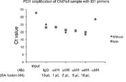
- the results are shown in FIG.
- the vertical axis represents the relative amount of immunoprecipitated DNA (input%) when the amount of DNA when chromatin DNA is fragmented is 100%. From this result, it was found that even when the internal standard molecule was added, H3K27Ac inherent in the cells bound to the DNA of the ⁇ 3′E region of the target gene Ig ⁇ and was not significantly inhibited by the addition of the internal standard molecule. It was.
Landscapes
- Health & Medical Sciences (AREA)
- Life Sciences & Earth Sciences (AREA)
- Immunology (AREA)
- Chemical & Material Sciences (AREA)
- Engineering & Computer Science (AREA)
- Molecular Biology (AREA)
- Biomedical Technology (AREA)
- Urology & Nephrology (AREA)
- Hematology (AREA)
- Biotechnology (AREA)
- Microbiology (AREA)
- General Health & Medical Sciences (AREA)
- Organic Chemistry (AREA)
- Analytical Chemistry (AREA)
- Physics & Mathematics (AREA)
- Biochemistry (AREA)
- Zoology (AREA)
- Wood Science & Technology (AREA)
- Cell Biology (AREA)
- Proteomics, Peptides & Aminoacids (AREA)
- Food Science & Technology (AREA)
- Medicinal Chemistry (AREA)
- General Physics & Mathematics (AREA)
- Pathology (AREA)
- General Engineering & Computer Science (AREA)
- Bioinformatics & Cheminformatics (AREA)
- Genetics & Genomics (AREA)
- Biophysics (AREA)
- Measuring Or Testing Involving Enzymes Or Micro-Organisms (AREA)
Abstract
免疫沈降用の内部標準分子であって、免疫沈降で用いられる抗体が認識するエピトープを含むタンパク質又はその部分ペプチドと、それに結合した核酸とを含む、内部標準分子。
Description
本発明は、免疫沈降用の内部標準分子及び免疫沈降方法に関する。
特定のタンパク質に特異的に結合する抗体は、ライフサイエンス分野の研究ツールとして汎用されている。中でも、特定のタンパク質を分離精製するために免疫沈降法は非常に利用される方法であり、様々な試薬が開発されている。また、例えば、細胞ライセート中に存在するタンパク質とタンパク質の複合体を分離精製するために共免疫沈降法も発展を遂げ、様々な共免疫沈降ツールが開発されている。
近年、転写因子などの特定のタンパク質が結合するDNA上の部位、およびその部位の塩基配列を明らかにする研究がなされている。その方法として、転写因子などの特定のタンパク質に対する抗体を用いたクロマチン免疫沈降法(Chromatin immunoprecipitation, ChIP)が用いられている(特許文献1)。
最近では、このクロマチン免疫沈降法と次世代シーケンサーによるDNAシーケンシングとを組み合わせたChIP-seq法も広く用いられている。ChIP-seq法では、まず、転写因子などの特定のタンパク質に結合する抗体を用いた免疫沈降法で得られたDNA断片の塩基配列を、次世代シーケンサーを用いて決定し、例えばヒトの全ゲノムであるリファレンス配列へマップする。そして、リファレンス配列にマップされた塩基配列の数(リード数)が多いほど、当該DNA領域はクロマチン免疫沈降法に用いた抗体と結合するタンパク質がより多く複合体を形成する領域であって、何らかの発現制御を受けている領域であると同定される。例えば、特定の物質の投与により、特定のDNA領域の塩基配列のリード数が増加すれば、そのDNA領域は、その特定の物質により抗体が認識するタンパク質がより多く複合体を形成し、発現制御に変化が生じたと評価される。
しかし、このリード数は、タンパク質と抗体との交叉率や実験操作などによる誤差に起因して、抗体等の試薬のロット間や条件の異なる試料間、同一実験条件で実施した複数の実験間において変動してしまうのが通常である。この問題に対し、2群のマップされた状態を比較して差があるものを検出する手法が開発されているが(非特許文献1、2)、この方法では変動幅は小さくても意味のある差を出力し損じるリスクがあり、定量というよりも半定量に近いのが実情である。このことは、ChIP-seq法において、得られるデータに影響を与える誤差要因を適切に補正する手段の開発と、その手段による適切に補正された定量的データの取得と、さらに複数の実験に由来する適切に補正された定量的データを用いた適切な定量的比較解析とが、観察された変動が生物学的に意味のある変動と理解する上で必要かつ重要であることを示している。
生体由来試料に含まれる標的タンパク質を定量する方法としては、内部標準ペプチドを用いた方法が開発されている(特許文献2)。本方法では、標的タンパク質に結合するペプチドを合成し、内部標準として生体由来試料に既知量を添加する。その後、プロテアーゼにてタンパク質をペプチドへ消化後、ペプチドの分離を行い、質量分析計で分析を行う。そして、既知量の内部標準ペプチドとタンパク質由来ペプチドのシグナル面積を比較することで、定量を行っている。しかし、本方法は、上記した免疫沈降法や共免疫沈降法、ChIP-seq法に適用できる方法ではない。
また、従来ChIP-seq法では、例えば、ポジティブコントロール実験として抗ヒストンH3抗体を用いた免疫沈降を行う系が使用され、ChIP-seq法用キットのなかに抗ヒストンH3抗体が含まれるものも市販されている。このヒストンH3と抗ヒストンH3抗体との結合による免疫沈降で得られる結果はChip-seq法またはそのためのキットに問題が無いことを確認するためのものであり、本発明が課題とする補正に供することはできない。その理由は第一に、ヒストンH3と抗ヒストンH3抗体の交叉率と、解析対象とする例えば転写因子タンパク質のその抗体との交叉率が等しいとはみなせないことにある。交叉率の差は沈降効率にも影響する点である。つまり、解析対象と異なるタンパク質に対する抗体を用いたChIP-seqデータは、本発明が課題とするデータの補正には使用できない。
Zhang et al, Genome Biology 2008, 9:R137
Park PJ, Nature Reviews Genetics 10, 669-680 (October 2009)
本発明は、免疫沈降法を用いた目的物質の定量において、ロット間や条件の異なる試料間の比較のための新たな内部標準分子の提供を課題とする。
本発明者等は上記課題に鑑み鋭意検討したところ、免疫沈降後に検出可能な特定の塩基配列を有する核酸と、前記免疫沈降で用いる抗体が認識するエピトープを含むタンパク質又はその部分ペプチドとが結合した内部標準分子を用いることで、上記課題を解決できることを見出した。本発明は以下の通りである。
(1)免疫沈降用の内部標準分子であって、
免疫沈降で用いられる抗体が認識するエピトープを含むタンパク質又はその部分ペプチドと、それに結合した核酸とを含む、内部標準分子。
(2)前記免疫沈降がクロマチン免疫沈降であって、
前記タンパク質がクロマチン上のDNAに結合するタンパク質であり、
前記核酸の配列は、被検試料が由来する生物種のゲノムに含まれない配列からなる、(1)に記載の内部標準分子。
(3)前記核酸の配列が酵母に由来する、(1)又は(2)に記載の内部標準分子。
(4)前記酵母に由来する核酸の配列が配列番号1~4のいずれかで表される、(3)に記載の内部標準分子。
(5)前記核酸がビオチンで標識されており、前記タンパク質又はその部分ペプチドがアビジン又はストレプトアビジンとの融合タンパク質又はペプチドであり、ビオチンとアビジン又はストレプトアビジンの相互作用により前記核酸が前記タンパク質又はその部分ペプチドと結合している、(1)~(4)のいずれかに記載の内部標準分子。
(6)前記核酸が当該核酸へのアミン修飾を介してSMCC(succinimidyl-4-(N-maleimidomethyl)cyclohexane-1-carboxylate)で標識されており、前記タンパク質又はその部分ペプチドが当該タンパク質またはその部分ペプチドの末端にシステイン残基が付加されたシステイン付加タンパク質又はその部分ペプチドであり、SMCCとシステイン残基との相互作用により前記核酸が前記タンパク質又はその部分ペプチドと結合している、(1)~(4)のいずれかに記載の内部標準分子。
(7)前記タンパク質が転写因子、転写制御因子、ヒストンタンパク質、またはクロマチン制御因子である、(1)~(6)のいずれかに記載の内部標準分子。
(8)前記転写因子又は転写制御因子がIRF4タンパク質、p53タンパク質、NF-κB、Rbタンパク質、BRCA1タンパク質、MYCタンパク質、またはPax5タンパク質である、(7)に記載の内部標準分子。
(9)以下の工程を含む免疫沈降方法。
被検試料と(1)~(8)のいずれかに記載の内部標準分子とを混合する工程、
免疫沈降工程、及び、
前記内部標準分子の核酸部分を検出することによって免疫沈降結果の標準化を行う工程。
(10)以下の工程を含むクロマチン免疫沈降方法。
被検試料と(1)~(8)のいずれかに記載の内部標準分子とを混合する工程、
クロマチン免疫沈降工程、
前記クロマチン免疫沈降工程で得られた試料中の目的DNA及び内部標準分子のDNA部分を増幅する工程、及び、
前記増幅工程で得られた内部標準分子のDNA部分の増幅DNAの量を指標として、試料中の目的断片DNAの増幅DNAの量を補正する工程。
(11)以下の工程を含むクロマチン免疫沈降方法。
被検試料と(1)~(8)のいずれかに記載の内部標準分子とを混合する工程、
クロマチン免疫沈降工程、
前記クロマチン免疫沈降工程で得られた試料中の目的DNA及び内部標準分子のDNA部分を増幅する工程、
前記増幅工程で増幅した試料中の目的DNA及び内部標準分子のDNA部分の塩基配列を解析する工程、及び、
前記塩基配列を解析する工程で得られた内部標準分子のDNA部分の塩基配列のシーケンス量を指標として、試料中の目的DNAの塩基配列のシーケンス量を補正する工程。
本発明の内部標準分子を用いることで、免疫沈降法を用いた目的物質の定量において、ロット間や条件の異なる試料間の比較のための補正を従来よりも正確に行うことができる。
本発明は、免疫沈降用の内部標準分子に係る発明(第一の発明)、及び、免疫沈降方法に係る発明(第二の発明)を含む。
<1.第一の発明>
本発明の第一の発明は、
免疫沈降用の内部標準分子であって、
前記免疫沈降で用いられる抗体が認識するエピトープを含むタンパク質又はその部分ペプチドと、それに結合した核酸とを含む、内部標準分子である。
本発明の第一の発明は、
免疫沈降用の内部標準分子であって、
前記免疫沈降で用いられる抗体が認識するエピトープを含むタンパク質又はその部分ペプチドと、それに結合した核酸とを含む、内部標準分子である。
[免疫沈降]
免疫沈降は、当該技術分野において公知の技術である。要すれば、可溶性の抗原を抗原特異的抗体に特異的に反応させて沈殿させる免疫沈降反応を利用して抗原を検出、分離、および/または精製する手法のことである。本発明における免疫沈降は特段限定されず、例えば、一般的な免疫沈降、共免疫沈降、クロマチン免疫沈降などが挙げられる。本発明においては、免疫沈降の前後に他の実験系が組まれてもよく、実験系全体のうちの一の系として免疫沈降の系が組まれてもよい。
免疫沈降は、当該技術分野において公知の技術である。要すれば、可溶性の抗原を抗原特異的抗体に特異的に反応させて沈殿させる免疫沈降反応を利用して抗原を検出、分離、および/または精製する手法のことである。本発明における免疫沈降は特段限定されず、例えば、一般的な免疫沈降、共免疫沈降、クロマチン免疫沈降などが挙げられる。本発明においては、免疫沈降の前後に他の実験系が組まれてもよく、実験系全体のうちの一の系として免疫沈降の系が組まれてもよい。
(クロマチン免疫沈降)
本発明における免疫沈降は好ましくはクロマチン免疫沈降である。上記した通り、本発明の第一の発明の内部標準分子を使用した免疫沈降法では、目的物質の定量において、ロット間や条件の異なる試料間の比較のための補正を従来よりも正確に行うことができる。
クロマチン免疫沈降の場合には、クロマチン上のDNAと結合する転写因子などのタンパク質のように、解離定数が大きいタンパク質や、結合が安定しないタンパク質が対象となるところ、ロット間や条件の異なる試料間の比較のための補正を従来よりも正確に行うことができることの効果は大きい。
本発明における免疫沈降は好ましくはクロマチン免疫沈降である。上記した通り、本発明の第一の発明の内部標準分子を使用した免疫沈降法では、目的物質の定量において、ロット間や条件の異なる試料間の比較のための補正を従来よりも正確に行うことができる。
クロマチン免疫沈降の場合には、クロマチン上のDNAと結合する転写因子などのタンパク質のように、解離定数が大きいタンパク質や、結合が安定しないタンパク質が対象となるところ、ロット間や条件の異なる試料間の比較のための補正を従来よりも正確に行うことができることの効果は大きい。
[内部標準分子]
本発明の第一の発明の内部標準分子は免疫沈降用の内部標準分子であり、核酸と、前記免疫沈降で用いる抗体が認識するエピトープを含むタンパク質又はその部分ペプチドとが結合したものである。以下に、本発明の第一の発明の内部標準分子の詳細を記載する。
本発明の第一の発明の内部標準分子は免疫沈降用の内部標準分子であり、核酸と、前記免疫沈降で用いる抗体が認識するエピトープを含むタンパク質又はその部分ペプチドとが結合したものである。以下に、本発明の第一の発明の内部標準分子の詳細を記載する。
[核酸]
本発明の第一の発明の内部標準分子は核酸を含む。
本発明の第一の発明の内部標準分子は核酸を含む。
核酸の種類としては、例えば、DNAやRNA、人工核酸などが挙げられる。合成の容易さ、他の実験系との関連性、分解しにくい安定性を有する等の観点から、好ましくはDNAであり、より好ましくは2本鎖DNAである。修飾塩基を含むものでもよい。
核酸の塩基配列としては、ハイブリダイゼーションやPCRで検出可能であれば特段限定されないが、被験試料中の物質との結合などの相互作用を極力小さくするために、被検試料が由来する生物種のゲノム中に含まれない配列からなることが好ましい。すなわち、クロマチン免疫沈降に使用する場合、試料の染色体上には含まれない配列とすることが好ましい。さらに、被験試料中の物質との結合などの相互作用を極力小さくするために、被検試料が由来する生物種の遺伝子制御配列等のタンパク質結合配列を含まないことが好ましい。
例えば、酵母などの菌類、大腸菌などの細菌、ショウジョウバエなどに由来する配列が挙げられる。中でも、被検試料として哺乳動物の試料を使用する場合、好ましくは酵母、大腸菌などの細菌に由来する配列であり、より好ましくは酵母に由来する配列である。
酵母に由来する核酸の配列としては、例えば、配列番号1~配列番号4で表される配列が挙げられる。
なお、どの生物種にも由来しない人工配列でもよい。
酵母に由来する核酸の配列としては、例えば、配列番号1~配列番号4で表される配列が挙げられる。
なお、どの生物種にも由来しない人工配列でもよい。
また、PCRで増幅する際のプライマー設計の都合上、リピート配列を含まないことが好ましい。
その他、核酸としては、PCR反応やハイブリダイゼーション反応で検出可能であれば、その種類、GC含量、塩基配列の長さ等に特に制限はないが、GC含量としては、好ましくは30%以上、より好ましくは40%以上であり、好ましくは70%以下、より好ましくは60%以下である。当該範囲内のGC含量にあることで、非特異的配列の増幅を抑制し、目的配列を効率的に増幅することができる。
核酸の塩基配列の長さとしては、後述するPCRでDNAが増幅すれば特段限定されないが、通常15塩基以上、好ましくは20塩基以上、より好ましくは25塩基以上であり、通常200塩基以下、好ましくは100塩基以下である。
(核酸の製造方法)
選択された塩基配列を有する核酸の製造方法は特段限定されず、公知の方法を用いることができる。例えば、塩基配列の長さが短ければ、公知の方法で人工的に合成してもよく、公知の受託サービスを利用してもよい。
また、公知の遺伝子工学的手法により製造することもできる。例えば、選択した塩基配列が由来する生物種のゲノムDNAを鋳型とし、目的とする塩基配列を増幅できるように設計したプライマーセットを使用したPCRを行うことによって得ることもできる。
選択された塩基配列を有する核酸の製造方法は特段限定されず、公知の方法を用いることができる。例えば、塩基配列の長さが短ければ、公知の方法で人工的に合成してもよく、公知の受託サービスを利用してもよい。
また、公知の遺伝子工学的手法により製造することもできる。例えば、選択した塩基配列が由来する生物種のゲノムDNAを鋳型とし、目的とする塩基配列を増幅できるように設計したプライマーセットを使用したPCRを行うことによって得ることもできる。
[タンパク質]
本発明の第一の発明の内部標準分子はタンパク質を含む。前記タンパク質は、前記免疫沈降で用いる抗体が認識するエピトープを含むタンパク質又はその部分ペプチドである。また、前記タンパク質は、当該抗体が認識するエピトープを含むタンパク質又はその部分ペプチドにさらにタグ配列が付与されたタンパク質又はその部分ペプチドであってもよい。ここで、タグ配列とは、該抗体が認識するエピトープを含むタンパク質又はその部分ペプチドの標識として機能するタンパク質またはペプチドをいう。タグ配列の具体的なものとしては、例えば、ヒスチジン(His)タグ、グルタチオン-S-トランスフェラーゼ(GST)タグ、FLAGタグが挙げられる。当該タグ配列は、市販される当該タグ配列との融合タンパク質を発現する発現ベクターを用いた遺伝子組み換え技術を使用することにより、当該抗体が認識するエピトープを含むタンパク質又はその部分ペプチドに付与することができる。タグ配列は、例えば、当該抗体が認識するエピトープを含むタンパク質又はその部分ペプチドの回収や精製に利用することができる。
本発明の第一の発明の内部標準分子はタンパク質を含む。前記タンパク質は、前記免疫沈降で用いる抗体が認識するエピトープを含むタンパク質又はその部分ペプチドである。また、前記タンパク質は、当該抗体が認識するエピトープを含むタンパク質又はその部分ペプチドにさらにタグ配列が付与されたタンパク質又はその部分ペプチドであってもよい。ここで、タグ配列とは、該抗体が認識するエピトープを含むタンパク質又はその部分ペプチドの標識として機能するタンパク質またはペプチドをいう。タグ配列の具体的なものとしては、例えば、ヒスチジン(His)タグ、グルタチオン-S-トランスフェラーゼ(GST)タグ、FLAGタグが挙げられる。当該タグ配列は、市販される当該タグ配列との融合タンパク質を発現する発現ベクターを用いた遺伝子組み換え技術を使用することにより、当該抗体が認識するエピトープを含むタンパク質又はその部分ペプチドに付与することができる。タグ配列は、例えば、当該抗体が認識するエピトープを含むタンパク質又はその部分ペプチドの回収や精製に利用することができる。
本発明の第一の発明の内部標準分子が含むタンパク質とは、通常、アミノ酸配列全長を意味するが、シグナル配列が除かれた成熟型タンパク質の全長や変異型タンパク質も含まれる。全長の場合、本発明の第一の発明の内部標準分子が含むタンパク質は、前記免疫沈降で用いる抗体が認識するエピトープを当然に含んでいる。
本発明の第一の発明の内部標準分子が含むタンパク質としては特段限定されず、アミノ配列やその長さ等に制限はない。内部標準分子が含むタンパク質としては、例えば、DNA結合タンパク質が好ましく、具体的には、転写因子、転写制御因子、ヒストンタンパク質、クロマチン制御因子などが挙げられる。
転写因子としては特段限定されないが、例えば、IRF4タンパク質、p53タンパク質、NF-κB、MYCタンパク質、Pax5タンパク質などが挙げられる。転写制御因子としては特段限定されないが、Rbタンパク質、BRCA1タンパク質などが挙げられる。
ヒストンタンパク質としては特段限定されないが、例えば、H2Aタンパク質、H2Bタンパク質、H3タンパク質、H4タンパク質などのコアヒストンタンパク質、H1タンパク質などのリンカーヒストンなどが挙げられる。
また、これらはバリアントであってもよい。さらに、これらのヒストンタンパク質は化学修飾を受けていてもよい。例えば、アセチル化、リン酸化、メチル化、ユビキチン化がされていてもよい。メチル化の場合、モノメチル化、ジメチル化、トリメチル化されていてもよい。
H3タンパク質であれば、例えば、9番目のリジン残基がメチル化されている場合(H3K9me)、27番目のリジン残基がメチル化されている場合(H3K27me)、9番目のリジン残基がアセチル化されている場合(H3K9Ac)などが挙げられる。また、9番目のリジン残基がトリメチル化されている場合(H3K9me3)、27番目のリジン残基がトリメチル化されている場合(H3K27me3)なども挙げられる。
同様に、H2Aタンパク質であれば、例えば、5番目のリジン残基がアセチル化されている場合(H2AK5Ac)、1番目のセリン残基がリン酸化されている場合(H2AS1ph)、120番目のスレオニン残基がリン酸化されている場合(H2AT120ph)、119番目のリジン残基がユビキチン化されている場合(H2AK119ub)などが挙げられる。
同様に、H2Bタンパク質であれば、例えば、5番目のリジン残基がアセチル化されている場合(H2BK5Ac)、14番目のセリン残基がリン酸化されている場合(H2BS14ph)、120番目のリジン残基がユビキチン化されている場合(H2BK120ub)などが挙げられる。
同様に、H4タンパク質であれば、例えば、5番目のリジン残基がアセチル化されている場合(H4K5Ac)、1番目のセリン残基がリン酸化されている場合(H4S1ph)、3番目のアルギニン残基がメチル化されている場合(H4R3me)などが挙げられる。
クロマチン制御因子としては特段限定されないが、例えば、Brg1、p300、Pc4などが挙げられる。
[部分ペプチド]
本発明の第一の発明の内部標準分子が含むタンパク質の部分ペプチドとしては、免疫沈降で用いる抗体が認識するエピトープを含んでいれば特段限定されないが、例えば、20~100アミノ酸の部分ペプチドが例示される。
本発明の第一の発明の内部標準分子が含むタンパク質の部分ペプチドとしては、免疫沈降で用いる抗体が認識するエピトープを含んでいれば特段限定されないが、例えば、20~100アミノ酸の部分ペプチドが例示される。
(タンパク質又はその部分ペプチドの製造方法)
タンパク質又はその部分ペプチドのアミノ酸配列の長さが短い場合、その製造方法は特段限定されず、公知のペプチド合成法に従って製造することができる。例えば、固相合成法、液相合成法が挙げられる。精製方法としては、例えば、溶媒抽出、蒸留、カラムクロマトグラフィー、液体クロマトグラフィー、再結晶などを組み合わせることでき、これにより、ペプチドを単離精製できる。得られるペプチドが遊離体である場合には、公知の方法によって適当な塩に変換することができ、逆に塩で得られた場合には、公知の方法によって遊離体または他の塩に変換することもできる。ペプチドに抗体が認識するリン酸化、アセチル化、メチル化などの修飾を入れることもできる。
タンパク質又はその部分ペプチドのアミノ酸配列の長さが短い場合、その製造方法は特段限定されず、公知のペプチド合成法に従って製造することができる。例えば、固相合成法、液相合成法が挙げられる。精製方法としては、例えば、溶媒抽出、蒸留、カラムクロマトグラフィー、液体クロマトグラフィー、再結晶などを組み合わせることでき、これにより、ペプチドを単離精製できる。得られるペプチドが遊離体である場合には、公知の方法によって適当な塩に変換することができ、逆に塩で得られた場合には、公知の方法によって遊離体または他の塩に変換することもできる。ペプチドに抗体が認識するリン酸化、アセチル化、メチル化などの修飾を入れることもできる。
タンパク質又はその部分ペプチドのアミノ酸配列の長さが長い場合、又は、短い場合であっても、遺伝子工学的手法によって製造することもできる。例えば、タンパク質又はその部分ペプチドをコードするDNAを作製し、該DNAを用いて組換え発現ベクターを構築し、宿主に導入して発現させ、精製して製造することができる。いずれも公知の方法を用いて行うことができる。
上記したタンパク質又はその部分ペプチドはストレプトアビジンタグ、Hisタグ、GSTタグ、Flagタグなどのタグ配列を含む融合タンパク質であってもよい。
[核酸とタンパク質又はその部分ペプチドとの結合]
(核酸の修飾)
本発明の第一の発明の内部標準分子は、核酸とタンパク質又は部分ペプチドとが結合しているものである。その結合態様は特段限定されないが、不可逆的な結合が好ましい。核酸側については、例えば、核酸の末端を、タンパク質又は部分ペプチドと結合できるように修飾することなどが挙げられる。
核酸の修飾の態様は、タンパク質又は部分ペプチドと結合するのであれば特段限定されないが、例えば、核酸の末端に、後述するタンパク質またはペプチドに導入した標識物質や官能基と反応して結合する標識物質や官能基を導入することが挙げられる。例えば、核酸の末端にビオチン、チオール基、アミノ基などの標識物質を導入することなどが挙げられる。尚、これらの標識物質や官能基はスペーサーを介して核酸に導入されてもよい。
(核酸の修飾方法)
核酸の修飾の方法は特段限定されず、核酸をPCRで増幅する際に末端に標識塩基が導入されたプライマーを用いる方法、核酸合成機で核酸を合成する際に末端塩基として標識塩基を用いる方法など、公知の方法に従うことができる。
例えば、核酸をPCRで増幅する際のプライマーとして、5’末端がビオチン化されたDNAや5’末端がアミノ化された核酸を用いることなどが挙げられる。5’末端がアミノ化されたDNAを用いる場合には、さらに、該アミノ基を介してN-ヒドロキシスクシンイミド活性エステルと結合する方法が挙げられる。例えば、N-ヒドロキシスクシンイミド活性エステルとマレイミド基を導入した二価性試薬であるSMCC(succinimidyl-4-(N-maleimidomethyl)cyclohexane-1-carboxylate)のN-ヒドロキシスクシンイミド活性エステルと結合させる方法が挙げられる。
(タンパク質又は部分ペプチドの修飾)
本発明の第一の発明の内部標準分子は、その内部標準分子が含む核酸とタンパク質又は部分ペプチドと結合しているものである。タンパク質又は部分ペプチド側については、核酸と結合できるように修飾される。
タンパク質又は部分ペプチドの修飾の態様は、核酸と結合し、免疫沈降で用いる抗体が認識できるのであれば、特段限定されないが、例えば、タンパク質又は部分ペプチドに、核酸に導入した前記標識物質や官能基と反応して結合する標識物質や官能基を導入することが挙げられる。また、タンパク質又は部分ペプチドの修飾は、タンパク質又は部分ペプチドの末端であってもよく、末端でなくてもよい。
本発明の第一の発明の内部標準分子は、その内部標準分子が含む核酸とタンパク質又は部分ペプチドと結合しているものである。タンパク質又は部分ペプチド側については、核酸と結合できるように修飾される。
タンパク質又は部分ペプチドの修飾の態様は、核酸と結合し、免疫沈降で用いる抗体が認識できるのであれば、特段限定されないが、例えば、タンパク質又は部分ペプチドに、核酸に導入した前記標識物質や官能基と反応して結合する標識物質や官能基を導入することが挙げられる。また、タンパク質又は部分ペプチドの修飾は、タンパク質又は部分ペプチドの末端であってもよく、末端でなくてもよい。
タンパク質又は部分ペプチドの修飾としては、例えば、核酸としてビオチン標識核酸を用いる場合には、アビジン標識やストレプトアビジン標識が挙げられる。また核酸としてアミノ基を介してN-ヒドロキシスクシンイミド活性エステルと結合した核酸を用いる場合には、システイン標識が挙げられる。システインは内在のシステインを標識してもよいし、新たにシステイン残基を導入してもよい。
(タンパク質又は部分ペプチドの修飾方法)
タンパク質又はその部分ペプチドを修飾する方法は特段限定されないが、遺伝子工学的手法を用いることができる。例えば、上記したタンパク質又はその部分ペプチドの製造方法において、ベクターを作製する際に、アビジンやストレプトアビジンなどの標識分子をコードするDNAとタンパク質又はその部分ペプチドをコードするDNAと連結するなどしてベクターに挿入し、融合タンパク質として発現させることができる。
このとき、タンパク質又は部分ペプチドが核酸と結合し、免疫沈降で用いる抗体が認識できるのであれば、標識分子をコードするDNAは、タンパク質又は部分ペプチドをコードするDNAの上流に配置されても下流に配置されても構わない。
(核酸とタンパク質又はその部分ペプチドとの結合方法)
核酸とタンパク質又はその部分ペプチドとの結合は公知の方法で行うことができる。
例えば、末端がビオチン修飾された核酸と、ストレプトアビジンとタンパク質との融合タンパク質との結合であれば、単に両者を溶液中で混合すればよい。その割合は実験により変えればよい。
核酸とタンパク質又はその部分ペプチドとの結合は公知の方法で行うことができる。
例えば、末端がビオチン修飾された核酸と、ストレプトアビジンとタンパク質との融合タンパク質との結合であれば、単に両者を溶液中で混合すればよい。その割合は実験により変えればよい。
<2.免疫沈降方法>
本発明の第二の発明の免疫沈降方法は、
被検試料と前記内部標準分子とを混合する工程、
免疫沈降工程、及び、
前記内部標準分子の核酸部分を検出することによって標準化を行う工程
を含む免疫沈降方法である。
本発明の第二の発明の免疫沈降方法は、
被検試料と前記内部標準分子とを混合する工程、
免疫沈降工程、及び、
前記内部標準分子の核酸部分を検出することによって標準化を行う工程
を含む免疫沈降方法である。
[被検試料と内部標準分子とを混合する工程]
本工程における被検試料と内部標準分子との混合方法は特段限定されず、ピペッティングなどの当該技術分野で通常に行われる方法で行えばよい。
本工程における被検試料と内部標準分子との混合方法は特段限定されず、ピペッティングなどの当該技術分野で通常に行われる方法で行えばよい。
[免疫沈降工程]
免疫沈降の方法は特段限定されず、公知の免疫沈降法を用いることができる。抗体をビーズなどの担体に結合させ、それを被検試料と内部標準分子との混合液に添加し、一定時間、例えば、30分~1日インキュベートしたのち、遠心分離などで沈降物を取得する。沈降物をバッファーなどで洗浄したのち、次の工程に使用する。
免疫沈降の方法は特段限定されず、公知の免疫沈降法を用いることができる。抗体をビーズなどの担体に結合させ、それを被検試料と内部標準分子との混合液に添加し、一定時間、例えば、30分~1日インキュベートしたのち、遠心分離などで沈降物を取得する。沈降物をバッファーなどで洗浄したのち、次の工程に使用する。
免疫沈降における抗体としては、通常の免疫沈降法と同様に、目的の断片DNAに結合していることが予想されるタンパク質に対する抗体を使用すればよく、この抗体で免疫沈降することにより、目的のタンパク質とそれに結合している断片DNAの複合体、及び、内部標準分子を沈降させることができる。
そして、内部標準分子の核酸部分を検出し、その検出結果に基づいて、免疫沈降の測定結果を標準化する。通常の共免疫沈降の測定結果についても、異なる試料間で実験条件間のばらつきなどを補正することが可能となる。
そして、内部標準分子の核酸部分を検出し、その検出結果に基づいて、免疫沈降の測定結果を標準化する。通常の共免疫沈降の測定結果についても、異なる試料間で実験条件間のばらつきなどを補正することが可能となる。
以下に、クロマチン免疫沈降の具体的な態様を説明する。ただし、これらの例には限定されない。
<2-1.クロマチン免疫沈降の第一の実施態様>
本発明の第二の発明のクロマチン免疫沈降方法の第一の実施態様は、
被検試料と前記内部標準分子とを混合する工程、
クロマチン免疫沈降工程、
前記クロマチン免疫沈降工程で得られた試料中の目的DNA及び内部標準分子のDNA部分を増幅する工程、及び、
前記増幅工程で得られた内部標準分子のDNA部分の増幅DNAの量を指標として、試料中の目的断片DNAの増幅DNAの量を補正する工程
を含むクロマチン免疫沈降方法である。
本発明の第二の発明のクロマチン免疫沈降方法の第一の実施態様は、
被検試料と前記内部標準分子とを混合する工程、
クロマチン免疫沈降工程、
前記クロマチン免疫沈降工程で得られた試料中の目的DNA及び内部標準分子のDNA部分を増幅する工程、及び、
前記増幅工程で得られた内部標準分子のDNA部分の増幅DNAの量を指標として、試料中の目的断片DNAの増幅DNAの量を補正する工程
を含むクロマチン免疫沈降方法である。
[被検試料と内部標準分子とを混合する工程]
本工程における被検試料と内部標準分子との混合方法は特段限定されず、ピペッティングなどの当該技術分野で通常に行われる方法で行えばよい。
本工程における被検試料と内部標準分子との混合方法は特段限定されず、ピペッティングなどの当該技術分野で通常に行われる方法で行えばよい。
[クロマチン免疫沈降工程]
クロマチン免疫沈降の方法は特段限定されず、公知のクロマチン免疫沈降法を用いることができる。
例えば、被検試料中に存在するクロマチン上のDNAとタンパク質との複合体をホルムアルデヒドなどの架橋剤で架橋して(例えば、室温、10分の条件)、その後、細胞を回収し、DNAを100~500bpの長さに超音波破砕する。そして、その超音波破砕されたDNAとタンパク質との複合体を含む試料を用いて免疫沈降を行うという方法が挙げられる。
被検試料と内部標準分子とを混合しておくと、クロマチン上のDNAとタンパク質との複合体とともに内部標準分子も抗体によって沈降される。
クロマチン免疫沈降の方法は特段限定されず、公知のクロマチン免疫沈降法を用いることができる。
例えば、被検試料中に存在するクロマチン上のDNAとタンパク質との複合体をホルムアルデヒドなどの架橋剤で架橋して(例えば、室温、10分の条件)、その後、細胞を回収し、DNAを100~500bpの長さに超音波破砕する。そして、その超音波破砕されたDNAとタンパク質との複合体を含む試料を用いて免疫沈降を行うという方法が挙げられる。
被検試料と内部標準分子とを混合しておくと、クロマチン上のDNAとタンパク質との複合体とともに内部標準分子も抗体によって沈降される。
[前記クロマチン免疫沈降工程で得られた試料中の目的DNA及び内部標準分子のDNA部分を増幅する工程]
本工程における増幅方法は、目的のDNA及び内部標準分子のDNAを増幅させることができ、後工程における増幅DNAの定量ができれば特段限定されず、公知の方法を用いることができる。PCR法が好適に使用され、例えば、リアルタイムPCR法などが挙げられる。
本工程における増幅方法は、目的のDNA及び内部標準分子のDNAを増幅させることができ、後工程における増幅DNAの定量ができれば特段限定されず、公知の方法を用いることができる。PCR法が好適に使用され、例えば、リアルタイムPCR法などが挙げられる。
本工程では、試料中の目的DNAを増幅するプライマーセットと、内部標準分子のDNAを増幅するプライマーセットとを用いればよく、それぞれのDNAがそれぞれに増幅されるプライマーセットを用いればよい。
リアルタイムPCR法を用いて試料中の目的DNA及び内部標準分子のDNAを増幅させる場合の方法としては、公知の方法を用いることができる。例えば、インターカレーション法やハイブリダイゼーション法、LUX法などが挙げられる。
インターカレーション法では、例えば、SYBR Green Iのように、二本鎖DNAに特異的にインターカレートして蛍光を発する色素などを用いることができる。
ハイブリダイゼーション法では、例えば、TaqManプローブのように、DNA配列に特異的なオリゴヌクレオチドに蛍光色素を結合させたプローブなどを用いることができる。
インターカレーション法では、例えば、SYBR Green Iのように、二本鎖DNAに特異的にインターカレートして蛍光を発する色素などを用いることができる。
ハイブリダイゼーション法では、例えば、TaqManプローブのように、DNA配列に特異的なオリゴヌクレオチドに蛍光色素を結合させたプローブなどを用いることができる。
[増幅工程で得られた内部標準分子のDNA部分の増幅量を指標として、試料中の目的DNAの量を補正する工程]
被検試料と混合した内部標準分子のDNA部分の量は既知である。よって、本工程において、増幅された内部標準分子のDNA部分が定量できれば、これを指標として、公知の方法、例えば、Ct値を比較する方法、微量流路電気泳動の方法を用いて、増幅された、試料中の目的DNA量を補正し、定量することができる。
被検試料と混合した内部標準分子のDNA部分の量は既知である。よって、本工程において、増幅された内部標準分子のDNA部分が定量できれば、これを指標として、公知の方法、例えば、Ct値を比較する方法、微量流路電気泳動の方法を用いて、増幅された、試料中の目的DNA量を補正し、定量することができる。
一般的にクロマチン免疫沈降法では、沈降するDNAとタンパク質との複合体の量が多いほど、タンパク質の当該DNA領域への結合量が多いと判定される。そこで、試料中の目的DNAとして、転写因子の結合配列として既知の配列を想定し、当該既知配列を増幅するためのプライマーセットを用いた場合は、免疫沈降後の増幅工程において得られる増幅量が多いほど転写因子の結合量が多いと判断することができる。そして、この量を内部標準の増幅量で補正することで、より正確に結合量を把握することができる。
例えば、特定の物質の投与により、沈降するDNAとタンパク質との複合体の量が増加すれば、そのDNA領域に対するタンパク質の結合量がその特定の物質により増加した、すなわち、発現制御に変化が生じたと評価される。また、疾患患者と健常人での比較、および変異保有者と非保有者との比較を行うことで、疾患や変異とタンパク質結合量の関係を調べることもできる。
例えば、特定の物質の投与により、沈降するDNAとタンパク質との複合体の量が増加すれば、そのDNA領域に対するタンパク質の結合量がその特定の物質により増加した、すなわち、発現制御に変化が生じたと評価される。また、疾患患者と健常人での比較、および変異保有者と非保有者との比較を行うことで、疾患や変異とタンパク質結合量の関係を調べることもできる。
<2-3.クロマチン免疫沈降の第二の実施態様>
本発明の第二の発明のクロマチン免疫沈降方法の第二の実施態様は、
被検試料と前記内部標準分子とを混合する工程、
クロマチン免疫沈降工程、
前記クロマチン免疫沈降工程で得られた試料中の目的DNA及び内部標準分子のDNA部分を増幅する工程、
前記増幅工程で増幅した試料中の目的DNA及び内部標準分子のDNA部分の塩基配列を解析する工程、及び、
前記塩基配列を解析する工程で得られた内部標準分子のDNA部分の塩基配列のシーケンス量を指標として、試料中の目的DNAの塩基配列のシーケンス量を補正する工程
を含む免疫沈降方法である。
本発明の第二の発明のクロマチン免疫沈降方法の第二の実施態様は、
被検試料と前記内部標準分子とを混合する工程、
クロマチン免疫沈降工程、
前記クロマチン免疫沈降工程で得られた試料中の目的DNA及び内部標準分子のDNA部分を増幅する工程、
前記増幅工程で増幅した試料中の目的DNA及び内部標準分子のDNA部分の塩基配列を解析する工程、及び、
前記塩基配列を解析する工程で得られた内部標準分子のDNA部分の塩基配列のシーケンス量を指標として、試料中の目的DNAの塩基配列のシーケンス量を補正する工程
を含む免疫沈降方法である。
[被検試料と内部標準分子とを混合する工程]
本工程については、上記した第一の実施態様における「被検試料と前記内部標準分子とを混合する工程」の説明が適用される。
本工程については、上記した第一の実施態様における「被検試料と前記内部標準分子とを混合する工程」の説明が適用される。
[クロマチン免疫沈降工程]
本工程については、上記した第一の実施態様における「免疫沈降工程」の説明が適用される。
本工程については、上記した第一の実施態様における「免疫沈降工程」の説明が適用される。
[前記クロマチン免疫沈降工程で得られた目的の断片DNA及び内部標準分子のDNA部分を増幅する工程]
本工程については、上記した第一の実施態様における「前記クロマチン免疫沈降工程で得られた試料中の目的DNA及び内部標準分子のDNA部分を増幅する工程」の説明が適用される。
本工程については、上記した第一の実施態様における「前記クロマチン免疫沈降工程で得られた試料中の目的DNA及び内部標準分子のDNA部分を増幅する工程」の説明が適用される。
[増幅した試料中の目的DNA及び内部標準分子のDNA部分の塩基配列を解析する工程]
前記増幅工程で増幅した試料中の目的DNA及び内部標準分子のDNA部分の塩基配列の解析方法は、特段限定されないが、例えば、次世代シーケンサーを用いて行うことができる。市販されている次世代シーケンサーとしては、例えば、Illumina社、SOLiD社、Helicos社、Complete Genomics社、Polonator社、Ion Torrent社、Pacific Biosciences社の次世代シーケンサーや次次世代シーケンサーが挙げられる(例えば、Mardis, Annu. Rev. Genom. Human Genet. (2008) vol.9, p.387-402およびPersson et al., Chem. Soc. Rev. (2010) vol.39, p.985-999)。次世代シークエンサーは、DNAポリメラーゼまたはDNAリガーゼによる逐次的DNA合成法を用いて、蛍光・発光など光検出により超並列的に塩基配列を決定する原理のものである。DNAポリメラーゼを用いる方法として、GS FLX(Roche Diagnostics)、Solexa (Illumina)等が、またDNAリガーゼを用いる方法としてSOLiD(Applied Biosystems)、Polonator (Dover Systems)等が知られている。また、DNA1分子を鋳型としてDNAポリメラーゼによるDNA合成を行い、1塩基ごとの反応を蛍光・発光などの光で検出することにより、1分子リアルタイム観察により塩基配列を決定するHeliScope (Helicos Biosciences)、SMRT (Pacific Biosciences)等が知られている。その他に、Post-light シークエンシングと称される蛍光・発光など光検出以外の検出方法により、超並列的に塩基配列を決定する原理も報告されており、半導体(CMOS)チップを用いて、DNA ポリメラーゼにより各塩基が取り込まれるときに放出される水素イオン(pH変化)を検出することで塩基配列を決定する、Ion Torrent Systems等がこれに該当する。他にもNanopore(Oxford Nanopore Technologies)等が知られている。
前記増幅工程で増幅した試料中の目的DNA及び内部標準分子のDNA部分の塩基配列の解析方法は、特段限定されないが、例えば、次世代シーケンサーを用いて行うことができる。市販されている次世代シーケンサーとしては、例えば、Illumina社、SOLiD社、Helicos社、Complete Genomics社、Polonator社、Ion Torrent社、Pacific Biosciences社の次世代シーケンサーや次次世代シーケンサーが挙げられる(例えば、Mardis, Annu. Rev. Genom. Human Genet. (2008) vol.9, p.387-402およびPersson et al., Chem. Soc. Rev. (2010) vol.39, p.985-999)。次世代シークエンサーは、DNAポリメラーゼまたはDNAリガーゼによる逐次的DNA合成法を用いて、蛍光・発光など光検出により超並列的に塩基配列を決定する原理のものである。DNAポリメラーゼを用いる方法として、GS FLX(Roche Diagnostics)、Solexa (Illumina)等が、またDNAリガーゼを用いる方法としてSOLiD(Applied Biosystems)、Polonator (Dover Systems)等が知られている。また、DNA1分子を鋳型としてDNAポリメラーゼによるDNA合成を行い、1塩基ごとの反応を蛍光・発光などの光で検出することにより、1分子リアルタイム観察により塩基配列を決定するHeliScope (Helicos Biosciences)、SMRT (Pacific Biosciences)等が知られている。その他に、Post-light シークエンシングと称される蛍光・発光など光検出以外の検出方法により、超並列的に塩基配列を決定する原理も報告されており、半導体(CMOS)チップを用いて、DNA ポリメラーゼにより各塩基が取り込まれるときに放出される水素イオン(pH変化)を検出することで塩基配列を決定する、Ion Torrent Systems等がこれに該当する。他にもNanopore(Oxford Nanopore Technologies)等が知られている。
[前記塩基配列を解析する工程で得られた内部標準分子のDNA部分の塩基配列のシーケンス量を指標として、試料中の目的DNAの塩基配列のシーケンス量を補正する工程]
増幅された試料中の目的DNAの塩基配列を、被検試料が由来する生物種のゲノム配列(リファレンス配列)にマップすることにより、解析された、試料中の目的DNAの塩基配列それぞれのシーケンス量が分かる。
被検試料と混合した内部標準分子のDNA部分の量は既知である。よって、内部標準分子のDNA部分の塩基配列のシーケンス量を指標として、試料中の目的DNAの塩基配列のシーケンス量を補正することができる。
増幅された試料中の目的DNAの塩基配列を、被検試料が由来する生物種のゲノム配列(リファレンス配列)にマップすることにより、解析された、試料中の目的DNAの塩基配列それぞれのシーケンス量が分かる。
被検試料と混合した内部標準分子のDNA部分の量は既知である。よって、内部標準分子のDNA部分の塩基配列のシーケンス量を指標として、試料中の目的DNAの塩基配列のシーケンス量を補正することができる。
一般的にChIP-seq法では、リファレンス配列にマップされた塩基配列の数(リード数)が多いほど、当該DNA領域はクロマチン免疫沈降法に用いた抗体と結合するタンパク質と複合体を形成する領域であって、何らかの発現制御を受けている領域であると同定される。
しかし、このリード数は、タンパク質と抗体との交叉率や実験操作などによる誤差に起因して、ロットや条件の異なる試料間において異なるのが通常である。そのため、例えば、2群の被検試料に対して特定の物質を投与し、その結果、特定のDNA領域の塩基配列のリード数に差が生じた場合、その差が、該特定の物質が投与されたことに起因する有意な差なのか、または上記した誤差なのかが判断できない。
このような場合に本実施態様の免疫沈降方法を用いれば、内部標準分子のDNA部分の塩基配列のシーケンス量を指標として、2群の被検試料中の目的DNAの塩基配列のシーケンス量を補正することができ、得られた差が、該特定の物質が投与されたことに起因する有意な差なのか、または誤差なのかを判断することができる。例えば、特定のタンパク質の発現量が小さく、バックグラウンドとの差が顕著に表れない場合などに、誤差を有意な差と判断してしまうリスクや、逆に有意な差を誤差と判断してしまうリスクを低減することができる。
本発明の方法によれば、高い精度で、遺伝子発現制御と疾患との関係や治療効果などを評価することができる。
例えば、特定の物質の投与により、特定のDNA領域の塩基配列のリード数が増加すれば、そのDNA領域は、その特定の物質により発現制御に変化が生じたと評価される。例えば、癌遺伝子の発現を制御する転写因子とその結合配列との相互作用をクロマチン免疫沈降で評価することにより、病態の予測や評価、さらには治療効果などを調べることができる。
また、患者由来の試料と健常人由来の試料を用いて比較することで、転写因子等のDNA結合タンパク質による転写制御と疾患の関係を調べることができる。
さらに、遺伝子変異を有する対象と、変異を有しない対象とで、それぞれの試料を用いて比較することで、転写因子等のDNA結合タンパク質による転写制御と遺伝子変異との関係を調べることができる。
以下、本発明を実施例によりさらに具体的に説明するが、本発明はその要旨を逸脱しない限り、下記の実施例に限定されるものではない。
[試験例1]
<ChIP-seq法を用いたヒストンH3タンパク質リジン27のトリメチル化状態の解析>
(概要)
NIH3T3細胞(ATCC CRL-1658)に、ヒトRasG12V遺伝子を組み込んだウイルスベクターpMX-puroを感染させ、細胞内でRasG12Vタンパク質を発現させた。また、別途、NIH3T3細胞に、RasG12V遺伝子を組み込んでいない対照ウイルスベクターpMX-puroを感染させた。それぞれの細胞群において、リジン27がトリメチル化されたヒストンH3タンパク質(以下、「H3K27me3」と称することがある。)を認識する抗体(抗ヒストンH3K27me3抗体)を用いてChIP-seq法を行い、塩基配列を解読した。
H3K27me3の量は、ウェスタンブロット法の結果によれば、RasG12V遺伝子の発現によって変化しないことが確認された。とすれば、それぞれの細胞群においてシークエンスされる、H3K27me3が結合するゲノムの量も同等であるはずである。
以下に、試験方法の詳細と試験結果を記載する。
(NIH3T3細胞の培養)
NIH3T3細胞を、37℃にて95%空気と5%の二酸化炭素ガスの環境下、10%牛胎児血清、1%ペニシリン・ストレプトマイシン、2mMのL-グルタミン、1%非必須アミノ酸、1%ピルビン酸ナトリウムを含有するダルベッコ変法イーグル培地(DMEM培地)中で培養した。
NIH3T3細胞を、37℃にて95%空気と5%の二酸化炭素ガスの環境下、10%牛胎児血清、1%ペニシリン・ストレプトマイシン、2mMのL-グルタミン、1%非必須アミノ酸、1%ピルビン酸ナトリウムを含有するダルベッコ変法イーグル培地(DMEM培地)中で培養した。
(ウイルスベクターpMX-puroへのRasG12V遺伝子の挿入)
ウイルスベクターpMX-puro(東京大学医科学研究所のDr.T.Kitamuraより分与を受けた。)のクローニングサイトを制限酵素EcoR I(タカラバイオ社)で切断し、KOD DNAポリメラーゼ(Toyobo社)で平滑末端とした。
一方、ヒトRasG12V遺伝子(東北大学加齢医学研究所のDr.M.Naritaより分与を受けた。)を制限酵素Hind III(タカラバイオ社)とNot I(タカラバイオ社)で処理し、その末端をKOD DNAポリメラーゼ(Toyobo社)で平滑末端として、T4 DNA Ligase(タカラバイオ社)を用いてpMX-puroに挿入した。
ウイルスベクターpMX-puro(東京大学医科学研究所のDr.T.Kitamuraより分与を受けた。)のクローニングサイトを制限酵素EcoR I(タカラバイオ社)で切断し、KOD DNAポリメラーゼ(Toyobo社)で平滑末端とした。
一方、ヒトRasG12V遺伝子(東北大学加齢医学研究所のDr.M.Naritaより分与を受けた。)を制限酵素Hind III(タカラバイオ社)とNot I(タカラバイオ社)で処理し、その末端をKOD DNAポリメラーゼ(Toyobo社)で平滑末端として、T4 DNA Ligase(タカラバイオ社)を用いてpMX-puroに挿入した。
(NIH3T3細胞へのウイルス感染)
ヒトRasG12V遺伝子を組み込んだウイルスベクターおよび対照ベクターを、Plat-E細胞(東京大学医科学研究所のDr.T.Kitamuraより分与を受けた。)に遺伝子導入し、上記同様に、DMEM培地中で培養することによりヒトRasG12V発現ウイルスを産生させた。遺伝子導入は、Hosogane et al.PLoS Genet. Aug 2013; 9(8): e1003698.(2013)に記載の方法によって行った。培養に供したDMEM培地を、ウイルス含有液として回収し、ポリブレン存在下、当該ウイルス含有液中でNIH3T3細胞を培養し、感染を成立させた。
ヒトRasG12V遺伝子を組み込んだウイルスベクターおよび対照ベクターを、Plat-E細胞(東京大学医科学研究所のDr.T.Kitamuraより分与を受けた。)に遺伝子導入し、上記同様に、DMEM培地中で培養することによりヒトRasG12V発現ウイルスを産生させた。遺伝子導入は、Hosogane et al.PLoS Genet. Aug 2013; 9(8): e1003698.(2013)に記載の方法によって行った。培養に供したDMEM培地を、ウイルス含有液として回収し、ポリブレン存在下、当該ウイルス含有液中でNIH3T3細胞を培養し、感染を成立させた。
(免疫沈降)
NIH3T3細胞を1.0%ホルマリンにより、撹拌しながら室温で5分間固定した。細胞を溶解した後、超音波発生装置Covaris S2(Covaris社)を用いてゲノムDNAを200~700塩基に断片化した。
一方で、表面にProtein Aが固定された磁気ビーズ(Life Technologies社)に、抗ヒストンH3K27me3抗体(Millipore社 カタログ番号07-449)を結合させた。
そして、Protein Aを介して抗ヒストンH3K27me3抗体が結合した上記磁気ビーズと、上記断片化ゲノムDNAとを4℃で14時間混和して免疫沈降を行った。
NIH3T3細胞を1.0%ホルマリンにより、撹拌しながら室温で5分間固定した。細胞を溶解した後、超音波発生装置Covaris S2(Covaris社)を用いてゲノムDNAを200~700塩基に断片化した。
一方で、表面にProtein Aが固定された磁気ビーズ(Life Technologies社)に、抗ヒストンH3K27me3抗体(Millipore社 カタログ番号07-449)を結合させた。
そして、Protein Aを介して抗ヒストンH3K27me3抗体が結合した上記磁気ビーズと、上記断片化ゲノムDNAとを4℃で14時間混和して免疫沈降を行った。
(シークエンス解析)
免疫沈降物10ngをTruSeq DNA LT Sample Prep Kit v2(Illumina社)を用いてシークエンスライブラリーを作製し、Illumina HiSeq2000でシークエンス解析を行った。シークエンスライブラリーの作製およびシークエンス解析はそれぞれIllumina社が提供するプロトコルにしたがって行った。
免疫沈降物10ngをTruSeq DNA LT Sample Prep Kit v2(Illumina社)を用いてシークエンスライブラリーを作製し、Illumina HiSeq2000でシークエンス解析を行った。シークエンスライブラリーの作製およびシークエンス解析はそれぞれIllumina社が提供するプロトコルにしたがって行った。
(結果)
解読塩基数は、RasG12V遺伝子を組み込んだウイルスベクターを感染させた細胞では6.79×10⁹塩基であったのに対し、RasG12V遺伝子を組み込んでいない対照ウイルスベクターを感染させた細胞では3.68×10⁹塩基であった。
上記した通り、H3K27me3の量はRasG12V遺伝子の発現とは関係しないことが想定されたことから、それぞれの細胞群においてChIP-seq法により配列を解読すれば同等の結果が得られるはずであった。しかし、実際には、細胞群間においては解読塩基数にばらつきがあることが分かった。
これは、従来のウェスタンブロット法では、H3K27me3の量を正確に測定できないことを示していると考えられる。
[実施例1:転写因子Irf4タンパク質のクロマチン免疫沈降]
<1.クロマチン免疫沈降用の内部標準分子の作成>
以下の手法に従って、転写因子Irf4タンパク質のクロマチン免疫沈降に用いる内部標準分子を作成した。
<1.クロマチン免疫沈降用の内部標準分子の作成>
以下の手法に従って、転写因子Irf4タンパク質のクロマチン免疫沈降に用いる内部標準分子を作成した。
<1-1.酵母DNAの調製>
(酵母DNA配列の選択)
本実施例では、内部標準分子の核酸として出芽酵母Saccharomyces cerevisiaeのDNA配列のうちの特定のDNA配列を選択した。即ち、出芽酵母のゲノムDNA配列情報を用いて、コンピュータ上で、リピート配列でなく、出芽酵母自身の遺伝子制御配列でもない、250塩基長のDNA配列を選択した。さらにその中からGC含量が40%以上60%以下の配列を選択し、既知の転写因子の結合部位がより少ないものを4種類選択した(以下、これらの塩基配列をID1、ID2、ID3、ID4とする。)。ID1~ID4は、それぞれ、配列番号1~配列番号4で表される塩基配列である。
(酵母DNA配列の選択)
本実施例では、内部標準分子の核酸として出芽酵母Saccharomyces cerevisiaeのDNA配列のうちの特定のDNA配列を選択した。即ち、出芽酵母のゲノムDNA配列情報を用いて、コンピュータ上で、リピート配列でなく、出芽酵母自身の遺伝子制御配列でもない、250塩基長のDNA配列を選択した。さらにその中からGC含量が40%以上60%以下の配列を選択し、既知の転写因子の結合部位がより少ないものを4種類選択した(以下、これらの塩基配列をID1、ID2、ID3、ID4とする。)。ID1~ID4は、それぞれ、配列番号1~配列番号4で表される塩基配列である。
ID1(配列番号1)
CTGTCGTTCTCAACGTCAACCCGTACTTCCAACTGCAGGACGACCCAACCATCAAAGACACACCAGAGACGGCGGCACAGGGCCCCTATGGCGCACACACGGTGCAGGTTCGTCGTGCCGCACGACTCACCACCTCTATTCTCAAGTTCATCCGCCAGATTCGCCACGGCACACTCCGCACAGACACTGTGCGCGGCAAAACGCCGCTGTCGATGGACCAGTATGAGCGGCTATTCGGCTCCAGTAGAAT
CTGTCGTTCTCAACGTCAACCCGTACTTCCAACTGCAGGACGACCCAACCATCAAAGACACACCAGAGACGGCGGCACAGGGCCCCTATGGCGCACACACGGTGCAGGTTCGTCGTGCCGCACGACTCACCACCTCTATTCTCAAGTTCATCCGCCAGATTCGCCACGGCACACTCCGCACAGACACTGTGCGCGGCAAAACGCCGCTGTCGATGGACCAGTATGAGCGGCTATTCGGCTCCAGTAGAAT
ID2(配列番号2)
TGAATCTGGTGAAGAAGAAGTACGTACTGATGGGGTGCACGCTGGCCAAAGGGTTGTTCCTGAAAAGGACCTTTCTACGGACCCTGCGTATGGTTTGACTTCGGATGAAGTCGCCAGGAGAAGAAAGAAATATGGGTTAAATCAAATGGCTGAGGAGAATGAATCGTTGATTGTGAAGTTTTTGATGTTCTTCGTAGGGCCTATTCAATTCGTTATGGAGGCTGCTGCTATTTTGGCTGCCGGTTTGTCT
TGAATCTGGTGAAGAAGAAGTACGTACTGATGGGGTGCACGCTGGCCAAAGGGTTGTTCCTGAAAAGGACCTTTCTACGGACCCTGCGTATGGTTTGACTTCGGATGAAGTCGCCAGGAGAAGAAAGAAATATGGGTTAAATCAAATGGCTGAGGAGAATGAATCGTTGATTGTGAAGTTTTTGATGTTCTTCGTAGGGCCTATTCAATTCGTTATGGAGGCTGCTGCTATTTTGGCTGCCGGTTTGTCT
ID3(配列番号3)
CAAATGTCCCATCAATCACGCGCAAGCGCCGCCTTCTGCCGCTGCCGCCGCTACCAGAAAATGTCCTGTTGATCACTCCGCGTTTTCGTCTGGCATGGTGGCCCCAAAGGAGGAGACTCCTCTTCCTAGGAAATGTCCAGTTGACCACACCATGTTCTCTTCGGGAATGATTCCTCCCAGAGAGGACACTTCGTCCCAGAAGAGGTGTCCCGTTGACCACACCATGTATTCCGCAGGAATGATGCCGCCC
CAAATGTCCCATCAATCACGCGCAAGCGCCGCCTTCTGCCGCTGCCGCCGCTACCAGAAAATGTCCTGTTGATCACTCCGCGTTTTCGTCTGGCATGGTGGCCCCAAAGGAGGAGACTCCTCTTCCTAGGAAATGTCCAGTTGACCACACCATGTTCTCTTCGGGAATGATTCCTCCCAGAGAGGACACTTCGTCCCAGAAGAGGTGTCCCGTTGACCACACCATGTATTCCGCAGGAATGATGCCGCCC
ID4(配列番号4)
TTCAAAAATAGTGTAAAATGGTACCATTTCGACCACAACGACAAAAAACTATCTATAAACTCTGCAGCAAGGAACATTCAACATCTTCTGTACGGGAAGAGGTTTTTCCCTTACTACGTTCATACGATCATTGCGGGTCTTGACGAAGATGGTAAGGGCGCTGTCTATTCGTTCGACCCAGTTGGCTCCTACGAAAGAGAACAGTGTAGAGCAGGTGGTGCTGCGGCATCATTGATCATGCCATTTTTGG
TTCAAAAATAGTGTAAAATGGTACCATTTCGACCACAACGACAAAAAACTATCTATAAACTCTGCAGCAAGGAACATTCAACATCTTCTGTACGGGAAGAGGTTTTTCCCTTACTACGTTCATACGATCATTGCGGGTCTTGACGAAGATGGTAAGGGCGCTGTCTATTCGTTCGACCCAGTTGGCTCCTACGAAAGAGAACAGTGTAGAGCAGGTGGTGCTGCGGCATCATTGATCATGCCATTTTTGG
(ベクターへ挿入するID1~ID4の酵母DNAの調製)
出芽酵母Saccharomyces cerevisiae BY4741株のDNAを含有するクロマチン分画は、Dr.M.Harada(東北大学農学部)から入手した。
BY4741株のDNAを含有するクロマチン分画中のDNAを鋳型として、ID1~ID4の酵母DNAを、それぞれの5’末端および3’末端から増幅するように設計したプライマーセットを用いて、PCRにより増幅した。
出芽酵母Saccharomyces cerevisiae BY4741株のDNAを含有するクロマチン分画は、Dr.M.Harada(東北大学農学部)から入手した。
BY4741株のDNAを含有するクロマチン分画中のDNAを鋳型として、ID1~ID4の酵母DNAを、それぞれの5’末端および3’末端から増幅するように設計したプライマーセットを用いて、PCRにより増幅した。
ID1~ID4の各酵母DNAを増幅するのに用いたプライマーセットは以下の通りである。
ID1の酵母DNAの増幅には、フォワードプライマー:5’-CTGTCGTTCTCAACGTCAACC-3’(配列番号5)、リバースプライマー:5’-ATTCTACTGGAGCCGAATAGC-3’ (配列番号6)を用いた。
ID1の酵母DNAの増幅には、フォワードプライマー:5’-CTGTCGTTCTCAACGTCAACC-3’(配列番号5)、リバースプライマー:5’-ATTCTACTGGAGCCGAATAGC-3’ (配列番号6)を用いた。
ID2の酵母DNAの増幅には、フォワードプライマー:5’-TGAATCTGGTGAAGAAGAAGTAC-3’(配列番号7)、リバースプライマー:5’-AGACAAACCGGCAGCCAAAATAG-3’ (配列番号8)を用いた。
ID3の酵母DNAの増幅には、フォワードプライマー:5’-CAAATGTCCCATCAATCACGC-3’(配列番号9)、リバースプライマー:5’-GGGCGGCATCATTCCTGCGGA-3’ (配列番号10)を用いた。
ID4の酵母DNAの増幅には、フォワードプライマー:5’-TTCAAAAATAGTGTAAAATGG-3’(配列番号11)、リバースプライマー:5’-CCAAAAATGGCATGATCAATG-3’(配列番号12)を用いた。
PCR増幅は、高成功率PCR酵素KOD FX NEO(TOYOBO)を用い、(98℃で10秒、57℃で30秒、68℃で20秒)×35サイクルの条件で行った。PCR増幅後、10x A-attachement Mix(TOYOBO)を用いて、増幅DNAの5’末端および3’末端にAを付加した。
(pTA2ベクターへのID1~ID4の酵母DNAの挿入)
pTA2ベクターへID1~ID4の酵母DNAを挿入するライゲーションにおいては、T4 DNA Ligase(TAKARA,Clontech)を用いた。上記PCRで増幅したID1~ID4の各酵母DNAを、pTA2ベクター(TOYOBO)へ、TAクローニング法によって挿入し、組換えベクターを得た(以下、それぞれを「pTA2-ID1ベクター」、「pTA2-ID2ベクター」、「pTA2-ID3ベクター」、「pTA2-ID4ベクター」などと称することがある。)。
pTA2ベクターへID1~ID4の酵母DNAを挿入するライゲーションにおいては、T4 DNA Ligase(TAKARA,Clontech)を用いた。上記PCRで増幅したID1~ID4の各酵母DNAを、pTA2ベクター(TOYOBO)へ、TAクローニング法によって挿入し、組換えベクターを得た(以下、それぞれを「pTA2-ID1ベクター」、「pTA2-ID2ベクター」、「pTA2-ID3ベクター」、「pTA2-ID4ベクター」などと称することがある。)。
(ID1~ID4のビオチン化酵母DNAの調製)
ID1~ID4のビオチン化酵母DNAは、それぞれ、pTA2-ID1ベクター、pTA2-ID2ベクター、pTA2-ID3ベクター、pTA2-ID4ベクターを鋳型DNAとするPCRにより得た。
PCR増幅におけるプライマーセットとしては、ID1~ID4の各酵母DNAの5’末端および3’末端から増幅するように設計したプライマーセット(ID1の増幅には、前記配列番号5及び配列番号6、ID2の増幅には、前記配列番号7及び配列番号8、ID3の増幅には、前記配列番号9及び配列番号10、ID4の増幅には、前記配列番号11及び配列番号12)であって、各プライマーの5’末端がビオチン化されたものを用いた。
ID1~ID4のビオチン化酵母DNAは、それぞれ、pTA2-ID1ベクター、pTA2-ID2ベクター、pTA2-ID3ベクター、pTA2-ID4ベクターを鋳型DNAとするPCRにより得た。
PCR増幅におけるプライマーセットとしては、ID1~ID4の各酵母DNAの5’末端および3’末端から増幅するように設計したプライマーセット(ID1の増幅には、前記配列番号5及び配列番号6、ID2の増幅には、前記配列番号7及び配列番号8、ID3の増幅には、前記配列番号9及び配列番号10、ID4の増幅には、前記配列番号11及び配列番号12)であって、各プライマーの5’末端がビオチン化されたものを用いた。
PCR増幅は、高成功率PCR酵素KOD FX NEO(TOYOBO)を用い、(98℃で10秒、57℃で30秒、68℃で20秒)×35サイクルの条件で行った。PCR増幅後、ID1~ID4のビオチン化酵母DNAは、PCR purification Kit(QIAGEN)を用いて精製した。
上記PCRにより生成されるビオチン化酵母DNAは、表面にストレプトアビジンが固定された磁気ビーズを用いて免疫沈降を行った場合に磁気ビーズ分画に含まれることになる。
図1は、上記PCRによりビオチン化酵母DNAが生成されたことを示唆するアガロースゲル電気泳動の写真である。ここでは、酵母DNAとしてID1の酵母DNAを用い、PCRにおいては、配列番号5及び配列番号6で表されるプライマーセットを用いた。また、表面にストレプトアビジンが固定された磁気ビーズとして、Dynal/Invitrogen社のDynabeads M-280 Streptavidin磁気ビーズを用い、添付のプロトコルに従って免疫沈降を行ったものである。
図1は、上記PCRによりビオチン化酵母DNAが生成されたことを示唆するアガロースゲル電気泳動の写真である。ここでは、酵母DNAとしてID1の酵母DNAを用い、PCRにおいては、配列番号5及び配列番号6で表されるプライマーセットを用いた。また、表面にストレプトアビジンが固定された磁気ビーズとして、Dynal/Invitrogen社のDynabeads M-280 Streptavidin磁気ビーズを用い、添付のプロトコルに従って免疫沈降を行ったものである。
レーン1は、ID1の非ビオチン化酵母DNAと、表面にストレプトアビジンが固定された磁気ビーズを用いて免疫沈降を行った場合の上清分画に含まれるDNAを鋳型として、PCR増幅した場合に得られたPCR産物を電気泳動したレーンである。
レーン2は、ID1の非ビオチン化酵母DNAと、表面にストレプトアビジンが固定された磁気ビーズを用いて免疫沈降を行った場合の磁気ビーズ分画に含まれるDNAを鋳型として、PCR増幅した場合に得られたPCR産物を電気泳動したレーンである。
レーン3は、ID1のビオチン化酵母DNAと、表面にストレプトアビジンが固定された磁気ビーズを用いて免疫沈降を行った場合の上清分画に含まれるDNAを鋳型として、PCR増幅した場合に得られたPCR産物を電気泳動したレーンである。
レーン4は、ID1のビオチン化酵母DNAと、表面にストレプトアビジンが固定された磁気ビーズを用いて免疫沈降を行った場合の磁気ビーズ分画に含まれるDNAを鋳型として、PCR増幅した場合に得られたPCR産物を電気泳動したレーンである。
非ビオチン化酵母DNAを用いた場合、該酵母DNAは、表面にストレプトアビジンが固定された磁気ビーズと結合しない。そのため、酵母DNAは上清分画に含まれる一方、磁気ビーズ分画には含まれないと考えられるところ、確かにレーン1にバンドが確認され、レーン2にはバンドが確認されなかった。
また、ビオチン化酵母DNAを用いた場合、該酵母DNAは表面にストレプトアビジンが固定された磁気ビーズと結合する。そのため、酵母DNAはビーズ分画に含まれる一方、上清分画には含まれないと考えられるところ、確かにレーン4にバンドが確認され、レーン3にはバンドが確認されなかった。
以上より、本方法によって、ビオチン化酵母DNAの調製ができることが分かった。
また、ビオチン化酵母DNAを用いた場合、該酵母DNAは表面にストレプトアビジンが固定された磁気ビーズと結合する。そのため、酵母DNAはビーズ分画に含まれる一方、上清分画には含まれないと考えられるところ、確かにレーン4にバンドが確認され、レーン3にはバンドが確認されなかった。
以上より、本方法によって、ビオチン化酵母DNAの調製ができることが分かった。
<1-2.転写因子Irf4タンパク質の配列とストレプトアビジン配列と4xFlag配列とを含む融合タンパク質の調製>
(ベクターへ挿入するIRF4遺伝子の調製)
MigR LVP IRF4ベクター(University of Chicago、Harinder Singh研究室より分与を受けた。)DNAを鋳型として、IRF4遺伝子配列(配列番号13)をPCRにより増幅した。
(ベクターへ挿入するIRF4遺伝子の調製)
MigR LVP IRF4ベクター(University of Chicago、Harinder Singh研究室より分与を受けた。)DNAを鋳型として、IRF4遺伝子配列(配列番号13)をPCRにより増幅した。
PCRに用いたプライマーセットは、IRF4遺伝子配列(配列番号13)の5’末端および3’末端から増幅するように設計したものであり、さらに、制限酵素Hind IIIサイトを付加したものである。具体的には、フォワードプライマー:5’-gcgAAGCTTatgaacttggagacgggc-3’(配列番号14)、リバースプライマー:5’-gcgAAGCTTtcactcttggatggaaga-3’ (配列番号15)を用いた。尚、大文字で記載した塩基配列が制限酵素Hind IIIサイトである。
PCR増幅は、高成功率PCR酵素KOD FX NEO(TOYOBO)を用い、(98℃で10秒、57℃で30秒、68℃で40秒)×35サイクルの条件で行った。PCR増幅後、IRF4遺伝子配列は、PCR purification Kit(QIAGEN)を用いて精製した。
(ベクターへ挿入するPax5遺伝子の調製)
MigR Pax5ベクター(University of Chicago、Harinder Singh研究室より分与を受けた。)DNAを鋳型として、Pax5遺伝子配列(配列番号16)をPCRにより増幅した。
MigR Pax5ベクター(University of Chicago、Harinder Singh研究室より分与を受けた。)DNAを鋳型として、Pax5遺伝子配列(配列番号16)をPCRにより増幅した。
PCRに用いたプライマーセットは、Pax5遺伝子配列(配列番号16)の5’末端および3’末端から増幅するように設計したものであり、さらに、制限酵素Hind IIIサイトを付加したものである。具体的には、フォワードプライマー:5’-gcg AAGCTTatggatttagagaaaaattacc-3’(配列番号17)、リバースプライマー:5’-gcgAAGCTTtcagtgacggtcataggcgg-3’ (配列番号18)を用いた。尚、大文字で記載した塩基配列が制限酵素Hind IIIサイトである。
PCR増幅は、高成功率PCR酵素KOD FX NEO(TOYOBO)を用い、(98℃で10秒、57℃で30秒、68℃で20秒)×35サイクルの条件で行った。PCR増幅後、Pax5遺伝子配列は、PCR purification Kit(QIAGEN)を用いて精製した。
(ベクターへ挿入する4xFlag遺伝子の調製)
pMSCV 4xFlagベクター(University of Chicago、Harinder Singh研究室より分与を受けた。)DNAを鋳型として、4xFlag遺伝子配列(配列番号19)をPCRにより増幅した。
pMSCV 4xFlagベクター(University of Chicago、Harinder Singh研究室より分与を受けた。)DNAを鋳型として、4xFlag遺伝子配列(配列番号19)をPCRにより増幅した。
PCRに用いたプライマーセットは、4xFlag遺伝子配列(配列番号19)の5’末端および3’末端から増幅するように設計したものであり、さらに、フォワードプライマーに制限酵素EcoR Iサイトを、リバースプライマーにHind IIIサイトを付加したものである。具体的には、フォワードプライマー:5’-gcgGAATTCggcggcatggactacaaggacgacg-3’(配列番号20)、リバースプライマー:5’-gcgAAGCTTgccgcccttatcgtcgtcatctttg-3’(配列番号21)を用いた。尚、大文字で記載した塩基配列が制限酵素サイトである。
PCR増幅は、高成功率PCR酵素KOD FX NEO(TOYOBO)を用い、(98℃で10秒、57℃で30秒、68℃で15秒)×35サイクルの条件で行った。PCR増幅後、4xFlag遺伝子配列は、PCR purification Kit(QIAGEN)を用いて精製した。
(ベクターへ挿入するストレプトアビジン遺伝子の調製)
pET21a-Streptavidin-Alive(Plasmid #20860; Addgene)ベクターDNAを鋳型として、ストレプトアビジン遺伝子配列(配列番号22)をPCRにより増幅した。
pET21a-Streptavidin-Alive(Plasmid #20860; Addgene)ベクターDNAを鋳型として、ストレプトアビジン遺伝子配列(配列番号22)をPCRにより増幅した。
PCRに用いたプライマーセットは、ストレプトアビジン遺伝子配列(配列番号22)の5’末端および3’末端から増幅するように設計したものであり、さらに、フォワードプライマーに制限酵素Xho Iサイトを、リバースプライマーにEcoR Iサイトを付加したものである。具体的には、フォワードプライマー:5’-gcgCTCGAGATGgctgaagctggtatcac-3’(配列番号23)、リバースプライマー:5’-gcgGAATTCatggtggtgatggtgatg-3’(配列番号24)を用いた。尚、大文字で記載した塩基配列が制限酵素サイトである。
PCR増幅は、高成功率PCR酵素KOD FX NEO(TOYOBO)を用い、(98℃で10秒、57℃で30秒、68℃で15秒)×35サイクルの条件で行った。PCR増幅後、ストレプトアビジン遺伝子配列は、PCR purification Kit(QIAGEN)を用いて精製した。
PCR増幅は、高成功率PCR酵素KOD FX NEO(TOYOBO)を用い、(98℃で10秒、57℃で30秒、68℃で15秒)×35サイクルの条件で行った。PCR増幅後、ストレプトアビジン遺伝子配列は、PCR purification Kit(QIAGEN)を用いて精製した。
(pcDNA3ベクターへのストレプトアビジン遺伝子及び4xFlag遺伝子、並びに、IRF4遺伝子又はPax5遺伝子の挿入)
pcDNA3ベクター(Life technologies)のマルチクローニングサイトを制限酵素Xho IとEcoR Iで処理した。一方で、PCRで増幅したストレプトアビジン遺伝子を、同様に制限酵素Xho IとEcoR Iで処理した。T4 DNA Ligaseを用いてストレプトアビジン遺伝子をpcDNA3ベクターへ挿入し、組換えベクター(以下、「pcDNA3 SAベクター」と称することがある。)を作成した。
pcDNA3ベクター(Life technologies)のマルチクローニングサイトを制限酵素Xho IとEcoR Iで処理した。一方で、PCRで増幅したストレプトアビジン遺伝子を、同様に制限酵素Xho IとEcoR Iで処理した。T4 DNA Ligaseを用いてストレプトアビジン遺伝子をpcDNA3ベクターへ挿入し、組換えベクター(以下、「pcDNA3 SAベクター」と称することがある。)を作成した。
次に、pcDNA3 SAベクターのマルチクローニングサイトを制限酵素EcoR IとHind IIIで処理した。一方で、PCRで増幅した4xFlag遺伝子を、同様に制限酵素EcoR IとHind IIIで処理した。T4 DNA Ligaseを用いて4xFlag遺伝子をpcDNA3 SAベクターへ挿入し、組換えベクター(以下、「pcDNA3 SA-4xFlagベクター」と称することがある。)を作成した。
次に、pcDNA3 SA-4xFlagベクターのマルチクローニングサイトを制限酵素Hind IIIで処理した。一方で、PCRで増幅したIRF4遺伝子または制限酵素Hind IIIで処理したPax5遺伝子を、同様に制限酵素Hind IIIで処理した。T4 DNA Ligase、同様に制限酵素Hind IIIで処理した。挿入し、組換えベクター(以下、それぞれ、「pcDNA3 SA-4xFlag-IRF4ベクター」、「pcDNA3 SA-4xFlag-Pax5ベクター」と称することがある。)を作成した。
(試験管内翻訳)
pcDNA3 ベクターは、T7 promoterを保持する。試験管内翻訳は、TNT T7 Quick Coupled Transcription/Translation System(Promega社)を用い、添付のプロトコルに従って行った。
上で作成したpcDNA3 SA-4xFlag-IRF4ベクターまたはpcDNA3 SA-4xFlag-Pax5ベクターを鋳型とし、試験管内翻訳により、ストレプトアビジン、4xFlagタンパク質、及びIRF4タンパク質を含む融合タンパク質、または、ストレプトアビジン、4xFlagタンパク質、及びPax5タンパク質を含む融合タンパク質を産生した。
pcDNA3 ベクターは、T7 promoterを保持する。試験管内翻訳は、TNT T7 Quick Coupled Transcription/Translation System(Promega社)を用い、添付のプロトコルに従って行った。
上で作成したpcDNA3 SA-4xFlag-IRF4ベクターまたはpcDNA3 SA-4xFlag-Pax5ベクターを鋳型とし、試験管内翻訳により、ストレプトアビジン、4xFlagタンパク質、及びIRF4タンパク質を含む融合タンパク質、または、ストレプトアビジン、4xFlagタンパク質、及びPax5タンパク質を含む融合タンパク質を産生した。
次に、上記試験管内翻訳によって目的とするタンパク質が翻訳できているかどうかを確認した。結果を図2に示す。
pcDNA3 SA-4xFlag-IRF4ベクター、またはpcDNA3 SA-4xFlag-Pax5ベクターを鋳型として試験管内翻訳した翻訳産物をSDS-PAGE電気泳動で分離し、抗IRF4抗体(サンタクルズ社)、抗Pax5抗体(サンタクルズ社)、または抗Flag抗体(シグマ社)でウェタンブロット解析した。
pcDNA3 SA-4xFlag-IRF4ベクターを鋳型として試験管内翻訳した場合には、抗IRF4抗体と抗Flag抗体で特異的なバンドが確認され、pcDNA3
SA-4xFlag-Pax5ベクターを鋳型として試験管内翻訳した場合には、抗Pax5抗体と抗Flag抗体で特異的なバンドが確認されたことから、目的の融合タンパク質が試験管内翻訳されたことが分かった。
<1-3.内部標準分子の製造>
(内部標準分子の製造)
次に、上で調製したID1~ID4のビオチン化酵母DNAと、ストレプトアビジン、4xFlagタンパク質、及びIRF4タンパク質を含む融合タンパク質、または、ストレプトアビジン、4xFlagタンパク質、及びPax5タンパク質を含む融合タンパク質とを用いて、以下のように内部標準分子を製造した。
(内部標準分子の製造)
次に、上で調製したID1~ID4のビオチン化酵母DNAと、ストレプトアビジン、4xFlagタンパク質、及びIRF4タンパク質を含む融合タンパク質、または、ストレプトアビジン、4xFlagタンパク質、及びPax5タンパク質を含む融合タンパク質とを用いて、以下のように内部標準分子を製造した。
(ビオチン-ストレプトアビジン結合)
ID1~ID4のビオチン化酵母DNAと、ストレプトアビジン、4xFlagタンパク質、及びIRF4タンパク質を含む融合タンパク質、または、ストレプトアビジン、4xFlagタンパク質、及びPax5タンパク質を含む融合タンパク質を混和し、4℃、1時間静置してビオチンとストレプトアビジンとを結合させた。
ID1~ID4のビオチン化酵母DNAと、ストレプトアビジン、4xFlagタンパク質、及びIRF4タンパク質を含む融合タンパク質、または、ストレプトアビジン、4xFlagタンパク質、及びPax5タンパク質を含む融合タンパク質を混和し、4℃、1時間静置してビオチンとストレプトアビジンとを結合させた。
(ビオチン化酵母DNA量と内部標準分子の生成量との関係)
次に、ビオチン化酵母DNAと、融合タンパク質との結合反応における、ビオチン化酵母DNAの添加量による内部標準分子の生成量の変化を確認した。ここでは、酵母DNAとしてID1の酵母DNAを用い、融合タンパク質として、ストレプトアビジン、4xFlagタンパク質、及びIRF4タンパク質を含む融合タンパク質を用いた。
次に、ビオチン化酵母DNAと、融合タンパク質との結合反応における、ビオチン化酵母DNAの添加量による内部標準分子の生成量の変化を確認した。ここでは、酵母DNAとしてID1の酵母DNAを用い、融合タンパク質として、ストレプトアビジン、4xFlagタンパク質、及びIRF4タンパク質を含む融合タンパク質を用いた。
ビオチン化酵母DNA(濃度10ng/μL)、融合タンパク質、10×リン酸緩衝生理食塩水(PBS)、及び、蒸留水を、表1の容量で混合し、4℃で1時間、ビオチン-ストレプトアビジン反応をおこなった。
尚、表中の(a)~(d)は以下を表す。
(a):ビオチン化酵母DNA(濃度10ng/μL)の添加量(μL)
(b):融合タンパク質の添加量(μL)
(c):10×PBSの添加量(μL)
(d):蒸留水の添加量(μL)
(a):ビオチン化酵母DNA(濃度10ng/μL)の添加量(μL)
(b):融合タンパク質の添加量(μL)
(c):10×PBSの添加量(μL)
(d):蒸留水の添加量(μL)

反応後、表面に抗Flag抗体が固定された磁気ビーズ(抗FLAG(登録商標)M2抗体磁気ビーズ>0.6mg/mL binding capacity;シグマ-アルドリッチ)を5μL添加し、4℃で1時間反応させた。反応後、磁石で磁気ビーズを試験管内壁に引き寄せて磁気ビーズをPBSで3回洗浄した。このとき上清画分は別途回収した。
次に、上清画分と磁気ビーズ画分とに含まれる酵母DNAを、それぞれ、配列番号5及び配列番号6で表されるプライマーセットを用いて定量PCRにより定量した。PCR反応は、7500リアルタイムPCRシステム(ABI)機器、SYBR Advantage qPCRプレミックス(TaKaRa Clontech)を使用し、95℃で10分、(95℃で15秒、60℃で33秒、95℃で15秒)×40サイクルの条件で行った。
定量PCRの結果を図3に示す。製造した内部標準分子のうち試料4~7では、DNAが同程度に増幅されることが分かった。一方、融合タンパク質と結合せずに上清画分に含まれた未反応ビオチン化酵母DNAの量は、試料4以降では、ID1のビオチン化酵母DNAの添加量に依存して以降徐々に上昇した(Ct値が小さくなった)。つまり、使用した融合タンパク質に対するビオチン化酵母DNAは、試料4の1μLで十分であることがわかった。
<1-4.製造した内部標準分子の評価>
(内在性タンパク質の免疫沈降における内部標準分子の存在の影響)
次に、内在性タンパク質を免疫沈降した場合に、内部標準分子の存在がどのように影響するかを確認した。ここでは、酵母DNAとしてID1の酵母DNAを、融合タンパク質として、ストレプトアビジン、4xFlagタンパク質、及びIRF4タンパク質を含む融合タンパク質を用いた。
(内在性タンパク質の免疫沈降における内部標準分子の存在の影響)
次に、内在性タンパク質を免疫沈降した場合に、内部標準分子の存在がどのように影響するかを確認した。ここでは、酵母DNAとしてID1の酵母DNAを、融合タンパク質として、ストレプトアビジン、4xFlagタンパク質、及びIRF4タンパク質を含む融合タンパク質を用いた。
理化学研究所の黒崎知博教授より供与された抗体遺伝子トランスジェニックマウスB1-8Hi(オリジナルはProf. Michel C. Nussenzweig(The Rockefeller University))より脾臓Bリンパ球を単離し、37℃にて95%空気と5%の二酸化炭素ガスの環境下、試験管内で抗原NP(40)-ficolおよびサイトカイン(IL-2、IL-4、IL-5、CD40L)存在下で3日間培養後、1.0%ホルマリンで室温10分間固定した。
2.5 M Glycineを最終濃度が1/20量となるように加えることで固定反応を止め、固定後の細胞をPBSにて洗浄し、細胞質溶解バッファーを加え、氷上で10分間反応させた。遠心にて細胞画分が含まれる上清を除去後、核画分である沈査を核画分溶解バッファーで溶解し、音波発生装置Bioruptor(コスモバイオ社、diagenode)を用いてゲノムDNAを100~500塩基に断片化した。断片化されたゲノムDNAと、表面にプロテインA/Gが固定された磁気ビーズ(Novex/life technologies社)とを4℃で1時間反応させ、プロテインA/G磁気ビーズへ非特異的に結合する画分の除去をおこなった。
次に、磁石でプロテインA/G磁気ビーズを試験管内壁に吸着後、プロテインA/G磁気ビーズに非特異的に結合しなかった上清画分を採取した。そして、内部標準分子を0、1、2、5、または10μL添加後、抗Irf4抗体(sc-6059、サンタクルズ社)で4℃一晩反応させ、その後プロテインA/G磁気ビーズを加え4℃で2時間反応させて免疫沈降を行った。尚、内部標準分子を10μL添加し、抗体としてIgG抗体で免疫沈降したものをコントロールとした。
ここで、内部標準分子の製造に際し、ビオチンを介して融合タンパク質中のストレプトアビジンと結合しない酵母DNAも存在する。そのため、結合しなかった酵母DNAを除去するために、ビオチン-ストレプトアビジン結合反応後、表面にストレプトアビジンが固定された磁気ビーズを添加し、撹拌しながら4℃で1時間反応させ、磁石を用いて試験管内壁に引き寄せられる磁気ビーズ画分を除去した。尚、コントロールとして、この操作をしなかったものも調製し、以降で相違を検討した。
免疫沈降後、表面に抗Irf4抗体が固定された磁気ビーズを用いて、撹拌しながら4℃で1時間反応させ、磁石を用いて試験管内壁に引き寄せられる磁気ビーズ画分を免疫沈降物として回収した。
次に、磁気ビーズ画分に含まれる酵母DNAを、配列番号5及び配列番号6で表されるプライマーセットを用いて、定量PCRにより定量した。PCR反応は、7500リアルタイムPCRシステム(ABI)機器、SYBR Advantage qPCRプレミックス(TaKaRa Clontech)を使用し、95℃で10分、(95℃で15秒、60℃で33秒、95℃で15秒)×40サイクルの条件で行った。
結果を図4に示す。縦軸は、免疫沈降に用いた断片化したクロマチンDNAの量を100%としたときの免疫沈降されたDNAの相対量(input%)を表し、定量PCRにより算出した。この結果より、内部標準分子を添加しても細胞が内在的に有しているIrf4タンパク質は標的遺伝子であるIgκ遺伝子のκ3’E領域のDNAに結合し、この結合は、内部標準分子の添加によっても大きく阻害されないことが分かった。
尚、融合タンパク1μLの場合と2μLの場合では、融合タンパク無添加の場合よりもinput%が大きいが、これは実験の誤差範囲と考えられる。
尚、融合タンパク1μLの場合と2μLの場合では、融合タンパク無添加の場合よりもinput%が大きいが、これは実験の誤差範囲と考えられる。
定量PCRの結果を図5に示す。この結果より、内部標準分子の添加量が増加すれば、免疫沈降される酵母DNAの量が増加することから、免疫沈降の工程において内部標準分子を免疫沈降できていることがわかった。また、前述した、ビオチンを介して融合タンパク質中のストレプトアビジンと結合しなかった酵母DNAを除去する工程を経ても経なくても、定量PCRの鋳型となる酵母DNAの量に差がないことも分かった。
<2.内部標準分子を用いたデータの補正>
<2-1.試験細胞株の樹立>
本実施例では、以下のようにして、上記した転写因子Irf4タンパク質の発現量を調節できる細胞株(本明細書では、「Irf4誘導型B細胞株」や「pre-B細胞株」、「B細胞株」などと称することがある。)を樹立した。
<2-1.試験細胞株の樹立>
本実施例では、以下のようにして、上記した転写因子Irf4タンパク質の発現量を調節できる細胞株(本明細書では、「Irf4誘導型B細胞株」や「pre-B細胞株」、「B細胞株」などと称することがある。)を樹立した。
(Irf4誘導型B細胞株の樹立)
Irf4とその類似因子Irf8の両方を欠損するマウス(dKO、Dr.R.Sciammas(Houston Methodist)より供与された。)に、ドキシサイクリン(DOX)で誘導可能なIrf4発現遺伝子(rtTA:TetO-Irf4)を、遺伝子改変マウスを交配させることで導入した。
Irf4とその類似因子Irf8の両方を欠損するマウス(dKO、Dr.R.Sciammas(Houston Methodist)より供与された。)に、ドキシサイクリン(DOX)で誘導可能なIrf4発現遺伝子(rtTA:TetO-Irf4)を、遺伝子改変マウスを交配させることで導入した。
同マウスより、骨髄B細胞をB細胞表面マーカーであるB220でセレクションした。セレクションした細胞を、5ng/mLのIL-7含有OptiMEM培地(5%FCS、50μM β-メルカプトエタノール、2mM L-グルタミン、100U/mL ペニシリン-ストレプトマイシン添加)で、37℃にて95%空気と5%の二酸化炭素ガスの環境下、5~10日間培養し、Irf4誘導型B細胞株を樹立した。
(DOX濃度とIrf4タンパク質発現量との関係)
次に、上で樹立したB細胞株が、DOX濃度依存的にIrf4タンパク質を高発現することを確認した。
Irf4誘導型でない細胞(dKO)をコントロール細胞とし、Irf4誘導型細胞を、DOX濃度の最終濃度が、0、100、200、400ng/mLとなるように培地に添加し、48時間培養した。
次に、上で樹立したB細胞株が、DOX濃度依存的にIrf4タンパク質を高発現することを確認した。
Irf4誘導型でない細胞(dKO)をコントロール細胞とし、Irf4誘導型細胞を、DOX濃度の最終濃度が、0、100、200、400ng/mLとなるように培地に添加し、48時間培養した。
培養後に細胞を回収し、細胞の全抽出液を用いて、ウェスタンブロット法にてIrf4タンパク質の発現を確認した。その結果を図6に示す。
この結果より、上で樹立したB細胞株は、DOX濃度依存的にIrf4タンパク質発現量が上昇することがわかった。
この結果より、上で樹立したB細胞株は、DOX濃度依存的にIrf4タンパク質発現量が上昇することがわかった。
(DOX濃度とIgκ遺伝子の発現量との関係)
Irf4標的遺伝子としてIgκ遺伝子が知られている。そこで次に、DOX濃度に依存したIgκ遺伝子の発現の変化を検討した。
Irf4標的遺伝子としてIgκ遺伝子が知られている。そこで次に、DOX濃度に依存したIgκ遺伝子の発現の変化を検討した。
Igκ遺伝子の発現の変化は、定量逆転写PCR(qRT-PCR)により解析した。
Igκ遺伝子の増幅にはフォワードプライマー:5’-GAGGGGGTTAAGCTTTCGCCTACCCAC-3’(配列番号25)、リバースプライマー:5’-GTTATGTCGTTCATACTCGTCCTTGGTCAAC-3’ (配列番号26)を用いた。
Igκ遺伝子の増幅にはフォワードプライマー:5’-GAGGGGGTTAAGCTTTCGCCTACCCAC-3’(配列番号25)、リバースプライマー:5’-GTTATGTCGTTCATACTCGTCCTTGGTCAAC-3’ (配列番号26)を用いた。
定量逆転写PCRは、7500リアルタイムPCRシステム(ABI)機器およびSYBR Advantage qPCRプレミックス(TaKaRa Clontech)を使用し、95℃を10分、(95℃で15秒、60℃を33秒、95℃を15秒)×40サイクルの条件で行った。
定量逆転写PCRの結果を図7に示す。この結果より、DOX濃度に依存するIrf4タンパク質の発現量の上昇に伴い、Igκ遺伝子発現誘導が上昇することがわかった。ポジティブコントロールとして、異なる2個体のマウス骨髄より採取したB細胞BM1およびBM2と比較している。
(DOX濃度と、Igκ遺伝子のκ3’E領域へのIrf4タンパク質の結合量との関係)
また、以下のようにして抗Irf4抗体を用いたクロマチン免疫沈降及び定量PCRを行い、DOX濃度に依存した、Irf4タンパク質のIgκ遺伝子への結合の変化を検討した。
また、以下のようにして抗Irf4抗体を用いたクロマチン免疫沈降及び定量PCRを行い、DOX濃度に依存した、Irf4タンパク質のIgκ遺伝子への結合の変化を検討した。
上で樹立したB細胞株を、試験管内で、5ng/mL IL-7存在下、DOX濃度が0、100、200、または400ng/mLという条件下で2日間培養後、1.0%ホルマリンで室温10分間固定した。
細胞質画分を除去後、核画分を溶解し、超音波発生装置Bioruptor(コスモバイオ社、diagenode)を用いてゲノムDNAを100~500塩基に断片化した。そこへ、抗Irf4抗体(sc-6059、サンタクルズ社)で添加し、免疫沈降を行った。尚、抗体としてIgG抗体で免疫沈降したものをコントロールとした。
免疫沈降後、表面に抗Irf4抗体が固定された磁気ビーズを用いて、撹拌しながら4℃で1時間反応させ、磁石を用いて試験管内壁に引き寄せられる磁気ビーズ画分を免疫沈降物として回収した。
細胞質画分を除去後、核画分を溶解し、超音波発生装置Bioruptor(コスモバイオ社、diagenode)を用いてゲノムDNAを100~500塩基に断片化した。そこへ、抗Irf4抗体(sc-6059、サンタクルズ社)で添加し、免疫沈降を行った。尚、抗体としてIgG抗体で免疫沈降したものをコントロールとした。
免疫沈降後、表面に抗Irf4抗体が固定された磁気ビーズを用いて、撹拌しながら4℃で1時間反応させ、磁石を用いて試験管内壁に引き寄せられる磁気ビーズ画分を免疫沈降物として回収した。
次に、磁気ビーズ画分に含まれるIgκ遺伝子のκ3’E領域のDNAを定量PCRにより定量した。Igκ遺伝子のκ3’E領域のDNAの増幅には、フォワードプライマー:5’-TCATAGCTACCGTCACACTG-3’(配列番号27)、リバースプライマー:5’-AACAGATGTGCCTAAGGTTC-3’ (配列番号28)を用いた。
PCR反応は、7500リアルタイムPCRシステム(ABI)機器、SYBR Advantage qPCRプレミックス(TaKaRa Clontech)を使用し、95℃を10分、(95℃を15秒、60℃を33秒、95℃を15秒)×40サイクルの条件で行った。
定量PCRの結果を図8に示す。縦軸は、免疫沈降に用いた断片化したクロマチンDNA量を100%としたときの免疫沈降されたDNAの相対量を表す。この結果より、DOX濃度に依存するIrf4タンパク質の発現量の上昇に伴い、Irf4タンパク質による免疫沈降率の上昇、すなわち、Igκ遺伝子のκ3’E領域へのIrf4タンパク質の結合量が上昇することがわかった。
<2-2.ChIP-Seq>
(免疫沈降)
上記と同様に、Irf4誘導型B細胞株を種々のDOX濃度(0、100、200、400ng/mL)を添加した培地で48時間培養し、1.0%ホルマリンで室温10分間固定した。そして、細胞質画分を除去後、核画分を溶解し、超音波発生装置Bioruptor(コスモバイオ社、diagenode)を用いてゲノムDNAを100~500塩基に断片化した。
(免疫沈降)
上記と同様に、Irf4誘導型B細胞株を種々のDOX濃度(0、100、200、400ng/mL)を添加した培地で48時間培養し、1.0%ホルマリンで室温10分間固定した。そして、細胞質画分を除去後、核画分を溶解し、超音波発生装置Bioruptor(コスモバイオ社、diagenode)を用いてゲノムDNAを100~500塩基に断片化した。
次に、クロマチン50μgに対して、酵母DNAとしてID1~ID4の各酵母DNA、融合タンパク質として、ストレプトアビジン、4xFlagタンパク質、及びIRF4タンパク質を含む融合タンパク質を含む内部標準分子をそれぞれ0.1ng添加して抗Irf4抗体を用いた免疫沈降を行った。尚、抗体としてIgG抗体で免疫沈降したものをコントロールとした。
(シークエンシング)
上記免疫沈降で得られた免疫沈降物を、次世代シークエンサーに適用して塩基配列の解読を行った。シークエンス解析は、TruSeq DNA LT Sample Prep Kit v2(Illumina社)を用いてシークエンスライブラリーを作製し、Illumina HiSeq2000で行った。ライブラリー作製およびシークエンス解析は、いずれもIllumina社が提供するプロトコルに従って行った。
上記免疫沈降で得られた免疫沈降物を、次世代シークエンサーに適用して塩基配列の解読を行った。シークエンス解析は、TruSeq DNA LT Sample Prep Kit v2(Illumina社)を用いてシークエンスライブラリーを作製し、Illumina HiSeq2000で行った。ライブラリー作製およびシークエンス解析は、いずれもIllumina社が提供するプロトコルに従って行った。
得られたシークエンスを酵母のゲノムDNAにマップしたところ、ID1~ID4の酵母DNAを含む内部標準分子において、それぞれ、図9のようなリード数が得られた。DOX濃度にかかわらず、ほぼ一定量の酵母DNAが沈降していることが示された。ID1~ID4の間のリード数の違いの原因は、ライブラリー作成時の増幅効率の違いなどが考えられる。
(免疫沈降による目的のDNA断片の回収量)
上記と同様に、Irf4誘導型B細胞株を種々のDOX濃度(0、100、200、400ng/mL)を添加した培地で48時間培養し、1.0%ホルマリンで室温10分間固定した。そして、細胞質画分を除去後、核画分を溶解し、超音波発生装置Bioruptor(コスモバイオ社、diagenode)を用いてゲノムDNAを100~500塩基に断片化した。
上記と同様に、Irf4誘導型B細胞株を種々のDOX濃度(0、100、200、400ng/mL)を添加した培地で48時間培養し、1.0%ホルマリンで室温10分間固定した。そして、細胞質画分を除去後、核画分を溶解し、超音波発生装置Bioruptor(コスモバイオ社、diagenode)を用いてゲノムDNAを100~500塩基に断片化した。
次に、クロマチン50μgに対して、酵母DNAとしてID1~ID2の各酵母DNA、融合タンパク質として、ストレプトアビジン、4xFlagタンパク質、及びIRF4タンパク質を含む融合タンパク質を含む内部標準分子をそれぞれ0.1ng添加して抗Irf4抗体を用いた免疫沈降を行った。尚、抗体としてIgG抗体で免疫沈降したものをコントロールとした。
その後、上記免疫沈降により得られた免疫沈降物中の、内在性Irf4タンパク質が結合するIgκ遺伝子のκ3’E領域のDNA量と、ID1又はID2の酵母DNA量とを、定量PCRにより定量した。
Igκ遺伝子のκ3’E領域のDNAの増幅には、配列番号27及び配列番号28で表されるプライマーセットを用いた。
ID1の酵母DNAの増幅には、配列番号5及び配列番号6で表されるプライマーセットを用いた。
ID2の酵母DNAの増幅には、配列番号7及び配列番号8で表されるプライマーセットを用いた。
ID1の酵母DNAの増幅には、配列番号5及び配列番号6で表されるプライマーセットを用いた。
ID2の酵母DNAの増幅には、配列番号7及び配列番号8で表されるプライマーセットを用いた。
PCR反応は、7500リアルタイムPCRシステム(ABI)機器、SYBR Advantage qPCRプレミックス(TaKaRa Clontech)を使用し、95℃を10分、(95℃を15秒、60℃を33秒、95℃を15秒)×40サイクルの条件で行った。
(免疫沈降による、内在性Irf4タンパク質が結合するIgκ遺伝子のκ3’E領域DNAの回収量)
結果を図10に示す。内部標準分子の存在下であっても、DOX濃度に依存するIrf4タンパク質の発現量の上昇に伴い、Irf4タンパク質による免疫沈降率の上昇、すなわち、Igκ遺伝子のκ3’E領域へのIrf4タンパク質の結合量が上昇することがわかった。
結果を図10に示す。内部標準分子の存在下であっても、DOX濃度に依存するIrf4タンパク質の発現量の上昇に伴い、Irf4タンパク質による免疫沈降率の上昇、すなわち、Igκ遺伝子のκ3’E領域へのIrf4タンパク質の結合量が上昇することがわかった。
(免疫沈降によるID1又はID2の酵母DNAの回収量)
結果を図11に示す。Inputサンプルには内部標準分子が含まれないため、DOX濃度がいずれの場合であってもID1、ID2の酵母DNAの増幅量は低い(Ct値が大きい)。一方で、各酵母DNA、融合タンパク質として、ストレプトアビジン、4xFlagタンパク質、及びIRF4タンパク質を含む融合タンパク質を含む内部標準分子を添加した上で抗Irf4抗体を用いて免疫沈降した場合には、コントロール抗体IgGと比較してすべてのサンプルでID1、ID2の増幅量が多く(Ct値が小さい)、その割合はDOX濃度に依存しない。
この結果より、酵母DNAの免疫沈降は、DOX濃度に大きく影響されないことが分かった。
結果を図11に示す。Inputサンプルには内部標準分子が含まれないため、DOX濃度がいずれの場合であってもID1、ID2の酵母DNAの増幅量は低い(Ct値が大きい)。一方で、各酵母DNA、融合タンパク質として、ストレプトアビジン、4xFlagタンパク質、及びIRF4タンパク質を含む融合タンパク質を含む内部標準分子を添加した上で抗Irf4抗体を用いて免疫沈降した場合には、コントロール抗体IgGと比較してすべてのサンプルでID1、ID2の増幅量が多く(Ct値が小さい)、その割合はDOX濃度に依存しない。
この結果より、酵母DNAの免疫沈降は、DOX濃度に大きく影響されないことが分かった。
(DOX濃度と次世代シーケンサーにおけるリード数の関係)
上記免疫沈降により得られた沈降DNAを次世代シークエンサーに適用して得られた配列リードをマウスゲノム上へマップしたなかの、DOX誘導Irf4タンパク質が結合するIgκ遺伝子のκ3’E領域のマッピング結果を図12に示す。
上記免疫沈降により得られた沈降DNAを次世代シークエンサーに適用して得られた配列リードをマウスゲノム上へマップしたなかの、DOX誘導Irf4タンパク質が結合するIgκ遺伝子のκ3’E領域のマッピング結果を図12に示す。
次に、図12で得られたようなDOX誘導Irf4結合ピークを、ゲノム全体で同定し、各濃度でピークを決定した。その後、各濃度のピーク範囲の和集合を取って、その範囲内にマップされるリード数をカウントした。
結果を図13に示す。横軸はDOX濃度を表し、縦軸は上で決定された結合ピーク範囲内のリード数を表す。この結果より、上で決定された結合ピーク範囲内のリード数は、DOX濃度依存的に増加することが分かった。
結果を図13に示す。横軸はDOX濃度を表し、縦軸は上で決定された結合ピーク範囲内のリード数を表す。この結果より、上で決定された結合ピーク範囲内のリード数は、DOX濃度依存的に増加することが分かった。
(内部標準分子を用いたデータの補正)
本実施例の内部標準分子を用いたデータの補正について、図14を用いて説明する。図14のグラフにおいて、横軸は結合ピーク範囲内のリード数を表し、縦軸は結合ピーク範囲内の全リード数に対する特定のリード数の割合を示す密度分布である。
結合ピーク範囲内のリード数を全リード数で除する従来の補正を行ったときの密度分布を図14左図に示す。また、結合ピーク範囲内のリード数を内部標準分子の一例である酵母DNAのリード数で除する本実施例の補正を行ったときの密度分布を図14右図に示す。
本実施例の密度分布では、DOX濃度が100ng/mLである場合と200ng/mLである場合との間で特に分離が明瞭になったことから、本実施例の補正は、DOX濃度の異なるサンプル間での比較、換言すれば、Irf4タンパク質の量が異なるサンプル間での比較に有効であることが分かった。
[実施例2:リジン27がアセチル化されたヒストンH3タンパク質のクロマチン免疫沈降]
<2.クロマチン免疫沈降用の内部標準分子の作成>
以下の手法に従って、リジン27がアセチル化されたヒストンH3タンパク質(以下、「H3K27Ac」と称することがある。)のクロマチン免疫沈降に用いる内部標準分子を作成した。
<2.クロマチン免疫沈降用の内部標準分子の作成>
以下の手法に従って、リジン27がアセチル化されたヒストンH3タンパク質(以下、「H3K27Ac」と称することがある。)のクロマチン免疫沈降に用いる内部標準分子を作成した。
<2-1.ID1のアミノ化酵母DNAの調製>
ID1のアミノ化酵母DNAは、pTA2-ID1ベクターを鋳型DNAとするPCRにより得た。PCR増幅におけるプライマーセットとしては、ID1の酵母DNAの5’末端および3’末端から増幅するように設計したプライマーセット(配列番号5及び配列番号6)であって、各プライマーの5’末端がアミノ基修飾されているものを用いた。尚、各プライマーの5’末端とアミノ基との間には、6炭素(C6)のスペーサーが存在するものを用いた。
ID1のアミノ化酵母DNAは、pTA2-ID1ベクターを鋳型DNAとするPCRにより得た。PCR増幅におけるプライマーセットとしては、ID1の酵母DNAの5’末端および3’末端から増幅するように設計したプライマーセット(配列番号5及び配列番号6)であって、各プライマーの5’末端がアミノ基修飾されているものを用いた。尚、各プライマーの5’末端とアミノ基との間には、6炭素(C6)のスペーサーが存在するものを用いた。
(ID1のアミノ化酵母DNAのSMCC化)
増幅されたアミノ化酵母DNAを、尿素含有ゲルを用いて精製し、100mM KH2PO4(pH 7.2)溶液後、200nmoleのSMCCを加えよく混和し室温で30分間反応させた。
増幅されたアミノ化酵母DNAを、尿素含有ゲルを用いて精製し、100mM KH2PO4(pH 7.2)溶液後、200nmoleのSMCCを加えよく混和し室温で30分間反応させた。
<2-2.リジン27がアセチル化されたヒストンH3タンパク質の調製>
Dr.木村(東京工業大学)よりH3K27アセチル化特異的修飾ペプチド情報を取得し、システインも含めたペプチドをシグマ社に合成依頼した。
Dr.木村(東京工業大学)よりH3K27アセチル化特異的修飾ペプチド情報を取得し、システインも含めたペプチドをシグマ社に合成依頼した。
<2-3.内部標準分子の製造>
(内部標準分子の製造)
次に、上で調製したID1のSMCC化されたアミノ化酵母DNAと、システイン標識されたH3K27Acとを結合させて、本実施例の内部標準分子を製造した。
(内部標準分子の製造)
次に、上で調製したID1のSMCC化されたアミノ化酵母DNAと、システイン標識されたH3K27Acとを結合させて、本実施例の内部標準分子を製造した。
(SMCC-システイン結合)
SMCC化酵母DNAを100mM KH2PO4(pH7.2)溶液に溶解し、システイン標識したH3K27Acを加え混合し、室温にて3時間以上反応させた。未反応SMCC化酵母DNAを除去するため、Mg2+含有TAGゲルを用いて反応液を泳動しPOCs(SMCC化酵母DNA-H3K27Ac)を精製した。
SMCC化酵母DNAを100mM KH2PO4(pH7.2)溶液に溶解し、システイン標識したH3K27Acを加え混合し、室温にて3時間以上反応させた。未反応SMCC化酵母DNAを除去するため、Mg2+含有TAGゲルを用いて反応液を泳動しPOCs(SMCC化酵母DNA-H3K27Ac)を精製した。
<2-4.製造した内部標準分子の評価>
(内在性タンパク質の免疫沈降における内部標準分子の存在の影響)
次に、内在性タンパク質の免疫沈降に、内部標準分子の存在がどのように影響するかを確認した。
(内在性タンパク質の免疫沈降における内部標準分子の存在の影響)
次に、内在性タンパク質の免疫沈降に、内部標準分子の存在がどのように影響するかを確認した。
上記同様に、理化学研究所の黒崎知博教授より供与された抗体遺伝子トランスジェニックマウスB1/8Hi(オリジナルはProf. Michel C. Nussenzweig(The Rockefeller University))より脾臓Bリンパ球を単離し、37℃にて95%空気と5%の二酸化炭素ガスの環境下、試験管内で抗原NP(40)-ficolおよびサイトカイン(IL-2、IL-4、IL-5、CD40L)存在下で3日間培養後、1.0%ホルマリンで室温10分間固定した。
細胞質画分を除去後、核画分を溶解し、超音波発生装置Bioruptor(コスモバイオ社、diagenode)を用いてゲノムDNAを100~500塩基に断片化した。断片化されたゲノムDNAと、表面にプロテインA/Gが固定された磁気ビーズとを4℃で1時間反応させ、プロテインA/G磁気ビーズへ非特異的に結合する画分の除去をおこなった。
次に、磁石でプロテインA/G磁気ビーズを試験管内壁に吸着後、プロテインA/G磁気ビーズに非特異的に結合しなかった上清画分を採取した。そして、内部標準分子を0、0.01、0.1、または1ng添加後、抗H3K27Ac抗体(アブカム社)で4℃一晩反応させたのち、プロテインA/G磁気ビーズを加えて4℃で2時間反応させて免疫沈降を行った。尚、内部標準分子を0.1ng添加し、抗体として、抗H3K27Ac抗体ではなくIgG抗体で免疫沈降したものをコントロールとした。
免疫沈降後、表面に抗ストレプトアビジン抗体が固定された磁気ビーズを用いて、撹拌しながら4℃で1時間反応させ、磁石を用いて試験管内壁に引き寄せられる磁気ビーズ画分を免疫沈降物として回収した。
次に、磁気ビーズ画分に含まれる酵母DNAを、配列番号5及び配列番号6で表されるプライマーセットを用いて、定量PCRにより定量した。PCR反応は、7500リアルタイムPCRシステム(ABI)機器、SYBR Advantage qPCRプレミックス(TaKaRa Clontech)を使用し、95℃で10分、(95℃で15秒、60℃で33秒、95℃で15秒)×40サイクルの条件で行った。
結果を図15に示す。縦軸は、クロマチンDNAを断片化したときのDNA量を100%としたときの免疫沈降されたDNAの相対量(input%)を表す。この結果より、内部標準分子を添加しても細胞が内在的に有しているH3K27Acは標的遺伝子Igκのκ3’E領域のDNAに結合し、内部標準分子の添加によっても大きく阻害されないことが分かった。
また、定量PCRの結果より、内部標準分子の添加量が増加すれば、免疫沈降される酵母DNAの量が増加することから、免疫沈降の工程において内部標準分子を免疫沈降できていることがわかった。
Claims (11)
- 免疫沈降用の内部標準分子であって、
免疫沈降で用いられる抗体が認識するエピトープを含むタンパク質又はその部分ペプチドと、それに結合した核酸とを含む、内部標準分子。 - 前記免疫沈降がクロマチン免疫沈降であって、
前記タンパク質がクロマチン上のDNAに結合するタンパク質であり、
前記核酸の配列は、被検試料が由来する生物種のゲノムに含まれない配列からなる、請求項1に記載の内部標準分子。 - 前記核酸の配列が酵母に由来する、請求項1又は2に記載の内部標準分子。
- 前記酵母に由来する核酸の配列が配列番号1~4のいずれかで表される、請求項3に記載の内部標準分子。
- 前記核酸がビオチンで標識されており、前記タンパク質又はその部分ペプチドがアビジン又はストレプトアビジンとの融合タンパク質又はペプチドであり、ビオチンとアビジン又はストレプトアビジンの相互作用により前記核酸が前記タンパク質又はその部分ペプチドと結合している、請求項1~4のいずれか1項に記載の内部標準分子。
- 前記核酸が当該核酸へのアミン修飾を介してSMCC(succinimidyl-4-(N-maleimidomethyl)cyclohexane-1-carboxylate)で標識されており、前記タンパク質又はその部分ペプチドが当該タンパク質またはその部分ペプチドの末端にシステイン残基が付加されたシステイン付加タンパク質又はその部分ペプチドであり、SMCCとシステイン残基との相互作用により前記核酸が前記タンパク質又はその部分ペプチドと結合している、請求項1~4のいずれか1項に記載の内部標準分子。
- 前記タンパク質が転写因子、転写制御因子、ヒストンタンパク質、またはクロマチン制御因子である、請求項1~6のいずれか1項に記載の内部標準分子。
- 前記転写因子又は転写制御因子がIRF4タンパク質、p53タンパク質、NF-κB、Rbタンパク質、BRCA1タンパク質、MYCタンパク質、またはPax5タンパク質である、請求項7に記載の内部標準分子。
- 以下の工程を含む免疫沈降方法。
被検試料と請求項1~8のいずれか1項に記載の内部標準分子とを混合する工程、
免疫沈降工程、及び、
前記内部標準分子の核酸部分を検出することによって免疫沈降結果の標準化を行う工程。 - 以下の工程を含むクロマチン免疫沈降方法。
被検試料と請求項1~8のいずれか1項に記載の内部標準分子とを混合する工程、
クロマチン免疫沈降工程、
前記クロマチン免疫沈降工程で得られた試料中の目的DNA及び内部標準分子のDNA部分を増幅する工程、及び、
前記増幅工程で得られた内部標準分子のDNA部分の増幅DNAの量を指標として、試料中の目的断片DNAの増幅DNAの量を補正する工程。 - 以下の工程を含むクロマチン免疫沈降方法。
被検試料と請求項1~8のいずれか1項に記載の内部標準分子とを混合する工程、
クロマチン免疫沈降工程、
前記クロマチン免疫沈降工程で得られた試料中の目的DNA及び内部標準分子のDNA部分を増幅する工程、
前記増幅工程で増幅した試料中の目的DNA及び内部標準分子のDNA部分の塩基配列を解析する工程、及び、
前記塩基配列を解析する工程で得られた内部標準分子のDNA部分の塩基配列のシーケンス量を指標として、試料中の目的DNAの塩基配列のシーケンス量を補正する工程。
Applications Claiming Priority (2)
| Application Number | Priority Date | Filing Date | Title |
|---|---|---|---|
| JP2015-035734 | 2015-02-25 | ||
| JP2015035734A JP2018082622A (ja) | 2015-02-25 | 2015-02-25 | 免疫沈降用の内部標準分子及び免疫沈降方法 |
Publications (1)
| Publication Number | Publication Date |
|---|---|
| WO2016136835A1 true WO2016136835A1 (ja) | 2016-09-01 |
Family
ID=56789448
Family Applications (1)
| Application Number | Title | Priority Date | Filing Date |
|---|---|---|---|
| PCT/JP2016/055515 Ceased WO2016136835A1 (ja) | 2015-02-25 | 2016-02-24 | 免疫沈降用の内部標準分子及び免疫沈降方法 |
Country Status (2)
| Country | Link |
|---|---|
| JP (1) | JP2018082622A (ja) |
| WO (1) | WO2016136835A1 (ja) |
Cited By (1)
| Publication number | Priority date | Publication date | Assignee | Title |
|---|---|---|---|---|
| CN115453126A (zh) * | 2022-09-26 | 2022-12-09 | 武汉生之源生物科技股份有限公司 | 一种pivka-ⅱ检测试剂盒及其制备方法 |
Families Citing this family (1)
| Publication number | Priority date | Publication date | Assignee | Title |
|---|---|---|---|---|
| CN113677712A (zh) * | 2019-02-15 | 2021-11-19 | 爱披萨佛公司 | 染色质相关蛋白的定量作图 |
Citations (1)
| Publication number | Priority date | Publication date | Assignee | Title |
|---|---|---|---|---|
| US20130217027A1 (en) * | 2010-09-29 | 2013-08-22 | The General Hospital Corporation D/B/A Massachusetts General Hospital | Agents providing controls and standards for immuno-precipitation assays |
-
2015
- 2015-02-25 JP JP2015035734A patent/JP2018082622A/ja active Pending
-
2016
- 2016-02-24 WO PCT/JP2016/055515 patent/WO2016136835A1/ja not_active Ceased
Patent Citations (1)
| Publication number | Priority date | Publication date | Assignee | Title |
|---|---|---|---|---|
| US20130217027A1 (en) * | 2010-09-29 | 2013-08-22 | The General Hospital Corporation D/B/A Massachusetts General Hospital | Agents providing controls and standards for immuno-precipitation assays |
Non-Patent Citations (8)
Cited By (1)
| Publication number | Priority date | Publication date | Assignee | Title |
|---|---|---|---|---|
| CN115453126A (zh) * | 2022-09-26 | 2022-12-09 | 武汉生之源生物科技股份有限公司 | 一种pivka-ⅱ检测试剂盒及其制备方法 |
Also Published As
| Publication number | Publication date |
|---|---|
| JP2018082622A (ja) | 2018-05-31 |
Similar Documents
| Publication | Publication Date | Title |
|---|---|---|
| US11692218B2 (en) | Spatial transcriptomics for antigen-receptors | |
| Becker et al. | The importance of being presented: target validation by immunopeptidomics for epitope-specific immunotherapies | |
| Kebede et al. | Histone propionylation is a mark of active chromatin | |
| Hafner et al. | Genome-wide identification of miRNA targets by PAR-CLIP | |
| Mallikaratchy et al. | Aptamer directly evolved from live cells recognizes membrane bound immunoglobin heavy mu chain in Burkitt's lymphoma cells | |
| Myers et al. | SOX2 O-GlcNAcylation alters its protein-protein interactions and genomic occupancy to modulate gene expression in pluripotent cells | |
| CN107002289B (zh) | 用于定量评价dna-蛋白质复合物密度的组合物和方法 | |
| JP2004506900A (ja) | 超高感度検出システム | |
| WO2014145458A1 (en) | Nucleic acid-tagged compositions and methods for multiplexed protein-protein interaction profiling | |
| CN103703143A (zh) | 鉴定细胞中的多个表位的方法 | |
| US20150045237A1 (en) | Method for identification of the sequence of poly(a)+rna that physically interacts with protein | |
| CN103725773A (zh) | 鉴定宿主基因组中hbv基因整合位点和重复靶基因 | |
| Ferreira et al. | PRMT7 regulates RNA-binding capacity and protein stability in Leishmania parasites | |
| CN103739664B (zh) | 利用dna免疫共沉淀分离和鉴定dna结合蛋白的方法 | |
| Hogan et al. | UBR7 acts as a histone chaperone for post‐nucleosomal histone H3 | |
| EP3132057B1 (en) | Methods and kits for analyzing dna binding moieties attached to dna | |
| Jin et al. | The dimeric deubiquitinase USP28 integrates 53BP1 and MYC functions to limit DNA damage | |
| Mitchener et al. | Janus bioparticles: asymmetric nucleosomes and their preparation using chemical biology approaches | |
| Zhong et al. | Modular DNA barcoding of nanobodies enables multiplexed in situ protein imaging and high-throughput biomolecule detection | |
| JP2018082622A (ja) | 免疫沈降用の内部標準分子及び免疫沈降方法 | |
| US11725239B2 (en) | Quantitative analysis of single-cell proteins by identification and quantification separation (duet) | |
| Rupasinghe et al. | PRAMEL7 and CUL2 decrease NuRD stability to establish ground-state pluripotency | |
| Anisimova et al. | Optimized infrared photoactivatable ribonucleoside-enhanced crosslinking and immunoprecipitation (IR-PAR-CLIP) protocol identifies novel IGF2BP3-interacting RNAs in colon cancer cells | |
| Sakata et al. | ChIP-seq analysis of condensin complex in cultured mammalian cells | |
| Manakongtreecheep et al. | Sensitive detection of cancer antigens enabled by user-defined peptide libraries |
Legal Events
| Date | Code | Title | Description |
|---|---|---|---|
| 121 | Ep: the epo has been informed by wipo that ep was designated in this application |
Ref document number: 16755579 Country of ref document: EP Kind code of ref document: A1 |
|
| NENP | Non-entry into the national phase |
Ref country code: DE |
|
| 122 | Ep: pct application non-entry in european phase |
Ref document number: 16755579 Country of ref document: EP Kind code of ref document: A1 |
|
| NENP | Non-entry into the national phase |
Ref country code: JP |