WO2016132989A1 - 近赤外脳機能計測装置及び計測法 - Google Patents
近赤外脳機能計測装置及び計測法 Download PDFInfo
- Publication number
- WO2016132989A1 WO2016132989A1 PCT/JP2016/053955 JP2016053955W WO2016132989A1 WO 2016132989 A1 WO2016132989 A1 WO 2016132989A1 JP 2016053955 W JP2016053955 W JP 2016053955W WO 2016132989 A1 WO2016132989 A1 WO 2016132989A1
- Authority
- WO
- WIPO (PCT)
- Prior art keywords
- probe
- light source
- light
- detector
- effective
- Prior art date
- Legal status (The legal status is an assumption and is not a legal conclusion. Google has not performed a legal analysis and makes no representation as to the accuracy of the status listed.)
- Ceased
Links
Images

Classifications
-
- A—HUMAN NECESSITIES
- A61—MEDICAL OR VETERINARY SCIENCE; HYGIENE
- A61B—DIAGNOSIS; SURGERY; IDENTIFICATION
- A61B5/00—Measuring for diagnostic purposes; Identification of persons
- A61B5/145—Measuring characteristics of blood in vivo, e.g. gas concentration or pH-value ; Measuring characteristics of body fluids or tissues, e.g. interstitial fluid or cerebral tissue
- A61B5/1455—Measuring characteristics of blood in vivo, e.g. gas concentration or pH-value ; Measuring characteristics of body fluids or tissues, e.g. interstitial fluid or cerebral tissue using optical sensors, e.g. spectral photometrical oximeters
-
- A—HUMAN NECESSITIES
- A61—MEDICAL OR VETERINARY SCIENCE; HYGIENE
- A61B—DIAGNOSIS; SURGERY; IDENTIFICATION
- A61B10/00—Instruments for taking body samples for diagnostic purposes; Other methods or instruments for diagnosis, e.g. for vaccination diagnosis, sex determination or ovulation-period determination; Throat striking implements
-
- A—HUMAN NECESSITIES
- A61—MEDICAL OR VETERINARY SCIENCE; HYGIENE
- A61B—DIAGNOSIS; SURGERY; IDENTIFICATION
- A61B5/00—Measuring for diagnostic purposes; Identification of persons
- A61B5/0059—Measuring for diagnostic purposes; Identification of persons using light, e.g. diagnosis by transillumination, diascopy, fluorescence
- A61B5/0075—Measuring for diagnostic purposes; Identification of persons using light, e.g. diagnosis by transillumination, diascopy, fluorescence by spectroscopy, i.e. measuring spectra, e.g. Raman spectroscopy, infrared absorption spectroscopy
-
- A—HUMAN NECESSITIES
- A61—MEDICAL OR VETERINARY SCIENCE; HYGIENE
- A61B—DIAGNOSIS; SURGERY; IDENTIFICATION
- A61B5/00—Measuring for diagnostic purposes; Identification of persons
- A61B5/145—Measuring characteristics of blood in vivo, e.g. gas concentration or pH-value ; Measuring characteristics of body fluids or tissues, e.g. interstitial fluid or cerebral tissue
- A61B5/1495—Calibrating or testing of in-vivo probes
-
- A—HUMAN NECESSITIES
- A61—MEDICAL OR VETERINARY SCIENCE; HYGIENE
- A61B—DIAGNOSIS; SURGERY; IDENTIFICATION
- A61B5/00—Measuring for diagnostic purposes; Identification of persons
- A61B5/40—Detecting, measuring or recording for evaluating the nervous system
- A61B5/4058—Detecting, measuring or recording for evaluating the nervous system for evaluating the central nervous system
- A61B5/4064—Evaluating the brain
-
- A—HUMAN NECESSITIES
- A61—MEDICAL OR VETERINARY SCIENCE; HYGIENE
- A61B—DIAGNOSIS; SURGERY; IDENTIFICATION
- A61B5/00—Measuring for diagnostic purposes; Identification of persons
- A61B5/68—Arrangements of detecting, measuring or recording means, e.g. sensors, in relation to patient
- A61B5/6801—Arrangements of detecting, measuring or recording means, e.g. sensors, in relation to patient specially adapted to be attached to or worn on the body surface
- A61B5/6813—Specially adapted to be attached to a specific body part
- A61B5/6814—Head
-
- A—HUMAN NECESSITIES
- A61—MEDICAL OR VETERINARY SCIENCE; HYGIENE
- A61B—DIAGNOSIS; SURGERY; IDENTIFICATION
- A61B5/00—Measuring for diagnostic purposes; Identification of persons
- A61B5/72—Signal processing specially adapted for physiological signals or for diagnostic purposes
- A61B5/7203—Signal processing specially adapted for physiological signals or for diagnostic purposes for noise prevention, reduction or removal
-
- A—HUMAN NECESSITIES
- A61—MEDICAL OR VETERINARY SCIENCE; HYGIENE
- A61B—DIAGNOSIS; SURGERY; IDENTIFICATION
- A61B5/00—Measuring for diagnostic purposes; Identification of persons
- A61B5/72—Signal processing specially adapted for physiological signals or for diagnostic purposes
- A61B5/7235—Details of waveform analysis
-
- A—HUMAN NECESSITIES
- A61—MEDICAL OR VETERINARY SCIENCE; HYGIENE
- A61B—DIAGNOSIS; SURGERY; IDENTIFICATION
- A61B5/00—Measuring for diagnostic purposes; Identification of persons
- A61B5/72—Signal processing specially adapted for physiological signals or for diagnostic purposes
- A61B5/7271—Specific aspects of physiological measurement analysis
- A61B5/7275—Determining trends in physiological measurement data; Predicting development of a medical condition based on physiological measurements, e.g. determining a risk factor
Definitions
- the present invention relates to a near-infrared brain function measuring device and a measuring method.
- the near-infrared brain function measurement method (function near-infrared spectroscopy, hereinafter abbreviated as “fNIRS”) is used when hemoglobin in the blood carries oxygen (oxyhemoglobin) and non-transport (deoxyhemoglobin).
- fNIRS function near-infrared spectroscopy
- This is a technique for measuring the dynamics of oxy and deoxyhemoglobin that changes with cranial nerve activity by utilizing the difference in light absorption characteristics in the near infrared region, and is an important technique for non-invasive detection of brain functional activity. It is being considered one.
- a light source and a detector are arranged at a position 30 mm away from the scalp.
- Permeate near-infrared light of multiple wavelengths from the light source through the surface tissue to the brain tissue and change the amount of hemoglobin in each state based on the amount of light returning to the detector on the head surface and the spectral absorption characteristics of hemoglobin Weigh.
- These light sources / detectors are called probes, and the light source-detector pairs arranged in this way and used for measurement are called channels.
- the close contact between the probe and the living tissue is very important. While the probe usually has a window diameter of several millimeters, the hair on the human head grows at an interval of about 1 mm even in adults, so all the hair directly under the window is eliminated and the probe is in close contact with the scalp. That is practically difficult.
- a multi-channel fNIRS apparatus currently on the market performs a temporary measurement first, and performs a calibration process for amplifying the detector signal to an appropriate magnitude in a channel with a low light quantity. This is for the purpose of effectively using the resolution of the A / D converter in measurement.
- the original signal consists of light intensity and noise, but the degree of amplification of the original signal varies from channel to channel, so the signal obtained through this calibration process has a different noise component (noise variance) for each channel.
- noise variance noise component
- the magnitude of noise derived from dark current differs by about 20 times between applied voltages 700 V and 1000 V, which is a normal calibration processing range. Therefore, prior to the actual measurement execution, the work of carefully removing the hair directly under the probe of the channel having a low light amount and a large noise is repeated many times, and even at a certain stage, the data collection is started with a compromise. This operation is a time-consuming and time-consuming bottleneck in multi-channel fNIRS measurement.
- the present invention solves the problems in such multi-channel fNIRS measurement, that is, troublesome probe mounting work and a lot of data noise between channels. Specifically, a systematic method is proposed in which noise dispersion is leveled between all measurement channels and minimized by increasing the amount of incident light within a safe range for the living body.
- the present invention provides n light source probes i, 1 ⁇ i ⁇ n and m detector probes j, n + 1 ⁇ j ⁇ n + m arranged on the front surface, and wavelength from each light source.
- an optical attenuator i for introducing light of ⁇ into each light source probe i with transmittance a i ; and an optical attenuator j for transmitting light with wavelength ⁇ detected by each detector probe j to the measurement data section with transmittance a j.
- the detection data received by the measurement data section is processed to measure based on the spectral absorption characteristics of oxyhemoglobin and deoxyhemoglobin in N channels k, 1 ⁇ k ⁇ N, which are composed of a light source probe i and a detector probe j.
- a near-infrared brain function measuring device equipped with a control means for detecting brain function activity from light absorbance change,
- the control means in advance (1)
- the transmittance of all optical attenuators is set to 1 and all light source intensities are set to the maximum light amount that can be safely irradiated, and the effective incident light amount of each light source probe i and the minimum effective incident light amount and the maximum Find the effective incident light quantity,
- the transmittance a i of the optical attenuator i of the light source probe side leveling the effective amount of incident light by changing the minimum effective amount of incident light to a value obtained by dividing the effective amount of incident light of the light source probe i,
- the transmittance a j of the optical attenuator j detector probe side so keep leveling the effective detection efficiency by changing the minimum effective detection efficiency to a value obtained by dividing the effective detection efficiency of the detector probe j
- the variance of the detector noise of oxyhemoglobin and the variance of the detector noise of deoxyhemoglobin are made uniform among all channels k.
- the present invention arranges n light source probes i, 1 ⁇ i ⁇ n and m detector probes j, n + 1 ⁇ j ⁇ n + m at the head surface, and transmits light of wavelength ⁇ from each light source as an optical attenuator.
- Determining the effective incident light amount of the light source probe i, the minimum effective incident light amount and the maximum effective incident light amount of the light source probe i, A step of leveling the effective amount of incident light by a transmittance of a i of the optical attenuator i of the light source probe side, changing the minimum effective amount of incident light to a value obtained by dividing the effective amount of incident light of the light source probe i, All light source intensities are changed by a factor of W, where W [the maximum effective incident light amount / the minimum effective incident efficiency], and the effective detection efficiency of the detector probe j and the minimum effective detection efficiency thereof are changed.
- the desired process The transmittance a j of the optical attenuator j detector probe side, the step of leveling the effective detection efficiency by changing the minimum effective detection efficiency to a value obtained by dividing the effective detection efficiency of the detector probe j , It is characterized in that the dispersion of detector noise of oxyhemoglobin and the dispersion of detector noise of deoxyhemoglobin are previously aligned among all channels k.
- an optical attenuator i is provided between the light source and the light source probe i, and an optical attenuator j is provided between the detector probe j and the measurement data section.
- the effective incident light intensity is leveled by changing the setting of the transmittance a i of the probe-side optical attenuator i, and then the effective detection is performed by changing the setting of the transmittance a j of the optical attenuator j of the detector probe.
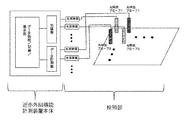
- FIG. 1 is a diagram showing an embodiment of the near-infrared brain function measuring apparatus of the present invention.
- FIG. 2 is a diagram for explaining an arrangement example of a light irradiation probe (light source probe) and a light detection probe (detector probe) and its probe arrangement matrix in the present invention.
- the difficulty of statistical comparison between channels in multi-channel fNIRS measurement is considered to be the essence of the problem that the apparent noise variance in the signal differs because the signal amplification factor is changed according to the amount of light detected.
- the present inventors tried to solve the problem by introducing an optical attenuator for each probe individually and controlling the amount of noise dispersion by increasing or decreasing the amount of light.
- the adjustment of one optical attenuator affects the magnitude of noise dispersion of a plurality of related channels.
- FIG. 1 shows the configuration of a near-infrared brain function measuring apparatus necessary for achieving the above.
- the detection unit of this apparatus is composed of n light source probes and m detector probes. A pair of light source probe and detector probe constitute one channel. Let N be the total number of channels in the device.
- Light of two wavelengths ( ⁇ 1 , ⁇ 2 ) is introduced into the light source probe from the light source unit of the near-infrared brain function measuring device main body, and the light passes through the living tissue and then passes through the detector probe. It is measured by a detector in the optical measuring unit of the infrared brain function measuring device main body. The measured data is then processed by a data processing / recording / display unit.
- the data processing unit of this device has a function of calculating the effective incident light amount and the effective detection efficiency according to the method described later based on the measurement data.
- the display unit has a function of displaying the result in real time.
- an optical attenuator is provided between the light source probe and the light source and between the detector probe and the detector.
- This optical attenuator has a function of appropriately attenuating and outputting the intensity of the input light according to the transmittance value designated by the control unit of the main body.
- measurement is performed using a plurality of lights having different wavelengths.
- one optical attenuator having no wavelength-dependent characteristics is introduced to each probe. .
- ⁇ A k, ⁇ (t) is the change in absorbance of channel k
- R k, ⁇ (t) is the tissue transmittance in channel k
- I k is the amount of incident light from light source probe i, n j
- ⁇ ( t) represents noise generated in the detector during measurement by the detector probe j.
- the transmittance change over time according to the light attenuation between the light source probe / detector probe and the scalp. And an average Variation pattern around (See Patent Document 1 and Non-Patent Document 1).
- the transmittance at the wavelength ⁇ of the optical attenuator i connected to the light source probe is a i (1 ⁇ i ⁇ n, 0 ⁇ a i ⁇ 1), and the wavelength of the optical attenuator j connected to the detector probe.
- the transmittance at ⁇ is represented by a j (n + 1 ⁇ j ⁇ n + m, 0 ⁇ a j ⁇ 1).
- the first term on the right side of the equation (1) is a change in absorbance associated with a change in the amount of hemoglobin in the tissue, and the second term is a baseline change caused by body movement.
- the third term is noise caused by measurement noise from the photodetector, and is referred to as detector noise in this specification.
- the noise component h oxy, k (t) derived from the detector noise included in the time variation of the observed oxyhemoglobin is expressed by the following equation (3).
- the noise generated in all detectors can be assumed to be white noise, and this property can be regarded as equivalent even between detector elements (j) or between the wavelengths of the same individual ( ⁇ ). Therefore, all n j, ⁇ 1 (t ), N j, ⁇ 2 (t) are independent and equally distributed. If this standard deviation is ⁇ n , the variance of the detector noise h oxy, k (t) should be observed as in the following equation (4).
- noise variance of deoxyhemoglobin is expressed by the following equation (5).
- J k, ⁇ is the observation light quantity of wavelength ⁇ in channel k, and is expressed by the following equation (6).
- the leveling of the observed detector noise variance ⁇ 2 oxy, k and ⁇ 2 doxy, k between channels is referred to as noise leveling. It is an object of the present invention to achieve this.
- I i, ⁇ 2 / I i, ⁇ 1 constituting ⁇ k in the above equation (7) is a constant determined by the setting of the light source intensity of each wavelength.
- R i, 0, ⁇ 2 / r i, 0, ⁇ 1 and r j, 0, ⁇ 2 / r j, 0, ⁇ 1 mean the wavelength dependence of the light transmittance in the air between the scalp and the probe, It can be regarded as a constant that does not depend on the probe location. Therefore, ⁇ k is a constant independent of the channel position.
- u xy is also a constant determined by the wavelength used and the spectral absorption characteristics of hemoglobin.
- the denominators of equations (8) and (9) are all constants, and in order to level the noise, the observed light quantity J k , ⁇ 1 or J k, ⁇ 2 can be adjusted so that the transmittances a i and a j of the optical attenuator are adjusted so as to be equal between the channels.
- the amount of light observed is affected by fluctuations in the transmittance between the probe and the scalp and the tissue, but it is easily eliminated by measuring the amount of light after passing through a low-pass filter in the appropriate band. it can.
- the light source side and the detector side are considered separately, and (1) an operation of making the light amount actually incident on the head tissue (hereinafter referred to as effective incident light amount) equal across all the light source probes, and ( 2) This is realized through the operation of making the efficiency at which light emitted from the head tissue can actually be detected (hereinafter referred to as effective detection efficiency) equal across all detector probes. Therefore, a method for estimating the effective incident light amount and effective detection efficiency in a certain channel is first formulated, and then a specific procedure for leveling the detector noise based on real time monitoring of these estimated amounts will be described.
- the observation light quantity is represented by the relational expression of the product of each variable, as shown in the above formula (6).
- the first term of the equation (10) Is the effective incident light intensity Represents the effective detection efficiency.
- the relationship between the measurement channel and the probes constituting the measurement channel is expressed by a matrix called a probe arrangement matrix following the above-mentioned Patent Document 1 and Non-Patent Document 1 by the present inventors.
- Probe placement matrix Is an N ⁇ (n + m) matrix, and when the light source probe i and the detector probe j constitute the channel k, its elements are Defined in Depends on the channel / probe arrangement employed in each fNIRS device. For example, when four channels are configured as shown in FIG.
- the effective incident light intensity Is independent of the adjustment of a j and the effective detection efficiency Is independent of the adjustment of a i and achieves leveling by avoiding the difficulty of mutual adjustments interfering with each other via the probe network as when leveling the observed light quantity J k, ⁇ itself Is possible.
- the equations indicating the effective incident light amount and the effective detection efficiency include undetermined coefficients C 1 and C 2 , but in the present invention, the leveling procedure can be completed without determining these coefficients as described below.
- the detector noise leveling in the near infrared brain function measuring apparatus of the present invention will be described.
- the maximum light amount that can be emitted from the light source probe in accordance with the safety guidelines for light irradiation on the living body is defined as I safe .
- the maximum value of the effective incident light quantity of the light source probe Even so, it is obvious that it is less than I safe .
- equalizing the effective incident light amount of all the light source probes is a method for realizing a noise leveling state with the best measurement S / N while maintaining biological safety. However, in the state where this leveling is achieved, the amount of light emitted from the light source probe may exceed I safe .
- the detector noise leveling of the near-infrared brain function measuring apparatus has been described.
- the present invention can be applied to the case where a probe network configured to share a light source probe and a detector probe is used.
Landscapes
- Health & Medical Sciences (AREA)
- Life Sciences & Earth Sciences (AREA)
- Physics & Mathematics (AREA)
- Engineering & Computer Science (AREA)
- Surgery (AREA)
- Public Health (AREA)
- Pathology (AREA)
- Veterinary Medicine (AREA)
- Biomedical Technology (AREA)
- Heart & Thoracic Surgery (AREA)
- Medical Informatics (AREA)
- Molecular Biology (AREA)
- General Health & Medical Sciences (AREA)
- Animal Behavior & Ethology (AREA)
- Biophysics (AREA)
- Neurology (AREA)
- Physiology (AREA)
- Spectroscopy & Molecular Physics (AREA)
- Signal Processing (AREA)
- Optics & Photonics (AREA)
- Psychiatry (AREA)
- Computer Vision & Pattern Recognition (AREA)
- Artificial Intelligence (AREA)
- Psychology (AREA)
- Neurosurgery (AREA)
- Measurement Of The Respiration, Hearing Ability, Form, And Blood Characteristics Of Living Organisms (AREA)
- Investigating Or Analysing Materials By Optical Means (AREA)
Abstract
Description
この計測において、プローブと生体組織の密着は非常に重要である。プローブは通常数ミリの窓径を有する一方、ヒト頭部の毛髪部では、成人でも約1ミリの間隔で毛髪が生えているため、窓直下の毛髪をすべて排除してプローブを頭皮に密着させることは実際上困難である。これに起因して、頭皮とプローブ間では必ず光減衰が生じ、この光減衰はプローブ直下の毛髪の多寡やプローブと頭皮の密着度によって異なる。そのため、このようなプローブを多数並列的に配置して行う多チャンネル計測では、チャンネルごとに検出される光量がしばしば大きく異なる。
このような状況に対して、現在市販されている多チャンネルfNIRS装置では、初めに仮計測を行って、光量の乏しいチャンネルでは検出器信号を適切な大きさまで増幅する較正処理を行う。これは計測においてA/Dコンバータの分解能を有効に使う目的のためである。原信号は、光量と雑音から構成されているが、原信号を増幅する程度がチャンネルごとに異なるため、この較正処理を経て得られた信号はチャンネルごとに異なる大きさの雑音成分(雑音分散)をもつことになる。例えば、光電子増倍管を検出器に用いた装置では、通常の較正処理範囲である印加電圧700Vと1000V間では暗電流に由来する雑音の大きさは20倍ほども異なる。そのため、実際の計測実行に先立って、光量が乏しく雑音が大きいチャンネルのプローブ直下の毛髪を念入りに排除する作業を何度も繰り返し、それでもある段階で妥協してデータ採取を開始することになる。この作業は多チャンネルfNIRS計測において手間と時間のかかるボトルネックとなっている。また、プローブ装着に関する実験者の熟練度、被験者や計測部位の毛髪量、色などによって計測データの品質が大きく左右される結果を生んできた(特許文献1、非特許文献1参照)。
さらにこのような信号強度較正の手法は、以下に記すように信号の統計解析にとっての大きな障害となっている。多チャンネルfNIRS装置の一般的な運用のかたちとして、ある程度の広がりをもつ面積にわたって複数のチャンネルで脳機能活動を計測しておき、事後的にその計測領域内のどの部位で活動が顕著であったか調べる手法が多用されている。一方、正統的な統計解析手法のほとんどは、標本集団どうしが等しい分散を持つことを前提として設計されている。このため、チャンネルごとに信号中の雑音分散が著しく異なるfNIRS計測データに対して厳格な統計解析を適用することができず、結果として、多チャンネルデータの定量的比較のための統計学的基盤は未だ整備されていない。
前記制御手段は、予め、
(1)すべての光減衰器の透過率を1に設定しすべての光源強度を安全に照射し得る最大光量に設定して各光源プローブiの実効入射光量とそのうち最小の実効入射光量および最大の実効入射光量を求め、
光源プローブ側の光減衰器iの透過率aiを、前記最小の実効入射光量を光源プローブiの実効入射光量で割り算した値に変更することにより実効入射光量を平準化し、
(2)すべての光源強度をW倍、ただしW=[前記最大の実効入射光量/前記最小の実効入射効率]だけ増大して変更し、検出器プローブjの実効検出効率とそのうちの最小の実効検出効率を求め、
検出器プローブ側の光減衰器jの透過率ajを、前記最小の実効検出効率を検出器プローブjの実効検出効率で割り算した値に変更することにより実効検出効率を平準化しておくように制御することにより、オキシヘモグロビンの検出器雑音の分散およびデオキシヘモグロビンの検出器雑音の分散をすべてのチャンネルk間で揃えたことを特徴とする。
また、本発明は、n個の光源プローブi、1≦i≦nおよびm個の検出器プローブj、n+1≦j≦n+mを頭表に配置し、各光源から波長λの光を光減衰器iを介して透過率aiで各光源プローブiに導入し、各検出器プローブjで検出した波長λの光を光減衰器jを介して透過率ajで計測データ部に送り、前記計測データ部で受けた検出データを処理して光源プローブiと検出器プローブjで構成されるN個のチャンネルk、1≦k≦Nにおけるオキシヘモグロビンおよびデオキシヘモグロビンの分光吸収特性に基づく計測光の吸光度変化から脳機能活動を検出する近赤外脳機能計測方法であって、
すべての光源プローブiおよびすべての検出器プローブjを頭表に装着し、すべての光減衰器の透過率を1に設定し、すべての光源強度を安全に照射し得る最大光量に設定して各光源プローブiの実効入射光量とそのうち最小の実効入射光量および最大の実効入射光量を求める工程と、
光源プローブ側の光減衰器iの透過率aiを、前記最小の実効入射光量を光源プローブiの実効入射光量で割算した値に変更することにより実効入射光量を平準化する工程と、
すべての光源強度をW倍、ただしW=[前記最大の実効入射光量/前記最小の実効入射効率]だけ増大して変更し、検出器プローブjの実効検出効率とそのうちの最小の実効検出効率を求める工程と、
検出器プローブ側の光減衰器jの透過率ajを、前記最小の実効検出効率を検出器プローブjの実効検出効率で割算した値に変更することにより実効検出効率を平準化する工程により、
予めオキシヘモグロビンの検出器雑音の分散およびデオキシヘモグロビンの検出器雑音の分散をすべてのチャンネルk間で揃えたことを特徴とする。
上記を達成するために必要な近赤外脳機能計測装置の構成を図1に示す。本装置の検知部はn本の光源プローブとm本の検出器プローブから構成される。一対の光源プローブと検出器プローブが一つのチャンネルを構成する。装置の全チャンネル数をNとする。光源プローブには近赤外脳機能計測装置本体の光源部から二つの波長(λ1,λ2)の光が導入され、それらの光は生体組織を通過した後、検出器プローブを通って近赤外脳機能計測装置本体の光計測部にある検出器で計測される。計測されたデータはその後、データ処理/記録/表示部で処理が行われる。通常の近赤外脳機能計測装置が有する諸機能に加えて、本装置のデータ処理部は計測データに基づいて後述する方法に従って実効入射光量と実効検出効率を演算する機能を有する。表示部はその結果を実時間表示する機能を有する。また、従来装置と異なる装備として、光源プローブと光源の間および検出器プローブと検出器の間に各々光減衰器を有する。この光減衰器は本体の制御部によって指定された透過率の値に従って、入力光の強度を適宜減衰させて出力する機能を有する。前述のように近赤外脳機能計測では波長の異なる複数の光を用いて計測を行うが、本発明では、波長依存特性をもたない光減衰器を各プローブに対して1つずつ導入する。


しかし、多チャンネルの近赤外脳機能計測では、通常一つの光源プローブは隣接する複数のチャンネルの計測に供される。また、検出器プローブについてもこの事情は同様である。そのため、着目したチャンネルの観測光量を変えるために関連する光減衰器を調節したことで隣接するチャンネルの観測光量も変化してしまうことが非常にしばしば生じ得る。このように複雑に連動する条件の下で、全てのチャンネルの観測光量を等しくする作業は見かけほど容易ではない。
観測光量は上記式(6)に示されるように、各変数の積の関係式で表される。この式の対数をとることによって各変数の線形和の関係式として以下のように記述することができる。


次に、計測チャンネルとこれを構成するプローブの関係を本発明者等による上記特許文献1および非特許文献1に倣ってプローブ配置行列と呼ぶ行列で表現する。プローブ配置行列


(i)不定連立方程式の一般解は、特殊解と同伴方程式

(ii)疑似逆行列


(iii)次に同伴方程式

光源プローブの数をn、検出器プローブの数をmとして、次の


ところで、光源プローブiを共有する別のチャンネルk’においても同様の要請があるので、これに関わる検出器プローブをj’とすると
が成り立たなければならないから


したがって、光源プローブや検出器プローブを共有するようにチャンネル構成されたプローブネットワーク内においては、すべての光源プローブi、検出器プローブjに関して

(iv)以上から、


1.プローブを頭部に装着したのちに、装置のすべて光減衰器の透過率を1に設定し、すべての光源強度をIsafeにする。
2.1.の設定で、どちらかの波長λを選び、式(16)、(17)により各プローブの


3.光源プローブ側の各光減衰器の透過率aiを下式(18)


4.次に、すべての光源強度を下式(19)で定められるW倍だけ増大させる。


6.上記の設定によりオキシヘモグロビンおよびデオキシヘモグロビンの検出器雑音の平準化が達成される。この設定を保持した状態でデータ計測を行う。
7.計測終了と同時に1.の状態に復帰させるか、もしくはすべての光源出力を遮断する。なお、式(18)~(20)に示されたとおり、上記すべての過程1.~7.において未定係数C1,C2に関わりなく手続きを行うことができる。
Claims (2)
- 頭表に配置されるn個の光源プローブi、1≦i≦nおよびm個の検出器プローブj、n+1≦j≦n+mと、各光源から波長λの光を透過率aiで各光源プローブiに導入する光減衰器iと、各検出器プローブjで検出した波長λの光を透過率ajで計測データ部に送る光減衰器jと、前記計測データ部で受けた検出データを処理して光源プローブiと検出器プローブjで構成されるN個のチャンネルk、1≦k≦Nにおけるオキシヘモグロビンおよびデオキシヘモグロビンの分光吸収特性に基づく計測光の吸光度変化から脳機能活動を検出する制御手段を備えた近赤外脳機能計測装置であって、
前記制御手段は、予め、
(1)すべての光減衰器の透過率を1に設定しすべての光源強度を安全に照射し得る最大光量に設定して各光源プローブiの実効入射光量とそのうち最小の実効入射光量および最大の実効入射光量を求め、
光源プローブ側の光減衰器iの透過率aiを、前記最小の実効入射光量を光源プローブiの実効入射光量で割り算した値に変更することにより実効入射光量を平準化し、
(2)すべての光源強度をW倍、ただしW=[前記最大の実効入射光量/前記最小の実効入射効率]だけ増大して変更し、検出器プローブjの実効検出効率とそのうちの最小の実効検出効率を求め、
検出器プローブ側の光減衰器jの透過率ajを、前記最小の実効検出効率を検出器プローブjの実効検出効率で割り算した値に変更することにより実効検出効率を平準化しておくように制御することにより、オキシヘモグロビンの検出器雑音の分散およびデオキシヘモグロビンの検出器雑音の分散をすべてのチャンネルk間で揃えたことを特徴とする近赤外脳機能計測装置。 - n個の光源プローブi、1≦i≦nおよびm個の検出器プローブj、n+1≦j≦n+mを頭表に配置し、各光源から波長λの光を光減衰器iを介して透過率aiで各光源プローブiに導入し、各検出器プローブjで検出した波長λの光を光減衰器jを介して透過率ajで計測データ部に送り、前記計測データ部で受けた検出データを処理して光源プローブiと検出器プローブjで構成されるN個のチャンネルk、1≦k≦Nにおけるオキシヘモグロビンおよびデオキシヘモグロビンの分光吸収特性に基づく計測光の吸光度変化から脳機能活動を検出する近赤外脳機能計測方法であって、
すべての光源プローブiおよびすべての検出器プローブjを頭表に装着し、すべての光減衰器の透過率を1に設定し、すべての光源強度を安全に照射し得る最大光量に設定して各光源プローブiの実効入射光量とそのうち最小の実効入射光量および最大の実効入射光量を求める工程と、
光源プローブ側の光減衰器iの透過率aiを、前記最小の実効入射光量を光源プローブiの実効入射光量で割り算した値に変更することにより実効入射光量を平準化する工程と、
すべての光源強度をW倍、ただしW=[前記最大の実効入射光量/前記最小の実効入射効率]だけ増大して変更し、検出器プローブjの実効検出効率とそのうちの最小の実効検出効率を求める工程と、
検出器プローブ側の光減衰器jの透過率ajを、前記最小の実効検出効率を検出器プローブjの実効検出効率で割り算した値に変更することにより実効検出効率を平準化する工程により、
予めオキシヘモグロビンの検出器雑音の分散およびデオキシヘモグロビンの検出器雑音の分散をすべてのチャンネルk間で揃えたことを特徴とする近赤外脳機能計測方法。
Priority Applications (5)
| Application Number | Priority Date | Filing Date | Title |
|---|---|---|---|
| EP16752375.2A EP3260049B1 (en) | 2015-02-16 | 2016-02-10 | Functional near-infrared spectroscope and spectroscopy |
| CN201680009588.4A CN107205732B (zh) | 2015-02-16 | 2016-02-10 | 近红外脑功能测量装置及测量方法 |
| US15/551,102 US10939851B2 (en) | 2015-02-16 | 2016-02-10 | Brain-functional near infrared spectroscopy device and measurement method |
| KR1020177023705A KR101944030B1 (ko) | 2015-02-16 | 2016-02-10 | 근적외 뇌 기능 계측 장치 및 계측법 |
| JP2017500630A JP6381168B2 (ja) | 2015-02-16 | 2016-02-10 | 近赤外脳機能計測装置及び計測法 |
Applications Claiming Priority (2)
| Application Number | Priority Date | Filing Date | Title |
|---|---|---|---|
| JP2015027690 | 2015-02-16 | ||
| JP2015-027690 | 2015-02-16 |
Publications (1)
| Publication Number | Publication Date |
|---|---|
| WO2016132989A1 true WO2016132989A1 (ja) | 2016-08-25 |
Family
ID=56689220
Family Applications (1)
| Application Number | Title | Priority Date | Filing Date |
|---|---|---|---|
| PCT/JP2016/053955 Ceased WO2016132989A1 (ja) | 2015-02-16 | 2016-02-10 | 近赤外脳機能計測装置及び計測法 |
Country Status (6)
| Country | Link |
|---|---|
| US (1) | US10939851B2 (ja) |
| EP (1) | EP3260049B1 (ja) |
| JP (1) | JP6381168B2 (ja) |
| KR (1) | KR101944030B1 (ja) |
| CN (1) | CN107205732B (ja) |
| WO (1) | WO2016132989A1 (ja) |
Cited By (2)
| Publication number | Priority date | Publication date | Assignee | Title |
|---|---|---|---|---|
| WO2018190130A1 (ja) | 2017-04-14 | 2018-10-18 | 国立研究開発法人産業技術総合研究所 | 脳機能計測装置と脳機能計測方法 |
| JPWO2020174842A1 (ja) * | 2019-02-27 | 2021-10-14 | 国立研究開発法人産業技術総合研究所 | 脳機能計測装置及び脳機能計測方法とプローブ |
Families Citing this family (1)
| Publication number | Priority date | Publication date | Assignee | Title |
|---|---|---|---|---|
| JP7227657B2 (ja) * | 2019-04-12 | 2023-02-22 | コリア アドヴァンスド インスティテュート オブ サイエンス アンド テクノロジー | マシンラーニングを利用して頭に関する生体情報を推定するための方法、システムおよび非一過性のコンピュータ読み取り可能な記録媒体 |
Citations (1)
| Publication number | Priority date | Publication date | Assignee | Title |
|---|---|---|---|---|
| JP2009268707A (ja) * | 2008-05-08 | 2009-11-19 | Hitachi Ltd | 生体光計測装置 |
Family Cites Families (6)
| Publication number | Priority date | Publication date | Assignee | Title |
|---|---|---|---|---|
| CN1718154B (zh) * | 2004-07-08 | 2011-06-15 | 株式会社日立制作所 | 生物体光测试装置 |
| US8954133B1 (en) * | 2008-08-08 | 2015-02-10 | The United States Of America As Represented By The Department Of Veterans Affairs | Spectroscopic detection of brain damage |
| WO2012005303A1 (ja) | 2010-07-06 | 2012-01-12 | 株式会社日立メディコ | 生体光計測装置およびそれを用いた生体光計測方法 |
| JP6004430B2 (ja) | 2012-10-19 | 2016-10-05 | 国立研究開発法人産業技術総合研究所 | 生体光計測装置 |
| CN104224165B (zh) | 2014-09-17 | 2016-05-11 | 哈尔滨工业大学 | 基于多距测量方法及最小一乘准则的近红外脑功能信号抗差估计方法 |
| US9554738B1 (en) * | 2016-03-30 | 2017-01-31 | Zyomed Corp. | Spectroscopic tomography systems and methods for noninvasive detection and measurement of analytes using collision computing |
-
2016
- 2016-02-10 JP JP2017500630A patent/JP6381168B2/ja active Active
- 2016-02-10 WO PCT/JP2016/053955 patent/WO2016132989A1/ja not_active Ceased
- 2016-02-10 US US15/551,102 patent/US10939851B2/en active Active
- 2016-02-10 CN CN201680009588.4A patent/CN107205732B/zh active Active
- 2016-02-10 KR KR1020177023705A patent/KR101944030B1/ko active Active
- 2016-02-10 EP EP16752375.2A patent/EP3260049B1/en active Active
Patent Citations (1)
| Publication number | Priority date | Publication date | Assignee | Title |
|---|---|---|---|---|
| JP2009268707A (ja) * | 2008-05-08 | 2009-11-19 | Hitachi Ltd | 生体光計測装置 |
Non-Patent Citations (2)
| Title |
|---|
| See also references of EP3260049A4 * |
| TORU YAMADA ET AL.: "Use of the Wavelength Differential Method to Eliminate Baseline Drift in the Assessment of Cerebral Function Using NIRS", TRANSACTIONS OF JAPANESE SOCIETY FOR MEDICAL AND BIOLOGICAL ENGINEERING, vol. 43, no. 4, December 2005 (2005-12-01), pages 530 - 537, XP055477708 * |
Cited By (8)
| Publication number | Priority date | Publication date | Assignee | Title |
|---|---|---|---|---|
| WO2018190130A1 (ja) | 2017-04-14 | 2018-10-18 | 国立研究開発法人産業技術総合研究所 | 脳機能計測装置と脳機能計測方法 |
| CN110520057A (zh) * | 2017-04-14 | 2019-11-29 | 国立研究开发法人产业技术综合研究所 | 脑功能测量装置和脑功能测量方法 |
| JPWO2018190130A1 (ja) * | 2017-04-14 | 2020-05-14 | 国立研究開発法人産業技術総合研究所 | 脳機能計測装置と脳機能計測方法 |
| EP3610796A4 (en) * | 2017-04-14 | 2021-01-13 | National Institute of Advanced Industrial Science and Technology | BRAIN FUNCTION MEASURING DEVICE AND BRAIN FUNCTION MEASURING METHOD |
| CN110520057B (zh) * | 2017-04-14 | 2022-05-13 | 国立研究开发法人产业技术综合研究所 | 脑功能测量装置和脑功能测量方法 |
| US11478149B2 (en) | 2017-04-14 | 2022-10-25 | National Institute of Advanced Indusuial Science and Technology | Brain function measurement device and brain function measurement method |
| JPWO2020174842A1 (ja) * | 2019-02-27 | 2021-10-14 | 国立研究開発法人産業技術総合研究所 | 脳機能計測装置及び脳機能計測方法とプローブ |
| JP7227652B2 (ja) | 2019-02-27 | 2023-02-22 | 国立研究開発法人産業技術総合研究所 | 脳機能計測装置及び脳機能計測方法とプローブ |
Also Published As
| Publication number | Publication date |
|---|---|
| CN107205732A (zh) | 2017-09-26 |
| EP3260049A1 (en) | 2017-12-27 |
| JP6381168B2 (ja) | 2018-08-29 |
| JPWO2016132989A1 (ja) | 2017-12-07 |
| CN107205732B (zh) | 2020-02-28 |
| EP3260049B1 (en) | 2019-11-27 |
| US20180028098A1 (en) | 2018-02-01 |
| US10939851B2 (en) | 2021-03-09 |
| KR101944030B1 (ko) | 2019-04-17 |
| EP3260049A4 (en) | 2018-10-31 |
| KR20170107072A (ko) | 2017-09-22 |
Similar Documents
| Publication | Publication Date | Title |
|---|---|---|
| Virtanen et al. | Comparison of principal and independent component analysis in removing extracerebral interference from near-infrared spectroscopy signals | |
| Chiarelli et al. | A kurtosis-based wavelet algorithm for motion artifact correction of fNIRS data | |
| DE69333456T2 (de) | System verfahren zur nichtinvasiven überwachung des hämatocrit-wertes | |
| US10925525B2 (en) | Combined pulse oximetry and diffusing wave spectroscopy system and control method therefor | |
| EP2891457A1 (en) | Biophotonic measurement apparatus and biophotonic measurement method using same | |
| EP0352923A1 (en) | Spectrophotometric apparatus and method for monitoring oxygen saturation | |
| WO2012005303A1 (ja) | 生体光計測装置およびそれを用いた生体光計測方法 | |
| JP6381168B2 (ja) | 近赤外脳機能計測装置及び計測法 | |
| CN102599888A (zh) | 基于数字锁相检测技术的光学拓扑成像系统及方法 | |
| EP3610796B1 (en) | Brain function measurement device and method | |
| Li et al. | Wavelength selection method based on test analysis of variance: application to oximetry | |
| JP2017023455A (ja) | 近赤外光生体計測装置及びそのプローブ | |
| KR102655740B1 (ko) | 스펙트럼 이미지 보정 장치 및 방법과, 대상체의 성분 분석 장치 | |
| Umeyama et al. | Detection of an unstable and/or a weak probe contact in a multichannel functional near-infrared spectroscopy measurement | |
| EP3791793B1 (en) | Brain function measurement device and brain function measurement method | |
| Metz et al. | Physiological effects of continuous colored light exposure on Mayer wave activity in cerebral hemodynamics: a functional near-infrared spectroscopy (fNIRS) study | |
| JP4696300B2 (ja) | 近赤外光を用いた生体光計測法のための波長差分を用いたベースライン安定化方法 | |
| JP6412956B2 (ja) | 生体光計測装置、解析装置、及び方法 | |
| DE112022005352T5 (de) | Stoffwechselmessapparatur, Stoffwechselberechnungsverfahren und Stoffwechselberechnungsprogramm | |
| Li et al. | Portable multi-channel functional NIR spectrometer system for human brain oxygen monitoring in motion states | |
| Lange et al. | Short and mid-term reproducibility analysis of cerebral tissue saturation measured by time domain-NIRS | |
| US20240049996A1 (en) | Nirs / tissue oximetry based method to measure arterial blood oxygen saturation from pulsatile hemoglobin waveforms | |
| US20250248629A1 (en) | Apparatus and method for determining oxygen saturation with skin tone correction | |
| Nur et al. | Proposal for the Optimal Scheme of Savitzky–Golay Filter for the fNIRS Signals of Different Sampling Rate | |
| Goyal et al. | Broadband NIRS assessment of cerebral water content in adults |
Legal Events
| Date | Code | Title | Description |
|---|---|---|---|
| 121 | Ep: the epo has been informed by wipo that ep was designated in this application |
Ref document number: 16752375 Country of ref document: EP Kind code of ref document: A1 |
|
| ENP | Entry into the national phase |
Ref document number: 2017500630 Country of ref document: JP Kind code of ref document: A |
|
| NENP | Non-entry into the national phase |
Ref country code: DE |
|
| ENP | Entry into the national phase |
Ref document number: 20177023705 Country of ref document: KR Kind code of ref document: A |
|
| REEP | Request for entry into the european phase |
Ref document number: 2016752375 Country of ref document: EP |