WO2016121862A1 - 抗炎症剤及びその使用 - Google Patents
抗炎症剤及びその使用 Download PDFInfo
- Publication number
- WO2016121862A1 WO2016121862A1 PCT/JP2016/052470 JP2016052470W WO2016121862A1 WO 2016121862 A1 WO2016121862 A1 WO 2016121862A1 JP 2016052470 W JP2016052470 W JP 2016052470W WO 2016121862 A1 WO2016121862 A1 WO 2016121862A1
- Authority
- WO
- WIPO (PCT)
- Prior art keywords
- compound
- result
- group
- gene
- hsd17b4
- Prior art date
- Legal status (The legal status is an assumption and is not a legal conclusion. Google has not performed a legal analysis and makes no representation as to the accuracy of the status listed.)
- Ceased
Links
- 0 CC(C)(C)OC(N=C(*Cc1ccccc1)N*)=O Chemical compound CC(C)(C)OC(N=C(*Cc1ccccc1)N*)=O 0.000 description 2
- SOKBOENXKNZLSO-UHFFFAOYSA-N CCCCCCNC(NCc1ccccc1)=N Chemical compound CCCCCCNC(NCc1ccccc1)=N SOKBOENXKNZLSO-UHFFFAOYSA-N 0.000 description 2
- XSFIGTROPRBMBO-UHFFFAOYSA-N CC(C)(C)OC(/N=C(/NCc1ccccc1)\NNC)=O Chemical compound CC(C)(C)OC(/N=C(/NCc1ccccc1)\NNC)=O XSFIGTROPRBMBO-UHFFFAOYSA-N 0.000 description 1
- CSOJECDGWHHWRS-UHFFFAOYSA-N CC(C)(C)OC(NC(NC(OC(C)(C)C)=O)=S)=O Chemical compound CC(C)(C)OC(NC(NC(OC(C)(C)C)=O)=S)=O CSOJECDGWHHWRS-UHFFFAOYSA-N 0.000 description 1
- HTYQSVMAKNEANU-UHFFFAOYSA-N CC(C)(C)OC(NC(NCc1ccccc1)=S)=O Chemical compound CC(C)(C)OC(NC(NCc1ccccc1)=S)=O HTYQSVMAKNEANU-UHFFFAOYSA-N 0.000 description 1
- UVZDZTISTNHCNV-IZWKRXCGSA-N CCCCCCCCCCCC(NC(NCc(cc1)ccc1OCc1c[n](CCOCCOCCOCCNC(CCCC[C@@H]([C@H]2N3)SC[C@@H]2NC3=O)=O)nn1)=N)OC(C(F)(F)F)=O Chemical compound CCCCCCCCCCCC(NC(NCc(cc1)ccc1OCc1c[n](CCOCCOCCOCCNC(CCCC[C@@H]([C@H]2N3)SC[C@@H]2NC3=O)=O)nn1)=N)OC(C(F)(F)F)=O UVZDZTISTNHCNV-IZWKRXCGSA-N 0.000 description 1
- DNRFWAWKXDCFDF-UHFFFAOYSA-N CCCCCCN/C(/NCc1ccccc1)=N\C(OC(C)(C)C)=O Chemical compound CCCCCCN/C(/NCc1ccccc1)=N\C(OC(C)(C)C)=O DNRFWAWKXDCFDF-UHFFFAOYSA-N 0.000 description 1
- HFSDNIUAZXMLTG-UHFFFAOYSA-N N=C(NCCCCCO)NCc1ccccc1 Chemical compound N=C(NCCCCCO)NCc1ccccc1 HFSDNIUAZXMLTG-UHFFFAOYSA-N 0.000 description 1
Images























Classifications
-
- C—CHEMISTRY; METALLURGY
- C07—ORGANIC CHEMISTRY
- C07C—ACYCLIC OR CARBOCYCLIC COMPOUNDS
- C07C279/00—Derivatives of guanidine, i.e. compounds containing the group, the singly-bound nitrogen atoms not being part of nitro or nitroso groups
- C07C279/04—Derivatives of guanidine, i.e. compounds containing the group, the singly-bound nitrogen atoms not being part of nitro or nitroso groups having nitrogen atoms of guanidine groups bound to acyclic carbon atoms of a carbon skeleton
-
- A—HUMAN NECESSITIES
- A61—MEDICAL OR VETERINARY SCIENCE; HYGIENE
- A61K—PREPARATIONS FOR MEDICAL, DENTAL OR TOILETRY PURPOSES
- A61K31/00—Medicinal preparations containing organic active ingredients
- A61K31/13—Amines
- A61K31/155—Amidines (), e.g. guanidine (H2N—C(=NH)—NH2), isourea (N=C(OH)—NH2), isothiourea (—N=C(SH)—NH2)
-
- A—HUMAN NECESSITIES
- A61—MEDICAL OR VETERINARY SCIENCE; HYGIENE
- A61K—PREPARATIONS FOR MEDICAL, DENTAL OR TOILETRY PURPOSES
- A61K31/00—Medicinal preparations containing organic active ingredients
- A61K31/70—Carbohydrates; Sugars; Derivatives thereof
- A61K31/7088—Compounds having three or more nucleosides or nucleotides
- A61K31/7105—Natural ribonucleic acids, i.e. containing only riboses attached to adenine, guanine, cytosine or uracil and having 3'-5' phosphodiester links
-
- A—HUMAN NECESSITIES
- A61—MEDICAL OR VETERINARY SCIENCE; HYGIENE
- A61K—PREPARATIONS FOR MEDICAL, DENTAL OR TOILETRY PURPOSES
- A61K31/00—Medicinal preparations containing organic active ingredients
- A61K31/70—Carbohydrates; Sugars; Derivatives thereof
- A61K31/7088—Compounds having three or more nucleosides or nucleotides
- A61K31/713—Double-stranded nucleic acids or oligonucleotides
-
- A—HUMAN NECESSITIES
- A61—MEDICAL OR VETERINARY SCIENCE; HYGIENE
- A61K—PREPARATIONS FOR MEDICAL, DENTAL OR TOILETRY PURPOSES
- A61K48/00—Medicinal preparations containing genetic material which is inserted into cells of the living body to treat genetic diseases; Gene therapy
-
- A—HUMAN NECESSITIES
- A61—MEDICAL OR VETERINARY SCIENCE; HYGIENE
- A61P—SPECIFIC THERAPEUTIC ACTIVITY OF CHEMICAL COMPOUNDS OR MEDICINAL PREPARATIONS
- A61P1/00—Drugs for disorders of the alimentary tract or the digestive system
- A61P1/16—Drugs for disorders of the alimentary tract or the digestive system for liver or gallbladder disorders, e.g. hepatoprotective agents, cholagogues, litholytics
-
- C—CHEMISTRY; METALLURGY
- C07—ORGANIC CHEMISTRY
- C07C—ACYCLIC OR CARBOCYCLIC COMPOUNDS
- C07C279/00—Derivatives of guanidine, i.e. compounds containing the group, the singly-bound nitrogen atoms not being part of nitro or nitroso groups
- C07C279/04—Derivatives of guanidine, i.e. compounds containing the group, the singly-bound nitrogen atoms not being part of nitro or nitroso groups having nitrogen atoms of guanidine groups bound to acyclic carbon atoms of a carbon skeleton
- C07C279/06—Derivatives of guanidine, i.e. compounds containing the group, the singly-bound nitrogen atoms not being part of nitro or nitroso groups having nitrogen atoms of guanidine groups bound to acyclic carbon atoms of a carbon skeleton being further substituted by halogen atoms, or by nitro or nitroso groups
-
- C—CHEMISTRY; METALLURGY
- C07—ORGANIC CHEMISTRY
- C07C—ACYCLIC OR CARBOCYCLIC COMPOUNDS
- C07C279/00—Derivatives of guanidine, i.e. compounds containing the group, the singly-bound nitrogen atoms not being part of nitro or nitroso groups
- C07C279/16—Derivatives of guanidine, i.e. compounds containing the group, the singly-bound nitrogen atoms not being part of nitro or nitroso groups having nitrogen atoms of guanidine groups bound to carbon atoms of rings other than six-membered aromatic rings
-
- C—CHEMISTRY; METALLURGY
- C07—ORGANIC CHEMISTRY
- C07C—ACYCLIC OR CARBOCYCLIC COMPOUNDS
- C07C279/00—Derivatives of guanidine, i.e. compounds containing the group, the singly-bound nitrogen atoms not being part of nitro or nitroso groups
- C07C279/18—Derivatives of guanidine, i.e. compounds containing the group, the singly-bound nitrogen atoms not being part of nitro or nitroso groups having nitrogen atoms of guanidine groups bound to carbon atoms of six-membered aromatic rings
-
- G—PHYSICS
- G01—MEASURING; TESTING
- G01N—INVESTIGATING OR ANALYSING MATERIALS BY DETERMINING THEIR CHEMICAL OR PHYSICAL PROPERTIES
- G01N33/00—Investigating or analysing materials by specific methods not covered by groups G01N1/00 - G01N31/00
- G01N33/48—Biological material, e.g. blood, urine; Haemocytometers
- G01N33/50—Chemical analysis of biological material, e.g. blood, urine; Testing involving biospecific ligand binding methods; Immunological testing
- G01N33/5005—Chemical analysis of biological material, e.g. blood, urine; Testing involving biospecific ligand binding methods; Immunological testing involving human or animal cells
- G01N33/5008—Chemical analysis of biological material, e.g. blood, urine; Testing involving biospecific ligand binding methods; Immunological testing involving human or animal cells for testing or evaluating the effect of chemical or biological compounds, e.g. drugs, cosmetics
- G01N33/502—Chemical analysis of biological material, e.g. blood, urine; Testing involving biospecific ligand binding methods; Immunological testing involving human or animal cells for testing or evaluating the effect of chemical or biological compounds, e.g. drugs, cosmetics for testing non-proliferative effects
- G01N33/5023—Chemical analysis of biological material, e.g. blood, urine; Testing involving biospecific ligand binding methods; Immunological testing involving human or animal cells for testing or evaluating the effect of chemical or biological compounds, e.g. drugs, cosmetics for testing non-proliferative effects on expression patterns
-
- C—CHEMISTRY; METALLURGY
- C07—ORGANIC CHEMISTRY
- C07C—ACYCLIC OR CARBOCYCLIC COMPOUNDS
- C07C2601/00—Systems containing only non-condensed rings
- C07C2601/12—Systems containing only non-condensed rings with a six-membered ring
- C07C2601/14—The ring being saturated
-
- C—CHEMISTRY; METALLURGY
- C12—BIOCHEMISTRY; BEER; SPIRITS; WINE; VINEGAR; MICROBIOLOGY; ENZYMOLOGY; MUTATION OR GENETIC ENGINEERING
- C12N—MICROORGANISMS OR ENZYMES; COMPOSITIONS THEREOF; PROPAGATING, PRESERVING, OR MAINTAINING MICROORGANISMS; MUTATION OR GENETIC ENGINEERING; CULTURE MEDIA
- C12N15/00—Mutation or genetic engineering; DNA or RNA concerning genetic engineering, vectors, e.g. plasmids, or their isolation, preparation or purification; Use of hosts therefor
- C12N15/09—Recombinant DNA-technology
- C12N15/11—DNA or RNA fragments; Modified forms thereof; Non-coding nucleic acids having a biological activity
- C12N15/113—Non-coding nucleic acids modulating the expression of genes, e.g. antisense oligonucleotides; Antisense DNA or RNA; Triplex- forming oligonucleotides; Catalytic nucleic acids, e.g. ribozymes; Nucleic acids used in co-suppression or gene silencing
-
- C—CHEMISTRY; METALLURGY
- C12—BIOCHEMISTRY; BEER; SPIRITS; WINE; VINEGAR; MICROBIOLOGY; ENZYMOLOGY; MUTATION OR GENETIC ENGINEERING
- C12N—MICROORGANISMS OR ENZYMES; COMPOSITIONS THEREOF; PROPAGATING, PRESERVING, OR MAINTAINING MICROORGANISMS; MUTATION OR GENETIC ENGINEERING; CULTURE MEDIA
- C12N2310/00—Structure or type of the nucleic acid
- C12N2310/10—Type of nucleic acid
- C12N2310/14—Type of nucleic acid interfering nucleic acids [NA]
-
- C—CHEMISTRY; METALLURGY
- C12—BIOCHEMISTRY; BEER; SPIRITS; WINE; VINEGAR; MICROBIOLOGY; ENZYMOLOGY; MUTATION OR GENETIC ENGINEERING
- C12Q—MEASURING OR TESTING PROCESSES INVOLVING ENZYMES, NUCLEIC ACIDS OR MICROORGANISMS; COMPOSITIONS OR TEST PAPERS THEREFOR; PROCESSES OF PREPARING SUCH COMPOSITIONS; CONDITION-RESPONSIVE CONTROL IN MICROBIOLOGICAL OR ENZYMOLOGICAL PROCESSES
- C12Q1/00—Measuring or testing processes involving enzymes, nucleic acids or microorganisms; Compositions therefor; Processes of preparing such compositions
- C12Q1/68—Measuring or testing processes involving enzymes, nucleic acids or microorganisms; Compositions therefor; Processes of preparing such compositions involving nucleic acids
- C12Q1/6876—Nucleic acid products used in the analysis of nucleic acids, e.g. primers or probes
- C12Q1/6883—Nucleic acid products used in the analysis of nucleic acids, e.g. primers or probes for diseases caused by alterations of genetic material
-
- C—CHEMISTRY; METALLURGY
- C12—BIOCHEMISTRY; BEER; SPIRITS; WINE; VINEGAR; MICROBIOLOGY; ENZYMOLOGY; MUTATION OR GENETIC ENGINEERING
- C12Q—MEASURING OR TESTING PROCESSES INVOLVING ENZYMES, NUCLEIC ACIDS OR MICROORGANISMS; COMPOSITIONS OR TEST PAPERS THEREFOR; PROCESSES OF PREPARING SUCH COMPOSITIONS; CONDITION-RESPONSIVE CONTROL IN MICROBIOLOGICAL OR ENZYMOLOGICAL PROCESSES
- C12Q1/00—Measuring or testing processes involving enzymes, nucleic acids or microorganisms; Compositions therefor; Processes of preparing such compositions
- C12Q1/68—Measuring or testing processes involving enzymes, nucleic acids or microorganisms; Compositions therefor; Processes of preparing such compositions involving nucleic acids
- C12Q1/6876—Nucleic acid products used in the analysis of nucleic acids, e.g. primers or probes
- C12Q1/6883—Nucleic acid products used in the analysis of nucleic acids, e.g. primers or probes for diseases caused by alterations of genetic material
- C12Q1/6886—Nucleic acid products used in the analysis of nucleic acids, e.g. primers or probes for diseases caused by alterations of genetic material for cancer
-
- C—CHEMISTRY; METALLURGY
- C12—BIOCHEMISTRY; BEER; SPIRITS; WINE; VINEGAR; MICROBIOLOGY; ENZYMOLOGY; MUTATION OR GENETIC ENGINEERING
- C12Q—MEASURING OR TESTING PROCESSES INVOLVING ENZYMES, NUCLEIC ACIDS OR MICROORGANISMS; COMPOSITIONS OR TEST PAPERS THEREFOR; PROCESSES OF PREPARING SUCH COMPOSITIONS; CONDITION-RESPONSIVE CONTROL IN MICROBIOLOGICAL OR ENZYMOLOGICAL PROCESSES
- C12Q2600/00—Oligonucleotides characterized by their use
- C12Q2600/136—Screening for pharmacological compounds
-
- C—CHEMISTRY; METALLURGY
- C12—BIOCHEMISTRY; BEER; SPIRITS; WINE; VINEGAR; MICROBIOLOGY; ENZYMOLOGY; MUTATION OR GENETIC ENGINEERING
- C12Q—MEASURING OR TESTING PROCESSES INVOLVING ENZYMES, NUCLEIC ACIDS OR MICROORGANISMS; COMPOSITIONS OR TEST PAPERS THEREFOR; PROCESSES OF PREPARING SUCH COMPOSITIONS; CONDITION-RESPONSIVE CONTROL IN MICROBIOLOGICAL OR ENZYMOLOGICAL PROCESSES
- C12Q2600/00—Oligonucleotides characterized by their use
- C12Q2600/158—Expression markers
Definitions
- the present invention relates to an anti-inflammatory agent and use thereof. More specifically, the present invention relates to anti-inflammatory agents, screening methods for anti-inflammatory agents, and novel compounds.
- This application claims priority based on Japanese Patent Application No. 2015-14829 filed in Japan on January 28, 2015 and Japanese Patent Application No. 2015-176745 filed in Japan on September 8, 2015. Is incorporated herein by reference.
- Inflammation is one of the symptoms observed in various diseases.
- Examples of inflammation-related diseases include cancer.
- Cancer is one of intractable diseases accompanied by inflammation. Cancer is the number one cause of death in Japan, and the number of deaths from cancer is increasing every year in the world.
- liver cancer hepatocellular carcinoma
- liver cancer hepatocellular carcinoma
- the cause of liver cancer accounts for nearly 90% due to viral infection, and it develops after long-term chronic hepatitis such as hepatitis and cirrhosis starting after viral infection (see Non-Patent Document 1, for example).
- chronic inflammation is known to promote the expression of cancer cells due to cellular gene mutations.
- the cause of the development of liver cancer has not yet been clarified as to whether it is due to the virus itself or chronic inflammation (see, for example, Non-Patent Documents 2 and 3).
- liver cancer a standard therapy for liver cancer has not been established because it is easy to show resistance to anticancer agents. Therefore, there is a demand for the establishment of innovative diagnostic agents and new treatment / prevention strategies for chronic hepatitis and liver cancer.
- biological clocks are constituted by a feedback loop mechanism of transcription / translation by a series of genes called clock genes.
- Various biological functions, sleep / wakefulness, body temperature, hormone secretion, metabolism, etc. are observed to have a fluctuation of about 24 hours (daily rhythm), and these are known to be regulated by the body clock mechanism.
- Non-Patent Documents 4 and 5 Regarding cancer, it has been pointed out that carcinogenesis, the nature of cancer cells, and the sensitivity to anticancer agents are influenced by the biological clock mechanism (for example, see Non-Patent Document 6).
- Bosch FX et al.
- Primary liver cancer worldwide incidence and trends., Gastroenterology, 127, S5-S16, 2004.
- Portolani N et al., Early and late recurrence after liver resection for hepatocellular carcinoma: prognostic and therapeutic implications., Ann. Surg., 243, 229-235, 2006.
- Freeman AJ et al., Estimating progression to cirrhosis in chronic hepatitis C virus infection., Hepatology, 134, 809-816, 2001.
- Filipski E et al., Effects of light and food schedules on liver and tumor molecular clocks in mice., J. Nat. Cancer Inst., 97, 507-517, 2005.
- the present invention aims to clarify whether or not a circadian clock mechanism is involved in inflammation and to provide a new anti-inflammatory agent.
- the present invention includes the following aspects.
- G0S2 inhibitor is a siRNA, shRNA, miRNA, ribozyme or antisense nucleic acid against the G0S2 gene or the Hydroxysteroid (17- ⁇ ) Dehydrogenase 4 (Hsd17b4) gene.
- the G0S2 inhibitor is a specific binding substance for G0S2 protein or Hsd17b4 protein.
- R 1 represents a single bond or an alkylene group having a carbon number of 1 ⁇ 3
- R 2 is (Wherein R 5 represents a halogenated alkyl group having 1 to 3 carbon atoms, and n represents an integer of 0 to 5)
- R 3 represents an alkyl group having 1 to 15 carbon atoms
- R 4 Represents a hydrogen atom or a carboxylate group.
- n is an integer of 2 or more, a plurality of R 5 may be the same as or different from each other.
- [5] The anti-inflammatory agent according to [4], wherein the G0S2 inhibitor has a binding ability to Hsd17b4.
- [6] The anti-inflammatory agent according to any one of [1] to [5], which exhibits an analgesic action.
- [7] A step of measuring the expression level of the G0S2 gene in the cell in the presence of the test substance, and comparing the expression level with the expression level of the G0S2 gene in the cell in the absence of the test substance. And a step of determining that the test substance is an anti-inflammatory agent when the test substance has been reduced.
- R 6 represents a single bond or an alkylene group having 1 to 3 carbon atoms
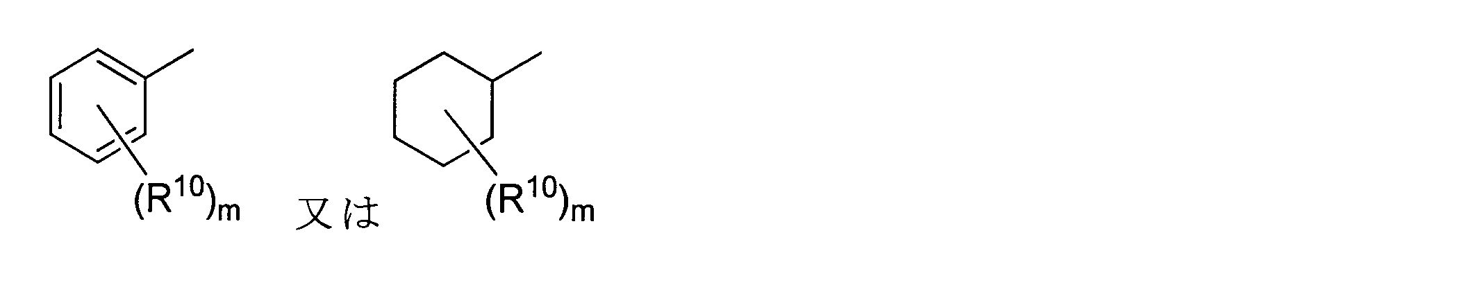
- R 7 represents (Wherein R 10 represents a halogenated alkyl group having 1 to 3 carbon atoms, m represents an integer of 0 to 5), R 8 represents an alkyl group having 1 to 15 carbon atoms, and R 9 Represents a hydrogen atom or a carboxylate group.
- R 8 is a hexyl group
- m is an integer of 1 to 5
- m is an integer of 2 or more
- a plurality of R 10 may be the same or different from each other.
- a new anti-inflammatory agent can be provided.
- FIG. 1 It is a schematic diagram which shows the process in which liver cancer develops.
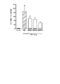
- FIG. 6 is a graph showing the binding activity of NF- ⁇ B to NF- ⁇ B response element (NRE) in the liver of mice exposed to diethylnitrosamine (DEN) administered with siRNA against G0S2 or control siRNA in Experimental Example 3.
- “*” indicates that there is a significant difference at a risk rate of less than 5%.
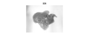
- 4 is a pathological micrograph of the liver of a mouse in a control siRNA administration group (shown as “control siRNA” in the figure) in Experimental Example 3.
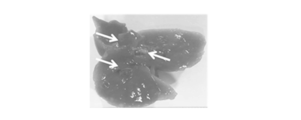
- 6 is a photograph of a liver extracted from a mouse in a control siRNA administration group in Experimental Example 3.
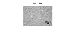
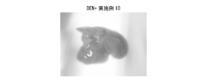
- FIG. 4 is a pathological micrograph of the liver of a mouse in a G0S2 siRNA administration group (shown as “G0S2 siRNA” in the figure) in Experimental Example 3.
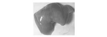
- 6 is a photograph of a liver extracted from a mouse in a G0S2 siRNA administration group in Experimental Example 3.
- it is a graph which shows the measurement result of the expression level of mRNA of Ccl2 in the liver of the mouse
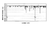
- 10 is a graph showing the results of primary screening of a compound library in Experimental Example 5.
- “**” indicates that there is a significant difference with a risk rate of less than 1%.
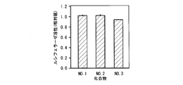
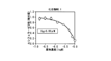
- compound no. 1 is a graph showing luciferase activity with respect to a concentration of 1.
- compound no. 1 is a graph showing luciferase activity with respect to a concentration of 1.
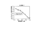
- compound no. 2 is a graph which shows the luciferase activity with respect to the density
- “#” Indicates that there is a significant difference at a risk rate of less than 5% as a result of the Scheffe test in comparison with the LPS administration group.
- the control group 0.05% DMSO-saline
- compound No. A significant difference was observed between the two administration groups.
- “*” Indicates a result of Scheffe test with a risk rate of less than 5% and a control group (0.05% DMSO-saline) and Compound No.
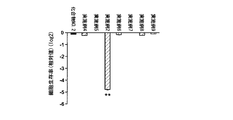
- compound no. 2 is a graph showing the survival rate (relative value) of NIH3T3 cells 24 hours after exposure to 2 and its derivative (10 ⁇ M).
- compound no. 2 is a graph showing the results of comparing the IC 50 of 2 with the compound of Example 2 (NS-3-011) which is a derivative thereof.
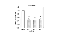
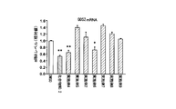
- 0.5 to 5 ⁇ M of Compound No. 2 is a graph showing the expression level of G0S2 mRNA in Hepa1-6 cells 48 hours after exposure to the compound of Example 2 (NS-3-011), which is a derivative of No.
- “*” Indicates compound No. by Bonferroni-Dan test. As a result of comparison with the results of the same dose administration group of 2, it shows that there is a significant difference at a risk rate of less than 5%.
- “**” indicates compound No. by Bonferroni Dunn test. As a result of comparison with the results of the same dose administration group of 2, it shows that there is a significant difference at a risk rate of less than 1%.
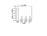
- “**” indicates that there is a significant difference with a risk rate of less than 1% as a result of comparison with the results of the DMSO administration group by the Scheffe test.
- “**” indicates that there is a significant difference at a risk rate of less than 1%.
- Example 17 it is a graph which shows the expression level of mRNA of Ccl2 in the liver of the hepatitis model mouse
- Experimental example 19 it is a graph which shows the expression level of mRNA of Ccl2 in the liver of a control mouse
- “*” indicates that there is a significant difference at a risk rate of less than 5%.
- 6 is a graph showing the blood drug concentration of the compound of Example 1 in Experimental Example 19.
- Experimental example 19 it is a graph which shows the expression level of the mRNA of (alpha) SMA in the liver of a control mouse
- “*” indicates that there is a significant difference at a risk rate of less than 5%.
- it is a graph which shows the expression level of the collagen type I alpha2 (Col1 alpha2) mRNA in the liver of a control mouse and the NASH model mouse which administered the compound of Example 1.
- Experimental Example 21 it is a graph which shows the blood drug concentration of the compound of Example 1.
- Experimental example 22 it is a graph which shows the influence of the compound of Example 10 on the serum ALT activity of the mouse hepatitis liver cancer onset model by DEN. The results after 8 weeks from the start of DEN drinking water are shown.
- Experimental Example 25 a cell in which an expression vector in which a cDNA for luciferase was linked downstream of the transcriptional activity regulatory region of G0S2 was stably expressed was used.
- “**” indicates that there is a significant difference with a risk rate of less than 1%.
- Compound No. It is a graph which shows the result of having measured the drug concentration in the blood of the mouse
- Compound No. It is a graph which shows the result of having measured the drug concentration in the liver of the mouse
- FIG. 1 is a schematic diagram showing a process in which liver cancer develops as an example of an inflammation-related disease.
- liver cancers develop after long-term chronic hepatitis such as hepatitis and cirrhosis.
- the influence of G0S2 and the inflammatory signal in the process shown in FIG. 1 has been clarified by the inventors this time, and details will be described later.
- This experimental model is a useful experimental system for elucidating the mechanism of liver cancer development in humans.
- the inventors analyzed the mechanism of hepatitis liver cancer onset by focusing on the molecular clock mechanism using the mouse hepatitis liver cancer onset model by DEN.
- G0S2 G0 / G1 switch gene 2
- G0S2 G0 / G1 switch gene 2
- the present invention provides an anti-inflammatory agent comprising a G0 / G1 Switch 2 (G0S2) inhibitor as an active ingredient.
- G0S2 inhibitor examples include compounds that suppress the transcription of G0S2, substances that inhibit the activity of G0S2, and the like.
- inflammation-related diseases to be treated or prevented by the anti-inflammatory agent of this embodiment include hepatitis, enterocolitis, colitis (IBD), liver cancer, rheumatism, asthma, atherosclerosis, and multiple sclerosis. And Helicobacter pylori-related gastritis.
- the anti-inflammatory agent of this embodiment may be siRNA, shRNA, miRNA, ribozyme or antisense nucleic acid against the G0S2 gene (inhibiting G0S2 expression).
- Hsd17b4 Hydroxysteroid (17- ⁇ ) Dehydrogenase 4
- the substance that inhibits the expression of the Hsd17b4 gene is a G0S2 inhibitor. That is, a substance that inhibits the expression of the Hsd17b4 gene can be used as an anti-inflammatory agent.
- the anti-inflammatory agent of this embodiment may be siRNA, shRNA, miRNA, ribozyme or antisense nucleic acid against the Hsd17b4 gene.
- SiRNA small interfering RNA
- miRNA miRNA
- RISC RNA-induced silencing complex
- sense strand and antisense strand oligonucleotides are respectively synthesized by a DNA / RNA automatic synthesizer and denatured in an appropriate annealing buffer at 90 to 95 ° C. for about 1 minute, and then 30 to 30 It can be prepared by annealing at 70 ° C. for about 1-8 hours.
- ShRNA short hairpin RNA
- shRNA is a hairpin RNA sequence used for gene silencing by RNA interference.
- shRNA may be introduced into cells by a vector and expressed by U6 promoter or H1 promoter, or an oligonucleotide having shRNA sequence may be synthesized by a DNA / RNA automatic synthesizer and self-annealed in the same manner as siRNA. May also be prepared.
- the shRNA hairpin structure introduced into the cell is cleaved into siRNA and binds to the RNA-induced silencing complex (RISC). This complex binds to and cleaves mRNA having a sequence complementary to siRNA. This suppresses gene expression in a sequence-specific manner.
- RISC RNA-induced silencing complex
- MiRNA is RNA having a length of about 20 to 25 bases existing in cells, and is a kind of ncRNA (non-coding RNA) that is considered to have a function of regulating the expression of other genes. In recent years, it has become possible to knock down a target gene using an artificial miRNA.
- Artificial miRNA may be expressed in mammalian cells from an expression vector using the Pol2 promoter.
- the miRNA introduced into the cell binds to the RNA-induced silencing complex (RISC).
- RISC RNA-induced silencing complex
- This complex binds to and cleaves mRNA having a sequence complementary to miRNA. This suppresses gene expression in a sequence-specific manner.
- Ribozyme is RNA having catalytic activity. Although some ribozymes have various activities, research on ribozymes as enzymes that cleave RNA has made it possible to design ribozymes for the purpose of site-specific cleavage of RNA.
- the ribozyme may be a group I intron type, a size of 400 nucleotides or more such as M1 RNA contained in RNaseP, or may be about 40 nucleotides called a hammerhead type, a hairpin type, or the like.
- An antisense nucleic acid is a nucleic acid complementary to a target sequence.
- Antisense nucleic acid inhibits transcription initiation by triplex formation, suppresses transcription by hybridization with a site where an open loop structure is locally formed by RNA polymerase, inhibits transcription by hybridization with RNA that is being synthesized, Inhibition of splicing by hybridization at the junction of intron and exon, suppression of splicing by hybridization with spliceosome formation site, suppression of transition from nucleus to cytoplasm by hybridization with mRNA, capping site and poly (A) addition site Suppression of splicing by hybridization with a protein, suppression of translation initiation by hybridization with a translation initiation factor binding site, suppression of translation by hybridization with a ribosome binding site near the initiation codon, translation region of mRNA and polysome binding site Hybridization outgrowth inhibitory peptide chain by the, by gene silencing due hybridization interaction site between a nucleic acid and a
- siRNA, shRNA, ribozyme and antisense nucleic acid may contain various chemical modifications in order to improve stability and activity.
- the phosphate residue may be substituted with a chemically modified phosphate residue such as phosphorothioate (PS), methylphosphonate, phosphorodithionate, and the like.
- PS phosphorothioate
- methylphosphonate phosphorodithionate
- you may comprise at least one part with nucleic acid analogs, such as a peptide nucleic acid (PNA).
- PNA peptide nucleic acid
- the anti-inflammatory agent of this embodiment may be a specific binding substance for G0S2 protein or Hsd17b4 protein.
- the specific binding substance exhibits an anti-inflammatory effect by specifically binding to the G0S2 protein or the Hsd17b4 protein and inhibiting their function.
- Examples of specific binding substances include antibodies, antibody fragments, aptamers, and low molecular compounds.
- An antibody can be produced, for example, by immunizing an animal such as a mouse with an antigen. Alternatively, it can be prepared by screening an antibody library such as a phage library.
- Antibody fragments include Fv, Fab, scFv and the like.
- the above antibody or antibody fragment may be polyclonal or monoclonal.
- the above antibody or antibody fragment may be one to which a compound such as polyethylene glycol is bound. By binding polyethylene glycol, for example, blood stability can be increased.
- An aptamer is a substance having a specific binding ability to a labeling substance.
- examples of aptamers include nucleic acid aptamers and peptide aptamers.
- Nucleic acid aptamers having specific binding ability to G0S2 protein or Hsd17b4 protein can be selected by, for example, systematic evolution of ligand by exponential enrichment (SELEX) method.
- Peptide aptamers having specific binding ability to G0S2 protein or Hsd17b4 protein can be selected by, for example, the two-hybrid method using yeast.
- the specific binding substance may be, for example, one that is screened from a compound library or the like using the binding property to the target substance as an index.
- the onset of hepatitis and liver cancer can be suppressed by administering siRNA against G0S2 to mice of the mouse hepatitis liver cancer onset model by DEN. Therefore, it can also be said that the anti-inflammatory agent of this embodiment is a therapeutic or prophylactic agent for hepatitis or liver cancer.
- the anti-inflammatory agent of this embodiment may be a compound represented by the following formula (1), a pharmaceutically acceptable salt thereof, or a solvate thereof.
- R 1 represents a single bond or an alkylene group having 1 to 3 carbon atoms
- R 2 represents (Wherein R 5 represents a halogenated alkyl group having 1 to 3 carbon atoms, and n represents an integer of 0 to 5), R 3 represents an alkyl group having 1 to 15 carbon atoms, and R 4 Represents a hydrogen atom or a carboxylate group.
- n is an integer of 2 or more, a plurality of R 5 may be the same as or different from each other.
- the compound represented by the formula (1) may be a free form, a pharmaceutically acceptable salt, or a free form solvate. It may be a solvate of a pharmaceutically acceptable salt.
- Examples of pharmaceutically acceptable salts include metal salts, ammonium salts, organic amine addition salts, amino acid addition salts, and the like. More specifically, for example, inorganic acid salts such as hydrochloride, sulfate, hydrobromide, nitrate, phosphate, hydroiodide; acetate, mesylate, succinate, maleic acid Salt, fumarate, citrate, tartrate, benzoate, methanesulfonate, 2-hydroxyethanesulfonate, p-toluenesulfonate, trifluoroacetate, propanoate, oxalate, Organic acid salts such as malonate, glutarate, adipate, malate and mandelate; alkali metal salts such as sodium salt and potassium salt; alkaline earth metal salts such as magnesium salt and calcium salt; aluminum Metal salts such as salts and zinc salts; ammonium salts such as ammonium salts and tetramethylammonium salts
- the solvate is not particularly limited as long as it is a pharmaceutically acceptable solvate, and examples thereof include hydrates and organic solvates.
- the compound represented by the above formula (1), a pharmaceutically acceptable salt thereof, or a solvate thereof may have a binding ability to Hsd17b4 (Peroxisomal multifunctional enzyme type 2).
- Hsd17b4 is a protein known to exist in peroxisomes, and is an enzyme having an activity such as ⁇ -oxidation of fatty acids.
- the Uniprot accession number of human Hsd17b4 is P51659 and the Uniprot accession number of mouse Hsd17b4 is P51660.
- the anti-inflammatory agent of the present embodiment exhibits an analgesic action. Therefore, it can be said that the anti-inflammatory agent of this embodiment is an analgesic.
- the anti-inflammatory agent of this embodiment may be administered per se, or may be administered as a pharmaceutical composition mixed with a pharmaceutically acceptable carrier.
- the pharmaceutical composition may be formulated into an orally used dosage form such as a tablet, capsule, elixir, or microcapsule, for example, parenterally such as an injection, ointment, or patch. It may be formulated into a dosage form to be used.
- the pharmaceutically acceptable carrier examples include solvents such as sterilized water and physiological saline; binders such as gelatin, corn starch, gum tragacanth and gum arabic, excipients such as crystalline cellulose; corn starch, gelatin, alginic acid and the like And the like.
- the pharmaceutical composition may contain an additive.
- Additives include lubricants such as magnesium stearate; sweeteners such as sucrose, lactose and saccharin; flavoring agents such as peppermint and red oil; stabilizers for benzyl alcohol and phenol; buffers such as phosphate and sodium acetate Agents; solubilizing agents such as benzyl benzoate and benzyl alcohol; antioxidants; preservatives; surfactants;
- the pharmaceutical composition can be formulated by appropriately combining the above carriers and additives and mixing them in a unit dosage form generally required for pharmaceutical practice.
- examples of the solvent for injection include isotonic solutions containing adjuvants such as physiological saline, glucose, D-sorbitol, D-mannose, D-mannitol, and sodium chloride. It is done. Solvents for injections may contain alcohols such as ethanol; polyalcohols such as propylene glycol and polyethylene glycol; nonionic surfactants such as polysorbate 80 (trademark) and HCO-50.
- Administration of anti-inflammatory agents to patients is known to those skilled in the art, for example, intraarterial injection, intravenous injection, subcutaneous injection, etc., as well as intranasally, transbronchially, intramuscularly, transdermally or orally. It can be done by a method.
- the dose of the anti-inflammatory agent varies depending on the patient's symptoms and the like, but in the case of oral administration, for example, 5 to 20 mg / kg per day can be mentioned.
- the dose varies depending on the administration subject, target organ, symptom, and administration method.
- 10 to 25 mg / kg is administered once a day. To do. If the dose exceeds the above range, liver damage may occur.
- the lethal dose in the case of intravenous injection is about 25 mg / kg.
- the present invention comprises a step of measuring the expression level of a G0S2 gene in a cell in the presence of a test substance, and the expression level is a G0S2 gene in the cell in the absence of the test substance. And a step of determining that the test substance is an anti-inflammatory agent when the expression level is lower than the expression level of the anti-inflammatory agent.
- anti-inflammatory agents can be screened by the screening method of the present embodiment.
- test substance for example, a compound library can be used.
- the test substance may be added to the cell culture medium, for example.
- the method for measuring the expression level of the G0S2 gene is not particularly limited, and may be performed by, for example, real-time PCR, or may be performed at the protein level by, for example, Western blotting, ELISA, or the like.
- test substance In the presence of the test substance, if the expression level of the G0S2 gene is reduced compared to the control (in the absence of the test substance), the test substance is an anti-inflammatory agent. Can be determined.
- the present invention includes a step of measuring the activity of Hsd17b4 protein in the presence of a test substance, and the activity is decreased as compared to the activity of Hsd17b4 protein in the absence of the test substance.
- a method for screening the anti-inflammatory agent comprising the step of determining that the test substance is an anti-inflammatory agent.
- the test substance that decreases the activity of the Hsd17b4 protein is an inhibitor of G0S2. That is, it can be said that the test substance that decreases the activity of the Hsd17b4 protein is an anti-inflammatory agent.
- the screening method of the present embodiment may be performed at the cell level, or may be performed in a test tube using purified Hsd17b4 protein.
- the test substance the same substances as described above can be used.
- the activity of the Hsd17b4 protein includes the activity of ⁇ -oxidizing fatty acids.
- the activity of the Hsd17b4 protein can be measured, for example, by measuring the metabolic amount of 2-methyl-branched-chain fatty acids, bile acid intermediates, or very long chain fatty acids.
- the inventors have clarified from the results of computer analysis that the activity of Hsd17b4 decreases as a result of binding of a compound such as NS-3-011 to the enzyme active site of Hsd17b4.
- compounds other than NS-3-011 can also bind to Hsd17b4.
- the present invention provides siRNA, shRNA, miRNA, ribozyme, antisense nucleic acid, or a compound represented by the following formula (1), a pharmaceutically acceptable salt thereof, or a solvate thereof:
- the present invention provides a method for treating or preventing an inflammation-related disease, comprising a step of administering to a patient or patient in need of treatment.
- R 1 represents a single bond or an alkylene group having a carbon number of 1 ⁇ 3
- R 2 is (Wherein R 5 represents a halogenated alkyl group having 1 to 3 carbon atoms, and n represents an integer of 0 to 5), R 3 represents an alkyl group having 1 to 15 carbon atoms, and R 4 Represents a hydrogen atom or a carboxylate group.
- n is an integer of 2 or more, a plurality of R 5 may be the same as or different from each other.
- the present invention provides siRNA, shRNA, miRNA, ribozyme, antisense nucleic acid, or a compound represented by the above formula (1), a pharmaceutically acceptable salt thereof, or a solvate thereof, against G0S2.
- a method for treating or preventing pain comprising a step of administering to a patient or patient in need of treatment.
- the present invention provides siRNA, shRNA, miRNA, ribozyme, antisense nucleic acid, or a compound represented by the above formula (1), a pharmaceutically acceptable salt thereof, or a solvate thereof, against G0S2.
- the present invention provides a method for treating or preventing hepatitis or liver cancer, which comprises a step of administering to a patient or patient in need of treatment.
- the present invention is represented by siRNA, shRNA, miRNA, ribozyme, antisense nucleic acid, or the above-mentioned formula (1) for G0S2 for the treatment or prevention of inflammation-related diseases or the treatment or prevention of pain. And a pharmaceutically acceptable salt or solvate thereof.
- the present invention relates to siRNA, shRNA, miRNA, ribozyme, antisense nucleic acid, or the above formula for G0S2 for the manufacture of a therapeutic or prophylactic agent for inflammation-related diseases, or a therapeutic or prophylactic agent for pain.
- a pharmaceutically acceptable salt thereof, or a solvate thereof is provided.
- R 6 represents a single bond or an alkylene group having 1 to 3 carbon atoms
- R 7 represents
- R 10 represents a halogenated alkyl group having 1 to 3 carbon atoms
- m represents an integer of 0 to 5.
- R 8 represents an alkyl group having 1 to 15 carbon atoms
- R 9 represents a hydrogen atom or a carboxylic acid ester group.
- the compound represented by the formula (2) may be a free form, a pharmaceutically acceptable salt, or a free form, like the compound represented by the formula (1). It may be a solvate or a pharmaceutically acceptable salt solvate.
- the pharmaceutically acceptable salts and solvates are the same as those described above.
- the compound of the present embodiment exhibits a significantly higher G0S2 expression inhibitory effect than the compound represented by the following formula (3) (compound No. 2). Therefore, the compound of this embodiment can be utilized as an anti-inflammatory agent, for example.
- the compound of the present embodiment may be formulated as a pharmaceutical composition in the same manner as the compound represented by the above formula (1).
- the dosage form, administration method, and dosage of the pharmaceutical composition are the same as those described above.
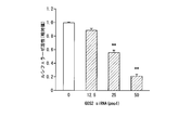
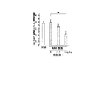
- a reporter assay using an NF- ⁇ B response element was performed.
- Cells in which an expression cassette in which a luciferase gene was linked downstream of the NF- ⁇ B response element were stably expressed in NIH3T3, a mouse embryo-derived fibroblast cell line, were prepared.
- 0, 12.5, 25, and 50 pmol of siRNA against G0S2 were added to the cell culture medium (capacity 500 ⁇ L), and allowed to stand for 48 hours.
- a commercially available siRNA for G0S2 (Life Technologies, trade name “Stealth siRNA”, catalog number “# 1320001”, SEQ ID NO: 3 shows the base sequence of the antisense strand) was used.
- FIG. 2 is a graph showing the measurement results. As a result, it was revealed that the activity of the NF- ⁇ B response element in NIH3T3 cells was reduced by adding siRNA against G0S2 to the medium.
- G0S2 is a regulator of transcriptional activity of inflammatory signals.
- mice (G0S2 knockdown) Mouse of hepatitis liver cancer development model by diethylnitrosamine (DEN) was divided into 2 groups, siRNA against G0S2 was administered to one (G0S2 siRNA administration group) and negative control siRNA was administered to the other (control siRNA administration group) .
- Administration of siRNA to mice was performed by administering 1.6 nmol / mouse via the tail vein using a nucleic acid introduction reagent (trade name “LipoTrust (trademark) EX Oligo ⁇ in vivo>, Hokkaido System Science Co., Ltd.).
- siRNA for G0S2 (Life Technologies, trade name “Stealth siRNA”, catalog number “# 1320001”, SEQ ID NO: 3 shows the base sequence of the antisense strand) was used.
- siRNA (Life Technologies, SEQ ID NO: 4 shows the base sequence of the antisense strand) was used as the control siRNA.
- DEN was added to drinking water at a concentration of 80 mg / L one day after siRNA administration to mice. Three days after administration of siRNA to mice, the liver was collected and subjected to gel shift assay using a DNA fragment containing the NF- ⁇ B response element sequence to measure the amount of binding between NF- ⁇ B and the NF- ⁇ B response element.
- FIG. 3 is a graph showing the measurement results.
- FIG. 4A is a pathological micrograph of the liver of a mouse in a control siRNA administration group (shown as “control siRNA” in the figure), and FIG. 4B is a photograph of a liver extracted from a mouse in the control siRNA administration group.
- the arrows in FIG. 4A indicate cancer cells, and the arrows in FIG. 4B indicate liver cancer that has developed.
- FIG. 4C is a pathological micrograph of the liver of a mouse in the G0S2 siRNA-administered group (shown as “G0S2 siRNA” in the figure), and FIG. 4D is a photograph of the liver extracted from the mouse in the G0S2 siRNA-administered group. is there.
- FIG. 5 is a graph showing the measurement results of the expression level of Ccl2 mRNA in the liver of each group of mice.
- the * mark in the figure means that there is a significant difference when the risk rate is less than 5%.
- mice of the G0S2 siRNA administration group shown as “G0S2 siRNA” in the figure
- Ccl2 expression was compared to the mice in the control siRNA administration group (shown as “control siRNA” in the figure).
- control siRNA was found to be significantly suppressed.
- FIG. 6B shows the result of the secondary screening. As a result, there were almost no compounds that significantly reduced cell viability. Therefore, 50 compounds that do not affect cell viability and lower the luciferase activity of the O-4000E cell line to less than 1/10 in the primary screening were selected as candidate compounds.
- the inventors' research suggests that the clock gene is also involved in the expression of the nuclear receptor RAR ⁇ that regulates the transcription of the G0S2 gene. Therefore, in order to exclude compounds that affect the clock gene in normal cells, studies were performed using a MEF cell line in which an expression cassette having a luciferase gene linked downstream of the Bmal1 promoter was stably expressed. The result is shown in FIG. 7B.
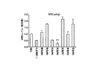
- FIG. 8A it was revealed that the mRNA expression level of the G0S2 gene was reduced to about half in any compound.
- the expression level of the nuclear receptor RAR ⁇ which is known to be involved in the transcriptional control of G0S2 is determined according to Compound No. 1 and compound no. 2 did not change, but compound No. 2 3 showed a significant decrease.
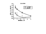
- the mRNA expression levels of Ccl2 and interleukin (IL) -6 which are downstream factors of NF- ⁇ B signal to which G0S2 contributes as a regulator, are determined by the exposure of any compound. There was also a significant decrease. From these results, compound no. 3 was removed from the candidates and compound no. 1 and compound no. 2 was a candidate compound.
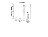
- the Ccl2 mRNA expression increased by exposure to LPS or TNF ⁇ was found to be compound No. 1 2 was significantly suppressed by exposure.
- 25 ⁇ g of LPS was administered to the mouse from the tail vein to prepare a hepatitis model.
- Compound No. 30 minutes after administration of LPS. 2 (5 mg / kg) was administered via the tail vein, and the liver was sampled at 6 hours.
- the expression level of liver Ccl2 mRNA was significantly increased compared to the control group (saline-administered group). In the group administered with 2, the expression was significantly suppressed.
- LPS (10 ⁇ g / animal) was administered to the ICR male mice via the tail vein to form an LPS administration group.
- a mouse administered with physiological saline instead of LPS was prepared and used as a control group.
- the LPS administration group was divided into two groups, and one group was divided into Compound No. 1 after 1 hour from the administration of LPS. 2 was administered subcutaneously to the back so as to be 5 mg / kg, and LPS + No. Two administration groups were used.
- Compound No. Five hours after the administration of 2 mice of each group were killed and livers were collected.
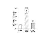
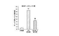
- FIG. 11A is a graph showing the results of quantification of G0S2 protein.
- the expression of G0S2 was significantly increased in the LPS administration group compared to the control group. Furthermore, the expression of G0S2 was compared to LPS + No. It was significantly suppressed in the 2 administration groups.
- FIG. 11B is a graph showing the results of quantification of p65 protein.
- the amount of p65 protein in the nucleus was significantly increased in the LPS administration group compared to the control group. Furthermore, the abundance of p65 protein in the nucleus is lower than that of the LPS administration group by LPS + No. It decreased significantly in the 2 administration groups.
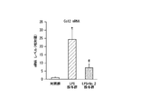
- FIG. 11C is a graph showing the results of quantification of the expression level of Ccl2 mRNA.
- the expression level of Ccl2 mRNA was significantly increased in the LPS administration group compared to the control group. Furthermore, the expression level of Ccl2 mRNA was lower than that of the LPS administration group. It decreased significantly in the 2 administration groups.
- Example 13 (Synthesis of compounds) The compounds of Example 1 and Example 2 were synthesized according to the following scheme.
- Compound 2 was obtained by reacting thiourea (Compound 1) with di-tert-butyl dicarbonate in the presence of sodium hydride. Subsequently, after conversion of compound 2 to compound 3 (see Yin B, et al., Tetrahedron Lett., 49, 3687, 2008.), compound 4a is obtained by condensation with hexylamine in the presence of water-soluble carbodiimide ( Poss MA, et al., Tetrahedron Lett., 33, 5933, 1992.), and condensation with dodecylamine gave compound 4b in 93% and 94% yields, respectively. By allowing a hydrogen chloride ether solution to act on compound 4a, the compound of Example 1 (Compound No.
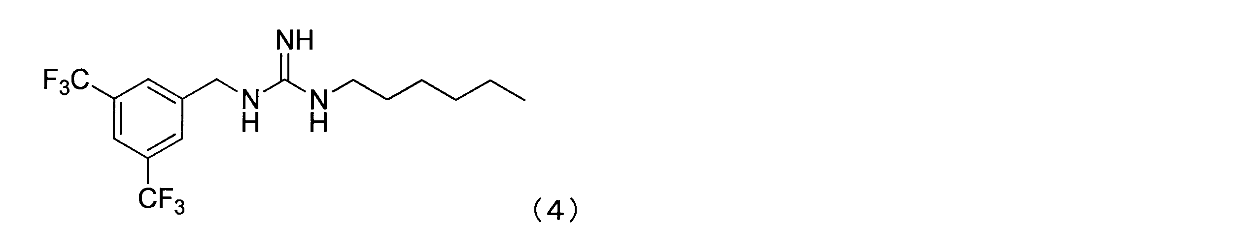
- Example 3 represented by the following formula (4) was also synthesized.
- Example 4 represented by the following formula (5) was also synthesized.
- Example 5 represented by the following formula (6) was also synthesized.
- Example 6 represented by the following formula (7) was also synthesized.
- Example 7 represented by the following formula (8) was also synthesized.
- Example 8 represented by the following formula (9) was also synthesized.
- Example 9 represented by the following formula (10) was also synthesized.
- Example 10 represented by the following formula (11) was also synthesized.
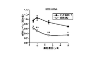
- Example 14 (Creation of compound No. 2 derivative and evaluation of drug efficacy) Compound No. Seven derivatives of Example 2 (Examples 2, 4 to 9) were exposed to Hepa1-6 cells at 10 ⁇ M, and the effect on the expression level of G0S2 mRNA after 24 hours was evaluated. As a result, as shown in FIG. 13A, in the compounds of Example 4 (NS-3-005) and Example 6 (NS-3-013), Compound No. An effect of reducing the expression level of G0S2 mRNA to the same level as 2 was observed.
- Example 6 when the drug effect on human HepG2 cells was evaluated at the mRNA level, a significant decrease was observed with the compound of Example 6 (NS-3-013), and Example 2 (NS-3- Compound No. 011) The same effect as 2 was confirmed.
- Example 2 which showed a common medicinal effect on mouse hepatoma-derived cell Hepa1-6 cells and human hepatoma-derived cell HepG2 cells, and Example 2 in which cell death was observed at 10 ⁇ M.
- the efficacy of the compound (NS-3-011) was evaluated at a concentration of less than 10 ⁇ M.
- Example 2 As a result, as shown in FIG. 2, the compounds of Example 2 (NS-3-011) and Example 6 (NS-3-013) were exposed at 2.5 and 5 ⁇ M for 48 hours. 2 was 5 ⁇ M, and the compound of Example 2 (NS-3-011) and Example 6 (NS-3-013) significantly decreased the expression level of G0S2 mRNA when exposed to both 5 ⁇ M and 2.5 ⁇ M. Furthermore, the compound of Example 2 (NS-3-011) is compound No. 5 at any concentration. 2 and 5 ⁇ M exposure group, a significant decrease in G0S2 mRNA expression level was observed.
- Example 15 Comparison of efficacy of compound No. 2 and compound of Example 2 From the examination so far, a medicinal effect at less than 10 ⁇ M has been confirmed.
- Compound No. The IC 50 was calculated for the G0S2 promoter activity of the compound 2 and the compound of Example 2 (NS-3-011). As a result, as shown in FIG. 2 had an IC 50 of 2.31 ⁇ M, whereas the compound of Example 2 (NS-3-011) had an IC 50 of 0.21 ⁇ M, indicating an equivalent efficacy in an amount of about 1/10. I understood.
- the compound of Example 2 (NS-3-011) had a compound No. in the range of 1 to 2.5 ⁇ M. The effect was significantly higher than 2.
- FIG. 14D when the cell viability at this time was evaluated by ATP assay, a significant decrease in cell viability was observed when the compound of Example 2 (NS-3-011) was exposed to 5 ⁇ M. . However, at a concentration of 1 ⁇ M of the compound of Example 2 (NS-3-011), no decrease in cell viability was observed, and at 1 ⁇ M, compound no. Thus, the compound of Example 2 (NS-3-011) was found to be compound No. 2 It was suggested that the compound is more useful than 2.
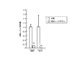
- FIG. 15 is a graph showing the results of real-time PCR. As a result, it was revealed that the expression level of G0S2 and Ccl2 mRNA was decreased in the cells in which G0S2 was knocked down as compared with the control cells.
- Example 17 (Evaluation of drug efficacy) LPS 10 ⁇ g was intraperitoneally administered to mice to prepare a hepatitis model. One hour after LPS administration, the compound of Example 2 (NS-3-011), the compound of Example 3 (NS-3-054) and the compound of Example 10 (NS-3-086) were subcutaneously administered to the back. . Five hours after administration of each compound, livers were collected from each mouse, and the expression level of Ccl2 was quantified by real-time PCR.
- FIG. 16 is a graph showing the results of real-time PCR.
- vehicle represents a mouse that did not receive LPS
- control represents a mouse that received physiological saline instead of the compound.
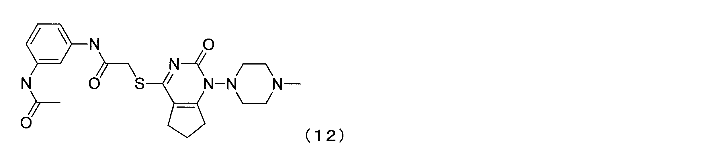
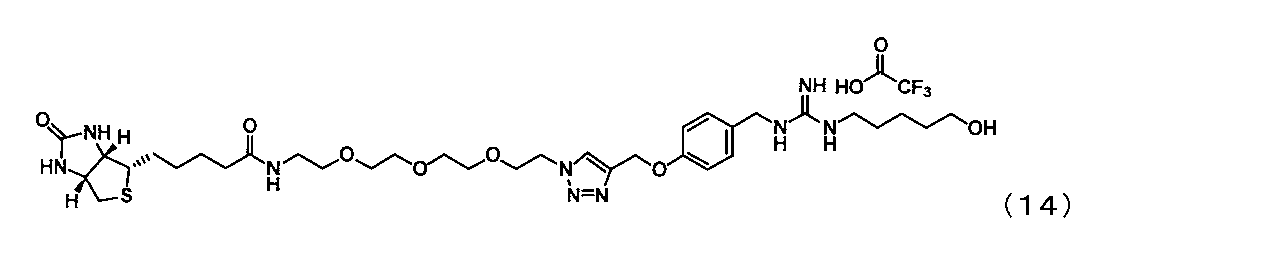
- Example 18 (Identification of target protein of compound of Example 2) Mouse liver cells were homogenized to extract proteins from the cytoplasm and nucleus respectively. Thereafter, the compound (# 142) obtained by pegbiotinylation of the compound of Example 2 (NS-3-011) that suppresses the expression of G0S2, or the compound of Example 1 (NS-3-060) that does not suppress the expression of G0S2. The pegylated biotinylated compound (# 151) was mixed with the above protein and incubated. The chemical formulas of compounds # 142 and # 151 are shown in the following formulas (13) and (14), respectively.
- avidin beads magnetic beads labeled with avidin (hereinafter sometimes referred to as “avidin beads”), and the compound and protein bound to the avidin beads were subjected to acrylamide gel electrophoresis.
- the acrylamide gel after electrophoresis was negatively stained, a protein band specifically bound to compound # 142 was cut out, and the protein was identified by LC / MS / MS analysis.
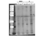
- FIG. 17 is a photograph showing the results of acrylamide gel electrophoresis. As a result of analyzing a protein band of about 75 kDa indicated by an arrow in the figure, it was revealed that it was Hsd17b4 (Peroxisomal multifunctional type 2, Uniprot accession number: P51660).
- Example 19 Evaluation of the efficacy of the compound of Example 1 using a non-alcoholic hepatitis model mouse
- NASH non-alcoholic hepatitis
- a feed for producing a NASH model type “A06071302”, Research Diet
- the compound of Example 1 was orally administered at 19:00 on consecutive days from the start of feeding.
- mice fed with normal feed and orally administered with water supplemented with 0.5% DMSO in the same volume as the compound were used.
- ⁇ SMA collagen type I ⁇ 2
- collagen type I ⁇ 1 are markers that serve as indices of liver fibrosis.
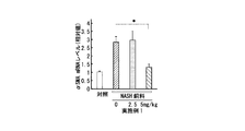
- FIG. 18A is a graph showing the expression level of Ccl2 in the liver of a NASH model mouse. As shown in FIG. 18A, it was revealed that the expression level of Ccl2 is decreased by administration of the compound.
- FIG. 18B is a graph showing the blood drug concentration of the compound of Example 1. As shown in FIG. 18B, the average blood concentration of the compound of Example 1 was 32 nM.
- 18C, 18D, and 18E are graphs showing the results of quantifying the expression levels of ⁇ SMA, collagen type I ⁇ 2 (Col1 ⁇ 2), and collagen type I ⁇ 1 (Col1 ⁇ 1) gene, respectively.
- FIG. 18C, FIG. 18D, and FIG. 18E in the group administered with the compound of Example 1 at 5 mg / kg, the expression levels of ⁇ SMA, collagen type I ⁇ 2, and collagen type I ⁇ 1 gene are significantly decreased. It became.
- mice of Example 1 were orally administered to mice at 19:00 every day. Blood was collected 21 days after the start of compound administration, and the blood concentration of the compound 24 hours after drug administration was measured using LC / MS / MS. As a control, mice fed with normal feed and orally administered with water supplemented with 0.5% DMSO in the same volume as the compound were used.
- FIG. 19 is a graph showing the blood drug concentration of the compound of Example 1. As shown in FIG. 19, the average blood concentration of the compound of Example 1 was 0.8 nM.
- Example 21 Measurement of blood concentration of the compound of Example 1.
- the compound of Example 1 (15 mg / kg) was subcutaneously administered to the back of ICR mice. Blood was collected 0.5, 1, 2, 4, 6, 12, and 24 hours after administration of the compound, and the blood concentration of the compound was measured using LC / MS / MS. 20 is a graph showing the blood drug concentration of the compound of Example 1.
- Example 22 (Investigation of the effect of compounds on serum ALT activity in mouse hepatitis liver cancer model by DEN)
- a solution obtained by dissolving diethylnitrosamine (DEN) in water at a concentration of 80 mg / L as drinking water was administered to ICR mice to cause chronic inflammation in the liver.
- DEN diethylnitrosamine
- a group was prepared in which the compound of Example 10 (NS-3-086) was administered at a dose of 15 mg / kg immediately after the start of DEN drinking water administration.
- a control group a group given tap water as drinking water was used.
- ALT activity was measured using a commercially available kit (type “Transaminase CII-Test Wako”). , Manufactured by Wako Pure Chemical Industries, Ltd.).
- FIG. 21A and FIG. 21B are graphs showing ALT activity in serum after 8 weeks (FIG. 21A) and 12 weeks (FIG. 21B) from the start of DEN drinking water administration.
- “Vehicle” indicates the result of the control group
- “DEN” indicates the result of the DEN administration group
- “DEN + Example 10” indicates that of the group to which DEN and the compound of Example 10 were administered. Results are shown.
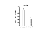
- each cell was collected after 48 hours, and total RNA was extracted. Subsequently, the expression level of the Hsd17b4 gene was quantified by real-time PCR.
- FIG. 22A is a graph showing the results of real-time PCR. As a result, it was confirmed that the expression of Hsd17b4 was suppressed by transfection of siRNA against Hsd17b4.
- Compound No. Hepa1-6 cells were transfected with siRNA against Hsd17b4 or control siRNA in the presence (10 ⁇ M) or absence (denoted as “DMSO” in FIG. 22B). Subsequently, after 48 hours, each cell was collected and total RNA was extracted. Subsequently, the expression level of the G0S2 gene was quantified by real-time PCR.
- FIG. 22B is a graph showing the results of real-time PCR.
- Compound No. In the presence of 2, it became clear that the expression level of the G0S2 gene was significantly reduced. Further, by transfection of siRNA against Hsd17b4, compound no. It was revealed that the effect of lowering the expression level of the G0S2 gene by 2 disappeared.
- Compound No. 2 was found to regulate the expression of the G0S2 gene via the Hsd17b4 protein.
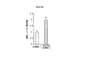
- Hepa1-6 cells which are mouse liver cancer-derived cells, were transfected with an Hsd17b4 expression vector (Hsd17b4-pcDNA3.1) or an empty vector (pcDNA3.1).
- each cell was collected after 48 hours, and total RNA was extracted. Subsequently, the expression level of Hsd17b4 gene or G0S2 gene was quantified by real-time PCR.
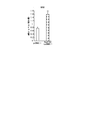
- FIG. 23A and FIG. 23B are graphs showing the results of real-time PCR.
- FIG. 23A shows the result of quantifying the expression level of the Hsd17b4 gene
- FIG. 23B shows the result of quantifying the expression level of the G0S2 gene.
- FIG. 23A it was confirmed that the expression level of the Hsd17b4 gene was significantly increased by transfection of the expression vector of Hsd17b4. Further, as shown in FIG. 23B, it was revealed that the expression level of the G0S2 gene was significantly increased by transfection of the expression vector of Hsd17b4.
- Compound No. 10 having a final concentration of 10 ⁇ M was added to the medium of each cell. 2 or DMSO (control) was added. Subsequently, the cells were collected after 24 hours, and luciferase activity was measured.
- FIG. 24 is a graph showing the results of measuring the luciferase activity of each cell. As a result, when the STAT5 / 6 binding region present in the G0S2 transcriptional activity regulatory region is eliminated, It was clarified that the effect of suppressing the expression of luciferase by addition of 2 is reduced.
- Compound No. 2 was considered to act via transcription of STAT5 or STAT6.
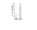
- NIH3T3 cells which are mouse fibroblasts, were transfected with an expression vector in which a luciferase cDNA was linked downstream of the transcriptional activity regulatory region of G0S2, and a STAT5 expression vector or an empty vector (pcDNA3.1, control). Subsequently, the cells were collected after 24 hours, and luciferase activity was measured.
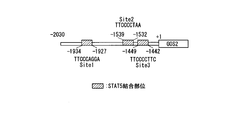
- FIG. 25A is a diagram showing the structure of the transcriptional activity regulatory region of G0S2.
- FIG. 25B is a graph showing the results of measuring luciferase activity. As a result, it was revealed that cells transfected with the STAT5 expression vector showed significantly higher luciferase activity compared to the control group.
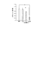
- FIG. 26 is a graph showing the results of Western blotting. As a result, compared with the control, Compound No. In cells to which 2 was added, compound no. The abundance of nuclear STAT5 protein decreased in a dose-dependent manner.
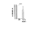
- FIG. 27A shows compound No. in blood. 2 is a graph showing the density of 2; In addition, FIG. 2 is a graph showing the density of 2; As a result, compound no. The concentration of No. 2 reached its maximum at 15 minutes after administration, and compound No. It was revealed that the concentration of 2 reached the highest concentration 5 minutes after administration.
- Example 29 (Examination of the effects of compounds on the liver of mouse hepatitis liver cancer development model by DEN) A solution obtained by dissolving diethylnitrosamine (DEN) in water at a concentration of 80 mg / L as drinking water was administered to ICR mice to cause chronic inflammation and liver cancer in the liver.
- DEN diethylnitrosamine
- a group was prepared in which the compound of Example 10 (NS-3-086) was administered at a dose of 15 mg / kg immediately after the start of DEN drinking water administration.
- a control group a group given tap water as drinking water was used.
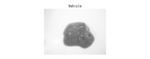
- FIG. 28A, FIG. 28B and FIG. 28C are photographs of the liver 14 weeks after the start of DEN drinking water administration.
- FIG. 28A is a representative photograph of the liver of a mouse in the control group (shown as “Vehicle” in the figure), and FIG. 28B shows the liver of a mouse in the DEN drinking group (shown as “DEN” in the figure).
- FIG. 28C is a representative photograph of a liver of a mouse in a group to which DEN and the compound of Example 10 were administered (shown as “DEN + Example 10” in the figure).
- liver sclerosis As a result, as compared with the control group, a large number of tumors were observed in the liver of mice in the DEN drinking group, and hardening of the liver was observed. On the other hand, in the livers of the mice of the group administered with DEN and the compound of Example 10, tumor formation was suppressed and liver sclerosis was also suppressed. Table 1 below shows the number of tumors in the liver of each group of mice.
- a new anti-inflammatory agent can be provided.
Landscapes
- Health & Medical Sciences (AREA)
- Chemical & Material Sciences (AREA)
- Life Sciences & Earth Sciences (AREA)
- Organic Chemistry (AREA)
- Engineering & Computer Science (AREA)
- General Health & Medical Sciences (AREA)
- Medicinal Chemistry (AREA)
- Molecular Biology (AREA)
- Animal Behavior & Ethology (AREA)
- Pharmacology & Pharmacy (AREA)
- Public Health (AREA)
- Veterinary Medicine (AREA)
- Epidemiology (AREA)
- Biomedical Technology (AREA)
- Immunology (AREA)
- Biochemistry (AREA)
- Bioinformatics & Cheminformatics (AREA)
- Urology & Nephrology (AREA)
- Hematology (AREA)
- Biotechnology (AREA)
- Physics & Mathematics (AREA)
- Microbiology (AREA)
- Tropical Medicine & Parasitology (AREA)
- Nuclear Medicine, Radiotherapy & Molecular Imaging (AREA)
- Toxicology (AREA)
- Food Science & Technology (AREA)
- Cell Biology (AREA)
- Analytical Chemistry (AREA)
- General Physics & Mathematics (AREA)
- Pathology (AREA)
- Gastroenterology & Hepatology (AREA)
- Chemical Kinetics & Catalysis (AREA)
- General Chemical & Material Sciences (AREA)
- Genetics & Genomics (AREA)
- Pharmaceuticals Containing Other Organic And Inorganic Compounds (AREA)
- Acyclic And Carbocyclic Compounds In Medicinal Compositions (AREA)
- Investigating Or Analysing Biological Materials (AREA)
- Measuring Or Testing Involving Enzymes Or Micro-Organisms (AREA)
- General Engineering & Computer Science (AREA)
- Wood Science & Technology (AREA)
Abstract
Description
[1]G0/G1 Switch 2(G0S2)阻害剤を有効成分として含有する抗炎症剤。
[2]前記G0S2阻害剤が、G0S2遺伝子又はHydroxysteroid(17-β)Dehydrogenase 4(Hsd17b4)遺伝子に対するsiRNA、shRNA、miRNA、リボザイム又はアンチセンス核酸である、[1]に記載の抗炎症剤。
[3]前記G0S2阻害剤が、G0S2タンパク質又はHsd17b4タンパク質に対する特異的結合物質である、[1]に記載の抗炎症剤。
[4]前記G0S2阻害剤が、下記式(1)で表される化合物、その薬学的に許容される塩又はそれらの溶媒和物である、[1]に記載の抗炎症剤。


[5]前記G0S2阻害剤がHsd17b4に対する結合能を有する、[4]に記載の抗炎症剤。
[6]鎮痛作用を示す、[1]~[5]のいずれかに記載の抗炎症剤。
[7]被検物質の存在下で細胞中のG0S2遺伝子の発現量を測定する工程と、前記発現量が、前記被検物質の非存在下における前記細胞中のG0S2遺伝子の発現量と比較して低下していた場合に、前記被検物質は抗炎症剤であると判定する工程と、を備える、抗炎症剤のスクリーニング方法。
[8]被検物質の存在下でHsd17b4タンパク質の活性を測定する工程と、前記活性が、前記被検物質の非存在下におけるHsd17b4タンパク質の活性と比較して低下していた場合に、前記被検物質は抗炎症剤であると判定する工程と、を備える、抗炎症剤のスクリーニング方法。
[9]下記式(2)で表される化合物、その薬学的に許容される塩又はそれらの溶媒和物。


1実施形態において、本発明は、G0/G1 Switch 2(G0S2)阻害剤を有効成分として含有する抗炎症剤を提供する。G0S2阻害剤としては、例えばG0S2の転写を抑制する化合物、G0S2の活性を阻害する物質等が挙げられる。


本実施形態の抗炎症剤は、それ自体を投与してもよいし、薬学的に許容される担体と混合した医薬組成物として製剤化したものを投与してもよい。
1実施形態において、本発明は、被検物質の存在下で細胞中のG0S2遺伝子の発現量を測定する工程と、前記発現量が、前記被検物質の非存在下における前記細胞中のG0S2遺伝子の発現量と比較して低下していた場合に、前記被検物質は抗炎症剤であると判定する工程と、を備える、抗炎症剤のスクリーニング方法を提供する。実施例において後述するように、本実施形態のスクリーニング方法により、抗炎症剤をスクリーニングすることができる。
1実施形態において、本発明は、G0S2に対するsiRNA、shRNA、miRNA、リボザイム、アンチセンス核酸、又は下記式(1)で表される化合物、その薬学的に許容される塩又はそれらの溶媒和物を、治療を必要とする患者又は患畜に投与する工程を備える、炎症関連疾患の治療又は予防方法を提供する。


発明者らはまた、下記式(2)で表される化合物を見出した。



(G0S2の同定)
DENによるマウス肝炎肝臓癌発症モデルを対象に、体内時計機構の変容を観察した。その結果、G0S2(G0/G1 switch gene 2)の発現リズムに変容が認められた。マウスG0S2の塩基配列を配列番号1に示し、ヒトG0S2の塩基配列を配列番号2に示す。
(G0S2の機能解析)
培養細胞におけるG0S2をノックダウンし、炎症性シグナルにより活性化される転写因子であるNF-κBに対する影響を検討した。
(G0S2のノックダウン)
ジエチルニトロソアミン(DEN)によるマウス肝炎肝臓癌発症モデルのマウスを2群に分け、一方にG0S2に対するsiRNAを投与し(G0S2 siRNA投与群)、他方に陰性対照のsiRNAを投与した(対照siRNA投与群)。マウスへのsiRNAの投与は、核酸導入試薬(商品名「LipoTrust(商標)EX Oligo<in vivo>、北海道システム・サイエンス社)を用いて、1.6nmol/匹を尾静脈投与することにより行った。G0S2に対するsiRNAとしては、市販されているもの(ライフテクノロジーズ社、商品名「Stealth siRNA」、カタログ番号「#1320001」、配列番号3にアンチセンス鎖の塩基配列を示す。)を使用した。対照siRNAとしては市販されているもの(ライフテクノロジーズ社、配列番号4にアンチセンス鎖の塩基配列を示す。)を使用した。
(Ccl2の発現量の検討)
実験例3のG0S2 siRNA投与群のマウス及び対照siRNA投与群のマウスの肝臓において、ケモカインCcl2のmRNAの発現量をqRT-PCR法により測定した。図5は、各群のマウスの肝臓におけるCcl2のmRNAの発現量の測定結果を示すグラフである。図中の*印は、危険率5%未満で有意差があることを意味する。
(化合物ライブラリーを用いた一次、二次スクリーニング)
G0S2遺伝子転写抑制薬を探索した。探索するにあたり、東京大学創薬オープンイノベーションセンターから9600化合物からなる化合物ライブラリー(Core library)の提供を受けた。この化合物ライブラリーを対象に384 well plateを用いてルシフェラーゼ活性への影響を評価することにより、一次スクリーニングを行った。G0S2プロモーター(-2030→-1378)の下流にルシフェラーゼ遺伝子を連結した発現カセットを安定発現させた細胞株を用いて、各化合物(終濃度10μM)について薬効評価を3回ずつ検討し、すべての検討でルシフェラーゼ活性を1/4未満に低下させた化合物を抽出した結果、175個の化合物がヒットした。図6Aに一次スクリーニングの結果を示す。
(G0S2プロモーターに対する選択性によるスクリーニング)
より肝臓での環境に近づけるため、マウス肝癌由来の細胞であるHepa1-6細胞に、G0S2プロモーター(-2030→+10)の下流にルシフェラーゼ遺伝子を連結した発現カセットを安定発現させた細胞を作製した。この細胞を使用して、上述した一次スクリーニング時と同様に候補化合物のルシフェラーゼ活性に対する影響を評価した。図7Aにスクリーニングの結果を示す。


(ヒット化合物の内因性mRNA発現への影響)
これまでに抽出された3つの化合物について、G0S2遺伝子のmRNA発現量に及ぼす影響を評価した。Hepa1-6細胞を播種して24時間後に、ヒット化合物を10μMで暴露し、24時間後にサンプリング及び全RNAの抽出を行った。続いて、qRT-PCR法によりG0S2遺伝子のmRNA発現量変化を測定した。
(ヒット化合物のIC50の算出)
ヒット化合物である化合物No.1及びNo.2の薬効評価のため、IC50の算出を行った。O-4000E細胞株を播種し、化合物No.1及びNo.2をそれぞれ終濃度0.15625μM~10μMとなるよう8段階に段階希釈して暴露した。暴露から24時間後にルシフェラーゼ活性を測定し、カーブフィッティングを行ってIC50を算出した。その結果、図8Eに示すように化合物No.1のIC50は5.00μMであり、図8Fに示すように化合物No.2のIC50は2.25μMと算出された。
(正常マウスに対する化合物No.2の薬効評価)
IC50がより低値を示した化合物No.2について、正常マウスの肝臓におけるG0S2 mRNA発現量に及ぼす影響について検討を行った。ICR雄性マウスに対し15mg/kgで化合物No.2を背部皮下投与し、投与後30分、1、2、4、6、12、24時間目の肝臓におけるG0S2 mRNAの発現量を測定した。その結果、図9Aに示すように、投与から4時間目においてG0S2 mRNA発現量が約半分にまで低下する傾向が認められ、2時間目および12時間目で、有意に減少していることが分かった。さらに、図9Bに示すように、投与後2時間目から12時間目の肝臓におけるG0S2タンパク質発現量を測定したところ、投与後4時間目と6時間目の化合物No.2投与群で有意な低下が認められた。
(化合物No.2の炎症シグナルへの拮抗作用の評価1)
次に、生体内での効果が認められた化合物No.2が炎症シグナルの活性化に拮抗する作用を持つか否かの検討を行った。G0S2タンパク質が調節因子として働くNF-κBを活性化させるリポポリサッカライド(LPS)又はTNFαをHepa1-6細胞に暴露すると同時に化合物No.2を10μMで暴露した。図10A、図10Bに示すように、暴露から12時間後に細胞を回収しCcl2 mRNA発現量を測定したところ、LPS又はTNFαの暴露によって増大したCcl2 mRNA発現が化合物No.2の暴露によって有意に抑制された。また、マウスにLPS 25μgを尾静脈より投与し、肝炎モデルを作製した。LPS投与30分後に化合物No.2(5mg/kg)を尾静脈投与し、6時間目に肝臓をサンプリングした。その結果、図10Cに示すように、肝臓のCcl2 mRNA発現量は、対照群(生理食塩水投与群)と比較し有意に発現量が増加したが、化合物No.2を投与した群ではその発現は有意に抑制された。
(化合物No.2の炎症シグナルへの拮抗作用の評価2)
実験例10とは異なる条件下で、インビボにおいて化合物No.2が炎症シグナルの活性化に拮抗する作用を持つか否かの検討を行った。
(化合物No.2の神経障害性疼痛への薬効評価)
炎症モデルで効果が認められた化合物No.2が神経障害性疼痛に拮抗する作用を持つか否かの検討を行った。癌移植による神経障害性疼痛モデルはNF-κBを活性化させる。そこで癌移植後2週間目にボンフェロニーテストを行い、疼痛強度を測定し疼痛が惹起されていることを確認したモデルマウスに、化合物No.2を15mg/kgで皮下投与した。図12に示すように、投与後経時的に疼痛強度を測定した結果、4、6、8時間目に有意に化合物No.2投与群で痛みが緩和された。また、坐骨神経部分損傷モデルマウスにおいても同様な効果が認められた。
(化合物の合成)
下記スキームにしたがって、実施例1及び実施例2の化合物を合成した。

(化合物No.2)
1H NMR(500MHz,DMSO-d6)δ8.10(1H,bs),7.79(1H,bs),7.56(2H,bs),7.41-7.27(5H,m),4.43(2H,d,J=6.2Hz),3.15(2H,q,J=6.8Hz),1.51-1.41(2H,m),1.32-1.21(6H,m),0.87(3H,t,J=6.7Hz)。
(NS-3-011)
1H NMR(500MHz,DMSO-d6)δ7.99(1H,bs),7.66(1H,t,J=5.2Hz),7.50(2H,bs),7.42-7.35(2H,m),7.33-6.26(3H,m),4.41(2H,d,J=6.1Hz),3.14(2H,q,J=6.8Hz),1.53-1.41(3H,m),1.32-1.16(17H,m),0.86(3H,t,J=6.8Hz)。
1H NMR(500MHz,DMSO-d6)δ8.21(1H,t,J=6.4Hz),8.07(1H,s),8.01(2H,s),7.82(1H,t,J=5.6Hz),7.61(2H,bs),4.61(2H,d,J=6.4Hz),3.15(2H,q,J=6.8Hz),1.50-1.38(2H,m),1.29-1.12(6H,m),0.84(3H,t,J=6.8Hz)。

1H NMR(500MHz,CDCl3)δ7.38-7.28(5H,m),4.48(2H,bs),3.15(2H,bs),1.62-1.48(11H,m),1.35-1.16(6H,m),0.86(3H,t,J=6.5Hz)。

1H NMR(500MHz,DMSO-d6)δ7.77(1H,bs),7.45(2H,bs),7.42-7.37(3H,m),7.34-7.28(3H,m),4.41(2H,d,J=6.1Hz),3.47-3.37(1H,m),1.86-1.77(2H,m),1.73-1.65(2H,m),1.61-1.53(1H,m),1.35-1.08(5H,m)。

1HNMR(500MHz,DMSO-d6)δ7.50-7.41(2H,m),7.37(2H,bs),7.34-7.29(2H,m),7.28-7.21(3H,m),3.43-3.35(2H,m),3.09(2H,q,J=7.6Hz),2.80(2H,t,J=6.8Hz),1.48-1.38(2H,m),1.34-1.20(6H,m),0.88(3H,t,J=6.8Hz)。

1HNMR(500MHz,DMSO-d6)δ9.66(1H,bs),8.03-7.90(1H,m),7.71(2H,bs),7.48-7.41(2H,m),7.28(1H,t,J=7.4Hz),7.22(2H,d,J=7.7Hz),3.22(2H,q,J=6.8Hz),1.59-1.48(2H,m),1.39-1.22(6H,m),0.89(3H,t,J=6.8Hz)。

1HNMR(500MHz,DMSO-d6)δ7.41-7.34(2H,m),7.31(2H,bs),3.11(2H,q,J=6.9Hz),2.97(2H,t,J=6.4Hz),1.76-1.58(5H,m),1.54-1.41(3H,m),1.36-1.07(9H,m),0.96-0.82(5H,m)。

1HNMR(500MHz,DMSO-d6)δ7.94-7.84(1H,m),7.487.35(5H,m),7.35-7.27(3H,m),4.87-4.78(1H,m),3.18-3.03(2H,m),1.50-1.34(5H,m),1.31-1.13(6H,m),0.85(3H,t,J=6.8Hz)。

1HNMR(500MHz,DMSO-d6)δ8.09(1H,bs),8.07(1H,s),8.00(2H,s),7.73(1H,t,J=5.2Hz),7.56(2H,bs),4.60(2H,d,J=6.4Hz),3.14(2H,q,J=6.7Hz),1.48-1.38(2H,m),1.32-1.10(18H,m),0.86(3H,t,J=6.8Hz)。

1HNMR(500MHz,DMSO-d6)δ7.83(1H,bs),7.52(1H,bs),7.45(2H,bs),7.41-7.36(2H,m),7.347.27(3H,m),4.40(2H,d,J=6.1Hz),4.38(1H,t,J=6.5Hz),3.39(2H,t,J=6.4Hz),3.19-3.10(2H,m),1.75-1.66(1H,m),1.55-1.37(3H,m),1.36-1.23(2H,m)。
(化合物No.2の誘導体の創製と薬効評価)
化合物No.2の誘導体7種(実施例2、4~9)をHepa1-6細胞に10μMで暴露し、24時間後のG0S2 mRNA発現量への影響を評価した。その結果、図13Aに示すように、実施例4(NS-3-005)及び実施例6(NS-3-013)の化合物において、化合物No.2と同等程度までG0S2 mRNA発現量を低下させる作用が認められた。
(化合物No.2と実施例2の化合物の薬効比較)
これまでの検討から10μM未満での薬効が認められ、化合物No.2以上の効果が期待される実施例2の化合物(NS-3-011)について、低濃度での詳細な薬効評価を行った。まず、O-4000E細胞を用いて、化合物No.2及び実施例2の化合物(NS-3-011)のG0S2プロモーター活性に対するIC50の算出を行った。その結果、図14Aに示すように、化合物No.2ではIC50が2.31μMであったのに対し、実施例2の化合物(NS-3-011)ではIC50が0.21μMとなり、約10分の1の量で同等の薬効を示すことが分かった。
(miRNAによるG0S2のノックダウン)
配列番号5及び6に示す塩基配列を有するDNA断片を合成し、ハイブリダイズさせて2本鎖DNA断片を形成した。続いて、形成した2本鎖DNA断片を、miRNA用発現ベクター(商品名「BLOCK-iT(商標)Pol II miR RNAi Expression Vector Kits」、インビトロジェン社製)に連結した。続いて、作製した発現ベクターをNIH3T3細胞にトランスフェクションした。トランスフェクションから48時間後に、リアルタイムPCRによりG0S2及びCcl2のmRNAの発現量を定量した。
(化合物の薬効評価)
LPS 10μgをマウスに腹腔内投与し、肝炎モデルを作製した。LPS投与から1時間後に、実施例2の化合物(NS-3-011)、実施例3の化合物(NS-3-054)及び実施例10の化合物(NS-3-086)を背部皮下投与した。各化合物の投与から5時間後に各マウスから肝臓を採取し、リアルタイムPCRによりCcl2の発現量を定量した。
(実施例2の化合物の標的タンパク質の同定)
マウス肝臓細胞をホモジナイズして、細胞質及び核からそれぞれタンパク質を抽出した。その後、G0S2の発現を抑制する実施例2の化合物(NS-3-011)をペグビオチン化した化合物(#142)、又はG0S2の発現を抑制しない比較例1の化合物(NS-3-060)をペグビオチン化した化合物(#151)を、上記のタンパク質と混合し、インキュベーションした。化合物#142及び#151の化学式を、それぞれ下記式(13)及び(14)に示す。

(非アルコール性肝炎モデルマウスを用いた実施例1の化合物の薬効評価)
非アルコール性肝炎(NASH)モデルの初期炎症に及ぼす化合物の影響を確認するために、NASHモデル作製用飼料(型式「A06071302」、リサーチダイエット社)を1週間摂食させた。摂食開始日から連日19時に実施例1の化合物(2.5、5.0mg/kg)を経口投与した。対照としては、通常の飼料を与え、0.5%DMSOを添加した水を化合物と同容量経口投与したマウスを使用した。
(実施例1の化合物の連日投与実験)
連日19時にマウスに実施例1の化合物(5mg/kg)を経口投与した。化合物の投与開始から21日後に血液を採取し、薬物投与から24時間後の化合物の血中濃度をLC/MS/MSを用い測定した。対照としては、通常の飼料を与え、0.5%DMSOを添加した水を化合物と同容量経口投与したマウスを使用した。
(実施例1の化合物の血中濃度の測定)
ICRマウスに実施例1の化合物(15mg/kg)を背部皮下投与した。化合物の投与から0.5、1、2、4、6、12及び24時間後に血液を採取し、化合物の血中濃度をLC/MS/MSを用いて測定した。図20は実施例1の化合物の血中薬物濃度を示すグラフである。
(DENによるマウス肝炎肝臓癌発症モデルの血清中ALT活性に及ぼす化合物の影響の検討)
ジエチルニトロソアミン(DEN)を80mg/Lの濃度で水に溶解したものを飲水として、ICRマウスに飲水投与し、肝臓に慢性的な炎症を起こした。また、DENの飲水投与開始直後より実施例10の化合物(NS-3-086)を15mg/kg飲水投与させた群を用意した。対照群としては、飲水として水道水を与えた群を使用した。
(G0S2 mRNA発現量及び化合物の薬効に及ぼすHsd17b4ノックダウンの影響の検討)
マウス肝癌由来細胞であるHepa1-6細胞を2つに分け、一方にHsd17b4に対するsiRNAを、他方に対照siRNAをトランスフェクションした。Hsd17b4に対するsiRNAのセンス鎖の塩基配列を配列番号7に示し、アンチセンス鎖の塩基配列を配列番号8に示す。また、対照siRNAとしては市販されているもの(ライフテクノロジーズ社、配列番号4にアンチセンス鎖の塩基配列を示す。)を使用した。
(G0S2 mRNA発現量に及ぼすHsd17b4遺伝子の過剰発現の影響の検討)
マウス肝癌由来細胞であるHepa1-6細胞に、Hsd17b4の発現ベクター(Hsd17b4-pcDNA3.1)又は空のベクター(pcDNA3.1)をトランスフェクションした。
(G0S2の転写活性調節領域に及ぼす化合物の影響の検討)
図24に示すように、各長さのG0S2転写活性調節領域の下流にルシフェラーゼのcDNAを連結した発現ベクターを作製した。続いて、これらの発現ベクターをマウス線維芽細胞であるNIH3T3細胞にトランスフェクションし、ルシフェラーゼの安定発現細胞を作製した。
(G0S2の転写活性に及ぼすSTAT5の影響の検討)
G0S2の転写活性調節領域の下流にルシフェラーゼのcDNAを連結した発現ベクターと、STAT5発現ベクター又は空のベクター(pcDNA3.1、対照)とを、マウス線維芽細胞であるNIH3T3細胞にトランスフェクションした。続いて、24時間後に細胞を回収し、ルシフェラーゼ活性を測定した。
(核内STAT5タンパク質の存在量に及ぼす化合物の影響の検討)
マウス線維芽細胞であるNIH3T3細胞の培地に終濃度2.5、5、10μMの化合物No.2又はDMSO(対照)を添加した。続いて、24時間後に細胞を回収し、核内タンパク質を調製し、ウエスタンブロッティングによりSTAT5タンパク質の存在量を測定した。
(化合物No.2の血中及び肝臓中濃度の測定)
マウスに化合物No.2(15mg/kg)を経口投与した。化合物No.2の投与から0分、5分、15分、30分、1時間、2時間、4時間、6時間及び12時間後に血液を採取し、化合物の血中濃度をLC/MS/MSを用いて測定した。また、化合物No.2の投与から0分、5分、15分、30分、1時間及び6時間後に肝臓を採取し、化合物の肝臓中濃度をLC/MS/MSを用いて測定した。
(DENによるマウス肝炎肝臓癌発症モデルの肝臓に及ぼす化合物の影響の検討)
ジエチルニトロソアミン(DEN)を80mg/Lの濃度で水に溶解したものを飲水として、ICRマウスに飲水投与し、肝臓に慢性的な炎症及び肝癌を発症させた。また、DENの飲水投与開始直後より実施例10の化合物(NS-3-086)を15mg/kg飲水投与させた群を用意した。対照群としては、飲水として水道水を与えた群を使用した。

Claims (9)
- G0/G1 Switch 2(G0S2)阻害剤を有効成分として含有する抗炎症剤。
- 前記G0S2阻害剤が、G0S2遺伝子又はHydroxysteroid(17-β)Dehydrogenase 4(Hsd17b4)遺伝子に対するsiRNA、shRNA、miRNA、リボザイム又はアンチセンス核酸である、請求項1に記載の抗炎症剤。
- 前記G0S2阻害剤が、G0S2タンパク質又はHsd17b4タンパク質に対する特異的結合物質である、請求項1に記載の抗炎症剤。
- 前記G0S2阻害剤が、下記式(1)で表される化合物、その薬学的に許容される塩又はそれらの溶媒和物である、請求項1に記載の抗炎症剤。


- 前記G0S2阻害剤がHsd17b4に対する結合能を有する、請求項4に記載の抗炎症剤。
- 鎮痛作用を示す、請求項1~5のいずれか一項に記載の抗炎症剤。
- 被検物質の存在下で細胞中のG0S2遺伝子の発現量を測定する工程と、
前記発現量が、前記被検物質の非存在下における前記細胞中のG0S2遺伝子の発現量と比較して低下していた場合に、前記被検物質は抗炎症剤であると判定する工程と、を備える、抗炎症剤のスクリーニング方法。 - 被検物質の存在下でHsd17b4タンパク質の活性を測定する工程と、
前記活性が、前記被検物質の非存在下におけるHsd17b4タンパク質の活性と比較して低下していた場合に、前記被検物質は抗炎症剤であると判定する工程と、を備える、抗炎症剤のスクリーニング方法。 - 下記式(2)で表される化合物、その薬学的に許容される塩又はそれらの溶媒和物。

Priority Applications (3)
| Application Number | Priority Date | Filing Date | Title |
|---|---|---|---|
| US15/545,831 US20180127358A1 (en) | 2015-01-28 | 2016-01-28 | Anti-inflammatory drug and uses thereof |
| JP2016572138A JP6713687B2 (ja) | 2015-01-28 | 2016-01-28 | 抗炎症剤及びその使用 |
| US16/357,768 US20190359560A1 (en) | 2015-01-28 | 2019-03-19 | Anti-inflammatory drug and uses thereof |
Applications Claiming Priority (4)
| Application Number | Priority Date | Filing Date | Title |
|---|---|---|---|
| JP2015014829 | 2015-01-28 | ||
| JP2015-014829 | 2015-01-28 | ||
| JP2015176745 | 2015-09-08 | ||
| JP2015-176745 | 2015-09-08 |
Related Child Applications (2)
| Application Number | Title | Priority Date | Filing Date |
|---|---|---|---|
| US15/545,831 A-371-Of-International US20180127358A1 (en) | 2015-01-28 | 2016-01-28 | Anti-inflammatory drug and uses thereof |
| US16/357,768 Division US20190359560A1 (en) | 2015-01-28 | 2019-03-19 | Anti-inflammatory drug and uses thereof |
Publications (1)
| Publication Number | Publication Date |
|---|---|
| WO2016121862A1 true WO2016121862A1 (ja) | 2016-08-04 |
Family
ID=56543467
Family Applications (1)
| Application Number | Title | Priority Date | Filing Date |
|---|---|---|---|
| PCT/JP2016/052470 Ceased WO2016121862A1 (ja) | 2015-01-28 | 2016-01-28 | 抗炎症剤及びその使用 |
Country Status (3)
| Country | Link |
|---|---|
| US (2) | US20180127358A1 (ja) |
| JP (1) | JP6713687B2 (ja) |
| WO (1) | WO2016121862A1 (ja) |
Families Citing this family (1)
| Publication number | Priority date | Publication date | Assignee | Title |
|---|---|---|---|---|
| CN114015689B (zh) * | 2021-10-26 | 2024-06-07 | 江苏大学 | 一种特异性抑制GOS2基因表达的shRNA序列及其应用 |
Citations (7)
| Publication number | Priority date | Publication date | Assignee | Title |
|---|---|---|---|---|
| US3109859A (en) * | 1960-10-21 | 1963-11-05 | Monsanto Canada Ltd | Disubstituted guanidines |
| FR2147881A1 (en) * | 1971-08-05 | 1973-03-11 | Inst Khim | Fungicidal guanidines - for combatting mildew and grey mould |
| JPH06506455A (ja) * | 1991-02-08 | 1994-07-21 | ケンブリッジ・ニューロサイエンス・インコーポレイテッド | 神経伝達物質放出調節剤としてのグアニジン置換体及びその誘導体ならびに神経伝達物質放出遮断物質を同定するための新規な方法 |
| JPH09255589A (ja) * | 1996-03-28 | 1997-09-30 | Shiseido Co Ltd | ストレス抑制剤 |
| WO1999002145A1 (en) * | 1997-07-07 | 1999-01-21 | Cambridge Neuroscience, Inc. | Combination drug therapies comprising aminoglycoside antibiotics and n,n'-disubstituted guanidines |
| JP2000515895A (ja) * | 1996-07-25 | 2000-11-28 | ケンブリッジ ニューロサイエンス インク. | 目の外傷及び疾患の治療方法 |
| JP2014530813A (ja) * | 2011-10-11 | 2014-11-20 | ウニベルシダッド ナシオナル デ キルメス | フェニル−グアニジン誘導体 |
-
2016
- 2016-01-28 US US15/545,831 patent/US20180127358A1/en not_active Abandoned
- 2016-01-28 WO PCT/JP2016/052470 patent/WO2016121862A1/ja not_active Ceased
- 2016-01-28 JP JP2016572138A patent/JP6713687B2/ja not_active Expired - Fee Related
-
2019
- 2019-03-19 US US16/357,768 patent/US20190359560A1/en not_active Abandoned
Patent Citations (7)
| Publication number | Priority date | Publication date | Assignee | Title |
|---|---|---|---|---|
| US3109859A (en) * | 1960-10-21 | 1963-11-05 | Monsanto Canada Ltd | Disubstituted guanidines |
| FR2147881A1 (en) * | 1971-08-05 | 1973-03-11 | Inst Khim | Fungicidal guanidines - for combatting mildew and grey mould |
| JPH06506455A (ja) * | 1991-02-08 | 1994-07-21 | ケンブリッジ・ニューロサイエンス・インコーポレイテッド | 神経伝達物質放出調節剤としてのグアニジン置換体及びその誘導体ならびに神経伝達物質放出遮断物質を同定するための新規な方法 |
| JPH09255589A (ja) * | 1996-03-28 | 1997-09-30 | Shiseido Co Ltd | ストレス抑制剤 |
| JP2000515895A (ja) * | 1996-07-25 | 2000-11-28 | ケンブリッジ ニューロサイエンス インク. | 目の外傷及び疾患の治療方法 |
| WO1999002145A1 (en) * | 1997-07-07 | 1999-01-21 | Cambridge Neuroscience, Inc. | Combination drug therapies comprising aminoglycoside antibiotics and n,n'-disubstituted guanidines |
| JP2014530813A (ja) * | 2011-10-11 | 2014-11-20 | ウニベルシダッド ナシオナル デ キルメス | フェニル−グアニジン誘導体 |
Non-Patent Citations (12)
Also Published As
| Publication number | Publication date |
|---|---|
| US20190359560A1 (en) | 2019-11-28 |
| JPWO2016121862A1 (ja) | 2017-12-28 |
| JP6713687B2 (ja) | 2020-07-01 |
| US20180127358A1 (en) | 2018-05-10 |
Similar Documents
| Publication | Publication Date | Title |
|---|---|---|
| Wang et al. | N 1-methyladenosine methylation in tRNA drives liver tumourigenesis by regulating cholesterol metabolism | |
| Yang et al. | circCAPRIN1 interacts with STAT2 to promote tumor progression and lipid synthesis via upregulating ACC1 expression in colorectal cancer | |
| Zhang et al. | LINE-1 retrotransposition promotes the development and progression of lung squamous cell carcinoma by disrupting the tumor-suppressor gene FGGY | |
| Jessen et al. | The discovery and mechanism of action of novel tumor-selective and apoptosis-inducing 3, 5-diaryl-1, 2, 4-oxadiazole series using a chemical genetics approach | |
| Liu et al. | Neutrophil extracellular traps promote gastric cancer cell metastasis via the NAT10-mediated N4-acetylcytidine modification of SMYD2 | |
| Zhang et al. | Deubiquitinase USP13 promotes the epithelial-mesenchymal transition and metastasis in gastric cancer by maintaining Snail protein | |
| Song et al. | Splicing factor PRPF6 upregulates oncogenic androgen receptor signaling pathway in hepatocellular carcinoma | |
| Cao et al. | LncRNA PVT1 suppresses the progression of renal fibrosis via inactivation of TGF-β signaling pathway | |
| AU2022226677A1 (en) | Compounds for programmable protein degradation and methods of use for the disease treatment | |
| Li et al. | Peptide deregulated in hypertrophic scar-1 alleviates hypertrophic scar fibrosis by targeting focal adhesion kinase and pyruvate kinase M2 and remodeling the metabolic landscape | |
| Tian et al. | Ribavirin inhibits the growth and ascites formation of hepatocellular carcinoma through downregulation of type I CARM1 and type II PRMT5 | |
| Kong et al. | Transcription factor FOXM1 promotes hepatocellular carcinoma malignant progression through activation of the WNT pathway by binding to SETDB1 | |
| Jiao et al. | Positive feedback between histone H4K16 lactylation and glycolysis promotes MAFLD progression | |
| US20190359560A1 (en) | Anti-inflammatory drug and uses thereof | |
| US20230131501A1 (en) | THERAPEUTIC USES OF INHIBITORS OF THE RNA-BINDING PROTEIN HuR | |
| CN115247213A (zh) | 长链非编码rna linc00205在制备诊断或治疗结直肠癌的药物中的应用 | |
| JP2020513409A (ja) | 脂肪肝の診断または治療のためのロイシンジッパータンパク質の用途 | |
| CN112899275A (zh) | circRHBDD1在制备治疗肝细胞癌的药物中应用 | |
| JP5645171B2 (ja) | 細胞増殖抑制剤及びそのスクリーニング方法 | |
| Ray et al. | SMURF2 Facilitates GAP17 Isoform 1 Membrane Displacement to Promote Mutant p53–KRAS Oncogenic Synergy | |
| CN115976024B (zh) | 一种超级增强子驱动的脂代谢相关长链非编码rna及其在制备肝细胞癌治疗试剂盒中应用 | |
| Li et al. | SLC35E2 promoter mutation as a prognostic marker of esophageal squamous cell carcinoma | |
| US11464750B2 (en) | Anticancer agent and use thereof | |
| Chen et al. | PCDH7 promotes EMT and chemoresistance by stabilizing ZEB1 via inhibition of TRIM26-mediated ubiquitination in lung adenocarcinoma | |
| Zhu et al. | Nuclear PHGDH promotes neutrophil recruitment to drive liver cancer progression |
Legal Events
| Date | Code | Title | Description |
|---|---|---|---|
| 121 | Ep: the epo has been informed by wipo that ep was designated in this application |
Ref document number: 16743465 Country of ref document: EP Kind code of ref document: A1 |
|
| ENP | Entry into the national phase |
Ref document number: 2016572138 Country of ref document: JP Kind code of ref document: A |
|
| NENP | Non-entry into the national phase |
Ref country code: DE |
|
| WWE | Wipo information: entry into national phase |
Ref document number: 15545831 Country of ref document: US |
|
| 122 | Ep: pct application non-entry in european phase |
Ref document number: 16743465 Country of ref document: EP Kind code of ref document: A1 |