WO2016111014A1 - X線撮影装置 - Google Patents
X線撮影装置 Download PDFInfo
- Publication number
- WO2016111014A1 WO2016111014A1 PCT/JP2015/050546 JP2015050546W WO2016111014A1 WO 2016111014 A1 WO2016111014 A1 WO 2016111014A1 JP 2015050546 W JP2015050546 W JP 2015050546W WO 2016111014 A1 WO2016111014 A1 WO 2016111014A1
- Authority
- WO
- WIPO (PCT)
- Prior art keywords
- image
- information
- ray
- learning
- processing method
- Prior art date
- Legal status (The legal status is an assumption and is not a legal conclusion. Google has not performed a legal analysis and makes no representation as to the accuracy of the status listed.)
- Ceased
Links
Images



Classifications
-
- A—HUMAN NECESSITIES
- A61—MEDICAL OR VETERINARY SCIENCE; HYGIENE
- A61B—DIAGNOSIS; SURGERY; IDENTIFICATION
- A61B6/00—Apparatus or devices for radiation diagnosis; Apparatus or devices for radiation diagnosis combined with radiation therapy equipment
Definitions
- the present invention relates to an X-ray imaging apparatus that acquires an X-ray image of a subject using X-rays, and more particularly to a technique for selecting and executing an image processing method optimal for an X-ray image by a machine learning method.
- a plurality of X-ray images may be acquired by irradiating a subject with X-rays from a plurality of positions, such as a stomach X-ray examination (see, for example, Patent Documents 1 and 2).
- the X-ray image shown in the X-ray image differs depending on the position and angle at which the X-ray is irradiated. Therefore, when performing image processing such as brightness adjustment on the X-ray image, it is necessary to appropriately change the pattern of image processing performed on the X-ray image in accordance with the X-ray image reflected in the X-ray image. That is, depending on the X-ray image, there may be a case where a larger luminance adjustment is performed on the central portion of the X-ray image or a case where a larger luminance adjustment is performed on the peripheral portion of the X-ray image.
- an operator selects an image processing pattern (image processing method) most suitable for an X-ray image with reference to an X-ray image reflected in the X-ray image.
- image processing method brightness adjustment, contrast adjustment, noise removal correction, and the like by a parameter corresponding to each part of the X-ray image can be given.
- the selected image processing method is executed on the X-ray image, and an X-ray image suitable for diagnosis is acquired.
- the conventional example having such a configuration has the following problems. That is, since it takes skill for an operator to determine an optimal image processing method by referring to an X-ray image, the operator may not be able to determine an optimal image processing method. Furthermore, when a large number of X-ray images are generated intermittently by X-ray fluoroscopy, it is necessary to determine an optimum image processing method for each of the many X-ray images. As a result, the work burden on the operator becomes extremely large.
- the present invention has been made in view of such circumstances, and provides an X-ray imaging apparatus that can efficiently perform optimal image processing on an acquired X-ray image according to acquisition conditions.
- the purpose is to provide.
- an X-ray imaging apparatus includes an X-ray source that irradiates a subject with X-rays, an X-ray detection unit that detects X-rays irradiated from the X-ray source and transmitted through the subject, Learning image generation means for generating a learning X-ray image as a learning image based on a detection signal output from the X-ray detection means, and the subject based on a detection signal output from the X-ray detection means
- Inspection image generating means for generating an X-ray image used for the inspection as an inspection image, inspection information input means for inputting inspection information for the subject as inspection information, the X-ray source and the X-ray detection means
- Apparatus information detecting means for detecting physical information such as position and tilt angle as apparatus information, a feature quantity extracting means for extracting feature quantities of the X-ray image, and the feature quantity extracting means.
- the feature quantity storage means for storing the feature quantity extracted from the learning image in association with the image processing method executed on the learning image is the same as the combination information consisting of the inspection information and the apparatus information.
- a feature amount of the learning image that is most approximated to a feature amount of the input inspection image by learning using the feature amount extracted from each of the learning images generated under the conditions as teacher data Discriminator generating means for generating a discriminator for outputting the discriminator; and discriminator storage means for storing the combination information in association with the discriminator when generating the learning image used for learning of the discriminator.
- the combination information that most closely approximates the combination information when the inspection image is generated.
- the classifier selection means for selecting the classifier stored in the classifier storage means in association with the searched combination information, and the learning class output by the classifier selected by the classifier selection means.
- a processing method extraction unit that extracts the image processing method stored in the feature amount storage unit in association with a feature amount of the image, and the image processing method that the processing method extraction unit extracts is applied to the inspection image.
- an image processing execution means to be executed.
- the discriminator is generated by learning using the feature amount extracted from each of the learning images generated under the condition where the combination information is the same as the teacher data. .
- the discriminator is configured to identify the feature amount of the learning image that is closest to the feature amount of the input inspection image among the feature amounts of the learning image in response to the input of the feature amount extracted from the inspection image. have.
- Each feature amount of the learning image is stored in association with an image processing method optimum for the learning image from which the feature amount is extracted.
- the processing method extraction unit extracts an image processing method associated with the feature amount of the learning image output from the classifier as an image processing method corresponding to the feature amount of the inspection image.
- the feature amount of the inspection image input to the discriminator and the feature amount of the learning image output by the discriminator are approximated. Therefore, by processing the inspection image using the image processing method extracted by the processing method extraction unit, the inspection image becomes an X-ray image suitable for diagnosis. That is, when an inspection image is generated, an image processing method optimum for the inspection image is automatically extracted according to the feature amount. Therefore, the operator does not need to determine an image processing method suitable for the generated inspection image. Accordingly, it is possible to reduce the time required for determining the image processing method and reduce the burden on the operator. Further, the quality of the X-ray image after image processing can be kept constant without being affected by the skill of the operator.
- the discriminator storage means stores the discriminator generated by learning in association with the combination information related to the learning image.
- the discriminator selecting means searches for the combination information of the stored learning image that is closest to the input of the combination information at the time of generating the inspection image, and associates it with the searched combination information of the learning image. Select the discriminator. Then, using the selected classifier, an image processing method most suitable for the inspection image is extracted.
- the discriminator selecting unit can appropriately handle the combination information at the time of generating the inspection image. Can be selected. Therefore, even when an inspection image is generated under conditions of completely new combination information, an image processing method suitable for the inspection image can be automatically extracted. Therefore, optimal image processing can be quickly performed on the X-ray image under any imaging conditions.
- the discriminator is preferably configured by a support vector machine or a neural network.
- the discriminator is constituted by a support vector machine or a neural network.
- the discriminator can quickly identify an appropriate learned feature amount according to the input feature amount information. Therefore, an appropriate image processing method can be quickly executed on the generated inspection image, and an X-ray image suitable for inspection can be acquired.
- the X-ray imaging apparatus preferably includes a feedback unit that feeds back the feature quantity extracted from the inspection image to the classifier generation unit and causes the classifier to perform additional learning.
- the feedback means feeds back the feature amount extracted from the inspection image to the discriminator generation means, and causes the discriminator to perform additional learning.
- additional learning is performed each time an X-ray image for inspection is generated. Therefore, as the number of times the X-ray imaging apparatus according to the embodiment is used increases, the classifier can identify a feature quantity that is more approximate to the input feature quantity. As a result, it is possible to acquire a processed image more suitable for diagnosis according to the number of times the X-ray imaging apparatus is used.
- the examination information is constituted by information for specifying the subject, information for specifying the examination for the subject, and information for specifying an operator who performs the examination for the subject. It is preferred that
- the examination information includes information for specifying a subject, information for specifying a test for the subject, and information for specifying an operator who performs the test for the subject.
- the examination information not only the subject but also information for specifying the examination and information for specifying the operator are included as the examination information, so that different discriminators are generated for each difference between the examination and the operator. Therefore, it is possible to generate an X-ray image suitable for diagnosis by applying an image processing method suitable for the image for inspection according to the difference between the inspection and the operator.
- the X-ray imaging apparatus is preferably configured by a support vector machine or a neural network in which the discriminator selecting means receives the combination information when generating the inspection image.
- the discriminator selecting means is constituted by a support vector machine or a neural network that receives combination information when generating an inspection image.
- the discriminator can quickly identify the combination information that is most approximate to the combination information related to the input inspection image among the learned combination information. Therefore, an appropriate image processing method can be quickly executed on the generated inspection image, and an X-ray image suitable for inspection can be acquired.
- the X-ray imaging apparatus provides a display unit that displays the inspection image on which the image processing execution unit has executed an image processing method, and the inspection image displayed by the display unit.
- the image processing method includes processing method correction means for correcting each parameter, and the image processing execution means executes again the image processing method corrected by the processing method correction means on the inspection image.
- the image processing execution means includes the display means for displaying the inspection image that has executed the image processing method.
- the processing method correcting unit corrects each parameter of the image processing method with respect to the inspection-processed image displayed by the display unit. In this case, the operator can appropriately correct each parameter of the image processing method to a more suitable parameter with reference to the displayed inspection image.
- the image processing execution means re-executes image processing on the inspection image using the image processing method corrected by the processing method correction means. Therefore, image processing with a more suitable parameter is performed on the inspection image. As a result, it is possible to obtain an inspection image that enables more accurate diagnosis.
- the discriminator is generated by learning using the feature amount extracted from each of the learning images generated under the condition where the combination information is the same as the teacher data. .
- the discriminator is configured to identify the feature amount of the learning image that is closest to the feature amount of the input inspection image among the feature amounts of the learning image in response to the input of the feature amount extracted from the inspection image. have.
- Each feature amount of the learning image is stored in association with an image processing method optimum for the learning image from which the feature amount is extracted.
- the processing method extraction unit extracts an image processing method associated with the feature amount of the learning image output from the classifier as an image processing method corresponding to the feature amount of the inspection image.
- the feature amount of the inspection image input to the discriminator and the feature amount of the learning image output by the discriminator are approximated. Therefore, by processing the inspection image using the image processing method extracted by the processing method extraction unit, the inspection image becomes an X-ray image suitable for diagnosis. That is, when an inspection image is generated, an image processing method optimum for the inspection image is automatically extracted according to the feature amount. Therefore, the operator does not need to determine an image processing method suitable for the generated inspection image. Accordingly, it is possible to reduce the time required for determining the image processing method and reduce the burden on the operator. Further, the quality of the X-ray image after image processing can be kept constant without being affected by the skill of the operator.
- the discriminator storage means stores the discriminator generated by learning in association with the combination information related to the learning image.
- the discriminator selecting means searches for the combination information of the stored learning image that is closest to the input of the combination information at the time of generating the inspection image, and associates it with the searched combination information of the learning image. Select the discriminator. Then, using the selected classifier, an image processing method most suitable for the inspection image is extracted.
- the discriminator selecting unit can appropriately handle the combination information at the time of generating the inspection image. Can be selected. Therefore, even when an inspection image is generated under conditions of completely new combination information, an image processing method suitable for the inspection image can be automatically extracted. Therefore, optimal image processing can be quickly performed on the X-ray image under any imaging conditions.
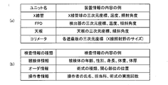
- FIG. 5 is a table illustrating contents of apparatus information and inspection information in the X-ray imaging apparatus according to the embodiment.
- A) is a table
- (b) is a table
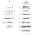
- (A) is a figure which shows the flowchart in a learning stage
- (b) is a figure which shows the flowchart in an implementation stage. It is a figure which shows the structure of the combination information memorize
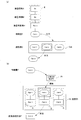
- (A) is a figure explaining the process by which a feature-value is identified by a discriminator
- (b) is a figure explaining the process in which a processing method extraction part extracts the optimal image processing method based on a feature-value. is there.
- the X-ray imaging apparatus 1 has an X-ray from an X-ray focal point 5 a to the subject M and a top plate 3 on which the subject M in a horizontal posture is placed.
- An X-ray tube 5 that irradiates 5b and an X-ray detector 7 that detects X-rays that have been irradiated and transmitted to the subject M are provided.
- the central axis of the X-ray 5b irradiated by the X-ray tube 5 is denoted by reference numeral 5c.
- the X-ray tube 5 and the X-ray detector 7 are arranged to face each other with the top plate 3 interposed therebetween.
- the X-ray detector 7 has a detection surface for detecting X-rays, and X-ray detection elements are two-dimensionally arranged on the detection surface.
- a flat panel detector (FPD) is used as the X-ray detector 7.
- a collimator 9 is provided below the X-ray tube 5.
- the collimator 9 includes two pairs of shielding plates. Each of the shielding plates limits the X-rays emitted from the X-ray tube 5 to a cone shape that is a pyramid.
- the X-ray tube 5 corresponds to the X-ray source in the present invention
- the X-ray detector 7 corresponds to the X-ray detection means in the present invention.
- the X-ray imaging apparatus 1 includes an X-ray irradiation control unit 11, an X-ray tube moving unit 13, a detector moving unit 15, a top plate moving unit 17, and a collimator control unit 19. A device information detection unit 21 and an image processing unit 23.
- the X-ray irradiation control unit 11 is connected to the X-ray tube 5 and controls the X-ray irradiation from the X-ray tube 5 by controlling the X-ray irradiation time, the tube voltage applied to the X-ray tube 5 and the like. Control the dose and the timing of X-ray irradiation.
- the X-ray tube moving unit 13 is connected to the X-ray tube 5, moves the X-ray tube 5 in each of the x direction, the y direction, and the z direction, and rotates the X-ray tube 5 around the axis in each direction.
- the x direction is the longitudinal direction of the top plate 3
- the y direction is the short direction of the top plate 3
- the z direction is the vertical direction.
- the detector moving unit 15 is connected to the X-ray detector 7, moves the X-ray detector 7 in each of the x direction, the y direction, and the z direction, and tilts the X-ray detector 7 with respect to the horizontal plane. Let That is, the X-ray tube moving unit 13 and the detector moving unit 15 irradiate the X-ray tube 5 and the X-ray detector 7 from the three-dimensional position of the imaging system including the X-ray tube 5 and the X-ray detector 7. The X-ray irradiation angle is controlled.
- the top plate moving unit 17 is connected to the top plate 3 and moves the top plate 3 in each of the x direction, the y direction, and the z direction, and tilts the top plate 3 with respect to the horizontal plane.
- the collimator control unit 19 adjusts the X-ray irradiation field irradiated from the X-ray tube 5 by controlling the opening and closing movement of each shielding plate provided in the collimator 9.
- the apparatus information detection unit 21 detects information indicating the state of each unit such as the top plate 3, the X-ray tube 5, the X-ray detector 7, and the collimator 9 constituting the X-ray imaging apparatus 1 as apparatus information. Sensors (not shown) are provided in each unit of the top plate 3, the X-ray tube 5, the X-ray detector 7, and the collimator 9, and each sensor detects apparatus information and transmits it to the apparatus information detection unit 21. Examples of information detected as device information include the three-dimensional coordinates of each unit, temperature, or inclination angle, as shown in FIG.
- the device information detection unit 21 corresponds to device information detection means in the present invention.
- the image processing unit 23 includes an image generation unit 25, a feature amount extraction unit 27, a classifier C, a processing method determination unit 29, an image processing execution unit 31, a processing method detection unit 33, and a classifier generation unit 35.
- the discriminator selecting unit 37 and the feedback unit 39 are provided.
- the image generation unit 25 is provided in the subsequent stage of the X-ray detector 7 and generates an X-ray image of the subject M based on the X-ray detection signal output from the X-ray detector 7.
- the X-ray image generated by the image generation unit 25 is an X-ray image used for examination of the subject M (hereinafter referred to as “examination image”) or an X-ray image used for learning of a discriminator described later (hereinafter referred to as “ Learning image ”).
- the image generation unit 25 corresponds to the inspection image generation unit and the learning image generation unit in the present invention.
- the feature amount extraction unit 27 is provided in the subsequent stage of the image generation unit 25, and extracts a feature amount from the target image.
- the image from which the feature amount extraction unit 27 extracts the feature amount includes a learning image in addition to the inspection image generated by the image generation unit 25.
- the discriminator C is provided in the subsequent stage of the feature quantity extraction unit 27 and is generated by learning a plurality of feature quantity information in the learning stage.
- a feature quantity extracted from the inspection image or the learning image by the feature quantity extraction unit 27 is input to the discriminator C.
- the discriminator C identifies the feature amount of the learning image that is closest to the feature amount of the input inspection image among the feature amounts of the learning image, and outputs data of the identified feature amount. Examples of a configuration in which the classifier C identifies and outputs a feature amount include a support vector machine and a neural network. In the embodiment, it is assumed that the discriminator C is constituted by a support vector machine.
- the processing method extraction unit 29 is provided in the subsequent stage of the classifier C, and extracts an image processing method stored in association with the feature value output from the classifier C.
- the image processing execution unit 31 is provided after the processing method extraction unit 29, and performs image processing on the inspection image generated by the image generation unit 25 using the image processing method extracted by the processing method extraction unit 29. Execute.
- the processing method extraction unit 29 corresponds to processing method extraction means in the present invention.
- the image processing execution unit 31 corresponds to an image processing execution unit.
- the processing method detection unit 33 is provided after the feature amount extraction unit 27, and detects an optimal image processing method based on the feature amount of the learning image.
- the discriminator generation unit 35 is provided at the subsequent stage of the processing method detection unit 33.
- the discriminator generation unit 35 generates the discriminator C based on the feature amount extracted from the learning image by the feature amount extraction unit 27. Details of the process of generating the discriminator C will be described later.
- the discriminator generator 35 corresponds to the discriminator generator in the present invention.
- the discriminator selection unit 37 selects the optimum discriminator C from each of the discriminators C generated by the discriminator generation unit 35 based on the device information and the inspection information.
- the feedback unit 39 feeds back information on the image processing method executed by the image processing execution unit 31 and the feature amount of the inspection image to be subjected to image processing to the discriminator generation unit 35.
- the discriminator generation unit 35 performs additional learning based on the fed back information.
- the discriminator selecting unit 37 corresponds to the discriminator selecting means in the present invention.
- the feedback unit 39 corresponds to feedback means in the present invention.
- the X-ray imaging apparatus 1 further includes an input unit 41, a storage unit 43, a monitor 45, and a main control unit 47.
- the input unit 41 inputs an operator's instruction and inspection information, and examples thereof include a keyboard input type panel and a touch input type panel.
- the examination information means a group of information related to the examination including information on the subject.
- the operator can further appropriately modify each parameter of image processing performed on the inspection image by operating the input unit 41.
- the input unit 41 corresponds to a processing method correcting unit in the present invention.
- the storage unit 43 stores information such as the feature amount extracted from each of the learning image and the inspection image generated by the image generation unit 25 and each of the classifiers C.
- the monitor 45 displays the inspection image generated by the image generation unit 25 and the X-ray image (hereinafter referred to as “processed image”) subjected to image processing by the image processing execution unit 31.
- the main control unit 47 includes an X-ray irradiation control unit 11, an X-ray tube moving unit 13, a detector moving unit 15, a top plate moving unit 17, a collimator control unit 19, an image processing unit 23, a storage unit 43, and a monitor 45. Oversee and control.
- the storage unit 43 corresponds to a feature amount storage unit and a discriminator storage unit in the present invention.
- FIG. 3 is a flowchart for explaining an operation process of the X-ray imaging apparatus 1 according to the embodiment.
- a case where an X-ray image in a stomach X-ray examination is acquired will be described as an example.
- the operation process by the X-ray imaging apparatus is divided into a learning stage shown in FIG. 3A and an implementation stage shown in FIG.
- the learning stage is a stage in which a discriminator used in the implementation stage is generated by learning.
- a learning image used for generating the discriminator is acquired (A1).
- a feature amount is extracted from the acquired learning image (A2), and an optimal image processing method for the learning image is detected based on the extracted feature amount (A3).
- a classifier is generated by learning a large number of feature quantities of the learning image (A4).
- the implementation stage first, preparation for X-ray imaging is performed, and apparatus information and inspection information are acquired (S1). Based on the combination of the device information and the inspection information, an optimum classifier for the inspection image is selected from a series of classifiers generated in the learning stage (S2). Then, an inspection image is acquired under the apparatus information and the inspection information (S3). A feature amount is extracted from the acquired inspection image (S4). By inputting the feature quantity of the inspection image to the selected classifier, an image processing method optimum for the inspection image is extracted (S5). Image processing is performed on the inspection image using the extracted image processing method, and a processed image suitable for diagnosis is acquired (S6). Finally, additional learning is performed on the feature amount of the inspection image and the executed image processing method (S7).
- an operation process of the X-ray imaging apparatus 1 in the learning stage will be described with reference to FIGS. To do.
- Step A1 (Acquisition of learning image)
- a plurality of learning images N are acquired.
- an X-ray image (sample image) that is generated in advance by the image generation unit 25 and stored in the storage unit 43 is used as the learning image N.
- the X-ray image generated by the image generation unit 25 may be used as the learning image N by irradiating the subject with X-rays from the X-ray tube 5 again.
- the apparatus information M includes physical elements such as three-dimensional coordinates, inclination angles, and temperatures as illustrated in FIG. 2A for the top plate 3, the X-ray tube 5, the X-ray detector 7, and the collimator 9. It consists of a group of information related to Three-dimensional coordinates mean the coordinates of each unit in the x, y, and z directions.
- An inclination angle means the inclination angle with respect to a horizontal surface as an example. Note that the information listed in FIG. 2A as the device information is an example, and is not limited thereto.
- the inspection information S is composed of a group of information related to the inspection. Examples of information constituting the examination information S include subject information, order information, operator information, and the like, as shown in FIG.
- the subject information is information related to the subject M to be examined, such as age, sex, and physique.
- the order information is information for specifying the content of the examination, such as the region of interest irradiated with X-rays and the type of technique to be performed.
- the operator information is information related to the operator who performs the inspection, such as the name and the number of times the technique is performed.
- the apparatus information detection unit 21 distinguishes each of the inspection information S based on the difference in the information group constituting the inspection information S. That is, when there are h combinations of information groups, each of the inspection information S is distinguished as inspection information S1 to Sh. When there are j combinations of information groups constituting the device information M, the device information detection unit 21 distinguishes each of the device information M as device information M1 to Mj based on the difference of the information groups constituting the device information M. To do.
- the storage unit 43 classifies and stores each of the learning images N in association with the inspection information S and the device information M.
- each of the k learning images N generated under the conditions of the examination information Sa and the device information Mb is classified as learning images Nab-1 to Nab-k.
- the learning image N is classified based on the inspection information S and the device information M already associated with the sample image.
- Each of the classified learning images N is transmitted to the feature amount extraction unit 27.
- Step A2 extraction of feature values
- the feature amount extraction unit 27 extracts a feature amount from the transmitted learning image.
- the feature amount is information for discriminating and classifying each learning image for a plurality of learning images, and one feature amount F is extracted for one learning image N.
- Examples of the feature quantity extracted by the feature quantity extraction unit 27 include the frequency of information of the X-ray image of the stomach wall shown in the learning image, the distribution of luminance values, and the like.
- the region of the learning image is divided into a plurality of regions having a predetermined number of pixels, and the frequency of information on the X-ray image of the stomach wall in each divided region is used as the feature amount.
- the method of extracting as F is mentioned. Further, it is possible to discriminate similar X-ray images for each of the learning images based on the ORB feature amount and the normalized histogram.
- the feature amount extraction unit 27 classifies each extracted feature amount F according to the learning image N. For example, among the learning images Nab-1 to Nab-k, the feature quantity F extracted for the learning image Nab-c is classified as the feature quantity Fab-c by the feature quantity extraction unit 27. Each of the learning images N from which the feature amount F has been extracted is transmitted to the processing method detection unit 33 together with the feature amount F information.
- Step A3 Detection of image processing method
- the processing method detection unit 33 detects the optimum image processing method P for each of the learning images N.
- the image processing method there is a parameter group including brightness adjustment, contrast adjustment, noise removal correction value, and the like corresponding to each region of the learning image.
- Specific examples of the image processing method P include combinations of various parameters relating to image adjustment, such as a ⁇ curve, edge enhancement, and contrast.
- the optimum image processing method P differs depending on the feature amount F of each learning image N.
- the learning image is divided into a plurality of areas, and optimal parameters for luminance adjustment and contrast adjustment according to the luminance value are set in each of the divided areas.
- An optimum image processing method P is detected as each combination of parameters set in each region.
- the processing method detection unit 33 detects an image processing method already executed on the sample image as the optimum image processing method P.
- the operator confirms the newly generated X-ray image and manually adjusts various parameters such as the gradation, brightness, and contrast of the X-ray image, for example. To do.
- various parameter groups for specifying the X-ray image as an image that is most easily interpreted are specified.
- the processing method detection unit 33 detects the specified various parameter groups as the optimum image processing method P for the learning image.
- the processing method detection unit 33 classifies the optimum image processing method P detected for the learning image Nab-c as the image processing method Pab-c.
- the processing method detection unit 33 associates the information on the feature amount F and the information on the image processing method P detected based on the same learning image N, and generates combined information D between the feature amount F and the image processing method P. (FIG. 4, one-dot chain line).
- the information on the image processing method Pab-c and the information on the feature value Fab-c are associated with each other by the processing method detection unit 33 and become combined information D.
- k learning images Nab-1 to Nab-k are generated under the conditions of the inspection information Sa and the device information Mb, and the feature amounts Fab-1 to Fab-k and the learning amounts N are calculated based on the learning images N.
- Image processing methods Pab-1 to Pab-k are detected.
- the feature amount F detected from the same learning image N and the image processing method P are associated, and k sets of combination information D are generated.
- Each of the combination information D is stored in the storage unit 43.
- Information on the feature amounts Fab-1 to Fab-k is transmitted from the processing method detection unit 33 to the discriminator generation unit 35.
- Step A4 The discriminator generation unit 35 generates the discriminator C based on the transmitted k types of feature amounts. Note that the discriminator C generated under the conditions of the inspection information Sa and the device information Mb is classified as a discriminator Cab.
- the discriminator Cab inputs the feature amount information of the inspection image to identify the feature amount of the learning image that is closest to the information of the feature amount Fx of the input inspection image, and identifies the identified learning image
- the information on the feature amount of the image is output.
- the discriminator Cab learns k types of feature quantities F, that is, feature quantities Fab-1 to Fab-k, so that the entire space to which the feature quantity F belongs is divided into k categories (regions). That is, every feature amount F belongs to any one of the first category including the feature amount Fab-1 to the kth category including Fab-k. Since the discriminator C is constituted by a support vector machine, each parameter of feature quantity is learned so that the margin for the boundary of each category is maximized.
- the discriminator Cab first identifies the feature quantity F that is the closest to the feature quantity Fx among the learned feature quantities Fab-1 to Fab-k based on each component of the feature quantity Fx. That is, the discriminator Cab identifies the category to which the feature quantity Fx belongs from the first category including the feature quantity Fab-1 to the kth category including Fab-k.
- the discriminator Cab outputs the learned feature quantity F included in the identified category as the feature quantity F most approximate to the input feature quantity Fx. For example, when the component of the feature quantity Fx is closest to the feature quantity Fab-c among the feature quantities Fab-1 to Fab-k, the discriminator Cab receives the feature quantity Fab with respect to the input of the feature quantity Fx. -C information is output. As described above, the discriminator Cab has a configuration that outputs any one of k types of feature amounts Fab-1 to Fab-k learned in advance for every feature amount input.
- step A4 the discriminator generation unit 35 generates a discriminator Cab corresponding to the combination of the inspection information Sa and the device information Mb by learning using the learning image N as teacher data.
- Steps A1 to A4 are performed for each combination of inspection information S and apparatus information M as shown in FIG. That is, the discriminator generating unit 35 generates (h ⁇ j) discriminators C11 to Chj according to combinations of h pieces of inspection information S1 to Sh and j pieces of device information M1 to Mj.
- the storage unit 43 stores each of the generated discriminators C11 to Chj in association with the combination information E composed of the inspection information S and the device information M.
- the discriminator Chj is stored in association with the combination information E composed of the inspection information Sh and the device information Mj.
- the classifiers C11 to Chj are generated by learning for each combination of the inspection information S1 to Sh and the device information M1 to Mj. By performing the steps A1 to A4 for all the learning images N, learning using the learning images N as teacher data is performed, and the learning stage ends.
- Step S1 acquisition of device information and inspection information
- preparation for X-ray imaging is performed, and apparatus information and inspection information are acquired. That is, the subject M is placed on the top 3 as shown in FIG.
- the operator operates the input unit 41 to move each unit of the top board 3, the X-ray tube 5, and the X-ray detector 7 according to the X-ray imaging conditions, and further, each unit is appropriately inclined with respect to the horizontal plane.
- the operator operates the input unit 41 to move the shielding plate provided in the collimator 9 and adjusts the X-ray irradiation field irradiated from the X-ray tube 5.
- each unit of the top plate 3, the X-ray tube 5, the X-ray detector 7, and the collimator 9 is provided with a sensor for detecting information such as a three-dimensional position and temperature as needed. And each sensor transmits the information which shows the state of each unit illustrated to Fig.2 (a) to the apparatus information detection part 21 at any time.
- the device information detection unit 21 detects information indicating the current state of each unit as device information M based on the transmitted information.
- the inclination angles of the top plate 3 and the X-ray tube 5 with respect to the horizontal plane (xy plane) are indicated by using symbols ⁇ 1 and ⁇ 2, respectively.
- Each piece of device information M is transmitted from the device information detection unit 21 to the discriminator selection unit 37.
- the operator operates the input unit 41 to input the inspection information S illustrated in FIG.
- the inputted inspection information S is transmitted to the discriminator selection unit 37.
- the inspection information S input in the implementation stage is hereinafter distinguished as inspection information Sx
- the apparatus information M detected in the implementation stage is hereinafter distinguished as apparatus information My.
- Step S2 selection of identifier
- the discriminator selection unit 37 selects the discriminator C to be used in the implementation stage based on the transmitted examination information Sx and device information My.
- Each of the discriminators C11 to Chj generated in the learning stage is stored in the storage unit 43, and each of the discriminators C11 to Chj includes test information S and device information M, which is combination information when generating a learning image. E is stored in association with each other.
- the discriminator selection unit 37 matches the combination information Exy composed of the inspection information Sx and the device information My at the time of generation of the transmitted inspection image among the combination information E at the time of generation of the learning image stored in the storage unit 43.
- the combination information E to be searched is searched.
- the discriminator selection unit 37 uses the discriminator Cxy stored in association with the combination information Exy in the implementation stage. Select as C.
- the discriminator selection unit 37 selects, from the combination information E stored in the storage unit 43 at the time of learning image generation, the one closest to the combination information Exy at the time of generation of the inspection image.
- the discriminator selection unit 37 performs a search based on collation of each parameter constituting the inspection information S and device information M, a mean square error, and the like, and a combination at the time of generating a learning image that most closely approximates the combination information Exy at the time of generating the inspection image.
- Select information E In the embodiment, as shown in FIG. 7, the discriminator Cxy is not learned, and the discriminator selecting unit 37 selects the combination information E composed of the inspection information Sa and the device information Mb based on the combination information Exy. .
- the discriminator selection unit 37 selects the discriminator C stored in association with the selected combination information E.
- the selected combination information E and the discriminator Cab are stored in association with each other. Therefore, the discriminator selection unit 37 selects the discriminator Cab as the discriminator C used in the implementation stage. As described above, the discriminator selecting unit 37 selects the discriminator C used in the implementation stage based on the inspection information Sx and the device information My at the time of generating the inspection image.
- Step S3 generation of inspection image
- the operator operates the input unit 41 to irradiate the subject M with the X-ray focal point 5a to the X-ray 5b.
- the X-ray detector 7 detects the X-ray 5b that passes through the subject M and outputs an X-ray detection signal.
- the image generation unit 25 generates an X-ray image of the subject M based on the X-ray detection signal.
- the X-ray image before the image processing is executed is hereinafter referred to as an inspection image R. As shown in FIG.
- the inspection image R generated on the z-th sheet under the conditions of the inspection information Sx and the apparatus information My is hereinafter referred to as an inspection image Rxy-z.
- the inspection image R is transmitted from the image generation unit 25 to the feature amount extraction unit 27.
- Step S4 feature amount extraction
- the feature amount extraction unit 27 extracts the feature amount F from the inspection image R.
- the content of the feature quantity F extracted by the feature quantity extraction unit 27 in the implementation stage is the same as in the learning stage.
- the feature amount F extracted by the feature amount extraction unit 27 from the inspection image Rxy-z is hereinafter referred to as a feature amount Fxy-z.
- Information on the feature amount F extracted in the implementation stage is transmitted to the classifier C selected by the classifier selection unit 37 in step S2.
- Step S5 Extraction of image processing method
- the discriminator C selected by the discriminator selection unit 37 that is, the discriminator Cab, extracts an image processing method optimum for the inspection image R based on the transmitted feature amount F information.
- the transmitted feature quantity F that is, information on the feature quantity Fxy is input to the discriminator Cab.
- information on k types of feature amounts Fab-1 to Fab-k is learned in the learning stage.
- the discriminator Cab identifies the feature quantity F that most closely approximates the feature quantity Fxy-z among the learned feature quantities Fab-1 to Fab-k based on each component of the feature quantity Fxy-z. That is, the discriminator Cab identifies the category to which the feature quantity Fxy-z belongs from the first category including the feature quantity Fab-1 to the kth category including Fab-k.
- the classifier Cab outputs information on the feature quantity Fab-d in response to the input of the feature quantity Fx.
- the output feature quantity Fab-d information is transmitted to the processing method extraction unit 29.
- the processing method extraction unit 29 searches for a feature value F stored in the storage unit 43 that matches the transmitted feature value F. Then, information on the image processing method P stored in association with the feature amount F specified by the search is extracted. When the discriminator Cab outputs information on the feature value Fab-d, the processing method extraction unit 29 searches the feature value Fab-d for a series of feature values stored in the storage unit 43.
- the feature value Fab-d is stored in association with the image processing method Pab-d. Accordingly, the processing method extraction unit 29 extracts the information on the image processing method Pub-d as the image processing method P optimum for the inspection image Rxy-z based on the feature value Fab-d. Information on the extracted image processing method Pab-d is transmitted from the processing method extraction unit 29 to the image processing execution unit 31.
- Step S6 Image processing of inspection image
- the image processing execution unit 31 performs image processing on the inspection image R generated by the image generation unit 25 based on the transmitted information on the image processing method P.
- the inspection image R that has been subjected to the image processing by the image processing method P is defined as a processed image.
- the image processing execution unit 31 performs image processing by the image processing method Pab-d on the inspection image Rxy-z, and generates a processed image.
- the storage unit 43 stores the information about the feature amount Fxy-z and the image processing method Pab-d, which is the image processing method P most suitable for the feature amount Fxy-z, in association with each other. Information about the feature amount Fxy-z is transmitted to the feedback unit 39.
- the image processing method Pab-d is an image processing method P that is optimal for the inspection image Rxy-z. Therefore, by the image processing based on the image processing method Pad-d, the inspection image Rxy-z becomes an X-ray image suitable for diagnosis in which brightness adjustment, noise correction, and the like are performed under optimal conditions. The processed image is displayed on the monitor 45.
- the operator may determine that the image processing method Pab-d executed by the image processing execution unit 31 needs to be corrected with reference to the processed image displayed on the monitor 45. In such a case, the operator can further manually correct each parameter of the image processing performed on the processed image by operating the input unit 41 as necessary.
- the storage unit 43 stores the information on the feature amount Fxy-z and the information on the corrected image processing method P in association with each other.
- Step S7 (additional learning of classifier) After image processing of the inspection image Rxy-z, additional learning of the classifier is performed using the feature amount Fxy-z extracted from the inspection image Rxy-z. That is, the feedback unit 39 feeds back the transmitted information on the feature amount Fxy-z to the discriminator generation unit 35.
- the discriminator generation unit 35 causes the discriminator C to perform additional learning using the fed back information on the feature amount Fxy-z.
- the discriminator C that performs the additional learning is preferably the discriminator C in which the fed-back feature quantity F is input in step S5. If the discriminator Cxy has been learned in association with the combination information Exy, the information on the feature amount Fxy-z is input to the discriminator Cxy in step S5. In this case, the discriminator generation unit 35 causes the discriminator Cxy to perform additional learning using information on the feature quantity Fxy-z.
- the discriminator Cxy is not learned, the discriminator Cab is selected for the combination information Exy, and the information on the feature amount Fxy-z is input to the discriminator Cab. Therefore, in the case of the embodiment, the feedback unit 39 causes the discriminator Cab to perform additional learning using information on the feature amount Fxy-z.
- the classifier Cab that has already learned k types of feature values F learns (k + 1) types of feature values by additionally learning information on the feature values Fxy-z extracted from the inspection image Rxy-z. It will be in the state. That is, in the discriminator Cab after additional learning, the entire space to which the feature amount F belongs is divided into (k + 1) categories.
- the X-ray imaging apparatus 1 specifies the optimum image processing method P using the discriminator C after additional learning for the feature amount F extracted from the inspection image R thereafter.
- the image processing unit 25 when the image processing unit 25 newly generates the (k + 1) -th inspection image Rxy- (z + 1), information on the feature amount Fxy- (z + 1) extracted from the inspection image Rxy- (z + 1) Is input to the discriminator Cab after the additional learning.
- the feature amount Fxy ⁇ (z + 1) belongs to one of the first category including the feature amount Fab ⁇ 1 and the (k + 1) th category including Fxy ⁇ z.
- the Cab after additional learning outputs any one of (k + 1) types of feature amounts Fab-1 to Fxy-z in response to the input of the feature amount Fxy- (z + 1).
- the classifier C is a learned feature quantity that is more approximate to the input feature quantity F. Can be output.
- additional learning for the discriminator C all the processes related to X-ray imaging are completed.
- a learning stage is provided before an implementation stage for imaging an X-ray image used for diagnosis.
- a discriminator is generated by learning using the feature amount extracted from the learning image as teacher data.
- the storage unit stores the feature amount of the learning image and the image processing method optimum for the learning image in association with each other.
- the discriminator has a configuration for identifying the feature quantity that is closest to the input feature quantity among the learned feature quantities for every feature quantity input, and outputting the identified learned feature quantity. Yes. Therefore, in the implementation stage, the feature quantity extracted from the inspection image is input to the discriminator, so that the discriminator identifies and outputs the learned feature quantity that most closely approximates the feature quantity of the inspection image.
- Each of the learned feature quantities is stored in association with an image processing method optimum for the learning image that is the basis of the feature quantity. Then, the processing method extraction unit extracts the image processing method associated with the feature value output from the classifier as an image processing method corresponding to the feature value input to the classifier.
- the feature quantity input to the discriminator and the feature quantity discriminated and output by the discriminator are approximate. Therefore, by processing the inspection image using the image processing method extracted by the processing method, the inspection image becomes an X-ray image suitable for diagnosis. As described above, in the embodiment, every time an inspection image is generated, an image processing method optimum for the inspection image is automatically extracted. Therefore, the operator does not need to determine an image processing method suitable for the generated inspection image.
- the X-ray imaging apparatus can significantly increase the quality and throughput of the X-ray image in X-ray fluoroscopy.
- the discriminator generated by learning is stored in association with the combination information related to the learning image.
- the discriminator selection unit selects the discriminator to be used for the inspection image in response to the input of combination information when the inspection image is generated.
- the classifier selection unit selects, by parameter matching, the combination information that is most similar to the combination information at the time of generating the inspection image, from among the combination information learned in association with the classifier. Then, the classifier associated with the selected combination information is selected as the classifier to be used for the inspection image.
- the discriminator selecting unit can appropriately handle the combination information at the time of generating the inspection image. Can be selected. By using the selected discriminator, an image processing method suitable for the inspection image is extracted. Therefore, even when an inspection image is generated under the conditions of new inspection information and apparatus information, an image processing method suitable for the inspection image can be automatically extracted. Therefore, the X-ray imaging apparatus according to the embodiment can quickly perform optimal image processing on the X-ray image under all imaging conditions.
- the X-ray imaging apparatus has a function of performing additional learning on the classifier based on the feature amount of the inspection image extracted in the implementation stage.
- the discriminator It is possible to discriminate and output a more approximate learned feature amount. As a result, it is possible to acquire a processed image more suitable for diagnosis according to the number of times the X-ray imaging apparatus is used.
- the present invention is not limited to the above embodiment, and can be modified as follows.
- the surgeon inputs the examination information to the input unit 35, but the arrangement for obtaining the examination information is not limited to this. That is, the X-ray imaging apparatus 1 may be configured to be connected to a HIS (Hospital Information System), a RIS (Radiology Information System), or the like via a network and acquire inspection information via the HIS or RIS.
- HIS Hospital Information System
- RIS Radiology Information System
- the discriminator generation unit 35 may newly generate (learn) the discriminator Cxy associated with the inspection information Sx and the device information My based on the feature amount Fxy-z.
- the discriminator selection unit 37 can search for the discriminator C stored in association with the input combination information E in response to the input of more various combination information E. That is, the discriminator selection unit 37 does not need to calculate the combination information E that approximates the input combination information E. As a result, the time required for selecting the discriminator C can be reduced, so that the optimum image processing method P for the inspection image R can be extracted more quickly.
- the discriminator selection unit 37 may be a discriminator taking a support vector machine or a neural network as an example.
- the discriminator S constituting the discriminator selection unit 37 inputs the combination information E composed of the inspection information S and the device information M, so that the input combination information E of the learned combination information E Identify and output the closest approximation. Therefore, the discriminator selection unit 37 can quickly output the combination information E that is the closest to the learned information with respect to any combination information E input.
- the first-stage discriminator S that inputs the combination information E and the second-stage discriminator C that inputs the feature amount F in this way, under the conditions of the new inspection information S and device information M Even when the inspection image R is generated, the optimum image processing method P is extracted more quickly. Therefore, an X-ray image subjected to image processing suitable for diagnosis can be quickly acquired under any imaging conditions.
- the timing for selecting the discriminator according to step S2 may be appropriately changed as long as it is before the start of step S5.
- the X-ray image is taken for the subject M taking a horizontal posture, but the present invention is not limited to this. That is, the configuration of the X-ray imaging apparatus according to the embodiment can also be applied to the case where X-ray imaging is performed on the subject M having a standing posture.
- the x direction that is, the body axis direction of the subject M is parallel to the vertical direction.
- X-ray imaging apparatus 3 Top plate 5 ... X-ray tube (X-ray source) 7 ... X-ray detector (X-ray detection means) 9 ... Collimator 21: Device information detection unit (device information detection means) 25 ... Image generation unit (learning image generation means, inspection image generation means) 27... Feature quantity extraction unit (feature quantity extraction means) 29 ... processing method extraction unit (processing method extraction means) 31: Image processing execution unit (image processing execution means) 33 ... Processing method detection unit 35 ... classifier generator (discriminator generator) 37. Classifier selection unit (classifier selection means) 39 ... Feedback section (feedback means) 41. Input unit (processing method correcting means) 43 ... Storage section (identifier storage means, feature value storage means) 45 ... Monitor (display means)
Landscapes
- Health & Medical Sciences (AREA)
- Life Sciences & Earth Sciences (AREA)
- Medical Informatics (AREA)
- Engineering & Computer Science (AREA)
- Radiology & Medical Imaging (AREA)
- Molecular Biology (AREA)
- Biophysics (AREA)
- Nuclear Medicine, Radiotherapy & Molecular Imaging (AREA)
- Optics & Photonics (AREA)
- Pathology (AREA)
- Physics & Mathematics (AREA)
- Biomedical Technology (AREA)
- Heart & Thoracic Surgery (AREA)
- High Energy & Nuclear Physics (AREA)
- Surgery (AREA)
- Animal Behavior & Ethology (AREA)
- General Health & Medical Sciences (AREA)
- Public Health (AREA)
- Veterinary Medicine (AREA)
- Apparatus For Radiation Diagnosis (AREA)
- Image Analysis (AREA)
Abstract
特徴量抽出部27は同一の組み合わせ情報の下で生成された学習用画像の各々から抽出し、識別器生成部35は抽出された特徴量を教師データとする学習により識別器を生成する。記憶部43は生成された識別器と、学習用画像に係る組み合わせ情報と関連づけて記憶する。識別器選択部37は、検査用画像の生成時における組み合わせ情報の入力に対して、最も近似する記憶済みの組み合わせ画像を探索し、探索された組み合わせ画像と関連づけられた識別器を選択する。そして選択された識別器を用いて、検査用画像に最も適する画像処理方法を抽出する。識別器Cに入力される検査用画像の特徴量に基づいて、検査用画像に適する画像処理方法が抽出される。この場合、あらゆる組み合わせ条件の下で、検査用画像に対して最適な画像処理を迅速に施すことができる。また操作者の技量に左右されることなく、画像処理後のX線画像の品質を一定に保つことができる。
Description
本発明は、X線を用いて被検体のX線像を取得するX線撮影装置に関し、特に機械学習的手法によって、X線画像に最適な画像処理方法を選択および実行する技術に関する。
医療現場において、胃部X線検査など、被検体に対して複数の位置からX線を照射して複数枚のX線画像を取得する場合がある(例えば、特許文献1,2参照)。この場合、X線を照射する位置や角度によってX線画像に映るX線像が相違する。そのため、X線画像に対して輝度調整などの画像処理を行う際に、X線画像に映るX線像に応じて、X線画像に対して行う画像処理のパターンを適宜変更する必要がある。すなわち、X線画像によっては、X線画像の中央部に対してより大きな輝度調整を行う場合や、X線画像の周辺部に対してより大きな輝度調整を行う場合がある。
従来では、操作者がX線画像に映るX線像を参照して、X線画像に最も適した画像処理のパターン(画像処理方法)を選択する。画像処理方法の一例としては、X線画像の各部位に応じたパラメータによる、輝度調整やコントラスト調整、またはノイズ除去補正などが挙げられる。そして選択された画像処理方法をX線画像に対して実行し、診断に適したX線画像を取得する。
しかしながら、このような構成を有する従来例の場合には、次のような問題点がある。
すなわち、操作者がX線画像を参照して最適な画像処理方法を判別するためには熟練を要するので、操作者によっては最適な画像処理方法を判別できない場合がある。さらに、X線透視により多数のX線画像を断続的に生成する場合、多数のX線画像に対してそれぞれ最適な画像処理方法を判別する必要がある。その結果、操作者に対する作業負担が極めて大きくなる。
すなわち、操作者がX線画像を参照して最適な画像処理方法を判別するためには熟練を要するので、操作者によっては最適な画像処理方法を判別できない場合がある。さらに、X線透視により多数のX線画像を断続的に生成する場合、多数のX線画像に対してそれぞれ最適な画像処理方法を判別する必要がある。その結果、操作者に対する作業負担が極めて大きくなる。
このような場合、X線画像の各々に応じた最適な画像処理を実行できないので、精度の高い診断を行うことが困難となる。また、最適な画像処理方法が判別される場合であっても、操作者が画像処理方法を判別して手動で適切な画像処理方法を実行するためには時間を要する。その結果、X線撮影の所要時間が長くなるので、X線撮影装置のワークフローが低下するという問題も懸念される。
本発明は、このような事情に鑑みてなされたものであって、取得されたX線画像に対して、取得条件に応じて効率的に最適な画像処理を行うことのできるX線撮影装置を提供することを目的とする。
本発明は、このような目的を達成するために、次のような構成をとる。
すなわち、本発明に係るX線撮影装置は、被検体にX線を照射するX線源と、前記X線源から照射されて前記被検体を透過したX線を検出するX線検出手段と、前記X線検出手段が出力する検出信号に基づいて学習用のX線画像を学習用画像として生成する学習用画像生成手段と、前記X線検出手段が出力する検出信号に基づいて、前記被検体に対する検査に用いるX線画像を検査用画像として生成する検査用画像生成手段と、前記被検体に対する検査の情報を検査情報として入力する検査情報入力手段と、前記X線源および前記X線検出手段を含む複数のユニットに関する、位置および傾斜角度などの物理的情報を装置情報として検出する装置情報検出手段と、前記X線画像の特徴量を抽出する特徴量抽出手段と、前記特徴量抽出手段が前記学習用画像から抽出する前記特徴量を、前記学習用画像に対して実行される画像処理方法と関連づけて記憶する特徴量記憶手段と、前記検査情報および前記装置情報からなる組み合わせ情報が同一である条件の下で生成された、前記学習用画像の各々から抽出される前記特徴量を教師データとする学習によって、入力される前記検査用画像の特徴量に最も近似する前記学習用画像の特徴量を出力する識別器を生成する識別器生成手段と、前記識別器の学習に用いられた前記学習用画像を生成する際における、前記組み合わせ情報を前記識別器と関連づけて記憶する識別器記憶手段と、前記識別器記憶手段が記憶している前記組み合わせ情報のうち、前記検査用画像を生成する際における前記組み合わせ情報に最も近似する前記組み合わせ情報を探索し、探索した前記組み合わせ情報と関連づけて前記識別器記憶手段が記憶する前記識別器を選択する識別器選択手段と、前記識別器選択手段によって選択された前記識別器が出力する前記学習用画像の特徴量と関連づけて、前記特徴量記憶手段に記憶されている前記画像処理方法を抽出する処理方法抽出手段と、前記処理方法抽出手段が抽出する前記画像処理方法を前記検査用画像に対して実行する画像処理実行手段とを備えていることを特徴とするものである。
すなわち、本発明に係るX線撮影装置は、被検体にX線を照射するX線源と、前記X線源から照射されて前記被検体を透過したX線を検出するX線検出手段と、前記X線検出手段が出力する検出信号に基づいて学習用のX線画像を学習用画像として生成する学習用画像生成手段と、前記X線検出手段が出力する検出信号に基づいて、前記被検体に対する検査に用いるX線画像を検査用画像として生成する検査用画像生成手段と、前記被検体に対する検査の情報を検査情報として入力する検査情報入力手段と、前記X線源および前記X線検出手段を含む複数のユニットに関する、位置および傾斜角度などの物理的情報を装置情報として検出する装置情報検出手段と、前記X線画像の特徴量を抽出する特徴量抽出手段と、前記特徴量抽出手段が前記学習用画像から抽出する前記特徴量を、前記学習用画像に対して実行される画像処理方法と関連づけて記憶する特徴量記憶手段と、前記検査情報および前記装置情報からなる組み合わせ情報が同一である条件の下で生成された、前記学習用画像の各々から抽出される前記特徴量を教師データとする学習によって、入力される前記検査用画像の特徴量に最も近似する前記学習用画像の特徴量を出力する識別器を生成する識別器生成手段と、前記識別器の学習に用いられた前記学習用画像を生成する際における、前記組み合わせ情報を前記識別器と関連づけて記憶する識別器記憶手段と、前記識別器記憶手段が記憶している前記組み合わせ情報のうち、前記検査用画像を生成する際における前記組み合わせ情報に最も近似する前記組み合わせ情報を探索し、探索した前記組み合わせ情報と関連づけて前記識別器記憶手段が記憶する前記識別器を選択する識別器選択手段と、前記識別器選択手段によって選択された前記識別器が出力する前記学習用画像の特徴量と関連づけて、前記特徴量記憶手段に記憶されている前記画像処理方法を抽出する処理方法抽出手段と、前記処理方法抽出手段が抽出する前記画像処理方法を前記検査用画像に対して実行する画像処理実行手段とを備えていることを特徴とするものである。
本発明に係るX線撮影装置によれば、組み合わせ情報が同一である条件の下で生成された学習用画像の各々から抽出される特徴量を教師データとする学習により、識別器が生成される。識別器は検査用画像から抽出された特徴量の入力に対して、学習用画像の特徴量のうち、入力された検査用画像の特徴量に最も近似する学習用画像の特徴量を識別する構成を有している。学習用画像の特徴量の各々は、当該特徴量が抽出された学習用画像に最適な画像処理方法と関連づけて記憶されている。そして処理方法抽出部は識別器が出力する学習用画像の特徴量と関連づけられている画像処理方法を、検査用画像の特徴量に対応する画像処理方法として抽出する。
この場合、識別器に入力される検査用画像の特徴量と、識別器が判別して出力する学習用画像の特徴量とは近似している。そのため、処理方法抽出部が抽出する画像処理方法を用いて検査用画像を処理することにより、検査用画像は診断に適するX線画像となる。すなわち検査用画像が生成される際に特徴量に応じて、当該検査用画像に最適な画像処理方法が自動的に抽出される。そのため操作者は生成される検査用画像に適する画像処理方法を自身で判別する必要がない。従って、画像処理方法の判別に要する時間を短縮し、かつ操作者が受ける負担を軽減できる。また操作者の技量に左右されることなく、画像処理後のX線画像の品質を一定に保つことができる。
識別器記憶手段は、学習によって生成された識別器と、学習用画像に係る組み合わせ情報と関連づけて記憶する。そして識別器選択手段は、検査用画像の生成時における組み合わせ情報の入力に対して、最も近似する記憶済みの学習用画像の組み合わせ情報を探索し、探索された学習用画像の組み合わせ情報と関連づけられた識別器を選択する。そして選択された識別器を用いて、検査用画像に最も適する画像処理方法を抽出する。
このような構成を有することにより、入力される組み合わせ情報が学習済みの組み合わせ情報のいずれにも一致しない場合であっても、識別器選択部は検査用画像の生成時における組み合わせ情報に対応する適切な識別器を選択できる。そのため、全く新しい組み合わせ情報の条件下で検査用画像を生成した場合であっても、当該検査用画像に適する画像処理方法を自動的に抽出できる。従って、あらゆる撮影条件の下で、X線画像に対して最適な画像処理を迅速に施すことができる。
また、本発明に係るX線撮影装置は、識別器はサポートベクタマシンまたはニューラルネットワークによって構成されることが好ましい。
本発明に係るX線撮影装置によれば、識別器はサポートベクタマシンまたはニューラルネットワークによって構成される。この場合、識別器は入力された特徴量の情報に応じて、適切な学習済みの特徴量を迅速に識別できる。従って、生成される検査用画像に対して迅速に適切な画像処理方法を実行し、検査に適するX線画像を取得することができる。
また、本発明に係るX線撮影装置は、前記検査用画像から抽出された前記特徴量を前記識別器生成手段にフィードバックし、前記識別器に追加学習を行わせるフィードバック手段を備えることが好ましい。
本発明に係るX線撮影装置によれば、フィードバック手段は検査用画像から抽出された特徴量を識別器生成手段にフィードバックし、識別器に追加学習を行わせる。この場合、検査用のX線画像を生成するたびに追加学習が行われる。そのため実施例に係るX線撮影装置の使用回数が多くなるに従って、識別器は入力される特徴量に対してより近似した特徴量を識別できるようになる。その結果、X線撮影装置の使用回数に応じて、より診断に適した処理後画像を取得することが可能となる。
また、本発明に係るX線撮影装置は、前記検査情報は前記被検体を特定する情報、前記被検体に対する検査を特定する情報、および前記被検体に対する検査を行う操作者を特定する情報によって構成されることが好ましい。
本発明に係るX線撮影装置によれば、検査情報は被検体を特定する情報、被検体に対する検査を特定する情報、および被検体に対する検査を行う操作者を特定する情報によって構成される。この場合、被検体のみならず、検査を特定する情報や操作者を特定する情報が検査情報として包含されるので、検査や操作者の違いごとに異なる識別器が生成される。従って、検査や操作者の相違に応じて検査用画像に適した画像処理方法を施し、診断に適するX線画像を生成することが可能となる。
また、本発明に係るX線撮影装置は、前記識別器選択手段は前記検査用画像を生成する際における前記組み合わせ情報を入力とする、サポートベクタマシンまたはニューラルネットワークによって構成されることが好ましい。
本発明に係るX線撮影装置によれば、識別器選択手段は検査用画像を生成する際における組み合わせ情報を入力とする、サポートベクタマシンまたはニューラルネットワークによって構成される。この場合、識別器は学習済みの組み合わせ情報のうち、入力された検査用画像に係る組み合わせ情報に最も近似する組み合わせ情報を迅速に識別できる。従って、生成される検査用画像に対して迅速に適切な画像処理方法を実行し、検査に適するX線画像を取得することができる。
また、本発明に係るX線撮影装置は、前記画像処理実行手段が画像処理方法を実行した前記検査用画像を表示する表示手段と、前記表示手段が表示する前記検査用画像に対して、前記画像処理方法の各パラメータを修正する処理方法修正手段を備え、前記画像処理実行手段は前記処理方法修正手段によって修正された画像処理方法を前記検査用画像に対して再度実行することが好ましい。
本発明に係るX線撮影装置によれば、画像処理実行手段が画像処理方法を実行した検査用画像を表示する表示手段を備えている。処理方法修正手段は、表示手段が表示する画像処理済みの検査用画像に対して、画像処理方法の各パラメータを修正する。この場合、操作者は表示される検査用画像を参照し、画像処理方法の各パラメータをより適したパラメータに適宜修正できる。
画像処理実行手段は処理方法修正手段によって修正された画像処理方法を用いて、検査用画像に対する画像処理を再度実行する。従って、検査用画像に対し、より適したパラメータによる画像処理が実行される。その結果、より精度の高い診断を可能とする検査用画像を取得できる。
本発明に係るX線撮影装置によれば、組み合わせ情報が同一である条件の下で生成された学習用画像の各々から抽出される特徴量を教師データとする学習により、識別器が生成される。識別器は検査用画像から抽出された特徴量の入力に対して、学習用画像の特徴量のうち、入力された検査用画像の特徴量に最も近似する学習用画像の特徴量を識別する構成を有している。学習用画像の特徴量の各々は、当該特徴量が抽出された学習用画像に最適な画像処理方法と関連づけて記憶されている。そして処理方法抽出部は識別器が出力する学習用画像の特徴量と関連づけられている画像処理方法を、検査用画像の特徴量に対応する画像処理方法として抽出する。
この場合、識別器に入力される検査用画像の特徴量と、識別器が判別して出力する学習用画像の特徴量とは近似している。そのため、処理方法抽出部が抽出する画像処理方法を用いて検査用画像を処理することにより、検査用画像は診断に適するX線画像となる。すなわち検査用画像が生成される際に特徴量に応じて、当該検査用画像に最適な画像処理方法が自動的に抽出される。そのため操作者は生成される検査用画像に適する画像処理方法を自身で判別する必要がない。従って、画像処理方法の判別に要する時間を短縮し、かつ操作者が受ける負担を軽減できる。また操作者の技量に左右されることなく、画像処理後のX線画像の品質を一定に保つことができる。
識別器記憶手段は、学習によって生成された識別器と、学習用画像に係る組み合わせ情報と関連づけて記憶する。そして識別器選択手段は、検査用画像の生成時における組み合わせ情報の入力に対して、最も近似する記憶済みの学習用画像の組み合わせ情報を探索し、探索された学習用画像の組み合わせ情報と関連づけられた識別器を選択する。そして選択された識別器を用いて、検査用画像に最も適する画像処理方法を抽出する。
このような構成を有することにより、入力される組み合わせ情報が学習済みの組み合わせ情報のいずれにも一致しない場合であっても、識別器選択部は検査用画像の生成時における組み合わせ情報に対応する適切な識別器を選択できる。そのため、全く新しい組み合わせ情報の条件下で検査用画像を生成した場合であっても、当該検査用画像に適する画像処理方法を自動的に抽出できる。従って、あらゆる撮影条件の下で、X線画像に対して最適な画像処理を迅速に施すことができる。
以下、図面を参照して本発明の実施例を説明する。
<全体構成の説明>
実施例1に係るX線撮影装置1は図1(a)に示すように、水平姿勢をとる被検体Mを載置させる天板3と、被検体Mに対してX線焦点5aからX線5bを照射するX線管5と、被検体Mに照射されて透過したX線を検出するX線検出器7とを備えている。X線管5が照射するX線5bの中心軸を符号5cで示す。X線管5とX線検出器7は、天板3を挟んで対向配置されている。X線検出器7はX線を検出する検出面を備えており、検出面にはX線検出素子が二次元的に配列されている。実施例ではX線検出器7としてフラットパネル型検出器(FPD)を用いる。
実施例1に係るX線撮影装置1は図1(a)に示すように、水平姿勢をとる被検体Mを載置させる天板3と、被検体Mに対してX線焦点5aからX線5bを照射するX線管5と、被検体Mに照射されて透過したX線を検出するX線検出器7とを備えている。X線管5が照射するX線5bの中心軸を符号5cで示す。X線管5とX線検出器7は、天板3を挟んで対向配置されている。X線検出器7はX線を検出する検出面を備えており、検出面にはX線検出素子が二次元的に配列されている。実施例ではX線検出器7としてフラットパネル型検出器(FPD)を用いる。
X線管5の下方にはコリメータ9が設けられている。コリメータ9は2対の遮蔽板を備えており、遮蔽板の各々によって、X線管5から照射されるX線を角錐となっているコーン状に制限する。X線管5は本発明におけるX線源に相当し、X線検出器7は本発明におけるX線検出手段に相当する。
X線撮影装置1は図1(b)に示すように、X線照射制御部11と、X線管移動部13と、検出器移動部15と、天板移動部17と、コリメータ制御部19と、装置情報検出部21と、画像処理部23とを備えている。X線照射制御部11はX線管5に接続されており、X線照射時間やX線管5に印加される管電圧などを制御することによって、X線管5から照射されるX線の線量、およびX線を照射させるタイミングなどを制御する。
X線管移動部13はX線管5に接続されており、X線管5をx方向、y方向、およびz方向の各々へ移動させるとともに、X線管5を各方向の軸周りに回転させる。なお、x方向は天板3の長手方向であり、y方向は天板3の短手方向であり、z方向は鉛直方向である。
検出器移動部15はX線検出器7に接続されており、X線検出器7をx方向、y方向、およびz方向の各々へ移動させるとともに、X線検出器7を水平面に対して傾斜させる。すなわち、X線管移動部13および検出器移動部15によって、X線管5およびX線検出器7からなる撮像系の三次元的な位置と、X線管5からX線検出器7へ照射されるX線の照射角度とが制御される。
天板移動部17は天板3に接続されており、天板3をx方向、y方向、およびz方向の各々へ移動させるとともに、天板3を水平面に対して傾斜させる。コリメータ制御部19は、コリメータ9に設けられている遮蔽板の各々の開閉移動を制御することにより、X線管5から照射されるX線の照射野を調節する。
装置情報検出部21はX線撮影装置1を構成する天板3、X線管5、X線検出器7、コリメータ9などの各ユニットについて、状態を示す情報を装置情報として検出する。天板3、X線管5、X線検出器7、コリメータ9の各ユニットには図示しないセンサが設けられており、各センサは装置情報を検出して装置情報検出部21へ送信する。装置情報として検出される情報の例としては図2(a)に示すように、各ユニットの三次元座標、温度、または傾斜角度などが挙げられる。装置情報検出部21は本発明における装置情報検出手段に相当する。
画像処理部23は画像生成部25と、特徴量抽出部27と、識別器Cと、処理方法判定部29と、画像処理実行部31と、処理方法検出部33と、識別器生成部35と、識別器選択部37と、フィードバック部39とを備えている。画像生成部25はX線検出器7の後段に設けられており、X線検出器7から出力されるX線検出信号に基づいて、被検体MのX線画像を生成する。画像生成部25が生成するX線画像は、被検体Mの検査に用いるX線画像(以下、「検査用画像」とする)や、後述する識別器の学習に用いるX線画像(以下、「学習用画像」とする)が含まれる。画像生成部25は本発明における検査用画像生成手段および学習用画像生成手段に相当する。
特徴量抽出部27は画像生成部25の後段に設けられており、対象となる画像から特徴量を抽出する。特徴量抽出部27が特徴量を抽出する画像には、画像生成部25が生成する検査用画像の他に、学習用画像が含まれる。
識別器Cは特徴量抽出部27の後段に設けられており、学習段階において複数の特徴量情報を学習することによって生成される。識別器Cには、特徴量抽出部27が検査用画像または学習用画像から抽出する特徴量が入力される。そして識別器Cは学習用画像の特徴量のうち、入力された検査用画像の特徴量に最も近似する学習用画像の特徴量を識別し、識別した特徴量のデータを出力する。識別器Cが特徴量を識別して出力する構成の例としては、サポートベクタマシンやニューラルネットワークなどが挙げられる。実施例において、識別器Cはサポートベクタマシンによって構成されているものとする。
処理方法抽出部29は識別器Cの後段に設けられており、識別器Cが出力する特徴量と関連づけて記憶されている画像処理方法を抽出する。画像処理実行部31は処理方法抽出部29の後段に設けられており、処理方法抽出部29が抽出する画像処理方法を用いて、画像生成部25が生成する検査用画像に対して画像処理を実行する。処理方法抽出部29は本発明における処理方法抽出手段に相当する。画像処理実行部31は画像処理実行手段に相当する。
処理方法検出部33は特徴量抽出部27の後段に設けられており、学習用画像の特徴量に基づいて、最適な画像処理方法を検出する。識別器生成部35は処理方法検出部33の後段に設けられている。識別器生成部35は、特徴量抽出部27が学習用画像から抽出する特徴量に基づいて、識別器Cを生成する。識別器Cを生成する工程の詳細は後述する。識別器生成部35は本発明における識別器生成手段に相当する。
識別器選択部37は装置情報および検査情報に基づいて、識別器生成部35が生成した識別器Cの各々から、最適な識別器Cを選択する。フィードバック部39は画像処理実行部31が実行した画像処理方法と、画像処理の対象となった検査用画像の特徴量との情報を識別器生成部35にフィードバックする。識別器生成部35はフィードバックされた情報に基づいて追加学習を行う。識別器選択部37は本発明における識別器選択手段に相当する。フィードバック部39は本発明におけるフィードバック手段に相当する。
X線撮影装置1はさらに、入力部41と、記憶部43と、モニタ45と、主制御部47とを備えている。入力部41は操作者の指示や検査情報を入力するものであり、その例として、キーボード入力式のパネルやタッチ入力式のパネルなどが挙げられる。検査情報とは、被検体に関する情報を始めとする、検査に関連する情報群を意味する。また、操作者は入力部41を操作することにより、検査用画像に対して行う画像処理の各パラメータをさらに適宜修正することが可能である。入力部41は本発明における処理方法修正手段に相当する。
記憶部43は、画像生成部25が生成する学習用画像および検査用画像の各々から抽出される特徴量、および識別器Cの各々などの情報を記憶する。モニタ45は画像生成部25が生成する検査用画像、および画像処理実行部31によって画像処理が行われたX線画像(以下、「処理後画像」とする)を表示する。主制御部47は、X線照射制御部11、X線管移動部13、検出器移動部15、天板移動部17、コリメータ制御部19、画像処理部23、記憶部43、モニタ45の各々を統括制御する。記憶部43は本発明における特徴量記憶手段および識別器記憶手段に相当する。
<動作の説明>
次に、実施例に係るX線撮影装置1の動作について説明する。図3は実施例に係るX線撮影装置1の動作の工程を説明するフローチャートである。実施例では胃部X線検査におけるX線画像を取得する場合を例にとって説明する。
次に、実施例に係るX線撮影装置1の動作について説明する。図3は実施例に係るX線撮影装置1の動作の工程を説明するフローチャートである。実施例では胃部X線検査におけるX線画像を取得する場合を例にとって説明する。
X線撮影装置による動作の工程は、図3(a)に示す学習段階と、図3(b)に示す実施段階とに分けられる。学習段階とは、実施段階で用いる識別器を学習により生成する段階であり、まず識別器の生成に用いる学習用画像を取得する(A1)。次に、取得された学習用画像から特徴量を抽出し(A2)、抽出された特徴量に基づいて、学習用画像に対して最適な画像処理方法を検出する(A3)。そして学習用画像の特徴量を多数学習することによって識別器を生成する(A4)。
実施段階では、まずX線撮影の準備を行い、装置情報および検査情報を取得する(S1)。装置情報および検査情報の組み合わせに基づいて、学習段階で生成された一連の識別器から、検査用画像に対して最適な識別器の選択を行う(S2)。そして装置情報および検査情報の下で検査用画像を取得する(S3)。取得された検査用画像から、特徴量を抽出する(S4)。選択された識別器に検査用画像の特徴量を入力することにより、検査用画像に最適な画像処理方法を抽出する(S5)。抽出された画像処理方法を用いて検査用画像に画像処理を行い、診断に適した処理後画像を取得する(S6)。最後に、検査用画像の特徴量と実行された画像処理方法とを追加学習する(S7)ここで図4および図5を用いて、まず学習段階におけるX線撮影装置1の動作の工程を説明する。
ステップA1(学習用画像の取得)
学習段階において、まず学習用画像Nを複数枚取得する。実施例1では学習用画像Nとして、予め画像生成部25によって生成され、記憶部43に記憶されているX線画像(サンプル画像)を用いる。なお、あらためてX線管5からX線を被検体に照射し、画像生成部25が生成したX線画像を学習用画像Nとして用いてもよい。
学習段階において、まず学習用画像Nを複数枚取得する。実施例1では学習用画像Nとして、予め画像生成部25によって生成され、記憶部43に記憶されているX線画像(サンプル画像)を用いる。なお、あらためてX線管5からX線を被検体に照射し、画像生成部25が生成したX線画像を学習用画像Nとして用いてもよい。
学習用画像Nの各々をあらためて生成する場合、操作者は入力部41を操作して検査情報Sを入力し、装置情報検出部21は装置情報Mを検出する。装置情報Mは、天板3、X線管5、X線検出器7、コリメータ9の各ユニットについて、図2(a)に例示するような、三次元座標や傾斜角度、温度といった物理的要素に関連する情報群によって構成される。三次元座標とは各ユニットのx方向、y方向、およびz方向の各々における座標を意味する。傾斜角度とは、例として水平面に対する傾斜角度を意味する。なお、装置情報として図2(a)に挙げられている情報は一例であり、これらに限定されるものではない。
検査情報Sは、検査に関連する情報群によって構成される。検査情報Sを構成する情報の例としては図2(b)に示すように、被検体情報、オーダ情報、および操作者情報などが挙げられる。被検体情報は、年齢、性別、体格など、検査の対象である被検体Mに関する情報である。オーダ情報は、X線を照射する関心部位や実行する術式の種類など、検査の内容を特定する情報である。操作者情報は、氏名や術式の実施回数など、検査を行う操作者に関する情報である。
装置情報検出部21は検査情報Sを構成する情報群の相違に基づいて、検査情報Sの各々を区別する。すなわち、情報群の組み合わせがh通りある場合、検査情報Sの各々は検査情報S1~Shとして区別される。また装置情報Mを構成する情報群の組み合わせがj通りある場合、装置情報検出部21は装置情報Mを構成する情報群の相違に基づいて、装置情報Mの各々を装置情報M1~Mjとして区別する。
記憶部43は、学習用画像Nの各々を検査情報Sおよび装置情報Mと関連づけて分類し、記憶する。一例として図4に示すように、検査情報Sa、装置情報Mbの条件において生成されるk枚の学習用画像Nの各々は、学習用画像Nab-1~Nab-kとして分類される。サンプル画像を学習用画像Nとして用いる場合、サンプル画像に既に関連づけされている検査情報Sおよび装置情報Mに基づいて学習用画像Nを分類する。分類された学習用画像Nの各々は特徴量抽出部27へ送信される。
ステップA2(特徴量の抽出)
特徴量抽出部27は送信された学習用画像から特徴量を抽出する。特徴量は複数の学習用画像について、各々の学習用画像を判別・分類するための情報であり、1枚の学習用画像Nに対して1つの特徴量Fが抽出される。特徴量抽出部27が抽出する特徴量の例としては、学習用画像に映る胃壁のX線像の情報の頻度や、輝度値の分布などが挙げられる。
特徴量抽出部27は送信された学習用画像から特徴量を抽出する。特徴量は複数の学習用画像について、各々の学習用画像を判別・分類するための情報であり、1枚の学習用画像Nに対して1つの特徴量Fが抽出される。特徴量抽出部27が抽出する特徴量の例としては、学習用画像に映る胃壁のX線像の情報の頻度や、輝度値の分布などが挙げられる。
特徴量Fを抽出する具体的な方法としては、学習用画像の領域を所定の画素数からなる複数の領域に分割し、分割した各領域における胃壁のX線像の情報の頻度などを特徴量Fとして抽出する方法などが挙げられる。またORB特徴量や正規化したヒストグラムに基づいて、学習用画像の各々について、類似するX線像をそれぞれ判別することが可能である。
特徴量抽出部27は抽出した特徴量Fの各々を、学習用画像Nに応じて分類する。例えば学習用画像Nab-1~Nab-kのうち、学習用画像Nab-cについて抽出された特徴量Fは、特徴量抽出部27によって特徴量Fab-cとして分類される。特徴量Fが抽出された学習用画像Nの各々は、特徴量Fの情報と共に処理方法検出部33へ送信される。
ステップA3(画像処理方法の検出)
処理方法検出部33は学習用画像Nの各々に対して、最適な画像処理方法Pを検出する。画像処理方法の例としては、学習用画像の各領域に応じた輝度調整、コントラスト調整、およびノイズ除去補正値などからなるパラメータ群が挙げられる。具体的な画像処理方法Pの例としては、γカーブ、エッジ強調、コントラストなど、画像の調整に関する各種パラメータの組み合わせが挙げられる。
処理方法検出部33は学習用画像Nの各々に対して、最適な画像処理方法Pを検出する。画像処理方法の例としては、学習用画像の各領域に応じた輝度調整、コントラスト調整、およびノイズ除去補正値などからなるパラメータ群が挙げられる。具体的な画像処理方法Pの例としては、γカーブ、エッジ強調、コントラストなど、画像の調整に関する各種パラメータの組み合わせが挙げられる。
各学習用画像Nの特徴量Fに応じて、最適な画像処理方法Pはそれぞれ相違する。一例として、学習用画像を複数の領域に分割し、分割された領域の各々において、輝度値に応じた輝度調整、コントラスト調整の最適パラメータが設定される。そして各領域において設定されるパラメータの各々の組み合わせとして、最適な画像処理方法Pが検出される。
過去に作成済みのサンプル画像を学習用画像Nとする場合、処理方法検出部33は、サンプル画像に対して既に実行されている画像処理方法を、最適な画像処理方法Pとして検出する。新たに学習用画像としてX線画像を生成する場合、操作者が新たに生成されたX線画像を確認し、例えば手動で当該X線画像の階調、輝度、およびコントラストなどの各種パラメータを調整する。そして当該X線画像を最も読影しやすい画像とする、各種パラメータ群を特定する。処理方法検出部33は特定された各種パラメータ群を、当該学習用画像に対する最適な画像処理方法Pとして検出する。
処理方法検出部33は学習用画像Nab-cについて検出された最適な画像処理方法Pを、画像処理方法Pab-cとして分類する。処理方法検出部33は同一の学習用画像Nに基づいて検出された、特徴量Fの情報と画像処理方法Pの情報とを関連づけ、特徴量Fと画像処理方法Pとの結合情報Dを生成する(図4、一点鎖線)。例として、画像処理方法Pab-cの情報と特徴量Fab-cの情報とは、処理方法検出部33によって関連づけがなされて結合情報Dとなる。
このように、検査情報Saおよび装置情報Mbの条件下においてk枚の学習用画像Nab-1~Nab-kが生成され、各学習用画像Nに基づいて特徴量Fab-1~Fab-kおよび画像処理方法Pab-1~Pab-kが検出される。そして同一の学習用画像Nから検出された特徴量Fと画像処理方法Pとは関連づけられ、結合情報Dがk組生成される。結合情報Dの各々は、記憶部43に記憶される。特徴量Fab-1~Fab-kの情報は、処理方法検出部33から識別器生成部35へ送信される。
ステップA4(識別器の生成)
識別器生成部35は、送信されたk種類の特徴量に基づいて識別器Cの生成を行う。なお、検査情報Saおよび装置情報Mbの条件下で生成される識別器Cは、識別器Cabとして分類される。
識別器生成部35は、送信されたk種類の特徴量に基づいて識別器Cの生成を行う。なお、検査情報Saおよび装置情報Mbの条件下で生成される識別器Cは、識別器Cabとして分類される。
識別器Cabは、検査用画像の特徴量の情報を入力することにより、入力された検査用画像の特徴量Fxの情報に最も近似する学習用画像の特徴量を識別し、識別した学習用画像の特徴量の情報を出力するものである。識別器Cabはk種類の特徴量F、すなわち特徴量Fab-1~Fab-kを学習することにより、特徴量Fが属する空間全体はk個のカテゴリ(領域)に区分けされる。すなわちあらゆる特徴量Fは、特徴量Fab-1を含む第1のカテゴリから、Fab-kを含む第kのカテゴリのうち、いずれか1つのカテゴリに属することとなる。識別器Cはサポートベクタマシンによって構成されるので、各カテゴリの境界についてのマージンが最大になるように、各々の特徴量のパラメータを学習する。
識別器Cabはまず特徴量Fxの各成分に基づいて、学習済みの特徴量Fab-1~Fab-kのうち、特徴量Fxに最も近似する特徴量Fを識別する。すなわち識別器Cabは特徴量Fab-1を含む第1のカテゴリから、Fab-kを含む第kのカテゴリまでのうち、特徴量Fxが属するカテゴリを識別する。
そして識別器Cabは識別されたカテゴリに含まれている学習済みの特徴量Fを、入力された特徴量Fxに最も近似する特徴量Fとして出力する。例として、特徴量Fab-1~Fab-kのうち、特徴量Fxの成分が特徴量Fab-cに最も近似している場合、識別器Cabは特徴量Fxの入力に対して、特徴量Fab-cの情報を出力する。このように、識別器Cabはあらゆる特徴量の入力に対して、予め学習したk種類の特徴量Fab-1~Fab-kのいずれかを出力する構成を有している。
このようにステップA4において、識別器生成部35は学習用画像Nを教師データとする学習により、検査情報Saおよび装置情報Mbの組み合わせに対応する識別器Cabを生成する。なおステップA1~A4の各工程は図5に示すように、検査情報Sおよび装置情報Mの組み合わせのそれぞれについて行われる。すなわち、h通りの検査情報S1~Sh、およびj通りの装置情報M1~Mjのそれぞれの組み合わせに応じて、識別器生成部35は(h×j)通りの識別器C11~Chjを生成する。
記憶部43は、生成された識別器C11~Chjの各々と、検査情報Sおよび装置情報Mからなる組み合わせ情報Eとを関連づけて記憶する。例として識別器Chjは、検査情報Shおよび装置情報Mjからなる組み合わせ情報Eと関連づけて記憶される。このように検査情報S1~Sh、および装置情報M1~Mjのそれぞれの組み合わせについて、学習による識別器C11~Chjの生成が行われる。ステップA1~A4の各工程を学習用画像Nの全てについて行うことにより、学習用画像Nを教師データとする学習が行われ、学習段階は終了する。
次に図6を用いて、実施段階におけるX線撮影装置1の動作の工程を説明する。なお学習段階において、一連の識別器C11~Chjが生成されているものとする。
ステップS1(装置情報および検査情報の取得)
実施段階ではまずX線撮影の準備を行い、装置情報および検査情報を取得する。すなわち図6に示すように被検体Mを天板3に載置する。そして操作者は入力部41を操作して天板3、X線管5、およびX線検出器7の各ユニットをX線撮影条件に応じて移動させ、さらに各ユニットを水平面に対して適宜傾斜させる。さらに操作者は入力部41を操作してコリメータ9に設けられる遮蔽板を移動させ、X線管5から照射されるX線の照射野を調整する。
実施段階ではまずX線撮影の準備を行い、装置情報および検査情報を取得する。すなわち図6に示すように被検体Mを天板3に載置する。そして操作者は入力部41を操作して天板3、X線管5、およびX線検出器7の各ユニットをX線撮影条件に応じて移動させ、さらに各ユニットを水平面に対して適宜傾斜させる。さらに操作者は入力部41を操作してコリメータ9に設けられる遮蔽板を移動させ、X線管5から照射されるX線の照射野を調整する。
天板3、X線管5、X線検出器7、およびコリメータ9の各ユニットを調整した後、装置情報の検出を行う。天板3、X線管5、X線検出器7、およびコリメータ9の各ユニットには三次元的位置や温度などの情報を随時検出するセンサが設けられている。そして各々のセンサは図2(a)に例示される、各ユニットの状態を示す情報を装置情報検出部21に随時送信する。
装置情報検出部21は送信された情報に基づいて、現時点における各ユニットの状態を示す情報を装置情報Mとして検出する。図6において、天板3およびX線管5の水平面(xy平面)に対する傾斜角度は、それぞれ符号θ1およびθ2を用いて示されている。装置情報Mの各々は、装置情報検出部21から識別器選択部37へ送信される。
そして操作者は入力部41を操作して、図2(b)に例示される検査情報Sを入力する。入力された検査情報Sは識別器選択部37へ送信される。なお実施段階において入力される検査情報Sを以下、検査情報Sxとして区別し、実施段階において検出される装置情報Mを以下、装置情報Myとして区別する。装置情報Mの検出と検査情報Sの入力により、ステップS1の工程は終了する。
ステップS2(識別器の選択)
ステップS1の終了後、識別器選択部37は送信された検査情報Sxおよび装置情報Myに基づいて、実施段階において使用する識別器Cを選択する。学習段階において生成された識別器C11~Chjの各々は記憶部43に記憶されており、識別器C11~Chjの各々には検査情報Sおよび装置情報Mからなる、学習用画像生成時の組み合わせ情報Eが関連づけて記憶されている。
ステップS1の終了後、識別器選択部37は送信された検査情報Sxおよび装置情報Myに基づいて、実施段階において使用する識別器Cを選択する。学習段階において生成された識別器C11~Chjの各々は記憶部43に記憶されており、識別器C11~Chjの各々には検査情報Sおよび装置情報Mからなる、学習用画像生成時の組み合わせ情報Eが関連づけて記憶されている。
識別器選択部37は、記憶部43に記憶されている学習用画像生成時の組み合わせ情報Eのうち、送信された検査用画像生成時の検査情報Sxおよび装置情報Myからなる組み合わせ情報Exyと一致する組み合わせ情報Eを探索する。学習段階において記憶部43に組み合わせ情報Exyが既に記憶されている場合(学習済みの場合)、識別器選択部37は組み合わせ情報Exyと関連づけて記憶されている識別器Cxyを実施段階で用いる識別器Cとして選択する。
しかし、検査情報Sと装置情報Mとの組み合わせは無限に存在するので、組み合わせ情報Exyと一致する組み合わせ情報Eが記憶部43に記憶されていない場合がある。この場合、識別器選択部37は記憶部43に記憶されている学習用画像生成時の組み合わせ情報Eのうち、検査用画像生成時の組み合わせ情報Exyに最も近似するものを選択する。識別器選択部37は検査情報Sおよび装置情報Mを構成する各パラメータの照合や平均二乗誤差などによる探索を行い、検査用画像生成時の組み合わせ情報Exyに最も近似する学習用画像生成時の組み合わせ情報Eを選択する。実施例では図7に示すように識別器Cxyは学習されておらず、識別器選択部37は組み合わせ情報Exyに基づいて、検査情報Saおよび装置情報Mbからなる組み合わせ情報Eを選択するものとする。
識別器選択部37は、選択された組み合わせ情報Eと関連づけて記憶されている識別器Cを選択する。実施例では選択された組み合わせ情報Eと、識別器Cabとが関連づけて記憶されている。従って、識別器選択部37は実施段階で用いる識別器Cとして、識別器Cabを選択する。このように、識別器選択部37は検査用画像生成時の検査情報Sxおよび装置情報Myに基づいて、実施段階において用いる識別器Cの選択を行う。
ステップS3(検査用画像の生成)
識別器Cの選択が行われた後、操作者は入力部41を操作してX線焦点5aからX線5bを被検体Mへ照射させる。X線検出器7は、被検体Mを透過するX線5bを検出してX線検出信号を出力する。画像生成部25はX線検出信号に基づいて、被検体MのX線画像を生成する。ステップS2において生成されるX線画像のうち、画像処理が実行される前のX線画像を以下、検査用画像Rとする。図7に示すように、検査情報Sxおよび装置情報Myの条件下でz枚目に生成される検査用画像Rを以下、検査用画像Rxy-zとする。検査用画像Rは画像生成部25から特徴量抽出部27へ送信される。
識別器Cの選択が行われた後、操作者は入力部41を操作してX線焦点5aからX線5bを被検体Mへ照射させる。X線検出器7は、被検体Mを透過するX線5bを検出してX線検出信号を出力する。画像生成部25はX線検出信号に基づいて、被検体MのX線画像を生成する。ステップS2において生成されるX線画像のうち、画像処理が実行される前のX線画像を以下、検査用画像Rとする。図7に示すように、検査情報Sxおよび装置情報Myの条件下でz枚目に生成される検査用画像Rを以下、検査用画像Rxy-zとする。検査用画像Rは画像生成部25から特徴量抽出部27へ送信される。
ステップS4(特徴量の抽出)
特徴量抽出部27は検査用画像Rから特徴量Fを抽出する。実施段階において特徴量抽出部27が抽出する特徴量Fの内容は学習段階と同様である。図7に示すように、特徴量抽出部27が検査用画像Rxy-zから抽出する特徴量Fを以下、特徴量Fxy-zとする。実施段階において抽出される特徴量Fの情報は、ステップS2において識別器選択部37が選択した識別器Cへ送信される。
特徴量抽出部27は検査用画像Rから特徴量Fを抽出する。実施段階において特徴量抽出部27が抽出する特徴量Fの内容は学習段階と同様である。図7に示すように、特徴量抽出部27が検査用画像Rxy-zから抽出する特徴量Fを以下、特徴量Fxy-zとする。実施段階において抽出される特徴量Fの情報は、ステップS2において識別器選択部37が選択した識別器Cへ送信される。
ステップS5(画像処理方法の抽出)
識別器選択部37が選択した識別器Cすなわち識別器Cabは、送信された特徴量Fの情報に基づいて検査用画像Rに最適な画像処理方法の抽出を行う。送信された特徴量Fすなわち特徴量Fxyの情報は、識別器Cabに入力される。識別器Cabには学習段階において、k種類の特徴量Fab-1~Fab-kの情報が学習されている。
識別器選択部37が選択した識別器Cすなわち識別器Cabは、送信された特徴量Fの情報に基づいて検査用画像Rに最適な画像処理方法の抽出を行う。送信された特徴量Fすなわち特徴量Fxyの情報は、識別器Cabに入力される。識別器Cabには学習段階において、k種類の特徴量Fab-1~Fab-kの情報が学習されている。
識別器Cabはまず特徴量Fxy-zの各成分に基づいて、学習済みの特徴量Fab-1~Fab-kのうち、特徴量Fxy-zに最も近似する特徴量Fを識別する。すなわち識別器Cabは特徴量Fab-1を含む第1のカテゴリから、Fab-kを含む第kのカテゴリまでのうち、特徴量Fxy-zが属するカテゴリを識別する。識別器Cabは、特徴量Fxyが特徴量Fab-dを含むカテゴリに属していると識別した場合、特徴量Fxの入力に対して特徴量Fab-dの情報を出力する。出力された特徴量Fab-dの情報は、処理方法抽出部29へ送信される。
処理方法抽出部29は、記憶部43に記憶されている特徴量Fのうち、送信された特徴量Fと一致するものを探索する。そして探索によって特定された特徴量Fと関連づけて記憶されている画像処理方法Pの情報を抽出する。識別器Cabが特徴量Fab-dの情報を出力した場合、処理方法抽出部29は記憶部43に記憶されている一連の特徴量に対して特徴量Fab-dの探索を行う。
記憶部43において、特徴量Fab-dは画像処理方法Pab-dと関連づけて記憶されている。従って、処理方法抽出部29は特徴量Fab-dに基づいて、画像処理方法Pab-dの情報を検査用画像Rxy-zに最適な画像処理方法Pとして抽出する。抽出された画像処理方法Pab-dの情報は、処理方法抽出部29から画像処理実行部31へ送信される。
ステップS6(検査用画像の画像処理)
画像処理実行部31は送信された画像処理方法Pの情報に基づいて、画像生成部25が生成した検査用画像Rに対して画像処理を行う。画像処理方法Pによる画像処理が行われた検査用画像Rを、処理後画像とする。画像処理実行部31は検査用画像Rxy-zに対して、画像処理方法Pab-dによる画像処理を実行し、処理後画像を生成する。記憶部43は特徴量Fxy-zの情報と、特徴量Fxy-zに最適な画像処理方法Pである、画像処理方法Pab-dとを関連づけて記憶する。また、特徴量Fxy-zの情報はフィードバック部39へ送信される。
画像処理実行部31は送信された画像処理方法Pの情報に基づいて、画像生成部25が生成した検査用画像Rに対して画像処理を行う。画像処理方法Pによる画像処理が行われた検査用画像Rを、処理後画像とする。画像処理実行部31は検査用画像Rxy-zに対して、画像処理方法Pab-dによる画像処理を実行し、処理後画像を生成する。記憶部43は特徴量Fxy-zの情報と、特徴量Fxy-zに最適な画像処理方法Pである、画像処理方法Pab-dとを関連づけて記憶する。また、特徴量Fxy-zの情報はフィードバック部39へ送信される。
画像処理方法Pab-dは、検査用画像Rxy-zに最適な画像処理方法Pである。従って、画像処理方法Pab-dに基づく画像処理により、検査用画像Rxy-zは輝度調整やノイズ補正などが最適な条件の下で行われた、診断に適するX線画像となる。処理後画像はモニタ45に表示される。
なお、モニタ45に表示された処理後画像を参照し、画像処理実行部31が実行した画像処理方法Pab-dを修正する必要があると操作者が判断する場合がある。このような場合、必要に応じて操作者はさらに入力部41を操作して、処理後画像に対して行われた画像処理の各パラメータを手動で適宜修正できる。画像処理方法Pab-dの修正を行った場合、記憶部43は特徴量Fxy-zの情報と、修正後の画像処理方法Pの情報とを関連づけて記憶する。
ステップS7(識別器の追加学習)
検査用画像Rxy-zの画像処理を行った後、検査用画像Rxy-zから抽出された特徴量Fxy-zを用いて識別器の追加学習を行う。すなわちフィードバック部39は送信された特徴量Fxy-zの情報を識別器生成部35へフィードバックする。
検査用画像Rxy-zの画像処理を行った後、検査用画像Rxy-zから抽出された特徴量Fxy-zを用いて識別器の追加学習を行う。すなわちフィードバック部39は送信された特徴量Fxy-zの情報を識別器生成部35へフィードバックする。
識別器生成部35はフィードバックされた特徴量Fxy-zの情報を用いて、識別器Cに追加学習を行わせる。追加学習を行わせる識別器Cは、フィードバックされた特徴量Fが、ステップS5において入力された識別器Cとすることが好ましい。組み合わせ情報Exyと関連づけて識別器Cxyが学習済みの場合、ステップS5において特徴量Fxy-zの情報は識別器Cxyに入力される。この場合、識別器生成部35は特徴量Fxy-zの情報を用いて、識別器Cxyに追加学習を行わせる。
しかし、実施例では識別器Cxyが学習されておらず、組み合わせ情報Exyに対して識別器Cabが選択され、特徴量Fxy-zの情報は識別器Cabに入力されている。従って実施例の場合、フィードバック部39は特徴量Fxy-zの情報を用いて、識別器Cabに追加学習を行わせる。
k種類の特徴量Fを既に学習している識別器Cabは、検査用画像Rxy-zから抽出された特徴量Fxy-zの情報を追加学習することにより、(k+1)種類の特徴量を学習した状態となる。すなわち追加学習後の識別器Cabにおいて、特徴量Fが属する空間全体は(k+1)個のカテゴリに区分けされる。X線撮影装置1は、以降に検査用画像Rから抽出される特徴量Fに対して、追加学習後の識別器Cを用いて最適な画像処理方法Pの特定を行う。
例として、画像処理部25が新たに(k+1)枚目の検査用画像Rxy-(z+1)を生成した場合、検査用画像Rxy-(z+1)から抽出された特徴量Fxy-(z+1)の情報は、追加学習後の識別器Cabに入力される。特徴量Fxy-(z+1)は、特徴量Fab-1を含む第1のカテゴリから、Fxy-zを含む第(k+1)のカテゴリのいずれかに属する。
従って、追加学習後のCabは特徴量Fxy-(z+1)の入力に対して、(k+1)種類の特徴量Fab-1~Fxy-zのいずれかを出力する。このような識別器Cの追加学習を行うことにより、実施段階でX線画像を生成するたびに、識別器Cは入力される特徴量Fに対して、より近似している学習済みの特徴量を出力できるようになる。識別器Cに対する追加学習を行うことにより、X線撮影に関する全ての工程は終了する。
<実施例の構成による効果>
実施例に係るX線撮影装置1では、診断に用いるX線画像を撮影する実施段階の前に、学習段階が設けられる。学習段階において、学習用画像から抽出される特徴量を教師データとする学習により、識別器が生成される。また、記憶部は学習用画像の特徴量と、当該学習用画像に最適な画像処理方法とを関連づけて記憶する。
実施例に係るX線撮影装置1では、診断に用いるX線画像を撮影する実施段階の前に、学習段階が設けられる。学習段階において、学習用画像から抽出される特徴量を教師データとする学習により、識別器が生成される。また、記憶部は学習用画像の特徴量と、当該学習用画像に最適な画像処理方法とを関連づけて記憶する。
識別器はあらゆる特徴量の入力に対して、学習済みの特徴量のうち、入力された特徴量に最も近似する特徴量を識別し、識別した学習済みの特徴量を出力する構成を有している。そのため実施段階において、検査用画像から抽出される特徴量を識別器に入力することにより、識別器は検査用画像の特徴量に最も近似する学習済みの特徴量を識別して出力する。学習済みの特徴量の各々は、いずれも特徴量の基となる学習用画像に最適な画像処理方法と関連づけて記憶されている。そして処理方法抽出部は識別器が出力する特徴量と関連づけられている画像処理方法を、識別器に入力された特徴量に対応する画像処理方法として抽出する。
識別器に入力される特徴量と、識別器が識別して出力する特徴量は近似している。そのため、処理方法が抽出する画像処理方法を用いて検査用画像を処理することにより、検査用画像は診断に適するX線画像となる。このように、実施例では検査用画像が生成されるたびに、その検査用画像に最適な画像処理方法が自動的に抽出される。そのため操作者は生成される検査用画像に適する画像処理方法を自身で判別する必要がない。
従って、画像処理方法の判別に要する時間を短縮し、かつ操作者が受ける負担を軽減できる。また操作者の技量に左右されることなく、画像処理後のX線画像の品質を一定に保つことができる。さらに、X線透視によって多数のX線画像を短時間に生成する場合であっても、多数のX線画像の各々について、それぞれ最適な画像処理方法による画像処理が迅速に実行される。そのため実施例に係るX線撮影装置は、X線透視におけるX線画像の品質とスループットとを大幅に上昇させることが可能となる。
学習によって生成された識別器は、学習用画像に係る組み合わせ情報と関連づけて記憶される。そして識別器選択部は実施段階において、検査用画像の生成時における組み合わせ情報の入力に対して、検査用画像に対して使用する識別器を選択する。識別器選択部は、識別器と関連づけられて学習されている組み合わせ情報のうち、検査用画像の生成時における組み合わせ情報と最も近似する組み合わせ情報をパラメータ照合などによって選択する。そして選択された組み合わせ情報と関連づけられている識別器を、検査用画像に対して使用する識別器として選択する。
このような構成を有することにより、入力される組み合わせ情報が学習済みの組み合わせ情報のいずれにも一致しない場合であっても、識別器選択部は検査用画像の生成時における組み合わせ情報に対応する適切な識別器を選択できる。選択された識別器を用いることにより、検査用画像に適する画像処理方法が抽出される。そのため、新規な検査情報および装置情報の条件下で検査用画像を生成した場合であっても、検査用画像に適する画像処理方法を自動的に抽出できる。従って、実施例に係るX線撮影装置はあらゆる撮影条件の下で、X線画像に対して最適な画像処理を迅速に施すことができる。
また実施例に係るX線撮影装置は、実施段階で抽出された検査用画像の特徴量に基づいて、識別器に対して追加学習を行う機能を有している。この場合、実施段階でX線画像を生成するたびに追加学習が行われるので、実施例に係るX線撮影装置の使用回数が多くなるに従って、識別器は入力される特徴量Fに対して、より近似した学習済みの特徴量を判別して出力できるようになる。その結果、X線撮影装置の使用回数に応じて、より診断に適した処理後画像を取得することが可能となる。
本発明は、上記実施形態に限られることはなく、下記のように変形実施することができる。
(1)上述した実施例では、検査情報を術者が入力部35に入力する構成を有しているが、検査情報を取得する構成はこれに限られない。すなわちX線撮影装置1はネットワークを介してHIS(Hospital Information System)やRIS(Radiology Information System)などと接続され、HISやRISを介して検査情報を取得する構成としてもよい。
(2)上述した実施例では、識別器Cxyが学習段階において生成されていない場合、ステップS7において特徴量Fxy-zの情報を用いて、特徴量Fxy-zが入力された識別器Cabの追加学習を行う構成としたがこれに限られない。すなわち識別器生成部35は特徴量Fxy-zに基づいて、検査情報Sxおよび装置情報Myに関連づけられた、新たに識別器Cxyを生成(学習)してもよい。
この場合、X線画像を生成するたびに、より多くの識別器Cが追加学習によって生成される。従って、識別器選択部37はより多様な組み合わせ情報Eの入力に対して、入力された組み合わせ情報Eと関連づけて記憶されている識別器Cを探索できる。すなわち、識別器選択部37は入力された組み合わせ情報Eと近似する組み合わせ情報Eを計算する必要がない。その結果、識別器Cの選択に要する時間を低減できるので、検査用画像Rに対して最適な画像処理方法Pをより迅速に抽出できる。
(3)上述した実施例において、識別器選択部37は、サポートベクタマシンやニューラルネットワークを例とする識別器であってもよい。この場合、識別器選択部37を構成する識別器Sは、検査情報Sおよび装置情報Mからなる組み合わせ情報Eを入力することにより、学習済みの組み合わせ情報Eのうち、入力された組み合わせ情報Eと最も近似するものを識別して出力する。そのため識別器選択部37は、あらゆる組み合わせ情報Eの入力に対して、学習済みの情報のうち最も近似する組み合わせ情報Eを迅速に出力できる。
このように組み合わせ情報Eを入力する1段目の識別器Sと、特徴量Fを入力する2段目の識別器Cとを備えることにより、新規な検査情報Sおよび装置情報Mの条件下で検査用画像Rを生成した場合であっても、より迅速に最適な画像処理方法Pが抽出される。従って、診断に適する画像処理が行われたX線画像を、あらゆる撮影条件の下で迅速に取得できる。
(4)上述した実施例において、ステップS2に係る識別器の選択を行うタイミングは、ステップS5を開始する前であれば適宜変更してもよい。
(5)上述した実施例において、水平姿勢をとる被検体Mに対してX線画像の撮影を行ったが、これに限られない。すなわち実施例に係るX線撮影装置の構成は、立位体勢をよる被検体Mに対してX線撮影を行う場合にも適用できる。この場合、x方向すなわち被検体Mの体軸方向は鉛直方向と平行になる。
1 …X線撮影装置
3 …天板
5 …X線管(X線源)
7 …X線検出器(X線検出手段)
9 …コリメータ
21 …装置情報検出部(装置情報検出手段)
25 …画像生成部(学習用画像生成手段、検査用画像生成手段)
27 …特徴量抽出部(特徴量抽出手段)
29 …処理方法抽出部(処理方法抽出手段)
31 …画像処理実行部(画像処理実行手段)
33 …処理方法検出部
35 …識別器生成部(識別器生成手段)
37 …識別器選択部(識別器選択手段)
39 …フィードバック部(フィードバック手段)
41 …入力部(処理方法修正手段)
43 …記憶部(識別器記憶手段、特徴量記憶手段)
45 …モニタ(表示手段)
3 …天板
5 …X線管(X線源)
7 …X線検出器(X線検出手段)
9 …コリメータ
21 …装置情報検出部(装置情報検出手段)
25 …画像生成部(学習用画像生成手段、検査用画像生成手段)
27 …特徴量抽出部(特徴量抽出手段)
29 …処理方法抽出部(処理方法抽出手段)
31 …画像処理実行部(画像処理実行手段)
33 …処理方法検出部
35 …識別器生成部(識別器生成手段)
37 …識別器選択部(識別器選択手段)
39 …フィードバック部(フィードバック手段)
41 …入力部(処理方法修正手段)
43 …記憶部(識別器記憶手段、特徴量記憶手段)
45 …モニタ(表示手段)
Claims (6)
- 被検体にX線を照射するX線源と、
前記X線源から照射されて前記被検体を透過したX線を検出するX線検出手段と、
前記X線検出手段が出力する検出信号に基づいて学習用のX線画像を学習用画像として生成する学習用画像生成手段と、
前記X線検出手段が出力する検出信号に基づいて、前記被検体に対する検査に用いるX線画像を検査用画像として生成する検査用画像生成手段と、
前記被検体に対する検査の情報を検査情報として入力する検査情報入力手段と、
前記X線源および前記X線検出手段を含む複数のユニットに関する、位置および傾斜角度などの物理的情報を装置情報として検出する装置情報検出手段と、
前記X線画像の特徴量を抽出する特徴量抽出手段と、
前記特徴量抽出手段が前記学習用画像から抽出する前記特徴量を、前記学習用画像に対して実行される画像処理方法と関連づけて記憶する特徴量記憶手段と、
前記検査情報および前記装置情報からなる組み合わせ情報が同一である条件の下で生成された、前記学習用画像の各々から抽出される前記特徴量を教師データとする学習によって、入力される前記検査用画像の特徴量に最も近似する前記学習用画像の特徴量を出力する識別器を生成する識別器生成手段と、
前記識別器の学習に用いられた前記学習用画像を生成する際における、前記組み合わせ情報を前記識別器と関連づけて記憶する識別器記憶手段と、
前記識別器記憶手段が記憶している前記組み合わせ情報のうち、前記検査用画像を生成する際における前記組み合わせ情報に最も近似する前記組み合わせ情報を探索し、探索した前記組み合わせ情報と関連づけて前記識別器記憶手段が記憶する前記識別器を選択する識別器選択手段と、
前記識別器選択手段によって選択された前記識別器が出力する前記学習用画像の特徴量と関連づけて、前記特徴量記憶手段に記憶されている前記画像処理方法を抽出する処理方法抽出手段と、
前記処理方法抽出手段が抽出する前記画像処理方法を前記検査用画像に対して実行する画像処理実行手段とを備えていることを特徴とするX線撮影装置。 - 請求項1に記載のX線撮影装置において、
前記識別器はサポートベクタマシンまたはニューラルネットワークによって構成されることを特徴とするX線撮影装置。 - 請求項1または請求項2に記載のX線撮影装置において、
前記検査用画像から抽出された前記特徴量を前記識別器生成手段にフィードバックし、前記識別器に追加学習を行わせるフィードバック手段を備えるX線撮影装置。 - 請求項1ないし請求項3のいずれかに記載のX線撮影装置において、
前記検査情報は前記被検体を特定する情報、前記被検体に対する検査を特定する情報、および前記被検体に対する検査を行う操作者を特定する情報によって構成されるX線撮影装置。 - 請求項1ないし請求項4のいずれかに記載のX線撮影装置において、
前記識別器選択手段は前記検査用画像を生成する際における前記組み合わせ情報を入力とする、サポートベクタマシンまたはニューラルネットワークによって構成されるX線撮影装置。 - 請求項1ないし請求項5のいずれかに記載のX線撮影装置において、
前記画像処理実行手段が画像処理方法を実行した前記検査用画像を表示する表示手段と、
前記表示手段が表示する前記検査用画像に対して、前記画像処理方法の各パラメータを修正する処理方法修正手段を備え、
前記画像処理実行手段は前記処理方法修正手段によって修正された画像処理方法を前記検査用画像に対して再度実行するX線撮影装置。
Priority Applications (2)
| Application Number | Priority Date | Filing Date | Title |
|---|---|---|---|
| JP2016568256A JP6347290B2 (ja) | 2015-01-09 | 2015-01-09 | X線撮影装置 |
| PCT/JP2015/050546 WO2016111014A1 (ja) | 2015-01-09 | 2015-01-09 | X線撮影装置 |
Applications Claiming Priority (1)
| Application Number | Priority Date | Filing Date | Title |
|---|---|---|---|
| PCT/JP2015/050546 WO2016111014A1 (ja) | 2015-01-09 | 2015-01-09 | X線撮影装置 |
Publications (1)
| Publication Number | Publication Date |
|---|---|
| WO2016111014A1 true WO2016111014A1 (ja) | 2016-07-14 |
Family
ID=56355734
Family Applications (1)
| Application Number | Title | Priority Date | Filing Date |
|---|---|---|---|
| PCT/JP2015/050546 Ceased WO2016111014A1 (ja) | 2015-01-09 | 2015-01-09 | X線撮影装置 |
Country Status (2)
| Country | Link |
|---|---|
| JP (1) | JP6347290B2 (ja) |
| WO (1) | WO2016111014A1 (ja) |
Cited By (4)
| Publication number | Priority date | Publication date | Assignee | Title |
|---|---|---|---|---|
| CN110946597A (zh) * | 2018-09-27 | 2020-04-03 | 上海西门子医疗器械有限公司 | X射线摄影设备和方法 |
| JP2020062340A (ja) * | 2018-10-19 | 2020-04-23 | キヤノンメディカルシステムズ株式会社 | 画像処理装置及びプログラム |
| JP2021019714A (ja) * | 2019-07-25 | 2021-02-18 | 株式会社日立製作所 | 画像処理装置、画像処理方法及びx線ct装置 |
| JP2021104140A (ja) * | 2019-12-26 | 2021-07-26 | キヤノンメディカルシステムズ株式会社 | 医用情報処理装置、医用情報処理方法、及び医用情報処理プログラム |
Families Citing this family (1)
| Publication number | Priority date | Publication date | Assignee | Title |
|---|---|---|---|---|
| JP2023102364A (ja) * | 2022-01-12 | 2023-07-25 | コニカミノルタ株式会社 | 条件決定装置、条件決定プログラム、および条件決定方法 |
Citations (5)
| Publication number | Priority date | Publication date | Assignee | Title |
|---|---|---|---|---|
| JPH0486820A (ja) * | 1990-07-31 | 1992-03-19 | Fuji Photo Film Co Ltd | 放射線画像読取条件及び/又は画像処理条件決定方法 |
| JPH04212949A (ja) * | 1990-10-17 | 1992-08-04 | Fuji Photo Film Co Ltd | 放射線画像読取条件及び/又は画像処理条件決定方法および装置 |
| JPH06343627A (ja) * | 1993-05-13 | 1994-12-20 | Arch Dev Corp | ディジタル乳房x線像における微小石灰化物検出方法及びそのシステム |
| JP2005111249A (ja) * | 2003-06-19 | 2005-04-28 | Konica Minolta Medical & Graphic Inc | 画像処理方法および画像処理装置ならびに画像処理プログラム |
| JP2010273764A (ja) * | 2009-05-27 | 2010-12-09 | Konica Minolta Medical & Graphic Inc | 画像処理装置及び方法 |
-
2015
- 2015-01-09 WO PCT/JP2015/050546 patent/WO2016111014A1/ja not_active Ceased
- 2015-01-09 JP JP2016568256A patent/JP6347290B2/ja active Active
Patent Citations (5)
| Publication number | Priority date | Publication date | Assignee | Title |
|---|---|---|---|---|
| JPH0486820A (ja) * | 1990-07-31 | 1992-03-19 | Fuji Photo Film Co Ltd | 放射線画像読取条件及び/又は画像処理条件決定方法 |
| JPH04212949A (ja) * | 1990-10-17 | 1992-08-04 | Fuji Photo Film Co Ltd | 放射線画像読取条件及び/又は画像処理条件決定方法および装置 |
| JPH06343627A (ja) * | 1993-05-13 | 1994-12-20 | Arch Dev Corp | ディジタル乳房x線像における微小石灰化物検出方法及びそのシステム |
| JP2005111249A (ja) * | 2003-06-19 | 2005-04-28 | Konica Minolta Medical & Graphic Inc | 画像処理方法および画像処理装置ならびに画像処理プログラム |
| JP2010273764A (ja) * | 2009-05-27 | 2010-12-09 | Konica Minolta Medical & Graphic Inc | 画像処理装置及び方法 |
Cited By (9)
| Publication number | Priority date | Publication date | Assignee | Title |
|---|---|---|---|---|
| CN110946597A (zh) * | 2018-09-27 | 2020-04-03 | 上海西门子医疗器械有限公司 | X射线摄影设备和方法 |
| CN110946597B (zh) * | 2018-09-27 | 2023-09-26 | 上海西门子医疗器械有限公司 | X射线摄影设备和方法 |
| JP2020062340A (ja) * | 2018-10-19 | 2020-04-23 | キヤノンメディカルシステムズ株式会社 | 画像処理装置及びプログラム |
| JP7325942B2 (ja) | 2018-10-19 | 2023-08-15 | キヤノンメディカルシステムズ株式会社 | 画像処理装置及びプログラム |
| JP2021019714A (ja) * | 2019-07-25 | 2021-02-18 | 株式会社日立製作所 | 画像処理装置、画像処理方法及びx線ct装置 |
| JP7245740B2 (ja) | 2019-07-25 | 2023-03-24 | 富士フイルムヘルスケア株式会社 | 画像処理装置、画像処理方法及びx線ct装置 |
| JP2021104140A (ja) * | 2019-12-26 | 2021-07-26 | キヤノンメディカルシステムズ株式会社 | 医用情報処理装置、医用情報処理方法、及び医用情報処理プログラム |
| US11901074B2 (en) | 2019-12-26 | 2024-02-13 | Canon Medical Systems Corporation | Medical information processing apparatus, medical information processing method, and non-transitory computer-readable storage medium storing program |
| JP7485512B2 (ja) | 2019-12-26 | 2024-05-16 | キヤノンメディカルシステムズ株式会社 | 医用情報処理装置、医用情報処理方法、及び医用情報処理プログラム |
Also Published As
| Publication number | Publication date |
|---|---|
| JP6347290B2 (ja) | 2018-06-27 |
| JPWO2016111014A1 (ja) | 2017-08-10 |
Similar Documents
| Publication | Publication Date | Title |
|---|---|---|
| JP6347290B2 (ja) | X線撮影装置 | |
| US10245001B2 (en) | Generation of a three-dimensional reconstruction of a body part by an X-ray machine | |
| US10779791B2 (en) | System and method for mobile X-ray imaging | |
| JP6670509B2 (ja) | コンピュータ断層撮像システムの造影方法 | |
| JP2014144118A (ja) | X線診断装置及び制御プログラム | |
| US9795349B2 (en) | Method for setting scanning protocol and apparatus thereof | |
| US20150154752A1 (en) | Image processor, treatment system, and image processing method | |
| US20150305703A1 (en) | Method and device for controlling rotation angle of c-arm of medical imaging apparatus | |
| JP2020027507A (ja) | 医用情報処理装置及び医用情報処理方法、プログラム | |
| JP2018130336A (ja) | 放射線撮影装置、放射線撮影システム、放射線撮影方法、及びプログラム | |
| US11069060B2 (en) | Image processing apparatus and radiographic image data display method | |
| CN102065953B (zh) | 放射线治疗装置控制装置和特定部位位置计测方法 | |
| US7801275B2 (en) | Imaging system | |
| TWI526195B (zh) | 醫療裝置 | |
| EP3764088B1 (en) | Inspection apparatus | |
| US11553891B2 (en) | Automatic radiography exposure control using rapid probe exposure and learned scene analysis | |
| KR102542505B1 (ko) | 학습데이터 생성 방법, 장치, 프로그램 및 이를 이용한 이물질 검출 방법 | |
| CN102958438B (zh) | X射线ct装置以及图像处理装置 | |
| JP7250330B2 (ja) | 検査装置及び学習装置 | |
| EP4613202A1 (en) | X-ray imaging device comprising camera, and operation method therefor | |
| EP4186431A1 (en) | X-ray diagnostic apparatus, medical image processing apparatus, and medical image processing method | |
| KR20220040490A (ko) | 하전 입자선 장치 | |
| CN118177850A (zh) | X射线成像系统以及动态检测的x射线成像方法 | |
| US20240090864A1 (en) | Radiographic imaging support system, radiographic imaging support method, and recording medium | |
| CN114680928A (zh) | 超声扫查反馈系统 |
Legal Events
| Date | Code | Title | Description |
|---|---|---|---|
| 121 | Ep: the epo has been informed by wipo that ep was designated in this application |
Ref document number: 15876889 Country of ref document: EP Kind code of ref document: A1 |
|
| ENP | Entry into the national phase |
Ref document number: 2016568256 Country of ref document: JP Kind code of ref document: A |
|
| NENP | Non-entry into the national phase |
Ref country code: DE |
|
| 122 | Ep: pct application non-entry in european phase |
Ref document number: 15876889 Country of ref document: EP Kind code of ref document: A1 |