WO2016094583A2 - Biomarkers of oocyte quality - Google Patents
Biomarkers of oocyte quality Download PDFInfo
- Publication number
- WO2016094583A2 WO2016094583A2 PCT/US2015/064836 US2015064836W WO2016094583A2 WO 2016094583 A2 WO2016094583 A2 WO 2016094583A2 US 2015064836 W US2015064836 W US 2015064836W WO 2016094583 A2 WO2016094583 A2 WO 2016094583A2
- Authority
- WO
- WIPO (PCT)
- Prior art keywords
- oocyte
- quality
- genes
- gene
- age
- Prior art date
- Legal status (The legal status is an assumption and is not a legal conclusion. Google has not performed a legal analysis and makes no representation as to the accuracy of the status listed.)
- Ceased
Links
Classifications
-
- C—CHEMISTRY; METALLURGY
- C12—BIOCHEMISTRY; BEER; SPIRITS; WINE; VINEGAR; MICROBIOLOGY; ENZYMOLOGY; MUTATION OR GENETIC ENGINEERING
- C12Q—MEASURING OR TESTING PROCESSES INVOLVING ENZYMES, NUCLEIC ACIDS OR MICROORGANISMS; COMPOSITIONS OR TEST PAPERS THEREFOR; PROCESSES OF PREPARING SUCH COMPOSITIONS; CONDITION-RESPONSIVE CONTROL IN MICROBIOLOGICAL OR ENZYMOLOGICAL PROCESSES
- C12Q1/00—Measuring or testing processes involving enzymes, nucleic acids or microorganisms; Compositions therefor; Processes of preparing such compositions
- C12Q1/68—Measuring or testing processes involving enzymes, nucleic acids or microorganisms; Compositions therefor; Processes of preparing such compositions involving nucleic acids
- C12Q1/6876—Nucleic acid products used in the analysis of nucleic acids, e.g. primers or probes
- C12Q1/6881—Nucleic acid products used in the analysis of nucleic acids, e.g. primers or probes for tissue or cell typing, e.g. human leukocyte antigen [HLA] probes
-
- C—CHEMISTRY; METALLURGY
- C12—BIOCHEMISTRY; BEER; SPIRITS; WINE; VINEGAR; MICROBIOLOGY; ENZYMOLOGY; MUTATION OR GENETIC ENGINEERING
- C12Q—MEASURING OR TESTING PROCESSES INVOLVING ENZYMES, NUCLEIC ACIDS OR MICROORGANISMS; COMPOSITIONS OR TEST PAPERS THEREFOR; PROCESSES OF PREPARING SUCH COMPOSITIONS; CONDITION-RESPONSIVE CONTROL IN MICROBIOLOGICAL OR ENZYMOLOGICAL PROCESSES
- C12Q1/00—Measuring or testing processes involving enzymes, nucleic acids or microorganisms; Compositions therefor; Processes of preparing such compositions
- C12Q1/68—Measuring or testing processes involving enzymes, nucleic acids or microorganisms; Compositions therefor; Processes of preparing such compositions involving nucleic acids
- C12Q1/6876—Nucleic acid products used in the analysis of nucleic acids, e.g. primers or probes
- C12Q1/6883—Nucleic acid products used in the analysis of nucleic acids, e.g. primers or probes for diseases caused by alterations of genetic material
-
- G—PHYSICS
- G01—MEASURING; TESTING
- G01N—INVESTIGATING OR ANALYSING MATERIALS BY DETERMINING THEIR CHEMICAL OR PHYSICAL PROPERTIES
- G01N33/00—Investigating or analysing materials by specific methods not covered by groups G01N1/00 - G01N31/00
- G01N33/48—Biological material, e.g. blood, urine; Haemocytometers
- G01N33/50—Chemical analysis of biological material, e.g. blood, urine; Testing involving biospecific ligand binding methods; Immunological testing
- G01N33/5005—Chemical analysis of biological material, e.g. blood, urine; Testing involving biospecific ligand binding methods; Immunological testing involving human or animal cells
- G01N33/5091—Chemical analysis of biological material, e.g. blood, urine; Testing involving biospecific ligand binding methods; Immunological testing involving human or animal cells for testing the pathological state of an organism
-
- G—PHYSICS
- G01—MEASURING; TESTING
- G01N—INVESTIGATING OR ANALYSING MATERIALS BY DETERMINING THEIR CHEMICAL OR PHYSICAL PROPERTIES
- G01N33/00—Investigating or analysing materials by specific methods not covered by groups G01N1/00 - G01N31/00
- G01N33/48—Biological material, e.g. blood, urine; Haemocytometers
- G01N33/50—Chemical analysis of biological material, e.g. blood, urine; Testing involving biospecific ligand binding methods; Immunological testing
- G01N33/53—Immunoassay; Biospecific binding assay; Materials therefor
-
- C—CHEMISTRY; METALLURGY
- C12—BIOCHEMISTRY; BEER; SPIRITS; WINE; VINEGAR; MICROBIOLOGY; ENZYMOLOGY; MUTATION OR GENETIC ENGINEERING
- C12Q—MEASURING OR TESTING PROCESSES INVOLVING ENZYMES, NUCLEIC ACIDS OR MICROORGANISMS; COMPOSITIONS OR TEST PAPERS THEREFOR; PROCESSES OF PREPARING SUCH COMPOSITIONS; CONDITION-RESPONSIVE CONTROL IN MICROBIOLOGICAL OR ENZYMOLOGICAL PROCESSES
- C12Q2600/00—Oligonucleotides characterized by their use
- C12Q2600/158—Expression markers
Definitions
- the present technology relates to biomarkers of oocyte quality and oocyte quality decline, as well as methods in connection with such biomarkers.
- the functions of the female reproductive system are known to decline with age.
- One major factor in decreased fertility is the decline of the quality of the eggs (oocytes) produced by the ovaries as a woman ages. Decline of quality of oocytes and ovarian follicles generally begins sometime in the 30s for most women and eventually leads to menopause, in most cases between ages 45 and 55.
- the present technology is directed to a method of determining the quality of an oocyte in the body of a human without disturbing or destroying the oocyte, the method comprising:
- the present technology is directed to a method of predicting the quality of an oocyte in the body of a mammal without disturbing or destroying the oocyte, the method comprising the steps of: [0012] (a) obtaining a cell sample from the mammal, wherein the cell sample does not include the oocyte;
- the present technology is directed to a kit for predicting a woman's oocyte quality without the need for disturbing or destroying an oocyte, or of measuring a characteristic that correlates with the quality of an oocyte, the kit comprising:
- testing assay comprising RT-PCR or ELISA, wherein the testing assay measures a characteristic of a gene, pathway or transcriptional profile characteristic of the cell sample, and wherein the characteristic indicates the likely quality of an oocyte;
- the present technology is directed to a method of producing a library of genes as markers of oocyte quality.
- a method may comprise the steps of correlating a test gene with a quantitative and measured characteristic of oocyte quality, listing the correlation in the library; comparing a measured characteristic of a gene provided by a patient with that listed in the library; and determining the quality of an oocyte of a patient based on the comparison.
- the technology herein contemplates a method of producing a library of genes as markers of oocyte quality, the method comprising the steps of:
- the present technology is directed to a reproductive aging gene expression profile; and a method of developing a reproductive aging gene expression profile and one or more candidate markers of reproductive success.
- the present technology is directed to a method of producing a profile set as an indicator of oocyte quality.
- Such methods discussed herein may be done, for example, with any gene, pathway, or transcriptional profile, or with a library of the same.
- FIG. la is a graph showing the prevalence of Down syndrome and infertility in a typical female human as a function of age.
- FIG. lb is a graph showing the aging profile of oocytes in a typical female human as a function of age.
- FIG. 2 shows age-dependent gene expression changes in blood from women aged 20 to 50, as a "heat map" which shows on a macro level relative changes.
- a cell sample refers to one or more cell from any part of the patient's body, which is desired to be tested in order to perform the diagnostic and scientific methods discussed herein.
- a cell sample in accordance with the technology herein may be extracted from any of the following: blood, skin, hair, urine, saliva, sweat, vaginal secretion, any other fluid (including but not limited to intracellular or extracellular fluid, interstitial fluid, lymphatic fluid or transcellular fluid, cerebrospinal fluid, mucus or phlegm).
- Oocyte quantity (the number of viable oocytes produced by a woman, the totality of which is determined long before the onset of puberty) and quality (the likelihood that the oocyte can be successfully fertilized and lead to pregnancy) are two factors of paramount importance in predicting fertility and the likelihood of carrying a pregnancy to term in a female.
- a woman's oocyte quality is generally predicted to decline after a certain age, until she reaches menopause and her ovaries stop releasing oocytes entirely. That is, with rising maternal age, and as a woman's oocytes age as well, a woman loses the ability to reproduce, and the children she may have are at greater risk for birth defects, chromosomal abnormalities, miscarriage and other problems. These defects start to arise when the woman is in her mid-30s and increase through the 40s, about a decade prior to menopause (the average age of menopause is 51), and are caused by declining oocyte quality.
- age-related reproductive decline is characterized by increasing levels of defects in oocyte chromosomal segregation, cell cycle arrest, and oocyte mitochondrial function and morphology, among other factors. Changes in gene expression correlate with these oocyte and egg quality changes, and have been shown in aging human and mouse oocytes.
- Our work in particular on C. elegans oocyte aging, discussed in greater detail below, has highlighted the evolutionary conservation of oocyte quality components. These components, particularly regulators of chromosome segregation fidelity, have been shown to be required for oocyte function and prevention of aneuploidy in worm and mammalian oocytes, and increased fidelity extends reproductive span.
- aneuploidies (aberrations in chromosome numbers) and cell cycle arrest/maintenance failures can be a cause of infertility, birth defects, and miscarriage.
- Morphological assessments are too gross to identify other important measures of quality, such as the levels of particular maternal RNAs that regulate processes in the oocyte.
- current measures of oocyte quality require invasive approaches, and the assessment itself can destroy the oocyte in question, further limiting a woman's number of healthy oocytes and potentially requiring a larger pool of donated oocytes for treatment.
- PBMCs peripheral blood mononuclear cells
- ovarian cells peripheral blood mononuclear cells
- Gielchinsky et al. in 2008 showed that PBMC expression analysis was able to identify candidate genes up- and down-regulated in women who delivered babies after spontaneous pregnancy at >45 years of age. These candidate genes included markers of ovarian function, apoptosis, ubiquitination, energy production, and insulin/IGF- 1 signaling— the same pathway that we showed extends reproductive span in C. elegans.
- a diagnostic of oocyte quality would be in high demand at several levels: clinics conducting assisted reproductive technology methods such as IVF would like this information before embarking on ART approaches to improve their success rates, egg-freezing companies could use such a diagnostic to predict the success of use of eggs they have frozen, and women could use the data to make informed decisions about their reproductive lives in a manner that is minimally invasive and highly convenient.
- the technology herein identifies, and permits health care workers or patients to identify biomarkers of oocyte quality in blood or other bodily fluids, as well as the development of a reliable long-term diagnostic of fertility.
- the quality of an oocyte can be determined by, for example, measuring certain factors and comparing those factors with known values of oocytes of known age.
- Certain methods herein permit an investigator (including a health care worker or the subject herself) to determine or predict the quality of an oocyte in the body of a human without disturbing (e.g., touching, sampling, moving or altering) or destroying the oocyte itself, and without taking it out of the human's body, e.g., in vivo.
- the oocyte itself can be evaluated (or its characteristics determined and predicted) without harming the oocyte, and the oocyte can itself subsequently be fertilized and lead to a successful pregnancy.
- the present technology relates to novel methods for assessing or predicting the quality of oocytes without the need for disturbing or destroying the oocytes themselves. These methods are based at least in part on the discovery that genes in C. elegans, which display changes as oocytes age, are the same genes as those found in mammals; further, even if no directly associated genes were located, mammalian genes with similar functions to the C. elegans genes were located. The C. elegans research also revealed data regarding the characteristics of gene expression in mutant C. elegans with long reproductive spans.
- genes are involved in chromosome segregation, spindle localization, chromosome organization, DNA damage response and repair, and mitochondrial processes such as ATP metabolism. These genes, therefore, are good biomarkers for oocyte quality from oocytes.
- the present technology is directed to the exploitation of the non-invasive nature of blood biomarkers, or biomarkers from other bodily fluids.
- the present technology is directed to the development of single-gene markers as biomarkers; in other embodiments, the present technology is directed to profiles or multiple-gene markers, rather than single-gene markers, as biomarkers.
- Table 1 shows pathways that Shown in the Table 1 is a summary of certain genes and pathways that have previously been identified as being of potential interest for human (H), mouse (M) and worm (W), in connection with applications of the present technology.
- genes and pathways further include any of the following:
- PTEN overlaps with IIS: RHEB2, MEK1 (MAP2K1 ), PtdIns(4,5)P2,
- FOXO transcription factors FOXOl , FOX03a
- HLA-G HSPA8, HSPD1 , IGF1R, IRS2, KATNB1, MAP2K1, PGK1, PMS2, PRKD2, SLC25A3, SNRPN, TCF8, TOPI, TPM1
- the gene or pathway indicative of oocyte quality is chosen from a gene expressed in connection with one or more of the following categories (reflecting those listed in Table 1): cell cycle (e.g., mitosis); chromosome segregation or organization (e.g., chromosome segregation, spindle localization or chromosome organization); DNA damage response and repair (e.g., response to DNA damage stimulus); proteolytic pathway (e.g., proteolysis); energy pathway or mitochondrial function (e.g., ATP metabolic process or ATP binding); cell signaling and communication (e.g., intracellular signaling cascade or cell-cell signaling); protein transport; transcription regulation;
- cell cycle e.g., mitosis
- chromosome segregation or organization e.g., chromosome segregation, spindle localization or chromosome organization
- DNA damage response and repair e.g., response to DNA damage stimulus
- proteolytic pathway e.g., proteolysis
- reproductive process e.g., oogenesis or oviposition
- cell death e.g., cell differentiation or cell adhesion.
- genes have been found to be of interest - among them, genes of chromatin structure, DNA methylation and genome stability.
- genes that have been shown to be associated with both ovarian function and general aging have shown promise in the methods, kits and libraries of the present technology.
- SERPINB2 serpin peptidase inhibitor, clade B, member 2, also known as PAI-2
- IGFIR insulin-like growth factor 1 receptor
- PIK3CB phosphoinositide-3-kinase, catalytic, beta polypeptide
- IRS2 insulin receptor substrate 2
- HSPA8 HSPD1 and HSP60 Two genetic pathways in particular have been found herein to be significant in the regulation of reproductive aging.
- TGF- ⁇ and insulin/IGF- 1 (IIS) signaling pathways regulate reproductive aging cell non-autonomously. That is, these pathways regulate reproductive aging systemically, outside of the oocyte itself rather than by signaling just inside the oocyte.
- IIS insulin/IGF- 1
- the present technology is directed to transcriptional analysis of peripheral blood mononuclear cells (PMBCs).
- PMBCs peripheral blood mononuclear cells
- RNA sequencing is used to achieve greater sensitivity and depth. Biomarkers of reproductive age and reproductive success have been identified. The focus is on the most significant genes associated with reproductive status, regardless of whether the gene function is known or not, thus removing bias in selection. By using a panel of the genes most significantly associated with reproductive success, and then testing the expression of these genes for their predictive power, a diagnostic with a high correlation with outcome has been created. Additionally, analysis of the systemic effects on reproduction can inform subsequent approaches for treatments.
- PMBCs peripheral blood mononuclear cells
- IIS insulin/IGF- 1 pathway
- SIRT2 SIRT2
- IIS insulin/IGF- 1 pathway
- IGFAR insulin/IGF- 1 pathway
- SIRT2 SIRT2
- IIS insulin/IGF- 1 pathway
- TGF- ⁇ pathway can also be systemic markers of reproductive success. IIS can influence both lifespan and reproductive span, while TGF- ⁇ has an effect specifically on reproductive span. Thus, IIS differences in PBMCs are thought also to act as a diagnostic for longevity.
- the technology herein is directed to methods for assessing a woman's likely oocyte quality, chances of conception, comprising developing a quantitative score that offers information regarding chances of conception or otherwise a measure of likelihood of success of an assisted reproductive procedure.
- the score can be calculated as follows: A sample's gene expression values can be obtained, and then how well the genes match a particular age can be determined in one of several ways, including but not limited to the following: (1) by Pearson correlation with a) the set of genes or pathways in the profile, if that is what is available by the gene set qRT- PCR assay, or b) by the Pearson correlation with the entire gene expression profile when the sample's total niRNA-sequence is assessed; or ( 2) by counting the genes with most extreme FisherZ scores more heavily (weighting) that includes the average expression of all the genes in the set.
- both scores can be used to assess the score of the sample.
- a "reproductive age" (as opposed to an actual age) of a given individual can be calculated as follows:
- the present technology is directed to a kit comprising any one or more of the following: (a) a collection container for collecting a cell sample; and (b) an assay that measures a characteristic of a gene or pathway related to oocyte quality.
- a medical professional or the patient herself, may collect the cell sample, subject it to the assay and use the results to predict the patient's oocyte quality.
- the kit can predict a woman's likelihood of conceiving, a woman's
- reproductive age (as described later herein) or the likelihood that ovulation will result in a viable oocyte in any given month. Any of these can even be packaged in conjunction with other tools used by women trying to conceive, including but not limited to ovulation predictor kits, tools for measuring body temperature (such as basal body temperature thermometers), tools for measuring or evaluating mucus signs and other physically manifested indicators of fertility and fertile windows.
- ovulation predictor kits tools for measuring body temperature (such as basal body temperature thermometers), tools for measuring or evaluating mucus signs and other physically manifested indicators of fertility and fertile windows.
- the kit comprises:
- the visual indicator can be, for example, a color-coded indicator showing a binary result such as one color for an above average score, and another for a below average score.
- the testing assay can be, for example, an RT-PCR assay on blood using the primers for the best genes.
- an assay can include metabolomics to assess metabolites or hormones, such as, for example, an enzyme-linked immunosorbent (ELISA) assay.
- ELISA enzyme-linked immunosorbent
- a colorimetric test could work in the event that the assay is matter of determining the levels of relatively few (e.g., just one, two or three, or in various
- the results of the assay could be displayed as one or more lines that would indicate the score.
- different lines could present different data points or results (for example, different colors or configurations, with the combination providing a score or other type of quantitative or qualitative result).
- such a kit could be made commercially available for at-home use, and could include information taken from a library of genes and pathways generated in connection with certain embodiments herein.
- the kit will include primers to amplify genes in this set (genes with FisherZ scores above 2 and below -2; see Table 2) from blood.
- any or all of the sample, or any characteristic thereof can be assigned a score that is equal to the average of the expression level of all the genes in the array weighted by the age-correlation (FisherZ) score of each of the genes. This score conveys the expected oocyte viability for a woman compared to an average for women of the same age.
- the present technology is directed to a method for the production of a library of genes, or a reproductive aging gene expression profile, as markers of oocyte quality.
- a method for the production of a library of genes, or a reproductive aging gene expression profile may comprise the steps of correlating a test gene with a quantitative and measured characteristic of oocyte quality, listing the correlation in the library; comparing a measured characteristic of a gene provided by a patient with that listed in the library; and determining the quality of an oocyte of a patient based on the comparison.
- a library is produced through any of the following steps: First, list certain genes known or thought to relate to factors such as ovarian function and general aging. Next, obtain samples of oocytes known to be from women of certain ages (for example, age 20, age 25, age 30 and the like) and measure one or more of the characteristics of those oocytes to obtain baseline values. Next, when a patient desires the assay, a cell sample is taken from the patient, the one or more characteristics can be determined by running the assay on the patient's cell sample and comparing the values to those of the library.
- a patient may be 35 years old but may have oocytes that are typical of a 25 year old or a 40 year old, as determined by a method of the present technology, using the library generated according to this embodiment.
- This is valuable information that the patient can acquire without the need for invasive testing or destruction of her oocytes, and in certain embodiments, is obtained by implementing a method or kit in accordance with the embodiments herein.
- a yearly clinical assessment of oocyte age could allow a clinician or woman to determine the rate of change of oocyte quality, yet another indicator that could be useful for diagnostics and for advising patients.
- a library can be created and used to establish a
- profile set for a user - that is, a set of one or more genes, pathways or transcriptional profiles that indicates various characteristics of a user's oocyte.
- a candidate gene expression assay can be developed based on the information obtained from bodily fluids such as blood, thus identifying biomarkers that correlate with oocyte quality and pregnancy success.
- a library was made by the following steps:
- [0070] (1) gathering expression data from samples (whole blood and PBMCs) from women in a particular age range (in certain embodiments, aged 20 to 50, but not so limited). An average gene expression for each gene at each age was calculated by averaging the expression for that gene in a given range of time (e.g., a 2 year window, in overlapping (sliding) windows). This allowed the inclusion of about 30 to about 90 samples in each year.
- samples whole blood and PBMCs
- An average gene expression for each gene at each age was calculated by averaging the expression for that gene in a given range of time (e.g., a 2 year window, in overlapping (sliding) windows). This allowed the inclusion of about 30 to about 90 samples in each year.
- an average of about 70 to about 80 GEO female blood samples were used for each 2 year sliding window; for each gene, the Spearmann correlation of the average gene expression to the age vector was determined, and then sorted by the FisherZ score; a score above 2 (top 5%) was used to generate a set of significantly changed age-dependent genes, all candidate biomarkers.
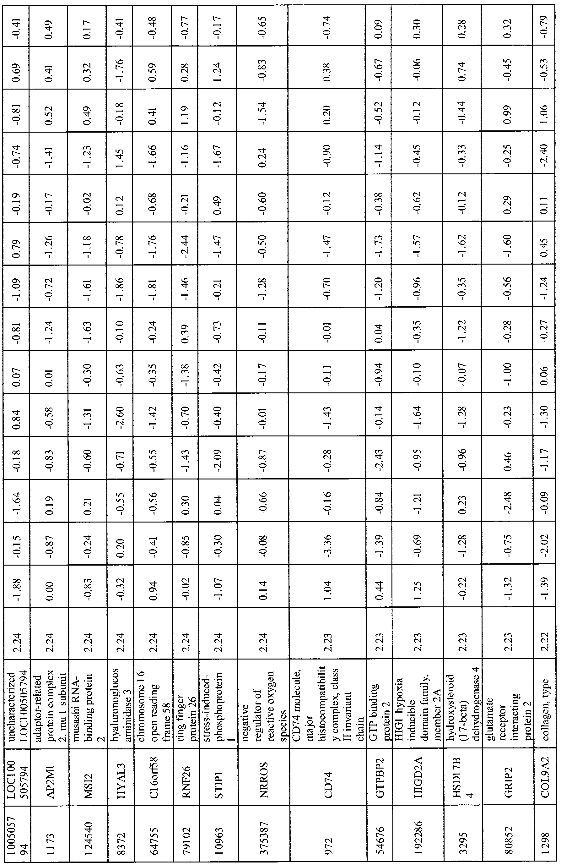
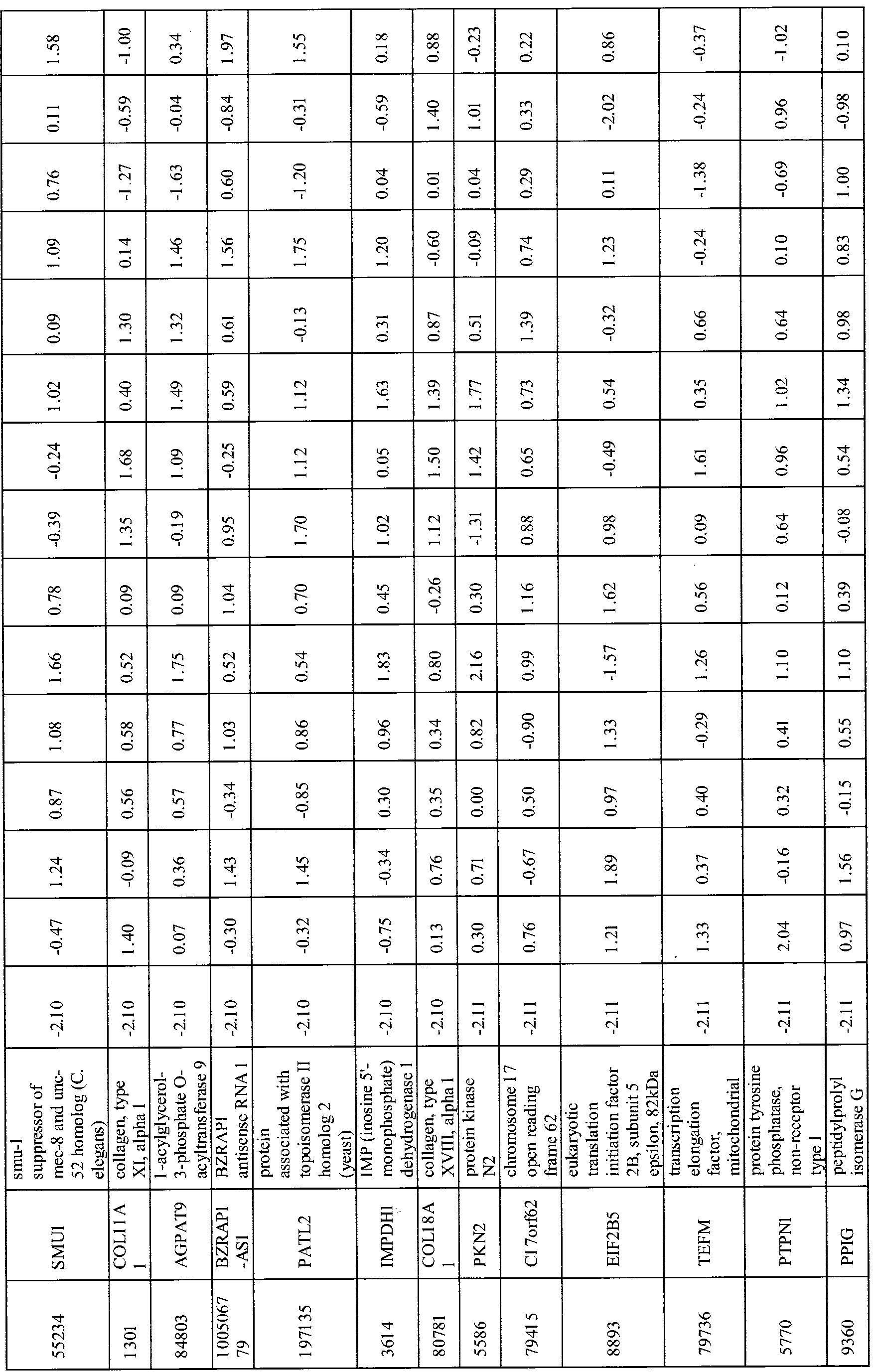
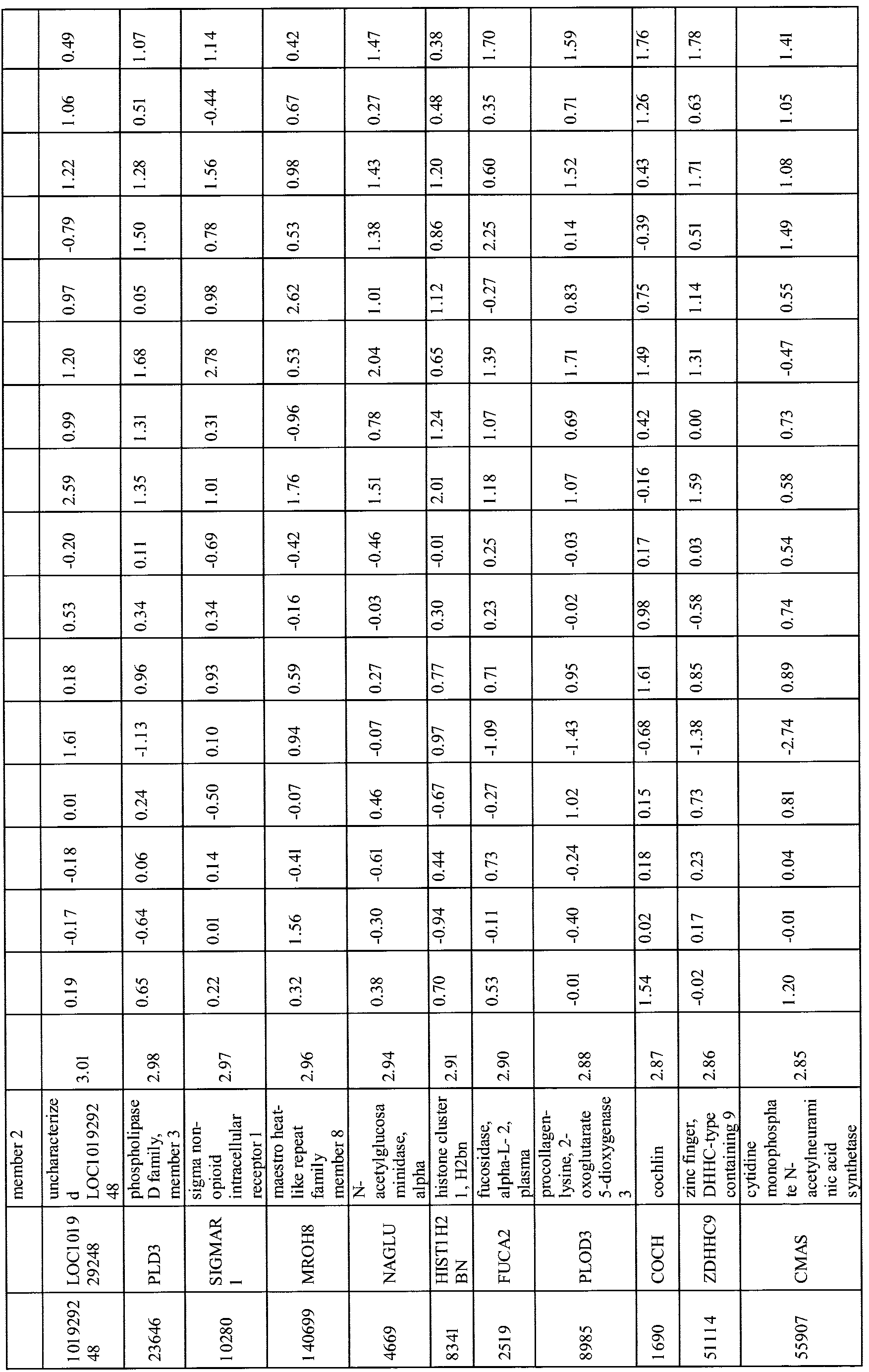
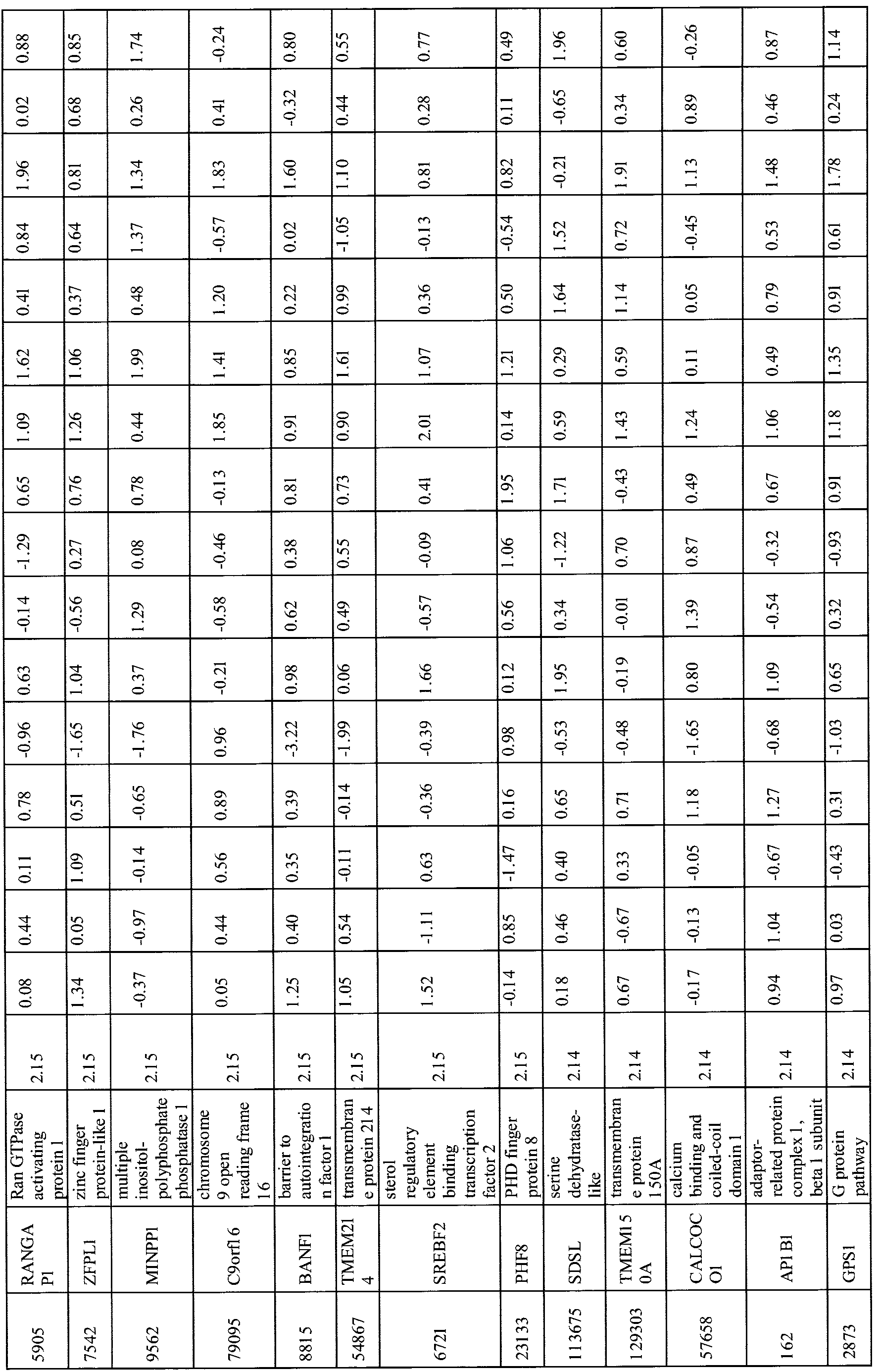
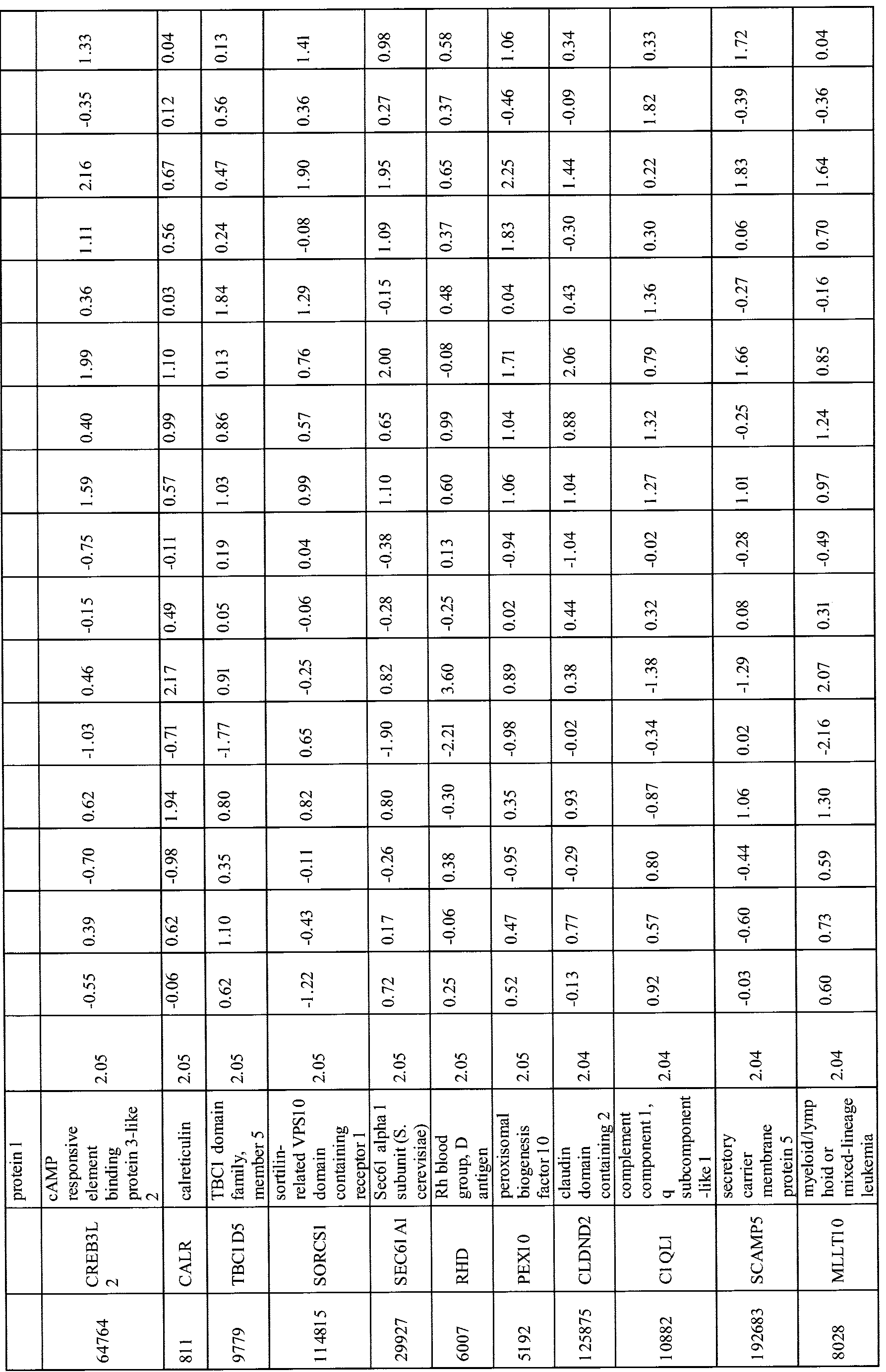
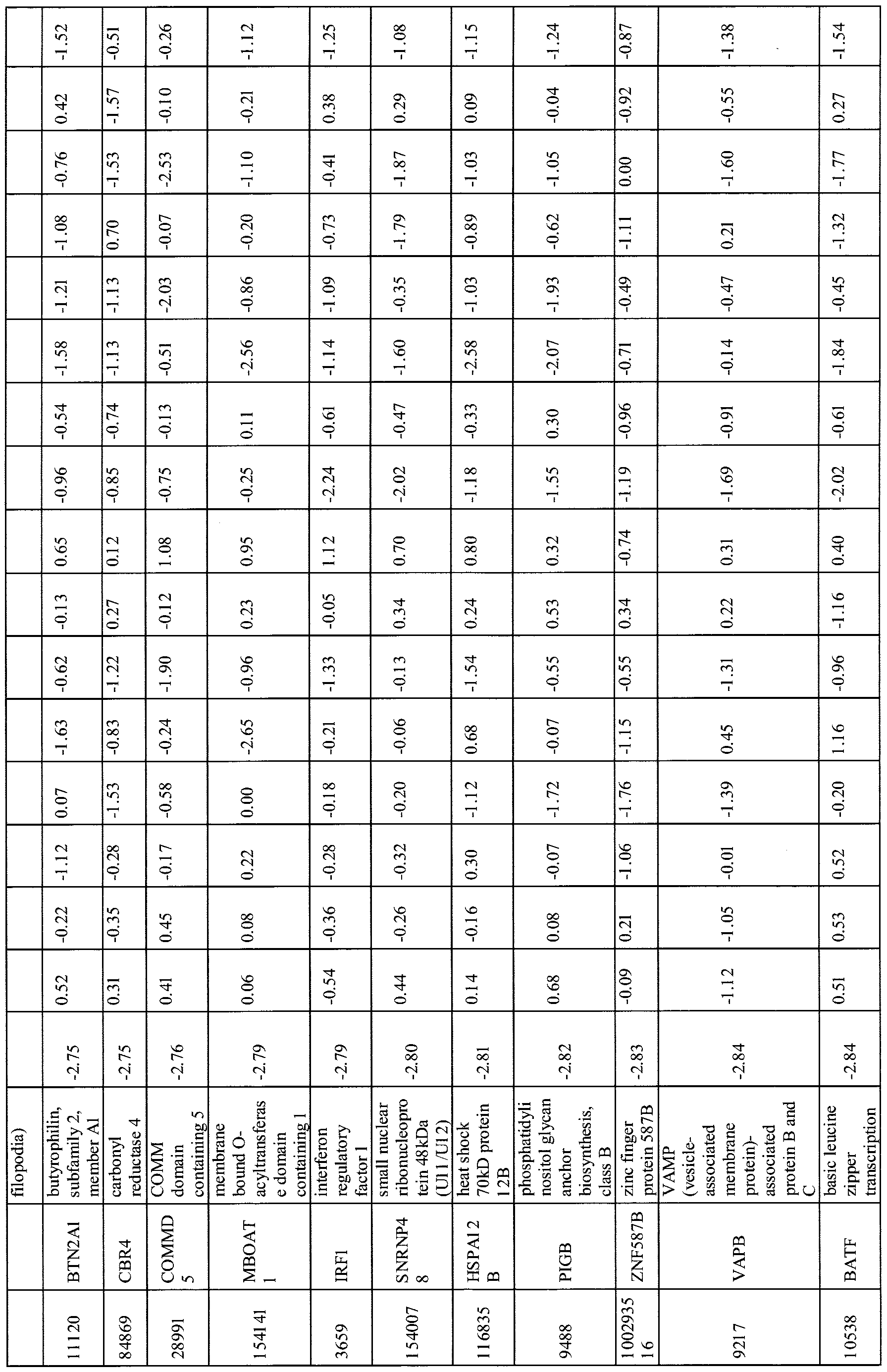
- FIG. 2 shows a heat map of the results showing gene trees. Tables 2 and 3 below shows data from FIG. 2 in numerical form. Table 2 shows the results for ages 20-33; Table 3 shows the results for ages 34-40.
- KRAS_SIGNALING_UP TNF A SIGNALING VIA NFKB , IL2_STAT5_SIGNALING COMPLEMENT, INFLAMMATORY RESPONSE, INTERFERON ALPHA RESPONSE, ALLOGRAFT REJECTION AND INTERFERENCE GAMMA RESPONSE.
- Table 4 summarizes the data from the high level genesets:
- Table 5 shows GO Biological Processes.
- Table 6 shows MeSH Anatomical Contexts.
- Table 7 shows chemical genetic perturbations.
Landscapes
- Life Sciences & Earth Sciences (AREA)
- Health & Medical Sciences (AREA)
- Chemical & Material Sciences (AREA)
- Engineering & Computer Science (AREA)
- Immunology (AREA)
- Proteomics, Peptides & Aminoacids (AREA)
- Organic Chemistry (AREA)
- Analytical Chemistry (AREA)
- Molecular Biology (AREA)
- Zoology (AREA)
- Wood Science & Technology (AREA)
- Physics & Mathematics (AREA)
- Biochemistry (AREA)
- Biotechnology (AREA)
- Microbiology (AREA)
- General Health & Medical Sciences (AREA)
- Biomedical Technology (AREA)
- Hematology (AREA)
- Urology & Nephrology (AREA)
- Genetics & Genomics (AREA)
- Pathology (AREA)
- Cell Biology (AREA)
- Bioinformatics & Cheminformatics (AREA)
- Biophysics (AREA)
- General Engineering & Computer Science (AREA)
- Food Science & Technology (AREA)
- Medicinal Chemistry (AREA)
- General Physics & Mathematics (AREA)
- Physiology (AREA)
- Tropical Medicine & Parasitology (AREA)
- Measuring Or Testing Involving Enzymes Or Micro-Organisms (AREA)
Abstract
The present technology relates to biomarkers of oocyte quality and oocyte quality dealine, as well as methods in connection with such biomarkers, including method of determining the quality of an oocyte, kits for the same, and libraries, reproductive aging gene expression profiles and profile sets with information relating the same.
Description
TITLE
Biomarkers of Oocyte Quality
STATEMENT REGARDING FEDERALLY SPONSORED RESEARCH OR DEVELOPMENT
[0001] This invention was made with government support under Grant No.
OD004402 awarded by the National Institutes of Health. The government has certain rights in the invention.
CROSS REFERENCE TO RELATED APPLICATIONS [0002] This application claims priority to U.S. Provisional Application No.
62/089,604 filed December 9, 2014, and U.S. Provisional Application No. 62/254,356 filed November 12, 2015, the contents of both of which are hereby incorporated by reference.
BACKGROUND
[0003] The present technology relates to biomarkers of oocyte quality and oocyte quality decline, as well as methods in connection with such biomarkers. [0004] The functions of the female reproductive system are known to decline with age. One major factor in decreased fertility is the decline of the quality of the eggs (oocytes) produced by the ovaries as a woman ages. Decline of quality of oocytes and ovarian follicles generally begins sometime in the 30s for most women and eventually leads to menopause, in most cases between ages 45 and 55.
[0005] In certain instances, it can be useful and desirable to determine the quality of an oocyte; for example, when a woman is seeking information about her level of fertility and chances of conception; or when a clinician seeks such information in association with assisted reproductive technologies (ART). Current methods are limited to the determination of oocyte quantity, not quality; furthermore, such methods are generally time consuming,
expensive and require destruction of the oocyte itself. For a female subject who may be concerned about declining egg reserve and quality as she ages, a serious disadvantage is associated with any method or kit that requires the destruction of an oocyte in order to measure the subject's chances of conception. Further, since known methods are invasive, currently they can be performed only by medical personnel in a clinic or hospital setting; a woman cannot determine oocyte quality on her own. Thus, the current limitation of the technology to invasive methods has prevented the widespread adoption of diagnostic tests.
[0006] Thus, there is a need for methods and systems that can accurately predict or determine the quality of an oocyte, namely methods that are non-invasive, easy for patients to use, affordable and provide rapid and accurate results.
SUMMARY OF THE DISCLOSED TECHNOLOGY
[0007] In certain embodiments, the present technology is directed to a method of determining the quality of an oocyte in the body of a human without disturbing or destroying the oocyte, the method comprising:
[0008] (a) obtaining a cell sample from a female subject, wherein the cell sample does not include the oocyte;
[0009] (b) measuring a characteristic of a gene or pathway indicative of oocyte quality in the cell sample; and
[0010] (c) predicting or determining the quality of the oocyte based on the characteristic of the gene or pathway.
[0011] In certain embodiments, the present technology is directed to a method of predicting the quality of an oocyte in the body of a mammal without disturbing or destroying the oocyte, the method comprising the steps of:
[0012] (a) obtaining a cell sample from the mammal, wherein the cell sample does not include the oocyte;
[0013] (b) conducting an RT-PCR assay or an ELISA assay on the cell sample using a primer for a gene known to be correlated with aging, and comparing the result with a known value obtained from a library of genes known to be correlated with decreased oocyte quality; and
[0014] (d) predicting the likelihood of oocyte viability based on (b).
[0015] In certain embodiments, the present technology is directed to a kit for predicting a woman's oocyte quality without the need for disturbing or destroying an oocyte, or of measuring a characteristic that correlates with the quality of an oocyte, the kit comprising:
[0016] (a) a collection container for collecting a cell sample obtained from the woman's body, wherein the cell sample does not include an oocyte;
[0017] (b) a testing assay comprising RT-PCR or ELISA, wherein the testing assay measures a characteristic of a gene, pathway or transcriptional profile characteristic of the cell sample, and wherein the characteristic indicates the likely quality of an oocyte; and
[0018] (c) a visual indicator visible to the woman, the visual indicator providing information regarding the predicted quality of the oocyte.
[0019] In certain embodiments, the present technology is directed to a method of producing a library of genes as markers of oocyte quality. Such a method may comprise the steps of correlating a test gene with a quantitative and measured characteristic of oocyte quality, listing the correlation in the library; comparing a measured characteristic of a gene provided by a patient with that listed in the library; and determining the quality of an oocyte of a patient based on the comparison. In certain embodiments, the technology herein
contemplates a method of producing a library of genes as markers of oocyte quality, the method comprising the steps of:
[0020] (a) gathering expression data from cells of women in a particular age range;
[0021] (b) calculating an average gene expression for each gene at each age in the range by averaging the expression for that gene in a window of a given period of time;
[0022] (c) comparing the average gene expression of (b) to an "age vector" to indicate which genes change most with age; and
[0023] (d) calculating a FisherZ score, thereby identifying the genes at the tail ends of the distribution as indicators of biological age.
[0024] In certain embodiments, the present technology is directed to a reproductive aging gene expression profile; and a method of developing a reproductive aging gene expression profile and one or more candidate markers of reproductive success.
[0025] In certain embodiments, the present technology is directed to a method of producing a profile set as an indicator of oocyte quality. Such methods discussed herein may be done, for example, with any gene, pathway, or transcriptional profile, or with a library of the same.
BRIEF DESCRIPTION OF THE FIGURES
[0026] FIG. la is a graph showing the prevalence of Down syndrome and infertility in a typical female human as a function of age. FIG. lb is a graph showing the aging profile of oocytes in a typical female human as a function of age.
[0027] FIG. 2 shows age-dependent gene expression changes in blood from women aged 20 to 50, as a "heat map" which shows on a macro level relative changes.
DETAILED DESCRIPTION
[0028] As used herein, "female subject" and "patient" are used interchangeably to refer to the individual whose oocyte quality is desired to be determined. As discussed herein, a "cell sample" refers to one or more cell from any part of the patient's body, which is desired to be tested in order to perform the diagnostic and scientific methods discussed herein. In various embodiments, a cell sample in accordance with the technology herein may be extracted from any of the following: blood, skin, hair, urine, saliva, sweat, vaginal secretion, any other fluid (including but not limited to intracellular or extracellular fluid, interstitial fluid, lymphatic fluid or transcellular fluid, cerebrospinal fluid, mucus or phlegm).
[0029] Oocyte quantity (the number of viable oocytes produced by a woman, the totality of which is determined long before the onset of puberty) and quality (the likelihood that the oocyte can be successfully fertilized and lead to pregnancy) are two factors of paramount importance in predicting fertility and the likelihood of carrying a pregnancy to term in a female. A woman's oocyte quality is generally predicted to decline after a certain age, until she reaches menopause and her ovaries stop releasing oocytes entirely. That is, with rising maternal age, and as a woman's oocytes age as well, a woman loses the ability to reproduce, and the children she may have are at greater risk for birth defects, chromosomal abnormalities, miscarriage and other problems. These defects start to arise when the woman is in her mid-30s and increase through the 40s, about a decade prior to menopause (the average age of menopause is 51), and are caused by declining oocyte quality.
[0030] Specifically, age-related reproductive decline is characterized by increasing levels of defects in oocyte chromosomal segregation, cell cycle arrest, and oocyte mitochondrial function and morphology, among other factors. Changes in gene expression correlate with these oocyte and egg quality changes, and have been shown in aging human and mouse oocytes. Our work in particular on C. elegans oocyte aging, discussed in greater
detail below, has highlighted the evolutionary conservation of oocyte quality components. These components, particularly regulators of chromosome segregation fidelity, have been shown to be required for oocyte function and prevention of aneuploidy in worm and mammalian oocytes, and increased fidelity extends reproductive span.
[0031] However, despite the great concern about this problem, the exact age at which any particular woman might have problems is not known, and no diagnostic assay currently exists that can predict long-term age-related fertility status. Thus, a woman who is trying to conceive has limited options for determining the quality of an oocyte any given month or for predicting future success.
[0032] Current methods, such as endocrine tests of the ovarian reserve (oocyte number) predictors follicle stimulating hormone (FSH) and anti-mullerian hormone (AMH), have only immediate, short-term predictability of success with assisted reproductive technologies (ART) such as intrauterine insemination (IUI) in vitro fertilization (IVF), gamete intrafallopian tube transfer (GIFT) and zygote intrafallopian tube transfer (ZIFT). However, such tests cannot generally predict long-term prospective fertility. Similarly, known methods involving transvaginal ultrasound measurements of antral follicle count (AFC) focus on assessing oocyte number. However, oocyte quality, not quantity, is the limiting factor in most age-related fertility decline. In particular, aneuploidies (aberrations in chromosome numbers) and cell cycle arrest/maintenance failures can be a cause of infertility, birth defects, and miscarriage. Morphological assessments are too gross to identify other important measures of quality, such as the levels of particular maternal RNAs that regulate processes in the oocyte. Finally, current measures of oocyte quality require invasive approaches, and the assessment itself can destroy the oocyte in question, further limiting a woman's number of healthy oocytes and potentially requiring a larger pool of donated oocytes for treatment.
[0033] By contrast, bodily fluids such as blood and urine are easy to obtain, and in the case of blood, PBMCs (peripheral blood mononuclear cells) have been shown to provide information about the physiological state of other tissues, including ovarian cells. In fact, a recent study by Gielchinsky et al. in 2008 showed that PBMC expression analysis was able to identify candidate genes up- and down-regulated in women who delivered babies after spontaneous pregnancy at >45 years of age. These candidate genes included markers of ovarian function, apoptosis, ubiquitination, energy production, and insulin/IGF- 1 signaling— the same pathway that we showed extends reproductive span in C. elegans. While these factors are good candidates for age-related oocyte quality biomarkers in blood, our work can establish a timecourse for oocyte aging biomarkers in blood throughout the reproductive aging years, allowing us to identify and verify a set of biomarker genes to use for diagnostic purposes.
[0034] A diagnostic of oocyte quality, particularly a non-invasive measure, would be in high demand at several levels: clinics conducting assisted reproductive technology methods such as IVF would like this information before embarking on ART approaches to improve their success rates, egg-freezing companies could use such a diagnostic to predict the success of use of eggs they have frozen, and women could use the data to make informed decisions about their reproductive lives in a manner that is minimally invasive and highly convenient. Thus, in certain embodiments, the technology herein identifies, and permits health care workers or patients to identify biomarkers of oocyte quality in blood or other bodily fluids, as well as the development of a reliable long-term diagnostic of fertility.
[0035] Aging studies have been performed on Caenorhabditis elegans, a worm that has been developed as a model of reproductive aging for humans. As demonstrated by data from C. elegans and mice, certain genes in an organism are related to the quality of the oocytes of the organism.
[0036] In certain embodiments herein, the quality of an oocyte can be determined by, for example, measuring certain factors and comparing those factors with known values of oocytes of known age. Certain methods herein permit an investigator (including a health care worker or the subject herself) to determine or predict the quality of an oocyte in the body of a human without disturbing (e.g., touching, sampling, moving or altering) or destroying the oocyte itself, and without taking it out of the human's body, e.g., in vivo. Thus, in certain embodiments, the oocyte itself can be evaluated (or its characteristics determined and predicted) without harming the oocyte, and the oocyte can itself subsequently be fertilized and lead to a successful pregnancy.
[0037] In certain embodiments, the present technology relates to novel methods for assessing or predicting the quality of oocytes without the need for disturbing or destroying the oocytes themselves. These methods are based at least in part on the discovery that genes in C. elegans, which display changes as oocytes age, are the same genes as those found in mammals; further, even if no directly associated genes were located, mammalian genes with similar functions to the C. elegans genes were located. The C. elegans research also revealed data regarding the characteristics of gene expression in mutant C. elegans with long reproductive spans. Many of these genes are involved in chromosome segregation, spindle localization, chromosome organization, DNA damage response and repair, and mitochondrial processes such as ATP metabolism. These genes, therefore, are good biomarkers for oocyte quality from oocytes.
[0038] In certain embodiments, the present technology is directed to the exploitation of the non-invasive nature of blood biomarkers, or biomarkers from other bodily fluids. In certain embodiments, the present technology is directed to the development of single-gene markers as biomarkers; in other embodiments, the present technology is directed to profiles or multiple-gene markers, rather than single-gene markers, as biomarkers.
[0039] Table 1 shows pathways that Shown in the Table 1 is a summary of certain genes and pathways that have previously been identified as being of potential interest for human (H), mouse (M) and worm (W), in connection with applications of the present technology.


[0040] In certain embodiments, the genes and pathways further include any of the following:
[0041] Genes in the IIS pathway: RHEB2, 14-3-3 beta/alpha, UDP-D-glucose cytosol, IGF-2, p90Rsk, MEK2 (MAP2K2), c-Myc, IRS-1, PtdIns(3,4,5)P3, PtdIns(4,5)P2, IKK (cat), Erk (MAPK1/3), Hamartin, c-Raf-1, AKT (PKB), PDK cat class IA, ASK1 (MAP3K5), c-Raf-1, 14-3-3 epsilon, PTEN, 14-3-3 zeta/delta, IGF-1, PI3K reg class IA, p70 S6 kinasel, CREB1, GSK3 alpha/beta, Cyclin D, 3.1.3.67, MEK1 (MAP2K1), Bcl-XL, GRB2, PDK (PDPKl), mTOR, Caspase-9, FOX03A, Tuberin, 2.7.1.137, Glycogen, I-kB, BAD, 4E-BP1, IGF-1 receptor, H-Ras, IBP, IKK-alpha, GYSl, 2.4.1.11, RPS6, She, SOS, NF-kB, Elk-1
[0042] Akt pathway (overlaps with IIS): MDM2, HGF receptor (Met), GYS 1 ,
2.7.1.153, Bcl-XL, IKK-alpha, GAB1, Bax, Hamartin, PI3K cat class IA, BAD, IRS-1, PtdIns(3,4,5)P3, PtdIns(4,5)P2, 4E-BP1 , mTOR, Caspase-9, FasL(TNFSF6), GSK3 alpha/beta, p21, NF-kB, IGF-1 receptor, RHEB2, PCNA, PI3K reg class IA, PTEN, PP2A catalytic, PDK (PDPK1), Cyclin D3, p53, 3.1.3.67, p27KIPl , AKT(PKB), Tuberin, RPS6, p70 S6 kinase 1, c-Myc, HSP90, Cyclin D, IKK (cat), Bim, FOX03A, I-kB
[0043] PTEN (overlaps with IIS): RHEB2, MEK1 (MAP2K1 ), PtdIns(4,5)P2,
Caspase-3, PDK (PDPK1), 3.1.3.67, ERKl/2, c-Jun, IGF-1, mTOR, PCNA,
MEK2(MAP2K2), FOX03A, p53, p21, FAK1, pl30CAS, c-Cbl, She, GSK3 beta, BAD, Paxillin, EGF, PI3K reg class IA, H-Ras, MAGI-2, SOS, PTEN, c-Src, Tcf(Lef), alpha- 5/beta-l integrin, IRS-1, PI3K cat class IA, Beta-catenin, MAGI-3, EGFR, PtdIns(3,4,5)P3, IGF-1 receptor, Tuberin, ILK, GRB2, 2.7.1.137, AKT(PKB), c-Raf-1, MDM2, Caspase-9
[0044] Genes in the TGF-β pathway p300, MSK1 , SOS, NF-kB, TGF-beta receptor type I, SMAD7, p15, Elk-1, TGF-beta 1, SMAD4, Caveolin-1, ErbB2, IKK-beta, TSC-22, TIEG1, FKBP12, SMURF2, APC/hCDHl complex, FAST-1/2, IKK-alpha, ERKl/2, SMAD3, TAK1(MAP3K7), PAIl, ER81, MEKK4(MAP3K4), NFKBIA, GADD45 beta, H- Ras, Anaphase-promoting complex (APC), c-Raf-1, XIAP, TGF-beta receptor type II, SMURFl, CBP, p21, MEK6(MAP2K6), Sno-N, MEK2(MAP2K2), YYl, MEK3(MAP2K3), SARA, SP1, She, Importin (karyopherin)-beta, SMAD2, TAB1, Ski, p38 MAPK,
MEK1(MAP2K1)
[0045] FOXO transcription factors: FOXOl , FOX03a
[0046] Genes in the Apoptosis pathways: ACVR1, BCL2L1, BIRCl, CAPN2,
CAPNS1, CCNG1, CD47, CD81, CD99, CFLAR, CUL1, DAD1, DGKA, DPP4, FST, GZMA, HLA-G, HSPA8, HSPD1, IGF1R, IL15, ILIA, IL2RG, IRS2, ITGB3, MAD2L1,
MAP2K1, MAX, NRAS, ODC1, PDGFA, PECAM1, PGRMC1, PRKAR2B, PROS1, PTPN13, RBBP7, RPS3, SERPINB2, SKI, TEGT, TOPI, TPM1, YWHAQ, ZFF148
[0047] Genes in the Ubiquitination pathways: CAPN2, CAST, CD47, CFLAR,
CLTC, CUL1, ESPL1, FLNA, HSPA8, IRS2, MAD2L1 , MAP2K1, SIRT2, SKI, SNRPN, TOPI, UBQLN4, YWHAB, YWHAQ
[0048] Genes in the Energy Production pathways: ACVR1, BCL2L1, BIRCl, GSS,
HLA-G, HSPA8, HSPD1 , IGF1R, IRS2, KATNB1, MAP2K1, PGK1, PMS2, PRKD2, SLC25A3, SNRPN, TCF8, TOPI, TPM1
[0049] In certain embodiments, the gene or pathway indicative of oocyte quality is chosen from a gene expressed in connection with one or more of the following categories (reflecting those listed in Table 1): cell cycle (e.g., mitosis); chromosome segregation or organization (e.g., chromosome segregation, spindle localization or chromosome organization); DNA damage response and repair (e.g., response to DNA damage stimulus); proteolytic pathway (e.g., proteolysis); energy pathway or mitochondrial function (e.g., ATP metabolic process or ATP binding); cell signaling and communication (e.g., intracellular signaling cascade or cell-cell signaling); protein transport; transcription regulation;
reproductive process (e.g., oogenesis or oviposition); cell death; cell differentiation or cell adhesion.
[0050] Other genes have been found to be of interest - among them, genes of chromatin structure, DNA methylation and genome stability. In particular, genes that have been shown to be associated with both ovarian function and general aging have shown promise in the methods, kits and libraries of the present technology. These include SERPINB2 (serpin peptidase inhibitor, clade B, member 2, also known as PAI-2); IGFIR (insulin-like growth factor 1 receptor); PIK3CB (phosphoinositide-3-kinase, catalytic, beta polypeptide), IRS2 (insulin receptor substrate 2), HSPA8, HSPD1 and HSP60.
Two genetic pathways in particular have been found herein to be significant in the regulation of reproductive aging. Specifically, it has previously been determined that TGF-β and insulin/IGF- 1 (IIS) signaling pathways regulate reproductive aging cell non-autonomously. That is, these pathways regulate reproductive aging systemically, outside of the oocyte itself rather than by signaling just inside the oocyte. The function of these signaling pathways outside of oocytes (that is, in other areas of the organism's system) can determine the rate of reproductive aging, and analysis of a systemic tissue such as blood or urine for the activity of these pathways can provide useful data, including one or more biological indicators of reproductive aging of the oocytes.
[0051] In certain embodiments, the present technology is directed to transcriptional analysis of peripheral blood mononuclear cells (PMBCs). RNA sequencing is used to achieve greater sensitivity and depth. Biomarkers of reproductive age and reproductive success have been identified. The focus is on the most significant genes associated with reproductive status, regardless of whether the gene function is known or not, thus removing bias in selection. By using a panel of the genes most significantly associated with reproductive success, and then testing the expression of these genes for their predictive power, a diagnostic with a high correlation with outcome has been created. Additionally, analysis of the systemic effects on reproduction can inform subsequent approaches for treatments. Genes that have been shown to be downregulated include members of the insulin/IGF- 1 (IIS) pathway (IGFAR, IRS2), and increased SIRT2, a co-regulator of the IIS pathway, is associated with late reproductive success. Therefore, previous studies of the C. elegans reproductive span correlates exactly with these observations in women, that one of the most important regulators of reproductive span appears to be the IIS pathway. Members of the TGF-β pathway can also be systemic markers of reproductive success. IIS can influence both lifespan and reproductive span, while TGF-β has an effect specifically on
reproductive span. Thus, IIS differences in PBMCs are thought also to act as a diagnostic for longevity.
[0052] Thus, in certain embodiments, the technology herein is directed to methods for assessing a woman's likely oocyte quality, chances of conception, comprising developing a quantitative score that offers information regarding chances of conception or otherwise a measure of likelihood of success of an assisted reproductive procedure. In certain embodiments, the score can be calculated as follows: A sample's gene expression values can be obtained, and then how well the genes match a particular age can be determined in one of several ways, including but not limited to the following: (1) by Pearson correlation with a) the set of genes or pathways in the profile, if that is what is available by the gene set qRT- PCR assay, or b) by the Pearson correlation with the entire gene expression profile when the sample's total niRNA-sequence is assessed; or ( 2) by counting the genes with most extreme FisherZ scores more heavily (weighting) that includes the average expression of all the genes in the set.
[0053] The latter could give a single score that depends most heavily on the set of genes already evaluated. The former would likely allow for a better match of the whole transcriptome by age. In certain embodiments, both scores can be used to assess the score of the sample.
[0054] In certain embodiments, a "reproductive age" (as opposed to an actual age) of a given individual can be calculated as follows:
[0055] A pre-calculated age-correlation-score Zi for each gene i (for a total of
-19,600 genes in the genome) is based on public gene-expression data from blood from females. (These are the "fisherz" scores reported in all the analyses results.)
[0056] Given a new gene-expression sample with normalized expression levels e, for each gene i, the "reproductive-age" arep of that sample equals the weighted average of all the
genes measured in the sample (N), weighted by the 'age-correlation' scores of those genes, in Equation (I) below:

[0057] In certain embodiments, the present technology is directed to a kit comprising any one or more of the following: (a) a collection container for collecting a cell sample; and (b) an assay that measures a characteristic of a gene or pathway related to oocyte quality. In use, a medical professional, or the patient herself, may collect the cell sample, subject it to the assay and use the results to predict the patient's oocyte quality. For example, in certain embodiments the kit can predict a woman's likelihood of conceiving, a woman's
"reproductive age" (as described later herein) or the likelihood that ovulation will result in a viable oocyte in any given month. Any of these can even be packaged in conjunction with other tools used by women trying to conceive, including but not limited to ovulation predictor kits, tools for measuring body temperature (such as basal body temperature thermometers), tools for measuring or evaluating mucus signs and other physically manifested indicators of fertility and fertile windows.
[0058] In various other embodiments, the kit comprises:
[0059] (a) a collection container for collecting a cell sample obtained from the woman's body;
[0060] (b) a testing assay that measures a characteristic of a gene, pathway or transcriptional profile characteristic of the cell sample, wherein the characteristic indicates the likely quality of an oocyte; and
[0061] (c) a visual indicator visible to the woman, the visual indicator providing information regarding the likely quality of the oocyte.
[0062] The visual indicator can be, for example, a color-coded indicator showing a binary result such as one color for an above average score, and another for a below average score.
[0063] In certain embodiments, the testing assay can be, for example, an RT-PCR assay on blood using the primers for the best genes. In the case of other samples such as urine or other bodily fluids, an assay can include metabolomics to assess metabolites or hormones, such as, for example, an enzyme-linked immunosorbent (ELISA) assay. In other embodiments, a colorimetric test could work in the event that the assay is matter of determining the levels of relatively few (e.g., just one, two or three, or in various
embodiments fewer than about 10, fewer than about 25 or fewer than about 50) genes, metabolites or hormones. For example, in certain embodiments, the results of the assay could be displayed as one or more lines that would indicate the score. In certain embodiments, different lines could present different data points or results (for example, different colors or configurations, with the combination providing a score or other type of quantitative or qualitative result).
[0064] In certain embodiments, such a kit could be made commercially available for at-home use, and could include information taken from a library of genes and pathways generated in connection with certain embodiments herein.
[0065] In certain embodiments, the kit will include primers to amplify genes in this set (genes with FisherZ scores above 2 and below -2; see Table 2) from blood. In certain embodiments, any or all of the sample, or any characteristic thereof, can be assigned a score that is equal to the average of the expression level of all the genes in the array weighted
by the age-correlation (FisherZ) score of each of the genes. This score conveys the expected oocyte viability for a woman compared to an average for women of the same age.
[0066] In certain embodiments, the present technology is directed to a method for the production of a library of genes, or a reproductive aging gene expression profile, as markers of oocyte quality. Such a method may comprise the steps of correlating a test gene with a quantitative and measured characteristic of oocyte quality, listing the correlation in the library; comparing a measured characteristic of a gene provided by a patient with that listed in the library; and determining the quality of an oocyte of a patient based on the comparison.
[0067] For example, in certain embodiments, a library is produced through any of the following steps: First, list certain genes known or thought to relate to factors such as ovarian function and general aging. Next, obtain samples of oocytes known to be from women of certain ages (for example, age 20, age 25, age 30 and the like) and measure one or more of the characteristics of those oocytes to obtain baseline values. Next, when a patient desires the assay, a cell sample is taken from the patient, the one or more characteristics can be determined by running the assay on the patient's cell sample and comparing the values to those of the library. For example, a patient may be 35 years old but may have oocytes that are typical of a 25 year old or a 40 year old, as determined by a method of the present technology, using the library generated according to this embodiment. This is valuable information that the patient can acquire without the need for invasive testing or destruction of her oocytes, and in certain embodiments, is obtained by implementing a method or kit in accordance with the embodiments herein. In certain embodiments, a yearly clinical assessment of oocyte age could allow a clinician or woman to determine the rate of change of oocyte quality, yet another indicator that could be useful for diagnostics and for advising patients.
[0068] In certain embodiments, a library can be created and used to establish a
"profile set" for a user - that is, a set of one or more genes, pathways or transcriptional profiles that indicates various characteristics of a user's oocyte. A candidate gene expression assay can be developed based on the information obtained from bodily fluids such as blood, thus identifying biomarkers that correlate with oocyte quality and pregnancy success.
[0069] In one exemplary method, a library was made by the following steps:
[0070] (1) gathering expression data from samples (whole blood and PBMCs) from women in a particular age range (in certain embodiments, aged 20 to 50, but not so limited). An average gene expression for each gene at each age was calculated by averaging the expression for that gene in a given range of time (e.g., a 2 year window, in overlapping (sliding) windows). This allowed the inclusion of about 30 to about 90 samples in each year. In certain embodiments, an average of about 70 to about 80 GEO female blood samples were used for each 2 year sliding window; for each gene, the Spearmann correlation of the average gene expression to the age vector was determined, and then sorted by the FisherZ score; a score above 2 (top 5%) was used to generate a set of significantly changed age-dependent genes, all candidate biomarkers.
[0071] (2) The gene expression data were compared to an "age vector" to indicate which genes changed most with age. Since most genes do not have an age-related change in expression, the data are normally distributed. The FisherZ score makes it possible to find the genes at the tail ends of the distribution; those genes are most changed with age, and thus are the best indicators of biological age. Such genes are included in the library, and can be used to compare with data acquired from the test subject.
[0072] For the samples of women aged 25 to 50, in certain embodiments only samples from women who have no known fertility defect (i.e., presence at ΓνΤ clinic is due to sperm or other partner issue) can be used to establish a "young" profile. The expression
profiles can then be sorted according to age and further segregated by pregnancy success/failure and other cytological information, and PCA can be performed to identify the genes best correlated with pregnancy success. Using qRT-PCR, the top genes most associated with pregnancy outcome can be tested on a separate sample set prior to IVF (or other ART treatment) to verify predictive power of the gene set. The most predictive genes can be used to develop a diagnostic tool that can be used in clinics after a simple blood draw or urine collection. The clinical procedure RT-PCR can be performed on RNA from blood samples using the mix of primers of the best candidate genes.
[0073] FIG. 2 shows a heat map of the results showing gene trees. Tables 2 and 3 below shows data from FIG. 2 in numerical form. Table 2 shows the results for ages 20-33; Table 3 shows the results for ages 34-40.

CD
ω m ω m m
3) c
I
m

ω c CD
ω m ω m m
C
m
CO

ω c
CD
ω m ω N3 m to m
C
m σ>

ω c
DO
ω m ω m m c m

ω c
CD
ω m ω τ Μ m m c m ro
CD

ω c
CD
ω m
ω
m σ> m
C
m
CD

ω c
DO
W m
ω
I
m
m
73 c
r- m
ro

ω c
CD
ω m ω ro m oo m
C
m σ>

ω c
CD
ω m ω m m
C
m
CO

ω c
CD
ω m ω
I
m
m
73 c
m
σ>

ω
c
CD
ω m
ω
I
m
m
H
c
m
σ>
ω c
CD
ω m ω
I
m m
73 c m
CD

m ω
I
m ω

ω
c
DO
ω m
ω
I ω m -t- m
H
c
m

ω
c
DO
ω m
ω
I ω m Oi m
H
c
m

ω c
DO
ω m ω
I
m m
73 c
I
ΓΠ
hJ
(3)


ω c
CD
ω m ω co m co m
3J
C
m σ>

ω c
DO
ω m ω x ω m m
7)
C
|— m

ω
c
CD
ω m
ω
m
m
73 c
m
σ>


00 ω m ω m m
7J c
|—
m

ω
c
DO
ω m
ω
I
m
m
H
c
m

ω c ω m w m m
73 c
I
m

ω
c
ω m
ω
m
m
73 c
m

ω
c
CD
ω m
ω
m
m
73 c
m
σ>

ω
c
DO
ω m
ω
m
m
73 c
m
ro
(3)

ω c
DO
ω m ω m m
73 c m


DO
ω m ω m m c t— m
N>
(3)

ω
c
DO
ω m
ω
I
m
m —\
73 c
m
CO

ω c
CD
ω m
ω
I
m l m
C
m
σ>
ω c
CD
ω m ω
ω m m
7)
C
m
CD

ω c
DO
ω m ω m m c m

c
DO
ω m ω
I
m m
TO
c
I- m

ω
c
DO
ω m
ω
I Oi m
m
H
c
m

ω c
DO
ω m ω m m c m

ω
c
DO
ω m
ω
I en m
m
H
c
m

ω c
CD
ω m ω m m
C
m σ>

ω c
CD
ω m ω
ΖΓ.
m m c m
CD

ω
c
DO
ω m
ω
m
m c
m

c
DO
ω m
ω
I
m
m
3) c
I- m

co c
DO CO m
CO
m ω m
C
I- m


ω
c
DO
ω m
ω
I en m
m
H
c
m

ω c
m ω
I
m m
73 c r- m

ω
c
DO
ω m
ω
I
m
m
H
c
m

ω
c
DO
ω m
ω
oo m
m
c
m

ω c
CD
ω m ω m m
3J c m σ>

ω c
CD
ω m ω m m c m

co c
CD
co m co m m
7J c r- m
(3)

ω c
CD
ω m σ> m m c i— m ro

ω
c
DO
ω m
ω
I
m
m
H
c
m
(3)

ω
c
CD
ω m
ω
I
m -p- m
H
¾
C
m

ω
c
CD
ω m
ω
m
m
73 c
m
σ>

ω c
DO
W m ω
I
m m
73 c m
(3)

ω
c
DO
ω m
ω
ΖΓ. -J m
m
H
c
m

ω c
DO
ω
73 c
I
m ro

ω c
DO
ω m ω m m
7) c r~
m

ω c
CD
ω m ω m m
73 c m ro σ>

ω c
DO
ω m ω
I
m m
73 c
I
m


c
ω m
ω
I
m
m
73 c
m
ω
c
DO
ω m
ω oo
Ol m
m
C
m


CD
ω m ω x m m
7}
c r- m

ω
c
DO
ω m
ω
I 00
oo m
m
C
m


ω
c
DO
ω m
ω
m
m
73 c
m

ω
c
DO
ω m
ω
I
m
m
H
c
m

co c
DO CO m
CO
I
m m
73 c
I
m

m
c
m

ω
c
DO
W m
ω
I
m
m
73 c
I
m
ro

ω
c
CD
ω m
ω
m
m c
m


I
m


c
DO
ω m
ω
ΖΓ. CO
co m
m
C
m


c
DO
ω m
ω
I
m
m
73 c
m
CO
ω
c
DO
ω m
ω
I o m
m
H
c
m

ω c
DO
ω m ω
I
m m
73 c
I- m ro

ω
c
DO
ω m
ω
ΖΓ.
m
m
73 c
m

ω
c
DO
ω m
ω
I
m
m
7] c
m

ω
c
CD
ω m
ω
I
m
m
73 c
m
CD


CD
co m
co x
m
m
7J c
I
m
(3)


c
DO
ω m
ω
ΖΓ.
m
m
H
c
m

ω c
DO
ω m ω
I
m m
7] c r- m


I
m

ω c
DO
ω m ω
I
m m
73 c m

ω c
CD
ω m ω
I
m m c r- m

ω c
CO
ω m ω
I
m
m c
m

ω c
CD
ω m ω x m m c m I

co c
CD
co m
CO
m m c i— m

c
DO
ω m r/>
I
m m c
I
m

ω c
DO
ω m ω
I
m m c m

ω c
DO
ω m ω
I
m m
7i c
m


00
ω m ω χ m m
73 c m


c
CD
ω m
ω
I
m
m
7] c
m
σ>

ω c
DO
W m w
I
m m c r- m
M


DO
ω m ω
I
m m
73
C
r- m

ω
c
DO
ω m
ω
m
m c
m


c
DO
ω m
ω
m
m c
m


CD
ω m ω
ΖΓ.
m Ol m
C
r- m σ>

ω
c
DO
ω m
ω m
m
C
m

ω
c
DO
ω m
ω
I
m
m
H
c
m

ω
c
DO
ω m
ω
I ω m 03 m
H
c
m


c
DO
ω m
ω
m
m
73
C
rm-
CD

ω c
CD
ω m ω
I
m m
H
c m i-o σ>


CD
ω m ω
I
m m
73 c m σ>


CD
ω m ω
I
m m
73 c m σ>

ω c
CD
ω m ω
I
m
m
73 c
m
σ>

ω
c
DO
ω m
ω
m
m
73 c
m

ω
c
DO
ω m
ω
m
m
73 c
m


DO
ω m
ω
I
m
m c
m

w c
DO
W m ω x m m c
I
m en

ω c
DO
ω m ω m Is} m
C
m


DO
ω m ω m m c m


[0077] For high level gene sets, the following sets of genes were found to be deleterious (that is, they increase with age): UNFOLDED PROTEIN-RE SPONSE,
OXIDATIVE_PHOSPHORYLATION, MYC TARGETS V 1 , ADIPOGENESIS,
GLYCOLYSIS, UV_RESPONSE_UP, DNA REPAIR, FATTY ACIDJVIETABOLISM, SPERMATOGENESIS, E2F TARGETS, BILE ACID METABOLISM,
MTORC 1 SIGNALING, ESTROGEN RESPONSE LATE.
[0078] The following genes were found to be beneficial (that is, they decrease with age): APOPTOSIS, APICAL_SURFACE, UV_RESPONSE_DN,
EPITHELIAL MESENCHYMAL TRANSITION, ANGIOGENESIS,
KRAS_SIGNALING_UP, TNF A SIGNALING VIA NFKB , IL2_STAT5_SIGNALING COMPLEMENT, INFLAMMATORY RESPONSE, INTERFERON ALPHA RESPONSE, ALLOGRAFT REJECTION AND INTERFERENCE GAMMA RESPONSE.
[0079] Table 4 summarizes the data from the high level genesets:
Table 4: High Level Genesets


Table 5 - GO Biological Processes


Table 6 - MeSH Anatomical Contexts


[0082] Table 7 shows chemical genetic perturbations.
Table 7 - Chemical Genetic Perturbations


[0083] Table 8 shows the Number of Data Sets
Table 8 - Number of Data Sets


[0084] Thus, in certain embodiments, as shown herein, samples are sorted by age, and then hierarchical clustering can be performed to identify sets of genes that are most correlated with older and thus deleterious effects, and younger. These genes are candidate biomarkers for reproductive status.
[0085] In certain embodiments, predictors of reproductive success can be verified as follows: The genes most associated with pregnancy outcome (both positive and negative) can be tested on a separate sample set prior to IVF treatment to verify predictive power of the gene set. Two approaches can be used: one with individual qRT-PCR primers for each gene, and a second with the entire set of primers.
[0086] In a non-limiting Example, the diagnostic application can be implemented as follows: A blood sample from a patient is collected, RT-PCR performed using the diagnostic primer set, and the profile results matched using Pearson correlation to the reproductive age profile. For example, a 32-year old patient might have a blood profile that best matches that of a 38 year old, indicating that she is reproductively aged relative to her chronological age, and thus might not want to delay childbearing much longer. Under current guidelines, older patients might be denied IVF or other ART treatment, but the tests herein could show that a chronologically 43 -year old patient might best match that of an average 38 year old, and thus would still be a viable candidate for IVF or other assisted reproductive techniques. Thus, a diagnostic that easily and accurately correlates a key set of biomarkers with reproductive capacity can be useful for several applications.
[0087] In certain embodiments, the methods herein may be directed to the measure or determination of oocyte quality based on a combination of two or more any of the markers discussed herein. For example, a determination of characteristics of two or more of the genes or pathways discussed herein can, in certain embodiments, provide a more accurate set of data regarding a subject's oocyte quality and thus her likelihood of conceiving, than would be the case with only a single gene or pathway.
[0088] In certain embodiments, the technology herein contemplates methods or kits that comprise a binding molecule, for example, a binding composition that specifically binds to any protein produced by a biomarker gene discussed herein and may be conjugated to
another molecule, for example, an enzyme or a molecule that provides a visual indication of oocyte quality of some other detected characteristic of the cell.
[0089] Although the present technology has been described in relation to particular embodiments thereof, these embodiments and examples are merely exemplary and not intended to be limiting. Many other variations and modifications and other uses will become apparent to those skilled in the art. The present technology should, therefore, not be limited by the specific disclosure herein, and may be embodied in other forms not explicitly described here, without departing from the spirit thereof.
Claims
1. A method of determining the quality of an oocyte in the body of a human without disturbing or destroying the oocyte, the method comprising:
(a) obtaining a cell sample from a female subject, wherein the cell sample does not include the oocyte;
(b) measuring a characteristic of a gene or pathway indicative of oocyte quality in the cell sample; and
(c) predicting or determining the quality of the oocyte based on the characteristic of the gene or pathway.
2. The method of claim 1, wherein step (b) comprises measuring a gene expression value of the cell sample through an RT-PCR assay, an ELISA assay or a colorometric test.
3. The method of claim 2, wherein step (b) further comprises comparing the measured gene expression values to a known gene expression value of an oocyte with Pearson correlation, or by matching the measured gene expression values to a known gene expression profile from a library of genes as markers of oocyte quality.
4. The method of claim 1, wherein the cell sample is extracted from blood, skin, hair, urine, saliva, sweat or vaginal secretion.
5. The method of claim 1, wherein the gene or pathway is chosen from
SERPI B2 (serpin peptidase inhibitor, clade B, member 2, also known as PAI-2); IGFIR (insulin-like growth factor 1 receptor); PIK3CB (phosphoinositide-3-kinase, catalytic, beta polypeptide), IRS2 (insulin receptor substrate 2), HSPA8, HSPD1, HSP60, TGF-β and insulin/IGF- 1 (IIS) signaling pathway.
6. A method of predicting the quality of an oocyte in the body of a mammal without disturbing or destroying the oocyte, the method comprising the steps of:
(a) obtaining a cell sample from the mammal, wherein the cell sample does not include the oocyte;
(b) conducting an RT-PCR assay or an ELISA assay on the cell sample using a primer for a gene known to be correlated with aging, and comparing the result with a known value obtained from a library of genes known to be correlated with decreased oocyte quality; and
(d) predicting the likelihood of oocyte viability based on (b).
7. A kit for predicting a woman's oocyte quality without the need for disturbing or destroying an oocyte, the kit comprising:
(a) a collection container for collecting a cell sample obtained from the woman's body, wherein the cell sample does not include an oocyte;
(b) a testing assay comprising RT-PCR or ELISA, wherein the testing assay measures a characteristic of a gene, pathway or transcriptional profile characteristic of the cell sample, and wherein the characteristic indicates the likely quality of an oocyte; and
(c) a visual indicator visible to the woman, the visual indicator providing information regarding the predicted quality of the oocyte.
8. The kit of claim 7, wherein the gene or pathway is chosen from SERPINB2 (serpin peptidase inhibitor, clade B, member 2, also known as PAI-2); IGFIR (insulin-like growth factor 1 receptor); PIK3CB (phosphoinositide-3 -kinase, catalytic, beta polypeptide), IRS2 (insulin receptor substrate 2), HSPA8, HSPDl, HSP60, TGF-β and insulin/IGF-1 (IIS) signaling pathway.
9. A method of producing a library of genes as markers of oocyte quality, the method comprising the steps of:
(a) gathering expression data from cells of women in a particular age range;
(b) calculating an average gene expression for each gene at each age in the range by averaging the expression for that gene in a window of a given period of time;
(c) comparing the average gene expression of (b) to an "age vector" to indicate which genes change most with age; and
(d) calculating a FisherZ score, thereby identifying the genes at the tail ends of the distribution as indicators of biological age.
10. The method of claim 9, wherein for one or more of the genes identified in step (d), the Spearmann correlation of the average gene expression to the age vector was determined, and then sorted by the FisherZ score.
11. The method of claim 10, wherein the genes for which a score above 2 (top 5%) was calculated were added to a set of significantly changed age-dependent genes to comprise the library of genes.
12. The method of claim 9, further comprising any of the following steps:
correlating a test gene with a quantitative and measured characteristic of oocyte quality; listing the correlation in the library; comparing a measured characteristic of a gene provided by a patient with that listed in the library; and determining the quality of an oocyte of a patient based on the comparison.
13. A method of developing a reproductive aging gene expression profile and one or more candidate markers of reproductive success or oocyte quality, the method comprising the steps of claim 9.
14. The method of claim 1, wherein the characteristic measured in the cell sample is assigned a score that conveys the expected oocyte viability.
15. The method of claim 14, wherein the score conveys the expected oocyte viability compared to an average for women of the same age as the female subject.
Priority Applications (2)
| Application Number | Priority Date | Filing Date | Title |
|---|---|---|---|
| US15/533,746 US20170362654A1 (en) | 2014-12-09 | 2015-12-09 | Biomarkers of oocyte quality |
| US16/547,127 US20200087727A1 (en) | 2014-12-09 | 2019-08-21 | Biomarkers of oocyte quality |
Applications Claiming Priority (4)
| Application Number | Priority Date | Filing Date | Title |
|---|---|---|---|
| US201462089604P | 2014-12-09 | 2014-12-09 | |
| US62/089,604 | 2014-12-09 | ||
| US201562254356P | 2015-11-12 | 2015-11-12 | |
| US62/254,356 | 2015-11-12 |
Related Child Applications (2)
| Application Number | Title | Priority Date | Filing Date |
|---|---|---|---|
| US15/533,746 A-371-Of-International US20170362654A1 (en) | 2014-12-09 | 2015-12-09 | Biomarkers of oocyte quality |
| US16/547,127 Continuation US20200087727A1 (en) | 2014-12-09 | 2019-08-21 | Biomarkers of oocyte quality |
Publications (2)
| Publication Number | Publication Date |
|---|---|
| WO2016094583A2 true WO2016094583A2 (en) | 2016-06-16 |
| WO2016094583A3 WO2016094583A3 (en) | 2016-08-18 |
Family
ID=56108366
Family Applications (1)
| Application Number | Title | Priority Date | Filing Date |
|---|---|---|---|
| PCT/US2015/064836 Ceased WO2016094583A2 (en) | 2014-12-09 | 2015-12-09 | Biomarkers of oocyte quality |
Country Status (2)
| Country | Link |
|---|---|
| US (2) | US20170362654A1 (en) |
| WO (1) | WO2016094583A2 (en) |
Cited By (4)
| Publication number | Priority date | Publication date | Assignee | Title |
|---|---|---|---|---|
| WO2018187585A1 (en) * | 2017-04-06 | 2018-10-11 | Celmatix Inc. | Methods for assessing the potential for reproductive success and informing treatment therefrom |
| US10580516B2 (en) | 2012-10-17 | 2020-03-03 | Celmatix, Inc. | Systems and methods for determining the probability of a pregnancy at a selected point in time |
| KR20210110073A (en) * | 2020-02-28 | 2021-09-07 | 차의과학대학교 산학협력단 | Biomarker for Ovarian reserve and use thereof |
| CN114167057A (en) * | 2021-12-10 | 2022-03-11 | 大连医科大学 | A biological marker for diagnosing miscarriage and its application |
Families Citing this family (7)
| Publication number | Priority date | Publication date | Assignee | Title |
|---|---|---|---|---|
| EP3430165A4 (en) * | 2016-03-09 | 2020-01-08 | Celmatix Inc. | Methods and systems for assessing infertility and ovulatory function disorders |
| US20200020419A1 (en) | 2018-07-16 | 2020-01-16 | Flagship Pioneering Innovations Vi, Llc. | Methods of analyzing cells |
| KR102114899B1 (en) * | 2020-02-26 | 2020-05-26 | 가천대학교 산학협력단 | Pharmaceutical composition for preventing or treating infertility or abortion comprising inhibitors of mSIN1 protein as an active ingredient |
| KR102147491B1 (en) * | 2020-05-29 | 2020-08-25 | 가천대학교 산학협력단 | Pharmaceutical composition for preventing or treating infertility or abortion comprising inhibitors of DEPTOR protein or mSIN1 protein as an active ingredient |
| CN111748559B (en) * | 2020-06-29 | 2022-06-17 | 华南农业大学 | Application of CTNNB1 gene in porcine ovary granular cells |
| CN112816691B (en) * | 2021-02-08 | 2024-06-11 | 杭州市妇产科医院 | A method for evaluating human oocyte quality |
| US20240350553A1 (en) * | 2021-10-15 | 2024-10-24 | Agex Therapeutics, Inc. | Methods for modulating the regenerative phenotype in mammalian cells |
Family Cites Families (4)
| Publication number | Priority date | Publication date | Assignee | Title |
|---|---|---|---|---|
| AU2010351560C1 (en) * | 2009-09-23 | 2015-10-08 | Celmatix Inc. | Methods and devices for assessing infertility and/or egg quality |
| US20130053261A1 (en) * | 2009-11-10 | 2013-02-28 | Jose B. Cibelli | Genes differentially expressed by cumulus cells and assays using same to identify pregnancy competent oocytes |
| WO2012109326A2 (en) * | 2011-02-09 | 2012-08-16 | Temple University-Of The Commonwealth System Of Higher Education | Determination of oocyte quality |
| US20140296104A1 (en) * | 2011-10-14 | 2014-10-02 | Gema Diagnostics, Inc. | Genes Differentially Expressed by Cumulus Cells and Assays Using Same to Identify Pregnancy Competent Oocytes |
-
2015
- 2015-12-09 US US15/533,746 patent/US20170362654A1/en not_active Abandoned
- 2015-12-09 WO PCT/US2015/064836 patent/WO2016094583A2/en not_active Ceased
-
2019
- 2019-08-21 US US16/547,127 patent/US20200087727A1/en not_active Abandoned
Cited By (5)
| Publication number | Priority date | Publication date | Assignee | Title |
|---|---|---|---|---|
| US10580516B2 (en) | 2012-10-17 | 2020-03-03 | Celmatix, Inc. | Systems and methods for determining the probability of a pregnancy at a selected point in time |
| WO2018187585A1 (en) * | 2017-04-06 | 2018-10-11 | Celmatix Inc. | Methods for assessing the potential for reproductive success and informing treatment therefrom |
| KR20210110073A (en) * | 2020-02-28 | 2021-09-07 | 차의과학대학교 산학협력단 | Biomarker for Ovarian reserve and use thereof |
| KR102316507B1 (en) | 2020-02-28 | 2021-10-22 | 차의과학대학교 산학협력단 | Biomarker for Ovarian reserve and use thereof |
| CN114167057A (en) * | 2021-12-10 | 2022-03-11 | 大连医科大学 | A biological marker for diagnosing miscarriage and its application |
Also Published As
| Publication number | Publication date |
|---|---|
| WO2016094583A3 (en) | 2016-08-18 |
| US20170362654A1 (en) | 2017-12-21 |
| US20200087727A1 (en) | 2020-03-19 |
Similar Documents
| Publication | Publication Date | Title |
|---|---|---|
| WO2016094583A2 (en) | Biomarkers of oocyte quality | |
| Grati et al. | Outcomes in pregnancies with a confined placental mosaicism and implications for prenatal screening using cell-free DNA | |
| Victorelli et al. | Senescent human melanocytes drive skin ageing via paracrine telomere dysfunction | |
| Tarca et al. | Maternal whole blood mRNA signatures identify women at risk of early preeclampsia: a longitudinal study | |
| Tharakan et al. | European Association of Urology guidelines panel on male sexual and reproductive health: a clinical consultation guide on the indications for performing sperm DNA fragmentation testing in men with infertility and testicular sperm extraction in nonazoospermic men | |
| Jedrzejczak et al. | Prediction of spontaneous conception based on semen parameters | |
| Coccia et al. | Ovarian reserve | |
| Tunc et al. | Oxidative DNA damage impairs global sperm DNA methylation in infertile men | |
| Chan | Biochemical markers of spontaneous preterm birth in asymptomatic women | |
| Iodice et al. | Particulate air pollution, blood mitochondrial DNA copy number, and telomere length in mothers in the first trimester of pregnancy: effects on fetal growth | |
| Fenech | Micronuclei and their association with sperm abnormalities, infertility, pregnancy loss, pre-eclampsia and intra-uterine growth restriction in humans | |
| Lee et al. | Age‐related distributions of anti‐Müllerian hormone level and anti‐Müllerian hormone models | |
| Ferrick et al. | Reducing time to pregnancy and facilitating the birth of healthy children through functional analysis of embryo physiology | |
| Ramasamy et al. | Fluorescent in situ hybridization of human sperm: diagnostics, indications, and therapeutic implications | |
| Oleszczuk et al. | Intra-individual variation of the sperm chromatin structure assay DNA fragmentation index in men from infertile couples | |
| Busnelli et al. | Mitochondrial DNA copy number in peripheral blood: a potential non-invasive biomarker for female subfertility | |
| Naglot et al. | Male contributory factors in recurrent pregnancy loss | |
| Hassanen et al. | TUNEL assay: Establishing a sperm DNA fragmentation cut‐off value for Egyptian infertile men | |
| Brummaier et al. | A prospective cohort for the investigation of alteration in temporal transcriptional and microbiome trajectories preceding preterm birth: a study protocol | |
| Cao et al. | Lipid metabolism affects fetal fraction and screen failures in non-invasive prenatal testing | |
| Fjeldstad et al. | Markers of placental function correlate with prevalence and quantity of nucleated fetal cells in maternal circulation in normotensive term pregnancies | |
| Luo et al. | Mitochondrial DNA quantification correlates with the developmental potential of human euploid blastocysts but not with that of mosaic blastocysts | |
| Al‐Rawi et al. | Brief Report: A Novel Application of Buccal Micronucleus Cytome Assay in Systemic Lupus Erythematosus: A Case–Control Study | |
| Mhatre et al. | The effect of maternal obesity on placental cell-free DNA release in a mouse model | |
| Nogueira et al. | Measurements in first-trimester abortion products: a pathologic study |
Legal Events
| Date | Code | Title | Description |
|---|---|---|---|
| 121 | Ep: the epo has been informed by wipo that ep was designated in this application |
Ref document number: 15866406 Country of ref document: EP Kind code of ref document: A2 |
|
| NENP | Non-entry into the national phase in: |
Ref country code: DE |
|
| 122 | Ep: pct application non-entry in european phase |
Ref document number: 15866406 Country of ref document: EP Kind code of ref document: A2 |