WO2016090323A1 - Dcis recurrence and invasive breast cancer - Google Patents
Dcis recurrence and invasive breast cancer Download PDFInfo
- Publication number
- WO2016090323A1 WO2016090323A1 PCT/US2015/064115 US2015064115W WO2016090323A1 WO 2016090323 A1 WO2016090323 A1 WO 2016090323A1 US 2015064115 W US2015064115 W US 2015064115W WO 2016090323 A1 WO2016090323 A1 WO 2016090323A1
- Authority
- WO
- WIPO (PCT)
- Prior art keywords
- dcis
- sample
- risk
- subject
- siah2
- Prior art date
Links
- 208000030776 invasive breast carcinoma Diseases 0.000 title claims abstract description 149
- 208000037396 Intraductal Noninfiltrating Carcinoma Diseases 0.000 claims abstract description 581
- 208000028715 ductal breast carcinoma in situ Diseases 0.000 claims abstract description 576
- 238000000034 method Methods 0.000 claims abstract description 335
- 238000011282 treatment Methods 0.000 claims abstract description 68
- 206010028980 Neoplasm Diseases 0.000 claims abstract description 67
- 239000000203 mixture Substances 0.000 claims abstract description 26
- 239000000523 sample Substances 0.000 claims description 318
- 101001062353 Homo sapiens Hepatocyte nuclear factor 3-alpha Proteins 0.000 claims description 191
- 102100029283 Hepatocyte nuclear factor 3-alpha Human genes 0.000 claims description 189
- 101000707245 Homo sapiens E3 ubiquitin-protein ligase SIAH2 Proteins 0.000 claims description 173
- 102100031748 E3 ubiquitin-protein ligase SIAH2 Human genes 0.000 claims description 172
- 101001012157 Homo sapiens Receptor tyrosine-protein kinase erbB-2 Proteins 0.000 claims description 167
- 102100030086 Receptor tyrosine-protein kinase erbB-2 Human genes 0.000 claims description 167
- 108090000623 proteins and genes Proteins 0.000 claims description 150
- 102000004169 proteins and genes Human genes 0.000 claims description 128
- 238000003364 immunohistochemistry Methods 0.000 claims description 86
- 102100038280 Prostaglandin G/H synthase 2 Human genes 0.000 claims description 80
- 239000003550 marker Substances 0.000 claims description 69
- 238000003556 assay Methods 0.000 claims description 65
- 230000014509 gene expression Effects 0.000 claims description 61
- 238000004458 analytical method Methods 0.000 claims description 60
- 238000004393 prognosis Methods 0.000 claims description 56
- 238000001514 detection method Methods 0.000 claims description 55
- 238000001356 surgical procedure Methods 0.000 claims description 54
- 238000002560 therapeutic procedure Methods 0.000 claims description 53
- 150000007523 nucleic acids Chemical class 0.000 claims description 52
- 238000001959 radiotherapy Methods 0.000 claims description 52
- 102000039446 nucleic acids Human genes 0.000 claims description 50
- 108020004707 nucleic acids Proteins 0.000 claims description 50
- 108020004414 DNA Proteins 0.000 claims description 41
- 230000017074 necrotic cell death Effects 0.000 claims description 18
- 230000003902 lesion Effects 0.000 claims description 17
- 230000008569 process Effects 0.000 claims description 15
- 239000007787 solid Substances 0.000 claims description 15
- 238000011065 in-situ storage Methods 0.000 claims description 14
- 238000011366 aggressive therapy Methods 0.000 claims description 12
- 108050003267 Prostaglandin G/H synthase 2 Proteins 0.000 claims description 10
- 101000725401 Homo sapiens Cytochrome c oxidase subunit 2 Proteins 0.000 claims description 9
- 101000605127 Homo sapiens Prostaglandin G/H synthase 2 Proteins 0.000 claims description 9
- 238000002493 microarray Methods 0.000 claims description 9
- 238000007481 next generation sequencing Methods 0.000 claims description 9
- 238000002512 chemotherapy Methods 0.000 claims description 7
- 238000004949 mass spectrometry Methods 0.000 claims description 7
- 238000010166 immunofluorescence Methods 0.000 claims description 6
- 238000010240 RT-PCR analysis Methods 0.000 claims description 5
- 238000010837 poor prognosis Methods 0.000 claims description 4
- 238000002271 resection Methods 0.000 claims description 4
- 101150000187 PTGS2 gene Proteins 0.000 claims description 3
- 238000010208 microarray analysis Methods 0.000 claims description 3
- 101150071146 COX2 gene Proteins 0.000 claims description 2
- 101100114534 Caenorhabditis elegans ctc-2 gene Proteins 0.000 claims description 2
- 238000009166 antihormone therapy Methods 0.000 claims description 2
- 238000003753 real-time PCR Methods 0.000 claims description 2
- 102100027456 Cytochrome c oxidase subunit 2 Human genes 0.000 claims 3
- 230000003447 ipsilateral effect Effects 0.000 abstract description 98
- 210000000481 breast Anatomy 0.000 abstract description 76
- 201000011510 cancer Diseases 0.000 abstract description 15
- 238000005516 engineering process Methods 0.000 abstract description 10
- 206010073094 Intraductal proliferative breast lesion Diseases 0.000 description 505
- 201000007273 ductal carcinoma in situ Diseases 0.000 description 505
- 108090000468 progesterone receptors Proteins 0.000 description 182
- 102000003998 progesterone receptors Human genes 0.000 description 182
- 210000001519 tissue Anatomy 0.000 description 115
- 238000010186 staining Methods 0.000 description 90
- 108091032973 (ribonucleotides)n+m Proteins 0.000 description 64
- 108010037462 Cyclooxygenase 2 Proteins 0.000 description 64
- 210000004027 cell Anatomy 0.000 description 64
- 238000012360 testing method Methods 0.000 description 54
- 230000005855 radiation Effects 0.000 description 48
- 102100033254 Tumor suppressor ARF Human genes 0.000 description 39
- 210000004881 tumor cell Anatomy 0.000 description 39
- WZUVPPKBWHMQCE-UHFFFAOYSA-N Haematoxylin Chemical compound C12=CC(O)=C(O)C=C2CC2(O)C1C1=CC=C(O)C(O)=C1OC2 WZUVPPKBWHMQCE-UHFFFAOYSA-N 0.000 description 38
- 108020004999 messenger RNA Proteins 0.000 description 37
- 239000000243 solution Substances 0.000 description 37
- 239000002671 adjuvant Substances 0.000 description 36
- 238000010790 dilution Methods 0.000 description 32
- 239000012895 dilution Substances 0.000 description 32
- 241000283973 Oryctolagus cuniculus Species 0.000 description 27
- 230000006870 function Effects 0.000 description 27
- 230000000875 corresponding effect Effects 0.000 description 26
- 238000003745 diagnosis Methods 0.000 description 24
- 238000007901 in situ hybridization Methods 0.000 description 24
- 206010073099 Lobular breast carcinoma in situ Diseases 0.000 description 18
- 241000699666 Mus <mouse, genus> Species 0.000 description 17
- 206010028851 Necrosis Diseases 0.000 description 17
- 238000001794 hormone therapy Methods 0.000 description 17
- 230000008901 benefit Effects 0.000 description 16
- 230000000694 effects Effects 0.000 description 16
- 238000009396 hybridization Methods 0.000 description 16
- 238000013077 scoring method Methods 0.000 description 16
- 206010006256 Breast hyperplasia Diseases 0.000 description 15
- 108060003951 Immunoglobulin Proteins 0.000 description 15
- 102000018358 immunoglobulin Human genes 0.000 description 15
- 230000000877 morphologic effect Effects 0.000 description 15
- 108050007957 Cadherin Proteins 0.000 description 14
- MHAJPDPJQMAIIY-UHFFFAOYSA-N Hydrogen peroxide Chemical compound OO MHAJPDPJQMAIIY-UHFFFAOYSA-N 0.000 description 14
- 230000027455 binding Effects 0.000 description 14
- -1 co-factors Substances 0.000 description 14
- 210000004379 membrane Anatomy 0.000 description 14
- 239000012528 membrane Substances 0.000 description 14
- 239000000872 buffer Substances 0.000 description 13
- 239000002299 complementary DNA Substances 0.000 description 13
- 210000004940 nucleus Anatomy 0.000 description 13
- 238000003752 polymerase chain reaction Methods 0.000 description 13
- 102000000905 Cadherin Human genes 0.000 description 12
- 238000013459 approach Methods 0.000 description 12
- 238000011534 incubation Methods 0.000 description 12
- 239000010410 layer Substances 0.000 description 12
- 201000011059 lobular neoplasia Diseases 0.000 description 12
- 102000004190 Enzymes Human genes 0.000 description 11
- 108090000790 Enzymes Proteins 0.000 description 11
- 230000003321 amplification Effects 0.000 description 11
- 238000001574 biopsy Methods 0.000 description 11
- 201000010099 disease Diseases 0.000 description 11
- 208000037265 diseases, disorders, signs and symptoms Diseases 0.000 description 11
- 229940088598 enzyme Drugs 0.000 description 11
- 238000003199 nucleic acid amplification method Methods 0.000 description 11
- 239000013641 positive control Substances 0.000 description 11
- 238000002360 preparation method Methods 0.000 description 11
- 239000013615 primer Substances 0.000 description 11
- WSFSSNUMVMOOMR-UHFFFAOYSA-N Formaldehyde Chemical compound O=C WSFSSNUMVMOOMR-UHFFFAOYSA-N 0.000 description 10
- SXEHKFHPFVVDIR-UHFFFAOYSA-N [4-(4-hydrazinylphenyl)phenyl]hydrazine Chemical compound C1=CC(NN)=CC=C1C1=CC=C(NN)C=C1 SXEHKFHPFVVDIR-UHFFFAOYSA-N 0.000 description 10
- 239000000427 antigen Substances 0.000 description 10
- 108091007433 antigens Proteins 0.000 description 10
- 102000036639 antigens Human genes 0.000 description 10
- 230000008859 change Effects 0.000 description 10
- 238000012800 visualization Methods 0.000 description 10
- 208000026310 Breast neoplasm Diseases 0.000 description 9
- 108010009392 Cyclin-Dependent Kinase Inhibitor p16 Proteins 0.000 description 9
- KCXVZYZYPLLWCC-UHFFFAOYSA-N EDTA Chemical compound OC(=O)CN(CC(O)=O)CCN(CC(O)=O)CC(O)=O KCXVZYZYPLLWCC-UHFFFAOYSA-N 0.000 description 9
- 102100034343 Integrase Human genes 0.000 description 9
- 108091034117 Oligonucleotide Proteins 0.000 description 9
- 102100024616 Platelet endothelial cell adhesion molecule Human genes 0.000 description 9
- 239000000090 biomarker Substances 0.000 description 9
- 230000000903 blocking effect Effects 0.000 description 9
- 238000006243 chemical reaction Methods 0.000 description 9
- 230000002596 correlated effect Effects 0.000 description 9
- YQGOJNYOYNNSMM-UHFFFAOYSA-N eosin Chemical compound [Na+].OC(=O)C1=CC=CC=C1C1=C2C=C(Br)C(=O)C(Br)=C2OC2=C(Br)C(O)=C(Br)C=C21 YQGOJNYOYNNSMM-UHFFFAOYSA-N 0.000 description 9
- 239000011521 glass Substances 0.000 description 9
- 230000009545 invasion Effects 0.000 description 9
- 229920000642 polymer Polymers 0.000 description 9
- 108090000765 processed proteins & peptides Proteins 0.000 description 9
- 230000002829 reductive effect Effects 0.000 description 9
- 230000004083 survival effect Effects 0.000 description 9
- XLYOFNOQVPJJNP-UHFFFAOYSA-N water Substances O XLYOFNOQVPJJNP-UHFFFAOYSA-N 0.000 description 9
- LFQSCWFLJHTTHZ-UHFFFAOYSA-N Ethanol Chemical compound CCO LFQSCWFLJHTTHZ-UHFFFAOYSA-N 0.000 description 8
- 238000003491 array Methods 0.000 description 8
- 239000011230 binding agent Substances 0.000 description 8
- 201000005389 breast carcinoma in situ Diseases 0.000 description 8
- 239000012120 mounting media Substances 0.000 description 8
- 229940126619 mouse monoclonal antibody Drugs 0.000 description 8
- 102000013415 peroxidase activity proteins Human genes 0.000 description 8
- 108040007629 peroxidase activity proteins Proteins 0.000 description 8
- 102000004196 processed proteins & peptides Human genes 0.000 description 8
- HSTOKWSFWGCZMH-UHFFFAOYSA-N 3,3'-diaminobenzidine Chemical compound C1=C(N)C(N)=CC=C1C1=CC=C(N)C(N)=C1 HSTOKWSFWGCZMH-UHFFFAOYSA-N 0.000 description 7
- 206010006187 Breast cancer Diseases 0.000 description 7
- 241000124008 Mammalia Species 0.000 description 7
- 238000009098 adjuvant therapy Methods 0.000 description 7
- 230000004075 alteration Effects 0.000 description 7
- 230000008508 epithelial proliferation Effects 0.000 description 7
- 230000007717 exclusion Effects 0.000 description 7
- 239000011159 matrix material Substances 0.000 description 7
- 230000004044 response Effects 0.000 description 7
- 238000012552 review Methods 0.000 description 7
- 230000009870 specific binding Effects 0.000 description 7
- 201000009030 Carcinoma Diseases 0.000 description 6
- 108010077544 Chromatin Proteins 0.000 description 6
- 102000004163 DNA-directed RNA polymerases Human genes 0.000 description 6
- 108090000626 DNA-directed RNA polymerases Proteins 0.000 description 6
- 238000002965 ELISA Methods 0.000 description 6
- 101000945496 Homo sapiens Proliferation marker protein Ki-67 Proteins 0.000 description 6
- 108010001336 Horseradish Peroxidase Proteins 0.000 description 6
- 229920001213 Polysorbate 20 Polymers 0.000 description 6
- 102100034836 Proliferation marker protein Ki-67 Human genes 0.000 description 6
- 239000007983 Tris buffer Substances 0.000 description 6
- 210000003483 chromatin Anatomy 0.000 description 6
- 239000012634 fragment Substances 0.000 description 6
- 238000003018 immunoassay Methods 0.000 description 6
- 239000003446 ligand Substances 0.000 description 6
- 238000005259 measurement Methods 0.000 description 6
- 238000012544 monitoring process Methods 0.000 description 6
- 230000001575 pathological effect Effects 0.000 description 6
- 150000002978 peroxides Chemical class 0.000 description 6
- 239000000256 polyoxyethylene sorbitan monolaurate Substances 0.000 description 6
- 235000010486 polyoxyethylene sorbitan monolaurate Nutrition 0.000 description 6
- 229920001184 polypeptide Polymers 0.000 description 6
- 102000005962 receptors Human genes 0.000 description 6
- 108020003175 receptors Proteins 0.000 description 6
- LENZDBCJOHFCAS-UHFFFAOYSA-N tris Chemical compound OCC(N)(CO)CO LENZDBCJOHFCAS-UHFFFAOYSA-N 0.000 description 6
- 102000005604 Myosin Heavy Chains Human genes 0.000 description 5
- 108010084498 Myosin Heavy Chains Proteins 0.000 description 5
- 108010092799 RNA-directed DNA polymerase Proteins 0.000 description 5
- 238000011226 adjuvant chemotherapy Methods 0.000 description 5
- 238000009260 adjuvant endocrine therapy Methods 0.000 description 5
- 230000002411 adverse Effects 0.000 description 5
- 238000004422 calculation algorithm Methods 0.000 description 5
- 210000000349 chromosome Anatomy 0.000 description 5
- 230000001086 cytosolic effect Effects 0.000 description 5
- 239000003085 diluting agent Substances 0.000 description 5
- 238000011532 immunohistochemical staining Methods 0.000 description 5
- 238000012744 immunostaining Methods 0.000 description 5
- 238000012758 nuclear staining Methods 0.000 description 5
- 239000002773 nucleotide Substances 0.000 description 5
- 125000003729 nucleotide group Chemical group 0.000 description 5
- 239000012188 paraffin wax Substances 0.000 description 5
- 238000004321 preservation Methods 0.000 description 5
- 239000002987 primer (paints) Substances 0.000 description 5
- 239000000047 product Substances 0.000 description 5
- 238000011084 recovery Methods 0.000 description 5
- 210000002460 smooth muscle Anatomy 0.000 description 5
- 238000013517 stratification Methods 0.000 description 5
- 239000000126 substance Substances 0.000 description 5
- 239000001993 wax Substances 0.000 description 5
- 102000009508 Cyclin-Dependent Kinase Inhibitor p16 Human genes 0.000 description 4
- 239000003298 DNA probe Substances 0.000 description 4
- 101710203526 Integrase Proteins 0.000 description 4
- 208000000265 Lobular Carcinoma Diseases 0.000 description 4
- 206010027476 Metastases Diseases 0.000 description 4
- 241001465754 Metazoa Species 0.000 description 4
- 239000004793 Polystyrene Substances 0.000 description 4
- 238000011529 RT qPCR Methods 0.000 description 4
- NKANXQFJJICGDU-QPLCGJKRSA-N Tamoxifen Chemical compound C=1C=CC=CC=1C(/CC)=C(C=1C=CC(OCCN(C)C)=CC=1)/C1=CC=CC=C1 NKANXQFJJICGDU-QPLCGJKRSA-N 0.000 description 4
- 201000003714 breast lobular carcinoma Diseases 0.000 description 4
- 239000003153 chemical reaction reagent Substances 0.000 description 4
- 239000003795 chemical substances by application Substances 0.000 description 4
- 238000012303 cytoplasmic staining Methods 0.000 description 4
- 230000004069 differentiation Effects 0.000 description 4
- 238000009261 endocrine therapy Methods 0.000 description 4
- 229940034984 endocrine therapy antineoplastic and immunomodulating agent Drugs 0.000 description 4
- 210000000981 epithelium Anatomy 0.000 description 4
- 239000000834 fixative Substances 0.000 description 4
- PCHJSUWPFVWCPO-UHFFFAOYSA-N gold Chemical compound [Au] PCHJSUWPFVWCPO-UHFFFAOYSA-N 0.000 description 4
- 229910052737 gold Inorganic materials 0.000 description 4
- 239000010931 gold Substances 0.000 description 4
- 230000002962 histologic effect Effects 0.000 description 4
- 108091008039 hormone receptors Proteins 0.000 description 4
- 238000003384 imaging method Methods 0.000 description 4
- 201000004933 in situ carcinoma Diseases 0.000 description 4
- 208000024312 invasive carcinoma Diseases 0.000 description 4
- 206010073096 invasive lobular breast carcinoma Diseases 0.000 description 4
- 230000007774 longterm Effects 0.000 description 4
- 230000001613 neoplastic effect Effects 0.000 description 4
- 230000007170 pathology Effects 0.000 description 4
- 230000002085 persistent effect Effects 0.000 description 4
- 229920000515 polycarbonate Polymers 0.000 description 4
- 239000004417 polycarbonate Substances 0.000 description 4
- 229920002223 polystyrene Polymers 0.000 description 4
- 238000012545 processing Methods 0.000 description 4
- 230000002062 proliferating effect Effects 0.000 description 4
- 230000000306 recurrent effect Effects 0.000 description 4
- 230000001105 regulatory effect Effects 0.000 description 4
- 238000012163 sequencing technique Methods 0.000 description 4
- NLJMYIDDQXHKNR-UHFFFAOYSA-K sodium citrate Chemical compound O.O.[Na+].[Na+].[Na+].[O-]C(=O)CC(O)(CC([O-])=O)C([O-])=O NLJMYIDDQXHKNR-UHFFFAOYSA-K 0.000 description 4
- 239000001509 sodium citrate Substances 0.000 description 4
- 210000000115 thoracic cavity Anatomy 0.000 description 4
- 238000010200 validation analysis Methods 0.000 description 4
- 102000002260 Alkaline Phosphatase Human genes 0.000 description 3
- 108020004774 Alkaline Phosphatase Proteins 0.000 description 3
- 102000053602 DNA Human genes 0.000 description 3
- 108020003215 DNA Probes Proteins 0.000 description 3
- 206010061819 Disease recurrence Diseases 0.000 description 3
- 102100038595 Estrogen receptor Human genes 0.000 description 3
- 241000282412 Homo Species 0.000 description 3
- 238000010824 Kaplan-Meier survival analysis Methods 0.000 description 3
- CTQNGGLPUBDAKN-UHFFFAOYSA-N O-Xylene Chemical compound CC1=CC=CC=C1C CTQNGGLPUBDAKN-UHFFFAOYSA-N 0.000 description 3
- 101150096336 PGR gene Proteins 0.000 description 3
- 108091005804 Peptidases Proteins 0.000 description 3
- 241000700159 Rattus Species 0.000 description 3
- 206010000496 acne Diseases 0.000 description 3
- 210000003719 b-lymphocyte Anatomy 0.000 description 3
- 239000012472 biological sample Substances 0.000 description 3
- 230000015572 biosynthetic process Effects 0.000 description 3
- 229960002685 biotin Drugs 0.000 description 3
- 239000011616 biotin Substances 0.000 description 3
- 208000030270 breast disease Diseases 0.000 description 3
- 230000001413 cellular effect Effects 0.000 description 3
- 239000007979 citrate buffer Substances 0.000 description 3
- 239000012141 concentrate Substances 0.000 description 3
- 238000012790 confirmation Methods 0.000 description 3
- 239000013068 control sample Substances 0.000 description 3
- 238000011393 cytotoxic chemotherapy Methods 0.000 description 3
- 230000006378 damage Effects 0.000 description 3
- 230000003247 decreasing effect Effects 0.000 description 3
- 238000011161 development Methods 0.000 description 3
- 230000008030 elimination Effects 0.000 description 3
- 238000003379 elimination reaction Methods 0.000 description 3
- 108700020302 erbB-2 Genes Proteins 0.000 description 3
- 229940022353 herceptin Drugs 0.000 description 3
- 229940088597 hormone Drugs 0.000 description 3
- 239000005556 hormone Substances 0.000 description 3
- 229940072221 immunoglobulins Drugs 0.000 description 3
- 230000003993 interaction Effects 0.000 description 3
- 206010073095 invasive ductal breast carcinoma Diseases 0.000 description 3
- 201000010985 invasive ductal carcinoma Diseases 0.000 description 3
- 238000007834 ligase chain reaction Methods 0.000 description 3
- 230000003211 malignant effect Effects 0.000 description 3
- 239000000463 material Substances 0.000 description 3
- 230000009401 metastasis Effects 0.000 description 3
- 230000000394 mitotic effect Effects 0.000 description 3
- 238000010369 molecular cloning Methods 0.000 description 3
- 239000003147 molecular marker Substances 0.000 description 3
- 239000012474 protein marker Substances 0.000 description 3
- 238000011160 research Methods 0.000 description 3
- 238000003757 reverse transcription PCR Methods 0.000 description 3
- 239000000758 substrate Substances 0.000 description 3
- 208000024891 symptom Diseases 0.000 description 3
- 238000001262 western blot Methods 0.000 description 3
- 239000008096 xylene Substances 0.000 description 3
- YBJHBAHKTGYVGT-ZKWXMUAHSA-N (+)-Biotin Chemical compound N1C(=O)N[C@@H]2[C@H](CCCCC(=O)O)SC[C@@H]21 YBJHBAHKTGYVGT-ZKWXMUAHSA-N 0.000 description 2
- QRXMUCSWCMTJGU-UHFFFAOYSA-L (5-bromo-4-chloro-1h-indol-3-yl) phosphate Chemical compound C1=C(Br)C(Cl)=C2C(OP([O-])(=O)[O-])=CNC2=C1 QRXMUCSWCMTJGU-UHFFFAOYSA-L 0.000 description 2
- OXEUETBFKVCRNP-UHFFFAOYSA-N 9-ethyl-3-carbazolamine Chemical compound NC1=CC=C2N(CC)C3=CC=CC=C3C2=C1 OXEUETBFKVCRNP-UHFFFAOYSA-N 0.000 description 2
- CSCPPACGZOOCGX-UHFFFAOYSA-N Acetone Chemical compound CC(C)=O CSCPPACGZOOCGX-UHFFFAOYSA-N 0.000 description 2
- IJGRMHOSHXDMSA-UHFFFAOYSA-N Atomic nitrogen Chemical compound N#N IJGRMHOSHXDMSA-UHFFFAOYSA-N 0.000 description 2
- 241000283690 Bos taurus Species 0.000 description 2
- 208000004434 Calcinosis Diseases 0.000 description 2
- 241000282472 Canis lupus familiaris Species 0.000 description 2
- 241000283707 Capra Species 0.000 description 2
- 230000004544 DNA amplification Effects 0.000 description 2
- BWGNESOTFCXPMA-UHFFFAOYSA-N Dihydrogen disulfide Chemical compound SS BWGNESOTFCXPMA-UHFFFAOYSA-N 0.000 description 2
- 241000255581 Drosophila <fruit fly, genus> Species 0.000 description 2
- 208000006402 Ductal Carcinoma Diseases 0.000 description 2
- 101710116362 E3 ubiquitin-protein ligase sina Proteins 0.000 description 2
- 101150029707 ERBB2 gene Proteins 0.000 description 2
- 241000282326 Felis catus Species 0.000 description 2
- 108090000852 Forkhead Transcription Factors Proteins 0.000 description 2
- 102000004315 Forkhead Transcription Factors Human genes 0.000 description 2
- 101000882584 Homo sapiens Estrogen receptor Proteins 0.000 description 2
- 102000018697 Membrane Proteins Human genes 0.000 description 2
- 108010052285 Membrane Proteins Proteins 0.000 description 2
- 241001529936 Murinae Species 0.000 description 2
- 241000699670 Mus sp. Species 0.000 description 2
- 238000000636 Northern blotting Methods 0.000 description 2
- 108020005187 Oligonucleotide Probes Proteins 0.000 description 2
- 241000288906 Primates Species 0.000 description 2
- RJKFOVLPORLFTN-LEKSSAKUSA-N Progesterone Chemical compound C1CC2=CC(=O)CC[C@]2(C)[C@@H]2[C@@H]1[C@@H]1CC[C@H](C(=O)C)[C@@]1(C)CC2 RJKFOVLPORLFTN-LEKSSAKUSA-N 0.000 description 2
- 239000004365 Protease Substances 0.000 description 2
- 102000001708 Protein Isoforms Human genes 0.000 description 2
- 108010029485 Protein Isoforms Proteins 0.000 description 2
- 238000003559 RNA-seq method Methods 0.000 description 2
- 102100037486 Reverse transcriptase/ribonuclease H Human genes 0.000 description 2
- BQCADISMDOOEFD-UHFFFAOYSA-N Silver Chemical compound [Ag] BQCADISMDOOEFD-UHFFFAOYSA-N 0.000 description 2
- 101000611441 Solanum lycopersicum Pathogenesis-related leaf protein 6 Proteins 0.000 description 2
- 241000282887 Suidae Species 0.000 description 2
- 108010083111 Ubiquitin-Protein Ligases Proteins 0.000 description 2
- 102000006275 Ubiquitin-Protein Ligases Human genes 0.000 description 2
- JLCPHMBAVCMARE-UHFFFAOYSA-N [3-[[3-[[3-[[3-[[3-[[3-[[3-[[3-[[3-[[3-[[3-[[5-(2-amino-6-oxo-1H-purin-9-yl)-3-[[3-[[3-[[3-[[3-[[3-[[5-(2-amino-6-oxo-1H-purin-9-yl)-3-[[5-(2-amino-6-oxo-1H-purin-9-yl)-3-hydroxyoxolan-2-yl]methoxy-hydroxyphosphoryl]oxyoxolan-2-yl]methoxy-hydroxyphosphoryl]oxy-5-(5-methyl-2,4-dioxopyrimidin-1-yl)oxolan-2-yl]methoxy-hydroxyphosphoryl]oxy-5-(6-aminopurin-9-yl)oxolan-2-yl]methoxy-hydroxyphosphoryl]oxy-5-(6-aminopurin-9-yl)oxolan-2-yl]methoxy-hydroxyphosphoryl]oxy-5-(6-aminopurin-9-yl)oxolan-2-yl]methoxy-hydroxyphosphoryl]oxy-5-(6-aminopurin-9-yl)oxolan-2-yl]methoxy-hydroxyphosphoryl]oxyoxolan-2-yl]methoxy-hydroxyphosphoryl]oxy-5-(5-methyl-2,4-dioxopyrimidin-1-yl)oxolan-2-yl]methoxy-hydroxyphosphoryl]oxy-5-(4-amino-2-oxopyrimidin-1-yl)oxolan-2-yl]methoxy-hydroxyphosphoryl]oxy-5-(5-methyl-2,4-dioxopyrimidin-1-yl)oxolan-2-yl]methoxy-hydroxyphosphoryl]oxy-5-(5-methyl-2,4-dioxopyrimidin-1-yl)oxolan-2-yl]methoxy-hydroxyphosphoryl]oxy-5-(6-aminopurin-9-yl)oxolan-2-yl]methoxy-hydroxyphosphoryl]oxy-5-(6-aminopurin-9-yl)oxolan-2-yl]methoxy-hydroxyphosphoryl]oxy-5-(4-amino-2-oxopyrimidin-1-yl)oxolan-2-yl]methoxy-hydroxyphosphoryl]oxy-5-(4-amino-2-oxopyrimidin-1-yl)oxolan-2-yl]methoxy-hydroxyphosphoryl]oxy-5-(4-amino-2-oxopyrimidin-1-yl)oxolan-2-yl]methoxy-hydroxyphosphoryl]oxy-5-(6-aminopurin-9-yl)oxolan-2-yl]methoxy-hydroxyphosphoryl]oxy-5-(4-amino-2-oxopyrimidin-1-yl)oxolan-2-yl]methyl [5-(6-aminopurin-9-yl)-2-(hydroxymethyl)oxolan-3-yl] hydrogen phosphate Polymers Cc1cn(C2CC(OP(O)(=O)OCC3OC(CC3OP(O)(=O)OCC3OC(CC3O)n3cnc4c3nc(N)[nH]c4=O)n3cnc4c3nc(N)[nH]c4=O)C(COP(O)(=O)OC3CC(OC3COP(O)(=O)OC3CC(OC3COP(O)(=O)OC3CC(OC3COP(O)(=O)OC3CC(OC3COP(O)(=O)OC3CC(OC3COP(O)(=O)OC3CC(OC3COP(O)(=O)OC3CC(OC3COP(O)(=O)OC3CC(OC3COP(O)(=O)OC3CC(OC3COP(O)(=O)OC3CC(OC3COP(O)(=O)OC3CC(OC3COP(O)(=O)OC3CC(OC3COP(O)(=O)OC3CC(OC3COP(O)(=O)OC3CC(OC3COP(O)(=O)OC3CC(OC3COP(O)(=O)OC3CC(OC3COP(O)(=O)OC3CC(OC3CO)n3cnc4c(N)ncnc34)n3ccc(N)nc3=O)n3cnc4c(N)ncnc34)n3ccc(N)nc3=O)n3ccc(N)nc3=O)n3ccc(N)nc3=O)n3cnc4c(N)ncnc34)n3cnc4c(N)ncnc34)n3cc(C)c(=O)[nH]c3=O)n3cc(C)c(=O)[nH]c3=O)n3ccc(N)nc3=O)n3cc(C)c(=O)[nH]c3=O)n3cnc4c3nc(N)[nH]c4=O)n3cnc4c(N)ncnc34)n3cnc4c(N)ncnc34)n3cnc4c(N)ncnc34)n3cnc4c(N)ncnc34)O2)c(=O)[nH]c1=O JLCPHMBAVCMARE-UHFFFAOYSA-N 0.000 description 2
- 239000000853 adhesive Substances 0.000 description 2
- 230000001070 adhesive effect Effects 0.000 description 2
- 150000001413 amino acids Chemical group 0.000 description 2
- 230000009286 beneficial effect Effects 0.000 description 2
- 230000002146 bilateral effect Effects 0.000 description 2
- 229960000074 biopharmaceutical Drugs 0.000 description 2
- 210000001124 body fluid Anatomy 0.000 description 2
- 239000010839 body fluid Substances 0.000 description 2
- 230000015556 catabolic process Effects 0.000 description 2
- 230000000295 complement effect Effects 0.000 description 2
- 150000001875 compounds Chemical class 0.000 description 2
- 239000002537 cosmetic Substances 0.000 description 2
- 238000006731 degradation reaction Methods 0.000 description 2
- 230000018044 dehydration Effects 0.000 description 2
- 238000006297 dehydration reaction Methods 0.000 description 2
- 238000003748 differential diagnosis Methods 0.000 description 2
- 238000009826 distribution Methods 0.000 description 2
- 238000000132 electrospray ionisation Methods 0.000 description 2
- 210000005081 epithelial layer Anatomy 0.000 description 2
- 230000001747 exhibiting effect Effects 0.000 description 2
- 210000001752 female genitalia Anatomy 0.000 description 2
- 238000002866 fluorescence resonance energy transfer Methods 0.000 description 2
- 239000007850 fluorescent dye Substances 0.000 description 2
- 238000005755 formation reaction Methods 0.000 description 2
- 230000007773 growth pattern Effects 0.000 description 2
- 238000010438 heat treatment Methods 0.000 description 2
- 206010020718 hyperplasia Diseases 0.000 description 2
- 230000003100 immobilizing effect Effects 0.000 description 2
- 238000003119 immunoblot Methods 0.000 description 2
- 238000000338 in vitro Methods 0.000 description 2
- 150000002500 ions Chemical class 0.000 description 2
- 230000001678 irradiating effect Effects 0.000 description 2
- 230000014759 maintenance of location Effects 0.000 description 2
- 239000002609 medium Substances 0.000 description 2
- 230000002175 menstrual effect Effects 0.000 description 2
- 239000011859 microparticle Substances 0.000 description 2
- 238000012986 modification Methods 0.000 description 2
- 230000004048 modification Effects 0.000 description 2
- 230000035772 mutation Effects 0.000 description 2
- 230000007935 neutral effect Effects 0.000 description 2
- FSVCQIDHPKZJSO-UHFFFAOYSA-L nitro blue tetrazolium dichloride Chemical compound [Cl-].[Cl-].COC1=CC(C=2C=C(OC)C(=CC=2)[N+]=2N(N=C(N=2)C=2C=CC=CC=2)C=2C=CC(=CC=2)[N+]([O-])=O)=CC=C1[N+]1=NC(C=2C=CC=CC=2)=NN1C1=CC=C([N+]([O-])=O)C=C1 FSVCQIDHPKZJSO-UHFFFAOYSA-L 0.000 description 2
- 238000003499 nucleic acid array Methods 0.000 description 2
- 239000002751 oligonucleotide probe Substances 0.000 description 2
- 210000000056 organ Anatomy 0.000 description 2
- 238000003909 pattern recognition Methods 0.000 description 2
- 239000002244 precipitate Substances 0.000 description 2
- 230000035755 proliferation Effects 0.000 description 2
- 238000000746 purification Methods 0.000 description 2
- 238000003127 radioimmunoassay Methods 0.000 description 2
- 230000000717 retained effect Effects 0.000 description 2
- 238000010839 reverse transcription Methods 0.000 description 2
- 238000012502 risk assessment Methods 0.000 description 2
- 201000008662 sclerosing adenosis of breast Diseases 0.000 description 2
- 238000012216 screening Methods 0.000 description 2
- 229910052709 silver Inorganic materials 0.000 description 2
- 239000004332 silver Substances 0.000 description 2
- 241000894007 species Species 0.000 description 2
- 238000001228 spectrum Methods 0.000 description 2
- 238000007619 statistical method Methods 0.000 description 2
- 210000002536 stromal cell Anatomy 0.000 description 2
- 230000004960 subcellular localization Effects 0.000 description 2
- 238000003786 synthesis reaction Methods 0.000 description 2
- 229960001603 tamoxifen Drugs 0.000 description 2
- 239000008399 tap water Substances 0.000 description 2
- 235000020679 tap water Nutrition 0.000 description 2
- 238000002626 targeted therapy Methods 0.000 description 2
- 238000012549 training Methods 0.000 description 2
- 238000013518 transcription Methods 0.000 description 2
- 230000035897 transcription Effects 0.000 description 2
- 230000001960 triggered effect Effects 0.000 description 2
- 238000005406 washing Methods 0.000 description 2
- KBTLDMSFADPKFJ-UHFFFAOYSA-N 2-phenyl-1H-indole-3,4-dicarboximidamide Chemical compound N1C2=CC=CC(C(N)=N)=C2C(C(=N)N)=C1C1=CC=CC=C1 KBTLDMSFADPKFJ-UHFFFAOYSA-N 0.000 description 1
- BUOYTFVLNZIELF-UHFFFAOYSA-N 2-phenyl-1h-indole-4,6-dicarboximidamide Chemical compound N1C2=CC(C(=N)N)=CC(C(N)=N)=C2C=C1C1=CC=CC=C1 BUOYTFVLNZIELF-UHFFFAOYSA-N 0.000 description 1
- KJDSORYAHBAGPP-UHFFFAOYSA-N 4-(3,4-diaminophenyl)benzene-1,2-diamine;hydron;tetrachloride Chemical compound Cl.Cl.Cl.Cl.C1=C(N)C(N)=CC=C1C1=CC=C(N)C(N)=C1 KJDSORYAHBAGPP-UHFFFAOYSA-N 0.000 description 1
- 241000576133 Alphasatellites Species 0.000 description 1
- 208000000058 Anaplasia Diseases 0.000 description 1
- 229940122815 Aromatase inhibitor Drugs 0.000 description 1
- 206010003445 Ascites Diseases 0.000 description 1
- 102100026189 Beta-galactosidase Human genes 0.000 description 1
- 206010065553 Bone marrow failure Diseases 0.000 description 1
- 206010006223 Breast discharge Diseases 0.000 description 1
- 208000024172 Cardiovascular disease Diseases 0.000 description 1
- 102000053642 Catalytic RNA Human genes 0.000 description 1
- 108090000994 Catalytic RNA Proteins 0.000 description 1
- 241000700198 Cavia Species 0.000 description 1
- 241000282994 Cervidae Species 0.000 description 1
- KRKNYBCHXYNGOX-UHFFFAOYSA-K Citrate Chemical compound [O-]C(=O)CC(O)(CC([O-])=O)C([O-])=O KRKNYBCHXYNGOX-UHFFFAOYSA-K 0.000 description 1
- 108010047041 Complementarity Determining Regions Proteins 0.000 description 1
- 241000699800 Cricetinae Species 0.000 description 1
- 238000000018 DNA microarray Methods 0.000 description 1
- 239000003155 DNA primer Substances 0.000 description 1
- 229920002307 Dextran Polymers 0.000 description 1
- 241000283086 Equidae Species 0.000 description 1
- 241000283074 Equus asinus Species 0.000 description 1
- 108010007005 Estrogen Receptor alpha Proteins 0.000 description 1
- 208000010201 Exanthema Diseases 0.000 description 1
- 108091092566 Extrachromosomal DNA Proteins 0.000 description 1
- 206010016654 Fibrosis Diseases 0.000 description 1
- 108010015776 Glucose oxidase Proteins 0.000 description 1
- 101150054472 HER2 gene Proteins 0.000 description 1
- 101100020155 Homo sapiens MKI67 gene Proteins 0.000 description 1
- 206010050808 Hyperchromasia Diseases 0.000 description 1
- DGAQECJNVWCQMB-PUAWFVPOSA-M Ilexoside XXIX Chemical compound C[C@@H]1CC[C@@]2(CC[C@@]3(C(=CC[C@H]4[C@]3(CC[C@@H]5[C@@]4(CC[C@@H](C5(C)C)OS(=O)(=O)[O-])C)C)[C@@H]2[C@]1(C)O)C)C(=O)O[C@H]6[C@@H]([C@H]([C@@H]([C@H](O6)CO)O)O)O.[Na+] DGAQECJNVWCQMB-PUAWFVPOSA-M 0.000 description 1
- 102000009786 Immunoglobulin Constant Regions Human genes 0.000 description 1
- 108010009817 Immunoglobulin Constant Regions Proteins 0.000 description 1
- 102000006496 Immunoglobulin Heavy Chains Human genes 0.000 description 1
- 108010019476 Immunoglobulin Heavy Chains Proteins 0.000 description 1
- 102000013463 Immunoglobulin Light Chains Human genes 0.000 description 1
- 108010065825 Immunoglobulin Light Chains Proteins 0.000 description 1
- 108010067060 Immunoglobulin Variable Region Proteins 0.000 description 1
- 102000017727 Immunoglobulin Variable Region Human genes 0.000 description 1
- 208000019693 Lung disease Diseases 0.000 description 1
- 101150028629 MKI67 gene Proteins 0.000 description 1
- 108020004711 Nucleic Acid Probes Proteins 0.000 description 1
- 108091028043 Nucleic acid sequence Proteins 0.000 description 1
- 241001494479 Pecora Species 0.000 description 1
- 102000035195 Peptidases Human genes 0.000 description 1
- 206010035226 Plasma cell myeloma Diseases 0.000 description 1
- 102100025803 Progesterone receptor Human genes 0.000 description 1
- 206010037765 Radiation pneumonitis Diseases 0.000 description 1
- 108020004487 Satellite DNA Proteins 0.000 description 1
- 229910052581 Si3N4 Inorganic materials 0.000 description 1
- BLRPTPMANUNPDV-UHFFFAOYSA-N Silane Chemical compound [SiH4] BLRPTPMANUNPDV-UHFFFAOYSA-N 0.000 description 1
- VYPSYNLAJGMNEJ-UHFFFAOYSA-N Silicium dioxide Chemical compound O=[Si]=O VYPSYNLAJGMNEJ-UHFFFAOYSA-N 0.000 description 1
- 108091027967 Small hairpin RNA Proteins 0.000 description 1
- 108020004459 Small interfering RNA Proteins 0.000 description 1
- FAPWRFPIFSIZLT-UHFFFAOYSA-M Sodium chloride Chemical compound [Na+].[Cl-] FAPWRFPIFSIZLT-UHFFFAOYSA-M 0.000 description 1
- 108010090804 Streptavidin Proteins 0.000 description 1
- 229940123237 Taxane Drugs 0.000 description 1
- 239000007984 Tris EDTA buffer Substances 0.000 description 1
- 108010040002 Tumor Suppressor Proteins Proteins 0.000 description 1
- 102000001742 Tumor Suppressor Proteins Human genes 0.000 description 1
- 206010054094 Tumour necrosis Diseases 0.000 description 1
- 239000002253 acid Substances 0.000 description 1
- 230000004913 activation Effects 0.000 description 1
- 238000001261 affinity purification Methods 0.000 description 1
- 238000011256 aggressive treatment Methods 0.000 description 1
- 150000001298 alcohols Chemical class 0.000 description 1
- 150000001408 amides Chemical group 0.000 description 1
- 239000012491 analyte Substances 0.000 description 1
- 230000000692 anti-sense effect Effects 0.000 description 1
- 239000000074 antisense oligonucleotide Substances 0.000 description 1
- 238000012230 antisense oligonucleotides Methods 0.000 description 1
- 239000003886 aromatase inhibitor Substances 0.000 description 1
- 238000002820 assay format Methods 0.000 description 1
- 210000002469 basement membrane Anatomy 0.000 description 1
- 108010005774 beta-Galactosidase Proteins 0.000 description 1
- 230000033228 biological regulation Effects 0.000 description 1
- 235000020958 biotin Nutrition 0.000 description 1
- 238000006664 bond formation reaction Methods 0.000 description 1
- 201000000390 breast intraductal proliferative lesion Diseases 0.000 description 1
- 238000004364 calculation method Methods 0.000 description 1
- 208000035269 cancer or benign tumor Diseases 0.000 description 1
- 239000004202 carbamide Chemical group 0.000 description 1
- 210000003855 cell nucleus Anatomy 0.000 description 1
- 230000004663 cell proliferation Effects 0.000 description 1
- 239000002738 chelating agent Substances 0.000 description 1
- 239000005081 chemiluminescent agent Substances 0.000 description 1
- 230000002759 chromosomal effect Effects 0.000 description 1
- 238000003776 cleavage reaction Methods 0.000 description 1
- 108010041758 cleavase Proteins 0.000 description 1
- 239000012228 culture supernatant Substances 0.000 description 1
- 230000001186 cumulative effect Effects 0.000 description 1
- 238000005520 cutting process Methods 0.000 description 1
- 210000000805 cytoplasm Anatomy 0.000 description 1
- 238000004925 denaturation Methods 0.000 description 1
- 230000036425 denaturation Effects 0.000 description 1
- 230000008021 deposition Effects 0.000 description 1
- 238000003795 desorption Methods 0.000 description 1
- 239000003599 detergent Substances 0.000 description 1
- 238000002059 diagnostic imaging Methods 0.000 description 1
- 238000007847 digital PCR Methods 0.000 description 1
- 238000006073 displacement reaction Methods 0.000 description 1
- 230000002526 effect on cardiovascular system Effects 0.000 description 1
- 238000001962 electrophoresis Methods 0.000 description 1
- 230000013931 endocrine signaling Effects 0.000 description 1
- 230000002255 enzymatic effect Effects 0.000 description 1
- 238000006911 enzymatic reaction Methods 0.000 description 1
- 230000002327 eosinophilic effect Effects 0.000 description 1
- 210000002919 epithelial cell Anatomy 0.000 description 1
- 229940011871 estrogen Drugs 0.000 description 1
- 239000000262 estrogen Substances 0.000 description 1
- 238000011156 evaluation Methods 0.000 description 1
- 201000005884 exanthem Diseases 0.000 description 1
- 206010016256 fatigue Diseases 0.000 description 1
- 239000000835 fiber Substances 0.000 description 1
- 230000004761 fibrosis Effects 0.000 description 1
- 238000000684 flow cytometry Methods 0.000 description 1
- 239000012530 fluid Substances 0.000 description 1
- MHMNJMPURVTYEJ-UHFFFAOYSA-N fluorescein-5-isothiocyanate Chemical compound O1C(=O)C2=CC(N=C=S)=CC=C2C21C1=CC=C(O)C=C1OC1=CC(O)=CC=C21 MHMNJMPURVTYEJ-UHFFFAOYSA-N 0.000 description 1
- 238000002875 fluorescence polarization Methods 0.000 description 1
- 230000008014 freezing Effects 0.000 description 1
- 238000007710 freezing Methods 0.000 description 1
- 230000004927 fusion Effects 0.000 description 1
- 108020001507 fusion proteins Proteins 0.000 description 1
- 102000037865 fusion proteins Human genes 0.000 description 1
- 238000010353 genetic engineering Methods 0.000 description 1
- 150000004676 glycans Chemical class 0.000 description 1
- 230000012010 growth Effects 0.000 description 1
- 230000036541 health Effects 0.000 description 1
- 238000007490 hematoxylin and eosin (H&E) staining Methods 0.000 description 1
- 230000009474 immediate action Effects 0.000 description 1
- 238000007654 immersion Methods 0.000 description 1
- 230000002163 immunogen Effects 0.000 description 1
- 230000002055 immunohistochemical effect Effects 0.000 description 1
- 238000000099 in vitro assay Methods 0.000 description 1
- 230000002779 inactivation Effects 0.000 description 1
- 238000010348 incorporation Methods 0.000 description 1
- 239000004615 ingredient Substances 0.000 description 1
- 239000003112 inhibitor Substances 0.000 description 1
- 238000011221 initial treatment Methods 0.000 description 1
- 238000002347 injection Methods 0.000 description 1
- 239000007924 injection Substances 0.000 description 1
- 230000001788 irregular Effects 0.000 description 1
- 238000009533 lab test Methods 0.000 description 1
- 238000002372 labelling Methods 0.000 description 1
- 230000000670 limiting effect Effects 0.000 description 1
- 239000007788 liquid Substances 0.000 description 1
- 238000004895 liquid chromatography mass spectrometry Methods 0.000 description 1
- XGZVUEUWXADBQD-UHFFFAOYSA-L lithium carbonate Chemical compound [Li+].[Li+].[O-]C([O-])=O XGZVUEUWXADBQD-UHFFFAOYSA-L 0.000 description 1
- 229910052808 lithium carbonate Inorganic materials 0.000 description 1
- 244000144972 livestock Species 0.000 description 1
- 210000004072 lung Anatomy 0.000 description 1
- 229920002521 macromolecule Polymers 0.000 description 1
- 238000004519 manufacturing process Methods 0.000 description 1
- 230000000873 masking effect Effects 0.000 description 1
- 230000001404 mediated effect Effects 0.000 description 1
- 208000037819 metastatic cancer Diseases 0.000 description 1
- 208000011575 metastatic malignant neoplasm Diseases 0.000 description 1
- 108091070501 miRNA Proteins 0.000 description 1
- 239000002679 microRNA Substances 0.000 description 1
- 238000000386 microscopy Methods 0.000 description 1
- 239000008267 milk Substances 0.000 description 1
- 210000004080 milk Anatomy 0.000 description 1
- 201000000050 myeloid neoplasm Diseases 0.000 description 1
- 239000013642 negative control Substances 0.000 description 1
- 230000009826 neoplastic cell growth Effects 0.000 description 1
- 229910052757 nitrogen Inorganic materials 0.000 description 1
- 210000004882 non-tumor cell Anatomy 0.000 description 1
- 108091008584 nuclear progesterone receptors Proteins 0.000 description 1
- 238000007899 nucleic acid hybridization Methods 0.000 description 1
- 238000003204 nucleic acid hybridization-based method Methods 0.000 description 1
- 239000002853 nucleic acid probe Substances 0.000 description 1
- 238000002966 oligonucleotide array Methods 0.000 description 1
- 238000011369 optimal treatment Methods 0.000 description 1
- 230000002018 overexpression Effects 0.000 description 1
- 238000005192 partition Methods 0.000 description 1
- 230000037361 pathway Effects 0.000 description 1
- 230000035515 penetration Effects 0.000 description 1
- 150000003057 platinum Chemical class 0.000 description 1
- 229920002401 polyacrylamide Polymers 0.000 description 1
- 229920001282 polysaccharide Polymers 0.000 description 1
- 239000005017 polysaccharide Substances 0.000 description 1
- 229920001343 polytetrafluoroethylene Polymers 0.000 description 1
- 239000004810 polytetrafluoroethylene Substances 0.000 description 1
- 239000002243 precursor Substances 0.000 description 1
- 238000002203 pretreatment Methods 0.000 description 1
- 239000000186 progesterone Substances 0.000 description 1
- 229960003387 progesterone Drugs 0.000 description 1
- 238000003498 protein array Methods 0.000 description 1
- 238000002331 protein detection Methods 0.000 description 1
- 239000003531 protein hydrolysate Substances 0.000 description 1
- 229940024999 proteolytic enzymes for treatment of wounds and ulcers Drugs 0.000 description 1
- 238000012175 pyrosequencing Methods 0.000 description 1
- 238000012207 quantitative assay Methods 0.000 description 1
- 230000002285 radioactive effect Effects 0.000 description 1
- 206010037844 rash Diseases 0.000 description 1
- 238000003259 recombinant expression Methods 0.000 description 1
- 230000009467 reduction Effects 0.000 description 1
- 230000010076 replication Effects 0.000 description 1
- 108091008146 restriction endonucleases Proteins 0.000 description 1
- 230000002441 reversible effect Effects 0.000 description 1
- 108091092562 ribozyme Proteins 0.000 description 1
- 230000037390 scarring Effects 0.000 description 1
- 230000007017 scission Effects 0.000 description 1
- 208000011571 secondary malignant neoplasm Diseases 0.000 description 1
- 238000012206 semi-quantitative assay Methods 0.000 description 1
- 239000004054 semiconductor nanocrystal Substances 0.000 description 1
- 230000035807 sensation Effects 0.000 description 1
- 230000035945 sensitivity Effects 0.000 description 1
- 238000007841 sequencing by ligation Methods 0.000 description 1
- 210000002966 serum Anatomy 0.000 description 1
- 229910000077 silane Inorganic materials 0.000 description 1
- 239000002924 silencing RNA Substances 0.000 description 1
- HQVNEWCFYHHQES-UHFFFAOYSA-N silicon nitride Chemical compound N12[Si]34N5[Si]62N3[Si]51N64 HQVNEWCFYHHQES-UHFFFAOYSA-N 0.000 description 1
- 229910052814 silicon oxide Inorganic materials 0.000 description 1
- 239000002356 single layer Substances 0.000 description 1
- 231100000046 skin rash Toxicity 0.000 description 1
- 239000004055 small Interfering RNA Substances 0.000 description 1
- 150000003384 small molecules Chemical class 0.000 description 1
- 229910052708 sodium Inorganic materials 0.000 description 1
- 239000011734 sodium Substances 0.000 description 1
- 239000011780 sodium chloride Substances 0.000 description 1
- RROSXLCQOOGZBR-UHFFFAOYSA-N sodium;isothiocyanate Chemical compound [Na+].[N-]=C=S RROSXLCQOOGZBR-UHFFFAOYSA-N 0.000 description 1
- 239000007790 solid phase Substances 0.000 description 1
- 238000002798 spectrophotometry method Methods 0.000 description 1
- 210000004989 spleen cell Anatomy 0.000 description 1
- 238000010561 standard procedure Methods 0.000 description 1
- 230000001629 suppression Effects 0.000 description 1
- 238000002198 surface plasmon resonance spectroscopy Methods 0.000 description 1
- 238000011521 systemic chemotherapy Methods 0.000 description 1
- 238000004885 tandem mass spectrometry Methods 0.000 description 1
- 230000008685 targeting Effects 0.000 description 1
- DKPFODGZWDEEBT-QFIAKTPHSA-N taxane Chemical class C([C@]1(C)CCC[C@@H](C)[C@H]1C1)C[C@H]2[C@H](C)CC[C@@H]1C2(C)C DKPFODGZWDEEBT-QFIAKTPHSA-N 0.000 description 1
- MPLHNVLQVRSVEE-UHFFFAOYSA-N texas red Chemical compound [O-]S(=O)(=O)C1=CC(S(Cl)(=O)=O)=CC=C1C(C1=CC=2CCCN3CCCC(C=23)=C1O1)=C2C1=C(CCC1)C3=[N+]1CCCC3=C2 MPLHNVLQVRSVEE-UHFFFAOYSA-N 0.000 description 1
- 230000001225 therapeutic effect Effects 0.000 description 1
- 150000004992 toluidines Chemical class 0.000 description 1
- 238000013519 translation Methods 0.000 description 1
- 239000000225 tumor suppressor protein Substances 0.000 description 1
- 238000012285 ultrasound imaging Methods 0.000 description 1
- 230000004222 uncontrolled growth Effects 0.000 description 1
- 230000009452 underexpressoin Effects 0.000 description 1
Classifications
-
- G—PHYSICS
- G01—MEASURING; TESTING
- G01N—INVESTIGATING OR ANALYSING MATERIALS BY DETERMINING THEIR CHEMICAL OR PHYSICAL PROPERTIES
- G01N33/00—Investigating or analysing materials by specific methods not covered by groups G01N1/00 - G01N31/00
- G01N33/48—Biological material, e.g. blood, urine; Haemocytometers
- G01N33/50—Chemical analysis of biological material, e.g. blood, urine; Testing involving biospecific ligand binding methods; Immunological testing
- G01N33/53—Immunoassay; Biospecific binding assay; Materials therefor
- G01N33/574—Immunoassay; Biospecific binding assay; Materials therefor for cancer
- G01N33/57407—Specifically defined cancers
- G01N33/57415—Specifically defined cancers of breast
-
- G—PHYSICS
- G01—MEASURING; TESTING
- G01N—INVESTIGATING OR ANALYSING MATERIALS BY DETERMINING THEIR CHEMICAL OR PHYSICAL PROPERTIES
- G01N33/00—Investigating or analysing materials by specific methods not covered by groups G01N1/00 - G01N31/00
- G01N33/48—Biological material, e.g. blood, urine; Haemocytometers
- G01N33/50—Chemical analysis of biological material, e.g. blood, urine; Testing involving biospecific ligand binding methods; Immunological testing
- G01N33/53—Immunoassay; Biospecific binding assay; Materials therefor
- G01N33/574—Immunoassay; Biospecific binding assay; Materials therefor for cancer
- G01N33/57484—Immunoassay; Biospecific binding assay; Materials therefor for cancer involving compounds serving as markers for tumor, cancer, neoplasia, e.g. cellular determinants, receptors, heat shock/stress proteins, A-protein, oligosaccharides, metabolites
- G01N33/57488—Immunoassay; Biospecific binding assay; Materials therefor for cancer involving compounds serving as markers for tumor, cancer, neoplasia, e.g. cellular determinants, receptors, heat shock/stress proteins, A-protein, oligosaccharides, metabolites involving compounds identifable in body fluids
-
- G—PHYSICS
- G01—MEASURING; TESTING
- G01N—INVESTIGATING OR ANALYSING MATERIALS BY DETERMINING THEIR CHEMICAL OR PHYSICAL PROPERTIES
- G01N2333/00—Assays involving biological materials from specific organisms or of a specific nature
- G01N2333/435—Assays involving biological materials from specific organisms or of a specific nature from animals; from humans
- G01N2333/705—Assays involving receptors, cell surface antigens or cell surface determinants
- G01N2333/72—Assays involving receptors, cell surface antigens or cell surface determinants for hormones
- G01N2333/723—Steroid/thyroid hormone superfamily, e.g. GR, EcR, androgen receptor, oestrogen receptor
-
- G—PHYSICS
- G01—MEASURING; TESTING
- G01N—INVESTIGATING OR ANALYSING MATERIALS BY DETERMINING THEIR CHEMICAL OR PHYSICAL PROPERTIES
- G01N2333/00—Assays involving biological materials from specific organisms or of a specific nature
- G01N2333/82—Translation products from oncogenes
-
- G—PHYSICS
- G01—MEASURING; TESTING
- G01N—INVESTIGATING OR ANALYSING MATERIALS BY DETERMINING THEIR CHEMICAL OR PHYSICAL PROPERTIES
- G01N2333/00—Assays involving biological materials from specific organisms or of a specific nature
- G01N2333/90—Enzymes; Proenzymes
- G01N2333/91—Transferases (2.)
- G01N2333/91045—Acyltransferases (2.3)
- G01N2333/91074—Aminoacyltransferases (general) (2.3.2)
- G01N2333/9108—Aminoacyltransferases (general) (2.3.2) with definite EC number (2.3.2.-)
-
- G—PHYSICS
- G01—MEASURING; TESTING
- G01N—INVESTIGATING OR ANALYSING MATERIALS BY DETERMINING THEIR CHEMICAL OR PHYSICAL PROPERTIES
- G01N2800/00—Detection or diagnosis of diseases
- G01N2800/52—Predicting or monitoring the response to treatment, e.g. for selection of therapy based on assay results in personalised medicine; Prognosis
Definitions
- the present technology generally relates to methods and compositions relevant to the prediction and or determination that a subject with ductal carcinoma in situ (DCIS) will experience a DCIS recurrence, an invasive breast cancer, both, or neither, and or how to treat such subjects.
- DCIS ductal carcinoma in situ
- kits or compositions for these methods are provided.
- the prognosis allows one to determine an appropriate approach for treatment of the subject's current DCIS and/or suggest lifestyle adjustments for the subject, as appropriate.
- the present application provides a general definition section (including some embodiments), a detailed description of the various embodiments for prognosis, treatment, etc., and concludes with a set of examples.
- a method of analyzing a sample for a set of markers comprises analyzing a human DCIS tissue sample from a patient diagnosed with DCIS for PR, and either or both of: analyzing the sample for at least HER2 and SIAH2, or analyzing the sample for at least FOXA1.
- this indicates that after the subject is treated for DCIS that they are at a high or elevated risk of a subsequent ipsilateral breast event that is either a DCIS recurrence or invasive breast cancer (see, e.g., Tables 1-11 and 13-15).
- a method of analyzing a sample comprises analyzing a human DCIS tissue sample for a level of at least SIAH2 and FOXA1. In some embodiments, depending upon the nature of the results, this indicates that the subject that provided the sample is at a high or elevated risk of subsequent ipsilateral invasive breast cancer (see, e.g., Tables 1-11, 13, and 15).
- a method of analyzing a sample comprises providing a DCIS sample from a subject having DCIS; 1) analyzing the DCIS sample for SIAH2, and analyzing the DCIS sample for at least one of HER2, PR, FOXA1, or any combination thereof; or 2) analyzing the DCIS sample for FOXA1 and PR.
- this indicates that the subject that provided the sample is at a high or elevated risk of subsequent ipsilateral invasive breast cancer (see, e.g., Tables 1-11, 13, and 15).
- a method for prognosing a risk of a subsequent ipsilateral invasive breast cancer event in a subject comprises providing a DCIS sample from a subject, analyzing the DCIS sample for a level of at least PR, analyzing the sample for at least HER2 and SIAH2, or analyzing the sample for at least FOXA1.
- the method further comprises providing a prognosis based upon at least PR, HER2 and SIAH2 or based upon at least PR and FOXA1. In some embodiments, depending upon the nature of the results, this indicates that the subject that provided the sample is at a high or elevated risk of subsequent ipsilateral invasive breast cancer (see, e.g., Tables 1-11, 13, and 15).
- a method for prognosing a risk of a subsequent ipsilateral invasive breast cancer event in a subject comprises providing a DCIS sample from a subject, analyzing the sample for a level of at least SIAH2 and FOXA1, and prognosing the subject as having an elevated risk of a subsequent ipsilateral invasive breast cancer based upon the level of at least SIAH2 and FOXA1. In some embodiments, depending upon the nature of the results, this indicates that the subject that provided the sample is at a high or elevated risk of a subsequent ipsilateral invasive breast cancer (see, e.g., Tables 1-11, 13, and 15).
- a method for prognosing a risk of a subsequent ipsilateral invasive breast cancer event in a subject comprises providing a DCIS sample from a subject, analyzing the sample for: a) PR, HER2, and SIAH2, or b) PR and FOXA1; and prognosing the subject as having an elevated risk of a subsequent ipsilateral invasive breast cancer event when at least one of: a) PR-, HER2-, and SIAH2-, b) PR+, FOXA1+, or c) PR+, FOXA1-, and Ki67+, is present in the DCIS sample.
- a method for prognosing a risk of a subsequent DCIS event in a subject comprises providing a DCIS sample from a subject; analyzing the sample for at least one of: a) SIAH2 and FOXA1, b) SIAH2 and at least one of i) PR and ii) HER2, or c) SIAH2 and post-menopausal status; and d) PR and FOXAl, prognosing the subject as having an elevated risk of a subsequent ipsilateral breast DCIS event when at least one of: a) i) SIAH2+ and FOXA1+, b) SIAH2+ and HER2+ or PR-; SIAH2+ and postmenopausal; or PR+ and FOXAl -, is present in the DCIS sample.
- a method for treating a subject diagnosed with DCIS at risk of having a subsequent ipsilateral invasive breast cancer comprises providing a subject having DCIS, wherein the subject has a DCIS sample that is at least one or more of: a) PR-, HER2-, and SIAH2-, b) PR+, FOXA1+, or c) PR+, FOXAl-,and Ki67+; and administering to the subject a therapy that is more aggressive than standard of care for DCIS.
- this therapy is tailored to an elevated or high risk of a subsequent ipsilateral invasive cancer, such as breast conserving surgery with adjuvant radiation therapy or a for subjects with high risk, a more aggressive therapy than standard of care cases such as mastectomy.
- a method for treating a subject at risk of having a subsequent DCIS recurrence comprises providing a subject having DCIS, wherein the subject has a DCIS sample that is at least one of: a) SIAH2+ and FOXA1+, b) SIAH2+ and HER2+ or PR-, c) SIAH2+ and post-menopausal status, or d) PR+ and FOXA1-, and administering to the subject a more aggressive therapy than standard of care for a single DCIS event.
- the therapy is tailored to an elevated or high risk of a subsequent ipsilateral DCIS recurrence that such as secondary breast conserving surgery, or breast conserving surgery with radiation.
- a method for treating a subject having DCIS comprises obtaining an analysis or prognosis as described herein, and a) performing a therapy appropriate to reduce a risk of subsequent ipsilateral invasive breast cancer if the analysis or prognosis indicates a risk of subsequent ipsilateral invasive breast cancer, or b) performing a therapy appropriate to reduce a risk of subsequent ipsilateral DCIS recurrence if the analysis or prognosis indicates a risk of subsequent ipsilateral DCIS recurrence.
- a method for treating a subject having DCIS comprises obtaining an analysis or prognosis as described herein, and either or both of: a) not performing a therapy, such as adjuvant radiation therapy or mastectomy, to reduce a risk of subsequent ipsilateral invasive breast cancer if the analysis or prognosis does not indicate a risk of subsequent ipsilateral invasive breast cancer, and/or b) not performing a therapy, such as adjuvant radiation therapy or secondary breast conserving therapy, to reduce a risk of a subsequent ipsilateral DCIS recurrence if the analysis or prognosis does not indicate a risk of DCIS recurrence.
- a therapy such as adjuvant radiation therapy or mastectomy
- the absence of a risk for invasive and/or DCIS recurrence is a combination of markers that fails to appear in Tables 1-11 and 13-15.
- the method can include providing a subject having DCIS, wherein the subject has a DCIS that is none of: a) PR-, HER2-, and SIAH2-, b) PR+, FOXA1+, or d) PR+, FOXA1-, and Ki67+ and administering to the subject a therapy that is less aggressive than standard of care for DCIS.
- a method for treating a subject comprising performing less than standard of care for a subject having DCIS, wherein the subject has a DCIS that lacks a combination of markers as indicated in Tables 1-11, 14, and15 that would indicate an elevated risk of subsequent ipsilateral invasive breast cancer.
- Such a method allows for reduced surgeries and/or other therapies, where there is no to little invasive risk involved.
- a method for treating a subject can comprise performing less than standard of care for a subject having DCIS, wherein the subject has a DCIS that lacks a combination of markers as indicated in Tables 1-11 and 13-15 that would indicate an elevated risk of subsequent ipsilateral DCIS recurrence or invasive cancer.
- Such a method allows for reduced surgeries and/or other therapies, where there is no to little risk involved.
- kits can include a FOXA1 probe, and a SIAH2 probe.
- an antibody composition that includes an isolated FOXAl antibody, and an isolated SIAH2 antibody.
- a solid support comprising probes or antibodies specific for at least SIAH2 and FOXAl is provided.
- FIG. 1 are IHC assays depicting some embodiments of a negative (top) and positive (bottom) staining result for PR.
- FIG. 2 are IHC assay images depicting some embodiments of a negative (top) and positive (bottom) staining result for HER2.
- FIG. 3 are IHC assay images depicting some embodiments of a negative (top) and positive (bottom) staining result for p16.
- FIG. 4 are IHC assay images depicting some embodiments of a negative (top) and positive (bottom) staining result for Ki67.
- FIG. 5 are IHC assay images depicting some embodiments of a negative (top) and positive (bottom) staining result for COX-2.
- FIG. 6 are IHC assay images depicting some embodiments of a negative (top) and positive (bottom) staining result for SIAH2.
- FIG. 7 are IHC assay images depicting some embodiments of a negative (top) and positive (bottom) staining result for FOXA1.
- FIG. 8 depicts a p63 immunostain image to distinguish DCIS from invasive breast cancer.
- FIG. 9 depicts a smooth muscle myosin heavy chain (SMMHC) immunostain image to distinguish DCIS from invasive breast cancer.
- SMMHC smooth muscle myosin heavy chain
- FIG. 10 depicts an image of an e-Cadherin immunostain to distinguish LCIS From DCIS.
- FIGs. 11A-11D depict exemplary HER2 staining and scoring.
- FIG. 12 depicts staining results of i-67 at 30%.
- FIGs. 13A-13C depict exemplary PR staining.
- FIG. 14 is a graph depicting overall subsequent ipsilateral event risk (combined invasive and DCIS events), as a function of PR, HER2, SIAH2, FOXA1, P16, I67, COX-2, and CD31 in the UUH test and UMASS test cohort.
- FIG. 15 is a graph depicting subsequent ipsilateral invasive event risk, as a function of PR HER2 SIAH2 FOXA1 P16 I67 COX-2 and CD31 in the UUH test and UMASS test cohort.
- FIG. 16 is a graph depicting subsequent ipsilateral total recurrence (UMASS Cohort BCS -HT +/- RT).
- FIG. 17 is a graph depicting subsequent ipsilateral total recurrence (UMASS and UUH Cohort BCS -HT +/- RT).
- FIG. 18 is a graph depicting the likelihood of a subsequent ipsilateral event (invasive) over time, as a function of age and PR.
- FIG. 19 is a graph depicting the likelihood of a subsequent ipsilateral event (invasive) over time, as a function of age and PR.
- FIG. 20 is a graph depicting the likelihood of a subsequent ipsilateral event (DCIS recurrence) over time, as a function of HER2 and SIAH2.
- FIG. 21 is a graph depicting the likelihood of a subsequent ipsilateral event (DCIS recurrence) over time, as a function of HER2 and PR.
- FIG. 22 is a graph depicting the likelihood of a subsequent ipsilateral event (DCIS recurrence) over time, as a function of PR and SIAH2.
- FIG. 23 is a graph depicting the likelihood of a subsequent ipsilateral event (DCIS recurrence) over time, as a function of HER2 and SIAH2.
- FIG. 24 is a graph depicting the likelihood of a subsequent ipsilateral event (DCIS recurrence) over time, as a function of HER2 and PR.
- FIG. 25 is a graph depicting the likelihood of a subsequent ipsilateral event (DCIS recurrence) over time, as a function of HER2, SIAH2, and PR.
- FIG. 26 is a graph depicting the likelihood of a subsequent ipsilateral event (DCIS recurrence) over time, as a function of HER2, PR, and SIAH2.
- FIG. 27 is a graph depicting the likelihood of a subsequent ipsilateral event (invasive) over time, as a function of HER2 and PR.
- FIG. 28 is a graph depicting the likelihood of a subsequent ipsilateral event (invasive) over time, as a function of HER2, PR, and SIAH2.
- FIG. 29 is a graph depicting the likelihood of a subsequent ipsilateral event (invasive) over time, as a function of HER2, PR, and SIAH2.
- FIG. 30 is a graph depicting the likelihood of a subsequent ipsilateral event (DCIS recurrence) over time, as a function of PR and FOXA1.
- FIG. 31 is a graph depicting the likelihood of a subsequent ipsilateral event (total recurrence) over time, as a function of PR HER2 SIAH2 FOXA1 PI 6 I67 COX-2 and CD31.
- FIG. 32 is a graph depicting the likelihood of a subsequent ipsilateral event (total recurrence) over time, as a function of PR HER2 SIAH2 FOXA1 PI 6 I67 COX-2 and CD31.
- FIG. 33 is a graph depicting the likelihood of a subsequent ipsilateral event (invasive) over time, as a function of PR HER2 SIAH2 FOXA1 P16 I67 COX-2 and CD31.
- FIG. 34 is a graph depicting the likelihood of a subsequent ipsilateral event (DCIS) overtime, as a function of PR HER2 SIAH2 FOXA1 P16 I67 COX-2 and CD31.
- FIG. 35 is a graph depicting the likelihood of a subsequent ipsilateral event (DCIS) over time, as a function of PR and FOXA1.
- FIG. 36 is a graph depicting the likelihood of a subsequent ipsilateral event (DCIS) over time, as a function of PR and FOXA1.
- FIG. 37 is a graph depicting the likelihood of a subsequent ipsilateral event (invasive) over time, as a function of PR and FOXA1.
- FIG. 38 is a graph depicting the likelihood of a subsequent ipsilateral event (invasive) over time, as a function of PR and FOXA1.
- FIG. 39 is a graph depicting the likelihood of a subsequent ipsilateral event (invasive) over time, as a function of treatment with breast conserving surgery and with or without adjuvant radiation therapy (RT).
- invasive ipsilateral event
- RT adjuvant radiation therapy
- FIG. 40 is a graph depicting the likelihood of a subsequent ipsilateral event (invasive) over time, as a function of FOXA1 and PR.
- FIG. 41 is a graph depicting the likelihood of a subsequent ipsilateral event (DCIS) over time, as a function of FOXA1 and PR.
- Ductal carcinoma in situ is initially typically diagnosed from a tissue biopsy triggered by a suspicious finding (e.g., microcalcifications, unusual mass, tissue distortion or asymmetry, etc.) on a mammogram and/or ultrasound imaging test. It may be from routine screening imaging or, more rarely, from diagnostic imaging triggered by a positive physical examination (e.g., a palpable mass, nipple discharge, skin change, etc.) or by a significant change in a previously identified mass.
- a suspicious finding e.g., microcalcifications, unusual mass, tissue distortion or asymmetry, etc.
- diagnostic imaging triggered by a positive physical examination (e.g., a palpable mass, nipple discharge, skin change, etc.) or by a significant change in a previously identified mass.
- a pathologist examines histologically stained (e.g., hematoxylin and eosin (H&E)) sections of the biopsy tissue to assess the presence of DCIS and/or other histologies.
- H&E histologically stained
- sections of the biopsy tissue can include non-malignant histologies, such as, for example, fully benign lesions, atypical hyperplasia, lobular carcinoma in situ (LCIS), etc. Sections may be stained for certain protein markers to help differentiate DCIS in some equivocal cases (e.g., E-cadherin to differentiate DCIS from LCIS).
- DCIS can occur as a pure DCIS lesion or co-exist with these non-malignant histologies.
- DCIS is diagnosed from the biopsy, it must be decided if and what additional surgery will be performed (pathology on subsequently removed tissue is done to confirm that there is no evidence of invasive cancer). If the lesion is very small and appears to be entirely captured by the biopsy, then no further surgery is typically performed. In most cases, a lumpectomy is performed, in which just the area of the lesion and sufficient surrounding tissue is removed to achieve a clear margin. If pathology on the lumpectomy tissue reveals a close or positive margin, an additional lumpectomy may be performed.
- a mastectomy (removal of the entire breast) is performed.
- a mastectomy is indicated in a variety of situations, including multicentric disease (multiple areas containing DCIS separated by about 5 cm or more), diffuse microcalcifications around the breast (indicating broad areas of ongoing changes), large lesion size relative to breast size that would preclude a good cosmetic outcome with lumpectomy, persistent positive margins after lumpectom(ies), or patient preference.
- Lumpectomy preserves the appearance and sensation of the breast and has a reduced recovery time and lower potential for complications relative to mastectomy.
- lumpectomies there is a higher likelihood for the need for an additional surgery (when clear margins are not achieved), and there is a somewhat higher risk of developing a recurrence.
- adjuvant radiation is standard of care after lumpectomy, and the regimens can be grueling (e.g., S days a week for 5 to 7 weeks), add expense, and lead to both short- and long-term side effects.
- Short-term side effects include localized skin rashes, radiation pneumonitis, myelosuppression, fatigue, etc.
- Serious long-term side effects although very rare with modern irradiation techniques, include cardiovascular and pulmonary disease and secondary cancers of the breast, lung, and other nearby organs.
- Mastectomy offers the lowest risk of recurrence and typically is not accompanied by radiation and its potential side effects.
- radiation can also have effects that mitigate the advantages of lumpectomy.
- radiation can cause long-term or permanent fibrosis (scarring) of the breast, which can negatively impact its feel and appearance.
- irradiating the breast can inhibit the ability to perform another lumpectomy in the future for a recurrence or to surgically address other cosmetic changes.
- prior thoracic radiation may preclude lumpectomy for DCIS if adjuvant radiation is indicated
- post-lumpectomy radiation may limit the ability to use radiation on a subsequent invasive recurrence, an arguably more critical setting. Therefore, it is important to limit radiation to only those patients who are most likely to benefit.
- a test that accurately estimates risk of recurrence in DCIS patients based on marker status in excised tissue and/or clinicopathologic factors can affect treatment at many decision points, thereby improving patient outcomes and decreasing side effects and costs.
- Lumpectomy-eligible patients found to be very low risk by the test from the initial biopsy can be designated for watchful waiting without additional surgery, regardless of margin status, or perhaps smaller or positive margins could be tolerated after a lumpectomy surgery. This would minimize treatment and the associated costs and adverse effects.
- the test would identify lumpectomy-eligible low-risk patients (particularly those at low risk for an invasive event) for whom definitive lumpectomy (with clear margins) is sufficient treatment without the need for adjuvant radiation.
- Risk assessment in combination with hormone receptor status, also can be used to decide whether endocrine therapy is indicated. This would allow preservation of the breast; elimination of radiation-associated adverse effects, including damage to the breast; lower cost, recovery time, and complication rates; and improved patient convenience.
- patients with previous thoracic radiation can safely receive lumpectomies, and most patients would retain their ability to receive future lumpectomies and/or radiation.
- Lumpectomy-eligible patients found to have elevated risk by the test can be treated more aggressively, such as attempting to achieve wider margins during the initial lumpectomy and/or treating with adjuvant radiation and/or hormone therapy after surgery. Rare, very high-risk lumpectomy-eligible patients can be upgraded to mastectomy.
- Mastectomy patients (those not eligible for lumpectomy due to the extent of the disease) found to have elevated risk by the test can be treated more aggressively, such as by adding adjuvant radiation. Rare, very high-risk mastectomy patients can be candidates for cytotoxic chemotherapy.
- the marker interactions/test can accurately assess risk and help guide treatment in patients with disruption of the MEC layer and/or microinvasion, or in patients with obvious invasive breast cancer.
- the marker interactions/test also may be applicable to risk assessment in earlier/other breast disease states, such as atypical hyperplasia, LCIS, etc.
- kits or compositions for these methods are provided.
- the prognosis allows one to determine an appropriate approach for treatment of the subject's current DCIS and/or suggest lifestyle adjustments for the subject, as appropriate.
- the present application provides a general definition section (including some embodiments), a detailed description of the various embodiments for prognosis, treatment, etc., and concludes with a set of examples.
- HGNC HUGO Gene Nomenclature Committee
- IDs HUGO Gene Nomenclature Committee
- Antibody denotes a polypeptide including at least a light chain or heavy chain immunoglobulin variable region which specifically recognizes and binds an epitope of an antigen.
- antibodies are composed of a heavy and a light chain, each of which has a variable region, termed the variable heavy (VH) region and the variable light (VL) region. Together, the VH region and the VL region are responsible for binding the antigen recognized by the antibody.
- VH region and VL region are responsible for binding the antigen recognized by the antibody.
- the term antibody includes intact immunoglobulins, as well the variants and portions thereof, such as Fab' fragments, F(ab)' 2 fragments, single chain Fv proteins (“scFv”), and disulfide stabilized Fv proteins (“dsFv”).
- a scFv protein is a fusion protein in which a light chain variable region of an immunoglobulin and a heavy chain variable region of an immunoglobulin are bound by a linker, while in dsFvs, the chains have been mutated to introduce a disulfide bond to stabilize the association of the chains.
- the term also includes genetically engineered forms such as chimeric antibodies (for example, humanized murine antibodies), heteroconjugate antibodies (such as, bispecific antibodies). See also, Pierce Catalog and Handbook, 1994-1995 (Pierce Chemical Co., Rockford, 111.); uby, J., Immunology, 3.sup.rd Ed., W.H. Freeman & Co., New York, 1997.
- Various antibodies can be used for detecting a a marker, including for example, those in the following table.
- each heavy and light chain contains a constant region and a variable region, (the regions are also known as “domains”).
- the heavy and the light chain variable regions specifically bind the antigen.
- Light and heavy chain variable regions contain a "framework" region interrupted by three hypervariable regions, also called “complementarity-determining regions” or "CDRs.”
- V H or “VH” refer to the variable region of an immunoglobulin heavy chain, including that of an Fv, scFv, dsFv or Fab.
- V L or “VL” refer to the variable region of an immunoglobulin light chain, including that of an Fv, scFv, dsFv or Fab.
- a "monoclonal antibody” is an antibody produced by a single clone of B- lymphocytes or by a cell into which the light and heavy chain genes of a single antibody have been transfected.
- Monoclonal antibodies are produced by methods known to those of skill in the art, for instance by making hybrid antibody-forming cells from a fusion of myeloma cells with immune spleen cells.
- Monoclonal antibodies include humanized monoclonal antibodies.
- a "polyclonal antibody” is an antibody that is derived from different B-cell lines.
- Polyclonal antibodies are a mixture of immunoglobulin molecules secreted against a specific antigen, each recognizing a different epitope. These antibodies are produced by methods known to those of skill in the art, for instance, by injection of an antigen into a suitable mammal (such as a mouse, rabbit or goat) that induces the B-lymphocytes to produce IgG immunoglobulins specific for the antigen, which are then purified from the mammal's serum.
- a suitable mammal such as a mouse, rabbit or goat
- a "chimeric antibody” has framework residues from one species, such as human, and CDRs (which generally confer antigen binding) from another species, such as a murine antibody.
- a "humanized” immunoglobulin is an immunoglobulin including a human framework region and one or more CDRs from a non-human (for example a mouse, rat, or synthetic) immunoglobulin.
- the non-human immunoglobulin providing the CDRs is termed a "donor,” and the human immunoglobulin providing the framework is termed an "acceptor.”
- all the CDRs are from the donor immunoglobulin in a humanized immunoglobulin.
- Constant regions need not be present, but if they are, they are substantially identical to human immunoglobulin constant regions, e.g., at least about 85-90%, such as about 95% or more identical.
- humanized immunoglobulins can be constructed by means of genetic engineering (see for example, U.S. Pat. No. 5,585,089).
- array denotes an arrangement of molecules, such as biological macromolecules (such as peptides or nucleic acid molecules) or biological samples (such as tissue sections), in addressable locations on or in a substrate.
- a "microarray” is an array that is miniaturized so as to require or be aided by microscopic examination for evaluation or analysis. Arrays are sometimes called chips or biochips.
- array of molecules makes it possible to carry out a very large number of analyses on a sample at one time.
- arrays of one or more molecule (such as an oligonucleotide probe) will occur on the array a plurality of times (such as twice), for instance to provide internal controls.
- the number of addressable locations on the array can vary, for example from at least one, to at least 2, to at least S, to at least 10, at least 20, at least 30, at least SO, at least 75, at least 100, at least 15O, at least 200, at least 300, at least 500, least SS0, at least 600, at least 800, at least 1000, at least 10,000, or more.
- an array includes nucleic acid molecules, such as oligonucleotide sequences that are at least 15 nucleotides in length, such as about 15-40 nucleotides in length.
- an array includes oligonucleotide probes or primers which can be used to detect the markers noted herein, such as at least one of those in Tables 1-11 and 13-15 provided herein.
- each arrayed sample can be addressable, in that its location can be reliably and consistently determined within at least two dimensions of the array.
- Addressable arrays can be computer readable, in that a computer can be programmed to correlate a particular address on the array with information about the sample at that position (such as hybridization or binding data, including for instance signal intensity).
- the individual features in the array are arranged regularly, for instance in a Cartesian grid pattern, which can be correlated to address information by a computer.
- Protein-based arrays include probe molecules that are or include proteins, or where the target molecules are or include proteins, and arrays including nucleic acids to which proteins are bound, or vice versa.
- an array contains antibodies to markers provided herein, such as at least one of those in Tables 1-11 and 13-15.
- the term "gene” means nucleic acid in the genome of a subject capable of being expressed to produce a mRNA and/or protein in addition to intervening intronic sequences and in addition to regulatory regions that control the expression of the gene, e.g., a promoter or fragment thereof.
- diagnosis shall include, but not be limited to, a primary diagnosis of a clinical state or any primary diagnosis of a clinical state.
- a diagnostic assay described herein is also useful for assessing the remission of a subject, or monitoring disease recurrence, or tumor recurrence, such as following surgery, radiation therapy, adjuvant therapy or chemotherapy, or determining the appearance of metastases of a primary tumor.
- a prognostic assay described herein is useful for assessing likelihood of treatment benefit, disease recurrence, tumor recurrence, or metastasis of a primary tumor, such as following surgery, radiation therapy, adjuvant therapy or chemotherapy. All such uses of the assays described herein are encompassed by the present disclosure.
- the test can be used to predict if the patient will have an occurrence.
- breast tumor denotes a neoplastic condition of breast tissue that can be benign or malignant.
- tumor is synonymous with “neoplasm” and “lesion”.
- Exemplary breast tumors include invasive breast cancer, DCIS, lobular carcinoma in situ (LCIS), and atypical ductal hyperplasia (ADH).
- cancer denotes a malignant neoplasm that has undergone characteristic anaplasia with loss of differentiation, increased rate of growth, invasion of surrounding tissue, and is capable of metastasis.
- cancer shall be taken to include a disease that is characterized by uncontrolled growth of cells within a subject, such as invasive breast cancer.
- intraductal lesion refers to tumors that are confined to the interior of the mammary ducts and are, therefore, not invasive breast cancers.
- exemplary intraductal lesions include ADH and DCIS.
- ADH is a neoplastic intraductal (non-invasive) lesion characterized by proliferation of evenly distributed, monomorphic mammary epithelial cells.
- DCIS is a neoplastic intraductal (non-invasive) lesion characterized by increased mammary epithelial proliferation with subtle to marked cellular atypia.
- DCIS has been divided into grades (low, intermediate, and high) based on factors such as nuclear atypia, intraluminal necrosis, mitotic acitivity etc.
- Low-grade DCIS and ADH are morphologically identical, and ADH is distinguished from DCIS based on the extent of the lesion, as determined by its size and/or the number of involved ducts.
- LCIS is non-invasive lesion that originates in mammary terminal duct-lobular units generally composed of small and often loosely cohesive cells. When it has spread into the ducts, it can be differentiated from DCIS based on morphology and/or marker stains.
- invasive breast cancer denotes a malignant tumor distinct from, and non-overlapping with, ADH and DCIS, in which the tumor cells have invaded adjacent tissue outside of the mammary duct structures. It can be divided into stages (I, IIA, IIB, IIIA, IIIB, and IV).
- Treatment is a treatment for a breast tumor and is frequently involved in diagnosis.
- the type of surgery depends upon how widespread the tumor is when diagnosed (the tumor stage), as well as the type and grade of tumor.
- treatment does not require the complete or 100% curing of the subject. Instead, it encompasses the broader concept or delaying the onset of one or more symptoms, extending the life and/or quality of life of the subject, reducing the severity of one or more symptoms, etc.
- Risk herein is the likelihood for a subject diagnosed with DCIS to have a subsequent ipsilateral breast event after having a first DCIS event.
- Primary treatment for DCIS can include surgery, radiation, or an adjuvant chemotherapy.
- the initial DCIS can be removed.
- the event can be a DCIS event or an invasive breast cancer event.
- “Risk of invasive breast cancer” denotes a risk of developing (or being diagnosed with) a subsequent invasive breast cancer in the same (a.k.a. ipsilateral) breast. That is also true for "risk of DCIS" or total risk.
- the initial DCIS can be removed.
- surgery as a treatment for DCIS breast tumors and/or preventing or reducing the risk of subsequent ipsilateral invasive breast cancer can include a lumpectomy, mastectomy, and/or bilateral mastectomy.
- Adjuvant chemotherapy is often used after surgery to treat any residual disease.
- Systemic chemotherapy often includes a platinum derivative with a taxane.
- Adjuvant chemotherapy is also used to treat subjects who have a recurrence or metastasis.
- Adjuvant DCIS treatment denotes any treatment that is appropriate for a subject that is likely to have a subsequent DCIS event, which can include, less aggressive to more aggressive treatment options depending on the risk profile and perceived patient benefit, from frequent monitoring with planned subsequent lumpectomy upon early detection of a breast event, to lumpectomy without radiation, to an additional lumpectomy, to wide excision.
- a subject at risk of DCIS recurrence, but not invasive breast cancer can receive adjuvant DCIS treatment (optionally, in combination with any of the embodiments provided herein).
- Adjuvant invasive breast cancer treatment denotes any treatment that is appropriate for a subject that is likely to have an invasive breast cancer occurrence, which can include, lumpectomy with radiation, to lumpectomy with a receptor targeted chemotherapy, to lumpectomy with radiation with a receptor targeted chemotherapy, to mastectomy, to mastectomy with a receptor targeted chemotherapy, to mastectomy with radiation, to mastectomy with radiation and a receptor targeted chemotherapy, to surgery with a chemotherapy.
- a subject at risk of DCIS recurrence, but not invasive breast cancer can receive adjuvant DCIS treatment (optionally, in combination with any of the embodiments provided herein).
- a "marker” refers to a measured biological component such as a protein, mRNA transcript, or a level of DNA amplification.
- the risk of a subsequent ipsilateral breast event can be predicted through various sets or markers that in combination allow for the prediction of whether or not a subject who has DCIS is likely to experience an ipsilateral DCIS recurrence, a subsequent ipsilateral invasive breast cancer, both, or neither following treatment for DCIS.
- control refers to a sample or standard used for comparison with a sample which is being examined, processed, characterized, analyzed, etc.
- the control is a sample obtained from a healthy patient or a non-tumor tissue sample obtained from a patient diagnosed with a breast tumor.
- the control is a historical control or standard reference value or range of values (such as a previously tested control sample, such as a group of breast tumor patients with poor prognosis, or group of samples that represent baseline or normal values, such as the level of cancer-associated genes in non-tumor tissue).
- the "Cox hazard ratio” is derived from the Cox proportional hazards model.
- Proportional hazards models are a class of survival models in statistics. Survival models relate the time that passes before some event occurs to one or more covariates that may be associated with that quantity of time.
- the unique effect of a unit increase in a covariate is multiplicative with respect to the hazard rate.
- a "Cox hazard ratio” is the ratio of the hazard rates corresponding to the conditions described by two levels of an explanatory variable— a covariate, that is calculated using the cox proportional hazards model.
- the cox hazard ratio is the ratio of survival hazards for a one-unit change in the covariate.
- the Cox hazard ratio may be the ratio of survival hazards for a 1 unit change in the logarithmic gene expression level.
- a larger value has a greater effect on survival or the hazard rate of the event being assessed, such as disease recurrence.
- a hazard ratio (HR) greater than 1 indicates that an increased covariate level is associated with a worse patient outcome, where the covariate level is a marker expression level.
- a HR less than 1 indicates that a decreased covariate level is associated with a better patient outcome, where the covariate level is a marker expression level.
- non-tumor tissue sample shall be taken to include any sample from or including a normal or healthy cell or tissue, or a data set produced using information from a normal or healthy cell or tissue.
- the non-tumor sample may be selected from the group comprising or consisting of: (i) a sample comprising a non-tumor cell; (ii) a sample from a normal tissue; (iii) a sample from a healthy tissue; (iv) an extract of any one of (i) to (iii); (v) a data set comprising measurements of modified chromatin and/or gene expression for a healthy individual or a population of healthy individuals; (vi) a data set comprising measurements of modified chromatin and/or gene expression for a normal individual or a population of normal individuals; and (vii) a data set comprising measurements of the modified chromatin and/or gene expression from the subject being tested wherein the measurements are determined in a matched sample having normal cells.
- the term "subject” encompasses any animal including humans, preferably a mammal.
- exemplary subjects include but are not limited to humans, primates, livestock (e.g. sheep, cows, horses, donkeys, pigs), companion animals (e.g. dogs, cats), laboratory test animals (e.g. mice, rabbits, rats, guinea pigs, hamsters), captive wild animals (e.g. fox, deer).
- the mammal is a human or primate. More preferably the mammal is a human.
- Detecting expression of a gene product denotes determining of a level expression in either a qualitative or quantitative manner can detect nucleic acid molecules or proteins.
- Exemplary methods include, but are not limited to: microarray analysis, RT-PCR, Northern blot, Western blot, next generation sequencing, and mass spectrometry.
- a diagnosis includes determining whether a subject with DCIS has a good or poor prognosis.
- the prognosis can be a high or low likelihood of a subsequent (within the next 10 years, 15, or 20 years) DCIS event.
- the prognosis can be a high or low likelihood of a (within the next 10 years, 15, or 20 years) invasive breast cancer event.
- the prognosis can be a high or low likelihood of a subsequent (within the next 10 years) DCIS event and a high or low likelihood of a (within the next 10 years) invasive breast cancer event.
- Differential or alteration in expression denotes a difference or change, such as an increase or decrease, in the amount of RNA, the conversion of mRNA to a protein, level of protein in the system, or any combination thereof.
- the difference is relative to a control or reference value or range of values, such as an amount of gene expression that is expected in a subject who does not have DCIS and/or an invasive breast cancer or in non-tumor tissue from a subject with a breast tumor.
- Detecting differential expression can include measuring a change in gene expression or a change in protein levels.
- the term "expression” denotes the process by which the coded information of a gene is converted into an operational, non-operational, or structural part of a cell, such as the synthesis of an RNA and/or protein.
- Gene expression can be influenced by external signals. For instance, exposure of a cell to a hormone may stimulate expression of a hormone-induced gene. Different types of cells can respond differently to an identical signal.
- Expression of a gene also can be regulated anywhere in the pathway from DNA to RNA to protein. Regulation can include controls on transcription, translation, RNA transport and processing, degradation of intermediary molecules such as mRNA, or through activation, inactivation, compartmentalization or degradation of specific protein molecules after they are produced.
- gene expression can be monitored to determine the diagnosis and/or prognosis of a subject with DCIS, such as to determine or to predict a subject's likelihood to develop a susbsequent DCIS or invasive breast cancer.
- mRNA expression can be monitored to determine the diagnosis and/or prognosis of a subject with DCIS, such as to determine or to predict a subject's likelihood to develop a subsequent DCIS or invasive breast cancer.
- protein expression can be monitored to determine the diagnosis and/or prognosis of a subject with DCIS, such as to determine or to predict a subject's likelihood to develop a subsequent DCIS or invasive breast cancer.
- the expression of a nucleic acid molecule in a sample can be altered relative to a control sample, such as a normal or non-tumor sample.
- Alterations in gene expression, such as differential expression include but are not limited to: (1) overexpression; (2) underexpression; or (3) suppression of expression. Alterations in the expression of a nucleic acid molecule can be associated with, and in fact cause, a change in expression of the corresponding protein.
- protein expression can also be altered in some manner to be different from the expression of the protein in a normal (e.g., non-DCIS) situation. This includes but is not necessarily limited to: (1) expression of an increased amount of the protein compared to a control or standard amount; (2) expression of a decreased amount of the protein compared to a control or standard amount; (3) alteration of the subcellular localization or targeting of the protein; (4) alteration of the temporally regulated expression of the protein (such that the protein is expressed when it normally would not be, or alternatively is not expressed when it normally would be); (5) alteration in stability of a protein through increased longevity in the time that the protein remains localized in a cell; and (6) alteration of the localized (such as organ or tissue specific or subcellular localization) expression of the protein (such that the protein is not expressed where it would normally be expressed or is expressed where it normally would not be expressed), each compared to a control or standard.
- a normal e.g., non-DCIS
- Controls or standards for comparison to a sample, for the determination of differential expression include samples believed to be normal (in that they are not altered for the desired characteristic, for example a sample from a subject who does not have DCIS or who had DCIS but did not experience any DCIS and/or invasive breast cancer in the 10 years following the DCIS event, as well as laboratory values (e.g., range of values), even though possibly arbitrarily set, keeping in mind that such values can vary from laboratory to laboratory.
- Laboratory standards and values can be set based on a known or determined population value and can be supplied in the format of a graph or table that permits comparison of measured, experimentally determined values.
- any of the above controls or standards can be provided for any of the methods (such as treatment, analysis, or prognosis) provided herein, and for any of the compositions or methods. These can be positive or negative controls or standards (showing, for example, what a high level or normal level of expression or presence of the molecule is).
- the controls can be matched for the relevant molecule type as well (e.g., RNA vs. protein).
- the control and/or standard can be for COX-2, i-67, p16, PR, SIAH2, FOXA1, and/or HER2.
- control and/or standard can be for COX-2, Ki-67, p16, ER, SIAH2, FOXA1, and/or HER2.
- any of the PR embodiments provided herein can be replaced with ER as a marker.
- gene expression profile denotes a differential or altered gene expression that can be detected by changes in the detectable amount of gene expression (such as cDNA, mRNA, or protein) or by changes in the detectable amount of proteins expressed by those genes.
- a distinct or identifiable pattern of gene expression for instance a pattern of high and low expression of a defined set of genes or gene-indicative nucleic acids such as ESTs.
- genes can be used in a profile, for example, at least 3, 4, S, 6, or 7 markers (e.g., genes) can be employed to provide a prognosis in regard to risk of subsequent DCIS and/or risk of subsequent invasive breast cancer.
- Gene expression profiles can include relative as well as absolute expression levels of specific genes, and can be viewed in the context of a test sample compared to a baseline or control sample profile (such as a sample from the same tissue type from a subject who does not have a tumor).
- a gene expression profile in a subject is read on an array (such as a nucleic acid or protein array).
- a gene expression profile can be performed using a commercially available array such as Human Genome GeneChipTM arrays from AffymetrixTM (Santa Clara, Calif.).
- any two or more of the markers indicated in any one of Tables 1-11 and 13-15 can be employed as a profile.
- hybridization means to form base pairs between complementary regions of two strands of DNA, RNA, or between DNA and RNA, thereby forming a duplex molecule, for example.
- Hybridization conditions resulting in particular degrees of stringency will vary depending upon the nature of the hybridization method and the composition and length of the hybridizing nucleic acid sequences. Generally, the temperature of hybridization and the ionic strength (such as the sodium concentration) of the hybridization buffer will determine the stringency of hybridization. Calculations regarding hybridization conditions for attaining particular degrees of stringency are discussed in Sambrook et al., (1989) Molecular Cloning, second edition, Cold Spring Harbor Laboratory, Plainview, N.Y. (chapters 9 and 11).
- isolated as used in an "isolated" biological component (such as a nucleic acid molecule, protein, or cell) is one that has been substantially separated or purified away from other biological components in the cell of the organism, or the organism itself, in which the component naturally occurs, such as other chromosomal and extra-chromosomal DNA and RNA, proteins and cells.
- Nucleic acid molecules and proteins that have been “isolated” include nucleic acid molecules and proteins purified by standard purification methods.
- the term also embraces nucleic acid molecules and proteins prepared by recombinant expression in a host cell as well as chemically synthesized nucleic acid molecules and proteins.
- an isolated cell is a DCIS cell that is substantially separated from other breast cell types, such as non-tumor breast cells.
- label or "probe” denotes an agent capable of detection, for example by ELISA, spectrophotometry, flow cytometry, or microscopy.
- a label can be attached to a nucleic acid molecule or protein (such as one that can hybridize or bind to any of the markers in any one or more of Tables 1-11 and 13-15), thereby permitting detection of the nucleic acid molecule or protein.
- labels include, but are not limited to, radioactive isotopes, enzyme substrates, co-factors, ligands, chemiluminescent agents, fluorophores, haptens, enzymes, and combinations thereof.
- a label is conjugated to a binding agent that specifically binds to one or more of the markers disclosed in any one or more of Tables 1-11 and 13-15 to allow for detecting the presence of the marker in a subject or a DCIS sample from the subject.
- a nucleic acid array is an arrangement of nucleic acids (such as DNA or RNA) in assigned locations on a matrix, such as that found in cDNA arrays, or oligonucleotide arrays.
- nucleic acid molecules representing genes is any nucleic acid, for example DNA (intron or exon or both), cDNA, or RNA (such as mRNA), of any length suitable for use as a probe or other indicator molecule, and that is informative about the corresponding gene, such as those listed in Tables 1-11 and 13-15.
- PCR Polymerase chain reaction
- a nucleic acid molecule for example, a nucleic acid molecule in a sample or specimen
- the product of a PCR can be characterized by standard techniques known in the art, such as electrophoresis, restriction endonuclease cleavage patterns, oligonucleotide hybridization or ligation, and/or nucleic acid sequencing.
- PCR utilizes primers, for example, DNA oligonucleotides 10-100 nucleotides in length, such as about 15, 20, 25, 30 or 50 nucleotides or more in length (such as primers that can be annealed to a complementary target DNA strand by nucleic acid hybridization to form a hybrid between the primer and the target DNA strand, such as those listed in Tables 1-11 and 13-15).
- Primers can be selected that include at least 15, at least 20, at least 25, at least 30, at least 35, at least 40, at least 45, at least 50 or more consecutive nucleotides of a marker provided herein.
- prognosis denotes a prediction of the course of a disease.
- phrase when used in the context of a person already having DCIS, denotes the likelihood that a subject having the DCIS will go on (within a following ten, fifteen, or twenty year period) to have a subsequent a) ipsilateral DCIS event after surgical removal of the primary DCIS, b) ipsilateral invasive breast cancer, c) both events, or d) neither a) nor b).
- the prediction can include determining a) the likelihood of an ipsilateral breast event, b) the likelihood of an ipsilateral breast event in a particular amount of time (e.g., 1, 2, 3 or 5 years), c) the likelihood that a particular therapy (e.g., radiation) will prevent an ipsilateral breast event, d) an optimal treatment to help prevent an ipsilateral event that matches the severity of the most likely event, or e) combinations thereof.
- a particular therapy e.g., radiation
- telomere binding agent denotes an agent that binds substantially or preferentially only to a defined target such as a protein, enzyme, polysaccharide, oligonucleotide, DNA, RNA, recombinant vector or a small molecule.
- a "specific binding agent” is capable of binding to at least one of the disclosed markers (such as those listed in Tables 1-11 and 13-15).
- the specific binding agent is capable of binding to a downstream factor regulated by at least one of the disclosed markers (such as those listed in Tables 1-11 and 13-15).
- a nucleic acid-specific binding agent binds substantially only to the defined nucleic acid, such as RNA, or to a specific region within the nucleic acid.
- a “specific binding agent” includes an antisense compound (such as an antisense oligonucleotide, siRNA, miRNA, shRNA or ribozyme) that binds substantially to a specified RNA.
- a "protein-specific binding agent” binds substantially only the defined protein, or to a specific region within the protein.
- a “specific binding agent” includes antibodies and other agents that bind substantially to a specified polypeptide.
- Antibodies can be monoclonal or polyclonal antibodies that are specific for the polypeptide, as well as immunologically effective portions ("fragments") thereof.
- fragments immunologically effective portions
- the determination that a particular agent binds substantially only to a specific polypeptide may readily be made by using or adapting routine procedures.
- One suitable in vitro assay makes use of the Western blotting procedure (described in many standard texts, including Harlow and Lane, Using Antibodies: A Laboratory Manual, CSHL, New York, 1999).
- Cyclooxygenase-2 ("prostaglandin-endoperoxide synthase 2," "PTGS2,” and “COX-2”; HGNC:9605), referenced herein as COX-2, is an enzyme that is encoded by the PTGS2 gene. Unless denoted otherwise, the term can encompass DNA, RNA, and/or protein versions. Thus, a level of the indicated marker can denote, for example, RNA levels or protein levels. The use of the generic term herein (such as a "level of COX-2”), denotes all of the above options together and individually (e.g., COX-2 protein level and COX-2 RNA level, or COX-2 protein level, or COX-2 RNA level).
- Ki-67 Marker of proliferation i-67
- M I67 and MIB-1 MIB-1
- HGNC:7107 is a protein that is encoded by the MKI67 gene.
- the term can encompass DNA, RNA, and/or protein versions.
- a level of the indicated marker can denote, for example, RNA levels or protein levels.
- the use of the generic term herein (such as a "level of p16"), denotes all of the above options together and individually (e.g., i-67 protein level and i-67 RNA level, or K.i-67 protein level, or K.i-67 RNA level).
- p16 isoform of cyclin-dependent kinase inhibitor 2A
- cyclin-dependent kinase inhibitor 2A "cyclin-dependent kinase inhibitor 2A,” “pl6/INK4A,” “CD N2A,” and “MTS1”; HGNC:1787), referenced herein as “p16”
- p16 is a tumor suppressor protein that is encoded by the CD N2A gene.
- the term can encompass DNA, RNA, and/or protein versions.
- a level of the indicated marker can denote, for example, RNA levels or protein levels.
- p16 protein level and p16 RNA level denotes all of the above options together and individually (e.g., p16 protein level and p16 RNA level, or p16 protein level, or p16 RNA level).
- Progesterone receptor (“NR3C3,” “PR,” and “PGR”; HGNC:8910), referenced herein as “PR”, is a protein that is encoded by the PGR gene. Unless denoted otherwise, the term can encompass DNA, RNA, and/or protein versions. Thus, a level of the indicated marker can denote, for example, RNA levels or protein levels. The use of the generic term herein (such as a "level of PR”), denotes all of the above options together and individually (e.g., PR protein level and PR RNA level, or PR protein level, or PR RNA level).
- Estrogen receptor 1 (“ESRl,” “ER,” “ESR,” “Era,” “ESRA,” “ESTRR,” and “NR3A1”; HGNC:3467), referenced herein as “ER”, is a protein that is encoded by the ESRl gene. Unless denoted otherwise, the term can encompass DNA, RNA, and/or protein versions. Thus, a level of the indicated marker can denote, for example, RNA levels or protein levels. The use of the generic term herein (such as a "level of ER”), denotes all of the above options together and individually (e.g., ER protein level and ER RNA level, or ER protein level, or ER RNA level).
- SIAH2 E3 ubiquitin protein ligase 2 (“SIAH2" and “seven in absentia [Drosophila] homolog 2"; HGNC: 10858), referenced herein as SIAH2, is an enzyme that is encoded by the SIAH2 gene. Unless denoted otherwise, the term can encompass DNA, RNA, and/or protein versions. Thus, a level of the indicated marker can denote, for example, RNA levels or protein levels. The use of the generic term herein (such as a "level" of SIAH2”), denotes all of the above options together and individually (e.g., SIAH2 protein level and SIAH2 RNA level, or SIAH2 protein level, or SIAH2 RNA level).
- FOXA1 forkhead box Al
- HGNC:5021 forkhead box Al
- FOXA1 is a protein that is encoded by the FOXA1 gene.
- the term can encompass DNA, RNA, and/or protein versions.
- a level of the indicated marker can denote, for example, RNA levels or protein levels.
- the use of the generic term herein (such as a "level of FOXA1"), denotes all of the above options together and individually (e.g., FOXA1 protein level and FOXA1 RNA level, or FOXA1 protein level, or FOXA1 RNA level).
- v-erb-b2 avian erythroblastic leukemia viral oncogene homolog 2 ("ERBB2,” “HER2” [human epidermal growth factor receptor 2], “NEU”, and “CD340”; HGNC:3430), referenced herein as "HER2”, is a protein that is encoded by the ERBB2 gene. Unless denoted otherwise, the term can encompass DNA, RNA, and/or protein versions. Thus, a level of the indicated marker can denote, for example, RNA levels or protein levels.
- HER2 protein level and HER2 RNA level denotes all of the above options together and individually (e.g., HER2 protein level and HER2 RNA level, or HER2 protein level, or HER2 RNA level).
- a subject having "post menopausal" status can be identified by menstrual cessation (if known) or by age (if menstrual status not known) for example, greater than 50, such as greater than 55.
- any of the methods, compositions, kits, systems, etc. described herein can be used employing one or more of the following markers and/or combinations (and/or other noted variable), outlined in Tables 1-9 11, 14, and 15 for markers that are relevant to DCIS (Table 9 for the noted combinations) and Tables 10, 11, 13 and 15 for markers that are relevant to invasive breast cancer.
- a reference to Table 10 includes both “Table 10" and “Table 10.1" unless specifically noted otherwise.
- PR- means PR negative
- PR+ means PR positive
- HER2- means HER2 negative
- HER2+ means HER2 positive.
- the marker scores are immunoscores.
- Table 9 provides a summary of the marker combinations that indicate a high risk of a subject diagnosed with DCIS experiencing an ipsilateral DCIS event after surgical removal of the primary DCIS.
- Table 10 (below) provides a summary of the combinations that indicate a high risk of a subject diagnosed with DCIS experiencing an ipsilateral invasive event. Table 10
- the marker level e.g. >250
- marker state e.g. "+” or "-"
- Marker thresholds were used for Table 10.
- a reference to Table 10 herein can include both tables 10 and 10.1.
- Table 11 provides a summary of the combinations that indicate a high risk of a subject diagnosed with DCIS experiencing an ipsilateral DCIS and/or invasive breast cancer after surgical removal of the primary DCIS.
- Tables 10 and 11 outline the relevant markers for identifying the level of risk that a subject with DCIS has for experiencing invasive breast cancer.
- Table 11 provides a summary of the combinations that indicate a high risk to a subject who already has DCIS experiencing invasive breast cancer and/or a recurrence of DCIS.
- Tables 1- 11, 13-15 are expressly contemplated for all embodiments of the various methods provided herein, as well as kits and compositions, etc.
- any noted combination of the above markers or variables can be used for compositions or methods relating to DCIS recurrence and/or a risk of invasive breast cancer (as indicated).
- various combinations denote that the subject is at a relatively higher (or lower) risk of experiencing DCIS recurrence and/or a risk of invasive breast cancer.
- this can be practically employed in terms of, for example, proper prognosis for the subject, advanced methods of treatment for the subject (for example, taking an approach that not only resolves DCIS that the subject currently has, but also addresses the risk level of DCIS recurrence and/or invasive breast cancer appropriately), methods for analyzing a sample (for example, a DCIS sample for various markers), compositions and kits that allow for the above noted methods, etc.
- FOXA1 and PR predict ipsilateral event risk and identify a group with strong radiation response in ductal carcinoma in situ (DCIS).
- low FOXA1 correlates with invasive events
- high FOXA1 correlates with invasive recurrence. Both correlations are reversed for DCIS events.
- separate biomarker-based risk models can be used to predict invasive and DCIS events, and at least some biomarkers can be assessed differently in the context of other markers, rather than being assigned a single weighting in a linear algorithm.
- the PR/FOXA1 combination identifies a subset of patients who experience a remarkable benefit from RT relative to the remaining patients.
- a multi-marker test that predicts recurrence in patients diagnosed with ductal carcinoma in situ (DCIS) treated with breast-conserving surgery (BCS) is provided.
- DCIS ductal carcinoma in situ
- BCS breast-conserving surgery
- the models presented here are the basis of a comprehensive multi-marker panel. It should be noted that in certain embodiments, the risk models can be performed on a computing device while in other embodiments, the risk models can be performed manually.
- a method of analyzing a sample comprises analyzing a human DCIS tissue sample for PR, and either or both of: analyzing the sample for at least HER2 and SIAH2, and/or analyzing the sample for at least FOXA1. In some embodiments, depending upon the nature of the results, this indicates that the subject that provided the sample is at a high or elevated risk of invasive breast cancer (see, e.g., Tables 1-11, 13, and 15).
- a method of analyzing a sample comprises analyzing a human DCIS tissue sample for a level of at least SIAH2 and FOXA1. In some embodiments, depending upon the nature of the results, this indicates that the subject that provided the sample is at a high or elevated risk of invasive breast cancer (see, e.g., Tables 1-11, 13, and 15).
- a method of analyzing a sample comprises providing a DCIS sample from a subject having DCIS; 1) analyzing the DCIS sample for SIAH2, and analyzing the DCIS sample for at least one of HER2, PR, FOXA1, or any combination thereof; or 2) analyzing the DCIS sample for FOXA1 and PR.
- this indicates that the subject that provided the sample is at a high or elevated risk of invasive breast cancer (see, e.g., Tables 1-11, 13, and 15).
- a method for prognosing a risk of an invasive breast cancer event in a subject comprises providing a DCIS sample from a subject, analyzing the DCIS sample for a level of at least PR, and at least either analyzing the sample for at least HER2 and SIAH2, or analyzing the sample for at least FOXA1.
- the method further comprises providing a prognosis based upon at least PR, HER2 and SIAH2 or based upon at least PR and FOXA1. In some embodiments, depending upon the nature of the results, this indicates that the subject that provided the sample is at a high or elevated risk of invasive breast cancer (see, e.g., Tables 1-11).
- a method for prognosing a risk of an invasive breast cancer event in a subject comprises providing a DCIS sample from a subject, analyzing the sample for a level of at least SIAH2 and FOXA1, and prognosing the subject as having an elevated risk of an invasive breast cancer based upon the level of at least SIAH2 and FOXA1. In some embodiments, depending upon the nature of the results, this indicates that the subject that provided the sample is at a high or elevated risk of invasive breast cancer (see, e.g., Tables 1-11). [0160] In some embodiments, a method for prognosing a risk of an invasive breast cancer event in a subject is provided.
- the method comprises providing a DCIS sample from a subject, analyzing the sample for: a) PR, HER2, and SIAH2, or b) PR and FOXA1; and prognosing the subject as having an elevated risk of an invasive breast cancer event when at least one of: a) PR-, HER2-, and SIAH2-, b) PR+, FOXA1+, or c) PR+, FOXA1-, and Ki67+.
- a method for prognosing a risk of a subsequent DCIS event in a subject comprises providing a DCIS sample from a subject; analyzing the sample for at least one of: a) SIAH2 and FOXA1, b) SIAH2 and at least one of i) PR and ii) HER2, or c) SIAH2 and post-menopausal status; and d) PR and FOXA1, prognosing the subject as having an elevated risk of a DCIS event when at least one of: a) i) SIAH2+ and FOXA1+, b) SIAH2+ and HER2+ or PR-; SIAH2+ and post-menopausal; or PR+ and FOXA1-, is present in the DCIS sample.
- a method for treating a subject at risk of having an invasive breast cancer comprises providing a subject having DCIS, wherein the subject has a DCIS that is at least one of: a) PR-, HER2-, and SIAH2-, b) PR+, FOXA1+, or c) PR+, FOXA1-, and i67+; and administering to the subject a therapy that is more aggressive than standard of care for DCIS.
- a method for treating a subject at risk of having a DCIS recurrence comprises providing a subject having DCIS, wherein the subject has a DCIS that is at least one of: a) SIAH2+ and FOXA1+, b) SIAH2+ and HER2+ or PR-, c) SIAH2+ and post-menopausal status, or d) PR+ and FOXA1-, and administering to the subject a more aggressive therapy than standard of care for a single DCIS event.
- a method for treating a subject having DCIS comprises obtaining an analysis or prognosis as described herein, and a) performing a therapy appropriate to reduce a risk of invasive breast cancer if the analysis or prognosis indicates a risk of invasive breast cancer, or b) performing a therapy appropriate to reduce a risk of DCIS recurrence if the analysis or prognosis indicates a risk of DCIS recurrence.
- a method for treating a subject having DCIS comprises obtaining an analysis or prognosis as described herein, and either or both of: a) performing frequent breast monitoring for early detection of a subsequent ipsilateral breast event if the analysis or prognosis does not indicate an elevated or high risk of invasive breast cancer, and if the analysis or prognosis does not indicate an elevated or high risk of DCIS recurrence, and/or b) ensuring the subject has clear margins by surgical resection, if needed, and performing frequent breast monitoring for early detection of a subsequent ipsilateral breast event, if the analysis or prognosis does not indicate an elevated or high risk of invasive breast cancer and if the analysis or prognosis does indicate an elevated or high risk of DCIS recurrence.
- the absence of a risk for invasive and/or DCIS recurrence is a combination of markers that fails to appear in Tables 1-11.
- a method for prognosing a risk of an invasive breast cancer event in a subject can involve applying an algorithm to the protein and/or mRNA expression level as an additional transformative process, to thereby provide a signature for the marker.
- the method comprises providing a DCIS sample from a subject, analyzing the DCIS sample for a level of at least PR, and at least either analyzing the sample for at least HER2 and SIAH2, or analyzing the sample for at least FOXA1.
- the method further comprises providing a prognosis based upon at least a PR, HER2 and SIAH2 signature or based upon at least a PR and FOXA1 signature. In some embodiments, depending upon the nature of the results, this indicates that the subject that provided the sample is at a high or elevated risk of invasive breast cancer (see, e.g., Tables 1-11, 13, and 15).
- a method for prognosing a risk of an invasive breast cancer event in a subject comprises providing a DCIS sample from a subject, analyzing the sample for a level of at least SIAH2 and FOXA1, and prognosing the subject as having an elevated risk of an invasive breast cancer based upon the signature of at least SIAH2 and FOXA1. In some embodiments, depending upon the nature of the results, this indicates that the subject that provided the sample is at a high or elevated risk of invasive breast cancer (see, e.g., Tables 1-11, 13, and 15).
- a method for prognosing a risk of an invasive breast cancer event in a subject comprises providing a DCIS sample from a subject, analyzing the sample for: a) PR, HER2, and SIAH2, or b) PR and FOXA1; and prognosing the subject as having an elevated risk of an invasive breast cancer event using a signature comprising at least one of: a) PR-, HER2-, and SIAH2-, b) PR+, FOXA1+, or c) PR+, FOXAl-, and Ki67+.
- a method for prognosing a risk of a subsequent DCIS event in a subject comprises providing a DCIS sample from a subject; analyzing the sample for at least one of: a) SIAH2 and FOXA1, b) SIAH2 and at least one of i) PR and ii) HER2, or c) SIAH2 and post-menopausal status; and d) PR and FOXA1, prognosing the subject as having an elevated risk of a DCIS event using a signature comprising at least one of: a) i) SIAH2+ and FOXA1+, b) SIAH2+ and HER2+ or PR-; SIAH2+ and post-menopausal; or PR+ and FOXA1-, is present in the DCIS sample.
- a method for treating a subject having DCIS comprises obtaining an analysis or prognosis (marker signatures) as described herein, and a) performing a therapy appropriate to reduce a risk of invasive breast cancer if the analysis or prognosis (marker signatures) indicates a risk of invasive breast cancer, or b) performing a therapy appropriate to reduce a risk of DCIS recurrence if the analysis or prognosis indicates a risk of DCIS recurrence.
- a method for treating a subject having DCIS comprises obtaining an analysis or prognosis (marker signatures) as described herein, and either or both of: a) performing frequent breast monitoring for early detection of a subsequent ipsilateral breast event if the analysis or prognosis (marker signatures) does not indicate an elevated or high risk of invasive breast cancer, and if the analysis or prognosis (marker signatures) does not indicate an elevated or high risk of DCIS recurrence, and/or b) ensuring the subject has clear margins by surgical resection, if needed, and performing frequent breast monitoring for early detection of a subsequent ipsilateral breast event, if the analysis or prognosis (marker signatures) does not indicate an elevated or high risk of invasive breast cancer and if the analysis or prognosis (marker signatures) does indicate an elevated or high risk of DCIS recurrence.
- the absence of a risk for invasive and/or DCIS recurrence is a combination of markers that fails to appear in Tables 1-11 and 13-15.
- the method can include providing a subject having DCIS, wherein the subject has a DCIS that is none of: a) PR-, HER2-, and SIAH2-, b) PR+, FOXA1+, or d) PR+, FOXA1-, and Ki67+ and administering to the subject a therapy that is less aggressive than standard of care for DCIS.
- a method for treating a DCIS subject comprising performing less than standard of care for a subject having DCIS where the standard of care for a subject having DCIS is breast-conserving surgery (BCS) with adjuvant radiation therapy, wherein the subject has a DCIS that lacks a combination of markers as indicated in Tables 1-11, 13, and 15 that would indicate an elevated risk of invasive breast cancer.
- BCS breast-conserving surgery
- Such a method allows for reduced surgeries and/or other therapies, where there is no, to little risk involved.
- a method for treating a DCIS subject is provided.
- the method can comprise performing a less than standard of care for a subject having DCIS, wherein the subject has a DCIS that lacks a combination of markers as indicated in Tables 1-11, 14, and IS that would indicate an elevated risk of DCIS recurrence.
- Such a method allows for reduced surgeries and/or other therapies, where there is no or little risk involved.
- a method for treating a DCIS subject is provided.
- the method can comprise not treating the subject with an aggressive form of treatment for DCIS because the subject does not fall into a high risk category for invasive breast cancer as outlined in Tables 9 11, 13, and IS.
- a method for treating a DCIS subject is provided.
- the method can comprise not treating the subject with an aggressive form of treatment for DCIS because the subject does not fall into a high risk category for recurrent DCIS events as outlined in Tables 9,11, 14, and IS.
- any level of treatment applied can be lower than that which one of skill in the art would administer, if they could not determine a risk of DCIS resulting in invasive breast cancer in the subject.
- any of the above methods can be combined with any one or more of the following further aspects as to methods.
- markers in addition to those noted above can be analyzed and/or assayed for.
- the sample if the sample is PR positive, one can further analyze the sample for a level of COX-2.
- the sample if the sample is PR positive, then one can further analyze the sample for Ki67 or size, or both Ki67 and size.
- the method further comprises analyzing at least the following combinations: a) PR, HER2, and SIAH2, b) PR, FOXA1, and COX-2, and c) PR, FOXA1, and Ki67.
- markers in addition to those disclosed and described herein can be analyzed and/or assayed for. These additional markers are disclosed and described in. U.S. Patent Application No. 12/373,047, filed May 13, 2009 and U.S. Patent Application No. 13/094,729, filed April 26, 2011, the contents of both applications are incorporated herein by reference in their entirety.
- the method further comprises analyzing at least COX- 2, Ki67, p16, PR and HER2.
- the sample if the sample is PR positive, one can further analyze the sample for a level of COX-2, wherein COX-2+ with at least FOXA1+ indicates a high risk of invasive breast cancer.
- the sample if the sample is PR positive and there is a very high level of FOXA1, there is a high risk of invasive breast cancer.
- the sample is PR positive, then one can further analyze the sample for Ki67, size, or both Ki67 and size.
- the method further comprises analyzing the sample for p16, COX-2, and Ki67.
- analysis of each marker is carried out in parallel with each other. In some embodiments, analysis of each marker is carried out at overlapping times.
- PR analysis occurs first and any further analysis depends upon the result of the PR analysis. In some embodiments, there is no required order for any of the tests and/or analysis for any of the markers provided herein.
- the method further comprises determining a prognosis of the subject with DCIS. At least a level of SIAH2 and FOXA1 relative to the non-DCIS control indicates that the subject has a poor prognosis or wherein no significant difference in the expression of SIAH2 and FOXA1 relative to a non-tumor control indicates that the subject has a good prognosis.
- the DCIS sample is further analyzed for COX-2. In some embodiments, the DCIS sample is further analyzed for p16. In some embodiments, the DCIS sample is analyzed for at least SIAH2, FOXA1, and PR. In some embodiments, the DCIS sample is further analyzed for HER2. In some embodiments, the DCIS sample is further analyzed for COX-2. In some embodiments, the DCIS sample is further analyzed for Ki67. In some embodiments, the DCIS sample is further analyzed for pl6.
- the analysis includes SIAH2 and FOXA1.
- the analysis further includes at least one of the following further combinations: 1) pl6, 2) Ki67, 3) PR, 4) COX-2, 5) HER2, 6) pl6 and Ki67, 7) pl6 and PR, 8) pl6 and COX-2, 9) pl6 and HER2, 10) HER2 and Ki67, 11) HER2 and PR, 12) HER2 and COX-2, 13) pl6, COX-2, and HER2, 14) HER2, COX-2, and Ki67, 15) HER2, COX-2, and PR, 16) HER2 and COX-2, 17) pl6, Ki-67, COX-2, and HER2, 18) Ki-67, HER2, COX-2, and PR, and/or 19) Ki-67, HER2, COX-2, PR, and pl6.
- the DCIS lesion is further analyzed for grade, necrosis, size, and/or margin status.
- the method further comprises prognosis of a risk by including age, menopausal status, mammographic density, tumor palpability of the subject.
- any of the "PR" steps, methods, compostions, etc. provided herein can be interchanged with an ER step (where ER staining and/or scoring is performed).
- any of the present methods can further comprise preparing a report regarding the risk associated with the human DCIS tissue sample.
- the report is a written report providing the risk of invasive breast cancer.
- the report is generated from and/or includes one or more of the marker combinations provided in Tables 1-11 and 13-15.
- any of the present methods further comprise providing a report regarding a level of risk of a subsequent DCIS event.
- the report is a written report providing the risk of a subsequent DCIS event.
- the report is generated from and/or includes one or more of the marker combinations provided in Tables 1-1 l and 13-15.
- the method further comprises recommending a treatment given a result from analyzing the DCIS sample for SIAH2 and at least one of HER2, PR, FOXA1, or any combination thereof.
- the treatment is less aggressive than would have otherwise been recommended, without the method predicting a low likelihood of invasive breast cancer.
- the treatment is more aggressive than would have otherwise been recommended, without the method predicting a high likelihood of invasive breast cancer.
- the treatment is less aggressive than would have otherwise been recommended, without the method predicting a low likelihood of a recurrence of DCIS.
- the treatment is more aggressive than would have otherwise been recommended, without the method predicting a high likelihood of a recurrence of DCIS.
- the method further comprises determining a risk of DCIS, invasive breast cancer, or both. In some embodiments, the method further comprises providing a written report regarding a risk of DCIS, invasive breast cancer, or both (e.g., in line with Tables 1-11 and 13-15).
- any of the above noted methods can include and/or be followed by an appropriate therapy for the subject, given the subject's reclassified risk of subsequent DCIS and/or invasive breast cancer.
- such therapies can be appropriate to reduce a risk of DCIS recurrence, if that is the risk.
- such therapies can be appropriate to reduce a risk of invasive breast cancer, if that is the risk.
- a therapy appropriate to reduce a risk of DCIS recurrence comprises at least one of surgical resection, radiation therapy, anti-hormone therapy.
- a therapy can be appropriate if one knows that the subject has a low likelihood of an invasive event, but would not be appropriate if one knows that the subject has a high likelihood of an invasive breast cancer event.
- a therapy appropriate to reduce a risk of invasive breast cancer comprises at least one of mastectomy, targeted HERs therapy, receptor-targeted chemotherapy.
- a therapy can be appropriate if one knows that the subject has a high likelihood of an invasive event, but would not be appropriate if one knows that the subject has a low likelihood of an invasive breast cancer event.
- any of the above methods can be followed by "watchful waiting" or other relatively minimal/instrusive therapies.
- the approach to treating the DCIS can be to take no immediate action, which can include more frequent breast imaging to provide an early identification of an ipsilateral breast event.
- a kit is provided.
- the kit can include a FOXA1 probe, and a SIAH2 probe.
- the kit further comprises a COX-2 probe, a Ki67 probe, a p16 probe, a PR probe, and a HER2 probe.
- the probe is an isolated antibody.
- the probe is a nucleic acid that selectively hybridizes to FOXA1, SIAH2, COX-2, Ki67, p16, PR or HER2 as appropriate.
- the kit contains enough of the probe and/or the probe is sensitive and/or selective enough such that the "+" and "-" states of one or more of the markers in Tables 1-11 and 13-15 can be adequately distinguished from one another.
- an antibody composition that includes an isolated FOXA1 antibody, and an isolated SIAH2 antibody.
- the antibody composition further comprises an isolated COX-2 antibody, an isolated Ki67 antibody, an isolated p16 antibody, an isolated PR antibody, and an isolated HER2 antibody.
- a solid support comprising probes or antibodies specific for at least SIAH2 and FOXA1 is provided.
- the probes or antibodies consists essentially of probes or antibodies specific for the prediction of DCIS or invasive breast cancer in a subject who has DCIS.
- the subject and/or sample to be analyzed can be a patient (or from a patient).
- the subject has, or had, DCIS.
- the sample came from the DCIS of the subject in question. There are a variety of ways in which such a subject can be identified.
- the DCIS sample itself can be processed in any number of ways to prepare it for screening for the markers.
- the DCIS sample has been surgically removed from a patient and preserved.
- the DCIS sample is obtained by surgical removal.
- the DCIS sample is cut into one or more blocks, such as 2, 3, 4, 5 or more blocks.
- a level of SIAH2 and HER2, PR, FOXA1, or any combination thereof is at least one of: a RNA level, a DNA level, a protein level.
- a level of SIAH2, HER2, PR, FOXA1, or any combination thereof is at least one of: a RNA level, a DNA level, a protein level.
- a level of SIAH2 and FOXA1 is at least one of: a RNA level, a DNA level, a protein level.
- a signature comprising a level of SIAH2 and HER2, PR, FOXA1, or any combination thereof is at least one of: a RNA level, a DNA level, a protein level.
- a signature comprising a level of SIAH2, HER2, PR, FOXA1, or any combination thereof is at least one of: a RNA level, a DNA level, a protein level.
- a signature comprising a level of SIAH2 and FOXA1 is at least one of: a RNA level, a DNA level, a protein level.
- a method of preparing a sample comprises obtaining a DCIS sample from a subject and preparing it so that its DNA, RNA, and/or protein can be analyzed for at least SIAH2 and/or FOXA1.
- the sample is preserved. In some embodiments, the sample is preserved via freezing. In some embodiments, the sample goes through (or does not go through) embedding in a chemical such as Optimal Cutting Temperature (OCT) compound, or fixation with a chemical(s), including, without limitation, formalin, formaldehyde, quaternary ammonium salts, alcohol, acetone, or other chemicals that preserve or extract DNA, RNA, and/or protein.
- OCT Optimal Cutting Temperature
- the technique used is one that allows SIAH2 and/or FOXA1 DNA, RNA, and/or protein to be preserved in an adequate amount and state so that SIAH2 and/or FOXA1 can be analyzed as provided herein.
- the DCIS sample is processed to allow for immunohistochemistry of at least SIAH2 and/or FOXA1. In some embodiments, at least three such samples (such as in the form of slices) can be prepared).
- analyzing the sample comprises determining an amount of a specified RNA in the sample.
- the amount of RNA for each marker can be determined by any number of techniques, some of which are discussed elsewhere in the present application.
- the RNA level is determined by at least one of: an assay involving nucleic acid microarray, reverse transcriptase-polymerase chain reaction, in situ nucleic acid detection, or a next generation sequencing method.
- expression of at least one of SIAH2 and FOXA1 is measured by real time quantitative polymerase chain reaction or microarray analysis.
- the RNA level is determined by: an assay involving nucleic acid microarray, reverse transcriptase-polymerase chain reaction, in situ nucleic acid detection, or a next generation sequencing method.
- analyzing the DCIS sample comprises determining an amount of a specified protein in the sample.
- the amount of protein for each marker can be determined by any number of techniques, some of which are discussed elsewhere in the present application.
- the protein level is determined by immunohistochemistry, immunofluorescence, or mass spectrometry.
- patient specimens used for the detection of the biomarkers can be surgically removed breast tissues that are cut into small blocks and submerged in fixative.
- the blocks can be dehydrated and then embedded in paraffin wax.
- the small blocks are no more than 20 mm in length and S mm in thickness to allow complete penetration of the fixative.
- the fixation occurs in 10% neutral-buffered formalin for 24 to 48 hours at room temperature to preserve tissue structure and compartmentalization of the various markers.
- fixatives and fixation times e.g., 6 to 72 hours
- assays are optimized to use specimens that have been flash frozen (e.g., in liquid nitrogen), rather than being fixed and embedded.
- the process of sample processing can include dehydration and embedding, which can be done manually or automated with a tissue processing instrument.
- the aqueous portion of the tissue and the fixation solution can be replaced by passing the block through a series of increasingly concentrated alcohol solutions. After reaching 100% alcohol, the alcohol is replaced using a chemical like xylene (or a xylene-free equivalent), followed by introduction of molten, low-melting-temperature (e.g., approximately 45°C) paraffin wax for embedding.
- the FFPE blocks can be stored for many years prior to analysis.
- TMAs tissue microarrays
- ultrathin sections can be cut off the formalin-fixed paraffin-embedded (FFPE) tumor blocks using a microtome.
- the sections can be mounted onto glass microscope slides, ensuring that the tissue does not become folded or fragmented, which could interfere with the assays.
- the glass microscope slides can contain a positively charged surface in order bind to the negatively charged tissue sections, although other methods of tissue binding, including adhesives, can also be compatible.
- wax removal and rehydration of the tissue sections can then be carried out. These processes can be done manually or automated with certain staining instruments. Wax can be removed from the tissue sections on the slides through heating and/or immersion in a solution of xylene (or an equivalent xylene-free solution, such as Novocastra Bond Dewaxing Solution). Rehydration can be accomplished by passing the slides through a series of decreasingly concentrated alcohol solutions until a concentration of 0% is reached (pure water).
- tissue sections can be stained with hematoxylin and eosin (H&E) and for a variety of molecular markers using immunohistochemistry (IHC) and/or in situ hybridization (ISH) assays and then assessed by pathologists or histotechnologists, as described below.
- H&E hematoxylin and eosin
- IHC immunohistochemistry
- ISH in situ hybridization
- the above processing steps can be performed for any of the methods provided herein in regard to the various markers (HER2, COX-2, i-67, PR, p16, SIAH2 and FOXA1).
- the subject and/or sample is confirmed as a DCIS sample or a subject having DCIS by any of a variety of ways known to one of skill in the art. This can occur before any of the other method steps provided herein (in some embodiments).
- hematoxylin and eosin can be used to stain at least one tissue section from each patient (or set of arrayed patients) in order to confirm the DCIS diagnosis, assess certain pathological features (nuclear grade, architectural pattern[s], and the presence or absence of necrosis), and as a reference for the interpretation of the molecular marker assays.
- This histological stain allows the differentiation of nuclei and cytoplasm in individual cells, as well as various cell types and stromal tissue components, based on the color of the staining. The staining can be done manually or automated with a special staining instrument.
- the section can be submerged in hematoxylin solution for approximately four minutes to stain the nuclei blue, and then rinsed with tap water (alkaline).
- the section can be exposed briefly (typically only a few seconds) to an acid alcohol solution to remove hematoxylin background staining, and then rinsed with tap water (alkaline).
- a "bluing" solution e.g., lithium carbonate for 30 to 60 seconds
- Eosin solution is then applied for approximately two minutes to stain other (eosinophilic) cellular components, followed by rinsing with water.
- tissue is dehydrated with an alcohol series and cleared with xylene (or equivalent), and a cover slip is attached using mounting medium.
- xylene or equivalent
- Other options are also known to those of skill in the art.
- any method for confirming the current presence of DCIS can be used on the sample.
- no confirmation process is required. The above variables can be altered as appropriate by one of skill in the art.
- DCIS itself can be identified in a sample or a subject in a variety of ways.
- Intraductal proliferative lesions include a group of cytologically and architecturally diverse epithelial proliferations originating in the terminal duct lobular unit (TDLU) and can be associated with an increased risk (of varying magnitude) for the subsequent development of invasive breast cancer.
- TDLU terminal duct lobular unit
- DCIS can be regarded as a possible true precursor lesion of invasive breast cancer. However, as demonstrated, not all DCIS events go on to form invasive breast cancer.
- There are various grades of DCIS (which, unless otherwise denoted, are all encompassed within the term "DCIS".
- Low grade DCIS is composed of small, monomorphic cells, growing in a variety of patterns, including arcades, micropapillae, cribriform or solid patterns.
- the nuclei are generally of uniform size and have a regular chromatin pattern with inconspicuous nucleoli with rare mitotic figures.
- Low-grade DCIS requires either involvement of two spaces or one or more duct cross sections exceeding 2 mm in diameter. Although desquamated cells within the ductal lumen may be present, frank necrosis/comedo-type histologic features are not typical for low grade DCIS.
- Cytologic features of DCIS can include: monotonous, uniform rounded cell population; subtle increase in nuclear-cytoplasmic ratio; equidistant or highly organized nuclear distribution; round nuclei; and hyperchromasia may or may not be present.
- Architectural features can include arcades, cribriform, solid and/or micropapillary.
- Intermediate grade DCIS is often composed of cells cytologically similar to those of low grade DCIS, forming solid, cribriform or micropapillary patterns, but with some ducts containing intraluminal necrosis. Others display nuclei of intermediate grade with occasional nucleoli and coarse chromatin; necrosis may or may not be present.
- High grade DCIS is usually larger than 5 mm, but even a single ⁇ 1 mm duct with the typical morphological features is sufficient for diagnosis. It is composed of highly atypical cells proliferating as one layer, forming micropapillae, cribriform or solid patterns. Nuclei are high grade, markedly pleomorphic, poorly polarized, with irregular contour and distribution, coarse, clumped chromatin and prominent nucleoli. Mitotic figures are usually common but their presence is not required. Comedonecrosis is frequently associated with high grade DCIS, but not necessary for diagnosis. Even a single layer of highly anaplastic cells lining the duct in a flat fashion is sufficient.
- Atypical Ductal Hyperplasia is distinct from DCIS.
- the morphological features of atypical ductal hyperplasia are identical to those of low-grade DCIS, but ADH is limited in size.
- the subject is one who has at least one form of DCIS.
- the presence of DCIS can be confirmed by any of a variety of techniques, including, for example, using slide-mounted tissue sections stained with hematoxylin and eosin (H&E) or an equivalent histology stain (noted above).
- the assessment can be done consistent with WHO classification of tumors of the breast (Lakhani SR. WHO classification of tumours of the breast. Lyon: International Agency for Research on Cancer, 2012, and Tavassoli FA, Devilee P. Pathology and genetics of tumours of the breast and female genital organs. Lyon: IARC Press, 2003) - see definition of DCIS section.
- the diagnosis can be confirmed by a second pathologist. Additional tissue blocks can be employed for morphologic review.
- the patient can be considered to be ineligible for the prognostic DCIS testing (and will not be tested, or can be excluded from the assay).
- additional myoepithelial marker immunostudies p63 and/or smooth muscle myosin heavy chain (SMMHC) immunostains
- SMMHC smooth muscle myosin heavy chain
- E-cadherin In lobular neoplasia, mutations in the E-cadherin gene result in loss of expresion of E-cadherin, a cell surface adhesion molecule present in normal breast epithelium and ductal carcinoma.
- an E- cadherin immunostain can be performed to confirm ductal or lobular differentiation.
- LCIS lobular carcinoma in situ
- the patient could be ineligible for further prognostic testing (e.g., will not be tested, or can be excluded from the assay).
- the subject if the subject currently has LCIS and not DCIS, the subject is not treated with the method.
- additional tissue blocks can be requested for morphologic review.
- a sample or subject is excluded from the method if one or more of the following applies: a) no DCIS identified, b) invasive or microinvasive carcinoma identified, c) LCIS, not DCIS identified, c) quantitative criteria for low/intermediate DCIS not met: 1) the presence of homogeneous involvement of more than 2 membrane-bound spaces and/or a size of ⁇ 2 mm. (No quantitative criteria required for high grade DCIS), or 2) tissue folded over in area of interest - not possible to score adequately.
- DCIS nuclear grade can be determined by using slide-mounted tissue sections stained with hematoxylin and eosin (H&E) or an equivalent histology stain.
- H&E hematoxylin and eosin
- the assessment can be consistent with the College of American Pathologists "Protocol for the Examination of Specimens from Patients with Ductal Carcinoma in situ (DCIS) of the Breast” (Lester SC, Bose S, Chen YY et al. Arch Pathol Lab Med 2009; 133, 15- 25.), based on references therein.
- Nuclear grades of I (low), II (intermediate), and/or III (high) will be noted based on Table 12:
- any of the methods provided herein can start with first determining if the subject has DCIS and/or the DCIS nuclear grade. In some embodiments, the method does not include determining nuclear grade.
- the presence and extent of necrosis in DCIS can be examined using slide-mounted tissue sections stained with hematoxylin and eosin (H&E) or an equivalent histology stain.
- H&E hematoxylin and eosin
- the assessment can be done consistent with the College of American Pathologists "Protocol for the Examination of Specimens from Patients with Ductal Carcinoma in situ (DCIS) of the Breast” (June 2012), based on references therein.
- necrosis can be classified as follows: A) Not identified: No evidence of necrosis, B) Focal (punctuate): Small foci, indistinct at low magnification, or single cell necrosis, or C) Central (comedo/extensive): The central portion of an involved ductal space is replaced by an area of expansive necrosis that is easily detected at low magnification. ghost cells and karyorrhectic debris are generally present. Although central necrosis is generally associated with high-grade nuclei (comedo DCIS), it can also occur with DCIS of low or intermediate nuclear grade. In some embodiments, any of the methods provided herein can include determining necrosis.
- any of the methods and/or kits provided herein can include a step or ingredient for epitope retrieval.
- Epitope retrieval can be done in different ways by varying the chemicals in the solution (e.g., buffers, proteolytic enzymes, chelators, etc.), the pH of the solution, the temperature of the solution (e.g., as applied by a water bath, pressure cooker, autoclave, or microwave oven), and/or the time in the solution, etc.
- the chemicals in the solution e.g., buffers, proteolytic enzymes, chelators, etc.
- the pH of the solution e.g., the pH of the solution
- the temperature of the solution e.g., as applied by a water bath, pressure cooker, autoclave, or microwave oven
- time in the solution e.g., etc.
- many of these other methods could be used to achieve substantially equivalent results, depending on the tissue source, primary antibody, and other factors.
- antibodies are commercially available and/or have been reported in the literature for each protein marker described herein (COX-2, Ki67, HER2, p16, PR, SIAH2, and FOXA1), and new antibodies can also be created.
- the antibodies are raised against and/or recognize different epitopes on the protein markers (COX-2, Ki67, HER2, p16, PR, SIAH2, and FOXA1), and, in other cases, the antibodies are raised against and/or recognize the same (or similar) epitopes.
- the usefulness of an individual antibody in an assay depends upon its affinity and specificity for the epitope, as well as the accessibility of the epitope in the assay (e.g., after epitope retrieval in and IHC assay).
- Some antibodies recognize more than one protein marker and are, therefore, not typically suitable for a specific marker assay. Other antibodies have low affinity or recognize an epitope that remains inaccessible in certain samples and are, therefore, not suitable for certain assay types. For example, an antibody that has a use for an immunoblot of fresh protein lysate may not have utility in an IHC assay on FFPE tissue due to the inability to unmask its epitope through epitope retrieval.
- the concentrations of primary antibody concentrates commercially available from manufacturers vary based on the production method (e.g., tissue culture supernatant, ascites fluid, or whole antiserum), and whether any purification was done (e.g., affinity purification), but they are typically in the range of about 0.1 to 10 mgml.
- the optimal final primary antibody concentration for incubation on the sections depends on such factors as the binding characteristics of the specific antibody, the incubation time and temperature, and other factors unique to the individual laboratory, but it is typically in the range of 0.1 to 10 ⁇ g/ml, and dilutions ranging from about 1:10 to 1:1,000 are typically used.
- staining results can be achieved with an antibody over a range of final primary antibody concentrations, as well as incubation times and temperatures.
- a Novocastra Bond Refine Polymer system can be used for detection of the primary antibody.
- This system includes a polymer backbone to which multiple secondary antibodies (against rabbit IgG) and enzymes are attached, as well as a rabbit anti-mouse IgG linker (when used with mouse primary antibodies).
- the enzymes catalyze a chemical reaction with DAB to form a brown precipitate that is visualized during marker scoring.
- one of several other detection methods can produce adequate results, including systems that utilize avidin-biotin complex (ABC), labeled streptavidin-biotin (LSAB), catalyzed signal amplification, and/or other technologies that are available in a variety of formats from a number of different manufacturers.
- the ABC and other polymer-based technologies e.g., Dako EnVision+
- Dako EnVision+ can be utilized with similar results (for detection).
- chromogen DAB can be used for final visualization of the marker through an enzymatic reaction of horseradish peroxidase (HRP) that produces a brown precipitate at the site of the antibody binding.
- HRP horseradish peroxidase
- AEC chromogen 3-amino-9-ethylcarbazole
- markers PR, HER2, COX-2, Ki-67, SIAH2, FOXA1, and p16.
- other markers that serve the same function can be substituted for any one or more of these markers.
- ER can be substituted for PR.
- any one or more of these markers can be used in the combinations suggested in the accompanying tables (Tables 1-11 and 13-15).
- one or more of the noted markers can be employed (e.g., for staining and/or scoring) but a corresponding staining technique and/or corresponding score is used instead (as outlined herein).
- any technique for PR staining can be used, as long as it is adequate to observe the degree of PR fluctuation provided and described herein.
- protein levels can be checked.
- mRNA levels can be checked.
- DNA levels can be checked.
- both protein and mRNA levels can be checked.
- An elevated level can be a level above that above a control or standardized level, for example, a level in a non-DCIS sample.
- a lowered level can be a level below a control or standardized level, for example, a level in a non-DCIS sample.
- a "positive" or "elevated” result is one that is above a "negative" or "lowered” result (in the context of scoring).
- a Leica BOND-MAX automated staining instrument can be used to conduct the following steps with rinsing between each step. Dewaxed and rehydrated tissue sections can be treated with Novocastra Peroxide Block (3-4% hydrogen peroxide) (peroxidase blocking step), followed by Novocastra Bond Epitope Retrieval Solution 1 (based on a 10 mM sodium citrate buffer plus 0.05% Tween 20, pH 6.0 solution) for 30 minutes at 95°C to 100°C (epitope retrieval step).
- Novocastra Peroxide Block 3-4% hydrogen peroxide
- Novocastra Bond Epitope Retrieval Solution 1 based on a 10 mM sodium citrate buffer plus 0.05% Tween 20, pH 6.0 solution
- tissue sections are then incubated at room temperature with mouse monoclonal antibody PgR 636 (Dako M3569) diluted 1 :50 in Novocastra Primary Antibody Diluent for 30 minutes (primary antibody step), followed by Novocastra Post Primary solution for 15 minutes (rabbit anti-mouse to introduce IgG linkers), followed by Novocastra Bond Refine Polymer for 15 minutes (anti-rabbit poly-HRP-IgG) (secondary detection step), followed by 3,3'-Diaminobenzidine (DAB) for 5 minutes (chromogen visualization step), followed by ⁇ 0.1% hematoxylin for 7 minutes (nuclear counterstain step). Finally, a cover slip is attached using mounting medium. In other embodiments, other options can be employed.
- PR can be detected by a number of primary antibodies from a number of different manufacturers, and most produce adequate results.
- mouse monoclonal 1A6 and rabbit monoclonal SP2 can be used from Novocastra and Lab Vision.
- mice monoclonals PgR1294, 16, 1A6, 1E2, Ab-8, Ab-9, hPRa2, hPRa3, and PR88 mouse monoclonals
- rabbit monoclonals SP2, SP42, Y85, and EP2 rabbit polyclonal A0097 (Dako)
- Dako Agilent
- Novocastra Leica
- Ventana Medical Systems Roche
- Cell Marque Sigma-Aldrich
- Lab Vision Thermo Scientific
- BioGenex Biocare
- Epitomics Epitomics
- high pH epitope retrieval (pH 9) can be been done in Tris-EDTA buffer with microwave heating.
- the titer of each lot of Dako PgR 636 antibody can be adjusted to a reference lot by the manufacturer to ensure consistent staining performance at the same dilution factor, and the 1 :50 dilution used in the above example is suggested by the manufacturer.
- a range of dilutions can be used (e.g., 1:10 through 1:500) with similar performance.
- Alternative dilutions can be optimal with other antibody preparations, and for situations, some preparations are provided pre-diluted (ready to use). In some embodiments, other options can be employed.
- any technique for PR scoring can be used, as long as it is adequate to observe the degree of PR fluctuation provided and described herein.
- PR status is determined from the IHC stained slide based upon the percentage of DCIS tumor cells with nuclear signal. In some embodiments, all areas of the tissue section containing DCIS can be evaluated to arrive at the percentage. In some embodiments, at least three DCIS-containing ducts or 1 mm of DCIS tissue can be employed to score the markers.
- the intensity of the nuclear signal can be reported as weak (1+), moderate (2+), or strong (3+).
- the intensity is the average intensity of the DCIS tumor cell nuclei with signal over the entire tissue section relative to the intensity of positive controls run with the same staining batch.
- selection of the DCIS regions to be scored and/or the scoring are conducted manually by a pathologist and/or automatically using a computer on scanned images of the slide.
- a DCIS with less than 10% of tumor cells with nuclear signal can be considered negative, whereas DCIS with greater than 10% of tumor cells with nuclear signal can be considered positive, when assayed by the system or examples described herein.
- the sample is only considered negative in the presence of appropriately stained extrinsic and internal controls.
- any specimen lacking internal control elements (normal breast ductal epithelium) that is negative should be reported as uninterpretable (rather than as negative) and repeated using another tumor specimen from the same or an alternative tumor block.
- alternative thresholds can be applied (e.g., 0% vs. >0%, ⁇ 1% vs. ⁇ 1%, ⁇ 1% vs. >1%, ⁇ 5% vs. ⁇ 5%, ⁇ 5% vs. >5%, ⁇ 10% vs. ⁇ 10%, ⁇ 15% vs. ⁇ 15%, ⁇ 15% vs. >15%, ⁇ 20% vs. ⁇ 20%, ⁇ 20% vs. >20%, ⁇ 25% vs. ⁇ 25%, ⁇ 25% vs. >25%, ⁇ 30% vs. ⁇ 30%, etc. and other thresholds), based upon the technique employed for staining and/or analysis.
- alternative techniques and/or scoring methods can be used for detection, which can result in a corresponding, but different cutoff range for high and/or low risk.
- the ranges provided herein for the present technique can be correlated to the other technique (for the "corresponding value") by analyzing the same sample (or two samples from a same DCIS sample) by the two different techniques and identifying them as being equivalent to one another.
- Alternative scoring methods such as the Allred scoring system, immunoscores, and others that combine the percentage and intensity scoring elements also show effectiveness.
- PR scoring can be as follows: negative is less than 5 percent positive by percentage scoring for IHC; positive is greater than 10 percent positive by percentage scoring for IHC.
- the difference in percent between positive and negative can be compressed, such that any sample is either positive or negative.
- the difference between positive and negative can be dropped or ignored, if the sample falls within the range.
- any of the values between positive and negative can be selected as the absolute distinguishing line between positive and negative (e.g., 5, 6, 7, 8, 9, or 10).
- FOXA1 scoring can be as follows: negative is less than a 100 immunoscore by (intensity times percentage) scoring for IHC; positive is greater than a 100 immonoscore by (intensity times percentage) scoring for IHC.
- the difference in percent between positive and negative (100) can be compressed, such that any sample is either positive or negative.
- the difference between positive and negative can be dropped or ignored, if the sample falls within the range.
- any of the values between positive and negative can be selected as the absolute distinguishing line between positive and negative (e.g., 100 or lower is negative vs. 100 or higher is positive).
- SIAH2 scoring can be as follows: negative is less than 10 percent positive by percentage scoring for IHC; positive is greater than 20 percent positive by percentage scoring for IHC.
- the difference in percent between positive and negative can be compressed, such that any sample is either positive or negative.
- the difference between positive and negative can be dropped or ignored, if the sample falls within the range.
- any of the values between positive and negative can be selected as the absolute distinguishing line between positive and negative (e.g., 10, 11, 12, 13, 14, 15, 16, 17, 18, 19, or 20).
- HER2 scoring can be as follows: negative is less than a 3+ score by HercepTest scoring criteria for IHC; positive is 3+ score by HercepTest scoring criteria for IHC.
- Ki67 scoring can be as follows: negative is less than 10 percent positive by by percentage scoring for IHC; positive is greater than 15 percent scoring by percentage for IHC.
- the difference in percent between positive and negative can be compressed, such that any sample is either positive or negative.
- the difference between positive and negative can be dropped or ignored, if the sample falls within the range.
- any of the values between positive and negative can be selected as the absolute distinguishing line between positive and negative (e.g., 10, 11, 12, 13, 14, or 15).
- p16 scoring can be as follows: negative is less than 20 percent positive by percentage scoring for IHC; positive is greater than 25 percent positive by percentage scoring for IHC.
- the difference in percent between positive and negative can be compressed, such that any sample is either positive or negative.
- the difference between positive and negative can be dropped or ignored, if the sample falls within the range.
- any of the values between positive and negative can be selected as the absolute distinguishing line between positive and negative (e.g., 20, 21, 22, 23, 24, or 25).
- COX-2 scoring can be as follows: negative is less than 6 by Allred Scoring Criteria for COX-2 for IHC; positive is greater than 6 by Allred Scoring Criteria for COX-2 for IHC.
- the above ranges are for IHC assays. In some embodiments, the above ranges are for FISH assays.
- the positive threshold can be higher depending on marker combinations employed in concert as determined by one skilled in the art for incorporation into a specific test.
- SIAH2, FOXA1, and COX-2 can further be broken into sub categories of high (very high and high) and low (very low and low). This allows for finer lines to be drawn regarding risk combinations. In some embodiments, this is simplified for the analysis by having very high and high fall within the "high” grouping and very low and low fall within the "low” grouping.
- any technique for HER2 staining can be used, as long as it is adequate to observe the degree of HER2 fluctuation provided and described herein.
- protein levels can be checked.
- mRNA levels can be checked.
- DNA levels can be checked.
- both protein and mRNA levels can be checked.
- An elevated level can be a level above that above a control or standardized level, for example, a level in a non-DCIS sample.
- a lowered level can be a level below a control or standardized level, for example, a level in a non-DCIS sample.
- a "positive” or “elevated” result is one that is above a “negative” or “lowered” result (in the context of scoring).
- ERBB2 v-erb-b2 avian erythroblastic leukemia viral oncogene homolog 2
- HGNC HGNC:3430
- HER2 human epidermal growth factor receptor 2
- NEU human epidermal growth factor receptor 2
- Dewaxed and rehydrated tissue sections can be treated with Dako Peroxidase Blocking Reagent (peroxidase blocking step), followed by Dako HercepTest Epitope Retrieval Solution (10 mM citrate buffer plus detergent, pH 6) for 40 minutes in a 95°C to 99°C water bath (epitope retrieval step).
- tissue sections can then be incubated at room temperature with pre-diluted rabbit polyclonal anti-HER2 antibody (Dako 5207) for 30 minutes (primary antibody step), followed by Dako HercepTest Visualization Reagent for 30 minutes (secondary detection step), followed by 3,3'-Diaminobenzidine (DAB) for 10 minutes (chromogen visualization step), followed by hematoxylin (nuclear counterstain step). Finally, a cover slip is attached using mounting medium.
- HER2 can be detected by various antibodies to HER2.
- mouse monoclonal TAB250 and rabbit monoclonal SP3 can be used from Invitrogen and Lab Vision.
- Other options include mouse monoclonals CB-11, TA9145, Ab-17, 3B5, and PN2A, and rabbit monoclonals SP3, 4B5, EP1045Y, and EP3, as well as many others, from manufacturers like Dako (Agilent), Novocastra (Leica), Ventana Medical Systems (Roche), Cell Marque (Sigma-Aldrich), Lab Vision (Thermo Scientific), BioGenex, Biocare, and Epitomics.
- heat-induced epitope retrieval can be performed by microwave oven or water bath.
- the HER2 primary antibody is provided in a pre-diluted (ready to use) format, so no dilution is performed.
- other preparations of HER2 primary antibody are available in concentrated format requiring dilution.
- a 200 ⁇ g/ml Ig concentrate may be diluted 1:400 to 1:800 to a final Ig concentration of 0.2S to 0.S0 ⁇ g/ml, or an alternative concentrate may have an optimal dilution of 1:100 with similar results achieved in a dilution range of 1 :50 to 1 :500.
- an alternative method to assess HER2 can be ISH.
- a fluorescent ISH (FISH) assay using a Vysis PathVysion HER-2 DNA Probe Kit (Abbott Molecular) can include the following steps with rinsing between each step.
- the dewaxed tissue section can be treated with 0.2 N HC1 for 20 minutes and then Vysis Pretreatment Solution (1 N sodium isothiocyanate) for 30-60 minutes at 80°C (pretreatment step), followed by Vysis Protease Solution for 10-60 minutes at 37°C (protease step), followed by 10% neutral buffered formalin (fixation step), followed by a series of increasing concentration alcohol solutions (dehydration step).
- a DNA probe mixture is applied to the tissue section under a sealed coverslip.
- the mixture contains a Locus Specific Identifier for HER-2 (a 190-Kb Spectrum Orange directly-labeled, fluorescent DNA probe specific for the HER-2 gene locus at 17q 11.2-q 12) and a Chromosome Enumeration Probe for chromosome 17 (CEP 17; a 5.4 Kb Spectrum Green directly-labeled, fluorescent DNA probe specific for the alpha satellite DNA sequence at the centromeric region of chromosome 17 at 17p 11.1 -q 11.1), as well as unlabeled blocking DNA to suppress sequences contained within the target loci that are common to other chromosomes.
- the slide is then placed into a Thermobrite instrument and subjected to a temperature of 73°C for 5 minutes (DNA denaturation step) followed by an incubation for 14-24 hours at 37°C (DNA hybridization step).
- the tissue is treated with two washes of 2x saline sodium citrate (SSC) plus 0.3% NP-40, the first for 2-5 minutes at room temperature, and the second for 2 minutes at 71-73°C (post hybridization wash step). Finally, the nuclei are stained blue with a 4, 6 diamidino-2-phenylindole (DAPI) solution (counterstaining step).
- SSC 2x saline sodium citrate
- NP-40 0.3%
- NP-40 NP-40
- the nuclei are stained blue with a 4, 6 diamidino-2-phenylindole (DAPI) solution (counterstaining step).
- DAPI 4, 6 diamidino-2-phenylindole
- an alternative ISH method based on silver deposition can be used according to manufacturer instructions with substantially equivalent results.
- SISH Stimplex ISH
- CISH chromogenic ISH
- any technique for HER2 scoring can be used, as long as it is adequate to observe the degree of HER2 fluctuation provided and described herein.
- HER2 status is determined from the IHC stained slide according to College of American Pathologists-American Society of Clinical Oncology (CAP- ASCO) guidelines (Wolff et al. J Clin Oncol 31:3997, 2013.) with modification for scoring on intraductal tumor cells.
- HER2 scores of 0-3+ are defined as follows: 0 is defined by no staining observed or membrane staining that is incomplete and is faint barely perceptible and within ⁇ 10% of the tumor cells. 1+ is defined by incomplete membrane staining that is faint barely perceptible and within >10% of the tumor cells.
- 2+ is defined by circumferential membrane staining that is incomplete and/or weak/moderate (observed in a homogeneous and contiguous population) and within >10% of the tumor cells, or complete and circumferential membrane staining that is intense and within ⁇ 10% of the tumor cells.
- 3+ is defined by circumferential membrane staining that is complete and intense (observed in a homogeneous and contiguous population and within >10% of the tumor cells, readily appreciated using a low power objective).
- selection of the DCIS regions to be scored and/or the scoring are conducted manually by a pathologist and/or automatically using a computer on scanned images of the slide.
- results of 0, 1+, or 2+ are considered negative, and 3+ is considered positive.
- 2+ results could be considered equivocal, triggering a HER2 ISH assay to determine status through quantitation of HER2 gene amplification (amplified patients are positive).
- HER2 IHC could be completely replaced by a HER2 ISH assay. Both of these alternative approaches have shown utility in related studies.
- HER2 status is determined from a FISH stained slide by counting the orange HER2 and green CEP 17 signals in a minimum of 20 DCIS cell nuclei and then calculating the ratio.
- the DCIS is considered HER2 non-amplified (negative) when there is an equal number of orange and green signals or the ratio of Orange to green is less than 2.0 with an average HER2 copy number per cell of less than 4.0.
- the DCIS is considered HER2 amplified (positive) when the ratio of orange to green signals is greater than 2.0.
- Cells are also considered amplified (positive) when the ratio of orange to green signals is less than 2.0 with an average HER2 copy number per cell greater than or equal to 6.0.
- the ratio of orange to green signals is less than 2.0 with an average HER2 copy number per cell of greater than or equal to 4.0 and less than 6.0, 20 additional cells are counted, and the ratio is re-calculated for all 40 cells, and the threshold of ⁇ 2.0 (negative) vs. ⁇ 2.0 (positive) is applied.
- FIG. 2 An example of HER2 scoring and staining is presented in FIG. 2 (HER2 IHC assays (top, negative; bottom, positive)).
- Ki-67 Staining any technique for i-67 staining can be used, as long as it is adequate to observe the degree of Ki-67 fluctuation provided and described herein.
- protein levels can be checked.
- mRNA levels can be checked.
- DNA levels can be checked.
- both protein and mRNA levels can be checked.
- An elevated level can be a level above that above a control or standardized level, for example, a level in a non-DCIS sample.
- a lowered level can be a level below a control or standardized level, for example, a level in a non-DCIS sample.
- a "positive" or "elevated” result is one that is above a "negative" or "lowered” result (in the context of scoring).
- Ki-67 MKI67; MIB-1; HGNC:7107
- IHC a Leica BOND-MAX automated staining instrument is used to conduct the following steps with rinsing between each step.
- dewaxed and rehydrated tissue sections are treated with Novocastra Peroxide Block (3-4% hydrogen peroxide) (peroxidase blocking step), followed by Novocastra Bond Epitope Retrieval Solution 2 (based on a 10 mM Tris Base and 1 mM ethylenediaminetetraacetic acid [EDTA] buffer plus 0.05% Tween 20, pH 9.0 solution) for 30 minutes at 95°C to 100°C (epitope retrieval step).
- Novocastra Peroxide Block 3-4% hydrogen peroxide
- Novocastra Bond Epitope Retrieval Solution 2 based on a 10 mM Tris Base and 1 mM ethylenediaminetetraacetic acid [EDTA] buffer plus 0.05% Tween 20, pH 9.0 solution
- the tissue sections are then incubated at room temperature with mouse monoclonal antibody MIB-1 (Dako M7240) diluted 1:50 in Novocastra Primary Antibody Diluent for 30 minutes (primary antibody step), followed by Novocastra Post Primary solution for 15 minutes (rabbit anti-mouse IgG to introduce IgG linkers), followed by Novocastra Bond Refine Polymer for 15 minutes (anti-rabbit poly-HRP-IgG) (secondary detection step), followed by 3,3 - Diaminobenzidine (DAB) for 5 minutes (chromogen visualization step), followed by ⁇ 0.1% hematoxylin for 7 minutes (nuclear counterstain step). Finally, a cover slip is attached using mounting medium.
- Ki-67 can be detected by any Ki-67 specific antibody.
- the antibody can be mouse monoclonals MM1, K-2; 7B11, BGX-297, Ki88, Ki-S5, and DVB-2; rabbit monoclonals SP6, 30-9, EPR3611; and rabbit polyclonal NCL- Ki67p, as well as many others, from manufacturers like Dako (Agilent), Novocastra (Leica), Ventana Medical Systems (Roche), Cell Marque (Sigma-Aldrich), Lab Vision (Thermo Scientific), BioGenex, and Biocare.
- a dilution of 1:50 is used for Dako MIB-1 antibody, although other dilutions, e.g., 1 :75 to 1 :150, can be used, including dilutions in the range of 1 :40 to 1 :600.
- other antibody manufacturers report recommended dilutions in the range of 1 :100 to 1 :200 for their antibody preparations, and some preparations are provided pre-diluted (ready to use).
- any technique for Ki-67 scoring can be used, as long as it is adequate to observe the degree of Ki-67 fluctuation provided and described herein.
- Ki-67 status is determined from the IHC stained slide based upon the percentage of DCIS tumor cells with nuclear signal. In some embodiments, all areas of the tissue section containing DCIS are evaluated to arrive at the percentage. In some embodiments, at least three DCIS-containing ducts or 1 mm of DCIS tissue are used to score the markers. In some embodiments, the intensity of the signal is also reported as weak (1+), moderate (2+), or strong (3+). In some embodiments, the intensity is the average intensity of the DCIS tumor cell nuclei with signal over the entire tissue section. In some embodiments, selection of the DCIS regions to be scored and/or the scoring are conducted manually by a pathologist and/or automatically using a computer on scanned images of the slide.
- DCIS with less than 10% of tumor cells with nuclear signal is considered negative, whereas DCIS with greater than 10% of tumor cells with nuclear signal is considered positive.
- the sample is only considered negative in the presence of an appropriately stained positive control.
- alternative thresholds e.g., 0% vs. >0%, ⁇ 1% vs. ⁇ 1%, ⁇ 1% vs. >1%, ⁇ 5% vs. ⁇ 5%, ⁇ 5% vs. >5%, ⁇ 10% vs. ⁇ 10%, ⁇ 15% vs. ⁇ 15%, ⁇ 15% vs. >15%, ⁇ 20% vs. ⁇ 20%, ⁇ 20% vs. >20%, ⁇ 25% vs. ⁇ 25%, ⁇ 25% vs. >25%, ⁇ 30% vs. ⁇ 30%, etc. can be employed based upon the technique employed for staining and/or analysis.
- alternative techniques and/or scoring methods can be used for detection, which can result in a corresponding, but different cutoff range for high and/or low risk.
- the ranges provided herein for the present technique can be correlated to the other technique (for the "corresponding value") by analyzing the same sample (or two samples from a same DCIS sample) by the two different techniques and identifying them as being equivalent to one another.
- Alternative scoring methods such as the Allred scoring system, immunoscores, and others that combine the percentage and intensity scoring elements also show effectiveness.
- any technique for p16 staining can be used, as long as it is adequate to observe the degree of p16 fluctuation provided and described herein.
- protein levels can be checked.
- mRNA levels can be checked.
- DNA levels can be checked.
- both protein and mRNA levels can be checked.
- An elevated level can be a level above that above a control or standardized level, for example, a level in a non-DCIS sample.
- a lowered level can be a level below a control or standardized level, for example, a level in a non-DCIS sample.
- a "positive" or "elevated” result is one that is above a "negative" or "lowered” result (in the context of scoring).
- a Leica BOND-MAX automated staining instrument can be used to conduct the following steps with rinsing between each step.
- tissue sections are dewaxed and rehydrated tissue sections are treated with Novocastra Peroxide Block (3-4% hydrogen peroxide) (peroxidase blocking step), followed by Novocastra Bond Epitope Retrieval Solution 2 (based on a 10 mM Tris Base and 1 mM EDTA buffer plus 0.05% Tween 20, pH 9.0 solution) for 30 minutes at 95°C to 100°C (epitope retrieval step).
- Novocastra Peroxide Block 3-4% hydrogen peroxide
- Novocastra Bond Epitope Retrieval Solution 2 based on a 10 mM Tris Base and 1 mM EDTA buffer plus 0.05% Tween 20, pH 9.0 solution
- tissue sections can then be incubated at room temperature with pre-diluted mouse monoclonal antibody E6H4 (Ventana Medical Systems CINtec p16 Histology Kit) for 30 minutes (primary antibody step), followed by Novocastra Post Primary solution for 15 minutes (rabbit anti-mouse IgG to introduce IgG linkers), followed by Novocastra Bond Refine Polymer for 15 minutes (anti-rabbit poly-HRP-IgG) (secondary detection step), followed by 3,3 - Diaminobenzidine (DAB) for 5 minutes (chromogen visualization step), followed by ⁇ 0.1% hematoxylin for 7 minutes (nuclear counterstain step).
- a cover slip is attached using mounting medium.
- p16/INK4A can be detected by a primary antibody.
- mouse monoclonal DCS-50.1/A7 (Neomarkers) can be used.
- mouse monoclonals 6H12, JC8, 16P04; 16P07, and G 175-405 and rabbit monoclonal EPR1473, as well as others can be used.
- the pl6/IN 4A primary antibody is provided in a pre-diluted (ready to use) format, so no dilution is performed.
- pl6/IN 4A scoring is recommended by the manufacturers in a range of 1 :75 to 1 :500 or even 1 :25 to 1 :800, depending on the specific protocol.
- any technique for p16 scoring can be used, as long as it is adequate to observe the degree of p16 fluctuation provided and described herein.
- pl6/IN 4A status is determined from the IHC stained slide based upon the percentage of DCIS tumor cells with nuclear signal, qualified by the intensity of the signal.
- the intensity of the signal is reported as weak (1+), moderate (2+), or strong (3+).
- the intensity is the average intensity of the DCIS tumor cell nuclei with signal over the entire tissue section relative to the intensity of positive controls run with the same staining batch.
- cells with nuclear signal of at least intermediate (2+) intensity are considered positive.
- cells with absent or weak (1+) staining are considered negative.
- all areas of the tissue section containing DCIS are evaluated to arrive at the percentage.
- At least three DCIS-containing ducts or 1 mm of DCIS tissue can be used to score the markers.
- selection of the DCIS regions to be scored and/or the scoring are conducted manually by a pathologist and/or automatically using a computer on scanned images of the slide.
- DCIS with less than or equal to 25% of tumor cells with nuclear signal of at least moderate intensity is considered negative, whereas DCIS with greater than 25% of tumor cells with nuclear signal of at least moderate intensity is considered positive.
- the sample is only considered negative in the presence of an appropriately stained positive control.
- Alternative thresholds e.g., ⁇ 20% vs. ⁇ 20%, ⁇ 20% vs. >20%, ⁇ 25% vs. ⁇ 25%, ⁇ 30% vs. ⁇ 30%, ⁇ 30% vs. >30%, ⁇ 40% vs. ⁇ 40%, etc. and other thresholds
- alternative techniques and/or scoring methods can be used for detection, which can result in a corresponding, but different cutoff range for high and/or low risk.
- the ranges provided herein for the present techniques can be correlated to the other technique (for the "corresponding value") by analyzing the same sample (or two samples from a same DCIS sample) by the two different techniques and identifying them as being equivalent to one another.
- Alternative scoring methods such as the Allred scoring system, immunoscores, and others that combine the percentage and intensity scoring elements also show effectiveness.
- pl6/IN 4A staining pattern of pl6/IN 4A can be nuclear and cytoplasmic and is often heterogeneous in nature.
- pl6/IN 4A staining can be present in both the DCIS tumor cells and the surrounding stromal cells.
- FIG. 3 An example of p16 scoring and staining is presented in FIG. 3 (pl6/IN 4A IHC assays (top, negative; bottom, positive)).
- any technique for COX-2 staining can be used, as long as it is adequate to observe the degree of COX-2 fluctuation provided and described herein.
- protein levels can be checked.
- mRNA levels can be checked.
- DNA levels can be checked.
- both protein and mRNA levels can be checked.
- An elevated level can be a level above that above a control or standardized level, for example, a level in a non-DCIS sample.
- a lowered level can be a level below a control or standardized level, for example, a level in a non-DCIS sample.
- a "positive" or "elevated” result is one that is above a "negative" or "lowered” result (in the context of scoring).
- prostaglandin-endoperoxide synthase 2 (PTGS2; cyclooxygenase-2 [COX-2]; HGNC:9605) can be assessed by IHC; a Leica BOND-MAX automated staining instrument is used to conduct the following steps with rinsing between each step.
- dewaxed and rehydrated tissue sections are treated with Novocastra Peroxide Block (3-4% hydrogen peroxide) (peroxidase blocking step), followed by Novocastra Bond Epitope Retrieval Solution 1 (based on a 10 mM sodium citrate buffer plus 0.05% Tween 20, pH 6.0 solution) for 30 minutes at 95°C to 100°C (epitope retrieval step).
- Novocastra Peroxide Block 3-4% hydrogen peroxide
- Novocastra Bond Epitope Retrieval Solution 1 based on a 10 mM sodium citrate buffer plus 0.05% Tween 20, pH 6.0 solution
- the tissue sections are then incubated at room temperature with rabbit monoclonal antibody SP21 (Cell Marque 240R-16) diluted 1 :50 in Novocastra Primary Antibody Diluent for 30 minutes (primary antibody step), followed by Novocastra Bond Refine Polymer for 15 minutes (anti-rabbit poly-HRP-IgG) (secondary detection step), followed by 3,3 - Diaminobenzidine (DAB) for 5 minutes (chromogen visualization step), followed by ⁇ 0.1% hematoxylin for 7 minutes (nuclear counterstain step). Finally, a cover slip is attached using mounting medium.
- SP21 Cell Marque 240R-16
- COX-2 can be detected by primary antibodies.
- mouse monoclonal CX-294 (Dako) can be used.
- mouse monoclonal 4H12 (Novocastra) and rabbit monoclonal SP21 from manufacturers like Ventana Medical Systems (Roche), Cell Marque (Sigma-Aldrich), Lab Vision (Thermo Scientific), and Biocare can be used.
- a dilution of 1:50 can be used for Cell Marque SP21 antibody.
- a dilution of 1:100 to 1:500, or in the range of 1:50 to 1:500, 1 :50 to 1 :200 can be used.
- some preparations are provided in pre-diluted (ready to use) form.
- any technique for COX-2 scoring can be used, as long as it is adequate to observe the degree of COX-2 fluctuation provided and described herein.
- COX-2 status can be determined from the IHC stained slide based upon the percentage of DCIS tumor cells with cytoplasmic signal and the intensity of the signal, in the form of an Alfred score.
- the intensity is reported as absent (0), weak (1), intermediate (2), or strong (3) and represents the average signal intensity over the entire tissue section relative to the intensity of positive controls run with the same staining batch.
- the percentage is converted to a proportion score as follows: 0, 0% positive; 1, ⁇ 1% positive; 2, >1-10% positive; 3, 11-33% positive; 4, 34-66% positive; 5, 67-100% positive.
- the Allred score is the sum of the intensity and proportion scores on a scale of 0-8.
- all areas of the tissue section containing DCIS are evaluated.
- at least three DCIS-containing ducts or 1 mm of DCIS tissue is employed to score the markers. Selection of the DCIS regions to be scored and/or the scoring can be conducted manually by a pathologist and/or automatically using a computer on scanned images of the slide.
- a DCIS with an Allred score 0 to 6 is considered negative, whereas DCIS with an Allred score of 7 or 8 is considered positive.
- the sample is considered negative in the presence of an appropriately stained positive control.
- alternative techniques and/or scoring methods can be used for detection, which can result in a corresponding, but different cutoff range for high and/or low risk.
- the ranges provided herein for the present techniques can be correlated to the other technique (for the "corresponding value") by analyzing the same sample (or two samples from a same DCIS sample) by the two different techniques and identifying them as being equivalent to one another.
- an alternative scoring method such as the Allred scoring system, immunoscores, and others that combine the percentage and intensity scoring elements also show effectiveness.
- COX-2 scoring and staining is presented in FIG. 5 (COX-2 IHC assays (top, negative; bottom, positive)).
- any technique for FOXA1 staining can be used, as long as it is adequate to observe the degree of FOXA1 fluctuation provided and described herein.
- protein levels can be checked.
- mRNA levels can be checked.
- DNA levels can be checked.
- both protein and mRNA levels can be checked.
- An elevated level can be a level above that above a control or standardized level, for example, a level in a non-DCIS sample.
- a lowered level can be a level below a control or standardized level, for example, a level in a non-DCIS sample.
- a "positive" or "elevated” result is one that is above a "negative" or "lowered” result (in the context of scoring).
- a Leica BOND-MAX automated staining instrument can be used to conduct the following processes with rinsing between each step.
- dewaxed and rehydrated tissue sections can be treated with Novocastra Peroxide Block (3-4% hydrogen peroxide) (peroxidase blocking step), followed by Novocastra Bond Epitope Retrieval Solution 2 (based on a 10 mM Tris Base and 1 mM ethylenediaminetetraacetic acid [EDTA] buffer plus 0.05% Tween 20, pH 9.0 solution) for 30 minutes at 95°C to 100°C (epitope retrieval step).
- Novocastra Peroxide Block 3-4% hydrogen peroxide
- Novocastra Bond Epitope Retrieval Solution 2 based on a 10 mM Tris Base and 1 mM ethylenediaminetetraacetic acid [EDTA] buffer plus 0.05% Tween 20, pH 9.0 solution
- tissue sections can then be incubated at room temperature with mouse monoclonal antibody 2F83 (Cell Marque 405M-16) diluted 1:25 in Novocastra Primary Antibody Diluent for 30 minutes (primary antibody step), followed by Novocastra Post Primary solution for 15 minutes (rabbit anti-mouse IgG to introduce IgG linkers), followed by Novocastra Refine Polymer for 15 minutes (anti- rabbit poly-HRP-IgG) (secondary detection step), followed by 3,3'-Diaminobenzidine (DAB) for 5 minutes (chromogen visualization step), followed by ⁇ 0.1% hematoxylin for 7 minutes (nuclear counterstain step).
- a cover slip can be attached using mounting medium.
- FOXA1 mouse monoclonal antibody 2F83 from Abeam can be used at a dilution of 1 :450 (or, for example, 1 :2,000) with effective results or used at a dilution of 1:25 (manufacturer recommended starting dilution range of 1:25 to 1:100, based on an initial estimated Ig concentration of 2.5 to 25.0 ⁇ g/ml with a final estimated optimal Ig concentration range of 0.1 to 1.0 ⁇ g/ml).
- mouse monoclonal antibodies can include 2F83, 3A8, 1B1, 2D7, 3C1, and 4F6, and rabbit monoclonals SP88 and EPR10881 from Thermo Scientific, Spring Bioscience, Epitomics, Millipore, and a variety of other manufacturers.
- Various dilutions can be used with other antibody preparations (e.g., Millipore recommends a 1:500 dilution of their 1 mg/ml version of 2F83, and Thermo Scientfic recommends a 1 :20 to 1 :200 dilution of their 1 mg/ml preparation of clone 3A8).
- FOXA1 antibody preparations are provided pre-diluted (ready to use).
- any technique for FOXA1 scoring can be used, as long as it is adequate to observe the degree of FOXA1 fluctuation provided and described herein.
- FOXA1 status can be determined from the IHC stained slide based upon the percentage of DCIS tumor cells with nuclear signal and the intensity of the signal, in the form of an immunoscore.
- the intensity can be reported as absent (0), weak (1), intermediate (2), or strong (3) and can represent the average signal intensity over the entire tissue section relative to the intensity of positive controls run with the same staining batch.
- the immunoscore can be the product of the intensity and percentage scores on a scale of 0-300.
- all areas of the tissue section containing DCIS are evaluated.
- three DCIS-containing ducts or 1 mm of DCIS tissue is used to score the markers. Selection of the DCIS regions to be scored and/or the scoring can be conducted manually by a pathologist and/or automatically using a computer on scanned images of the slide.
- DCIS with an immunoscore less than 100 is considered FOXA1 low
- DCIS with an immunoscore between 100 and 250 is considered FOXA1 intermediate
- DCIS with an immunoscore greater than 250 is considered FOXA1 high.
- Alternative lower (e.g., 40 or 150) and/or upper (e.g., 150 or 250) thresholds, as well as alternative scoring methods, also show utility.
- the utility can vary based on the outcome being predicted (i.e., a DCIS or invasive event in the ipsilateral breast).
- FOXA1 is merely treated as either being "negative” (100 or lower) or positive (greater than 100).
- FIG. 7 depicts a FOXA1 IHC assays (top, negative; bottom, positive).
- alternative techniques and/or scoring methods can be used for detection, which can result in a corresponding, but different cutoff range for high and/or low risk.
- the ranges provided herein for the present techniques can be correlated to the other technique (for the "corresponding value") by analyzing the same sample (or two samples from a same DCIS sample) by the two different techniques and identifying them as being equivalent to one another.
- an alternative scoring method such as the Allred scoring system, immunoscores, and others that combine the percentage and intensity scoring elements also show effectiveness.
- any technique for SIAH2 staining can be used, as long as it is adequate to observe the degree of SIAH2 fluctuation provided and described herein.
- protein levels can be checked.
- mRNA levels can be checked.
- DNA levels can be checked.
- both protein and mRNA levels can be checked.
- An elevated level can be a level above a control or standardized level, for example, a level in a non-DCIS sample.
- a lowered level can be a level below a control or standardized level, for example, a level in a non-DCIS sample.
- a "positive" or "elevated” result is one that is above a "negative" or "lowered” result (in the context of scoring).
- a Leica BOND-MAX automated staining instrument can be used to conduct the following steps with rinsing between each step.
- dewaxed and rehydrated tissue sections are treated with Novocastra Peroxide Block (3-4% hydrogen peroxide) (peroxidase blocking step), followed by Novocastra Bond Epitope Retrieval Solution 1 (based on a 10 mM sodium citrate buffer plus 0.05% Tween 20, pH 6.0 solution) for 30 minutes at 95°C to 100°C (epitope retrieval step).
- Novocastra Peroxide Block 3-4% hydrogen peroxide
- Novocastra Bond Epitope Retrieval Solution 1 based on a 10 mM sodium citrate buffer plus 0.05% Tween 20, pH 6.0 solution
- the tissue sections are then incubated at room temperature with mouse monoclonal antibody 24E6H3 (Santa Cruz Biotechnology sc-81787) diluted 1:200 in Novocastra Primary Antibody Diluent for 30 minutes (primary antibody step), followed by Novocastra Post Primary solution for 15 minutes (rabbit anti-mouse to introduce IgG linkers), followed by Novocastra Bond Refine Polymer for 15 minutes (anti-rabbit poly-HRP-IgG) (secondary detection step), followed by 3,3'-Diaminobenzidine (DAB) for 5 minutes (chromogen visualization step), followed by ⁇ 0.1% hematoxylin for 7 minutes (nuclear counterstain step).
- a cover slip is attached using mounting medium.
- epitope retrieval can be done at a higher pH (8.0) in EDTA-containing buffer in either a 98°C water bath or steam chamber with both mouse monoclonal antibody 24E6H3 and its parental clone 24E6 in related studies.
- alternative antibodies can be used, including those from Novus Biologicals. Different dilutions can be used with different preparations of these antibody, including dilutions within the range of 1 :40 to 1 :200.
- mouse monoclonal 35F7I4 (Creative Diagnostics), which works for IHC (recommended dilution of 1:40 to 1:50), and mouse monoclonals 1F5, 2G6, and SIAH2-369 (available from Abnova, Epigentek, US Biologicals, Sigma-Aldrich, and Creative Diagnostics).
- any technique for SIAH2 scoring can be used, as long as it is adequate to observe the degree of SIAH2 fluctuation provided and described herein.
- SIAH2 status is determined from the IHC stained slide based upon the percentage of DCIS tumor cells with nuclear signal. In some embodiments, all areas of the tissue section containing DCIS are evaluated to arrive at the percentage. In some embodiments, at least three DCIS-containing ducts or 1 mm of DCIS tissue is used to score the markers. In some embodiments, the intensity of the signal can be reported as weak (1+), moderate (2+), or strong (3+). In some embodiments, the intensity is the average intensity of the DCIS tumor cell nuclei with signal over the entire tissue section. In some embodiments, selection of the DCIS regions to be scored and/or the scoring are conducted manually by a pathologist and/or automatically using a computer on scanned images of the slide.
- DCIS with less than 20% of tumor cells with nuclear signal is considered negative, whereas DCIS with greater than or equal to 20% of tumor cells with nuclear signal is considered positive.
- the sample is only considered negative in the presence of an appropriately stained positive control.
- alternative techniques and/or scoring methods can be used for detection, which can result in a corresponding, but different cutoff range for high and/or low risk.
- the ranges provided herein for the noted techniques can be correlated to the other technique (for the "corresponding value") by analyzing the same sample (or two samples from a same DCIS sample) by the two different techniques and identifying them as being equivalent to one another.
- an alternative scoring method such as the Allred scoring system, immunoscores, and others that combine the percentage and intensity scoring elements also show effectiveness.
- alternative thresholds e.g., ⁇ 20% vs. >20%, ⁇ 25% vs. ⁇ 25%, ⁇ 25% vs.
- ⁇ 30% vs. ⁇ 30%, ⁇ 30% vs. >30%, ⁇ 40% vs. ⁇ 40%, ⁇ 40% vs. >40%, etc. and other thresholds can be used.
- alternative scoring methods such as the Allred scoring system, immunoscores, and others that combine the percentage and intensity scoring elements can also be used.
- FIG. 6 depicts SIAH2 IHC assays (top, negative, on a UUH TMA; bottom, positive, on a Biomax BR8011 TMA).
- Kaplan-Meier survival analyses can be used to estimate the proportions of patients who experienced first events (DCIS or invasive recurrence) after initial DCIS diagnosis/surgery.
- Hazard ratios (HR) can be determined using Cox proportional hazards analysis.
- patients can be censored for the other event type.
- Patients can also be censored if an event is detected within 6 months of surgery or on the first post-surgery mammogram, because this can be considered as persistent, rather than recurrent, disease.
- subjects with persistent disease are excluded from the various methods provided herein.
- an alternative scoring method such as the Allred scoring system, immunoscores, and others that combine the percentage and intensity scoring elements also show effectiveness.
- these corresponding scores and/or techniques can be used for any of the embodiments provided herein, and are expressly contemplated as alternatives for each and all disclosure regarding the noted markers.
- protein detection assays can be used, including for example, immunohistochemistry, immunofluorescence, mass spectrometry, or others, discussed in more detail below.
- mRNA detection assays can be used, including, for example, nucleic acid hybridization-based methods such as Northern blots, gene expression arrays, quantitative real-time polymerase chain reaction (qPCR), nCounter, in situ nucleic acid detection, etc., as well as next-generation RNA sequencing (RNA -Seq) ), and others.
- techniques to detect changes at the DNA level can be used, including, for example, polymerase chain reaction (PCR), in situ hybridization (ISH), next generation sequencing (NGS), and others as will be readily understood by those of skill in the art.
- the relative result for example, high vs. low or very high vs. medium vs. low
- scoring "high” in one system will be correlated to scoring "high” in another system, without significant complications or difficulties.
- various results can be ported from one system to another, as desired, as long as the levels in terms of relatively high vs low (for example) are maintained.
- protein levels can be used for one marker, while a second marker can be analyzed via DNA, and, for example, a third marker can be analyzed via mRNA.
- the level of expression is determined by detecting the level of mRNA transcribed from a gene.
- the mRNA in the sample is first transcribed into cDNA using reverse transcriptase.
- the sample is subjected to an amplification reaction (e.g., using methods based on polymerase chain reaction (PCR), nucleic acid sequence-based amplification (NASBA), transcription-mediated amplification (TMA), strand displacement amplification (SDA), etc.), probe amplification (e.g., using methods based on ligase chain reaction (LCR), cleavase invader, etc.), signal amplification (e.g, using methods based on branched DNA probes [bDNA], hybrid capture, etc.), and others as will be readily understood by those of skill in the art.
- PCR polymerase chain reaction
- NASBA nucleic acid sequence-based amplification
- TMA transcription-mediated amplification
- SDA strand displacement amplification
- probe amplification e.g., using methods based on ligase chain reaction (LCR), cleavase invader, etc.
- signal amplification e.g, using methods based on branched DNA probes [b
- the mRNA is detected in the sample by hybridizing a nucleic acid probe or primer capable of selectively hybridizing to a mRNA transcript of interest, or cDNA derived therefrom, and then detecting the hybridization with a detection device or system.
- the term “selective hybridization” means that hybridization of a probe or primer occurs at a higher frequency or rate, or has a higher maximum reaction velocity, than hybridization of the same probe or primer to any other nucleic acid.
- the probe or primer does not hybridize to another nucleic acid at a detectable level under the reaction conditions used.
- transcripts of a gene described herein are detected using mRNA or cDNA derived therefrom, assays that detect changes in mRNA can be employed (for example, Northern hybridization, RT-PCR, NASBA, TMA or ligase chain reaction).
- mRNA quantitation can be carried out on gene expression array platforms, including Agilent's Bioanalyzer ® system; Affymetrix' GeneChip ® , GeneTitan , or GeneAtlas systems; and others as will be readily understood by those of skill in the art.
- mRNA quantitation can be carried out by real-time qPCR.
- Such reactions can be performed with a variety of reporters, including non-specific DNA- binding fluorochromes (e.g., SYBR ® Green) or fluorescent reporter probes that selectively hybridize to the sequence of interest (e.g., TaqMan ® probes).
- the reaction is carried out in parallel on multiple partitions of the same sample (digital PCR).
- Real-time qPCR platforms include ThermoFisher Scientific/Applied Biosystems' FAST, QuantStudio, and related systems; Hologic/Gen-Probe's DTS systems; Roche/Idaho Technology's LightCycler ® systems; Qiagen's Rotor-Gene ® systems; Bio-Rad's CFX and related systems; and others as will be readily understood by those of skill in the art.
- mRNA quantitation can be carried out without a reverse-transcription or amplification step, such as NanoString's in vitro nCounter ® platform, or various in situ hybridization (ISH) approaches, including paired probe ISH (e.g., RNAscope ® and Quanti-Gene RNAview), single-tag multi-probe ISH (e.g., Stellaris ® ), or locked nucleic-acid (LNA) probes.
- paired probe ISH e.g., RNAscope ® and Quanti-Gene RNAview
- single-tag multi-probe ISH e.g., Stellaris ®
- LNA locked nucleic-acid
- RNA quantitation can be carried out with RNA-Seq next-generation sequencing technologies, including sequencing by synthesis (e.g, Illumina's HiSeq ® and NextSeq ® systems), single-molecule real-time sequencing (e.g., Pacific Biosciences' RS systems), ion seminconductor sequencing (e.g., ThermoFisher Scientific' s Ion TorrentTM systems), sequencing by ligation (e.g., ThermoFisher Scientific's SOLiDTM systems), pyrosequencing (e.g., Roche/454 Life Sciences), and others as will be readily understood by those of skill in the art.
- sequencing by synthesis e.g, Illumina's HiSeq ® and NextSeq ® systems
- single-molecule real-time sequencing e.g., Pacific Biosciences' RS systems
- ion seminconductor sequencing e.g., ThermoFisher Scientific' s Ion TorrentTM systems
- Methods of RT-PCR include, for example, in Dieffenbach (ed) and Dveksler (ed) (In: PCR Primer: A Laboratory Manual, Cold Spring Harbour Laboratories, NY, 1995). Essentially, this method comprises performing a PCR reaction using cDNA produced by reverse transcribing mRNA from a cell using a reverse transcriptase. Methods of PCR described supra are to be taken to apply mutatis mutandis to this embodiment of the invention.
- PCR can be performed using cDNA.
- One or more of the probes or primers used in the reaction specifically hybridize to the transcript of interest.
- Methods of TMA or self-sustained sequence replication (3SR) use two or more oligonucleotides that flank a target sequence, a RNA polymerase, RNase H and a reverse transcriptase.
- One oligonucleotide (that also comprises a RNA polymerase binding site) hybridizes to an RNA molecule that comprises the target sequence and the reverse transcriptase produces cDNA copy of this region.
- RNase H is used to digest the RNA in the RNA-DNA complex, and the second oligonucleotide used to produce a copy of the cDNA.
- the RNA polymerase is then used to produce a RNA copy of the cDNA, and the process repeated.
- NASBA systems relies on the simultaneous activity of three enzymes (a reverse transcriptase, RNase H and RNA polymerase) to selectively amplify target mRNA sequences.
- the mRNA template is transcribed to cDNA by reverse transcription using an oligonucleotide that hybridizes to the target sequence and comprises a RNA polymerase binding site at its 5' end.
- the template RNA is digested with RNase H and double stranded DNA is synthesized.
- the RNA polymerase then produces multiple RNA copies of the cDNA and the process is repeated.
- a microarray can be used to determine the level of expression of one or more nucleic acids described herein. Such a method allows for the detection of a number of different nucleic acids, thereby providing a multi-analyte test and improving the sensitivity and/or accuracy of the diagnostic assay of the invention.
- the level of expression is determined by detecting the level of a protein encoded by a nucleic acid within a gene described herein.
- the embodiments are not necessarily limited to the detection of a protein comprising the specific amino acid sequence recited herein. Rather, the present invention encompasses the detection of variant sequences (e.g., having at least about 80% or 90% or 95% or 98% amino acid sequence identity) or the detection of an immunogenic fragment or epitope of said protein.
- the amount, level and/or presence of a polypeptide can be determined using any of a variety of techniques known to the skilled artisan such as, for example, a technique selected from the group of, immunohistochemistry, immunofluorescence, an immunoblot, a Western blot, a dot blot, an enzyme linked immunosorbent assay (ELISA), radioimmunoassay (RIA), enzyme immunoassay, fluorescence resonance energy transfer (FRET), matrix-assisted laser desorption/ionization time of flight (MALDI-TOF), electrospray ionization (ESI), mass spectrometry (including tandem mass spectrometry, e.g. LC MS/MS), biosensor technology, evanescent fiber-optics technology or protein chip technology.
- the assay used to determine the amount or level of a protein is a semi-quantitative assay. In some embodiments, the assay used to determine the amount or level of a protein in a quantitative assay. As will be apparent from the preceding description, such an assay may involve the use of a suitable control, e.g. from a normal individual or matched normal control.
- standard solid-phase ELISA or FLISA formats can be useful in determining the concentration of a protein from a variety of samples.
- such an assay involves immobilizing a biological sample onto a solid matrix, such as, for example a polystyrene or polycarbonate microwell or dipstick, a membrane, or a glass support (e.g. a glass slide).
- a solid matrix such as, for example a polystyrene or polycarbonate microwell or dipstick, a membrane, or a glass support (e.g. a glass slide).
- An antibody that specifically binds to a protein described herein is brought into direct contact with the immobilized biological sample, and forms a direct bond with any of its target protein present in said sample.
- This antibody is generally labeled with a detectable reporter molecule, such as for example, a fluorescent label (e.g. FITC or Texas Red) or a fluorescent semiconductor nanocrystal (as described in U.S. Pat. No. 6,306,610) in the case of a FLISA or an enzyme (e.g.
- HRP horseradish peroxidase
- AP alkaline phosphatase
- beta-galactosidase a second labeled antibody can be used that binds to the first antibody.
- a substrate such as for example hydrogen peroxide, TMB, or toluidine, or 5-bromo-4-chloro-3-indol-beta-D-galaotopyranoside (x-gal) in the case of an enzymatic label.
- an ELISA or FLISA comprises immobilizing an antibody or ligand that specifically binds a protein described supra on a solid matrix, such as, for example, a membrane, a polystyrene or polycarbonate microwell, a polystyrene or polycarbonate dipstick or a glass support.
- a sample is then brought into physical relation with said antibody, and the polypeptide is bound or 'captured".
- the bound protein is then detected using a labeled antibody.
- a labeled antibody that binds to an epitope that is distinct from the first (capture) antibody is used to detect the captured protein.
- a third labeled antibody can be used that binds the second (detecting) antibody.
- the presence or level of a protein is detected in a body fluid using, for example, a biosensor instrument (e.g., BIAcoreTM, Pharmacia Biosensor, Piscataway, N.J.).
- a biosensor instrument e.g., BIAcoreTM, Pharmacia Biosensor, Piscataway, N.J.
- an antibody or ligand that specifically binds a protein is immobilized onto the surface of a receptor chip.
- the antibody or ligand is covalently attached to dextran fibers that are attached to gold film within the flow cell of the biosensor device.
- a test sample is passed through the cell. Any antigen present in the body fluid sample, binds to the immobilized antibody or ligand, causing a change in the refractive index of the medium over the gold film, which is detected as a change in surface plasmon resonance of the gold film.
- the presence or level of a protein or a fragment or epitope thereof is detected using a protein and/or antibody chip.
- a protein and/or antibody chip To produce such a chip, an antibody or ligand that binds to the antigen of interest is bound to a solid support such as, for example glass, polycarbonate, polytetrafluoroethylene, polystyrene, silicon oxide, gold or silicon nitride.
- This immobilization is either direct (e.g. by covalent linkage, such as, for example, Schiff s base formation, disulfide linkage, or amide or urea bond formation) or indirect.
- a streptavidin chip is also useful for capturing proteins and/or peptides and/or nucleic acid and/or cells that have been conjugated with biotin (e.g. as described in Pavlickova et al., Biotechniques, 34: 124-130, 2003).
- biotin e.g. as described in Pavlickova et al., Biotechniques, 34: 124-130, 2003.
- a peptide is captured on a microfabricated polyacrylamide gel pad and accelerated into the gel using microelectrophoresis as described in, Arenkov et al. Anal. Biochem. 278:123-131, 2000.
- assay formats are also contemplated, such as flow-through immunoassays (PCT/AU2002/01684), a lateral flow immunoassay (US20040228761, US20040248322 or US20040265926), a fluorescence polarization immunoassay (FPIA) (U.S. Pat. Nos. 4,593,089, 4,492,762, 4,668,640, and 4,751,190), a homogeneous microparticles immunoassay ("HMI”) (e.g., U.S. Pat. Nos.
- the various methods and compositions can allow one to determine the likelihood of whether or not a subject with DCIS now will experience no further DCIS/invasive breast cancer, experience DCIS and/or experience invasive breast cancer.
- This ability allows one to determine a superior approach to therapy at the present, for example, while treating the current DCIS. That is, as compared to current DCIS standard of care (breast- conserving surgery (BCS) with adjuvant radiation therapy), more appropriate treatment may be selected for patients according to an individual risk profile detailed and described in Tables 13- 15 and included herein by example.
- BCS current DCIS standard of care
- more appropriate treatment may be selected for patients according to an individual risk profile detailed and described in Tables 13- 15 and included herein by example.
- a subject is only likely to have a recurrence of DCIS, one can then treat (or instruct treatment in line with) at least one of the following: breast-conserving surgery (BCS) or frequent breast imaging for early detection of a ipsilateral breast event (watchful waiting).
- BCS breast-conserving surgery
- the subject may be treated with at least BCS plus radiation therapy (RT), and may receive further adjuvant therapy of at least hormone therapy (e.g., tamoxifen or an aromase inhibitor), and/or HER2 therapy (e.g., Herceptin).
- RT radiation therapy
- HER2 therapy e.g., Herceptin
- a subject if a subject has a high likelihood of both an invasive ipsilateral breast event and a DCIS ipsilateral breast event then the subject can be treated with mastectomy and can receive further adjuvant therapy of at least hormone therapy and/or HER2 therapy.
- compositions can allow treatment of a patient diagnosed with DCIS in a more appropriate manner.
- This ability allows one a superior approach to therapy without having to determine therapy after interpreting a risk profile. That is, as compared to current DCIS standard of care (breast-conserving surgery (BCS) with adjuvant radiation therapy), more appropriate treatment may be given to patients according to a signature recommending treatment for patients based on a biological profile detailed and described in Tables 13-15 and included herein by example.
- a subject is treated with BCS or frequent breast imaging for early detection of an ipsilateral breast event (watchful waiting) to reduce likelihood of a subsequent ipsilateral breast event.
- the guided treatment is at least BCS plus radiation therapy and may include further adjuvant therapy of at least hormone therapy (e.g., tamoxifen or an aromatase inhibitor), and/or HER2 therapy (e.g., Herceptin) to reduce the likelihood of an invasive ipsilateral breast event.
- hormone therapy e.g., tamoxifen or an aromatase inhibitor
- HER2 therapy e.g., Herceptin
- the subject can be treated with mastectomy and can receive further adjuvant therapy of at least hormone therapy and/or HER2 therapy to reduce the likelihood that a subject/patient has an invasive ipsilateral breast event and/or a DCIS ipsilateral breast event.
- the marker signatures disclosed and described herein can be used to comprehensively determine a total recurrence or progression risk and therefore, determine a more appropriate treatment.
- the (a) markers listed in Table 13 are used to assess risk of an invasive cancer and (b) the markers listed in Table 14 are used to assess risk of a DCIS event.
- the total risk which includes risk of an invasive event and risk of a DCIS event, may be determined and used to recommend a more appropriate treatment. For example, if a patient is found to be of low risk for an invasive event according to Table 13 but at a high risk for a DCIS event according to Table 14, the patient would be at a high total risk of recurrence.
- the various methods and compositions can allow one to determine the likelihood of (a) a recurrent breast event, (b) progression to invasive or metastatic cancer, or (c) whether or not a subject/patient with DCIS now will experience no further DCIS/invasive breast cancer, experience DCIS and/or experience invasive breast cancer and to thus, treat a patient diagnosed with DCIS in a more appropriate manner.
- the same can be applied for Table 15, as a combined option.
- the marker signatures disclosed and described herein can be used to comprehensively treat a patient in a manner that appropriately reduces the risk of total recurrence or progression, including an invasive event or a DCIS event.
- the marker signatures in Table 13 can be used to identify a treatment that reduces the risk of an invasive event
- the marker signatures in Table 14 can be used to identify a treatment that reduces the risk of an DCIS event and/or identify treatment to reduce total risk according to Table 13 and Table 14. The same can be applied for Table 15, as a combined option.
- Lumpectomy-eligible patients found to have an elevated risk profile using the various methods and compositions disclosed and described herein can be treated more aggressively, such as by attempting to achieve wider margins during the initial lumpectomy and/or treating with adjuvant endocrine therapy after surgery in the case the patient is hormone receptor positive, typically assessed by ER or PR.
- Lumpectomy-eligible patients found to have an elevated risk profile (particularly those at high risk for an invasive event) using the various methods and compositions disclosed and described herein can be treated more aggressively, such as by attempting to achieve wider margins during the initial lumpectomy and/or treating with adjuvant ErbB2 targeted therapy (such as Herceptin) after surgery in the case where the patient is HER2 positive.
- adjuvant ErbB2 targeted therapy such as Herceptin
- Patients included in the noted studies are from population-based observational cohorts of women diagnosed with ductal carcinoma in situ (DCIS) without evidence of invasive breast cancer, who were treated with either breast-conserving surgery (BCS) or full mastectomy with or without radiation therapy (RT).
- the Uppsala University Hospital (UUH) patient set (Uppsala, Sweden) includes a total of 458 DCIS patients diagnosed between 1986 and 1994, of which 201 received BCS-only, 153 received BCS+RT, and 104 received mastectomy. At last follow-up, there were 47 recurrences each of the invasive and DCIS types in the UUH patient set.
- the University of Massachusetts Memorial Hospital (UMass) patient set (Worcester, Massachusetts) includes 296 patients with available tumor tissue diagnosed between 1999 and 2008, of which 90 received BCS-only and 206 received BCS+RT. Demographic, clinical, pathological, treatment, and long-term outcome data for the patients were derived from medical records and entered into a database. [0357] Patient specimens used for the detection of the biomarkers were surgically removed breast tissues that were cut into small blocks and submerged in fixative. Following fixation, the blocks were dehydrated and then embedded in paraffin wax.
- Wax removal and rehydration of the tissue sections was then carried out. These processes were done manually and with automated certain staining instruments. Rehydration was accomplished by passing the slides through a series of decreasingly concentrated alcohol solutions until a concentration of 0% is reached (pure water). Following wax removal and rehydration, the tissue sections were stained with hematoxylin and eosin (H&E) and for a variety of molecular markers using immunohistochemistry (IHC) and/or in situ hybridization (ISH) assays and then assessed by pathologists or histotechnologists, as described below.
- H&E hematoxylin and eosin
- IHC immunohistochemistry
- ISH in situ hybridization
- FIG. 8 depicts a P63 immunostain showing loss of a myoepithelialommeture (arrows), confiming presence of invasive ductal carcinoma. Focus of DCIS shows retention of the outer myoepithelial layer in upper left corner of image.
- FIG. 9 depicts a smooth muscle myosin heavy chain (SMMHC) immunostain showing loss of a myoepithelialommeture (arrows), consistent with invasive ductal carcinoma. Focus of DCIS shows retention of the outer myoepithelial layer (*).
- SMMHC smooth muscle myosin heavy chain
- E-cadherin In lobular neoplasia, mutations in the E-cadherin gene result in loss of expresion of E-cadherin, a cell surface adhesion molecule present in normal breast epithelium and ductal carcinoma.
- FIG. 10 depicts an image of an e-Cadherin immunostain to distinguish LCIS From DCIS.
- Tumor sections (four-microns thick) were cut from archived formalin-fixed paraffin-embedded (FFPE) resected tissue blocks (or tissue microarrays [TMAs] manufactured from such blocks) and mounted onto glass microscope slides. The sections were stained for a variety of molecular markers using immunohistochemistry (IHC) and/or in situ hybridization (FISH) assays and then assessed by pathologists or histotechnologists.
- FFPE formalin-fixed paraffin-embedded
- TMAs tissue microarrays
- IHC assays Table 16
- the sections underwent antigen retrieval, incubations with primary antibodies and detection reagents (including 3,3'-diaminobenzidine tetrahydrochloride [DAB]), and counter-staining with hematoxylin.
- Most IHC markers were scored by pathologists for percentage (0-100%) and intensity (0, absent; 1, weak; 2, moderate; 3, strong).
- Immunoscores (the product of percentage and intensity on a scale of 0-300) were calculated for FOXA1, and Allred scores were calculated for COX2.
- HER2 was scored using the standard 0/1+/2+/3+ HercepTest scoring system.
- HER2 was also assessed by ISH in some samples, using a combination of HER2 and chromosome 17 probes via silver ISH (Ventana Medical Systems Inform HER2 kit) or fluorescence ISH (Abbott Molecular PathVysion HER2 kit). Amplification of HER2 genes was determined according to manufacturer instructions.
- HER2 immunohistochemistry is scored according to CAP-ASCO guidelines (Wolff AC et al., 2013) with modification for scoring on intraductal tumor cells (and not invasive tumor cells). In short, HER2 immunostaining results are scored as negative (either IHC 0 or IHC 1+ immunostaining signal), positive (IHC 3+ immunostaining signal), or equivocal (IHC 2+ immunostaining signal).
- HER2 IHC 0-3+ are defined as follows:
- IHC 0 is defined by no staining observed or membrane staining that is incomplete and is faint/barely perceptible and within ⁇ 10% of the tumor cells
- IHC 1+ is defined by incomplete membrane staining that is faint/barely
- IHC 2+ is defined by circumferential membrane staining that is incomplete and/or weak/moderate (observed in a homogeneous and contiguous population) and within >10% of the invasive tumor cells or complete and circumferential membrane staining that is intense and within ⁇ 10% of the tumor cells
- IHC 3+ is defined by circumferential membrane staining that is complete, intense (observed in a homogeneous and contiguous population and within > 10% of the tumor cells; readily appreciated using a low power objective)
- FIGs. 1 lA-1 ID Exemplary HER2 staining and scoring is shown in FIGs. 1 lA-1 ID.
- i-67 immunohistochemistry is scored based upon the percentage of in situ tumor cells staining positive regardless of intensity.
- the percentage of in situ tumor cell staining positively will be recorded for all cases and reported as follows: 0%, 1%, 5%, 10%, 15%, 20%, 25%, 30%, 40%, 50%, 60%, 70%, 80%, 90%, 100%. All areas of the tissue section containing in situ carcinoma should be evaluated to arrive at this average percentage.
- the intensity of staining should be recorded and reported as weak (1+), moderate (2+), or strong (3+); this measurement should represent an estimate of the average staining of the intensity of the positively stained tumor cells on the entire tissue section relative to the intensity of positive controls run with the same batch.
- p16 is scored using a scoring system by combining the sum of a proportion score (PS) and average intensity score (IS).
- PS proportion score
- IS average intensity score
- the pl6ink4a staining pattern is nuclear and cytoplasmic and often heterogeneous in nature. Nuclear and cytoplasmic staining will be assessed individually in DCIS and in the stroma around DCIS, nuclear and cytoplasmic staining of stromal cell will be assessed together. Only staining that is at least intermediate (2+) or greater (strong 3+) in intensity will be considered positive staining. Weak 1+ cytoplasmic and/or nuclear staining will be considered negative.
- the proportion of positive cells is calculated based upon the inclusion of all cells showing cytoplasmic/nuclear staining of intermediate (2+) and/or strong (3+) intensity.
- the proportion of positive cells should be estimated as closely as possible using the following average scores: 0%, 1%, 5%, 10%, 15%, 20%, 25%, 30%, 40%, 50%, 60%, 70%, 80%, 90%, 100%.
- the average intensity (either 2+ or 3+) will also be scored. At least two ducts with DCIS are required for scoring.
- PR immunohistochemistry is scored according to CAP-ASCO guidelines (Hammond ME et al., 2010). The percentage of in situ tumor cell staining positively will be recoreded and reported for all cases. All areas of the tissue section containing in situ carcinoma should be evaluated to arrive at this percentage. The intensity of staining should be recorded and reported as weak (1+), moderate (2+), or strong (3+); this measurement should represent an estimate of the average staining of the intensity of the positively stained tumor cells on the entire tissue section relative to the intensity of positive controls run with the same batch.
- DCIS exhibiting less than 10% of tumor cells staining for PR of any intensity should be considered negative based on data that such patients do not receive meaningful benefit from endocrine therapy.
- the sample should only be considered negative in the presence of appropriately stained extrinsic and internal controls. Any specimen lacking internal control elements (normal breast ductal epithelium) that is negative on PR assay should be repeated using another tumor block or another tumor specimen and reported as uninterpretable rather than as negative.
- FIGs. 13A-13C Exemplary PR staining is shown in FIGs. 13A-13C.
- SIAH2 is scored using a modified Allred scoring system by combining the sum of a proportion score (PS) and average intensity score (IS).
- PSD proportion score
- IS average intensity score
- the SIAH2 staining pattern is nuclear and often heterogeneous. At least two ducts with DCIS can be used for scoring.
- the proportion of positive cells is calculated based upon the inclusion of all cells showing nuclear staining of all intensities: weak, intermediate, and strong.
- the proportion of positive cells should be estimated as closely as possible using the following average scores: 0%, 5%, 10%, 15%, 20%, 25%, 30%, 40%, 50%, 60%, 70%, 80%, 90%, 100%.
- microscope slide mounted tissue sections (approximately four micrometers in thickness), which were cut from patient tumor blocks that had been fixed in formalin and embedded in paraffin, were dewaxed and rehydrated.
- FIG. 1 depicts the PR IHC assays (top, negative; bottom, positive) that were conducted on a UUH TMA with epitope retrieval done in 10 mM citrate buffer for 8 minutes in a microwave pressure cooker, followed by primary antibody incubation with rabbit monoclonal SP2 (Lab Vision) at a 1 : 1,500 dilution for 40 minutes, followed by detection with the EnVision+ detection system (Dako).
- FIG. 2 depicts the HER2 IHC assays (top, negative; bottom, positive) that were conducted on a Biomax BR8011 TMA with epitope retrieval done in 0.05 M Tris (pH 10) in a steamer for 8 minutes in a microwave pressure cooker, followed by primary antibody incubation with rabbit monoclonal SP3 (Lab Vision) at a 1 :200 dilution for 40 minutes, followed by detection with the EnVision+ detection system (Dako).
- FIG. 3 depicts the pl6/IN 4A IHC assays (top, negative; bottom, positive) that were conducted on a Biomax BR8011 TMA with epitope retrieval done in 0.05 M Tris/0.1 M EDTA (pH 9) buffer for 20 minutes in a steamer, followed by primary antibody incubation with mouse monoclonal E6H4 (MTM Laboratories) for 30 minutes, followed by detection with the EnVision+ detection system (Dako).
- FIG. 4 depicts i-67 IHC assays (top, negative; bottom, positive) that were conducted on a Biomax BR8011 TMA with epitope retrieval done in Tris/EDTA (pH 9) buffer for 20 minutes in a steamer, followed by primary antibody incubation with mouse monoclonal MIB-1 (Dako) at a 1:500 dilution for 40 minutes, followed by detection with the EnVision+ detection system (Dako).
- FIG. 5 depicts COX2 IHC assays (top, negative; bottom, positive) that were conducted on a Biomax BR8011 TMA with epitope retrieval done in 10 mM citrate buffer for 8 minutes in a microwave pressure cooker, followed by primary antibody incubation with rabbit monoclonal SP21 (Lab Vision) at a 1 :100 dilution for 30 minutes, followed by detection with the EnVision+ detection system (Dako).
- FIG. 6 depicts SIAH2 IHC assays (top, negative, on a UUH TMA; bottom, positive, on a Biomax BR8011 TMA) that were conducted with epitope retrieval in EDTA buffer (pH 8) for 20 minutes in a steamer, followed by primary antibody incubation with mouse monoclonal 24E6 (Dr. Amy Tang) at a 1:400 dilution, followed by detection with the UltraVision HRP detection system (Thermo Scientific).
- FIG. 7 depicts FOXA1 IHC assays (top, negative; bottom, positive) that were conducted on a Biomax BR8011 TMA with epitope retrieval done in 10 mM citrate (pH 6) buffer for 20 minutes in a steamer, followed by primary antibody incubation with rabbit monoclonal SP88 (Spring Bioscience) at a 1 :100 dilution for 40 minutes, followed by detection with the EnVision+ detection system (Dako).
- FOXA1 and PR predict ipsilateral event risk and identify a group with strong radiation response in ductal carcinoma in situ (DCIS).
- Tumors were immunohistochemically stained for PR and FOXA1 and scored by pathologists for percentage (0-100) and intensity (0-3), with the product being calculated for FOXA1 immunoscore (0-300).
- Patients were considered PR+ when ⁇ 10% of cells stained positively, and immunoscore thresholds of 100 and 270 were used to separate patients into FOXA1 low, intermediate, and high groups.
- Event rates were assessed for 10-year outcome using Kaplan-Meier survival analysis.
- Hazard ratios (HR) were determined using Cox proportional hazards analysis.
- DCIS is diagnosed in ⁇ 54,000 women/year in the US. Identifying the patients who are most likely to recur has been deemed one of the most important unmet needs in invasive breast cancer treatment. The goal of this study was to develop and blindly validate a multi- marker risk stratification test in DCIS patients treated with BCS.
- the risk models consist of algorithms that combine data on pl6/IN 4A, i-67, COX2, PR, HER2, FOXA1, SIAH2, CD31, patient age, necrosis, tumor size, palpability, and/or margin status, along with predefined thresholds that create low, intermediate, and high risk groups ("elevated" risk groups combine the intermediate and high risk groups).
- the 10-year event rate in the full population was 10% overall (5% invasive plus 5% DCIS).
- the overall risk model maintained significance when adjusted for nuclear grade, tumor size, patient age, necrosis, and margin status, while none of these clinicopathologic factors demonstrated significance in the presence of the model.
- a risk stratification test identifies breast-conserving surgery (BCS)-treated ductal carcinoma in situ (DCIS) patients at high risk of recurrence despite receiving adjuvant radiation therapy (RT).
- BCS breast-conserving surgery
- DCIS ductal carcinoma in situ
- RT adjuvant radiation therapy
- Adjuvant RT after BCS for DCIS reduces ipsilateral event rates (both DCIS and invasive) by approximately 50% relative to BCS alone. However, RT does not reduce the event rate down to the level achieved with mastectomy.
- an "overall” risk model was created by combining the DCIS and invasive models.
- the risk models include algorithms that combine data on pl6/IN 4A, i-67, COX2, PR, HER2, FOXA1, SIAH2, CD31, patient age, necrosis, tumor size, palpability, and/or margin status, along with predefined thresholds that create low, intermediate, and high risk categories (“elevated” risk groups combine the intermediate and high risk groups).
- the models were tested on a set of independent patients diagnosed with DCIS and treated with BCS and RT, and were either PR-negative or not treated with hormone therapy.
- Molecular marker data was collected with CLIA-validated assays and Board-certified pathologists, and other data was collected from medical records. Event rates were assessed using Kaplan-Meier survival analysis. Hazard ratios (HRs) were determined using Cox proportional hazards analysis.
- the 12-year event rate in the full independent testing set population was 15% overall (9% invasive plus 6% DCIS).
- a prognostic risk profile was evaluated in subjects independent of those used for test development.
- the test was evaluated in 183 subjects from a University of Massachusetts study whom were diagnosed with DCIS and treated with or without radiation therapy and without adjuvant hormone therapy (PR-positive subjects only).
- the test identified subjects (the risk profile was produced from the biomarkers: COX2, I67, P16, PR, HER2, FOXA1, SIAH2, and CD31, along with the clinicopathologic factors: tumor palpability, BCS margin status, patient age, and DCIS tumor necrosis.
- Subjects included in the independent assessment had minimum biomarker complete results for PR and HER2 and (FOXA1 or SIAH2, or K.I67 and either COX2 or PI 6).
- Subjects whom were PR positive and treated with adjuvant hormone therapy were excluded from assessment.
- Subjects from UUH whom were BCS treated without radiation were excluded from the independent assessment) at low risk of progression to either invasive breast cancer or a secondary DCIS event (0% 10 year observed risk).
- the test identified subjects at risk for either ipsilateral invasive or secondary DCIS events, and the test identified subjects at risk for both ipsilateral invasive and secondary DCIS events.
- a prognostic risk profile was evaluated in subjects independent of those used for test development using both UUH and UMASS subjects.
- the test was evaluated in 311 subjects from either (a) the UUH study when treated with BCS with radiation and without adjuvant hormone therapy or (b) from the University of Massachusetts Study whom were diagnosed with DCIS and treated with or without radiation therapy and without adjuvant hormone therapy (PR-positive subjects only).
- the test identified subjects at low risk for either invasive or DCIS risk (0% 10 year observed risk).
- the test identified subjects at risk for either ipsilateral invasive or secondary DCIS events, and the test identified subjects at risk for both ipsilateral invasive and secondary DCIS events.
- FIGs. 18-41 depict event occurrence over time for the markers/factors noted in the figures, for DCIS, invasive breast cancer, or both.
- the samples tested were DCIS samples taken from subjects.
- the use of combined risk factors allows for one to predict the risk that a subsequent DCIS will occur, that invasive breast cancer will occur, or that both will occur.
- a subject matching one of the noted risk profiles can receive the appropriate treatment (or lack of overly aggressive therapy) based upon the degree of risk of invasive breast cancer and/or DCIS recurrence.
- Example 7 A human DCIS tissue sample is analyzed for PR, and either or both of: at least HER2 and SIAH2, or at least FOXA1. The results are compared with the matrix in Tables 9 and/or 11 to determine if the subject has an elevated risk of invasive breast cancer, and/or DCIS recurrence, or neither.
- a DCIS tissue sample is analyzed for a level of at least SIAH2 and FOXA1. The results are compared with the matrix in Tables 9 and/or 11 to determine if the subject has an elevated risk of invasive breast cancer, and/or DCIS recurrence, or neither.
- a DCIS sample from a subject having DCIS is analyzing for SIAH2, and for at least one of HER2, PR, FOXA1, or (in the alternative) analyzed for FOXA1 and PR. The results are compared with the matrix in Tables 9 and/or 11 to determine if the subject has an elevated risk of invasive breast cancer, DCIS recurrence, or neither.
- a DCIS sample from a subject is analyzed for a level of at least PR, and at least either analyzing the sample for at least HER2 and SIAH2, or (in the alternative) analyzing the sample for at least FOXA1.
- a prognosis is provided based upon at least PR, HER2 and SIAH2 or based upon at least PR and FOXA1. Depending upon the nature of the results, this indicates that the subject that provided the sample is at a high or elevated risk of invasive breast cancer (see, e.g., Tables 9 and/or 11).
- a DCIS sample from a subject is analyzed for a level of at least SIAH2 and FOXA1.
- a prognosis is provided with the subject having an elevated risk of an invasive breast cancer based upon the level of at least SIAH2 and FOXA1.
- a DCIS sample from a subject is analyzed for: a) PR, HER2, and SIAH2, or (in the alternative) b) PR and FOXA1.
- a prognosis is provided for the subject as having an elevated risk of an invasive breast cancer event when at least one of: a) PR-, HER2-, and SIAH2- , b) PR+, FOXA1+, or c) PR+, FOXA1-, and Ki67+.
- a DCIS sample is analyzed for at least one of: a) SIAH2 and FOXA1, b) SIAH2 and at least one of i) PR and ii) HER2, c) SIAH2 and post-menopausal status; or (in the alternative) d) PR and FOXA1.
- a prognosis is provided that the subject has an elevated risk of a DCIS event when at least one of: a) i) SIAH2+ and FOXA1+, b) SIAH2+ and HER2+ or PR-; SIAH2+ and post-menopausal; or PR+ and FOXA1-, is present in the DCIS sample.
- a DCIS sample is analyzed for at least one of: a) PR-, HER2-, and SIAH2-, b) PR+, FOXA1+, or c) PR+, FOXA1-, and Ki67+.
- the analysis indicates a high risk of invasive breast cancer (see, Tables 9 and/or 11) the subject is administered a therapy that is more aggressive than standard of care for DCIS.
- a DCIS sample is analyzed for at least one of: a) SIAH2+ and FOXA1+, b) SIAH2+ and HER2+ or PR-, c) SIAH2+ and post-menopausal status, or (in the alternative) d) PR+ and FOXA1-.
- the analysis indicates a high likelihood of an invasive breast cancer, one administers to the subject a more aggressive therapy than standard of care for a single DCIS event.
- a subject that has a DCIS that is none of: a) PR-, HER2-, and SIAH2-, b) PR+, FOXA1+, or d) PR+, FOXA1-, and Ki67+ is administered a therapy that is less aggressive than standard of care for DCIS.
- a method allows for reduced surgeries and/or other therapies, where there is little to no risk involved for a subsequent DCIS and/or invasive breast cancer
- a subject having a DCIS that lacks a combination of markers as indicated in Tables 9 and/or 11 that would indicate an elevated risk of invasive breast cancer is not treated for the DCIS.
- Such a method allows for reduced surgeries and/or other therapies, where there is little to no risk involved for a subsequent DCIS and/or invasive breast cancer
- a subject having a DCIS that lacks a combination of markers as indicated in Tables 9 and/or 11 that would indicate an elevated risk of DCIS recurrence is treated for the DCIS by a method that is less than standard of care for a subject having DCIS (without the disclosure provided herein regarding the present markers). Such a method allows for reduced surgeries and/or other therapies, where there is little to no risk involved for a subsequent DCIS and/or invasive breast cancer.
- a subject provides a sample that is analyzed for at least one of the combinations as outlined in any one of Tables 13-15 to provide a risk level for the subject.
- a corresponding treatment is then administered to the subject, as outlined in the appropriate row in Tables 13-15, based on the indicated risk level for the subject.
Abstract
The present technology generally relates to methods and compositions relevant to the prediction that a subject with and/or after treatment for DCIS will experience a subsequent ipsilateral breast event that is a DCIS recurrence, an invasive breast cancer, both a DCIS recurrence and invasive cancer, or neither. The technology can assist one with how to treat such subjects.
Description
DCIS RECURRENCE AND INVASIVE BREAST CANCER
STATEMENT REGARDING FEDERALLY SPONSORED R&D
[0001] The invention was made with government support under Award Number R44CA 162744 awarded by the National Cancer Institute of the National Institutes of Health. The government has certain rights in the invention.
CROSS-REFERENCE TO RELATED APPLICATIONS
[0002] This application claims the benefit of U.S. Provisional Application No. 62/088,132, filed on December 5, 2014, the entire content of which is incorporated herein by reference in its entirety for all purposes.
BACKGROUND
Field of the Invention
[0003] The present technology generally relates to methods and compositions relevant to the prediction and or determination that a subject with ductal carcinoma in situ (DCIS) will experience a DCIS recurrence, an invasive breast cancer, both, or neither, and or how to treat such subjects.
Description of the Related Art
[0004] While there are a variety of markers for the identification of tumors, as well as various markers for the prediction of neoplastic progression, additional markers can provide useful and beneficial tools for the clinician wishing to determine a prognosis for a subject. Provided herein are various marker sets that allow for the prediction of whether or not a subject who has DCIS is likely to experience a DCIS recurrence, an invasive breast cancer, both, or neither following a treatment. In some embodiments, kits or compositions for these methods are provided. In some embodiments, the prognosis allows one to determine an appropriate approach for treatment of the subject's current DCIS and/or suggest lifestyle adjustments for the subject,
as appropriate. The present application provides a general definition section (including some embodiments), a detailed description of the various embodiments for prognosis, treatment, etc., and concludes with a set of examples.
SUMMARY OF VARIOUS EMBODIMENTS
[0005] In some embodiments, a method of analyzing a sample for a set of markers is provided. The method comprises analyzing a human DCIS tissue sample from a patient diagnosed with DCIS for PR, and either or both of: analyzing the sample for at least HER2 and SIAH2, or analyzing the sample for at least FOXA1. In some embodiments, depending upon the nature of the results, this indicates that after the subject is treated for DCIS that they are at a high or elevated risk of a subsequent ipsilateral breast event that is either a DCIS recurrence or invasive breast cancer (see, e.g., Tables 1-11 and 13-15).
[0006] In some embodiments, a method of analyzing a sample is provided. The method comprises analyzing a human DCIS tissue sample for a level of at least SIAH2 and FOXA1. In some embodiments, depending upon the nature of the results, this indicates that the subject that provided the sample is at a high or elevated risk of subsequent ipsilateral invasive breast cancer (see, e.g., Tables 1-11, 13, and 15).
[0007] In some embodiments, a method of analyzing a sample is provided. The method comprises providing a DCIS sample from a subject having DCIS; 1) analyzing the DCIS sample for SIAH2, and analyzing the DCIS sample for at least one of HER2, PR, FOXA1, or any combination thereof; or 2) analyzing the DCIS sample for FOXA1 and PR. In some embodiments, depending upon the nature of the results, this indicates that the subject that provided the sample is at a high or elevated risk of subsequent ipsilateral invasive breast cancer (see, e.g., Tables 1-11, 13, and 15).
[0008] In some embodiments, a method for prognosing a risk of a subsequent ipsilateral invasive breast cancer event in a subject is provided. The method comprises providing a DCIS sample from a subject, analyzing the DCIS sample for a level of at least PR, analyzing the sample for at least HER2 and SIAH2, or analyzing the sample for at least FOXA1. The method further comprises providing a prognosis based upon at least PR, HER2 and SIAH2 or based upon at least PR and FOXA1. In some embodiments, depending upon the nature of the
results, this indicates that the subject that provided the sample is at a high or elevated risk of subsequent ipsilateral invasive breast cancer (see, e.g., Tables 1-11, 13, and 15).
[0009] In some embodiments, a method for prognosing a risk of a subsequent ipsilateral invasive breast cancer event in a subject is provided. The method comprises providing a DCIS sample from a subject, analyzing the sample for a level of at least SIAH2 and FOXA1, and prognosing the subject as having an elevated risk of a subsequent ipsilateral invasive breast cancer based upon the level of at least SIAH2 and FOXA1. In some embodiments, depending upon the nature of the results, this indicates that the subject that provided the sample is at a high or elevated risk of a subsequent ipsilateral invasive breast cancer (see, e.g., Tables 1-11, 13, and 15).
[0010] In some embodiments, a method for prognosing a risk of a subsequent ipsilateral invasive breast cancer event in a subject is provided. The method comprises providing a DCIS sample from a subject, analyzing the sample for: a) PR, HER2, and SIAH2, or b) PR and FOXA1; and prognosing the subject as having an elevated risk of a subsequent ipsilateral invasive breast cancer event when at least one of: a) PR-, HER2-, and SIAH2-, b) PR+, FOXA1+, or c) PR+, FOXA1-, and Ki67+, is present in the DCIS sample.
[0011] In some embodiments, a method for prognosing a risk of a subsequent DCIS event in a subject is provided. The method comprises providing a DCIS sample from a subject; analyzing the sample for at least one of: a) SIAH2 and FOXA1, b) SIAH2 and at least one of i) PR and ii) HER2, or c) SIAH2 and post-menopausal status; and d) PR and FOXAl, prognosing the subject as having an elevated risk of a subsequent ipsilateral breast DCIS event when at least one of: a) i) SIAH2+ and FOXA1+, b) SIAH2+ and HER2+ or PR-; SIAH2+ and postmenopausal; or PR+ and FOXAl -, is present in the DCIS sample.
[0012] In some embodiments, a method for treating a subject diagnosed with DCIS at risk of having a subsequent ipsilateral invasive breast cancer is provided. The method comprises providing a subject having DCIS, wherein the subject has a DCIS sample that is at least one or more of: a) PR-, HER2-, and SIAH2-, b) PR+, FOXA1+, or c) PR+, FOXAl-,and Ki67+; and administering to the subject a therapy that is more aggressive than standard of care for DCIS. In some embodiments, this therapy is tailored to an elevated or high risk of a subsequent ipsilateral invasive cancer, such as breast conserving surgery with adjuvant radiation therapy or a for
subjects with high risk, a more aggressive therapy than standard of care cases such as mastectomy.
[0013] In some embodiments, a method for treating a subject at risk of having a subsequent DCIS recurrence is provided. The method comprises providing a subject having DCIS, wherein the subject has a DCIS sample that is at least one of: a) SIAH2+ and FOXA1+, b) SIAH2+ and HER2+ or PR-, c) SIAH2+ and post-menopausal status, or d) PR+ and FOXA1-, and administering to the subject a more aggressive therapy than standard of care for a single DCIS event. In some embodiments the therapy is tailored to an elevated or high risk of a subsequent ipsilateral DCIS recurrence that such as secondary breast conserving surgery, or breast conserving surgery with radiation. By the use of such a method, one can achieve an ideal therapy for the subject, where the subject might otherwise (without having the benefit of the method) have had an insufficiently aggressive therapy.
[0014] In some embodiments, a method for treating a subject having DCIS is provided. The method comprises obtaining an analysis or prognosis as described herein, and a) performing a therapy appropriate to reduce a risk of subsequent ipsilateral invasive breast cancer if the analysis or prognosis indicates a risk of subsequent ipsilateral invasive breast cancer, or b) performing a therapy appropriate to reduce a risk of subsequent ipsilateral DCIS recurrence if the analysis or prognosis indicates a risk of subsequent ipsilateral DCIS recurrence.
[0015] In some embodiments, a method for treating a subject having DCIS is provided. The method comprises obtaining an analysis or prognosis as described herein, and either or both of: a) not performing a therapy, such as adjuvant radiation therapy or mastectomy, to reduce a risk of subsequent ipsilateral invasive breast cancer if the analysis or prognosis does not indicate a risk of subsequent ipsilateral invasive breast cancer, and/or b) not performing a therapy, such as adjuvant radiation therapy or secondary breast conserving therapy, to reduce a risk of a subsequent ipsilateral DCIS recurrence if the analysis or prognosis does not indicate a risk of DCIS recurrence. In some embodiments, the absence of a risk for invasive and/or DCIS recurrence is a combination of markers that fails to appear in Tables 1-11 and 13-15. By the use of such a method, one can achieve an ideal therapy for the subject, where the subject might otherwise (without having the benefit of the method) have had an unnecessarily aggressive therapy.
[0016] In some embodiments, a method for treating a subject is provided. The method can include providing a subject having DCIS, wherein the subject has a DCIS that is none of: a) PR-, HER2-, and SIAH2-, b) PR+, FOXA1+, or d) PR+, FOXA1-, and Ki67+ and administering to the subject a therapy that is less aggressive than standard of care for DCIS.
[0017] In some embodiments, a method for treating a subject is provided. The method can comprise performing less than standard of care for a subject having DCIS, wherein the subject has a DCIS that lacks a combination of markers as indicated in Tables 1-11, 14, and15 that would indicate an elevated risk of subsequent ipsilateral invasive breast cancer. Such a method allows for reduced surgeries and/or other therapies, where there is no to little invasive risk involved.
[0018] In some embodiments, a method for treating a subject is provided. The method can comprise performing less than standard of care for a subject having DCIS, wherein the subject has a DCIS that lacks a combination of markers as indicated in Tables 1-11 and 13-15 that would indicate an elevated risk of subsequent ipsilateral DCIS recurrence or invasive cancer. Such a method allows for reduced surgeries and/or other therapies, where there is no to little risk involved.
[0019] In some embodiments, a kit is provided. The kit can include a FOXA1 probe, and a SIAH2 probe.
[0020] In some embodiments, an antibody composition is provided that includes an isolated FOXAl antibody, and an isolated SIAH2 antibody.
[0021] In some embodiments, a solid support comprising probes or antibodies specific for at least SIAH2 and FOXAl is provided.
BRIEF DESCRIPTION OF THE DRAWINGS
[0022] FIG. 1 are IHC assays depicting some embodiments of a negative (top) and positive (bottom) staining result for PR.
[0023] FIG. 2 are IHC assay images depicting some embodiments of a negative (top) and positive (bottom) staining result for HER2.
[0024] FIG. 3 are IHC assay images depicting some embodiments of a negative (top) and positive (bottom) staining result for p16.
[0025] FIG. 4 are IHC assay images depicting some embodiments of a negative (top) and positive (bottom) staining result for Ki67.
[0026] FIG. 5 are IHC assay images depicting some embodiments of a negative (top) and positive (bottom) staining result for COX-2.
[0027] FIG. 6 are IHC assay images depicting some embodiments of a negative (top) and positive (bottom) staining result for SIAH2.
[0028] FIG. 7 are IHC assay images depicting some embodiments of a negative (top) and positive (bottom) staining result for FOXA1.
[0029] FIG. 8 depicts a p63 immunostain image to distinguish DCIS from invasive breast cancer.
[0030] FIG. 9 depicts a smooth muscle myosin heavy chain (SMMHC) immunostain image to distinguish DCIS from invasive breast cancer.
[0031] FIG. 10 depicts an image of an e-Cadherin immunostain to distinguish LCIS From DCIS.
[0032] FIGs. 11A-11D depict exemplary HER2 staining and scoring.
[0033] FIG. 12 depicts staining results of i-67 at 30%.
[0034] FIGs. 13A-13C depict exemplary PR staining.
[0035] FIG. 14 is a graph depicting overall subsequent ipsilateral event risk (combined invasive and DCIS events), as a function of PR, HER2, SIAH2, FOXA1, P16, I67, COX-2, and CD31 in the UUH test and UMASS test cohort.
[0036] FIG. 15 is a graph depicting subsequent ipsilateral invasive event risk, as a function of PR HER2 SIAH2 FOXA1 P16 I67 COX-2 and CD31 in the UUH test and UMASS test cohort.
[0037] FIG. 16 is a graph depicting subsequent ipsilateral total recurrence (UMASS Cohort BCS -HT +/- RT).
[0038] FIG. 17 is a graph depicting subsequent ipsilateral total recurrence (UMASS and UUH Cohort BCS -HT +/- RT).
[0039] FIG. 18 is a graph depicting the likelihood of a subsequent ipsilateral event (invasive) over time, as a function of age and PR.
[0040] FIG. 19 is a graph depicting the likelihood of a subsequent ipsilateral event (invasive) over time, as a function of age and PR.
[0041] FIG. 20 is a graph depicting the likelihood of a subsequent ipsilateral event (DCIS recurrence) over time, as a function of HER2 and SIAH2.
[0042] FIG. 21 is a graph depicting the likelihood of a subsequent ipsilateral event (DCIS recurrence) over time, as a function of HER2 and PR.
[0043] FIG. 22 is a graph depicting the likelihood of a subsequent ipsilateral event (DCIS recurrence) over time, as a function of PR and SIAH2.
[0044] FIG. 23 is a graph depicting the likelihood of a subsequent ipsilateral event (DCIS recurrence) over time, as a function of HER2 and SIAH2.
[0045] FIG. 24 is a graph depicting the likelihood of a subsequent ipsilateral event (DCIS recurrence) over time, as a function of HER2 and PR.
[0046] FIG. 25 is a graph depicting the likelihood of a subsequent ipsilateral event (DCIS recurrence) over time, as a function of HER2, SIAH2, and PR.
[0047] FIG. 26 is a graph depicting the likelihood of a subsequent ipsilateral event (DCIS recurrence) over time, as a function of HER2, PR, and SIAH2.
[0048] FIG. 27 is a graph depicting the likelihood of a subsequent ipsilateral event (invasive) over time, as a function of HER2 and PR.
[0049] FIG. 28 is a graph depicting the likelihood of a subsequent ipsilateral event (invasive) over time, as a function of HER2, PR, and SIAH2.
[0050] FIG. 29 is a graph depicting the likelihood of a subsequent ipsilateral event (invasive) over time, as a function of HER2, PR, and SIAH2.
[0051] FIG. 30 is a graph depicting the likelihood of a subsequent ipsilateral event (DCIS recurrence) over time, as a function of PR and FOXA1.
[0052] FIG. 31 is a graph depicting the likelihood of a subsequent ipsilateral event (total recurrence) over time, as a function of PR HER2 SIAH2 FOXA1 PI 6 I67 COX-2 and CD31.
[0053] FIG. 32 is a graph depicting the likelihood of a subsequent ipsilateral event (total recurrence) over time, as a function of PR HER2 SIAH2 FOXA1 PI 6 I67 COX-2 and CD31.
[0054] FIG. 33 is a graph depicting the likelihood of a subsequent ipsilateral event (invasive) over time, as a function of PR HER2 SIAH2 FOXA1 P16 I67 COX-2 and CD31.
[0055] FIG. 34 is a graph depicting the likelihood of a subsequent ipsilateral event (DCIS) overtime, as a function of PR HER2 SIAH2 FOXA1 P16 I67 COX-2 and CD31.
[0056] FIG. 35 is a graph depicting the likelihood of a subsequent ipsilateral event (DCIS) over time, as a function of PR and FOXA1.
[0057] FIG. 36 is a graph depicting the likelihood of a subsequent ipsilateral event (DCIS) over time, as a function of PR and FOXA1.
[0058] FIG. 37 is a graph depicting the likelihood of a subsequent ipsilateral event (invasive) over time, as a function of PR and FOXA1.
[0059] FIG. 38 is a graph depicting the likelihood of a subsequent ipsilateral event (invasive) over time, as a function of PR and FOXA1.
[0060] FIG. 39 is a graph depicting the likelihood of a subsequent ipsilateral event (invasive) over time, as a function of treatment with breast conserving surgery and with or without adjuvant radiation therapy (RT).
[0061] FIG. 40 is a graph depicting the likelihood of a subsequent ipsilateral event (invasive) over time, as a function of FOXA1 and PR.
[0062] FIG. 41 is a graph depicting the likelihood of a subsequent ipsilateral event (DCIS) over time, as a function of FOXA1 and PR.
DETAILED DESCRIPTION OF VARIOUS EMBODIMENTS
[0063] Ductal carcinoma in situ (DCIS) is initially typically diagnosed from a tissue biopsy triggered by a suspicious finding (e.g., microcalcifications, unusual mass, tissue distortion or asymmetry, etc.) on a mammogram and/or ultrasound imaging test. It may be from routine screening imaging or, more rarely, from diagnostic imaging triggered by a positive physical examination (e.g., a palpable mass, nipple discharge, skin change, etc.) or by a significant change in a previously identified mass.
[0064] A pathologist examines histologically stained (e.g., hematoxylin and eosin (H&E)) sections of the biopsy tissue to assess the presence of DCIS and/or other histologies. These can include non-malignant histologies, such as, for example, fully benign lesions, atypical hyperplasia, lobular carcinoma in situ (LCIS), etc. Sections may be stained for certain protein markers to help differentiate DCIS in some equivocal cases (e.g., E-cadherin to differentiate
DCIS from LCIS). DCIS can occur as a pure DCIS lesion or co-exist with these non-malignant histologies.
[0065] Cellular proliferation in DCIS is confined to the milk ducts. If the proliferating cells have invaded through the basement membrane of the myoepithelial cell (MEC) layer lining the duct, thus appearing in the surrounding stroma, then the lesion is considered an invasive breast cancer, even if DCIS is also present. In some cases, the invasion is very minimal (microinvasion) or the only evidence of invasion is disruption of the MEC layer (e.g., by observing discontinuities in MEC-specific protein marker stains such as SMMHC and/or p63). Typically, these microinvasive cases are treated as invasive breast cancers, although there is some controversy in the treatment of these cases.
[0066] After DCIS is diagnosed from the biopsy, it must be decided if and what additional surgery will be performed (pathology on subsequently removed tissue is done to confirm that there is no evidence of invasive cancer). If the lesion is very small and appears to be entirely captured by the biopsy, then no further surgery is typically performed. In most cases, a lumpectomy is performed, in which just the area of the lesion and sufficient surrounding tissue is removed to achieve a clear margin. If pathology on the lumpectomy tissue reveals a close or positive margin, an additional lumpectomy may be performed.
[0067] In about one-fourth of cases, a mastectomy (removal of the entire breast) is performed. A mastectomy is indicated in a variety of situations, including multicentric disease (multiple areas containing DCIS separated by about 5 cm or more), diffuse microcalcifications around the breast (indicating broad areas of ongoing changes), large lesion size relative to breast size that would preclude a good cosmetic outcome with lumpectomy, persistent positive margins after lumpectom(ies), or patient preference.
[0068] Recurrence rates with current treatments are difficult to estimate. However, it is likely that about 20% of patients who receive lumpectomies without any further treatment would experience recurrence events within 10 years, approximately evenly split between DCIS and invasive events, while <2% of patients who receive mastectomies would experience recurrence. Standard of care with lumpectomy is to receive adjuvant radiation therapy (RT). Several randomized clinical trials provide evidence that adjuvant radiation therapy following lumpectomy reduces recurrence risk by approximately half for both DCIS and invasive event types, and that current clinical and pathologic assessment techniques cannot identify a low-risk
sub-group in which there is no benefit from radiation therapy. Radiation is not typically administered after mastectomy. Importantly, although radiation reduces the risk of recurrence events, a survival benefit has not been established with radiation like it has for invasive breast cancer.
[0069] Given that adjuvant radiation only provides the most important benefit in about 5% of patients (reducing invasive recurrence risk from about 10% to 5%), there is a great deal of overtreatment associated with irradiating the vast majority of lumpectomy patients. Reducing radiation use in DCIS patients currently is considered one of the top priorities in breast cancer research. Clinical practice guidelines refer to the option of foregoing radiation in patients considered low-risk. However, the identification of such patients is acknowledged to be elusive and is crudely based on factors, such as palpability, size, nuclear grade, margin size, and age, with no clear guidance.
[0070] It also is becoming standard of care in the United States (and some other countries) to test DCIS tumors for hormone (estrogen and/or progesterone) receptor status and administer endocrine therapy in patients who are positive and opt for lumpectomy. Similar to radiation, though, while evidence exists that endocrine therapy may reduce risk of recurrence, there is no established survival benefit such as that shown in invasive breast cancer, and there are measurable side effects.
[0071] As described above, the choices of surgery type and adjuvant radiation and hormone therapy are driven by both clinical evidence and patient preference. Lumpectomy preserves the appearance and sensation of the breast and has a reduced recovery time and lower potential for complications relative to mastectomy. However, with lumpectomies, there is a higher likelihood for the need for an additional surgery (when clear margins are not achieved), and there is a somewhat higher risk of developing a recurrence.
[0072] In addition, adjuvant radiation is standard of care after lumpectomy, and the regimens can be grueling (e.g., S days a week for 5 to 7 weeks), add expense, and lead to both short- and long-term side effects. Short-term side effects include localized skin rashes, radiation pneumonitis, myelosuppression, fatigue, etc. Serious long-term side effects, although very rare with modern irradiation techniques, include cardiovascular and pulmonary disease and secondary cancers of the breast, lung, and other nearby organs.
[0073] Mastectomy offers the lowest risk of recurrence and typically is not accompanied by radiation and its potential side effects. However, it is a more expensive approach, particularly when reconstruction is done, with a longer recovery time and higher likelihood of complications, and the entire breast is lost. There is even a recent trend for women to opt for bilateral mastectomy, particularly when there are other risk factors, such as age, personal/family history, and/or genetics, even though the negative effects often outweigh the benefits relative to lumpectomy.
[0074] In addition to the general disadvantages of radiation described above, radiation can also have effects that mitigate the advantages of lumpectomy. For example, radiation can cause long-term or permanent fibrosis (scarring) of the breast, which can negatively impact its feel and appearance. Further, irradiating the breast can inhibit the ability to perform another lumpectomy in the future for a recurrence or to surgically address other cosmetic changes. There are also limits on the cumulative radiation that can be used. For example, prior thoracic radiation may preclude lumpectomy for DCIS if adjuvant radiation is indicated, and post-lumpectomy radiation may limit the ability to use radiation on a subsequent invasive recurrence, an arguably more critical setting. Therefore, it is important to limit radiation to only those patients who are most likely to benefit.
[0075] A test that accurately estimates risk of recurrence in DCIS patients based on marker status in excised tissue and/or clinicopathologic factors can affect treatment at many decision points, thereby improving patient outcomes and decreasing side effects and costs.
[0076] Lumpectomy-eligible patients found to be very low risk by the test from the initial biopsy can be designated for watchful waiting without additional surgery, regardless of margin status, or perhaps smaller or positive margins could be tolerated after a lumpectomy surgery. This would minimize treatment and the associated costs and adverse effects.
[0077] Predominantly, the test would identify lumpectomy-eligible low-risk patients (particularly those at low risk for an invasive event) for whom definitive lumpectomy (with clear margins) is sufficient treatment without the need for adjuvant radiation. Risk assessment, in combination with hormone receptor status, also can be used to decide whether endocrine therapy is indicated. This would allow preservation of the breast; elimination of radiation-associated adverse effects, including damage to the breast; lower cost, recovery time, and complication rates; and improved patient convenience. In addition, patients with previous thoracic radiation
can safely receive lumpectomies, and most patients would retain their ability to receive future lumpectomies and/or radiation.
[0078] Lumpectomy-eligible patients found to have elevated risk by the test can be treated more aggressively, such as attempting to achieve wider margins during the initial lumpectomy and/or treating with adjuvant radiation and/or hormone therapy after surgery. Rare, very high-risk lumpectomy-eligible patients can be upgraded to mastectomy.
[0079] Mastectomy patients (those not eligible for lumpectomy due to the extent of the disease) found to have elevated risk by the test can be treated more aggressively, such as by adding adjuvant radiation. Rare, very high-risk mastectomy patients can be candidates for cytotoxic chemotherapy.
[0080] Rare lumpectomy-eligible patients found to be very high-risk by the test from the initial biopsy can be candidates for mastectomy with or without adjuvant radiation and/or hormone therapy (or even cytotoxic chemotherapy). The upgrades to more aggressive therapy would minimize the likelihood of potentially life-threatening recurrences and give patients peace of mind.
[0081] There is evidence that patients with microinvasion have a lower risk than patients with more obvious Stage I invasive breast cancer. Theoretically, the marker interactions/test can accurately assess risk and help guide treatment in patients with disruption of the MEC layer and/or microinvasion, or in patients with obvious invasive breast cancer. The marker interactions/test also may be applicable to risk assessment in earlier/other breast disease states, such as atypical hyperplasia, LCIS, etc.
[0082] Given the more serious nature of invasive recurrence, the separate assessment of invasive recurrence risk vs. DCIS/total recurrence risk in such a test can be used to further refine treatment decisions. Accordingly, disclosed and described herein are embodiments that use markers to generate a patient risk profile to help guide a patient's treatment decisions.
[0083] While there are a variety of markers for the identification of cancer, as well as various markers for the prediction of cancer, additional markers can provide useful and beneficial tools for the clinician wishing to determine a prognosis for a subject. Provided herein are various marker sets that allow for the prediction of whether or not a subject who has DCIS is likely to experience a DCIS recurrence, an invasive breast cancer, both, or neither. In some embodiments, kits or compositions for these methods are provided. In some embodiments, the
prognosis allows one to determine an appropriate approach for treatment of the subject's current DCIS and/or suggest lifestyle adjustments for the subject, as appropriate. The present application provides a general definition section (including some embodiments), a detailed description of the various embodiments for prognosis, treatment, etc., and concludes with a set of examples.
Definitions and Optional Embodiments
[0084] The term "and/or" shall be taken to provide explicit support for both meanings or for either meaning.
[0085] Throughout this specification the word "comprise", or variations such as "comprises" or "comprising", will be understood to imply the inclusion of a stated element, integer or step, or group of elements, integers or steps, but not the exclusion of any other element, integer or step, or group of elements, integers or steps.
[0086] The following explanations of terms and methods are provided to better describe the present disclosure and to guide those of ordinary skill in the art in the practice of the present disclosure. The singular forms "a," "an," and "the" refer to one or more than one, unless the context clearly dictates otherwise. For example, the term "comprising a nucleic acid molecule" includes single or plural nucleic acid molecules and is considered equivalent to the phrase "comprising at least one nucleic acid molecule." The term "or" refers to a single element of stated alternative elements or a combination of two or more elements, unless the context clearly indicates otherwise. As used herein, "comprises" means "includes." Thus, "comprising A or B," means "including A, B, or A and B," without excluding additional elements. Unless otherwise specified, the definitions provided herein control when the present definitions may be different from other possible definitions.
[0087] Unless explained otherwise, all technical and scientific terms used herein have the same meaning as commonly understood to one of ordinary skill in the art to which this disclosure belongs. All HUGO Gene Nomenclature Committee (HGNC) identifiers (IDs) mentioned herein are incorporated by reference in their entirety. Although methods and materials similar or equivalent to those described herein can be used in the practice or testing of the present disclosure, suitable methods and materials are described below. The materials, methods, and examples are illustrative only and not intended to be limiting.
[0088] "Antibody" denotes a polypeptide including at least a light chain or heavy chain immunoglobulin variable region which specifically recognizes and binds an epitope of an antigen. In some embodiments, antibodies are composed of a heavy and a light chain, each of which has a variable region, termed the variable heavy (VH) region and the variable light (VL) region. Together, the VH region and the VL region are responsible for binding the antigen recognized by the antibody. The term antibody includes intact immunoglobulins, as well the variants and portions thereof, such as Fab' fragments, F(ab)'2 fragments, single chain Fv proteins ("scFv"), and disulfide stabilized Fv proteins ("dsFv"). A scFv protein is a fusion protein in which a light chain variable region of an immunoglobulin and a heavy chain variable region of an immunoglobulin are bound by a linker, while in dsFvs, the chains have been mutated to introduce a disulfide bond to stabilize the association of the chains. The term also includes genetically engineered forms such as chimeric antibodies (for example, humanized murine antibodies), heteroconjugate antibodies (such as, bispecific antibodies). See also, Pierce Catalog and Handbook, 1994-1995 (Pierce Chemical Co., Rockford, 111.); uby, J., Immunology, 3.sup.rd Ed., W.H. Freeman & Co., New York, 1997. Various antibodies can be used for detecting a a marker, including for example, those in the following table.
Table of Antibodies


[0089] In some embodiments, each heavy and light chain contains a constant region and a variable region, (the regions are also known as "domains"). In combination, the heavy and the light chain variable regions specifically bind the antigen. Light and heavy chain variable
regions contain a "framework" region interrupted by three hypervariable regions, also called "complementarity-determining regions" or "CDRs."
[0090] References to "VH" or "VH" refer to the variable region of an immunoglobulin heavy chain, including that of an Fv, scFv, dsFv or Fab. References to "VL" or "VL" refer to the variable region of an immunoglobulin light chain, including that of an Fv, scFv, dsFv or Fab.
[0091] A "monoclonal antibody" is an antibody produced by a single clone of B- lymphocytes or by a cell into which the light and heavy chain genes of a single antibody have been transfected. Monoclonal antibodies are produced by methods known to those of skill in the art, for instance by making hybrid antibody-forming cells from a fusion of myeloma cells with immune spleen cells. Monoclonal antibodies include humanized monoclonal antibodies.
[0092] A "polyclonal antibody" is an antibody that is derived from different B-cell lines. Polyclonal antibodies are a mixture of immunoglobulin molecules secreted against a specific antigen, each recognizing a different epitope. These antibodies are produced by methods known to those of skill in the art, for instance, by injection of an antigen into a suitable mammal (such as a mouse, rabbit or goat) that induces the B-lymphocytes to produce IgG immunoglobulins specific for the antigen, which are then purified from the mammal's serum.
[0093] A "chimeric antibody" has framework residues from one species, such as human, and CDRs (which generally confer antigen binding) from another species, such as a murine antibody.
[0094] A "humanized" immunoglobulin is an immunoglobulin including a human framework region and one or more CDRs from a non-human (for example a mouse, rat, or synthetic) immunoglobulin. The non-human immunoglobulin providing the CDRs is termed a "donor," and the human immunoglobulin providing the framework is termed an "acceptor." In one example, all the CDRs are from the donor immunoglobulin in a humanized immunoglobulin. Constant regions need not be present, but if they are, they are substantially identical to human immunoglobulin constant regions, e.g., at least about 85-90%, such as about 95% or more identical. Hence, all parts of a humanized immunoglobulin, except possibly the CDRs, are substantially identical to corresponding parts of natural human immunoglobulin sequences. Humanized immunoglobulins can be constructed by means of genetic engineering (see for example, U.S. Pat. No. 5,585,089).
[0095] The term "array" denotes an arrangement of molecules, such as biological macromolecules (such as peptides or nucleic acid molecules) or biological samples (such as tissue sections), in addressable locations on or in a substrate. A "microarray" is an array that is miniaturized so as to require or be aided by microscopic examination for evaluation or analysis. Arrays are sometimes called chips or biochips.
[0096] The array of molecules makes it possible to carry out a very large number of analyses on a sample at one time. In some embodiments, arrays of one or more molecule (such as an oligonucleotide probe) will occur on the array a plurality of times (such as twice), for instance to provide internal controls. The number of addressable locations on the array can vary, for example from at least one, to at least 2, to at least S, to at least 10, at least 20, at least 30, at least SO, at least 75, at least 100, at least 15O, at least 200, at least 300, at least 500, least SS0, at least 600, at least 800, at least 1000, at least 10,000, or more. In particular examples, an array includes nucleic acid molecules, such as oligonucleotide sequences that are at least 15 nucleotides in length, such as about 15-40 nucleotides in length. In particular examples, an array includes oligonucleotide probes or primers which can be used to detect the markers noted herein, such as at least one of those in Tables 1-11 and 13-15 provided herein.
[0097] In some embodiments, within an array, each arrayed sample can be addressable, in that its location can be reliably and consistently determined within at least two dimensions of the array. Addressable arrays can be computer readable, in that a computer can be programmed to correlate a particular address on the array with information about the sample at that position (such as hybridization or binding data, including for instance signal intensity). In some examples of computer readable formats, the individual features in the array are arranged regularly, for instance in a Cartesian grid pattern, which can be correlated to address information by a computer.
[0098] Protein-based arrays include probe molecules that are or include proteins, or where the target molecules are or include proteins, and arrays including nucleic acids to which proteins are bound, or vice versa. In some examples, an array contains antibodies to markers provided herein, such as at least one of those in Tables 1-11 and 13-15.
[0099] As used herein, the term "gene" means nucleic acid in the genome of a subject capable of being expressed to produce a mRNA and/or protein in addition to intervening intronic
sequences and in addition to regulatory regions that control the expression of the gene, e.g., a promoter or fragment thereof.
[0100] As used herein, the term "diagnosis", and variants thereof, such as, but not limited to "diagnose" or "diagnosing" shall include, but not be limited to, a primary diagnosis of a clinical state or any primary diagnosis of a clinical state. A diagnostic assay described herein is also useful for assessing the remission of a subject, or monitoring disease recurrence, or tumor recurrence, such as following surgery, radiation therapy, adjuvant therapy or chemotherapy, or determining the appearance of metastases of a primary tumor.
[0101] In some embodiments, a prognostic assay described herein is useful for assessing likelihood of treatment benefit, disease recurrence, tumor recurrence, or metastasis of a primary tumor, such as following surgery, radiation therapy, adjuvant therapy or chemotherapy. All such uses of the assays described herein are encompassed by the present disclosure. In some embodiments, the test can be used to predict if the patient will have an occurrence.
[0102] The term "breast tumor" denotes a neoplastic condition of breast tissue that can be benign or malignant. The term "tumor" is synonymous with "neoplasm" and "lesion". Exemplary breast tumors include invasive breast cancer, DCIS, lobular carcinoma in situ (LCIS), and atypical ductal hyperplasia (ADH).
[0103] The term "cancer" denotes a malignant neoplasm that has undergone characteristic anaplasia with loss of differentiation, increased rate of growth, invasion of surrounding tissue, and is capable of metastasis. The term "cancer" shall be taken to include a disease that is characterized by uncontrolled growth of cells within a subject, such as invasive breast cancer.
[0104] The term "intraductal lesion" refers to tumors that are confined to the interior of the mammary ducts and are, therefore, not invasive breast cancers. Exemplary intraductal lesions include ADH and DCIS.
[0105] ADH is a neoplastic intraductal (non-invasive) lesion characterized by proliferation of evenly distributed, monomorphic mammary epithelial cells.
[0106] DCIS is a neoplastic intraductal (non-invasive) lesion characterized by increased mammary epithelial proliferation with subtle to marked cellular atypia. DCIS has been divided into grades (low, intermediate, and high) based on factors such as nuclear atypia, intraluminal necrosis, mitotic acitivity etc. Low-grade DCIS and ADH are morphologically
identical, and ADH is distinguished from DCIS based on the extent of the lesion, as determined by its size and/or the number of involved ducts.
[0107] LCIS is non-invasive lesion that originates in mammary terminal duct-lobular units generally composed of small and often loosely cohesive cells. When it has spread into the ducts, it can be differentiated from DCIS based on morphology and/or marker stains.
[0108] The term "invasive breast cancer" denotes a malignant tumor distinct from, and non-overlapping with, ADH and DCIS, in which the tumor cells have invaded adjacent tissue outside of the mammary duct structures. It can be divided into stages (I, IIA, IIB, IIIA, IIIB, and IV).
[0109] Surgery is a treatment for a breast tumor and is frequently involved in diagnosis. The type of surgery depends upon how widespread the tumor is when diagnosed (the tumor stage), as well as the type and grade of tumor. The term "treatment" as provided herein does not require the complete or 100% curing of the subject. Instead, it encompasses the broader concept or delaying the onset of one or more symptoms, extending the life and/or quality of life of the subject, reducing the severity of one or more symptoms, etc.
[0110] "Risk" herein is the likelihood for a subject diagnosed with DCIS to have a subsequent ipsilateral breast event after having a first DCIS event. Primary treatment for DCIS can include surgery, radiation, or an adjuvant chemotherapy. In some embodiments, the initial DCIS can be removed. The event can be a DCIS event or an invasive breast cancer event. "Risk of invasive breast cancer", denotes a risk of developing (or being diagnosed with) a subsequent invasive breast cancer in the same (a.k.a. ipsilateral) breast. That is also true for "risk of DCIS" or total risk. In some embodiments, the initial DCIS can be removed.
[0111] In some embodiments, surgery as a treatment for DCIS breast tumors and/or preventing or reducing the risk of subsequent ipsilateral invasive breast cancer can include a lumpectomy, mastectomy, and/or bilateral mastectomy.
[0112] Adjuvant chemotherapy is often used after surgery to treat any residual disease. Systemic chemotherapy often includes a platinum derivative with a taxane. Adjuvant chemotherapy is also used to treat subjects who have a recurrence or metastasis.
[0113] "Adjuvant DCIS treatment" denotes any treatment that is appropriate for a subject that is likely to have a subsequent DCIS event, which can include, less aggressive to more aggressive treatment options depending on the risk profile and perceived patient benefit,
from frequent monitoring with planned subsequent lumpectomy upon early detection of a breast event, to lumpectomy without radiation, to an additional lumpectomy, to wide excision. In some embodiments, a subject at risk of DCIS recurrence, but not invasive breast cancer can receive adjuvant DCIS treatment (optionally, in combination with any of the embodiments provided herein).
[0114] "Adjuvant invasive breast cancer treatment" denotes any treatment that is appropriate for a subject that is likely to have an invasive breast cancer occurrence, which can include, lumpectomy with radiation, to lumpectomy with a receptor targeted chemotherapy, to lumpectomy with radiation with a receptor targeted chemotherapy, to mastectomy, to mastectomy with a receptor targeted chemotherapy, to mastectomy with radiation, to mastectomy with radiation and a receptor targeted chemotherapy, to surgery with a chemotherapy. In some embodiments, a subject at risk of DCIS recurrence, but not invasive breast cancer can receive adjuvant DCIS treatment (optionally, in combination with any of the embodiments provided herein).
[0115] A "marker" refers to a measured biological component such as a protein, mRNA transcript, or a level of DNA amplification. The risk of a subsequent ipsilateral breast event can be predicted through various sets or markers that in combination allow for the prediction of whether or not a subject who has DCIS is likely to experience an ipsilateral DCIS recurrence, a subsequent ipsilateral invasive breast cancer, both, or neither following treatment for DCIS.
[0116] The term "control" refers to a sample or standard used for comparison with a sample which is being examined, processed, characterized, analyzed, etc. In some embodiments, the control is a sample obtained from a healthy patient or a non-tumor tissue sample obtained from a patient diagnosed with a breast tumor. In some embodiments, the control is a historical control or standard reference value or range of values (such as a previously tested control sample, such as a group of breast tumor patients with poor prognosis, or group of samples that represent baseline or normal values, such as the level of cancer-associated genes in non-tumor tissue).
[0117] The "Cox hazard ratio" is derived from the Cox proportional hazards model. Proportional hazards models are a class of survival models in statistics. Survival models relate the time that passes before some event occurs to one or more covariates that may be associated
with that quantity of time. In the Cox proportional hazards model, the unique effect of a unit increase in a covariate is multiplicative with respect to the hazard rate. A "Cox hazard ratio" is the ratio of the hazard rates corresponding to the conditions described by two levels of an explanatory variable— a covariate, that is calculated using the cox proportional hazards model. The cox hazard ratio is the ratio of survival hazards for a one-unit change in the covariate. For example, the Cox hazard ratio may be the ratio of survival hazards for a 1 unit change in the logarithmic gene expression level. Thus, a larger value has a greater effect on survival or the hazard rate of the event being assessed, such as disease recurrence. In some embodiments, a hazard ratio (HR) greater than 1 indicates that an increased covariate level is associated with a worse patient outcome, where the covariate level is a marker expression level. In some embodiments, a HR less than 1 indicates that a decreased covariate level is associated with a better patient outcome, where the covariate level is a marker expression level.
[0118] As used herein, the term "non-tumor tissue sample" shall be taken to include any sample from or including a normal or healthy cell or tissue, or a data set produced using information from a normal or healthy cell or tissue. For example, the non-tumor sample may be selected from the group comprising or consisting of: (i) a sample comprising a non-tumor cell; (ii) a sample from a normal tissue; (iii) a sample from a healthy tissue; (iv) an extract of any one of (i) to (iii); (v) a data set comprising measurements of modified chromatin and/or gene expression for a healthy individual or a population of healthy individuals; (vi) a data set comprising measurements of modified chromatin and/or gene expression for a normal individual or a population of normal individuals; and (vii) a data set comprising measurements of the modified chromatin and/or gene expression from the subject being tested wherein the measurements are determined in a matched sample having normal cells. Preferably, the non- tumor sample is (i) or (ii) or (v) or (vii).
[0119] As used herein, the term "subject" encompasses any animal including humans, preferably a mammal. Exemplary subjects include but are not limited to humans, primates, livestock (e.g. sheep, cows, horses, donkeys, pigs), companion animals (e.g. dogs, cats), laboratory test animals (e.g. mice, rabbits, rats, guinea pigs, hamsters), captive wild animals (e.g. fox, deer). Preferably the mammal is a human or primate. More preferably the mammal is a human.
[0120] Detecting expression of a gene product denotes determining of a level expression in either a qualitative or quantitative manner can detect nucleic acid molecules or proteins. Exemplary methods include, but are not limited to: microarray analysis, RT-PCR, Northern blot, Western blot, next generation sequencing, and mass spectrometry.
[0121] The term "diagnosis" denotes the process of identifying a disease by its signs, symptoms and results of various tests. The conclusion reached through that process is also called "a diagnosis." Forms of testing commonly performed include biopsy for the collection of the DCIS. In some embodiments, a diagnosis includes determining whether a subject with DCIS has a good or poor prognosis. In some embodiments, the prognosis can be a high or low likelihood of a subsequent (within the next 10 years, 15, or 20 years) DCIS event. In some embodiments, the prognosis can be a high or low likelihood of a (within the next 10 years, 15, or 20 years) invasive breast cancer event. In some embodiments, the prognosis can be a high or low likelihood of a subsequent (within the next 10 years) DCIS event and a high or low likelihood of a (within the next 10 years) invasive breast cancer event.
[0122] "Differential or alteration in expression" denotes a difference or change, such as an increase or decrease, in the amount of RNA, the conversion of mRNA to a protein, level of protein in the system, or any combination thereof. In some examples, the difference is relative to a control or reference value or range of values, such as an amount of gene expression that is expected in a subject who does not have DCIS and/or an invasive breast cancer or in non-tumor tissue from a subject with a breast tumor. Detecting differential expression can include measuring a change in gene expression or a change in protein levels.
[0123] The term "expression" denotes the process by which the coded information of a gene is converted into an operational, non-operational, or structural part of a cell, such as the synthesis of an RNA and/or protein. Gene expression can be influenced by external signals. For instance, exposure of a cell to a hormone may stimulate expression of a hormone-induced gene. Different types of cells can respond differently to an identical signal. Expression of a gene also can be regulated anywhere in the pathway from DNA to RNA to protein. Regulation can include controls on transcription, translation, RNA transport and processing, degradation of intermediary molecules such as mRNA, or through activation, inactivation, compartmentalization or degradation of specific protein molecules after they are produced. In some embodiments, gene expression can be monitored to determine the diagnosis and/or prognosis of a subject with DCIS,
such as to determine or to predict a subject's likelihood to develop a susbsequent DCIS or invasive breast cancer. In some embodiments, mRNA expression can be monitored to determine the diagnosis and/or prognosis of a subject with DCIS, such as to determine or to predict a subject's likelihood to develop a subsequent DCIS or invasive breast cancer. In some embodiments, protein expression can be monitored to determine the diagnosis and/or prognosis of a subject with DCIS, such as to determine or to predict a subject's likelihood to develop a subsequent DCIS or invasive breast cancer.
[0124] The expression of a nucleic acid molecule in a sample can be altered relative to a control sample, such as a normal or non-tumor sample. Alterations in gene expression, such as differential expression, include but are not limited to: (1) overexpression; (2) underexpression; or (3) suppression of expression. Alterations in the expression of a nucleic acid molecule can be associated with, and in fact cause, a change in expression of the corresponding protein.
[0125] In some embodiments, protein expression can also be altered in some manner to be different from the expression of the protein in a normal (e.g., non-DCIS) situation. This includes but is not necessarily limited to: (1) expression of an increased amount of the protein compared to a control or standard amount; (2) expression of a decreased amount of the protein compared to a control or standard amount; (3) alteration of the subcellular localization or targeting of the protein; (4) alteration of the temporally regulated expression of the protein (such that the protein is expressed when it normally would not be, or alternatively is not expressed when it normally would be); (5) alteration in stability of a protein through increased longevity in the time that the protein remains localized in a cell; and (6) alteration of the localized (such as organ or tissue specific or subcellular localization) expression of the protein (such that the protein is not expressed where it would normally be expressed or is expressed where it normally would not be expressed), each compared to a control or standard.
[0126] Controls or standards for comparison to a sample, for the determination of differential expression, include samples believed to be normal (in that they are not altered for the desired characteristic, for example a sample from a subject who does not have DCIS or who had DCIS but did not experience any DCIS and/or invasive breast cancer in the 10 years following the DCIS event, as well as laboratory values (e.g., range of values), even though possibly arbitrarily set, keeping in mind that such values can vary from laboratory to laboratory. Laboratory standards and values can be set based on a known or determined population value
and can be supplied in the format of a graph or table that permits comparison of measured, experimentally determined values.
[0127] As will be appreciated by one of skill in the art, any of the above controls or standards can be provided for any of the methods (such as treatment, analysis, or prognosis) provided herein, and for any of the compositions or methods. These can be positive or negative controls or standards (showing, for example, what a high level or normal level of expression or presence of the molecule is). The controls can be matched for the relevant molecule type as well (e.g., RNA vs. protein). In some embodiments, the control and/or standard can be for COX-2, i-67, p16, PR, SIAH2, FOXA1, and/or HER2. In some embodiments, the control and/or standard can be for COX-2, Ki-67, p16, ER, SIAH2, FOXA1, and/or HER2. In some embodiments, any of the PR embodiments provided herein can be replaced with ER as a marker.
[0128] The phrase "gene expression profile" (or signature) denotes a differential or altered gene expression that can be detected by changes in the detectable amount of gene expression (such as cDNA, mRNA, or protein) or by changes in the detectable amount of proteins expressed by those genes. A distinct or identifiable pattern of gene expression, for instance a pattern of high and low expression of a defined set of genes or gene-indicative nucleic acids such as ESTs. In some examples, as few as two genes provides a profile, but more genes can be used in a profile, for example, at least 3, 4, S, 6, or 7 markers (e.g., genes) can be employed to provide a prognosis in regard to risk of subsequent DCIS and/or risk of subsequent invasive breast cancer. Gene expression profiles can include relative as well as absolute expression levels of specific genes, and can be viewed in the context of a test sample compared to a baseline or control sample profile (such as a sample from the same tissue type from a subject who does not have a tumor). In some embodiments, a gene expression profile in a subject is read on an array (such as a nucleic acid or protein array). For example, a gene expression profile can be performed using a commercially available array such as Human Genome GeneChip™ arrays from Affymetrix™ (Santa Clara, Calif.). In some embodiments, any two or more of the markers indicated in any one of Tables 1-11 and 13-15 can be employed as a profile. The term "hybridization" means to form base pairs between complementary regions of two strands of DNA, RNA, or between DNA and RNA, thereby forming a duplex molecule, for example. Hybridization conditions resulting in particular degrees of stringency will vary depending upon the nature of the hybridization method and the composition and length of the hybridizing nucleic
acid sequences. Generally, the temperature of hybridization and the ionic strength (such as the sodium concentration) of the hybridization buffer will determine the stringency of hybridization. Calculations regarding hybridization conditions for attaining particular degrees of stringency are discussed in Sambrook et al., (1989) Molecular Cloning, second edition, Cold Spring Harbor Laboratory, Plainview, N.Y. (chapters 9 and 11).
[0129] The term "isolated" as used in an "isolated" biological component (such as a nucleic acid molecule, protein, or cell) is one that has been substantially separated or purified away from other biological components in the cell of the organism, or the organism itself, in which the component naturally occurs, such as other chromosomal and extra-chromosomal DNA and RNA, proteins and cells. Nucleic acid molecules and proteins that have been "isolated" include nucleic acid molecules and proteins purified by standard purification methods. The term also embraces nucleic acid molecules and proteins prepared by recombinant expression in a host cell as well as chemically synthesized nucleic acid molecules and proteins. In some embodiments, an isolated cell is a DCIS cell that is substantially separated from other breast cell types, such as non-tumor breast cells.
[0130] The term "label" or "probe" denotes an agent capable of detection, for example by ELISA, spectrophotometry, flow cytometry, or microscopy. For example, a label can be attached to a nucleic acid molecule or protein (such as one that can hybridize or bind to any of the markers in any one or more of Tables 1-11 and 13-15), thereby permitting detection of the nucleic acid molecule or protein. Examples of labels include, but are not limited to, radioactive isotopes, enzyme substrates, co-factors, ligands, chemiluminescent agents, fluorophores, haptens, enzymes, and combinations thereof. Methods for labeling and guidance in the choice of labels appropriate for various purposes are discussed for example in Sambrook et al. (Molecular Cloning: A Laboratory Manual, Cold Spring Harbor, N.Y., 1989) and Ausubel et al. (In Current Protocols in Molecular Biology, John Wiley & Sons, New York, 1998). In some embodiments, a label is conjugated to a binding agent that specifically binds to one or more of the markers disclosed in any one or more of Tables 1-11 and 13-15 to allow for detecting the presence of the marker in a subject or a DCIS sample from the subject.
[0131] The term "mammal" includes both human and non-human mammals. Examples of mammals include, but are not limited to: humans, pigs, cows, goats, cats, dogs, rabbits, rats, and mice.
[0132] A nucleic acid array is an arrangement of nucleic acids (such as DNA or RNA) in assigned locations on a matrix, such as that found in cDNA arrays, or oligonucleotide arrays.
[0133] A "nucleic acid molecules representing genes" is any nucleic acid, for example DNA (intron or exon or both), cDNA, or RNA (such as mRNA), of any length suitable for use as a probe or other indicator molecule, and that is informative about the corresponding gene, such as those listed in Tables 1-11 and 13-15.
[0134] "Polymerase chain reaction" (PCR) is an in vitro amplification technique that increases the number of copies of a nucleic acid molecule (for example, a nucleic acid molecule in a sample or specimen), such as amplification of a nucleic acid molecule listed in Tables 1-11 and 13-15. The product of a PCR can be characterized by standard techniques known in the art, such as electrophoresis, restriction endonuclease cleavage patterns, oligonucleotide hybridization or ligation, and/or nucleic acid sequencing. In some examples, PCR utilizes primers, for example, DNA oligonucleotides 10-100 nucleotides in length, such as about 15, 20, 25, 30 or 50 nucleotides or more in length (such as primers that can be annealed to a complementary target DNA strand by nucleic acid hybridization to form a hybrid between the primer and the target DNA strand, such as those listed in Tables 1-11 and 13-15). Primers can be selected that include at least 15, at least 20, at least 25, at least 30, at least 35, at least 40, at least 45, at least 50 or more consecutive nucleotides of a marker provided herein. Methods for preparing and using nucleic acid primers are described, for example, in Sambrook et al. (In Molecular Cloning: A Laboratory Manual, CSHL, New York, 1989), Ausubel et al. (ed.) (In Current Protocols in Molecular Biology, John Wiley & Sons, New York, 1998), and Innis et al. (PCR Protocols, A Guide to Methods and Applications, Academic Press, Inc., San Diego, Calif., 1990).
[0135] The term "prognosis" denotes a prediction of the course of a disease. In some embodiments provided herein, the phrase, when used in the context of a person already having DCIS, denotes the likelihood that a subject having the DCIS will go on (within a following ten, fifteen, or twenty year period) to have a subsequent a) ipsilateral DCIS event after surgical removal of the primary DCIS, b) ipsilateral invasive breast cancer, c) both events, or d) neither a) nor b). The prediction can include determining a) the likelihood of an ipsilateral breast event, b) the likelihood of an ipsilateral breast event in a particular amount of time (e.g., 1, 2, 3 or 5 years), c) the likelihood that a particular therapy (e.g., radiation) will prevent an ipsilateral breast
event, d) an optimal treatment to help prevent an ipsilateral event that matches the severity of the most likely event, or e) combinations thereof.
[0136] The phrase "specific binding agent" denotes an agent that binds substantially or preferentially only to a defined target such as a protein, enzyme, polysaccharide, oligonucleotide, DNA, RNA, recombinant vector or a small molecule. In an example, a "specific binding agent" is capable of binding to at least one of the disclosed markers (such as those listed in Tables 1-11 and 13-15). In some embodiments, the specific binding agent is capable of binding to a downstream factor regulated by at least one of the disclosed markers (such as those listed in Tables 1-11 and 13-15). Thus, a nucleic acid-specific binding agent binds substantially only to the defined nucleic acid, such as RNA, or to a specific region within the nucleic acid. For example, a "specific binding agent" includes an antisense compound (such as an antisense oligonucleotide, siRNA, miRNA, shRNA or ribozyme) that binds substantially to a specified RNA.
[0137] A "protein-specific binding agent" binds substantially only the defined protein, or to a specific region within the protein. For example, a "specific binding agent" includes antibodies and other agents that bind substantially to a specified polypeptide. Antibodies can be monoclonal or polyclonal antibodies that are specific for the polypeptide, as well as immunologically effective portions ("fragments") thereof. The determination that a particular agent binds substantially only to a specific polypeptide may readily be made by using or adapting routine procedures. One suitable in vitro assay makes use of the Western blotting procedure (described in many standard texts, including Harlow and Lane, Using Antibodies: A Laboratory Manual, CSHL, New York, 1999).
[0138] Cyclooxygenase-2 ("prostaglandin-endoperoxide synthase 2," "PTGS2," and "COX-2"; HGNC:9605), referenced herein as COX-2, is an enzyme that is encoded by the PTGS2 gene. Unless denoted otherwise, the term can encompass DNA, RNA, and/or protein versions. Thus, a level of the indicated marker can denote, for example, RNA levels or protein levels. The use of the generic term herein (such as a "level of COX-2"), denotes all of the above options together and individually (e.g., COX-2 protein level and COX-2 RNA level, or COX-2 protein level, or COX-2 RNA level).
[0139] Marker of proliferation i-67 ("M I67" and "MIB-1"; HGNC:7107), referenced herein as Ki-67, is a protein that is encoded by the MKI67 gene. Unless denoted
otherwise, the term can encompass DNA, RNA, and/or protein versions. Thus, a level of the indicated marker can denote, for example, RNA levels or protein levels. The use of the generic term herein (such as a "level of p16"), denotes all of the above options together and individually (e.g., i-67 protein level and i-67 RNA level, or K.i-67 protein level, or K.i-67 RNA level).
[0140] p16 isoform of cyclin-dependent kinase inhibitor 2A ("cyclin-dependent kinase inhibitor 2A," "pl6/INK4A," "CD N2A," and "MTS1"; HGNC:1787), referenced herein as "p16", is a tumor suppressor protein that is encoded by the CD N2A gene. Unless denoted otherwise, the term can encompass DNA, RNA, and/or protein versions. Thus, a level of the indicated marker can denote, for example, RNA levels or protein levels. The use of the generic term herein (such as a "level of p16"), denotes all of the above options together and individually (e.g., p16 protein level and p16 RNA level, or p16 protein level, or p16 RNA level).
[0141] Progesterone receptor ("NR3C3," "PR," and "PGR"; HGNC:8910), referenced herein as "PR", is a protein that is encoded by the PGR gene. Unless denoted otherwise, the term can encompass DNA, RNA, and/or protein versions. Thus, a level of the indicated marker can denote, for example, RNA levels or protein levels. The use of the generic term herein (such as a "level of PR"), denotes all of the above options together and individually (e.g., PR protein level and PR RNA level, or PR protein level, or PR RNA level).
[0142] Estrogen receptor 1 ("ESRl," "ER," "ESR," "Era," "ESRA," "ESTRR," and "NR3A1"; HGNC:3467), referenced herein as "ER", is a protein that is encoded by the ESRl gene. Unless denoted otherwise, the term can encompass DNA, RNA, and/or protein versions. Thus, a level of the indicated marker can denote, for example, RNA levels or protein levels. The use of the generic term herein (such as a "level of ER"), denotes all of the above options together and individually (e.g., ER protein level and ER RNA level, or ER protein level, or ER RNA level).
[0143] SIAH2 E3 ubiquitin protein ligase 2 ("SIAH2" and "seven in absentia [Drosophila] homolog 2"; HGNC: 10858), referenced herein as SIAH2, is an enzyme that is encoded by the SIAH2 gene. Unless denoted otherwise, the term can encompass DNA, RNA, and/or protein versions. Thus, a level of the indicated marker can denote, for example, RNA levels or protein levels. The use of the generic term herein (such as a "level" of SIAH2"), denotes all of the above options together and individually (e.g., SIAH2 protein level and SIAH2 RNA level, or SIAH2 protein level, or SIAH2 RNA level).
[0144] forkhead box Al ("FOXA1"; HGNC:5021), referenced herein as FOXA1 , is a protein that is encoded by the FOXA1 gene. Unless denoted otherwise, the term can encompass DNA, RNA, and/or protein versions. Thus, a level of the indicated marker can denote, for example, RNA levels or protein levels. The use of the generic term herein (such as a "level of FOXA1"), denotes all of the above options together and individually (e.g., FOXA1 protein level and FOXA1 RNA level, or FOXA1 protein level, or FOXA1 RNA level).
[0145] v-erb-b2 avian erythroblastic leukemia viral oncogene homolog 2" ("ERBB2," "HER2" [human epidermal growth factor receptor 2], "NEU", and "CD340"; HGNC:3430), referenced herein as "HER2", is a protein that is encoded by the ERBB2 gene. Unless denoted otherwise, the term can encompass DNA, RNA, and/or protein versions. Thus, a level of the indicated marker can denote, for example, RNA levels or protein levels. The use of the generic term herein (such as a "level of HER2"), denotes all of the above options together and individually (e.g., HER2 protein level and HER2 RNA level, or HER2 protein level, or HER2 RNA level).
[0146] A subject having "post menopausal" status can be identified by menstrual cessation (if known) or by age (if menstrual status not known) for example, greater than 50, such as greater than 55.
General Description Of Various Embodiments:
[0147] In some embodiments, any of the methods, compositions, kits, systems, etc. described herein, can be used employing one or more of the following markers and/or combinations (and/or other noted variable), outlined in Tables 1-9 11, 14, and 15 for markers that are relevant to DCIS (Table 9 for the noted combinations) and Tables 10, 11, 13 and 15 for markers that are relevant to invasive breast cancer.
[0148] As used herein, a reference to Table 10 includes both "Table 10" and "Table 10.1" unless specifically noted otherwise. In the tables herein, PR- means PR negative, PR+ means PR positive, HER2- means HER2 negative and HER2+ means HER2 positive. In some embodiments, the marker scores (e.g., SIAH2>30) are immunoscores.


[0149] Table 9 provides a summary of the marker combinations that indicate a high risk of a subject diagnosed with DCIS experiencing an ipsilateral DCIS event after surgical removal of the primary DCIS.
[0150] Table 10 (below) provides a summary of the combinations that indicate a high risk of a subject diagnosed with DCIS experiencing an ipsilateral invasive event.
Table 10


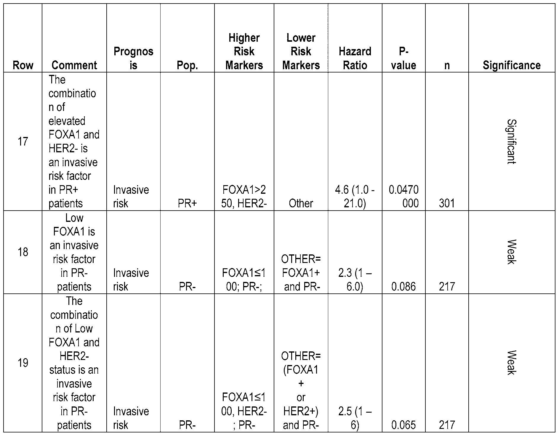
Table 10.1


[0151] Unless otherwise noted, the marker level (e.g. >250) or marker state (e.g. "+" or "-"), also referred to as "Marker thresholds", were used for Table 10. Additionally, in Table 10, young: age <50; old: age≥50; PR-: <10%; PR+:≥10%; HER2-: 0-2+; HER2+: 3+; SIAH2-: ≤20%; SIAH2+: >20%; FOXA1 low: immunoscore≤100; FOXA1 intermediate: immunscore > 100 and≤250; FOXA1 high: immunoscore >250; FOXA1 low: immunoscore≤100; FOXA1
intermediate: immunscore >100 and≤250; FOXA1 high: immunoscore >250. Unless denoted otherwise, a reference to Table 10 herein can include both tables 10 and 10.1.
[0152] In some embodiments, Table 11 provides a summary of the combinations that indicate a high risk of a subject diagnosed with DCIS experiencing an ipsilateral DCIS and/or invasive breast cancer after surgical removal of the primary DCIS.
Table 11


[0153] Tables 10 and 11 outline the relevant markers for identifying the level of risk that a subject with DCIS has for experiencing invasive breast cancer. Table 11 provides a summary of the combinations that indicate a high risk to a subject who already has DCIS experiencing invasive breast cancer and/or a recurrence of DCIS. The above results (Tables 1-
11, 13-15) are expressly contemplated for all embodiments of the various methods provided herein, as well as kits and compositions, etc.)
[0154] In some embodiments, any noted combination of the above markers or variables can be used for compositions or methods relating to DCIS recurrence and/or a risk of invasive breast cancer (as indicated). As noted herein, various combinations denote that the subject is at a relatively higher (or lower) risk of experiencing DCIS recurrence and/or a risk of invasive breast cancer. Thus, in some embodiments, this can be practically employed in terms of, for example, proper prognosis for the subject, advanced methods of treatment for the subject (for example, taking an approach that not only resolves DCIS that the subject currently has, but also addresses the risk level of DCIS recurrence and/or invasive breast cancer appropriately), methods for analyzing a sample (for example, a DCIS sample for various markers), compositions and kits that allow for the above noted methods, etc. In some embodiments, as demonstrated in the examples below, FOXA1 and PR predict ipsilateral event risk and identify a group with strong radiation response in ductal carcinoma in situ (DCIS). In some embodiments, in a PR- background, low FOXA1 correlates with invasive events, while in a PR+ background, high FOXA1 correlates with invasive recurrence. Both correlations are reversed for DCIS events. In some embodiments, separate biomarker-based risk models (algorithms) can be used to predict invasive and DCIS events, and at least some biomarkers can be assessed differently in the context of other markers, rather than being assigned a single weighting in a linear algorithm. In some embodiments, the PR/FOXA1 combination identifies a subset of patients who experience a remarkable benefit from RT relative to the remaining patients. In some embodiments, as shown in the examples below, validation of a multi-marker test that predicts recurrence in patients diagnosed with ductal carcinoma in situ (DCIS) treated with breast-conserving surgery (BCS) is provided. As shown, the studies indicate that the present approach to risk stratification modeling can accurately identify patients at risk for DCIS or invasive events after a primary DCIS diagnosis. In some embodiments, the models presented here (such as in the Examples below) are the basis of a comprehensive multi-marker panel. It should be noted that in certain embodiments, the risk models can be performed on a computing device while in other embodiments, the risk models can be performed manually.
[0155] In some embodiments, a method of analyzing a sample is provided. The method comprises analyzing a human DCIS tissue sample for PR, and either or both of:
analyzing the sample for at least HER2 and SIAH2, and/or analyzing the sample for at least FOXA1. In some embodiments, depending upon the nature of the results, this indicates that the subject that provided the sample is at a high or elevated risk of invasive breast cancer (see, e.g., Tables 1-11, 13, and 15).
[0156] In some embodiments, a method of analyzing a sample is provided. The method comprises analyzing a human DCIS tissue sample for a level of at least SIAH2 and FOXA1. In some embodiments, depending upon the nature of the results, this indicates that the subject that provided the sample is at a high or elevated risk of invasive breast cancer (see, e.g., Tables 1-11, 13, and 15).
[0157] In some embodiments, a method of analyzing a sample is provided. The method comprises providing a DCIS sample from a subject having DCIS; 1) analyzing the DCIS sample for SIAH2, and analyzing the DCIS sample for at least one of HER2, PR, FOXA1, or any combination thereof; or 2) analyzing the DCIS sample for FOXA1 and PR. In some embodiments, depending upon the nature of the results, this indicates that the subject that provided the sample is at a high or elevated risk of invasive breast cancer (see, e.g., Tables 1-11, 13, and 15).
[0158] In some embodiments, a method for prognosing a risk of an invasive breast cancer event in a subject is provided. The method comprises providing a DCIS sample from a subject, analyzing the DCIS sample for a level of at least PR, and at least either analyzing the sample for at least HER2 and SIAH2, or analyzing the sample for at least FOXA1. The method further comprises providing a prognosis based upon at least PR, HER2 and SIAH2 or based upon at least PR and FOXA1. In some embodiments, depending upon the nature of the results, this indicates that the subject that provided the sample is at a high or elevated risk of invasive breast cancer (see, e.g., Tables 1-11).
[0159] In some embodiments, a method for prognosing a risk of an invasive breast cancer event in a subject is provided. The method comprises providing a DCIS sample from a subject, analyzing the sample for a level of at least SIAH2 and FOXA1, and prognosing the subject as having an elevated risk of an invasive breast cancer based upon the level of at least SIAH2 and FOXA1. In some embodiments, depending upon the nature of the results, this indicates that the subject that provided the sample is at a high or elevated risk of invasive breast cancer (see, e.g., Tables 1-11).
[0160] In some embodiments, a method for prognosing a risk of an invasive breast cancer event in a subject is provided. The method comprises providing a DCIS sample from a subject, analyzing the sample for: a) PR, HER2, and SIAH2, or b) PR and FOXA1; and prognosing the subject as having an elevated risk of an invasive breast cancer event when at least one of: a) PR-, HER2-, and SIAH2-, b) PR+, FOXA1+, or c) PR+, FOXA1-, and Ki67+.
[0161] In some embodiments, a method for prognosing a risk of a subsequent DCIS event in a subject is provided. The method comprises providing a DCIS sample from a subject; analyzing the sample for at least one of: a) SIAH2 and FOXA1, b) SIAH2 and at least one of i) PR and ii) HER2, or c) SIAH2 and post-menopausal status; and d) PR and FOXA1, prognosing the subject as having an elevated risk of a DCIS event when at least one of: a) i) SIAH2+ and FOXA1+, b) SIAH2+ and HER2+ or PR-; SIAH2+ and post-menopausal; or PR+ and FOXA1-, is present in the DCIS sample.
[0162] In some embodiments, a method for treating a subject at risk of having an invasive breast cancer is provided. The method comprises providing a subject having DCIS, wherein the subject has a DCIS that is at least one of: a) PR-, HER2-, and SIAH2-, b) PR+, FOXA1+, or c) PR+, FOXA1-, and i67+; and administering to the subject a therapy that is more aggressive than standard of care for DCIS.
[0163] In some embodiments, a method for treating a subject at risk of having a DCIS recurrence is provided. The method comprises providing a subject having DCIS, wherein the subject has a DCIS that is at least one of: a) SIAH2+ and FOXA1+, b) SIAH2+ and HER2+ or PR-, c) SIAH2+ and post-menopausal status, or d) PR+ and FOXA1-, and administering to the subject a more aggressive therapy than standard of care for a single DCIS event.
[0164] In some embodiments, a method for treating a subject having DCIS is provided. The method comprises obtaining an analysis or prognosis as described herein, and a) performing a therapy appropriate to reduce a risk of invasive breast cancer if the analysis or prognosis indicates a risk of invasive breast cancer, or b) performing a therapy appropriate to reduce a risk of DCIS recurrence if the analysis or prognosis indicates a risk of DCIS recurrence.
[0165] In some embodiments, a method for treating a subject having DCIS is provided. The method comprises obtaining an analysis or prognosis as described herein, and either or both of: a) performing frequent breast monitoring for early detection of a subsequent ipsilateral breast event if the analysis or prognosis does not indicate an elevated or high risk of
invasive breast cancer, and if the analysis or prognosis does not indicate an elevated or high risk of DCIS recurrence, and/or b) ensuring the subject has clear margins by surgical resection, if needed, and performing frequent breast monitoring for early detection of a subsequent ipsilateral breast event, if the analysis or prognosis does not indicate an elevated or high risk of invasive breast cancer and if the analysis or prognosis does indicate an elevated or high risk of DCIS recurrence. In some embodiments, the absence of a risk for invasive and/or DCIS recurrence is a combination of markers that fails to appear in Tables 1-11. By the use of such a method, one can achieve an ideal therapy for the subject, where the subject might otherwise (without having the benefit of the method) have had an unnecessarily aggressive therapy.
[0166] In some embodiments, a method for prognosing a risk of an invasive breast cancer event in a subject is provided. The method can involve applying an algorithm to the protein and/or mRNA expression level as an additional transformative process, to thereby provide a signature for the marker. The method comprises providing a DCIS sample from a subject, analyzing the DCIS sample for a level of at least PR, and at least either analyzing the sample for at least HER2 and SIAH2, or analyzing the sample for at least FOXA1. The method further comprises providing a prognosis based upon at least a PR, HER2 and SIAH2 signature or based upon at least a PR and FOXA1 signature. In some embodiments, depending upon the nature of the results, this indicates that the subject that provided the sample is at a high or elevated risk of invasive breast cancer (see, e.g., Tables 1-11, 13, and 15).
[0167] In some embodiments, a method for prognosing a risk of an invasive breast cancer event in a subject is provided. The method comprises providing a DCIS sample from a subject, analyzing the sample for a level of at least SIAH2 and FOXA1, and prognosing the subject as having an elevated risk of an invasive breast cancer based upon the signature of at least SIAH2 and FOXA1. In some embodiments, depending upon the nature of the results, this indicates that the subject that provided the sample is at a high or elevated risk of invasive breast cancer (see, e.g., Tables 1-11, 13, and 15).
[0168] In some embodiments, a method for prognosing a risk of an invasive breast cancer event in a subject is provided. The method comprises providing a DCIS sample from a subject, analyzing the sample for: a) PR, HER2, and SIAH2, or b) PR and FOXA1; and prognosing the subject as having an elevated risk of an invasive breast cancer event using a
signature comprising at least one of: a) PR-, HER2-, and SIAH2-, b) PR+, FOXA1+, or c) PR+, FOXAl-, and Ki67+.
[0169] In some embodiments, a method for prognosing a risk of a subsequent DCIS event in a subject is provided. The method comprises providing a DCIS sample from a subject; analyzing the sample for at least one of: a) SIAH2 and FOXA1, b) SIAH2 and at least one of i) PR and ii) HER2, or c) SIAH2 and post-menopausal status; and d) PR and FOXA1, prognosing the subject as having an elevated risk of a DCIS event using a signature comprising at least one of: a) i) SIAH2+ and FOXA1+, b) SIAH2+ and HER2+ or PR-; SIAH2+ and post-menopausal; or PR+ and FOXA1-, is present in the DCIS sample.
[0170] In some embodiments, a method for treating a subject having DCIS is provided. The method comprises obtaining an analysis or prognosis (marker signatures) as described herein, and a) performing a therapy appropriate to reduce a risk of invasive breast cancer if the analysis or prognosis (marker signatures) indicates a risk of invasive breast cancer, or b) performing a therapy appropriate to reduce a risk of DCIS recurrence if the analysis or prognosis indicates a risk of DCIS recurrence.
[0171] In some embodiments, a method for treating a subject having DCIS is provided. The method comprises obtaining an analysis or prognosis (marker signatures) as described herein, and either or both of: a) performing frequent breast monitoring for early detection of a subsequent ipsilateral breast event if the analysis or prognosis (marker signatures) does not indicate an elevated or high risk of invasive breast cancer, and if the analysis or prognosis (marker signatures) does not indicate an elevated or high risk of DCIS recurrence, and/or b) ensuring the subject has clear margins by surgical resection, if needed, and performing frequent breast monitoring for early detection of a subsequent ipsilateral breast event, if the analysis or prognosis (marker signatures) does not indicate an elevated or high risk of invasive breast cancer and if the analysis or prognosis (marker signatures) does indicate an elevated or high risk of DCIS recurrence. In some embodiments, the absence of a risk for invasive and/or DCIS recurrence is a combination of markers that fails to appear in Tables 1-11 and 13-15. By the use of such a method, one can achieve an ideal therapy for the subject, where the subject might otherwise (without having the benefit of the method) have had an unnecessarily aggressive therapy.
[0172] In some embodiments, a method for treating a subject is provided. The method can include providing a subject having DCIS, wherein the subject has a DCIS that is none of: a) PR-, HER2-, and SIAH2-, b) PR+, FOXA1+, or d) PR+, FOXA1-, and Ki67+ and administering to the subject a therapy that is less aggressive than standard of care for DCIS.
[0173] In some embodiments, a method for treating a DCIS subject is provided. The method can comprise performing less than standard of care for a subject having DCIS where the standard of care for a subject having DCIS is breast-conserving surgery (BCS) with adjuvant radiation therapy, wherein the subject has a DCIS that lacks a combination of markers as indicated in Tables 1-11, 13, and 15 that would indicate an elevated risk of invasive breast cancer. Such a method allows for reduced surgeries and/or other therapies, where there is no, to little risk involved.
[0174] In some embodiments, a method for treating a DCIS subject is provided. The method can comprise performing a less than standard of care for a subject having DCIS, wherein the subject has a DCIS that lacks a combination of markers as indicated in Tables 1-11, 14, and IS that would indicate an elevated risk of DCIS recurrence. Such a method allows for reduced surgeries and/or other therapies, where there is no or little risk involved.
[0175] In some embodiments, a method for treating a DCIS subject is provided. The method can comprise not treating the subject with an aggressive form of treatment for DCIS because the subject does not fall into a high risk category for invasive breast cancer as outlined in Tables 9 11, 13, and IS. In some embodiments, a method for treating a DCIS subject is provided. The method can comprise not treating the subject with an aggressive form of treatment for DCIS because the subject does not fall into a high risk category for recurrent DCIS events as outlined in Tables 9,11, 14, and IS. In some embodiments, any level of treatment applied, can be lower than that which one of skill in the art would administer, if they could not determine a risk of DCIS resulting in invasive breast cancer in the subject.
[0176] In some embodiments, any of the above methods can be combined with any one or more of the following further aspects as to methods.
MARKERS
[0177] In some embodiments, markers in addition to those noted above can be analyzed and/or assayed for. In some embodiments, if the sample is PR positive, one can further
analyze the sample for a level of COX-2. In some embodiments, if the sample is PR positive, then one can further analyze the sample for Ki67 or size, or both Ki67 and size. In some embodiments, one can analyze the sample for pl6 with K.i-67 or p16 with COX-2. In some embodiments, the method further comprises analyzing at least the following combinations: a) PR, HER2, and SIAH2, b) PR, FOXA1, and COX-2, and c) PR, FOXA1, and Ki67.
[0178] In some embodiments, markers in addition to those disclosed and described herein can be analyzed and/or assayed for. These additional markers are disclosed and described in. U.S. Patent Application No. 12/373,047, filed May 13, 2009 and U.S. Patent Application No. 13/094,729, filed April 26, 2011, the contents of both applications are incorporated herein by reference in their entirety.
[0179] In some embodiments, the method further comprises analyzing at least COX- 2, Ki67, p16, PR and HER2. In some embodiments, if the sample is PR positive, one can further analyze the sample for a level of COX-2, wherein COX-2+ with at least FOXA1+ indicates a high risk of invasive breast cancer. In some embodiments, if the sample is PR positive and there is a very high level of FOXA1, there is a high risk of invasive breast cancer. In some embodiments, if the sample is PR positive, then one can further analyze the sample for Ki67, size, or both Ki67 and size. In some embodiments, if the sample is PR positive and FOXA1-, one can further analyze the sample for a level of Ki67, size, or a level of Ki67 and size. Ki67+, a size larger than 5 mm of DCIS, or both, indicates an elevated risk of invasive breast cancer. In some embodiments, the method further comprises analyzing the sample for p16, COX-2, and Ki67.
[0180] In some embodiments, analysis of each marker is carried out in parallel with each other. In some embodiments, analysis of each marker is carried out at overlapping times.
[0181] In some embodiments, PR analysis occurs first and any further analysis depends upon the result of the PR analysis. In some embodiments, there is no required order for any of the tests and/or analysis for any of the markers provided herein.
[0182] In some embodiments, the method further comprises determining a prognosis of the subject with DCIS. At least a level of SIAH2 and FOXA1 relative to the non-DCIS control indicates that the subject has a poor prognosis or wherein no significant difference in the expression of SIAH2 and FOXA1 relative to a non-tumor control indicates that the subject has a good prognosis.
[0183] In some embodiments, the DCIS sample is further analyzed for COX-2. In some embodiments, the DCIS sample is further analyzed for p16. In some embodiments, the DCIS sample is analyzed for at least SIAH2, FOXA1, and PR. In some embodiments, the DCIS sample is further analyzed for HER2. In some embodiments, the DCIS sample is further analyzed for COX-2. In some embodiments, the DCIS sample is further analyzed for Ki67. In some embodiments, the DCIS sample is further analyzed for pl6.
[0184] In some embodiments, the analysis (staining and/or scoring) includes SIAH2 and FOXA1. In some embodiments, the analysis further includes at least one of the following further combinations: 1) pl6, 2) Ki67, 3) PR, 4) COX-2, 5) HER2, 6) pl6 and Ki67, 7) pl6 and PR, 8) pl6 and COX-2, 9) pl6 and HER2, 10) HER2 and Ki67, 11) HER2 and PR, 12) HER2 and COX-2, 13) pl6, COX-2, and HER2, 14) HER2, COX-2, and Ki67, 15) HER2, COX-2, and PR, 16) HER2 and COX-2, 17) pl6, Ki-67, COX-2, and HER2, 18) Ki-67, HER2, COX-2, and PR, and/or 19) Ki-67, HER2, COX-2, PR, and pl6.
[0185] In some embodiments, the DCIS lesion is further analyzed for grade, necrosis, size, and/or margin status.
[0186] In some embodiments, the method further comprises prognosis of a risk by including age, menopausal status, mammographic density, tumor palpability of the subject.
[0187] In some embodiments, any of the "PR" steps, methods, compostions, etc. provided herein can be interchanged with an ER step (where ER staining and/or scoring is performed).
Reports/Recommendations
[0188] In some embodiments, any of the present methods can further comprise preparing a report regarding the risk associated with the human DCIS tissue sample. In some embodiments, the report is a written report providing the risk of invasive breast cancer. In some embodiments, the report is generated from and/or includes one or more of the marker combinations provided in Tables 1-11 and 13-15.
[0189] In some embodiments, any of the present methods further comprise providing a report regarding a level of risk of a subsequent DCIS event. In some embodiments, the report is a written report providing the risk of a subsequent DCIS event. In some embodiments, the
report is generated from and/or includes one or more of the marker combinations provided in Tables 1-1 l and 13-15.
[0190] In some embodiments, the method further comprises recommending a treatment given a result from analyzing the DCIS sample for SIAH2 and at least one of HER2, PR, FOXA1, or any combination thereof. In some embodiments, the treatment is less aggressive than would have otherwise been recommended, without the method predicting a low likelihood of invasive breast cancer. In some embodiments, the treatment is more aggressive than would have otherwise been recommended, without the method predicting a high likelihood of invasive breast cancer. In some embodiments, the treatment is less aggressive than would have otherwise been recommended, without the method predicting a low likelihood of a recurrence of DCIS. In some embodiments, the treatment is more aggressive than would have otherwise been recommended, without the method predicting a high likelihood of a recurrence of DCIS.
[0191] In some embodiments, the method further comprises determining a risk of DCIS, invasive breast cancer, or both. In some embodiments, the method further comprises providing a written report regarding a risk of DCIS, invasive breast cancer, or both (e.g., in line with Tables 1-11 and 13-15).
TREATMENT
[0192] In some embodiments, any of the above noted methods can include and/or be followed by an appropriate therapy for the subject, given the subject's reclassified risk of subsequent DCIS and/or invasive breast cancer. In some embodiments, such therapies can be appropriate to reduce a risk of DCIS recurrence, if that is the risk. In some embodiments, such therapies can be appropriate to reduce a risk of invasive breast cancer, if that is the risk.
[0193] In some embodiments, a therapy appropriate to reduce a risk of DCIS recurrence comprises at least one of surgical resection, radiation therapy, anti-hormone therapy. In some embodiments, a therapy can be appropriate if one knows that the subject has a low likelihood of an invasive event, but would not be appropriate if one knows that the subject has a high likelihood of an invasive breast cancer event.
[0194] In some embodiments, a therapy appropriate to reduce a risk of invasive breast cancer comprises at least one of mastectomy, targeted HERs therapy, receptor-targeted chemotherapy. In some embodiments, such a therapy can be appropriate if one knows that the
subject has a high likelihood of an invasive event, but would not be appropriate if one knows that the subject has a low likelihood of an invasive breast cancer event.
[0195] In some embodiments, any of the above methods can be followed by "watchful waiting" or other relatively minimal/instrusive therapies. For example, when none of the high risk categories are met for invasive breast cancer, and if the subject has no DCIS risk (or is okay with having a DCIS risk), then the approach to treating the DCIS can be to take no immediate action, which can include more frequent breast imaging to provide an early identification of an ipsilateral breast event.
[0196] Additional aspects and approaches regarding possible therapeutic actions that are specific for the present DCIS subjects are provided below.
KIT
[0197] In some embodiments, a kit is provided. The kit can include a FOXA1 probe, and a SIAH2 probe. In some embodiments, the kit further comprises a COX-2 probe, a Ki67 probe, a p16 probe, a PR probe, and a HER2 probe. In some embodiments, the probe is an isolated antibody. In some embodiments, the probe is a nucleic acid that selectively hybridizes to FOXA1, SIAH2, COX-2, Ki67, p16, PR or HER2 as appropriate. In some embodiments, the kit contains enough of the probe and/or the probe is sensitive and/or selective enough such that the "+" and "-" states of one or more of the markers in Tables 1-11 and 13-15 can be adequately distinguished from one another.
[0198] In some embodiments, an antibody composition is provided that includes an isolated FOXA1 antibody, and an isolated SIAH2 antibody. In some embodiments, the antibody composition further comprises an isolated COX-2 antibody, an isolated Ki67 antibody, an isolated p16 antibody, an isolated PR antibody, and an isolated HER2 antibody.
In some embodiments, a solid support comprising probes or antibodies specific for at least SIAH2 and FOXA1 is provided. In some embodiments, the probes or antibodies consists essentially of probes or antibodies specific for the prediction of DCIS or invasive breast cancer in a subject who has DCIS.
[0199] In some embodiments, the subject and/or sample to be analyzed can be a patient (or from a patient). In some embodiments, the subject has, or had, DCIS. In some
embodiments, the sample came from the DCIS of the subject in question. There are a variety of ways in which such a subject can be identified.
SAMPLE
[0200] In some embodiments, the DCIS sample itself can be processed in any number of ways to prepare it for screening for the markers. In some embodiments, the DCIS sample has been surgically removed from a patient and preserved. In some embodiments, the DCIS sample is obtained by surgical removal. In some embodiments, the DCIS sample is cut into one or more blocks, such as 2, 3, 4, 5 or more blocks.
[0201] In some embodiments, a level of SIAH2 and HER2, PR, FOXA1, or any combination thereof is at least one of: a RNA level, a DNA level, a protein level. In some embodiments, a level of SIAH2, HER2, PR, FOXA1, or any combination thereof is at least one of: a RNA level, a DNA level, a protein level. In some embodiments, a level of SIAH2 and FOXA1 is at least one of: a RNA level, a DNA level, a protein level.
[0202] In some embodiments, a signature comprising a level of SIAH2 and HER2, PR, FOXA1, or any combination thereof is at least one of: a RNA level, a DNA level, a protein level. In some embodiments, a signature comprising a level of SIAH2, HER2, PR, FOXA1, or any combination thereof is at least one of: a RNA level, a DNA level, a protein level. In some embodiments, a signature comprising a level of SIAH2 and FOXA1 is at least one of: a RNA level, a DNA level, a protein level.
[0203] In some embodiments, a method of preparing a sample is provided. The method comprises obtaining a DCIS sample from a subject and preparing it so that its DNA, RNA, and/or protein can be analyzed for at least SIAH2 and/or FOXA1.
[0204] In some embodiments, the sample is preserved. In some embodiments, the sample is preserved via freezing. In some embodiments, the sample goes through (or does not go through) embedding in a chemical such as Optimal Cutting Temperature (OCT) compound, or fixation with a chemical(s), including, without limitation, formalin, formaldehyde, quaternary ammonium salts, alcohol, acetone, or other chemicals that preserve or extract DNA, RNA, and/or protein. In some embodiments, the technique used is one that allows SIAH2 and/or FOXA1 DNA, RNA, and/or protein to be preserved in an adequate amount and state so that SIAH2 and/or FOXA1 can be analyzed as provided herein.
[0205] In some embodiments, the DCIS sample is processed to allow for immunohistochemistry of at least SIAH2 and/or FOXA1. In some embodiments, at least three such samples (such as in the form of slices) can be prepared).
[0206] In some embodiments, analyzing the sample comprises determining an amount of a specified RNA in the sample. The amount of RNA for each marker can be determined by any number of techniques, some of which are discussed elsewhere in the present application. In some embodiments, the RNA level is determined by at least one of: an assay involving nucleic acid microarray, reverse transcriptase-polymerase chain reaction, in situ nucleic acid detection, or a next generation sequencing method. In some embodiments, expression of at least one of SIAH2 and FOXA1 is measured by real time quantitative polymerase chain reaction or microarray analysis.
[0207] In some embodiments, the RNA level is determined by: an assay involving nucleic acid microarray, reverse transcriptase-polymerase chain reaction, in situ nucleic acid detection, or a next generation sequencing method.
[0208] In some embodiments, analyzing the DCIS sample comprises determining an amount of a specified protein in the sample. The amount of protein for each marker can be determined by any number of techniques, some of which are discussed elsewhere in the present application. In some embodiments, the protein level is determined by immunohistochemistry, immunofluorescence, or mass spectrometry.
[0209] In some embodiments, patient specimens used for the detection of the biomarkers can be surgically removed breast tissues that are cut into small blocks and submerged in fixative. In some embodiments, following fixation, the blocks can be dehydrated and then embedded in paraffin wax. In some embodiments, the small blocks are no more than 20 mm in length and S mm in thickness to allow complete penetration of the fixative. In some embodiments, the fixation occurs in 10% neutral-buffered formalin for 24 to 48 hours at room temperature to preserve tissue structure and compartmentalization of the various markers. However, other fixatives and fixation times (e.g., 6 to 72 hours) can also be compatible with the marker assays. In some embodiments, assays are optimized to use specimens that have been flash frozen (e.g., in liquid nitrogen), rather than being fixed and embedded.
[0210] In some embodiments, the process of sample processing can include dehydration and embedding, which can be done manually or automated with a tissue processing
instrument. In either case, the aqueous portion of the tissue and the fixation solution can be replaced by passing the block through a series of increasingly concentrated alcohol solutions. After reaching 100% alcohol, the alcohol is replaced using a chemical like xylene (or a xylene- free equivalent), followed by introduction of molten, low-melting-temperature (e.g., approximately 45°C) paraffin wax for embedding. The FFPE blocks can be stored for many years prior to analysis. In some embodiments, "cores" of DCIS tissue can be cut from these blocks using a hollow needle and then inserted in an array format in a separate block of paraffin. Such "tissue microarrays" (TMAs) allow assessment of multiple tissues on a single section/microscope slide.
[0211] In some embodiments, ultrathin sections, approximately three to five micrometers in thickness, can be cut off the formalin-fixed paraffin-embedded (FFPE) tumor blocks using a microtome. The sections can be mounted onto glass microscope slides, ensuring that the tissue does not become folded or fragmented, which could interfere with the assays. The glass microscope slides can contain a positively charged surface in order bind to the negatively charged tissue sections, although other methods of tissue binding, including adhesives, can also be compatible.
[0212] In some embodiments, wax removal and rehydration of the tissue sections can then be carried out. These processes can be done manually or automated with certain staining instruments. Wax can be removed from the tissue sections on the slides through heating and/or immersion in a solution of xylene (or an equivalent xylene-free solution, such as Novocastra Bond Dewaxing Solution). Rehydration can be accomplished by passing the slides through a series of decreasingly concentrated alcohol solutions until a concentration of 0% is reached (pure water). Following wax removal and rehydration, the tissue sections can be stained with hematoxylin and eosin (H&E) and for a variety of molecular markers using immunohistochemistry (IHC) and/or in situ hybridization (ISH) assays and then assessed by pathologists or histotechnologists, as described below. The above processing steps can be performed for any of the methods provided herein in regard to the various markers (HER2, COX-2, i-67, PR, p16, SIAH2 and FOXA1).
DCIS Diagnosis and Assessment Of Pathological Factors
Hematoxylin and eosin staining
[0213] In some embodiments, the subject and/or sample is confirmed as a DCIS sample or a subject having DCIS by any of a variety of ways known to one of skill in the art. This can occur before any of the other method steps provided herein (in some embodiments).
[0214] In some embodiments, hematoxylin and eosin (H&E) can be used to stain at least one tissue section from each patient (or set of arrayed patients) in order to confirm the DCIS diagnosis, assess certain pathological features (nuclear grade, architectural pattern[s], and the presence or absence of necrosis), and as a reference for the interpretation of the molecular marker assays. This histological stain allows the differentiation of nuclei and cytoplasm in individual cells, as well as various cell types and stromal tissue components, based on the color of the staining. The staining can be done manually or automated with a special staining instrument. In either case, the section can be submerged in hematoxylin solution for approximately four minutes to stain the nuclei blue, and then rinsed with tap water (alkaline). In some embodiments, next, the section can be exposed briefly (typically only a few seconds) to an acid alcohol solution to remove hematoxylin background staining, and then rinsed with tap water (alkaline). A "bluing" solution (e.g., lithium carbonate for 30 to 60 seconds) next may be applied to enhance the blue color of the hematoxylin in the nuclei, followed by rinsing with water. Eosin solution is then applied for approximately two minutes to stain other (eosinophilic) cellular components, followed by rinsing with water. The tissue is dehydrated with an alcohol series and cleared with xylene (or equivalent), and a cover slip is attached using mounting medium. Other options are also known to those of skill in the art. In some embodiments, any method for confirming the current presence of DCIS can be used on the sample. In some embodiments, no confirmation process is required. The above variables can be altered as appropriate by one of skill in the art.
[0215] DCIS itself can be identified in a sample or a subject in a variety of ways. Intraductal proliferative lesions include a group of cytologically and architecturally diverse epithelial proliferations originating in the terminal duct lobular unit (TDLU) and can be associated with an increased risk (of varying magnitude) for the subsequent development of invasive breast cancer. DCIS can be regarded as a possible true precursor lesion of invasive breast cancer. However, as demonstrated, not all DCIS events go on to form invasive breast cancer. There are various grades of DCIS (which, unless otherwise denoted, are all encompassed within the term "DCIS".
[0216] "Low grade DCIS" is composed of small, monomorphic cells, growing in a variety of patterns, including arcades, micropapillae, cribriform or solid patterns. The nuclei are generally of uniform size and have a regular chromatin pattern with inconspicuous nucleoli with rare mitotic figures. Low-grade DCIS requires either involvement of two spaces or one or more duct cross sections exceeding 2 mm in diameter. Although desquamated cells within the ductal lumen may be present, frank necrosis/comedo-type histologic features are not typical for low grade DCIS.
[0217] Cytologic features of DCIS can include: monotonous, uniform rounded cell population; subtle increase in nuclear-cytoplasmic ratio; equidistant or highly organized nuclear distribution; round nuclei; and hyperchromasia may or may not be present. Architectural features can include arcades, cribriform, solid and/or micropapillary.
[0218] "Intermediate grade DCIS" is often composed of cells cytologically similar to those of low grade DCIS, forming solid, cribriform or micropapillary patterns, but with some ducts containing intraluminal necrosis. Others display nuclei of intermediate grade with occasional nucleoli and coarse chromatin; necrosis may or may not be present.
[0219] "High grade DCIS" is usually larger than 5 mm, but even a single <1 mm duct with the typical morphological features is sufficient for diagnosis. It is composed of highly atypical cells proliferating as one layer, forming micropapillae, cribriform or solid patterns. Nuclei are high grade, markedly pleomorphic, poorly polarized, with irregular contour and distribution, coarse, clumped chromatin and prominent nucleoli. Mitotic figures are usually common but their presence is not required. Comedonecrosis is frequently associated with high grade DCIS, but not necessary for diagnosis. Even a single layer of highly anaplastic cells lining the duct in a flat fashion is sufficient.
[0220] "Atypical Ductal Hyperplasia" is distinct from DCIS. The morphological features of atypical ductal hyperplasia are identical to those of low-grade DCIS, but ADH is limited in size. There are two quantitative criteria that distinguish ADH from low-grade DCIS: the presence of homogeneous involvement of not more than 2 membrane-bound spaces; or a size of≤ 2 mm. The use of one or both criteria is considered appropriate by the authors of the WHO classification.
Histologic Confirmation of DCIS
[0221] In some embodiments, the subject is one who has at least one form of DCIS. In some embodiments, the presence of DCIS can be confirmed by any of a variety of techniques, including, for example, using slide-mounted tissue sections stained with hematoxylin and eosin (H&E) or an equivalent histology stain (noted above). In some embodiments, the assessment can be done consistent with WHO classification of tumors of the breast (Lakhani SR. WHO classification of tumours of the breast. Lyon: International Agency for Research on Cancer, 2012, and Tavassoli FA, Devilee P. Pathology and genetics of tumours of the breast and female genital organs. Lyon: IARC Press, 2003) - see definition of DCIS section. These references contain sample images and review characteristic features of DCIS and differential diagnosis with other breast disease entities, such as invasive breast cancer (including microinvasion defined as invasion≤1 mm), lobular carcinoma in situ (LCIS), in situ Paget's, atypical ductal hyperplasia (ADH), sclerosing adenosis, etc., the entireties of which are incorporated herein by reference).
[0222] In some embodiments, when histological features are not sufficient for the diagnosis of DCIS, the diagnosis can be confirmed by a second pathologist. Additional tissue blocks can be employed for morphologic review.
Cases Suspected To Have Invasive (or Microinvasive) Carcinoma Based Upon Morphologic Features:
[0223] Normal breast ducts and lobules as well as intraductal epithelial proliferations are composed of two epithelial layers. Loss of the outer myoepithelial layer is the hallmark of infiltrating carcinoma of the breast. The outer myoepithelial layer is retained in all benign proliferative processes as well as ductal carcinoma in situ. Consequently identification of the presence or loss of myoepitheilium using antibodies to the myoepithelial-specific proteins can be helpful in distinguishing in situ from infiltrating carcinoma in circumstances where morphology may be equivocal ( alof AN et al., alof AN, Tarn D, Beatty B, Cooper . Immunostaining patterns of myoepithelial cells in breast lesions: a comparison of CD 10 and smooth muscle myosin heavy chain. J Clin Pathol 2004; 57, 625-629; Barbareschi M et al., Barbareschi M, Pecciarini L, Cangi MG et al. p63, a p53 homologue, is a selective nuclear marker of myoepithelial cells of the human breast. Am J Surg Pathol; 25, 1054-1060, 2001).
[0224] In some embodiments, if there is unequivocal morphologic evidence of invasion, including microinvasion, the patient can be considered to be ineligible for the prognostic DCIS testing (and will not be tested, or can be excluded from the assay). In some
embodiments, if, upon morphologic examination of the tumor focus, there is a question of invasion of microinvasion, additional myoepithelial marker immunostudies (p63 and/or smooth muscle myosin heavy chain (SMMHC) immunostains) can be performed to examine the continuity of the myoepithelial cell layers and confirm and/or exclude the presence of (micro)invasive carcinoma. In some embodiments, other follow procedures can be performed for confirmation, where appropriate.
Cases Suspected To Be Of Lobular Origin Based Upon Morphologic Features:
[0225] It has been demonstrated that in histologic settings where ductal and lobular neoplasia might be confused, particularly in the setting of in situ carcinoma, where there can be significant differences in patient management, loss of expression of E-cadherin by immunohistochemistry can confirm the diagnosis of lobular carcinoma, even in the setting of non-classical morphologic findings (Acs G et al., Acs G, Lawton TJ, Rebbeck TR, LiVolsi VA, Zhang PJ. Differential expression of E-cadherin in lobular and ductal neoplasms of the breast and its biologic and diagnostic implications. Am J Clin Pathol 2001; 115, 85-98, 2001). In lobular neoplasia, mutations in the E-cadherin gene result in loss of expresion of E-cadherin, a cell surface adhesion molecule present in normal breast epithelium and ductal carcinoma. The role of E-cadherin in homotypic cell-cell binding, loss of expression of this cell surface protein accounts for the characteristic non-cohesive growth pattern of lobular carcinoma.
[0226] In some embodiments, if, upon examination of an intraductal epithelial proliferation, it is unclear whether the intraductal tumor is ductal or lobular in nature, an E- cadherin immunostain can be performed to confirm ductal or lobular differentiation. In some embodiments, if lobular carcinoma in situ (LCIS) is confirmed histologically or by loss of e- cadherin by immunhisotochemistry, the patient could be ineligible for further prognostic testing (e.g., will not be tested, or can be excluded from the assay). In some embodiments, if the subject currently has LCIS and not DCIS, the subject is not treated with the method. In some embodiments, if there is no evidence of DCIS or invasive carcinoma, additional tissue blocks can be requested for morphologic review.
[0227] In some embodiments, a sample or subject is excluded from the method if one or more of the following applies: a) no DCIS identified, b) invasive or microinvasive carcinoma identified, c) LCIS, not DCIS identified, c) quantitative criteria for low/intermediate DCIS not met: 1) the presence of homogeneous involvement of more than 2 membrane-bound spaces
and/or a size of≤ 2 mm. (No quantitative criteria required for high grade DCIS), or 2) tissue folded over in area of interest - not possible to score adequately.
Nuclear Grade Determination
In some embodiments, DCIS nuclear grade can be determined by using slide-mounted tissue sections stained with hematoxylin and eosin (H&E) or an equivalent histology stain. In some embodiments, the assessment can be consistent with the College of American Pathologists "Protocol for the Examination of Specimens from Patients with Ductal Carcinoma in Situ (DCIS) of the Breast" (Lester SC, Bose S, Chen YY et al. Arch Pathol Lab Med 2009; 133, 15- 25.), based on references therein. Nuclear grades of I (low), II (intermediate), and/or III (high) will be noted based on Table 12:
Table 12

Adapted from Lester S et al. Arch Pathol Lab Med 133:15-25 2009.
[0228] It is not uncommon to find admixture of various grades of DCIS within the same biopsy. In some embodiments, when more than one grade of DCIS is present, the proportion (percentage in deciles) of each grade will be noted. In some embodiments subjects with extensive disease and high grade DCIS will not be considered to be low risk for a subsequent ipsilateral breast event. In some embodiments, any of the methods provided herein can start with first determining if the subject has DCIS and/or the DCIS nuclear grade. In some embodiments, the method does not include determining nuclear grade.
Necrosis Determination
[0229] In some embodiments, the presence and extent of necrosis in DCIS can be examined using slide-mounted tissue sections stained with hematoxylin and eosin (H&E) or an equivalent histology stain. The assessment can be done consistent with the College of American Pathologists "Protocol for the Examination of Specimens from Patients with Ductal Carcinoma
in Situ (DCIS) of the Breast" (June 2012), based on references therein. In some embodiments, necrosis can be classified as follows: A) Not identified: No evidence of necrosis, B) Focal (punctuate): Small foci, indistinct at low magnification, or single cell necrosis, or C) Central (comedo/extensive): The central portion of an involved ductal space is replaced by an area of expansive necrosis that is easily detected at low magnification. Ghost cells and karyorrhectic debris are generally present. Although central necrosis is generally associated with high-grade nuclei (comedo DCIS), it can also occur with DCIS of low or intermediate nuclear grade. In some embodiments, any of the methods provided herein can include determining necrosis.
IHC Marker Staining/Scoring
[0230] When a formalin-based fixation method is used, it creates molecular crosslinks in proteins, thereby masking epitopes from recognition by antibodies, and other fixation/preservation methods can also mask epitopes. In such embodiments, epitope retrieval can be a pre-treatment step that allows one to unmask the epitopes by reversing, at least in part, the changes introduced by fixation/preservation. Thus, in some embodiments, any of the methods and/or kits provided herein can include a step or ingredient for epitope retrieval.
[0231] Epitope retrieval can be done in different ways by varying the chemicals in the solution (e.g., buffers, proteolytic enzymes, chelators, etc.), the pH of the solution, the temperature of the solution (e.g., as applied by a water bath, pressure cooker, autoclave, or microwave oven), and/or the time in the solution, etc. In addition to the specific methods described in the examples below, many of these other methods could be used to achieve substantially equivalent results, depending on the tissue source, primary antibody, and other factors.
[0232] Multiple antibodies are commercially available and/or have been reported in the literature for each protein marker described herein (COX-2, Ki67, HER2, p16, PR, SIAH2, and FOXA1), and new antibodies can also be created. In some embodiments, the antibodies are raised against and/or recognize different epitopes on the protein markers (COX-2, Ki67, HER2, p16, PR, SIAH2, and FOXA1), and, in other cases, the antibodies are raised against and/or recognize the same (or similar) epitopes. The usefulness of an individual antibody in an assay depends upon its affinity and specificity for the epitope, as well as the accessibility of the epitope in the assay (e.g., after epitope retrieval in and IHC assay). Some antibodies recognize more than one protein marker and are, therefore, not typically suitable for a specific marker assay.
Other antibodies have low affinity or recognize an epitope that remains inaccessible in certain samples and are, therefore, not suitable for certain assay types. For example, an antibody that has a use for an immunoblot of fresh protein lysate may not have utility in an IHC assay on FFPE tissue due to the inability to unmask its epitope through epitope retrieval.
[0233] The concentrations of primary antibody concentrates commercially available from manufacturers vary based on the production method (e.g., tissue culture supernatant, ascites fluid, or whole antiserum), and whether any purification was done (e.g., affinity purification), but they are typically in the range of about 0.1 to 10 mgml. The optimal final primary antibody concentration for incubation on the sections depends on such factors as the binding characteristics of the specific antibody, the incubation time and temperature, and other factors unique to the individual laboratory, but it is typically in the range of 0.1 to 10 μg/ml, and dilutions ranging from about 1:10 to 1:1,000 are typically used. In some embodiments, staining results can be achieved with an antibody over a range of final primary antibody concentrations, as well as incubation times and temperatures.
[0234] In some embodiments, a Novocastra Bond Refine Polymer system can be used for detection of the primary antibody. This system includes a polymer backbone to which multiple secondary antibodies (against rabbit IgG) and enzymes are attached, as well as a rabbit anti-mouse IgG linker (when used with mouse primary antibodies). The enzymes catalyze a chemical reaction with DAB to form a brown precipitate that is visualized during marker scoring. In some embodiments one of several other detection methods can produce adequate results, including systems that utilize avidin-biotin complex (ABC), labeled streptavidin-biotin (LSAB), catalyzed signal amplification, and/or other technologies that are available in a variety of formats from a number of different manufacturers. In some embodiments, the ABC and other polymer-based technologies (e.g., Dako EnVision+) can be utilized with similar results (for detection).
[0235] In some embodiments, chromogen DAB can be used for final visualization of the marker through an enzymatic reaction of horseradish peroxidase (HRP) that produces a brown precipitate at the site of the antibody binding. In some embodiments, HRP can be used in combination with the chromogen 3-amino-9-ethylcarbazole (AEC) to produce red coloration with substantially equivalent results. Other options include the enzyme alkaline phosphatase in combination with the chromogens nitro blue tetrazolium chloride (NBT) and 5-bromo-4-chloro-
3-indolyl phosphate (BCIP), and the enzyme glucose oxidase in conjunction with NBT— both of which produce a bluish-purple coloration. There are a variety of enzyme/chromogen combinations that can produce results.
[0236] The following section outlines various representative embodiments for staining and scoring seven markers (PR, HER2, COX-2, Ki-67, SIAH2, FOXA1, and p16). In some embodiments, other markers that serve the same function can be substituted for any one or more of these markers. In some embodiments ER can be substituted for PR. In some embodiments, any one or more of these markers can be used in the combinations suggested in the accompanying tables (Tables 1-11 and 13-15). In some embodiments, one or more of the noted markers can be employed (e.g., for staining and/or scoring) but a corresponding staining technique and/or corresponding score is used instead (as outlined herein).
PR Staining
[0237] In some embodiments, any technique for PR staining can be used, as long as it is adequate to observe the degree of PR fluctuation provided and described herein. In some embodiments, protein levels can be checked. In some embodiments, mRNA levels can be checked. In some embodiments, DNA levels can be checked. In some embodiments, both protein and mRNA levels can be checked. An elevated level can be a level above that above a control or standardized level, for example, a level in a non-DCIS sample. Similarly, a lowered level can be a level below a control or standardized level, for example, a level in a non-DCIS sample. In some embodiments, a "positive" or "elevated" result is one that is above a "negative" or "lowered" result (in the context of scoring).
[0238] In some embodiments, to assess progesterone receptor (PR; PGR; HGNC:8910) by IHC, a Leica BOND-MAX automated staining instrument can be used to conduct the following steps with rinsing between each step. Dewaxed and rehydrated tissue sections can be treated with Novocastra Peroxide Block (3-4% hydrogen peroxide) (peroxidase blocking step), followed by Novocastra Bond Epitope Retrieval Solution 1 (based on a 10 mM sodium citrate buffer plus 0.05% Tween 20, pH 6.0 solution) for 30 minutes at 95°C to 100°C (epitope retrieval step). The tissue sections are then incubated at room temperature with mouse monoclonal antibody PgR 636 (Dako M3569) diluted 1 :50 in Novocastra Primary Antibody Diluent for 30 minutes (primary antibody step), followed by Novocastra Post Primary solution for 15 minutes (rabbit anti-mouse to introduce IgG linkers), followed by Novocastra Bond
Refine Polymer for 15 minutes (anti-rabbit poly-HRP-IgG) (secondary detection step), followed by 3,3'-Diaminobenzidine (DAB) for 5 minutes (chromogen visualization step), followed by <0.1% hematoxylin for 7 minutes (nuclear counterstain step). Finally, a cover slip is attached using mounting medium. In other embodiments, other options can be employed.
[0239] In some embodiments, PR can be detected by a number of primary antibodies from a number of different manufacturers, and most produce adequate results. In some embodiments, mouse monoclonal 1A6 and rabbit monoclonal SP2 can be used from Novocastra and Lab Vision. Other options include mouse monoclonals PgR1294, 16, 1A6, 1E2, Ab-8, Ab-9, hPRa2, hPRa3, and PR88; rabbit monoclonals SP2, SP42, Y85, and EP2; and rabbit polyclonal A0097 (Dako), as well as many others, from manufacturers like Dako (Agilent), Novocastra (Leica), Ventana Medical Systems (Roche), Cell Marque (Sigma-Aldrich), Lab Vision (Thermo Scientific), BioGenex, Biocare, and Epitomics. In some embodiments, other options can be employed.
[0240] In some embodiments, high pH epitope retrieval (pH 9) can be been done in Tris-EDTA buffer with microwave heating. The titer of each lot of Dako PgR 636 antibody can be adjusted to a reference lot by the manufacturer to ensure consistent staining performance at the same dilution factor, and the 1 :50 dilution used in the above example is suggested by the manufacturer. However, a range of dilutions can be used (e.g., 1:10 through 1:500) with similar performance. Alternative dilutions can be optimal with other antibody preparations, and for situations, some preparations are provided pre-diluted (ready to use). In some embodiments, other options can be employed.
PR Scoring
[0241] In some embodiments, any technique for PR scoring can be used, as long as it is adequate to observe the degree of PR fluctuation provided and described herein.
[0242] In some embodiments, PR status is determined from the IHC stained slide based upon the percentage of DCIS tumor cells with nuclear signal. In some embodiments, all areas of the tissue section containing DCIS can be evaluated to arrive at the percentage. In some embodiments, at least three DCIS-containing ducts or 1 mm of DCIS tissue can be employed to score the markers.
[0243] In some embodiments, the intensity of the nuclear signal can be reported as weak (1+), moderate (2+), or strong (3+). The intensity is the average intensity of the DCIS
tumor cell nuclei with signal over the entire tissue section relative to the intensity of positive controls run with the same staining batch. In some embodiments, selection of the DCIS regions to be scored and/or the scoring are conducted manually by a pathologist and/or automatically using a computer on scanned images of the slide.
[0244] In some embodiments, a DCIS with less than 10% of tumor cells with nuclear signal can be considered negative, whereas DCIS with greater than 10% of tumor cells with nuclear signal can be considered positive, when assayed by the system or examples described herein. In some embodiments, the sample is only considered negative in the presence of appropriately stained extrinsic and internal controls. In some embodiments, any specimen lacking internal control elements (normal breast ductal epithelium) that is negative should be reported as uninterpretable (rather than as negative) and repeated using another tumor specimen from the same or an alternative tumor block.
[0245] In some embodiments, alternative thresholds can be applied (e.g., 0% vs. >0%, <1% vs.≥1%,≤1% vs. >1%, <5% vs.≥5%,≤5% vs. >5%, <10% vs.≥10%, <15% vs. ≥15%,≤15% vs. >15%, <20% vs.≥20%,≤20% vs. >20%, <25% vs.≥25%,≤25% vs. >25%, <30% vs.≥30%, etc. and other thresholds), based upon the technique employed for staining and/or analysis. In some embodiments, alternative techniques and/or scoring methods can be used for detection, which can result in a corresponding, but different cutoff range for high and/or low risk. For such situations, the ranges provided herein for the present technique can be correlated to the other technique (for the "corresponding value") by analyzing the same sample (or two samples from a same DCIS sample) by the two different techniques and identifying them as being equivalent to one another. Alternative scoring methods, such as the Allred scoring system, immunoscores, and others that combine the percentage and intensity scoring elements also show effectiveness.
[0246] An example of PR scoring and staining is presented in FIG. 1 (PR IHC assays (top, negative; bottom, positive) ). In some embodiments, PR scoring can be as follows: negative is less than 5 percent positive by percentage scoring for IHC; positive is greater than 10 percent positive by percentage scoring for IHC. In some embodiments, the difference in percent between positive and negative can be compressed, such that any sample is either positive or negative. In some embodiments, the difference between positive and negative can be dropped or ignored, if the sample falls within the range. In some embodiments, any of the values between positive and
negative can be selected as the absolute distinguishing line between positive and negative (e.g., 5, 6, 7, 8, 9, or 10).
[0247] In some embodiments, FOXA1 scoring can be as follows: negative is less than a 100 immunoscore by (intensity times percentage) scoring for IHC; positive is greater than a 100 immonoscore by (intensity times percentage) scoring for IHC. In some embodiments, the difference in percent between positive and negative (100) can be compressed, such that any sample is either positive or negative. In some embodiments, the difference between positive and negative can be dropped or ignored, if the sample falls within the range. In some embodiments, any of the values between positive and negative can be selected as the absolute distinguishing line between positive and negative (e.g., 100 or lower is negative vs. 100 or higher is positive).
[0248] In some embodiments, SIAH2 scoring can be as follows: negative is less than 10 percent positive by percentage scoring for IHC; positive is greater than 20 percent positive by percentage scoring for IHC. In some embodiments, the difference in percent between positive and negative can be compressed, such that any sample is either positive or negative. In some embodiments, the difference between positive and negative can be dropped or ignored, if the sample falls within the range. In some embodiments, any of the values between positive and negative can be selected as the absolute distinguishing line between positive and negative (e.g., 10, 11, 12, 13, 14, 15, 16, 17, 18, 19, or 20).
[0249] In some embodiments, HER2 scoring can be as follows: negative is less than a 3+ score by HercepTest scoring criteria for IHC; positive is 3+ score by HercepTest scoring criteria for IHC.
[0250] In some embodiments, Ki67 scoring can be as follows: negative is less than 10 percent positive by by percentage scoring for IHC; positive is greater than 15 percent scoring by percentage for IHC. In some embodiments, the difference in percent between positive and negative can be compressed, such that any sample is either positive or negative. In some embodiments, the difference between positive and negative can be dropped or ignored, if the sample falls within the range. In some embodiments, any of the values between positive and negative can be selected as the absolute distinguishing line between positive and negative (e.g., 10, 11, 12, 13, 14, or 15).
[0251] In some embodiments, p16 scoring can be as follows: negative is less than 20 percent positive by percentage scoring for IHC; positive is greater than 25 percent positive by
percentage scoring for IHC. In some embodiments, the difference in percent between positive and negative can be compressed, such that any sample is either positive or negative. In some embodiments, the difference between positive and negative can be dropped or ignored, if the sample falls within the range. In some embodiments, any of the values between positive and negative can be selected as the absolute distinguishing line between positive and negative (e.g., 20, 21, 22, 23, 24, or 25).
[0252] In some embodiments, COX-2 scoring can be as follows: negative is less than 6 by Allred Scoring Criteria for COX-2 for IHC; positive is greater than 6 by Allred Scoring Criteria for COX-2 for IHC.
[0253] In some embodiments, the above ranges are for IHC assays. In some embodiments, the above ranges are for FISH assays.
[0254] In some embodiments, the positive threshold can be higher depending on marker combinations employed in concert as determined by one skilled in the art for incorporation into a specific test.
[0255] In some embodiments, SIAH2, FOXA1, and COX-2 can further be broken into sub categories of high (very high and high) and low (very low and low). This allows for finer lines to be drawn regarding risk combinations. In some embodiments, this is simplified for the analysis by having very high and high fall within the "high" grouping and very low and low fall within the "low" grouping.
HER2 Staining
HER2IHC
[0256] In some embodiments, any technique for HER2 staining can be used, as long as it is adequate to observe the degree of HER2 fluctuation provided and described herein. In some embodiments, protein levels can be checked. In some embodiments, mRNA levels can be checked. In some embodiments, DNA levels can be checked. In some embodiments, both protein and mRNA levels can be checked. An elevated level can be a level above that above a control or standardized level, for example, a level in a non-DCIS sample. Similarly, a lowered level can be a level below a control or standardized level, for example, a level in a non-DCIS sample. In some embodiments, a "positive" or "elevated" result is one that is above a "negative" or "lowered" result (in the context of scoring).
[0257] In some embodiments, to assess v-erb-b2 avian erythroblastic leukemia viral oncogene homolog 2 (ERBB2; HGNC:3430; HER2 [human epidermal growth factor receptor 2]; NEU) by IHC, the following steps can be conducted with rinsing between each step on a Dako Autostainer (through the chromogen visualization step). Dewaxed and rehydrated tissue sections can be treated with Dako Peroxidase Blocking Reagent (peroxidase blocking step), followed by Dako HercepTest Epitope Retrieval Solution (10 mM citrate buffer plus detergent, pH 6) for 40 minutes in a 95°C to 99°C water bath (epitope retrieval step). The tissue sections can then be incubated at room temperature with pre-diluted rabbit polyclonal anti-HER2 antibody (Dako 5207) for 30 minutes (primary antibody step), followed by Dako HercepTest Visualization Reagent for 30 minutes (secondary detection step), followed by 3,3'-Diaminobenzidine (DAB) for 10 minutes (chromogen visualization step), followed by hematoxylin (nuclear counterstain step). Finally, a cover slip is attached using mounting medium.
[0258] In some embodiments, HER2 can be detected by various antibodies to HER2. In some embodiments, mouse monoclonal TAB250 and rabbit monoclonal SP3 can be used from Invitrogen and Lab Vision. Other options include mouse monoclonals CB-11, TA9145, Ab-17, 3B5, and PN2A, and rabbit monoclonals SP3, 4B5, EP1045Y, and EP3, as well as many others, from manufacturers like Dako (Agilent), Novocastra (Leica), Ventana Medical Systems (Roche), Cell Marque (Sigma-Aldrich), Lab Vision (Thermo Scientific), BioGenex, Biocare, and Epitomics.
[0259] In some embodiments, heat-induced epitope retrieval can be performed by microwave oven or water bath. In the above example, the HER2 primary antibody is provided in a pre-diluted (ready to use) format, so no dilution is performed. However, other preparations of HER2 primary antibody are available in concentrated format requiring dilution. For example, a 200 μg/ml Ig concentrate may be diluted 1:400 to 1:800 to a final Ig concentration of 0.2S to 0.S0 μg/ml, or an alternative concentrate may have an optimal dilution of 1:100 with similar results achieved in a dilution range of 1 :50 to 1 :500.
HER2-ISH
[0260] In some embodiments, an alternative method to assess HER2 can be ISH. A fluorescent ISH (FISH) assay using a Vysis PathVysion HER-2 DNA Probe Kit (Abbott Molecular) can include the following steps with rinsing between each step. The dewaxed tissue section can be treated with 0.2 N HC1 for 20 minutes and then Vysis Pretreatment Solution (1 N
sodium isothiocyanate) for 30-60 minutes at 80°C (pretreatment step), followed by Vysis Protease Solution for 10-60 minutes at 37°C (protease step), followed by 10% neutral buffered formalin (fixation step), followed by a series of increasing concentration alcohol solutions (dehydration step).
[0261] In some embodiments, a DNA probe mixture is applied to the tissue section under a sealed coverslip. The mixture contains a Locus Specific Identifier for HER-2 (a 190-Kb Spectrum Orange directly-labeled, fluorescent DNA probe specific for the HER-2 gene locus at 17q 11.2-q 12) and a Chromosome Enumeration Probe for chromosome 17 (CEP 17; a 5.4 Kb Spectrum Green directly-labeled, fluorescent DNA probe specific for the alpha satellite DNA sequence at the centromeric region of chromosome 17 at 17p 11.1 -q 11.1), as well as unlabeled blocking DNA to suppress sequences contained within the target loci that are common to other chromosomes. The slide is then placed into a Thermobrite instrument and subjected to a temperature of 73°C for 5 minutes (DNA denaturation step) followed by an incubation for 14-24 hours at 37°C (DNA hybridization step).
[0262] In some embodiments, the tissue is treated with two washes of 2x saline sodium citrate (SSC) plus 0.3% NP-40, the first for 2-5 minutes at room temperature, and the second for 2 minutes at 71-73°C (post hybridization wash step). Finally, the nuclei are stained blue with a 4, 6 diamidino-2-phenylindole (DAPI) solution (counterstaining step).
[0263] In some embodiments, an alternative ISH method based on silver deposition (SISH; Ventana Medical Systems Inform HER2 kit) can be used according to manufacturer instructions with substantially equivalent results. In some embodiments, other HER2 ISH methods, for example chromogenic ISH (CISH), can also be employed.
HER2 Scoring-HER2 IHC Scoring
[0264] In some embodiments, any technique for HER2 scoring can be used, as long as it is adequate to observe the degree of HER2 fluctuation provided and described herein.
[0265] In some embodiments, HER2 status is determined from the IHC stained slide according to College of American Pathologists-American Society of Clinical Oncology (CAP- ASCO) guidelines (Wolff et al. J Clin Oncol 31:3997, 2013.) with modification for scoring on intraductal tumor cells. HER2 scores of 0-3+ are defined as follows: 0 is defined by no staining observed or membrane staining that is incomplete and is faint barely perceptible and within ≤10% of the tumor cells. 1+ is defined by incomplete membrane staining that is faint barely
perceptible and within >10% of the tumor cells. 2+ is defined by circumferential membrane staining that is incomplete and/or weak/moderate (observed in a homogeneous and contiguous population) and within >10% of the tumor cells, or complete and circumferential membrane staining that is intense and within≤10% of the tumor cells. 3+ is defined by circumferential membrane staining that is complete and intense (observed in a homogeneous and contiguous population and within >10% of the tumor cells, readily appreciated using a low power objective).
[0266] In some embodiments, selection of the DCIS regions to be scored and/or the scoring are conducted manually by a pathologist and/or automatically using a computer on scanned images of the slide. In some embodiments, results of 0, 1+, or 2+ are considered negative, and 3+ is considered positive. In some embodiments, 2+ results could be considered equivocal, triggering a HER2 ISH assay to determine status through quantitation of HER2 gene amplification (amplified patients are positive). In some embodiments, HER2 IHC could be completely replaced by a HER2 ISH assay. Both of these alternative approaches have shown utility in related studies.
HER2 ISH Scoring
[0267] In some embodiments, HER2 status is determined from a FISH stained slide by counting the orange HER2 and green CEP 17 signals in a minimum of 20 DCIS cell nuclei and then calculating the ratio. The DCIS is considered HER2 non-amplified (negative) when there is an equal number of orange and green signals or the ratio of Orange to green is less than 2.0 with an average HER2 copy number per cell of less than 4.0. The DCIS is considered HER2 amplified (positive) when the ratio of orange to green signals is greater than 2.0. Cells are also considered amplified (positive) when the ratio of orange to green signals is less than 2.0 with an average HER2 copy number per cell greater than or equal to 6.0. When the ratio of orange to green signals is less than 2.0 with an average HER2 copy number per cell of greater than or equal to 4.0 and less than 6.0, 20 additional cells are counted, and the ratio is re-calculated for all 40 cells, and the threshold of <2.0 (negative) vs.≥2.0 (positive) is applied.
[0268] An example of HER2 scoring and staining is presented in FIG. 2 (HER2 IHC assays (top, negative; bottom, positive)).
Ki-67 Staining
[0269] In some embodiments, any technique for i-67 staining can be used, as long as it is adequate to observe the degree of Ki-67 fluctuation provided and described herein. In some embodiments, protein levels can be checked. In some embodiments, mRNA levels can be checked. In some embodiments, DNA levels can be checked. In some embodiments, both protein and mRNA levels can be checked. An elevated level can be a level above that above a control or standardized level, for example, a level in a non-DCIS sample. Similarly, a lowered level can be a level below a control or standardized level, for example, a level in a non-DCIS sample. In some embodiments, a "positive" or "elevated" result is one that is above a "negative" or "lowered" result (in the context of scoring).
[0270] In some embodiments, Ki-67 (MKI67; MIB-1; HGNC:7107) levels can be assessed by IHC, a Leica BOND-MAX automated staining instrument is used to conduct the following steps with rinsing between each step. In some embodiments, dewaxed and rehydrated tissue sections are treated with Novocastra Peroxide Block (3-4% hydrogen peroxide) (peroxidase blocking step), followed by Novocastra Bond Epitope Retrieval Solution 2 (based on a 10 mM Tris Base and 1 mM ethylenediaminetetraacetic acid [EDTA] buffer plus 0.05% Tween 20, pH 9.0 solution) for 30 minutes at 95°C to 100°C (epitope retrieval step). In some embodiments, the tissue sections are then incubated at room temperature with mouse monoclonal antibody MIB-1 (Dako M7240) diluted 1:50 in Novocastra Primary Antibody Diluent for 30 minutes (primary antibody step), followed by Novocastra Post Primary solution for 15 minutes (rabbit anti-mouse IgG to introduce IgG linkers), followed by Novocastra Bond Refine Polymer for 15 minutes (anti-rabbit poly-HRP-IgG) (secondary detection step), followed by 3,3 - Diaminobenzidine (DAB) for 5 minutes (chromogen visualization step), followed by <0.1% hematoxylin for 7 minutes (nuclear counterstain step). Finally, a cover slip is attached using mounting medium.
[0271] In some embodiments, Ki-67 can be detected by any Ki-67 specific antibody. In some embodiments, the antibody can be mouse monoclonals MM1, K-2; 7B11, BGX-297, Ki88, Ki-S5, and DVB-2; rabbit monoclonals SP6, 30-9, EPR3611; and rabbit polyclonal NCL- Ki67p, as well as many others, from manufacturers like Dako (Agilent), Novocastra (Leica), Ventana Medical Systems (Roche), Cell Marque (Sigma-Aldrich), Lab Vision (Thermo Scientific), BioGenex, and Biocare.
[0272] In some embodiments, a dilution of 1:50 is used for Dako MIB-1 antibody, although other dilutions, e.g., 1 :75 to 1 :150, can be used, including dilutions in the range of 1 :40 to 1 :600. Similarly, other antibody manufacturers report recommended dilutions in the range of 1 :100 to 1 :200 for their antibody preparations, and some preparations are provided pre-diluted (ready to use).
Ki-67 Scoring
[0273] In some embodiments, any technique for Ki-67 scoring can be used, as long as it is adequate to observe the degree of Ki-67 fluctuation provided and described herein.
[0274] In some embodiments, Ki-67 status is determined from the IHC stained slide based upon the percentage of DCIS tumor cells with nuclear signal. In some embodiments, all areas of the tissue section containing DCIS are evaluated to arrive at the percentage. In some embodiments, at least three DCIS-containing ducts or 1 mm of DCIS tissue are used to score the markers. In some embodiments, the intensity of the signal is also reported as weak (1+), moderate (2+), or strong (3+). In some embodiments, the intensity is the average intensity of the DCIS tumor cell nuclei with signal over the entire tissue section. In some embodiments, selection of the DCIS regions to be scored and/or the scoring are conducted manually by a pathologist and/or automatically using a computer on scanned images of the slide.
[0275] In some embodiments, DCIS with less than 10% of tumor cells with nuclear signal is considered negative, whereas DCIS with greater than 10% of tumor cells with nuclear signal is considered positive. The sample is only considered negative in the presence of an appropriately stained positive control.
[0276] In some embodiments, alternative thresholds (e.g., 0% vs. >0%, <1% vs.≥1%, ≤1% vs. >1%, <5% vs.≥5%,≤5% vs. >5%, <10% vs.≥10%, <15% vs.≥15%,≤15% vs. >15%, <20% vs.≥20%,≤20% vs. >20%, <25% vs.≥25%,≤25% vs. >25%, <30% vs.≥30%, etc. can be employed based upon the technique employed for staining and/or analysis. In some embodiments, alternative techniques and/or scoring methods can be used for detection, which can result in a corresponding, but different cutoff range for high and/or low risk. For such situations, the ranges provided herein for the present technique can be correlated to the other technique (for the "corresponding value") by analyzing the same sample (or two samples from a same DCIS sample) by the two different techniques and identifying them as being equivalent to
one another. Alternative scoring methods, such as the Allred scoring system, immunoscores, and others that combine the percentage and intensity scoring elements also show effectiveness.
[0277] An example of K.i-67 scoring and staining is presented in FIG. 4 i-67 IHC assays (top, negative; bottom, positive). pl6/INK4A Staining
[0278] In some embodiments, any technique for p16 staining can be used, as long as it is adequate to observe the degree of p16 fluctuation provided and described herein. In some embodiments, protein levels can be checked. In some embodiments, mRNA levels can be checked. In some embodiments, DNA levels can be checked. In some embodiments, both protein and mRNA levels can be checked. An elevated level can be a level above that above a control or standardized level, for example, a level in a non-DCIS sample. Similarly, a lowered level can be a level below a control or standardized level, for example, a level in a non-DCIS sample. In some embodiments, a "positive" or "elevated" result is one that is above a "negative" or "lowered" result (in the context of scoring).
[0279] In some embodiments, to assess the p16 isoform of cyclin-dependent kinase inhibitor 2A (pl6/IN 4A; CD N2A; MTS1; HGNC:1787) by IHC, a Leica BOND-MAX automated staining instrument can be used to conduct the following steps with rinsing between each step. In some embodiments, it can be dewaxed and rehydrated tissue sections are treated with Novocastra Peroxide Block (3-4% hydrogen peroxide) (peroxidase blocking step), followed by Novocastra Bond Epitope Retrieval Solution 2 (based on a 10 mM Tris Base and 1 mM EDTA buffer plus 0.05% Tween 20, pH 9.0 solution) for 30 minutes at 95°C to 100°C (epitope retrieval step). The tissue sections can then be incubated at room temperature with pre-diluted mouse monoclonal antibody E6H4 (Ventana Medical Systems CINtec p16 Histology Kit) for 30 minutes (primary antibody step), followed by Novocastra Post Primary solution for 15 minutes (rabbit anti-mouse IgG to introduce IgG linkers), followed by Novocastra Bond Refine Polymer for 15 minutes (anti-rabbit poly-HRP-IgG) (secondary detection step), followed by 3,3 - Diaminobenzidine (DAB) for 5 minutes (chromogen visualization step), followed by <0.1% hematoxylin for 7 minutes (nuclear counterstain step). In some embodiments, a cover slip is attached using mounting medium.
[0280] In some embodiments, p16/INK4A can be detected by a primary antibody. In some embodiments, mouse monoclonal DCS-50.1/A7 (Neomarkers) can be used. In some
embodiments, mouse monoclonals 6H12, JC8, 16P04; 16P07, and G 175-405 and rabbit monoclonal EPR1473, as well as others can be used. In some embodiments, the pl6/IN 4A primary antibody is provided in a pre-diluted (ready to use) format, so no dilution is performed. However, other preparations of pl6/IN 4A primary antibody are available in concentrated format, and dilutions are recommended by the manufacturers in a range of 1 :75 to 1 :500 or even 1 :25 to 1 :800, depending on the specific protocol. pl6/INK4A scoring
[0281] In some embodiments, any technique for p16 scoring can be used, as long as it is adequate to observe the degree of p16 fluctuation provided and described herein.
[0282] In some embodiments, pl6/IN 4A status is determined from the IHC stained slide based upon the percentage of DCIS tumor cells with nuclear signal, qualified by the intensity of the signal. In some embodiments, the intensity of the signal is reported as weak (1+), moderate (2+), or strong (3+). In some embodiments, the intensity is the average intensity of the DCIS tumor cell nuclei with signal over the entire tissue section relative to the intensity of positive controls run with the same staining batch. In some embodiments, cells with nuclear signal of at least intermediate (2+) intensity are considered positive. In some embodiments, cells with absent or weak (1+) staining are considered negative. In some embodiments, all areas of the tissue section containing DCIS are evaluated to arrive at the percentage. In some embodiments, at least three DCIS-containing ducts or 1 mm of DCIS tissue can be used to score the markers. In some embodiments, selection of the DCIS regions to be scored and/or the scoring are conducted manually by a pathologist and/or automatically using a computer on scanned images of the slide.
[0283] In some embodiments, DCIS with less than or equal to 25% of tumor cells with nuclear signal of at least moderate intensity is considered negative, whereas DCIS with greater than 25% of tumor cells with nuclear signal of at least moderate intensity is considered positive. The sample is only considered negative in the presence of an appropriately stained positive control. Alternative thresholds (e.g., <20% vs.≥20%,≤20% vs. >20%, <25% vs.≥25%, <30% vs.≥30%,≤30% vs. >30%, <40% vs.≥40%, etc. and other thresholds) can be employed based upon the technique employed for staining and/or analysis. In some embodiments, alternative techniques and/or scoring methods can be used for detection, which can result in a corresponding, but different cutoff range for high and/or low risk. For such situations, the ranges
provided herein for the present techniques can be correlated to the other technique (for the "corresponding value") by analyzing the same sample (or two samples from a same DCIS sample) by the two different techniques and identifying them as being equivalent to one another. Alternative scoring methods, such as the Allred scoring system, immunoscores, and others that combine the percentage and intensity scoring elements also show effectiveness.
[0284] The staining pattern of pl6/IN 4A can be nuclear and cytoplasmic and is often heterogeneous in nature. In addition, pl6/IN 4A staining can be present in both the DCIS tumor cells and the surrounding stromal cells.
[0285] An example of p16 scoring and staining is presented in FIG. 3 (pl6/IN 4A IHC assays (top, negative; bottom, positive)).
COX-2 Staining
[0286] In some embodiments, any technique for COX-2 staining can be used, as long as it is adequate to observe the degree of COX-2 fluctuation provided and described herein. In some embodiments, protein levels can be checked. In some embodiments, mRNA levels can be checked. In some embodiments, DNA levels can be checked. In some embodiments, both protein and mRNA levels can be checked. An elevated level can be a level above that above a control or standardized level, for example, a level in a non-DCIS sample. Similarly, a lowered level can be a level below a control or standardized level, for example, a level in a non-DCIS sample. In some embodiments, a "positive" or "elevated" result is one that is above a "negative" or "lowered" result (in the context of scoring).
[0287] In some embodiments, prostaglandin-endoperoxide synthase 2 (PTGS2; cyclooxygenase-2 [COX-2]; HGNC:9605) can be assessed by IHC; a Leica BOND-MAX automated staining instrument is used to conduct the following steps with rinsing between each step. In some embodiments, dewaxed and rehydrated tissue sections are treated with Novocastra Peroxide Block (3-4% hydrogen peroxide) (peroxidase blocking step), followed by Novocastra Bond Epitope Retrieval Solution 1 (based on a 10 mM sodium citrate buffer plus 0.05% Tween 20, pH 6.0 solution) for 30 minutes at 95°C to 100°C (epitope retrieval step). In some embodiments, the tissue sections are then incubated at room temperature with rabbit monoclonal antibody SP21 (Cell Marque 240R-16) diluted 1 :50 in Novocastra Primary Antibody Diluent for 30 minutes (primary antibody step), followed by Novocastra Bond Refine Polymer for 15 minutes (anti-rabbit poly-HRP-IgG) (secondary detection step), followed by 3,3 -
Diaminobenzidine (DAB) for 5 minutes (chromogen visualization step), followed by <0.1% hematoxylin for 7 minutes (nuclear counterstain step). Finally, a cover slip is attached using mounting medium.
[0288] In some embodiments, COX-2 can be detected by primary antibodies. In some embodiments, mouse monoclonal CX-294 (Dako) can be used. In some embodiments, mouse monoclonal 4H12 (Novocastra) and rabbit monoclonal SP21 from manufacturers like Ventana Medical Systems (Roche), Cell Marque (Sigma-Aldrich), Lab Vision (Thermo Scientific), and Biocare can be used.
[0289] In some embodiments, a dilution of 1:50 can be used for Cell Marque SP21 antibody. In some embodiments, a dilution of 1:100 to 1:500, or in the range of 1:50 to 1:500, 1 :50 to 1 :200 can be used. In addition, some preparations are provided in pre-diluted (ready to use) form.
COX-2 Scoring
[0290] In some embodiments, any technique for COX-2 scoring can be used, as long as it is adequate to observe the degree of COX-2 fluctuation provided and described herein.
[0291] In some embodiments, COX-2 status can be determined from the IHC stained slide based upon the percentage of DCIS tumor cells with cytoplasmic signal and the intensity of the signal, in the form of an Alfred score. In some embodiments, the intensity is reported as absent (0), weak (1), intermediate (2), or strong (3) and represents the average signal intensity over the entire tissue section relative to the intensity of positive controls run with the same staining batch. In some embodiments, the percentage is converted to a proportion score as follows: 0, 0% positive; 1,≤1% positive; 2, >1-10% positive; 3, 11-33% positive; 4, 34-66% positive; 5, 67-100% positive. The Allred score is the sum of the intensity and proportion scores on a scale of 0-8.
[0292] In some embodiments, all areas of the tissue section containing DCIS are evaluated. In some embodiments, at least three DCIS-containing ducts or 1 mm of DCIS tissue is employed to score the markers. Selection of the DCIS regions to be scored and/or the scoring can be conducted manually by a pathologist and/or automatically using a computer on scanned images of the slide.
[0293] In some embodiments, a DCIS with an Allred score 0 to 6 is considered negative, whereas DCIS with an Allred score of 7 or 8 is considered positive. In some
embodiments, the sample is considered negative in the presence of an appropriately stained positive control. In some embodiments, alternative techniques and/or scoring methods can be used for detection, which can result in a corresponding, but different cutoff range for high and/or low risk. For such situations, the ranges provided herein for the present techniques can be correlated to the other technique (for the "corresponding value") by analyzing the same sample (or two samples from a same DCIS sample) by the two different techniques and identifying them as being equivalent to one another. In some embodiments, an alternative scoring method, such as the Allred scoring system, immunoscores, and others that combine the percentage and intensity scoring elements also show effectiveness.
[0294] An example of COX-2 scoring and staining is presented in FIG. 5 (COX-2 IHC assays (top, negative; bottom, positive)).
FOXA1 Staining
[0295] In some embodiments, any technique for FOXA1 staining can be used, as long as it is adequate to observe the degree of FOXA1 fluctuation provided and described herein. In some embodiments, protein levels can be checked. In some embodiments, mRNA levels can be checked. In some embodiments, DNA levels can be checked. In some embodiments, both protein and mRNA levels can be checked. An elevated level can be a level above that above a control or standardized level, for example, a level in a non-DCIS sample. Similarly, a lowered level can be a level below a control or standardized level, for example, a level in a non-DCIS sample. In some embodiments, a "positive" or "elevated" result is one that is above a "negative" or "lowered" result (in the context of scoring).
[0296] In some embodiments, to assess forkhead box Al (FOXA1; HGNC:5021) by IHC, a Leica BOND-MAX automated staining instrument can be used to conduct the following processes with rinsing between each step. In some embodiments, dewaxed and rehydrated tissue sections can be treated with Novocastra Peroxide Block (3-4% hydrogen peroxide) (peroxidase blocking step), followed by Novocastra Bond Epitope Retrieval Solution 2 (based on a 10 mM Tris Base and 1 mM ethylenediaminetetraacetic acid [EDTA] buffer plus 0.05% Tween 20, pH 9.0 solution) for 30 minutes at 95°C to 100°C (epitope retrieval step). The tissue sections can then be incubated at room temperature with mouse monoclonal antibody 2F83 (Cell Marque 405M-16) diluted 1:25 in Novocastra Primary Antibody Diluent for 30 minutes (primary
antibody step), followed by Novocastra Post Primary solution for 15 minutes (rabbit anti-mouse IgG to introduce IgG linkers), followed by Novocastra Refine Polymer for 15 minutes (anti- rabbit poly-HRP-IgG) (secondary detection step), followed by 3,3'-Diaminobenzidine (DAB) for 5 minutes (chromogen visualization step), followed by <0.1% hematoxylin for 7 minutes (nuclear counterstain step). A cover slip can be attached using mounting medium.
[0297] In some embodiments, FOXA1 mouse monoclonal antibody 2F83 from Abeam can be used at a dilution of 1 :450 (or, for example, 1 :2,000) with effective results or used at a dilution of 1:25 (manufacturer recommended starting dilution range of 1:25 to 1:100, based on an initial estimated Ig concentration of 2.5 to 25.0 μg/ml with a final estimated optimal Ig concentration range of 0.1 to 1.0 μg/ml). In some embodiments, mouse monoclonal antibodies can include 2F83, 3A8, 1B1, 2D7, 3C1, and 4F6, and rabbit monoclonals SP88 and EPR10881 from Thermo Scientific, Spring Bioscience, Epitomics, Millipore, and a variety of other manufacturers. Various dilutions can be used with other antibody preparations (e.g., Millipore recommends a 1:500 dilution of their 1 mg/ml version of 2F83, and Thermo Scientfic recommends a 1 :20 to 1 :200 dilution of their 1 mg/ml preparation of clone 3A8). And some FOXA1 antibody preparations are provided pre-diluted (ready to use).
FOXA1 Scoring
[0298] In some embodiments, any technique for FOXA1 scoring can be used, as long as it is adequate to observe the degree of FOXA1 fluctuation provided and described herein.
[0299] In some embodiments, FOXA1 status can be determined from the IHC stained slide based upon the percentage of DCIS tumor cells with nuclear signal and the intensity of the signal, in the form of an immunoscore. The intensity can be reported as absent (0), weak (1), intermediate (2), or strong (3) and can represent the average signal intensity over the entire tissue section relative to the intensity of positive controls run with the same staining batch. In some embodiments, the immunoscore can be the product of the intensity and percentage scores on a scale of 0-300.
[0300] In some embodiments, all areas of the tissue section containing DCIS are evaluated. In some embodiments, three DCIS-containing ducts or 1 mm of DCIS tissue is used to score the markers. Selection of the DCIS regions to be scored and/or the scoring can be conducted manually by a pathologist and/or automatically using a computer on scanned images of the slide.
[0301] In some embodiments, DCIS with an immunoscore less than 100 is considered FOXA1 low, DCIS with an immunoscore between 100 and 250 is considered FOXA1 intermediate, and DCIS with an immunoscore greater than 250 is considered FOXA1 high. Alternative lower (e.g., 40 or 150) and/or upper (e.g., 150 or 250) thresholds, as well as alternative scoring methods, also show utility. In addition, the utility can vary based on the outcome being predicted (i.e., a DCIS or invasive event in the ipsilateral breast). In some embodiments, FOXA1 is merely treated as either being "negative" (100 or lower) or positive (greater than 100).
An example of FOXA1 staining and scoring is presented in FIG. 7. FIG. 7 depicts a FOXA1 IHC assays (top, negative; bottom, positive).
[0302] In some embodiments, alternative techniques and/or scoring methods can be used for detection, which can result in a corresponding, but different cutoff range for high and/or low risk. For such situations, the ranges provided herein for the present techniques can be correlated to the other technique (for the "corresponding value") by analyzing the same sample (or two samples from a same DCIS sample) by the two different techniques and identifying them as being equivalent to one another. In some embodiments, an alternative scoring method, such as the Allred scoring system, immunoscores, and others that combine the percentage and intensity scoring elements also show effectiveness.
SIAH2 Staining
[0303] In some embodiments, any technique for SIAH2 staining can be used, as long as it is adequate to observe the degree of SIAH2 fluctuation provided and described herein. In some embodiments, protein levels can be checked. In some embodiments, mRNA levels can be checked. In some embodiments, DNA levels can be checked. In some embodiments, both protein and mRNA levels can be checked. An elevated level can be a level above a control or standardized level, for example, a level in a non-DCIS sample. Similarly, a lowered level can be a level below a control or standardized level, for example, a level in a non-DCIS sample. In some embodiments, a "positive" or "elevated" result is one that is above a "negative" or "lowered" result (in the context of scoring).
[0304] In some embodiments, to assess SIAH2 E3 ubiquitin protein ligase 2 (SIAH2; seven in absentia [Drosophila] homolog 2; HGNC: 10858) by IHC, a Leica BOND-MAX automated staining instrument can be used to conduct the following steps with rinsing between
each step. In some embodiments, dewaxed and rehydrated tissue sections are treated with Novocastra Peroxide Block (3-4% hydrogen peroxide) (peroxidase blocking step), followed by Novocastra Bond Epitope Retrieval Solution 1 (based on a 10 mM sodium citrate buffer plus 0.05% Tween 20, pH 6.0 solution) for 30 minutes at 95°C to 100°C (epitope retrieval step). In some embodiments, the tissue sections are then incubated at room temperature with mouse monoclonal antibody 24E6H3 (Santa Cruz Biotechnology sc-81787) diluted 1:200 in Novocastra Primary Antibody Diluent for 30 minutes (primary antibody step), followed by Novocastra Post Primary solution for 15 minutes (rabbit anti-mouse to introduce IgG linkers), followed by Novocastra Bond Refine Polymer for 15 minutes (anti-rabbit poly-HRP-IgG) (secondary detection step), followed by 3,3'-Diaminobenzidine (DAB) for 5 minutes (chromogen visualization step), followed by <0.1% hematoxylin for 7 minutes (nuclear counterstain step). In some embodiments, a cover slip is attached using mounting medium.
[0305] In some embodiments, epitope retrieval can be done at a higher pH (8.0) in EDTA-containing buffer in either a 98°C water bath or steam chamber with both mouse monoclonal antibody 24E6H3 and its parental clone 24E6 in related studies. In some embodiments, alternative antibodies can be used, including those from Novus Biologicals. Different dilutions can be used with different preparations of these antibody, including dilutions within the range of 1 :40 to 1 :200. In some embodiments, other primary antibodies can be used, such as mouse monoclonal 35F7I4 (Creative Diagnostics), which works for IHC (recommended dilution of 1:40 to 1:50), and mouse monoclonals 1F5, 2G6, and SIAH2-369 (available from Abnova, Epigentek, US Biologicals, Sigma-Aldrich, and Creative Diagnostics).
SIAH2 Scoring
[0306] In some embodiments, any technique for SIAH2 scoring can be used, as long as it is adequate to observe the degree of SIAH2 fluctuation provided and described herein.
[0307] In some embodiments, SIAH2 status is determined from the IHC stained slide based upon the percentage of DCIS tumor cells with nuclear signal. In some embodiments, all areas of the tissue section containing DCIS are evaluated to arrive at the percentage. In some embodiments, at least three DCIS-containing ducts or 1 mm of DCIS tissue is used to score the markers. In some embodiments, the intensity of the signal can be reported as weak (1+), moderate (2+), or strong (3+). In some embodiments, the intensity is the average intensity of the DCIS tumor cell nuclei with signal over the entire tissue section. In some embodiments,
selection of the DCIS regions to be scored and/or the scoring are conducted manually by a pathologist and/or automatically using a computer on scanned images of the slide.
[0308] In some embodiments, DCIS with less than 20% of tumor cells with nuclear signal is considered negative, whereas DCIS with greater than or equal to 20% of tumor cells with nuclear signal is considered positive.
[0309] In some embodiments, the sample is only considered negative in the presence of an appropriately stained positive control.
[0310] In some embodiments, alternative techniques and/or scoring methods can be used for detection, which can result in a corresponding, but different cutoff range for high and/or low risk. For such situations, the ranges provided herein for the noted techniques can be correlated to the other technique (for the "corresponding value") by analyzing the same sample (or two samples from a same DCIS sample) by the two different techniques and identifying them as being equivalent to one another. In some embodiments, an alternative scoring method, such as the Allred scoring system, immunoscores, and others that combine the percentage and intensity scoring elements also show effectiveness. In some embodiments, alternative thresholds (e.g.,≤20% vs. >20%, <25% vs.≥25%,≤25% vs. >25%, <30% vs.≥30%,≤30% vs. >30%, <40% vs.≥40%,≤40% vs. >40%, etc. and other thresholds) can be used. In some embodiments, alternative scoring methods, such as the Allred scoring system, immunoscores, and others that combine the percentage and intensity scoring elements can also be used.
[0311] An example of SIAH2 staining and scoring is presented in FIG. 6. FIG. 6 depicts SIAH2 IHC assays (top, negative, on a UUH TMA; bottom, positive, on a Biomax BR8011 TMA).
Statistical Analysis
[0312] Kaplan-Meier survival analyses can be used to estimate the proportions of patients who experienced first events (DCIS or invasive recurrence) after initial DCIS diagnosis/surgery. Hazard ratios (HR) can be determined using Cox proportional hazards analysis. At the time of first events (DCIS or invasive), patients can be censored for the other event type. Patients can also be censored if an event is detected within 6 months of surgery or on the first post-surgery mammogram, because this can be considered as persistent, rather than recurrent, disease. In some embodiments, subjects with persistent disease are excluded from the various methods provided herein.
Methods of Assessing Markers
[0313] While the above noted assaying system and scoring systems have been put forth, in some embodiments, alternative techniques and/or scoring methods can be used for detection, which can result in a corresponding, but different cutoff range for high and/or low risk. For such situations, the ranges provided herein for the present techniques can be correlated to the other technique (for the "corresponding value" of one or more of COX-2, Ki67, p16, SIAH2, FOXA1, PR, and /or HER2) by analyzing the same sample (or two samples from a same DCIS sample) by the two different techniques and identifying them as being matched or equal to one another. In some embodiments, an alternative scoring method, such as the Allred scoring system, immunoscores, and others that combine the percentage and intensity scoring elements also show effectiveness. In some embodiments, there can be a corresponding technique and/or score for one or more of COX-2, Ki67, pl6, SIAH2, FOXA1, PR, and /or HER2. In some embodiments, there can be a corresponding technique for one or more of COX-2, Ki67, p16, SIAH2, FOXA1, PR, and /or HER2. In some embodiments, there can be a corresponding score for one or more of COX-2, Ki67, pl6, SIAH2, FOXA1, PR, and /or HER2. As will be appreciated by one of skill in the art, these corresponding scores and/or techniques can be used for any of the embodiments provided herein, and are expressly contemplated as alternatives for each and all disclosure regarding the noted markers.
[0314] In some embodiments, protein detection assays can be used, including for example, immunohistochemistry, immunofluorescence, mass spectrometry, or others, discussed in more detail below. In some embodiments, mRNA detection assays can be used, including, for example, nucleic acid hybridization-based methods such as Northern blots, gene expression arrays, quantitative real-time polymerase chain reaction (qPCR), nCounter, in situ nucleic acid detection, etc., as well as next-generation RNA sequencing (RNA -Seq) ), and others. In some embodiments, techniques to detect changes at the DNA level can be used, including, for example, polymerase chain reaction (PCR), in situ hybridization (ISH), next generation sequencing (NGS), and others as will be readily understood by those of skill in the art.
[0315] In some embodiments, while corresponding values (involving alternative scoring or alternative assays to analyze the sample) are used, the relative result (for example, high vs. low or very high vs. medium vs. low) will be maintained between the various techniques for analysis or scoring systems. Thus, scoring "high" in one system will be correlated to scoring
"high" in another system, without significant complications or difficulties. Thus, various results can be ported from one system to another, as desired, as long as the levels in terms of relatively high vs low (for example) are maintained. Similarly, in some embodiments, protein levels can be used for one marker, while a second marker can be analyzed via DNA, and, for example, a third marker can be analyzed via mRNA. Thus, the nature of the molecule being tested can be altered within a test, if desired.
[0316] In some embodiments, the level of expression is determined by detecting the level of mRNA transcribed from a gene.
[0317] In some embodiments, the mRNA in the sample is first transcribed into cDNA using reverse transcriptase.
[0318] In some embodiments, the sample is subjected to an amplification reaction (e.g., using methods based on polymerase chain reaction (PCR), nucleic acid sequence-based amplification (NASBA), transcription-mediated amplification (TMA), strand displacement amplification (SDA), etc.), probe amplification (e.g., using methods based on ligase chain reaction (LCR), cleavase invader, etc.), signal amplification (e.g, using methods based on branched DNA probes [bDNA], hybrid capture, etc.), and others as will be readily understood by those of skill in the art.
[0319] In some embodiments, the mRNA is detected in the sample by hybridizing a nucleic acid probe or primer capable of selectively hybridizing to a mRNA transcript of interest, or cDNA derived therefrom, and then detecting the hybridization with a detection device or system.
[0320] In this context, the term "selective hybridization" means that hybridization of a probe or primer occurs at a higher frequency or rate, or has a higher maximum reaction velocity, than hybridization of the same probe or primer to any other nucleic acid. Preferably, the probe or primer does not hybridize to another nucleic acid at a detectable level under the reaction conditions used.
[0321] As transcripts of a gene described herein are detected using mRNA or cDNA derived therefrom, assays that detect changes in mRNA can be employed (for example, Northern hybridization, RT-PCR, NASBA, TMA or ligase chain reaction).
[0322] In some embodiments, mRNA quantitation can be carried out on gene expression array platforms, including Agilent's Bioanalyzer® system; Affymetrix' GeneChip®,
GeneTitan , or GeneAtlas systems; and others as will be readily understood by those of skill in the art.
[0323] In some embodiments, mRNA quantitation can be carried out by real-time qPCR. Such reactions can be performed with a variety of reporters, including non-specific DNA- binding fluorochromes (e.g., SYBR® Green) or fluorescent reporter probes that selectively hybridize to the sequence of interest (e.g., TaqMan® probes). In some cases, the reaction is carried out in parallel on multiple partitions of the same sample (digital PCR). Real-time qPCR platforms include ThermoFisher Scientific/Applied Biosystems' FAST, QuantStudio, and related systems; Hologic/Gen-Probe's DTS systems; Roche/Idaho Technology's LightCycler® systems; Qiagen's Rotor-Gene® systems; Bio-Rad's CFX and related systems; and others as will be readily understood by those of skill in the art.
[0324] In some embodiments, mRNA quantitation can be carried out without a reverse-transcription or amplification step, such as NanoString's in vitro nCounter® platform, or various in situ hybridization (ISH) approaches, including paired probe ISH (e.g., RNAscope® and Quanti-Gene RNAview), single-tag multi-probe ISH (e.g., Stellaris®), or locked nucleic-acid (LNA) probes.
[0325] In some embodiments, mRNA quantitation can be carried out with RNA-Seq next-generation sequencing technologies, including sequencing by synthesis (e.g, Illumina's HiSeq® and NextSeq® systems), single-molecule real-time sequencing (e.g., Pacific Biosciences' RS systems), ion seminconductor sequencing (e.g., ThermoFisher Scientific' s Ion Torrent™ systems), sequencing by ligation (e.g., ThermoFisher Scientific's SOLiD™ systems), pyrosequencing (e.g., Roche/454 Life Sciences), and others as will be readily understood by those of skill in the art.
[0326] Methods of RT-PCR include, for example, in Dieffenbach (ed) and Dveksler (ed) (In: PCR Primer: A Laboratory Manual, Cold Spring Harbour Laboratories, NY, 1995). Essentially, this method comprises performing a PCR reaction using cDNA produced by reverse transcribing mRNA from a cell using a reverse transcriptase. Methods of PCR described supra are to be taken to apply mutatis mutandis to this embodiment of the invention.
[0327] Similarly PCR can be performed using cDNA. One or more of the probes or primers used in the reaction specifically hybridize to the transcript of interest.
[0328] Methods of TMA or self-sustained sequence replication (3SR) use two or more oligonucleotides that flank a target sequence, a RNA polymerase, RNase H and a reverse transcriptase. One oligonucleotide (that also comprises a RNA polymerase binding site) hybridizes to an RNA molecule that comprises the target sequence and the reverse transcriptase produces cDNA copy of this region. RNase H is used to digest the RNA in the RNA-DNA complex, and the second oligonucleotide used to produce a copy of the cDNA. The RNA polymerase is then used to produce a RNA copy of the cDNA, and the process repeated.
[0329] NASBA systems relies on the simultaneous activity of three enzymes (a reverse transcriptase, RNase H and RNA polymerase) to selectively amplify target mRNA sequences. The mRNA template is transcribed to cDNA by reverse transcription using an oligonucleotide that hybridizes to the target sequence and comprises a RNA polymerase binding site at its 5' end. The template RNA is digested with RNase H and double stranded DNA is synthesized. The RNA polymerase then produces multiple RNA copies of the cDNA and the process is repeated.
[0330] In some embodiments, a microarray can be used to determine the level of expression of one or more nucleic acids described herein. Such a method allows for the detection of a number of different nucleic acids, thereby providing a multi-analyte test and improving the sensitivity and/or accuracy of the diagnostic assay of the invention.
[0331] In some embodiments, the level of expression is determined by detecting the level of a protein encoded by a nucleic acid within a gene described herein.
[0332] In this respect, the embodiments are not necessarily limited to the detection of a protein comprising the specific amino acid sequence recited herein. Rather, the present invention encompasses the detection of variant sequences (e.g., having at least about 80% or 90% or 95% or 98% amino acid sequence identity) or the detection of an immunogenic fragment or epitope of said protein.
[0333] The amount, level and/or presence of a polypeptide can be determined using any of a variety of techniques known to the skilled artisan such as, for example, a technique selected from the group of, immunohistochemistry, immunofluorescence, an immunoblot, a Western blot, a dot blot, an enzyme linked immunosorbent assay (ELISA), radioimmunoassay (RIA), enzyme immunoassay, fluorescence resonance energy transfer (FRET), matrix-assisted laser desorption/ionization time of flight (MALDI-TOF), electrospray ionization (ESI), mass
spectrometry (including tandem mass spectrometry, e.g. LC MS/MS), biosensor technology, evanescent fiber-optics technology or protein chip technology.
[0334] In some embodiments, the assay used to determine the amount or level of a protein is a semi-quantitative assay. In some embodiments, the assay used to determine the amount or level of a protein in a quantitative assay. As will be apparent from the preceding description, such an assay may involve the use of a suitable control, e.g. from a normal individual or matched normal control.
[0335] In some embodiments, standard solid-phase ELISA or FLISA formats can be useful in determining the concentration of a protein from a variety of samples.
[0336] In one form such an assay involves immobilizing a biological sample onto a solid matrix, such as, for example a polystyrene or polycarbonate microwell or dipstick, a membrane, or a glass support (e.g. a glass slide). An antibody that specifically binds to a protein described herein is brought into direct contact with the immobilized biological sample, and forms a direct bond with any of its target protein present in said sample. This antibody is generally labeled with a detectable reporter molecule, such as for example, a fluorescent label (e.g. FITC or Texas Red) or a fluorescent semiconductor nanocrystal (as described in U.S. Pat. No. 6,306,610) in the case of a FLISA or an enzyme (e.g. horseradish peroxidase (HRP), alkaline phosphatase (AP) or beta-galactosidase) in the case of an ELISA, or alternatively a second labeled antibody can be used that binds to the first antibody. Following washing to remove any unbound antibody the label is detected either directly, in the case of a fluorescent label, or through the addition of a substrate, such as for example hydrogen peroxide, TMB, or toluidine, or 5-bromo-4-chloro-3-indol-beta-D-galaotopyranoside (x-gal) in the case of an enzymatic label.
[0337] In some embodiments, an ELISA or FLISA comprises immobilizing an antibody or ligand that specifically binds a protein described supra on a solid matrix, such as, for example, a membrane, a polystyrene or polycarbonate microwell, a polystyrene or polycarbonate dipstick or a glass support. A sample is then brought into physical relation with said antibody, and the polypeptide is bound or 'captured". The bound protein is then detected using a labeled antibody. For example, a labeled antibody that binds to an epitope that is distinct from the first (capture) antibody is used to detect the captured protein. Alternatively, a third labeled antibody can be used that binds the second (detecting) antibody.
[0338] In some embodiments, the presence or level of a protein is detected in a body fluid using, for example, a biosensor instrument (e.g., BIAcore™, Pharmacia Biosensor, Piscataway, N.J.). In such an assay, an antibody or ligand that specifically binds a protein is immobilized onto the surface of a receptor chip. For example, the antibody or ligand is covalently attached to dextran fibers that are attached to gold film within the flow cell of the biosensor device. A test sample is passed through the cell. Any antigen present in the body fluid sample, binds to the immobilized antibody or ligand, causing a change in the refractive index of the medium over the gold film, which is detected as a change in surface plasmon resonance of the gold film.
[0339] In some embodiments, the presence or level of a protein or a fragment or epitope thereof is detected using a protein and/or antibody chip. To produce such a chip, an antibody or ligand that binds to the antigen of interest is bound to a solid support such as, for example glass, polycarbonate, polytetrafluoroethylene, polystyrene, silicon oxide, gold or silicon nitride. This immobilization is either direct (e.g. by covalent linkage, such as, for example, Schiff s base formation, disulfide linkage, or amide or urea bond formation) or indirect.
[0340] To bind a protein to a solid support it is often useful to treat the solid support so as to create chemically reactive groups on the surface, such as, for example, with an aldehyde- containing silane reagent or the calixcrown derivatives described in Lee et al, Proteomics, 3: 2289-2304, 2003. A streptavidin chip is also useful for capturing proteins and/or peptides and/or nucleic acid and/or cells that have been conjugated with biotin (e.g. as described in Pavlickova et al., Biotechniques, 34: 124-130, 2003). Alternatively, a peptide is captured on a microfabricated polyacrylamide gel pad and accelerated into the gel using microelectrophoresis as described in, Arenkov et al. Anal. Biochem. 278:123-131, 2000.
[0341] Other assay formats are also contemplated, such as flow-through immunoassays (PCT/AU2002/01684), a lateral flow immunoassay (US20040228761, US20040248322 or US20040265926), a fluorescence polarization immunoassay (FPIA) (U.S. Pat. Nos. 4,593,089, 4,492,762, 4,668,640, and 4,751,190), a homogeneous microparticles immunoassay ("HMI") (e.g., U.S. Pat. Nos. 5,571,728, 4,847,209, 6,514,770, and 6,248,597) or a chemiluminescent microparticle immunoassay ("CMIA"). The contents of all of the patent applications and patents disclosed in this paragraph are incorporated herein by reference in their entirety.
Treatments By Test Results
[0342] As noted herein, the various methods and compositions can allow one to determine the likelihood of whether or not a subject with DCIS now will experience no further DCIS/invasive breast cancer, experience DCIS and/or experience invasive breast cancer. This ability allows one to determine a superior approach to therapy at the present, for example, while treating the current DCIS. That is, as compared to current DCIS standard of care (breast- conserving surgery (BCS) with adjuvant radiation therapy), more appropriate treatment may be selected for patients according to an individual risk profile detailed and described in Tables 13- 15 and included herein by example. In such scenarios, where a subject is only likely to have a recurrence of DCIS, one can then treat (or instruct treatment in line with) at least one of the following: breast-conserving surgery (BCS) or frequent breast imaging for early detection of a ipsilateral breast event (watchful waiting). In some embodiments, if the subject is likely to have a subsequent invasive ipsilateral breast event, then the subject may be treated with at least BCS plus radiation therapy (RT), and may receive further adjuvant therapy of at least hormone therapy (e.g., tamoxifen or an aromase inhibitor), and/or HER2 therapy (e.g., Herceptin). In some embodiments, if a subject has a high likelihood of both an invasive ipsilateral breast event and a DCIS ipsilateral breast event then the subject can be treated with mastectomy and can receive further adjuvant therapy of at least hormone therapy and/or HER2 therapy.
[0343] As noted herein, the various methods and compositions can allow treatment of a patient diagnosed with DCIS in a more appropriate manner.
[0344] In some embodiments, rather than relying on standard clinical and pathologic factors to determine treatment or a risk profile to interpret, one can use a signature of biomarkers to direct performance of a therapy. This ability allows one a superior approach to therapy without having to determine therapy after interpreting a risk profile. That is, as compared to current DCIS standard of care (breast-conserving surgery (BCS) with adjuvant radiation therapy), more appropriate treatment may be given to patients according to a signature recommending treatment for patients based on a biological profile detailed and described in Tables 13-15 and included herein by example. In some embodiments, a subject is treated with BCS or frequent breast imaging for early detection of an ipsilateral breast event (watchful waiting) to reduce likelihood of a subsequent ipsilateral breast event. In some embodiments, the guided treatment is at least BCS plus radiation therapy and may include further adjuvant therapy
of at least hormone therapy (e.g., tamoxifen or an aromatase inhibitor), and/or HER2 therapy (e.g., Herceptin) to reduce the likelihood of an invasive ipsilateral breast event. In some embodiments, the subject can be treated with mastectomy and can receive further adjuvant therapy of at least hormone therapy and/or HER2 therapy to reduce the likelihood that a subject/patient has an invasive ipsilateral breast event and/or a DCIS ipsilateral breast event.
Table 13 (Invasive Risk)
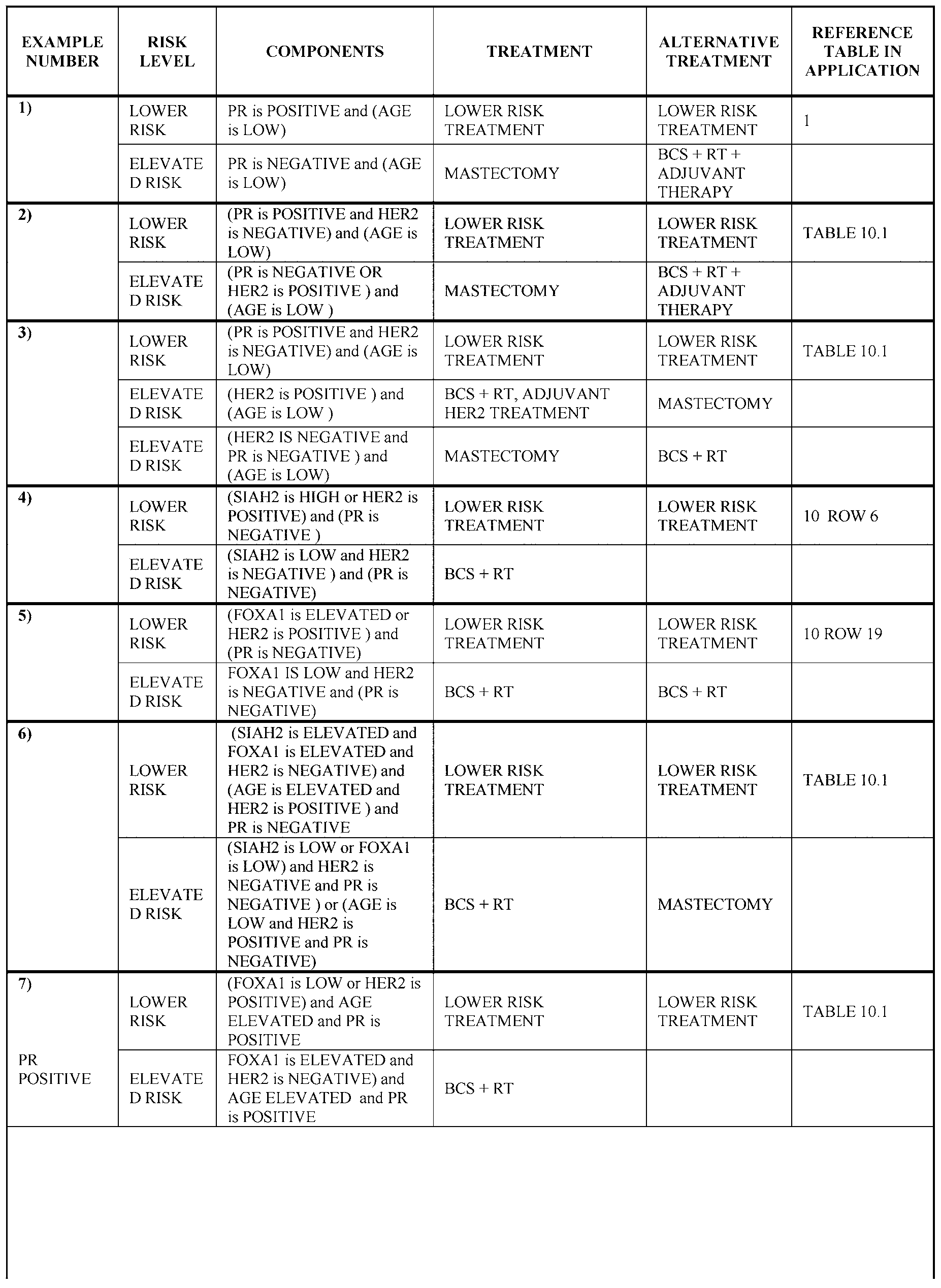


Table 14 (DCIS Risk)


[0347] In some embodiments, the marker signatures disclosed and described herein can be used to comprehensively treat a patient in a manner that appropriately reduces the risk of total recurrence or progression, including an invasive event or a DCIS event. For example, (a) the marker signatures in Table 13 can be used to identify a treatment that reduces the risk of an invasive event and (b) the marker signatures in Table 14 can be used to identify a treatment that reduces the risk of an DCIS event and/or identify treatment to reduce total risk according to Table 13 and Table 14. The same can be applied for Table 15, as a combined option.
[0348] The following is a non-exhaustive list of examples of treatment alternatives for patients based on their individual risk profiles as set forth in Tables 13-15.
New Alternatives for Low Risk Patients
[0349] Watchful Waiting Plus Frequent Observation. Lumpectomy-eligible patients found to be low risk by testing a biopsy sample in combination with the various methods and compositions disclosed and described herein could be designated for watchful waiting without additional surgery, regardless of margin status, or perhaps smaller or positive margins could be tolerated after lumpectomy surgery. More frequent observation could also be prescribed
in conjunction with watchful waiting. This would minimize treatment and the associated costs and adverse effects.
[0350] Breast-Conserving Surgery Only. By assessing risk in a biopsy or surgically-resected tissue sample with the various methods and compositions disclosed and described herein, patients identified as low-risk (particularly those at low risk for an invasive event) can be identified for whom definitive lumpectomy (with clear margins) is sufficient treatment without the need for adjuvant radiation therapy. This results in preservation of the breast; elimination of radiation-associated adverse effects, including damage to the breast; reduction in treatment costs, recovery time, and complication rates; and improves patient convenience. In addition, patients with previous thoracic radiation can safely receive lumpectomies, and most patients would retain their ability to receive future lumpectomies and/or radiation.
Breast-Conserving Surgery With Adjuvant Endocrine Therapy.
[0351] Low-risk patients (particularly those at low risk for an invasive event) that are hormone receptor positive can be treated with lumpectomy and adjuvant endocrine therapy. This results in preservation of the breast; elimination of radiation-associated adverse effects, including damage to the breast; reduces cost, recovery time, and complication rates; and improves patient convenience. In addition, patients with previous thoracic radiation can safely receive lumpectomies, and most patients would retain their ability to receive future lumpectomies and/or radiation.
New Alternatives for High Risk Patients
[0352] Breast-Conserving Surgery With Adjuvant Endocrine Therapy.
Lumpectomy-eligible patients found to have an elevated risk profile using the various methods and compositions disclosed and described herein can be treated more aggressively, such as by attempting to achieve wider margins during the initial lumpectomy and/or treating with adjuvant endocrine therapy after surgery in the case the patient is hormone receptor positive, typically assessed by ER or PR.
[0353] Older (not young) patients that have an elevated total recurrence risk but low invasive risk found to be hormone receptor positive, can be treated by lumpectomy and adjuvant
endocrine therapy whereas current standard of care would include lumpectomy and radiation therapy at a minimum.
[0354] Breast-Conserving Surgery With Adjuvant ErbB2 Targeted Therapy. Lumpectomy-eligible patients found to have an elevated risk profile (particularly those at high risk for an invasive event) using the various methods and compositions disclosed and described herein can be treated more aggressively, such as by attempting to achieve wider margins during the initial lumpectomy and/or treating with adjuvant ErbB2 targeted therapy (such as Herceptin) after surgery in the case where the patient is HER2 positive.
[0355] Mastectomy. Lumpectomy-eligible patients found to have a very high-risk profile from an initial biopsy using the various methods and compositions disclosed and described herein can be candidates for mastectomy with or without adjuvant radiation and/or hormone therapy (or even cytotoxic chemotherapy). The upgrades to more aggressive therapy would minimize the likelihood of potentially life-threatening recurrences and give patients peace of mind.
EXAMPLES
Example 1
[0356] Patients included in the noted studies are from population-based observational cohorts of women diagnosed with ductal carcinoma in situ (DCIS) without evidence of invasive breast cancer, who were treated with either breast-conserving surgery (BCS) or full mastectomy with or without radiation therapy (RT). The Uppsala University Hospital (UUH) patient set (Uppsala, Sweden) includes a total of 458 DCIS patients diagnosed between 1986 and 1994, of which 201 received BCS-only, 153 received BCS+RT, and 104 received mastectomy. At last follow-up, there were 47 recurrences each of the invasive and DCIS types in the UUH patient set. The University of Massachusetts Memorial Hospital (UMass) patient set (Worcester, Massachusetts) includes 296 patients with available tumor tissue diagnosed between 1999 and 2008, of which 90 received BCS-only and 206 received BCS+RT. Demographic, clinical, pathological, treatment, and long-term outcome data for the patients were derived from medical records and entered into a database.
[0357] Patient specimens used for the detection of the biomarkers were surgically removed breast tissues that were cut into small blocks and submerged in fixative. Following fixation, the blocks were dehydrated and then embedded in paraffin wax.
[0358] Ultrathin sections, approximately three to five micrometers in thickness, were then cut off the formalin-fixed paraffin-embedded (FFPE) tumor blocks using a microtome and floated in a water bath. The sections were carefully mounted onto glass microscope slides, ensuring that the tissue did not become folded or fragmented, which could interfere with the assays. The glass microscope slides contained a positively charged surface in order bind to the negatively charged tissue sections, although other methods of tissue binding, including adhesives, can also be compatible.
[0359] Wax removal and rehydration of the tissue sections was then carried out. These processes were done manually and with automated certain staining instruments. Rehydration was accomplished by passing the slides through a series of decreasingly concentrated alcohol solutions until a concentration of 0% is reached (pure water). Following wax removal and rehydration, the tissue sections were stained with hematoxylin and eosin (H&E) and for a variety of molecular markers using immunohistochemistry (IHC) and/or in situ hybridization (ISH) assays and then assessed by pathologists or histotechnologists, as described below.
[0360] All patients to be assessed have previously been diagnosed with DCIS. However, given interobserver variability in the diagnosis of in situ disease between pathologists, particularly in borderline intraductal neoplasia, a central review will be done to confirm DCIS using slide-mounted tissue sections stained with hematoxylin and eosin (H&E) or an equivalent histology stain. The assessment is done consistent with WHO classification of tumors of the breast (Lakhani SR. WHO classification of tumours of the breast. Lyon: International Agency for Research on Cancer, 2012, and Tavassoli FA and Devilee P, Tavassoli FA, Devilee P. Pathology and genetics of tumours of the breast and female genital organs. Lyon: IARC Press, 2003) - see definition of DCIS section. These references contain sample images and review characteristic features of DCIS and differential diagnosis with other breast disease entities, such as invasive breast cancer (including microinvasion defined as invasion≤1 mm), lobular carcinoma in situ (LCIS), in situ Paget's, atypical ductal hyperplasia (ADH), sclerosing adenosis, etc.).
Cases Suspected To Have Invasive (or Microinvasive) Carcinoma Based Upon Morphologic Features:
[0361] Normal breast ducts and lobules as well as intraductal epithelial proliferations are composed of two epithelial layers. Loss of the outer myoepithelial layer is the hallmark of infiltrating carcinoma of the breast. The outer myoepithelial layer is retained in all benign proliferative processes as well as ductal carcinoma in situ. Consequently identification of the presence or loss of myoepitheilium using antibodies to the myoepithelial-specific proteins can be helpful in distinguishing in situ from infiltrating carcinoma in circumstances where morphology may be equivocal ( alof AN et al., 2004, Barbareschi M et al., 2001).
[0362] If there is unequivocal morphologic evidence of invasion, including microinvasion, the patient is ineligible for the prognostic DCIS testing. If, upon morphologic examination of the tumor focus, there is a question of invasion or microinvasion, additional myoepithelial marker immunostudies (p63 and/or smooth muscle myosin heavy chain (SMMHC) immunostains) will be performed to examine the continuity of the myoepithelial cell layers and confirm and/or exclude the presence of (micro)invasive carcinoma.
IMAGES: P63 AND SMMHC IMMUNOSTAINS IN DISTINGUISHING
INVASIVE DUCTAL CARCINOMA FROM DCIS
[0363] FIG. 8 depicts a P63 immunostain showing loss of a myoepithelial investiture (arrows), confiming presence of invasive ductal carcinoma. Focus of DCIS shows retention of the outer myoepithelial layer in upper left corner of image.
[0364] FIG. 9 depicts a smooth muscle myosin heavy chain (SMMHC) immunostain showing loss of a myoepithelial investiture (arrows), consistent with invasive ductal carcinoma. Focus of DCIS shows retention of the outer myoepithelial layer (*).
Cases Suspected To Be Of Lobular Origin Based Upon Morphologic Features:
[0365] It has been demonstrated that in histologic settings where ductal and lobular neoplasia might be confused, particularly in the setting of in situ carcinoma, where there can be significant differences in patient management), loss of expressionof E-cadherin by immunohistochemistry can confhn the diagnosis of lobular carcinoma, even in the setting of non-classical morphologic findings (Acs G, Lawton TJ, Rebbeck TR, LiVolsi VA, Zhang PJ. Differential expression of E-cadherin in lobular and ductal neoplasms of the breast and its
biologic and diagnostic implications. Am J Clin Pathol 2001; 1 IS, 85-98). In lobular neoplasia, mutations in the E-cadherin gene result in loss of expresion of E-cadherin, a cell surface adhesion molecule present in normal breast epithelium and ductal carcinoma. The role of E- cadherin in homotypic cell-cell binding, loss of expression of this cell surface protein accounts for the characteristic non-cohesive growth pattern of lobular carcinoma.
[0366] If, upon examination of an intraductal epithelial proliferation, it is unclear whether the intraductal tumor is ductal or lobular in nature, an E-cadherin immunostain will be performed to confirm ductal or lobular differentiation. If lobular carcinoma in situ (LCIS) is confirmed histologically or by loss of e-cadherin by immunhisotochemistry, the patient is ineligible for further prognostic testing.
[0367] If there is no evidence of DCIS or invasive carcinoma, additional tissue blocks can be requested for morphologic review. FIG. 10 depicts an image of an e-Cadherin immunostain to distinguish LCIS From DCIS.
[0368] Loss of e-cadherin expression confirms the lobular nature of this intraductal epithelial proliferation. Adjacent normal ducts on left are e-cadherin-positive.
Criteria for exclusion:
• No DCIS identified
• Invasive or microinvasive carcinoma identified
• LCIS, not DCIS identified
• Tissue folded over in area of interest - not possible to score adequately SPECIMENS
[0369] Tumor sections (four-microns thick) were cut from archived formalin-fixed paraffin-embedded (FFPE) resected tissue blocks (or tissue microarrays [TMAs] manufactured from such blocks) and mounted onto glass microscope slides. The sections were stained for a variety of molecular markers using immunohistochemistry (IHC) and/or in situ hybridization (FISH) assays and then assessed by pathologists or histotechnologists.
MARKERS
[0370] For IHC assays (Table 16), the sections underwent antigen retrieval, incubations with primary antibodies and detection reagents (including 3,3'-diaminobenzidine
tetrahydrochloride [DAB]), and counter-staining with hematoxylin. Most IHC markers were scored by pathologists for percentage (0-100%) and intensity (0, absent; 1, weak; 2, moderate; 3, strong). Immunoscores (the product of percentage and intensity on a scale of 0-300) were calculated for FOXA1, and Allred scores were calculated for COX2. HER2 was scored using the standard 0/1+/2+/3+ HercepTest scoring system.
TABLE 16
LIST OF MARKERS, PRIMARY ANTIBODIES, AND SOME EMBODIMENTS OF SCORING THRESHOLDS AND CELLULAR COMPARTMENTS FOR IHC ASSAYS.

[0371] HER2 was also assessed by ISH in some samples, using a combination of HER2 and chromosome 17 probes via silver ISH (Ventana Medical Systems Inform HER2 kit) or fluorescence ISH (Abbott Molecular PathVysion HER2 kit). Amplification of HER2 genes was determined according to manufacturer instructions.
[0372] Kaplan-Meier survival analyses were used to estimate the proportions of patients who experienced first events (DCIS or invasive recurrence) after initial DCIS diagnosis/surgery. Hazard ratios (HR) were determined using Cox proportional hazards analysis. At the time of first events (DCIS or invasive), patients were censored for the other event type. Patients were also censored if an event was detected within 6 months of surgery or on the first post-surgery mammogram, because this was considered persistent, rather than recurrent, disease.
Assessment of Human Epidermal Growth Factor Receptor 2 (HER2/neu or CD340) Immunohistochemical Staining
[0373] HER2 immunohistochemistry is scored according to CAP-ASCO guidelines (Wolff AC et al., 2013) with modification for scoring on intraductal tumor cells (and not invasive tumor cells). In short, HER2 immunostaining results are scored as negative (either IHC 0 or IHC 1+ immunostaining signal), positive (IHC 3+ immunostaining signal), or equivocal (IHC 2+ immunostaining signal).
[0374] HER2 IHC 0-3+ are defined as follows:
• IHC 0 is defined by no staining observed or membrane staining that is incomplete and is faint/barely perceptible and within≤ 10% of the tumor cells
• IHC 1+ is defined by incomplete membrane staining that is faint/barely
perceptible and within > 10% of the tumor cells
• IHC 2+ is defined by circumferential membrane staining that is incomplete and/or weak/moderate (observed in a homogeneous and contiguous population) and within >10% of the invasive tumor cells or complete and circumferential membrane staining that is intense and within≤ 10% of the tumor cells
• IHC 3+ is defined by circumferential membrane staining that is complete, intense (observed in a homogeneous and contiguous population and within > 10% of the tumor cells; readily appreciated using a low power objective)
Criteria for exclusion:
• No DCIS identified or area of DCIS is not present on immunostained slide
• Area of DCIS is difficult to evaluate (due to tissue disruption/tissue folding etc.).
[0375] Exemplary HER2 staining and scoring is shown in FIGs. 1 lA-1 ID.
Assessment of Ki-67 Immunohistochemical Staining
[0376] i-67 immunohistochemistry is scored based upon the percentage of in situ tumor cells staining positive regardless of intensity. The percentage of in situ tumor cell staining positively will be recorded for all cases and reported as follows: 0%, 1%, 5%, 10%, 15%, 20%, 25%, 30%, 40%, 50%, 60%, 70%, 80%, 90%, 100%. All areas of the tissue section containing in situ carcinoma should be evaluated to arrive at this average percentage.
[0377] The intensity of staining should be recorded and reported as weak (1+), moderate (2+), or strong (3+); this measurement should represent an estimate of the average staining of the intensity of the positively stained tumor cells on the entire tissue section relative to the intensity of positive controls run with the same batch.
Criteria for exclusion:
• No DCIS identified or area of DCIS is not present on immunostained slide
• Area of DCIS is difficult to evaluate (due to tissue disruption/tissue folding etc.)
[0378] Exemplary K.i-67 staining is shown in FIG. 12.
Assessment of pl6-INK4A Immunohistochemical Staining
[0379] p16 is scored using a scoring system by combining the sum of a proportion score (PS) and average intensity score (IS). The pl6ink4a staining pattern is nuclear and cytoplasmic and often heterogeneous in nature. Nuclear and cytoplasmic staining will be assessed individually in DCIS and in the stroma around DCIS, nuclear and cytoplasmic staining of stromal cell will be assessed together. Only staining that is at least intermediate (2+) or greater (strong 3+) in intensity will be considered positive staining. Weak 1+ cytoplasmic and/or nuclear staining will be considered negative. The proportion of positive cells is calculated based upon the inclusion of all cells showing cytoplasmic/nuclear staining of intermediate (2+) and/or strong (3+) intensity. The proportion of positive cells should be estimated as closely as possible using the following average scores: 0%, 1%, 5%, 10%, 15%, 20%, 25%, 30%, 40%, 50%, 60%, 70%, 80%, 90%, 100%. The average intensity (either 2+ or 3+) will also be scored. At least two ducts with DCIS are required for scoring.
Criteria for exclusion:
• No DCIS identified
• Tissue folded over in area of interest - not possible to score adequately
Assessment of Progesterone Receptor (PR) Immunohistochemical Staining
[0380] PR immunohistochemistry is scored according to CAP-ASCO guidelines (Hammond ME et al., 2010). The percentage of in situ tumor cell staining positively will be recoreded and reported for all cases. All areas of the tissue section containing in situ carcinoma should be evaluated to arrive at this percentage. The intensity of staining should be recorded and
reported as weak (1+), moderate (2+), or strong (3+); this measurement should represent an estimate of the average staining of the intensity of the positively stained tumor cells on the entire tissue section relative to the intensity of positive controls run with the same batch.
[0381] DCIS exhibiting less than 10% of tumor cells staining for PR of any intensity should be considered negative based on data that such patients do not receive meaningful benefit from endocrine therapy. The sample should only be considered negative in the presence of appropriately stained extrinsic and internal controls. Any specimen lacking internal control elements (normal breast ductal epithelium) that is negative on PR assay should be repeated using another tumor block or another tumor specimen and reported as uninterpretable rather than as negative.
Criteria for exclusion:
• No DCIS identified or area of DCIS is not present on immunostained slide
• Area of DCIS is difficult to evaluate (due to tissue disruption/tissue folding etc..)
[0382] Exemplary PR staining is shown in FIGs. 13A-13C.
Assessment of SIAH2 Immunohistochemical Staining
[0383] SIAH2 is scored using a modified Allred scoring system by combining the sum of a proportion score (PS) and average intensity score (IS). The SIAH2 staining pattern is nuclear and often heterogeneous. At least two ducts with DCIS can be used for scoring. The proportion of positive cells is calculated based upon the inclusion of all cells showing nuclear staining of all intensities: weak, intermediate, and strong. The proportion of positive cells should be estimated as closely as possible using the following average scores: 0%, 5%, 10%, 15%, 20%, 25%, 30%, 40%, 50%, 60%, 70%, 80%, 90%, 100%.
Criteria for exclusion:
• No DCIS identified
• Tissue folded over in area of interest - not possible to score adequately
[0384] Additional examples of marker storing and staining are presented in FIGs. 1- 7, as outlined below.
[0385] For each of the 7 markers, microscope slide mounted tissue sections (approximately four micrometers in thickness), which were cut from patient tumor blocks that had been fixed in formalin and embedded in paraffin, were dewaxed and rehydrated.
[0386] FIG. 1 depicts the PR IHC assays (top, negative; bottom, positive) that were conducted on a UUH TMA with epitope retrieval done in 10 mM citrate buffer for 8 minutes in a microwave pressure cooker, followed by primary antibody incubation with rabbit monoclonal SP2 (Lab Vision) at a 1 : 1,500 dilution for 40 minutes, followed by detection with the EnVision+ detection system (Dako).
[0387] FIG. 2 depicts the HER2 IHC assays (top, negative; bottom, positive) that were conducted on a Biomax BR8011 TMA with epitope retrieval done in 0.05 M Tris (pH 10) in a steamer for 8 minutes in a microwave pressure cooker, followed by primary antibody incubation with rabbit monoclonal SP3 (Lab Vision) at a 1 :200 dilution for 40 minutes, followed by detection with the EnVision+ detection system (Dako).
[0388] FIG. 3 depicts the pl6/IN 4A IHC assays (top, negative; bottom, positive) that were conducted on a Biomax BR8011 TMA with epitope retrieval done in 0.05 M Tris/0.1 M EDTA (pH 9) buffer for 20 minutes in a steamer, followed by primary antibody incubation with mouse monoclonal E6H4 (MTM Laboratories) for 30 minutes, followed by detection with the EnVision+ detection system (Dako).
[0389] FIG. 4 depicts i-67 IHC assays (top, negative; bottom, positive) that were conducted on a Biomax BR8011 TMA with epitope retrieval done in Tris/EDTA (pH 9) buffer for 20 minutes in a steamer, followed by primary antibody incubation with mouse monoclonal MIB-1 (Dako) at a 1:500 dilution for 40 minutes, followed by detection with the EnVision+ detection system (Dako).
[0390] FIG. 5 depicts COX2 IHC assays (top, negative; bottom, positive) that were conducted on a Biomax BR8011 TMA with epitope retrieval done in 10 mM citrate buffer for 8 minutes in a microwave pressure cooker, followed by primary antibody incubation with rabbit monoclonal SP21 (Lab Vision) at a 1 :100 dilution for 30 minutes, followed by detection with the EnVision+ detection system (Dako).
[0391] FIG. 6 depicts SIAH2 IHC assays (top, negative, on a UUH TMA; bottom, positive, on a Biomax BR8011 TMA) that were conducted with epitope retrieval in EDTA buffer (pH 8) for 20 minutes in a steamer, followed by primary antibody incubation with mouse
monoclonal 24E6 (Dr. Amy Tang) at a 1:400 dilution, followed by detection with the UltraVision HRP detection system (Thermo Scientific).
[0392] FIG. 7 depicts FOXA1 IHC assays (top, negative; bottom, positive) that were conducted on a Biomax BR8011 TMA with epitope retrieval done in 10 mM citrate (pH 6) buffer for 20 minutes in a steamer, followed by primary antibody incubation with rabbit monoclonal SP88 (Spring Bioscience) at a 1 :100 dilution for 40 minutes, followed by detection with the EnVision+ detection system (Dako).
Example 2
[0393] FOXA1 and PR predict ipsilateral event risk and identify a group with strong radiation response in ductal carcinoma in situ (DCIS).
[0394] Identification of biomarkers in DCIS is useful to help guide treatment decisions, particularly for patients receiving breast-conserving surgery (BCS). The goal of this study was to assess FOXA1 levels in the context of PR status in primary DCIS to predict ipsilateral invasive and DCIS events, given the established roles of these endocrine signaling factors in breast cancer.
[0395] Patients included in this study (n=518) were women diagnosed with DCIS without evidence of invasive cancer treated with BCS, and for whom tumor tissue was evaluable for both PR and FOXA1. An Uppsala University Hospital (UUH) set was diagnosed in 1986- 2004 (117 treated and 122 not treated with adjuvant radiation therapy [RT]); and a University of Massachusetts Memorial Hospital (UMass) set was diagnosed in 1999-2008 (195 treated and 84 not treated with RT).
[0396] Tumors were immunohistochemically stained for PR and FOXA1 and scored by pathologists for percentage (0-100) and intensity (0-3), with the product being calculated for FOXA1 immunoscore (0-300). Patients were considered PR+ when≥10% of cells stained positively, and immunoscore thresholds of 100 and 270 were used to separate patients into FOXA1 low, intermediate, and high groups. Event rates were assessed for 10-year outcome using Kaplan-Meier survival analysis. Hazard ratios (HR) were determined using Cox proportional hazards analysis.
[0397] In non-RT-treated patients, neither FOXA1 nor PR were prognostic as independent factors for either invasive or DCIS event risk. However, in the PR+ subset, the invasive event rate increased with increasing FOXA1 (0%, 10%, and 13%, respectively,
HR=2.9/bin, p=0.046), while the invasive event rate was lower in the PR- subset with elevated FOXA1 levels (3% in FOXA1 int./high vs. 25% in FOXA1 low, HR=9.7, p=0.006). In contrast, the DCIS event rate increased in the PR- subset with increasing FOXA1 (3%, 15%, and 22%, respectively, HR=2.1/bin, p=0.040), while the DCIS event rate was lower in the PR+ subset with elevated FOXA1 levels (8% in FOXA1 int./high vs. 28% in FOXA1 low, HR=3.7, p=0.011).
[0398] In the full population, RT-treated patients (n=312) fared better than the non- irradiated (n=206) with invasive event rates of 6% and 10%, respectively. However, the event rate in patients with elevated marker-based invasive event risk based on PR and FOXA1 status (n=195) was reduced from 22% to 4% with RT. In contrast, the remainder of patients (n=323) showed no evident RT response. In a multivariate Cox analysis, the high-risk group (HR=5.0, p=0.005) and RT response within that group (HR=0.13, p=0.013) were significant when adjusted for clinicopathologic factors, but baseline RT response was not (p=0.29).
[0399] These data demonstrate a complex interaction between PR and FOXA1, in which the prognosis for invasive and DCIS events flips both within and between the event types. Thus, the biology that drives these events differs and, in order to predict both event types, risk models must include biomarkers in context. In addition, PR/FOXA1 identified a risk group with remarkably strong RT response with the remaining patients exhibiting no measurable response. See results in FIG. 40 and FIG. 41.
Example 3
[0400] Validation of a multi-marker test that predicts recurrence in patients diagnosed with ductal carcinoma in situ (DCIS) and treated with breast-conserving surgery (BCS).
[0401] DCIS is diagnosed in ~54,000 women/year in the US. Identifying the patients who are most likely to recur has been deemed one of the most important unmet needs in invasive breast cancer treatment. The goal of this study was to develop and blindly validate a multi- marker risk stratification test in DCIS patients treated with BCS.
[0402] A variety of clinical, pathological, immunohistochemical, and in situ hybridization data were derived from a set of patients diagnosed with DCIS (without evidence of invasive breast cancer) between 1986 and 2004 in Uppland and Vastmanland, Sweden— the Uppsala University Hospital (UUH) patient set. Separate models to predict DCIS and invasive event risk were developed using statistical pattern recognition and modeling methods on UUH patients treated with BCS in the absence of adjuvant therapy (n=200). In addition, an "overall"
risk model was created by combining the DCIS and invasive models. The risk models consist of algorithms that combine data on pl6/IN 4A, i-67, COX2, PR, HER2, FOXA1, SIAH2, CD31, patient age, necrosis, tumor size, palpability, and/or margin status, along with predefined thresholds that create low, intermediate, and high risk groups ("elevated" risk groups combine the intermediate and high risk groups).
[0403] The models were then tested blindly on a set of patients diagnosed with DCIS and treated with BCS from 1999 through July 2008 at the University of Massachusetts Memorial Hospital, Worcester, MA (UMass patient set). Molecular marker data was collected with CLIA- validated assays and Board-certified pathologists, and other data was collected from medical records. Testing was done according to a predefined statistical analysis plan in the UMass patients equivalent to those in the UUH patient training set—those with complete marker data and treated with BCS, and were either PR-negative or not treated with hormone therapy (n=155). Event rates were assessed for up to 10-year outcome using Kaplan-Meier survival analysis. Hazard ratios (HRs) were determined using Cox proportional hazards analysis.
[0404] The 10-year event rate in the full population was 10% overall (5% invasive plus 5% DCIS). For the invasive risk model, the low (n=53) and elevated (n=102) risk groups had 0% and 8% 10-year invasive event rates, respectively. For the DCIS risk model, the low (n=97) and elevated (n=58) risk groups had 1% and 13% 10-year DCIS event rates, respectively.
[0405] For the overall risk model (combined risk of DCIS or invasive events), the low (n=34), intermediate (n=51), and high (n=70) risk groups had 0%, 12%, and 15% overall 10- year event rates, respectively (HR=4.1/bin, p=0.0088). The overall risk model maintained significance when adjusted for nuclear grade, tumor size, patient age, necrosis, and margin status, while none of these clinicopathologic factors demonstrated significance in the presence of the model.
[0406] This study indicates that the present approach to risk stratification modeling can accurately identify patients at risk for DCIS or invasive events after a primary DCIS diagnosis.
Example 4
[0407] A risk stratification test identifies breast-conserving surgery (BCS)-treated ductal carcinoma in situ (DCIS) patients at high risk of recurrence despite receiving adjuvant radiation therapy (RT).
[0408] Adjuvant RT after BCS for DCIS reduces ipsilateral event rates (both DCIS and invasive) by approximately 50% relative to BCS alone. However, RT does not reduce the event rate down to the level achieved with mastectomy. We have developed a risk stratification test designed to assess risk of recurrence in patients with primary DCIS treated with BCS to help guide the decision to treat with adjuvant RT. The goal of the following analysis was to determine whether the test can identify patients who remain at high risk despite receiving RT.
[0409] Risk models to predict DCIS and invasive event risk were developed using statistical pattern recognition and modeling methods on a set of DCIS patients treated with BCS without radiation or adjuvant hormone therapy through Uppsala University Hospital (UUH) between 1986 and 2004 (n=200, UUH "training set"). In addition, an "overall" risk model was created by combining the DCIS and invasive models. The risk models include algorithms that combine data on pl6/IN 4A, i-67, COX2, PR, HER2, FOXA1, SIAH2, CD31, patient age, necrosis, tumor size, palpability, and/or margin status, along with predefined thresholds that create low, intermediate, and high risk categories ("elevated" risk groups combine the intermediate and high risk groups). The models were tested on a set of independent patients diagnosed with DCIS and treated with BCS and RT, and were either PR-negative or not treated with hormone therapy. The independent "testing set" patients (n=198) were treated through the University of Massachusetts Memorial Hospital, Worcester, MA in 1999 through July 2008 (UMass, n=l 18) and at UUH (n=80). Molecular marker data was collected with CLIA-validated assays and Board-certified pathologists, and other data was collected from medical records. Event rates were assessed using Kaplan-Meier survival analysis. Hazard ratios (HRs) were determined using Cox proportional hazards analysis.
[0410] The 12-year event rate in the full independent testing set population was 15% overall (9% invasive plus 6% DCIS). For the overall risk model (combined risk of DCIS or invasive events), the low (n=44), intermediate (n=69), and high (n=85) risk groups had 12-year event rates of 5%, 3%, and 32%, respectively (HR=4.5/bin, p=3.4E-5). For the invasive risk model, the low (n=80) and elevated (n=l 18) risk groups had 12-year event rates of 3% and 14%, respectively (HR=7.2, p=0.015). For the DCIS risk model, the low (n=l 13), intermediate (n=50), and high (n=35) risk groups had 12-year event rates of 2%, 4%, and 25%, respectively (HR=3.8/bin, p=0.00032).
[0411] Margin status by itself was significant for overall event risk (HR=3.3, p=0.0012) but lost significance in the presence of the overall risk model. The overall risk model maintained significance when adjusted for all clinicopathologic factors.
[0412] Our risk models can identify patients at risk for DCIS or invasive events after a primary DCIS diagnosis and treatment with BCS, despite their receiving RT. The models are the basis of a comprehensive multi-marker panel currently undergoing formal validation.
[0413] Overall ipsilateral event risk (combined invasive and DCIS events), UUH test and UMASS test (BCS with radiation and without adjuvant hormone therapy), Low solid, intermediate - -, and high risk .... patients n=44, n=69, and n=85. Low and intermediate (n=l 13) versus high risk (n=85) (HR= 7.4, p=2.6e-5). The results are presented in FIG. 14. In addition, invasive event risk is also shown. UUH test and UMASS test (BCS with radiation and without adjuvant hormone therapy). Low solid, intermediate and high risk patients n=80, n=118
(HR= 7.2, p=0.015). The results are presented in FIG. 15.
Example 5
[0414] A prognostic risk profile was evaluated in subjects independent of those used for test development. The test was evaluated in 183 subjects from a University of Massachusetts study whom were diagnosed with DCIS and treated with or without radiation therapy and without adjuvant hormone therapy (PR-positive subjects only). The test identified subjects (the risk profile was produced from the biomarkers: COX2, I67, P16, PR, HER2, FOXA1, SIAH2, and CD31, along with the clinicopathologic factors: tumor palpability, BCS margin status, patient age, and DCIS tumor necrosis. Subjects included in the independent assessment had minimum biomarker complete results for PR and HER2 and (FOXA1 or SIAH2, or K.I67 and either COX2 or PI 6). Subjects whom were PR positive and treated with adjuvant hormone therapy were excluded from assessment. Subjects from UUH whom were BCS treated without radiation (whom were used as a developmental cohort) were excluded from the independent assessment) at low risk of progression to either invasive breast cancer or a secondary DCIS event (0% 10 year observed risk). The test identified subjects at risk for either ipsilateral invasive or secondary DCIS events, and the test identified subjects at risk for both ipsilateral invasive and secondary DCIS events.
[0415] Subjects at low risk for either ipsilateral Invasive or secondary DCIS events after BCS treatment (top line) N= 45, 10 year Endpoint 100% (100-100); Subjects at elevated
risk for either ipsilateral Invasive or DCIS events after BCS treatment (middle line) N=97 (10 Year Endpoint 91% (82% - 100%); Subjects at elevated risk for both ipsilateral DCIS and Invasive events after BCS treatment, (bottom line) N=41 (78% (62% - 96%). Total N=183; HR/bin=4.4 (1.6-12) p(LR)=0.0021. (See FIG. 16)
Validation Summary: Uppsala University, Sweden (UUH) and University of Massachusetts USA (UMASS)
[0416] A prognostic risk profile was evaluated in subjects independent of those used for test development using both UUH and UMASS subjects. The test was evaluated in 311 subjects from either (a) the UUH study when treated with BCS with radiation and without adjuvant hormone therapy or (b) from the University of Massachusetts Study whom were diagnosed with DCIS and treated with or without radiation therapy and without adjuvant hormone therapy (PR-positive subjects only). The test identified subjects at low risk for either invasive or DCIS risk (0% 10 year observed risk). The test identified subjects at risk for either ipsilateral invasive or secondary DCIS events, and the test identified subjects at risk for both ipsilateral invasive and secondary DCIS events.
[0417] Results are shown in FIG. 17. Subjects at low risk for either ipsilateral Invasive or secondary DCIS events after BCS treatment (top line) N= 85, 10 year Endpoint 94% (88-100); Subjects at elevated risk for either ipsilateral Invasive or DCIS events after BCS treatment (middle line) N=161 (10 Year Endpoint 90% (84% - 96%); Subjects at elevated risk for both ipsilateral DCIS and Invasive events after BCS treatment, (bottom line) N=64 (73% (60% - 88%). Total N=311; HR/bin=2.7 (1.6-4.5) p(LR)=0.00019.
[0418] Details regarding FIG. 16 are shown in Table 17. Details regarding FIG. 17 are shown in Table 18.
TABLE 17


Same for 10+ year assessment
TABLE 18

Example 6
[0419] FIGs. 18-41 depict event occurrence over time for the markers/factors noted in the figures, for DCIS, invasive breast cancer, or both. The samples tested were DCIS samples taken from subjects. As can be observed from a review of the various graphs, the use of combined risk factors allows for one to predict the risk that a subsequent DCIS will occur, that invasive breast cancer will occur, or that both will occur.
[0420] In some embodiments, with the information from Example 6, a subject matching one of the noted risk profiles can receive the appropriate treatment (or lack of overly aggressive therapy) based upon the degree of risk of invasive breast cancer and/or DCIS recurrence.
Example 7
[0421] A human DCIS tissue sample is analyzed for PR, and either or both of: at least HER2 and SIAH2, or at least FOXA1. The results are compared with the matrix in Tables 9 and/or 11 to determine if the subject has an elevated risk of invasive breast cancer, and/or DCIS recurrence, or neither.
Example 8
[0422] A DCIS tissue sample is analyzed for a level of at least SIAH2 and FOXA1. The results are compared with the matrix in Tables 9 and/or 11 to determine if the subject has an elevated risk of invasive breast cancer, and/or DCIS recurrence, or neither.
Example 9
[0423] A DCIS sample from a subject having DCIS is analyzing for SIAH2, and for at least one of HER2, PR, FOXA1, or (in the alternative) analyzed for FOXA1 and PR. The results are compared with the matrix in Tables 9 and/or 11 to determine if the subject has an elevated risk of invasive breast cancer, DCIS recurrence, or neither.
Example 10
[0424] A DCIS sample from a subject is analyzed for a level of at least PR, and at least either analyzing the sample for at least HER2 and SIAH2, or (in the alternative) analyzing the sample for at least FOXA1. A prognosis is provided based upon at least PR, HER2 and SIAH2 or based upon at least PR and FOXA1. Depending upon the nature of the results, this indicates that the subject that provided the sample is at a high or elevated risk of invasive breast cancer (see, e.g., Tables 9 and/or 11).
Example 11
[0425] A DCIS sample from a subject is analyzed for a level of at least SIAH2 and FOXA1. A prognosis is provided with the subject having an elevated risk of an invasive breast cancer based upon the level of at least SIAH2 and FOXA1.
Example 12
[0426] A DCIS sample from a subject is analyzed for: a) PR, HER2, and SIAH2, or (in the alternative) b) PR and FOXA1. A prognosis is provided for the subject as having an
elevated risk of an invasive breast cancer event when at least one of: a) PR-, HER2-, and SIAH2- , b) PR+, FOXA1+, or c) PR+, FOXA1-, and Ki67+.
Example 13
[0427] A DCIS sample is analyzed for at least one of: a) SIAH2 and FOXA1, b) SIAH2 and at least one of i) PR and ii) HER2, c) SIAH2 and post-menopausal status; or (in the alternative) d) PR and FOXA1. A prognosis is provided that the subject has an elevated risk of a DCIS event when at least one of: a) i) SIAH2+ and FOXA1+, b) SIAH2+ and HER2+ or PR-; SIAH2+ and post-menopausal; or PR+ and FOXA1-, is present in the DCIS sample.
Example 14
[0428] A DCIS sample is analyzed for at least one of: a) PR-, HER2-, and SIAH2-, b) PR+, FOXA1+, or c) PR+, FOXA1-, and Ki67+. When the analysis indicates a high risk of invasive breast cancer (see, Tables 9 and/or 11) the subject is administered a therapy that is more aggressive than standard of care for DCIS.
Example IS
[0429] A DCIS sample is analyzed for at least one of: a) SIAH2+ and FOXA1+, b) SIAH2+ and HER2+ or PR-, c) SIAH2+ and post-menopausal status, or (in the alternative) d) PR+ and FOXA1-. When the analysis indicates a high likelihood of an invasive breast cancer, one administers to the subject a more aggressive therapy than standard of care for a single DCIS event.
Example 16
[0430] A subject that has a DCIS that is none of: a) PR-, HER2-, and SIAH2-, b) PR+, FOXA1+, or d) PR+, FOXA1-, and Ki67+ is administered a therapy that is less aggressive than standard of care for DCIS. Such a method allows for reduced surgeries and/or other therapies, where there is little to no risk involved for a subsequent DCIS and/or invasive breast cancer
Example 17
[0431] A subject having a DCIS that lacks a combination of markers as indicated in Tables 9 and/or 11 that would indicate an elevated risk of invasive breast cancer is not treated for
the DCIS. Such a method allows for reduced surgeries and/or other therapies, where there is little to no risk involved for a subsequent DCIS and/or invasive breast cancer
Example 18
[0432] A subject having a DCIS that lacks a combination of markers as indicated in Tables 9 and/or 11 that would indicate an elevated risk of DCIS recurrence is treated for the DCIS by a method that is less than standard of care for a subject having DCIS (without the disclosure provided herein regarding the present markers). Such a method allows for reduced surgeries and/or other therapies, where there is little to no risk involved for a subsequent DCIS and/or invasive breast cancer.
Example 19
[0433] A subject provides a sample that is analyzed for at least one of the combinations as outlined in any one of Tables 13-15 to provide a risk level for the subject. A corresponding treatment is then administered to the subject, as outlined in the appropriate row in Tables 13-15, based on the indicated risk level for the subject.
Claims
1. A method of analyzing a sample, the method comprising:
analyzing a human DCIS tissue sample for PR, and either or both of:
a) analyzing the sample for at least HER2 and SIAH2, or
b) analyzing the sample for at least FOXA 1.
2. The method of claim 1 , wherein if the sample is PR positive, further analyzing the sample for a level of COX2.
3. The method of claim 1, wherein if the sample is PR positive, then further analyzing the sample for Ki67, size, or both Ki67 and size.
4. The method of claim 1, further comprising analyzing the sample for p16 with i- 67 or pl6 with COX-2.
5. The method of claim 1, wherein analyzing the sample comprises determining an amount of a specified RNA in the sample.
6. The method of claim 1, wherein analyzing the sample comprises determining an amount of a specified protein in the sample.
7. The method of claim 6, wherein analyzing the sample further comprises determining an amount of a specified RNA in the sample.
8. A method of analyzing a sample, the method comprising:
analyzing a human DCIS tissue sample for a level of at least SIAH2 and FOXA1.
9. The method of claim 8, wherein the human DCIS tissue sample has been surgically removed from a patient and preserved.
10. The method of claim 8, wherein a level of SIAH2 and FOXA1 is at least one of: a RNA level, a DNA level, a protein level.
11. The method of claim 10, wherein the RNA level is determined by: an assay involving nucleic acid microarray, reverse transcriptase-polymerase chain reaction, in situ nucleic acid detection, or a next generation sequencing methods.
12. The method of claim 10, wherein the protein level is determined by immunohistochemistry, immunofluorescence, or mass spectrometry.
13. The method of claim 8, further comprising analyzing at least the following combinations:
a) PR, HER2, and SIAH2,
b) PR, FOXAl, and COX2
c) PR, FOXAl, and Ki67.
14. The method of claim 8, further comprising analyzing at least COX-2, Ki67, p16, PR and HER2.
15. A kit comprising:
a FOXA1 probe; and
a SIAH2 probe.
16. The kit of claim 15, further comprising:
a COX-2 probe;
a Ki67 probe;
a p16 probe;
a PR probe; and
a HER2 probe.
17. The kit of claim 16, wherein the probe is an isolated antibody.
18. The kit of claim 17, wherein the probe is a nucleic acid that selectively hybridizes to FOXA1, SIAH2, COX-2, Ki67, p16, PR or HER2 as appropriate.
19. An antibody composition comprising:
an isolated FOXA1 antibody; and
an isolated SIAH2 antibody.
20. The antibody composition of claim 19, further comprising:
an isolated COX-2 antibody;
an isolated Ki67 antibody;
an isolated p16 antibody;
an isolated PR antibody; and
an isolated HER2 antibody.
21. A method for prognosing a risk of an invasive breast cancer event in a subject, said method comprising:
providing a DCIS sample from a subject;
analyzing the DCIS sample for a level of at least PR, and at least either:
a) analyzing the sample for at least HER2 and SIAH2, or
b) analyzing the sample for at least FOXA1 ; and providing a prognosis based upon at least PR, HER2 and SIAH2 or based upon at least PR and FOXA1.
22. The method of claim 21, wherein if the sample is PR positive, further analyzing the sample for a level of COX2, wherein COX-2+ with at least FOXA1+ indicates a high risk of invasive breast cancer.
23. The method of claim 21, wherein if the sample is PR positive and there is a very high level of FOXA1 , there is a high risk of invasive breast cancer.
24. The method of claim 21, wherein if the sample is PR positive, then further analyzing the sample for Ki67, size, or both Ki67 and size.
25. The method of claim 21, wherein if the sample is PR positive, and wherein when FOXA1- further analyzing the sample for a level of Ki67, size, or a level of Ki67 and size, wherein Ki67+, a size larger than 5 mm of DCIS, or both, indicates an elevated risk of invasive breast cancer.
26. The method of claim 21, further comprising analyzing the sample for p16, COX- 2, and Ki67.
27. The method of claim 21 , wherein analysis of each marker is carried out in parallel with each other.
28. The method of claim 21, wherein analysis of each marker is carried out at overlapping times.
29. The method of claim 21, wherein PR analysis occurs first and any further analysis depends upon the result of the PR analysis.
30. A method for prognosing a risk of an invasive breast cancer event in a subject, said method comprising:
providing a DCIS sample from a subject;
analyzing the sample for a level of at least SIAH2 and FOXA1 ; and prognosing the subject as having an elevated risk of an invasive breast cancer based upon the level of at least SIAH2 and FOXA1.
31. A method for prognosing a risk of an invasive breast cancer event in a subject, said method comprising:
providing a DCIS sample from a subject;
analyzing the sample for:
a) PR, HER2, and SIAH2, or
b) PR and FOXAl; and
prognosing the subject as having an elevated risk of an invasive breast cancer event when at least one of:
a) PR-, HER2-, and SIAH2-,
b) PR+, FOXA1+, or
c) PR+, FOXA1-, and Ki67+.
32. The method of claim 31, further comprising the process of preparing a report regarding the risk associated with the human DCIS tissue sample.
33. The method of claim 32, wherein the report is a written report providing the risk of invasive breast cancer.
34. A method for treating a subject at risk of having an invasive breast cancer, the method comprising:
providing a subject having DCIS, wherein the subject has a DCIS that is at least one of:
a) PR-, HER2-, and SIAH2-,
b) PR+, FOXA1+, or
c) PR+, FOXA1-, and Ki67+; and
administering to the subject a therapy that is more aggressive than standard of care for DCIS.
35. A method of analyzing a sample, the method comprising:
providing a DCIS sample from a subject having DCIS;
1. analyzing the DCIS sample for SIAH2, and analyzing the DCIS sample for at least one of HER2, PR, FOXA1, or any combination thereof; or
2. analyzing the DCIS sample for FOXA1 and PR.
36. The method of claim 35, further comprising providing a report regarding a level of risk of a subsequent DCIS even.
37. The method of claim 35, further comprising recommending a treatment given a result from analyzing the DCIS sample for SIAH2 and at least one of HER2, PR, FOXAl, or any combination thereof.
38. The method of claim 35, wherein analyzing the DCIS sample comprises determining an amount of a specified RNA in the sample.
39. The method of claim 35, wherein analyzing the DCIS sample comprises determining an amount of a specified protein in the sample.
40. The method of claim 39, wherein analyzing the sample further comprises determining an amount of a specified RNA in the sample.
41. The method of claim 35, wherein the DCIS sample has been surgically removed from a patient and preserved.
42. The method of claim 35, wherein a level of SIAH2 and HER2, PR, FOXA1, or any combination thereof is at least one of: a RNA level, a DNA level, a protein level.
43. The method of claim 42, wherein the RNA level is determined by: an assay involving nucleic acid microarray, reverse transcriptase-polymerase chain reaction, in situ nucleic acid detection, or a next generation sequencing methods.
44. The method of claim 42, wherein the protein level is determined by: immunohistochemistry, immunofluorescence, or mass spectrometry
45. A method for prognosing a risk of a subsequent DCIS event in a subject, said method comprising:
providing a DCIS sample from a subject;
analyzing the sample for at least one of:
a) SIAH2 and FOXA1,
b) SIAH2 and at least one of i) PR and ii) HER2, or
c) SIAH2 and post-menopausal status; and
d) PR and FOXAl
prognosing the subject as having an elevated risk of a DCIS event when at least one of:
a) SIAH2+ and FOXAl+,
b) SIAH2+ and HER2+ or PR-,
SIAH2+ and post-menopausal, or
PR+ and FOXAl-,
is present in the DCIS sample.
46. The method of claim 45, further comprising the process of preparing a report regarding the risk associated with the human DCIS tissue sample.
47. The method of claim 46, wherein the report is a written report providing the risk of DCIS recurrence.
48. The method of claim 45, wherein analysis of each marker is carried out in parallel with each other.
49. The method of claim 45, wherein analysis of each marker is carried out at overlapping times.
50. The method of claim 45, wherein a level of SIAH2, HER2, PR, FOXA1, or any combination thereof is at least one of: a RNA level, a DNA level, a protein level.
51. The method of claim 45, wherein the RNA level is determined by: an assay involving nucleic acid microarray, reverse transcriptase-polymerase chain reaction, in situ nucleic acid detection, or a next generation sequencing methods52. The method of claim 45, wherein the protein level is determined by: immunohistochemistry, immunofluorescence, or mass spectrometry.
52. A method for treating a subject at risk of having a DCIS recurrence, the method comprising:
providing a subject having DCIS, wherein the subject has a DCIS that is at least one of:
a) i) SIAH2+ and FOXA1+
b) SIAH2+ and HER2+ or PR-;
c) SIAH2+ and post-menopausal status; or
d) PR+ and FOXAl-,
administering to the subject a more aggressive therapy than standard of care for a single DCIS event.
53. A method of detecting gene expression in a DCIS sample, comprising detecting expression of at least SIAH2 and FOXA1 in the DCIS sample obtained from a subject with
DCIS.
54. The method of claim 54, wherein expression of the at least at least SIAH2 and FOXA1 is measured by real time quantitative polymerase chain reaction or microarray analysis.
55. The method of claim 54, further comprising determining a prognosis of the subject with DCIS, wherein an at least a level of SIAH2 and FOXA1 relative to the non-DCIS control indicates that the subject has a poor prognosis or wherein no significant difference in the expression of SIAH2 and FOXA1 relative to a non-tumor control indicates that the subject has a good prognosis.
56. A solid support comprising probes or antibodies specific for at least SIAH2 and
FOXA1.
57. The solid support of claim 57, wherein the probes or antibodies consists essentially of probes or antibodies specific for the prediction of DCIS or invasive breast cancer in a subject who has DCIS.
58. The method of any one of claims 1-14 or 21-53, wherein the DCIS sample is obtained by surgical removal.
59. The method of any one of claims 14 or 21-53, wherein the DCIS sample is cut into one or more blocks.
60. The method of any one of claims 14 or 21-51, wherein the DCIS sample is further analyzed for COX-2.
61. The method of any one of claims 14 or 21-53, wherein the DCIS sample is further analyzed for p16.
62. The method of any one of claims 14 or 21-53, wherein the DCIS sample is analyzed for at least SIAH2, FOXA1, and PR.
63. The method of claim 63, wherein the DCIS sample is further analyzed for HER2.
64. The method of claim 64, wherein the DCIS sample is further analyzed for COX-2.
65. The method of claim 65, wherein the DCIS sample is further analyzed for Ki67.
66. The method of claim 66, wherein the DCIS sample is further analyzed for p16.
67. The method of any one of claims 14 or 21-53, wherein the DCIS lesion is further analyzed for grade, necrosis, size, margin status,
68. The method of any one of claims 14 or 21-53, further comprising prognosis of a risk by including age, menopausal status, mammographic density, tumor palpability of the subject.
69. The method of any one of claims 14 or 21-53, further comprising determining a risk of DCIS, invasive breast cancer, or both.
70. The method of any one of claims 14 or 21-53, further comprising providing a written report regarding a risk of DCIS, invasive breast cancer, or both.
71. A method for treating a subject having DCIS, the method comprising:
obtaining an analysis or prognosis from any of claims 1-14, 21-33, 35-52, or 54-
70; and
performing a therapy appropriate to reduce a risk of invasive breast cancer if the analysis or prognosis indicates a risk of invasive breast cancer, or
performing a therapy appropriate to reduce a risk of DCIS recurrence if the analysis or prognosis indicates a risk of DCIS recurrence.
72. The method of claim 72, wherein a therapy appropriate to reduce a risk of DCIS recurrence comprises at least one of surgical resection, radiation therapy, anti-hormone therapy.
73. The method of claim 72, wherein a therapy appropriate to reduce a risk of invasive breast cancer comprises at least one of mastectomy, targeted HERs therapy, receptor- targeted chemotherapy.
74. A method for treating a subject, the method comprising:
providing a subject having DCIS, wherein the subject has a DCIS that is none of: a) PR-, HER2-, and SIAH2-,
b) PR+, FOXA1+, or
d) PR+, FOXA1-, and Ki67+; and
administering to the subject a therapy that is less aggressive than standard of care for DCIS.
Priority Applications (3)
| Application Number | Priority Date | Filing Date | Title |
|---|---|---|---|
| EP15865163.8A EP3227687A4 (en) | 2014-12-05 | 2015-12-04 | Dcis recurrence and invasive breast cancer |
| US15/529,966 US11543411B2 (en) | 2014-12-05 | 2015-12-04 | DCIS recurrence and invasive breast cancer |
| US17/652,466 US20230136619A1 (en) | 2014-12-05 | 2022-02-24 | Dcis recurrence and invasive breast cancer |
Applications Claiming Priority (2)
| Application Number | Priority Date | Filing Date | Title |
|---|---|---|---|
| US201462088132P | 2014-12-05 | 2014-12-05 | |
| US62/088,132 | 2014-12-05 |
Related Child Applications (2)
| Application Number | Title | Priority Date | Filing Date |
|---|---|---|---|
| US15/529,966 A-371-Of-International US11543411B2 (en) | 2014-12-05 | 2015-12-04 | DCIS recurrence and invasive breast cancer |
| US17/652,466 Continuation US20230136619A1 (en) | 2014-12-05 | 2022-02-24 | Dcis recurrence and invasive breast cancer |
Publications (1)
| Publication Number | Publication Date |
|---|---|
| WO2016090323A1 true WO2016090323A1 (en) | 2016-06-09 |
Family
ID=56092570
Family Applications (1)
| Application Number | Title | Priority Date | Filing Date |
|---|---|---|---|
| PCT/US2015/064115 WO2016090323A1 (en) | 2014-12-05 | 2015-12-04 | Dcis recurrence and invasive breast cancer |
Country Status (3)
| Country | Link |
|---|---|
| US (2) | US11543411B2 (en) |
| EP (1) | EP3227687A4 (en) |
| WO (1) | WO2016090323A1 (en) |
Cited By (2)
| Publication number | Priority date | Publication date | Assignee | Title |
|---|---|---|---|---|
| WO2020056338A1 (en) * | 2018-09-14 | 2020-03-19 | Prelude Corporation | Method of selection for treatment of subjects at risk of invasive breast cancer |
| US11543411B2 (en) | 2014-12-05 | 2023-01-03 | Prelude Corporation | DCIS recurrence and invasive breast cancer |
Families Citing this family (3)
| Publication number | Priority date | Publication date | Assignee | Title |
|---|---|---|---|---|
| WO2015189264A1 (en) * | 2014-06-10 | 2015-12-17 | Ventana Medical Systems, Inc. | Predicting breast cancer recurrence directly from image features computed from digitized immunohistopathology tissue slides |
| EP4107278A1 (en) * | 2020-02-18 | 2022-12-28 | Mayo Foundation for Medical Education and Research | Methods and materials for treating cancer |
| WO2022226038A1 (en) * | 2021-04-22 | 2022-10-27 | The George Washington University | Compositions and methods for treatment of invasive cancers |
Citations (4)
| Publication number | Priority date | Publication date | Assignee | Title |
|---|---|---|---|---|
| US20060275844A1 (en) * | 2005-04-19 | 2006-12-07 | Linke Steven P | Diagnostic markers of breast cancer treatment and progression and methods of use thereof |
| US20070105133A1 (en) * | 2005-06-13 | 2007-05-10 | The Regents Of The University Of Michigan | Compositions and methods for treating and diagnosing cancer |
| US20120003639A1 (en) * | 2010-04-27 | 2012-01-05 | Prelude, Inc. | Cancer biomarkers and methods of use thereof |
| US20130317083A1 (en) * | 2012-05-04 | 2013-11-28 | Thomas Jefferson University | Non-coding transcripts for determination of cellular states |
Family Cites Families (137)
| Publication number | Priority date | Publication date | Assignee | Title |
|---|---|---|---|---|
| US4318846A (en) | 1979-09-07 | 1982-03-09 | Syva Company | Novel ether substituted fluorescein polyamino acid compounds as fluorescers and quenchers |
| US4458066A (en) | 1980-02-29 | 1984-07-03 | University Patents, Inc. | Process for preparing polynucleotides |
| US4415732A (en) | 1981-03-27 | 1983-11-15 | University Patents, Inc. | Phosphoramidite compounds and processes |
| FI76888C (en) | 1981-04-29 | 1988-12-12 | Ciba Geigy Ag | New agents and packaging for immunological analysis. |
| US4591570A (en) | 1983-02-02 | 1986-05-27 | Centocor, Inc. | Matrix of antibody-coated spots for determination of antigens |
| US4605735A (en) | 1983-02-14 | 1986-08-12 | Wakunaga Seiyaku Kabushiki Kaisha | Oligonucleotide derivatives |
| US4683195A (en) | 1986-01-30 | 1987-07-28 | Cetus Corporation | Process for amplifying, detecting, and/or-cloning nucleic acid sequences |
| US4683202A (en) | 1985-03-28 | 1987-07-28 | Cetus Corporation | Process for amplifying nucleic acid sequences |
| US4716849A (en) | 1985-05-31 | 1988-01-05 | Tracor Hydronautics, Inc. | Erosive-jet diver tool |
| US4757141A (en) | 1985-08-26 | 1988-07-12 | Applied Biosystems, Incorporated | Amino-derivatized phosphite and phosphate linking agents, phosphoramidite precursors, and useful conjugates thereof |
| GB8610410D0 (en) | 1986-04-29 | 1986-06-04 | Silent Channel Prod Ltd | Conveying apparatus |
| US4728472A (en) | 1986-05-06 | 1988-03-01 | E. I. Du Pont De Nemours And Company | Cellular fibers via soluble fluid injection |
| WO1988001058A1 (en) | 1986-08-06 | 1988-02-11 | Roger Philip Ekins | Determination of analyte concentration using two labelling markers |
| US5322770A (en) | 1989-12-22 | 1994-06-21 | Hoffman-Laroche Inc. | Reverse transcription with thermostable DNA polymerases - high temperature reverse transcription |
| US4889818A (en) | 1986-08-22 | 1989-12-26 | Cetus Corporation | Purified thermostable enzyme |
| US5202231A (en) | 1987-04-01 | 1993-04-13 | Drmanac Radoje T | Method of sequencing of genomes by hybridization of oligonucleotide probes |
| US5525464A (en) | 1987-04-01 | 1996-06-11 | Hyseq, Inc. | Method of sequencing by hybridization of oligonucleotide probes |
| JPS649474A (en) | 1987-07-01 | 1989-01-12 | Minolta Camera Kk | Copying device capable of simultaneous multicolor copying |
| US5639604A (en) | 1987-09-21 | 1997-06-17 | Gen-Probe Incorporated | Homogeneous protection assay |
| US5283174A (en) | 1987-09-21 | 1994-02-01 | Gen-Probe, Incorporated | Homogenous protection assay |
| US5585481A (en) | 1987-09-21 | 1996-12-17 | Gen-Probe Incorporated | Linking reagents for nucleotide probes |
| US6004745A (en) | 1987-09-21 | 1999-12-21 | Gen-Probe Incorporated | Hybridization protection assay |
| US5185439A (en) | 1987-10-05 | 1993-02-09 | Gen-Probe Incorporated | Acridinium ester labelling and purification of nucleotide probes |
| JP2650159B2 (en) | 1988-02-24 | 1997-09-03 | アクゾ・ノベル・エヌ・ベー | Nucleic acid amplification method |
| CA1340807C (en) | 1988-02-24 | 1999-11-02 | Lawrence T. Malek | Nucleic acid amplification process |
| GB8810400D0 (en) | 1988-05-03 | 1988-06-08 | Southern E | Analysing polynucleotide sequences |
| US5223409A (en) | 1988-09-02 | 1993-06-29 | Protein Engineering Corp. | Directed evolution of novel binding proteins |
| SE462408B (en) | 1988-11-10 | 1990-06-18 | Pharmacia Ab | OPTICAL BIOSENSOR SYSTEM USING SURFACE MONITORING RESONSE FOR THE DETECTION OF A SPECIFIC BIOMOLIC CYCLE, TO CALIBRATE THE SENSOR DEVICE AND TO CORRECT FOUND BASELINE OPERATION IN THE SYSTEM |
| SE462454B (en) | 1988-11-10 | 1990-06-25 | Pharmacia Ab | METHOD FOR USE IN BIOSENSORS |
| US6040138A (en) | 1995-09-15 | 2000-03-21 | Affymetrix, Inc. | Expression monitoring by hybridization to high density oligonucleotide arrays |
| US6919211B1 (en) | 1989-06-07 | 2005-07-19 | Affymetrix, Inc. | Polypeptide arrays |
| US5143854A (en) | 1989-06-07 | 1992-09-01 | Affymax Technologies N.V. | Large scale photolithographic solid phase synthesis of polypeptides and receptor binding screening thereof |
| US5800992A (en) | 1989-06-07 | 1998-09-01 | Fodor; Stephen P.A. | Method of detecting nucleic acids |
| US5547839A (en) | 1989-06-07 | 1996-08-20 | Affymax Technologies N.V. | Sequencing of surface immobilized polymers utilizing microflourescence detection |
| CA2020958C (en) | 1989-07-11 | 2005-01-11 | Daniel L. Kacian | Nucleic acid sequence amplification methods |
| US5683888A (en) | 1989-07-22 | 1997-11-04 | University Of Wales College Of Medicine | Modified bioluminescent proteins and their use |
| EP0430881A3 (en) | 1989-11-29 | 1991-10-23 | Ciba-Geigy Ag | Photochromic compounds, process for their preparation and their use |
| US5292658A (en) | 1989-12-29 | 1994-03-08 | University Of Georgia Research Foundation, Inc. Boyd Graduate Studies Research Center | Cloning and expressions of Renilla luciferase |
| US5091652A (en) | 1990-01-12 | 1992-02-25 | The Regents Of The University Of California | Laser excited confocal microscope fluorescence scanner and method |
| US5498538A (en) | 1990-02-15 | 1996-03-12 | The University Of North Carolina At Chapel Hill | Totally synthetic affinity reagents |
| US5288644A (en) | 1990-04-04 | 1994-02-22 | The Rockefeller University | Instrument and method for the sequencing of genome |
| US5849481A (en) | 1990-07-27 | 1998-12-15 | Chiron Corporation | Nucleic acid hybridization assays employing large comb-type branched polynucleotides |
| US5784162A (en) | 1993-08-18 | 1998-07-21 | Applied Spectral Imaging Ltd. | Spectral bio-imaging methods for biological research, medical diagnostics and therapy |
| DK0575319T3 (en) | 1991-03-11 | 2000-07-10 | Univ Georgia Res Found | Cloning and expression of Renilla luciferase |
| JP3672306B2 (en) | 1991-04-10 | 2005-07-20 | ザ スクリップス リサーチ インスティテュート | Heterodimeric receptor library using phagemids |
| IL103674A0 (en) | 1991-11-19 | 1993-04-04 | Houston Advanced Res Center | Method and apparatus for molecule detection |
| US5324633A (en) | 1991-11-22 | 1994-06-28 | Affymax Technologies N.V. | Method and apparatus for measuring binding affinity |
| GB9218482D0 (en) | 1992-09-01 | 1992-10-14 | Dixon Arthur E | Apparatus and method for scanning laser imaging of macroscopic samples |
| US5667988A (en) | 1992-01-27 | 1997-09-16 | The Scripps Research Institute | Methods for producing antibody libraries using universal or randomized immunoglobulin light chains |
| WO1993020236A1 (en) | 1992-04-03 | 1993-10-14 | Applied Biosystems, Inc. | Probe composition and method |
| DE69334095T2 (en) | 1992-07-17 | 2007-04-12 | Dana-Farber Cancer Institute, Boston | Method for intracellular binding of targeted molecules |
| US5503980A (en) | 1992-11-06 | 1996-04-02 | Trustees Of Boston University | Positional sequencing by hybridization |
| US5472762A (en) | 1993-02-22 | 1995-12-05 | E. I. Du Pont De Nemours And Company | Method and apparatus for making a pile article and the products thereof |
| US5418144A (en) | 1993-05-25 | 1995-05-23 | American Cyanamid Company | Spindle pole body screen for fungicides |
| SE501439C2 (en) | 1993-06-22 | 1995-02-13 | Pharmacia Lkb Biotech | Method and apparatus for analyzing polynucleotide sequences |
| US5858659A (en) | 1995-11-29 | 1999-01-12 | Affymetrix, Inc. | Polymorphism detection |
| CA2164498C (en) | 1993-07-09 | 2007-09-11 | Paul A. Price | Assay for ykl-40 as a marker for degradation of mammalian connective tissue matrices |
| US5851829A (en) | 1993-07-16 | 1998-12-22 | Dana-Farber Cancer Institute | Method of intracellular binding of target molecules |
| AU694745B2 (en) | 1993-09-10 | 1998-07-30 | Trustees Of Columbia University In The City Of New York, The | Uses of green fluorescent protein |
| US5470710A (en) | 1993-10-22 | 1995-11-28 | University Of Utah | Automated hybridization/imaging device for fluorescent multiplex DNA sequencing |
| US5538848A (en) | 1994-11-16 | 1996-07-23 | Applied Biosystems Division, Perkin-Elmer Corp. | Method for detecting nucleic acid amplification using self-quenching fluorescence probe |
| GB9401833D0 (en) | 1994-02-01 | 1994-03-30 | Isis Innovation | Method for discovering ligands |
| WO1995021191A1 (en) | 1994-02-04 | 1995-08-10 | William Ward | Bioluminescent indicator based upon the expression of a gene for a modified green-fluorescent protein |
| US5578832A (en) | 1994-09-02 | 1996-11-26 | Affymetrix, Inc. | Method and apparatus for imaging a sample on a device |
| US5631734A (en) | 1994-02-10 | 1997-05-20 | Affymetrix, Inc. | Method and apparatus for detection of fluorescently labeled materials |
| US5648211A (en) | 1994-04-18 | 1997-07-15 | Becton, Dickinson And Company | Strand displacement amplification using thermophilic enzymes |
| US5571639A (en) | 1994-05-24 | 1996-11-05 | Affymax Technologies N.V. | Computer-aided engineering system for design of sequence arrays and lithographic masks |
| US5516637A (en) | 1994-06-10 | 1996-05-14 | Dade International Inc. | Method involving display of protein binding pairs on the surface of bacterial pili and bacteriophage |
| US5807522A (en) | 1994-06-17 | 1998-09-15 | The Board Of Trustees Of The Leland Stanford Junior University | Methods for fabricating microarrays of biological samples |
| US5556752A (en) | 1994-10-24 | 1996-09-17 | Affymetrix, Inc. | Surface-bound, unimolecular, double-stranded DNA |
| WO1997027317A1 (en) | 1996-01-23 | 1997-07-31 | Affymetrix, Inc. | Nucleic acid analysis techniques |
| US6974666B1 (en) | 1994-10-21 | 2005-12-13 | Appymetric, Inc. | Methods of enzymatic discrimination enhancement and surface-bound double-stranded DNA |
| US5777079A (en) | 1994-11-10 | 1998-07-07 | The Regents Of The University Of California | Modified green fluorescent proteins |
| US5599695A (en) | 1995-02-27 | 1997-02-04 | Affymetrix, Inc. | Printing molecular library arrays using deprotection agents solely in the vapor phase |
| GB9507238D0 (en) | 1995-04-07 | 1995-05-31 | Isis Innovation | Detecting dna sequence variations |
| US5545531A (en) | 1995-06-07 | 1996-08-13 | Affymax Technologies N.V. | Methods for making a device for concurrently processing multiple biological chip assays |
| US5856174A (en) | 1995-06-29 | 1999-01-05 | Affymetrix, Inc. | Integrated nucleic acid diagnostic device |
| US5661028A (en) | 1995-09-29 | 1997-08-26 | Lockheed Martin Energy Systems, Inc. | Large scale DNA microsequencing device |
| US5981180A (en) | 1995-10-11 | 1999-11-09 | Luminex Corporation | Multiplexed analysis of clinical specimens apparatus and methods |
| US5736330A (en) | 1995-10-11 | 1998-04-07 | Luminex Corporation | Method and compositions for flow cytometric determination of DNA sequences |
| US5876146A (en) | 1995-10-27 | 1999-03-02 | General Electric Company | Apparatus and methods for repairing jet pump diffusers in a nuclear reactor |
| JP4073038B2 (en) | 1995-12-15 | 2008-04-09 | ジーイー・ヘルスケア・バイオサイエンス・コーポレイション | Thermostable DNA polymerase from Thermoanaerobacter thermohydrosulfurica and its mutant enzyme from which exonuclease activity has been removed |
| US5874304A (en) | 1996-01-18 | 1999-02-23 | University Of Florida Research Foundation, Inc. | Humanized green fluorescent protein genes and methods |
| US5804387A (en) | 1996-02-01 | 1998-09-08 | The Board Of Trustees Of The Leland Stanford Junior University | FACS-optimized mutants of the green fluorescent protein (GFP) |
| US5876995A (en) | 1996-02-06 | 1999-03-02 | Bryan; Bruce | Bioluminescent novelty items |
| WO1997029212A1 (en) | 1996-02-08 | 1997-08-14 | Affymetrix, Inc. | Chip-based speciation and phenotypic characterization of microorganisms |
| DE69717895T2 (en) | 1996-03-28 | 2003-11-13 | Nihon Mediphysics Co Ltd | Diagnostic agent for glycine metabolism |
| US6458530B1 (en) | 1996-04-04 | 2002-10-01 | Affymetrix Inc. | Selecting tag nucleic acids |
| US5786146A (en) | 1996-06-03 | 1998-07-28 | The Johns Hopkins University School Of Medicine | Method of detection of methylated nucleic acid using agents which modify unmethylated cytosine and distinguishing modified methylated and non-methylated nucleic acids |
| US5925558A (en) | 1996-07-16 | 1999-07-20 | The Regents Of The University Of California | Assays for protein kinases using fluorescent protein substrates |
| US5976796A (en) | 1996-10-04 | 1999-11-02 | Loma Linda University | Construction and expression of renilla luciferase and green fluorescent protein fusion genes |
| WO1998026277A2 (en) | 1996-12-12 | 1998-06-18 | Prolume, Ltd. | Apparatus and method for detecting and identifying infectious agents |
| NZ516848A (en) | 1997-06-20 | 2004-03-26 | Ciphergen Biosystems Inc | Retentate chromatography apparatus with applications in biology and medicine |
| CA2319828A1 (en) | 1998-01-29 | 1999-08-05 | Miller, Samuel | High density arrays for proteome analysis and methods and compositions therefor |
| ATE388224T1 (en) | 1998-03-27 | 2008-03-15 | Prolume Ltd | LUCIFERASE, GFP FLUORESCENT PROTEINS, CODING NUCLEIC ACID AND THEIR USE IN DIAGNOSTICS |
| DE69933369D1 (en) | 1998-04-03 | 2006-11-09 | Compound Therapeutics Inc | Adressierbare protein arrays |
| US6406921B1 (en) | 1998-07-14 | 2002-06-18 | Zyomyx, Incorporated | Protein arrays for high-throughput screening |
| US6576478B1 (en) | 1998-07-14 | 2003-06-10 | Zyomyx, Inc. | Microdevices for high-throughput screening of biomolecules |
| US6197599B1 (en) | 1998-07-30 | 2001-03-06 | Guorong Chin | Method to detect proteins |
| US6610484B1 (en) | 1999-01-26 | 2003-08-26 | Cytyc Health Corporation | Identifying material from a breast duct |
| US6320196B1 (en) | 1999-01-28 | 2001-11-20 | Agilent Technologies, Inc. | Multichannel high dynamic range scanner |
| US6355934B1 (en) | 1999-02-26 | 2002-03-12 | Packard Biochip Technologies | Imaging system for an optical scanner |
| AU773291B2 (en) | 1999-03-10 | 2004-05-20 | Government of The United States of America, as represented by The Secretary Department of Health & Human Services, The National Institutes of Health, The | UPA, a universal protein array system |
| AU4025300A (en) | 1999-03-24 | 2000-10-09 | Packard Bioscience Company | Continuous porous matrix arrays |
| EP1169649A2 (en) | 1999-04-15 | 2002-01-09 | The Board Of Trustees Of The Leland Stanford Junior University | Microarrays of polypeptides |
| US6242266B1 (en) | 1999-04-30 | 2001-06-05 | Agilent Technologies Inc. | Preparation of biopolymer arrays |
| US6323043B1 (en) | 1999-04-30 | 2001-11-27 | Agilent Technologies, Inc. | Fabricating biopolymer arrays |
| US6331393B1 (en) | 1999-05-14 | 2001-12-18 | University Of Southern California | Process for high-throughput DNA methylation analysis |
| US6180351B1 (en) | 1999-07-22 | 2001-01-30 | Agilent Technologies Inc. | Chemical array fabrication with identifier |
| KR20000071894A (en) | 1999-08-19 | 2000-12-05 | 김선영 | Multipurpose diagnostic systems using protein chips |
| EP2308974A3 (en) | 1999-10-14 | 2011-08-10 | Clontech Laboratories Inc. | Anthozoa derived chromo/fluoroproteins and methods for using the same |
| US6232072B1 (en) | 1999-10-15 | 2001-05-15 | Agilent Technologies, Inc. | Biopolymer array inspection |
| US6171797B1 (en) | 1999-10-20 | 2001-01-09 | Agilent Technologies Inc. | Methods of making polymeric arrays |
| GB9928787D0 (en) | 1999-12-03 | 2000-02-02 | Medical Res Council | Direct screening method |
| US7236838B2 (en) | 2000-08-29 | 2007-06-26 | Matsushita Electric Industrial Co., Ltd. | Signal processing apparatus, signal processing method, program and recording medium |
| DE10063112A1 (en) | 2000-12-18 | 2002-06-20 | Bayer Ag | Method for increasing the clinical specificity in the detection of tumors and their precursors by simultaneous measurement of at least two different molecular markers |
| US7217510B2 (en) | 2001-06-26 | 2007-05-15 | Isis Pharmaceuticals, Inc. | Methods for providing bacterial bioagent characterizing information |
| WO2003055439A2 (en) | 2001-07-18 | 2003-07-10 | The Regents Of The University Of California | Her2/neu target antigen and use of same to stimulate an immune response |
| JP2005509420A (en) | 2001-11-13 | 2005-04-14 | クローンテック ラボラトリーズ インク. | Novel chromophores / fluorescent chromophores and their use |
| US6797393B2 (en) | 2001-11-30 | 2004-09-28 | Eastman Kodak Company | Method for making biochip substrate |
| CA2478850C (en) | 2002-03-13 | 2018-02-27 | Genomic Health, Inc. | Gene expression profiling in biopsied tumor tissues |
| CA2489360C (en) | 2002-06-26 | 2013-01-29 | Cold Spring Harbor Laboratory | Methods and compositions for determining methylation profiles |
| US7011949B2 (en) | 2002-09-30 | 2006-03-14 | Agilent Technologies, Inc. | Methods and compositions for producing labeled probe nucleic acids for use in array based comparative genomic hybridization applications |
| US7881873B2 (en) | 2003-04-29 | 2011-02-01 | The Jackson Laboratory | Systems and methods for statistical genomic DNA based analysis and evaluation |
| ES2432025T3 (en) | 2003-10-09 | 2013-11-29 | Epigenomics Ag | Improved conversion of DNA with bisulfite |
| EP1721159B1 (en) | 2004-02-20 | 2014-12-10 | Janssen Diagnostics, LLC | Breast cancer prognostics |
| CA2580795A1 (en) | 2004-09-22 | 2006-04-06 | Tripath Imaging, Inc. | Methods and compositions for evaluating breast cancer prognosis |
| US7718365B2 (en) | 2005-07-09 | 2010-05-18 | Agilent Technologies, Inc. | Microarray analysis of RNA |
| WO2007035684A2 (en) | 2005-09-16 | 2007-03-29 | Primera Biosystems, Inc. | Method for quantitative detection of short rna molecules |
| AU2013205531B2 (en) | 2006-07-14 | 2016-08-04 | The Regents Of The University Of California | Cancer biomarkers and methods of use thereof |
| EP2041574B1 (en) | 2006-07-14 | 2016-11-16 | The Regents of The University of California | Cancer biomarkers and methods of use threof |
| WO2008032876A1 (en) | 2006-09-15 | 2008-03-20 | Tokai University | Preventive or remedy for er-negative and her2-negative breast cancer and method of screening the same |
| US20100068717A1 (en) * | 2007-01-30 | 2010-03-18 | Indiana University Research And Technology Corporation | Cancer therapy prognosis |
| WO2010025928A1 (en) | 2008-09-04 | 2010-03-11 | Mtm Laboratories Ag | Method for prediction of the progression risk of tumors |
| AU2011248632B2 (en) | 2010-04-27 | 2015-07-02 | Prelude Corporation | Cancer biomarkers and methods of use thereof |
| US11543411B2 (en) | 2014-12-05 | 2023-01-03 | Prelude Corporation | DCIS recurrence and invasive breast cancer |
| JP2022500504A (en) | 2018-09-14 | 2022-01-04 | プレリュード コーポレーションPrelude Corporation | How to choose treatment for subjects at risk of invasive breast cancer |
-
2015
- 2015-12-04 US US15/529,966 patent/US11543411B2/en active Active
- 2015-12-04 EP EP15865163.8A patent/EP3227687A4/en active Pending
- 2015-12-04 WO PCT/US2015/064115 patent/WO2016090323A1/en active Application Filing
-
2022
- 2022-02-24 US US17/652,466 patent/US20230136619A1/en active Pending
Patent Citations (4)
| Publication number | Priority date | Publication date | Assignee | Title |
|---|---|---|---|---|
| US20060275844A1 (en) * | 2005-04-19 | 2006-12-07 | Linke Steven P | Diagnostic markers of breast cancer treatment and progression and methods of use thereof |
| US20070105133A1 (en) * | 2005-06-13 | 2007-05-10 | The Regents Of The University Of Michigan | Compositions and methods for treating and diagnosing cancer |
| US20120003639A1 (en) * | 2010-04-27 | 2012-01-05 | Prelude, Inc. | Cancer biomarkers and methods of use thereof |
| US20130317083A1 (en) * | 2012-05-04 | 2013-11-28 | Thomas Jefferson University | Non-coding transcripts for determination of cellular states |
Non-Patent Citations (6)
Cited By (4)
| Publication number | Priority date | Publication date | Assignee | Title |
|---|---|---|---|---|
| US11543411B2 (en) | 2014-12-05 | 2023-01-03 | Prelude Corporation | DCIS recurrence and invasive breast cancer |
| WO2020056338A1 (en) * | 2018-09-14 | 2020-03-19 | Prelude Corporation | Method of selection for treatment of subjects at risk of invasive breast cancer |
| EP3850368A4 (en) * | 2018-09-14 | 2022-11-23 | Prelude Corporation | Method of selection for treatment of subjects at risk of invasive breast cancer |
| US11821900B2 (en) | 2018-09-14 | 2023-11-21 | Prelude Corporation | Method of selection for treatment of subjects at risk of invasive breast cancer |
Also Published As
| Publication number | Publication date |
|---|---|
| US20230136619A1 (en) | 2023-05-04 |
| US11543411B2 (en) | 2023-01-03 |
| EP3227687A4 (en) | 2018-10-24 |
| US20170350895A1 (en) | 2017-12-07 |
| EP3227687A1 (en) | 2017-10-11 |
Similar Documents
| Publication | Publication Date | Title |
|---|---|---|
| US20230136619A1 (en) | Dcis recurrence and invasive breast cancer | |
| Tan et al. | Thyroid transcription factor-1 expression prevalence and its clinical implications in non-small cell lung cancer: a high-throughput tissue microarray and immunohistochemistry study | |
| JP5747419B2 (en) | Methods and compositions for detecting lethal cells and uses thereof | |
| US11821900B2 (en) | Method of selection for treatment of subjects at risk of invasive breast cancer | |
| KR101690249B1 (en) | Methods for diagnosing cancer and determining the overall survival and disease-free survival of cancer patients | |
| EP2329261B1 (en) | Prostate cancer biomarker | |
| Yang et al. | Expression pattern of androgen receptor and AR-V7 in androgen-deprivation therapy–naïve salivary duct carcinomas | |
| US20120004136A1 (en) | Msh2 and adjuvant cisplatin-based chemotherapy in non-small-cell lung cancer | |
| JP2014519818A (en) | Predictive biomarkers for prostate cancer | |
| US20110059452A1 (en) | Methods of screening for gastric cancer | |
| JP6675716B2 (en) | Prognostic expression formula and prognosis estimation method for lung adenocarcinoma using immune factors as indices | |
| US9702875B2 (en) | Expression of isoform 202 of ERCC1 for predicting response to cancer chemotherapy | |
| US20110144047A1 (en) | Combined method for predicting the response to an anti-cancer therapy | |
| JP2020201278A (en) | Pd-ecgf as biomarker of cancer | |
| US20140120543A1 (en) | Expression of isoform 202 of ercc1 for predicting response to cancer chemotherapy | |
| WO2007148095A2 (en) | Methods for determining a prognosis of colorectal cancer | |
| US7358060B2 (en) | Method for monitoring and prognosis of disease course of gastrointestinal tumors | |
| CN114705863B (en) | Application of SSH3 protein in cardiac adenocarcinoma prognosis prediction | |
| JP2011512147A (en) | BRCA1 mRNA expression predicts survival in bladder cancer patients treated with neoadjuvant chemotherapy with cisplatin | |
| JP2009192527A (en) | Cancer marker and method for inspecting cancer cell | |
| Van et al. | Association of BRAF V600E immunoexpression with clinicopathological variant groups in papillary thyroid carcinoma | |
| EP3052938B1 (en) | Methods and products for the diagnosis and prognosis of ovarian tumor malignancy | |
| CN117092342A (en) | Application of CHD4 protein in prognosis prediction of esophageal squamous carcinoma |
Legal Events
| Date | Code | Title | Description |
|---|---|---|---|
| 121 | Ep: the epo has been informed by wipo that ep was designated in this application |
Ref document number: 15865163 Country of ref document: EP Kind code of ref document: A1 |
|
| NENP | Non-entry into the national phase |
Ref country code: DE |
|
| REEP | Request for entry into the european phase |
Ref document number: 2015865163 Country of ref document: EP |