WO2016081584A1 - Process for preparing cell-binding agent-cytotoxic agent conjugates - Google Patents
Process for preparing cell-binding agent-cytotoxic agent conjugates Download PDFInfo
- Publication number
- WO2016081584A1 WO2016081584A1 PCT/US2015/061310 US2015061310W WO2016081584A1 WO 2016081584 A1 WO2016081584 A1 WO 2016081584A1 US 2015061310 W US2015061310 W US 2015061310W WO 2016081584 A1 WO2016081584 A1 WO 2016081584A1
- Authority
- WO
- WIPO (PCT)
- Prior art keywords
- hours
- antibody
- cell
- reaction
- agent
- Prior art date
Links
- 229940127089 cytotoxic agent Drugs 0.000 title claims abstract description 81
- 239000002254 cytotoxic agent Substances 0.000 title claims abstract description 81
- 238000004519 manufacturing process Methods 0.000 title claims abstract description 7
- 230000027455 binding Effects 0.000 title description 24
- 238000000034 method Methods 0.000 claims abstract description 122
- 230000008569 process Effects 0.000 claims abstract description 106
- 239000011230 binding agent Substances 0.000 claims abstract description 89
- 231100000599 cytotoxic agent Toxicity 0.000 claims abstract description 73
- 239000000203 mixture Substances 0.000 claims abstract description 54
- 230000001588 bifunctional effect Effects 0.000 claims abstract description 51
- 239000003431 cross linking reagent Substances 0.000 claims abstract description 51
- 239000007853 buffer solution Substances 0.000 claims abstract description 36
- 239000000243 solution Substances 0.000 claims abstract description 32
- 239000006172 buffering agent Substances 0.000 claims abstract description 28
- 239000000872 buffer Substances 0.000 claims abstract description 19
- 231100000433 cytotoxic Toxicity 0.000 claims abstract description 17
- 230000001472 cytotoxic effect Effects 0.000 claims abstract description 17
- 150000003839 salts Chemical class 0.000 claims abstract description 12
- 238000006243 chemical reaction Methods 0.000 claims description 69
- 239000003960 organic solvent Substances 0.000 claims description 26
- OWXMKDGYPWMGEB-UHFFFAOYSA-N HEPPS Chemical compound OCCN1CCN(CCCS(O)(=O)=O)CC1 OWXMKDGYPWMGEB-UHFFFAOYSA-N 0.000 claims description 18
- 238000010791 quenching Methods 0.000 claims description 18
- FAPWRFPIFSIZLT-UHFFFAOYSA-M Sodium chloride Chemical compound [Na+].[Cl-] FAPWRFPIFSIZLT-UHFFFAOYSA-M 0.000 claims description 16
- 238000013467 fragmentation Methods 0.000 claims description 15
- 238000006062 fragmentation reaction Methods 0.000 claims description 15
- 230000000171 quenching effect Effects 0.000 claims description 14
- 230000000274 adsorptive effect Effects 0.000 claims description 10
- 238000004587 chromatography analysis Methods 0.000 claims description 9
- 238000009295 crossflow filtration Methods 0.000 claims description 9
- JKMHFZQWWAIEOD-UHFFFAOYSA-N 2-[4-(2-hydroxyethyl)piperazin-1-yl]ethanesulfonic acid Chemical compound OCC[NH+]1CCN(CCS([O-])(=O)=O)CC1 JKMHFZQWWAIEOD-UHFFFAOYSA-N 0.000 claims description 8
- JOCBASBOOFNAJA-UHFFFAOYSA-N N-tris(hydroxymethyl)methyl-2-aminoethanesulfonic acid Chemical compound OCC(CO)(CO)NCCS(O)(=O)=O JOCBASBOOFNAJA-UHFFFAOYSA-N 0.000 claims description 8
- 239000003795 chemical substances by application Substances 0.000 claims description 8
- 239000011780 sodium chloride Substances 0.000 claims description 8
- 230000003432 anti-folate effect Effects 0.000 claims description 7
- 229940127074 antifolate Drugs 0.000 claims description 7
- 239000004052 folic acid antagonist Substances 0.000 claims description 7
- 102000005962 receptors Human genes 0.000 claims description 6
- 108020003175 receptors Proteins 0.000 claims description 6
- SXGZJKUKBWWHRA-UHFFFAOYSA-N 2-(N-morpholiniumyl)ethanesulfonate Chemical compound [O-]S(=O)(=O)CC[NH+]1CCOCC1 SXGZJKUKBWWHRA-UHFFFAOYSA-N 0.000 claims description 4
- LVQFQZZGTZFUNF-UHFFFAOYSA-N 2-hydroxy-3-[4-(2-hydroxy-3-sulfonatopropyl)piperazine-1,4-diium-1-yl]propane-1-sulfonate Chemical compound OS(=O)(=O)CC(O)CN1CCN(CC(O)CS(O)(=O)=O)CC1 LVQFQZZGTZFUNF-UHFFFAOYSA-N 0.000 claims description 4
- 239000007995 HEPES buffer Substances 0.000 claims description 4
- GIZQLVPDAOBAFN-UHFFFAOYSA-N HEPPSO Chemical compound OCCN1CCN(CC(O)CS(O)(=O)=O)CC1 GIZQLVPDAOBAFN-UHFFFAOYSA-N 0.000 claims description 4
- 239000008351 acetate buffer Substances 0.000 claims description 4
- 239000007979 citrate buffer Substances 0.000 claims description 4
- 108010044426 integrins Proteins 0.000 claims description 4
- 102000006495 integrins Human genes 0.000 claims description 4
- 239000008363 phosphate buffer Substances 0.000 claims description 4
- YCLWMUYXEGEIGD-UHFFFAOYSA-M sodium;2-hydroxy-3-[4-(2-hydroxyethyl)piperazin-1-yl]propane-1-sulfonate Chemical compound [Na+].OCCN1CCN(CC(O)CS([O-])(=O)=O)CC1 YCLWMUYXEGEIGD-UHFFFAOYSA-M 0.000 claims description 4
- 239000008362 succinate buffer Substances 0.000 claims description 4
- 229960000575 trastuzumab Drugs 0.000 claims description 4
- 229950002903 bivatuzumab Drugs 0.000 claims description 3
- 238000001914 filtration Methods 0.000 claims description 3
- 238000001556 precipitation Methods 0.000 claims description 3
- 229960004641 rituximab Drugs 0.000 claims description 3
- 229950008684 sibrotuzumab Drugs 0.000 claims description 3
- HSXUNHYXJWDLDK-UHFFFAOYSA-N 2-hydroxypropane-1-sulfonic acid Chemical compound CC(O)CS(O)(=O)=O HSXUNHYXJWDLDK-UHFFFAOYSA-N 0.000 claims 1
- 238000005377 adsorption chromatography Methods 0.000 claims 1
- 230000008685 targeting Effects 0.000 claims 1
- 210000004027 cell Anatomy 0.000 description 107
- 239000000562 conjugate Substances 0.000 description 51
- 239000000427 antigen Substances 0.000 description 40
- 102000036639 antigens Human genes 0.000 description 39
- 108091007433 antigens Proteins 0.000 description 38
- 239000011347 resin Substances 0.000 description 25
- 229920005989 resin Polymers 0.000 description 25
- 125000004432 carbon atom Chemical group C* 0.000 description 24
- 125000003342 alkenyl group Chemical group 0.000 description 20
- 239000012634 fragment Substances 0.000 description 20
- FXHOOIRPVKKKFG-UHFFFAOYSA-N N,N-Dimethylacetamide Chemical compound CN(C)C(C)=O FXHOOIRPVKKKFG-UHFFFAOYSA-N 0.000 description 19
- 206010028980 Neoplasm Diseases 0.000 description 18
- 125000002496 methyl group Chemical group [H]C([H])([H])* 0.000 description 17
- 150000001875 compounds Chemical class 0.000 description 16
- 125000001997 phenyl group Chemical group [H]C1=C([H])C([H])=C(*)C([H])=C1[H] 0.000 description 14
- 238000000746 purification Methods 0.000 description 14
- 125000003275 alpha amino acid group Chemical group 0.000 description 12
- 108010021625 Immunoglobulin Fragments Proteins 0.000 description 11
- 102000008394 Immunoglobulin Fragments Human genes 0.000 description 11
- 125000006615 aromatic heterocyclic group Chemical group 0.000 description 11
- 230000021615 conjugation Effects 0.000 description 11
- 125000000217 alkyl group Chemical group 0.000 description 10
- 201000011510 cancer Diseases 0.000 description 10
- 102000004169 proteins and genes Human genes 0.000 description 10
- 108090000623 proteins and genes Proteins 0.000 description 10
- QWPXBEHQFHACTK-KZVYIGENSA-N (10e,12e)-86-chloro-12,14,4-trihydroxy-85,14-dimethoxy-33,2,7,10-tetramethyl-15,16-dihydro-14h-7-aza-1(6,4)-oxazina-3(2,3)-oxirana-8(1,3)-benzenacyclotetradecaphane-10,12-dien-6-one Chemical compound CN1C(=O)CC(O)C2(C)OC2C(C)C(OC(=O)N2)CC2(O)C(OC)\C=C\C=C(C)\CC2=CC(OC)=C(Cl)C1=C2 QWPXBEHQFHACTK-KZVYIGENSA-N 0.000 description 9
- QTBSBXVTEAMEQO-UHFFFAOYSA-N Acetic acid Chemical compound CC(O)=O QTBSBXVTEAMEQO-UHFFFAOYSA-N 0.000 description 9
- 229920002684 Sepharose Polymers 0.000 description 8
- 210000004408 hybridoma Anatomy 0.000 description 8
- -1 G-50 Chemical compound 0.000 description 7
- 125000002816 methylsulfanyl group Chemical group [H]C([H])([H])S[*] 0.000 description 7
- 150000003573 thiols Chemical class 0.000 description 7
- 210000003719 b-lymphocyte Anatomy 0.000 description 6
- 239000003153 chemical reaction reagent Substances 0.000 description 6
- 238000005516 engineering process Methods 0.000 description 6
- 108090000765 processed proteins & peptides Proteins 0.000 description 6
- 239000011541 reaction mixture Substances 0.000 description 6
- 108010047041 Complementarity Determining Regions Proteins 0.000 description 5
- KCXVZYZYPLLWCC-UHFFFAOYSA-N EDTA Chemical compound OC(=O)CN(CC(O)=O)CCN(CC(O)=O)CC(O)=O KCXVZYZYPLLWCC-UHFFFAOYSA-N 0.000 description 5
- 108010017213 Granulocyte-Macrophage Colony-Stimulating Factor Proteins 0.000 description 5
- 102100039620 Granulocyte-macrophage colony-stimulating factor Human genes 0.000 description 5
- 108010002350 Interleukin-2 Proteins 0.000 description 5
- 102000000588 Interleukin-2 Human genes 0.000 description 5
- QWPXBEHQFHACTK-UHFFFAOYSA-N Maytansinol Natural products CN1C(=O)CC(O)C2(C)OC2C(C)C(OC(=O)N2)CC2(O)C(OC)C=CC=C(C)CC2=CC(OC)=C(Cl)C1=C2 QWPXBEHQFHACTK-UHFFFAOYSA-N 0.000 description 5
- 241001529936 Murinae Species 0.000 description 5
- 108010073929 Vascular Endothelial Growth Factor A Proteins 0.000 description 5
- 102000005789 Vascular Endothelial Growth Factors Human genes 0.000 description 5
- 108010019530 Vascular Endothelial Growth Factors Proteins 0.000 description 5
- 229940049595 antibody-drug conjugate Drugs 0.000 description 5
- 239000003102 growth factor Substances 0.000 description 5
- 238000011065 in-situ storage Methods 0.000 description 5
- 229940126619 mouse monoclonal antibody Drugs 0.000 description 5
- 238000002360 preparation method Methods 0.000 description 5
- VBEQCZHXXJYVRD-GACYYNSASA-N uroanthelone Chemical compound C([C@@H](C(=O)N[C@H](C(=O)N[C@@H](CS)C(=O)N[C@@H](CC(N)=O)C(=O)N[C@@H](CS)C(=O)N[C@H](C(=O)N[C@@H]([C@@H](C)CC)C(=O)NCC(=O)N[C@@H](CC=1C=CC(O)=CC=1)C(=O)N[C@@H](CO)C(=O)NCC(=O)N[C@@H](CC(O)=O)C(=O)N[C@@H](CCCNC(N)=N)C(=O)N[C@@H](CS)C(=O)N[C@@H](CCC(N)=O)C(=O)N[C@@H]([C@@H](C)O)C(=O)N[C@@H](CCCNC(N)=N)C(=O)N[C@@H](CC(O)=O)C(=O)N[C@@H](CC(C)C)C(=O)N[C@@H](CCCNC(N)=N)C(=O)N[C@@H](CC=1C2=CC=CC=C2NC=1)C(=O)N[C@@H](CC=1C2=CC=CC=C2NC=1)C(=O)N[C@@H](CCC(O)=O)C(=O)N[C@@H](CC(C)C)C(=O)N[C@@H](CCCNC(N)=N)C(O)=O)C(C)C)[C@@H](C)O)NC(=O)[C@H](CO)NC(=O)[C@H](CC(O)=O)NC(=O)[C@H](CC(C)C)NC(=O)[C@H](CO)NC(=O)[C@H](CCC(O)=O)NC(=O)[C@@H](NC(=O)[C@H](CC=1NC=NC=1)NC(=O)[C@H](CCSC)NC(=O)[C@H](CS)NC(=O)[C@@H](NC(=O)CNC(=O)CNC(=O)[C@H](CC(N)=O)NC(=O)[C@H](CC(C)C)NC(=O)[C@H](CS)NC(=O)[C@H](CC=1C=CC(O)=CC=1)NC(=O)CNC(=O)[C@H](CC(O)=O)NC(=O)[C@H](CC=1C=CC(O)=CC=1)NC(=O)[C@H](CO)NC(=O)[C@H](CO)NC(=O)[C@H]1N(CCC1)C(=O)[C@H](CS)NC(=O)CNC(=O)[C@H]1N(CCC1)C(=O)[C@H](CC=1C=CC(O)=CC=1)NC(=O)[C@H](CO)NC(=O)[C@@H](N)CC(N)=O)C(C)C)[C@@H](C)CC)C1=CC=C(O)C=C1 VBEQCZHXXJYVRD-GACYYNSASA-N 0.000 description 5
- XLYOFNOQVPJJNP-UHFFFAOYSA-N water Substances O XLYOFNOQVPJJNP-UHFFFAOYSA-N 0.000 description 5
- 208000031261 Acute myeloid leukaemia Diseases 0.000 description 4
- 102000007644 Colony-Stimulating Factors Human genes 0.000 description 4
- 108010071942 Colony-Stimulating Factors Proteins 0.000 description 4
- 108060003951 Immunoglobulin Proteins 0.000 description 4
- 102000004877 Insulin Human genes 0.000 description 4
- 108090001061 Insulin Proteins 0.000 description 4
- 241001465754 Metazoa Species 0.000 description 4
- 241000699666 Mus <mouse, genus> Species 0.000 description 4
- 208000033776 Myeloid Acute Leukemia Diseases 0.000 description 4
- ZDZOTLJHXYCWBA-VCVYQWHSSA-N N-debenzoyl-N-(tert-butoxycarbonyl)-10-deacetyltaxol Chemical compound O([C@H]1[C@H]2[C@@](C([C@H](O)C3=C(C)[C@@H](OC(=O)[C@H](O)[C@@H](NC(=O)OC(C)(C)C)C=4C=CC=CC=4)C[C@]1(O)C3(C)C)=O)(C)[C@@H](O)C[C@H]1OC[C@]12OC(=O)C)C(=O)C1=CC=CC=C1 ZDZOTLJHXYCWBA-VCVYQWHSSA-N 0.000 description 4
- 102100029268 Neurotrophin-3 Human genes 0.000 description 4
- 229930012538 Paclitaxel Natural products 0.000 description 4
- 239000000611 antibody drug conjugate Substances 0.000 description 4
- 239000012539 chromatography resin Substances 0.000 description 4
- 229940047120 colony stimulating factors Drugs 0.000 description 4
- OVBPIULPVIDEAO-LBPRGKRZSA-N folic acid Chemical compound C=1N=C2NC(N)=NC(=O)C2=NC=1CNC1=CC=C(C(=O)N[C@@H](CCC(O)=O)C(O)=O)C=C1 OVBPIULPVIDEAO-LBPRGKRZSA-N 0.000 description 4
- 229910052588 hydroxylapatite Inorganic materials 0.000 description 4
- 238000001597 immobilized metal affinity chromatography Methods 0.000 description 4
- 102000018358 immunoglobulin Human genes 0.000 description 4
- NOESYZHRGYRDHS-UHFFFAOYSA-N insulin Chemical compound N1C(=O)C(NC(=O)C(CCC(N)=O)NC(=O)C(CCC(O)=O)NC(=O)C(C(C)C)NC(=O)C(NC(=O)CN)C(C)CC)CSSCC(C(NC(CO)C(=O)NC(CC(C)C)C(=O)NC(CC=2C=CC(O)=CC=2)C(=O)NC(CCC(N)=O)C(=O)NC(CC(C)C)C(=O)NC(CCC(O)=O)C(=O)NC(CC(N)=O)C(=O)NC(CC=2C=CC(O)=CC=2)C(=O)NC(CSSCC(NC(=O)C(C(C)C)NC(=O)C(CC(C)C)NC(=O)C(CC=2C=CC(O)=CC=2)NC(=O)C(CC(C)C)NC(=O)C(C)NC(=O)C(CCC(O)=O)NC(=O)C(C(C)C)NC(=O)C(CC(C)C)NC(=O)C(CC=2NC=NC=2)NC(=O)C(CO)NC(=O)CNC2=O)C(=O)NCC(=O)NC(CCC(O)=O)C(=O)NC(CCCNC(N)=N)C(=O)NCC(=O)NC(CC=3C=CC=CC=3)C(=O)NC(CC=3C=CC=CC=3)C(=O)NC(CC=3C=CC(O)=CC=3)C(=O)NC(C(C)O)C(=O)N3C(CCC3)C(=O)NC(CCCCN)C(=O)NC(C)C(O)=O)C(=O)NC(CC(N)=O)C(O)=O)=O)NC(=O)C(C(C)CC)NC(=O)C(CO)NC(=O)C(C(C)O)NC(=O)C1CSSCC2NC(=O)C(CC(C)C)NC(=O)C(NC(=O)C(CCC(N)=O)NC(=O)C(CC(N)=O)NC(=O)C(NC(=O)C(N)CC=1C=CC=CC=1)C(C)C)CC1=CN=CN1 NOESYZHRGYRDHS-UHFFFAOYSA-N 0.000 description 4
- ANZJBCHSOXCCRQ-FKUXLPTCSA-N mertansine Chemical compound CO[C@@H]([C@@]1(O)C[C@H](OC(=O)N1)[C@@H](C)[C@@H]1O[C@@]1(C)[C@@H](OC(=O)[C@H](C)N(C)C(=O)CCS)CC(=O)N1C)\C=C\C=C(C)\CC2=CC(OC)=C(Cl)C1=C2 ANZJBCHSOXCCRQ-FKUXLPTCSA-N 0.000 description 4
- 229960005558 mertansine Drugs 0.000 description 4
- 229960001592 paclitaxel Drugs 0.000 description 4
- XYJRXVWERLGGKC-UHFFFAOYSA-D pentacalcium;hydroxide;triphosphate Chemical compound [OH-].[Ca+2].[Ca+2].[Ca+2].[Ca+2].[Ca+2].[O-]P([O-])([O-])=O.[O-]P([O-])([O-])=O.[O-]P([O-])([O-])=O XYJRXVWERLGGKC-UHFFFAOYSA-D 0.000 description 4
- 230000002265 prevention Effects 0.000 description 4
- 230000009467 reduction Effects 0.000 description 4
- 238000000926 separation method Methods 0.000 description 4
- 241000894007 species Species 0.000 description 4
- 125000003107 substituted aryl group Chemical group 0.000 description 4
- RCINICONZNJXQF-MZXODVADSA-N taxol Chemical compound O([C@@H]1[C@@]2(C[C@@H](C(C)=C(C2(C)C)[C@H](C([C@]2(C)[C@@H](O)C[C@H]3OC[C@]3([C@H]21)OC(C)=O)=O)OC(=O)C)OC(=O)[C@H](O)[C@@H](NC(=O)C=1C=CC=CC=1)C=1C=CC=CC=1)O)C(=O)C1=CC=CC=C1 RCINICONZNJXQF-MZXODVADSA-N 0.000 description 4
- 102100024222 B-lymphocyte antigen CD19 Human genes 0.000 description 3
- 102100035361 Cerebellar degeneration-related protein 2 Human genes 0.000 description 3
- 102000001301 EGF receptor Human genes 0.000 description 3
- 108060006698 EGF receptor Proteins 0.000 description 3
- 238000002965 ELISA Methods 0.000 description 3
- 102100037241 Endoglin Human genes 0.000 description 3
- 102400001368 Epidermal growth factor Human genes 0.000 description 3
- 101800003838 Epidermal growth factor Proteins 0.000 description 3
- 102100023600 Fibroblast growth factor receptor 2 Human genes 0.000 description 3
- 101710182389 Fibroblast growth factor receptor 2 Proteins 0.000 description 3
- 102100041003 Glutamate carboxypeptidase 2 Human genes 0.000 description 3
- 108010017080 Granulocyte Colony-Stimulating Factor Proteins 0.000 description 3
- 102000004269 Granulocyte Colony-Stimulating Factor Human genes 0.000 description 3
- 101000980825 Homo sapiens B-lymphocyte antigen CD19 Proteins 0.000 description 3
- 101000737796 Homo sapiens Cerebellar degeneration-related protein 2 Proteins 0.000 description 3
- 101000892862 Homo sapiens Glutamate carboxypeptidase 2 Proteins 0.000 description 3
- 101001136592 Homo sapiens Prostate stem cell antigen Proteins 0.000 description 3
- 101000834948 Homo sapiens Tomoregulin-2 Proteins 0.000 description 3
- 108090000723 Insulin-Like Growth Factor I Proteins 0.000 description 3
- 102000004218 Insulin-Like Growth Factor I Human genes 0.000 description 3
- 102000014150 Interferons Human genes 0.000 description 3
- 108010050904 Interferons Proteins 0.000 description 3
- 108010046938 Macrophage Colony-Stimulating Factor Proteins 0.000 description 3
- 102100028123 Macrophage colony-stimulating factor 1 Human genes 0.000 description 3
- 239000000637 Melanocyte-Stimulating Hormone Substances 0.000 description 3
- 108010007013 Melanocyte-Stimulating Hormones Proteins 0.000 description 3
- 241000699670 Mus sp. Species 0.000 description 3
- 108010025020 Nerve Growth Factor Proteins 0.000 description 3
- 108090000099 Neurotrophin-4 Proteins 0.000 description 3
- 102100033857 Neurotrophin-4 Human genes 0.000 description 3
- 229920001213 Polysorbate 20 Polymers 0.000 description 3
- 102100036735 Prostate stem cell antigen Human genes 0.000 description 3
- 208000000236 Prostatic Neoplasms Diseases 0.000 description 3
- 108020004511 Recombinant DNA Proteins 0.000 description 3
- 102000005157 Somatostatin Human genes 0.000 description 3
- 108010056088 Somatostatin Proteins 0.000 description 3
- 241000187747 Streptomyces Species 0.000 description 3
- 229940123237 Taxane Drugs 0.000 description 3
- 102100026160 Tomoregulin-2 Human genes 0.000 description 3
- 102400001320 Transforming growth factor alpha Human genes 0.000 description 3
- 101800004564 Transforming growth factor alpha Proteins 0.000 description 3
- 239000002253 acid Substances 0.000 description 3
- 125000002252 acyl group Chemical group 0.000 description 3
- 239000007864 aqueous solution Substances 0.000 description 3
- 125000003118 aryl group Chemical group 0.000 description 3
- 239000006227 byproduct Substances 0.000 description 3
- 229910052799 carbon Inorganic materials 0.000 description 3
- 229960003668 docetaxel Drugs 0.000 description 3
- 229940116977 epidermal growth factor Drugs 0.000 description 3
- 235000019152 folic acid Nutrition 0.000 description 3
- 239000011724 folic acid Substances 0.000 description 3
- 229940088597 hormone Drugs 0.000 description 3
- 239000005556 hormone Substances 0.000 description 3
- 230000002209 hydrophobic effect Effects 0.000 description 3
- 125000002887 hydroxy group Chemical group [H]O* 0.000 description 3
- 125000004029 hydroxymethyl group Chemical group [H]OC([H])([H])* 0.000 description 3
- 238000011031 large-scale manufacturing process Methods 0.000 description 3
- 239000003446 ligand Substances 0.000 description 3
- 238000002156 mixing Methods 0.000 description 3
- 230000004048 modification Effects 0.000 description 3
- 238000012986 modification Methods 0.000 description 3
- 239000012038 nucleophile Substances 0.000 description 3
- 229920001481 poly(stearyl methacrylate) Polymers 0.000 description 3
- 239000000256 polyoxyethylene sorbitan monolaurate Substances 0.000 description 3
- 235000010486 polyoxyethylene sorbitan monolaurate Nutrition 0.000 description 3
- 229920001184 polypeptide Polymers 0.000 description 3
- 102000004196 processed proteins & peptides Human genes 0.000 description 3
- 238000003127 radioimmunoassay Methods 0.000 description 3
- 238000001542 size-exclusion chromatography Methods 0.000 description 3
- NHXLMOGPVYXJNR-ATOGVRKGSA-N somatostatin Chemical compound C([C@H]1C(=O)N[C@H](C(N[C@@H](CO)C(=O)N[C@@H](CSSC[C@@H](C(=O)N[C@@H](CCCCN)C(=O)N[C@@H](CC(N)=O)C(=O)N[C@@H](CC=2C=CC=CC=2)C(=O)N[C@@H](CC=2C=CC=CC=2)C(=O)N[C@@H](CC=2C3=CC=CC=C3NC=2)C(=O)N[C@@H](CCCCN)C(=O)N[C@H](C(=O)N1)[C@@H](C)O)NC(=O)CNC(=O)[C@H](C)N)C(O)=O)=O)[C@H](O)C)C1=CC=CC=C1 NHXLMOGPVYXJNR-ATOGVRKGSA-N 0.000 description 3
- 229960000553 somatostatin Drugs 0.000 description 3
- 125000001424 substituent group Chemical group 0.000 description 3
- 210000004881 tumor cell Anatomy 0.000 description 3
- JSHOVKSMJRQOGY-UHFFFAOYSA-N (2,5-dioxopyrrolidin-1-yl) 4-(pyridin-2-yldisulfanyl)butanoate Chemical compound O=C1CCC(=O)N1OC(=O)CCCSSC1=CC=CC=N1 JSHOVKSMJRQOGY-UHFFFAOYSA-N 0.000 description 2
- FUHCFUVCWLZEDQ-UHFFFAOYSA-N 1-(2,5-dioxopyrrolidin-1-yl)oxy-1-oxo-4-(pyridin-2-yldisulfanyl)butane-2-sulfonic acid Chemical compound O=C1CCC(=O)N1OC(=O)C(S(=O)(=O)O)CCSSC1=CC=CC=N1 FUHCFUVCWLZEDQ-UHFFFAOYSA-N 0.000 description 2
- IUTPJBLLJJNPAJ-UHFFFAOYSA-N 3-(2,5-dioxopyrrol-1-yl)propanoic acid Chemical compound OC(=O)CCN1C(=O)C=CC1=O IUTPJBLLJJNPAJ-UHFFFAOYSA-N 0.000 description 2
- NCPQROHLJFARLL-UHFFFAOYSA-N 4-(2,5-dioxopyrrol-1-yl)butanoic acid Chemical compound OC(=O)CCCN1C(=O)C=CC1=O NCPQROHLJFARLL-UHFFFAOYSA-N 0.000 description 2
- 102100031585 ADP-ribosyl cyclase/cyclic ADP-ribose hydrolase 1 Human genes 0.000 description 2
- QTBSBXVTEAMEQO-UHFFFAOYSA-M Acetate Chemical compound CC([O-])=O QTBSBXVTEAMEQO-UHFFFAOYSA-M 0.000 description 2
- 208000024893 Acute lymphoblastic leukemia Diseases 0.000 description 2
- 208000014697 Acute lymphocytic leukaemia Diseases 0.000 description 2
- 102100036526 Anoctamin-7 Human genes 0.000 description 2
- 102100022005 B-lymphocyte antigen CD20 Human genes 0.000 description 2
- 102000007350 Bone Morphogenetic Proteins Human genes 0.000 description 2
- 108010007726 Bone Morphogenetic Proteins Proteins 0.000 description 2
- 102100025221 CD70 antigen Human genes 0.000 description 2
- 102000000844 Cell Surface Receptors Human genes 0.000 description 2
- 108010001857 Cell Surface Receptors Proteins 0.000 description 2
- 208000001333 Colorectal Neoplasms Diseases 0.000 description 2
- 102100039498 Cytotoxic T-lymphocyte protein 4 Human genes 0.000 description 2
- 101150029707 ERBB2 gene Proteins 0.000 description 2
- 108010036395 Endoglin Proteins 0.000 description 2
- 102000050554 Eph Family Receptors Human genes 0.000 description 2
- 108091008815 Eph receptors Proteins 0.000 description 2
- 108010055196 EphA2 Receptor Proteins 0.000 description 2
- 102100030340 Ephrin type-A receptor 2 Human genes 0.000 description 2
- 101150021185 FGF gene Proteins 0.000 description 2
- 108091008794 FGF receptors Proteins 0.000 description 2
- 102000044168 Fibroblast Growth Factor Receptor Human genes 0.000 description 2
- 102100027844 Fibroblast growth factor receptor 4 Human genes 0.000 description 2
- 102000010451 Folate receptor alpha Human genes 0.000 description 2
- 108050001931 Folate receptor alpha Proteins 0.000 description 2
- 208000009329 Graft vs Host Disease Diseases 0.000 description 2
- 241000282412 Homo Species 0.000 description 2
- 101000777636 Homo sapiens ADP-ribosyl cyclase/cyclic ADP-ribose hydrolase 1 Proteins 0.000 description 2
- 101000928370 Homo sapiens Anoctamin-7 Proteins 0.000 description 2
- 101000897405 Homo sapiens B-lymphocyte antigen CD20 Proteins 0.000 description 2
- 101000917148 Homo sapiens Fibroblast growth factor receptor 3 Proteins 0.000 description 2
- 101000917134 Homo sapiens Fibroblast growth factor receptor 4 Proteins 0.000 description 2
- 101001023230 Homo sapiens Folate receptor alpha Proteins 0.000 description 2
- 101000777628 Homo sapiens Leukocyte antigen CD37 Proteins 0.000 description 2
- 101000628547 Homo sapiens Metalloreductase STEAP1 Proteins 0.000 description 2
- 101000934338 Homo sapiens Myeloid cell surface antigen CD33 Proteins 0.000 description 2
- 101000581981 Homo sapiens Neural cell adhesion molecule 1 Proteins 0.000 description 2
- 101000721757 Homo sapiens Olfactory receptor 51E2 Proteins 0.000 description 2
- 101000874179 Homo sapiens Syndecan-1 Proteins 0.000 description 2
- 108010054477 Immunoglobulin Fab Fragments Proteins 0.000 description 2
- 102000001706 Immunoglobulin Fab Fragments Human genes 0.000 description 2
- 108090001117 Insulin-Like Growth Factor II Proteins 0.000 description 2
- 102000048143 Insulin-Like Growth Factor II Human genes 0.000 description 2
- 102000006992 Interferon-alpha Human genes 0.000 description 2
- 108010047761 Interferon-alpha Proteins 0.000 description 2
- 102000003996 Interferon-beta Human genes 0.000 description 2
- 108090000467 Interferon-beta Proteins 0.000 description 2
- 102000008070 Interferon-gamma Human genes 0.000 description 2
- 108010074328 Interferon-gamma Proteins 0.000 description 2
- 108010002386 Interleukin-3 Proteins 0.000 description 2
- 108090000978 Interleukin-4 Proteins 0.000 description 2
- 108090001005 Interleukin-6 Proteins 0.000 description 2
- 108010063738 Interleukins Proteins 0.000 description 2
- 102000015696 Interleukins Human genes 0.000 description 2
- KFZMGEQAYNKOFK-UHFFFAOYSA-N Isopropanol Chemical compound CC(C)O KFZMGEQAYNKOFK-UHFFFAOYSA-N 0.000 description 2
- XNSAINXGIQZQOO-UHFFFAOYSA-N L-pyroglutamyl-L-histidyl-L-proline amide Natural products NC(=O)C1CCCN1C(=O)C(NC(=O)C1NC(=O)CC1)CC1=CN=CN1 XNSAINXGIQZQOO-UHFFFAOYSA-N 0.000 description 2
- 108090001090 Lectins Proteins 0.000 description 2
- 102000004856 Lectins Human genes 0.000 description 2
- 102100031586 Leukocyte antigen CD37 Human genes 0.000 description 2
- 102000008072 Lymphokines Human genes 0.000 description 2
- 108010074338 Lymphokines Proteins 0.000 description 2
- 102100026712 Metalloreductase STEAP1 Human genes 0.000 description 2
- 102100025243 Myeloid cell surface antigen CD33 Human genes 0.000 description 2
- HDFGOPSGAURCEO-UHFFFAOYSA-N N-ethylmaleimide Chemical compound CCN1C(=O)C=CC1=O HDFGOPSGAURCEO-UHFFFAOYSA-N 0.000 description 2
- 102100027347 Neural cell adhesion molecule 1 Human genes 0.000 description 2
- 206010029260 Neuroblastoma Diseases 0.000 description 2
- 108090000742 Neurotrophin 3 Proteins 0.000 description 2
- 102100025128 Olfactory receptor 51E2 Human genes 0.000 description 2
- 206010061535 Ovarian neoplasm Diseases 0.000 description 2
- 206010061902 Pancreatic neoplasm Diseases 0.000 description 2
- 206010035226 Plasma cell myeloma Diseases 0.000 description 2
- WCUXLLCKKVVCTQ-UHFFFAOYSA-M Potassium chloride Chemical compound [Cl-].[K+] WCUXLLCKKVVCTQ-UHFFFAOYSA-M 0.000 description 2
- 208000006664 Precursor Cell Lymphoblastic Leukemia-Lymphoma Diseases 0.000 description 2
- 208000009052 Precursor T-Cell Lymphoblastic Leukemia-Lymphoma Diseases 0.000 description 2
- 206010060862 Prostate cancer Diseases 0.000 description 2
- 208000005718 Stomach Neoplasms Diseases 0.000 description 2
- 229930006000 Sucrose Natural products 0.000 description 2
- CZMRCDWAGMRECN-UGDNZRGBSA-N Sucrose Chemical compound O[C@H]1[C@H](O)[C@@H](CO)O[C@@]1(CO)O[C@@H]1[C@H](O)[C@@H](O)[C@H](O)[C@@H](CO)O1 CZMRCDWAGMRECN-UGDNZRGBSA-N 0.000 description 2
- 102100035721 Syndecan-1 Human genes 0.000 description 2
- 208000029052 T-cell acute lymphoblastic leukemia Diseases 0.000 description 2
- 210000001744 T-lymphocyte Anatomy 0.000 description 2
- 108010000499 Thromboplastin Proteins 0.000 description 2
- 239000000627 Thyrotropin-Releasing Hormone Substances 0.000 description 2
- 102400000336 Thyrotropin-releasing hormone Human genes 0.000 description 2
- 101800004623 Thyrotropin-releasing hormone Proteins 0.000 description 2
- 102100030859 Tissue factor Human genes 0.000 description 2
- 102100033571 Tissue-type plasminogen activator Human genes 0.000 description 2
- 102000004338 Transferrin Human genes 0.000 description 2
- 108090000901 Transferrin Proteins 0.000 description 2
- 108010009583 Transforming Growth Factors Proteins 0.000 description 2
- 102000009618 Transforming Growth Factors Human genes 0.000 description 2
- 206010052779 Transplant rejections Diseases 0.000 description 2
- 238000002835 absorbance Methods 0.000 description 2
- UATOFRZSCHRPBG-UHFFFAOYSA-N acetamide;hydrate Chemical compound O.CC(N)=O UATOFRZSCHRPBG-UHFFFAOYSA-N 0.000 description 2
- 201000011186 acute T cell leukemia Diseases 0.000 description 2
- 238000004458 analytical method Methods 0.000 description 2
- 239000003098 androgen Substances 0.000 description 2
- 230000015572 biosynthetic process Effects 0.000 description 2
- 239000003114 blood coagulation factor Substances 0.000 description 2
- 229940112869 bone morphogenetic protein Drugs 0.000 description 2
- 210000000481 breast Anatomy 0.000 description 2
- 230000030833 cell death Effects 0.000 description 2
- 239000000919 ceramic Substances 0.000 description 2
- 238000003776 cleavage reaction Methods 0.000 description 2
- 238000007796 conventional method Methods 0.000 description 2
- 230000017858 demethylation Effects 0.000 description 2
- 238000010520 demethylation reaction Methods 0.000 description 2
- 239000000539 dimer Substances 0.000 description 2
- AMRJKAQTDDKMCE-UHFFFAOYSA-N dolastatin Chemical compound CC(C)C(N(C)C)C(=O)NC(C(C)C)C(=O)N(C)C(C(C)C)C(OC)CC(=O)N1CCCC1C(OC)C(C)C(=O)NC(C=1SC=CN=1)CC1=CC=CC=C1 AMRJKAQTDDKMCE-UHFFFAOYSA-N 0.000 description 2
- 229940014144 folate Drugs 0.000 description 2
- 102000006815 folate receptor Human genes 0.000 description 2
- 108020005243 folate receptor Proteins 0.000 description 2
- 230000008014 freezing Effects 0.000 description 2
- 238000007710 freezing Methods 0.000 description 2
- 208000024908 graft versus host disease Diseases 0.000 description 2
- 238000004128 high performance liquid chromatography Methods 0.000 description 2
- 102000055709 human FGFR3 Human genes 0.000 description 2
- 102000053180 human FOLR1 Human genes 0.000 description 2
- 229940072221 immunoglobulins Drugs 0.000 description 2
- 239000012535 impurity Substances 0.000 description 2
- 229940125396 insulin Drugs 0.000 description 2
- 229940047124 interferons Drugs 0.000 description 2
- 229940047122 interleukins Drugs 0.000 description 2
- 238000004255 ion exchange chromatography Methods 0.000 description 2
- 239000002523 lectin Substances 0.000 description 2
- 238000004811 liquid chromatography Methods 0.000 description 2
- 208000020816 lung neoplasm Diseases 0.000 description 2
- 201000001441 melanoma Diseases 0.000 description 2
- 239000000178 monomer Substances 0.000 description 2
- 210000000066 myeloid cell Anatomy 0.000 description 2
- 201000000050 myeloid neoplasm Diseases 0.000 description 2
- 230000000955 neuroendocrine Effects 0.000 description 2
- 239000003900 neurotrophic factor Substances 0.000 description 2
- 208000002154 non-small cell lung carcinoma Diseases 0.000 description 2
- 238000002823 phage display Methods 0.000 description 2
- 229940068977 polysorbate 20 Drugs 0.000 description 2
- 210000002307 prostate Anatomy 0.000 description 2
- XNSAINXGIQZQOO-SRVKXCTJSA-N protirelin Chemical compound NC(=O)[C@@H]1CCCN1C(=O)[C@@H](NC(=O)[C@H]1NC(=O)CC1)CC1=CN=CN1 XNSAINXGIQZQOO-SRVKXCTJSA-N 0.000 description 2
- 230000007017 scission Effects 0.000 description 2
- 238000002415 sodium dodecyl sulfate polyacrylamide gel electrophoresis Methods 0.000 description 2
- 238000003860 storage Methods 0.000 description 2
- KDYFGRWQOYBRFD-UHFFFAOYSA-L succinate(2-) Chemical compound [O-]C(=O)CCC([O-])=O KDYFGRWQOYBRFD-UHFFFAOYSA-L 0.000 description 2
- 239000005720 sucrose Substances 0.000 description 2
- 229910052717 sulfur Inorganic materials 0.000 description 2
- 125000004434 sulfur atom Chemical group 0.000 description 2
- XOAAWQZATWQOTB-UHFFFAOYSA-N taurine Chemical compound NCCS(O)(=O)=O XOAAWQZATWQOTB-UHFFFAOYSA-N 0.000 description 2
- 230000001225 therapeutic effect Effects 0.000 description 2
- 238000002560 therapeutic procedure Methods 0.000 description 2
- 125000003396 thiol group Chemical group [H]S* 0.000 description 2
- 229940034199 thyrotropin-releasing hormone Drugs 0.000 description 2
- 239000012581 transferrin Substances 0.000 description 2
- LWIHDJKSTIGBAC-UHFFFAOYSA-K tripotassium phosphate Chemical compound [K+].[K+].[K+].[O-]P([O-])([O-])=O LWIHDJKSTIGBAC-UHFFFAOYSA-K 0.000 description 2
- 208000029729 tumor suppressor gene on chromosome 11 Diseases 0.000 description 2
- 238000004704 ultra performance liquid chromatography Methods 0.000 description 2
- 238000001262 western blot Methods 0.000 description 2
- QPPUSXURIGJCFQ-UHFFFAOYSA-N 2-[(2-iodoacetyl)amino]propanoic acid Chemical compound OC(=O)C(C)NC(=O)CI QPPUSXURIGJCFQ-UHFFFAOYSA-N 0.000 description 1
- GUPXYSSGJWIURR-UHFFFAOYSA-N 3-octoxypropane-1,2-diol Chemical compound CCCCCCCCOCC(O)CO GUPXYSSGJWIURR-UHFFFAOYSA-N 0.000 description 1
- 241000186046 Actinomyces Species 0.000 description 1
- 108010059616 Activins Proteins 0.000 description 1
- 102000005606 Activins Human genes 0.000 description 1
- 108010083359 Antigen Receptors Proteins 0.000 description 1
- 102000006306 Antigen Receptors Human genes 0.000 description 1
- 101800001288 Atrial natriuretic factor Proteins 0.000 description 1
- 102400001282 Atrial natriuretic peptide Human genes 0.000 description 1
- 101800001890 Atrial natriuretic peptide Proteins 0.000 description 1
- 102100038080 B-cell receptor CD22 Human genes 0.000 description 1
- 102000015081 Blood Coagulation Factors Human genes 0.000 description 1
- 108010039209 Blood Coagulation Factors Proteins 0.000 description 1
- 102000013585 Bombesin Human genes 0.000 description 1
- 108010051479 Bombesin Proteins 0.000 description 1
- 206010006187 Breast cancer Diseases 0.000 description 1
- 208000026310 Breast neoplasm Diseases 0.000 description 1
- 239000012619 Butyl Sepharose® Substances 0.000 description 1
- 102100024217 CAMPATH-1 antigen Human genes 0.000 description 1
- 108010046080 CD27 Ligand Proteins 0.000 description 1
- 102000049320 CD36 Human genes 0.000 description 1
- 108010045374 CD36 Antigens Proteins 0.000 description 1
- 101150013553 CD40 gene Proteins 0.000 description 1
- 102100032912 CD44 antigen Human genes 0.000 description 1
- 108010065524 CD52 Antigen Proteins 0.000 description 1
- 108010009575 CD55 Antigens Proteins 0.000 description 1
- 102100022002 CD59 glycoprotein Human genes 0.000 description 1
- 102100027221 CD81 antigen Human genes 0.000 description 1
- 108010021064 CTLA-4 Antigen Proteins 0.000 description 1
- 229940045513 CTLA4 antagonist Drugs 0.000 description 1
- 0 C[C@@](C(OC(CC(N(C)c(cc(CC(C)=CC=CC(C(C1)(N2)O)OC)cc3OC)c3Cl)=O)C3(C)OC3C(C)C1OC2=O)=O)N(C)C(*)=O Chemical compound C[C@@](C(OC(CC(N(C)c(cc(CC(C)=CC=CC(C(C1)(N2)O)OC)cc3OC)c3Cl)=O)C3(C)OC3C(C)C1OC2=O)=O)N(C)C(*)=O 0.000 description 1
- 102400000113 Calcitonin Human genes 0.000 description 1
- 108060001064 Calcitonin Proteins 0.000 description 1
- 102000014914 Carrier Proteins Human genes 0.000 description 1
- 108010078791 Carrier Proteins Proteins 0.000 description 1
- ZEOWTGPWHLSLOG-UHFFFAOYSA-N Cc1ccc(cc1-c1ccc2c(n[nH]c2c1)-c1cnn(c1)C1CC1)C(=O)Nc1cccc(c1)C(F)(F)F Chemical compound Cc1ccc(cc1-c1ccc2c(n[nH]c2c1)-c1cnn(c1)C1CC1)C(=O)Nc1cccc(c1)C(F)(F)F ZEOWTGPWHLSLOG-UHFFFAOYSA-N 0.000 description 1
- 241000251730 Chondrichthyes Species 0.000 description 1
- 102100022641 Coagulation factor IX Human genes 0.000 description 1
- 206010009944 Colon cancer Diseases 0.000 description 1
- 102100025680 Complement decay-accelerating factor Human genes 0.000 description 1
- 102100032768 Complement receptor type 2 Human genes 0.000 description 1
- 108020004414 DNA Proteins 0.000 description 1
- 102000016911 Deoxyribonucleases Human genes 0.000 description 1
- 108010053770 Deoxyribonucleases Proteins 0.000 description 1
- BWGNESOTFCXPMA-UHFFFAOYSA-N Dihydrogen disulfide Chemical compound SS BWGNESOTFCXPMA-UHFFFAOYSA-N 0.000 description 1
- 108090000204 Dipeptidase 1 Proteins 0.000 description 1
- 102100025012 Dipeptidyl peptidase 4 Human genes 0.000 description 1
- 101100421450 Drosophila melanogaster Shark gene Proteins 0.000 description 1
- 101710181478 Envelope glycoprotein GP350 Proteins 0.000 description 1
- 102000018651 Epithelial Cell Adhesion Molecule Human genes 0.000 description 1
- 108010066687 Epithelial Cell Adhesion Molecule Proteins 0.000 description 1
- 102000003951 Erythropoietin Human genes 0.000 description 1
- 108090000394 Erythropoietin Proteins 0.000 description 1
- 108010076282 Factor IX Proteins 0.000 description 1
- 102100031517 Fc receptor-like protein 1 Human genes 0.000 description 1
- 102100031511 Fc receptor-like protein 2 Human genes 0.000 description 1
- 102100031512 Fc receptor-like protein 3 Human genes 0.000 description 1
- 102100031513 Fc receptor-like protein 4 Human genes 0.000 description 1
- 102100031507 Fc receptor-like protein 5 Human genes 0.000 description 1
- 102000018233 Fibroblast Growth Factor Human genes 0.000 description 1
- 108050007372 Fibroblast Growth Factor Proteins 0.000 description 1
- 108090000386 Fibroblast Growth Factor 1 Proteins 0.000 description 1
- 102100031706 Fibroblast growth factor 1 Human genes 0.000 description 1
- 102100024785 Fibroblast growth factor 2 Human genes 0.000 description 1
- 108090000379 Fibroblast growth factor 2 Proteins 0.000 description 1
- 102100023593 Fibroblast growth factor receptor 1 Human genes 0.000 description 1
- 101710182386 Fibroblast growth factor receptor 1 Proteins 0.000 description 1
- 102100027842 Fibroblast growth factor receptor 3 Human genes 0.000 description 1
- 101710182396 Fibroblast growth factor receptor 3 Proteins 0.000 description 1
- 102000012673 Follicle Stimulating Hormone Human genes 0.000 description 1
- 108010079345 Follicle Stimulating Hormone Proteins 0.000 description 1
- 102400000321 Glucagon Human genes 0.000 description 1
- 108060003199 Glucagon Proteins 0.000 description 1
- 102000006771 Gonadotropins Human genes 0.000 description 1
- 108010086677 Gonadotropins Proteins 0.000 description 1
- 108010051696 Growth Hormone Proteins 0.000 description 1
- 239000000095 Growth Hormone-Releasing Hormone Substances 0.000 description 1
- 241001622557 Hesperia Species 0.000 description 1
- 101000884305 Homo sapiens B-cell receptor CD22 Proteins 0.000 description 1
- 101000868273 Homo sapiens CD44 antigen Proteins 0.000 description 1
- 101000897400 Homo sapiens CD59 glycoprotein Proteins 0.000 description 1
- 101000934356 Homo sapiens CD70 antigen Proteins 0.000 description 1
- 101000914479 Homo sapiens CD81 antigen Proteins 0.000 description 1
- 101000856022 Homo sapiens Complement decay-accelerating factor Proteins 0.000 description 1
- 101000941929 Homo sapiens Complement receptor type 2 Proteins 0.000 description 1
- 101000908391 Homo sapiens Dipeptidyl peptidase 4 Proteins 0.000 description 1
- 101000846913 Homo sapiens Fc receptor-like protein 1 Proteins 0.000 description 1
- 101000846911 Homo sapiens Fc receptor-like protein 2 Proteins 0.000 description 1
- 101000846910 Homo sapiens Fc receptor-like protein 3 Proteins 0.000 description 1
- 101000846909 Homo sapiens Fc receptor-like protein 4 Proteins 0.000 description 1
- 101000846908 Homo sapiens Fc receptor-like protein 5 Proteins 0.000 description 1
- 101001034652 Homo sapiens Insulin-like growth factor 1 receptor Proteins 0.000 description 1
- 101001057504 Homo sapiens Interferon-stimulated gene 20 kDa protein Proteins 0.000 description 1
- 101001055144 Homo sapiens Interleukin-2 receptor subunit alpha Proteins 0.000 description 1
- 101001063456 Homo sapiens Leucine-rich repeat-containing G-protein coupled receptor 5 Proteins 0.000 description 1
- 101000946889 Homo sapiens Monocyte differentiation antigen CD14 Proteins 0.000 description 1
- 101001133056 Homo sapiens Mucin-1 Proteins 0.000 description 1
- 101000623901 Homo sapiens Mucin-16 Proteins 0.000 description 1
- 101001012157 Homo sapiens Receptor tyrosine-protein kinase erbB-2 Proteins 0.000 description 1
- 101000932478 Homo sapiens Receptor-type tyrosine-protein kinase FLT3 Proteins 0.000 description 1
- 101000914514 Homo sapiens T-cell-specific surface glycoprotein CD28 Proteins 0.000 description 1
- 101000914484 Homo sapiens T-lymphocyte activation antigen CD80 Proteins 0.000 description 1
- 101000851376 Homo sapiens Tumor necrosis factor receptor superfamily member 8 Proteins 0.000 description 1
- 101000955999 Homo sapiens V-set domain-containing T-cell activation inhibitor 1 Proteins 0.000 description 1
- 108010000521 Human Growth Hormone Proteins 0.000 description 1
- 102000002265 Human Growth Hormone Human genes 0.000 description 1
- 239000000854 Human Growth Hormone Substances 0.000 description 1
- 102000008100 Human Serum Albumin Human genes 0.000 description 1
- 108091006905 Human Serum Albumin Proteins 0.000 description 1
- AVXURJPOCDRRFD-UHFFFAOYSA-N Hydroxylamine Chemical compound ON AVXURJPOCDRRFD-UHFFFAOYSA-N 0.000 description 1
- 102000009786 Immunoglobulin Constant Regions Human genes 0.000 description 1
- 108010009817 Immunoglobulin Constant Regions Proteins 0.000 description 1
- 108700005091 Immunoglobulin Genes Proteins 0.000 description 1
- 108010067060 Immunoglobulin Variable Region Proteins 0.000 description 1
- 102000017727 Immunoglobulin Variable Region Human genes 0.000 description 1
- 102100039688 Insulin-like growth factor 1 receptor Human genes 0.000 description 1
- 102100022341 Integrin alpha-E Human genes 0.000 description 1
- 108010008212 Integrin alpha4beta1 Proteins 0.000 description 1
- 102100026878 Interleukin-2 receptor subunit alpha Human genes 0.000 description 1
- KDXKERNSBIXSRK-YFKPBYRVSA-N L-lysine Chemical compound NCCCC[C@H](N)C(O)=O KDXKERNSBIXSRK-YFKPBYRVSA-N 0.000 description 1
- 102100031036 Leucine-rich repeat-containing G-protein coupled receptor 5 Human genes 0.000 description 1
- 108090001030 Lipoproteins Proteins 0.000 description 1
- 102000004895 Lipoproteins Human genes 0.000 description 1
- 206010058467 Lung neoplasm malignant Diseases 0.000 description 1
- 102000009151 Luteinizing Hormone Human genes 0.000 description 1
- 108010073521 Luteinizing Hormone Proteins 0.000 description 1
- 208000031422 Lymphocytic Chronic B-Cell Leukemia Diseases 0.000 description 1
- 102000004083 Lymphotoxin-alpha Human genes 0.000 description 1
- 108090000542 Lymphotoxin-alpha Proteins 0.000 description 1
- KDXKERNSBIXSRK-UHFFFAOYSA-N Lysine Natural products NCCCCC(N)C(O)=O KDXKERNSBIXSRK-UHFFFAOYSA-N 0.000 description 1
- 239000004472 Lysine Substances 0.000 description 1
- 102000009571 Macrophage Inflammatory Proteins Human genes 0.000 description 1
- 108010009474 Macrophage Inflammatory Proteins Proteins 0.000 description 1
- PEEHTFAAVSWFBL-UHFFFAOYSA-N Maleimide Chemical compound O=C1NC(=O)C=C1 PEEHTFAAVSWFBL-UHFFFAOYSA-N 0.000 description 1
- 240000001427 Mallotus nudiflorus Species 0.000 description 1
- 241000124008 Mammalia Species 0.000 description 1
- 108010052285 Membrane Proteins Proteins 0.000 description 1
- 102000003735 Mesothelin Human genes 0.000 description 1
- 108090000015 Mesothelin Proteins 0.000 description 1
- 102000029749 Microtubule Human genes 0.000 description 1
- 108091022875 Microtubule Proteins 0.000 description 1
- 102100035877 Monocyte differentiation antigen CD14 Human genes 0.000 description 1
- 102100034256 Mucin-1 Human genes 0.000 description 1
- 102100023123 Mucin-16 Human genes 0.000 description 1
- 102100030173 Muellerian-inhibiting factor Human genes 0.000 description 1
- 101710122877 Muellerian-inhibiting factor Proteins 0.000 description 1
- 101100369076 Mus musculus Tdgf1 gene Proteins 0.000 description 1
- OVBPIULPVIDEAO-UHFFFAOYSA-N N-Pteroyl-L-glutaminsaeure Natural products C=1N=C2NC(N)=NC(=O)C2=NC=1CNC1=CC=C(C(=O)NC(CCC(O)=O)C(O)=O)C=C1 OVBPIULPVIDEAO-UHFFFAOYSA-N 0.000 description 1
- GHAZCVNUKKZTLG-UHFFFAOYSA-N N-ethyl-succinimide Natural products CCN1C(=O)CCC1=O GHAZCVNUKKZTLG-UHFFFAOYSA-N 0.000 description 1
- 108090000028 Neprilysin Proteins 0.000 description 1
- 102000003729 Neprilysin Human genes 0.000 description 1
- 102000015336 Nerve Growth Factor Human genes 0.000 description 1
- 108010069196 Neural Cell Adhesion Molecules Proteins 0.000 description 1
- 102100023616 Neural cell adhesion molecule L1-like protein Human genes 0.000 description 1
- 108090000095 Neurotrophin-6 Proteins 0.000 description 1
- 241000187654 Nocardia Species 0.000 description 1
- 208000015914 Non-Hodgkin lymphomas Diseases 0.000 description 1
- 101710160107 Outer membrane protein A Proteins 0.000 description 1
- 206010033128 Ovarian cancer Diseases 0.000 description 1
- 239000002033 PVDF binder Substances 0.000 description 1
- 108090000526 Papain Proteins 0.000 description 1
- 102000003982 Parathyroid hormone Human genes 0.000 description 1
- 108090000445 Parathyroid hormone Proteins 0.000 description 1
- 108090000284 Pepsin A Proteins 0.000 description 1
- 102000057297 Pepsin A Human genes 0.000 description 1
- 102100021768 Phosphoserine aminotransferase Human genes 0.000 description 1
- 102000001938 Plasminogen Activators Human genes 0.000 description 1
- 108010001014 Plasminogen Activators Proteins 0.000 description 1
- 108010038512 Platelet-Derived Growth Factor Proteins 0.000 description 1
- 102000010780 Platelet-Derived Growth Factor Human genes 0.000 description 1
- 241000276498 Pollachius virens Species 0.000 description 1
- 108010076181 Proinsulin Proteins 0.000 description 1
- 108010072866 Prostate-Specific Antigen Proteins 0.000 description 1
- 101800004937 Protein C Proteins 0.000 description 1
- 239000012614 Q-Sepharose Substances 0.000 description 1
- JQYMGXZJTCOARG-UHFFFAOYSA-N Reactive blue 2 Chemical compound C1=2C(=O)C3=CC=CC=C3C(=O)C=2C(N)=C(S(O)(=O)=O)C=C1NC(C=C1S(O)(=O)=O)=CC=C1NC(N=1)=NC(Cl)=NC=1NC1=CC=CC(S(O)(=O)=O)=C1 JQYMGXZJTCOARG-UHFFFAOYSA-N 0.000 description 1
- 102100030086 Receptor tyrosine-protein kinase erbB-2 Human genes 0.000 description 1
- 101710100969 Receptor tyrosine-protein kinase erbB-3 Proteins 0.000 description 1
- 102100029986 Receptor tyrosine-protein kinase erbB-3 Human genes 0.000 description 1
- 102100029981 Receptor tyrosine-protein kinase erbB-4 Human genes 0.000 description 1
- 101710100963 Receptor tyrosine-protein kinase erbB-4 Proteins 0.000 description 1
- 102100020718 Receptor-type tyrosine-protein kinase FLT3 Human genes 0.000 description 1
- 102400000834 Relaxin A chain Human genes 0.000 description 1
- 101800000074 Relaxin A chain Proteins 0.000 description 1
- 102400000610 Relaxin B chain Human genes 0.000 description 1
- 101710109558 Relaxin B chain Proteins 0.000 description 1
- 108090000783 Renin Proteins 0.000 description 1
- 102100028255 Renin Human genes 0.000 description 1
- 102400000827 Saposin-D Human genes 0.000 description 1
- 101800001700 Saposin-D Proteins 0.000 description 1
- 102000007562 Serum Albumin Human genes 0.000 description 1
- 108010071390 Serum Albumin Proteins 0.000 description 1
- 108010029180 Sialic Acid Binding Ig-like Lectin 3 Proteins 0.000 description 1
- 102000001555 Sialic Acid Binding Ig-like Lectin 3 Human genes 0.000 description 1
- 102100022831 Somatoliberin Human genes 0.000 description 1
- 101710142969 Somatoliberin Proteins 0.000 description 1
- 108010017622 Somatomedin Receptors Proteins 0.000 description 1
- 102000004584 Somatomedin Receptors Human genes 0.000 description 1
- 102100038803 Somatotropin Human genes 0.000 description 1
- 231100000632 Spindle poison Toxicity 0.000 description 1
- 102000019197 Superoxide Dismutase Human genes 0.000 description 1
- 108010012715 Superoxide dismutase Proteins 0.000 description 1
- 108091008874 T cell receptors Proteins 0.000 description 1
- 102000016266 T-Cell Antigen Receptors Human genes 0.000 description 1
- 102100025237 T-cell surface antigen CD2 Human genes 0.000 description 1
- 102100027213 T-cell-specific surface glycoprotein CD28 Human genes 0.000 description 1
- 102100027222 T-lymphocyte activation antigen CD80 Human genes 0.000 description 1
- 108090000190 Thrombin Proteins 0.000 description 1
- 102000011923 Thyrotropin Human genes 0.000 description 1
- 108010061174 Thyrotropin Proteins 0.000 description 1
- 108090000373 Tissue Plasminogen Activator Proteins 0.000 description 1
- 108050006955 Tissue-type plasminogen activator Proteins 0.000 description 1
- 108010033576 Transferrin Receptors Proteins 0.000 description 1
- 102100026144 Transferrin receptor protein 1 Human genes 0.000 description 1
- 102000004887 Transforming Growth Factor beta Human genes 0.000 description 1
- 108090001012 Transforming Growth Factor beta Proteins 0.000 description 1
- 102000004243 Tubulin Human genes 0.000 description 1
- 108090000704 Tubulin Proteins 0.000 description 1
- 108060008682 Tumor Necrosis Factor Proteins 0.000 description 1
- 102000000852 Tumor Necrosis Factor-alpha Human genes 0.000 description 1
- 102100022153 Tumor necrosis factor receptor superfamily member 4 Human genes 0.000 description 1
- 102100040245 Tumor necrosis factor receptor superfamily member 5 Human genes 0.000 description 1
- 102100036857 Tumor necrosis factor receptor superfamily member 8 Human genes 0.000 description 1
- 102100036856 Tumor necrosis factor receptor superfamily member 9 Human genes 0.000 description 1
- 102000003990 Urokinase-type plasminogen activator Human genes 0.000 description 1
- 108090000435 Urokinase-type plasminogen activator Proteins 0.000 description 1
- 102100038929 V-set domain-containing T-cell activation inhibitor 1 Human genes 0.000 description 1
- 108091008605 VEGF receptors Proteins 0.000 description 1
- 102100033177 Vascular endothelial growth factor receptor 2 Human genes 0.000 description 1
- 230000004913 activation Effects 0.000 description 1
- 239000000488 activin Substances 0.000 description 1
- 150000001263 acyl chlorides Chemical class 0.000 description 1
- 230000010933 acylation Effects 0.000 description 1
- 238000005917 acylation reaction Methods 0.000 description 1
- 125000004423 acyloxy group Chemical group 0.000 description 1
- 125000005042 acyloxymethyl group Chemical group 0.000 description 1
- 238000001042 affinity chromatography Methods 0.000 description 1
- 150000003973 alkyl amines Chemical class 0.000 description 1
- 102000015395 alpha 1-Antitrypsin Human genes 0.000 description 1
- 108010050122 alpha 1-Antitrypsin Proteins 0.000 description 1
- 229940024142 alpha 1-antitrypsin Drugs 0.000 description 1
- 125000003277 amino group Chemical group 0.000 description 1
- 229940094957 androgens and estrogen Drugs 0.000 description 1
- 210000004102 animal cell Anatomy 0.000 description 1
- 230000001455 anti-clotting effect Effects 0.000 description 1
- 230000000259 anti-tumor effect Effects 0.000 description 1
- 230000005875 antibody response Effects 0.000 description 1
- 238000013459 approach Methods 0.000 description 1
- 238000003556 assay Methods 0.000 description 1
- 239000013602 bacteriophage vector Substances 0.000 description 1
- 230000008901 benefit Effects 0.000 description 1
- 102000006635 beta-lactamase Human genes 0.000 description 1
- 230000008033 biological extinction Effects 0.000 description 1
- 210000004369 blood Anatomy 0.000 description 1
- 239000008280 blood Substances 0.000 description 1
- DNDCVAGJPBKION-DOPDSADYSA-N bombesin Chemical compound C([C@@H](C(=O)N[C@@H](CC(C)C)C(=O)N[C@@H](CCSC)C(N)=O)NC(=O)CNC(=O)[C@@H](NC(=O)[C@H](C)NC(=O)[C@H](CC=1NC2=CC=CC=C2C=1)NC(=O)[C@H](CCC(N)=O)NC(=O)[C@H](CC(N)=O)NC(=O)CNC(=O)[C@H](CC(C)C)NC(=O)[C@H](CCCNC(N)=N)NC(=O)[C@H](CCC(N)=O)NC(=O)[C@H]1NC(=O)CC1)C(C)C)C1=CN=CN1 DNDCVAGJPBKION-DOPDSADYSA-N 0.000 description 1
- 210000000988 bone and bone Anatomy 0.000 description 1
- 108010006025 bovine growth hormone Proteins 0.000 description 1
- 210000004556 brain Anatomy 0.000 description 1
- 229960004015 calcitonin Drugs 0.000 description 1
- BBBFJLBPOGFECG-VJVYQDLKSA-N calcitonin Chemical compound N([C@H](C(=O)N[C@@H](CC(C)C)C(=O)NCC(=O)N[C@@H](CCCCN)C(=O)N[C@@H](CC(C)C)C(=O)N[C@@H](CO)C(=O)N[C@@H](CCC(N)=O)C(=O)N[C@@H](CCC(O)=O)C(=O)N[C@@H](CC(C)C)C(=O)N[C@@H](CC=1NC=NC=1)C(=O)N[C@@H](CCCCN)C(=O)N[C@@H](CC(C)C)C(=O)N[C@@H](CCC(N)=O)C(=O)N[C@@H]([C@@H](C)O)C(=O)N[C@@H](CC=1C=CC(O)=CC=1)C(=O)N1[C@@H](CCC1)C(=O)N[C@@H](CCCNC(N)=N)C(=O)N[C@@H]([C@@H](C)O)C(=O)N[C@@H](CC(N)=O)C(=O)N[C@@H]([C@@H](C)O)C(=O)NCC(=O)N[C@@H](CO)C(=O)NCC(=O)N[C@@H]([C@@H](C)O)C(=O)N1[C@@H](CCC1)C(N)=O)C(C)C)C(=O)[C@@H]1CSSC[C@H](N)C(=O)N[C@@H](CO)C(=O)N[C@@H](CC(N)=O)C(=O)N[C@@H](CC(C)C)C(=O)N[C@@H](CO)C(=O)N[C@@H]([C@@H](C)O)C(=O)N1 BBBFJLBPOGFECG-VJVYQDLKSA-N 0.000 description 1
- 238000005251 capillar electrophoresis Methods 0.000 description 1
- 125000002915 carbonyl group Chemical group [*:2]C([*:1])=O 0.000 description 1
- NSQLIUXCMFBZME-MPVJKSABSA-N carperitide Chemical compound C([C@H]1C(=O)NCC(=O)NCC(=O)N[C@@H](CCCNC(N)=N)C(=O)N[C@@H](CCSC)C(=O)N[C@@H](CC(O)=O)C(=O)N[C@@H](CCCNC(N)=N)C(=O)N[C@H](C(NCC(=O)N[C@@H](C)C(=O)N[C@@H](CCC(N)=O)C(=O)N[C@@H](CO)C(=O)NCC(=O)N[C@@H](CC(C)C)C(=O)NCC(=O)N[C@@H](CSSC[C@@H](C(=O)N1)NC(=O)[C@H](CO)NC(=O)[C@H](CO)NC(=O)[C@H](CCCNC(N)=N)NC(=O)[C@H](CCCNC(N)=N)NC(=O)[C@H](CC(C)C)NC(=O)[C@@H](N)CO)C(=O)N[C@@H](CC(N)=O)C(=O)N[C@@H](CO)C(=O)N[C@@H](CC=1C=CC=CC=1)C(=O)N[C@@H](CCCNC(N)=N)C(=O)N[C@@H](CC=1C=CC(O)=CC=1)C(O)=O)=O)[C@@H](C)CC)C1=CC=CC=C1 NSQLIUXCMFBZME-MPVJKSABSA-N 0.000 description 1
- 238000005277 cation exchange chromatography Methods 0.000 description 1
- 230000003833 cell viability Effects 0.000 description 1
- 230000006957 competitive inhibition Effects 0.000 description 1
- 230000001268 conjugating effect Effects 0.000 description 1
- 125000000392 cycloalkenyl group Chemical group 0.000 description 1
- 125000000753 cycloalkyl group Chemical group 0.000 description 1
- UFULAYFCSOUIOV-UHFFFAOYSA-N cysteamine Chemical compound NCCS UFULAYFCSOUIOV-UHFFFAOYSA-N 0.000 description 1
- 210000001151 cytotoxic T lymphocyte Anatomy 0.000 description 1
- 230000034994 death Effects 0.000 description 1
- 238000006298 dechlorination reaction Methods 0.000 description 1
- 230000007423 decrease Effects 0.000 description 1
- 238000012217 deletion Methods 0.000 description 1
- 230000037430 deletion Effects 0.000 description 1
- 238000004925 denaturation Methods 0.000 description 1
- 230000036425 denaturation Effects 0.000 description 1
- 238000001514 detection method Methods 0.000 description 1
- 230000001627 detrimental effect Effects 0.000 description 1
- 230000029087 digestion Effects 0.000 description 1
- 238000010790 dilution Methods 0.000 description 1
- 239000012895 dilution Substances 0.000 description 1
- 238000006471 dimerization reaction Methods 0.000 description 1
- 201000010099 disease Diseases 0.000 description 1
- 208000037265 diseases, disorders, signs and symptoms Diseases 0.000 description 1
- 125000002228 disulfide group Chemical group 0.000 description 1
- VHJLVAABSRFDPM-QWWZWVQMSA-N dithiothreitol Chemical compound SC[C@@H](O)[C@H](O)CS VHJLVAABSRFDPM-QWWZWVQMSA-N 0.000 description 1
- 229930188854 dolastatin Natural products 0.000 description 1
- 229940079593 drug Drugs 0.000 description 1
- 239000003814 drug Substances 0.000 description 1
- 230000000694 effects Effects 0.000 description 1
- 108010087914 epidermal growth factor receptor VIII Proteins 0.000 description 1
- 229940105423 erythropoietin Drugs 0.000 description 1
- 239000003687 estradiol congener Substances 0.000 description 1
- 229940011871 estrogen Drugs 0.000 description 1
- 239000000262 estrogen Substances 0.000 description 1
- 229960004222 factor ix Drugs 0.000 description 1
- 229940126864 fibroblast growth factor Drugs 0.000 description 1
- 229910052587 fluorapatite Inorganic materials 0.000 description 1
- 229960000304 folic acid Drugs 0.000 description 1
- 229940028334 follicle stimulating hormone Drugs 0.000 description 1
- 238000009472 formulation Methods 0.000 description 1
- 239000012537 formulation buffer Substances 0.000 description 1
- 230000004927 fusion Effects 0.000 description 1
- 229940044627 gamma-interferon Drugs 0.000 description 1
- 206010017758 gastric cancer Diseases 0.000 description 1
- 238000001641 gel filtration chromatography Methods 0.000 description 1
- 238000002523 gelfiltration Methods 0.000 description 1
- 102000034356 gene-regulatory proteins Human genes 0.000 description 1
- 108091006104 gene-regulatory proteins Proteins 0.000 description 1
- MASNOZXLGMXCHN-ZLPAWPGGSA-N glucagon Chemical compound C([C@@H](C(=O)N[C@H](C(=O)N[C@@H](CCC(N)=O)C(=O)N[C@@H](CC=1C2=CC=CC=C2NC=1)C(=O)N[C@@H](CC(C)C)C(=O)N[C@@H](CCSC)C(=O)N[C@@H](CC(N)=O)C(=O)N[C@@H]([C@@H](C)O)C(O)=O)C(C)C)NC(=O)[C@H](CC(O)=O)NC(=O)[C@H](CCC(N)=O)NC(=O)[C@H](C)NC(=O)[C@H](CCCNC(N)=N)NC(=O)[C@H](CCCNC(N)=N)NC(=O)[C@H](CO)NC(=O)[C@H](CC(O)=O)NC(=O)[C@H](CC(C)C)NC(=O)[C@H](CC=1C=CC(O)=CC=1)NC(=O)[C@H](CCCCN)NC(=O)[C@H](CO)NC(=O)[C@H](CC=1C=CC(O)=CC=1)NC(=O)[C@H](CC(O)=O)NC(=O)[C@H](CO)NC(=O)[C@@H](NC(=O)[C@H](CC=1C=CC=CC=1)NC(=O)[C@@H](NC(=O)CNC(=O)[C@H](CCC(N)=O)NC(=O)[C@H](CO)NC(=O)[C@@H](N)CC=1NC=NC=1)[C@@H](C)O)[C@@H](C)O)C1=CC=CC=C1 MASNOZXLGMXCHN-ZLPAWPGGSA-N 0.000 description 1
- 229960004666 glucagon Drugs 0.000 description 1
- 230000013595 glycosylation Effects 0.000 description 1
- 238000006206 glycosylation reaction Methods 0.000 description 1
- 239000002622 gonadotropin Substances 0.000 description 1
- 239000000122 growth hormone Substances 0.000 description 1
- 201000010536 head and neck cancer Diseases 0.000 description 1
- 208000014829 head and neck neoplasm Diseases 0.000 description 1
- 230000002607 hemopoietic effect Effects 0.000 description 1
- 239000013628 high molecular weight specie Substances 0.000 description 1
- 210000005260 human cell Anatomy 0.000 description 1
- 210000004754 hybrid cell Anatomy 0.000 description 1
- XLYOFNOQVPJJNP-UHFFFAOYSA-M hydroxide Chemical compound [OH-] XLYOFNOQVPJJNP-UHFFFAOYSA-M 0.000 description 1
- 210000000987 immune system Anatomy 0.000 description 1
- 230000002163 immunogen Effects 0.000 description 1
- 238000001114 immunoprecipitation Methods 0.000 description 1
- 229940051026 immunotoxin Drugs 0.000 description 1
- 239000002596 immunotoxin Substances 0.000 description 1
- 230000002637 immunotoxin Effects 0.000 description 1
- 231100000608 immunotoxin Toxicity 0.000 description 1
- 238000000338 in vitro Methods 0.000 description 1
- 238000011534 incubation Methods 0.000 description 1
- 230000006698 induction Effects 0.000 description 1
- 239000000893 inhibin Substances 0.000 description 1
- ZPNFWUPYTFPOJU-LPYSRVMUSA-N iniprol Chemical compound C([C@H]1C(=O)NCC(=O)NCC(=O)N[C@H]2CSSC[C@H]3C(=O)N[C@@H](CCCCN)C(=O)N[C@@H](C)C(=O)N[C@@H](CCCNC(N)=N)C(=O)N[C@H](C(N[C@H](C(=O)N[C@@H](CCCNC(N)=N)C(=O)N[C@@H](CC=4C=CC(O)=CC=4)C(=O)N[C@@H](CC=4C=CC=CC=4)C(=O)N[C@@H](CC=4C=CC(O)=CC=4)C(=O)N[C@@H](CC(N)=O)C(=O)N[C@@H](C)C(=O)N[C@@H](CCCCN)C(=O)N[C@@H](C)C(=O)NCC(=O)N[C@@H](CC(C)C)C(=O)N[C@@H](CSSC[C@H](NC(=O)[C@H](CC(O)=O)NC(=O)[C@H](CCC(O)=O)NC(=O)[C@H](C)NC(=O)[C@H](CO)NC(=O)[C@H](CCCCN)NC(=O)[C@H](CC=4C=CC=CC=4)NC(=O)[C@H](CC(N)=O)NC(=O)[C@H](CC(N)=O)NC(=O)[C@H](CCCNC(N)=N)NC(=O)[C@H](CCCCN)NC(=O)[C@H](C)NC(=O)[C@H](CCCNC(N)=N)NC2=O)C(=O)N[C@@H](CCSC)C(=O)N[C@@H](CCCNC(N)=N)C(=O)N[C@@H]([C@@H](C)O)C(=O)N[C@@H](CSSC[C@H](NC(=O)[C@H](CC=2C=CC=CC=2)NC(=O)[C@H](CC(O)=O)NC(=O)[C@H]2N(CCC2)C(=O)[C@@H](N)CCCNC(N)=N)C(=O)N[C@@H](CC(C)C)C(=O)N[C@@H](CCC(O)=O)C(=O)N2[C@@H](CCC2)C(=O)N2[C@@H](CCC2)C(=O)N[C@@H](CC=2C=CC(O)=CC=2)C(=O)N[C@@H]([C@@H](C)O)C(=O)NCC(=O)N2[C@@H](CCC2)C(=O)N3)C(=O)NCC(=O)NCC(=O)N[C@@H](C)C(O)=O)C(=O)N[C@@H](CCC(N)=O)C(=O)N[C@H](C(=O)N[C@@H](CC=2C=CC=CC=2)C(=O)N[C@H](C(=O)N1)C(C)C)[C@@H](C)O)[C@@H](C)CC)=O)[C@@H](C)CC)C1=CC=C(O)C=C1 ZPNFWUPYTFPOJU-LPYSRVMUSA-N 0.000 description 1
- 150000007529 inorganic bases Chemical class 0.000 description 1
- 102000028416 insulin-like growth factor binding Human genes 0.000 description 1
- 108091022911 insulin-like growth factor binding Proteins 0.000 description 1
- 230000003993 interaction Effects 0.000 description 1
- 229940079322 interferon Drugs 0.000 description 1
- PGLTVOMIXTUURA-UHFFFAOYSA-N iodoacetamide Chemical compound NC(=O)CI PGLTVOMIXTUURA-UHFFFAOYSA-N 0.000 description 1
- 239000003456 ion exchange resin Substances 0.000 description 1
- 229920003303 ion-exchange polymer Polymers 0.000 description 1
- 150000002500 ions Chemical class 0.000 description 1
- 238000002955 isolation Methods 0.000 description 1
- 108010034897 lentil lectin Proteins 0.000 description 1
- 208000032839 leukemia Diseases 0.000 description 1
- 230000000670 limiting effect Effects 0.000 description 1
- 238000011068 loading method Methods 0.000 description 1
- 210000004072 lung Anatomy 0.000 description 1
- 201000005202 lung cancer Diseases 0.000 description 1
- 229940066294 lung surfactant Drugs 0.000 description 1
- 239000003580 lung surfactant Substances 0.000 description 1
- 208000037841 lung tumor Diseases 0.000 description 1
- 229940040129 luteinizing hormone Drugs 0.000 description 1
- 239000012516 mab select resin Substances 0.000 description 1
- 208000015486 malignant pancreatic neoplasm Diseases 0.000 description 1
- 210000004962 mammalian cell Anatomy 0.000 description 1
- 210000002752 melanocyte Anatomy 0.000 description 1
- 239000012528 membrane Substances 0.000 description 1
- 229960003151 mercaptamine Drugs 0.000 description 1
- 230000000813 microbial effect Effects 0.000 description 1
- 210000004688 microtubule Anatomy 0.000 description 1
- 230000000394 mitotic effect Effects 0.000 description 1
- 238000010369 molecular cloning Methods 0.000 description 1
- 229930014626 natural product Natural products 0.000 description 1
- 229940053128 nerve growth factor Drugs 0.000 description 1
- 229940032018 neurotrophin 3 Drugs 0.000 description 1
- 230000007935 neutral effect Effects 0.000 description 1
- 150000007523 nucleic acids Chemical group 0.000 description 1
- 150000007530 organic bases Chemical class 0.000 description 1
- 230000002138 osteoinductive effect Effects 0.000 description 1
- 230000002611 ovarian Effects 0.000 description 1
- 201000002528 pancreatic cancer Diseases 0.000 description 1
- 208000008443 pancreatic carcinoma Diseases 0.000 description 1
- 239000000199 parathyroid hormone Substances 0.000 description 1
- 229960001319 parathyroid hormone Drugs 0.000 description 1
- 239000002245 particle Substances 0.000 description 1
- 239000013612 plasmid Substances 0.000 description 1
- 229940127126 plasminogen activator Drugs 0.000 description 1
- 229920002981 polyvinylidene fluoride Polymers 0.000 description 1
- 239000001103 potassium chloride Substances 0.000 description 1
- 235000011164 potassium chloride Nutrition 0.000 description 1
- 229910000160 potassium phosphate Inorganic materials 0.000 description 1
- 235000011009 potassium phosphates Nutrition 0.000 description 1
- OXCMYAYHXIHQOA-UHFFFAOYSA-N potassium;[2-butyl-5-chloro-3-[[4-[2-(1,2,4-triaza-3-azanidacyclopenta-1,4-dien-5-yl)phenyl]phenyl]methyl]imidazol-4-yl]methanol Chemical compound [K+].CCCCC1=NC(Cl)=C(CO)N1CC1=CC=C(C=2C(=CC=CC=2)C2=N[N-]N=N2)C=C1 OXCMYAYHXIHQOA-UHFFFAOYSA-N 0.000 description 1
- 230000003389 potentiating effect Effects 0.000 description 1
- 239000000047 product Substances 0.000 description 1
- 108010087851 prorelaxin Proteins 0.000 description 1
- 229960000856 protein c Drugs 0.000 description 1
- 230000006337 proteolytic cleavage Effects 0.000 description 1
- 239000011535 reaction buffer Substances 0.000 description 1
- 230000002829 reductive effect Effects 0.000 description 1
- 230000001105 regulatory effect Effects 0.000 description 1
- 238000004366 reverse phase liquid chromatography Methods 0.000 description 1
- 238000004007 reversed phase HPLC Methods 0.000 description 1
- 159000000000 sodium salts Chemical group 0.000 description 1
- 230000009870 specific binding Effects 0.000 description 1
- 238000004611 spectroscopical analysis Methods 0.000 description 1
- 210000000952 spleen Anatomy 0.000 description 1
- 239000003270 steroid hormone Substances 0.000 description 1
- 239000011550 stock solution Substances 0.000 description 1
- 201000011549 stomach cancer Diseases 0.000 description 1
- 238000006467 substitution reaction Methods 0.000 description 1
- 229960003080 taurine Drugs 0.000 description 1
- DKPFODGZWDEEBT-QFIAKTPHSA-N taxane Chemical class C([C@]1(C)CCC[C@@H](C)[C@H]1C1)C[C@H]2[C@H](C)CC[C@@H]1C2(C)C DKPFODGZWDEEBT-QFIAKTPHSA-N 0.000 description 1
- 229940063683 taxotere Drugs 0.000 description 1
- 125000000999 tert-butyl group Chemical group [H]C([H])([H])C(*)(C([H])([H])[H])C([H])([H])[H] 0.000 description 1
- 238000012360 testing method Methods 0.000 description 1
- 210000001550 testis Anatomy 0.000 description 1
- ZRKFYGHZFMAOKI-QMGMOQQFSA-N tgfbeta Chemical compound C([C@H](NC(=O)[C@H](C(C)C)NC(=O)CNC(=O)[C@H](CCC(O)=O)NC(=O)[C@H](CCCNC(N)=N)NC(=O)[C@H](CC(N)=O)NC(=O)[C@H](CC(C)C)NC(=O)[C@H]([C@@H](C)O)NC(=O)[C@H](CCC(O)=O)NC(=O)[C@H]([C@@H](C)O)NC(=O)[C@H](CC(C)C)NC(=O)CNC(=O)[C@H](C)NC(=O)[C@H](CO)NC(=O)[C@H](CCC(N)=O)NC(=O)[C@@H](NC(=O)[C@H](C)NC(=O)[C@H](C)NC(=O)[C@@H](NC(=O)[C@H](CC(C)C)NC(=O)[C@@H](N)CCSC)C(C)C)[C@@H](C)CC)C(=O)N[C@@H]([C@@H](C)O)C(=O)N[C@@H](C(C)C)C(=O)N[C@@H](CC=1C=CC=CC=1)C(=O)N[C@@H](C)C(=O)N1[C@@H](CCC1)C(=O)N[C@@H]([C@@H](C)O)C(=O)N[C@@H](CC(N)=O)C(=O)N[C@@H](CCC(O)=O)C(=O)N[C@@H](C)C(=O)N[C@@H](CC=1C=CC=CC=1)C(=O)N[C@@H](CCCNC(N)=N)C(=O)N[C@@H](C)C(=O)N[C@@H](CC(C)C)C(=O)N1[C@@H](CCC1)C(=O)N1[C@@H](CCC1)C(=O)N[C@@H](CCCNC(N)=N)C(=O)N[C@@H](CCC(O)=O)C(=O)N[C@@H](CCCNC(N)=N)C(=O)N[C@@H](CO)C(=O)N[C@@H](CCCNC(N)=N)C(=O)N[C@@H](CC(C)C)C(=O)N[C@@H](CC(C)C)C(O)=O)C1=CC=C(O)C=C1 ZRKFYGHZFMAOKI-QMGMOQQFSA-N 0.000 description 1
- 229960004072 thrombin Drugs 0.000 description 1
- YONPGGFAJWQGJC-UHFFFAOYSA-K titanium(iii) chloride Chemical compound Cl[Ti](Cl)Cl YONPGGFAJWQGJC-UHFFFAOYSA-K 0.000 description 1
- 231100000331 toxic Toxicity 0.000 description 1
- 230000002588 toxic effect Effects 0.000 description 1
- 230000001988 toxicity Effects 0.000 description 1
- 231100000419 toxicity Toxicity 0.000 description 1
- 230000009261 transgenic effect Effects 0.000 description 1
- 125000005208 trialkylammonium group Chemical group 0.000 description 1
- 210000002700 urine Anatomy 0.000 description 1
- 229960005356 urokinase Drugs 0.000 description 1
- 230000003612 virological effect Effects 0.000 description 1
- 229940088594 vitamin Drugs 0.000 description 1
- 239000011782 vitamin Substances 0.000 description 1
- 235000013343 vitamin Nutrition 0.000 description 1
- 229930003231 vitamin Natural products 0.000 description 1
Classifications
-
- A—HUMAN NECESSITIES
- A61—MEDICAL OR VETERINARY SCIENCE; HYGIENE
- A61K—PREPARATIONS FOR MEDICAL, DENTAL OR TOILETRY PURPOSES
- A61K47/00—Medicinal preparations characterised by the non-active ingredients used, e.g. carriers or inert additives; Targeting or modifying agents chemically bound to the active ingredient
- A61K47/50—Medicinal preparations characterised by the non-active ingredients used, e.g. carriers or inert additives; Targeting or modifying agents chemically bound to the active ingredient the non-active ingredient being chemically bound to the active ingredient, e.g. polymer-drug conjugates
- A61K47/51—Medicinal preparations characterised by the non-active ingredients used, e.g. carriers or inert additives; Targeting or modifying agents chemically bound to the active ingredient the non-active ingredient being chemically bound to the active ingredient, e.g. polymer-drug conjugates the non-active ingredient being a modifying agent
- A61K47/68—Medicinal preparations characterised by the non-active ingredients used, e.g. carriers or inert additives; Targeting or modifying agents chemically bound to the active ingredient the non-active ingredient being chemically bound to the active ingredient, e.g. polymer-drug conjugates the non-active ingredient being a modifying agent the modifying agent being an antibody, an immunoglobulin or a fragment thereof, e.g. an Fc-fragment
- A61K47/6801—Drug-antibody or immunoglobulin conjugates defined by the pharmacologically or therapeutically active agent
- A61K47/6803—Drugs conjugated to an antibody or immunoglobulin, e.g. cisplatin-antibody conjugates
- A61K47/6811—Drugs conjugated to an antibody or immunoglobulin, e.g. cisplatin-antibody conjugates the drug being a protein or peptide, e.g. transferrin or bleomycin
- A61K47/6817—Toxins
-
- A—HUMAN NECESSITIES
- A61—MEDICAL OR VETERINARY SCIENCE; HYGIENE
- A61K—PREPARATIONS FOR MEDICAL, DENTAL OR TOILETRY PURPOSES
- A61K47/00—Medicinal preparations characterised by the non-active ingredients used, e.g. carriers or inert additives; Targeting or modifying agents chemically bound to the active ingredient
- A61K47/50—Medicinal preparations characterised by the non-active ingredients used, e.g. carriers or inert additives; Targeting or modifying agents chemically bound to the active ingredient the non-active ingredient being chemically bound to the active ingredient, e.g. polymer-drug conjugates
- A61K47/51—Medicinal preparations characterised by the non-active ingredients used, e.g. carriers or inert additives; Targeting or modifying agents chemically bound to the active ingredient the non-active ingredient being chemically bound to the active ingredient, e.g. polymer-drug conjugates the non-active ingredient being a modifying agent
- A61K47/68—Medicinal preparations characterised by the non-active ingredients used, e.g. carriers or inert additives; Targeting or modifying agents chemically bound to the active ingredient the non-active ingredient being chemically bound to the active ingredient, e.g. polymer-drug conjugates the non-active ingredient being a modifying agent the modifying agent being an antibody, an immunoglobulin or a fragment thereof, e.g. an Fc-fragment
- A61K47/6889—Conjugates wherein the antibody being the modifying agent and wherein the linker, binder or spacer confers particular properties to the conjugates, e.g. peptidic enzyme-labile linkers or acid-labile linkers, providing for an acid-labile immuno conjugate wherein the drug may be released from its antibody conjugated part in an acidic, e.g. tumoural or environment
-
- A—HUMAN NECESSITIES
- A61—MEDICAL OR VETERINARY SCIENCE; HYGIENE
- A61K—PREPARATIONS FOR MEDICAL, DENTAL OR TOILETRY PURPOSES
- A61K47/00—Medicinal preparations characterised by the non-active ingredients used, e.g. carriers or inert additives; Targeting or modifying agents chemically bound to the active ingredient
- A61K47/50—Medicinal preparations characterised by the non-active ingredients used, e.g. carriers or inert additives; Targeting or modifying agents chemically bound to the active ingredient the non-active ingredient being chemically bound to the active ingredient, e.g. polymer-drug conjugates
- A61K47/51—Medicinal preparations characterised by the non-active ingredients used, e.g. carriers or inert additives; Targeting or modifying agents chemically bound to the active ingredient the non-active ingredient being chemically bound to the active ingredient, e.g. polymer-drug conjugates the non-active ingredient being a modifying agent
- A61K47/68—Medicinal preparations characterised by the non-active ingredients used, e.g. carriers or inert additives; Targeting or modifying agents chemically bound to the active ingredient the non-active ingredient being chemically bound to the active ingredient, e.g. polymer-drug conjugates the non-active ingredient being a modifying agent the modifying agent being an antibody, an immunoglobulin or a fragment thereof, e.g. an Fc-fragment
- A61K47/6891—Pre-targeting systems involving an antibody for targeting specific cells
- A61K47/6899—Antibody-Directed Enzyme Prodrug Therapy [ADEPT]
-
- A—HUMAN NECESSITIES
- A61—MEDICAL OR VETERINARY SCIENCE; HYGIENE
- A61P—SPECIFIC THERAPEUTIC ACTIVITY OF CHEMICAL COMPOUNDS OR MEDICINAL PREPARATIONS
- A61P35/00—Antineoplastic agents
-
- A—HUMAN NECESSITIES
- A61—MEDICAL OR VETERINARY SCIENCE; HYGIENE
- A61P—SPECIFIC THERAPEUTIC ACTIVITY OF CHEMICAL COMPOUNDS OR MEDICINAL PREPARATIONS
- A61P43/00—Drugs for specific purposes, not provided for in groups A61P1/00-A61P41/00
-
- C—CHEMISTRY; METALLURGY
- C07—ORGANIC CHEMISTRY
- C07K—PEPTIDES
- C07K16/00—Immunoglobulins [IGs], e.g. monoclonal or polyclonal antibodies
- C07K16/18—Immunoglobulins [IGs], e.g. monoclonal or polyclonal antibodies against material from animals or humans
- C07K16/28—Immunoglobulins [IGs], e.g. monoclonal or polyclonal antibodies against material from animals or humans against receptors, cell surface antigens or cell surface determinants
- C07K16/2863—Immunoglobulins [IGs], e.g. monoclonal or polyclonal antibodies against material from animals or humans against receptors, cell surface antigens or cell surface determinants against receptors for growth factors, growth regulators
-
- C—CHEMISTRY; METALLURGY
- C07—ORGANIC CHEMISTRY
- C07K—PEPTIDES
- C07K2317/00—Immunoglobulins specific features
- C07K2317/20—Immunoglobulins specific features characterized by taxonomic origin
- C07K2317/24—Immunoglobulins specific features characterized by taxonomic origin containing regions, domains or residues from different species, e.g. chimeric, humanized or veneered
Definitions
- ADC's Antibody-Drug-Conjugates
- ADC's which are useful for the treatment of cancer and other diseases are commonly composed of three distinct elements: a cell-binding agent; a linker; and a cytotoxic agent.
- the conventional method of conjugating a cell-binding agent, such as an antibody, to a cytotoxic agent employs two distinct reaction steps. In the first reaction step, the antibody is reacted with a bifunctional crosslinking reagent to produce a linker-modified antibody. The modified antibody product is then optionally purified from the excess linker or hydrolyzed linker reagent.
- the linker-modified antibody is reacted with the cytotoxic agent containing a reactive group, such as thiol, to generate the antibody-cytotoxic agent conjugate, which is again purified in an additional purification step.
- This conventional method may require two purification steps, which lowers the overall yield and makes the process cumbersome and uneconomical for large scale manufacturing of ADCs.
- the cytotoxic agent can first react with the bifunctional crosslinking reagent to form a cytotoxic agent-linker compound, which is then reacted with a cell-binding agent to form the cell-binding agent-cyto toxic agent conjugate (conjugation reaction).
- the cytotoxic agent, the bifunctional crosslinking reagent and the resulting cytotoxic agent-linker compound are generally hydrophobic compounds and have low solubility in water. As such, a certain amount of organic solvent is required to be present in the conjugation reaction to solubilize the cytotoxic agent-linker compound. On the other hand, the presence of large amount of organic solvent may have a detrimental effect on the antibody stability.
- the process of the art may be limited to small scale preparation of the cell- binding agent-cytotoxic agent conjugates when low concentrations of cell-binding agents, cytotoxic agents and/or bifunctional crosslinking reagents are used.
- the present invention provides novel processes that are suitable for large scale production of antibody-drug-conjugates (ADCs).
- the processes comprise the steps of:
- a buffer solution comprising a buffering agent to provide a first mixture comprising a cytotoxic agent-linker compound, wherein the buffer solution has high buffer capacity;
- step (b) reacting the cytotoxic agent-linker compound in the first mixture from step (a) with a cell-binding agent in a solution having a pH of 4 to 9 to provide the second mixture comprising the cell-binding agent cytotoxic agent conjugate.
- step (a) when a high buffer capacity buffer solution is used in the reaction of step (a), the resulting cell-binding agent conjugate has significantly less antibody fragmentation as compared to the conjugates prepared using low buffer capacity buffer solution in the reaction of step (a).
- high concentrations of cell-binding agent e.g. , antibody
- step (b) of the present processes can be used in the conjugation reaction (i.e. , step (b) of the present processes), which makes the process more economical for large-scale production.
- step (b) of the present processes can be used in the conjugation reaction (i.e. , step (b) of the present processes), which makes the process more economical for large-scale production.
- lower amounts of an organic solvent e.g. ,
- DMA dimethylacetamide
- the processes of the present invention provide cell-binding agent cytotoxic agent conjugates with high purity and/or stability. Specifically, the processes of the present invention reduces antibody fragmentation as compared to the processes described in the prior art.
- the present invention is also directed to the cell-binding agent cytotoxic agent conjugates prepared using the processes described herein.
- the present invention provides a novel process for preparing a cell-binding agent cytotoxic agent conjugate.
- the process comprises the steps of:
- sSPDB or a salt thereof in a buffer solution comprising a buffering agent to provide a first mixture comprising a cytotoxic agent-linker compound, wherein the buffer solution has high buffer capacity;
- step (b) reacting the cytotoxic agent-linker compound in the first mixture obtained from step (a) with a cell-binding agent in a solution having a pH of 4 to 9 to provide the second mixture comprising the cell-binding agent cytotoxic agent conjugate.
- a salt refers to a derivative of the compounds described herein wherein the parent compound is modified by making acid or base salts thereof. More specifically, a salt refers to a derivative wherein the parent compound is modified by reacting the parent compound with an inorganic or organic base, such as an alkaline hydroxide or an alkyl amine. In one embodiment, the salt is a sodium salt. Alternatively, the salt is a trialkylammonium salt, e.g., ⁇ , ⁇ -diisopropylethylammonium salt.
- the cell-binding agent e.g., an antibody or an antigen binding fragment thereof
- fragmentation refers to disruption of a covalent bond in a protein (e.g. , an antibody).
- fragmentation refers to reduction of inter-chain disulfide bonds in the presence of a free thiol. Fragmentation can also includes cleavage of the protein backbone and fragmentation or modification of side chains (see, for example, Vlasak, J. and Ionescu, R. Fragmentation of monoclonal antibodies. mAbs 3:3, 253-263, May/June 2011).
- Exemplary antibodies or antigen-binding fragments that are prone to fragmentation include, but are not limited to, IgG4, antibodies comprising a lambda light chain, Fab or F(ab') 2 . Fragmentation can be identified using separation techniques based primarily on the size of the molecule or separation techniques involving the chemistry of amino acid side chains. Separation techniques based primarily on the size of the molecule include, but are not limited to, size- exclusion chromatography (SEC), sodium dodecyl sulfate-polyacrylamide gel electrophoresis (SDS-PAGE) and capillary electrophoresis with SDS (CE-SDS).
- SEC size- exclusion chromatography
- SDS-PAGE sodium dodecyl sulfate-polyacrylamide gel electrophoresis
- CE-SDS capillary electrophoresis with SDS
- Separation techniques involving the chemistry of amino acid side chains include, but are not limited to, liquid chromatography, such as reversed-phase HPLC, hydrophobic-interaction HPLC, cation- exchange HPLC, fast protein liquid chromatography (FPLC), ultra performance liquid chromatography (UPLC) and etc.
- liquid chromatography such as reversed-phase HPLC, hydrophobic-interaction HPLC, cation- exchange HPLC, fast protein liquid chromatography (FPLC), ultra performance liquid chromatography (UPLC) and etc.
- the buffer solution used in step (a) of the present process has high buffer capacity.
- buffer capacity refers to the ability of the buffer solution to prevent significant changes in the pH of the reaction mixture in the presence of an added acid or base.
- the buffer solution comprises a molar excess of a buffering agent relative to the bifunctional crosslinking reagent.
- the molar ratio of the buffering agent to the bifunctional crosslinking reagent in the buffer solution is from 1.1 : 1 to 50: 1 ; from 1.2: 1 to 30: 1 ; from 1.3: 1 to 25: 1 ; or from 1.5: 1 to 12.5: 1. More specifically, the molar ratio of the buffering agent to the bifunctional crosslinking reagent is from 2: 1 to 8: 1. Even more specifically, the molar ratio of the buffering agent to the bifunctional crosslinking reagent is from 4: 1 to 6: 1. In another more specific embodiment, the molar ratio of the buffering agent to the bifunctional crosslinking reagent is 5: 1.
- the buffer solution in step (a) of the present processes has a pH of 4 to 9. More specifically, the pH is from 4.5 to 8.5, 5.0 to 8.5, from 5.5 to 8.5, from 6.0 to 8.5, from 6.5 to 8.5, from 7.0 to 8.5, from 7.1 to 8.5, from 7.2 to 8.5, from 7.3 to 8.5, from 7.4 to 8.5, from 7.5 to 8.5, from 7.5 to 8.4, from 7.5 to 8.3, from 5.0 to 6.5, from 5.0 to 6.0, or from 5.5 to 6.5. In one specific embodiment, the pH is from 7.5 to 8.5 (e.g. , 7.5, 7.6, 7.7, 7.8, 7.9, 8.0, 8.1, 8.2, 8.3, 8.4 or 8.5).
- the pH is from 7.5 to 8.5 (e.g. , 7.5, 7.6, 7.7, 7.8, 7.9, 8.0, 8.1, 8.2, 8.3, 8.4 or 8.5).
- the pH is from 7.9 to 8.5, 8.0 to 8.4 or 8.1 to 8.3. Even more specifically, the pH is 8.2.
- the pH is from 4.5 to 5.5 (e.g., 4.5, 4.6, 4.7, 4.8, 4.9, 5.0, 5.1, 5.2, 5.3, 5.4 or 5.5).
- the pH is from 4.7 to 5.3, 4.8 to 5.2 or 4.9 to 5.1. Even more specifically, the pH is 5.0.
- Suitable buffering agents include, for example, but are not limited to, a citrate buffer, an acetate buffer, a succinate buffer, and a phosphate buffer.
- the buffering agent is selected from the group consisting of HEPPSO (N-(2-hydroxyethyl)piperazine-N'-(2-hydroxypropanesulfonic acid)), POPSO (piperazine- l,4-bis-(2-hydroxy-propane-sulfonic acid) dehydrate), HEPES (4- (2-hydroxyethyl)piperazine-l-ethanesulfonic acid), EPPS (4-(2-hydroxyethyl)piperazine-l- propanesulfonic acid), TES (N-[tris(hydroxymethyl)methyl]-2-aminoethanesulfonic acid), MES (2-(N-morpholino)ethanesulfonic acid) and a combination thereof. More specifically, the buffering agent is EPPS.
- the buffer solution may further comprise an inert salt to maintain the ionic strength of the buffer.
- the buffer solution further comprises sodium chloride.
- the buffer solution of step (a) comprises an organic solvent. More specifically, the organic solvent is dimethylacetamide (DMA). In one embodiment, the buffer solution of step (a) comprise a mixture of water and an organic solvent. More specifically, buffer solution of step (a) comprises a mixture of water and DMA.
- the organic solvent e.g., DMA
- DMA can be present in the amount of l%-99%, 5%-99%, 10%-99%, 20%- 99%, 30%-99%, 40%-99%, 50%-99%, 60%-99%, 70%-99%, 80%-99%, 90%-99% by volume of the buffer solution.
- the organic solvent e.g., DMA
- the organic solvent is present in the amount of 50%-90%, 55%-85%, 60%-80% or 65%-75% by volume of the buffer solution.
- the organic solvent e.g., DMA
- the organic solvent is present in the amount of 70% by volume of the buffer solution.
- molar equivalent amount of the cytotoxic agent and the bifunctional crosslinking reagent is used in the reaction of step (a). In certain embodiments, molar excess of the cytotoxic agent relative to the bifunctional crosslinking reagent is used in the reaction of step (a). In one embodiment, the molar ratio of the cytotoxic agent to the bifunctional crosslinking reagent is from 1.01:1 to 20:1, 1.1:1 to 20:1, from 1.1:1 to 10:1, from 1.1:1 to 5:1, from 1.1:1 to 4:1, from 1.1:1 to 3:1, from 1.1:1 to 2:1, from 1.1:1 to 1.5: 1, from 1.1:1 to 1.4:1, or from 1.1:1 to 1.3:1. More specifically, the molar ratio of the cytotoxic agent to the bifunctional crosslinking reagent is 1.2:1.
- molar excess of the bifunctional crosslinking reagent relative to the cytotoxic agent is used in the reaction of step (a).
- the molar ratio of the bifunctional crosslinking reagent to the cytotoxic agent is from 1.01: 1, 1.1:1 to 20:1, from 1.1:1 to 10:1, from 1.1:1 to 5:1, from 1.1:1 to 4:1, from 1.1:1 to 3:1, from 1.1:1 to 2:1, from 1.1:1 to 1.5: 1, from 1.1:1 to 1.4:1, or from 1.1:1 to 1.3:1.
- the reaction in step (a) of the present processes is carried out by mixing a cytotoxic agent with the bifunctional crosslinking reagent sSPDB in a buffer solution described above.
- the reaction is allowed to proceed for 2 minutes to 1 week, 1 hour to 48 hours, 1 hour to 36 hours, 1 hour to 24 hours, 5 hours to 20 hours, 5 hours to 15 hours, 6 hours to 14 hours, 4 hours to 8 hours, 5 hours to 7 hours, 1 hours to 5 hours, 1 hours to 4 hours, 1 hours to 2 hours, or 2 hours to 5 hours.
- the reaction is allowed to proceed for 1 hour, 2 hours, 3 hours, 4 hours, 5 hours, 6 hours, 7 hours, 8 hours, 9 hours, 10 hours, 11 hours, 12 hours, 13 hours, 14 hours, 15 hours, etc.
- the reaction of step (a) can be carried out at any suitable temperature.
- the reaction can be carried out at a temperature from 10 °C to 50 °C, from 10 °C to 40 °C, or from 10 °C to 30 °C.
- the reaction can be carried out at a temperature from 15 °C to 25 °C, from 16 °C to 24 °C, from 17 °C to 23 °C, from 18 °C to 22 °C or from 19 °C to 21 °C.
- the reaction can be carried out at 15 °C, 16 °C, 17 °C, 18 °C, 19 °C, 20 °C, 21 °C, 22 °C, 23 °C, 24 °C or 25 °C.
- the reaction can be carried out at a low temperature, such as less than 10 °C or less than 0 °C (provided that the solution is prevented from freezing, e.g., by the presence of an organic solvent used to dissolve the cytotoxic agent and/or the bifunctional crosslinking reagent).
- the reaction can be carried out at a temperature from - 10 °C to 0 °C, from 0°C to 10 °C, or from 0 °C to 5 °C.
- the first mixture obtained from reacting the cytotoxic agent and the bifunctional crosslinking reagent in step (a) of the present processes can be stored for a long period of time (such as for 1 day, 2 days, 3 days, 4 days, 5 days, 6 days, 1 week, 2 weeks, 3 weeks, 1 month, 2 months, 3 months, 4 months, 5 months, 6 months, 1 year, 2 years or 5 years) before reacting with the cell-binding agent.
- the first mixture can be stored in a frozen state, at a low temperature (e.g.
- the first mixture can be stored at room temperature or at a temperature from 10 °C to 25 °C, from 15 °C to 25 °C, or from 20 °C to 25 °C upon completion of the reaction of step (a).
- the first mixture comprises the cytotoxic agent-linker compound and impurities, such as unreacted cytotoxic agent, unreacted bifunctional crosslinking reagent and reaction by-products, such as hydrolyzed bifunctional crosslinking reagent and/or dimer of the cytotoxic agents.
- the first reaction mixture is not substantially purified before reacting the cytotoxic agent-linker compound in the first mixture with the cell-binding agent in step (b) of the present processes.
- substantially purified means that less than 50%, less than 40%, less than 30%, less than 20%, less than 10%, less than 9%, less than 8%, less than 7%, less than 6%, less than 5%, less than 4%, less than 3%, less than 2% or less than 1% of the impurities (such as unreacted cytotoxic agent, unreacted bifunctional crosslinking reagent and reaction by-products, such as hydrolyzed bifunctional crosslinking reagent and/or dimer of the cytotoxic agents) are removed before the cytotoxic agent-linker compound in the first reaction mixture is reacted with the cell-binding agent in step (b) of the present processes.
- the first mixture obtained from step (a) of the present processes is reacted with a cell-binding agent in a solution (e.g. , conjugation buffer solution) having a pH of 4 to 9, 5 to 9, 5.5 to 9, 6 to 9, 6.3 to 9, or 6.3 to 8.7, 6.4 to 8.6, 6.5 to 8.5, 7.3 to 8.7, or 7.4 to 8.6, to provide a second mixture comprising the cell-binding agent cytotoxic agent conjugate.
- a solution e.g. , conjugation buffer solution
- the solution has a pH of 7.5 to 8.5 (e.g. , 7.5, 7.6, 7.7, 7.8, 7.9, 8.0, 8.1, 8.2, 8.3, 8.4 or 8.5).
- the solution has a pH of 7.9 to 8.5, 8.0 to 8.4, or 8.1 to 8.3. In a even more specific embodiment, the solution has a pH of 8.2. In another specific embodiment, the solution has a pH of 6.5 to 7.5 (e.g. , 6.5, 6.6, 6.7, 6.8, 6.9, 7.0, 7.1, 7.2, 7.3, 7.4 or 7.5).
- the solution in step (b) comprises a buffering agent.
- a buffering agent Any suitable buffering agent known in the art can be used. Suitable buffering agents include, for example, but are not limited to, a citrate buffer, an acetate buffer, a succinate buffer, and a phosphate buffer.
- the buffering agent is selected from the group consisting of HEPPSO (N-(2-hydroxyethyl)piperazine-N'-(2-hydroxypropanesulfonic acid)), POPSO (piperazine-l,4-bis-(2-hydroxy-propane- sulfonic acid) dehydrate), HEPES (4-(2- hydroxyethyl)piperazine- l-ethanesulfonic acid), EPPS (4-(2-hydroxyethyl)piperazine-l- propanesulfonic acid), TES (N-[tris(hydroxymethyl)methyl]-2-aminoethanesulfonic acid), MES (2-(N-morpholino)ethanesulfonic acid) and a combination thereof. More specifically, the buffering agent is EPPS.
- the solution may further comprise an inert salt to maintain the ionic strength of the buffer.
- the buffer solution further comprises sodium chloride.
- the solution of step (b) of the present processes further comprises an organic solvent. More specifically, the organic solvent is dimethylacetamide (DMA).
- the solution of step (b) comprises a mixture of water and an organic solvent. More specifically, the solution of step (b) comprises a mixture of water and DMA.
- the organic solvent e.g.
- DMA can be present in the amount of l -99 , l -90 , l -80 , l%-70%, l%-60%, l%-50%, l%-40%, l%-30%, l%-25%, 2%-20%, 2% -15%, 2%- 10%, 2.5%-7.5%, 3%-7%, 4%-6%, or 4.5%-5.5% by volume of the solution. More specifically, the organic solvent (e.g. , DMA) is present in the amount of 5% by volume of the solution.
- the first mixture in step (b) of the present processes, is reacted with a cell-binding agent by adding an aqueous solution comprising the cell-binding agent to the first mixture obtained from step (a) of the present processes.
- the first mixture obtained from step (a) is added to an aqueous solution comprising the cell- binding agent.
- the first mixture obtained from step (a) is pH adjusted before reacting with the cell-binding agent.
- the first mixture is pH adjusted to have a pH equal to the pH of the aqueous solution comprising the cell-binding agent before reacting with the cell-binding agent.
- the reaction in step (b) is carried out with high concentration of the cell-binding agent.
- the concentration for the cell-binding agent in the solution of step (b) is from 5 g/L to 100 g/L. More specifically, the concentration is from 5 g/L to 50g/L, from 5 g/L to 40 g/L, from 10 g/L to 40 g/L, from 10 g/L to 30 g/L, or from 15 g/L to 25 g/L. In a more specific embodiment, the concentration is from 15 g/L to 25 g/L, from 18 g/L to 22 g/L or from 19 g/L to 21 g/L. In a even more specific embodiment, the concentration is 20 g/L.
- excess amount of the bifunctional crosslinking reagent relative to the cell-binding agent is used in the processes of the present invention.
- the molar ratio of the bifunctional crosslinking reagent to the cell-binding agent is in the range of 1 to 20, 2 to 10, 3 to 8, or 3 to 6.
- the molar ratio of the bifunctional crosslinking reagent to the cell-binding agent is 3.5 to 5, e.g., 3.5, 3.6, 3.7, 3.8, 3.9, 4.0, 4.1, 4.2, 4.3, 4.4, 4.5, 4.6, 4.7, 4.8, 4.9, or 5.0.
- the reaction in step (b) is allowed to proceed for 5 hours to 48 hours, 10 hours to 48 hours, 10 hours to 30 hours, 15 hours to 25 hours.
- the reaction is step (b) is allowed to proceed for 8 hours to 24 hours, 12 hours to 20 hours, 14 hours to 18 hours, or 15 hours to 17 hours.
- the reaction in step (b) is allowed to proceed for 16 hours.
- the reaction in step (b) is allowed to proceed for 14 hours to 30 hours, 18 hours to 26 hours, 20 hours to 24 hours or 19 hours to 23 hours.
- the reaction in step (b) is allowed to proceed for 22 hours.
- the reaction in step (b) can be carried out at any suitable temperature.
- the reaction can be carried out at a temperature from 10 °C to 50 °C, from 10 °C to 40 °C, or from 10 °C to 30 °C.
- the reaction can be carried out at a temperature from 15 °C to 25 °C, from 16 °C to 24 °C, from 17 °C to 23 °C, from 18 °C to 22 °C or from 19 °C to 21 °C.
- the reaction can be carried out at 15 °C, 16 °C, 17 °C, 18 °C, 19 °C, 20 °C, 21 °C, 22 °C, 23 °C, 24 °C or 25 °C.
- the reaction can be carried out at a low temperature, such as less than 10 °C or less than 0 °C (provided that the solution is prevented from freezing, e.g., by the presence of an organic solvent used to dissolve the cytotoxic agent and/or the bifunctional crosslinking reagent).
- the reaction can be carried out at a temperature from - 10 °C to 0 °C, from 0°C to 10 °C, or from 0 °C to 5 °C.
- the processes of the present invention further comprises a quenching step after step (b).
- the quenching is achieved by rapidly adjusting the pH of the second mixture obtained from step (b) to a low pH (e.g., pH less than or equal to 6.5, less than or equal to 6.0, less than or equal to 5.5, less than or equal to 5.4, less than or equal to 5.3, less than or equal to 5.2 , less than or equal to 5.1, less than or equal to 5.0).
- the pH of the second mixture is adjusted to a pH of 4.0 to 6.5, 4.5 to 6.0, 4.5 to 5.5, 4.6 to 5.4, 4.7 to 5.3, 4.8 to 5.2 or 4.9 to 5.1.
- the pH of the second mixture is adjusted to pH 5.0.
- the pH of the second mixture can be adjusted by adding an acid to the second mixture. More specifically, the pH of the second mixture can be adjusted by adding acetic acid to the second mixture.
- quenching is achieved by adding one or more quenching agents to the second mixture to quench any unreacted cytotoxic agent and/or unreacted bifunctional crosslinking reagent.
- quenching agent refers to a reagent that reacts with the free cytotoxic agent and/or bifunctional crosslinking reagent.
- maleimide or haloacetamide quenching reagents such as 4- maleimidobutyric acid, 3-maleimidopropionic acid, N-ethylmaleimide, iodoacetamide, or iodoacetamidopropionic acid, can be used to ensure that any unreacted group (such as thiol) in the cytotoxic agent is quenched.
- the quenching step can help prevent the dimerization of the cytotoxic agent, particular the cytotoxic agent having an unreacted thiol group (such as DM1).
- the dimerized cytotoxic agent can be difficult to remove.
- the quenching step may also minimize any unwanted thiol-disulfide interchange reaction with the native antibody disulfide groups.
- polar, charged thiol-quenching reagents such as 4- maleimidobutyric acid or 3-maleimidopropionic acid
- the excess, unreacted cytotoxic agent is converted into a polar, charged, water-soluble adduct that can be easily separated from the covalently-linked conjugate during the purification step.
- Quenching with non-polar and neutral thiol-quenching reagents can also be used.
- the mixture is quenched by contacting the mixture with a quenching reagent that reacts with the unreacted bifunctional crosslinking reagent.
- a quenching reagent that reacts with the unreacted bifunctional crosslinking reagent.
- nucleophiles can be added to the mixture in order to quench any unreacted bifunctional crosslinking reagent.
- the nucleophile preferably is an amino group containing nucleophile, such as lysine, taurine and hydroxylamine.
- step (b) the reaction of step (b) is allowed to proceed to completion before the quenching step, i.e. , adjusting the pH and/or contacting the second mixture with a quenching agent described above.
- the cell-binding agent cytotoxic agent conjugate is subjected to a purification step.
- the cell-binding agent cytotoxic agent conjugate can be purified from the other components of the mixture (e.g. , free bifunctional crosslinking agent, free cytotoxic agent and reaction by-products) using tangential flow filtration (TFF), which is a membrane -based tangential flow filtration process, non-adsorptive chromatography, adsorptive chromatography, adsorptive filtration, selective precipitation, or any other suitable purification process, as well as combinations thereof.
- TMF tangential flow filtration
- the cell-binding agent cytotoxic agent conjugate is purified using a single purification step (e.g. , TFF).
- TFF a single purification step
- the conjugate is purified and exchanged into the appropriate formulation using a single purification step (e.g., TFF).
- the cell-binding agent cytotoxic agent conjugate is purified using two sequential purification steps.
- the conjugate can be first purified by selective precipitation, adsorptive filtration, absorptive chromatography or non- absorptive chromatography, followed by purification with TFF.
- purification of the cell-binding agent cytotoxic agent conjugate enables the isolation of a stable conjugate comprising the cell-binding agent chemically coupled to the cytotoxic agent.
- TFF systems may be utilized for purification, including a Pellicon type system (Millipore, Billerica, Mass.), a Sartocon Cassette system (Sartorius AG, Edgewood, N.Y.), and a Centrasette type system (Pall Corp., East Hills, N.Y.).
- Pellicon type system Millipore, Billerica, Mass.
- Sartocon Cassette system Sartorius AG, Edgewood, N.Y.
- Centrasette type system Pall Corp., East Hills, N.Y.
- Any suitable adsorptive chromatography resin may be utilized for purification.
- Preferred adsorptive chromatography resins include hydroxyapatite chromatography, hydrophobic charge induction chromatography (HCIC), hydrophobic interaction
- hydroxyapatite resins include ceramic hydroxyapatite (CHT Type I and Type II, Bio-Rad Laboratories, Hercules, Calif.), HA Ultrogel hydroxyapatite (Pall Corp., East Hills, N.Y.), and ceramic fluoroapatite (CFT Type I and Type II, Bio-Rad Laboratories, Hercules, Calif.).
- HCIC resin An example of a suitable HCIC resin is MEP Hypercel resin (Pall Corp., East Hills, N.Y.).
- suitable HIC resins include Butyl-Sepharose, Hexyl-Sepaharose, Phenyl-Sepharose, and Octyl Sepharose resins (all from GE Healthcare, Piscataway, N.J.), as well as Macro-prep Methyl and Macro-Prep t-Butyl resins (Biorad Laboratories, Hercules, Calif.).
- Suitable ion exchange resins include SP- Sepharose, CM-Sepharose, and Q-Sepharose resins (all from GE Healthcare, Piscataway, N.J.), and Unosphere S resin (Bio-Rad Laboratories, Hercules, Calif.).
- suitable mixed mode ion exchangers include Bakerbond ABx resin (JT Baker, Phillipsburg N.J.).
- suitable IMAC resins include Chelating Sepharose resin (GE Healthcare, Piscataway, N.J.) and Profinity IMAC resin (Bio-Rad Laboratories, Hercules, Calif.).
- Suitable dye ligand resins include Blue Sepharose resin (GE Healthcare, Piscataway, N.J.) and Affi-gel Blue resin (Bio-Rad Laboratories, Hercules, Calif.).
- suitable affinity resins include Protein A Sepharose resin (e.g. , MabSelect, GE Healthcare, Piscataway, N.J.), where the cell-binding agent is an antibody, and lectin affinity resins, e.g. Lentil Lectin Sepharose resin (GE Healthcare, Piscataway, N.J.), where the cell-binding agent bears appropriate lectin binding sites.
- an antibody specific to the cell-binding agent may be used.
- Such an antibody can be immobilized to, for instance, Sepharose 4 Fast Flow resin (GE Healthcare, Piscataway, N.J.).
- suitable reversed phase resins include C4, C8, and C18 resins (Grace Vydac, Hesperia, Calif.).
- non-adsorptive chromatography resin Any suitable non-adsorptive chromatography resin may be utilized for purification.
- suitable non-adsorptive chromatography resins include, but are not limited to, SEPHADEXTM G-25, G-50, G-100, SEPHACRYLTM resins (e.g. , S-200 and S-300),
- SUPERDEXTM resins e.g. , SUPERDEXTM 75 and SUPERDEXTM 200
- BIO-GEL ® resins e.g. , P-6, P-10, P-30, P-60, and P-100
- a cell-binding agent-cytotoxic agent conjugate of substantially high purity has one or more of the following features: (a) less than 25%, less than 20%, less than 15% (e.g. , less than or equal to 15%, 14%, 13%, 12%, 11%, 10%, 9%, 8%, 7%, 6%, 5%, 4%, 3%, 2% or 1%) of antibody fragmentation, (b) greater than 90% (e.g.
- conjugate species are monomeric, (c) unconjugated linker level in the conjugate preparation is less than about 10% (e.g. , less than or equal to about 9%, 8%, 7%, 6%, 5%, 4%, 3%, 2%, 1%, or 0%) (relative to total linker), (d) less than 10% of conjugate species are crosslinked (e.g.
- free cytotoxic agent level in the conjugate preparation is less than about 2% (e.g. , less than or equal to about 1.5%, 1.4%, 1.3%, 1.2%, 1.1%, 1.0%, 0.9%, 0.8%, 0.7%, 0.6%, 0.5%, 0.4%, 0.3%, 0.2%, 0.1%, or 0%) (mol/mol relative to total cytotoxic agent) and/or (f) no substantial increase in the level of free cytotoxic agent upon storage (e.g.
- Substantial increase in the level of free cytotoxic agent means that after certain storage time (e.g.
- the increase in the level of free cytotoxic agent is less than about 0.1%, about 0.2%, about 0.3%, about 0.4%, about 0.5%, about 0.6%, about 0.7%, about 0.8%, about 0.9%, about 1.0%, about 1.1 %, about 1.2%, about 1.3%, about 1.4%, about 1.5%, about 1.6%, about 1.7%, about 1.8%, about 1.9%, about 2.0%, about 2.2%, about 2.5%, about 2.7%, about 3.0%, about 3.2%, about 3.5%, about 3.7%, or about 4.0%.
- the term "unconjugated linker” refers to the cell-binding agent that is covalently linked with the bifunctional crosslinking reagent, wherein the cell-binding agent is not covalently coupled to the cytotoxic agent through the linker of the bifunctional crosslinking reagent (i.e., the "unconjugated linker” can be represented by CBA-L, wherein CBA represents the cell-binding agent and L represents the bifunctional crosslinking reagent.
- the cell-binding agent cytotoxic agent conjugate can be represented by CBA-L-D, wherein D represents the cytotoxic agent).
- the average molar ratio of the cytotoxic agent to the cell-binding agent (i.e., DAR) in the cell-binding agent cytotoxic agent conjugate is about 1 to about 10, about 2 to about 7, about 3 to about 5, about 2.5 to about 4.5 (e.g.
- the cell-binding agent can be any suitable agent that binds to a cell, typically and preferably an animal cell (e.g., a human cell).
- the cell-binding agent preferably is a peptide or a polypeptide.
- Suitable cell-binding agents include, for example, antibodies (e.g., monoclonal antibodies and fragments thereof), interferons (e.g.
- lymphokines e.g., IL-2, IL-3, IL-4, IL-6
- hormones e.g., insulin, TRH (thyrotropin releasing hormone), MSH (melanocyte- stimulating hormone), steroid hormones, such as androgens and estrogens
- growth factors and colony- stimulating factors such as EGF, TGF-alpha, FGF, VEGF, G-CSF, M-CSF and GM-CSF (Burgess, Immunology Today 5: 155-158 (1984)
- nutrient-transport molecules e.g., transferrin
- vitamins e.g., folate
- any other agent or molecule that specifically binds a target molecule on the surface of a cell e.g., folate
- the cell-binding agent is an antibody, it binds to an antigen that is a
- polypeptide or a glycotope may be a transmembrane molecule (e.g., receptor) or a ligand such as a growth factor.
- exemplary antigens include molecules such as renin; a growth hormone, including human growth hormone and bovine growth hormone; growth hormone releasing factor; parathyroid hormone; thyroid stimulating hormone; lipoproteins; alpha- 1- antitrypsin; insulin A-chain; insulin B-chain; proinsulin; follicle stimulating hormone;
- calcitonin luteinizing hormone; glucagon; clotting factors such as factor vmc, factor IX, tissue factor (TF), and von Willebrands factor; anti-clotting factors such as Protein C; atrial natriuretic factor; lung surfactant; a plasminogen activator, such as urokinase or human urine or tissue-type plasminogen activator (t-PA); bombesin; thrombin; hemopoietic growth factor; tumor necrosis factor-alpha and -beta; enkephalinase; RANTES (regulated on activation normally T-cell expressed and secreted); human macrophage inflammatory protein (MIP-1- alpha); a serum albumin, such as human serum albumin; Muellerian-inhibiting substance; relaxin A-chain; relaxin B-chain; prorelaxin; mouse gonadotropin-associated peptide; a microbial protein, such as beta-lactamase; DNase; IgE;
- erythropoietin erythropoietin
- osteoinductive factors immunotoxins
- a bone morphogenetic protein BMP
- an interferon such as interferon- alpha, -beta, and -gamma
- colony stimulating factors CSFs
- ILs interleukins
- superoxide dismutase T-cell receptors
- surface membrane proteins surface membrane proteins
- decay accelerating factor viral antigen such as, for example, a portion of the HIV envelope; transport proteins; homing receptors; addressins; regulatory proteins; integrins, such as CDl la, CDl lb, CDl lc, CD 18, an ICAM, VLA-4 and VCAM; a tumor associated antigen such as HER2, HER3, or HER4 receptor; endoglin; c-Met; IGF1R
- GM-CSF which binds to myeloid cells can be used as a cell-binding agent to diseased cells from acute myelogenous leukemia.
- IL-2 which binds to activated T- cells can be used for prevention of transplant graft rejection, for therapy and prevention of graft-versus-host disease, and for treatment of acute T-cell leukemia.
- MSH which binds to melanocytes, can be used for the treatment of melanoma, as can antibodies directed towards melanomas.
- Folic acid can be used to target the folate receptor expressed on ovarian and other tumors.
- Epidermal growth factor can be used to target squamous cancers such as lung and head and neck.
- Somatostatin can be used to target neuroblastomas and other tumor types.
- an antibody refers to any immunoglobulin, any immunoglobulin fragment, such as Fab, Fab', F(ab').sub.2, dsFv, sFv, minibodies, diabodies, tribodies, tetrabodies (Parham, J. Immunol., 131: 2895-2902 (1983); Spring et al. J.
- Any suitable antibody can be used as the cell-binding agent.
- One of ordinary skill in the art will appreciate that the selection of an appropriate antibody will depend upon the cell population to be targeted.
- cell surface molecules i.e., antigens
- a particular cell population typically and preferably a diseased cell population
- Cell surface expression profiles are known for a wide variety of cell types, including tumor cell types, or, if unknown, can be determined using routine molecular biology and histochemistry techniques.
- the antibody can be polyclonal or monoclonal, but is most preferably a monoclonal antibody.
- polyclonal antibodies refer to heterogeneous populations of antibody molecules, typically contained in the sera of immunized animals.
- Monoclonal antibodies refer to homogenous populations of antibody molecules that are specific to a particular antigen.
- Monoclonal antibodies are typically produced by a single clone of B lymphocytes ("B cells").
- B cells B cells
- Monoclonal antibodies may be obtained using a variety of techniques known to those skilled in the art, including standard hybridoma technology (see, e.g., Kohler and Milstein, Eur. J.
- the hybridoma method of producing monoclonal antibodies typically involves injecting any suitable animal, typically and preferably a mouse, with an antigen (i.e., an "immunogen"). The animal is subsequently sacrificed, and B cells isolated from its spleen are fused with human myeloma cells.
- an antigen i.e., an "immunogen”
- hybrid cell is produced (i.e., a "hybridoma"), which proliferates indefinitely and continuously secretes high titers of an antibody with the desired specificity in vitro.
- Any appropriate method known in the art can be used to identify hybridoma cells that produce an antibody with the desired specificity. Such methods include, for example, enzyme-linked immunosorbent assay (ELISA), Western blot analysis, and
- each hybridoma is a clone derived from fusion with a single B cell, all the antibody molecules it produces are identical in structure, including their antigen binding site and isotype.
- Monoclonal antibodies also may be generated using other suitable techniques including EBV-hybridoma technology (see, e.g., Haskard and Archer, J. Immunol.
- the monoclonal antibody can be isolated from or produced in any suitable animal, but is preferably produced in a mammal, more preferably a mouse or human, and most preferably a human. Methods for producing an antibody in mice are well known to those skilled in the art and are described herein. With respect to human antibodies, one of ordinary skill in the art will appreciate that polyclonal antibodies can be isolated from the sera of human subjects vaccinated or immunized with an appropriate antigen. Alternatively, human antibodies can be generated by adapting known techniques for producing human antibodies in non-human animals such as mice (see, e.g., U.S. Pat. Nos. 5,545,806, 5,569,825, and 5,714,352, and U.S. Patent Application Publication No. 2002/0197266 Al).
- human antibodies While being the ideal choice for therapeutic applications in humans, human antibodies, particularly human monoclonal antibodies, typically are more difficult to generate than mouse monoclonal antibodies.
- Mouse monoclonal antibodies induce a rapid host antibody response when administered to humans, which can reduce the therapeutic or diagnostic potential of the antibody-cytotoxic agent conjugate.
- a monoclonal antibody preferably is not recognized as "foreign" by the human immune system.
- phage display can be used to generate the antibody.
- phage libraries encoding antigen-binding variable (V) domains of antibodies can be generated using standard molecular biology and recombinant DNA techniques (see, e.g., Sambrook et al. (eds.), Molecular Cloning, A Laboratory Manual, 3.sup.rd Edition, Cold Spring Harbor Laboratory Press, New York (2001)). Phages encoding a variable region with the desired specificity are selected for specific binding to the desired antigen, and a complete human antibody is reconstituted comprising the selected variable domain.
- Nucleic acid sequences encoding the reconstituted antibody are introduced into a suitable cell line, such as a myeloma cell used for hybridoma production, such that human antibodies having the characteristics of monoclonal antibodies are secreted by the cell (see, e.g., Janeway et al., supra, Huse et al., supra, and U.S. Pat. No. 6,265,150).
- a suitable cell line such as a myeloma cell used for hybridoma production
- monoclonal antibodies can be generated from mice that are transgenic for specific human heavy and light chain immunoglobulin genes.
- Such methods are known in the art and described in, for example, U.S. Pat. Nos. 5,545,806 and 5,569,825, and Janeway et al., supra.
- the antibody is a humanized antibody.
- a "humanized” antibody is one in which the complementarity-determining regions (CDR) of a mouse monoclonal antibody, which form the antigen binding loops of the antibody, are grafted onto the framework of a human antibody molecule. Owing to the similarity of the frameworks of mouse and human antibodies, it is generally accepted in the art that this approach produces a monoclonal antibody that is antigenically identical to a human antibody but binds the same antigen as the mouse monoclonal antibody from which the CDR sequences were derived. Methods for generating humanized antibodies are well known in the art and are described in detail in, for example, Janeway et al., supra, U.S. Pat. Nos. 5,225,539, 5,585,089 and
- Humanized antibodies can also be generated using the antibody resurfacing technology described in U.S. Pat. No. 5,639,641 and Pedersen et al., J. Mol. Biol., 235: 959-973 (1994). While the antibody employed in the conjugate of the inventive composition most preferably is a humanized monoclonal antibody, a human monoclonal antibody and a mouse monoclonal antibody, as described above, are also within the scope of the invention.
- Antibody fragments that have at least one antigen binding site, and thus recognize and bind to at least one antigen or receptor present on the surface of a target cell also are within the scope of the invention.
- proteolytic cleavage of an intact antibody molecule can produce a variety of antibody fragments that retain the ability to recognize and bind antigens.
- limited digestion of an antibody molecule with the protease papain typically produces three fragments, two of which are identical and are referred to as the Fab fragments, as they retain the antigen binding activity of the parent antibody molecule.
- F(ab').sub.2 fragment Cleavage of an antibody molecule with the enzyme pepsin normally produces two antibody fragments, one of which retains both antigen-binding arms of the antibody molecule, and is thus referred to as the F(ab').sub.2 fragment.
- Reduction of a F(ab').sub.2 fragment with dithiothreitol or mercaptoethylamine produces a fragment referred to as a Fab' fragment.
- a single-chain variable region fragment (sFv) antibody fragment which consists of a truncated Fab fragment comprising the variable (V) domain of an antibody heavy chain linked to a V domain of a light antibody chain via a synthetic peptide, can be generated using routine recombinant DNA technology techniques (see, e.g., Janeway et al., supra).
- disulfide-stabilized variable region fragments (dsFv) can be prepared by recombinant DNA technology (see, e.g., Reiter et al., Protein Engineering, 7: 697-704 (1994)).
- Antibody fragments in the context of the invention are not limited to these exemplary types of antibody fragments.
- any suitable antibody fragment that recognizes and binds to a desired cell surface receptor or antigen can be employed.
- Antibody fragments are further described in, for example, Parham, J. Immunol., 131: 2895-2902 (1983), Spring et al., J. Immunol., 113: 470-478 (1974), and Nisonoff et al., Arch. Biochem. Biophys., 89: 230-244 (1960).
- Antibody-antigen binding can be assayed using any suitable method known in the art, such as, for example, radioimmunoassay (RIA), ELISA, Western blot, immunoprecipitation, and competitive inhibition assays (see, e.g., Janeway et al., supra, and U.S. Patent Application Publication No. 2002/0197266 Al).
- RIA radioimmunoassay
- ELISA ELISA
- Western blot Western blot
- immunoprecipitation immunoprecipitation
- competitive inhibition assays see, e.g., Janeway et al., supra, and U.S. Patent Application Publication No. 2002/0197266 Al.
- the antibody can be a chimeric antibody or an antigen binding fragment thereof.
- chimeric it is meant that the antibody comprises at least two immunoglobulins, or fragments thereof, obtained or derived from at least two different species (e.g., two different immunoglobulins, such as a human immunoglobulin constant region combined with a murine immunoglobulin variable region).
- the antibody also can be a domain antibody (dAb) or an antigen binding fragment thereof, such as, for example, a camelid antibody (see, e.g., Desmyter et al., Nature Struct.
- a shark antibody such as, for example, a new antigen receptor (IgNAR) (see, e.g., Greenberg et al., Nature, 374: 168 (1995), and Stanfield et al., Science, 305: 1770-1773 (2004)).
- IgNAR new antigen receptor
- the monoclonal antibody J5 is a murine IgG2a antibody that is specific for Common Acute Lymphoblastic Leukemia Antigen (CALLA) (Ritz et al., Nature, 283: 583-585 (1980)), and can be used to target cells that express CALLA (e.g., acute lymphoblastic leukemia cells).
- the monoclonal antibody MY9 is a murine IgGl antibody that binds specifically to the CD33 antigen (Griffin et al., Leukemia Res., 8: 521 (1984)), and can be used to target cells that express CD33 (e.g., acute myelogenous leukemia (AML) cells).
- the monoclonal antibody anti-B4 (also referred to as B4) is a murine IgGl antibody that binds to the CD19 antigen on B cells (Nadler et al., J. Immunol., 131: 244-250 (1983)), and can be used to target B cells or diseased cells that express CD19 (e.g., non- Hodgkin's lymphoma cells and chronic lymphoblastic leukemia cells).
- N901 is a murine monoclonal antibody that binds to the CD56 (neural cell adhesion molecule) antigen found on cells of neuroendocrine origin, including small cell lung tumor, which can be used in the conjugate to target drugs to cells of neuroendocrine origin.
- the J5, MY9, and B4 antibodies preferably are resurfaced or humanized prior to their use as part of the conjugate. Resurfacing or humanization of antibodies is described in, for example, Roguska et al., Proc. Natl. Acad. Sci. USA, 91: 969-73 (1994).
- the monoclonal antibody C242 binds to the CanAg antigen (see, e.g., U.S. Pat. No. 5,552,293), and can be used to target the conjugate to CanAg expressing tumors, such as colorectal, pancreatic, non-small cell lung, and gastric cancers.
- HuC242 is a humanized form of the monoclonal antibody C242 (see, e.g., U.S. Pat. No. 5,552,293).
- the hybridoma from which HuC242 is produced is deposited with ECACC identification Number 90012601.
- HuC242 can be prepared using CDR-grafting methodology (see, e.g., U.S. Pat. Nos.
- HuC242 can be used to target the conjugate to tumor cells expressing the CanAg antigen, such as, for example, colorectal, pancreatic, non-small cell lung, and gastric cancer cells.
- an anti-MUCl antibody can be used as the cell-binding agent in the conjugate.
- Anti-MUCl antibodies include, for example, anti-HMFG-2 (see, e.g., Taylor-Papadimitriou et al., Int. J. Cancer, 28: 17-21 (1981)), hCTMOl (see, e.g., van H of et al., Cancer Res., 56: 5179-5185 (1996)), and DS6.
- Prostate cancer cells also can be targeted with the conjugate by using an anti-pro state- specific membrane antigen (PSMA) as the cell-binding agent, such as J591 (see, e.g., Liu et al., Cancer Res., 57: 3629-3634 (1997)).
- PSMA anti-pro state- specific membrane antigen
- cancer cells that express the Her2 antigen such as breast, prostate, and ovarian cancers, can be targeted with the conjugate by using anti- Her2 antibodies, e.g., trastuzumab, as the cell-binding agent.
- Cells that express epidermal growth factor receptor (EGFR) and variants thereof, such as the type III deletion mutant, EGFRvIII, can be targeted with the conjugate by using anti-EGFR antibodies.
- EGFR epidermal growth factor receptor
- Anti-EGFR antibodies are described in International Patent Application Nos. PCT/US 11/058,385 and PCT/US 11/058,378.
- Anti-EGFRvIII antibodies are described in U.S. Pat. Nos. 7,736,644 and 7,628,986 and U.S. Application Publications 2010/0111979, 2009/0240038, 2009/0175887, 2009/0156790, and 2009/0155282.
- Anti-IGF-IR antibodies that bind to insulin-like growth factor receptor such as those described in U.S. Pat. No. 7,982,024, also can be used in the conjugate.
- Antibodies that bind to CD27L, Cripto, CD138, CD38, EphA2, integrins, CD37, folate, CD20, PSGR, NGEP, PSCA, TMEFF2, STEAPl, endoglin, and Her3 also can be used in the conjugate.
- the antibody is selected from the group consisting of huN901, huMy9-6, huB4, huC242, an anti-HER2 antibody (e.g., trastuzumab), bivatuzumab, sibrotuzumab, rituximab, huDS6, anti-mesothelin antibodies described in International Patent Application Publication WO 2010/124797 (such as MF-T), anti-cripto antibodies described in U.S. Patent Application Publication 2010/0093980 (such as huB3F6), anti-CD138 antibodies described in U.S. Patent Application Publication 2007/0183971 (such as huB-B4), anti-EGFR antibodies described in International Patent Application Nos.
- an anti-HER2 antibody e.g., trastuzumab
- bivatuzumab sibrotuzumab
- rituximab e.g., huDS6, anti-mesothelin antibodies described in
- PCT/US 11/058,385 and PCT/US 11/058,378 (such as EGFR-7), anti-EGFRvIII antibodies described U.S. Pat. Nos. 7,736,644 and 7,628,986 and U.S. Patent Application Publications 2010/0111979,
- the cell-binding agent is an antibody or an antigen binding fragment thereof that binds to FGFR2 (e.g. , those described in US2014/030820, the entire teachings of which is incorporated herein by reference).
- the cell-binding agent is an antibody or an antigen binding fragment thereof that binds to FGFR2 and FGFR4 (e.g. , those described in US 2014/301946, the entire teachings of which is incorporated herein by reference).
- the cell-binding agent binds to human FGFR3 (SEQ ID NO: 11). More specifically, the cell-binding agent binds to human FGFR3 (SEQ ID NO 11) and comprises a CDRHl having the sequence GYMFTSYGIS (SEQ ID NO 1), a CDRH2 having the sequence WVSTYNGDTNYAQKFQG (SEQ ID NO 2), a CDRH3 having the sequence VLGYYDSIDGYYYGMDV (SEQ ID NO 3), a CDRL1 having the sequence GGNNIGDKSVH (SEQ ID NO 4), a CDRL2 having the sequence LDTERPS (SEQ ID NO 5), and a CDRL3 having the sequence QVWDSGSDHVV (SEQ ID NO 6). Even more specifically, the cell binding agent further comprises a variable heavy amino acid sequence of: EVQLVQSGAEVKKPGASVKVSCKASGYMFTSYGISWVRQAPGQGLEWMGW
- variable light amino acid sequence of:
- the cell-binding agent further comprises a light chain comprising the amino acid sequence of SEQ ID NO: 10 and a heavy chain comprising the amino acid sequence of SEQ ID NO: 9. In another embodiment, the cell-binding agent further comprises two light chains each comprising the amino acid sequence of SEQ ID NO: 10 and two heavy chains each comprising the amino acid sequence of SEQ ID NO: 9.
- antibodies are humanized monoclonal antibodies described herein. Examples include, but are not limited to, huN901, huMy9-6, huB4, huC242, a humanized monoclonal anti-Her2 antibody (e.g., trastuzumab), bivatuzumab, sibrotuzumab, CNT095, huDS6, and rituximab (see, e.g., U.S. Pat. Nos. 5,639,641 and 5,665,357, U.S. Provisional Patent Application No. 60/424,332 (which is related to U.S. Pat. No.
- the cell-binding agent is huMy9-6, or other related antibodies, which are described in U.S. Pat. Nos. 7,342,110 and 7,557,189 (incorporated herein by reference).
- the cell-binding agent is an anti-folate receptor antibody described in U.S. Provisional Application Nos. 61/307,797, 61/346,595, 61/413,172 and U.S. Application No. 13/033,723 (published as US 2012-0009181 Al). The teachings of all these applications are incorporated herein by reference in its entirety.
- the cell-binding agent is an humanized anti-folate antibody or antigen binding fragment thereof that specifically binds a human folate receptor 1 (FOLR1), wherein the antibody comprises: (a) a heavy chain CDR1 comprising GYFMN; a heavy chain CDR2 comprising RIHP YDGDTF YNQXaa i FXaa 2 Xaa 3 ; and a heavy chain CDR3 comprising YDGSRAMDY; and (b) a light chain CDR1 comprising
- KASQSVSFAGTSLMH a light chain CDR2 comprising RASNLEA; and a light chain CDR3 comprising QQSREYPYT; wherein Xaai is selected from K, Q, H, and R; Xaa 2 is selected from Q, H, N, and R; and Xaa 3 is selected from G, E, T, S, A, and V.
- the heavy chain CDR2 sequence comprises RIHP YDGDTFYNQKFQG .
- the anti-folate antibody is a humanized antibody or antigen binding fragment thereof that specifically binds the human folate receptor 1 comprising the heavy chain having the amino acid sequence of
- the anti-folate antibody is a humanized antibody or antigen binding fragment thereof encoded by the plasmid DNA deposited with the ATCC on Apr. 7, 2010 and having ATCC deposit nos. PTA- 10772 and PTA- 10773 or 10774.
- the anti-folate antibody is a humanized antibody or antigen binding fragment thereof comprising a heavy chain variable domain at least about 90%, 95%, 99% or 100% identical to
- the cell-binding agent preferably is an antibody
- the cell-binding agent also can be a non-antibody molecule.
- Suitable non-antibody molecules include, for example, interferons (e.g., alpha, beta, or gamma interferon), lymphokines (e.g., interleukin 2 (IL-2), IL-3, IL-4, or IL-6), hormones (e.g., insulin), growth factors (e.g., EGF, TGF-alpha, FGF, and VEGF), colony- stimulating factors (e.g., G-CSF, M-CSF, and GM-CSF (see, e.g., Burgess, Immunology Today, 5: 155-158 (1984)), somatostatin, and transferrin (see, e.g., O'Keefe et al., J.
- interferons e.g., alpha, beta, or gamma interferon
- lymphokines e.g., interleukin 2 (IL-2), IL-3, IL-4, or IL-6
- hormones e
- GM-CSF which binds to myeloid cells
- IL-2 which binds to activated T-cells
- EGF epidermal growth factor
- Somatostatin can be used to target neuroblastoma cells and other tumor cell types
- cytotoxic agent refers to any compound that results in the death of a cell, induces cell death, or decreases cell viability.
- Suitable cytotoxic agents include, for example, maytansinoids and conjugatable ansamitocins (see, for example, International Patent Application No. PCT/US 11/59131, filed Nov. 3, 2011), taxoids, CC-1065 and CC- 1065 analogs, and dolastatin and dolastatin analogs.
- the cytotoxic agent is a maytansinoid, including maytansinol and maytansinol analogs.
- Maytansinoids are compounds that inhibit microtubule formation and are highly toxic to mammalian cells. Examples of suitable maytansinol analogues include those having a modified aromatic ring and those having modifications at other positions.
- maytansinoids are described in, for example, U.S. Pat. Nos. 4,256,746, 4,294,757, 4,307,016, 4,313,946, 4,315,929, 4,322,348, 4,331,598, 4,361,650, 4,362,663, 4,364,866, 4,424,219, 4,371,533, 4,450,254, 5,475,092, 5,585,499, 5,846,545, and 6,333,410.
- Examples of maytansinol analogs having a modified aromatic ring include: (1) C-19- dechloro (U.S. Pat. No. 4,256,746) (prepared by LAH reduction of ansamytocin P2), (2) C- 20-hydroxy (or C-20-demethyl)+/-C-19-dechloro (U.S. Pat. Nos. 4,361,650 and 4,307,016) (prepared by demethylation using Streptomyces or Actinomyces or dechlorination using LAH), and (3) C-20-demethoxy, C-20-acyloxy (-OCOR), +/-dechloro (U.S. Pat. No.
- acyloxymethyl (CH.sub.20H or CH.sub.20Ac) (U.S. Pat. No. 4,450,254) (prepared from Nocardia), (4) C-15-hydroxy/acyloxy (U.S. Pat. No. 4,364,866) (prepared by the conversion of maytansinol by Streptomyces), (5) C-15-methoxy (U.S. Pat. Nos. 4,313,946 and
- the cytotoxic agent can be used in the processes of the present invention is the thiol-containing maytansinoid DM1, also known as
- DM1 N 2' -deacetyl-N 2' -(3-mercapto-l-oxopropyl)-maytansine.
- the structure of DM1 is represented by formula (I):
- t the cytotoxic agent can be used in the processes of the present invention is the thiol-containing maytansinoid DM1, also known as N 2' -deacetyl-N 2' -(4-methyl-4-mercapto-l-oxopentyl)-maytansine.
- DM4 is represented by formula (II):
- maytansinoids may be used in the context of the invention, including, for example, thiol and disulfide-containing maytansinoids bearing a mono or di-alkyl substitution on the carbon atom bearing the sulfur atom.
- Particularly preferred is a maytansinoid having at the C-3 position (a) C-14 hydroxymethyl, C-15 hydroxy, or C-20 desmethyl functionality, and (b) an acylated amino acid side chain with an acyl group bearing a hindered sulfhydryl group, wherein the carbon atom of the acyl group bearing the thiol functionality has one or two substituents, said substituents being a linear or branched alkyl or alkenyl having from 1 to 10 carbon atoms, cyclic alkyl or alkenyl having from 3 to 10 carbon atoms, phenyl, substituted phenyl, or heterocyclic aromatic or heterocycloalkyl radical, and further wherein one of the substituents can be H, and wherein
- Ri and R 2 are each independently a linear alkyl or alkenyl having from 1 to 10 carbon atoms, branched or cyclic alkyl or alkenyl having from 3 to 10 carbon atoms, phenyl, substituted phenyl or heterocyclic aromatic or heterocycloalkyl radical, and wherein R 2 also can be H, wherein A, B, D are cycloalkyl or cycloalkenyl having 3-10 carbon atoms, simple or substituted aryl, or heterocyclic aromatic, or heterocycloalkyl radical, wherein R 3 , R 4 , R5, R 6 , R 7 , R 8 , R9, Rio, R 11
- Preferred embodiments of formula (III) include compounds of formula (III) wherein (a) Ri is H, R 2 is methyl and Z is H, (b) Ri and R 2 are methyl and Z is H, (c) Ri is H, R 2 is methyl, and Z is -SCH 3 , and (d) Ri and R 2 are methyl , and Z is -SCH 3 .
- Such additional maytansinoids also include compounds represented by formula (IV- L), (IV-D), or (IV-D,L):
- Y represents (CR 7 R 8 )i(CR 5 R 6 ) m (CR 3 R 4 ) n CRiR 2 SZ
- Ri and R 2 are each independently linear alkyl, or alkenyl having from 1 to 10 carbon atoms, branched or cyclic alkyl or alkenyl having from 3 to 10 carbon atoms, phenyl, substituted phenyl, or heterocyclic aromatic or heterocycloalkyl radical, and wherein R 2 also can be H, wherein R 3 , R ⁇ , R 5 , R 6 , R 7 , and Rg are each independently H, linear alkyl or alkenyl having from 1 to 10 carbon atoms, branched or cyclic alkyl or alkenyl having from 3 to 10 carbon atoms, phenyl, substituted phenyl, or heterocyclic aromatic or heterocycloalkyl radical, wherein 1, m, and n are each independently an integer of from 1 to 5, and in addition n can be zero
- Preferred embodiments of formulas (IV-L), (IV-D) and (IV-D,L) include compounds of formulas (IV-L), (IV-D) and (IV-D,L) wherein (a) Ri is H, R 2 is methyl, R5, R 6 , R 7 , R8 are each H, 1 and m are each 1, n is 0, and Z is H, (b) Ri is H, R 2 is methyl, R5, R 6 , R 7 , R & are each H, 1 and m are 1, n is 0, and Z is H, (c) Ri is H, R 2 is methyl, R 5 , R 6 , R 7 , Rg are each H, 1 and m are each 1, n is 0, and Z is -SCH 3 , or (d) Ri and R 2 are methyl, R 5 , R 6 , R 7 , Rg are each H, 1 and m are 1, n is 0, and Z is -SCH 3 .
- the cytotoxic agent is represented by formula (IV-
- Additional preferred maytansinoids also include compounds represented by formula
- Ri and R 2 are each independently linear alkyl, or alkenyl having from 1 to 10 carbon atoms, branched or cyclic alkyl or alkenyl having from 3 to 10 carbon atoms, phenyl, substituted phenyl, or heterocyclic aromatic or heterocycloalkyl radical, and wherein R 2 also can be H, wherein R 3 , R 4 , R5, R 6 , R 7 , and R 8 are each independently H, linear alkyl or alkenyl having from 1 to 10 carbon atoms, branched or cyclic alkyl or alkenyl having from 3 to 10 carbon atoms, phenyl, substituted phenyl, or heterocyclic aromatic or heterocycloalkyl radical, wherein 1, m, and n are each independently an integer of from 1 to 5, and in addition n can be zero, wherein Z
- Preferred embodiments of formulas (IV-L), (IV-D) and (IV-D,L) include compounds of formulas (IV-L), (IV-D) and (IV-D,L) wherein (a) Ri is H, R 2 is methyl, R 5 , R 6 , R 7 , R 8 are each H, 1 and m are each 1, n is 0, and Z is H, (b) Ri is H, R 2 is methyl, R 5 , R 6 , R 7 , Rs are each H, 1 and m are 1, n is 0, and Z is H, (c) Ri is H, R 2 is methyl, R 5 , R 6 , R 7 , Rg are each H, 1 and m are each 1, n is 0, and Z is -SCH 3 , or (d) Ri and R 2 are methyl, R5, R 6 , R 7 , Rg are each H, 1 and m are 1, n is 0, and Z is— SCH.sub.3.
- Preferred embodiments of formula (V) include compounds of formula (V) wherein (a) Ri is H, R 2 is methyl, R 5 , R 6 , R 7 , and Rg are each H; 1 and m are each 1; n is 0; and Z is H, (b) Ri and R 2 are methyl; R5, R 6 , R 7 , and Rg are each H, 1 and m are 1; n is 0; and Z is H, (c) Ri is H, R 2 is methyl, R5, R 6 , R 7 , and Rg are each H, 1 and m are each 1, n is 0, and Z is -SCH 3 , or (d) Ri and R 2 are methyl; R 5 , R 6 , R 7 , and Rg are each H, 1 and m are 1, n is 0, and Z is -SCH 3 .
- the cytotoxic agent used in the conjugate can be a taxane or derivative thereof.
- Taxanes are a family of compounds that includes paclitaxel (Taxol.RTM.), a cytotoxic natural product, and docetaxel (Taxotere.RTM.), a semi-synthetic derivative, which are both widely used in the treatment of cancer. Taxanes are mitotic spindle poisons that inhibit the depolymerization of tubulin, resulting in cell death. While docetaxel and paclitaxel are useful agents in the treatment of cancer, their antitumor activity is limited because of their non-specific toxicity towards normal cells. Further, compounds like paclitaxel and docetaxel themselves are not sufficiently potent to be used in conjugates of cell-binding agents.
- Antibody and conjugate concentrations were measured using experimentally determined or calculated extinction coefficients for the antibody and DM4 components at 252nm and 280nm.
- Monomer, high molecular weight species were determined by SEC-HPLC, using a Tosoh Bioscience TSK G3000SWXL (7.8x300mm), 5um particle size column.
- the mobile phase was 160 mM Potassium Phosphate, 212 mM Potassium Chloride pH 7.0 containing 15% Isopropanol, run at a flow rate of 0.5mL/minute, with detection by absorbance at 280nm.
- Antibody fragments were measured using an Agilent 2100 Bioanalyzer with 2100 Expert Software, using a Protein 230 kit (Part#5087-1518).
- Denatured samples are prepared by addition of denaturation buffer, supplemented to contain 1.6mM N-ethylmaleimide (NEM), and samples are heated to 70 °C for 5 minutes. All other steps, including dilutions, sample and ladder loading and analysis is performed according to the Agilent Protein 230 kit instructions.
- Antibody fragmentation percentages are calculated by the sum of all peaks that are smaller than the antibody monomer species (excluding known glycosylation artifacts) divided by the sum of all antibody related peaks.
- cytotoxic agent-linker compounds were carried out by reacting DM4 (12mM) with SPDB or sulfo-SPDB linker (lOmM) in 70.0% (v/v) DMA, and 30% (v/v) of one of four study buffers (200mM Succinate, pH 5.0; 20mM
- Conjugation mixtures were prepared by mixing the appropriate quantity of conjugation buffer (50mM EPPS, 20mM sodium chloride, 2mM EDTA, pH 8.2) with the calculated amount of DMA to reach a final DMA level of 2.5% or 10%.
- Antibodyl an anti- FOLR1 antibody, which had been previously buffer exchanged into conjugation buffer by gel filtration chromatography, was then added to this mixture to achieve the final target antibody concentration.
- the conjugation reactions were then incubated at 20.0 °C for 22 hours with mixing on an orbital shaker at 160 rpm.
- pH 8.2 reaction mixtures were pH adjusted by addition of 6.5% (v/v) of 1M acetic acid, and filtered through a 0.22um PVDF filter. Filtered, pH adjusted reaction mixtures were purified by gel filtration, and the purified conjugates were analyzed by UV- Visible absorbance spectroscopy, size exclusion chromatography by high performance liquid chromatography (SEC-HPLC), and non-reduced CE-SDS (GelChip).
- SEC-HPLC size exclusion chromatography
- CE-SDS GelChip
- Antibody2 an anti-FGFR3 antibody having a light chain with amino acid sequence of SEQ ID No: 10 and a heavy chain with amino acid sequence of SEQ ID NO: 9, was concentrated to 30 mg/mL and diafiltered into reaction buffer (50 mM EPPS, 20mM sodium chloride, 2 mM EDTA, pH 8.2) for 10 diavolumes, and further concentrated to 45 g/L.
- reaction buffer 50 mM EPPS, 20mM sodium chloride, 2 mM EDTA, pH 8.2
- a 1.2 molar ratio of DM4 (12mM) relative to sulfo-SPDB (lOmM) was reacted with a 4.68 molar ratio of sulfo-SPDB relative to antibody in 167 mM EPPS, 66.7 mM NaCl, 2 mM EDTA, pH 8.2 and 70.0% (v/v) DMA.
- the in-situ reaction was carried out for 10 + 4 hours at 20 + 3°C, then added to Antibody2 with DMA to achieve a final Ab concentration of 20 mg/mL in 50 mM EPPS, 20mM NaCl, 2 mM EDTA, pH 8.2 + 0.2 and 5.0% DMA (v/v).
- the conjugation reaction was carried out at 20.0 + 3.0°C for 16 + 8 hours. After reaction, the conjugation mixture was rapidly pH adjusted to 5.0 by addition of 6.5% (v/v) of 1M acetic acid. The pH adjusted mixture is concentrated to 20 mg/mL, and diafiltered against basal formulation buffer (10 mM Acetate, pH 5.0 + 0.1) for 16 diavolumes. The purified conjugate was formulated at 5.0 mg/mL in 10 mM Acetate, 9% (w/v) Sucrose, 0.01% (w/v) Polysorbate-20 (Tween-20), pH 5.0 , using concentrated stock solutions of sucrose (45%, w/v) and polysorbate-20 (10%, w/v).
Abstract
Description


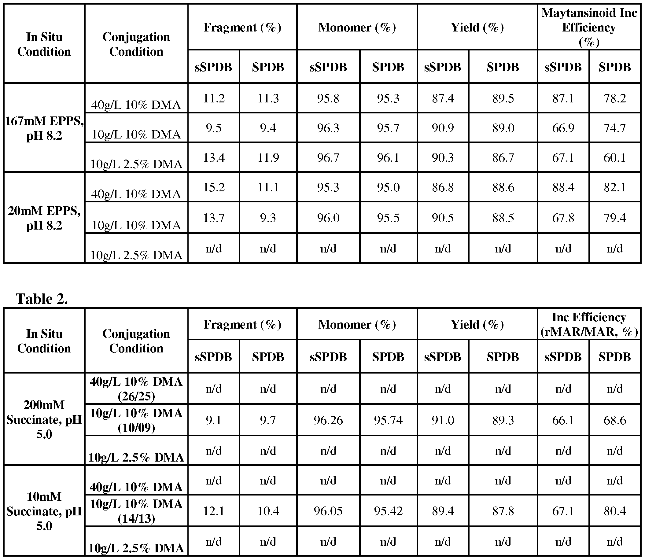

Claims

Priority Applications (11)
| Application Number | Priority Date | Filing Date | Title |
|---|---|---|---|
| AU2015349985A AU2015349985A1 (en) | 2014-11-19 | 2015-11-18 | Process for preparing cell-binding agent-cytotoxic agent conjugates |
| KR1020177016749A KR20170088905A (en) | 2014-11-19 | 2015-11-18 | Process for preparing cell-binding agent-cytotoxic agent conjugates |
| SG11201703599VA SG11201703599VA (en) | 2014-11-19 | 2015-11-18 | Process for preparing cell-binding agent-cytotoxic agent conjugates |
| CN201580060700.2A CN107592813A (en) | 2014-11-19 | 2015-11-18 | Method for preparing cell binding agent cytotoxic agent conjugate |
| CA2966932A CA2966932A1 (en) | 2014-11-19 | 2015-11-18 | Process for preparing cell-binding agent-cytotoxic agent conjugates |
| EP15802310.1A EP3220959A1 (en) | 2014-11-19 | 2015-11-18 | Process for preparing cell-binding agent-cytotoxic agent conjugates |
| RU2017115812A RU2711930C2 (en) | 2014-11-19 | 2015-11-18 | Method of producing conjugates of cell-binding agent and cytotoxic agent |
| JP2017525940A JP2018505128A (en) | 2014-11-19 | 2015-11-18 | Process for preparing cell binding agent-cytotoxic agent conjugates |
| US15/527,525 US10413615B2 (en) | 2014-11-19 | 2015-11-18 | Process for preparing cell-binding agent-cytotoxic agent conjugates |
| IL251900A IL251900B (en) | 2014-11-19 | 2017-04-25 | Process for preparing cell-binding agent-cytotoxic agent conjugates |
| US16/534,521 US20200101168A1 (en) | 2014-11-19 | 2019-08-07 | Process for preparing cell-binding agent-cytotoxic agent conjugates |
Applications Claiming Priority (2)
| Application Number | Priority Date | Filing Date | Title |
|---|---|---|---|
| US201462081914P | 2014-11-19 | 2014-11-19 | |
| US62/081,914 | 2014-11-19 |
Related Child Applications (2)
| Application Number | Title | Priority Date | Filing Date |
|---|---|---|---|
| US15/527,525 A-371-Of-International US10413615B2 (en) | 2014-11-19 | 2015-11-18 | Process for preparing cell-binding agent-cytotoxic agent conjugates |
| US16/534,521 Continuation US20200101168A1 (en) | 2014-11-19 | 2019-08-07 | Process for preparing cell-binding agent-cytotoxic agent conjugates |
Publications (1)
| Publication Number | Publication Date |
|---|---|
| WO2016081584A1 true WO2016081584A1 (en) | 2016-05-26 |
Family
ID=54754814
Family Applications (1)
| Application Number | Title | Priority Date | Filing Date |
|---|---|---|---|
| PCT/US2015/061310 WO2016081584A1 (en) | 2014-11-19 | 2015-11-18 | Process for preparing cell-binding agent-cytotoxic agent conjugates |
Country Status (12)
| Country | Link |
|---|---|
| US (2) | US10413615B2 (en) |
| EP (1) | EP3220959A1 (en) |
| JP (1) | JP2018505128A (en) |
| KR (1) | KR20170088905A (en) |
| CN (1) | CN107592813A (en) |
| AU (1) | AU2015349985A1 (en) |
| CA (1) | CA2966932A1 (en) |
| IL (1) | IL251900B (en) |
| MA (1) | MA40934A (en) |
| RU (1) | RU2711930C2 (en) |
| SG (1) | SG11201703599VA (en) |
| WO (1) | WO2016081584A1 (en) |
Cited By (2)
| Publication number | Priority date | Publication date | Assignee | Title |
|---|---|---|---|---|
| US11555019B2 (en) | 2019-07-10 | 2023-01-17 | Cybrexa 3, Inc. | Peptide conjugates of microtubule-targeting agents as therapeutics |
| US11634508B2 (en) | 2019-07-10 | 2023-04-25 | Cybrexa 2, Inc. | Peptide conjugates of cytotoxins as therapeutics |
Citations (50)
| Publication number | Priority date | Publication date | Assignee | Title |
|---|---|---|---|---|
| US4256746A (en) | 1978-11-14 | 1981-03-17 | Takeda Chemical Industries | Dechloromaytansinoids, their pharmaceutical compositions and method of use |
| US4294757A (en) | 1979-01-31 | 1981-10-13 | Takeda Chemical Industries, Ltd | 20-O-Acylmaytansinoids |
| US4307016A (en) | 1978-03-24 | 1981-12-22 | Takeda Chemical Industries, Ltd. | Demethyl maytansinoids |
| US4313946A (en) | 1981-01-27 | 1982-02-02 | The United States Of America As Represented By The Secretary Of Agriculture | Chemotherapeutically active maytansinoids from Trewia nudiflora |
| US4315929A (en) | 1981-01-27 | 1982-02-16 | The United States Of America As Represented By The Secretary Of Agriculture | Method of controlling the European corn borer with trewiasine |
| US4322348A (en) | 1979-06-05 | 1982-03-30 | Takeda Chemical Industries, Ltd. | Maytansinoids |
| US4331598A (en) | 1979-09-19 | 1982-05-25 | Takeda Chemical Industries, Ltd. | Maytansinoids |
| US4362663A (en) | 1979-09-21 | 1982-12-07 | Takeda Chemical Industries, Ltd. | Maytansinoid compound |
| US4364866A (en) | 1979-09-21 | 1982-12-21 | Takeda Chemical Industries, Ltd. | Maytansinoids |
| US4371533A (en) | 1980-10-08 | 1983-02-01 | Takeda Chemical Industries, Ltd. | 4,5-Deoxymaytansinoids, their use and pharmaceutical compositions thereof |
| US4424219A (en) | 1981-05-20 | 1984-01-03 | Takeda Chemical Industries, Ltd. | 9-Thiomaytansinoids and their pharmaceutical compositions and use |
| US4450254A (en) | 1980-11-03 | 1984-05-22 | Standard Oil Company | Impact improvement of high nitrile resins |
| GB2188638A (en) | 1986-03-27 | 1987-10-07 | Gregory Paul Winter | Chimeric antibodies |
| WO1992001047A1 (en) | 1990-07-10 | 1992-01-23 | Cambridge Antibody Technology Limited | Methods for producing members of specific binding pairs |
| US5225539A (en) | 1986-03-27 | 1993-07-06 | Medical Research Council | Recombinant altered antibodies and methods of making altered antibodies |
| US5475092A (en) | 1992-03-25 | 1995-12-12 | Immunogen Inc. | Cell binding agent conjugates of analogues and derivatives of CC-1065 |
| US5545806A (en) | 1990-08-29 | 1996-08-13 | Genpharm International, Inc. | Ransgenic non-human animals for producing heterologous antibodies |
| US5552293A (en) | 1991-07-03 | 1996-09-03 | Pharmacia Aktiebolag | Tumor antigen specific antibody |
| US5569825A (en) | 1990-08-29 | 1996-10-29 | Genpharm International | Transgenic non-human animals capable of producing heterologous antibodies of various isotypes |
| US5585089A (en) | 1988-12-28 | 1996-12-17 | Protein Design Labs, Inc. | Humanized immunoglobulins |
| US5639641A (en) | 1992-09-09 | 1997-06-17 | Immunogen Inc. | Resurfacing of rodent antibodies |
| US5665357A (en) | 1993-12-03 | 1997-09-09 | Zeneca Limited | Antibodies recognizing tumor associated antigen CA 55.1 |
| US5714352A (en) | 1996-03-20 | 1998-02-03 | Xenotech Incorporated | Directed switch-mediated DNA recombination |
| WO1999006587A2 (en) | 1997-08-01 | 1999-02-11 | Morphosys Ag | Novel method and phage for the identification of nucleic acid sequences encoding members of a multimeric (poly)peptide complex |
| US5885793A (en) | 1991-12-02 | 1999-03-23 | Medical Research Council | Production of anti-self antibodies from antibody segment repertoires and displayed on phage |
| US5958872A (en) | 1996-04-01 | 1999-09-28 | Apoptosis Technology, Inc. | Active survival domains of IGF-IR and methods of use |
| US6265150B1 (en) | 1995-06-07 | 2001-07-24 | Becton Dickinson & Company | Phage antibodies |
| US6333410B1 (en) | 2000-08-18 | 2001-12-25 | Immunogen, Inc. | Process for the preparation and purification of thiol-containing maytansinoids |
| WO2002016401A2 (en) | 2000-08-18 | 2002-02-28 | East Carolina University | Monoclonal antibody ds6, tumor-associated antigen ca6, and methods of use thereof |
| US20020197266A1 (en) | 2000-02-08 | 2002-12-26 | Waldemar Debinski | Immunotherapy using interleukin 13 receptor subunit alpha 2 |
| US6596743B2 (en) | 1999-04-20 | 2003-07-22 | Hoffmann-La Roche Inc. | Carbamic acid derivatives |
| US20060127407A1 (en) | 2004-12-09 | 2006-06-15 | Qiming Chen | Anti-integrin immunoconjugates, methods and uses |
| US20070183971A1 (en) | 2004-08-30 | 2007-08-09 | Biotest Ag | Immunoconjugates targeting syndecan-1 expressing cells and use thereof |
| US7342110B2 (en) | 2002-11-07 | 2008-03-11 | Immunogen Inc. | Anti-CD33 antibodies and method for treatment of acute myeloid leukemia using the same |
| WO2008047242A2 (en) | 2006-10-19 | 2008-04-24 | Sanofi-Aventis | Novel anti-cd38 antibodies for the treatment of cancer |
| US20080171040A1 (en) | 2004-06-01 | 2008-07-17 | Genentech, Inc. | Antibody-drug conjugates and methods |
| US20080305044A1 (en) | 2004-11-29 | 2008-12-11 | Seattle Genetics, Inc. | Engineered Antibodies and Immunoconjugates |
| US20090156790A1 (en) | 2003-06-27 | 2009-06-18 | Amgen Fremont Inc. | Antibodies directed to the deletion mutants of epidermal growth factor receptor and uses thereof |
| US20100093980A1 (en) | 2005-03-04 | 2010-04-15 | Biogen Idec Ma Inc. | Methods of Humanizing Immunoglobulin Variable Regions Through Rational Modification Of Complementarity Determining Residues |
| WO2010124797A1 (en) | 2009-04-29 | 2010-11-04 | Bayer Schering Pharma Aktiengesellschaft | Anti-mesothelin immunoconjugates and uses therefor |
| WO2011039724A1 (en) | 2009-10-02 | 2011-04-07 | Sanofi-Aventis | Antibodies that specifically bind to the epha2 receptor |
| WO2011039721A1 (en) | 2009-10-02 | 2011-04-07 | Sanofi-Aventis | New maytansinoids and the use of said maytansinoids to prepare conjugates with an antibody |
| US7982024B2 (en) | 2001-01-05 | 2011-07-19 | Amgen Fremont Inc. | Antibodies to insulin-like growth factor I receptor |
| WO2011106528A1 (en) | 2010-02-24 | 2011-09-01 | Immunogen, Inc. | Folate receptor 1 antibodies and immunoconjugates and uses thereof |
| US20110256153A1 (en) | 2010-03-12 | 2011-10-20 | Jutta Deckert | CD37-Binding Molecules and Immunoconjugates Thereof |
| WO2012019024A2 (en) | 2010-08-04 | 2012-02-09 | Immunogen, Inc. | Her3-binding molecules and immunoconjugates thereof |
| WO2012112687A1 (en) * | 2011-02-15 | 2012-08-23 | Immunogen, Inc. | Methods of preparation of conjugates |
| US20140030820A1 (en) | 2011-01-10 | 2014-01-30 | Technische Universitaet Muenchen | Triazacyclononane-based phosphinate ligand and its use for molecular imaging |
| WO2014134483A2 (en) * | 2013-02-28 | 2014-09-04 | Immunogen, Inc. | Conjugates comprising cell-binding agents and cytotoxic agents |
| WO2014160160A2 (en) * | 2013-03-13 | 2014-10-02 | Novartis Ag | Antibody drug conjugates |
Family Cites Families (5)
| Publication number | Priority date | Publication date | Assignee | Title |
|---|---|---|---|---|
| EP1888637A2 (en) * | 2005-05-19 | 2008-02-20 | Amgen Inc. | Compositions and methods for increasing the stability of antibodies |
| LT2437790T (en) * | 2009-06-03 | 2019-06-10 | Immunogen, Inc. | Conjugation methods |
| US20120282282A1 (en) * | 2011-04-04 | 2012-11-08 | Immunogen, Inc. | Methods for Decreasing Ocular Toxicity of Antibody Drug Conjugates |
| RU2018130108A (en) * | 2016-02-05 | 2020-03-06 | Иммуноджен, Инк. | EFFECTIVE METHOD FOR PRODUCING CONJUGATES BINDING A CELL AGENT CYTOTOXIC AGENT |
| AU2019206587A1 (en) * | 2018-01-12 | 2020-08-06 | Immunogen, Inc. | Methods for antibody drug conjugation, purification, and formulation |
-
2015
- 2015-11-18 RU RU2017115812A patent/RU2711930C2/en active
- 2015-11-18 WO PCT/US2015/061310 patent/WO2016081584A1/en active Application Filing
- 2015-11-18 SG SG11201703599VA patent/SG11201703599VA/en unknown
- 2015-11-18 CA CA2966932A patent/CA2966932A1/en not_active Abandoned
- 2015-11-18 AU AU2015349985A patent/AU2015349985A1/en not_active Abandoned
- 2015-11-18 JP JP2017525940A patent/JP2018505128A/en not_active Ceased
- 2015-11-18 CN CN201580060700.2A patent/CN107592813A/en active Pending
- 2015-11-18 EP EP15802310.1A patent/EP3220959A1/en not_active Withdrawn
- 2015-11-18 US US15/527,525 patent/US10413615B2/en not_active Expired - Fee Related
- 2015-11-18 KR KR1020177016749A patent/KR20170088905A/en unknown
- 2015-11-18 MA MA040934A patent/MA40934A/en unknown
-
2017
- 2017-04-25 IL IL251900A patent/IL251900B/en not_active IP Right Cessation
-
2019
- 2019-08-07 US US16/534,521 patent/US20200101168A1/en not_active Abandoned
Patent Citations (66)
| Publication number | Priority date | Publication date | Assignee | Title |
|---|---|---|---|---|
| US4307016A (en) | 1978-03-24 | 1981-12-22 | Takeda Chemical Industries, Ltd. | Demethyl maytansinoids |
| US4361650A (en) | 1978-03-24 | 1982-11-30 | Takeda Chemical Industries, Ltd. | Fermentation process of preparing demethyl maytansinoids |
| US4256746A (en) | 1978-11-14 | 1981-03-17 | Takeda Chemical Industries | Dechloromaytansinoids, their pharmaceutical compositions and method of use |
| US4294757A (en) | 1979-01-31 | 1981-10-13 | Takeda Chemical Industries, Ltd | 20-O-Acylmaytansinoids |
| US4322348A (en) | 1979-06-05 | 1982-03-30 | Takeda Chemical Industries, Ltd. | Maytansinoids |
| US4331598A (en) | 1979-09-19 | 1982-05-25 | Takeda Chemical Industries, Ltd. | Maytansinoids |
| US4364866A (en) | 1979-09-21 | 1982-12-21 | Takeda Chemical Industries, Ltd. | Maytansinoids |
| US4362663A (en) | 1979-09-21 | 1982-12-07 | Takeda Chemical Industries, Ltd. | Maytansinoid compound |
| US4371533A (en) | 1980-10-08 | 1983-02-01 | Takeda Chemical Industries, Ltd. | 4,5-Deoxymaytansinoids, their use and pharmaceutical compositions thereof |
| US4450254A (en) | 1980-11-03 | 1984-05-22 | Standard Oil Company | Impact improvement of high nitrile resins |
| US4315929A (en) | 1981-01-27 | 1982-02-16 | The United States Of America As Represented By The Secretary Of Agriculture | Method of controlling the European corn borer with trewiasine |
| US4313946A (en) | 1981-01-27 | 1982-02-02 | The United States Of America As Represented By The Secretary Of Agriculture | Chemotherapeutically active maytansinoids from Trewia nudiflora |
| US4424219A (en) | 1981-05-20 | 1984-01-03 | Takeda Chemical Industries, Ltd. | 9-Thiomaytansinoids and their pharmaceutical compositions and use |
| GB2188638A (en) | 1986-03-27 | 1987-10-07 | Gregory Paul Winter | Chimeric antibodies |
| US5225539A (en) | 1986-03-27 | 1993-07-06 | Medical Research Council | Recombinant altered antibodies and methods of making altered antibodies |
| EP0239400B1 (en) | 1986-03-27 | 1994-08-03 | Medical Research Council | Recombinant antibodies and methods for their production |
| US5585089A (en) | 1988-12-28 | 1996-12-17 | Protein Design Labs, Inc. | Humanized immunoglobulins |
| US5693761A (en) | 1988-12-28 | 1997-12-02 | Protein Design Labs, Inc. | Polynucleotides encoding improved humanized immunoglobulins |
| US5693762A (en) | 1988-12-28 | 1997-12-02 | Protein Design Labs, Inc. | Humanized immunoglobulins |
| WO1992001047A1 (en) | 1990-07-10 | 1992-01-23 | Cambridge Antibody Technology Limited | Methods for producing members of specific binding pairs |
| US5969108A (en) | 1990-07-10 | 1999-10-19 | Medical Research Council | Methods for producing members of specific binding pairs |
| US5569825A (en) | 1990-08-29 | 1996-10-29 | Genpharm International | Transgenic non-human animals capable of producing heterologous antibodies of various isotypes |
| US5545806A (en) | 1990-08-29 | 1996-08-13 | Genpharm International, Inc. | Ransgenic non-human animals for producing heterologous antibodies |
| US5552293A (en) | 1991-07-03 | 1996-09-03 | Pharmacia Aktiebolag | Tumor antigen specific antibody |
| US5885793A (en) | 1991-12-02 | 1999-03-23 | Medical Research Council | Production of anti-self antibodies from antibody segment repertoires and displayed on phage |
| US5585499A (en) | 1992-03-25 | 1996-12-17 | Immunogen Inc. | Cyclopropylbenzindole-containing cytotoxic drugs |
| US5475092A (en) | 1992-03-25 | 1995-12-12 | Immunogen Inc. | Cell binding agent conjugates of analogues and derivatives of CC-1065 |
| US5846545A (en) | 1992-03-25 | 1998-12-08 | Immunogen, Inc. | Targeted delivery of cyclopropylbenzindole-containing cytotoxic drugs |
| US5639641A (en) | 1992-09-09 | 1997-06-17 | Immunogen Inc. | Resurfacing of rodent antibodies |
| US5665357A (en) | 1993-12-03 | 1997-09-09 | Zeneca Limited | Antibodies recognizing tumor associated antigen CA 55.1 |
| US6265150B1 (en) | 1995-06-07 | 2001-07-24 | Becton Dickinson & Company | Phage antibodies |
| US5714352A (en) | 1996-03-20 | 1998-02-03 | Xenotech Incorporated | Directed switch-mediated DNA recombination |
| US5958872A (en) | 1996-04-01 | 1999-09-28 | Apoptosis Technology, Inc. | Active survival domains of IGF-IR and methods of use |
| WO1999006587A2 (en) | 1997-08-01 | 1999-02-11 | Morphosys Ag | Novel method and phage for the identification of nucleic acid sequences encoding members of a multimeric (poly)peptide complex |
| US6596743B2 (en) | 1999-04-20 | 2003-07-22 | Hoffmann-La Roche Inc. | Carbamic acid derivatives |
| US20020197266A1 (en) | 2000-02-08 | 2002-12-26 | Waldemar Debinski | Immunotherapy using interleukin 13 receptor subunit alpha 2 |
| WO2002016401A2 (en) | 2000-08-18 | 2002-02-28 | East Carolina University | Monoclonal antibody ds6, tumor-associated antigen ca6, and methods of use thereof |
| US6333410B1 (en) | 2000-08-18 | 2001-12-25 | Immunogen, Inc. | Process for the preparation and purification of thiol-containing maytansinoids |
| US7982024B2 (en) | 2001-01-05 | 2011-07-19 | Amgen Fremont Inc. | Antibodies to insulin-like growth factor I receptor |
| US7342110B2 (en) | 2002-11-07 | 2008-03-11 | Immunogen Inc. | Anti-CD33 antibodies and method for treatment of acute myeloid leukemia using the same |
| US7557189B2 (en) | 2002-11-07 | 2009-07-07 | Immunogen Inc. | Anti-CD33 antibodies and method for treatment of acute myeloid leukemia using the same |
| US7628986B2 (en) | 2003-06-27 | 2009-12-08 | Amgen Fremont Inc. | Antibodies directed to the deletion mutants of epidermal growth factor receptor and uses thereof |
| US7736644B2 (en) | 2003-06-27 | 2010-06-15 | Amgen Fremont Inc. | Antibodies directed to the deletion mutants of epidermal growth factor receptor and uses thereof |
| US20100111979A1 (en) | 2003-06-27 | 2010-05-06 | Amgen Fremont Inc. | Antibodies directed to the deletion mutants of epidermal growth factor receptor and uses thereof |
| US20090156790A1 (en) | 2003-06-27 | 2009-06-18 | Amgen Fremont Inc. | Antibodies directed to the deletion mutants of epidermal growth factor receptor and uses thereof |
| US20090155282A1 (en) | 2003-06-27 | 2009-06-18 | Amgen Fremont Inc. | Antibodies directed to the deletion mutants of epidermal growth factor receptor and uses thereof |
| US20090175887A1 (en) | 2003-06-27 | 2009-07-09 | Amgen Fremont Inc. | Antibodies directed to the deletion mutants of epidermal growth factor receptor and uses thereof |
| US20090240038A1 (en) | 2003-06-27 | 2009-09-24 | Amgen Fremont Inc. | Antibodies directed to the deletion mutants of epidermal growth factor receptor and uses thereof |
| US20080171040A1 (en) | 2004-06-01 | 2008-07-17 | Genentech, Inc. | Antibody-drug conjugates and methods |
| US20070183971A1 (en) | 2004-08-30 | 2007-08-09 | Biotest Ag | Immunoconjugates targeting syndecan-1 expressing cells and use thereof |
| US20080305044A1 (en) | 2004-11-29 | 2008-12-11 | Seattle Genetics, Inc. | Engineered Antibodies and Immunoconjugates |
| US20060127407A1 (en) | 2004-12-09 | 2006-06-15 | Qiming Chen | Anti-integrin immunoconjugates, methods and uses |
| US20100093980A1 (en) | 2005-03-04 | 2010-04-15 | Biogen Idec Ma Inc. | Methods of Humanizing Immunoglobulin Variable Regions Through Rational Modification Of Complementarity Determining Residues |
| WO2008047242A2 (en) | 2006-10-19 | 2008-04-24 | Sanofi-Aventis | Novel anti-cd38 antibodies for the treatment of cancer |
| WO2010124797A1 (en) | 2009-04-29 | 2010-11-04 | Bayer Schering Pharma Aktiengesellschaft | Anti-mesothelin immunoconjugates and uses therefor |
| WO2011039724A1 (en) | 2009-10-02 | 2011-04-07 | Sanofi-Aventis | Antibodies that specifically bind to the epha2 receptor |
| WO2011039721A1 (en) | 2009-10-02 | 2011-04-07 | Sanofi-Aventis | New maytansinoids and the use of said maytansinoids to prepare conjugates with an antibody |
| US20120009181A1 (en) | 2010-02-24 | 2012-01-12 | Ab Olga | Folate Receptor 1 Antibodies and Immunoconjugates and Uses Thereof |
| WO2011106528A1 (en) | 2010-02-24 | 2011-09-01 | Immunogen, Inc. | Folate receptor 1 antibodies and immunoconjugates and uses thereof |
| US20110256153A1 (en) | 2010-03-12 | 2011-10-20 | Jutta Deckert | CD37-Binding Molecules and Immunoconjugates Thereof |
| WO2012019024A2 (en) | 2010-08-04 | 2012-02-09 | Immunogen, Inc. | Her3-binding molecules and immunoconjugates thereof |
| US20140030820A1 (en) | 2011-01-10 | 2014-01-30 | Technische Universitaet Muenchen | Triazacyclononane-based phosphinate ligand and its use for molecular imaging |
| WO2012112687A1 (en) * | 2011-02-15 | 2012-08-23 | Immunogen, Inc. | Methods of preparation of conjugates |
| WO2014134483A2 (en) * | 2013-02-28 | 2014-09-04 | Immunogen, Inc. | Conjugates comprising cell-binding agents and cytotoxic agents |
| WO2014160160A2 (en) * | 2013-03-13 | 2014-10-02 | Novartis Ag | Antibody drug conjugates |
| US20140301946A1 (en) | 2013-03-13 | 2014-10-09 | Novartis Ag | Antibody drug conjugates |
Non-Patent Citations (31)
| Title |
|---|
| BURGESS, IMMUNOLOGY TODAY, vol. 5, 1984, pages 155 - 158 |
| C. A. JANEWAY ET AL: "Immunobiology", 2001, GARLAND PUBLISHING |
| CARTER, NATURE REVS., vol. 6, 2006, pages 343 - 357 |
| COLOMER ET AL., CANCER INVEST., vol. 19, 2001, pages 49 - 56 |
| DESMYTER ET AL., NATURE STRUCT. BIOL., vol. 3, 1996, pages 752 |
| GREENBERG ET AL., NATURE, vol. 374, 1995, pages 168 |
| GRIFFIN ET AL., LEUKEMIA RES., vol. 8, 1984, pages 521 |
| HARLOW AND LANE: "Antibodies: A Laboratory Manual", 1988, CSH PRESS |
| HASKARD; ARCHER, J. IMMUNOL. METHODS, vol. 74, no. 2, 1984, pages 361 - 367 |
| HEIDER ET AL., EUR. J. CANCER, vol. 31A, 1995, pages 2385 - 2391 |
| HUSE ET AL., SCIENCE, vol. 246, 1989, pages 1275 - 1281 |
| KIM ET AL., MOL. CANCER THER., vol. 7, 2008, pages 2486 - 2497 |
| KOHLER; MILSTEIN, EUR. J. IMMUNOL., vol. 5, 1976, pages 511 - 519 |
| LIU ET, CANCER RES., vol. 57, 1997, pages 3629 - 3634 |
| MALONEY ET AL., BLOOD, vol. 90, 1997, pages 2188 - 2195 |
| NADLER ET AL., J. IMMUNOL., vol. 131, 1983, pages 244 - 250 |
| NISONOFF ET AL., ARCH. BIOCHEM. BIOPHYS., vol. 89, 1960, pages 230 - 244 |
| O'KEEFE ET AL., J. BIOL. CHEM., vol. 260, 1985, pages 932 - 937 |
| PARHAM, J. IMMUNOL., vol. 131, 1983, pages 2895 - 2902 |
| PEDERSEN ET AL., J. MOL. BIOL., vol. 235, 1994, pages 959 - 973 |
| REITER ET AL., PROTEIN ENGINEERING, vol. 7, 1994, pages 697 - 704 |
| RITZ ET AL., NATURE, vol. 283, 1980, pages 583 - 585 |
| RODER ET AL., METHODS ENZYMOL., vol. 121, 1986, pages 140 - 167 |
| ROGUSKA ET AL., PROC. NATL. ACAD. SCI. USA, vol. 91, 1994, pages 969 - 973 |
| SAMBROOK ET AL: "Molecular Cloning, A Laboratory Manual", 2001, COLD SPRING HARBOR LABORATORY PRESS |
| SPRING ET AL., J. IMMUNOL., vol. 113, 1974, pages 470 - 478 |
| STANFIELD ET AL., SCIENCE, vol. 305, 2004, pages 1770 - 1773 |
| TAYLOR-PAPADIMITRIOU ET AL., INT. J. CANCER, vol. 28, 1981, pages 17 - 21 |
| VAN H ET AL., CANCER RES., vol. 56, 1996, pages 5179 - 5185 |
| VLASAK, J.; LONESCU, R.: "Fragmentation of monoclonal antibodies", MABS, vol. 3, no. 3, May 2011 (2011-05-01), pages 253 - 263 |
| WELT ET AL., J. CLIN. ONCOL., vol. 12, 1994, pages 1193 - 1203 |
Cited By (2)
| Publication number | Priority date | Publication date | Assignee | Title |
|---|---|---|---|---|
| US11555019B2 (en) | 2019-07-10 | 2023-01-17 | Cybrexa 3, Inc. | Peptide conjugates of microtubule-targeting agents as therapeutics |
| US11634508B2 (en) | 2019-07-10 | 2023-04-25 | Cybrexa 2, Inc. | Peptide conjugates of cytotoxins as therapeutics |
Also Published As
| Publication number | Publication date |
|---|---|
| US10413615B2 (en) | 2019-09-17 |
| RU2017115812A (en) | 2018-12-19 |
| CN107592813A (en) | 2018-01-16 |
| US20170354742A1 (en) | 2017-12-14 |
| IL251900A0 (en) | 2017-07-31 |
| RU2017115812A3 (en) | 2019-06-18 |
| MA40934A (en) | 2017-09-27 |
| SG11201703599VA (en) | 2017-06-29 |
| CA2966932A1 (en) | 2016-05-26 |
| IL251900B (en) | 2019-12-31 |
| EP3220959A1 (en) | 2017-09-27 |
| RU2711930C2 (en) | 2020-01-23 |
| KR20170088905A (en) | 2017-08-02 |
| AU2015349985A1 (en) | 2017-05-25 |
| US20200101168A1 (en) | 2020-04-02 |
| JP2018505128A (en) | 2018-02-22 |
Similar Documents
| Publication | Publication Date | Title |
|---|---|---|
| US11744900B2 (en) | Preparation of maytansinoid antibody conjugates by a one-step process | |
| RU2661083C2 (en) | Use of pvdf membrane to purify cell-binding agent - cytotoxic agent conjugates | |
| WO2012135517A2 (en) | Preparation of maytansinoid antibody conjugates by a one-step process | |
| EP2790724A1 (en) | Use of n-hydroxysuccinimide to improve conjugate stability | |
| EP3411077A1 (en) | Efficient process for preparing cell-binding agent-cytotoxic agent conjugates | |
| US20200101168A1 (en) | Process for preparing cell-binding agent-cytotoxic agent conjugates | |
| NZ616509B2 (en) | Preparation of maytansinoid antibody conjugates by a one-step process |
Legal Events
| Date | Code | Title | Description |
|---|---|---|---|
| 121 | Ep: the epo has been informed by wipo that ep was designated in this application |
Ref document number: 15802310 Country of ref document: EP Kind code of ref document: A1 |
|
| WWE | Wipo information: entry into national phase |
Ref document number: 251900 Country of ref document: IL |
|
| ENP | Entry into the national phase |
Ref document number: 2966932 Country of ref document: CA |
|
| WWE | Wipo information: entry into national phase |
Ref document number: 11201703599V Country of ref document: SG |
|
| ENP | Entry into the national phase |
Ref document number: 2017525940 Country of ref document: JP Kind code of ref document: A |
|
| WWE | Wipo information: entry into national phase |
Ref document number: 15527525 Country of ref document: US |
|
| NENP | Non-entry into the national phase |
Ref country code: DE |
|
| ENP | Entry into the national phase |
Ref document number: 2015349985 Country of ref document: AU Date of ref document: 20151118 Kind code of ref document: A |
|
| REEP | Request for entry into the european phase |
Ref document number: 2015802310 Country of ref document: EP |
|
| ENP | Entry into the national phase |
Ref document number: 2017115812 Country of ref document: RU Kind code of ref document: A Ref document number: 20177016749 Country of ref document: KR Kind code of ref document: A |