WO2016073989A2 - Osteochondroreticular stem cells for bone and cartilage regeneration - Google Patents
Osteochondroreticular stem cells for bone and cartilage regeneration Download PDFInfo
- Publication number
- WO2016073989A2 WO2016073989A2 PCT/US2015/059772 US2015059772W WO2016073989A2 WO 2016073989 A2 WO2016073989 A2 WO 2016073989A2 US 2015059772 W US2015059772 W US 2015059772W WO 2016073989 A2 WO2016073989 A2 WO 2016073989A2
- Authority
- WO
- WIPO (PCT)
- Prior art keywords
- cells
- greml
- stem cells
- ocr
- bone
- Prior art date
- Legal status (The legal status is an assumption and is not a legal conclusion. Google has not performed a legal analysis and makes no representation as to the accuracy of the status listed.)
- Ceased
Links
Classifications
-
- C—CHEMISTRY; METALLURGY
- C12—BIOCHEMISTRY; BEER; SPIRITS; WINE; VINEGAR; MICROBIOLOGY; ENZYMOLOGY; MUTATION OR GENETIC ENGINEERING
- C12N—MICROORGANISMS OR ENZYMES; COMPOSITIONS THEREOF; PROPAGATING, PRESERVING, OR MAINTAINING MICROORGANISMS; MUTATION OR GENETIC ENGINEERING; CULTURE MEDIA
- C12N5/00—Undifferentiated human, animal or plant cells, e.g. cell lines; Tissues; Cultivation or maintenance thereof; Culture media therefor
- C12N5/06—Animal cells or tissues; Human cells or tissues
- C12N5/0602—Vertebrate cells
- C12N5/0652—Cells of skeletal and connective tissues; Mesenchyme
- C12N5/0654—Osteocytes, Osteoblasts, Odontocytes; Bones, Teeth
-
- A—HUMAN NECESSITIES
- A61—MEDICAL OR VETERINARY SCIENCE; HYGIENE
- A61K—PREPARATIONS FOR MEDICAL, DENTAL OR TOILETRY PURPOSES
- A61K39/00—Medicinal preparations containing antigens or antibodies
-
- C—CHEMISTRY; METALLURGY
- C12—BIOCHEMISTRY; BEER; SPIRITS; WINE; VINEGAR; MICROBIOLOGY; ENZYMOLOGY; MUTATION OR GENETIC ENGINEERING
- C12N—MICROORGANISMS OR ENZYMES; COMPOSITIONS THEREOF; PROPAGATING, PRESERVING, OR MAINTAINING MICROORGANISMS; MUTATION OR GENETIC ENGINEERING; CULTURE MEDIA
- C12N5/00—Undifferentiated human, animal or plant cells, e.g. cell lines; Tissues; Cultivation or maintenance thereof; Culture media therefor
- C12N5/06—Animal cells or tissues; Human cells or tissues
- C12N5/0602—Vertebrate cells
- C12N5/0652—Cells of skeletal and connective tissues; Mesenchyme
- C12N5/0662—Stem cells
- C12N5/0663—Bone marrow mesenchymal stem cells (BM-MSC)
-
- C—CHEMISTRY; METALLURGY
- C12—BIOCHEMISTRY; BEER; SPIRITS; WINE; VINEGAR; MICROBIOLOGY; ENZYMOLOGY; MUTATION OR GENETIC ENGINEERING
- C12N—MICROORGANISMS OR ENZYMES; COMPOSITIONS THEREOF; PROPAGATING, PRESERVING, OR MAINTAINING MICROORGANISMS; MUTATION OR GENETIC ENGINEERING; CULTURE MEDIA
- C12N5/00—Undifferentiated human, animal or plant cells, e.g. cell lines; Tissues; Cultivation or maintenance thereof; Culture media therefor
- C12N5/06—Animal cells or tissues; Human cells or tissues
- C12N5/0602—Vertebrate cells
- C12N5/0652—Cells of skeletal and connective tissues; Mesenchyme
- C12N5/0669—Bone marrow stromal cells; Whole bone marrow
-
- A—HUMAN NECESSITIES
- A61—MEDICAL OR VETERINARY SCIENCE; HYGIENE
- A61K—PREPARATIONS FOR MEDICAL, DENTAL OR TOILETRY PURPOSES
- A61K39/00—Medicinal preparations containing antigens or antibodies
- A61K2039/51—Medicinal preparations containing antigens or antibodies comprising whole cells, viruses or DNA/RNA
- A61K2039/515—Animal cells
-
- A—HUMAN NECESSITIES
- A61—MEDICAL OR VETERINARY SCIENCE; HYGIENE
- A61K—PREPARATIONS FOR MEDICAL, DENTAL OR TOILETRY PURPOSES
- A61K35/00—Medicinal preparations containing materials or reaction products thereof with undetermined constitution
- A61K35/12—Materials from mammals; Compositions comprising non-specified tissues or cells; Compositions comprising non-embryonic stem cells; Genetically modified cells
- A61K35/28—Bone marrow; Haematopoietic stem cells; Mesenchymal stem cells of any origin, e.g. adipose-derived stem cells
Definitions
- Osteoarthritis describes a disease involving the degeneration in the protective cartilage around bones and occurs to millions of patients worldwide. Osteoarthritis is just one example of cartilage and bone deterioration and is a debilitating disease that affects millions of people. With the aging population, it is expected to continue to be a major disease worldwide. When the protective cartilage wears down, bones can rub together under joint movement, causing pain and stiffness, which may lead to decreased movement and morbidity. Current treatments include non-steroidal anti-inflammatories that can pose cardiovascular and gastrointestinal side effects, narcotics, and physical therapy to alleviate the symptoms. More invasive joint replacement surgeries are needed for severe cases. No cure exists at the moment other than replacement of the joint.
- the present invention provides an alternative approach to treatment.
- Inventive embodiments herein are based on the discovery of a newly identified stem cell, the osteochondroreticular (OCR) stem cell that is shown to be the chief origin of cartilage and bone during development.
- OCR osteochondroreticular
- Certain conventional therapies to address bone and cartilage degeneration and injury have utilized pooled mesenchymal stem cells.
- the novel OCR stem cell population described herein provides significantly improved therapeutic methods and/or implantable products, compared to conventional pooled mesenchymal stem cell populations, particularly for the repair of cartilage due to their superior chondrogenic properties.
- Certain embodiments provide improved stem cell therapy methods of treating diseases, degeneration or injury of the bone and cartilage as described herein including but not limited to osteoarthritis, osteoporosis, and bone fractures using OCR stem cells.
- OCR stem cells contribute temporally and make lineage- specific contributions to skeletal development and maintenance. These stem cells are isolated from mesenchymal stromal cells found typically in the bone and bone marrow and can be distinguished by expression of Gremlin 1 (Greml) and/or cell surface markers such as CD200, CD 109 and CD 105, markers identified through microarray screens. In addition, OCR stem cells are more clonogenic than other mesenchymal stem cells. OCR stem cells are lineage restricted skeletal stem cells that are determined in their skeletal fate and do not give rise to muscle and fat.
- Embodiments of the invention also include a method for isolating OCR stem cells.
- Isolated OCR stem cells are useful in regenerating cartilage or bone tissue.
- OCR stem cells are useful for treatment of diseases, degeneration or injury of the bone and cartilage resulting from age, gender, genes, excess weight, poor diet, sedentary lifestyle, injury or trauma,, abnormal metabolism (such as gout and pseudogout), osteoarthritis, infections (such as in the arthritis of Lyme disease), and an overactive immune system (such as rheumatoid arthritis and systemic lupus erythematosus).
- the method comprises the steps of: (a) obtaining multipotent mesenchymal stromal cells from a subject that comprise OCR stem cells and then isolating from the multipotent mesenchymal stromal cells a population of cells that express Gremlin 1 (Greml) and/or by particular cell surface markers selected from the group consisting of CD200, CD 109, and CD 105; and (c) isolating OCR stem cells from the bone and/or bone marrow, wherein the isolated OCR stem cells promote regeneration of cartilage tissue and/or bone.
- the OCR stem cells may be isolated without necessarily utilizing cell markers, but can be identified retrospectively by their capacity to generate bone and cartilage in vitro (in culture), but poor efficiency for developing into fat cells.
- the isolated Greml + CD 105 OCR stem cells are subjected to conditions that promote differentiation into osteoblasts, chondrocytes, and reticular marrow stromal cells that are useful for regeneration of cartilage tissue for treatment of diseases described herein.
- the conditions that promote differentiation comprise culturing the OCR stem cells in the presence of medium that comprises certain factors such as one or more bone morphogenic proteins (BMPs).
- BMPs bone morphogenic proteins
- certain embodiments of the invention comprise a composition comprising an acceptable carrier and the isolated Greml + CD 105 OCR stem cells described above in this paragraph and throughout the specification.
- the composition may comprise other therapeutic agents.
- Certain embodiments of the invention also include substantially pure isolated Greml + CD 105 OCR stem cells from tissue such as bone and bone marrow, which express the cell marker Greml + and CD 105.
- methods are provided for treating osteoarthritis and bone fracture by administering a therapeutically effective amount of the composition. The composition may be administered to tissue surround the fracture.
- FIG. 1A-1K illustrates that Greml identifies rare adult multipotent mesenchymal stromal cells.
- A Protocol and
- B Greml -creER ; R26-LSL-TdTomato; Nes-GFP mouse femur, showing that metaphyseal Nes-GFP + (green) and Greml + cells (red, white arrows) are distinct.
- C Adult Greml-creER ; R26-LSL-TdTomato bone marrow cells are rare and mesenchymal (CD45 " CD31-Ter-119 " ).
- H-K Greml + cells from Greml-creERT; R26-LSL-ZsGreen; Acta2-RFP mice could be clonally expanded in vitro and differentiated into bone (H) (alizarin red), cartilage (I) (toluidine blue), and myofibroblasts
- Greml + and Greml -negative cells were compared across the increasingly specific immunophenotypes; data shown with mean ⁇ SD.
- G Microarray was performed to compare the Greml + (red) to the nonrecombined stromal (CD45 _ CD31 ⁇ Ter-l 19 ⁇ ) population (green); in qPCR, we also sorted and evaluated the CD45CD31Ter-l 19 + population (blue) that did not contain any recombined cells.
- Greml + cells were not enriched for Nes expression.
- FIG. 3A-3P illustrates that endogenous Greml cells self-renew and lineage trace bone, cartilage, and stroma.
- A Protocol.
- B-E PI induction in Greml -creER ; R26-LSL- ZsGreen; Acta2-RFP mice.
- B At 24 hr after tamoxifen, Greml recombined (green) only within the primary spongiosa of long bones distinct from the Acta2-RFP (red) cells in the marrow.
- Greml cells began organizing into chondrocytic columns (D) and differentiated into stromal cells that invade the bone marrow (E) intertwined with ⁇ 4cto2-positive (red) cells.
- FIG. 4A-4H illustrates that Nes-GFP cells make little contribution to skeletal tissues during early life.
- A Whole-mount in situ hybridization on mouse embryos at E9.5, E10.5, El 1.5 and E12.5. These embryos were evaluated for Sox9, Runx2, Greml, and Nes expression.
- B Protocol.
- C By flow cytometry, approximately 4% of Nes- G -positive cells recombined (that is, were both green and red).
- D-H This specific Nes- CreERT transgenic line recombined in all typical Nes-GFP populations, including perivascular cells immediately inferior to the growth plate (D), in periarteriolar cells (E), and in perisinusoidal Nes-GFP-positivQ cells (red arrows, F-H). The only osteochondral lineage tracing found were isolated osteocytes throughout the diaphyseal bone (H, white arrow).
- FIG. 5A-5L illustrates that adult Greml cells, both endogenous and transplanted, differentiate into osteochondral fracture callus.
- A Protocol.
- B and C Greml -creEB ; R26-
- LSL-TdTomato LSL-TdTomato; 2.3colGFP mice adult induction: Greml + (red) cells were not osteoblasts (green) but were adjacent to each other in situ (B) and during the first week of adherent bone marrow stromal culture (C).
- D is an X-ray of the femoral osteotomy and internal fixation of the bone.
- E and (F) show the serial histology and fluorescent microscopy sections from the resulting fracture callus after the osteotomy.
- G) and (H) are magnified from areas shown in (F).
- Greml + cells (red) stream into the fracture site and differentiate into either osteoblasts (G, yellow cells, white arrows) or Sox9 + (white, nuclear stain) chondrocytes (yellow arrows) (H).
- G yellow cells, white arrows
- Sox9 + white, nuclear stain
- chondrocytes yellow arrows
- Greml + clone after adult induction, was expanded in vitro. An osteotomy with internal fixation was performed in wild-type mice, at which point 500 3 10 ⁇ clonal cells (red) were irrigated into the surgical field. Seven days later, the Greml clone had engrafted (J), the site of injury was identified by TdTomato fluorescence imaging, and recombined cells (fluorescent red) differentiated into osteoblastic cells (K) (alkaline phosphatase positive cells, red-brown, white arrows) in the callus (sequential fluorescence microscopy and ALP staining performed on the same slide). (L) Callus culture was performed, and the recombined Greml cells were easily recovered in vitro and serially transplanted into a secondary fracture (FIG. 12E).
- FIG. 6A-6I illustrates that Greml expression identifies i SCs.
- A Protocol.
- Axes are provided to indicate the longitudinal and circumferential axes (x and y), and "CVA” to designate the crypt-villus, or radial, axis.
- E The sheath was comprised of a reticulated population of stellate cells with long processes that encircled the entire intestinal gland.
- FIG. 7 is a schematic representation showing the OCR stem cell and the perisinusoidal MSC make a complementary contribution to skeletal development, adult homeostasis, and Repair.
- FIG. 8A-8F illustrates (A) a candidate panel of genes based on perisinusoidal, niche and important mesenchymal lineage genes. qPCR for each gene was first performed on 100,000 freshly sorted CD45-CD31-Terl 19- and then repeated on the same population of cells after 14 days of adherent culture in MEM plus 10% MSC defined FBS and 1% antibiotics.
- Greml-creERT from 8 potential founders we generated 3 founder lines, confirmed by PCR genotyping for the regions of insertion of the creERT into the Greml BAC transgene and also primers specific for Cre cDNA. The 5' insertion site PCR was used subsequently for genotyping (Table 2). These founders were generated on a B6CBA/F2 background, which was then backcrossed 6 generations to C57BL/6J. Only one line (line 3) displayed significant recombination following adult tamoxifen induction.
- Line 3 was confirmed to be specific for endogenous GremI function by qPCR (E & F) of sorted bone marrow populations based on mesenchymal markers and GremI -recombination, represented as fold change relative to the CD45CD31Ter-l 19 (+) population. Line 3 was used for all experiments and is available at JAX.
- FIG. 10A-10L represents (A) color-coded FACS strategy reflected in the color of subsequent graphs, all of which represent mean +/- SD.
- C prior to microarray analysis the RNA from FACS cells had to be amplified. We confirmed that amplification was successful and had not distorted the population we repeated GremI qPCR, confirming results from FIG. 8G.
- GremI recombined cells had higher GremI expression compared to their GremI negative but mesenchymal (CD45-CD31-Ter-119-) counterparts, in which Greml expression was undetectable.
- D Support of microarray findings, Greml -recombination cells had higher expression of Fap and Klf4.
- F-H To determine which signaling pathways (Bmp, Tgfb, Pdgf, Fgf) were active within Greml cells all differentially expressed genes (fdr ⁇ 0.05) were assigned to KEGG pathways. These figures show the F, Bmp, and H, Pdgfr/Fgf/Mapk and Vegf I, pathways for which Greml + vs.
- Greml negative (mesenchyme) was significantly different with fdr ⁇ -.05. F.
- the genes in the Bmp pathway (Bmp2, Bmp5, Bmp6, Acvrl, and Id2 are all upregulated (red) so that the pathway is activated.
- the pathway also F., although all of the genes (Tgfbl, Den, and Ltbpl) are also upregulated, Den, and Ltbpl inhibit Tgfbl, so that there is little, if any net upregulation of the pathway.
- Fgf2, Pdgfa, and Fgrl-3 are all upregulated, but their effect is cancelled all or in part, and perhaps even reversed by the down regulation (blue) of their downstream effectors Sos2, and Mapkl/Erk, so that there is little if any activation of this pathway.
- the Vegf pathways shows a mix of downregulated (blue) and slightly upregulated genes, that fail to generate a coherent signal constituting pathway activation G.
- Greml+ cells give rise to approximately 64% of the bone and 50% of the chondrocytes within the metaphysis and epiphysis,
- C Greml -creERT;R26-LSL-TdTomato adult induction, traced for 11 months, traced articular cartilage, here shown on the articular cartilage on the inferior aspect of the femoral head.
- D at 12 months, following adult induction the Greml OCR stem cells Greml - creERT;R26-LSL-ZsGreen mice had generated diaphyseal osteoblasts along the endosteal surface (white arrows) and periosteal cells (yellow arrow).
- Greml + cells which are green (Greml- creERT;R26-LSL-ZsGreen mice), did not generate adipocytes in the femur E, or in the vertebrae F.
- Anti-perilipin immunostaining (red) was used to identify adipocytes.
- G-J 11 months after adult induction of Greml -creERT ;R26- LSLTdTomatos gle recombined red clones could be harvested from the bone marrow (G), and then expanded and clonally differentiated in bone, I (alizarin red), and cartilage J, (toluidine blue), but again adipocytic differentiation (oil red) was poor H.
- FIG. 13A-13E illustrates (A-C), Greml-creERT ;R26-LSL-TdTomato;23colGFP mice induced in adulthood 1 week before fracture prove that Greml ' + cells generate both bone and cartilage within the fracture callus (just another example of lineage tracing as shown in main Fig 5, but from different mice).
- the Greml + OCR-derived osteoblasts are yellow (white arrows) and the OCR stem cell derived chondrocytes are identified by yellow arrows.
- FIG. 14A- 14P illustrates (A) Small intestine of adult Grem 1 -creER T;R26-m T/m G mice 24 hours after tamoxifen induction recombine in single, periepithelial cells at the small intestinal isthmus.
- B-E the same cells were positive by Greml in situ hybridization.
- F Greml-LacZ knock in mouse reported Greml expression in the same periepithelial cells at the isthmus (black arrows).
- the Greml-LacZ mice also were positive for LacZ within the muscularis propria, which was occasionally positive within adult Grem 1-creERT; fluorescent reporter mice.
- Greml-creERT line there were never any Greml-LacZ positive cells in the lamina intestinal villus.
- G Greml recombined cells (Green) divided slowly, BrdU incorporation, red, over 1 month of continuous BrdU administered via the drinking water. Given the extended BrdU administration, all of the epithelial cells have incorporated BrdU.
- H many of the traced (green) Greml-lmeagQ periepithelial cells expressed Ng2 a mesenchymal and pericytic marker (also known as Cspg4), which was also elevated in our bone marrow microarray.
- the Greml + iRSC-derived sheath (red) is intimately related to, but distinct from, the overlying si 00b + (I) and Nes-GFP+ (J) glial sheath.
- the Greml + iRSC-derived sheath is more closely related to the epithelium, than the glial sheath.
- P Tissue engineering: Another example of Greml+ cells (red) derived from donor Greml + iRSCs within the harvested TESI. Shows that the periepithelial mesenchymal sheath (red) is derived from the donor Greml cells (red), dotted white line outlines the regenerated intestinal epithelium within the TESI.
- Stem cells are pluripotent or multipotent cells with the potential to differentiate into a variety of other cell types, which perform one or more specific functions and have the ability to self-renew. It has been found that stem cells from a variety of sources can be used for multiple therapeutic or prophylactic purposes.
- mesenchymal stem cells derived from multiple tissues in the adult body are multipotent non-hematopoietic stem cells and are characterized by extensive proliferative ability in an uncommitted state while retaining the potential to give rise to cell types including osteoblasts, myocytes, chondrocytes, adipocytes, endothelial cells and beta pancreatic islet cells.
- MSCs are present in tissues which arise from the embryonic mesoderm (e.g., hematopoietic cells and connective tissue).
- stem cells can be isolated from many tissue sources within the adult body.
- MSC myeloma
- the current understanding in skeletal biology is that a MSC exists in the bone marrow, which is the cellular origin of all adult bone, fat and cartilage. MSCs can differentiate into a variety of cell types including cells of connective tissues such as cartilage, muscle, adipose, or tendon. MSCs can be obtained from the bone marrow and can be expanded in vitro.
- Arthritis is a degenerative disease in which cartilage cells lose its function over time, leading to inflammation and other complications accompanied by the loss of cartilage surface on bones, ligaments and joints.
- Stem cells may be used for orthopedic application including treatment of treatment of cartilage damage in joints caused by osteoarthritis, aging, and/or mechanical injury.
- osteochondroreticular stem cell that is the chief origin of cartilage and bone during development and that traditional mesenchymal stem cells, contribute very little to cartilage.
- Current approaches for cellular therapy in bone and cartilage regeneration and repair have utilized pooled mesenchymal stem cells.
- the novel stem cell population described herein provides improved benefits over other stem cells, particularly for the repair of cartilage.
- the new stem cell described herein is called the "osteochondroreticular stem cell” and can be identified and isolated from the bone by expression of the gene Gremlin 1 and/or by particular cell surface markers including, but not limited to, CD200, CD 109, CD 105. These markers were identified through the process of microarray screens and flow cytometry experiments. These and related markers identified from the screen can be used to isolate human osteochondroreticular stem cells. Described herein is data showing these cells in mice are easily propagated in culture, behave differently to traditional mesenchymal stem cells, are more chondrogenic, and can be easily transplanted into fracture.
- Osteoarthritis describes a degenerative disease whereby the cartilage surrounding tissue at joints wears down, leading to rubbing of bones (Noth U, Steinert A, Tuan R. "Technology Insight: Adult Mesenchymal Stem Cells for Osteoarthritis Therapy.” Nat Clin Pract Rheumatol. 2008;4 (7):371-380).
- Symptoms of osteoarthritis include pain and decreased motility in the joint.
- Current methods of treatment include pain killers to alleviate symptoms and invasive joint replacement surgery for severe cases.
- Stem cells are cells that can be differentiated into any type of cell depending on the stimulus given.
- Mesenchymal stem cells have osteogenic (bone) and condrogenic (cartilage) potential (Solchaga L, Penick K, and Welter J. "Chondrogenic Differentiation of Bone Marrow-Derived Mesenchymal Stem Cells: Tips and Tricks.” Methods Mol Biol. 2011;698:253-278).
- Clinical trials are under way to test the viability of using mesenchymal stem cells to generate cartilage in joints where arthritis is present (Jo C, Lee Y, Shin W, et al. "Intra-Articular Injection of Mesenchymal Stem Cells for the Treatment of Osteoarthritis of the Knee: A Proof-of-Concept Clinical Trial.” Stem Cells. 2014;32: 1254-1266).
- Knee osteoarthritis is a chronic, debilitating condition affecting more than 250 million people world wide (Buchbinder, R. Meniscectomy in Patients with Knee Osteoarthritis and a Meniscal Tear? N Engl J Med 2013; 368: 1740-1741).
- arthroscopic surgical approaches for this condition are no superior to sham procedure and physical therapy alone (Katz JN, Brophy RH, Chaisson CE, et al. Surgery versus physical therapy for a meniscal tear and osteoarthritis. N Engl J Med2013;368: 1675-1684).
- Prosthetic joint replacement is the only viable approach for many patients with severe disease.
- Joint replacement is expensive, complicated and is associated with many specific complications including, infection, joint failure and venous thromboembolism. Despite its limited efficacy, arthroscopy continues to be performed throughout the Western world. Delivery of an effective cellular therapy for osteoarthritis at the time of arthroscopy, would provide enormous benefit to patients and would be of great value.
- osteochondroreticular stem cells are isolated typically from bone tissue and can be distinguished by expression of Gremlin 1 and/or cell surface markers such as CD200, CD 109 and CD 105, markers identified through microarray screens. Osteochondroreticular stem cells behave differently from mesenchymal stem cells in that they are more chondrogenic.
- OCRs possesses strong chondrogenic potential (able to develop into cartilage) than previously investigated mesenchymal stem cells.
- the OCRs can be used to developed treatment of osteoarthritis or other diseases where cartilage re-generation would be beneficial.
- OCR stem cells can regenerate cartilage between bones to alleviate pain and stiffness associated with osteoarthritis.
- OCR stem cells can be administered directly into joints affected by osteoarthritis.
- OCR stem cells can be differentiated into cartilage in culture to aid in study of cartilages or production of cell-based implants for implantation into joints or bone structures.
- isolated OCR stem cells can be expanded, enriched for chondrogenic properties, and used for subsequent injection into joints suffering from osteoarthritis or for application onto or into the bone either to treat or even potentially to prevent fracture in high risk patients.
- acceptable carrier means excipients, emollients, and stabilizers or stabilizing agents or other acceptable materials, compositions, or structures involved in holding, carrying, transporting, or delivering any subject cell or composition. Each means must be “acceptable” in the sense of being compatible with the other ingredients of a subject composition and not injurious to the subject.
- administering means delivery, for example of an OCR stem cell to a subject.
- bone tissue as used herein is tissue that includes bone or bone marrow.
- the terms "express,” “expression,” and “expressing,” as used herein with respect to gene products, indicate that the gene product of interest is produced by the cell at a detectable level.
- “Significant expression” refers to expression of the gene product of interest to 10% above the minimum detectable expression.
- Cells with "high expression” or “high levels” of expression of a given expression product are the 10% of cells in a given sample or population of cells that exhibit the highest expression of the expression product.
- Cells with “low expression” of a given expression product are the 10% of cells in a given sample or population of cells that exhibit the lowest expression of the expression product (which can be no expression).
- isolated means that the OCR stem cells at some point in time were separated, sorted and capable of directed differentiation.
- Highly purified indicates that the cells of interest are at least about 70%>, about 75%, about 80%, about 85% about 90% or more of the cells, about 95%, at least 99% pure, at least 99.5% pure, or at least 99.9% pure or more of the cells, and can preferably be about 95% or more of the differentiated cells.
- multipotent refers to a property of any stem cell or progenitor cell, meaning that it has the ability to differentiate into two or more different cell types.
- Pluripotent stem cells such as embryonic stem cells, can give rise to all of cell types, thus multipotent cells are less potent than pluripotent cells.
- Adult stem cells are considered multipotent.
- OCR stem cell refers to lineage-specific Greml+ skeletal stem cells. OCR stem cells typically reside within the bone or bone marrow.
- population means a group or collection of cells that share one or more characteristics.
- subpopulation when used with respect to cells, refers to a population of cells that are only a portion or “subset" of a population of cells.
- progenitor cell is a cell that, like a stem cell, has a tendency to differentiate into a specific type of cell, but is already more specific than a stem cell and is pushed to differentiate into its "target” cell.
- the OCR stem cell is a lineage- specific progenitor cell that is pushed to differentiate into osteoblasts, chondrocytes, and reticular marrow stem cells, but not adipocytes.
- skeletal cell sample as used herein means, a cell sample obtained from bone tissue. Skeletal cell samples include multiple cell types including OCRs. OCRs are isolated utilizing the techniques herein such as sorting based on Greml expression.
- stem cells are undifferentiated cells that can divide or differentiate into specialized cells, replacing dying cells or damaged tissues. There are two broad types of stem cells: embryonic stem cells (ESCs) and adult stem cells (somatic stem cells).
- subject is used interchangeably and mean a mammalian animal being treated with the present compositions, including, but not limited to, vertebrates, simians, humans, felines, canines, equines, rodents (including rats, mice and the like), bovines, porcines, ovines, caprines, mammalian farm animals, mammalian sport animals, and mammalian pets.
- substantially pure means the isolated cell population of cells that includes at least 80% pure, and preferably at least 85% pure, at least 90% pure, at least 95% pure, at least 97% pure, at least 98% pure, at least 99% pure, at least 99.5% pure, or at least 99.9% pure cells of the type in question, for example, Greml+ OCR stem cells. Percentage purity refers to the percentage of the cell type in question relative to all cells in the sample.
- a “therapeutic agent” means a compound or molecule capable of producing an effect. Preferably, the effect is beneficial.
- terapéuticaally effective amount means an amount sufficient to treat a subject.
- treatment refers to obtaining a desired medical effect.
- the effect may be prophylactic in terms of completely or partially preventing a condition (i.e., disease, degeneration, disorder or injury) or symptom thereof and/or may be therapeutic in terms of a partial or complete cure or repair of the condition and/or adverse effect attributable to the same.
- Treatment includes any treatment of a condition in a mammal, particularly in a human, and includes: (a) preventing the condition or disease or symptom thereof from occurring in a subject which may be predisposed to the condition or disease but has not yet been diagnosed as having it; (b) inhibiting the condition or symptom thereof, such as, arresting its development; and (c) relieving, alleviating or ameliorating the condition or symptom thereof, such as, for example, causing regression of the condition or symptom thereof.
- the newly-identified OCR stem cells are integrally involved in maintenance and repair of the postnatal skeleton.
- MSCs perisinusoidal mesenchymal stem cells
- the discovered OCR stem cells that express the BMP antagonist gremlin 1 ⁇ Greml) are found in bone and bone marrow.
- OCR stem cells self-renew and generate osteoblasts, chondrocytes and reticular marrow stromal cells, but not adipocytes.
- OCR stem cells are concentrated within the metaphysis of long bones and are distinct from traditional perisinusoidal, nestin-expressing MSCs. OCR stem cells are important for bone development, adult skeletal homeostasis and fracture repair, while nestin+ MSCs contribute little to skeletogenesis.
- Greml expression also identifies intestinal reticular stem cells (iRSCs) that can be transplanted and are the cell of origin for the periepithelial intestinal mesenchymal sheath.
- OCR stem cells when transplanted to a fracture site, contribute to bone repair. It is therefore possible that drugs or other therapies can be developed to stimulate the production of OCR stem cells and improve the body's ability to repair bone injury— a process that declines significantly in old age. These cells are particularly active during development, but they also increase in number in adulthood after bone injury. The study also showed that the adult OCR stem cells are distinct from MSCs, which play a role in bone generation during development and adulthood.
- MSCs were the origin of all bone, cartilage, and fat, but recent studies have shown that these cells do not generate young bone and cartilage.
- OCR stem cells actually fill this function and that both OCR stems cells and MSCs contribute to bone maintenance and repair in adults.
- Described herein are methods of identifying and/or isolating OCR stem cells. These stem cells are isolated typically from bone tissue and can be distinguished by expression of Gremlin 1 and/or cell surface markers such as CD200, CD 109 and CD 105, markers identified through microarray screens. Osteochondroreticular stem cells behave differently from mesenchymal stem cells in that they are more clonogenic and chondrogenic.
- OCRs possesses strong chondrogenic potential (able to develop into cartilage) than previously investigated mesenchymal stem cells.
- the OCRs can be used to developed treatment of osteoarthritis or other diseases where cartilage re-generation would be beneficial.
- OCR stem cells can re-generate cartilage between bones to alleviate pain and stiffness associated with osteoarthritis.
- OCR stem cells can be administered directly into joints affected by osteoarthritis.
- OCR stem cells can be differentiated into cartilage in culture to aid in study of cartilages or production of cell-based implants for implantation into joints or bone structures.
- the method by which OCRs were identified can be used to identify other types of stem cells that have chondrogenic potential.
- isolated OCR stem cells can be expanded, enriched for chondrogenic properties, and used for subsequent injection into joints suffering from osteoarthritis or for application onto or into the bone either to treat or even potentially to prevent fracture in high risk patients.
- a specific method embodiment disclosed herein involves isolating OCR stem cells that promote regeneration of cartilage tissue useful for treatment of diseases of the bone and cartilage involving the steps of extracting bone and/or bone marrow from a subject, identifying the OCR stem cells by expression of the gene Gremlin 1 (Greml) and/or by particular cell surface markers and isolating OCR stem cells from the bone and/or bone marrow. .
- Greml Gremlin 1
- the OCR stem cells are identified by expression of the gene Gremlin 1 (Greml) and/or by particular cell surface markers CD200, CD109, and CD105. These markers can be identified through the process of microarray screens and flow cytometry experiments as described herein in Example 2 and FIG. 2. These and related markers identified from the screen can be used to isolate the OCR stem cells to promote regeneration of cartilage or bone.
- An inducible Cre line driven by the enhancer elements of the BMP antagonist Greml, was generated to identify and trace rare skeletal stem cells in vivo.
- Adult Greml osteochondroreticular (OCR) stem cells were found beside the growth plate and the trabecular bone, where they generated and maintained articular and growth plate cartilage, bone and reticular marrow stromal cells, but not fat.
- OCR stem cells which is programmed to produce certain cell types, namely bone and cartilage cells. Accordingly, methods of isolating OCR stem cells, compositions and kits comprising them, are provided. Methods for treating diseases relating to damaged cartilage or bone are also provided, such as administration via direct injection into a joint to help repair cartilage, injection into subchondral defects, bone fractures, and the engineering various cell-based scaffolds for implantation for bone or cartilage repair, or bone paste materials. See, for example, U.S. Patent Pub. No. 20140147419.
- Certain embodiments described herein relate to for isolating OCR stem cells that promote regeneration of cartilage tissue useful for treatment of diseases of the bone and cartilage resulting from age, gender, genes, excess weight, poor diet, sedentary lifestyle, injury or trauma (leading to degenerative arthritis), abnormal metabolism (such as gout and pseudogout), inheritance (such as in osteoarthritis), infections (such as in the arthritis of Lyme disease), and an overactive immune system (such as rheumatoid arthritis and systemic lupus erythematosus).
- the lineage restricted progenitor cells, or OCR stem cells express CD 105, a well-established marker of bone marrow, (Greml + CD 105 OCR stem cells) and have skeletal tissue fates.
- the method comprises the steps of: (a) identifying a subject in need of cartilage or bone repair; (b) extracting bone and/or bone marrow from a subject; (c) identifying the OCR stem cells by expression of the gene Gremlin 1 (Greml) and/or by particular cell surface markers selected from the group consisting of CD200, CD 109, and CD 105; and (c) isolating OCR stem cells from the bone and/or bone marrow via enzymatic digestion wherein the isolated OCR stem cells promote regeneration of cartilage tissue and/or bone.
- Gremlin 1 Gremlin 1
- the isolated Greml+ CD105 OCR stem cells are subjected to conditions that promote differentiation into osteoblasts, chondrocytes, and reticular marrow stromal cells that are useful for regeneration of cartilage tissue for treatment of diseases described herein.
- the conditions that promote differentiation comprise culturing the OCR stem cells in the presence of medium that comprises bone morphogenic protein (BMP).
- BMP bone morphogenic protein
- Other embodiments include using the immunophenotype of OCR stem cells or the presence of Greml expression to isolate these cells.
- OCR stem cells are obtainable from bone marrow by minimally invasive techniques and can be expanded in culture and permitted to differentiate into the desired lineage.
- OCR stem cells can be isolated based either on surface markers ('prospectively') or by establishing clonal adherent cultures. As long as clonogenicity assays remain the mainstay of characterization of cells isolated based on surface markers, and as long as cell culture remains necessary prior to transplantation in vivo, isolation by either surface marker or by adherence and clonogenicity yield essentially identical results.
- the method comprises preparing a cell suspension from bone marrow.
- a cell suspension generally comprises OCRs and is separated from the cell suspension using any convenient method known in the art, for example, a fluorescence-based sorting techniques and expression labels.
- Suitable labels include, but are not limited to green fluorescent protein (GFP), varieties of other fluorescent proteins including yellow and red, other optical labels utilized for cell separation whose expression is driven by a Grem promoter, Greml, or other cell surface markers whose expression is highly correlated with the expression of GFP or its derivatives, or Greml, or both.
- GFP green fluorescent protein
- Greml or other cell surface markers whose expression is highly correlated with the expression of GFP or its derivatives, or Greml, or both.
- Anti-Greml antibody is preferred.
- OCR stem cells can be cultured by a variety of means known to the art.
- OCR stem cells can be plated (e.g., about 100,000 cells per well) for 2D culture.
- OCR stem cells can be centrifuged (e.g., about 2 million cells) to form a 3D pellet.
- Monolayer (2D) or 3D cell pellets can be cultured in a suitable growth medium.
- Methods of culturing OCR stem cells are generally known in the art and such methods can be adapted so as to provide optimal conditions for differentiation.
- OCR stem cells can be induced to differentiate in a first medium (e.g., a medium with serum and missing BMP) and then expanded in a second medium (e.g., a medium with BMP).
- a first medium e.g., a medium with serum and missing BMP
- a second medium e.g., a medium with BMP
- OCR stem cells can be expanded on an expansion medium.
- An expansion medium would usually be simply the base media (alphaMEM plus 10% fetal calf serum), that could include additives depending on the desired differentiation for the expanded cells. If just expanding the cells, the "base media" would be sufficient.
- TGF-beta transforming growth factor beta
- BMPs bone morphogenetic proteins
- OCR stem cells Differentiation of OCR stem cells to the osteogenic lineage may be achieved by culture in osteogenic medium.
- OCR stem cells are seeded at 3,000/cm in maintenance medium (DMEM, 1 g/1 glucose, 10% FCS, 2 mM L-glutamine, 50 U/ml penicillin and 50 U/ml streptomycin) in 6-well, 12-well and chamber slides for 24 h before changing to osteogenic media (maintenance medium, 10 nM dexamethasone, 25 ⁇ g/ml ascorbic acid and 10 mM ⁇ - glycerophosphate). Cells are then maintained for up to 28 days with a media change every 3-4 days.
- maintenance medium DMEM, 1 g/1 glucose, 10% FCS, 2 mM L-glutamine, 50 U/ml penicillin and 50 U/ml streptomycin
- maintenance medium 10 nM dexamethasone, 25 ⁇ g/ml ascorbic acid and 10 mM ⁇ -
- RNA may also be extracted for analysis using the Nucleospin RNA extraction kit according to the manufacturer's instructions (Macherey Nagel) and protein samples may be extracted for analysis.
- Differentiation of OCR stem cells to the chondrogenic lineage in certain embodiments may be achieved by culture in chrondrogenic medium.
- OCR stem cells are counted and resuspended at 5 xlO 5 cells/ml in chondrogenic media (DMEM with Cambrex chondrogenic single aliquots) with or without 10 ng/ml TGF. quadrature.3 (Cambrex) and then 500 ml aliquots were put into 15 ml tubes before centrifugation at 150.times.g at room temperature for 10 min and incubated at 37 C for 2 days. After two days the tubes will contain loose round pellets.
- RNA is isolated using Trizol (Invitrogen) or cell pellets are fixed in 4% PFA and embedded for cryosectioning. Serial sections are made before slides are stored at -80 degrees Celsius for immunohistochemistry.
- Any suitable method of culturing ORC stem cells may be used, and any suitable container may be used to propagate ORC stem cells.
- Suitable containers include those described in US Patent Publication US2007/0264713 (Terstegge).
- Containers may include bioreactors and spinners, for example.
- a "bioreactor” is a container suitable for the cultivation of eukaryotic cells, for example animal cells or mammalian cells, such as in a large scale.
- a typical cultivation volume of a regulated bioreactor is between 20 ml and 500 ml.
- Bioreactors may comprise a regulated bioreactor, in which one or more conditions may be controlled or monitored, for example, oxygen partial pressure.
- Devices for measuring and regulating these conditions are known in the art.
- oxygen electrodes may be used for oxygen partial pressure.
- the oxygen partial pressure can be regulated via the amount and the composition of the selected gas mixture (e.g., air or a mixture of air and/or oxygen and/or nitrogen and/or carbon dioxide). Suitable devices for measuring and regulating the oxygen partial pressure are described by Bailey, J E. (Bailey, J E., Biochemical Engineering Fundamentals, second edition, McGraw-Hill, Inc. ISBN 0-07-003212-2 Higher Education, (1986)) or Jackson A T.
- spinners are regulated or unregulated bioreactors, which can be agitated using various agitator mechanisms, such as glass ball agitators, impeller agitators, and other suitable agitators.
- the cultivation volume of a spinner is typically between 20 ml and 500 ml.
- Roller bottles are round cell culture flasks made of plastic or glass having a culture area of between 400 and 2000 cm . The cells are cultivated along the entire inner surface of these flasks; the cells are coated with culture medium accomplished by a "rolling" motion, i.e. rotating the bottles about their own individual axis.
- culture may be static, i.e. where active agitation of the culture/culture media is not employed.
- active agitation of the culture By reducing agitation of the culture, aggregates of cells may be allowed to form. While some agitation may be employed to encourage distribution and flow of the culture media over the cultured cells this may be applied so as not to substantially disrupt aggregate formation.
- a low rpm agitation e.g. less than 30 rpm or less than 20 rpm, may be employed.
- cloning cylinders are used.
- Expansion of OCR stem cells refers to the increase in population of OCR stem cells in a culture, achieved through cell division.
- OCR stem cells are obtained by culture of bone marrow stromal cells alone or in the presence of BMP for sufficient time to expand a single MSC to a population of more than 1x10 stem cells.
- the culture may initially contain more than one OC stem cell.
- the culture time to expand the OCR stem cells may be between 5 and 50 days, more preferably between 10 and 45 days and more preferably less than one of 45 days, 40 days, 35 days, 30 days, 25 days, 20 days or 15 days.
- cultures may also comprise other cells, e.g. non-stem cells associated with the stem cells in the tissue from which the stem cells are collected, and/or supporting cells, e.g. feeder cells.
- Cells used to initiate a culture of stem cells will preferably contain a high proportion of the respective stem cells, e.g. at least 60% stem cells, more preferably one of at least 70% stem cells, 80%> stem cells, 90%> stem cells, 95% stem cells, 96% stem cells, 97% stem cells, 98% stem cells, 99% stem cells or 100% stem cells.
- Cells, e.g. cells collected from previous cell culture or from live animals or humans may be enriched prior to initiating cell culture, e.g.
- Marker enrichment may be performed by cell sorting, e.g. FACS.
- OCR cells are sorted using the Smartflare system (Millipore) based on Greml expression (www.emdmillipore.com/US/en/life-science-research/genomic-analysis/SmartFlare-Live-Cell- R A-Detection ZdGb.qB.KCcAAAFLAQsOi.s 1 ,nav).
- OCR stem cells described herein may be cells from any type of animal. Preferably they are mammalian. In some embodiments they are human. In other embodiments they are from a non-human mammal.
- the non-human mammal may be a domestic pet, or animal kept for commercial purposes, e.g. a race horse, or farming livestock such as pigs, sheep or cattle.
- Non- human mammals include rabbits, guinea pigs, rats, mice or other rodents (including any animal in the order Rodentia), cats, dogs, pigs, sheep, goats, cattle (including cows, e.g. dairy cows, or any animal in the order Bos), horse (including any animal in the order Equidae), donkey, and non-human primates.
- the culture methodology described above is preferably performed in vitro.
- the term "in vitro” is intended to encompass experiments with cells in culture whereas the term “in vivo” is intended to encompass experiments with intact multi-cellular organisms.
- culture of cells in the presence of a factor refers to culture of cells under conditions in which the cells being cultured are able to come into contact with the factor.
- this comprises culturing cells in culture media containing the factor.
- the culture media may be fluid, e.g. liquid or gel, and may contain MBP in addition to the normal nutrients, growth factors and matrix material.
- the factor will preferably be present in non-trace amounts.
- the concentration of the factor in the culture media may range between about 1.0 ng/ml culture media to about 1000 ng/ml culture media. More preferably, the concentration of the factor in the culture media may be between about 5 ng/ml culture media and 200 ng/ml culture media, or between about 20 ng/ml culture media and 170 ng/ml culture media.
- cell culture media may include growth factors, cytokines, hormones, and various nutrients.
- growth factors may include transforming growth factor-beta (TGF- ⁇ ), fibroblast growth factors (FGFs), insulin like growth factors (IGFs), bone morphogenic proteins (BMPs);
- illustrative cytokines may include cytokine-like 1 (Cytll);
- illustrative hormones may include human growth hormone (HGH); and testosterone; and illustrative nutrients may include ascorbic acid, pyruvate, hyaluronic acid and amino acids.
- Control conditions or “control culture” refers to culture of the cells under conditions in which the cells being cultured do not come into contact with the factor.
- control conditions may comprise culture in culture media that contains the normal nutrients, growth factors and matrix material but no factor.
- control culture media for culture of OCR stem cells include serum free media such as that Brunner, D., et al., "Serum-free cell culture: the serum-free media interactive online database,” ALTEX 27(1), 53-62, 2010.
- Other culture conditions known in the art are disclosed in Panagiota, A., et al., “Characterization of the Optimal Culture Conditions for Clinical Scale Production of Human Mesenchymal Stem Cells", Stem Cells, 2005.
- Exemplary maintenance media for cell culture in certain embodiments may comprise DMEM, 1,000 mg/1 glucose supplemented with 10% fetal bovine serum (FBS) with 0.1% penicillin/streptomycin and 2 mM L-glutamine at 37 ° C in a humidified 5% C0 2 incubator. Media may be changed at three-day intervals and the cells subcultured every 4-5 days (about 80%) confluency).
- FBS fetal bovine serum
- Isolated Greml+, and optionally CD105+, OCR stem cells are made according to the methods described above, in this paragraph and throughout the specification.
- the isolated Greml+, and optionally CD105+, OCR stem cells in embodiments of the invention are at least 80%o pure, at least 85%> pure, at least 90%> pure, at least 95%> pure, at least 97%> pure, at least 98%) pure, at least 99%> pure, at least 99.5%> pure, or at least 99.9%> pure Greml+, and optionally CD105+, OCR stem cells.
- certain embodiments of the invention comprise a composition comprising an acceptable carrier and the isolated Greml+, and optionally CD105+, stem cells described above in this paragraph and described throughout the specification.
- Certain embodiments of the invention also include substantially pure isolated Greml+ OCR stem cells from bone and bone marrow, which express the cell marker Greml+ and optionally CD105+.
- methods are provided for treating osteoarthritis and bone fracture by administering a therapeutically effective amount of the composition.
- the composition may be administered to tissue surround the fracture,
- the OCR stem cells identified herein can be used for treating disease, degeneration or injury of bone and/or cartilage tissue.
- conditions that may be treated include, but are not limited to, arthritis; osteoarthritis; osteoporosis; osteochondrosis; osteochondritis; osteogenesis imperfecta; osteomyelitis; osteophytes; achondroplasia; costochondritis; chondroma; chondrosarcoma; herniated disk; Klippel-Feil syndrome; osteitis deformans; osteitis fibrosa cystica, a congenital defect that results in absence of a tissue; accidental tissue defect or damage; fracture; wound; joint trauma; an autoimmune disorder; diabetes; Charcot foot; tissue resection; periodontal disease; implant extraction; or tumor resection.
- the OCR stem cells are particularly suitable for treating conditions such as osteoarthritis, osteoporosis and fracture.
- Greml OCR stem cells are harvested from a donor animal, expanded in vitro, and transplanted, directly and serially, into a site of need in a recipient animal.
- a site of need includes, but is not limited to, the space of a joint, cartilage tissue of the joint, the articular tissue of the bones forming the joint, locations on a bone outside of a joint; a site of fracture or defect and tissue surrounding a fracture or defect.
- iRSCs will be used in settings requiring the support of epithelium, such as in generation of new tissue for intestinal failure (such as short gut, inflammatory ulceration, peptic ulceration, fistulae), it could also be used for screening therapeutic targets in cancer, when the epithelial-mesenchymal partnership is more predictive of drug sensitivity then epithelial-specific tissue alone and finally could be used to help mature epithelium to better model intestinal microbiome interactions.
- a method that involves obtaining a population of isolated intestinal reticular stem cells (iRSCs); and administering the population of iRSCs into an intestine of a subject in need thereof.
- the population of iRSCs is able to generate periepithelial mesenchymal sheath in the intestine of the recipient.
- the isolated iRSCs are isolated based on expression of Greml .
- the isolated Greml expressing intestinal cells may be subjected to cell culture conditions to generate a clone and further a gut organoid suitable for implantation.
- a further method embodiment involves (a) obtaining an intestinal cell sample from a subject, wherein the intestinal cell sample comprises intestinal reticular stem cells (iRSCs); and (b) isolating from the cell sample a population of cells that express Gremlin 1 ⁇ Greml), to produce a sample of isolated iRSCs.
- iRSCs intestinal reticular stem cells
- OCR stem cells are administered to treat diseases of the bone or cartilage such as osteoarthritis.
- a subject in need may have damage to a tissue, such as bone tissue, and the method provides an increase in biological function of the tissue by at least 5%, 10%, 25%, 50%, 75%, 90%, 100%, or 200%, or even by as much as 300%, 400%, or 500%).
- the subject in need may have a disease, disorder, or condition, and the method provides an administration of OCR stem cells or compositions comprising them, sufficient to ameliorate or stabilize the disease, disorder, or condition.
- the subject may have a disease, disorder, or condition that results in the loss, atrophy, dysfunction, or death of bone and/or cartilage cells.
- exemplary treated conditions include arthritis; osteoarthritis; osteoporosis; osteochondrosis; osteochondritis; osteogenesis imperfecta; osteomyelitis; osteophytes (i.e., bone spurs); achondroplasia; costochondritis; chondroma; chondrosarcoma; herniated disk; Klippel-Feil syndrome; osteitis deformans; osteitis fibrosa cystica, a congenital defect that results in the absence of a tissue; accidental tissue defect or damage such as fracture, wound, or joint trauma; an autoimmune disorder; diabetes (e.g., Charcot foot); cancer; a disease, disorder, or condition that requires the removal of a tissue (e.g., tumor resection); periodontal disease; and implant extraction.
- the subject in need may have an increased risk of
- the methods, compositions, and devices of the application can include concurrent or sequential treatment with one or more of enzymes, ions, growth factors, and biologic agents, such as thrombin and calcium, or combinations thereof.
- the methods, compositions, and devices of the application can include concurrent or sequential treatment with non-biologic or biologic drugs.
- OCR stem cells are administered to treat bone fracture.
- OCR stem cells stimulate bone regeneration following injury and contribute to improved wound healing in bone.
- OCR stem cells provide improvements in the speed of bone fracture repair enabling a reduction in the recovery time from injury.
- Administration of OCRs is preferably to the tissue surrounding the fracture. This may include administration directly to bone tissue in which the fracture has occurred. Administration may be to connective tissue surrounding the bone or fracture or to vasculature (e.g. blood vessels) near to and supplying the bone. Administration may be directly to the site of injury and may be to a callus formed by initial healing of the wound.
- fractures include closed or open and simple or multi- fragmentary fractures.
- closed fractures the skin remains intact, whilst in an open fracture the bone may be exposed through the wound site, which brings a higher risk of infection.
- Simple fractures occur along a single line, tending to divide the bone in two.
- Multi-fragmentary fractures spilt the bone into multiple pieces.
- Other fracture types include, compression fracture, compacted fracture, spiral fracture, complete and incomplete fractures, transverse, linear and oblique fractures and comminuted fractures.
- bone healing fracture union
- Bleeding normally leads to clotting and attraction of white blood cells and fibroblasts, followed by production of collagen fibeRs.
- bone matrix calcium hydroxyapatite
- mineralization transforming the collagen matrix into bone. Immature re-generated bone is typically weaker than mature bone and over time the immature bone undergoes a process of remodeling to produce mature "lamellar" bone. The complete bone healing process takes considerable time, typically many months.
- Bones in which fractures occur and which may benefit from treatment using OCRs include all bone types, particularly all mammalian bones including, but not limited to, long bones (e.g. femur, humerus, phalanges), short bones (e.g. carpals, tarsals), flat bones (e.g. cranium, ribs, scapula, sternum, pelvic girdle), irregular bones (e.g. vertebrae), sesamoid bones (e.g. patella). Bone fracture also includes pathological porosity, such as that exhibited by subjects with osteoporosis.
- long bones e.g. femur, humerus, phalanges
- short bones e.g. carpals, tarsals
- flat bones e.g. cranium, ribs, scapula, sternum, pelvic girdle
- irregular bones e.g. vertebrae
- sesamoid bones e.g
- ORC stem cells of the invention can be formulated in a suitable composition, comprising ORC stem cells of the invention, in a therapeutically or prophylactically effective amount, together with a suitable pharmaceutically acceptable vehicle.
- the composition of the invention can be formulated according to the chosen form of administration.
- a composition is prepared in a liquid dosage form, e.g., as a suspension, to be injected into the subject in need of treatment.
- composition of the invention can contain a prophylactically or therapeutically effective amount of the cells of the invention, preferably in a substantially purified form, together with the suitable vehicle in the appropriate amount in order to provide the form for proper administration to the subject.
- suitable carriers or components typically used alone, or in combination are known in the art and include, but are not limited to, water, saline, dextrose, and glycerol.
- compositions of the invention can also contain, when necessary, additives to enhance, control, or otherwise direct the intended therapeutic effect of the cells comprising said pharmaceutical composition, and/or auxiliary substances or pharmaceutically acceptable substances, such as minor amounts of pH buffering agents, tensioactives, co-solvents, preservatives, etc.
- the pharmaceutical composition preferably comprises constituents which protect, culture, and maintain the stem cells for a desired treatment period of 5 to 14 days or more thereby extending the release of therapeutic extracellular factors from the encapsulated cells.
- the pharmaceutical composition can also contain constituents to maintain the stem cells in undifferentiated form.
- the stability of the cells in the pharmaceutical composition of the invention can be improved by means of adding additional substances, such as, for example, amino acids such as aspartic acid, glutamic acid, etc.
- additional substances such as, for example, amino acids such as aspartic acid, glutamic acid, etc.
- Pharmaceutically acceptable substances that can be used in the pharmaceutical composition of the invention are known, in general, by the skilled person in the art and are normally used in the manufacture of cellular compositions.
- the compositions may also include auxiliary substances such as growth factors, cytokines, hormones, and various nutrients.
- Illustrative growth factors may include transforming growth factor-beta (TGF- ⁇ ), fibroblast growth factors (FGFs), insulin like growth factors (IGFs), bone morp biogenic proteins (BMPs); illustrative cytokines may include cytokine-like 1 (Cytll); illustrative hormones may include human growth hormone (HGH); and testosterone; and illustrative nutrients may include ascorbic acid, pyruvate, hyaluronic acid and amino acids.
- TGF- ⁇ transforming growth factor-beta
- FGFs fibroblast growth factors
- IGFs insulin like growth factors
- BMPs bone morp biogenic proteins
- illustrative cytokines may include cytokine-like 1 (Cytll)
- illustrative hormones may include human growth hormone (HGH); and testosterone
- illustrative nutrients may include ascorbic acid, pyruvate, hyaluronic acid and amino acids.
- compositions may also include additional therapeutic agents routinely used in the art for alleviation of pain and inflammation and include, but are not limited to, narcotics, corticosteroids, anti-inflammatories including ibuprofen, naproxen, diclofenac, anti-biotics, analgesics, and natural remedies,
- OCR stem cells are produced by any of the methods described herein. OCR stem cells are then prepared for application to subjects in need of the cells. OCR stem cells can also be prepared in pharmaceutical dosages (e.g., in a pharmaceutically acceptable solution) and stored in appropriate containers. The OCR stem cells can be stored in an appropriate manner (e.g., frozen) until needed. Additionally, the pharmaceutical dosages can be placed in pre-prepared syringes, catheters or other medical devices appropriate for delivery to an affected joint. One of skill in the art will recognize that dosage amount, needle length and other such parameters can be adjusted for any individual preparation.
- a pharmaceutical composition containing OCR stem cells of the present invention may be stored until use by means of conventional methods known by the skilled person in the art.
- the pharmaceutical composition containing said cells may be stored at or below room temperature in a sealed container with or without supplementation with a nutrient solution.
- Medium term storage (less than 48 hours) is preferably performed at 2- 8 ° C, the pharmaceutical composition comprising an iso-osmotic, buffered solution in a container composed of or coated with a material that prevents cell adhesion.
- Longer term storage is preferably performed by appropriate cryopreservation and storage under conditions that promote retention of cellular function.
- OCR stem cells produced, stored, or banked may be administered to non-autologous recipients in either prepared dosages or pre-dosage containers and can be shipped to medical facilities through any approved delivery system (governmentally approved and/or commercial). OCR stem cells can be delivered directly from the manufacturer or via an intermediary.
- the administration of the pharmaceutical composition of the invention to the subject in need thereof can be carried out by conventional means.
- said pharmaceutical composition can be administered to the subject in need by administration using devices such as syringes, catheters, trocars, cannulae, etc. for direct injection into a joint to help repair cartilage, injection into subchondral defects, bone fractures, etc. engineering various cell- based scaffolds for implantation for bone or cartilage repair, or bone paste materials.
- the pharmaceutical composition of the invention will be administrated using the appropriate equipment, apparatus, and devices which are known by the skilled person in art in a therapeutically or prophylactically effective amount, together with a suitable pharmaceutically acceptable vehicle.
- OCR stem cells disclosed herein can be applied by several routes including direct injection into the affected anatomical site.
- a pharmaceutical composition containing the cells may be injected in a single bolus, through a slow infusion, or through a staggered series of applications separated by several hours, several days or weeks.
- the pharmaceutical composition of the invention will be administrated to the target tissue using the appropriate equipment, apparatus, and devices which are known by the skilled person in art in a therapeutically or prophylactically effective amount.
- cell numbers will vary depending upon multiple factors including, but not limited to site of administration, extent of disease, and method of administration. For example, an administration directly into the joint of a subject suffering from OA will typically contain a smaller number of cells than an administration of the cells into the bloodstream.
- the dose of cells disclosed herein can be repeated, depending on the patient's condition and reaction, at time intervals of days, weeks or months as determined necessary by a treating physician or other healthcare professional.
- Compositions according to the present invention may be formulated for administration in fluid or liquid form for injection, or as part of a gel suitable for application to bone or other tissue surrounding the fracture.
- OCR stem cells are being used for treatment of bone fracture
- administration is preferably in a therapeutically effective amount, this being sufficient to improve healing of the bone fracture compared to a corresponding untreated fracture or to a fracture treated with MSCs obtained from culture in control conditions.
- the actual amount administered, and rate and time-course of administration, will depend on the nature and severity of the fracture. Prescription of treatment, e.g. decisions on dosage etc, is within the responsibility of general practitioners and other medical doctors, and will typically take account of the nature of the fracture, the condition of the individual patient, the site of delivery, the method of administration and other factors known to practitioners.
- Single or multiple administrations of OCR stem cells doses may be administered in accordance with the guidance of the prescribing medical practitioner. Purely by way of example, OCR stem cells may be delivered in dosages of about 10-10,000,000 cells. Examples of the techniques and protocols mentioned above can be found in Remington's Pharmaceutical Sciences, 20th Edition, 2000, pub. Lippincott, Williams & Wilkins.
- OCR stem cells may be used to treat bone fracture alongside other treatments, such as administration of pain relieving or anti-inflammatory medicaments, immobilization and setting of the bone, e.g. immobilizing the injured limb in a plaster cast, surgical intervention, e.g. to reset a bone or move a bone to correct displacement, angulation or dislocation. If surgery is required OCR stem cells may be administered directly to (e.g. applied to) the fracture during the surgical procedure.
- compositions and medicaments of the invention may take the form of a biomaterial that is coated and/or impregnated with OCR stem cell.
- An implant may be formed from the biomaterial and be surgically implanted to assist in bone growth, regeneration, restructuring and/or re-modeling.
- OCR stem cells may be applied to implants to accelerate new bone formation at a desired location.
- the biomaterial may be coated or impregnated with OCR stem cells. Coating or impregnating may comprise contacting the OCR stem cells with the biomaterial such that they are allowed to be adsorbed and/or absorbed onto and/or into the biomaterial. Coating may comprise adsorbing the OCR stem cells onto the surface of the biomaterial. Coating or impregnation of the biomaterial may involve seeding OCR stem cells onto or into the biomaterial.
- the biomaterial should allow the coated or impregnated OCR stem cells to be released from the biomaterial when administered to or implanted in the subject. Biomaterial release kinetics may be altered by altering the structure, e.g. porosity, of the biomaterial. Biomaterials coated or impregnated with ORC stem cells may improve the quality of life of a patient.
- the biomaterial provides a scaffold or matrix support.
- the biomaterial may be suitable for implantation in tissue, or may be suitable for administration (e.g. as microcapsules in solution).
- the implant should be biocompatible, e.g. non-toxic and of low immunogenicity (most preferably non-immunogenic).
- the biomaterial may be biodegradable such that the biomaterial degrades as wound healing occurs, ultimately leaving only the regenerated bone in situ in the subject.
- a non-biodegradable biomaterial may be used, e.g. to guide bone regeneration over a large discontinuity and/or to act as a structural support during bone healing, with surgical removal of the biomaterial being an optional requirement after successful wound healing.
- the matrix configuration can be dependent on the bone tissue that is to be produced.
- the matrix is a pliable, biocompatible, porous template that allows for target tissue growth.
- the matrix can be fabricated into structural supports, where the geometry of the structure is tailored to the application.
- the porosity of the matrix is a design parameter that influences cell introduction or cell infiltration.
- the matrix can be designed to incorporate extracellular matrix proteins that influence cell adhesion and migration in the matrix.
- Biomaterials may be soft and/or flexible, e.g. hydrogels, fibrin web or mesh, or collagen sponges.
- a “hydrogel” is a substance formed when an organic polymer, which can be natural or synthetic, is set or solidified to create a three-dimensional open-lattice structure that entraps molecules of water or other solutions to form a gel. Solidification can occur by aggregation, coagulation, hydrophobic interactions or cross-linking.
- biomaterials may be relatively rigid structures, e.g. formed from solid materials such as plastics or biologically inert metals such as titanium.
- the biomaterial may have a porous matrix structure which may be provided by a cross-linked polymer. The matrix is preferably permeable to nutrients and growth factors required for bone growth.
- Matrix structures may be formed by crosslinking fibers, e.g. fibrin or collagen, or of liquid films of sodium alginate, chitosan, or other polysaccharides with suitable crosslinkers, e.g. calcium salts, polyacrylic acid, heparin.
- suitable crosslinkers e.g. calcium salts, polyacrylic acid, heparin.
- scaffolds may be formed as a gel, fabricated by collagen or alginates, crosslinked using well established methods known to those skilled in the art.
- Suitable polymer materials for matrix formation include, but are not limited by, biodegradable/bioresorbable polymers which may be chosen from the group of: agarose, collagen, fibrin, chitosan, polycaprolactone, poly(DL-lactide-co-caprolactone), poly(L -lactide- co-caprolactone-co-glycolide), polyglycolide, polylactide, polyhydroxyalcanoates, co-polymers thereof, or non-biodegradable polymers which may be chosen from the group of: cellulose acetate; cellulose butyrate, alginate, polysulfone, polyurethane, polyacrylonitrile, sulfonated polysulfone, polyamide, polyacrylonitrile, polymethylmethacrylate, co-polymers thereof.
- biodegradable/bioresorbable polymers which may be chosen from the group of: agarose, collagen, fibrin, chitosan, polycaprolactone, poly(DL-l
- a matrix with a high porosity and an adequate pore size can provide for increased cell introduction and diffusion throughout the whole structure of both cells and nutrients.
- Matrix biodegradability can provide for absorption of the matrix by the surrounding tissues (e.g., after differentiation and growth of bone tissues from progenitor cells) and can eliminate the necessity of a surgical removal. The rate at which degradation occurs should coincide as much as possible with the rate of tissue formation.
- the matrix can provide structural integrity and eventually break down leaving the neotissue, newly formed tissue which can assume the mechanical load. Injectability is also preferred in some clinical applications. Suitable matrix materials are discussed in, for example, Ma and Elisseeff, ed. (2005) Scaffolding in Tissue Engineering, CRC, ISBN 1574445219; Saltzman (2004) Tissue Engineering: Engineering Principles for the Design of Replacement Organs and Tissues, Oxford ISBN 019514130X.
- the biomaterial can be supplemented with additional cells.
- additional cells For example, one can "seed" the biomaterial with feeder cells, which may be useful for supporting growth and maintenance of the OCRs.
- the subject to be treated may be any animal or human.
- the subject is preferably mammalian. In some embodiments the subject is a human. In other embodiments the subject is an animal, more preferably a non-human mammal.
- non-human mammals include rabbits, guinea pigs, rats, mice or other rodents (including any animal in the order Rodentia), cats, dogs, pigs, sheep, goats, cattle (including cows or any animal in the order Bos), horse (including any animal in the order Equidae), donkey, and non-human primates.
- the subject may be male or female.
- the subject may be a patient.
- the present teachings include methods for optimizing the density of OCR stem cells (and their lineage derivatives) so as to maximize the regenerative outcome of a bone tissue.
- Cell densities in a matrix can be monitored over time and at end-points. Tissue properties can be determined, for example, using standard techniques known to skilled artisans, such as histology, structural analysis, immunohistochemistry, biochemical analysis, and mechanical properties.
- the cell densities of progenitor cells can vary according to, for example, progenitor type, tissue or organ type, matrix material, matrix volume, infusion method, seeding pattern, culture medium, growth factors, incubation time, incubation conditions, and the like.
- kits can include a therapeutic composition described herein and, in certain embodiments, instructions for administration. Instructions may be printed on paper or other substrate, or may be supplied as an electronic-readable medium, such as a floppy disc, mini-CD-ROM, CD-ROM, DVD-ROM, Zip disc, videotape, audio tape, and the like. Detailed instructions may not be physically associated with the kit; instead, a user may be directed to an Internet web site specified by the manufacturer or distributor of the kit. Such kits can facilitate performance of the methods described herein. When supplied as a kit, the different components of the composition can be packaged in separate containers and admixed immediately before use.
- Kits may also include OCR stem cells in a container with or without other components such as water, media, growth factors etc.
- Containers may include test tubes, vials, flasks, bottles, syringes, bags or pouch, and the like.
- Containers may have a sterile access port, such as a bottle having a stopper that can be pierced by a hypodermic injection needle.
- Other containers may have two compartments that are separated by a readily removable membrane that upon removal permits the components to mix.
- Removable membranes may be glass, plastic, rubber, and the like.
- Greml expression identifies a new bone, cartilage and stromal stem cell.
- Greml cells are endogenous OCR stem cells.
- OCR stem cells are distinct from Nes-GFP+ MSCs.
- Greml expression also defines intestinal connective tissue (reticular) stem cells (iRSCs).
- Greml + OCR stem cells contribute to fracture repair. • A new model of skeletogenesis and intestinal mesenchymal homeostasis is established.
- Example 1 Materials and Methods
- Nes-GFP (Mumblee et al, 2004), Nes-CreER T2 (Dranovsky et al., 2011), Greml-LacZ (Khokha et al., 2003), Acta2-RFP (Magness et al., 2004), R26-LSL-ZsGreen (Madisen et al, 2010), R26-LSL- TdTomato (Madisen et al, 2010), R26-LSL-mT/mG (Muzumdar et al, 2007), 2.3ColGFP (Kalajzic et al, 2002), R26-LSL- Confetti (Snippert et al, 2010), and R26-LSL-DTA (Voehringer et al, 2008) (Table IB).
- the R26-LSL- mT/mG was used in the intestine to better appreciate intestinal architecture, but for the bone marrow, either the R26-LSL-ZsGreen or the R26-LSL- TdTomato was used to enable the addition of a second reporter, such as Nes-GFP, 2.3colGFP or Acta2-RFP.
- a second reporter such as Nes-GFP, 2.3colGFP or Acta2-RFP.
- Greml-CreER ⁇ transgenic by BAC recombineering (clone RP24-317C19), as previously described (Sharan et al., 2009).
- the recombineering primers amplified the CreER ⁇ - pA-fNf cassette with 60 bp homology arms upstream and downstream of the Greml translational start site in exon 2 (Table 1).
- Line 3 displayed the greatest recombination following adult tamoxifen induction, and it was backcrossed six generations to C57
- Marrow stromal cells were plated at clonal density and cultured for 14 days in aMEM + 10% defined MSC FBS + 1% penicillin/streptomycin. The total number of colonies, defined as R50 cells, was stained with Giemsa. The number of clones were reported as (CFU-Fs)/1,000 cells plated.
- For differentiation single recombined clones were isolated using cloning cylinders and then expanded and split for differentiation using Invitrogen StemPro differentiation products into adipocytes, chondrocytes, and osteoblasts. All in vitro differentiation reported in this study is clonal.
- Organoid units were harvested from 3-week-old, PI tamoxifen-induced Greml - creERT;R26-LSL-TdTomato donor mice and transplanted into 8 wild-type adult C57BL/6 mice. The procedure was otherwise performed as previously described (Levin et al, 2013) with the TESIs harvested at 4 weeks post-implantation for analysis.
- Tamoxifen for adult induction of creERT lines was administered at 6 to 8 weeks of age.
- Induction schedule for intestine was one 6mg dose of tamoxifen dissolved in 300 ⁇ of corn oil administered by gastric gavage.
- For the bone marrow 4 x 6mg doses of tamoxifen were administered alternate days by gastric gavage.
- For perinatal induction the pups were injected subcutaneously with 2mg of tamoxifen dissolved in corn oil.
- the embryonic induction was 2mg of tamoxifen administered by oral gavage to pregnant dams at El 3.5.
- n 3 adult (6-8 weeks)
- Greml -creERT;R26-LSL-TdTomato mice were induced and bone marrow sorted by FACS with the non-recombined CD45/CD31/Ter 119 triple negative population compared to the Greml + cells.
- the extracted RNA was amplified using the Nugen single direct kit. Data from the hybridized chips were scanned and analyzed using Bioconductor and R software (Gentleman et al., 2004; Ihaka and Gentleman, 1996).
- MicroCT was performed on a Quantum FX Micro-CT (Perkin-Elmer).
- the 3D microCT images were imported to image analysis software (ImageJ, National Institutes of Health, Bethesda, MD).
- image analysis software ImageJ, National Institutes of Health, Bethesda, MD.
- a heuristic algorithm was used to eliminate non-bone voxels. Bone volume was estimated by multiplying the total bone voxel counts in the region of interest (e.g. total field or left femur) after segmentation by the volume per voxel.
- Femoral sections were processed in usual fashion and sectioned using either a Tungsten blade and the CryoJane tape transfer system or following decalcification before conventional sectioning.
- a Tungsten blade and the CryoJane tape transfer system or following decalcification before conventional sectioning.
- For the Greml in situ hybridization on adult small intestine we used an ACD RNAscope® FFPE reagent kit specific for Greml, using manufacturer's instructions.
- HRP horseradish peroxidase
- 3-3'-diaminobenzine and glucose/glucose oxidase to generate the peroxide substrate.
- the sections were then dehydrated through a graded ethanol and examined with a JEOL 1200EX electron microscope.
- tamoxifen 6mg was administered by oral gavage once to 6-8 week old mice, which were then sacrificed at increasing time points post tamoxifen including 24 hours, 1 month, 3 months, 6 months, 9 months, 12 months and 24 months. Three to 5 mice were sacrificed at each time point.
- Intestines from Greml-creERT; R26-LSL-TdTomato mice were induced at PI, digested with collagenase VIII and dispase adapted from previous protocols (Manieri et al, 2012; Newberry et al., 1999). The cells were cultured in a 10cm dish containing lOmLs of DMEM with 10% FBS, 1% antibiotics with ⁇ of 20mg/mL DNAse I.
- Gremlin 1 (Greml), identified from these studies, is a secreted antagonist of bone morphogenetic protein (Bmp) -2, -4, and -7 and a VEGFR2 agonist (Hsu et al., 1998; Mitola et al., 2010).
- Greml is important in normal skeletal and renal development and homeostasis (Canalis et al., 2012; Khokha et al., 2003; Michos et al., 2004). Furthermore, overexpression of Greml interrupts normal intestinal function and has been linked to intestinal cancer (Jaeger et al., 2012). Greml expression identified the most clonogenic fraction of marrow stromal cultures (Quante et al., 2011). In the present study, it was confirmed that expression of Greml was increased in undifferentiated mesenchymal cultures compared to endogenous bone marrow mesenchyme (FIG. 1A-FIG. 1C ).
- a tamoxifen- inducible BAC transgenic creER ⁇ line specific for Greml expression (Greml -creER ⁇ , was generated. (FIG. ID- FIG. IF, Table 1).
- the Greml-creER ⁇ BAC transgenic line was crossed to different reporters (such as R26-LSL-TdTomato and R26- LSL-ZsGreen) and the R26-LSL-diphtheria toxin subunit A (DTA) line to allow lineage tracing and functional ablation of specific mesenchymal cells, respectively (See Tables IB and 1C for summary of transgenic lines).
- CD45 characterizes most hematopoietic cells with the exception of maturing erythroid cells, which are marked by Ter-119.
- CD31 was used to exclude endothelial cells (Park et al., 2012) (Table ID).
- the CD45 -negative, Ter-119-negative, and CD31-negative fraction of bone marrow defines the nonendothelial, nonhematopoietic compartment that contains putative skeletal stem cells.
- Many Greml -creER ⁇ + cells identified by a recombined fluorescent reporter gene shortly after tamoxifen administration (hereafter referred to as
- Greml + cells were immediately adjacent to the growth plate and trabecular bone (FIG. IB, FIG. lC, and FIG. 5B).
- Greml -creER ⁇ were crossed to Nes-GFP; R26-LSL-TdTomato (Greml + cells and their progeny were red, and Nes-G -expressing cells were green) and to Acta2- RFP;R26-LSL-
- Nes-GFP (FIG. IB and FIG. ID), and only a minority of Greml + cells expressed Acta2-RFP
- Greml + CFU-Fs did not initially express Acta2-
- ZsGreen;Acta2-RFP and Greml- creER ⁇ ;R26-LSL-TdTomato mice were administered tamoxifen by oral gavage at 6-8 weeks of age. Bones were processed as described (see
- Greml + cells in addition to being triple negative for CD45 ⁇ Ter-l 19 _ CD31 ⁇ , were also positive for CD 105, a well-established marker of bone marrow CFU-Fs (Park et al., 2012). In contrast, less than 2% of the Greml -negative cells were CD45 ⁇ CD31 _ Ter-119 _ CD105 + . Greml + cells, however, expressed lower levels of
- CD140a and Sca-1 (FIG. 2A- FIG. 2F).
- the Greml + population was enriched for CD45 "
- CD31 _ Ter-119 ⁇ CD105 + cells a subpopulation previously reported to contain all mouse bone marrow CFU-Fs (Park et al, 2012). It followed that Greml + cells were also enriched for CFU-Fs compared to Greml -negative fractions (FIG. 10A and FIG. 10B). It is worth noting that we used standard adherent cell culture conditions (aMEM with 10% FBS), whereas other studies have used 20%> FBS, hypoxic conditions, and a ROCK inhibitor, which enhance the recovery of CFU-Fs (Zhou et al, 2014). Thus, the exact CFU-F efficiencies reported in our study may not be directly comparable to other reports.
- CD31 _ Ter-119 ⁇ cells revealed 1,426 differentially expressed genes (false discovery rate
- Greml + population had significantly higher expression of many osteoblast (Sp ), chondrocyte (Acan), pericytic (Cpsg4, Fap), and putative stem cell genes (Klf4), all of which were confirmed by qPCR (FIG. 2G- FIG. 2M, , and FIG. IOC and FIG. 10E).
- Greml + cells did not differentially express Nes (FIG. 2H) or other genes typical of the perisinusoidal mesenchymal niche .
- Greml + versus Greml- negative mesenchymal cells (FIG. 10F and Table IE).
- Id2 was expressed in 100% of clones
- Bmp2 was expressed in 91%> of clones.
- pSmadl,5 a marker of
- Pathways with a gamma fdr ⁇ 0.05 are included in Table IF (KEGG) and Table 1G (Reactome).
- Differentially expressed genes from these pathways are given in Table ⁇ -Table II. Many of the genes upregulated in these pathways are involved in differentiation into bone and cartilage and include the following: chondroadherin, cartilage oligomeric matrix protein, fibroblast growth factors, collagens, integrins, and cyclins.
- Greml* cells The expression profile of Greml* cells was enriched for genes implicated in bone and cartilage, rather than adipocytic, differentiation (FIG. 10J; FIG. 10L- FIG. 10N). Furthermore, Greml* cells, and their derivative clones, expressed active inhibitors of adipogenesis (e.g., Nr2f2; FIG. 10K; Xu et al., 2008). The exact molecular explanation for the more restricted mesenchymal repertoire of Greml * cells, however, remains to be confirmed.
- Greml-creER T ;R26-LSL-ZsGreen;Acta2-RFP mice were induced with perinatal tamoxifen (postnatal day [P] 1, FIG. 3 A). In these mice, all Greml* cells and their subsequent progeny were labeled by green fluorescence, and any cells expressing Acta2 were marked by red fluorescence. Twenty-four hours after PI induction, Greml* (green) cells were present within the primitive mesenchyme and the primary spongiosa of the femur. In contrast, the Acta2- expressing (red) stromal cells were localized within the bone marrow ( FIG. 3B).
- Greml * cells had differentiated into columns of chondrocytes (FIG. 3C and FIG. 3D, green) as well as spindle-shaped stromal cells immediately inferior to the developing growth plate (FIG. 3C and FIG. 3E).
- the 2.3colGFP mouse is a transgenic line in which GFP expression, driven by a short 2.3 kb promoter element from the rat collagen lal gene, has been used to identify committed osteoblasts (Kalajzic et al., 2002).
- the Pl-labeled Greml* cells had differentiated into reticular marrow stromal cells (red), chondrocytes (red), and osteoblasts (yellow), all concentrated within the peritrabecular bone area (FIG. 3F-FIG. 31). Many of the reticular marrow stromal cells anatomically spanned perivascular and endosteal regions and were CD105 + by immunostaining (FIG. 10A). As early as 4 weeks (P28) following PI induction,
- Greml* cells give rise to approximately 64% of the bone and 50% of the chondrocytes within the metaphysis and epiphysis, albeit with little contribution to diaphyseal bone (FIG. 10B).
- the fluorescent cells represented clonal populations
- Single-color clones of chondrocytes and marrow stromal cells were present by 6 weeks, confirming single-cell multipotentiality (FIG. 3J and FIG. 3K).
- Our Monte Carlo simulation confirmed that the majority (>90%) of patches of adjacent identically colored cells (“clones”) were likely to be monoclonal in origin (see Experimental Procedures).
- FIG. 3L Approximately 12 months after adult (6-8 weeks, FIG. 3L) tamoxifen induction of Greml-creERT;R26-LSL-TdTomato mice (and Greml-creER T ;R26-LSL-Zs Green, mice), we found that Greml* cells had differentiated into columns of chondrocytes (FIG. 3M and FIG. 3N), articular cartilage (FIG. IOC), reticular marrow stromal cells (Figure 30), periosteal cells (Figure 3P), diaphyseal osteoblasts (FIG. 10D), and osteocytes ( Figure 3P). No adipocytes, either in the femurs (FIG.
- Greml* cells were functional, postnatal skeletal stem cells, Greml- creER 1 ;R26-LSL-ZsGreen;R26- LSL-DTA mice and Greml -creER T ;R26-LSL-ZsGreen littermate and related controls were generated.
- Cre-mediated excision of a STOP signal leads to the expression of the Diphtheria toxin (DTA) and thus ablation of Greml -expressing cells.
- DTA Diphtheria toxin
- GremI The expression of GremI, Nes, Runx2, and Sox9 was measured by whole-mount in situ hybridization during the earliest stages of hind limb bud development, i.e., embryonic day (E) 9.5, E10.5, El 1.5, and E12.5 (FIG. 4A).
- GremI was expressed at the onset of hind limb development, E9.5.
- Nes was not expressed within the hind limb at these stages.
- tamoxifen was administered to pregnant GremI- creERT;R26-LSL-TdTomato dams at E13.5.
- GremI was expressed within much of the primitive hind limb mesenchyme within the embryos and gave rise to almost all of the cells within the primitive mesenchyme and the primary spongiosa by E21 (FIG. HE and FIG. 1 IF).
- these results confirm that GremI expression marked a new, endogenous skeletal stem cell, in development and adulthood, which lacked any significant capacity for adipogenesis. As a result, it does not meet the minimal criteria for MSCs (Dominici et al., 2006).
- these GremI* stem cells as osteochondroreticular "OCR stem cells" in reference to the earlier concept of the osteochondroprogenitor (Ducy et al, 1997).
- Nes-cre and Nes-creER T lines may not reliably identify the perisinusoidal Nes-GFP cells that are purported to be endogenous MSCs (Ding et al, 2012; Me ndez- Ferrer et al., 2010).
- Nes-creER line was used in an attempt to better understand the lineage potential of Nes-GFP* perisinusoidal MSCs (Dranovsky et al., 2011).
- the transgenic Nes-creER line used here had a different Nes regulatory sequence directing the expression of creER T (Dranovsky et al., 2011).
- Nes-creER T recombined R26-LSL-TdTomato in approximately 4% of all bone marrow Nes-GFP cells by 6-8 weeks (FIG. 4C). This included metaphyseal (FIG. 4D), periarteriolar (FIG. 4E), and perisinusoidal Nes-GFP cells (FIG. 4F- FIG. 4H).
- Ne- s-creER T line did not generate cartilage or trabecular bone by 6-8 weeks (Ding et al., 2012; Mizoguchi et al, 2014).
- the only osteochondral cells that were traced consisted of rare, isolated osteocytes embedded within the diaphyseal cortical bone (FIG. 4H).
- Our findings suggest that perisinusoidal cells, labeled by Nes-GFP, may not be the principal skeletal stem cells during development or early postnatal life (Ding et al., 2012; Mizoguchi et al., 2014; Zhou et al., 2014).
- Nes gene or other Nes-transgenic lines could be expressed in postnatal skeletal stem/progenitor cells, but that these cells were not captured by our lineage-tracing strategy in young ( ⁇ 8-week-old) mice.
- a clonal population of GremP OCR stem cells was expanded and harvested. This clone, mixed with hydrogel, was applied to the fracture site at the time of injury and engrafted into the callus of the recipient wild-type mice (FIG. 51 and FIG. 5J).
- the transplanted cells differentiated into osteoblasts (alkaline phosphatase-expressing) within the fracture callus (FIG. 5K).
- OCR stem cells self-renewed within the callus and were recovered from the recipient animals and rapidly expanded again in fracture callus cultures (Figure 5L).
- the GremP OCR stem cells cultured from the fracture callus and expanded in vitro could be serially transplanted into a second fracture (FIG. 12E).
- Greml expression identifies developmental and adult, both physiological and reactive, endogenous OCR stem cells amenable to serial transplantation.
- the Greml lineage was distinct from the closely associated si 00b (FIG. 141) and Nes-GFP positive periepithelial glial sheath (FIG. 14J) (Belkind-Gerson et al., 2013).
- the Greml* cell lineage was related to the overlying intestinal epithelium (FIG. 6G and FIG. 14K). After 6 to 9 months, intestinal Greml cells had expanded to give rise to the mesenchymal sheath subjacent to the Lgr5* crypt base columnar stem cell zone (FIG. 6B, FIG. 6C, and FIG. 6E) (Barker et al., 2007).
- the Greml lineage also identified small-intestinal CFU-Fs ( FIG. 14N). Similar patterns of lineage tracing were evident throughout the entire gastrointestinal tract, including the stomach (FIG. 140). Greml also identified a progenitor population within the skin (FIG. 140). The exact nature of these cells, however, requires further study.
- iRSCs reflects their morphology and the reticular network formed by these cells and in reference to their Greml -expressing reticular counter- parts in the bone marrow.
- a transgenic line in which a short regulatory sequence of Nes drives expression of a tamoxifen inducible Cre recombinase Following administration of tamoxifen, Nes expressing cells express Cre recombinase that can translocate to the nucleus to recombine and thus activate fluorescent reporters.
- this line a 5.3kb region drives the CreERT2 upstream of the transcriptional start site of the Nestin gene.
- This fragment is fused to the CreERT2 followed by a polyA tail and then a 653bp region of the 2nd intron (corresponding to base pare positions 2880— >3533 downstream of the start site).
- This line is used to identify committed osteoblasts in mice.
- the value of this reporter is that one of 4 fluorescent
- FCGR Fcgamma receptor
- Rho GTPases 5605351 100 97 15 1.77
- Col6a1 collagen, type VI, alpha 1 5.01
- Hspg2 perlecan (heparan sulfate proteoglycan 2) 2.60
- Thbs3 thrombospondin 3 4.57
- Col1 1 a2 collagen, type XI, alpha 2 4.14
- Col6a1 collagen, type VI, alpha 1 5.01
- Fgfrl fibroblast growth factor receptor 1 2.60
- Ptk2 PTK2 protein tyrosine kinase 2 2.83
- Thbs3 thrombospondin 3 4.57
- Col6a1 collagen, type VI, alpha 1 5.01
- Fine filamin C Fine filamin C, gamma 3.35
- Pak3 p21 protein (Cdc42/Rac)-activated kinase 3 5.49
- Prkcb protein kinase C beta -3.85
- Prkcg protein kinase C gamma 2.75 Pten phosphatase and tensin homolog -3.97
- Ptk2 PTK2 protein tyrosine kinase 2 2.83
- Thbs3 thrombospondin 3 4.57
- Trpsl 1438214_ at 3.407552042 4.76E-05 0.02471733
- Sox9 1424950 , . at 6.90133905 0.000110199 0.02471733
- Lama4 1424807 , . at 1.798716009 0.001447975 0.043819119
Landscapes
- Health & Medical Sciences (AREA)
- Life Sciences & Earth Sciences (AREA)
- Engineering & Computer Science (AREA)
- Biomedical Technology (AREA)
- Chemical & Material Sciences (AREA)
- Biotechnology (AREA)
- Organic Chemistry (AREA)
- Bioinformatics & Cheminformatics (AREA)
- Genetics & Genomics (AREA)
- Wood Science & Technology (AREA)
- Zoology (AREA)
- General Health & Medical Sciences (AREA)
- Microbiology (AREA)
- Biochemistry (AREA)
- Cell Biology (AREA)
- General Engineering & Computer Science (AREA)
- Rheumatology (AREA)
- Developmental Biology & Embryology (AREA)
- Immunology (AREA)
- Hematology (AREA)
- Orthopedic Medicine & Surgery (AREA)
- Mycology (AREA)
- Medicinal Chemistry (AREA)
- Pharmacology & Pharmacy (AREA)
- Epidemiology (AREA)
- Animal Behavior & Ethology (AREA)
- Public Health (AREA)
- Veterinary Medicine (AREA)
- Medicines Containing Material From Animals Or Micro-Organisms (AREA)
- Micro-Organisms Or Cultivation Processes Thereof (AREA)
Abstract
The invention is directed to osteochondroreticular stem cells and methods of using osteochondroreticular stem cells. In another aspect the invention is directed to a method of treating osteoarthritis or skeletal fractures using osteochondroreticular stem cells.
Description
OSTEOCHONDRORETICULAR STEM CELLS FOR BONE AND CARTILAGE
REGENERATION
CROSS REFERENCE TO RELATED APPLICATIONS
[0001] This application claims priority to U.S. Provisional Application Serial No. 62/017,664, entitled, "Osteochondroreticular stem cells for bone and cartilage regeneration" filed on November 7, 2014, the contents of which are incorporated herein.
STATEMENT OF GOVERNMENT INTEREST
[0002] This invention was made with government support under RHL115145A and NIH5U54CA126513 awarded by the National Institutes of Health. The government has certain rights in the invention.
BACKGROUND
[0003] Arthritis sufferers include men and women, children and adults. Approximately 350 million people worldwide have arthritis. Nearly 40 million persons in the United States are affected by arthritis, including over a quarter million children. More than 21 million Americans have osteoarthritis. Cartilage and bone deterioration are a common consequence of aging, but poor diet, sedentary lifestyle, excess weight or injury can also result in damaged tissue. Mature cartilage is avascular and doesn't heal well after injury. Surgery is one option for repairing or replacing a damaged joint, but the costs are high and there are also several risks involved in the procedure, such as rejection in the event of joint replacement and infection.
[0004] Osteoarthritis describes a disease involving the degeneration in the protective cartilage around bones and occurs to millions of patients worldwide. Osteoarthritis is just one example of cartilage and bone deterioration and is a debilitating disease that affects millions of people. With the aging population, it is expected to continue to be a major disease worldwide. When the protective cartilage wears down, bones can rub together under joint movement, causing pain and stiffness, which may lead to decreased movement and morbidity. Current treatments include non-steroidal anti-inflammatories that can pose cardiovascular and gastrointestinal side effects, narcotics, and physical therapy to alleviate the symptoms. More invasive joint replacement
surgeries are needed for severe cases. No cure exists at the moment other than replacement of the joint.
[0005] The present invention provides an alternative approach to treatment.
SUMMARY
[0006] Inventive embodiments herein are based on the discovery of a newly identified stem cell, the osteochondroreticular (OCR) stem cell that is shown to be the chief origin of cartilage and bone during development. Certain conventional therapies to address bone and cartilage degeneration and injury have utilized pooled mesenchymal stem cells. The novel OCR stem cell population described herein provides significantly improved therapeutic methods and/or implantable products, compared to conventional pooled mesenchymal stem cell populations, particularly for the repair of cartilage due to their superior chondrogenic properties.
[0007] Certain embodiments provide improved stem cell therapy methods of treating diseases, degeneration or injury of the bone and cartilage as described herein including but not limited to osteoarthritis, osteoporosis, and bone fractures using OCR stem cells.
[0008] It has been determined that OCR stem cells contribute temporally and make lineage- specific contributions to skeletal development and maintenance. These stem cells are isolated from mesenchymal stromal cells found typically in the bone and bone marrow and can be distinguished by expression of Gremlin 1 (Greml) and/or cell surface markers such as CD200, CD 109 and CD 105, markers identified through microarray screens. In addition, OCR stem cells are more clonogenic than other mesenchymal stem cells. OCR stem cells are lineage restricted skeletal stem cells that are determined in their skeletal fate and do not give rise to muscle and fat.
[0009] Embodiments of the invention also include a method for isolating OCR stem cells. Isolated OCR stem cells are useful in regenerating cartilage or bone tissue. Particularly, OCR stem cells are useful for treatment of diseases, degeneration or injury of the bone and cartilage resulting from age, gender, genes, excess weight, poor diet, sedentary lifestyle, injury or trauma,, abnormal metabolism (such as gout and pseudogout), osteoarthritis, infections (such as in the arthritis of Lyme disease), and an overactive immune system (such as rheumatoid arthritis and systemic lupus erythematosus).
[0010] A significant percentage but not all of the OCR stem cells express CD 105, a well- established marker of bone marrow, (Greml+ CD105 OCR stem cells) and have skeletal tissue fates. Therefore, in certain embodiments, the method comprises the steps of: (a) obtaining
multipotent mesenchymal stromal cells from a subject that comprise OCR stem cells and then isolating from the multipotent mesenchymal stromal cells a population of cells that express Gremlin 1 (Greml) and/or by particular cell surface markers selected from the group consisting of CD200, CD 109, and CD 105; and (c) isolating OCR stem cells from the bone and/or bone marrow, wherein the isolated OCR stem cells promote regeneration of cartilage tissue and/or bone. In an alternative embodiment, the OCR stem cells may be isolated without necessarily utilizing cell markers, but can be identified retrospectively by their capacity to generate bone and cartilage in vitro (in culture), but poor efficiency for developing into fat cells.
[0011] In some embodiments, the isolated Greml + CD 105 OCR stem cells are subjected to conditions that promote differentiation into osteoblasts, chondrocytes, and reticular marrow stromal cells that are useful for regeneration of cartilage tissue for treatment of diseases described herein. The conditions that promote differentiation comprise culturing the OCR stem cells in the presence of medium that comprises certain factors such as one or more bone morphogenic proteins (BMPs).
[0012] In addition, certain embodiments of the invention comprise a composition comprising an acceptable carrier and the isolated Greml + CD 105 OCR stem cells described above in this paragraph and throughout the specification. Optionally, the composition may comprise other therapeutic agents. Certain embodiments of the invention also include substantially pure isolated Greml + CD 105 OCR stem cells from tissue such as bone and bone marrow, which express the cell marker Greml + and CD 105. In other specific embodiments, methods are provided for treating osteoarthritis and bone fracture by administering a therapeutically effective amount of the composition. The composition may be administered to tissue surround the fracture.
[0013] These and other features, aspects, and advantages of the present invention will become better understood with regard to the following description, appended claims, and accompanying figures.
DESCRIPTION OF THE DRAWINGS
[0014] The following drawings form part of the present specification and are included to further demonstrate certain embodiments of the present invention. The invention may be better understood by reference to one or more of these drawings in combination with the detailed description of specific embodiments presented herein.
[0015] FIG. 1A-1K illustrates that Greml identifies rare adult multipotent mesenchymal stromal cells. (A) Protocol and (B) Greml -creER ; R26-LSL-TdTomato; Nes-GFP mouse femur, showing that metaphyseal Nes-GFP+ (green) and Greml + cells (red, white arrows) are distinct. (C) Adult Greml-creER ; R26-LSL-TdTomato bone marrow cells are rare and mesenchymal (CD45"CD31-Ter-119"). (D-G) Adult Greml-creERT; R26-LSL-TdTomato; Nes- GFP mice: Greml and mesenchymal Nes-GFP cells do not overlap, and clonogenicity is greater in the Greml + versus the mesenchymal Nes-GFP+ cells (CD45"CD31-Ter-119"). (E and F) 10 cm cell culture dish; (G) n = 5, data shown with mean ±SD, p = 0.013. (H-K) Greml + cells from Greml-creERT; R26-LSL-ZsGreen; Acta2-RFP mice could be clonally expanded in vitro and differentiated into bone (H) (alizarin red), cartilage (I) (toluidine blue), and myofibroblasts
(J) (Greml + green-derived cells with coexpression of Acta2 [red]), but very limited adipogenesis (oil red) (K). Lower right insets show equivalent stain in a control marrow culture. In all graphs, the data are shown with the mean ±SD.
[0016] FIG. 2A-2L illustrates that Greml +cells are enriched for CD 105 bone marrow cells with upregulated osteochondral versus adipogenic gene expression (A-F) n = 3, Greml + cells from adult, collagenase-digested whole bone and bone marrow were compared to the Gremi-negative population. On average, 40% (95% CI 20%-60%) of all Greml cells were CD45"CD31-Ter-
119-CD105+ compared to only 1.8% of Grem 1 -negative cells (F, p = 0.014). (C) Greml + cells, however, were not further enriched for other MSC markers CD 140a and Sca-1.
Greml +and Greml -negative cells were compared across the increasingly specific immunophenotypes; data shown with mean ±SD. (G) Microarray was performed to compare the Greml + (red) to the nonrecombined stromal (CD45_CD31~Ter-l 19~) population (green); in qPCR, we also sorted and evaluated the CD45CD31Ter-l 19+ population (blue) that did not
contain any recombined cells. (H) Greml + cells were not enriched for Nes expression. (I-L) qPCR confirmation of microarray revealed that Greml + cells had increased expression of pericytic (Cpsg4; I) and osteochondral genes (Acan and Sp 7; J and K) but no association with the adipogenic differentiation gene Pparg (L). In all graphs, the data are shown with the mean ±SD.
[0017] FIG. 3A-3P illustrates that endogenous Greml cells self-renew and lineage trace bone, cartilage, and stroma. (A) Protocol. (B-E) PI induction in Greml -creER ; R26-LSL- ZsGreen; Acta2-RFP mice. (B) At 24 hr after tamoxifen, Greml recombined (green) only within the primary spongiosa of long bones distinct from the Acta2-RFP (red) cells in the marrow. But, over the following 96 hr (C), the Greml cells began organizing into chondrocytic columns (D) and differentiated into stromal cells that invade the bone marrow (E) intertwined with ^4cto2-positive (red) cells. (F-I) Greml -creER^;R26-LSL-TdTomato;2.3colGFP mice induced at PI, examined at 6 weeks, show that the Greml + cells generate reticular marrow stromal cells (G), chondrocytes in the epiphyseal plate (H), osteoblasts (2.3colGFP+, thus yellow) in the trabecular bone (I). Greml-creER ;R26-Confetti PI induction, examined at 6 weeks, revealed clonal populations of chondrocytes, and (K) serial sections confirm mixed clones, yellow clone shown, of chondrocytes and marrow stromal cells, low- and higher-power
(inset) images. (L-P) Adult induction in Greml-creER ; R26-LSL-TdTomato analyzed 11 months after adult induction (L). Greml + cells had generated chondrocytes (M and N), reticular marrow stromal cells (O), and bone and periosteal cells (red) (P).
[0018] FIG. 4A-4H illustrates that Nes-GFP cells make little contribution to skeletal tissues during early life. (A) Whole-mount in situ hybridization on mouse embryos at E9.5, E10.5, El 1.5 and E12.5. These embryos were evaluated for Sox9, Runx2, Greml, and Nes expression.
Nes-creER ; R26-LSL-TdTomato; Nes-GFP mice (n = 3) were generated to lineage trace from Nes-G -positive cells throughout the bone marrow. These mice were induced at PI and examined 6-8 weeks after. (B) Protocol. (C) By flow cytometry, approximately 4% of Nes- G -positive cells recombined (that is, were both green and red). (D-H) This specific Nes- CreERT transgenic line recombined in all typical Nes-GFP populations, including perivascular cells immediately inferior to the growth plate (D), in periarteriolar cells (E), and in perisinusoidal
Nes-GFP-positivQ cells (red arrows, F-H). The only osteochondral lineage tracing found were isolated osteocytes throughout the diaphyseal bone (H, white arrow).
[0019] FIG. 5A-5L illustrates that adult Greml cells, both endogenous and transplanted, differentiate into osteochondral fracture callus. (A) Protocol. (B and C) Greml -creEB ; R26-
LSL-TdTomato; 2.3colGFP mice adult induction: Greml + (red) cells were not osteoblasts (green) but were adjacent to each other in situ (B) and during the first week of adherent bone marrow stromal culture (C). (D) is an X-ray of the femoral osteotomy and internal fixation of the bone. (E) and (F) show the serial histology and fluorescent microscopy sections from the resulting fracture callus after the osteotomy. (G) and (H) are magnified from areas shown in (F).
Greml + cells (red) stream into the fracture site and differentiate into either osteoblasts (G, yellow cells, white arrows) or Sox9+ (white, nuclear stain) chondrocytes (yellow arrows) (H). (I) A
Greml + clone, after adult induction, was expanded in vitro. An osteotomy with internal fixation was performed in wild-type mice, at which point 500 3 10^ clonal cells (red) were irrigated into the surgical field. Seven days later, the Greml clone had engrafted (J), the site of injury was identified by TdTomato fluorescence imaging, and recombined cells (fluorescent red) differentiated into osteoblastic cells (K) (alkaline phosphatase positive cells, red-brown, white arrows) in the callus (sequential fluorescence microscopy and ALP staining performed on the same slide). (L) Callus culture was performed, and the recombined Greml cells were easily recovered in vitro and serially transplanted into a secondary fracture (FIG. 12E).
[0020] FIG. 6A-6I illustrates that Greml expression identifies i SCs. (A) Protocol. (B and C) Greml -creER^ ;R26-mT/mG adult induction identifies rare, single cells at the junction of the crypt and villus in the small intestine (green = Greml- creER?+, red = Grem 1 -creER ^-negative). Over 1 year, Greml + cells expand to renew the entire periepithelial mesenchymal sheath. By 6 months, they are immediately beneath the intestinal stem cells at the crypt base (B and C). Axes are provided to indicate the longitudinal and circumferential axes (x and y), and "CVA" to designate the crypt-villus, or radial, axis. (D) The mesenchymal expansion was plotted relative to the adjacent epithelial position; 20 well-orientated crypt-villus columns were quantified per mouse, n = 3- 5 mice at each time point; Kruskal-Wallis analysis (p < 0.0001) and post-hoc pairwise Mann- Whitney tests, corrected for multiple comparisons, revealed significant differences (****p <
0.0001, *p < 0.05). (E) The sheath was comprised of a reticulated population of stellate cells with long processes that encircled the entire intestinal gland. This cell encapsulated the very base of the intestinal crypt, similar to the position of the cell identified in (C). (F) The Greml + population self-renewed and was multipotent, generating both Acta2 positive (yellow cell, white arrow) and negative fibroblastic lineages. (G) Transmission electron microscopy: the Greml lineage (yellow arrows) is immediately beneath the epithelial cells. (H) Tissue engineering: representative images from n = 8 small-intestinal organoid unit transplants. Small intestines were harvested from 3-week-old, tamoxifen- induced donor mice. In the donor intestines, prior to harvest, there were single Greml + cells (red) near the isthmus of the intestinal gland. After digestion of the donor intestines into organoid units, rare Greml + mesenchymal cells (red) were found within individual organoid units (inset). (I) Four weeks after transplantation, TESIs develop, with the periepithelial mesenchymal sheath recapitulated from the donor Greml + cells (red). E-cadherin staining (green) was used to identify the epithelium.
[0021] FIG. 7 is a schematic representation showing the OCR stem cell and the perisinusoidal MSC make a complementary contribution to skeletal development, adult homeostasis, and Repair.
[0022] FIG. 8A-8F illustrates (A) a candidate panel of genes based on perisinusoidal, niche and important mesenchymal lineage genes. qPCR for each gene was first performed on 100,000 freshly sorted CD45-CD31-Terl 19- and then repeated on the same population of cells after 14 days of adherent culture in MEM plus 10% MSC defined FBS and 1% antibiotics. Represented as fold-change in expression relative to pre-culture levels B, Greml was increased 70x in sorted CD45-CD31-Terl 19- cells after 14 days of adherent culture, n=7, Wilcoxon rank-sum test, p=0.016*, consistent with a marker of undifferentiated mesenchyme C, Other MSC genes Nes and Cxcll2 were also slightly elevated after culture when compared to their freshly sorted parent population (Mann- Whitney test). All graphs represent mean +/- SD. The increase in post-culture Greml expression was significantly greater than the increase found for Cxcll2 expression. The increase in Greml expression relative to Nes expression after culture did not reach statistical significance. D, Greml-creERT : from 8 potential founders we generated 3 founder lines, confirmed by PCR genotyping for the regions of insertion of the creERT into the Greml BAC transgene and also primers specific for Cre cDNA. The 5' insertion site PCR was used
subsequently for genotyping (Table 2). These founders were generated on a B6CBA/F2 background, which was then backcrossed 6 generations to C57BL/6J. Only one line (line 3) displayed significant recombination following adult tamoxifen induction. Line 3 was confirmed to be specific for endogenous GremI function by qPCR (E & F) of sorted bone marrow populations based on mesenchymal markers and GremI -recombination, represented as fold change relative to the CD45CD31Ter-l 19 (+) population. Line 3 was used for all experiments and is available at JAX.
[0023] FIG. 9A-9G illustrates (A) Greml-creERT;R26-LSL-ZsGreen;Acta2-RFP mice (n=3), induced in adulthood revealed that GremI cells were usually Acta2-RFP negative. B, When the GremI and Acta2 positive populations were sorted and cultured, the Greml+ population was significantly more clonogenic than the CD45-CD31-Ter-119- Acta2-RFP positive cells, p=0.002. In adherent culture the Greml+ (green) but Acta2 negative cells ultimately differentiate into Acta2 expressing cells (yellow arrows), C, and after sorting these cells and repeating the CFU-F assay, self-renewal was diminished within the Acta2 expressing descendants of GremI + cells i.e. clonogenicity in first passage of cultured cells was higher in GremI +Acta2- than those that had become GremI +Acta2+, p=0.003, D. Although, GremI cells traced Acta2 cells, they never traced Nes-GFP expressing cells in vitro or in vivo. E, Representative flow cytometry of Greml- creERT;R26-LSL-TdTomato;Nes-GFP mice induced in adulthood and then followed for 10 months. Despite GremI OCR stem cells having generated multiple mesenchymal lineages by this time point, none of the Gremi-recombined cells had differentiated into Nes-GFP cells. Suggesting they are distinct lineages. F & G additional clonal differentiation experiments. In F confirming osteoblast differentiation with alkaline phosphatase detection, and in G, confirming mineralized bone with Alizarin red, and chondrogenesis with Alcian blue.
[0024] FIG. 10A-10L represents (A) color-coded FACS strategy reflected in the color of subsequent graphs, all of which represent mean +/- SD. B, GremI + cells, as a single criterion, significantly enriched for bone marrow CFU-Fs in adherent culture (represented as number of CFU-Fs per 1000 cells for each population, p = 0.002). C, prior to microarray analysis the RNA from FACS cells had to be amplified. We confirmed that amplification was successful and had not distorted the population we repeated GremI qPCR, confirming results from FIG. 8G. GremI recombined cells had higher GremI expression compared to their GremI negative but
mesenchymal (CD45-CD31-Ter-119-) counterparts, in which Greml expression was undetectable. D, Support of microarray findings, Greml -recombination cells had higher expression of Fap and Klf4. F-H. To determine which signaling pathways (Bmp, Tgfb, Pdgf, Fgf) were active within Greml cells all differentially expressed genes (fdr <0.05) were assigned to KEGG pathways. These figures show the F, Bmp, and H, Pdgfr/Fgf/Mapk and Vegf I, pathways for which Greml + vs. Greml negative (mesenchyme) was significantly different with fdr<-.05. F. The genes in the Bmp pathway (Bmp2, Bmp5, Bmp6, Acvrl, and Id2 are all upregulated (red) so that the pathway is activated. On the other hand, in the pathway also F., although all of the genes (Tgfbl, Den, and Ltbpl) are also upregulated, Den, and Ltbpl inhibit Tgfbl, so that there is little, if any net upregulation of the pathway. In the Pdgf/Fgf/Mapk pathway (H), Fgf2, Pdgfa, and Fgrl-3 are all upregulated, but their effect is cancelled all or in part, and perhaps even reversed by the down regulation (blue) of their downstream effectors Sos2, and Mapkl/Erk, so that there is little if any activation of this pathway. There is cross-talk between the Pdgf/Fgf/Mapk and Bmp pathways through the downregulation of Mapkl/Erk, but since Mapkl inhibits this pathway, its downregulation will either activate or leave the Bmp pathway unaffected. I. The Vegf pathways shows a mix of downregulated (blue) and slightly upregulated genes, that fail to generate a coherent signal constituting pathway activation G. Greml -creERT;R26-LSL-ZsGreen mice (n=4) were induced in adulthood and then examined by flow cytometry for pSmadl,5 positivity, indicative of Bmp signaling. A significantly higher proportion of cells were pSmadl,5+ in the Greml + vs. the Greml negative population. J. Nr2f2 gene expression was on average 4.8 xs higher in the Greml + clones (23 clones) vs. polyclonal Nes-GFP derived marrow stromal culture. K. The 23 Greml + derived clones revealed relatively homogeneous expression of the important mesenchymal genes Runx2, Sox9, Pparg, Myod and Sp7. L. 100% of the 3 distinct human MSC lines we examined expressed GREML
[0025] FIG. 11A-11J represents A, Grem 1 creER T;R26-LSL- Td Tomato; 2.3colGFP mice. PI induction, by 6 weeks in addition to traced osteoblasts (white arrow) and chondrocytes (not shown in this figure) Greml + cells had also traced an extensive network of reticular marrow stromal cells that spanned the endosteal and perivascular areas. These were CD 105+ (white), yellow arrows, scale bar = ΙΟμιη. B, 4 weeks (P28) following PI induction, Greml+ cells give rise to approximately 64% of the bone and 50% of the chondrocytes within the metaphysis and epiphysis, C, Greml -creERT;R26-LSL-TdTomato adult induction, traced for 11 months, traced
articular cartilage, here shown on the articular cartilage on the inferior aspect of the femoral head. D, at 12 months, following adult induction the Greml OCR stem cells Greml - creERT;R26-LSL-ZsGreen mice had generated diaphyseal osteoblasts along the endosteal surface (white arrows) and periosteal cells (yellow arrow). 12 months after adult induction Greml + cells, which are green (Greml- creERT;R26-LSL-ZsGreen mice), did not generate adipocytes in the femur E, or in the vertebrae F. Anti-perilipin immunostaining (red) was used to identify adipocytes. G-J, 11 months after adult induction of Greml -creERT ;R26- LSLTdTomatos gle recombined red clones could be harvested from the bone marrow (G), and then expanded and clonally differentiated in bone, I (alizarin red), and cartilage J, (toluidine blue), but again adipocytic differentiation (oil red) was poor H.
[0026] FIG. 12A-12F illustrates (A), Greml-creERT; R26-LSL-Zs Green ;R26-LSL-D TA mice (DTA mice, n=3) vs Greml -creERT; R26-LSL-Zs Green littermates/related controls (control mice, n=10), tamoxifen induction at P9 (4 doses subcut, 2mg), sacrificed at P23. As one would expect, he DTA mice had a trend towards fewer green cells in the femur, but this did not reach significance, suggesting incomplete ablation. Nevertheless, the DTA mice were significantly lighter than the control mice, B. C, we performed microCT on these mice and there was significantly reduced femur volume in the DTA mice, and a trend towards reduced total bone volume. D, this was associated with reduced trabecular bone development on histology using thresholded measurements and analysis of epiphyseal trabecular bone area in ImageJ (p=0.04). E & F, Induction of Greml + cells at El 3.5 results in almost all of the primary spongiosa being labeled with red fluorescence by E21 (Greml -creERT ;R26-LSL-TdTomato, n=4).
[0027] FIG. 13A-13E illustrates (A-C), Greml-creERT ;R26-LSL-TdTomato;23colGFP mice induced in adulthood 1 week before fracture prove that Greml '+ cells generate both bone and cartilage within the fracture callus (just another example of lineage tracing as shown in main Fig 5, but from different mice). In C, the Greml + OCR-derived osteoblasts are yellow (white arrows) and the OCR stem cell derived chondrocytes are identified by yellow arrows. D, The fracture callus from another Greml -creERT;R26-LSL-TdTomato;2.3colGFP was counterstained by anti-osteocalcin (white) as further proof that the Greml +OCR stem cells generate osteoblast (cells that are red (Greml + derived), 2.3ColGFP+ (green) and osteocalcin+ (white). Triple + (red, green, white cells) are identified by yellow arrows. The different colors are separated into
distinct images to help with interpretation. E, This figure describes the serial transplantation experiment. Greml + OCR stem cells (red) from one of the fractures described in main FIG. 5, were recovered by callus culture and serially transplanted into a secondary fracture site. Again the secondary fracture showed significant TdTomato fiuorescence specifically at the site of engraftment and serial H&E and fiuorescence sections reveal the serial engraftment of the red Greml OCR stem cell derived cells into the secondary fracture callus (white arrows), and these cells were positive for Sox9 immunostaining (green) suggesting that they had differentiated into fracture callus chondrocytes (shown in high power, yellow arrows).
[0028] FIG. 14A- 14P illustrates (A) Small intestine of adult Grem 1 -creER T;R26-m T/m G mice 24 hours after tamoxifen induction recombine in single, periepithelial cells at the small intestinal isthmus. B-E the same cells were positive by Greml in situ hybridization. F, Greml-LacZ knock in mouse reported Greml expression in the same periepithelial cells at the isthmus (black arrows). In addition, the Greml-LacZ mice also were positive for LacZ within the muscularis propria, which was occasionally positive within adult Grem 1-creERT; fluorescent reporter mice. But, in keeping with our Greml-creERT line, there were never any Greml-LacZ positive cells in the lamina propria of the small intestinal villus. G, Greml recombined cells (Green) divided slowly, BrdU incorporation, red, over 1 month of continuous BrdU administered via the drinking water. Given the extended BrdU administration, all of the epithelial cells have incorporated BrdU. H, many of the traced (green) Greml-lmeagQ periepithelial cells expressed Ng2 a mesenchymal and pericytic marker (also known as Cspg4), which was also elevated in our bone marrow microarray. I, Greml creERT;R26-LSL-TdTomato mice induced at PI and then the jejunum was examined by wholemount confocal microscopy. Patches of glands surrounded by Greml+ iRSC derived (red) mesenchymal sheaths can be found. The Greml + iRSC-derived sheath (red) is intimately related to, but distinct from, the overlying si 00b + (I) and Nes-GFP+ (J) glial sheath. The Greml + iRSC-derived sheath is more closely related to the epithelium, than the glial sheath. K, the intimacy between the Greml lineage and the epithelium was best shown by transmission electron microscopy where long cellular processes (red arrows) are very closely opposed to the overlying epithelium, here at the small intestinal crypt base. (L), Greml- creERT ;R26-mT/mG mice induced with 6mg of tamoxifen once in adulthood, (6 - 8 weeks) still have fully iRSC-derived (green) small intestinal gland periepithelial mesenchyme (here in the jejunum) at 2 years, confirming self-renewal of the Greml iRSCs. M, Grem 1-creERT ;R26-
Confetti mice, induced at PI . R26-Confetti displayed less robust recombination of the reporter than the other reporters we used. Nevertheless, occasional clones of iRSCs could be tracked to confirm that the Greml sheath is derived from one iRSC and that the mesenchymal unit in the small intestine conforms to the epithelial unit, i.e. the periepithelial mesenchymal sheath from one crypt contributes to multiple associated villi, just as in the epithelial compartment. Shown here, with a recombined yellow clone from different sides of the same small intestinal crypt tracked across serial (5μιη) sections contributing to the periepithelial sheath in two distinct villi (one villi identified by white, the other by red, arrows). N, The red Greml iRSCs from Greml creERT;R26-LSL-TdTomato mice, can be isolated and cultured to form in vitro clones, confirming that Greml iRSCs contribute to small intestinal CFU-Fs. O, expression of Greml- creERT also identified stem/progenitor populations in the hair follicle/skin and the stomach. P, Tissue engineering: Another example of Greml+ cells (red) derived from donor Greml + iRSCs within the harvested TESI. Shows that the periepithelial mesenchymal sheath (red) is derived from the donor Greml cells (red), dotted white line outlines the regenerated intestinal epithelium within the TESI.
DESCRIPTION
[0029] In the Summary above and in the Detailed Description below, in the claims below, and in the accompanying drawings, reference is made to particular features (including method steps) of the invention. It is to be understood that the disclosure of the invention in this specification includes all possible combinations of such particular features. For example, where a particular feature is disclosed in the context of a claim, that feature can also be used to the extent possible, in combination with and/or in the context of other particular aspects and embodiments of the invention, and in the invention generally.
1. Introduction
[0030] Regenerative medicine is often described as harnessing the body's regenerative mechanisms in a clinically targeted manner, using the body's capacity to regenerate in ways that are not part of the normal healing mechanism or by artificially amplifying normal mechanisms. Stem cells are pluripotent or multipotent cells with the potential to differentiate into a variety of other cell types, which perform one or more specific functions and have the ability to self-renew. It has been found that stem cells from a variety of sources can be used for multiple therapeutic or prophylactic purposes. For example, mesenchymal stem cells (MSCs) derived from multiple tissues in the adult body are multipotent non-hematopoietic stem cells and are characterized by extensive proliferative ability in an uncommitted state while retaining the potential to give rise to cell types including osteoblasts, myocytes, chondrocytes, adipocytes, endothelial cells and beta pancreatic islet cells. MSCs are present in tissues which arise from the embryonic mesoderm (e.g., hematopoietic cells and connective tissue). Thus, stem cells can be isolated from many tissue sources within the adult body.
[0031] The current understanding in skeletal biology is that a MSC exists in the bone marrow, which is the cellular origin of all adult bone, fat and cartilage. MSCs can differentiate into a variety of cell types including cells of connective tissues such as cartilage, muscle, adipose, or tendon. MSCs can be obtained from the bone marrow and can be expanded in vitro.
[0032] Arthritis is a degenerative disease in which cartilage cells lose its function over time, leading to inflammation and other complications accompanied by the loss of cartilage surface on bones, ligaments and joints. Stem cells may be used for orthopedic application including
treatment of treatment of cartilage damage in joints caused by osteoarthritis, aging, and/or mechanical injury.
[0033] Described herein is the discovery that there is an alternate stem cell, the osteochondroreticular stem cell, that is the chief origin of cartilage and bone during development and that traditional mesenchymal stem cells, contribute very little to cartilage. Current approaches for cellular therapy in bone and cartilage regeneration and repair have utilized pooled mesenchymal stem cells. The novel stem cell population described herein provides improved benefits over other stem cells, particularly for the repair of cartilage.
[0034] The new stem cell described herein is called the "osteochondroreticular stem cell" and can be identified and isolated from the bone by expression of the gene Gremlin 1 and/or by particular cell surface markers including, but not limited to, CD200, CD 109, CD 105. These markers were identified through the process of microarray screens and flow cytometry experiments. These and related markers identified from the screen can be used to isolate human osteochondroreticular stem cells. Described herein is data showing these cells in mice are easily propagated in culture, behave differently to traditional mesenchymal stem cells, are more chondrogenic, and can be easily transplanted into fracture.
[0035] The utility for this discovery is vast for treatment of osteoarthritis. Osteoarthritis describes a degenerative disease whereby the cartilage surrounding tissue at joints wears down, leading to rubbing of bones (Noth U, Steinert A, Tuan R. "Technology Insight: Adult Mesenchymal Stem Cells for Osteoarthritis Therapy." Nat Clin Pract Rheumatol. 2008;4 (7):371-380). Symptoms of osteoarthritis include pain and decreased motility in the joint. Current methods of treatment include pain killers to alleviate symptoms and invasive joint replacement surgery for severe cases. Stem cells are cells that can be differentiated into any type of cell depending on the stimulus given. Mesenchymal stem cells have osteogenic (bone) and condrogenic (cartilage) potential (Solchaga L, Penick K, and Welter J. "Chondrogenic Differentiation of Bone Marrow-Derived Mesenchymal Stem Cells: Tips and Tricks." Methods Mol Biol. 2011;698:253-278). Clinical trials are under way to test the viability of using mesenchymal stem cells to generate cartilage in joints where arthritis is present (Jo C, Lee Y, Shin W, et al. "Intra-Articular Injection of Mesenchymal Stem Cells for the Treatment of Osteoarthritis of the Knee: A Proof-of-Concept Clinical Trial." Stem Cells. 2014;32: 1254-1266).
[0036] Knee osteoarthritis is a chronic, debilitating condition affecting more than 250 million people world wide (Buchbinder, R. Meniscectomy in Patients with Knee Osteoarthritis and a Meniscal Tear? N Engl J Med 2013; 368: 1740-1741). Unfortunately, arthroscopic surgical approaches for this condition are no superior to sham procedure and physical therapy alone (Katz JN, Brophy RH, Chaisson CE, et al. Surgery versus physical therapy for a meniscal tear and osteoarthritis. N Engl J Med2013;368: 1675-1684). Prosthetic joint replacement is the only viable approach for many patients with severe disease. Joint replacement is expensive, complicated and is associated with many specific complications including, infection, joint failure and venous thromboembolism. Despite its limited efficacy, arthroscopy continues to be performed throughout the Western world. Delivery of an effective cellular therapy for osteoarthritis at the time of arthroscopy, would provide enormous benefit to patients and would be of great value.
[0037] There are current clinical trials proceeding looking at the role of allogeneic mesenchymal stem cells for use in knee osteoarthritis (NCT01453738), but pooled MSCs are less chondrogenic than osteochondroreticular stem cells and expansion of the most chondrogenic fraction for delivery, the osteochondroreticular stem cells described herein, has the greatest opportunity for clinical success.
[0038] Accordingly, described herein are methods of identifying and/or isolating osteochondroreticular stem cells. These stem cells are isolated typically from bone tissue and can be distinguished by expression of Gremlin 1 and/or cell surface markers such as CD200, CD 109 and CD 105, markers identified through microarray screens. Osteochondroreticular stem cells behave differently from mesenchymal stem cells in that they are more chondrogenic.
[0039] Also described herein are therapeutic methods to regenerate the cartilage tissue for treatment of osteoarthritis. It has been determined that OCRs possesses strong chondrogenic potential (able to develop into cartilage) than previously investigated mesenchymal stem cells. The OCRs can be used to developed treatment of osteoarthritis or other diseases where cartilage re-generation would be beneficial.
[0040] Use of these stem cells can regenerate cartilage between bones to alleviate pain and stiffness associated with osteoarthritis. For example, OCR stem cells can be administered directly into joints affected by osteoarthritis. Alternatively, OCR stem cells can be differentiated
into cartilage in culture to aid in study of cartilages or production of cell-based implants for implantation into joints or bone structures.
[0041] In one embodiment isolated OCR stem cells can be expanded, enriched for chondrogenic properties, and used for subsequent injection into joints suffering from osteoarthritis or for application onto or into the bone either to treat or even potentially to prevent fracture in high risk patients.
[0042] In the following description, for the purposes of explanation, numerous specific details are set forth in order to provide a thorough understanding of the present invention. It will be apparent, however, to one skilled in the art that the present invention may be practiced without these specific details. In order that the invention may be readily understood and put into practical effect, particular preferred embodiments will now be described by way of the following non- limiting examples.
2. Definitions
[0043] Unless otherwise defined, all technical and scientific terms used herein are intended to have the same meaning as commonly understood in the art to which this invention pertains and at the time of its filing. Although various methods and materials similar or equivalent to those described herein can be used in the practice or testing of the present invention, suitable methods and materials are described below. However, the skilled should understand that the methods and materials used and described are examples and may not be the only ones suitable for use in the invention. Moreover, it should also be understood that as measurements are subject to inherent variability, any temperature, weight, volume, time interval, pH, salinity, molarity or molality, range, concentration and any other measurements, quantities or numerical expressions given herein are intended to be approximate and not exact or critical figures unless expressly stated to the contrary. Hence, where appropriate to the invention and as understood by those of skill in the art, it is proper to describe the various aspects of the invention using approximate or relative terms and terms of degree commonly employed in patent applications, such as: so dimensioned, about, approximately, substantially, essentially, consisting essentially of, comprising, and effective amount.
[0044] Generally, nomenclature used in connection with, and techniques of, cell and tissue culture, molecular biology, immunology, microbiology, genetics, protein, and nucleic acid chemistry and hybridization described herein are those well-known and commonly used in the art. The methods and techniques of the present invention are generally performed according to conventional methods well known in the art and as described in various general and more specific references that are cited and discussed throughout the present specification unless otherwise indicated. See, e.g., Sambrook et al. Molecular Cloning: A Laboratory Manual, 2d ed., Cold Spring Harbor Laboratory Press, Cold Spring Harbor, N.Y. (1989); Ausubel et al, Current Protocols in Molecular Biology, Greene Publishing Associates (1992, and Supplements to 2002); Harlow and Lan, Antibodies: A Laboratory Manual, Cold Spring Harbor Laboratory Press, Cold Spring Harbor, N.Y. (1990); Principles of Neural Science, 4th ed., Eric R. Kandel, James H. Schwartz, Thomas M. Jessell editors. McGraw-Hill/ Appleton & Lange: New York, N. Y. (2000). Unless defined otherwise, all technical and scientific terms used herein have the same meaning as commonly understood by one of ordinary skill in the art.
[0045] The term "acceptable carrier" as used herein, means excipients, emollients, and stabilizers or stabilizing agents or other acceptable materials, compositions, or structures involved in holding, carrying, transporting, or delivering any subject cell or composition. Each means must be "acceptable" in the sense of being compatible with the other ingredients of a subject composition and not injurious to the subject.
[0046] The term "administering" as used herein, means delivery, for example of an OCR stem cell to a subject.
[0047] The term "bone tissue" as used herein is tissue that includes bone or bone marrow.
[0048] The terms "express," "expression," and "expressing," as used herein with respect to gene products, indicate that the gene product of interest is produced by the cell at a detectable level. "Significant expression" refers to expression of the gene product of interest to 10% above the minimum detectable expression. Cells with "high expression" or "high levels" of expression of a given expression product are the 10% of cells in a given sample or population of cells that exhibit the highest expression of the expression product. Cells with "low expression" of a given
expression product are the 10% of cells in a given sample or population of cells that exhibit the lowest expression of the expression product (which can be no expression).
[0049] The terms "isolated," "isolating," "purified," "purifying," "enriched," and "enriching," as used herein with respect to cells, means that the OCR stem cells at some point in time were separated, sorted and capable of directed differentiation. "Highly purified," "highly enriched," and "highly isolated," when used with respect to cells, indicates that the cells of interest are at least about 70%>, about 75%, about 80%, about 85% about 90% or more of the cells, about 95%, at least 99% pure, at least 99.5% pure, or at least 99.9% pure or more of the cells, and can preferably be about 95% or more of the differentiated cells.
[0050] The term "multipotent" as used herein, refers to a property of any stem cell or progenitor cell, meaning that it has the ability to differentiate into two or more different cell types. Pluripotent stem cells, such as embryonic stem cells, can give rise to all of cell types, thus multipotent cells are less potent than pluripotent cells. Adult stem cells are considered multipotent.
[0051] The term "osteochondroreticular stem cell" or "OCR stem cell" as used herein, refers to lineage-specific Greml+ skeletal stem cells. OCR stem cells typically reside within the bone or bone marrow.
[0052] The term "population" as used herein when used with respect to cells, means a group or collection of cells that share one or more characteristics. The term "subpopulation," when used with respect to cells, refers to a population of cells that are only a portion or "subset" of a population of cells.
[0053] The term "progenitor cell" is a cell that, like a stem cell, has a tendency to differentiate into a specific type of cell, but is already more specific than a stem cell and is pushed to differentiate into its "target" cell. In certain embodiments, the OCR stem cell is a lineage- specific progenitor cell that is pushed to differentiate into osteoblasts, chondrocytes, and reticular marrow stem cells, but not adipocytes.
[0054] The term "skeletal cell sample" as used herein means, a cell sample obtained from bone tissue. Skeletal cell samples include multiple cell types including OCRs. OCRs are isolated utilizing the techniques herein such as sorting based on Greml expression.
[0055] The term "stem cells" (or blank cells) are undifferentiated cells that can divide or differentiate into specialized cells, replacing dying cells or damaged tissues. There are two broad types of stem cells: embryonic stem cells (ESCs) and adult stem cells (somatic stem cells).
[0056] The terms "subject," "host," and "patient," as used herein, are used interchangeably and mean a mammalian animal being treated with the present compositions, including, but not limited to, vertebrates, simians, humans, felines, canines, equines, rodents (including rats, mice and the like), bovines, porcines, ovines, caprines, mammalian farm animals, mammalian sport animals, and mammalian pets.
[0057] The terms "substantially pure," "substantially purified," and "substantially enriched" as used herein with respect to cells means the isolated cell population of cells that includes at least 80% pure, and preferably at least 85% pure, at least 90% pure, at least 95% pure, at least 97% pure, at least 98% pure, at least 99% pure, at least 99.5% pure, or at least 99.9% pure cells of the type in question, for example, Greml+ OCR stem cells. Percentage purity refers to the percentage of the cell type in question relative to all cells in the sample.
[0058] As used herein, a "therapeutic agent" means a compound or molecule capable of producing an effect. Preferably, the effect is beneficial.
[0059] As used herein, "therapeutically effective amount" means an amount sufficient to treat a subject.
[0060] As used herein, the terms "treatment," "treating," and "treat" and the like, as used herein refer to obtaining a desired medical effect. The effect may be prophylactic in terms of completely or partially preventing a condition (i.e., disease, degeneration, disorder or injury) or symptom thereof and/or may be therapeutic in terms of a partial or complete cure or repair of the condition and/or adverse effect attributable to the same. "Treatment," includes any treatment of a condition in a mammal, particularly in a human, and includes: (a) preventing the condition or disease or symptom thereof from occurring in a subject which may be predisposed to the condition or
disease but has not yet been diagnosed as having it; (b) inhibiting the condition or symptom thereof, such as, arresting its development; and (c) relieving, alleviating or ameliorating the condition or symptom thereof, such as, for example, causing regression of the condition or symptom thereof.
3. Overview
[0061] The newly-identified OCR stem cells are integrally involved in maintenance and repair of the postnatal skeleton. One model that has been previously suggested is that perisinusoidal mesenchymal stem cells (MSCs) give rise to osteoblasts, chondrocytes and marrow stromal cells, as well as adipocytes. However, the existence of such an endogenous MSC has not been proven through fate-mapping experiments. The discovered OCR stem cells that express the BMP antagonist gremlin 1 {Greml) are found in bone and bone marrow. OCR stem cells self-renew and generate osteoblasts, chondrocytes and reticular marrow stromal cells, but not adipocytes. In adulthood, OCR stem cells are concentrated within the metaphysis of long bones and are distinct from traditional perisinusoidal, nestin-expressing MSCs. OCR stem cells are important for bone development, adult skeletal homeostasis and fracture repair, while nestin+ MSCs contribute little to skeletogenesis. Incidentally, it has been discovered that Greml expression also identifies intestinal reticular stem cells (iRSCs) that can be transplanted and are the cell of origin for the periepithelial intestinal mesenchymal sheath.
[0062] Furthermore, OCR stem cells, when transplanted to a fracture site, contribute to bone repair. It is therefore possible that drugs or other therapies can be developed to stimulate the production of OCR stem cells and improve the body's ability to repair bone injury— a process that declines significantly in old age. These cells are particularly active during development, but they also increase in number in adulthood after bone injury. The study also showed that the adult OCR stem cells are distinct from MSCs, which play a role in bone generation during development and adulthood. Researchers presumed that MSCs were the origin of all bone, cartilage, and fat, but recent studies have shown that these cells do not generate young bone and cartilage. Without being bound by theory, OCR stem cells actually fill this function and that both OCR stems cells and MSCs contribute to bone maintenance and repair in adults.
[0063] Described herein are methods of identifying and/or isolating OCR stem cells. These stem cells are isolated typically from bone tissue and can be distinguished by expression of Gremlin 1 and/or cell surface markers such as CD200, CD 109 and CD 105, markers identified through microarray screens. Osteochondroreticular stem cells behave differently from mesenchymal stem cells in that they are more clonogenic and chondrogenic.
[0064] Also described herein are therapeutic methods to regenerate the cartilage tissue for treatment of osteoarthritis. It has been determined that OCRs possesses strong chondrogenic potential (able to develop into cartilage) than previously investigated mesenchymal stem cells. The OCRs can be used to developed treatment of osteoarthritis or other diseases where cartilage re-generation would be beneficial.
[0065] Use of these stem cells can re-generate cartilage between bones to alleviate pain and stiffness associated with osteoarthritis. For example, OCR stem cells can be administered directly into joints affected by osteoarthritis. Alternatively, OCR stem cells can be differentiated into cartilage in culture to aid in study of cartilages or production of cell-based implants for implantation into joints or bone structures. Further, the method by which OCRs were identified (through microarrary screens) can be used to identify other types of stem cells that have chondrogenic potential.
[0066] In one embodiment, isolated OCR stem cells can be expanded, enriched for chondrogenic properties, and used for subsequent injection into joints suffering from osteoarthritis or for application onto or into the bone either to treat or even potentially to prevent fracture in high risk patients.
[0067] A specific method embodiment disclosed herein involves isolating OCR stem cells that promote regeneration of cartilage tissue useful for treatment of diseases of the bone and cartilage involving the steps of extracting bone and/or bone marrow from a subject, identifying the OCR stem cells by expression of the gene Gremlin 1 (Greml) and/or by particular cell surface markers and isolating OCR stem cells from the bone and/or bone marrow. .
[0068] In the identification step, the OCR stem cells are identified by expression of the gene Gremlin 1 (Greml) and/or by particular cell surface markers CD200, CD109, and CD105. These markers can be identified through the process of microarray screens and flow cytometry
experiments as described herein in Example 2 and FIG. 2. These and related markers identified from the screen can be used to isolate the OCR stem cells to promote regeneration of cartilage or bone. An inducible Cre line, driven by the enhancer elements of the BMP antagonist Greml, was generated to identify and trace rare skeletal stem cells in vivo. Adult Greml osteochondroreticular (OCR) stem cells were found beside the growth plate and the trabecular bone, where they generated and maintained articular and growth plate cartilage, bone and reticular marrow stromal cells, but not fat.
4. Detailed Description of the Embodiments
[0069] The discovery of a new Grem-1+ cell type, herein referred to as OCR stem cells, which is programmed to produce certain cell types, namely bone and cartilage cells, leads to a number of therapeutic possibilities. Accordingly, methods of isolating OCR stem cells, compositions and kits comprising them, are provided. Methods for treating diseases relating to damaged cartilage or bone are also provided, such as administration via direct injection into a joint to help repair cartilage, injection into subchondral defects, bone fractures, and the engineering various cell-based scaffolds for implantation for bone or cartilage repair, or bone paste materials. See, for example, U.S. Patent Pub. No. 20140147419.
A. Methods of Isolating OCR Stem Cells for Generation of Bone and Cartilage
[0070] Certain embodiments described herein relate to for isolating OCR stem cells that promote regeneration of cartilage tissue useful for treatment of diseases of the bone and cartilage resulting from age, gender, genes, excess weight, poor diet, sedentary lifestyle, injury or trauma (leading to degenerative arthritis), abnormal metabolism (such as gout and pseudogout), inheritance (such as in osteoarthritis), infections (such as in the arthritis of Lyme disease), and an overactive immune system (such as rheumatoid arthritis and systemic lupus erythematosus). The lineage restricted progenitor cells, or OCR stem cells express CD 105, a well-established marker of bone marrow, (Greml + CD 105 OCR stem cells) and have skeletal tissue fates. Therefore, in certain embodiments, the method comprises the steps of: (a) identifying a subject in need of cartilage or bone repair; (b) extracting bone and/or bone marrow from a subject; (c) identifying the OCR stem cells by expression of the gene Gremlin 1 (Greml) and/or by particular cell surface markers selected from the group consisting of CD200, CD 109, and CD 105; and (c)
isolating OCR stem cells from the bone and/or bone marrow via enzymatic digestion wherein the isolated OCR stem cells promote regeneration of cartilage tissue and/or bone.
[0071] In some embodiments, the isolated Greml+ CD105 OCR stem cells are subjected to conditions that promote differentiation into osteoblasts, chondrocytes, and reticular marrow stromal cells that are useful for regeneration of cartilage tissue for treatment of diseases described herein. The conditions that promote differentiation comprise culturing the OCR stem cells in the presence of medium that comprises bone morphogenic protein (BMP). Other embodiments include using the immunophenotype of OCR stem cells or the presence of Greml expression to isolate these cells.
Extraction and Isolation of OCR Stem Cells
[0072] OCR stem cells are obtainable from bone marrow by minimally invasive techniques and can be expanded in culture and permitted to differentiate into the desired lineage. OCR stem cells can be isolated based either on surface markers ('prospectively') or by establishing clonal adherent cultures. As long as clonogenicity assays remain the mainstay of characterization of cells isolated based on surface markers, and as long as cell culture remains necessary prior to transplantation in vivo, isolation by either surface marker or by adherence and clonogenicity yield essentially identical results.
[0073] In a select embodiment, the method comprises preparing a cell suspension from bone marrow. Such a cell suspension generally comprises OCRs and is separated from the cell suspension using any convenient method known in the art, for example, a fluorescence-based sorting techniques and expression labels. Suitable labels include, but are not limited to green fluorescent protein (GFP), varieties of other fluorescent proteins including yellow and red, other optical labels utilized for cell separation whose expression is driven by a Grem promoter, Greml, or other cell surface markers whose expression is highly correlated with the expression of GFP or its derivatives, or Greml, or both. Anti-Greml antibody is preferred.
[0074] Techniques for labeling, sorting, fluorescence activated cell sorting (FACS), and enrichment of cells are well known in the art. Useful examples are described in WO 2001/022507 and U.S. application Ser. No. 13/391,251 (US 2012-0220030 Al), which are hereby incorporated by reference in their entirety, and specifically for their description of cell
labeling, sorting, and enrichment. The cells can be identified, separated, and/or enriched based on cell markers. It will be understood by those of skill in the art that the stated expression levels reflect detectable amounts of the marker protein on the cell surface. Generally, cell markers can be assessed by staining or labeling cells with probes that specifically bind the marker of interest and that generate a detectable signal.
Differentiation
[0075] OCR stem cells can be cultured by a variety of means known to the art. For example, OCR stem cells can be plated (e.g., about 100,000 cells per well) for 2D culture. As another example, OCR stem cells can be centrifuged (e.g., about 2 million cells) to form a 3D pellet. Monolayer (2D) or 3D cell pellets can be cultured in a suitable growth medium. Methods of culturing OCR stem cells are generally known in the art and such methods can be adapted so as to provide optimal conditions for differentiation. OCR stem cells can be induced to differentiate in a first medium (e.g., a medium with serum and missing BMP) and then expanded in a second medium (e.g., a medium with BMP).
[0076] OCR stem cells can be expanded on an expansion medium. An expansion medium would usually be simply the base media (alphaMEM plus 10% fetal calf serum), that could include additives depending on the desired differentiation for the expanded cells. If just expanding the cells, the "base media" would be sufficient.
[0077] Differentiation can be induced by the application of specific growth factors. The transforming growth factor beta (TGF-beta) superfamily member proteins such as the bone morphogenetic proteins (BMPs) are important factors of chondrogenic and osteogenic differentiation.
[0078] Differentiation of OCR stem cells to the osteogenic lineage may be achieved by culture in osteogenic medium. For example, OCR stem cells are seeded at 3,000/cm in maintenance medium (DMEM, 1 g/1 glucose, 10% FCS, 2 mM L-glutamine, 50 U/ml penicillin and 50 U/ml streptomycin) in 6-well, 12-well and chamber slides for 24 h before changing to osteogenic media (maintenance medium, 10 nM dexamethasone, 25 μg/ml ascorbic acid and 10 mM β- glycerophosphate). Cells are then maintained for up to 28 days with a media change every 3-4 days. After 14 days cells in the chamber slides may be fixed in 4% PFA and stored at 4°C in PBS
for immunohistochemistry. After 14 and 28 days the cells are stained with alizarin red S for calcium, and von Kossa for calcium phosphate. RNA may also be extracted for analysis using the Nucleospin RNA extraction kit according to the manufacturer's instructions (Macherey Nagel) and protein samples may be extracted for analysis.
[0079] Differentiation of OCR stem cells to the chondrogenic lineage in certain embodiments may be achieved by culture in chrondrogenic medium. For example, OCR stem cells are counted and resuspended at 5 xlO5 cells/ml in chondrogenic media (DMEM with Cambrex chondrogenic single aliquots) with or without 10 ng/ml TGF. quadrature.3 (Cambrex) and then 500 ml aliquots were put into 15 ml tubes before centrifugation at 150.times.g at room temperature for 10 min and incubated at 37 C for 2 days. After two days the tubes will contain loose round pellets. Pellets are maintained for 21 days with a media change every 3-4 days before RNA is isolated using Trizol (Invitrogen) or cell pellets are fixed in 4% PFA and embedded for cryosectioning. Serial sections are made before slides are stored at -80 degrees Celsius for immunohistochemistry.
[0080] Any suitable method of culturing ORC stem cells may be used, and any suitable container may be used to propagate ORC stem cells. Suitable containers include those described in US Patent Publication US2007/0264713 (Terstegge). Containers may include bioreactors and spinners, for example. A "bioreactor" is a container suitable for the cultivation of eukaryotic cells, for example animal cells or mammalian cells, such as in a large scale. A typical cultivation volume of a regulated bioreactor is between 20 ml and 500 ml.
[0081] Bioreactors may comprise a regulated bioreactor, in which one or more conditions may be controlled or monitored, for example, oxygen partial pressure. Devices for measuring and regulating these conditions are known in the art. For example, oxygen electrodes may be used for oxygen partial pressure. The oxygen partial pressure can be regulated via the amount and the composition of the selected gas mixture (e.g., air or a mixture of air and/or oxygen and/or nitrogen and/or carbon dioxide). Suitable devices for measuring and regulating the oxygen partial pressure are described by Bailey, J E. (Bailey, J E., Biochemical Engineering Fundamentals, second edition, McGraw-Hill, Inc. ISBN 0-07-003212-2 Higher Education, (1986)) or Jackson A T. Jackson A T., Verfahrenstechnik in der Biotechnologie, Springer, ISBN 3540561900 (1993)).
[0082] Other suitable containers include spinners. Spinners are regulated or unregulated bioreactors, which can be agitated using various agitator mechanisms, such as glass ball agitators, impeller agitators, and other suitable agitators. The cultivation volume of a spinner is typically between 20 ml and 500 ml. Roller bottles are round cell culture flasks made of plastic or glass having a culture area of between 400 and 2000 cm . The cells are cultivated along the entire inner surface of these flasks; the cells are coated with culture medium accomplished by a "rolling" motion, i.e. rotating the bottles about their own individual axis.
[0083] Alternatively, culture may be static, i.e. where active agitation of the culture/culture media is not employed. By reducing agitation of the culture, aggregates of cells may be allowed to form. While some agitation may be employed to encourage distribution and flow of the culture media over the cultured cells this may be applied so as not to substantially disrupt aggregate formation. For example, a low rpm agitation, e.g. less than 30 rpm or less than 20 rpm, may be employed. In preferred embodiments, cloning cylinders are used.
Expansion of OCR stem cells
[0084] Expansion of OCR stem cells refers to the increase in population of OCR stem cells in a culture, achieved through cell division. In some embodiments, OCR stem cells are obtained by culture of bone marrow stromal cells alone or in the presence of BMP for sufficient time to expand a single MSC to a population of more than 1x10 stem cells. The culture may initially contain more than one OC stem cell. The culture time to expand the OCR stem cells may be between 5 and 50 days, more preferably between 10 and 45 days and more preferably less than one of 45 days, 40 days, 35 days, 30 days, 25 days, 20 days or 15 days.
[0085] In some embodiments, cultures may also comprise other cells, e.g. non-stem cells associated with the stem cells in the tissue from which the stem cells are collected, and/or supporting cells, e.g. feeder cells. Cells used to initiate a culture of stem cells will preferably contain a high proportion of the respective stem cells, e.g. at least 60% stem cells, more preferably one of at least 70% stem cells, 80%> stem cells, 90%> stem cells, 95% stem cells, 96% stem cells, 97% stem cells, 98% stem cells, 99% stem cells or 100% stem cells. Cells, e.g. cells collected from previous cell culture or from live animals or humans, may be enriched prior to initiating cell culture, e.g. by enriching for markers such as Greml CD200, CD 109, and CD 105
Marker enrichment may be performed by cell sorting, e.g. FACS. In a specific embodiment, OCR cells are sorted using the Smartflare system (Millipore) based on Greml expression (www.emdmillipore.com/US/en/life-science-research/genomic-analysis/SmartFlare-Live-Cell- R A-Detection ZdGb.qB.KCcAAAFLAQsOi.s 1 ,nav).
[0086] OCR stem cells described herein may be cells from any type of animal. Preferably they are mammalian. In some embodiments they are human. In other embodiments they are from a non-human mammal. The non-human mammal may be a domestic pet, or animal kept for commercial purposes, e.g. a race horse, or farming livestock such as pigs, sheep or cattle. Non- human mammals include rabbits, guinea pigs, rats, mice or other rodents (including any animal in the order Rodentia), cats, dogs, pigs, sheep, goats, cattle (including cows, e.g. dairy cows, or any animal in the order Bos), horse (including any animal in the order Equidae), donkey, and non-human primates.
[0087] The culture methodology described above is preferably performed in vitro. The term "in vitro" is intended to encompass experiments with cells in culture whereas the term "in vivo" is intended to encompass experiments with intact multi-cellular organisms.
[0088] In certain embodiments, culture of cells in the presence of a factor, such as BMP refers to culture of cells under conditions in which the cells being cultured are able to come into contact with the factor. In preferred embodiments this comprises culturing cells in culture media containing the factor. The culture media may be fluid, e.g. liquid or gel, and may contain MBP in addition to the normal nutrients, growth factors and matrix material. The factor will preferably be present in non-trace amounts. For example, the concentration of the factor in the culture media may range between about 1.0 ng/ml culture media to about 1000 ng/ml culture media. More preferably, the concentration of the factor in the culture media may be between about 5 ng/ml culture media and 200 ng/ml culture media, or between about 20 ng/ml culture media and 170 ng/ml culture media.
[0089] As mentioned above, cell culture media may include growth factors, cytokines, hormones, and various nutrients. Illustrative growth factors may include transforming growth factor-beta (TGF-β), fibroblast growth factors (FGFs), insulin like growth factors (IGFs), bone morphogenic proteins (BMPs); illustrative cytokines may include cytokine-like 1 (Cytll);
illustrative hormones may include human growth hormone (HGH); and testosterone; and illustrative nutrients may include ascorbic acid, pyruvate, hyaluronic acid and amino acids.
[0090] The properties of cells obtained from culture in the presence of a factor, such as BMP, may be compared against cells of the same type obtained from culture in control conditions. "Control conditions" or "control culture" refers to culture of the cells under conditions in which the cells being cultured do not come into contact with the factor. As such, control conditions may comprise culture in culture media that contains the normal nutrients, growth factors and matrix material but no factor. Examples of control culture media for culture of OCR stem cells include serum free media such as that Brunner, D., et al., "Serum-free cell culture: the serum-free media interactive online database," ALTEX 27(1), 53-62, 2010. Other culture conditions known in the art are disclosed in Panagiota, A., et al., "Characterization of the Optimal Culture Conditions for Clinical Scale Production of Human Mesenchymal Stem Cells", Stem Cells, 2005.
[0091] Exemplary maintenance media for cell culture in certain embodiments may comprise DMEM, 1,000 mg/1 glucose supplemented with 10% fetal bovine serum (FBS) with 0.1% penicillin/streptomycin and 2 mM L-glutamine at 37° C in a humidified 5% C02 incubator. Media may be changed at three-day intervals and the cells subcultured every 4-5 days (about 80%) confluency).
B. Isolated OCR stem cells
[0092] Isolated Greml+, and optionally CD105+, OCR stem cells, are made according to the methods described above, in this paragraph and throughout the specification. Preferably, the isolated Greml+, and optionally CD105+, OCR stem cells in embodiments of the invention are at least 80%o pure, at least 85%> pure, at least 90%> pure, at least 95%> pure, at least 97%> pure, at least 98%) pure, at least 99%> pure, at least 99.5%> pure, or at least 99.9%> pure Greml+, and optionally CD105+, OCR stem cells.
[0093] In addition, certain embodiments of the invention comprise a composition comprising an acceptable carrier and the isolated Greml+, and optionally CD105+, stem cells described above in this paragraph and described throughout the specification. Certain embodiments of the invention also include substantially pure isolated Greml+ OCR stem cells from bone and bone marrow, which express the cell marker Greml+ and optionally CD105+. In other embodiments, methods are provided for treating osteoarthritis and bone fracture by administering a
therapeutically effective amount of the composition. The composition may be administered to tissue surround the fracture,
C. Therapeutic Methods
[0094] The OCR stem cells identified herein can be used for treating disease, degeneration or injury of bone and/or cartilage tissue. Examples of conditions that may be treated include, but are not limited to, arthritis; osteoarthritis; osteoporosis; osteochondrosis; osteochondritis; osteogenesis imperfecta; osteomyelitis; osteophytes; achondroplasia; costochondritis; chondroma; chondrosarcoma; herniated disk; Klippel-Feil syndrome; osteitis deformans; osteitis fibrosa cystica, a congenital defect that results in absence of a tissue; accidental tissue defect or damage; fracture; wound; joint trauma; an autoimmune disorder; diabetes; Charcot foot; tissue resection; periodontal disease; implant extraction; or tumor resection. The OCR stem cells are particularly suitable for treating conditions such as osteoarthritis, osteoporosis and fracture.
[0095] In certain embodiments, Greml OCR stem cells are harvested from a donor animal, expanded in vitro, and transplanted, directly and serially, into a site of need in a recipient animal. A site of need includes, but is not limited to, the space of a joint, cartilage tissue of the joint, the articular tissue of the bones forming the joint, locations on a bone outside of a joint; a site of fracture or defect and tissue surrounding a fracture or defect.
[0096] The discovery of analogous is likely to be important in intestinal replacement and repair and may inform mesenchymal hierarchy in other complicated connective tissues including the tumor microenvironment. iRSCs will be used in settings requiring the support of epithelium, such as in generation of new tissue for intestinal failure (such as short gut, inflammatory ulceration, peptic ulceration, fistulae), it could also be used for screening therapeutic targets in cancer, when the epithelial-mesenchymal partnership is more predictive of drug sensitivity then epithelial-specific tissue alone and finally could be used to help mature epithelium to better model intestinal microbiome interactions.
[0097] Accordingly, in a further embodiment, disclosed is a method that involves obtaining a population of isolated intestinal reticular stem cells (iRSCs); and administering the population of iRSCs into an intestine of a subject in need thereof. Upon administration, the population of
iRSCs is able to generate periepithelial mesenchymal sheath in the intestine of the recipient. Typically, the isolated iRSCs are isolated based on expression of Greml . The isolated Greml expressing intestinal cells may be subjected to cell culture conditions to generate a clone and further a gut organoid suitable for implantation.
[0098] A further method embodiment involves (a) obtaining an intestinal cell sample from a subject, wherein the intestinal cell sample comprises intestinal reticular stem cells (iRSCs); and (b) isolating from the cell sample a population of cells that express Gremlin 1 {Greml), to produce a sample of isolated iRSCs.
Treatment of Diseases Associated with Bone or Tissue Damage.
[0099] In certain embodiments, OCR stem cells are administered to treat diseases of the bone or cartilage such as osteoarthritis. As an example, a subject in need may have damage to a tissue, such as bone tissue, and the method provides an increase in biological function of the tissue by at least 5%, 10%, 25%, 50%, 75%, 90%, 100%, or 200%, or even by as much as 300%, 400%, or 500%). As yet another example, the subject in need may have a disease, disorder, or condition, and the method provides an administration of OCR stem cells or compositions comprising them, sufficient to ameliorate or stabilize the disease, disorder, or condition. For example, the subject may have a disease, disorder, or condition that results in the loss, atrophy, dysfunction, or death of bone and/or cartilage cells. Exemplary treated conditions include arthritis; osteoarthritis; osteoporosis; osteochondrosis; osteochondritis; osteogenesis imperfecta; osteomyelitis; osteophytes (i.e., bone spurs); achondroplasia; costochondritis; chondroma; chondrosarcoma; herniated disk; Klippel-Feil syndrome; osteitis deformans; osteitis fibrosa cystica, a congenital defect that results in the absence of a tissue; accidental tissue defect or damage such as fracture, wound, or joint trauma; an autoimmune disorder; diabetes (e.g., Charcot foot); cancer; a disease, disorder, or condition that requires the removal of a tissue (e.g., tumor resection); periodontal disease; and implant extraction. In a further example, the subject in need may have an increased risk of developing a disease, disorder, or condition that is delayed or prevented by the method.
[0100] The methods, compositions, and devices of the application can include concurrent or sequential treatment with one or more of enzymes, ions, growth factors, and biologic agents, such as thrombin and calcium, or combinations thereof. The methods, compositions, and devices
of the application can include concurrent or sequential treatment with non-biologic or biologic drugs.
Bone Fracture
[0101] In certain embodiments, OCR stem cells are administered to treat bone fracture. OCR stem cells stimulate bone regeneration following injury and contribute to improved wound healing in bone. OCR stem cells provide improvements in the speed of bone fracture repair enabling a reduction in the recovery time from injury. Administration of OCRs is preferably to the tissue surrounding the fracture. This may include administration directly to bone tissue in which the fracture has occurred. Administration may be to connective tissue surrounding the bone or fracture or to vasculature (e.g. blood vessels) near to and supplying the bone. Administration may be directly to the site of injury and may be to a callus formed by initial healing of the wound.
[0102] In certain embodiments, fractures include closed or open and simple or multi- fragmentary fractures. In closed fractures the skin remains intact, whilst in an open fracture the bone may be exposed through the wound site, which brings a higher risk of infection. Simple fractures occur along a single line, tending to divide the bone in two. Multi-fragmentary fractures spilt the bone into multiple pieces. Other fracture types include, compression fracture, compacted fracture, spiral fracture, complete and incomplete fractures, transverse, linear and oblique fractures and comminuted fractures. In most subjects bone healing (fracture union) occurs naturally and is initiated following injury. Bleeding normally leads to clotting and attraction of white blood cells and fibroblasts, followed by production of collagen fibeRs. This is followed by bone matrix (calcium hydroxyapatite) deposition (mineralization) transforming the collagen matrix into bone. Immature re-generated bone is typically weaker than mature bone and over time the immature bone undergoes a process of remodeling to produce mature "lamellar" bone. The complete bone healing process takes considerable time, typically many months.
[0103] Bones in which fractures occur and which may benefit from treatment using OCRs include all bone types, particularly all mammalian bones including, but not limited to, long bones (e.g. femur, humerus, phalanges), short bones (e.g. carpals, tarsals), flat bones (e.g. cranium, ribs, scapula, sternum, pelvic girdle), irregular bones (e.g. vertebrae), sesamoid bones (e.g. patella).
Bone fracture also includes pathological porosity, such as that exhibited by subjects with osteoporosis.
D. Compositions and Kits
[0104] For the administration in the prevention and/or treatment of a bone or cartilage disorder, such as joint disorders (e.g., osteoarthritis), ORC stem cells of the invention can be formulated in a suitable composition, comprising ORC stem cells of the invention, in a therapeutically or prophylactically effective amount, together with a suitable pharmaceutically acceptable vehicle. The composition of the invention can be formulated according to the chosen form of administration. For example, a composition is prepared in a liquid dosage form, e.g., as a suspension, to be injected into the subject in need of treatment. The composition of the invention can contain a prophylactically or therapeutically effective amount of the cells of the invention, preferably in a substantially purified form, together with the suitable vehicle in the appropriate amount in order to provide the form for proper administration to the subject. Suitable carriers or components typically used alone, or in combination, are known in the art and include, but are not limited to, water, saline, dextrose, and glycerol.
[0105] The compositions of the invention, if desired, can also contain, when necessary, additives to enhance, control, or otherwise direct the intended therapeutic effect of the cells comprising said pharmaceutical composition, and/or auxiliary substances or pharmaceutically acceptable substances, such as minor amounts of pH buffering agents, tensioactives, co-solvents, preservatives, etc. For example, the pharmaceutical composition preferably comprises constituents which protect, culture, and maintain the stem cells for a desired treatment period of 5 to 14 days or more thereby extending the release of therapeutic extracellular factors from the encapsulated cells. The pharmaceutical composition can also contain constituents to maintain the stem cells in undifferentiated form. The stability of the cells in the pharmaceutical composition of the invention can be improved by means of adding additional substances, such as, for example, amino acids such as aspartic acid, glutamic acid, etc. Pharmaceutically acceptable substances that can be used in the pharmaceutical composition of the invention are known, in general, by the skilled person in the art and are normally used in the manufacture of cellular compositions.
[0106] The compositions may also include auxiliary substances such as growth factors, cytokines, hormones, and various nutrients. Illustrative growth factors may include transforming growth factor-beta (TGF-β), fibroblast growth factors (FGFs), insulin like growth factors (IGFs), bone morp biogenic proteins (BMPs); illustrative cytokines may include cytokine-like 1 (Cytll); illustrative hormones may include human growth hormone (HGH); and testosterone; and illustrative nutrients may include ascorbic acid, pyruvate, hyaluronic acid and amino acids.
[0107] The compositions may also include additional therapeutic agents routinely used in the art for alleviation of pain and inflammation and include, but are not limited to, narcotics, corticosteroids, anti-inflammatories including ibuprofen, naproxen, diclofenac, anti-biotics, analgesics, and natural remedies,
[0108] In one example of a therapeutic composition, OCR stem cells are produced by any of the methods described herein. OCR stem cells are then prepared for application to subjects in need of the cells. OCR stem cells can also be prepared in pharmaceutical dosages (e.g., in a pharmaceutically acceptable solution) and stored in appropriate containers. The OCR stem cells can be stored in an appropriate manner (e.g., frozen) until needed. Additionally, the pharmaceutical dosages can be placed in pre-prepared syringes, catheters or other medical devices appropriate for delivery to an affected joint. One of skill in the art will recognize that dosage amount, needle length and other such parameters can be adjusted for any individual preparation.
[0109] A pharmaceutical composition containing OCR stem cells of the present invention may be stored until use by means of conventional methods known by the skilled person in the art. For short term storage (less than 6 hours) the pharmaceutical composition containing said cells may be stored at or below room temperature in a sealed container with or without supplementation with a nutrient solution. Medium term storage (less than 48 hours) is preferably performed at 2- 8°C, the pharmaceutical composition comprising an iso-osmotic, buffered solution in a container composed of or coated with a material that prevents cell adhesion. Longer term storage is preferably performed by appropriate cryopreservation and storage under conditions that promote retention of cellular function.
[0110] OCR stem cells produced, stored, or banked may be administered to non-autologous recipients in either prepared dosages or pre-dosage containers and can be shipped to medical facilities through any approved delivery system (governmentally approved and/or commercial). OCR stem cells can be delivered directly from the manufacturer or via an intermediary.
E. Administration Of Therapeutic Compositions
[0111] The administration of the pharmaceutical composition of the invention to the subject in need thereof can be carried out by conventional means. In a particular embodiment, said pharmaceutical composition can be administered to the subject in need by administration using devices such as syringes, catheters, trocars, cannulae, etc. for direct injection into a joint to help repair cartilage, injection into subchondral defects, bone fractures, etc. engineering various cell- based scaffolds for implantation for bone or cartilage repair, or bone paste materials. In any case, the pharmaceutical composition of the invention will be administrated using the appropriate equipment, apparatus, and devices which are known by the skilled person in art in a therapeutically or prophylactically effective amount, together with a suitable pharmaceutically acceptable vehicle.
[0112] OCR stem cells disclosed herein can be applied by several routes including direct injection into the affected anatomical site. A pharmaceutical composition containing the cells may be injected in a single bolus, through a slow infusion, or through a staggered series of applications separated by several hours, several days or weeks. In any case, the pharmaceutical composition of the invention will be administrated to the target tissue using the appropriate equipment, apparatus, and devices which are known by the skilled person in art in a therapeutically or prophylactically effective amount.
[0113] One of skill in the art will recognize that cell numbers (e.g., dosage amount) will vary depending upon multiple factors including, but not limited to site of administration, extent of disease, and method of administration. For example, an administration directly into the joint of a subject suffering from OA will typically contain a smaller number of cells than an administration of the cells into the bloodstream. The dose of cells disclosed herein can be repeated, depending on the patient's condition and reaction, at time intervals of days, weeks or months as determined necessary by a treating physician or other healthcare professional.
[0114] Compositions according to the present invention may be formulated for administration in fluid or liquid form for injection, or as part of a gel suitable for application to bone or other tissue surrounding the fracture. In certain embodiments where OCR stem cells are being used for treatment of bone fracture, administration is preferably in a therapeutically effective amount, this being sufficient to improve healing of the bone fracture compared to a corresponding untreated fracture or to a fracture treated with MSCs obtained from culture in control conditions. The actual amount administered, and rate and time-course of administration, will depend on the nature and severity of the fracture. Prescription of treatment, e.g. decisions on dosage etc, is within the responsibility of general practitioners and other medical doctors, and will typically take account of the nature of the fracture, the condition of the individual patient, the site of delivery, the method of administration and other factors known to practitioners. Single or multiple administrations of OCR stem cells doses may be administered in accordance with the guidance of the prescribing medical practitioner. Purely by way of example, OCR stem cells may be delivered in dosages of about 10-10,000,000 cells. Examples of the techniques and protocols mentioned above can be found in Remington's Pharmaceutical Sciences, 20th Edition, 2000, pub. Lippincott, Williams & Wilkins.
[0115] OCR stem cells may be used to treat bone fracture alongside other treatments, such as administration of pain relieving or anti-inflammatory medicaments, immobilization and setting of the bone, e.g. immobilizing the injured limb in a plaster cast, surgical intervention, e.g. to reset a bone or move a bone to correct displacement, angulation or dislocation. If surgery is required OCR stem cells may be administered directly to (e.g. applied to) the fracture during the surgical procedure.
[0116] Therapeutic compositions and medicaments of the invention may take the form of a biomaterial that is coated and/or impregnated with OCR stem cell. An implant may be formed from the biomaterial and be surgically implanted to assist in bone growth, regeneration, restructuring and/or re-modeling.
[0117] In other embodiments, OCR stem cells may be applied to implants to accelerate new bone formation at a desired location. The biomaterial may be coated or impregnated with OCR stem cells. Coating or impregnating may comprise contacting the OCR stem cells with the biomaterial such that they are allowed to be adsorbed and/or absorbed onto and/or into the
biomaterial. Coating may comprise adsorbing the OCR stem cells onto the surface of the biomaterial. Coating or impregnation of the biomaterial may involve seeding OCR stem cells onto or into the biomaterial. The biomaterial should allow the coated or impregnated OCR stem cells to be released from the biomaterial when administered to or implanted in the subject. Biomaterial release kinetics may be altered by altering the structure, e.g. porosity, of the biomaterial. Biomaterials coated or impregnated with ORC stem cells may improve the quality of life of a patient.
[0118] In other embodiments, the biomaterial provides a scaffold or matrix support. The biomaterial may be suitable for implantation in tissue, or may be suitable for administration (e.g. as microcapsules in solution). The implant should be biocompatible, e.g. non-toxic and of low immunogenicity (most preferably non-immunogenic). The biomaterial may be biodegradable such that the biomaterial degrades as wound healing occurs, ultimately leaving only the regenerated bone in situ in the subject. Alternatively a non-biodegradable biomaterial may be used, e.g. to guide bone regeneration over a large discontinuity and/or to act as a structural support during bone healing, with surgical removal of the biomaterial being an optional requirement after successful wound healing.
[0119] The matrix configuration can be dependent on the bone tissue that is to be produced. Preferably the matrix is a pliable, biocompatible, porous template that allows for target tissue growth. The matrix can be fabricated into structural supports, where the geometry of the structure is tailored to the application. The porosity of the matrix is a design parameter that influences cell introduction or cell infiltration. The matrix can be designed to incorporate extracellular matrix proteins that influence cell adhesion and migration in the matrix. Biomaterials may be soft and/or flexible, e.g. hydrogels, fibrin web or mesh, or collagen sponges. A "hydrogel" is a substance formed when an organic polymer, which can be natural or synthetic, is set or solidified to create a three-dimensional open-lattice structure that entraps molecules of water or other solutions to form a gel. Solidification can occur by aggregation, coagulation, hydrophobic interactions or cross-linking. Alternatively biomaterials may be relatively rigid structures, e.g. formed from solid materials such as plastics or biologically inert metals such as titanium. The biomaterial may have a porous matrix structure which may be
provided by a cross-linked polymer. The matrix is preferably permeable to nutrients and growth factors required for bone growth.
[0120] Matrix structures may be formed by crosslinking fibers, e.g. fibrin or collagen, or of liquid films of sodium alginate, chitosan, or other polysaccharides with suitable crosslinkers, e.g. calcium salts, polyacrylic acid, heparin. Alternatively scaffolds may be formed as a gel, fabricated by collagen or alginates, crosslinked using well established methods known to those skilled in the art.
[0121] Suitable polymer materials for matrix formation include, but are not limited by, biodegradable/bioresorbable polymers which may be chosen from the group of: agarose, collagen, fibrin, chitosan, polycaprolactone, poly(DL-lactide-co-caprolactone), poly(L -lactide- co-caprolactone-co-glycolide), polyglycolide, polylactide, polyhydroxyalcanoates, co-polymers thereof, or non-biodegradable polymers which may be chosen from the group of: cellulose acetate; cellulose butyrate, alginate, polysulfone, polyurethane, polyacrylonitrile, sulfonated polysulfone, polyamide, polyacrylonitrile, polymethylmethacrylate, co-polymers thereof.
[0122] A matrix with a high porosity and an adequate pore size can provide for increased cell introduction and diffusion throughout the whole structure of both cells and nutrients. Matrix biodegradability can provide for absorption of the matrix by the surrounding tissues (e.g., after differentiation and growth of bone tissues from progenitor cells) and can eliminate the necessity of a surgical removal. The rate at which degradation occurs should coincide as much as possible with the rate of tissue formation. Thus, while cells are fabricating their own natural structure around themselves, the matrix can provide structural integrity and eventually break down leaving the neotissue, newly formed tissue which can assume the mechanical load. Injectability is also preferred in some clinical applications. Suitable matrix materials are discussed in, for example, Ma and Elisseeff, ed. (2005) Scaffolding in Tissue Engineering, CRC, ISBN 1574445219; Saltzman (2004) Tissue Engineering: Engineering Principles for the Design of Replacement Organs and Tissues, Oxford ISBN 019514130X.
[0123] The biomaterial can be supplemented with additional cells. For example, one can "seed" the biomaterial with feeder cells, which may be useful for supporting growth and maintenance of the OCRs. The subject to be treated may be any animal or human. The subject is preferably
mammalian. In some embodiments the subject is a human. In other embodiments the subject is an animal, more preferably a non-human mammal. In certain embodiments, non-human mammals include rabbits, guinea pigs, rats, mice or other rodents (including any animal in the order Rodentia), cats, dogs, pigs, sheep, goats, cattle (including cows or any animal in the order Bos), horse (including any animal in the order Equidae), donkey, and non-human primates. The subject may be male or female. The subject may be a patient.
[0124] The present teachings include methods for optimizing the density of OCR stem cells (and their lineage derivatives) so as to maximize the regenerative outcome of a bone tissue. Cell densities in a matrix can be monitored over time and at end-points. Tissue properties can be determined, for example, using standard techniques known to skilled artisans, such as histology, structural analysis, immunohistochemistry, biochemical analysis, and mechanical properties. As will be recognized by one skilled in the art, the cell densities of progenitor cells can vary according to, for example, progenitor type, tissue or organ type, matrix material, matrix volume, infusion method, seeding pattern, culture medium, growth factors, incubation time, incubation conditions, and the like.
F. Kits
[0125] Also provided are kits. Such kits can include a therapeutic composition described herein and, in certain embodiments, instructions for administration. Instructions may be printed on paper or other substrate, or may be supplied as an electronic-readable medium, such as a floppy disc, mini-CD-ROM, CD-ROM, DVD-ROM, Zip disc, videotape, audio tape, and the like. Detailed instructions may not be physically associated with the kit; instead, a user may be directed to an Internet web site specified by the manufacturer or distributor of the kit. Such kits can facilitate performance of the methods described herein. When supplied as a kit, the different components of the composition can be packaged in separate containers and admixed immediately before use. Components include, but are not limited to OCR stem cells, culture media, and matrix or scaffold materials, as described herein. Such packaging of the components separately can, if desired, be presented in a pack or dispenser device which may contain one or more unit dosage forms containing the composition. The pack may, for example, comprise metal or plastic foil such as a blister pack. Such packaging of the components separately can also, in certain instances, permit long-term storage without losing activity of the components.
[0126] Kits may also include OCR stem cells in a container with or without other components such as water, media, growth factors etc. Containers may include test tubes, vials, flasks, bottles, syringes, bags or pouch, and the like. Containers may have a sterile access port, such as a bottle having a stopper that can be pierced by a hypodermic injection needle. Other containers may have two compartments that are separated by a readily removable membrane that upon removal permits the components to mix. Removable membranes may be glass, plastic, rubber, and the like.
5. Examples
[0127] The invention is illustrated herein by the experiments described by the following examples, which should not be construed as limiting. The contents of all references, pending patent applications and published patents, cited throughout this application are hereby expressly incorporated by reference. Those skilled in the art will understand that this invention may be embodied in many different forms and should not be construed as limited to the embodiments set forth herein. Rather, these embodiments are provided so that this disclosure will fully convey the invention to those skilled in the art. Many modifications and other embodiments of the invention will come to mind in one skilled in the art to which this invention pertains having the benefit of the teachings presented in the foregoing description. Although specific terms are employed, they are used as in the art unless otherwise indicated.
Summary of Experimental Results
[0128] The following is a summary of results of experiments described in the Examples of this application:
• Greml expression identifies a new bone, cartilage and stromal stem cell. Greml cells are endogenous OCR stem cells.
• OCR stem cells are distinct from Nes-GFP+ MSCs.
Greml expression also defines intestinal connective tissue (reticular) stem cells (iRSCs). Greml + OCR stem cells contribute to fracture repair.
• A new model of skeletogenesis and intestinal mesenchymal homeostasis is established. Example 1 : Materials and Methods
Mice
[0129] The following lines were used: Nes-GFP (Mignone et al, 2004), Nes-CreERT2 (Dranovsky et al., 2011), Greml-LacZ (Khokha et al., 2003), Acta2-RFP (Magness et al., 2004), R26-LSL-ZsGreen (Madisen et al, 2010), R26-LSL- TdTomato (Madisen et al, 2010), R26-LSL-mT/mG (Muzumdar et al, 2007), 2.3ColGFP (Kalajzic et al, 2002), R26-LSL- Confetti (Snippert et al, 2010), and R26-LSL-DTA (Voehringer et al, 2008) (Table IB). The R26-LSL- mT/mG was used in the intestine to better appreciate intestinal architecture, but for the bone marrow, either the R26-LSL-ZsGreen or the R26-LSL- TdTomato was used to enable the addition of a second reporter, such as Nes-GFP, 2.3colGFP or Acta2-RFP. We generated the Greml-CreER^ transgenic by BAC recombineering (clone RP24-317C19), as previously described (Sharan et al., 2009). The recombineering primers amplified the CreER^- pA-fNf cassette with 60 bp homology arms upstream and downstream of the Greml translational start site in exon 2 (Table 1). We generated three founder lines. Line 3 displayed the greatest recombination following adult tamoxifen induction, and it was backcrossed six generations to C57BL/6J. All experiments were performed according to the guidelines of the Institute of Comparative Medicine at Columbia University.
Marrow Stromal Cell Isolation
[0130] Long bones of the arms and legs were harvested and gently disrupted using a mortar and pestle, in PBS with 2% FBS and 1 mM EDTA. The bone and all of the liberated bone marrow were collected and digested in 0.25% collagenase type I (Worthington, Lakewood, NJ, USA, LS004196) in PBS with 20% FBS, for cell culture or flow cytometry.
Clonal In Vitro Bone Marrow MSC Culture, Clonogenicity Assays, and Differentiation
[0131] Marrow stromal cells were plated at clonal density and cultured for 14 days in aMEM + 10% defined MSC FBS + 1% penicillin/streptomycin. The total number of colonies, defined as R50 cells, was stained with Giemsa. The number of clones were reported as (CFU-Fs)/1,000
cells plated. For differentiation, single recombined clones were isolated using cloning cylinders and then expanded and split for differentiation using Invitrogen StemPro differentiation products into adipocytes, chondrocytes, and osteoblasts. All in vitro differentiation reported in this study is clonal.
Fracture Studies
[0132] Adult Greml-creERT;R26-LSL-TdTomato;2.3ColGFP mice were induced with tamoxifen with a 1 week washout period before fracture. Unilateral femoral osteotomy was internally fixed by an angiocatheter. Femurs were harvested at 7 days. For the fracture transplantation, a single Greml -derived clone (after adult in vivo induction) was expanded in vitro and then 500 3 10^ cells were mixed with a HyStem-C(TM) Hydrogel Kit (Glycosan) and injected around the fracture sites of the recipient wild-type mice. The fractured bones were harvested at 7 days. Some of the fracture callus was recultured to recover the donor Greml"1" cells. The fractures were imaged by X-ray and a Kodak In Vivo Multispectral Imaging System FX (carestream Health) specific for TdTomato fluorescence.
Tissue -Engineered Small Intestines
[0133] Organoid units were harvested from 3-week-old, PI tamoxifen-induced Greml - creERT;R26-LSL-TdTomato donor mice and transplanted into 8 wild-type adult C57BL/6 mice. The procedure was otherwise performed as previously described (Levin et al, 2013) with the TESIs harvested at 4 weeks post-implantation for analysis.
Tamoxifen induction of mice
[0134] Tamoxifen for adult induction of creERT lines was administered at 6 to 8 weeks of age. Induction schedule for intestine was one 6mg dose of tamoxifen dissolved in 300μί of corn oil administered by gastric gavage. For the bone marrow 4 x 6mg doses of tamoxifen were administered alternate days by gastric gavage. For perinatal induction, the pups were injected subcutaneously with 2mg of tamoxifen dissolved in corn oil. The embryonic induction was 2mg of tamoxifen administered by oral gavage to pregnant dams at El 3.5.
Flow cytometry
[0135] For all flow cytometry and FACS the cells were blocked and then incubated with primary antibodies from BioLegend and BD Biosciences following standard procedures (Table 2). Flow cytometry and FACS was performed on a BD FACSAria cell sorter.
In Situ Hybridization
[0136] Single whole-mount in situ hybridization was performed as previously described (Brent et al., 2003). Briefly, embryos were fixed in 4% PFA, dehydrated into Methanol and bleached. DIG-labeled antisense probes (-300- 600 ng/ml) were hybridized at 70°C, detected with a-DIG- AP (1 :2000, Roche), and developed using BM Purple (Roche). Greml probe was a generous gift from Richard Harland.
Microarray
[0137] n=3 adult (6-8 weeks) Greml -creERT;R26-LSL-TdTomato mice were induced and bone marrow sorted by FACS with the non-recombined CD45/CD31/Ter 119 triple negative population compared to the Greml + cells. The extracted RNA was amplified using the Nugen single direct kit. Data from the hybridized chips were scanned and analyzed using Bioconductor and R software (Gentleman et al., 2004; Ihaka and Gentleman, 1996). All chips passed recommended QC tests (Bolstad et al, 2005), Normalization was performed using GCRMA (Wu and Irizarry, 2005; Wu et al., 2004) and statistical analysis was performed using Limma (Smyth, 2004). A cutoff of a Benjamini-Hochberg False Discovery Rate (Benjamini and Hochberg, 1995), fdr<0.05, was used found. The array data was deposited in the Gene Expression Omnibus (GSE57729)(Barrett et al., 2005). Pathway Analysis was performed with PathwayGuide (Tarca et al, 2009)
Analysis of clonal Confetti labeling in the bone
[0138] We estimated the probability that patches of adjacent identically-colored cells ("clones") where not in fact clonally-derived, but instead due to the merging of multiple independently initiated clones that by chance bore the same confetti color. To do this, we constructed a Monte Carlo simulation of clone labeling in the growing bone. We assumed that the analyzed area of the bone growth plate at PI could be reasonably described by a square-lattice, consisting of the
approximately 400 cells (which subsequently grow to form the approximately 1000 cells that constitute the analyzed area of the growth plate at 6 weeks of age). The transformation efficacy at PI was measured to be 94/439 cells (approx. 21%), but we note that only 8 clones were observed at 6 weeks implying the fraction of labeled clones that survive to 6 weeks is 8/94 (approx. 9%). To initiate each Monte Carlo simulation we randomly labeled 2% of cells (e.g. the transformation efficiency x survival fraction) as red, blue or yellow with equal probability according to the observed proportions of each confetti-color (green clones were never observed in practice). Clones where observed to grow to an average of 8 cells by 6 weeks (std dev: 6.2 cells), thus we assumed that two clones within a distance of 3 cells (approximately twice the radius of the average clone size) would collide (merge) in the growing bone. For each labeled cell, we then computed any collisions, and recorded the proportion of the resulting "clones" that were composed of two or more independently labeled cells of the same color. All simulations were performed in the R statistical computing environment. The simulations suggest that 9% of patches of adjacent uniformly colored cells actually had a polyclonal origin, thus >90% were monoclonal.
MicroCT analyses
[0139] MicroCT was performed on a Quantum FX Micro-CT (Perkin-Elmer). The 3D microCT images were imported to image analysis software (ImageJ, National Institutes of Health, Bethesda, MD). A heuristic algorithm was used to eliminate non-bone voxels. Bone volume was estimated by multiplying the total bone voxel counts in the region of interest (e.g. total field or left femur) after segmentation by the volume per voxel.
Histology
[0140] Femoral sections were processed in usual fashion and sectioned using either a Tungsten blade and the CryoJane tape transfer system or following decalcification before conventional sectioning. For the Greml in situ hybridization on adult small intestine we used an ACD RNAscope® FFPE reagent kit specific for Greml, using manufacturer's instructions.
Transmission Electron Microscopy
[0141] To define the anatomical relationship between the Greml+ iRSC-derived periepithelial sheath and the epithelium of the small intestine, fragments from the jejunum of a 6-week-old Greml-creERT;R26-LSL-TdTomato mouse induced at PI were fixed by 4% PFA and 0.1% glutaraldehyde. Specimens were embedded in 20% gelatin and 70 μιη sections were cut by vibratome. The sections were incubated for 30 minutes at room temperature in the blocking solution (10% normal goat serum in phosphate buffered saline (PBS)) and then with polyclonal rabbit antibodies against dsRed diluted (Table 2). Primary antibodies were located with biotinylated secondary antibodies and avidin coupled to horseradish peroxidase (HRP; Vector ABC elite kit) and visualized with 3-3'-diaminobenzine and glucose/glucose oxidase to generate the peroxide substrate. The sections were then dehydrated through a graded ethanol and examined with a JEOL 1200EX electron microscope.
Small intestinal lineage tracing
[0142] 6mg of tamoxifen was administered by oral gavage once to 6-8 week old mice, which were then sacrificed at increasing time points post tamoxifen including 24 hours, 1 month, 3 months, 6 months, 9 months, 12 months and 24 months. Three to 5 mice were sacrificed at each time point.
Isolation of Small intestinal CFU-Fs
[0143] Intestines from Greml-creERT; R26-LSL-TdTomato mice were induced at PI, digested with collagenase VIII and dispase adapted from previous protocols (Manieri et al, 2012; Newberry et al., 1999). The cells were cultured in a 10cm dish containing lOmLs of DMEM with 10% FBS, 1% antibiotics with ΙΟΟμΕ of 20mg/mL DNAse I.
Statistics
[0144] All analyses were performed using Stata version 12 (StataCorp, College Station, Texas, USA) or Prism 6 (GraphPad software Inc.).
Example 2. Generating a Specific Marker of Skeletal Stem Cells
[0145] To select a specific MSC marker in the bone and intestine, we considered human gene- expression arrays from bone marrow, intestine, and peritumoral mesenchyme (Delorme et al.,
2009; Kosinski et al, 2007; Sneddon et al, 2006). Gremlin 1 (Greml), identified from these studies, is a secreted antagonist of bone morphogenetic protein (Bmp) -2, -4, and -7 and a VEGFR2 agonist (Hsu et al., 1998; Mitola et al., 2010). Greml is important in normal skeletal and renal development and homeostasis (Canalis et al., 2012; Khokha et al., 2003; Michos et al., 2004). Furthermore, overexpression of Greml interrupts normal intestinal function and has been linked to intestinal cancer (Jaeger et al., 2012). Greml expression identified the most clonogenic fraction of marrow stromal cultures (Quante et al., 2011). In the present study, it was confirmed that expression of Greml was increased in undifferentiated mesenchymal cultures compared to endogenous bone marrow mesenchyme (FIG. 1A-FIG. 1C ). To extend these findings in vivo, a tamoxifen- inducible BAC transgenic creER^ line specific for Greml expression (Greml -creER^ , was generated. (FIG. ID- FIG. IF, Table 1). The Greml-creER^ BAC transgenic line was crossed to different reporters (such as R26-LSL-TdTomato and R26- LSL-ZsGreen) and the R26-LSL-diphtheria toxin subunit A (DTA) line to allow lineage tracing and functional ablation of specific mesenchymal cells, respectively (See Tables IB and 1C for summary of transgenic lines).
Example 3. Greml + Cells Are Distinct from, and More Clonogenic than, Nes-GFP+
[0146] MSCs Tamoxifen induction of adult Greml -creER^ ;R26-LSL-TdTomato mice (Figure 1A) resulted in recombination in and expression of the TdTomato reporter (red fluorescent protein) in a rare and exclusively mesenchymal population of bone marrow cells (0.0025% of all single, live, nucleated cells after collagenase digestion [95% confidence interval (CI) 0.0022-0.0028]). In this experiment and elsewhere in the paper, we defined skeletal mesenchyme as triple negative for CD45~Ter-l 19_CD31~ in enzymatically digested bone and bone marrow cells. CD45 characterizes most hematopoietic cells with the exception of maturing erythroid cells, which are marked by Ter-119. CD31 was used to exclude endothelial cells (Park et al., 2012) (Table ID). The CD45 -negative, Ter-119-negative, and CD31-negative fraction of bone marrow defines the nonendothelial, nonhematopoietic compartment that contains putative skeletal stem cells. Many Greml -creER^+ cells, identified by a recombined fluorescent reporter gene shortly after tamoxifen administration (hereafter referred to as
Greml + cells), were immediately adjacent to the growth plate and trabecular bone (FIG. IB,
FIG. lC, and FIG. 5B).
[0147] To determine the overlap between Greml and other reported CFU-F populations,
Greml -creER^ were crossed to Nes-GFP; R26-LSL-TdTomato (Greml + cells and their progeny were red, and Nes-G -expressing cells were green) and to Acta2- RFP;R26-LSL-
ZsGreen (Greml + cells and their progeny were green, and ^4cto2-i? -expressing cells were red) (Grcevic et al, 2012; Mendez-Ferrer et al, 2010). Adult Greml+ cells did not express
Nes-GFP (FIG. IB and FIG. ID), and only a minority of Greml + cells expressed Acta2-RFP
(mean 5.9% of Greml + were Acta2+; FIG. 9A). The Greml + population was more clonogenic than Nes-GFP or Acta2-RFP cells, even after depletion of contaminating nonmesenchymal lineages (FIG. ID- FIG. 1G and FIG. 9B). Greml + CFU-Fs did not initially express Acta2-
RFP, but during culture, the Greml + progeny differentiated into ^4cto2-i? -expressing myofibroblasts (these cells were yellow, expressing both ZsGreen and Acta2-RFP; FIG. 9C).
Importantly, the proportion of CFU-Fs was diminished within the Greml + descendants expressing Acta2-RFP, suggesting that Acta2 expression marks a more differentiated cell less capable of clonogenic growth (FIG. 9D). Greml+ cells, however, never gave rise to Nes- G -expressing cells, either in adherent culture or in vivo (10 months tracing after adult tamoxifen induction of Greml-creER^ ;R26-LSL-TdTomato;Nes-GFP mice; FIG. 9E).
[0148] For the clonal differentiation experiments (FIG. 1), adult Greml-creER^ ;R26-LSL-
ZsGreen;Acta2-RFP and Greml- creER^ ;R26-LSL-TdTomato mice were administered tamoxifen by oral gavage at 6-8 weeks of age. Bones were processed as described (see
Experimental Procedures), and the fluorescent Greml+ cells were sorted by fluorescence- activated cell sorting (FACS) and plated at clonal density < 2,500 cells/10 cm dish. Clones (i.e., isolated clusters of > 50 cells at 10-14 days) were harvested using a cloning cylinder and expanded into individual wells for differentiation. 19 clones were evaluated after in vitro differentiation. Single clones gave rise to osteoblasts (defined by alizarin red [FIG. 1H] or alkaline phosphatase staining in 84% of clones, [FIG. 9F and FIG. 9G]), chondrocytes (100% of clones confirmed by toluidine blue staining), and myofibroblasts on the basis of coexpression
of the Acta2-RFP transgene (100%, FIG. 1H- FIG. 1J). In contrast, the GremV cell-derived clones showed little capacity for adipocytic differentiation (defined by oil red staining, Figure
IK, 0 of 19 single clones). Polyclonal cultures from sorted Greml+ cells also failed to differentiate into adipocytes.
[0149] A substantial proportion (40%) of Greml + cells, in addition to being triple negative for CD45~Ter-l 19_CD31~, were also positive for CD 105, a well-established marker of bone marrow CFU-Fs (Park et al., 2012). In contrast, less than 2% of the Greml -negative cells were CD45~CD31_Ter-119_CD105+. Greml+ cells, however, expressed lower levels of
CD140a and Sca-1 (FIG. 2A- FIG. 2F). The Greml+ population was enriched for CD45"
CD31_Ter-119~CD105+ cells, a subpopulation previously reported to contain all mouse bone marrow CFU-Fs (Park et al, 2012). It followed that Greml+ cells were also enriched for CFU-Fs compared to Greml -negative fractions (FIG. 10A and FIG. 10B). It is worth noting that we used standard adherent cell culture conditions (aMEM with 10% FBS), whereas other studies have used 20%> FBS, hypoxic conditions, and a ROCK inhibitor, which enhance the recovery of CFU-Fs (Zhou et al, 2014). Thus, the exact CFU-F efficiencies reported in our study may not be directly comparable to other reports.
[0150] Gene-expression microarray of Greml+ versus Greml -negative mesenchymal (CD45-
CD31_Ter-119~) cells revealed 1,426 differentially expressed genes (false discovery rate
[FDR] < 0.05). The Greml+ population had significantly higher expression of many osteoblast (Sp ), chondrocyte (Acan), pericytic (Cpsg4, Fap), and putative stem cell genes (Klf4), all of which were confirmed by qPCR (FIG. 2G- FIG. 2M, , and FIG. IOC and FIG. 10E). Greml+ cells did not differentially express Nes (FIG. 2H) or other genes typical of the perisinusoidal mesenchymal niche .
[0151] To determine the signaling pathways that were activated in the Greml+ cells, we investigated candidate pathways previously reported to be relevant in MSC differentiation, such as the BMP, TGF-b, FGF/PDGF, and VEGF pathways (Gerber et al, 1999; Ng et al,
2008). Although all of these pathways were statistically significant (<2.2 3 10 ^ by the c2 test),
only the genes in the Bmp-activating pathway were consistently increased. Bmp2, Bmp5, Bmp6, the Bmp receptor Acvrl, and the BMP signaling target gene Id2 were all upregulated in
Greml+ versus Greml- negative mesenchymal cells (FIG. 10F and Table IE). To support the role of BMP signaling in the Greml + population, we performed qPCR on 23 clones derived from Greml+ bone marrow cells. Id2 was expressed in 100% of clones, and Bmp2 was expressed in 91%> of clones. Furthermore, using flow cytometry, pSmadl,5, a marker of
BMP signaling, was detected in 37.5% of Greml+ versus 5.8% of Greml -negative freshly sorted cells (Data S1G, p = 0.004). In contrast, genes identified in the TGF-b, FGF/PDGF, and VEGF pathways were found to be both activators and repressors and so did not generate a coherent signal constituting pathway activation (FIG. 10F, Data 1H, and FIG. 101). We also used an unbiased analysis in which all differentially expressed genes in Greml+ versus Greml- negative cells (fdr < 0.05) were evaluated against the KEGG and Reactome databases. Pathways with a gamma fdr < 0.05 are included in Table IF (KEGG) and Table 1G (Reactome). The top 3 significant KEGG pathways, the ECM-receptor interaction, PI3K-Akt signaling, and focal adhesion pathways, were all activated according to PathwayGuide's statistical criterion. Differentially expressed genes from these pathways are given in Table ΙΗ-Table II. Many of the genes upregulated in these pathways are involved in differentiation into bone and cartilage and include the following: chondroadherin, cartilage oligomeric matrix protein, fibroblast growth factors, collagens, integrins, and cyclins. Taken together, BMP signaling along with ECM-receptor inter- action, PI3K-Akt signaling, and focal adhesion pathways were all significantly activated in the Greml + cells and are likely to be important either in determining or as a consequence of the Greml + cells' osteochondral lineage potential.
[0152] The expression profile of Greml* cells was enriched for genes implicated in bone and cartilage, rather than adipocytic, differentiation (FIG. 10J; FIG. 10L- FIG. 10N). Furthermore, Greml* cells, and their derivative clones, expressed active inhibitors of adipogenesis (e.g., Nr2f2; FIG. 10K; Xu et al., 2008). The exact molecular explanation for the more restricted mesenchymal repertoire of Greml* cells, however, remains to be confirmed.
[0153] Further qPCR analysis of 23 Greml* cell-derived clones confirmed the mesenchymal homogeneity of Greml cells and their derivative clones (FIG. 10L). Interestingly, 100% of
the Greml+ clones expressed both Nes and Greml, and Greml transcripts were also detectable within polyclonal Nes-GFP cultures. Thus, endogenous Greml and Nes expression are not as mutually exclusive as their respective transgenes in vivo. Finally, GREM1 is also expressed in human MSCs. We examined three distinct human MSC lines derived from normal bone marrow, normal synovium, or inflamed synovium. All lines expressed GREM1 (FIG. 10M). Greml Cells Are Endogenous OCR Stem Cells
[0154] Greml-creERT;R26-LSL-ZsGreen;Acta2-RFP mice were induced with perinatal tamoxifen (postnatal day [P] 1, FIG. 3 A). In these mice, all Greml* cells and their subsequent progeny were labeled by green fluorescence, and any cells expressing Acta2 were marked by red fluorescence. Twenty-four hours after PI induction, Greml* (green) cells were present within the primitive mesenchyme and the primary spongiosa of the femur. In contrast, the Acta2- expressing (red) stromal cells were localized within the bone marrow ( FIG. 3B). By P5, Greml* cells had differentiated into columns of chondrocytes (FIG. 3C and FIG. 3D, green) as well as spindle-shaped stromal cells immediately inferior to the developing growth plate (FIG. 3C and FIG. 3E).
[0155] By using Greml-creERT;R26-LSL-TdTomato;2.3colGFP mice, one can track Greml* cells and their progeny by red fluorescence, and osteoblasts are marked by green fluorescence. The 2.3colGFP mouse is a transgenic line in which GFP expression, driven by a short 2.3 kb promoter element from the rat collagen lal gene, has been used to identify committed osteoblasts (Kalajzic et al., 2002). After 6 weeks, the Pl-labeled Greml* cells had differentiated into reticular marrow stromal cells (red), chondrocytes (red), and osteoblasts (yellow), all concentrated within the peritrabecular bone area (FIG. 3F-FIG. 31). Many of the reticular marrow stromal cells anatomically spanned perivascular and endosteal regions and were CD105+ by immunostaining (FIG. 10A). As early as 4 weeks (P28) following PI induction,
[0156] Greml* cells give rise to approximately 64% of the bone and 50% of the chondrocytes within the metaphysis and epiphysis, albeit with little contribution to diaphyseal bone (FIG. 10B). To prove that the fluorescent cells represented clonal populations, we generated Greml - creERT;R26-Confetti mice and again induced at PI . Single-color clones of chondrocytes and marrow stromal cells were present by 6 weeks, confirming single-cell multipotentiality (FIG. 3J and FIG. 3K). Our Monte Carlo simulation confirmed that the majority (>90%) of patches of
adjacent identically colored cells ("clones") were likely to be monoclonal in origin (see Experimental Procedures).
[0157] Approximately 12 months after adult (6-8 weeks, FIG. 3L) tamoxifen induction of Greml-creERT;R26-LSL-TdTomato mice (and Greml-creERT ;R26-LSL-Zs Green, mice), we found that Greml* cells had differentiated into columns of chondrocytes (FIG. 3M and FIG. 3N), articular cartilage (FIG. IOC), reticular marrow stromal cells (Figure 30), periosteal cells (Figure 3P), diaphyseal osteoblasts (FIG. 10D), and osteocytes (Figure 3P). No adipocytes, either in the femurs (FIG. 10E) or in the vertebral bodies (FIG. 10F), were derived from adult Greml* cells, even at 1 year after tamoxifen induction. Fluorescently labeled Greml* cells harvested from the bone marrow after 1 year could still self-renew in vitro. The harvested Greml* cells in culture formed large colonies, which were clonally expanded (using cloning cylinders) to prove that they retained clonal in vitro multi- potentiality, even 12 months after adult tamoxifen induction (FIG. 10G). Again, in vitro differentiation was skewed to osteoblast and chondrocyte (FIG. 10G-FIG. 10 J), with little in vitro adipogenesis (FIG. 10H). These results indicate that expression of Greml also identified adult multipotent stem cells.
[0158] To confirm that Greml* cells were functional, postnatal skeletal stem cells, Greml- creER1 ;R26-LSL-ZsGreen;R26- LSL-DTA mice and Greml -creERT ;R26-LSL-ZsGreen littermate and related controls were generated. In these mice, Cre-mediated excision of a STOP signal leads to the expression of the Diphtheria toxin (DTA) and thus ablation of Greml -expressing cells. We administered four daily doses of 2 mg subcutaneous tamoxifen starting at P9 and measured total body and left femoral bone volume by microcomputed tomography (CT) at P23 (Quantum FX MicroCT, Perkin-Elmer; FIG. 11A-FIG. 11C). Greml cells were incompletely ablated, although reduced, as assessed by the proportion of Zs Green-expressing cells in the bone marrow (FIG. 11 A). The DTA+ mice were significantly smaller than the controls (FIG. 11B). There was a significant difference in femoral volume between the groups and a trend toward reduced total bone volume in the DTA versus the control mice (FIG. 11C).
[0159] It was noted that the major site of Greml -driven recombination at P23, after P9 induction, was within the femoral epiphysis (nearly 60% of the epiphyseal bone was labeled). Therefore, we examined anatomically comparable sections in Greml DTA versus control mice and measured the fraction of mineralized bone in the femoral epiphysis. Here too, the trabecular bone
fraction was significantly reduced in DTA mice versus control mice (FIG. 1 ID). This suggested a functional role of GremI + cells in postnatal skeletogenesis. We acknowledge, however, that ablation of extra-skeletal GremI* cells may have indirectly contributed, at least in part, to the impaired skeletogenesis.
[0160] The expression of GremI, Nes, Runx2, and Sox9 was measured by whole-mount in situ hybridization during the earliest stages of hind limb bud development, i.e., embryonic day (E) 9.5, E10.5, El 1.5, and E12.5 (FIG. 4A). GremI was expressed at the onset of hind limb development, E9.5. Nes was not expressed within the hind limb at these stages. To confirm that GremI marked a multipotent mesenchymal stem/progenitor cell during development, tamoxifen was administered to pregnant GremI- creERT;R26-LSL-TdTomato dams at E13.5. Consistent with the in situ hybridization findings, GremI was expressed within much of the primitive hind limb mesenchyme within the embryos and gave rise to almost all of the cells within the primitive mesenchyme and the primary spongiosa by E21 (FIG. HE and FIG. 1 IF). Taken together, these results confirm that GremI expression marked a new, endogenous skeletal stem cell, in development and adulthood, which lacked any significant capacity for adipogenesis. As a result, it does not meet the minimal criteria for MSCs (Dominici et al., 2006). Thus, we named these GremI* stem cells as osteochondroreticular "OCR stem cells" in reference to the earlier concept of the osteochondroprogenitor (Ducy et al, 1997).
Example 4. Perisinusoidal Nes-GFP* Cells May Not Be Skeletal Stem Cells during Early Life
[0161] The previously published Nes-cre and Nes-creERT lines may not reliably identify the perisinusoidal Nes-GFP cells that are purported to be endogenous MSCs (Ding et al, 2012; Me ndez- Ferrer et al., 2010). Thus, we used a different transgenic Ne- s-creER line in an attempt to better understand the lineage potential of Nes-GFP* perisinusoidal MSCs (Dranovsky et al., 2011). Compared to previously reported Nes reporter mouse lines, the transgenic Nes-creER line used here had a different Nes regulatory sequence directing the expression of creERT (Dranovsky et al., 2011). Following PI tamoxifen induction of Nes-creERT;R26-LSL- TdTomato;Nes-GFP mice (FIG. 4B), Nes-creERT recombined R26-LSL-TdTomato in approximately 4% of all bone marrow Nes-GFP cells by 6-8 weeks (FIG. 4C). This included metaphyseal (FIG. 4D), periarteriolar (FIG. 4E), and perisinusoidal Nes-GFP cells (FIG. 4F- FIG. 4H). In keeping with previous perisinusoidal lineage -tracing studies, this Ne- s-creERT
line did not generate cartilage or trabecular bone by 6-8 weeks (Ding et al., 2012; Mizoguchi et al, 2014). The only osteochondral cells that were traced consisted of rare, isolated osteocytes embedded within the diaphyseal cortical bone (FIG. 4H). Our findings suggest that perisinusoidal cells, labeled by Nes-GFP, may not be the principal skeletal stem cells during development or early postnatal life (Ding et al., 2012; Mizoguchi et al., 2014; Zhou et al., 2014). It is quite possible, however, that the endogenous Nes gene or other Nes-transgenic lines could be expressed in postnatal skeletal stem/progenitor cells, but that these cells were not captured by our lineage-tracing strategy in young (<8-week-old) mice.
Example 5: GremP OCR Stem Cells Contribute to Fracture Repair
[0162] Adult GremP cells do not overlap with 2.3colGFP+ osteoblasts. GremP cells are, however, adjacent to osteoblasts in vivo and during early adherent bone marrow stromal culture (FIG. 5B and FIG. 5C). Surgical fracture with internal fixation of the femur in GremP creERT;R26-LSL-PdPomato;2.3colGFP mice (performed 1 week after adult induction with tamoxifen) (FIG. 5A and FIG. 5D) resulted in GremP OCR stem cell expansion and differentiation into 2. Jco/G -positive (and osteocalcin-positive, FIG. 12D osteoblasts and 2.3co/G -negative, Sox9+ chondrocytes within the fracture callus (FIG. 5E-5H and FIG. 12A-FIG. 12C). The GremP OCR stem cell lineage, as defined by red fluorescence, contributed approximately 28% of osteoblasts (red and 2.3colGFP green) and 14% of chondrocytes (defined by Sox9 immunostaining and/or by hematoxylin and eosin staining [H&E] on serial sections) within the fracture callus. Next, we tested whether GremP cells could be transplanted into the fracture site.
[0163] A clonal population of GremP OCR stem cells was expanded and harvested. This clone, mixed with hydrogel, was applied to the fracture site at the time of injury and engrafted into the callus of the recipient wild-type mice (FIG. 51 and FIG. 5J). The transplanted cells differentiated into osteoblasts (alkaline phosphatase-expressing) within the fracture callus (FIG. 5K). OCR stem cells self-renewed within the callus and were recovered from the recipient animals and rapidly expanded again in fracture callus cultures (Figure 5L). The GremP OCR stem cells cultured from the fracture callus and expanded in vitro could be serially transplanted into a second fracture (FIG. 12E). Thus, Greml expression identifies developmental
and adult, both physiological and reactive, endogenous OCR stem cells amenable to serial transplantation.
Example 6: Greml Expression also Defines a New iRSC
[0164] It was investigated whether Greml could also mark extramedullary connective tissue stem cells. The small intestine was selected as our extramedullary organ of interest because the gut is known to contain multipotent mesenchymal stromal cells (Powell et al., 2011). It is worth emphasizing that we searched for a connective tissue stem cell within the lamina propria and not for an epithelial stem cell, such as that previously identified by Lgr5 expression (Barker et al., 2007). Furthermore, the small intestine does not contain bone and cartilage, and thus it was not expected to find a bona fide OCR stem cell. Rather, testing for an organ-relevant connective tissue stem cell, sharing Greml expression and the defining stem cell characteristics of self-renewal and multipotentiality was of interest. The connective tissue immediately beneath the intestinal epithelium is a mesenchymal sheath that invests the entire intestinal gland (Powell et al., 2011). Adult Greml recombination (24 hr after 6 mg of tamoxifen by oral gavage, Greml -creERT;R26-mT/mG) identified single cells {Greml* = green) immediately beneath the epithelium at the junction between the small-intestinal crypt and villus, a region known as the intestinal isthmus (FIG. 6A and FIG. 6B). In situ hybridization confirmed Greml expression within periepithelial isthmus cells, and these cells were also positive in the Greml- ZacZknockin line (. FIG. 14A-FIG. 14F) (Khokha et al, 2003).
[0165] Single periepithelial Greml cells divided slowly (BrdU incorporation over 1 month of continuous dosing; FIG. 14G). Over time, the fluorescently tagged Greml* cells gave rise to reticular, periepithelial mesenchymal lineages throughout both the crypt and villus sheaths (FIG. 6B-FIG. 6E). The Greml* cells differentiated into both ^4cto2-positive myofibroblasts (FIG. 6F) and Acta2- negative, but Ng2 -positive, stromal cells (.FIG. 14H). Greml cells gave rise to an intricate reticular network of cells that invested the entire gland (FIG. 6B- FIG. 6E, ). The Greml lineage was distinct from the closely associated si 00b (FIG. 141) and Nes-GFP positive periepithelial glial sheath (FIG. 14J) (Belkind-Gerson et al., 2013). The Greml* cell lineage was related to the overlying intestinal epithelium (FIG. 6G and FIG. 14K). After 6 to 9 months, intestinal Greml cells had expanded to give rise to the mesenchymal sheath subjacent to the Lgr5* crypt base columnar stem cell zone (FIG. 6B, FIG. 6C, and FIG. 6E) (Barker et
al., 2007). After 12 months, the cells had also renewed the periepithelial mesenchymal sheath to the tip of the villi (FIG. 6B). The Greml+ cells at the isthmus self- renewed so that their fluorescently tagged lineage persisted across the entire crypt-villus axis for at least 2 years after adult tamoxifen induction (FIG. 14L). Perinatal induction at PI led to accelerated intestinal mesenchymal tracing (at 6 weeks), providing further proof of the clonality of the Greml lineage within the mesenchymal sheath (Greml -creERT;R26-Confetti mice; FIG.14-M). The Greml lineage also identified small-intestinal CFU-Fs ( FIG. 14N). Similar patterns of lineage tracing were evident throughout the entire gastrointestinal tract, including the stomach (FIG. 140). Greml also identified a progenitor population within the skin (FIG. 140). The exact nature of these cells, however, requires further study.
[0166] Extending the concept of connective tissue stem cells to extramedullary mesenchyme, however, requires that the model be rigorously validated in vivo. A protocol was adopted for generating tissue-engineered small intestines (TESIs) to test whether Greml* intestinal mesenchymal cells could be transplanted to generate the small-intestinal, periepithelial mesenchymal sheath within the recipient's TESI graft (Levin et al., 2013). Greml - creERT;R26-LSL-TdTomato donor small-intestinal organoid units were transplanted into the omentum of recipient wild- type mice. Only one or two Greml* cells were present within the harvested donor units (FIGs. 6H), but they were able to expand and restore the intestinal mesenchymal sheath in the TESI (FIGs. 61 and FIG. 14P). Thus, small-intestinal Greml* cells self-renew and clonally generate multiple distinct, compartment-relevant mesenchymal lineages and can be transplanted to recapitulate the periepithelial mesenchymal sheath, confirming that they are connective tissue stem cells. In this context, they are referred to as iRSCs. The term, iRSCs, reflects their morphology and the reticular network formed by these cells and in reference to their Greml -expressing reticular counter- parts in the bone marrow.
[0167] Although the present invention has been described in considerable detail with reference to certain preferred embodiments, other embodiments are possible. Therefore, the spirit and scope of the appended claims should not be limited to the description of the preferred embodiments contained herein.
Table 1A
SEQ ID NO. : 1
SEQ ID NO. : 2 SEQ ID NO.: 3
SEQ ID NO.: 4
SEQ ID NO.: 5
SEQ ID NO.: 6
SEQ ID NO.: 7
SEQ ID NO.: 8
SEQ ID NO.: 9
SEQ ID NO.: 10
SEQ ID NO.: 1 1
SEQ ID NO.: 12
SEQ ID NO.: 13 SEQ ID NO.: 14

Gapdh qPCR Mm.PT.39a.1
Greml qPCR Mm.PT.53a.31803129
Id2 qPCR Mm.PT.58.131 16812.g
Kitl qPCR Mm.PT.56a.6382703
Klf4 qPCR Mm. PT.56a.10779296
Lepr qPCR Mm.PT.56a.33275723
Myod qPCR Mm.PT.56a.8193525
Nes qPCR Mm.PT.56a.5953887
Pparg qPCR Mm. PT.56a.31 161924
Runx2 qPCR Mm.PT.56a.31234632.g
Sox9 qPCR Mm.PT.56a.42739087
Sp7
qPCR Mm. PT.56a.10898265
(Osterix)
Wnt4 qPCR Mm.PT.56a.8723468
GAPDH
qPCR Hs.PT.39a.22214836
(human)
GREM1
qPCR Hs.PT.53a.26917089.g
(human)
Acta2-RFP
Carriers were detected by red fluorescence of and R26- Phenotyping
the tail
mT/mG
Table IB: Mice used in our study

A transgenic line in which a short regulatory sequence of Nes drives expression of a tamoxifen inducible Cre recombinase. Following administration of tamoxifen, Nes expressing cells express Cre recombinase that can translocate to the nucleus to recombine and thus activate fluorescent reporters. In
Nes-creERT Dranovsky et al., 201 1 investigator
this line a 5.3kb region drives the CreERT2 upstream of the transcriptional start site of the Nestin gene. This fragment is fused to the CreERT2 followed by a polyA tail and then a 653bp region of the 2nd intron (corresponding to base pare positions 2880— >3533 downstream of the start site).
A transgenic line in which a short regulatory
Nes-GFP Mignone et al., 2004 investigator
sequence of Nes drives GFP expression.
A transgenic line in which a short (2.3kb) regulatory sequence of rat Col 1a1 drives GFP
2.3ColGFP Kalajzic et al., 2002 JAX stock no. 13134
expression. This line is used to identify committed osteoblasts in mice.
A transgenic line in which a short regulatory
Acta2-RFP Magness et al., 2004 investigator
sequence of Acta2 drives RFP expression.
A cre recombinase activated red fluorescent
R26-L SL- TdTomato Madisen et al., 2010 JAX stock no. 7909
reporter knocked into the Rosa26 locus.
A ere recombinase activated green
R26-LSL- ZsGreen Madisen et al., 2010 JAX stock no. 7906 fluorescent reporter knocked into the Rosa26 locus.
A ere recombinase activated green fluorescent reporter knocked into the Rosa26 locus. Prior to recombination all cells
R26-L SL-m T/m G Muzumdar et al., 2007 JAX stock no. 7676
express a red fluorescent protein, which allows easier appreciation of tissue architecture on fluorescent microscopy.
A ere recombinase activated fluorescent reporter knocked into the Rosa26 locus. The value of this reporter is that one of 4 fluorescent
R26-LSL-Confetti Snippert et al., 2010 JAX stock no. 17492 tags is expressed
randomly following recombination, either, YFP, RFP, mCFP or nGFP. This allows clones to be mapped in vivo.
A ere recombinase activated diphtheria toxin subunit A expressing construct
R26-LSL-DTA Voehringer et al., 2008 JAX stock no. 9669
knocked into the Rosa26 locus. This allowed cell specific programmed cell death.
Table 1C: Other mice previously reported as marking mesenchymal stem/progenitor cells, not included in our study


Table IE: Bmp2 pathway Genes, Greml+VsGreml- fdr<.05
Symbol Description Log2 FC
Acvrl activin A receptor, type 1 6.94
Bmp2 bone morphogenetic protein 2 5.79
Bmp5 bone morphogenetic protein 5 4.36
Bmp6 bone morphogenetic protein 6 2.18
Id2 inhibitor of DNA binding 2 2.56
Table IF: Greml+ vs Greml - Signficant KEGG Pathways
Rank Name ID pSize onArray
1 ECM-receptor interaction 4512 87 85
2 PI3K-Akt signaling pathway 4151 356 333
3 Focal adhesion 4510 205 203
4 Fc gamma R-mediated phagocytosis 4666 89 87
5 Natural killer cell mediated cytotoxicity 4650 125 1 12
6 Osteoclast differentiation 4380 127 1 18
7 Amoebiasis 5146 120 1 13
8 Proteoglycans in cancer 5205 230 222
9 Cytokine-cytokine receptor interaction 4060 266 227
10 B cell receptor signaling pathway 4662 78 77
11 Regulation of actin cytoskeleton 4810 217 210
12 Aldosterone-regulated sodium reabsorption 4960 40 40
13 Fc epsilon Rl signaling pathway 4664 71 70
14 MAPK signaling pathway 4010 259 253
15 HIF-1 signaling pathway 4066 1 13 104
16 VEGF signaling pathway 4370 66 66
17 Leishmaniasis 5140 66 66
18 Jak-STAT signaling pathway 4630 156 136
19 Transcriptional misregulation in cancer 5202 181 164
20 Axon guidance 4360 133 133
21 Phosphatidylinositol signaling system 4070 81 79
22 Long-term depression 4730 61 59
23 Tuberculosis 5152 179 170
24 Bacterial invasion of epithelial cells 5100 77 76
25 Salivary secretion 4970 77 73
26 Pancreatic secretion 4972 103 99
27 Leukocyte transendothelial migration 4670 121 116
28 Rheumatoid arthritis 5323 84 81
29 Viral myocarditis 5416 94 74
30 Renal cell carcinoma 521 1 69 68
31 Staphylococcus aureus infection 5150 52 49
32 Glioma 5214 66 64
33 Hepatitis B 5161 149 140
34 Small cell lung cancer 5222 86 85
35 HTLV-I infection 5166 290 266
36 Salmonella infection 5132 79 77
37 Amphetamine addiction 5031 70 68
38 Viral carcinogenesis 5203 236 191
39 GnRH signaling pathway 4912 89 87
Table 1G: Greml+ vs Greml - Signficant Reactome Pathways
Rank Name ID pSize on Array NDE tA
1 Collagen biosynthesis and modifying enzymes 5605856 52 50 20 1.96
2 MPS MIC - Sanfilippo syndrome C 5605255 1 10 108 25 -0.98
3 MPS IV - Morquio syndrome B 5605261 1 10 108 25 -0.96
4 MPS VI - Maroteaux-Lamy syndrome 5605253 1 10 108 25 -0.96
5 MPS II - Hunter syndrome 5605257 1 10 108 25 -0.95
6 Glycosaminoglycan metabolism 5605249 1 10 108 25 -0.96
7 MPS IX - Natowicz syndrome 5605250 1 10 108 25 -0.95
8 MPS VII - Sly syndrome 5605256 1 10 108 25 -0.94
9 MPS I - Hurler syndrome 5605258 1 10 108 25 -0.94
10 MPS NIB - Sanfilippo syndrome B 5605252 1 10 108 25 -0.94
1 1 MPS MIA - Sanfilippo syndrome A 5605260 1 10 108 25 -0.94
12 MPS MID - Sanfilippo syndrome D 5605259 1 10 108 25 -0.93
13 MPS IV - Morquio syndrome A 5605254 1 10 108 25 -0.95
14 Fcgamma receptor (FCGR) dependent phagocytosis 5605170 72 71 17 -0.53
15 Cell surface interactions at the vascular wall 5605041 83 81 17 1.45
16 Collagen degradation 5605823 31 31 10 1.47
17 Integrin cell surface interactions 5605038 60 58 14 1.00
18 Assembly of collagen fibrils and other multimeric structures 5605828 27 27 9 -0.91
19 Degradation of the extracellular matrix 5605824 94 87 16 -1.05
20 Signaling by Rho GTPases 5605351 100 97 15 1.77
21 GPVI-mediated activation cascade 5605042 26 26 8 0.21
22 Response to elevated platelet cytosolic Ca2+ 5605037 86 81 14 -0.17
23 Syndecan interactions 5605962 18 18 6 0.37
24 Elastic fibre formation 5605850 36 36 7 -1.32
25 Signaling by Hippo 5605905 20 20 6 0.29
26 Activation of Matrix Metalloproteinases 5605848 45 39 8 0.93 Immunoregulatory interactions between a Lymphoid and a
27 non-Lymphoid cell 5605383 51 43 7 1.66
Regulation of lipid metabolism by Peroxisome proliferator-
28 activated receptor alpha (PPARalpha) 5605592 73 73 1 1 -1 .10
Table 1G, cont.
NDE tA pNDE pPERT pG pG FDR Status
26 4.91 4.E-12 0.00 1.E-08 2.E-06 Activated
48 2.53 3.E-08 0.02 3.E-07 2.E-05 Activated
49 2.36 6.E-17 0.02 4.E-07 2.E-05 Activated
27 1.53 1.E-12 0.12 2.E-06 7.E-05 Not Applicable
21 2.12 4.E-06 0.04 2.E-06 7.E-05 Activated
23 0.94 7.E-07 0.34 5.E-06 1.E-04 Not Applicable
25 0.65 2.E-08 0.52 8.E-06 2.E-04 Not Applicable
36 0.44 9.E-08 0.66 1.E-05 2.E-04 Not Applicable
32 -1.70 1.E-05 0.08 1.E-05 2.E-04 Not Applicable
18 0.13 7.E-07 0.89 1.E-05 2.E-04 Not Applicable
34 -0.10 2.E-07 0.92 1.E-05 2.E-04 Not Applicable
1 1 -0.46 2.E-05 0.67 2.E-04 2.E-03 Not Applicable
14 1.00 8.E-05 0.30 3.E-04 3.E-03 Not Applicable
33 0.07 4.E-05 0.94 4.E-04 4.E-03 Not Applicable
16 1.71 6.E-04 0.07 5.E-04 4.E-03 Not Applicable
13 -1.06 2.E-04 0.28 5.E-04 4.E-03 Not Applicable
13 0.88 2.E-04 0.36 6.E-04 5.E-03 Not Applicable
20 -0.76 2.E-04 0.46 1.E-03 9.E-03 Not Applicable
22 -0.47 5.E-04 0.34 2.E-03 0.01 Not Applicable
19 -0.83 5.E-04 0.40 2.E-03 0.01 Not Applicable
13 -0.73 1.E-03 0.30 3.E-03 0.02 Not Applicable
9 2.1 1 9.E-03 0.04 3.E-03 0.02 Activated
22 0.67 8.E-04 0.49 3.E-03 0.02 Not Applicable
13 -0.36 7.E-04 0.72 4.E-03 0.02 Not Applicable
12 -0.68 2.E-03 0.50 6.E-03 0.03 Not Applicable
15 -0.24 1.E-03 0.82 7.E-03 0.04 Not Applicable
16 0.71 2.E-03 0.46 7.E-03 0.04 Not Applicable
1 1 1.53 1.E-02 0.09 7.E-03 0.04 Not Applicable
12 -0.61 2.E-03 0.54 8.E-03 0.04 Not Applicable
1 1 -0.88 3.E-03 0.39 8.E-03 0.04 Not Applicable
8 -1.43 9.E-03 0.12 9.E-03 0.04 Not Applicable
10 -1.1 1 5.E-03 0.22 9.E-03 0.04 Not Applicable
18 -0.62 2.E-03 0.53 1.E-02 0.04 Not Applicable
12 1.19 6.E-03 0.23 1.E-02 0.04 Not Applicable
29 0.41 2.E-03 0.68 1.03E-02 0.040327256 Not Applicable
12 0.44 2.E-03 0.65 1.20E-02 0.045777725 Not Applicable
1 1 0.49 3.E-03 0.63 1.26E-02 0.0467475 Not Applicable
22 -0.70 3.E-03 0.57 1.41 E-02 0.049467655 Not Applicable
12 0.98 7.E-03 0.29 1.41 E-02 0.049467655 Not Applicable
pNDE pPERT pG pG FDR Status
O.E+00 0.05 0.00 7.E-05 Activated
O.E+00 0.32 0.00 7.E-05 Not Applicable
O.E+00 0.33 0.00 7.E-05 Not Applicable
O.E+00 0.34 0.00 7.E-05 Not Applicable
O.E+00 0.34 0.00 7.E-05 Not Applicable
O.E+00 0.35 0.00 7.E-05 Not Applicable
O.E+00 0.35 0.00 7.E-05 Not Applicable
O.E+00 0.35 0.00 7.E-05 Not Applicable
O.E+00 0.35 0.00 7.E-05 Not Applicable
O.E+00 0.35 0.00 7.E-05 Not Applicable
O.E+00 0.35 0.00 7.E-05 Not Applicable
O.E+00 0.35 0.00 7.E-05 Not Applicable
O.E+00 0.37 0.00 7.E-05 Not Applicable
1.E-06 0.61 0.00 1.E-04 Not Applicable
7.E-06 0.13 0.00 1.E-04 Not Applicable
1.E-05 0.13 0.00 2.E-04 Not Applicable
8.E-06 0.34 0.00 3.E-04 Not Applicable
2.E-05 0.56 0.00 1.E-03 Not Applicable
7.E-05 0.28 0.00 2.E-03 Not Applicable
8.E-04 0.06 0.00 4.E-03 Not Applicable
1.E-04 0.83 0.00 0.01 Not Applicable
4.E-04 0.93 0.00 0.02 Not Applicable
5.E-04 0.74 0.00 0.02 Not Applicable
6.E-03 0.12 0.01 0.03 Not Applicable
1.E-03 0.80 0.01 0.04 Not Applicable
2.E-03 0.36 0.01 0.04 Not Applicable
1.E-02 0.07 0.01 0.04 Not Applicable
5.E-03 0.23 0.01 0.04 Not Applicable
Table 1H: ECM-Receptor Interaction Genes in Greml+ Vs Greml- (fdr<0.05)
Symbol Description Log2 FC
Chad chondroadherin 5.39
Col1 1a1 collagen, type XI, alpha 1 3.77
Col1 1a2 collagen, type XI, alpha 2 4.14
Col27a1 collagen, type XXVII, alpha 1 2.88
Col4a2 collagen, type IV, alpha 2 1.49
Col4a5 collagen, type IV, alpha 5 4.27
Col5a1 collagen, type V, alpha 1 3.09
Col5a2 collagen, type V, alpha 2 2.49
Col6a1 collagen, type VI, alpha 1 5.01
Col6a2 collagen, type VI, alpha 2 4.00
Col6a3 collagen, type VI, alpha 3 4.66
Comp cartilage oligomeric matrix protein 4.00
Dag1 dystroglycan 1 2.35
Hspg2 perlecan (heparan sulfate proteoglycan 2) 2.60
Ibsp integrin binding sialoprotein 3.26
Itga4 integrin alpha 4 -4.24
Itga5 integrin alpha 5 (fibronectin receptor alpha) 3.73
Itga6 integrin alpha 6 2.17
Itgav integrin alpha V 2.81
Itgb3 integrin beta 3 -1.62
Lama4 laminin, alpha 4 1.80
Lamb2 laminin, beta 2 2.50
Npnt nephronectin 3.91
Sdc1 syndecan 1 2.76
Sdc4 syndecan 4 4.91
Thbs3 thrombospondin 3 4.57
A B C
Table 11: PI3K-Akt Signaling Genes in Greml+ Vs Greml- (fdr<0.05)
Symbol Description Log2 FC
Ccndl cyclin D1 4.35
Ccnd3 cyclin D3 -2.04
Chad chondroadherin 5.39
Col1 1 a1 collagen, type XI, alpha 1 3.77
Col1 1 a2 collagen, type XI, alpha 2 4.14
Col27a1 collagen, type XXVII, alpha 1 2.88
Col4a2 collagen, type IV, alpha 2 1 .49
Col4a5 collagen, type IV, alpha 5 4.27
Col5a1 collagen, type V, alpha 1 3.09
Col5a2 collagen, type V, alpha 2 2.49
Col6a1 collagen, type VI, alpha 1 5.01
Col6a2 collagen, type VI, alpha 2 4.00
Col6a3 collagen, type VI, alpha 3 4.66
Comp cartilage oligomeric matrix protein 4.00
Creb3l2 cAMP responsive element binding protein 3-like 2 4.85
Csfl r colony stimulating factor 1 receptor -1.73
Fgf2 fibroblast growth factor 2 5.70
Fgfrl fibroblast growth factor receptor 1 2.60
Fgfr2 fibroblast growth factor receptor 2 5.65
Fgfr3 fibroblast growth factor receptor 3 3.86
Ibsp integrin binding sialoprotein 3.26
Igf1 insulin-like growth factor 1 4.1 1
Itga4 integrin alpha 4 -4.24
Itga5 integrin alpha 5 (fibronectin receptor alpha) 3.73
Itga6 integrin alpha 6 2.17
Itgav integrin alpha V 2.81
Itgb3 integrin beta 3 -1.62
Lama4 laminin, alpha 4 1 .80
Lamb2 laminin, beta 2 2.50
Lparl lysophosphatidic acid receptor 1 3.03
Lpar4 lysophosphatidic acid receptor 4 4.46
Lpar6 lysophosphatidic acid receptor 6 -3.83
Mapkl mitogen-activated protein kinase 1 -3.32
Myb myeloblastosis oncogene -5.06
Ngf nerve growth factor 5.93
Osmr oncostatin M receptor 5.40
Pdgfa platelet derived growth factor, alpha 1 .50
Pik3cd phosphatidylinositol 3-kinase catalytic delta polypeptide -2.58
Pten phosphatase and tensin homolog -3.97
Ptk2 PTK2 protein tyrosine kinase 2 2.83
Rheb Ras homolog enriched in brain 1 .33
Sgk1 serum/glucocorticoid regulated kinase 1 2.45
Sos2 son of sevenless homolog 2 (Drosophila) -2.70
Syk spleen tyrosine kinase -4.15
Thbs3 thrombospondin 3 4.57
Tlr4 toll-like receptor 4 -2.88
Vegfa vascular endothelial growth factor A 1 .87 tyrosine 3-monooxygenase/tryptophan 5-monooxygenase activation
Ywhaq protein, theta polypeptide 1 .77
Table 1J: Focal Adhesion Genes in Greml+ Vs Greml- (fdr<0.05)
Symbol Description Log2 FC
Actn4 actinin alpha 4 1.47
Capn2 calpain 2 3.45
Cav1 caveolin 1 , caveolae protein 3.07
Cav2 caveolin 2 3.08
Ccndl cyclin D1 4.35
Ccnd3 cyclin D3 -2.04
Chad chondroadherin 5.39
Col1 1a1 collagen, type XI, alpha 1 3.77
Col1 1a2 collagen, type XI, alpha 2 4.14
Col27a1 collagen, type XXVII, alpha 1 2.88
Col4a2 collagen, type IV, alpha 2 1.49
Col4a5 collagen, type IV, alpha 5 4.27
Col5a1 collagen, type V, alpha 1 3.09
Col5a2 collagen, type V, alpha 2 2.49
Col6a1 collagen, type VI, alpha 1 5.01
Col6a2 collagen, type VI, alpha 2 4.00
Col6a3 collagen, type VI, alpha 3 4.66
Comp cartilage oligomeric matrix protein 4.00
Flnb filamin, beta 2.89
Fine filamin C, gamma 3.35
Ibsp integrin binding sialoprotein 3.26 igfi insulin-like growth factor 1 4.1 1
Itga4 integrin alpha 4 -4.24
Itga5 integrin alpha 5 (fibronectin receptor alpha) 3.73
Itga6 integrin alpha 6 2.17
Itgav integrin alpha V 2.81
Itgb3 integrin beta 3 -1.62
Jun jun proto-oncogene 1.98
Lama4 laminin, alpha 4 1.80
Lamb2 laminin, beta 2 2.50
Mapkl mitogen-activated protein kinase 1 -3.32
Mylk myosin, light polypeptide kinase 2.14
Pak3 p21 protein (Cdc42/Rac)-activated kinase 3 5.49
Parvg parvin, gamma -3.07
Pdgfa platelet derived growth factor, alpha 1.50
Pik3cd phosphatidylinositol 3-kinase catalytic delta polypeptide -2.58
Prkcb protein kinase C, beta -3.85
Prkcg protein kinase C, gamma 2.75
Pten phosphatase and tensin homolog -3.97
Ptk2 PTK2 protein tyrosine kinase 2 2.83
Rac2 RAS-related C3 botulinum substrate 2 -3.03
Sos2 son of sevenless homolog 2 (Drosophila) -2.70
Thbs3 thrombospondin 3 4.57
Vasp vasodilator-stimulated phosphoprotein -1.39
Vav1 vav 1 oncogene -4.46
Vav2 vav 2 oncogene 2.38
Vav3 vav 3 oncogene -2.01
Vegfa vascular endothelial growth factor A 1.87
Xiap X-linked inhibitor of apoptosis -2.45
Table IK: Osteoblast differentiation GO:0001649
Symbol probeid logFC P. Value fdr
Shox2 1438042_ .at 7.139693094 1.12E-05 0.024717328
Fgfr2 1433489, _s_at 5.652317879 1.51E-05 0.024717328
Bmp2 1423635, .at 5.787265063 5.84E-05 0.024717328
Col9al 1421381_ _a_at 5.143849185 7.20E-05 0.024717328
Sox9 1424950_ .at 6.90133905 0.000110199 0.024717328
Comp 1419527_ .at 3.999527882 0.00013709 0.025235259
Papss2 1421987, .at 5.85768973 0.000137253 0.025235259
CollOal 1422253, .at 4.649700769 0.000150964 0.025235259
Matnl 1418477, .at 5.63933284 0.000191855 0.026424026
Impadl 1437290, .at 4.000095671 0.000198075 0.02656323
Ltbp3 1437833, .at 2.966172299 0.000217433 0.027040111
Sp7 1418425, .at 3.103511735 0.000261664 0.028624838
Ddr2 1422738, .at 3.897227684 0.000269907 0.028738428
Ankrdll 1458452, .at -1.749643873 0.000427813 0.03083902
Bmp6 1450759, .at 2.177660599 0.000527626 0.032374884
Thbs3 1416623, .at 4.56764372 0.000569659 0.033236981
Ptprc 1440165, .at -5.673465126 0.000645357 0.03437141
Igfl 1419519, .at 4.105829219 0.000766857 0.03552034
Mef2c 1451506, .at 1.562297202 0.000772609 0.03552034
GN3 1456067, .at 2.454729863 0.000813139 0.036059697
Insigl 1454671, .at 2.083603859 0.000857009 0.036461333
Csgalnactl 1452365, .at 6.102113169 0.000870032 0.036461333
Asxl2 1460597, .at -2.69793666 0.000925415 0.036876699
Hspg2 1418670, _s_at 2.601492005 0.000988966 0.037703609
Dlx5 1449863, _a_at 3.758824499 0.001062068 0.039185564
Sparc 1416589, .at 1.604128392 0.001134471 0.040211493
Slc38al0 1427295, .at 1.836342464 0.001251799 0.041425472
Has2 1449169, .at 4.992358344 0.001262338 0.041642764
Fgfr3 1421841, .at 3.862610571 0.001486791 0.044252681
Npr2 1427191, .at 3.46860457 0.00168539 0.046605008
Sulf2 1442408, .at 1.6036124 0.001818939 0.048072123
Serpinhl 1450843, _a_at 1.296545474 0.00188638 0.048519059
Sema4d 1420824, .at -1.802904281 0.001947949 0.04888768
Phospho2 1425190, _a_at -2.814035598 0.001978658 0.049100382
Alpl 1423611, .at 2.963453942 0.003152191 0.059797918
Cadml 1417376, _a_at 4.105907278 0.003749737 0.06413052
Smadl 1448208, .at 3.171019581 0.003939544 0.065217381
Fat4 1459749, _s_at 3.238993925 0.004025324 0.065970771
Runx2 1424704 at 2.887812904 0.005507695 0.0751405
Rarg 1419415_a_at 1.550286712 0.005801297 0.076976842
Insig2 1417980_a_at 2.44324681 0.005844576 0.077271793
Sik3 1460439_at -1.085764805 0.007557193 0.087776707
Cyp26bl 1460011_at 2.414945578 0.007803403 0.089217497
Glgl 1460554_s_at 1.08231524 0.009548025 0.098119909
Pex7 1418988_at -1.511451194 0.010079617 0.101400796
Gnas 1450186_s_at 1.15793611 0.01017669 0.101946203
Smad5 1433641_at 1.326053173 0.011763418 0.109476522
Ostc 1449139_at 1.477735339 0.013125314 0.116439497
Plxnbl 1435254_at 2.942409912 0.013429212 0.117743176
Bbx 1425835_a_at 2.446951157 0.014483662 0.122648828
Twistl 1418733_at 1.382611779 0.015769561 0.1278689
Nabl 1438819_at -2.921685423 0.01616797 0.129413797
Asxll 1458380_at -1.336329518 0.020033869 0.144567603
Osr2 1426155_a_at 0.502214387 0.024820681 0.160932229
Setdbl 1451833_a_at -0.705544003 0.026029982 0.164930909
Pthlh 1422324_a_at 2.22023888 0.027653676 0.170616752
Ift80 1427568_a_at 2.136527636 0.035499982 0.194259243
Col2al 1450567_a_at 0.516328675 0.036987644 0.198758455
Cited2 1452207_at 0.777201054 0.038182 0.202260558
Paxl 1449359_at 3.129371792 0.039117548 0.205001226
Ski 1426373_at 1.208235773 0.039744837 0.206769456
Rhoa 1437628_s_at -0.461425196 0.044691344 0.220310888
Bnc2 1438861_at 4.234307253 0.048503083 0.23058618
Whscl 1435136_at -1.003323897 0.060212775 0.258609312
Inppll 1460394_a_at 0.567599274 0.068322178 0.276571991
Mcphl 1439115_at -1.231750154 0.074267025 0.289484996
FgflS 1449545_at 1.956305626 0.080130689 0.300802975
Hoxall 1420414_at 0.511171629 0.134279 0.394218529
Ctcl 1423656_x_at -0.345446644 0.137792974 0.400270575
Sh3pxd2b 1442919_at 0.783611603 0.137852296 0.40031788
Eve 1448876_at 1.720368653 0.140736918 0.404315813
Lrp6 1451022_at 1.847234643 0.145204659 0.410548216
Greml 1425357_a_at 2.037426007 0.154104522 0.422971522
Amerl 1439565_at -1.2063171 0.160046885 0.430901053
Sex 1456291_x_at 0.275344353 0.169500763 0.442948625
Ptger4 1424208_at -0.972916857 0.178471738 0.455326048
Ryrl 1427306_at -0.295073248 0.191481139 0.471358493
Rab23 1454876_at 0.859713003 0.194017485 0.474398912
Trim45 1441412_s_at 0.203427908 0.202761269 0.484027734
Axin2 1436845 at 0.387992131 0.211247475 0.49331913
Lrrcl7 1429679_at 0.233010363 0.235628038 0.520602564
Pitx2 1424797_a_at 1.070819666 0.236282353 0.521384139
Carml 1419743_s_at 0.647225506 0.253119301 0.539097732
Dym 1423736_a_at -0.915006359 0.271266186 0.55550201
Su lfl 1436319_at 1.195688044 0.274329539 0.558100796
Smad9 1450265_at 0.162215826 0.278961897 0.562412139
Prpsap2 1452062_at -0.314973589 0.28405301 0.566962065
Msx2 1438351_at 0.352902478 0.299225831 0.581271523
Mef2d 1421388_at -0.186624593 0.317508841 0.598850338
Nppc 1422790_at 0.160740904 0.318516161 0.599649792
Fam73b 1454621_s_at 0.299423097 0.321171428 0.601935055
Acp5 1431609_a_at -0.930966065 0.331118878 0.610044038
Fgf4 1420086_x_at 0.182639322 0.356485355 0.632486284
Cbs 1423844_s_at 0.200634176 0.366165065 0.640416125
Wntl 1425377_at 0.369757817 0.379671843 0.650544352
T 1419304_at 0.16388178 0.390584684 0.659124441
Collal 1423669_at -0.621264052 0.406590229 0.671883603 Hoxb4 1451761_at -0.127266076 0.462604836 0.716107186 Sp5 1422914_at -0.117409597 0.495346498 0.738630642
Hoxdll 1450584_at 0.09335222 0.516782714 0.752676029
Bmp4 1422912_at -0.151574562 0.528444464 0.760720517
Fgf8 1451882_a_at 0.082771762 0.563508165 0.784527913
Rarb 1454906_at 0.132469624 0.580742851 0.795459147
Msxl 1417127_at -0.07927679 0.6088118 0.812199278
Por 1416933_at -0.080981309 0.640208478 0.830405871
Thbsl 1460302_at -0.09408991 0.640329897 0.830419322
Rara 1450180_a_at -0.145032547 0.656889118 0.839241871
Lrp5 1449299_at 0.120069936 0.667173815 0.845349241
Tfap2a 1421996_at 0.048827656 0.730112475 0.878838306
Lep 1422582_at -0.05140898 0.743335178 0.886299367
Spns2 1451601_a_at -0.093090513 0.750770457 0.890055922
Nab2 1417930_at -0.141428281 0.763601435 0.897204332
Sfrp2 1448201_at 0.267663483 0.765342683 0.898105369
Dchsl 1429163_at 0.034058005 0.851902726 0.938492662
Sbds 1426480_at -0.065123897 0.882072035 0.950943967
Bglap2 1449880_s_at 0.030899771 0.961461518 0.983599805
Cdxl 1449582_at -0.001241112 0.992657152 0.997194845
Freml 1455280 at 0.0010303 0.993945229 0.997823631
Table 1L: Chondrocyte differentiation GO:0002062
Symbol probeid logFC P. Value globalfdr
Sox5 1452511_ .at 7.227671317 7.96E-06 0.02471733
Frzb 1416658_ .at 6.487492378 8.83E-06 0.02471733
Shox2 1438042_ .at 7.139693094 1.12E-05 0.02471733
Trpsl 1438214_ .at 3.407552042 4.76E-05 0.02471733
Cytll 1456793, .at 6.874597272 5.09E-05 0.02471733
Bmp2 1423635, .at 5.787265063 5.84E-05 0.02471733
Collla2 1423578_ .at 4.142066889 6.46E-05 0.02471733
Pkdcc 1454838, _s_ at 7.289629759 6.74E-05 0.02471733
Col9al 1421381, _a_ at 5.143849185 7.20E-05 0.02471733
Sox9 1424950, .at 6.90133905 0.000110199 0.02471733
Tgfb2 1423250, _a_ at 5.546499385 0.000122435 0.02520744
Comp 1419527, .at 3.999527882 0.00013709 0.02523526
CollOal 1422253, .at 4.649700769 0.000150964 0.02523526
Matnl 1418477, .at 5.63933284 0.000191855 0.02642403
Impadl 1437290, .at 4.000095671 0.000198075 0.02656323
Sox6 1447655, x_ at 3.042240366 0.00020449 0.0267325
Ltbp3 1437833, .at 2.966172299 0.000217433 0.02704011
Nfib 1434101, .at 2.200370996 0.000300679 0.02873843
Chstll 1450509, .at 4.028847262 0.000322352 0.02907679
Creb3l2 1452381, .at 4.852931274 0.00046023 0.03115804
Bmp6 1450759, .at 2.177660599 0.000527626 0.03237488
Colllal 1418599, .at 2.341265056 0.000531438 0.03247748
Thbs3 1416623, .at 4.56764372 0.000569659 0.03323698
Mmpl3 1417256, .at 4.244523746 0.000617022 0.03397116
Cyr61 1438133, _a_ at 5.691198456 0.00063681 0.03419138
Mia3 1459984, .at 2.370589971 0.000658158 0.03447572
Hifla 1448183, _a_ at 1.72827087 0.000744244 0.03532241
Bmp5 1455851, .at 4.361119404 0.000754542 0.03535411
Mef2c 1451506, .at 1.562297202 0.000772609 0.03552034
Fgf2 1449826, _a_ at 5.704557594 0.000783732 0.03566748
GN3 1456067, .at 2.454729863 0.000813139 0.0360597
Pthlr 1417092, .at 5.144468665 0.000813154 0.0360597
Acan 1449827, .at 3.418337262 0.000822404 0.03611613
Fgfrl 1424050, _s_ at 2.601293712 0.000865071 0.03646133
Csgalnactl 1452365, .at 6.102113169 0.000870032 0.03646133
Hspg2 1418670, _s_ at 2.601492005 0.000988966 0.03770361
Mgp 1448416, .at 3.350708397 0.001161888 0.0404339
Snai2 1418673, .at 5.037011523 0.001285815 0.04171829
Fgfr3 1421841, .at 3.862610571 0.001486791 0.04425268
Maf 1437473_at 3.769803046 0.001536869 0.04489271
Ctgf 1416953_at 2.49942916 0.001724906 0.04701858
Sulf2 1442408_at 1.6036124 0.001818939 0.04807212
Serpinhl 1450843_a_at 1.296545474 0.00188638 0.04851906
Thra 1443952_at 1.825949647 0.00237175 0.05300708
Zbtbl6 1419874_x_at 3.803773056 0.002398106 0.05325226
Bmp8a 1449873_at 2.201924918 0.003848633 0.06493723
Smadl 1448208_at 3.171019581 0.003939544 0.06521738
Pkdl 1460210_at 2.908566799 0.00405261 0.06613797
Bmp7 1418910_at 1.926967359 0.004189368 0.06714193
Mex3c 1444701_at 2.842755288 0.004395625 0.06871649
Bmprla 1425492_at 1.362007729 0.005169266 0.07349907
Runx2 1424704_at 2.887812904 0.005507695 0.0751405
Rarg 1419415_a_at 1.550286712 0.005801297 0.07697684
Barx2 1421761_a_at 0.856240972 0.006409754 0.08068276
Tgfbl 1445360_at -1.853388092 0.006592491 0.08152761
Lectl 1460258_at 4.589863169 0.006972883 0.08388477
Ctnnbl 1450008_a_at 1.662619268 0.007200751 0.08544095
Tgfbr2 1426397_at 1.866595465 0.007508601 0.08755963
Sik3 1460439_at -1.085764805 0.007557193 0.08777671
Hoxc4 1422870_at 1.197960754 0.007610561 0.08803383
Otor 1425083_at 4.223579518 0.008769642 0.09435106
Ror2 1457128_at 4.339094191 0.008963 0.09531397
Glgl 1460554_s_at 1.08231524 0.009548025 0.09811991
Tgfbrl 1420893_a_at -1.55359297 0.009766074 0.09956142
Gnas 1450186_s_at 1.15793611 0.01017669 0.1019462
Smad5 1433641_at 1.326053173 0.011763418 0.10947652
Prkca 1427562_a_at 2.106132835 0.013587175 0.11834592
Hes5 1456010_x_at -2.12314448 0.016556928 0.13093705
GN2 1459211_at 1.040592945 0.017675299 0.13518291
Mapkl4 1426104_at -1.553226091 0.020975461 0.14796945
Lnp 1453035_at -2.975528114 0.023920912 0.15807429
Osr2 1426155_a_at 0.502214387 0.024820681 0.16093223
Hmga2 1450781_at 1.095868613 0.02694941 0.16831614
Pthlh 1422324_a_at 2.22023888 0.027653676 0.17061675
Wnt9a 1436978_at 0.87174088 0.027858165 0.17122255
Bmprlb 1437312_at 1.259734785 0.028010413 0.17173704
Prrxl 1432129_a_at 1.477735949 0.028062403 0.17191557
Foxdl 1418876_at 4.467305245 0.030439858 0.17907112
Ift80 1427568_a_at 2.136527636 0.035499982 0.19425924
Col2al 1450567 a at 0.516328675 0.036987644 0.19875846
Thrb 1422202_at 2.480665058 0.043633913 0.21759789
Atp7a 1418774_a_at -1.673902233 0.044739406 0.22045143
Mapk3 1427060_at -0.665697033 0.049130252 0.23233967
Wnt5a 1448818_at 2.932630314 0.049273596 0.23264251
Bmpl 1427457_a_at 2.430286862 0.051273483 0.23736193
Rela 1419536_a_at 1.047678744 0.057679732 0.25345212
Hoxa5 1443803_x_at 1.027593295 0.061492348 0.26170129
Esrra 1442864_at -0.644689343 0.067250466 0.27435072
FgflS 1449545_at 1.956305626 0.080130689 0.30080298
Hoxa3 1427433_s_at 0.507084998 0.08120338 0.30287153
Zbtb7a 1437255_at -0.366464305 0.09699201 0.33330316
Runx3 1440275_at 0.967803284 0.097930192 0.33494632
Hoxb3 1427605_at 0.394183877 0.099739178 0.33804288
Ednl 1451924_a_at -0.298299622 0.101953846 0.34187512
Eif2ak3 1430371_x_at -1.422549648 0.119792665 0.37163913
Satb2 1425904_at 0.486466036 0.128215418 0.38470549
Bbsl 1437310_at 1.90386954 0.128294835 0.38470549
Hoxall 1420414_at 0.511171629 0.134279 0.39421853
Bmp8b 1440706_at 0.263087681 0.135557361 0.39655082
Foxd2 1442315_at 0.463202134 0.143844177 0.40876544
Lrp6 1451022_at 1.847234643 0.145204659 0.41054822
Zebl 1418926_at 1.163547486 0.146603252 0.41268652
Fgf9 1438718_at -0.396660104 0.159287355 0.42979473
Sex 1456291_x_at 0.275344353 0.169500763 0.44294863
Wnt7a 1458334_at 0.230585657 0.178295657 0.45510852
Mkks 1422627_a_at 0.629498974 0.204234917 0.48574462
Bbs2 1424478_at 1.188472075 0.204515227 0.48587449
Axin2 1436845_at 0.387992131 0.211247475 0.49331913
Arid5a 1451340_at -0.312811068 0.211944957 0.49438477
Handl 1417525_at 0.201272013 0.240887009 0.52659659
Carml 1419743_s_at 0.647225506 0.253119301 0.53909773
Su lfl 1436319_at 1.195688044 0.274329539 0.5581008
Smad9 1450265_at 0.162215826 0.278961897 0.56241214
Mycn 1417155_at 0.397691657 0.289395697 0.57218164
Pitxl 1419514_at 0.170296898 0.291463694 0.57395417
Msx2 1438351_at 0.352902478 0.299225831 0.58127152
Prrx2 1432331_a_at 0.731912166 0.300918486 0.58270253
Wnt7b 1420892_at 0.157152661 0.314547758 0.59632366
Mef2d 1421388_at -0.186624593 0.317508841 0.59885034
Nppc 1422790_at 0.160740904 0.318516161 0.59964979
Chrdl2 1420539 a at 0.203324878 0.341475431 0.61922033
Uncx 1419633_at 0.148759176 0.353940851 0.62995605
Fgf4 1420086_x_at 0.182639322 0.356485355 0.63248628
Cbs 1423844_s_at 0.200634176 0.366165065 0.64041613
CstlO 1449447_at 0.197121868 0.368427113 0.6420478
Gdf5 1419139_at 0.129088056 0.371305218 0.6441108
Foxd4 1422318_at -0.134476132 0.379737298 0.65058051
Foxd3 1422210_at 0.122987443 0.399274562 0.66574299
Collal 1423669_at -0.621264052 0.406590229 0.6718836
Fgf6 1427582_at 0.133060268 0.435640805 0.69508034
Nog 1422300_at 0.153211198 0.443767114 0.70129789
Smad3 1450472_s_at 0.284666876 0.448289325 0.70498612
Fbxw4 1417226_at 0.154450778 0.489472582 0.73441242
Six2 1427436_at 0.117987176 0.493264422 0.73685768
Dlx2 1448877_at 0.179930447 0.501440307 0.74322059
Hoxdll 1450584_at 0.09335222 0.516782714 0.75267603
Bmp4 1422912_at -0.151574562 0.528444464 0.76072052
Rarb 1454906_at 0.132469624 0.580742851 0.79545915
Msxl 1417127_at -0.07927679 0.6088118 0.81219928
Pax7 1452510_at -0.072603424 0.615871162 0.81680712
Por 1416933_at -0.080981309 0.640208478 0.83040587
Thbsl 1460302_at -0.09408991 0.640329897 0.83041932
Rara 1450180_a_at -0.145032547 0.656889118 0.83924187
Osrl 1449350_at 0.066742628 0.657967365 0.83979472
Nkx3-2 1421464_at 0.072593918 0.659884707 0.84131336
Snail 1448742_at 0.051585999 0.712278255 0.86945623
Hand2 1436041_at 0.051832079 0.719149452 0.87278294
Lep 1422582_at -0.05140898 0.743335178 0.88629937
Sfrp2 1448201_at 0.267663483 0.765342683 0.89810537
Myf5 1420757_at 0.036879133 0.798434878 0.91361693
Ihh 1450704_at 0.030673687 0.821630534 0.9243161
Efempl 1427183_at 0.226301546 0.846451572 0.93572931
Hoxd3 1421537_at -0.019430098 0.903327872 0.96012428
Rspo2 1455893 at -0.022960144 0.960848418 0.9833494
Table 1M: Adipocyte Differentiation GO:0045444
Symbol probeid logFC P. Value fdr
Frzb 1416658_ .at 6.487492378 8.83E-06 0.024717328
Scdl 1415965_ .at 6.929461289 2.02E-05 0.024717328
Wifl 1425425, _a_ at 5.61016474 2.97E-05 0.024717328
Wwtrl 1417818, .at 4.269668342 4.04E-05 0.024717328
Bmp2 1423635, .at 5.787265063 5.84E-05 0.024717328
Medag 1452244, .at 2.545095371 0.00012336 0.025207442
Fndc3b 1433833, .at 4.141051542 0.00016123 0.025235259
Id2 1435176, _a_ at 2.559804641 0.000172645 0.025235259
Enppl 1419276, .at 4.181038806 0.000180395 0.025504996
Ccndl 1448698, .at 4.352370653 0.000619696 0.033971157
Selenbpl 1450699, .at -2.072635347 0.000691838 0.034716762
Plcbl 1435043, .at 4.482106245 0.000692032 0.034716762
Igfl 1419519, .at 4.105829219 0.000766857 0.03552034
4932438A13Rik 1444660, .at -1.721706348 0.000835528 0.036271815
Insigl 1454671, .at 2.083603859 0.000857009 0.036461333
Asxl2 1460597, .at -2.69793666 0.000925415 0.036876699
Itga6 1422445, .at 2.165019349 0.000951619 0.037127119
Eroll 1449324, .at -5.241542349 0.001097216 0.039747422
Klf4 1417394, .at 2.491431313 0.00117445 0.040597548
Snai2 1418673, .at 5.037011523 0.001285815 0.041718287
Lama4 1424807, .at 1.798716009 0.001447975 0.043819119
Plac8 1451335, .at -3.934376347 0.001976289 0.049100382
Zbtbl6 1419874, x_ at 3.803773056 0.002398106 0.053252258
Rgs2 1419248, .at -2.06216508 0.003091707 0.059308851
Id4 1423259, .at 3.91112981 0.003232534 0.060264862
Egr2 1427683, .at 2.262049947 0.003343364 0.061201783
Gpxl 1460671, .at -1.501819908 0.004191145 0.067141932
1100001G20Rik 1434484, .at -2.836197894 0.004262215 0.067829984
Zfpm2 1449314, .at 2.053382872 0.004319297 0.068075477
Mex3c 1444701, .at 2.842755288 0.004395625 0.068716491
Lamb3 1417812, _a_ at 1.656879301 0.004815257 0.071439951
Osbpl8 1437069, .at -1.029145389 0.005234847 0.073806387
Tcf7l2 1429428, .at 2.819792433 0.005265046 0.073875105
Fcor 1439834, .at -1.006061729 0.005906145 0.077727492
Nipbl 1442103, .at -1.251092678 0.006463627 0.080954194
Tgfbl 1445360, .at -1.853388092 0.006592491 0.081527606
Psmb8 1422962, _a_ at -2.805817714 0.006746438 0.082498168
Crebl 1428755, .at -2.709135357 0.00842715 0.092353841
Bnip3 1422470, .at 4.08594527 0.009517431 0.097978919
Creb5 1457222_at 2.782121921 0.011615045 0.108908555
Aktl 1425711_a_at 0.643312065 0.013226622 0.116886603
Pexlla 1419365_at 3.114987589 0.013427369 0.117743176
Wnt5b 1422602_a_at 1.821566134 0.015112995 0.124997466
Adrb2 1437302_at -1.425485021 0.016312374 0.13005204
Osbplll 1436027_at -1.513347684 0.016442532 0.130444086
Bbsl2 1447275_at 2.085258954 0.016764282 0.131584735
Dactl 1417937_at 2.668260054 0.018729084 0.139389505
Asxll 1458380_at -1.336329518 0.020033869 0.144567603
Arl4a 1425411_at 1.291469422 0.020514069 0.146485916
Sox8 1435438_at 3.11014067 0.022232322 0.152924449
Zc3hl2a 1443993_at -1.394639385 0.023186221 0.156129026
Zfp385a 1418865_at 0.542010075 0.023687294 0.157485763
Hmga2 1450781_at 1.095868613 0.02694941 0.168316138
Almsl 1456950_at -0.917436423 0.026999562 0.168445562
Tgfblil 1418136_at 2.245342116 0.03175692 0.182967407
Jdp2 1450350_a_at -1.716067966 0.038047468 0.201807159
Crebbp 1459804_at -1.327455321 0.039460203 0.20599124
Lrgl 1417290_at -1.396551363 0.045891248 0.223712254
Wnt5a 1448818_at 2.932630314 0.049273596 0.232642512
Adipoq 142265 l_at -0.967741253 0.04950593 0.233187147
Sirtl 1418640_at 0.664587861 0.049795512 0.233721239
Jagl 1434070_at 1.602428888 0.050991735 0.236724473
Arid5b 1458238_at -0.685662171 0.051489457 0.237932994
Ptgs2 1417263_at 1.371927346 0.052627103 0.241065913
Gata2 1450333_a_at -0.420057378 0.056717396 0.251052809
Medl 1450402_at -1.571906461 0.059570019 0.257266033
Dlk2 1420807_a_at 0.373678953 0.068401715 0.276729974
Axinl 1426966_at 1.169289 0.069027484 0.27796505
Ppard 1425703_at 0.434121886 0.076751272 0.294402282
Retn 1449182_at -0.387213077 0.081515149 0.303534902
Cebpa 1418982_at -0.634142288 0.082465927 0.305385975
Tbl lx 1455042_at -0.543952328 0.085276819 0.31094428
Cebpb 1418901_at -0.972281866 0.085444205 0.311252652
Cbyl 1451305_at 0.673131945 0.087651962 0.315548462
Mb 1451203_at -0.51130744 0.088606749 0.317238289
Ncor2 1451841_a_at 0.340384564 0.090456528 0.320981893
Gsk3b 1439931_at 0.834614429 0.090614747 0.32122473
Crebl2 1442738_at -0.635267227 0.107915586 0.351948864
Aldh6al 1448104_at 2.581164414 0.108694563 0.353034242
Eif2ak3 1430371 x at -1.422549648 0.119792665 0.371639131
Fabp4 1417023_a_at -1.680412884 0.121138 0.373696644
Ccdc85b 1435589_at 0.279290591 0.122081875 0.375133492
Rarres2 1425091_at 0.316286623 0.127825163 0.384259327
Socsl 1450446_a_at 0.28564968 0.129177821 0.386022436
Sh3pxd2b 1442919_at 0.783611603 0.137852296 0.40031788
Fam57b 1454209_at 0.247150807 0.143889355 0.408784179
Taf8 1416450_at 0.340135345 0.145111022 0.410548216
Lrp6 1451022_at 1.847234643 0.145204659 0.410548216
Runxltl 1448785_at 0.447542765 0.1596707 0.430417084
Trib2 1426641_at 0.891332721 0.183420649 0.461761357
Ankrd26 1436071_at 0.362057222 0.206617022 0.488193332
Aamdc 1451381_at 1.068486032 0.218997978 0.501958012
Trib3 1426065_a_at 0.295148999 0.233681553 0.51862483
Lpinl 1426516_a_at 0.583917646 0.24135771 0.526996505
Zfpml 1451046_at -0.189108731 0.250389917 0.536494732
Noc3l 1437500_at 0.783741682 0.250779312 0.536893509
Carml 1419743_s_at 0.647225506 0.253119301 0.539097732
Ctbp2 1422887_a_at -0.404782759 0.270763331 0.554900576
Slc2a4 1415959_at 0.176147069 0.27374878 0.557749536
Msx2 1438351_at 0.352902478 0.299225831 0.581271523
Ctbpl 1415702_a_at 0.63070087 0.307078777 0.58884036
Bscl2 1420632_a_at 0.208707574 0.319770256 0.600814793
Socs7 1420766_at 0.745400207 0.332937222 0.61166653
Prdml6 1429309_at 0.142308012 0.340168904 0.618323642
Sfrpl 1428136_at 0.163111293 0.344049035 0.62158781
Mrap 1451371_at -0.159923525 0.354449217 0.630342999
Mettl8 1451141_at 0.661628871 0.377374795 0.648726201
Wntl 1425377_at 0.369757817 0.379671843 0.650544352
Gm6484 1427422_at 0.125398475 0.407923447 0.673018565
Adrb3 1421555_at -0.101387422 0.481093024 0.728724651
Aloxe3 1449237_at 0.111671705 0.490075015 0.734658995
Sod2 1454976_at -0.464238608 0.498589239 0.741120235
Sh2b2 1450718_at 0.337876551 0.500932827 0.742889702
Nudt7 1430896_s_at -0.500929104 0.521799273 0.756028945
Hesl 1418102_at 0.121585835 0.55711349 0.780249527
Gprll6 1440225_at -0.647155618 0.579033461 0.794203154
Dkkll 1417787_at 0.070573306 0.609619995 0.812529315
Uchll 1448260_at 0.072150504 0.6101289 0.812826358
Cebpd 1456605_at 0.188950666 0.638667475 0.829432786
Gata3 1448886_at -0.083381986 0.65162031 0.836542813
Pparg 1420715 a at -0.067019888 0.664841034 0.844180446
Fndc5 1435115_at 0.099988573 0.664919216 0.844180446
Lrp5 1449299_at 0.120069936 0.667173815 0.845349241
Adig 1424729_at 0.059571714 0.676763232 0.850717398
Mmpll 1417234_at 0.084863835 0.717623641 0.872175047
Lep 1422582_at -0.05140898 0.743335178 0.886299367
Sfrp2 1448201_at 0.267663483 0.765342683 0.898105369
Wnt3a 1422093_at -0.038794612 0.81145358 0.919832305
Adrbl 1423420_at -0.021770889 0.873212928 0.947461132
WntlOb 1426091_a_at 0.026764532 0.926524869 0.96924821
FgflO 1420690 at -0.003608569 0.986998638 0.99442913

Table 2. Primer sequences
Sequence (5'-3')/Sequence name (IDT
Gene Purpose descriptor)
GATTTTTACTAAATAACTTTCTTATTGTCTG
TGTCCCCCTCTCTTTGTCCTTTGTCTAGAAT
Recombineerin GTCCAATTTACTGACCGTACA (SEQ ID NO.:
F-Greml recombineering primer g 1)
GTTGGCAGTAGGGTCCCCAGGAGGAGAAGC
AACGCTCCCACAGTGTATGCGGTGCGATTA
Recombineerin TTATGTACCTGACTGATGAAGTT (SEQ ID
R-Greml recombineering primer 8 NO: 2)
CTG TGT CGA ATT ACT CAG TTT GAT G
F- Greml-creERT Genotyping (SEQ ID NO.:3)
AAT GTT GCT GGA TAG TTT TTA CTG C
R- Greml-creERT Genotyping (SEQ ID NO: 4)
F- Greml-LacZ Genotyping ATCCTCTGCATGGTCAGGTC (SEQ ID NO: 5)
R- Greml-LacZ Genotyping CGTGGCCTGATTCATTCC (SEQ ID NO.: 6)
F- R26-TdTomato or ZsGreen (the ACT GTG TTT GCT GAC GCA AC (SEQ ID NO:
WPRE) Genotyping V)
R- R26-TdTomato or ZsGreen (the CAA CAC CAC GGA ATT GTC AG (SEQ ID
WPRE) Genotyping NO: 8)
F- Nes-GFP or 2.3ColGFP or R26- GAG CTG AAG GGC ATC GAC TTC AAG (SEQ
Confetti (the GFP) Genotyping ID NO.: 9)
R- Nes-GFP or 2.3ColGFP or R26- GGA CTG GGT GCT CAG GTA GTG G (SEQ ID
Confetti (the GFP) Genotyping NO.: 10)
GCG GCA TGG TGC AAG TTG AAT (SEQ ID
F- Nes-CreERT Genotyping NO.: l l)
CGT TCA CCG GCA TCA ACG TTT (SEQ ID
R- Nes-CreERT Genotyping NO.: 12)
ACC TGG TTA TGT AGA TTC CAT TCA A
F- R26-iDTA Genotyping (SEQ ID NO.: 13)
CAG AAG TAA GGT TCC TTC ACA AAG A
R- R26-iDTA Genotyping (SEQ ID NO.: 14)
Acan qPCR Mm.PT.56a.l0174685
Angpt2 qPCR Mm.PT.56a.29139310
Bmp2 qPCR Mm.PT.58.10419414
Cspg4 qPCR Mm.PT.56a.29461721
Cxcll2 qPCR Mm.PT.56a.7098583.g
Fap qPCR Mm.PT.56a.31960536
Gapdh qPCR Mm.PT.39a.l
Greml qPCR Mm.PT.53a.31803129
Id2 qPCR Mm.PT.58.13116812.g
Kitl qPCR Mm.PT.56a.6382703
Kl/4 qPCR Mm.PT.56a.10779296
Lepr qPCR Mm.PT.56a.33275723
Myod qPCR Mm.PT.56a.8193525
Nes qPCR Mm.PT.56a.5953887
Pparg qPCR Mm.PT.56a.31161924
Runx2 qPCR Mm.PT.56a.31234632.g
Sox9 qPCR Mm.PT.56a.42739087
Sp7 (Osterix) qPCR Mm.PT.56a.l0898265
Wnt4 qPCR Mm.PT.56a.8723468
GAPDH (human) qPCR Hs.PT.39a.22214836
GREM1 (human) qPCR Hs.PT.53a.26917089.g
Carriers were detected by red fluorescence of the
Acta2-RFP and R26-mT/mG Phenotyping tail
Table 3A. Mice used in our study
Mouse line Reference Received Description
used from
Generated A BAC transgenic in which the regulatory elements of in the Wang Greml drive expression of a tamoxifen inducible Cre lab. recombinase. Greml expressing cells express Cre
recombinase that can, following binding of tamoxifen, translocate to the nucleus to recombine and thus activate
Greml - Published fluorescent reporters or the diphtheria toxin subunit A lines creERT here described below.
investigator A LacZ reporter is knocked in to the endogenous Greml
Greml - Khokha et locus. Cells that express Greml can be detected by LacZ
LacZ al, 2003 activity.
investigator A transgenic line in which a short regulatory sequence of
Nes drives expression of a tamoxifen inducible Cre recombinase. Following administration of tamoxifen, Nes expressing cells express Cre recombinase that can translocate to the nucleus to recombine and thus activate fluorescent reporters. In this line a 5.3kb region drives the
CreERT2 upstream of the transcriptional start site of the Nestin gene. This fragment is fused to the CreERT2 followed by a polyA tail and then a 653bp region of the 2nd
Dranovsky intron (corresponding to base pare positions 2880— >3533
Nes-creERT et al., 201 1 downstream of the start site).
Mignone et investigator A transgenic line in which a short regulatory sequence of
Nes-GFP al., 2004 Nes drives GFP expression.
JAX stock A transgenic line in which a short (2.3kb) regulatory
Kalajzic et no. 13134 sequence of rat Col1 a1 drives GFP expression. This line is
2.3ColGFP al., 2002 used to identify committed osteoblasts in mice.
Magness investigator A transgenic line in which a short regulatory sequence of
Acta2-RFP et al., 2004 Acta2 drives RFP expression.
R26-LSL- Madisen et JAX stock A cre recombinase activated red fluorescent reporter
TdTomato al., 2010 no. 7909 knocked into the Rosa26 locus.
R26-LSL- Madisen et JAX stock A cre recombinase activated green fluorescent reporter
ZsGreen al., 2010 no. 7906 knocked into the Rosa26 locus.
JAX stock A cre recombinase activated green fluorescent reporter no. 7676 knocked into the Rosa26 locus. Prior to recombination all cells express a red fluorescent protein, which allows easier
R26-LSL- Muzumdar appreciation of tissue architecture on fluorescent mT/mG et al., 2007 microscopy.
JAX stock A cre recombinase activated fluorescent reporter knocked no. 17492 into the Rosa26 locus. The value of this reporter is that one of 4 fluorescent tags is expressed randomly following
R26-LSL- Snippert et recombination, either, YFP, RFP, mCFP or nGFP. This
Confetti al., 2010 allows clones to be mapped in vivo.
JAX stock A cre recombinase activated diphtheria toxin subunit A
R26-LSL- Voehringer no. 9669 expressing construct knocked into the Rosa26 locus. This
DTA et al., 2008 allowed cell specific programmed cell death.
Table 3B. Other mice previously reported as marking mesenchymal stem/progenitor cells, not included in our stud
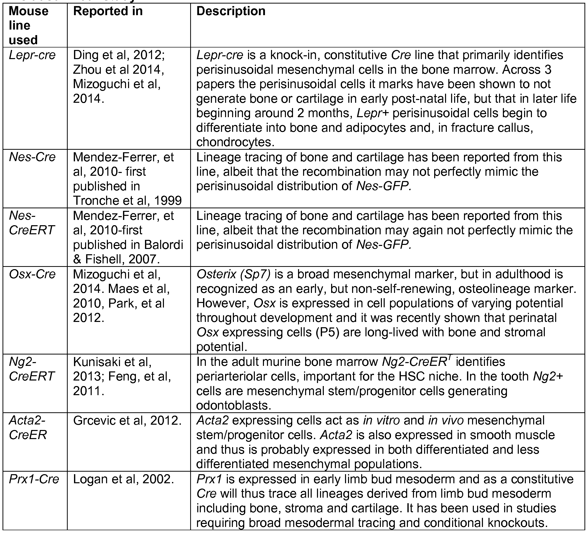
Symbol Description Log2 FC
Acvrl activin A receptor, type 1 6.94
Bmp2 bone morphogenetic protein 2 5.79
Bmp5 bone morphogenetic protein 5 4.36
Bmp6 bone morphogenetic protein 6 2.18
Id2 inhibitor of DNA binding 2 2.56
Greml+ vs Greml - Signficant KEGG Pathways
Name ID pSize onArray ECM-receptor interaction 4512 87 85 PI3K-Akt signaling pathway 4151 356 333 Focal adhesion 4510 205 203 Fc gamma R-mediated phagocytosis 4666 89 87 Natural killer cell mediated cytotoxicity 4650 125 1 12 Osteoclast differentiation 4380 127 1 18 Amoebiasis 5146 120 113 Proteoglycans in cancer 5205 230 222 Cytokine-cytokine receptor interaction 4060 266 227 B cell receptor signaling pathway 4662 78 77 Regulation of actin cytoskeleton 4810 217 210 Aldosterone-regulated sodium reabsorption 4960 40 40 Fc epsilon Rl signaling pathway 4664 71 70 MAPK signaling pathway 4010 259 253 HIF-1 signaling pathway 4066 113 104 VEGF signaling pathway 4370 66 66 Leishmaniasis 5140 66 66 Jak-STAT signaling pathway 4630 156 136 Transcriptional misregulation in cancer 5202 181 164 Axon guidance 4360 133 133 Phosphatidylinositol signaling system 4070 81 79 Long-term depression 4730 61 59 Tuberculosis 5152 179 170 Bacterial invasion of epithelial cells 5100 77 76 Salivary secretion 4970 77 73 Pancreatic secretion 4972 103 99 Leukocyte transendothelial migration 4670 121 1 16 Rheumatoid arthritis 5323 84 81 Viral myocarditis 5416 94 74 Renal cell carcinoma 521 1 69 68 Staphylococcus aureus infection 5150 52 49 Glioma 5214 66 64 Hepatitis B 5161 149 140 Small cell lung cancer 5222 86 85 HTLV-I infection 5166 290 266 Salmonella infection 5132 79 77 Amphetamine addiction 5031 70 68 Viral carcinogenesis 5203 236 191 GnRH signaling pathway 4912 89 87
NDE tA pNDE pPERT pG pG FDR Status
26 4.91 4.E-12 0.00 1.E-08 2.E-06 Activated
48 2.53 3.E-08 0.02 3.E-07 2.E-05 Activated
49 2.36 6.E-17 0.02 4.E-07 2.E-05 Activated
27 1.53 1.E-12 0.12 2.E-06 7.E-05 Not Applicable
21 2.12 4.E-06 0.04 2.E-06 7.E-05 Activated
23 0.94 7.E-07 0.34 5.E-06 1.E-04 Not Applicable
25 0.65 2.E-08 0.52 8.E-06 2.E-04 Not Applicable
36 0.44 9.E-08 0.66 1.E-05 2.E-04 Not Applicable
32 -1.70 1.E-05 0.08 1.E-05 2.E-04 Not Applicable
18 0.13 7.E-07 0.89 1.E-05 2.E-04 Not Applicable
34 -0.10 2.E-07 0.92 1.E-05 2.E-04 Not Applicable
1 1 -0.46 2.E-05 0.67 2.E-04 2.E-03 Not Applicable
14 1.00 8.E-05 0.30 3.E-04 3.E-03 Not Applicable
33 0.07 4.E-05 0.94 4.E-04 4.E-03 Not Applicable
16 1.71 6.E-04 0.07 5.E-04 4.E-03 Not Applicable
13 -1.06 2.E-04 0.28 5.E-04 4.E-03 Not Applicable
13 0.88 2.E-04 0.36 6.E-04 5.E-03 Not Applicable
20 -0.76 2.E-04 0.46 1.E-03 9.E-03 Not Applicable
22 -0.47 5.E-04 0.34 2.E-03 0.01 Not Applicable
19 -0.83 5.E-04 0.40 2.E-03 0.01 Not Applicable
13 -0.73 1.E-03 0.30 3.E-03 0.02 Not Applicable
9 2.1 1 9.E-03 0.04 3.E-03 0.02 Activated
22 0.67 8.E-04 0.49 3.E-03 0.02 Not Applicable
13 -0.36 7.E-04 0.72 4.E-03 0.02 Not Applicable
12 -0.68 2.E-03 0.50 6.E-03 0.03 Not Applicable
15 -0.24 1.E-03 0.82 7.E-03 0.04 Not Applicable
16 0.71 2.E-03 0.46 7.E-03 0.04 Not Applicable
1 1 1.53 1.E-02 0.09 7.E-03 0.04 Not Applicable
12 -0.61 2.E-03 0.54 8.E-03 0.04 Not Applicable
1 1 -0.88 3.E-03 0.39 8.E-03 0.04 Not Applicable
8 -1.43 9.E-03 0.12 9.E-03 0.04 Not Applicable
10 -1.11 5.E-03 0.22 9.E-03 0.04 Not Applicable
18 -0.62 2.E-03 0.53 1.E-02 0.04 Not Applicable
12 1.19 6.E-03 0.23 1.E-02 0.04 Not Applicable
29 0.41 2.E-03 0.68 1.03E-02 0.040327256 Not Applicable
12 0.44 2.E-03 0.65 1.20E-02 0.045777725 Not Applicable
1 1 0.49 3.E-03 0.63 1.26E-02 0.0467475 Not Applicable
22 -0.70 3.E-03 0.57 1.41 E-02 0.049467655 Not Applicable
12 0.98 7.E-03 0.29 1.41 E-02 0.049467655 Not Applicable
Table 6. Greml+ vs Greml - Signficant Reactome Pathways
Rank Name
1 Collagen biosynthesis and modifying enzymes
2 MPS MIC - Sanfilippo syndrome C
3 MPS IV - Morquio syndrome B
4 MPS VI - Maroteaux-Lamy syndrome
5 MPS II - Hunter syndrome
6 Glycosaminoglycan metabolism
7 MPS IX - Natowicz syndrome
8 MPS VII - Sly syndrome
9 MPS I - Hurler syndrome
10 MPS NIB - Sanfilippo syndrome B
1 1 MPS MIA - Sanfilippo syndrome A
12 MPS MID - Sanfilippo syndrome D
13 MPS IV - Morquio syndrome A
14 Fcgamma receptor (FCGR) dependent phagocytosis
15 Cell surface interactions at the vascular wall
16 Collagen degradation
17 Integrin cell surface interactions
18 Assembly of collagen fibrils and other multimeric structures
19 Degradation of the extracellular matrix
20 Signaling by Rho GTPases
21 GPVI-mediated activation cascade
22 Response to elevated platelet cytosolic Ca2+
23 Syndecan interactions
24 Elastic fibre formation
25 Signaling by Hippo
26 Activation of Matrix Metalloproteinases
27 Immunoregulatory interactions between a Lymphoid and a non-Lymphoid cell
28 Regulation of lipid metabolism by Peroxisome proliferator-activated receptor alpha (PPARalpha)
ID pSize onArray NDE tA pNDE pPERT pG pG FDR Status
5605856 52 50 20 1.96 O.E+00 0.05 0.00 7.E-05 Activated
5605255 1 10 108 25 -0.98 O.E+00 0.32 0.00 7.E-05 Not Applicable
5605261 1 10 108 25 -0.96 O.E+00 0.33 0.00 7.E-05 Not Applicable
5605253 110 108 25 -0.96 O.E+00 0.34 0.00 7.E-05 Not Applicable
5605257 1 10 108 25 -0.95 O.E+00 0.34 0.00 7.E-05 Not Applicable
5605249 1 10 108 25 -0.96 O.E+00 0.35 0.00 7.E-05 Not Applicable
5605250 1 10 108 25 -0.95 O.E+00 0.35 0.00 7.E-05 Not Applicable
5605256 1 10 108 25 -0.94 O.E+00 0.35 0.00 7.E-05 Not Applicable
5605258 1 10 108 25 -0.94 O.E+00 0.35 0.00 7.E-05 Not Applicable
5605252 1 10 108 25 -0.94 O.E+00 0.35 0.00 7.E-05 Not Applicable
5605260 1 10 108 25 -0.94 O.E+00 0.35 0.00 7.E-05 Not Applicable
5605259 1 10 108 25 -0.93 O.E+00 0.35 0.00 7.E-05 Not Applicable
5605254 1 10 108 25 -0.95 O.E+00 0.37 0.00 7.E-05 Not Applicable
5605170 72 71 17 -0.53 1.E-06 0.61 0.00 1.E-04 Not Applicable
5605041 83 81 17 1.45 7.E-06 0.13 0.00 1.E-04 Not Applicable
5605823 31 31 10 1.47 1.E-05 0.13 0.00 2.E-04 Not Applicable
5605038 60 58 14 1.00 8.E-06 0.34 0.00 3.E-04 Not Applicable
5605828 27 27 9 -0.91 2.E-05 0.56 0.00 1.E-03 Not Applicable
5605824 94 87 16 -1.05 7.E-05 0.28 0.00 2.E-03 Not Applicable
5605351 100 97 15 1.77 8.E-04 0.06 0.00 4.E-03 Not Applicable
5605042 26 26 8 0.21 1.E-04 0.83 0.00 0.01 Not Applicable
5605037 86 81 14 -0.17 4.E-04 0.93 0.00 0.02 Not Applicable
5605962 18 18 6 0.37 5.E-04 0.74 0.00 0.02 Not Applicable
5605850 36 36 7 -1.32 6.E-03 0.12 0.01 0.03 Not Applicable
5605905 20 20 6 0.29 1.E-03 0.80 0.01 0.04 Not Applicable
5605848 45 39 8 0.93 2.E-03 0.36 0.01 0.04 Not Applicable
5605383 51 43 7 1.66 1.E-02 0.07 0.01 0.04 Not Applicable
5605592 73 73 1 1 -1.10 5.E-03 0.23 0.01 0.04 Not Applicable
Table 7 ECM-Receptor I nteraction Genes in
Greml+ Vs Greml- (fdr<0.05)
Symbol Description Log2 FC
Chad chondroadherin 5.39
Col1 1a1 collagen, type XI, alpha 1 3.77
Col1 1a2 collagen, type XI, alpha 2 4.14
Col27a1 collagen, type XXVII, alpha 1 2.88
Col4a2 collagen, type IV, alpha 2 1.49
Col4a5 collagen, type IV, alpha 5 4.27
Col5a1 collagen, type V, alpha 1 3.09
Col5a2 collagen, type V, alpha 2 2.49
Col6a1 collagen, type VI, alpha 1 5.01
Col6a2 collagen, type VI, alpha 2 4.00
Col6a3 collagen, type VI, alpha 3 4.66
Comp cartilage oligomeric matrix protein 4.00
Dag1 dystroglycan 1 2.35
Hspg2 perlecan (heparan sulfate proteoglycan 2) 2.60
Ibsp integrin binding sialoprotein 3.26
Itga4 integrin alpha 4 -4.24
Itga5 integrin alpha 5 (fibronectin receptor alpha) 3.73
Itga6 integrin alpha 6 2.17
Itgav integrin alpha V 2.81
Itgb3 integrin beta 3 -1.62
Lama4 laminin, alpha 4 1.80
Lamb2 laminin, beta 2 2.50
Npnt nephronectin 3.91
Sdc1 syndecan 1 2.76
Sdc4 syndecan 4 4.91
Thbs3 thrombospondin 3 4.57
A B C
Table 8 PI3K-Akt Signaling Genes in
Greml+ Vs Greml- (fdr<0.05)
Symbol Description Log2 FC
Ccndl cyclin D1 4.35
Ccnd3 cyclin D3 -2.04
Chad chondroadherin 5.39
Col1 1 a1 collagen, type XI, alpha 1 3.77
Col1 1 a2 collagen, type XI, alpha 2 4.14
Col27a1 collagen, type XXVII, alpha 1 2.88
Col4a2 collagen, type IV, alpha 2 1.49
Col4a5 collagen, type IV, alpha 5 4.27
Col5a1 collagen, type V, alpha 1 3.09
Col5a2 collagen, type V, alpha 2 2.49
Col6a1 collagen, type VI, alpha 1 5.01
Col6a2 collagen, type VI, alpha 2 4.00
Col6a3 collagen, type VI, alpha 3 4.66
Comp cartilage oligomeric matrix protein 4.00
Creb3l2 cAMP responsive element binding protein 3-like 2 4.85
Csfl r colony stimulating factor 1 receptor -1.73
Fgf2 fibroblast growth factor 2 5.70
Fgfii fibroblast growth factor receptor 1 2.60
Fgfr2 fibroblast growth factor receptor 2 5.65
Fgfr3 fibroblast growth factor receptor 3 3.86
Ibsp integrin binding sialoprotein 3.26
Igf1 insulin-like growth factor 1 4.1 1
Itga4 integrin alpha 4 -4.24
Itga5 integrin alpha 5 (fibronectin receptor alpha) 3.73
Itga6 integrin alpha 6 2.17
Itgav integrin alpha V 2.81
Itgb3 integrin beta 3 -1.62
Lama4 laminin, alpha 4 1.80
Lamb2 laminin, beta 2 2.50
Lparl lysophosphatidic acid receptor 1 3.03
Lpar4 lysophosphatidic acid receptor 4 4.46
Lpar6 lysophosphatidic acid receptor 6 -3.83
Mapkl mitogen-activated protein kinase 1 -3.32
Myb myeloblastosis oncogene -5.06
Ngf nerve growth factor 5.93
Osmr oncostatin M receptor 5.40
Pdgfa platelet derived growth factor, alpha 1.50
Pik3cd phosphatidylinositol 3-kinase catalytic delta polypeptide -2.58
Pten phosphatase and tensin homolog -3.97
Ptk2 PTK2 protein tyrosine kinase 2 2.83
Rheb Ras homolog enriched in brain 1.33
Sgk1 serum/glucocorticoid regulated kinase 1 2.45
Sos2 son of sevenless homolog 2 (Drosophila) -2.70
Syk spleen tyrosine kinase -4.15
Thbs3 thrombospondin 3 4.57
Tlr4 toll-like receptor 4 -2.88
Vegfa vascular endothelial growth factor A 1.87
Ywhaq tyrosine 3-monooxygenase/tryptophan 5-monooxygenase activation protein, theta polypeptide 1.77
Table 9 Focal Adhesion Genes in
Greml+ Vs Greml- (fdr<0.05)
Symbol Description Log2 FC
Actn4 actinin alpha 4 1.47
Capn2 calpain 2 3.45
Cav1 caveolin 1 , caveolae protein 3.07
Cav2 caveolin 2 3.08
Ccndl cyclin D1 4.35
Ccnd3 cyclin D3 -2.04
Chad chondroadherin 5.39
Col1 1a1 collagen, type XI, alpha 1 3.77
Col1 1a2 collagen, type XI, alpha 2 4.14
Col27a1 collagen, type XXVII, alpha 1 2.88
Col4a2 collagen, type IV, alpha 2 1.49
Col4a5 collagen, type IV, alpha 5 4.27
Col5a1 collagen, type V, alpha 1 3.09
Col5a2 collagen, type V, alpha 2 2.49
Col6a1 collagen, type VI, alpha 1 5.01
Col6a2 collagen, type VI, alpha 2 4.00
Col6a3 collagen, type VI, alpha 3 4.66
Comp cartilage oligomeric matrix protein 4.00
Flnb filamin, beta 2.89
Fine filamin C, gamma 3.35
Ibsp integrin binding sialoprotein 3.26 igfi insulin-like growth factor 1 4.1 1
Itga4 integrin alpha 4 -4.24
Itga5 integrin alpha 5 (fibronectin receptor alpha) 3.73
Itga6 integrin alpha 6 2.17
Itgav integrin alpha V 2.81
Itgb3 integrin beta 3 -1.62
Jun jun proto-oncogene 1.98
Lama4 laminin, alpha 4 1.80
Lamb2 laminin, beta 2 2.50
Mapkl mitogen-activated protein kinase 1 -3.32
Mylk myosin, light polypeptide kinase 2.14
Pak3 p21 protein (Cdc42/Rac)-activated kinase 3 5.49
Parvg parvin, gamma -3.07
Pdgfa platelet derived growth factor, alpha 1.50
Pik3cd phosphatidylinositol 3-kinase catalytic delta polypeptide -2.58
Prkcb protein kinase C, beta -3.85
Prkcg protein kinase C, gamma 2.75
Pten phosphatase and tensin homolog -3.97
Ptk2 PTK2 protein tyrosine kinase 2 2.83
Rac2 RAS-related C3 botulinum substrate 2 -3.03
Sos2 son of sevenless homolog 2 (Drosophila) -2.70
Thbs3 thrombospondin 3 4.57
Vasp vasodilator-stimulated phosphoprotein -1.39
Vav1 vav 1 oncogene -4.46
Vav2 vav 2 oncogene 2.38
Vav3 vav 3 oncogene -2.01
Vegfa vascular endothelial growth factor A 1.87
Xiap X-linked inhibitor of apoptosis -2.45
Table 10 Osteoblast differentiation GO:0001649
Symbol probeid logFC P.Value fdr
Shox2 1438042_ .at 7.13969309 1.12E-05 0.02471733
Fgfr2 1433489, _s_a 5.65231788 1.51E-05 0.02471733
Bmp2 1423635, .at 5.78726506 5.84E-05 0.02471733
Col9al 1421381, _a_a 5.14384919 7.20E-05 0.02471733
Sox9 1424950, .at 6.90133905 0.0001102 0.02471733
Comp 1419527, .at 3.99952788 0.00013709 0.02523526
Papss2 1421987, .at 5.85768973 0.00013725 0.02523526
CollOal 1422253, .at 4.64970077 0.00015096 0.02523526
Matnl 1418477, .at 5.63933284 0.00019186 0.02642403
Impadl 1437290, .at 4.00009567 0.00019808 0.02656323
Ltbp3 1437833, .at 2.9661723 0.00021743 0.02704011
Sp7 1418425, .at 3.10351174 0.00026166 0.02862484
Ddr2 1422738, .at 3.89722768 0.00026991 0.02873843
Ankrdll 1458452, .at -1.7496439 0.00042781 0.03083902
Bmp6 1450759, .at 2.1776606 0.00052763 0.03237488
Thbs3 1416623, .at 4.56764372 0.00056966 0.03323698
Ptprc 1440165, .at -5.6734651 0.00064536 0.03437141
Igfl 1419519, .at 4.10582922 0.00076686 0.03552034
Mef2c 1451506, .at 1.5622972 0.00077261 0.03552034
GN3 1456067, .at 2.45472986 0.00081314 0.0360597
Insigl 1454671, .at 2.08360386 0.00085701 0.03646133
Csgalnactl 1452365, .at 6.10211317 0.00087003 0.03646133
Asxl2 1460597, .at -2.6979367 0.00092542 0.0368767
Hspg2 1418670, _s_a 2.60149201 0.00098897 0.03770361
Dlx5 1449863, _a_a 3.7588245 0.00106207 0.03918556
Sparc 1416589, .at 1.60412839 0.00113447 0.04021149
Slc38al0 1427295, .at 1.83634246 0.0012518 0.04142547
Has2 1449169, .at 4.99235834 0.00126234 0.04164276
Fgfr3 1421841, .at 3.86261057 0.00148679 0.04425268
Npr2 1427191, .at 3.46860457 0.00168539 0.04660501
Sulf2 1442408, .at 1.6036124 0.00181894 0.04807212
Serpinhl 1450843, _a_a 1.29654547 0.00188638 0.04851906
Sema4d 1420824, .at -1.8029043 0.00194795 0.04888768
Phospho2 1425190, _a_a -2.8140356 0.00197866 0.04910038
Alpl 1423611, .at 2.96345394 0.00315219 0.05979792
Cadml 1417376, _a_a 4.10590728 0.00374974 0.06413052
Smadl 1448208, .at 3.17101958 0.00393954 0.06521738
Fat4 1459749, _s_a 3.23899393 0.00402532 0.06597077
Runx2 1424704 at 2.8878129 0.0055077 0.0751405
Rarg 1419415, _a_a 1.55028671 0.0058013 0.07697684
Insig2 1417980, _a_a 2.44324681 0.00584458 0.07727179
Sik3 1460439_ .at -1.0857648 0.00755719 0.08777671
Cyp26bl 1460011_ .at 2.41494558 0.0078034 0.0892175
Glgl 1460554, _s_a 1.08231524 0.00954803 0.09811991
Pex7 1418988, .at -1.5114512 0.01007962 0.1014008
Gnas 1450186, _s_a 1.15793611 0.01017669 0.1019462
Smad5 1433641, .at 1.32605317 0.01176342 0.10947652
Ostc 1449139, .at 1.47773534 0.01312531 0.1164395
Plxnbl 1435254, .at 2.94240991 0.01342921 0.11774318
Bbx 1425835, _a_a 2.44695116 0.01448366 0.12264883
Twistl 1418733, .at 1.38261178 0.01576956 0.1278689
Nabl 1438819, .at -2.9216854 0.01616797 0.1294138
Asxll 1458380, .at -1.3363295 0.02003387 0.1445676
Osr2 1426155, _a_a 0.50221439 0.02482068 0.16093223
Setdbl 1451833, _a_a -0.705544 0.02602998 0.16493091
Pthlh 1422324, _a_a 2.22023888 0.02765368 0.17061675
Ift80 1427568, _a_a 2.13652764 0.03549998 0.19425924
Col2al 1450567, _a_a 0.51632868 0.03698764 0.19875846
Cited2 1452207, .at 0.77720105 0.038182 0.20226056
Paxl 1449359, .at 3.12937179 0.03911755 0.20500123
Ski 1426373, .at 1.20823577 0.03974484 0.20676946
Rhoa 1437628, _s_a -0.4614252 0.04469134 0.22031089
Bnc2 1438861, .at 4.23430725 0.04850308 0.23058618
Whscl 1435136, .at -1.0033239 0.06021278 0.25860931
Inppll 1460394, _a_a 0.56759927 0.06832218 0.27657199
Mcphl 1439115, .at -1.2317502 0.07426703 0.289485
FgflS 1449545, .at 1.95630563 0.08013069 0.30080298
Hoxall 1420414, .at 0.51117163 0.134279 0.39421853
Ctcl 1423656, _x_a -0.3454466 0.13779297 0.40027058
Sh3pxd2b 1442919, .at 0.7836116 0.1378523 0.40031788
Eve 1448876, .at 1.72036865 0.14073692 0.40431581
Lrp6 1451022, .at 1.84723464 0.14520466 0.41054822
Greml 1425357, _a_a 2.03742601 0.15410452 0.42297152
Amerl 1439565, .at -1.2063171 0.16004689 0.43090105
Sex 1456291, _x_a 0.27534435 0.16950076 0.44294863
Ptger4 1424208, .at -0.9729169 0.17847174 0.45532605
Ryrl 1427306, .at -0.2950732 0.19148114 0.47135849
Rab23 1454876, .at 0.859713 0.19401749 0.47439891
Trim45 1441412, _s_a 0.20342791 0.20276127 0.48402773
Axin2 1436845 at 0.38799213 0.21124748 0.49331913
Lrrcl7 1429679_at 0.23301036 0.23562804 0.52060256
Pitx2 1424797_a_a 1.07081967 0.23628235 0.52138414
Carml 1419743_s_a 0.64722551 0.2531193 0.53909773
Dym 1423736_a_a -0.9150064 0.27126619 0.55550201
Su lfl 1436319_at 1.19568804 0.27432954 0.5581008
Smad9 1450265_at 0.16221583 0.2789619 0.56241214
Prpsap2 1452062_at -0.3149736 0.28405301 0.56696207
Msx2 1438351_at 0.35290248 0.29922583 0.58127152
Mef2d 1421388_at -0.1866246 0.31750884 0.59885034
Nppc 1422790_at 0.1607409 0.31851616 0.59964979
Fam73b 1454621_s_a 0.2994231 0.32117143 0.60193506
Acp5 1431609_a_a -0.9309661 0.33111888 0.61004404
Fgf4 1420086_x_a 0.18263932 0.35648536 0.63248628
Cbs 1423844_s_a 0.20063418 0.36616507 0.64041613
Wntl 1425377_at 0.36975782 0.37967184 0.65054435
T 1419304_at 0.16388178 0.39058468 0.65912444
Collal 1423669_at -0.6212641 0.40659023 0.6718836 Hoxb4 1451761_at -0.1272661 0.46260484 0.71610719 Sp5 1422914_at -0.1174096 0.4953465 0.73863064
Hoxdll 1450584_at 0.09335222 0.51678271 0.75267603
Bmp4 1422912_at -0.1515746 0.52844446 0.76072052
Fgf8 1451882_a_a 0.08277176 0.56350817 0.78452791
Rarb 1454906_at 0.13246962 0.58074285 0.79545915
Msxl 1417127_at -0.0792768 0.6088118 0.81219928
Por 1416933_at -0.0809813 0.64020848 0.83040587
Thbsl 1460302_at -0.0940899 0.6403299 0.83041932
Rara 1450180_a_a -0.1450325 0.65688912 0.83924187
Lrp5 1449299_at 0.12006994 0.66717382 0.84534924
Tfap2a 1421996_at 0.04882766 0.73011248 0.87883831
Lep 1422582_at -0.051409 0.74333518 0.88629937
Spns2 1451601_a_a -0.0930905 0.75077046 0.89005592
Nab2 1417930_at -0.1414283 0.76360144 0.89720433
Sfrp2 1448201_at 0.26766348 0.76534268 0.89810537
Dchsl 1429163_at 0.03405801 0.85190273 0.93849266
Sbds 1426480_at -0.0651239 0.88207204 0.95094397
Bglap2 1449880_s_a 0.03089977 0.96146152 0.98359981
Cdxl 1449582_at -0.0012411 0.99265715 0.99719485
Freml 1455280 at 0.0010303 0.99394523 0.99782363
Table 11 Chondrocyte differentiation GO:0002062
Symbol probeid logFC P.Value globalfdr
Sox5 1452511. _at 7.22767132 7.96E-06 0.02471733
Frzb 1416658. _at 6.48749238 8.83E-06 0.02471733
Shox2 1438042. .at 7.13969309 1.12E-05 0.02471733
Trpsl 1438214. _at 3.40755204 4.76E-05 0.02471733
Cytll 1456793. _at 6.87459727 5.09E-05 0.02471733
Bmp2 1423635. _at 5.78726506 5.84E-05 0.02471733
Collla2 1423578. _at 4.14206689 6.46E-05 0.02471733
Pkdcc 1454838. _s_ a 7.28962976 6.74E-05 0.02471733
Col9al 1421381. _a_ E 5.14384919 7.20E-05 0.02471733
Sox9 1424950. _at 6.90133905 0.0001102 0.02471733
Tgfb2 1423250. _a_ c 5.54649939 0.00012244 0.02520744
Comp 1419527. .at 3.99952788 0.00013709 0.02523526
CollOal 1422253. .at 4.64970077 0.00015096 0.02523526
Matnl 1418477. .at 5.63933284 0.00019186 0.02642403
Impadl 1437290. .at 4.00009567 0.00019808 0.02656323
Sox6 1447655. _x_ 33.04224037 0.00020449 0.0267325
Ltbp3 1437833. .at 2.9661723 0.00021743 0.02704011
Nfib 1434101. .at 2.200371 0.00030068 0.02873843
Chstll 1450509. .at 4.02884726 0.00032235 0.02907679
Creb3l2 1452381. .at 4.85293127 0.00046023 0.03115804
Bmp6 1450759. .at 2.1776606 0.00052763 0.03237488
Colllal 1418599. .at 2.34126506 0.00053144 0.03247748
Thbs3 1416623. .at 4.56764372 0.00056966 0.03323698
Mmpl3 1417256. .at 4.24452375 0.00061702 0.03397116
Cyr61 1438133. _a_ E 5.69119846 0.00063681 0.03419138
Mia3 1459984. .at 2.37058997 0.00065816 0.03447572
Hifla 1448183. _a_ E 1.72827087 0.00074424 0.03532241
Bmp5 1455851. .at 4.3611194 0.00075454 0.03535411
Mef2c 1451506. .at 1.5622972 0.00077261 0.03552034
Fgf2 1449826. _a_ E 5.70455759 0.00078373 0.03566748
GN3 1456067. .at 2.45472986 0.00081314 0.0360597
Pthlr 1417092. .at 5.14446867 0.00081315 0.0360597
Acan 1449827. .at 3.41833726 0.0008224 0.03611613
Fgfrl 1424050. .s_' a 2.60129371 0.00086507 0.03646133
Csgalnactl 1452365. .at 6.10211317 0.00087003 0.03646133
Hspg2 1418670_ _s_; a 2.60149201 0.00098897 0.03770361
Mgp 1448416_ .at 3.3507084 0.00116189 0.0404339
Snai2 1418673_ .at 5.03701152 0.00128582 0.04171829
Fgfr3 1421841_ .at 3.86261057 0.00148679 0.04425268
Maf 1437473_at 3.76980305 0.00153687 0.04489271
Ctgf 1416953_at 2.49942916 0.00172491 0.04701858
Sulf2 1442408_at 1.6036124 0.00181894 0.04807212
Serpinhl 1450843_a_£ 1.29654547 0.00188638 0.04851906
Thra 1443952_at 1.82594965 0.00237175 0.05300708
Zbtbl6 1419874_x_a 3.80377306 0.00239811 0.05325226
Bmp8a 1449873_at 2.20192492 0.00384863 0.06493723
Smadl 1448208_at 3.17101958 0.00393954 0.06521738
Pkdl 1460210_at 2.9085668 0.00405261 0.06613797
Bmp7 1418910_at 1.92696736 0.00418937 0.06714193
Mex3c 1444701_at 2.84275529 0.00439563 0.06871649
Bmprla 1425492_at 1.36200773 0.00516927 0.07349907
Runx2 1424704_at 2.8878129 0.0055077 0.0751405
Rarg 1419415_a_£ 1.55028671 0.0058013 0.07697684
Barx2 1421761_a_£ 0.85624097 0.00640975 0.08068276
Tgfbl 1445360_at -1.8533881 0.00659249 0.08152761
Lectl 1460258_at 4.58986317 0.00697288 0.08388477
Ctnnbl 1450008_a_£ 1.66261927 0.00720075 0.08544095
Tgfbr2 1426397_at 1.86659547 0.0075086 0.08755963
Sik3 1460439_at -1.0857648 0.00755719 0.08777671
Hoxc4 1422870_at 1.19796075 0.00761056 0.08803383
Otor 1425083_at 4.22357952 0.00876964 0.09435106
Ror2 1457128_at 4.33909419 0.008963 0.09531397
Glgl 1460554_s_a 1.08231524 0.00954803 0.09811991
Tgfbrl 1420893_a_£ -1.553593 0.00976607 0.09956142
Gnas 1450186_s_a 1.15793611 0.01017669 0.1019462
Smad5 1433641_at 1.32605317 0.01176342 0.10947652
Prkca 1427562_a_£ 2.10613284 0.01358718 0.11834592
Hes5 1456010_x_a -2.1231445 0.01655693 0.13093705
GN2 1459211_at 1.04059295 0.0176753 0.13518291
Mapkl4 1426104_at -1.5532261 0.02097546 0.14796945
Lnp 1453035_at -2.9755281 0.02392091 0.15807429
Osr2 1426155_a_£ 0.50221439 0.02482068 0.16093223
Hmga2 1450781_at 1.09586861 0.02694941 0.16831614
Pthlh 1422324_a_£ 2.22023888 0.02765368 0.17061675
Wnt9a 1436978_at 0.87174088 0.02785817 0.17122255
Bmprlb 1437312_at 1.25973479 0.02801041 0.17173704
Prrxl 1432129_a_£ 1.47773595 0.0280624 0.17191557
Foxdl 1418876_at 4.46730525 0.03043986 0.17907112
Ift80 1427568_a_£ 2.13652764 0.03549998 0.19425924
Col2al 1450567 a £ 0.51632868 0.03698764 0.19875846
Thrb 1422202_at 2.48066506 0.04363391 0.21759789
Atp7a 1418774_a_ -1.6739022 0.04473941 0.22045143
Mapk3 1427060_at -0.665697 0.04913025 0.23233967
Wnt5a 1448818_at 2.93263031 0.0492736 0.23264251
Bmpl 1427457_a_ 2.43028686 0.05127348 0.23736193
Rela 1419536_a_ 1.04767874 0.05767973 0.25345212
Hoxa5 1443803_x_ 1.0275933 0.06149235 0.26170129
Esrra 1442864_at -0.6446893 0.06725047 0.27435072
Fgfl8 1449545_at 1.95630563 0.08013069 0.30080298
Hoxa3 1427433_s_ 0.507085 0.08120338 0.30287153
Zbtb7a 1437255_at -0.3664643 0.09699201 0.33330316
Runx3 1440275_at 0.96780328 0.09793019 0.33494632
Hoxb3 1427605_at 0.39418388 0.09973918 0.33804288
Ednl 1451924_a_ -0.2982996 0.10195385 0.34187512
Eif2ak3 1430371_x_ -1.4225496 0.11979267 0.37163913
Satb2 1425904_at 0.48646604 0.12821542 0.38470549
Bbsl 1437310_at 1.90386954 0.12829484 0.38470549
Hoxall 1420414_at 0.51117163 0.134279 0.39421853
Bmp8b 1440706_at 0.26308768 0.13555736 0.39655082
Foxd2 1442315_at 0.46320213 0.14384418 0.40876544
Lrp6 1451022_at 1.84723464 0.14520466 0.41054822
Zebl 1418926_at 1.16354749 0.14660325 0.41268652
Fgf9 1438718_at -0.3966601 0.15928736 0.42979473
Sex 1456291_x_ 0.27534435 0.16950076 0.44294863
Wnt7a 1458334_at 0.23058566 0.17829566 0.45510852
Mkks 1422627_a_ 0.62949897 0.20423492 0.48574462
Bbs2 1424478_at 1.18847208 0.20451523 0.48587449
Axin2 1436845_at 0.38799213 0.21124748 0.49331913
Arid5a 1451340_at -0.3128111 0.21194496 0.49438477
Handl 1417525_at 0.20127201 0.24088701 0.52659659
Carml 1419743_s_ 0.64722551 0.2531193 0.53909773
Su lfl 1436319_at 1.19568804 0.27432954 0.5581008
Smad9 1450265_at 0.16221583 0.2789619 0.56241214
Mycn 1417155_at 0.39769166 0.2893957 0.57218164
Pitxl 1419514_at 0.1702969 0.29146369 0.57395417
Msx2 1438351_at 0.35290248 0.29922583 0.58127152
Prrx2 1432331_a_ 0.73191217 0.30091849 0.58270253
Wnt7b 1420892_at 0.15715266 0.31454776 0.59632366
Mef2d 1421388_at -0.1866246 0.31750884 0.59885034
Nppc 1422790_at 0.1607409 0.31851616 0.59964979
Chrdl2 1420539 a 0.20332488 0.34147543 0.61922033
Uncx 1419633_at 0.14875918 0.35394085 0.62995605
Fgf4 1420086_x_a 0.18263932 0.35648536 0.63248628
Cbs 1423844_s_a 0.20063418 0.36616507 0.64041613
CstlO 1449447_at 0.19712187 0.36842711 0.6420478
Gdf5 1419139_at 0.12908806 0.37130522 0.6441108
Foxd4 1422318_at -0.1344761 0.3797373 0.65058051
Foxd3 1422210_at 0.12298744 0.39927456 0.66574299
Collal 1423669_at -0.6212641 0.40659023 0.6718836
Fgf6 1427582_at 0.13306027 0.43564081 0.69508034
Nog 1422300_at 0.1532112 0.44376711 0.70129789
Smad3 1450472_s_a 0.28466688 0.44828933 0.70498612
Fbxw4 1417226_at 0.15445078 0.48947258 0.73441242
Six2 1427436_at 0.11798718 0.49326442 0.73685768
Dlx2 1448877_at 0.17993045 0.50144031 0.74322059
Hoxdll 1450584_at 0.09335222 0.51678271 0.75267603
Bmp4 1422912_at -0.1515746 0.52844446 0.76072052
Rarb 1454906_at 0.13246962 0.58074285 0.79545915
Msxl 1417127_at -0.0792768 0.6088118 0.81219928
Pax7 1452510_at -0.0726034 0.61587116 0.81680712
Por 1416933_at -0.0809813 0.64020848 0.83040587
Thbsl 1460302_at -0.0940899 0.6403299 0.83041932
Rara 1450180_a_E -0.1450325 0.65688912 0.83924187
Osrl 1449350_at 0.06674263 0.65796737 0.83979472
Nkx3-2 1421464_at 0.07259392 0.65988471 0.84131336
Snail 1448742_at 0.051586 0.71227826 0.86945623
Hand2 1436041_at 0.05183208 0.71914945 0.87278294
Lep 1422582_at -0.051409 0.74333518 0.88629937
Sfrp2 1448201_at 0.26766348 0.76534268 0.89810537
Myf5 1420757_at 0.03687913 0.79843488 0.91361693
Ihh 1450704_at 0.03067369 0.82163053 0.9243161
Efempl 1427183_at 0.22630155 0.84645157 0.93572931
Hoxd3 1421537_at -0.0194301 0.90332787 0.96012428
Rspo2 1455893_at -0.0229601 0.96084842 0.9833494
Table 12 Adipocyte Differentiation GO:0045444
Symbol probeid logFC P.Value fdr
Frzb 1416658_ .at 6.48749238 8.83E-06 0.02471733
Scdl 1415965_ .at 6.92946129 2.02E-05 0.02471733
Wifl 1425425, _a_a 5.61016474 2.97E-05 0.02471733
Wwtrl 1417818, .at 4.26966834 4.04E-05 0.02471733
Bmp2 1423635, .at 5.78726506 5.84E-05 0.02471733
Medag 1452244, .at 2.54509537 0.00012336 0.02520744
Fndc3b 1433833, .at 4.14105154 0.00016123 0.02523526
Id2 1435176, _a_a 2.55980464 0.00017265 0.02523526
Enppl 1419276, .at 4.18103881 0.0001804 0.025505
Ccndl 1448698, .at 4.35237065 0.0006197 0.03397116
Selenbpl 1450699, .at -2.0726353 0.00069184 0.03471676
Plcbl 1435043, .at 4.48210625 0.00069203 0.03471676
Igfl 1419519, .at 4.10582922 0.00076686 0.03552034
4932438A13F 1444660, .at -1.7217063 0.00083553 0.03627182
Insigl 1454671, .at 2.08360386 0.00085701 0.03646133
Asxl2 1460597, .at -2.6979367 0.00092542 0.0368767
Itga6 1422445, .at 2.16501935 0.00095162 0.03712712
Eroll 1449324, .at -5.2415423 0.00109722 0.03974742
Klf4 1417394, .at 2.49143131 0.00117445 0.04059755
Snai2 1418673, .at 5.03701152 0.00128582 0.04171829
Lama4 1424807, .at 1.79871601 0.00144798 0.04381912
Plac8 1451335, .at -3.9343763 0.00197629 0.04910038
Zbtbl6 1419874, _x_a 3.80377306 0.00239811 0.05325226
Rgs2 1419248, .at -2.0621651 0.00309171 0.05930885
Id4 1423259, .at 3.91112981 0.00323253 0.06026486
Egr2 1427683, .at 2.26204995 0.00334336 0.06120178
Gpxl 1460671, .at -1.5018199 0.00419115 0.06714193
1100001G20I 1434484, .at -2.8361979 0.00426222 0.06782998
Zfpm2 1449314, .at 2.05338287 0.0043193 0.06807548
Mex3c 1444701, .at 2.84275529 0.00439563 0.06871649
Lamb3 1417812, _a_a 1.6568793 0.00481526 0.07143995
Osbpl8 1437069, .at -1.0291454 0.00523485 0.07380639
Tcf7l2 1429428, .at 2.81979243 0.00526505 0.07387511
Fcor 1439834, .at -1.0060617 0.00590615 0.07772749
Nipbl 1442103, .at -1.2510927 0.00646363 0.08095419
Tgfbl 1445360, .at -1.8533881 0.00659249 0.08152761
Psmb8 1422962, _a_a -2.8058177 0.00674644 0.08249817
Crebl 1428755, .at -2.7091354 0.00842715 0.09235384
Bnip3 1422470, .at 4.08594527 0.00951743 0.09797892
Creb5 1457222_at 2.78212192 0.01161505 0.10890856
Aktl 1425711_a_ 0.64331207 0.01322662 0.1168866
Pexlla 1419365_at 3.11498759 0.01342737 0.11774318
Wnt5b 1422602_a_ 1.82156613 0.015113 0.12499747
Adrb2 1437302_at -1.425485 0.01631237 0.13005204
Osbplll 1436027_at -1.5133477 0.01644253 0.13044409
Bbsl2 1447275_at 2.08525895 0.01676428 0.13158474
Dactl 1417937_at 2.66826005 0.01872908 0.13938951
Asxll 1458380_at -1.3363295 0.02003387 0.1445676
Arl4a 1425411_at 1.29146942 0.02051407 0.14648592
Sox8 1435438_at 3.11014067 0.02223232 0.15292445
Zc3hl2a 1443993_at -1.3946394 0.02318622 0.15612903
Zfp385a 1418865_at 0.54201008 0.02368729 0.15748576
Hmga2 1450781_at 1.09586861 0.02694941 0.16831614
Almsl 1456950_at -0.9174364 0.02699956 0.16844556
Tgfblil 1418136_at 2.24534212 0.03175692 0.18296741
Jdp2 1450350_a_ -1.716068 0.03804747 0.20180716
Crebbp 1459804_at -1.3274553 0.0394602 0.20599124
Lrgl 1417290_at -1.3965514 0.04589125 0.22371225
Wnt5a 1448818_at 2.93263031 0.0492736 0.23264251
Adipoq 142265 l_at -0.9677413 0.04950593 0.23318715
Sirtl 1418640_at 0.66458786 0.04979551 0.23372124
Jagl 1434070_at 1.60242889 0.05099174 0.23672447
Arid5b 1458238_at -0.6856622 0.05148946 0.23793299
Ptgs2 1417263_at 1.37192735 0.0526271 0.24106591
Gata2 1450333_a_ -0.4200574 0.0567174 0.25105281
Medl 1450402_at -1.5719065 0.05957002 0.25726603
Dlk2 1420807_a_ 0.37367895 0.06840172 0.27672997
Axinl 1426966_at 1.169289 0.06902748 0.27796505
Ppard 1425703_at 0.43412189 0.07675127 0.29440228
Retn 1449182_at -0.3872131 0.08151515 0.3035349
Cebpa 1418982_at -0.6341423 0.08246593 0.30538598
Tbl lx 1455042_at -0.5439523 0.08527682 0.31094428
Cebpb 1418901_at -0.9722819 0.08544421 0.31125265
Cbyl 1451305_at 0.67313195 0.08765196 0.31554846
Mb 1451203_at -0.5113074 0.08860675 0.31723829
Ncor2 1451841_a_ 0.34038456 0.09045653 0.32098189
Gsk3b 1439931_at 0.83461443 0.09061475 0.32122473
Crebl2 1442738_at -0.6352672 0.10791559 0.35194886
Aldh6al 1448104_at 2.58116441 0.10869456 0.35303424
Eif2ak3 1430371 x -1.4225496 0.11979267 0.37163913
Fabp4 1417023. _a_a -1.6804129 0.121138 0.37369664
Ccdc85b 1435589, .at 0.27929059 0.12208188 0.37513349
Rarres2 1425091. .at 0.31628662 0.12782516 0.38425933
Socsl 1450446. _a_a 0.28564968 0.12917782 0.38602244
Sh3pxd2b 1442919. .at 0.7836116 0.1378523 0.40031788
Fam57b 1454209. .at 0.24715081 0.14388936 0.40878418
Taf8 1416450. .at 0.34013535 0.14511102 0.41054822
Lrp6 1451022. .at 1.84723464 0.14520466 0.41054822
Runxltl 1448785. .at 0.44754277 0.1596707 0.43041708
Trib2 1426641. .at 0.89133272 0.18342065 0.46176136
Ankrd26 1436071. .at 0.36205722 0.20661702 0.48819333
Aamdc 1451381. .at 1.06848603 0.21899798 0.50195801
Trib3 1426065. _a_a 0.295149 0.23368155 0.51862483
Lpinl 1426516. _a_a 0.58391765 0.24135771 0.52699651
Zfpml 1451046. .at -0.1891087 0.25038992 0.53649473
Noc3l 1437500. .at 0.78374168 0.25077931 0.53689351
Carml 1419743. _s_a 0.64722551 0.2531193 0.53909773
Ctbp2 1422887. _a_a -0.4047828 0.27076333 0.55490058
Slc2a4 1415959. .at 0.17614707 0.27374878 0.55774954
Msx2 1438351. .at 0.35290248 0.29922583 0.58127152
Ctbpl 1415702. _a_a 0.63070087 0.30707878 0.58884036
Bscl2 1420632. _a_a 0.20870757 0.31977026 0.60081479
Socs7 1420766. .at 0.74540021 0.33293722 0.61166653
Prdml6 1429309. .at 0.14230801 0.3401689 0.61832364
Sfrpl 1428136. .at 0.16311129 0.34404904 0.62158781
Mrap 1451371. .at -0.1599235 0.35444922 0.630343
Mettl8 1451141. .at 0.66162887 0.3773748 0.6487262
Wntl 1425377. .at 0.36975782 0.37967184 0.65054435
Gm6484 1427422. .at 0.12539848 0.40792345 0.67301857
Adrb3 1421555. .at -0.1013874 0.48109302 0.72872465
Aloxe3 1449237. .at 0.11167171 0.49007502 0.734659
Sod2 1454976. .at -0.4642386 0.49858924 0.74112024
Sh2b2 1450718. .at 0.33787655 0.50093283 0.7428897
Nudt7 1430896. _s_a -0.5009291 0.52179927 0.75602895
Hesl 1418102. .at 0.12158584 0.55711349 0.78024953
Gprll6 1440225. .at -0.6471556 0.57903346 0.79420315
Dkkll 1417787. .at 0.07057331 0.60962 0.81252932
Uchll 1448260. .at 0.0721505 0.6101289 0.81282636
Cebpd 1456605. .at 0.18895067 0.63866748 0.82943279
Gata3 1448886. .at -0.083382 0.65162031 0.83654281
Pparg 1420715. _a_a -0.0670199 0.66484103 0.84418045
Fndc5 1435115_at 0.09998857 0.66491922 0.84418045
Lrp5 1449299_at 0.12006994 0.66717382 0.84534924
Adig 1424729_at 0.05957171 0.67676323 0.8507174
Mmpll 1417234_at 0.08486384 0.71762364 0.87217505
Lep 1422582_at -0.051409 0.74333518 0.88629937
Sfrp2 1448201_at 0.26766348 0.76534268 0.89810537
Wnt3a 1422093_at -0.0387946 0.81145358 0.91983231
Adrbl 1423420_at -0.0217709 0.87321293 0.94746113
WntlOb 1426091_a_a 0.02676453 0.92652487 0.96924821
FgflO 1420690 at -0.0036086 0.98699864 0.99442913
REFERENCES
[0168] All references cited herein are hereby incorporated by reference in their entirety.
Ashburner, M., Ball, C.A., Blake, J.A., Botstein, D., Butler, H., Cherry, J.M., Davis, A.P., Dolinski, K., Dwight, S.S., Eppig, J.T., et al. (2000). Gene ontology: tool for the unification of biology. The Gene Ontology Consortium. Nat Genet 25, 25-29.
Barker, N., van Es, J.H., Kuipers, J., Kujala, P., van den Born, M., Cozijnsen, M., Haegebarth, A., Korving, J., Begthel, H., Peters, P.J., et al. (2007). Identification of stem cells in small intestine and colon by marker gene Lgr5. Nature 449, 1003-1007.
Barrett, T., Suzek, T.O., Troup, D.B., Wilhite, S.E., Ngau, W.C., Ledoux, P., Rudnev, D., Lash, A.E., Fujibuchi, W., and Edgar, R. (2005). NCBI GEO: mining millions of expression profiles- database and tools. Nucleic Acids Res 33, D562-566.
Belkind-Gerson, J., Carreon-Rodriguez, A., Benedict, L.A., Steiger, C, Pieretti, A., Nagy, N., Dietrich, J., and Goldstein, A.M. (2013). Nestin-expressing cells in the gut give rise to enteric neurons and glial cells. Neurogastroenterology and motility : the official journal of the European Gastrointestinal Motility Society 25, 61-69 e67.
Benazet, J.-D., Bischofberger, M., Tiecke, E., Goncalves, A., Martin, J.F., Zuniga, A., Naef, F., and Zeller, R. (2009). A Self-Regulatory System of Interlinked Signaling Feedback Loops Controls Mouse Limb Patterning. Science 323, 1050-1053.
Benjamini, Y., and Hochberg, Y. (1995). Controlling the false discovery rate; A practical and powerful approach to multiple testing. J Roy Stat Soc Ser B 57, 289-300.
Bianco, P., Cao, X., Frenette, P.S., Mao, J.J., Robey, P.G., Simmons, P.J., and Wang, C.Y. (2013). The meaning, the sense and the significance: translating the science of mesenchymal stem cells into medicine. Nature medicine 19, 35-42.
Bolstad, B.M., Collin, F., Bretttscneider, J., Simpson, K., Cope, L.M., Irizarry, R., and Speed, T.P. (2005). Quality Assesment of Affymetrix GeneChip Data. In Bioinformatics and Computaional Biology Solutions Using R and Bioconductor, R. Gentleman, V. Carey, W. Huber, I. RA, and S. Dudoit, eds. (New York: Springer).
Canalis, E., Parker, K., and Zanotti, S. (2011). Gremlinl is required for skeletal development and postnatal skeletal homeostasis. J Cell Physiol.
Clevers, H., and Batlle, E. SnapShot: The Intestinal Crypt. Cell 152, 1198-1 198.el 192.
Crisan, M., Yap, S., Casteilla, L., Chen, C.-W., Corselli, M., Park, T.S., Andriolo, G., Sun, B., Zheng, B., Zhang, L., et al. (2008). A Perivascular Origin for Mesenchymal Stem Cells in Multiple Human Organs. Cell stem cell 3, 301-313.
Delorme, B., Ringe, J., Pontikoglou, C, Gaillard, J., Langonne, A., Sensebe, L., Noel, D., Jorgensen, C, Haupl, T., and Charbord, P. (2009). Specific lineage-priming of bone marrow mesenchymal stem cells provides the molecular framework for their plasticity. Stem cells 27, 1142-1151.
Ding, L., Saunders, T.L., Enikolopov, G., and Morrison, S.J. (2012). Endothelial and perivascular cells maintain haematopoietic stem cells. Nature 481, 457-462.
Dominici, M., Le Blanc, K., Mueller, I., Slaper-Cortenbach, I., Marini, F., Krause, D., Deans, R., Keating, A., Prockop, D., and Horwitz, E. (2006). Minimal criteria for defining multipotent mesenchymal stromal cells. The International Society for Cellular Therapy position statement. Cytotherapy 8, 315-317.
Dranovsky, A., Picchini, A.M., Moadel, T., Sisti, A.C., Yamada, A., Kimura, S., Leonardo, E.D., and Hen, R. (2011). Experience dictates stem cell fate in the adult hippocampus. Neuron 70, 908- 923.
Ducy, P., Zhang, R., Geoffroy, V., Ridall, A.L., and Karsenty, G. (1997). Osf2/Cbfal : a transcriptional activator of osteoblast differentiation. Cell 89, 747-754.
Friedenstein, A.J., Chailakhyan, R.K., Latsinik, N.V., Panasyuk, A.F., and Keiliss-Borok, I.V. (1974a). Stromal cells responsible for transferring the microenvironment of the hemopoietic tissues. Cloning in vitro and retransplantation in vivo. Transplantation 17, 331-340.
Friedenstein, A.J., Deriglasova, U.F., Kulagina, N.N., Panasuk, A.F., Rudakowa, S.F., Luria, E.A., and Ruadkow, LA. (1974b). Precursors for fibroblasts in different populations of hematopoietic cells as detected by the in vitro colony assay method. Exp Hematol 2, 83-92.
Gentleman, R.C., Carey, V.J., Bates, D.M., Bolstad, B., Dettling, M., Dudoit, S., Ellis, B., Gautier, L., Ge, Y., Gentry, J., et al. (2004). Bioconductor: open software development for computational biology and bioinformatics. Genome Biol 5, R80.
Gerber, H.P., Vu, T.H., Ryan, A.M., Kowalski, J., Werb, Z., and Ferrara, N. (1999). VEGF couples hypertrophic cartilage remodeling, ossification and angiogenesis during endochondral bone formation. Nature medicine 5, 623-628.
Grcevic, D., Pejda, S., Matthews, B.G., Repic, D., Wang, L., Li, H., Kronenberg, M.S., Jiang, X., Maye, P., Adams, D.J., et al. (2012). In vivo fate mapping identifies mesenchymal progenitor cells. Stem cells 30, 187-196.
Hsu, D.R., Economides, A.N., Wang, X., Eimon, P.M., and Harland, R.M. (1998). The Xenopus dorsalizing factor Gremlin identifies a novel family of secreted proteins that antagonize BMP activities. Molecular cell 1, 673-683.
Ihaka, R., and Gentleman, R. (1996). R: A language for data analysis and graphics. Journal of Computational and Graphical Statistics 5, 299-314.
Jaeger, E., Leedham, S., Lewis, A., Segditsas, S., Becker, M., Cuadrado, P.R., Davis, H., Kaur, K., Heinimann, K., Howarth, K., et al. (2012). Hereditary mixed polyposis syndrome is caused by a 40-kb upstream duplication that leads to increased and ectopic expression of the BMP antagonist GREM1. Nat Genet 44, 699-703.
Kalajzic, I., Kalajzic, Z., Kaliterna, M., Gronowicz, G., Clark, S.H., Lichtler, A.C., and Rowe, D. (2002). Use of type I collagen green fluorescent protein transgenes to identify subpopulations of cells at different stages of the osteoblast lineage. J Bone Miner Res 17, 15-25.
Khokha, M.K., Hsu, D., Brunei, L.J., Dionne, M.S., and Harland, R.M. (2003). Gremlin is the BMP antagonist required for maintenance of Shh and Fgf signals during limb patterning. Nat Genet 34, 303-307.
Kosinski, C, Li, V.S.W., Chan, A.S.Y., Zhang, J., Ho, C, Tsui, W.Y., Chan, T.L., Mifflin, R.C., Powell, D.W., Yuen, S.T., et al. (2007). Gene expression patterns of human colon tops and basal
crypts and BMP antagonists as intestinal stem cell niche factors. Proceedings of the National Academy of Sciences 104, 15418-15423.
Kronenberg, H.M. (2003). Developmental regulation of the growth plate. Nature 423, 332-336.
Levin, D.E., Sala, F.G., Barthel, E.R., Speer, A.L., Hou, X., Torashima, Y., and Grikscheit, T.C. (2013). A "living bioreactor" for the production of tissue-engineered small intestine. Methods in molecular biology 1001, 299-309.
Liu, Y., Strecker, S., Wang, L., Kronenberg, M.S., Wang, W., Rowe, D.W., and Maye, P. (2013). Osterix-cre labeled progenitor cells contribute to the formation and maintenance of the bone marrow stroma. PloS one 8, e71318.
Madisen, L., Zwingman, T.A., Sunkin, S.M., Oh, S.W., Zariwala, H.A., Gu, H., Ng, L.L., Palmiter, R.D., Hawrylycz, M.J., Jones, A.R., et al. (2010). A robust and high-throughput Cre reporting and characterization system for the whole mouse brain. Nature neuroscience 13, 133- 140.
Magness, S.T., Bataller, R., Yang, L., and Brenner, D.A. (2004). A dual reporter gene transgenic mouse demonstrates heterogeneity in hepatic fibrogenic cell populations. Hepatology 40, 1151- 1159.
Manieri, N.A., Drylewicz, M.R., Miyoshi, H., and Stappenbeck, T.S. (2012). Igf2bpl is required for full induction of Ptgs2 mRNA in colonic mesenchymal stem cells in mice. Gastroenterology 143, 110-121 el lO.
Marsh, M.N., and Trier, J.S. (1974). Morphology and cell proliferation of subepithelial fibroblasts in adult mouse jejunum. II. Radioautographic studies. Gastroenterology 67, 636-645.
Mendez-Ferrer, S., Michurina, T.V., Ferraro, F., Mazloom, A.R., Macarthur, B.D., Lira, S.A., Scadden, D.T., Ma'ayan, A., Enikolopov, G.N., and Frenette, P.S. (2010). Mesenchymal and haematopoietic stem cells form a unique bone marrow niche. Nature 466, 829-834.
Michos, O., Panman, L., Vintersten, K., Beier, K., Zeller, R., and Zuniga, A. (2004). Gremlin- mediated BMP antagonism induces the epithelial-mesenchymal feedback signaling controlling metanephric kidney and limb organogenesis. Development 131, 3401-3410.
I l l
Mignone, J.L.J., Kukekov, V.V., Chiang, A.-S.A., Steindler, D.D., and Enikolopov, G.G. (2004). Neural stem and progenitor cells in nestin-GFP transgenic mice. Journal of Comparative Neurology 469, 311-324.
Mitola, S., Ravelli, C, Moroni, E., Salvi, V., Leali, D., Ballmer-Hofer, K., Zammataro, L., and Presta, M. (2010). Gremlin is a novel agonist of the major proangiogenic receptor VEGFR2. Blood 116, 3677-3680.
Mizoguchi, T., Pinho, S., Ahmed, J., Kunisaki, Y., Hanoun, M., Mendelson, A., Ono, N., Kronenberg, Henry M., and Frenette, Paul S. (2014). Osterix Marks Distinct Waves of Primitive and Definitive Stromal Progenitors during Bone Marrow Development. Developmental Cell 29, 340-349.
Morikawa, S., Mabuchi, Y., Kubota, Y., Nagai, Y., Niibe, K., Hiratsu, E., Suzuki, S., Miyauchi- Hara, C, Nagoshi, N., Sunabori, T., et al. (2009). Prospective identification, isolation, and systemic transplantation of multipotent mesenchymal stem cells in murine bone marrow. The Journal of experimental medicine 206, 2483-2496.
Muzumdar, M.D., Tasic, B., Miyamichi, K., Li, L., and Luo, L. (2007). A global double- fluorescent Cre reporter mouse. Genesis 45, 593-605.
Neal, J.V., and Potten, C.S. (1981). Description and basic cell kinetics of the murine pericryptal fibroblast sheath. Gut 22, 19-24.
Newberry, R.D., Stenson, W.F., and Lorenz, R.G. (1999). Cyclooxygenase-2-dependent arachidonic acid metabolites are essential modulators of the intestinal immune response to dietary antigen. Nature medicine 5, 900-906.
Ng, F., Boucher, S., Koh, S., Sastry, K.S., Chase, L., Lakshmipathy, U., Choong, C, Yang, Z., Vemuri, M.C., Rao, M.S., et al. (2008). PDGF, TGF-beta, and FGF signaling is important for differentiation and growth of mesenchymal stem cells (MSCs): transcriptional profiling can identify markers and signaling pathways important in differentiation of MSCs into adipogenic, chondrogenic, and osteogenic lineages. Blood 112, 295-307.
Ono, N., Ono, W., Mizoguchi, T., Nagasawa, T., Frenette, Paul S., and Kronenberg, Henry M. (2014). Vasculature-Associated Cells Expressing Nestin in Developing Bones Encompass Early Cells in the Osteoblast and Endothelial Lineage. Developmental Cell 29, 330-339.
Park, D., Spencer, J.A., Koh, B.I., Kobayashi, T., Fujisaki, J., Clemens, T.L., Lin, CP., Kronenberg, H.M., and Scadden, D.T. (2012). Endogenous bone marrow MSCs are dynamic, fate-restricted participants in bone maintenance and regeneration. Cell stem cell 10, 259-272.
Powell, D.W., Pinchuk, I.V., Saada, J.I., Chen, X., and Mifflin, R.C. (2011). Mesenchymal cells of the intestinal lamina propria. Annual review of physiology 73, 213-237.
Powell, D.W., and Saada, J.I. (2012). Mesenchymal stem cells and prostaglandins may be critical for intestinal wound repair. Gastroenterology 143, 19-22.
Quante, M., Tu, S.P., Tomita, H., Gonda, T., Wang, S.S.W., Takashi, S., Baik, G.H., Shibata, W., DiPrete, B., Betz, K.S., et al. (2011). Bone Marrow-Derived Myofibroblasts Contribute to the Mesenchymal Stem Cell Niche and Promote Tumor Growth. Cancer Cell 19, 257-272.
Sacchetti, B., Funari, A., Michienzi, S., Di Cesare, S., Piersanti, S., Saggio, I., Tagliafico, E., Ferrari, S., Robey, P.G., Riminucci, M., et al. (2007). Self-Renewing Osteoprogenitors in Bone Marrow Sinusoids Can Organize a Hematopoietic Microenvironment. Cell 131, 324-336.
Sharan, S.K., Thomason, L.C., Kuznetsov, S.G., and Court, D.L. (2009). Recombineering: a homologous recombination-based method of genetic engineering. Nat Protocols 4, 206-223.
Smyth, G.K. (2004). Linear Models and Empirical Bayes Methods for Assessing Differential Expression in Microarray Experiments. Statistical Applications in Genetics and Molecular Biology 3, Article 3, http://www.bepress.com/sagmb/vol3/issl/art3/.
Sneddon, J.B., Zhen, H.H., Montgomery, K., van de Rijn, M., Tward, A.D., West, R., Gladstone, H., Chang, H.Y., Morganroth, G.S., Oro, A.E., et al. (2006). Bone morphogenetic protein antagonist gremlin 1 is widely expressed by cancer-associated stromal cells and can promote tumor cell proliferation. Proceedings of the National Academy of Sciences 103, 14842-14847.
Snippert, H.J., van der Flier, L.G., Sato, T., van Es, J.H., van den Born, M., Kroon-Veenboer, C, Barker, N., Klein, A.M., van Rheenen, J., Simons, B.D., et al. (2010). Intestinal Crypt
Homeostasis Results from Neutral Competition between Symmetrically Dividing Lgr5 Stem Cells. Cell 143, 134-144.
Takashima, Y., Era, T., Nakao, K., Kondo, S., Kasuga, M., Smith, A.G., and Nishikawa, S. (2007). Neuroepithelial cells supply an initial transient wave of MSC differentiation. Cell 129, 1377-1388.
Tang, W., Zeve, D., Suh, J.M., Bosnakovski, D., Kyba, M., Hammer, R.E., Tallquist, M.D., and Graff, J.M. (2008). White Fat Progenitor Cells Reside in the Adipose Vasculature. Science 322, 583-586.
Voehringer, D., Liang, H.E., and Locksley, R.M. (2008). Homeostasis and effector function of lymphopenia-induced "memory-like" T cells in constitutively T cell-depleted mice. Journal of immunology 180, 4742-4753.
Walker, M.R., Brown, S.L., Riehl, T.E., Stenson, W.F., and Stappenbeck, T.S. (2010). Growth Factor Regulation of Prostaglandin-Endoperoxide Synthase 2 (Ptgs2) Expression in Colonic Mesenchymal Stem Cells. Journal of Biological Chemistry 285, 5026-5039.
Wilm, B., Ipenberg, A., Hastie, N.D., Burch, J.B., and Bader, D.M. (2005). The serosal mesothelium is a major source of smooth muscle cells of the gut vasculature. Development 132, 5317-5328.
Wu, Z., and Irizarry, R.A. (2005). Stochastic models inspired by hybridization theory for short oligonucleotide arrays. J Comput Biol 12, 882-893.
Wu, Z., Irizarry, R.A., Gentleman, R., Murillo, F.M., and Spencer, F. (2004). A model based background adjustment for oligonucleotide expression. J Am Stat Assoc 99, 909-917.
Xu, Z., Yu, S., Hsu, C.H., Eguchi, J., and Rosen, E.D. (2008). The orphan nuclear receptor chicken ovalbumin upstream promoter-transcription factor II is a critical regulator of adipogenesis. Proceedings of the National Academy of Sciences of the United States of America 105, 2421-2426.
Zajicek, G. (1982). The intestinal proliferon hypothesis. J Theor Biol 97, 337-340.
Zhou, B.O., Yue, R., Murphy, M.M., Peyer, J.G., and Morrison, S.J. (2014). Leptin-Receptor- Expressing Mesenchymal Stromal Cells Represent the Main Source of Bone Formed by Adult Bone Marrow. Cell stem cell.
Tranche F, Kellendonk C, Kretz O, Gass P, Anlag K, Orban PC, Bock R, Klein R, Schutz G. Disruption of the glucocorticoid receptor gene in the nervous system results in reduced anxiety. Nat Genet. 1999 Sep;23(l):99-103
Balordi F, Fishell G. Mosaic Removal of Hedgehog Signaling in the Adult SVZ Reveals That the Residual Wild-Type Stem Cells Have a Limited Capacity for Self-Renewal. J Neurosci. 2007;27: 14248-14259.
Maes C, Kobayashi T, Selig MK, Torrekens S, Roth SI, Mackem S, Carmeliet G, Kronenberg HM. Osteoblast precursors, but not mature osteoblasts, move into developing and fractured bones along with invading blood vessels. Dev Cell. 2010 Aug 17;19(2):329-44.
Feng J, Mantesso A, De Bari C, Nishiyama A, Sharpe PT. Dual origin of mesenchymal stem cells contributing to organ growth and repair. Proc Natl Acad Sci U S A. 2011 Apr 19; 108(16):6503-8.
Logan M, Martin JF, Nagy A, Lobe C, Olson EN, Tabin CJ. Expression of Cre Recombinase in the developing mouse limb bud driven by a Prxl enhancer. Genesis. 2002 Jun;33(2):77-80.
Claims
1. A method comprising the steps of:
(a) obtaining multipotent mesenchymal stromal cells from a subject, wherein the multipotent mesenchymal stromal cells comprise osteochondroreticular (OCR) stem cells; and
(b) isolating from the multipotent mesenchymal stromal cells a population of cells that express Gremlin 1 (Greml) and/or cell surface markers selected from the group consisting of CD200, CD109, and CD105 to produce isolated OCR stem cells.
2. The method of claim 1, further comprising subjecting the multipotent mesenchymal stromal cells to enzymatic digestion.
3. The method of claim 1, wherein the OCR stem cells are isolated via fluorescence-activated cell sorting.
4. The method of claim 1, wherein isolating comprises subjecting mesenchymal stromal cells to enzymatic digestion followed by a CFU assay; and selecting cells comprising highest clonogenicity.
5. The method of claim 1, wherein the multipotent mesenchymal stromal cells are mammalian.
6. The method of claim 5, wherein the multipotent mesenchymal stromal cells are human.
7. The method of claim 1, wherein the isolated OCR stem cells are at least 80% pure, at least 85%o pure, at least 90%> pure, at least 95%> pure, at least 97%> pure, at least 98%> pure, at least 99%> pure, at least 99.5%> pure, or at least 99.9%> pure osteochondroreticular (OCR) stem cells.
8. A method comprising subjecting isolated OCR stem cells to conditions that promote differentiation into osteoblasts, chondrocytes, and reticular marrow stromal cells.
9. The method of claim 8, wherein the conditions that promote differentiation comprise culturing the OCR stem cells in the presence of differentiation medium, the medium comprising a bone morphogenic protein.
10. A composition comprising an acceptable carrier and isolated OCR stem cells that express at least the cell marker Grew, I.
11. A method of treating disease, degeneration, or injury of bone, orcartilage, or both, in a subject comprising administering a therapeutically effective amount of a composition of claim 10 to a site in need in the subject.
12. The method of claim 11 wherein the site of need is a joint and the therapeutically effective amount of the composition is administered into a space of the joint or into articular tissue of the joint.
13. The method of claim 12, wherein the disease treated is osteoarthritis.
14. The method of claim 11, wherein the site of need is a fracture, and the therapeutically effective amount of the composition is administered into the fracture or tissue surrounding the fracture, or both.
15. A kit comprising a container in which the composition of claim 10 is contained.
16. The kit of claim 15, wherein the container is a vial, tube, syringe or bag.
17. A method comprising applying a sample of OCR stem cells to a biocompatible scaffold to produce a cell-seeded scaffold; and subjecting the cell seeded scaffold to cell culture conditions for a sufficient amount of time to allow the OCR stem cells to divide and populate the biocompatible scaffold to produce an implant.
18. The method of claim 17, wherein the cell culture conditions comprise incubating the seeded scaffold in cell culture media comprising one or more factors that that promote differentiation of the OCR stem cells into osteoblasts, chondrocytes, or reticular marrow stromal cells.
19. An implant produced by the method of claim 17.
20. A method of treating osteoarthritis of a joint comprising administering a therapeutically effective amount of OCR stem cells to the joint.
21. A method comprising obtaining a population of intestinal reticular stem cells (iRSCs); and administering the population of iRSCs into an intestine of a subject in need thereof, wherein the administered population generates periepithelial mesenchymal sheath.
22. The method of claim 21, wherein the population is produced by isolating cells from an intestinal tissue sample that express Gremlin 1 (Greml) and subjecting the isolated cells to form a clone.
23. The method of claim 21, wherein the population of cells are subjected to conditions to form a gut organoid unit prior to administration.
24. A method comprising the steps of:
(a) obtaining an intestinal cell sample from a subject, wherein the intestinal cell sample comprises intestinal reticular stem cells (iRSCs); and
(b) isolating from the cell sample a population of cells that express Gremlin 1 {Greml), to produce a sample of isolated iRSCs.
25. The method of claim 24, wherein the isolated iRSCs are subjected to culture conditions to produce a gut organoid.
Priority Applications (1)
| Application Number | Priority Date | Filing Date | Title |
|---|---|---|---|
| US15/525,208 US10479977B2 (en) | 2014-11-07 | 2015-11-09 | Osteochondroreticular stem cells for bone and cartilage regeneration |
Applications Claiming Priority (2)
| Application Number | Priority Date | Filing Date | Title |
|---|---|---|---|
| US201462077162P | 2014-11-07 | 2014-11-07 | |
| US62/077,162 | 2014-11-07 |
Publications (2)
| Publication Number | Publication Date |
|---|---|
| WO2016073989A2 true WO2016073989A2 (en) | 2016-05-12 |
| WO2016073989A3 WO2016073989A3 (en) | 2016-06-30 |
Family
ID=55910048
Family Applications (1)
| Application Number | Title | Priority Date | Filing Date |
|---|---|---|---|
| PCT/US2015/059772 Ceased WO2016073989A2 (en) | 2014-11-07 | 2015-11-09 | Osteochondroreticular stem cells for bone and cartilage regeneration |
Country Status (2)
| Country | Link |
|---|---|
| US (1) | US10479977B2 (en) |
| WO (1) | WO2016073989A2 (en) |
Cited By (18)
| Publication number | Priority date | Publication date | Assignee | Title |
|---|---|---|---|---|
| US9980984B2 (en) | 2016-10-13 | 2018-05-29 | Kenneth Allen Pettine | Treatment of inflammation of osteoarthritic knees with mesenchymal stem cells |
| US10174289B2 (en) | 2014-05-28 | 2019-01-08 | Children's Hospital Medical Center | Methods and systems for converting precursor cells into gastric tissues through directed differentiation |
| US10781425B2 (en) | 2010-05-06 | 2020-09-22 | Children's Hospital Medical Center | Methods and systems for converting precursor cells into intestinal tissues through directed differentiation |
| US11066650B2 (en) | 2016-05-05 | 2021-07-20 | Children's Hospital Medical Center | Methods for the in vitro manufacture of gastric fundus tissue and compositions related to same |
| WO2022182636A1 (en) * | 2021-02-26 | 2022-09-01 | University Of Rochester | Skeletal stem cell isolation and uses thereof |
| US11584916B2 (en) | 2014-10-17 | 2023-02-21 | Children's Hospital Medical Center | Method of making in vivo human small intestine organoids from pluripotent stem cells |
| CN116218769A (en) * | 2023-01-03 | 2023-06-06 | 深圳市汉科生物工程有限公司 | Method for promoting chondrocyte growth |
| US11767515B2 (en) | 2016-12-05 | 2023-09-26 | Children's Hospital Medical Center | Colonic organoids and methods of making and using same |
| CN119506203A (en) * | 2025-01-17 | 2025-02-25 | 广州医科大学附属口腔医院(广州医科大学羊城医院) | Application of Axin1 in regulating osteogenic differentiation of dental pulp stem cells in deciduous teeth |
| US12281334B2 (en) | 2017-04-14 | 2025-04-22 | Children's Hospital Medical Center | Multi donor stem cell compositions and methods of making same |
| US12297457B2 (en) | 2017-10-10 | 2025-05-13 | Children's Hospital Medical Center | Esophageal tissue and/or organoid compositions and methods of making same |
| US12379372B2 (en) | 2017-12-21 | 2025-08-05 | Children's Hospital Medical Center | Digitalized human organoids and methods of using same |
| US12414967B2 (en) | 2016-11-04 | 2025-09-16 | Children's Hospital Medical Center | Compositions and methods of treating liver disease |
| US12421500B2 (en) | 2018-07-26 | 2025-09-23 | Children's Hospital Medical Center | Hepato-biliary-pancreatic tissues and methods of making same |
| US12428622B2 (en) | 2018-09-12 | 2025-09-30 | Children's Hospital Medical Center | Organoid compositions for the production of hematopoietic stem cells and derivatives thereof |
| US12497597B2 (en) | 2019-05-31 | 2025-12-16 | Children's Hospital Medical Center | Methods of generating and expanding hematopoietic stem cells |
| US12534709B2 (en) | 2019-05-31 | 2026-01-27 | Children's Hospital Medical Center | Shaped organoid compositions and methods of making same |
| US12600943B2 (en) | 2019-02-01 | 2026-04-14 | The University Of Hong Kong | Innervated organoid compositions and methods of making same |
Families Citing this family (12)
| Publication number | Priority date | Publication date | Assignee | Title |
|---|---|---|---|---|
| PL2408401T3 (en) | 2009-03-03 | 2016-10-31 | Methods, devices, and systems for bone tissue engineering using a bioreactor | |
| ES2837750T3 (en) | 2016-02-29 | 2021-07-01 | Epibone Inc | Porous polymeric support and method of its preparation |
| KR102447734B1 (en) | 2017-04-07 | 2022-09-28 | 에피본, 인크. | Systems and methods for inoculation and culture |
| WO2018207918A1 (en) * | 2017-05-12 | 2018-11-15 | 国立大学法人 東京医科歯科大学 | Method for purifying mesenchymal stem cells to improve transplantation efficiency |
| US11285177B2 (en) | 2018-01-03 | 2022-03-29 | Globus Medical, Inc. | Allografts containing viable cells and methods thereof |
| US11583402B2 (en) | 2019-07-16 | 2023-02-21 | William Baumgartl | Method for treating joint pain |
| CN112522388A (en) * | 2020-12-18 | 2021-03-19 | 上海市东方医院(同济大学附属东方医院) | Application of fibroblast activation protein as drug target in treating osteoarthritis |
| CN115089614A (en) * | 2022-06-28 | 2022-09-23 | 中国人民解放军军事科学院军事医学研究院 | A method for enhancing the performance of skeletal stem cells and its application in the treatment of osteoarthritis |
| CN116159141B (en) * | 2022-08-12 | 2025-09-23 | 中国人民解放军空军军医大学 | Research and development methods for histone methyltransferase Setdb1 in disuse osteoporosis |
| AU2024293527A1 (en) * | 2023-07-07 | 2025-12-04 | The Chinese University Of Hong Kong | Method of producing fabp4 inhibitor-loaded biomaterial and application for the treatment of tendon and ligament injuries |
| WO2025164642A1 (en) * | 2024-01-30 | 2025-08-07 | 国立大学法人大阪大学 | Agent for preventing or treating steroid osteoporosis |
| CN120183536B (en) * | 2025-03-06 | 2025-09-02 | 北京大学第三医院(北京大学第三临床医学院) | Virtual screening method for anti-osteoarthritis drugs and anti-osteoarthritis drugs |
Family Cites Families (3)
| Publication number | Priority date | Publication date | Assignee | Title |
|---|---|---|---|---|
| AR047712A1 (en) | 2002-09-07 | 2006-02-15 | Royal Veterinary College | TREATMENT METHOD OF A NATURAL SOFT SKELETTIC TISSUE INJURY MANAGING A COMPOSITION OF MESENQUIMATOSE MOTHER CELLS |
| WO2007122233A1 (en) * | 2006-04-25 | 2007-11-01 | Vrije Universiteit Brussel | Preparation of mesenchymal progenitor cells, particularly osteogenic progenitor cells |
| CN104906126B (en) * | 2007-02-21 | 2019-05-28 | 持田制药株式会社 | Composition for treating cartilage disease |
-
2015
- 2015-11-09 WO PCT/US2015/059772 patent/WO2016073989A2/en not_active Ceased
- 2015-11-09 US US15/525,208 patent/US10479977B2/en not_active Expired - Fee Related
Cited By (21)
| Publication number | Priority date | Publication date | Assignee | Title |
|---|---|---|---|---|
| US10781425B2 (en) | 2010-05-06 | 2020-09-22 | Children's Hospital Medical Center | Methods and systems for converting precursor cells into intestinal tissues through directed differentiation |
| US12258584B2 (en) | 2010-05-06 | 2025-03-25 | Children's Hospital Medical Center | Methods and systems for converting precursor cells into intestinal tissues through directed differentiation |
| US12241090B2 (en) | 2014-05-28 | 2025-03-04 | Children's Hospital Medical Center | Methods and systems for converting precursor cells into gastric tissues through directed differentiation |
| US10174289B2 (en) | 2014-05-28 | 2019-01-08 | Children's Hospital Medical Center | Methods and systems for converting precursor cells into gastric tissues through directed differentiation |
| US11053477B2 (en) | 2014-05-28 | 2021-07-06 | Children's Hospital Medical Center | Methods and systems for converting precursor cells into gastric tissues through directed differentiation |
| US11584916B2 (en) | 2014-10-17 | 2023-02-21 | Children's Hospital Medical Center | Method of making in vivo human small intestine organoids from pluripotent stem cells |
| US11066650B2 (en) | 2016-05-05 | 2021-07-20 | Children's Hospital Medical Center | Methods for the in vitro manufacture of gastric fundus tissue and compositions related to same |
| US9980984B2 (en) | 2016-10-13 | 2018-05-29 | Kenneth Allen Pettine | Treatment of inflammation of osteoarthritic knees with mesenchymal stem cells |
| US12414967B2 (en) | 2016-11-04 | 2025-09-16 | Children's Hospital Medical Center | Compositions and methods of treating liver disease |
| US11767515B2 (en) | 2016-12-05 | 2023-09-26 | Children's Hospital Medical Center | Colonic organoids and methods of making and using same |
| US12281334B2 (en) | 2017-04-14 | 2025-04-22 | Children's Hospital Medical Center | Multi donor stem cell compositions and methods of making same |
| US12297457B2 (en) | 2017-10-10 | 2025-05-13 | Children's Hospital Medical Center | Esophageal tissue and/or organoid compositions and methods of making same |
| US12379372B2 (en) | 2017-12-21 | 2025-08-05 | Children's Hospital Medical Center | Digitalized human organoids and methods of using same |
| US12421500B2 (en) | 2018-07-26 | 2025-09-23 | Children's Hospital Medical Center | Hepato-biliary-pancreatic tissues and methods of making same |
| US12428622B2 (en) | 2018-09-12 | 2025-09-30 | Children's Hospital Medical Center | Organoid compositions for the production of hematopoietic stem cells and derivatives thereof |
| US12600943B2 (en) | 2019-02-01 | 2026-04-14 | The University Of Hong Kong | Innervated organoid compositions and methods of making same |
| US12497597B2 (en) | 2019-05-31 | 2025-12-16 | Children's Hospital Medical Center | Methods of generating and expanding hematopoietic stem cells |
| US12534709B2 (en) | 2019-05-31 | 2026-01-27 | Children's Hospital Medical Center | Shaped organoid compositions and methods of making same |
| WO2022182636A1 (en) * | 2021-02-26 | 2022-09-01 | University Of Rochester | Skeletal stem cell isolation and uses thereof |
| CN116218769A (en) * | 2023-01-03 | 2023-06-06 | 深圳市汉科生物工程有限公司 | Method for promoting chondrocyte growth |
| CN119506203A (en) * | 2025-01-17 | 2025-02-25 | 广州医科大学附属口腔医院(广州医科大学羊城医院) | Application of Axin1 in regulating osteogenic differentiation of dental pulp stem cells in deciduous teeth |
Also Published As
| Publication number | Publication date |
|---|---|
| US20170335283A1 (en) | 2017-11-23 |
| WO2016073989A3 (en) | 2016-06-30 |
| US10479977B2 (en) | 2019-11-19 |
Similar Documents
| Publication | Publication Date | Title |
|---|---|---|
| US10479977B2 (en) | Osteochondroreticular stem cells for bone and cartilage regeneration | |
| Xie et al. | High-efficient engineering of osteo-callus organoids for rapid bone regeneration within one month | |
| Mo et al. | Single‐cell transcriptomics of LepR‐positive skeletal cells reveals heterogeneous stress‐dependent stem and progenitor pools | |
| Serowoky et al. | Skeletal stem cells: insights into maintaining and regenerating the skeleton | |
| JP6545624B2 (en) | Use of adipose tissue-derived stromal stem cells in the treatment of epilepsy | |
| Horie et al. | Intra-articular injection of human mesenchymal stem cells (MSCs) promote rat meniscal regeneration by being activated to express Indian hedgehog that enhances expression of type II collagen | |
| Willems et al. | Regeneration in the pituitary after cell-ablation injury: time-related aspects and molecular analysis | |
| Castinetti et al. | Pituitary stem cell update and potential implications for treating hypopituitarism | |
| KR102771727B1 (en) | Immune-Specific Bioactive Kidney Cells for the Treatment of Kidney Disease | |
| BRPI0921726B1 (en) | method for preparing an implant composition to promote bone growth in a mammal | |
| US20250354139A1 (en) | Rna-guided transcriptional regulation and methods of using the same for the treatment of back pain | |
| Li et al. | Type H vessel/platelet‐derived growth factor receptor β+ perivascular cell disintegration is involved in vascular injury and bone loss in radiation‐induced bone damage | |
| US20200360563A1 (en) | Methods for Vascular Construction and Products Therefrom | |
| KR101642202B1 (en) | Composition comprising klf10 inhibitors for stimulating chondrogenesis or treatment of cartilage diseases, and method for stimulating chondrogenesis using the same | |
| EP4188343A1 (en) | A chemical cocktail driving expansion of myogenic stem cells | |
| Nijsure et al. | YAP regulates periosteal expansion in fracture repair | |
| CN109689893A (en) | Cell composition for regeneration | |
| US20230295571A1 (en) | Compositions and methods for stem cell chondrogenesis | |
| James et al. | Paracrine interaction between adipose-derived stromal cells and cranial suture–derived mesenchymal cells | |
| RU2852309C1 (en) | Enriched bioactive kidney cell populations, their properties and applications | |
| Vesprey et al. | Bone marrow stromal cells in a mouse model of metal implant osseointegration | |
| RU2816753C2 (en) | Immunologically privileged bioactive kidney cells for treatment of kidney disease | |
| Liu | Characterization of Skeletal Stem Cells Derived From Different Musculoskeletal Locations | |
| Liu et al. | Osteochondrogenesis by TGF-β3 | |
| JP2024545139A (en) | Cellular compositions and uses thereof |
Legal Events
| Date | Code | Title | Description |
|---|---|---|---|
| 121 | Ep: the epo has been informed by wipo that ep was designated in this application |
Ref document number: 15856729 Country of ref document: EP Kind code of ref document: A2 |
|
| NENP | Non-entry into the national phase |
Ref country code: DE |
|
| 122 | Ep: pct application non-entry in european phase |
Ref document number: 15856729 Country of ref document: EP Kind code of ref document: A2 |