WO2016025394A2 - Delivery of bioactive, nanoencapsulated antioxidants - Google Patents
Delivery of bioactive, nanoencapsulated antioxidants Download PDFInfo
- Publication number
- WO2016025394A2 WO2016025394A2 PCT/US2015/044483 US2015044483W WO2016025394A2 WO 2016025394 A2 WO2016025394 A2 WO 2016025394A2 US 2015044483 W US2015044483 W US 2015044483W WO 2016025394 A2 WO2016025394 A2 WO 2016025394A2
- Authority
- WO
- WIPO (PCT)
- Prior art keywords
- lutein
- nanoparticles
- gel
- polymer
- antioxidant
- Prior art date
Links
Classifications
-
- A—HUMAN NECESSITIES
- A61—MEDICAL OR VETERINARY SCIENCE; HYGIENE
- A61K—PREPARATIONS FOR MEDICAL, DENTAL OR TOILETRY PURPOSES
- A61K9/00—Medicinal preparations characterised by special physical form
- A61K9/0012—Galenical forms characterised by the site of application
- A61K9/0048—Eye, e.g. artificial tears
-
- A—HUMAN NECESSITIES
- A61—MEDICAL OR VETERINARY SCIENCE; HYGIENE
- A61K—PREPARATIONS FOR MEDICAL, DENTAL OR TOILETRY PURPOSES
- A61K31/00—Medicinal preparations containing organic active ingredients
- A61K31/045—Hydroxy compounds, e.g. alcohols; Salts thereof, e.g. alcoholates
- A61K31/047—Hydroxy compounds, e.g. alcohols; Salts thereof, e.g. alcoholates having two or more hydroxy groups, e.g. sorbitol
-
- A—HUMAN NECESSITIES
- A61—MEDICAL OR VETERINARY SCIENCE; HYGIENE
- A61K—PREPARATIONS FOR MEDICAL, DENTAL OR TOILETRY PURPOSES
- A61K31/00—Medicinal preparations containing organic active ingredients
- A61K31/045—Hydroxy compounds, e.g. alcohols; Salts thereof, e.g. alcoholates
- A61K31/05—Phenols
-
- A—HUMAN NECESSITIES
- A61—MEDICAL OR VETERINARY SCIENCE; HYGIENE
- A61K—PREPARATIONS FOR MEDICAL, DENTAL OR TOILETRY PURPOSES
- A61K47/00—Medicinal preparations characterised by the non-active ingredients used, e.g. carriers or inert additives; Targeting or modifying agents chemically bound to the active ingredient
- A61K47/06—Organic compounds, e.g. natural or synthetic hydrocarbons, polyolefins, mineral oil, petrolatum or ozokerite
- A61K47/08—Organic compounds, e.g. natural or synthetic hydrocarbons, polyolefins, mineral oil, petrolatum or ozokerite containing oxygen, e.g. ethers, acetals, ketones, quinones, aldehydes, peroxides
- A61K47/10—Alcohols; Phenols; Salts thereof, e.g. glycerol; Polyethylene glycols [PEG]; Poloxamers; PEG/POE alkyl ethers
-
- A—HUMAN NECESSITIES
- A61—MEDICAL OR VETERINARY SCIENCE; HYGIENE
- A61K—PREPARATIONS FOR MEDICAL, DENTAL OR TOILETRY PURPOSES
- A61K47/00—Medicinal preparations characterised by the non-active ingredients used, e.g. carriers or inert additives; Targeting or modifying agents chemically bound to the active ingredient
- A61K47/06—Organic compounds, e.g. natural or synthetic hydrocarbons, polyolefins, mineral oil, petrolatum or ozokerite
- A61K47/24—Organic compounds, e.g. natural or synthetic hydrocarbons, polyolefins, mineral oil, petrolatum or ozokerite containing atoms other than carbon, hydrogen, oxygen, halogen, nitrogen or sulfur, e.g. cyclomethicone or phospholipids
-
- A—HUMAN NECESSITIES
- A61—MEDICAL OR VETERINARY SCIENCE; HYGIENE
- A61K—PREPARATIONS FOR MEDICAL, DENTAL OR TOILETRY PURPOSES
- A61K47/00—Medicinal preparations characterised by the non-active ingredients used, e.g. carriers or inert additives; Targeting or modifying agents chemically bound to the active ingredient
- A61K47/30—Macromolecular organic or inorganic compounds, e.g. inorganic polyphosphates
- A61K47/34—Macromolecular compounds obtained otherwise than by reactions only involving carbon-to-carbon unsaturated bonds, e.g. polyesters, polyamino acids, polysiloxanes, polyphosphazines, copolymers of polyalkylene glycol or poloxamers
-
- A—HUMAN NECESSITIES
- A61—MEDICAL OR VETERINARY SCIENCE; HYGIENE
- A61K—PREPARATIONS FOR MEDICAL, DENTAL OR TOILETRY PURPOSES
- A61K9/00—Medicinal preparations characterised by special physical form
- A61K9/06—Ointments; Bases therefor; Other semi-solid forms, e.g. creams, sticks, gels
-
- A—HUMAN NECESSITIES
- A61—MEDICAL OR VETERINARY SCIENCE; HYGIENE
- A61K—PREPARATIONS FOR MEDICAL, DENTAL OR TOILETRY PURPOSES
- A61K9/00—Medicinal preparations characterised by special physical form
- A61K9/48—Preparations in capsules, e.g. of gelatin, of chocolate
- A61K9/50—Microcapsules having a gas, liquid or semi-solid filling; Solid microparticles or pellets surrounded by a distinct coating layer, e.g. coated microspheres, coated drug crystals
- A61K9/51—Nanocapsules; Nanoparticles
- A61K9/5107—Excipients; Inactive ingredients
- A61K9/513—Organic macromolecular compounds; Dendrimers
- A61K9/5146—Organic macromolecular compounds; Dendrimers obtained otherwise than by reactions only involving carbon-to-carbon unsaturated bonds, e.g. polyethylene glycol, polyamines, polyanhydrides
- A61K9/5153—Polyesters, e.g. poly(lactide-co-glycolide)
-
- A—HUMAN NECESSITIES
- A61—MEDICAL OR VETERINARY SCIENCE; HYGIENE
- A61K—PREPARATIONS FOR MEDICAL, DENTAL OR TOILETRY PURPOSES
- A61K9/00—Medicinal preparations characterised by special physical form
- A61K9/48—Preparations in capsules, e.g. of gelatin, of chocolate
- A61K9/50—Microcapsules having a gas, liquid or semi-solid filling; Solid microparticles or pellets surrounded by a distinct coating layer, e.g. coated microspheres, coated drug crystals
- A61K9/51—Nanocapsules; Nanoparticles
- A61K9/5107—Excipients; Inactive ingredients
- A61K9/513—Organic macromolecular compounds; Dendrimers
- A61K9/5169—Proteins, e.g. albumin, gelatin
-
- A—HUMAN NECESSITIES
- A61—MEDICAL OR VETERINARY SCIENCE; HYGIENE
- A61K—PREPARATIONS FOR MEDICAL, DENTAL OR TOILETRY PURPOSES
- A61K9/00—Medicinal preparations characterised by special physical form
- A61K9/48—Preparations in capsules, e.g. of gelatin, of chocolate
- A61K9/50—Microcapsules having a gas, liquid or semi-solid filling; Solid microparticles or pellets surrounded by a distinct coating layer, e.g. coated microspheres, coated drug crystals
- A61K9/51—Nanocapsules; Nanoparticles
- A61K9/5192—Processes
-
- A—HUMAN NECESSITIES
- A61—MEDICAL OR VETERINARY SCIENCE; HYGIENE
- A61P—SPECIFIC THERAPEUTIC ACTIVITY OF CHEMICAL COMPOUNDS OR MEDICINAL PREPARATIONS
- A61P27/00—Drugs for disorders of the senses
- A61P27/02—Ophthalmic agents
- A61P27/12—Ophthalmic agents for cataracts
-
- A—HUMAN NECESSITIES
- A61—MEDICAL OR VETERINARY SCIENCE; HYGIENE
- A61K—PREPARATIONS FOR MEDICAL, DENTAL OR TOILETRY PURPOSES
- A61K9/00—Medicinal preparations characterised by special physical form
- A61K9/0012—Galenical forms characterised by the site of application
- A61K9/0048—Eye, e.g. artificial tears
- A61K9/0051—Ocular inserts, ocular implants
-
- A—HUMAN NECESSITIES
- A61—MEDICAL OR VETERINARY SCIENCE; HYGIENE
- A61K—PREPARATIONS FOR MEDICAL, DENTAL OR TOILETRY PURPOSES
- A61K9/00—Medicinal preparations characterised by special physical form
- A61K9/14—Particulate form, e.g. powders, Processes for size reducing of pure drugs or the resulting products, Pure drug nanoparticles
- A61K9/19—Particulate form, e.g. powders, Processes for size reducing of pure drugs or the resulting products, Pure drug nanoparticles lyophilised, i.e. freeze-dried, solutions or dispersions
Definitions
- This invention pertains to methods and compositions to enhance the delivery of bioactive lutein or other antioxidants to tissues, and methods to make compositions that are useful for the enhanced delivery of bioactive lutein or other antioxidants to tissues, particularly to the eye and components of the eye such as the cornea and the retina.
- Lutein is a plant pigment, a xanthophyll, a dihydroxy carotenoid.
- the lUPAC name for lutein is ,£-carotene-3,3'-diol; and its structure is:
- Lutein is found, for example, in green plants (e.g., alfalfa, wheat grass, barley grass, kale, spinach, broccoli, green beans, green peas, lima beans, cabbage, collards, mustard greens, and turnip greens), certain flowers (e.g., marigold flower petals), certain yellow fruits and vegetables (e.g., carrots, peaches, mango, papaya, squash, and oranges), egg yolks, chicken skin, and chicken fat.
- lutein is found primarily in the horny endosperm. Marigold flower petals (Tagetes erecta) are also an excellent source of lutein, albeit more expensive than lutein derived from maize.
- Lutein has a sequence of ten conjugated carbon-carbon double bonds.
- the conjugated structure allows lutein to function as a primary antioxidant in a biological system by scavenging radicals such as peroxyl radicals, but the extensive conjugation also makes lutein susceptible to degradation by light, oxygen, and heat. The susceptibility to degradation makes it challenging to deliver lutein to tissues where needed.
- Lutein decreases the risk of certain diseases and reduces the symptoms of certain diseases, particularly eye diseases such as Age-Related Macular Degeneration (AMD), and angiogenic-related diseases such as breast cancer and colon cancer.
- AMD Age-Related Macular Degeneration
- AMD angiogenic-related diseases
- AMD is a degenerative condition of the region of the retina that is responsible for central vision. AMD is the most common cause of irreversible vision loss among older people.
- the carotenoids in the eye are concentrated in the inner retinal layer of the macula. Evidence from human studies suggest that dietary intake of carotenoids can lead to their accumulation in the retina, and is believed to provide protection against retinal degeneration.
- lutein is water-insoluble, making it difficult to effectively deliver bioactive lutein to target tissues, such as the retina, in a bioactive form without degradation.
- Lutein protects retinal pigment epithelial cells (RPE) from photo-oxidative damage through its ability to absorb short wavelength blue light, especially around 445 nm. Lutein can also modulate inflammation, and can help at least partially break the vicious cycle between oxidative stress and inflammatory response in RPEs. Furthermore, because lutein can quench singlet oxygen, lutein can help inhibit conditions resulting from oxidative stress, such as cardiovascular disease, stroke, lung cancer, breast cancer, and colon cancer. Lutein has a low water solubility, poor in vivo absorption, and low bioavailability. There is an unfilled need for improved delivery systems to take advantage of lutein's potential as an antioxidant, and to improve its physicochemical stability during processing and storage.
- RPE retinal pigment epithelial cells
- lipid nanocarriers for dermal delivery of lutein preparation, characterization, stability and performance.
- International journal of pharmaceutics 2011 , 414 (1 -2), 267-75 discloses the use of lipid nanocarriers for dermal delivery of lutein, for example for use as a dermal anti-oxidant, anti-stress agent, or blue light filter.
- the lipid nanocarriers tested included solid lipid nanoparticles, nanostructured lipid carriers, and a nanoemulsion. Permeation studies with fresh pig ear skin showed that no or very little lutein permeated, leading to an inference that the active lutein remained in the skin but was not systemically absorbed.
- Zein is a naturally-occurring protein that has been used in synthesizing nanodelivery systems. Zein is "generally recognized as safe” (GRAS) for human consumption by the United States Food and Drug Administration (FDA). Because zein is hydrophobic, it can be used as a carrier for the entrapment, controlled release, and stabilization of fat-soluble compounds. Zein nanoparticles have been synthesized with entrapped drugs, antimicrobial agents, and bioactive compounds such as 5-fluorouracil, thymol, curcumin, essential oils, and lutein.
- GRAS generally recognized as safe
- FDA United States Food and Drug Administration
- lutein or other antioxidants are encapsulated in nanoparticles comprising a protein such as zein or a synthetic polymer such as poly(lactic-co- glycolic acid) (PLGA).
- PLGA poly(lactic-co- glycolic acid)
- a surfactant is associated with the nanoparticles as well, further helping to protect the lutein or other antioxidant.
- bioactive lutein or other antioxidant is released to the tissue over time.
- the nanoparticles are admixed with a thermosensitive, bioadhesive gel to promote slow release of lutein or other antioxidant.
- surfactants could also be used, for example TweenTM 80 and other surfactants in the TweenTM family.
- "Conventional" emulsions containing lutein were prepared as controls. Dynamic light scattering (DLS) and transmission electron microscopy (TEM) were used to characterize particle physical stability. Lutein release and lutein degradation from nanoparticles suspended in PBS were measured both in the absence and presence of surfactant. Thermal- and photo- oxidation of lutein were also measured as indicators of chemical stability. Nanoparticles measured 156.1 ⁇ 18 nm without, and 216.5 ⁇ 29 nm with surfactant. Surfactant improved the polydispersity index, decreased the zeta potential, and improved entrapment efficiency.
- a two-phase release profile was observed: an initial burst release over 24 hours, which was smaller in the presence of surfactants; followed by a gradual zero-order release profile for systems both with and without surfactant.
- Lutein degradation followed second-order kinetics, with no significant differences between nanoparticles suspended in PBS and emulsified controls.
- Incorporating lutein into nanoparticles improved the stability of lutein against both thermal and UV stress, especially in the presence of surfactant.
- polymeric (PLGA) nanoparticles were used to deliver bioactive lutein to the eye.
- Bioactive lutein administered to the eye can be beneficial for such uses as inhibiting cataracts or macular degeneration.
- Preliminary results from a rat model were encouraging.
- Our preliminary results showed that the polymeric nanoparticles successfully delivered lutein to the eye, and provided therapeutic benefit.
- Figure 1 depicts release kinetics of lutein from zein nanoparticles, with and without surfactant, in PBS solution (pH 7.4) at 37°C, 100 rpm for 7 days.
- Figure 2 depicts lutein retention in zein nanoparticles both with (LTZN SF) and without surfactants (LTZN NSF), and comparable observations for lutein emulsified with surfactant (LTEM SF). All measurements were carried out in PBS solution (pH 7.4) at 37°C, 100 rpm for 7 days.
- Figures 3A through 3C depict lutein retention in zein nanoparticles as a function of time at different temperatures.
- Figure 4 depicts retention of lutein as a function of time for different compositions in response to UV-induced degradation.
- Example 1 Zein, Pluronic F127, chloroform, ethanol, pepsin and pancreatin were purchased from Sigma-Aldrich (Sigma Chemical Co. Ltd., St. Louis, MO). Soybean lecithin, hydrochloric acid, and sodium hydroxide were purchased from Fisher Chemical (Fisher Scientific International, Fairlawn, NJ). Lutein was provided by Kemin Foods, L.C. (Iowa, USA). Nanopure water was obtained using Nanopure Diamond lOOkDA Cellulose Ester Biotech membrane tubing (Barnstead international, IA, USA), and closures were purchased from Spectrum Laboratories Inc. (CA, USA). All other reagents and components used were analytical grade.
- Nanoparticles were synthesized by liquid-liquid dispersion. 10 mg zein was dissolved in 1 ml ethanol-aqueous solution (70:30, v/v). A 0.75 mg/ml lutein solution was prepared in 100% ethanol, which was added dropwise to the zein solution at a 1 :1 ratio (v/v) under mild stirring conditions. The mixture was then injected into 7.5 ml of an aqueous phase containing a combination of lecithin and Pluronic F127 0.045% :0.09% (w/v) as surfactants. The sample was then processed in a microfluidizer at 30,000 PSI for 3 cycles (M-110P, Microfluidics, MA, USA).
- ethanol was evaporated under partial vacuum (at approximately 500- 600 mmHg) and nitrogen injection (80 mm Hg) in a rotovapor (Buchi R-124, Buchi Analytical Inc., DE, USA).
- the lutein-loaded zein nanoparticles remaining after the complete evaporation of ethanol were washed by dialysis using a 100 kDa Spectra/POR CE membrane (Spectrum Collinso, CA, USA).
- the nanoparticle suspension was placed in the membrane and suspended in 1 .5 L nanopure water for 24 hours total time; the dialysis medium (water) was changed every 4-6 hours to remove free surfactant.
- the suspension was collected and kept at room temperature for further analysis.
- Zein nanoparticles without surfactant were prepared in parallel following otherwise identical procedures.
- a lutein emulsion made with surfactant only served as a control. Characterization of zein nanoparticles
- Example 3 Particle size, polydispersity index (PDI), and zeta potential.
- Freshly-prepared nanoparticle samples were characterized by measuring average particle size, PDI, and zeta potential by dynamic light scattering (DLS), using a Malvern Zetasizer Nano ZS (Malvern Instruments Ltd., Worcestershire, U.K.). Before the measurements were made, samples were diluted to a final concentration range of 0.2-0.32 mg/ml. Citrate buffer at pH 7.4 (0.1 M) was added to stabilize the samples and to inhibit particle aggregation. All measurements were performed in triplicate.
- Morphology of freshly-made zein nanoparticles was observed by transmission electron microscopy (TEM).
- TEM transmission electron microscopy
- One droplet of the sample was placed on a copper grid of 400 mesh with a carbon film, and excess sample was absorbed with filter paper.
- Uranyl acetate was used as a negative stain to improve the contrast of the sample.
- a 1 .0 mL sample of freshly-made, lutein-loaded zein nanoparticles was centrifuged at 30,000 rpm for 75 min. The supernatant and the nanoparticle- containing pellet were collected. Both samples were broken by ethanol, and lutein was then extracted with chloroform (1 :1 ratio). The solubility of lutein in chloroform (6000 mg/L) is 20 times higher than its solubility in ethanol (300 mg/L). The concentration of lutein was measured with a UVA/is spectrophotometer in a glass cell, 1 cm path length, absorption measured at 445 nm.
- a centrifuge tube was sampled and centrifuged at 30,000 rpm for 75 min.
- the supernatant was removed and extracted with ethanol and chloroform (2 ml : 2 ml), and then vortexed for 10 minutes.
- the extracted lutein was determined in the supernatant by measuring absorbance at 445 nm using a UVA/is spectrophotometer as otherwise described under entrapment efficiency section. All measurements were performed in triplicate.
- Example 7 Lutein degradation from zein nanoparticles suspended in PBS.
- Example 8 Physical stability of zein nanoparticles with entrapped lutein.
- Freshly-made samples were stored in the dark at three different temperatures: 4°C in a refrigerator, 25°C room temperature, and 40°C in an incubator over one month. Samples were monitored for changes in average particle size, surface characteristics, and fraction entrapped after 7, 15, and 30 days of storage. All experiments were performed in triplicate.
- Example 9 Photo-chemical stability of lutein entrapped in zein nanoparticles.
- Nanoparticle and emulsion samples were stored in transparent glass vials in a lightproof cabinet, where they were exposed to 365 nm UV lamps (100 W: Blak- Ray model B 100AP) for up to 10 hours. After intervals of 0.5, 1 , 2, 3, 5, 7, and 10 hours, 1 ml was withdrawn from each sample and then extracted and analyzed for lutein concentration using UV-Vis spectrophotometer at 445 nm. The experiments were performed in triplicate.
- Example 10 Degradation reaction kinetics.
- Liquid-liquid dispersion was successfully used to synthesize lutein-loaded zein nanoparticles, both with and without surfactant.
- the surfactant used to stabilize the nanoparticles in these prototype experiments was a combination of lecithin and PluronicTM F127.
- Lecithin a natural food emulsifier or stabilizer, has a hydrophilic head, phosphatidylcholine (PC); and two hydrophobic tails, phosphatidylethanolamine (PE) and phosphotidylinositol (PI).
- PluronicTM F127 is a hydrophilic, non-ionic surfactant copolymer with a hydrophobic block of polypropylene between two hydrophilic blocks of polyethylene glycol.
- lecithin cover the surface of the hydrophobic zein nanoparticles, with lutein entrapped inside.
- the hydrophilic head of the lecithin associates with the hydrophilic polyethylene glycol moieties of PluronicTM F127; and the hydrophobic polypropylene moiety possibly associates with the zein matrix.
- the result is a hydrophilic zein nanoparticle loaded with hydrophobic lutein, which may be used to disperse the bioactive lutein in an aqueous environment, while protecting it from degradation.
- Table 2 Characteristics of unloaded and lutein (LT)-loaded zein (ZN) nanoparticles, with surfactant (SF) or without surfactant (NSF).
- Mass ratio of zein to lutein was 1 : 0.075 (w/w), and mass ratio of lecithin to Pluronic F127 was 1 : 2 (w/v).
- Zeta potential is a measure of nanoparticle stability. A high degree of stability is expected at zeta potentials above about +30 mV, or below about -30 mV. Particles with surfactant were found to be more negatively charged (-47.6 ⁇ 1 .6 mV) than particles without surfactant (-31 .9+ 4.3 mV), indicating a good stability for the surfactant-stabilized particles. Entrapped lutein reduced the magnitude of the zeta potential to -30.9 ⁇ 3.3 mV with surfactant or -21 .0 ⁇ 8.6 mV without surfactant. The hydrophobic interaction between lutein and zein presumably rearranged the zein structure, resulting in the observed zeta potential change.
- Example 13 Release and Release Mechanism. Lutein release from zein nanoparticles in PBS.
- PBS Phosphate buffered saline
- Figure 1 depicts release kinetics of lutein from zein nanoparticles, with and without surfactant, in PBS solution (pH 7.4) at 37°C, 100 rpm for 7 days. The release profile followed a two-phase pattern, with an initial-burst release over 24 hours, followed by zero-order release after 24 hours. See Table 3. For particles without surfactant (LTZN NSF), 43% of the lutein was released in the initial-burst phase. For particles with surfactant (LTZN SF), only 20% of the lutein was released in the initial-burst phase. The surfactant retarded lutein release.
- Lutein tends to be more susceptible to degradation from heat and other causes than are many other carotenes, due to its conjugated double bonds and its two hydroxyl groups.
- Examples 15 and 16 Nanoparticle chemical and physical stability, as a function of time and as a function of temperature
- the size of particles without surfactant increased much more, to 3103 ⁇ 332 nm following 30 days of storage at 25°C. At 40°C, particles larger than 1 ⁇ could be detected after only 7 days of storage without surfactant.
- the PDI generally increased with temperature and storage time (from 0.27 to 0.80). Zeta potential ranged from -18 mV to -25 mV for nanoparticles without surfactant, and from -15 mV to -38 mV for particles with surfactant.
- Table 3 Characteristics of lutein-loaded zein nanoparticles at different temperatures after storage for 30 days.
- the surfactants not only provided long-term storage stability for at least 30 days, but they also delayed the degradation of lutein. See Figures 3A through 3C. Only 26% of entrapped lutein LTZN SF had degraded after 30 days at 25°C, compared to 54% for LTZN NSF. Similar trends were seen at 40°C for both types of particles; 13.8% and 7.5% of lutein remained in the nanoparticles with and without surfactant, respectively. At all temperatures, emulsified lutein degraded faster than lutein entrapped in zein nanoparticles. Lutein degradation at all temperatures followed second-order kinetics. See Table 4. At each storage temperature, the lowest degradation rate was seen for lutein-loaded zein nanoparticles with surfactant. Degradation rates increased at higher temperatures for all systems.
- Example 17 Photo-chemical stability against UV exposure.
- the zein nanoparticles enhanced the photochemical stability of lutein against UV-induced degradation; and the addition of surfactant to the nanoparticles enhanced stability further.
- Emulsified lutein underwent rapid photochemical degradation. See Figure 4. After 10 h, only 1 .4% entrapped lutein remained in the lutein emulsion, compared to 15.9% for lutein in zein nanoparticles without surfactant, and 46.6% for lutein in zein nanoparticles with surfactant. See also Table 4. Photochemical degradation followed first-order decay in all cases.
- Table 4 A best-fit model for release and degradation of lutein loaded in zein nanoparticles.
- LTEM SF denotes the formulation of emulsified lutein-zein nanoparticles made with surfactants.
- polymeric nanoparticles in accordance with the invention are used to deliver lutein to the eye.
- Lutein administered to the eye can be beneficial for such uses as inhibiting cataracts or inhibiting macular degeneration.
- Our preliminary results from a rat model are encouraging.
- Our preliminary results showed that the polymeric nanoparticles could successfully deliver lutein to the eye and deliver therapeutic benefit.
- Selenite-induced cataract in the rat is a rapid and convenient model for nuclear cataracts. Administering selenite to suckling rat pups induces cataracts. Several biochemical mechanisms are believed to be involved, including loss of calcium homeostasis, calpain-induced proteolysis, crystallin precipitation, and cytoskeletal loss. Lutein's antioxidant properties could help to inhibit at least some of these pathways.
- the novel topical formulation of lutein, entrapped in polymeric nanoparticles, especially when complemented by a bioadhesive formulation, enhanced the ocular bioavailability of lutein and increased its therapeutic efficacy.
- a different type of model relies on aging mice (16 months) fed a high-fat diet. See Cousins SW, Espinosa-Heidmann DG, Alexandridou A, Sail J, Dubovy S, Csaky K. The role of aging, high fat diet and blue light exposure in an experimental mouse model for basal laminar deposit formation. Exp. Eye Res. 2002; 75(5):543-553.
- Still another model relies on ultraviolet induction of macular degeneration. See Pavelic SC et al.
- UV-induced retinal proteome changes in the rat model of age-related macular degeneration Biochimica et Biophysica Acta-Molecular Basis of Disease. 2015, 1852 (9):1833-1845.
- the first two models may be better suited for testing the effect of the lutein-loaded nanoparticles on potential disease remission.
- the last two models may be better suited for testing effect of the lutein-loaded nanoparticles in protecting against the development of AMD.
- lutein would be administered as lutein-loaded polymeric nanoparticles in bioadhesive hydrogel, topically applied to the cornea.
- lutein-containing poly(lactic-co-glycolic) acid (PLGA) nanoparticles were synthesized by a modified emulsion/evaporation method. Briefly, 100 mg PLGA was dissolved in 10 ml of ethyl acetate, and 10 mg lutein was added under mild stirring. The mixture was then added dropwise at room temperature to 80 ml of an aqueous solution of TweenTM 80 (4 mg/ml) saturated with ethyl acetate. After 5 minutes of stirring, the sample was processed with a microfluidizer at 30,000 psi (-200 MPa) for 3 cycles (M-110P, Microfluidics, MA, USA).
- a rotovapor (Buchi R-124, Buchi Inc., DE, USA).
- the lutein-loaded PLGA nanoparticles were dialyzed with a 100 kDa Spectra/POR CE membrane (Spectrum Collinso, CA, USA) against water for 24 hours, with three water changes to remove free surfactant.
- trehalose (3:1 w/w) was added to the PLGA nanoparticle suspension, and the sample was lyophilized with a freeze dryer (Labconco, Kansas City, MO) for 48 hours at -80°C.
- the nanoparticle powder was stored at -20°C until used.
- lutein-containing poly(lactic-co-glycolic) acid (PLGA) nanoparticles were synthesized by a slightly different emulsion/evaporation method. These nanoparticles were used in our first set of animal studies (Examples 25-38). Briefly, 400 mg PLGA 50:50 copolymer with a molecular weight of 30-60 kDa (Sigma-Aldrich, St. Louis, MO) was dissolved in 8 mL of ethyl acetate; and 40 mg lutein was added after the polymer had dissolved, to produce the organic phase.
- PLGA 50:50 copolymer with a molecular weight of 30-60 kDa Sigma-Aldrich, St. Louis, MO
- the organic phase was mixed with 60 mL of 2% polyvinyl alcohol (PVA) in water (aqueous phase), and then microfluidized (Microfluidics Inc., Westwood, MA) at 25,000 psi (-170 MPa) four times in an ice bath.
- PVA polyvinyl alcohol
- the solvent was evaporated with a Rotovapor Buchi R-124 (Buchi Labortechnik AG, Switzerland) under N 2 gas.
- the nanoparticle suspension was dialyzed for 48 hours (with water replaced every 8 hours) using a Spectra/Por CE cellulose ester membrane with a 100 kDa molecular weight cut off (Spectrum Collinso, Dominquez, CA).
- trehalose Sigma-Aldrich, St. Louis, MO
- the sample was freeze-dried for 40 h using a Freezone 2.5 Plus freeze-drier (Labconco, Kansas City, MO).
- lutein-containing poly(lactic-co-glycolic) acid (PLGA) nanoparticles were synthesized by a slightly different emulsion/evaporation method. These nanoparticles were used in our second set of animal studies (Examples 39-53). Briefly, 100 mg PLGA was dissolved in 10 ml ethyl acetate, and 10 mg lutein was then added under mild stirring. The mixture was then added drop- wise to 80 ml of an aqueous solution of TweenTM 80 (4 mg/ml) saturated with ethyl acetate at room temperature.
- TweenTM 80 4 mg/ml
- the sample was processed with a microfluidizer at 30,000 PSI (-200 MPa) for 3 cycles (M-110P, Microfluidics, MA, USA). Subsequently, the solvent was evaporated under vacuum and nitrogen injection in a rotovapor (Buchi R-124, Buchi Inc., DE, USA). The lutein-loaded PLGA nanoparticles were dialyzed with a 100 kDa Spectra/POR CE membrane (Spectrum Collinso, CA, USA) against water for 24 hours with three water changes to remove free surfactant.
- a microfluidizer at 30,000 PSI (-200 MPa) for 3 cycles (M-110P, Microfluidics, MA, USA). Subsequently, the solvent was evaporated under vacuum and nitrogen injection in a rotovapor (Buchi R-124, Buchi Inc., DE, USA). The lutein-loaded PLGA nanoparticles were dialyzed with a 100 kDa Spectra/POR CE membrane
- trehalose (3:1 w/w) was added to the PLGA nanoparticle suspension, and the sample was lyophilized with a freeze dryer (Labconco, Kansas City, MO) for 48 hours at -80°C.
- the nanoparticle powder was stored at -20 °C until used.
- zein-lutein nanoparticles were synthesized by liquid-liquid dispersion. Briefly, 500 mg zein was dissolved in 15 ml of an acetone- water solution (70:30, v/v). Next, lutein was added to the acetone-water solution under mild stirring conditions, to 3 mg/ml final concentration. The mixture was then injected into 110 ml of an aqueous solution of TweenTM 80 (3 mg/ml). The sample was then processed in a microfluidizer at 30,000 PSI (-200 MPa) for 3 cycles (M- 11 OP, Microfluidics, MA, USA).
- the in situ bioadhesive gel used in these experiments as a vehicle for the lutein-entrapped nanoparticles comprised a mixture of 2.7% (w/w) bioadhesive polymer (polyethylene oxide, PolyoxTM 1105, Dow Chemical, MW -900,000), and 16.5% (w/w) Poloxamer P407 (a triblock copolymer comprising a central hydrophobic block of polypropylene glycol, flanked by two hydrophilic blocks of polyethylene glycol; the approximate lengths of the two PEG blocks was 101 repeat units, and the approximate length of the propylene glycol block was 56 repeat units).
- the polyethylene oxide - Poloxamer P407 mixture readily forms a thermoreversible gel.
- the Polyethylene oxide 1105 and the Poloxamer P407 were each separately dispersed in sterile water until used.
- the Polyethylene oxide/Poloxamer mixture was prepared by mixing the dispersions, and it the mixture was stored in a refrigerator (4°C) until used.
- the lutein-containing nanoparticles were later added to the bioadhesive in situ gel forming matrix under continuous, gentle stirring.
- the bioadhesive matrix may optionally comprise another polymer, copolymer, or mixture of polymers or copolymers with bioadhesive properties, including for example polyacrylic acid derivatives, cellulose derivatives, polycarbophil, other polyethylene oxides, hyaluronic acid derivatives, pectin, carrageenan, alginates, and the like.
- the matrix should be bioadhesive, it should be thermosensitive (to form a gel and release nanoparticles slowly at body temperature, or more specifically the temperature of the conjunctival sac), it should be well-tolerated by ocular mucosa, it should be compatible with the nanoparticles, it should facilitate controlled and reproducible release of the dispersed bioactive ingredient, and it should exhibit prolonged retention following topical administration.
- Poloxamer is compatible with the ocular mucosa.
- the poloxamer is a thermoreversible polymer which, at higher concentrations and temperatures forms a stable, rigid gel that would, by itself, be difficult to apply topically. At lower temperatures, the polymer stays in aqueous solution - a liquid. As the temperature rises, the polymer forms a gel.
- the composition is a liquid at room temperature, but becomes a gel at body temperature (or more specifically, at the temperature of the conjunctival sac, which may be 2-3 degrees below body temperature), allowing for the slow release of the active ingredient once the composition forms a gel on the surface of the cornea.
- Polyethylene oxides have good adhesive qualities. Polyethylene oxide 1105 (with a small to medium molecular weight range) is a preferred compound due to its rheological characteristics. Polyethylene oxides are also compatible with ocular mucosa. A mixture of poloxamer and polyethylene oxide provides a product that is easily applied as a liquid to the cornea, into the conjunctival sac, and that forms a gel following contact with the conjunctiva at body temperature.
- the mixture has enhanced bioadhesive properties, for extended retention following topical administration, and thus improved bioavailability of lutein to the eye - including the interior of the eye and the retina.
- These nonionic polymers should not exhibit incompatibilities with the bioactive components.
- the nanoparticles as drops that can form a thermoreversible gel as described above
- other routes of administration may also be used.
- the polymeric components optionally may be omitted.
- Other pharmaceutical formulations otherwise known in the art may optionally be used - e.g., liquid eye preparations (eye drops, eye lotions, gel-forming solutions); semisolid eye preparations (ointments, gels); solid eye preparations (powders, ocular inserts); or aerosols (ophthalmic drugs mixed with a gas under pressure).
- Typical ranges that have been tested in embodiments to date have included polyethylene oxide 1105 in a range of 1 .5-3.5% (w/w), and Poloxamer P407 in a range of 12-19% (w/w).
- the bioadhesive matrix should produce a good dispersion of nanoparticles, it should have sufficient viscosity to maintain homogeneity during storage (physical stability), it should allow ready application on conjunctival mucosa, and it should be compatible with the substances used in the preparation of nanoparticles.
- bioadhesive / thermosensitive polymer's concentration can be adjusted to optimize viscosity and bioadhesive capacity.
- the needed w/v% of embodied lutein-containing nanoparticles depends on their load in lutein. To date we have tested lutein loadings in the bioadhesive gel primarily in the range 1 -5% w/v%. Higher and lower concentrations can also be tested or used.
- Other bioadhesive gel-forming matrices may be prepared by mixing suitable polymers in appropriate proportions.
- bioadhesive polymers include one or more of polyacrylic acid, polycarbophil, polyethylene oxides, cellulose derivatives, hyaluronic acid derivatives, pectin, carrageenan, alginates, and the like.
- the molecular weight may be chosen to optimize performance.
- Group 2 (PLGA-NP-1 ) daily received orally, by gavage, 2.5 mg/kg PLGA-NP-lutein dispersed in a 0.5 mL emulsion of 30% olive oil + 70% flour slurry, corresponding to a daily dose of 2.66 mg lutein / kg body mass.
- the flour slurry in turn, had been prepared by mixing 0.3 g flour in 1 ml water.
- Group 3 (PLGA-NP-2) daily received orally, by gavage, 5 mg/kg PLGA-NP-lutein dispersed in 0.5 mL emulsion of 30% olive oil + 70% flour slurry, corresponding to a daily dose of 5.32 mg lutein / kg body mass. (Flour slurry: identical to that used for Group 2
- Group 4 (lutein) daily received orally, by gavage, 0.00525 mg unmodified lutein dispersed in 0.5 mL emulsion of 30% olive oil + 70% flour slurry, corresponding to a daily dose of 0.125 mg lutein / kg body mass. (Flour slurry: identical to that used for Group 2.)
- Group 5 (HG-PLGA-NP-1 ) was treated locally, once a day, with corneal application (1 drop in each eye) of 1 wt% PLGA-NP-lutein in the bioadhesive hydrogel of Example 19. (30 mg of lyophilized lutein-loaded PLGA NPs were dissolved in 3 ml of bioadhesive hydrogel. The volume of one drop of hydrogel was approx. 0.012 mL, with a density of 0.9444g/mL.)
- HG-PLGA-NP-2 was treated locally, once a day, by corneal application (1 drop/in each eye) with 3% PLGA-NP-lutein in a bioadhesive hydrogel.
- 3% PLGA-NP-lutein was dissolved in 3 ml of bioadhesive hydrogel.
- the volume of one drop of hydrogel was approx. 0.012 ml_, with a density of 0.9444g/ml_.
- stage 0 no cataract
- stage 1 light nucleus opacity
- stage 2 mimild nucleus opacity, a central while opacity occupying less than half the diameter of the nucleus
- stage 3 diense opacity, a central while opacity occupying more than half the diameter of the nucleus
- stage 4 doense, white opacity over the whole nucleus.
- Statistics were performed in SPSS 14.0 for Windows and Excel. The variables were checked for normal distribution with the Shapiro-Wilk test. Groups were compared with the Wilcoxon test. Statistical significance was set at p ⁇ 0.05.
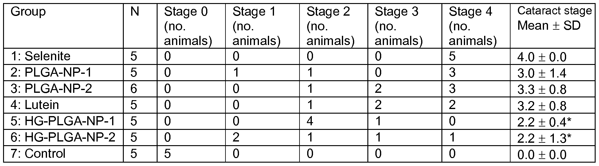
- Table 5 shows the observed distribution of cataract severity for the various Groups. All animals in the negative control group showed no symptoms of cataracts (stage 0). All animals in the selenite positive control group developed bilateral, stage 4 cataracts. Table 5: Observed distribution of cataract severity for the various Groups of rat pups, day 21 postpartum
- group 2 treated orally with 62.5 mg/kg PLGA-NP-lutein (equivalent to a dose of 2.66 mg lutein/kg), three animals developed stage 4 cataracts, one developed stage 2 cataracts, and one developed stage 1 cataracts.
- group 3 treated orally with 125 mg/kg PLGA-NP-lutein (equivalent to a dose of 5.32 mg lutein/kg), three animals developed stage 4 cataracts, two developed stage 3 cataracts, and one developed stage 2 cataracts.
- group 4 treated orally with 0.125 mg/kg lutein, two animals developed stage 4 cataracts, two stage 3 cataracts and one stage 2 cataracts.
- group 5 treated locally with 1 % PLGA-NP-lutein in the bioadhesive hydrogel, one animal developed stage 3 cataracts, and the other four animals developed stage 2 cataracts.
- group 6 treated locally with 3% PLGA-NP- lutein in the bioadhesive hydrogel, one animal developed stage 4 cataracts, one animal developed stage 3 cataracts, one animal developed stage 2 cataracts, and two animals developed only stage 1 cataracts.
- Group 2 (lutein - 426) was treated locally, once a day, with a corneal application (1 drop in each eye) of unmodified lutein (426 g lutein/ml) in the bioadhesive hydrogel. 3.21 mg finely ground pure lutein were dispersed by trituration into 7.53 ml of bioadhesive hydrogel. The volume of one drop of hydrogel was approx. 0.012 mL, with a density of 1 .023 g/mL.) The final concentration of lutein (426 g lutein/ml) was equivalent to 1 wt% lutein-loaded nanoparticles in the hydrogel. (The intention was that each of the animals in Groups 2, 4, and 7 would receive approximately the same concentration of lutein.)
- Group 3 (lutein - 2130) was treated locally, once a day, with corneal application (1 drop in each eye) of unmodified lutein (2130 g lutein/ml) in the bioadhesive hydrogel. 1 1 .99 mg finely ground pure lutein were dispersed by trituration into 5.63 ml of bioadhesive hydrogel. The volume of one drop of hydrogel was approx. 0.012 mL, with a density of 1 .023g/mL.) The concentration of lutein (2130 g lutein/ml) was equivalent to 5 wt% lutein-loaded nanoparticles in the hydrogel.
- Group 4 (PLGA-NP-lutein 426) was treated locally, once a day, by corneal application (1 drop in each eye) with 426 g lutein/mL (from lutein-loaded PLGA nanoparticles, Example 20) in the bioadhesive hydrogel. 375.7 mg of lyophilized, lutein- loaded PLGA nanoparticles were dissolved in 15.25 ml of bioadhesive hydrogel. The volume of one drop of hydrogel was approx. 0.012 ml_, with a density of 1 .040g/ml_.
- Group 5 (PLGA-NP-1278) was treated locally, once a day, by corneal application (1 drop in each eye) with 1278 g lutein/mL (from lutein-loaded PLGA nanoparticles, Example 20) in the bioadhesive hydrogel. 379.9 mg of lyophilized, lutein-loaded PLGA nanoparticles were dissolved in 5.14 ml of bioadhesive hydrogel. The volume of one drop of hydrogel was approx. 0.012 mL, with a density of 1 .040g/mL.)
- Group 6 (PLGA-NP-2130) was treated locally, once a day, by corneal application (1 drop in each eye) with 2130 g lutein/mL (from lutein-loaded PLGA nanoparticles, Example 20) in the bioadhesive hydrogel. 376.7 mg of lyophilized, lutein-loaded PLGA nanoparticles were dissolved in 3.06 ml of bioadhesive hydrogel. The volume of one drop of hydrogel was approx. 0.012 mL, with a density of 1 .040g/mL.)
- Group 7 (ZEIN-NP-426) was treated locally, once a day, by corneal application (1 drop in each eye) with 426 g lutein/mL (from lutein-loaded zein nanoparticles, Example 21 ) in the bioadhesive hydrogel. 252.1 mg of lyophilized, lutein-loaded zein nanoparticles were dissolved in 8.81 ml of bioadhesive hydrogel. The volume of one drop of hydrogel was approx. 0.012 mL, with a density of 1 .040g/mL.
- Group 8 (ZEIN-NP-1278) was treated locally, once a day, by corneal application (1 drop in each eye) with 1278 g lutein/mL (from lutein-loaded zein nanoparticles, Example 21 ) in the bioadhesive hydrogel. 345.7 mg of lyophilized, lutein-loaded zein nanoparticles were dissolved in 4.03 ml of bioadhesive hydrogel. The volume of one drop of hydrogel was approx. 0.012 mL, with a density of 1 .040g/mL.
- Group 9 (ZEIN-NP-2130) was treated locally, once a day, by corneal application (1 drop in each eye) with 2130 g lutein/mL (from lutein-loaded zein nanoparticles, Example 21 ) in the bioadhesive hydrogel. 346.1 mg of lyophilized, lutein-loaded zein nanoparticles were dissolved in 2.42 ml of bioadhesive hydrogel. The volume of one drop of hydrogel was approx. 0.012 mL, with a density of 1 .040g/mL.
- stage 0 no cataract
- stage 1 light nucleus opacity
- stage 2 mimild nucleus opacity, a central while opacity occupying less than half the diameter of the nucleus
- stage 3 diense opacity, a central while opacity occupying more than half the diameter of the nucleus
- stage 4 doense, white opacity over the whole nucleus.
- Statistics were performed in SPSS 14.0 for Windows and Excel. The variables were checked for normal distribution with the Shapiro-Wilk test. Groups were compared with the Wilcoxon test. Statistical significance was set at p ⁇ 0.05.
- Table 6 shows the observed distribution of cataract severity for the various Groups.
- the prototype embodiments of this invention have employed lutein for delivery to the eye.
- the invention may also be used to deliver other antioxidants, such as beta-carotene, lycopene, retinol, and other carotenoids.
- the invention may be used to deliver antioxidants to other tissues where needed, for example to the skin.
- a "therapeutically effective amount" of a composition refers to a quantity of the composition sufficient to be therapeutically effective to prevent, inhibit, slow the progression, or treat the symptoms of a disease of the eye such as cataracts, dry macular degeneration or wet macular degeneration (age-related macular degeneration), Stargardt disease, or retinitis pigmentosa.
- a "therapeutically effective amount” of a composition can also refer to a quantity of the composition that, when administered topically to a tissue, is sufficient to deliver a concentration of an antioxidant to the tissue to have a clinically meaningful effect on the tissue or neighboring tissues.
Abstract
Description


Claims
Priority Applications (12)
| Application Number | Priority Date | Filing Date | Title |
|---|---|---|---|
| KR1020177003119A KR102218883B1 (en) | 2014-08-11 | 2015-08-10 | Delivery of Bioactive, Nanoencapsulated Antioxidants |
| CA2992879A CA2992879C (en) | 2014-08-11 | 2015-08-10 | Delivery of bioactive, nanoencapsulated antioxidants |
| JP2017505869A JP6668327B2 (en) | 2014-08-11 | 2015-08-10 | Delivery of bioactive, nano-encapsulated antioxidants |
| AU2015301945A AU2015301945B2 (en) | 2014-08-11 | 2015-08-10 | Delivery of bioactive, nanoencapsulated antioxidants |
| EA201790361A EA035620B1 (en) | 2014-08-11 | 2015-08-10 | Methods and composition for treating or preventing cataract |
| AP2017009684A AP2017009684A0 (en) | 2014-08-11 | 2015-08-10 | |
| BR112017002580A BR112017002580A2 (en) | 2014-08-11 | 2015-08-10 | administration of nanoencapsulated bioactive antioxidants |
| US15/502,415 US10292943B2 (en) | 2014-08-11 | 2015-08-10 | Delivery of bioactive, nanoencapsulated antioxidants |
| MX2017001903A MX2017001903A (en) | 2014-08-11 | 2015-08-10 | Delivery of bioactive, nanoencapsulated antioxidants. |
| EP15831551.5A EP3179988A4 (en) | 2014-08-11 | 2015-08-10 | Delivery of bioactive, nanoencapsulated antioxidants |
| ZA2017/00987A ZA201700987B (en) | 2014-08-11 | 2017-02-03 | Delivery of bioactive, nanoencapsulated antioxidants |
| US16/399,388 US10952974B2 (en) | 2014-08-11 | 2019-04-30 | Delivery of bioactive, nanoencapsulated antioxidants |
Applications Claiming Priority (4)
| Application Number | Priority Date | Filing Date | Title |
|---|---|---|---|
| US201462035683P | 2014-08-11 | 2014-08-11 | |
| US62/035,683 | 2014-08-11 | ||
| US201562172455P | 2015-06-08 | 2015-06-08 | |
| US62/172,455 | 2015-06-08 |
Related Child Applications (2)
| Application Number | Title | Priority Date | Filing Date |
|---|---|---|---|
| US15/502,415 A-371-Of-International US10292943B2 (en) | 2014-08-11 | 2015-08-10 | Delivery of bioactive, nanoencapsulated antioxidants |
| US16/399,388 Continuation US10952974B2 (en) | 2014-08-11 | 2019-04-30 | Delivery of bioactive, nanoencapsulated antioxidants |
Publications (2)
| Publication Number | Publication Date |
|---|---|
| WO2016025394A2 true WO2016025394A2 (en) | 2016-02-18 |
| WO2016025394A3 WO2016025394A3 (en) | 2016-05-19 |
Family
ID=55304745
Family Applications (1)
| Application Number | Title | Priority Date | Filing Date |
|---|---|---|---|
| PCT/US2015/044483 WO2016025394A2 (en) | 2014-08-11 | 2015-08-10 | Delivery of bioactive, nanoencapsulated antioxidants |
Country Status (12)
| Country | Link |
|---|---|
| US (2) | US10292943B2 (en) |
| EP (1) | EP3179988A4 (en) |
| JP (1) | JP6668327B2 (en) |
| KR (1) | KR102218883B1 (en) |
| AP (1) | AP2017009684A0 (en) |
| AU (1) | AU2015301945B2 (en) |
| BR (1) | BR112017002580A2 (en) |
| CA (1) | CA2992879C (en) |
| EA (1) | EA035620B1 (en) |
| MX (1) | MX2017001903A (en) |
| WO (1) | WO2016025394A2 (en) |
| ZA (1) | ZA201700987B (en) |
Cited By (2)
| Publication number | Priority date | Publication date | Assignee | Title |
|---|---|---|---|---|
| WO2019148140A3 (en) * | 2018-01-26 | 2019-09-19 | Wu Joseph C | Implantable biomaterials that enhance stem cell survival and function |
| WO2020249825A1 (en) * | 2019-06-14 | 2020-12-17 | Folium Biosciences Europe B.V. | Method for micro-encapsulation of natural ingredients by means of contacting with supercritical gas |
Families Citing this family (3)
| Publication number | Priority date | Publication date | Assignee | Title |
|---|---|---|---|---|
| CA2992879C (en) * | 2014-08-11 | 2022-10-25 | Board Of Supervisors Of Louisiana State University And Agriculatural And Mechanical College | Delivery of bioactive, nanoencapsulated antioxidants |
| US11298321B1 (en) | 2021-07-12 | 2022-04-12 | King Abdulaziz University | Methods for treating metabolic disorder |
| US11253481B1 (en) | 2021-07-12 | 2022-02-22 | King Abdulaziz University | Self-nanoemulsifying 3D-printed tablet composition and method of use thereof |
Family Cites Families (14)
| Publication number | Priority date | Publication date | Assignee | Title |
|---|---|---|---|---|
| CA2011423A1 (en) * | 1989-03-07 | 1990-09-07 | Peter M. Taylor | Pharmaceutical compositions useful as drug delivery vehicles and/or as wound dressings |
| US6573299B1 (en) * | 1999-09-20 | 2003-06-03 | Advanced Medical Instruments | Method and compositions for treatment of the aging eye |
| US6703039B2 (en) * | 2000-12-06 | 2004-03-09 | Bausch & Lomb Incorporated | Reversible gelling system for ocular drug delivery |
| DE10141018A1 (en) * | 2001-08-22 | 2003-03-13 | Eth Zuerich Eidgenoessische Te | Use of negatively charged phospholipids and compositions comprising phospholipids for the treatment of the eye |
| US20040009212A1 (en) * | 2002-01-30 | 2004-01-15 | Pharma Power Biotec Co. Ltd. | Mucoadhesive thermoresponsive medicament-carrier composition |
| CN100369593C (en) * | 2002-06-05 | 2008-02-20 | 佛罗里达大学研究基金会有限公司 | Ophthalmic drug delivery system |
| MX2008004981A (en) | 2005-10-16 | 2008-10-17 | Lycored Ltd | Compositions for treatment of eye diseases. |
| WO2008137831A1 (en) * | 2007-05-07 | 2008-11-13 | Board Of Supervisors Of Louisiana State University And Agricultural And Mechanical College | Water-soluble nanoparticles containing water-insoluble compounds |
| PT2594140T (en) * | 2010-07-16 | 2019-06-11 | Centro Nac De Tecnologia Y Seguridad Alimentaria Laboratorio Dei Ebro | Nanoparticles for encapsulation of compounds, the production and uses thereof |
| EA028375B1 (en) * | 2010-09-03 | 2017-11-30 | Сантен Сас | Water-in-oil type emulsion for use by intraocular route |
| CA2828255A1 (en) | 2011-02-25 | 2012-08-30 | South Dakota State University | Protein nanocarriers for topical delivery |
| US20120231072A1 (en) * | 2011-03-11 | 2012-09-13 | Chemisches Institut Schaefer Ag | Thermo-responsive hydrogel compositions |
| CN103845278B (en) | 2012-11-30 | 2016-06-22 | 沈阳药科大学 | A kind of phylloxanthin eye nano-emulsion-thermosensitive in situ gel and preparation method thereof |
| CA2992879C (en) * | 2014-08-11 | 2022-10-25 | Board Of Supervisors Of Louisiana State University And Agriculatural And Mechanical College | Delivery of bioactive, nanoencapsulated antioxidants |
-
2015
- 2015-08-10 CA CA2992879A patent/CA2992879C/en active Active
- 2015-08-10 AU AU2015301945A patent/AU2015301945B2/en active Active
- 2015-08-10 KR KR1020177003119A patent/KR102218883B1/en active IP Right Grant
- 2015-08-10 EP EP15831551.5A patent/EP3179988A4/en active Pending
- 2015-08-10 WO PCT/US2015/044483 patent/WO2016025394A2/en active Application Filing
- 2015-08-10 AP AP2017009684A patent/AP2017009684A0/en unknown
- 2015-08-10 EA EA201790361A patent/EA035620B1/en unknown
- 2015-08-10 US US15/502,415 patent/US10292943B2/en active Active
- 2015-08-10 BR BR112017002580A patent/BR112017002580A2/en not_active Application Discontinuation
- 2015-08-10 MX MX2017001903A patent/MX2017001903A/en unknown
- 2015-08-10 JP JP2017505869A patent/JP6668327B2/en active Active
-
2017
- 2017-02-03 ZA ZA2017/00987A patent/ZA201700987B/en unknown
-
2019
- 2019-04-30 US US16/399,388 patent/US10952974B2/en active Active
Cited By (2)
| Publication number | Priority date | Publication date | Assignee | Title |
|---|---|---|---|---|
| WO2019148140A3 (en) * | 2018-01-26 | 2019-09-19 | Wu Joseph C | Implantable biomaterials that enhance stem cell survival and function |
| WO2020249825A1 (en) * | 2019-06-14 | 2020-12-17 | Folium Biosciences Europe B.V. | Method for micro-encapsulation of natural ingredients by means of contacting with supercritical gas |
Also Published As
| Publication number | Publication date |
|---|---|
| KR102218883B1 (en) | 2021-02-23 |
| AU2015301945B2 (en) | 2020-05-14 |
| CA2992879A1 (en) | 2016-02-18 |
| EP3179988A2 (en) | 2017-06-21 |
| CA2992879C (en) | 2022-10-25 |
| KR20170097601A (en) | 2017-08-28 |
| JP2017523983A (en) | 2017-08-24 |
| EP3179988A4 (en) | 2018-11-14 |
| EA035620B1 (en) | 2020-07-16 |
| ZA201700987B (en) | 2019-05-29 |
| EA201790361A1 (en) | 2017-06-30 |
| US10952974B2 (en) | 2021-03-23 |
| US10292943B2 (en) | 2019-05-21 |
| US20170216221A1 (en) | 2017-08-03 |
| AU2015301945A1 (en) | 2017-03-09 |
| JP6668327B2 (en) | 2020-03-18 |
| BR112017002580A2 (en) | 2018-03-27 |
| US20190254987A1 (en) | 2019-08-22 |
| MX2017001903A (en) | 2017-09-26 |
| WO2016025394A3 (en) | 2016-05-19 |
| AP2017009684A0 (en) | 2017-01-31 |
Similar Documents
| Publication | Publication Date | Title |
|---|---|---|
| US10952974B2 (en) | Delivery of bioactive, nanoencapsulated antioxidants | |
| Chuacharoen et al. | Stability and controlled release of lutein loaded in zein nanoparticles with and without lecithin and pluronic F127 surfactants | |
| Yuan et al. | Fabrication and characterization of lutein-loaded nanoparticles based on zein and sophorolipid: Enhancement of water solubility, stability, and bioaccessibility | |
| Natesan et al. | Co-encapsulated resveratrol and quercetin in chitosan and peg modified chitosan nanoparticles: For efficient intra ocular pressure reduction | |
| Madaan et al. | Lutein, a versatile phyto-nutraceutical: An insight on pharmacology, therapeutic indications, challenges and recent advances in drug delivery | |
| Lopedota et al. | The use of Eudragit® RS 100/cyclodextrin nanoparticles for the transmucosal administration of glutathione | |
| Maghsoudi et al. | The colorful world of carotenoids: A profound insight on therapeutics and recent trends in nano delivery systems | |
| Bodoki et al. | Topical nanodelivery system of lutein for the prevention of selenite-induced cataract | |
| Ranganathan et al. | Biocompatible lutein-polymer-lipid nanocapsules: Acute and subacute toxicity and bioavailability in mice | |
| US20180028447A1 (en) | Development of curcumin and piperine loaded double-layered biopolymer based nano delivery systems by using electrospray / coating method | |
| CN113226293A (en) | Nanocapsules coated with chitosan and uses thereof | |
| JP7390738B2 (en) | Nanocapsules coated with chitosan and their uses | |
| Giuliani et al. | Locust bean gum-based hydrogel containing nanocapsules for 3, 3′-diindolylmethane delivery in skin inflammatory conditions | |
| US20200000736A1 (en) | Oil-free carotenoid composition | |
| Taebpour et al. | Fabrication and characterization of PLGA polymeric nanoparticles containing Berberine and its cytotoxicity on breast cancer cell (MCF-7) | |
| MXPA01012169A (en) | Preparations for the application of anti-infective and/or anti-inflammatory agents. | |
| Deshmukh et al. | Chitosan-vitamin C nanoparticles | |
| KR102384243B1 (en) | Nanocapsule coated with chitosan comprising retinol or retinol derivatives and uses thereof | |
| Mehdipour Biregani et al. | Curcumin as a bioactive compound: biological properties and encapsulation methods | |
| Mahajan et al. | Development and evaluation of topical nanoemulgel formulation of tazarotene for effective treatment of excision wounds | |
| US20160166513A1 (en) | Nanoparticles and Methods of Producing the Same | |
| KR101771483B1 (en) | Double layered polymer capsules with improved dissolution rate and stability of lutein, method for preparing the same, and pharmaceutical composition for preventing or treating ocular diseases containing the same | |
| US20170105974A1 (en) | Use of mtor inhibitors for prevention of intestinal polyp growth and cancer | |
| Truong | Development and Evaluation of Quercetin Nanoparticles and Hot Melt Cast Films for Retinal Neuroprotection | |
| Cosby | Preclinical Delivery of Fractionated Black Raspberry Phytochemicals to Oral Epithelial Cells Using Lipid and Polymer Nanoparticles |
Legal Events
| Date | Code | Title | Description |
|---|---|---|---|
| 121 | Ep: the epo has been informed by wipo that ep was designated in this application |
Ref document number: 15831551 Country of ref document: EP Kind code of ref document: A2 |
|
| REEP | Request for entry into the european phase |
Ref document number: 2015831551 Country of ref document: EP |
|
| ENP | Entry into the national phase |
Ref document number: 2017505869 Country of ref document: JP Kind code of ref document: A |
|
| ENP | Entry into the national phase |
Ref document number: 20177003119 Country of ref document: KR Kind code of ref document: A |
|
| WWE | Wipo information: entry into national phase |
Ref document number: 15502415 Country of ref document: US |
|
| WWE | Wipo information: entry into national phase |
Ref document number: MX/A/2017/001903 Country of ref document: MX |
|
| NENP | Non-entry into the national phase |
Ref country code: DE |
|
| REG | Reference to national code |
Ref country code: BR Ref legal event code: B01A Ref document number: 112017002580 Country of ref document: BR |
|
| 121 | Ep: the epo has been informed by wipo that ep was designated in this application |
Ref document number: 15831551 Country of ref document: EP Kind code of ref document: A2 |
|
| ENP | Entry into the national phase |
Ref document number: 2015301945 Country of ref document: AU Date of ref document: 20150810 Kind code of ref document: A |
|
| WWE | Wipo information: entry into national phase |
Ref document number: 201790361 Country of ref document: EA |
|
| REG | Reference to national code |
Ref country code: BR Ref legal event code: B01E Ref document number: 112017002580 Country of ref document: BR |
|
| ENP | Entry into the national phase |
Ref document number: 2992879 Country of ref document: CA |
|
| ENP | Entry into the national phase |
Ref document number: 112017002580 Country of ref document: BR Kind code of ref document: A2 Effective date: 20170208 |