WO2015164727A1 - Methods of measuring antigen-specific t cells - Google Patents
Methods of measuring antigen-specific t cells Download PDFInfo
- Publication number
- WO2015164727A1 WO2015164727A1 PCT/US2015/027497 US2015027497W WO2015164727A1 WO 2015164727 A1 WO2015164727 A1 WO 2015164727A1 US 2015027497 W US2015027497 W US 2015027497W WO 2015164727 A1 WO2015164727 A1 WO 2015164727A1
- Authority
- WO
- WIPO (PCT)
- Prior art keywords
- peptide
- antigen
- level
- subject
- kit
- Prior art date
Links
- 238000000034 method Methods 0.000 title claims abstract description 451
- 108091007433 antigens Proteins 0.000 title claims abstract description 200
- 102000036639 antigens Human genes 0.000 title claims abstract description 200
- 239000000427 antigen Substances 0.000 title claims abstract description 199
- 210000001744 T-lymphocyte Anatomy 0.000 title claims description 220
- 108090000765 processed proteins & peptides Proteins 0.000 claims abstract description 523
- 230000005867 T cell response Effects 0.000 claims abstract description 165
- 208000015943 Coeliac disease Diseases 0.000 claims abstract description 146
- 206010067584 Type 1 diabetes mellitus Diseases 0.000 claims abstract description 86
- 108090000623 proteins and genes Proteins 0.000 claims abstract description 60
- 102000004169 proteins and genes Human genes 0.000 claims abstract description 52
- 208000023275 Autoimmune disease Diseases 0.000 claims abstract description 43
- 230000002411 adverse Effects 0.000 claims abstract description 29
- 206010020751 Hypersensitivity Diseases 0.000 claims abstract description 16
- 230000007815 allergy Effects 0.000 claims abstract description 16
- 208000015181 infectious disease Diseases 0.000 claims abstract description 16
- 208000035473 Communicable disease Diseases 0.000 claims abstract description 15
- 208000026935 allergic disease Diseases 0.000 claims abstract description 15
- 108091022930 Glutamate decarboxylase Proteins 0.000 claims description 317
- 102000008214 Glutamate decarboxylase Human genes 0.000 claims description 315
- 108010068370 Glutens Proteins 0.000 claims description 306
- 235000021312 gluten Nutrition 0.000 claims description 235
- 102000004196 processed proteins & peptides Human genes 0.000 claims description 175
- 108010002350 Interleukin-2 Proteins 0.000 claims description 174
- 102000000588 Interleukin-2 Human genes 0.000 claims description 174
- 239000000203 mixture Substances 0.000 claims description 166
- 102100037850 Interferon gamma Human genes 0.000 claims description 154
- 108010074328 Interferon-gamma Proteins 0.000 claims description 154
- NOESYZHRGYRDHS-UHFFFAOYSA-N insulin Chemical compound N1C(=O)C(NC(=O)C(CCC(N)=O)NC(=O)C(CCC(O)=O)NC(=O)C(C(C)C)NC(=O)C(NC(=O)CN)C(C)CC)CSSCC(C(NC(CO)C(=O)NC(CC(C)C)C(=O)NC(CC=2C=CC(O)=CC=2)C(=O)NC(CCC(N)=O)C(=O)NC(CC(C)C)C(=O)NC(CCC(O)=O)C(=O)NC(CC(N)=O)C(=O)NC(CC=2C=CC(O)=CC=2)C(=O)NC(CSSCC(NC(=O)C(C(C)C)NC(=O)C(CC(C)C)NC(=O)C(CC=2C=CC(O)=CC=2)NC(=O)C(CC(C)C)NC(=O)C(C)NC(=O)C(CCC(O)=O)NC(=O)C(C(C)C)NC(=O)C(CC(C)C)NC(=O)C(CC=2NC=NC=2)NC(=O)C(CO)NC(=O)CNC2=O)C(=O)NCC(=O)NC(CCC(O)=O)C(=O)NC(CCCNC(N)=N)C(=O)NCC(=O)NC(CC=3C=CC=CC=3)C(=O)NC(CC=3C=CC=CC=3)C(=O)NC(CC=3C=CC(O)=CC=3)C(=O)NC(C(C)O)C(=O)N3C(CCC3)C(=O)NC(CCCCN)C(=O)NC(C)C(O)=O)C(=O)NC(CC(N)=O)C(O)=O)=O)NC(=O)C(C(C)CC)NC(=O)C(CO)NC(=O)C(C(C)O)NC(=O)C1CSSCC2NC(=O)C(CC(C)C)NC(=O)C(NC(=O)C(CCC(N)=O)NC(=O)C(CC(N)=O)NC(=O)C(NC(=O)C(N)CC=1C=CC=CC=1)C(C)C)CC1=CN=CN1 NOESYZHRGYRDHS-UHFFFAOYSA-N 0.000 claims description 104
- 239000008280 blood Substances 0.000 claims description 100
- 239000000902 placebo Substances 0.000 claims description 97
- 229940068196 placebo Drugs 0.000 claims description 97
- 210000004369 blood Anatomy 0.000 claims description 94
- 238000003556 assay Methods 0.000 claims description 88
- 102000004127 Cytokines Human genes 0.000 claims description 83
- 108090000695 Cytokines Proteins 0.000 claims description 83
- 238000012360 testing method Methods 0.000 claims description 74
- 102100025248 C-X-C motif chemokine 10 Human genes 0.000 claims description 60
- 101710098275 C-X-C motif chemokine 10 Proteins 0.000 claims description 55
- 239000003795 chemical substances by application Substances 0.000 claims description 53
- 102000004877 Insulin Human genes 0.000 claims description 52
- 108090001061 Insulin Proteins 0.000 claims description 52
- 229940125396 insulin Drugs 0.000 claims description 52
- 235000018102 proteins Nutrition 0.000 claims description 51
- 235000021307 Triticum Nutrition 0.000 claims description 45
- 241000209140 Triticum Species 0.000 claims description 45
- 235000007340 Hordeum vulgare Nutrition 0.000 claims description 38
- 235000001014 amino acid Nutrition 0.000 claims description 37
- 150000001413 amino acids Chemical class 0.000 claims description 33
- 239000013642 negative control Substances 0.000 claims description 28
- 238000002965 ELISA Methods 0.000 claims description 26
- 239000011324 bead Substances 0.000 claims description 19
- 238000012216 screening Methods 0.000 claims description 18
- 102000004190 Enzymes Human genes 0.000 claims description 16
- 108090000790 Enzymes Proteins 0.000 claims description 16
- 210000003819 peripheral blood mononuclear cell Anatomy 0.000 claims description 16
- 108010076181 Proinsulin Proteins 0.000 claims description 15
- 230000003247 decreasing effect Effects 0.000 claims description 15
- LOKCTEFSRHRXRJ-UHFFFAOYSA-I dipotassium trisodium dihydrogen phosphate hydrogen phosphate dichloride Chemical compound P(=O)(O)(O)[O-].[K+].P(=O)(O)([O-])[O-].[Na+].[Na+].[Cl-].[K+].[Cl-].[Na+] LOKCTEFSRHRXRJ-UHFFFAOYSA-I 0.000 claims description 14
- 239000002953 phosphate buffered saline Substances 0.000 claims description 14
- 108010029485 Protein Isoforms Proteins 0.000 claims description 11
- 102000001708 Protein Isoforms Human genes 0.000 claims description 11
- 108091006550 Zinc transporters Proteins 0.000 claims description 11
- 102000017011 Glycated Hemoglobin A Human genes 0.000 claims description 10
- 238000003018 immunoassay Methods 0.000 claims description 10
- 210000004153 islets of langerhan Anatomy 0.000 claims description 10
- 239000013641 positive control Substances 0.000 claims description 10
- 238000003205 genotyping method Methods 0.000 claims description 8
- 108091005995 glycated hemoglobin Proteins 0.000 claims description 7
- 210000000601 blood cell Anatomy 0.000 claims description 6
- 239000003146 anticoagulant agent Substances 0.000 claims description 5
- 229940127219 anticoagulant drug Drugs 0.000 claims description 5
- 238000007446 glucose tolerance test Methods 0.000 claims description 5
- 239000003547 immunosorbent Substances 0.000 claims description 5
- 239000000243 solution Substances 0.000 claims description 5
- 238000012286 ELISA Assay Methods 0.000 claims description 4
- 230000036755 cellular response Effects 0.000 claims description 2
- 241000209219 Hordeum Species 0.000 claims 12
- 239000000523 sample Substances 0.000 description 120
- 230000004044 response Effects 0.000 description 62
- 230000027455 binding Effects 0.000 description 52
- 235000013305 food Nutrition 0.000 description 52
- -1 adipyl Chemical group 0.000 description 45
- 239000013566 allergen Substances 0.000 description 34
- 108700039882 Protein Glutamine gamma Glutamyltransferase 2 Proteins 0.000 description 32
- 102100038095 Protein-glutamine gamma-glutamyltransferase 2 Human genes 0.000 description 32
- 229940024606 amino acid Drugs 0.000 description 32
- 241000282414 Homo sapiens Species 0.000 description 28
- 230000004048 modification Effects 0.000 description 28
- 238000012986 modification Methods 0.000 description 28
- 240000005979 Hordeum vulgare Species 0.000 description 26
- 235000007238 Secale cereale Nutrition 0.000 description 26
- WQZGKKKJIJFFOK-GASJEMHNSA-N Glucose Natural products OC[C@H]1OC(O)[C@H](O)[C@@H](O)[C@@H]1O WQZGKKKJIJFFOK-GASJEMHNSA-N 0.000 description 25
- 210000004027 cell Anatomy 0.000 description 25
- 239000008103 glucose Substances 0.000 description 25
- 208000024891 symptom Diseases 0.000 description 25
- 229960003130 interferon gamma Drugs 0.000 description 21
- 125000003275 alpha amino acid group Chemical group 0.000 description 20
- 230000006240 deamidation Effects 0.000 description 20
- 201000010099 disease Diseases 0.000 description 19
- 208000037265 diseases, disorders, signs and symptoms Diseases 0.000 description 19
- 235000013312 flour Nutrition 0.000 description 17
- 235000006171 gluten free diet Nutrition 0.000 description 16
- 235000020884 gluten-free diet Nutrition 0.000 description 16
- 230000000638 stimulation Effects 0.000 description 15
- 108700028369 Alleles Proteins 0.000 description 14
- 108010067902 Peptide Library Proteins 0.000 description 14
- 229940088598 enzyme Drugs 0.000 description 14
- 101100284398 Bos taurus BoLA-DQB gene Proteins 0.000 description 13
- 239000003814 drug Substances 0.000 description 13
- 238000006467 substitution reaction Methods 0.000 description 13
- 108020004999 messenger RNA Proteins 0.000 description 12
- 238000011510 Elispot assay Methods 0.000 description 11
- 108010061711 Gliadin Proteins 0.000 description 11
- 230000000694 effects Effects 0.000 description 11
- 238000011282 treatment Methods 0.000 description 11
- 102100036241 HLA class II histocompatibility antigen, DQ beta 1 chain Human genes 0.000 description 10
- 108010086786 HLA-DQA1 antigen Proteins 0.000 description 10
- 238000004458 analytical method Methods 0.000 description 10
- 230000015572 biosynthetic process Effects 0.000 description 10
- 238000006243 chemical reaction Methods 0.000 description 10
- 238000001514 detection method Methods 0.000 description 10
- 230000028993 immune response Effects 0.000 description 10
- 210000002966 serum Anatomy 0.000 description 10
- 238000003786 synthesis reaction Methods 0.000 description 10
- 108010067148 HLA-DQbeta antigen Proteins 0.000 description 9
- WHUUTDBJXJRKMK-VKHMYHEASA-L glutamate group Chemical group N[C@@H](CCC(=O)[O-])C(=O)[O-] WHUUTDBJXJRKMK-VKHMYHEASA-L 0.000 description 9
- 238000001727 in vivo Methods 0.000 description 9
- 230000000977 initiatory effect Effects 0.000 description 9
- 125000006239 protecting group Chemical group 0.000 description 9
- 239000000126 substance Substances 0.000 description 9
- YBJHBAHKTGYVGT-ZKWXMUAHSA-N (+)-Biotin Chemical compound N1C(=O)N[C@@H]2[C@H](CCCCC(=O)O)SC[C@@H]21 YBJHBAHKTGYVGT-ZKWXMUAHSA-N 0.000 description 8
- 108020004414 DNA Proteins 0.000 description 8
- 102100036243 HLA class II histocompatibility antigen, DQ alpha 1 chain Human genes 0.000 description 8
- 235000005911 diet Nutrition 0.000 description 8
- 229940079593 drug Drugs 0.000 description 8
- 125000000404 glutamine group Chemical group N[C@@H](CCC(N)=O)C(=O)* 0.000 description 8
- 239000002609 medium Substances 0.000 description 8
- 150000007523 nucleic acids Chemical class 0.000 description 8
- 230000009885 systemic effect Effects 0.000 description 8
- VOUAQYXWVJDEQY-QENPJCQMSA-N 33017-11-7 Chemical compound OC(=O)CC[C@H](N)C(=O)N[C@@H](C)C(=O)N[C@@H](CCC(O)=O)C(=O)N[C@@H](CC(O)=O)C(=O)N[C@@H](CC(C)C)C(=O)N[C@@H](CCC(N)=O)C(=O)N[C@@H](C(C)C)C(=O)NCC(=O)N[C@@H](CCC(N)=O)C(=O)N[C@@H](C(C)C)C(=O)N[C@@H](CCC(O)=O)C(=O)N[C@@H](CC(C)C)C(=O)NCC(=O)NCC(=O)NCC(=O)N1CCC[C@H]1C(=O)NCC(=O)N[C@@H](C)C(=O)NCC(=O)N[C@@H](CO)C(=O)N[C@@H](CC(C)C)C(=O)N[C@@H](CCC(N)=O)C(=O)N1[C@H](C(=O)N[C@@H](CC(C)C)C(=O)N[C@@H](C)C(=O)N[C@@H](CC(C)C)C(=O)N[C@@H](CCC(O)=O)C(=O)NCC(=O)N[C@@H](CO)C(=O)N[C@@H](CC(C)C)C(=O)N[C@@H](CCC(N)=O)C(O)=O)CCC1 VOUAQYXWVJDEQY-QENPJCQMSA-N 0.000 description 7
- 102100034091 Receptor-type tyrosine-protein phosphatase-like N Human genes 0.000 description 7
- 108060008539 Transglutaminase Proteins 0.000 description 7
- 230000004913 activation Effects 0.000 description 7
- 238000013459 approach Methods 0.000 description 7
- 230000001363 autoimmune Effects 0.000 description 7
- 230000005784 autoimmunity Effects 0.000 description 7
- 230000000981 bystander Effects 0.000 description 7
- 238000007405 data analysis Methods 0.000 description 7
- 238000003745 diagnosis Methods 0.000 description 7
- 238000003114 enzyme-linked immunosorbent spot assay Methods 0.000 description 7
- 108020004707 nucleic acids Proteins 0.000 description 7
- 102000039446 nucleic acids Human genes 0.000 description 7
- 210000002381 plasma Anatomy 0.000 description 7
- 102000003601 transglutaminase Human genes 0.000 description 7
- 108091032973 (ribonucleotides)n+m Proteins 0.000 description 6
- 102000004031 Carboxy-Lyases Human genes 0.000 description 6
- 108090000489 Carboxy-Lyases Proteins 0.000 description 6
- 102000001554 Hemoglobins Human genes 0.000 description 6
- 108010054147 Hemoglobins Proteins 0.000 description 6
- 241000711549 Hepacivirus C Species 0.000 description 6
- 230000021736 acetylation Effects 0.000 description 6
- 238000006640 acetylation reaction Methods 0.000 description 6
- 230000003213 activating effect Effects 0.000 description 6
- 125000002252 acyl group Chemical group 0.000 description 6
- 230000009435 amidation Effects 0.000 description 6
- 238000007112 amidation reaction Methods 0.000 description 6
- 125000001584 benzyloxycarbonyl group Chemical group C(=O)(OCC1=CC=CC=C1)* 0.000 description 6
- WQZGKKKJIJFFOK-VFUOTHLCSA-N beta-D-glucose Chemical compound OC[C@H]1O[C@@H](O)[C@H](O)[C@@H](O)[C@@H]1O WQZGKKKJIJFFOK-VFUOTHLCSA-N 0.000 description 6
- 239000000090 biomarker Substances 0.000 description 6
- 230000000903 blocking effect Effects 0.000 description 6
- 238000009826 distribution Methods 0.000 description 6
- 239000012634 fragment Substances 0.000 description 6
- 108020001507 fusion proteins Proteins 0.000 description 6
- 102000037865 fusion proteins Human genes 0.000 description 6
- 238000010166 immunofluorescence Methods 0.000 description 6
- 230000001404 mediated effect Effects 0.000 description 6
- 230000007170 pathology Effects 0.000 description 6
- 238000003752 polymerase chain reaction Methods 0.000 description 6
- 108091033319 polynucleotide Proteins 0.000 description 6
- 239000002157 polynucleotide Substances 0.000 description 6
- 102000040430 polynucleotide Human genes 0.000 description 6
- 230000001225 therapeutic effect Effects 0.000 description 6
- 238000013519 translation Methods 0.000 description 6
- JOYRKODLDBILNP-UHFFFAOYSA-N urethane group Chemical group NC(=O)OCC JOYRKODLDBILNP-UHFFFAOYSA-N 0.000 description 6
- 108010075254 C-Peptide Proteins 0.000 description 5
- 108010047762 HLA-DQ8 antigen Proteins 0.000 description 5
- 206010039710 Scleroderma Diseases 0.000 description 5
- 210000004899 c-terminal region Anatomy 0.000 description 5
- 238000011161 development Methods 0.000 description 5
- 230000000378 dietary effect Effects 0.000 description 5
- 239000013604 expression vector Substances 0.000 description 5
- 210000001035 gastrointestinal tract Anatomy 0.000 description 5
- 238000011534 incubation Methods 0.000 description 5
- 150000002576 ketones Chemical class 0.000 description 5
- 238000004519 manufacturing process Methods 0.000 description 5
- 238000005259 measurement Methods 0.000 description 5
- 210000001616 monocyte Anatomy 0.000 description 5
- 235000014571 nuts Nutrition 0.000 description 5
- 238000002360 preparation method Methods 0.000 description 5
- 238000000746 purification Methods 0.000 description 5
- 230000002829 reductive effect Effects 0.000 description 5
- 229920005989 resin Polymers 0.000 description 5
- 239000011347 resin Substances 0.000 description 5
- 210000001519 tissue Anatomy 0.000 description 5
- 241000894006 Bacteria Species 0.000 description 4
- 102100026735 Coagulation factor VIII Human genes 0.000 description 4
- 206010012735 Diarrhoea Diseases 0.000 description 4
- IAZDPXIOMUYVGZ-UHFFFAOYSA-N Dimethylsulphoxide Chemical compound CS(C)=O IAZDPXIOMUYVGZ-UHFFFAOYSA-N 0.000 description 4
- 108010008165 Etanercept Proteins 0.000 description 4
- 101000858088 Homo sapiens C-X-C motif chemokine 10 Proteins 0.000 description 4
- 101000911390 Homo sapiens Coagulation factor VIII Proteins 0.000 description 4
- WHUUTDBJXJRKMK-VKHMYHEASA-N L-glutamic acid Chemical compound OC(=O)[C@@H](N)CCC(O)=O WHUUTDBJXJRKMK-VKHMYHEASA-N 0.000 description 4
- 201000004681 Psoriasis Diseases 0.000 description 4
- XSQUKJJJFZCRTK-UHFFFAOYSA-N Urea Chemical compound NC(N)=O XSQUKJJJFZCRTK-UHFFFAOYSA-N 0.000 description 4
- 206010046851 Uveitis Diseases 0.000 description 4
- 241000700605 Viruses Species 0.000 description 4
- 206010047700 Vomiting Diseases 0.000 description 4
- 229960002964 adalimumab Drugs 0.000 description 4
- 239000011230 binding agent Substances 0.000 description 4
- 239000003124 biologic agent Substances 0.000 description 4
- 239000011616 biotin Substances 0.000 description 4
- 229960002685 biotin Drugs 0.000 description 4
- 235000020958 biotin Nutrition 0.000 description 4
- 229960003115 certolizumab pegol Drugs 0.000 description 4
- 230000008859 change Effects 0.000 description 4
- 238000004587 chromatography analysis Methods 0.000 description 4
- 238000003776 cleavage reaction Methods 0.000 description 4
- 238000013461 design Methods 0.000 description 4
- 230000006866 deterioration Effects 0.000 description 4
- 150000002148 esters Chemical class 0.000 description 4
- 229960000403 etanercept Drugs 0.000 description 4
- 125000002485 formyl group Chemical group [H]C(*)=O 0.000 description 4
- 229930195712 glutamate Natural products 0.000 description 4
- ZDXPYRJPNDTMRX-UHFFFAOYSA-N glutamine Natural products OC(=O)C(N)CCC(N)=O ZDXPYRJPNDTMRX-UHFFFAOYSA-N 0.000 description 4
- 238000004128 high performance liquid chromatography Methods 0.000 description 4
- 230000002519 immonomodulatory effect Effects 0.000 description 4
- 230000001939 inductive effect Effects 0.000 description 4
- 229960000598 infliximab Drugs 0.000 description 4
- 206010022498 insulinoma Diseases 0.000 description 4
- 210000001165 lymph node Anatomy 0.000 description 4
- 239000003550 marker Substances 0.000 description 4
- 238000002483 medication Methods 0.000 description 4
- 210000000440 neutrophil Anatomy 0.000 description 4
- 208000021255 pancreatic insulinoma Diseases 0.000 description 4
- 244000052769 pathogen Species 0.000 description 4
- 230000001717 pathogenic effect Effects 0.000 description 4
- 239000011886 peripheral blood Substances 0.000 description 4
- 210000005259 peripheral blood Anatomy 0.000 description 4
- 108010066381 preproinsulin Proteins 0.000 description 4
- 230000002035 prolonged effect Effects 0.000 description 4
- 230000009467 reduction Effects 0.000 description 4
- 230000007017 scission Effects 0.000 description 4
- 239000000758 substrate Substances 0.000 description 4
- 229910052717 sulfur Inorganic materials 0.000 description 4
- 230000002194 synthesizing effect Effects 0.000 description 4
- 239000003826 tablet Substances 0.000 description 4
- 230000008673 vomiting Effects 0.000 description 4
- 125000004080 3-carboxypropanoyl group Chemical group O=C([*])C([H])([H])C([H])([H])C(O[H])=O 0.000 description 3
- ODHCTXKNWHHXJC-VKHMYHEASA-N 5-oxo-L-proline Chemical compound OC(=O)[C@@H]1CCC(=O)N1 ODHCTXKNWHHXJC-VKHMYHEASA-N 0.000 description 3
- 208000004998 Abdominal Pain Diseases 0.000 description 3
- 208000026872 Addison Disease Diseases 0.000 description 3
- 108091023037 Aptamer Proteins 0.000 description 3
- 244000105624 Arachis hypogaea Species 0.000 description 3
- 206010003402 Arthropod sting Diseases 0.000 description 3
- 206010003694 Atrophy Diseases 0.000 description 3
- 206010003827 Autoimmune hepatitis Diseases 0.000 description 3
- 108090000317 Chymotrypsin Proteins 0.000 description 3
- 108010044226 Class 8 Receptor-Like Protein Tyrosine Phosphatases Proteins 0.000 description 3
- 208000011231 Crohn disease Diseases 0.000 description 3
- 241000588724 Escherichia coli Species 0.000 description 3
- 108010051815 Glutamyl endopeptidase Proteins 0.000 description 3
- 108010014663 Glycated Hemoglobin A Proteins 0.000 description 3
- 208000003807 Graves Disease Diseases 0.000 description 3
- 208000015023 Graves' disease Diseases 0.000 description 3
- 241000700721 Hepatitis B virus Species 0.000 description 3
- 241000238631 Hexapoda Species 0.000 description 3
- 241000282412 Homo Species 0.000 description 3
- 101000591210 Homo sapiens Receptor-type tyrosine-protein phosphatase-like N Proteins 0.000 description 3
- 241000713772 Human immunodeficiency virus 1 Species 0.000 description 3
- 241000713340 Human immunodeficiency virus 2 Species 0.000 description 3
- FBOZXECLQNJBKD-ZDUSSCGKSA-N L-methotrexate Chemical compound C=1N=C2N=C(N)N=C(N)C2=NC=1CN(C)C1=CC=C(C(=O)N[C@@H](CCC(O)=O)C(O)=O)C=C1 FBOZXECLQNJBKD-ZDUSSCGKSA-N 0.000 description 3
- OUYCCCASQSFEME-QMMMGPOBSA-N L-tyrosine Chemical compound OC(=O)[C@@H](N)CC1=CC=C(O)C=C1 OUYCCCASQSFEME-QMMMGPOBSA-N 0.000 description 3
- 206010028813 Nausea Diseases 0.000 description 3
- BZQFBWGGLXLEPQ-UHFFFAOYSA-N O-phosphoryl-L-serine Natural products OC(=O)C(N)COP(O)(O)=O BZQFBWGGLXLEPQ-UHFFFAOYSA-N 0.000 description 3
- 239000002033 PVDF binder Substances 0.000 description 3
- 108090000526 Papain Proteins 0.000 description 3
- 102000004160 Phosphoric Monoester Hydrolases Human genes 0.000 description 3
- 108090000608 Phosphoric Monoester Hydrolases Proteins 0.000 description 3
- 239000004365 Protease Substances 0.000 description 3
- 238000012952 Resampling Methods 0.000 description 3
- 241000209056 Secale Species 0.000 description 3
- 208000021386 Sjogren Syndrome Diseases 0.000 description 3
- 108090000631 Trypsin Proteins 0.000 description 3
- 102000004142 Trypsin Human genes 0.000 description 3
- YJQCOFNZVFGCAF-UHFFFAOYSA-N Tunicamycin II Natural products O1C(CC(O)C2C(C(O)C(O2)N2C(NC(=O)C=C2)=O)O)C(O)C(O)C(NC(=O)C=CCCCCCCCCC(C)C)C1OC1OC(CO)C(O)C(O)C1NC(C)=O YJQCOFNZVFGCAF-UHFFFAOYSA-N 0.000 description 3
- 229930003316 Vitamin D Natural products 0.000 description 3
- QYSXJUFSXHHAJI-XFEUOLMDSA-N Vitamin D3 Natural products C1(/[C@@H]2CC[C@@H]([C@]2(CCC1)C)[C@H](C)CCCC(C)C)=C/C=C1\C[C@@H](O)CCC1=C QYSXJUFSXHHAJI-XFEUOLMDSA-N 0.000 description 3
- 102000004248 Zinc Transporter 8 Human genes 0.000 description 3
- 108090000702 Zinc Transporter 8 Proteins 0.000 description 3
- 230000002159 abnormal effect Effects 0.000 description 3
- 150000001241 acetals Chemical class 0.000 description 3
- 125000002777 acetyl group Chemical group [H]C([H])([H])C(*)=O 0.000 description 3
- 239000002253 acid Substances 0.000 description 3
- 150000001298 alcohols Chemical class 0.000 description 3
- 150000001299 aldehydes Chemical class 0.000 description 3
- 125000001931 aliphatic group Chemical group 0.000 description 3
- 150000001408 amides Chemical group 0.000 description 3
- 125000003118 aryl group Chemical group 0.000 description 3
- 230000037444 atrophy Effects 0.000 description 3
- 229960002170 azathioprine Drugs 0.000 description 3
- LMEKQMALGUDUQG-UHFFFAOYSA-N azathioprine Chemical compound CN1C=NC([N+]([O-])=O)=C1SC1=NC=NC2=C1NC=N2 LMEKQMALGUDUQG-UHFFFAOYSA-N 0.000 description 3
- 125000003236 benzoyl group Chemical group [H]C1=C([H])C([H])=C(C([H])=C1[H])C(*)=O 0.000 description 3
- 125000005620 boronic acid group Chemical class 0.000 description 3
- 235000008429 bread Nutrition 0.000 description 3
- 125000000837 carbohydrate group Chemical group 0.000 description 3
- 150000001735 carboxylic acids Chemical class 0.000 description 3
- 230000001413 cellular effect Effects 0.000 description 3
- 238000007385 chemical modification Methods 0.000 description 3
- 208000025302 chronic primary adrenal insufficiency Diseases 0.000 description 3
- 229960002376 chymotrypsin Drugs 0.000 description 3
- 235000014510 cooky Nutrition 0.000 description 3
- ATDGTVJJHBUTRL-UHFFFAOYSA-N cyanogen bromide Chemical compound BrC#N ATDGTVJJHBUTRL-UHFFFAOYSA-N 0.000 description 3
- 125000001295 dansyl group Chemical group [H]C1=C([H])C(N(C([H])([H])[H])C([H])([H])[H])=C2C([H])=C([H])C([H])=C(C2=C1[H])S(*)(=O)=O 0.000 description 3
- 238000001212 derivatisation Methods 0.000 description 3
- 229950006137 dexfosfoserine Drugs 0.000 description 3
- 206010012601 diabetes mellitus Diseases 0.000 description 3
- 238000002405 diagnostic procedure Methods 0.000 description 3
- 230000037213 diet Effects 0.000 description 3
- 231100000673 dose–response relationship Toxicity 0.000 description 3
- 150000002170 ethers Chemical class 0.000 description 3
- 238000011156 evaluation Methods 0.000 description 3
- 230000007717 exclusion Effects 0.000 description 3
- 230000006126 farnesylation Effects 0.000 description 3
- 239000000796 flavoring agent Substances 0.000 description 3
- 125000001153 fluoro group Chemical group F* 0.000 description 3
- 235000013355 food flavoring agent Nutrition 0.000 description 3
- 235000003599 food sweetener Nutrition 0.000 description 3
- 230000022244 formylation Effects 0.000 description 3
- 238000006170 formylation reaction Methods 0.000 description 3
- 230000006870 function Effects 0.000 description 3
- 230000004927 fusion Effects 0.000 description 3
- 230000002068 genetic effect Effects 0.000 description 3
- 230000013595 glycosylation Effects 0.000 description 3
- 238000006206 glycosylation reaction Methods 0.000 description 3
- 210000003714 granulocyte Anatomy 0.000 description 3
- 229910052736 halogen Inorganic materials 0.000 description 3
- 150000002367 halogens Chemical class 0.000 description 3
- 230000036541 health Effects 0.000 description 3
- 238000009396 hybridization Methods 0.000 description 3
- 229910052739 hydrogen Inorganic materials 0.000 description 3
- 150000002431 hydrogen Chemical class 0.000 description 3
- 239000001257 hydrogen Substances 0.000 description 3
- 230000036039 immunity Effects 0.000 description 3
- 230000002998 immunogenetic effect Effects 0.000 description 3
- 238000000338 in vitro Methods 0.000 description 3
- 238000011065 in-situ storage Methods 0.000 description 3
- 230000000968 intestinal effect Effects 0.000 description 3
- 238000001990 intravenous administration Methods 0.000 description 3
- 210000000265 leukocyte Anatomy 0.000 description 3
- 239000003446 ligand Substances 0.000 description 3
- 150000002632 lipids Chemical class 0.000 description 3
- 210000004962 mammalian cell Anatomy 0.000 description 3
- 230000000873 masking effect Effects 0.000 description 3
- 230000002503 metabolic effect Effects 0.000 description 3
- 229960000485 methotrexate Drugs 0.000 description 3
- 238000002493 microarray Methods 0.000 description 3
- 238000010369 molecular cloning Methods 0.000 description 3
- 235000012459 muffins Nutrition 0.000 description 3
- 201000006417 multiple sclerosis Diseases 0.000 description 3
- 230000007498 myristoylation Effects 0.000 description 3
- 230000008693 nausea Effects 0.000 description 3
- 210000000056 organ Anatomy 0.000 description 3
- 230000003647 oxidation Effects 0.000 description 3
- 238000007254 oxidation reaction Methods 0.000 description 3
- 230000026792 palmitoylation Effects 0.000 description 3
- 229940055729 papain Drugs 0.000 description 3
- 235000019834 papain Nutrition 0.000 description 3
- 235000020232 peanut Nutrition 0.000 description 3
- UYWQUFXKFGHYNT-UHFFFAOYSA-N phenylmethyl ester of formic acid Natural products O=COCC1=CC=CC=C1 UYWQUFXKFGHYNT-UHFFFAOYSA-N 0.000 description 3
- 230000026731 phosphorylation Effects 0.000 description 3
- 238000006366 phosphorylation reaction Methods 0.000 description 3
- BZQFBWGGLXLEPQ-REOHCLBHSA-N phosphoserine Chemical compound OC(=O)[C@@H](N)COP(O)(O)=O BZQFBWGGLXLEPQ-REOHCLBHSA-N 0.000 description 3
- USRGIUJOYOXOQJ-GBXIJSLDSA-N phosphothreonine Chemical compound OP(=O)(O)O[C@H](C)[C@H](N)C(O)=O USRGIUJOYOXOQJ-GBXIJSLDSA-N 0.000 description 3
- DCWXELXMIBXGTH-UHFFFAOYSA-N phosphotyrosine Chemical compound OC(=O)C(N)CC1=CC=C(OP(O)(O)=O)C=C1 DCWXELXMIBXGTH-UHFFFAOYSA-N 0.000 description 3
- 239000013612 plasmid Substances 0.000 description 3
- 229920002981 polyvinylidene fluoride Polymers 0.000 description 3
- 238000009597 pregnancy test Methods 0.000 description 3
- 230000013823 prenylation Effects 0.000 description 3
- 239000003755 preservative agent Substances 0.000 description 3
- 230000035755 proliferation Effects 0.000 description 3
- 230000006337 proteolytic cleavage Effects 0.000 description 3
- 229940043131 pyroglutamate Drugs 0.000 description 3
- 238000006722 reduction reaction Methods 0.000 description 3
- 230000001850 reproductive effect Effects 0.000 description 3
- 206010039073 rheumatoid arthritis Diseases 0.000 description 3
- 230000003248 secreting effect Effects 0.000 description 3
- 210000000813 small intestine Anatomy 0.000 description 3
- 239000012279 sodium borohydride Substances 0.000 description 3
- 229910000033 sodium borohydride Inorganic materials 0.000 description 3
- 230000009870 specific binding Effects 0.000 description 3
- 235000020790 strict gluten-free diet Nutrition 0.000 description 3
- 239000003765 sweetening agent Substances 0.000 description 3
- 201000000596 systemic lupus erythematosus Diseases 0.000 description 3
- 230000008685 targeting Effects 0.000 description 3
- 210000001685 thyroid gland Anatomy 0.000 description 3
- 238000001890 transfection Methods 0.000 description 3
- 125000004044 trifluoroacetyl group Chemical group FC(C(=O)*)(F)F 0.000 description 3
- 239000012588 trypsin Substances 0.000 description 3
- 229960001322 trypsin Drugs 0.000 description 3
- ZHSGGJXRNHWHRS-VIDYELAYSA-N tunicamycin Chemical compound O([C@H]1[C@@H]([C@H]([C@@H](O)[C@@H](CC(O)[C@@H]2[C@H]([C@@H](O)[C@@H](O2)N2C(NC(=O)C=C2)=O)O)O1)O)NC(=O)/C=C/CC(C)C)[C@H]1O[C@H](CO)[C@@H](O)[C@H](O)[C@H]1NC(C)=O ZHSGGJXRNHWHRS-VIDYELAYSA-N 0.000 description 3
- MEYZYGMYMLNUHJ-UHFFFAOYSA-N tunicamycin Natural products CC(C)CCCCCCCCCC=CC(=O)NC1C(O)C(O)C(CC(O)C2OC(C(O)C2O)N3C=CC(=O)NC3=O)OC1OC4OC(CO)C(O)C(O)C4NC(=O)C MEYZYGMYMLNUHJ-UHFFFAOYSA-N 0.000 description 3
- OUYCCCASQSFEME-UHFFFAOYSA-N tyrosine Natural products OC(=O)C(N)CC1=CC=C(O)C=C1 OUYCCCASQSFEME-UHFFFAOYSA-N 0.000 description 3
- 230000002485 urinary effect Effects 0.000 description 3
- 210000002700 urine Anatomy 0.000 description 3
- 235000019166 vitamin D Nutrition 0.000 description 3
- 239000011710 vitamin D Substances 0.000 description 3
- 150000003710 vitamin D derivatives Chemical class 0.000 description 3
- 229940046008 vitamin d Drugs 0.000 description 3
- 238000001262 western blot Methods 0.000 description 3
- FWMNVWWHGCHHJJ-SKKKGAJSSA-N 4-amino-1-[(2r)-6-amino-2-[[(2r)-2-[[(2r)-2-[[(2r)-2-amino-3-phenylpropanoyl]amino]-3-phenylpropanoyl]amino]-4-methylpentanoyl]amino]hexanoyl]piperidine-4-carboxylic acid Chemical compound C([C@H](C(=O)N[C@H](CC(C)C)C(=O)N[C@H](CCCCN)C(=O)N1CCC(N)(CC1)C(O)=O)NC(=O)[C@H](N)CC=1C=CC=CC=1)C1=CC=CC=C1 FWMNVWWHGCHHJJ-SKKKGAJSSA-N 0.000 description 2
- MJZJYWCQPMNPRM-UHFFFAOYSA-N 6,6-dimethyl-1-[3-(2,4,5-trichlorophenoxy)propoxy]-1,6-dihydro-1,3,5-triazine-2,4-diamine Chemical compound CC1(C)N=C(N)N=C(N)N1OCCCOC1=CC(Cl)=C(Cl)C=C1Cl MJZJYWCQPMNPRM-UHFFFAOYSA-N 0.000 description 2
- 206010000060 Abdominal distension Diseases 0.000 description 2
- RZVAJINKPMORJF-UHFFFAOYSA-N Acetaminophen Chemical compound CC(=O)NC1=CC=C(O)C=C1 RZVAJINKPMORJF-UHFFFAOYSA-N 0.000 description 2
- 206010067484 Adverse reaction Diseases 0.000 description 2
- 102100036475 Alanine aminotransferase 1 Human genes 0.000 description 2
- 108010082126 Alanine transaminase Proteins 0.000 description 2
- 206010002383 Angina Pectoris Diseases 0.000 description 2
- 235000017060 Arachis glabrata Nutrition 0.000 description 2
- 235000010777 Arachis hypogaea Nutrition 0.000 description 2
- 235000018262 Arachis monticola Nutrition 0.000 description 2
- 208000004300 Atrophic Gastritis Diseases 0.000 description 2
- 208000003014 Bites and Stings Diseases 0.000 description 2
- 108010078791 Carrier Proteins Proteins 0.000 description 2
- 241000606161 Chlamydia Species 0.000 description 2
- 206010009900 Colitis ulcerative Diseases 0.000 description 2
- 208000001380 Diabetic Ketoacidosis Diseases 0.000 description 2
- 241000196324 Embryophyta Species 0.000 description 2
- 108010054218 Factor VIII Proteins 0.000 description 2
- 102000001690 Factor VIII Human genes 0.000 description 2
- 108091006020 Fc-tagged proteins Proteins 0.000 description 2
- 241000233866 Fungi Species 0.000 description 2
- 208000007882 Gastritis Diseases 0.000 description 2
- 208000036495 Gastritis atrophic Diseases 0.000 description 2
- 206010018364 Glomerulonephritis Diseases 0.000 description 2
- 208000024869 Goodpasture syndrome Diseases 0.000 description 2
- 206010072579 Granulomatosis with polyangiitis Diseases 0.000 description 2
- 208000035895 Guillain-Barré syndrome Diseases 0.000 description 2
- 108010065026 HLA-DQB1 antigen Proteins 0.000 description 2
- 208000030836 Hashimoto thyroiditis Diseases 0.000 description 2
- 229920002971 Heparan sulfate Polymers 0.000 description 2
- HTTJABKRGRZYRN-UHFFFAOYSA-N Heparin Chemical compound OC1C(NC(=O)C)C(O)OC(COS(O)(=O)=O)C1OC1C(OS(O)(=O)=O)C(O)C(OC2C(C(OS(O)(=O)=O)C(OC3C(C(O)C(O)C(O3)C(O)=O)OS(O)(=O)=O)C(CO)O2)NS(O)(=O)=O)C(C(O)=O)O1 HTTJABKRGRZYRN-UHFFFAOYSA-N 0.000 description 2
- NTYJJOPFIAHURM-UHFFFAOYSA-N Histamine Chemical compound NCCC1=CN=CN1 NTYJJOPFIAHURM-UHFFFAOYSA-N 0.000 description 2
- 101100232904 Homo sapiens IL2 gene Proteins 0.000 description 2
- 101000666171 Homo sapiens Protein-glutamine gamma-glutamyltransferase 2 Proteins 0.000 description 2
- 241000701044 Human gammaherpesvirus 4 Species 0.000 description 2
- 108060003951 Immunoglobulin Proteins 0.000 description 2
- 208000022559 Inflammatory bowel disease Diseases 0.000 description 2
- 208000006877 Insect Bites and Stings Diseases 0.000 description 2
- 102100035678 Interferon gamma receptor 1 Human genes 0.000 description 2
- 102100036157 Interferon gamma receptor 2 Human genes 0.000 description 2
- 102000014150 Interferons Human genes 0.000 description 2
- 108010050904 Interferons Proteins 0.000 description 2
- 241001465754 Metazoa Species 0.000 description 2
- 206010049567 Miller Fisher syndrome Diseases 0.000 description 2
- 241000699670 Mus sp. Species 0.000 description 2
- 206010028665 Myxoedema Diseases 0.000 description 2
- 208000002366 Nut Hypersensitivity Diseases 0.000 description 2
- 108091034117 Oligonucleotide Proteins 0.000 description 2
- 108020005187 Oligonucleotide Probes Proteins 0.000 description 2
- 206010034277 Pemphigoid Diseases 0.000 description 2
- 201000011152 Pemphigus Diseases 0.000 description 2
- 208000031845 Pernicious anaemia Diseases 0.000 description 2
- ONIBWKKTOPOVIA-UHFFFAOYSA-N Proline Natural products OC(=O)C1CCCN1 ONIBWKKTOPOVIA-UHFFFAOYSA-N 0.000 description 2
- 206010037549 Purpura Diseases 0.000 description 2
- 241001672981 Purpura Species 0.000 description 2
- 240000004808 Saccharomyces cerevisiae Species 0.000 description 2
- 244000062793 Sorghum vulgare Species 0.000 description 2
- 241000223997 Toxoplasma gondii Species 0.000 description 2
- 201000006704 Ulcerative Colitis Diseases 0.000 description 2
- 230000006838 adverse reaction Effects 0.000 description 2
- 208000004631 alopecia areata Diseases 0.000 description 2
- 230000004075 alteration Effects 0.000 description 2
- 230000003321 amplification Effects 0.000 description 2
- 208000007502 anemia Diseases 0.000 description 2
- 230000003466 anti-cipated effect Effects 0.000 description 2
- 230000005875 antibody response Effects 0.000 description 2
- 208000010928 autoimmune thyroid disease Diseases 0.000 description 2
- 235000015173 baked goods and baking mixes Nutrition 0.000 description 2
- 230000008901 benefit Effects 0.000 description 2
- 208000024330 bloating Diseases 0.000 description 2
- 235000012970 cakes Nutrition 0.000 description 2
- 239000004202 carbamide Substances 0.000 description 2
- 238000004113 cell culture Methods 0.000 description 2
- HVYWMOMLDIMFJA-DPAQBDIFSA-N cholesterol Chemical compound C1C=C2C[C@@H](O)CC[C@]2(C)[C@@H]2[C@@H]1[C@@H]1CC[C@H]([C@H](C)CCCC(C)C)[C@@]1(C)CC2 HVYWMOMLDIMFJA-DPAQBDIFSA-N 0.000 description 2
- 230000002759 chromosomal effect Effects 0.000 description 2
- 208000016644 chronic atrophic gastritis Diseases 0.000 description 2
- 229940105778 coagulation factor viii Drugs 0.000 description 2
- 239000003086 colorant Substances 0.000 description 2
- 235000008504 concentrate Nutrition 0.000 description 2
- 239000012141 concentrate Substances 0.000 description 2
- 238000007796 conventional method Methods 0.000 description 2
- DDRJAANPRJIHGJ-UHFFFAOYSA-N creatinine Chemical compound CN1CC(=O)NC1=N DDRJAANPRJIHGJ-UHFFFAOYSA-N 0.000 description 2
- 210000001151 cytotoxic T lymphocyte Anatomy 0.000 description 2
- 230000006378 damage Effects 0.000 description 2
- 229960001251 denosumab Drugs 0.000 description 2
- 201000001981 dermatomyositis Diseases 0.000 description 2
- 230000001079 digestive effect Effects 0.000 description 2
- 239000000428 dust Substances 0.000 description 2
- 239000003792 electrolyte Substances 0.000 description 2
- 239000000839 emulsion Substances 0.000 description 2
- 230000006862 enzymatic digestion Effects 0.000 description 2
- 239000000284 extract Substances 0.000 description 2
- 239000000945 filler Substances 0.000 description 2
- 238000011049 filling Methods 0.000 description 2
- 239000012530 fluid Substances 0.000 description 2
- 239000007850 fluorescent dye Substances 0.000 description 2
- 239000013568 food allergen Substances 0.000 description 2
- 102000054766 genetic haplotypes Human genes 0.000 description 2
- 229960001743 golimumab Drugs 0.000 description 2
- 239000008187 granular material Substances 0.000 description 2
- 239000003102 growth factor Substances 0.000 description 2
- 239000007902 hard capsule Substances 0.000 description 2
- 230000002949 hemolytic effect Effects 0.000 description 2
- 229920000669 heparin Polymers 0.000 description 2
- 229960002897 heparin Drugs 0.000 description 2
- 206010020718 hyperplasia Diseases 0.000 description 2
- 230000006058 immune tolerance Effects 0.000 description 2
- 102000018358 immunoglobulin Human genes 0.000 description 2
- 230000006698 induction Effects 0.000 description 2
- 230000015788 innate immune response Effects 0.000 description 2
- 229940030980 inova Drugs 0.000 description 2
- 108010085650 interferon gamma receptor Proteins 0.000 description 2
- 238000009533 lab test Methods 0.000 description 2
- 230000003908 liver function Effects 0.000 description 2
- 239000007937 lozenge Substances 0.000 description 2
- 238000013507 mapping Methods 0.000 description 2
- 206010028417 myasthenia gravis Diseases 0.000 description 2
- 201000008383 nephritis Diseases 0.000 description 2
- 238000003199 nucleic acid amplification method Methods 0.000 description 2
- 239000002751 oligonucleotide probe Substances 0.000 description 2
- 208000005963 oophoritis Diseases 0.000 description 2
- 201000005737 orchitis Diseases 0.000 description 2
- 238000007427 paired t-test Methods 0.000 description 2
- 201000001976 pemphigus vulgaris Diseases 0.000 description 2
- 238000010647 peptide synthesis reaction Methods 0.000 description 2
- 239000008194 pharmaceutical composition Substances 0.000 description 2
- 239000012071 phase Substances 0.000 description 2
- 239000004033 plastic Substances 0.000 description 2
- 229920003023 plastic Polymers 0.000 description 2
- 102000054765 polymorphisms of proteins Human genes 0.000 description 2
- 208000005987 polymyositis Diseases 0.000 description 2
- 230000004481 post-translational protein modification Effects 0.000 description 2
- 239000000843 powder Substances 0.000 description 2
- 238000002331 protein detection Methods 0.000 description 2
- 238000010188 recombinant method Methods 0.000 description 2
- 230000001105 regulatory effect Effects 0.000 description 2
- 238000003757 reverse transcription PCR Methods 0.000 description 2
- 201000003068 rheumatic fever Diseases 0.000 description 2
- 150000003839 salts Chemical class 0.000 description 2
- 230000028327 secretion Effects 0.000 description 2
- 235000011888 snacks Nutrition 0.000 description 2
- 239000007901 soft capsule Substances 0.000 description 2
- 239000007787 solid Substances 0.000 description 2
- 239000007790 solid phase Substances 0.000 description 2
- 238000010186 staining Methods 0.000 description 2
- 230000004936 stimulating effect Effects 0.000 description 2
- 230000003319 supportive effect Effects 0.000 description 2
- 239000000725 suspension Substances 0.000 description 2
- 230000002889 sympathetic effect Effects 0.000 description 2
- 239000006188 syrup Substances 0.000 description 2
- 235000020357 syrup Nutrition 0.000 description 2
- 230000009897 systematic effect Effects 0.000 description 2
- 238000002560 therapeutic procedure Methods 0.000 description 2
- 229940021747 therapeutic vaccine Drugs 0.000 description 2
- 230000003867 tiredness Effects 0.000 description 2
- 208000016255 tiredness Diseases 0.000 description 2
- 231100000419 toxicity Toxicity 0.000 description 2
- 230000001988 toxicity Effects 0.000 description 2
- 230000014621 translational initiation Effects 0.000 description 2
- 229960000575 trastuzumab Drugs 0.000 description 2
- 241000701447 unidentified baculovirus Species 0.000 description 2
- 229960003824 ustekinumab Drugs 0.000 description 2
- 239000013598 vector Substances 0.000 description 2
- 108010047303 von Willebrand Factor Proteins 0.000 description 2
- 102100036537 von Willebrand factor Human genes 0.000 description 2
- 229960001134 von willebrand factor Drugs 0.000 description 2
- UHPQFNXOFFPHJW-UHFFFAOYSA-N (4-methylphenyl)-phenylmethanamine Chemical compound C1=CC(C)=CC=C1C(N)C1=CC=CC=C1 UHPQFNXOFFPHJW-UHFFFAOYSA-N 0.000 description 1
- RTQWWZBSTRGEAV-PKHIMPSTSA-N 2-[[(2s)-2-[bis(carboxymethyl)amino]-3-[4-(methylcarbamoylamino)phenyl]propyl]-[2-[bis(carboxymethyl)amino]propyl]amino]acetic acid Chemical compound CNC(=O)NC1=CC=C(C[C@@H](CN(CC(C)N(CC(O)=O)CC(O)=O)CC(O)=O)N(CC(O)=O)CC(O)=O)C=C1 RTQWWZBSTRGEAV-PKHIMPSTSA-N 0.000 description 1
- HBAHZZVIEFRTEY-UHFFFAOYSA-N 2-heptylcyclohex-2-en-1-one Chemical compound CCCCCCCC1=CCCCC1=O HBAHZZVIEFRTEY-UHFFFAOYSA-N 0.000 description 1
- UHPMCKVQTMMPCG-UHFFFAOYSA-N 5,8-dihydroxy-2-methoxy-6-methyl-7-(2-oxopropyl)naphthalene-1,4-dione Chemical compound CC1=C(CC(C)=O)C(O)=C2C(=O)C(OC)=CC(=O)C2=C1O UHPMCKVQTMMPCG-UHFFFAOYSA-N 0.000 description 1
- HZWWPUTXBJEENE-UHFFFAOYSA-N 5-amino-2-[[1-[5-amino-2-[[1-[2-amino-3-(4-hydroxyphenyl)propanoyl]pyrrolidine-2-carbonyl]amino]-5-oxopentanoyl]pyrrolidine-2-carbonyl]amino]-5-oxopentanoic acid Chemical compound C1CCC(C(=O)NC(CCC(N)=O)C(=O)N2C(CCC2)C(=O)NC(CCC(N)=O)C(O)=O)N1C(=O)C(N)CC1=CC=C(O)C=C1 HZWWPUTXBJEENE-UHFFFAOYSA-N 0.000 description 1
- 241000224422 Acanthamoeba Species 0.000 description 1
- 241000251468 Actinopterygii Species 0.000 description 1
- 241000701242 Adenoviridae Species 0.000 description 1
- 206010001367 Adrenal insufficiency Diseases 0.000 description 1
- 240000001592 Amaranthus caudatus Species 0.000 description 1
- 235000009328 Amaranthus caudatus Nutrition 0.000 description 1
- 244000036975 Ambrosia artemisiifolia Species 0.000 description 1
- 235000003129 Ambrosia artemisiifolia var elatior Nutrition 0.000 description 1
- 244000144725 Amygdalus communis Species 0.000 description 1
- 235000011437 Amygdalus communis Nutrition 0.000 description 1
- 241000208223 Anacardiaceae Species 0.000 description 1
- 241000712892 Arenaviridae Species 0.000 description 1
- 241000228212 Aspergillus Species 0.000 description 1
- 241001533362 Astroviridae Species 0.000 description 1
- 244000075850 Avena orientalis Species 0.000 description 1
- 235000007319 Avena orientalis Nutrition 0.000 description 1
- 108090001008 Avidin Proteins 0.000 description 1
- 210000002237 B-cell of pancreatic islet Anatomy 0.000 description 1
- 241000223836 Babesia Species 0.000 description 1
- 235000014469 Bacillus subtilis Nutrition 0.000 description 1
- 241000934146 Balamuthia mandrillaris Species 0.000 description 1
- 208000008439 Biliary Liver Cirrhosis Diseases 0.000 description 1
- 208000033222 Biliary cirrhosis primary Diseases 0.000 description 1
- 241000228405 Blastomyces dermatitidis Species 0.000 description 1
- 241001674044 Blattodea Species 0.000 description 1
- 108010039209 Blood Coagulation Factors Proteins 0.000 description 1
- 102000015081 Blood Coagulation Factors Human genes 0.000 description 1
- 208000019838 Blood disease Diseases 0.000 description 1
- 241000588807 Bordetella Species 0.000 description 1
- 241000589968 Borrelia Species 0.000 description 1
- 206010006482 Bronchospasm Diseases 0.000 description 1
- 241000589562 Brucella Species 0.000 description 1
- 210000001266 CD8-positive T-lymphocyte Anatomy 0.000 description 1
- 108050006947 CXC Chemokine Proteins 0.000 description 1
- 102000019388 CXC chemokine Human genes 0.000 description 1
- 101150004010 CXCR3 gene Proteins 0.000 description 1
- 241000714198 Caliciviridae Species 0.000 description 1
- 241000589876 Campylobacter Species 0.000 description 1
- 241000178270 Canarypox virus Species 0.000 description 1
- 241000222120 Candida <Saccharomycetales> Species 0.000 description 1
- 244000025254 Cannabis sativa Species 0.000 description 1
- OKTJSMMVPCPJKN-UHFFFAOYSA-N Carbon Chemical compound [C] OKTJSMMVPCPJKN-UHFFFAOYSA-N 0.000 description 1
- 235000014036 Castanea Nutrition 0.000 description 1
- 241001070941 Castanea Species 0.000 description 1
- 108010008978 Chemokine CXCL10 Proteins 0.000 description 1
- 102000019034 Chemokines Human genes 0.000 description 1
- 108010012236 Chemokines Proteins 0.000 description 1
- 240000006162 Chenopodium quinoa Species 0.000 description 1
- 206010008609 Cholangitis sclerosing Diseases 0.000 description 1
- 244000045195 Cicer arietinum Species 0.000 description 1
- 235000010523 Cicer arietinum Nutrition 0.000 description 1
- 241000222290 Cladosporium Species 0.000 description 1
- 241000193403 Clostridium Species 0.000 description 1
- 241000193449 Clostridium tetani Species 0.000 description 1
- 102100022641 Coagulation factor IX Human genes 0.000 description 1
- 241000223203 Coccidioides Species 0.000 description 1
- 244000060011 Cocos nucifera Species 0.000 description 1
- 235000013162 Cocos nucifera Nutrition 0.000 description 1
- 208000032170 Congenital Abnormalities Diseases 0.000 description 1
- 206010010356 Congenital anomaly Diseases 0.000 description 1
- 206010010774 Constipation Diseases 0.000 description 1
- 206010010904 Convulsion Diseases 0.000 description 1
- 241000711573 Coronaviridae Species 0.000 description 1
- 241000186216 Corynebacterium Species 0.000 description 1
- 241001337994 Cryptococcus <scale insect> Species 0.000 description 1
- 241000223935 Cryptosporidium Species 0.000 description 1
- 241000179197 Cyclospora Species 0.000 description 1
- XUIIKFGFIJCVMT-GFCCVEGCSA-N D-thyroxine Chemical compound IC1=CC(C[C@@H](N)C(O)=O)=CC(I)=C1OC1=CC(I)=C(O)C(I)=C1 XUIIKFGFIJCVMT-GFCCVEGCSA-N 0.000 description 1
- 239000003298 DNA probe Substances 0.000 description 1
- 101150034979 DRB3 gene Proteins 0.000 description 1
- 101150082328 DRB5 gene Proteins 0.000 description 1
- 241000702421 Dependoparvovirus Species 0.000 description 1
- 206010012559 Developmental delay Diseases 0.000 description 1
- 229920002307 Dextran Polymers 0.000 description 1
- QOSSAOTZNIDXMA-UHFFFAOYSA-N Dicylcohexylcarbodiimide Chemical compound C1CCCCC1N=C=NC1CCCCC1 QOSSAOTZNIDXMA-UHFFFAOYSA-N 0.000 description 1
- 108010010256 Dietary Proteins Proteins 0.000 description 1
- 102000015781 Dietary Proteins Human genes 0.000 description 1
- SHIBSTMRCDJXLN-UHFFFAOYSA-N Digoxigenin Natural products C1CC(C2C(C3(C)CCC(O)CC3CC2)CC2O)(O)C2(C)C1C1=CC(=O)OC1 SHIBSTMRCDJXLN-UHFFFAOYSA-N 0.000 description 1
- 206010013654 Drug abuse Diseases 0.000 description 1
- 206010013786 Dry skin Diseases 0.000 description 1
- 241000224431 Entamoeba Species 0.000 description 1
- 241000194033 Enterococcus Species 0.000 description 1
- 241000160765 Erebia ligea Species 0.000 description 1
- 102000003951 Erythropoietin Human genes 0.000 description 1
- 108090000394 Erythropoietin Proteins 0.000 description 1
- 241000588722 Escherichia Species 0.000 description 1
- 241000701959 Escherichia virus Lambda Species 0.000 description 1
- 108090000371 Esterases Proteins 0.000 description 1
- 208000010201 Exanthema Diseases 0.000 description 1
- 241000306559 Exserohilum Species 0.000 description 1
- 108010076282 Factor IX Proteins 0.000 description 1
- 108010071289 Factor XIII Proteins 0.000 description 1
- 240000008620 Fagopyrum esculentum Species 0.000 description 1
- 235000009419 Fagopyrum esculentum Nutrition 0.000 description 1
- 108010008177 Fd immunoglobulins Proteins 0.000 description 1
- 241000282326 Felis catus Species 0.000 description 1
- 108010049003 Fibrinogen Proteins 0.000 description 1
- 102000008946 Fibrinogen Human genes 0.000 description 1
- 108010029961 Filgrastim Proteins 0.000 description 1
- 241000711950 Filoviridae Species 0.000 description 1
- 241000710781 Flaviviridae Species 0.000 description 1
- KRHYYFGTRYWZRS-UHFFFAOYSA-N Fluorane Chemical compound F KRHYYFGTRYWZRS-UHFFFAOYSA-N 0.000 description 1
- 102000012673 Follicle Stimulating Hormone Human genes 0.000 description 1
- 108010079345 Follicle Stimulating Hormone Proteins 0.000 description 1
- 241000700662 Fowlpox virus Species 0.000 description 1
- 241000589601 Francisella Species 0.000 description 1
- 241000223218 Fusarium Species 0.000 description 1
- 241000224466 Giardia Species 0.000 description 1
- 102000006395 Globulins Human genes 0.000 description 1
- 108010044091 Globulins Proteins 0.000 description 1
- WHUUTDBJXJRKMK-UHFFFAOYSA-N Glutamic acid Natural products OC(=O)C(N)CCC(O)=O WHUUTDBJXJRKMK-UHFFFAOYSA-N 0.000 description 1
- 229920002683 Glycosaminoglycan Polymers 0.000 description 1
- 102100039619 Granulocyte colony-stimulating factor Human genes 0.000 description 1
- 229920002907 Guar gum Polymers 0.000 description 1
- 102100028972 HLA class I histocompatibility antigen, A alpha chain Human genes 0.000 description 1
- 108010075704 HLA-A Antigens Proteins 0.000 description 1
- 102210010948 HLA-DQB1*0201 Human genes 0.000 description 1
- 241000606790 Haemophilus Species 0.000 description 1
- 241000589989 Helicobacter Species 0.000 description 1
- 241000700739 Hepadnaviridae Species 0.000 description 1
- 241001122120 Hepeviridae Species 0.000 description 1
- 241000700586 Herpesviridae Species 0.000 description 1
- 241000228404 Histoplasma capsulatum Species 0.000 description 1
- 101100452284 Homo sapiens IFNG gene Proteins 0.000 description 1
- 101000976075 Homo sapiens Insulin Proteins 0.000 description 1
- 101000599951 Homo sapiens Insulin-like growth factor I Proteins 0.000 description 1
- 101000599940 Homo sapiens Interferon gamma Proteins 0.000 description 1
- 101001001420 Homo sapiens Interferon gamma receptor 1 Proteins 0.000 description 1
- 101001002657 Homo sapiens Interleukin-2 Proteins 0.000 description 1
- 101001100327 Homo sapiens RNA-binding protein 45 Proteins 0.000 description 1
- 108010000521 Human Growth Hormone Proteins 0.000 description 1
- 102000002265 Human Growth Hormone Human genes 0.000 description 1
- 239000000854 Human Growth Hormone Substances 0.000 description 1
- 108090000144 Human Proteins Proteins 0.000 description 1
- 102000003839 Human Proteins Human genes 0.000 description 1
- 241000701024 Human betaherpesvirus 5 Species 0.000 description 1
- 241000725303 Human immunodeficiency virus Species 0.000 description 1
- 206010020710 Hyperphagia Diseases 0.000 description 1
- 101150106931 IFNG gene Proteins 0.000 description 1
- 101150083678 IL2 gene Proteins 0.000 description 1
- 102100037852 Insulin-like growth factor I Human genes 0.000 description 1
- 101710174028 Interferon gamma receptor 1 Proteins 0.000 description 1
- 102000010789 Interleukin-2 Receptors Human genes 0.000 description 1
- 108010038453 Interleukin-2 Receptors Proteins 0.000 description 1
- 108090000978 Interleukin-4 Proteins 0.000 description 1
- 108010002616 Interleukin-5 Proteins 0.000 description 1
- 102000000743 Interleukin-5 Human genes 0.000 description 1
- 241000758791 Juglandaceae Species 0.000 description 1
- 206010023388 Ketonuria Diseases 0.000 description 1
- 208000007976 Ketosis Diseases 0.000 description 1
- 241000235058 Komagataella pastoris Species 0.000 description 1
- CKLJMWTZIZZHCS-REOHCLBHSA-N L-aspartic acid Chemical compound OC(=O)[C@@H](N)CC(O)=O CKLJMWTZIZZHCS-REOHCLBHSA-N 0.000 description 1
- 102000007330 LDL Lipoproteins Human genes 0.000 description 1
- 108010007622 LDL Lipoproteins Proteins 0.000 description 1
- 238000012773 Laboratory assay Methods 0.000 description 1
- 108090001090 Lectins Proteins 0.000 description 1
- 102000004856 Lectins Human genes 0.000 description 1
- 241000589248 Legionella Species 0.000 description 1
- 208000007764 Legionnaires' Disease Diseases 0.000 description 1
- 241000222722 Leishmania <genus> Species 0.000 description 1
- 241000713666 Lentivirus Species 0.000 description 1
- 241000589902 Leptospira Species 0.000 description 1
- 206010024264 Lethargy Diseases 0.000 description 1
- 108090001030 Lipoproteins Proteins 0.000 description 1
- 102000004895 Lipoproteins Human genes 0.000 description 1
- 241000186781 Listeria Species 0.000 description 1
- 206010067125 Liver injury Diseases 0.000 description 1
- 208000016604 Lyme disease Diseases 0.000 description 1
- 206010025280 Lymphocytosis Diseases 0.000 description 1
- 102100033342 Lysosomal acid glucosylceramidase Human genes 0.000 description 1
- 240000003183 Manihot esculenta Species 0.000 description 1
- 235000016735 Manihot esculenta subsp esculenta Nutrition 0.000 description 1
- 235000010804 Maranta arundinacea Nutrition 0.000 description 1
- 229920001367 Merrifield resin Polymers 0.000 description 1
- 208000019695 Migraine disease Diseases 0.000 description 1
- 241000235200 Montina Species 0.000 description 1
- 206010028034 Mouth ulceration Diseases 0.000 description 1
- 241000761989 Mucoromycotina Species 0.000 description 1
- 241000186359 Mycobacterium Species 0.000 description 1
- 241000204031 Mycoplasma Species 0.000 description 1
- QPCDCPDFJACHGM-UHFFFAOYSA-N N,N-bis{2-[bis(carboxymethyl)amino]ethyl}glycine Chemical compound OC(=O)CN(CC(O)=O)CCN(CC(=O)O)CCN(CC(O)=O)CC(O)=O QPCDCPDFJACHGM-UHFFFAOYSA-N 0.000 description 1
- 241000224438 Naegleria fowleri Species 0.000 description 1
- 241000588653 Neisseria Species 0.000 description 1
- 206010028980 Neoplasm Diseases 0.000 description 1
- 101000606406 Nicotiana tabacum Pistil-specific extensin-like protein Proteins 0.000 description 1
- 238000000636 Northern blotting Methods 0.000 description 1
- 108091028043 Nucleic acid sequence Proteins 0.000 description 1
- 241000712464 Orthomyxoviridae Species 0.000 description 1
- 240000007594 Oryza sativa Species 0.000 description 1
- 235000007164 Oryza sativa Nutrition 0.000 description 1
- 101100278514 Oryza sativa subsp. japonica DRB2 gene Proteins 0.000 description 1
- 101100117565 Oryza sativa subsp. japonica DRB4 gene Proteins 0.000 description 1
- 101100117569 Oryza sativa subsp. japonica DRB6 gene Proteins 0.000 description 1
- 208000001132 Osteoporosis Diseases 0.000 description 1
- 238000012408 PCR amplification Methods 0.000 description 1
- 208000002193 Pain Diseases 0.000 description 1
- 241001631646 Papillomaviridae Species 0.000 description 1
- 241000392928 Parachromis friedrichsthalii Species 0.000 description 1
- 241001537205 Paracoccidioides Species 0.000 description 1
- 241000711504 Paramyxoviridae Species 0.000 description 1
- 241000701945 Parvoviridae Species 0.000 description 1
- 208000008267 Peanut Hypersensitivity Diseases 0.000 description 1
- 241000150350 Peribunyaviridae Species 0.000 description 1
- 235000010627 Phaseolus vulgaris Nutrition 0.000 description 1
- 244000046052 Phaseolus vulgaris Species 0.000 description 1
- 241000235648 Pichia Species 0.000 description 1
- 241000709664 Picornaviridae Species 0.000 description 1
- 241000224016 Plasmodium Species 0.000 description 1
- 241000233870 Pneumocystis Species 0.000 description 1
- 239000002202 Polyethylene glycol Substances 0.000 description 1
- 241001631648 Polyomaviridae Species 0.000 description 1
- 239000004793 Polystyrene Substances 0.000 description 1
- 208000004880 Polyuria Diseases 0.000 description 1
- 241000700625 Poxviridae Species 0.000 description 1
- 208000012654 Primary biliary cholangitis Diseases 0.000 description 1
- 101800004937 Protein C Proteins 0.000 description 1
- 102000017975 Protein C Human genes 0.000 description 1
- 241000589516 Pseudomonas Species 0.000 description 1
- 108020004518 RNA Probes Proteins 0.000 description 1
- 239000003391 RNA probe Substances 0.000 description 1
- 102100038823 RNA-binding protein 45 Human genes 0.000 description 1
- 101710191614 Receptor-type tyrosine-protein phosphatase-like N Proteins 0.000 description 1
- 241000702247 Reoviridae Species 0.000 description 1
- 241000712907 Retroviridae Species 0.000 description 1
- 241000711931 Rhabdoviridae Species 0.000 description 1
- 241000606701 Rickettsia Species 0.000 description 1
- 241000235070 Saccharomyces Species 0.000 description 1
- 241000607142 Salmonella Species 0.000 description 1
- 101800001700 Saposin-D Proteins 0.000 description 1
- 241000607768 Shigella Species 0.000 description 1
- FAPWRFPIFSIZLT-UHFFFAOYSA-M Sodium chloride Chemical compound [Na+].[Cl-] FAPWRFPIFSIZLT-UHFFFAOYSA-M 0.000 description 1
- 206010041349 Somnolence Diseases 0.000 description 1
- 235000011684 Sorghum saccharatum Nutrition 0.000 description 1
- 241001149963 Sporothrix schenckii Species 0.000 description 1
- 241001279361 Stachybotrys Species 0.000 description 1
- 241000191940 Staphylococcus Species 0.000 description 1
- 108010090804 Streptavidin Proteins 0.000 description 1
- 241000194017 Streptococcus Species 0.000 description 1
- 230000006052 T cell proliferation Effects 0.000 description 1
- 108091008874 T cell receptors Proteins 0.000 description 1
- 102000016266 T-Cell Antigen Receptors Human genes 0.000 description 1
- 244000145580 Thalia geniculata Species 0.000 description 1
- 235000012419 Thalia geniculata Nutrition 0.000 description 1
- 206010043458 Thirst Diseases 0.000 description 1
- 108090000190 Thrombin Proteins 0.000 description 1
- 208000024799 Thyroid disease Diseases 0.000 description 1
- 102000003978 Tissue Plasminogen Activator Human genes 0.000 description 1
- 108090000373 Tissue Plasminogen Activator Proteins 0.000 description 1
- 241000710924 Togaviridae Species 0.000 description 1
- 238000008050 Total Bilirubin Reagent Methods 0.000 description 1
- 108090000340 Transaminases Proteins 0.000 description 1
- 241000589886 Treponema Species 0.000 description 1
- 241000223238 Trichophyton Species 0.000 description 1
- 241000223105 Trypanosoma brucei Species 0.000 description 1
- 241000700618 Vaccinia virus Species 0.000 description 1
- 101710123661 Venom allergen 5 Proteins 0.000 description 1
- 241000256856 Vespidae Species 0.000 description 1
- 241000607598 Vibrio Species 0.000 description 1
- 108020005202 Viral DNA Proteins 0.000 description 1
- 206010048218 Xeroderma Diseases 0.000 description 1
- 241000607734 Yersinia <bacteria> Species 0.000 description 1
- 240000008042 Zea mays Species 0.000 description 1
- 235000005824 Zea mays ssp. parviglumis Nutrition 0.000 description 1
- 235000002017 Zea mays subsp mays Nutrition 0.000 description 1
- XYVNHPYNSPGYLI-UUOKFMHZSA-N [(2r,3s,4r,5r)-5-(2-amino-6-oxo-3h-purin-9-yl)-4-hydroxy-2-(phosphonooxymethyl)oxolan-3-yl] dihydrogen phosphate Chemical compound C1=2NC(N)=NC(=O)C=2N=CN1[C@@H]1O[C@H](COP(O)(O)=O)[C@@H](OP(O)(O)=O)[C@H]1O XYVNHPYNSPGYLI-UUOKFMHZSA-N 0.000 description 1
- JLCPHMBAVCMARE-UHFFFAOYSA-N [3-[[3-[[3-[[3-[[3-[[3-[[3-[[3-[[3-[[3-[[3-[[5-(2-amino-6-oxo-1H-purin-9-yl)-3-[[3-[[3-[[3-[[3-[[3-[[5-(2-amino-6-oxo-1H-purin-9-yl)-3-[[5-(2-amino-6-oxo-1H-purin-9-yl)-3-hydroxyoxolan-2-yl]methoxy-hydroxyphosphoryl]oxyoxolan-2-yl]methoxy-hydroxyphosphoryl]oxy-5-(5-methyl-2,4-dioxopyrimidin-1-yl)oxolan-2-yl]methoxy-hydroxyphosphoryl]oxy-5-(6-aminopurin-9-yl)oxolan-2-yl]methoxy-hydroxyphosphoryl]oxy-5-(6-aminopurin-9-yl)oxolan-2-yl]methoxy-hydroxyphosphoryl]oxy-5-(6-aminopurin-9-yl)oxolan-2-yl]methoxy-hydroxyphosphoryl]oxy-5-(6-aminopurin-9-yl)oxolan-2-yl]methoxy-hydroxyphosphoryl]oxyoxolan-2-yl]methoxy-hydroxyphosphoryl]oxy-5-(5-methyl-2,4-dioxopyrimidin-1-yl)oxolan-2-yl]methoxy-hydroxyphosphoryl]oxy-5-(4-amino-2-oxopyrimidin-1-yl)oxolan-2-yl]methoxy-hydroxyphosphoryl]oxy-5-(5-methyl-2,4-dioxopyrimidin-1-yl)oxolan-2-yl]methoxy-hydroxyphosphoryl]oxy-5-(5-methyl-2,4-dioxopyrimidin-1-yl)oxolan-2-yl]methoxy-hydroxyphosphoryl]oxy-5-(6-aminopurin-9-yl)oxolan-2-yl]methoxy-hydroxyphosphoryl]oxy-5-(6-aminopurin-9-yl)oxolan-2-yl]methoxy-hydroxyphosphoryl]oxy-5-(4-amino-2-oxopyrimidin-1-yl)oxolan-2-yl]methoxy-hydroxyphosphoryl]oxy-5-(4-amino-2-oxopyrimidin-1-yl)oxolan-2-yl]methoxy-hydroxyphosphoryl]oxy-5-(4-amino-2-oxopyrimidin-1-yl)oxolan-2-yl]methoxy-hydroxyphosphoryl]oxy-5-(6-aminopurin-9-yl)oxolan-2-yl]methoxy-hydroxyphosphoryl]oxy-5-(4-amino-2-oxopyrimidin-1-yl)oxolan-2-yl]methyl [5-(6-aminopurin-9-yl)-2-(hydroxymethyl)oxolan-3-yl] hydrogen phosphate Polymers Cc1cn(C2CC(OP(O)(=O)OCC3OC(CC3OP(O)(=O)OCC3OC(CC3O)n3cnc4c3nc(N)[nH]c4=O)n3cnc4c3nc(N)[nH]c4=O)C(COP(O)(=O)OC3CC(OC3COP(O)(=O)OC3CC(OC3COP(O)(=O)OC3CC(OC3COP(O)(=O)OC3CC(OC3COP(O)(=O)OC3CC(OC3COP(O)(=O)OC3CC(OC3COP(O)(=O)OC3CC(OC3COP(O)(=O)OC3CC(OC3COP(O)(=O)OC3CC(OC3COP(O)(=O)OC3CC(OC3COP(O)(=O)OC3CC(OC3COP(O)(=O)OC3CC(OC3COP(O)(=O)OC3CC(OC3COP(O)(=O)OC3CC(OC3COP(O)(=O)OC3CC(OC3COP(O)(=O)OC3CC(OC3COP(O)(=O)OC3CC(OC3CO)n3cnc4c(N)ncnc34)n3ccc(N)nc3=O)n3cnc4c(N)ncnc34)n3ccc(N)nc3=O)n3ccc(N)nc3=O)n3ccc(N)nc3=O)n3cnc4c(N)ncnc34)n3cnc4c(N)ncnc34)n3cc(C)c(=O)[nH]c3=O)n3cc(C)c(=O)[nH]c3=O)n3ccc(N)nc3=O)n3cc(C)c(=O)[nH]c3=O)n3cnc4c3nc(N)[nH]c4=O)n3cnc4c(N)ncnc34)n3cnc4c(N)ncnc34)n3cnc4c(N)ncnc34)n3cnc4c(N)ncnc34)O2)c(=O)[nH]c1=O JLCPHMBAVCMARE-UHFFFAOYSA-N 0.000 description 1
- 229960003697 abatacept Drugs 0.000 description 1
- 230000005856 abnormality Effects 0.000 description 1
- 230000009471 action Effects 0.000 description 1
- 239000012190 activator Substances 0.000 description 1
- 108700010877 adenoviridae proteins Proteins 0.000 description 1
- 208000017515 adrenocortical insufficiency Diseases 0.000 description 1
- 238000001042 affinity chromatography Methods 0.000 description 1
- 229960002459 alefacept Drugs 0.000 description 1
- 229960000548 alemtuzumab Drugs 0.000 description 1
- 108010060162 alglucerase Proteins 0.000 description 1
- 229960003122 alglucerase Drugs 0.000 description 1
- 230000000172 allergic effect Effects 0.000 description 1
- 235000020224 almond Nutrition 0.000 description 1
- 102000015395 alpha 1-Antitrypsin Human genes 0.000 description 1
- 108010050122 alpha 1-Antitrypsin Proteins 0.000 description 1
- 229960003318 alteplase Drugs 0.000 description 1
- 235000012735 amaranth Nutrition 0.000 description 1
- 239000004178 amaranth Substances 0.000 description 1
- 125000000539 amino acid group Chemical group 0.000 description 1
- 125000003277 amino group Chemical group 0.000 description 1
- BFNBIHQBYMNNAN-UHFFFAOYSA-N ammonium sulfate Chemical compound N.N.OS(O)(=O)=O BFNBIHQBYMNNAN-UHFFFAOYSA-N 0.000 description 1
- 229910052921 ammonium sulfate Inorganic materials 0.000 description 1
- 239000001166 ammonium sulphate Substances 0.000 description 1
- 235000011130 ammonium sulphate Nutrition 0.000 description 1
- AVKUERGKIZMTKX-NJBDSQKTSA-N ampicillin Chemical compound C1([C@@H](N)C(=O)N[C@H]2[C@H]3SC([C@@H](N3C2=O)C(O)=O)(C)C)=CC=CC=C1 AVKUERGKIZMTKX-NJBDSQKTSA-N 0.000 description 1
- 229960000723 ampicillin Drugs 0.000 description 1
- 229940124326 anaesthetic agent Drugs 0.000 description 1
- 230000003444 anaesthetic effect Effects 0.000 description 1
- 238000010171 animal model Methods 0.000 description 1
- 238000005571 anion exchange chromatography Methods 0.000 description 1
- 150000001450 anions Chemical class 0.000 description 1
- 235000003484 annual ragweed Nutrition 0.000 description 1
- 229940069428 antacid Drugs 0.000 description 1
- 239000003159 antacid agent Substances 0.000 description 1
- 239000005557 antagonist Substances 0.000 description 1
- 239000003242 anti bacterial agent Substances 0.000 description 1
- 230000003110 anti-inflammatory effect Effects 0.000 description 1
- 229940088710 antibiotic agent Drugs 0.000 description 1
- 210000000628 antibody-producing cell Anatomy 0.000 description 1
- 230000000890 antigenic effect Effects 0.000 description 1
- 239000004019 antithrombin Substances 0.000 description 1
- 229950005725 arcitumomab Drugs 0.000 description 1
- 229940009098 aspartate Drugs 0.000 description 1
- 229940019003 atropine sulfate 0.025 mg Drugs 0.000 description 1
- 210000003719 b-lymphocyte Anatomy 0.000 description 1
- 201000008680 babesiosis Diseases 0.000 description 1
- 230000001580 bacterial effect Effects 0.000 description 1
- 239000002585 base Substances 0.000 description 1
- 229960004669 basiliximab Drugs 0.000 description 1
- 210000003651 basophil Anatomy 0.000 description 1
- 229960000397 bevacizumab Drugs 0.000 description 1
- 230000004071 biological effect Effects 0.000 description 1
- 239000012620 biological material Substances 0.000 description 1
- 239000012472 biological sample Substances 0.000 description 1
- 238000001574 biopsy Methods 0.000 description 1
- 230000007698 birth defect Effects 0.000 description 1
- 239000003114 blood coagulation factor Substances 0.000 description 1
- 231100001015 blood dyscrasias Toxicity 0.000 description 1
- 230000036765 blood level Effects 0.000 description 1
- 238000009534 blood test Methods 0.000 description 1
- 210000001124 body fluid Anatomy 0.000 description 1
- ZADPBFCGQRWHPN-UHFFFAOYSA-N boronic acid Chemical compound OBO ZADPBFCGQRWHPN-UHFFFAOYSA-N 0.000 description 1
- 235000021152 breakfast Nutrition 0.000 description 1
- 235000006263 bur ragweed Nutrition 0.000 description 1
- 239000001506 calcium phosphate Substances 0.000 description 1
- 229910000389 calcium phosphate Inorganic materials 0.000 description 1
- 235000011010 calcium phosphates Nutrition 0.000 description 1
- 229960001838 canakinumab Drugs 0.000 description 1
- 201000011510 cancer Diseases 0.000 description 1
- 238000005251 capillar electrophoresis Methods 0.000 description 1
- 229940034605 capromab pendetide Drugs 0.000 description 1
- 229910052799 carbon Inorganic materials 0.000 description 1
- 125000003178 carboxy group Chemical group [H]OC(*)=O 0.000 description 1
- 150000001732 carboxylic acid derivatives Chemical class 0.000 description 1
- 235000020226 cashew nut Nutrition 0.000 description 1
- 238000005277 cation exchange chromatography Methods 0.000 description 1
- 125000002091 cationic group Chemical group 0.000 description 1
- 230000001364 causal effect Effects 0.000 description 1
- 238000005119 centrifugation Methods 0.000 description 1
- 235000013339 cereals Nutrition 0.000 description 1
- 229960005395 cetuximab Drugs 0.000 description 1
- 238000012512 characterization method Methods 0.000 description 1
- 238000001311 chemical methods and process Methods 0.000 description 1
- 239000003153 chemical reaction reagent Substances 0.000 description 1
- 125000004218 chloromethyl group Chemical group [H]C([H])(Cl)* 0.000 description 1
- 235000012000 cholesterol Nutrition 0.000 description 1
- 230000001684 chronic effect Effects 0.000 description 1
- 238000003759 clinical diagnosis Methods 0.000 description 1
- 229940105774 coagulation factor ix Drugs 0.000 description 1
- 229940105784 coagulation factor xiii Drugs 0.000 description 1
- 238000004040 coloring Methods 0.000 description 1
- 235000003488 common ragweed Nutrition 0.000 description 1
- 230000000295 complement effect Effects 0.000 description 1
- 230000009850 completed effect Effects 0.000 description 1
- 210000002808 connective tissue Anatomy 0.000 description 1
- 239000000470 constituent Substances 0.000 description 1
- 229940124558 contraceptive agent Drugs 0.000 description 1
- 239000003433 contraceptive agent Substances 0.000 description 1
- 230000036461 convulsion Effects 0.000 description 1
- 235000005822 corn Nutrition 0.000 description 1
- 239000002537 cosmetic Substances 0.000 description 1
- 230000008878 coupling Effects 0.000 description 1
- 238000010168 coupling process Methods 0.000 description 1
- 238000005859 coupling reaction Methods 0.000 description 1
- 229940109239 creatinine Drugs 0.000 description 1
- 239000000287 crude extract Substances 0.000 description 1
- 239000012043 crude product Substances 0.000 description 1
- 235000018417 cysteine Nutrition 0.000 description 1
- 150000001945 cysteines Chemical class 0.000 description 1
- 206010052015 cytokine release syndrome Diseases 0.000 description 1
- 229960002806 daclizumab Drugs 0.000 description 1
- 230000002939 deleterious effect Effects 0.000 description 1
- 230000001419 dependent effect Effects 0.000 description 1
- 235000021245 dietary protein Nutrition 0.000 description 1
- QONQRTHLHBTMGP-UHFFFAOYSA-N digitoxigenin Natural products CC12CCC(C3(CCC(O)CC3CC3)C)C3C11OC1CC2C1=CC(=O)OC1 QONQRTHLHBTMGP-UHFFFAOYSA-N 0.000 description 1
- SHIBSTMRCDJXLN-KCZCNTNESA-N digoxigenin Chemical compound C1([C@@H]2[C@@]3([C@@](CC2)(O)[C@H]2[C@@H]([C@@]4(C)CC[C@H](O)C[C@H]4CC2)C[C@H]3O)C)=CC(=O)OC1 SHIBSTMRCDJXLN-KCZCNTNESA-N 0.000 description 1
- 108010034479 digoxin antibodies Fab fragments Proteins 0.000 description 1
- HYPPXZBJBPSRLK-UHFFFAOYSA-N diphenoxylate Chemical compound C1CC(C(=O)OCC)(C=2C=CC=CC=2)CCN1CCC(C#N)(C=1C=CC=CC=1)C1=CC=CC=C1 HYPPXZBJBPSRLK-UHFFFAOYSA-N 0.000 description 1
- 229940026607 diphenoxylate hydrochloride 2.5 mg Drugs 0.000 description 1
- 208000002173 dizziness Diseases 0.000 description 1
- 229960000533 dornase alfa Drugs 0.000 description 1
- 108010067396 dornase alfa Proteins 0.000 description 1
- 230000037336 dry skin Effects 0.000 description 1
- 201000006549 dyspepsia Diseases 0.000 description 1
- 229960002224 eculizumab Drugs 0.000 description 1
- 210000003162 effector t lymphocyte Anatomy 0.000 description 1
- 238000004520 electroporation Methods 0.000 description 1
- 238000005516 engineering process Methods 0.000 description 1
- 230000002708 enhancing effect Effects 0.000 description 1
- 230000007613 environmental effect Effects 0.000 description 1
- 238000007824 enzymatic assay Methods 0.000 description 1
- 238000001952 enzyme assay Methods 0.000 description 1
- 229940105423 erythropoietin Drugs 0.000 description 1
- 238000012869 ethanol precipitation Methods 0.000 description 1
- 210000003527 eukaryotic cell Anatomy 0.000 description 1
- 201000005884 exanthem Diseases 0.000 description 1
- 238000000605 extraction Methods 0.000 description 1
- 206010016256 fatigue Diseases 0.000 description 1
- 229940012952 fibrinogen Drugs 0.000 description 1
- 229960004177 filgrastim Drugs 0.000 description 1
- 238000000684 flow cytometry Methods 0.000 description 1
- 238000007478 fluorogenic assay Methods 0.000 description 1
- 229940028334 follicle stimulating hormone Drugs 0.000 description 1
- 230000037406 food intake Effects 0.000 description 1
- 239000012458 free base Substances 0.000 description 1
- 125000000524 functional group Chemical group 0.000 description 1
- 239000013569 fungal allergen Substances 0.000 description 1
- 229960005390 galsulfase Drugs 0.000 description 1
- 108010089296 galsulfase Proteins 0.000 description 1
- 230000002496 gastric effect Effects 0.000 description 1
- 238000002523 gelfiltration Methods 0.000 description 1
- 229960003297 gemtuzumab ozogamicin Drugs 0.000 description 1
- 239000011521 glass Substances 0.000 description 1
- 235000013922 glutamic acid Nutrition 0.000 description 1
- 239000004220 glutamic acid Substances 0.000 description 1
- 230000002641 glycemic effect Effects 0.000 description 1
- 239000001963 growth medium Substances 0.000 description 1
- 235000010417 guar gum Nutrition 0.000 description 1
- 239000000665 guar gum Substances 0.000 description 1
- 229960002154 guar gum Drugs 0.000 description 1
- 208000024798 heartburn Diseases 0.000 description 1
- 208000014951 hematologic disease Diseases 0.000 description 1
- 208000018706 hematopoietic system disease Diseases 0.000 description 1
- 231100000753 hepatic injury Toxicity 0.000 description 1
- 208000006454 hepatitis Diseases 0.000 description 1
- 231100000283 hepatitis Toxicity 0.000 description 1
- 208000002672 hepatitis B Diseases 0.000 description 1
- 229940060415 hepatitis b immune globulin Drugs 0.000 description 1
- 239000000833 heterodimer Substances 0.000 description 1
- 229960001340 histamine Drugs 0.000 description 1
- 235000014304 histidine Nutrition 0.000 description 1
- 150000002411 histidines Chemical class 0.000 description 1
- 210000005260 human cell Anatomy 0.000 description 1
- 235000003642 hunger Nutrition 0.000 description 1
- 229910000040 hydrogen fluoride Inorganic materials 0.000 description 1
- 238000004191 hydrophobic interaction chromatography Methods 0.000 description 1
- 229910052588 hydroxylapatite Inorganic materials 0.000 description 1
- 229960001001 ibritumomab tiuxetan Drugs 0.000 description 1
- 206010021198 ichthyosis Diseases 0.000 description 1
- 229950007354 imciromab Drugs 0.000 description 1
- 210000002865 immune cell Anatomy 0.000 description 1
- 230000001900 immune effect Effects 0.000 description 1
- 230000008073 immune recognition Effects 0.000 description 1
- 230000000984 immunochemical effect Effects 0.000 description 1
- 238000003364 immunohistochemistry Methods 0.000 description 1
- 230000001976 improved effect Effects 0.000 description 1
- 238000007901 in situ hybridization Methods 0.000 description 1
- 208000000509 infertility Diseases 0.000 description 1
- 230000036512 infertility Effects 0.000 description 1
- 231100000535 infertility Toxicity 0.000 description 1
- 210000004969 inflammatory cell Anatomy 0.000 description 1
- 239000003112 inhibitor Substances 0.000 description 1
- 230000000266 injurious effect Effects 0.000 description 1
- 239000002919 insect venom Substances 0.000 description 1
- PBGKTOXHQIOBKM-FHFVDXKLSA-N insulin (human) Chemical compound C([C@@H](C(=O)N[C@@H](CC(C)C)C(=O)N[C@H]1CSSC[C@H]2C(=O)N[C@H](C(=O)N[C@@H](CO)C(=O)N[C@H](C(=O)N[C@H](C(N[C@@H](CO)C(=O)N[C@@H](CC(C)C)C(=O)N[C@@H](CC=3C=CC(O)=CC=3)C(=O)N[C@@H](CCC(N)=O)C(=O)N[C@@H](CC(C)C)C(=O)N[C@@H](CCC(O)=O)C(=O)N[C@@H](CC(N)=O)C(=O)N[C@@H](CC=3C=CC(O)=CC=3)C(=O)N[C@@H](CSSC[C@H](NC(=O)[C@H](C(C)C)NC(=O)[C@H](CC(C)C)NC(=O)[C@H](CC=3C=CC(O)=CC=3)NC(=O)[C@H](CC(C)C)NC(=O)[C@H](C)NC(=O)[C@H](CCC(O)=O)NC(=O)[C@H](C(C)C)NC(=O)[C@H](CC(C)C)NC(=O)[C@H](CC=3NC=NC=3)NC(=O)[C@H](CO)NC(=O)CNC1=O)C(=O)NCC(=O)N[C@@H](CCC(O)=O)C(=O)N[C@@H](CCCNC(N)=N)C(=O)NCC(=O)N[C@@H](CC=1C=CC=CC=1)C(=O)N[C@@H](CC=1C=CC=CC=1)C(=O)N[C@@H](CC=1C=CC(O)=CC=1)C(=O)N[C@@H]([C@@H](C)O)C(=O)N1[C@@H](CCC1)C(=O)N[C@@H](CCCCN)C(=O)N[C@@H]([C@@H](C)O)C(O)=O)C(=O)N[C@@H](CC(N)=O)C(O)=O)=O)CSSC[C@@H](C(N2)=O)NC(=O)[C@H](CCC(N)=O)NC(=O)[C@H](CCC(O)=O)NC(=O)[C@H](C(C)C)NC(=O)[C@@H](NC(=O)CN)[C@@H](C)CC)[C@@H](C)CC)[C@@H](C)O)NC(=O)[C@H](CCC(N)=O)NC(=O)[C@H](CC(N)=O)NC(=O)[C@@H](NC(=O)[C@@H](N)CC=1C=CC=CC=1)C(C)C)C1=CN=CN1 PBGKTOXHQIOBKM-FHFVDXKLSA-N 0.000 description 1
- 229940079322 interferon Drugs 0.000 description 1
- 229940047124 interferons Drugs 0.000 description 1
- 208000037817 intestinal injury Diseases 0.000 description 1
- 210000004347 intestinal mucosa Anatomy 0.000 description 1
- 230000003834 intracellular effect Effects 0.000 description 1
- 238000011835 investigation Methods 0.000 description 1
- 238000004255 ion exchange chromatography Methods 0.000 description 1
- 229960005386 ipilimumab Drugs 0.000 description 1
- 238000002955 isolation Methods 0.000 description 1
- 238000002372 labelling Methods 0.000 description 1
- 229960002486 laronidase Drugs 0.000 description 1
- 239000004816 latex Substances 0.000 description 1
- 229920000126 latex Polymers 0.000 description 1
- 230000013016 learning Effects 0.000 description 1
- 239000002523 lectin Substances 0.000 description 1
- 235000021374 legumes Nutrition 0.000 description 1
- 230000003902 lesion Effects 0.000 description 1
- 230000021633 leukocyte mediated immunity Effects 0.000 description 1
- 208000013433 lightheadedness Diseases 0.000 description 1
- 239000007791 liquid phase Substances 0.000 description 1
- 229940087973 lomotil Drugs 0.000 description 1
- 239000006166 lysate Substances 0.000 description 1
- 238000004949 mass spectrometry Methods 0.000 description 1
- 239000000463 material Substances 0.000 description 1
- 230000007246 mechanism Effects 0.000 description 1
- 210000004379 membrane Anatomy 0.000 description 1
- 239000012528 membrane Substances 0.000 description 1
- 229910052751 metal Inorganic materials 0.000 description 1
- 239000002184 metal Substances 0.000 description 1
- 229910021645 metal ion Inorganic materials 0.000 description 1
- 238000000520 microinjection Methods 0.000 description 1
- 230000027939 micturition Effects 0.000 description 1
- 235000013336 milk Nutrition 0.000 description 1
- 239000008267 milk Substances 0.000 description 1
- 210000004080 milk Anatomy 0.000 description 1
- 235000019713 millet Nutrition 0.000 description 1
- 150000007522 mineralic acids Chemical class 0.000 description 1
- 239000003226 mitogen Substances 0.000 description 1
- 230000001483 mobilizing effect Effects 0.000 description 1
- 238000012544 monitoring process Methods 0.000 description 1
- 150000002772 monosaccharides Chemical group 0.000 description 1
- 210000004877 mucosa Anatomy 0.000 description 1
- 238000011512 multiplexed immunoassay Methods 0.000 description 1
- 229960005027 natalizumab Drugs 0.000 description 1
- 210000000581 natural killer T-cell Anatomy 0.000 description 1
- 239000000041 non-steroidal anti-inflammatory agent Substances 0.000 description 1
- 229940021182 non-steroidal anti-inflammatory drug Drugs 0.000 description 1
- 231100000252 nontoxic Toxicity 0.000 description 1
- 230000003000 nontoxic effect Effects 0.000 description 1
- ABCVHPIKBGRCJA-UHFFFAOYSA-N nonyl 8-[(8-heptadecan-9-yloxy-8-oxooctyl)-(2-hydroxyethyl)amino]octanoate Chemical compound OCCN(CCCCCCCC(=O)OC(CCCCCCCC)CCCCCCCC)CCCCCCCC(=O)OCCCCCCCCC ABCVHPIKBGRCJA-UHFFFAOYSA-N 0.000 description 1
- 238000007826 nucleic acid assay Methods 0.000 description 1
- 239000002773 nucleotide Substances 0.000 description 1
- 125000003729 nucleotide group Chemical group 0.000 description 1
- 201000010854 nut allergy Diseases 0.000 description 1
- 229960003347 obinutuzumab Drugs 0.000 description 1
- 229960002450 ofatumumab Drugs 0.000 description 1
- 229960000470 omalizumab Drugs 0.000 description 1
- 238000007410 oral glucose tolerance test Methods 0.000 description 1
- 150000007524 organic acids Chemical class 0.000 description 1
- 210000000496 pancreas Anatomy 0.000 description 1
- 229960001972 panitumumab Drugs 0.000 description 1
- 239000000123 paper Substances 0.000 description 1
- 229960005489 paracetamol Drugs 0.000 description 1
- 230000003076 paracrine Effects 0.000 description 1
- 238000004810 partition chromatography Methods 0.000 description 1
- HQQSBEDKMRHYME-UHFFFAOYSA-N pefloxacin mesylate Chemical compound [H+].CS([O-])(=O)=O.C1=C2N(CC)C=C(C(O)=O)C(=O)C2=CC(F)=C1N1CCN(C)CC1 HQQSBEDKMRHYME-UHFFFAOYSA-N 0.000 description 1
- 108010044644 pegfilgrastim Proteins 0.000 description 1
- 229960001373 pegfilgrastim Drugs 0.000 description 1
- XYJRXVWERLGGKC-UHFFFAOYSA-D pentacalcium;hydroxide;triphosphate Chemical compound [OH-].[Ca+2].[Ca+2].[Ca+2].[Ca+2].[Ca+2].[O-]P([O-])([O-])=O.[O-]P([O-])([O-])=O.[O-]P([O-])([O-])=O XYJRXVWERLGGKC-UHFFFAOYSA-D 0.000 description 1
- 229940067082 pentetate Drugs 0.000 description 1
- 210000004976 peripheral blood cell Anatomy 0.000 description 1
- 230000002085 persistent effect Effects 0.000 description 1
- 229960002087 pertuzumab Drugs 0.000 description 1
- 229940080469 phosphocellulose Drugs 0.000 description 1
- 229910052698 phosphorus Inorganic materials 0.000 description 1
- 201000000317 pneumocystosis Diseases 0.000 description 1
- 239000013573 pollen allergen Substances 0.000 description 1
- 206010036067 polydipsia Diseases 0.000 description 1
- 229920001223 polyethylene glycol Polymers 0.000 description 1
- 229920000642 polymer Polymers 0.000 description 1
- 229920001184 polypeptide Polymers 0.000 description 1
- 208000022530 polyphagia Diseases 0.000 description 1
- 229920002223 polystyrene Polymers 0.000 description 1
- OXCMYAYHXIHQOA-UHFFFAOYSA-N potassium;[2-butyl-5-chloro-3-[[4-[2-(1,2,4-triaza-3-azanidacyclopenta-1,4-dien-5-yl)phenyl]phenyl]methyl]imidazol-4-yl]methanol Chemical compound [K+].CCCCC1=NC(Cl)=C(CO)N1CC1=CC=C(C=2C(=CC=CC=2)C2=N[N-]N=N2)C=C1 OXCMYAYHXIHQOA-UHFFFAOYSA-N 0.000 description 1
- 229920001592 potato starch Polymers 0.000 description 1
- 230000003389 potentiating effect Effects 0.000 description 1
- 239000002243 precursor Substances 0.000 description 1
- 230000003449 preventive effect Effects 0.000 description 1
- 201000000742 primary sclerosing cholangitis Diseases 0.000 description 1
- 238000012545 processing Methods 0.000 description 1
- 230000000770 proinflammatory effect Effects 0.000 description 1
- 210000001236 prokaryotic cell Anatomy 0.000 description 1
- 108060006613 prolamin Proteins 0.000 description 1
- 229960000856 protein c Drugs 0.000 description 1
- 229940024790 prothrombin complex concentrate Drugs 0.000 description 1
- 208000009305 pseudorabies Diseases 0.000 description 1
- 230000002285 radioactive effect Effects 0.000 description 1
- 238000003127 radioimmunoassay Methods 0.000 description 1
- 235000009736 ragweed Nutrition 0.000 description 1
- 229960002633 ramucirumab Drugs 0.000 description 1
- 229960003876 ranibizumab Drugs 0.000 description 1
- 206010037844 rash Diseases 0.000 description 1
- 230000007420 reactivation Effects 0.000 description 1
- 102000005962 receptors Human genes 0.000 description 1
- 108020003175 receptors Proteins 0.000 description 1
- 210000003289 regulatory T cell Anatomy 0.000 description 1
- 230000010076 replication Effects 0.000 description 1
- 238000011160 research Methods 0.000 description 1
- 230000029058 respiratory gaseous exchange Effects 0.000 description 1
- 230000000284 resting effect Effects 0.000 description 1
- 230000000717 retained effect Effects 0.000 description 1
- 210000001995 reticulocyte Anatomy 0.000 description 1
- 229940108900 rho(d) immune globulin Drugs 0.000 description 1
- 235000009566 rice Nutrition 0.000 description 1
- 238000003118 sandwich ELISA Methods 0.000 description 1
- 208000010157 sclerosing cholangitis Diseases 0.000 description 1
- 238000012163 sequencing technique Methods 0.000 description 1
- 230000001568 sexual effect Effects 0.000 description 1
- 235000015170 shellfish Nutrition 0.000 description 1
- 229960003323 siltuximab Drugs 0.000 description 1
- 231100000046 skin rash Toxicity 0.000 description 1
- 238000012819 small-bowel biopsy Methods 0.000 description 1
- 210000002460 smooth muscle Anatomy 0.000 description 1
- 238000010532 solid phase synthesis reaction Methods 0.000 description 1
- 239000002904 solvent Substances 0.000 description 1
- 238000001179 sorption measurement Methods 0.000 description 1
- 230000003595 spectral effect Effects 0.000 description 1
- 238000010561 standard procedure Methods 0.000 description 1
- 238000003860 storage Methods 0.000 description 1
- 238000005728 strengthening Methods 0.000 description 1
- 238000007920 subcutaneous administration Methods 0.000 description 1
- 208000011117 substance-related disease Diseases 0.000 description 1
- 238000011477 surgical intervention Methods 0.000 description 1
- 206010042772 syncope Diseases 0.000 description 1
- 239000002562 thickening agent Substances 0.000 description 1
- 150000003568 thioethers Chemical class 0.000 description 1
- 229960004072 thrombin Drugs 0.000 description 1
- 229940107955 thymoglobulin Drugs 0.000 description 1
- 208000021510 thyroid gland disease Diseases 0.000 description 1
- 229940034208 thyroxine Drugs 0.000 description 1
- XUIIKFGFIJCVMT-UHFFFAOYSA-N thyroxine-binding globulin Natural products IC1=CC(CC([NH3+])C([O-])=O)=CC(I)=C1OC1=CC(I)=C(O)C(I)=C1 XUIIKFGFIJCVMT-UHFFFAOYSA-N 0.000 description 1
- 230000000451 tissue damage Effects 0.000 description 1
- 231100000827 tissue damage Toxicity 0.000 description 1
- 229960003989 tocilizumab Drugs 0.000 description 1
- 229960005267 tositumomab Drugs 0.000 description 1
- 102000014898 transaminase activity proteins Human genes 0.000 description 1
- 238000013518 transcription Methods 0.000 description 1
- 230000035897 transcription Effects 0.000 description 1
- 238000010361 transduction Methods 0.000 description 1
- 230000026683 transduction Effects 0.000 description 1
- 230000009466 transformation Effects 0.000 description 1
- 230000001052 transient effect Effects 0.000 description 1
- 238000002054 transplantation Methods 0.000 description 1
- 150000003626 triacylglycerols Chemical class 0.000 description 1
- QORWJWZARLRLPR-UHFFFAOYSA-H tricalcium bis(phosphate) Chemical compound [Ca+2].[Ca+2].[Ca+2].[O-]P([O-])([O-])=O.[O-]P([O-])([O-])=O QORWJWZARLRLPR-UHFFFAOYSA-H 0.000 description 1
- 208000001072 type 2 diabetes mellitus Diseases 0.000 description 1
- 241000701161 unidentified adenovirus Species 0.000 description 1
- 241001529453 unidentified herpesvirus Species 0.000 description 1
- 241000712461 unidentified influenza virus Species 0.000 description 1
- 241001430294 unidentified retrovirus Species 0.000 description 1
- 210000002438 upper gastrointestinal tract Anatomy 0.000 description 1
- 229960005486 vaccine Drugs 0.000 description 1
- 229940041230 varicella-zoster immune globulin Drugs 0.000 description 1
- 229960004914 vedolizumab Drugs 0.000 description 1
- 231100000611 venom Toxicity 0.000 description 1
- 229950004393 visilizumab Drugs 0.000 description 1
- 238000012800 visualization Methods 0.000 description 1
- 235000020234 walnut Nutrition 0.000 description 1
- 230000003442 weekly effect Effects 0.000 description 1
- 230000004580 weight loss Effects 0.000 description 1
Classifications
-
- A—HUMAN NECESSITIES
- A61—MEDICAL OR VETERINARY SCIENCE; HYGIENE
- A61K—PREPARATIONS FOR MEDICAL, DENTAL OR TOILETRY PURPOSES
- A61K39/00—Medicinal preparations containing antigens or antibodies
- A61K39/0005—Vertebrate antigens
- A61K39/0008—Antigens related to auto-immune diseases; Preparations to induce self-tolerance
-
- A—HUMAN NECESSITIES
- A61—MEDICAL OR VETERINARY SCIENCE; HYGIENE
- A61K—PREPARATIONS FOR MEDICAL, DENTAL OR TOILETRY PURPOSES
- A61K38/00—Medicinal preparations containing peptides
- A61K38/16—Peptides having more than 20 amino acids; Gastrins; Somatostatins; Melanotropins; Derivatives thereof
- A61K38/168—Peptides having more than 20 amino acids; Gastrins; Somatostatins; Melanotropins; Derivatives thereof from plants
-
- A—HUMAN NECESSITIES
- A61—MEDICAL OR VETERINARY SCIENCE; HYGIENE
- A61P—SPECIFIC THERAPEUTIC ACTIVITY OF CHEMICAL COMPOUNDS OR MEDICINAL PREPARATIONS
- A61P1/00—Drugs for disorders of the alimentary tract or the digestive system
-
- A—HUMAN NECESSITIES
- A61—MEDICAL OR VETERINARY SCIENCE; HYGIENE
- A61P—SPECIFIC THERAPEUTIC ACTIVITY OF CHEMICAL COMPOUNDS OR MEDICINAL PREPARATIONS
- A61P1/00—Drugs for disorders of the alimentary tract or the digestive system
- A61P1/04—Drugs for disorders of the alimentary tract or the digestive system for ulcers, gastritis or reflux esophagitis, e.g. antacids, inhibitors of acid secretion, mucosal protectants
-
- C—CHEMISTRY; METALLURGY
- C07—ORGANIC CHEMISTRY
- C07K—PEPTIDES
- C07K14/00—Peptides having more than 20 amino acids; Gastrins; Somatostatins; Melanotropins; Derivatives thereof
- C07K14/435—Peptides having more than 20 amino acids; Gastrins; Somatostatins; Melanotropins; Derivatives thereof from animals; from humans
- C07K14/52—Cytokines; Lymphokines; Interferons
- C07K14/54—Interleukins [IL]
- C07K14/55—IL-2
-
- C—CHEMISTRY; METALLURGY
- C07—ORGANIC CHEMISTRY
- C07K—PEPTIDES
- C07K7/00—Peptides having 5 to 20 amino acids in a fully defined sequence; Derivatives thereof
- C07K7/04—Linear peptides containing only normal peptide links
- C07K7/06—Linear peptides containing only normal peptide links having 5 to 11 amino acids
-
- C—CHEMISTRY; METALLURGY
- C07—ORGANIC CHEMISTRY
- C07K—PEPTIDES
- C07K7/00—Peptides having 5 to 20 amino acids in a fully defined sequence; Derivatives thereof
- C07K7/04—Linear peptides containing only normal peptide links
- C07K7/08—Linear peptides containing only normal peptide links having 12 to 20 amino acids
-
- G—PHYSICS
- G01—MEASURING; TESTING
- G01N—INVESTIGATING OR ANALYSING MATERIALS BY DETERMINING THEIR CHEMICAL OR PHYSICAL PROPERTIES
- G01N33/00—Investigating or analysing materials by specific methods not covered by groups G01N1/00 - G01N31/00
- G01N33/48—Biological material, e.g. blood, urine; Haemocytometers
- G01N33/50—Chemical analysis of biological material, e.g. blood, urine; Testing involving biospecific ligand binding methods; Immunological testing
- G01N33/5005—Chemical analysis of biological material, e.g. blood, urine; Testing involving biospecific ligand binding methods; Immunological testing involving human or animal cells
- G01N33/5091—Chemical analysis of biological material, e.g. blood, urine; Testing involving biospecific ligand binding methods; Immunological testing involving human or animal cells for testing the pathological state of an organism
-
- G—PHYSICS
- G01—MEASURING; TESTING
- G01N—INVESTIGATING OR ANALYSING MATERIALS BY DETERMINING THEIR CHEMICAL OR PHYSICAL PROPERTIES
- G01N33/00—Investigating or analysing materials by specific methods not covered by groups G01N1/00 - G01N31/00
- G01N33/48—Biological material, e.g. blood, urine; Haemocytometers
- G01N33/50—Chemical analysis of biological material, e.g. blood, urine; Testing involving biospecific ligand binding methods; Immunological testing
- G01N33/53—Immunoassay; Biospecific binding assay; Materials therefor
- G01N33/564—Immunoassay; Biospecific binding assay; Materials therefor for pre-existing immune complex or autoimmune disease, i.e. systemic lupus erythematosus, rheumatoid arthritis, multiple sclerosis, rheumatoid factors or complement components C1-C9
-
- G—PHYSICS
- G01—MEASURING; TESTING
- G01N—INVESTIGATING OR ANALYSING MATERIALS BY DETERMINING THEIR CHEMICAL OR PHYSICAL PROPERTIES
- G01N33/00—Investigating or analysing materials by specific methods not covered by groups G01N1/00 - G01N31/00
- G01N33/48—Biological material, e.g. blood, urine; Haemocytometers
- G01N33/50—Chemical analysis of biological material, e.g. blood, urine; Testing involving biospecific ligand binding methods; Immunological testing
- G01N33/68—Chemical analysis of biological material, e.g. blood, urine; Testing involving biospecific ligand binding methods; Immunological testing involving proteins, peptides or amino acids
- G01N33/6863—Cytokines, i.e. immune system proteins modifying a biological response such as cell growth proliferation or differentiation, e.g. TNF, CNF, GM-CSF, lymphotoxin, MIF or their receptors
-
- G—PHYSICS
- G01—MEASURING; TESTING
- G01N—INVESTIGATING OR ANALYSING MATERIALS BY DETERMINING THEIR CHEMICAL OR PHYSICAL PROPERTIES
- G01N33/00—Investigating or analysing materials by specific methods not covered by groups G01N1/00 - G01N31/00
- G01N33/48—Biological material, e.g. blood, urine; Haemocytometers
- G01N33/50—Chemical analysis of biological material, e.g. blood, urine; Testing involving biospecific ligand binding methods; Immunological testing
- G01N33/68—Chemical analysis of biological material, e.g. blood, urine; Testing involving biospecific ligand binding methods; Immunological testing involving proteins, peptides or amino acids
- G01N33/6893—Chemical analysis of biological material, e.g. blood, urine; Testing involving biospecific ligand binding methods; Immunological testing involving proteins, peptides or amino acids related to diseases not provided for elsewhere
-
- A—HUMAN NECESSITIES
- A61—MEDICAL OR VETERINARY SCIENCE; HYGIENE
- A61K—PREPARATIONS FOR MEDICAL, DENTAL OR TOILETRY PURPOSES
- A61K39/00—Medicinal preparations containing antigens or antibodies
- A61K2039/55—Medicinal preparations containing antigens or antibodies characterised by the host/recipient, e.g. newborn with maternal antibodies
-
- A—HUMAN NECESSITIES
- A61—MEDICAL OR VETERINARY SCIENCE; HYGIENE
- A61K—PREPARATIONS FOR MEDICAL, DENTAL OR TOILETRY PURPOSES
- A61K39/00—Medicinal preparations containing antigens or antibodies
- A61K2039/57—Medicinal preparations containing antigens or antibodies characterised by the type of response, e.g. Th1, Th2
- A61K2039/577—Medicinal preparations containing antigens or antibodies characterised by the type of response, e.g. Th1, Th2 tolerising response
-
- G—PHYSICS
- G01—MEASURING; TESTING
- G01N—INVESTIGATING OR ANALYSING MATERIALS BY DETERMINING THEIR CHEMICAL OR PHYSICAL PROPERTIES
- G01N2333/00—Assays involving biological materials from specific organisms or of a specific nature
- G01N2333/415—Assays involving biological materials from specific organisms or of a specific nature from plants
-
- G—PHYSICS
- G01—MEASURING; TESTING
- G01N—INVESTIGATING OR ANALYSING MATERIALS BY DETERMINING THEIR CHEMICAL OR PHYSICAL PROPERTIES
- G01N2333/00—Assays involving biological materials from specific organisms or of a specific nature
- G01N2333/435—Assays involving biological materials from specific organisms or of a specific nature from animals; from humans
- G01N2333/52—Assays involving cytokines
- G01N2333/521—Chemokines
- G01N2333/522—Alpha-chemokines, e.g. NAP-2, ENA-78, GRO-alpha/MGSA/NAP-3, GRO-beta/MIP-2alpha, GRO-gamma/MIP-2beta, IP-10, GCP-2, MIG, PBSF, PF-4 or KC
-
- G—PHYSICS
- G01—MEASURING; TESTING
- G01N—INVESTIGATING OR ANALYSING MATERIALS BY DETERMINING THEIR CHEMICAL OR PHYSICAL PROPERTIES
- G01N2333/00—Assays involving biological materials from specific organisms or of a specific nature
- G01N2333/435—Assays involving biological materials from specific organisms or of a specific nature from animals; from humans
- G01N2333/52—Assays involving cytokines
- G01N2333/54—Interleukins [IL]
- G01N2333/55—IL-2
-
- G—PHYSICS
- G01—MEASURING; TESTING
- G01N—INVESTIGATING OR ANALYSING MATERIALS BY DETERMINING THEIR CHEMICAL OR PHYSICAL PROPERTIES
- G01N2333/00—Assays involving biological materials from specific organisms or of a specific nature
- G01N2333/435—Assays involving biological materials from specific organisms or of a specific nature from animals; from humans
- G01N2333/52—Assays involving cytokines
- G01N2333/555—Interferons [IFN]
- G01N2333/57—IFN-gamma
-
- G—PHYSICS
- G01—MEASURING; TESTING
- G01N—INVESTIGATING OR ANALYSING MATERIALS BY DETERMINING THEIR CHEMICAL OR PHYSICAL PROPERTIES
- G01N2800/00—Detection or diagnosis of diseases
- G01N2800/06—Gastro-intestinal diseases
-
- G—PHYSICS
- G01—MEASURING; TESTING
- G01N—INVESTIGATING OR ANALYSING MATERIALS BY DETERMINING THEIR CHEMICAL OR PHYSICAL PROPERTIES
- G01N2800/00—Detection or diagnosis of diseases
- G01N2800/50—Determining the risk of developing a disease
Definitions
- T cells specific for such antigens may be rare or difficult to identify, meaning that the antigens themselves are also difficult to identify based on conventional T cell screening methods.
- the disclosure relates, at least in part, to methods of assessing a T cell response to an antigen, e.g., an autoantigen.
- an antigen e.g., an autoantigen.
- methods for identifying antigen- specific T cells are desirable as such antigen-specific T cells may contribute to disease pathology and other phenotypes, e.g., autoimmune disease pathology or adverse reactions to therapeutics such as biologies. It is believed that such antigen- specific T cells can be detected using IP-10 as a biomarker. Accordingly, aspects of the disclosure relate to use of IP-10 as a marker for assessing antigen-specific T cell responses, e.g., rare antigen- specific T cell responses.
- the disclosure also relates, at least in part, to methods of assessing a T cell response to an antigen peptide, such as an islet autoantigen peptide, e.g., either rare or common islet autoantigen- specific T cell responses.
- an antigen peptide such as an islet autoantigen peptide, e.g., either rare or common islet autoantigen- specific T cell responses.
- T1D comorbid type 1 diabetes
- Celiac disease following a gluten free diet will mobilize both gluten- specific and islet autoantigen-specific T cells in blood. It is expected that such mobilizatin of gluten- specific and islet autoantigen-specific T cells will occur.
- aspects of the disclosure relate to methods of assessing a T cell response to an antigen peptide, such as an islet autoantigen peptide, which may involve use of IP-10 or other biomarkers of a T cell response (e.g., IFN- ⁇ and/or IL-2).
- an antigen peptide such as an islet autoantigen peptide
- IP-10 or other biomarkers of a T cell response (e.g., IFN- ⁇ and/or IL-2).
- aspects of the disclosure relate to a method of assaying an antigen- specific T cell response, the method comprising measuring a level of IP-10 in a sample comprising an antigen - specific T cell obtained from a subject.
- the antigen specific T cell response is a rare antigen- specific T cell response and wherein the antigen- specific T cell is a rare antigen- specific T cell.
- the subject is a subject that has previously been administered IL-2 or an agent that stimulates IL-2 expression. In some embodiments of any one of the methods provided, the method further comprises administering IL-
- the subject has or is suspected of having autoimmune disease, an allergy, an infectious disease or condition, or an adverse immune condition caused by administration of an isolated, recombinant or synthetic protein or peptide.
- the subject has or is suspected of having the autoimmune disease and Celiac disease.
- the subject is a subject that has previously been administered a composition comprising a gluten peptide.
- the method further comprises administering a composition comprising a gluten peptide to the subject o prior to the measuring.
- the method further comprises administering a composition comprising a gluten peptide to the subject o prior to the measuring.
- composition is or has previously been administered to the subject more than once. In some embodiments of any one of the methods provided, the composition is or has previously been administered to the subject at least once a day for three days. In some embodiments of any one of the methods provided, the composition comprises at least one of a wheat gluten, a barley hordein,5 and a rye secalin. In some embodiments of any one of the methods provided, the composition comprises at least two of a wheat gluten, a barley hordein, and a rye secalin. In some
- the composition comprises a wheat gluten, a barley hordein, and a rye secalin.
- the administration of the composition is oral administration.
- the composition is a foodstuff. In some embodiments of any one of the
- the sample is obtained from the subject six days after administration of the composition.
- the sample comprises whole blood or peripheral blood mononuclear cells.
- IP- 10 in the sample comprises contacting the sample with an antigen peptide and measuring the level of IP- 10 in the sample.
- the measuring of the level of IP- 10 in the sample comprises contacting the sample with an antigen peptide, such as an autoantigen peptide, and measuring the level of IP- 10 in the sample.
- the level of IP- 10 is measured with an enzyme- linked immunosorbent assay (ELISA).
- ELISA enzyme- linked immunosorbent assay
- the level of IP- 10 is measured with a multiplex bead-based assay.
- the method further comprises comparing the level of IP- 10 with a control level of IP- 10 to identify or aid in identifying the antigen peptide as being one that is recognized by the antigen- specific T cell.
- an elevated level of IP- 10 compared to the control level indicates that the antigen peptide is recognized by the antigen- specific T cell and wherein a decreased or substantially the same level of IP- 10 compared to the control level indicates that the antigen peptide is not recognized by the antigen- specific T cell.
- the subject has or is suspected of having an autoimmune disease and the antigen- specific T cell is a autoantigen-specific T cell.
- the method further comprises comparing the level of IP- 10 with a control level of IP- 10 to identify or aid in identifying the autoantigen peptide as being one that is recognized by the rare autoantigen-specific T cell.
- an elevated level of IP- 10 compared to the control level indicates that the autoantigen peptide is recognized by the rare autoantigen-specific T cell and wherein a decreased or substantially the same level of IP- 10 compared to the control level indicates that the autoantigen peptide is not recognized by the rare autoantigen-specific T cell.
- a level of IP- 10 is elevated if the level of IP- 10 is at least two-fold greater than a control level of IP- 10.
- the control level of IP- 10 is a level of IP- 10 in a sample that has been contacted with a composition comprising phosphate buffered saline.
- the method further comprises measuring a level of IFN- ⁇ and/or IL-2 in the sample. In some embodiments of any one of the methods provided, the level of IFN- ⁇ and/or IL-2 is compared to a control level of IFN- ⁇ and/or
- an elevated level of IFN- ⁇ and/or IL-2 compared to the control level indicates that the autoantigen peptide is recognized by the rare autoantigen-specific T cell and wherein a decreased or substantially the same level of IFN- ⁇ and/or IL-2 compared to the control level indicates that the autoantigen peptide is not recognized by the rare autoantigen-specific T cell.
- a level of IFN- ⁇ and/or IL-2 is elevated if the level of IFN- ⁇ and/or IL-2 is at least two-fold greater than a control level of IFN- ⁇ and/or IL-2.
- control level of IFN- ⁇ and/or IL-2 is a level of IFN- ⁇ and/or IL-2in a sample that has been contacted with a composition comprising phosphate buffered saline.
- kits comprising (a) a means for detecting a level of IP- 10; and (b) at least one antigen peptide.
- the at least one antigen peptide is at least one autoantigen peptide.
- the at least one antigen peptide is at least one foreign antigen.
- the means for detecting a level of IP- 10 is an antibody that binds to IP- 10.
- the kit further comprises IL-2 or an agent that stimulates IL-2 expression.
- the kit further comprises a composition comprising a gluten peptide.
- the composition comprises at least one of a wheat gluten, a barley hordein, and a rye secalin.
- the composition comprises at least two of a wheat gluten, a barley hordein, and a rye secalin.
- the composition comprises a wheat gluten, a barley hordein, and a rye secalin.
- the kit comprises a container, such as a vial or tube, for whole blood.
- the at least antigen peptide is dried on the wall of the container for whole blood.
- the at least one antigen peptide is in a solution or lyophilized in a separate container.
- the kit further comprises an anticoagulant.
- the container for whole blood and/or other container are present in duplicate or triplicate.
- the kit further comprises a negative control container, such as a vial or tube.
- the kit further comprises a positive control container, such as a vial or tube.
- the kit further comprises means for detecting a level of IFN- ⁇ and/or IL-2.
- the means for detecting a level of IFN- ⁇ is an antibody that binds to IFN- ⁇ and/or the means for detecting a level of IL-2 is an antibody that binds to IL-2.
- Other aspects of the disclosure relate to a method of assaying a T cell response to an islet autoantigen peptide, the method comprising:
- the at least one islet autoantigen peptide is selected from a proinsulin peptide, a 65-kDa isoform of glutamic acid decarboxylase (GAD 65) peptide, or an islet antigen-2 (IA-2) peptide.
- the at least one autoantigen peptide is a peptide comprising a sequence as put forth in
- the first sample comprises whole blood or peripheral blood mononuclear cells.
- the composition is administered to the first subject more than once. In some embodiments of any one of the methods provided, the5 composition is administered to the first subject at least once a day for three days. In some
- the composition comprises at least one of a wheat gluten, a barley hordein, and a rye secalin. In some embodiments of any one of the methods provided, the composition comprises at least two of a wheat gluten, a barley hordein, and a rye secalin. In some embodiments of any one of the methods provided, the composition comprises a o wheat gluten, a barley hordein, and a rye secalin. In some embodiments of any one of the methods provided, the administration of the composition is oral administration. In some embodiments of any one of the methods provided, the composition is a foodstuff.
- the measuring of the first T cell response in the first sample comprises contacting the first sample with the at least one antigen 5 peptide, such as an islet autoantigen peptide, and measuring a level of at least one cytokine in the first sample.
- the at least one cytokine is IL-2 and/or IFN- ⁇ and/or IP- 10.
- the level of the at least one cytokine is measured with an enzyme-linked immunosorbent assay (ELISA).
- the level of the at least one 0 cytokine is measured with a multiplex bead-based assay.
- the level of the at least one cytokine is measured with an enzyme-linked immunosorbent spot (ELISpot) assay.
- ELISpot enzyme-linked immunosorbent spot
- the method further comprises comparing the first T cell response with a control T cell response to identify or aid in identifying the first subject as in need of further testing for TID if the T cell response measured in the first sample is elevated compared to the control T cell response, or to identify or aid in identifying the first subject as not in need of further testing for TID if the first T cell response is substantially the same or decreased compared to the control T cell response.
- the method further comprises performing further testing for TID if the first subject is identified as in need of further testing for TID.
- the further testing comprises a glycated hemoglobin test, a glucose tolerance test, a fasting blood sugar test, and/or an immunoassay for autoantibodies.
- autoantibodies comprises one or more of islet cell autoantibodies, insulin autoantibodies, 65-kDa isoform of glutamic acid decarboxylase (GAD65) autoantibodies, islet antigen-2 (IA-2) autoantibodies, and zinc transporter (ZnT8) autoantibodies.
- the first sample is obtained from the first subject six days after administration of the composition.
- the method further comprises:
- the composition is administered to the first subject more than once and the placebo is administered to the second subject more than once. In some embodiments of any one of the methods provided, the composition is administered to the first subject at least once a day for three days and the placebo is administered to the second subject at least once a day for three days. In some embodiments of any one of the methods provided, the administration of the composition and the placebo is oral administration. In some embodiments of any one of the methods provided, the composition and the placebo are foodstuffs.
- the measuring of the first and second T cell response in the first and second sample comprises contacting the first and second samples with the at least one antigen peptide, such as an islet autoantigen peptide, and measuring a level of at least one cytokine in the first and second samples.
- the at least one cytokine is IL-2 and/or IFN- ⁇ and/or IP- 10.
- the level of the at least one cytokine is measured with an enzyme-linked immunosorbent assay (ELISA). In some embodiments of any one of the methods provided, the level of the at least one cytokine is measured with an enzyme- linked immunosorbent spot (ELISpot) assay. In some embodiments of any one of the methods o provided, the level of the at least one cytokine is measured with a multiplex bead-based assay.
- ELISA enzyme-linked immunosorbent assay
- ELISpot enzyme- linked immunosorbent spot
- the method further comprises comparing the first T cell response with the second T cell response to identify or aid in identifying the first subject as in need of further testing for TID if the T cell response measured in the first sample is elevated compared to the second T cell response, or to identify or aid in identifying the5 first subject as not in further testing for TID if the first T cell response is substantially the same or decreased compared to the second T cell response.
- the method further comprises performing further testing for TID if the first subject is identified as in need of further testing for TID.
- the further testing comprises a glycated hemoglobin test, a glucose tolerance o test, a fasting blood sugar test, and/or an immunoassay for autoantibodies.
- autoantibodies comprises one or more of islet cell autoantibodies, insulin autoantibodies, 65-kDa isoform of glutamic acid decarboxylase (GAD65) autoantibodies, islet antigen-2 (IA-2) autoantibodies, and zinc transporter (ZnT8) autoantibodies.
- the second sample is obtained 5 from the second subject six days after administration of the placebo.
- the method further comprises performing another test on the first subject and/or second subject prior to or after the steps of the method, preferably, in some embodiments, performing a serology and/or genotyping assay. In some embodiments of any one of the methods provided, the performing a serology and/or
- 0 genotyping assay occurs prior to all of the steps recited in the method. In some embodiments of any one of the methods provided, the performing a serology and/or genotyping assay occurs after all of the steps recited in the method.
- the first subject and/or second subject is HLA-DQ2.5 positive.
- kits comprising (a) a means for detecting a T cell response; and (b) at least one antigen peptide, such as an islet autoantigen peptide.
- the at least one islet autoantigen peptide is selected from a proinsulin peptide, a 65-kDa isoform of glutamic acid decarboxylase (GAD 65) peptide, or an islet antigen-2 (IA-2) peptide.
- the at o least one autoantigen peptide is a peptide comprising a sequence as put forth in Table 3.
- the means for detecting a T cell response is an antibody that binds to a cytokine.
- the antibody that binds to a cytokine is an antibody that binds to IL-2 and/or IFN- ⁇ and/or IP- 10.
- the kit further comprises a
- composition comprising a gluten peptide.
- the kit further comprises a placebo.
- the composition and the placebo are foodstuffs.
- the composition comprises at o least one of a wheat gluten, a barley hordein, and a rye secalin.
- the composition comprises at least two of a wheat gluten, a barley hordein, and a rye secalin.
- the composition comprises a wheat gluten, a barley hordein, and a rye secalin.
- the kit comprises a container, such 5 as a vial or tube, for whole blood.
- the at least one antigen peptide such as an islet autoantigen peptide
- the at least one antigen peptide is dried on the wall of the container for whole blood.
- the at least one antigen peptide such as an islet autoantigen peptide, is in a solution or lyophilized in a separate container.
- the kit further comprises an
- the container for whole blood and/or other container are present in duplicate or triplicate.
- the kit further comprises a negative control container, such as a vial or tube.
- the kit further comprises a positive control container, such as a vial or tube.
- aspects of the disclosure relate to a method of screening for peptides that activate antigen-specific T cells, the method comprising providing a plurality of antigen peptides comprising sequences derived from an antigen; contacting a plurality of samples comprising antigen-specific T cells obtained from a subject with the plurality of antigen peptides; and measuring a level of IP- 10 in each of the samples within the plurality of samples.
- the plurality of antigen peptides is 10-10,000 peptides. In some embodiments of any one of the methods provided, each of the antigen peptides within the plurality of antigen peptides is 10 to 20 amino acids in length. In some embodiments of any one of the methods provided, the plurality of antigen peptides comprise one or more peptides comprising one or more deamidated variants of the sequences derived from the antigen.
- the pluratity of antigen peptides contacted with the plurality of samples is present in an amount of 0.4 micrograms/mL, 1 microgram/mL, 4 micrograms/mL, 5 micrograms/mL, 10 micrograms/mL, 20 micrograms/mL, 25 micrograms/mL, or 50
- the subject has or is suspected of having an autoimmune disease, an allergy, an infectious disease or condition, or an adverse immune condition caused by administration of an isolated, recombinant or synthetic protein or peptide.
- the antigen is an autoantigen or a foreign antigen.
- the level of IP- 10 is measured using an ELISA assay or a multiplex bead-based assay. In some embodiments of any one of the methods provided, the method further comprises measuring a level of IL-2 and/or IFN- ⁇ in each of the samples within the plurality of samples. In some embodiments of any one of the methods provided, the level of IL-2 and/or IFN- ⁇ is measured using an ELISA assay or a multiplex bead- based assay.
- the method further comprises identifying a peptide within the plurality of antigen peptides as a peptide that activates antigen- specific T cells if the level of IP- 10 is elevated compared to a control level of IP- 10. In some embodiments of any one of the methods provided, the method further comprises identifying a peptide within the plurality of antigen peptides as a peptide that activates antigen- specific T cells if the level of IP- 10 is at least two-fold greater than a control level of IP- 10. In some
- control level of IP- 10 is a level of IP- 10 in a 5 sample that has been contacted with a composition comprising phosphate buffered saline.
- the method further comprises identifying a peptide within the plurality of antigen peptides as a peptide that activates antigen- specific T cells if the level of IP- 10 is elevated compared to a control level of IP- 10 and the level of IL-2 and/or IFN- ⁇ and is elevated compared to a control level of IL-2 and/or IFN- ⁇ , o respectively.
- the method further comprises identifying a peptide within the plurality of antigen peptides as a peptide that activates antigen- specific T cells if the level of IP- 10 is at least two-fold greater than a control level of IP- 10 and the level of IL-2 and/or IFN- ⁇ and is at least two-fold greater than a control level of IL-2 and/or 5 IFN- ⁇ , respectively.
- control level of IP- 10 and the control level of IL-2 and/or IFN- ⁇ is a level of IP- 10 and IL-2 and/or IFN- ⁇ , respectively, in a sample that has been contacted with a composition comprising phosphate buffered saline.
- the antigen- specific T cells are o rare antigen- specific T cells.
- the method further comprises recording the level(s), value(s), amount(s), or result(s) of a measuring, assessment, and/or identification step. 5
- FIG. 1 is a schematic of the study design in Example 1.
- FIG. 2 is a graph showing the schedule of assessments in the study of Example 1.
- T cell responses to antigens contribute to diseases, such as autoimmune diseases (e.g., thyroid disease, type I diabetes, and multiple sclerosis, and other immune-mediated diseases such as Celiac Disease).
- diseases such as autoimmune diseases (e.g., thyroid disease, type I diabetes, and multiple sclerosis, and other immune-mediated diseases such as Celiac Disease).
- T cell responses to foreign antigens can also contribute to disease pathology and conditions, such as adverse reactions to therapeutics.
- IFN- ⁇ is a potent activator of innate immunity mediated by monocytes and neutrophils.
- Inducible protein- 10 IP-10 can be released in substantial amounts by a variety of human cells including monocytes and neutrophils upon exposure to IFN- ⁇ (Luster, A.D. & Ravetch, J.V. Biochemical characterization of a gamma interferon-inducible cytokine (IP- 10).
- the frequency of circulating moncytes and neutrophils capable of detecting and responding to IFN- ⁇ is approximately a million-times higher than rare antigen- specific T cells.
- Assessing IP- 10 levels in blood incubated with a potentially antigenic peptide of interest provides a desirable biomarker for assessing antigen- specific T cell responses, including rare antigen- specific T cell responses, as IP- 10 provides a robust readout for rare T cells secreting IFN- ⁇ .
- celiac disease the lining mucosa of the upper gut becomes infiltrated by chronic inflammatory cells ⁇
- the surface fingerlike projections (villi) become flattened causing the typical histological appearance of "villous atrophy and crypt hyperplasia”.
- the environmental factor causing celiac disease is a ubiquitous dietary protein, gluten, derived from wheat, barley and rye flour. Individuals are only susceptible to celiac disease if they possess the immune recognition genes HLA-DQ2 or HLA DQ8, that are collectively found in about half of most Indo-Europeans .
- CD4 T cells targets partially digested gluten fragments and triggers tissue damage in organs exposed to gluten and as well as causing a variety of complications outside the gut - skin rash, hepatitis, osteoporosis, fatigue, migraines, infertility, developmental delay and reduced growth in children ⁇
- gluten free diet has recently become popular, the prevalence of Celiac Disease has been about 1-2% for some time . Medical awareness is improving, but still only about 10% of Americans truly affected by Celiac Disease have been formally diagnosed 4 .
- gluten exclusion followed by gluten challenge for a month or even up to two-years is needed to confirm whether Celiac Disease is present 6 .
- the only treatment for Celiac Disease is to avoid the causative antigen with a strict, life-long gluten-free diet ⁇
- Other therapeutic approaches are being explored and include a therapeutic vaccine intended to restore immune tolerance to gluten (ImmusanT, Inc. Cambridge MA) .
- Celiac Disease stands out as an immune-mediated disease closely related to T1D; the most important susceptibility genes (HLA-DR3-DQ2, and HLA-DR4-DQ8) are the same 8 , both conditions are caused by an acquired cell-mediated immune response orchestrated by CD4 T cells, and the affected organs are adjacent (proximal small intestine and pancreas) meaning that immune cells migrating from the inflamed intestinal mucosa and pancreatic islets are likely to drain to the same local lymph nodes 9 . This suggests immune-mediated pathology in the proximal small intestine and pancreatic islets may be linked through paracrine effects of cytokines released by activated T cells in local draining lymph nodes.
- Celiac Disease affects 10% of patients with T1D and both conditions have a similar age of onset 10 ' n .
- patients with T1D are regularly tested for serum transglutaminase IgA ensuring prompt diagnosis, and consequently avoiding complications of Celiac Disease 12.
- a case report of a 6-year old boy with recent onset T1D adopting gluten-free diet and entering extended disease remission without insulin therapy highlighted the importance of resolving the functional relationship between dietary gluten and islet autoimmunity 13.
- Animal models have supported dietary gluten contributing to the development of autoimmune diabetes, and also to mild intestinal damage in NOD mice prone to islet autoimmunity 14 .
- Dietary gluten also contributes to reduction in the number of anti-inflammatory (regulatory) T cells in the intestinal lining of mice 15 .
- human studies addressing the link between gluten and T1D have been limited and inconclusive.
- Klemetti et al. reported isolating gluten-reactive T cells in seven of 29 patients with newly diagnosed T1D, significantly more than in non-diabetic controls (two of 37) 16 .
- Hummel et al. were unable to demonstrate that delaying gluten introduction in the diet of infants reduced development of islet autoantibodies or T1D 17.
- T1D type 1 diabetes
- Celiac disease following a gluten free diet.
- the immune stimulation provided by gluten in patients with celiac disease who also have T1D might cause not only gluten-reactive T cells but also islet- autoantigen-specific T cells to appear in the peripheral blood.
- This "bystander” stimulation of islet- specific T cells can occur if T cells specific for gluten are activated and secrete T-cell growth factors such as interleukin-2 that stimulate proliferation of not just gluten- specific T cells but also islet autoantigen-specific T cells in the same local draining lymph nodes.
- T-cell growth factors such as interleukin-2 that stimulate proliferation of not just gluten- specific T cells but also islet autoantigen-specific T cells in the same local draining lymph nodes.
- This "bystander” stimulation can be measured, e.g., using IP-10 or other T cell response biomarkers (e.g., IFN- ⁇ ).
- the disclosure provides methods and kits related to assaying a T cell response to an antigen, such as an islet autoantigen peptide.
- a gluten challenge can be used to provoke bystander stimulation of rare autoantigen-specific T cells of other comorbid autoimmune diseases because of release of cytokines such IL-2 would stimulate all T cells not just those specific for gluten-derived epitopes.
- Other means of bystander stimulation of T cells could be substituted for the gluten challenge, e.g., an agent that drives IL-2 stimulation of T cells, such as IL-2 or an agent that stimulates IL-2 expression.
- This approach may also be used in autoimmune diseases that are not co-morbid with Celiac disease such as an autoimmune disease described herein.
- an agent that drives IL-2 stimulation of T cells such as IL-2 or an agent that stimulates IL-2 expression
- IL-2 or an agent that stimulates IL-2 expression may not result in IL-2 stimulation in subjects that do not have Celiac disease.
- This approach may also be used for any other disease or condition that might be associated with rare antigen- specific T cells, such as allergic and infectious diseases or conditions or diseases associated with administration or contact with foreign antigens, whether or not they might co-exist with Celiac disease.
- IP- 10 will be a robust readout for T cell responses, such as bystander stimulation of rare antigen- specific T cells, such as rare autoantigen-specific T cells.
- the IP- 10 readout may also be used as a readout for rare antigen- specific T cell responses in the absence of stimulation of the T cells prior to assessment of the readout, such as in the absence of IL-2 stimulation.
- the disclosure also provides methods and kits related to assaying a rare antigen-specific T cell response, e.g., a rare autoantigen-specific T cell response, using IP-10 as a biomarker.
- assaying a rare antigen-specific T cell response can be done using IP-10 and/or IL-2 and/or IFN- ⁇ as biomarker(s).
- One aspect of the disclosure relates to methods of assaying a T cell response to an antigen, such as an islet autoantigen peptide.
- the method comprises (a) administering a composition comprising a gluten peptide as described herein to a first subject having or suspected of having Type 1 Diabetes (TID) and Celiac disease; and (b) measuring a first T cell response to at least one islet autoantigen peptide as described herein in a first sample obtained from the first subject after the administration of the composition. Assays for measuring a T cell response are described herein.
- the method further comprises comparing the first T cell response with a control T cell response to identify or aid in identifying the first subject as in need of further testing for TID if the T cell response measured in the first sample is elevated compared to the control T cell response, or to identify or aid in identifying the first subject as not in need of further testing for TID if the first T cell response is substantially the same or decreased compared to the control T cell response.
- the T cell response measured in the first sample is elevated if it is at least two times higher than the control T cell response.
- the control T cell response is a T cell response in a sample from the first subject obtained before the administration of the composition comprising the gluten peptide. Further testing for T1D is 5 described herein.
- the method further comprises (c) administering a placebo to a second subject having or suspected of having T1D and Celiac disease; and (d) measuring a second T cell response to the at least one islet autoantigen peptide in a second sample obtained from the second subject after the administration of the placebo.
- the method further comprises comparing the first T cell response with the second T cell response to identify or aid in identifying the first subject as in need of further testing for T1D if the T cell response measured in the first sample is elevated compared to the second T cell response, or to identify or aid in identifying the first subject as not in need of further testing for T1D if the first T cell response is substantially the same or decreased compared to the second T5 cell response.
- the T cell response measured in the first sample is elevated if it is at least two times higher than the second T cell response.
- the method further comprises performing other testing on the first subject and/or second subject prior to or after the steps of the method, such as other testing for Celiac disease or T1D. Other testing is described herein.
- the method comprises measuring a level of IP- 10 in a sample comprising a rare antigen- specific T cell (e.g., a rare autoantigen-specific T cell) obtained from a subject as described herein, e.g., a subject having or suspected of having an autoimmune disease, an allergy, an infectious disease or condition, or an 5 adverse immune condition caused by administration of an isolated, recombinant or synthetic
- the measuring of the level of IP- 10 in the sample comprises contacting the sample with an antigen peptide (e.g., an autoantigen peptide) as described herein and measuring the level of IP- 10 in the sample. Assays for measuring a level of IP- 10 are described herein. In some
- the method further comprises comparing the level of IP- 10 with a control level of IP- 10 to identify or aid in identifying the antigen peptide as being one that is recognized by the rare antigen- specific T cell.
- an elevated level of IP- 10 compared to the control level indicates that the antigen peptide (e.g., autoantigen peptide) is recognized by the rare antigen- specific T cell.
- the level of IP- 10 measured in the sample is elevated if it is at least two times higher than the control level of IP- 10.
- a decreased or substantially the same level of IP- 10 compared to the control level indicates that the antigen peptide (e.g., autoantigen peptide) is not recognized by the rare antigen- specific T cell.
- a level of IP- 10 is elevated if the level of IP- 10 is at least two-fold greater than a control level of IP- 10.
- the control level of IP- 10 is a level of IP- 10 in a sample that has been contacted with a composition comprising phosphate buffered saline.
- control level of IP- 10 is a level of IP- 10 in a sample from the subject, e.g., obtained before the administration to the subject of an agent that stimulates IL-2 expression or a composition comprising a gluten peptide as described herein.
- the method further comprises measuring a level of IFN- ⁇ and/or IL-2 in the sample.
- the level of IFN- ⁇ and/or IL-2 is compared to a control level of IFN- ⁇ and/or IL-2, respectively.
- an elevated level of IFN- ⁇ and/or IL-2 compared to the control level indicates that the autoantigen peptide is recognized by the rare autoantigen-specific T cell and wherein a decreased or substantially the same level of IFN- ⁇ and/or IL-2 compared to the control level indicates that the autoantigen peptide is not recognized by the rare autoantigen-specific T cell.
- a level of IFN- ⁇ and/or IL-2 is elevated if the level of IFN- ⁇ and/or IL-2 is at least two-fold greater than a control level of IFN- ⁇ and/or IL-2, respectively.
- the control level of IFN- ⁇ and/or IL-2 is a level of IFN- ⁇ and/or IL-2, respectively, in a sample that has been contacted with a composition comprising phosphate buffered saline.
- the subject is a subject that has previously been administered an agent that stimulates IL-2 expression.
- the method further comprises administering an agent that stimulates IL-2 expression to the subject prior to the measuring.
- the subject has or is suspected of having an autoimmune disease .
- the subject has or is suspected of having the autoimmune disease and Celiac disease and the subject has previously been administered any of the compositions comprising a gluten peptide as described herein.
- the subject has or 5 is suspected of having the autoimmune disease and Celiac disease and the method further
- compositions comprising a gluten peptide comprises administering any one of the compositions comprising a gluten peptide to the subject prior to the measuring.
- Another aspect of the disclosure relates to screening for rare antigen- specific T cell responses, e.g., a rare autoantigen- specific T cell response.
- the method o comprises measuring a level of IP- 10 in a plurality of samples comprising a plurality of rare
- antigen-specific T cells e.g., rare autoantigen-specific T cells
- the measuring 5 comprises contacting the plurality of samples with an plurality of antigen peptides as described herein and measuring the level of IP- 10 in each sample in the plurality of samples. Assays for measuring a level of IP- 10 are described herein.
- the method further comprises comparing the level of IP- 10 in each sample in the plurality of samples with a control level of IP- 10 to identify or aid in identifying each of the o antigen peptides in the plurality of antigen peptides as being one that is recognized by one or more of the plurality of rare antigen- specific T cells.
- an elevated level of IP- 10 compared to the control level indicates that one or more of the antigen peptides in the plurality of antigen peptides is recognized by one or more of the plurality of rare antigen- specific T cells.
- the 5 level of IP- 10 measured in the sample is elevated if it is at least two times higher than the control level of IP- 10.
- a decreased or substantially the same level of IP- 10 compared to the control level indicates that one or more of the antigen peptides in the plurality of antigen peptides is not recognized by one or more of the plurality of rare antigen- specific T cells.
- the control level of IP- 10 is a level of IP- 10 in a sample from the subject, e.g.
- a level of IP- 10 is elevated if the level of IP- 10 is at least two-fold greater than a control level of IP- 10.
- the control level of IP- 10 is a level of IP- 10 in a sample that has been contacted with a
- composition comprising phosphate buffered saline.
- the method further comprises measuring a level of IFN- ⁇ and/or IL-2 in the sample.
- the level of IFN- ⁇ and/or IL-2 is compared to a control level of IFN- ⁇ and/or IL-2, respectively.
- an elevated level of IFN- ⁇ and/or IL-2 compared to the control level indicates that the autoantigen peptide is recognized by the rare autoantigen-specific T cell and wherein a decreased or substantially the same level of IFN- ⁇ and/or IL-2 compared to the control level indicates that the autoantigen peptide is not recognized by the rare autoantigen-specific T cell.
- a level of IFN- ⁇ and/or IL-2 is elevated if the level of IFN- ⁇ and/or IL-2 is at least two-fold greater than a control level of IFN- ⁇ and/or IL-2, respectively.
- the control level of IFN- ⁇ and/or IL-2 is a level of IFN- ⁇ and/or IL-2, respectively in a sample that has been contacted with a composition comprising phosphate buffered saline. Islet autoantigen peptides
- islet autoantigen peptides include any peptide comprising a sequence derived from, or encompassed within, one or more of islet autoantigens.
- Exemplary islet autoantigens include, but are not limited to, proinsulin, 65-kDa isoform of glutamic acid decarboxylase (GAD 65), 67-kDa isoform of glutamic acid decarboxylase (GAD 67) and islet antigen-2 (IA-2).
- the islet autoantigen peptide is selected from or part of a library of peptides (e.g., a library of peptides that are 10, 11, 12, 13, 14, 15, 16, 17, 18, 19 or 20 amino acids or more in length) that is designed to encompass all unique 1 lmer sequences in human proteins that are commonly recognized by autoantibodies circulating in patients with Type- 1 diabetes, such as proinsulin, GAD 65 and IA-2 (see, e.g., Beissbarth T, Tye-Din JA, Smyth GK,
- the library may be a plurality of peptides (e.g., at least 2, at least 3, at least 4, at least 5, at least 6, at least 7, at least 8, at least 9, at least 10, at least 15, at least 20, at least 30, at least 40, at least 50, at least 60, at least 70, at least 80, at least 90, at least 100, at least 200, at least 300, at least 400, at least 500, at least 600, at least 700, at least 800, at least 900, at least 1000, at least 5000, at least 10000, at least 50000, at least 100000, or more peptides; or e.g., 10-100000, 10-10000, 10-1000, 10-100, 50-100000, 50-10000, 50-1000, 150-100, 100-100000, 100-10000, 100-1000, 500-100000, 500-10000, or 500-1000 peptides).
- Protein antigen sequence(s) are identified using Genbank or another sequence database. All possible 17-20mers derived from the sequence(s) are identified. All possible 11 or 12mers are derived in the same manner. 17-20mers that cover the most 11 or 12mers are selected, the 11 or 12mers that are now covered by the selected 17-20mers are marked. If not all of the 11 or 12mers are marked, a second round of selection of 17-20mers that cover the most non-marked 11 or 12mers is performed in the same manner. This is iterated until all 11 or 12mers are covered by 20mers. Using such an exemplary method can reduce the number of peptides to screen by, e.g., about 5- to 10-fold.
- the library can be further modified or supplemented by deamidating one or more glutamate residues of peptide(s) in the library.
- the one or more glutamate residues of peptide(s) in the library may be generated by tissue transglutaminase (tTG) deamidation activity upon one or more glutamine residues of the peptide(s). This deamidation of glutamine to glutamate may cause the generation of peptides that can bind to HLA-DQ2 or -DQ8 molecules with high affinity. This reaction may occur in vitro by contacting the peptide with tTG outside of the subject or in vivo following administration through deamidation via tTG in the body.
- tissue transglutaminase tTG
- Deamidation of a peptide may also be accomplished by synthesizing a peptide de novo with glutamate residues in place of one or more glutamine residues, and thus deamidation does not necessarily require use of tTG.
- a deamidation motif defined for transglutaminase-2 QXiPX 3 , or QXiX 2 [F,Y,W,I,L,V], where X and X 3 are not proline
- QXiPX 3 QXiX 2 [F,Y,W,I,L,V]
- the library of islet autoantigen peptides may be derived from known protein sequences, e.g., sourced from Genbank or other sequence databases. Exemplary amino acid sequences from Genbank for generation of an islet autoantigen peptide library are shown below:
- Glutamic decarboxylase 65 Glutamic decarboxylase 65 (GAD65) and 67 (GAD67)
- VAEEVNAILKALPQ (SEQ ID NO: 52)
- the islet autoantigen peptide is a peptide (e.g., at least one peptide) comprising or consisting of an amino acid sequence provided in Table 3.
- IA-2 H-RQHARQQDKERLAALGP-OH 80 IA-2 H-GCSAVSAHGCLFDRRLC-OH 81
- IA-2 H-RSKDEFEFALTAVAEEV-OH 140 IA-2 H-SPMRSVLLTLVALAGVA-OH 141
- IA-2 H-LVALAVALCVRQHARQQ-OH 200 IA-2 H-LEHVRDERPGLVRSKDE-OH 201
- IA-2 H-LYLAQELPAPSRARVPR-OH 290 IA-2 H-YGYIVTDQKPLSLAAGV-OH 291
- the length of the peptide may vary.
- peptides are, e.g., 4, 5, 6, 7, 8, 9, 10, 11, 12, 13, 14, 15, 16, 17, 18, 19, 20, 21, 22, 23, 24, 25, 26, 27, 28, 29, 30, 31, 32, 33, 34, 35, 36, 37, 38, 39, 40, 41, 42, 43, 44, 45, 46, 47, 48, 49, 50 or more amino acids in length.
- peptides are, e.g., 5, 6, 7, 8, 9, 10, 11, 12, 13, 14, 15, 16, 17, 18, 19, 20, 21, 22, 23, 24, 25, 26, 27, 28, 29, 30, 31, 32, 33, 34, 35, 36, 37, 38, 39, 40, 41, 42, 43, 44, 45, 46, 47, 48, 49, 50, 60, 70, 80, 90, or 100 or fewer amino acids in length.
- peptides are, e.g., 4-1000, 4-500, 4-100, 4-50, 4-40, 4-30, or 4-20 amino acids in length.
- peptides are 4-20, 5-20, 6-20, 7-20, 8-20, 9-20, 10-20, 11-20, 12-20, 13-20, 14-20, or 15-20 amino acids in length. In some embodiments of any one of the compositions, methods or kits provided, peptides are e.g., 5-30, 10-30, 15-30 or 20-30 amino acids in length. In some embodiments of any one of the compositions, methods or kits provided, peptides are 4-50, 5-50, 6-50, 7-50, 8-50, 9-50, 10-50, 11-50, 12-50, 13-50, 14-50, or 15-50 amino acids in length. In some embodiments of any one of the compositions, methods or kits provided, peptides are 8-30 amino acids in length.
- Modifications to a peptide are also contemplated herein. This modification may occur during or after translation or synthesis (for example, by farnesylation, prenylation, myristoylation, glycosylation, palmitoylation, acetylation, phosphorylation (such as phosphotyrosine,
- protecting groups include esters of carboxylic acids and boronic acids, ethers of alcohols and acetals, and ketals of aldehydes and ketones.
- acyl protecting groups such as, for example, furoyl, formyl, adipyl, azelayl, suberyl, dansyl, acetyl, theyl, benzoyl, trifluoroacetyl, succinyl and methoxysuccinyl; aromatic urethane protecting groups such as, for example, benzyloxycarbonyl (Cbz); aliphatic urethane protecting groups such as, for example, t-butoxycarbonyl (Boc) or 9-fluorenylmethoxy- carbonyl (FMOC); pyroglutamate and amidation.
- acyl protecting groups such as, for example, furoyl, formyl, adipyl, azelayl, suberyl, dansyl, acetyl, theyl, benzoyl, trifluoroacetyl, succinyl and methoxysuccinyl
- aromatic urethane protecting groups such as, for example, benz
- the peptides may comprise one or more modifications, which may be natural post- translation modifications or artificial modifications.
- the modification may provide a chemical moiety (typically by substitution of a hydrogen, for example, of a C-H bond), such as an amino, acetyl, acyl, carboxy, hydroxy or halogen (for example, fluorine) group, or a carbohydrate group.
- the modification is present on the N- and/or C-terminal.
- one or more of the peptides may be PEGylated, where the PEG (polyethyleneoxy group) provides for enhanced lifetime in the blood stream.
- One or more of the peptides may also be combined as a fusion or chimeric protein with other proteins, or with specific binding agents that allow targeting to specific moieties on a target cell.
- a peptide may also be chemically modified at the level of amino acid side chains, of amino acid chirality, and/ or of the peptide backbone.
- a composition comprising at least one or one or more islet autoantigen peptide(s) is contemplated.
- the methods described herein comprise contacting the composition or the peptide with a sample from a subject (e.g., a sample comprising T cells).
- a rare antigen-specific T cell e.g., a rare autoantigen- specific T cell
- a rare antigen-specific T cell is a T cell that recognizes an antigen (e.g., a peptide or protein expressed by a cell or tissue of the subject or a foreign antigen (e.g., a peptide or protein contacted with or administered to the subject)) and is present in less than 0.01 of the T cell population in a subject.
- Exemplary rare antigen -specific T cells include those specific for the immune-dominant gluten-derived epitopes DQ2.5-glia-ccl or DQ2.5-glia-cc2 as described by Christophersen, A., et al. (Christophersen, A., et al. Tetramer-visualized gluten- specific CD4+ T cells in blood as a potential diagnostic marker for coeliac disease without oral gluten challenge. United European gastroenterology journal 2, 268- 278 (2014)).
- Such an exemplary rare T-cell population may not be detected by IFNy release assays (e.g., ELISpot, intracellular cytokine release measured by flow cytometry or whole blood cytokine release) but may be detectable by flow cytometery using cell labeling with MHC-peptide multimers combined with cell enrichment techniques as described by Christophersen, A., et al. 2014.
- MHC-peptide multimer staining of antigen- specific T cells requires a priori knowledge of the relevant epitope, or is applied by incubating preselected MHC variants with peptides of interest.
- a sample comprising a (i.e., at least one) rare antigen- specific T cell e.g., a rare autoantigen-specific T cell
- a level of IP-10 is measured to detect a rare antigen- specific T cell response (e.g., a rare
- a sample comprising a rare antigen- specific T cell e.g., a rare autoantigen-specific T cell
- at least one antigen peptide e.g., at least one autoantigen peptide
- the at least one antigen peptide may be, e.g., a peptide derived from a protein or peptide sequence suspected of containing T cell epitopes, such that the antigen peptide may be a candidate antigen, e.g., in a library of candidate antigens.
- a antigen peptide is selected from or part of a library of peptides (e.g., a library of candidate peptides that are 10, 11, 12, 13, 14, 15, 16, 17, 18, 19 or 20 amino acids or more in length) that is designed to encompass all unique 8-, 9-, 10-, 11-, or 12mer sequences in a protein or peptide suspected of containing T cell epitopes.
- a library of peptides e.g., a library of candidate peptides that are 10, 11, 12, 13, 14, 15, 16, 17, 18, 19 or 20 amino acids or more in length
- the library may be plurality of peptides (e.g., at least 2, at least 3, at least 4, at least 5, at least 6, at least 7, at least 8, at least 9, at least 10, at least 15, at least 20, at least 30, at least 40, at least 50, at least 60, at least 70, at least 80, at least 90, at least 100, at least 200, at least 300, at least 400, at least 500, at least 600, at least 700, at least 800, at least 900, at least 1000, at least 5000, at least 10000, at least 50000, at least 100000, or more; or e.g., 10- 100000, 10-10000, 10-1000, 10-100, 50-100000, 50-10000, 50-1000, 150-100, 100-100000, 100- 10000, 100-1000, 500-100000, 500-10000, or 500-1000 peptides), e.g., a plurality of peptides derived from sequence(s) of a protein or proteins suspected of containing T cell epitopes, such that the plurality of anti
- Candidate proteins or peptides suitable for designing antigen peptides (e.g., autoantigen peptides) for mapping potential T cell epitopes can be identified on the basis of known targets for disease- specific or condition- specific antibody responses, for example in Type-1 diabetes proinsulin, GAD65, and IA2. Equivalents of whole protein with a pool of overlapping peptides spanning the primary sequence of the corresponding protein is established in T cell cytokine release assays (see, e.g., Kern, F., et al. Eur J Immunol 2000; 30, 1676-1682. & Maecker, H.T., et al. J Immun
- Protein antigen sequence(s) are identified using Genbank or another sequence database. All possible 17-20mers derived from the sequence(s) are identified. All possible 11 or 12mers are derived in the same manner. 17-20mers that cover the most 11 or 12mers are selected, the 11 or 12mers that are now covered by the selected 17-20mers are marked. If not all of the 11 or 12mers are marked, a second round of selection of 17-20mers that cover the most non-marked 11 or 12mers is performed in the same manner. This is iterated until all 11 or 12mers are covered by 17- 20mers. Using such an exemplary method can reduce the number of peptides to screen by, e.g., about 5- to 10-fold.
- the library can be further modified or supplemented by deamidating one or more glutamate residues of peptide(s) in the library.
- the one or more glutamate residues of peptide(s) in the library may be generated by tissue transglutaminase (tTG) deamidation activity upon one or more glutamine residues of the peptide(s). This deamidation of glutamine to glutamate may cause the generation of peptides that can bind to HLA-DQ2 or -DQ8 molecules with high affinity. This reaction may occur in vitro by contacting the peptide with tTG outside of the subject or in vivo following administration through deamidation via tTG in the body.
- tissue transglutaminase tTG
- Deamidation of a peptide may also be accomplished by synthesizing a peptide de novo with glutamate residues in place of one or more glutamine residues, and thus deamidation does not necessarily require use of tTG.
- a deamidation motif defined for transglutaminase-2 QXiPX , or QX 1 X 2 [F,Y,W,I,L,V], where X 1 and X are not proline
- Any one of the methods provided herein may also be used to identify rare antigen- specific T cells, where the antigen is a self-antigen (e.g., autoantigen) or a foreign antigen.
- antigen peptides can be designed for diseases or conditions that would be expected to be amenable to identification of disease- or condition-causing T-cell epitopes, such as in autoantigens or foreign antigens.
- Exemplary diseases and conditions that the methods are applicable to include, but are not limited to, autoimmune diseases, allergies, infectious diseases and conditions, and adverse immune conditions caused by administration of an isolated, recombinant or synthetic protein or peptide, such as therapeutic, to a subject (e.g., any undesired immune response against an isolated, recombinant or synthetic protein or peptide, such as therapeutic, to a subject).
- the antigen peptide or library of antigen peptides is an autoantigen peptide or library of autoantigen peptides.
- the autoantigen peptide may be, e.g., a peptide or library of peptides designed based on known targets for immune responses (e.g., antibody responses) associated with an autoimmune disease.
- autoimmune diseases include, but are not limited to, rheumatoid arthritis, multiple sclerosis, immune-mediated or Type I diabetes mellitus, inflammatory bowel disease (e.g., Crohn's disease or ulcerative colitis), systemic lupus erythematosus, psoriasis, scleroderma, autoimmune thyroid disease, alopecia areata, Grave's disease, Guillain-Barre syndrome, celiac disease, Sjogren's syndrome, rheumatic fever, gastritis, autoimmune atrophic gastritis, autoimmune hepatitis, insulitis, oophoritis, orchitis, uveitis, phacogenic uveitis, myasthenia gravis, primary myxoedema, pernicious anemia, autoimmune haemolytic anemia, Addison's disease, scleroderma, Goodpasture's syndrome, nephriti
- the antigen peptide or library of antigen peptides is a foreign antigen peptide or library of foreign antigen peptides.
- the foreign antigen peptide or library of foreign antigen peptides may be, e.g., a peptide or library designed based on a known foreign antigen.
- Exemplary foreign antigens include, but are not limited to, peptides and proteins derived from pathogens such as viruses, bacteria, fungi, or protozoa.
- Other exemplary foreign antigens include allergens and recombinant, synthetic, or isolated proteins (including antibodies and fragments thereof) or peptides, such as biologies.
- Exemplary viruses include, but are not limited to, those in the Adenoviridae, Herpesviridae, Papillomaviridae, Polyomaviridae, Poxviridae, Hepadnaviridae, 5 Parvoviridae, Astroviridae, Caliciviridae, Picornaviridae, Coronaviridae, Flaviviridae,
- Exemplary bacteria include, but are not limited to, those in the Bordetella, Borrelia, Brucella, Campylobacter, Chlamydia, Chlamydophila, Clostridium, Corynebacterium, Enterococcus, Escherichia, Francisella,
- fungi include, but are not limited to,
- Candida Aspergillus, Cryptococcus, Histoplasma capsulatum, Pneumocystis, Stachybotrys, Exserohilum, Cladosporium, Blastomyces dermatitidis, Coccidioides, Trichophyton
- Exemplary protozoa include, but are not limited to, Plasmodium spp., Entamoeba, Giardia, Trypanosoma brucei, Toxoplasma gondii, Acanthamoeba, Leishmania, Babesia,
- Exemplary recombinant, synthetic, or isolated proteins include, but are not limited to, abatacept, adalimumab, alefacept, erythropoietin, etanercept, infliximab, trastuzumab, ustekinumab, denileukin difitox, golimumab, human growth hormone, human insulin, follicle- stimulating hormone, Coagulation Factor VIII, Coagulation Factor IX , Coagulation Factor Vila, filgrastim, pegfilgrastim, alpha-glactosidase A, laronidase, galsulfase, Dornase alfa, Alteplase, 5 alglucerase, Interferon, Insulin-like growth factor 1, Thymoglobulin, Hepatitis B Immune
- Globulin Antihemophilic Factor/von Willebrand Factor Complex, Antihemophilic Factor, Crotalidae Polyvalent Immune Fab, digoxin immune FAB, Alpha- 1 Proteinase Inhibitor,
- allergens include, but are not limited to, plant allergens (e.g., pollen, ragweed allergen), insect allergens, insect sting allergens (e.g., bee sting allergens), animal allergens (e.g., pet allergens, such as animal dander or cat Fel d 1 antigen), latex allergens, mold allergens, fungal allergens, cosmetic allergens, drug allergens, food allergens, dust, insect venom, viruses, bacteria, etc.
- Exemplary food allergens include, but are not limited to milk allergens, egg allergens, nut allergens (e.g., peanut or tree nut allergens, etc. (e.g., walnuts, cashews, etc.)), fish allergens, shellfish allergens, soy allergens, legume allergens, seed allergens and wheat allergens.
- Exemplary insect sting allergens include allergens that are or are associated with bee stings, wasp stings, hornet stings, yellow jacket stings, etc.
- Exemplary insect allergens also include house dust mite allergens (e.g., Der PI antigen) and cockroach allergens.
- Exemplary drug allergens include allergens that are or are associated with antibiotics, NSAIDs, anaesthetics, etc.
- Exemplary pollen allergens include grass allergens, tree allergens, weed allergens, flower allergens, etc.
- an antigen peptide is a peptide that stimulates an elevated level of IP- 10 in a sample comprising rare antigen- specific T cells (e.g., rare autoantigen- specific T cells) from a subject compared to a control level of IP- 10 but does not stimulate an elevated level of IFN- ⁇ in a sample comprising rare antigen- specific T cells (e.g., rare autoantigen- specific T cells) from the subject compared to a control level of IFN- ⁇ .
- rare antigen- specific T cells e.g., rare autoantigen- specific T cells
- the control level of IFN- ⁇ or IP- 10 is a level in a sample that has not been contacted with the antigen peptide (e.g., the autoantigen peptide).
- the antigen peptide is not a gluten peptide, e.g., not a gluten peptide as described herein.
- the antigen peptide is not an islet autoantigen peptide, e.g., not an islet autoantigen peptide as described herein.
- the antigen peptide is not a gluten peptide or an islet autoantigen peptide.
- the length of the antigen peptide may vary.
- peptides are, e.g., 4, 5, 6, 7, 8, 9, 10, 11, 12, 13, 14, 15, 16, 17, 18, 19, 20, 21, 22, 23, 24, 25, 26, 27, 28, 29, 30, 31, 32, 33, 34, 35, 36, 37, 38, 39, 40, 41, 42, 43, 44, 45, 46, 47, 48, 5 49, 50 or more amino acids in length.
- peptides are, e.g., 5, 6, 7, 8, 9, 10, 11, 12, 13, 14, 15, 16, 17, 18, 19, 20, 21, 22, 23, 24, 25, 26, 27, 28, 29, 30, 31, 32, 33, 34, 35, 36, 37, 38, 39, 40, 41, 42, 43, 44, 45, 46, 47, 48, 49, 50, 60, 70, 80, 90, or 100 or fewer amino acids in length.
- peptides are, e.g., 4-1000, 4-500, 4-100, 4-50, o 4-40, 4-30, or 4-20 amino acids in length.
- peptides are 4-20, 5-20, 6-20, 7-20, 8-20, 9-20, 10-20, 11-20, 12-20, 13- 20, 14-20, or 15-20 amino acids in length. In some embodiments of any one of the compositions, methods or kits provided, peptides are 4-16, 5-16, 6-16, 7-16, 8-16, 9-16, 10-16, 11-16, 12-16, 13- 16, 14-16, or 15-16 amino acids in length. In some embodiments of any one of the compositions,5 methods or kits provided, peptides are e.g., 5-30, 10-30, 15-30 or 20-30 amino acids in length.
- peptides are 4-50, 5- 50, 6-50, 7-50, 8-50, 9-50, 10-50, 11-50, 12-50, 13-50, 14-50, or 15-50 amino acids in length. In some embodiments of any one of the compositions, methods or kits provided, peptides are 8-30 amino acids in length.
- Modifications to an antigen peptide are also contemplated herein. This modification may occur during or after translation or synthesis (for example, by farnesylation, prenylation, myristoylation, glycosylation, palmitoylation, acetylation, phosphorylation (such as
- phosphotyrosine, phosphoserine or phosphothreonine amidation, pyrolation, derivatisation by known protecting/blocking groups, proteolytic cleavage, linkage to an antibody molecule or other 5 cellular ligand, and the like).
- Any of the numerous chemical modification methods known within the art may be utilized including, but not limited to, specific chemical cleavage by cyanogen bromide, trypsin, chymotrypsin, papain, V8 protease, NaBH4, acetylation, formylation, oxidation, reduction, metabolic synthesis in the presence of tunicamycin, etc.
- protecting group and “blocking group” as used herein, refers to 0 modifications to the antigen peptide which protect it from undesirable chemical reactions
- protecting groups include esters of carboxylic acids and boronic acids, ethers of alcohols and acetals, and ketals of aldehydes and ketones.
- suitable groups include acyl protecting groups such as, for example, furoyl, formyl, adipyl, azelayl, suberyl, dansyl, acetyl, theyl, benzoyl, trifluoroacetyl, succinyl and methoxysuccinyl; aromatic urethane protecting groups such as, for example, benzyloxycarbonyl (Cbz); aliphatic urethane protecting groups such as, for example, t-butoxycarbonyl (Boc) or 9- fluorenylmethoxy-carbonyl (FMOC); pyroglutamate and amidation.
- acyl protecting groups such as, for example, furoyl, formyl, adipyl, azelayl, suberyl, dansyl, acetyl,
- the antigen peptides may comprise one or more modifications, which may be natural post- translation modifications or artificial modifications.
- the modification may provide a chemical moiety (typically by substitution of a hydrogen, for example, of a C-H bond), such as an amino, acetyl, acyl, carboxy, hydroxy or halogen (for example, fluorine) group, or a carbohydrate group.
- the modification is present on the N- and/or C-terminal.
- one or more of the peptides may be PEGylated, where the PEG (polyethyleneoxy group) provides for enhanced lifetime in the blood stream.
- One or more of the antigen peptides may also be combined as a fusion or chimeric protein with other proteins, or with specific binding agents that allow targeting to specific moieties on a target cell.
- An antigen peptide may also be chemically modified at the level of amino acid side chains, of amino acid chirality, and/ or of the peptide backbone.
- a composition comprising at least one antigen peptide(s) is contemplated.
- the method comprises contacting the composition or the antigen peptide with a sample from a subject (e.g., a sample comprising a rare antigen- specific T cell).
- a sample from a subject e.g., a sample comprising a rare antigen- specific T cell.
- the term "gluten peptide” includes any peptide comprising a sequence derived from, or encompassed within, one or more of gluten proteins alpha (a), beta ( ⁇ ), ⁇ ( ⁇ ) and omega ( ⁇ ) gliadins, and low and high molecular weight (LMW and HMW) glutenins in wheat, B, C and D hordeins in barley, ⁇ , ⁇ and omega secalins in rye, and optionally avenins in oats, including deamidated variants thereof containing one or more glutamine to glutamate substitutions.
- the gluten peptide(s) stimulate a CD4+ T cell specific response.
- a gluten peptide may include one or more sequences of epitopes known to be recognized by a CD4 + T cell in a subject with Celiac disease, e.g., PELP (SEQ ID NO: 496), PELPY (SEQ ID NO: 497), QPELPYP (SEQ ID NO: 498), PQPELPY (SEQ ID NO: 500), FPQPELP, (SEQ ID NO: 501), PELPYPQ (SEQ ID NO: 502), FPQPELPYP (SEQ ID NO: 503), PYPQPELPY (SEQ ID NO:504), PFPQPELPY (SEQ ID NO: 505), PQPELPYPQ (SEQ ID NO: 506), PFPQPEQPF (SEQ ID NO: 507), PQPEQPFPW (SEQ ID NO: 508), PIPEQPQPY (SEQ ID NO: 509),
- PQPELPYPQ (SEQ ID NO: 510), FRPEQPYPQ (SEQ ID NO: 511), PQQSFPEQQ (SEQ ID NO: 512), IQPEQPAQL (SEQ ID NO: 513), QQPEQPYPQ (SEQ ID NO: 514), SQPEQEFPQ (SEQ ID NO: 515), PQPEQEFPQ (SEQ ID NO: 516), QQPEQPFPQ (SEQ ID NO: 517), PQPEQPFCQ (SEQ ID NO: 518), QQPFPEQPQ (SEQ ID NO: 519), PFPQPEQPF (SEQ ID NO: 520),
- PQPEQPFPW (SEQ ID NO: 521), PFSEQEQPV (SEQ ID NO: 522), FSQQQESPF (SEQ ID NO: 523), PFPQPEQPF (SEQ ID NO: 524), PQPEQPFPQ (SEQ ID NO: 525), PIPEQPQPY (SEQ ID NO: 526), PFPQPEQPF (SEQ ID NO: 527), PQPEQPFPQ (SEQ ID NO: 528), PYPEQEEPF (SEQ ID NO: 529), PYPEQEQPF (SEQ ID NO: 530), PFSEQEQPV (SEQ ID NO: 531),
- EGSFQPSQE (SEQ ID NO: 532), EQPQQPFPQ (SEQ ID NO: 533), EQPQQPYPE (SEQ ID NO: 534), QQGYYPTSPQ (SEQ ID NO: 535), EGSFQPSQE (SEQ ID NO: 536), PQQSFPEQE (SEQ ID NO: 537), or QGYYPTSPQ (SEQ ID NO: 538)
- Sollid LM Qiao SW
- Anderson RP Anderson RP
- Gianfrani C Koning F. Nomenclature and listing of celiac disease relevant gluten epitopes recognized by CD4 + T cells. Immunogenetics. 2012;64:455-60; PCT Publication Nos.:
- the gluten peptides that comprise sequences of epitopes such as those set forth in SEQ ID NO: 496, 497, etc., also comprise additional amino acids flanking either or both sides of the epitope.
- the gluten peptides are at least 8 or 9 amino acids in length.
- Exemplary gluten peptides and method for synthesizing or obtaining such peptides are known in the art (see, e.g., PCT Publication Nos.: WO/2001/025793, WO/2003/104273,
- a gluten peptide can be recombinantly and/or synthetically produced.
- a gluten peptide is chemically synthesized, e.g., using a method known in the art. Non-limiting examples of peptide synthesis include liquid-phase synthesis and solid-phase synthesis.
- a gluten peptide is produced by enzymatic digestion, e.g., by enzymatic digestion of a larger polypeptide into short peptides.
- one or more glutamate residues of a gluten peptide may be
- tissue transglutaminase tissue transglutaminase
- This deamidation of glutamine to glutamate can cause the generation of gluten peptides that can bind to HLA-DQ2 or -DQ8 molecules with high affinity.
- This reaction may occur in vitro by contacting the gluten peptide composition with tTG outside of o the subject or in vivo following administration through deamidation via tTG in the body.
- Deamidation of a peptide may also be accomplished by synthesizing a peptide de novo with glutamate residues in place of one or more glutamine residues, and thus deamidation does not necessarily require use of tTG.
- PFPQPQLPY SEQ ID NO: 539) could become PFPQPELPY (SEQ ID NO: 505) after processing by tTG.
- Conservative substitution of E with D 5 is also contemplated herein (e.g., PFPQPELPY (SEQ ID NO: 505) could become PFPQPDLPY (SEQ ID NO: 540).
- Exemplary peptides including an E to D substitution include peptide comprising or consisting of PFPQPDLPY (SEQ ID NO: 540), PQPDLPYPQ (SEQ ID NO: 541), PFPQPDQPF (SEQ ID NO: 542), PQPDQPFPW (SEQ ID NO: 543), PIPDQPQPY (SEQ ID NO: 544), LQPFPQPDLPYPQPQ (SEQ ID NO: 545), QPFPQPDQPFPWQP (SEQ ID NO: 546), or o PQQPIPDQPQPYPQQ (SEQ ID NO: 547).
- Such substituted peptides can be the gluten peptides of any of the methods and compositions provided herein. Accordingly, gluten peptides that have not undergone deamidation are also contemplated herein (e.g., gluten peptides comprising or consisting of PQLP (SEQ ID NO: 548), PQLPY (SEQ ID NO: 549), QPQLPYP (SEQ ID NO: 550), PQPQLPY (SEQ ID NO: 551), FPQPQLP, (SEQ ID NO: 552), PQLPYPQ (SEQ ID NO: 5 553), FPQPQLPYP (SEQ ID NO: 554), PYPQPQLPY (SEQ ID NO: 555), PFPQPQLPY (SEQ ID NO: 548), PQLPY (SEQ ID NO: 549), QPQLPYP (SEQ ID NO: 550), PQPQLPY (SEQ ID NO: 551), FPQPQLP
- PQPQLPYPQ SEQ ID NO: 557
- PFPQPQQPF SEQ ID NO: 558
- PQPQQPFPW SEQ ID NO: 559)
- PIPQQPQPY SEQ ID NO: 560
- LQPFPQPQLPYPQPQ SEQ ID NO: 561)
- QPFPQPQQPFPWQP SEQ ID NO: 562
- PEQPIPQQPQPYPQQ SEQ ID NO: 563
- PQPQLPYPQ SEQ ID NO: 564
- FRPQQPYPQ SEQ ID NO: 565
- PQSFPQQQQ SEQ ID 0 NO: 566
- IQPQQPAQL SEQ ID NO: 567
- QQPQQPYPQ SEQ ID NO: 568
- PYPEQQEPF (SEQ ID NO: 583), PYPEQQQPF (SEQ ID NO: 584), PFSQQQQPV (SEQ ID NO: 585), QGSFQPSQQ (SEQ ID NO: 586), QQPQQPFPQ (SEQ ID NO: 587), QQPQQPYPQ (SEQ ID NO: 588), QQGYYPTSPQ (SEQ ID NO: 589), QGSFQPSQQ (SEQ ID NO: 590),
- PQQSFPQQQ (SEQ ID NO: 591), QGYYPTSPQ (SEQ ID NO: 592), LQPFPQPELPYPQPQ (SEQ ID NO: 593), QPFPQPQQPFPWQP (SEQ ID NO: 594), or PQQPIPQQPQPYPQQ (SEQ ID NO: 595)).
- a gluten peptide may also be an analog of any of the peptides described herein.
- the analog is recognized by a CD4 + T cell that recognizes one or more of the epitopes listed herein.
- Exemplary analogs comprise a peptide that has a sequence that is, e.g., 75%, 80%, 85%, 90%, 95%, 96%, 97%, 98%, or 99% homologous to the epitopes specifically recited herein.
- the analogs comprise a peptide that is, e.g., 75%, 80%, 85%, 90%, 95%, 96%, 97%, 98%, or 99% homologous to the peptides specifically recited herein.
- Analogs may also be a variant of any of the peptides provided, such variants can include conservative amino acid substitution variants, e.g., E to D substitution.
- analogs may include one or more amino acid substitutions as shown in Table A (see, e.g., Anderson et al. Antagonists and non-toxic variants of the dominant wheat gliadin T cell epitope in coeliac disease. Gut. 2006 April; 55(4): 485-491; and PCT Publication WO2003104273, the contents of which are incorporated herein by reference).
- the gluten peptides provided herein include analogs of SEQ ID NO:91 comprising one or more of the listed amino acid substitutions.
- the analog is an analog of SEQ ID NO: 501 comprising one of the amino acid substitutions provided in Table A below.
- the length of the peptide may vary.
- peptides are, e.g., 4, 5, 6, 7, 8, 9, 10, 11, 12, 13, 14, 15, 16, 17, 18, 19, 20, 21, 22, 23, 24, 25, 26, 27, 28, 29, 30, 31, 32, 33, 34, 35, 36, 37, 38, 39, 40, 41, 42, 43, 44, 45, 46, 47, 48, 49, 50 or more amino acids in length.
- peptides are, e.g., 5, 6, 7, 8, 9, 10, 11, 12, 13, 14, 15, 16, 17, 18, 19, 20, 21, 22, 23, 24, 25, 26, 27, 28, 29, 30, 31, 32, 33, 34, 35, 36, 37, 38, 39, 40, 41, 42, 43, 44, 45, 46, 47, 48, 49, 50, 60, 70, 80, 90, or 100 or fewer amino acids in length.
- peptides are, e.g., 4-1000, 4-500, 4-100, 4-50, 4-40, 4-30, or 4-20 amino acids in length.
- peptides are 4-20, 5-20, 6-20, 7-20, 8-20, 9-20, 10-20, 11-20, 12-20, 13-20, 14-20, or 15-20 amino acids in length. In some embodiments of any one of the compositions, methods or kits provided, peptides are e.g., 5-30, 10-30, 15-30 or 20-30 amino acids in length. In some embodiments of any one of the compositions, methods or kits provided, peptides are 4-50, 5-50, 6-50, 7-50, 8-50, 9-50, 10-50, 11-50, 12-50, 13-50, 14-50, or 15-50 amino acids in length. In some embodiments of any one of the compositions, methods or kits provided, peptides are 8-30 amino acids in length.
- a composition comprising at least one or one or more gluten peptide(s) is contemplated.
- the methods described herein comprise administering the composition to a subject (e.g., a subject having or suspected of having Celiac disease and T1D).
- the methods described herein comprise contacting the composition with a sample from a subject (e.g., a sample comprising T cells).
- the composition comprises at least one of: (i) a first peptide comprising the amino acid sequence PFPQPELPY (SEQ ID NO: 505) and PQPELPYPQ (SEQ ID NO: 506), (ii) a second peptide comprising the amino acid sequence PFPQPEQPF (SEQ ID NO: 507) and PQPEQPFPW (SEQ ID NO: 508), and (iii) a third peptide comprising the amino acid sequence PIPEQPQPY (SEQ ID NO: 509).
- a first peptide comprising the amino acid sequence PFPQPELPY (SEQ ID NO: 505) and PQPELPYPQ (SEQ ID NO: 506)
- a second peptide comprising the amino acid sequence PFPQPEQPF (SEQ ID NO: 507) and PQPEQPFPW (SEQ ID NO: 508)
- a third peptide comprising the amino acid sequence PIPEQPQPY (SEQ ID NO: 509).
- the composition comprises the first and second peptide, the first and third peptide, or the second and third peptide. In some embodiments, the composition comprises the first and second peptide. In some embodiments, the composition comprises the first, second, and third peptide. In some embodiments, the first peptide comprises the amino acid sequence LQPFPQPELPYPQPQ (SEQ ID NO: 593); the second peptide comprises the amino acid sequence QPFPQPEQPFPWQP (SEQ ID NO: 594); and/or the third peptide comprises the amino acid sequence PEQPIPEQPQPYPQQ (SEQ ID NO: 595).
- tissue transglutaminase it may be desirable to utilize the non-deamidated forms of such peptides, e.g., if the peptides are contained within a composition for administration to a subject where tissue transglutaminase will act in situ (see, e.g., 0yvind Molberg, Stephen McAdam, Knut E.A. Lundin, Christel Kristiansen, Helene Arentz-Hansen, Kjell Kett and Ludvig M. Sollid. T cells from celiac disease lesions recognize gliadin epitopes deamidated in situ by endogenous tissue transglutaminase. Eur. J. Immunol. 2001. 31: 1317-1323).
- the composition comprises at least one of: (i) a first peptide comprising the amino acid sequence PFPQPQLPY (SEQ ID NO: 539) and PQPQLPYPQ (SEQ ID NO: 557), (ii) a second peptide comprising the amino acid sequence PFPQPQQPF (SEQ ID NO: 558) and
- the first peptide comprises
- the peptides are 8-30 amino acids in length.
- Modifications to a gluten peptide are also contemplated herein. This modification may occur during or after translation or synthesis (for example, by farnesylation, prenylation, myristoylation, glycosylation, palmitoylation, acetylation, phosphorylation (such as
- phosphotyrosine, phosphoserine or phosphothreonine amidation, pyrolation, derivatisation by known protecting/blocking groups, proteolytic cleavage, linkage to an antibody molecule or other cellular ligand, and the like).
- Any of the numerous chemical modification methods known within the art may be utilized including, but not limited to, specific chemical cleavage by cyanogen bromide, trypsin, chymotrypsin, papain, V8 protease, NaBH4, acetylation, formylation, oxidation, reduction, metabolic synthesis in the presence of tunicamycin, etc.
- protecting group refers to modifications to the peptide which protect it from undesirable chemical reactions, particularly 5 chemical reactions in vivo.
- protecting groups include esters of carboxylic acids and boronic acids, ethers of alcohols and acetals, and ketals of aldehydes and ketones.
- acyl protecting groups such as, for example, furoyl, formyl, adipyl, azelayl, suberyl, dansyl, acetyl, theyl, benzoyl, trifluoroacetyl, succinyl and methoxysuccinyl; aromatic urethane protecting groups such as, for example, benzyloxycarbonyl (Cbz); aliphatic o urethane protecting groups such as, for example, t-butoxycarbonyl (Boc) or 9-fluorenylmethoxy- carbonyl (FMOC); pyroglutamate and amidation.
- acyl protecting groups such as, for example, furoyl, formyl, adipyl, azelayl, suberyl, dansyl, acetyl, theyl, benzoyl, trifluoroacetyl, succinyl and methoxysuccinyl
- aromatic urethane protecting groups such as, for example,
- the peptides may comprise one or more modifications, which may be natural post-5 translation modifications or artificial modifications.
- the modification may provide a chemical moiety (typically by substitution of a hydrogen, for example, of a C-H bond), such as an amino, acetyl, acyl, carboxy, hydroxy or halogen (for example, fluorine) group, or a carbohydrate group.
- the modification is present on the N- and/or C-terminal.
- one or more of the peptides may be PEGylated, where the PEG (polyethyleneoxy group) provides for enhanced o lifetime in the blood stream.
- One or more of the peptides may also be combined as a fusion or chimeric protein with other proteins, or with specific binding agents that allow targeting to specific moieties on a target cell.
- a gluten peptide may also be chemically modified at the level of amino acid side chains, of amino acid chirality, and/ or of the peptide backbone.
- the composition comprises at least one of a wheat gluten, a barley hordein, and a rye secalin. In some embodiments, the composition comprises at least two of a wheat gluten, a barley hordein, or a rye secalin. In some embodiments, the composition comprises a wheat gluten, a barley hordein, and a rye secalin. In some embodiments, the composition comprises a consistently known amount of a wheat gluten, a barley hordein, and/or a rye secalin.
- the amount of wheat gluten, barley hordein, and/or rye secalin may be standardized such that each composition for each subject contains the same amount of wheat gluten, barley hordein, and/or rye secalin.
- the wheat gluten, barley hordein, and/or rye secalin are present in an amount of at least 500 mg, e.g., 500mg to 10 grams.
- Administration of the composition comprising a gluten peptide may be self-administration by the subject or administration by a qualified individual, e.g., a medical practitioner such as a doctor or nurse. Such administration may be through any method known in the art. Compositions suitable for each administration route are well known in the art (see, e.g., Remington: The Science and Practice of Pharmacy, 22nd Ed., Pharmaceutical Press, 2012). In some embodiments, administration of the composition comprising a gluten peptide is oral administration.
- Suitable forms of oral administration include foodstuffs (e.g., baked goods such as breads, cookies, muffins, cakes, etc.), tablets, troches, lozenges, aqueous or oily suspensions, dispersible powders or granules, emulsions, hard or soft capsules, or syrups or elixirs.
- Compositions intended for oral use may be prepared according to methods known to the art for the manufacture of pharmaceutical compositions and such compositions may contain one or more agents such as sweetening agents, flavoring agents, coloring agents and preserving agents in order to provide pharmaceutically elegant and palatable preparations.
- the peptides described herein can be prepared in any suitable manner.
- the peptides can be recombinantly and/or synthetically produced.
- the peptides may be synthesised by standard chemistry techniques, including synthesis by an automated procedure using a commercially available peptide synthesiser.
- peptides may be prepared by solid-phase peptide synthesis methodologies which may involve coupling each protected amino acid residue to a resin support, preferably a 4-methylbenzhydrylamine resin, by activation with dicyclohexylcarbodiimide to yield a peptide with a C-terminal amide.
- a chloromethyl resin may be used to yield a peptide with a free carboxylic acid at the C-terminal.
- the protected peptide- resin is treated with hydrogen fluoride to cleave the peptide from the resin, as well as deprotect the side chain functional groups.
- Crude product can be further purified by gel filtration, high pressure liquid chromatography (HPLC), partition chromatography, or ion-exchange chromatography.
- various groups may be introduced into the peptide of the composition during synthesis or during expression, which allow for linking to other molecules or to a surface.
- cysteines can be used to make thioethers, histidines for linking to a metal ion complex, carboxyl groups for forming amides or esters, amino groups for forming
- the peptides may also be produced using cell-free translation systems.
- Standard translation systems such as reticulocyte lysates and wheat germ extracts, use RNA as a template; whereas "coupled” and “linked” systems start with DNA templates, which are transcribed into RNA then translated.
- the peptides may be produced by transfecting host cells with expression vectors that comprise a polynucleotide(s) that encodes one or more peptides.
Abstract
Provided herein are methods and kits for assaying antigen-specific T cell responses, such as rare autoantigen- specific T cell responses, by measuring a level of IP- 10 in a sample from a subject having or suspected of having an autoimmune disease, an allergy, an infectious disease or condition, or an adverse immune condition caused by administration of an isolated, recombinant or synthetic protein or peptide. Also provided herein are methods and kits for assaying a T cell response to an antigen peptide, such as an islet autoantigen peptide, such as measuring a T cell response to at least one antigen peptide, such as an islet autoantigen peptide, in a sample from a subject, such as one having or suspected of having Type 1 Diabetes (TID), Celiac disease or both.
Description
METHODS OF MEASURING ANTIGEN-SPECIFIC T CELLS
RELATED APPLICATIONS
This application claims the benefit under 35 U.S.C. § 119(e) of U.S. provisional application number 61/983,981, filed April 24, 2014, U.S. provisional application number
62/011,561, filed June 12, 2014, U.S. provisional application number 62/014,676, filed June 19, 2014, U.S. provisional application number 62/057,152, filed September 29, 2014, U.S. provisional application number 62/115,925, filed February 13, 2015, U.S. provisional application number 61/984,028, filed April 24, 2014, U.S. provisional application number 61/984,043, filed April 25, 2014, U.S. provisional application number 62/011,566, filed June 12, 2014, U.S. provisional application number 62/014,681, filed June 19, 2014, U.S. provisional application number
62/057,163, filed September 29, 2014, U.S. provisional application number 62/115,897, filed February 13, 2015, U.S. provisional application number 61/983,989, filed April 24, 2014, U.S. provisional application number 62/014,666, filed June 19, 2014, U.S. provisional application number 62/009,146, filed June 06, 2014, U.S. provisional application number 62/043,386, filed August 28, 2014, U.S. provisional application number 62/115,963, filed February 13, 2015, U.S. provisional application number 61/983,993, filed April 24, 2014, U.S. provisional application number 62/011,508, filed June 12, 2014, U.S. provisional application number 62/116,052, filed February 13, 2015, U.S. provisional application number 62/043,395, filed August 28, 2014, U.S. provisional application number 62/082,832, filed November 21, 2014, U.S. provisional application number 62/009,090, filed June 6, 2014, U.S. provisional application number 62/014,373, filed June 19, 2014, U.S. provisional application number 62/043,390, filed August 28, 2014, U.S. provisional application number 62/116,002, filed February 13, 2015, U.S. provisional application number 62/011,493, filed June 12, 2014, U.S. provisional application number 62/011,794, filed June 13, 2014, U.S. provisional application number 62/014,401, filed June 19, 2014, U.S.
provisional application number 62/116,027, filed February 13, 2015, and U.S. provisional application number 62/011,540, filed June 12, 2014, the contents of each of which are
incorporated by reference herein in their entirety.
BACKGROUND
Identification of antigens is important for understanding disease pathology and for designing diagnostics and treatments. In some instances, T cells specific for such antigens may be rare or difficult to identify, meaning that the antigens themselves are also difficult to identify based on conventional T cell screening methods.
SUMMARY
The disclosure relates, at least in part, to methods of assessing a T cell response to an antigen, e.g., an autoantigen. As described herein, methods for identifying antigen- specific T cells are desirable as such antigen-specific T cells may contribute to disease pathology and other phenotypes, e.g., autoimmune disease pathology or adverse reactions to therapeutics such as biologies. It is believed that such antigen- specific T cells can be detected using IP-10 as a biomarker. Accordingly, aspects of the disclosure relate to use of IP-10 as a marker for assessing antigen-specific T cell responses, e.g., rare antigen- specific T cell responses.
The disclosure also relates, at least in part, to methods of assessing a T cell response to an antigen peptide, such as an islet autoantigen peptide, e.g., either rare or common islet autoantigen- specific T cell responses. As described herein, a study is undertaken to assess whether a gluten challenge in patients with comorbid type 1 diabetes (T1D) and Celiac disease following a gluten free diet will mobilize both gluten- specific and islet autoantigen-specific T cells in blood. It is expected that such mobilizatin of gluten- specific and islet autoantigen-specific T cells will occur. Accordingly, other aspects of the disclosure relate to methods of assessing a T cell response to an antigen peptide, such as an islet autoantigen peptide, which may involve use of IP-10 or other biomarkers of a T cell response (e.g., IFN-γ and/or IL-2).
Aspects of the disclosure relate to a method of assaying an antigen- specific T cell response, the method comprising measuring a level of IP-10 in a sample comprising an antigen - specific T cell obtained from a subject. In some embodiments of any one of the methods provided, the antigen specific T cell response is a rare antigen- specific T cell response and wherein the antigen- specific T cell is a rare antigen- specific T cell.
In some embodiments of any one of the methods provided, the subject is a subject that has previously been administered IL-2 or an agent that stimulates IL-2 expression. In some embodiments of any one of the methods provided, the method further comprises administering IL-
2 or an agent that stimulates IL-2 expression to the subject prior to the measuring.
In some embodiments of any one of the methods provided, the subject has or is suspected of having autoimmune disease, an allergy, an infectious disease or condition, or an adverse immune condition caused by administration of an isolated, recombinant or synthetic protein or peptide.
5 In some embodiments of any one of the methods provided, the subject has or is suspected of having the autoimmune disease and Celiac disease. In some embodiments of any one of the methods provided, the subject is a subject that has previously been administered a composition comprising a gluten peptide. In some embodiments of any one of the methods provided, the method further comprises administering a composition comprising a gluten peptide to the subject o prior to the measuring. In some embodiments of any one of the methods provided, the
composition is or has previously been administered to the subject more than once. In some embodiments of any one of the methods provided, the composition is or has previously been administered to the subject at least once a day for three days. In some embodiments of any one of the methods provided, the composition comprises at least one of a wheat gluten, a barley hordein,5 and a rye secalin. In some embodiments of any one of the methods provided, the composition comprises at least two of a wheat gluten, a barley hordein, and a rye secalin. In some
embodiments of any one of the methods provided, the composition comprises a wheat gluten, a barley hordein, and a rye secalin. In some embodiments of any one of the methods provided, the administration of the composition is oral administration. In some embodiments of any one of the o methods provided, the composition is a foodstuff. In some embodiments of any one of the
methods provided, the sample is obtained from the subject six days after administration of the composition.
In some embodiments of any one of the methods provided, the sample comprises whole blood or peripheral blood mononuclear cells.
5 In some embodiments of any one of the methods provided, the measuring of the level of
IP- 10 in the sample comprises contacting the sample with an antigen peptide and measuring the level of IP- 10 in the sample. In some embodiments of any one of the methods provided, the measuring of the level of IP- 10 in the sample comprises contacting the sample with an antigen peptide, such as an autoantigen peptide, and measuring the level of IP- 10 in the sample. In some 0 embodiments of any one of the methods provided, the level of IP- 10 is measured with an enzyme-
linked immunosorbent assay (ELISA). In some embodiments of any one of the methods provided, the level of IP- 10 is measured with a multiplex bead-based assay.
In some embodiments of any one of the methods provided, the method further comprises comparing the level of IP- 10 with a control level of IP- 10 to identify or aid in identifying the antigen peptide as being one that is recognized by the antigen- specific T cell. In some
embodiments of any one of the methods provided, an elevated level of IP- 10 compared to the control level indicates that the antigen peptide is recognized by the antigen- specific T cell and wherein a decreased or substantially the same level of IP- 10 compared to the control level indicates that the antigen peptide is not recognized by the antigen- specific T cell. In some embodiments of any one of the methods provided, the subject has or is suspected of having an autoimmune disease and the antigen- specific T cell is a autoantigen-specific T cell. In some embodiments of any one of the methods provided, the method further comprises comparing the level of IP- 10 with a control level of IP- 10 to identify or aid in identifying the autoantigen peptide as being one that is recognized by the rare autoantigen-specific T cell. In some embodiments of any one of the methods provided, an elevated level of IP- 10 compared to the control level indicates that the autoantigen peptide is recognized by the rare autoantigen-specific T cell and wherein a decreased or substantially the same level of IP- 10 compared to the control level indicates that the autoantigen peptide is not recognized by the rare autoantigen-specific T cell. In some embodiments of any one of the methods provided, a level of IP- 10 is elevated if the level of IP- 10 is at least two-fold greater than a control level of IP- 10. In some embodiments of any one of the methods provided, the control level of IP- 10 is a level of IP- 10 in a sample that has been contacted with a composition comprising phosphate buffered saline.
In some embodiments of any one of the methods provided, the method further comprises measuring a level of IFN-γ and/or IL-2 in the sample. In some embodiments of any one of the methods provided, the level of IFN-γ and/or IL-2 is compared to a control level of IFN-γ and/or
IL-2. In some embodiments of any one of the methods provided, an elevated level of IFN-γ and/or IL-2 compared to the control level indicates that the autoantigen peptide is recognized by the rare autoantigen-specific T cell and wherein a decreased or substantially the same level of IFN-γ and/or IL-2 compared to the control level indicates that the autoantigen peptide is not recognized by the rare autoantigen-specific T cell. In some embodiments of any one of the methods provided, a level of IFN-γ and/or IL-2 is elevated if the level of IFN-γ and/or IL-2 is at least two-fold greater
than a control level of IFN-γ and/or IL-2. In some embodiments of any one of the methods provided, the control level of IFN-γ and/or IL-2is a level of IFN-γ and/or IL-2in a sample that has been contacted with a composition comprising phosphate buffered saline.
Other aspects of the disclosure relate to a kit, comprising (a) a means for detecting a level of IP- 10; and (b) at least one antigen peptide. In some embodiments of any one of the kits provided, the at least one antigen peptide is at least one autoantigen peptide. In some
embodiments of any one of the kits provided, the at least one antigen peptide is at least one foreign antigen.
In some embodiments of any one of the kits provided, the means for detecting a level of IP- 10 is an antibody that binds to IP- 10. In some embodiments of any one of the kits provided, the kit further comprises IL-2 or an agent that stimulates IL-2 expression. In some embodiments of any one of the kits provided, the kit further comprises a composition comprising a gluten peptide. In some embodiments of any one of the kits provided, the composition comprises at least one of a wheat gluten, a barley hordein, and a rye secalin. In some embodiments of any one of the kits provided, the composition comprises at least two of a wheat gluten, a barley hordein, and a rye secalin. In some embodiments of any one of the kits provided, the composition comprises a wheat gluten, a barley hordein, and a rye secalin.
In some embodiments of any one of the kits provided, the kit comprises a container, such as a vial or tube, for whole blood. In some embodiments of any one of the kits provided, the at least antigen peptide is dried on the wall of the container for whole blood. In some embodiments of any one of the kits provided, the at least one antigen peptide is in a solution or lyophilized in a separate container. In some embodiments of any one of the kits provided, the kit further comprises an anticoagulant. In some embodiments of any one of the kits provided, the container for whole blood and/or other container are present in duplicate or triplicate. In some embodiments of any one of the kits provided, the kit further comprises a negative control container, such as a vial or tube. In some embodiments of any one of the kits provided, the kit further comprises a positive control container, such as a vial or tube.
In some embodiments of any one of the kits provided, the kit further comprises means for detecting a level of IFN-γ and/or IL-2. In some embodiments of any one of the kits provided, the means for detecting a level of IFN-γ is an antibody that binds to IFN-γ and/or the means for detecting a level of IL-2 is an antibody that binds to IL-2.
Other aspects of the disclosure relate to a method of assaying a T cell response to an islet autoantigen peptide, the method comprising:
(a) administering a composition comprising a gluten peptide to a first subject having or suspected of having Type 1 Diabetes (T1D) and Celiac disease; and
5 (b) measuring a first T cell response to at least one islet autoantigen peptide in a first
sample obtained from the first subject after the administration of the composition. In some embodiments of any one of the methods provided, the at least one islet autoantigen peptide is selected from a proinsulin peptide, a 65-kDa isoform of glutamic acid decarboxylase (GAD 65) peptide, or an islet antigen-2 (IA-2) peptide. In some embodiments of any one of the methods o provided, the at least one autoantigen peptide is a peptide comprising a sequence as put forth in
Table 3. In some embodiments of any one of the methods provided, the first sample comprises whole blood or peripheral blood mononuclear cells.
In some embodiments of any one of the methods provided, the composition is administered to the first subject more than once. In some embodiments of any one of the methods provided, the5 composition is administered to the first subject at least once a day for three days. In some
embodiments of any one of the methods provided, the composition comprises at least one of a wheat gluten, a barley hordein, and a rye secalin. In some embodiments of any one of the methods provided, the composition comprises at least two of a wheat gluten, a barley hordein, and a rye secalin. In some embodiments of any one of the methods provided, the composition comprises a o wheat gluten, a barley hordein, and a rye secalin. In some embodiments of any one of the methods provided, the administration of the composition is oral administration. In some embodiments of any one of the methods provided, the composition is a foodstuff.
In some embodiments of any one of the methods provided, the measuring of the first T cell response in the first sample comprises contacting the first sample with the at least one antigen 5 peptide, such as an islet autoantigen peptide, and measuring a level of at least one cytokine in the first sample. In some embodiments of any one of the methods provided, the at least one cytokine is IL-2 and/or IFN-γ and/or IP- 10. In some embodiments of any one of the methods provided, the level of the at least one cytokine is measured with an enzyme-linked immunosorbent assay (ELISA). In some embodiments of any one of the methods provided, the level of the at least one 0 cytokine is measured with a multiplex bead-based assay. In some embodiments of any one of the
methods provided, the level of the at least one cytokine is measured with an enzyme-linked immunosorbent spot (ELISpot) assay.
In some embodiments of any one of the methods provided, the method further comprises comparing the first T cell response with a control T cell response to identify or aid in identifying the first subject as in need of further testing for TID if the T cell response measured in the first sample is elevated compared to the control T cell response, or to identify or aid in identifying the first subject as not in need of further testing for TID if the first T cell response is substantially the same or decreased compared to the control T cell response. In some embodiments of any one of the methods provided, the method further comprises performing further testing for TID if the first subject is identified as in need of further testing for TID. In some embodiments of any one of the methods provided, the further testing comprises a glycated hemoglobin test, a glucose tolerance test, a fasting blood sugar test, and/or an immunoassay for autoantibodies. In some embodiments of any one of the methods provided, autoantibodies comprises one or more of islet cell autoantibodies, insulin autoantibodies, 65-kDa isoform of glutamic acid decarboxylase (GAD65) autoantibodies, islet antigen-2 (IA-2) autoantibodies, and zinc transporter (ZnT8) autoantibodies.
In some embodiments of any one of the methods provided, the first sample is obtained from the first subject six days after administration of the composition.
In some embodiments of any one of the methods provided, the method further comprises:
(c) administering a placebo to a second subject having or suspected of having Type 1 Diabetes (TID) and Celiac disease; and
(d) measuring a second T cell response to the at least one islet autoantigen peptide in a second sample obtained from the second subject after the administration of the placebo. In some embodiments of any one of the methods provided, the measuring of the first and second T cell response are performed together in one assay.
In some embodiments of any one of the methods provided, the composition is administered to the first subject more than once and the placebo is administered to the second subject more than once. In some embodiments of any one of the methods provided, the composition is administered to the first subject at least once a day for three days and the placebo is administered to the second subject at least once a day for three days. In some embodiments of any one of the methods provided, the administration of the composition and the placebo is oral administration. In some embodiments of any one of the methods provided, the composition and the placebo are foodstuffs.
In some embodiments of any one of the methods provided, the measuring of the first and second T cell response in the first and second sample comprises contacting the first and second samples with the at least one antigen peptide, such as an islet autoantigen peptide, and measuring a level of at least one cytokine in the first and second samples. In some embodiments of any one of 5 the methods provided, the at least one cytokine is IL-2 and/or IFN-γ and/or IP- 10. In some
embodiments of any one of the methods provided, the level of the at least one cytokine is measured with an enzyme-linked immunosorbent assay (ELISA). In some embodiments of any one of the methods provided, the level of the at least one cytokine is measured with an enzyme- linked immunosorbent spot (ELISpot) assay. In some embodiments of any one of the methods o provided, the level of the at least one cytokine is measured with a multiplex bead-based assay.
In some embodiments of any one of the methods provided, the method further comprises comparing the first T cell response with the second T cell response to identify or aid in identifying the first subject as in need of further testing for TID if the T cell response measured in the first sample is elevated compared to the second T cell response, or to identify or aid in identifying the5 first subject as not in further testing for TID if the first T cell response is substantially the same or decreased compared to the second T cell response. In some embodiments of any one of the methods provided, the method further comprises performing further testing for TID if the first subject is identified as in need of further testing for TID. In some embodiments of any one of the methods provided, the further testing comprises a glycated hemoglobin test, a glucose tolerance o test, a fasting blood sugar test, and/or an immunoassay for autoantibodies. In some embodiments of any one of the methods provided, autoantibodies comprises one or more of islet cell autoantibodies, insulin autoantibodies, 65-kDa isoform of glutamic acid decarboxylase (GAD65) autoantibodies, islet antigen-2 (IA-2) autoantibodies, and zinc transporter (ZnT8) autoantibodies.
In some embodiments of any one of the methods provided, the second sample is obtained 5 from the second subject six days after administration of the placebo.
In some embodiments of any one of the methods provided, the method further comprises performing another test on the first subject and/or second subject prior to or after the steps of the method, preferably, in some embodiments, performing a serology and/or genotyping assay. In some embodiments of any one of the methods provided, the performing a serology and/or
0 genotyping assay occurs prior to all of the steps recited in the method. In some embodiments of
any one of the methods provided, the performing a serology and/or genotyping assay occurs after all of the steps recited in the method.
In some embodiments of any one of the methods provided, the first subject and/or second subject is HLA-DQ2.5 positive.
5 Yet other aspects of the disclosure relate to a kit, comprising (a) a means for detecting a T cell response; and (b) at least one antigen peptide, such as an islet autoantigen peptide. In some embodiments of any one of the kits provided, the at least one islet autoantigen peptide is selected from a proinsulin peptide, a 65-kDa isoform of glutamic acid decarboxylase (GAD 65) peptide, or an islet antigen-2 (IA-2) peptide. In some embodiments of any one of the kits provided, the at o least one autoantigen peptide is a peptide comprising a sequence as put forth in Table 3.
In some embodiments of any one of the kits provided, the means for detecting a T cell response is an antibody that binds to a cytokine. In some embodiments of any one of the kits provided, the antibody that binds to a cytokine is an antibody that binds to IL-2 and/or IFN-γ and/or IP- 10.
5 In some embodiments of any one of the kits provided, the kit further comprises a
composition comprising a gluten peptide. In some embodiments of any one of the kits provided, the kit further comprises a placebo.
In some embodiments of any one of the kits provided, the composition and the placebo are foodstuffs. In some embodiments of any one of the kits provided, the composition comprises at o least one of a wheat gluten, a barley hordein, and a rye secalin. In some embodiments of any one of the kits provided, the composition comprises at least two of a wheat gluten, a barley hordein, and a rye secalin. In some embodiments of any one of the kits provided, the composition comprises a wheat gluten, a barley hordein, and a rye secalin.
In some embodiments of any one of the kits provided, the kit comprises a container, such 5 as a vial or tube, for whole blood. In some embodiments of any one of the kits provided, the at least one antigen peptide, such as an islet autoantigen peptide, is dried on the wall of the container for whole blood. In some embodiments of any one of the kits provided, the at least one antigen peptide, such as an islet autoantigen peptide, is in a solution or lyophilized in a separate container.
In some embodiments of any one of the kits provided, the kit further comprises an
0 anticoagulant. In some embodiments of any one of the kits provided, the container for whole blood and/or other container are present in duplicate or triplicate. In some embodiments of any
one of the kits provided, the kit further comprises a negative control container, such as a vial or tube. In some embodiments of any one of the kits provided, the kit further comprises a positive control container, such as a vial or tube.
Other aspects of the disclosure relate to a method of screening for peptides that activate antigen-specific T cells, the method comprising providing a plurality of antigen peptides comprising sequences derived from an antigen; contacting a plurality of samples comprising antigen-specific T cells obtained from a subject with the plurality of antigen peptides; and measuring a level of IP- 10 in each of the samples within the plurality of samples.
In some embodiments of any one of the methods provided, the plurality of antigen peptides is 10-10,000 peptides. In some embodiments of any one of the methods provided, each of the antigen peptides within the plurality of antigen peptides is 10 to 20 amino acids in length. In some embodiments of any one of the methods provided, the plurality of antigen peptides comprise one or more peptides comprising one or more deamidated variants of the sequences derived from the antigen. In some embodiments, the pluratity of antigen peptides contacted with the plurality of samples is present in an amount of 0.4 micrograms/mL, 1 microgram/mL, 4 micrograms/mL, 5 micrograms/mL, 10 micrograms/mL, 20 micrograms/mL, 25 micrograms/mL, or 50
micrograms/mL.
In some embodiments of any one of the methods provided, the subject has or is suspected of having an autoimmune disease, an allergy, an infectious disease or condition, or an adverse immune condition caused by administration of an isolated, recombinant or synthetic protein or peptide. In some embodiments of any one of the methods provided, the antigen is an autoantigen or a foreign antigen.
In some embodiments of any one of the methods provided, the level of IP- 10 is measured using an ELISA assay or a multiplex bead-based assay. In some embodiments of any one of the methods provided, the method further comprises measuring a level of IL-2 and/or IFN-γ in each of the samples within the plurality of samples. In some embodiments of any one of the methods provided, the level of IL-2 and/or IFN-γ is measured using an ELISA assay or a multiplex bead- based assay.
In some embodiments of any one of the methods provided, the method further comprises identifying a peptide within the plurality of antigen peptides as a peptide that activates antigen- specific T cells if the level of IP- 10 is elevated compared to a control level of IP- 10. In some
embodiments of any one of the methods provided, the method further comprises identifying a peptide within the plurality of antigen peptides as a peptide that activates antigen- specific T cells if the level of IP- 10 is at least two-fold greater than a control level of IP- 10. In some
embodiments of any one of the methods provided, the control level of IP- 10 is a level of IP- 10 in a 5 sample that has been contacted with a composition comprising phosphate buffered saline.
In some embodiments of any one of the methods provided, the method further comprises identifying a peptide within the plurality of antigen peptides as a peptide that activates antigen- specific T cells if the level of IP- 10 is elevated compared to a control level of IP- 10 and the level of IL-2 and/or IFN-γ and is elevated compared to a control level of IL-2 and/or IFN-γ, o respectively.
In some embodiments of any one of the methods provided, the method further comprises identifying a peptide within the plurality of antigen peptides as a peptide that activates antigen- specific T cells if the level of IP- 10 is at least two-fold greater than a control level of IP- 10 and the level of IL-2 and/or IFN-γ and is at least two-fold greater than a control level of IL-2 and/or 5 IFN-γ, respectively. In some embodiments of any one of the methods provided, the control level of IP- 10 and the control level of IL-2 and/or IFN-γ is a level of IP- 10 and IL-2 and/or IFN-γ, respectively, in a sample that has been contacted with a composition comprising phosphate buffered saline.
In some embodiments of any one of the methods provided, the antigen- specific T cells are o rare antigen- specific T cells.
In some embodiments of any one of the methods provided, the method further comprises recording the level(s), value(s), amount(s), or result(s) of a measuring, assessment, and/or identification step. 5 The details of one or more embodiments of the disclosure are set forth in the description below. Other features or advantages of the present disclosure will be apparent from the following drawings and detailed description of several embodiments, and also from the appending claims.
BRIEF DESCRIPTION OF THE DRAWINGS
0 The following drawings form part of the present specification and are included to further demonstrate certain aspects of the present disclosure, which can be better understood by reference
to one or more of these drawings in combination with the detailed description of specific embodiments presented herein.
FIG. 1 is a schematic of the study design in Example 1.
FIG. 2 is a graph showing the schedule of assessments in the study of Example 1.
DETAILED DESCRIPTION
Abnormal T cell responses to antigens, such as autoantigens, contribute to diseases, such as autoimmune diseases (e.g., thyroid disease, type I diabetes, and multiple sclerosis, and other immune-mediated diseases such as Celiac Disease). T cell responses to foreign antigens can also contribute to disease pathology and conditions, such as adverse reactions to therapeutics.
Identification of antigens, such as autoantigens and foreign antigens, is important for
understanding disease pathology and critical for designing targeted diagnostics and treatments. In some instances, T cells specific for such antigens may be rare or difficult to identify, meaning that the antigens themselves are also difficult to identify based on conventional T cell screening methods. Without wishing to be bound by theory, it is believed that IFN-γ is a potent activator of innate immunity mediated by monocytes and neutrophils. Inducible protein- 10 (IP- 10) can be released in substantial amounts by a variety of human cells including monocytes and neutrophils upon exposure to IFN-γ (Luster, A.D. & Ravetch, J.V. Biochemical characterization of a gamma interferon-inducible cytokine (IP- 10). The Journal of experimental medicine 166, 1084-1097 (1987); and Cassatella, M.A., et al. Regulated production of the interferon-gamma-inducible protein- 10 (IP- 10) chemokine by human neutrophils. European journal of immunology 27, 111- 115 (1997)). Circulating antigen- specific memory CD4 T cells relevant to autoimmune disease including celiac disease may number no more 1-10 per milliliter of blood (Christophersen, A., et al. Tetramer-visualized gluten- specific CD4+ T cells in blood as a potential diagnostic marker for coeliac disease without oral gluten challenge. United European gastroenterology journal 2, 268- 278 (2014). In contrast, the frequency of circulating moncytes and neutrophils capable of detecting and responding to IFN-γ is approximately a million-times higher than rare antigen- specific T cells. Assessing IP- 10 levels in blood incubated with a potentially antigenic peptide of interest provides a desirable biomarker for assessing antigen- specific T cell responses, including rare antigen- specific T cell responses, as IP- 10 provides a robust readout for rare T cells secreting IFN-γ.
In Celiac Disease, the lining mucosa of the upper gut becomes infiltrated by chronic inflammatory cells \ In the small intestine where the damage is most marked, the surface fingerlike projections (villi) become flattened causing the typical histological appearance of "villous atrophy and crypt hyperplasia". The environmental factor causing celiac disease is a ubiquitous dietary protein, gluten, derived from wheat, barley and rye flour. Individuals are only susceptible to celiac disease if they possess the immune recognition genes HLA-DQ2 or HLA DQ8, that are collectively found in about half of most Indo-Europeans . An aggressive, pro-inflammatory immune response led by CD4 T cells targets partially digested gluten fragments and triggers tissue damage in organs exposed to gluten and as well as causing a variety of complications outside the gut - skin rash, hepatitis, osteoporosis, fatigue, migraines, infertility, developmental delay and reduced growth in children \ Although gluten free diet has recently become popular, the prevalence of Celiac Disease has been about 1-2% for some time . Medical awareness is improving, but still only about 10% of Americans truly affected by Celiac Disease have been formally diagnosed 4. Diagnosis often requires small bowel biopsy and is supported by the presence of serum autoantibody specific for transglutaminase and/or deamidated gliadin peptides, and HLA-DQ2 or DQ8 genes 5. In some cases, gluten exclusion followed by gluten challenge for a month or even up to two-years is needed to confirm whether Celiac Disease is present 6. Currently, the only treatment for Celiac Disease is to avoid the causative antigen with a strict, life-long gluten-free diet \ Other therapeutic approaches are being explored and include a therapeutic vaccine intended to restore immune tolerance to gluten (ImmusanT, Inc. Cambridge MA) .
Celiac Disease stands out as an immune-mediated disease closely related to T1D; the most important susceptibility genes (HLA-DR3-DQ2, and HLA-DR4-DQ8) are the same 8, both conditions are caused by an acquired cell-mediated immune response orchestrated by CD4 T cells, and the affected organs are adjacent (proximal small intestine and pancreas) meaning that immune cells migrating from the inflamed intestinal mucosa and pancreatic islets are likely to drain to the same local lymph nodes 9. This suggests immune-mediated pathology in the proximal small intestine and pancreatic islets may be linked through paracrine effects of cytokines released by activated T cells in local draining lymph nodes. Indeed, Celiac Disease affects 10% of patients with T1D and both conditions have a similar age of onset 10'n. In clinical practice, patients with T1D are regularly tested for serum transglutaminase IgA ensuring prompt diagnosis, and consequently avoiding complications of Celiac Disease 12.
A case report of a 6-year old boy with recent onset T1D adopting gluten-free diet and entering extended disease remission without insulin therapy highlighted the importance of resolving the functional relationship between dietary gluten and islet autoimmunity 13. Animal models have supported dietary gluten contributing to the development of autoimmune diabetes, and also to mild intestinal damage in NOD mice prone to islet autoimmunity 14. Dietary gluten also contributes to reduction in the number of anti-inflammatory (regulatory) T cells in the intestinal lining of mice 15. However, human studies addressing the link between gluten and T1D have been limited and inconclusive. Klemetti et al. reported isolating gluten-reactive T cells in seven of 29 patients with newly diagnosed T1D, significantly more than in non-diabetic controls (two of 37) 16. But Hummel et al. were unable to demonstrate that delaying gluten introduction in the diet of infants reduced development of islet autoantibodies or T1D 17.
As described herein, a study is undertaken to assess the mobilization of both gluten- specific and islet autoantigen-specific T cells in blood with a gluten challenge in patients with comorbid type 1 diabetes (T1D) and Celiac disease following a gluten free diet. Without wishing to be bound by theory, it is thought that the immune stimulation provided by gluten in patients with celiac disease who also have T1D might cause not only gluten-reactive T cells but also islet- autoantigen-specific T cells to appear in the peripheral blood. This "bystander" stimulation of islet- specific T cells can occur if T cells specific for gluten are activated and secrete T-cell growth factors such as interleukin-2 that stimulate proliferation of not just gluten- specific T cells but also islet autoantigen-specific T cells in the same local draining lymph nodes. This "bystander" stimulation can be measured, e.g., using IP-10 or other T cell response biomarkers (e.g., IFN-γ).
Accordingly, the disclosure provides methods and kits related to assaying a T cell response to an antigen, such as an islet autoantigen peptide.
It is also believed that a similar approach can be applied to other autoimmune diseases that may or may not co-exist with Celiac disease. Without wishing to be bound by theory, it is thought that a gluten challenge can be used to provoke bystander stimulation of rare autoantigen-specific T cells of other comorbid autoimmune diseases because of release of cytokines such IL-2 would stimulate all T cells not just those specific for gluten-derived epitopes. Other means of bystander stimulation of T cells could be substituted for the gluten challenge, e.g., an agent that drives IL-2 stimulation of T cells, such as IL-2 or an agent that stimulates IL-2 expression.
This approach may also be used in autoimmune diseases that are not co-morbid with Celiac disease such as an autoimmune disease described herein. In such cases, it may be advantageous to use an agent that drives IL-2 stimulation of T cells, such as IL-2 or an agent that stimulates IL-2 expression, as a gluten challenge may not result in IL-2 stimulation in subjects that do not have Celiac disease. This approach may also be used for any other disease or condition that might be associated with rare antigen- specific T cells, such as allergic and infectious diseases or conditions or diseases associated with administration or contact with foreign antigens, whether or not they might co-exist with Celiac disease.
Further, it is expected that IP- 10 will be a robust readout for T cell responses, such as bystander stimulation of rare antigen- specific T cells, such as rare autoantigen-specific T cells. The IP- 10 readout may also be used as a readout for rare antigen- specific T cell responses in the absence of stimulation of the T cells prior to assessment of the readout, such as in the absence of IL-2 stimulation.
Accordingly, the disclosure also provides methods and kits related to assaying a rare antigen-specific T cell response, e.g., a rare autoantigen-specific T cell response, using IP-10 as a biomarker. In some embodiments, of any one of the methods and kits assaying a rare antigen- specific T cell response can be done using IP-10 and/or IL-2 and/or IFN-γ as biomarker(s).
Methods
One aspect of the disclosure relates to methods of assaying a T cell response to an antigen, such as an islet autoantigen peptide.
In some embodiments, the method comprises (a) administering a composition comprising a gluten peptide as described herein to a first subject having or suspected of having Type 1 Diabetes (TID) and Celiac disease; and (b) measuring a first T cell response to at least one islet autoantigen peptide as described herein in a first sample obtained from the first subject after the administration of the composition. Assays for measuring a T cell response are described herein.
In some embodiments, the method further comprises comparing the first T cell response with a control T cell response to identify or aid in identifying the first subject as in need of further testing for TID if the T cell response measured in the first sample is elevated compared to the control T cell response, or to identify or aid in identifying the first subject as not in need of further testing for TID if the first T cell response is substantially the same or decreased compared to the
control T cell response. In some embodiments, the T cell response measured in the first sample is elevated if it is at least two times higher than the control T cell response. In some embodiments, the control T cell response is a T cell response in a sample from the first subject obtained before the administration of the composition comprising the gluten peptide. Further testing for T1D is 5 described herein.
In some embodiments of any one of the methods provided, the method further comprises (c) administering a placebo to a second subject having or suspected of having T1D and Celiac disease; and (d) measuring a second T cell response to the at least one islet autoantigen peptide in a second sample obtained from the second subject after the administration of the placebo. In some o embodiments, the method further comprises comparing the first T cell response with the second T cell response to identify or aid in identifying the first subject as in need of further testing for T1D if the T cell response measured in the first sample is elevated compared to the second T cell response, or to identify or aid in identifying the first subject as not in need of further testing for T1D if the first T cell response is substantially the same or decreased compared to the second T5 cell response. In some embodiments, the T cell response measured in the first sample is elevated if it is at least two times higher than the second T cell response.
In some embodiments of any one of the methods provided, the method further comprises performing other testing on the first subject and/or second subject prior to or after the steps of the method, such as other testing for Celiac disease or T1D. Other testing is described herein.
o Another aspect of the disclosure relates to assaying a rare antigen- specific T cell response, e.g., a rare autoantigen-specific T cell response. In some embodiments, the method comprises measuring a level of IP- 10 in a sample comprising a rare antigen- specific T cell (e.g., a rare autoantigen-specific T cell) obtained from a subject as described herein, e.g., a subject having or suspected of having an autoimmune disease, an allergy, an infectious disease or condition, or an 5 adverse immune condition caused by administration of an isolated, recombinant or synthetic
protein or peptide, such as a therapeutic. In some embodiments of any one of the methods provided, the measuring of the level of IP- 10 in the sample comprises contacting the sample with an antigen peptide (e.g., an autoantigen peptide) as described herein and measuring the level of IP- 10 in the sample. Assays for measuring a level of IP- 10 are described herein. In some
0 embodiments of any one of the methods provided, the method further comprises comparing the level of IP- 10 with a control level of IP- 10 to identify or aid in identifying the antigen peptide as
being one that is recognized by the rare antigen- specific T cell. In some embodiments of any one of the methods provided, an elevated level of IP- 10 compared to the control level indicates that the antigen peptide (e.g., autoantigen peptide) is recognized by the rare antigen- specific T cell. In some embodiments, the level of IP- 10 measured in the sample is elevated if it is at least two times higher than the control level of IP- 10. In some embodiments of any one of the methods provided, a decreased or substantially the same level of IP- 10 compared to the control level indicates that the antigen peptide (e.g., autoantigen peptide) is not recognized by the rare antigen- specific T cell. In some embodiments of any one of the methods provided, a level of IP- 10 is elevated if the level of IP- 10 is at least two-fold greater than a control level of IP- 10. In some embodiments of any one of the methods provided, the control level of IP- 10 is a level of IP- 10 in a sample that has been contacted with a composition comprising phosphate buffered saline. In some embodiments of any one of the methods provided, the control level of IP- 10 is a level of IP- 10 in a sample from the subject, e.g., obtained before the administration to the subject of an agent that stimulates IL-2 expression or a composition comprising a gluten peptide as described herein.
In some embodiments of any one of the methods provided, the method further comprises measuring a level of IFN-γ and/or IL-2 in the sample. In some embodiments of any one of the methods provided, the level of IFN-γ and/or IL-2 is compared to a control level of IFN-γ and/or IL-2, respectively. In some embodiments of any one of the methods provided, an elevated level of IFN-γ and/or IL-2 compared to the control level indicates that the autoantigen peptide is recognized by the rare autoantigen-specific T cell and wherein a decreased or substantially the same level of IFN-γ and/or IL-2 compared to the control level indicates that the autoantigen peptide is not recognized by the rare autoantigen-specific T cell. In some embodiments of any one of the methods provided, a level of IFN-γ and/or IL-2 is elevated if the level of IFN-γ and/or IL-2 is at least two-fold greater than a control level of IFN-γ and/or IL-2, respectively. In some embodiments of any one of the methods provided, the control level of IFN-γ and/or IL-2 is a level of IFN-γ and/or IL-2, respectively, in a sample that has been contacted with a composition comprising phosphate buffered saline. In some embodiments of any one of the methods provided, the subject is a subject that has previously been administered an agent that stimulates IL-2 expression. In some embodiments of any one of the methods provided, the method further comprises administering an agent that stimulates IL-2 expression to the subject prior to the measuring. In some embodiments of any one of the methods provided, the subject has or is
suspected of having an autoimmune disease . In some embodiments of any one of the methods provided, the subject has or is suspected of having the autoimmune disease and Celiac disease and the subject has previously been administered any of the compositions comprising a gluten peptide as described herein. In some embodiments of any one of the methods provided, the subject has or 5 is suspected of having the autoimmune disease and Celiac disease and the method further
comprises administering any one of the compositions comprising a gluten peptide to the subject prior to the measuring.
Another aspect of the disclosure relates to screening for rare antigen- specific T cell responses, e.g., a rare autoantigen- specific T cell response. In some embodiments, the method o comprises measuring a level of IP- 10 in a plurality of samples comprising a plurality of rare
antigen-specific T cells (e.g., rare autoantigen-specific T cells) obtained from a subject as described herein (e.g., having or suspected of having an autoimmune disease, an allergy, an infectious disease or condition, or an adverse immune condition caused by administration of an isolated, recombinant or synthetic protein or peptide, such as a therapeutic) wherein the measuring 5 comprises contacting the plurality of samples with an plurality of antigen peptides as described herein and measuring the level of IP- 10 in each sample in the plurality of samples. Assays for measuring a level of IP- 10 are described herein. In some embodiments of any one of the methods provided, the method further comprises comparing the level of IP- 10 in each sample in the plurality of samples with a control level of IP- 10 to identify or aid in identifying each of the o antigen peptides in the plurality of antigen peptides as being one that is recognized by one or more of the plurality of rare antigen- specific T cells. In some embodiments of any one of the methods provided, an elevated level of IP- 10 compared to the control level indicates that one or more of the antigen peptides in the plurality of antigen peptides is recognized by one or more of the plurality of rare antigen- specific T cells. In some embodiments of any one of the methods provided, the 5 level of IP- 10 measured in the sample is elevated if it is at least two times higher than the control level of IP- 10. In some embodiments of any one of the methods provided, a decreased or substantially the same level of IP- 10 compared to the control level indicates that one or more of the antigen peptides in the plurality of antigen peptides is not recognized by one or more of the plurality of rare antigen- specific T cells. In some embodiments of any one of the methods 0 provided, the control level of IP- 10 is a level of IP- 10 in a sample from the subject, e.g. , obtained before the administration to the subject of an agent that stimulates IL-2 expression or any one of
the compositions comprising a gluten peptide as described herein. In some embodiments of any one of the methods provided, a level of IP- 10 is elevated if the level of IP- 10 is at least two-fold greater than a control level of IP- 10. In some embodiments of any one of the methods provided, the control level of IP- 10 is a level of IP- 10 in a sample that has been contacted with a
composition comprising phosphate buffered saline.
In some embodiments of any one of the methods provided, the method further comprises measuring a level of IFN-γ and/or IL-2 in the sample. In some embodiments of any one of the methods provided, the level of IFN-γ and/or IL-2 is compared to a control level of IFN-γ and/or IL-2, respectively. In some embodiments of any one of the methods provided, an elevated level of IFN-γ and/or IL-2 compared to the control level indicates that the autoantigen peptide is recognized by the rare autoantigen-specific T cell and wherein a decreased or substantially the same level of IFN-γ and/or IL-2 compared to the control level indicates that the autoantigen peptide is not recognized by the rare autoantigen-specific T cell. In some embodiments of any one of the methods provided, a level of IFN-γ and/or IL-2 is elevated if the level of IFN-γ and/or IL-2 is at least two-fold greater than a control level of IFN-γ and/or IL-2, respectively. In some embodiments of any one of the methods provided, the control level of IFN-γ and/or IL-2 is a level of IFN-γ and/or IL-2, respectively in a sample that has been contacted with a composition comprising phosphate buffered saline. Islet autoantigen peptides
Aspects of the disclosure relate to islet autoantigen peptides. As used herein the term "islet autoantigen peptide" includes any peptide comprising a sequence derived from, or encompassed within, one or more of islet autoantigens. Exemplary islet autoantigens include, but are not limited to, proinsulin, 65-kDa isoform of glutamic acid decarboxylase (GAD 65), 67-kDa isoform of glutamic acid decarboxylase (GAD 67) and islet antigen-2 (IA-2).
Accordingly, in some embodiments, the islet autoantigen peptide is selected from or part of a library of peptides (e.g., a library of peptides that are 10, 11, 12, 13, 14, 15, 16, 17, 18, 19 or 20 amino acids or more in length) that is designed to encompass all unique 1 lmer sequences in human proteins that are commonly recognized by autoantibodies circulating in patients with Type- 1 diabetes, such as proinsulin, GAD 65 and IA-2 (see, e.g., Beissbarth T, Tye-Din JA, Smyth GK,
Speed TP, Anderson RP. A systematic approach for comprehensive T-cell epitope discovery
using peptide libraries. Bioinformatics. 2005;21(sl):i29-i37). Such peptides derived from native protein sequences or sequences selectively deamidated by human transglutaminase-2 may provide epitopes recognized by pathogenic CD4 or CD8 T-cell epitopes that mediate islet-specific autoimmunity (see, e.g., Vader LW, et al. Specificity of tissue transglutaminase explains cereal toxicity in celiac disease. J Exp Med. 2002;195(5):643-9). The library may be a plurality of peptides (e.g., at least 2, at least 3, at least 4, at least 5, at least 6, at least 7, at least 8, at least 9, at least 10, at least 15, at least 20, at least 30, at least 40, at least 50, at least 60, at least 70, at least 80, at least 90, at least 100, at least 200, at least 300, at least 400, at least 500, at least 600, at least 700, at least 800, at least 900, at least 1000, at least 5000, at least 10000, at least 50000, at least 100000, or more peptides; or e.g., 10-100000, 10-10000, 10-1000, 10-100, 50-100000, 50-10000, 50-1000, 150-100, 100-100000, 100-10000, 100-1000, 500-100000, 500-10000, or 500-1000 peptides).
An exemplary method for designing a library utilizing a computer algorithm follows. Protein antigen sequence(s) are identified using Genbank or another sequence database. All possible 17-20mers derived from the sequence(s) are identified. All possible 11 or 12mers are derived in the same manner. 17-20mers that cover the most 11 or 12mers are selected, the 11 or 12mers that are now covered by the selected 17-20mers are marked. If not all of the 11 or 12mers are marked, a second round of selection of 17-20mers that cover the most non-marked 11 or 12mers is performed in the same manner. This is iterated until all 11 or 12mers are covered by 20mers. Using such an exemplary method can reduce the number of peptides to screen by, e.g., about 5- to 10-fold. In some embodiments, the library can be further modified or supplemented by deamidating one or more glutamate residues of peptide(s) in the library. The one or more glutamate residues of peptide(s) in the library may be generated by tissue transglutaminase (tTG) deamidation activity upon one or more glutamine residues of the peptide(s). This deamidation of glutamine to glutamate may cause the generation of peptides that can bind to HLA-DQ2 or -DQ8 molecules with high affinity. This reaction may occur in vitro by contacting the peptide with tTG outside of the subject or in vivo following administration through deamidation via tTG in the body. Deamidation of a peptide may also be accomplished by synthesizing a peptide de novo with glutamate residues in place of one or more glutamine residues, and thus deamidation does not necessarily require use of tTG. For example, in some embodiments, a deamidation motif defined
for transglutaminase-2 (QXiPX3, or QXiX2[F,Y,W,I,L,V], where X and X3 are not proline) may be used to identify glutamine residues for deamidation within peptide(s) in the library.
The library of islet autoantigen peptides may be derived from known protein sequences, e.g., sourced from Genbank or other sequence databases. Exemplary amino acid sequences from Genbank for generation of an islet autoantigen peptide library are shown below:
Insulin/Proinsulin/Preproinsulin
>CAA23828.1
MALWMRLLPLLALLALWGPDPAAAFVNQHLCGSHLVEALYLVCGERGFFYTPKTRREA EDLQVGQVELGGGPGAGSLQPLALEGSLQKRGrVEQCCTSICSLYQLENYCN (SEQ ID NO: 1)
>AFK93533.1
MALWMRLLPLLALLALWGPDPAAAFVNQHLCGSHLVEALYLVCGERGFFYTPKTRREA EDLQVGQVELGGGPGAGSLQPLALEGSLQKRGrVEQCCTSICSLYQLENYCN (SEQ ID NO: 2)
>NP_001172026.1
MALWMRLLPLLALLALWGPDPAAAFVNQHLCGSHLVEALYLVCGERGFFYTPKTRREA EDLQVGQVELGGGPGAGSLQPLALEGSLQKRGrVEQCCTSICSLYQLENYCN (SEQ ID NO: 3)
>NP_001172027.1
MALWMRLLPLLALLALWGPDPAAAFVNQHLCGSHLVEALYLVCGERGFFYTPKTRREA EDLQVGQVELGGGPGAGSLQPLALEGSLQKRGrVEQCCTSICSLYQLENYCN (SEQ ID NO: 4)
>NP_000198.1
MALWMRLLPLLALLALWGPDPAAAFVNQHLCGSHLVEALYLVCGERGFFYTPKTRREA EDLQVGQVELGGGPGAGSLQPLALEGSLQKRGrVEQCCTSICSLYQLENYCN (SEQ ID NO: 5)
>AAA59172.1
MALWMRLLPLLALLALWGPDPAAAFVNQHLCGSHLVEALYLVCGERGFFYTPKTRREA EDLQVGQVELGGGPGAGSLQPLALEGSLQKRGrVEQCCTSICSLYQLENYCN (SEQ ID NO: 6)
>CAA49913.1
MALWMRLLPLLALLALWGPDPAAAFVNQHLCGSHLVEALYLVCGERGFFYTPKTRREA EDLQVGQVELGGGPGAGSLQPLALEGSLQKRGrVEQCCTSICSLYQLENYCN(SEQ ID NO:
7)
>P01308.1
MALWMRLLPLLALLALWGPDPAAAFVNQHLCGSHLVEALYLVCGERGFFYTPKTRREA EDLQVGQVELGGGPGAGSLQPLALEGSLQKRGrVEQCCTSICSLYQLENYCN (SEQ ID NO: 8)
>0601246A
MALWMRLLPLLALLALWGPDPAAAFVNQHLCGSHLVEALYLVCGERGFFYTPKTRREA EDLQVGQVELGGGPGAGSLQPLALEGSLQKRGrVEQCCTSICSLYQLENYCN (SEQ ID NO: 9)
>AAW83741.1
MALWMRLLPLLALLALWGPDPAAAFVNQHLCGSHLVEALYLVCGERGFFYTPKTRREA EDLQVGQVELGGGPGAGSLQPLALEGSLQKRGrVEQCCTSICSLYQLENYCN (SEQ ID NO: 10)
>AAA59172.1
MALWMRLLPLLALLALWGPDPAAAFVNQHLCGSHLVEALYLVCGERGFFYTPKTRREA EDLQVGQVELGGGPGAGSLQPLALEGSLQKRGrVEQCCTSICSLYQLENYCN (SEQ ID NO: 11)
>AAN39451.1
MALWMRLLPLLALLALWGPDPAAAFVNQHLCGSHLVEALYLVCGERGFFYTPKTRREA EDLQVGQVELGGGPGAGSLQPLALEGSLQKRGrVEQCCTSICSLYQLENYCN (SEQ ID NO: 12)
>AAA59173.1
MALWMRLLPLLALLALWGPDPAAAFVNQHLCGSHLVEALYLVCGERGFFYTPKTRREA EDLQVGQVELGGGPGAGSLQPLALEGSLQKRGrVEQCCTSICSLYQLENYCN (SEQ ID NO: 13)
>AAH05255.1
MALWMRLLPLLALLALWGPDPAAAFVNQHLCGSHLVEALYLVCGERGFFYTPKTRREA EDLQVGQVELGGGPGAGSLQPLALEGSLQKRGrVEQCCTSICSLYQLENYCN (SEQ ID NO: 14)
>AAP35454.1
MALWMRLLPLLALLALWGPDPAAAFVNQHLCGSHLVEALYLVCGERGFFYTPKTRREA EDLQVGQVELGGGPGAGSLQPLALEGSLQKRGrVEQCCTSICSLYQLENYCN (SEQ ID NO: 15)
>AAA59179.1
MALWMRLLPLLALLALWGPDPAAAFVNQHLCGSHLVEALYLVCGERGFFYTPKTRREA EVGQVELGGGPGAGSLQPLALEGSLQKRGIVEQCCTSICSLYQLENYCN(SEQ ID NO: 16) >AEG 19452.1
WGPDPAAAFVNQHLCGSHLVEALYLVCGERGFFYTPKTRREAEDLQVGQVELGGGPGA
GSLQPLALEGSLQKRGIVEQCCTSICSLYQLENYCN (SEQ ID NO: 17)
>CAA08766.1
MALWMRLLPLLALLALWGPDPAAAFVNQHLCGSHLVEALYLVCGERGFFYTPKTRREA
E (SEQ ID NO: 18)
>ABI63346.1
MALWMRLLPLLALLALWGPDPAAAFVNQHLCGSHLVEALYLVCGERGFFYTPKTRREA EDLQGSLQPLALEGSLQKRGIVEQCCTSICSLYQLENYCN (SEQ ID NO: 19)
>AAP35454.1E
MALWMRLLPLLALLALWGPDPAAAFVNQHLCGSHLVEALYLVCGERGFFYTPKTRREA EDLQVGEVELGGGPGAGSLQPLALEGSLQKRGIVEQCCTSICSLYQLENYCN (SEQ ID NO: 20)
>AAA59179.1E
MALWMRLLPLLALLALWGPDPAAAFVNQHLCGSHLVEALYLVCGERGFFYTPKTRREA EVGEVELGGGPGAGSLQPLALEGSLQKRGIVEQCCTSICSLYQLENYCN (SEQ ID NO: 21) >AEG19452.1E
WGPDPAAAFVNQHLCGSHLVEALYLVCGERGFFYTPKTRREAEDLQVGEVELGGGPGA
GSLQPLALEGSLQKRGIVEQCCTSICSLYQLENYCN (SEQ ID NO: 22)
>CAA08766.1E
MALWMRLLPLLALLALWGPDPAAAFVNQHLCGSHLVEALYLVCGERGFFYTPKTRREA
E (SEQ ID NO: 23)
>ABI63346.1E
MALWMRLLPLLALLALWGPDPAAAFVNQHLCGSHLVEALYLVCGERGFFYTPKTRREA EDLEGSLQPLALEGSLQKRGIVEQCCTSICSLYQLENYCN (SEQ ID NO: 24)
Glutamic decarboxylase 65 (GAD65) and 67 (GAD67)
>NP_001127838.1
MASPGSGFWSFGSEDGSGDSENPGTARAWCQVAQKFTGGIGNKLCALLYGDAEKPAESG
GSQPPRAAARKAACACDQKPCSCSKVDVNYAFLHATDLLPACDGERPTLAFLQDVMNIL
LQYVVKSFDRSTKVIDFHYPNELLQEYNWELADQPQNLEEILMHCQTTLKYAIKTGHPRY
FNQLSTGLDMVGLAADWLTSTANTNMFTYEIAPVFVLLEYVTLKKMREIIGWPGGSGDGI
FSPGGAISNMYAMMIARFKMFPEVKEKGMAALPRLIAFTSEHSHFSLKKGAAALGIGTDS
VILIKCDERGKMIPSDLERRILEAKQKGFVPFLVSATAGTTVYGAFDPLLAVADICKKYKI
WMHVDAAWGGGLLMSRKHKWKLSGVERANSVTWNPHKMMGVPLQCSALLVREEGL
MQNCNQMHASYLFQQDKHYDLSYDTGDKALQCGRHVDVFKLWLMWRAKGTTGFEAH
VDKCLELAEYLYNIIKNREGYEMVFDGKPQHTNVCFWYIPPSLRTLEDNEERMSRLSKVA
PVIKARMMEYGTTMVSYQPLGDKVNFFRMVISNPAATHQDIDFLIEEIERLGQDL (SEQ ID
NO: 25)
>NP_001127838. IE
MASPGSGFWSFGSEDGSGDSENPGTARAWCQVAQKFTGGIGNKLCALLYGDAEKPAESG
GSQPPRAAARKAACACDEKPCSCSKVDVNYAFLHATDLLPACDGERPTLAFLQDVMNIL
LEYVVKSFDRSTKVIDFHYPNELLQEYNWELADQPQNLEEILMHCETTLKYAIKTGHPRY
FNQLSTGLDMVGLAADWLTSTANTNMFTYEIAPVFVLLEYVTLKKMREIIGWPGGSGDGI
FSPGGAISNMYAMMIARFKMFPEVKEKGMAALPRLIAFTSEHSHFSLKKGAAALGIGTDS
VILIKCDERGKMIPSDLERRILEAKEKGFVPFLVSATAGTTVYGAFDPLLAVADICKKYKI
WMHVDAAWGGGLLMSRKHKWKLSGVERANSVTWNPHKMMGVPLQCSALLVREEGL
MQNCNQMHASYLFQQDKHYDLSYDTGDKALQCGRHVDVFKLWLMWRAKGTTGFEAH
VDKCLELAEYLYNIIKNREGYEMVFDGKPQHTNVCFWYIPPSLRTLEDNEERMSRLSKVA
PVIKARMMEYGTTMVSYQPLGDKVNFFRMVISNPAATHQDIDFLIEEIERLGQDL (SEQ ID
>NP_000809.1
MASPGSGFWSFGSEDGSGDSENPGTARAWCQVAQKFTGGIGNKLCALLYGDAEKPAESG
GSQPPRAAARKAACACDQKPCSCSKVDVNYAFLHATDLLPACDGERPTLAFLQDVMNIL
LQYVVKSFDRSTKVIDFHYPNELLQEYNWELADQPQNLEEILMHCQTTLKYAIKTGHPRY
FNQLSTGLDMVGLAADWLTSTANTNMFTYEIAPVFVLLEYVTLKKMREIIGWPGGSGDGI
FSPGGAISNMYAMMIARFKMFPEVKEKGMAALPRLIAFTSEHSHFSLKKGAAALGIGTDS
VILIKCDERGKMIPSDLERRILEAKQKGFVPFLVSATAGTTVYGAFDPLLAVADICKKYKI
WMHVDAAWGGGLLMSRKHKWKLSGVERANSVTWNPHKMMGVPLQCSALLVREEGL
MQNCNQMHASYLFQQDKHYDLSYDTGDKALQCGRHVDVFKLWLMWRAKGTTGFEAH
VDKCLELAEYLYNIIKNREGYEMVFDGKPQHTNVCFWYIPPSLRTLEDNEERMSRLSKVA
PVIKARMMEYGTTMVSYQPLGDKVNFFRMVISNPAATHQDIDFLIEEIERLGQDL (SEQ ID
NO: 27)
>Q05329.1
MASPGSGFWSFGSEDGSGDSENPGTARAWCQVAQKFTGGIGNKLCALLYGDAEKPAESG
GSQPPRAAARKAACACDQKPCSCSKVDVNYAFLHATDLLPACDGERPTLAFLQDVMNIL
LQYVVKSFDRSTKVIDFHYPNELLQEYNWELADQPQNLEEILMHCQTTLKYAIKTGHPRY
FNQLSTGLDMVGLAADWLTSTANTNMFTYEIAPVFVLLEYVTLKKMREIIGWPGGSGDGI
FSPGGAISNMYAMMIARFKMFPEVKEKGMAALPRLIAFTSEHSHFSLKKGAAALGIGTDS
VILIKCDERGKMIPSDLERRILEAKQKGFVPFLVSATAGTTVYGAFDPLLAVADICKKYKI
WMHVDAAWGGGLLMSRKHKWKLSGVERANSVTWNPHKMMGVPLQCSALLVREEGL
MQNCNQMHASYLFQQDKHYDLSYDTGDKALQCGRHVDVFKLWLMWRAKGTTGFEAH
VDKCLELAEYLYNIIKNREGYEMVFDGKPQHTNVCFWYIPPSLRTLEDNEERMSRLSKVA
PVIKARMMEYGTTMVSYQPLGDKVNFFRMVISNPAATHQDIDFLIEEIERLGQDL (SEQ ID
NO: 28)
>AAA58491.1
MASPGSGFWSFGSEDGSGDSENPGTARAWCQVAQKFTGGIGNKLCALLYGDAEKPAESG
GSQPPRAAARKAACACDQKPCSCSKVDVNYAFLHATDLLPACDGERPTLAFLQDVMNIL
LQYVVKSFDRSTKVIDFHYPNELLQEYNWELADQPQNLEEILMHCQTTLKYAIKTGHPRY
FNQLSTGLDMVGLAADWLTSTANTNMFTYEIAPVFVLLEYVTLKKMREIIGWPGGSGDGI
FSPGGAISNMYAMMIARFKMFPEVKEKGMAALPRLIAFTSEHSHFSLKKGAAALGIGTDS
VILIKCDERGKMIPSDLERRILEAKQKGFVPFLVSATAGTTVYGAFDPLLAVADICKKYKI
WMHVDAAWGGGLLMSRKHKWKLSGVERANSVTWNPHKMMGVPLQCSALLVREEGL
MQNCNQMHASYLFQQDKHYDLSYDTGDKALQCGRHVDVFKLWLMWRAKGTTGFEAH
VDKCLELAEYLYNIIKNREGYEMVFDGKPQHTNVCFWYIPPSLRTLEDNEERMSRLSKVA
PVIKARMMEYGTTMVSYQPLGDKVNFFRMVISNPAATHQDIDFLIEEIERLGQDL (SEQ ID
NO: 29)
>CAC09233.1
MASPGSGFWSFGSEDGSGDSENPGTARAWCQVAQKFTGGIGNKLCALLYGDAEKPAESG
GSQPPRAAARKAACACDQKPCSCSKVDVNYAFLHATDLLPACDGERPTLAFLQDVMNIL
LQYVVKSFDRSTKVIDFHYPNELLQEYNWELADQPQNLEEILMHCQTTLKYAIKTGHPRY
FNQLSTGLDMVGLAADWLTSTANTNMFTYEIAPVFVLLEYVTLKKMREIIGWPGGSGDGI
FSPGGAISNMYAMMIARFKMFPEVKEKGMAALPRLIAFTSEHSHFSLKKGAAALGIGTDS
VILIKCDERGKMIPSDLERRILEAKQKGFVPFLVSATAGTTVYGAFDPLLAVADICKKYKI
WMHVDAAWGGGLLMSRKHKWKLSGVERANSVTWNPHKMMGVPLQCSALLVREEGL
MQNCNQMHASYLFQQDKHYDLSYDTGDKALQCGRHVDVFKLWLMWRAKGTTGFEAH
VDKCLELAEYLYNIIKNREGYEMVFDGKPQHTNVCFWYIPPSLRTLEDNEERMSRLSKVA
PVIKARMMEYGTTMVSYQPLGDKVNFFRMVISNPAATHQDIDFLIEEIERLGQDL (SEQ ID
NO: 30)
>AAA62367.1
MASPGSGFWSFGSEDGSGDSENPGTARAWCQVAQKFTGGIGNKLCALLYGDAEKPAESG
GSQPPRAAARKAACACDQKPCSCSKVDVNYAFLHATDLLPACDGERPTLAFLQDVMNIL
LQYVVKSFDRSTKVIDFHYPNELLQEYNWELADQPQNLEEILMHCQTTLKYAIKTGHPRY
FNQLSTGLDMVGLAADWLTSTANTNMFTYEIAPVFVLLEYVTLKKMREIIGWPGGSGDGI
FSPGGAISNMYAMMIARFKMFPEVKEKGMAALPRLIAFTSEHSHFSLKKGAAALGIGTDS
VILIKCDERGKMIPSDLERRILEAKQKGFVPFLVSATAGTTVYGAFDPLLAVADICKKYKI
WMHVDAAWGGGLLMSRKHKWKLSGVERANSVTWNPHKMMGVPLQCSALLVREEGL
MQNCNQMHASYLFQQDKHYDLSYDTGDKALQCGRHVDVFKLWLMWRAKGTTGFEAH
VDKCLELAEYLYNIIKNREGYEMVFDGKPQHTNVCFWYIPPSLRTLEDNEERMSRLSKVA
PVIKARMMEYGTTMVSYQPLGDKVNFFRMVISNPAATHQDIDFLIEEIERLGQDL (SEQ ID
NO: 31)
>IEAW86103.1
MASPGSGFWSFGSEDGSGDSENPGTARAWCQVAQKFTGGIGNKLCALLYGDAEKPAESG
GSQPPRAAARKAACACDQKPCSCSKVDVNYAFLHATDLLPACDGERPTLAFLQDVMNIL
LQYVVKSFDRSTKVIDFHYPNELLQEYNWELADQPQNLEEILMHCQTTLKYAIKTGHPRY
FNQLSTGLDMVGLAADWLTSTANTNMFTYEIAPVFVLLEYVTLKKMREIIGWPGGSGDGI
FSPGGAISNMYAMMIARFKMFPEVKEKGMAALPRLIAFTSEHSHFSLKKGAAALGIGTDS
VILIKCDERGKMIPSDLERRILEAKQKGFVPFLVSATAGTTVYGAFDPLLAVADICKKYKI
WMHVDAAWGGGLLMSRKHKWKLSGVERANSVTWNPHKMMGVPLQCSALLVREEGL
MQNCNQMHASYLFQQDKHYDLSYDTGDKALQCGRHVDVFKLWLMWRAKGTTGFEAH
VDKCLELAEYLYNIIKNREGYEMVFDGKPQHTNVCFWYIPPSLRTLEDNEERMSRLSKVA
PVIKARMMEYGTTMVSYQPLGDKVNFFRMVISNPAATHQDIDFLIEEIERLGQDL (SEQ ID
NO: 32)
>EAW86104.1
MASPGSGFWSFGSEDGSGDSENPGTARAWCQVAQKFTGGIGNKLCALLYGDAEKPAESG
GSQPPRAAARKAACACDQKPCSCSKVDVNYAFLHATDLLPACDGERPTLAFLQDVMNIL
LQYVVKSFDRSTKVIDFHYPNELLQEYNWELADQPQNLEEILMHCQTTLKYAIKTGHPRY
FNQLSTGLDMVGLAADWLTSTANTNMFTYEIAPVFVLLEYVTLKKMREIIGWPGGSGDGI
FSPGGAISNMYAMMIARFKMFPEVKEKGMAALPRLIAFTSEHSHFSLKKGAAALGIGTDS
VILIKCDERGKMIPSDLERRILEAKQKGFVPFLVSATAGTTVYGAFDPLLAVADICKKYKI
WMHVDGLMQNCNQMHASYLFQQDKHYDLSYDTGDKALQCGRHVDVFKLWLMWRAK
GTTGFEAHVDKCLELAEYLYNIIKNREGYEMVFDGKPQHTNVCFWYIPPSLRTLEDNEER
MSRLSKVAPVIKARMMEYGTTMVSYQPLGDKVNFFRMVISNPAATHQDIDFLIEEIERLG
QDL (SEQ ID NO: 33)
>EAW86102.1
MASPGSGFWSFGSEDGSGDSENPGTARAWCQVAQKFTGGIGNKLCALLYGDAEKPAESG
GSQPPRAAARKAACACDQKPCSCSKVDVNYAFLHATDLLPACDGERPTLAFLQDVMNIL
LQYVVKSFDRSTKVIDFHYPNELLQEYNWELADQPQNLEEILMHCQTTLKYAIKTGHPRY
FNQLSTGLDMVGLAADWLTSTANTNMFTYEIAPVFVLLEYVTLKKMREIIGWPGGSGDGI
FSPGGAISNMYAMMIARFKMFPEVKEKGMAALPRLIAFTSEHSHFSLKKGAAALGIGTDS
VILIKCDERGKMIPSDLERRILEAKQKGFVPFLVSATAGTTVYGAFDPLLAVADICKKYKI
WMHVDAAWGGGLLMSRKHKWKLSGVERANSVTWNPHKMMGVPLQCSALLVREEGL
MQNCNQMHASYLFQQDKHYDLSYDTGDKALQCGRHVDVFKLWLMWRAKGTTGFEAH
VDKCLELAEYLYNIIKNREGYEMVFDGKPQHTNVCFWYIPPSLRTLEDNEERMSRLSKVA PVIKARMMEYGTTMVSYQPLGDKVNFFRMVISNPAATHQDIDFLIEEIERLGQDL (SEQ ID NO: 34)
>AAI26330.1
MASPGSGFWSFGSEDGSGDSENPGTARAWCQVAQKFTGGIGNKLCALLYGDAEKPAESG
GSQPPRAAARKAACACDQKPCSCSKVDVNYAFLHATDLLPACDGERPTLAFLQDVMNIL
LQYVVKSFDRSTKVIDFHYPNELLQEYNWELADQPQNLEEILMHCQTTLKYAIKTGHPRY
FNQLSTGLDMVGLAADWLTSTANTNMFTYEIAPVFVLLEYVTLKKMREIIGWPGGSGDGI
FSPGGAISNMYAMMIARFKMFPEVKEKGMAALPRLIAFTSEHSHFSLKKGAAALGIGTDS
VILIKCDERGKMIPSDLERRILEAKQKGFVPFLVSATAGTTVYGAFDPLLAVADICKKYKI
WMHVDAAWGGGLLMSRKHKWKLSGVERANSVTWNPHKMMGVPLQCSALLVREEGL
MQNCNQMHASYLFQQDKHYDLSYDTGDKALQCGRHVDVFKLWLMWRAKGTTGFEAH
VDKCLELAEYLYNIIKNREGYEMVFDGKPQHTNVCFWYIPPSLRTLEDNEERMSRLSKVA
PVIKARMMEYGTTMVSYQPLGDKVNFFRMVISNPAATHQDIDFLIEEIERLGQDL (SEQ ID
NO: 35)
>AAI26328.1
MASPGSGFWSFGSEDGSGDSENPGTARAWCQVAQKFTGGIGNKLCALLYGDAEKPAESG
GSQPPRAAARKAACACDQKPCSCSKVDVNYAFLHATDLLPACDGERPTLAFLQDVMNIL
LQYVVKSFDRSTKVIDFHYPNELLQEYNWELADQPQNLEEILMHCQTTLKYAIKTGHPRY
FNQLSTGLDMVGLAADWLTSTANTNMFTYEIAPVFVLLEYVTLKKMREIIGWPGGSGDGI
FSPGGAISNMYAMMIARFKMFPEVKEKGMAALPRLIAFTSEHSHFSLKKGAAALGIGTDS
VILIKCDERGKMIPSDLERRILEAKQKGFVPFLVSATAGTTVYGAFDPLLAVADICKKYKI
WMHVDAAWGGGLLMSRKHKWKLSGVERANSVTWNPHKMMGVPLQCSALLVREEGL
MQNCNQMHASYLFQQDKHYDLSYDTGDKALQCGRHVDVFKLWLMWRAKGTTGFEAH
VDKCLELAEYLYNIIKNREGYEMVFDGKPQHTNVCFWYIPPSLRTLEDNEERMSRLSKVA
PVIKARMMEYGTTMVSYQPLGDKVNFFRMVISNPAATHQDIDFLIEEIERLGQDL (SEQ ID
NO: 36)
>AAP88040.1
MASPGSGFWSFGSEDGSGDSENPGTARAWCQVAQKFTGGIGNKLCALLYGDAEKPAESG GSQPPRAAARKAACACDQKPCSCSKVDVNYAFLHATDLLPACDGERPTLAFLQDVMNIL LQYVVKSFDRSTKVIDFHYPNELLQEYNWELADQPQNLEEILMHCQTTLKYAIKTGHPRY
FNQLSTGLDMVGLAADWLTSTANTNMFTYEIAPVFVLLEYVTLKKMREIIGWPGGSGDGI
FSPGGAISNMYAMMIARFKMFPEVKEKGMAALPRLIAFTSEHSHFSLKKGAAALGIGTDS
VILIKCDERGKMIPSDLERRILEAKQKGFVPFLVSATAGTTVYGAFDPLLAVADICKKYKI
WMHVDAAWGGGLLMSRKHKWKLSGVERANSVTWNPHKMMGVPLQCSALLVREEGL
MQNCNQMHASYLFQQDKHYDLSYDTGDKALQCGRHVDVFKLWLMWRAKGTTGFEAH
VDKCLELAEYLYNIIKNREGYEMVFDGKPQHTNVCFWYIPPSLRTLEDNEERMSRLSKVA
PVIKARMMEYGTTMVSYQPLGDKVNFFRMVISNPAATHQDIDFLIEEIERLGQDL (SEQ ID
NO: 37)
>CAH73659.1
MASPGSGFWSFGSEDGSGDSENPGTARAWCQVAQKFTGGIGNKLCALLYGDAEKPAESG
GSQPPRAAARKAACACDQKPCSCSKVDVNYAFLHATDLLPACDGERPTLAFLQDVMNIL
LQYVVKSFDRSTKVIDFHYPNELLQEYNWELADQPQNLEEILMHCQTTLKYAIKTGHPRY
FNQLSTGLDMVGLAADWLTSTANTNMFTYEIAPVFVLLEYVTLKKMREIIGWPGGSGDGI
FSPGGAISNMYAMMIARFKMFPEVKEKGMAALPRLIAFTSEHSHFSLKKGAAALGIGTDS
VILIKCDERGKMIPSDLERRILEAKQKGFVPFLVSATAGTTVYGAFDPLLAVADICKKYKI
WMHVDAAWGGGLLMSRKHKWKLSGVERANSVTWNPHKMMGVPLQCSALLVREEGL
MQNCNQMHASYLFQQDKHYDLSYDTGDKALQCGRHVDVFKLWLMWRAKGTTGFEAH
VDKCLELAEYLYNIIKNREGYEMVFDGKPQHTNVCFWYIPPSLRTLEDNEERMSRLSKVA
PVIKARMMEYGTTMVSYQPLGDKVNFFRMVISNPAATHQDIDFLIEEIERLGQDL (SEQ ID
NO: 38)
>Q5VZ30
MASPGSGFWSFGSEDGSGDSENPGTARAWCQVAQKFTGGIGNKLCALLYGDAEKPAESG
GSQPPRAAARKAACACDQKPCSCSKVDVNYAFLHATDLLPACDGERPTLAFLQDVMNIL
LQYVVKSFDRSTKVIDFHYPNELLQEYNWELADQPQNLEEILMHCQTTLKYAIKTGHPRY
FNQLSTGLDMVGLAADWLTSTANTNMFTYEIAPVFVLLEYVTLKKMREIIGWPGGSGDGI
FSPGGAISNMYAMMIARFKMFPEVKEKGMAALPRLIAFTSEHSHFSLKKGAAALGIGTDS
VILIKCDERGKMIPSDLERRILEAKQKGFVPFLVSATAGTTVYGAFDPLLAVADICKKYKI
WMHVDAAWGGGLLMSRKHKWKLSGVERANSVTWNPHKMMGVPLQCSALLVREEGL
MQNCNQMHASYLFQQDKHYDLSYDTGDKALQCGRHVDVFKLWLMWRAKGTTGFEAH
VDKCLELAEYLYNIIKNREGYEMVFDGKPQHTNVCFWYIPPSLRTLEDNEERMSRLSKVA
PVIKARMMEYGTTMVSYQPLGDKVNFFRMVISNPAATHQDIDFLIEEIERLGQDL (SEQ ID
NO: 39)
>CAB62572.1
LKYAIKTGHPRYFNQLSTGLDMVGLAADWLTSTANTNMFTYEIAPVFVLLEYVTLKKMR
EIIGWPGGSGDGIFSPGGAISNMYAMMIARFKMFPEVKEKGMAALPRLIAFTSEHSHFSLK
KGAAALGIGTDSVILIKCDERGKMIPSDLERRILEAKQKGFVPFLVSATAGTTVYGAFDPL
LAVADICKKYKr MHVDAAWGGGLLMSRKHKWKLSGVERANSVTWNPHKMMGVPLQ
CSALLVREEGLMQNCNQMHASYLFQQDKHYDLSYDTGDKALQCGRHVDVFKLWLMW
RAKGTTGFEAHVDKCLELAEYLYNIIKNREGYEMVFDGKPQHTNVCFWYIPPSLRTLEDN
EERMSRLSKVAPVIKARMMEYGTTMVSYQPLGDKVNFFRMVISNPAATHQDIDFLIEEIE
RLGQDL (SEQ ID NO: 40)
>CAB62572.1E
LKYAIKTGHPRYFNQLSTGLDMVGLAADWLTSTANTNMFTYEIAPVFVLLEYVTLKKMR
EIIGWPGGSGDGIFSPGGAISNMYAMMIARFKMFPEVKEKGMAALPRLIAFTSEHSHFSLK
KGAAALGIGTDSVILIKCDERGKMIPSDLERRILEAKEKGFVPFLVSATAGTTVYGAFDPL
LAVADICKKYKr MHVDAAWGGGLLMSRKHKWKLSGVERANSVTWNPHKMMGVPLQ
CSALLVREEGLMQNCNQMHASYLFQQDKHYDLSYDTGDKALQCGRHVDVFKLWLMW
RAKGTTGFEAHVDKCLELAEYLYNIIKNREGYEMVFDGKPQHTNVCFWYIPPSLRTLEDN
EERMSRLSKVAPVIKARMMEYGTTMVSYQPLGDKVNFFRMVISNPAATHQDIDFLIEEIE
RLGQDL (SEQ ID NO: 41)
>CAA49554.1
MSPIHHHHHHLVPRGSEASNSGFWSFGSEDGSGDSENPGTARAWCQVAQKFTGGIGNKL
CALLYGDAEKPAESGGSQPPRAAARKAACACDQKPCSCSKVDVNYAFLHATDLLPACD
GERPTLAFLQDVMNILLQYVVKSFDRSTKVIDFHYPNELLQEYNWELADQPQNLEEILMH
CQTTLKYAIKTGHPRYFNQLSTGLDMVGLAADWLTSTANTNMFTYEIAPVFVLLEYVTL
KKMREIIGWPGGSGDGIFSPGGAISNMYAMMIARFKMFPEVKEKGMAALPRLIAFTSEHS
HFSLKKGAAALGIGTDSVILIKCDERGKMIPSDLERRILEAKQKGFVPFLVSATAGTTVYG
AFDPLLAVADICKKYKIWMHVDAAWGGGLLMSRKHKWKLSGVERANSVTWNPHKMM
GVPLQCSALLVREEGLMQNCNQMHASYLFQQDKHYDLSYDTGDKALQCGRHVDVFKL
WLMWRAKGTTGFEAHVDKCLELAEYLYNIIKNREGYEMVFDGKPQHTNVCFWYIPPSLR
TLEDNEERMSRLSKVAPVIKARMMEYGTTMVSYQPLGDKVNFFRMVISNPAATHQDIDF
LIEEIERLGQDL (SEQ ID NO: 42)
>CAA49554.1E
MSPIHHHHHHLVPRGSEASNSGFWSFGSEDGSGDSENPGTARAWCQVAQKFTGGIGNKL
CALLYGDAEKPAESGGSQPPRAAARKAACACDEKPCSCSKVDVNYAFLHATDLLPACDG
ERPTLAFLQDVMNILLEYVVKSFDRSTKVIDFHYPNELLQEYNWELADQPQNLEEILMHC
ETTLKYAIKTGHPRYFNQLSTGLDMVGLAADWLTSTANTNMFTYEIAPVFVLLEYVTLK
KMREIIGWPGGSGDGIFSPGGAISNMYAMMIARFKMFPEVKEKGMAALPRLIAFTSEHSH
FSLKKGAAALGIGTDSVILIKCDERGKMIPSDLERRILEAKEKGFVPFLVSATAGTTVYGAF
DPLLAVADICKKYKr MHVDAAWGGGLLMSRKHKWKLSGVERANSVTWNPHKMMGV
PLQCSALLVREEGLMQNCNQMHASYLFQEDKHYDLSYDTGDKALQCGRHVDVFKLWL
MWRAKGTTGFEAHVDKCLELAEYLYNIIKNREGYEMVFDGKPQHTNVCFWYIPPSLRTL
EDNEERMSRLSKVAPVIKARMMEYGTTMVSYQPLGDKVNFFRMVISNPAATHQDIDFLIE
EIERLGQDL (SEQ ID NO: 43)
>AAB28987.1
ISNMYAMMIARFKMFPEVKEKGMAALPRLIAFTSEHSHFSLKKGAAALGIGTDSVILIKCD
ERGKMIPSDLERRILEAKQKGFVPFLVSATAGTTVYGAFDPLLAVADICKKYKr MHVDA
AWGGGLLMSRKHKWKLSGVERANSVTWNPHKMMGVPLQCSALLVREEGLMQNCNQM
HASYLFQQDKHYDLSYDTGDKALQCGRHVDVFKLWLMWRAKGTTGFEAHVDKCLELA
EYLYNIIKNREGYEMVFDGKPQHTNVCFWYIPPSLRTLEDNEERMSRLSKVAPVIKARMM
EYGTTMVSYQPLGDKVNFFRMVISNPAATHQDIDFLIEEIERLGQDL (SEQ ID NO: 44)
>AAB28987.1E
ISNMYAMMIARFKMFPEVKEKGMAALPRLIAFTSEHSHFSLKKGAAALGIGTDSVILIKCD
ERGKMIPSDLERRILEAKEKGFVPFLVSATAGTTVYGAFDPLLAVADICKKYKr MHVDA
AWGGGLLMSRKHKWKLSGVERANSVTWNPHKMMGVPLQCSALLVREEGLMQNCNQM
HASYLFQEDKHYDLSYDTGDKALQCGRHVDVFKLWLMWRAKGTTGFEAHVDKCLELA
EYLYNIIKNREGYEMVFDGKPQHTNVCFWYIPPSLRTLEDNEERMSRLSKVAPVIKARMM
EYGTTMVSYQPLGDKVNFFRMVISNPAATHQDIDFLIEEIERLGQDL (SEQ ID NO: 45)
>EAW86101.1
MASPGSGFWSFGSEDGSGDSENPGTARAWCQVAQKFTGGIGNKLCDLLPACDGERPTLA FLQDVMNILLQYVVKSFDR (SEQ ID NO: 46)
>EAW86101.1E
MASPGSGFWSFGSEDGSGDSENPGTARAWCQVAQKFTGGIGNKLCDLLPACDGERPTLA
FLQDVMNILLEYVVKSFDR (SEQ ID NO: 47)
>CAH73660.1
MNILLQYVVKSFDRSTKVIDFHYPNELLQEYNWELADQPQNLEEILMHCQTTLKYAIKTG HPRYFNQLSTGLDMVGLAADWLTSTANTNMFTYEIAPVFVLLEYVTLKKMREIIGWPGG SGDGIFSPGT (SEQ ID NO: 48)
>CAH73660.1E
MNILLEYVVKSFDRSTKVIDFHYPNELLQEYNWELADQPQNLEEILMHCETTLKYAIKTG HPRYFNQLSTGLDMVGLAADWLTSTANTNMFTYEIAPVFVLLEYVTLKKMREIIGWPGG SGDGIFSPGT (SEQ ID NO: 49)
>CAH73658.1
MASPGSGFWSFGSEDGSGDSENPGTARAWCQVAQKFTGGIGNKLCALLYGDAEKPAESG
GSQPPRAAARKAACACDQKPCSCSKVDVNYAFLHATDLLPACDGERPTLAFLQDVMNIL
LQYVVKSFDRSTKVIDFHYPNELLQEYNWELADQPQNLEEILMHCQTTLKYAIKTGIVSS
KIIKLFFRLQ (SEQ ID NO: 50)
>CAH73658.1E
MASPGSGFWSFGSEDGSGDSENPGTARAWCQVAQKFTGGIGNKLCALLYGDAEKPAESG GSQPPRAAARKAACACDEKPCSCSKVDVNYAFLHATDLLPACDGERPTLAFLQDVMNIL LEYVVKSFDRSTKVIDFHYPNELLQEYNWELADQPQNLEEILMHCETTLKYAIKTGIVSSK IIKLFFRLQ (SEQ ID NO: 51)
Insulinoma antigen-2 or Tyrosine phosphatase like autoantigen or (IA-2; ICA512, PTPRN)
>Q16849.1
MRRPRRPGGLGGSGGLRLLLCLLLLSSRPGGCSAVSAHGCLFDRRLCSHLEVCIQDGLFG
QCQVGVGQARPLLQVTSPVLQRLQGVLRQLMSQGLSWHDDLTQYVISQEMERIPRLRPP
EPRPRDRSGLAPKRPGPAGELLLQDIPTGSAPAAQHRLPQPPVGKGGAGASSSLSPLQAEL
LPPLLEHLLLPPQPPHPSLSYEPALLQPYLFHQFGSRDGSRVSEGSPGMVSVGPLPKAEAPA
LFSRTASKGIFGDHPGHSYGDLPGPSPAQLFQDSGLLYLAQELPAPSRARVPRLPEQGSSSR
AEDSPEGYEKEGLGDRGEKPASPAVQPDAALQRLAAVLAGYGVELRQLTPEQLSTLLTLL
QLLPKGAGRNPGGVVNVGADIKKTMEGPVEGRDTAELPARTSPMPGHPTASPTSSEVQQ
VPSPVSSEPPKAARPPVTPVLLEKKSPLGQSQPTVAGQPSARPAAEEYGYIVTDQKPLSLA
AGVKLLEILAEHVHMSSGSFINISVVGPALTFRIRHNEQNLSLADVTQQAGLVKSELEAQT
GLQILQTGVGQREEAAAVLPQTAHSTSPMRSVLLTLVALAGVAGLLVALAVALCVRQHA
RQQDKERLAALGPEGAHGDTTFEYQDLCRQHMATKSLFNRAEGPPEPSRVSSVSSQFSDA
AQASPSSHSSTPSWCEEPAQANMDISTGHMILAYMEDHLRNRDRLAKEWQALCAYQAEP
NTCATAQGEGNIKKNRHPDFLPYDHARIKLKVESSPSRSDYINASPIIEHDPRMPAYIATQG
PLSHTIADFWQMVWESGCTVIVMLTPLVEDGVKQCDRYWPDEGASLYHVYEVNLVSEHI
WCEDFLVRSFYLKNVQTQETRTLTQFHFLSWPAEGTPASTRPLLDFRRKVNKCYRGRSCP
irVHCSDGAGRTGTYILIDMVLNRMAKGVKEIDIAATLEHVRDQRPGLVRSKDQFEFALTA
VAEEVNAILKALPQ (SEQ ID NO: 52)
>Q16849.1E
MRRPRRPGGLGGSGGLRLLLCLLLLSSRPGGCSAVSAHGCLFDRRLCSHLEVCIEDGLFGE
CEVGVGQARPLLQVTSPVLQRLEGVLRQLMSQGLSWHDDLTEYVISQEMERIPRLRPPEP
RPRDRSGLAPKRPGPAGELLLQDIPTGSAPAAEHRLPQPPVGKGGAGASSSLSPLQAELLP
PLLEHLLLPPQPPHPSLSYEPALLQPYLFHQFGSRDGSRVSEGSPGMVSVGPLPKAEAPALF
SRTASKGIFGDHPGHSYGDLPGPSPAQLFQDSGLLYLAQELPAPSRARVPRLPEQGSSSRA
EDSPEGYEKEGLGDRGEKPASPAVQPDAALQRLAAVLAGYGVELRQLTPEQLSTLLTLLQ
LLPKGAGRNPGGVVNVGADIKKTMEGPVEGRDTAELPARTSPMPGHPTASPTSSEVQEVP
SPVSSEPPKAARPPVTPVLLEKKSPLGQSQPTVAGQPSARPAAEEYGYIVTDEKPLSLAAG
VKLLEILAEHVHMSSGSFINISVVGPALTFRIRHNEQNLSLADVTQEAGLVKSELEAETGL
QILQTGVGQREEAAAVLPQTAHSTSPMRSVLLTLVALAGVAGLLVALAVALCVRQHARQ
QDKERLAALGPEGAHGDTTFEYQDLCRQHMATKSLFNRAEGPPEPSRVSSVSSQFSDAAQ
ASPSSHSSTPSWCEEPAQANMDISTGHMILAYMEDHLRNRDRLAKEWQALCAYQAEPNT
CATAQGEGNIKKNRHPDFLPYDHARIKLKVESSPSRSDYINASPIIEHDPRMPAYIATEGPL
SHTIADFWEMVWESGCTVIVMLTPLVEDGVKQCDRYWPDEGASLYHVYEVNLVSEHr
CEDFLVRSFYLKNVQTQETRTLTEFHFLSWPAEGTPASTRPLLDFRRKVNKCYRGRSCPII
VHCSDGAGRTGTYILIDMVLNRMAKGVKEIDIAATLEHVRDERPGLVRSKDEFEFALTAV
AEEVNAILKALPQ (SEQ ID NO: 53)
>NP_001186692.1
MRRPRRPGGLGGSGGLRLLLCLLLLSSRPGGCSAVSAHGCLFDRRLCSHLEVCIQDGLFG QCQVGVGQARPLLQVTSPVLQRLQGVLRQLMSQGLSWHDDLTQYVISQEMERIPRLRPP
EPRPRDRSGLAPKRPGPAGELLLQDIPTGSAPAAQHRLPQPPVGKGGAGASSSLSPLQAEL
LPPLLEHLLLPPQPPHPSLSYEPALLQPYLFHQFGSRDGSRVSEGSPGMVSVGPLPKAEAPA
LFSRTASKGIFGDHPGHSYGDLPGPSPAQLFQDSGLLYLAQELPAPSRARVPRLPEQGSSSR
AEDSPEGYEKEGLGDRGEKPASPAVQPDAALQRLAAVLAGYGVELRQLTPEQLSTLLTLL
QLLPKGAGRNPGGVVNVGADIKKTMEGPVEGRDTAELPARTSPMPGHPTASPTSSEVQQ
VPSPVSSEPPKAARPPVTPVLLEKKSPLGQSQPTVAGQPSARPAAEEYGYIVTDQNVVGPA
LTFRIRHNEQNLSLADVTQQAGLVKSELEAQTGLQILQTGVGQREEAAAVLPQTAHSTSP
MRSVLLTLVALAGVAGLLVALAVALCVRQHARQQDKERLAALGPEGAHGDTTFEYQDL
CRQHMATKSLFNRAEGPPEPSRVSSVSSQFSDAAQASPSSHSSTPSWCEEPAQANMDISTG
HMILAYMEDHLRNRDRLAKEWQALCAYQAEPNTCATAQGEGNIKKNRHPDFLPYDHAR
IKLKVESSPSRSDYINASPIIEHDPRMPAYIATQGPLSHTIADFWQMVWESGCTVrVMLTPL
VEDGVKQCDRYWPDEGASLYHVYEVNLVSEHrWCEDFLVRSFYLKNVQTQETRTLTQFH
FLSWPAEGTPASTRPLLDFRRKVNKCYRGRSCPIIVHCSDGAGRTGTYILIDMVLNRMAK
GVKEIDIAATLEHVRDQRPGLVRSKDQFEFALTAVAEEVNAILKALPQ (SEQ ID NO: 54)
>NP_001186693.1
MSQGLSWHDDLTQYVISQEMERIPRLRPPEPRPRDRSGLAPKRPGPAGELLLQDIPTGSAP
AAQHRLPQPPVGKGGAGASSSLSPLQAELLPPLLEHLLLPPQPPHPSLSYEPALLQPYLFHQ
FGSRDGSRVSEGSPGMVSVGPLPKAEAPALFSRTASKGIFGDHPGHSYGDLPGPSPAQLFQ
DSGLLYLAQELPAPSRARVPRLPEQGSSSRAEDSPEGYEKEGLGDRGEKPASPAVQPDAA
LQRLAAVLAGYGVELRQLTPEQLSTLLTLLQLLPKGAGRNPGGVVNVGADIKKTMEGPV
EGRDTAELPARTSPMPGHPTASPTSSEVQQVPSPVSSEPPKAARPPVTPVLLEKKSPLGQSQ
PTVAGQPSARPAAEEYGYIVTDQKPLSLAAGVKLLEILAEHVHMSSGSFINISVVGPALTF
RIRHNEQNLSLADVTQQAGLVKSELEAQTGLQILQTGVGQREEAAAVLPQTAHSTSPMRS
VLLTLVALAGVAGLLVALAVALCVRQHARQQDKERLAALGPEGAHGDTTFEYQDLCRQ
HMATKSLFNRAEGPPEPSRVSSVSSQFSDAAQASPSSHSSTPSWCEEPAQANMDISTGHMI
LAYMEDHLRNRDRLAKEWQALCAYQAEPNTCATAQGEGNIKKNRHPDFLPYDHARIKL
KVESSPSRSDYINASPIIEHDPRMPAYIATQGPLSHTIADFWQMVWESGCTVIVMLTPLVE
DGVKQCDRYWPDEGASLYHVYEVNLVSEHrWCEDFLVRSFYLKNVQTQETRTLTQFHFL
SWPAEGTPASTRPLLDFRRKVNKCYRGRSCPIIVHCSDGAGRTGTYILIDMVLNRMAKGV
KEIDIAATLEHVRDQRPGLVRSKDQFEFALTAVAEEVNAILKALPQ (SEQ ID NO: 55)
In some embodiments, the islet autoantigen peptide is a peptide (e.g., at least one peptide) comprising or consisting of an amino acid sequence provided in Table 3.
Table 3. Islet autoantigen peptide library
Islet Autoantigen Sequence SEQ ID NO:
IA-2 H-VSSEPPKAARPPVTPVL-OH 56
IA-2 H-HSSTPSWCEEPAQANMD-OH 57
IA-2 H-KSLFNRAEGPPEPSRVS-OH 58
IA-2 H-ALCVRQHARQQDKERLA-OH 59
IA-2 H-TGHMILAYMEDHLRNRD-OH 60
IA-2 H-LLPPLLEHLLLPPQPPH-OH 61
IA-2 H-SPLQAELLPPLLEHLLL-OH 62
IA-2 H-EHVRDQRPGLVRSKDQF-OH 63
IA-2 H-ASPTSSEVQQVPSPVSS-OH 64
IA-2 H-PGHPTASPTSSEVQQVP-OH 65
IA-2 H-GVKLLEILAEHVHMSSG-OH 66
IA-2 H-EGYEKEGLGDRGEKPAS-OH 67
IA-2 H-TEFHFLSWPAEGTPAST-OH 68
IA-2 H-LKNVQTQETRTLTQFHF-OH 69
IA-2 H-AAEEYGYIVTDEKPLSL-OH 70
IA-2 H-GRDTAELPARTSPMPGH-OH 71
IA-2 H-QLLPKGAGRNPGGVVNV-OH 72
IA-2 H-DFLVRSFYLKNVQTQET-OH 73
IA-2 H-SSTPSWCEEPAQANMDI-OH 74
IA-2 H-LAAVLAGYGVELRQLTP-OH 75
IA-2 H-PI IEHDPRMPAYIATEG-OH 76
IA-2 H-LLTLVALAGVAGLLVAL-OH 77
IA-2 H-PASTRPLLDFRRKVNKC-OH 78
IA-2 H-GYGVELRQLTPEQLSTL-OH 79
IA-2 H-RQHARQQDKERLAALGP-OH 80
IA-2 H-GCSAVSAHGCLFDRRLC-OH 81
IA-2 H-QQDKERLAALGPEGAHG-OH 82
IA-2 H-PGGLGGSGGLRLLLCLL-OH 83
IA-2 H-AQGEGNIKKNRHPDFLP-OH 84
IA-2 H-PGHSYGDLPGPSPAQLF-OH 85
IA-2 H-YHVYEVNLVSEHIWCED-OH 86
IA-2 H-LSWHDDLTQYVI SQEME-OH 87
IA-2 H-PVLLEKKSPLGQSQPTV-OH 88
IA-2 H-GDHPGHSYGDLPGPSPA-OH 89
IA-2 H-YWPDEGASLYHVYEVNL-OH 90
IA-2 H-QGPLSHTIADFWQMVWE-OH 91
IA-2 H-LVKSELEAQTGLQILQT-OH 92
IA-2 H-GVKEIDIAATLEHVRDE-OH 93
IA-2 H-VSEGSPGMVSVGPLPKA-OH 94
IA-2 H-GRNPGGVVNVGADIKKT-OH 95
IA-2 H-GLQILQTGVGQREEAAA-OH 96
IA-2 H-HDDLTEYVI SQEMERIP-OH 97
IA-2 H-ALLQPYLFHQFGSRDGS-OH 98
IA-2 H-YQAEPNTCATAQGEGNI-OH 99
IA-2 H-LPKAEAPALFSRTASKG-OH 100
IA-2 H-PKAARPPVTPVLLEKKS-OH 101
IA-2 H-RPRDRSGLAPKRPGPAG-OH 102
IA-2 H-ALTAVAEEVNAILKALP-OH 103
IA-2 H-DFWQMVWESGCTVIVML-OH 104
IA-2 H-DRLAKEWQALCAYQAEP-OH 105
IA-2 H-RGRSCPIIVHCSDGAGR-OH 106
IA-2 H-ARIKLKVESSPSRSDYI-OH 107
IA-2 H-VSSVSSQFSDAAQASPS-OH 108
IA-2 H-QFHFLSWPAEGTPASTR-OH 109
IA-2 H-SDYINASPIIEHDPRMP-OH 1 10
IA-2 H-PMPGHPTASPTSSEVQQ-OH 1 1 1
IA-2 H-YILIDMVLNRMAKGVKE-OH 1 12
IA-2 H-EQNLSLADVTQEAGLVK-OH 1 13
IA-2 H-KGAGRNPGGVVNVGADI-OH 1 14
IA-2 H-SGGLRLLLCLLLLSSRP-OH 1 15
IA-2 H-AEEYGYIVTDQNVVGPA-OH 1 16
IA-2 H-QAEPNTCATAQGEGNIK-OH 1 17
IA-2 H-HGDTTFEYQDLCRQHMA-OH 1 18
IA-2 H-TQYVI SQEMERIPRLRP-OH 1 19
IA-2 H-DIAATLEHVRDQRPGLV-OH 120
IA-2 H-RIPRLRPPEPRPRDRSG-OH 121
IA-2 H-KAEAPALFSRTASKGIF-OH 122
IA-2 H-QEMERIPRLRPPEPRPR-OH 123
IA-2 H-LCSHLEVCIQDGLFGQC-OH 124
IA-2 H-WCEEPAQANMDISTGHM-OH 125
IA-2 H-DLPGPSPAQLFQDSGLL-OH 126
IA-2 H-REEAAAVLPQTAHSTSP-OH 127
IA-2 H-GSFINISWGPALTFRI-OH 128
IA-2 H-PAQANMDI STGHMILAY-OH 129
IA-2 H-PPQPPHPSLSYEPALLQ-OH 130
IA-2 H-GLFGQCQVGVGQARPLL-OH 131
IA-2 H-WQALCAYQAEPNTCATA-OH 132
IA-2 H-AQHRLPQPPVGKGGAGA-OH 133
IA-2 H-PPVTPVLLEKKSPLGQS-OH 134
IA-2 H-SHTIADFWEMVWESGCT-OH 135
IA-2 H-QDIPTGSAPAAEHRLPQ-OH 136
IA-2 H-QLMSQGLSWHDDLTQYV-OH 137
IA-2 H-GPALTFRIRHNEQNLSL-OH 138
IA-2 H-EYGYIVTDQNVVGPALT-OH 139
IA-2 H-RSKDEFEFALTAVAEEV-OH 140
IA-2 H-SPMRSVLLTLVALAGVA-OH 141
IA-2 H-TQQAGLVKSELEAQTGL-OH 142
IA-2 H-LFDRRLCSHLEVCIQDG-OH 143
IA-2 H-QSQPTVAGQPSARPAAE-OH 144
IA-2 H-NEQNLSLADVTQQAGLV-OH 145
IA-2 H-PAYIATQGPLSHTIADF-OH 146
IA-2 H-IADFWEMVWESGCTVIV-OH 147
IA-2 H-PSLSYEPALLQPYLFHQ-OH 148
IA-2 H-EGNIKKNRHPDFLPYDH-OH 149
IA-2 H-SHLEVCIEDGLFGECEV-OH 150
IA-2 H-HCSDGAGRTGTYILIDM-OH 151
IA-2 H-KNRHPDFLPYDHARIKL-OH 152
IA-2 H-VLQRLQGVLRQLMSQGL-OH 153
IA-2 H-QEVPSPVSSEPPKAARP-OH 154
IA-2 H-AEGPPEPSRVSSVSSQF-OH 155
IA-2 H-HPTASPTSSEVQEVPSP-OH 156
IA-2 H-VQPDAALQRLAAVLAGY-OH 157
IA-2 H-QTGLQILQTGVGQREEA-OH 158
IA-2 H-CIEDGLFGECEVGVGQA-OH 159
IA-2 H-ASSSLSPLQAELLPPLL-OH 160
IA-2 H-ILAYMEDHLRNRDRLAK-OH 161
IA-2 H-TPLVEDGVKQCDRYWPD-OH 162
IA-2 H-IVMLTPLVEDGVKQCDR-OH 163
IA-2 H-PQTAHSTSPMRSVLLTL-OH 164
IA-2 H-TQETRTLTQFHFLSWPA-OH 165
IA-2 H-QTGVGQREEAAAVLPQT-OH 166
IA-2 H-ETRTLTQFHFLSWPAEG-OH 167
IA-2 H-DLTEYVI SQEMERIPRL-OH 168
IA-2 H-LGPEGAHGDTTFEYQDL-OH 169
IA-2 H-QFSDAAQASPSSHSSTP-OH 170
IA-2 H-VESSPSRSDYINASPII-OH 171
IA-2 H-LKNVQTQETRTLTEFHF-OH 172
IA-2 H-RKVNKCYRGRSCPIIVH-OH 173
IA-2 H-RPLLQVTSPVLQRLEGV-OH 174
IA-2 H-FGSRDGSRVSEGSPGMV-OH 175
IA-2 H-QRLAAVLAGYGVELRQL-OH 176
IA-2 H-VCIQDGLFGQCQVGVGQ-OH 177
IA-2 H-QDLCRQHMATKSLFNRA-OH 178
IA-2 H-ADVTQEAGLVKSELEAE-OH 179
IA-2 H-ANMDI STGHMILAYMED-OH 180
IA-2 H-EDGVKQCDRYWPDEGAS-OH 181
IA-2 H-QASPSSHSSTPSWCEEP-OH 182
IA-2 H-SSEPPKAARPPVTPVLL-OH 183
IA-2 H-LSSRPGGCSAVSAHGCL-OH 184
IA-2 H-SSPSRSDYINASPIIEH-OH 185
IA-2 H-KLLEILAEHVHMSSGSF-OH 186
IA-2 H-PTSSEVQEVPSPVSSEP-OH 187
IA-2 H-QRPGLVRSKDQFEFALT-OH 188
IA-2 H-SAPAAEHRLPQPPVGKG-OH 189
IA-2 H-EDHLRNRDRLAKEWQAL-OH 190
IA-2 H-GLAPKRPGPAGELLLQD-OH 191
IA-2 H-EHDPRMPAYIATQGPLS-OH 192
IA-2 H-PRMPAYIATEGPLSHTI-OH 193
IA-2 H-PPLLEHLLLPPQPPHPS-OH 194
IA-2 H-VTDQKPLSLAAGVKLLE-OH 195
IA-2 H-DEGASLYHVYEVNLVSE-OH 196
IA-2 H-CATAQGEGNIKKNRHPD-OH 197
IA-2 H-ALFSRTASKGIFGDHPG-OH 198
IA-2 H-ELPAPSRARVPRLPEQG-OH 199
IA-2 H-LVALAVALCVRQHARQQ-OH 200
IA-2 H-LEHVRDERPGLVRSKDE-OH 201
IA-2 H-VNLVSEHIWCEDFLVRS-OH 202
IA-2 H-IAATLEHVRDERPGLVR-OH 203
IA-2 H-GLFGECEVGVGQARPLL-OH 204
IA-2 H-PYLFHQFGSRDGSRVSE-OH 205
IA-2 H-AGASSSLSPLQAELLPP-OH 206
IA-2 H-VRSKDQFEFALTAVAEE-OH 207
IA-2 H-NRDRLAKEWQALCAYQA-OH 208
IA-2 H-HLLLPPQPPHPSLSYEP-OH 209
IA-2 H-GSPGMVSVGPLPKAEAP-OH 210
IA-2 H-SFYLKNVQTQETRTLTQ-OH 21 1
IA-2 H-KERLAALGPEGAHGDTT-OH 212
IA-2 H-TLLTLLQLLPKGAGRNP-OH 213
IA-2 H-AEEYGYIVTDQKPLSLA-OH 214
IA-2 H-VALAGVAGLLVALAVAL-OH 215
IA-2 H-GRTGTYILIDMVLNRMA-OH 216
IA-2 H-KTMEGPVEGRDTAELPA-OH 217
IA-2 H-HMSSGSFINISWGPAL-OH 218
IA-2 H-TSPVLQRLEGVLRQLMS-OH 219
IA-2 H-EGLGDRGEKPASPAVQP-OH 220
IA-2 H-LLQDIPTGSAPAAQHRL-OH 221
IA-2 H-DVTQQAGLVKSELEAQT-OH 222
IA-2 H-LNRMAKGVKEIDIAATL-OH 223
IA-2 H-SELEAETGLQILQTGVG-OH 224
IA-2 H-LAEHVHMSSGSFINISV-OH 225
IA-2 H-IKKTMEGPVEGRDTAEL-OH 226
IA-2 H-GDRGEKPASPAVQPDAA-OH 227
IA-2 H-QETRTLTEFHFLSWPAE-OH 228
IA-2 H-GPSPAQLFQDSGLLYLA-OH 229
IA-2 H-NVGADIKKTMEGPVEGR-OH 230
IA-2 H-SLADVTQQAGLVKSELE-OH 231
IA-2 H-ERPGLVRSKDEFEFALT-OH 232
IA-2 H-LTAVAEEVNAILKALPQ-OH 233
IA-2 H-QPPVGKGGAGASSSLSP-OH 234
IA-2 H-AHSTSPMRSVLLTLVAL-OH 235
IA-2 H-HIWCEDFLVRSFYLKNV-OH 236
IA-2 H-FEYQDLCRQHMATKSLF-OH 237
IA-2 H-LPYDHARIKLKVESSPS-OH 238
IA-2 H-EEYGYIVTDEKPLSLAA-OH 239
IA-2 H-AGLVKSELEAETGLQIL-OH 240
IA-2 H-AGVAGLLVALAVALCVR-OH 241
IA-2 H-LFQDSGLLYLAQELPAP-OH 242
IA-2 H-EKPLSLAAGVKLLEILA-OH 243
IA-2 H-EQGSSSRAEDSPEGYEK-OH 244
IA-2 H-PVEGRDTAELPARTSPM-OH 245
IA-2 H-GVVNVGADIKKTMEGPV-OH 246
IA-2 H-MVSVGPLPKAEAPALFS-OH 247
IA-2 H-RQLTPEQLSTLLTLLQL-OH 248
IA-2 H-LEAQTGLQILQTGVGQR-OH 249
IA-2 H-PGPAGELLLQDIPTGSA-OH 250
IA-2 H-PSRARVPRLPEQGSSSR-OH 251
IA-2 H-GPAGELLLQDIPTGSAP-OH 252
IA-2 H-PLLDFRRKVNKCYRGRS-OH 253
IA-2 H-SQGLSWHDDLTEYVISQ-OH 254
IA-2 H-DLTQYVI SQEMERIPRL-OH 255
IA-2 H-SARPAAEEYGYIVTDQK-OH 256
IA-2 H-LQVTSPVLQRLQGVLRQ-OH 257
IA-2 H-KQCDRYWPDEGASLYHV-OH 258
IA-2 H-KKSPLGQSQPTVAGQPS-OH 259
IA-2 H-QPTVAGQPSARPAAEEY-OH 260
IA-2 H-SRVSEGSPGMVSVGPLP-OH 261
IA-2 H-INASPIIEHDPRMPAYI-OH 262
IA-2 H-VSAHGCLFDRRLCSHLE-OH 263
IA-2 H-VGKGGAGASSSLSPLQA-OH 264
IA-2 H-HMATKSLFNRAEGPPEP-OH 265
IA-2 H-RRLCSHLEVCIEDGLFG-OH 266
IA-2 H-RLEGVLRQLMSQGLSWH-OH 267
IA-2 H-LEVCIQDGLFGQCQVGV-OH 268
IA-2 H-CPI IVHCSDGAGRTGTY-OH 269
IA-2 H-QGVLRQLMSQGLSWHDD-OH 270
IA-2 H-PDAALQRLAAVLAGYGV-OH 271
IA-2 H-FQDSGLLYLAQELPAPS-OH 272
IA-2 H-ATEGPLSHTIADFWEMV-OH 273
IA-2 H-ARPLLQVTSPVLQRLQG-OH 274
IA-2 H-CTVIVMLTPLVEDGVKQ-OH 275
IA-2 H-AAAVLPQTAHSTSPMRS-OH 276
IA-2 H-RIRHNEQNLSLADVTQE-OH 277
IA-2 H-TDQNVVGPALTFRIRHN-OH 278
IA-2 H-PLSLAAGVKLLEILAEH-OH 279
IA-2 H-LRPPEPRPRDRSGLAPK-OH 280
IA-2 H-SLSYEPALLQPYLFHQF-OH 281
IA-2 H-TLEHVRDERPGLVRSKD-OH 282
IA-2 H-SSEVQQVPSPVSSEPPK-OH 283
IA-2 H-WESGCTVIVMLTPLVED-OH 284
IA-2 H-GQPSARPAAEEYGYIVT-OH 285
IA-2 H-PASPAVQPDAALQRLAA-OH 286
IA-2 H-RPRRPGGLGGSGGLRLL-OH 287
IA-2 H-TGSAPAAQHRLPQPPVG-OH 288
IA-2 H-SHTIADFWQMVWESGCT-OH 289
IA-2 H-LYLAQELPAPSRARVPR-OH 290
IA-2 H-YGYIVTDQKPLSLAAGV-OH 291
IA-2 H-I SVVGPALTFRIRHNEQ-OH 292
IA-2 H-QLSTLLTLLQLLPKGAG-OH 293
IA-2 H-SLYHVYEVNLVSEHIWC-OH 294
IA-2 H-PEPSRVSSVSSQFSDAA-OH 295
IA-2 H-ASKGIFGDHPGHSYGDL-OH 296
IA-2 H-DMVLNRMAKGVKEIDIA-OH 297
IA-2 H-LLCLLLLSSRPGGCSAV-OH 298
IA-2 H-QVGVGQARPLLQVTSPV-OH 299
IA-2 H-YIATEGPLSHTIADFWE-OH 300
IA-2 H-EFALTAVAEEVNAILKA-OH 301
IA-2 H-VNKCYRGRSCPIIVHCS-OH 302
IA-2 H-RAEDSPEGYEKEGLGDR-OH 303
IA-2 H-MRRPRRPGGLGGSGGLR-OH 304
IA-2 H-ECEVGVGQARPLLQVTS-OH 305
IA-2 H-PRLPEQGSSSRAEDSPE-OH 306
IA-2 H-GYIVTDEKPLSLAAGVK-OH 307
IA-2 H-HPDFLPYDHARIKLKVE-OH 308
IA-2 H-WPAEGTPASTRPLLDFR-OH 309
IA-2 H-LPARTSPMPGHPTASPT-OH 310
GAD 65/67 H-PIHHHHHHLVPRGSEAS-OH 31 1
GAD 65/67 H-VILIKCDERGKMIPSDL-OH 312
GAD 65/67 H-GDKVNFFRMVI SNPAAT-OH 313
GAD 65/67 H-MIPSDLERRILEAKEKG-OH 314
GAD 65/67 H-MASPGSGFWSFGSEDGS-OH 315
GAD 65/67 H-EMVFDGKPQHTNVCFWY-OH 316
GAD 65/67 H-NQMHASYLFQQDKHYDL-OH 317
GAD 65/67 H-TANTNMFTYEIAPVFVL-OH 318
GAD 65/67 H-PQNLEEILMHCQTTLKY-OH 319
GAD 65/67 H-GFWSFGSEDGSGDSENP-OH 320
GAD 65/67 H-EYVTLKKMREI IGWPGG-OH 321
GAD 65/67 H-YDTGDKALQCGRHVDVF-OH 322
GAD 65/67 H-LCDLLPACDGERPTLAF-OH 323
GAD 65/67 H-FTGGIGNKLCDLLPACD-OH 324
GAD 65/67 H-LHATDLLPACDGERPTL-OH 325
GAD 65/67 H-RLSKVAPVIKARMMEYG-OH 326
GAD 65/67 H-QNCNQMHASYLFQEDKH-OH 327
GAD 65/67 H-AFDPLLAVADICKKYKI-OH 328
GAD 65/67 H-EAKQKGFVPFLVSATAG-OH 329
GAD 65/67 H-CACDEKPCSCSKVDVNY-OH 330
GAD 65/67 H-FDRSTKVIDFHYPNELL-OH 331
GAD 65/67 H-VIDFHYPNELLQEYNWE-OH 332
GAD 65/67 H-KTGIVSSKI IKLFFRLQ-OH 333
GAD 65/67 H-PACDGERPTLAFLQDVM-OH 334
GAD 65/67 H-SDLERRILEAKQKGFVP-OH 335
GAD 65/67 H-IGTDSVILIKCDERGKM-OH 336
GAD 65/67 H-VISNPAATHQDIDFLIE-OH 337
GAD 65/67 H-AIKTGHPRYFNQLSTGL-OH 338
GAD 65/67 H-VAQKFTGGIGNKLCALL-OH 339
GAD 65/67 H-HVDAAWGGGLLMSRKHK-OH 340
GAD 65/67 H-MQNCNQMHASYLFQQDK-OH 341
GAD 65/67 H-YLFQQDKHYDLSYDTGD-OH 342
GAD 65/67 H-DFHYPNELLQEYNWELA-OH 343
GAD 65/67 H-ELLQEYNWELADQPQNL-OH 344
GAD 65/67 H-AAARKAACACDEKPCSC-OH 345
GAD 65/67 H-EKGMAALPRLIAFTSEH-OH 346
GAD 65/67 H-YKIWMHVDGLMQNCNQM-OH 347
GAD 65/67 H-VDVNYAFLHATDLLPAC-OH 348
GAD 65/67 H-PRYFNQLSTGLDMVGLA-OH 349
GAD 65/67 H-QDIDFLIEEIERLGQDL-OH 350
GAD 65/67 H-ENPGTARAWCQVAQKFT-OH 351
GAD 65/67 H-PQNLEEILMHCETTLKY-OH 352
GAD 65/67 H-RPTLAFLQDVMNILLQY-OH 353
GAD 65/67 H-AESGGSQPPRAAARKAA-OH 354
GAD 65/67 H-RTLEDNEERMSRLSKVA-OH 355
GAD 65/67 H-VPFLVSATAGTTVYGAF-OH 356
GAD 65/67 H-DLSYDTGDKALQCGRHV-OH 357
GAD 65/67 H-FNQLSTGLDMVGLAADW-OH 358
GAD 65/67 H-YVVKSFDRSTKVIDFHY-OH 359
GAD 65/67 H-NMFTYEIAPVFVLLEYV-OH 360
GAD 65/67 H-GFEAHVDKCLELAEYLY-OH 361
GAD 65/67 H-SEDGSGDSENPGTARAW-OH 362
GAD 65/67 H-RAAARKAACACDQKPCS-OH 363
GAD 65/67 H-CFWYIPPSLRTLEDNEE-OH 364
GAD 65/67 H-TGLDMVGLAADWLTSTA-OH 365
GAD 65/67 H-IKARMMEYGTTMVSYQP-OH 366
GAD 65/67 H-THQDIDFLIEEIERLGQ-OH 367
GAD 65/67 H-NMYAMMIARFKMFPEVK-OH 368
GAD 65/67 H-MNILLEYVVKSFDRSTK-OH 369
GAD 65/67 H-TARAWCQVAQKFTGGIG-OH 370
GAD 65/67 H-QDVMNILLQYVVKSFDR-OH 371
GAD 65/67 H-ASPGSGFWSFGSEDGSG-OH 372
GAD 65/67 H-HFSLKKGAAALGIGTDS-OH 373
GAD 65/67 H-YQPLGDKVNFFRMVI SN-OH 374
GAD 65/67 H-ILLQYVVKSFDRSTKVI-OH 375
GAD 65/67 H-AACACDQKPCSCSKVDV-OH 376
GAD 65/67 H-AATHQDIDFLIEEPEAN-OH 377
GAD 65/67 H-HHLVPRGSEASNSGFWS-OH 378
GAD 65/67 H-KLSGVERANSVTWNPHK-OH 379
GAD 65/67 H-CSCSKVDVNYAFLHATD-OH 380
GAD 65/67 H-DPLLAVADICKKYKIWM-OH 381
GAD 65/67 H-AKEKGFVPFLVSATAGT-OH 382
GAD 65/67 H-KGTTGFEAHVDKCLELA-OH 383
GAD 65/67 H-GIFSPGGAI SNMYAMMI-OH 384
GAD 65/67 H-HASYLFQEDKHYDLSYD-OH 385
GAD 65/67 H-GTTMVSYQPLGDKVNFF-OH 386
GAD 65/67 H-RRILEAKQKGFVPFLVS-OH 387
GAD 65/67 H-LADQPQNLEEILMHCET-OH 388
GAD 65/67 H-LQDVMNILLQYVVKSFD-OH 389
GAD 65/67 H-IARFKMFPEVKEKGMAA-OH 390
GAD 65/67 H-QCSALLVREEGLMQNCN-OH 391
GAD 65/67 H-FSLKKGAAALGIGTDSV-OH 392
GAD 65/67 H-LCALLYGDAEKPAESGG-OH 393
GAD 65/67 H-ELAEYLYNI IKNREGYE-OH 394
GAD 65/67 H-CQVAQKFTGGIGNKLCD-OH 395
GAD 65/67 H-QEDKHYDLSYDTGDKAL-OH 396
GAD 65/67 H-GIGNKLCALLYGDAEKP-OH 397
GAD 65/67 H-NYAFLHATDLLPACDGE-OH 398
GAD 65/67 H-SEASNSGFWSFGSEDGS-OH 399
GAD 65/67 H-GDAEKPAESGGSQPPRA-OH 400
GAD 65/67 H-AAALGIGTDSVILIKCD-OH 401
GAD 65/67 H-WNPHKMMGVPLQCSALL-OH 402
GAD 65/67 H-MMEYGTTMVSYQPLGDK-OH 403
GAD 65/67 H-IGNKLCDLLPACDGERP-OH 404
GAD 65/67 H-KYKIWMHVDAAWGGGLL-OH 405
GAD 65/67 H-CDQKPCSCSKVDVNYAF-OH 406
GAD 65/67 H-ERRILEAKEKGFVPFLV-OH 407
GAD 65/67 H-DGSGDSENPGTARAWCQ-OH 408
GAD 65/67 H-ETTLKYAIKTGHPRYFN-OH 409
GAD 65/67 H-RANSVTWNPHKMMGVPL-OH 410
GAD 65/67 H-LEYVVKSFDRSTKVIDF-OH 41 1
GAD 65/67 H-PGGSGDGIFSPGGAISN-OH 412
GAD 65/67 H-AALPRLIAFTSEHSHFS-OH 413
GAD 65/67 H-KHKWKLSGVERANSVTW-OH 414
GAD 65/67 H-FTSEHSHFSLKKGAAAL-OH 415
GAD 65/67 H-ADWLTSTANTNMFTYEI-OH 416
GAD 65/67 H-REGYEMVFDGKPQHTNV-OH 417
GAD 65/67 H-GTTVYGAFDPLLAVADI-OH 418
GAD 65/67 H-ADICKKYKIWMHVDGLM-OH 419
GAD 65/67 H-ICKKYKIWMHVDAAWGG-OH 420
GAD 65/67 H-NWELADQPQNLEEILMH-OH 421
GAD 65/67 H-EERMSRLSKVAPVIKAR-OH 422
GAD 65/67 H-ILMHCQTTLKYAIKTGH-OH 423
GAD 65/67 H-TYEIAPVFVLLEYVTLK-OH 424
GAD 65/67 H-GAI SNMYAMMIARFKMF-OH 425
GAD 65/67 H-YIPPSLRTLEDNEERMS-OH 426
GAD 65/67 H-LMHCETTLKYAIKTGIV-OH 427
GAD 65/67 H-MSPIHHHHHHLVPRGSE-OH 428
GAD 65/67 H-GDKALQCGRHVDVFKLW-OH 429
GAD 65/67 H-GLAADWLTSTANTNMFT-OH 430
GAD 65/67 H-GLLMSRKHKWKLSGVER-OH 431
GAD 65/67 H-DVFKLWLMWRAKGTTGF-OH 432
GAD 65/67 H-LMWRAKGTTGFEAHVDK-OH 433
GAD 65/67 H-APVIKARMMEYGTTMVS-OH 434
GAD 65/67 H-MGVPLQCSALLVREEGL-OH 435
GAD 65/67 H-LKYAIKTGIVSSKI IKL-OH 436
GAD 65/67 H-IWMHVDGLMQNCNQMHA-OH 437
GAD 65/67 H-QPPRAAARKAACACDEK-OH 438
GAD 65/67 H-QCGRHVDVFKLWLMWRA-OH 439
GAD 65/67 H-FPEVKEKGMAALPRLIA-OH 440
GAD 65/67 H-TGGIGNKLCALLYGDAE-OH 441
GAD 65/67 H-RMVI SNPAATHQDID L-OH 442
GAD 65/67 H-GKPQHTNVCFWYIPPSL-OH 443
GAD 65/67 H-IIGWPGGSGDGIFSPGT-OH 444
GAD 65/67 H-WGGGLLMSRKHKWKLSG-OH 445
GAD 65/67 H-VNFFRMVI SNPAATHQD-OH 446
GAD 65/67 H-VSATAGTTVYGAFDPLL-OH 447
GAD 65/67 H-DERGKMIPSDLERRILE-OH 448
GAD 65/67 H-NVCFWYIPPSLRTLEDN-OH 449
GAD 65/67 H-PPRAAARKAACACDQKP-OH 450
GAD 65/67 H-GSEASNSGFWSFGSEDG-OH 451
GAD 65/67 H-ACACDEKPCSCSKVDVN-OH 452
GAD 65/67 H-AFLQDVMNILLEYVVKS-OH 453
GAD 65/67 H-VFVLLEYVTLKKMREI I-OH 454
GAD 65/67 H-NLEEILMHCETTLKYAI-OH 455
GAD 65/67 H-LPRLIAFTSEHSHFSLK-OH 456
GAD 65/67 H-YNI IKNREGYEMVFDGK-OH 457
GAD 65/67 H-QTTLKYAIKTGIVSSKI-OH 458
GAD 65/67 H-KMREI IGWPGGSGDGIF-OH 459
GAD 65/67 H-VDKCLELAEYLYNI IKN-OH 460
GAD 65/67 H-VREEGLMQNCNQMHASY-OH 461
Insulin/Prolnsulin FYTPKTRREAEDLQGSL 462
Insulin/Prolnsulin EDLQVGEVELGGGPGAG 463
Insulin/Prolnsulin RREAEDLEGSLQPLALE 464
Insulin/Prolnsulin CGSHLVEALYLVCGERG 465
Insulin/Prolnsulin REAEDLQVGQVELGGGP 466
Insulin/Prolnsulin ALLALWGPDPAAAFVNQ 467
Insulin/Prolnsulin YTPKTRREAEVGQVELG 468
Insulin/Prolnsulin GPGAGSLQPLALEGSLQ 469
Insulin/Prolnsulin QCCTS ICSLYQLENYCN 470
Insulin/Prolnsulin VEALYLVCGERGFFYTP 471
Insulin/Prolnsulin NQHLCGSHLVEALYLVC 472
Insulin/Prolnsulin TPKTRREAEDLEGSLQP 473
Insulin/Prolnsulin SLQKRGIVEQCCTSICS 474
Insulin/Prolnsulin PKTRREAEVGEVELGGG 475
Insulin/Prolnsulin LQGSLQPLALEGSLQKR 476
Insulin/Prolnsulin YTPKTRREAEDLQVGQV 477
Insulin/Prolnsulin CGERGFFYTPKTRREAE 478
Insulin/Prolnsulin LYLVCGERGFFYTPKTR 479
Insulin/Prolnsulin EDLQVGQVELGGGPGAG 480
Insulin/Prolnsulin PKTRREAEVGQVELGGG 481
Insulin/Prolnsulin PAAAFVNQHLCGSHLVE 482
Insulin/Prolnsulin DLEGSLQPLALEGSLQK 483
Insulin/Prolnsulin VELGGGPGAGSLQPLAL 484
Insulin/Prolnsulin PLLALLALWGPDPAAAF 485
Insulin/Prolnsulin WGPDPAAAFVNQHLCGS 486
Insulin/Prolnsulin EAEVGQVELGGGPGAGS 487
Insulin/Prolnsulin MALWMRLLPLLALLALW 488
Insulin/Prolnsulin MRLLPLLALLALWGPDP 489
Insulin/Prolnsulin AEVGEVELGGGPGAGSL 490
Insulin/Prolnsulin RGFFYTPKTRREAEVGE 491
Insulin/Prolnsulin RREAEDLQGSLQPLALE 492
Insulin/Prolnsulin IVEQCCTS ICSLYQLEN 493
Insulin/Prolnsulin PLALEGSLQKRGIVEQC 494
Insulin/Prolnsulin TRREAEDLQVGEVELGG 495
The length of the peptide may vary. In some embodiments of any one of the compositions, methods or kits provided, peptides are, e.g., 4, 5, 6, 7, 8, 9, 10, 11, 12, 13, 14, 15, 16, 17, 18, 19, 20, 21, 22, 23, 24, 25, 26, 27, 28, 29, 30, 31, 32, 33, 34, 35, 36, 37, 38, 39, 40, 41, 42, 43, 44, 45, 46, 47, 48, 49, 50 or more amino acids in length. In some embodiments of any one of the
compositions, methods or kits provided, peptides are, e.g., 5, 6, 7, 8, 9, 10, 11, 12, 13, 14, 15, 16, 17, 18, 19, 20, 21, 22, 23, 24, 25, 26, 27, 28, 29, 30, 31, 32, 33, 34, 35, 36, 37, 38, 39, 40, 41, 42, 43, 44, 45, 46, 47, 48, 49, 50, 60, 70, 80, 90, or 100 or fewer amino acids in length. In some embodiments of any one of the compositions, methods or kits provided, peptides are, e.g., 4-1000, 4-500, 4-100, 4-50, 4-40, 4-30, or 4-20 amino acids in length. In some embodiments of any one of the compositions, methods or kits provided, peptides are 4-20, 5-20, 6-20, 7-20, 8-20, 9-20, 10-20, 11-20, 12-20, 13-20, 14-20, or 15-20 amino acids in length. In some embodiments of any one of the compositions, methods or kits provided, peptides are e.g., 5-30, 10-30, 15-30 or 20-30 amino acids in length. In some embodiments of any one of the compositions, methods or kits provided, peptides are 4-50, 5-50, 6-50, 7-50, 8-50, 9-50, 10-50, 11-50, 12-50, 13-50, 14-50, or 15-50 amino acids in length. In some embodiments of any one of the compositions, methods or kits provided, peptides are 8-30 amino acids in length.
Modifications to a peptide are also contemplated herein. This modification may occur during or after translation or synthesis (for example, by farnesylation, prenylation, myristoylation, glycosylation, palmitoylation, acetylation, phosphorylation (such as phosphotyrosine,
phosphoserine or phosphothreonine), amidation, pyrolation, derivatisation by known
protecting/blocking groups, proteolytic cleavage, linkage to an antibody molecule or other cellular ligand, and the like). Any of the numerous chemical modification methods known within the art may be utilized including, but not limited to, specific chemical cleavage by cyanogen bromide, trypsin, chymotrypsin, papain, V8 protease, NaBH4, acetylation, formylation, oxidation, reduction, metabolic synthesis in the presence of tunicamycin, etc.
The phrases "protecting group" and "blocking group" as used herein, refers to
modifications to the peptide which protect it from undesirable chemical reactions, particularly chemical reactions in vivo. Examples of such protecting groups include esters of carboxylic acids and boronic acids, ethers of alcohols and acetals, and ketals of aldehydes and ketones. Examples of suitable groups include acyl protecting groups such as, for example, furoyl, formyl, adipyl, azelayl, suberyl, dansyl, acetyl, theyl, benzoyl, trifluoroacetyl, succinyl and methoxysuccinyl; aromatic urethane protecting groups such as, for example, benzyloxycarbonyl (Cbz); aliphatic urethane protecting groups such as, for example, t-butoxycarbonyl (Boc) or 9-fluorenylmethoxy- carbonyl (FMOC); pyroglutamate and amidation. Many other modifications providing increased
potency, prolonged activity, ease of purification, and/ or increased half-life will be known to the person skilled in the art.
The peptides may comprise one or more modifications, which may be natural post- translation modifications or artificial modifications. The modification may provide a chemical moiety (typically by substitution of a hydrogen, for example, of a C-H bond), such as an amino, acetyl, acyl, carboxy, hydroxy or halogen (for example, fluorine) group, or a carbohydrate group. Typically, the modification is present on the N- and/or C-terminal. Furthermore, one or more of the peptides may be PEGylated, where the PEG (polyethyleneoxy group) provides for enhanced lifetime in the blood stream. One or more of the peptides may also be combined as a fusion or chimeric protein with other proteins, or with specific binding agents that allow targeting to specific moieties on a target cell.
A peptide may also be chemically modified at the level of amino acid side chains, of amino acid chirality, and/ or of the peptide backbone.
In some embodiments, a composition comprising at least one or one or more islet autoantigen peptide(s) is contemplated. In some embodiments of any one of the methods provided, the methods described herein comprise contacting the composition or the peptide with a sample from a subject (e.g., a sample comprising T cells).
Rare antigen-specific T cells and antigen peptides
Aspects of the disclosure relate to rare antigen- specific T cells (e.g., rare autoantigen- specific T cells) and methods and kits for detection of such T cells, e.g., using antigen peptides such as autoantigen peptides. A rare antigen-specific T cell (e.g., a rare autoantigen- specific T cell) is a T cell that recognizes an antigen (e.g., a peptide or protein expressed by a cell or tissue of the subject or a foreign antigen (e.g., a peptide or protein contacted with or administered to the subject)) and is present in less than 0.01 of the T cell population in a subject. Exemplary rare antigen -specific T cells include those specific for the immune-dominant gluten-derived epitopes DQ2.5-glia-ccl or DQ2.5-glia-cc2 as described by Christophersen, A., et al. (Christophersen, A., et al. Tetramer-visualized gluten- specific CD4+ T cells in blood as a potential diagnostic marker for coeliac disease without oral gluten challenge. United European gastroenterology journal 2, 268- 278 (2014)). Such an exemplary rare T-cell population may not be detected by IFNy release assays
(e.g., ELISpot, intracellular cytokine release measured by flow cytometry or whole blood cytokine release) but may be detectable by flow cytometery using cell labeling with MHC-peptide multimers combined with cell enrichment techniques as described by Christophersen, A., et al. 2014. However, MHC-peptide multimer staining of antigen- specific T cells requires a priori knowledge of the relevant epitope, or is applied by incubating preselected MHC variants with peptides of interest. The latter methodology is unsatisfactory for unbiased comprehensive epitope screening because a particular HLA heterodimer must be pre-selected, and HLA-peptide complexes may not form with sufficient speed or stability to reliably detect epitope- specific T cells (Yang et al. CD4+ T cells recognize diverse epitopes within GAD65: implications for repertoire development and diabetes monitoring. Immunology. 2013 Mar;138(3):269-79).
In some embodiments of any one of the methods provided, a sample comprising a (i.e., at least one) rare antigen- specific T cell (e.g., a rare autoantigen-specific T cell) is provided and a level of IP-10 is measured to detect a rare antigen- specific T cell response (e.g., a rare
autoantigen-specific T cell response). In some embodiments of any one of the methods provided, a sample comprising a rare antigen- specific T cell (e.g., a rare autoantigen-specific T cell) is contacted with at least one antigen peptide (e.g., at least one autoantigen peptide) and the level of IP-10 is measured subsequent to the contacting. The at least one antigen peptide (e.g., at least one autoantigen peptide) may be, e.g., a peptide derived from a protein or peptide sequence suspected of containing T cell epitopes, such that the antigen peptide may be a candidate antigen, e.g., in a library of candidate antigens. In some embodiments of any one of the methods provided, a antigen peptide is selected from or part of a library of peptides (e.g., a library of candidate peptides that are 10, 11, 12, 13, 14, 15, 16, 17, 18, 19 or 20 amino acids or more in length) that is designed to encompass all unique 8-, 9-, 10-, 11-, or 12mer sequences in a protein or peptide suspected of containing T cell epitopes. The library may be plurality of peptides (e.g., at least 2, at least 3, at least 4, at least 5, at least 6, at least 7, at least 8, at least 9, at least 10, at least 15, at least 20, at least 30, at least 40, at least 50, at least 60, at least 70, at least 80, at least 90, at least 100, at least 200, at least 300, at least 400, at least 500, at least 600, at least 700, at least 800, at least 900, at least 1000, at least 5000, at least 10000, at least 50000, at least 100000, or more; or e.g., 10- 100000, 10-10000, 10-1000, 10-100, 50-100000, 50-10000, 50-1000, 150-100, 100-100000, 100- 10000, 100-1000, 500-100000, 500-10000, or 500-1000 peptides), e.g., a plurality of peptides derived from sequence(s) of a protein or proteins suspected of containing T cell epitopes, such that
the plurality of antigen peptides may be a plurality of candidate antigen peptides. Candidate proteins or peptides suitable for designing antigen peptides (e.g., autoantigen peptides) for mapping potential T cell epitopes can be identified on the basis of known targets for disease- specific or condition- specific antibody responses, for example in Type-1 diabetes proinsulin, GAD65, and IA2. Equivalents of whole protein with a pool of overlapping peptides spanning the primary sequence of the corresponding protein is established in T cell cytokine release assays (see, e.g., Kern, F., et al. Eur J Immunol 2000; 30, 1676-1682. & Maecker, H.T., et al. J Immun
Methods 2001; 255, 27-40). Furthermore, comprehensive peptide libraries can be readily designed by one of skill in the art using the primary sequence of proteins or peptides retrieved from databases such as NCBI Genbank (see, e.g., Beissbarth T, Tye-Din JA, Smyth GK, Speed TP, Anderson RP. A systematic approach for comprehensive T-cell epitope discovery using peptide libraries. Bioinformatics. 2005;21(sl):i29-i37.)
An exemplary method for designing a library utilizing a computer algorithm follows.
Protein antigen sequence(s) are identified using Genbank or another sequence database. All possible 17-20mers derived from the sequence(s) are identified. All possible 11 or 12mers are derived in the same manner. 17-20mers that cover the most 11 or 12mers are selected, the 11 or 12mers that are now covered by the selected 17-20mers are marked. If not all of the 11 or 12mers are marked, a second round of selection of 17-20mers that cover the most non-marked 11 or 12mers is performed in the same manner. This is iterated until all 11 or 12mers are covered by 17- 20mers. Using such an exemplary method can reduce the number of peptides to screen by, e.g., about 5- to 10-fold. In some embodiments, the library can be further modified or supplemented by deamidating one or more glutamate residues of peptide(s) in the library. The one or more glutamate residues of peptide(s) in the library may be generated by tissue transglutaminase (tTG) deamidation activity upon one or more glutamine residues of the peptide(s). This deamidation of glutamine to glutamate may cause the generation of peptides that can bind to HLA-DQ2 or -DQ8 molecules with high affinity. This reaction may occur in vitro by contacting the peptide with tTG outside of the subject or in vivo following administration through deamidation via tTG in the body. Deamidation of a peptide may also be accomplished by synthesizing a peptide de novo with glutamate residues in place of one or more glutamine residues, and thus deamidation does not necessarily require use of tTG. For example, in some embodiments, a deamidation motif defined for transglutaminase-2 (QXiPX , or QX1X2[F,Y,W,I,L,V], where X1 and X are not proline) may
be used to identify glutamine residues for deamidation within peptide(s) in the library.Any one of the methods provided herein may also be used to identify rare antigen- specific T cells, where the antigen is a self-antigen (e.g., autoantigen) or a foreign antigen. Accordingly, antigen peptides can be designed for diseases or conditions that would be expected to be amenable to identification of disease- or condition-causing T-cell epitopes, such as in autoantigens or foreign antigens.
Exemplary diseases and conditions that the methods are applicable to include, but are not limited to, autoimmune diseases, allergies, infectious diseases and conditions, and adverse immune conditions caused by administration of an isolated, recombinant or synthetic protein or peptide, such as therapeutic, to a subject (e.g., any undesired immune response against an isolated, recombinant or synthetic protein or peptide, such as therapeutic, to a subject).
In some embodiments of any one of the methods or kits provided, the antigen peptide or library of antigen peptides (e.g., a library of candidate antigen peptides) is an autoantigen peptide or library of autoantigen peptides. The autoantigen peptide may be, e.g., a peptide or library of peptides designed based on known targets for immune responses (e.g., antibody responses) associated with an autoimmune disease. Exemplary autoimmune diseases include, but are not limited to, rheumatoid arthritis, multiple sclerosis, immune-mediated or Type I diabetes mellitus, inflammatory bowel disease (e.g., Crohn's disease or ulcerative colitis), systemic lupus erythematosus, psoriasis, scleroderma, autoimmune thyroid disease, alopecia areata, Grave's disease, Guillain-Barre syndrome, celiac disease, Sjogren's syndrome, rheumatic fever, gastritis, autoimmune atrophic gastritis, autoimmune hepatitis, insulitis, oophoritis, orchitis, uveitis, phacogenic uveitis, myasthenia gravis, primary myxoedema, pernicious anemia, autoimmune haemolytic anemia, Addison's disease, scleroderma, Goodpasture's syndrome, nephritis, for example, glomerulonephritis, psoriasis, pemphigus vulgaris, pemphigoid, sympathetic opthalmia, idiopathic thrombocylopenic purpura, idiopathic feucopenia, Wegener's granulomatosis and poly/dermatomyositis. In some embodiments, the autoimmune disease is not Celiac disease. In some embodiments, the autoimmune disease is co-morbid with Celiac disease.
In some embodiments of any one of the methods or kits provided, the antigen peptide or library of antigen peptides (e.g., a library of candidate antigen peptides) is a foreign antigen peptide or library of foreign antigen peptides. The foreign antigen peptide or library of foreign antigen peptides may be, e.g., a peptide or library designed based on a known foreign antigen.
Exemplary foreign antigens include, but are not limited to, peptides and proteins derived from
pathogens such as viruses, bacteria, fungi, or protozoa. Other exemplary foreign antigens include allergens and recombinant, synthetic, or isolated proteins (including antibodies and fragments thereof) or peptides, such as biologies. Exemplary viruses include, but are not limited to, those in the Adenoviridae, Herpesviridae, Papillomaviridae, Polyomaviridae, Poxviridae, Hepadnaviridae, 5 Parvoviridae, Astroviridae, Caliciviridae, Picornaviridae, Coronaviridae, Flaviviridae,
Togaviridae, Hepeviridae, Retroviridae, Orthomyxoviridae, Arenaviridae, Bunyaviridae,
Filoviridae, Paramyxoviridae, Rhabdoviridae, or Reoviridae families. Exemplary bacteria include, but are not limited to, those in the Bordetella, Borrelia, Brucella, Campylobacter, Chlamydia, Chlamydophila, Clostridium, Corynebacterium, Enterococcus, Escherichia, Francisella,
o Haemophilus, Helicobacter, Legionella, Leptospira, Listeria, Mycobacterium, Mycoplasma,
Neisseria, Pseudomonas, Rickettsia, Salmonella, Shigella, Staphylococcus, Streptococcus, Treponema, Vibrio, or Yersinia genuses. Exemplary fungi include, but are not limited to,
Candida, Aspergillus, Cryptococcus, Histoplasma capsulatum, Pneumocystis, Stachybotrys, Exserohilum, Cladosporium, Blastomyces dermatitidis, Coccidioides, Trichophyton
5 mentagrophytes, Fusarium, Mucoromycotina, Sporothrix schenckii, or Paracoccidioides
brasiliensis. Exemplary protozoa include, but are not limited to, Plasmodium spp., Entamoeba, Giardia, Trypanosoma brucei, Toxoplasma gondii, Acanthamoeba, Leishmania, Babesia,
Balamuthia mandrillaris, Naegleria fowleri, Cryptosporidium, Cyclospora, and Toxoplasma gondii.
o Exemplary recombinant, synthetic, or isolated proteins (e.g., biologies) include, but are not limited to, abatacept, adalimumab, alefacept, erythropoietin, etanercept, infliximab, trastuzumab, ustekinumab, denileukin difitox, golimumab, human growth hormone, human insulin, follicle- stimulating hormone, Coagulation Factor VIII, Coagulation Factor IX , Coagulation Factor Vila, filgrastim, pegfilgrastim, alpha-glactosidase A, laronidase, galsulfase, Dornase alfa, Alteplase, 5 alglucerase, Interferon, Insulin-like growth factor 1, Thymoglobulin, Hepatitis B Immune
Globulin, Antihemophilic Factor/von Willebrand Factor Complex, Antihemophilic Factor, Crotalidae Polyvalent Immune Fab, digoxin immune FAB, Alpha- 1 Proteinase Inhibitor,
Antihemophilic Factor, Immune Globulin Intravenous, Rho(D) Immune Globulin Intravenous, thrombin, Hepatitis B Immune Globulin Intravenous, Protein C Concentrate, CI Esterase
0 Inhibitor, von Willebrand Factor/Coagulation Factor VIII Complex, Antithrombin, Fibrinogen
Concentrate, Immune Globulin Subcutaneous, Varicella Zoster Immune Globulin, Coagulation
Factor XIII A Subunit, Prothrombin Complex Concentrate, Antihemophilic Factor (Recombinant), Fc Fusion protein, Fc Fusion Protein, tocilizumab, ofatumumab, bevacizumab, tositumomab, alemtuzumab, arcitumomab, certolizumab pegol, ramucirumab, vedolizumab, cetuximab, obinutuzumab, trastuzumab, canakinumab, ranibizumab, gemtuzumab ozogamicin, imciromab pentetate, pertuzumab, denosumab, capromab pendetide, golimumab, basiliximab, eculizumab, ustekinumab, siltuximab, natalizumab, panitumumab, nofetumomab, denosumab, omalizumab, ipilimumab, daclizumab, and ibritumomab tiuxetan.
Exemplary allergens include, but are not limited to, plant allergens (e.g., pollen, ragweed allergen), insect allergens, insect sting allergens (e.g., bee sting allergens), animal allergens (e.g., pet allergens, such as animal dander or cat Fel d 1 antigen), latex allergens, mold allergens, fungal allergens, cosmetic allergens, drug allergens, food allergens, dust, insect venom, viruses, bacteria, etc. Exemplary food allergens include, but are not limited to milk allergens, egg allergens, nut allergens (e.g., peanut or tree nut allergens, etc. (e.g., walnuts, cashews, etc.)), fish allergens, shellfish allergens, soy allergens, legume allergens, seed allergens and wheat allergens.
Exemplary insect sting allergens include allergens that are or are associated with bee stings, wasp stings, hornet stings, yellow jacket stings, etc. Exemplary insect allergens also include house dust mite allergens (e.g., Der PI antigen) and cockroach allergens. Exemplary drug allergens include allergens that are or are associated with antibiotics, NSAIDs, anaesthetics, etc. Exemplary pollen allergens include grass allergens, tree allergens, weed allergens, flower allergens, etc.
In some embodiments of any one of the methods or kits provided, an antigen peptide (e.g., an autoantigen peptide) is a peptide that stimulates an elevated level of IP- 10 in a sample comprising rare antigen- specific T cells (e.g., rare autoantigen- specific T cells) from a subject compared to a control level of IP- 10 but does not stimulate an elevated level of IFN-γ in a sample comprising rare antigen- specific T cells (e.g., rare autoantigen- specific T cells) from the subject compared to a control level of IFN-γ. In some embodiments, the control level of IFN-γ or IP- 10 is a level in a sample that has not been contacted with the antigen peptide (e.g., the autoantigen peptide). In some embodiments, the antigen peptide is not a gluten peptide, e.g., not a gluten peptide as described herein. In some embodiments, the antigen peptide is not an islet autoantigen peptide, e.g., not an islet autoantigen peptide as described herein. In some embodiments, the antigen peptide is not a gluten peptide or an islet autoantigen peptide.
In some embodiments of any one of the compositions, methods or kits provided, the length of the antigen peptide may vary. In some embodiments of any one of the compositions, methods or kits provided, peptides are, e.g., 4, 5, 6, 7, 8, 9, 10, 11, 12, 13, 14, 15, 16, 17, 18, 19, 20, 21, 22, 23, 24, 25, 26, 27, 28, 29, 30, 31, 32, 33, 34, 35, 36, 37, 38, 39, 40, 41, 42, 43, 44, 45, 46, 47, 48, 5 49, 50 or more amino acids in length. In some embodiments of any one of the compositions, methods or kits provided, peptides are, e.g., 5, 6, 7, 8, 9, 10, 11, 12, 13, 14, 15, 16, 17, 18, 19, 20, 21, 22, 23, 24, 25, 26, 27, 28, 29, 30, 31, 32, 33, 34, 35, 36, 37, 38, 39, 40, 41, 42, 43, 44, 45, 46, 47, 48, 49, 50, 60, 70, 80, 90, or 100 or fewer amino acids in length. In some embodiments of any one of the compositions, methods or kits provided, peptides are, e.g., 4-1000, 4-500, 4-100, 4-50, o 4-40, 4-30, or 4-20 amino acids in length. In some embodiments of any one of the compositions, methods or kits provided, peptides are 4-20, 5-20, 6-20, 7-20, 8-20, 9-20, 10-20, 11-20, 12-20, 13- 20, 14-20, or 15-20 amino acids in length. In some embodiments of any one of the compositions, methods or kits provided, peptides are 4-16, 5-16, 6-16, 7-16, 8-16, 9-16, 10-16, 11-16, 12-16, 13- 16, 14-16, or 15-16 amino acids in length. In some embodiments of any one of the compositions,5 methods or kits provided, peptides are e.g., 5-30, 10-30, 15-30 or 20-30 amino acids in length. In some embodiments of any one of the compositions, methods or kits provided, peptides are 4-50, 5- 50, 6-50, 7-50, 8-50, 9-50, 10-50, 11-50, 12-50, 13-50, 14-50, or 15-50 amino acids in length. In some embodiments of any one of the compositions, methods or kits provided, peptides are 8-30 amino acids in length.
o Modifications to an antigen peptide are also contemplated herein. This modification may occur during or after translation or synthesis (for example, by farnesylation, prenylation, myristoylation, glycosylation, palmitoylation, acetylation, phosphorylation (such as
phosphotyrosine, phosphoserine or phosphothreonine), amidation, pyrolation, derivatisation by known protecting/blocking groups, proteolytic cleavage, linkage to an antibody molecule or other 5 cellular ligand, and the like). Any of the numerous chemical modification methods known within the art may be utilized including, but not limited to, specific chemical cleavage by cyanogen bromide, trypsin, chymotrypsin, papain, V8 protease, NaBH4, acetylation, formylation, oxidation, reduction, metabolic synthesis in the presence of tunicamycin, etc.
The phrases "protecting group" and "blocking group" as used herein, refers to 0 modifications to the antigen peptide which protect it from undesirable chemical reactions,
particularly chemical reactions in vivo. Examples of such protecting groups include esters of
carboxylic acids and boronic acids, ethers of alcohols and acetals, and ketals of aldehydes and ketones. Examples of suitable groups include acyl protecting groups such as, for example, furoyl, formyl, adipyl, azelayl, suberyl, dansyl, acetyl, theyl, benzoyl, trifluoroacetyl, succinyl and methoxysuccinyl; aromatic urethane protecting groups such as, for example, benzyloxycarbonyl (Cbz); aliphatic urethane protecting groups such as, for example, t-butoxycarbonyl (Boc) or 9- fluorenylmethoxy-carbonyl (FMOC); pyroglutamate and amidation. Many other modifications providing increased potency, prolonged activity, ease of purification, and/ or increased half-life will be known to the person skilled in the art.
The antigen peptides may comprise one or more modifications, which may be natural post- translation modifications or artificial modifications. The modification may provide a chemical moiety (typically by substitution of a hydrogen, for example, of a C-H bond), such as an amino, acetyl, acyl, carboxy, hydroxy or halogen (for example, fluorine) group, or a carbohydrate group. Typically, the modification is present on the N- and/or C-terminal. Furthermore, one or more of the peptides may be PEGylated, where the PEG (polyethyleneoxy group) provides for enhanced lifetime in the blood stream. One or more of the antigen peptides may also be combined as a fusion or chimeric protein with other proteins, or with specific binding agents that allow targeting to specific moieties on a target cell.
An antigen peptide may also be chemically modified at the level of amino acid side chains, of amino acid chirality, and/ or of the peptide backbone.
In some embodiments of any one of the methods or kits provided, a composition comprising at least one antigen peptide(s) is contemplated. In some embodiments of any one of the methods provided, the method comprises contacting the composition or the antigen peptide with a sample from a subject (e.g., a sample comprising a rare antigen- specific T cell). Gluten Peptides and Compositions Comprising Gluten Peptides
As used herein the term "gluten peptide" includes any peptide comprising a sequence derived from, or encompassed within, one or more of gluten proteins alpha (a), beta (β), γ (γ) and omega (ω) gliadins, and low and high molecular weight (LMW and HMW) glutenins in wheat, B, C and D hordeins in barley, β, γ and omega secalins in rye, and optionally avenins in oats, including deamidated variants thereof containing one or more glutamine to glutamate
substitutions. In some embodiments, the gluten peptide(s) stimulate a CD4+ T cell specific response.
A gluten peptide may include one or more sequences of epitopes known to be recognized by a CD4+ T cell in a subject with Celiac disease, e.g., PELP (SEQ ID NO: 496), PELPY (SEQ ID NO: 497), QPELPYP (SEQ ID NO: 498), PQPELPY (SEQ ID NO: 500), FPQPELP, (SEQ ID NO: 501), PELPYPQ (SEQ ID NO: 502), FPQPELPYP (SEQ ID NO: 503), PYPQPELPY (SEQ ID NO:504), PFPQPELPY (SEQ ID NO: 505), PQPELPYPQ (SEQ ID NO: 506), PFPQPEQPF (SEQ ID NO: 507), PQPEQPFPW (SEQ ID NO: 508), PIPEQPQPY (SEQ ID NO: 509),
PQPELPYPQ (SEQ ID NO: 510), FRPEQPYPQ (SEQ ID NO: 511), PQQSFPEQQ (SEQ ID NO: 512), IQPEQPAQL (SEQ ID NO: 513), QQPEQPYPQ (SEQ ID NO: 514), SQPEQEFPQ (SEQ ID NO: 515), PQPEQEFPQ (SEQ ID NO: 516), QQPEQPFPQ (SEQ ID NO: 517), PQPEQPFCQ (SEQ ID NO: 518), QQPFPEQPQ (SEQ ID NO: 519), PFPQPEQPF (SEQ ID NO: 520),
PQPEQPFPW (SEQ ID NO: 521), PFSEQEQPV (SEQ ID NO: 522), FSQQQESPF (SEQ ID NO: 523), PFPQPEQPF (SEQ ID NO: 524), PQPEQPFPQ (SEQ ID NO: 525), PIPEQPQPY (SEQ ID NO: 526), PFPQPEQPF (SEQ ID NO: 527), PQPEQPFPQ (SEQ ID NO: 528), PYPEQEEPF (SEQ ID NO: 529), PYPEQEQPF (SEQ ID NO: 530), PFSEQEQPV (SEQ ID NO: 531),
EGSFQPSQE (SEQ ID NO: 532), EQPQQPFPQ (SEQ ID NO: 533), EQPQQPYPE (SEQ ID NO: 534), QQGYYPTSPQ (SEQ ID NO: 535), EGSFQPSQE (SEQ ID NO: 536), PQQSFPEQE (SEQ ID NO: 537), or QGYYPTSPQ (SEQ ID NO: 538) (see, e.g., Sollid LM, Qiao SW, Anderson RP, Gianfrani C, Koning F. Nomenclature and listing of celiac disease relevant gluten epitopes recognized by CD4+ T cells. Immunogenetics. 2012;64:455-60; PCT Publication Nos.:
WO/2001/025793, WO/2003/104273, WO/2005/105129, and WO/2010/060155), or an amidated version of any one of these sequences. Preferably, in some embodiments of any one of the kits or methods provided, the gluten peptides that comprise sequences of epitopes such as those set forth in SEQ ID NO: 496, 497, etc., also comprise additional amino acids flanking either or both sides of the epitope. Preferably, in some embodiments of any one of the kits or methods provided, the gluten peptides are at least 8 or 9 amino acids in length.
Exemplary gluten peptides and method for synthesizing or obtaining such peptides are known in the art (see, e.g., PCT Publication Nos.: WO/2001/025793, WO/2003/104273,
WO/2005/105129, and WO/2010/060155, which are incorporated herein by reference in their entirety). A gluten peptide can be recombinantly and/or synthetically produced. In some
embodiments, a gluten peptide is chemically synthesized, e.g., using a method known in the art. Non-limiting examples of peptide synthesis include liquid-phase synthesis and solid-phase synthesis. In some embodiments, a gluten peptide is produced by enzymatic digestion, e.g., by enzymatic digestion of a larger polypeptide into short peptides.
5 In some embodiments, one or more glutamate residues of a gluten peptide may be
generated by tissue transglutaminase (tTG) deamidation activity upon one or more glutamine residues of the gluten peptide. This deamidation of glutamine to glutamate can cause the generation of gluten peptides that can bind to HLA-DQ2 or -DQ8 molecules with high affinity. This reaction may occur in vitro by contacting the gluten peptide composition with tTG outside of o the subject or in vivo following administration through deamidation via tTG in the body.
Deamidation of a peptide may also be accomplished by synthesizing a peptide de novo with glutamate residues in place of one or more glutamine residues, and thus deamidation does not necessarily require use of tTG. For example, PFPQPQLPY (SEQ ID NO: 539) could become PFPQPELPY (SEQ ID NO: 505) after processing by tTG. Conservative substitution of E with D 5 is also contemplated herein (e.g., PFPQPELPY (SEQ ID NO: 505) could become PFPQPDLPY (SEQ ID NO: 540). Exemplary peptides including an E to D substitution include peptide comprising or consisting of PFPQPDLPY (SEQ ID NO: 540), PQPDLPYPQ (SEQ ID NO: 541), PFPQPDQPF (SEQ ID NO: 542), PQPDQPFPW (SEQ ID NO: 543), PIPDQPQPY (SEQ ID NO: 544), LQPFPQPDLPYPQPQ (SEQ ID NO: 545), QPFPQPDQPFPWQP (SEQ ID NO: 546), or o PQQPIPDQPQPYPQQ (SEQ ID NO: 547). Such substituted peptides can be the gluten peptides of any of the methods and compositions provided herein. Accordingly, gluten peptides that have not undergone deamidation are also contemplated herein (e.g., gluten peptides comprising or consisting of PQLP (SEQ ID NO: 548), PQLPY (SEQ ID NO: 549), QPQLPYP (SEQ ID NO: 550), PQPQLPY (SEQ ID NO: 551), FPQPQLP, (SEQ ID NO: 552), PQLPYPQ (SEQ ID NO: 5 553), FPQPQLPYP (SEQ ID NO: 554), PYPQPQLPY (SEQ ID NO: 555), PFPQPQLPY (SEQ
ID NO: 556), PQPQLPYPQ (SEQ ID NO: 557), PFPQPQQPF (SEQ ID NO: 558), PQPQQPFPW (SEQ ID NO: 559), PIPQQPQPY (SEQ ID NO: 560), LQPFPQPQLPYPQPQ (SEQ ID NO: 561), QPFPQPQQPFPWQP (SEQ ID NO: 562), or PEQPIPQQPQPYPQQ (SEQ ID NO: 563), PQPQLPYPQ (SEQ ID NO: 564), FRPQQPYPQ (SEQ ID NO: 565), PQQSFPQQQ (SEQ ID 0 NO: 566), IQPQQPAQL (SEQ ID NO: 567), QQPQQPYPQ (SEQ ID NO: 568), SQPQQQFPQ
(SEQ ID NO: 569), PQPQQQFPQ (SEQ ID NO: 570), QQPQQPFPQ (SEQ ID NO: 571),
PQPQQPFCQ (SEQ ID NO: 572), QQPFPQQPQ (SEQ ID NO: 573), PFPQPQQPF (SEQ ID NO: 574), PQPQQPFPW (SEQ ID NO: 575), PFSQQQQPV (SEQ ID NO: 576), FSQQQQSPF (SEQ ID NO: 577), PFPQPQQPF (SEQ ID NO: 578), PQPQQPFPQ (SEQ ID NO: 579), PIPQQPQPY (SEQ ID NO: 580), PFPQPQQPF (SEQ ID NO: 581), PQPQQPFPQ (SEQ ID NO: 582),
PYPEQQEPF (SEQ ID NO: 583), PYPEQQQPF (SEQ ID NO: 584), PFSQQQQPV (SEQ ID NO: 585), QGSFQPSQQ (SEQ ID NO: 586), QQPQQPFPQ (SEQ ID NO: 587), QQPQQPYPQ (SEQ ID NO: 588), QQGYYPTSPQ (SEQ ID NO: 589), QGSFQPSQQ (SEQ ID NO: 590),
PQQSFPQQQ (SEQ ID NO: 591), QGYYPTSPQ (SEQ ID NO: 592), LQPFPQPELPYPQPQ (SEQ ID NO: 593), QPFPQPQQPFPWQP (SEQ ID NO: 594), or PQQPIPQQPQPYPQQ (SEQ ID NO: 595)).
A gluten peptide may also be an analog of any of the peptides described herein.
Preferably, in some embodiments the analog is recognized by a CD4+ T cell that recognizes one or more of the epitopes listed herein. Exemplary analogs comprise a peptide that has a sequence that is, e.g., 75%, 80%, 85%, 90%, 95%, 96%, 97%, 98%, or 99% homologous to the epitopes specifically recited herein. In some embodiments, the analogs comprise a peptide that is, e.g., 75%, 80%, 85%, 90%, 95%, 96%, 97%, 98%, or 99% homologous to the peptides specifically recited herein. Analogs may also be a variant of any of the peptides provided, such variants can include conservative amino acid substitution variants, e.g., E to D substitution.
In some embodiments, analogs may include one or more amino acid substitutions as shown in Table A (see, e.g., Anderson et al. Antagonists and non-toxic variants of the dominant wheat gliadin T cell epitope in coeliac disease. Gut. 2006 April; 55(4): 485-491; and PCT Publication WO2003104273, the contents of which are incorporated herein by reference). The gluten peptides provided herein include analogs of SEQ ID NO:91 comprising one or more of the listed amino acid substitutions. In some embodiments, the analog is an analog of SEQ ID NO: 501 comprising one of the amino acid substitutions provided in Table A below.
Table A. Exemplary substitutions in the epitope FPQPELPYP (SEQ ID NO: 501)
Amino acid in
epitope F P Q P E L P Y P
A, F, G,
Exemplary A, G, H, 1, A, F, 1, M, H, 1, L,
Substitutions L, M P, S, S, T, V, M, S, T, 1, S, S, T,
T, W, Y W, Y V - D M S V, W Y
The length of the peptide may vary. In some embodiments of any one of the compositions, methods or kits provided, peptides are, e.g., 4, 5, 6, 7, 8, 9, 10, 11, 12, 13, 14, 15, 16, 17, 18, 19, 20, 21, 22, 23, 24, 25, 26, 27, 28, 29, 30, 31, 32, 33, 34, 35, 36, 37, 38, 39, 40, 41, 42, 43, 44, 45, 46, 47, 48, 49, 50 or more amino acids in length. In some embodiments of any one of the compositions, methods or kits provided, peptides are, e.g., 5, 6, 7, 8, 9, 10, 11, 12, 13, 14, 15, 16, 17, 18, 19, 20, 21, 22, 23, 24, 25, 26, 27, 28, 29, 30, 31, 32, 33, 34, 35, 36, 37, 38, 39, 40, 41, 42, 43, 44, 45, 46, 47, 48, 49, 50, 60, 70, 80, 90, or 100 or fewer amino acids in length. In some embodiments of any one of the compositions, methods or kits provided, peptides are, e.g., 4-1000, 4-500, 4-100, 4-50, 4-40, 4-30, or 4-20 amino acids in length. In some embodiments of any one of the compositions, methods or kits provided, peptides are 4-20, 5-20, 6-20, 7-20, 8-20, 9-20, 10-20, 11-20, 12-20, 13-20, 14-20, or 15-20 amino acids in length. In some embodiments of any one of the compositions, methods or kits provided, peptides are e.g., 5-30, 10-30, 15-30 or 20-30 amino acids in length. In some embodiments of any one of the compositions, methods or kits provided, peptides are 4-50, 5-50, 6-50, 7-50, 8-50, 9-50, 10-50, 11-50, 12-50, 13-50, 14-50, or 15-50 amino acids in length. In some embodiments of any one of the compositions, methods or kits provided, peptides are 8-30 amino acids in length.
In some embodiments of any one of the methods or kits provided, a composition comprising at least one or one or more gluten peptide(s) is contemplated. In some embodiments of any one of the methods provided, the methods described herein comprise administering the composition to a subject (e.g., a subject having or suspected of having Celiac disease and T1D). In some embodiments of any one of the method provided, the methods described herein comprise contacting the composition with a sample from a subject (e.g., a sample comprising T cells).
In some embodiments of any one of the methods or kits provided, the composition comprises at least one of: (i) a first peptide comprising the amino acid sequence PFPQPELPY (SEQ ID NO: 505) and PQPELPYPQ (SEQ ID NO: 506), (ii) a second peptide comprising the amino acid sequence PFPQPEQPF (SEQ ID NO: 507) and PQPEQPFPW (SEQ ID NO: 508), and
(iii) a third peptide comprising the amino acid sequence PIPEQPQPY (SEQ ID NO: 509). "First", "second", and "third" are not meant to imply an order of use or importance, unless specifically stated otherwise. In some embodiments, the composition comprises the first and second peptide, the first and third peptide, or the second and third peptide. In some embodiments, the composition comprises the first and second peptide. In some embodiments, the composition comprises the first, second, and third peptide. In some embodiments, the first peptide comprises the amino acid sequence LQPFPQPELPYPQPQ (SEQ ID NO: 593); the second peptide comprises the amino acid sequence QPFPQPEQPFPWQP (SEQ ID NO: 594); and/or the third peptide comprises the amino acid sequence PEQPIPEQPQPYPQQ (SEQ ID NO: 595).
In some embodiments, it may be desirable to utilize the non-deamidated forms of such peptides, e.g., if the peptides are contained within a composition for administration to a subject where tissue transglutaminase will act in situ (see, e.g., 0yvind Molberg, Stephen McAdam, Knut E.A. Lundin, Christel Kristiansen, Helene Arentz-Hansen, Kjell Kett and Ludvig M. Sollid. T cells from celiac disease lesions recognize gliadin epitopes deamidated in situ by endogenous tissue transglutaminase. Eur. J. Immunol. 2001. 31: 1317-1323). Accordingly, in some embodiments, the composition comprises at least one of: (i) a first peptide comprising the amino acid sequence PFPQPQLPY (SEQ ID NO: 539) and PQPQLPYPQ (SEQ ID NO: 557), (ii) a second peptide comprising the amino acid sequence PFPQPQQPF (SEQ ID NO: 558) and
PQPQQPFPW (SEQ ID NO: 559), or (iii) a third peptide comprising the amino acid sequence PIPQQPQPY (SEQ ID NO: 560). In some embodiments, the first peptide comprises
LQPFPQPQLPYPQPQ (SEQ ID NO: 561); the second peptide comprises QPFPQPQQPFPWQP (SEQ ID NO: 562); and/or the third peptide comprises PEQPIPQQPQPYPQQ (SEQ ID NO: 563). In some embodiments, the peptides are 8-30 amino acids in length.
Modifications to a gluten peptide are also contemplated herein. This modification may occur during or after translation or synthesis (for example, by farnesylation, prenylation, myristoylation, glycosylation, palmitoylation, acetylation, phosphorylation (such as
phosphotyrosine, phosphoserine or phosphothreonine), amidation, pyrolation, derivatisation by known protecting/blocking groups, proteolytic cleavage, linkage to an antibody molecule or other cellular ligand, and the like). Any of the numerous chemical modification methods known within the art may be utilized including, but not limited to, specific chemical cleavage by cyanogen
bromide, trypsin, chymotrypsin, papain, V8 protease, NaBH4, acetylation, formylation, oxidation, reduction, metabolic synthesis in the presence of tunicamycin, etc.
The phrases "protecting group" and "blocking group" as used herein, refers to modifications to the peptide which protect it from undesirable chemical reactions, particularly 5 chemical reactions in vivo. Examples of such protecting groups include esters of carboxylic acids and boronic acids, ethers of alcohols and acetals, and ketals of aldehydes and ketones. Examples of suitable groups include acyl protecting groups such as, for example, furoyl, formyl, adipyl, azelayl, suberyl, dansyl, acetyl, theyl, benzoyl, trifluoroacetyl, succinyl and methoxysuccinyl; aromatic urethane protecting groups such as, for example, benzyloxycarbonyl (Cbz); aliphatic o urethane protecting groups such as, for example, t-butoxycarbonyl (Boc) or 9-fluorenylmethoxy- carbonyl (FMOC); pyroglutamate and amidation. Many other modifications providing increased potency, prolonged activity, ease of purification, and/ or increased half-life will be known to the person skilled in the art.
The peptides may comprise one or more modifications, which may be natural post-5 translation modifications or artificial modifications. The modification may provide a chemical moiety (typically by substitution of a hydrogen, for example, of a C-H bond), such as an amino, acetyl, acyl, carboxy, hydroxy or halogen (for example, fluorine) group, or a carbohydrate group. Typically, the modification is present on the N- and/or C-terminal. Furthermore, one or more of the peptides may be PEGylated, where the PEG (polyethyleneoxy group) provides for enhanced o lifetime in the blood stream. One or more of the peptides may also be combined as a fusion or chimeric protein with other proteins, or with specific binding agents that allow targeting to specific moieties on a target cell.
A gluten peptide may also be chemically modified at the level of amino acid side chains, of amino acid chirality, and/ or of the peptide backbone.
5 In some embodiments, the composition comprises at least one of a wheat gluten, a barley hordein, and a rye secalin. In some embodiments, the composition comprises at least two of a wheat gluten, a barley hordein, or a rye secalin. In some embodiments, the composition comprises a wheat gluten, a barley hordein, and a rye secalin. In some embodiments, the composition comprises a consistently known amount of a wheat gluten, a barley hordein, and/or a rye secalin. 0 For example, the amount of wheat gluten, barley hordein, and/or rye secalin may be standardized such that each composition for each subject contains the same amount of wheat gluten, barley
hordein, and/or rye secalin. In some embodiments, the wheat gluten, barley hordein, and/or rye secalin are present in an amount of at least 500 mg, e.g., 500mg to 10 grams.
Administration of the composition comprising a gluten peptide may be self-administration by the subject or administration by a qualified individual, e.g., a medical practitioner such as a doctor or nurse. Such administration may be through any method known in the art. Compositions suitable for each administration route are well known in the art (see, e.g., Remington: The Science and Practice of Pharmacy, 22nd Ed., Pharmaceutical Press, 2012). In some embodiments, administration of the composition comprising a gluten peptide is oral administration.
Suitable forms of oral administration include foodstuffs (e.g., baked goods such as breads, cookies, muffins, cakes, etc.), tablets, troches, lozenges, aqueous or oily suspensions, dispersible powders or granules, emulsions, hard or soft capsules, or syrups or elixirs. Compositions intended for oral use may be prepared according to methods known to the art for the manufacture of pharmaceutical compositions and such compositions may contain one or more agents such as sweetening agents, flavoring agents, coloring agents and preserving agents in order to provide pharmaceutically elegant and palatable preparations.
Peptide Production
The peptides described herein (e.g., antigen peptides, islet autoantigen peptides or gluten peptides as described herein) can be prepared in any suitable manner. For example, the peptides can be recombinantly and/or synthetically produced.
The peptides may be synthesised by standard chemistry techniques, including synthesis by an automated procedure using a commercially available peptide synthesiser. In general, peptides may be prepared by solid-phase peptide synthesis methodologies which may involve coupling each protected amino acid residue to a resin support, preferably a 4-methylbenzhydrylamine resin, by activation with dicyclohexylcarbodiimide to yield a peptide with a C-terminal amide.
Alternatively, a chloromethyl resin (Merrifield resin) may be used to yield a peptide with a free carboxylic acid at the C-terminal. After the last residue has been attached, the protected peptide- resin is treated with hydrogen fluoride to cleave the peptide from the resin, as well as deprotect the side chain functional groups. Crude product can be further purified by gel filtration, high pressure liquid chromatography (HPLC), partition chromatography, or ion-exchange chromatography.
If desired, and as outlined above, various groups may be introduced into the peptide of the composition during synthesis or during expression, which allow for linking to other molecules or to a surface. For example, cysteines can be used to make thioethers, histidines for linking to a metal ion complex, carboxyl groups for forming amides or esters, amino groups for forming
5 amides, and the like.
The peptides may also be produced using cell-free translation systems. Standard translation systems, such as reticulocyte lysates and wheat germ extracts, use RNA as a template; whereas "coupled" and "linked" systems start with DNA templates, which are transcribed into RNA then translated.
o Alternatively, the peptides may be produced by transfecting host cells with expression vectors that comprise a polynucleotide(s) that encodes one or more peptides.
For recombinant production, a recombinant construct comprising a sequence which encodes one or more of the peptides is introduced into host cells by conventional methods such as calcium phosphate transfection, DEAE-dextran mediated transfection, microinjection, cationic lipid-5 mediated transfection, electroporation, transduction, scrape lading, ballistic introduction or
infection.
One or more of the peptides may be expressed in suitable host cells, such as, for example, mammalian cells (for example, COS, CHO, BHK, 293 HEK, VERO, HeLa, HepG2, MDCK, W138, or NIH 3T3 cells), yeast (for example, Saccharomyces or Pichia), bacteria (for example, E. o coli, P. pastoris, or B. subtilis), insect cells (for example, baculovirus in Sf9 cells) or other cells under the control of appropriate promoters using conventional techniques. Following
transformation of the suitable host strain and growth of the host strain to an appropriate cell density, the cells are harvested by centrifugation, disrupted by physical or chemical means, and the resulting crude extract retained for further purification of the peptide or variant thereof.
5 Suitable expression vectors include, for example, chromosomal, non-chromosomal and synthetic polynucleotides, for example, derivatives of SV40, bacterial plasmids, phage DNAs, yeast plasmids, vectors derived from combinations of plasmids and phage DNAs, viral DNA such as vaccinia viruses, adenovirus, adeno-associated virus, lentivirus, canary pox virus, fowl pox virus, pseudorabies, baculovirus, herpes virus and retrovirus. The polynucleotide may be
0 introduced into the expression vector by conventional procedures known in the art.
The polynucleotide which encodes one or more peptides may be operatively linked to an expression control sequence, i.e., a promoter, which directs mRNA synthesis. Representative examples of such promoters include the LTR or SV40 promoter, the E. coli lac or trp, the phage lambda PL promoter and other promoters known to control expression of genes in prokaryotic or eukaryotic cells or in viruses. The expression vector may also contain a ribosome binding site for translation initiation and a transcription terminator. The expression vectors may also include an origin of replication and a selectable marker, such as the ampicillin resistance gene of E. coli to permit selection of transformed cells, i.e., cells that are expressing the heterologous
polynucleotide. The nucleic acid molecule encoding one or more of the peptides may be incorporated into the vector in frame with translation initiation and termination sequences.
One or more of the peptides can be recovered and purified from recombinant cell cultures (i.e., from the cells or culture medium) by well-known methods including ammonium sulphate or ethanol precipitation, acid extraction, anion or cation exchange chromatography, phosphocellulose chromatography, hydrophobic interaction chromatography, affinity chromatography,
hydroxyapatite chromatography, lectin chromatography, and HPLC. Well known techniques for refolding proteins may be employed to regenerate active conformation when the peptide is denatured during isolation and or purification.
To produce a glycosylated peptide, it is preferred that recombinant techniques be used. To produce a glycosylated peptide, it is preferred that mammalian cells such as, COS-7 and Hep-G2 cells be employed in the recombinant techniques.
The peptides can also be prepared by cleavage of longer peptides, especially from food extracts.
Pharmaceutically acceptable salts of the peptides can be synthesised from the peptides which contain a basic or acid moiety by conventional chemical methods. Generally, the salts are prepared by reacting the free base or acid with stoichiometric amounts or with an excess of the desired salt-forming inorganic or organic acid or base in a suitable solvent.
IL-2 or an Agent that Stimulates IL-2 Expression
Aspects of the disclosure relate to use of IL-2 or an agent that stimulates IL-2 expression in any one of the methods or kits described herein. In some embodiments of any one of the methods provided, IL-2 or an agent that stimulates IL-2 expression is administered to a subject, e.g., a
subject having or suspected of having an autoimmune disease. IL-2 is a cytokine, which is involved in proliferation of B and T lymphocytes.
The Genbank number for the human IL2 gene is 3558. Exemplary Genbank mRNA transcript IDs and protein IDs for IL-2 are NM_000586.3 and NP_000577.2, respectively. An 5 exemplary protein sequence of IL-2 is provided below.
IL-2
LSCIALSLALVTNSAPTSSSTKKTQLQLEHLLLDLQMILNGINNYKNPKLTRMLTFKFYMP KKATELKHLQCLEEELKPLEEVLNLAQSKNFHLRPRDLISNINVIVLELKGSETTFMCEYA
o DET ATIVEFLNRWITFCQS IIS (SEQ ID NO: 596)
IL-2 protein may be recombinantly produced (e.g., in E. coli or a mammalian cell line) or isolated from biological material (e.g., from cells or tissues that secrete IL-2). Methods of producing and/or isolating proteins are known in the art.
5 IL-2 can be directly administered to patients and has been shown previously to have
biological effects (see, e.g., Ahmadzadeh M. IL-2 adminstration increases CD4+CD25hiFoxp3+ regulatory T cells in cancer patients. Blood 107_2409 2006; and White MV Effects of in vivo administration of interleukin-2 (IL-2) and IL-4, alone and in combination, on ex vivo human basophil histamine release. Blood 79_1491 1992).
o An agent that stimulates IL-2 expression is an agent that causes an increase in IL-2
expression (e.g., an increase in a level of IL-2 protein) in a subject after administration of the agent to the subject. An increase in IL-2 expression includes a level of IL-2 expression that is, for example, 1%, 5%, 10%, 20%, 30%, 40%, 50%, 60%, 70%, 80%, 90%, 100%, 150%, 200%, 300%, 400%, 500% or more above a control level of IL-2. A control level of IL-2 may be e.g., a 5 level of IL-2 expression in the subject prior to administration of the agent or in a subject that has not been administered the agent. Exemplary agents that stimulate IL-2 expression include, but are not limited to, ingestion of gluten or gluten peptides in subjects with celiac disease, or
administration of other antigens to sensitized subjects, or administration of agents that non- specifically activate T cells as demonstrated by an associated cytokine release syndrome, for 0 example anti-CD3 (see, e.g., Chatenoud L. IN VIVO CELL
ACTIVATION_FOLLOWING_OKT3 Administration. Transplantation 49_697 1990 ; and
Baumgart DC. Et al. Transient cytokine-induced liver injury following administration of the humanized anti-CD3 antibody visilizumab (HuM291) in Crohn's disease. AmJGast 2009).
T Cell Responses and Measurement Thereof
Aspects of the disclosure relate to a determination or measurement of a T cell response, e.g., a rare antigen- specific T cell response, in a sample from a subject, such as a subject having or suspected of having Celiac disease or T1D or Celiac disease and T1D or a subject having or suspected of having an autoimmune disease (optionally comorbid with Celiac disease). In some embodiments of any one of the methods provided, a first composition comprising a gluten peptide or gluten as described herein is administered to a subject and is capable of activating a CD4+ T cell in a subject, e.g., a subject with Celiac disease or T1D or Celiac disease and T1D. The term "activate" or "activating" or "activation" in relation to a CD4+ T cell refers to the presentation by an MHC molecule of an epitope on one cell to an appropriate T cell receptor on a CD4+ T cell, together with binding of a co- stimulatory molecule by the CD4+ T cell, thereby eliciting a "T cell response", in this example, a CD4+ T cell response. Such a T cell response can be measured ex vivo, e.g., by measuring a T cell response in a sample, e.g., a sample comprising T cells from the subject. In some embodiments of any one of the methods provided, IL-2 or an agent that stimulates IL-2 expression is administered to a subject as described herein, e.g., a subject having or suspected of having an autoimmune disease, an allergy, an infectious disease or condition, or an adverse immune condition caused by administration of an isolated, recombinant or synthetic protein or peptide (optionally comorbid with Celiac disease). Without wishing to be bound by theory, administration of such agents or IL-2 is thought to cause T cell proliferation, which can amplify rare CD4+ T cells through "bystander" activation in a subject that recognizes autoantigen peptides. For example, oral gluten challenge has been shown to be followed by expansion of both gluten-reactive CD4+ T cells, and also CD8+ T cells circulating in blood 6-days later (see, e.g.,
Han. Dietary gluten triggers concomitant activation of CD4+ and CD8+ αβ T cells and γδ T cells in celiac disease. PNAS 2013).
In some embodiments of any one of the methods provided, measuring a T cell response in a sample comprises contacting the sample with at least one islet autoantigen peptide or antigen peptide (e.g., autoantigen peptide) as described herein. For example, whole blood or PBMCs obtained from a subject, such as a subject who has been exposed to a gluten peptide (e.g., by
coadministration of a first composition comprising a gluten peptide) or IL-2 or an agent that stimulates IL-2 expression,etc. may be contacted with the second composition in order to stimulate T cells in the sample. In some embodiments of any one of the methods provided, the sample is contacted with the at least one islet autoantigen peptide or antigen peptide as described herein for 1 or more, 2 or more, 3 or more, 4 or more, 5 or more, 6 or more, 7 or more, 8 or more, 9 or more, 10 or more, 11 or more, 12 or more, 13 or more, 14 or more, 15 or more, 16 or more, 17 or more, 18 or more, 19 or more, 20 or more, 21 or more, 22 or more, 23 or more, 24 or more, 36 or more, or 48 or more hours. In some embodiments of any one of the methods provided, the sample is contacted with the at least one islet autoantigen peptide or antigen peptide as described herein for 18 to 36, 20 to 30, or 22 to 26 hours. In some embodiments of any one of the methods provided, the sample is contacted with the at least one islet autoantigen peptide or antigen peptide as described herein for 24 hours.
Measuring a T cell response can be accomplished using any assay known in the art (see, e.g., Molecular Cloning: A Laboratory Manual, J. Sambrook, et al., eds., Fourth Edition, Cold Spring Harbor Laboratory Press, Cold Spring Harbor, New York, 2012, Current Protocols in Molecular Biology, F.M. Ausubel, et al., eds., John Wiley & Sons, Inc., New York).
In some embodiments of any one of the methods provided, measuring a T cell response in a sample comprises measuring a level of at least one cytokine in the sample. In some embodiments, measuring a rare antigen- specific T cell response in a sample comprises measuring a level of IP- 10 in a sample comprising a rare antigen-specific T cell. In some embodiments, measuring a T cell response in a sample comprising T cells from a subject comprises contacting the sample with at least one antigen peptide, such as an islet autoantigen peptide, as described herein and measuring a level of at least one cytokine in the sample. In some embodiments, the at least one cytokine IFN-γ or IP-10. In some embodiments, the at least one cytokine is IP-10 and IFN-γ or IL-2 or IP- 10 and IFN-γ and IL-2.
Interferon-γ (IFN-γ, also called IFNG, IFG, and IFI) is a dimerized soluble cytokine of the type II class of interferons. IFN-γ typically binds to a heterodimeric receptor consisting of Interferon γ receptor 1 (IFNGR1) and Interferon γ receptor 2 (IFNGR2). IFN-γ can also bind to the glycosaminoglycan heparan sulfate (HS). IFN-γ is produced predominantly by natural killer (NK) and natural killer T (NKT) cells as part of the innate immune response, and by CD4 Thl and
CD8 cytotoxic T lymphocyte (CTL) effector T cells once antigen- specific immunity develops in a
subject. In humans, the IFN-γ protein is encoded by the IFNG gene. The Genbank number for the human IFNG gene is 3458. Exemplary Genbank mRNA transcript IDs and protein IDs for IFN-γ are NM_000619.2 and NP_000610.2, respectively. An exemplary protein sequence of IFN- γ is provided below.
IFN- γ
QDPYVKEAENLKKYFNAGHSDVADNGTLFLGILKNWKEESDRKIMQSQIVSFYFKLFKNFKDDQS IQKSV ETIKEDMNVKFFNSNKKKRDDFEKLTNYSVTDLNVQRKAIHELIQVMAELSPAAKTGKRKRSQMLFRGRR ASQ (SEQ ID NO: 597)
IFN-γ inducible protein- 10 (IP- 10, also referred to as C-X-C motif chemokine 10, CXCLIO, small-inducible cytokine B10, SCYBIO, C7, IFI10, crg-2, gIP-10, or mob-1) is a protein that in humans is encoded by the CXCLIO gene. IP-10 is a small cytokine belonging to the CXC chemokine family and binds to the chemokine receptor CXCR3. The Genbank ID number for the human CXCLIO gene is 3627. Exemplary Genbank mRNA transcript IDs and protein IDs for IP-10 are NM_001565.3 and NP_001556.2, respectively. An exemplary protein sequence of IP- 10 is provided below.
IP-10
VPLSRTVRCTCI S I SNQPVNPRSLEKLEI IPASQFCPRVEI IATMKKKGEKRCLNPESKAIKNLLKAVSK ERSKRSP (SEQ ID NO: 598)
Interleukin-2 (IL-2) is a protein that in humans is encoded by the IL2 gene. IL-2 is a secreted cytokine and binds, e.g., to the heterotrimeric protein receptor interleukin-2 receptor (IL- 2R). The Genbank ID number for the human IL2 gene is 3558. Exemplary mRNA sequences and protein sequences for IL-2 are shown below.
>gi I 125661059 I ref I NM_000586.3 I Homo sapiens interleukin 2 (IL2), mRNA
AGTTCCCTATCACTCTCTTTAATCACTACTCACAGTAACCTCAACTCCTGCCACAATGTACAGGATGCAA CTCCTGTCTTGCATTGCACTAAGTCTTGCACTTGTCACAAACAGTGCACCTACTTCAAGTTCTACAAAGA
AAACACAGCTACAACTGGAGCATTTACTGCTGGATTTACAGATGATTTTGAATGGAATTAATAATTACAA GAATCCCAAACTCACCAGGATGCTCACATTTAAGTTTTACATGCCCAAGAAGGCCACAGAACTGAAACAT CTTCAGTGTCTAGAAGAAGAACTCAAACCTCTGGAGGAAGTGCTAAATTTAGCTCAAAGCAAAAACTTTC ACTTAAGACCCAGGGACTTAATCAGCAATATCAACGTAATAGTTCTGGAACTAAAGGGATCTGAAACAAC ATTCATGTGTGAATATGCTGATGAGACAGCAACCATTGTAGAATTTCTGAACAGATGGATTACCTTTTGT
CAAAGCATCATCTCAACACTGACTTGATAATTAAGTGCTTCCCACTTAAAACATATCAGGCCTTCTATTT ATTTAAATATTTAAATTTTATATTTATTGTTGAATGTATGGTTTGCTACCTATTGTAACTATTATTCTTA ATCTTAAAACTATAAATATGGATCTTTTATGATTCTTTTTGTAAGCCCTAGGGGCTCTAAAATGGTTTCA CTTATTTATCCCAAAATATTTATTATTATGTTGAATGTTAAATATAGTATCTATGTAGATTGGTTAGTAA AACTATTTAATAAATTTGATAAATATAAAAAAAAAAAAAAAAAAAAAAAAAA (SEQ ID NO: 599 )
>gi I 28178861 I ref I P_000577 . 2 I interleukin-2 precursor [Homo sapiens]
MYRMQLLSCIALSLALVTNSAPTSSSTKKTQLQLEHLLLDLQMILNGINNYKNPKLTRMLTFKFYMPKKA TELKHLQCLEEELKPLEEVLNLAQSKNFHLRPRDLI S INVIVLELKGSETTFMCEYADETATIVEFLNR WITFCQSIISTLT (SEQ ID NO: 600 )
>gi I 28178861 I ref I NP_000577 . 2 I interleukin-2 mature protein [Homo sapiens] APTSSSTKKTQLQLEHLLLDLQMILNGINNYKNPKLTRMLTFKFYMPKKATELKHLQCLEEELKPLEEVLNLAQSKNFH LRPRDLI SNINVIVLELKGSETTFMCEYADETATIVEFLNRWITFCQS I I STLT (SEQ ID NO: 601 )
In some embodiments, measuring a T cell response comprises measuring a level of at least one cytokine. Levels of at least one cytokine, e.g., IP- 10, include levels of cytokine RNA, e.g., mRNA, and/or levels of cytokine protein. In a preferred embodiment, levels of the at least one cytokine, e.g., IP- 10, are protein levels.
Assays for detecting cytokine RNA include, but are not limited to, Northern blot analysis,
RT-PCR, sequencing technology, RNA in situ hybridization (using e.g., DNA or RNA probes to hybridize RNA molecules present in the sample), in situ RT-PCR (e.g., as described in Nuovo GJ, et al. Am J Surg Pathol. 1993, 17: 683-90; Komminoth P, et al. Pathol Res Pract. 1994, 190: 1017- 25), and oligonucleotide microarray (e.g., by hybridization of polynucleotide sequences derived from a sample to oligonucleotides attached to a solid surface (e.g., a glass wafer with addressable location, such as Affymetrix microarray (Affymetrix®, Santa Clara, CA)). Designing nucleic acid binding partners, such as probes, is well known in the art. In some embodiments, the nucleic acid binding partners bind to a part of or an entire nucleic acid sequence of at least one cytokine, e.g., IFN-γ, IL-2 or IP- 10, the sequence(s) being identifiable using the Genbank IDs described herein, the sequences provided herein, or as otherwise known in the art.
Assays for detecting protein levels include, but are not limited to, immunoassays (also referred to herein as immune-based or immuno-based assays, e.g., Western blot, ELISA, and ELISpot assays), Mass spectrometry, and multiplex bead-based assays. Binding partners for protein detection can be designed using methods known in the art and as described herein. In some
embodiments, the protein binding partners, e.g., antibodies, bind to a part of or an entire amino acid sequence of at least one cytokine, e.g., IFN-γ, IL-2 or IP- 10, the sequence(s) being identifiable using the Genbank IDs described herein, the sequences provided herein, or as otherwise known in the art. Other examples of protein detection and quantitation methods include multiplexed immunoassays as described for example in U.S. Patent Nos. 6939720 and 8148171, and published U.S. Patent Application No. 2008/0255766, and protein microarrays as described for example in published U.S. Patent Application No. 2009/0088329.
In some embodiments, measuring a level of at least one cytokine comprises an enzyme- linked immunosorbent assay (ELISA) or enzyme-linked immunosorbent spot (ELISpot) assay. ELISA and ELISpot assays are well known in the art (see, e.g., U.S. Patent Nos. 5,939, 281, 6,410,252, and 7,575,870; Czerkinsky C, Nilsson L, Nygren H, Ouchterlony O, Tarkowski A (1983) "A solid-phase enzyme-linked immunospot (ELISPOT) assay for enumeration of specific antibody- secreting cells". J Immunol Methods 65 (1-2): 109-121 and Lequin R (2005). "Enzyme immunoassay (EIA)/enzyme-linked immunosorbent assay (ELISA)". Clin. Chem. 51 (12): 2415- 8).
An exemplary ELISA involves at least one binding partner, e.g., an antibody, with specificity for the at least one cytokine, e.g., IFN-γ, IL-2 or IP-10. The sample with an unknown amount of the at least one cytokine can be immobilized on a solid support (e.g., a polystyrene micro titer plate) either non- specifically (via adsorption to the surface) or specifically (via capture by another binding partner specific to the same at least one cytokine, as in a "sandwich" ELISA). After the antigen is immobilized, the binding partner for the at least one cytokine is added, forming a complex with the immobilized at least one cytokine. The binding partner can be attached to a detectable label as described herein (e.g., a fluorophor or an enzyme), or can itself be detected by an agent that recognizes the at least one cytokine binding partner that is attached to a detectable label as described herein (e.g., a fluorophor or an enzyme). If the detectable label is an enzyme, a substrate for the enzyme is added, and the enzyme elicits a chromogenic or fluorescent signal by acting on the substrate. The detectable label can then be detected using an appropriate machine, e.g., a fluorimeter or spectrophotometer, or by eye.
An exemplary ELISpot assay involves a binding agent for the at least one cytokine (e.g., an anti- IFN-γ) that is coated aseptically onto a PVDF (polyvinylidene fluoride)-backed microplate. Cells of interest (e.g., peripheral blood mononuclear cells) are plated out at varying
densities, along with antigen (e.g., a gluten peptide as described herein), and allowed to incubate for a period of time (e.g., about 24 hours). The at least one cytokine secreted by activated cells is captured locally by the binding partner for the at least one cytokine on the high surface area PVDF membrane. After the at least one cytokine is immobilized, a second binding partner for the at least 5 one cytokine is added, forming a complex with the immobilized at least one cytokine. The binding partner can be linked to a detectable label (e.g., a fluorophor or an enzyme), or can itself be detected by an agent that recognizes the binding partner for the at least one cytokine (e.g., a secondary antibody) that is linked to a detectable label (e.g., a fluorophor or an enzyme). If the detectable label is an enzyme, a substrate for the enzyme is added, and the enzyme elicits a o chromogenic or fluorescent signal by acting on the substrate. The detectable label can then be detected using an appropriate machine, e.g., a fluorimeter or spectrophotometer, or by eye.
In some embodiments, a level of at least one cytokine is measured using a multiplex bead- based assay (see, e.g., MAGPix® from Luminex® Corp). An exemplary multiplex bead-based assay involves use of magnetic beads that are internally dyed with fluorescent dyes to produce a5 specific spectral address. Binding partners (e.g., antibodies) are conjugated to the surface of beads to capture the at least one cytokine, e.g., IP- 10. The sample is loaded into a 96-well plate containing the beads and the sample is incubated to allow binding of the at least one cytokine, e.g., IP-10, to the beads. A second biotinylated binding partner for the at least one cytokine, e.g., IP- 10, is added after the at least one cytokine, e.g., IP-10, binds to the beads. A streptavidin- o conjugated detectable label is then bound to the biotin. Light emitting diodes are used to
illuminate the samples, causing the fluorescent dyes in the beads to fluoresce, as well as the detectable label to fluoresce. The concentration of the at least one cytokine, e.g., IP-10, is then determined based on the level of fluorescence. An exemplary system for running a multiplex bead-based assay is the MAGPIX® system available from Luminex® Corporation (see, e.g., US5 Patent Nos. US 8,031,918, US 8,296,088, US 8,274,656, US 8,532,351, US 8,542,897, US
6,514,295, US 6,599,331, US 6,632,526, US 6,929,859, US 7,445,844, US 7,718,262, US
8,283,037, and US 8,568,881, all of which are incorporated by reference herein, and in particular the systems provided herein).
In some embodiments of any one of the methods provided, a control T cell response, e.g., a 0 control level of IP-10, is contemplated. In some embodiments, the control T cell response is a negative control T cell response, e.g., a negative control level of IP-10. Exemplary negative
controls include, but are not limited to, a T cell response or level of IP- 10 in a sample that has been contacted with a non-T cell- activating peptide (e.g., a peptide not recognized by T cells present in a sample from a subject), such as a non-CD4+-T cell- activating peptide, or a T cell response or level of IP- 10 in sample that has not been contacted with a T cell-activating peptide (e.g., contacting the sample with a saline solution containing no peptides), such as a CD4+ T cell- activating peptide. Another exemplary control T cell response, e.g., a control level of IP- 10, can be obtained using a sample from the subject before administration of a composition comprising a gluten peptide or IL-2 or an agent that stimulates IL-2 expression, such that a baseline T cell response, e.g., a baseline level of IP-10, can be established. Another exemplary control T cell response can be obtained using a sample from a subject that has been administered a placebo as described herein. Another exemplary control T cell response can be obtained using a sample that has been contacted with a composition comprising phosphate buffered saline or phosphate buffered saline and dimethyl sulfoxide.
In some embodiments of any one of the methods provided, the control T cell response is a positive control T cell response. In some embodiments of any one of the methods provided, a positive control T cell response is a T cell response in a sample that has been contacted with a T cell-activating peptide (e.g., a gluten peptide described herein) or a pathogen-derived recall antigen peptide mixture (e.g., CEF; a pool of 23 peptides consisting of MHC class I-restricted T- cell epitopes from human cytomegalovirus, Epstein Barr virus and influenza virus available from Mabtech (#3615-1; Nacka Strand, Sweden) or CEFT, a pool of 27 peptides consisting of MHC class I- and II-restricted T-cell epitopes from Clostridium tetani,Epstein-Barr virus, Human cytomegalovirusjnfluenza A, available from Creative Peptides (#PPO-H107)).
Any control T cell responses can be measured using any one of the methods above or any other appropriate methods.
Any one or more T cell responses described herein may be performed as part of one assay
(e.g., in multiple wells of a single plate) or as part of multiple assays (e.g., some or all of the T cell responses are measured using separate assays performed separately time and/or space).
An elevated T cell response, e.g., an elevated level of IP-10, IFN-gamma, and/or IL-2, includes a response that is, for example, 1%, 5%, 10%, 20%, 30%, 40%, 50%, 60%, 70%, 80%, 90%, 100%, 150%, 200%, 300%, 400%, 500% or more above a control T cell response, e.g., a control level of IP-10, IFN-gamma, and/or IL-2, respectively. In some embodiments, an elevated
level of IP- 10 is a level that is at least two-fold greater than a control level of IP- 10. In some embodiments, an elevated level of IL-2 is a level that is at least two-fold greater than a control level of IL-2. In some embodiments, an elevated level of IFN-gamma is a level that is at least two-fold greater than a control level of IFN-gamma. A reduced T cell response, e.g., a reduced level of IP-10, IFN-gamma, and/or IL-2, includes a response that is, for example, 1%, 5%, 10%, 20%, 30%, 40%, 50%, 60%, 70%, 80%, 90%, 100%, 150%, 200%, 300%, 400%, 500% or more below a control T cell response, e.g., a control level of IP-10, IFN-gamma, and/or IL-2, respectively. Samples
Samples, as used herein, refer to biological samples taken or derived from a subject, e.g., a subject having or suspected of having Celiac disease, T1D or both or a subject having or suspected of having an autoimmune disease, an allergy, an infectious disease or condition, or an adverse immune condition caused by administration of an isolated, recombinant or synthetic protein or peptide, such as a therapeutic (optionally comorbid with Celiac disease) . Examples of samples include tissue samples or fluid samples. Examples of fluid samples are whole blood, plasma, serum, and other bodily fluids that comprise T cells, e.g., rare antigen- specific T cells such as rare autoantigen-specific T cells. In some embodiments of any one of the methods provided, the sample comprises T cells. In some embodiments of any one of the methods provided, the sample comprises a rare antigen- specific T cell such as a rare autoantigen-specific T cell. In some embodiments, the sample comprises T cells, e.g., rare antigen- specific T cells, and monocytes and/or granulocytes. In some embodiments of any one of the methods provided, the sample comprises whole blood or peripheral blood mononuclear cells (PBMCs). The T cell, e.g., rare antigen-specific T cell, may be a CD4+ T cell. In some embodiments of any one of the methods provided, the methods described herein comprise obtaining or providing the sample. In some embodiments of any one of the methods provided, a first and second sample are contemplated. In some embodiments of any one of the methods provided, the first sample is obtained from a subject after administration of a composition comprising a gluten peptide as described herein. In some embodiments of any one of the methods provided, the first sample is obtained from a subject after administration of IL-2 or an agent that stimulates IL-2 expression as described herein. In some embodiments of any one of the methods provided, the second sample is obtained before
administration of the composition, IL-2 or agent that stimulates IL-2 expression. In some embodiments of any one of the methods provided, the first sample is obtained from the subject six days after the administration of the composition or IL-2 or agent that stimulates IL-2 expression and the second sample is obtained before administration of the composition or IL-2 or agent that stimulates IL-2 expression. In some embodiments of any one of the methods provided, the second sample may be a sample obtained from a subject that has been administered a placebo as described herein. Additional samples, e.g., third, fourth, fifth, etc., are also contemplated if additional measurements of a T cell response are desired. Such additional samples may be obtained from a subject at any time, e.g., before or after administration of a composition comprising a gluten peptide and/or a placebo or before or after administration of IL-2 or an agent that stimulates IL-2 expression. In some embodiments of any one of the methods provided, a sample is obtained from the subject 1, 2, 3, 4, 5, 6, 7, 8, 9, 10, or more days after administration of a composition comprising a gluten peptide or IL-2 or an agent that stimulates IL-2 expression. In some embodiments of any one of the methods provided, a sample is obtained from the subject six days after administration of the composition comprising a gluten peptide or IL-2 or an agent that stimulates IL-2 expression. In some embodiments of any one of the methods provided, a sample is obtained from the subject 1, 2, 3, 4, 5, 6, 7, 8, 9, 10, or more days after administration of a placebo. In some embodiments of any one of the methods provided, a sample is obtained from the subject six days after administration of the placebo.
Subjects
In some embodiments of any one of the methods provided, a subject may include any subject that is suspected of having Celiac disease, TID or both. In some embodiments of any one of the methods provided, the subject may include any subject that has or is suspected of having Celiac disease, TID or both. Preferably, the subject is a human. In some embodiments of any one of the methods provided, the subject has one or more HLA-DQA and HLA-DQB susceptibility alleles encoding HLA-DQ2.5 (DQA1 *05 and DQB1 *02), HLA-DQ2.2 (DQA1 *02 and DQB1 *02) or HLA-DQ8 (DQA1 *03 and DQB1 *0302). In some embodiments of any one of the methods provided, the subject is HLA-DQ2.5 positive (i.e., has both susceptibility alleles DQA1 *05 and DQB1 *02). In some embodiments of any one of the methods provided, a subject may have a family member that has one or more HLA-DQA and HLA-DQB susceptibility alleles encoding
HLA-DQ2.5 (DQA1 *05 and DQB1 *02), HLA-DQ2.2 (DQA1 *02 and DQB1 *02) or HLA-DQ8 (DQA1 *03 and DQB1 *0302). The presence of susceptibility alleles can be detected by any nucleic acid detection method known in the art, e.g., by polymerase chain reaction (PCR) amplification of DNA extracted from the patient followed by hybridization with sequence-specific oligonucleotide probes. A subject may be identified as having or suspected of having TID, Celiac disease or both using diagnostic methods known in the art and described herein (e.g., diagnostic assays for TID and diagnostic assays for Celiac disease).
In some embodiments of any one of the methods provided, the subject has one or more symptoms of TID. Exemplary symptoms of TID include, but are not limited to, polyuria (frequent urination), polydipsia (increased thirst), polyphagia (increased hunger), or weight loss.
In some embodiments of any one of the methods provided, the subject may have diabetic ketoacidosis. Symptoms of diabetic ketoacidosis include xeroderma (dry skin), rapid deep breathing, drowsiness, abdominal pain, and vomiting. Other symptoms of TID are known in the art and within the knowledge of the skilled practitioner.
In some embodiments of any one of the methods provided, the subject is on a gluten-free diet.
In some embodiments of any one of the methods provided, a subject may include any subject that is suspected of having an autoimmune disease, an allergy, an infectious disease or condition, or an adverse immune condition caused by administration of an isolated, recombinant or synthetic protein or peptide. In some embodiments of any one of the methods provided, a subject may include any subject that is suspected of having or has been exposed to a foreign antigen as described herein.
In some embodiments of any one of the methods provided, the subject may include any subject that has or is suspected of having an autoimmune disease. Exemplary autoimmune diseases include, but are not limited to, rheumatoid arthritis, multiple sclerosis, immune-mediated or Type I diabetes mellitus, inflammatory bowel disease (e.g., Crohn's disease or ulcerative colitis), systemic lupus erythematosus, psoriasis, scleroderma, autoimmune thyroid disease, alopecia areata, Grave's disease, Guillain-Barre syndrome, celiac disease, Sjogren's syndrome, rheumatic fever, gastritis, autoimmune atrophic gastritis, autoimmune hepatitis, insulitis, oophoritis, orchitis, uveitis, phacogenic uveitis, myasthenia gravis, primary myxoedema, pernicious anemia, autoimmune haemolytic anemia, Addison's disease, scleroderma,
Goodpasture's syndrome, nephritis, for example, glomerulonephritis, psoriasis, pemphigus vulgaris, pemphigoid, sympathetic opthalmia, idiopathic thrombocylopenic purpura, idiopathic feucopenia, Wegener's granulomatosis and poly/dermatomyositis. In some embodiments, the autoimmune disease is not Celiac disease.
In some embodiments of any one of the methods provided, a subject may include any subject that is suspected of having an autoimmune disease and Celiac disease. In some
embodiments of any one of the methods provided, the subject may include any subject that has or is suspected of having an autoimmune disease and Celiac disease. Exemplary autoimmune diseases that are co-morbid with Celiac disease include, but are not limited to, autoimmune thyroiditis (including Grave's disease and Hashimoto's thyroiditis), Type-1 diabetes, latent autoimmune disease of adults (LAD A), autoimmune adrenal insufficiency (Addison's disease), primary biliary cirrhosis, primary sclerosing cholangitis, chronic autoimmune hepatitis, systemic lupus erythematosus, rheumatoid arthritis, Sjogren's syndrome and scleroderma.
In some embodiments of any one of the methods provided, the subject has one or more symptoms of an autoimmune disease, an allergy, an infectious disease or condition, or an adverse immune condition caused by administration of an isolated, recombinant or synthetic protein or peptide. Symptoms of such diseases and conditions are known in the art and within the knowledge of the skilled practitioner. Controls and Control Subjects
In some embodiments of any one of the methods provided, the method comprises measuring a T cell response in a sample obtained from a subject after administration of a composition comprising a gluten peptide and comparing the T cell response to one or more control T cell responses. In some embodiments of any one of the methods provided, the method comprises measuring a T cell response in a sample obtained from a subject after administration of
IL-2 or an agent that stimulates IL-2 expression and comparing the T cell response to one or more control T cell responses. In some embodiments of any one of the methods provided, the method comprises measuring a T cell response in a sample obtained from a subject without administration of a composition beforehand. In some embodiments of any one of the methods provided, the control T cell response is a T cell response in a sample obtained from the same subject prior to administration of the composition or the IL-2 or the agent that stimulates IL-2 expression. In
some embodiments of any one of the methods provided, the control T cell response is a T cell response in a sample obtained from a different subject after administration of a placebo. In some embodiments of any one of the methods provided, the control T cell response is a T cell response in a sample contacted with a composition comprising phosphate buffered saline. In some embodiments of any one of the methods provided, the control T cell response is a T cell response in a sample contacted with a composition comprising phosphate buffered saline and dimethyl sulfoxide.
However, other or further controls are also contemplated. For example, a control T cell response may be a T cell response in a sample from a control subject (or subjects). In some embodiments of any one of the methods provided, a control subject is a subject having or suspected of having Celiac disease but does not have T1D or another autoimmune disease. In some embodiments of any one of the methods provided, a control subject has one or more HLA- DQA and HLA-DQB susceptibility alleles encoding HLA-DQ2.5 (DQAl *05 and DQBl *02), DQ2.2 (DQAl *02 and DQBl *02) or DQ8 (DQAl *03 and DQBl *0302) described herein but does not have Celiac disease or T1D. In some embodiments of any one of the methods provided, a control subject does not have any of the HLA-DQA and HLA-DQB susceptibility alleles encoding HLA-DQ2.5 (DQAl *05 and DQBl *02), DQ2.2 (DQAl *02 and DQBl *02) or DQ8 (DQAl *03 and DQBl *0302) described herein. In some embodiments of any one of the methods provided, a control subject is a healthy individual not having or suspected of having Celiac disease, T1D, an autoimmune disease other than T1D, an allergy, an infectious disease or condition, or an adverse immune condition caused by administration of an isolated, recombinant or synthetic protein or peptide. In some embodiments of any one of the methods provided, a control T cell response is a pre-determined value from a control subject or subjects, such that the control T cell response need not be measured every time the methods described herein are performed.
Placebo
Aspects of the disclosure relate to administration of a placebo to a subject, such as a subject having or suspected of having Celiac disease, T1D or both. Any appropriate placebo is contemplated. A preferred placebo is nearly or entirely indistinguishable from the composition causing the desired effect. The desired effect herein, in preferred embodiments, is activation and/or mobilization of CD4+ T cells in a subject who has Celiac disease and T1D after
administration of a composition comprising a gluten peptide. Without wishing to be bound by theory, it is believed that the gluten peptide serves as the active component causing the activation and/or mobilization of CD4+ T cells in a subject who has Celiac disease. The activation and/or mobilization of CD4+ T cells can be measured in a sample from the subject as described herein. 5 Accordingly, in some embodiments, the placebo does not contain a gluten peptide (or is not in amount that causes what would be considered a positive T cell response) or is "gluten-free". A placebo can be determined to be gluten-free using standard definitions (see, e.g., Codex
Alimentarius as measured by accepted gluten food tests such as R5-ELISA <20ppm or no detectable gluten TGA Australia). Depending on the route of administration, the degree of o alteration and disguise of the placebo may vary. In some embodiments, the placebo contains the same components as the composition but does not contain a gluten peptide (or an amount that causes what would be considered a positive T cell response).
In some embodiments of any one of the methods provided, the gluten peptide composition and the placebo are administered orally (e.g., as foodstuffs). In such embodiments, the placebo5 and the composition should have a similar taste, texture and appearance such that a subject cannot distinguish between the two while consuming either foodstuff. For example, the composition comprising a gluten peptide may be a foodstuff (such a cookie, muffin, or bread) containing wheat gluten, barley hordein, and/or rye secalin. The corresponding placebo foodstuff in some embodiments does not contain any of wheat gluten, barley hordein, and rye secalin. It is expected o that, generally, omission of wheat, barley, and rye will alter the taste, texture and/or appearance of the placebo foodstuff. To bring the taste, texture and/or appearance of the placebo closer to that of the foodstuff comprising a gluten peptide, the placebo may comprise additional components or comprise alterations or omissions of components found in the gluten-peptide-containing foodstuff. Such additional components include, e.g., fillers, sweetening agents, flavoring agents, coloring 5 agents, thickening agents (e.g., xantam gum, arrowroot, or guar gum), and preserving agents.
Such additional components should ideally not comprise a gluten peptide. Exemplary fillers include, but are not limited to, flours that have no gluten peptides such as Almond flour, Amaranth flour, Buckwheat flour, Chestnut flour, Coconut flour, Corn flour, Millet flour, Montina® flour, Quinoa flour, Rice flour, Sorghum flour, Teff flour, Garbanzo Bean flour, Soy flour, Potato flour, 0 Tapioca flour, and combinations thereof. Accordingly, in some embodiments, the placebo does
not contain a gluten peptide; and comprises at least one additional component and/or excludes or alters at least one component of the composition comprising a gluten peptide.
For example, the administration of the placebo may occur more than once, e.g., two or more times daily, daily, bi-weekly, or weekly. In some embodiments, placebo administration is daily for 1, 2, 3, 4, 5, 6, 7, 8, 9, 10, 11, 12, 13, 14 or more days, or 1, 2, 3, 4, 5, 6, 7, 8 or more weeks. In some embodiments, placebo administration is daily for 3 days. In some embodiments, placebo administration is at least once daily (i.e., 1, 2, 3, 4, 5 or more times daily) for 1, 2, 3, 4, 5, 6, 7, 8, 9, 10, 11, 12, 13, 14 or more days, or 1, 2, 3, 4, 5, 6, 7, 8 or more weeks. In some embodiments, administration is at least once daily (i.e., 1, 2, 3, 4, 5 or more times daily) for 3 days. In some embodiments, the placebo is administered to the subject three times a day for three days.
Administration of the placebo may be self-administration by the subject or administration by a qualified individual, e.g., a medical practitioner such as a doctor or nurse. Such
administration may be through any method known in the art. Compositions suitable for each administration route are well known in the art (see, e.g., Remington: The Science and Practice of Pharmacy, 22nd Ed., Pharmaceutical Press, 2012). In some embodiments, administration of the placebo is oral administration.
Suitable forms of oral administration include foodstuffs (e.g., baked goods such as breads, cookies, muffins, cakes, etc.), tablets, troches, lozenges, aqueous or oily suspensions, dispersible powders or granules, emulsions, hard or soft capsules, or syrups or elixirs. Compositions intended for oral use may be prepared according to methods known to the art for the manufacture of pharmaceutical compositions and such compositions may contain one or more agents such as sweetening agents, flavoring agents, coloring agents and preserving agents in order to provide pharmaceutically elegant and palatable preparations.
Other or Further Testing
In some embodiments of any one of the methods provided, methods described herein further comprise other or further testing of a subject (e.g., before or after a method described herein or as a result of a T cell response measurement). As used herein, "other testing" and "further testing" describe use of at least one diagnostic method, such as for T1D or Celiac disease.
In some embodiments of any one of the methods provided, the method comprises performing
further testing for T1D if a subject is identified as in need of further testing for T1D (e.g., as a result of a T cell response measurement). Such other testing may be performed as part of the methods described herein or after the methods described herein (e.g., as a companion diagnostic), or before use of the methods described herein (e.g., as a first-pass screen to eliminate certain subjects before use of the methods described herein, e.g., eliminating those that do not have one or more HLA-DQA and HLA-DQB susceptibility alleles).
Any diagnostic method or combinations thereof for T1D and/or Celiac disease is contemplated for other or further testing.
Exemplary other/further testing for T1D includes, but is not limited to autoantibody assays, a glycated hemoglobin test, a glucose tolerance test, a fasting blood sugar test, C-peptide levels, and/or urinary glucose and ketones. In some embodiments, a glycated hemoglobin level at or above 6.5, plasma glucose at or above 11.1 mmol/L (200 mg/dL), a fasting plasma glucose level at or above 7.0 mmol/L, the presence of autoantibodies, or a combination thereof, indicates the subject has T1D.
Autoantibodies may be detected using any method known in the art, e.g., by ELISA, histology, cytology, immunofluorescence or western blotting. In some embodiments,
autoantibodies are detecting using an immunoassay. In some embodiments, autoantibodies comprise one or more of islet cell autoantibodies, insulin autoantibodies, 65-kDa isoform of glutamic acid decarboxylase (GAD65) autoantibodies, islet antigen-2 (IA-2) autoantibodies, and zinc transporter (ZnT8) autoantibodies. Islet cell autoantibodies may be detected by indirect immunofluorescence. GAD65, IA-2, and/or ZnT8 autoantibodies may be detected using radioimmunoassay or ELISA.
Glycated hemoglobin may be detected using high-performance liquid chromatography (HPLC), immunoassay, enzymatic assay, capillary electrophoresis, or boronate affinity
chromatography.
A glucose tolerance test may comprise administering glucose to a subject and obtaining blood from the subject after the glucose administration to determine how quickly the glucose is cleared from the blood. In some embodiments, an oral glucose tolerance test (OGTT) is used and a standard dose of glucose is ingested by mouth and blood levels are measured from a sample collected two hours later. Glucose may be measured using any method known in the art.
A fasting blood sugar test may comprise measuring blood glucose levels after a subject has not eaten for at least 8 hours. Glucose may be measured using any method known in the art.
Elevated urinary glucose and ketones support the diagnosis of Type-1 diabetes, elevated urinary glucose without ketonuria is consistent with Type-2 diabetes.
Fasting serum levels of the C-peptide fragment of proinsulin reflect the pancreatic beta-cell secretion of insulin, and aid in the diagnosis of autoimmune diabetes by demonstrating reduced C- peptide levels compared to Type-2 diabetics.
Exemplary other/further testing for Celiac disease includes, but is not limited to, intestinal biopsy, serology (measuring the levels of one or more antibodies present in the serum), and genotyping (see, e.g., Walker- Smith JA, et al. Arch Dis Child 1990).
Detection of serum antibodies (serology) is contemplated. The presence of such serum antibodies can be detected using methods known to those of skill in the art, e.g., by ELISA, histology, cytology, immunofluorescence or western blotting. Such antibodies include, but are not limited to: IgA ant-endomysial antibody (IgA EMA), IgA anti-tissue transglutaminase antibody (IgA tTG), IgA anti-deamidated gliadin peptide antibody (IgA DGP), and IgG anti-deamidated gliadin peptide antibody (IgG DGP).
IgA EMA: IgA endomysial antibodies bind to endomysium, the connective tissue around smooth muscle, producing a characteristic staining pattern that is visualized by indirect immunofluorescence. The target antigen has been identified as tissue transglutaminase (tTG or transglutaminase 2). IgA endomysial antibody testing is thought to be moderately sensitive and highly specific for untreated (active) Celiac disease.
IgA tTG: The antigen is tTG. Anti-tTG antibodies are thought to be highly sensitive and specific for the diagnosis of Celiac disease. Enzyme-linked immunosorbent assay (ELISA) tests for IgA anti-tTG antibodies are now widely available and are easier to perform, less observer- dependent, and less costly than the immunofluorescence assay used to detect IgA endomysial antibodies. The diagnostic accuracy of IgA anti-tTG immunoassays has been improved further by the use of human tTG in place of the nonhuman tTG preparations used in earlier immunoassay kits. Kits for IgA tTG are commercially available (INV 708760, 704525, and 704520, INOVA Diagnostics, San Diego, CA).
Deamidated gliadin peptide-IgA (DGP-IgA) and deamidated gliadin peptide-IgG (DGP- IgG) are also contemplated herein and can be evaluated with commercial kits (INV 708760, 704525, and 704520, INOVA Diagnostics, San Diego, CA).
Genetic testing (genotyping) is also contemplated. Subjects can be tested for the presence of the HLA-DQA and HLA-DQB susceptibility alleles encoding HLA-DQ2.5 (DQAl *05 and
DQB1 *02), DQ2.2 (DQAl *02 and DQB1 *02) or DQ8 (DQAl *03 and DQB1 *0302). Exemplary sequences that encode the DQA and DQB susceptibility alleles include HLA-DQA 1*0501 (Genbank accession number: AF515813.1) HLA-DQA1*0505 (AH013295.2), HLA-DQB1*0201 (AY375842.1) or HLA-DQB1*0202 (AY375844.1). Methods of genetic testing are well known in the art (see, e.g., Bunce M, et al. Phototyping: comprehensive DNA typing for HLA-A, B, C, DRB1, DRB3, DRB4, DRB5 & DQB1 by PCR with 144 primer mixes utilizing sequence- specific primers (PCR-SSP). Tissue Antigens 46, 355-367 (1995); Olerup O, Aldener A, Fogdell A. HLA-DQB 1 and DQAl typing by PCR amplification with sequence- specific primers in 2 hours. Tissue antigens 41, 119-134 (1993); Mullighan CG, Bunce M, Welsh KI. High-resolution HLA- DQB1 typing using the polymerase chain reaction and sequence- specific primers. Tissue- Antigens. 50, 688-92 (1997); Koskinen L, Romanos J, Kaukinen K, Mustalahti K, Korponay- Szabo I, et al. (2009) Cost-effective HLA typing with tagging SNPs predicts celiac disease risk haplotypes in the Finnish, Hungarian, and Italian populations. Immunogenetics 61: 247-256.; and Monsuur AJ, de Bakker PI, Zhernakova A, Pinto D, Verduijn W, et al. (2008) Effective detection of human leukocyte antigen risk alleles in celiac disease using tag single nucleotide
polymorphisms. PLoS ONE 3: e2270). Subjects that have one or more copies of a susceptibility allele are considered to be positive for that allele. Detection of the presence of susceptibility alleles can be accomplished by any nucleic acid assay known in the art, e.g., by polymerase chain reaction (PCR) amplification of DNA extracted from the patient followed by hybridization with sequence-specific oligonucleotide probes or using leukocyte-derived DNA (Koskinen L, Romanos
J, Kaukinen K, Mustalahti K, Korponay-Szabo I, Barisani D, Bardella MT, Ziberna F, Vatta S, Szeles G et al: Cost-effective HLA typing with tagging SNPs predicts Celiac disease risk haplotypes in the Finnish, Hungarian, and Italian populations. Immunogenetics 2009, 61(4):247- 256; Monsuur AJ, de Bakker PI, Zhernakova A, Pinto D, Verduijn W, Romanos J, Auricchio R, Lopez A, van Heel DA, Crusius JB et al: Effective detection of human leukocyte antigen risk
alleles in Celiac disease using tag single nucleotide polymorphisms. PLoS ONE 2008,
3(5):e2270).
Kits
5 Another aspect of the disclosure relates to kits. In some embodiments, the kit comprises
(a) a means for detecting a T cell response; and (b) at least one antigen peptide, such as an islet autoantigen peptide, as described herein.
In some embodiments of any one of the kits provided, means for detecting a T cell response is a binding partner, e.g., an antibody that binds to a cytokine. In some embodiments, the o binding partner that binds to a cytokine is a binding partner or plurality of binding partners that binds to IL-2, IFN-γ and/or IP- 10. In some embodiments, the binding partner is a first binding partner that binds to IP- 10 and a second binding partner that binds to IFN-γ or IL-2. In some embodiments, the binding partner is a first binding partner that binds to IP- 10, a second binding partner that binds to IFN-γ, and a third binding partner that binds to IL-2.
5 In some embodiments of any one of the kits provided, the kit comprises (a) a means for detecting a level of IP- 10; and (b) at least one antigen peptide (e.g., at least one autoantigen peptide) as described herein. In some embodiments of any one of the kits provided, the means for detecting a level of IP- 10 is an antibody that binds to IP- 10. In some embodiments of any one of the kits provided, the kit further comprises means for detecting a level of IFN-γ. In some
o embodiments of any one of the kits provided, the means for detecting a level of IFN-γ is an
antibody that binds to IFN-γ. In some embodiments of any one of the kits provided, the kit further comprises means for detecting a level of IL-2. In some embodiments of any one of the kits provided, the means for detecting a level of IL-2 is an antibody that binds to IL-2. In some embodiments of any one of the kits provided, the kit further comprises means for detecting a level 5 of IL-2 and IFN-γ. In some embodiments of any one of the kits provided, the means for detecting a level of IL-2 and IFN-γ is an antibody that binds to IL-2 and IFN-γ, respectively.
In some embodiments of any one of the kits provided, the kit further comprises a composition comprising a gluten peptide as described herein. In some embodiments of any one of the kits provided, the composition comprises at least one of a wheat gluten, a barley hordein, and a 0 rye secalin. In some embodiments of any one of the kits provided, the kit further comprises a
placebo. In some embodiments of any one of the kits provided, the composition and the placebo are foodstuffs.
In some embodiments of any one of the kits provided, the kit further comprises IL-2 or an agent that stimulates IL-2 expression.
5 In some embodiments of any one of the kits provided, kit comprises a container, such as a vial or tube, for whole blood. In some embodiments of any one of the kits provided, the at least one islet autoantigen peptide or antigen peptide is dried on the wall of the container for whole blood. In some embodiments of any one of the kits provided, the islet autoantigen peptide or antigen peptide is contained within a solution separate from the container, such that the islet o autoantigen peptide or antigen peptide may be added to the container after blood collection. In some embodiments of any one of the kits provided, the islet autoantigen peptide or antigen peptide is in lyophilized form in a separate container, such that the islet autoantigen peptide may be reconstituted and added to the container after blood collection. In some embodiments of any one of the kits provided, the container further contains an anti-coagulant, such as heparin. In some 5 embodiments of any one of the kits provided, the container is structured to hold a defined volume of blood, e.g., 1 mL or 5 mL. In some embodiments of any one of the kits provided, the container is present in the kit in duplicate or triplicate.
In some embodiments of any one of the kits provided, the kit further comprises a negative control container and/or a positive control container. The negative control container may be, for o example, an empty container or a container containing a non-T cell- activating peptide (e.g., dried onto the wall of the container), such as a non-CD4+-T cell-activating peptide. The positive control container may contain, for example, a mitogen such as PHA-L (e.g., 10 units PHA-L). In some embodiments of any one of the kits provided, the negative control container and/or positive control container are structured to hold a defined volume of blood, e.g., 1 mL or 5 mL. In some 5 embodiments of any one of the kits provided, the negative control container and/or positive
control container are present in the kit in duplicate or triplicate. In some embodiments of any one of the kits provided, the kit comprises any combination of the components mentioned above.
Any suitable binding partner is contemplated. In some embodiments of any one of the kits provided, the binding partner is any molecule that binds specifically to a cytokine, e.g., IL-2, IFN- 0 γ or IP-10. As described herein, "binds specifically" means that the molecule is more likely to bind to a portion of or the entirety of a protein to be measured than to a portion of or the entirety of
another protein. In some embodiments of any one of the kits provided, the binding partner is an antibody , which includes antigen-binding fragments thereof, such as Fab, F(ab)2, Fv, single chain antibodies, Fab and sFab fragments, F(ab')2, Fd fragments, scFv, or dAb fragments. Methods for making antibodies and antigen-binding fragments are well known in the art (see, e.g. Molecular Cloning: A Laboratory Manual, supra; Lewin's Genes XI, Jones & Bartlett Learning, 11th ed., 2012; Roitt's Essential Immunology, Wiley-Blackwell, 12th Ed., 2011; Current Protocols in Immunology, Wiley Online Library, 2014; WO2006/040153; WO2006/122786; and
WO2003/002609). Binding partners also include other peptide molecules and aptamers that bind specifically. Methods for producing peptide molecules and aptamers are well known in the art (see, e.g., published US Patent Application No. 2009/0075834, US Patent Nos. 7435542, 7807351, and 7239742). In some embodiments, the binding partner is any molecule that binds specifically to an IL-2, IFN-γ or IP- 10 mRNA. As described herein, "binds specifically to an mRNA" means that the molecule is more likely to bind to a portion of or the entirety of the mRNA to be measured (e.g., by complementary base-pairing) than to a portion of or the entirety of another mRNA or other nucleic acid. In some embodiments of any one of the kits provided, the binding partner that binds specifically to an mRNA is a nucleic acid, e.g., a probe.
In some embodiments of any one of the kits provided, the kit further comprises a first and second binding partner for a cytokine. In some embodiments of any one of the kits provided, the first and second binding partners are antibodies. In some embodiments of any one of the kits provided, the second binding partner is bound to a surface. The second binding partner may be bound to the surface covalently or non-covalently. The second binding partner may be bound directly to the surface, or may be bound indirectly, e.g., through a linker. Examples of linkers, include, but are not limited to, carbon-containing chains, polyethylene glycol (PEG), nucleic acids, monosaccharide units, and peptides. The surface can be made of any material, e.g., metal, plastic, paper, or any other polymer, or any combination thereof. In some embodiments, the first binding partner is washed over the cytokine bound to the second binding partner (e.g., as in a sandwich ELISA). The first binding partner may comprise a detectable label, or an agent that recognizes the first binding partner (e.g., a secondary antibody) may comprise a detectable label.
Any suitable agent that recognizes a binding partner is contemplated. In some
embodiments, the binding partner is any molecule that binds specifically to the binding partner. In some embodiments of any one of the kits provided, the agent is an antibody (e.g., a secondary
antibody). Agents also include other peptide molecules and aptamers that bind specifically to a binding partner. In some embodiments of any one of the kits provided, the binding partner comprises a biotin moiety and the agent is a composition that binds to the biotin moiety (e.g., an avidin or streptavidin).
In some embodiments of any one of the kits provided, the binding partner and/or the agent comprise a detectable label. Any suitable detectable label is contemplated. Detectable labels include any composition detectable by spectroscopic, photochemical, biochemical,
immunochemical, chemical, or other physical means, e.g., an enzyme, a radioactive label, a fluorophore, an electron dense reagent, biotin, digoxigenin, or a hapten. Such detectable labels are well-known in the art and can be detectable through use of, e.g., an enzyme assay, a chromogenic assay, a luminometric assay, a fluorogenic assay, or a radioimmune assay. The reaction conditions to perform detection of the detectable label depend upon the detection method selected.
In some embodiments of any one of the kits provided, the kit further comprises instructions for performing a method herein and/or for detecting a T cell response (e.g., detecting a cytokine indicative of the T cell response, e.g., a rare antigen- specific T cell response) in a sample from a subject having or suspected of having Celiac disease, T1D or both or a subject otherwise described herein, such as a subject having or suspected of having an autoimmune disease, an allergy, an infectious disease or condition, or an adverse immune condition caused by administration of an isolated, recombinant or synthetic protein or peptide (optionally comorbid with Celiac disease). Instructions can be in any suitable form, e.g., as a printed insert or a label.
General Techniques and Definitions
Unless specifically defined otherwise, all technical and scientific terms used herein shall be taken to have the same meaning as commonly understood by one of ordinary skill in the art (e.g., in cell culture, molecular genetics, immunology, immunohistochemistry, protein chemistry, and biochemistry).
Unless otherwise indicated, techniques utilized in the present disclosure are standard procedures, well known to those skilled in the art. Such techniques are described and explained throughout the literature in sources such as, J. Sambrook et al., Molecular Cloning: A Laboratory Manual, Cold Spring Harbour Laboratory Press (2012); T.A. Brown (editor), Essential Molecular
Biology: A Practical Approach, Volumes 1 and 2, IRL Press (2000 and 2002); D.M. Glover and
B.D. Hames (editors), Current Protocols in Molecular Biology, Greene Pub. Associates and Wiley-Interscience (1988, including all updates until present); Edward A. Greenfield (editor) Antibodies: A Laboratory Manual, Cold Spring Harbour Laboratory, (2013); and J.E. Coligan et al. (editors), Current Protocols in Immunology, John Wiley & Sons (including all updates until 5 present).
In any one aspect or embodiment provided herein "comprising" may be replaced with "consisting essentially of or "consisting of.
Without further elaboration, it is believed that one skilled in the art can, based on the above o description, utilize the present disclosure to its fullest extent. The following specific embodiments are, therefore, to be construed as merely illustrative, and not limitative of the remainder of the disclosure in any way whatsoever. All publications cited herein are incorporated by reference for the purposes or subject matter referenced herein. 5 EXAMPLES
Example 1. A Randomized, Double-Masked, Placebo-Controlled, Gluten Challenge Study to Evaluate Induction of Islet Autoantigen-specific T cells in Patients With Type-1 Diabetes and Celiac Disease After Oral Gluten Challenge
The present study is intended to advance the understanding of how the immune response to o gluten in celiac disease influences the islet-specific autoimmune T cell response associated with
T1D. Previously, it has been shown in patients with celiac disease on gluten-free diet that reactivating the immune response associated with celiac disease by eating gluten-containing food for three days leads to the appearance of gluten-reactive T cells in blood six days later 18. It is thought that the immune stimulation provided by gluten in patients with celiac disease who also 5 have T1D causes not only gluten-reactive T cells but also islet-autoantigen-specific T cells to
appear in the peripheral blood. This "bystander" stimulation of islet-specific T cells can occur if T cells specific for gluten are activated and secrete T-cell growth factors such as interleukin-2 that stimulate proliferation of not just gluten- specific T cells but also islet autoantigen-specific T cells in the same local draining lymph nodes.
0 Islet autoantigen-specific T cells are measured in blood along with gluten-reactive and recall antigen- specific T cells before and on the sixth day after commencing gluten food challenge,
when gluten-reactive T cells are most plentiful in the peripheral blood. The first part of the Testing portion of the study seeks to optimize detection of rare islet autoantigen- specific T cells using fresh blood in a novel, highly sensitive laboratory test. In the second part of the study (Placebo-controlled) the optimized T cell detection assay will be used to test blood from an
5 expanded group of subjects with both T1D and Celiac Disease, and a negative control group who have Celiac Disease but not T1D or islet autoimmunity as measured by islet- specific- autoantibodies. Blood drawn after gluten challenge will also be used to localize which parts of three islet autoantigens implicated in T1D (proinsulin, glutamic acid decarboxylase-65 [GAD65], and insulinoma-associated antigen-2 [IA-2]) are recognized by circulating T cells.
o Showing functional linkage between gluten immunity and islet autoimmunity supports the concept that restoring or "strengthening" immune tolerance to gluten might also promote linked- tolerance to islet autoantigens in T1D 19. And furthermore, mobilizing islet autoantigen- specific T cells in blood allowing them to be characterized without prolonged manipulation in the laboratory could overcome a significant obstacle to designing "antigen-specific" diagnostics and therapeutics5 for TlD.
Summary
Dietary gluten can play a role in causing or enhancing islet autoimmunity, but the mechanism is not understood. In patient-based studies over the last 14-years, short-term "gluten o challenge" has provided a detailed understanding of the immune response underlying celiac
disease, and allowed a therapeutic vaccine to be designed. In the present study, gluten challenge is being used for the first time to study patients affected by celiac disease as well as T ID to test islet autoimmunity as the effect of reactivation of gluten immunity. 5 Rationale
Gluten challenge
Subjects eat approximately 9 grams of gluten daily for three days in addition to their usual gluten-free diet. Immune stimulation provided by oral gluten challenge in patients with comorbid T1D and celiac disease following a gluten free diet is expected to mobilize both gluten- specific 0 and islet autoantigen- specific T cells in blood.
Patient selection
Subjects are aged 18 to 50 years. All subjects enrolled in the Pilot portion have both TID and celiac disease. In the Placebo-controlled portion, % have both TID and celiac disease, and ½ have celiac disease but not comorbid TID. Subjects with both TID and celiac disease who 5 participate in Pilot portion would be eligible to re-enroll in the Placebo-controlled portion
provided the time between gluten challenges is greater than 60 days. Subjects with celiac disease but not TID who receive the gluten challenge, serve as a positive control for mobilization of gluten-reactive T cells, and all those without TID serve as a negative control for induction of islet autoantigen-reactive T cells.
o Only patients following strict gluten-free diet are enrolled because gluten-reactive T cells have not been identified in blood from patients with active celiac disease who are regularly exposed to gluten. Gluten-reactive T cells are mobilized in patients with celiac disease who have either of the two common genetic variants of celiac disease - either HLA-DQ2 or HLA-DQ8 18 ' 27. For the present study subjects with HLA-DQ2 are selected for participation. Over 90% of patients 5 with celiac disease have HLA-DQ2, and the gluten-reactive peptides recognized by T cells are better characterized for HLA-DQ2-associated celiac disease than those for HLA-DQ8-associated disease 2 ' 28. A recent study indicated that amongst patients with TID and celiac disease 77% (10/13) possessed HLA-DQ2.5 compared to 91% (70/77) with celiac disease alone .
Because up to 350 ml of blood would be collected during a study portion a normal o hemoglobin level is required for entry into the study.
Potential Risks
Gluten challenge
Ever since gluten was first incriminated in celiac disease by Wilhelm Dicke in 1950 30 ' 31 , 5 reintroduction of gluten into the diet of patients with celiac disease following gluten free diet has been used in research and, in doubtful cases, to resolve whether patients truly have celiac disease. By definition, all patients with confirmed celiac disease eventually show deterioration of small bowel histology, but typically this occurs after 1 to 8 weeks while consuming lOg gluten daily 6. During extended gluten challenge, patients most commonly report tiredness, unwellness, and 0 diarrhea, but a significant number report no symptoms 6. Symptoms reported by 181 volunteers with celiac disease during un-blinded 3-day oral gluten challenge with wheat, barley or rye are
shown below in the Table 1. In a recent study from Boston, Leffler et al. showed un-blinded gluten challenge with either 3 or 7.5 g/day gluten produced no significant histological
deterioration after 3-days, but 75% showed deterioration by two-weeks 22. In this study, a gastrointestinal symptom score index showed deterioration after three days into the 14-day gluten challenge, but symptom scores had normalized at follow-up two weeks after completing the two- week gluten challenge.
In the present study, volunteers consume approximately 9 g/day of gluten for three days. Over 400 English, Australian, Norwegian, Italian and Finnish volunteers with celiac disease have undergone un-blinded three-day gluten challenge of this type for the purpose of studying gluten- specific T cells 18 ' 23-"26. There have been no significant adverse events reported, all symptoms have resolved upon return to gluten-free diet, and well over 90% of subjects have completed the full three-day challenge. In un-blinded gluten challenges, about a quarter to a half of subjects are asymptomatic or report only tiredness, and the remainder report digestive symptoms that resolve with return to gluten-free diet. Symptoms affecting the gastrointestinal tract can range from mouth ulceration, nausea, bloating, abdominal pain, diarrhea, and occasionally vomiting. Upper gastrointestinal tract symptoms tend to predominate early and can have an onset within 2-6 hours after commencing gluten challenge. Vomiting when present resolves within hours even if gluten consumption continues. Two studies examining small bowel histology have now indicated gluten challenge for three days does not cause intestinal injury 21 ' 22. Table 1.
Symptoms during 3-day ~10g/day gluten challenge in adults with celiac disease usually following strict gluten free diet (%)*
Challenge Wheat Rye Barley All
Total (N) 103 37 41 181
Asymptomatic % 16 22 15 17
Nausea % 32 22 17 27
Bloating % 35 19 42 33
Vomiting % 23 11 5 17
Diarrhea 28 38 27 30
Abdo. Pain 16 30 15 18
Lethargy % 29 24 22 27
Constipation % 10 14 17 12
* Tye-Din JA. Gluten peptides recognized by T cells in coeliac disease. Doctoral Thesis.
University of Melbourne 2008. And: Tye-Din, J.A., et al. Comprehensive, quantitative mapping of
T cell epitopes in gluten in celiac disease. Science translational medicine 2, 41ra51 (2010).
Glycemic control and insulin treatment
Digestive upsets and inability to eat regularly may affect glycemia. Insulin adjustments may be needed.
Blood Collection
Up to 350 mL of blood is collected in the Pilot or Placebo-controlled portions of the study, no more than 200 mL is collected at any one time. This total volume is less than that collected during blood donation (450-500 mL) and is not considered harmful. Fainting and light-headedness may occur during blood collection, but subjects are resting seated or lying during blood collection.
Study Objectives:
Primary objective:
1. To quantify and compare T cell responses to pancreatic islet autoantigens, immunodominant gluten peptides, and pathogen-derived recall antigens before and after oral gluten challenge in patients with both T1D and celiac disease and in patients with celiac disease alone.
Secondary objectives:
1. To establish the safety of oral gluten challenge in patients with comorbid celiac disease and T1D.
2. To define islet autoantigen-derived peptides recognized by T cells mobilized after oral gluten challenge.
Primary Outcome Measures
Laboratory Assessment of blood for T cell responses
The presence of T cells specific for proinsulin, GAD65 and IA-2 autoantigens, immunodominant gluten peptides, or recall antigens is assessed using two complimentary cytokine release assays and either peripheral blood mononuclear cells (PBMC) separated from fresh blood 5 or fresh whole blood incubated directly with antigens. Coded heparinized blood (without
identifiable PHI) from study subjects is transported to the laboratory within two hours of collection using ambient temperature-controlled transporters. To enumerate antigen- specific T cells secreting interferon-γ, ELISpot assays using PBMC is performed. After overnight incubation, plates are developed, dried, and spot forming units determined for each well using an automated o Zeiss counter (Zellnet Consulting). Experimental conditions and wells are arranged in order to optimize later analysis using a widely used on-line ELISpot analysis tool
(http://www.scharp.org/zoe/runDFR/). Plasma from whole blood incubated for 24h with antigens is used to assess antigen- stimulated interferon-γ (from T cells) and interferon-γ inducible protein- 10 (IP- 10, from monocytes and granulocytes stimulated by interferon-γ). A magnetic bead-based5 assay (MAGPix®) is used to measure interferon-γ and IP- 10. Interim data analysis of antigen dose response studies in cytokine release assays in the Pilot portion is also used to select a single optimal antigen concentration for each of antigens assessed in Placebo-controlled portion. Data from MAGPix® assays in Pilot portion may also allow this assay to be used in place of interferon- γ ELISpot assays in Placebo-controlled portion to identify epitopes in islet autoantigens
o recognized by T cells in blood on Day-6 after gluten challenge. Mean of triplicate antigen- stimulated responses greater than 2x mean of six replicate wells with medium only are considered positive.
GO CRITERIA FOR PLACEBO-CONTROLLED PORTION: If one subject in the Pilot
5 shows a positive response to any one of the islet auto antigen peptide pools then this will prompt initiation of enrollment in the Placebo-controlled Study.
Primary Outcome Measure:
Interferon-γ T cell response on Day 6 measured by IFN-γ ELISpot assay, where a positive is defined by Distribution Free Resampling (DFR) of spot forming units in triplicate wells with any one of the six autoantigen peptide pools compared to replicate wells with medium only.
Secondary Outcome Measures
5 1. Safety of gluten challenge in patients with celiac disease and T1D:
Rates of adverse events after gluten challenge are determined.
2. Islet autoantigen-derived peptides recognized by T cells
To define islet autoantigen-derived peptides recognized by T cells mobilized after oral gluten challenge whole blood secretion of IFNy or IP- 10 measured by MAGPix® assay as described for o the primary objective except that each peptide are tested in a single assay. Positive IFNy responses to peptides are defined as being greater than 2x response to medium only, and confirmed by positive IP- 10 response in the same plasma sample (IP- 10 response > twice mean negative control IP- 10 response), or by repeat positive assessment of IFNy by MAGPix® using frozen plasma from the same peptide- whole blood incubation.
5
Study Design
A schematic of the study design is shown in FIG. 1.
Design: A Randomized, Double-Masked, Placebo-Controlled intervention study preceded by an o un-blinded pilot to optimize special laboratory assays.
Intervention: Non-pharmaceutical, clinical food challenge
Study Population: Otherwise healthy men and women aged between 18 and 50 years diagnosed 5 with celiac disease, some with comorbid T1D all following strict gluten-free diet.
Single study site
Two study phases: The Pilot portion includes 8 subjects with T1D and comorbid celiac disease 0 who undergo an open gluten food challenge for three days. The Placebo-controlled portion
includes 24 subjects with T1D and comorbid celiac disease who undergo a double-blinded
placebo-controlled gluten food challenge for three days. The latter portion of the study also includes 12 subjects with celiac disease who do not have comorbid TID. For any participant who does not complete a phase of the study a replacement will be sought so total number of subjects may be greater than those described above as completers. A 20% dropout rate with an estimated 5 total enrollment about 52 subjects to achieve the above group sizes is anticipated.
The intervention in Placebo-controlled portion is essentially the same as the Pilot Study except that the three-day gluten challenge is now double-blinded and placebo-controlled. In the Pilot and Placebo-controlled portions, blood for assays of T cells will be collected before and six days after commencing 3-day gluten/placebo challenge. Although the Pilot portion is exploratory, o and not designed for statistical significance, it is anticipated that it would support that islet
autoreactive T cells are mobilized in blood after gluten challenge in at least some individuals with TID and celiac disease. The primary hypothesis is formally tested in Placebo-controlled portion.
The expected duration of subject participation: 21 days. Participants in the Pilot portion may5 also enroll in the Placebo-controlled portion as long as at least 60 days elapses between gluten challenges. Such participation would entail a separate consent and an additional 21 days of participation.
Hypothesis: The immune stimulation provided by oral gluten challenge in patients with both o TID and celiac disease following a gluten free diet mobilizes both gluten- specific and islet
autoantigen-specific T cells in blood.
Primary objective:
To quantify and compare T cell responses to pancreatic islet autoantigens, immunodominant
5 gluten peptides, and pathogen-derived recall antigens before and after oral gluten challenge in patients with both TID and celiac disease and in patients with celiac disease alone.
Secondary objectives:
1. To establish the safety of oral gluten challenge in patients with comorbid Celiac Disease 0 and TID.
2. To define islet autoantigen-derived peptides recognized by T cells mobilized after oral gluten challenge.
Visit Schedule:
Screening
Informed consent is obtained prior to participation.
The screening blood tests include:
• HLA-DRB 1 , HLA-DQA and HLA-DQB genotype
• Islet autoantibody panel (insulin - IAA, GAD65 - GAA, IA-2A, and zinc transporter-8 - ZnT8A)
• Celiac disease- specific serology panel (transglutaminase [tTG]-IgA, deamdiated gliadin peptide [DGP]-IgA and -IgG)
• Hemoglobin
• Human immuno-deficiency virus- 1 and -2 (HIVl+2), hepatitis B virus (HBV) and hepatitis C virus (HCV) serology (sample may be frozen for later testing pending above results).
Medical information collected includes:
• Celiac disease history including prior celiac serology and small bowel histology
• Age of diagnosis of T1D if applicable
• Complications of celiac disease or T1D
• Compliance with gluten free diet, including duration
• Medication history including treatment with systemic biological agents or systemic
immunomodulatory agents
• Allergies, in particular nut or peanut allergies
• History of angina
• History of other autoimmune diseases
Pilot Study
Prior to or at time of visit 1 the study, what participation entails including the study diary is reviewed with potential participants.
Visit 1
Pre- Challenge:
Study visit takes place one to two weeks before planned oral challenge. The visit is scheduled in the morning. The visit includes:
• Addressing any of the subject's questions about the study.
• The subject providing written informed consent.
• Physical exam.
• Urine pregnancy test for women of reproductive age.
• Finger stick blood glucose level.
• Blood for C-peptide and glucose. In persons with T1D if finger stick glucose level is <150 mg/dL then gluten free snack is provided and blood collection for glucose and C-peptide is performed later in the visit.
• Blood collection for T cell assays (-100 mL), and for glycosylated hemoglobin AIC, and serum (-10 mL) is stored (frozen) for later discretionary testing such as liver function, urea and electrolytes, lipid profile, thyroid function, and Vitamin D depending upon results.
• Subjects is provided the active gluten food for the three-day oral challenge (Day +1, Day +2 and Day +3) and instructed when it should be consumed (approximately half in the morning and half in the afternoon).
• Subjects are reminded not to consume any other food containing gluten.
• Subjects are provided with Daily Symptom Diaries and instructed to complete them each morning from Day -6 until Day +6. Daily Symptom Diaries are forwarded to study coordinator daily by fax or email.
Phone call: Day -7 Pre-Challenge
Subjects are phoned, texted, or emailed seven days before they commence oral food challenge and reminded to commence filling in Daily Symptom diaries from the following day (Day -6). Phone call: Day -1 Pre-Challenge
Subjects are phoned, texted, or emailed the day before they commence oral food challenge for three days.
Visit 2 (Site visit): Day +6 Post Challenge
Subjects return to the study site on the morning of Day +6. Clinical and adverse event assessment are performed. Blood is collected for T cell assays (~200mL). The final Daily
Symptom diary is collected.
Phone call: Day +14 Post Challenge - End of Study Assessment
Patients are contacted by phone call for adverse event assessment, and End-of-Study (EOS) assessment.
Placebo-controlled Study
Prior to or at time of visit 1 the study, what participation entails including the study diary is reviewed with potential participants.
Visit 1
Pre- Challenge:
Study visit takes place one to two weeks before planned oral challenge. The visit is scheduled in the morning. The visit includes:
• Addressing any of the subject's questions about the study.
• The subject providing written informed consent.
• Physical exam.
• Urine pregnancy test for women of reproductive age.
• Finger stick blood glucose level.
• Blood for C-peptide and glucose. In persons with TID if finger stick glucose level is <150 mg/dL then gluten free snack is provided and blood collection for glucose and C-peptide is performed later in the visit.
• Blood collection for T cell assays (-100 mL), and for glycosylated hemoglobin AIC, and serum (-10 mL) is stored (frozen) for later discretionary testing such as liver function, urea and electrolytes, lipid profile, thyroid function, and Vitamin D depending upon results.
• Subjects are provided the food for the three-day oral challenge (Day +1, Day +2 and Day +3) and instructed when it should be consumed (approximately half in the morning and half in the afternoon). They are not told if the food contains gluten or not. Randomization to gluten versus placebo is 2: 1.
• Subjects are reminded not to consume any (other) food containing gluten.
• Subjects are provided with Daily Symptom Diaries and instructed to complete them each morning from Day -6 until Day +6. Daily Symptom Diaries are forwarded to study coordinator daily by fax or email.
Phone call: Day -7 Pre-Challenge
Subjects are phoned, texted, or emailed seven days before they commence oral food challenge and reminded to commence filling in Daily Symptom diaries from the following day (Day -6).
Phone call: Day -1 Pre-Challenge
Subjects are phoned, texted, or emailed the day before they commence oral food challenge for three days.
Visit 2 (Site visit): Day +6 Post Challenge
Subjects returns to the study site on the morning of Day +6. Clinical and adverse event assessment is performed. Blood is collected for T cell assays (~200mL). The final Daily Symptom diary is collected.
Phone call: Day +14 Post Challenge - End of Study Assessment
Patients are contacted by phone call for adverse event assessment, and End-of-Study (EOS) assessment.
Data Analysis
Pilot
A descriptive analysis of T cell assay findings are performed using the data from the 8 subjects who complete 3-day active gluten challenge, and any other patients who commence but do not complete the three-day challenge. ELISpot responses to recall, gluten, and islet autoantigen pools is graded as positive or negative according to DFR analysis of six replicate medium only wells and six replicate test wells. An interferon-γ response measured by MAGPix® assay is regarded as positive when mean islet autoantigen IFN-γ response is greater than or equal to twice mean negative control IFN-γ response. An IP- 10 response on measured by MAGPix® assay will be regarded as positive when mean islet autoantigen IP- 10 response is greater than or equal to twice mean negative control IP- 10 response.
One subject with a positive Day +6 islet auto antigen- specific T-cell ELISpot response supported by a positive MAGAPix® IFN-γ response, and acceptable safety of the gluten challenge prompts initiation of the Placebo-controlled portion of the study.
Placebo-controlled
In the Placebo-controlled portion, IFN-γ ELISpot, and MAGPix® IFN-γ and IP- 10 responses are analyzed as they were in the Pilot.
In addition to describing the frequency of positive responses to individual peptide pools, the change in response between Day 0 and Day +6 is determined for each of the patient groups (1. Celiac disease with comorbid T1D having active gluten challenge; 2. Celiac disease with comorbid T1D having placebo challenge; 3. Celiac disease without T1D having active gluten challenge; and 4. Celiac disease without T1D having placebo challenge). Change in the mean test response minus negative control response between Day 0 and Day 6 is analyzed for each peptide pool and compared using a One-tailed Paired T-test, or if data are not Gaussian in distribution then they are compared by non-parametric Wilcoxon paired ranked sum test to determine whether gluten challenge induces antigen-specific responses. P value less than 0.05 is considered significant in either test.
In the Placebo-controlled portion, each individual peptide derived from islet autoantigens are incubated in a single well (of a 96-well plate) with PBMCs (ELISpot) OR whole blood collected (MAGPix®) on Day 6 after oral gluten challenge. Data for single peptide incubations are
considered positive if the response in the IFN-γ ELISpot, and MAGPix® IFN-γ is greater than 2x higher than the mean negative control. Positive responses in the MAGPix® IP- 10 assay re considered positive if greater than 2x higher than the mean negative control and documented separately from the IFN-γ and will be considered supportive of positive IFN-γ ELISpot and MAGPix® findings but not necessary to confirm positive responses in these assays. The frequency of "positive" IFN-γ ELISpot, IFN-γ MAGPix®, IP- 10 MAGPix® responses to individual peptides are described in tabular form.
Adverse events are tabulated for subjects with T1D and celiac disease undergoing double- blinded oral challenge (i.e., active gluten or placebo gluten). The severity and frequency of adverse events for subjects undergoing open challenge (from the Pilot) with gluten and those with celiac disease but not T ID are also be described
Primary Endpoint:
Interferon-γ T cell response on Day 6 measured by IFN-γ ELISpot assay, where a positive is defined by Distribution free resampling (DFR) of spot forming units in triplicate wells with any one of the six autoantigen peptide pools compared to replicate wells with medium only.
Secondary Endpoints:
1. Intervention-emergent Adverse Events (i.e. following initiation of the oral challenge)
2. Interferon-γ Response on Day 6 measured by MAGPix® assay, where a positive is defined by mean islet autoantigen IFN-γ response > twice mean negative control IFN-γ response
3. IP- 10 Response on Day 6 measured by MAGPix® assay, where a positive is defined by mean islet autoantigen IP- 10 response > twice mean negative control IP- 10 response
NOTE: The primary and secondary endpoints for Placebo-controlled portion are the same as for Pilot. However, a data-driven modification of these endpoints may be introduced prior to initiation of the Placebo-controlled portion.
Study Enrollment and Withdrawal
Subject Inclusion Criteria
(1) Patient has signed and understood the informed consent form before initiation of any
study- specific procedures.
(2) Patient is between 18 and 50 years old (inclusive).
(3) For those participants in the TID group they must have a clinical diagnosis of TID
diabetes and at least one anti-islet antibody detected.
(4) Participant has a celiac disease diagnosis consistent with the criteria defined in the
National Institutes of Health Consensus Statement 2004 (Department of Health and Human Services, 2004): small bowel histology showing at least villous atrophy, and serology showing elevated transglutaminase IgA or abnormal endomysial immunofluorescence while gluten is being regularly consumed.
(5) Patient has HLA-DQ2.5 genotype (both DQA1*05 and DQB1*02, homozygous or
heterozygous)
Subject Exclusion Criteria
Patients meeting any of the following criteria are excluded from the study:
(1) Individual has not been prescribed and/or has not followed a GFD for at least 12 months or has had known gluten exposure within two months prior to screening.
(2) Subject with elevation in transglutaminase [tTG]-IgA, deamdiated gliadin peptide [DGP]- IgA or IgG to a level >/= 50% above upper limit of normal range for that assay
(3) Individual has uncontrolled complications of TID which, in the opinion of the investigator, would impact the immune response or pose an increased risk to the patient.
(4) Individual has uncontrolled complications of celiac disease or unstable autoimmune
disease which, in the opinion of the investigator, would impact the immune response or pose an increased risk to the patient.
(5) Individual has had treatment with systemic biological agents (e.g., adalimumab, etanercept, infliximab, certolizumab pegol) less than six months prior to screening.
(6) Individual has taken systemic immunomodulatory agents (eg, azathioprine, methotrexate) less than 30 days prior to screening.
(7) Human immuno-deficiency virus- 1 and -2 (HIV 1+2) infection or active, untreated hepatitis B virus (HBV) or hepatitis C virus (HCV) infection
(8) Individual has any nut (including peanut) allergy
(9) Hemoglobin level is below sex specific normal range for the lab
(10) Individuals with celiac disease without known comorbid T1D who possess one or more abnormal serology test for Islet autoantibody panel (insulin - IAA, GAD65 - GAA, IA-2A, and zinc transporter- 8 A - ZnT8A).
(11) Medical history of angina
(12) Individual is lactating or pregnant
(13) If a woman of childbearing potential, unwilling to abstain from becoming pregnant during the period of participation. Acceptable methods of birth control include transdermal patch, intrauterine devices/systems, oral, implantable, or injectable contraceptives, sexual abstinence, double -barrier method, and vasectomized partner.
(14) Individual is unable and/or unwilling to comply with study requirements.
Treatment Assignment Procedures
All subjects in Pilot undergo active gluten challenge.
During the Placebo-controlled portion, subjects are randomized 2: 1 to either active gluten challenge or placebo challenge. The celiac disease with comorbid T1D and celiac disease alone groups are randomized separately to ensure the desired 2: 1 gluten to placebo ratio in each group.
Randomization Procedures
All patients are assigned a unique patient number during the Screening Period. Once a patient number is assigned, it is not reused, even if the patient withdraws from the study before receiving food challenge.
During the Pilot portion, the Study Coordinator provides the active gluten challenge food to all subjects. This portion is not randomized or masked.
During the Placebo-controlled portion randomization occurs at the study site by pulling the numbers of pre-masked food packets out of a hat. The celiac disease with comorbid T1D and celiac disease alone groups are randomized separately to ensure the desired 2: 1 gluten to placebo ratio in each group.
Masking Procedures
The Sponsor provides the Study Site with plastic- vacuum sealed food packets to be consumed for the challenge, which are labeled with the randomization code but have no marks by which it can be identified as gluten containing or placebo. The Study Site randomly assigns a food
packet to a given participant, recording the distribution. The Sponsor does not know which participants received which food packets and the Study Site does not know which food packets contain gluten maintaining the masking during data acquisition. In case of emergency requiring unmasking, the Study Site can unseal the corresponding envelope to the food challenge given to 5 the particular individual participant without otherwise compromising the masking of other
participants.
During the Placebo-controlled portion, the Study Coordinator provides the food challenge to the subjects with T1D and celiac disease in a masked manner as described. The gluten- containing food and the food placebo containing no gluten are identical in terms of physical o appearance, consistency and taste. The patient, investigator, and all other study center personnel remain blinded to the content of the food.
During the Pilot, active gluten challenge food to all subjects. This portion is not randomized or masked. 5 Reasons for Withdrawal
A study subject is discontinued from participation in the study if:
Any clinical adverse event (AE), laboratory abnormality, intercurrent illness, or other medical condition or situation occurs such that continued participation in the study would not be in the best interest of the subject.
o Development of any exclusion criteria may be cause for discontinuation.
Subjects are free to withdraw from participating in the study at any time upon request. Patient participation in the study may be stopped at any time at the discretion of the investigator or at the request of the Sponsor. 5 Handling of Withdrawals
Subjects who withdraw during screening are replaced, and not be included in data analysis. Subjects who withdraw after screening but before food challenge are replaced, and not included in data analysis. They are not encouraged to return for follow-up visits.
Subjects who withdraw after commencing food challenge are replaced, and are not 0 included in regular data analysis, but they are encouraged to return for all subsequent visits.
Whenever possible, all patients who withdraw from the study prematurely undergo all End-of-Study assessments. For patients who fail to return for final assessments, reasonable efforts re used by the study center personnel to contact the patient in an attempt to have them comply with the protocol. Reasonable efforts include telephone calls, email and text messaging. It is important to obtain complete follow-up data on any patient withdrawn because of an AE or SAE. In every case, efforts must be made to undertake protocol- specified, safety, and all follow-up procedures.
In order to maximize available immunological response data in this study, it is important for patient to consume all of the oral challenge as dispensed. Subjects who do not complete the three-day oral challenge are replaced, and immunological data from those who do not compete all three days of the food challenge are analyzed separately from those who do complete the three day challenge.
Study Intervention
Food Challenge Description
The active food challenge includes approximately 9 grams of gluten. Gluten is delivered in the form of food bars containing 20% gluten, 12% sugar, and 12% glucose. The food challenge includes a total of 5.4g sugar and 5.4g glucose daily. The placebo food challenge is identical in taste, appearance and texture, but does not contain gluten. Both food bars contain nuts, including peanuts. The food bars are presented in sealed plastic wraps and marked with an expiry date.
Modification of Study Intervention
All subjects are encouraged to complete the three-day food challenge. Some subjects may elect not consume the full dose or duration of the food challenge due to adverse events. Subjects are instructed to contact the Principal Investigator before omitting a component of the challenge. Subjects are instructed to consume approximately half the food challenge in the morning beginning at breakfast and half in the afternoon.
Accountability Procedures for the Study
All food bars are labeled with a unique identifier. Food bars for oral challenges are stored at the Study site under the supervision of the Study Coordinator. At Visit 1, the Study Coordinator
will distribute the food to the subject instructing the patient on how much to consume each day. Unused food is returned to the site at Visit 2.
Concomitant Medications/Treatments
5 Patients are not eligible for enrollment if they have been treated with systemic biological agents (eg, adalimumab, etanercept, infliximab, certolizumab pegol) less than six months prior to screening, or systemic immunomodulatory agents (eg, azathioprine, methotrexate) less than 30 days prior to screening. o Rescue Treatments
During the 21 -day enrollment period, nausea or heartburn can be treated by the patient's usual medications or by the addition of antacids; abdominal pain can be treated by acetaminophen (up to two 500mg tablets six-hourly); diarrhea can be treated with up to two lomotil tablets four times daily (diphenoxylate hydrochloride 2.5 mg atropine sulfate 0.025 mg).
5 Self-administered insulin dose may need to be adjusted according to blood sugar levels and whether food is being consumed regularly. The Principal Investigator is available at any time to advise subjects regarding management of blood sugar levels.
Clinical Evaluations
o Medical history is obtained by interview and from medical records to confirm celiac
disease has been diagnosed by small bowel histology showing villous atrophy, crypt hyperplasia and intra-epithelial lymphocytosis while a gluten-containing diet was consumed. Compliance with gluten free diet is undertaken by medical history. Absence of a history of nut allergy is obtained. Evidence of adverse events is obtained by open-ended questioning of the subjects, for example, 5 "Have you experienced any new or different symptoms during the study?"
Medications history to confirm that the patient has not had treatment with systemic biological agents (eg, adalimumab, etanercept, infliximab, certolizumab pegol) in the previous six months prior to screening, and that the patient has not taken systemic immunomodulatory agents (eg, azathioprine, methotrexate) less than 30 days prior to screening.
0 Physical examination with assessment of vital signs is undertaken on Visit 1, and if an adverse event occurs.
Laboratory Evaluations
Clinical Laboratory Evaluations
All tests are performed prior to oral food challenge
(1) During pre-enrollment
• HLA-DRB 1 , HLA-DQA and HLA-DQB genotype
• Islet autoantibody panel (insulin - IAA, GAD65 - GAA, IA-2A, and zinc transporter-8 - ZnT8A)
• Celiac disease- specific serology panel (transglutaminase [tTG]-IgA, deamdiated gliadin peptide [DGP]-IgA and -IgG)
• Hemoglobin
• Human immuno-deficiency virus- 1 and -2 (HIVl+2), hepatitis B virus (HBV) and hepatitis C virus (HCV) serology
(2) After enrollment, before oral food challenge
• glycosylated hemoglobin AIC (in T1D patients)
• Insulin C-peptide (in T1D patients)
• Biochemistry: creatinine, total bilirubin, alanine aminotransferase (ALT), aspartate
aminotransferase (AST) (discretionary)
• Serum lipid profile: Triglycerides, total cholesterol, low density lipoprotein, and high
density lipoprotein (discretionary)
• Thyroid function: Thyroxine stimulating hormore (TSH) (discretionary)
• Vitamin D (discretionary)
• Finger prick blood glucose level (in T1D patients)
• Urine pregnancy test (in women of reproductive age)
Special Assays or Procedures
Laboratory Assessment of Blood for T cell responses
Blood collected before and six days after oral food challenge are tested for the presence of T cells specific for proinsulin, GAD65 and IA-2 autoantigens, immunodominant gluten peptides, or recall antigens. Assessment is using two complimentary cytokine release assays and either
peripheral blood mononuclear cells (PBMC) separated from fresh blood or fresh whole blood incubated directly with antigens. Heparinized blood from volunteers are transported to the laboratory within two hours of collection using ambient temperature-controlled transporters. To enumerate antigen- specific T cells secreting interferon-γ, ELISpot assays using PBMC are
5 performed. After overnight incubation, plates are developed, dried, and spot forming units
determined for each well using an automated Zeiss counter (performed by a blinded reader ay Zellnet Consulting). Experimental conditions and wells are arranged in order to optimize later analysis using a widely used on-line ELISpot analysis tool (www.scharp.org/zoe/runDFR/).
Plasma from whole blood incubated for 24h with antigens is used to assess antigen- stimulated o interferon-γ (from T cells) and interferon-γ inducible protein- 10 (IP- 10, from monocytes and
granulocytes stimulated by interferon-γ). A magnetic bead-based assay (MAGPix®) is used to measure interferon-γ and IP- 10. Interim data analysis of antigen dose response studies in cytokine release assays in Pilot Study is also used to select a single optimal antigen concentration for each of antigens assessed in Placebo-controlled Study. Data from MAGPix® assays in Pilot Study may5 also allow this assay to be used in place of interferon-γ ELISpot assays in Placebo-controlled Study to identify epitopes in islet autoantigens recognized by T cells in blood on Day-6 after gluten challenge. Mean of triplicate antigen- stimulated responses greater than 2x mean of six replicate wells with medium only are considered positive. o Specimen Preparation, Handling, and Shipping
Instructions for Specimen Preparation, Handling, and Storage
Fresh blood for clinical laboratory tests is collected directly into vacutainers. Tubes are sent to the diagnostic laboratory as applicable (Hemoglobin, Hemoglobin Ale, glucose). Other tubes are processed by the study team and sent for additional testing or stored as described.
5 Blood for T cell assays is drawn via a canula directly into a blood collection bag with
heparin pre-added to the collection bag. Blood for T cell assays is transported to the laboratory and maintained at 20-30°C. Peripheral blood cells that are not used for fresh T cell assays are cryopreserved for later use in T cell assays of recall, gluten, and islet autoantigen-specific T cells. o Specification of Safety Parameters
-I l l-
Standard reporting of adverse events is a secondary outcome measure.
Methods and Timing for Assessing, Recording, and Analyzing Safety Parameters
Adverse Events
Adverse events (AEs) and concomitant medications are assessed at each visit. Safety is assessed by the incidence of AEs and severe adverse events (SAEs). Adverse events are assessed utilizing the "Toxicity Grading Scale for Healthy Adult and Adolescent Volunteers Enrolled in Preventive Vaccine Clinical Trials" (Department of Health and Human Services, 2009). Adverse events are summarized for both the Pilot and Placebo-controlled portions, presenting the numbers and percent of patients having an adverse event (AE) and having AEs in each system organ class and preferred term.
The investigator is responsible for reporting all AEs that are observed or reported during the study, regardless of their relationship to the food challenge or their clinical significance.
An AE is defined as any untoward medical occurrence in a patient enrolled into this study, regardless of its causal relationship to food challenge. Patients are instructed to contact the investigator at any time after signing the informed consent form if any symptoms develop.
An SAE is defined as any event that results in death, is immediately life threatening, requires inpatient hospitalization or prolongation of existing hospitalization, results in persistent or significant disability/incapacity, or is a congenital anomaly/birth defect. Important medical events that may not result in death, be life threatening, or require hospitalization may be considered an SAE when, based upon appropriate medical judgment, they may jeopardize the patient and may require medical or surgical intervention to prevent one of the outcomes listed in this definition. Examples of such medical events include allergic bronchospasm requiring intensive treatment in an emergency room or at home, blood dyscrasias or convulsions that do not result in inpatient hospitalization, or the development of drug dependency or drug abuse.
Planned Interim Analyses
Interim data analysis of antigen dose response studies in laboratory cytokine release assays in the Pilot Study are also used to select a single optimal antigen concentration for each of antigens assessed in the Placebo-controlled portion.
Data from MAGPix® assays in Pilot are compared for equivalence or superiority to interferon-γ ELISpot assays. This may allow MAGPix® assay to be used in place of the ELISpot, which requires more blood per condition tested, in the Placebo-controlled Study to screen individual peptides with blood collected on Day-6 after gluten challenge.
5 Interim analysis of T cell responses to islet autoantigen peptide pools are undertaken
before proceeding to the Placebo-controlled study: If one subject in the Pilot
shows a positive response to any one of the islet auto antigen peptide pools then this prompts initiation of enrollment in the Placebo-controlled Study. o Final Analysis Plan
Pilot
A descriptive analysis of T cell assay findings are performed using the data from the 8 subjects who complete 3-day active gluten challenge, and any other patients who commence but do not complete the three-day challenge. ELISpot responses to recall, gluten, islet autoantigen5 pools are graded as positive or negative according to DFR analysis of six replicate medium only wells and six replicate test wells. An interferon-γ response measured by MAGPix® assay is regarded as positive when mean islet autoantigen IFN-γ response is greater than or equal to twice mean negative control IFN-γ response. An IP- 10 response on measured by MAGPix® assay is regarded as positive when mean islet autoantigen IP- 10 response is greater than or equal to twice o mean negative control IP- 10 response.
The severity and frequency of adverse events for subjects undergoing open challenge with gluten and those with celiac disease but not Type-1 diabetes are described in table.
One subject with a positive Day +6 islet auto antigen- specific T-cell ELISpot response supported by a positive MAGAPix IFN-γ response, and acceptable safety of the gluten challenge 5 prompts initiation of the Placebo-controlled Study.
Placebo-controlled
In the Placebo-controlled Study, IFN-γ ELISpot, and MAGPix® IFN-γ and IP- 10 responses are analysed as they were in the Pilot Study.
0 In addition to describing the frequency of positive responses to individual peptide pools, the change in response between Day 0 and Day +6 is determined for each of the study groups.
Change in the mean test response minus negative control response between Day 0 and Day 6 is analyzed for each peptide pool and compared using a One-tailed Paired T-test, or if data are not Gaussian in distribution then they are compared by non-parametric Wilcoxon paired ranked sum test to determine whether gluten challenge induces antigen- specific responses. P value less than 0.05 is considered significant in either test.
In the Placebo-controlled Study, each individual peptide derived from islet autoantigens is incubated in a single well (of a 96-well plate) with PBMCs (ELISpot) or whole blood collected (MAGPix®) on Day 6 after oral gluten challenge. Data for single peptide incubations are considered positive if the response in the IFN-γ ELISpot, and MAGPix® IFN-γ is greater than 2x higher than the mean negative control. Positive responses in the MAGPix® IP- 10 assay are considered positive if greater than 2x higher than the mean negative control and documented separately from the IFN-γ and are considered supportive of positive IFN-γ ELISpot and
MAGPix® findings but not necessary to confirm positive responses in these assays. The frequency of "positive" IFN-γ ELISpot, IFN-γ MAGPix®, IP- 10 MAGPix® responses to individual peptides are described in tabular form.
Primary Endpoint:
Interferon-γ T cell response on Day 6 measured by IFN-γ ELISpot assay, where a positive is defined by Distribution free resampling (DFR) (http://www.scharp.org/zoe/runDFR/) of spot forming units in triplicate wells with any one of the six autoantigen peptide pools compared to six replicate wells with medium only.
Secondary Endpoints:
1. Intervention-emergent Adverse Events (i.e. following initiation of the oral challenge)
2. Interferon-γ Response on Day 6 measured by MAGPix® assay, where a positive is defined by mean islet autoantigen IFN-γ response > twice mean negative control IFN-γ response
3. IP- 10 Response on Day 6 measured by MAGPix® assay, where a positive is defined by mean islet autoantigen IP- 10 response > twice mean negative control IP- 10 response
NOTE: The primary and secondary endpoints for Placebo-controlled Study are the same as for Pilot Study. However, a data-driven modification of the ranking of these endpoints may be introduced prior to initiation of Placebo-controlled portion.
Literature References
I. Green PH, Cellier C. Celiac disease. The New England journal of medicine
2007;357: 1731-43.
5 2. Karell K, Louka AS, Moodie SJ, et al. HLA types in celiac disease patients not carrying the DQA1*05-DQB1*02 (DQ2) heterodimer: results from the European Genetics Cluster on Celiac Disease. Human immunology 2003;64:469-77.
3. Fasano A, Berti I, Gerarduzzi T, et al. Prevalence of celiac disease in at-risk and not- at-risk groups in the United States: a large multicenter study. Archives of internal medicine
i o 2003;163:286-92.
4. Katz KD, Rashtak S, Lahr BD, et al. Screening for celiac disease in a North American population: sequential serology and gastrointestinal symptoms. The American journal of gastroenterology 2011;106: 1333-9.
5. Husby S, Koletzko S, Korponay-Szabo IR, et al. European Society for Pediatric
15 Gastroenterology, Hepatology, and Nutrition guidelines for the diagnosis of coeliac disease.
Journal of pediatric gastroenterology and nutrition 2012;54: 136-60.
6. Kumar PJ, O'Donoghue DP, Stenson K, Dawson AM. Reintroduction of gluten in adults and children with treated coeliac disease. Gut 1979;20:743-9.
7. Sollid LM, Khosla C. Novel therapies for coeliac disease. Journal of internal medicine 20 2011;269:604-13.
8. Smyth DJ, Plagnol V, Walker NM, et al. Shared and distinct genetic variants in type 1 diabetes and celiac disease. The New England journal of medicine 2008;359:2767-77.
9. Atkinson MA. The pathogenesis and natural history of type 1 diabetes. Cold Spring Harbor perspectives in medicine 2012;2.
25 10. Triolo TM, Armstrong TK, McFann K, et al. Additional autoimmune disease found in 33% of patients at type 1 diabetes onset. Diabetes care 2011;34: 1211-3.
I I. Simell S, Hoppu S, Simell T, et al. Age at development of type 1 diabetes- and celiac disease-associated antibodies and clinical disease in genetically susceptible children observed from birth. Diabetes care 2010;33:774-9.
12. Bakker SF, Tushuizen ME, Stokvis-Brantsma WH, et al. Frequent delay of coeliac disease diagnosis in symptomatic patients with type 1 diabetes mellitus: Clinical and genetic
characteristics. European journal of internal medicine 2013.
13. Sildorf SM, Fredheim S, Svensson J, Buschard K. Remission without insulin therapy on 5 gluten-free diet in a 6-year old boy with type 1 diabetes mellitus. BMJ case reports 2012;2012.
14. Galipeau HJ, Rulli NE, Jury J, et al. Sensitization to gliadin induces moderate enteropathy and insulitis in nonobese diabetic-DQ8 mice. Journal of immunology 2011;187:4338-46.
15. Ejsing-Duun M, Josephsen J, Aasted B, Buschard K, Hansen AK. Dietary gluten reduces the number of intestinal regulatory T cells in mice. Scandinavian journal of immunology i o 2008;67:553-9.
16. Klemetti P, Savilahti E, Ilonen J, Akerblom HK, Vaarala O. T-cell reactivity to wheat gluten in patients with insulin-dependent diabetes mellitus. Scandinavian journal of immunology 1998;47:48-53.
17. Hummel S, Pfluger M, Hummel M, Bonifacio E, Ziegler AG. Primary dietary intervention 15 study to reduce the risk of islet autoimmunity in children at increased risk for type 1 diabetes: the
BABYDIET study. Diabetes care 2011;34: 1301-5.
18. Anderson RP, Degano P, Godkin AJ, Jewell DP, Hill AV. In vivo antigen challenge in celiac disease identifies a single transglutaminase-modified peptide as the dominant A-gliadin T- cell epitope. Nature medicine 2000;6:337-42.
20 19. Qin S, Cobbold SP, Pope H, et al. "Infectious" transplantation tolerance. Science
1993;259:974-7.
20. Rostom A, Murray JA, Kagnoff MF. American Gastroenterological Association (AGA) Institute technical review on the diagnosis and management of celiac disease. Gastroenterology 2006;131: 1981-2002.
25 21. Brottveit M, Raki M, Bergseng E, et al. Assessing possible celiac disease by an HLA-
DQ2-gliadin Tetramer Test. The American journal of gastroenterology 2011;106: 1318-24.
22. Leffler D, Schuppan D, Pallav K, et al. Kinetics of the histological, serological and symptomatic responses to gluten challenge in adults with coeliac disease. Gut 2012.
23. Anderson RP, van Heel DA, Tye-Din JA, et al. T cells in peripheral blood after gluten
30 challenge in coeliac disease. Gut 2005;54: 1217-23.
24. Raki M, Fallang LE, Brottveit M, et al. Tetramer visualization of gut-homing gluten- specific T cells in the peripheral blood of celiac disease patients. Proceedings of the National Academy of Sciences of the United States of America 2007;104:2831-6.
25. Camarca A, Radano G, Di Mase R, et al. Short wheat challenge is a reproducible in- vivo
5 assay to detect immune response to gluten. Clinical and experimental immunology 2012;169: 129- 36.
26. Tye-Din JA, Stewart JA, Dromey JA, et al. Comprehensive, quantitative mapping of T cell epitopes in gluten in celiac disease. Science translational medicine 2010;2:41ra51.
27. Henderson KN, Tye-Din JA, Reid HH, et al. A structural and immunological basis for the l o role of human leukocyte antigen DQ8 in celiac disease. Immunity 2007;27:23-34.
28. Sollid LM, Qiao SW, Anderson RP, Gianfrani C, Koning F. Nomenclature and listing of celiac disease relevant gluten T-cell epitopes restricted by HLA-DQ molecules. Immunogenetics 2012;64:455-60.
29. Doolan A, Donaghue K, Fairchild J, Wong M, Williams AJ. Use of HLA typing in 15 diagnosing celiac disease in patients with type 1 diabetes. Diabetes care 2005;28:806-9.
30. Dicke WK, Weijers HA, Van De Kamer JH. Coeliac disease. II. The presence in wheat of a factor having a deleterious effect in cases of coeliac disease. Acta paediatrica 1953;42:34-42.
31. Van De Kamer JH, Weijers HA, Dicke WK. Coeliac disease. IV. An investigation into the injurious constituents of wheat in connection with their action on patients with coeliac disease.
20 Acta paediatrica 1953;42:223-31.
Example 2. Islet autoantigen peptide library
Libraries of peptides were derived from protein sequences sourced from Genbank according to searches utilizing the following the following terms:
25
Insulin/Proinsulin/Preproinsulin Peptide Library
34 17mer peptides encompassing all unique 1 lmer sequences in Genbank entries retrieved using the Genbank search with the terms:
(1) Insulin[All Fields] AND ("Homo sapiens" [Organism] OR ("Homo sapiens" [Organism] OR 30 homo sapiens[All Fields])) , or
(2) Proinsulin[All Fields] AND ("Homo sapiens" [Organism] OR homo sapiens[All Fields]), or
(3) Preproinsulin[All Fields] AND ("Homo sapiens" [Organism] OR homo sapiens[All Fields]) Glutamic decarboxylase 65 (GAD65) and 67 (GAD67) Peptide Library
150 17mer peptides encompassing all unique 1 lmer sequences in Genbank entries retrieved using the Genbank search with the terms:
(1) Glutamic[All Fields] AND decarboxylase[All Fields] AND 65[A11 Fields] AND ("Homo sapiens" [Organism] OR homo sapiens[All Fields])
(2) Glutamic[All Fields] AND decarboxylase[All Fields] AND 67 [All Fields] AND ("Homo sapiens" [Organism] OR homo sapiens[All Fields]), or
(3) (Glutamic decarboxylase[All Fields]) AND ("Homo sapiens" [Organism] OR homo sapiens[All Fields]), or
(4) ("glutamate decarboxylase" [All Fields]) AND ("Homo sapiens" [Organism])
Insulinoma antigen-2 or Tyrosine phosphatase like autoantigen or (IA-2; ICA512, PTPRN) Peptide Library
255 17mer peptides encompassing all unique 1 lmer sequences in Genbank entries retrieved using the Genbank search with the terms:
(Receptor-type tyro sine-protein phosphatase-like N[Protein Name] OR (Receptor-type[All Fields] AND tyrosine-protein[All Fields] AND phosphatase-like [All Fields] AND N[A11 Fields])) AND ("Homo sapiens" [Organism] OR homo sapiens[All Fields])
The amino-acid sequences of proteins sourced from Genbank were:
Insulin/Proinsulin/Preproinsulin Peptide Library
34 17mer peptides encompassing all unique 1 lmer sequences in Genbank entries retrieved using the Genbank search:
>CAA23828.1
MALWMRLLPLLALLALWGPDPAAAFVNQHLCGSHLVEALYLVCGERGFFYTPKTRREA EDLQVGQVELGGGPGAGSLQPLALEGSLQKRGrVEQCCTSICSLYQLENYCN (SEQ ID NO: 1)
>AFK93533.1
MALWMRLLPLLALLALWGPDPAAAFVNQHLCGSHLVEALYLVCGERGFFYTPKTRREA EDLQVGQVELGGGPGAGSLQPLALEGSLQKRGrVEQCCTSICSLYQLENYCN (SEQ ID NO: 2)
>NP_001172026.1
MALWMRLLPLLALLALWGPDPAAAFVNQHLCGSHLVEALYLVCGERGFFYTPKTRREA EDLQVGQVELGGGPGAGSLQPLALEGSLQKRGrVEQCCTSICSLYQLENYCN (SEQ ID NO: 3)
>NP_001172027.1
MALWMRLLPLLALLALWGPDPAAAFVNQHLCGSHLVEALYLVCGERGFFYTPKTRREA EDLQVGQVELGGGPGAGSLQPLALEGSLQKRGrVEQCCTSICSLYQLENYCN (SEQ ID NO: 4)
>NP_000198.1
MALWMRLLPLLALLALWGPDPAAAFVNQHLCGSHLVEALYLVCGERGFFYTPKTRREA EDLQVGQVELGGGPGAGSLQPLALEGSLQKRGrVEQCCTSICSLYQLENYCN (SEQ ID NO: 5)
>AAA59172.1
MALWMRLLPLLALLALWGPDPAAAFVNQHLCGSHLVEALYLVCGERGFFYTPKTRREA EDLQVGQVELGGGPGAGSLQPLALEGSLQKRGrVEQCCTSICSLYQLENYCN (SEQ ID NO: 6)
>CAA49913.1
MALWMRLLPLLALLALWGPDPAAAFVNQHLCGSHLVEALYLVCGERGFFYTPKTRREA EDLQVGQVELGGGPGAGSLQPLALEGSLQKRGrVEQCCTSICSLYQLENYCN (SEQ ID NO: 7)
>P01308.1
MALWMRLLPLLALLALWGPDPAAAFVNQHLCGSHLVEALYLVCGERGFFYTPKTRREA EDLQVGQVELGGGPGAGSLQPLALEGSLQKRGrVEQCCTSICSLYQLENYCN (SEQ ID NO: 8)
>0601246A
MALWMRLLPLLALLALWGPDPAAAFVNQHLCGSHLVEALYLVCGERGFFYTPKTRREA EDLQVGQVELGGGPGAGSLQPLALEGSLQKRGrVEQCCTSICSLYQLENYCN (SEQ ID NO: 9)
>AAW83741.1
MALWMRLLPLLALLALWGPDPAAAFVNQHLCGSHLVEALYLVCGERGFFYTPKTRREA EDLQVGQVELGGGPGAGSLQPLALEGSLQKRGrVEQCCTSICSLYQLENYCN (SEQ ID NO: 10)
>AAA59172.1
MALWMRLLPLLALLALWGPDPAAAFVNQHLCGSHLVEALYLVCGERGFFYTPKTRREA EDLQVGQVELGGGPGAGSLQPLALEGSLQKRGrVEQCCTSICSLYQLENYCN (SEQ ID NO: 11)
>AAN39451.1
MALWMRLLPLLALLALWGPDPAAAFVNQHLCGSHLVEALYLVCGERGFFYTPKTRREA EDLQVGQVELGGGPGAGSLQPLALEGSLQKRGrVEQCCTSICSLYQLENYCN (SEQ ID NO: 12)
>AAA59173.1
MALWMRLLPLLALLALWGPDPAAAFVNQHLCGSHLVEALYLVCGERGFFYTPKTRREA EDLQVGQVELGGGPGAGSLQPLALEGSLQKRGrVEQCCTSICSLYQLENYCN (SEQ ID NO: 13)
>AAH05255.1
MALWMRLLPLLALLALWGPDPAAAFVNQHLCGSHLVEALYLVCGERGFFYTPKTRREA EDLQVGQVELGGGPGAGSLQPLALEGSLQKRGrVEQCCTSICSLYQLENYCN (SEQ ID NO: 14)
>AAP35454.1
MALWMRLLPLLALLALWGPDPAAAFVNQHLCGSHLVEALYLVCGERGFFYTPKTRREA EDLQVGQVELGGGPGAGSLQPLALEGSLQKRGrVEQCCTSICSLYQLENYCN (SEQ ID NO: 15)
>AAA59179.1
MALWMRLLPLLALLALWGPDPAAAFVNQHLCGSHLVEALYLVCGERGFFYTPKTRREA EVGQVELGGGPGAGSLQPLALEGSLQKRGIVEQCCTSICSLYQLENYCN (SEQ ID NO: 16) >AEG 19452.1
WGPDPAAAFVNQHLCGSHLVEALYLVCGERGFFYTPKTRREAEDLQVGQVELGGGPGA
GSLQPLALEGSLQKRGIVEQCCTSICSLYQLENYCN (SEQ ID NO: 17)
>CAA08766.1
MALWMRLLPLLALLALWGPDPAAAFVNQHLCGSHLVEALYLVCGERGFFYTPKTRREA
E (SEQ ID NO: 18)
>ABI63346.1
MALWMRLLPLLALLALWGPDPAAAFVNQHLCGSHLVEALYLVCGERGFFYTPKTRREA EDLQGSLQPLALEGSLQKRGIVEQCCTSICSLYQLENYCN (SEQ ID NO: 19)
>AAP35454.1E
MALWMRLLPLLALLALWGPDPAAAFVNQHLCGSHLVEALYLVCGERGFFYTPKTRREA EDLQVGEVELGGGPGAGSLQPLALEGSLQKRGIVEQCCTSICSLYQLENYCN (SEQ ID NO: 20)
>AAA59179.1E
MALWMRLLPLLALLALWGPDPAAAFVNQHLCGSHLVEALYLVCGERGFFYTPKTRREA EVGEVELGGGPGAGSLQPLALEGSLQKRGIVEQCCTSICSLYQLENYCN (SEQ ID NO: 21) >AEG19452.1E
WGPDPAAAFVNQHLCGSHLVEALYLVCGERGFFYTPKTRREAEDLQVGEVELGGGPGA
GSLQPLALEGSLQKRGIVEQCCTSICSLYQLENYCN (SEQ ID NO: 22)
>CAA08766.1E
MALWMRLLPLLALLALWGPDPAAAFVNQHLCGSHLVEALYLVCGERGFFYTPKTRREA
E (SEQ ID NO: 23)
>ABI63346.1E
MALWMRLLPLLALLALWGPDPAAAFVNQHLCGSHLVEALYLVCGERGFFYTPKTRREA EDLEGSLQPLALEGSLQKRGIVEQCCTSICSLYQLENYCN (SEQ ID NO: 24)
Glutamic decarboxylase 65 (GAD65) and 67 (GAD67) Peptide Library
150 17mer peptides encompassing all unique 1 lmer sequences in Genbank entries retrieved using the Genbank search:
>NP_001127838.1
MASPGSGFWSFGSEDGSGDSENPGTARAWCQVAQKFTGGIGNKLCALLYGDAEKPAESG
GSQPPRAAARKAACACDQKPCSCSKVDVNYAFLHATDLLPACDGERPTLAFLQDVMNIL
LQYVVKSFDRSTKVIDFHYPNELLQEYNWELADQPQNLEEILMHCQTTLKYAIKTGHPRY
FNQLSTGLDMVGLAADWLTSTANTNMFTYEIAPVFVLLEYVTLKKMREIIGWPGGSGDGI
FSPGGAISNMYAMMIARFKMFPEVKEKGMAALPRLIAFTSEHSHFSLKKGAAALGIGTDS
VILIKCDERGKMIPSDLERRILEAKQKGFVPFLVSATAGTTVYGAFDPLLAVADICKKYKI
WMHVDAAWGGGLLMSRKHKWKLSGVERANSVTWNPHKMMGVPLQCSALLVREEGL
MQNCNQMHASYLFQQDKHYDLSYDTGDKALQCGRHVDVFKLWLMWRAKGTTGFEAH
VDKCLELAEYLYNIIKNREGYEMVFDGKPQHTNVCFWYIPPSLRTLEDNEERMSRLSKVA
PVIKARMMEYGTTMVSYQPLGDKVNFFRMVISNPAATHQDIDFLIEEIERLGQDL (SEQ ID
NO: 25)
>NP_001127838. IE
MASPGSGFWSFGSEDGSGDSENPGTARAWCQVAQKFTGGIGNKLCALLYGDAEKPAESG
GSQPPRAAARKAACACDEKPCSCSKVDVNYAFLHATDLLPACDGERPTLAFLQDVMNIL
LEYVVKSFDRSTKVIDFHYPNELLQEYNWELADQPQNLEEILMHCETTLKYAIKTGHPRY
FNQLSTGLDMVGLAADWLTSTANTNMFTYEIAPVFVLLEYVTLKKMREIIGWPGGSGDGI
FSPGGAISNMYAMMIARFKMFPEVKEKGMAALPRLIAFTSEHSHFSLKKGAAALGIGTDS
VILIKCDERGKMIPSDLERRILEAKEKGFVPFLVSATAGTTVYGAFDPLLAVADICKKYKI
WMHVDAAWGGGLLMSRKHKWKLSGVERANSVTWNPHKMMGVPLQCSALLVREEGL
MQNCNQMHASYLFQQDKHYDLSYDTGDKALQCGRHVDVFKLWLMWRAKGTTGFEAH
VDKCLELAEYLYNIIKNREGYEMVFDGKPQHTNVCFWYIPPSLRTLEDNEERMSRLSKVA
PVIKARMMEYGTTMVSYQPLGDKVNFFRMVISNPAATHQDIDFLIEEIERLGQDL (SEQ ID
NO: 26)
>NP_000809.1
MASPGSGFWSFGSEDGSGDSENPGTARAWCQVAQKFTGGIGNKLCALLYGDAEKPAESG
GSQPPRAAARKAACACDQKPCSCSKVDVNYAFLHATDLLPACDGERPTLAFLQDVMNIL
LQYVVKSFDRSTKVIDFHYPNELLQEYNWELADQPQNLEEILMHCQTTLKYAIKTGHPRY
FNQLSTGLDMVGLAADWLTSTANTNMFTYEIAPVFVLLEYVTLKKMREIIGWPGGSGDGI
FSPGGAISNMYAMMIARFKMFPEVKEKGMAALPRLIAFTSEHSHFSLKKGAAALGIGTDS
VILIKCDERGKMIPSDLERRILEAKQKGFVPFLVSATAGTTVYGAFDPLLAVADICKKYKI
WMHVDAAWGGGLLMSRKHKWKLSGVERANSVTWNPHKMMGVPLQCSALLVREEGL
MQNCNQMHASYLFQQDKHYDLSYDTGDKALQCGRHVDVFKLWLMWRAKGTTGFEAH VDKCLELAEYLYNIIKNREGYEMVFDGKPQHTNVCFWYIPPSLRTLEDNEERMSRLSKVA PVIKARMMEYGTTMVSYQPLGDKVNFFRMVISNPAATHQDIDFLIEEIERLGQDL (SEQ ID NO: 27)
>Q05329.1
MASPGSGFWSFGSEDGSGDSENPGTARAWCQVAQKFTGGIGNKLCALLYGDAEKPAESG
GSQPPRAAARKAACACDQKPCSCSKVDVNYAFLHATDLLPACDGERPTLAFLQDVMNIL
LQYVVKSFDRSTKVIDFHYPNELLQEYNWELADQPQNLEEILMHCQTTLKYAIKTGHPRY
FNQLSTGLDMVGLAADWLTSTANTNMFTYEIAPVFVLLEYVTLKKMREIIGWPGGSGDGI
FSPGGAISNMYAMMIARFKMFPEVKEKGMAALPRLIAFTSEHSHFSLKKGAAALGIGTDS
VILIKCDERGKMIPSDLERRILEAKQKGFVPFLVSATAGTTVYGAFDPLLAVADICKKYKI
WMHVDAAWGGGLLMSRKHKWKLSGVERANSVTWNPHKMMGVPLQCSALLVREEGL
MQNCNQMHASYLFQQDKHYDLSYDTGDKALQCGRHVDVFKLWLMWRAKGTTGFEAH
VDKCLELAEYLYNIIKNREGYEMVFDGKPQHTNVCFWYIPPSLRTLEDNEERMSRLSKVA
PVIKARMMEYGTTMVSYQPLGDKVNFFRMVISNPAATHQDIDFLIEEIERLGQDL (SEQ ID
NO: 28)
>AAA58491.1
MASPGSGFWSFGSEDGSGDSENPGTARAWCQVAQKFTGGIGNKLCALLYGDAEKPAESG
GSQPPRAAARKAACACDQKPCSCSKVDVNYAFLHATDLLPACDGERPTLAFLQDVMNIL
LQYVVKSFDRSTKVIDFHYPNELLQEYNWELADQPQNLEEILMHCQTTLKYAIKTGHPRY
FNQLSTGLDMVGLAADWLTSTANTNMFTYEIAPVFVLLEYVTLKKMREIIGWPGGSGDGI
FSPGGAISNMYAMMIARFKMFPEVKEKGMAALPRLIAFTSEHSHFSLKKGAAALGIGTDS
VILIKCDERGKMIPSDLERRILEAKQKGFVPFLVSATAGTTVYGAFDPLLAVADICKKYKI
WMHVDAAWGGGLLMSRKHKWKLSGVERANSVTWNPHKMMGVPLQCSALLVREEGL
MQNCNQMHASYLFQQDKHYDLSYDTGDKALQCGRHVDVFKLWLMWRAKGTTGFEAH
VDKCLELAEYLYNIIKNREGYEMVFDGKPQHTNVCFWYIPPSLRTLEDNEERMSRLSKVA
PVIKARMMEYGTTMVSYQPLGDKVNFFRMVISNPAATHQDIDFLIEEIERLGQDL (SEQ ID
NO: 29)
>CAC09233.1
MASPGSGFWSFGSEDGSGDSENPGTARAWCQVAQKFTGGIGNKLCALLYGDAEKPAESG GSQPPRAAARKAACACDQKPCSCSKVDVNYAFLHATDLLPACDGERPTLAFLQDVMNIL
LQYVVKSFDRSTKVIDFHYPNELLQEYNWELADQPQNLEEILMHCQTTLKYAIKTGHPRY
FNQLSTGLDMVGLAADWLTSTANTNMFTYEIAPVFVLLEYVTLKKMREIIGWPGGSGDGI
FSPGGAISNMYAMMIARFKMFPEVKEKGMAALPRLIAFTSEHSHFSLKKGAAALGIGTDS
VILIKCDERGKMIPSDLERRILEAKQKGFVPFLVSATAGTTVYGAFDPLLAVADICKKYKI
WMHVDAAWGGGLLMSRKHKWKLSGVERANSVTWNPHKMMGVPLQCSALLVREEGL
MQNCNQMHASYLFQQDKHYDLSYDTGDKALQCGRHVDVFKLWLMWRAKGTTGFEAH
VDKCLELAEYLYNIIKNREGYEMVFDGKPQHTNVCFWYIPPSLRTLEDNEERMSRLSKVA
PVIKARMMEYGTTMVSYQPLGDKVNFFRMVISNPAATHQDIDFLIEEIERLGQDL (SEQ ID
NO: 30)
>AAA62367.1
MASPGSGFWSFGSEDGSGDSENPGTARAWCQVAQKFTGGIGNKLCALLYGDAEKPAESG
GSQPPRAAARKAACACDQKPCSCSKVDVNYAFLHATDLLPACDGERPTLAFLQDVMNIL
LQYVVKSFDRSTKVIDFHYPNELLQEYNWELADQPQNLEEILMHCQTTLKYAIKTGHPRY
FNQLSTGLDMVGLAADWLTSTANTNMFTYEIAPVFVLLEYVTLKKMREIIGWPGGSGDGI
FSPGGAISNMYAMMIARFKMFPEVKEKGMAALPRLIAFTSEHSHFSLKKGAAALGIGTDS
VILIKCDERGKMIPSDLERRILEAKQKGFVPFLVSATAGTTVYGAFDPLLAVADICKKYKI
WMHVDAAWGGGLLMSRKHKWKLSGVERANSVTWNPHKMMGVPLQCSALLVREEGL
MQNCNQMHASYLFQQDKHYDLSYDTGDKALQCGRHVDVFKLWLMWRAKGTTGFEAH
VDKCLELAEYLYNIIKNREGYEMVFDGKPQHTNVCFWYIPPSLRTLEDNEERMSRLSKVA
PVIKARMMEYGTTMVSYQPLGDKVNFFRMVISNPAATHQDIDFLIEEIERLGQDL (SEQ ID
NO: 31)
>IEAW86103.1
MASPGSGFWSFGSEDGSGDSENPGTARAWCQVAQKFTGGIGNKLCALLYGDAEKPAESG
GSQPPRAAARKAACACDQKPCSCSKVDVNYAFLHATDLLPACDGERPTLAFLQDVMNIL
LQYVVKSFDRSTKVIDFHYPNELLQEYNWELADQPQNLEEILMHCQTTLKYAIKTGHPRY
FNQLSTGLDMVGLAADWLTSTANTNMFTYEIAPVFVLLEYVTLKKMREIIGWPGGSGDGI
FSPGGAISNMYAMMIARFKMFPEVKEKGMAALPRLIAFTSEHSHFSLKKGAAALGIGTDS
VILIKCDERGKMIPSDLERRILEAKQKGFVPFLVSATAGTTVYGAFDPLLAVADICKKYKI
WMHVDAAWGGGLLMSRKHKWKLSGVERANSVTWNPHKMMGVPLQCSALLVREEGL
MQNCNQMHASYLFQQDKHYDLSYDTGDKALQCGRHVDVFKLWLMWRAKGTTGFEAH
VDKCLELAEYLYNIIKNREGYEMVFDGKPQHTNVCFWYIPPSLRTLEDNEERMSRLSKVA
PVIKARMMEYGTTMVSYQPLGDKVNFFRMVISNPAATHQDIDFLIEEIERLGQDL (SEQ ID NO: 32)
>EAW86104.1
MASPGSGFWSFGSEDGSGDSENPGTARAWCQVAQKFTGGIGNKLCALLYGDAEKPAESG
GSQPPRAAARKAACACDQKPCSCSKVDVNYAFLHATDLLPACDGERPTLAFLQDVMNIL
LQYVVKSFDRSTKVIDFHYPNELLQEYNWELADQPQNLEEILMHCQTTLKYAIKTGHPRY
FNQLSTGLDMVGLAADWLTSTANTNMFTYEIAPVFVLLEYVTLKKMREIIGWPGGSGDGI
FSPGGAISNMYAMMIARFKMFPEVKEKGMAALPRLIAFTSEHSHFSLKKGAAALGIGTDS
VILIKCDERGKMIPSDLERRILEAKQKGFVPFLVSATAGTTVYGAFDPLLAVADICKKYKI
WMHVDGLMQNCNQMHASYLFQQDKHYDLSYDTGDKALQCGRHVDVFKLWLMWRAK
GTTGFEAHVDKCLELAEYLYNIIKNREGYEMVFDGKPQHTNVCFWYIPPSLRTLEDNEER
MSRLSKVAPVIKARMMEYGTTMVSYQPLGDKVNFFRMVISNPAATHQDIDFLIEEIERLG
QDL (SEQ ID NO: 33)
>EAW86102.1
MASPGSGFWSFGSEDGSGDSENPGTARAWCQVAQKFTGGIGNKLCALLYGDAEKPAESG
GSQPPRAAARKAACACDQKPCSCSKVDVNYAFLHATDLLPACDGERPTLAFLQDVMNIL
LQYVVKSFDRSTKVIDFHYPNELLQEYNWELADQPQNLEEILMHCQTTLKYAIKTGHPRY
FNQLSTGLDMVGLAADWLTSTANTNMFTYEIAPVFVLLEYVTLKKMREIIGWPGGSGDGI
FSPGGAISNMYAMMIARFKMFPEVKEKGMAALPRLIAFTSEHSHFSLKKGAAALGIGTDS
VILIKCDERGKMIPSDLERRILEAKQKGFVPFLVSATAGTTVYGAFDPLLAVADICKKYKI
WMHVDAAWGGGLLMSRKHKWKLSGVERANSVTWNPHKMMGVPLQCSALLVREEGL
MQNCNQMHASYLFQQDKHYDLSYDTGDKALQCGRHVDVFKLWLMWRAKGTTGFEAH
VDKCLELAEYLYNIIKNREGYEMVFDGKPQHTNVCFWYIPPSLRTLEDNEERMSRLSKVA
PVIKARMMEYGTTMVSYQPLGDKVNFFRMVISNPAATHQDIDFLIEEIERLGQDL (SEQ ID
NO: 34)
>AAI26330.1
MASPGSGFWSFGSEDGSGDSENPGTARAWCQVAQKFTGGIGNKLCALLYGDAEKPAESG
GSQPPRAAARKAACACDQKPCSCSKVDVNYAFLHATDLLPACDGERPTLAFLQDVMNIL
LQYVVKSFDRSTKVIDFHYPNELLQEYNWELADQPQNLEEILMHCQTTLKYAIKTGHPRY
FNQLSTGLDMVGLAADWLTSTANTNMFTYEIAPVFVLLEYVTLKKMREIIGWPGGSGDGI
FSPGGAISNMYAMMIARFKMFPEVKEKGMAALPRLIAFTSEHSHFSLKKGAAALGIGTDS
VILIKCDERGKMIPSDLERRILEAKQKGFVPFLVSATAGTTVYGAFDPLLAVADICKKYKI
WMHVDAAWGGGLLMSRKHKWKLSGVERANSVTWNPHKMMGVPLQCSALLVREEGL
MQNCNQMHASYLFQQDKHYDLSYDTGDKALQCGRHVDVFKLWLMWRAKGTTGFEAH
VDKCLELAEYLYNIIKNREGYEMVFDGKPQHTNVCFWYIPPSLRTLEDNEERMSRLSKVA
PVIKARMMEYGTTMVSYQPLGDKVNFFRMVISNPAATHQDIDFLIEEIERLGQDL (SEQ ID
NO: 35)
>AAI26328.1
MASPGSGFWSFGSEDGSGDSENPGTARAWCQVAQKFTGGIGNKLCALLYGDAEKPAESG
GSQPPRAAARKAACACDQKPCSCSKVDVNYAFLHATDLLPACDGERPTLAFLQDVMNIL
LQYVVKSFDRSTKVIDFHYPNELLQEYNWELADQPQNLEEILMHCQTTLKYAIKTGHPRY
FNQLSTGLDMVGLAADWLTSTANTNMFTYEIAPVFVLLEYVTLKKMREIIGWPGGSGDGI
FSPGGAISNMYAMMIARFKMFPEVKEKGMAALPRLIAFTSEHSHFSLKKGAAALGIGTDS
VILIKCDERGKMIPSDLERRILEAKQKGFVPFLVSATAGTTVYGAFDPLLAVADICKKYKI
WMHVDAAWGGGLLMSRKHKWKLSGVERANSVTWNPHKMMGVPLQCSALLVREEGL
MQNCNQMHASYLFQQDKHYDLSYDTGDKALQCGRHVDVFKLWLMWRAKGTTGFEAH
VDKCLELAEYLYNIIKNREGYEMVFDGKPQHTNVCFWYIPPSLRTLEDNEERMSRLSKVA
PVIKARMMEYGTTMVSYQPLGDKVNFFRMVISNPAATHQDIDFLIEEIERLGQDL (SEQ ID
NO: 36)
>AAP88040.1
MASPGSGFWSFGSEDGSGDSENPGTARAWCQVAQKFTGGIGNKLCALLYGDAEKPAESG
GSQPPRAAARKAACACDQKPCSCSKVDVNYAFLHATDLLPACDGERPTLAFLQDVMNIL
LQYVVKSFDRSTKVIDFHYPNELLQEYNWELADQPQNLEEILMHCQTTLKYAIKTGHPRY
FNQLSTGLDMVGLAADWLTSTANTNMFTYEIAPVFVLLEYVTLKKMREIIGWPGGSGDGI
FSPGGAISNMYAMMIARFKMFPEVKEKGMAALPRLIAFTSEHSHFSLKKGAAALGIGTDS
VILIKCDERGKMIPSDLERRILEAKQKGFVPFLVSATAGTTVYGAFDPLLAVADICKKYKI
WMHVDAAWGGGLLMSRKHKWKLSGVERANSVTWNPHKMMGVPLQCSALLVREEGL
MQNCNQMHASYLFQQDKHYDLSYDTGDKALQCGRHVDVFKLWLMWRAKGTTGFEAH
VDKCLELAEYLYNIIKNREGYEMVFDGKPQHTNVCFWYIPPSLRTLEDNEERMSRLSKVA
PVIKARMMEYGTTMVSYQPLGDKVNFFRMVISNPAATHQDIDFLIEEIERLGQDL (SEQ ID
NO: 37)
>CAH73659.1
MASPGSGFWSFGSEDGSGDSENPGTARAWCQVAQKFTGGIGNKLCALLYGDAEKPAESG
GSQPPRAAARKAACACDQKPCSCSKVDVNYAFLHATDLLPACDGERPTLAFLQDVMNIL
LQYVVKSFDRSTKVIDFHYPNELLQEYNWELADQPQNLEEILMHCQTTLKYAIKTGHPRY
FNQLSTGLDMVGLAADWLTSTANTNMFTYEIAPVFVLLEYVTLKKMREIIGWPGGSGDGI
FSPGGAISNMYAMMIARFKMFPEVKEKGMAALPRLIAFTSEHSHFSLKKGAAALGIGTDS
VILIKCDERGKMIPSDLERRILEAKQKGFVPFLVSATAGTTVYGAFDPLLAVADICKKYKI
WMHVDAAWGGGLLMSRKHKWKLSGVERANSVTWNPHKMMGVPLQCSALLVREEGL
MQNCNQMHASYLFQQDKHYDLSYDTGDKALQCGRHVDVFKLWLMWRAKGTTGFEAH
VDKCLELAEYLYNIIKNREGYEMVFDGKPQHTNVCFWYIPPSLRTLEDNEERMSRLSKVA
PVIKARMMEYGTTMVSYQPLGDKVNFFRMVISNPAATHQDIDFLIEEIERLGQDL (SEQ ID
NO: 38)
>Q5VZ30
MASPGSGFWSFGSEDGSGDSENPGTARAWCQVAQKFTGGIGNKLCALLYGDAEKPAESG
GSQPPRAAARKAACACDQKPCSCSKVDVNYAFLHATDLLPACDGERPTLAFLQDVMNIL
LQYVVKSFDRSTKVIDFHYPNELLQEYNWELADQPQNLEEILMHCQTTLKYAIKTGHPRY
FNQLSTGLDMVGLAADWLTSTANTNMFTYEIAPVFVLLEYVTLKKMREIIGWPGGSGDGI
FSPGGAISNMYAMMIARFKMFPEVKEKGMAALPRLIAFTSEHSHFSLKKGAAALGIGTDS
VILIKCDERGKMIPSDLERRILEAKQKGFVPFLVSATAGTTVYGAFDPLLAVADICKKYKI
WMHVDAAWGGGLLMSRKHKWKLSGVERANSVTWNPHKMMGVPLQCSALLVREEGL
MQNCNQMHASYLFQQDKHYDLSYDTGDKALQCGRHVDVFKLWLMWRAKGTTGFEAH
VDKCLELAEYLYNIIKNREGYEMVFDGKPQHTNVCFWYIPPSLRTLEDNEERMSRLSKVA
PVIKARMMEYGTTMVSYQPLGDKVNFFRMVISNPAATHQDIDFLIEEIERLGQDL (SEQ ID
NO: 39)
>CAB62572.1
LKYAIKTGHPRYFNQLSTGLDMVGLAADWLTSTANTNMFTYEIAPVFVLLEYVTLKKMR
EIIGWPGGSGDGIFSPGGAISNMYAMMIARFKMFPEVKEKGMAALPRLIAFTSEHSHFSLK
KGAAALGIGTDSVILIKCDERGKMIPSDLERRILEAKQKGFVPFLVSATAGTTVYGAFDPL
LAVADICKKYKr MHVDAAWGGGLLMSRKHKWKLSGVERANSVTWNPHKMMGVPLQ
CSALLVREEGLMQNCNQMHASYLFQQDKHYDLSYDTGDKALQCGRHVDVFKLWLMW
RAKGTTGFEAHVDKCLELAEYLYNIIKNREGYEMVFDGKPQHTNVCFWYIPPSLRTLEDN
EERMSRLSKVAPVIKARMMEYGTTMVSYQPLGDKVNFFRMVISNPAATHQDIDFLIEEIE
RLGQDL (SEQ ID NO: 40)
>CAB62572.1E
LKYAIKTGHPRYFNQLSTGLDMVGLAADWLTSTANTNMFTYEIAPVFVLLEYVTLKKMR
EIIGWPGGSGDGIFSPGGAISNMYAMMIARFKMFPEVKEKGMAALPRLIAFTSEHSHFSLK
KGAAALGIGTDSVILIKCDERGKMIPSDLERRILEAKEKGFVPFLVSATAGTTVYGAFDPL
LAVADICKKYKr MHVDAAWGGGLLMSRKHKWKLSGVERANSVTWNPHKMMGVPLQ
CSALLVREEGLMQNCNQMHASYLFQQDKHYDLSYDTGDKALQCGRHVDVFKLWLMW
RAKGTTGFEAHVDKCLELAEYLYNIIKNREGYEMVFDGKPQHTNVCFWYIPPSLRTLEDN
EERMSRLSKVAPVIKARMMEYGTTMVSYQPLGDKVNFFRMVISNPAATHQDIDFLIEEIE
RLGQDL (SEQ ID NO: 41)
>CAA49554.1
MSPIHHHHHHLVPRGSEASNSGFWSFGSEDGSGDSENPGTARAWCQVAQKFTGGIGNKL
CALLYGDAEKPAESGGSQPPRAAARKAACACDQKPCSCSKVDVNYAFLHATDLLPACD
GERPTLAFLQDVMNILLQYVVKSFDRSTKVIDFHYPNELLQEYNWELADQPQNLEEILMH
CQTTLKYAIKTGHPRYFNQLSTGLDMVGLAADWLTSTANTNMFTYEIAPVFVLLEYVTL
KKMREIIGWPGGSGDGIFSPGGAISNMYAMMIARFKMFPEVKEKGMAALPRLIAFTSEHS
HFSLKKGAAALGIGTDSVILIKCDERGKMIPSDLERRILEAKQKGFVPFLVSATAGTTVYG
AFDPLLAVADICKKYKIWMHVDAAWGGGLLMSRKHKWKLSGVERANSVTWNPHKMM
GVPLQCSALLVREEGLMQNCNQMHASYLFQQDKHYDLSYDTGDKALQCGRHVDVFKL
WLMWRAKGTTGFEAHVDKCLELAEYLYNIIKNREGYEMVFDGKPQHTNVCFWYIPPSLR
TLEDNEERMSRLSKVAPVIKARMMEYGTTMVSYQPLGDKVNFFRMVISNPAATHQDIDF
LIEEIERLGQDL (SEQ ID NO: 42)
>CAA49554.1E
MSPIHHHHHHLVPRGSEASNSGFWSFGSEDGSGDSENPGTARAWCQVAQKFTGGIGNKL
CALLYGDAEKPAESGGSQPPRAAARKAACACDEKPCSCSKVDVNYAFLHATDLLPACDG
ERPTLAFLQDVMNILLEYVVKSFDRSTKVIDFHYPNELLQEYNWELADQPQNLEEILMHC
ETTLKYAIKTGHPRYFNQLSTGLDMVGLAADWLTSTANTNMFTYEIAPVFVLLEYVTLK
KMREIIGWPGGSGDGIFSPGGAISNMYAMMIARFKMFPEVKEKGMAALPRLIAFTSEHSH
FSLKKGAAALGIGTDSVILIKCDERGKMIPSDLERRILEAKEKGFVPFLVSATAGTTVYGAF
DPLLAVADICKKYKr MHVDAAWGGGLLMSRKHKWKLSGVERANSVTWNPHKMMGV
PLQCSALLVREEGLMQNCNQMHASYLFQEDKHYDLSYDTGDKALQCGRHVDVFKLWL
MWRAKGTTGFEAHVDKCLELAEYLYNIIKNREGYEMVFDGKPQHTNVCFWYIPPSLRTL
EDNEERMSRLSKVAPVIKARMMEYGTTMVSYQPLGDKVNFFRMVISNPAATHQDIDFLIE
EIERLGQDL (SEQ ID NO: 43)
>AAB28987.1
ISNMYAMMIARFKMFPEVKEKGMAALPRLIAFTSEHSHFSLKKGAAALGIGTDSVILIKCD
ERGKMIPSDLERRILEAKQKGFVPFLVSATAGTTVYGAFDPLLAVADICKKYKr MHVDA
AWGGGLLMSRKHKWKLSGVERANSVTWNPHKMMGVPLQCSALLVREEGLMQNCNQM
HASYLFQQDKHYDLSYDTGDKALQCGRHVDVFKLWLMWRAKGTTGFEAHVDKCLELA
EYLYNIIKNREGYEMVFDGKPQHTNVCFWYIPPSLRTLEDNEERMSRLSKVAPVIKARMM
EYGTTMVSYQPLGDKVNFFRMVISNPAATHQDIDFLIEEIERLGQDL (SEQ ID NO: 44)
>AAB28987.1E
ISNMYAMMIARFKMFPEVKEKGMAALPRLIAFTSEHSHFSLKKGAAALGIGTDSVILIKCD
ERGKMIPSDLERRILEAKEKGFVPFLVSATAGTTVYGAFDPLLAVADICKKYKr MHVDA
AWGGGLLMSRKHKWKLSGVERANSVTWNPHKMMGVPLQCSALLVREEGLMQNCNQM
HASYLFQEDKHYDLSYDTGDKALQCGRHVDVFKLWLMWRAKGTTGFEAHVDKCLELA
EYLYNIIKNREGYEMVFDGKPQHTNVCFWYIPPSLRTLEDNEERMSRLSKVAPVIKARMM
EYGTTMVSYQPLGDKVNFFRMVISNPAATHQDIDFLIEEIERLGQDL (SEQ ID NO: 45)
>EAW86101.1
MASPGSGFWSFGSEDGSGDSENPGTARAWCQVAQKFTGGIGNKLCDLLPACDGERPTLA
FLQDVMNILLQYVVKSFDR (SEQ ID NO: 46)
>EAW86101.1E
MASPGSGFWSFGSEDGSGDSENPGTARAWCQVAQKFTGGIGNKLCDLLPACDGERPTLA
FLQDVMNILLEYVVKSFDR (SEQ ID NO: 47)
>CAH73660.1
MNILLQYVVKSFDRSTKVIDFHYPNELLQEYNWELADQPQNLEEILMHCQTTLKYAIKTG HPRYFNQLSTGLDMVGLAADWLTSTANTNMFTYEIAPVFVLLEYVTLKKMREIIGWPGG SGDGIFSPGT (SEQ ID NO: 48)
>CAH73660.1E
MNILLEYVVKSFDRSTKVIDFHYPNELLQEYNWELADQPQNLEEILMHCETTLKYAIKTG HPRYFNQLSTGLDMVGLAADWLTSTANTNMFTYEIAPVFVLLEYVTLKKMREIIGWPGG SGDGIFSPGT (SEQ ID NO: 49)
>CAH73658.1
MASPGSGFWSFGSEDGSGDSENPGTARAWCQVAQKFTGGIGNKLCALLYGDAEKPAESG
GSQPPRAAARKAACACDQKPCSCSKVDVNYAFLHATDLLPACDGERPTLAFLQDVMNIL
LQYVVKSFDRSTKVIDFHYPNELLQEYNWELADQPQNLEEILMHCQTTLKYAIKTGIVSS
KIIKLFFRLQ (SEQ ID NO: 50)
>CAH73658.1E
MASPGSGFWSFGSEDGSGDSENPGTARAWCQVAQKFTGGIGNKLCALLYGDAEKPAESG GSQPPRAAARKAACACDEKPCSCSKVDVNYAFLHATDLLPACDGERPTLAFLQDVMNIL LEYVVKSFDRSTKVIDFHYPNELLQEYNWELADQPQNLEEILMHCETTLKYAIKTGIVSSK IIKLFFRLQ (SEQ ID NO: 51)
Insulinoma antigen-2 or Tyrosine phosphatase like autoantigen or (IA-2; ICA512, PTPRN) Peptide Library: 255 17mer peptides encompassing all unique 1 lmer sequences in Genbank entries retrieved using the Genbank search:
>Q16849.1
MRRPRRPGGLGGSGGLRLLLCLLLLSSRPGGCSAVSAHGCLFDRRLCSHLEVCIQDGLFG
QCQVGVGQARPLLQVTSPVLQRLQGVLRQLMSQGLSWHDDLTQYVISQEMERIPRLRPP
EPRPRDRSGLAPKRPGPAGELLLQDIPTGSAPAAQHRLPQPPVGKGGAGASSSLSPLQAEL
LPPLLEHLLLPPQPPHPSLSYEPALLQPYLFHQFGSRDGSRVSEGSPGMVSVGPLPKAEAPA
LFSRTASKGIFGDHPGHSYGDLPGPSPAQLFQDSGLLYLAQELPAPSRARVPRLPEQGSSSR
AEDSPEGYEKEGLGDRGEKPASPAVQPDAALQRLAAVLAGYGVELRQLTPEQLSTLLTLL
QLLPKGAGRNPGGVVNVGADIKKTMEGPVEGRDTAELPARTSPMPGHPTASPTSSEVQQ
VPSPVSSEPPKAARPPVTPVLLEKKSPLGQSQPTVAGQPSARPAAEEYGYIVTDQKPLSLA
AGVKLLEILAEHVHMSSGSFINISVVGPALTFRIRHNEQNLSLADVTQQAGLVKSELEAQT
GLQILQTGVGQREEAAAVLPQTAHSTSPMRSVLLTLVALAGVAGLLVALAVALCVRQHA
RQQDKERLAALGPEGAHGDTTFEYQDLCRQHMATKSLFNRAEGPPEPSRVSSVSSQFSDA
AQASPSSHSSTPSWCEEPAQANMDISTGHMILAYMEDHLRNRDRLAKEWQALCAYQAEP
NTCATAQGEGNIKKNRHPDFLPYDHARIKLKVESSPSRSDYINASPIIEHDPRMPAYIATQG
PLSHTIADFWQMVWESGCTVIVMLTPLVEDGVKQCDRYWPDEGASLYHVYEVNLVSEHI WCEDFLVRSFYLKNVQTQETRTLTQFHFLSWPAEGTPASTRPLLDFRRKVNKCYRGRSCP
irVHCSDGAGRTGTYILIDMVLNRMAKGVKEIDIAATLEHVRDQRPGLVRSKDQFEFALTA VAEEVNAILKALPQ (SEQ ID NO: 52)
>Q16849.1E
MRRPRRPGGLGGSGGLRLLLCLLLLSSRPGGCSAVSAHGCLFDRRLCSHLEVCIEDGLFGE
CEVGVGQARPLLQVTSPVLQRLEGVLRQLMSQGLSWHDDLTEYVISQEMERIPRLRPPEP
RPRDRSGLAPKRPGPAGELLLQDIPTGSAPAAEHRLPQPPVGKGGAGASSSLSPLQAELLP
PLLEHLLLPPQPPHPSLSYEPALLQPYLFHQFGSRDGSRVSEGSPGMVSVGPLPKAEAPALF
SRTASKGIFGDHPGHSYGDLPGPSPAQLFQDSGLLYLAQELPAPSRARVPRLPEQGSSSRA
EDSPEGYEKEGLGDRGEKPASPAVQPDAALQRLAAVLAGYGVELRQLTPEQLSTLLTLLQ
LLPKGAGRNPGGVVNVGADIKKTMEGPVEGRDTAELPARTSPMPGHPTASPTSSEVQEVP
SPVSSEPPKAARPPVTPVLLEKKSPLGQSQPTVAGQPSARPAAEEYGYIVTDEKPLSLAAG
VKLLEILAEHVHMSSGSFINISVVGPALTFRIRHNEQNLSLADVTQEAGLVKSELEAETGL
QILQTGVGQREEAAAVLPQTAHSTSPMRSVLLTLVALAGVAGLLVALAVALCVRQHARQ
QDKERLAALGPEGAHGDTTFEYQDLCRQHMATKSLFNRAEGPPEPSRVSSVSSQFSDAAQ
ASPSSHSSTPSWCEEPAQANMDISTGHMILAYMEDHLRNRDRLAKEWQALCAYQAEPNT
CATAQGEGNIKKNRHPDFLPYDHARIKLKVESSPSRSDYINASPIIEHDPRMPAYIATEGPL
SHTIADFWEMVWESGCTVIVMLTPLVEDGVKQCDRYWPDEGASLYHVYEVNLVSEHr
CEDFLVRSFYLKNVQTQETRTLTEFHFLSWPAEGTPASTRPLLDFRRKVNKCYRGRSCPII
VHCSDGAGRTGTYILIDMVLNRMAKGVKEIDIAATLEHVRDERPGLVRSKDEFEFALTAV
AEEVNAILKALPQ (SEQ ID NO: 53)
>NP_001186692.1
MRRPRRPGGLGGSGGLRLLLCLLLLSSRPGGCSAVSAHGCLFDRRLCSHLEVCIQDGLFG
QCQVGVGQARPLLQVTSPVLQRLQGVLRQLMSQGLSWHDDLTQYVISQEMERIPRLRPP
EPRPRDRSGLAPKRPGPAGELLLQDIPTGSAPAAQHRLPQPPVGKGGAGASSSLSPLQAEL
LPPLLEHLLLPPQPPHPSLSYEPALLQPYLFHQFGSRDGSRVSEGSPGMVSVGPLPKAEAPA
LFSRTASKGIFGDHPGHSYGDLPGPSPAQLFQDSGLLYLAQELPAPSRARVPRLPEQGSSSR
AEDSPEGYEKEGLGDRGEKPASPAVQPDAALQRLAAVLAGYGVELRQLTPEQLSTLLTLL
QLLPKGAGRNPGGVVNVGADIKKTMEGPVEGRDTAELPARTSPMPGHPTASPTSSEVQQ
VPSPVSSEPPKAARPPVTPVLLEKKSPLGQSQPTVAGQPSARPAAEEYGYIVTDQNVVGPA
LTFRIRHNEQNLSLADVTQQAGLVKSELEAQTGLQILQTGVGQREEAAAVLPQTAHSTSP
MRSVLLTLVALAGVAGLLVALAVALCVRQHARQQDKERLAALGPEGAHGDTTFEYQDL
CRQHMATKSLFNRAEGPPEPSRVSSVSSQFSDAAQASPSSHSSTPSWCEEPAQANMDISTG
HMILAYMEDHLRNRDRLAKEWQALCAYQAEPNTCATAQGEGNIKKNRHPDFLPYDHAR
IKLKVESSPSRSDYINASPIIEHDPRMPAYIATQGPLSHTIADFWQMVWESGCTVrVMLTPL
VEDGVKQCDRYWPDEGASLYHVYEVNLVSEHrWCEDFLVRSFYLKNVQTQETRTLTQFH
FLSWPAEGTPASTRPLLDFRRKVNKCYRGRSCPIIVHCSDGAGRTGTYILIDMVLNRMAK
GVKEIDIAATLEHVRDQRPGLVRSKDQFEFALTAVAEEVNAILKALPQ (SEQ ID NO: 54)
>NP_001186693.1
MSQGLSWHDDLTQYVISQEMERIPRLRPPEPRPRDRSGLAPKRPGPAGELLLQDIPTGSAP
AAQHRLPQPPVGKGGAGASSSLSPLQAELLPPLLEHLLLPPQPPHPSLSYEPALLQPYLFHQ
FGSRDGSRVSEGSPGMVSVGPLPKAEAPALFSRTASKGIFGDHPGHSYGDLPGPSPAQLFQ
DSGLLYLAQELPAPSRARVPRLPEQGSSSRAEDSPEGYEKEGLGDRGEKPASPAVQPDAA
LQRLAAVLAGYGVELRQLTPEQLSTLLTLLQLLPKGAGRNPGGVVNVGADIKKTMEGPV
EGRDTAELPARTSPMPGHPTASPTSSEVQQVPSPVSSEPPKAARPPVTPVLLEKKSPLGQSQ
PTVAGQPSARPAAEEYGYIVTDQKPLSLAAGVKLLEILAEHVHMSSGSFINISVVGPALTF
RIRHNEQNLSLADVTQQAGLVKSELEAQTGLQILQTGVGQREEAAAVLPQTAHSTSPMRS
VLLTLVALAGVAGLLVALAVALCVRQHARQQDKERLAALGPEGAHGDTTFEYQDLCRQ
HMATKSLFNRAEGPPEPSRVSSVSSQFSDAAQASPSSHSSTPSWCEEPAQANMDISTGHMI
LAYMEDHLRNRDRLAKEWQALCAYQAEPNTCATAQGEGNIKKNRHPDFLPYDHARIKL
KVESSPSRSDYINASPIIEHDPRMPAYIATQGPLSHTIADFWQMVWESGCTVIVMLTPLVE
DGVKQCDRYWPDEGASLYHVYEVNLVSEHrWCEDFLVRSFYLKNVQTQETRTLTQFHFL
SWPAEGTPASTRPLLDFRRKVNKCYRGRSCPIIVHCSDGAGRTGTYILIDMVLNRMAKGV
KEIDIAATLEHVRDQRPGLVRSKDQFEFALTAVAEEVNAILKALPQ (SEQ ID NO: 55)
The peptide library designed is shown in Table 2.
Table 2. Islet autoantigen peptide library
Islet Autoantigen Sequence SEQ ID NO:
IA-2 H-VSSEPPKAARPPVTPVL-OH 56
IA-2 H-HSSTPSWCEEPAQANMD-OH 57
IA-2 H-KSLFNRAEGPPEPSRVS-OH 58
IA-2 H-ALCVRQHARQQDKERLA-OH 59
IA-2 H-TGHMILAYMEDHLRNRD-OH 60
IA-2 H-LLPPLLEHLLLPPQPPH-OH 61
IA-2 H-SPLQAELLPPLLEHLLL-OH 62
IA-2 H-EHVRDQRPGLVRSKDQF-OH 63
IA-2 H-ASPTSSEVQQVPSPVSS-OH 64
IA-2 H-PGHPTASPTSSEVQQVP-OH 65
IA-2 H-GVKLLEILAEHVHMSSG-OH 66
IA-2 H-EGYEKEGLGDRGEKPAS-OH 67
IA-2 H-TEFHFLSWPAEGTPAST-OH 68
IA-2 H-LKNVQTQETRTLTQFHF-OH 69
IA-2 H-AAEEYGYIVTDEKPLSL-OH 70
IA-2 H-GRDTAELPARTSPMPGH-OH 71
IA-2 H-QLLPKGAGRNPGGVVNV-OH 72
IA-2 H-DFLVRSFYLKNVQTQET-OH 73
IA-2 H-SSTPSWCEEPAQANMDI-OH 74
IA-2 H-LAAVLAGYGVELRQLTP-OH 75
IA-2 H-PI IEHDPRMPAYIATEG-OH 76
IA-2 H-LLTLVALAGVAGLLVAL-OH 77
IA-2 H-PASTRPLLDFRRKVNKC-OH 78
IA-2 H-GYGVELRQLTPEQLSTL-OH 79
IA-2 H-RQHARQQDKERLAALGP-OH 80
IA-2 H-GCSAVSAHGCLFDRRLC-OH 81
IA-2 H-QQDKERLAALGPEGAHG-OH 82
IA-2 H-PGGLGGSGGLRLLLCLL-OH 83
IA-2 H-AQGEGNIKKNRHPDFLP-OH 84
IA-2 H-PGHSYGDLPGPSPAQLF-OH 85
IA-2 H-YHVYEVNLVSEHIWCED-OH 86
IA-2 H-LSWHDDLTQYVI SQEME-OH 87
IA-2 H-PVLLEKKSPLGQSQPTV-OH 88
IA-2 H-GDHPGHSYGDLPGPSPA-OH 89
IA-2 H-YWPDEGASLYHVYEVNL-OH 90
IA-2 H-QGPLSHTIADFWQMVWE-OH 91
IA-2 H-LVKSELEAQTGLQILQT-OH 92
IA-2 H-GVKEIDIAATLEHVRDE-OH 93
IA-2 H-VSEGSPGMVSVGPLPKA-OH 94
IA-2 H-GRNPGGVVNVGADIKKT-OH 95
IA-2 H-GLQILQTGVGQREEAAA-OH 96
IA-2 H-HDDLTEYVI SQEMERIP-OH 97
IA-2 H-ALLQPYLFHQFGSRDGS-OH 98
IA-2 H-YQAEPNTCATAQGEGNI-OH 99
IA-2 H-LPKAEAPALFSRTASKG-OH 100
IA-2 H-PKAARPPVTPVLLEKKS-OH 101
IA-2 H-RPRDRSGLAPKRPGPAG-OH 102
IA-2 H-ALTAVAEEVNAILKALP-OH 103
IA-2 H-DFWQMVWESGCTVIVML-OH 104
IA-2 H-DRLAKEWQALCAYQAEP-OH 105
IA-2 H-RGRSCPIIVHCSDGAGR-OH 106
IA-2 H-ARIKLKVESSPSRSDYI-OH 107
IA-2 H-VSSVSSQFSDAAQASPS-OH 108
IA-2 H-QFHFLSWPAEGTPASTR-OH 109
IA-2 H-SDYINASPIIEHDPRMP-OH 1 10
IA-2 H-PMPGHPTASPTSSEVQQ-OH 1 1 1
IA-2 H-YILIDMVLNRMAKGVKE-OH 1 12
IA-2 H-EQNLSLADVTQEAGLVK-OH 1 13
IA-2 H-KGAGRNPGGVVNVGADI-OH 1 14
IA-2 H-SGGLRLLLCLLLLSSRP-OH 1 15
IA-2 H-AEEYGYIVTDQNVVGPA-OH 1 16
IA-2 H-QAEPNTCATAQGEGNIK-OH 1 17
IA-2 H-HGDTTFEYQDLCRQHMA-OH 1 18
IA-2 H-TQYVI SQEMERIPRLRP-OH 1 19
IA-2 H-DIAATLEHVRDQRPGLV-OH 120
IA-2 H-RIPRLRPPEPRPRDRSG-OH 121
IA-2 H-KAEAPALFSRTASKGIF-OH 122
IA-2 H-QEMERIPRLRPPEPRPR-OH 123
IA-2 H-LCSHLEVCIQDGLFGQC-OH 124
IA-2 H-WCEEPAQANMDISTGHM-OH 125
IA-2 H-DLPGPSPAQLFQDSGLL-OH 126
IA-2 H-REEAAAVLPQTAHSTSP-OH 127
IA-2 H-GSFINISWGPALTFRI-OH 128
IA-2 H-PAQANMDI STGHMILAY-OH 129
IA-2 H-PPQPPHPSLSYEPALLQ-OH 130
IA-2 H-GLFGQCQVGVGQARPLL-OH 131
IA-2 H-WQALCAYQAEPNTCATA-OH 132
IA-2 H-AQHRLPQPPVGKGGAGA-OH 133
IA-2 H-PPVTPVLLEKKSPLGQS-OH 134
IA-2 H-SHTIADFWEMVWESGCT-OH 135
IA-2 H-QDIPTGSAPAAEHRLPQ-OH 136
IA-2 H-QLMSQGLSWHDDLTQYV-OH 137
IA-2 H-GPALTFRIRHNEQNLSL-OH 138
IA-2 H-EYGYIVTDQNVVGPALT-OH 139
IA-2 H-RSKDEFEFALTAVAEEV-OH 140
IA-2 H-SPMRSVLLTLVALAGVA-OH 141
IA-2 H-TQQAGLVKSELEAQTGL-OH 142
IA-2 H-LFDRRLCSHLEVCIQDG-OH 143
IA-2 H-QSQPTVAGQPSARPAAE-OH 144
IA-2 H-NEQNLSLADVTQQAGLV-OH 145
IA-2 H-PAYIATQGPLSHTIADF-OH 146
IA-2 H-IADFWEMVWESGCTVIV-OH 147
IA-2 H-PSLSYEPALLQPYLFHQ-OH 148
IA-2 H-EGNIKKNRHPDFLPYDH-OH 149
IA-2 H-SHLEVCIEDGLFGECEV-OH 150
IA-2 H-HCSDGAGRTGTYILIDM-OH 151
IA-2 H-KNRHPDFLPYDHARIKL-OH 152
IA-2 H-VLQRLQGVLRQLMSQGL-OH 153
IA-2 H-QEVPSPVSSEPPKAARP-OH 154
IA-2 H-AEGPPEPSRVSSVSSQF-OH 155
IA-2 H-HPTASPTSSEVQEVPSP-OH 156
IA-2 H-VQPDAALQRLAAVLAGY-OH 157
IA-2 H-QTGLQILQTGVGQREEA-OH 158
IA-2 H-CIEDGLFGECEVGVGQA-OH 159
IA-2 H-ASSSLSPLQAELLPPLL-OH 160
IA-2 H-ILAYMEDHLRNRDRLAK-OH 161
IA-2 H-TPLVEDGVKQCDRYWPD-OH 162
IA-2 H-IVMLTPLVEDGVKQCDR-OH 163
IA-2 H-PQTAHSTSPMRSVLLTL-OH 164
IA-2 H-TQETRTLTQFHFLSWPA-OH 165
IA-2 H-QTGVGQREEAAAVLPQT-OH 166
IA-2 H-ETRTLTQFHFLSWPAEG-OH 167
IA-2 H-DLTEYVI SQEMERIPRL-OH 168
IA-2 H-LGPEGAHGDTTFEYQDL-OH 169
IA-2 H-QFSDAAQASPSSHSSTP-OH 170
IA-2 H-VESSPSRSDYINASPII-OH 171
IA-2 H-LKNVQTQETRTLTEFHF-OH 172
IA-2 H-RKVNKCYRGRSCPIIVH-OH 173
IA-2 H-RPLLQVTSPVLQRLEGV-OH 174
IA-2 H-FGSRDGSRVSEGSPGMV-OH 175
IA-2 H-QRLAAVLAGYGVELRQL-OH 176
IA-2 H-VCIQDGLFGQCQVGVGQ-OH 177
IA-2 H-QDLCRQHMATKSLFNRA-OH 178
IA-2 H-ADVTQEAGLVKSELEAE-OH 179
IA-2 H-ANMDI STGHMILAYMED-OH 180
IA-2 H-EDGVKQCDRYWPDEGAS-OH 181
IA-2 H-QASPSSHSSTPSWCEEP-OH 182
IA-2 H-SSEPPKAARPPVTPVLL-OH 183
IA-2 H-LSSRPGGCSAVSAHGCL-OH 184
IA-2 H-SSPSRSDYINASPIIEH-OH 185
IA-2 H-KLLEILAEHVHMSSGSF-OH 186
IA-2 H-PTSSEVQEVPSPVSSEP-OH 187
IA-2 H-QRPGLVRSKDQFEFALT-OH 188
IA-2 H-SAPAAEHRLPQPPVGKG-OH 189
IA-2 H-EDHLRNRDRLAKEWQAL-OH 190
IA-2 H-GLAPKRPGPAGELLLQD-OH 191
IA-2 H-EHDPRMPAYIATQGPLS-OH 192
IA-2 H-PRMPAYIATEGPLSHTI-OH 193
IA-2 H-PPLLEHLLLPPQPPHPS-OH 194
IA-2 H-VTDQKPLSLAAGVKLLE-OH 195
IA-2 H-DEGASLYHVYEVNLVSE-OH 196
IA-2 H-CATAQGEGNIKKNRHPD-OH 197
IA-2 H-ALFSRTASKGIFGDHPG-OH 198
IA-2 H-ELPAPSRARVPRLPEQG-OH 199
IA-2 H-LVALAVALCVRQHARQQ-OH 200
IA-2 H-LEHVRDERPGLVRSKDE-OH 201
IA-2 H-VNLVSEHIWCEDFLVRS-OH 202
IA-2 H-IAATLEHVRDERPGLVR-OH 203
IA-2 H-GLFGECEVGVGQARPLL-OH 204
IA-2 H-PYLFHQFGSRDGSRVSE-OH 205
IA-2 H-AGASSSLSPLQAELLPP-OH 206
IA-2 H-VRSKDQFEFALTAVAEE-OH 207
IA-2 H-NRDRLAKEWQALCAYQA-OH 208
IA-2 H-HLLLPPQPPHPSLSYEP-OH 209
IA-2 H-GSPGMVSVGPLPKAEAP-OH 210
IA-2 H-SFYLKNVQTQETRTLTQ-OH 21 1
IA-2 H-KERLAALGPEGAHGDTT-OH 212
IA-2 H-TLLTLLQLLPKGAGRNP-OH 213
IA-2 H-AEEYGYIVTDQKPLSLA-OH 214
IA-2 H-VALAGVAGLLVALAVAL-OH 215
IA-2 H-GRTGTYILIDMVLNRMA-OH 216
IA-2 H-KTMEGPVEGRDTAELPA-OH 217
IA-2 H-HMSSGSFINISVVGPAL-OH 218
IA-2 H-TSPVLQRLEGVLRQLMS-OH 219
IA-2 H-EGLGDRGEKPASPAVQP-OH 220
IA-2 H-LLQDIPTGSAPAAQHRL-OH 221
IA-2 H-DVTQQAGLVKSELEAQT-OH 222
IA-2 H-LNRMAKGVKEIDIAATL-OH 223
IA-2 H-SELEAETGLQILQTGVG-OH 224
IA-2 H-LAEHVHMSSGSFINISV-OH 225
IA-2 H-IKKTMEGPVEGRDTAEL-OH 226
IA-2 H-GDRGEKPASPAVQPDAA-OH 227
IA-2 H-QETRTLTEFHFLSWPAE-OH 228
IA-2 H-GPSPAQLFQDSGLLYLA-OH 229
IA-2 H-NVGADIKKTMEGPVEGR-OH 230
IA-2 H-SLADVTQQAGLVKSELE-OH 231
IA-2 H-ERPGLVRSKDEFEFALT-OH 232
IA-2 H-LTAVAEEVNAILKALPQ-OH 233
IA-2 H-QPPVGKGGAGASSSLSP-OH 234
IA-2 H-AHSTSPMRSVLLTLVAL-OH 235
IA-2 H-HIWCEDFLVRSFYLKNV-OH 236
IA-2 H-FEYQDLCRQHMATKSLF-OH 237
IA-2 H-LPYDHARIKLKVESSPS-OH 238
IA-2 H-EEYGYIVTDEKPLSLAA-OH 239
IA-2 H-AGLVKSELEAETGLQIL-OH 240
IA-2 H-AGVAGLLVALAVALCVR-OH 241
IA-2 H-LFQDSGLLYLAQELPAP-OH 242
IA-2 H-EKPLSLAAGVKLLEILA-OH 243
IA-2 H-EQGSSSRAEDSPEGYEK-OH 244
IA-2 H-PVEGRDTAELPARTSPM-OH 245
IA-2 H-GVVNVGADIKKTMEGPV-OH 246
IA-2 H-MVSVGPLPKAEAPALFS-OH 247
IA-2 H-RQLTPEQLSTLLTLLQL-OH 248
IA-2 H-LEAQTGLQILQTGVGQR-OH 249
IA-2 H-PGPAGELLLQDIPTGSA-OH 250
IA-2 H-PSRARVPRLPEQGSSSR-OH 251
IA-2 H-GPAGELLLQDIPTGSAP-OH 252
IA-2 H-PLLDFRRKVNKCYRGRS-OH 253
IA-2 H-SQGLSWHDDLTEYVISQ-OH 254
IA-2 H-DLTQYVI SQEMERIPRL-OH 255
IA-2 H-SARPAAEEYGYIVTDQK-OH 256
IA-2 H-LQVTSPVLQRLQGVLRQ-OH 257
IA-2 H-KQCDRYWPDEGASLYHV-OH 258
IA-2 H-KKSPLGQSQPTVAGQPS-OH 259
IA-2 H-QPTVAGQPSARPAAEEY-OH 260
IA-2 H-SRVSEGSPGMVSVGPLP-OH 261
IA-2 H-INASPIIEHDPRMPAYI-OH 262
IA-2 H-VSAHGCLFDRRLCSHLE-OH 263
IA-2 H-VGKGGAGASSSLSPLQA-OH 264
IA-2 H-HMATKSLFNRAEGPPEP-OH 265
IA-2 H-RRLCSHLEVCIEDGLFG-OH 266
IA-2 H-RLEGVLRQLMSQGLSWH-OH 267
IA-2 H-LEVCIQDGLFGQCQVGV-OH 268
IA-2 H-CPI IVHCSDGAGRTGTY-OH 269
IA-2 H-QGVLRQLMSQGLSWHDD-OH 270
IA-2 H-PDAALQRLAAVLAGYGV-OH 271
IA-2 H-FQDSGLLYLAQELPAPS-OH 272
IA-2 H-ATEGPLSHTIADFWEMV-OH 273
IA-2 H-ARPLLQVTSPVLQRLQG-OH 274
IA-2 H-CTVIVMLTPLVEDGVKQ-OH 275
IA-2 H-AAAVLPQTAHSTSPMRS-OH 276
IA-2 H-RIRHNEQNLSLADVTQE-OH 277
IA-2 H-TDQNVVGPALTFRIRHN-OH 278
IA-2 H-PLSLAAGVKLLEILAEH-OH 279
IA-2 H-LRPPEPRPRDRSGLAPK-OH 280
IA-2 H-SLSYEPALLQPYLFHQF-OH 281
IA-2 H-TLEHVRDERPGLVRSKD-OH 282
IA-2 H-SSEVQQVPSPVSSEPPK-OH 283
IA-2 H-WESGCTVIVMLTPLVED-OH 284
IA-2 H-GQPSARPAAEEYGYIVT-OH 285
IA-2 H-PASPAVQPDAALQRLAA-OH 286
IA-2 H-RPRRPGGLGGSGGLRLL-OH 287
IA-2 H-TGSAPAAQHRLPQPPVG-OH 288
IA-2 H-SHTIADFWQMVWESGCT-OH 289
IA-2 H-LYLAQELPAPSRARVPR-OH 290
IA-2 H-YGYIVTDQKPLSLAAGV-OH 291
IA-2 H-I SVVGPALTFRIRHNEQ-OH 292
IA-2 H-QLSTLLTLLQLLPKGAG-OH 293
IA-2 H-SLYHVYEVNLVSEHIWC-OH 294
IA-2 H-PEPSRVSSVSSQFSDAA-OH 295
IA-2 H-ASKGIFGDHPGHSYGDL-OH 296
IA-2 H-DMVLNRMAKGVKEIDIA-OH 297
IA-2 H-LLCLLLLSSRPGGCSAV-OH 298
IA-2 H-QVGVGQARPLLQVTSPV-OH 299
IA-2 H-YIATEGPLSHTIADFWE-OH 300
IA-2 H-EFALTAVAEEVNAILKA-OH 301
IA-2 H-VNKCYRGRSCPIIVHCS-OH 302
IA-2 H-RAEDSPEGYEKEGLGDR-OH 303
IA-2 H-MRRPRRPGGLGGSGGLR-OH 304
IA-2 H-ECEVGVGQARPLLQVTS-OH 305
IA-2 H-PRLPEQGSSSRAEDSPE-OH 306
IA-2 H-GYIVTDEKPLSLAAGVK-OH 307
IA-2 H-HPDFLPYDHARIKLKVE-OH 308
IA-2 H-WPAEGTPASTRPLLDFR-OH 309
IA-2 H-LPARTSPMPGHPTASPT-OH 310
GAD 65/67 H-PIHHHHHHLVPRGSEAS-OH 31 1
GAD 65/67 H-VILIKCDERGKMIPSDL-OH 312
GAD 65/67 H-GDKVNFFRMVI SNPAAT-OH 313
GAD 65/67 H-MIPSDLERRILEAKEKG-OH 314
GAD 65/67 H-MASPGSGFWSFGSEDGS-OH 315
GAD 65/67 H-EMVFDGKPQHTNVCFWY-OH 316
GAD 65/67 H-NQMHASYLFQQDKHYDL-OH 317
GAD 65/67 H-TANTNMFTYEIAPVFVL-OH 318
GAD 65/67 H-PQNLEEILMHCQTTLKY-OH 319
GAD 65/67 H-GFWSFGSEDGSGDSENP-OH 320
GAD 65/67 H-EYVTLKKMREI IGWPGG-OH 321
GAD 65/67 H-YDTGDKALQCGRHVDVF-OH 322
GAD 65/67 H-LCDLLPACDGERPTLAF-OH 323
GAD 65/67 H-FTGGIGNKLCDLLPACD-OH 324
GAD 65/67 H-LHATDLLPACDGERPTL-OH 325
GAD 65/67 H-RLSKVAPVIKARMMEYG-OH 326
GAD 65/67 H-QNCNQMHASYLFQEDKH-OH 327
GAD 65/67 H-AFDPLLAVADICKKYKI-OH 328
GAD 65/67 H-EAKQKGFVPFLVSATAG-OH 329
GAD 65/67 H-CACDEKPCSCSKVDVNY-OH 330
GAD 65/67 H-FDRSTKVIDFHYPNELL-OH 331
GAD 65/67 H-VIDFHYPNELLQEYNWE-OH 332
GAD 65/67 H-KTGIVSSKI IKLFFRLQ-OH 333
GAD 65/67 H-PACDGERPTLAFLQDVM-OH 334
GAD 65/67 H-SDLERRILEAKQKGFVP-OH 335
GAD 65/67 H-IGTDSVILIKCDERGKM-OH 336
GAD 65/67 H-VISNPAATHQDIDFLIE-OH 337
GAD 65/67 H-AIKTGHPRYFNQLSTGL-OH 338
GAD 65/67 H-VAQKFTGGIGNKLCALL-OH 339
GAD 65/67 H-HVDAAWGGGLLMSRKHK-OH 340
GAD 65/67 H-MQNCNQMHASYLFQQDK-OH 341
GAD 65/67 H-YLFQQDKHYDLSYDTGD-OH 342
GAD 65/67 H-DFHYPNELLQEYNWELA-OH 343
GAD 65/67 H-ELLQEYNWELADQPQNL-OH 344
GAD 65/67 H-AAARKAACACDEKPCSC-OH 345
GAD 65/67 H-EKGMAALPRLIAFTSEH-OH 346
GAD 65/67 H-YKIWMHVDGLMQNCNQM-OH 347
GAD 65/67 H-VDVNYAFLHATDLLPAC-OH 348
GAD 65/67 H-PRYFNQLSTGLDMVGLA-OH 349
GAD 65/67 H-QDIDFLIEEIERLGQDL-OH 350
GAD 65/67 H-ENPGTARAWCQVAQKFT-OH 351
GAD 65/67 H-PQNLEEILMHCETTLKY-OH 352
GAD 65/67 H-RPTLAFLQDVMNILLQY-OH 353
GAD 65/67 H-AESGGSQPPRAAARKAA-OH 354
GAD 65/67 H-RTLEDNEERMSRLSKVA-OH 355
GAD 65/67 H-VPFLVSATAGTTVYGAF-OH 356
GAD 65/67 H-DLSYDTGDKALQCGRHV-OH 357
GAD 65/67 H-FNQLSTGLDMVGLAADW-OH 358
GAD 65/67 H-YVVKSFDRSTKVIDFHY-OH 359
GAD 65/67 H-NMFTYEIAPVFVLLEYV-OH 360
GAD 65/67 H-GFEAHVDKCLELAEYLY-OH 361
GAD 65/67 H-SEDGSGDSENPGTARAW-OH 362
GAD 65/67 H-RAAARKAACACDQKPCS-OH 363
GAD 65/67 H-CFWYIPPSLRTLEDNEE-OH 364
GAD 65/67 H-TGLDMVGLAADWLTSTA-OH 365
GAD 65/67 H-IKARMMEYGTTMVSYQP-OH 366
GAD 65/67 H-THQDIDFLIEEIERLGQ-OH 367
GAD 65/67 H-NMYAMMIARFKMFPEVK-OH 368
GAD 65/67 H-MNILLEYVVKSFDRSTK-OH 369
GAD 65/67 H-TARAWCQVAQKFTGGIG-OH 370
GAD 65/67 H-QDVMNILLQYVVKSFDR-OH 371
GAD 65/67 H-ASPGSGFWSFGSEDGSG-OH 372
GAD 65/67 H-HFSLKKGAAALGIGTDS-OH 373
GAD 65/67 H-YQPLGDKVNFFRMVI SN-OH 374
GAD 65/67 H-ILLQYVVKSFDRSTKVI-OH 375
GAD 65/67 H-AACACDQKPCSCSKVDV-OH 376
GAD 65/67 H-AATHQDIDFLIEEPEAN-OH 377
GAD 65/67 H-HHLVPRGSEASNSGFWS-OH 378
GAD 65/67 H-KLSGVERANSVTWNPHK-OH 379
GAD 65/67 H-CSCSKVDVNYAFLHATD-OH 380
GAD 65/67 H-DPLLAVADICKKYKIWM-OH 381
GAD 65/67 H-AKEKGFVPFLVSATAGT-OH 382
GAD 65/67 H-KGTTGFEAHVDKCLELA-OH 383
GAD 65/67 H-GIFSPGGAI SNMYAMMI-OH 384
GAD 65/67 H-HASYLFQEDKHYDLSYD-OH 385
GAD 65/67 H-GTTMVSYQPLGDKVNFF-OH 386
GAD 65/67 H-RRILEAKQKGFVPFLVS-OH 387
GAD 65/67 H-LADQPQNLEEILMHCET-OH 388
GAD 65/67 H-LQDVMNILLQYVVKSFD-OH 389
GAD 65/67 H-IARFKMFPEVKEKGMAA-OH 390
GAD 65/67 H-QCSALLVREEGLMQNCN-OH 391
GAD 65/67 H-FSLKKGAAALGIGTDSV-OH 392
GAD 65/67 H-LCALLYGDAEKPAESGG-OH 393
GAD 65/67 H-ELAEYLYNI IKNREGYE-OH 394
GAD 65/67 H-CQVAQKFTGGIGNKLCD-OH 395
GAD 65/67 H-QEDKHYDLSYDTGDKAL-OH 396
GAD 65/67 H-GIGNKLCALLYGDAEKP-OH 397
GAD 65/67 H-NYAFLHATDLLPACDGE-OH 398
GAD 65/67 H-SEASNSGFWSFGSEDGS-OH 399
GAD 65/67 H-GDAEKPAESGGSQPPRA-OH 400
GAD 65/67 H-AAALGIGTDSVILIKCD-OH 401
GAD 65/67 H-WNPHKMMGVPLQCSALL-OH 402
GAD 65/67 H-MMEYGTTMVSYQPLGDK-OH 403
GAD 65/67 H-IGNKLCDLLPACDGERP-OH 404
GAD 65/67 H-KYKIWMHVDAAWGGGLL-OH 405
GAD 65/67 H-CDQKPCSCSKVDVNYAF-OH 406
GAD 65/67 H-ERRILEAKEKGFVPFLV-OH 407
GAD 65/67 H-DGSGDSENPGTARAWCQ-OH 408
GAD 65/67 H-ETTLKYAIKTGHPRYFN-OH 409
GAD 65/67 H-RANSVTWNPHKMMGVPL-OH 410
GAD 65/67 H-LEYVVKSFDRSTKVIDF-OH 41 1
GAD 65/67 H-PGGSGDGIFSPGGAISN-OH 412
GAD 65/67 H-AALPRLIAFTSEHSHFS-OH 413
GAD 65/67 H-KHKWKLSGVERANSVTW-OH 414
GAD 65/67 H-FTSEHSHFSLKKGAAAL-OH 415
GAD 65/67 H-ADWLTSTANTNMFTYEI-OH 416
GAD 65/67 H-REGYEMVFDGKPQHTNV-OH 417
GAD 65/67 H-GTTVYGAFDPLLAVADI-OH 418
GAD 65/67 H-ADICKKYKIWMHVDGLM-OH 419
GAD 65/67 H-ICKKYKIWMHVDAAWGG-OH 420
GAD 65/67 H-NWELADQPQNLEEILMH-OH 421
GAD 65/67 H-EERMSRLSKVAPVIKAR-OH 422
GAD 65/67 H-ILMHCQTTLKYAIKTGH-OH 423
GAD 65/67 H-TYEIAPVFVLLEYVTLK-OH 424
GAD 65/67 H-GAI SNMYAMMIARFKMF-OH 425
GAD 65/67 H-YIPPSLRTLEDNEERMS-OH 426
GAD 65/67 H-LMHCETTLKYAIKTGIV-OH 427
GAD 65/67 H-MSPIHHHHHHLVPRGSE-OH 428
GAD 65/67 H-GDKALQCGRHVDVFKLW-OH 429
GAD 65/67 H-GLAADWLTSTANTNMFT-OH 430
GAD 65/67 H-GLLMSRKHKWKLSGVER-OH 431
GAD 65/67 H-DVFKLWLMWRAKGTTGF-OH 432
GAD 65/67 H-LMWRAKGTTGFEAHVDK-OH 433
GAD 65/67 H-APVIKARMMEYGTTMVS-OH 434
GAD 65/67 H-MGVPLQCSALLVREEGL-OH 435
GAD 65/67 H-LKYAIKTGIVSSKI IKL-OH 436
GAD 65/67 H-IWMHVDGLMQNCNQMHA-OH 437
GAD 65/67 H-QPPRAAARKAACACDEK-OH 438
GAD 65/67 H-QCGRHVDVFKLWLMWRA-OH 439
GAD 65/67 H-FPEVKEKGMAALPRLIA-OH 440
GAD 65/67 H-TGGIGNKLCALLYGDAE-OH 441
GAD 65/67 H-RMVI SNPAATHQDIDFL-OH 442
GAD 65/67 H-GKPQHTNVCFWYIPPSL-OH 443
GAD 65/67 H-IIGWPGGSGDGIFSPGT-OH 444
GAD 65/67 H-WGGGLLMSRKHKWKLSG-OH 445
GAD 65/67 H-VNFFRMVI SNPAATHQD-OH 446
GAD 65/67 H-VSATAGTTVYGAFDPLL-OH 447
GAD 65/67 H-DERGKMIPSDLERRILE-OH 448
GAD 65/67 H-NVCFWYIPPSLRTLEDN-OH 449
GAD 65/67 H-PPRAAARKAACACDQKP-OH 450
GAD 65/67 H-GSEASNSGFWSFGSEDG-OH 451
GAD 65/67 H-ACACDEKPCSCSKVDVN-OH 452
GAD 65/67 H-AFLQDVMNILLEYVVKS-OH 453
GAD 65/67 H-VFVLLEYVTLKKMREI I-OH 454
GAD 65/67 H-NLEEILMHCETTLKYAI-OH 455
GAD 65/67 H-LPRLIAFTSEHSHFSLK-OH 456
GAD 65/67 H-YNI IKNREGYEMVFDGK-OH 457
GAD 65/67 H-QTTLKYAIKTGIVSSKI-OH 458
GAD 65/67 H-KMREI IGWPGGSGDGIF-OH 459
GAD 65/67 H-VDKCLELAEYLYNI IKN-OH 460
GAD 65/67 H-VREEGLMQNCNQMHASY-OH 461
Insulin/Prolnsulin FYTPKTRREAEDLQGSL 462
Insulin/Prolnsulin EDLQVGEVELGGGPGAG 463
Insulin/Prolnsulin RREAEDLEGSLQPLALE 464
Insulin/Prolnsulin CGSHLVEALYLVCGERG 465
Insulin/Prolnsulin REAEDLQVGQVELGGGP 466
Insulin/Prolnsulin ALLALWGPDPAAAFVNQ 467
Insulin/Prolnsulin YTPKTRREAEVGQVELG 468
Insulin/Prolnsulin GPGAGSLQPLALEGSLQ 469
Insulin/Prolnsulin QCCTS ICSLYQLENYCN 470
Insulin/Prolnsulin VEALYLVCGERGFFYTP 471
Insulin/Prolnsulin NQHLCGSHLVEALYLVC 472
Insulin/Prolnsulin TPKTRREAEDLEGSLQP 473
Insulin/Prolnsulin SLQKRGIVEQCCTSICS 474
Insulin/Prolnsulin PKTRREAEVGEVELGGG 475
Insulin/Prolnsulin LQGSLQPLALEGSLQKR 476
Insulin/Prolnsulin YTPKTRREAEDLQVGQV 477
Insulin/Prolnsulin CGERGFFYTPKTRREAE 478
Insulin/Prolnsulin LYLVCGERGFFYTPKTR 479
Insulin/Prolnsulin EDLQVGQVELGGGPGAG 480
Insulin/Prolnsulin PKTRREAEVGQVELGGG 481
Insulin/Prolnsulin PAAAFVNQHLCGSHLVE 482
Insulin/Prolnsulin DLEGSLQPLALEGSLQK 483
Insulin/Prolnsulin VELGGGPGAGSLQPLAL 484
Insulin/Prolnsulin PLLALLALWGPDPAAAF 485
Insulin/Prolnsulin WGPDPAAAFVNQHLCGS 486
Insulin/Prolnsulin EAEVGQVELGGGPGAGS 487
Insulin/Prolnsulin MALWMRLLPLLALLALW 488
Insulin/Prolnsulin MRLLPLLALLALWGPDP 489
Insulin/Prolnsulin AEVGEVELGGGPGAGSL 490
Insulin/Prolnsulin RGFFYTPKTRREAEVGE 491
Insulin/Prolnsulin RREAEDLQGSLQPLALE 492
Insulin/Prolnsulin IVEQCCTS ICSLYQLEN 493
Insulin/Prolnsulin PLALEGSLQKRGIVEQC 494
Insulin/Prolnsulin TRREAEDLQVGEVELGG 495
This peptide library is for use in a study such as the one outlined in Example 1.
Example 3. Use of IP- 10 to Measure Rare Antigen-Specific T cells in TIP
The objective of the study was to quantify and compare T cell responses to pancreatic islet autoantigens, immunodominant gluten peptides, and pathogen-derived recall antigens before and
after oral gluten challenge in patients with both Type-1 diabetes (T1D) and Celiac disease. Background
Dietary gluten may play a role in causing or enhancing islet autoimmunity, but the mechanism is not understood. In patient-based studies over the last 14-years, short-term "gluten challenge" has provided a detailed understanding of the immune response underlying Celiac disease. In the present study, gluten challenge was used for the first time to study patients affected by Celiac disease as well as T1D to detect CD4+ T cells specific for autoantigens implicated in T1D, and to test whether islet autoimmunity is affected by reactivation of gluten immunity.
In Celiac disease, the most prevalent genetic association is with the MHC-Class II alleles encoding HLA-DQ2.5, and gluten-derived epitopes recognized by Celiac disease-specific CD4+ T cells are preferentially presented by HLA-DQ2.5. Most of the 10% of patients with Celiac disease who are negative for HLA-DQA1 *05 and DQB1 *02, which encode HLA-DQ2.5, possess the HLA- DQA1 *03 and DQB1 *0302 alleles encoding HLA-DQ8, and in these patients distinctive gluten- specific epitopes are restricted by HLA-DQ8. The amino acid sequences of gluten-derived HLA- DQ2.5- and DQ8-retsricted epitopes have been described (Sollid LM et al. Immunogenetics.
2012). Six-days after Celiac disease patients who usually exclude dietary gluten commence oral gluten challenge, certain gluten peptides stimulate interferon (IFN)-y secretion when incubated with peripheral blood mononuclear cells (PBMC) or whole blood (Ontiveros N et al. Clin Exp Immunol 2014). IFN-γ secretion elicited by gluten peptides can be detected by ELISpot assay and is due to activated CD4+ T cells (Anderson RP et al., Nature Med 2000), which have been further characterized by flow cytometry using MHC-Class II multimers (Raki M et al., PNAS USA 2007). In HLA-DQ2.5+ Celiac disease patients who regularly consume gluten (untreated), the median frequency of circulating CD4+ T cells stained by HLA-DQ2.5 multimers specific for the two dominant wheat gluten epitopes is approximately 16 per million CD4+ T cells; and in HLA-
DQ2.5+ Celiac disease patients who exclude dietary gluten the median frequency is 5 per million (Christophersen A. et al., UEGW J 2014). Oral gluten challenge transiently increases the frequencies of CD4+ T cells specific for dominant gluten epitopes by 10-100-fold (Anderson RP et al. Gut 2005; Raki M et al. PNAS USA 2007), allowing otherwise undetectable gluten epitope- specific T cells to be quantified and characterized by ex vivo IFN-γ secretion assays such as
ELISpot using PBMC or ELISA using plasma from whole blood incubated with gluten peptides.
In recent studies using blood collected before and 6 days after oral gluten challenge in HLA-DQ2.5+ Celiac disease patients, IFN-γ inducible protein (IP)- 10 and interleukin(IL)-2 were found to increase more consistently and to relatively higher concentrations in plasma from whole blood incubated with dominant gluten peptides. In the majority of patients, incubation of whole blood collected before oral gluten challenge with gluten peptides stimulated IP- 10 and sometimes IL-2 levels that were significantly higher than whole blood incubated with culture medium alone. After oral gluten challenge, IP- 10 and IL-2 as well as IFN-ywere elevated in whole blood incubated with gluten peptides.
This observation suggested that rare antigen- specific CD4+ T cells could be detected in cytokine release assays measuring IP- 10, optionally in combination with IL-2 and/or IFN-γ, in plasma from whole blood incubated with candidate peptides encompassing cognate epitopes.
This novel approach to mapping epitopes for rare antigen- specific CD4+ T cells was tested in patients with Type-1 diabetes (TID) who also had Celiac disease. TID is also a T-cell mediated disease, but the epitopes recognized by disease-causing CD4+ T cells is unclear. The HLA-DR3- DQ2.5 and HLA-DR4-DQ8 haplotypes are strongly associated with TID, and T-cell epitopes derived from autoantigens recognized by IgG in TID have been reported. However, no single HLA-DR or DQ-restricted epitope has been consistently associated with TID, and many regions of the most thoroughly studied islet autoantigens (preproinsulin, glutamic acid
decarboxylase(GAD)-65, and insulinoma-associated antigen(IA)-2) are capable of being recognized by CD4+ T cells in MHC Class II transgenic mice, healthy human and/or TID patients (Di Lorenzo TP et al., Clin Exp Immunol 2007).
In patients with TID and Celiac disease, oral gluten challenge would allow CD4+ T cell responses to well-characterized gluten-derived epitopes to be used as a positive control for whole blood IFN-γ, IL-2 and IP- 10 responses elicited by peptides derived from autoantigens implicated in TID. Mapping epitopes relevant to TID might then possible for the first time using fresh blood in overnight assays. In the current study, cytokine responses to pools and individual peptides encompassing all unique 12mers in the TID autoantigens, preproinsulin, GAD65 and IA-2 were assessed before and after oral gluten challenge in TID patients with Celiac disease following a gluten exclusion diet.
Methods
Subjects were eligible if they were adults aged 18-55 yrs, had not received
immunosuppressive medication within the previous 3-months, and adhered to a gluten-free diet. All subjects had biopsy-confirmed Celiac disease and were also insulin-treated Type-1 diabetics. Autoantibodies associated with T1D and Celiac disease were assessed (Barbara Davis Center, Denver CO; and Quest Diagnostics). Each of the four subjects showed elevated insulin
autoantibodies, but autoantibodies specific for glutamic acid decarboxylase (GAD), insulinoma- associated antigen (IA)-2, or zinc-transporter-8 were not detected (Table 6). MHC Class-II alleles were determined (Barbara Davis Center, Denver CO) and showed each of the four subjects possessed alleles encoding HLA-DR3 and HLA-DQ2.5, and three also possessed HLA-DR4 and DQ8 (Table 7). All subjects strictly adhered to a gluten free diet until undergoing a 3-day oral food challenge. The food consumed during the challenge consisted of cookies prepared from wheat gluten, and barley and rye flour. Three cookies consumed daily were estimated to provide a total of 4.5g wheat gluten, 3 g barley hordein, and 1.5 g rye secalin. Heparinized blood was collected before and 6 days after commencing the oral food challenge.
Table 6. Subject demographics and serology
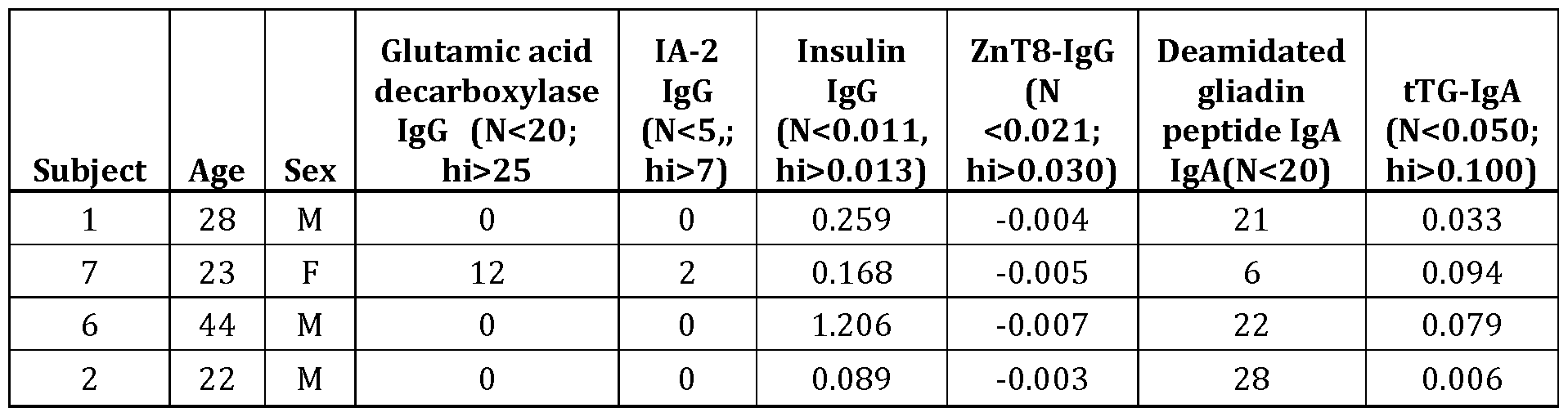

IFN-γ ELISpot and whole blood cytokine release assays were performed to assess T-cell responses to pools of 17mer peptides encompassing all unique 12mer amino-acid sequences derived from pancreatic islet antigens commonly recognized by autoantibodies in TID: insulin, glutamic decarboxylase-65 (GAD65), and insulinoma-associated antigen(IA)-2 (purity 70% by HPLC, identity confirmed by LC-MS; synthesized by JPT Peptide Technologies GmbH, Germany) (Table 8). Libraries were designed according to Beissbarth et al (Bioninformatics T. et al. Suppl l:i29-37) using protein sequences sourced from the public NCBI Genbank database (ncbi.nlm.nih.gov/protein). Table 8 summarizes the resulting peptide libraries.
Table 8. Islet autoantigen peptide libraries
Library Insulin GAD65 IA-2
Insulinoma antigen-2
Glutamic (IA-2) or Tyrosine
Antigen: Preproinsulin decarboxylase 65 phosphatase like
(GAD65) autoantigen (PTPRN) or
(ICA512)
"homo sapiens" and
"glutamate
"homo sapiens"
decarboxylase", or
and "Receptor- type tyro sine- "glutamic
Search terms "preproinsulin", or protein phosphatase-like decarboxylase", or
"proinsulin", or N"
"glutamic" and
"insulin"
"decarboxylase" and
"65"
Genbank entries
19 20 3
retrieved
Unique 12mers 154 692 1141
Total 17mers in
34 151 255
library
Pools per
1 2 3
library
17mers per pool 34 75 or 76 85
Deamidation - additional 4 in NP_001127838.1,
sequences 1 in CAB62572.1, 4 in
1 in AAP35454.1,
generated with CAA49554.1, 2 in
AAA59179.1,
glutamate AAB28987.1, 1 in 15 in Q16849
AEG 19452.1, and
substitutions at EAW86101.1, 2 in
ABI63346.1
potential CAH73660.1, and 3 in
deamidation CAH73658.1
sites1
Genbank
accession 0601246A EAW86103.1 NP_001186692.1 numbers:
AAA59172.1 AAA58491.1 NP_001186693.1
AAA59172.1 AAA62367.1 Q16849.1
AAA59173.1 AAB28987.1
AAA59179.1 AAI26328.1
AAH05255.1 AAI26330.1
AAN39451.1 AAP88040.1
AAP35454.1 CAA49554.1
AAW83741.1 CAB62572.1
ABI63346.1 CAC09233.1
AEG 19452.1 CAH73658.1
AFK93533.1 CAH73659.1
CAA08766.1 CAH73660.1
CAA23828.1 EAW86101.1
CAA49913.1 EAW86102.1
ΝΡ_000198.1 EAW86104.1
NP_001172026.1 NP_000809.1
NP_001172027.1 NP_001127838.1
P01308.1 Q05329.1
Q5VZ30
If a glutamine residue in the primary sequence conformed to the deamidation motif defined for transglutaminase-2 (QXiPX3, or QXiX2[F,Y,W,I,L,V], where and X3 are not proline) a second primary sequence was generated with glutamine replaced by glutamate. This deamidated primary sequences was included amongst those used to generate peptide
libraries.
In addition, a pool of 3 gluten peptides consisting of immune-dominant HLA-DQ2.5- restricted T cell epitopes (95% purity by HPLC, identity confirmed by LC-MS; synthesized by CS Bio CA), a pool with 10 additional gluten peptides consisting of immuno-dominant HLA-DQ2.5, - DQ8, and DQ2.2 restricted T cell epitopes (90% purity by HPLC, identity confirmed by LC-MS; synthesized by Pepscan Netherlands), a pool of 14 gluten peptides consisting of 11 from the two smaller pools (90% purity by HPLC, identity confirmed by LC-MS; synthesized by Pepscan Netherlands), a pool of 71 gluten-derived peptides consisting of all the peptides in the three smaller pools as well as 55 additional peptides consisting of sequences implicated in HLA-DQ2.5- associated Celiac disease (purity 70% by HPLC, identity confirmed by LC-MS; JPT Peptide Technologies) (Tye-Din et al. Sci Transl Med 2010), and of 23 MHC class-I (CEF) (Product no. 3615-1, Mabtech AG, Sweden) and 14 MHC class-II restricted epitopes derived from recall viral antigens (PM-CEFT-MHC-II, JPT Peptide Technologies GmbH, Germany) were also assessed. Individual constituent peptides from the preproinsulin, GAD, IA-2 and gluten pools were assessed in whole blood cytokine release assays using blood collected 6 days after commencing oral gluten challenge.
In whole blood assays 225 volumes of fresh heparinized blood were incubated with 25 μL of peptide in phosphate buffered saline (PBS) with dimethylsulfoxide (DMSO) to a final concentration of 0.05% or 0.1% in 96-well round-bottom plates. Concentrations of constituent
peptides in islet-autoantigen pools in whole blood assays were 0.4, 1 or 4 μg/mL; for CEF 0.1 μg/mL; CEFT 1 μg/mL; 3-gluten-peptide pool 10, 20 or 50 μg/mL, 13-gluten-peptide pool 5, 10, or 25 μg/mL; 14-gluten-peptide pool 5 , 10, or 25 μg/mL; 71-gluten-peptide pool 1, 5 or 10 μg/mL. The final concentration of individual peptides incubated in whole blood with 10% PBS 5 was 20 μg/mL with 0.0.5% DMSO. Whole blood assays were incubated for 24 hours at 37°C in 5% C02. At the conclusion of the incubation period, approximately 120 plasma was separated from blood after centrifuging plates and then transferred to a corresponding well in a "mirror image" sterile 96-well plate. Plates containing plasma were sealed with adhesive plastic coverslips and frozen at -80°C. Multiplex bead-based cytokine assays (MAGPIX®) were performed on o plasma thawed while being centrifuged at room temperature for lOmin. Plasma was pipetted
directly into dedicated 96-well plates for magnetic bead-based assays to measure the
concentrations of interferon(IFN)-y-induced protein- 10 (IP- 10), interleukin(IL)-2 and IFN-γ. For peptide pools assessed before and after oral gluten challenge, triplicate plasma samples were assessed and the final concentration was expressed as the mean of triplicates after removal of5 outliers. The assay blank was determined by the mean levels of cytokines in plasma from blood incubated with 10% PBS and 0.1% DMSO. After oral gluten challenge, individual peptides in libraries were incubated in duplicate wells with whole blood. Plasma from duplicate wells was combined and assessed in triplicate wells for the multiplex cytokine assay. Each 96-well
MAGPIX® plate was considered a separate assay that included 6 replicates with plasma from o whole blood incubated with medium only. For each cytokine, a standard curve was determined using standards provided by the manufacturer from 3.2 to 10,000 pg/mL. If a cytokine
concentration was reported as being <3.2 pg/mL a value of 3.2 pg/mL was recorded, and if > 10,000 pg/mL a value of 10,000 pg/mL was recorded. Cytokine levels were considered elevated if the mean cytokine concentration in triplicate test wells was at least twice the mean level in 6 5 replicate wells in the same 96-well plate containing plasma from whole blood incubated with medium only.
In ELISpot assays, 0.4 million freshly isolated peripheral blood mononuclear cells (PBMC) re-suspended in 50 X-Vivo® serum-free medium were incubated with peptide pools dissolved in 40 X-Vivo® and 10 PBS. IFN-γ ELISpot assays were performed in pre-coated 0 96-well MAIP plates (Mabtech) that were incubated at 37°C in 5% C02. After 18h, PBMCs were
discarded from ELISpot plates, and plates were washed and developed for later analysis by an automated ELISpot reader (Zellnet Inc., NJ). Spot forming counts were determined for each well.
Results
Gluten peptide pool responses: Cytokine release responses to pools and individual gluten- derived peptides were either unchanged or substantially increased after oral gluten challenge (Table 9 and Tables lOA-C). Amongst the whole blood cytokine release assays, responses to gluten peptides were least common and weakest in the whole blood IFN-γ release assay and most consistent and pronounced in the IP- 10 assay after oral gluten challenge. However, IP- 10 responses to the two larger gluten peptide pools were more than double those to medium alone in blood collected before as well as after oral gluten challenge in all four subjects. IL-2 responses to the highest concentrations (10-50 μg/mL) of gluten peptide pools were also more than double responses to medium alone in blood collected before oral gluten challenge in three subjects.
Whole blood IFN-γ release and IFN-γ ELISpot responses to gluten peptide pools were not elevated before oral gluten challenge.
Table 9. IFNg ELISpot responses 1 to peptide pools 2
Subject 1 7 6 2
HLA-DR 3, 4 3, 4 3 3, 4
2.5, 2.5, 2.5,
HLA-DQ 2.5
8 8 8
Days since commencing oral challenge 0 6 0 6 0 6 0 6
Medium & PBMC 0.5% DMSO 0 0 1 5 0 0 0 0
Medium & PBMC 0.5% DMSO 4 1 7 7 2 7 5 4
Medium no PBMC 0.5% DMSO 2 1 4 7 3 5 3 2
PHA 10 ug/mL 0.5% DMSO 2171 1730 1263 2109 1693 763 2487 2784
Recall antigen pools
CEF each peptide 0.1 ug/mL 249 75 12 45 42 74 1 1 14
CEFT each peptide 1 ug/mL 9 5 22 36 34 59 3 16
Islet autoantigen-derived pools
Insulin - 34 peptides 17mers 20
222 147 92 410 141 25 261 338 ug/mL
GAD65 pool 1 - 75 17mers 20 ug/mL 21 1 1 1 20 5 21 8 7
GAD65 pool 2 - 76 17mers 20 ug/mL 84 3 14 18 7 15 3 4
IA2 pool 1 - 85 17mers 20 ug/mL 6 3 8 17 19 32 2 7
IA2 pool 2 - 85 17mers 20 ug/mL 56 6 76 95 71 151 19 23
IA2 pool 3 - 85 17mers 20 ug/mL 23 1 9 22 24 40 3 7
Gluten pools (epitope restrictions)
71-peptides (DQ2.5/2.2/8) 50 uM 2 4 8 53 8 354 2 1 1
14-peptides (DQ2.5/2.2/8) 25 uM 2 4 8 53 8 354 2 1 1
13-peptides (DQ2.5/2.2/8) 25 uM 2 2 4 65 2 281 3 9
3-peptides (DQ2.5) 50 ug/mL 2 3 4 19 5 270 4 2
1 Spot forming units (sum of 3 wells, 0.4 million PBMC per well)
2 Pools with individual peptides at concentrations indicated in a final concentration of 0.5% DMSO
Table 10A. Whole blood IFNg release to Gluten Peptide Pools
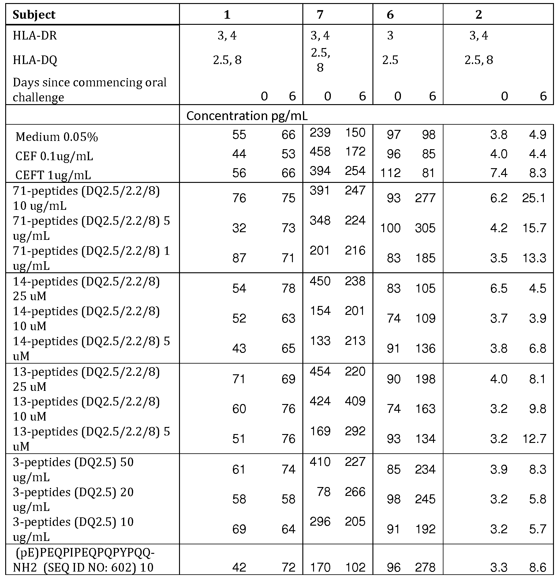
(pE) QPFPQPEQPFPWQP- NH2 (SEQ ID NO: 603] 10 uM 60 62 158 150 94 258 4.5 6.4
(pE)LQPFPQPELPYPQPQ- NH2 (SEQ ID NO: 604) 10 uM 39 55 407 194 102 200 4.1 13.9
Table 10A (continued).
Subject 1 7 6 2
HLA-DR 3,4 3,4 3 3,4
2.5,
HLA-DQ 2.5,8 2.5,8 2.5
8
Days since commencing oral challenge 0 6 0 6 0 6 0 6
Fold-change over medium only (bold>2)
Medium 0.05% 1.00 1.00 1.00 1.00 1.00 1.00 1.00 1.00
CEF O.lug/mL 0.80 0.81 1.92 1.14 0.99 0.86 1.03 0.90
CEFT lug/mL 1.03 1.01 1.65 1.69 1.16 0.83 1.94 1.70
71-peptides (DQ2.5/2.2/8) 10 ug/mL 1.39 1.14 1.64 1.64 0.97 2.83 1.62 5.15
71-peptides (DQ2.5/2.2/8) 5 ug/mL 0.59 1.11 1.46 1.49 1.04 3.11 1.09 3.23
71-peptides (DQ2.5/2.2/8) 1 ug/mL 1.59 1.08 0.84 1.44 0.86 1.89 0.90 2.74
14-peptides (DQ2.5/2.2/8) 25 uM 0.99 1.19 1.88 1.58 0.85 1.07 1.71 0.93
14-peptides (DQ2.5/2.2/8) 10 uM 0.96 0.95 0.65 1.34 0.76 1.11 0.96 0.80
14-peptides (DQ2.5/2.2/8) 5 uM 0.79 0.98 0.56 1.41 0.94 1.39 0.98 1.40
13-peptides (DQ2.5/2.2/8) 25 uM 1.30 1.04 1.90 1.46 0.94 2.02 1.03 1.66
13-peptides (DQ2.5/2.2/8) 10 uM 1.10 1.16 1.77 2.72 0.76 1.66 0.84 2.01
13-peptides (DQ2.5/2.2/8) 5 uM 0.93 1.15 0.71 1.94 0.96 1.36 0.84 2.61
3-peptides (DQ2.5) 50 ug/mL 1.12 1.13 1.72 1.51 0.88 2.38 1.01 1.71
3-peptides (DQ2.5) 20 ug/mL 1.06 0.88 0.33 1.77 1.01 2.50 0.84 1.18
3-peptides (DQ2.5) 10 ug/mL 1.27 0.98 1.24 1.36 0.94 1.96 0.84 1.17
(pE)PEQPIPEQPQPYPQQ-NH2 (SEQID
NO: 602) 10 uM 0.76 1.09 0.71 0.68 0.99 2.84 0.85 1.77
(pE)QPFPQPEQPFPWQP-NH2 (SEQID
NO: 603) 10 uM 1.09 0.95 0.66 1.00 0.98 2.63 1.19 1.32
(pE)LQPFPQPELPYPQPQ-NH2 (SEQ ID
NO: 604) 10 uM 0.71 0.84 1.71 1.29 1.05 2.03 1.06 2.85
Table lOB.Whole blood IL-2 release to Gluten Peptide Pools
Subject 1 7 6 2
HLA-DR 3,4 3,4 3 3,4
HLA-DQ 2.5,8 2.5,8 2.5 2.5,8
Days since
commencing oral
challenge 0 6 0 6 0 6 0 6
Concentration pg/mL

Table 10B (continued)
Subject 1 7 6 2
HLA-DR 3,4 3,4 3 3,4
2.5, 2.5,
HLA-DQ 2.5,8 2.5
8 8
Days since commencing oral
challenge 0 6 0 6 0 6 0 6
Fold-change over medium only (bold>2)
Medium 0.05% 1.00 1.00 1.00 1.00 1.00 1.00 1.00 1.00
CEF O.lug/mL 1.00 1.00 3.12 1.28 0.91 0.87 1.00 1.00
CEFT lug/mL 1.00 1.00 3.65 3.22 4.48 4.92 2.03 2.30
71-peptides (DQ2.5/2.2/8) 10
4.70 4.11
ug/mL 2.71 3.86 6.17 25.99 1.00 15.75
71-peptides (DQ2.5/2.2/8) 5
1.14 8.28
ug/mL 1.89 2.78 7.39 32.84 1.00 5.94
71-peptides (DQ2.5/2.2/8) 1
1.06 1.38
ug/mL 0.99 3.03 3.42 17.36 1.00 4.10
14-peptides (DQ2.5/2.2/8) 25
1.82 8.08
uM 2.64 4.54 0.65 1.41 1.00 1.00
14-peptides (DQ2.5/2.2/8) 10
1.28 2.38
uM 0.52 2.78 1.72 3.48 1.00 1.00
14-peptides (DQ2.5/2.2/8) 5 uM 1.00 3.65 0.60 2.99 0.99 5.55 1.00 1.00
13-peptides (DQ2.5/2.2/8) 25
1.00 2.99
uM 2.59 3.97 2.36 21.74 1.00 1.00
13-peptides (DQ2.5/2.2/8) 10
1.00 1.82
uM 1.76 3.04 2.52 21.16 1.00 1.00
13-peptides (DQ2.5/2.2/8) 5 uM 1.00 4.44 0.90 4.42 1.49 14.16 1.00 9.25
3-peptides (DQ2.5) 50 ug/mL 1.00 3.30 2.39 2.88 1.53 19.49 1.00 1.00
3-peptides (DQ2.5) 20 ug/mL 1.00 1.00 0.33 3.12 4.58 26.02 1.00 1.00
3-peptides (DQ2.5) 10 ug/mL 1.00 1.75 1.48 2.53 1.81 13.67 1.00 1.00
(pE)PEQPIPEQPQPYPQQ-NH2
(SEQIDNO: 602] 10 uM 1.00 1.00 1.69 0.66 4.71 31.67 1.00 7.00
(pE)QPFPQPEQPFPWQP-NH2
(SEQIDNO: 603] 10 uM 1.00 1.00 0.66 1.31 3.62 27.33 1.00 1.61
(pE)LQPFPQPELPYPQPQ-NH2
(SEQIDNO: 604] 10 uM 1.00 1.00 2.30 1.69 2.52 20.88 1.00 3.82
Table IOC. Whole blood IP- 10 release to Gluten Peptide Pools
Subject 1 7 6 2
HLA-DR 3,4 3,4 3 3,4
HLA-DQ 2.5,8 2.5,8 2.5 2.5,8
Days since
commencing oral
challenge 0 6 0 6 0 6 0 6
Concentration pg/mL
Medium 0.05% 266 335 743 892 277 201 266 312
CEF O.lug/mL 225 346 1211 764 318 644 241 282
CEFT lug/mL 586 604 10000 10000 5439 8789 937 1235
71-peptides 1805 10000
688 1546 3479 10000 561 3696 (DQ2.5/2.2/8) 10

Table IOC (continued).
Subject 1 7 6 2
HLA-DR 3,4 3,4 3 3,4
2.5,
HLA-DQ 2.5,8 2.5,8 2.5
8
Days since commencing oral
challenge 0 6 0 6 0 6 0 6
Fold-change over medium only (bold>2)
Medium 0.05% 1.00 1.00 1.00 1.00 1.00 1.00 1.00 1.00 CEF O.lug/mL 0.85 1.03 1.63 0.86 1.15 3.21 0.91 0.90
CEFT lug/mL 2.21 1 .80 13.46 11.21 19.62 43.73 3.53 3.95
71-peptides (DQ2.5/2.2/8) 10
2.59 4.61
ug/mL 2.43 11.21 12.55 49.76 2.11 11.83
71-peptides (DQ2.5/2.2/8) 5
2.87 9.25
ug/mL 1 .63 11.21 11.34 49.76 1 .75 9.47
71-peptides (DQ2.5/2.2/8) 1
2.19 2.55
ug/mL 2.77 11.21 9.90 49.76 2.18 6.30
14-peptides (DQ2.5/2.2/8) 25
3.58 6.36
uM 3.58 11.21 0.79 9.21 1 .00 0.85
14-peptides (DQ2.5/2.2/8) 10
1 .23 4.64
uM 1 .85 11.21 3.25 18.02 0.91 0.87
14-peptides (DQ2.5/2.2/8) 5
0.85 2.54
uM 1 .95 11.21 1 .89 49.56 1 .02 1 .73
13-peptides (DQ2.5/2.2/8) 25
1 .24 2.82
uM 1 .99 11.21 3.19 49.76 0.97 1 .35
13-peptides (DQ2.5/2.2/8) 10
1 .00 3.01
uM 1 .22 11.21 4.47 49.76 0.81 4.26
13-peptides (DQ2.5/2.2/8) 5
0.88 4.11
uM 1 .23 11.21 1 .62 49.76 0.95 8.72
3-peptides (DQ2.5) 50 ug/mL 0.88 2.92 1 .29 11.21 1 .16 49.76 0.86 1 .60
3-peptides (DQ2.5) 20 ug/mL 0.94 1 .25 0.72 11.21 8.94 49.76 0.86 2.15
3-peptides (DQ2.5) 10 ug/mL 1 .19 1 .92 1 .14 11.21 4.79 49.76 0.95 2.22
(pE)PEQPIPEQPQPYPQQ-NH2
(SEQ ID NO: 602] 10 uM 0.89 0.97 0.96 2.86 12.30 49.76 1 .26 4.06
(pE)QPFPQPEQPFPWQP-NH2
(SEQ ID NO: 603] 10 uM 0.82 1 .49 1 .21 11.21 6.98 49.76 1 .42 3.65
(pE)LQPFPQPELPYPQPQ-NH2
(SEQ ID NO: 604] 10 uM 0.76 1 .15 1 .32 3.69 5.83 49.76 1 .20 5.90
TID autoantigen peptide pool responses: Responses to TID autoantigen peptide pools in cytokine release assays, especially whole blood IP- 10 release, were frequently elevated (Table 9 and Tables 11A-C). The highest concentration of peptide pools tested (4 μg/mL) almost always stimulated stronger responses than the lowest concentration (0.4 μg/mL). With the exception of pool 1 for GAD65 in one patient, each of the eight TID autoantigen peptide pools elicited whole blood IP- 10 responses that were more than double those to medium alone in all four subjects. The preproinsulin pool and IA-2 pool 2 frequently stimulated strongest cytokine release responses, especially in the IFN-γ ELISpot. Consistency and magnitude of cytokine release stimulated by TID autoantigen peptide pools was not consistently changed after oral gluten challenge.
Table 11A. Whole blood IFNg release to Islet-autoantigen Peptide Pools
Subject 1 7 6 2
HLA-DR 3, 4 3, 4 3 3, 4

Table 11A (continued)
Subject 1 7 6 2
HLA-DR 3,4 3,4 3 3,4
2.5, 2.5, HLA-DQ 2.5,8 2.5
8 8
Days since commencing oral
challenge 0 6 0 6 0 6 0 6
Fold-change over medium only (bold>2)
Medium 0.05% 1.00 1.00 1.00 1.00 1.00 1.00 1.00 1.00 CEFO.lug/mL 0.70 1.05 1.05 1.46 0.84 1.02 0.88 0.89 CEFT lug/mL 0.63 0.90 1.17 1.44 0.93 1.07 1.40 0.93
Insulin - 34 peptides 17mers 4
0.83 1.27
ug/mL 1.92 2.69 1.26 1.18 5.41 7.49
Insulin - 34 peptides 17mers 1
0.75 0.94
ug/mL 1.43 1.35 1.01 0.92 2.54 1.73
Insulin - 34 peptides 17mers 0.4
0.72 1.10
ug/mL 0.97 0.83 0.96 0.82 1.08 1.06
GAD65 pool 1 - 7517mers 4 ug/mL 0.87 1.43 1.37 2.89 1.22 0.99 1.50 1.37
GAD65 pool 1 - 7517mers 1 ug/mL 0.73 0.61 1.28 0.98 1.01 0.94 0.77 0.86 GAD 65 pool 1 - 7517mers 0.4
0.74 1.03
ug/mL 1.25 0.76 0.85 0.81 0.88 0.84
GAD65 pool 2 - 7617mers 4 ug/mL 0.92 1.08 1.12 1.78 1.28 1.01 1.43 1.68
GAD65 pool 2 - 7617mers 1 ug/mL 0.74 1.06 1.26 1.87 0.86 1.01 1.38 1.21 GAD 65 pool 2 - 7617mers 0.4
0.61 1.12
ug/mL 1.25 1.12 0.80 0.91 1.63 0.94
IA2 pool 1 - 8517mers 4 ug/mL 1.56 1.01 1.19 1.82 1.22 0.93 1.78 1.43 IA2 pool 1 - 8517mers 1 ug/mL 0.71 0.53 1.09 1.27 0.84 0.78 0.77 0.89 IA2 pool 1 - 8517mers 0.4 ug/mL 0.78 1.09 1.05 1.34 0.75 0.89 1.08 0.65
IA2 pool 2 - 8517mers 4 ug/mL 0.74 1.40 1.07 2.21 1.65 1.33 7.11 5.10 IA2 pool 2 - 8517mers 1 ug/mL 1.13 1.12 0.67 0.91 1.06 1.20 2.08 1.37 IA2 pool 2 - 8517mers 0.4 ug/mL 0.81 0.24 1.00 2.08 0.86 0.83 1.26 0.71
IA2 pool 3 - 8517mers 4 ug/mL 0.78 0.70 0.42 0.96 1.08 0.87 2.90 1.36 IA2 pool 3 - 8517mers 1 ug/mL 0.58 0.77 0.55 0.56 0.79 0.70 0.90 0.98 IA2 pool 3 - 8517mers 0.4 ug/mL 0.70 0.95 0.96 1.18 0.80 0.67 0.93 0.78
Table 1 IB. Whole blood IL-2 release to Islet-autoantigen Peptide Pools
Subject 1 7 6 2
HLA-DR 3,4 3,4 3 3,4
2.5, 2.5, HLA-DQ 2.5,8 2.5
8 8
Days since commencing oral
challenge 0 6 0 6 0 6 0 6
Concentration pg/mL
Medium 0.05% 3.2 3.2 162 23 3.6 4.3 3.2 3.2 CEF O.lug/mL 3.2 3.2 211 33 3.2 5.0 3.2 3.2 CEFT lug/mL 3.2 3.2 236 47 17.1 24.4 8.4 3.2
Insulin - 34 peptides 17mers 4 311 94
5.2 6.3 15.8 16.7 21.7 34.0 ug/mL
Insulin - 34 peptides 17mers 1 205 34
5.1 3.2 5.8 5.0 6.4 3.2 ug/mL
Insulin - 34 peptides 17mers 0.4 136 19
3.2 3.2 3.5 3.2 3.2 3.2 ug/mL
GAD 65 pool 1 - 7517mers 4 236 95
14.0 3.2 10.4 7.2 3.2 3.2 ug/mL
GAD 65 pool 1 - 7517mers 1 216 23
3.2 3.2 3.6 4.8 3.2 3.2 ug/mL
GAD 65 pool 1 - 7517mers 0.4 3.2 3.2 181 18 3.2 3.2 3.2 3.2

Table 11B (continued).
 IA2 pool 2 - 8517mers 4 ug/mL 1.19 6.74 1.05 3.70 12.40 6.39 12.57 12.30 IA2 pool 2 - 8517mers 1 ug/mL 7.48 1.93 0.48 0.90 3.36 5.03 4.56 1.00 IA2 pool 2 - 8517mers 0.4 ug/mL 2.05 1.00 1.25 2.36 1.04 0.98 1.00 1.00
IA2 pool 3 - 8517mers 4 ug/mL 1.00 1.00 0.06 0.51 1.45 1.43 10.40 9.09 IA2 pool 3 - 8517mers 1 ug/mL 1.00 1.00 0.41 0.44 0.88 0.74 4.57 4.63 IA2 pool 3 - 8517mers 0.4 ug/mL 1.00 1.00 1.18 1.24 0.88 0.74 6.67 1.00
Table 11C. Whole blood IP- 10 release to Islet- autoantigen Peptide Pools


Table 11C (continued).
Subject 1 7 6 2
HLA-DR 3,4 3,4 3 3,4
HLA-DQ 2.5,8 2.5,8 2.5 2.5,8
Days since commencing oral
challenge 0 6 0 6 0 6 0 6
Fold-change over medium only (bold>2)
Medium 0.05% 37.89 1.00 1.00 1.00 1.00 1.00 1.00 1.00 CEFO.lug/mL 1.00 0.95 0.90 1.10 2.76 1.94 0.87 0.84 CEFT lug/mL 1.04 1.04 8.60 9.94 22.53 44.82 2.63 2.47
Insulin - 34 peptides 17mers
12.06 28.71
4 ug/mL 11.61 16.68 38.46 48.62 23.58 15.85
Insulin - 34 peptides 17mers
17.87 8.92
1 ug/mL 11.61 12.46 10.02 16.15 12.98 8.60
Insulin - 34 peptides 17mers
2.19 2.88
0.4 ug/mL 2.65 2.27 1.40 1.67 1.00 2.15
GAD 65 pool 1 - 7517mers 4
7.25 4.58
ug/mL 2.52 1.93 20.79 6.15 1.88 1.57
GAD 65 pool 1 - 7517mers 1
2.51 2.65
ug/mL 1.31 1.14 2.62 2.25 1.08 1.03
GAD 65 pool 1 - 7517mers 0.4
1.07 1.78
ug/mL 1.12 1.33 1.25 1.43 1.24 0.91
GAD 65 pool 2 - 7617mers 4
20.68 2.32
ug/mL 11.61 15.83 7.58 12.68 1.50 2.51
GAD 65 pool 2 - 7617mers 1
8.26 2.64
ug/mL 11.61 6.16 11.97 5.96 4.96 2.09
GAD 65 pool 2 - 7617mers 0.4
2.05 1.92
ug/mL 5.98 2.70 6.92 5.16 0.85 1.72
IA2 pool 1 - 8517mers 4
37.89 7.29
ug/mL 11.61 16.63 17.43 13.49 4.94 3.83
IA2 pool 1 - 8517mers 1
11.82 3.35
ug/mL 3.91 2.10 5.58 9.16 1.76 1.63
IA2 pool 1 - 8517mers 0.4
5.10 2.45
ug/mL 1.50 1.39 1.01 1.46 0.96 0.83
IA2 pool 2 - 8517mers 4
6.26 14.62
ug/mL 11.61 16.68 38.46 48.62 15.26 16.27
IA2 pool 2 - 8517mers 1
33.87 9.77
ug/mL 11.61 6.94 27.17 48.62 6.84 3.29
IA2 pool 2 - 8517mers 0.4
5.46 1.25
ug/mL 2.09 5.56 8.28 14.11 1.35 0.76
IA2 pool 3 - 8517mers 4
0.97 2.34
ug/mL 1.39 1.50 4.35 3.14 6.41 3.76
IA2 pool 3 - 85 17mers 1
2.85 2.31
ug/mL 1 .15 0.92 1 .18 1 .23 1 .25 1 .72
IA2 pool 3 - 85 17mers 0.4
1 .90 1 .13
ug/mL 1 .07 3.92 0.90 0.96 1 .16 0.96
TID autoantigen peptide-specific responses: Each of the 440 individual TID autoantigen- derived 17mer peptides (20 μg/mL) were incubated in a single well with whole blood collected 6- days after commencing oral gluten challenge. Amongst the 34 preproinsulin-derived peptides, one elicited IP-10 responses in all four subjects that were 2-fold greater than to medium alone. Three 17mers in the 155-member GAD65 peptide library elicited increased IP-10 release in all four subjects. One peptide amongst the 255 17mers in the IA-2 library consistently stimulated IP-10 and IL-2 release, and in two subjects also stimulated IFN-γ release, suggesting that it is the immuno-dominant peptide in IA-2. A second peptide, evoked IP-10 but not IL-2 or IFN-γ release in all four subjects.
Conclusions
Oral gluten challenge does not enhance whole blood cytokine release in response to TID autoantigen-derived peptides, as it does to gluten-derived peptides in Celiac disease. However, IP- 10 and IL-2 both offer greater sensitivity than IFN-γ ίη whole blood cytokine release assays for gluten-reactive T cells in Celiac disease, and when applied to screening peptides derived from TID autoantigens in patients with TID and Celiac disease, identify amino acid sequences that are consistently immuno-dominant. One 17mer peptide from preproinsulin, three from GAD65 and two from IA-2 stimulated elevated whole blood IP-10 release in four of four TID subjects. Several other sequences commonly but less consistently and less potently stimulated IP-10 release in the TID subjects assessed in this study. Hence, application of multiplex cytokine and chemokine measurement to epitope mapping in TID reveals a hierarchy of epitopes not previously evident in the classical TID-associated autoantigens preproinsulin, GAD65 and IA-2.
Example 4. Use of IP-10 to Measure Rare Antigen-Specific T cells
Methods
HLA-DQ2.5-positive celiac disease subjects on gluten-free diet were used in this study. Blood was collected immediately before and 6 days after commencing 3-day oral gluten challenge.
Whole blood or PBMCs were incubated with pools or single peptides derived from gluten or recall antigens. IFNy and IP- 10 levels were measured in plasma from the whole blood that was incubated in 96-well plates with peptides or peptide pools. Plasma cytokine/chemokine levels were measured by MAGPIX® multiplex bead assay (IFNy and IP- 10) or by ELISA (IFNy and IP- 10), and PBMC separated from the same blood sample were incubated in overnight IFNy ELISpot assays.
Results
It was observed that IP- 10 was elevated in plasma from blood incubated with gluten peptide pools 6 days after commencing the oral gluten challenge. The elevation was more statistically significant with IP- 10 (p<0.002) than with IFN-y (not significant) (Table 4 and 5). Using a cut-off of stimulation index >1.25 and cytokine level >100 pg/mL to indicate a positive result, the IP-10 assay was positive in 7/10 subjects prior to gluten challenge and 10/10 subjects after gluten challenge, whereas the IFN-γ assay using gluten peptide pools, with a cut-off of stimulation index >1.25 and cytokine level >7.2 pg/mL to indicate a positive result, was positive in 1/10 subjects pre-gluten challenge and 7/10 subjects after gluten challenge. Thus, the IP-10 assay was very effective both before and after gluten challenge for detecting antigen- specific T cells.
Table 4. Whole blood secretion of IFNy pre- and post- gluten challenge
IFNy Day O Day O Day 6 Day 6
MAGPIX Ratio Gluten Gluten peptides minus Ratio Gluten Gluten peptides peptides:Medium Medium (pg/mL) peptides:Medium minus Medium
(pg/mL)
Minimum 0.9969 -0.1 0.72 -259
25% 1.086 1.205 0.9014 -17.58
Percentile
Median 1.265 4.625 5.53 50.77
75% 1.689 21.01 29.82 185.6
Percentile
Maximum 2.6 143 228.6 728.2
Day 0 = prior to oral gluten challenge, Day 6 = 6 days after commencing a 3-day gluten challenge.
Table 5. Whole blood secretion of IP10 pre- and post-gluten challenge

Day 0 = prior to oral gluten challenge, Day 6 = 6 days after commencing a 3-day gluten challenge.
OTHER EMBODIMENTS
All of the features disclosed in this specification may be combined in any combination. Each feature disclosed in this specification may be replaced by an alternative feature serving the same, equivalent, or similar purpose. Thus, unless expressly stated otherwise, each feature disclosed is only an example of a generic series of equivalent or similar features.
From the above description, one skilled in the art can easily ascertain the essential characteristics of the present disclosure, and without departing from the spirit and scope thereof, can make various changes and modifications of the disclosure to adapt it to various usages and conditions. Thus, other embodiments are also within the claims.
EQUIVALENTS
While several inventive embodiments have been described and illustrated herein, those of ordinary skill in the art will readily envision a variety of other means and/or structures for performing the function and/or obtaining the results and/or one or more of the advantages described herein, and each of such variations and/or modifications is deemed to be within the 5 scope of the inventive embodiments described herein. More generally, those skilled in the art will readily appreciate that all parameters, dimensions, materials, and configurations described herein are meant to be exemplary and that the actual parameters, dimensions, materials, and/or configurations will depend upon the specific application or applications for which the inventive teachings is/are used. Those skilled in the art will recognize, or be able to ascertain using no more o than routine experimentation, many equivalents to the specific inventive embodiments described herein. It is, therefore, to be understood that the foregoing embodiments are presented by way of example only and that, within the scope of the appended claims and equivalents thereto, inventive embodiments may be practiced otherwise than as specifically described and claimed. Inventive embodiments of the present disclosure are directed to each individual feature, system, article,5 material, kit, and/or method described herein. In addition, any combination of two or more such features, systems, articles, materials, kits, and/or methods, if such features, systems, articles, materials, kits, and/or methods are not mutually inconsistent, is included within the inventive scope of the present disclosure.
All definitions, as defined and used herein, should be understood to control over dictionary o definitions, definitions in documents incorporated by reference, and/or ordinary meanings of the defined terms.
All references, patents and patent applications disclosed herein are incorporated by reference with respect to the subject matter for which each is cited, which in some cases may encompass the entirety of the document.
5 The indefinite articles "a" and "an," as used herein in the specification and in the claims, unless clearly indicated to the contrary, should be understood to mean "at least one."
The phrase "and/or," as used herein in the specification and in the claims, should be understood to mean "either or both" of the elements so conjoined, i.e., elements that are conjunctively present in some cases and disjunctively present in other cases. Multiple elements 0 listed with "and/or" should be construed in the same fashion, i.e., "one or more" of the elements so conjoined. Other elements may optionally be present other than the elements specifically
identified by the "and/or" clause, whether related or unrelated to those elements specifically identified. Thus, as a non-limiting example, a reference to "A and/or B", when used in conjunction with open-ended language such as "comprising" can refer, in one embodiment, to A only (optionally including elements other than B); in another embodiment, to B only (optionally including elements other than A); in yet another embodiment, to both A and B (optionally including other elements); etc.
As used herein in the specification and in the claims, "or" should be understood to have the same meaning as "and/or" as defined above. For example, when separating items in a list, "or" or "and/or" shall be interpreted as being inclusive, i.e., the inclusion of at least one, but also including more than one, of a number or list of elements, and, optionally, additional unlisted items. Only terms clearly indicated to the contrary, such as "only one of or "exactly one of," or, when used in the claims, "consisting of," will refer to the inclusion of exactly one element of a number or list of elements. In general, the term "or" as used herein shall only be interpreted as indicating exclusive alternatives (i.e. "one or the other but not both") when preceded by terms of exclusivity, such as "either," "one of," "only one of," or "exactly one of." "Consisting essentially of," when used in the claims, shall have its ordinary meaning as used in the field of patent law.
As used herein in the specification and in the claims, the phrase "at least one," in reference to a list of one or more elements, should be understood to mean at least one element selected from any one or more of the elements in the list of elements, but not necessarily including at least one of each and every element specifically listed within the list of elements and not excluding any combinations of elements in the list of elements. This definition also allows that elements may optionally be present other than the elements specifically identified within the list of elements to which the phrase "at least one" refers, whether related or unrelated to those elements specifically identified. Thus, as a non-limiting example, "at least one of A and B" (or, equivalently, "at least one of A or B," or, equivalently "at least one of A and/or B") can refer, in one embodiment, to at least one, optionally including more than one, A, with no B present (and optionally including elements other than B); in another embodiment, to at least one, optionally including more than one, B, with no A present (and optionally including elements other than A); in yet another embodiment, to at least one, optionally including more than one, A, and at least one, optionally including more than one, B (and optionally including other elements); etc.
It should also be understood that, unless clearly indicated to the contrary, in any methods claimed herein that include more than one step or act, the order of the steps or acts of the method is not necessarily limited to the order in which the steps or acts of the method are recited.
In the claims, as well as in the specification above, all transitional phrases such as "comprising," "including," "carrying," "having," "containing," "involving," "holding,"
"composed of," and the like are to be understood to be open-ended, i.e., to mean including but not limited to. Only the transitional phrases "consisting of and "consisting essentially of shall be closed or semi-closed transitional phrases, respectively, as set forth in the United States Patent Office Manual of Patent Examining Procedures, Section 2111.03.
Claims
1. A method of assaying an antigen- specific T cell response, the method comprising:
measuring a level of IP- 10 in a sample comprising an antigen- specific T cell obtained from a subject.
2. The method of claim 1, wherein the antigen specific T cell response is a rare antigen- specific T cell response and wherein the antigen- specific T cell is a rare antigen- specific T cell.
3. The method of claim 1 or 2, wherein the subject is a subject that has previously been administered IL-2 or an agent that stimulates IL-2 expression.
4. The method of claim 1 or 2, wherein the method further comprises administering IL-2 or an agent that stimulates IL-2 expression to the subject prior to the measuring.
5. The method of any one of claims 1 to 4, wherein the subject has or is suspected of having autoimmune disease, an allergy, an infectious disease or condition, or an adverse immune condition caused by administration of an isolated, recombinant or synthetic protein or peptide.
6. The method of any one of claims 1 to 4, wherein the subject has or is suspected of having an autoimmune disease and the antigen- specific T cell is a autoantigen- specific T cell.
7. The method of claim 6, wherein the subject has or is suspected of having the autoimmune disease and Celiac disease.
8. The method of claim 7, wherein the subject is a subject that has previously been administered a composition comprising a gluten peptide.
9. The method of claim 7, wherein the method further comprises administering a composition comprising a gluten peptide to the subject prior to the measuring.
10. The method of claim 8 or 9, wherein the composition is or has previously been administered to the subject more than once.
11. The method of claim 10 wherein the composition is or has previously been administered to the subject at least once a day for three days.
12. The method of any one of claims 8 to 11, wherein the composition comprises at least one of a wheat gluten, a barley hordein, and a rye secalin.
13. The method of claim 12, wherein the composition comprises at least two of a wheat gluten, a barley hordein, and a rye secalin.
14. The method of claim 13, wherein the composition comprises a wheat gluten, a barley hordein, and a rye secalin.
15. The method of any one of claims 9 to 14, wherein the administration of the composition is oral administration.
16. The method of claim 15, wherein the composition is a foodstuff.
17. The method of any one of claims 8 to 16, wherein the sample is obtained from the subject six days after administration of the composition.
18. The method of any one of claims 1 to 17, wherein the sample comprises whole blood or peripheral blood mononuclear cells.
19. The method of any one of claims 1 to 5 or 7 to 18, wherein the measuring of the level of IP- 10 in the sample comprises contacting the sample with an antigen peptide and measuring the level of IP- 10 in the sample.
20. The method of claim 6, wherein the measuring of the level of IP-10 in the sample comprises contacting the sample with an autoantigen peptide and measuring the level of IP-10 in the sample.
21. The method of claim 19 or 20, wherein the level of IP-10 is measured with an enzyme- linked immunosorbent assay (ELISA).
22. The method of claim 19 or 20, wherein the level of IP-10 is measured with a multiplex bead-based assay.
23. The method of any one of claims 19, 21 or 22, wherein the method further comprises comparing the level of IP-10 with a control level of IP-10 to identify or aid in identifying the antigen peptide as being one that is recognized by the antigen- specific T cell.
24. The method of claim 23, wherein an elevated level of IP-10 compared to the control level indicates that the antigen peptide is recognized by the antigen- specific T cell and wherein a decreased or substantially the same level of IP-10 compared to the control level indicates that the antigen peptide is not recognized by the antigen- specific T cell.
25. The method of claim 23 or 24', wherein a level of IP-10 that is at least two-folder greater than the control level indicates that the antigen peptide is recognized by the antigen- specific T cell.
26. The method of claim 20, wherein the method further comprises comparing the level of IP- 10 with a control level of IP-10 to identify or aid in identifying the autoantigen peptide as being one that is recognized by the rare autoantigen-specific T cell.
27. The method of claim 26, wherein an elevated level of IP-10 compared to the control level indicates that the autoantigen peptide is recognized by the rare autoantigen-specific T cell and wherein a decreased or substantially the same level of IP-10 compared to the control level indicates that the autoantigen peptide is not recognized by the rare autoantigen-specific T cell.
28. The method of claim 26 or 27, wherein a level of IP- 10 that is at least two-folder greater than the control level indicates that the autoantigen peptide is recognized by the rare autoantigen- specific T cell.
5 29. The method of any one of claims 1 to 28, wherein the method further comprises measuring a level of IFN-γ and/or IL-2 in the sample.
30. The method of claim 29, wherein a level of IFN-γ that is at least two-fold greater than a control level of IFN-γ and/or a level of IL-2 that is at least two-fold greater than a control level of o IL-2 indicates that the antigen peptide is recognized by the antigen- specific T cell or that the
autoantigen peptide is recognized by the rare autoantigen- specific T cell.
31. A kit, comprising:
(a) a means for detecting a level of IP- 10; and
5 (b) at least one antigen peptide.
32. The kit of claim 31, wherein the at least one antigen peptide is at least one autoantigen peptide. 0
33. The kit of claim 31, wherein the at least one antigen peptide is at least one foreign antigen.
34. The kit of any one of claims31 to 33, wherein the means for detecting a level of IP-10 is an antibody that binds to IP-10. 5 35. The kit any one of claims 31 to 34, wherein the kit further comprises a composition
comprising a gluten peptide.
36. The kit any one of claims 31 to 34, wherein the kit further comprises IL-2 or an agent that stimulates IL-2 expression.
0
37. The kit of claim 35, wherein the composition comprises at least one of a wheat gluten, a barley hordein, and a rye secalin.
38. The kit of claim37, wherein the composition comprises at least two of a wheat gluten, a barley hordein, and a rye secalin.
39. The kit of claim 38, wherein the composition comprises a wheat gluten, a barley hordein, and a rye secalin.
40. The kit of any one of claims 31 to 39, wherein the kit comprises a container, such as a vial or tube, for whole blood.
41. The kit of claim 40, wherein the at least antigen peptide is dried on the wall of the container for whole blood.
42. The kit of claim any one of claims 31 to 40, wherein the at least one antigen peptide is in a solution or lyophilized in a separate container.
43. The kit of any one of claims 40 to 42, further comprising an anticoagulant.
44. The kit of any one of claims 40 to 43, wherein the container for whole blood and/or other container are present in duplicate or triplicate.
45. The kit of any one of claims 40 to 44, wherein the kit further comprises a negative control container, such as a vial or tube.
46. The kit of any one of claims 40 to 45, wherein the kit further comprises a positive control container, such as a vial or tube.
47. The kit of any one of claims 31 to 46, wherein the kit further comprises means for detecting a level of IFN-γ and/or IL-2.
48. The kit of claim 47, wherein the means for detecting a level of IFN-γ is an antibody that binds to IFN-γ.
5 49. The kit of claim 47 or 48, wherein the means for detecting a level of IL-2 is an antibody that binds to IL-2.
50. A method of assaying a T cell response to an islet autoantigen peptide, the method comprising:
o (a) administering a composition comprising a gluten peptide to a first subject having or suspected of having Type 1 Diabetes (T1D) and Celiac disease; and
(b) measuring a first T cell response to at least one islet autoantigen peptide in a first sample obtained from the first subject after the administration of the composition. 5
51. The method of claim 50, wherein the at least one islet autoantigen peptide is selected from a proinsulin peptide, a 65-kDa isoform of glutamic acid decarboxylase (GAD 65) peptide, or an islet antigen-2 (IA-2) peptide.
52. The method of claims 51, wherein the at least one autoantigen peptide is a peptide o comprising a sequence as put forth in Table 3.
53. The method of any one of claims 50 to 52, wherein the first sample comprises whole blood or peripheral blood mononuclear cells. 5
54. The method of any one of claims 50 to 53, wherein the composition is administered to the first subject more than once.
55. The method of claim 54, wherein the composition is administered to the first subject at least once a day for three days.
0
56. The method of any one of claims 50 to 55, wherein the composition comprises at least one of a wheat gluten, a barley hordein, and a rye secalin.
57. The method of claim 56, wherein the composition comprises at least two of a wheat gluten, a barley hordein, and a rye secalin.
58. The method of claim 57, wherein the composition comprises a wheat gluten, a barley hordein, and a rye secalin.
59. The method of any one of claims 50 to 58, wherein the administration of the composition is oral administration.
60. The method of claim 59, wherein the composition is a foodstuff.
61. The method of any one of claims 50 to 60, wherein the measuring of the first T cell response in the first sample comprises contacting the first sample with the at least one islet autoantigen peptide and measuring a level of at least one cytokine in the first sample.
62. The method of claim 61, wherein the at least one cytokine is IL-2, IFN-γ or IP-10.
63. The method of claim 62, wherein the at least one cytokine is IP-10 and IL-2.
64. The method of claim 62, wherein the at least one cytokine is IP-10, IFN-γ and IL-2
65. The method of any one of claims 61 to 64, wherein the level of the at least one cytokine is measured with an enzyme-linked immunosorbent assay (ELISA).
66. The method of any one of claims 61 to 64, wherein the level of the at least one cytokine is measured with a multiplex bead-based assay.
67. The method of any one of claims 61 to 64, wherein the level of the at least one cytokine is measured with an enzyme-linked immunosorbent spot (ELISpot) assay.
68. The method of any one of claims 50 to 67, wherein the method further comprises comparing the first T cell response with a control T cell response to identify or aid in identifying the first subject as in need of further testing for TID if the T cell response measured in the first sample is elevated compared to the control T cell response, or to identify or aid in identifying the first subject as not in need of further testing for TID if the first T cell response is substantially the same or decreased compared to the control T cell response.
69. The method of claim 68, wherein the method further comprises performing further testing for TID if the first subject is identified as in need of further testing for TID.
70. The method of claim 69, wherein the further testing comprises a glycated hemoglobin test, a glucose tolerance test, a fasting blood sugar test, and/or an immunoassay for autoantibodies.
71. The method of claim 70, wherein autoantibodies comprises one or more of islet cell autoantibodies, insulin autoantibodies, 65-kDa isoform of glutamic acid decarboxylase (GAD65) autoantibodies, islet antigen-2 (IA-2) autoantibodies, and zinc transporter (ZnT8) autoantibodies.
72. The method of any one of claims 50 to 71, wherein the first sample is obtained from the first subject six days after administration of the composition.
73. The method of any one of claims 50 to 72, wherein the method further comprises:
(c) administering a placebo to a second subject having or suspected of having Type 1
Diabetes (TID) and Celiac disease; and
(d) measuring a second T cell response to the at least one islet autoantigen peptide in a second sample obtained from the second subject after the administration of the placebo.
74. The method of claim 73, wherein the measuring of the first and second T cell response are performed together in one assay.
75. The method of claim 73 or 74, wherein the composition is administered to the first subject more than once and the placebo is administered to the second subject more than once.
5 76. The method of claim 75, wherein the composition is administered to the first subject at least once a day for three days and the placebo is administered to the second subject at least once a day for three days.
77. The method of any one of claims 73 to 76, wherein the administration of the composition o and the placebo is oral administration.
78. The method of claim 77, wherein the composition and the placebo are foodstuffs.
79. The method of any one of claims 73 to 78, wherein the measuring of the first and second T 5 cell response in the first and second sample comprises contacting the first and second samples with the at least one islet autoantigen peptide and measuring a level of at least one cytokine in the first and second samples.
80. The method of claim 79, wherein the at least one cytokine is IL-2, IFN-γ or IP- 10.
0
81. The method of claim 80, wherein the at least one cytokine is IP-10 and IL-2.
82. The method of claim 80, wherein the at least one cytokine is IP-10, IFN-γ and IL-2 5
83. The method of any one of claims 79 to 82, wherein the level of the at least one cytokine is measured with an enzyme-linked immunosorbent assay (ELISA).
84. The method of any one of claims 79 to 82, wherein the level of the at least one cytokine is measured with an enzyme-linked immunosorbent spot (ELISpot) assay.
0
85. The method of claim any one of claims 79 to 82, wherein the level of the at least one cytokine is measured with a multiplex bead-based assay.
86. The method of any one of claims 73 to 85, wherein the method further comprises comparing the first T cell response with the second T cell response to identify or aid in identifying the first subject as in need of further testing for TID if the T cell response measured in the first sample is elevated compared to the second T cell response, or to identify or aid in identifying the first subject as not in further testing for TID if the first T cell response is substantially the same or decreased compared to the second T cell response.
87. The method of claim 86, wherein the method further comprises performing further testing for TID if the first subject is identified as in need of further testing for TID.
88. The method of claim 87, wherein the further testing comprises a glycated hemoglobin test, a glucose tolerance test, a fasting blood sugar test, and/or an immunoassay for autoantibodies.
89. The method of claim 88, wherein autoantibodies comprises one or more of islet cell autoantibodies, insulin autoantibodies, 65-kDa isoform of glutamic acid decarboxylase (GAD65) autoantibodies, islet antigen-2 (IA-2) autoantibodies, and zinc transporter (ZnT8) autoantibodies.
90. The method of any one of claims 73 to 89, wherein the second sample is obtained from the second subject six days after administration of the placebo.
91. The method of any one of claims 50 to 90, wherein the method further comprises performing another test on the first subject and/or second subject prior to or after the steps of the method, preferably, in some embodiments, performing a serology and/or genotyping assay.
92. The method of claim 91, wherein the performing a serology and/or genotyping assay occurs prior to all of the steps recited in the method.
93. The method of claim 91, wherein the performing a serology and/or genotyping assay occurs after all of the steps recited in the method.
94. The method of any one of claims 50 to 93, wherein the first subject and/or second subject is HLA-DQ2.5 positive.
95. A kit, comprising:
(a) a means for detecting a T cell response; and
(b) at least one islet autoantigen peptide.
96. The kit of claim 95, wherein the at least one islet autoantigen peptide is selected from a proinsulin peptide, a 65-kDa isoform of glutamic acid decarboxylase (GAD 65) peptide, or an islet antigen-2 (IA-2) peptide.
97. The kit of claim 96, wherein the at least one autoantigen peptide is a peptide comprising a sequence as put forth in Table 3.
98. The kit of any one of claims 95 to 97, wherein the means for detecting a T cell response is an antibody that binds to a cytokine.
99. The kit of claim 98, wherein the antibody that binds to a cytokine is an antibody that binds to IL-2, IFN-γ or IP- 10.
100. The method of claim 99, wherein the antibody that binds to a cytokine is an antibody that binds IP- 10 and an antibody that binds to IL-2.
101. The method of claim 99, wherein the antibody that binds to a cytokine is an antibody that binds IP- 10, an antibody that binds to IFN-γ, and an antibody that binds to IL-2.
102. The kit of any of claims 95 to 101, wherein the kit further comprises a composition comprising a gluten peptide.
103. The kit of any of claims 95 to 102, wherein the kit further comprises a placebo.
104. The kit of claim 103, wherein the composition and the placebo are foodstuffs.
5
105. The kit of claim any one of claims 102-104, wherein the composition comprises at least one of a wheat gluten, a barley hordein, and a rye secalin.
106. The kit of claim 105, wherein the composition comprises at least two of a wheat gluten, a l o barley hordein, and a rye secalin.
107. The kit of claim 105, wherein the composition comprises a wheat gluten, a barley hordein, and a rye secalin.
15 108. The kit of any one of claims 95 to 107, wherein the kit comprises a container, such as a vial or tube, for whole blood.
109. The kit of claim 108, wherein the at least one islet autoantigen peptide is dried on the wall of the container for whole blood.
20
110. The kit of any one of claims 95 to 108, wherein the at least one islet autoantigen peptide is in a solution or lyophilized in a separate container.
111. The kit of any one of claims 108 to 110, further comprising an anticoagulant.
25
112. The kit of any one of claims 108 to 111, wherein the container for whole blood and/or other container are present in duplicate or triplicate.
113. The kit of any one of claims 95 to 112, wherein the kit further comprises a negative control 30 container, such as a vial or tube.
114. The kit of any one of claims 95 to 113, wherein the kit further comprises a positive control container, such as a vial or tube.
115. A method of screening for peptides that activate antigen- specific T cells, the method comprising:
providing a plurality of antigen peptides comprising sequences derived from an antigen; contacting a plurality of samples comprising antigen- specific T cells obtained from a subject with the plurality of antigen peptides; and
measuring a level of IP- 10 in each of the samples within the plurality of samples.
116. The method of 115, where the plurality of antigen peptides is 10-10,000 peptides.
117. The method of claim 115 or 116, wherein each of the antigen peptides within the plurality of antigen peptides is 10 to 20 amino acids in length.
118. The method of any one of claims 115 to 117, wherein the plurality of antigen peptides comprise one or more peptides comprising one or more deamidated variants of the sequences derived from the antigen.
119. The method of any one of claims 115 to 118, wherein the subject has or is suspected of having an autoimmune disease, an allergy, an infectious disease or condition, or an adverse immune condition caused by administration of an isolated, recombinant or synthetic protein or peptide.
120. The method of any one of claims 115 to 119, wherein the antigen is an autoantigen or a foreign antigen.
121. The method of any one of claims 115 tol 19, wherein the level of IP-10 is measured using an ELISA assay or a multiplex bead-based assay.
122. The method of any one of claims 115 to 121, wherein the method further comprises measuring a level of IL-2 and/or IFN-γ in each of the samples within the plurality of samples.
123. The method of claim 122, wherein the level of IL-2 and/or IFN-γ is measured using an 5 ELISA assay or a multiplex bead-based assay.
124. The method of any one of claims 115 to 123, wherein the method further comprises:
identifying a peptide within the plurality of antigen peptides as a peptide that activates antigen-specific T cells if the level of IP-10 is elevated compared to a control level of IP-10.0
125. The method of any one of claims 115 to 124, wherein the method further comprises:
identifying a peptide within the plurality of antigen peptides as a peptide that activates antigen-specific T cells if the level of IP-10 is at least two-fold greater than a control level of IP- 10.
5
126. The method of claims 124 or 125, wherein the control level of IP-10 is a level of IP-10 in a sample that has been contacted with a composition comprising phosphate buffered saline.
127. The method of claim 122, wherein the method further comprises:
o identifying a peptide within the plurality of antigen peptides as a peptide that activates antigen-specific T cells if the level of IP-10 is elevated compared to a control level of IP-10 and the level of IL-2 and/or IFN-γ and is elevated compared to a control level of IL-2 and/or IFN-γ.
128. The method of claim 122 or 127, wherein the method further comprises:
5 identifying a peptide within the plurality of antigen peptides as a peptide that activates antigen-specific T cells if the level of IP-10 is at least two-fold greater than a control level of IP-10 and the level of IL-2 and/or IFN-γ and is at least two-fold greater than a control level of IL-2 and/or IFN-γ.
129. The method of claims 127 or 128, wherein the control level of IP- 10 and the control level of IL-2 and/or IFN-γ is a level of IP- 10 and IL-2 and/or IFN-γ in a sample that has been contacted with a composition comprising phosphate buffered saline.
130. The method of any one of claims 115 to 129, wherein the antigen- specific T cells are rare antigen-specific T cells.
Priority Applications (2)
| Application Number | Priority Date | Filing Date | Title |
|---|---|---|---|
| US15/306,164 US20170045529A1 (en) | 2014-04-24 | 2015-04-24 | Methods of measuring antigen-specific t cells |
| EP15782759.3A EP3134735A4 (en) | 2014-04-24 | 2015-04-24 | Methods of measuring antigen-specific t cells |
Applications Claiming Priority (60)
| Application Number | Priority Date | Filing Date | Title |
|---|---|---|---|
| US201461983993P | 2014-04-24 | 2014-04-24 | |
| US201461984028P | 2014-04-24 | 2014-04-24 | |
| US201461983981P | 2014-04-24 | 2014-04-24 | |
| US201461983989P | 2014-04-24 | 2014-04-24 | |
| US61/984,028 | 2014-04-24 | ||
| US61/983,989 | 2014-04-24 | ||
| US61/983,981 | 2014-04-24 | ||
| US61/983,993 | 2014-04-24 | ||
| US201461984043P | 2014-04-25 | 2014-04-25 | |
| US61/984,043 | 2014-04-25 | ||
| US201462009090P | 2014-06-06 | 2014-06-06 | |
| US201462009146P | 2014-06-06 | 2014-06-06 | |
| US62/009,090 | 2014-06-06 | ||
| US62/009,146 | 2014-06-06 | ||
| US201462011561P | 2014-06-12 | 2014-06-12 | |
| US201462011566P | 2014-06-12 | 2014-06-12 | |
| US201462011493P | 2014-06-12 | 2014-06-12 | |
| US201462011540P | 2014-06-12 | 2014-06-12 | |
| US201462011508P | 2014-06-12 | 2014-06-12 | |
| US62/011,566 | 2014-06-12 | ||
| US62/011,493 | 2014-06-12 | ||
| US62/011,561 | 2014-06-12 | ||
| US62/011,508 | 2014-06-12 | ||
| US62/011,540 | 2014-06-12 | ||
| US201462011794P | 2014-06-13 | 2014-06-13 | |
| US62/011,794 | 2014-06-13 | ||
| US201462014681P | 2014-06-19 | 2014-06-19 | |
| US201462014373P | 2014-06-19 | 2014-06-19 | |
| US201462014676P | 2014-06-19 | 2014-06-19 | |
| US201462014666P | 2014-06-19 | 2014-06-19 | |
| US201462014401P | 2014-06-19 | 2014-06-19 | |
| US62/014,666 | 2014-06-19 | ||
| US62/014,681 | 2014-06-19 | ||
| US62/014,401 | 2014-06-19 | ||
| US62/014,373 | 2014-06-19 | ||
| US62/014,676 | 2014-06-19 | ||
| US201462043390P | 2014-08-28 | 2014-08-28 | |
| US201462043395P | 2014-08-28 | 2014-08-28 | |
| US201462043386P | 2014-08-28 | 2014-08-28 | |
| US62/043,395 | 2014-08-28 | ||
| US62/043,386 | 2014-08-28 | ||
| US62/043,390 | 2014-08-28 | ||
| US201462057152P | 2014-09-29 | 2014-09-29 | |
| US201462057163P | 2014-09-29 | 2014-09-29 | |
| US62/057,152 | 2014-09-29 | ||
| US62/057,163 | 2014-09-29 | ||
| US201462082832P | 2014-11-21 | 2014-11-21 | |
| US62/082,832 | 2014-11-21 | ||
| US201562115963P | 2015-02-13 | 2015-02-13 | |
| US201562116002P | 2015-02-13 | 2015-02-13 | |
| US201562116027P | 2015-02-13 | 2015-02-13 | |
| US201562115897P | 2015-02-13 | 2015-02-13 | |
| US201562116052P | 2015-02-13 | 2015-02-13 | |
| US201562115925P | 2015-02-13 | 2015-02-13 | |
| US62/116,052 | 2015-02-13 | ||
| US62/115,897 | 2015-02-13 | ||
| US62/116,002 | 2015-02-13 | ||
| US62/115,925 | 2015-02-13 | ||
| US62/116,027 | 2015-02-13 | ||
| US62/115,963 | 2015-02-13 |
Publications (1)
| Publication Number | Publication Date |
|---|---|
| WO2015164727A1 true WO2015164727A1 (en) | 2015-10-29 |
Family
ID=54333249
Family Applications (7)
| Application Number | Title | Priority Date | Filing Date |
|---|---|---|---|
| PCT/US2015/027489 WO2015164722A1 (en) | 2014-04-24 | 2015-04-24 | Compositions comprising gluten |
| PCT/US2015/027488 WO2015164721A1 (en) | 2014-04-24 | 2015-04-24 | Methods of diagnosing celiac disease using ip-10 |
| PCT/US2015/027522 WO2015164747A1 (en) | 2014-04-24 | 2015-04-24 | Methods for diagnosing celiac disease using circulating cytokines/chemokines |
| PCT/US2015/027477 WO2015164714A1 (en) | 2014-04-24 | 2015-04-24 | Use of interleukin-2 for diagnosis of celiac disease |
| PCT/US2015/027483 WO2015164717A1 (en) | 2014-04-24 | 2015-04-24 | Methods of diagnosis and treatment of celiac disease in children |
| PCT/US2015/027530 WO2015164752A1 (en) | 2014-04-24 | 2015-04-24 | Compositions comprising gluten peptides and uses thereof |
| PCT/US2015/027497 WO2015164727A1 (en) | 2014-04-24 | 2015-04-24 | Methods of measuring antigen-specific t cells |
Family Applications Before (6)
| Application Number | Title | Priority Date | Filing Date |
|---|---|---|---|
| PCT/US2015/027489 WO2015164722A1 (en) | 2014-04-24 | 2015-04-24 | Compositions comprising gluten |
| PCT/US2015/027488 WO2015164721A1 (en) | 2014-04-24 | 2015-04-24 | Methods of diagnosing celiac disease using ip-10 |
| PCT/US2015/027522 WO2015164747A1 (en) | 2014-04-24 | 2015-04-24 | Methods for diagnosing celiac disease using circulating cytokines/chemokines |
| PCT/US2015/027477 WO2015164714A1 (en) | 2014-04-24 | 2015-04-24 | Use of interleukin-2 for diagnosis of celiac disease |
| PCT/US2015/027483 WO2015164717A1 (en) | 2014-04-24 | 2015-04-24 | Methods of diagnosis and treatment of celiac disease in children |
| PCT/US2015/027530 WO2015164752A1 (en) | 2014-04-24 | 2015-04-24 | Compositions comprising gluten peptides and uses thereof |
Country Status (5)
| Country | Link |
|---|---|
| US (4) | US20170045513A1 (en) |
| EP (5) | EP3134735A4 (en) |
| AU (5) | AU2015249348A1 (en) |
| CA (4) | CA2946869A1 (en) |
| WO (7) | WO2015164722A1 (en) |
Cited By (6)
| Publication number | Priority date | Publication date | Assignee | Title |
|---|---|---|---|---|
| WO2016081869A3 (en) * | 2014-11-21 | 2016-07-14 | Immusant, Inc. | Peptides for use in treatment and diagnosis of type 1 diabetes |
| US9464120B2 (en) | 2008-11-30 | 2016-10-11 | Immusant, Inc. | Compositions for treatment of celiac disease |
| IT201600070384A1 (en) * | 2016-07-07 | 2018-01-07 | A M T Service S R L | "KIT FOR THE NON-CELIAC GLUTEN SENSITIVITY" |
| US10370718B2 (en) | 2014-09-29 | 2019-08-06 | Immusant, Inc. | Use of HLA genetic status to assess or select treatment of celiac disease |
| US10449228B2 (en) | 2013-09-10 | 2019-10-22 | Immusant, Inc. | Dosage of a gluten peptide composition |
| WO2022226197A1 (en) * | 2021-04-22 | 2022-10-27 | The United States Of America, As Represented By The Secretary, Department Of Health And Human Services | Human insulin c-alpha-peptides and methods of use |
Families Citing this family (3)
| Publication number | Priority date | Publication date | Assignee | Title |
|---|---|---|---|---|
| CA2946869A1 (en) * | 2014-04-24 | 2015-10-29 | Immusant, Inc. | Methods for diagnosing celiac disease using circulating cytokines/chemokines |
| US10570185B2 (en) * | 2015-05-11 | 2020-02-25 | Northwestern University | Method to detect autoantibody reactivity for deamidated insulin autoantigen in diabetes |
| WO2019104391A1 (en) * | 2017-12-01 | 2019-06-06 | St Vincent's Institute Of Medical Research | Type 1 diabetes treatment |
Citations (1)
| Publication number | Priority date | Publication date | Assignee | Title |
|---|---|---|---|---|
| EP2128612A2 (en) * | 2006-09-05 | 2009-12-02 | Hvidovre Hospital | IP-10 based immunological monitoring |
Family Cites Families (38)
| Publication number | Priority date | Publication date | Assignee | Title |
|---|---|---|---|---|
| DE3779909T2 (en) | 1986-03-06 | 1993-01-07 | Commw Scient Ind Res Org | IN VITRO TEST PROCEDURE FOR DETECTING CELLULAR IMMUNE RESPONSES. |
| AU7398996A (en) | 1995-10-11 | 1997-04-30 | Luminex Corporation | Multiplexed analysis of clinical specimens apparatus and method |
| US6410252B1 (en) | 1995-12-22 | 2002-06-25 | Case Western Reserve University | Methods for measuring T cell cytokines |
| US5939281A (en) | 1996-09-16 | 1999-08-17 | Case Western Reserve University | Detecting alloreactivity |
| GB9624456D0 (en) | 1996-11-25 | 1997-01-15 | Isis Innovation | Assay method |
| EP0905518A1 (en) * | 1997-09-23 | 1999-03-31 | Academisch Ziekenhuis Leiden | Peptides specific for gluten-sensitive T-cells and use thereof |
| KR20010031140A (en) | 1997-10-14 | 2001-04-16 | 루미넥스 코포레이션 | Precision fluorescently dyed particles and methods of making and using same |
| GB9923306D0 (en) * | 1999-10-01 | 1999-12-08 | Isis Innovation | Diagnostic and therapeutic epitope, and transgenic plant |
| US20030166589A1 (en) * | 2001-06-05 | 2003-09-04 | Nathan Karin | Method and pharmaceutical composition for the treatment of multiple sclerosis |
| JP4303105B2 (en) | 2001-06-28 | 2009-07-29 | ドマンティス リミテッド | Dual specific ligands and their use |
| JP4106888B2 (en) | 2001-09-19 | 2008-06-25 | カシオ計算機株式会社 | Liquid crystal display device and portable terminal device |
| US8148171B2 (en) | 2001-10-09 | 2012-04-03 | Luminex Corporation | Multiplexed analysis of clinical specimens apparatus and methods |
| EP1332760A1 (en) * | 2002-02-04 | 2003-08-06 | Academisch Ziekenhuis Leiden | Novel epitopes for celiac disease and autoimmune diseases, methods for detecting those and novel non-antigenic food compounds |
| GB0212885D0 (en) * | 2002-06-05 | 2002-07-17 | Isis Innovation | Therapeutic epitopes and uses thereof |
| US7435542B2 (en) | 2002-06-24 | 2008-10-14 | Cornell Research Foundation, Inc. | Exhaustive selection of RNA aptamers against complex targets |
| EP1556162A1 (en) | 2002-11-01 | 2005-07-27 | McMaster University | Multicomponent protein microarrays |
| EP1563300B1 (en) * | 2002-11-20 | 2012-04-18 | The Board Of Trustees Of The Leland Stanford Junior University | Diagnostic method for celiac sprue |
| US7341816B2 (en) | 2003-02-24 | 2008-03-11 | Promerus, Llc | Method of controlling the differential dissolution rate of photoresist compositions, polycyclic olefin polymers and monomers used for making such polymers |
| US7329742B2 (en) | 2003-09-04 | 2008-02-12 | The Regents Of The University Of California | Aptamers and methods for their in vitro selection and uses thereof |
| US7807351B2 (en) | 2003-11-22 | 2010-10-05 | Techno Medica Co., Ltd. | Method of detecting target molecule by using aptamer |
| AU2005237287B2 (en) * | 2004-04-28 | 2011-08-11 | Btg International Limited | Epitopes related to coeliac disease |
| AU2005293752A1 (en) | 2004-10-13 | 2006-04-20 | Ablynx N.V. | Single domain camelide anti-amyloid beta antibodies and polypeptides comprising the same for the treatment and diagnosis of degenarative neural diseases such as Alzheimer's disease |
| ES2391529T3 (en) | 2005-01-20 | 2012-11-27 | Luminex Corporation | Microspheres with fluorescent and magnetic properties |
| PL1888640T3 (en) | 2005-05-18 | 2012-08-31 | Ablynx Nv | Improved nanobodies against tumor necrosis factor-alpha |
| CN102622746B (en) | 2005-09-21 | 2016-05-25 | 卢米尼克斯股份有限公司 | The method and system of view data processing |
| WO2007047303A2 (en) * | 2005-10-12 | 2007-04-26 | Alvine Pharmaceuticals, Inc. | Pegylated glutenase polypeptides |
| US8296088B2 (en) | 2006-06-02 | 2012-10-23 | Luminex Corporation | Systems and methods for performing measurements of one or more materials |
| WO2008052185A2 (en) * | 2006-10-26 | 2008-05-02 | Alba Therapeutics Corp | Materials and methods for the treatment of celiac disease |
| WO2008100791A1 (en) | 2007-02-16 | 2008-08-21 | Rules-Based Medicine, Inc. | Methods and kits for the diagnosis of sickle cell |
| CN102144313B (en) * | 2008-09-05 | 2014-07-30 | 株式会社半导体能源研究所 | Organic semiconductor material and light-emitting element, light-emitting device, lighting system, and electronic device using the same |
| EP3269379A1 (en) * | 2008-11-30 | 2018-01-17 | Immusant, Inc. | Compositions and methods for treatment of celiac disease |
| US8274656B2 (en) | 2010-06-30 | 2012-09-25 | Luminex Corporation | Apparatus, system, and method for increasing measurement accuracy in a particle imaging device |
| BR112013027867A2 (en) * | 2011-04-29 | 2016-09-06 | Bristol Myers Squibb Co | "method of detecting the level of a sample anti-ip10 antibody, isolated monoclonal antibody or antigen binding portion thereof, hybridoma cell line and kit" |
| US20140248251A1 (en) * | 2011-07-25 | 2014-09-04 | Daniel C. Adelman | Methods and Pharmaceutical Compositions for Treating Celiac Disease and Gluten Intolerance |
| US20130058970A1 (en) * | 2011-09-06 | 2013-03-07 | Selecta Biosciences, Inc. | Induced tolerogenic dendritic cells to reduce systemic inflammatory cytokines |
| WO2013087917A1 (en) * | 2011-12-15 | 2013-06-20 | INSERM (Institut National de la Santé et de la Recherche Médicale) | Methods and kits for diagnosing latent tuberculosis infection |
| AU2014240046A1 (en) * | 2013-03-14 | 2015-10-08 | Robert P. Anderson | Placebo-controlled gluten challenge method |
| CA2946869A1 (en) * | 2014-04-24 | 2015-10-29 | Immusant, Inc. | Methods for diagnosing celiac disease using circulating cytokines/chemokines |
-
2015
- 2015-04-24 CA CA2946869A patent/CA2946869A1/en not_active Abandoned
- 2015-04-24 US US15/306,189 patent/US20170045513A1/en not_active Abandoned
- 2015-04-24 AU AU2015249348A patent/AU2015249348A1/en not_active Abandoned
- 2015-04-24 WO PCT/US2015/027489 patent/WO2015164722A1/en active Application Filing
- 2015-04-24 US US15/306,136 patent/US20170042991A1/en not_active Abandoned
- 2015-04-24 WO PCT/US2015/027488 patent/WO2015164721A1/en active Application Filing
- 2015-04-24 EP EP15782759.3A patent/EP3134735A4/en not_active Withdrawn
- 2015-04-24 EP EP15783033.2A patent/EP3134425A4/en not_active Withdrawn
- 2015-04-24 US US15/306,164 patent/US20170045529A1/en not_active Abandoned
- 2015-04-24 WO PCT/US2015/027522 patent/WO2015164747A1/en active Application Filing
- 2015-04-24 AU AU2015249383A patent/AU2015249383A1/en not_active Abandoned
- 2015-04-24 CA CA2946864A patent/CA2946864A1/en not_active Abandoned
- 2015-04-24 AU AU2015249592A patent/AU2015249592A1/en not_active Abandoned
- 2015-04-24 WO PCT/US2015/027477 patent/WO2015164714A1/en active Application Filing
- 2015-04-24 EP EP15783897.0A patent/EP3134736A4/en not_active Withdrawn
- 2015-04-24 EP EP15783910.1A patent/EP3134737A4/en not_active Withdrawn
- 2015-04-24 US US15/306,154 patent/US20170232083A1/en not_active Abandoned
- 2015-04-24 WO PCT/US2015/027483 patent/WO2015164717A1/en active Application Filing
- 2015-04-24 AU AU2015249378A patent/AU2015249378A1/en not_active Abandoned
- 2015-04-24 CA CA2946887A patent/CA2946887A1/en not_active Abandoned
- 2015-04-24 CA CA2946862A patent/CA2946862A1/en not_active Abandoned
- 2015-04-24 EP EP15782996.1A patent/EP3134730A4/en not_active Withdrawn
- 2015-04-24 WO PCT/US2015/027530 patent/WO2015164752A1/en active Application Filing
- 2015-04-24 WO PCT/US2015/027497 patent/WO2015164727A1/en active Application Filing
-
2019
- 2019-11-08 AU AU2019261780A patent/AU2019261780A1/en not_active Abandoned
Patent Citations (1)
| Publication number | Priority date | Publication date | Assignee | Title |
|---|---|---|---|---|
| EP2128612A2 (en) * | 2006-09-05 | 2009-12-02 | Hvidovre Hospital | IP-10 based immunological monitoring |
Non-Patent Citations (4)
| Title |
|---|
| DATABASE GenBank [O] 7 October 2008 (2008-10-07), SHAPIRO,J.: "Islet Cell Antigen 512", XP055233637, retrieved from ncbi Database accession no. CAA44688.2 * |
| GIULIANI L. ET AL.: "Detection of GAD65 Autoreactive T-Cells by HLA Class I Tetramers in Type 1 Diabetic Patients", JOURNAL OF BIOMEDICINE AND BIOTECHNOLOGY, vol. 93, no. 445, 2009, pages 38 - 7, XP055233636, ISSN: 1110-7243 * |
| ONTIVEROS N. ET AL.: "Ex-vivo whole blood secretion of interferon (IFN)-y and IFN- y-inducible protein-10 measured by enzyme-linked immunosorbent assay are as sensitive as IFN-y enzyme-linked immunospot for the detection of gluten-reactive T cells in human leucocyte antigen (HLA)-DQ2?5+-associated coeliac disease", CLIN. EXP. IMMUNOL., vol. 175, no. 2, 2013, pages 305 - 315, XP055233635, ISSN: 0009-9104 * |
| See also references of EP3134735A4 * |
Cited By (7)
| Publication number | Priority date | Publication date | Assignee | Title |
|---|---|---|---|---|
| US9464120B2 (en) | 2008-11-30 | 2016-10-11 | Immusant, Inc. | Compositions for treatment of celiac disease |
| US10449228B2 (en) | 2013-09-10 | 2019-10-22 | Immusant, Inc. | Dosage of a gluten peptide composition |
| US10370718B2 (en) | 2014-09-29 | 2019-08-06 | Immusant, Inc. | Use of HLA genetic status to assess or select treatment of celiac disease |
| WO2016081869A3 (en) * | 2014-11-21 | 2016-07-14 | Immusant, Inc. | Peptides for use in treatment and diagnosis of type 1 diabetes |
| US10473647B1 (en) | 2014-11-21 | 2019-11-12 | Immusant, Inc. | Peptides for use in treatment and diagnosis of type 1 diabetes |
| IT201600070384A1 (en) * | 2016-07-07 | 2018-01-07 | A M T Service S R L | "KIT FOR THE NON-CELIAC GLUTEN SENSITIVITY" |
| WO2022226197A1 (en) * | 2021-04-22 | 2022-10-27 | The United States Of America, As Represented By The Secretary, Department Of Health And Human Services | Human insulin c-alpha-peptides and methods of use |
Also Published As
| Publication number | Publication date |
|---|---|
| CA2946862A1 (en) | 2015-10-29 |
| CA2946864A1 (en) | 2015-10-29 |
| EP3134425A1 (en) | 2017-03-01 |
| EP3134735A1 (en) | 2017-03-01 |
| WO2015164752A1 (en) | 2015-10-29 |
| US20170045529A1 (en) | 2017-02-16 |
| US20170045513A1 (en) | 2017-02-16 |
| EP3134737A1 (en) | 2017-03-01 |
| EP3134730A4 (en) | 2018-01-17 |
| AU2015249348A1 (en) | 2016-12-15 |
| EP3134737A4 (en) | 2018-01-17 |
| EP3134735A4 (en) | 2018-06-20 |
| EP3134736A1 (en) | 2017-03-01 |
| AU2015249378A1 (en) | 2016-12-15 |
| AU2019261780A1 (en) | 2019-11-28 |
| EP3134730A1 (en) | 2017-03-01 |
| WO2015164747A1 (en) | 2015-10-29 |
| EP3134736A4 (en) | 2018-01-17 |
| WO2015164714A1 (en) | 2015-10-29 |
| WO2015164721A1 (en) | 2015-10-29 |
| WO2015164747A8 (en) | 2017-01-26 |
| EP3134425A4 (en) | 2018-06-20 |
| US20170232083A1 (en) | 2017-08-17 |
| WO2015164717A1 (en) | 2015-10-29 |
| US20170042991A1 (en) | 2017-02-16 |
| AU2015249592A1 (en) | 2016-12-15 |
| WO2015164722A1 (en) | 2015-10-29 |
| CA2946869A1 (en) | 2015-10-29 |
| AU2015249383A1 (en) | 2016-12-15 |
| CA2946887A1 (en) | 2015-10-29 |
Similar Documents
| Publication | Publication Date | Title |
|---|---|---|
| US20170045529A1 (en) | Methods of measuring antigen-specific t cells | |
| Ciclitira et al. | The pathogenesis of coeliac disease | |
| US10370718B2 (en) | Use of HLA genetic status to assess or select treatment of celiac disease | |
| US20160041148A1 (en) | Placebo-controlled gluten challenge method | |
| US20170097346A1 (en) | Use of interleukin-2 for diagnosis of celiac disease | |
| US20200141924A1 (en) | Peptides for use in treatment and diagnosis of type 1 diabetes | |
| US20160238590A1 (en) | Compositions and methods related to oat sensitivity | |
| US20170059582A1 (en) | Methods for diagnosing celiac disease using circulating cytokines/chemokines | |
| Mamone et al. | Immunogenic peptides can be detected in whole gluten by transamidating highly susceptible glutamine residues: implication in the search for gluten-free cereals |
Legal Events
| Date | Code | Title | Description |
|---|---|---|---|
| 121 | Ep: the epo has been informed by wipo that ep was designated in this application |
Ref document number: 15782759 Country of ref document: EP Kind code of ref document: A1 |
|
| NENP | Non-entry into the national phase |
Ref country code: DE |
|
| WWE | Wipo information: entry into national phase |
Ref document number: 15306164 Country of ref document: US |
|
| REEP | Request for entry into the european phase |
Ref document number: 2015782759 Country of ref document: EP |
|
| WWE | Wipo information: entry into national phase |
Ref document number: 2015782759 Country of ref document: EP |