WO2015142120A1 - 고해상도 마노메트리를 이용한 질환 검사 방법 및 그 장치 - Google Patents
고해상도 마노메트리를 이용한 질환 검사 방법 및 그 장치 Download PDFInfo
- Publication number
- WO2015142120A1 WO2015142120A1 PCT/KR2015/002761 KR2015002761W WO2015142120A1 WO 2015142120 A1 WO2015142120 A1 WO 2015142120A1 KR 2015002761 W KR2015002761 W KR 2015002761W WO 2015142120 A1 WO2015142120 A1 WO 2015142120A1
- Authority
- WO
- WIPO (PCT)
- Prior art keywords
- section
- disease
- manometry
- esophageal
- value
- Prior art date
- Legal status (The legal status is an assumption and is not a legal conclusion. Google has not performed a legal analysis and makes no representation as to the accuracy of the status listed.)
- Ceased
Links
Images



Classifications
-
- A—HUMAN NECESSITIES
- A61—MEDICAL OR VETERINARY SCIENCE; HYGIENE
- A61B—DIAGNOSIS; SURGERY; IDENTIFICATION
- A61B5/00—Measuring for diagnostic purposes; Identification of persons
- A61B5/03—Measuring fluid pressure within the body other than blood pressure, e.g. cerebral pressure ; Measuring pressure in body tissues or organs
- A61B5/036—Measuring fluid pressure within the body other than blood pressure, e.g. cerebral pressure ; Measuring pressure in body tissues or organs by means introduced into body tracts
- A61B5/037—Measuring oesophageal pressure
-
- A—HUMAN NECESSITIES
- A61—MEDICAL OR VETERINARY SCIENCE; HYGIENE
- A61B—DIAGNOSIS; SURGERY; IDENTIFICATION
- A61B5/00—Measuring for diagnostic purposes; Identification of persons
- A61B5/05—Detecting, measuring or recording for diagnosis by means of electric currents or magnetic fields; Measuring using microwaves or radio waves
- A61B5/053—Measuring electrical impedance or conductance of a portion of the body
- A61B5/0538—Measuring electrical impedance or conductance of a portion of the body invasively, e.g. using a catheter
-
- A—HUMAN NECESSITIES
- A61—MEDICAL OR VETERINARY SCIENCE; HYGIENE
- A61B—DIAGNOSIS; SURGERY; IDENTIFICATION
- A61B5/00—Measuring for diagnostic purposes; Identification of persons
- A61B5/22—Ergometry; Measuring muscular strength or the force of a muscular blow
- A61B5/224—Measuring muscular strength
- A61B5/227—Measuring muscular strength of constricting muscles, i.e. sphincters
-
- A—HUMAN NECESSITIES
- A61—MEDICAL OR VETERINARY SCIENCE; HYGIENE
- A61B—DIAGNOSIS; SURGERY; IDENTIFICATION
- A61B5/00—Measuring for diagnostic purposes; Identification of persons
- A61B5/42—Detecting, measuring or recording for evaluating the gastrointestinal, the endocrine or the exocrine systems
- A61B5/4222—Evaluating particular parts, e.g. particular organs
- A61B5/4233—Evaluating particular parts, e.g. particular organs oesophagus
-
- A—HUMAN NECESSITIES
- A61—MEDICAL OR VETERINARY SCIENCE; HYGIENE
- A61B—DIAGNOSIS; SURGERY; IDENTIFICATION
- A61B5/00—Measuring for diagnostic purposes; Identification of persons
- A61B5/42—Detecting, measuring or recording for evaluating the gastrointestinal, the endocrine or the exocrine systems
- A61B5/4222—Evaluating particular parts, e.g. particular organs
- A61B5/4255—Intestines, colon or appendix
-
- A—HUMAN NECESSITIES
- A61—MEDICAL OR VETERINARY SCIENCE; HYGIENE
- A61B—DIAGNOSIS; SURGERY; IDENTIFICATION
- A61B5/00—Measuring for diagnostic purposes; Identification of persons
- A61B5/68—Arrangements of detecting, measuring or recording means, e.g. sensors, in relation to patient
- A61B5/6846—Arrangements of detecting, measuring or recording means, e.g. sensors, in relation to patient specially adapted to be brought in contact with an internal body part, i.e. invasive
- A61B5/6847—Arrangements of detecting, measuring or recording means, e.g. sensors, in relation to patient specially adapted to be brought in contact with an internal body part, i.e. invasive mounted on an invasive device
- A61B5/6852—Catheters
- A61B5/6853—Catheters with a balloon
-
- A—HUMAN NECESSITIES
- A61—MEDICAL OR VETERINARY SCIENCE; HYGIENE
- A61B—DIAGNOSIS; SURGERY; IDENTIFICATION
- A61B5/00—Measuring for diagnostic purposes; Identification of persons
- A61B5/68—Arrangements of detecting, measuring or recording means, e.g. sensors, in relation to patient
- A61B5/6846—Arrangements of detecting, measuring or recording means, e.g. sensors, in relation to patient specially adapted to be brought in contact with an internal body part, i.e. invasive
- A61B5/6867—Arrangements of detecting, measuring or recording means, e.g. sensors, in relation to patient specially adapted to be brought in contact with an internal body part, i.e. invasive specially adapted to be attached or implanted in a specific body part
- A61B5/6873—Intestine
-
- A—HUMAN NECESSITIES
- A61—MEDICAL OR VETERINARY SCIENCE; HYGIENE
- A61B—DIAGNOSIS; SURGERY; IDENTIFICATION
- A61B5/00—Measuring for diagnostic purposes; Identification of persons
- A61B5/72—Signal processing specially adapted for physiological signals or for diagnostic purposes
- A61B5/7271—Specific aspects of physiological measurement analysis
- A61B5/7282—Event detection, e.g. detecting unique waveforms indicative of a medical condition
-
- A—HUMAN NECESSITIES
- A61—MEDICAL OR VETERINARY SCIENCE; HYGIENE
- A61B—DIAGNOSIS; SURGERY; IDENTIFICATION
- A61B5/00—Measuring for diagnostic purposes; Identification of persons
- A61B5/74—Details of notification to user or communication with user or patient; User input means
- A61B5/7475—User input or interface means, e.g. keyboard, pointing device, joystick
- A61B5/748—Selection of a region of interest, e.g. using a graphics tablet
-
- A—HUMAN NECESSITIES
- A61—MEDICAL OR VETERINARY SCIENCE; HYGIENE
- A61B—DIAGNOSIS; SURGERY; IDENTIFICATION
- A61B2562/00—Details of sensors; Constructional details of sensor housings or probes; Accessories for sensors
- A61B2562/02—Details of sensors specially adapted for in-vivo measurements
- A61B2562/0209—Special features of electrodes classified in A61B5/24, A61B5/25, A61B5/283, A61B5/291, A61B5/296, A61B5/053
-
- A—HUMAN NECESSITIES
- A61—MEDICAL OR VETERINARY SCIENCE; HYGIENE
- A61B—DIAGNOSIS; SURGERY; IDENTIFICATION
- A61B2562/00—Details of sensors; Constructional details of sensor housings or probes; Accessories for sensors
- A61B2562/02—Details of sensors specially adapted for in-vivo measurements
- A61B2562/0247—Pressure sensors
-
- A—HUMAN NECESSITIES
- A61—MEDICAL OR VETERINARY SCIENCE; HYGIENE
- A61B—DIAGNOSIS; SURGERY; IDENTIFICATION
- A61B2562/00—Details of sensors; Constructional details of sensor housings or probes; Accessories for sensors
- A61B2562/04—Arrangements of multiple sensors of the same type
- A61B2562/043—Arrangements of multiple sensors of the same type in a linear array
-
- A—HUMAN NECESSITIES
- A61—MEDICAL OR VETERINARY SCIENCE; HYGIENE
- A61B—DIAGNOSIS; SURGERY; IDENTIFICATION
- A61B2562/00—Details of sensors; Constructional details of sensor housings or probes; Accessories for sensors
- A61B2562/06—Arrangements of multiple sensors of different types
- A61B2562/063—Arrangements of multiple sensors of different types in a linear array
-
- A—HUMAN NECESSITIES
- A61—MEDICAL OR VETERINARY SCIENCE; HYGIENE
- A61B—DIAGNOSIS; SURGERY; IDENTIFICATION
- A61B5/00—Measuring for diagnostic purposes; Identification of persons
- A61B5/42—Detecting, measuring or recording for evaluating the gastrointestinal, the endocrine or the exocrine systems
- A61B5/4222—Evaluating particular parts, e.g. particular organs
-
- A—HUMAN NECESSITIES
- A61—MEDICAL OR VETERINARY SCIENCE; HYGIENE
- A61B—DIAGNOSIS; SURGERY; IDENTIFICATION
- A61B5/00—Measuring for diagnostic purposes; Identification of persons
- A61B5/68—Arrangements of detecting, measuring or recording means, e.g. sensors, in relation to patient
- A61B5/6846—Arrangements of detecting, measuring or recording means, e.g. sensors, in relation to patient specially adapted to be brought in contact with an internal body part, i.e. invasive
- A61B5/6867—Arrangements of detecting, measuring or recording means, e.g. sensors, in relation to patient specially adapted to be brought in contact with an internal body part, i.e. invasive specially adapted to be attached or implanted in a specific body part
Definitions
- the present invention relates to a disease test method and apparatus using high resolution manometry, and more particularly, to examine anal rectal disease or esophageal disease using high resolution manometry consisting of a plurality of pressure sensors or a plurality of impedance electrodes.
- the present invention relates to a disease detection method using a high resolution manometry and a device thereof.
- the rectum and anus play important physiological roles for normal bowel movements. If your rectum and anus are dysfunctional, you will have symptoms such as constipation, remnants, and incontinence.
- Methods for diagnosing rectal and anal dysfunction include colonoscopy, balloon discharge test, bowel angiography, anal sphincter EMG test, and rectal anal pressure test.
- rectal pressure test which is mainly used for evaluating anal sphincter function and planning treatment in patients with bowel movement disorder or before and after surgery around the rectum anus.
- Rectal pressure test is to insert a manometry (manometry) in which a plurality of pressure sensors are placed in the anus to measure the pressure and to determine the disease using the measured value.
- manometry manometry
- the accuracy and reliability of disease determination are inferior.
- esophageal diseases show symptoms such as difficulty swallowing, suppleness, and vomiting, so they need to be differentiated by esophageal examination.
- esophageal motion disease that causes symptoms such as difficulty swallowing is often difficult to know only by the widely performed upper gastroesophageal endoscopy, and may be known through esophageal pressure test, that is, manometry test.
- manometry test esophageal pressure test
- Impedance test which was used in the past for a limited amount of gastroesophageal reflux tests, is used in conjunction with high-resolution manometry catheter in view of the fact that the amount of mass swallowed in the esophagus can be measured through an indirect resistance value.
- high resolution impedance manometry inserts a catheter in which a plurality of impedance electrodes are formed at regular intervals in the longitudinal direction into the esophagus and measures the properties of the passage based on the change in resistance and thereby the esophagus. Disease can be analyzed. The more passages left in the esophagus, the lower the impedance value and the greater the chance of esophageal disease.
- the impedance test method using the high resolution manometry catheter as described above has many limitations in the interpretation of the results, and it is not clinically accurate to indicate the exact amount as an objective number. there was.
- the present invention provides a method for testing an anus disease using manometry in which a plurality of pressure sensors are arranged at predetermined intervals along an arbitrary position section in a longitudinal direction, wherein each pressure value is preset from the plurality of pressure sensors. Acquiring a time period, and acquiring a three-dimensional pressure distribution indicating a change in the pressure value according to the position and time by using the position, the time, and the pressure value at which each pressure sensor is disposed within the arbitrary position section. And it provides an anal rectal disease testing method using a high-resolution anal rectal manometry comprising calculating a volume integral value for a predetermined region of interest location corresponding to the disease in the three-dimensional pressure distribution.
- the random position section may include a first section corresponding to a rectal part and a second section corresponding to an anal part when the manometry is inserted, and the calculating of the volume integral value may include:
- the volume integral may be calculated by using the first section as the location of interest, and when the disease is fecal incontinence, the volume integration may be calculated by using the second section as the location of interest.
- the rectal disease test method using the high-resolution anal rectal manometry calculates the ratio of the volume integral value of the first interval to the total volume integral value of the three-dimensional pressure distribution
- the method may further include comparing the calculated ratio with a preset threshold value, and determining that there is constipation disease when the calculated ratio is less than the threshold value.
- the anal rectal disease inspection method using the high resolution anal rectal manometry, when the disease is fecal incontinence, comparing the volume integral value of the second section with a predetermined threshold value, and the volume of the second section If the integral value is less than the threshold value may further include determining that there is fecal incontinence disease.
- the anal rectal disease test method using the high-resolution anal rectal manometry if the ratio of the first section and the second section in the arbitrary position section is set to the first arbitrary position section according to the set ratio;
- the method may further include dividing the data into a section and a second section, and receiving the region of interest location among the first section and the second section.
- each pressure value from the plurality of pressure sensors obtains the pressure value according to the position and time by using a pressure value obtaining unit obtained for a preset time, and three variables including the position, the time, and the pressure value where each pressure sensor is disposed in the arbitrary position section.
- High resolution including a three-dimensional distribution acquisition unit for obtaining a three-dimensional pressure distribution indicating a change, and a volume integral calculation unit for calculating a volume integral value for a preset region of interest location corresponding to the disease in the three-dimensional pressure distribution
- a volume integral calculation unit for calculating a volume integral value for a preset region of interest location corresponding to the disease in the three-dimensional pressure distribution
- the random position section may include a first section corresponding to a rectal part and a second section corresponding to an anal part when the manometry is inserted, and the volume integral calculating part may be performed when the disease is constipation.
- the volume integral may be calculated by using the first section as the location of interest, and the volume integration value may be calculated by setting the second section as the location of interest when the disease is fecal incontinence.
- the anal rectal disease inspection apparatus using the high-resolution anal rectal manometry calculates the ratio of the volume integral value of the first interval to the total volume integral value of the three-dimensional pressure distribution
- the apparatus may further include a ratio calculating unit, a threshold comparing unit comparing the calculated ratio with a preset threshold value, and a disease determining unit determining that there is a constipation disease when the calculated ratio is less than the threshold value.
- the anal rectal disease inspection apparatus using the high-resolution anal rectal manometry when the disease is fecal incontinence
- the anal disease inspection apparatus using the anometry when the disease is fecal incontinence
- the volume of the second section may further include a threshold comparison unit comparing an integral value with a preset threshold value, and a disease determination unit determining that there is a fecal incontinence disease when the volume integral value of the second section is less than the threshold value.
- the anal rectal disease inspection apparatus using the high-resolution anal rectal manometry if the ratio of the first section and the second section in the arbitrary position section is set to the first arbitrary position section according to the set ratio;
- the apparatus may further include a section classifying unit configured to divide a section into a second section, and an interested section setting unit configured to receive the interested location section among the first section and the second section.
- the present invention is a method for testing esophageal disease using a high-resolution esophageal impedance manometry in which a plurality of impedance electrodes are arranged at predetermined intervals along an arbitrary position section in the longitudinal direction, wherein each impedance value is obtained from the plurality of impedance electrodes. Acquiring a conduction value by acquiring for a predetermined time, inversely acquiring the obtained impedance value, and using the position, the time, and the conductance value of each impedance electrode disposed within the arbitrary position section.
- Esophageal disease inspection method using a high-resolution esophageal impedance manometry comprising the step of obtaining a three-dimensional conductance distribution indicating a change in conductance with position and time, and calculating the total volume integral value for the three-dimensional conductance distribution To provide.
- the esophageal disease inspection method using the high-resolution esophageal impedance manometry comparing the total volume integral of the three-dimensional conductance distribution with a predetermined threshold value, and if the calculated ratio is greater than or equal to the threshold value
- the method may further include determining that there is an esophageal disease.
- the calculating of the total volume integral value may include calculating a volume integral value of a predetermined region of interest within the 3D conductance distribution in a case of a subject having an abnormality in the esophagus function.
- the section may correspond to a normal esophageal region having no abnormality in esophageal function.
- the method for detecting esophageal disease using the high-resolution esophageal impedance manometry may further include receiving the setting of the location of interest within the location of the arbitrary location.
- the present invention in the esophageal disease inspection apparatus using a high-resolution esophageal impedance manometry in which a plurality of impedance electrodes are arranged at predetermined intervals along an arbitrary position section in the longitudinal direction, each impedance value from the plurality of impedance electrodes
- An impedance acquiring unit for acquiring a predetermined time, a conductance acquiring unit for obtaining a conductance value by inverting the obtained impedance value, a position where each impedance electrode is disposed within the arbitrary position section, the time, and the conductance value
- a three-dimensional distribution acquisition unit for obtaining a three-dimensional conductance distribution representing a change in conductance with respect to the position and time using three variables including a; and a volume integral for calculating a total volume integration value for the three-dimensional conductance distribution
- This is a high resolution esophageal impedance Provided is an esophageal disease tester.
- the esophageal disease inspection apparatus using the high-resolution esophageal impedance manometry the threshold comparison unit for comparing the total volume integral value for the three-dimensional conductance distribution with a predetermined threshold value, and the calculated ratio is the threshold value
- the above may further include a disease determination unit determining that the esophageal disease is present.
- the volume integral calculating unit may calculate a volume integral value for a preset interest location section within the 3D conductance distribution in a case of a subject having an abnormal esophageal function, and the interested location section may include the arbitrary location section.
- the high-resolution esophageal impedance manometry when inserted, it may be a section corresponding to a normal esophageal region having no abnormality in esophageal function.
- the apparatus for detecting esophageal disease using the high-resolution esophageal impedance manometry may further include a region of interest setter configured to receive the region of interest within the arbitrary position section.
- the disease detection method and apparatus using the high resolution manometry according to the present invention, among the plurality of sensors provided in the manometry using the pressure value of some sensors related to the rectal or anal region at the time of insertion of the manometry This can improve the efficiency of judgment of various diseases related to rectal anal.
- the spatiotemporal three-dimensional distribution of conductance can be obtained using the inverse value of the impedances of the plurality of impedance electrodes provided in the high-resolution esophageal impedance manometer, and the determination efficiency of the esophageal disease can be improved by using the same.
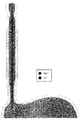
- Fig. 1 shows an example of manometry used in the first embodiment of the present invention.
- FIG. 2 is a block diagram of an anal disease test apparatus using a manometry according to a first embodiment of the present invention.
- FIG. 3 is a flowchart of a method for testing anal disease using the apparatus of FIG. 2.
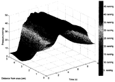
- step S320 of FIG. 3 illustrates a three-dimensional pressure distribution obtained according to step S320 of FIG. 3.
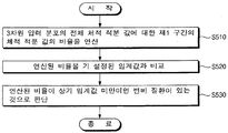
- FIG. 5 is a flowchart illustrating a test method for constipation disease after step S330 of FIG. 3.
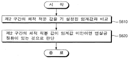
- FIG. 6 is a flowchart illustrating a test method for fecal incontinence disease after step S330 of FIG. 3.
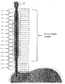
- Fig. 8 shows an example of high resolution esophageal impedance manometry used in the second embodiment of the present invention.
- Figure 9 is a state in which saline is injected into the esophagus in FIG.
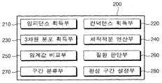
- FIG. 10 is a block diagram of an apparatus for detecting esophageal disease using high-resolution esophageal impedance manometry according to a second embodiment of the present invention.
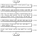
- FIG. 11 is a flowchart illustrating a method of testing esophageal disease using the apparatus of FIG. 10.
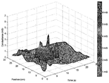
- FIG. 12 illustrates a three-dimensional conductance distribution obtained according to step S1130 of FIG. 11.
- the present invention relates to a disease detection method using a high resolution manometry and apparatus therefor.
- the present invention is largely divided into two embodiments.
- the first embodiment of the present invention provides a method and apparatus for testing anal rectal diseases using high resolution anal rectal manometry.
- a second embodiment of the present invention provides a method and apparatus for examining esophageal disease using high-resolution esophageal impedance manometry.
- the first embodiment of the present invention relates to a method for testing anal rectal diseases using high-resolution anal rectal manometry, in which a plurality of pressure sensors are disposed using manometry disposed at predetermined intervals along the longitudinal direction. Measure internal pressure around your rectum and use the measured values to check for conditions such as constipation or constipation.
- manometry is a device used for rectal analgesic pressure is known in various forms.
- Fig. 1 shows an example of manometry used in the first embodiment of the present invention.
- the first embodiment of the present invention is not necessarily limited to the manometer shape of FIG. 1.
- a plurality of pressure sensors are disposed at predetermined intervals (1 cm interval along a 1.0 to 6.0 cm section in the case of FIG. 1) along an arbitrary position section in a longitudinal direction thereof.
- the arbitrary position section is divided into 5 equal parts at 1 cm intervals, and a plurality of pressure sensors are disposed in each equally divided section (see Anal Canal Radial Sensors in FIG. 1).
- the pressure value of each sensor may be processed by an arbitrary method (eg, an average value calculation) and used as a representative pressure value for the corresponding section.
- the number of installation of the pressure sensor for each section may be changed in consideration of the importance, sensing error, accuracy, etc. of the section (corresponding point when the human body is inserted).
- the mannometry 10 includes an intra-balloon pressure sensor, an anal verge position band, and a rectal pressure sensor as shown in FIG. 1. ), An external reference channel, etc. may be included, and a detailed description thereof will be omitted.
- the above sensor corresponding to the rectal area of the sensor of the entire position can be used for the verification of constipation disease
- the sensor of the position corresponding to the anal area can be used for the verification of fecal incontinence disease.
- one of the uppermost sections among the five equally divided sections may be used for verification of constipation disease
- the other four sections may be used for verification of constipation disease.
- the ratio of dividing the interval is not necessarily limited to 1: 4 as shown in FIG. This ratio may vary depending on conditions such as gender, age, height, organ length of the subject.
- FIG. 2 is a block diagram of an anal disease test apparatus using manometry according to a first embodiment of the present invention
- FIG. 3 is a flowchart of an anal disease test method using the apparatus of FIG. 1.
- the anal disease test apparatus 100 includes a pressure value obtaining unit 110, a three-dimensional distribution obtaining unit 120, a volume integral calculating unit 130, The ratio calculator 140, the threshold comparator 150, the disease determiner 160, the segment classification unit 170, and the interest interval setting unit 180 may be included.
- the pressure value obtaining unit 110 obtains each of the pressure values for a predetermined time from the plurality of pressure sensors provided in the manometry 10 (S310).
- the plurality of pressure sensors are arranged at set intervals (ex, 1 cm interval) along an arbitrary position section (ex, 1.0 to 6.0 cm section) with respect to the longitudinal direction of the manometer geometry 10, as shown in FIG.
- step S310 pressure values are individually obtained from pressure sensors disposed in each of the five equal sections, and continuously obtained for a predetermined time. Then, the change in the sensing value of each pressure sensor with time can be seen, and the change in the sensing value according to the position (installation position in the manometer) of each pressure sensor can be known.
- the 3D distribution obtaining unit 120 indicates a change in the pressure value according to the position and the time, using the position point, the time, and the obtained pressure value in which each pressure sensor is disposed within the arbitrary position section.
- FIG. 4 illustrates a three-dimensional pressure distribution obtained according to step S320 of FIG. 3.
- the x-axis represents Time (s)
- the y-axis represents a position (cm) of the sensor installed
- the z-axis represents a pressure value (mmHg).
- each of the pressure sensors is disposed only at 1 cm intervals along the longitudinal direction (y-axis direction) of the manometer geometry 10, the sensing values for the intervals therebetween are understood in detail. There is no number.
- the method of subsequently curved each point point it is possible to obtain (estimate) the continuous data for each point as shown in FIG.
- the technique of subsequently curved or curving each measured point point and obtaining intermediate values between each point may use a known method.
- various diseases are determined by performing volume integration on an interval of interest using the data of FIG. 4. That is, the volume integral calculating unit 130 calculates a volume integral value for a predetermined region of interest location corresponding to the disease in the 3D pressure distribution (S330).
- the arbitrary position section corresponds to the first section (upper section among the five equal parts in FIG. 1) and the anal part corresponding to the rectal part at the time of insertion of the manometry 10. And a second section (the remaining lower section in FIG. 1).
- the upper section and the lower section are shown by hatching of different shades.
- the volume integral calculating unit 130 sets the first section (upper section) as the location of interest section and calculates a volume integral value, and the disease is fecal incontinence.
- a volume integral value is calculated by setting the second section (lower section) as the region of interest location.
- the apparatus may further include an input means for setting of a user.
- 5 is a flowchart illustrating a test method for constipation disease after step S330 of FIG. 3.
- the ratio calculating unit 140 calculates a ratio of the volume integral value to the first section (upper section) to the total volume integration value of the three-dimensional pressure distribution (S510). For example, the total volume integral A of the three-dimensional pressure distribution of FIG. 4 is calculated, and only data corresponding to the 4-5 cm section (first section; upper section) among the three-dimensional pressure distribution of FIG. 4 is calculated. Calculate the volume integral (A ') and then calculate the ratio of A' divided by A.
- the threshold comparison unit 150 compares the calculated ratio with a preset first threshold value (S520). Here, if the ratio calculated in step S510 is greater than or equal to the first threshold, it is determined to be normal, and if it is less than or equal to the first threshold, it is determined that there is constipation disease (S530).
- 6 is a flowchart illustrating a test method for fecal incontinence disease after step S330 of FIG. 3. In this case, as above, the ratio value is not used, and the absolute value of the volume integral for the corresponding part is used.
- the volume integral calculating unit 130 calculates a volume integral value for the second section (lower section) at step S330.
- the threshold comparison unit 150 compares the volume integral value of the second section (lower section) with a preset second threshold value (S610). Then, the disease determining unit 160 determines that the volume integral value of the second section is greater than or equal to the second threshold value, and determines that there is a fecal incontinence disease if it is less than the second threshold value (S620).
- the first section (upper section) and the second section (lower section) may be changed according to the body structure of the examinee.
- the two sections need to be optimally set according to the physical condition of the subject. This will increase the accuracy and reliability of disease judgment.
- the first exemplary embodiment of the present invention includes a section classification unit 170 and an interest section setting unit 180.
- the section classifying unit 170 receives a ratio between the first section and the second section within the random location section, the section classifying unit 170 generates the first location section (upper section) and the second section according to the set ratio. (Lower section).
- the first section and the second section may be automatically divided into upper and lower portions as shown in FIG.
- a menu for selecting one of 1: 4, 2: 3, 3: 2, and 4: 1 may be provided on the screen. It is possible.
- the interested section setting unit 180 is a portion that receives the interested location section among the first section (upper section) and the second section (lower section). That is, when the constipation disease is suspected, the first section may be set as the location of interest section, and then the method of FIG. 5 may be used after the process of FIG. 3. In addition, if the fecal incontinence disease is suspected, the second section is set as the location of interest section, and then the method of FIG. 6 may be used after the process of FIG. 3. As such, the process of classifying a section or setting a location of interest section may be performed before step S310 of FIG. 3. Of course, it may be performed at any step before the step S330.
- FIG. 7 shows an example of classification of the first and second sections in the first embodiment of the present invention.
- A, B, C, and D of FIG. 7 are examples in which a ratio of a first section to a second section is set to 1: 4, 2: 3, 3: 2, and 4: 1, respectively.
- the relationship between the rectum or anal region during insertion of the manometry among the plurality of sensors provided in the manometry By using the pressure value of some sensors to improve the efficiency of the judgment of various diseases related to rectal anus.
- the first embodiment of the present invention unlike the conventional use of the pressure value simply in the rectal rectal pressure test, it utilizes a three-dimensional volume distribution processing the time value in each sensor in time and space, By using the three-dimensional integral volume according to the proposed method, we can propose a new technique for diagnosing functional colon ankle disease.
- a second embodiment of the present invention relates to a method and apparatus for examining esophageal disease using high-resolution esophageal impedance manometry, wherein a plurality of impedance electrodes are arranged at predetermined intervals along a length direction of a high resolution Measurements of impedance manometry can be used to check for disease in the esophagus.
- Fig. 8 shows an example of high resolution esophageal impedance manometry used in the second embodiment of the present invention.
- the second embodiment of the present invention is not necessarily limited to the form of FIG.
- a plurality of impedance electrodes Z1 to Z16 are disposed at a predetermined interval (2 cm interval in FIG. 8) along an arbitrary position section in the longitudinal direction of the high resolution esophageal impedance manometer 20. More specifically, it has a form in which a plurality of pressure sensors (ex, 36) arranged at 1 cm intervals for measuring pressure are provided with impedance electrodes (ex, 16 arranged at 2 cm intervals). It consists of a catheter of finger thickness.
- the high-resolution esophageal impedance manometry 20 inserts the catheter into the nostrils, and then all tests are completed by swallowing saline of about 10 times for a period of about 5 to 10 minutes.
- the second embodiment of the present invention proposes a method for displaying the measured values through an impedance sensor arranged at 2 cm intervals attached to the high-resolution impedance manometry as an objective value. It is based on the theory that it is not possible to get out of the esophagus in the inside, and that the more remains in the esophagus, the more problems with esophageal movement.
- the high-resolution esophageal impedance manometry shown in FIG. 8 corresponds to a known form, and a detailed description thereof will be omitted.
- the second embodiment of the present invention is not necessarily limited to the shape of the anometry as shown in FIG.
- FIG. 9 is a state in which saline is injected into the esophagus in FIG.
- Esophageal impedance testing is a method of measuring the properties of a passage (ex, cell line; saline) based on a change in resistance. After saline injection, the more saline remains in the esophagus, the lower the impedance and the higher the conductivity.
- FIG. 10 is a block diagram of an apparatus for testing esophageal disease using high-resolution esophageal impedance manometry according to a second embodiment of the present invention
- FIG. 11 is a flowchart of a method for testing esophageal disease using the apparatus of FIG. 10.
- the apparatus 200 for testing esophageal disease includes an impedance acquirer 210, a conductance acquirer 220, a three-dimensional distribution acquirer 230, and a volume.
- An integration calculator 240, a threshold comparison unit 250, a disease determination unit 260, an interval classification unit 270, and an interest interval setting unit 280 are included.
- the impedance obtaining unit 210 obtains each impedance value from a plurality of impedance electrodes provided in the impedance manometer geometry 20 for a preset time (S1110). As shown in FIG. 8, the plurality of impedance electrodes are disposed at predetermined intervals (eg, 2 cm intervals) along an arbitrary position section of the impedance manometer geometry 20 in the longitudinal direction.
- the arbitrary position section means a partial length section in which the electrode is installed among the entire lengths of the tube portion of the impedance manometer geometry 20.
- step S1110 an impedance value is separately obtained from each impedance electrode, but is continuously obtained for a predetermined time. Then, the change of the impedance value in each impedance electrode with time can be known, and the change in the impedance value according to the position of each impedance electrode (installation position in the impedance manometer 20) can be known.
- step S1120 refers to a process of inverting all impedance values acquired at step S1110 and converting them into conductance values.
- the 3D distribution obtaining unit 230 indicates a change in conductance with respect to the position and time using the position, the time, and the conductance value where each impedance electrode is disposed in an arbitrary position section in which the electrode is installed.
- the dimensional conductance distribution is obtained (S1130).
- FIG. 12 illustrates a three-dimensional conductance distribution obtained according to step S1130 of FIG. 11.
- the x axis represents Time (s)
- the y axis represents an installation position (cm) of the electrode
- the z axis represents a conductance value (mS).
- each of the impedance electrodes is disposed only at 2 cm intervals along the longitudinal direction (y-axis direction) of the impedance manometer geometry 20, the conductance value for the interval therebetween is finely detailed. It is not known. However, using the method of subsequently curved each point point, it is possible to obtain (estimate) the continuous data between each point as shown in FIG. Of course, the same can be applied to conductance values (x-axis direction) over time. As such, the technique of subsequently curved or curving each measured point point and obtaining intermediate values between each point may use a known method.
- esophageal disease is determined using the data of FIG. That is, the volume integral calculating unit 240 calculates the total volume integral value for the three-dimensional conductance distribution (S1140). For example, as shown in FIG. 12, a volume integral value is calculated for the entire 3D conductance distribution obtained for 0 to 25 seconds for the entire 0 to 30 cm section.
- the threshold comparison unit 250 compares the total volume integration value for the three-dimensional conductance distribution with a predetermined threshold value (S1150). Thereafter, the disease determining unit 260 determines that there is an esophageal disease when the calculated ratio is greater than or equal to the threshold value (S1160).
- a partial interval (ex, 10-20 cm) is used instead of the entire 3D conductance distribution. That is, in the step S1140, in the case of a subject having an abnormal esophageal function, a volume integral value is calculated for a predetermined region of interest within the 3D conductance distribution. That is, the possibility of disease can be determined by comparing the volume integral value for this partial section with a threshold value.
- the threshold used may be smaller than the threshold used in the case where the entire interval is used. That is, the threshold value used may vary in proportion to the length ratio of some sections to the entire electrode length sections.
- the location of interest section may mean a section corresponding to a normal esophageal region in which an esophageal function is not abnormal at the time of insertion of the impedance manometry 20 among the sections in which the electrode is installed. That is, in the case of a subject having a partial abnormality in esophageal function, it is possible to determine whether it is normal and abnormal / lesion by using a section in which the abnormality is not present.
- the region of interest setting unit 280 receives a region of the region of interest location within a section (0 to 30 cm) in which an impedance electrode is installed in the impedance manometer geometry 20.
- the setting of the location of interest section may be performed before step S1110 of FIG. 11. Of course, it may be performed in any step before the step S1140.
- the second embodiment of the present invention obtains a spatiotemporal three-dimensional distribution of conductance using an inverse value of impedances of a plurality of impedance electrodes provided in the impedance manometer, and increases efficiency of judging esophageal diseases by using the same. There is an advantage.
- the three-dimensional conductance distribution obtained by spatiotemporally processing the conductance value obtained by inverting the impedance value at each electrode can propose a new technique for diagnosing esophageal diseases using three-dimensional integral volume according to space-time changes.
Landscapes
- Health & Medical Sciences (AREA)
- Life Sciences & Earth Sciences (AREA)
- Engineering & Computer Science (AREA)
- General Health & Medical Sciences (AREA)
- Veterinary Medicine (AREA)
- Biophysics (AREA)
- Biomedical Technology (AREA)
- Heart & Thoracic Surgery (AREA)
- Medical Informatics (AREA)
- Molecular Biology (AREA)
- Surgery (AREA)
- Animal Behavior & Ethology (AREA)
- Physics & Mathematics (AREA)
- Public Health (AREA)
- Pathology (AREA)
- Physiology (AREA)
- Endocrinology (AREA)
- Gastroenterology & Hepatology (AREA)
- Hematology (AREA)
- Nuclear Medicine, Radiotherapy & Molecular Imaging (AREA)
- Radiology & Medical Imaging (AREA)
- Physical Education & Sports Medicine (AREA)
- Artificial Intelligence (AREA)
- Computer Vision & Pattern Recognition (AREA)
- Psychiatry (AREA)
- Signal Processing (AREA)
- Measurement And Recording Of Electrical Phenomena And Electrical Characteristics Of The Living Body (AREA)
Abstract
본 발명은 고해상도 마노메트리를 이용한 질환 검사 방법 및 그 장치에 관한 것이다. 본 발명에 따르면, 복수의 압력 센서가 길이 방향의 임의 위치 구간을 따라 설정 간격으로 배치된 마노메트리를 이용한 항문 질환 검사방법에 있어서, 복수의 압력 센서로부터 각각의 압력값을 기 설정된 시간 동안 획득하는 단계와, 임의 위치 구간 내에서 각 압력 센서가 배치된 위치, 시간, 압력값을 이용하여 위치 및 시간에 따른 압력값의 변화를 나타내는 3차원 압력 분포를 획득하는 단계, 및 3차원 압력 분포 내에서 상기 질환과 대응하여 기 설정된 관심 위치 구간을 대상으로 체적 적분 값을 연산하는 단계를 포함하는 마노메트리를 이용한 항문 질환 검사방법을 제공한다. 이에 따르면, 마노메트리의 삽입 시에 직장 또는 항문 부위와 관계하는 일부 센서의 압력 값을 이용하여 직장 항문과 관련한 각종 질환의 판단 효율을 높인다.
Description
본 발명은 고해상도 마노메트리를 이용한 질환 검사 방법 및 그 장치에 관한 것으로서, 보다 상세하게는 복수의 압력 센서 또는 복수의 임피던스 전극으로 구성된 고해상도 마노메트리를 이용하여 항문 직장 질환 또는 식도 질환을 검사할 수 있는 고해상도 마노메트리를 이용한 질환 검사 방법 및 그 장치에 관한 것이다.
직장과 항문은 정상적인 배변을 위한 중요한 생리적인 역할을 수행한다. 직장과 항문의 기능에 이상이 생기면 변비, 잔변감, 변실금 등의 증상을 호소하게 된다.
직장과 항문의 기능 이상을 진단하기 위한 방법으로는 대장내시경, 풍선배출검사, 배변 조영술, 항문괄약근 근전도검사, 직장 항문 내압검사 등이 있다. 그 중에서도 직장 항문의 기능을 평가하는 가장 대표적인 방법은 직장 항문 내압검사로서, 주로 배변 장애 환자나 직장 항문 주위의 수술 전후 환자에서 항문 괄약근 기능 평가와 치료 계획 수립 등에 활용되고 있다.
직장 항문 내압검사는 다수의 압력 센서가 배치된 마노메트리(manometry)를 항문에 삽입하여 압력을 측정하고 측정된 값을 이용하여 질환을 판단한다. 기존의 경우 마노메트리 내의 모든 센서의 값 중 대표값 하나만 사용하거나 그 평균값을 사용하는 방법이 일반적이기 때문에, 질환 판단의 정확도와 신뢰성이 떨어지는 단점이 있다.
또한, 식도 질환은 삼킴 곤란, 유연, 구토 등의 증상을 나타내므로 식도 검사에 의하여 감별할 필요가 있다. 그 중에서 삼킴 곤란 등의 증상을 일으키는 식도 운동 질환의 경우 일반적으로 널리 시행하는 상부 위 식도 내시경 검사만으로는 알기 어려운 경우가 많이 있어, 식도 내압 검사 즉, 마노메트리 검사를 통해서 알 수 있는 경우가 있다. 이러한 마노메트리 검사는 과거 70-80년대에는 5 cm 간격으로 띄엄띄엄하게 배열된 센서를 통하여 그 결과 값을 분석하는 수준이었으나, 최근 10여년 사이에 고해상도 마노메트리(high-resolution manometry)가 개발되었으며, 이는 보다 조밀하게 1 cm 간격으로 배열된 센서를 통하여 시그널을 받아 들여서 보다 풍부한 임상 정보를 통하여 진단에 도움을 받게 되었다.
과거 위식도 역류증 검사에 제한적으로 쓰이던 임피던스 검사법은 식도 내에 삼킨 식괴의 양을 간접적인 저항값을 통하여 측정할 수 있다는 점에 착안하여, 고해상도 마노메트리 카테터에서 같이 부착되어 사용되고 있다.
이와 관련하여, 고해상도 식도 임피던스 마노메트리(high resolution impedance manometry)는, 복수의 임피던스 전극이 길이 방향에 대해 일정 간격으로 형성된 카테터를 식도 내에 삽입시키고 저항 변화에 기초한 통과물의 성질을 측정하고 이를 통해 식도 질환을 분석할 수 있다. 식도에 통과물이 많이 남아 있을수록 임피던스 값은 낮아지며 식도 질환이 있을 가능성이 높아진다.
그런데, 이와 같이 고해상도 마노메트리 카테터를 활용한 임피던스 검사법은 아직까지 그 결과값의 해석에 있어 제한점이 많고, 결정적으로 정확한 양을 객관적인 숫자로 나타내주지 못하여, 임상적으로는 그 쓰임에 있어 제한이 있었다.
본 발명의 배경이 되는 기술은 한국등록특허 제0816847호(2008.03.19 공개) 및 한국공개특허 제2002-0093860호(2002.12.16)에 개시되어 있다.
본 발명은 직장 항문 또는 식도와 관계하는 각종 질환의 판단 효율을 높일 수 있는 고해상도 마노메트리를 이용한 질환 검사 방법 및 그 장치를 제공하는데 목적이 있다.
본 발명은, 복수의 압력 센서가 길이 방향의 임의 위치 구간을 따라 설정 간격으로 배치된 마노메트리(manometry)를 이용한 항문 질환 검사방법에 있어서, 상기 복수의 압력 센서로부터 각각의 압력값을 기 설정된 시간 동안 획득하는 단계와, 상기 임의 위치 구간 내에서 각 압력 센서가 배치된 위치, 상기 시간, 상기 압력값을 이용하여 상기 위치 및 시간에 따른 압력값의 변화를 나타내는 3차원 압력 분포를 획득하는 단계, 및 상기 3차원 압력 분포 내에서 상기 질환과 대응하여 기 설정된 관심 위치 구간을 대상으로 체적 적분 값을 연산하는 단계를 포함하는 고해상도 항문 직장 마노메트리를 이용한 항문 직장 질환 검사방법을 제공한다.
여기서, 상기 임의 위치 구간은, 상기 마노메트리의 삽입 시에 직장 부위에 대응하는 제1 구간 및 항문 부위에 대응하는 제2 구간을 포함하며, 상기 체적 적분 값을 연산하는 단계는, 상기 질환이 변비인 경우 상기 제1 구간을 상기 관심 위치 구간으로 하여 체적 적분 값을 연산하고, 상기 질환이 변실금인 경우 상기 제2 구간을 상기 관심 위치 구간으로 하여 체적 적분 값을 연산할 수 있다.
또한, 상기 고해상도 항문 직장 마노메트리를 이용한 항문 직장 질환 검사방법은, 상기 질환이 변비인 경우, 상기 3차원 압력 분포의 전체 체적 적분 값에 대한 상기 제1 구간의 체적 적분 값의 비율을 연산하는 단계, 상기 연산된 비율을 기 설정된 임계값과 비교하는 단계, 및 상기 연산된 비율이 상기 임계값 미만이면 변비 질환이 있는 것으로 판단하는 단계를 더 포함할 수 있다.
또한, 상기 고해상도 항문 직장 마노메트리를 이용한 항문 직장 질환 검사방법은, 상기 질환이 변실금인 경우, 상기 제2 구간의 체적 적분 값을 기 설정된 임계값과 비교하는 단계, 및 상기 제2 구간의 체적 적분 값이 상기 임계값 미만이면 변실금 질환이 있는 것으로 판단하는 단계를 더 포함할 수 있다.
또한, 상기 고해상도 항문 직장 마노메트리를 이용한 항문 직장 질환 검사방법은, 상기 임의 위치 구간 내에서 상기 제1 구간과 제2 구간의 비율을 설정받으면 상기 설정받은 비율에 따라 상기 임의 위치 구간을 제1 구간 및 제2 구간으로 구분하는 단계, 및 상기 제1 구간 및 제2 구간 중 상기 관심 위치 구간을 설정받는 단계를 더 포함할 수 있다.
그리고, 본 발명은, 복수의 압력 센서가 길이 방향의 임의 위치 구간을 따라 설정 간격으로 배치된 마노메트리(manometry)를 이용한 항문 질환 검사장치에 있어서, 상기 복수의 압력 센서로부터 각각의 압력값을 기 설정된 시간 동안 획득하는 압력값 획득부와, 상기 임의 위치 구간 내에서 각 압력 센서가 배치된 위치, 상기 시간, 상기 압력값을 포함하는 세 가지 변수를 이용하여 상기 위치 및 시간에 따른 압력값의 변화를 나타내는 3차원 압력 분포를 획득하는 3차원 분포 획득부, 및 상기 3차원 압력 분포 내에서 상기 질환과 대응하여 기 설정된 관심 위치 구간을 대상으로 체적 적분 값을 연산하는 체적적분 연산부를 포함하는 고해상도 항문 직장 마노메트리를 이용한 항문 직장 질환 검사장치를 제공한다.
여기서, 상기 임의 위치 구간은, 상기 마노메트리의 삽입 시에 직장 부위에 대응하는 제1 구간 및 항문 부위에 대응하는 제2 구간을 포함하며, 상기 체적적분 연산부는, 상기 질환이 변비인 경우 상기 제1 구간을 상기 관심 위치 구간으로 하여 체적 적분 값을 연산하고, 상기 질환이 변실금인 경우 상기 제2 구간을 상기 관심 위치 구간으로 하여 체적 적분 값을 연산할 수 있다.
또한, 상기 고해상도 항문 직장 마노메트리를 이용한 항문 직장 질환 검사장치는, 상기 질환이 변비인 경우, 상기 3차원 압력 분포의 전체 체적 적분 값에 대한 상기 제1 구간의 체적 적분 값의 비율을 연산하는 비율 연산부와, 상기 연산된 비율을 기 설정된 임계값과 비교하는 임계값 비교부, 및 상기 연산된 비율이 상기 임계값 미만이면 변비 질환이 있는 것으로 판단하는 질환 판단부를 더 포함할 수 있다.
또한, 상기 고해상도 항문 직장 마노메트리를 이용한 항문 직장 질환 검사장치는, 상기 질환이 변실금인 경우, 상기 마노메트리를 이용한 항문 질환 검사장치는, 상기 질환이 변실금인 경우, 상기 제2 구간의 체적 적분 값을 기 설정된 임계값과 비교하는 임계값 비교부, 및 상기 제2 구간의 체적 적분 값이 상기 임계값 미만이면 변실금 질환이 있는 것으로 판단하는 질환 판단부를 더 포함할 수 있다.
또한, 상기 고해상도 항문 직장 마노메트리를 이용한 항문 직장 질환 검사장치는, 상기 임의 위치 구간 내에서 상기 제1 구간과 제2 구간의 비율을 설정받으면 상기 설정받은 비율에 따라 상기 임의 위치 구간을 제1 구간 및 제2 구간으로 구분하는 구간 분류부, 및 상기 제1 구간 및 제2 구간 중 상기 관심 위치 구간을 설정받는 관심 구간 설정부를 더 포함할 수 있다.
그리고, 본 발명은, 복수의 임피던스 전극이 길이 방향의 임의 위치 구간을 따라 설정 간격으로 배치된 고해상도 식도 임피던스 마노메트리를 이용한 식도 질환 검사방법에 있어서, 상기 복수의 임피던스 전극으로부터 각각의 임피던스 값을 기 설정된 시간 동안 획득하는 단계와, 상기 획득한 임피던스 값을 역수화하여 컨덕턴스 값을 획득하는 단계와, 상기 임의 위치 구간 내에서 각 임피던스 전극이 배치된 위치, 상기 시간, 상기 컨덕턴스 값을 이용하여 상기 위치 및 시간에 따른 컨덕턴스의 변화를 나타내는 3차원 컨덕턴스 분포를 획득하는 단계, 및 상기 3차원 컨덕턴스 분포에 대한 전체 체적 적분 값을 연산하는 단계를 포함하는 고해상도 식도 임피던스 마노메트리를 이용한 식도 질환 검사방법을 제공한다.
여기서, 상기 고해상도 식도 임피던스 마노메트리를 이용한 식도 질환 검사방법은, 상기 3차원 컨덕턴스 분포에 대한 전체 체적 적분 값을 기 설정된 임계값과 비교하는 단계, 및 상기 연산된 비율이 상기 임계값 이상이면 상기 식도 질환이 있는 것으로 판단하는 단계를 더 포함할 수 있다.
또한, 상기 전체 체적 적분 값을 연산하는 단계는, 식도 기능에 이상이 있는 피검자의 경우, 상기 3차원 컨덕턴스 분포 내에서 기 설정된 관심 위치 구간을 대상으로 체적 적분 값을 연산하며, 상기 관심 위치 구간은, 상기 임의 위치 구간 중에서, 상기 고해상도 식도 임피던스 마노메트리의 삽입 시, 식도 기능에 이상이 없는 정상적인 식도 부위에 대응하는 구간일 수 있다.
또한, 상기 고해상도 식도 임피던스 마노메트리를 이용한 식도 질환 검사방법은, 상기 임의 위치 구간 내에서 상기 관심 위치 구간을 설정받는 단계를 더 포함할 수 있다.
그리고, 본 발명은, 복수의 임피던스 전극이 길이 방향의 임의 위치 구간을 따라 설정 간격으로 배치된 고해상도 식도 임피던스 마노메트리를 이용한 식도 질환 검사장치에 있어서, 상기 복수의 임피던스 전극으로부터 각각의 임피던스 값을 기 설정된 시간 동안 획득하는 임피던스 획득부와, 상기 획득한 임피던스 값을 역수화하여 컨덕턴스 값을 획득하는 컨덕턴스 획득부와, 상기 임의 위치 구간 내에서 각 임피던스 전극이 배치된 위치, 상기 시간, 상기 컨덕턴스 값을 포함하는 세 가지 변수를 이용하여 상기 위치 및 시간에 따른 컨덕턴스의 변화를 나타내는 3차원 컨덕턴스 분포를 획득하는 3차원 분포 획득부, 및 상기 3차원 컨덕턴스 분포에 대한 전체 체적 적분 값을 연산하는 체적적분 연산부를 포함하는 고해상도 식도 임피던스 마노메트리를 이용한 식도 질환 검사장치를 제공한다.
여기서, 상기 고해상도 식도 임피던스 마노메트리를 이용한 식도 질환 검사장치는, 상기 3차원 컨덕턴스 분포에 대한 전체 체적 적분 값을 기 설정된 임계값과 비교하는 임계값 비교부, 및 상기 연산된 비율이 상기 임계값 이상이면 상기 식도 질환이 있는 것으로 판단하는 질환 판단부를 더 포함할 수 있다.
또한, 상기 체적적분 연산부는, 식도 기능에 이상이 있는 피검자의 경우, 상기 3차원 컨덕턴스 분포 내에서 기 설정된 관심 위치 구간을 대상으로 체적 적분 값을 연산하며, 상기 관심 위치 구간은, 상기 임의 위치 구간 중에서, 상기 고해상도 식도 임피던스 마노메트리의 삽입 시, 식도 기능에 이상이 없는 정상적인 식도 부위에 대응하는 구간일 수 있다.
또한, 상기 고해상도 식도 임피던스 마노메트리를 이용한 식도 질환 검사장치는, 상기 임의 위치 구간 내에서 상기 관심 위치 구간을 설정받는 관심구간 설정부를 더 포함할 수 있다.
본 발명에 따른 고해상도 마노메트리를 이용한 질환 검사 방법 및 그 장치에 따르면, 마노메트리에 구비된 복수의 센서들 중에서 마노메트리의 삽입 시에 직장 또는 항문 부위와 관계하는 일부 센서의 압력 값을 이용하여 직장 항문과 관련한 각종 질환의 판단 효율을 높일 수 있다.
또한, 본 발명에 따르면, 고해상도 식도 임피던스 마노메트리에 구비된 복수의 임피던스 전극들의 임피던스의 역수 값을 이용하여 컨덕턴스의 시공간적 3차원 분포를 획득하고 이를 활용하여 식도 질환의 판단 효율을 높일 수 있다.
도 1은 본 발명의 제1 실시예에 사용되는 마노메트리의 일례를 나타낸다.
도 2는 본 발명의 제1 실시예에 따른 마노메트리를 이용한 항문 질환 검사 장치의 구성도이다.
도 3은 도 2의 장치를 이용한 항문 질환 검사 방법의 흐름도이다.
도 4는 도 3의 S320 단계에 따라 획득한 3차원 압력 분포를 나타낸다.
도 5는 도 3의 S330 단계 이후에 변비 질환에 대한 검사 방법을 나타내는 흐름도이다.
도 6은 도 3의 S330 단계 이후에 변실금 질환에 대한 검사 방법을 나타내는 흐름도이다.
도 7은 본 발명의 제1 실시예에서 제1 및 제2 구간의 분류 예시를 나타낸다.
도 8은 본 발명의 제2 실시예에 사용되는 고해상도 식도 임피던스 마노메트리의 일례를 나타낸다.
도 9는 도 8에서 식도에 식염수가 주입된 모습이다.
도 10은 본 발명의 제2 실시예에 따른 고해상도 식도 임피던스 마노메트리를 이용한 식도 질환 검사 장치의 구성도이다.
도 11은 도 10의 장치를 이용한 식도 질환 검사 방법의 흐름도이다.
도 12는 도 11의 S1130 단계에 따라 획득한 3차원 컨덕턴스 분포를 나타낸다.
그러면 첨부한 도면을 참고로 하여 본 발명의 실시예에 대하여 본 발명이 속하는 기술 분야에서 통상의 지식을 가진 자가 용이하게 실시할 수 있도록 상세히 설명한다.
본 발명은 고해상도 마노메트리를 이용한 질환 검사 방법 및 그 장치에 관한 것이다. 본 발명은 크게 두 가지 실시예로 구분된다. 우선, 본 발명의 제1 실시예는 고해상도 항문 직장 마노메트리를 이용한 항문 직장 질환 검사방법 및 그 장치를 제공한다. 그리고, 본 발명의 제2 실시예는 고해상도 식도 임피던스 마노메트리를 이용한 식도 질환 검사방법 및 그 장치를 제공한다.
이하에서는 먼저 본 발명의 제1 실시예에 관하여 상세히 설명한다.
본 발명의 제1 실시예는 고해상도 항문 직장 마노메트리를 이용한 항문 직장 질환 검사방법에 관한 것으로서, 다수의 압력 센서가 길이 방향을 따라 설정 간격으로 배치된 마노메트리(manometry)를 이용하여 항문이나 직장 주변의 내압을 측정하고 측정된 값을 이용하여 변비나 변실금과 같은 질환의 여부를 검사할 수 있도록 한다.
일반적으로 마노메트리는 직장 항문 내압검사에 사용되는 기구로서 기존에 다양한 형태로 공지되어 있다. 도 1은 본 발명의 제1 실시예에 사용되는 마노메트리의 일례를 나타낸다. 여기서 본 발명의 제1 실시예가 반드시 도 1의 마노메트리 형태로 한정되는 것은 아니다.
도 1을 참조하면, 마노메트리(10)는 그 길이 방향의 임의 위치 구간을 따라 설정 간격(도 1의 경우 1.0~6.0㎝ 구간을 따라 1㎝ 간격)으로 복수의 압력 센서가 배치되어 있다. 도 1의 경우 상기 임의 위치 구간을 1㎝ 간격으로 5 등분하고 각 등분된 구간마다 압력 센서를 복수 개로 배치한 예이다(도 1의 Anal Canal Radial Sensors 부분 참조).
도 1과 같이 하나의 구간에 복수의 센서를 방사형으로 배치한 경우에는 각 센서의 압력 값을 임의 방법으로 가공(ex, 평균값 연산)하여 해당 구간에 대한 대표 압력 값으로 사용할 수도 있다. 각각의 구간에 대한 압력 센서의 설치 개수는 해당 구간(인체 삽입시 해당 지점)의 중요도, 센싱 오차, 정확도 등을 고려하여 변경될 수 있다.
마노메트리(10)는 내압 측정을 위한 압력 센서들 이외에도 도 1과 같이 내부 풍선 압력 센서(Intra-balloon Pressure Sensor), 항문연 경계 위치 밴드(Anal Verge Position Band), 직장 압력 센서(Rectal Pressure Sensor), 외부 참조 채널(External Reference Channel) 등의 구성을 포함할 수도 있으며, 이에 관한 구체적인 설명은 생략한다.
본 발명의 제1 실시예에서는 전체 위치의 센서 중에서 직장 부위에 대응하는 위의 센서는 변비 질환의 검증에 사용될 수 있고, 항문 부위에 대응하는 위치의 센서는 변실금 질환의 검증에 사용될 수 있다. 도 1의 경우 총 5 등분된 구간 중에서 맨 위의 한 구간은 변비 질환의 검증에 사용되고, 나머지 네 구간은 변실금 질환의 검증에 사용될 수 있다. 물론, 구간이 나뉘는 비율은 반드시 도 1과 같이 1:4로 한정되지 않는다. 이러한 비율은 피검자의 성별, 나이, 신장, 장기 길이 등의 조건에 따라 변경될 수 있다.
이하에서는 본 발명의 제1 실시예에 따른 마노메트리를 이용한 항문 질환 검사방법 및 그 장치에 관하여 상세히 설명한다. 다만, 설명의 편의를 위하여, 도 1에서 각각의 등분된 구간마다 압력 센서가 하나씩 배치된 것을 예시로 하여 설명한다.
도 2는 본 발명의 제1 실시예에 따른 마노메트리를 이용한 항문 질환 검사 장치의 구성도이고, 도 3은 도 1의 장치를 이용한 항문 질환 검사 방법의 흐름도이다.
도 2 및 도 3을 참조하면, 본 발명의 제1 실시예에 따른 항문 질환 검사 장치(100)는 압력값 획득부(110), 3차원 분포 획득부(120), 체적적분 연산부(130), 비율 연산부(140), 임계값 비교부(150), 질환 판단부(160), 구간 분류부(170), 관심 구간 설정부(180)를 포함한다.
먼저, 압력값 획득부(110)는 마노메트리(10)에 구비된 복수의 압력 센서로부터 각각의 압력값을 기 설정된 시간 동안 획득한다(S310). 복수의 압력 센서는 도 1과 같이 마노메트리(10)의 길이 방향에 대한 임의 위치 구간(ex, 1.0~6.0㎝ 구간)을 따라 설정 간격(ex, 1㎝ 간격)으로 배치되어 있다.
상기 S310 단계는 상기 등분된 5개의 구간에 각각 배치된 압력 센서로부터 압력값을 개별 획득하되 기 설정된 시간 동안 지속적으로 획득한다. 그러면, 시간에 따른 각 압력 센서의 센싱 값의 변화를 알 수 있으며, 각 압력 센서의 위치(마노메트리에서의 설치 위치)에 따른 센싱 값의 변화를 알 수 있다.
이후, 3차원 분포 획득부(120)는 상기 임의 위치 구간 내에서 각 압력 센서가 배치된 위치 지점, 상기 시간, 상기 획득한 압력값을 이용하여, 상기 위치 및 시간에 따른 압력값의 변화를 나타내는 3차원 압력 분포를 획득한다(S320).
도 4는 도 3의 S320 단계에 따라 획득한 3차원 압력 분포를 나타낸다. 도 4의 x축은 시간(Time; s), y축은 센서의 설치 위치(Distance; cm), z축은 압력값(Pressure; mmHg)을 나타낸다.
도 4의 경우 10초 동안 압력 값을 획득한 데이터를 나타낸다. 센서의 설치 위치를 나타내는 y축의 경우 도식의 편의를 위하여 0~5㎝ 범위로 표현되어 있으며, 이는 도 1에서 각 센서가 위치한 1.0~6.0㎝ 지점에 대응하는 부분이다. 이러한 3차원 압력 분포는 하나의 입체감 있는 면 형상을 가지고 있다.
한편, 본 발명의 제1 실시예에서 각각의 압력 센서는 마노메트리(10)의 길이 방향(y축 방향)을 따라 1cm 간격으로만 배치되어 있기 때문에, 그 사이 구간에 대한 센싱 값을 세세히 알 수는 없다. 하지만, 각각의 포인트 지점을 이어서 곡선화하는 방법을 사용하면 도 4와 같이 각 지점 사이에 대한 연속적인 데이터를 획득(추정)할 수 있게 된다. 물론 이는 시간에 따른 압력 값들(x축 방향)에 대해서도 동일하게 적용될 수 있다. 이와 같이, 측정된 각 포인트 지점을 이어서 곡선화 또는 곡면화 처리하고 각 지점 사이의 중간값들을 획득하는 기법은 기 공지된 방법을 사용할 수 있다.
본 발명의 제1 실시예에서는 도 4의 데이터를 활용하여 임의의 관심 구간에 대해 체적 적분을 수행하여 각종 질환을 판단한다. 즉, 체적적분 연산부(130)는 상기 3차원 압력 분포 내에서 상기 질환과 대응하여 기 설정된 관심 위치 구간을 대상으로 체적 적분 값을 연산한다(S330).
본 발명의 제1 실시예에서 상기 임의 위치 구간은, 상기 마노메트리(10)의 삽입 시에 직장 부위에 대응하는 제1 구간(도 1에서 5 등분된 구간 중에서 상부 구간) 및 항문 부위에 대응하는 제2 구간(도 1에서 나머지 하부 구간)을 포함한다. 도 1에는 상부 구간과 하부 구간을 서로 다른 음영의 해칭으로 도시하고 있다.
이를 전제로 하여, 상기 체적적분 연산부(130)는, 상기 질환이 변비인 경우에는 상기 제1 구간(상부 구간)을 상기 관심 위치 구간으로 설정하여 체적 적분 값을 연산하고, 상기 질환이 변실금인 경우 상기 제2 구간(하부 구간)을 상기 관심 위치 구간으로 설정하여 체적 적분 값을 연산한다.
예를 들어, 사용자로부터 변비 질환 검사를 선택받는 경우, 제1 구간(상부 구간)을 관심 위치 구간으로 설정하고, 변실금 질환 검사를 선택받는 경우, 제2 구간(하부 구간)을 관심 위치 구간으로 설정하여 체적 적분을 연산한다. 본 발명의 제1 실시예예 따른 장치는 사용자의 설정을 위한 입력 수단을 더 포함할 수 있다.
이상과 같은 내용을 바탕으로, 이하에서는 제1 구간을 활용한 변비 질환에 대한 구체적인 검사 방법을 설명한다. 도 5는 도 3의 S330 단계 이후에 변비 질환에 대한 검사 방법을 나타내는 흐름도이다.
먼저, 비율 연산부(140)는 검사 대상 질환이 변비인 경우, 상기 3차원 압력 분포의 전체 체적 적분 값에 대한 상기 제1 구간(상부 구간)에 대한 체적 적분 값의 비율을 연산한다(S510). 예를 들어, 도 4의 3차원 압력 분포에 대한 전체 체적 적분 값(A)을 연산하고, 도 4의 전체 3차원 압력 분포 중에서도 4~5cm 구간(제1 구간; 상부 구간)에 해당되는 데이터만을 대상으로 체적 적분 값(A')을 연산한 다음, A' 값을 A 값으로 나눈 비율을 연산한다.
이후, 임계값 비교부(150)는 상기 연산된 비율을 기 설정된 제1 임계값과 비교한다(S520). 여기서, 상기 S510 단계에서 연산된 비율이 상기 제1 임계값 이상이면 정상으로 판단하고, 제1 임계값 미만이면 변비 질환이 있는 것으로 판단한다(S530).
다음은 제2 구간을 활용한 변실금에 대한 구체적인 검사 방법을 설명한다. 도 6은 도 3의 S330 단계 이후에 변실금 질환에 대한 검사 방법을 나타내는 흐름도이다. 이때에는 앞서와 같이 비율 값을 이용하지 않으며 해당 부위에 대한 체적 적분의 절대값을 사용한다.
이와 같이, 검사 대상 질환이 변실금인 경우, S330 단계 시에 체적적분 연산부(130)는 제2 구간(하부 구간)에 대한 체적 적분 값을 연산하게 된다.
이후, 도 6과 같이 임계값 비교부(150)는 제2 구간(하부 구간)의 체적 적분 값을 기 설정된 제2 임계값과 비교한다(S610). 그런 다음, 질환 판단부(160)는 상기 제2 구간의 체적 적분 값이 상기 제2 임계값 이상이면 정상으로 판단하고, 제2 임계값 미만이면 변실금 질환이 있는 것으로 판단한다(S620).
여기서, 앞서와 같이 상기 제1 구간(상부 구간)과 제2 구간(하부 구간)은 피검자의 신체 구조 등에 따라 변경이 가능한 부분이다. 피검자의 신체 조건에 따라 두 구간은 최적으로 설정될 필요가 있다. 이를 통하여 질환 판단의 정확도와 신뢰성을 더욱 높일 수 있을 것이다.
이를 위하여, 본 발명의 제1 실시예에서는 구간 분류부(170) 및 관심 구간 설정부(180)를 포함하고 있다. 상기 구간 분류부(170)는 상기 임의 위치 구간 내에서 상기 제1 구간과 제2 구간의 비율을 설정받으면, 상기 설정받은 비율에 따라 상기 임의 위치 구간을 제1 구간(상부 구간) 및 제2 구간(하부 구간)으로 구분하게 된다.
예를 들어, 사용자로부터 1:4의 비율을 설정받으면 도 1과 같이 제1 구간 및 제2 구간을 자동으로 상하로 구분하고 이를 색상으로 구별하여 표시할 수 있다. 또한, 압력 센서의 위치에 따른 구간이 도 1과 같이 5가지로 존재하는 경우에는 1:4, 2:3, 3:2, 4:1 중 하나를 선택받을 수 있는 메뉴를 화면에 제공하는 것도 가능하다.
이후, 관심 구간 설정부(180)는 상기 제1 구간(상부 구간) 및 제2 구간(하부 구간) 중 상기 관심 위치 구간을 설정받는 부분이다. 즉, 변비 질환이 의심되면, 제1 구간을 관심 위치 구간으로 설정받은 다음 도 3의 과정 이후 도 5의 방법을 이용하면 된다. 또한, 변실금 질환이 의심되면, 제2 구간을 관심 위치 구간으로 설정받은 다음 도 3의 과정 이후 도 6의 방법을 이용하면 된다. 이와 같이, 구간 분류 내지 관심 위치 구간의 설정 과정은 도 3의 S310 단계 이전에 수행할 수 있다. 물론, S330 단계 이전의 어느 단계에 수행하여도 무관하다.
도 7은 본 발명의 제1 실시예에서 제1 및 제2 구간의 분류 예시를 나타낸다. 도 7의 A, B, C, D는 제1 구간:제2 구간의 비율이 각각 1:4, 2:3, 3:2, 4:1로 설정된 예이다.
이상과 같은 본 발명의 제1 실시예에 따른 마노메트리를 이용한 항문 질환 검사방법 및 그 장치에 따르면, 마노메트리에 구비된 복수의 센서들 중에서 마노메트리의 삽입 시에 직장 또는 항문 부위와 관계하는 일부 센서의 압력 값을 이용하여 직장 항문과 관련한 각종 질환의 판단 효율을 높일 수 있다.
또한, 본 발명의 제1 실시예에 따르면, 기존에 직장 항문 내압검사에서 단순히 압력 값을 사용한 것과는 달리, 각 센서에서의 압력 값을 시공간적으로 가공한 3차원 체적 분포를 활용한 것으로서, 시공간적 변화에 따른 3차원 적분 체적을 이용하여 기능성 대장항문 운동 질환을 진단할 수 있는 새로운 기법을 제안할 수 있다.
이하에서는 본 발명의 제2 실시예에 관하여 상세히 설명한다.
본 발명의 제2 실시예는 고해상도 식도 임피던스 마노메트리를 이용한 식도 질환 검사방법 및 그 장치에 관한 것으로서, 다수의 임피던스 전극이 길이 방향을 따라 설정 간격으로 배치된 고해상도 식도 임피던스 마노메트리(high resolution impedance manometry)의 측정 값을 이용하여 식도 내의 질환 여부를 검사할 수 있도록 한다.
도 8은 본 발명의 제2 실시예에 사용되는 고해상도 식도 임피던스 마노메트리의 일례를 나타낸다. 여기서 본 발명의 제2 실시예가 반드시 도 8의 형태로 한정되는 것은 아니다.
도 8을 참조하면, 고해상도 식도 임피던스 마노메트리(20)는 그 길이 방향의 임의 위치 구간을 따라 설정 간격(도 8의 경우 2cm 간격)으로 복수의 임피던스 전극(Z1~Z16)이 배치되어 있다. 더 상세하게는, 압력(pressure)를 측정하는 1cm 간격으로 배열된 복수의 압력 센서들(ex, 36개) 사이에, 2cm 간격으로 배열된 임피던스 전극(ex, 16개)이 달려 있는 형태를 가지는 손가락 굵기의 카테터(catheter)로 구성된다. 이러한 고해상도 식도 임피던스 마노메트리(20)는 카테터를 콧구멍으로 삽입한 후, 대략 5~10분 정도의 기간 동안 10번 정도의 식염수(saline)를 삼킴으로써 모든 검사가 종료된다.
본 발명의 제2 실시예는 이러한 고해상도 임피던스 마노메트리에 부착되어 있는 2cm 간격으로 배열된 임피던스 센서를 통하여 측정된 값을 객관화된 수치로 보여줄 수 있는 방법을 제안하는 것으로서, 삼킨 식괴의 양이 일정 시간 내에 식도를 다 빠져 나가지 못하고, 식도에 많이 남아 있을 수록 식도 운동에 문제가 있다는 이론에 기반한 것이다.
도 8에 도시된 고해상도 식도 임피던스 마노메트리는 기 공지된 형태에 해당되는 것으로서 이에 관한 보다 상세한 설명은 생략한다. 물론, 본 발명의 제2 실시예가 반드시 도 8와 같은 마노메트리 형태에 의하여 한정되는 것은 아니다.
도 9는 도 8에서 식도에 식염수가 주입된 모습이다. 식도 임피던스 검사는 저항 변화에 기초한 통과물(ex, 셀라인; 식염수)의 성질을 측정하는 방법이다. 식염수가 주입된 이후 식도 내에 식염수가 남아있는 양이 많을수록 임피던스는 저하되고 전도도는 커지게 된다.
이하에서는 본 발명의 제2 실시예에 따른 고해상도 식도 임피던스 마노메트리를 이용한 식도 질환 검사방법 및 그 장치에 관하여 상세히 설명한다.
도 10은 본 발명의 제2 실시예에 따른 고해상도 식도 임피던스 마노메트리를 이용한 식도 질환 검사 장치의 구성도이고, 도 11은 도 10의 장치를 이용한 식도 질환 검사 방법의 흐름도이다.
도 10 및 도 11을 참조하면, 본 발명의 제2 실시예에 따른 식도 질환 검사 장치(200)는 임피던스 획득부(210), 컨덕턴스 획득부(220), 3차원 분포 획득부(230), 체적적분 연산부(240), 임계값 비교부(250), 질환 판단부(260), 구간 분류부(270), 관심구간 설정부(280)를 포함한다.
먼저, 임피던스 획득부(210)는 임피던스 마노메트리(20)에 구비된 복수의 임피던스 전극으로부터 각각의 임피던스 값을 기 설정된 시간 동안 획득한다(S1110). 복수의 임피던스 전극은 도 8과 같이 임피던스 마노메트리(20)의 길이 방향에 대한 임의 위치 구간을 따라 설정 간격(ex, 2㎝ 간격)으로 배치되어 있다. 여기서, 임의 위치 구간이란 임피던스 마노메트리(20)의 관부의 전체 길이 중 전극이 설치된 일부 길이 구간을 의미함은 자명하다.
상기 S1110 단계는 각각의 임피던스 전극으로부터 임피던스 값을 개별 획득하되 기 설정된 시간 동안 지속적으로 획득한다. 그러면, 시간에 따른 각 임피던스 전극에서의 임피던스 값의 변화를 알 수 있으며, 각 임피던스 전극의 위치(임피던스 마노메트리(20)에서의 설치 위치)에 따른 임피던스 값의 변화를 알 수 있다.
이후, 컨덕턴스 획득부(220)는 상기 획득한 임피던스 값을 역수화하여 컨덕턴스 값을 획득한다(S1120). 이러한 S1120 단계는 앞서 S1110 단계 시에 획득한 모든 임피던스 값을 역수화하여 컨덕턴스 값으로 변환하는 과정을 의미한다
다음, 3차원 분포 획득부(230)는 상기 전극이 설치된 임의 위치 구간 내에서 각 임피던스 전극이 배치된 위치, 상기 시간, 상기 컨덕턴스 값을 이용하여, 상기 위치 및 시간에 따른 컨덕턴스의 변화를 나타내는 3차원 컨덕턴스 분포를 획득한다(S1130).
도 12는 도 11의 S1130 단계에 따라 획득한 3차원 컨덕턴스 분포를 나타낸다. 도 12의 x축은 시간(Time; s), y축은 전극의 설치 위치(Position; cm), z축은 컨덕턴스 값(conductance; mS)을 나타낸다.
도 12의 경우 25초 동안의 컨덕턴스 값의 획득 데이터를 나타낸다. 임피던스 전극의 설치 위치를 나타내는 y축의 경우 0~30㎝ 범위로 표현되어 있다. 이러한 3차원 컨덕턴스 분포는 하나의 입체감 있는 면 형상을 가지고 있다.
한편, 본 발명의 제2 실시예에서 각각의 임피던스 전극은 임피던스 마노메트리(20)의 길이 방향(y축 방향)을 따라 2cm 간격으로만 배치되어 있기 때문에, 그 사이 구간에 대한 컨덕턴스 값을 세세히 알 수는 없다. 하지만, 각각의 포인트 지점을 이어서 곡선화하는 방법을 사용하면 도 12와 같이 각 지점 사이에 대한 연속적인 데이터를 획득(추정)할 수 있게 된다. 물론 이는 시간에 따른 컨덕턴스 값들(x축 방향)에 대해서도 동일하게 적용될 수 있다. 이와 같이, 측정된 각 포인트 지점을 이어서 곡선화 또는 곡면화 처리하고 각 지점 사이의 중간값들을 획득하는 기법은 기 공지된 방법을 사용할 수 있다.
본 발명의 제2 실시예에서는 도 12의 데이터를 활용하여 식도 질환을 판단한다. 즉, 체적적분 연산부(240)는 상기 3차원 컨덕턴스 분포에 대한 전체 체적 적분 값을 연산한다(S1140). 예를 들면, 도 12와 같이 0~30cm 구간 전체에 대하여 0~25초 동안 획득된 3차원 컨덕턴스 분포 전체에 대하여 체적 적분 값을 연산한다.
그런 다음, 임계값 비교부(250)는 상기 3차원 컨덕턴스 분포에 대한 전체 체적 적분 값을 기 설정된 임계값과 비교한다(S1150). 이후, 질환 판단부(260)는 상기 연산된 비율이 상기 임계값 이상이면 상기 식도 질환이 있는 것으로 판단한다(S1160).
컨덕턴스의 경우 임피던스와 반대의 개념으로서, 임피던스가 낮을수록 질환 가능성이 높기 때문에 컨덕턴스는 그 값이 클수록 질환 가능성이 있는 것으로 판단하게 된다.
여기서, 만약 식도 기능에 이상이 있는 피검자의 경우에는, 상기 3차원 컨덕턴스 분포의 전체를 이용하지 않고 일부 구간(ex, 10~20cm)을 이용한다. 즉, 상기 S1140 단계 시에, 식도 기능에 이상이 있는 피검자의 경우에는 상기 3차원 컨덕턴스 분포 내에서 기 설정된 관심 위치 구간을 대상으로 체적 적분 값을 연산한다. 즉, 이 일부 구간에 대한 체적 적분 값을 임계값과 비교하여 질환 가능성을 판단할 수 있다. 이때 사용되는 임계값은 앞서 전체 구간을 이용한 경우에 사용하는 임계값보다 작을 수 있다. 즉, 사용되는 임계값은 전체 전극 길이 구간에 대한 일부 구간의 길이 비율에 비례하여 변동될 수도 있다.
여기서, 상기 관심 위치 구간이란, 전극이 설치된 구간 중에서도 상기 임피던스 마노메트리(20)의 식도 삽입 시에, 식도 기능에 이상이 없는 정상적인 식도 부위에 대응하는 구간을 의미할 수 있다. 즉, 식도 기능에 일부 이상이 있는 피검자의 경우에는 기능에 이상이 없는 구간을 활용하여 정상 및 비정상/병변 여부의 판단이 가능하다.
여기서, 본 발명의 제2 실시예에서는 피검자에 따라 정상 기능의 부위가 다르기 때문에, 관심 구간의 설정 기능을 포함한다. 즉, 관심구간 설정부(280)는 상기 임피던스 마노메트리(20)에서 임피던스 전극이 설치된 구간(0~30cm) 내에서 상기 관심 위치 구간의 영역을 설정받는다. 이러한 관심 위치 구간의 설정 과정은 도 11의 S1110 단계 이전에 수행할 수 있다. 물론, S1140 단계 이전의 어느 단계에 수행하여도 무관하다.
이상과 같은 본 발명의 제2 실시예는 임피던스 마노메트리에 구비된 복수의 임피던스 전극들의 임피던스의 역수 값을 이용하여 컨덕턴스의 시공간적 3차원 분포를 획득하고 이를 활용하여 식도 질환의 판단 효율을 높일 수 있는 이점이 있다.
또한, 본 발명의 제2 실시예에 따르면, 기존에 임피던스 검사에서 단순히 임피던스 값을 표준값과 비교한 것과는 달리, 각 전극에서의 임피던스 값을 역수화한 컨덕턴스 값을 시공간적으로 가공한 3차원 컨덕턴스 분포를 활용한 것으로서, 시공간적 변화에 따른 3차원 적분 체적을 이용하여 식도 질환을 진단할 수 있는 새로운 기법을 제안할 수 있다.
본 발명은 도면에 도시된 실시예를 참고로 설명되었으나 이는 예시적인 것에 불과하며, 본 기술 분야의 통상의 지식을 가진 자라면 이로부터 다양한 변형 및 균등한 다른 실시예가 가능하다는 점을 이해할 것이다. 따라서, 본 발명의 진정한 기술적 보호 범위는 첨부된 특허청구범위의 기술적 사상에 의하여 정해져야 할 것이다.
Claims (18)
- 복수의 압력 센서가 길이 방향의 임의 위치 구간을 따라 설정 간격으로 배치된 마노메트리(manometry)를 이용한 항문 질환 검사방법에 있어서,상기 복수의 압력 센서로부터 각각의 압력값을 기 설정된 시간 동안 획득하는 단계;상기 임의 위치 구간 내에서 각 압력 센서가 배치된 위치, 상기 시간, 상기 압력값을 이용하여 상기 위치 및 시간에 따른 압력값의 변화를 나타내는 3차원 압력 분포를 획득하는 단계; 및상기 3차원 압력 분포 내에서 상기 질환과 대응하여 기 설정된 관심 위치 구간을 대상으로 체적 적분 값을 연산하는 단계를 포함하는 마노메트리를 이용한 항문 질환 검사방법.
- 청구항 1에 있어서,상기 임의 위치 구간은,상기 마노메트리의 삽입 시에 직장 부위에 대응하는 제1 구간 및 항문 부위에 대응하는 제2 구간을 포함하며,상기 체적 적분 값을 연산하는 단계는,상기 질환이 변비인 경우 상기 제1 구간을 상기 관심 위치 구간으로 하여 체적 적분 값을 연산하고, 상기 질환이 변실금인 경우 상기 제2 구간을 상기 관심 위치 구간으로 하여 체적 적분 값을 연산하는 마노메트리를 이용한 항문 질환 검사방법.
- 청구항 2에 있어서,상기 질환이 변비인 경우,상기 3차원 압력 분포의 전체 체적 적분 값에 대한 상기 제1 구간의 체적 적분 값의 비율을 연산하는 단계;상기 연산된 비율을 기 설정된 임계값과 비교하는 단계; 및상기 연산된 비율이 상기 임계값 미만이면 변비 질환이 있는 것으로 판단하는 단계를 더 포함하는 마노메트리를 이용한 항문 질환 검사방법.
- 청구항 2에 있어서,상기 질환이 변실금인 경우,상기 제2 구간의 체적 적분 값을 기 설정된 임계값과 비교하는 단계; 및상기 제2 구간의 체적 적분 값이 상기 임계값 미만이면 변실금 질환이 있는 것으로 판단하는 단계를 더 포함하는 마노메트리를 이용한 항문 질환 검사방법.
- 청구항 2에 있어서,상기 임의 위치 구간 내에서 상기 제1 구간과 제2 구간의 비율을 설정받으면 상기 설정받은 비율에 따라 상기 임의 위치 구간을 제1 구간 및 제2 구간으로 구분하는 단계; 및상기 제1 구간 및 제2 구간 중 상기 관심 위치 구간을 설정받는 단계를 더 포함하는 마노메트리를 이용한 항문 질환 검사방법.
- 복수의 압력 센서가 길이 방향의 임의 위치 구간을 따라 설정 간격으로 배치된 마노메트리(manometry)를 이용한 항문 질환 검사장치에 있어서,상기 복수의 압력 센서로부터 각각의 압력값을 기 설정된 시간 동안 획득하는 압력값 획득부;상기 임의 위치 구간 내에서 각 압력 센서가 배치된 위치, 상기 시간, 상기 압력값을 포함하는 세 가지 변수를 이용하여 상기 위치 및 시간에 따른 압력값의 변화를 나타내는 3차원 압력 분포를 획득하는 3차원 분포 획득부; 및상기 3차원 압력 분포 내에서 상기 질환과 대응하여 기 설정된 관심 위치 구간을 대상으로 체적 적분 값을 연산하는 체적적분 연산부를 포함하는 마노메트리를 이용한 항문 질환 검사장치.
- 청구항 6에 있어서,상기 임의 위치 구간은,상기 마노메트리의 삽입 시에 직장 부위에 대응하는 제1 구간 및 항문 부위에 대응하는 제2 구간을 포함하며,상기 체적적분 연산부는,상기 질환이 변비인 경우 상기 제1 구간을 상기 관심 위치 구간으로 하여 체적 적분 값을 연산하고, 상기 질환이 변실금인 경우 상기 제2 구간을 상기 관심 위치 구간으로 하여 체적 적분 값을 연산하는 마노메트리를 이용한 항문 질환 검사장치.
- 청구항 7에 있어서,상기 질환이 변비인 경우,상기 3차원 압력 분포의 전체 체적 적분 값에 대한 상기 제1 구간의 체적 적분 값의 비율을 연산하는 비율 연산부;상기 연산된 비율을 기 설정된 임계값과 비교하는 임계값 비교부; 및상기 연산된 비율이 상기 임계값 미만이면 변비 질환이 있는 것으로 판단하는 질환 판단부를 더 포함하는 마노메트리를 이용한 항문 질환 검사장치.
- 청구항 7에 있어서,상기 질환이 변실금인 경우,상기 제2 구간의 체적 적분 값을 기 설정된 임계값과 비교하는 임계값 비교부; 및상기 제2 구간의 체적 적분 값이 상기 임계값 미만이면 변실금 질환이 있는 것으로 판단하는 질환 판단부를 더 포함하는 마노메트리를 이용한 항문 질환 검사장치.
- 청구항 7에 있어서,상기 임의 위치 구간 내에서 상기 제1 구간과 제2 구간의 비율을 설정받으면 상기 설정받은 비율에 따라 상기 임의 위치 구간을 제1 구간 및 제2 구간으로 구분하는 구간 분류부; 및상기 제1 구간 및 제2 구간 중 상기 관심 위치 구간을 설정받는 관심 구간 설정부를 더 포함하는 마노메트리를 이용한 항문 질환 검사장치.
- 복수의 임피던스 전극이 길이 방향의 임의 위치 구간을 따라 설정 간격으로 배치된 고해상도 식도 임피던스 마노메트리를 이용한 식도 질환 검사방법에 있어서,상기 복수의 임피던스 전극으로부터 각각의 임피던스 값을 기 설정된 시간 동안 획득하는 단계;상기 획득한 임피던스 값을 역수화하여 컨덕턴스 값을 획득하는 단계;상기 임의 위치 구간 내에서 각 임피던스 전극이 배치된 위치, 상기 시간, 상기 컨덕턴스 값을 이용하여 상기 위치 및 시간에 따른 컨덕턴스의 변화를 나타내는 3차원 컨덕턴스 분포를 획득하는 단계; 및상기 3차원 컨덕턴스 분포에 대한 전체 체적 적분 값을 연산하는 단계를 포함하는 고해상도 식도 임피던스 마노메트리를 이용한 식도 질환 검사방법.
- 청구항 11에 있어서,상기 3차원 컨덕턴스 분포에 대한 전체 체적 적분 값을 기 설정된 임계값과 비교하는 단계; 및상기 연산된 비율이 상기 임계값 이상이면 상기 식도 질환이 있는 것으로 판단하는 단계를 더 포함하는 고해상도 식도 임피던스 마노메트리를 이용한 식도 질환 검사방법.
- 청구항 11 또는 청구항 12에 있어서,상기 전체 체적 적분 값을 연산하는 단계는,식도 기능에 이상이 있는 피검자의 경우, 상기 3차원 컨덕턴스 분포 내에서 기 설정된 관심 위치 구간을 대상으로 체적 적분 값을 연산하며,상기 관심 위치 구간은,상기 임의 위치 구간 중에서, 상기 고해상도 식도 임피던스 마노메트리의 삽입 시, 식도 기능에 이상이 없는 정상적인 식도 부위에 대응하는 구간인 고해상도 식도 임피던스 마노메트리를 이용한 식도 질환 검사방법.
- 청구항 13에 있어서,상기 임의 위치 구간 내에서 상기 관심 위치 구간을 설정받는 단계를 더 포함하는 고해상도 식도 임피던스 마노메트리를 이용한 식도 질환 검사방법.
- 복수의 임피던스 전극이 길이 방향의 임의 위치 구간을 따라 설정 간격으로 배치된 고해상도 식도 임피던스 마노메트리를 이용한 식도 질환 검사장치에 있어서,상기 복수의 임피던스 전극으로부터 각각의 임피던스 값을 기 설정된 시간 동안 획득하는 임피던스 획득부;상기 획득한 임피던스 값을 역수화하여 컨덕턴스 값을 획득하는 컨덕턴스 획득부;상기 임의 위치 구간 내에서 각 임피던스 전극이 배치된 위치, 상기 시간, 상기 컨덕턴스 값을 포함하는 세 가지 변수를 이용하여 상기 위치 및 시간에 따른 컨덕턴스의 변화를 나타내는 3차원 컨덕턴스 분포를 획득하는 3차원 분포 획득부; 및상기 3차원 컨덕턴스 분포에 대한 전체 체적 적분 값을 연산하는 체적적분 연산부를 포함하는 고해상도 식도 임피던스 마노메트리를 이용한 식도 질환 검사장치.
- 청구항 15에 있어서,상기 3차원 컨덕턴스 분포에 대한 전체 체적 적분 값을 기 설정된 임계값과 비교하는 임계값 비교부; 및상기 연산된 비율이 상기 임계값 이상이면 상기 식도 질환이 있는 것으로 판단하는 질환 판단부를 더 포함하는 고해상도 식도 임피던스 마노메트리를 이용한 식도 질환 검사장치.
- 청구항 15 또는 청구항 16에 있어서,상기 체적적분 연산부는,식도 기능에 이상이 있는 피검자의 경우, 상기 3차원 컨덕턴스 분포 내에서 기 설정된 관심 위치 구간을 대상으로 체적 적분 값을 연산하며,상기 관심 위치 구간은,상기 임의 위치 구간 중에서, 상기 고해상도 식도 임피던스 마노메트리의 삽입 시, 식도 기능에 이상이 없는 정상적인 식도 부위에 대응하는 구간인 고해상도 식도 임피던스 마노메트리를 이용한 식도 질환 검사장치.
- 청구항 17에 있어서,상기 임의 위치 구간 내에서 상기 관심 위치 구간을 설정받는 관심구간 설정부를 더 포함하는 고해상도 식도 임피던스 마노메트리를 이용한 식도 질환 검사장치.
Priority Applications (2)
| Application Number | Priority Date | Filing Date | Title |
|---|---|---|---|
| US15/310,793 US10750965B2 (en) | 2014-03-21 | 2015-03-20 | Method for detecting disease using high resolution manometry, and apparatus thereof |
| US16/931,456 US20200345254A1 (en) | 2014-03-21 | 2020-07-17 | Method for detecting disease using high resolution manometry, and apparatus thereof |
Applications Claiming Priority (4)
| Application Number | Priority Date | Filing Date | Title |
|---|---|---|---|
| KR1020140033403A KR101602555B1 (ko) | 2014-03-21 | 2014-03-21 | 고해상도 식도 임피던스 마노메트리를 이용한 식도 질환 검사방법 및 그 장치 |
| KR10-2014-0033402 | 2014-03-21 | ||
| KR1020140033402A KR101521750B1 (ko) | 2014-03-21 | 2014-03-21 | 고해상도 항문 직장 마노메트리를 이용한 항문 직장 질환 검사 방법 및 그 장치 |
| KR10-2014-0033403 | 2014-03-21 |
Related Child Applications (2)
| Application Number | Title | Priority Date | Filing Date |
|---|---|---|---|
| US15/310,793 A-371-Of-International US10750965B2 (en) | 2014-03-21 | 2015-03-20 | Method for detecting disease using high resolution manometry, and apparatus thereof |
| US16/931,456 Division US20200345254A1 (en) | 2014-03-21 | 2020-07-17 | Method for detecting disease using high resolution manometry, and apparatus thereof |
Publications (1)
| Publication Number | Publication Date |
|---|---|
| WO2015142120A1 true WO2015142120A1 (ko) | 2015-09-24 |
Family
ID=54144984
Family Applications (1)
| Application Number | Title | Priority Date | Filing Date |
|---|---|---|---|
| PCT/KR2015/002761 Ceased WO2015142120A1 (ko) | 2014-03-21 | 2015-03-20 | 고해상도 마노메트리를 이용한 질환 검사 방법 및 그 장치 |
Country Status (2)
| Country | Link |
|---|---|
| US (2) | US10750965B2 (ko) |
| WO (1) | WO2015142120A1 (ko) |
Families Citing this family (3)
| Publication number | Priority date | Publication date | Assignee | Title |
|---|---|---|---|---|
| US10238328B2 (en) * | 2015-05-20 | 2019-03-26 | Thd S.P.A. | System and a processing method |
| CN115460979A (zh) * | 2019-12-19 | 2022-12-09 | 西悉尼大学 | 肛门直肠生物反馈装置 |
| CN114366186B (zh) * | 2022-01-17 | 2022-09-02 | 中国中医科学院广安门医院 | 一种肛瘘手术中的挂线打洞辅助系统和方法 |
Citations (4)
| Publication number | Priority date | Publication date | Assignee | Title |
|---|---|---|---|---|
| KR20020093860A (ko) * | 2000-03-27 | 2002-12-16 | 윌슨-쿡 메디컬 인크. | 식도 괄약근 컴플라이언스를 측정하기 위한 장치 |
| KR100816847B1 (ko) * | 2007-05-18 | 2008-03-26 | 연세대학교 산학협력단 | 요·변실금 치료기 |
| US20090024001A1 (en) * | 2004-05-17 | 2009-01-22 | Parks Thomas R | Manometry probe and data visualization |
| US20130158365A1 (en) * | 2011-12-16 | 2013-06-20 | The Regents Of The University Of Michigan | Digital manometry finger-mountable sensor device |
Family Cites Families (1)
| Publication number | Priority date | Publication date | Assignee | Title |
|---|---|---|---|---|
| EP2593029A4 (en) * | 2010-07-13 | 2015-03-04 | Sandhill Scient Inc | DEVICE AND METHOD FOR DETECTING AND MEASURING AN INCIDENT OF THE FIBERGLARE MICROPHOSIS AND SYNTHESIS ON GASTROESOPHAGEAL REFLUX DISEASE |
-
2015
- 2015-03-20 US US15/310,793 patent/US10750965B2/en active Active
- 2015-03-20 WO PCT/KR2015/002761 patent/WO2015142120A1/ko not_active Ceased
-
2020
- 2020-07-17 US US16/931,456 patent/US20200345254A1/en not_active Abandoned
Patent Citations (4)
| Publication number | Priority date | Publication date | Assignee | Title |
|---|---|---|---|---|
| KR20020093860A (ko) * | 2000-03-27 | 2002-12-16 | 윌슨-쿡 메디컬 인크. | 식도 괄약근 컴플라이언스를 측정하기 위한 장치 |
| US20090024001A1 (en) * | 2004-05-17 | 2009-01-22 | Parks Thomas R | Manometry probe and data visualization |
| KR100816847B1 (ko) * | 2007-05-18 | 2008-03-26 | 연세대학교 산학협력단 | 요·변실금 치료기 |
| US20130158365A1 (en) * | 2011-12-16 | 2013-06-20 | The Regents Of The University Of Michigan | Digital manometry finger-mountable sensor device |
Also Published As
| Publication number | Publication date |
|---|---|
| US10750965B2 (en) | 2020-08-25 |
| US20170119267A1 (en) | 2017-05-04 |
| US20200345254A1 (en) | 2020-11-05 |
Similar Documents
| Publication | Publication Date | Title |
|---|---|---|
| Ordás et al. | Development and validation of a simplified magnetic resonance index of activity for Crohn’s disease | |
| Coss-Adame et al. | Accuracy and reproducibility of high-definition anorectal manometry and pressure topography analyses in healthy subjects | |
| Carrington et al. | Traditional measures of normal anal sphincter function using high‐resolution anorectal manometry (HRAM) in 115 healthy volunteers | |
| Li et al. | Normal values and pressure morphology for three-dimensional high-resolution anorectal manometry of asymptomatic adults: a study in 110 subjects | |
| Gundling et al. | Influence of gender and age on anorectal function: normal values from anorectal manometry in a large caucasian population | |
| US20180008156A1 (en) | Systems, methods, and apparatus for esophageal panometry | |
| WO2019194480A1 (ko) | 운동 능력 파라미터들에 기초한 노쇠 정도 측정기 | |
| WO2014175601A1 (ko) | 배뇨근압 측정장치 | |
| KR20140087642A (ko) | 휴대용 방광 내압 모니터링 장치 | |
| Rogal et al. | Relationship between detection of adenomas by flexible sigmoidoscopy and interval distal colorectal cancer | |
| Vitton et al. | Comparison of three‐dimensional high‐resolution manometry and endoanal ultrasound in the diagnosis of anal sphincter defects | |
| Wu et al. | Diagnostic role of anal sphincter relaxation integral in high-resolution anorectal manometry for Hirschsprung disease in infants | |
| WO2015142120A1 (ko) | 고해상도 마노메트리를 이용한 질환 검사 방법 및 그 장치 | |
| Staller | Role of anorectal manometry in clinical practice | |
| Dias et al. | Innervation asymmetry of the external anal sphincter in aging characterized from high‐density intra‐rectal surface EMG recordings | |
| JP2002541895A (ja) | 平滑筋の運動活性の検出システムおよび方法 | |
| O'Connor et al. | Current and future perspectives on the utility of provocative tests of anal sphincter function: a state‐of‐the‐art summary | |
| CN104116506A (zh) | 一种回盲瓣功能检测及内镜下测压系统 | |
| Banasiuk et al. | Comparison of anorectal function as measured with high-resolution and high-definition anorectal manometry | |
| KR20140118444A (ko) | 압력센서를 이용한 전립선암 진단 시스템 및 이를 이용한 전립선암 진단 방법 | |
| Gruppo Lombardo per lo Studio della Motilità Intestinale | Anorectal manometry with water‐perfused catheter in healthy adults with no functional bowel disorders | |
| RU2576445C1 (ru) | Способ оценки функционального состояния запирательного аппарата прямой кишки | |
| Ang et al. | Current status of functional gastrointestinal evaluation in clinical practice | |
| KR101521750B1 (ko) | 고해상도 항문 직장 마노메트리를 이용한 항문 직장 질환 검사 방법 및 그 장치 | |
| CN204181609U (zh) | 一种用于确定空腔器官功能的测压设备及测压系统 |
Legal Events
| Date | Code | Title | Description |
|---|---|---|---|
| 121 | Ep: the epo has been informed by wipo that ep was designated in this application |
Ref document number: 15764367 Country of ref document: EP Kind code of ref document: A1 |
|
| NENP | Non-entry into the national phase |
Ref country code: DE |
|
| WWE | Wipo information: entry into national phase |
Ref document number: 15310793 Country of ref document: US |
|
| 122 | Ep: pct application non-entry in european phase |
Ref document number: 15764367 Country of ref document: EP Kind code of ref document: A1 |