WO2015131848A1 - Mtor-independent activator of tfeb for autophagy enhancement and uses thereof - Google Patents
Mtor-independent activator of tfeb for autophagy enhancement and uses thereof Download PDFInfo
- Publication number
- WO2015131848A1 WO2015131848A1 PCT/CN2015/073764 CN2015073764W WO2015131848A1 WO 2015131848 A1 WO2015131848 A1 WO 2015131848A1 CN 2015073764 W CN2015073764 W CN 2015073764W WO 2015131848 A1 WO2015131848 A1 WO 2015131848A1
- Authority
- WO
- WIPO (PCT)
- Prior art keywords
- tfeb
- curcumin
- cells
- mono
- autophagy
- Prior art date
Links
- WMKGGPCROCCUDY-PHEQNACWSA-N O=C(/C=C/c1ccccc1)/C=C/c1ccccc1 Chemical compound O=C(/C=C/c1ccccc1)/C=C/c1ccccc1 WMKGGPCROCCUDY-PHEQNACWSA-N 0.000 description 2
- FTEGUKWEUQPKIS-YDWXAUTNSA-N Oc1ccc(/C=C/C(/C=C/c(cc2)ccc2O)=O)cc1 Chemical compound Oc1ccc(/C=C/C(/C=C/c(cc2)ccc2O)=O)cc1 FTEGUKWEUQPKIS-YDWXAUTNSA-N 0.000 description 1
Images


Classifications
-
- A—HUMAN NECESSITIES
- A61—MEDICAL OR VETERINARY SCIENCE; HYGIENE
- A61K—PREPARATIONS FOR MEDICAL, DENTAL OR TOILETRY PURPOSES
- A61K31/00—Medicinal preparations containing organic active ingredients
- A61K31/12—Ketones
-
- A—HUMAN NECESSITIES
- A61—MEDICAL OR VETERINARY SCIENCE; HYGIENE
- A61P—SPECIFIC THERAPEUTIC ACTIVITY OF CHEMICAL COMPOUNDS OR MEDICINAL PREPARATIONS
- A61P25/00—Drugs for disorders of the nervous system
-
- A—HUMAN NECESSITIES
- A61—MEDICAL OR VETERINARY SCIENCE; HYGIENE
- A61P—SPECIFIC THERAPEUTIC ACTIVITY OF CHEMICAL COMPOUNDS OR MEDICINAL PREPARATIONS
- A61P25/00—Drugs for disorders of the nervous system
- A61P25/14—Drugs for disorders of the nervous system for treating abnormal movements, e.g. chorea, dyskinesia
- A61P25/16—Anti-Parkinson drugs
-
- A—HUMAN NECESSITIES
- A61—MEDICAL OR VETERINARY SCIENCE; HYGIENE
- A61P—SPECIFIC THERAPEUTIC ACTIVITY OF CHEMICAL COMPOUNDS OR MEDICINAL PREPARATIONS
- A61P25/00—Drugs for disorders of the nervous system
- A61P25/28—Drugs for disorders of the nervous system for treating neurodegenerative disorders of the central nervous system, e.g. nootropic agents, cognition enhancers, drugs for treating Alzheimer's disease or other forms of dementia
Definitions
- the present invention relates to a composition comprising an autophagy enhancement compound.
- the present invention relates to a composition comprising a small molecule being able to enhance autophagy and lysosome biogenesis by activating the gene TFEB which can prevent the accumulation of toxic protein aggregates in treating neurodegenerative diseases such as Parkinson's , Alzheimer's and Huntington's diseases.
- Macroautophagy herein referred to as autophagy, is a highly conserved process for cellular degradation and recycling of cytosolic contents to maintain cellular homeostasis.
- Autophagy substrates are generally cellular organelles, long-lived proteins and aggregate-prone proteins. Due to its functionality to clear cytosolic contents, this highly conserved process has been shown to be a promising approach for treatment of diseases characterized by the formation of intracellular aggregates, such as aging of the brain and neurodegeneration.
- TFEB transcription factor EB
- TFEB transgene to increase TFEB expression or small molecules aimed to stimulate nuclear translocation of endogenous TFEB promotes the clearance of toxic protein aggregates, thus providing a disease-modifying intervention for neurodegenerative disorders such as Parkinson’s disease (PD) , Alzheimer's disease (AD) and Huntington's disease (HD) .
- PD Parkinson’s disease
- AD Alzheimer's disease
- HD Huntington's disease
- MTOR inhibitors such as rapamycin and torin1
- rapamycin and torin1 activate TFEB by promoting TFEB nuclear translocation.
- their pharmacokinetic profile and side effects make them less likely to be useful for long-term use in patients with neurodegenerative diseases.
- Disaccharides such as trehalose and sucrose, activate TFEB in an MTOR-independent manner and may be beneficial for neurodegenerative diseases.
- the blood-brain barrier (BBB) permeability of trehalose and sucrose is poor. Discovery of small molecules which directly target TFEB hold great promise for the development of efficient neuroprotective therapies.
- the present invention provides a compound having simple chemical structure which can be easily synthesized in large scale.
- the present invention provides a compound that directly binds to and activates TFEB without inhibiting MTOR pathway, thus eliminating possible MTOR-associated complications.
- a further objective of the current invention is to provide a method for treating lysosomal storage disorders and diseases that can benefit from autophagy, including but not limited to neurodegenerative disorders, immunological diseases, cardiac diseases and cancer.
- the present invention relates to a composition comprising an autophagy enhancement compound.
- the present invention relates to a composition comprising a small molecule being able to enhance autophagy and lysosome biogenesis by activating the gene TFEB which can prevent the accumulation of toxic protein aggregates in treating neurodegenerative diseases such as Parkinson's , Alzheimer's and Huntington's diseases.
- the present invention discloses a potent activator of TFEB that enhances autophagy and lysosome biogenesis in neuronal and non-neuronal cells.
- the advantages of the present invention are: 1) The compound of the present invention is a small lipid molecule with good BBB permeability and potent TFEB-activating property; 2) The chemical structure of the compound of the present invention is simple and it can be easily synthesized in large scale for pre-clinical and clinical studies; 3) Compound of the present invention activates TFEB without inhibiting MTOR pathway, thus eliminating possible MTOR-associated complications in clinical trials. Therefore, the present invention has a wide field of application in the treatment of lysosomal storage disorders and common neurodegenerative diseases.
- the three compounds (namely A2, B3 and C1) induce autophagy in neuronal-like N2a cells.
- the three compounds are synthesized mono-carbonyl analogs of curcumin and their chemical names are:
- the three compounds A2, B3 and C1 promote the degradation of wild-type and A53T mutant alpha-synuclein (SNCA) in cell culture.
- compounds A2 and B3 induce autophagy through inhibiting AKT/MTOR pathway.
- compound C1 activates transcription factor EB (TFEB) , an essential regulator of autophagy and lysosome biogenesis.
- TFEB transcription factor EB
- C1 significantly increases endogenous TFEB expression and promotes the nuclear translocation of TFEB.
- MTOR pathway is a key regulator of cell growth and proliferation.
- compound C1 activates TFEB-mediated autophagy without inhibiting MTOR pathway.
- C1 directly binds to TFEB and inhibits MTOR-TFEB-YWHA interaction, which releases TFEB from MTOR complex and promotes TFEB nuclear translocation.
- compound C1 enhances TFEB-mediated autophagy and lysosome biogenesis in non-neuronal cells and neuronal cells.
- the medium lethal dose (LD 50 ) value of C1 is 175 mg/kg in rats by single-dose intravenous (IV) tail vein injection.
- the average concentration of C1 in brain tissues is 0.26 ⁇ 0.063 ⁇ g/g and 0.849 ⁇ 0.302 ⁇ g/g after 6 h short-term and chronic oral administration of C1 (10 mg/kg) in rats respectively.
- curcumin analog C1 activates TFEB and autophagy in rats brains, and chronic administration of C1 promotes the degradation of endogenous SNCA in rats brains.
- a second aspect of the present invention provides a method for treating neurodegenerative disorders without obvious side effects caused by mTOR inhibition comprising administering a composition comprising compound C1.
- Such neurodegenerative diseases comprise but are not limited to the following: Alzheimer's disease, Parkinson's disease, Huntington's disease, Creutzfeldt-Jakob disease.
- C1 is administered at 1.62 mg/kg to 28.38 mg/kg per body weight of the subject in need thereof.
- the composition is administered via oral administration and/or intravenous injection.
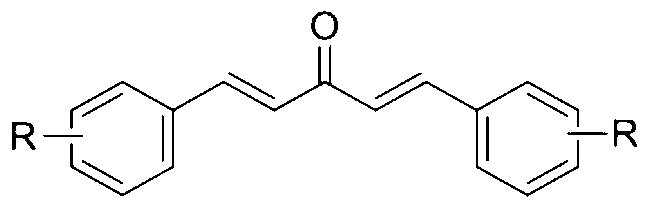
- a third aspect of the present invention provides a method for enhancing autophagy in cells comprising providing a mono-carbonyl analog of curcumin having a formula of
- R is independently selected from CF 3 , OH or OCH 3 .
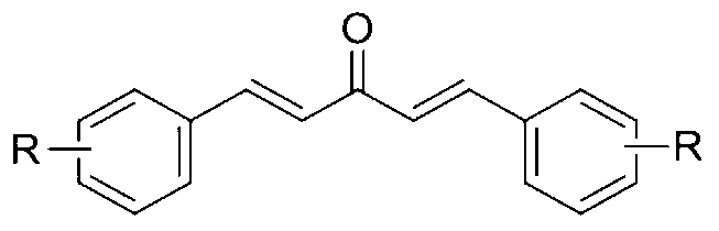
- a method for enhancing lysosome biogenesis in cells comprising providing a mono-carbonyl analogs of curcumin having a formula of
- R is OCH 3 .
- the mono-carbonyl analog binds to and activates TFEB in cells.
- the cells are non-neuronal cells or neuronal cells.
- Patent law e.g., they can mean “includes” , “included” , “including” , and the like; and that terms such as “consisting essentially of” and “consists essentially of” have the meaning ascribed to them in U.S. Patent law, e.g., they allow for elements not explicitly recited, but exclude elements that are found in the prior art or that affect a basic or novel characteristic of the present invention.
- Fig. 1 shows the chemical structure of mono-carbonyl analogs of curcumin (Fig. 1A) and cytotoxicity of mono-carbonyl analogs of curcumin (Fig. 1B) . Data are presented as the mean ⁇ SD from three independent experiments. * p ⁇ 0.05 vs. the control (0.1% DMSO) .
- Fig. 2 shows western blots of LC3-II, an autophagy marker, and LC3-II relative intensity in N2a cells treated with curcumin analogs.
- N2a cells were treated with curcumin (Cur, 10 ⁇ M) and its analogs (1 ⁇ M) for 12 h (Fig. 2A) .
- N2a cells were treated with Cur (10 ⁇ M) , analogs A2, B3 and C1 (1 ⁇ M) with or without chloroquine (CQ, 20 ⁇ M) for 12 h (Fig. 2B) .
- N2a cells were transfected with non-target siRNA (siNT) or Atg5 siRNA (siAtg5, 50 nM) for 72 h and then treated with Cur (10 ⁇ M) and analogs A2, B3 and C1 (1 ⁇ M) for 12 h (Fig. 2C) .
- Fig. 3 shows the effects of curcumin analogs on mTOR pathway.
- N2a cells were treated with curcumin (Cur, 10 ⁇ M) , A2, B3 and C1 (1 ⁇ M) for 12 h. Torin 1 (1 ⁇ M) treatment for 2 h was used as a positive control.
- Fig. 3A is a representative blots shows the expression of phosphorylated (p-) and total RPS6KB1/P70S6K, MTOR and AKT.
- Fig. 3B is a bar chart showing the expression of phosphorylated (p-) and total RPS6KB1/P70S6K, MTOR and AKT. Data are presented as the mean ⁇ SD from three independent experiments. * p ⁇ 0.05 vs. the control (0.1% DMSO) .
- Fig. 4 shows effects of curcumin and analogs A2, B3 and C1 activate the expression of endogenous TFEB in N2a cells.
- Cells were treated with curcumin (Cur, 10 ⁇ M) , A2, B3 and C1 (1 ⁇ M) for 12 h.
- Fig. 4A shows fluorescence images (at 50 and 10 ⁇ m scale) of cells fixed and stained with TFEB antibody and DAPI.
- Fig. 4B shows TFEB intensity per cell. Data is quantified by ImageJ software and is presented as mean ⁇ SD of three replicates for each treatment conditions. At least 1000 cells are analyzed in each treatment group. * p ⁇ 0.05 vs. the control (0.1% DMSO) .
- Fig. 4 shows effects of curcumin and analogs A2, B3 and C1 activate the expression of endogenous TFEB in N2a cells.
- Cells were treated with curcumin (Cur, 10 ⁇ M) , A2, B3 and C1 (1 ⁇
- FIG. 4C is a western blot showing the expression of endogenous TFEB.
- Fig. 4D shows the relative intensity of TFEB normalized to that of ACTB/ ⁇ -actin. Data are presented as the mean ⁇ SD from three independent experiments. * p ⁇ 0.05 vs. the control (0.1% DMSO) .
- Fig. 4E is fluorescence images showing expression of 3x Flag-TFEB in Hela cells having treated with Cur (10 ⁇ M) , A2, B3 and C1 (1 ⁇ M) for 12 h. Torin 1 (1 ⁇ M) treatment for 2 h is used as a positive control. Cells were fixed and stained with anti-Flag antibody.
- FIG. 4F shows percentage of cells with nuclear staining of TFEB; nuclear staining of TFEB is quantified by counting of the fluorescent cells in three random fields of view.
- Fig. 4G is the western blot showing the expression of Flag-tagged TFEB in the cytosolic (Cyt. ) and nuclear (Nuc. ) fractions.
- ACTB and H3F3A H3 histone, family 3A are used as loading control of cytoplasmic and nuclear fraction, respectively.
- Fig. 4H is showing the relative intensity of TFEB in the cytosolic and nuclear fractions. Data are presented as the mean ⁇ SD from three independent experiments. * p ⁇ 0.05 vs. the control (0.1% DMSO) .
- Fig. 5 shows C1 inhibits MTOR-TFEB-YWHA interaction.
- Hela cells stably expressing 3xFlag-TFEB are treated with C1 (1 ⁇ M) for 12 h.
- Endogenous MTOR and YWHA are co-immunoprecipitated with Flag-TFEB.
- WCL whole cell lysates
- Fig. 6 shows western blot of binding of solid-phase C1 or curcumin with recombinant TFEB protein.
- Fig. 7 shows curcumin analog C1 promotes TFEB-mediated autophagy and lysosomal biogenesis in cell cultures.
- mRNA transcript abundance is assessed by real-time PCR using specific primers for the indicated genes.
- Relative quantification (RQ) is presented as means ⁇ SD of three independent experiments.
- FIG. 7C is western blot showing the protein levels of autophagy marker (LC3-II) , TFEB and lysosome markers (LAMP1, CTSD) in Hela and SH-SY5Y cells having treated with C1 (0.5, 1 ⁇ M) or sucrose (100 mM) for 12 h.
- the relative intensity of autophagy marker (LC3-II) , TFEB and lysosome markers (LAMP1, CTSD) normalized to that of ACTB/ ⁇ -actin in Hela cells (Fig. 7D) and SH-SY5Y cells (Fig. 7E) .
- Data are presented as the mean ⁇ SD from three independent experiments. * p ⁇ 0.05 vs. the control (0.1% DMSO) .
- FIG. 7F shows western blot (left) and bar chat (right) showing the expression of endogenous TFEB and LC3-II in Hela cells having transfected with non-target siRNA (siNT) or TFEB siRND (siTFEB, 100 nM) for 72h and treated with C1 (1 ⁇ M) for 12 h.
- siNT non-target siRNA
- TFEB siRND TFEB siRND
- Fig. 8 shows curcumin analogs promotes the degradation of SNCA.
- Expression and intensity of HA-tagged wild-type (WT) or A53T mutant SNCA in inducible PC12 cells are treated with doxycycline (Dox, 2 ⁇ g/ml) for 24 h, and then treated with curcumin (10 ⁇ M) , A2, B3 and C1 (1 ⁇ M) for another 48 h (A, C) in the presence or absence of chloroquine (CQ, 20 ⁇ M) and wortmannin (WM, 1 ⁇ M) (B, D) .
- the expression of HA-SNCA is determined by western blots and quantified from three independent experiments.
- Corynoxine B (Cory B, 25 ⁇ M) is used as a positive control. Relative intensity is normalized to that of ACTB/ ⁇ -actin. Data are presented as the mean ⁇ SD from three independent experiments. * p ⁇ 0.05 vs. the control (0.1% DMSO) .
- Fig. 9 shows short-term administration of C1 increases the expression of TFEB and enhances autophagy in rat brain.
- Fig. 9A shows western blots of the various protein levels as indicated in the liver, frontal cortex and striatum.
- Fig. 10 shows effects of short-term administration of C1 on MTOR pathway, TFEB nuclear translocation and MTOR-TFEB interaction.
- Fig. 11 shows chronic administration of C1 enhances autophagy and promotes the degradation of endogenous SNCA in rat brain.
- Fig. 11A is western blots showing the protein levels as indicated in the liver, frontal cortex and striatum.
- a, "an, “ and “the” as used herein include “at least one” and “one or more” unless stated otherwise.
- reference to “a pharmacologically acceptable carrier” includes mixtures of two or more carriers as well as a single carrier, and the like.
- autophagy refers to macroautophagy, unless stated otherwise, is the catabolic process involving the degradation of a cell's own components; such as, long lived proteins, protein aggregates, cellular organelles, cell membranes, organelle membranes, and other cellular components.
- the mechanism of autophagy may include: (i) the formation of a membrane around a targeted region of the cell, separating the contents from the rest of the cytoplasm, (ii) the fusion of the resultant vesicle with a lysosome and the subsequent degradation of the vesicle contents.
- autophagy may also refer to one of the mechanisms by which a starving cell re-allocates nutrients from unnecessary processes to more essential processes. Also, for example, autophagy may inhibit the progression of some diseases and play a protective role against infection by intracellular pathogens.
- the diseases that benefit from autophagy inducement are diseases of which conditions are ameliorated, reduced or eliminated by autophagy and can be treated by the inventions as disclosed herein.
- the diseases include aggregate-prone disorder which represents any disease, disorder or condition associated with or caused by abnormal protein aggregates that are not sufficiently destroyed by a natural autophagy process in an organism and can be treated through degradation thereof via induction of autophagy by the subject invention.
- diseases include Alzheimer's disease, Parkinson's disease, amyotrophic lateral sclerosis, Huntington's disease, spinocerebellar ataxia, oculopharyngeal muscular dystrophy, prion diseases, fatal familial insomnia, alpha-1 antitrypsin deficiency, dentatorubral pallidoluysian atrophy, frontal temporal dementia, progressive supranuclear palsy, x-linked spinobulbar muscular atrophy, and neuronal intranuclear hyaline inclusion disease.
- the diseases also include cancer e.g., any cancer wherein the induction of autophagy would inhibit cell growth and division, reduce mutagenesis, remove mitochondria and other organelles damaged by reactive oxygen species or kill developing tumor cells.
- diseases can be chronic diseases which refers to persistent and lasting diseases, medical conditions or diseases that have developed slowly.
- diseases that can be treated by the subject invention also include, but not limited to, cardiovascular disorders, autoimmune disorders, metabolic disorders, hamartoma syndrome, genetic muscle disorders, and myopathies.
- the present invention provides a small molecule being able to enhance autophagy and lysosome biogenesis by activating TFEB.
- the molecule is a mono-carbonyl analog of curcumin.
- the molecule directly binds to TFEB, promote its expression and nuclear translocation.
- the molecule can prevent the accumulation of toxic protein aggregates in treating neurodegenerative diseases such as Parkinson's , Alzheimer's and Huntington's diseases.
- the molecule activates TFEB without inhibiting MTOR pathway, which is a key regulator of cell growth and proliferation.
- TFEB has been identified as a master gene regulating lysosome biogenesis and autophagy. Pharmacological activation of TFEB promotes cellular clearance of accumulated toxic molecules.
- the present invention discloses a potent activator of TFEB that enhances autophagy and lysosome biogenesis in neuronal and non-neuronal cells.
- the advantages of the present invention are: 1) compound of the present invention is a small lipid molecule with good BBB permeability and potent TFEB-activating effects for treating neurodegenerative diseases; 2) chemical structure of compound of the present invention is simple and it can be easily synthesized in large scale for pre-clinical and clinical studies; 3) compound of the present invention activates TFEB without inhibiting MTOR pathway, thus eliminating possible MTOR-associated complications in clinical trials. Therefore, the present invention has a wide field of application in the treatment of lysosomal storage disorders and common neurodegenerative diseases.
- the present invention provides a method for enhancing autophagy in cells comprising providing a mono-carbonyl analog of curcumin having a formula of
- R are independently selected from CF 3 , OH and OCH 3 .
- a method for enhancing lysosome biogenesis in cells comprising providing a mono-carbonyl analog of curcumin having a formula of
- R is OCH 3 .
- a method for enhancing lysosome biogenesis in cells comprising providing a mono-carbonyl analog of curcumin having a formula of C1:
- the mono-carbonyl analog binds to and activates TFEB in cells.
- the cells are non-neuronal cells or neuronal cells.
- a method of treating neurodegenerative diseases by administering a composition comprising a mono-carbonyl analog of Formula I to a subject in need thereof, wherein R is independently selected from CF 3 , OH and OCH 3 .
- the neurodegenerative diseases comprising Alzheimer's disease, Parkinson's disease, Huntington's disease and Creutzfeldt-Jakob disease.
- the composition comprises 1.62 mg/kg to 28.38 mg/kg of C1.
- the composition is administered via oral administration, intravenous injection or both.
- Anti-phospho-AKT (ser473) (9271) , anti-AKT (9272) , anti-phospho-MTOR (Ser2448) (2971) , anti-MTOR (2983) , anti-phospho-P70S6K/RPS6KB1 (Thr389) (9234) and anti-P70S6K/RPS6KB1 (9202) , pan-14-3-3/YWHA (8312) antibodies are purchased from Cell Signaling Technology.
- Anti-á-syn/SNCA antibody (610786) is purchased from BD Transduction Laboratories.
- HRP-conjugated goat anti-mouse (115-035-003) and goat anti-rabbit (111-035-003) secondary antibodies are purchased from Jackson ImmunoResearch.
- Anti-a-actin/ACTB (sc-47778) is purchased from Santa Cruz Biotechnology.
- Anti-ATG5 (NB110-53818) and anti-LC3 (NB100-2220) antibodies were purchased from Novus Biologicals.
- Anti-TFEB (13372-1-AP) was purchased from Proteintech.
- Mouse Atg5 siRNA (L-064838-00-0005) and non-target siRNA, human TFEB siRNA (M-009798-02-0005) and non-target siRNA are purchased from Dharmacon.
- DMEM (11965-126) , FBS (10270-106) , Opti-MEM I (31985-070) , horse serum (16050-122) , Hygromycin B (10687-010) , G418 (10131-035) , Alexa 488 goat anti-mouse IgG (A-11001) and Alexa 594 goat anti-rabbit IgG (A-11012) are purchased from Life Technologies.
- N2a Hela and Hela cells stably expressing 3x-Flag-TFEB are cultured in DMEM supplemented with 10% FBS.
- SH-SY5Y cells are cultured in DMEM/F12 supplemented with 10% FBS.
- the full medium is replaced by fresh Opti-MEM I and then the compounds (in 0.1% DMSO) are added to the cells and incubated for 12 h.
- Inducible PC12 cells overexpressing SNCA WT and A53T
- the cytotoxicity is determined by measurement of LDH release from damaged cells using LDH Kit (11644793001, Roche) according to the manufacturer’s protocol.
- Mouse Atg5 siRNA (25 nM) or human TFEB (100 nM) siRNA and the non-target siRNAs are transfected with Lipofectamine RNAiMAX (13778030, Invitrogen) and incubated at 37°C for 72 h.
- RNA is extracted from cells and tissues using RNeasy Plus Mini Kit (74134, Qiagen) .
- Reverse transcription is performed using High-Capacity cDNA Reverse Transcription Kit (4368814, Life Technologies) .
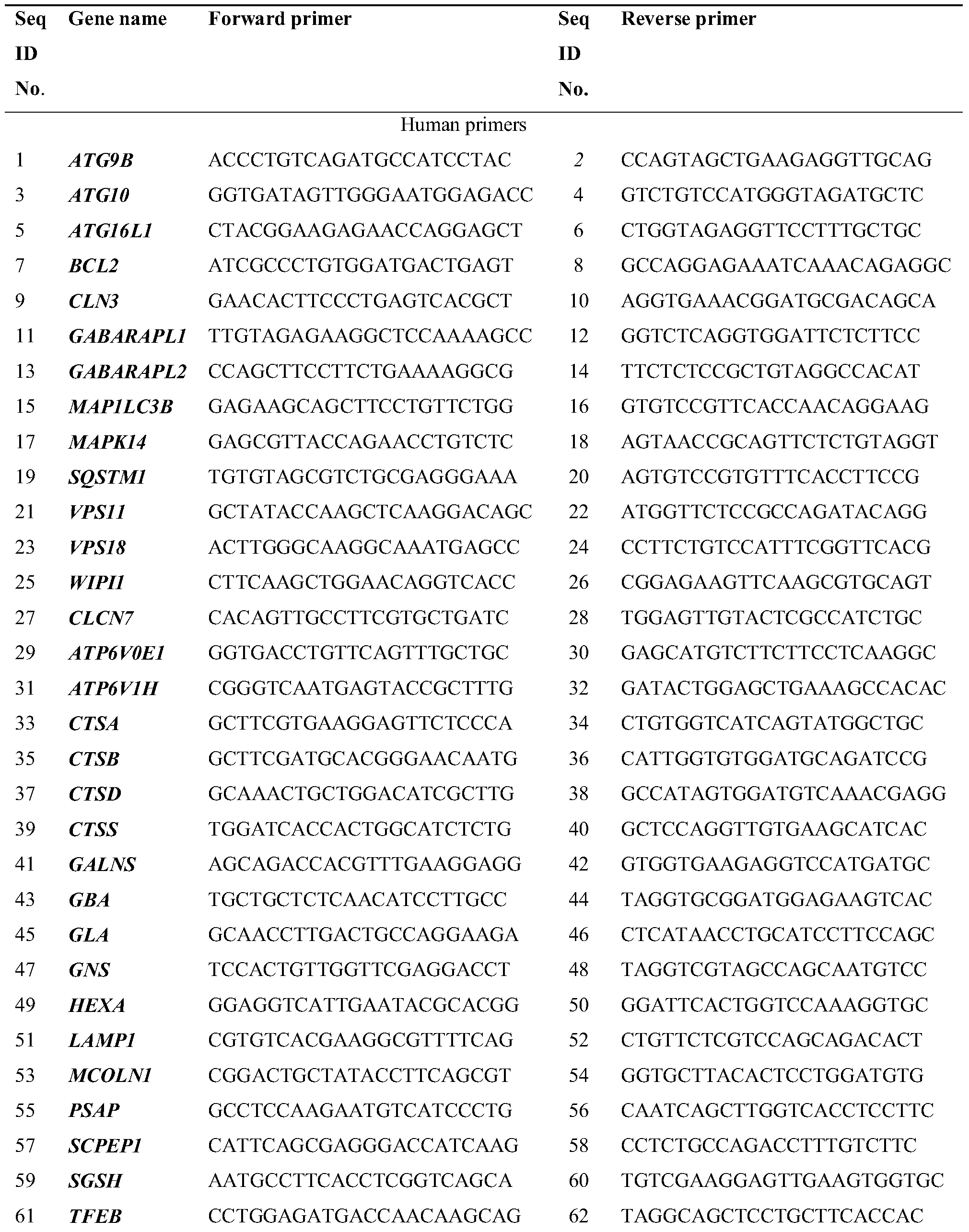
- Autophagy and lysosome gene primers are synthesized by Life Technologies and the oligonucleotide sequences are listed in Table 1.
- Real-time PCR is carried out with the Fast SYBRR Green Master Mix (4385612, Life Technologies) using the ViiA TM 7 Real-Time PCR System (Life Technologies) . Fold changes are calculated using the ⁇ CT method and the results were normalized against an internal control (GAPDH or ACTB) .
- Cells are lysed on ice in 1X Lysis Buffer (9803, Life Technologies) with complete protease inhibitor mixture (04693124001, Roche Applied Science) .
- Animal tissues are homogenized in nine volumes of ice-cold PBS supplemented with protease inhibitors. Cytosolic and nuclear fractions are isolated using protocols similar to those described previously.
- Anti-Flag or TFEB antibody is added to the whole cell lysates and Protein G (10003D, Life Technologies) is used for immunoprecipitation. Proteins are separated by 10-15% SDS-PAGE, transferred, and blotted with the antibodies described.
- the blots are then incubated with secondary antibodies or the Clean-Blot IP Detection Reagent (21230, Thermo Scientific) at room temperature for 1 h.
- the protein signals are detected by ECL kit (32106, Pierce) and quantified using ImageJ software.
- Cells are seeded on coverslips placed in 24-well plates. After drug treatment, slices are fixed with 3.7% paraformaldehyde, permeabilised in 0.2% Triton X-100 and blocked with 5% BSA. After blocking, the slices are stained with anti-TFEB (1: 200) or anti-Flag (1: 500) antibodies overnight at 4°C. Alexa 488 (green) or Alexa 594 (red) secondary antibodies (1: 500) are added for 1 h at room temperature. After nuclear staining with DAPI, the slices are mounted with FluorSave reagent (345789, Calbiochem) . Cells are visualized using an Eclipse 80i fluorescence microscope (Nikon Instruments Inc. )
- Fig. 1A the inventors tested the cytotoxicity of the testing compounds at 1 ⁇ M and 10 ⁇ M for 24 h (as shown in Fig. 1A) by LDH assay (Fig. 1B) and used 1 ⁇ M concentration subsequently.
- Curcumin (10 ⁇ M) and analogs A2, B3 and C1 (1 ⁇ M) significantly increase the levels of microtubule-associated protein 1 light chain 3B (LC3B) -II in N2a cells compared to the vehicle control (0.1% DMSO) (Fig. 2A) .
- LC3B microtubule-associated protein 1 light chain 3B
- Fig. 2A the vehicle control (0.1% DMSO)
- CQ lysosomal inhibitor chloroquine
- curcumin enhances autophagy through inhibiting MTOR pathway, the effects of the three newly identified autophagy enhancers on MTOR pathway of the present invention are confirmed. Similar to curcumin, A2 and B3 inhibit the phosphorylation of RPS6KB1/p70S6K, MTOR and AKT (Fig. 3A and Fig. 3B) . Torin1, a potent MTOR inhibitor is used as a positive control. C1 significantly promotes the phosphorylation of RPS6KB1, MTOR and AKT (Fig. 3A and Fig. 3B) .
- Curcumin analog C1 activates TFEB in cell cultures
- TFEB has been identified as a target of MTOR. Pharmacological inhibition of MTORC1 activates TFEB by promoting its nuclear translocation. However, the effects of curcumin and its analogs which inhibit MTOR pathway on TFEB has not been studied. Firstly, the expression and distribution of endogenous TFEB in N2a cells treated with curcumin and its analogs are determined. N2a cells are treated with curcumin (Cur, 10 ⁇ M) , A2, B3 and C1 (1 ⁇ M) for 12 h. It is shown that compound C1 increases the total levels of TFEB and promotes its nuclear translocation (Fig. 4 A-D) . Curcumin, A2 and B3 show no effects on the expression or distribution of TFEB (Fig.
- curcumin analog C1 represents a new activator of TFEB which affects both its expression and translocation.
- Curcumin analog C1 binds to TFEB and inhibits MTOR-TFEB-YWHA interaction
- Hela cells stably expressing 3xFlag-TFEB are treated with C1 (1 ⁇ M) for 12 h.
- Endogenous MTOR (A) and YWHA (B) are co-immunoprecipitated with Flag-TFEB.
- the levels of immunoprecipitated MTOR and YWHA are normalized to their corresponding levels in whole cell lysates (WCL) .
- C1 treatment does not affect the levels of endogenous MTOR (Fig. 5 A) and YWHA (Fig. 5B) .
- interaction of MTOR and YWHA is decreased as compared with control.
- Curcumin analog C1 activates TFEB-mediated autophagy and lysosomal biogenesis in cell cultures.
- TFEB genes and a series of genes involved in autophagy and lysosome biogenesis are shown to be up-regulated by C1 treatment in Hela (Fig. 7A) and SH-SY5Y (Fig. 7B) cells.
- the changes in gene expression by C1 treatment are more sensitive in Hela cells than in SH-SY5Y cells.
- the expression of TFEB, autophagy marker (LC3B) and lysosome markers (LAMP1 and CTSD) are determined by western blot (Fig. 7C) .
- Sucrose a TFEB activator is used as a positive control.
- C1 at different concentrations increased the protein levels of TFEB and activated autophagy/lysosomal biogenesis in Hela (Fig. 7D) and SH-SY5Y (Fig. 7E) cells.
- Fig. 7F the increase in LC3-II levels by C1 treatment is blocked, indicating that the autophagy-enhancing effect of C1 is TFEB-dependent (Fig. 7F) .
- Curcumin analog C1 promotes the degradation of SNCA.
- Inducible PC12 cells are treated with doxycycline (Dox, 2 ⁇ g/ml) for 24 h to induce the expression of HA-tagged wild-type (WT) or A53T mutant SNCA, and then treated with curcumin (10 ⁇ M) , A2, B3 and C1 (1 ⁇ M) for another 48 h (A, C) in the presence or absence of chloroquine (CQ, 20 ⁇ M) and wortmannin (WM, 1 ⁇ M) (B, D) .
- the expression of HA-SNCA is determined by western blots and quantified from three independent experiments. Corynoxine B (Cory B, 25 ⁇ M) is used as a positive control.
- curcumin (10 ⁇ M) promotes the degradation of SNCA A53T but had no effect on SNCA WT .
- curcumin analog A2, B3 and C1 (1 ⁇ M) significantly degrade both SNCA WT or SNCA A53T (Fig. 8A and Fig. 8C) .
- C1 showed the best effects on SNCA clearance.
- Corynoxine B Cory B
- the activity of C1 in degrading SNCA was significantly blocked by autophagy inhibitor wortmannin (WM) or the lysosome inhibitor CQ (Fig. 8B and Fig. 8D) , indicating that C1 promotes the clearance of SNCA via autophagy-lysosome pathway.
- curcumin analog C1 activates TFEB and autophagy in rats brains.
- Acute toxicity of C1 in rats by single-dose intravenous (IV) tail vein injection and the medium lethal dose (LD 50 ) value of C1 is 175 mg/kg are determined.
- Short-tern oral administration of C1 (10 mg/kg and 25 mg/kg) dose-dependently increases the expression of LC3-II and TFEB in the liver, frontal cortex and striatum of the brains (Fig. 9 A-D) .
- short-term administration of C1 does not affect the levels of endogenous SQSTM1/p62 or SNCA in the brains (Fig. 9 A-D) .
- C1 treatment dose-dependently increases the expression of LAMP1 in the brains.
- the real-time PCR analysis of brain lysates shows that C1 up-regulated TFEB and several autophagy/lysosomal genes in the brain (Fig. 9E) . This indicates that C1 can pass the blood-brain barrier (BBB) .
- BBB blood-brain barrier
- the average concentration of C1 in brain tissues is 0.26 ⁇ 0.063 ⁇ g/g determined by HPLC after oral administration of C1 (10 mg/kg) for 6 h. Based on these data, it is demonstrated that short-term treatment of C1 can activate TFEB and autophagy in rats’ brains while insufficient to degrade the autophagy substrates SQSTM1/p62 and SNCA.
- curcumin analog C1 promotes the degradation of endogenous SNCA.
- Rats receive oral administration of C1 (10 mg/kg) for 21 days. Another dose of C1 is given for 6 h and the average concentration of C1 in brain tissues is 0.849 ⁇ 0.302 ⁇ g/g. Then the autophagy markers in the livers and brains are analyzed. Chronic C1 treatment increases the levels of LC3-II in the livers (Fig. 11 A and B) . In the frontal cortex and striatum of brains, C1 treatment significantly increases the levels of TFEB and LC3-II (Fig. 11 A, C and D) . Notably, the levels of endogenous SQSTM1/p62 and SNCA significantly decrease in rats’ brains treated with C1 for 21 days (Fig.
- the effective dosage of the invented curcumin mono-carbonyl analog C1 ranges from 10 mg/kg (body weight) to 175 mg/kg (body weight) per day.
- the effective translated human dose of the curcumin mono-carbonyl analog C1 of the present invention ranges from 1.62 mg/kg (body weight) to 28.38 mg/kg (body weight) per day.
- the present invention discloses novel compositions comprising an autophagy enhancement compound.
- the present invention relates to a composition comprising a small molecule being able to enhance autophagy and lysosome biogenesis by activating the gene TFEB which can prevent the accumulation of toxic protein aggregates in treating neurodegenerative diseases such as Parkinson's , Alzheimer's and Huntington's diseases.
- the different functions discussed herein may be performed in a different order and/or concurrently with each other. Furthermore, if desired, one or more of the above-described functions may be optional or may be combined.
Landscapes
- Health & Medical Sciences (AREA)
- Pharmacology & Pharmacy (AREA)
- Animal Behavior & Ethology (AREA)
- Chemical & Material Sciences (AREA)
- Veterinary Medicine (AREA)
- Medicinal Chemistry (AREA)
- Life Sciences & Earth Sciences (AREA)
- Public Health (AREA)
- General Health & Medical Sciences (AREA)
- Engineering & Computer Science (AREA)
- Neurology (AREA)
- Neurosurgery (AREA)
- Bioinformatics & Cheminformatics (AREA)
- Biomedical Technology (AREA)
- Chemical Kinetics & Catalysis (AREA)
- Nuclear Medicine, Radiotherapy & Molecular Imaging (AREA)
- General Chemical & Material Sciences (AREA)
- Organic Chemistry (AREA)
- Epidemiology (AREA)
- Psychology (AREA)
- Hospice & Palliative Care (AREA)
- Psychiatry (AREA)
- Acyclic And Carbocyclic Compounds In Medicinal Compositions (AREA)
- Pharmaceuticals Containing Other Organic And Inorganic Compounds (AREA)
Abstract
A composition comprising an autophagy enhancement compound is disclosed. Small molecules that are able to enhance autophagy and lysosome biogenesis by activating the gene TFEB which can prevent the accumulation of toxic protein aggregates in treating neurodegenerative diseases are disclosed.
Description
CROSS-REFERENCE TO RELATED APPLICATIONS
This application claims the benefit of U.S. Provisional Patent Application Serial Number 61/949,233 filed on March 06, 2014 and U.S. Non-Provisional Patent Application Serial Number 14/609,438 filed on January 30, 2015, the disclosures of which are hereby incorporated by reference.
FIELD OF INVENTION
The present invention relates to a composition comprising an autophagy enhancement compound. In particular, the present invention relates to a composition comprising a small molecule being able to enhance autophagy and lysosome biogenesis by activating the gene TFEB which can prevent the accumulation of toxic protein aggregates in treating neurodegenerative diseases such as Parkinson's , Alzheimer's and Huntington's diseases.
BACKGROUND OF INVENTION
Macroautophagy, herein referred to as autophagy, is a highly conserved process for cellular degradation and recycling of cytosolic contents to maintain cellular homeostasis. Autophagy substrates are generally cellular organelles, long-lived proteins and aggregate-prone proteins. Due to its functionality to clear cytosolic contents, this highly conserved process has been shown to be a promising approach for treatment of diseases characterized by the formation of intracellular aggregates, such as aging of the brain and neurodegeneration.
Dysfunction in the autophagy-lysosome pathway (ALP) has been directly linked to neurodegenerative disorders. Recently, the transcription factor EB (TFEB) has been identified in Settembre, C., et. al., TFEB links autophagy to lysosomal biogenesis. Science, 2011. 332(6036) : 1429-33, and Sardiello, M., et al., A gene network regulating lysosomal biogenesis and function. Science, 2009.325 (5939) : 473-7, as a master regulator of ALP. TFEB transgene to increase TFEB expression, or small molecules aimed to stimulate nuclear translocation of endogenous TFEB promotes the clearance of toxic protein aggregates, thus providing a disease-modifying intervention for neurodegenerative disorders such as Parkinson’s disease (PD) , Alzheimer's disease (AD) and Huntington's disease (HD) .
Current MTOR inhibitors, such as rapamycin and torin1, activate TFEB by promoting TFEB nuclear translocation. However, their pharmacokinetic profile and side effects make them less likely to be useful for long-term use in patients with neurodegenerative diseases. Disaccharides, such as trehalose and sucrose, activate TFEB in an MTOR-independent manner and may be beneficial for neurodegenerative diseases. However, the blood-brain barrier (BBB) permeability of trehalose and sucrose is poor. Discovery of small molecules which directly target TFEB hold great promise for the development of efficient neuroprotective therapies.
It is an objective of the current invention to provide for a small molecule compound having good BBB permeability and potent TFEB-activating effects for the treatment of neurodegenerative diseases. The present invention provides a compound having simple chemical structure which can be easily synthesized in large scale. The present invention provides a compound that directly binds to and activates TFEB without inhibiting MTOR pathway, thus eliminating possible MTOR-associated complications. A further objective of the current invention is to provide a method for treating lysosomal storage disorders and diseases that can benefit from autophagy, including but not limited to neurodegenerative disorders, immunological diseases, cardiac diseases and cancer.
SUMMARY OF THE INVENTION
The present invention relates to a composition comprising an autophagy enhancement compound. In particular, the present invention relates to a composition comprising a small molecule being able to enhance autophagy and lysosome biogenesis by activating the gene TFEB which can prevent the accumulation of toxic protein aggregates in treating neurodegenerative diseases such as Parkinson's , Alzheimer's and Huntington's diseases.
The present invention discloses a potent activator of TFEB that enhances autophagy and lysosome biogenesis in neuronal and non-neuronal cells. In comparison to currently known TFEB activators, the advantages of the present invention are: 1) The compound of the present invention is a small lipid molecule with good BBB permeability and potent TFEB-activating property; 2) The chemical structure of the compound of the present invention is simple and it can be easily synthesized in large scale for pre-clinical and clinical studies; 3) Compound of the present invention activates TFEB without inhibiting MTOR pathway, thus eliminating possible
MTOR-associated complications in clinical trials. Therefore, the present invention has a wide field of application in the treatment of lysosomal storage disorders and common neurodegenerative diseases.
In a first aspect of the present invention, there is provided three potent autophagy enhancers. The three compounds (namely A2, B3 and C1) induce autophagy in neuronal-like N2a cells. The three compounds are synthesized mono-carbonyl analogs of curcumin and their chemical names are:
A2: 1, 5-bis (3- (trifluoromethyl) phenyl) penta-1, 4-dien-3-one
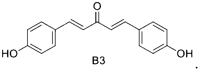
B3: 1, 5-bis (4-hydroxyphenyl) penta-1, 4-dien-3-one
C1: 1, 5-bis (2-methoxyphenyl) penta-1, 4-dien-3-one
In another embodiment of the present invention, it is provided that the three compounds A2, B3 and C1 promote the degradation of wild-type and A53T mutant alpha-synuclein (SNCA) in cell culture.
In a further embodiment of the present invention, there is provided that compounds A2 and B3 induce autophagy through inhibiting AKT/MTOR pathway.
In yet a further embodiment of the present invention, there is provided that compound C1 activates transcription factor EB (TFEB) , an essential regulator of autophagy and lysosome biogenesis. C1 significantly increases endogenous TFEB expression and promotes the nuclear translocation of TFEB.
It is known that MTOR pathway is a key regulator of cell growth and proliferation. In another embodiment of the present invention there is provided that compound C1 activates TFEB-mediated autophagy without inhibiting MTOR pathway.
In yet a further embodiment of the present invention, there is provided that C1 directly binds to TFEB and inhibits MTOR-TFEB-YWHA interaction, which releases TFEB from MTOR complex and promotes TFEB nuclear translocation.
In yet another embodiment of the present invention, there is provided that compound C1 enhances TFEB-mediated autophagy and lysosome biogenesis in non-neuronal cells and neuronal cells.
In yet another embodiment of the present invention, there is provided that the medium lethal dose (LD50) value of C1 is 175 mg/kg in rats by single-dose intravenous (IV) tail vein injection.
In yet another embodiment of the present invention, there is provided that the average concentration of C1 in brain tissues is 0.26 ± 0.063 μg/g and 0.849 ± 0.302 μg/g after 6 h short-term and chronic oral administration of C1 (10 mg/kg) in rats respectively.
In yet another embodiment of the present invention there is provided that short-term oral administration of curcumin analog C1 activates TFEB and autophagy in rats brains, and chronic administration of C1 promotes the degradation of endogenous SNCA in rats brains.
A second aspect of the present invention provides a method for treating neurodegenerative disorders without obvious side effects caused by mTOR inhibition comprising administering a composition comprising compound C1. Such neurodegenerative diseases comprise but are not limited to the following: Alzheimer's disease, Parkinson's disease, Huntington's disease, Creutzfeldt-Jakob disease.
In an embodiment of the second aspect of the present invention, C1 is administered at 1.62 mg/kg to 28.38 mg/kg per body weight of the subject in need thereof.
In another embodiment of the second aspect of the present invention, the composition is administered via oral administration and/or intravenous injection.
A third aspect of the present invention provides a method for enhancing autophagy in cells comprising providing a mono-carbonyl analog of curcumin having a formula of

wherein R is independently selected from CF3, OH or OCH3.
In a first embodiment of the third aspect of the present invention, the mono-carbonyl analog of curcumin having the formula of A2:

1, 5-bis (3- (trifluoromethyl) phenyl) penta-1, 4-dien-3-one.
In a second embodiment of the third aspect of the present invention, the mono-carbonyl analog of curcumin having the formula of B3:

1, 5-bis (4-hydroxyphenyl) penta-1, 4-dien-3-one.
In a third embodiment of the third aspect of the present invention, the mono-carbonyl analog of curcumin having the formula of C1:

1, 5-bis (2-methoxyphenyl) penta-1, 4-dien-3-one.
In a fourth aspect of the present invention, there is provided a method for enhancing lysosome biogenesis in cells comprising providing a mono-carbonyl analogs of curcumin having a formula of
wherein R is OCH3.
In a first embodiment of the forth aspect of the present invention the mono-carbonyl analog of curcumin having a formula of C1:

1, 5-bis (2-methoxyphenyl) penta-1, 4-dien-3-one.
In a second embodiment of the fourth aspect of the present invention, the mono-carbonyl analog binds to and activates TFEB in cells.
In the third and fourth aspects of the present invention, the cells are non-neuronal cells or neuronal cells.
Throughout the present specification, unless the context requires otherwise, the word "comprise" or variations such as "comprises" or "comprising" , will be understood to imply the inclusion of a stated integer or group of integers but not the exclusion of any other integer or group of integers. It is also noted that in this disclosure and particularly in the claims and/or paragraphs, terms such as “comprises” , “comprised” , “comprising” and the like can have the meaning attributed to it in U.S. Patent law; e.g., they can mean “includes” , “included” , “including” , and the like; and that terms such as “consisting essentially of” and “consists essentially of” have the meaning ascribed to them in U.S. Patent law, e.g., they allow for elements not explicitly recited, but exclude elements that are found in the prior art or that affect a basic or novel characteristic of the present invention.
Furthermore, throughout the present specification and claims, unless the context requires otherwise, the word “include” or variations such as “includes” or “including” , will be understood to imply the inclusion of a stated integer or group of integers but not the exclusion of any other integer or group of integers.
Other definitions for selected terms used herein may be found within the detailed description of the present invention and apply throughout. Unless otherwise defined, all other technical terms used herein have the same meaning as commonly understood to one of ordinary skill in the art to which the invention belongs.
Other aspects and advantages of the present invention will be apparent to those skilled in the art from a review of the ensuing description.
The above and other objects and features of the present invention will become apparent from the following description of the invention, when taken in conjunction with the accompanying drawings, in which:
Fig. 1 shows the chemical structure of mono-carbonyl analogs of curcumin (Fig. 1A) and cytotoxicity of mono-carbonyl analogs of curcumin (Fig. 1B) . Data are presented as the mean ±SD from three independent experiments. *p < 0.05 vs. the control (0.1% DMSO) .
Fig. 2 shows western blots of LC3-II, an autophagy marker, and LC3-II relative intensity in N2a cells treated with curcumin analogs. N2a cells were treated with curcumin (Cur, 10 μM) and its analogs (1 μM) for 12 h (Fig. 2A) . N2a cells were treated with Cur (10 μM) , analogs A2, B3 and C1 (1 μM) with or without chloroquine (CQ, 20 μM) for 12 h (Fig. 2B) . N2a cells were transfected with non-target siRNA (siNT) or Atg5 siRNA (siAtg5, 50 nM) for 72 h and then treated with Cur (10 μM) and analogs A2, B3 and C1 (1 μM) for 12 h (Fig. 2C) . The expression of LC3-II is determined by western blot. Relative intensity is normalized to that of ACTB/β-actin. Data are presented as the mean ± SD from three independent experiments. *p <0.05 vs. the control (0.1% DMSO) ; #p < 0.05 vs. CQ treatment alone. N.S. = not significant.
Fig. 3 shows the effects of curcumin analogs on mTOR pathway. N2a cells were treated with curcumin (Cur, 10 μM) , A2, B3 and C1 (1 μM) for 12 h. Torin 1 (1 μM) treatment for 2 h was used as a positive control. Fig. 3A is a representative blots shows the expression of phosphorylated (p-) and total RPS6KB1/P70S6K, MTOR and AKT. Fig. 3B is a bar chart showing the expression of phosphorylated (p-) and total RPS6KB1/P70S6K, MTOR and AKT. Data are presented as the mean ± SD from three independent experiments. *p < 0.05 vs. the control (0.1% DMSO) .
Fig. 4 shows effects of curcumin and analogs A2, B3 and C1 activate the expression of endogenous TFEB in N2a cells. Cells were treated with curcumin (Cur, 10 μM) , A2, B3 and C1 (1 μM) for 12 h. Fig. 4A shows fluorescence images (at 50 and 10 μm scale) of cells fixed and stained with TFEB antibody and DAPI. Fig. 4B shows TFEB intensity per cell. Data is quantified by ImageJ software and is presented as mean ± SD of three replicates for each treatment conditions. At least 1000 cells are analyzed in each treatment group. *p < 0.05 vs. the control (0.1% DMSO) . Fig. 4C is a western blot showing the expression of endogenous TFEB. Fig. 4D shows the relative intensity of TFEB normalized to that of ACTB/β-actin. Data are presented as the mean ± SD from three independent experiments. *p < 0.05 vs. the control (0.1% DMSO) . Fig. 4E is fluorescence images showing expression of 3x Flag-TFEB in Hela cells having treated with Cur (10 μM) , A2, B3 and C1 (1 μM) for 12 h. Torin 1 (1 μM) treatment for 2 h is used as a positive control. Cells were fixed and stained with anti-Flag antibody. Fig. 4F shows percentage of cells with nuclear staining of TFEB; nuclear staining of TFEB is quantified by counting of the fluorescent cells in three random fields of view. Fig. 4G is the western blot showing the expression of Flag-tagged TFEB in the cytosolic (Cyt. ) and nuclear (Nuc. ) fractions. ACTB and H3F3A (H3 histone, family 3A) are used as loading control of cytoplasmic and nuclear fraction, respectively. Fig. 4H is showing the relative intensity of TFEB in the cytosolic and nuclear fractions. Data are presented as the mean ± SD from three independent experiments. *p < 0.05 vs. the control (0.1% DMSO) .
Fig. 5 shows C1 inhibits MTOR-TFEB-YWHA interaction. Hela cells stably expressing 3xFlag-TFEB are treated with C1 (1 μM) for 12 h. Endogenous MTOR and YWHA are co-immunoprecipitated with Flag-TFEB. Western blots and levels of immunoprecipitated MTOR (Fig. 5A) and YWHA (Fig. 5B) normalized to their corresponding levels in whole cell
lysates (WCL) . Data are presented as the mean ± SD from three independent experiments. *p < 0.05 vs. the control. WB-western blot and IP-immunoprecipitation
Fig. 6 shows western blot of binding of solid-phase C1 or curcumin with recombinant TFEB protein.
Fig. 7 shows curcumin analog C1 promotes TFEB-mediated autophagy and lysosomal biogenesis in cell cultures. Various gene expressions in Hela cells (Fig. 7A) and SH-SY5Y cells (Fig. 7B) after treated with C1 (1 μM) for 12 h. mRNA transcript abundance is assessed by real-time PCR using specific primers for the indicated genes. Relative quantification (RQ) is presented as means ± SD of three independent experiments. Fig. 7C is western blot showing the protein levels of autophagy marker (LC3-II) , TFEB and lysosome markers (LAMP1, CTSD) in Hela and SH-SY5Y cells having treated with C1 (0.5, 1 μM) or sucrose (100 mM) for 12 h. The relative intensity of autophagy marker (LC3-II) , TFEB and lysosome markers (LAMP1, CTSD) normalized to that of ACTB/β-actin in Hela cells (Fig. 7D) and SH-SY5Y cells (Fig. 7E) . Data are presented as the mean ± SD from three independent experiments. *p < 0.05 vs. the control (0.1% DMSO) . Fig. 7F shows western blot (left) and bar chat (right) showing the expression of endogenous TFEB and LC3-II in Hela cells having transfected with non-target siRNA (siNT) or TFEB siRND (siTFEB, 100 nM) for 72h and treated with C1 (1 μM) for 12 h. Autophagy-enhancing effect of C1 is TFEB-dependent. Data are presented as the mean ± SD from three independent experiments. *p < 0.05 vs. the control. N. S. = not significant.
Fig. 8 shows curcumin analogs promotes the degradation of SNCA. Expression and intensity of HA-tagged wild-type (WT) or A53T mutant SNCA in inducible PC12 cells are treated with doxycycline (Dox, 2 μg/ml) for 24 h, and then treated with curcumin (10 μM) , A2, B3 and C1 (1 μM) for another 48 h (A, C) in the presence or absence of chloroquine (CQ, 20 μM) and wortmannin (WM, 1 μM) (B, D) . The expression of HA-SNCA is determined by western blots and quantified from three independent experiments. Corynoxine B (Cory B, 25 μM) is used as a positive control. Relative intensity is normalized to that of ACTB/β-actin. Data are presented as the mean ± SD from three independent experiments. *p < 0.05 vs. the control (0.1% DMSO) .
Fig. 9 shows short-term administration of C1 increases the expression of TFEB and enhances autophagy in rat brain. SD rats (n = 6 per group) are orally administered by gavage with C1 (10mg/kg and 25 mg/kg per day) or vehicle (1% CMC-Na) for 24 h. An additional dosage of C1 was given for 6 h before the rats were killed. Fig. 9A shows western blots of the various protein levels as indicated in the liver, frontal cortex and striatum. Fig. 9B-9D are bar charts showing quantitative data of various protein expressions in the liver, frontal cortex and striatum. Data are presented as the mean ± SD (n = 6) . *p < 0.05 vs. the vehicle treatment. Fig. 9E shows mRNA levels in the frontal cortex were analyzed by real time PCR. Relative quantification (RQ) is presented as means ± SD (n = 4) . *p < 0.05 vs. the vehicle treatment.
Fig. 10 shows effects of short-term administration of C1 on MTOR pathway, TFEB nuclear translocation and MTOR-TFEB interaction. SD rats (n = 6 per group) are orally administered by gavage with C1 (10mg/kg and 25 mg/kg per day) or vehicle (1% CMC-Na) for 24 h. An additional dosage of C1 is given for 6 h before the rats are killed. Fig. 10A shows western blot (left) and quantitative data (right) of MTOR and RPS6KB1 expressions having treated with C1 on MTOR pathway in the frontal cortex. Data are presented as the mean ± SD (n = 6) . Fig. 10B is western blot (left) and quantitative data (right) showing TFEB nuclear translocation after C1 treatment (n = 4) . Fig. 10C shows immunoprecipitation data of MTOR-TFEB interaction after C1 treatment in the frontal cortex (n = 4) . *p < 0.05 vs. the vehicle treatment.
Fig. 11 shows chronic administration of C1 enhances autophagy and promotes the degradation of endogenous SNCA in rat brain. SD rats (n = 6 per group) are orally administered by gavage with C1 (10mg/kg per day) or vehicle (1% CMC-Na) for 21 days. An additional dosage of C1 is given for 6 h before the rats are killed. Fig. 11A is western blots showing the protein levels as indicated in the liver, frontal cortex and striatum. Fig. 11B-11D are quantitative data protein levels in the liver frontal cortex and striatum. Data are presented as the mean ± SD (n = 6) . *p < 0.05 vs. the vehicle treatment.
The present invention is not to be limited in scope by any of the specific embodiments described herein. The following embodiments are presented for exemplification only.
DEFINITIONS
"a, " "an, " and "the" as used herein include "at least one" and "one or more" unless stated otherwise. Thus, for example, reference to "a pharmacologically acceptable carrier" includes mixtures of two or more carriers as well as a single carrier, and the like.
The term "autophagy" refers to macroautophagy, unless stated otherwise, is the catabolic process involving the degradation of a cell's own components; such as, long lived proteins, protein aggregates, cellular organelles, cell membranes, organelle membranes, and other cellular components. The mechanism of autophagy may include: (i) the formation of a membrane around a targeted region of the cell, separating the contents from the rest of the cytoplasm, (ii) the fusion of the resultant vesicle with a lysosome and the subsequent degradation of the vesicle contents. The term autophagy may also refer to one of the mechanisms by which a starving cell re-allocates nutrients from unnecessary processes to more essential processes. Also, for example, autophagy may inhibit the progression of some diseases and play a protective role against infection by intracellular pathogens.
The diseases that benefit from autophagy inducement are diseases of which conditions are ameliorated, reduced or eliminated by autophagy and can be treated by the inventions as disclosed herein. The diseases include aggregate-prone disorder which represents any disease, disorder or condition associated with or caused by abnormal protein aggregates that are not sufficiently destroyed by a natural autophagy process in an organism and can be treated through degradation thereof via induction of autophagy by the subject invention. For example, such diseases include Alzheimer's disease, Parkinson's disease, amyotrophic lateral sclerosis, Huntington's disease, spinocerebellar ataxia, oculopharyngeal muscular dystrophy, prion diseases, fatal familial insomnia, alpha-1 antitrypsin deficiency, dentatorubral pallidoluysian atrophy, frontal temporal dementia, progressive supranuclear palsy, x-linked spinobulbar muscular atrophy, and neuronal intranuclear hyaline inclusion disease. The diseases also include cancer e.g., any cancer wherein the induction of autophagy would inhibit cell growth and division, reduce mutagenesis, remove mitochondria and other organelles damaged by reactive oxygen species or kill developing tumor cells. They can be chronic diseases which refers to persistent and lasting diseases, medical conditions or diseases that have developed slowly. The diseases that can be treated by the subject invention also include, but not limited to,
cardiovascular disorders, autoimmune disorders, metabolic disorders, hamartoma syndrome, genetic muscle disorders, and myopathies.
The present invention provides a small molecule being able to enhance autophagy and lysosome biogenesis by activating TFEB. The molecule is a mono-carbonyl analog of curcumin. The molecule directly binds to TFEB, promote its expression and nuclear translocation. The molecule can prevent the accumulation of toxic protein aggregates in treating neurodegenerative diseases such as Parkinson's , Alzheimer's and Huntington's diseases. The molecule activates TFEB without inhibiting MTOR pathway, which is a key regulator of cell growth and proliferation.
TFEB has been identified as a master gene regulating lysosome biogenesis and autophagy. Pharmacological activation of TFEB promotes cellular clearance of accumulated toxic molecules. The present invention discloses a potent activator of TFEB that enhances autophagy and lysosome biogenesis in neuronal and non-neuronal cells. In comparison to currently known TFEB activators, the advantages of the present invention are: 1) compound of the present invention is a small lipid molecule with good BBB permeability and potent TFEB-activating effects for treating neurodegenerative diseases; 2) chemical structure of compound of the present invention is simple and it can be easily synthesized in large scale for pre-clinical and clinical studies; 3) compound of the present invention activates TFEB without inhibiting MTOR pathway, thus eliminating possible MTOR-associated complications in clinical trials. Therefore, the present invention has a wide field of application in the treatment of lysosomal storage disorders and common neurodegenerative diseases.
The present invention provides a method for enhancing autophagy in cells comprising providing a mono-carbonyl analog of curcumin having a formula of

wherein R are independently selected from CF3, OH and OCH3.
In a first embodiment of the present invention, the mono-carbonyl analog of curcumin having a formula of A2:

In a second embodiment of the present invention, the mono-carbonyl analog of curcumin having a formula of B3:

In a third embodiment of the present invention, the mono-carbonyl analog of curcumin having a formula of C1:

In a second aspect of the present invention there is provided a method for enhancing lysosome biogenesis in cells comprising providing a mono-carbonyl analog of curcumin having a formula of
wherein R is OCH3.
In a first embodiment of the second aspect of the present invention there is provided a method for enhancing lysosome biogenesis in cells comprising providing a mono-carbonyl analog of curcumin having a formula of C1:
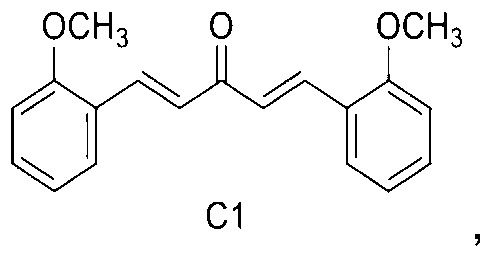
1, 5-bis (2-methoxyphenyl) penta-1, 4-dien-3-one.
In a second embodiment of the second aspect of the present invention, the mono-carbonyl analog binds to and activates TFEB in cells.
In yet another embodiment of the present invention, the cells are non-neuronal cells or neuronal cells.
In a third aspect of the present invention there is provided a method of treating neurodegenerative diseases by administering a composition comprising a mono-carbonyl analog of Formula I to a subject in need thereof, wherein R is independently selected from CF3, OH and OCH3.
In a first embodiment of the third aspect of the present invention, the neurodegenerative diseases comprising Alzheimer's disease, Parkinson's disease, Huntington's disease and Creutzfeldt-Jakob disease.
In a second embodiment of the third aspect of the present invention, the composition comprises 1.62 mg/kg to 28.38 mg/kg of C1.
In a third embodiment of the third aspect of the present invention, the composition is administered via oral administration, intravenous injection or both.
The embodiments of the present invention are further illustrated by the following working examples, which should not be construed as further limiting.
EXAMPLES
In the following examples, the following materials are used; various commercial sources for the materials are provided. Details of the various protocols are also set forth below:
Reagents and Antibodies.
The trial samples of mono-carbonyl analogs of curcumin are kindly provided by Dr. Zhou Bo (Lanzhou University, China) . Compound C1 ( (1E, 4E) -1, 5-bis (2-methoxy-phenyl) penta-1, 4-dien-3-one) is synthesized from 2-methoxybenzaldehyde in an one-step reaction. The structure and purity of the compound are confirmed by 1H NMR and HPLC. Curcumin (08511) , chloroquine (C6628) , doxycycline (D9891) , Anti-Flag M2 (F1804) are purchased from Sigma-Aldrich. Torin 1 (2273-5) is purchased from BioVision Inc. Anti-phospho-AKT (ser473) (9271) , anti-AKT (9272) , anti-phospho-MTOR (Ser2448) (2971) , anti-MTOR (2983) , anti-phospho-P70S6K/RPS6KB1 (Thr389) (9234) and anti-P70S6K/RPS6KB1 (9202) , pan-14-3-3/YWHA (8312) antibodies are purchased from Cell Signaling Technology. Anti-á-syn/SNCA antibody (610786) is purchased from BD Transduction Laboratories. HRP-conjugated goat anti-mouse (115-035-003) and goat anti-rabbit (111-035-003) secondary antibodies are purchased from Jackson ImmunoResearch. Anti-a-actin/ACTB (sc-47778) is purchased from Santa Cruz Biotechnology. Anti-ATG5 (NB110-53818) and anti-LC3 (NB100-2220) antibodies were purchased from Novus Biologicals. Anti-TFEB (13372-1-AP) was purchased from Proteintech. Mouse Atg5 siRNA (L-064838-00-0005) and non-target siRNA, human TFEB siRNA (M-009798-02-0005) and non-target siRNA are purchased from Dharmacon. DMEM (11965-126) , FBS (10270-106) , Opti-MEM I (31985-070) , horse serum (16050-122) , Hygromycin B (10687-010) , G418 (10131-035) , Alexa 488 goat anti-mouse IgG (A-11001) and Alexa
 594 goat anti-rabbit IgG (A-11012) are purchased from Life Technologies.
Cell culture and drug treatment.
N2a, Hela and Hela cells stably expressing 3x-Flag-TFEB are cultured in DMEM supplemented with 10% FBS. SH-SY5Y cells are cultured in DMEM/F12 supplemented with 10% FBS. For drug treatment, the full medium is replaced by fresh Opti-MEM I and then the compounds (in 0.1% DMSO) are added to the cells and incubated for 12 h. Inducible PC12 cells overexpressing SNCA (WT and A53T) (a kind gift from Prof. David C. Rubinsztein at
Cambridge University) are grown in DMEM supplemented with 10% horse serum, 5% FBS, 50 μg/ml G418, and 150 μg/ml hygromycin B at 37 ℃, 10% CO2. Cells are treated with 2 μg/ml doxycycline (Dox) for 24 h to induce SNCA expression. The full medium is changed to Opti-MEM I containing the testing compounds for another 48 h. .
LDH assay.
The cytotoxicity is determined by measurement of LDH release from damaged cells using LDH Kit (11644793001, Roche) according to the manufacturer’s protocol.
siRNA knock-down.
Mouse Atg5 siRNA (25 nM) or human TFEB (100 nM) siRNA and the non-target siRNAs are transfected with Lipofectamine RNAiMAX (13778030, Invitrogen) and incubated at 37℃ for 72 h.
Animals and treatments.
All animal care and procedures are approved by the Hong Kong Baptist University Committee on the Use of Human and Animal Subjects in Teaching and Research. Adult male Sprague-Dawley (SD) rats weighted 350-400 g are maintained on ad libitum food and water with a 12-hour light/dark cycle in a controlled environment. For short-term treatment, rats (n = 6 per group) are orally administered by gavage with C1 (10mg/kg and 25 mg/kg per day) or vehicle (1% sodium carbonyl methylcellulose (CMC-Na) ) for 24 h. For chronic treatment, C1 (10mg/kg per day) is given by gavage to rats for 21 days. At the end of each treatment, an additional dosage of C1 is given for 6 h before the rats are killed. Livers and major brain regions dissected are snap-frozen in liquid nitrogen.
Quantitative real-time PCR.
Total RNA is extracted from cells and tissues using RNeasy Plus Mini Kit (74134, Qiagen) . Reverse transcription is performed using High-Capacity cDNA Reverse Transcription Kit (4368814, Life Technologies) . Autophagy and lysosome gene primers are synthesized by Life Technologies and the oligonucleotide sequences are listed in Table 1. Real-time PCR is carried out with the Fast SYBRR Green Master Mix (4385612, Life Technologies) using the ViiATM 7 Real-Time PCR System (Life Technologies) . Fold changes are calculated using the ΔΔCT method and the results were normalized against an internal control (GAPDH or ACTB) .
Table 1. Primer sequences used in real-time PCR analysis.

Western blotting and immunoprecipitation.
Cells are lysed on ice in 1X Lysis Buffer (9803, Life Technologies) with complete protease inhibitor mixture (04693124001, Roche Applied Science) . Animal tissues are homogenized in nine volumes of ice-cold PBS supplemented with protease inhibitors. Cytosolic and nuclear fractions are isolated using protocols similar to those described previously. Anti-Flag or TFEB antibody is added to the whole cell lysates and Protein G (10003D, Life Technologies) is used for immunoprecipitation. Proteins are separated by 10-15% SDS-PAGE, transferred, and blotted with the antibodies described. The blots are then incubated with secondary antibodies or the Clean-Blot IP Detection Reagent (21230, Thermo Scientific) at room temperature for 1 h. The protein signals are detected by ECL kit (32106, Pierce) and quantified using ImageJ software.
Immunocytochemistry.
Cells are seeded on coverslips placed in 24-well plates. After drug treatment, slices are fixed with 3.7% paraformaldehyde, permeabilised in 0.2% Triton X-100 and blocked with 5% BSA. After blocking, the slices are stained with anti-TFEB (1: 200) or anti-Flag (1: 500) antibodies overnight at 4℃. Alexa 488 (green) or Alexa
 594 (red) secondary antibodies (1: 500) are added for 1 h at room temperature. After nuclear staining with DAPI, the slices are mounted with FluorSave reagent (345789, Calbiochem) . Cells are visualized using an Eclipse 80i fluorescence microscope (Nikon Instruments Inc. )
Statistical analysis.
Each experiment is performed at least 3 times, and the results are presented as mean ± SD. One-way analysis of variance (ANOVA) followed by the Student-Newman-Keuls test using the SigmaPlot 11.0 software packages. A probability value of P< 0.05 is considered to be statistically significant.
Example I
New autophagy enhancers identified from monocarbonyl analogs of curcumin.
First, the inventors tested the cytotoxicity of the testing compounds at 1 μM and 10 μM for 24 h (as shown in Fig. 1A) by LDH assay (Fig. 1B) and used 1 μM concentration subsequently. Curcumin (10 μM) and analogs A2, B3 and C1 (1 μM) significantly increase the levels of microtubule-associated protein 1 light chain 3B (LC3B) -II in N2a cells compared to the vehicle control (0.1% DMSO) (Fig. 2A) . In the presence of the lysosomal inhibitor chloroquine (CQ) , these analogs further increase LC3-II levels compared with CQ treatment alone (Fig. 2B) . Furthermore, in cells depletion of the autophagy-related gene 5 (Atg5) by siRNA, these compounds failed to increase the levels of LC3-II (Fig. 2C) . The data indicate that curcumin analogs A2, B3 and C1 enhance autophagy rather than blocking lysosomal degradation.
Example II
Effects of curcumin analogs on MTOR pathway
Since curcumin enhances autophagy through inhibiting MTOR pathway, the effects of the three newly identified autophagy enhancers on MTOR pathway of the present invention are confirmed. Similar to curcumin, A2 and B3 inhibit the phosphorylation of RPS6KB1/p70S6K, MTOR and AKT (Fig. 3A and Fig. 3B) . Torin1, a potent MTOR inhibitor is used as a positive control. C1 significantly promotes the phosphorylation of RPS6KB1, MTOR and AKT (Fig. 3A and Fig. 3B) .
Example III
Curcumin analog C1 activates TFEB in cell cultures
TFEB has been identified as a target of MTOR. Pharmacological inhibition of MTORC1 activates TFEB by promoting its nuclear translocation. However, the effects of curcumin and its analogs which inhibit MTOR pathway on TFEB has not been studied. Firstly, the expression and distribution of endogenous TFEB in N2a cells treated with curcumin and its analogs are determined. N2a cells are treated with curcumin (Cur, 10 μM) , A2, B3 and C1 (1 μM) for 12 h. It
is shown that compound C1 increases the total levels of TFEB and promotes its nuclear translocation (Fig. 4 A-D) . Curcumin, A2 and B3 show no effects on the expression or distribution of TFEB (Fig. 4 A-D) . Secondly, the effects of curcumin and its analogs on the distribution of TFEB in Hela cells stably expressing Flag-TFEB are determined by immunostaining and western blot. Similar to Torin 1 treatment, compound C1 significantly promotes the nuclear translocation of Flag-TFEB while curcumin, A2 and B3 show no effects (Fig. 4 E-H) . These data indicate that curcumin analog C1 represents a new activator of TFEB which affects both its expression and translocation.
Example IV
Curcumin analog C1 binds to TFEB and inhibits MTOR-TFEB-YWHA interaction
Hela cells stably expressing 3xFlag-TFEB are treated with C1 (1 μM) for 12 h. Endogenous MTOR (A) and YWHA (B) are co-immunoprecipitated with Flag-TFEB. The levels of immunoprecipitated MTOR and YWHA are normalized to their corresponding levels in whole cell lysates (WCL) . In Hela cells stably expressing Flag-TFEB, C1 treatment does not affect the levels of endogenous MTOR (Fig. 5 A) and YWHA (Fig. 5B) . However, when MTOR and YWHA coimmunoprecipitated with Flag-TFEB after C1 treatment, interaction of MTOR and YWHA is decreased as compared with control. This indicates that C1 inhibits MTOR-TFEB-YWHA interaction. Furthermore, purified His-TFEB (TP760282, ORIGENE) is diluted in RIPA buffer and 17 μmol curcumin (6.3 mg) or C1 (5 mg) powder is added into TFEB solution and incubated for 2 hours at 4℃ with shaking. The supernatant is discarded after centrifuge. The pellet (compounds and the bound protein) is washes 4 times in RIPA buffer and the bound proteins are eluted by adding 2% SDS sampling buffer. The bound proteins are separated on 10% SDS–polyacrylamide gel and visualized by TFEB antibody (Proteintech) . It is shown that C1, but not curcumin, can directly bind to purified TFEB (Fig. 6) . As seen in Fig. 6, the 55kDa band of TFEB is only observed in treatment with compound C1.
Example V
Curcumin analog C1 activates TFEB-mediated autophagy and lysosomal biogenesis in cell cultures.
In the non-neuronal Hela and neuronal SH-SY5Y cell lines, TFEB genes and a series of genes involved in autophagy and lysosome biogenesis are shown to be up-regulated by C1
treatment in Hela (Fig. 7A) and SH-SY5Y (Fig. 7B) cells. The changes in gene expression by C1 treatment are more sensitive in Hela cells than in SH-SY5Y cells. Next, the expression of TFEB, autophagy marker (LC3B) and lysosome markers (LAMP1 and CTSD) are determined by western blot (Fig. 7C) . Sucrose, a TFEB activator is used as a positive control. As expected, C1 at different concentrations increased the protein levels of TFEB and activated autophagy/lysosomal biogenesis in Hela (Fig. 7D) and SH-SY5Y (Fig. 7E) cells. In Hela cells transfected with TFEB siRNA, the increase in LC3-II levels by C1 treatment is blocked, indicating that the autophagy-enhancing effect of C1 is TFEB-dependent (Fig. 7F) .
Example VI
Curcumin analog C1 promotes the degradation of SNCA.
Inducible PC12 cells are treated with doxycycline (Dox, 2 μg/ml) for 24 h to induce the expression of HA-tagged wild-type (WT) or A53T mutant SNCA, and then treated with curcumin (10 μM) , A2, B3 and C1 (1 μM) for another 48 h (A, C) in the presence or absence of chloroquine (CQ, 20 μM) and wortmannin (WM, 1 μM) (B, D) . The expression of HA-SNCA is determined by western blots and quantified from three independent experiments. Corynoxine B (Cory B, 25 μM) is used as a positive control. In inducible PC12 cells overexpressing SNCAWT or SNCAA53T, it is found that curcumin (10 μM) promotes the degradation of SNCAA53T but had no effect on SNCAWT. However, curcumin analog A2, B3 and C1 (1 μM) significantly degrade both SNCAWT or SNCAA53T (Fig. 8A and Fig. 8C) . Notably, C1 showed the best effects on SNCA clearance. Corynoxine B (Cory B) is used as a positive control. Furthermore, the activity of C1 in degrading SNCA was significantly blocked by autophagy inhibitor wortmannin (WM) or the lysosome inhibitor CQ (Fig. 8B and Fig. 8D) , indicating that C1 promotes the clearance of SNCA via autophagy-lysosome pathway.
Example VII
Oral administration of curcumin analog C1 activates TFEB and autophagy in rats brains.
Acute toxicity of C1 in rats by single-dose intravenous (IV) tail vein injection and the medium lethal dose (LD50) value of C1 is 175 mg/kg are determined. Short-tern oral administration of C1 (10 mg/kg and 25 mg/kg) dose-dependently increases the expression of LC3-II and TFEB in the liver, frontal cortex and striatum of the brains (Fig. 9 A-D) . However, short-term administration of C1 does not affect the levels of endogenous SQSTM1/p62 or SNCA
in the brains (Fig. 9 A-D) . Moreover, C1 treatment dose-dependently increases the expression of LAMP1 in the brains. The real-time PCR analysis of brain lysates shows that C1 up-regulated TFEB and several autophagy/lysosomal genes in the brain (Fig. 9E) . This indicates that C1 can pass the blood-brain barrier (BBB) . The average concentration of C1 in brain tissues is 0.26 ± 0.063 μg/g determined by HPLC after oral administration of C1 (10 mg/kg) for 6 h. Based on these data, it is demonstrated that short-term treatment of C1 can activate TFEB and autophagy in rats’ brains while insufficient to degrade the autophagy substrates SQSTM1/p62 and SNCA.
MTOR pathway and TFEB translocation in the frontal cortex of rats orally administrated with C1 is shown. Consistent with the in vitro observation, C1 treatment (25 mg/kg) significantly increases the phosphorylation of MTOR and RPS6KB1 (Fig. 10A) , confirming that C1 promotes MTOR activity. C1 administration dose-dependently promotes the nuclear translocation of TFEB in the brains as determined by western blot (Fig. 10B) . The interaction between endogenous TFEB and MTOR is significantly inhibited in the frontal cortex of rats treated with C1 (25 mg/kg) (Fig. 10C) .
Example VIII
Chronic administration of curcumin analog C1 promotes the degradation of endogenous SNCA.
Rats receive oral administration of C1 (10 mg/kg) for 21 days. Another dose of C1 is given for 6 h and the average concentration of C1 in brain tissues is 0.849 ± 0.302 μg/g. Then the autophagy markers in the livers and brains are analyzed. Chronic C1 treatment increases the levels of LC3-II in the livers (Fig. 11 A and B) . In the frontal cortex and striatum of brains, C1 treatment significantly increases the levels of TFEB and LC3-II (Fig. 11 A, C and D) . Notably, the levels of endogenous SQSTM1/p62 and SNCA significantly decrease in rats’ brains treated with C1 for 21 days (Fig. 11 A, C and D) , indicating that chronic administration of C1 is sufficient to promote autophagy-mediated degradation of protein aggregates in the brains. During the treatment procedure, no changes in body weight or behavior abnormalities are observed. Histological evaluation of major organs (liver, lungs, kidneys, and pancreas) do not reveal any morphological abnormalities.
Translation of animal dosage to human dosage.
The effective dosage of the invented curcumin mono-carbonyl analog C1 ranges from 10 mg/kg (body weight) to 175 mg/kg (body weight) per day. According to the dose translation formula (Reagan-Shaw S1, et. al., Dose translation from animal to human studies revisited. FASEB J. 2008; 22 (3) : 659-61. ) , the effective translated human dose of the curcumin mono-carbonyl analog C1 of the present invention ranges from 1.62 mg/kg (body weight) to 28.38 mg/kg (body weight) per day.
The present invention discloses novel compositions comprising an autophagy enhancement compound. In particular, the present invention relates to a composition comprising a small molecule being able to enhance autophagy and lysosome biogenesis by activating the gene TFEB which can prevent the accumulation of toxic protein aggregates in treating neurodegenerative diseases such as Parkinson's , Alzheimer's and Huntington's diseases.
If desired, the different functions discussed herein may be performed in a different order and/or concurrently with each other. Furthermore, if desired, one or more of the above-described functions may be optional or may be combined.
While the foregoing invention has been described with respect to various embodiments and examples, it is understood that other embodiments are within the scope of the present invention as expressed in the following claims and their equivalents. Moreover, the above specific examples are to be construed as merely illustrative, and not limitative of the remainder of the disclosure in any way whatsoever. Without further elaboration, it is believed that one skilled in the art can, based on the description herein, utilize the present invention to its fullest extent. All publications recited herein are hereby incorporated by reference in their entirety.
Claims (15)
- A method for enhancing autophagy in cells comprising providing a mono-carbonyl analog of curcumin having a formula of
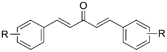 wherein R is independently selected from the group consisting of CF3, OH and OCH3.
 - The method of claim 1, wherein said R is CF3 and the mono-carbonyl analog of curcumin having a formula of A2:
- The method of claim 1, wherein said R is OH and the mono-carbonyl analog of curcumin having a formula of B3:
- The method of claim 1, wherein said R is OCH3 and the mono-carbonyl analog of curcumin having a formula of C1:

- A method of treating neurodegenerative diseases comprising administering a composition comprising a mono-carbonyl analog of curcumin having a formula of
 wherein R is selected from the group consisting of CF3, OH and OCH3 to a subject in need thereof.
 - The method according to claim 5, wherein the neurodegenerative diseases comprising Alzheimer′s disease, Parkinson′s disease,Huntington′s disease and Creutzfeldt-Jakob disease.
- The method according to claim 5, wherein the mono-carbonyl analog isthe mono-carbonyl analogs is administered at 1.62 mg/kg to 28.38mg/kg body weight of the subject in need thereof.
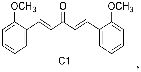
- The method according to claim 5, wherein the composition is administered to the subject in need thereofvia oral administration, intravenous inj ection or both.
- The method according to claim 5, wherein said R is CF3 and the mono-carbonyl analog of curcumin comprises a formula of A2:
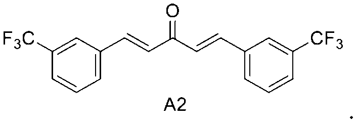
- The method according to claim 5, wherein said R is OH and the mono-carbonyl analog of curcumin comprises a formula of B3:

- The method according to claim 5, wherein said R is OCH3 and the mono-carbonyl analog of curcumin comprises a formula of C1:
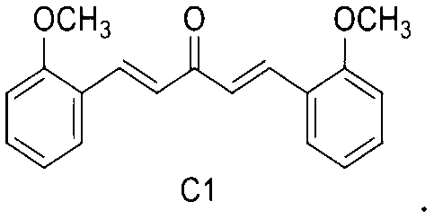
- A method of enhancing lysosome biogenesis in cells comprising providing a mono-carbonyl analog of curcumin having a formula of
 wherein R is OCH3.
 - The method of claim 12, wherein the mono-carbonyl analog of curcumin comprises a formula of C1:

- The method ofclaim 12, wherein the cells are neuronal cells.
- The method ofclaim 12, wherein the cells are non-neuronal cells.
Priority Applications (2)
| Application Number | Priority Date | Filing Date | Title |
|---|---|---|---|
| CN201580012376.7A CN106102728A (en) | 2014-03-06 | 2015-03-06 | For the enhanced non-MTOR dependent T FEB activator of autophagy and its purposes |
| EP15759224.7A EP3113767B1 (en) | 2014-03-06 | 2015-03-06 | Mtor-independent activator of tfeb for autophagy enhancement and uses thereof |
Applications Claiming Priority (4)
| Application Number | Priority Date | Filing Date | Title |
|---|---|---|---|
| US201461949233P | 2014-03-06 | 2014-03-06 | |
| US61/949,233 | 2014-03-06 | ||
| US14/609,438 | 2015-01-30 | ||
| US14/609,438 US9351946B2 (en) | 2014-03-06 | 2015-01-30 | MTOR-independent activator of TFEB for autophagy enhancement and uses thereof |
Publications (1)
| Publication Number | Publication Date |
|---|---|
| WO2015131848A1 true WO2015131848A1 (en) | 2015-09-11 |
Family
ID=54016289
Family Applications (1)
| Application Number | Title | Priority Date | Filing Date |
|---|---|---|---|
| PCT/CN2015/073764 WO2015131848A1 (en) | 2014-03-06 | 2015-03-06 | Mtor-independent activator of tfeb for autophagy enhancement and uses thereof |
Country Status (5)
| Country | Link |
|---|---|
| US (1) | US9351946B2 (en) |
| EP (1) | EP3113767B1 (en) |
| CN (2) | CN113398106A (en) |
| HK (1) | HK1225625A1 (en) |
| WO (1) | WO2015131848A1 (en) |
Families Citing this family (5)
| Publication number | Priority date | Publication date | Assignee | Title |
|---|---|---|---|---|
| US10987319B2 (en) * | 2018-06-28 | 2021-04-27 | Hong Kong Baptist University | TFEB activator C1 ameliorates app and tau pathology and rescues cognitive deficits in neurodegenerative diseases |
| CN113384541B (en) * | 2019-01-09 | 2023-05-23 | 纳菲(深圳)制药科技有限公司 | Nasal cavity nanometer autophagy inducer for preventing and treating early neurodegenerative diseases and preparation method thereof |
| CN111603483B (en) * | 2020-06-02 | 2023-03-14 | 中国人民解放军陆军军医大学 | Application of propionitrile compound in enhancing lysosome generation |
| KR20230048080A (en) * | 2020-08-03 | 2023-04-10 | 카스마 테라퓨틱스, 인코포레이티드 | TFEB activation and lysosomal biogenesis methods and compositions therefor |
| CN114848652A (en) * | 2022-05-31 | 2022-08-05 | 澳门大学 | Application of betulinal in preparing medicine for preventing and treating neurodegenerative diseases |
Citations (4)
| Publication number | Priority date | Publication date | Assignee | Title |
|---|---|---|---|---|
| US20070060644A1 (en) * | 2004-02-12 | 2007-03-15 | Vander Jagt David L | Therapeutic curcumin derivatives |
| CN102225919A (en) * | 2011-04-29 | 2011-10-26 | 中山大学 | Curcumin analogue and preparation method thereof as well as application of analogue in preparation of Alzheimer disease resisting medicament |
| WO2012021692A1 (en) * | 2010-08-11 | 2012-02-16 | Rutgers, The State University Of New Jersey | Curcumin analogs and methods of use thereof |
| CN102670573A (en) * | 2011-11-21 | 2012-09-19 | 西安交通大学 | Application of curcumin derivative in preparation of drug for treating prostate cancer disease |
Family Cites Families (2)
| Publication number | Priority date | Publication date | Assignee | Title |
|---|---|---|---|---|
| US9393198B2 (en) * | 2010-03-22 | 2016-07-19 | Signpath Pharma Inc. | Intravenous curcumin and derivatives for treatment of neurodegenerative and stress disorders |
| CN102675134B (en) * | 2012-05-29 | 2014-01-15 | 东南大学 | Nitrogen-containing substituent curcumin analogue and medical application thereof |
-
2015
- 2015-01-30 US US14/609,438 patent/US9351946B2/en active Active
- 2015-03-06 WO PCT/CN2015/073764 patent/WO2015131848A1/en active Application Filing
- 2015-03-06 CN CN202110429206.6A patent/CN113398106A/en active Pending
- 2015-03-06 EP EP15759224.7A patent/EP3113767B1/en active Active
- 2015-03-06 CN CN201580012376.7A patent/CN106102728A/en active Pending
-
2016
- 2016-12-07 HK HK16113937A patent/HK1225625A1/en unknown
Patent Citations (4)
| Publication number | Priority date | Publication date | Assignee | Title |
|---|---|---|---|---|
| US20070060644A1 (en) * | 2004-02-12 | 2007-03-15 | Vander Jagt David L | Therapeutic curcumin derivatives |
| WO2012021692A1 (en) * | 2010-08-11 | 2012-02-16 | Rutgers, The State University Of New Jersey | Curcumin analogs and methods of use thereof |
| CN102225919A (en) * | 2011-04-29 | 2011-10-26 | 中山大学 | Curcumin analogue and preparation method thereof as well as application of analogue in preparation of Alzheimer disease resisting medicament |
| CN102670573A (en) * | 2011-11-21 | 2012-09-19 | 西安交通大学 | Application of curcumin derivative in preparation of drug for treating prostate cancer disease |
Non-Patent Citations (1)
| Title |
|---|
| ORLANDO , D. A. ET AL.: "A chemical analog of curcumin as an improved inhibitor of amyloid abeta oligomerization", PLOS ONE, vol. 7, no. Issue 3, 19 March 2012 (2012-03-19), XP003031269 * |
Also Published As
| Publication number | Publication date |
|---|---|
| US20150250741A1 (en) | 2015-09-10 |
| US9351946B2 (en) | 2016-05-31 |
| CN113398106A (en) | 2021-09-17 |
| EP3113767A4 (en) | 2017-11-08 |
| HK1225625A1 (en) | 2017-09-15 |
| CN106102728A (en) | 2016-11-09 |
| EP3113767B1 (en) | 2020-12-09 |
| EP3113767A1 (en) | 2017-01-11 |
Similar Documents
| Publication | Publication Date | Title |
|---|---|---|
| Yuan et al. | Sorafenib attenuates liver fibrosis by triggering hepatic stellate cell ferroptosis via HIF‐1α/SLC7A11 pathway | |
| Li et al. | Eriodictyol ameliorates cognitive dysfunction in APP/PS1 mice by inhibiting ferroptosis via vitamin D receptor-mediated Nrf2 activation | |
| Yang et al. | Application and interpretation of current autophagy inhibitors and activators | |
| Fajardo et al. | The endoplasmic reticulum stress/unfolded protein response in gliomagenesis, tumor progression and as a therapeutic target in glioblastoma | |
| Gomez et al. | Inhibition of GSK3β by postconditioning is required to prevent opening of the mitochondrial permeability transition pore during reperfusion | |
| Li et al. | Metformin synergistically suppress tumor growth with doxorubicin and reverse drug resistance by inhibiting the expression and function of P-glycoprotein in MCF7/ADR cells and xenograft models | |
| Li et al. | Sulforaphane prevents doxorubicin-induced oxidative stress and cell death in rat H9c2 cells | |
| Yang et al. | Magnesium isoglycyrrhizinate ameliorates high fructose-induced liver fibrosis in rat by increasing miR-375-3p to suppress JAK2/STAT3 pathway and TGF-β1/Smad signaling | |
| WO2015131848A1 (en) | Mtor-independent activator of tfeb for autophagy enhancement and uses thereof | |
| Liu et al. | Tetramethylpyrazine inhibits production of nitric oxide and inducible nitric oxide synthase in lipopolysaccharide-induced N9 microglial cells through blockade of MAPK and PI3K/Akt signaling pathways, and suppression of intracellular reactive oxygen species | |
| Kim et al. | Hyperglycemia-induced GLP-1R downregulation causes RPE cell apoptosis | |
| US20130288980A1 (en) | Targeting senescent and cancer cells for selective killing by interference with foxo4 | |
| Lee et al. | Macrophage-stimulating protein attenuates gentamicin-induced inflammation and apoptosis in human renal proximal tubular epithelial cells | |
| Wang et al. | Catalpol suppresses osteosarcoma cell proliferation through blocking epithelial-mesenchymal transition (EMT) and inducing apoptosis | |
| Huang et al. | Panaxatriol saponins attenuated oxygen-glucose deprivation injury in PC12 cells via activation of PI3K/Akt and Nrf2 signaling pathway | |
| Xu et al. | D-galactose induces senescence of glioblastoma cells through YAP-CDK6 pathway | |
| Du et al. | Nuciferine protects against high-fat diet-induced hepatic steatosis and insulin resistance via activating TFEB-mediated autophagy–lysosomal pathway | |
| WO2019035866A1 (en) | Compositions and methods for treating tuberous sclerosis complex | |
| Wang et al. | Local delivery of β-elemene improves locomotor functional recovery by alleviating endoplasmic reticulum stress and reducing neuronal apoptosis in rats with spinal cord injury | |
| JP2019532049A (en) | Modulation method of PCSK9 and LDLR through DRP1 inhibition | |
| Jiang et al. | SGK1 attenuates oxidative stress-induced renal tubular epithelial cell injury by regulating mitochondrial function | |
| Xin et al. | Pentamethylquercetin ameliorates fibrosis in diabetic Goto-Kakizaki rat kidneys and mesangial cells with suppression of TGF-β/Smads signaling | |
| US9540299B2 (en) | MTOR-independent activator of TFEB for autophagy enhancement and uses thereof | |
| Kaowinn et al. | N-Benzyl-N-methyl-dodecan-1-amine, a novel compound from garlic, exerts anti-cancer effects on human A549 lung cancer cells overexpressing cancer upregulated gene (CUG) 2 | |
| Zheng et al. | Protosappanin‐A and oleanolic acid protect injured podocytes from apoptosis through inhibition of AKT‐mTOR signaling |
Legal Events
| Date | Code | Title | Description |
|---|---|---|---|
| 121 | Ep: the epo has been informed by wipo that ep was designated in this application |
Ref document number: 15759224 Country of ref document: EP Kind code of ref document: A1 |
|
| NENP | Non-entry into the national phase |
Ref country code: DE |
|
| REEP | Request for entry into the european phase |
Ref document number: 2015759224 Country of ref document: EP |
|
| WWE | Wipo information: entry into national phase |
Ref document number: 2015759224 Country of ref document: EP |