WO2015124120A1 - Vacuolin-1 as an inhibitor of autophagyand endosomal trafficking and the use thereof for inhibiting tumor progression - Google Patents
Vacuolin-1 as an inhibitor of autophagyand endosomal trafficking and the use thereof for inhibiting tumor progression Download PDFInfo
- Publication number
- WO2015124120A1 WO2015124120A1 PCT/CN2015/073255 CN2015073255W WO2015124120A1 WO 2015124120 A1 WO2015124120 A1 WO 2015124120A1 CN 2015073255 W CN2015073255 W CN 2015073255W WO 2015124120 A1 WO2015124120 A1 WO 2015124120A1
- Authority
- WO
- WIPO (PCT)
- Prior art keywords
- vacuolin
- autophagy
- analogues
- cells
- lysosomal
- Prior art date
Links
Images


Classifications
-
- A—HUMAN NECESSITIES
- A61—MEDICAL OR VETERINARY SCIENCE; HYGIENE
- A61K—PREPARATIONS FOR MEDICAL, DENTAL OR TOILETRY PURPOSES
- A61K31/00—Medicinal preparations containing organic active ingredients
- A61K31/33—Heterocyclic compounds
- A61K31/395—Heterocyclic compounds having nitrogen as a ring hetero atom, e.g. guanethidine or rifamycins
- A61K31/535—Heterocyclic compounds having nitrogen as a ring hetero atom, e.g. guanethidine or rifamycins having six-membered rings with at least one nitrogen and one oxygen as the ring hetero atoms, e.g. 1,2-oxazines
- A61K31/5375—1,4-Oxazines, e.g. morpholine
- A61K31/5377—1,4-Oxazines, e.g. morpholine not condensed and containing further heterocyclic rings, e.g. timolol
-
- A—HUMAN NECESSITIES
- A61—MEDICAL OR VETERINARY SCIENCE; HYGIENE
- A61K—PREPARATIONS FOR MEDICAL, DENTAL OR TOILETRY PURPOSES
- A61K31/00—Medicinal preparations containing organic active ingredients
- A61K31/33—Heterocyclic compounds
- A61K31/335—Heterocyclic compounds having oxygen as the only ring hetero atom, e.g. fungichromin
- A61K31/337—Heterocyclic compounds having oxygen as the only ring hetero atom, e.g. fungichromin having four-membered rings, e.g. taxol
-
- A—HUMAN NECESSITIES
- A61—MEDICAL OR VETERINARY SCIENCE; HYGIENE
- A61K—PREPARATIONS FOR MEDICAL, DENTAL OR TOILETRY PURPOSES
- A61K31/00—Medicinal preparations containing organic active ingredients
- A61K31/33—Heterocyclic compounds
- A61K31/395—Heterocyclic compounds having nitrogen as a ring hetero atom, e.g. guanethidine or rifamycins
- A61K31/435—Heterocyclic compounds having nitrogen as a ring hetero atom, e.g. guanethidine or rifamycins having six-membered rings with one nitrogen as the only ring hetero atom
- A61K31/4353—Heterocyclic compounds having nitrogen as a ring hetero atom, e.g. guanethidine or rifamycins having six-membered rings with one nitrogen as the only ring hetero atom ortho- or peri-condensed with heterocyclic ring systems
- A61K31/436—Heterocyclic compounds having nitrogen as a ring hetero atom, e.g. guanethidine or rifamycins having six-membered rings with one nitrogen as the only ring hetero atom ortho- or peri-condensed with heterocyclic ring systems the heterocyclic ring system containing a six-membered ring having oxygen as a ring hetero atom, e.g. rapamycin
-
- A—HUMAN NECESSITIES
- A61—MEDICAL OR VETERINARY SCIENCE; HYGIENE
- A61K—PREPARATIONS FOR MEDICAL, DENTAL OR TOILETRY PURPOSES
- A61K31/00—Medicinal preparations containing organic active ingredients
- A61K31/33—Heterocyclic compounds
- A61K31/395—Heterocyclic compounds having nitrogen as a ring hetero atom, e.g. guanethidine or rifamycins
- A61K31/495—Heterocyclic compounds having nitrogen as a ring hetero atom, e.g. guanethidine or rifamycins having six-membered rings with two or more nitrogen atoms as the only ring heteroatoms, e.g. piperazine or tetrazines
- A61K31/505—Pyrimidines; Hydrogenated pyrimidines, e.g. trimethoprim
- A61K31/513—Pyrimidines; Hydrogenated pyrimidines, e.g. trimethoprim having oxo groups directly attached to the heterocyclic ring, e.g. cytosine
-
- A—HUMAN NECESSITIES
- A61—MEDICAL OR VETERINARY SCIENCE; HYGIENE
- A61P—SPECIFIC THERAPEUTIC ACTIVITY OF CHEMICAL COMPOUNDS OR MEDICINAL PREPARATIONS
- A61P33/00—Antiparasitic agents
- A61P33/02—Antiprotozoals, e.g. for leishmaniasis, trichomoniasis, toxoplasmosis
- A61P33/06—Antimalarials
-
- A—HUMAN NECESSITIES
- A61—MEDICAL OR VETERINARY SCIENCE; HYGIENE
- A61P—SPECIFIC THERAPEUTIC ACTIVITY OF CHEMICAL COMPOUNDS OR MEDICINAL PREPARATIONS
- A61P35/00—Antineoplastic agents
-
- Y—GENERAL TAGGING OF NEW TECHNOLOGICAL DEVELOPMENTS; GENERAL TAGGING OF CROSS-SECTIONAL TECHNOLOGIES SPANNING OVER SEVERAL SECTIONS OF THE IPC; TECHNICAL SUBJECTS COVERED BY FORMER USPC CROSS-REFERENCE ART COLLECTIONS [XRACs] AND DIGESTS
- Y02—TECHNOLOGIES OR APPLICATIONS FOR MITIGATION OR ADAPTATION AGAINST CLIMATE CHANGE
- Y02A—TECHNOLOGIES FOR ADAPTATION TO CLIMATE CHANGE
- Y02A50/00—TECHNOLOGIES FOR ADAPTATION TO CLIMATE CHANGE in human health protection, e.g. against extreme weather
- Y02A50/30—Against vector-borne diseases, e.g. mosquito-borne, fly-borne, tick-borne or waterborne diseases whose impact is exacerbated by climate change
Definitions
- the present invention relates generally to the field of compositions and methods for treatment ofautophagy related human diseases. More particularly, the present invention provides methods ofusing small chemicals to manipulate autophagy and to treat autophagy related human diseases, e.g. cancer.
- Autophagy is an evolutionarily conserved catabolic degradation cellular process in which misfolded proteins or damaged organelles are first sequestered by a double-membrane vesicle, called autophagosomes. Autophagosomes are then fused with lysosomes to digest and recycle the contents to maintain cellular homeostasis. Autophagy can also be markedly induced by a wide variety of stresses, e.g. nutrient starvation, infection, and aging, for cell survival. Dysfunctional autophagy has been associated with wide ranges of human diseases, such as, e.g., cancer, neurodegenerative diseases, heart disease, diabetes, and bacterial infection (Wirawan et al., 2012; Yang and Klionsky, 2010) .
- human diseases such as, e.g., cancer, neurodegenerative diseases, heart disease, diabetes, and bacterial infection (Wirawan et al., 2012; Yang and Klionsky, 2010) .
- Autophagyis a double-edged sword for many cellular processes, depending upon the genetic background and microenvironment.
- autophagy can act as a tumor suppressor by preventing oncogenic protein substrates, toxic misfolded proteins, damaged organelles, and reactive oxygen species (ROS) from accumulating to cause genome instability and cancer initiation (Mathew et al., 2009; Yue et al., 2003) .
- ROS reactive oxygen species
- autophagy inducers include mTOR kinase inhibitors, e.g., rapamycin and torin 1 (Hanson et al., 2013) , and chemicals inhibiting inositol monophosphatase, e.g., lithium and carbamazepine (Hidvegi et al., 2010) .
- rapamycin is an immunosuppressant and has recently been used as an anticancer agent (Ravikumar et al., 2004) .
- Lithium has also been used to treat Huntington’s disease and other related neurodegenerative disorders (Sarkar et al., 2005) .
- Commonly used autophagy inhibitors include chloroquine (CQ) , 3-methyladenine, wortmannin, and bafilomycin (BAF) (Baek et al., 2012; Rote and Rechsteiner, 1983; Seglen and Gordon, 1982; Wu et al., 2013) .
- CQ chloroquine
- BAF bafilomycin
- CQ offers great promise for cancer therapy
- CQ induces ocular toxicities and damages the renal system, and it is uncertain whether the tolerated doses of HCQ or CQ can be reached in human tumors to effectively inhibit autophagy (Kimura et al., 2013) .
- most of the available autophagy inhibitors like HCQ or CQ, also lack either specificity, potency, or antitumor activity (Janku et al., 2011) .
- potent and specific inhibitors of autophagy are needed in order to provide a novel and powerful approach for future cancertherapy.
- vacuolin-1 By screening a Chembridge library (ChemBridge Corporation, San Diego) that contains around 10, 000 drug-like or lead-like small chemicals, vacuolin-1 was originally found to induce homotypic fusion of endosomes or lysosomes, thereby forming large vacuoles. Yet, it does not alter other cell structures and membrane trafficking functions (Cerny et al., 2004; Huynh and Andrews, 2005; Shaik et al., 2009) . It remains controversial whether vacuolin-1 blocks the Ca 2+ -dependent exocytosis oflysosomes.
- the present invention provides methods of using vacuolin-1 and its analogues to manipulate autophagy and to treat autophagy related human diseases, e.g. cancer.
- the present invention also provides application ofvacuolin-1 and its analogues for inhibiting autophagy and endosomal trafficking.
- methods for treatment of cancer such as, but not limited to, lung carcinomaand nasopharyngeal carcinoma are provided.
- the methods comprise administering a therapeutically effective amount of vacuolin-1, and/or one or more analogues, alone or in combination with one or more chemotherapy drugs to a subject who has cancer.
- the methods are used to treat cancers that are susceptible to treatment with an autophagy inhibiting agent.
- the methods result in inhibition of autophagy and/or endosomal traffic. In some embodiments, the methods result in activation of Rab5, which leads to maturation of endosomes and lysosomes and contributes to lysosomal pH increase.
- the chemotherapy drugs can include taxol, 5-Fu, temirolimus, and combinations thereof.
- vacuolin-1 analogues are identified by virtual screening.
- Figure 1 shows structures ofvacuolin-1 and structural analogues.
- Figure 2 shows immunofluorescence staining analysis of RFP-GFP-LC3 expressing HeLa cells following treatment with bafilomycin, CQ, and vacuolin-1.
- FIG. 3 shows vacuolin-1 inhibits the fusion between autophagosomes and lysosomes in HeLa cells.
- A Percentage of yellow LC3II puncta in RFP-GFP-LC3 expressing HeLa cells following induction by vacuolin-1.
- Figure 4 shows (A) western blot analyses ofLC3 and p62 in HeLa cells following treatment with vacuolin-1 or CQ at the indicated dose for 6 hours; (B) an MTT assay for cell viability in HeLa cells treated with vacuolin-1 or CQ for the indicated doses for 48 hours; (C) western blot analyses of LC3 and p62 in vacuolin-1 (1 ⁇ M) treated HeLa cells; (D) microplate reader measurement of Lysosensor DND-189 stained HeLa cells following vacuolin-1 (1 ⁇ M) induction; (E) microplate reader measurement of quantitative ratiometric LysoSensor Yellow/Blue DND-160 stained HeLa cells following vacuolin-1 (1 ⁇ M) induction; and (F) Fura-2 loaded HeLa cells following vacuolin-1 (1 ⁇ M) pretreatment for 5 hours followed by GPN (200 ⁇ M) induced lysosomal Ca 2+ release.
- Figure 5 shows western blot analyses against LC3 and p62 inPC3 prostate cancer cells treatedwithvacuolin-1 or CQ at indicated dose for 6 hours.
- Figure 9 shows expression of Rab5A-DN does not affect starvation induced LC3 puncta in HeLa cells.
- Figure 10 shows Rab5A knockdown or expression of Rab5A-DN does not affect starvation induced LC3-II in HeLa cells.
- Figure 11 shows an electron micrograph of enlarged lysosomes in HeLa cells followingvacuolin-1 treatment (B) versus control (A) .
- Figure 12 showsvacuolin-1 analogues inhibit the fusion between autophagosomes and lysosomes in HeLa cells.
- FIG 13 shows vacuolin-1 potently enhances the anti-tumor effects of chemotherapy drugs in human cancer cell lines.
- A Vacuolin-1 augments the anti-tumor effects of chemotherapy drugs
- B Vacuolin-1 in combination with chemotherapy drugs greatly decreases the colony formation efficiency cancer cells.
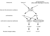
- Figure 14 shows a virtual screening strategy for vacuolin-1 analogue identification using ChemDiv.
- Figure 15 shows vacuolin-1 analogues.
- Figure 16 shows vacuolin-1 markedly inhibited colony formation and migration of human nasopharyngeal carcinomas._
- A Vacuolin-1 treatment alone displayed little cell toxicity in CNE-1 or HONE-1 cells.
- B Vacuolin-1 dramatically reduced the size and number of colonies generated from single CNE-1 or HONE-1 cell.
- C Vacuolin-1 efficiently suppressed the migration of both CNE-1 and HONE-1 cells as revealed by the observation that vacuolin-1 treatment induced a marked decrease in motility oftumor cell.
- D and
- FIG. 17 shows vacuolin-1 markedly inhibited colony formation and migration of human lung cancer stem cells (LCSCs) in vitro.
- A Vacuolin-1 on autophagy inhibition in human lung cancer stem cells (hLCSCs) were 10 times less potent than those in other tumor cell lines.
- B Vacuolin-1 did not affect the cell proliferation ofhLCSCs.
- C-F Vacuolin-1 (>10 ⁇ M) almost completely inhibited the colony formation (C) , migration (D) , invasion (E) and tumor sphere formation (F) ofhLCSCs.
- Figure 18 shows vacuolin-1 markedly suppressed tumor growth in nude mice.
- A Intratumoral injection ofvacuolin-1 (5 mg/kg) twice a week markedly inhibited tumor growth of LCSC xenograft in nude mice but had little effects on the weight gain.
- B vacuolin-1 treatment markedly inhibited autophagy in tumors.
- C Intraperitoneal injection ofvacuolin-1 (250 mg/kg) in young adult mice exhibited little acute toxicity as the drug injected mice showed normal activity and weight gain as compared to control after two weeks.
- the present invention describes vacuolin-1 and its analogues as potent and reversible inhibitors ofautophagy and endosomal traffic and methods ofusing vacuolin-1 and its analogues alone or in combination with chemotherapeutic drugs, e.g. 5-FU, temsirolimus, and taxol, or alone, to manipulate autophagy and to treat cancer, e.g. prostate, breast, and nasopharyngeal carcinoma, or malaria, respectively.
- chemotherapeutic drugs e.g. 5-FU, temsirolimus, and taxol
- methods for treatment of cancer such as but not limited to, colon carcinoma, prostate cancer, breast cancer, osteosarcoma and/or nasopharyngeal carcinoma.
- the methods comprise administering a therapeutically effective amount ofvacuolin-1, and/or one or more analogues, either alone or in combination with one or more chemotherapy drugs to a subject who has cancer.
- methods for treatment of malaria comprise administering a therapeutically effective amount ofvacuolin-1, and/or one or more analogues, to a subject who is infected with malaria.
- the methods result in inhibition of autophagy and/or endosomal traffic. In some embodiments, the methods result in activation of Rab5, which leads to maturation of endosomes and lysosomes and contributes to lysosomal pH increase.
- the chemotherapy drugs can include, but are not limited to, taxol, 5-Fu, temirolimus, and combinations thereof.
- additional vacuolin-1 analogues are identified by virtual screening.
- vacuolin-1 potently and reversibly inhibit autophagy by blocking the fusion between autophagosomes and lysosomes in mammalian cells, thereby leading to the accumulation of autophagosomes ( Figures 2-5, and 12) .
- Vacuolins are less toxic than chloroquine (CQ) but are at least 10 folds more potent in inhibiting autophagy as compared to CQ ( Figures 3-5) .
- Vacuolin-1 and its analogues present a novel class of drug that can potently and reversibly modulate autophagy.
- the present invention describes vacuolins as potent inhibitors of general endosomal traffic (Figure 7) and a novel class ofRab5 GTPase activators (Figure 8) .
- vacuolins in combination with other chemotherapy drugs, e.g. 5-FU, temsirolimus, and taxol
- other chemotherapy drugs e.g. 5-FU, temsirolimus, and taxol
- vacuolins, and/or analogs thereof can be utilized alone in the treatment of cancers.
- the present invention also provides methods to treat malaria with vacuolins (Figure 15).
- vacuolins are used in combination with chemotherapeutic drugs, e.g. 5-FU, temsirolimus, and taxol, to synergistically kill a panel oftumor cell lines in vitro and suppress tumor growth in xenograft mouse models ( Figures 13 and 14) . It is expected that vacuolin-1 and its analogues provide a novel therapeutic strategy for fighting cancers. It is also expected that vacuolins sensitize tumor cells carrying wild type p53, not mutant p53, to chemotherapeutic drugs.
- chemotherapeutic drugs e.g. 5-FU, temsirolimus, and taxol
- Vacuolins are cell-permeable and water soluble triazine based compounds. Compared to other popular autophagy modulators, such as CQ which has been applied in a dozen anti-cancer clinical trials, vacuolinsare at least 10 fold more potent in inhibiting autophagy while exhibiting little toxicity in a wide variety of cancer cell lines ( Figures 4 and 6) . Besides inhibiting autophagy, vacuolins potently and reversibly inhibit endosomal traffic as well (Figure 7) .
- Rab5 is a master regulator for the biogenesis of the endolysosomal system, and no drugs, other than vacuolins, are found to modulate its activity thus far. Therefore, vacuolins are the first class of Rab5 agonists to be identified and can be widely used in basic research related to both autophagy and intracellular traffic.
- Vacuolins also modestly inhibit V-ATPase activity (30%at 1 ⁇ M) , which may be due to the fact that V-ATPase is sensitive to hydrophobic compounds in general ( Figure 8) .
- vacuolins are not specific V-ATPase inhibitors, but nonetheless at least partially lead to the increase of lysosomal pH, which may also contribute to autophagy inhibition.
- endosomal traffic is essential for the function and biogenesis of lysosome because lysosomes depend on the influx of new components.
- vacuolins constitutively activate Rab-5, and active Rab5 stops endosome maturation, which subsequently compromises the biogenesis and function oflysosomes. This should also partially contribute to the increase oflysosomal pH.
- the present invention also provides toxicity studies ofthe vacuolins in combination with, or without, chemotherapy drugs in both tumor-free and tumor-bearing mice. Since vacuolins are much less toxic than CQ and its effects on cells are reversible, the toxicity ofvacuolins on mice is much less than CQ.
- the present invention further provides pharmacokinetic studies on vacuolins in normal mice or rats.
- the blood concentration of vacuolins after the intragastric (oral) , intravenous, intramuscular, or subcutaneous administration is determined.
- the metabolites of vacuolins after incubating vacuolins with liver microsomes are determined.
- CQ which exhibits high toxicity alone and its effective concentration against autophagy is hard to reach in an animal model
- this invention showsvacuolin-1 and its analogues are better lead compounds for future clinical application against human cancer and malaria.
- Antibodies and reagents-The antibodies used were as follows: LC3 (Novus; 1: 1000 for the Western blotting analysis (WB) ) , p62 (Novus; 1: 500 for the immunofluorescence analysis (IF) and 1: 1000 WB) , Cathepsin-L (BD Bioscience; 1: 250 WB) , EGFR (Santa Cruz; 1: 250 WB) , Rab5 (Cell Signaling; 1: 1000 WB) , GAPDH antibody (Sigma; 1: 5000 WB) . Vacuolin-1 was purchased from Santa Cruz.
- Bafilomycin A1 Chloroquine (CQ)
- CQ Chloroquine
- GPN Glycyl-L-phenyl-alanine- ⁇ -naphthylamide
- Fura-2 AM,lysosensor Green DND-189, and LysoSensorYellow/Blue DND-160 were purchased from Invitrogen.
- Cell culture-HeLa, MCF-7, A549, and HepG2 cells were maintained in DMEM (Invitrogen) plus 10%fetal bovine serum (Invitrogen) and 100 units/ml ofpenicillin/streptomycin (Invitrogen) at 5%CO 2 and 37°C.
- PC3 cells were maintained in RPMI (Invitrogen) plus 10%fetal bovine serum (Invitrogen) and 100 units/ml of penicillin/streptomycin (Invitrogen) at 5%CO 2 and 37°C.
- shRNA and lentivirusproduction and infection-Two optimal 21-mers were selected inthe human Rab5A gene.
- One 21-mer was selected in the GFP gene as a control.
- These sequences were then cloned into the pLKO. 1 vector for expressing shRNA. The production and infection were performed as described previously (Lu et al., 2013) .
- Intracellular Ca 2+ measurement-Cells were cultured in 24-well plates at the density of 7x10 4 cells/well in regular medium overnight and were labeled with 4 ⁇ M Fura-2 AM (Invitrogen) in regular HBSS at room temperature for 30 min. The cells were then washed with calcium-free HBSS containing 2mM EGTA three times and incubated in the presence or absence of vacuolin-1 at room temperature for another 10 min. Cells were put on the stage of an Olympus inverted epifluorescence microscope and visualized using a 20x objective. Fluorescence images were obtained by alternate excitation at 340 nm and 380 nm with emission set at 510 nm. Images were collected by a CCD camera every 3 or 6 seconds and analyzed by CellR imaging software.
- TEM transmission electron microscopy
- MTT cell proliferation assay-Cells were treated in four replicates and seeded into 96-well plate. Following drug treatment, MTT solution of 20 ⁇ l for every 100 ⁇ l medium was added to wells and incubated for 2 hours, followed by the addition of 150 ⁇ L of the DMSO solution to each well. The final reaction product, apurple formazan solution, was detected by a microplate reader (Techan infinite M200) for absorbance at a wavelength of 570 nm and a reference wavelength of 630 nm.
- lysosomal pH Quantification of lysosomal pH was performed using a ratiometriclysosomal pH dye LysoSensor Yellow/Blue DND-160.
- the pH calibration curve was generated as described previously (Bankers-Fulbright et al., 2004) . Briefly, cells were trypsinized and labeled with 2 ⁇ M Lysosensor Yellow/Blue DND-160 for 30 min at 37°C in regular medium, and excessive dye was washed out using PBS.
- the labeled cells were treated for 10 min with 10 ⁇ M monensin and 10 ⁇ M nigericin in 25mM 2- (N-morpholino) ethanesulfonic acid (MES) calibration buffer, pH 3.5–6.0, containing 5 mM NaCl, 115 mM KCl and 1.2 mM MgSO4. Quantitative comparisons were performed in a 96-well plate, and the fluorescence was measured with a microplate reader at 37°C. Light emitted at 440 and 535nm in response to excitation at 340 and 380nm were measured, respectively. The ratio of light emitted with 340 and 380nm excitation was plotted against the pH values in MES buffer, and the pH calibration curve for the fluorescence probe was generated from the plot using Microsoft Excel.
- MES 2- (N-morpholino) ethanesulfonic acid
- V-ATPase assay-Fifth instar larvae ofM. sexta (Lepidoptera, Sphingidae) , weighing 6-8 g,were reared under long day conditions (16 h of light) at 27°C using the gypsy moth diet (MP Biomedicals, Germany) .
- the purification of the V 1 V O holoenzyme was performed as previously described (Huss et al., 2002) .
- Activity assays were performed in triplicate in a final volume of 160 ⁇ L and at a pH of 8.1.
- Assays contained 3 ⁇ g of purified V 1 V 0 holoenzyme, 50 mM Tris-Mops, 3 mM 2-mercaptoethanol, 1 mM MgCl 2 , 20 mM KCl, 0.003%C 12 E 10 , 20 mM NaCl, and 3 mM Tris-HCl. After 10 min of preincubation at 30°C, with or without inhibitors, 1 mM Tris-ATP was added and after an incubation for 2 min at 30°C the reaction was stopped by freezing the samples in liquid nitrogen. Inorganic phosphate was determined as previously described (Wieczorek et al., 1990) .
- Glutathione S-Transferase Pull-Down Assay-Cells were collected and lysed in an ice-cold EBC lysis buffer described previously. Lysates were clarified by centrifugation at 13,000g for 10 min at 4°C, and equal amount of protein (500 ⁇ g) from each supernatant was incubated with 30 ⁇ l of GST-R5BD bound to the 30 ⁇ l glutathione–Sepharose beads for 1h at 4°C on a rotating mixer. The beads was subsequently washed and resuspended in the standard SDS-sample buffer, boiled and subjected to SDS-PAGE (15%gel) , followed by immunoblot analysis with the anti-Rab5 mAb.
- Example 1 The small chemical vacuolin-1functions as an autophagy inhibitor by inhibiting the autophagosomal-lysosomalfusion in HeLa cells.
- vacuolin-1 similar to BAF or CQ, blocks the fusion between autophagosomes and autolysosomes, thereby leading to accumulation ofLC3-II positive autophagosomes to achieve its effect ofautophagy inhibition.
- Example 2 Vacuolin-1 potently and reversibly inhibits autophagy but shows little cell toxicity.
- Vacuolin-1 is a cell-permeable and water soluble triazine based compound. It has been previously reported that vacuolin-1 induced rapid homotypic fusion ofendosomes and lysosomes to form large and swollen structures, yet it did not disturb cell cytoskeletal network (Cerny et al., 2004; Huynh and Andrews, 2005; Shaik et al., 2009) . Amazingly, vacuolin-1 was at least ten times more potent than CQ in suppressing autophagy ( Figures 4A and 5) , yet it exhibited much less cell toxicity than CQ in a wide variety of cell types ( Figures 4B and 6) .
- Example 3 Vacuolin-1 alkalinized lysosomal pH and decreased lysosomal Ca 2+ content.
- vacuolin-1 induced autophagy arrest
- lysosomal pH is essential for the fusion (Lu et al., 2013) and lysosomes were enlarged by vacuolin-1 treatment (Cerny et al., 2004; Huynh and Andrews, 2005; Shaik et al., 2009)
- vacuolin-1 affects lysosomal pH in these enlarged lysosomes.
- LysoSensor Green DND-189 permeates cell membranes and accumulates in acidic intracellular organelles, and its fluorescence increases or decreases in acidic or alkaline environments, respectively.
- vacuolin-1 treatment raised lysosomal pH in HeLa cells. Lysosomal pH was further quantified by a quantitative ratiometricLysoSensor Yellow/Blue DND-160 (DePedro and Urayama, 2009) stained cells and found that lysosomal pH in vacuolin-1 treated HeLa cells was increased from pH 4.7 in control cells to pH 5.2 ( Figure 4E) .
- vacuolin-1 alkalinizes lysosomalpH.
- lysosome is also a major intracellular Ca 2+ pool, and Ca 2+ and protons are tightly coupled in lysosomes (Morgan et al., 2011) , it was also assessed whether vacuolin-1 treatment affects the lysosomal Ca 2+ content.
- Treatment of cells with glycyl-l-phenylalanine 2-naphthylamide (GPN) selectively disrupts the lysosomal membrane (Jadot et al., 2001) , thereby releasing the lysosomal Ca 2+ (Srinivas et al., 2002) .
- GPN glycyl-l-phenylalanine 2-naphthylamide
- vacuolin-1 affects the general lysosomal functions to inhibit autophagy maturation.
- the processing of cathepsin L from the precursor form to its mature form has been commonly used as a marker for lysosomal activity, and it was found that the processing of cathepsin L into its mature form was not affected in vacuolin-1 treated HeLa cells, but vacuolin-1 did markedly increase the levels of immature form of cathepsin L ( Figure 7A) .
- an epidermal growth factor receptor (EGFR) degradation assay was performed to examine whether vacuolin-1 affects the general endosomal-lysosomal pathway.
- DQ-BSA-green is a BSA labeled with a self-quenching fluorescent dye. After DQ-BSA-green is delivered to lysosomes via endocytosis, it is hydrolyzed into single dye-labeled peptides by lysosomal proteases, thereby relieving self-quenching and the fluorescence can subsequently be monitored by flow cytometry. As shown in Figure7D, vacuolin-1 markedly inhibited the degradation of BSA by lysosomes, yet DQ-BSA-Green was present in endosomes but failed to be delivered to lysosomes (data not shown) . Collectively, these results support that vacuolin-1 inhibits the general endosomal-lysosomal degradation, which might be due to the alkalized lysosomal pH arresting the fusion of endosomes with lysosomes.
- Example 5 Rab5 was required for vacuolin-1 induced autophagy arrest and endosomal fusion.
- vacuolin-1 increased lysosomal pH, vacuolin-1 had little effects on V-ATPase activity in vitro (Figure 8A) .
- Vacuolin-1 also induced the homotypic fusion of endosomes or lysosomes, and Rab5, a small GTPase, is essential for endosome fusion (Zeigerer et al., 2012) . Therefore, it was examined whether vacuolin-1 activates Rab5 to indirectly change lysosomal pH. Indeed, vacuolin-1 markedly activated Rab5, as shown by a GST-tagged Rabaptin5 pull-down assay, which specifically interacts with Rab5-GTP, not Rab5-GDP ( Figures 8B) (Liu et al., 2007) .
- Rab5A knockdown markedly decreased vacuolin-1 induced accumulation of both LC3-II and p62 ( Figure 8C) .
- overexpression of a dominant-negative form of Rab5A (Rab5A-DN) blocked vacuolin-1 induced autophagy arrest and homotypic fusion
- overexpression of a constitutive form of Rab5A (Rab5A-CA) (Bohdanowicz et al., 2012) augmented the ability of vacuolin-1 induced autophagy arrest and homotypic fusion ( Figures 8D) .
- Rab5A-CA overexpression alone also blocked the autophagosomal-lysosomal fusion, thereby inducing the accumulation of LC3-II and p62 ( Figures8E and 8F) .
- Rab5A-DN expression or Rab5A knockdown did not affect starvation-induced autophagy ( Figures9 and 10) .
- Example 6 Vacuolin-1 markedly inhibited colony formation and migration of human nasopharyngeal carcinomas.
- vacuolin-1 Although CQ has already been applied to treat a wide spectrum of human cancers, CQ induces ocular or renal toxicity and easily triggers drug resistance (Kimura et al., 2013) . We, thus, assessed whether vacuolin-1 exhibits any anti-tumor ability and whether it can, like CQ, potentiate the sensitivity of tumor cells to chemotherapeutic drugs. We first investigated the anti-tumor effects of vacuolin-1 in two nasopharyngeal carcinoma cell lines, CNE-1 and HONE-1. As shown in Figure 16A, vacuolin-1 treatment alone displayed little cell toxicity in CNE-1 or HONE-1 cells.
- vacuolin-1 also showed almost no synergistic effects on inhibiting tumor cell proliferation in combination with several clinical anti-tumor drugs, such as taxol, 5-FU, and temsirolimus (Fig. 16A and data not shown) .
- colony formation assay was performed to assess whether vacuolin-1 affects the ability of a single tumor cell to grow into a colony.
- vacuolin-1 dramatically reduced the size and number of colonies generated from single CNE-1 or HONE-1 cell.
- wound healing assay was performed to assess whether vacuolin-1 affects the migration of tumor cells.
- vacuolin-1 efficiently suppressed the migration of both CNE-1 and HONE-1 cells as revealed by the observation that vacuolin-1 treatment induced a marked decrease in motility of tumor cell (Fig. 16C and data not shown) .
- vacuolin-1 markedly inhibited the migration and invasiveness of CNE-1 and HONE-1 cells (Fig. 16D, Fig. 16E, and data not shown) .
- vacuoin-1 is an efficient anti-tumor agent in vitro.
- Example 7 Vacuolin-1 markedly inhibited colony formation and migration of human lung cancer stem cells (LCSCs) in vitro .
- LCSCs human lung cancer stem cells
- CSCs Cancer stem cells
- chemo-and radiation therapy thus they can survive these therapy to regenerate new tumors.
- CSCs have been shown to possess stem cell characteristics, such as self-renewal, stress and drug resistance, and enhanced migration, all of which have been implicated in disease recurrence and distant metastasis (Gupta et al., 2009) . Therefore, it is ofgreat interest to assess the anti-tumor effects of vacuolin-1 on CSCs.
- vacuolin-1 on autophagy inhibition in human lung cancer stem cells were 10 times less potent than those in other tumor cell lines (Fig. 17A) , and vacuolin-1 did not affect the cell proliferation ofhLCSCs (Fig. 17B) .
- vacuolin-1 >10 ⁇ Malmost completely inhibited the colony formation, migration, invasion and tumor sphere formation of hLCSCs (Fig. 17C-17F) .
- Example 8 Vacuolin -1 markedly suppressed tumor growth in nude mice.
- vacuolin-1 potently inhibited the migration and colony formation ability of human nasopharyngeal carcinomas cells and human lung cancer stem cells in vitro
- vacuolin-1 was assessed the ability of vacuolin-1 to inhibit tumor progression in xenograft mouse model.
- LCSCs lung cancer stem cells
- Matrigel Matrigel (BD Biosciences) at a ratio of 1: 1, and 200 of cells was subcutaneously injected into the back of nude mice. The tumor volume was measured every five days after injection and calculated from the formula: length ⁇ width ⁇ depth ⁇ /6.
- FIG. 18A Mice were removed from the study when their tumor volumes exceed 2000 mm 3 (Fig. 18A) .
- intra tumoral injection of vacuolin-1 (5 mg/kg) twice a week markedly inhibited tumor growth of LCSC xenograft in nude mice but had little effects on the weight gain.
- vacuolin-1 treatment markedly inhibited autophagy in tumors (Fig. 18B) .
- Intraperitoneal injection of vacuolin-1 (250 mg/kg) in young adult mice exhibited little acute toxicity as the drug injected mice showed normal activity and weight gain as compared to control after two weeks (Fig. 18C) .
- vacuolin-1 is a promising anti-tumor agent in vivo with tolerable toxicity.
- Platelet-activating factor stimulates cytoplasmic alkalinization and granule acidification in human eosinophils. Journal of cell science 117, 5749-5757.
- Torins are potent antimalarials that block replenishment of Plasmodium liver stage parasitophorous vacuole membrane proteins. Proceedings of the National Academy of Sciences of the United States of America 110, E2838-2847.
- TPC2 Two pore channel 2 inhibits autophagosomal-lysosomal fusion by alkalinizing lysosomal pH.
- LINGO-1 antagonist promotes spinal cord remyelination and axonal integrity in MOG-induced experimental autoimmune encephalomyelitis. Nat Med 13, 1228-1233.
- Bafilomycin A1 prevents maturation of autophagic vacuoles by inhibiting fusion between autophagosomes and lysosomes in rat hepatoma cell line, H-4-II-E cells. Cell structure and function 23, 33-42.
- Beclin 1 an autophagy gene essential for early embryonic development, is a haploinsufficient tumor suppressor. Proceedings of the National Academy of Sciences of the United States of America 100, 15077-15082.
- Rab5 is necessary for the biogenesis of the endolysosomal system in vivo. Nature 485, 465-470.
Landscapes
- Health & Medical Sciences (AREA)
- Veterinary Medicine (AREA)
- Chemical & Material Sciences (AREA)
- Medicinal Chemistry (AREA)
- Pharmacology & Pharmacy (AREA)
- Life Sciences & Earth Sciences (AREA)
- Animal Behavior & Ethology (AREA)
- General Health & Medical Sciences (AREA)
- Public Health (AREA)
- Epidemiology (AREA)
- Tropical Medicine & Parasitology (AREA)
- Chemical Kinetics & Catalysis (AREA)
- General Chemical & Material Sciences (AREA)
- Nuclear Medicine, Radiotherapy & Molecular Imaging (AREA)
- Organic Chemistry (AREA)
- Pharmaceuticals Containing Other Organic And Inorganic Compounds (AREA)
Abstract
Methods of treating autophagy related diseases, e. g. cancer and malaria, using novel autophagy inhibiting agents are described.
Description
The present invention relates generally to the field of compositions and methods for treatment ofautophagy related human diseases. More particularly, the present invention provides methods ofusing small chemicals to manipulate autophagy and to treat autophagy related human diseases, e.g. cancer.
Autophagy is an evolutionarily conserved catabolic degradation cellular process in which misfolded proteins or damaged organelles are first sequestered by a double-membrane vesicle, called autophagosomes. Autophagosomes are then fused with lysosomes to digest and recycle the contents to maintain cellular homeostasis. Autophagy can also be markedly induced by a wide variety of stresses, e.g. nutrient starvation, infection, and aging, for cell survival. Dysfunctional autophagy has been associated with wide ranges of human diseases, such as, e.g., cancer, neurodegenerative diseases, heart disease, diabetes, and bacterial infection (Wirawan et al., 2012; Yang and Klionsky, 2010) .
Autophagyis a double-edged sword for many cellular processes, depending upon the genetic background and microenvironment. For example, autophagy can act as a tumor suppressor by preventing oncogenic protein substrates, toxic misfolded proteins, damaged organelles, and reactive oxygen species (ROS) from accumulating to cause genome instability
and cancer initiation (Mathew et al., 2009; Yue et al., 2003) . On the other hand, higher basal autophagic activity detected in established tumor cells functions to promote the survival and growth of tumors by maintaining energy production under increased metabolic consumption and a hypoxic microenvironment, thereby enabling tumors to escape chemotherapy and/or radiation (Amaravadi et al., 2007; Lock et al., 2011; Lum etal., 2005; Yang et al., 2011) . Therefore, dissecting the molecular mechanisms in regulating autophagy and identifying specific autophagy inhibitors or inducers suitable for clinical application are necessary for specifically targeting autophagy to fight human disease.
A large number of chemicals have been found to either promote or inhibit autophagy. Some of these compounds have been widely used to dissect the mechanisms underlying autophagy (Baek et al., 2012) . Popular autophagy inducers include mTOR kinase inhibitors, e.g., rapamycin and torin 1 (Hanson et al., 2013) , and chemicals inhibiting inositol monophosphatase, e.g., lithium and carbamazepine (Hidvegi et al., 2010) . Notably, rapamycin is an immunosuppressant and has recently been used as an anticancer agent (Ravikumar et al., 2004) . Lithium has also been used to treat Huntington’s disease and other related neurodegenerative disorders (Sarkar et al., 2005) . Commonly used autophagy inhibitors include chloroquine (CQ) , 3-methyladenine, wortmannin, and bafilomycin (BAF) (Baek et al., 2012; Rote and Rechsteiner, 1983; Seglen and Gordon, 1982; Wu et al., 2013) . Since established tumor cells normally activate autophagy to escape chemotherapy and/or radiation (Yang et al., 2011) , numerous preclinical studies found that inhibition of autophagy by CQ restored chemosensitivity and promoted tumor cell death by diverse anticancer therapies (Kimura et al., 2013) . Although CQ offers great promise for cancer therapy, CQ induces ocular toxicities and damages the renal system, and it is uncertain whether the tolerated doses of HCQ or CQ can be reached in human tumors to effectively inhibit autophagy (Kimura et al., 2013) . Moreover, most of the available
autophagy inhibitors, like HCQ or CQ, also lack either specificity, potency, or antitumor activity (Janku et al., 2011) . Thus, potent and specific inhibitors of autophagy are needed in order to provide a novel and powerful approach for future cancertherapy.
Recently, many new autophagy chemical modulators have been identified either by screens based on clearance of aggregates of mutant a-synuclein in cells or by image-based screens with GFP-LC3 transfected cells. Although these small chemicals are useful pharmacological tools to study autophagy and are potential therapeutic drugs for autophagy-related diseases, many ofthese compounds still lack either specificity or potency, or both (Rubinsztein et al., 2012) . Therefore, the search for specific and potent autophagy chemical modulators must continue in order to gain deep insight into autophagy and provide potential therapeutic drugs.
By screening a Chembridge library (ChemBridge Corporation, San Diego) that contains around 10, 000 drug-like or lead-like small chemicals, vacuolin-1 was originally found to induce homotypic fusion of endosomes or lysosomes, thereby forming large vacuoles. Yet, it does not alter other cell structures and membrane trafficking functions (Cerny et al., 2004; Huynh and Andrews, 2005; Shaik et al., 2009) . It remains controversial whether vacuolin-1 blocks the Ca2+-dependent exocytosis oflysosomes.
BRIEF SUMMARY
The present invention provides methods of using vacuolin-1 and its analogues to manipulate autophagy and to treat autophagy related human diseases, e.g. cancer. The present invention also provides application ofvacuolin-1 and its analogues for inhibiting autophagy and endosomal trafficking.
In one aspect, methods for treatment of cancer, such as, but not limited to, lung carcinomaand nasopharyngeal carcinoma are provided. The methods comprise administering a therapeutically effective amount of vacuolin-1, and/or one or more analogues, alone or in combination with one or more chemotherapy drugs to a subject who has cancer. In at least one embodiment, the methods are used to treat cancers that are susceptible to treatment with an autophagy inhibiting agent.
In some embodiments, the methods result in inhibition of autophagy and/or endosomal traffic. In some embodiments, the methods result in activation of Rab5, which leads to maturation of endosomes and lysosomes and contributes to lysosomal pH increase. In some embodiments, the chemotherapy drugs can include taxol, 5-Fu, temirolimus, and combinations thereof.
In other aspects ofthe invention, vacuolin-1 analogues are identified by virtual screening.
BRIEF DESCRIPTION OF DRAWINGS
Figure 1 shows structures ofvacuolin-1 and structural analogues.
Figure 2 shows immunofluorescence staining analysis of RFP-GFP-LC3 expressing HeLa cells following treatment with bafilomycin, CQ, and vacuolin-1.
Figure 3 shows vacuolin-1 inhibits the fusion between autophagosomes and lysosomes in HeLa cells. Immunofluorescence staining analysis of RFP-GFP-LC3 expressing HeLa cells following treatment with bafilomycin, CQ, and vacuolin-1 is shown. Scale bar=20μM. (A) Percentage of yellow LC3II puncta in RFP-GFP-LC3 expressing HeLa cells following induction by vacuolin-1. (B) Western blot analysis showing induction of accumulation of both LC3-II and p62 in HeLa cells by vacuolin-1 (1 μM) . (C) Immunofluorescence staining analysis of vacuolin-1 (1μM) induction ofGFP-LC3-II puncta in HeLa cells, which are not colocalized with
RFP-Lamp1. Scale bar=20μM. (D) Vacuolin-1 (1μM) induces the accumulation ofautophagic vacuoles as shown in the electron micrographs and highlighted in areas D1 and D2.
Figure 4 shows (A) western blot analyses ofLC3 and p62 in HeLa cells following treatment with vacuolin-1 or CQ at the indicated dose for 6 hours; (B) an MTT assay for cell viability in HeLa cells treated with vacuolin-1 or CQ for the indicated doses for 48 hours; (C) western blot analyses of LC3 and p62 in vacuolin-1 (1μM) treated HeLa cells; (D) microplate reader measurement of Lysosensor DND-189 stained HeLa cells following vacuolin-1 (1μM) induction; (E) microplate reader measurement of quantitative ratiometric LysoSensor Yellow/Blue DND-160 stained HeLa cells following vacuolin-1 (1μM) induction; and (F) Fura-2 loaded HeLa cells following vacuolin-1 (1μM) pretreatment for 5 hours followed by GPN (200μM) induced lysosomal Ca2+release.
Figure 5 shows western blot analyses against LC3 and p62 inPC3 prostate cancer cells treatedwithvacuolin-1 or CQ at indicated dose for 6 hours.
Figure 6 shows line graphs of cell viability measured by MTT assays for PC3, HepG2, MCF7, andA549 cells treated withvacuolin-1 or CQ for the indicated doses for 48 hours.
Figure 7 shows (A) western blot analyses against LC3, p62, and cathepsin L in HeLa cells treated with or without vacuolin-1 (1μM) or bafilomycin (100 nM) ; (B) western blot analyses against EGFR in HeLa cells following an EGFR degradation assay (treatment ofHeLA cells with EGF in the presence or absence of vacuolin-1) ; (C) immunofluorescence staining analysis for EGFR in HeLa cells treated withvacuolin-1 (1μM) (Scale bar=20μM) ; and (D) flow cytometric analysis for DQ-BSA-green fluorescence in vacuolin-1 (1μM) treated and control HeLa cells.
Figure 8 shows (A) an in vitro V-ATPase assay showing V-ATPase activity in vacuolin-1 and bafilomycin treated cells; (B) results of a GST-tagged Rabaptin5 pull-down assay in HeLa
cells showing active Rab5 in cells treated with vacuoin-1 (1μM) or transfected with Rab5A-CA or Rab5A-DN; (C) western blot analysis against LC3-II and p62 in HeLa cells showing Rab5A knockdown blocks vacuolin-1 induced accumulation of LC3-II and p62; (D) expression of Rab5A-CA enhances vacuolin-1 induced autophagy arrest, whereas expression of Rab5A-DN blocks it in HeLa cells; (E) western blot analysis against LC3-II and p62 following expression of Rab5A-CA; and (F) immunostaining analysis against LC3-II and p62 following expression of Rab5A-CA (scale bar=20μM) .
Figure 9 shows expression of Rab5A-DN does not affect starvation induced LC3 puncta in HeLa cells.
Figure 10 shows Rab5A knockdown or expression of Rab5A-DN does not affect starvation induced LC3-II in HeLa cells.
Figure 11 shows an electron micrograph of enlarged lysosomes in HeLa cells followingvacuolin-1 treatment (B) versus control (A) .
Figure 12 showsvacuolin-1 analogues inhibit the fusion between autophagosomes and lysosomes in HeLa cells. Western blot analysis against LC3-I, LC3-II, and p62.
Figure 13 shows vacuolin-1 potently enhances the anti-tumor effects of chemotherapy drugs in human cancer cell lines. (A) Vacuolin-1 augments the anti-tumor effects of chemotherapy drugs (B) Vacuolin-1 in combination with chemotherapy drugs greatly decreases the colony formation efficiency cancer cells.
Figure 14 shows a virtual screening strategy for vacuolin-1 analogue identification using ChemDiv.
Figure 15 shows vacuolin-1 analogues.
Figure 16 shows vacuolin-1 markedly inhibited colony formation and migration of human nasopharyngeal carcinomas._ (A) Vacuolin-1 treatment alone displayed little cell toxicity
in CNE-1 or HONE-1 cells. (B) Vacuolin-1 dramatically reduced the size and number of colonies generated from single CNE-1 or HONE-1 cell. (C) Vacuolin-1 efficiently suppressed the migration of both CNE-1 and HONE-1 cells as revealed by the observation that vacuolin-1 treatment induced a marked decrease in motility oftumor cell. (D) and (E) Vacuolin-1 markedly inhibited the migration (D) and invasiveness (E) of CNE-1 and HONE-1 cells.
Figure 17 shows vacuolin-1 markedly inhibited colony formation and migration of human lung cancer stem cells (LCSCs) in vitro. (A) Vacuolin-1 on autophagy inhibition in human lung cancer stem cells (hLCSCs) were 10 times less potent than those in other tumor cell lines. (B) Vacuolin-1 did not affect the cell proliferation ofhLCSCs. (C-F) ) Vacuolin-1 (>10μM) almost completely inhibited the colony formation (C) , migration (D) , invasion (E) and tumor sphere formation (F) ofhLCSCs.
Figure 18 shows vacuolin-1 markedly suppressed tumor growth in nude mice. (A) . Intratumoral injection ofvacuolin-1 (5 mg/kg) twice a week markedly inhibited tumor growth of LCSC xenograft in nude mice but had little effects on the weight gain. (B) vacuolin-1 treatment markedly inhibited autophagy in tumors. (C) Intraperitoneal injection ofvacuolin-1 (250 mg/kg) in young adult mice exhibited little acute toxicity as the drug injected mice showed normal activity and weight gain as compared to control after two weeks.
The present invention describes vacuolin-1 and its analogues as potent and reversible inhibitors ofautophagy and endosomal traffic and methods ofusing vacuolin-1 and its analogues alone or in combination with chemotherapeutic drugs, e.g. 5-FU, temsirolimus, and taxol, or
alone, to manipulate autophagy and to treat cancer, e.g. prostate, breast, and nasopharyngeal carcinoma, or malaria, respectively.
In one aspect, methods for treatment of cancer, such as but not limited to, colon carcinoma, prostate cancer, breast cancer, osteosarcoma and/or nasopharyngeal carcinoma, are provided. The methods comprise administering a therapeutically effective amount ofvacuolin-1, and/or one or more analogues, either alone or in combination with one or more chemotherapy drugs to a subject who has cancer.
In another aspect, methods for treatment of malaria are provided. The methods comprise administering a therapeutically effective amount ofvacuolin-1, and/or one or more analogues, to a subject who is infected with malaria.
In some embodiments, the methods result in inhibition of autophagy and/or endosomal traffic. In some embodiments, the methods result in activation of Rab5, which leads to maturation of endosomes and lysosomes and contributes to lysosomal pH increase. In some embodiments, the chemotherapy drugs can include, but are not limited to, taxol, 5-Fu, temirolimus, and combinations thereof.
In other aspects ofthe invention, additional vacuolin-1 analogues are identified by virtual screening.
The small chemical, vacuolin-1, and its analogues (Figure 1) potently and reversibly inhibit autophagy by blocking the fusion between autophagosomes and lysosomes in mammalian cells, thereby leading to the accumulation of autophagosomes (Figures 2-5, and 12) . Vacuolins are less toxic than chloroquine (CQ) but are at least 10 folds more potent in inhibiting autophagy as compared to CQ (Figures 3-5) . Vacuolin-1 and its analogues present a novel class of drug that can potently and reversibly modulate autophagy.
The present invention describes vacuolins as potent inhibitors of general endosomal traffic (Figure 7) and a novel class ofRab5 GTPase activators (Figure 8) .
Furthermore, methods to treat human cancers, e.g. colon carcinoma, osteosarcoma, and nasopharyngeal carcinoma, with vacuolins in combination with other chemotherapy drugs, e.g. 5-FU, temsirolimus, and taxol (Figures 13 and 14) are provided herein. It is also contemplated that vacuolins, and/or analogs thereof, can be utilized alone in the treatment of cancers. Furthermore, the present invention also provides methods to treat malaria with vacuolins (Figure 15).
In at least one aspect, vacuolins are used in combination with chemotherapeutic drugs, e.g. 5-FU, temsirolimus, and taxol, to synergistically kill a panel oftumor cell lines in vitro and suppress tumor growth in xenograft mouse models (Figures 13 and 14) . It is expected that vacuolin-1 and its analogues provide a novel therapeutic strategy for fighting cancers. It is also expected that vacuolins sensitize tumor cells carrying wild type p53, not mutant p53, to chemotherapeutic drugs.
Vacuolins are cell-permeable and water soluble triazine based compounds. Compared to other popular autophagy modulators, such as CQ which has been applied in a dozen anti-cancer clinical trials, vacuolinsare at least 10 fold more potent in inhibiting autophagy while exhibiting little toxicity in a wide variety of cancer cell lines (Figures 4 and 6) . Besides inhibiting autophagy, vacuolins potently and reversibly inhibit endosomal traffic as well (Figure 7) .
Vacuolins markedly activate Rab5 and expression of Rab5A-DN or Rab5A knockdown abolishes vacuolin-1 mediated autophagy inhibition (Figure 8) . Rab5 is a master regulator for the biogenesis of the endolysosomal system, and no drugs, other than vacuolins, are found to modulate its activity thus far. Therefore, vacuolins are the first class of Rab5 agonists to be
identified and can be widely used in basic research related to both autophagy and intracellular traffic.
Vacuolins also modestly inhibit V-ATPase activity (30%at 1μM) , which may be due to the fact that V-ATPase is sensitive to hydrophobic compounds in general (Figure 8) . Thus, vacuolins are not specific V-ATPase inhibitors, but nonetheless at least partially lead to the increase of lysosomal pH, which may also contribute to autophagy inhibition. In addition, endosomal traffic is essential for the function and biogenesis of lysosome because lysosomes depend on the influx of new components. Without incoming endosomal traffic, lysosomes lose their intact morphology, contents of protons and other ions, and perinuclear localization (Huotari and Helenius, 2011; Spang, 2009) . Thus, vacuolins constitutively activate Rab-5, and active Rab5 stops endosome maturation, which subsequently compromises the biogenesis and function oflysosomes. This should also partially contribute to the increase oflysosomal pH.
The present invention also provides toxicity studies ofthe vacuolins in combination with, or without, chemotherapy drugs in both tumor-free and tumor-bearing mice. Since vacuolins are much less toxic than CQ and its effects on cells are reversible, the toxicity ofvacuolins on mice is much less than CQ.
The present invention further provides pharmacokinetic studies on vacuolins in normal mice or rats. The blood concentration of vacuolins after the intragastric (oral) , intravenous, intramuscular, or subcutaneous administration is determined. In addition, the metabolites of vacuolins after incubating vacuolins with liver microsomes are determined. Compared to CQ, which exhibits high toxicity alone and its effective concentration against autophagy is hard to reach in an animal model, this invention showsvacuolin-1 and its analogues are better lead compounds for future clinical application against human cancer and malaria.
MATERIALS AND METHODS
Antibodies and reagents-The antibodies used were as follows: LC3 (Novus; 1: 1000 for the Western blotting analysis (WB) ) , p62 (Novus; 1: 500 for the immunofluorescence analysis (IF) and 1: 1000 WB) , Cathepsin-L (BD Bioscience; 1: 250 WB) , EGFR (Santa Cruz; 1: 250 WB) , Rab5 (Cell Signaling; 1: 1000 WB) , GAPDH antibody (Sigma; 1: 5000 WB) . Vacuolin-1 was purchased from Santa Cruz. Bafilomycin A1, Chloroquine (CQ) , and Glycyl-L-phenyl-alanine-β-naphthylamide (GPN) were purchased from Sigma-Aldrich. Fura-2 AM,lysosensor Green DND-189, and LysoSensorYellow/Blue DND-160were purchased from Invitrogen.
Cell culture-HeLa, MCF-7, A549, and HepG2 cells (ATCC) were maintained in DMEM (Invitrogen) plus 10%fetal bovine serum (Invitrogen) and 100 units/ml ofpenicillin/streptomycin (Invitrogen) at 5%CO2 and 37℃. PC3 cells were maintained in RPMI (Invitrogen) plus 10%fetal bovine serum (Invitrogen) and 100 units/ml of penicillin/streptomycin (Invitrogen) at 5%CO2 and 37℃.
shRNA and lentivirusproduction and infection-Two optimal 21-mers were selected inthe human Rab5A gene. One 21-mer was selected in the GFP gene as a control. These sequences were then cloned into the pLKO. 1 vector for expressing shRNA. The production and infection were performed as described previously (Lu et al., 2013) .
Intracellular Ca2+measurement-Cells were cultured in 24-well plates at the density of 7x10⁴cells/well in regular medium overnight and were labeled with 4μM Fura-2 AM (Invitrogen) in regular HBSS at room temperature for 30 min. The cells were then washed with calcium-free HBSS containing 2mM EGTA three times and incubated in the presence or absence of vacuolin-1 at room temperature for another 10 min. Cells were put on the stage of an Olympus inverted epifluorescence microscope and visualized using a 20x objective. Fluorescence images
were obtained by alternate excitation at 340 nm and 380 nm with emission set at 510 nm. Images were collected by a CCD camera every 3 or 6 seconds and analyzed by CellR imaging software.
Western blot andImmunofluorescence staining analyses-Both assays were performed as described previously (Lu et al., 2013) .
Cell preparation for transmission electron microscopy (TEM) -Cell preparation for TEM was performed as described previously (Mi et al., 2007) .
MTT cell proliferation assay-Cells were treated in four replicates and seeded into 96-well plate. Following drug treatment, MTT solution of 20μl for every 100μl medium was added to wells and incubated for 2 hours, followed by the addition of 150μL of the DMSO solution to each well. The final reaction product, apurple formazan solution, was detected by a microplate reader (Techan infinite M200) for absorbance at a wavelength of 570 nm and a reference wavelength of 630 nm.
Lysosomal pH measurement-LysoSensor Green DND-189 is commonly used to measure the pH of acidic organelles, such as lysosomes, which become more fluorescent in acidic environments. Briefly, cells were loaded with 1μMLysoSensor Green DND-189 in pre-warmed regular medium for 20 min at 37℃. Then the cells were washed twice with PBS and immediately analyzed by flow cytometry (collecting FL1 fluorescence and10, 000 cells were collected for each sample) or in a microplate reader (excitation/emission=485/530 nm) .
Quantification of lysosomal pH was performed using a ratiometriclysosomal pH dye LysoSensor Yellow/Blue DND-160. The pH calibration curve was generated as described previously (Bankers-Fulbright et al., 2004) . Briefly, cells were trypsinized and labeled with 2μM Lysosensor Yellow/Blue DND-160 for 30 min at 37℃ in regular medium, and excessive dye was washed out using PBS. The labeled cells were treated for 10 min with 10μM monensin and 10μM nigericin in 25mM 2- (N-morpholino) ethanesulfonic acid (MES) calibration buffer, pH
3.5–6.0, containing 5 mM NaCl, 115 mM KCl and 1.2 mM MgSO4. Quantitative comparisons were performed in a 96-well plate, and the fluorescence was measured with a microplate reader at 37℃. Light emitted at 440 and 535nm in response to excitation at 340 and 380nm were measured, respectively. The ratio of light emitted with 340 and 380nm excitation was plotted against the pH values in MES buffer, and the pH calibration curve for the fluorescence probe was generated from the plot using Microsoft Excel.
V-ATPase assay-Fifth instar larvae ofM. sexta (Lepidoptera, Sphingidae) , weighing 6-8 g,were reared under long day conditions (16 h of light) at 27℃ using the gypsy moth diet (MP Biomedicals, Germany) . The purification of the V1VOholoenzyme was performed as previously described (Huss et al., 2002) . Activity assays were performed in triplicate in a final volume of 160μL and at a pH of 8.1. Assays contained 3μg of purified V1V0holoenzyme, 50 mM Tris-Mops, 3 mM 2-mercaptoethanol, 1 mM MgCl2, 20 mM KCl, 0.003%C12E10, 20 mM NaCl, and 3 mM Tris-HCl. After 10 min of preincubation at 30℃, with or without inhibitors, 1 mM Tris-ATP was added and after an incubation for 2 min at 30℃ the reaction was stopped by freezing the samples in liquid nitrogen. Inorganic phosphate was determined as previously described (Wieczorek et al., 1990) .
Glutathione S-Transferase (GST) Pull-Down Assay-Cells were collected and lysed in an ice-cold EBC lysis buffer described previously. Lysates were clarified by centrifugation at 13,000g for 10 min at 4℃, and equal amount of protein (500μg) from each supernatant was incubated with 30μl of GST-R5BD bound to the 30μl glutathione–Sepharose beads for 1h at 4℃ on a rotating mixer. The beads was subsequently washed and resuspended in the standard SDS-sample buffer, boiled and subjected to SDS-PAGE (15%gel) , followed by immunoblot analysis with the anti-Rab5 mAb.
Statistical analysis-Data were presented as mean±S.E.M. The statistical significance of differences was estimated by one-way ANOVA or Student's t-test. P<0.05 was considered significant.
EXAMPLES
Example 1. The small chemical vacuolin-1functions as an autophagy inhibitor by inhibiting the autophagosomal-lysosomalfusion in HeLa cells.
Prompted by the fact that many available autophagy chemical modulators lack either potency or specificity (Kimura et al., 2013) , afluorescence image-based assay was set up to screen small molecules affecting autophagy. HeLa cells, an autophagy competent cell line, were infected with lentiviruses carrying expression cassettes that encode RFP-GFP-LC3 (tfLC3) (Kimura et al., 2007) . Thus, the LC3-II positive autophagosomes are labeled with both GFP and RFP signals shown as yellow puncta, and after fusion with lysosomes, autolysosomes are shown as red only puncta because GFP loses its fluorescence in acidic pH. As shown in Figure 2, starvation greatly induced the increase of both yellow and red only puncta, yet treatment of cells with bafilomycin (BAF) , an inhibitor of the vacuolar proton pump that blocks the fusion ofautophagosomes with lysosomes (Yamamoto et al., 1998) , or CQ, markedly induced the accumulation of yellow puncta only, indicating that the autophagy is arrested at autophagasomes. These data indicate that RFP-GFP-LC3 expressing Hela cells can be used to monitor the progression of autophagy.
Next, a panel of small chemicals that are commercially available and have been previously shown to affect vesicle trafficking or organelle morphology were selected and screened for their effects on autophagy regulation in tfLC3 expressing HeLa cells (data not shown) . One small chemical, vacuolin-1, potently induced LC3 yellow puncta, not red puncta
(Figure 3A) . Similarly, western blot analyses confirmed that lipidated LC3-II was markedly increased in cells treated with vacuolin-1. p62, an autophagic substrate (Bjorkoy et al., 2006) , was also accumulated in cells treated with vacuolin-1, suggesting that vacuolin-1 inhibits the fusion between autophagasome and lysosomes (Figure 3B) . Indeed, GFP-LC3 puncta were greatly increased in vacuolin-1 treated cells and did not co-localize with Lamp1, which was similar to the cells treated with BAF (Figure 3C) . Moreover, under the electron microscope, large numbers of autophagic vacuoles were observed in vacuolin-1 treated HeLa cells maintained in normal culture conditions (Figure 3D) . Taken together, these data indicate that vacuolin-1, similar to BAF or CQ, blocks the fusion between autophagosomes and autolysosomes, thereby leading to accumulation ofLC3-II positive autophagosomes to achieve its effect ofautophagy inhibition.
Example 2. Vacuolin-1 potently and reversibly inhibits autophagy but shows little cell toxicity.
Vacuolin-1 is a cell-permeable and water soluble triazine based compound. It has been previously reported that vacuolin-1 induced rapid homotypic fusion ofendosomes and lysosomes to form large and swollen structures, yet it did not disturb cell cytoskeletal network (Cerny et al., 2004; Huynh and Andrews, 2005; Shaik et al., 2009) . Amazingly, vacuolin-1 was at least ten times more potent than CQ in suppressing autophagy (Figures 4A and 5) , yet it exhibited much less cell toxicity than CQ in a wide variety of cell types (Figures 4B and 6) . Interestingly, the fusion block between autophagosomes and lysosomes was completely relieved 3 hours after removal of vacuolin-1 from the medium (Figure 4C) . Similarly, vacuolin-1 induced homotypic fusion between endosomes or lysosomes was recovered after removal of vacuolin-1 (data not shown) . These data indicate that the effects of vacuolin-1 on autophagy inhibition or homortypic fusion are reversible.
Example 3. Vacuolin-1 alkalinized lysosomal pH and decreased lysosomal Ca2+ content.
The mechanisms underlying vacuolin-1 induced autophagy arrest were explored. As lysosomal pH is essential for the fusion (Lu et al., 2013) and lysosomes were enlarged by vacuolin-1 treatment (Cerny et al., 2004; Huynh and Andrews, 2005; Shaik et al., 2009) , it was examined whether vacuolin-1 affects lysosomal pH in these enlarged lysosomes. LysoSensor Green DND-189 (pKa = ~5.2) was first applied to qualitatively measure lysosomal pH(Davis-Kaplan et al., 2004) . LysoSensor Green DND-189 permeates cell membranes and accumulates in acidic intracellular organelles, and its fluorescence increases or decreases in acidic or alkaline environments, respectively. As shown inFigure4D, vacuolin-1 treatment raised lysosomal pH in HeLa cells. Lysosomal pH was further quantified by a quantitative ratiometricLysoSensor Yellow/Blue DND-160 (DePedro and Urayama, 2009) stained cells and found that lysosomal pH in vacuolin-1 treated HeLa cells was increased from pH 4.7 in control cells to pH 5.2 (Figure 4E) . Thus, it is clear that vacuolin-1 alkalinizes lysosomalpH.
Since lysosome is also a major intracellular Ca2+ pool, and Ca2+ and protons are tightly coupled in lysosomes (Morgan et al., 2011) , it was also assessed whether vacuolin-1 treatment affects the lysosomal Ca2+ content. Treatment of cells with glycyl-l-phenylalanine 2-naphthylamide (GPN) selectively disrupts the lysosomal membrane (Jadot et al., 2001) , thereby releasing the lysosomal Ca2+ (Srinivas et al., 2002) . As shown in Figure 4F, pretreatment of cells with vacuolin-1 significantly lowered the ability of GPN induced lysosomal Ca2+ release, suggesting that vacuolin-1 decreases lysosomal Ca2+ levels as well.
Example 4. Vacuolin-1 inhibited general endosomal-lysosomal degradation in HeLa cells.
Since the increase of lysosomal pH normally compromises the lysosomal activity, it was assessed whether vacuolin-1affects the general lysosomal functions to inhibit autophagy maturation. The processing of cathepsin L from the precursor form to its mature form has been commonly used as a marker for lysosomal activity, and it was found that the processing of cathepsin L into its mature form was not affected in vacuolin-1 treated HeLa cells, but vacuolin-1 did markedly increase the levels of immature form of cathepsin L (Figure 7A) . Next, an epidermal growth factor receptor (EGFR) degradation assay was performed to examine whether vacuolin-1 affects the general endosomal-lysosomal pathway. In this assay, HeLa cells were treated with EGF in the presence or absence of vacuolin-1. After EGF binds to its receptors (EGFR) , the receptor complex undergoes endocytosis and is targeted tolysosomes for degradation. As shown in Figure7B, vacuolin-1 inhibited EGF-triggered EGFR degradation. Interestingly, EGFR was internalized normally but the endosomes containing EGFR failed to fuse with lysosomes; this explains the accumulation of EGFR in vacaulin-1 treated cells (Figure 7C). Similarly, a DQ-BSA-green degradation assay was applied to measure the general endosomal-lysosomal degradation. DQ-BSA-green is a BSA labeled with a self-quenching fluorescent dye. After DQ-BSA-green is delivered to lysosomes via endocytosis, it is hydrolyzed into single dye-labeled peptides by lysosomal proteases, thereby relieving self-quenching and the fluorescence can subsequently be monitored by flow cytometry. As shown in Figure7D, vacuolin-1 markedly inhibited the degradation of BSA by lysosomes, yet DQ-BSA-Green was present in endosomes but failed to be delivered to lysosomes (data not shown) . Collectively, these results support that vacuolin-1 inhibits the general endosomal-lysosomal degradation, which might be due to the alkalized lysosomal pH arresting the fusion of endosomes with lysosomes.
Example 5. Rab5 was required for vacuolin-1 induced autophagy arrest and endosomal fusion.
Although vacuolin-1 increased lysosomal pH, vacuolin-1 had little effects on V-ATPase activity in vitro (Figure 8A) . Vacuolin-1 also induced the homotypic fusion of endosomes or lysosomes, and Rab5, a small GTPase, is essential for endosome fusion (Zeigerer et al., 2012) . Therefore, it was examined whether vacuolin-1 activates Rab5 to indirectly change lysosomal pH. Indeed, vacuolin-1 markedly activated Rab5, as shown by a GST-tagged Rabaptin5 pull-down assay, which specifically interacts with Rab5-GTP, not Rab5-GDP (Figures 8B) (Liu et al., 2007) . Consistently, Rab5A knockdown markedly decreased vacuolin-1 induced accumulation of both LC3-II and p62 (Figure 8C) . Similarly, overexpression of a dominant-negative form of Rab5A (Rab5A-DN) blocked vacuolin-1 induced autophagy arrest and homotypic fusion, whereas overexpression of a constitutive form of Rab5A (Rab5A-CA) (Bohdanowicz et al., 2012) augmented the ability of vacuolin-1 induced autophagy arrest and homotypic fusion (Figures 8D) . Interestingly, Rab5A-CA overexpression alone also blocked the autophagosomal-lysosomal fusion, thereby inducing the accumulation of LC3-II and p62 (Figures8E and 8F) . Notably, Rab5A-DN expression or Rab5A knockdown did not affect starvation-induced autophagy (Figures9 and 10) . Taken together, these data indicate that Rab5A is required for vacuolin-1 induced autophagy arrest.
Example 6. Vacuolin-1 markedly inhibited colony formation and migration of human nasopharyngeal carcinomas.
Although CQ has already been applied to treat a wide spectrum of human cancers, CQ induces ocular or renal toxicity and easily triggers drug resistance (Kimura et al., 2013) . We, thus, assessed whether vacuolin-1 exhibits any anti-tumor ability and whether it can, like CQ, potentiate the sensitivity of tumor cells to chemotherapeutic drugs. We first investigated the
anti-tumor effects of vacuolin-1 in two nasopharyngeal carcinoma cell lines, CNE-1 and HONE-1. As shown in Figure 16A, vacuolin-1 treatment alone displayed little cell toxicity in CNE-1 or HONE-1 cells. Surprisingly, vacuolin-1 also showed almost no synergistic effects on inhibiting tumor cell proliferation in combination with several clinical anti-tumor drugs, such as taxol, 5-FU, and temsirolimus (Fig. 16A and data not shown) . Alternatively, colony formation assay was performed to assess whether vacuolin-1 affects the ability of a single tumor cell to grow into a colony. As shown in Figure 16B, vacuolin-1 dramatically reduced the size and number of colonies generated from single CNE-1 or HONE-1 cell. We subsequently performed wound healing assay to assess whether vacuolin-1 affects the migration of tumor cells. Strikingly, vacuolin-1 efficiently suppressed the migration of both CNE-1 and HONE-1 cells as revealed by the observation that vacuolin-1 treatment induced a marked decrease in motility of tumor cell (Fig. 16C and data not shown) . Likewise, vacuolin-1 markedly inhibited the migration and invasiveness of CNE-1 and HONE-1 cells (Fig. 16D, Fig. 16E, and data not shown) . Obviously, vacuoin-1 is an efficient anti-tumor agent in vitro.
Example 7. Vacuolin-1 markedly inhibited colony formation and migration of human lung cancer stem cells (LCSCs) in vitro.
Cancer stem cells (CSCs) , asubpopulation of cells within tumors, actually drive tumor growth and recurrence. CSCs are resistant to many current cancer treatments, including chemo-and radiation therapy, thus they can survive these therapy to regenerate new tumors. CSCs have been shown to possess stem cell characteristics, such as self-renewal, stress and drug resistance, and enhanced migration, all of which have been implicated in disease recurrence and distant metastasis (Gupta et al., 2009) . Therefore, it is ofgreat interest to assess the anti-tumor effects of vacuolin-1 on CSCs. Indeed, vacuolin-1 on autophagy inhibition in human lung cancer stem cells
(hLCSCs) were 10 times less potent than those in other tumor cell lines (Fig. 17A) , and vacuolin-1 did not affect the cell proliferation ofhLCSCs (Fig. 17B) . Likewise, vacuolin-1 (>10 μM)almost completely inhibited the colony formation, migration, invasion and tumor sphere formation of hLCSCs (Fig. 17C-17F) . Collectively, these data indicate that vacuolin-1 can efficiently suppress the tumorigenic potential of lung cancer stem cells in vitro.
Example 8. Vacuolin-1 markedly suppressed tumor growth in nude mice.
Promoted by the fact that vacuolin-1 potently inhibited the migration and colony formation ability of human nasopharyngeal carcinomas cells and human lung cancer stem cells in vitro, we examined the ability of vacuolin-1 to inhibit tumor progression in xenograft mouse model. We first assessed the ability of vacuolin-1 to suppress LCSC tumor xenograft in BALB/c nude mice. Briefly, 1×10⁴ lung cancer stem cells (LCSCs, 3rd generation) were suspended in Matrigel (BD Biosciences) at a ratio of 1: 1, and 200 of cells was subcutaneously injected into the back of nude mice. The tumor volume was measured every five days after injection and calculated from the formula: length×width×depth×π/6. Mice were removed from the study when their tumor volumes exceed 2000 mm3 (Fig. 18A) . As shown in Fig. 18A, intra tumoral injection of vacuolin-1 (5 mg/kg) twice a week markedly inhibited tumor growth of LCSC xenograft in nude mice but had little effects on the weight gain. As expected, vacuolin-1 treatment markedly inhibited autophagy in tumors (Fig. 18B) . Also, wetestedthe acute toxicity of vacuolin-1 in mice. Intraperitoneal injection of vacuolin-1 (250 mg/kg) in young adult mice exhibited little acute toxicity as the drug injected mice showed normal activity and weight gain as compared to control after two weeks (Fig. 18C) . Taken together, these data indicate that vacuolin-1 is a promising anti-tumor agent in vivo with tolerable toxicity.
REFERENCES
Amaravadi, R.K., Yu, D., Lum, J.J., Bui, T., Christophorou, M.A., Evan, G.I., Thomas-Tikhonenko, A., and Thompson, C.B. (2007) . Autophagy inhibition enhances therapy-induced apoptosis in a Myc-induced model of lymphoma. The Journal of clinical investigation 117, 326-336.
Baek, K.H., Park, J., and Shin, I. (2012) . Autophagy-regulating small molecules and their therapeutic applications. Chemical Society reviews 41, 3245-3263.
Bankers-Fulbright, J.L., Kephart, G.M., Bartemes, K.R., Kita, H., and O'Grady, S.M. (2004) .
Platelet-activating factor stimulates cytoplasmic alkalinization and granule acidification in human eosinophils. Journal of cell science 117, 5749-5757.
Bjorkoy, G., Lamark, T., and Johansen, T. (2006) . p62/SQSTM1: amissing link between protein aggregates and the autophagy machinery. Autophagy 2, 138-139.
Bohdanowicz, M., Balkin, D. M., De Camilli, P., and Grinstein, S. (2012) . Recruitment of OCRL and Inpp5B to phagosomes by Rab5 and APPL1 depletes phosphoinositides and attenuates Akt signaling. Molecular biology of the cell 23, 176-187.
Cerny, J., Feng, Y., Yu, A., Miyake, K., Borgonovo, B., Klumperman, J., Meldolesi, J., McNeil, P.L.,and Kirchhausen, T. (2004) . The small chemical vacuolin-1 inhibits Ca (2+) -dependent lysosomal exocytosis but not cell resealing. EMBO reports 5, 883-888.
Davis-Kaplan, S.R., Ward, D.M., Shiflett, S.L., and Kaplan, J. (2004) . Genome-wide analysis of iron-dependent growth reveals a novel yeast gene required for vacuolar acidification. The Journal of biological chemistry 279, 4322-4329.
DePedro, H.M., and Urayama, P. (2009) . Using LysoSensor Yellow/Blue DND-160 to sense acidic pH under high hydrostatic pressures. Analytical biochemistry 384, 359-361.
Gupta, P.B., Onder, T.T., Jiang, G., Tao, K., Kuperwasser, C., Weinberg, R.A., and Lander, E.S.
(2009) . Identification of selective inhibitors of cancer stem cells by high-throughput screening.
Cell 138, 645-659.
Hanson, K.K., Ressurreicao, A.S., Buchholz, K., Prudencio, M., Herman-Ornelas, J.D., Rebelo, M., Beatty, W.L., Wirth, D.F., Hanscheid, T., Moreira, R., et al. (2013) . Torins are potent antimalarials that block replenishment of Plasmodium liver stage parasitophorous vacuole membrane proteins. Proceedings of the National Academy of Sciences of the United States of America 110, E2838-2847.
Hidvegi, T., Ewing, M., Hale, P., Dippold, C., Beckett, C., Kemp, C., Maurice, N., Mukherjee, A., Goldbach, C., Watkins, S., et al. (2010) . An autophagy-enhancing drug promotes degradation of mutant alpha1-antitrypsin Z and reduces hepatic fibrosis. Science 329, 229-232.
Huotari, J., and Helenius, A. (2011) . Endosome maturation. The EMBO journal 30, 3481-3500.
Huss, M., Ingenhorst, G., Konig, S., Gassel, M., S., Zeeck, A., Altendorf, K., and Wieczorek,
H.(2002) . Concanamycin A, the specific inhibitor of V-ATPases, binds to the VO subunit c.J Biol Chem 277, 40544-40548.
S., Zeeck, A., Altendorf, K., and Wieczorek,
H.(2002) . Concanamycin A, the specific inhibitor of V-ATPases, binds to the VO subunit c.J Biol Chem 277, 40544-40548.
Huynh, C., and Andrews, N.W. (2005) . The small chemical vacuolin-1 alters the morphology of lysosomes without inhibiting Ca2+-regulated exocytosis. EMBO reports 6, 843-847.
Jadot, M., Andrianaivo, F., Dubois, F., and Wattiaux, R. (2001) . Effects of methylcyclodextrin on lysosomes. Eur J Biochem 268, 1392-1399.
Janku, F., McConkey, D.J., Hong, D.S., and Kurzrock, R. (2011) . Autophagy as a target for anticancer therapy. Nature reviews Clinical oncology 8, 528-539.
Kimura, S., Noda, T., and Yoshimori, T. (2007) . Dissection of the autophagosome maturation process by a novel reporter protein, tandem fluorescent-tagged LC3. Autophagy 3, 452-460.
Kimura, T., Takabatake, Y., Takahashi, A., and Isaka, Y. (2013) . Chloroquine in cancer therapy: a double-edged sword of autophagy. Cancer research 73, 3-7.
Liu, J., Lamb, D., Chou, M.M., Liu, Y.J., and Li, G. (2007) . Nerve growth factor-mediated neurite outgrowth via regulation of Rab5. Molecular biology of the cell 18, 1375-1384.
Lock, R., Roy, S., Kenific, C.M., Su, J.S., Salas, E., Ronen, S.M., and Debnath, J. (2011) . Autophagy facilitates glycolysis during Ras-mediated oncogenic transformation. Molecular biology of the cell 22, 165-178.
Lu, Y., Hao, B.X., Graeff, R., Wong, C.W., Wu, W.T., and Yue, J. (2013) . Two pore channel 2 (TPC2) inhibits autophagosomal-lysosomal fusion by alkalinizing lysosomal pH. The Journal of biological chemistry 288, 24247-24263.
Lum, J.J., Bauer, D.E., Kong, M., Harris, M.H., Li, C., Lindsten, T., and Thompson, C.B. (2005) .
Growth factor regulation of autophagy and cell survival in the absence of apoptosis. Cell 120, 237-248.
Mathew, R., Karp, C.M., Beaudoin, B., Vuong, N., Chen, G., Chen, H.Y., Bray, K., Reddy, A., Bhanot, G.,Gelinas, C., et al. (2009) . Autophagy suppresses tumorigenesis through elimination of p62.
Cell 137, 1062-1075.
Mi, S., Hu, B., Hahm, K., Luo, Y., Kam Hui, E.S., Yuan, Q., Wong, W.M., Wang, L., Su, H., Chu, T.H., et al. (2007) . LINGO-1 antagonist promotes spinal cord remyelination and axonal integrity in MOG-induced experimental autoimmune encephalomyelitis. Nat Med 13, 1228-1233.
Morgan, A.J., Platt, F.M., Lloyd-Evans, E., and Galione, A. (2011) . Molecular mechanisms of endolysosomal Ca2+signalling in health and disease. The Biochemical journal 439, 349-374.
Ravikumar, B., Vacher, C., Berger, Z., Davies, J.E., Luo, S., Oroz, L.G., Scaravilli, F., Easton, D.F., Duden, R., O'Kane, C.J., et al. (2004) . Inhibition of mTOR induces autophagy and reduces toxicity of polyglutamine expansions in fly and mouse models of Huntington disease. Nature genetics 36, 585-595.
Rote, K.V., and Rechsteiner, M. (1983) . Degradation of microinjected proteins: effects of
lysosomotropic agents and inhibitors of autophagy. Journal of cellular physiology 116, 103-110.
Rubinsztein, D.C., Codogno, P., and Levine, B. (2012) . Autophagy modulation as a potential therapeutic target for diverse diseases. Nature reviews Drug discovery 11, 709-730.
Sarkar, S., Floto, R.A., Berger, Z., Imarisio, S., Cordenier, A., Pasco, M., Cook, L.J., and Rubinsztein, D.C. (2005) . Lithium induces autophagy by inhibiting inositol monophosphatase. The Journal of cell biology 170, 1101-1111.
Seglen, P.O., and Gordon, P.B. (1982) . 3-Methyladenine: specific inhibitor of autophagic/lysosomal protein degradation in isolated rat hepatocytes. Proceedings of the National Academy of Sciences of the United States ofAmerica 79, 1889-1892.
Shaik, G.M., Draberova, L., Heneberg, P., and Draber, P. (2009) . Vacuolin-1-modulated exocytosis and cell resealing in mast cells. Cell Signal 21, 1337-1345.
Spang, A. (2009) . On the fate of early endosomes. Biological chemistry 390, 753-759.
Srinivas, S.P., Ong, A., Goon, L., and Bonanno, J.A. (2002) . Lysosomal Ca2+stores in bovine corneal endothelium. Invest Ophthalmol Vis Sci 43, 2341-2350.
Wieczorek, H., Cioffi, M., Klein, U., Harvey, W.R., Schweikl, H., and Wolfersberger, M.G. (1990) . Isolation of goblet cell apical membrane from tobacco hornworm midgut and purification of its vacuolar-type ATPase. Methods Enzymol 192, 608-616.
Wirawan, E., Vanden Berghe, T., Lippens, S., Agostinis, P., and Vandenabeele, P. (2012) .
Autophagy: for better or for worse. Cell research 22, 43-61.
Wu, Y., Wang, X., Guo, H., Zhang, B., Zhang, X.B., Shi, Z.J., and Yu, L. (2013) . Synthesis and screening of 3-MA derivatives for autophagy inhibitors. Autophagy 9, 595-603.
Yamamoto, A., Tagawa, Y., Yoshimori, T., Moriyama, Y., Masaki, R., and Tashiro, Y. (1998) .
Bafilomycin A1 prevents maturation of autophagic vacuoles by inhibiting fusion between autophagosomes and lysosomes in rat hepatoma cell line, H-4-II-E cells. Cell structure and function 23, 33-42.
Yang, Z., and Klionsky, D.J. (2010) . Eaten alive: a history of macroautophagy. Nature cell biology 12,814-822.
Yang, Z.J., Chee, C.E., Huang, S., and Sinicrope, F.A. (2011) . The role of autophagy in cancer: therapeutic implications. Molecular cancer therapeutics 10, 1533-1541.
Yue, Z., Jin, S., Yang, C., Levine, A.J., and Heintz, N. (2003) . Beclin 1, an autophagy gene essential for early embryonic development, is a haploinsufficient tumor suppressor. Proceedings of the National Academy of Sciences of the United States of America 100, 15077-15082.
Zeigerer, A., Gilleron, J., Bogorad, R.L., Marsico, G., Nonaka, H., Seifert, S., Epstein-Barash, H., Kuchimanchi, S., Peng, C.G., Ruda, V.M., et al. (2012) . Rab5 is necessary for the biogenesis of the endolysosomal system in vivo. Nature 485, 465-470.
Claims (21)
- A method for treatment of human lung carcinoma, the method comprising administering a therapeutically effective amount of vacuolin-1, and/or one or more analogues, alone or in combination with one or more chemotherapy drugs to a subject who has colon carcinoma.
- The method according to claim 1, wherein vacuolin-1 and its analogues inhibit autophagy.
- The method according to claim 1, wherein vacuolin-1 and its analogues inhibit endosomal traffic.
- The method according to claim 1, wherein vacuolin-1 and its analogues activate Rab5, which leads to maturation of endosomes and lysosomes and contributes to lysosomal pH increase.
- The method according to claim1, wherein the one or more chemotherapy drugs are selected from the group consisting oftaxol, 5-Fu, temirolimus, and combinations thereof.
- The method according to claim 1, wherein the vacuolin-1 analogues are identified by virtual screening.
- A method for treatment of human lung carcinoma, the method comprising administering a therapeutically effective amount of vacuolin-1, and/or one or more analogues, alone or in combination with one or more chemotherapy drugs to a subject who has human lung carcinoma.
- The method according to claim 6, wherein vacuolin-1 and its analogues inhibit autophagy.
- The method according to claim 6, wherein vacuolin-1 and its analogues inhibit endosomal traffic.
- The method according to claim 6, wherein vacuolin-1 and its analogues activate Rab5, which leads to maturation of endosomes and lysosomes and contributes to lysosomal pH increase.
- The method according to claim 6, wherein the one or more chemotherapy drugs are selected from the group consisting oftaxol, 5-Fu, temirolimus, and combinations thereof.
- A method for treatment of nasopharyngeal carcinoma, the method comprising administering a therapeutically effective amount of vacuolin-1, and/or one or more analogues, alone or in combination with chemotherapy drugs to a subject who has nasopharyngeal carcinoma.
- The method according to claim 11, wherein vacuolin-1 and its analogues inhibit autophagy.
- The method according to claim 11, wherein vacuolin-1 and its analogues inhibit endosomal traffic.
- The method according to claim 11, wherein vacuolin-1 and its analogues activate Rab5, which leads to maturation of endosomes and lysosomes, and contributes to lysosomal pH increase.
- The method according to claim 11, wherein the one or more chemotherapy drugs are selected from the group consisting oftaxol, 5-Fu, temirolimus, and combinations thereof.
- A method of treating malaria, the method comprising administering a therapeutically effective amount of vacuolin-1, and/orone or more analogues, to a subject who is infected with malaria.
- The method according to claim 17, wherein vacuolin-1 and its analogues inhibit autophagy.
- The method according to claim 17, wherein vacuolin-1 and its analogues inhibit endosomal traffic.
- The method according to claim 17, wherein vacuolin-1 and its analogues activate Rab5, which leads to maturation of endosomes and lysosomes, and contributes to lysosomal pH increase.
- A method for treating cancer in a subject, wherein the cancer is susceptible to treatment with an autophagy inhibiting agent, the method comprising administering a therapeutically effective amount ofvacuolin-1, and/or one or more analogues.
Priority Applications (4)
| Application Number | Priority Date | Filing Date | Title |
|---|---|---|---|
| ES15751298T ES2874423T3 (en) | 2014-02-19 | 2015-02-25 | Vacuolin-1 as an inhibitor of endosomal trafficking and autophagy and its use to inhibit tumor progression |
| CN201580009459.0A CN106456650B (en) | 2014-02-19 | 2015-02-25 | Vacuolin-1 as inhibitor of autophagy and endosome transport and its use in preparation of medicine for inhibiting tumor progression |
| EP15751298.9A EP3107543B1 (en) | 2014-02-19 | 2015-02-25 | Vacuolin-1 as an inhibitor of autophagyand endosomal trafficking and the use thereof for inhibiting tumor progression |
| US15/039,744 US9717737B2 (en) | 2014-02-19 | 2015-02-25 | Vacuolin-1 as an inhibitor of autophagy and endosomal trafficking and the use thereof for inhibiting tumor progression |
Applications Claiming Priority (2)
| Application Number | Priority Date | Filing Date | Title |
|---|---|---|---|
| US201461941693P | 2014-02-19 | 2014-02-19 | |
| US61/941,693 | 2014-02-19 |
Publications (1)
| Publication Number | Publication Date |
|---|---|
| WO2015124120A1 true WO2015124120A1 (en) | 2015-08-27 |
Family
ID=53877645
Family Applications (1)
| Application Number | Title | Priority Date | Filing Date |
|---|---|---|---|
| PCT/CN2015/073255 WO2015124120A1 (en) | 2014-02-19 | 2015-02-25 | Vacuolin-1 as an inhibitor of autophagyand endosomal trafficking and the use thereof for inhibiting tumor progression |
Country Status (5)
| Country | Link |
|---|---|
| US (1) | US9717737B2 (en) |
| EP (1) | EP3107543B1 (en) |
| CN (1) | CN106456650B (en) |
| ES (1) | ES2874423T3 (en) |
| WO (1) | WO2015124120A1 (en) |
Cited By (2)
| Publication number | Priority date | Publication date | Assignee | Title |
|---|---|---|---|---|
| WO2019108665A1 (en) * | 2017-12-01 | 2019-06-06 | The United States Of America, As Represented By The Secretary, Department Of Health And Human Services | Autophagy modulators for use in treating cancer |
| US10894051B2 (en) * | 2017-05-31 | 2021-01-19 | City University Of Hong Kong | Method of treating metastatic cancer in a subject |
Families Citing this family (1)
| Publication number | Priority date | Publication date | Assignee | Title |
|---|---|---|---|---|
| CN111886225A (en) * | 2017-08-21 | 2020-11-03 | 纳维根公司 | ARF6 inhibitors and related methods |
Citations (1)
| Publication number | Priority date | Publication date | Assignee | Title |
|---|---|---|---|---|
| US20060019951A1 (en) * | 2002-06-25 | 2006-01-26 | Cbr Institute For Biomedical Research | Vacuolins |
-
2015
- 2015-02-25 ES ES15751298T patent/ES2874423T3/en active Active
- 2015-02-25 EP EP15751298.9A patent/EP3107543B1/en active Active
- 2015-02-25 CN CN201580009459.0A patent/CN106456650B/en active Active
- 2015-02-25 WO PCT/CN2015/073255 patent/WO2015124120A1/en active Application Filing
- 2015-02-25 US US15/039,744 patent/US9717737B2/en active Active
Patent Citations (1)
| Publication number | Priority date | Publication date | Assignee | Title |
|---|---|---|---|---|
| US20060019951A1 (en) * | 2002-06-25 | 2006-01-26 | Cbr Institute For Biomedical Research | Vacuolins |
Non-Patent Citations (42)
| Title |
|---|
| AMARAVADI, R.K.YU, D.LUM, J.J.BUI, T.CHRISTOPHOROU, M.A.EVAN, G.I.THOMAS-TIKHONENKO, A.THOMPSON, C.B.: "Autophagy inhibition enhances therapy-induced apoptosis in a Myc-induced model of lymphoma", THE JOURNAL OF CLINICAL INVESTIGATION, vol. 117, 2007, pages 326 - 336, XP055171973, DOI: 10.1172/JCI28833 |
| BAEK, K.H.PARK, J.SHIN, I.: "Autophagy-regulating small molecules and their therapeutic applications", CHEMICAL SOCIETY REVIEWS, vol. 41, 2012, pages 3245 - 3263 |
| BANKERS-FULBRIGHT, J.L.KEPHART, G.M.BARTEMES, K.R.KITA, H.O'GRADY, S.M.: "Platelet-activating factor stimulates cytoplasmic alkalinization and granule acidification in human eosinophils", JOURNAL OF CELL SCIENCE, vol. 117, 2004, pages 5749 - 5757 |
| BJORKOY, G.LAMARK, TJOHANSEN, T.: "p62/SQSTM1: a missing link between protein aggregates and the autophagy machinery", AUTOPHAGY, vol. 2, 2006, pages 138 - 139 |
| BOHDANOWICZ, M.BALKIN, D.M.DE CAMILLI, P.GRINSTEIN, S.: "Recruitment of OCRL and lnpp5B to phagosomes by Rab5 and APPL1 depletes phosphoinositides and attenuates Akt signaling", MOLECULAR BIOLOGY OF THE CELL, vol. 23, 2012, pages 176 - 187 |
| CERNY, J.FENG, Y.YU, A.MIYAKE, K.BORGONOVO, B.KLUMPERMAN, J.MELDOLESI, J.MCNEIL, P.L.KIRCHHAUSEN, T.: "The small chemical vacuolin-1 inhibits Ca(2+)-dependent lysosomal exocytosis but not cell resealing", EMBO REPORTS, vol. 5, 2004, pages 883 - 888, XP055559528, DOI: 10.1038/sj.embor.7400243 |
| DAVIS-KAPLAN, S.R.WARD, D.M.SHIFLETT, S.L.KAPLAN, J.: "Genome-wide analysis of iron-dependent growth reveals a novel yeast gene required for vacuolar acidification", THE JOURNAL OF BIOLOGICAL CHEMISTRY, vol. 279, 2004, pages 4322 - 4329 |
| DEPEDRO, H.M.URAYAMA, P.: "Using LysoSensor Yellow/Blue DND-160 to sense acidic pH under high hydrostatic pressures", ANALYTICAL BIOCHEMISTRY, vol. 384, 2009, pages 359 - 361, XP026171996, DOI: 10.1016/j.ab.2008.10.007 |
| GUPTA, P.B.ONDER, T.T.JIANG, G.TAO, K.KUPERWASSER, C.WEINBERG, R.A.LANDER, E.S.: "Identification of selective inhibitors of cancer stem cells by high-throughput screening", CELL, vol. 138, 2009, pages 645 - 659, XP055001903, DOI: 10.1016/j.cell.2009.06.034 |
| HANSON, K.K.RESSURREICAO, A.S.BUCHHOLZ, K.PRUDENCIO, M.HERMAN-ORNELAS, J.D.REBELO, M.BEATTY, W.L.WIRTH, D.F.HANSCHEID, T.MOREIRA, : "Torins are potent antimalarials that block replenishment of Plasmodium liver stage parasitophorous vacuole membrane proteins", PROCEEDINGS OF THE NATIONAL ACADEMY OF SCIENCES OF THE UNITED STATES OF AMERICA, vol. 110, 2013, pages E2838 - 2847, XP002735655, DOI: 10.1073/pnas.1306097110 |
| HIDVEGI, T.EWING, M.HALE, P.DIPPOLD, C.BECKETT, C.KEMP, C.MAURICE, N.MUKHERJEE, A.GOLDBACH, C.WATKINS, S. ET AL.: "An autophagy-enhancing drug promotes degradation of mutant alphal-antitrypsin Z and reduces hepatic fibrosis", SCIENCE, vol. 329, 2010, pages 229 - 232, XP055118652, DOI: 10.1126/science.1190354 |
| HUOTARI, J.HELENIUS, A.: "Endosome maturation", THE EMBO JOURNAL, vol. 30, 2011, pages 3481 - 3500 |
| HUSS, M.INGENHORST, G.KONIG, S.GASSEL, M.DROSE, S.ZEECK, A.ALTENDORF, K.WIECZOREK, H.: "Concanamycin A, the specific inhibitor of V-ATPases, binds to the Vo subunit c", J BIOL CHEM, vol. 277, 2002, pages 40544 - 40548 |
| HUYNH, C.ANDREWS, N.W.: "The small chemical vacuolin-1 alters the morphology of lysosomes without inhibiting Ca2+-regulated exocytosis", EMBO REPORTS, vol. 6, 2005, pages 843 - 847 |
| JADOT, M.ANDRIANAIVO, F.DUBOIS, F.WATTIAUX, R.: "Effects of methylcyclodextrin on lysosomes", EUR J BIOCHEM, vol. 268, 2001, pages 1392 - 1399 |
| JANKU, F.MCCONKEY, D.J.HONG, D.S.KURZROCK, R.: "Autophagy as a target for anticancer therapy", NATURE REVIEWS CLINICAL ONCOLOGY, vol. 8, 2011, pages 528 - 539 |
| KIMURA, S.NODA, T.YOSHIMORI, T.: "Dissection of the autophagosome maturation process by a novel reporter protein, tandem fluorescent-tagged LC3", AUTOPHAGY, vol. 3, 2007, pages 452 - 460, XP055557658, DOI: 10.4161/auto.4451 |
| KIMURA, T.TAKABATAKE, Y.TAKAHASHI, A.ISAKA, Y.: "Chloroquine in cancer therapy: a double-edged sword of autophagy", CANCER RESEARCH, vol. 73, 2013, pages 3 - 7 |
| LIU, J.LAMB, D.CHOU, M.M.LIU, Y.J.LI, G.: "Nerve growth factor-mediated neurite outgrowth via regulation of Rab5", MOLECULAR BIOLOGY OF THE CELL, vol. 18, 2007, pages 1375 - 1384 |
| LOCK, R.ROY, S.KENIFIC, C.M.SU, J.S.SALAS, E.RONEN, S.M.DEBNATH, J.: "Autophagy facilitates glycolysis during Ras-mediated oncogenic transformation", MOLECULAR BIOLOGY OF THE CELL, vol. 22, 2011, pages 165 - 178 |
| LU, Y.HAO, B.X.GRAEFF, R.WONG, C.W.WU, W.T.YUE, J.: "Two pore channel 2 (TPC2) inhibits autophagosomal-lysosomal fusion by alkalinizing lysosomal pH", THE JOURNAL OF BIOLOGICAL CHEMISTRY, vol. 288, 2013, pages 24247 - 24263, XP002732945, DOI: 10.1074/jbc.M113.484253 |
| LUM, J.J.BAUER, D.E.KONG, M.HARRIS, M.H.LI, C.LINDSTEN, T.THOMPSON, C.B.: "Growth factor regulation of autophagy and cell survival in the absence of apoptosis", CELL, vol. 120, 2005, pages 237 - 248 |
| MATHEW, R.KARP, C.M.BEAUDOIN, B.VUONG, N.CHEN, G.CHEN, H.Y.BRAY, K.REDDY, A.BHANOT, G.GELINAS, C. ET AL.: "Autophagy suppresses tumorigenesis through elimination of p62", CELL, vol. 137, 2009, pages 1062 - 1075, XP029215703, DOI: 10.1016/j.cell.2009.03.048 |
| MI, S.HU, B.HAHM, K.LUO, Y.KAM HUI, E.S.YUAN, Q.WONG, W.M.WANG, L.SU, H.CHU, 1.H. ET AL.: "LINGO-1 antagonist promotes spinal cord remyelination and axonal integrity in MOG-induced experimental autoimmune encephalomyelitis", NAT MED, vol. 13, 2007, pages 1228 - 1233, XP002525677, DOI: 10.1038/NM1664 |
| MORGAN, A.J.PLATT, F.M.LLOYD-EVANS, E.GALIONE, A.: "Molecular mechanisms of endolysosomal Ca2+ signalling in health and disease", THE BIOCHEMICAL JOURNAL, vol. 439, 2011, pages 349 - 374 |
| RAVIKUMAR, B.VACHER, C.BERGER, Z.DAVIES, J.E.LUO, S.OROZ, L.G.SCARAVILLI, F.EASTON, D.F.DUDEN, R.O'KANE, C.J. ET AL.: "Inhibition of mTOR induces autophagy and reduces toxicity of polyglutamine expansions in fly and mouse models of Huntington disease", NATURE GENETICS, vol. 36, 2004, pages 585 - 595, XP002378584, DOI: 10.1038/ng1362 |
| ROTE, K.V.RECHSTEINER, M: "Degradation of microinjected proteins: effects of lysosomotropic agents and inhibitors of autophagy", JOURNAL OF CELLULAR PHYSIOLOGY, vol. 116, 1983, pages 103 - 110 |
| RUBINSZTEIN, D.C.CODOGNO, P.LEVINE, B.: "Autophagy modulation as a potential therapeutic target for diverse diseases", NATURE REVIEWS DRUG DISCOVERY, vol. 11, 2012, pages 709 - 730, XP037115030, DOI: 10.1038/nrd3802 |
| SARKAR, S.FLOTO, R.A.BERGER, Z.IMARISIO, S.CORDENIER, A.PASCO, M.COOK, L.J.RUBINSZTEIN, D.C.: "Lithium induces autophagy by inhibiting inositol monophosphatase", THE JOURNAL OF CELL BIOLOGY, vol. 170, 2005, pages 1101 - 1111, XP002378587, DOI: 10.1083/jcb.200504035 |
| SEGLEN, P.O.GORDON, P.B.: "3-Methyladenine: specific inhibitor of autophagic/lysosomal protein degradation in isolated rat hepatocytes", PROCEEDINGS OF THE NATIONAL ACADEMY OF SCIENCES OF THE UNITED STATES OF AMERICA, vol. 79, 1982, pages 1889 - 1892 |
| SHAIK, G.M.DRABEROVA, L.HENEBERG, P.DRABER, P.: "Vacuolin-l-modulated exocytosis and cell resealing in mast cells", CELL SIGNAL, vol. 21, 2009, pages 1337 - 1345, XP026116203, DOI: 10.1016/j.cellsig.2009.04.001 |
| SPANG, A.: "On the fate of early endosomes", BIOLOGICAL CHEMISTRY, vol. 390, 2009, pages 753 - 759 |
| SRINIVAS, S.P.ONG, A.GOON, L.BONANNO, J.A.: "Lysosomal Ca2+ stores in bovine corneal endothelium", INVEST OPHTHALMOL VIS SCI, vol. 43, 2002, pages 2341 - 2350 |
| WIECZOREK, H.CIOFFI, M.KLEIN, U.HARVEY, W.R.SCHWEIKL, H.WOLFERSBERGER, M.G.: "Isolation of goblet cell apical membrane from tobacco hornworm midgut and purification of its vacuolar-type ATPase", METHODS ENZYMOL, vol. 192, 1990, pages 608 - 616 |
| WIRAWAN, E.VANDEN BERGHE, T.LIPPENS, S.AGOSTINIS, P.VANDENABEELE, P.: "Autophagy: for better or for worse", CELL RESEARCH, vol. 22, 2012, pages 43 - 61 |
| WU, Y.WANG, X.GUO, H.ZHANG, B.ZHANG, X.B.SHI, Z.J.YU, L.: "Synthesis and screening of 3-MA derivatives for autophagy inhibitors", AUTOPHAGY, vol. 9, 2013, pages 595 - 603 |
| YAMAMOTO, A.TAGAWA, Y.YOSHIMORI, T.MORIYAMA, Y.MASAKI, R.TASHIRO, Y.: "Bafilomycin A1 prevents maturation of autophagic vacuoles by inhibiting fusion between autophagosomes and lysosomes in rat hepatoma cell line, H-4-II-E cells", CELL STRUCTURE AND FUNCTION, vol. 23, 1998, pages 33 - 42 |
| YANG, Z.J.CHEE, C.E.HUANG, S.SINICROPE, F.A.: "The role of autophagy in cancer: therapeutic implications", MOLECULAR CANCER THERAPEUTICS, vol. 10, 2011, pages 1533 - 1541 |
| YANG, Z.KLIONSKY, D.J.: "Eaten alive: a history of macroautophagy", NATURE CELL BIOLOGY, vol. 12, 2010, pages 814 - 822 |
| YU LIU ET AL.: "Inhibition of Glioma Cell Lysosome Exocytosis Inhibits Glioma Invasion", PLOS ONE, vol. 7, no. 9, 1 January 2012 (2012-01-01), pages E45910, XP055402119, DOI: 10.1371/journal.pone.0045910 |
| YUE, Z.JIN, S.YANG, C.LEVINE, A.J.HEINTZ, N.: "Beclin 1, an autophagy gene essential for early embryonic development, is a haploinsufficient tumor suppressor", PROCEEDINGS OF THE NATIONAL ACADEMY OF SCIENCES OF THE UNITED STATES OF AMERICA, vol. 100, 2003, pages 15077 - 15082 |
| ZEIGERER, A.GILLERON, J.BOGORAD, R.L.MARSICO, G.NONAKA, H.SEIFERT, S.EPSTEIN-BARASH, H.KUCHIMANCHI, S.PENG, C.G.RUDA, V.M. ET AL.: "Rab5 is necessary for the biogenesis of the endolysosomal system in vivo", NATURE, vol. 485, 2012, pages 465 - 470 |
Cited By (3)
| Publication number | Priority date | Publication date | Assignee | Title |
|---|---|---|---|---|
| US10894051B2 (en) * | 2017-05-31 | 2021-01-19 | City University Of Hong Kong | Method of treating metastatic cancer in a subject |
| WO2019108665A1 (en) * | 2017-12-01 | 2019-06-06 | The United States Of America, As Represented By The Secretary, Department Of Health And Human Services | Autophagy modulators for use in treating cancer |
| US11471460B2 (en) | 2017-12-01 | 2022-10-18 | The United States of Americans represented by the Secretary, Department of Health and Human Services | Autophagy modulators for use in treating cancer |
Also Published As
| Publication number | Publication date |
|---|---|
| CN106456650A (en) | 2017-02-22 |
| EP3107543A1 (en) | 2016-12-28 |
| CN106456650B (en) | 2021-07-20 |
| US20160375029A1 (en) | 2016-12-29 |
| EP3107543A4 (en) | 2017-10-04 |
| US9717737B2 (en) | 2017-08-01 |
| EP3107543B1 (en) | 2021-01-13 |
| ES2874423T3 (en) | 2021-11-04 |
Similar Documents
| Publication | Publication Date | Title |
|---|---|---|
| Kania et al. | IP3 receptor-mediated calcium signaling and its role in autophagy in cancer | |
| Wang et al. | Oxidative stress activates the TRPM2-Ca2+-CaMKII-ROS signaling loop to induce cell death in cancer cells | |
| Xi et al. | Autophagy inhibition promotes paclitaxel-induced apoptosis in cancer cells | |
| Zou et al. | ROS generation mediates the anti-cancer effects of WZ35 via activating JNK and ER stress apoptotic pathways in gastric cancer | |
| Han et al. | MiR-449a regulates autophagy to inhibit silica-induced pulmonary fibrosis through targeting Bcl2 | |
| Hu et al. | Lycorine is a novel inhibitor of the growth and metastasis of hormone-refractory prostate cancer | |
| Lu et al. | Vacuolin-1 potently and reversibly inhibits autophagosome-lysosome fusion by activating RAB5A | |
| Arend et al. | Niclosamide and its analogs are potent inhibitors of Wnt/β-catenin, mTOR and STAT3 signaling in ovarian cancer | |
| Hou et al. | Flubendazole, FDA-approved anthelmintic, targets breast cancer stem-like cells | |
| Jhou et al. | Chlorpromazine, an antipsychotic agent, induces G2/M phase arrest and apoptosis via regulation of the PI3K/AKT/mTOR-mediated autophagy pathways in human oral cancer | |
| Park et al. | OSU-03012 stimulates PKR-like endoplasmic reticulum-dependent increases in 70-kDa heat shock protein expression, attenuating its lethal actions in transformed cells | |
| Yang et al. | Hirsutanol A induces apoptosis and autophagy via reactive oxygen species accumulation in breast cancer MCF-7 cells | |
| Meschini et al. | The plant alkaloid voacamine induces apoptosis-independent autophagic cell death on both sensitive and multidrug resistant human osteosarcoma cells | |
| Chang et al. | Inhibition of HDAC6 protein enhances bortezomib-induced apoptosis in head and neck squamous cell carcinoma (HNSCC) by reducing autophagy | |
| Zhang et al. | Betulinic acid induces autophagy-dependent apoptosis via Bmi-1/ROS/AMPK-mTOR-ULK1 axis in human bladder cancer cells | |
| Song et al. | AMPK activation-dependent autophagy compromises oleanolic acid-induced cytotoxicity in human bladder cancer cells | |
| Zhou et al. | PP2A mediates apoptosis or autophagic cell death in multiple myeloma cell lines | |
| Naves et al. | Autophagy takes place in mutated p53 neuroblastoma cells in response to hypoxia mimetic CoCl2 | |
| Zhu et al. | Activating transcription factor 4 mediates a multidrug resistance phenotype of esophageal squamous cell carcinoma cells through transactivation of STAT3 expression | |
| Foggetti et al. | Gambogic acid counteracts mutant p53 stability by inducing autophagy | |
| Jeon et al. | Extracellular vesicles from KSHV-infected endothelial cells activate the complement system | |
| EP3107543B1 (en) | Vacuolin-1 as an inhibitor of autophagyand endosomal trafficking and the use thereof for inhibiting tumor progression | |
| Jang et al. | Quercetin and chloroquine synergistically kill glioma cells by inducing organelle stress and disrupting Ca2+ homeostasis | |
| EP3102247A1 (en) | Galectin-3 inhibitor (gal-3m) is associated with additive anti-myeloma and anti-solid tumor effects, decreased osteoclastogenesis and organ protection when used in combination with proteasome inhibitors | |
| Yang et al. | Oroxin B selectively induces tumor-suppressive ER stress and concurrently inhibits tumor-adaptive ER stress in B-lymphoma cells for effective anti-lymphoma therapy |
Legal Events
| Date | Code | Title | Description |
|---|---|---|---|
| 121 | Ep: the epo has been informed by wipo that ep was designated in this application |
Ref document number: 15751298 Country of ref document: EP Kind code of ref document: A1 |
|
| WWE | Wipo information: entry into national phase |
Ref document number: 15039744 Country of ref document: US |
|
| REEP | Request for entry into the european phase |
Ref document number: 2015751298 Country of ref document: EP |
|
| WWE | Wipo information: entry into national phase |
Ref document number: 2015751298 Country of ref document: EP |
|
| NENP | Non-entry into the national phase |
Ref country code: DE |