WO2015098989A1 - Novel anti-transferrin receptor antibody that passes through blood-brain barrier - Google Patents
Novel anti-transferrin receptor antibody that passes through blood-brain barrier Download PDFInfo
- Publication number
- WO2015098989A1 WO2015098989A1 PCT/JP2014/084198 JP2014084198W WO2015098989A1 WO 2015098989 A1 WO2015098989 A1 WO 2015098989A1 JP 2014084198 W JP2014084198 W JP 2014084198W WO 2015098989 A1 WO2015098989 A1 WO 2015098989A1
- Authority
- WO
- WIPO (PCT)
- Prior art keywords
- amino acid
- seq
- antibody
- acid sequence
- transferrin receptor
- Prior art date
Links
Images


Classifications
-
- C—CHEMISTRY; METALLURGY
- C07—ORGANIC CHEMISTRY
- C07K—PEPTIDES
- C07K16/00—Immunoglobulins [IGs], e.g. monoclonal or polyclonal antibodies
- C07K16/18—Immunoglobulins [IGs], e.g. monoclonal or polyclonal antibodies against material from animals or humans
- C07K16/28—Immunoglobulins [IGs], e.g. monoclonal or polyclonal antibodies against material from animals or humans against receptors, cell surface antigens or cell surface determinants
- C07K16/2881—Immunoglobulins [IGs], e.g. monoclonal or polyclonal antibodies against material from animals or humans against receptors, cell surface antigens or cell surface determinants against CD71
-
- C—CHEMISTRY; METALLURGY
- C12—BIOCHEMISTRY; BEER; SPIRITS; WINE; VINEGAR; MICROBIOLOGY; ENZYMOLOGY; MUTATION OR GENETIC ENGINEERING
- C12N—MICROORGANISMS OR ENZYMES; COMPOSITIONS THEREOF; PROPAGATING, PRESERVING, OR MAINTAINING MICROORGANISMS; MUTATION OR GENETIC ENGINEERING; CULTURE MEDIA
- C12N9/00—Enzymes; Proenzymes; Compositions thereof; Processes for preparing, activating, inhibiting, separating or purifying enzymes
- C12N9/14—Hydrolases (3)
- C12N9/16—Hydrolases (3) acting on ester bonds (3.1)
-
- C—CHEMISTRY; METALLURGY
- C12—BIOCHEMISTRY; BEER; SPIRITS; WINE; VINEGAR; MICROBIOLOGY; ENZYMOLOGY; MUTATION OR GENETIC ENGINEERING
- C12Y—ENZYMES
- C12Y301/00—Hydrolases acting on ester bonds (3.1)
- C12Y301/06—Sulfuric ester hydrolases (3.1.6)
- C12Y301/06013—Iduronate-2-sulfatase (3.1.6.13)
-
- A—HUMAN NECESSITIES
- A61—MEDICAL OR VETERINARY SCIENCE; HYGIENE
- A61K—PREPARATIONS FOR MEDICAL, DENTAL OR TOILETRY PURPOSES
- A61K39/00—Medicinal preparations containing antigens or antibodies
- A61K2039/505—Medicinal preparations containing antigens or antibodies comprising antibodies
-
- A—HUMAN NECESSITIES
- A61—MEDICAL OR VETERINARY SCIENCE; HYGIENE
- A61K—PREPARATIONS FOR MEDICAL, DENTAL OR TOILETRY PURPOSES
- A61K38/00—Medicinal preparations containing peptides
-
- C—CHEMISTRY; METALLURGY
- C07—ORGANIC CHEMISTRY
- C07K—PEPTIDES
- C07K2317/00—Immunoglobulins specific features
- C07K2317/20—Immunoglobulins specific features characterized by taxonomic origin
- C07K2317/21—Immunoglobulins specific features characterized by taxonomic origin from primates, e.g. man
-
- C—CHEMISTRY; METALLURGY
- C07—ORGANIC CHEMISTRY
- C07K—PEPTIDES
- C07K2317/00—Immunoglobulins specific features
- C07K2317/30—Immunoglobulins specific features characterized by aspects of specificity or valency
- C07K2317/34—Identification of a linear epitope shorter than 20 amino acid residues or of a conformational epitope defined by amino acid residues
-
- C—CHEMISTRY; METALLURGY
- C07—ORGANIC CHEMISTRY
- C07K—PEPTIDES
- C07K2317/00—Immunoglobulins specific features
- C07K2317/60—Immunoglobulins specific features characterized by non-natural combinations of immunoglobulin fragments
- C07K2317/62—Immunoglobulins specific features characterized by non-natural combinations of immunoglobulin fragments comprising only variable region components
- C07K2317/622—Single chain antibody (scFv)
-
- C—CHEMISTRY; METALLURGY
- C07—ORGANIC CHEMISTRY
- C07K—PEPTIDES
- C07K2319/00—Fusion polypeptide
Definitions
- the present invention relates to blood-brain barrier transport of a protein that should function in the central nervous system (CNS), and more specifically, a specific anti-transferrin receptor antibody, any of those antibodies, and a protein that should function in the central nervous system. And a method for delivering the protein to the central nervous system by administering the protein to be functioned in the central nervous system in the form of a fusion protein with a specific anti-transferrin receptor antibody. .
- Capillaries that supply blood to most tissues in the brain except for some areas including the periventricular organs (pine gland, pituitary gland, last cortex, etc.) are the capillaries that exist in other tissues such as muscles.
- the endothelial cells forming this are joined together by strong intercellular junctions. For this reason, passive movement of substances from blood to the brain is hindered, and there are exceptions, but substances other than those that are highly fat-soluble or that are low in molecular weight (200 to 500 daltons) and electrically neutral near physiological pH. , It is difficult to move from the capillaries to the brain.
- BBB Blood-brain barrier
- nerve growth factor acts on cholinergic neurons in the central nervous system, and is thought to function to maintain cell survival by preventing cell death due to apoptosis, and is a therapeutic agent for dementia of Alzheimer's disease
- nerve growth factor does not pass through the blood-brain barrier and does not reach the affected area in the brain, so it is said that it does not function as a therapeutic agent for Alzheimer's disease .
- mucopolysaccharidosis type I a hereditary metabolic disease caused by deficiency of ⁇ -L-iduronidase
- enzyme replacement therapy for intravenous replacement of recombinant ⁇ -L-iduronidase is It has been used as a treatment for it, but it is not effective for central nervous system (CNS) abnormalities in Hurler's syndrome because the enzyme cannot pass through the blood-brain barrier.
- CNS central nervous system
- Non-Patent Document 1 In the case of ⁇ -L-iduronidase, attempts have been made to increase passive transport of enzymes at the blood-brain barrier by increasing the amount of enzyme administered at one time and increasing the blood concentration of the enzyme. It has been shown that this technique ameliorates central nervous system (CNS) abnormalities using animal models (Non-patent Document 2).
- Patent Document 1 a method of administering human ⁇ -L-iduronidase into the medullary cavity of a patient with Hurler's syndrome (mucopolysaccharidosis type I)
- Patent Document 2 human acid sphingomyelinase in the ventricle of a patient with Niemann-Pick disease A method of administration
- Patent Document 3 a method of administering iduronic acid 2-sulfatase (I2S) into the ventricle of a model animal of Hunter syndrome
- the polymer substance As a method to reach the brain through the blood-brain barrier, the polymer substance is modified so that it has an affinity for the membrane protein present on the endothelial cells of the brain capillaries to form a complex with the membrane protein.
- membrane proteins present on endothelial cells of brain capillaries include receptors for insulin, transferrin, insulin-like growth factors (IGF-I, IGF-II), LDL, and leptin.
- NGF nerve growth factor
- Patent Documents 4 and 7 a technique has been reported in which nerve growth factor (NGF) is synthesized in the form of a fusion protein with insulin, and this fusion protein is passed through the blood-brain barrier through binding to an insulin receptor.
- NEF nerve growth factor
- TfR transferrin receptor
- anti-TfR antibody anti-transferrin receptor antibody
- Non-patent Document 3 it has been reported that an anti-hTfR antibody having a dissociation constant with hTfR of 30 nM to 1 ⁇ M can be suitably used in a technique for passing a blood-brain barrier with a drug (Patent Document 10).
- a lysosomal enzyme can be passed through the blood-brain barrier by using a fusion protein in which an lysosomal enzyme such as I2S is bound to an anti-hTfR antibody (Patent Document 11).
- Patent Document 11 There is also a report on a technique for passing a blood-brain barrier by using a combination of an anti-hTfR antibody and a liposome and encapsulating the drug in a liposome having an anti-hTfR antibody on the surface (Patent Documents 12 and 13).
- a fusion protein with the above antibody as a medicine, it may be difficult to administer the drug after the start of administration due to an excessive reaction such as an immune reaction against the antibody. Therefore, in preparation for such a case, preparing a fusion protein with an antibody different from a known antibody is extremely significant in avoiding the problem of discontinuing treatment due to excessive reaction.
- the object of the present invention is to provide a novel anti-transferrin receptor antibody capable of crossing the blood-brain barrier, a protein to be administered into the blood to function in the central nervous system, and a fusion protein of the antibody, and It is to provide a method for producing or using them.
- the present inventors have conducted intensive studies, and as a result, are anti-human transferrin receptor antibodies that allow lysosomal enzymes to pass through the blood-brain barrier by fusing with the antibodies.
- the present invention has been completed by finding a novel antibody capable of That is, the present invention provides the following. 1.
- An anti-human transferrin receptor antibody that recognizes any amino acid sequence selected from the group consisting of SEQ ID NOs: 1, 2, and 3.
- the anti-human transferrin receptor antibody according to 1 above which recognizes a portion consisting of at least 10 consecutive amino acid residues in an amino acid sequence selected from the group consisting of SEQ ID NOs: 1, 2 and 3. 3.
- An anti-human transferrin receptor antibody according to either 1 or 2 above, An amino acid sequence comprising all or part of the variable region of the light chain; An amino acid sequence consisting of 15 to 25 amino acid residues bound to the C-terminal side as a first linker sequence; An anti-human transferrin receptor antibody, which is a single-chain antibody further comprising an amino acid sequence containing all or part of the variable region of the heavy chain bound to the C-terminal side. 4). 4.
- a fusion protein comprising the anti-human transferrin receptor antibody of any one of 1 to 4 above and an amino acid sequence of another protein bound to the C-terminal side thereof. 6). Any one of the above anti-human transferrin receptor antibodies 1 to 4, An amino acid sequence consisting of 3 to 50 amino acid residues bound to the C-terminal side as a second linker sequence; Furthermore, a fusion protein comprising the amino acid sequence of another protein bound to the C-terminal side. 7).
- the fusion protein according to 5 or 6 above, wherein the other protein is a lysosomal enzyme. 8).
- the lysosomal enzyme is human iduronic acid-2-sulfatase. 9.
- a protein to be functioned in the central nervous system can be passed through the blood brain barrier by using a fusion protein with an anti-human transferrin receptor. Therefore, these bioactive proteins can be administered into the blood and act directly on the central nervous system.
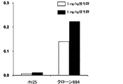
- FIG. 1A shows the results of a brain distribution test when hI2S-sc anti-hTfR antibody fusion protein produced by transformed E. coli clone M11 was administered to mice via tail vein, together with the results of rhI2S.
- the vertical axis shows the I2S concentration ( ⁇ g / wet weight) in the brain homogenate (average value of 2 mice in each group).
- FIG. 1B shows the results of a brain distribution test when hI2S-sc anti-hTfR antibody fusion protein produced by transformed E. coli clone M27 was administered to mice via the tail vein, together with the results obtained with rhI2S.
- FIG. 1C shows the results of a brain distribution test when hI2S-sc anti-hTfR antibody fusion protein produced by transformed E. coli clone B84 was administered to mice via the tail vein, together with the results obtained with rhI2S.
- the vertical axis shows the I2S concentration ( ⁇ g / wet weight) in the brain homogenate (average value of 2 mice in each group).
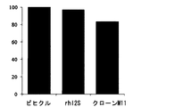
- FIG. 2 is a graph showing the results of evaluation of the efficacy of hI2S-sc anti-TfR antibody fusion protein using Hunter syndrome disease state model mice.
- the vertical axis shows the glycosaminoglycan accumulated in the brain of mice treated with vehicle as 100%, in mice administered rhI2S and mice administered hI2S-sc anti-hTfR antibody fusion protein.
- the accumulated amount (%) of saminoglycan is shown (value is the average value of 3 animals in each group).
- single chain antibody refers to a sequence containing all or part of the variable region of an immunoglobulin light chain, a first linker sequence at the C terminus, and an immunoglobulin heavy chain at the C terminus.
- the variable region of an immunoglobulin light chain has three complementarity determining regions (CDRs) that directly contact the antigen and determine the specificity of the antibody.
- CDRs complementarity determining regions
- the variable region of an immunoglobulin heavy chain has three CDRs. These CDRs are regions that directly contact the antigen and determine the specificity of the antibody. Therefore, it is preferred that the single-chain antibody includes all three CDRs of the immunoglobulin heavy chain and all three CDRs of the immunoglobulin light chain.
- mutations such as substitution, insertion, or loss of one or several amino acids may be introduced into the CDR amino acid sequence as long as the affinity of the single-chain antibody for the antigen is not significantly impaired.
- the affinity between the single-chain antibody and the antigen can be appropriately adjusted.
- the dissociation constant is adjusted in steps of 2 to 5 times, 5 to 10 times, 10 to 100 times, etc. of the original antibody.
- Preferred single chain antibodies can be obtained.
- the dissociation constant is adjusted stepwise to 1 / 2-1 / 5 times, 1 / 5-1 / 10 times, 1 / 10-1 / 100 times, etc. of the original antibody. You can also.
- Such a dissociation constant is adjusted by incorporating a gene encoding a single-chain antibody into a phagemid, using this to create a phage having a single-chain antibody on the capsid surface, and allowing a mutagen or the like to act on it.
- a phage is grown while introducing a mutation on a gene encoding a single-chain antibody, and a phage expressing a single-chain antibody having a desired dissociation constant is amplified from the grown phage using an antigen column under certain conditions. Can be obtained by purification.
- affinity of a single chain antibody to an antigen refers to the property of a single chain antibody that specifically recognizes and binds to the antigen. "Having affinity” means that a single-chain antibody has the property of specifically binding to an antigen under physiological conditions, preferably with a dissociation constant of 0.1 nM to 5 ⁇ M, more preferably 1 nM to 1 ⁇ M.
- the gene encoding the single-chain antibody is a spleen cell or peripheral blood mononuclear cell obtained from an animal immunized with the antigen, or an mRNA extracted from the spleen cell or peripheral blood mononuclear cell immunized with the antigen in vitro.
- cDNA is synthesized, and using this cDNA as a template, a DNA fragment encoding an immunoglobulin light chain and a DNA fragment encoding an immunoglobulin heavy chain are amplified by PCR, and then the DNA fragment encoding the immunoglobulin light chain is amplified.
- a gene encoding a single chain antibody can also be constructed. The gene encoding the single-chain antibody thus constructed has an initiation codon located on the 5 'end side so that it can be expressed in animal cells.
- the number of DNA sequences encoding the first linker sequence arranged between the immunoglobulin light chain and the heavy chain cDNA is preferably 15 to 25, more preferably 15 to 20, and even more preferably 15.
- a peptide chain composed of the amino acid residues is encoded as the first linker sequence.
- the first linker sequence is not limited in its amino acid sequence as long as the immunoglobulin light chain and heavy chain linked by the linker sequence have a configuration capable of recognizing an antigen.
- an amino acid sequence represented by SEQ ID NO: 8 (Gly Gly Gly Ser Gly Gly Gly Gly Ser Gly Gly Gly Gly Gly Ser).
- single-chain anti-human transferrin receptor antibody refers to the human transferrin receptor among the above-mentioned single-chain antibodies.
- Such a gene encoding a single chain anti-hTfR antibody has a partial amino acid sequence of hTfR or a peptide chain constituting hTfR in the above-described method for preparing a gene encoding a single chain antibody.
- the peptide chain constituting hTfR used at this time is not particularly limited as long as an anti-hTfR antibody that specifically binds to hTfR under physiological conditions can be obtained.
- Peptide chain having the following amino acid sequence: (1) Gly Lys Leu Val His Ala Asn Phe Gly Thr Lys Lys Asp Phe Glu (SEQ ID NO: 1) (2) Ser Ser Gly Leu Pro Asn Ile Pro Val Gln Thr Ile Ser Arg Ala Ala Ala Glu Lys Leu Phe Gly (SEQ ID NO: 2), and (3) Val Gly Ala Thr Glu Trp Leu Glu Gly Tyr Leu Ser Ser Number 3) Can be suitably used.
- the preferred form of the single-chain anti-human transferrin receptor antibody of the present invention recognizes and specifically binds to the amino acid sequence shown in SEQ ID NOs: 1 to 3 forming a part of the peptide chain constituting the human transferrin receptor. More preferably, it recognizes a sequence consisting of at least 10 consecutive amino acid residues in the amino acid sequence shown in SEQ ID NOs: 1 to 3, and more preferably the amino acid shown in SEQ ID NOs: 1 to 3 A sequence consisting of 13 consecutive amino acid residues in the sequence is recognized.
- the gene encoding the single-chain anti-hTfR antibody prepared by the above method was obtained as a group of amplification products amplified by PCR from immunoglobulin genes having various DNA sequences. It is necessary to isolate the gene encoding the single-chain anti-hTfR antibody. Therefore, in order to screen for a gene encoding a single-chain anti-hTfR antibody having desired properties, a gene encoding the single-chain anti-hTfR antibody prepared by PCR amplification or the like is temporarily used in mammalian cells. It is necessary to prepare a single-stranded anti-hTfR antibody cDNA library by incorporating it into an expression vector for eukaryotic cells such as yeast or prokaryotic cells such as E. coli.
- host cells are transformed to express a single-chain anti-hTfR antibody, and the affinity between the expressed single-chain anti-hTfR antibody and the antigen is measured to have a desired affinity.
- a gene encoding the desired single-chain anti-hTfR antibody can be isolated and identified.
- the affinity between the single-chain anti-hTfR antibody and hTfR is preferably 1 nM to 1 ⁇ M in the dissociation constant under physiological conditions, but not limited to this, the affinity depends on the use of the single-chain anti-hTfR antibody. Is appropriately adjusted.
- Sc anti-hTfR antibody fusion protein refers to any other protein fused to the C-terminal side of the above-mentioned single-chain anti-hTfR antibody via the second linker sequence or directly.
- Such an Sc anti-hTfR antibody fusion protein is a single strand having an affinity for a desired hTfR, directly or by sandwiching a DNA fragment encoding a linker sequence at the 5 ′ end of a cDNA encoding another protein.
- a DNA fragment linked with cDNA encoding an anti-hTfR antibody can be obtained by incorporating it into an expression vector for mammalian cells, eukaryotic cells such as yeast, or prokaryotic cells such as Escherichia coli, and expressing them in these cells. it can.
- the second linker sequence arranged in the single-chain anti-hTfR antibody and other proteins is preferably composed of 3 to 50, more preferably 13 to 17, and even more preferably 15 amino acids. It is a peptide chain.
- Such a second linker sequence is such that a single-chain anti-hTfR antibody linked by the linker sequence retains affinity with hTfR, and other proteins linked by the linker sequence are under physiological conditions.
- the amino acid sequence is not limited, but is preferably composed of glycine and serine.
- the amino acid sequence represented by SEQ ID NO: 9 (Gly Gly Gly Gly Ser Gly Gly Gly Gly Ser Gly Gly Gly Gly Gly Ser).
- Sc anti-hTfR antibody fusion protein binds to hTfR expressed on the endothelial cells of brain capillaries when administered to the human body, and is taken into the brain through the blood-brain barrier by a mechanism such as endocytosis. It is. Therefore, when the Sc anti-hTfR antibody fusion protein, which is a protein that should function in the brain, is fused with the Sc anti-hTfR antibody, it cannot normally pass through the blood-brain barrier and is functioning in the brain. Even proteins that cannot be expected to reach the brain will be able to exert their functions by reaching the brain. Therefore, the Sc anti-hTfR antibody fusion protein can be selected by selecting the protein to be fused with the Sc anti-hTfR antibody.
- nerve growth factor fused with Sc anti-hTfR antibody is a therapeutic agent for dementia of Alzheimer's disease
- ⁇ -L-iduronidase fused with Sc anti-hTfR antibody is the brain of Harler syndrome.
- iduronic acid-2-sulfatase fused with Sc anti-hTfR antibody can be used as a brain disorder treatment for Hunter syndrome.
- BDNF brain-derived neurotrophic factor
- CNTF ciliary neurotrophic factor
- GDNF glial cell line neurotrophic factor
- various neutrophins Activin, basic fibroblast growth factor (bFGF), epidermal growth factor (EGF), various cytokines, interferon alpha, interferon beta, interferon gamma, interleukin 6, granulocyte macrophage colony stimulating factor (GM-CSF), Examples include granulocyte colony stimulating factor (G-CSF), macrophage colony stimulating factor (M-CSF), PD-1 and the like.
- BDNF brain-derived neurotrophic factor
- CNTF ciliary neurotrophic factor
- GDNF glial cell line neurotrophic factor
- Activin Activin, basic fibroblast growth factor (bFGF), epidermal growth factor (EGF), various cytokines, interferon alpha, interferon beta, interferon gamma, interleukin 6, granulocyte macrophage
- the medium was added with 3.5 mL, and cultured at 37 ° C. for 2 days in the presence of 5% CO 2 . Furthermore, IL-21 was added to a final concentration of 10 ng / mL, and the cells were further cultured for 3 days under the same conditions.
- RNA was extracted from the immunized cells, and single-stranded cDNA was synthesized using this as a template.
- the immunoglobulin light chain and heavy chain cDNAs were each amplified from the single-stranded cDNA by PCR.
- a forward primer having the base sequence shown in SEQ ID NOs: 10 to 27 below: (1) VL-FW1: ttcatggcggactacaaagayatccagctgactcagcc (SEQ ID NO: 10) (2) VL-FW2: ttcatggcggactacaaagayattgttctcwcccagtc (SEQ ID NO: 11) (3) VL-FW3: ttcatggcggactacaaagayattgtgmtmactcagtc (SEQ ID NO: 12) (4) VL-FW4: ttcatggcggactacaaagayattgtgytracacagtc (SEQ ID NO: 13) (5) VL-FW5: ttcatggcggactacaaagayattgtratgacmcagtc (SEQ ID NO: 14) (6) VL-FW6: ttcatggcggact
- r is g or a
- y is t or c
- m is a or c
- k is g or t
- s is g or c.
- W represents a or t
- b represents g, c or t
- d represents a, g or t
- v represents a, g or c.
- a forward primer having the base sequence shown in SEQ ID NOs: 33 to 51 below: (1) VH-FW1: ggCggCggCggCTCCggTggTggTggATCCgAKgTRMAgCTTCAggAgTC (SEQ ID NO: 33) (2) VH-FW2: ggCggCggCggCTCCggTggTggATCCgAggTBCAgCTBCAgCAgTC (SEQ ID NO: 34) (3) VH-FW3: ggCggCggCggCTCCggTggTggTggATCCCAggTgCAgCTgAAgSASTC (SEQ ID NO: 35) (4) VH-FW4: ggCggCggCTCCggTggTggTggATCCgAggTCCARCTgCAACARTC (SEQ ID NO: 36) (5) VH-FW5: ggCggCggCTCCggTggTggATCCCAggTYCAgCTB
- PCR was carried out by mixing equimolar amounts of the light chain cDNA and heavy chain cDNA of immunoglobulin obtained by amplification by the above PCR, and at the 3 ′ side of the light chain cDNA, SEQ ID NO: 9 was used as the first linker sequence.
- a cDNA encoding a single-chain anti-hTfR antibody bound with a heavy chain cDNA was amplified via a base sequence encoding a peptide chain having the amino acid sequence shown.
- the forward primer having the base sequence shown in SEQ ID NO: 56 (ggcgaattcatggcggactacaaag) and the reverse primer having the base sequence shown in SEQ ID NO: 57 (ggcaagctttactgcagcg) PCR was performed using and.
- the obtained amplification product is cleaved with EcoRI / HindIII, and the maltose-binding protein is fused to the N-terminal side of the peptide chain encoded by this amplification product.
- the plasmid for expression of E is cleaved with EcoRI / HindIII, and the maltose-binding protein is fused to the N-terminal side of the peptide chain encoded by this amplification product.
- coli pMAL-c2E New England Biolabs This was incorporated into EcoRI / HindIII and used as a mouse single-chain anti-hTfR antibody cDNA library.
- the cDNA library was prepared according to the methods described in International Publication No. WO2009 / 072660 and JP 2012-29685.
- Escherichia coli JM109 was transformed with mouse and human single-stranded anti-hTfR antibody cDNA library, seeded on LB plate containing ampicillin (Amp), and cultured at 37 ° C. overnight. The next day, colonies formed on the plate were collected one by one and inoculated into Overnight Express TM Instant LB Medium (Novagen) dispensed in advance into a 96-well plate.
- each well was washed three times with 300 ⁇ L TBS-T, and then 50 ⁇ L HRP-labeled anti-MBP antibody (Novagen) was added and allowed to stand at room temperature for 1 hour. After removing the solution, each well was washed three times with 300 ⁇ L of TBS-T, and then 50 ⁇ L of POD substrate solution (Nacalai Tesque) was added and allowed to react for 10-15 minutes at room temperature.
- rhTfR high-affinity clone a clone that expresses a single-chain anti-rhTfR antibody with high affinity to rhTfR (rhTfR high-affinity clone). Identified. From the mouse single-chain anti-hTfR antibody cDNA library, three types of clones, M11, M23 and M27, were obtained as rhTfR high affinity clones. In addition, one clone (B84) was obtained as a rhTfR high-affinity clone from the human single-chain anti-hTfR antibody cDNA library.
- Clone M11 is peptide 2 (SEQ ID NO: 2) and peptide 3 (SEQ ID NO: 3)
- clone M23 is peptide 2 (SEQ ID NO: 2) and peptide 3 (SEQ ID NO: 3)
- clone M27 is peptide 2 (SEQ ID NO: 2).
- peptide 3 (SEQ ID NO: 3) and clone B84 were peptide 1 (SEQ ID NO: 1), which were obtained by screening single-chain anti-hTfR antibody libraries derived from immunized cells, respectively.
- clone M11, clone M23 and clone M27 are clones expressing a mouse single-chain anti-hTfR antibody that recognizes all or part of the amino acid sequence of peptide 2 or peptide 3 as an epitope
- clone B84 is peptide 1 A clone that expresses a human single-chain anti-hTfR antibody that recognizes all or part of the amino acid sequence as an epitope.
- the recovered vector was digested with BglII / NotI and subjected to agarose gel electrophoresis, and an approximately 700 bp DNA fragment encoding a single-chain anti-rhTfR antibody was excised.
- the amplified product was digested with XhoI and NotI and incorporated into the XhoI and NotI sites of the mammalian expression vector pE-neo7 to obtain pE-neo-I2S.
- PE-neo7 was prepared by the method described in International Publication WO 2012/101998.
- pE-neo-hI2S is digested with BglII / NotI, and an about 700 bp DNA fragment encoding the above-mentioned single-chain anti-hTfR antibody digested with BglII / NotI is incorporated into this, and a single-stranded anti-hI2S antibody is incorporated into the N-terminal side of hI2S.
- a vector capable of expressing a fusion protein (hI2S-sc anti-hTfR antibody fusion protein) fused with an hTfR antibody in mammalian cells was constructed.
- Escherichia coli JM109 was transformed with the vector thus obtained, seeded on an LB plate containing ampicillin (Amp), and cultured at 37 ° C. overnight. On the next day, colonies formed on the plate were cultured in LB medium, and then the vector was recovered for each colony. CHO cells were transformed with the recovered vector using electroporation, then selectively cultured for 1 week in the presence of neomycin, and drug resistant cells expressing hI2S-sc anti-hTfR antibody fusion protein were cloned. Got every time.
- the drug-resistant cells obtained for each clone were cultured in a 1 L Erlenmeyer flask containing 200 mL of OptiCHO medium (Invitrogen), and the culture supernatant was collected on the seventh day of culture. Particles were removed from the collected culture supernatant with a 0.22 ⁇ m membrane filter and added to a HiTrap Q HP Sepharose column (column volume: 5 mL) equilibrated with an equilibration buffer (20 mM HEPES, 100 mM NaCl, pH7.0).
- the column was then washed with 5 volumes of equilibrated buffer, and the hI2S-sc anti-hTfR antibody fusion protein was eluted with a linear gradient of equilibrated buffer and elution buffer (20 mM HEPES, 500 mM NaCl, pH 6.0).
- the fractions with high enzyme activity of hI2S were collected, concentrated about 10 times using a 30 kDa cutoff ultrafiltration membrane concentration column, and stored at -80 ° C.
- the measurement of the enzyme activity of hI2S was performed according to the method described in International Publication WO 2012/101998.
- the collected brain was homogenized with T-PER Tissue Protein Extraction Reagent (Thermo Fisher Scientific KK) containing Protease Inhibitor Cocktail (Sigma-Aldrich Co. LLC.), And centrifuged to collect the supernatant.
- the collected supernatant is mixed with biotinylated anti-hI2S monoclonal antibody and Sulfo-modified anti-hI2S monoclonal antibody, and reacted at room temperature for 1 hour. An antibody complex was formed.
- the reaction solution containing the complex is added to Streptavidin Gold Plates (Meso Scale Diagnostics, LLC.) And allowed to react for 1 hour, and the complex is transferred onto the plate via the binding of biotin and streptavidin. Combined.
- the Sulfo label in the complex was electrochemically stimulated by passing an electric current through the plate using a SECTOR Imager 6000 (Meso Scale Diagnostics, LLC.).
- the concentration of hI2S-scrhTfR fusion protein was measured by measuring the fluorescence intensity.
- Recombinant hI2S was prepared according to the method described in International Publication WO2012 / 101998.
- the collected brain was immediately frozen in liquid nitrogen and then freeze-dried.
- the freeze-dried brain was pulverized, the pulverized brain tissue was suspended in 0.5 mol / L Tris-HCl buffer (pH 7.5), actinase E was added thereto, and the mixture was allowed to stand at 60 ° C. for 16 hours. Protein was digested.
- the supernatant is collected by centrifugation, and the concentration of glycosaminoglycan that is a substrate of hI2S contained in the supernatant is quantified by Alcian Blue colorimetry using the Wieslab sGAG quantitative kit (Euro-Diagnostica AB). did.
- mice administered with rhI2S the I2S concentration in the brain homogenate was less than 0.01 ⁇ g / g wet weight in both the 3 and 6 mg / kg groups.
- mice administered with the hI2S-sc anti-hTfR antibody fusion protein obtained for each clone showed 10 to 20 times higher I2S concentration in the brain homogenate compared with mice administered with rhI2S (Fig. 1A to 1C, clone M23 not shown).
- a desired physiologically active protein can be provided in the form of a fusion protein with a single-chain anti-rhTfR antibody capable of crossing the blood brain barrier and as a protein that can function in the neutral nervous system after passage. It is highly useful for providing a protein that should be administered to and act on the central nervous system.
Abstract
Description
1.配列番号1,2及び3からなる群から選択される何れかのアミノ酸配列を認識する抗ヒトトランスフェリン受容体抗体。
2.該配列番号1,2及び3からなる群から選択されるアミノ酸配列中の少なくとも10個の連続するアミノ酸残基よりなる部分を認識する,上記1の抗ヒトトランスフェリン受容体抗体。
3.上記1又は2の何れかの抗ヒトトランスフェリン受容体抗体であって,
軽鎖の可変領域の全て又は一部を含むアミノ酸配列と,
そのC末端側に第1のリンカー配列として結合した15~25個のアミノ酸残基からなるアミノ酸配列と,
更にそのC末端側に結合した,重鎖の可変領域の全て又は一部を含むアミノ酸配列とを含んでなる1本鎖抗体である,抗ヒトトランスフェリン受容体抗体。
4.配列番号4,5,6及び7からなる群から選択されるアミノ酸配列を含んでなる,上記3の抗ヒトトランスフェリン受容体抗体。
5.上記1~4の何れかの抗ヒトトランスフェリン受容体抗体と,そのC末端側に結合した他の蛋白質のアミノ酸配列とを含んでなる,融合蛋白質。
6.上記1~4の何れかの抗ヒトトランスフェリン受容体抗体と,
そのC末端側に第2のリンカー配列として結合した3~50個のアミノ酸残基からなるアミノ酸配列と,
更にそのC末端側に結合した他の蛋白質のアミノ酸配列とを含んでなる,融合蛋白質。
7.該他の蛋白質がリソソーム酵素である,上記5又は6の融合蛋白質。
8.該リソソーム酵素がヒトイズロン酸-2-スルファターゼである,上記7の融合蛋白質。
9.上記1~4の何れかの抗ヒトトランスフェリン受容体抗体をコードするDNA。
10.上記5~8の何れかの融合蛋白質をコードするDNA。
11.上記10のDNAを組み込んだ哺乳動物発現用ベクター。
12.上記11の哺乳動物発現用ベクターで形質転換させた哺乳動物細胞。
13.該哺乳動物細胞がCHO細胞である,上記12の細胞。 In the research aimed at the above-mentioned purpose, the present inventors have conducted intensive studies, and as a result, are anti-human transferrin receptor antibodies that allow lysosomal enzymes to pass through the blood-brain barrier by fusing with the antibodies. The present invention has been completed by finding a novel antibody capable of That is, the present invention provides the following.
1. An anti-human transferrin receptor antibody that recognizes any amino acid sequence selected from the group consisting of SEQ ID NOs: 1, 2, and 3.
2. The anti-human transferrin receptor antibody according to 1 above, which recognizes a portion consisting of at least 10 consecutive amino acid residues in an amino acid sequence selected from the group consisting of SEQ ID NOs: 1, 2 and 3.
3. An anti-human transferrin receptor antibody according to either 1 or 2 above,
An amino acid sequence comprising all or part of the variable region of the light chain;
An amino acid sequence consisting of 15 to 25 amino acid residues bound to the C-terminal side as a first linker sequence;
An anti-human transferrin receptor antibody, which is a single-chain antibody further comprising an amino acid sequence containing all or part of the variable region of the heavy chain bound to the C-terminal side.
4). 4. The anti-human transferrin receptor antibody according to 3 above, comprising an amino acid sequence selected from the group consisting of SEQ ID NOs: 4, 5, 6 and 7.
5. A fusion protein comprising the anti-human transferrin receptor antibody of any one of 1 to 4 above and an amino acid sequence of another protein bound to the C-terminal side thereof.
6). Any one of the above anti-human transferrin receptor antibodies 1 to 4,
An amino acid sequence consisting of 3 to 50 amino acid residues bound to the C-terminal side as a second linker sequence;
Furthermore, a fusion protein comprising the amino acid sequence of another protein bound to the C-terminal side.
7). The fusion protein according to 5 or 6 above, wherein the other protein is a lysosomal enzyme.
8). 8. The fusion protein according to 7 above, wherein the lysosomal enzyme is human iduronic acid-2-sulfatase.
9. A DNA encoding the anti-human transferrin receptor antibody according to any one of 1 to 4 above.
10. DNA encoding the fusion protein of any one of 5 to 8 above.
11. A mammalian expression vector incorporating the DNA of 10 above.
12 Mammalian cells transformed with the above-described mammalian expression vector.
13. 12. The cell according to 12 above, wherein the mammalian cell is a CHO cell.
(1) Gly Lys Leu Val His Ala Asn Phe Gly Thr Lys Lys Asp Phe Glu(配列番号1)
(2) Ser Ser Gly Leu Pro Asn Ile Pro Val Gln Thr Ile Ser Arg Ala Ala Ala Glu Lys Leu Phe Gly(配列番号2),及び
(3) Val Gly Ala Thr Glu Trp Leu Glu Gly Tyr Leu Ser Ser(配列番号3)
が好適に使用できる。 In the present invention, the term “single-chain anti-human transferrin receptor antibody”, “single-chain anti-hTfR antibody” or “sc anti-hTfR antibody” refers to the human transferrin receptor among the above-mentioned single-chain antibodies. An antigen that specifically binds to it. Such a gene encoding a single chain anti-hTfR antibody has a partial amino acid sequence of hTfR or a peptide chain constituting hTfR in the above-described method for preparing a gene encoding a single chain antibody. Spleen or peripheral blood mononuclear cells immunized in vitro with a peptide chain consisting of a single amino acid as an antigen, or mRNA extracted from splenocytes or peripheral blood mononuclear cells obtained from an animal immunized with such a peptide chain as an antigen It can be prepared from cDNA synthesized as a template. The peptide chain constituting hTfR used at this time is not particularly limited as long as an anti-hTfR antibody that specifically binds to hTfR under physiological conditions can be obtained. Peptide chain having the following amino acid sequence:
(1) Gly Lys Leu Val His Ala Asn Phe Gly Thr Lys Lys Asp Phe Glu (SEQ ID NO: 1)
(2) Ser Ser Gly Leu Pro Asn Ile Pro Val Gln Thr Ile Ser Arg Ala Ala Ala Glu Lys Leu Phe Gly (SEQ ID NO: 2), and (3) Val Gly Ala Thr Glu Trp Leu Glu Gly Tyr Leu Ser Ser Number 3)
Can be suitably used.
マウス脾臓から得た細胞を分割し,これを,ヒトトランスフェリン受容体(hTfR)を構成するペプチド鎖の部分アミノ酸配列を有する3種類のペプチド,すなわち,ペプチド1(アミノ酸配列を配列番号1で示す),ペプチド2(アミノ酸配列を配列番号2で示す),ペプチド3(アミノ酸配列を配列番号3で示す)及びこれらペプチドを組合わせたものを用いて,体外免疫法によりそれぞれ免疫した。体外免疫法は概略以下の手法で行った。15mLの遠沈管にペプチドをそれぞれ1nmolと,アジュバントとして,N-アセチルムラミル-L-アラニル-D-イソグルタミン10mg/mLを5μL添加し,次いで,マウス脾臓から得た細胞を1.5×107個添加して室温で15分間静置した。次いで,IL-4,IL-13,抗-CD40抗体及びLPS を,終濃度がそれぞれ10ng/mL,10ng/mL,1μg/mL及び40μg/mLとなるように添加してから40% FBS含有RPMI1640培地を3.5mL添加し,5%CO2存在下,37℃で2日間培養した。更に,IL-21を,終濃度が10ng/mLとなるように添加し,同条件下で更に3日間培養した。 [Preparation of mouse single-chain anti-hTfR antibody cDNA library]
Cells obtained from mouse spleen were divided, and this was divided into three types of peptides having partial amino acid sequences of peptide chains constituting human transferrin receptor (hTfR), namely peptide 1 (the amino acid sequence is represented by SEQ ID NO: 1). Peptide 2 (the amino acid sequence is represented by SEQ ID NO: 2), peptide 3 (the amino acid sequence is represented by SEQ ID NO: 3) and a combination of these peptides were used for immunization by in vitro immunization. In vitro immunization was performed by the following method. Into a 15 mL centrifuge tube, 1 nmol each of peptide and 5 μL of N-acetylmuramyl-L-alanyl-D-isoglutamine 10 mg / mL as an adjuvant were added, and then 1.5 × 10 7 cells obtained from mouse spleen The mixture was added and allowed to stand at room temperature for 15 minutes. Next, IL-4, IL-13, anti-CD40 antibody and LPS were added to final concentrations of 10 ng / mL, 10 ng / mL, 1 μg / mL and 40 μg / mL, respectively, and then RPMI1640 containing 40% FBS was added. The medium was added with 3.5 mL, and cultured at 37 ° C. for 2 days in the presence of 5% CO 2 . Furthermore, IL-21 was added to a final concentration of 10 ng / mL, and the cells were further cultured for 3 days under the same conditions.
(1) VL-FW1:ttcatggcggactacaaagayatccagctgactcagcc(配列番号10)
(2) VL-FW2:ttcatggcggactacaaagayattgttctcwcccagtc(配列番号11)
(3) VL-FW3:ttcatggcggactacaaagayattgtgmtmactcagtc(配列番号12)
(4) VL-FW4:ttcatggcggactacaaagayattgtgytracacagtc(配列番号13)
(5) VL-FW5:ttcatggcggactacaaagayattgtratgacmcagtc(配列番号14)
(6) VL-FW6:ttcatggcggactacaaagayattmagatramccagtc(配列番号15)
(7) VL-FW7:ttcatggcggactacaaagayattcagatgaydcagtc(配列番号16)
(8) VL-FW8:ttcatggcggactacaaagayatycagatgacacagac(配列番号17)
(9) VL-FW9:ttcatggcggactacaaagayattgttctcawccagtc(配列番号18)
(10) VL-FW10:ttcatggcggactacaaagayattgwgctsacccaatc(配列番号19)
(11) VL-FW11:ttcatggcggactacaaagayattstratgacccartc(配列番号20)
(12) VL-FW12:ttcatggcggactacaaagayrttktgatgacccarac(配列番号21)
(13) VL-FW13:ttcatggcggactacaaagayattgtgatgacbcagkc(配列番号22)
(14) VL-FW14:ttcatggcggactacaaagayattgtgataacycagga(配列番号23)
(15) VL-FW15:ttcatggcggactacaaagayattgtgatgacccagwt(配列番号24)
(16) VL-FW16:ttcatggcggactacaaagayattgtgatgacacaacc(配列番号25)
(17) VL-FW17:ttcatggcggactacaaagayattttgctgactcagtc(配列番号26)
(18) VL-FW18:ttcatggcggactacaaagatgctgttgtactcaggaatc(配列番号27)
と,下記の配列番号28~32に示す塩基配列を有するリバースプライマー:
(19) VL-RV1:ggagccgccgccgccagaaccaccaccaccagaaccaccaccaccacgtttgatttccagcttgg(配列番号28)
(20) VL-RV2:ggagccgccgccgccagaaccaccaccaccagaaccaccaccaccacgttttatttccagcttgg(配列番号29)
(21) VL-RV3:ggagccgccgccgccagaaccaccaccaccagaaccaccaccaccacgttttatttccaactttg(配列番号30)
(22) VL-RV4:ggagccgccgccgccagaaccaccaccaccagaaccaccaccaccacgtttcagctccagcttgg(配列番号31)
(23) VL-RV5:ggagccgccgccgccagaaccaccaccaccagaaccaccaccaccacctaggacagtcagtttgg(配列番号32)
とを用いた。 Total RNA was extracted from the immunized cells, and single-stranded cDNA was synthesized using this as a template. The immunoglobulin light chain and heavy chain cDNAs were each amplified from the single-stranded cDNA by PCR. For amplification of the immunoglobulin light chain, a forward primer having the base sequence shown in SEQ ID NOs: 10 to 27 below:
(1) VL-FW1: ttcatggcggactacaaagayatccagctgactcagcc (SEQ ID NO: 10)
(2) VL-FW2: ttcatggcggactacaaagayattgttctcwcccagtc (SEQ ID NO: 11)
(3) VL-FW3: ttcatggcggactacaaagayattgtgmtmactcagtc (SEQ ID NO: 12)
(4) VL-FW4: ttcatggcggactacaaagayattgtgytracacagtc (SEQ ID NO: 13)
(5) VL-FW5: ttcatggcggactacaaagayattgtratgacmcagtc (SEQ ID NO: 14)
(6) VL-FW6: ttcatggcggactacaaagayattmagatramccagtc (SEQ ID NO: 15)
(7) VL-FW7: ttcatggcggactacaaagayattcagatgaydcagtc (SEQ ID NO: 16)
(8) VL-FW8: ttcatggcggactacaaagayatycagatgacacagac (SEQ ID NO: 17)
(9) VL-FW9: ttcatggcggactacaaagayattgttctcawccagtc (SEQ ID NO: 18)
(10) VL-FW10: ttcatggcggactacaaagayattgwgctsacccaatc (SEQ ID NO: 19)
(11) VL-FW11: ttcatggcggactacaaagayattstratgacccartc (SEQ ID NO: 20)
(12) VL-FW12: ttcatggcggactacaaagayrttktgatgacccarac (SEQ ID NO: 21)
(13) VL-FW13: ttcatggcggactacaaagayattgtgatgacbcagkc (SEQ ID NO: 22)
(14) VL-FW14: ttcatggcggactacaaagayattgtgataacycagga (SEQ ID NO: 23)
(15) VL-FW15: ttcatggcggactacaaagayattgtgatgacccagwt (SEQ ID NO: 24)
(16) VL-FW16: ttcatggcggactacaaagayattgtgatgacacaacc (SEQ ID NO: 25)
(17) VL-FW17: ttcatggcggactacaaagayattttgctgactcagtc (SEQ ID NO: 26)
(18) VL-FW18: ttcatggcggactacaaagatgctgttgtactcaggaatc (SEQ ID NO: 27)
And a reverse primer having the base sequence shown in SEQ ID NOs: 28 to 32 below:
(19) VL-RV1: ggagccgccgccgccagaaccaccaccaccagaaccaccaccaccacgtttgatttccagcttgg (SEQ ID NO: 28)
(20) VL-RV2: ggagccgccgccgccagaaccaccaccaccagaaccaccaccaccacacgttttatttccagcttgg (SEQ ID NO: 29)
(21) VL-RV3: ggagccgccgccgccagaaccaccaccaccagaaccaccaccaccacactttttatttccaactttg (SEQ ID NO: 30)
(22) VL-RV4: ggagccgccgccgccagaaccaccaccaccagaaccaccaccaccacgtttcagctccagcttgg (SEQ ID NO: 31)
(23) VL-RV5: ggagccgccgccgccagaaccaccaccaccagaaccaccaccaccacctagtagaccagtcagtttgg (SEQ ID NO: 32)
And were used.
(1) VH-FW1:ggCggCggCggCTCCggTggTggTggATCCgAKgTRMAgCTTCAggAgTC(配列番号33)
(2) VH-FW2:ggCggCggCggCTCCggTggTggTggATCCgAggTBCAgCTBCAgCAgTC(配列番号34)
(3) VH-FW3:ggCggCggCggCTCCggTggTggTggATCCCAggTgCAgCTgAAgSASTC(配列番号35)
(4) VH-FW4:ggCggCggCggCTCCggTggTggTggATCCgAggTCCARCTgCAACARTC(配列番号36)
(5) VH-FW5:ggCggCggCggCTCCggTggTggTggATCCCAggTYCAgCTBCAgCARTC(配列番号37)
(6) VH-FW6:ggCggCggCggCTCCggTggTggTggATCCCAggTYCARCTgCAgCAgTC(配列番号38)
(7) VH-FW7:ggCggCggCggCTCCggTggTggTggATCCCAggTCCACgTgAAgCAgTC(配列番号39)
(8) VH-FW8:ggCggCggCggCTCCggTggTggTggATCCgAggTgAASSTggTggAATC(配列番号40)
(9) VH-FW9:ggCggCggCggCTCCggTggTggTggATCCgAVgTgAWgYTggTggAgTC(配列番号41)
(10) VH-FW10:ggCggCggCggCTCCggTggTggTggATCCgAggTgCAgSKggTggAgTC(配列番号42)
(11) VH-FW11:ggCggCggCggCTCCggTggTggTggATCCgAKgTgCAMCTggTggAgTC(配列番号43)
(12) VH-FW12:ggCggCggCggCTCCggTggTggTggATCCgAggTgAAgCTgATggARTC(配列番号44)
(13) VH-FW13:ggCggCggCggCTCCggTggTggTggATCCgAggTgCARCTTgTTgAgTC(配列番号45)
(14) VH-FW14:ggCggCggCggCTCCggTggTggTggATCCgARgTRAAgCTTCTCgAgTC(配列番号46)
(15) VH-FW15:ggCggCggCggCTCCggTggTggTggATCCgAAgTgAARSTTgAggAgTC(配列番号47)
(16) VH-FW16:ggCggCggCggCTCCggTggTggTggATCCCAggTTACTCTRAAAgWgTSTg(配列番号48)
(17) VH-FW17:ggCggCggCggCTCCggTggTggTggATCCCAggTCCAACTVCAgCARCC(配列番号49)
(18) VH-FW18:ggCggCggCggCTCCggTggTggTggATCCgATgTgAACTTggAAgTgTC(配列番号50)
(19) VH-FW19:ggCggCggCggCTCCggTggTggTggATCCgAggTgAAggTCATCgAgTC(配列番号51)
と,下記の配列番号52~55に示す塩基配列を有するリバースプライマー:
(20) VH-RV1:ggCAAgCTTTACCTgCAgCgAggAAACggTgACCgTggT(配列番号52)
(21) VH-RV2:ggCAAgCTTTACCTgCAgCgAggAgACTgTgAgAgTggT(配列番号53)
(22) VH-RV3:ggCAAgCTTTACCTgCAgCgCAgAgACAgTgACCAgAgT(配列番号54)
(23) VH-RV4:ggCAAgCTTTACCTgCAgCgAggAgACggTgACTgAggT(配列番号55)
とを用いた。 For amplification of immunoglobulin heavy chain, a forward primer having the base sequence shown in SEQ ID NOs: 33 to 51 below:
(1) VH-FW1: ggCggCggCggCTCCggTggTggTggATCCgAKgTRMAgCTTCAggAgTC (SEQ ID NO: 33)
(2) VH-FW2: ggCggCggCggCTCCggTggTggTggATCCgAggTBCAgCTBCAgCAgTC (SEQ ID NO: 34)
(3) VH-FW3: ggCggCggCggCTCCggTggTggTggATCCCAggTgCAgCTgAAgSASTC (SEQ ID NO: 35)
(4) VH-FW4: ggCggCggCggCTCCggTggTggTggATCCgAggTCCARCTgCAACARTC (SEQ ID NO: 36)
(5) VH-FW5: ggCggCggCggCTCCggTggTggTggATCCCAggTYCAgCTBCAgCARTC (SEQ ID NO: 37)
(6) VH-FW6: ggCggCggCggCTCCggTggTggTggATCCCAggTYCARCTgCAgCAgTC (SEQ ID NO: 38)
(7) VH-FW7: ggCggCggCggCTCCggTggTggTggATCCCAggTCCACgTgAAgCAgTC (SEQ ID NO: 39)
(8) VH-FW8: ggCggCggCggCTCCggTggTggTggATCCgAggTgAASSTggTggAATC (SEQ ID NO: 40)
(9) VH-FW9: ggCggCggCggCTCCggTggTggTggATCCgAVgTgAWgYTggTggAgTC (SEQ ID NO: 41)
(10) VH-FW10: ggCggCggCggCTCCggTggTggTggATCCgAggTgCAgSKggTggAgTC (SEQ ID NO: 42)
(11) VH-FW11: ggCggCggCggCTCCggTggTggTggATCCgAKgTgCAMCTggTggAgTC (SEQ ID NO: 43)
(12) VH-FW12: ggCggCggCggCTCCggTggTggTggATCCgAggTgAAgCTgATggARTC (SEQ ID NO: 44)
(13) VH-FW13: ggCggCggCggCTCCggTggTggTggATCCgAggTgCARCTTgTTgAgTC (SEQ ID NO: 45)
(14) VH-FW14: ggCggCggCggCTCCggTggTggTggATCCgARgTRAAgCTTCTCgAgTC (SEQ ID NO: 46)
(15) VH-FW15: ggCggCggCggCTCCggTggTggTggATCCgAAgTgAARSTTgAggAgTC (SEQ ID NO: 47)
(16) VH-FW16: ggCggCggCggCTCCggTggTggTggATCCCAggTTACTCTRAAAgWgTSTg (SEQ ID NO: 48)
(17) VH-FW17: ggCggCggCggCTCCggTggTggTggATCCCAggTCCAACTVCAgCARCC (SEQ ID NO: 49)
(18) VH-FW18: ggCggCggCggCTCCggTggTggTggATCCgATgTgAACTTggAAgTgTC (SEQ ID NO: 50)
(19) VH-FW19: ggCggCggCggCTCCggTggTggTggATCCgAggTgAAggTCATCgAgTC (SEQ ID NO: 51)
And a reverse primer having the base sequence shown in SEQ ID NOs: 52 to 55 below:
(20) VH-RV1: ggCAAgCTTTACCTgCAgCgAggAAACggTgACCgTggT (SEQ ID NO: 52)
(21) VH-RV2: ggCAAgCTTTACCTgCAgCgAggAgACTgTgAgAgTggT (SEQ ID NO: 53)
(22) VH-RV3: ggCAAgCTTTACCTgCAgCgCAgAgACAgTgACCAgAgT (SEQ ID NO: 54)
(23) VH-RV4: ggCAAgCTTTACCTgCAgCgAggAgACggTgACTgAggT (SEQ ID NO: 55)
And were used.
上記マウス1本鎖抗hTfR抗体cDNAライブラリの作製と同様の手法により,ペプチド1(配列番号1),ペプチド2(配列番号2),ペプチド3(配列番号3)及びこれらペプチドを組合わせたものを用いて,ヒト末梢血単核球(ロンザ社)を体外免疫法により免疫して全RNAを抽出し,これを鋳型として,ヒト1本鎖抗hTfR抗体cDNAライブラリを作成した。 [Preparation of human single-chain anti-hTfR antibody cDNA library]
Peptide 1 (SEQ ID NO: 1), Peptide 2 (SEQ ID NO: 2), Peptide 3 (SEQ ID NO: 3) and combinations of these peptides were prepared in the same manner as in the preparation of the mouse single chain anti-hTfR antibody cDNA library. Using it, human peripheral blood mononuclear cells (Lonza) were immunized by in vitro immunization to extract total RNA, and using this as a template, a human single-stranded anti-hTfR antibody cDNA library was prepared.
大腸菌(JM109)を,マウス及びヒト1本鎖抗hTfR抗体cDNAライブラリにより形質転換させた後,アンピシリン(Amp)を含むLBプレートに播種し,37℃で一晩培養した。翌日,プレート上に形成されたコロニーを一つずつ採取し,96穴プレートに予め分注したOvernight ExpressTM Instant LB Medium(Novagen社)に植菌した。これを37℃で一晩振とう培養した後,Popculture Reagent (Novagen社) を1/10量加えて溶菌し,次いで等量のTBS-Tを添加して遠心し,上清を回収した。この上清をサンプル溶液とした。 [Screening of single-stranded anti-hTfR antibody cDNA library]
Escherichia coli (JM109) was transformed with mouse and human single-stranded anti-hTfR antibody cDNA library, seeded on LB plate containing ampicillin (Amp), and cultured at 37 ° C. overnight. The next day, colonies formed on the plate were collected one by one and inoculated into Overnight Express ™ Instant LB Medium (Novagen) dispensed in advance into a 96-well plate. This was cultured with shaking overnight at 37 ° C., 1/10 volume of Popculture Reagent (Novagen) was added to lyse, then an equal amount of TBS-T was added and centrifuged, and the supernatant was collected. This supernatant was used as a sample solution.
上記スクリーニングで得た4種のrhTfR高親和性クローンから,発現ベクターを精製し,各クローンがコードする1本鎖抗rhTfR抗体のアミノ酸配列を解析した。その結果,クローンM11は配列番号4,クローンM23は配列番号5,クローンM27は配列番号6,クローンB84は配列番号7にそれぞれ示すアミノ酸配列を有する1本鎖抗rhTfR抗体をコードすることが判った。 [Analysis of rhTfR high affinity clone]
Expression vectors were purified from the four rhTfR high affinity clones obtained in the above screening, and the amino acid sequence of the single-chain anti-rhTfR antibody encoded by each clone was analyzed. As a result, it was found that clone M11 encodes a single chain anti-rhTfR antibody having the amino acid sequence shown in SEQ ID NO: 4, clone M23 is SEQ ID NO: 5, clone M27 is SEQ ID NO: 6, and clone B84 is SEQ ID NO: 7, respectively. .
上記4種のrhTfR高親和性クローンから,発現ベクターを精製し,これをEcoRI/HindIIIで消化してアガロースゲル電気泳動に供し,1本鎖抗rhTfR抗体をコードする約700bpのDNA断片を切り出し,次いで,このDNA断片をpET32aベクターのEcoRI/HindIIIに組み込んだ。こうして得られたベクターを用いて大腸菌(JM109)を形質転換させた後,アンピシリン(Amp)を含むLBプレートに播種し,37℃で一晩培養した。翌日,プレート上に形成されたコロニーをLB培地で培養した後,クローン毎にベクターを回収した。 [Preparation of fusion protein of human iduronic acid-2-sulfatase and single-chain anti-rhTfR antibody]
From the above four rhTfR high affinity clones, an expression vector was purified, digested with EcoRI / HindIII and subjected to agarose gel electrophoresis, and a DNA fragment of about 700 bp encoding a single-chain anti-rhTfR antibody was excised. Subsequently, this DNA fragment was incorporated into EcoRI / HindIII of the pET32a vector. Escherichia coli (JM109) was transformed with the vector thus obtained, seeded on an LB plate containing ampicillin (Amp), and cultured at 37 ° C. overnight. The next day, colonies formed on the plate were cultured in LB medium, and then the vector was recovered for each clone.
(a) I2S-f: ACGCCTATTGCTGCAGGATG(配列番号58),及び
(b) I2S-r:AAACGACCAGCTCTAACTCC(配列番号59),並びに
第二反応に用いる内側プライマーセット:
(c) I2S-f2:ATActcgagGCCACCATGCCGCCACCCCGG(配列番号60),及び
(d) I2S-r2:TTCTTATgcggccgcTCAAGGCATCAACAA(配列番号61)
の2つのプライマーセットを用いてネスティッドPCRを行い,ヒトイズロン酸-2-スルファターゼ(hI2S)をコードする cDNAを含むDNA断片を増幅させた。この増幅産物を,XhoIとNotIで消化し,哺乳動物発現ベクターpE-neo7のXhoIとNotIサイトに組込み,pE-neo-I2Sを得た。なお,pE-neo7は,国際公開公報WO 2012/101998に記載の方法で作成した。 The recovered vector was digested with BglII / NotI and subjected to agarose gel electrophoresis, and an approximately 700 bp DNA fragment encoding a single-chain anti-rhTfR antibody was excised. Outer primer set for the first reaction using human placenta cDNA library (Takara Bio) as a template:
(a) I2S-f: ACGCCTATTGCTGCAGGATG (SEQ ID NO: 58), and (b) I2S-r: AAACGACCAGCTCTAACTCC (SEQ ID NO: 59), and the inner primer set used for the second reaction:
(c) I2S-f2: ATActcgagGCCACCATGCCGCCACCCCGG (SEQ ID NO: 60), and (d) I2S-r2: TTCTTATgcggccgcTCAAGGCATCAACAA (SEQ ID NO: 61)
Nested PCR was performed using these two primer sets to amplify a DNA fragment containing cDNA encoding human iduronic acid-2-sulfatase (hI2S). The amplified product was digested with XhoI and NotI and incorporated into the XhoI and NotI sites of the mammalian expression vector pE-neo7 to obtain pE-neo-I2S. PE-neo7 was prepared by the method described in International Publication WO 2012/101998.
雄性C57BL/6Nマウスへ,組換えhI2S及びクローン毎(M11,M23,M27,及びB84)に得た上記精製したhI2S-sc抗hTfR抗体融合蛋白質を3及び6mg/kgの用量で尾静脈内投与した (n=2/群)。投与から7時間後に,マウスをイソフルランで麻酔し,マウスの静脈から生理食塩水を潅流させた後,脳を採取した。採取した脳を,Protease Inhibitor Cocktail (Sigma-Aldrich Co. LLC.) を含むT-PER Tissue Protein Extraction Reagent (Thermo Fisher Scientific K.K.) でホモジネートし,遠心して上清を回収した。回収した上清と,ビオチン化抗hI2Sモノクローナル抗体及びSulfo化抗hI2Sモノクローナル抗体とを混和し,室温で1時間反応させて,ビオチン化抗I2Sモノクローナル抗体,hI2S-scrhTfR抗体融合タンパク及びSulfo化hI2Sモノクローナル抗体の複合体を形成させた。次いで,上記複合体を含む反応液をStreptavidin Gold Plates (Meso Scale Diagnostics, LLC.) へ添加し1時間反応させて,上記複合体を,ビオチンとストレプロアビジンとの結合を介して,プレート上へ結合させた。プレートを洗浄後,SECTOR Imager 6000 (Meso Scale Diagnostics, LLC.) を用いて,プレートに電流を流すことにより上記複合体中のSulfo標識体に電気化学的刺激を与え,このときSulfo標識体から発せられる蛍光強度を測定することにより,hI2S-scrhTfR融合タンパクの濃度を測定した。なお,組換えhI2Sは,国際公開公報WO2012/101998に記載の方法に準じて調製したものを使用した。 [Brain distribution test of hI2S-sc anti-hTfR antibody fusion protein in mice]
Recombinant hI2S and the purified hI2S-sc anti-hTfR antibody fusion protein obtained for each clone (M11, M23, M27, and B84) are injected into male C57BL / 6N mice at 3 and 6 mg / kg via the tail vein. (N = 2 / group). Seven hours after the administration, the mice were anesthetized with isoflurane, and saline was perfused from the veins of the mice, and then the brains were collected. The collected brain was homogenized with T-PER Tissue Protein Extraction Reagent (Thermo Fisher Scientific KK) containing Protease Inhibitor Cocktail (Sigma-Aldrich Co. LLC.), And centrifuged to collect the supernatant. The collected supernatant is mixed with biotinylated anti-hI2S monoclonal antibody and Sulfo-modified anti-hI2S monoclonal antibody, and reacted at room temperature for 1 hour. An antibody complex was formed. Next, the reaction solution containing the complex is added to Streptavidin Gold Plates (Meso Scale Diagnostics, LLC.) And allowed to react for 1 hour, and the complex is transferred onto the plate via the binding of biotin and streptavidin. Combined. After washing the plate, the Sulfo label in the complex was electrochemically stimulated by passing an electric current through the plate using a SECTOR Imager 6000 (Meso Scale Diagnostics, LLC.). The concentration of hI2S-scrhTfR fusion protein was measured by measuring the fluorescence intensity. Recombinant hI2S was prepared according to the method described in International Publication WO2012 / 101998.
ハンター症候群の病態モデルマウスである,雄性イズロン酸2-スルファターゼ遺伝子ノックアウトマウスに,ビヒクル(生理食塩水),組換えhI2S(rhI2S),及びクローンM11のhI2S-sc抗hTfR抗体融合蛋白質を3 mg/kgの用量で,3日に1回,計3回,尾静脈内投与した (n=3/群)。最終投与から3日後にマウスをイソフルランで麻酔して放血致死させた後,脳を採取した。採取した脳を直ちに液体窒素中で急速凍結し,その後凍結乾燥した。次いで,凍結乾燥した脳を粉砕し,粉砕した脳組織を0.5 mol/Lトリス塩酸緩衝液 (pH 7.5) 中に懸濁させ,これにアクチナーゼEを添加して60℃で16時間静置し,タンパク質を消化させた。次いで,遠心して上清を回収し,Wieslab sGAG quantitative kit (Euro-Diagnostica AB) を用いアルシアンブルー比色定量法により,上清中に含まれるhI2Sの基質であるグリコサミノグリカンの濃度を定量した。 [Evaluation of efficacy of hI2S-sc anti-hTfR antibody fusion protein using Hunter syndrome model mouse]
Male iduronic acid 2-sulfatase gene knockout mouse, a model mouse model of Hunter syndrome, was treated with 3 mg / h of vehicle (saline), recombinant hI2S (rhI2S), and hI2S-sc anti-hTfR antibody fusion protein of clone M11. It was administered via the tail vein at a dose of kg once every 3 days for a total of 3 times (n = 3 / group). Three days after the final administration, the mice were anesthetized with isoflurane and exsanguinated, and then the brains were collected. The collected brain was immediately frozen in liquid nitrogen and then freeze-dried. Next, the freeze-dried brain was pulverized, the pulverized brain tissue was suspended in 0.5 mol / L Tris-HCl buffer (pH 7.5), actinase E was added thereto, and the mixture was allowed to stand at 60 ° C. for 16 hours. Protein was digested. Next, the supernatant is collected by centrifugation, and the concentration of glycosaminoglycan that is a substrate of hI2S contained in the supernatant is quantified by Alcian Blue colorimetry using the Wieslab sGAG quantitative kit (Euro-Diagnostica AB). did.
rhI2Sを投与したマウスでは,脳ホモジネート中のI2S濃度は,3及び6mg/kg投与群ともに,0.01μg/g湿重量未満であった。一方,クローン毎に得たhI2S-sc抗hTfR抗体融合蛋白質を投与したマウスでは,脳ホモジネート中のI2S濃度が,rhI2Sを投与したマウスと比較して,10~20倍の高値を示した(図1A~図1C,クローンM23については示さず)。また,4種のクローン中,クローンB84のhI2S-sc抗hTfR抗体融合蛋白質を投与したマウスでの脳ホモジネート中のI2S濃度が,最も高値であった(図1B)。この結果は,4種のクローンのsc抗hTfR抗体が,これらをhI2Sと融合させたとき,hI2Sを脳に効率良く輸送する機能を有することを示すものである。 [Result 1: Brain distribution test of hI2S-sc anti-hTfR antibody fusion protein in mice]
In mice administered with rhI2S, the I2S concentration in the brain homogenate was less than 0.01 μg / g wet weight in both the 3 and 6 mg / kg groups. On the other hand, mice administered with the hI2S-sc anti-hTfR antibody fusion protein obtained for each clone showed 10 to 20 times higher I2S concentration in the brain homogenate compared with mice administered with rhI2S (Fig. 1A to 1C, clone M23 not shown). In addition, among the four clones, the I2S concentration in the brain homogenate in the mouse administered with the hI2S-sc anti-hTfR antibody fusion protein of clone B84 was the highest (FIG. 1B). This result indicates that the four clones of sc anti-hTfR antibodies have a function of efficiently transporting hI2S to the brain when they are fused with hI2S.
組換えhI2Sを投与したマウスでは,ビヒクルを投与したマウスと比較して,脳内に蓄積したグリコサミノグリカンの減少はほとんど観察されなかったが,hI2S-sc抗hTfR抗体融合蛋白質を投与したマウスでは,グリコサミノグリカンの脳内での蓄積量が,ビヒクルを投与したマウスと比較して,約83%に減少した(図2)。この結果は,クローンM11のhI2S-sc抗hTfR抗体融合蛋白質が,ハンター症候群病態モデルマウスの脳内に蓄積したグリコサミノグリカンを分解できることを示すものである。従って,hI2S-sc抗hTfR抗体融合蛋白質は,ハンター症候群の患者に投与したときに,患者の脳内に蓄積したグリコサミノグリカンを分解することにより,ハンター症候群における中枢神経障害を緩解し得る。 [Result 2: Evaluation of efficacy of hI2S-sc anti-hTfR antibody fusion protein using Hunter syndrome model mouse]
Mice that received recombinant hI2S showed little decrease in glycosaminoglycans accumulated in the brain compared to mice that received vehicle, but mice that received hI2S-sc anti-hTfR antibody fusion protein. Then, the amount of glycosaminoglycan accumulated in the brain decreased to about 83% compared to mice administered with vehicle (Fig. 2). This result indicates that the hI2S-sc anti-hTfR antibody fusion protein of clone M11 can degrade glycosaminoglycan accumulated in the brain of Hunter syndrome model mice. Therefore, the hI2S-sc anti-hTfR antibody fusion protein can relieve central neuropathy in Hunter syndrome by degrading glycosaminoglycans accumulated in the patient's brain when administered to patients with Hunter syndrome.
Claims (13)
- 配列番号1,2及び3からなる群から選択される何れかのアミノ酸配列を認識する抗ヒトトランスフェリン受容体抗体。 An anti-human transferrin receptor antibody that recognizes any amino acid sequence selected from the group consisting of SEQ ID NOs: 1, 2, and 3.
- 該配列番号1,2及び3からなる群から選択されるアミノ酸配列中の少なくとも10個の連続するアミノ酸残基よりなる部分を認識する,請求項1の抗ヒトトランスフェリン受容体抗体。 The anti-human transferrin receptor antibody according to claim 1, which recognizes a portion consisting of at least 10 consecutive amino acid residues in an amino acid sequence selected from the group consisting of SEQ ID NOs: 1, 2 and 3.
- 請求項1又は2の何れかの抗ヒトトランスフェリン受容体抗体であって,
軽鎖の可変領域の全て又は一部を含むアミノ酸配列と,
そのC末端側に第1のリンカー配列として結合した15~25個のアミノ酸残基からなるアミノ酸配列と,
更にそのC末端側に結合した,重鎖の可変領域の全て又は一部を含むアミノ酸配列とを含んでなる1本鎖抗体である,抗ヒトトランスフェリン受容体抗体。 An anti-human transferrin receptor antibody according to claim 1 or 2,
An amino acid sequence comprising all or part of the variable region of the light chain;
An amino acid sequence consisting of 15 to 25 amino acid residues bound to the C-terminal side as a first linker sequence;
An anti-human transferrin receptor antibody, which is a single-chain antibody further comprising an amino acid sequence containing all or part of the variable region of the heavy chain bound to the C-terminal side. - 配列番号4,5,6及び7からなる群から選択されるアミノ酸配列を含んでなる,請求項3の抗ヒトトランスフェリン受容体抗体。 The anti-human transferrin receptor antibody of claim 3, comprising an amino acid sequence selected from the group consisting of SEQ ID NOs: 4, 5, 6 and 7.
- 請求項1~4の何れかの抗ヒトトランスフェリン受容体抗体と,そのC末端側に結合した他の蛋白質のアミノ酸配列とを含んでなる,融合蛋白質。 A fusion protein comprising the anti-human transferrin receptor antibody according to any one of claims 1 to 4 and the amino acid sequence of another protein bound to the C-terminal side thereof.
- 請求項1~4の何れかの抗ヒトトランスフェリン受容体抗体と,
そのC末端側に第2のリンカー配列として結合した3~50個のアミノ酸残基からなるアミノ酸配列と,
更にそのC末端側に結合した他の蛋白質のアミノ酸配列とを含んでなる,融合蛋白質。 An anti-human transferrin receptor antibody according to any one of claims 1 to 4;
An amino acid sequence consisting of 3 to 50 amino acid residues bound to the C-terminal side as a second linker sequence;
Furthermore, a fusion protein comprising the amino acid sequence of another protein bound to the C-terminal side. - 該他の蛋白質がリソソーム酵素である,請求項5又は6の融合蛋白質。 The fusion protein according to claim 5 or 6, wherein the other protein is a lysosomal enzyme.
- 該リソソーム酵素がヒトイズロン酸-2-スルファターゼである,請求項7の融合蛋白質。 The fusion protein according to claim 7, wherein the lysosomal enzyme is human iduronic acid-2-sulfatase.
- 請求項1~4の何れかの抗ヒトトランスフェリン受容体抗体をコードするDNA。 DNA encoding the anti-human transferrin receptor antibody according to any one of claims 1 to 4.
- 請求項5~8の何れかの融合蛋白質をコードするDNA。 DNA encoding the fusion protein according to any one of claims 5 to 8.
- 請求項10のDNAを組み込んだ哺乳動物発現用ベクター。 A mammalian expression vector incorporating the DNA of claim 10.
- 請求項11の哺乳動物発現用ベクターで形質転換させた哺乳動物細胞。 A mammalian cell transformed with the mammalian expression vector of claim 11.
- 該哺乳動物細胞がCHO細胞である,請求項12の細胞。 The cell according to claim 12, wherein the mammalian cell is a CHO cell.
Priority Applications (4)
| Application Number | Priority Date | Filing Date | Title |
|---|---|---|---|
| EP18199592.9A EP3450557A1 (en) | 2013-12-25 | 2014-12-24 | Novel anti-transferrin receptor antibody that passes through blood-brain barrier |
| US15/107,839 US9994641B2 (en) | 2013-12-25 | 2014-12-24 | Anti-human transferrin receptor antibody that passes through blood-brain barrier |
| JP2015554972A JP6616189B2 (en) | 2013-12-25 | 2014-12-24 | A novel anti-transferrin receptor antibody that crosses the blood-brain barrier |
| EP14873852.9A EP3088518A4 (en) | 2013-12-25 | 2014-12-24 | Novel anti-transferrin receptor antibody that passes through blood-brain barrier |
Applications Claiming Priority (2)
| Application Number | Priority Date | Filing Date | Title |
|---|---|---|---|
| JP2013266880 | 2013-12-25 | ||
| JP2013-266880 | 2013-12-25 |
Publications (1)
| Publication Number | Publication Date |
|---|---|
| WO2015098989A1 true WO2015098989A1 (en) | 2015-07-02 |
Family
ID=53478836
Family Applications (1)
| Application Number | Title | Priority Date | Filing Date |
|---|---|---|---|
| PCT/JP2014/084198 WO2015098989A1 (en) | 2013-12-25 | 2014-12-24 | Novel anti-transferrin receptor antibody that passes through blood-brain barrier |
Country Status (4)
| Country | Link |
|---|---|
| US (1) | US9994641B2 (en) |
| EP (2) | EP3088518A4 (en) |
| JP (1) | JP6616189B2 (en) |
| WO (1) | WO2015098989A1 (en) |
Cited By (12)
| Publication number | Priority date | Publication date | Assignee | Title |
|---|---|---|---|---|
| WO2016208695A1 (en) * | 2015-06-24 | 2016-12-29 | Jcrファーマ株式会社 | Anti-human transferrin receptor antibody permeating blood-brain barrier |
| WO2016208696A1 (en) * | 2015-06-24 | 2016-12-29 | Jcrファーマ株式会社 | Fusion protein containing bdnf |
| JP2018033454A (en) * | 2016-08-25 | 2018-03-08 | Jcrファーマ株式会社 | Method for producing antibody fusion protein |
| WO2018124121A1 (en) * | 2016-12-26 | 2018-07-05 | Jcrファーマ株式会社 | Novel anti-human transferrin receptor antibody capable of penetrating blood-brain barrier |
| WO2018124277A1 (en) * | 2016-12-28 | 2018-07-05 | Jcrファーマ株式会社 | Lyophilized preparation |
| WO2018124107A1 (en) * | 2016-12-26 | 2018-07-05 | Jcrファーマ株式会社 | Fusion protein including bdnf |
| WO2020004368A1 (en) * | 2018-06-25 | 2020-01-02 | Jcrファーマ株式会社 | Protein-containing aqueous liquid formulation |
| US10870837B2 (en) | 2017-10-02 | 2020-12-22 | Denali Therapeutics Inc. | Fusion proteins comprising enzyme replacement therapy enzymes |
| WO2023277627A1 (en) * | 2021-07-02 | 2023-01-05 | 주식회사 아임뉴런 | Blood-brain barrier permeable fusion protein and uses thereof |
| US11753455B2 (en) | 2018-06-21 | 2023-09-12 | Novo Nordisk A/S | Compounds for treatment of obesity |
| US11827702B2 (en) | 2021-09-01 | 2023-11-28 | Biogen Ma Inc. | Anti-transferrin receptor antibodies and uses thereof |
| TWI833178B (en) | 2016-12-26 | 2024-02-21 | 日商Jcr製藥股份有限公司 | Novel anti-human transferrin receptor antibodies cross the blood-brain barrier |
Families Citing this family (5)
| Publication number | Priority date | Publication date | Assignee | Title |
|---|---|---|---|---|
| CA3176004A1 (en) | 2015-05-04 | 2016-11-10 | Cytomx Therapeutics, Inc. | Anti-cd71 antibodies, activatable anti-cd71 antibodies, and methods of use thereof |
| JP2020508049A (en) | 2017-02-17 | 2020-03-19 | デナリ セラピューティクス インコーポレイテッドDenali Therapeutics Inc. | Engineered transferrin receptor binding polypeptide |
| EP3679945A4 (en) * | 2017-09-07 | 2021-06-16 | JCR Pharmaceuticals Co., Ltd. | Aqueous pharmaceutical composition |
| AU2019210204A1 (en) * | 2018-01-18 | 2020-08-13 | EndoProtech, Inc. | Treating microvascular dysfunction |
| WO2023004156A1 (en) | 2021-07-23 | 2023-01-26 | Denali Therapeutics Inc. | Methods for the treatment of hunter syndrome |
Citations (14)
| Publication number | Priority date | Publication date | Assignee | Title |
|---|---|---|---|---|
| WO1991004014A1 (en) | 1989-09-21 | 1991-04-04 | Synergen, Inc. | Method for transporting compositions across the blood brain barrier |
| US5154924A (en) | 1989-09-07 | 1992-10-13 | Alkermes, Inc. | Transferrin receptor specific antibody-neuropharmaceutical agent conjugates |
| JPH06228199A (en) | 1992-11-27 | 1994-08-16 | Takeda Chem Ind Ltd | Peptide binding body capable of passing through blood brain barrier |
| US20040101904A1 (en) | 2002-11-27 | 2004-05-27 | The Regents Of The University Of California | Delivery of pharmaceutical agents via the human insulin receptor |
| JP2007504166A (en) | 2003-08-29 | 2007-03-01 | バイオマリン ファーマシューティカル インコーポレイテッド | Delivery of therapeutic compounds to the brain and other tissues |
| WO2009072660A1 (en) | 2007-12-03 | 2009-06-11 | Kabushiki Kaisya Advance | Method for production of antibody |
| JP2009525963A (en) | 2006-01-20 | 2009-07-16 | ジェンザイム・コーポレーション | Intraventricular enzyme transport for lysosomal storage diseases |
| JP2012029685A (en) | 2010-06-29 | 2012-02-16 | Advance Co Ltd | In vitro immunization and method of producing antibody using the same |
| JP2012062312A (en) | 2010-08-19 | 2012-03-29 | Yoshikatsu Eto | Agent for treatment of hunter syndrome |
| WO2012075037A1 (en) | 2010-11-30 | 2012-06-07 | Genentech, Inc. | Low affinity blood brain barrier receptor antibodies and uses therefor |
| WO2012101998A1 (en) | 2011-01-25 | 2012-08-02 | Jcr Pharmaceuticals Co., Ltd. | Method for production of recombinant human iduronate 2-sulfatase |
| JP2013507131A (en) | 2009-10-09 | 2013-03-04 | アーメイゲン・テクノロジーズ・インコーポレイテッド | Methods and compositions for increasing iduronic acid 2-sulfatase activity in the CNS |
| WO2013059617A1 (en) | 2011-10-21 | 2013-04-25 | Ndsu Research Foundation | Liposome compositions and methods of use |
| JP5947307B2 (en) | 2010-10-20 | 2016-07-06 | グリュネンタール・ゲゼルシャフト・ミト・ベシュレンクテル・ハフツング | Substituted 6-amino-nicotinamide as a KCNQ2 / 3 modulator |
Family Cites Families (7)
| Publication number | Priority date | Publication date | Assignee | Title |
|---|---|---|---|---|
| US5977307A (en) | 1989-09-07 | 1999-11-02 | Alkermes, Inc. | Transferrin receptor specific ligand-neuropharmaceutical agent fusion proteins |
| NZ542134A (en) * | 2003-03-04 | 2009-06-26 | Alexion Pharma Inc | Method of treating autoimmune disease by inducing antigen presentation by tolerance inducing antigen presenting cells |
| EP2102239B1 (en) * | 2006-11-30 | 2012-04-25 | Research Development Foundation | Improved immunoglobulin libraries |
| EP2332994A1 (en) * | 2009-12-09 | 2011-06-15 | Friedrich-Alexander-Universität Erlangen-Nürnberg | Trispecific therapeutics against acute myeloid leukaemia |
| EP3242136B1 (en) | 2010-04-06 | 2021-05-19 | Fertility Lab Sciences, LLC | Secretome profile-facilitated in vitro fertilization |
| CA2876397C (en) * | 2012-06-15 | 2019-08-06 | Pfizer Inc. | Improved antagonist antibodies against gdf-8 and uses therefor |
| DE102013111615A1 (en) | 2013-10-22 | 2015-04-23 | Dr. Ing. H.C. F. Porsche Aktiengesellschaft | Bug module of a motor vehicle |
-
2014
- 2014-12-24 EP EP14873852.9A patent/EP3088518A4/en not_active Withdrawn
- 2014-12-24 US US15/107,839 patent/US9994641B2/en active Active
- 2014-12-24 EP EP18199592.9A patent/EP3450557A1/en not_active Withdrawn
- 2014-12-24 WO PCT/JP2014/084198 patent/WO2015098989A1/en active Application Filing
- 2014-12-24 JP JP2015554972A patent/JP6616189B2/en active Active
Patent Citations (16)
| Publication number | Priority date | Publication date | Assignee | Title |
|---|---|---|---|---|
| US5154924A (en) | 1989-09-07 | 1992-10-13 | Alkermes, Inc. | Transferrin receptor specific antibody-neuropharmaceutical agent conjugates |
| WO1991004014A1 (en) | 1989-09-21 | 1991-04-04 | Synergen, Inc. | Method for transporting compositions across the blood brain barrier |
| JPH06228199A (en) | 1992-11-27 | 1994-08-16 | Takeda Chem Ind Ltd | Peptide binding body capable of passing through blood brain barrier |
| JP2011144178A (en) | 2002-11-27 | 2011-07-28 | Regents Of The Univ Of California | Delivery of pharmaceutical agent via human insulin receptor |
| JP2006511516A (en) | 2002-11-27 | 2006-04-06 | ザ リージェンツ オブ ザ ユニバーシティ オブ カリフォルニア | Delivery of pharmaceuticals via the human insulin receptor |
| US20040101904A1 (en) | 2002-11-27 | 2004-05-27 | The Regents Of The University Of California | Delivery of pharmaceutical agents via the human insulin receptor |
| JP2007504166A (en) | 2003-08-29 | 2007-03-01 | バイオマリン ファーマシューティカル インコーポレイテッド | Delivery of therapeutic compounds to the brain and other tissues |
| JP2009525963A (en) | 2006-01-20 | 2009-07-16 | ジェンザイム・コーポレーション | Intraventricular enzyme transport for lysosomal storage diseases |
| WO2009072660A1 (en) | 2007-12-03 | 2009-06-11 | Kabushiki Kaisya Advance | Method for production of antibody |
| JP2013507131A (en) | 2009-10-09 | 2013-03-04 | アーメイゲン・テクノロジーズ・インコーポレイテッド | Methods and compositions for increasing iduronic acid 2-sulfatase activity in the CNS |
| JP2012029685A (en) | 2010-06-29 | 2012-02-16 | Advance Co Ltd | In vitro immunization and method of producing antibody using the same |
| JP2012062312A (en) | 2010-08-19 | 2012-03-29 | Yoshikatsu Eto | Agent for treatment of hunter syndrome |
| JP5947307B2 (en) | 2010-10-20 | 2016-07-06 | グリュネンタール・ゲゼルシャフト・ミト・ベシュレンクテル・ハフツング | Substituted 6-amino-nicotinamide as a KCNQ2 / 3 modulator |
| WO2012075037A1 (en) | 2010-11-30 | 2012-06-07 | Genentech, Inc. | Low affinity blood brain barrier receptor antibodies and uses therefor |
| WO2012101998A1 (en) | 2011-01-25 | 2012-08-02 | Jcr Pharmaceuticals Co., Ltd. | Method for production of recombinant human iduronate 2-sulfatase |
| WO2013059617A1 (en) | 2011-10-21 | 2013-04-25 | Ndsu Research Foundation | Liposome compositions and methods of use |
Non-Patent Citations (10)
| Title |
|---|
| LI JY ET AL.: "Genetically engineered brain drug delivery vectors: cloning, expression and in vivo application of an anti-transferrin receptor single chain antibody-streptavidin fusion gene and protein", PROTEIN ENG., vol. 12, no. 9, 1999, pages 787 - 796, XP000984720 * |
| LI JY., PROTEIN ENGINEERING., vol. 12, 1999, pages 787 - 96 |
| LUO D ET AL.: "Construction and expression of bi-functional proteins of single-chain Fv with effector domains", J. BIOCHEM., vol. 120, no. 2, August 1996 (1996-08-01), pages 229 - 232, XP055362802 * |
| NEWTON DL ET AL.: "Angiogenin single-chain immunofusions: influence of peptide linkers and spacers between fusion protein domains", BIOCHEMISTRY, vol. 35, no. 2, 16 January 1996 (1996-01-16), pages 545 - 553, XP000647484 * |
| OU L. ET AL., MOL GENET METAB., 2013 |
| See also references of EP3088518A4 * |
| XIE Y. ET AL., J CONTROL RELEASE, vol. 105, 2005, pages 106 - 19 |
| ZEWE M ET AL.: "Cloning and cytotoxicity of a human pancreatic RNase immunofusion", IMMUNOTECHNOLOGY, vol. 3, no. 2, June 1997 (1997-06-01), pages 127 - 136, XP004126675 * |
| ZHOU QH ET AL.: "Brain-penetrating IgG-iduronate 2-sulfatase fusion protein for the mouse", DRUG METAB. DISPOS., vol. 40, no. 2, 2012, pages 329 - 335, XP055239284 * |
| ZHOU QH ET AL.: "Selective plasma pharmacokinetics and brain uptake in the mouse of enzyme fusion proteins derived from species- specific receptor-targeted antibodies", J. DRUG TARGET., vol. 20, no. 8, 2012, pages 715 - 719, XP055354164 * |
Cited By (52)
| Publication number | Priority date | Publication date | Assignee | Title |
|---|---|---|---|---|
| US11130815B2 (en) | 2015-06-24 | 2021-09-28 | Jcr Pharmaceuticals Co., Ltd. | Fusion proteins containing a BDNF and an anti-human transferrin receptor antibody |
| WO2016208696A1 (en) * | 2015-06-24 | 2016-12-29 | Jcrファーマ株式会社 | Fusion protein containing bdnf |
| US11958905B2 (en) | 2015-06-24 | 2024-04-16 | Jcr Pharmaceuticals Co., Ltd. | Fusion proteins containing a BDNF and an anti-human transferrin receptor antibody |
| WO2016208695A1 (en) * | 2015-06-24 | 2016-12-29 | Jcrファーマ株式会社 | Anti-human transferrin receptor antibody permeating blood-brain barrier |
| US11248045B2 (en) | 2015-06-24 | 2022-02-15 | Jcr Pharmaceuticals Co., Ltd. | Anti-human transferrin receptor antibody permeating blood-brain barrier |
| EA039366B1 (en) * | 2015-06-24 | 2022-01-19 | Джей Си Ар ФАРМАСЬЮТИКАЛЗ КО., ЛТД. | Anti-human transferrin receptor antibody permeating blood-brain barrier |
| JP2018033454A (en) * | 2016-08-25 | 2018-03-08 | Jcrファーマ株式会社 | Method for producing antibody fusion protein |
| JP7232889B2 (en) | 2016-08-25 | 2023-03-03 | Jcrファーマ株式会社 | Method for producing antibody fusion protein |
| US11512135B2 (en) | 2016-08-25 | 2022-11-29 | Jcr Pharmaceuticals Co., Ltd. | Method for producing antibody fusion protein |
| JP2022028959A (en) * | 2016-08-25 | 2022-02-16 | Jcrファーマ株式会社 | Method for producing antibody fusion protein |
| JP6993814B2 (en) | 2016-08-25 | 2022-01-14 | Jcrファーマ株式会社 | Method for producing antibody fusion protein |
| TWI761413B (en) * | 2016-12-26 | 2022-04-21 | 日商Jcr製藥股份有限公司 | Novel anti-human transferrin receptor antibodies that pass through the blood-brain barrier |
| CN110100000B (en) * | 2016-12-26 | 2023-06-20 | Jcr制药股份有限公司 | BDNF-containing fusion proteins |
| WO2018124121A1 (en) * | 2016-12-26 | 2018-07-05 | Jcrファーマ株式会社 | Novel anti-human transferrin receptor antibody capable of penetrating blood-brain barrier |
| US10759864B2 (en) | 2016-12-26 | 2020-09-01 | Jcr Pharmaceuticals Co., Ltd. | Anti-human transferrin receptor antibody capable of penetrating blood-brain barrier |
| TWI833178B (en) | 2016-12-26 | 2024-02-21 | 日商Jcr製藥股份有限公司 | Novel anti-human transferrin receptor antibodies cross the blood-brain barrier |
| JP7427046B2 (en) | 2016-12-26 | 2024-02-02 | Jcrファーマ株式会社 | Fusion protein containing BDNF |
| US11111308B2 (en) | 2016-12-26 | 2021-09-07 | Jcr Pharmaceuticals Co., Ltd. | Anti-human transferrin receptor antibody capable of penetrating blood-brain barrier |
| JPWO2018124121A1 (en) * | 2016-12-26 | 2019-10-31 | Jcrファーマ株式会社 | A novel anti-human transferrin receptor antibody that crosses the blood-brain barrier |
| AU2017385288B2 (en) * | 2016-12-26 | 2021-12-16 | Jcr Pharmaceuticals Co., Ltd. | Novel anti-human transferrin receptor antibody capable of penetrating blood-brain barrier |
| AU2017385288B9 (en) * | 2016-12-26 | 2022-01-13 | Jcr Pharmaceuticals Co., Ltd. | Novel anti-human transferrin receptor antibody capable of penetrating blood-brain barrier |
| JP7385723B2 (en) | 2016-12-26 | 2023-11-22 | Jcrファーマ株式会社 | Novel anti-human transferrin receptor antibody that crosses the blood-brain barrier |
| KR102573622B1 (en) | 2016-12-26 | 2023-08-31 | 제이씨알 파마 가부시키가이샤 | Fusion protein comprising BDNF |
| JP2022017258A (en) * | 2016-12-26 | 2022-01-25 | Jcrファーマ株式会社 | Novel anti-human transferrin receptor antibody capable of penetrating blood-brain barrier |
| KR20190099470A (en) * | 2016-12-26 | 2019-08-27 | 제이씨알 파마 가부시키가이샤 | Novel antihuman transferrin receptor antibodies across the blood brain barrier |
| KR20190097217A (en) * | 2016-12-26 | 2019-08-20 | 제이씨알 파마 가부시키가이샤 | Fusion Proteins Including BDNF |
| JPWO2018124107A1 (en) * | 2016-12-26 | 2019-10-31 | Jcrファーマ株式会社 | Fusion protein containing BDNF |
| CN110100000A (en) * | 2016-12-26 | 2019-08-06 | Jcr制药股份有限公司 | Fusion protein containing BDNF |
| JP7072524B2 (en) | 2016-12-26 | 2022-05-20 | Jcrファーマ株式会社 | Fusion protein containing BDNF |
| KR102497564B1 (en) | 2016-12-26 | 2023-02-07 | 제이씨알 파마 가부시키가이샤 | Novel anti-human transferrin receptor antibody capable of penetrating blood-brain barrier |
| KR20220100111A (en) * | 2016-12-26 | 2022-07-14 | 제이씨알 파마 가부시키가이샤 | Novel anti-human transferrin receptor antibody capable of penetrating blood-brain barrier |
| KR102420850B1 (en) | 2016-12-26 | 2022-07-15 | 제이씨알 파마 가부시키가이샤 | Novel anti-human transferrin receptor antibody that crosses the blood-brain barrier |
| JP2022110056A (en) * | 2016-12-26 | 2022-07-28 | Jcrファーマ株式会社 | Fusion protein including bdnf |
| KR102466248B1 (en) | 2016-12-26 | 2022-11-10 | 제이씨알 파마 가부시키가이샤 | Novel anti-human transferrin receptor antibody capable of penetrating blood-brain barrier |
| KR20220154840A (en) * | 2016-12-26 | 2022-11-22 | 제이씨알 파마 가부시키가이샤 | Novel anti-human transferrin receptor antibody capable of penetrating blood-brain barrier |
| TWI791006B (en) * | 2016-12-26 | 2023-02-01 | 日商Jcr製藥股份有限公司 | Fusion protein comprising BDNF |
| JP7179137B2 (en) | 2016-12-26 | 2022-11-28 | Jcrファーマ株式会社 | A novel anti-human transferrin receptor antibody that crosses the blood-brain barrier |
| WO2018124107A1 (en) * | 2016-12-26 | 2018-07-05 | Jcrファーマ株式会社 | Fusion protein including bdnf |
| KR20190102012A (en) * | 2016-12-28 | 2019-09-02 | 제이씨알 파마 가부시키가이샤 | Lyophilized Formulation |
| JPWO2018124277A1 (en) * | 2016-12-28 | 2019-10-31 | Jcrファーマ株式会社 | Lyophilized formulation |
| JP2022078290A (en) * | 2016-12-28 | 2022-05-24 | Jcrファーマ株式会社 | Lyophilized formulation |
| WO2018124277A1 (en) * | 2016-12-28 | 2018-07-05 | Jcrファーマ株式会社 | Lyophilized preparation |
| JP7042220B2 (en) | 2016-12-28 | 2022-03-25 | Jcrファーマ株式会社 | Freeze-dried product |
| US10940185B2 (en) | 2016-12-28 | 2021-03-09 | Jcr Pharmaceuticals Co., Ltd. | Lyophilized preparation |
| KR102471458B1 (en) | 2016-12-28 | 2022-11-25 | 제이씨알 파마 가부시키가이샤 | Freeze-dried formulation |
| US11866742B2 (en) | 2017-10-02 | 2024-01-09 | Denali Therapeutics Inc. | Fusion proteins comprising enzyme replacement therapy enzymes |
| US10870837B2 (en) | 2017-10-02 | 2020-12-22 | Denali Therapeutics Inc. | Fusion proteins comprising enzyme replacement therapy enzymes |
| US11753455B2 (en) | 2018-06-21 | 2023-09-12 | Novo Nordisk A/S | Compounds for treatment of obesity |
| US11738068B2 (en) | 2018-06-25 | 2023-08-29 | Jcr Pharmaceuticals Co., Ltd. | Protein-containing aqueous liquid formulation |
| WO2020004368A1 (en) * | 2018-06-25 | 2020-01-02 | Jcrファーマ株式会社 | Protein-containing aqueous liquid formulation |
| WO2023277627A1 (en) * | 2021-07-02 | 2023-01-05 | 주식회사 아임뉴런 | Blood-brain barrier permeable fusion protein and uses thereof |
| US11827702B2 (en) | 2021-09-01 | 2023-11-28 | Biogen Ma Inc. | Anti-transferrin receptor antibodies and uses thereof |
Also Published As
| Publication number | Publication date |
|---|---|
| JPWO2015098989A1 (en) | 2017-03-23 |
| EP3088518A4 (en) | 2017-09-20 |
| EP3450557A1 (en) | 2019-03-06 |
| US9994641B2 (en) | 2018-06-12 |
| EP3088518A1 (en) | 2016-11-02 |
| US20160369001A1 (en) | 2016-12-22 |
| JP6616189B2 (en) | 2019-12-04 |
Similar Documents
| Publication | Publication Date | Title |
|---|---|---|
| JP6616189B2 (en) | A novel anti-transferrin receptor antibody that crosses the blood-brain barrier | |
| US11358995B2 (en) | Compositions and methods of use for treating metabolic disorders | |
| TWI769982B (en) | Anti-human transferrin receptor antibody that passes through blood-brain barrier | |
| KR20190032480A (en) | Extracellular < / RTI > vesicles containing a fusion protein with binding potency | |
| KR20130010461A (en) | Fibronectin based scaffold domain proteins that bind il-23 | |
| EP3315515A1 (en) | Fusion protein containing bdnf | |
| AU2019384523A1 (en) | Split interleukin mimetics and their use | |
| CN115551530A (en) | Modified TFF2 polypeptides | |
| EP4159758A1 (en) | Rhfgf21 fusion protein, polynucleotide encoding rhfgf21 fusion protein, composition containing rhfgf21 fusion protein, and use of rhfgf21 fusion protein | |
| JP7028802B2 (en) | Fusion protein between short-chain rod-derived cone survival factors and hydrophilic peptides | |
| WO2022153957A1 (en) | Nucleic acid molecule containing incorporated gene encoding fused protein of ligand and protein having physiological activity | |
| WO2022089605A1 (en) | Modified red blood cells and uses thereof for delivering agents | |
| US20230220068A1 (en) | Anti-transferrin extracellular vesicles | |
| EP3142692B1 (en) | Compositions comprising il-31 and uses thereof | |
| CN114073775A (en) | Combination therapy for delivery across the blood brain barrier | |
| Xu et al. | Two tandem repeats of mHSP70407–426 enhance therapeutic antitumor effects of a recombined vascular endothelial growth factor (VEGF) protein vaccine | |
| KR102504190B1 (en) | Cargo molecule transport domain RMMR1, variant thereof, recombinant cargo molecule containing thereof and cargo molecule transport method using the same | |
| WO2023090409A1 (en) | Peptide having affinity for human transferrin receptor | |
| KR102549520B1 (en) | Fusion molecules that induce non-inflammatory phagocytosis | |
| WO2024080304A1 (en) | Fusion protein of serum albumin and physiologically active protein | |
| WO2024080305A1 (en) | Fusion protein of serum albumin and bioactive protein | |
| WO2024080306A1 (en) | Fusion protein of serum albumin and physiologically-active protein | |
| Wu et al. | Future horizons and new target class opportunities | |
| WO2021003425A1 (en) | Methods of inducing hematopoiesis | |
| CN117683140A (en) | Tumor-targeted fusion protein type prodrug taking interleukin 2 as active ingredient |
Legal Events
| Date | Code | Title | Description |
|---|---|---|---|
| 121 | Ep: the epo has been informed by wipo that ep was designated in this application |
Ref document number: 14873852 Country of ref document: EP Kind code of ref document: A1 |
|
| ENP | Entry into the national phase |
Ref document number: 2015554972 Country of ref document: JP Kind code of ref document: A |
|
| NENP | Non-entry into the national phase |
Ref country code: DE |
|
| REEP | Request for entry into the european phase |
Ref document number: 2014873852 Country of ref document: EP |
|
| WWE | Wipo information: entry into national phase |
Ref document number: 2014873852 Country of ref document: EP |
|
| WWE | Wipo information: entry into national phase |
Ref document number: 15107839 Country of ref document: US |