WO2014198919A2 - Tumor marker, monoclonal antibodies and methods of use thereof - Google Patents
Tumor marker, monoclonal antibodies and methods of use thereof Download PDFInfo
- Publication number
- WO2014198919A2 WO2014198919A2 PCT/EP2014/062419 EP2014062419W WO2014198919A2 WO 2014198919 A2 WO2014198919 A2 WO 2014198919A2 EP 2014062419 W EP2014062419 W EP 2014062419W WO 2014198919 A2 WO2014198919 A2 WO 2014198919A2
- Authority
- WO
- WIPO (PCT)
- Prior art keywords
- seq
- antibody
- tumor
- fatl
- cancer
- Prior art date
Links
- 238000000034 method Methods 0.000 title claims abstract description 55
- 239000000439 tumor marker Substances 0.000 title claims abstract description 21
- 206010028980 Neoplasm Diseases 0.000 claims abstract description 162
- 208000029742 colonic neoplasm Diseases 0.000 claims abstract description 49
- 210000001072 colon Anatomy 0.000 claims abstract description 38
- 210000001672 ovary Anatomy 0.000 claims abstract description 31
- 210000003238 esophagus Anatomy 0.000 claims abstract description 29
- 206010061535 Ovarian neoplasm Diseases 0.000 claims abstract description 28
- 210000003734 kidney Anatomy 0.000 claims abstract description 24
- 208000008839 Kidney Neoplasms Diseases 0.000 claims abstract description 22
- 238000011282 treatment Methods 0.000 claims abstract description 21
- 208000023958 prostate neoplasm Diseases 0.000 claims abstract description 17
- 208000025402 neoplasm of esophagus Diseases 0.000 claims abstract description 15
- 238000001514 detection method Methods 0.000 claims abstract description 11
- 210000001519 tissue Anatomy 0.000 claims description 46
- 210000001808 exosome Anatomy 0.000 claims description 44
- 239000012634 fragment Substances 0.000 claims description 35
- 108090000765 processed proteins & peptides Proteins 0.000 claims description 32
- 241000282414 Homo sapiens Species 0.000 claims description 29
- 239000000523 sample Substances 0.000 claims description 23
- 210000004881 tumor cell Anatomy 0.000 claims description 18
- 238000012360 testing method Methods 0.000 claims description 17
- 210000002307 prostate Anatomy 0.000 claims description 15
- 108010047041 Complementarity Determining Regions Proteins 0.000 claims description 13
- 239000000427 antigen Substances 0.000 claims description 13
- 108091007433 antigens Proteins 0.000 claims description 13
- 102000036639 antigens Human genes 0.000 claims description 13
- 108020004459 Small interfering RNA Proteins 0.000 claims description 9
- 239000000872 buffer Substances 0.000 claims description 9
- 108010029485 Protein Isoforms Proteins 0.000 claims description 7
- 102000001708 Protein Isoforms Human genes 0.000 claims description 7
- 102100022095 Protocadherin Fat 1 Human genes 0.000 claims description 7
- 239000013060 biological fluid Substances 0.000 claims description 7
- 238000000338 in vitro Methods 0.000 claims description 7
- 230000000295 complement effect Effects 0.000 claims description 6
- 150000001875 compounds Chemical class 0.000 claims description 5
- 239000013068 control sample Substances 0.000 claims description 5
- 239000000243 solution Substances 0.000 claims description 4
- 239000003153 chemical reaction reagent Substances 0.000 claims description 3
- 238000009007 Diagnostic Kit Methods 0.000 claims description 2
- 108010021625 Immunoglobulin Fragments Proteins 0.000 claims description 2
- 102000008394 Immunoglobulin Fragments Human genes 0.000 claims description 2
- 238000003018 immunoassay Methods 0.000 claims description 2
- 230000002055 immunohistochemical effect Effects 0.000 claims description 2
- 239000000463 material Substances 0.000 claims description 2
- 239000008194 pharmaceutical composition Substances 0.000 claims description 2
- 125000003275 alpha amino acid group Chemical group 0.000 claims 7
- 101000824318 Homo sapiens Protocadherin Fat 1 Proteins 0.000 claims 6
- 230000000259 anti-tumor effect Effects 0.000 claims 1
- 108090000623 proteins and genes Proteins 0.000 abstract description 92
- 102000004169 proteins and genes Human genes 0.000 abstract description 83
- 230000001225 therapeutic effect Effects 0.000 abstract description 17
- 238000003745 diagnosis Methods 0.000 abstract description 11
- 239000003446 ligand Substances 0.000 abstract description 7
- 238000002560 therapeutic procedure Methods 0.000 abstract description 7
- 210000004027 cell Anatomy 0.000 description 151
- 201000011510 cancer Diseases 0.000 description 81
- 230000014509 gene expression Effects 0.000 description 29
- 206010009944 Colon cancer Diseases 0.000 description 27
- 241000699670 Mus sp. Species 0.000 description 24
- 150000001413 amino acids Chemical group 0.000 description 24
- 238000003364 immunohistochemistry Methods 0.000 description 22
- 238000002965 ELISA Methods 0.000 description 21
- 238000004458 analytical method Methods 0.000 description 20
- 102000004196 processed proteins & peptides Human genes 0.000 description 20
- 238000001262 western blot Methods 0.000 description 20
- 239000003550 marker Substances 0.000 description 18
- 208000000236 Prostatic Neoplasms Diseases 0.000 description 14
- 208000037265 diseases, disorders, signs and symptoms Diseases 0.000 description 13
- 239000000611 antibody drug conjugate Substances 0.000 description 12
- 229940049595 antibody-drug conjugate Drugs 0.000 description 12
- 201000010099 disease Diseases 0.000 description 12
- 238000002474 experimental method Methods 0.000 description 12
- 238000010186 staining Methods 0.000 description 12
- 239000003814 drug Substances 0.000 description 11
- 239000000284 extract Substances 0.000 description 11
- 238000000684 flow cytometry Methods 0.000 description 11
- 108050007957 Cadherin Proteins 0.000 description 10
- 102000000905 Cadherin Human genes 0.000 description 10
- 206010060862 Prostate cancer Diseases 0.000 description 10
- 230000018109 developmental process Effects 0.000 description 10
- 230000000694 effects Effects 0.000 description 10
- 238000004624 confocal microscopy Methods 0.000 description 9
- 238000011161 development Methods 0.000 description 9
- 239000013612 plasmid Substances 0.000 description 9
- 239000000047 product Substances 0.000 description 9
- 241000283707 Capra Species 0.000 description 8
- 241001465754 Metazoa Species 0.000 description 8
- 239000012228 culture supernatant Substances 0.000 description 8
- 229940079593 drug Drugs 0.000 description 8
- 108020004999 messenger RNA Proteins 0.000 description 8
- 239000008188 pellet Substances 0.000 description 8
- 238000002360 preparation method Methods 0.000 description 8
- 238000012216 screening Methods 0.000 description 8
- 239000006228 supernatant Substances 0.000 description 8
- 230000004614 tumor growth Effects 0.000 description 8
- 208000000461 Esophageal Neoplasms Diseases 0.000 description 7
- 241000699666 Mus <mouse, genus> Species 0.000 description 7
- 206010033128 Ovarian cancer Diseases 0.000 description 7
- 238000005516 engineering process Methods 0.000 description 7
- 230000006870 function Effects 0.000 description 7
- 230000003053 immunization Effects 0.000 description 7
- 230000004807 localization Effects 0.000 description 7
- 230000009257 reactivity Effects 0.000 description 7
- 108010085238 Actins Proteins 0.000 description 6
- 102000007469 Actins Human genes 0.000 description 6
- PEDCQBHIVMGVHV-UHFFFAOYSA-N Glycerine Chemical compound OCC(O)CO PEDCQBHIVMGVHV-UHFFFAOYSA-N 0.000 description 6
- OKKJLVBELUTLKV-UHFFFAOYSA-N Methanol Chemical compound OC OKKJLVBELUTLKV-UHFFFAOYSA-N 0.000 description 6
- 108010084592 Saporins Proteins 0.000 description 6
- 238000003556 assay Methods 0.000 description 6
- 238000002649 immunization Methods 0.000 description 6
- 238000011534 incubation Methods 0.000 description 6
- 201000001441 melanoma Diseases 0.000 description 6
- 239000012528 membrane Substances 0.000 description 6
- 238000013518 transcription Methods 0.000 description 6
- 230000035897 transcription Effects 0.000 description 6
- 102100027221 CD81 antigen Human genes 0.000 description 5
- 101000914479 Homo sapiens CD81 antigen Proteins 0.000 description 5
- 102000007056 Recombinant Fusion Proteins Human genes 0.000 description 5
- 108010008281 Recombinant Fusion Proteins Proteins 0.000 description 5
- 229940124650 anti-cancer therapies Drugs 0.000 description 5
- 238000011319 anticancer therapy Methods 0.000 description 5
- 238000013459 approach Methods 0.000 description 5
- 239000000090 biomarker Substances 0.000 description 5
- 238000013461 design Methods 0.000 description 5
- 238000010790 dilution Methods 0.000 description 5
- 239000012895 dilution Substances 0.000 description 5
- 230000008034 disappearance Effects 0.000 description 5
- 238000010494 dissociation reaction Methods 0.000 description 5
- 230000005593 dissociations Effects 0.000 description 5
- 230000003834 intracellular effect Effects 0.000 description 5
- 108020004707 nucleic acids Proteins 0.000 description 5
- 102000039446 nucleic acids Human genes 0.000 description 5
- 150000007523 nucleic acids Chemical class 0.000 description 5
- 102000040430 polynucleotide Human genes 0.000 description 5
- 108091033319 polynucleotide Proteins 0.000 description 5
- 239000002157 polynucleotide Substances 0.000 description 5
- 238000012545 processing Methods 0.000 description 5
- 238000002415 sodium dodecyl sulfate polyacrylamide gel electrophoresis Methods 0.000 description 5
- 230000008685 targeting Effects 0.000 description 5
- 238000005406 washing Methods 0.000 description 5
- 108091032973 (ribonucleotides)n+m Proteins 0.000 description 4
- FWBHETKCLVMNFS-UHFFFAOYSA-N 4',6-Diamino-2-phenylindol Chemical compound C1=CC(C(=N)N)=CC=C1C1=CC2=CC=C(C(N)=N)C=C2N1 FWBHETKCLVMNFS-UHFFFAOYSA-N 0.000 description 4
- 108091003079 Bovine Serum Albumin Proteins 0.000 description 4
- 206010006187 Breast cancer Diseases 0.000 description 4
- 208000026310 Breast neoplasm Diseases 0.000 description 4
- WSFSSNUMVMOOMR-UHFFFAOYSA-N Formaldehyde Chemical compound O=C WSFSSNUMVMOOMR-UHFFFAOYSA-N 0.000 description 4
- 108060003951 Immunoglobulin Proteins 0.000 description 4
- 102000007066 Prostate-Specific Antigen Human genes 0.000 description 4
- 108010072866 Prostate-Specific Antigen Proteins 0.000 description 4
- 108010040002 Tumor Suppressor Proteins Proteins 0.000 description 4
- 102000001742 Tumor Suppressor Proteins Human genes 0.000 description 4
- 102000013814 Wnt Human genes 0.000 description 4
- 108050003627 Wnt Proteins 0.000 description 4
- 229940098773 bovine serum albumin Drugs 0.000 description 4
- 210000004408 hybridoma Anatomy 0.000 description 4
- 102000018358 immunoglobulin Human genes 0.000 description 4
- 230000036210 malignancy Effects 0.000 description 4
- 238000005259 measurement Methods 0.000 description 4
- 238000002493 microarray Methods 0.000 description 4
- 239000000203 mixture Substances 0.000 description 4
- 230000002018 overexpression Effects 0.000 description 4
- 229920000136 polysorbate Polymers 0.000 description 4
- 230000003449 preventive effect Effects 0.000 description 4
- 230000008569 process Effects 0.000 description 4
- 239000012474 protein marker Substances 0.000 description 4
- 230000004044 response Effects 0.000 description 4
- 238000003757 reverse transcription PCR Methods 0.000 description 4
- 230000035945 sensitivity Effects 0.000 description 4
- 150000003384 small molecules Chemical class 0.000 description 4
- 239000003053 toxin Substances 0.000 description 4
- 231100000765 toxin Toxicity 0.000 description 4
- 108700012359 toxins Proteins 0.000 description 4
- 230000002103 transcriptional effect Effects 0.000 description 4
- 229960005486 vaccine Drugs 0.000 description 4
- 239000012103 Alexa Fluor 488 Substances 0.000 description 3
- OBMZMSLWNNWEJA-XNCRXQDQSA-N C1=CC=2C(C[C@@H]3NC(=O)[C@@H](NC(=O)[C@H](NC(=O)N(CC#CCN(CCCC[C@H](NC(=O)[C@@H](CC4=CC=CC=C4)NC3=O)C(=O)N)CC=C)NC(=O)[C@@H](N)C)CC3=CNC4=C3C=CC=C4)C)=CNC=2C=C1 Chemical compound C1=CC=2C(C[C@@H]3NC(=O)[C@@H](NC(=O)[C@H](NC(=O)N(CC#CCN(CCCC[C@H](NC(=O)[C@@H](CC4=CC=CC=C4)NC3=O)C(=O)N)CC=C)NC(=O)[C@@H](N)C)CC3=CNC4=C3C=CC=C4)C)=CNC=2C=C1 OBMZMSLWNNWEJA-XNCRXQDQSA-N 0.000 description 3
- 102000004190 Enzymes Human genes 0.000 description 3
- 108090000790 Enzymes Proteins 0.000 description 3
- 208000037396 Intraductal Noninfiltrating Carcinoma Diseases 0.000 description 3
- 206010073094 Intraductal proliferative breast lesion Diseases 0.000 description 3
- 208000003445 Mouth Neoplasms Diseases 0.000 description 3
- 101710176384 Peptide 1 Proteins 0.000 description 3
- 206010038389 Renal cancer Diseases 0.000 description 3
- -1 Silv Proteins 0.000 description 3
- 230000004913 activation Effects 0.000 description 3
- 230000000903 blocking effect Effects 0.000 description 3
- 230000005907 cancer growth Effects 0.000 description 3
- 229940022399 cancer vaccine Drugs 0.000 description 3
- 210000000170 cell membrane Anatomy 0.000 description 3
- 230000001413 cellular effect Effects 0.000 description 3
- 238000012790 confirmation Methods 0.000 description 3
- 230000001086 cytosolic effect Effects 0.000 description 3
- 208000028715 ductal breast carcinoma in situ Diseases 0.000 description 3
- 201000007273 ductal carcinoma in situ Diseases 0.000 description 3
- 230000004927 fusion Effects 0.000 description 3
- 239000000499 gel Substances 0.000 description 3
- 238000003197 gene knockdown Methods 0.000 description 3
- 230000002068 genetic effect Effects 0.000 description 3
- 239000011521 glass Substances 0.000 description 3
- 230000012010 growth Effects 0.000 description 3
- 230000028993 immune response Effects 0.000 description 3
- 238000003119 immunoblot Methods 0.000 description 3
- 230000002163 immunogen Effects 0.000 description 3
- 238000002347 injection Methods 0.000 description 3
- 239000007924 injection Substances 0.000 description 3
- 230000003993 interaction Effects 0.000 description 3
- 208000030776 invasive breast carcinoma Diseases 0.000 description 3
- 210000002510 keratinocyte Anatomy 0.000 description 3
- 230000002147 killing effect Effects 0.000 description 3
- 208000012987 lip and oral cavity carcinoma Diseases 0.000 description 3
- 238000004519 manufacturing process Methods 0.000 description 3
- 238000013507 mapping Methods 0.000 description 3
- 230000004048 modification Effects 0.000 description 3
- 238000012986 modification Methods 0.000 description 3
- 239000003068 molecular probe Substances 0.000 description 3
- 229940126619 mouse monoclonal antibody Drugs 0.000 description 3
- 230000035772 mutation Effects 0.000 description 3
- 238000011275 oncology therapy Methods 0.000 description 3
- 229920001184 polypeptide Polymers 0.000 description 3
- 230000002829 reductive effect Effects 0.000 description 3
- 210000002966 serum Anatomy 0.000 description 3
- 239000007787 solid Substances 0.000 description 3
- 230000004083 survival effect Effects 0.000 description 3
- 229940124597 therapeutic agent Drugs 0.000 description 3
- 231100000419 toxicity Toxicity 0.000 description 3
- 230000001988 toxicity Effects 0.000 description 3
- HZAXFHJVJLSVMW-UHFFFAOYSA-N 2-Aminoethan-1-ol Chemical compound NCCO HZAXFHJVJLSVMW-UHFFFAOYSA-N 0.000 description 2
- XZKIHKMTEMTJQX-UHFFFAOYSA-N 4-Nitrophenyl Phosphate Chemical compound OP(O)(=O)OC1=CC=C([N+]([O-])=O)C=C1 XZKIHKMTEMTJQX-UHFFFAOYSA-N 0.000 description 2
- ITZMJCSORYKOSI-AJNGGQMLSA-N APGPR Enterostatin Chemical compound C[C@H](N)C(=O)N1CCC[C@H]1C(=O)NCC(=O)N1[C@H](C(=O)N[C@@H](CCCN=C(N)N)C(O)=O)CCC1 ITZMJCSORYKOSI-AJNGGQMLSA-N 0.000 description 2
- 102000002260 Alkaline Phosphatase Human genes 0.000 description 2
- 108020004774 Alkaline Phosphatase Proteins 0.000 description 2
- 101100028791 Caenorhabditis elegans pbs-5 gene Proteins 0.000 description 2
- 101100507655 Canis lupus familiaris HSPA1 gene Proteins 0.000 description 2
- 108010022366 Carcinoembryonic Antigen Proteins 0.000 description 2
- 102100025475 Carcinoembryonic antigen-related cell adhesion molecule 5 Human genes 0.000 description 2
- 102400001368 Epidermal growth factor Human genes 0.000 description 2
- 101800003838 Epidermal growth factor Proteins 0.000 description 2
- 238000012413 Fluorescence activated cell sorting analysis Methods 0.000 description 2
- 102000004961 Furin Human genes 0.000 description 2
- 108090001126 Furin Proteins 0.000 description 2
- 102100031181 Glyceraldehyde-3-phosphate dehydrogenase Human genes 0.000 description 2
- DHMQDGOQFOQNFH-UHFFFAOYSA-N Glycine Chemical compound NCC(O)=O DHMQDGOQFOQNFH-UHFFFAOYSA-N 0.000 description 2
- WZUVPPKBWHMQCE-UHFFFAOYSA-N Haematoxylin Chemical compound C12=CC(O)=C(O)C=C2CC2(O)C1C1=CC=C(O)C(O)=C1OC2 WZUVPPKBWHMQCE-UHFFFAOYSA-N 0.000 description 2
- 241000282412 Homo Species 0.000 description 2
- 101000851181 Homo sapiens Epidermal growth factor receptor Proteins 0.000 description 2
- 108010052285 Membrane Proteins Proteins 0.000 description 2
- 101000686985 Mouse mammary tumor virus (strain C3H) Protein PR73 Proteins 0.000 description 2
- 239000000020 Nitrocellulose Substances 0.000 description 2
- 241000283973 Oryctolagus cuniculus Species 0.000 description 2
- 241000700159 Rattus Species 0.000 description 2
- 238000012300 Sequence Analysis Methods 0.000 description 2
- FAPWRFPIFSIZLT-UHFFFAOYSA-M Sodium chloride Chemical compound [Na+].[Cl-] FAPWRFPIFSIZLT-UHFFFAOYSA-M 0.000 description 2
- 102100021164 Vasodilator-stimulated phosphoprotein Human genes 0.000 description 2
- 230000001594 aberrant effect Effects 0.000 description 2
- 230000035508 accumulation Effects 0.000 description 2
- 238000009825 accumulation Methods 0.000 description 2
- 239000002671 adjuvant Substances 0.000 description 2
- 238000011226 adjuvant chemotherapy Methods 0.000 description 2
- 230000004075 alteration Effects 0.000 description 2
- 229940037003 alum Drugs 0.000 description 2
- 125000000539 amino acid group Chemical group 0.000 description 2
- 230000010056 antibody-dependent cellular cytotoxicity Effects 0.000 description 2
- 238000011717 athymic nude mouse Methods 0.000 description 2
- 229960000397 bevacizumab Drugs 0.000 description 2
- 239000012472 biological sample Substances 0.000 description 2
- 238000001574 biopsy Methods 0.000 description 2
- 230000015572 biosynthetic process Effects 0.000 description 2
- OWMVSZAMULFTJU-UHFFFAOYSA-N bis-tris Chemical compound OCCN(CCO)C(CO)(CO)CO OWMVSZAMULFTJU-UHFFFAOYSA-N 0.000 description 2
- 210000001124 body fluid Anatomy 0.000 description 2
- 239000010839 body fluid Substances 0.000 description 2
- 210000000481 breast Anatomy 0.000 description 2
- 230000005880 cancer cell killing Effects 0.000 description 2
- 238000009566 cancer vaccine Methods 0.000 description 2
- 238000004113 cell culture Methods 0.000 description 2
- 230000004663 cell proliferation Effects 0.000 description 2
- 229960005395 cetuximab Drugs 0.000 description 2
- 238000006243 chemical reaction Methods 0.000 description 2
- 230000000973 chemotherapeutic effect Effects 0.000 description 2
- 208000006990 cholangiocarcinoma Diseases 0.000 description 2
- 238000003759 clinical diagnosis Methods 0.000 description 2
- 239000002254 cytotoxic agent Substances 0.000 description 2
- 229940127089 cytotoxic agent Drugs 0.000 description 2
- 238000009826 distribution Methods 0.000 description 2
- 230000009977 dual effect Effects 0.000 description 2
- 239000012636 effector Substances 0.000 description 2
- 229940116977 epidermal growth factor Drugs 0.000 description 2
- 201000004101 esophageal cancer Diseases 0.000 description 2
- 239000013604 expression vector Substances 0.000 description 2
- 230000001605 fetal effect Effects 0.000 description 2
- 238000009472 formulation Methods 0.000 description 2
- 230000030279 gene silencing Effects 0.000 description 2
- 238000012226 gene silencing method Methods 0.000 description 2
- 108020004445 glyceraldehyde-3-phosphate dehydrogenase Proteins 0.000 description 2
- 239000011544 gradient gel Substances 0.000 description 2
- 239000000833 heterodimer Substances 0.000 description 2
- XLYOFNOQVPJJNP-UHFFFAOYSA-M hydroxide Chemical compound [OH-] XLYOFNOQVPJJNP-UHFFFAOYSA-M 0.000 description 2
- 238000007901 in situ hybridization Methods 0.000 description 2
- 230000002401 inhibitory effect Effects 0.000 description 2
- 230000005764 inhibitory process Effects 0.000 description 2
- 201000010982 kidney cancer Diseases 0.000 description 2
- 230000007246 mechanism Effects 0.000 description 2
- 239000008267 milk Substances 0.000 description 2
- 210000004080 milk Anatomy 0.000 description 2
- 235000013336 milk Nutrition 0.000 description 2
- 238000010172 mouse model Methods 0.000 description 2
- 210000002487 multivesicular body Anatomy 0.000 description 2
- 229920001220 nitrocellulos Polymers 0.000 description 2
- 238000010606 normalization Methods 0.000 description 2
- 230000000771 oncological effect Effects 0.000 description 2
- 230000003287 optical effect Effects 0.000 description 2
- 238000012634 optical imaging Methods 0.000 description 2
- 210000000056 organ Anatomy 0.000 description 2
- 230000001575 pathological effect Effects 0.000 description 2
- 102000013415 peroxidase activity proteins Human genes 0.000 description 2
- 108040007629 peroxidase activity proteins Proteins 0.000 description 2
- 238000004393 prognosis Methods 0.000 description 2
- 230000000770 proinflammatory effect Effects 0.000 description 2
- 230000002062 proliferating effect Effects 0.000 description 2
- 230000002797 proteolythic effect Effects 0.000 description 2
- 230000000306 recurrent effect Effects 0.000 description 2
- 230000009467 reduction Effects 0.000 description 2
- 230000003248 secreting effect Effects 0.000 description 2
- 238000012163 sequencing technique Methods 0.000 description 2
- 230000011664 signaling Effects 0.000 description 2
- 231100000617 superantigen Toxicity 0.000 description 2
- 238000001356 surgical procedure Methods 0.000 description 2
- 208000024891 symptom Diseases 0.000 description 2
- 238000001890 transfection Methods 0.000 description 2
- 239000012096 transfection reagent Substances 0.000 description 2
- 238000012546 transfer Methods 0.000 description 2
- 230000002100 tumorsuppressive effect Effects 0.000 description 2
- 230000003827 upregulation Effects 0.000 description 2
- VBEQCZHXXJYVRD-GACYYNSASA-N uroanthelone Chemical compound C([C@@H](C(=O)N[C@H](C(=O)N[C@@H](CS)C(=O)N[C@@H](CC(N)=O)C(=O)N[C@@H](CS)C(=O)N[C@H](C(=O)N[C@@H]([C@@H](C)CC)C(=O)NCC(=O)N[C@@H](CC=1C=CC(O)=CC=1)C(=O)N[C@@H](CO)C(=O)NCC(=O)N[C@@H](CC(O)=O)C(=O)N[C@@H](CCCNC(N)=N)C(=O)N[C@@H](CS)C(=O)N[C@@H](CCC(N)=O)C(=O)N[C@@H]([C@@H](C)O)C(=O)N[C@@H](CCCNC(N)=N)C(=O)N[C@@H](CC(O)=O)C(=O)N[C@@H](CC(C)C)C(=O)N[C@@H](CCCNC(N)=N)C(=O)N[C@@H](CC=1C2=CC=CC=C2NC=1)C(=O)N[C@@H](CC=1C2=CC=CC=C2NC=1)C(=O)N[C@@H](CCC(O)=O)C(=O)N[C@@H](CC(C)C)C(=O)N[C@@H](CCCNC(N)=N)C(O)=O)C(C)C)[C@@H](C)O)NC(=O)[C@H](CO)NC(=O)[C@H](CC(O)=O)NC(=O)[C@H](CC(C)C)NC(=O)[C@H](CO)NC(=O)[C@H](CCC(O)=O)NC(=O)[C@@H](NC(=O)[C@H](CC=1NC=NC=1)NC(=O)[C@H](CCSC)NC(=O)[C@H](CS)NC(=O)[C@@H](NC(=O)CNC(=O)CNC(=O)[C@H](CC(N)=O)NC(=O)[C@H](CC(C)C)NC(=O)[C@H](CS)NC(=O)[C@H](CC=1C=CC(O)=CC=1)NC(=O)CNC(=O)[C@H](CC(O)=O)NC(=O)[C@H](CC=1C=CC(O)=CC=1)NC(=O)[C@H](CO)NC(=O)[C@H](CO)NC(=O)[C@H]1N(CCC1)C(=O)[C@H](CS)NC(=O)CNC(=O)[C@H]1N(CCC1)C(=O)[C@H](CC=1C=CC(O)=CC=1)NC(=O)[C@H](CO)NC(=O)[C@@H](N)CC(N)=O)C(C)C)[C@@H](C)CC)C1=CC=C(O)C=C1 VBEQCZHXXJYVRD-GACYYNSASA-N 0.000 description 2
- 108010054220 vasodilator-stimulated phosphoprotein Proteins 0.000 description 2
- 239000013598 vector Substances 0.000 description 2
- 239000011534 wash buffer Substances 0.000 description 2
- 108700020469 14-3-3 Proteins 0.000 description 1
- 102000004899 14-3-3 Proteins Human genes 0.000 description 1
- PXFBZOLANLWPMH-UHFFFAOYSA-N 16-Epiaffinine Natural products C1C(C2=CC=CC=C2N2)=C2C(=O)CC2C(=CC)CN(C)C1C2CO PXFBZOLANLWPMH-UHFFFAOYSA-N 0.000 description 1
- QKNYBSVHEMOAJP-UHFFFAOYSA-N 2-amino-2-(hydroxymethyl)propane-1,3-diol;hydron;chloride Chemical compound Cl.OCC(N)(CO)CO QKNYBSVHEMOAJP-UHFFFAOYSA-N 0.000 description 1
- 206010069754 Acquired gene mutation Diseases 0.000 description 1
- 208000024893 Acute lymphoblastic leukemia Diseases 0.000 description 1
- 208000031261 Acute myeloid leukaemia Diseases 0.000 description 1
- 108050008874 Annexin Proteins 0.000 description 1
- 102000000412 Annexin Human genes 0.000 description 1
- 108010032595 Antibody Binding Sites Proteins 0.000 description 1
- 108010039627 Aprotinin Proteins 0.000 description 1
- 206010003571 Astrocytoma Diseases 0.000 description 1
- 108060000903 Beta-catenin Proteins 0.000 description 1
- 102000015735 Beta-catenin Human genes 0.000 description 1
- QYOVMAREBTZLBT-KTKRTIGZSA-N CCCCCCCC\C=C/CCCCCCCCOCCOCCOCCOCCOCCOCCOCCOCCOCCOCCO Chemical compound CCCCCCCC\C=C/CCCCCCCCOCCOCCOCCOCCOCCOCCOCCOCCOCCOCCO QYOVMAREBTZLBT-KTKRTIGZSA-N 0.000 description 1
- 101000824316 Caenorhabditis elegans Omega-3 fatty acid desaturase fat-1 Proteins 0.000 description 1
- 101100314454 Caenorhabditis elegans tra-1 gene Proteins 0.000 description 1
- 208000005623 Carcinogenesis Diseases 0.000 description 1
- 208000017897 Carcinoma of esophagus Diseases 0.000 description 1
- 102000005853 Clathrin Human genes 0.000 description 1
- 108010019874 Clathrin Proteins 0.000 description 1
- 108091026890 Coding region Proteins 0.000 description 1
- 206010052358 Colorectal cancer metastatic Diseases 0.000 description 1
- 108020004635 Complementary DNA Proteins 0.000 description 1
- 102000004127 Cytokines Human genes 0.000 description 1
- 108090000695 Cytokines Proteins 0.000 description 1
- 101710200523 DNA polymerase 3 Proteins 0.000 description 1
- 238000001712 DNA sequencing Methods 0.000 description 1
- 208000021994 Diffuse astrocytoma Diseases 0.000 description 1
- 241000255581 Drosophila <fruit fly, genus> Species 0.000 description 1
- 108700032759 Drosophila ft Proteins 0.000 description 1
- 241000255601 Drosophila melanogaster Species 0.000 description 1
- KCXVZYZYPLLWCC-UHFFFAOYSA-N EDTA Chemical compound OC(=O)CN(CC(O)=O)CCN(CC(O)=O)CC(O)=O KCXVZYZYPLLWCC-UHFFFAOYSA-N 0.000 description 1
- 241000196324 Embryophyta Species 0.000 description 1
- 241000283073 Equus caballus Species 0.000 description 1
- 241000588724 Escherichia coli Species 0.000 description 1
- 108010039471 Fas Ligand Protein Proteins 0.000 description 1
- 102000015212 Fas Ligand Protein Human genes 0.000 description 1
- 201000010915 Glioblastoma multiforme Diseases 0.000 description 1
- 239000004471 Glycine Substances 0.000 description 1
- 102100034051 Heat shock protein HSP 90-alpha Human genes 0.000 description 1
- 101100012569 Homo sapiens FAT1 gene Proteins 0.000 description 1
- 101001016865 Homo sapiens Heat shock protein HSP 90-alpha Proteins 0.000 description 1
- 101001034846 Homo sapiens Interferon-induced transmembrane protein 3 Proteins 0.000 description 1
- 101000623901 Homo sapiens Mucin-16 Proteins 0.000 description 1
- 101100369992 Homo sapiens TNFSF10 gene Proteins 0.000 description 1
- 108700005091 Immunoglobulin Genes Proteins 0.000 description 1
- 102100040035 Interferon-induced transmembrane protein 3 Human genes 0.000 description 1
- 101710191666 Lactadherin Proteins 0.000 description 1
- 102100039648 Lactadherin Human genes 0.000 description 1
- GDBQQVLCIARPGH-UHFFFAOYSA-N Leupeptin Natural products CC(C)CC(NC(C)=O)C(=O)NC(CC(C)C)C(=O)NC(C=O)CCCN=C(N)N GDBQQVLCIARPGH-UHFFFAOYSA-N 0.000 description 1
- 239000012097 Lipofectamine 2000 Substances 0.000 description 1
- 108091054455 MAP kinase family Proteins 0.000 description 1
- 108010010995 MART-1 Antigen Proteins 0.000 description 1
- 238000000134 MTT assay Methods 0.000 description 1
- 231100000002 MTT assay Toxicity 0.000 description 1
- 102100028389 Melanoma antigen recognized by T-cells 1 Human genes 0.000 description 1
- 102000018697 Membrane Proteins Human genes 0.000 description 1
- 102000003735 Mesothelin Human genes 0.000 description 1
- 108090000015 Mesothelin Proteins 0.000 description 1
- 108010006519 Molecular Chaperones Proteins 0.000 description 1
- 102000005431 Molecular Chaperones Human genes 0.000 description 1
- 102100023123 Mucin-16 Human genes 0.000 description 1
- NQTADLQHYWFPDB-UHFFFAOYSA-N N-Hydroxysuccinimide Chemical compound ON1C(=O)CCC1=O NQTADLQHYWFPDB-UHFFFAOYSA-N 0.000 description 1
- 108091028043 Nucleic acid sequence Proteins 0.000 description 1
- CTQNGGLPUBDAKN-UHFFFAOYSA-N O-Xylene Chemical compound CC1=CC=CC=C1C CTQNGGLPUBDAKN-UHFFFAOYSA-N 0.000 description 1
- 108010038807 Oligopeptides Proteins 0.000 description 1
- 102000015636 Oligopeptides Human genes 0.000 description 1
- 238000012408 PCR amplification Methods 0.000 description 1
- 238000002944 PCR assay Methods 0.000 description 1
- 101150012195 PREB gene Proteins 0.000 description 1
- 229930012538 Paclitaxel Natural products 0.000 description 1
- 108700020962 Peroxidase Proteins 0.000 description 1
- 102000003992 Peroxidases Human genes 0.000 description 1
- 108010010677 Phosphodiesterase I Proteins 0.000 description 1
- 241000276498 Pollachius virens Species 0.000 description 1
- 229920001213 Polysorbate 20 Polymers 0.000 description 1
- 208000009052 Precursor T-Cell Lymphoblastic Leukemia-Lymphoma Diseases 0.000 description 1
- 208000017414 Precursor T-cell acute lymphoblastic leukemia Diseases 0.000 description 1
- 102000001253 Protein Kinase Human genes 0.000 description 1
- 101710095128 Protocadherin Fat 1 Proteins 0.000 description 1
- 102000013009 Pyruvate Kinase Human genes 0.000 description 1
- 108020005115 Pyruvate Kinase Proteins 0.000 description 1
- 239000012083 RIPA buffer Substances 0.000 description 1
- 108090000829 Ribosome Inactivating Proteins Proteins 0.000 description 1
- 240000004808 Saccharomyces cerevisiae Species 0.000 description 1
- VMHLLURERBWHNL-UHFFFAOYSA-M Sodium acetate Chemical compound [Na+].CC([O-])=O VMHLLURERBWHNL-UHFFFAOYSA-M 0.000 description 1
- 230000005867 T cell response Effects 0.000 description 1
- 208000029052 T-cell acute lymphoblastic leukemia Diseases 0.000 description 1
- 210000001744 T-lymphocyte Anatomy 0.000 description 1
- 102000046283 TNF-Related Apoptosis-Inducing Ligand Human genes 0.000 description 1
- 108700012411 TNFSF10 Proteins 0.000 description 1
- 108700031126 Tetraspanins Proteins 0.000 description 1
- 102000043977 Tetraspanins Human genes 0.000 description 1
- 208000034841 Thrombotic Microangiopathies Diseases 0.000 description 1
- 102000004887 Transforming Growth Factor beta Human genes 0.000 description 1
- 108090001012 Transforming Growth Factor beta Proteins 0.000 description 1
- 108090000704 Tubulin Proteins 0.000 description 1
- 102000004243 Tubulin Human genes 0.000 description 1
- 108700025716 Tumor Suppressor Genes Proteins 0.000 description 1
- 102000044209 Tumor Suppressor Genes Human genes 0.000 description 1
- 101150117115 V gene Proteins 0.000 description 1
- 230000004156 Wnt signaling pathway Effects 0.000 description 1
- SXEHKFHPFVVDIR-UHFFFAOYSA-N [4-(4-hydrazinylphenyl)phenyl]hydrazine Chemical compound C1=CC(NN)=CC=C1C1=CC=C(NN)C=C1 SXEHKFHPFVVDIR-UHFFFAOYSA-N 0.000 description 1
- 230000002159 abnormal effect Effects 0.000 description 1
- 230000021736 acetylation Effects 0.000 description 1
- 238000006640 acetylation reaction Methods 0.000 description 1
- 230000003213 activating effect Effects 0.000 description 1
- 238000009098 adjuvant therapy Methods 0.000 description 1
- 230000002411 adverse Effects 0.000 description 1
- 150000001298 alcohols Chemical class 0.000 description 1
- 150000001412 amines Chemical class 0.000 description 1
- 230000003321 amplification Effects 0.000 description 1
- 238000010171 animal model Methods 0.000 description 1
- 230000000692 anti-sense effect Effects 0.000 description 1
- 230000005875 antibody response Effects 0.000 description 1
- 239000002246 antineoplastic agent Substances 0.000 description 1
- 230000005975 antitumor immune response Effects 0.000 description 1
- 229960004405 aprotinin Drugs 0.000 description 1
- 229940120638 avastin Drugs 0.000 description 1
- 210000003719 b-lymphocyte Anatomy 0.000 description 1
- 230000008901 benefit Effects 0.000 description 1
- 230000031018 biological processes and functions Effects 0.000 description 1
- 239000003914 blood derivative Substances 0.000 description 1
- 210000001185 bone marrow Anatomy 0.000 description 1
- 210000004556 brain Anatomy 0.000 description 1
- 230000036952 cancer formation Effects 0.000 description 1
- 231100000504 carcinogenesis Toxicity 0.000 description 1
- 239000000969 carrier Substances 0.000 description 1
- 230000021164 cell adhesion Effects 0.000 description 1
- 230000034303 cell budding Effects 0.000 description 1
- 230000023402 cell communication Effects 0.000 description 1
- 230000030833 cell death Effects 0.000 description 1
- 230000010261 cell growth Effects 0.000 description 1
- 230000022534 cell killing Effects 0.000 description 1
- 239000013553 cell monolayer Substances 0.000 description 1
- 230000009087 cell motility Effects 0.000 description 1
- 230000003833 cell viability Effects 0.000 description 1
- 230000033077 cellular process Effects 0.000 description 1
- 238000005119 centrifugation Methods 0.000 description 1
- 239000003795 chemical substances by application Substances 0.000 description 1
- 238000002038 chemiluminescence detection Methods 0.000 description 1
- 238000002512 chemotherapy Methods 0.000 description 1
- 239000007979 citrate buffer Substances 0.000 description 1
- 238000005352 clarification Methods 0.000 description 1
- 229930193282 clathrin Natural products 0.000 description 1
- 238000010367 cloning Methods 0.000 description 1
- 239000002299 complementary DNA Substances 0.000 description 1
- 239000000562 conjugate Substances 0.000 description 1
- 230000001276 controlling effect Effects 0.000 description 1
- 230000008878 coupling Effects 0.000 description 1
- 238000010168 coupling process Methods 0.000 description 1
- 238000005859 coupling reaction Methods 0.000 description 1
- 210000004292 cytoskeleton Anatomy 0.000 description 1
- 231100000599 cytotoxic agent Toxicity 0.000 description 1
- 230000003247 decreasing effect Effects 0.000 description 1
- 239000007857 degradation product Substances 0.000 description 1
- 238000012217 deletion Methods 0.000 description 1
- 230000037430 deletion Effects 0.000 description 1
- 229940009976 deoxycholate Drugs 0.000 description 1
- 230000001419 dependent effect Effects 0.000 description 1
- 238000001085 differential centrifugation Methods 0.000 description 1
- 230000029087 digestion Effects 0.000 description 1
- 239000013024 dilution buffer Substances 0.000 description 1
- 238000002651 drug therapy Methods 0.000 description 1
- 238000001962 electrophoresis Methods 0.000 description 1
- 230000008030 elimination Effects 0.000 description 1
- 238000003379 elimination reaction Methods 0.000 description 1
- 230000002121 endocytic effect Effects 0.000 description 1
- 210000001163 endosome Anatomy 0.000 description 1
- 230000003511 endothelial effect Effects 0.000 description 1
- 231100000655 enterotoxin Toxicity 0.000 description 1
- 230000002255 enzymatic effect Effects 0.000 description 1
- 230000001973 epigenetic effect Effects 0.000 description 1
- 210000002919 epithelial cell Anatomy 0.000 description 1
- 230000008472 epithelial growth Effects 0.000 description 1
- 229940082789 erbitux Drugs 0.000 description 1
- 230000034964 establishment of cell polarity Effects 0.000 description 1
- DEFVIWRASFVYLL-UHFFFAOYSA-N ethylene glycol bis(2-aminoethyl)tetraacetic acid Chemical compound OC(=O)CN(CC(O)=O)CCOCCOCCN(CC(O)=O)CC(O)=O DEFVIWRASFVYLL-UHFFFAOYSA-N 0.000 description 1
- 238000011156 evaluation Methods 0.000 description 1
- 230000002349 favourable effect Effects 0.000 description 1
- 201000001169 fibrillary astrocytoma Diseases 0.000 description 1
- 239000008098 formaldehyde solution Substances 0.000 description 1
- 238000012239 gene modification Methods 0.000 description 1
- 230000005017 genetic modification Effects 0.000 description 1
- 235000013617 genetically modified food Nutrition 0.000 description 1
- 208000005017 glioblastoma Diseases 0.000 description 1
- ZDXPYRJPNDTMRX-UHFFFAOYSA-N glutamine Natural products OC(=O)C(N)CCC(N)=O ZDXPYRJPNDTMRX-UHFFFAOYSA-N 0.000 description 1
- 230000013595 glycosylation Effects 0.000 description 1
- 238000006206 glycosylation reaction Methods 0.000 description 1
- 239000003102 growth factor Substances 0.000 description 1
- 239000001963 growth medium Substances 0.000 description 1
- 210000003958 hematopoietic stem cell Anatomy 0.000 description 1
- 229940022353 herceptin Drugs 0.000 description 1
- 108091006093 heterotrimeric G proteins Proteins 0.000 description 1
- 102000034345 heterotrimeric G proteins Human genes 0.000 description 1
- 230000017945 hippo signaling cascade Effects 0.000 description 1
- 230000002962 histologic effect Effects 0.000 description 1
- 229940088597 hormone Drugs 0.000 description 1
- 239000005556 hormone Substances 0.000 description 1
- 230000006607 hypermethylation Effects 0.000 description 1
- 230000001900 immune effect Effects 0.000 description 1
- 210000004201 immune sera Anatomy 0.000 description 1
- 229940042743 immune sera Drugs 0.000 description 1
- 230000005847 immunogenicity Effects 0.000 description 1
- 229940072221 immunoglobulins Drugs 0.000 description 1
- 238000002991 immunohistochemical analysis Methods 0.000 description 1
- 238000011532 immunohistochemical staining Methods 0.000 description 1
- 230000036046 immunoreaction Effects 0.000 description 1
- 230000001506 immunosuppresive effect Effects 0.000 description 1
- 238000000099 in vitro assay Methods 0.000 description 1
- 238000001727 in vivo Methods 0.000 description 1
- 238000010348 incorporation Methods 0.000 description 1
- 231100000253 induce tumour Toxicity 0.000 description 1
- 230000006698 induction Effects 0.000 description 1
- 239000003112 inhibitor Substances 0.000 description 1
- ZPNFWUPYTFPOJU-LPYSRVMUSA-N iniprol Chemical compound C([C@H]1C(=O)NCC(=O)NCC(=O)N[C@H]2CSSC[C@H]3C(=O)N[C@@H](CCCCN)C(=O)N[C@@H](C)C(=O)N[C@@H](CCCNC(N)=N)C(=O)N[C@H](C(N[C@H](C(=O)N[C@@H](CCCNC(N)=N)C(=O)N[C@@H](CC=4C=CC(O)=CC=4)C(=O)N[C@@H](CC=4C=CC=CC=4)C(=O)N[C@@H](CC=4C=CC(O)=CC=4)C(=O)N[C@@H](CC(N)=O)C(=O)N[C@@H](C)C(=O)N[C@@H](CCCCN)C(=O)N[C@@H](C)C(=O)NCC(=O)N[C@@H](CC(C)C)C(=O)N[C@@H](CSSC[C@H](NC(=O)[C@H](CC(O)=O)NC(=O)[C@H](CCC(O)=O)NC(=O)[C@H](C)NC(=O)[C@H](CO)NC(=O)[C@H](CCCCN)NC(=O)[C@H](CC=4C=CC=CC=4)NC(=O)[C@H](CC(N)=O)NC(=O)[C@H](CC(N)=O)NC(=O)[C@H](CCCNC(N)=N)NC(=O)[C@H](CCCCN)NC(=O)[C@H](C)NC(=O)[C@H](CCCNC(N)=N)NC2=O)C(=O)N[C@@H](CCSC)C(=O)N[C@@H](CCCNC(N)=N)C(=O)N[C@@H]([C@@H](C)O)C(=O)N[C@@H](CSSC[C@H](NC(=O)[C@H](CC=2C=CC=CC=2)NC(=O)[C@H](CC(O)=O)NC(=O)[C@H]2N(CCC2)C(=O)[C@@H](N)CCCNC(N)=N)C(=O)N[C@@H](CC(C)C)C(=O)N[C@@H](CCC(O)=O)C(=O)N2[C@@H](CCC2)C(=O)N2[C@@H](CCC2)C(=O)N[C@@H](CC=2C=CC(O)=CC=2)C(=O)N[C@@H]([C@@H](C)O)C(=O)NCC(=O)N2[C@@H](CCC2)C(=O)N3)C(=O)NCC(=O)NCC(=O)N[C@@H](C)C(O)=O)C(=O)N[C@@H](CCC(N)=O)C(=O)N[C@H](C(=O)N[C@@H](CC=2C=CC=CC=2)C(=O)N[C@H](C(=O)N1)C(C)C)[C@@H](C)O)[C@@H](C)CC)=O)[C@@H](C)CC)C1=CC=C(O)C=C1 ZPNFWUPYTFPOJU-LPYSRVMUSA-N 0.000 description 1
- 108010044426 integrins Proteins 0.000 description 1
- 102000006495 integrins Human genes 0.000 description 1
- 201000007450 intrahepatic cholangiocarcinoma Diseases 0.000 description 1
- 208000032839 leukemia Diseases 0.000 description 1
- GDBQQVLCIARPGH-ULQDDVLXSA-N leupeptin Chemical compound CC(C)C[C@H](NC(C)=O)C(=O)N[C@@H](CC(C)C)C(=O)N[C@H](C=O)CCCN=C(N)N GDBQQVLCIARPGH-ULQDDVLXSA-N 0.000 description 1
- 108010052968 leupeptin Proteins 0.000 description 1
- 230000000670 limiting effect Effects 0.000 description 1
- 210000004185 liver Anatomy 0.000 description 1
- 210000004072 lung Anatomy 0.000 description 1
- 208000020816 lung neoplasm Diseases 0.000 description 1
- 210000003712 lysosome Anatomy 0.000 description 1
- 230000001868 lysosomic effect Effects 0.000 description 1
- 208000030883 malignant astrocytoma Diseases 0.000 description 1
- 230000001404 mediated effect Effects 0.000 description 1
- 210000002752 melanocyte Anatomy 0.000 description 1
- 230000008172 membrane trafficking Effects 0.000 description 1
- 239000002207 metabolite Substances 0.000 description 1
- 208000037819 metastatic cancer Diseases 0.000 description 1
- 208000011575 metastatic malignant neoplasm Diseases 0.000 description 1
- 238000012775 microarray technology Methods 0.000 description 1
- 230000009456 molecular mechanism Effects 0.000 description 1
- 238000012544 monitoring process Methods 0.000 description 1
- 238000002625 monoclonal antibody therapy Methods 0.000 description 1
- 239000013642 negative control Substances 0.000 description 1
- 230000001613 neoplastic effect Effects 0.000 description 1
- 210000005155 neural progenitor cell Anatomy 0.000 description 1
- 238000003199 nucleic acid amplification method Methods 0.000 description 1
- 238000007899 nucleic acid hybridization Methods 0.000 description 1
- 239000002773 nucleotide Substances 0.000 description 1
- 125000003729 nucleotide group Chemical group 0.000 description 1
- 231100000590 oncogenic Toxicity 0.000 description 1
- 230000002246 oncogenic effect Effects 0.000 description 1
- 229960001592 paclitaxel Drugs 0.000 description 1
- 230000036961 partial effect Effects 0.000 description 1
- 230000008823 permeabilization Effects 0.000 description 1
- 239000000546 pharmaceutical excipient Substances 0.000 description 1
- YBYRMVIVWMBXKQ-UHFFFAOYSA-N phenylmethanesulfonyl fluoride Chemical compound FS(=O)(=O)CC1=CC=CC=C1 YBYRMVIVWMBXKQ-UHFFFAOYSA-N 0.000 description 1
- 230000026731 phosphorylation Effects 0.000 description 1
- 238000006366 phosphorylation reaction Methods 0.000 description 1
- 210000002826 placenta Anatomy 0.000 description 1
- 238000002264 polyacrylamide gel electrophoresis Methods 0.000 description 1
- 238000003752 polymerase chain reaction Methods 0.000 description 1
- 238000006116 polymerization reaction Methods 0.000 description 1
- 102000054765 polymorphisms of proteins Human genes 0.000 description 1
- 239000000256 polyoxyethylene sorbitan monolaurate Substances 0.000 description 1
- 235000010486 polyoxyethylene sorbitan monolaurate Nutrition 0.000 description 1
- 230000004481 post-translational protein modification Effects 0.000 description 1
- 230000003389 potentiating effect Effects 0.000 description 1
- 208000017426 precursor B-cell acute lymphoblastic leukemia Diseases 0.000 description 1
- 230000003652 pro-growth Effects 0.000 description 1
- 229940002612 prodrug Drugs 0.000 description 1
- 239000000651 prodrug Substances 0.000 description 1
- 230000001737 promoting effect Effects 0.000 description 1
- 108060006633 protein kinase Proteins 0.000 description 1
- 230000007026 protein scission Effects 0.000 description 1
- 238000000746 purification Methods 0.000 description 1
- 238000003908 quality control method Methods 0.000 description 1
- 230000002285 radioactive effect Effects 0.000 description 1
- 238000003127 radioimmunoassay Methods 0.000 description 1
- 238000001959 radiotherapy Methods 0.000 description 1
- 238000010188 recombinant method Methods 0.000 description 1
- 230000008929 regeneration Effects 0.000 description 1
- 238000011069 regeneration method Methods 0.000 description 1
- 230000001105 regulatory effect Effects 0.000 description 1
- 201000010174 renal carcinoma Diseases 0.000 description 1
- 238000011160 research Methods 0.000 description 1
- 238000010839 reverse transcription Methods 0.000 description 1
- 230000002441 reversible effect Effects 0.000 description 1
- 230000000630 rising effect Effects 0.000 description 1
- 210000003296 saliva Anatomy 0.000 description 1
- 238000010845 search algorithm Methods 0.000 description 1
- 230000028327 secretion Effects 0.000 description 1
- 238000000926 separation method Methods 0.000 description 1
- 238000013207 serial dilution Methods 0.000 description 1
- 239000012679 serum free medium Substances 0.000 description 1
- 230000019491 signal transduction Effects 0.000 description 1
- 102000035025 signaling receptors Human genes 0.000 description 1
- 108091005475 signaling receptors Proteins 0.000 description 1
- 239000001632 sodium acetate Substances 0.000 description 1
- 235000017281 sodium acetate Nutrition 0.000 description 1
- 239000011780 sodium chloride Substances 0.000 description 1
- 230000037439 somatic mutation Effects 0.000 description 1
- 241000894007 species Species 0.000 description 1
- 238000011255 standard chemotherapy Methods 0.000 description 1
- 238000010561 standard procedure Methods 0.000 description 1
- 238000007920 subcutaneous administration Methods 0.000 description 1
- 239000000126 substance Substances 0.000 description 1
- 238000006467 substitution reaction Methods 0.000 description 1
- 239000000758 substrate Substances 0.000 description 1
- 238000002198 surface plasmon resonance spectroscopy Methods 0.000 description 1
- 238000003786 synthesis reaction Methods 0.000 description 1
- RCINICONZNJXQF-MZXODVADSA-N taxol Chemical compound O([C@@H]1[C@@]2(C[C@@H](C(C)=C(C2(C)C)[C@H](C([C@]2(C)[C@@H](O)C[C@H]3OC[C@]3([C@H]21)OC(C)=O)=O)OC(=O)C)OC(=O)[C@H](O)[C@@H](NC(=O)C=1C=CC=CC=1)C=1C=CC=CC=1)O)C(=O)C1=CC=CC=C1 RCINICONZNJXQF-MZXODVADSA-N 0.000 description 1
- 210000001550 testis Anatomy 0.000 description 1
- ZRKFYGHZFMAOKI-QMGMOQQFSA-N tgfbeta Chemical compound C([C@H](NC(=O)[C@H](C(C)C)NC(=O)CNC(=O)[C@H](CCC(O)=O)NC(=O)[C@H](CCCNC(N)=N)NC(=O)[C@H](CC(N)=O)NC(=O)[C@H](CC(C)C)NC(=O)[C@H]([C@@H](C)O)NC(=O)[C@H](CCC(O)=O)NC(=O)[C@H]([C@@H](C)O)NC(=O)[C@H](CC(C)C)NC(=O)CNC(=O)[C@H](C)NC(=O)[C@H](CO)NC(=O)[C@H](CCC(N)=O)NC(=O)[C@@H](NC(=O)[C@H](C)NC(=O)[C@H](C)NC(=O)[C@@H](NC(=O)[C@H](CC(C)C)NC(=O)[C@@H](N)CCSC)C(C)C)[C@@H](C)CC)C(=O)N[C@@H]([C@@H](C)O)C(=O)N[C@@H](C(C)C)C(=O)N[C@@H](CC=1C=CC=CC=1)C(=O)N[C@@H](C)C(=O)N1[C@@H](CCC1)C(=O)N[C@@H]([C@@H](C)O)C(=O)N[C@@H](CC(N)=O)C(=O)N[C@@H](CCC(O)=O)C(=O)N[C@@H](C)C(=O)N[C@@H](CC=1C=CC=CC=1)C(=O)N[C@@H](CCCNC(N)=N)C(=O)N[C@@H](C)C(=O)N[C@@H](CC(C)C)C(=O)N1[C@@H](CCC1)C(=O)N1[C@@H](CCC1)C(=O)N[C@@H](CCCNC(N)=N)C(=O)N[C@@H](CCC(O)=O)C(=O)N[C@@H](CCCNC(N)=N)C(=O)N[C@@H](CO)C(=O)N[C@@H](CCCNC(N)=N)C(=O)N[C@@H](CC(C)C)C(=O)N[C@@H](CC(C)C)C(O)=O)C1=CC=C(O)C=C1 ZRKFYGHZFMAOKI-QMGMOQQFSA-N 0.000 description 1
- 229940126585 therapeutic drug Drugs 0.000 description 1
- 231100000331 toxic Toxicity 0.000 description 1
- 231100000167 toxic agent Toxicity 0.000 description 1
- 230000002588 toxic effect Effects 0.000 description 1
- 239000003440 toxic substance Substances 0.000 description 1
- 230000005945 translocation Effects 0.000 description 1
- 229960000575 trastuzumab Drugs 0.000 description 1
- 238000011269 treatment regimen Methods 0.000 description 1
- GPRLSGONYQIRFK-MNYXATJNSA-N triton Chemical compound [3H+] GPRLSGONYQIRFK-MNYXATJNSA-N 0.000 description 1
- 230000005740 tumor formation Effects 0.000 description 1
- 239000000717 tumor promoter Substances 0.000 description 1
- 210000002700 urine Anatomy 0.000 description 1
- 238000012418 validation experiment Methods 0.000 description 1
- LSGOVYNHVSXFFJ-UHFFFAOYSA-N vanadate(3-) Chemical compound [O-][V]([O-])([O-])=O LSGOVYNHVSXFFJ-UHFFFAOYSA-N 0.000 description 1
- 238000012447 xenograft mouse model Methods 0.000 description 1
- 239000008096 xylene Substances 0.000 description 1
Classifications
-
- G—PHYSICS
- G01—MEASURING; TESTING
- G01N—INVESTIGATING OR ANALYSING MATERIALS BY DETERMINING THEIR CHEMICAL OR PHYSICAL PROPERTIES
- G01N33/00—Investigating or analysing materials by specific methods not covered by groups G01N1/00 - G01N31/00
- G01N33/48—Biological material, e.g. blood, urine; Haemocytometers
- G01N33/50—Chemical analysis of biological material, e.g. blood, urine; Testing involving biospecific ligand binding methods; Immunological testing
- G01N33/53—Immunoassay; Biospecific binding assay; Materials therefor
- G01N33/574—Immunoassay; Biospecific binding assay; Materials therefor for cancer
- G01N33/57484—Immunoassay; Biospecific binding assay; Materials therefor for cancer involving compounds serving as markers for tumor, cancer, neoplasia, e.g. cellular determinants, receptors, heat shock/stress proteins, A-protein, oligosaccharides, metabolites
-
- A—HUMAN NECESSITIES
- A61—MEDICAL OR VETERINARY SCIENCE; HYGIENE
- A61K—PREPARATIONS FOR MEDICAL, DENTAL OR TOILETRY PURPOSES
- A61K39/00—Medicinal preparations containing antigens or antibodies
- A61K39/0005—Vertebrate antigens
- A61K39/0011—Cancer antigens
-
- A—HUMAN NECESSITIES
- A61—MEDICAL OR VETERINARY SCIENCE; HYGIENE
- A61K—PREPARATIONS FOR MEDICAL, DENTAL OR TOILETRY PURPOSES
- A61K47/00—Medicinal preparations characterised by the non-active ingredients used, e.g. carriers or inert additives; Targeting or modifying agents chemically bound to the active ingredient
- A61K47/50—Medicinal preparations characterised by the non-active ingredients used, e.g. carriers or inert additives; Targeting or modifying agents chemically bound to the active ingredient the non-active ingredient being chemically bound to the active ingredient, e.g. polymer-drug conjugates
- A61K47/51—Medicinal preparations characterised by the non-active ingredients used, e.g. carriers or inert additives; Targeting or modifying agents chemically bound to the active ingredient the non-active ingredient being chemically bound to the active ingredient, e.g. polymer-drug conjugates the non-active ingredient being a modifying agent
- A61K47/68—Medicinal preparations characterised by the non-active ingredients used, e.g. carriers or inert additives; Targeting or modifying agents chemically bound to the active ingredient the non-active ingredient being chemically bound to the active ingredient, e.g. polymer-drug conjugates the non-active ingredient being a modifying agent the modifying agent being an antibody, an immunoglobulin or a fragment thereof, e.g. an Fc-fragment
- A61K47/6835—Medicinal preparations characterised by the non-active ingredients used, e.g. carriers or inert additives; Targeting or modifying agents chemically bound to the active ingredient the non-active ingredient being chemically bound to the active ingredient, e.g. polymer-drug conjugates the non-active ingredient being a modifying agent the modifying agent being an antibody, an immunoglobulin or a fragment thereof, e.g. an Fc-fragment the modifying agent being an antibody or an immunoglobulin bearing at least one antigen-binding site
- A61K47/6843—Medicinal preparations characterised by the non-active ingredients used, e.g. carriers or inert additives; Targeting or modifying agents chemically bound to the active ingredient the non-active ingredient being chemically bound to the active ingredient, e.g. polymer-drug conjugates the non-active ingredient being a modifying agent the modifying agent being an antibody, an immunoglobulin or a fragment thereof, e.g. an Fc-fragment the modifying agent being an antibody or an immunoglobulin bearing at least one antigen-binding site the antibody targeting a material from animals or humans
-
- C—CHEMISTRY; METALLURGY
- C07—ORGANIC CHEMISTRY
- C07K—PEPTIDES
- C07K16/00—Immunoglobulins [IGs], e.g. monoclonal or polyclonal antibodies
- C07K16/18—Immunoglobulins [IGs], e.g. monoclonal or polyclonal antibodies against material from animals or humans
- C07K16/28—Immunoglobulins [IGs], e.g. monoclonal or polyclonal antibodies against material from animals or humans against receptors, cell surface antigens or cell surface determinants
- C07K16/30—Immunoglobulins [IGs], e.g. monoclonal or polyclonal antibodies against material from animals or humans against receptors, cell surface antigens or cell surface determinants from tumour cells
- C07K16/3046—Stomach, Intestines
-
- C—CHEMISTRY; METALLURGY
- C07—ORGANIC CHEMISTRY
- C07K—PEPTIDES
- C07K16/00—Immunoglobulins [IGs], e.g. monoclonal or polyclonal antibodies
- C07K16/18—Immunoglobulins [IGs], e.g. monoclonal or polyclonal antibodies against material from animals or humans
- C07K16/28—Immunoglobulins [IGs], e.g. monoclonal or polyclonal antibodies against material from animals or humans against receptors, cell surface antigens or cell surface determinants
- C07K16/30—Immunoglobulins [IGs], e.g. monoclonal or polyclonal antibodies against material from animals or humans against receptors, cell surface antigens or cell surface determinants from tumour cells
- C07K16/3069—Reproductive system, e.g. ovaria, uterus, testes, prostate
-
- G—PHYSICS
- G01—MEASURING; TESTING
- G01N—INVESTIGATING OR ANALYSING MATERIALS BY DETERMINING THEIR CHEMICAL OR PHYSICAL PROPERTIES
- G01N33/00—Investigating or analysing materials by specific methods not covered by groups G01N1/00 - G01N31/00
- G01N33/48—Biological material, e.g. blood, urine; Haemocytometers
- G01N33/50—Chemical analysis of biological material, e.g. blood, urine; Testing involving biospecific ligand binding methods; Immunological testing
- G01N33/53—Immunoassay; Biospecific binding assay; Materials therefor
- G01N33/574—Immunoassay; Biospecific binding assay; Materials therefor for cancer
- G01N33/57407—Specifically defined cancers
- G01N33/57419—Specifically defined cancers of colon
-
- A—HUMAN NECESSITIES
- A61—MEDICAL OR VETERINARY SCIENCE; HYGIENE
- A61K—PREPARATIONS FOR MEDICAL, DENTAL OR TOILETRY PURPOSES
- A61K39/00—Medicinal preparations containing antigens or antibodies
- A61K2039/555—Medicinal preparations containing antigens or antibodies characterised by a specific combination antigen/adjuvant
- A61K2039/55505—Inorganic adjuvants
-
- C—CHEMISTRY; METALLURGY
- C07—ORGANIC CHEMISTRY
- C07K—PEPTIDES
- C07K2317/00—Immunoglobulins specific features
- C07K2317/20—Immunoglobulins specific features characterized by taxonomic origin
- C07K2317/21—Immunoglobulins specific features characterized by taxonomic origin from primates, e.g. man
-
- C—CHEMISTRY; METALLURGY
- C07—ORGANIC CHEMISTRY
- C07K—PEPTIDES
- C07K2317/00—Immunoglobulins specific features
- C07K2317/20—Immunoglobulins specific features characterized by taxonomic origin
- C07K2317/24—Immunoglobulins specific features characterized by taxonomic origin containing regions, domains or residues from different species, e.g. chimeric, humanized or veneered
-
- C—CHEMISTRY; METALLURGY
- C07—ORGANIC CHEMISTRY
- C07K—PEPTIDES
- C07K2317/00—Immunoglobulins specific features
- C07K2317/30—Immunoglobulins specific features characterized by aspects of specificity or valency
- C07K2317/34—Identification of a linear epitope shorter than 20 amino acid residues or of a conformational epitope defined by amino acid residues
-
- C—CHEMISTRY; METALLURGY
- C07—ORGANIC CHEMISTRY
- C07K—PEPTIDES
- C07K2317/00—Immunoglobulins specific features
- C07K2317/50—Immunoglobulins specific features characterized by immunoglobulin fragments
- C07K2317/54—F(ab')2
-
- C—CHEMISTRY; METALLURGY
- C07—ORGANIC CHEMISTRY
- C07K—PEPTIDES
- C07K2317/00—Immunoglobulins specific features
- C07K2317/50—Immunoglobulins specific features characterized by immunoglobulin fragments
- C07K2317/55—Fab or Fab'
-
- C—CHEMISTRY; METALLURGY
- C07—ORGANIC CHEMISTRY
- C07K—PEPTIDES
- C07K2317/00—Immunoglobulins specific features
- C07K2317/50—Immunoglobulins specific features characterized by immunoglobulin fragments
- C07K2317/56—Immunoglobulins specific features characterized by immunoglobulin fragments variable (Fv) region, i.e. VH and/or VL
- C07K2317/565—Complementarity determining region [CDR]
-
- C—CHEMISTRY; METALLURGY
- C07—ORGANIC CHEMISTRY
- C07K—PEPTIDES
- C07K2317/00—Immunoglobulins specific features
- C07K2317/60—Immunoglobulins specific features characterized by non-natural combinations of immunoglobulin fragments
- C07K2317/62—Immunoglobulins specific features characterized by non-natural combinations of immunoglobulin fragments comprising only variable region components
- C07K2317/622—Single chain antibody (scFv)
-
- C—CHEMISTRY; METALLURGY
- C07—ORGANIC CHEMISTRY
- C07K—PEPTIDES
- C07K2317/00—Immunoglobulins specific features
- C07K2317/60—Immunoglobulins specific features characterized by non-natural combinations of immunoglobulin fragments
- C07K2317/62—Immunoglobulins specific features characterized by non-natural combinations of immunoglobulin fragments comprising only variable region components
- C07K2317/626—Diabody or triabody
-
- C—CHEMISTRY; METALLURGY
- C07—ORGANIC CHEMISTRY
- C07K—PEPTIDES
- C07K2317/00—Immunoglobulins specific features
- C07K2317/70—Immunoglobulins specific features characterized by effect upon binding to a cell or to an antigen
- C07K2317/77—Internalization into the cell
Landscapes
- Health & Medical Sciences (AREA)
- Life Sciences & Earth Sciences (AREA)
- Chemical & Material Sciences (AREA)
- Immunology (AREA)
- General Health & Medical Sciences (AREA)
- Medicinal Chemistry (AREA)
- Molecular Biology (AREA)
- Organic Chemistry (AREA)
- Engineering & Computer Science (AREA)
- Cell Biology (AREA)
- Biochemistry (AREA)
- Hematology (AREA)
- Urology & Nephrology (AREA)
- Biomedical Technology (AREA)
- Proteomics, Peptides & Aminoacids (AREA)
- Genetics & Genomics (AREA)
- Biophysics (AREA)
- Oncology (AREA)
- Microbiology (AREA)
- Pharmacology & Pharmacy (AREA)
- Epidemiology (AREA)
- Veterinary Medicine (AREA)
- Public Health (AREA)
- Animal Behavior & Ethology (AREA)
- Hospice & Palliative Care (AREA)
- Pathology (AREA)
- Physics & Mathematics (AREA)
- Biotechnology (AREA)
- General Physics & Mathematics (AREA)
- Analytical Chemistry (AREA)
- Food Science & Technology (AREA)
- Reproductive Health (AREA)
- Pregnancy & Childbirth (AREA)
- Gynecology & Obstetrics (AREA)
- Mycology (AREA)
- Zoology (AREA)
- Bioinformatics & Cheminformatics (AREA)
- Medicines Containing Antibodies Or Antigens For Use As Internal Diagnostic Agents (AREA)
- Peptides Or Proteins (AREA)
Abstract
TUMOR MARKER, MONOCLONAL ANTIBODIES AND METHODS OF USE THEREOF Newly identified proteins as markers for the detection of colon, ovary, kidney, esophagus and prostate tumors, or as therapeutic targets for their treatment; affinity ligands and particularly antibodies capable of selectively interacting with the tumor markers and methods for tumor diagnosis and therapy using such antibodies.
Description
TUMOR MARKER, MONOCLONAL ANTIBODIES AND METHODS OF USE
THEREOF
The present invention relates to the use of the FAT Tumor Suppressor Homo log 1 (FAT1) protein or encoding polynucleotides as markers for the detection of tumors, or as targets for their treatment, particularly of tumors affecting colon, ovary, prostate, esophagus and kidneys. Also provided are affinity ligands capable of selectively interacting with the newly identified markers, as well as methods for tumor diagnosis and therapy using such ligands.
Background of the invention
Tumor markers (or biomarkers)
Tumor markers are substances that can be produced by tumor cells or by other cells of the body in response to cancer. In particular, a protein biomarker is either a single protein or a panel of different proteins that could be used to unambiguously distinguish a disease state. Ideally, a biomarker would have both a high specificity and sensitivity, being represented in a significant percentage of the cases of given disease and not in healthy state.
Biomarkers can be identified in different biological samples, like tissue biopsies or preferably biological fluids (saliva, urine, blood-derivatives and other body fluids) whose collection does not necessitate invasive treatments. Tumor marker levels may be categorized in three major classes on the basis of their clinical use. Diagnostic markers can be used in the detection and diagnosis of cancer. Prognostics markers are indicative of specific outcomes of the disease and can be used to define predictive models that allow the clinicians to predict the likely prognosis of the disease at time of diagnosis. Moreover, prognosis markers are helpful to monitor the patient response to a drug therapy and facilitate a more personalized patient management. A decrease or return to a normal level may indicate that the cancer is responding to therapy, whereas an increase may indicate that the cancer is not responding. After treatment has ended, tumor marker levels may be
used to check for recurrence of the tumor. Finally, therapeutic markers can be used to develop tumor-specific drugs or affinity ligand (i.e. antibodies) for a tumor treatment.
Currently, although an abnormal tumor marker level may suggest cancer, this alone is usually not enough to accurately diagnose cancer and their measurement in body fluids is frequently combined with other tests, such as a biopsy and radioscopic examination. Frequently, tumor marker levels are not altered in all of people with a certain cancer disease, especially if the cancer is at early stage. Some tumor marker levels can also be altered in patients with noncancerous conditions. Most biomarkers commonly used in clinical practice do not reach a sufficiently high level of specificity and sensitivity to unambiguously distinguish a tumor from a normal state.
To date the number of markers that are expressed abnormally is limited to certain types/subtypes of cancer, some of which are also found in other diseases. (http://www.cancer.gov/cancertopics/factsheet).
For example, prostate-specific antigen (PSA) levels are often used to screen men for prostate cancer, but this is controversial since elevated PSA levels can be caused by both prostate cancer or benign conditions, and most men with elevated PSA levels turn out not to have prostate cancer.
Another tumor marker, Cancer Antigen 125, (CA 125), is sometimes used to screen women who have an increased risk for ovarian cancer. Scientists are studying whether measurement of CA 125, along with other tests and exams, is useful to find ovarian cancer before symptoms develop. So far, CA 125 measurement is not sensitive or specific enough to be used to screen all women for ovarian cancer. Mostly, CA 125 is used to monitor response to treatment and check for recurrence in women with ovarian cancer. Finally, human epidermal growth factor receptor (HER2) is a marker protein overproduced in about 20% of breast cancers, whose expression is typically associated with a more aggressive and recurrent tumors of this class.
Routine screening test for tumor diagnosis
Screening tests are a way of detecting cancer early, before there are any
symptoms. For a screening test to be helpful, it should have high sensitivity and specificity. Sensitivity refers to the test's ability to identify people who have the disease. Specificity refers to the test's ability to identify people who do not have the disease. Different molecular biology approaches such as analysis of DNA sequencing, small nucleotide polymorphisms, in situ hybridization and whole transcriptional profile analysis have done remarkable progresses to discriminate a tumor state from a normal state and are accelerating the knowledge process in the tumor field. However so far different reasons are delaying their use in the common clinical practice, including the higher analysis complexity and their expensiveness. Other diagnosis tools whose application is increasing in clinics include in situ hybridization and gene sequencing.
Currently, Immuno-HistoChemistry (IHC), a technique that allows the detection of proteins expressed in tissues and cells using specific antibodies, is the most commonly used method for the clinical diagnosis of tumor samples. This technique enables the analysis of cell morphology and the classification of tissue samples on the basis of their immunoreactivity. However, at present, IHC can be used in clinical practice to detect cancerous cells of tumor types for which protein markers and specific antibodies are available. In this context, the identification of a large panel of markers for the most frequent cancer classes would have a great impact in the clinical diagnosis of the disease.
Anti-cancer therapies
In the last decades, an overwhelming number of studies remarkably contributed to the comprehension of the molecular mechanisms leading to cancer. However, this scientific progress in the molecular oncology field has not been paralleled by a comparable progress in cancer diagnosis and therapy. Surgery and/or radiotherapy are still the main modality of local treatment of cancer in the majority of patients. However, these treatments are effective only at initial phases of the disease and in particular for solid tumors of epithelial origin, as is the case of colon, breast, ovary, prostate and others, while they are not effective for distant recurrence of the disease. In some tumor classes, chemotherapeutic treatments have been developed, which generally relies on drugs,
hormones and antibodies, targeting specific biological processes used by cancers to grow and spread. However, so far many cancer therapies had limited efficacy due to severity of side effects and overall toxicity. Indeed, a major effort in cancer therapy is the development of treatments able to target specifically tumor cells causing limited damages to surrounding normal cells thereby decreasing adverse side effects. Recent developments in cancer therapy in this direction are encouraging, indicating that in some cases a cancer specific therapy is feasible. In particular, the development and commercialization of humanized monoclonal antibodies that recognize specifically tumor-associated markers and promote the elimination of cancer is one of the most promising solutions that appears to be an extremely favorable market opportunity for pharmaceutical companies. However, at present the number of therapeutic antibodies available on the market or under clinical studies is very limited and restricted to specific cancer classes. So far licensed monoclonal antibodies currently used in clinics for the therapy of specific tumor classes show only a partial efficacy and are frequently associated with chemotherapies to increase their therapeutic effect. Administration of Trastuzumab (Herceptin), a commercial monoclonal antibody targeting HER2in conjunction with Taxol adjuvant chemotherapy induces tumor remission in about 42% of the cases (1). Bevacizumab (Avastin) and Cetuximab (Erbitux) are two monoclonal antibodies recently licensed for use in humans, targeting the endothelial and epithelial growth factors respectively that, combined with adjuvant chemotherapy, proved to be effective against different tumor diseases. Bevacizumab proved to be effective in prolonging the life of patients with metastatic colorectal and breast cancers. Cetuximab efficacy has been demonstrated in patients with tumor types refractory to standard chemotherapeutic treatments (1).
In summary, available screening tests for tumor diagnosis are uncomfortable or invasive and this sometimes limits their applications. Moreover tumor markers available today have a limited utility in clinics due to either their incapability to detect all tumor subtypes of the defined cancers types and/or to distinguish unambiguously tumor vs. normal tissues. Similarly, licensed monoclonal antibodies combined with standard
chemotherapies are not effective against the majority of cases. Therefore, there is a great demand for new tools to advance the diagnosis and treatment of cancer.
Cancer derived exosomes.
Exosomes are nanoscale (30-100 nm) membrane vesicles formed by "inward/reverse budding" of the limiting membrane of the multivesicular bodies (MVBs) in the late endocytic compartment and released upon the fusion of MVB with the plasma membrane. Exosome secretion is observed from most cell types under both physiological and pathological conditions, particularly tumour cells and hematopoietic cells. Exosomes contain cytosolic and membrane proteins, as well as nucleic acid derived from the parental cells. The protein content is generally enriched for certain molecules, including targeting/adhesion molecules (e.g. tetraspanins, lactadherin and integrins), membrane trafficking molecules (e.g. annexins and Rab proteins), cytoskeleton molecules (e.g. actin and tubulin), proteins involved in MVB formation (e.g. Alix, TsglOl and clathrin), chaperones (e.g., Hsp70 and Hsp90), signal transduction proteins (e.g. protein kinases, 14-3-3, and heterotrimeric G proteins) and cytoplasmic enzymes (e.g. GAPDH, peroxidases, and pyruvate kinases) (2). Other animal vesicles also contain various active molecules, such as those described above for exosomes. Depending on their cellular origin the protein composition of animal vesicles can be enriched in specific proteins. For instance, tumour-derived animal vesicles usually contain tumor-specific antigens (TAAs) expressed in the parental tumour cells such as melan-A, Silv, carcinoembryonic antigen (CEA), and mesothelin. Thus, cancer vaccine strategies have used tumour-derived exosomes as a source of TAAs to pulse DCs, resulting in the transfer of tumour antigens to DCs that were able to induce tumour-specific CD8+CTL response in mice (3) and humans (4). Methods of altering exosome protein expression are well known and include, for example, genetic modification, inhibition by small molecule inhibitors, enzymes or other inhibitory/activating proteins or peptides, and antisense technology (or other nucleic acid technologies). For example, exosomes can be modified to contain high levels of proinflammatory factors to stimulate Thl -polarized immune responses (2), e.g. by
subjecting the cell that the vesicle is derived from to stress conditions under which proinflammatory cytokine and/or Hsp70 levels increase. Alternatively, the parent cell may be modified to reduce the expression of immunosuppressive molecules, such as FasL, TRAIL or TGF-beta. Exosomes can also be modified by incorporation of additional immunogenic proteins e.g. fusion with the superantigen staphylococcal enterotoxin A (SEA) (5).
Experimental approaches commonly used to identify tumor markers
Most popular approaches used to discover new tumor markers are based on genome-wide transcription profile or total protein content analyses of tumor. These studies usually lead to the identification of groups of mR As and proteins which are differentially expressed in tumors. Validation experiments then follow to eventually single out, among the hundreds of R As/proteins identified, the very few that have the potential to become useful markers. Although often successful, these approaches have several limitations and often, do not provide firm indications on the association of protein markers with tumor. A first limitation is that, since frequently mR A levels not always correlate with corresponding protein abundance (approx. 50% correlation), studies based on transcription profile do not provide solid information regarding the expression of protein markers in tumor (6, 7, 8, 9,10).
A second limitation is that neither transcription profiles nor analysis of total protein content discriminate post-translation modifications, which often occur during oncogenesis. These modifications, including phosphorylations, acetylations, and glycosylations, or protein cleavages influence significantly protein stability, localization, interactions, and functions (11).
As a consequence, large scale studies generally result in long lists of differentially expressed genes that would require complex experimental paths in order to validate the potential markers. However, large-scale genomic/proteomic studies reporting novel tumor markers frequently lack of confirmation data on the reported potential novel markers and thus do not provide solid demonstration on the association of the described protein
markers with tumor.
Summary of the invention
The present invention provides new means for the detection and treatment of colon, ovary, prostate, kidney and esophagus tumors, based on the identification of the FAT Tumor Suppressor Homolog 1 (FAT1) marker specific for these tumor types.
The invention also provides a method for the diagnosis of these cancer types, comprising a step of detecting the above-identified marker in a biological sample, e.g. in a tissue or biological fluid sample of a subject suspected of having or at risk of developing malignancies or susceptible to cancer recurrences.
In addition, the tumor marker identifies a novel target for affinity ligands, which can be used for therapeutic applications. Also provided are specific affinity ligands, particularly antibodies, capable of selectively interacting with the newly identified protein marker expressed on the cell surface. The antibodies can be used to specifically discriminate cancer cells, based on the recognition of the marker. The invention also provides monoclonal antibodies able to recognize the marker on the cell surface and, upon binding, to be internalized by cancer cells. Finally, the invention provides antibodies that can be used to directly kill or promote killing of cancer cells either as unconjugated or conjugated with cell payloads (e.g. radioisotopes, drugs, or toxins).
State of the art
General information.
Human FAT1 gene is an ortholog of the Drosophila fat gene, which encodes a tumor suppressor essential for controlling cell proliferation during Drosophila development. The gene product FAT1 is a member of the cadherin superfamily, a group of integral membrane proteins characterized by the presence of cadherin-type repeats. In addition to containing 34 tandem cadherin-type repeats, the gene product has five epidermal growth factor (EGF)-like repeats and one laminin A-G domain. It was first identified as a tumor suppressor in Drosophila melanogaster, acting via the Salvador- Warts-Hippo signaling pathway (12).
This gene is expressed at high levels in a number of fetal epithelia. Transcript variants derived from alternative splicing and/or alternative promoter usage exist, but they have not been fully described. The protein product probably functions as an adhesion molecule and/or signaling receptor, and is likely to be important in developmental processes and cell communication. FATl is known to interact with Ena/VASP, thereby it is involved in promoting actin polymerization and cell motility (13).
Role in cancer
Scientific literature has reported that FATl has a predominant tumor-suppressive effect. The human FATl gene is homozygously deleted in 23% of oral cancer cell lines and in 80% of primary oral cancer cases and FATl mRNA expression is repressed in oral cancer cell lines due to homozygous deletion and/or promoter CpG hypermethylation (14). Loss of heterozygosity (LOH) of the FATl gene occurs in 42% of low-grade diffuse astrocytoma and 63% of glioblastoma multiforme (15). FATl mRNA level in ductal carcinoma in situ is significantly higher than that in invasive breast cancer and FATl knockdown promotes progression from ductal carcinoma in situ to invasive breast cancer, indicating that lower FATl expression is associated with aggressive breast cancers (16).
Available in vitro and in vivo studies reported that FATl depletion leads to markedly accelerated cell growth and proliferation, while expression of FATl robustly suppresses tumor growth. These growth-suppressive effects are abrogated when mutations observed in tumors are present (17). Recent data now implicates FATl mutation as a driver of Wnt activation in many cancers, through the involvement the b-catenin. As other cadherins, FATl can bind to b-catenin and limits its translocation to the nucleus. Mutations in FATl intracytoplasmic domain result in a loss of this ability to regulate b-catenin. Consequently, loss of FATl in cells activates the Wnt signaling pathway, unleashing b-catenin-dependent transcriptional activity and upregulating pro-growth wnt transcriptional targets. Consistent with this, primary cancer samples with FATl alterations are characterized by significant enhancement of Wnt signaling. The growth-suppressive functions of FATl are mediated by its intracytoplasmic, b-catenin
binding domain, but the extracellular domain also mediates cell adhesion, which may be a secondary mechanism by which FATl loss promotes tumor growth.
In a recent study on melanoma (18) showed that melanocytes and keratinocytes express FATl at similar level. However in melanoma the protein shows an altered processing. In keratinocytes FATl is cleaved by the proprotein convertase furin forming two fragments of 430 and 85 kDa that form a non-covalent heterodimer before achieving cell surface expression. Differently, in melanoma cells, the non-cleaved pro form of FATl is also expressed at the cell surface together with the furin-cleaved heterodimer. Moreover, furin-independent processing generates an aberrant proteolytic product of 65-kDa no longer in association with the extracellular fragment. In vitro localization studies of FATl showed that melanoma cells display high levels of cytosolic FATl protein, whereas keratinocytes, despite comparable FATl expression levels, exhibited mainly cell-cell junctional staining. These differences in protein distribution are compatible with the different protein products generated by dual FATl processing. The authors suggest that the uncleaved FATl could promote altered signaling, and the novel products of alternate processing provide a dominant negative function in melanoma.
Only few examples of FATl upregulation in cancer have been so far reported. FATl mRNA expression is upregulated in 11% of acute myeloid leukemia (AML), 29% of preB acute lymphoblastic leukemia (ALL) and 63% of T-ALL, and FATl upregulation in preB-ALL is associated with shorter relapse-free survival as well as shorter overall survival (19). FATl immunoreactivity is strong in 29% of cholangiocarcinoma (20).
Thus, it is conceivable that FATl has a multifaceted role such that it may operate in different mechanisms, and may act as a tumor suppressive or oncogenic depending on the specific cell context and in a manner that cannot be predicted based on available knowledge.
Despite the involvement of FATl in tumor has been partially investigated, so far no previous evidence documents the FATl association with colon, ovary, kidney, esophagus and prostate tumors. In particular, no study reports FATl over-expression in
these cancers. Finally, FAT1 antibodies able to recognize FAT1 over-expressed on the surface of colon, ovary, kidney, esophagus and prostate cancers and reduce cancer growth have not been reported so far.
Disclosure of the invention
The present invention is based on the surprising finding that FAT1 is over-expressed and acts as tumor promoter in colon, ovary, kidney, esophagus and prostate cancers. Moreover, it was found that antibodies specific for FAT1 are able to specifically recognize colon, ovary, esophagus, prostate and kidney tumor tissues from patients, while they show negative or very poor staining in corresponding normal tissues.
Accordingly, the present invention provides FAT1 as a protein marker for colon, ovary, esophagus, kidney, and prostate tumors and in general for cancers of these types. Antibodies generated towards the FAT1 protein show a selective immunoreactivity in histological preparation of colon, ovary, esophagus, prostate and kidney cancer tissues with concomitant negligible expression in corresponding normal samples, which indicates a specific over-expression of FAT1 protein in these cancer samples, and makes FAT1 protein and specific antibodies thereto novel tools for specifically distinguishing these cancer types from a normal state.
Antibodies generated against FAT1 are able to specifically recognize the protein on the surface of different cancer cell lines. In particular, experiments carried out with two specific anti-FATl antibodies showed that the latter, upon surface binding, are efficiently internalized by cancer cells, indicating that they are suitable for the generation of antibody-drug conjugate (ADC). In addition one of these antibodies, when administered to mice bearing tumor, is able to reduce cancer growth.
Finally, antibodies generated against FAT1 are also able to detect the protein in exosomes from cancer cells, indicating that the marker can be detected in biological fluids of oncologic patients. The exosomes carrying FAT1 can be exploited as tools for the development of vaccines to prevent or treat cancers affecting colon, ovary, esophagus, kidney, and prostate.
Overall, these findings indicate that FAT1 can be conveniently used as a diagnostic marker as well as a target for anti-cancer therapies (e.g. based on small molecules, antibodies, nucleic acids, toxins) and cancer vaccines. Moreover, they provide experimental evidences that the FAT1 antibodies can be exploited as novel therapeutic agents.
Hence, in a first aspect, the invention provides the use of a marker for colon, ovary, esophagus, kidney, and prostate tumor, which is selected from:
(i) FAT1 protein, in one of its iso forms SEQ ID NO: l, SEQ ID NO:2, SEQ ID NO:3, SEQ ID NO:4, SEQ ID NO:5, or a different isoform having sequence identity of at least 80%, preferably at least 90%, more preferably at least 95% to SEQ ID NO: l, SEQ ID NO:2, SEQ ID NO:3, SEQ ID NO:4, SEQ ID NO:5, or
(ii) a RNA molecule coding for a FAT1 protein, wherein the encoding sequence is preferably selected from SEQ ID NO:6, SEQ ID NO:7, SEQ ID NO:8, SEQ ID NO:9 and SEQ ID NO: 10 or from complementary sequences thereof.
As used herein the "% amino acid sequence identity" with respect to the marker protein sequences identified herein indicates the percentage of amino acid residues in a protein variant or isoform, or in a portion thereof, that are identical to the amino acid residues in the specific marker sequence, after aligning the sequences and introducing gaps, if necessary, to achieve the maximum percent sequence identity, and not considering any conservative substitution as part of the sequence identity.
Identity between nucleotide sequences is preferably determined by the Smith- Waterman homology search algorithm as implemented in the SSEARCH program (Oxford Molecular), using an affine gap search with parameters gap open penalty = 12 and gap extension penalty = 1.
A further aspect of this invention is a method of screening a tissue sample for malignancy, which comprises determining the presence in said sample of the above-mentioned tumor marker. This method includes detecting either the marker protein, e.g. by means of labeled monoclonal or polyclonal antibodies that specifically bind to the
target protein, or the respective mRNA, e.g. by means of polymerase chain reaction techniques such as RT-PCR. The methods for detecting proteins in a tissue sample are known to one skilled in the art and include immunoradiometric, immunoenzymatic or immunohistochemical techniques, such as radioimmunoassays, immunofluorescent assays or enzyme-linked immunoassays. Other known protein analysis techniques, such as polyacrylamide gel electrophoresis (PAGE), Western blot or Dot blot are equally suitable. Preferably, the detection of the protein marker is carried out with the immune-histochemistry technology, particularly by means of High Through-Put methods that allow the analyses of the antibody immune -reactivity simultaneously on different tissue samples immobilized on a microscope slide. Briefly, each Tissue Micro Array (TMA) slide includes tissue samples suspected of malignancy taken from different patients, and an equal number of normal tissue samples from the same patients as controls. The direct comparison of samples by qualitative or quantitative measurement, e.g. by enzymatic or colorimetric reactions, allows the identification of tumors.
In one embodiment, the invention provides a method of screening a sample of colon, ovary, esophagus, kidney, and prostate tissue for malignancy, which comprises determining the presence in said sample of the FAT1 protein tumor marker, variants or iso forms thereof as described above.
A further aspect of the invention is a method in vitro for determining the presence of a colon, ovary, esophagus, kidney, or prostate tumor in a subject, which comprises the steps of:
(a) providing a test sample which is a sample of tissue suspected of containing tumor cells or a biological fluid sample preferably containing exosomes of the suspected tumor cells;
(b) determining the amount of FAT 1 tumor marker in the test sample;
(c) comparing said amount of tumor marker with that obtained in a control sample from healthy subjects;
wherein an increased amount of tumor marker in the test sample compared to the
control sample is indicative of a tumor state in the subject.
The control sample may be a colon, ovary, esophagus, kidney or prostate tissue sample from healthy individuals.
In alternative to detecting altered FAT1 expression level, the test sample can be assayed for processed form of the protein marker or for differential expression of the respective mR A transcripts.
The methods and techniques for carrying out the assay are known to one skilled in the art and are preferably based on immunoreactions for detecting proteins and on PCR methods for the detection of mRNAs. The same methods for detecting proteins or mRNAs from a tissue sample as disclosed above can be applied.
A further aspect of this invention is the use of the FAT1 tumor marker herein provided as target for the identification of candidate antitumor agents for the treatment of colon, ovary, kidney, esophagus or prostate cancers. Accordingly, the invention provides a method for screening compounds which comprises contacting cells expressing the FAT1 protein from colon, ovary, kidney, esophagus or prostate tissues, with the test compound, and determining the binding of said compound to said tumor-associated protein or the cellular or intracellular effects elicited by that interaction. In addition, the ability of the test compound to modulate the activity of each target molecule can be assayed.
A further aspect of the invention is an antibody or a fragment thereof, which is able to specifically recognize and bind to one of the FAT1 tumor-associated proteins described above, for use in a method in vitro for determining the presence of a colon, ovary, esophagus, kidney or prostate tumor as defined above or for use in the treatment of the same tumors. The term "antibody" as used herein refers to any type of immunoglobulins, including IgG, IgM, IgA, IgD and IgE and it may be selected from the group consisting of a polyclonal antibody; a monoclonal antibody including a Human Engineered antibody; a humanized antibody; a human antibody; a chimeric antibody; Fab, F(ab')2; Fv; Sc Fv or SCA antibody fragment; a diabody; linear antibody; or a mutein of
any one of these antibodies.
The antibodies may be of various origin, including human, mouse, rat, rabbit and horse, or chimeric antibodies. The production of antibodies is well known in the art. For the production of antibodies in experimental animals, various hosts including goats, rabbits, rats, mice, and others, may be immunized by injection with polypeptides of the present invention or any fragment or oligopeptide or derivative thereof which has immunogenic properties or forms a suitable epitope. Monoclonal antibodies may be produced following the procedures described in Kohler and Milstein, Nature 265:495 (1975) or other techniques known in the art.
In a preferred embodiment, the invention provides an isolated antibody or fragment thereof which specifically binds the FAT1 protein, wherein the heavy- and light- chain variable regions of said antibody contain complementarity determining regions 1 , 2 and 3 (CDR-H 1-3 and CDR-L 1-3, respectively) and:
- said CDR-H 1-3 comprises the amino acid sequences set forth in SEQ ID NOs: 17, 18 and 19, respectively, or an amino acid sequence identical to: SEQ ID NO: 17 by at least
60%, preferably at least 85%; SEQ ID NO: 18 by at least 85%, preferably at least 90%; and to SEQ ID NO: 19 by at least 90%, preferably at least 95%;
- said CDR-L 1-3 comprises the amino acid sequences set forth in SEQ ID NOs:20, 21 and 22, respectively, or an amino acid sequence identical to: SEQ ID NO:20 by at least 80%, preferably at least 90%; SEQ ID NO:21 by at least 85%, preferably at least 90%; and to SEQ ID NO:22 by at least 88%, preferably at least 95%.
In a further preferred embodiment, said CDR-H 1-3 contain the amino acid sequences SEQ ID NOs:37, 38 and 39, respectively, and said CDR-L 1-3 contain the amino acid sequences SEQ ID NOs:40, 41 and 42, respectively.
In another preferred embodiment said CDR-H 1-3 and CDR-L 1-3 are encoded by the polynucleotides SEQ ID NOs: 11-13 and SEQ ID NOs: 14-16, respectively, or variants thereof due to the degeneracy of genetic code.
In another preferred embodiment the antibody of invention contains the heavy and
light chains set forth in SEQ ID NO:25 and SEQ ID NO:26, respectively, or heavy and light chain sequences identical to SEQ ID NOs:25 or 26 by at least 85%, preferably at least 95%.
In a likewise preferred embodiment, the antibody of invention contains the heavy and light chains set forth in SEQ ID NO:35 and SEQ ID NO:36, respectively, or heavy and light chain sequences identical to SEQ ID NOs:35 and 36 by at least 85%, preferably at least 95%.
In another preferred embodiment, the heavy and light chains of the invention antibody are encoded by the polynucleotides of SEQ ID NOs:23 and 24, respectively, or variants thereof due to the degeneracy of genetic code.
In yet another preferred embodiment, the heavy and light chains of the invention antibody are encoded by the polynucleotides of SEQ ID NOs:33 and 34, respectively, or variants thereof due to the degeneracy of genetic code.
In related embodiments, the invention antibody comprises a modified or unmodified constant region of a human IgGl, IgG2, IgG3 or IgG4. In a preferred embodiment, the constant region is human IgGl, yet more preferably IgGlk which may optionally be modified to enhance or decrease certain properties. In the case of IgGl, modifications to the constant region, particularly the hinge or CH2 region, may increase or decrease effector function, including ADCC and/or CDC activity.
Epitope-mapping was carried out on FAT1 molecule to identify the molecule regions recognized by monoclonal antibodies on cancer cells. Overlapping fragments were isolated and further investigated for more accurate epitope identification and eventually a region of FAT 1 molecule including cadherin domains 8 and 12 and a panel of shorter peptides were identified and validated as FAT1 epitopes in ELISA and FACS competition experiments.
Accordingly, in a further embodiment the invention provides a FAT1 epitope which is selected from the group consisting of SEQ ID NOs: 27, 28, 29, 30, 31 and 32, or peptide sequences identical to SEQ ID NOs: 27, 28, 29, 30, 31 and 32 by at least 48%,
preferably at least 75%, more preferably at least 84%, and the use thereof as tumor antigens for rising an immune response against FATl -expressing tumors. For example, the FATl epitope could be used to generate antibodies or T lymphocytes able to impair growth of FATl -expressing tumors.
In a yet further embodiment the invention provides an isolated monoclonal antibody or fragment thereof which specifically binds to one or more of said FATl protein epitopes.
In a preferred embodiment, the monoclonal antibody or fragment thereof contains the CDR-H, CDR-L, heavy and light chain sequences specified above.
Chimeric monoclonal antibodies, in which the variable Ig domains of a mouse monoclonal antibody are fused to human constant Ig domains, can be generated using standard procedures known in the art (See Morrison, S. L., et al. (1984) Chimeric Human Antibody Molecules; Mouse Antigen Binding Domains with Human Constant Region Domains, Proc. Natl. Acad. Sci. USA 81 , 6841-6855; and, Boulianne, G. L., et al, Nature 312, 643-646. (1984)).
Humanized antibodies may be achieved by a variety of methods including, for example: (1) grafting the non-human complementarity determining regions (CDRs) onto a human framework and constant region (a process referred to in the art as humanizing through "CDR grafting"), or, alternatively, (2) transplanting the entire non-human variable domains, but "cloaking" them with a human-like surface by replacement of surface residues (a process referred to in the art as "veneering").
The antibodies to the tumor markers of the invention can be used to detect the presence of the marker in histologic preparations or to distinguish tumor cells from normal cells. To that purpose, the antibodies may be labeled with radioactive, fluorescent or enzyme labels.
In addition, the antibodies of the invention can be used for treating proliferative diseases by modulating, e.g. inhibiting or abolishing the activity of the target protein according to the invention.
Therefore, in a further aspect the invention provides the use of antibodies to FAT1 protein for the preparation of a therapeutic agent for the treatment of proliferative diseases of colon, ovary, esophagus, kidney and prostate tissues. For use in therapy, the antibodies can be formulated with suitable carriers and excipients, optionally with the addition of adjuvants to enhance their effects.
A further aspect of the invention relates to a diagnostic kit containing suitable means for detection, in particular FAT1 polypeptides or polynucleotides, antibodies or fragments or derivatives thereof described above, reagents, buffers, solutions and materials needed for setting up and carrying out the immunoassays, nucleic acid hybridization or PCR assays described above. Parts of the kit of the invention can be packaged individually in vials or bottles or in combination in containers or multicontainer units.
In a further embodiment, the invention provides a pharmaceutical composition containing an antibody to a FAT1 protein or a fragment thereof as herein disclosed, for use in a method of treatment of subjects affected by colon, ovary, esophagus, kidney, or prostate tumor.
In a further embodiment, the invention provides a method for suppressing or reducing the expression of the FAT1 protein in a subject affected by a colon, ovary, esophagus, kidney or prostate tumor, which comprises administering to that subject a siR A molecule having a sequence complementary to SEQ ID NOs: 6-10.
Description of the Figures
Figure 1. Frequency of positive IHC staining of colon, ovary, esophagus, kidney and prostate cancer using the anti-FATl monoclonal antibody mAb91.3.
Graph represents the percentage of cancer samples showing positive IHC reactivity to mAb91.3. As shown in the graph, FAT1 is over-expressed in approximately 80% of colon cancer, 18% of ovary cancer, 20%> of esophagus and kidney carcinoma, and 100% of prostate cancer.
Figure 2. The anti-FATl mAb91.3 specifically recognizes FAT1 over-expressed
in colon cancer by IHC.
IHC images of colon tumor and normal colon tissue samples stained with the anti- FATl monoclonal antibody mAb91.3. The antibody stains specifically tumor cells, visible in dark gray.
Figure 3. The anti-FATl mAb91.3 recognizes FATl over-expressed in prostate cancer by IHC. IHC images of prostate tumor (lower panel) and normal tissue samples (upper panel) stained with the anti- FATl antibody mAb91.3. The antibody mAb91.3 stains specifically tumor cells, visible in dark gray.
Figure 4. FATl is expressed of in colon tumor cell lines.
Western blot analysis of total protein extracts (corresponding to 2x10⁵ cells) from colon cancer cell lines. Cell extracts (EXT) and culture supernatants (SN) were separated by SDS-PAGE, transferred onto nitrocellulose membranes and probed with mAb91.3. FATl is detected at very high molecular weight bands. Moreover, possible degradation products are also visible. Molecular weight markers are reported on the right.
Figure 5. FATl is associated to cell derived exosomes.
Western blot analysis of the exosomal fraction and the exosome-depleted supernatant derived from colon cancer cells stained with the anti-FATl monoclonal antibody. Molecular weight markers are reported on the left. The protein mainly associated with exosomes and is detectable at low level in the exosome-depleted supernatant
Figure 6. FATl is exposed on the surface of colon cancer cells.
FATl surface expression was confirmed by confocal microscopy and flow cytometry of different colon cell lines stained with the anti-FATl mAb91.3 or unrelated antibodies. A) Confocal microscopy analysis. Cells were fixed with 3% formaldehyde and incubated with mAb91.3 or an irrelevant mouse monoclonal antibody. The antibody binding was detected by incubation with an Alexa 488-conjugated goat anti-mouse antibody. DAPI was used to visualize nuclei, visible in the left images. Arrows mark examples FATl staining visible at the cell surface with mAb91.3. B) FACS. Cells were
incubated with mAb91.3 (white peaks) or an irrelevant mouse monoclonal antibody (grey peaks). The antibody binding was detected by incubation with an R-Phycoerythrin (PE)-conjugated secondary antibody. X axis, Fluorescence scale; Y axis, Cells (expressed as % relatively to major peaks).
Figure 7. Specificity of the anti-FATl antibody.
HCT15 cells were transfected with two FAT1 specific siRNA or a scrambled siRNA and the loss of expression and protein disappearance from the cell surface was assessed respectively by Western blot (left panel) and flow cytometry (right panel), using mAb91.3 and two additional anti-FATl antibodies that were negative on cancer tissues. Actin and CD81 -specific antibodies were used as internal reference for immunoblot or flow cytometry surface staining. As shown in the figure, both antibodies specifically detect FAT 1.
Figure 8. mAb91.3 is internalized by colon cancer cells.
mAb91.3 (10 micrograms/ml) was incubated with cells 30' at 4°C to allow surface binding. Then cells were shifted for lhour at 37°C to permit the antibody internalization.
A) At defined time-points mAb91.3 disappearance from the cell surface was monitored by flow cytometry with R-Phycoerythrin (PE)-conjugated secondary antibody.
B) Confocal microscopy analysis was also used to monitor the loss of antibody from the cell surface and the concomitant accumulation of antibody complexes in the intracellular compartments of cells permeabilized with cold methanol and incubated with a-mouse AlexaFluor488-conjugated secondary antibody. DAPI was used to visualize nuclei. Upper and lower images show 2D and 3D representations, respectively.
Figure 9. The anti-FATl mAb91.3 specifically binds colon cancer and reduces its growth in xenograft colon mouse models.
A) mAb therapeutic activity. HCT15 and HT29 colon cancer cells were engrafted subcutaneously in athymic nude mice (6 per group) and when they reached approx. 60-100 mm3 mice were administered intravenously with either mAb91.3 or an irrelevant mAb as isotype control (300 micrograms/dose, equal to approximately 12 mg mAb per
Kg of animal weight, 3 doses per week). Tumor growth measured with a caliper over a 2 week-period. Graph represents the tumor growth rate. For each mouse tumor growth rate was calculated by dividing the tumor volume at time points vs the tumor volume before treatment start. Horizontal bars represent mean values of each animal group (squares, untreated; triangles, isotype control; circles, mAb91.3).
B) Preventive activity. HCT15 colon cancer cells were engrafted subcutaneous ly in athymic nude mice (8 per group). The day before and subsequently mice were administered intravenously with either mAb91.3 or an isotype control irrelevant antibody at the indicated dose regimen. Graph represents the tumor mass ratio at time points between treated and control mice for the irrelevant isotype control (gray square) or the mAb91.3 (black triangles). The specificity of mAb91.3 antibody binding on cancer xenografts was monitored by Near-Infrared (NI) Optical Imaging in mice injected NI-labelled antibodies.
Figure 10. The anti-Fatl mAb91.3 promotes killing of cancer cells in an indirect saporin killing assay.
HCT15 cells were incubated with different concentrations of mAb91.3 for 30' at 4°C. Cells were washed with PBS to remove unbound antibody and further incubated for 30' with a saporin-conjugated secondary antibody (ATS system). Afterwards, cells were shifted at 37°C for 72 hours. Cell viability was assessed with the MTT.
Figure 11. mAb91.4 binds the surface of colon cancer and is internalized upon binding
Cells were incubated with mAb91.4 (10 micrograms/ml) was incubated with cells 30' at 4°C to allow surface binding. Then cells were shifted for lhour at 37°C to permit the antibody internalization. The antibody binding was detected by FACS by incubation with an R-Phycoerythrin (PE)-conjugated secondary antibody. X axis, Fluorescence scale; Y axis, Cells (expressed as % relatively to major peaks). At defined time-points mAb91.4 disappearance from the cell surface was monitored by FACS as described before.
Figure 12. mAb91.3 binds two FATl regions in Western blot.
Western blot analysis of HEK-293T or Hela cells transfected with a plasmid encoding the FATl fragments. Cells were transfected with plasmids encoding Fatl fragment A, Fragment B, the short form of fragment A lacking the overlapping region with Fragment B, Domain 8 or the empty vector (mock). After 48 hours cell were lysed and total extracts were loaded on PAGE-SDS, along with recombinant domain 12, and subjected to Western blot using mAb91.3.
Figure 13. Identification of mAb91.3 surface exposed epitopes by FACS peptide competition.
HCT15 cells were incubated with mAb 91.3 in the presence of different concentration of the 25 mer FATl peptides encompassing the epitopes and an irrelevant peptide. The residual antibody binding on the cell surface was assessed by FACS.
Figure 14. Identification of the mAb91.3 peptides recognized in colon cancer by IHC peptide competition.
Representative IHC image of colon tumor and normal colon tissue samples stained with the anti- FATl monoclonal antibody mAb91.3 (5 micrograms /ml) in the presence of one of the 25 mer peptides encompassing the antibody epitope.
Figure 15. Total IgGs elicited by the HCT15 exosomes formulations against FATl . Groups of CD1 mice were immunized with exosomes (15 micrograms dose, three doses) purified from the culture supernatant of HCT15. Two weeks after the last immunization mice were bled and serum was collected. Sera from mice immunized with exosomes were pooled and analyzed by ELISA on two recombinant FATl domains as compared to pre-immune sera
Examples
Example 1. Discovery and confirmation of FATl over-expression in cancer by immune-histochemistry
A proprietary collection of polyclonal and monoclonal antibodies raised against human recombinant proteins was used to identify proteins over-expressed in cancer by
immune-histochemistry (IHC). The antibody library was used screen clinical samples by Tissue Micro Array (TMA), a miniaturized immuno-histochemistry technology suitable for HTP analysis that allows to analyse the antibody immuno -reactivity simultaneously on different tissue samples immobilized on a microscope slide. Since the TMAs include both tumor and healthy tissues, the specificity of the antibodies for the tumors can be immediately appreciated. The use of this technology, differently from approaches based on transcription profile, has the important advantage of giving a first hand evaluation on the potential of the markers in clinics. Conversely, since mR A levels not always correlate with protein levels (approx. 50% correlation), studies based on transcription profile do not provide solid information regarding the expression of protein markers.
Methods
The tissue microarrays were prepared containing formalin- fixed paraffin-embedded cores of human tissues from patients affected by breast, colon, lung, ovary, esophagus, kidney, and prostate cancers and corresponding normal tissues as controls and subsequently analyzed using the specific antibody sample. For each tumor class two TMA designs were generated and used for IHC with the anti-FATl mAb91.3. A first TMA design consisted in pathological and normal tissue samples from 5 patients of known clinical history per each of the five organs (all samples in duplicate) and was used to identify promising target molecules differentially expressed in cancer and normal cells. The direct comparison between tumor and normal tissues of each patient allowed the identification of antibodies that stain specifically tumor cells and provided indication of target expression in tumor. A second expanded TMA design represented samples from 50 patients from each of the five organs and was used to confirm the marker over-expression in the cancer in which the antibody showed specific reactivity.
Corresponding whole tissue sections were examined to confirm diagnosis and tumor classification, and to select representative areas in donor blocks. Normal tissues were defined as microscopically normal (non- neoplastic) and were generally selected from specimens collected from the vicinity of surgically removed tumors. The TMA
production was performed essentially as previously described (21, 22). Briefly, a hole was made in the recipient TMA block. A cylindrical core tissue sample (1 mm in diameter) from the donor block was acquired and deposited in the recipient TMA block. This was repeated in an automated tissue arrayer "Galileo TMA CK 3500" BioRep (Milan) until a complete TMA design was produced. TMA recipient blocks were baked at 42 <0>C for 2 h prior to sectioning. The TMA blocks were sectioned with 2-3 mm thickness using a waterfall microtome (Leica), and placed onto poly-L-lysinated glass slides for immunohistochemical analysis. For automated immuno histochemistry, glass slides were incubated for 30' min in 60°C, de-paraffinized in xylene (2 x 15 min) using the Bio-Clear solution (Midway. Scientific, Melbourne, Australia), and re-hydrated in graded alcohols. For antigen retrieval, slides were immersed 0.01 M Na-citrate buffer, pH 6.0 at 99°C for 30 min Slides were placed in the Autostainer (R) (DakoCytomation) and endogenous peroxidase was initially blocked with 3% H202, for 5 min. Slides were then blocked in Dako Cytomation Wash Buffer containing 5% Bovine serum albumin (BSA) and subsequently incubated with mouse antibodies for 30' (dilution 1 :200 in Dako Real™ dilution buffer). After washing with DakoCytomation wash buffer, slides were incubated with the goat anti-mouse peroxidase conjugated Envision(R) for 30 min each at room temperature (DakoCytomation). Finally, diaminobenzidine (DakoCytomation) was used as chromogen and Harris hematoxylin (Sigma- Aldrich) was used for counterstaining. The slides were mounted with Pertex(R) (Histolab).
The staining results have been evaluated by a trained pathologist at the light microscope, and scored according to both the percentage of immunostained cells and the intensity of staining. The individual values and the combined score (from 0 to 300) were recorded in a custom-tailored database. Digital images of the immunocytochemical findings have been taken at a Leica DM LB light microscope, equipped with a Leica DFC289 color camera.
Results
The results from tissue profiling showed that a monoclonal antibody specific for
human FATl mAb91.3 was strongly immunoreactive on tissues from colon cancer (approximately 80%), while no or poor reactivity was detected in corresponding normal samples. This monoclonal antibody also showed selective reactivity on ovary, esophagus and kidney cancers (approximately 18%, 20% and 20% of positive staining, respectively). Moreover, this monoclonal antibody also showed very strong reactivity on 100% of prostate cancer sample, with concomitant moderate reactivity on normal prostate tissues. The antibody staining accumulated at the plasma membrane of tumor cells. Figure 1 shows the frequency of IHC positive staining with mAb91.3, based on analysis of 50 patients/tumor. Representative examples of microscopic enlargements of colon and prostate tissue samples stained by the anti-FATl monoclonal antibody are reported in Figures 2-3. Other antibodies towards FATl were used in the tissue profile analysis and these did not prove as much efficient as mAb91.3 to selectively recognize cancer tissues.
Based on this finding, the detection of FATl protein in tumor tissue samples can be associated with colon, ovary, esophagus, kidney and prostate tumors. Moreover, the FATl localization at the plasma membrane makes this protein a suitable target for anti-cancer therapies.
Example 2. Expression and localization of FATl protein in cancer cells
The expression and localization of FAT 1 protein in cancer cells was investigated using an anti-FATl monoclonal antibody to confirm that FATl is expressed by cancer cell lines derived from the human cancers found positive in the IHC screening. Moreover, FATl surface localization surface was verified to confirm that FATl could be exploited as therapeutic target of anti-cancer therapies. FAT-1 affinity ligands, such as small molecules or antibodies, able to recognize the protein on the cell surface can be developed as novel therapeutic antigens. Finally the association of FATl with cell-derived exosomes was investigated to assess whether FATl is released by cancer cells and could be detected in patients' biological fluids. This property would allow developing non-invasive diagnostic assays based on FATl detection. Moreover, exosomes could be exploited in vaccines based on the elicitation of antibody and T cell response against FATl .
Methods
FAT1 expression was first assessed by WB on total extracts from a panel of colon cancer epithelial cell lines. In the analysis, cells were cultured in under ATCC recommended conditions, and sub-confluent cell monolayers were detached with PBS-0.5 mM EDTA and lysed by several freeze-thaw passages in PBS-1% Triton. Total protein extracts were loaded on SDS-PAGE (2xl05 cells/lane), and subjected to WB with anti-FATl specific antibodies.
To analyse the presence of FAT 1 in cancer cell exosomes, exosomes were purified from 10 ml culture supernatant of different colon cancer cell lines using the Exoquick-TC purification kit (SBI). The exosomal pellet (corresponding to approximately 5xl06 cells) were lysed with Laemmli buffer under reducing condition, loaded of SDS-PAGE gradient gels (NuPage 4-12% Bis-Tris gel, Invitrogen) under reducing conditions, and subjected to immunoblot with anti-FATl antibodies as described (see example 1). The culture supernatants deprived of exosomes were concentrated, loaded on the gel and analysed in parallel by immunoblot. The quality of the exosomal preparation was verified by probing the blots with antibodies specific for known exosomal markers (e.g. CD81) or exosome-associated proteins (e.g IFITM3).
Western blot was performed by separation of the protein extracts on pre-cast SDS-PAGE gradient gels (NuPage 4-12% Bis-Tris gel, Invitrogen) under reducing conditions, followed by electro-transfer to nitrocellulose membranes (Invitrogen) according to the manufacturer's recommendations. The membranes were blocked in blocking buffer composed of lx PBS-0.1% Tween 20 (PBST) added with 10% dry milk, for 1 h at room temperature, incubated with the antibody diluted 1 :2500 in blocking buffer containing 1% dry milk and washed in PBST-1%. The secondary HRP-conjugated antibody (goat anti-mouse immunoglobulin/HRP, Perkin Elmer) was diluted 1 :5000 in blocking buffer and chemiluminescence detection was carried out using a Chemidoc-IT UVP CCD camera (UVP) and the Western lightningTm cheminulescence Reagent Plus (Perkin Elmer), according to the manufacturer's protocol.
FAT1 surface localization was assessed by Flow cytometry (FACS) and confocal microscopy analyses on colon and prostate cancer cells.
For Flow Cytometry analysis, cells (2xl04 per well) were pelleted in 96 U-bottom microplates by centrifugation at 200 x g for 5 min at 4°C and incubated for 1 hour at 4°C with the appropriate dilutions of anti-FATl -mono clonal antibody. The cells were washed twice in PBS-5% FCS and incubated for 20 min with the appropriate dilution of R-Phycoerythrin (PE)-conjugated secondary antibodies (Jackson Immuno Research, PA, USA) at 4°C. After washing, cells were analysed by a FACS Canto II flow cytometer (Becton Dickinson). Data were analyzed with FlowJo 8.3.3 program.
For confocal microscopy, cells were plated on glass cover slips and after 48 h were washed with PBS and fixed with 3% formaldehyde solution in PBS for 20 min at RT. Then, after extensive washing in PBS, the cells were incubated with the anti-FATl antibodies overnight at 4°C (1 :200) with or without a previous permeabilization step with 0.01% BriJ96® (Fluka). Cells were then stained with Alexafluor 488-labeled goat anti-mouse antibodies (Molecular Probes). DAPI (Molecular Probes) was used to visualize nuclei. The cells were mounted with glycerol plastine and observed under a laser-scanning confocal microscope (LeicaSP5).
Results
FAT1 expression was confirmed in a panel of colon tumor cell lines, including HCT15, HCT116, HCC2998, Colo205, HT29 and Caco2, examples of which are given in Figure 4. In all tested cell lines a peculiar protein pattern was observed in which different high molecular weight protein bands (around 200 kDa and higher mass) were detected by the antibody, and other proteins species of lower molecular weight (ranging from 100 to 30 kDa), that could correspond to the annotated FAT1 isoforms as well as processed form of it.
FAT1 protein was also clearly detected in exosomes derived from cancer cells using specific antibodies (Figure 5) whereas it was marginally detected in the exosome-free supernatants. This indicated that the protein detected in the cell supernatant
is mainly associated to exosomes. This result suggests that FATl could be released in biological fluids and could be detectable in patients' derived exosomes.
Surface staining of a panel of tumor cell lines with flow cytometry and confocal microscopy indicated that FATl protein is clearly exposed on the surface of colon and prostate cancer cells, as judged by the capability of the anti-FATl monoclonal antibody to bind the cell surface (Figure 6). This evidence suggests that FATl could be exploited as therapeutic target of anticancer therapies.
Example 3. Confirmation of the specificity of the anti-FATl antibody by gene silencing
The specifity of the anti-FATl monoclonal antibody mAb91.3 showing selective cancer reactivity in IHC was further verified by specific FATl knock-down in FATl positive tumor cell lines by the siRNA technology and the knock-down of FATl expression was monitored at transcriptional and protein level.
Method
FATl expression was silenced in the HCT15 colon cell lines with two
FATl -specific siRNAs (10 nM) (whose target sequences are reported in the Table) using the HiPerfect transfection reagent (QIAGEN) following the manufacturer's protocol. As control, cells treated with equal concentrations of irrelevant siRNA (scrambled siRNA) were analysed in parallel. At different time points (ranging from 24 to 72 hours) post transfection, the reduction of gene transcription was assessed by quantitative RT-PCR (Q-RT-PCR) on total RNA, by evaluating the relative marker transcript level, using the beta-actin, GAPDH or MAPK genes as internal normalization control. Western blot was carried out on cells transfected with FATl or the scrambled siRNA and the reduced FATl protein expression was measured by Western blot with mAb91.3, using antibodies for beta-actin as normalization standard. Furthermore, the FATl disappearance from the cell surface was assessed with mAb91.3, using the surface marker CD81 as internal standard. Finally, FACS analysis of silenced cells was also extended to other two antibodies generated against FATl (Ab623 and Ab624) that did not react with cancer tissues.
Results
Gene silencing experiments with both FATl -specific siRNA significantly reduced the marker transcripts, as determined by all Q-RT-PCR. A significant reduction of the FATl expression was clearly visible by Western blot (Figure 7A). Flow cytometry analysis also showed the disappearance of FATl surface staining in silenced cells using mAb91.3 as well other two anti-FATl antibodies, whereas CD81 staining remained unchanged (Figure 7B). These results confirmed that mAb91.3 and the two other antibodies unambiguously recognize FATl on the surface of cancer cells. Moreover, the data highlighted that mAb91.3, compared to other anti-FATl antibodies, has unique and unexpected properties to detect cancer tissues.
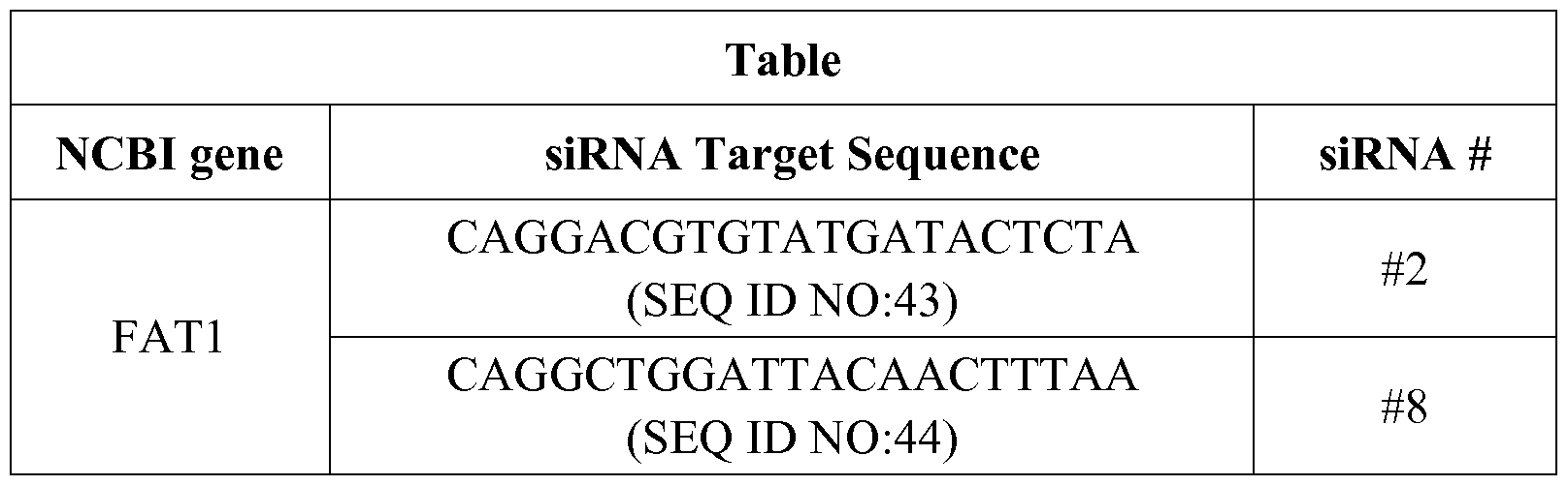
Example 4. Internalization of the anti-FATl monoclonal antibody mAb91.3 by cancer cells.
The ability of the anti-FATl monoclonal antibody to be internalized by cancer cells was assessed in different cancer model. Indeed, monoclonal antibodies able to be efficiently internalized by cancer cells are ideal candidate to generate ADC, in which they can be linked to therapeutic drugs, such as small molecules, toxins, radionucleotides, epigenetic agents and others.
Method
The ability of the anti-FATl antibody to be internalized was first assessed by flow cytometry, monitoring the kinetics by with the surface-bound antibody disappeared from the cell surface upon temperature shift from 0°C to 37°C. In parallel, confocal microscopy was used to confirm the accumulation of antibody complexes in the
intracellular milieu.
For Flow cytometry analysis Cok>205 cells were incubated with the anti-FATl monoclonal antibody (10 micrograms/ml) for 30' at 4°C, as described (see Example 2) to allow antibody binding on the cell surface. Then cells were washed with PBS-5% FCS to remove unbound antibody and shifted to 37°C. At time points, cells were incubated for 20 min with the appropriate dilution of R-Phycoerythrin (PE)-conjugated secondary antibodies at 4°C. After washing, cells were analysed by a flow cytometer and the data were analyzed, as described.
For confocal microscopy analysis HCT15 cells were plated on microscope coverslips as described and after 48 h were washed with PBS. Cells were incubated with the anti-FATl monoclonal antibody mAb91.3 for 1 hour at 4°C (10 micrograms/ ml) and subsequently shifted at 37°C. Cells were then fixed with 10 minute incubation with 90% cold methanol and stained with Alexafluor 488-labeled goat anti-mouse antibodies. DAPI (Molecular Probes) was used to visualize nuclei. The cells were mounted with glycerol plastine and observed under a laser-scanning confocal microscope (LeicaSP5).
Results
Flow cytometry and confocal microscopy analyses showed that the anti-FATl antibody is able to bind the cells surface and upon temperature shift to 37°C it disappears from the cell surface and accumulates in the intracellular milieu (Figure 8) These results confirm that the anti-FATl antibody is efficiently internalized by cancer cells and indicate that the antibody is a suitable vehicle to drive therapeutic/cancer cytotoxic agents within cancer cells.
Example 5. Therapeutic activity of the anti-FATl antibody in mouse colon cancer models
Marker specific monoclonal antibodies able to selectively bind and reduce human cancers engrafted in appropriate mouse models can be developed as therapeutic agents either as naked antibody or as ADC. Such antibodies can be used in therapeutic treatment regimens of oncologic patients, to reduce the tumor burden in patients affected by
primary or metastatic cancers. Moreover, they can be used in preventive treatments to prevent or delay the formation of cancer, for instance in the adjuvant therapy of patients subjected to surgery.
Method
The efficacy of the anti-FATl mAb91.3 against cancer growth was assessed in colon cancer xenograft mouse models in two experimental setting aimed at measuring the ability of the antibody to reduce tumor growth or delay tumor formation.
Therapeutic setting. The HCT15 and the HT29 human colon carcinoma cells (5x10⁶ cells) were injected subcutaneously into nude athymic mice. Mice (6 per group) bearing xenografts of approx. 60-100 mm3 were administered i.v. of either mAb91.3 or the irrelevant mAb 61 (300 micrograms/dose, equal to approximately 12 mg mAb per Kg of animal weight, 2 doses per week). The specificity of the antibody binding to the tumor was also assessed by injecting mice with Near Infrared (NI) fluorescence-labeled antibodies (300 micrograms) followed by optical imaging 2-3 days after mAb injection. Tumor growth measured with a caliper over approximately a 2 week-period. Mice bearing tumors higher than 700 mm3 were sacrificed.
Preventive Setting. The HCT15 human colon carcinoma cells (5x 10⁶ cells) were injected subcutaneously into nude athymic mice (8 per group). Mice were administered i.v. with repeated doses of either mAb91.3 or the irrelevant mAb61 (IgGlk isotype control) (300 micrograms/dose, equal to approximately 12 mg mAb per Kg of animal weight, 2 doses per week starting from day -1 before cancer injection). Tumor growth measured with a caliper over a 3week-period.
Results
The anti-FATl monoclonal antibody was able to bind colon cancer xenograft (Figure 9A) and showed negligible background distribution in other animal anatomical districts. Moreover, the anti-FATl antibody was able to significant reduce growth rate in both HCT15 and HT29 colon cancer (Figure 9Aand 9B) both in the therapeutic and the preventive experimental settings.
Example 6. Therapeutic activity of the anti-FAT mAb91.3 in an indirect antibody-drug conjugate.
mAbs can be used as naked molecules or conjugated with cell payloads (radioisotopes, drugs or toxins) to direct kill tumor cells or to activate pro-drugs specifically within the tumors. These antibody-drug conjugates (ADC) can deliver a toxic load selectively to the tumor site while normal tissues are generally spared. ADC are of particular interest in that their therapeutic efficacy is stronger than that of naked antibodies.
The most important property of antibodies to be used for the generation of ADC is their specificity for cancer cells, and the ability to be efficiently internalized by them so as to deliver the toxic compound in the intracellular compartment.
In order to minimize toxicity, conjugates are usually engineered based on molecules with a short serum half-life (e.g. the use of IgG3 or IgG4 isotypes). Different linker chemistry can be used to link the cell payload to the antibody. Labile linkers allow a rapid dissociation of the drug from the antibody within the cells (e.g. pH sensitive linkers dissociates from the antibody at pH below 6, allowing the drug release within endosomes or lysosomes). Stable linkers require complete proteolytic digestion of the ADC to release the cytotoxic drug as the active metabolite. An in vitro assay generally used to predict the potential of an antibody as ADC exploits the use of a secondary antibody conjugated to saporin, the most potent of the plant ribosome-inactivating proteins. In this assay the primary monoclonal antibody is incubated with cancer cells to allow surface binding. Afterwards a saporin-conjugated secondary antibody is added that recognizes the cell-bound primary antibody. After shift to 37°C the immunocomplex is internalized in the cell and cell death is induced.
Method
HCT15 cells were seeded on 96w plates (2000 cells per well) and incubated 4°C for 30' with mAb91.3 at concentrations ranging from 1 to 40 micrograms/ml. After washing, cells were incubated for 30' with a saporin-conjugated secondary antibody
(FABZAP system, ATS) according to the manufacturer's recommendation and shifted at 37°C for 72 hours to allow internalization and cell killing. The percentage of killing was evaluated with the MTT assay. Results are from triplicate samples.
Results
The anti-FATl mAb91.3 incubated in the presence of a saporin-conjugated secondary showed a significant killing on HCT15 cancer cells, indicating that this antibody has a high potential for the generation of ADC (see Figure 10).
Example 7. Identification of monoclonal antibodies reactive with peculiar FAT1 epitopes able to be bind the surface of cancer cells and internalized upon binding
To reinforce the validity of FAT1 as potential target of monoclonal antibody therapy a panel of anti-FATl monoclonal antibodies were screened in search for other antibodies that recognize the FAT1 region exposed on the surface of cancer cells and are internalized by cancer cells upon binding.
Method
A panel of monoclonal antibodies secreted by distinct hybridoma cells able to recognize FAT1 in ELISA were tested for the ability to recognize colon cancer cells in Western blot and FACS (see Western blot and FACS methods described in Example 2) and of being internalized upon binding (see Internalization methods described in Example 4).
Results
The FACS selection process allowed to identify another anti-FATl monoclonal antibody (namely mAb91.4) able to bind the surface of HCT15 and Colo205 colon cancer cells and to internalize upon temperature shift to 37°C (Figure 11).
Example 8. Identification of the FAT1 region recognized by the monoclonal antibodies and epitope mapping
The epitopes specifically recognized by monoclonal antibodies on cancer cells can be exploited as diagnostic tools for the development of diagnostic assay. Moreover, they
can be used as targets for the development of affinity drugs with therapeutic properties. Given the high molecular weight of FAT1 , a selection of the FAT1 regions recognized by mAb91.3 and mAb91.4 was done by transfecting cells with plasmids encoding overlapping FAT1. These regions were further subcloned in smaller fragments and expressed in a recombinant forms and analyzed by enzyme-linked immunosorbent assay (ELISA) and Western blot. Finally, overlapping peptides were generated and used to identify the antibody epitope/s. The specificity of the monoclonal antibodies for the target epitopes was demonstrated by peptide competition experiments in ELISA, FACS and IHC. Overall the results unambiguously led to the identification of the FAT1 epitopes recognized on the surface of cancer cells and detected in cancer tissues by IHC.
Method
Overlapping FAT1 cDNAs regions encoding approximately 600-800 amino acids were cloned in the mammalian expression vector pcDNA3.1 so as to generate a series of plasmids globally covering the FAT1 extracellular region from amino acid 1 to amino acid 4181. For cloning, cDNA were generated from pools of total RNA derived from Human testis, Human placenta, Human bone marrow, Human fetal brain, in reverse transcription reactions and the entire coding regions were PCR-amplified with specific primers pairs. PCR products were cloned into plasmid pcDNA3 (Invitrogen) so as to generate His6-V5 tagged fusions. HeLa and Hek-293T cells were grown in DMEM-10% FCS supplemented with 1 mM Glutamine were transiently transfected with preparation of the resulting plasmid and with the empty vector as negative control using the Lipofectamine-2000 transfection reagent (Invitrogen). After 48 hours, cells were collected and analysed by Western blot as described in Example 2, using the anti-FATl mAb91.3 or an anti-V5 antibody.
Shorter FAT1 domains covering Region A and Region B (see example before) were also cloned, used for transfection and analysed the ability of the mAb91.3 to recognize them in Western blot. Moreover, these FAT1 regions were expressed in recombinant form, purified from E.coli and used for ELISA. Finally, 25 mer peptides
were obtained by chemical synthesis covering selected FAT1 regions and used for competition of the antibody binding in ELISA, FACS and IHC, thus leading to the unambiguous identification of the mAb91.3 target epitopes.
For ELISA competition experiments, ELISA plates (Nunc Maxisorp) were coated with 1 μg of the FAT1 recombinant proteins in PBS (pH 7.4) at 4°C overnight. The plates were washed, treated for 1 h at 37°C with PBS-1% BSA, and 100 μΐ aliquots of mAb91.3 at (10 micrograms/ml) in PBS-0.1% Tween were added to the wells in the presence of the 25mer peptides at concentration ranging from 0.001 to 100 micrograms/ml. After incubation for 2 h at 37°C, plates were again washed and incubated for lh at 37°C with alkaline-phosphatase conjugated goat anti-mouse IgG (Sigma) diluted 1 :2500 in PBS-Tween. Thereafter 100 μΐ of PNPP substrate (Sigma) were added to the samples and incubated for 30 min at room temperature and optical densities were read at 405 nm.
For FACS competition, Colo205 and HCT15 cells were co-incubated with mAb91.3 in the presence of different concentrations of selected 25mer peptides (ranging from 0 to 100 micrograms per ml) and the inhibition of surface binding was assessed by FACS, as described in Example 2, compared to samples incubated with an irrelevant peptide.
For IHC competition experiments, cancer tissues were co-incubated with mAb91.3 in the presence of selected 25mer peptides (100 micrograms per ml) and residual antibody binding to cancer cells was assessed (see Method in Example 1).
Results
HeLa and HEK-293T were transfected with plasmids encoding overlapping FAT1 regions and their expression was first confirmed by Western blot using the anti-V5 antibody. Two positive FAT1 regions were recognized by mAb91.3 overlapping for 102 amino acids (Fragment A, encompassing the amino acid regions from amino acid 723 to amino acid 1352, and Fragment B encompassing the FAT1 region from amino acid 1246 to amino acid 1879) were recognized by the mAb91.3. Moreover, a shorter form of Fragment A lacking the region overlapping with Fragment B (referred as fragment A
short) was also recognized by the antibody. This evidence indicates that, unexpectedly, this monoclonal antibody has two binding sites on FATl protein (Figure 12). Fragment A-transfected cells were also recognized by mAb91.4, suggesting that mAb91.3 and mAb91.4 share at least in part the same epitopes.
HeLa and HEK-293T were also transfected with plasmids encoding shorter FATl regions included in Fragment A and Fragment B, which allowed to discriminate that the two antibody binding sites map on two distinct cadherin domains, namely Domain 8 (from amino acid 823 to amino acid 927 - SEQ ID NO:28) and Domain 12 (from amino acid 1246 to 1352 - SEQ ID NO:29). Both Domains 8 and 12 are included in the original Region A (SEQ ID NO:27) while Domain 12 is located in the overlapping region between Region A and Region B.
FATl recombinant proteins encompassing the two FATl cadherin Domain 8 and 12 were also recognized in ELISA by mAb91.3, further confirming antibody specificity. Afterwards, for a more accurate epitope identification, a panel of overlapping peptides were designed on Domain 8 and Domain 12 and used in ELISA and FACS competition experiments. Among them, three 25mer peptides encompassing the FATl regions were recognized by mAb91.3 in ELISA. Among them, Peptide 1 and 2 map on the cadherin Domain 12 and partially overlap, whereas peptide 3 maps on the Domain 8. The peptide sequences are reported below.
Peptide 1 : REPLYHVIATDKDEGPNAEISYSIE (SEQ ID NO:30)
Peptide 2: YHVIATDKDEGPNAEISYSIEDGNE (SEQ ID NO:31)
Peptide 3: IQVEATDKDLGPNGHVTYSIVTDTD (SEQ ID NO:32)
ELISA competition experiments showed that the binding of mAb91.3 on the two FATl recombinant domains was almost abolished by competition with each of the three peptides.
Similarly, FACS competition experiments showed that the antibody ability to bind the surface of Colo205 and HCT15 cells was dramatically reduced by competition the three peptides (Figure 13).
IHC competition experiment was carried out with peptide 1 on colon cancer tissue. Under this condition the antibody recognition of cancer cells was almost abolished (Figure 14).
Overall, results demonstrate that mAb91.3 and mAb91.4 epitopes are included Region A. For mAb 91.3, a more precise epitope mapping showed that its target epitopes are located in peptides 1, 2 and 3. The three peptides show differences in their primary sequence and have amino acid identity between 84 to 48%. Moreover, shorter peptides encompassing the central conserved region are not able to compete for the antibody binding. Thus, a plausible hypothesis is that they have a structural similarity and that anti-FATl monoclonal antibodies recognize a structural motif in common between them.
(see annexed sequence listings).
Example 9. Sequencing of mAb91.3 and mAb91.4
Complementarity determining regions (CDRs) are the most variable part of antibody molecules determining the diversity of these molecules, and represent a unique feature of the antibody. CDRs are the antibody regions that complement an antigen's shape and determine the antibody specificity for given antigens. Differences in the CDRs are also responsible for the affinity constant of the monoclonal antibodies for their epitopes. RNA was isolated from the hybridoma clone secreting mAb91.3 and subjected to RT-PCR using degenerated primers able to amplify the variable regions of heavy and light chains (23). Since the subclass of mAb91.3 and mAb91.4 is IgGlk, the following primers were used:
Heavy chain:
IgGl :HC ATAGACAGATGGGGGTGTCGTTTTGGC (SEQ ID NO:45)
MH1 SARGTNMAGCTGSAGSAGTC (SEQ ID NO:47)
MH2 SARGTNMAGCTGSAGSAGTCWGG (SEQ ID NO:48)
Light chain
Kc GGATACAGTTGGTGCAGCATC (SEQ ID NO:46)
Mk GAYATTGTGMTSACMCARWCTMCA (SEQ ID NO:49)
The amplification products were sequenced and the variable regions were analyzed using three different sequence analysis tools: Ig-BLAST http://www.ncbi.nlm.nih.gov/igblast/ (Ye J1, Ma N, Madden TL, Ostell JM.) IgBLAST: an immunoglobulin variable domain sequence analysis tool. Nucleic Acids Res. 2013 Jul;41(Web Server issue): W34-40. doi: 10.1093/nar/gkt382. Epub 2013 May 13.), V-BASE2 http://www.vbase2.org/ (Retter I, Althaus HH, Munch R, Muller W: VBASE2, an integrative V gene database. Nucleic Acids Res. 2005 Jan l;33(Database issue) :D671-4) and the fully integrated antibody discovery system AbYsis http://www.bioinf.org.uk/abysis.
For either of the two monoclonal antibodies, sets of CDRs were determined with the three predictors.
The CDR and variable region sequences of mAb91.3 and mAb91.4 are identified in the annexed sequence listing, wherein:
mAb 91.3 heavy chain CDRs: SEQ ID NOs: 17-19
mAb 91.3 light chain CDRs: SEQ ID NOs: 20-22
mAb 91.3 heavy chain: SEQ ID NO:25
mAb 91.3 light chain: SEQ ID NO:26
mAb 91.4 heavy chain CDRs: SEQ ID NOs: 37-39
mAb 91.4 light chain CDRs: SEQ ID NOs:40-42
mAb 91.4 heavy chain: SEQ ID NO:35
mAb 91.4 light chain: SEQ ID NO:36
Example 10. Determination of the affinity constant of the anti-FATl mAb91.3
Method
The kinetics of association and dissociation and the Dissociation constants of the anti-FATl mAb91.3 were determined using Surface Plasmon Resonance Analysis. Experiments were performed at 25°C with a BIACORE T100 instrument (Biacore AB, Uppsala, Sweden). The recombinant FAT1 proteins were immobilised on a carboxymethylated dextran-coated (CM5) sensor chip by amine coupling. Briefly, a
mixture of 0.2 M l-ethyl-3-diaminopropyl-carbodiimide (EDC) and 0.05 M N-hydroxysuccinimide (NHS) was used for sensor chip surface activation. Proteins pre-concentrated in 0.01 M sodium acetate pH 3.5 were injected at concentration ranging from 2.5 and 5 μg/ml for 10 min and then 1 M ethanolamine pH 8.5 was used to block any remaining activated groups. mAb91.3 was diluted in HBS-EP+ buffer at different concentrations from 0.625 μg/ml to 5μg/ml and injected for 120s at a flow rate of 30μ1/ηιίη on flow cell 2 and 3. HBS-EP+ buffer was run as control. Dissociation was followed for 450 sec, regeneration was achieved with a short pulse of glycine lOmM pH2.0. kon, k0ff and KD were calculated with the 1 : 1 Langmuir model using "BiaEvaluation 4.1".
Results
mAb91.3 Dissociation constants (KD) on FAT Domain 8 and 12, reported below, ranged from 10"8 to 10"9M

Example 11. Generation of a recombinant monoclonal antibody reproducing mAb91.3 properties
Recombinant monoclonal antibodies can be prepared in order to improve the characteristic and functions of antibodies. Changes in the variable region can be made in order to improve the antigen binding characteristics. Changes in the constant region can, in general, be made in order to improve the cellular process characteristics, such as ADCC activity, complement fixation, interaction with membranes, and other effector functions. Alterations can be made by standard recombinant techniques.
Moreover, recombinant monoclonal antibodies can be also prepared to confirm the specificity and molecular properties of the original antibodies produced by hybridoma clones.
Method
R A was isolated from the hybridoma clone secreting mAb91.3 and subjected to RT-PCR using primers able to amplify the variable regions of heavy and light chains. These primers contain convenient modification of the immunoglobulin sequences in order to clone the variable regions in the light (Igk) and heavy (IgG2a) pFUSEss expression vectors (Invivogen). The plasmids obtained containing the variable regions of mAb91.3 were sequenced and the corrected clones (light and heavy chains) were used to cotransfect HEK-293T cells. Total cell extracts and culture supernatant were analyzed by Western blot using a secondary antibody to confirm the expression and correct assembly of the recombinant antibody.
Results
A recombinant mouse IgG2a antibody bearing the variable regions of mAb91.3 has been produced and detected both in total extracts and in the culture supernatant. The recombinant antibody produced in the cell culture supernatant was used in WB, ELISA and FACS analysis and confirmed its ability to bind the surface of Cok>205 cells and to recognize specific FAT1 polypeptides.
Example 12. Immunogenicity of cell-derived exosomes bearing FAT1
Exosomes released by cancer cells can be used as antigens for vaccines, able to elicit strong immune responses. Cancer derived exosomes bearing FAT1 can be exploited for the development of vaccines against FAT1 positive cancer types, such as colon, ovary, esophagus, kidney and prostate.
Methods
Preparation of exosomes for immunization studies
For immunization studies, exosomes from cell culture supernatants were isolated by differential centrifugation as described (24). Briefly, lxlO8 HCT15 cells were cultured in DMEM-10% FCS until confluency in 18 175cm2 flasks until pre-confluence. For exosomes preparation, the culture medium was replaced with serum-free medium (PFHM-II Gibco-Life Technologies), cultured for 24 h and then centrifuged at 200xg for
10 min (pellet PI). The supernatant was collected and centrifuged twice at 500g for 10 min (pellet P2). The second supernatant was sequentially centrifuged at 2,000xg twice for 15 min (pellet P3), once at 10,000xg for 30 min (pellet P4), and once at 70,000xg for 60 min (pellet P5), using a SW28 rotor (Beckman instruments, Inc.). The cellular pellet PI was solubilized in 1 ml of C-RIPA buffer (50 mM Tris-HCl pH7.5, 150 mM NaCl, 1% Nonidet P-40; 2mM EGTA, 1 mM orthovanadate, 0.1% SDS, 0.5% Na-deoxycholate, 1 mM phenyl-methane-sulphonylfluoride, 10 μg/ml leupeptin, 10 μg/ml aprotinin) while each of the supernatant-derived pellets P2-P5 were solubilized in 0.5 μΐ of the same buffer. After clarification, the protein concentration of each sample was determined by Bradford.
As quality controls of the exosomal preparation, 20 μg of PI extract and 10 μg of P2-P5 extracts (corresponding to approximately 2xl05 and 2xl07 cells, respectively) were loaded on SDS-PAGE (4-12%) and analyzed by Western blot with antibodies targeting the exosomal marker CD81. Moreover, the presence of FAT1 in exosomes was also assessed by Western blot as described.
Immunizations
5/6 week old CD1 outbred female mice (5 mice per group) were immunised intra-peritoneally at days 1, 14 and 28 with exosomes (15 micrograms, in 100 microliters) formulated with an equal volume of Alum Hydroxide as adjuvant at the final concentration of 3 mg/ml. Two weeks after the last immunization mice were bled and sera from individual mice were pooled.
ELISA analysis
Total IgG titers elicited by immunizing mice with exosomes was tested on two FAT1 recombinant domains covering the protein cadherin domains 7-10 and 11-12 (Domain 7-10 from amino acid 723 to amino acid 1148; Domain 11-12 regions from amino acid 1136 to amino acid 1352) as proteins were assayed by enzyme-linked immunosorbent assay (ELISA). Individual wells of micro-ELISA plates (Nunc Maxisorp) were coated with 1 μg of each recombinant protein in PBS (pH 7.4) at 4°C overnight. The
plates were washed, treated for lh at 37°C with PBS-1%BSA, and 100 μΐ aliquots of anti- sera towards exosomes, at different serial dilutions in PBS-0.1% Tween, were added to the wells. After incubation for 2h at 37°C, plates were again washed and incubated for lh at 37°C with alkaline-phosphatase conjugated goat anti-mouse IgG (Sigma) diluted 1 :2500 in PBS-Tween. Thereafter 100 μΐ of PNPP (Sigma) were added to the samples and incubated for 30 min. at room temperature. Optical densities were read at 405 nm and the serum-antibody titers were defined as the serum dilution yielding an OD value of 0.5.
RESULTS
To verify the ability of the FAT 1 -bearing exosomes to elicit high antibody titers against FAT1, CD1 mice were mice immunized with the combination exosomes (15 micrograms) formulated in Alum Hydroxide. Sera collected after the last immunization were pooled and analyzed by ELISA on plates coated with FAT1 recombinant proteins. As shown in Figure 15, exosomes induced high antibodies titers against both recombinant FAT1 domains. During the entire experiments mice did not showed any evident sign of toxicity or pain. Results confirm that that the FAT1- Exosomes formulation is safe and highly immunogenic, thus could be exploited for as candidate antigen for anticancer vaccines.
References
1. Adams G.P. and Weiner L.M. (2005) Monoclonal antibody therapy cancer. Nat Biotechnol. 23: 1147-1157.
2. Yang C. & Robbins D.B. (2011) The role of tumor-derived exosomes in cancer pathogenesis. Clinical and Developmental Immunology, doi: 10.1155/2011/842849). 3. Wolfers J, Lozier A, Raposo G, et al. Tumor-derived exosomes are a source of shared tumor rejection antigens for CTL cross-priming. Nature Medicine. 2001 ;7(3):297- 303).
4. Bu N, Wu H, Sun B, et al. Exosome-loaded dendritic cells elicit tumor-specific CD8(+) cytotoxic T cells in patients with glioma. Journal of Neuro-Oncology. 104(3):659-667).
5. Xiu F, Cai Z, Yang Y, Wang X, Wang J, Cao X. Surface anchorage of superantigen SEA promotes induction of specific antitumor immune response by tumor-derived exosomes. Journal of Molecular Medicine. 2007;85(5):511-521.
6. Anderson, L., and Seilhamer, J. (1997). A comparison of selected mRNA and protein abundances in human liver. Electrophoresis 18: 533 - 537.
7. Chen, G., Gharib, T.G., Wang, H., Huang, C.C., Kuick, R., Thomas, D.G., Shedden, K.A., Misek, D.E., Taylor, J. M., Giordano, T. J., Kardia, S.L., Iannettoni, M.D., Yee, J., Hogg, P.J., Orringer, M.B., Hanash, S.M., and Beer, D.G. (2003) Protein profiles associated with survival in adenocarcinoma. Proc. Natl. Acad. Sci. U. S. A 100: 13537 - 13542.
8. Ginestier, C, Charafe-Jauffret, E., Bertucci, F., Eisinger, F., Geneix, J., Bechlian, D., Conte, N., Adelaide, J., Toiron, Y., Nguyen, C, Viens, P., Mozziconacci, M. J., Houlgatte, R., Birnbaum, D., and Jacquemier, J. (2002) Distinct and complementary information provided by use of tissue and DNA microarrays in the study of breast tumor markers. Am. J. Pathol. 161 : 1223-1233.
9. Gygi, S.P., Rochon, Y., Franza, B.R., and Aebersold, R. (1999) Correlation
between protein and mRNA abundance in yeast. Mol. Cell. Biol. 19, 1720 -1730.
10. Nishizuka, S., Charboneau, L., Young, L., Major, S., Reinhold, W.C., Waltham, M., Kouros-Mehr, H., Bussey, K. J., Lee, J.K., Espina, V., Munson, P. J., Petricoin, E., Ill, Liotta, L. A., and Weinstein, J.N. (2003) Proteomic profiling of the NCI-60 cancer cell lines using new high-density reverse-phase lysate microarrays. Proc. Natl. Acad. Sci. U.S.A. 100, 14229 - 14234.
11. Tyers, M., and Mann, M. (2003) From genomics to proteomics. Nature 422: 193-197.
12. Ivan Hateren NJ, Das RM, Hautbergue GM, Borycki AG, Placzek M and Wilson SA: Fat J acts via the Hippo mediator Yapl to restrict the size of neural progenitor cell pools. Development 138: 1893-1902, 2011.
13. Moeller MJ, Soofi A, Braun GS, et al: Protocadherin FAT1 binds Ena/VASP proteins and is necessary for actin dynamics and cell polarization. EMBO J 23 : 3769-3779, 2004.
14. Nakaya K, Yamagata HD, Arita N, et al: Identification of homozygous deletions of tumor suppressor gene FAT in oral cancer using CGH-array. Oncogene 26: 5300-5308, 2007.
15. Chosdol K, Misra A, Puri S, et al: Frequent loss of heterozygosity and altered expression of the candidate tumor suppressor gene "FAT" in human astrocytic tumors. BMC Cancer 9: 5, 2009.
16. Lee S, Stewart S, Nagtegaal I, et al: Differentially expressed genes regulating the progression of ductal carcinoma in situ to invasive breast cancer. Cancer Res 72: 4574-4586, 2012.
17. Morris LG, Kaufman AM, Gong Y, Ramaswami D, Walsh LA, Turcan S, Eng S, Kannan K, Zou Y, Peng L, Banuchi VE, Paty P, Zeng Z, Vakiani E, Solit D, Singh B,
Ganly I, Liau L, Cloughesy TC, Mischel PS, Mellinghoff IK, Chan TA. Recurrent somatic mutation of FAT1 in multiple human cancers leads to aberrant Wnt activation. Nat Genet. 2013 45:253-61. doi: 10.1038/ng.2538.
18. Sadeqzadeh E, de Bock CE, Zhang XD, Shipman KL, Scott NM, Song C, Yeadon T, Oliveira CS, Jin B, Hersey P, Boyd AW, Burns GF, Thorne RF. Dual processing of FATl cadherin protein by human melanoma cells generates distinct protein products. J Biol Chem. 2011 Aug 12;286(32):28181-91. doi: 10.1074/jbc.Ml 11.234419.
19. de Bock CE, Ardjmand A, Mo Hoy TJ, et al: The Fatl cadherin is overexpressed and an independent prognostic factor for survival in paired diagnosis-relapse samples of precursor B-cell acute lymphoblastic leukemia. Leukemia 26: 918-926, 2012.
20. Settakorn J, Kaewpila N, Burns GF and Leong AS: FAT, E-cadherin, β-catenin, HER 2/neu, Ki67 immuno-expression, and histological grade in intrahepatic cholangiocarcinoma. J Clin Pathol 58: 1249-1254, 2005.
21. Kononen J, Bubendorf L, Kallioniemi A, Barlund M, Schraml P, Leighton S, Torhorst J, Mihatsch MJ, Sauter G, Kallioniemi OP. Tissue microarrays for high- throughput molecular profiling of tumor specimens. (1998) Nat Med. 4:844-7.
22. Kallioniemi OP, Wagner U, Kononen J, Sauter G. (2001) Tissue microarray technology for high-throughput molecular profiling of cancer. Hum Mol Genet.
10:657-62.
23. Wang, Z. Raifu M, Howard M, Smith L, Hansen D, Goldsby R, Ratner D (2000) Universal PCR amplification of mouse immunoglobulin gene variable regions: the design of degenerate primers and an assessment of the effect of DNA polymerase 3' to 5' exonuclease activity. Journal of immunological methods 233, 167-77.
24. Raposo G, Nijman HW, Stoorvogel W, Liejendekker R, Harding CV, Melief CJ, Geuze HJ (1996) B lymphocytes secrete antigen- presenting vesicles. J Exp Med 183: 1161-1172.
Claims
1. The use of:
(i) a FAT1 protein, in one of its iso forms SEQ ID NO: l, SEQ ID NO:2, SEQ ID NO:3, SEQ ID NO:4, SEQ ID NO:5 or a different isoform having sequence identity of at least 80%, preferably at least 90%, more preferably at least 95% to SEQ ID NO: l, SEQ ID NO:2, SEQ ID NO:3, SEQ ID NO:4 or SEQ ID NO:5,
or
(ii) a R A molecule coding for a FAT1 protein as defined in (i), the encoding sequence being preferably selected from SEQ ID NO:6, SEQ ID NO:7, SEQ ID NO:8,
SEQ ID NO:9 and SEQ ID NO: 10 or from complementary sequences thereof,
as a tumor marker for in vitro detection of colon, ovary, kidney, esophagus or prostate tumor.
2. A method in vitro for determining the presence of a colon, ovary, esophagus, kidney or prostate tumor in a subject, which comprises the steps of:
(a) providing a test sample which is a sample of tissue suspected of containing tumor cells or a biological fluid sample containing exosomes of suspected tumor cells;
(b) determining the amount of a tumor marker according to claim 1 in said test sample;
(c) comparing said amount of tumor marker with that of a control sample obtained from healthy individuals,
wherein an increased amount of tumor marker in the test sample compared to the control sample is indicative of a tumor state in said subject.
3. A method according to claim 2, wherein the tumor marker is a FAT1 protein as defined in claim 1 and determination of its amount is effected by means of an antibody.
4. A method according to claim 3, wherein the antibody is a monoclonal antibody.
5. A method according to claims 2-4, which is based on immunoradiometric, immunoenzymatic or immunohistochemical techniques.
6. An isolated antibody or fragment thereof which specifically binds the FAT1 protein, wherein the heavy- and light-chain variable regions of said antibody contain complementarity determining regions 1, 2 and 3 (CDR-H 1-3 and CDR-L 1-3, respectively) and:
- said CDR-H 1-3 comprises the amino acid sequences set forth in SEQ ID NOs: 17, 18 and 19, respectively, or an amino acid sequence identical to: SEQ ID NO: 17 by at least 60%, preferably at least 85%; SEQ ID NO: 18 by at least 85%, preferably at least 90%; and to SEQ ID NO: 19 by at least 90%, preferably at least 95%;
- said CDR-L 1-3 comprises the amino acid sequences set forth in SEQ ID NOs:20, 21 and 22, respectively, or an amino acid sequence identical to: SEQ ID NO:20 by at least 80%, preferably at least 90%; SEQ ID NO:21 by at least 85%, preferably at least 90%; and to SEQ ID NO:22 by at least 88%, preferably at least 95%.
7. An isolated antibody or fragment thereof according to claim 6, wherein said CDR- H 1-3 comprises the amino acid sequences set forth in SEQ ID NOs:37, 38 and 39, respectively, and said CDR-L 1-3 comprises the amino acid sequences set forth in SEQ ID NOs:40, 41 and 42, respectively.
8. An antibody or fragment thereof according to claim 6, wherein said variable region contains heavy and light chains set forth in SEQ ID NO:25 and SEQ ID NO:26, respectively, or heavy and light chain sequences identical to SEQ ID NOs:25 or 26 by at least 85%, preferably at least 95%.
9. An antibody or fragment thereof according to claim 7, wherein said variable region contains the heavy and light chains set forth in SEQ ID NO:35 and SEQ ID NO:36, respectively, or heavy and light chain sequences identical to SEQ ID NOs:35 or 36 by at least 90%, preferably at least 95%.
10. An antibody according to claims 6-9, containing a constant region of a human IgGl, IgG2, IgG3 or Ig4.
11. An isolated monoclonal antibody or fragment thereof which specifically binds to an epitope of the FAT1 protein, wherein said epitope is selected from SEQ ID NOs:27,
28, 29, 30, 31 and 32.
12. An antibody according to claims 6-11, which is a IgG, IgM, IgA, IgD or IgE antibody.
13. An antibody fragment according to claims 6-11, which is a Fab, F(ab')2, Fv, ScFv or SCA fragment.
14. An antibody according to claims 6-12, which is one of the following: a humanized antibody, a human antibody, a chimeric antibody, a diabody, or a linear antibody.
15. An antibody or fragment thereof according to claims 6-14, which is conjugated to an antitumor compound.
16. The use of an antibody or fragment thereof according to claims 6-15, in a method in vitro for determining the presence of a colon, ovary, prostate, esophagus or renal tumor according to claims 2-5.
17. An antibody or fragment thereof according to claims 6-15, for use in the treatment of a colon, ovary, prostate, esophagus or renal tumor.
18. An isolated peptide antigen the amino acid sequence of which is selected from SEQ ID NOs:27, 28, 29, 30, 31 and 32, or from sequences identical to SEQ ID NOs: 27, 28, 29, 30, 31 or 32 by at least 48%, preferably at least 75%>, more preferably at least 84%.
19. A pharmaceutical composition containing an antibody to a FAT1 protein or a fragment thereof according to claims 6-15 or a peptide antigen according to claim 18.
20. A diagnostic kit containing an antibody or a fragment thereof according to claims 6-15 and optionally reagents, buffers, solutions and materials to carry out an immunoassay.
21. A siRNA molecule having a sequence complementary to one of SEQ ID NO:6, SEQ ID NO:7, SEQ ID NO:8, SEQ ID NO:9 or SEQ ID NO: 10, for use in the treatment of colon, ovary or prostate tumor.
Priority Applications (2)
| Application Number | Priority Date | Filing Date | Title |
|---|---|---|---|
| EP14736307.1A EP3008086A2 (en) | 2013-06-14 | 2014-06-13 | Tumor marker, monoclonal antibodies and methods of use thereof |
| US14/897,654 US10359428B2 (en) | 2013-06-14 | 2014-06-13 | Tumor marker, monoclonal antibodies and methods of use thereof |
Applications Claiming Priority (2)
| Application Number | Priority Date | Filing Date | Title |
|---|---|---|---|
| EP13172028 | 2013-06-14 | ||
| EP13172028.6 | 2013-06-14 |
Publications (2)
| Publication Number | Publication Date |
|---|---|
| WO2014198919A2 true WO2014198919A2 (en) | 2014-12-18 |
| WO2014198919A3 WO2014198919A3 (en) | 2015-04-09 |
Family
ID=48607156
Family Applications (1)
| Application Number | Title | Priority Date | Filing Date |
|---|---|---|---|
| PCT/EP2014/062419 WO2014198919A2 (en) | 2013-06-14 | 2014-06-13 | Tumor marker, monoclonal antibodies and methods of use thereof |
Country Status (3)
| Country | Link |
|---|---|
| US (1) | US10359428B2 (en) |
| EP (1) | EP3008086A2 (en) |
| WO (1) | WO2014198919A2 (en) |
Cited By (5)
| Publication number | Priority date | Publication date | Assignee | Title |
|---|---|---|---|---|
| WO2016184860A1 (en) | 2015-05-18 | 2016-11-24 | Universita' Degli Studi Di Trento | Immunogenic compositions containing bacterial outer membrane vesicles and therapeutic uses thereof |
| WO2017120274A1 (en) * | 2016-01-08 | 2017-07-13 | Temple University Of The Commonwealth System Of Higher Education | Stabilized peptide fragments from protocadherin fat1 as cancer biomarkers |
| WO2019148043A1 (en) | 2018-01-26 | 2019-08-01 | Exelixis, Inc. | Compounds for the treatment of kinase-dependent disorders |
| WO2019148036A1 (en) | 2018-01-26 | 2019-08-01 | Exelixis, Inc. | Compounds for the treatment of kinase-dependent disorders |
| WO2019148044A1 (en) | 2018-01-26 | 2019-08-01 | Exelixis, Inc. | Compounds for the treatment of kinase-dependent disorders |
Families Citing this family (1)
| Publication number | Priority date | Publication date | Assignee | Title |
|---|---|---|---|---|
| WO2020181080A1 (en) * | 2019-03-06 | 2020-09-10 | Celldex Therapeutics, Inc. | Biomarkers for treatment of cancer |
Family Cites Families (1)
| Publication number | Priority date | Publication date | Assignee | Title |
|---|---|---|---|---|
| CA2385487A1 (en) | 1999-08-13 | 2001-02-22 | Human Genome Sciences, Inc. | 13 human colon and colon cancer associated proteins |
-
2014
- 2014-06-13 WO PCT/EP2014/062419 patent/WO2014198919A2/en active Application Filing
- 2014-06-13 US US14/897,654 patent/US10359428B2/en not_active Expired - Fee Related
- 2014-06-13 EP EP14736307.1A patent/EP3008086A2/en not_active Withdrawn
Non-Patent Citations (30)
| Title |
|---|
| ADAMS G.P.; WEINER L.M.: "Monoclonal antibody therapy cancer", NAT BIOTECHNOL., vol. 23, 2005, pages 1147 - 1157, XP002456710, DOI: doi:10.1038/nbt1137 |
| ANDERSON, L.; SEILHAMER, J.: "A comparison of selected mRNA and protein abundances in human liver", ELECTROPHORESIS, vol. 18, 1997, pages 533 - 537, XP008013305, DOI: doi:10.1002/elps.1150180333 |
| BOULIANNE, G. L. ET AL., NATURE, vol. 312, 1984, pages 643 - 646 |
| BU N; WU H; SUN B ET AL.: "Exosome-loaded dendritic cells elicit tumor-specific CD8(+) cytotoxic T cells in patients with glioma", JOURNAL OF NEURO-ONCOLOGY, vol. 104, no. 3, pages 659 - 667, XP019948331, DOI: doi:10.1007/s11060-011-0537-1 |
| CHEN, G.; GHARIB, T.G.; WANG, H.; HUANG, C.C.; KUICK, R.; THOMAS, D.G.; SHEDDEN, K.A.; MISEK, D.E.; TAYLOR, J. M.; GIORDANO, T. J.: "Protein profiles associated with survival in adenocarcinoma", PROC. NATL. ACAD. SCI. U. S. A, vol. 100, 2003, pages 13537 - 13542, XP055217585, DOI: doi:10.1073/pnas.2233850100 |
| CHOSDOL K; MISRA A; PURI S ET AL.: "Frequent loss of heterozygosity and altered expression of the candidate tumor suppressor gene ''FAT'' in human astrocytic tumors", BMC CANCER, vol. 9, 2009, pages 5, XP021049059, DOI: doi:10.1186/1471-2407-9-5 |
| DE BOCK CE; ARDJMAND A; MOLLOY TJ ET AL.: "The Fatl cadherin is overexpressed and an independent prognostic factor for survival in paired diagnosis-relapse samples of precursor B-cell acute lymphoblastic leukemia", LEUKEMIA, vol. 26, 2012, pages 918 - 926, XP055141748, DOI: doi:10.1038/leu.2011.319 |
| GINESTIER, C.; CHARAFE-JAUFFRET, E.; BERTUCCI, F.; EISINGER, F.; GENEIX, J.; BECHLIAN, D.; CONTE, N.; ADELAIDE, J.; TOIRON, Y.; NG: "Distinct and complementary information provided by use of tissue and DNA microarrays in the study of breast tumor markers", AM. J. PATHOL., vol. 161, 2002, pages 1223 - 1233, XP008013988 |
| GYGI, S.P.; ROCHON, Y.; FRANZA, B.R.; AEBERSOLD, R.: "Correlation between protein and mRNA abundance in yeast", MOL. CELL. BIOL., vol. 19, 1999, pages 1720 - 1730, XP002923559 |
| IVAN HATEREN NJ; DAS RM; HAUTBERGUE GM; BORYCKI AG; PLACZEK M; WILSON SA: "FatJ acts via the Hippo mediator Yapl to restrict the size of neural progenitor cell pools", DEVELOPMENT, vol. 138, 2011, pages 1893 - 1902, XP055222154, DOI: doi:10.1242/dev.064204 |
| KALLIONIEMI OP; WAGNER U; KONONEN J; SAUTER G: "Tissue microarray technology for high-throughput molecular profiling of cancer", HUM MOL GENET, vol. 10, 2001, pages 657 - 62, XP002306111, DOI: doi:10.1093/hmg/10.7.657 |
| KOHLER; MILSTEIN, NATURE, vol. 265, 1975, pages 495 |
| KONONEN J; BUBENDORF L; KALLIONIEMI A; BARLUND M; SCHRAML P; LEIGHTON S; TORHORST J; MIHATSCH MJ; SAUTER G; KALLIONIEMI OP: "Tissue microarrays for high-throughput molecular profiling of tumor specimens", NAT MED., vol. 4, 1998, pages 844 - 7, XP002927997, DOI: doi:10.1038/nm0798-844 |
| LEE S; STEWART S; NAGTEGAAL I ET AL.: "Differentially expressed genes regulating the progression of ductal carcinoma in situ to invasive breast cancer", CANCER RES, vol. 72, 2012, pages 4574 - 4586, XP055360915, DOI: doi:10.1158/0008-5472.CAN-12-0636 |
| MOELLER MJ; SOOFI A; BRAUN GS ET AL.: "Protocadherin FATl binds Ena/VASP proteins and is necessary for actin dynamics and cell polarization", EMBO J, vol. 23, 2004, pages 3769 - 3779 |
| MORRIS LG; KAUFMAN AM; GONG Y; RAMASWAMI D; WALSH LA; TURCAN $; ENG S; KANNAN K; ZOU Y; PENG L: "Recurrent somatic mutation of FAT in multiple human cancers leads to aberrant Wnt activation", NAT GENET., vol. 45, 2013, pages 253 - 61, XP055144931, DOI: doi:10.1038/ng.2538 |
| MORRISON, S. L. ET AL.: "Chimeric Human Antibody Molecules; Mouse Antigen Binding Domains with Human Constant Region Domains", PROC. NATL. ACAD. SCI. USA, vol. 81, 1984, pages 6841 - 6855 |
| NAKAYA K; YAMAGATA HD; ARITA N ET AL.: "Identification of homozygous deletions of tumor suppressor gene FAT in oral cancer using CGH-array", ONCOGENE, vol. 26, 2007, pages 5300 - 5308, XP002497569, DOI: doi:10.1038/SJ.ONC.1210330 |
| NISHIZUKA, S.; CHARBONEAU, L.; YOUNG, L.; MAJOR, S.; REINHOLD, W.C.; WALTHAM, M.; KOUROS-MEHR, H.; BUSSEY, K. J.; LEE, J.K.; ESPIN: "Proteomic profiling of the NCI-60 cancer cell lines using new high-density reverse-phase lysate microarrays", PROC. NATL. ACAD. SCI. U.S.A., vol. 100, 2003, pages 14229 - 14234, XP055291251, DOI: doi:10.1073/pnas.2331323100 |
| NUCLEIC ACIDS RES., vol. 41, 13 May 2013 (2013-05-13), pages W34 - 40 |
| RAPOSO G; NIJMAN HW; STOORVOGEL W; LIEJENDEKKER R; HARDING CV; MELIEF CJ; GEUZE HJ: "B lymphocytes secrete antigen- presenting vesicles", J EXP MED, vol. 183, 1996, pages 1161 - 1172, XP002060486, DOI: doi:10.1084/jem.183.3.1161 |
| RETTER I; ALTHAUS HH; MUNCH R; MÜLLER W: "VBASE2, an integrative V gene database", NUCLEIC ACIDS RES., vol. 33, 1 January 2005 (2005-01-01), pages D671 - 4 |
| SADEQZADEH E; DE BOCK CE; ZHANG XD; SHIPMAN KL; SCOTT NM; SONG C; YEADON T; OLIVEIRA CS; JIN B; HERSEY P: "Dual processing of FATl cadherin protein by human melanoma cells generates distinct protein products", J BIOL CHEM., vol. 286, no. 32, 12 August 2011 (2011-08-12), pages 28181 - 91 |
| See also references of EP3008086A2 |
| SETTAKORN J; KAEWPILA N; BURNS GF; LEONG AS: "FAT, E-cadherin, -catenin, HER 2/neu, Ki67 immuno-expression, and histological grade in intrahepatic cholangiocarcinoma", J CLIN PATHOL, vol. 58, 2005, pages 1249 - 1254 |
| TYERS, M.; MANN, M.: "From genomics to proteomics", NATURE, vol. 422, 2003, pages 193 - 197 |
| WANG, Z.; RAIFU M; HOWARD M; SMITH L; HANSEN D; GOLDSBY R; RATNER D: "Universal PCR amplification of mouse immunoglobulin gene variable regions: the design of degenerate primers and an assessment of the effect of DNA polymerase 3' to 5' exonuclease activity", JOURNAL OF IMMUNOLOGICAL METHODS, vol. 233, 2000, pages 167 - 77, XP004188255, DOI: doi:10.1016/S0022-1759(99)00184-2 |
| WOLFERS J; LOZIER A; RAPOSO G ET AL.: "Tumor-derived exosomes are a source of shared tumor rejection antigens for CTL cross-priming", NATURE MEDICINE, vol. 7, no. 3, 2001, pages 297 - 303 |
| XIU F; CAI Z; YANG Y; WANG X; WANG J; CAO X: "Surface anchorage of superantigen SEA promotes induction of specific antitumor immune response by tumor-derived exosomes", JOURNAL OF MOLECULAR MEDICINE, vol. 85, no. 5, 2007, pages 511 - 521, XP019505896, DOI: doi:10.1007/s00109-006-0154-1 |
| YANG C.; ROBBINS D.B.: "The role of tumor-derived exosomes in cancer pathogenesis", CLINICAL AND DEVELOPMENTAL IMMUNOLOGY, 2011 |
Cited By (6)
| Publication number | Priority date | Publication date | Assignee | Title |
|---|---|---|---|---|
| WO2016184860A1 (en) | 2015-05-18 | 2016-11-24 | Universita' Degli Studi Di Trento | Immunogenic compositions containing bacterial outer membrane vesicles and therapeutic uses thereof |
| EP3838918A1 (en) | 2015-05-18 | 2021-06-23 | BiOMVis Srl | Immunogenic compositions containing bacterial outer membrane vesicles and therapeutic uses thereof |
| WO2017120274A1 (en) * | 2016-01-08 | 2017-07-13 | Temple University Of The Commonwealth System Of Higher Education | Stabilized peptide fragments from protocadherin fat1 as cancer biomarkers |
| WO2019148043A1 (en) | 2018-01-26 | 2019-08-01 | Exelixis, Inc. | Compounds for the treatment of kinase-dependent disorders |
| WO2019148036A1 (en) | 2018-01-26 | 2019-08-01 | Exelixis, Inc. | Compounds for the treatment of kinase-dependent disorders |
| WO2019148044A1 (en) | 2018-01-26 | 2019-08-01 | Exelixis, Inc. | Compounds for the treatment of kinase-dependent disorders |
Also Published As
| Publication number | Publication date |
|---|---|
| US20160131656A1 (en) | 2016-05-12 |
| US10359428B2 (en) | 2019-07-23 |
| WO2014198919A3 (en) | 2015-04-09 |
| EP3008086A2 (en) | 2016-04-20 |
Similar Documents
| Publication | Publication Date | Title |
|---|---|---|
| US20180369269A1 (en) | Antibodies that specifically block the biological activity of a tumor antigen | |
| Kuan et al. | Glycoprotein nonmetastatic melanoma protein B, a potential molecular therapeutic target in patients with glioblastoma multiforme | |
| US10359428B2 (en) | Tumor marker, monoclonal antibodies and methods of use thereof | |
| EP2494351B1 (en) | Colon and rectal tumor markers and methods of use thereof | |
| EP2213686A1 (en) | Tumor markers and methods of use thereof | |
| EP2493916B1 (en) | Lung tumor markers and methods of use thereof | |
| US20180313841A1 (en) | Ovary Tumor Markers And Methods Of Use Thereof | |
| US20170227543A1 (en) | Breast Tumor Markers And Methods Of Use Thereof | |
| US20150093396A1 (en) | Prostate Tumor Markers And Methods Of Use Thereof | |
| US20130137106A1 (en) | Tumor Marker and Methods of Use Thereof | |
| Grifantini et al. | A novel polyclonal antibody library for expression profiling of poorly characterized, membrane and secreted human proteins | |
| US20120045439A1 (en) | Tumor markers and methods of use thereof |
Legal Events
| Date | Code | Title | Description |
|---|---|---|---|
| 121 | Ep: the epo has been informed by wipo that ep was designated in this application |
Ref document number: 14736307 Country of ref document: EP Kind code of ref document: A2 |
|
| WWE | Wipo information: entry into national phase |
Ref document number: 14897654 Country of ref document: US |
|
| WWE | Wipo information: entry into national phase |
Ref document number: 2014736307 Country of ref document: EP |