WO2014181767A1 - Method for producing particulate decellularized tissue - Google Patents
Method for producing particulate decellularized tissue Download PDFInfo
- Publication number
- WO2014181767A1 WO2014181767A1 PCT/JP2014/062149 JP2014062149W WO2014181767A1 WO 2014181767 A1 WO2014181767 A1 WO 2014181767A1 JP 2014062149 W JP2014062149 W JP 2014062149W WO 2014181767 A1 WO2014181767 A1 WO 2014181767A1
- Authority
- WO
- WIPO (PCT)
- Prior art keywords
- tissue
- decellularized tissue
- particulate
- particulate decellularized
- hydrostatic pressure
- Prior art date
Links
Images


Classifications
-
- A—HUMAN NECESSITIES
- A61—MEDICAL OR VETERINARY SCIENCE; HYGIENE
- A61L—METHODS OR APPARATUS FOR STERILISING MATERIALS OR OBJECTS IN GENERAL; DISINFECTION, STERILISATION OR DEODORISATION OF AIR; CHEMICAL ASPECTS OF BANDAGES, DRESSINGS, ABSORBENT PADS OR SURGICAL ARTICLES; MATERIALS FOR BANDAGES, DRESSINGS, ABSORBENT PADS OR SURGICAL ARTICLES
- A61L27/00—Materials for grafts or prostheses or for coating grafts or prostheses
- A61L27/36—Materials for grafts or prostheses or for coating grafts or prostheses containing ingredients of undetermined constitution or reaction products thereof, e.g. transplant tissue, natural bone, extracellular matrix
- A61L27/3604—Materials for grafts or prostheses or for coating grafts or prostheses containing ingredients of undetermined constitution or reaction products thereof, e.g. transplant tissue, natural bone, extracellular matrix characterised by the human or animal origin of the biological material, e.g. hair, fascia, fish scales, silk, shellac, pericardium, pleura, renal tissue, amniotic membrane, parenchymal tissue, fetal tissue, muscle tissue, fat tissue, enamel
- A61L27/3633—Extracellular matrix [ECM]
-
- A—HUMAN NECESSITIES
- A61—MEDICAL OR VETERINARY SCIENCE; HYGIENE
- A61L—METHODS OR APPARATUS FOR STERILISING MATERIALS OR OBJECTS IN GENERAL; DISINFECTION, STERILISATION OR DEODORISATION OF AIR; CHEMICAL ASPECTS OF BANDAGES, DRESSINGS, ABSORBENT PADS OR SURGICAL ARTICLES; MATERIALS FOR BANDAGES, DRESSINGS, ABSORBENT PADS OR SURGICAL ARTICLES
- A61L27/00—Materials for grafts or prostheses or for coating grafts or prostheses
- A61L27/36—Materials for grafts or prostheses or for coating grafts or prostheses containing ingredients of undetermined constitution or reaction products thereof, e.g. transplant tissue, natural bone, extracellular matrix
- A61L27/3683—Materials for grafts or prostheses or for coating grafts or prostheses containing ingredients of undetermined constitution or reaction products thereof, e.g. transplant tissue, natural bone, extracellular matrix subjected to a specific treatment prior to implantation, e.g. decellularising, demineralising, grinding, cellular disruption/non-collagenous protein removal, anti-calcification, crosslinking, supercritical fluid extraction, enzyme treatment
- A61L27/3691—Materials for grafts or prostheses or for coating grafts or prostheses containing ingredients of undetermined constitution or reaction products thereof, e.g. transplant tissue, natural bone, extracellular matrix subjected to a specific treatment prior to implantation, e.g. decellularising, demineralising, grinding, cellular disruption/non-collagenous protein removal, anti-calcification, crosslinking, supercritical fluid extraction, enzyme treatment characterised by physical conditions of the treatment, e.g. applying a compressive force to the composition, pressure cycles, ultrasonic/sonication or microwave treatment, lyophilisation
-
- A—HUMAN NECESSITIES
- A61—MEDICAL OR VETERINARY SCIENCE; HYGIENE
- A61L—METHODS OR APPARATUS FOR STERILISING MATERIALS OR OBJECTS IN GENERAL; DISINFECTION, STERILISATION OR DEODORISATION OF AIR; CHEMICAL ASPECTS OF BANDAGES, DRESSINGS, ABSORBENT PADS OR SURGICAL ARTICLES; MATERIALS FOR BANDAGES, DRESSINGS, ABSORBENT PADS OR SURGICAL ARTICLES
- A61L27/00—Materials for grafts or prostheses or for coating grafts or prostheses
- A61L27/36—Materials for grafts or prostheses or for coating grafts or prostheses containing ingredients of undetermined constitution or reaction products thereof, e.g. transplant tissue, natural bone, extracellular matrix
- A61L27/3604—Materials for grafts or prostheses or for coating grafts or prostheses containing ingredients of undetermined constitution or reaction products thereof, e.g. transplant tissue, natural bone, extracellular matrix characterised by the human or animal origin of the biological material, e.g. hair, fascia, fish scales, silk, shellac, pericardium, pleura, renal tissue, amniotic membrane, parenchymal tissue, fetal tissue, muscle tissue, fat tissue, enamel
-
- A—HUMAN NECESSITIES
- A61—MEDICAL OR VETERINARY SCIENCE; HYGIENE
- A61L—METHODS OR APPARATUS FOR STERILISING MATERIALS OR OBJECTS IN GENERAL; DISINFECTION, STERILISATION OR DEODORISATION OF AIR; CHEMICAL ASPECTS OF BANDAGES, DRESSINGS, ABSORBENT PADS OR SURGICAL ARTICLES; MATERIALS FOR BANDAGES, DRESSINGS, ABSORBENT PADS OR SURGICAL ARTICLES
- A61L27/00—Materials for grafts or prostheses or for coating grafts or prostheses
- A61L27/50—Materials characterised by their function or physical properties, e.g. injectable or lubricating compositions, shape-memory materials, surface modified materials
- A61L27/54—Biologically active materials, e.g. therapeutic substances
-
- A—HUMAN NECESSITIES
- A61—MEDICAL OR VETERINARY SCIENCE; HYGIENE
- A61L—METHODS OR APPARATUS FOR STERILISING MATERIALS OR OBJECTS IN GENERAL; DISINFECTION, STERILISATION OR DEODORISATION OF AIR; CHEMICAL ASPECTS OF BANDAGES, DRESSINGS, ABSORBENT PADS OR SURGICAL ARTICLES; MATERIALS FOR BANDAGES, DRESSINGS, ABSORBENT PADS OR SURGICAL ARTICLES
- A61L27/00—Materials for grafts or prostheses or for coating grafts or prostheses
- A61L27/50—Materials characterised by their function or physical properties, e.g. injectable or lubricating compositions, shape-memory materials, surface modified materials
- A61L27/58—Materials at least partially resorbable by the body
-
- A—HUMAN NECESSITIES
- A61—MEDICAL OR VETERINARY SCIENCE; HYGIENE
- A61P—SPECIFIC THERAPEUTIC ACTIVITY OF CHEMICAL COMPOUNDS OR MEDICINAL PREPARATIONS
- A61P17/00—Drugs for dermatological disorders
- A61P17/02—Drugs for dermatological disorders for treating wounds, ulcers, burns, scars, keloids, or the like
-
- A—HUMAN NECESSITIES
- A61—MEDICAL OR VETERINARY SCIENCE; HYGIENE
- A61P—SPECIFIC THERAPEUTIC ACTIVITY OF CHEMICAL COMPOUNDS OR MEDICINAL PREPARATIONS
- A61P25/00—Drugs for disorders of the nervous system
-
- C—CHEMISTRY; METALLURGY
- C12—BIOCHEMISTRY; BEER; SPIRITS; WINE; VINEGAR; MICROBIOLOGY; ENZYMOLOGY; MUTATION OR GENETIC ENGINEERING
- C12N—MICROORGANISMS OR ENZYMES; COMPOSITIONS THEREOF; PROPAGATING, PRESERVING, OR MAINTAINING MICROORGANISMS; MUTATION OR GENETIC ENGINEERING; CULTURE MEDIA
- C12N5/00—Undifferentiated human, animal or plant cells, e.g. cell lines; Tissues; Cultivation or maintenance thereof; Culture media therefor
- C12N5/0081—Purging biological preparations of unwanted cells
-
- A—HUMAN NECESSITIES
- A61—MEDICAL OR VETERINARY SCIENCE; HYGIENE
- A61L—METHODS OR APPARATUS FOR STERILISING MATERIALS OR OBJECTS IN GENERAL; DISINFECTION, STERILISATION OR DEODORISATION OF AIR; CHEMICAL ASPECTS OF BANDAGES, DRESSINGS, ABSORBENT PADS OR SURGICAL ARTICLES; MATERIALS FOR BANDAGES, DRESSINGS, ABSORBENT PADS OR SURGICAL ARTICLES
- A61L2300/00—Biologically active materials used in bandages, wound dressings, absorbent pads or medical devices
- A61L2300/40—Biologically active materials used in bandages, wound dressings, absorbent pads or medical devices characterised by a specific therapeutic activity or mode of action
- A61L2300/412—Tissue-regenerating or healing or proliferative agents
-
- A—HUMAN NECESSITIES
- A61—MEDICAL OR VETERINARY SCIENCE; HYGIENE
- A61L—METHODS OR APPARATUS FOR STERILISING MATERIALS OR OBJECTS IN GENERAL; DISINFECTION, STERILISATION OR DEODORISATION OF AIR; CHEMICAL ASPECTS OF BANDAGES, DRESSINGS, ABSORBENT PADS OR SURGICAL ARTICLES; MATERIALS FOR BANDAGES, DRESSINGS, ABSORBENT PADS OR SURGICAL ARTICLES
- A61L2430/00—Materials or treatment for tissue regeneration
- A61L2430/34—Materials or treatment for tissue regeneration for soft tissue reconstruction
-
- C—CHEMISTRY; METALLURGY
- C12—BIOCHEMISTRY; BEER; SPIRITS; WINE; VINEGAR; MICROBIOLOGY; ENZYMOLOGY; MUTATION OR GENETIC ENGINEERING
- C12M—APPARATUS FOR ENZYMOLOGY OR MICROBIOLOGY; APPARATUS FOR CULTURING MICROORGANISMS FOR PRODUCING BIOMASS, FOR GROWING CELLS OR FOR OBTAINING FERMENTATION OR METABOLIC PRODUCTS, i.e. BIOREACTORS OR FERMENTERS
- C12M25/00—Means for supporting, enclosing or fixing the microorganisms, e.g. immunocoatings
- C12M25/14—Scaffolds; Matrices
-
- C—CHEMISTRY; METALLURGY
- C12—BIOCHEMISTRY; BEER; SPIRITS; WINE; VINEGAR; MICROBIOLOGY; ENZYMOLOGY; MUTATION OR GENETIC ENGINEERING
- C12N—MICROORGANISMS OR ENZYMES; COMPOSITIONS THEREOF; PROPAGATING, PRESERVING, OR MAINTAINING MICROORGANISMS; MUTATION OR GENETIC ENGINEERING; CULTURE MEDIA
- C12N2521/00—Culture process characterised by the use of hydrostatic pressure, flow or shear forces
Definitions
- the present invention relates to a method for producing a decellularized tissue that can be suitably used for regenerative medicine, cell culture, and the like.
- a method using a surfactant for example, see Patent Documents 1 and 2
- a method using an enzyme for example, see Patent Document 3
- a method using an oxidizing agent for example, refer to Patent Document 4
- methods using ultrahigh hydrostatic pressure treatment for example, refer to Patent Documents 5 to 7
- ultrahigh hydrostatic pressure treatment for example, refer to Patent Documents 5 to 7
- particulate decellularized tissue is also known, and the particulate decellularized tissue is injected into the diseased part and injected into the diseased part. Regeneration / healing is promoted or molded and used as a graft (see, for example, Patent Documents 8 to 10).
- Conventionally known particulate decellularized tissue is produced by a method using a surfactant, but particulate decellularized tissue produced by a method using ultrahigh hydrostatic pressure treatment has not been known. .
- the present inventors have found that the particulate animal tissue decellularized by applying a high hydrostatic pressure in the medium has the effect of attracting cells and inducing differentiation. It has been found that it has no cytotoxicity during cell culture, and the present invention has been completed. That is, the present invention is a method for producing a particulate decellularized tissue, which comprises a step of applying a high hydrostatic pressure to an animal-derived tissue in a medium.
- the present invention includes the following.
- a method for producing a particulate decellularized tissue comprising a step of applying a high hydrostatic pressure to an animal-derived tissue in a medium.
- the method for producing a particulate decellularized tissue according to [1] or [2], wherein the animal-derived decellularized tissue to which a high hydrostatic pressure is applied in a medium is pulverized.
- [5] The animal-derived decellularized tissue to which a high hydrostatic pressure is applied in a medium is washed with a washing solution that does not contain an anionic surfactant and / or a nonionic surfactant.
- 4] The method for producing a particulate decellularized tissue according to any one of 4).
- [6] A particulate decellularized tissue produced by the method for producing a particulate decellularized tissue according to any one of [1] to [5].
- [7] A material for transplantation or treatment using the particulate decellularized tissue according to [6].
- [8] A cell culture material using the particulate decellularized tissue according to [6].
- a particulate decellularized tissue having no cytotoxicity, a cell attracting effect or a differentiation inducing effect, no cytotoxicity, and a high tissue regeneration effect can be obtained. Since the particulate decellularized tissue obtained by the production method of the present invention is in a particulate form, the part to be applied is not limited and can be used in various parts.
- Example 2 of the neurite extension test The result without NGF addition of Example 2 of the neurite extension test is shown.
- the result of NGF non-addition of Example 3 of a neurite extension test is shown.
- the result of NGF non-addition of the blank of a neurite extension test is shown.
- the result of the NGF addition of the blank of a neurite extension test is shown.
- stained rat tissue after 28 days of Example 1 of a subcutaneous implantation test are shown.
- the appearance before the test (upper left figure), the appearance after the test (upper right figure), and the cross section of the stained rat tissue (lower figure) in Example 1 of the frostbite model test are shown.
- the appearance of the frostbite model test blank before the test (upper left figure), the appearance after the test (upper right figure), and the cross section of the stained rat tissue (lower figure) are shown.
- the biological tissue used in the method for producing the particulate decellularized tissue of the present invention is not particularly limited as long as it is a biological tissue having cells derived from vertebrates. However, since there are few rejection reactions, biological tissues derived from mammals or birds Tissues are preferred, and because they are readily available, mammalian livestock, avian livestock or human-derived ecological tissues are more preferred.
- Mammalian livestock includes cattle, horses, camels, llamas, donkeys, yaks, sheep, pigs, goats, deer, alpaca, dogs, raccoon dogs, weasels, foxes, cats, rabbits, hamsters, guinea pigs, rats, mice, squirrels, Raccoon etc. are mentioned.
- examples of avian livestock include parakeets, parrots, chickens, ducks, turkeys, geese, guinea fowls, pheasants, ostriches, and quails.
- porcine, rabbit, and human biological tissues are preferable from the viewpoint of availability.
- a part having a matrix structure outside the cell can be used as a part of a living tissue.
- a part having a matrix structure outside the cell examples include a liver, kidney, ureter, bladder, urethra, tongue, tonsils, esophagus, stomach, small intestine.
- cartilage, bone, liver, kidney, heart, lung, brain, and spinal cord are preferable because of the high tissue regeneration effect.
- the biological tissue is subjected to a treatment for preventing spoilage and functional deterioration after collection.
- a treatment for preventing spoilage and functional deterioration after collection examples include sterilization treatment with a drug, freezing treatment by freezing, and the like, and freezing treatment is preferable because there is little damage to the tissue.
- the biological tissue is collected, a high hydrostatic pressure is applied to the biological tissue, the broken cells are washed and removed, and the particulate decellularized tissue is obtained.
- the biological tissue or decellularized tissue
- the particles are more stable than the state in which the shape of the biological tissue is maintained. It is preferable to pulverize at least before washing and removing the cells because the broken cells can be easily washed away.
- Examples of the method of pulverizing the tissue include a method of pulverizing the living tissue as it is at room temperature, a method of freezing and pulverizing the living tissue in a frozen state, and the like.
- the pulverizing temperature when pulverizing in the frozen state is preferably ⁇ 80 to ⁇ 5 ° C., It is more preferably ⁇ 50 to ⁇ 10 ° C., and most preferably ⁇ 40 to ⁇ 15 ° C. Since it is preferable to store the living tissue in a frozen state, if the stored frozen living tissue is pulverized, the process can be simplified, and the particulate living tissue can be subjected to a high hydrostatic pressure treatment.
- the biological tissue may be pulverized after drying because it is easy to classify after pulverization.
- Examples of the method for drying a living tissue include heat drying, vacuum drying, freeze drying, dehydration with an organic solvent, and the like. Since cell and nucleic acid can be easily washed, freeze drying and dehydration with an organic solvent are preferable. Is more preferable.
- Examples of the organic solvent for dehydration using an organic solvent include ethanol and acetone.
- the living tissue may be decellularized without being pulverized, and the decellularized biological tissue may be dried and pulverized.
- grinding methods include ball mills, bead mills, colloid mills, conical mills, disk mills, edge mills, milling mills, hammer mills, pellet mills, cutting mills, roller mills, jet mills, etc., because there is little damage to living tissue.
- a cutting mill is preferred.
- the particle size of the decellularized tissue is preferably 0.1 to 1,000 ⁇ m, more preferably 0.5 to 500 ⁇ m, and most preferably 1 to 100 ⁇ m.
- the hydrostatic pressure applied to the living tissue is preferably 2 to 1,500 MPa, more preferably 10 to 1,000 MPa, and most preferably 80 to 500 MPa.
- examples of the medium used for applying the hydrostatic pressure include water, physiological saline, propylene glycol or an aqueous solution thereof, glycerin or an aqueous solution thereof, and an aqueous saccharide solution.
- examples of the buffer solution include acetate buffer solution, phosphate buffer solution, citrate buffer solution, borate buffer solution, tartaric acid buffer solution, Tris buffer solution, HEPES buffer solution, MES buffer solution and the like.
- saccharide in the aqueous saccharide solution examples include erythrose, xylose, arabinose, allose, talose, glucose, mannose, galactose, erythritol, xylitol, mannitol, sorbitol, galactitol, sucrose, lactose, maltose, trehalose, dextran, alginic acid, hyaluronic acid, etc. Can be mentioned.
- the temperature of the high hydrostatic pressure treatment is not particularly limited as long as no ice is generated and the tissue is not damaged by heat. However, since the decellularization treatment is performed smoothly and the tissue is less affected, it is 5 to 45 ° C is preferable, 10 to 40 ° C is more preferable, and 15 to 35 ° C is most preferable. If the time for the high hydrostatic pressure treatment is too short, the cells are not sufficiently destroyed, and if it is long, energy is wasted, and it is preferably 5 to 60 minutes, more preferably 7 to 30 minutes.
- the biological tissue to which the high hydrostatic pressure is applied is washed away with the cells destroyed by the washing liquid.
- the cleaning liquid may be the same liquid as the medium used for applying the hydrostatic pressure, may be a different cleaning liquid, or a combination of a plurality of types of cleaning liquids.
- the cleaning liquid preferably contains a nucleolytic enzyme, an organic solvent, or a chelating agent. Nucleolytic enzymes can improve the removal efficiency of nucleic acid components from living tissues to which hydrostatic pressure is applied, organic solvents are lipids, and chelating agents can reduce calcium ions and magnesium ions in decellularized tissues. By inactivation, calcification when the particulate decellularized tissue of the present invention is applied to a diseased part can be prevented.
- Chelating agents include ethylenediaminetetraacetic acid (EDTA), nitrilotriacetic acid (NTA), diethylenetriminepentaacetic acid (DTPA), hydroxyethylethylenediaminetriacetic acid (HEDTA), triethylenetetraminehexaacetic acid (TTHA), 1,3-propane Diamine tetraacetic acid (PDTA), 1,3-diamino-2-hydroxypropanetetraacetic acid (DPTA-OH), hydroxyethyliminodiacetic acid (HIDA), dihydroxyethylglycine (DHEG), glycol ether diamine tetraacetic acid (GEDTA), Iminocarboxylic acid chelating agents such as dicarboxymethylglutamic acid (CMGA), ethylenediaminetetraacetic acid (NTA), diethylenetriminepentaacetic acid (DTPA), hydroxyethylethylenediaminetriacetic acid (HEDTA), triethylenetetramine
- an anionic surfactant or a nonionic surfactant is added to the washing solution, the efficiency of removing damaged cell tissues, nucleic acids, lipids, etc. increases, but cytotoxicity occurs and tissue regeneration of decellularized tissues occurs. Since the effect may be reduced, it is preferable not to use such a surfactant.
- the particulate structure that has been subjected to the high hydrostatic pressure treatment is immersed in the washing liquid, but the washing liquid may be shaken or stirred as necessary.
- the disease site to which the particulate decellularized tissue of the present invention is applied is not particularly limited as long as it is a disease site having a cell tissue. Moreover, you may apply to the disease site
- the particulate decellularized tissue of the present invention is a decellularized tissue derived from the liver, it may be used for the liver, or may be used in different parts, for example, kidney, heart, lung, brain, spinal cord, etc. You may adapt to.
- the particulate decellularized tissue of the present invention may be applied to a diseased site as it is, dispersed in physiological saline, 5% glucose solution, Ringer's solution or the like, or made into a gel by fibrinogen or the like. May be applied.
- the particulate decellularized tissue of the present invention may be applied alone, or may be applied together with other components having an effect of regenerating / healing a disease site. Examples of such other components include growth factors, proteoglycans or glycosaminoglycans, cells, ⁇ -1,3-glucan, mevalonic acid and the like.
- Growth factors include insulin-like growth factor (IGF), basic fibroblast growth factor (bFGF), acidic fibroblast growth factor (aFGF), transforming growth factor- ⁇ (TGF- ⁇ ), transforming growth factor - ⁇ (TGF- ⁇ ), bone morphogenetic protein (BMP), platelet derived growth factor (PDGF), keratinocyte growth factor (KGF), epidermal cell growth factor (EGF), vascular endothelial growth factor (VEGF), hematopoietic promotion Factor (EPO), granule macrophage growth factor (GM-CSF), condyle d321 granule cell growth factor (G-CSF), nerve cell growth factor (NGF), heparin binding factor (EGF) and the like.
- IGF insulin-like growth factor
- bFGF basic fibroblast growth factor
- aFGF acidic fibroblast growth factor
- TGF- ⁇ transforming growth factor- ⁇
- TGF- ⁇ transforming growth factor - ⁇
- BMP bone morphogenetic
- proteoglycan or glycosaminoglycan examples include chondroitin sulfate, heparan sulfate, keratan sulfate, dermatan sulfate, hyaluronic acid, heparin and the like.
- the cell is not particularly limited as long as it can be used for regenerative medicine.
- a cell For example, stem cells such as neural stem cells, hematopoietic stem cells, mesenchymal stem cells, hepatic stem cells, pancreatic stem cells, skin stem cells, muscle stem cells, germ stem cells; myoblasts, osteoblasts, odontoblasts, neuroblastomas, fibroblasts Examples thereof include blasts such as cells, chondroblasts, retinoblastoma, enamel blasts, and cement blasts.
- the cell is preferably an autologous cell, but may be an autologous cell if no rejection occurs when applied to a diseased site.
- the particulate decellularized tissue of the present invention has a cell differentiation promoting effect and an attracting effect, and has a high healing / regenerative effect on the diseased part, so that it is dermatological such as treatment of skin wounds and frostbite.
- Applications Surgical applications such as tissue regeneration or replacement when tissue is removed due to surgery or accidents, or damage caused by accidents or illnesses; Cosmetic surgery applications such as tissue reconstruction or correction Etc. can be used.
- the particulate decellularized tissue of the present invention When the particulate decellularized tissue of the present invention is applied to a diseased site, it may be injected into the diseased site using application, adhesion, spraying, implantation, or a syringe.
- the particulate decellularized tissue of the present invention Since the particulate decellularized tissue of the present invention has low cytotoxicity and has a cell differentiation promoting effect and an attracting effect, it is a therapeutic agent for diseases, an adjuvant for tissue transplantation, a scaffold material for regenerative medicine, a cell It can be preferably used as a culture substrate.
- the particulate decellularized tissue of the present invention When the particulate decellularized tissue of the present invention is used as a disease treatment material, a tissue transplantation aid, or a scaffold material for regenerative medicine, it may be applied directly to the affected area, or it may be a gel or sheet. It may be applied after being processed into a three-dimensional structure or the like. Moreover, you may apply what was cell-cultured using the particulate decellularized tissue of this invention to an affected part.
- Example 1 Pig liver is frozen, crushed at -20 ° C using a food processor, and particles with a diameter of 500 ⁇ m or more are removed using a sieve to make fine particles of pig liver (average particle size 50 ⁇ m, particle size less than 1 ⁇ m) Of 5% or less).
- a bag with a polyethylene chuck 5 g of the fine particles and 15 g of physiological saline as a medium for high hydrostatic pressure treatment are placed, and a high-pressure treatment apparatus for research and development (product name: Dr. CHEF) is used. A hydrostatic pressure of 1,000 MPa was applied for 15 minutes.
- the high hydrostatic pressure-treated powder is transferred to a sterilization cup, and 20 g of physiological saline is added as a washing liquid, shaken at 25 ° C. for 5 hours, renewed washing liquid, and further permeated for 2 hours. A decellularized tissue was obtained.
- Example 2 The particulate decellularized tissue of Example 2 was obtained in the same manner as in Example 1 except that the pig liver was changed to a pig cerebrum.
- Example 3 The particulate decellularized tissue of Example 3 was obtained in the same manner as in Example 1 except that the pig liver was changed to a pig spinal cord.
- Example 1 In a sterilized cup, 5 g of fine particles of porcine liver ground in the same manner as in Example 1 and 15 g of Hanks' balanced salt solution containing 0.5% sodium dodecyl sulfate as a decellularized solution were placed at 25 ° C. Stored for 5 hours. After this, the decellularized solution is removed, 20 g of Hank's balanced salt solution containing no surfactant is added as a washing solution, shaken at 25 ° C. for 5 hours, and the washing solution is renewed and shaken for another 2 hours. The particulate decellularized tissue of Example 1 was obtained.
- Comparative Example 2 A particulate decellularized tissue of Comparative Example 2 was obtained in the same manner as in Comparative Example 1 except that the pig liver was changed to a pig cerebrum.
- Comparative Example 3 A particulate decellularized tissue of Comparative Example 3 was obtained in the same manner as in Comparative Example 1 except that the pig liver was changed to a pig spinal cord.
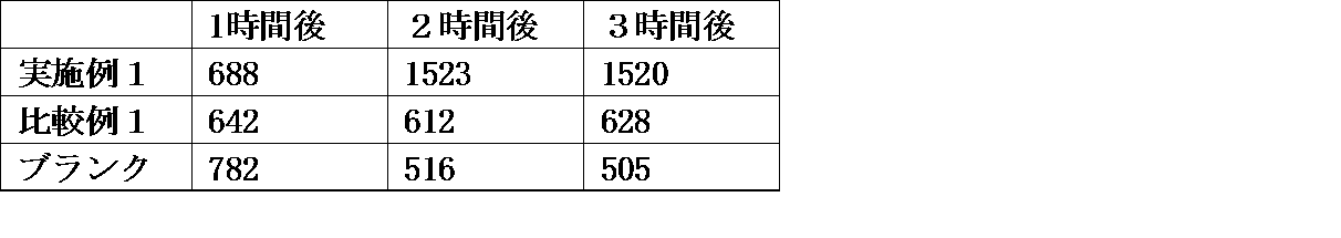
- L929 cells (mouse fibroblasts) were cultured in MEM medium for 24 hours at 37 ° C. and starved. 1 mL of MEM medium containing 5% powdered decellularized tissue was added to a 24-well plate dish, and cell culture inserts (pore diameter: 8 ⁇ m) were set in each well. L929 cells cultured in MEM medium were seeded in the insert (1.0 ⁇ 10 5 cells / well), cultured at 37 ° C. for 1 to 3 hours, and the number of cells migrated to the well part under the insert was counted. The number of cells was measured using a fluorescent microscope after staining with DAPI, and the average number of 5 well portions was determined. A blank that did not use powdered decellularized tissue was used as a blank. The results are shown in Table 1.
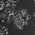
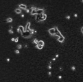
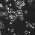
- a collagen-coated 24-well plate dish is seeded with PC12 cells (a pheochromocytoma derived from rat adrenal medulla) using RPMI medium containing 10% Horse serum and 5% fetal bovine serum (FBS) (1. 0 ⁇ 10 4 cells / well) and cultured at 37 ° C. for 24 hours.
- the medium was changed to an RPMI medium containing 0.1% Hose serum, and a cell culture insert (pore size: 8 ⁇ m) was set in each well. 200 ⁇ L of physiological saline containing 5% decellularized powder was added to the insert, and cultured for 24 hours with or without 50 ng of nerve cell growth factor (NGF).
- NGF nerve cell growth factor
- the powdered decellularized tissue of the present invention showed a clear neurite extension effect even when NGF was not added. This indicates that the powdered decellularized tissue of the present invention has an effect of inducing cell differentiation. In the comparative example, cell death was observed, suggesting that the powdered decellularized tissue of the comparative example has cytotoxicity.
- Rats (Wistar rat, male, 8-12 weeks old) were anesthetized, the back was shaved, and the shaved part was disinfected with povidone iodine solution. A defect (diameter 15 mm) up to the middle layer of the dermis was created on the back of this rat, and a metal body cooled with liquid nitrogen was pressed for 60 seconds to create a frostbite wound.
- the decellularized powder of Example 1 or Comparative Example 2 was applied to this wound, the wound part was covered with a bandage, and the entire circumference of the trunk of the rat was covered with gauze.
- the particulate decellularized tissue of the present invention Since the particulate decellularized tissue of the present invention has low cytotoxicity and has a cell differentiation promoting effect and an attracting effect, it is a therapeutic agent for diseases, an adjuvant for tissue transplantation, a scaffold material for regenerative medicine, a cell It can be preferably used as a culture substrate.
- the particulate decellularized tissue of the present invention When the particulate decellularized tissue of the present invention is used as a disease treatment material, a tissue transplantation aid, or a scaffold material for regenerative medicine, it may be applied directly to the affected area, or it may be a gel or sheet. It may be applied after being processed into a three-dimensional structure or the like. Moreover, you may apply what was cell-cultured using the particulate decellularized tissue of this invention to an affected part.
Abstract
Description
[1]動物由来組織に、媒体中で高静水圧を印加する工程を有することを特徴とする粒子状脱細胞化組織の製造方法。
[2]高静水圧が2~1,500MPaであることを特徴とする[1]記載の粒子状脱細胞化組織の製造方法。
[3]動物由来組織を粉砕した後に、媒体中で高静水圧を印加することを特徴とする[1]又は[2]記載の粒子状脱細胞化組織の製造方法。
[4]媒体中で高静水圧を印加した動物由来脱細胞化組織を粉砕することを特徴とする[1]又は[2]記載の粒子状脱細胞化組織の製造方法。
[5]媒体中で高静水圧を印加した動物由来脱細胞化組織を、アニオン性界面活性剤及び/又はノニオン性界面活性剤を含有しない洗浄液で洗浄することを特徴とする[1]~[4]の何れか1項に記載の粒子状脱細胞化組織の製造方法。
[6][1]~[5]の何れか1項に記載の粒子状脱細胞化組織の製造方法より製造された粒子状脱細胞化組織。
[7][6]に記載の粒子状脱細胞化組織を使用した移植用又は治療用の材料。
[8][6]に記載の粒子状脱細胞化組織を使用した細胞培養用材料。 Specifically, the present invention includes the following.
[1] A method for producing a particulate decellularized tissue, comprising a step of applying a high hydrostatic pressure to an animal-derived tissue in a medium.
[2] The method for producing a particulate decellularized tissue according to [1], wherein the high hydrostatic pressure is 2 to 1,500 MPa.
[3] The method for producing a particulate decellularized tissue according to [1] or [2], wherein a high hydrostatic pressure is applied in a medium after pulverizing the animal-derived tissue.
[4] The method for producing a particulate decellularized tissue according to [1] or [2], wherein the animal-derived decellularized tissue to which a high hydrostatic pressure is applied in a medium is pulverized.
[5] The animal-derived decellularized tissue to which a high hydrostatic pressure is applied in a medium is washed with a washing solution that does not contain an anionic surfactant and / or a nonionic surfactant. 4] The method for producing a particulate decellularized tissue according to any one of 4).
[6] A particulate decellularized tissue produced by the method for producing a particulate decellularized tissue according to any one of [1] to [5].
[7] A material for transplantation or treatment using the particulate decellularized tissue according to [6].
[8] A cell culture material using the particulate decellularized tissue according to [6].
〔生体組織〕
本発明の粒子状脱細胞化組織の製造方法に使用する生体組織は、脊椎動物由来の細胞を有する生体組織であれば、特に限定されないが、拒絶反応が少ないことから、哺乳類又は鳥類由来の生体組織が好ましく、入手が容易であることから、哺乳類の家畜、鳥類の家畜またはヒト由来の生態組織が更に好ましい。哺乳類の家畜としては、ウシ、ウマ、ラクダ、リャマ、ロバ、ヤク、ヒツジ、ブタ、ヤギ、シカ、アルパカ、イヌ、タヌキ、イタチ、キツネ、ネコ、ウサギ、ハムスター、モルモット、ラット、マウス、リス、アライグマ等が挙げられる。また、鳥類の家畜としては、インコ、オウム、ニワトリ、アヒル、七面鳥、ガチョウ、ホロホロ鳥、キジ、ダチョウ、ウズラ等が挙げられる。これらの中でも、入手の安定性から、ブタ、ウサギ、ヒトの生体組織が好ましい。 Hereinafter, the present invention will be described in detail.
[Biological tissue]
The biological tissue used in the method for producing the particulate decellularized tissue of the present invention is not particularly limited as long as it is a biological tissue having cells derived from vertebrates. However, since there are few rejection reactions, biological tissues derived from mammals or birds Tissues are preferred, and because they are readily available, mammalian livestock, avian livestock or human-derived ecological tissues are more preferred. Mammalian livestock includes cattle, horses, camels, llamas, donkeys, yaks, sheep, pigs, goats, deer, alpaca, dogs, raccoon dogs, weasels, foxes, cats, rabbits, hamsters, guinea pigs, rats, mice, squirrels, Raccoon etc. are mentioned. In addition, examples of avian livestock include parakeets, parrots, chickens, ducks, turkeys, geese, guinea fowls, pheasants, ostriches, and quails. Among these, porcine, rabbit, and human biological tissues are preferable from the viewpoint of availability.
本発明の粒子状脱細胞化組織の製造方法では、生体組織を採取し、生体組織に高静水圧を印加し、破壊された細胞を洗浄除去し、粒子状の脱細胞化組織を得るまでの間の、どの段階で生体組織(または脱細胞化組織)を粉砕して粒子状にしてもよいが、破壊された細胞を洗浄除去する場合に、生体組織の形状が保たれた状態よりは粒子状である方が、破壊された細胞の洗浄除去が容易に行えることから、少なくとも細胞の洗浄除去の前に、粉砕することが好ましい。 [Crushing process]
In the method for producing a particulate decellularized tissue of the present invention, the biological tissue is collected, a high hydrostatic pressure is applied to the biological tissue, the broken cells are washed and removed, and the particulate decellularized tissue is obtained. At any stage, the biological tissue (or decellularized tissue) may be pulverized into particles. However, when the broken cells are washed away, the particles are more stable than the state in which the shape of the biological tissue is maintained. It is preferable to pulverize at least before washing and removing the cells because the broken cells can be easily washed away.
本発明の製造方法では、生体細胞に媒体中で静水圧を印加ことにより生体組織が脱細胞化される。印加する静水圧が100MPaよりも低い場合には、生体組織からの脱細胞が不十分となる。一方、静水圧の印加には印加に耐えられる圧力容器が必要であり、多大なエネルギーを要する。このため、生体組織に印加する静水圧は、2~1,500MPaが好ましく、10~1,000MPaが更に好ましく、80~500MPaが最も好ましい。 [High hydrostatic pressure treatment process]
In the production method of the present invention, biological tissue is decellularized by applying hydrostatic pressure to the biological cells in a medium. When the applied hydrostatic pressure is lower than 100 MPa, decellularization from the living tissue is insufficient. On the other hand, the application of hydrostatic pressure requires a pressure vessel that can withstand the application and requires a great deal of energy. For this reason, the hydrostatic pressure applied to the living tissue is preferably 2 to 1,500 MPa, more preferably 10 to 1,000 MPa, and most preferably 80 to 500 MPa.
高静水圧が印加された生体組織は、洗浄液により破壊された細胞を洗浄除去される。洗浄液は、静水圧の印加に使用した媒体と同じ液でもよいし、異なる洗浄液でもよく、複数の種類の洗浄液を組み合わせて用いてもよい。洗浄液は、核酸分解酵素、有機溶媒又はキレート剤を含有することが好ましい。核酸分解酵素は、静水圧が印加された生体組織からの核酸成分、有機溶媒は脂質、それぞれの除去効率を向上させることができ、キレート剤は、脱細胞化組織中のカルシウムイオンやマグネシウムイオンを不活性化することにより、本発明の粒子化脱細胞組織を疾患部に適用した場合の石灰化を防ぐことができる。 [Washing process]
The biological tissue to which the high hydrostatic pressure is applied is washed away with the cells destroyed by the washing liquid. The cleaning liquid may be the same liquid as the medium used for applying the hydrostatic pressure, may be a different cleaning liquid, or a combination of a plurality of types of cleaning liquids. The cleaning liquid preferably contains a nucleolytic enzyme, an organic solvent, or a chelating agent. Nucleolytic enzymes can improve the removal efficiency of nucleic acid components from living tissues to which hydrostatic pressure is applied, organic solvents are lipids, and chelating agents can reduce calcium ions and magnesium ions in decellularized tissues. By inactivation, calcification when the particulate decellularized tissue of the present invention is applied to a diseased part can be prevented.
ブタの肝臓を冷凍し、-20℃で、フードプロセッサーを用いて粉砕し、ふるいを用いて直径500μm以上の粒子を除去して、ブタの肝臓の微細粒子(平均粒径50μm、粒径1μm未満の成分は5%以下)を得た。ポリエチレン製チャック付き袋に、この微細粒子5gと、高静水圧処理の媒体として生理食塩水15gとを入れ、研究開発用高圧処理装置(神戸製鋼製、商品名:Dr.CHEF)を用いて、1,000MPaの静水圧を15分間印加した。この後、高静水圧処理した粉末を滅菌カップに移し、洗浄液として生理食塩水20gを入れて、25℃で5時間振盪し、洗浄液を更新して更に2時間浸透して、実施例1の粒子状脱細胞化組織を得た。 [Example 1]
Pig liver is frozen, crushed at -20 ° C using a food processor, and particles with a diameter of 500 µm or more are removed using a sieve to make fine particles of pig liver (average particle size 50 µm, particle size less than 1 µm) Of 5% or less). In a bag with a polyethylene chuck, 5 g of the fine particles and 15 g of physiological saline as a medium for high hydrostatic pressure treatment are placed, and a high-pressure treatment apparatus for research and development (product name: Dr. CHEF) is used. A hydrostatic pressure of 1,000 MPa was applied for 15 minutes. Thereafter, the high hydrostatic pressure-treated powder is transferred to a sterilization cup, and 20 g of physiological saline is added as a washing liquid, shaken at 25 ° C. for 5 hours, renewed washing liquid, and further permeated for 2 hours. A decellularized tissue was obtained.
ブタの肝臓をブタの大脳に変えた以外は、実施例1と同様の操作を行い実施例2の粒子状脱細胞化組織を得た。 [Example 2]
The particulate decellularized tissue of Example 2 was obtained in the same manner as in Example 1 except that the pig liver was changed to a pig cerebrum.
ブタの肝臓をブタの脊髄に変えた以外は、実施例1と同様の操作を行い実施例3の粒子状脱細胞化組織を得た。 Example 3
The particulate decellularized tissue of Example 3 was obtained in the same manner as in Example 1 except that the pig liver was changed to a pig spinal cord.
滅菌カップに、実施例1と同様の方法で粉砕したブタの肝臓の微細粒子5gと、脱細胞溶液として0.5%のドデシル硫酸ナトリウムを含有するハンクスの平衡塩溶液15gを入れ、25℃で5時間保存した。この後、脱細胞溶液を除去し、洗浄液として界面活性剤を含有しないハンクスの平衡塩溶液20gを入れて、25℃で5時間振盪し、洗浄液を更新して更に2時間振盪することにより、比較例1の粒子状脱細胞化組織を得た。 [Comparative Example 1]
In a sterilized cup, 5 g of fine particles of porcine liver ground in the same manner as in Example 1 and 15 g of Hanks' balanced salt solution containing 0.5% sodium dodecyl sulfate as a decellularized solution were placed at 25 ° C. Stored for 5 hours. After this, the decellularized solution is removed, 20 g of Hank's balanced salt solution containing no surfactant is added as a washing solution, shaken at 25 ° C. for 5 hours, and the washing solution is renewed and shaken for another 2 hours. The particulate decellularized tissue of Example 1 was obtained.
ブタの肝臓をブタの大脳に変えた以外は、比較例1と同様の操作を行い比較例2の粒子状脱細胞化組織を得た。 [Comparative Example 2]
A particulate decellularized tissue of Comparative Example 2 was obtained in the same manner as in Comparative Example 1 except that the pig liver was changed to a pig cerebrum.
ブタの肝臓をブタの脊髄に変えた以外は、比較例1と同様の操作を行い比較例3の粒子状脱細胞化組織を得た。 [Comparative Example 3]
A particulate decellularized tissue of Comparative Example 3 was obtained in the same manner as in Comparative Example 1 except that the pig liver was changed to a pig spinal cord.
L929細胞(マウス繊維芽細胞)をMEM培地を用いて、37℃で24時間培養し、飢餓状態にした。24 well plate dishに5%の粉末化脱細胞化組織を含有するMEM培地1mLを添加し、セルカルチャーインサート(孔径8μm)を各wellにセットした。インサート内に、MEM培地で培養したL929細胞を播種し(1.0×10⁵細胞/well)、37℃で1~3時間培養し、インサート下のwell部に移動した細胞数を計測した。細胞数の計測は、DAPIで染色した後、蛍光顕微鏡を用いて行い、5のwell部の平均数を求めた。なお、粉末化脱細胞化組織を使用しないものをブランクとした。結果を表1に示す。 [Cell migration test]
L929 cells (mouse fibroblasts) were cultured in MEM medium for 24 hours at 37 ° C. and starved. 1 mL of MEM medium containing 5% powdered decellularized tissue was added to a 24-well plate dish, and cell culture inserts (pore diameter: 8 μm) were set in each well. L929 cells cultured in MEM medium were seeded in the insert (1.0 × 10 5 cells / well), cultured at 37 ° C. for 1 to 3 hours, and the number of cells migrated to the well part under the insert was counted. The number of cells was measured using a fluorescent microscope after staining with DAPI, and the average number of 5 well portions was determined. A blank that did not use powdered decellularized tissue was used as a blank. The results are shown in Table 1.
コラーゲンコート24 well plate dishに、10%のHorse serumと5%のウシ胎児血清(FBS)を含有するRPMI培地を用いて、PC12細胞(ラットの副腎髄質由来の褐色細胞腫)を播種(1.0×10⁴細胞/well)し、37℃で24時間培養した。培地を0.1%のHose serumを含有するRPMI培地に変更し、セルカルチャーインサート(孔径8μm)を各wellにセットした。インサート内に5%の脱細胞化粉末を含有する生理食塩水を200μL添加し、神経細胞成長因子(NGF)を50ng添加した場合と添加しない場合について24時間培養した。位相差顕微鏡で細胞を観察し、以下の基準で神経突起伸長効果を評価した。結果を表2に示す。
◎:明らかな神経突起伸長効果が見られる。
○:一部に神経突起伸長効果が見られる。
△:神経突起伸長効果が見られない。
×:一部または全部の細胞に、細胞死が観察された。 [Nerite outgrowth test]
A collagen-coated 24-well plate dish is seeded with PC12 cells (a pheochromocytoma derived from rat adrenal medulla) using RPMI medium containing 10% Horse serum and 5% fetal bovine serum (FBS) (1. 0 × 10 4 cells / well) and cultured at 37 ° C. for 24 hours. The medium was changed to an RPMI medium containing 0.1% Hose serum, and a cell culture insert (pore size: 8 μm) was set in each well. 200 μL of physiological saline containing 5% decellularized powder was added to the insert, and cultured for 24 hours with or without 50 ng of nerve cell growth factor (NGF). Cells were observed with a phase contrast microscope, and the neurite outgrowth effect was evaluated according to the following criteria. The results are shown in Table 2.
A: A clear neurite outgrowth effect is observed.
○: A neurite outgrowth effect is partially observed.
Δ: No neurite outgrowth effect is observed.
X: Cell death was observed in some or all cells.

ラット(Wistar rat、オス、8~12週齢)を麻酔処理し、背部を剃毛し、剃毛部をポビドンヨード溶液で消毒した。このラットの背部をハサミで約1.5cm切開して皮下ポケットを作成した。この皮下ポケットに脱細胞化粉末約20mgを埋入し、縫合糸を用いて切開部を縫合し、縫合部をポビドンヨード溶液で再度消毒した。28日後に、ラットの皮下ポケット部を回収し、ヘマトキシリン・エオシン染色により染色して組織学的評価を行った。なお、脱細胞化粉末を埋入せずに縫合したものをブランクとした。結果を表3に示す。
○:炎症反応が見られず、細胞の誘引が確認できる。
△:炎症反応は見られないが、細胞の誘引が明確でない。
×:炎症反応が見られ、細胞の誘引も確認できない。 [Subcutaneous implantation test]
Rats (Wistar rat, male, 8-12 weeks old) were anesthetized, the back was shaved, and the shaved part was disinfected with povidone iodine solution. A subcutaneous pocket was created by cutting the back of the rat with scissors about 1.5 cm. About 20 mg of decellularized powder was embedded in this subcutaneous pocket, the incision was sutured with a suture, and the suture was disinfected again with a povidone iodine solution. After 28 days, the rat subcutaneous pocket was collected and stained with hematoxylin and eosin for histological evaluation. In addition, what was sewed without embedding decellularized powder was used as the blank. The results are shown in Table 3.
○: Inflammatory reaction is not observed, and cell attraction can be confirmed.
Δ: Inflammatory reaction is not observed, but cell attraction is not clear.
X: Inflammatory reaction is observed, and cell attraction cannot be confirmed.
ラット(Wistar rat、オス、8~12週齢)を麻酔処理し、背部を剃毛し、剃毛部をポビドンヨード溶液で消毒した。このラットの背部に、真皮中層までの欠損(直径15mm)を作成し、更に、液体窒素で冷却した金属体を60秒間押し付けて、凍傷した創傷を作成した。この創傷に、実施例1又は比較例2の脱細胞化粉末を塗布し、創傷部を絆創膏で被覆し、さらにラットの胴体全周をガーゼを用いて被覆した。7日後、創傷部を回収し、ヘマトキシリン・エオシン染色により染色して組織学的評価を行った。なお、脱細胞化粉末を塗布せずに絆創膏で被覆したものをブランクとした。結果を表3に示す。
◎:組織の再生が認められ、拘縮は認められない。
○:組織の再生が認められるが、やや拘縮が認められる。
△:組織の再生が認められるが、明らかな拘縮が認められる。
×:患部の悪化が認められる。 [Frostbit model test]
Rats (Wistar rat, male, 8-12 weeks old) were anesthetized, the back was shaved, and the shaved part was disinfected with povidone iodine solution. A defect (diameter 15 mm) up to the middle layer of the dermis was created on the back of this rat, and a metal body cooled with liquid nitrogen was pressed for 60 seconds to create a frostbite wound. The decellularized powder of Example 1 or Comparative Example 2 was applied to this wound, the wound part was covered with a bandage, and the entire circumference of the trunk of the rat was covered with gauze. Seven days later, the wound was collected and stained with hematoxylin and eosin for histological evaluation. In addition, what was coat | covered with the adhesive bandage without applying decellularized powder was made into the blank. The results are shown in Table 3.
A: Regeneration of tissue is permitted, and no contracture is observed.
○: Regeneration of tissue is observed, but somewhat contracture is observed.
Δ: Regeneration of tissue is observed, but clear contracture is observed.
X: Deterioration of the affected area is observed.

Claims (8)
- 動物由来組織に、媒体中で高静水圧を印加する工程を有することを特徴とする粒子状脱細胞化組織の製造方法。 A method for producing a particulate decellularized tissue, comprising a step of applying a high hydrostatic pressure in a medium to an animal-derived tissue.
- 高静水圧が2~1,500MPaであることを特徴とする請求項1記載の粒子状脱細胞化組織の製造方法。 2. The method for producing a particulate decellularized tissue according to claim 1, wherein the high hydrostatic pressure is 2 to 1,500 MPa.
- 動物由来組織を粉砕した後に、媒体中で高静水圧を印加することを特徴とする請求項1又は2記載の粒子状脱細胞化組織の製造方法。 3. A method for producing a particulate decellularized tissue according to claim 1 or 2, wherein a high hydrostatic pressure is applied in the medium after the animal-derived tissue is pulverized.
- 媒体中で高静水圧を印加した動物由来脱細胞化組織を粉砕することを特徴とする請求項1又は2記載の粒子状脱細胞化組織の製造方法。 3. The method for producing a particulate decellularized tissue according to claim 1 or 2, wherein the animal-derived decellularized tissue to which high hydrostatic pressure is applied is pulverized in a medium.
- 媒体中で高静水圧を印加した動物由来脱細胞化組織を、アニオン性界面活性剤及び/又はノニオン性界面活性剤を含有しない洗浄液で洗浄することを特徴とする請求項1~4の何れか1項に記載の粒子状脱細胞化組織の製造方法。 The animal-derived decellularized tissue to which high hydrostatic pressure is applied in a medium is washed with a washing solution containing no anionic surfactant and / or nonionic surfactant. 2. A method for producing a particulate decellularized tissue according to item 1.
- 請求項1~5の何れか1項に記載の粒子状脱細胞化組織の製造方法より製造された粒子状脱細胞化組織。 A particulate decellularized tissue produced by the method for producing a particulate decellularized tissue according to any one of claims 1 to 5.
- 請求項6に記載の粒子状脱細胞化組織を使用した移植用又は治療用の材料。 A material for transplantation or treatment using the particulate decellularized tissue according to claim 6.
- 請求項6に記載の粒子状脱細胞化組織を使用した細胞培養用材料。 A cell culture material using the particulate decellularized tissue according to claim 6.
Priority Applications (6)
| Application Number | Priority Date | Filing Date | Title |
|---|---|---|---|
| CN201480025727.3A CN105188781A (en) | 2013-05-07 | 2014-05-02 | Method for producing particulate decellularized tissue |
| US14/783,232 US20160058910A1 (en) | 2013-05-07 | 2014-05-02 | Method for preparing particulate decellularized tissue |
| EP14794303.9A EP2995325B1 (en) | 2013-05-07 | 2014-05-02 | Method for preparing particulate decellularized tissue |
| JP2015515867A JP6515304B2 (en) | 2013-05-07 | 2014-05-02 | Method of producing particulate decellularized tissue |
| KR1020157032675A KR102240373B1 (en) | 2013-05-07 | 2014-05-02 | Method for producing particulate decellularized tissue |
| EP19174604.9A EP3556406B1 (en) | 2013-05-07 | 2014-05-02 | Method for producing particulate decellularized tissue |
Applications Claiming Priority (2)
| Application Number | Priority Date | Filing Date | Title |
|---|---|---|---|
| JP2013-097402 | 2013-05-07 | ||
| JP2013097402 | 2013-05-07 |
Publications (1)
| Publication Number | Publication Date |
|---|---|
| WO2014181767A1 true WO2014181767A1 (en) | 2014-11-13 |
Family
ID=51867247
Family Applications (1)
| Application Number | Title | Priority Date | Filing Date |
|---|---|---|---|
| PCT/JP2014/062149 WO2014181767A1 (en) | 2013-05-07 | 2014-05-02 | Method for producing particulate decellularized tissue |
Country Status (6)
| Country | Link |
|---|---|
| US (1) | US20160058910A1 (en) |
| EP (2) | EP2995325B1 (en) |
| JP (2) | JP6515304B2 (en) |
| KR (1) | KR102240373B1 (en) |
| CN (1) | CN105188781A (en) |
| WO (1) | WO2014181767A1 (en) |
Cited By (4)
| Publication number | Priority date | Publication date | Assignee | Title |
|---|---|---|---|---|
| CN104740685A (en) * | 2015-04-16 | 2015-07-01 | 烟台隽秀生物科技有限公司 | Nerve repairing film and preparation method thereof |
| EP3263140A4 (en) * | 2015-02-27 | 2018-10-24 | Adeka Corporation | Decellularized tissue |
| WO2021029216A1 (en) * | 2019-08-09 | 2021-02-18 | 国立研究開発法人国立長寿医療研究センター | Dentin regenerative cell culture |
| WO2022270569A1 (en) * | 2021-06-23 | 2022-12-29 | 株式会社Adeka | Decellularized cell structure |
Families Citing this family (3)
| Publication number | Priority date | Publication date | Assignee | Title |
|---|---|---|---|---|
| CA2911592C (en) * | 2013-05-07 | 2021-10-26 | The Chemo-Sero-Therapeutic Research Institute | Hybrid gel comprising particulate decellularized tissue |
| CN112826983A (en) * | 2019-11-22 | 2021-05-25 | 中国科学院大连化学物理研究所 | Heart acellular matrix modified bionic membrane and preparation and application thereof |
| CN112472870A (en) * | 2020-12-03 | 2021-03-12 | 中国人民解放军陆军军医大学第一附属医院 | Preparation method and application of artificial microskin |
Citations (13)
| Publication number | Priority date | Publication date | Assignee | Title |
|---|---|---|---|---|
| JPS60501540A (en) | 1983-06-10 | 1985-09-19 | ユニバ−シテイ パテンツ,インコ−ポレイテイド | Extracellular matrix body implants and means and methods for their manufacture and use |
| JPH07509638A (en) | 1992-08-07 | 1995-10-26 | ティーイーアイ バイオサイエンシズ,インコーポレイテッド | Production of graft tissue from extracellular matrix |
| JP2002507907A (en) | 1997-06-27 | 2002-03-12 | バーダー、アウグスチヌス | Biosynthetic implant and method for producing the same |
| JP2002518319A (en) | 1998-06-19 | 2002-06-25 | ライフセル コーポレイション | Granular cell-free tissue matrix |
| JP2003518981A (en) | 1999-12-29 | 2003-06-17 | チルドレンズ メディカル センター コーポレーション | Methods and compositions for organ decellularization |
| JP2003525062A (en) | 1998-09-30 | 2003-08-26 | メドトロニック・インコーポレーテッド | Methods for reducing tissue mineralization used in transplantation |
| JP2004094552A (en) | 2002-08-30 | 2004-03-25 | Sumitomo Chem Co Ltd | Pathology texture image analyzing method and system |
| WO2008111530A1 (en) | 2007-03-09 | 2008-09-18 | National University Corporation, Tokyo Medical And Dental University | Method of preparing decellularized soft tissue, graft and culture material |
| JP2010227246A (en) * | 2009-03-26 | 2010-10-14 | Tokyo Medical & Dental Univ | Method for preparing decellularized living organism tissue |
| JP2012505013A (en) | 2008-10-13 | 2012-03-01 | アジュウ ユニバーシティー インダストリー アカデミック コーアベレイション ファンデーション | Method for producing porous three-dimensional support using animal tissue powder and porous three-dimensional support produced using the same |
| JP2013502275A (en) | 2009-08-18 | 2013-01-24 | ライフセル コーポレーション | How to process the organization |
| US20130028981A1 (en) * | 2010-02-26 | 2013-01-31 | Decell Technologies Inc. | Methods for tissue decellularization |
| JP2013042677A (en) * | 2011-08-22 | 2013-03-04 | Tokyo Medical & Dental Univ | Decellularization process liquid, method for preparing decellularized cornea, and implant having decellularized cornea |
Family Cites Families (6)
| Publication number | Priority date | Publication date | Assignee | Title |
|---|---|---|---|---|
| US5800537A (en) * | 1992-08-07 | 1998-09-01 | Tissue Engineering, Inc. | Method and construct for producing graft tissue from an extracellular matrix |
| US6933326B1 (en) * | 1998-06-19 | 2005-08-23 | Lifecell Coporation | Particulate acellular tissue matrix |
| US8882850B2 (en) * | 1998-12-01 | 2014-11-11 | Cook Biotech Incorporated | Multi-formed collagenous biomaterial medical device |
| US7153518B2 (en) | 2001-08-27 | 2006-12-26 | Regeneration Technologies, Inc. | Processed soft tissue for topical or internal application |
| JP4092397B2 (en) | 2002-09-10 | 2008-05-28 | 国立循環器病センター総長 | Treatment of living tissue for transplantation by applying ultrahigh hydrostatic pressure |
| PL2192907T3 (en) * | 2007-08-16 | 2018-10-31 | Remedor Biomed Ltd. | Erythropoietin and fibronectin compositions for therapeutic applications |
-
2014
- 2014-05-02 WO PCT/JP2014/062149 patent/WO2014181767A1/en active Application Filing
- 2014-05-02 EP EP14794303.9A patent/EP2995325B1/en active Active
- 2014-05-02 CN CN201480025727.3A patent/CN105188781A/en active Pending
- 2014-05-02 JP JP2015515867A patent/JP6515304B2/en active Active
- 2014-05-02 KR KR1020157032675A patent/KR102240373B1/en active IP Right Grant
- 2014-05-02 US US14/783,232 patent/US20160058910A1/en not_active Abandoned
- 2014-05-02 EP EP19174604.9A patent/EP3556406B1/en active Active
-
2018
- 2018-11-02 JP JP2018207431A patent/JP6623333B2/en active Active
Patent Citations (13)
| Publication number | Priority date | Publication date | Assignee | Title |
|---|---|---|---|---|
| JPS60501540A (en) | 1983-06-10 | 1985-09-19 | ユニバ−シテイ パテンツ,インコ−ポレイテイド | Extracellular matrix body implants and means and methods for their manufacture and use |
| JPH07509638A (en) | 1992-08-07 | 1995-10-26 | ティーイーアイ バイオサイエンシズ,インコーポレイテッド | Production of graft tissue from extracellular matrix |
| JP2002507907A (en) | 1997-06-27 | 2002-03-12 | バーダー、アウグスチヌス | Biosynthetic implant and method for producing the same |
| JP2002518319A (en) | 1998-06-19 | 2002-06-25 | ライフセル コーポレイション | Granular cell-free tissue matrix |
| JP2003525062A (en) | 1998-09-30 | 2003-08-26 | メドトロニック・インコーポレーテッド | Methods for reducing tissue mineralization used in transplantation |
| JP2003518981A (en) | 1999-12-29 | 2003-06-17 | チルドレンズ メディカル センター コーポレーション | Methods and compositions for organ decellularization |
| JP2004094552A (en) | 2002-08-30 | 2004-03-25 | Sumitomo Chem Co Ltd | Pathology texture image analyzing method and system |
| WO2008111530A1 (en) | 2007-03-09 | 2008-09-18 | National University Corporation, Tokyo Medical And Dental University | Method of preparing decellularized soft tissue, graft and culture material |
| JP2012505013A (en) | 2008-10-13 | 2012-03-01 | アジュウ ユニバーシティー インダストリー アカデミック コーアベレイション ファンデーション | Method for producing porous three-dimensional support using animal tissue powder and porous three-dimensional support produced using the same |
| JP2010227246A (en) * | 2009-03-26 | 2010-10-14 | Tokyo Medical & Dental Univ | Method for preparing decellularized living organism tissue |
| JP2013502275A (en) | 2009-08-18 | 2013-01-24 | ライフセル コーポレーション | How to process the organization |
| US20130028981A1 (en) * | 2010-02-26 | 2013-01-31 | Decell Technologies Inc. | Methods for tissue decellularization |
| JP2013042677A (en) * | 2011-08-22 | 2013-03-04 | Tokyo Medical & Dental Univ | Decellularization process liquid, method for preparing decellularized cornea, and implant having decellularized cornea |
Non-Patent Citations (1)
| Title |
|---|
| See also references of EP2995325A4 |
Cited By (6)
| Publication number | Priority date | Publication date | Assignee | Title |
|---|---|---|---|---|
| EP3263140A4 (en) * | 2015-02-27 | 2018-10-24 | Adeka Corporation | Decellularized tissue |
| EP3782629A1 (en) * | 2015-02-27 | 2021-02-24 | Adeka Corporation | Decellularized tissue |
| CN104740685A (en) * | 2015-04-16 | 2015-07-01 | 烟台隽秀生物科技有限公司 | Nerve repairing film and preparation method thereof |
| WO2021029216A1 (en) * | 2019-08-09 | 2021-02-18 | 国立研究開発法人国立長寿医療研究センター | Dentin regenerative cell culture |
| JP2021027803A (en) * | 2019-08-09 | 2021-02-25 | 国立研究開発法人国立長寿医療研究センター | Cell culture for dentin regeneration |
| WO2022270569A1 (en) * | 2021-06-23 | 2022-12-29 | 株式会社Adeka | Decellularized cell structure |
Also Published As
| Publication number | Publication date |
|---|---|
| CN105188781A (en) | 2015-12-23 |
| EP3556406B1 (en) | 2021-12-22 |
| JP2019030332A (en) | 2019-02-28 |
| US20160058910A1 (en) | 2016-03-03 |
| EP2995325A1 (en) | 2016-03-16 |
| EP2995325B1 (en) | 2020-01-15 |
| JPWO2014181767A1 (en) | 2017-02-23 |
| JP6623333B2 (en) | 2019-12-25 |
| KR20160005712A (en) | 2016-01-15 |
| EP2995325A4 (en) | 2016-12-21 |
| KR102240373B1 (en) | 2021-04-13 |
| EP3556406A1 (en) | 2019-10-23 |
| JP6515304B2 (en) | 2019-05-22 |
Similar Documents
| Publication | Publication Date | Title |
|---|---|---|
| JP6623333B2 (en) | Method for producing particulate decellularized tissue | |
| US20220073881A1 (en) | Compositions and methods for implantation of processed adipose tissue and processed adipose tissue products | |
| US20180125897A1 (en) | Decellularized Adipose Cell Growth Scaffold | |
| JP6606429B2 (en) | Decellularized biomaterial from non-mammalian tissue | |
| KR102604205B1 (en) | Biologically functional soft tissue scaffolds and implants | |
| US20180071432A1 (en) | Compositions and methods for cardiac therapy | |
| US10709810B2 (en) | Processed adipose tissue | |
| US20120040013A1 (en) | Regenerative Tissue Scaffolds | |
| JP7156597B2 (en) | PLACENTA-DERIVED MATRIX AND METHOD FOR PREPARATION AND USE THEREOF | |
| Inci et al. | Decellularized inner body membranes for tissue engineering: A review |
Legal Events
| Date | Code | Title | Description |
|---|---|---|---|
| WWE | Wipo information: entry into national phase |
Ref document number: 201480025727.3 Country of ref document: CN |
|
| 121 | Ep: the epo has been informed by wipo that ep was designated in this application |
Ref document number: 14794303 Country of ref document: EP Kind code of ref document: A1 |
|
| ENP | Entry into the national phase |
Ref document number: 2015515867 Country of ref document: JP Kind code of ref document: A |
|
| WWE | Wipo information: entry into national phase |
Ref document number: 14783232 Country of ref document: US |
|
| WWE | Wipo information: entry into national phase |
Ref document number: 2014794303 Country of ref document: EP |
|
| NENP | Non-entry into the national phase |
Ref country code: DE |
|
| ENP | Entry into the national phase |
Ref document number: 20157032675 Country of ref document: KR Kind code of ref document: A |