WO2014168153A1 - Gip受容体の発現制御方法 - Google Patents
Gip受容体の発現制御方法 Download PDFInfo
- Publication number
- WO2014168153A1 WO2014168153A1 PCT/JP2014/060222 JP2014060222W WO2014168153A1 WO 2014168153 A1 WO2014168153 A1 WO 2014168153A1 JP 2014060222 W JP2014060222 W JP 2014060222W WO 2014168153 A1 WO2014168153 A1 WO 2014168153A1
- Authority
- WO
- WIPO (PCT)
- Prior art keywords
- gip
- adipocytes
- hifα
- test substance
- cells
- Prior art date
- Legal status (The legal status is an assumption and is not a legal conclusion. Google has not performed a legal analysis and makes no representation as to the accuracy of the status listed.)
- Ceased
Links
Images





Classifications
-
- C—CHEMISTRY; METALLURGY
- C12—BIOCHEMISTRY; BEER; SPIRITS; WINE; VINEGAR; MICROBIOLOGY; ENZYMOLOGY; MUTATION OR GENETIC ENGINEERING
- C12N—MICROORGANISMS OR ENZYMES; COMPOSITIONS THEREOF; PROPAGATING, PRESERVING, OR MAINTAINING MICROORGANISMS; MUTATION OR GENETIC ENGINEERING; CULTURE MEDIA
- C12N5/00—Undifferentiated human, animal or plant cells, e.g. cell lines; Tissues; Cultivation or maintenance thereof; Culture media therefor
- C12N5/06—Animal cells or tissues; Human cells or tissues
- C12N5/0602—Vertebrate cells
- C12N5/0652—Cells of skeletal and connective tissues; Mesenchyme
- C12N5/0653—Adipocytes; Adipose tissue
Definitions
- the present invention relates to GIP receptor expression control in adipocytes and use thereof.
- GIP glucose-dependent insulotropic polypeptide
- Non-patent Document 1 GIP receptor (GIPR) is expressed in adipocytes, and obesity due to high fat diet load is not observed in GIPR-deficient mice.
- GIPR GIP receptor
- Non-patent Document 3 Promotion of fatty acid uptake through activation of LPL (Lipoprotein Lipase) (Non-patent Document 3), enhanced sugar uptake through Akt (Non-patent Document 4), increased fat accumulation (Non-patent Document 5), inflammatory Enhancement (Non-Patent Document 6) has been reported.
- LPL Lipoprotein Lipase
- Non-Patent Document 6 discloses that the expression and production of interleukin 6 (IL6) is promoted and the inflammatory reaction is enhanced in adipocytes in which an adenovirus vector is used and the GIP receptor is forcibly highly expressed.
- IL6 interleukin 6
- a system for forcedly expressing the GIP receptor using an adenovirus vector is not suitable for use in elucidation of the receptor function or as a screening system because it is complicated and non-physiological. .
- Non-Patent Document 1 Seino Y et al. GIP and GLP-1, the two incretin hormones: Similarities and differences. Journal of Diabetes Investigation (2010), 1, pp.8-23
- Non-Patent Document 2 Yip RG. Et al. Functional GIP receptors are present on adipocytes. Endocrinology (1998), 139 (9), pp. 4004-4007
- Non-Patent Document 3 Miyawaki et al. Inhibition of gastric inhibitory polypeptide signaling prevents obesity. Nat Med. (2002), 8 (7), pp. 738-742
- Non-Patent Document 4 Song DH et al.
- Non-Patent Document 5 Kim SJ et al. Activation of Lipoprotein Lipase by Glucose-dependent Insulinotropic Polypeptide in Adipocytes. JBC (2007), 282 (12), pp. 8557-8567
- Non-Patent Document 6 Nie Y et al.
- Glucose-dependent insulinotropic polypeptide impairs insulin signaling via inducing adipocyte inflammation in glucose-dependent insulinotropic polypeptide receptor-overexpressing adipocytes. FASEB Journal (2012), 26 (6), pp. 2383-2393
- the present invention relates to the following 1) -4).
- a method for screening for a substance having affinity for a GIP receptor comprising a step of activating HIF ⁇ in an adipocyte.
- the graph which shows the expression change of a GIP receptor in a hypoxic environment (pseudo hypoxic environment).
- the graph which shows the expression change of a GIP receptor in a hypoxic environment (hypoxic culture).
- the graph which shows the influence of HIF (alpha) expression suppression on the expression of a GIP receptor in a hypoxic environment (hypoxic culture).
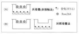
- A co-culture method
- a fat cell 3T3-L1
- a macrophage Raw264
- B control culture method
- Left: GIP receptor expression, right: HIF ⁇ expression The graph which shows IL6 secretion suppression by a GIP receptor inhibitor using a macrophage co-culture system.
- the present invention provides a system that can efficiently express a GIP receptor in adipocytes, and a method of using the expression system.
- the present inventors examined the expression of the GIP receptor in adipocytes.
- HIF ⁇ Hydrophila-inducible factor 1 ⁇
- hypoxia response factor Hypoxia response factor
- the expression of the GIP receptor in adipocytes can be efficiently performed using an adenovirus vector or the like without taking the troublesome work of strong expression. Therefore, expression analysis and functional analysis of GIP receptors in adipocytes can be easily performed, thereby making it possible to elucidate the mechanism of obesity and inflammation and to search for anti-obesity substances and anti-inflammatory substances targeting receptor molecules. It becomes.
- GIP receptor (GIPR) expression is performed by including a step of activating HIF ⁇ in adipocytes.
- the “adipocyte” may be any cell that has the properties of an adipocyte (for example, performs intracellular metabolism peculiar to an adipocyte and accumulates a large amount of lipid droplets) and can express a GIP receptor,
- cultured adipocytes such as human and rat primary cultured cells
- cells derived from preadipocytes may be used.
- preadipocytes 3T3-L1 cells (Journal of Biological Chemistry (1996), 271 (5), pp.2365-2368) widely used in the research area as fibroblasts having the ability to differentiate into adipocytes.
- Adipose precursor cells derived from various animals are commercially available and can be purchased from appropriate sales companies.
- mouse-derived 3T3-L1 cells and 3T3-F442A cells can be purchased from DS Pharma Biomedical Corporation.
- the differentiation induction from preadipocytes to adipocytes can be performed by applying differentiation induction stimulation to the preadipocytes by a known method.
- the precursor fat is obtained by using an adipocyte differentiation-inducing cocktail containing fetal bovine serum (FBS), Insulin, IBMX (3-Isobutyl-1-methylxanthine), Dex (Dexamethasone), or the like.
- FBS fetal bovine serum
- IBMX 3-Isobutyl-1-methylxanthine
- Dex Dex
- Cell differentiation can be induced (Wu Z. et al., Genes & Dev. (1992), 9, pp.2350-2363, etc.).
- the GIP receptor shows biphasic expression in the early differentiation stage and the mature stage, and the expression level thereof is It increases markedly at around 6 days after differentiation induction. Therefore, when preadipocytes are used after differentiation induction, it is preferable to use cells after the mature stage. For example, in the case of 3T3-L1 cells, cells after 6 days after differentiation induction, for example, 6 to 16 days are used. It is preferable to use it.
- HIF ⁇ is a protein that is induced when oxygen supply to cells is deficient, and is an ⁇ subunit of a hypoxia response factor (Hypoxia Inducible Factor, HIF) that functions as a transcription factor.
- HIF Hypoxia response factor
- the present invention includes a step of activating HIF ⁇ in adipocytes.
- the activation of HIF ⁇ includes inhibiting and stabilizing the degradation of HIF ⁇ , activating the transcription function, enhancing the expression of HIF ⁇ , and the like.
- HIF ⁇ is hydroxylated by proline hydroxylase (PHD)
- PHD proline hydroxylase
- the hydroxylated HIF ⁇ is ubiquitinated and is constantly degraded by the proteasome.
- PHD proline hydroxylase
- hydroxylation of HIF ⁇ is suppressed and HIF ⁇ is stabilized (Experimental Medicine 2012 Vol.30 No.8, (5), metabolism, development Hypoxic response system that begins to move with immunity).
- a HIF ⁇ activator is added to the medium (Yun.Z et al., Developmental Cell, (2002), Vol.2, 331-341, Hanson. ES et al., JBC (2003) , Vol. 278, 40337-40342, etc.).
- Examples of means for placing the cells in a hypoxic state include culturing the cells in a hypoxic environment.
- “under a low oxygen environment” means an environment with little oxygen (“under a low oxygen atmosphere”)
- “low oxygen” means an oxygen concentration lower than the normal oxygen concentration (21%).
- the concentration of oxygen in a low oxygen environment is preferably 10% or less, more preferably 5% or less, and still more preferably 2% or less.
- a minimum oxygen concentration is necessary to maintain the survival of the cells subjected to culture. From such a viewpoint, the oxygen concentration should be 0.01% or more, preferably 0.1% or more, more preferably 0.5% or more, and still more preferably 1% or more.
- preferable oxygen concentrations are 0.01 to 10%, 0.1 to 10%, 0.5 to 10%, 1 to 10%, 0.01 to 5%, 0.1 to 5%, 0.0. 5-5%, 1-5%, 0.01-2%, 0.1-2%, 0.5-2%, 1-2%, etc.
- the low oxygen atmosphere can be realized by adjusting the oxygen concentration to the above range using, for example, a low oxygen culture kit (BIONIX).
- BIONIX low oxygen culture kit
- HIF ⁇ activator examples include a compound or factor having at least one function of suppressing and stabilizing the degradation of HIF ⁇ , enhancing the transcriptional activity function of HIF ⁇ , and inducing expression of HIF ⁇ .
- Such an HIF ⁇ activator is any of a compound that interacts with HIF ⁇ , a compound that binds to HIF ⁇ , a compound that modifies HIF ⁇ , or a factor that interacts with HIF ⁇ (for example, an enzyme whose substrate is HIF ⁇ ).
- the compound may be any of polynucleotides, polypeptides, antibodies, other proteins, carbohydrates, fats, lipids, organic and inorganic substances (eg, small molecules), but small molecule compounds are preferred.
- HIF ⁇ activator examples include divalent cobalt ions, nickel ions, manganese ions, iron chelator deferoxamine mesylate (DFO), O-phenanthroline, hydroxypyridinone, and the like.
- the amount of the HIF ⁇ activator added is not particularly limited as long as it is an amount that enhances the stability or transcriptional activity of HIF ⁇ .
- amount of the HIF ⁇ activator added is not particularly limited as long as it is an amount that enhances the stability or transcriptional activity of HIF ⁇ .
- cobalt ion, nickel ion, or manganese ion 25 ⁇ M to 200 ⁇ M is preferable, and when DFO, O-phenanthroline, or hydroxypyridinone is used, 10 ⁇ M to 200 ⁇ M is preferable.
- any liquid culture medium preferably a medium for mammalian cells can be used as the medium.
- the cell culture medium for example, minimal essential medium (MEM) such as Eagle medium, Dulbecco's modified Eagle medium (DMEM), RPMI 1640 medium, and 199 medium can be used (H. Sakaue et al., Mol. Endocrinol. 11, 1552 (2004); Miki et al., Mol. Cell. Biol. (2001), 21, 2521; H. Sakaue et al., Genes & Dev. (2002), 16, 908).
- MEM minimal essential medium
- DMEM Dulbecco's modified Eagle medium
- RPMI 1640 medium RPMI 1640 medium
- 199 medium 199 medium
- additives can be appropriately added within a range that does not affect the expression of the GIP receptor.
- the culture temperature is 30 ° C. to 40 ° C., preferably 35 ° C. to 37 ° C.
- the pH value of the medium is generally 6 to 8, preferably 7 to 7.5.
- the culture period is, for example, 1 hour or more and 48 hours or less, preferably 4 hours or more and 24 hours or less. More preferably, it is 8 hours or more and 16 hours or less.
- adipocytes are collected from the culture.
- the recovery method is not particularly limited, but for example, trypsin can be used.
- a lysis buffer attached to RNeasy mini 50 QIAGEN can be used.
- adipocyte when an adipocyte is cultured with the addition of an activator of HIF ⁇ , the expression level of the GIP receptor in the adipocyte increases and the expression is maintained (Example 1).
- the expression level of the GIP receptor in the adipocytes increases and the expression is maintained (Example 2).
- the expression level of the GIP receptor in the cells is suppressed (Example 3). Therefore, a culture system that activates HIF ⁇ in adipocytes can be used to enhance expression of GIP receptors in adipocytes.
- a culture system that activates HIF ⁇ in adipocytes can be used for screening a GIP receptor expression controlling substance or a GIP receptor affinity substance in order to analyze the function of the GIP receptor.
- another aspect of the means (step) for activating HIF ⁇ includes co-culturing adipocytes with macrophages.
- the present inventors have found that when adipocytes are co-cultured with macrophages, the expression level of GIP receptors in adipocytes increases and the expression is maintained (Example 4). And it was confirmed that the expression of the GIP receptor induced by the co-culture of adipocytes and macrophages is under the control of HIF1 ⁇ (Example 5). It can be considered as a means to activate. Therefore, a coculture system of adipocytes and macrophages can also be used to enhance the expression of GIP receptors in adipocytes. In addition, the co-culture system of adipocytes and macrophages can be used for screening a GIP receptor expression controlling substance or a GIP receptor affinity substance in order to analyze the function of the GIP receptor.
- the macrophages are not particularly limited as long as they are derived from monocytes, and for example, any of mouse, rat, human and the like may be used. Suitable examples include RAW264 cells, RAW264.7 cells, and peritoneal macrophages known as macrophage-like cells. RAW264 cells and RAW264.7 cells can be purchased from DS Pharma Biomedical Co., Ltd. In addition, peritoneal macrophages can be obtained by a known preparation method, for example, by administering a thioglyconate-containing solution into the abdominal cavity of mice, washing the abdominal cavity after 2-4 days, and collecting the macrophages from the washing solution. it can.
- the co-culture of macrophages and adipocytes is a method in which adipocytes and macrophages are in contact with each other (contact method), or the cells themselves are separated by a partition wall in which the fat cells and macrophages are present in the same culture system, but the substance can move back and forth. Any of the methods (non-contact method) that does not allow physical contact with the substrate may be used. Non-contact methods include, for example, a transwell method in which both cells are cultured separately using a filter such as a culture insert used for normal cell culture. In the present invention, macrophages and adipocytes are individually collected.
- the pore size of the filter is 0.1 ⁇ m or more, preferably 0.4 ⁇ m or more, 3 ⁇ m or less, preferably 1 ⁇ m or less.
- filters include BD Falcon cell culture inserts. Either fat cells or macrophages may be cultured on the filter, but it is preferable to culture the macrophages on the filter.
- any liquid culture medium preferably a medium for mammalian cells can be used.
- a minimal essential medium such as Eagle medium, Dulbecco's modified Eagle medium (DMEM), RPMI 1640 medium, and 199 medium can be used.
- additives can be appropriately added within a range that does not affect the expression of the GIP receptor.
- the temperature of the co-culture is 30 ° C. to 40 ° C., preferably 35 ° C. to 37 ° C.
- the pH value of the medium is generally 6 to 8, preferably 7 to 7.5.
- the co-culture is usually performed in an atmosphere of about 5% CO 2 , and the co-culture period is, for example, 1 hour or more and 48 hours or less, and preferably 16 hours or more and 24 hours or less.
- the ratio of the number of adipocytes and macrophages at the start of co-culture is not particularly limited, but is, for example, 50: 1 to 1: 1, preferably 20: 1 to 1: 1, more preferably 5: 1 to 2: 1.
- adipocytes are collected from the co-culture.
- the recovery method is not particularly limited, but for example, trypsin can be used.
- a lysis buffer attached to RNeasy mini 50 QIAGEN can be used.
- the expression enhancement of the GIP receptor means that the abundance and / or production amount of the GIP receptor increases in adipocytes.
- the abundance or production amount of the GIP receptor can be measured, for example, as the expression level of the GIP receptor gene.
- the gene expression level can be measured by extracting RNA by a known method, synthesizing cDNA, quantitative real-time PCR, etc. It can be performed by using.
- the functions of the GIP receptor include various cell stimulating activities caused by the GIP receptor, sugar uptake promoting action, fat accumulation promoting action, lipolysis inhibiting action and the like.
- Examples of the cell stimulating activity include intracellular cAMP generation, intracellular cGMP generation, intracellular protein phosphorylation, production of inflammatory cytokines (interleukin 6 (IL6), TNF ⁇ , MCP-1, PAI1), and the like. .
- the GIP receptor expression controlling substance means a substance that increases or decreases the expression level of the GIP receptor in adipocytes.
- the expression level of the GIP receptor can be measured, for example, as the expression level of the GIP receptor gene.
- examples of the substance having affinity for GIP receptor include a substance that binds to GIP receptor and activates the receptor, and a substance that binds to GIP receptor but does not activate the receptor.
- examples of the receptor activity include cell stimulation activity via GIP receptor, for example, intracellular cAMP generation, intracellular cGMP generation, intracellular protein phosphorylation, IL6, TNF ⁇ production, and the like.
- a screening method for a GIP receptor expression regulator comprising a step of activating HIF ⁇ in adipocytes specifically comprises the following steps (a) to (c).
- (c) step The step of comparing the expression level of the GIP receptor when the test substance is contacted in b) with the expression level of the GIP receptor when the test substance is not contacted
- step (a) The culture in step (a) is as described above.
- the test substance is preferably contacted with the co-culture system for about 1 to 24 hours.
- step (c) the expression level of the GIP receptor is evaluated.
- the expression level of the GIP receptor can be determined, for example, by measuring the expression level of the GIP receptor gene.
- the comparison of the expression level is performed based on the presence or absence of a significant difference, for example. As a result of the comparison, if the suppression of the GIP receptor expression level can be confirmed in the presence of the test substance relative to the absence of the test substance, the test substance can be a GIP receptor expression inhibitor.
- the method for screening for a substance having affinity for GIP receptor including the step of activating HIF ⁇ in adipocytes specifically includes, for example, the following steps (d) to (f).
- step (e) the receptor activity when GIP is contacted with the culture system in the presence of the test substance is compared with the receptor activity when GIP is contacted with the culture system in the absence of the test substance.
- step (d) The culture in step (d) is as described above.
- step (e) The test substance and GIP are preferably added to the culture system for about 1 to 24 hours.
- step (f) receptor activity in the presence of the test substance and GIP is measured.
- the receptor activity is compared with the activity in the absence of the test substance. Comparison of receptor activity is performed based on, for example, the presence or absence of a significant difference. As a result of comparison, if the promotion of activity can be confirmed in the presence of the test substance relative to the absence of the test substance, the test substance is a GIP receptor agonist, and if suppression of the activity is confirmed, the test substance is GIP receptor.
- the screening method for a GIP receptor expression controlling substance using co-culture of adipocytes and macrophages specifically includes the following steps (h) to (j).
- the co-culture in the step (h) is as described above.
- the test substance is preferably brought into contact with the co-culture system for about 1 to 24 hours.
- the expression level of the GIP receptor is evaluated.
- the expression level of the GIP receptor can be determined, for example, by measuring the expression level of the GIP receptor gene.
- the comparison of the expression level is performed based on the presence or absence of a significant difference, for example. As a result of the comparison, if the suppression of the GIP receptor expression level can be confirmed in the presence of the test substance relative to the absence of the test substance, the test substance can be a GIP receptor expression inhibitor.
- the method for screening a substance having affinity for GIP receptor using co-culture of adipocytes and macrophages specifically includes, for example, the following steps (k) to (m).
- K The step of co-culturing adipocytes with macrophages
- l The step of bringing the test substance and GIP into contact with the co-culture system
- m The step of contacting the GIP with the co-culture system in the presence of the test substance in step (l) Comparing the receptor activity in the case of contact with GIP in the absence of the test substance to the co-culture system
- step (k) The co-culture in the step (k) is as described above.
- Step (l) The test substance and GIP are preferably added to the co-culture system for about 1 to 24 hours.
- step (m) receptor activity in the presence of the test substance and GIP is measured.
- the receptor activity is compared with the activity in the absence of the test substance. Comparison of receptor activity is performed based on, for example, the presence or absence of a significant difference. As a result of comparison, if the promotion of activity can be confirmed in the presence of the test substance relative to the absence of the test substance, the test substance is a GIP receptor agonist, and if suppression of the activity is confirmed, the test substance is GIP receptor.
- ⁇ 1> A method for enhancing GIP receptor expression in adipocytes, comprising a step of activating HIF ⁇ in adipocytes.
- ⁇ 2> A method for analyzing the function of a GIP receptor in adipocytes, comprising a step of activating HIF ⁇ in adipocytes.
- ⁇ 3> A screening method for a GIP receptor expression regulator, comprising a step of activating HIF ⁇ in an adipocyte.
- ⁇ 4> A method for screening a substance having affinity for a GIP receptor, comprising a step of activating HIF ⁇ in an adipocyte.
- ⁇ 5> The method according to any one of ⁇ 1> to ⁇ 4>, wherein the step of activating HIF ⁇ is culturing in a hypoxic environment.
- step of activating HIF ⁇ is a culture in which a HIF ⁇ activator is added to the medium.
- ⁇ 7> The method according to any one of ⁇ 1> to ⁇ 4>, wherein the step of activating HIF ⁇ is co-culture of adipocytes and macrophages.
- ⁇ 8> The method according to ⁇ 3>, comprising the following steps (a) to (c).
- step (D) A step of culturing adipocytes in a hypoxic environment or a step of culturing adipocytes by adding a HIF ⁇ activator to the medium
- step (e) A step of bringing a test substance and GIP into contact with the culture system
- step (f) the receptor activity when GIP is contacted with the culture system in the presence of the test substance is compared with the receptor activity when GIP is contacted with the culture system in the absence of the test substance.
- Step ⁇ 10> The method according to ⁇ 3>, comprising the following steps (h) to (j): (H) a step of co-culturing adipocytes with macrophages (i) a step of contacting a test substance with the co-culture system (j) an expression level of a GIP receptor when the test substance is contacted in step (i); The method according to ⁇ 4>, comprising the step ⁇ 11> of comparing the expression level of the GIP receptor when the test substance is not contacted, and the following steps (k) to (m).
- Adipocytes are 3T3-L1 cells, 3T3-F442A cells And the method according to any one of ⁇ 1> to ⁇ 11>, wherein the cell is induced to differentiate from a preadipocyte selected from Ob1771 cells.
- ⁇ 13> The method according to any one of ⁇ 1> to ⁇ 12>, wherein the 6th to 16th day cells are used after 3T3-L1 cells are induced to differentiate as adipocytes.
- the HIF ⁇ activator is preferably at least one selected from divalent cobalt ion, nickel ion, manganese ion, deferoxamine mesylate, O-phenanthroline, and hydroxypyridinone, ⁇ 6>, ⁇ 8> And any one of ⁇ 9>.
- ⁇ 15> The method according to any one of ⁇ 7>, ⁇ 10> and ⁇ 11>, wherein the macrophages are RAW264 cells, RAW264.7 cells or peritoneal macrophages.
- Example 1 Expression of GIPR under hypoxic environment (1)
- Induction of differentiation 3T3-L1 (DS Pharma Biomedical Co., Ltd.) was subcultured in DMEM medium (10% FBS, High glucose, Sigma) at 37 ° C. in the presence of 5% CO 2 and then transferred to a 24-well plate. It seed
- the induction of differentiation was initiated by exchanging the medium with Insulin (Sigma): 10 ⁇ g / ml, IBMX (3-Isobutyl-1-methylxanthine, Sigma): 0.5 mM, Dex (Dexamethathone, Sigma): 1 ⁇ M. did.
- the medium was replaced with DMEM medium containing Insulin: 5 ⁇ g / ml, followed by culturing for 2 days. Thereafter, the medium was changed every 2 days in the DMEM medium, and cultured for a total of 6 days from the start of differentiation induction.
- Example 2 GIPR expression by hypoxic culture (2) 3T3-L1 was induced to differentiate by the same method as in Example 1, and 3T3-L1 on the 6th day after differentiation induction was reduced to 2% or less with a deoxygenating agent using a BIONIX (Sugiamagen) hypoxia culture kit. The culture was maintained at 37 ° C. in a 5% CO 2 incubator. Control culture was performed in the same experimental system without hypoxia. After 24 hours, the GIPR gene expression level in 3T3-L1 was measured in the same manner as in Example 1. As a result, as shown in FIG. 2, the expression of GIPR was enhanced by 2% O 2 in a hypoxic state.
- BIONIX Sugiamagen
- siRNA siMAX Silencer (R) Selective RefNe #
- Lipofectamine RNAiMAX Invitrogen
- siRNA Silencer (R) Selective RefNe #
- siRNA Lipofectamine RNAiMAX
- Applied Biosystems siRNA; Applied Biosystems
- siRNA Fexitube GeneSolution GS15251 for HIF1 ⁇ ; Qiagen
- Example 4 Expression of GIPR by co-culture of adipocytes and macrophages 3T3-L1 (DS Pharma Biomedical Co., Ltd.) in DMEM medium (10% FBS, High glucose, Sigma) at 37 ° C., 5% CO 2 After subculture in the presence, the cells were seeded at 1 ⁇ 10 5 cells / well on a 24 well plate or 1 ⁇ 10 6 cells / well on a 6 well plate.
- the induction of differentiation was initiated by exchanging the medium with Insulin (Sigma): 10 ⁇ g / ml, IBMX (3-Isobutyl-1-methylxanthine, Sigma): 0.5 mM, Dex (Dexamethathone, Sigma): 1 ⁇ M. did.
- the medium was replaced with DMEM medium containing Insulin: 5 ⁇ g / ml, followed by culturing for 2 days. Thereafter, the medium was changed every 2 days in the DMEM medium, and cultured for a total of 6 days from the start of differentiation induction.
- Macrophage-like cells (Raw264, DS Pharma Biomedical Co., Ltd.) are subcultured in DMEM medium (10% FBS, High glucose, Sigma) at 37 ° C. in the presence of 5% CO 2 and then have a 0.4 ⁇ m pore.
- DMEM medium 10% FBS, High glucose, Sigma
- a 24-well plate made of a porous membrane or a 6-well plate transwell insert (BD Falcon) was seeded at 1 ⁇ 10 4 to 1 ⁇ 10 5 cells / well and cultured for 24 hours.
- 3T3-L1 on day 6 after differentiation induction and Raw 264 cultured for 24 hours were co-cultured by a non-contact method using a transwell.
- 3T3-L1 was collected from the lower well, and the GIPR gene expression level was measured by the method (2) below.
- 3T3-L1 and Raw264 were each cultured alone, and the gene expression level was measured in the same manner.
- Example 5 Functional Analysis of GIPR
- the function of GIPR expressed by the method of Example 4 was confirmed using IL6 expression as an index.
- (1) IL6 gene expression level 3T3-L1 and Raw264 were co-cultured for 24 hours in the same manner as in Example 4, then stimulated with a medium containing GIP and GIP + insulin, and after 1, 3 and 6 hours, 3T3 in the lower well -L1 was collected, and after cDNA synthesis (High capacity RNA-to-cDNA Kit, Applied Biosystems), quantitative expression of the gene was performed by quantitative real-time PCR (Applied Biosystems 7500 Fast real-time PCR system), and correction by 36B4 was performed. .
- IL6 (Mm00446190_m1) of Applied Biosystems Taqman Gene Expression Assays was used.
- GIP enhanced IL6 gene expression 1 hour after stimulation in the presence or absence of insulin.
- siRNA siMAX Silencer (R) Selective RefNe #
- Lipofectamine RNAiMAX Invitrogen
- siRNA Applied Biosystems
- siRNA Fexitube GeneSolution GS15251 for HIF1 ⁇ ; Qiagen
- Example 7 Inhibitory effect of GIP receptor inhibitor on IL6 production
- 3T3-L1 and Raw264 were co-cultured for 24 hours, and then the production of inflammatory cytokine (IL6) induced by GIP was suppressed.
- H-89 (Cell) of a known substance Timper, K et al. Glucose-dependent insulinotropic polypeptide induces cytokine expression, lipolysis, and insulin resistance in human adipocytes.
- Example 8 Evaluation of GIP Receptor Affinity Substance
- 3T3-L1 and Raw264 on day 6 after differentiation induction were co-cultured for 24 hours, and then H-89 ( Cell Signaling Technology Japan) Pre-treated for 1 h in a medium supplemented with 20 nM, argon extract 0.005%, and ginger extract 0.005%, then stimulated with a medium containing GIP 100 nM, and 1 hour later, 3T3-L1 in the lower well Were collected, and the gene expression level of IL6 was measured.
- the Augon extract and ginger extract significantly inhibited the enhancement of IL6 expression by GIP, similar to H-89.
- the result is shown by the relative value with respect to the stimulation culture medium which added each agent. Therefore, it can be evaluated that an argon extract and a ginger extract are GIP receptor inhibitors.
- Reference example 1 3T3-L1 was induced to differentiate according to the method of Example 1, and after differentiation induction, RNA was extracted by the above method at 0 min, 30 min, 2 h, 4 h, 8 h, 24 h, 2 day, 3 day, 4 day, 6 day, 8 day, 12 day, 16 day, GIP receptor gene expression analysis was performed. As a result, with the differentiation of 3T3-L1 cells, it was confirmed that the GIP receptor showed biphasic expression 2 hours after differentiation and on the 6th day (FIG. 11).
Landscapes
- Health & Medical Sciences (AREA)
- Engineering & Computer Science (AREA)
- Biomedical Technology (AREA)
- Life Sciences & Earth Sciences (AREA)
- Genetics & Genomics (AREA)
- Zoology (AREA)
- Organic Chemistry (AREA)
- Bioinformatics & Cheminformatics (AREA)
- Biotechnology (AREA)
- Chemical & Material Sciences (AREA)
- Wood Science & Technology (AREA)
- Rheumatology (AREA)
- Microbiology (AREA)
- Biochemistry (AREA)
- General Engineering & Computer Science (AREA)
- General Health & Medical Sciences (AREA)
- Cell Biology (AREA)
- Measuring Or Testing Involving Enzymes Or Micro-Organisms (AREA)
Abstract
脂肪細胞においてGIP受容体を効率よく発現できる系、及び当該発現系の利用法の提供。 脂肪細胞においてHIFαを活性化する工程を含む、脂肪細胞におけるGIP受容体の発現増強方法。
Description
本発明は、脂肪細胞におけるGIP受容体の発現制御及びその利用に関する。
消化管ホルモンの1つであるGIP(glucose-dependent insulinotropic polypeptide)は、グルコース依存的に膵臓β細胞からインスリン分泌を促進し、血糖値の調節に寄与するインクレチンとして知られている。
一方で、GIPはインクレチン作用以外に、脂肪組織、骨、神経細胞において様々な膵外作用を有することが報告されている(非特許文献1)。中でも、GIPと肥満の関連性が示唆されており、脂肪組織に対するGIPの作用は特に着目されている。すなわち、脂肪細胞にはGIP受容体(GIPR)が発現し、GIPR欠損マウスにおいて高脂肪食負荷による肥満が認められないことが報告され(非特許文献2)、また、脂肪組織に対するGIPの作用として、LPL(lipoprotein lipase)活性化を介した脂肪酸取り込みの促進(非特許文献3)、Aktを介した糖取り込みの亢進(非特許文献4)、脂肪蓄積亢進(非特許文献5)、炎症性の亢進(非特許文献6)等が報告されている。
したがって、脂肪細胞におけるGIP受容体の発現や機能の解析を進めることは、肥満や炎症のメカニズム解明や受容体分子を標的とした抗肥満物質や抗炎症物質の開発につながり、重要である。
上記非特許文献6では、アデノウィスルベクターを用い、GIP受容体を強制高発現させた脂肪細胞において、インターロイキン6(IL6)の発現・産生が促進し、炎症反応が亢進することが開示されているが、アデノウィスルベクターを用いGIP受容体を強制高発現させる系は、操作が煩雑であり、また非生理的であることから、受容体機能の解明やスクリーニング系として使用することには適さない。
上記非特許文献6では、アデノウィスルベクターを用い、GIP受容体を強制高発現させた脂肪細胞において、インターロイキン6(IL6)の発現・産生が促進し、炎症反応が亢進することが開示されているが、アデノウィスルベクターを用いGIP受容体を強制高発現させる系は、操作が煩雑であり、また非生理的であることから、受容体機能の解明やスクリーニング系として使用することには適さない。
〔非特許文献1〕Seino Y et al. GIP and GLP-1, the two incretin hormones: Similarities and differences. Journal of Diabetes Investigation (2010), 1, pp.8-23
〔非特許文献2〕Yip RG. et al. Functional GIP receptors are present on adipocytes. Endocrinology (1998), 139(9), pp.4004-4007
〔非特許文献3〕Miyawaki et al. Inhibition of gastric inhibitory polypeptide signaling prevents obesity. Nat Med. (2002), 8(7), pp.738-742
〔非特許文献4〕Song DH et al. Glucose-Dependent Insulinotropic Polypeptide Enhances Adipocyte Development and Glucose Uptake in Part Through Akt Activation. Gastroenterology (2007), 133(6), pp.1796-1805
〔非特許文献5〕Kim SJ et al. Activation of Lipoprotein Lipase by Glucose-dependent Insulinotropic Polypeptide in Adipocytes. JBC (2007), 282(12), pp. 8557-8567
〔非特許文献6〕Nie Y et al. Glucose-dependent insulinotropic polypeptide impairs insulin signaling via inducing adipocyte inflammation in glucose-dependent insulinotropic polypeptide receptor-overexpressing adipocytes. FASEB Journal (2012), 26(6), pp. 2383-2393
〔非特許文献2〕Yip RG. et al. Functional GIP receptors are present on adipocytes. Endocrinology (1998), 139(9), pp.4004-4007
〔非特許文献3〕Miyawaki et al. Inhibition of gastric inhibitory polypeptide signaling prevents obesity. Nat Med. (2002), 8(7), pp.738-742
〔非特許文献4〕Song DH et al. Glucose-Dependent Insulinotropic Polypeptide Enhances Adipocyte Development and Glucose Uptake in Part Through Akt Activation. Gastroenterology (2007), 133(6), pp.1796-1805
〔非特許文献5〕Kim SJ et al. Activation of Lipoprotein Lipase by Glucose-dependent Insulinotropic Polypeptide in Adipocytes. JBC (2007), 282(12), pp. 8557-8567
〔非特許文献6〕Nie Y et al. Glucose-dependent insulinotropic polypeptide impairs insulin signaling via inducing adipocyte inflammation in glucose-dependent insulinotropic polypeptide receptor-overexpressing adipocytes. FASEB Journal (2012), 26(6), pp. 2383-2393
本発明は、以下の1)-4)に係るものである。
1)脂肪細胞においてHIFαを活性化する工程を含む、脂肪細胞におけるGIP受容体の発現増強方法。
2)脂肪細胞においてHIFαを活性化する工程を含む、脂肪細胞におけるGIP受容体の機能解析方法。
3)脂肪細胞においてHIFαを活性化する工程を含む、GIP受容体発現制御物質のスクリーニング方法。
4)脂肪細胞においてHIFαを活性化する工程を含む、GIP受容体親和性物質のスクリーニング方法。
1)脂肪細胞においてHIFαを活性化する工程を含む、脂肪細胞におけるGIP受容体の発現増強方法。
2)脂肪細胞においてHIFαを活性化する工程を含む、脂肪細胞におけるGIP受容体の機能解析方法。
3)脂肪細胞においてHIFαを活性化する工程を含む、GIP受容体発現制御物質のスクリーニング方法。
4)脂肪細胞においてHIFαを活性化する工程を含む、GIP受容体親和性物質のスクリーニング方法。
本発明は、脂肪細胞においてGIP受容体を効率よく発現できる系、及び当該発現系の利用法を提供するものである。
本発明らは、脂肪細胞におけるGIP受容体の発現について検討したところ、低酸素応答因子として知られているHIFα(Hypoxia-inducible factor 1α)を活性化することにより、GIP受容体の発現が高まり且つ維持されることを見出した。
本発明によれば、脂肪細胞におけるGIP受容体の発現を、アデノウィスルベクター等を用い、強発現させるような面倒な手間を掛けずに効率よく行うことができる。したがって、脂肪細胞におけるGIP受容体の発現解析や機能解析を容易に行うことができ、これにより肥満や炎症のメカニズム解明や受容体分子を標的とした抗肥満物質や抗炎症物質等の探索が容易となる。
本発明において、GIP受容体(GIPR)の発現は、脂肪細胞においてHIFαを活性化する工程を含むことによって行われる。
ここで、「脂肪細胞」は、脂肪細胞の性質(例えば、脂肪細胞特有の細胞内代謝を行い、脂肪滴を多量に蓄積する)をもち、GIP受容体を発現し得る細胞であればよく、ヒトやラットの初代培養細胞等の培養脂肪細胞の他、前駆脂肪細胞から分化誘導された細胞でもよい。前駆脂肪細胞としては、脂肪細胞に分化する能力を有する線維芽細胞として広く当該研究領域で使用されている3T3-L1細胞(Journal of Biological Chemistry (1996), 271(5), pp.2365-2368)、Ob1771(Biochemical and Biophysical Research Communications (1997), 238(2), pp.606-611)等が挙げられる。各種の動物に由来する脂肪前駆細胞は市販されており、適当な販社から購入することができる。例えばマウス由来の3T3-L1細胞、3T3-F442A細胞はDSファーマバイオメディカル株式会社等から購入することができる。
ここで、「脂肪細胞」は、脂肪細胞の性質(例えば、脂肪細胞特有の細胞内代謝を行い、脂肪滴を多量に蓄積する)をもち、GIP受容体を発現し得る細胞であればよく、ヒトやラットの初代培養細胞等の培養脂肪細胞の他、前駆脂肪細胞から分化誘導された細胞でもよい。前駆脂肪細胞としては、脂肪細胞に分化する能力を有する線維芽細胞として広く当該研究領域で使用されている3T3-L1細胞(Journal of Biological Chemistry (1996), 271(5), pp.2365-2368)、Ob1771(Biochemical and Biophysical Research Communications (1997), 238(2), pp.606-611)等が挙げられる。各種の動物に由来する脂肪前駆細胞は市販されており、適当な販社から購入することができる。例えばマウス由来の3T3-L1細胞、3T3-F442A細胞はDSファーマバイオメディカル株式会社等から購入することができる。
前駆脂肪細胞から脂肪細胞への分化誘導は、公知の方法により前駆脂肪細胞に分化誘導刺激を加えることで行うことができる。例えば、3T3-L1細胞を脂肪細胞に誘導する場合、ウシ胎児血清(FBS)、Insulin、IBMX(3-Isobutyl-1-methylxanthine)、Dex(Dexamethasone)を含む脂肪細胞分化誘導カクテル等により当該前駆脂肪細胞の分化誘導を行うことができる(Wu Z. et al., Genes & Dev. (1992), 9, pp.2350-2363等)。
前駆脂肪細胞から脂肪細胞へ分化誘導する場合、参考例1に示すように、3T3-L1細胞の場合、GIP受容体は分化初期及び成熟期において2相性の発現を示し、その発現量は成熟期(分化誘導後6日目頃)において顕著に増加する。したがって、前駆脂肪細胞を分化誘導して用いる場合は、成熟期以降の細胞を用いるのが好ましく、例えば3T3-L1細胞であれば分化誘導後6日目以降、例えば6~16日目の細胞を用いるのが好ましい。
「HIFα」とは、細胞に対する酸素供給が不足状態に陥った際に誘導されるタンパク質であり、転写因子として機能する低酸素応答因子(Hypoxia Inducible Factor、HIF)のαサブユニットである。本発明は、脂肪細胞において、当該HIFαを活性化する工程を含むものである。
ここで、HIFαの活性化とは、HIFαの分解を抑制し安定化すること、転写機能を活性化すること、HIFαの発現を亢進させること等が含まれる。
通常酸素環境下では、HIFαはプロリン水酸化酵素(PHD)によって水酸化修飾を受け、水酸化修飾されたHIFαはユビキチン化され、プロテアソームで恒常的に分解されている。一方、低酸素条件下では、PHDは不活性化されていることから、HIFαの水酸化は抑制され、HIFαは安定化する(実験医学 2012 Vol.30 No.8, (5), 代謝、発生、免疫で動き出す低酸素応答システム)。したがって、本発明のHIFαを活性化する手段(工程)としては、細胞を低酸素状態に置くことがその一態様として挙げられる。また別の態様として、HIFα活性化剤を培地に添加すること(Yun.Z et al., Developmental Cell, (2002), Vol.2, 331-341、Hanson. ES et al., JBC (2003), Vol. 278, 40337-40342他)が挙げられる。
通常酸素環境下では、HIFαはプロリン水酸化酵素(PHD)によって水酸化修飾を受け、水酸化修飾されたHIFαはユビキチン化され、プロテアソームで恒常的に分解されている。一方、低酸素条件下では、PHDは不活性化されていることから、HIFαの水酸化は抑制され、HIFαは安定化する(実験医学 2012 Vol.30 No.8, (5), 代謝、発生、免疫で動き出す低酸素応答システム)。したがって、本発明のHIFαを活性化する手段(工程)としては、細胞を低酸素状態に置くことがその一態様として挙げられる。また別の態様として、HIFα活性化剤を培地に添加すること(Yun.Z et al., Developmental Cell, (2002), Vol.2, 331-341、Hanson. ES et al., JBC (2003), Vol. 278, 40337-40342他)が挙げられる。
細胞を低酸素状態に置く手段としては、例えば、細胞を低酸素環境下で培養することが挙げられる。
ここで、「低酸素環境下」とは、酸素が少ない環境下(「低酸素雰囲気下」)を意味し、「低酸素」とは、常酸素濃度(21%)を下回る酸素濃度をいう。低酸素環境下における酸素の濃度は、好ましくは10%以下、より好ましくは5%以下、更に好ましくは2%以下である。培養に供する細胞の生存を維持するため、最小限度の酸素濃度は必要である。斯かる観点から、酸素濃度は0.01%以上、好ましくは0.1%以上、より好ましくは0.5%以上、更に好ましくは1%以上とするのがよい。また、好適な酸素濃度は、0.01~10%、0.1~10%、0.5~10%、1~10%、0.01~5%、0.1~5%、0.5~5%、1~5%、0.01~2%、0.1~2%、0.5~2%、1~2%等である。
ここで、「低酸素環境下」とは、酸素が少ない環境下(「低酸素雰囲気下」)を意味し、「低酸素」とは、常酸素濃度(21%)を下回る酸素濃度をいう。低酸素環境下における酸素の濃度は、好ましくは10%以下、より好ましくは5%以下、更に好ましくは2%以下である。培養に供する細胞の生存を維持するため、最小限度の酸素濃度は必要である。斯かる観点から、酸素濃度は0.01%以上、好ましくは0.1%以上、より好ましくは0.5%以上、更に好ましくは1%以上とするのがよい。また、好適な酸素濃度は、0.01~10%、0.1~10%、0.5~10%、1~10%、0.01~5%、0.1~5%、0.5~5%、1~5%、0.01~2%、0.1~2%、0.5~2%、1~2%等である。
低酸素雰囲気は、例えば、低酸素培養キット(BIONIX(スギヤマゲン))等を用いて、酸素濃度を上記の範囲に調節することにより実現できる。
HIFα活性化剤としては、HIFαの分解を抑制し安定化させる、HIFαの転写活性機能を亢進させる及びHIFαの発現を誘導する、という機能を少なくとも一つ以上有する化合物又は因子が挙げられる。
斯かるHIFα活性化剤は、夫々HIFαと相互作用する化合物、HIFαに結合する化合物、HIFαを修飾する化合物、或いはHIFαと相互作用する因子(例えば、HIFαが基質となる酵素)の何れであっても良い。化合物としては、ポリヌクレオチドやポリペプチド、抗体、他のタンパク質、炭水化物、脂肪、脂質、有機及び無機物質(例えば、小分子等)の何れでもよいが、小分子化合物が好ましい。
斯かるHIFα活性化剤は、夫々HIFαと相互作用する化合物、HIFαに結合する化合物、HIFαを修飾する化合物、或いはHIFαと相互作用する因子(例えば、HIFαが基質となる酵素)の何れであっても良い。化合物としては、ポリヌクレオチドやポリペプチド、抗体、他のタンパク質、炭水化物、脂肪、脂質、有機及び無機物質(例えば、小分子等)の何れでもよいが、小分子化合物が好ましい。
HIFα活性化剤としては、例えば、2価のコバルトイオンやニッケルイオン、マンガンイオン、鉄のキレーターであるデフェロキサミンメシル酸塩(DFO)、O-フェナントロリン、ヒドロキシピリジノン等が挙げられる。
斯かるHIFα活性化剤の添加量は、HIFαの安定化或いは転写活性能を亢進させる量であれば特に限定されない。
例えば、コバルトイオン、ニッケルイオン、マンガンイオンを用いる場合は、25μM~200μM、DFO、O-フェナントロリン、ヒドロキシピリジノンを用いる場合は、10μM~200μMが好ましい。
例えば、コバルトイオン、ニッケルイオン、マンガンイオンを用いる場合は、25μM~200μM、DFO、O-フェナントロリン、ヒドロキシピリジノンを用いる場合は、10μM~200μMが好ましい。
上記HIFαを活性化する工程において行われる脂肪細胞の培養は、培地として、任意の液体培養培地、好ましくは哺乳動物細胞用培地を使用できる。細胞培養培地としては、例えば、イーグル培地のような最小必須培地(MEM)、ダルベッコ改変イーグル培地(DMEM)、RPMI1640培地、199培地を用いることができる(H. Sakaue et al., Mol. Endocrinol. 11, 1552 (2004); Miki et al., Mol. Cell. Biol. (2001), 21, 2521; H. Sakaue et al., Genes & Dev. (2002), 16, 908を参照)。
これらの培地には、GIP受容体の発現に影響を与えない範囲で、適宜添加剤を添加することができる。
培養の温度は30℃~40℃、好ましくは35℃~37℃である。培地のpH値は、一般に6~8、好ましくはpH7~7.5である。
また、培養期間は、例えば1時間以上、48時間以下であり、好ましくは、4時間以上、24時間以下である。より好ましくは8時間以上、16時間以下である。
また、培養期間は、例えば1時間以上、48時間以下であり、好ましくは、4時間以上、24時間以下である。より好ましくは8時間以上、16時間以下である。
培養後、培養物から脂肪細胞を回収する。回収方法は特に限定されないが、例えば、トリプシンを用いることが出来る。また、RNAを回収する際は、RNeasy mini 50(QIAGEN)付属のLysis Bufferを用いることが出来る。
後記実施例に示すように、HIFαの活性化剤を添加して脂肪細胞を培養すると、脂肪細胞におけるGIP受容体の発現量が増加し、且つその発現は維持される(実施例1)。
また、脂肪細胞を低酸素環境下で培養すると、脂肪細胞におけるGIP受容体の発現量が増加し、且つその発現は維持され(実施例2)、この場合において、HIFαの発現を抑制すると、脂肪細胞におけるGIP受容体の発現量が抑制される(実施例3)。
したがって、脂肪細胞においてHIFαを活性化させる培養系は、脂肪細胞におけるGIP受容体の発現増強のために使用できる。また、脂肪細胞においてHIFαを活性化させる培養系は、GIP受容体の機能を解析するため、GIP受容体発現制御物質又はGIP受容体親和性物質をスクリーニングするために使用できる。
また、脂肪細胞を低酸素環境下で培養すると、脂肪細胞におけるGIP受容体の発現量が増加し、且つその発現は維持され(実施例2)、この場合において、HIFαの発現を抑制すると、脂肪細胞におけるGIP受容体の発現量が抑制される(実施例3)。
したがって、脂肪細胞においてHIFαを活性化させる培養系は、脂肪細胞におけるGIP受容体の発現増強のために使用できる。また、脂肪細胞においてHIFαを活性化させる培養系は、GIP受容体の機能を解析するため、GIP受容体発現制御物質又はGIP受容体親和性物質をスクリーニングするために使用できる。
また、HIFαを活性化する手段(工程)の更なる別の態様として、脂肪細胞をマクロファージと共培養することが挙げられる。
本発明らは、脂肪細胞をマクロファージと共培養すると、脂肪細胞におけるGIP受容体の発現量が増加し、且つその発現は維持されることを見出した(実施例4)。そして、斯かる脂肪細胞とマクロファージの共培養によって誘導されるGIP受容体の発現は、HIF1αの制御下にあることが確認され(実施例5)、脂肪細胞とマクロファージの共培養系は、HIFαを活性化する手段となり得ると考えられる。
したがって、脂肪細胞とマクロファージの共培養系もまた、脂肪細胞におけるGIP受容体の発現増強のために使用できる。また、脂肪細胞とマクロファージの共培養系は、GIP受容体の機能を解析するため、GIP受容体発現制御物質又はGIP受容体親和性物質をスクリーニングするために使用できる。
本発明らは、脂肪細胞をマクロファージと共培養すると、脂肪細胞におけるGIP受容体の発現量が増加し、且つその発現は維持されることを見出した(実施例4)。そして、斯かる脂肪細胞とマクロファージの共培養によって誘導されるGIP受容体の発現は、HIF1αの制御下にあることが確認され(実施例5)、脂肪細胞とマクロファージの共培養系は、HIFαを活性化する手段となり得ると考えられる。
したがって、脂肪細胞とマクロファージの共培養系もまた、脂肪細胞におけるGIP受容体の発現増強のために使用できる。また、脂肪細胞とマクロファージの共培養系は、GIP受容体の機能を解析するため、GIP受容体発現制御物質又はGIP受容体親和性物質をスクリーニングするために使用できる。
ここで、マクロファージとしては、単球に由来するものであればよく、生物種は特に限定されず、例えばマウス、ラット、ヒト等の何れでもよい。好適には、例えば、マクロファージ様細胞として公知のRAW264細胞、RAW264.7細胞、腹腔マクロファージ(primary macrophages)等が挙げられる。
RAW264細胞、RAW264.7細胞は、DSファーマバイオメディカル株式会社等から購入可能である。また、腹腔マクロファージは、公知の調製方法、例えば、チオグリコネート含有液をマウスの腹腔内に投与し、2-4日後に腹腔内を洗浄し、その洗浄液からマクロファージを回収することにより得ることができる。
RAW264細胞、RAW264.7細胞は、DSファーマバイオメディカル株式会社等から購入可能である。また、腹腔マクロファージは、公知の調製方法、例えば、チオグリコネート含有液をマウスの腹腔内に投与し、2-4日後に腹腔内を洗浄し、その洗浄液からマクロファージを回収することにより得ることができる。
マクロファージと脂肪細胞の共培養は、脂肪細胞とマクロファージが相互に接触する方法(接触法)、或いは脂肪細胞とマクロファージが同じ培養系に存在するが物質の行き来が可能な隔壁により隔てられ、細胞自体を物理的に接触させない方法(非接触法)の何れでもよい。非接触法としては、例えば、通常の細胞培養に用いられるカルチャーインサート等のフィルターを用いて両細胞を隔てて培養するトランスウェル法が挙げられ、本発明においては、マクロファージと脂肪細胞を個々に回収し、それぞれの遺伝子発現の解析を行うこと、また、接触による両細胞間での相互作用を回避する点から当該非接触法を用いるのが好ましい。
トランスウェル法において、フィルターの孔径は、0.1μm以上、好ましくは0.4μm以上で、3μm以下、好ましくは1μm以下である。このようなフィルターとしては、BD Falcon セルカルチャーインサート等を挙げることができる。脂肪細胞とマクロファージのどちらをフィルター上に培養しても構わないが、マクロファージをフィルター上で培養するのが好ましい。
トランスウェル法において、フィルターの孔径は、0.1μm以上、好ましくは0.4μm以上で、3μm以下、好ましくは1μm以下である。このようなフィルターとしては、BD Falcon セルカルチャーインサート等を挙げることができる。脂肪細胞とマクロファージのどちらをフィルター上に培養しても構わないが、マクロファージをフィルター上で培養するのが好ましい。
培養培地としては、任意の液体培養培地、好ましくは哺乳動物細胞用培地を使用できる。細胞培養培地としては、例えば、イーグル培地のような最小必須培地(MEM)、ダルベッコ改変イーグル培地(DMEM)、RPMI1640培地、199培地を用いることができる。(H. Sakaue et al., Mol. Endocrinol. (2004), 11, 1552.; Miki et al., Mol. Cell. Biol. (2001), 21, 2521.; H. Sakaue et al., Genes & Dev. (2002), 16, 908.を参照)
これらの培地には、GIP受容体の発現に影響を与えない範囲で、適宜添加剤を添加することができる。
共培養の温度は30℃~40℃、好ましくは35℃~37℃である。培地のpH値は、一般に6~8、好ましくはpH7~7.5である。
共培養は、通常、約5%のCO2雰囲気下で行われ、共培養期間は、例えば1時間以上、48時間以下であり、好ましくは、16時間以上、24時間以下である。
共培養は、通常、約5%のCO2雰囲気下で行われ、共培養期間は、例えば1時間以上、48時間以下であり、好ましくは、16時間以上、24時間以下である。
また、共培養開始時の脂肪細胞とマクロファージの細胞数の割合は、特に限定されないが、例えば、50:1~1:1であり、好ましくは20:1~1:1であり、より好ましくは5:1~2:1である。
共培養後、共培養物から脂肪細胞を回収する。回収方法は特に限定されないが、例えば、トリプシンを用いることが出来る。また、RNAを回収する際は、RNeasy mini 50(QIAGEN)付属のLysis Bufferを用いることが出来る。
本明細書において、GIP受容体の発現増強とは、脂肪細胞においてGIP受容体の存在量及び/又は産生量が増加することを意味する。当該GIP受容体の存在量又は産生量は、例えばGIP受容体遺伝子の発現量として測定でき、遺伝子発現量の測定は、公知の方法によりRNAを抽出し、cDNA合成後、定量的リアルタイムPCR法等を用いることにより行うことができる。
GIP受容体の機能としては、GIP受容体を介して引き起こされる各種細胞刺激活性や、糖取り込み促進作用、脂肪蓄積促進作用、脂肪分解抑制作用等が挙げられる。細胞刺激活性としては、例えば、細胞内cAMP生成、細胞内cGMP生成、細胞内タンパク質のリン酸化、炎症性サイトカイン(インターロイキン6(IL6)、TNFα、MCP-1、PAI1)の産生等が挙げられる。
また、GIP受容体発現制御物質とは、脂肪細胞においてGIP受容体の発現量を増加又は減少させる物質を意味する。GIP受容体の発現量は、例えばGIP受容体遺伝子の発現量として測定できる。
また、GIP受容体親和性物質としては、GIP受容体に結合して受容体を活性化する物質、GIP受容体に結合するが受容体を活性化しない物質等が挙げられる。受容体活性としては、GIP受容体を介した細胞刺激活性、例えば、細胞内cAMP生成、細胞内cGMP生成、細胞内タンパク質のリン酸化、IL6、TNFαの産生等が挙げられる。
脂肪細胞においてHIFαを活性化する工程を含むGIP受容体発現制御物質のスクリーニング方法は、具体的には以下の工程(a)~(c)を含むものである。
(a)脂肪細胞を低酸素環境下で培養する工程、又は培地にHIFα活性化剤を添加して脂肪細胞を培養する工程
(b)被験物質を前記培養系に接触させる工程
(c)工程(b)において被験物質を接触させた場合におけるGIP受容体の発現レベルと、被験物質を接触させない場合におけるGIP受容体の発現レベルとを比較する工程
(a)脂肪細胞を低酸素環境下で培養する工程、又は培地にHIFα活性化剤を添加して脂肪細胞を培養する工程
(b)被験物質を前記培養系に接触させる工程
(c)工程(b)において被験物質を接触させた場合におけるGIP受容体の発現レベルと、被験物質を接触させない場合におけるGIP受容体の発現レベルとを比較する工程
工程(a)の培養は、前記したとおりである。工程(b)被験物質の接触は、被験物質を共培養系へ、1時間~24時間程度接触させることが好ましい。工程(c)では、GIP受容体の発現レベルが評価されるが、当該GIP受容体発現レベルは、例えばGIP受容体遺伝子の発現量を測定することにより行うことができる。発現レベルの比較は、例えば、有意差の有無に基づいて行われる。比較の結果、被験物質の非存在下に対して被験物質の存在下で、GIP受容体発現レベルの抑制が確認できれば、その被験物質はGIP受容体発現抑制物質となり得る。
また、脂肪細胞においてHIFαを活性化する工程を含むGIP受容体親和性物質のスクリーニング方法は、具体的には、例えば、以下の工程(d)~(f)を含むものである。
(d)脂肪細胞を低酸素環境下で培養する工程、又は培地にHIFα活性化剤を添加して脂肪細胞を培養する工程
(e)被験物質及びGIPを前記培養系に接触させる工程
(f)工程(e)において被験物質の存在下でGIPを培養系へ接触させた場合における受容体活性と、被験物質の非存在下でGIPを培養系へ接触させた場合における受容体活性とを比較する工程
(d)脂肪細胞を低酸素環境下で培養する工程、又は培地にHIFα活性化剤を添加して脂肪細胞を培養する工程
(e)被験物質及びGIPを前記培養系に接触させる工程
(f)工程(e)において被験物質の存在下でGIPを培養系へ接触させた場合における受容体活性と、被験物質の非存在下でGIPを培養系へ接触させた場合における受容体活性とを比較する工程
工程(d)の培養は、前記したとおりである。工程(e)被験物質及びGIPの添加は、培養系へ、1時間~24時間分程度接触させることが好ましい。工程(f)では、被験物質及びGIPの存在下における受容体活性が測定される。同時に該受容体活性を、被験物質の非存在下の活性と比較する。受容体活性の比較は、例えば、有意差の有無に基づいて行われる。比較の結果、被験物質の非存在下に対して被験物質の存在下で、活性の促進が確認できれば、その被験物質はGIP受容体作動物質、活性の抑制が確認できれば、その被験物質はGIP受容体阻害物質となり得る。
また、脂肪細胞とマクロファージの共培養を用いたGIP受容体発現制御物質のスクリーニング方法は、具体的には以下の工程(h)~(j)を含むものである。
(h)脂肪細胞をマクロファージと共培養する工程
(i)被験物質を前記共培養系に接触させる工程
(j)工程(i)において被験物質を接触させた場合におけるGIP受容体の発現レベルと、被験物質を接触させない場合におけるGIP受容体の発現レベルとを比較する工程
(h)脂肪細胞をマクロファージと共培養する工程
(i)被験物質を前記共培養系に接触させる工程
(j)工程(i)において被験物質を接触させた場合におけるGIP受容体の発現レベルと、被験物質を接触させない場合におけるGIP受容体の発現レベルとを比較する工程
工程(h)の共培養は、前記したとおりである。工程(i)被験物質の接触は、被験物質を共培養系へ、1時間~24時間程度接触させることが好ましい。工程(j)では、GIP受容体の発現レベルが評価されるが、当該GIP受容体発現レベルは、例えばGIP受容体遺伝子の発現量を測定することにより行うことができる。発現レベルの比較は、例えば、有意差の有無に基づいて行われる。比較の結果、被験物質の非存在下に対して被験物質の存在下で、GIP受容体発現レベルの抑制が確認できれば、その被験物質はGIP受容体発現抑制物質となり得る。
また脂肪細胞とマクロファージの共培養を用いたGIP受容体親和性物質のスクリーニング方法は、具体的には、例えば、以下の工程(k)~(m)を含むものである。
(k)脂肪細胞をマクロファージと共培養する工程
(l)被験物質及びGIPを前記共培養系に接触させる工程
(m)工程(l)において被験物質の存在下でGIPを共培養系へ接触させた場合における受容体活性と、被験物質の非存在下でGIPを共培養系へ接触させた場合における受容体活性とを比較する工程
(k)脂肪細胞をマクロファージと共培養する工程
(l)被験物質及びGIPを前記共培養系に接触させる工程
(m)工程(l)において被験物質の存在下でGIPを共培養系へ接触させた場合における受容体活性と、被験物質の非存在下でGIPを共培養系へ接触させた場合における受容体活性とを比較する工程
工程(k)の共培養は、前記したとおりである。工程(l)被験物質及びGIPの添加は、共培養系へ、1時間~24時間程度接触させることが好ましい。工程(m)では、被験物質及びGIPの存在下における受容体活性が測定される。同時に該受容体活性を、被験物質の非存在下の活性と比較する。受容体活性の比較は、例えば、有意差の有無に基づいて行われる。比較の結果、被験物質の非存在下に対して被験物質の存在下で、活性の促進が確認できれば、その被験物質はGIP受容体作動物質、活性の抑制が確認できれば、その被験物質はGIP受容体阻害物質となり得る。
上述した実施形態に関し、本発明においては以下の態様が開示される。
<1>脂肪細胞においてHIFαを活性化する工程を含む、脂肪細胞におけるGIP受容体の発現増強方法。
<2>脂肪細胞においてHIFαを活性化する工程を含む、脂肪細胞におけるGIP受容体の機能解析方法。
<3>脂肪細胞においてHIFαを活性化する工程を含む、GIP受容体発現制御物質のスクリーニング方法。
<4>脂肪細胞においてHIFαを活性化する工程を含む、GIP受容体親和性物質のスクリーニング方法。
<5>HIFαを活性化する工程が、低酸素環境下での培養である、<1>~<4>のいずれかの方法。
<6>HIFαを活性化する工程が、HIFα活性化剤を培地に添加する培養である、<1>~<4>のいずれかの方法。
<7>HIFαを活性化する工程が、脂肪細胞とマクロファージの共培養である、<1>~<4>のいずれかの方法。
<8>以下の工程(a)~(c)を含む、<3>の方法。
(a)脂肪細胞を低酸素環境下で培養する工程、又は培地にHIFα活性化剤を添加して脂肪細胞を培養する工程
(b)被験物質を前記培養系に接触させる工程
(c)工程(b)において被験物質を接触させた場合におけるGIP受容体の発現レベルと、被験物質を接触させない場合におけるGIP受容体の発現レベルとを比較する工程
<9>以下の工程(d)~(f)を含む、<4>の方法。
(d)脂肪細胞を低酸素環境下で培養する工程、又は培地にHIFα活性化剤を添加して脂肪細胞を培養する工程
(e)被験物質及びGIPを前記培養系に接触させる工程
(f)工程(e)において被験物質の存在下でGIPを培養系へ接触させた場合における受容体活性と、被験物質の非存在下でGIPを培養系へ接触させた場合における受容体活性とを比較する工程
<10>以下の工程(h)~(j)を含む、<3>の方法。
(h)脂肪細胞をマクロファージと共培養する工程
(i)被験物質を前記共培養系に接触させる工程
(j)工程(i)において被験物質を接触させた場合におけるGIP受容体の発現レベルと、被験物質を接触させない場合におけるGIP受容体の発現レベルとを比較する工程
<11>以下の工程(k)~(m)を含む、<4>の方法。
(k)脂肪細胞をマクロファージと共培養する工程
(l)被験物質及びGIPを前記共培養系に接触させる工程
(m)工程(l)において被験物質の存在下でGIPを共培養系へ接触させた場合における受容体活性と、被験物質の非存在下でGIPを共培養系へ接触させた場合における受容体活性とを比較する工程
<12>脂肪細胞が、3T3-L1細胞、3T3-F442A細胞及びOb1771細胞から選ばれる前駆脂肪細胞から分化誘導された細胞である、<1>~<11>のいずれかの方法。
<13>脂肪細胞として、3T3-L1細胞を分化誘導後、6~16日目の細胞を用いる<1>~<12>のいずれかの方法。
<14>HIFα活性化剤が、好ましくは2価のコバルトイオン、ニッケルイオン、マンガンイオン及びデフェロキサミンメシル酸塩、O-フェナントロリン、ヒドロキシピリジノンから選ばれる一種以上である、<6>、<8>及び<9>のいずれかの方法。
<15>マクロファージが、RAW264細胞、RAW264.7細胞又は腹腔マクロファージである、<7>、<10>及び<11>のいずれかの方法。
<1>脂肪細胞においてHIFαを活性化する工程を含む、脂肪細胞におけるGIP受容体の発現増強方法。
<2>脂肪細胞においてHIFαを活性化する工程を含む、脂肪細胞におけるGIP受容体の機能解析方法。
<3>脂肪細胞においてHIFαを活性化する工程を含む、GIP受容体発現制御物質のスクリーニング方法。
<4>脂肪細胞においてHIFαを活性化する工程を含む、GIP受容体親和性物質のスクリーニング方法。
<5>HIFαを活性化する工程が、低酸素環境下での培養である、<1>~<4>のいずれかの方法。
<6>HIFαを活性化する工程が、HIFα活性化剤を培地に添加する培養である、<1>~<4>のいずれかの方法。
<7>HIFαを活性化する工程が、脂肪細胞とマクロファージの共培養である、<1>~<4>のいずれかの方法。
<8>以下の工程(a)~(c)を含む、<3>の方法。
(a)脂肪細胞を低酸素環境下で培養する工程、又は培地にHIFα活性化剤を添加して脂肪細胞を培養する工程
(b)被験物質を前記培養系に接触させる工程
(c)工程(b)において被験物質を接触させた場合におけるGIP受容体の発現レベルと、被験物質を接触させない場合におけるGIP受容体の発現レベルとを比較する工程
<9>以下の工程(d)~(f)を含む、<4>の方法。
(d)脂肪細胞を低酸素環境下で培養する工程、又は培地にHIFα活性化剤を添加して脂肪細胞を培養する工程
(e)被験物質及びGIPを前記培養系に接触させる工程
(f)工程(e)において被験物質の存在下でGIPを培養系へ接触させた場合における受容体活性と、被験物質の非存在下でGIPを培養系へ接触させた場合における受容体活性とを比較する工程
<10>以下の工程(h)~(j)を含む、<3>の方法。
(h)脂肪細胞をマクロファージと共培養する工程
(i)被験物質を前記共培養系に接触させる工程
(j)工程(i)において被験物質を接触させた場合におけるGIP受容体の発現レベルと、被験物質を接触させない場合におけるGIP受容体の発現レベルとを比較する工程
<11>以下の工程(k)~(m)を含む、<4>の方法。
(k)脂肪細胞をマクロファージと共培養する工程
(l)被験物質及びGIPを前記共培養系に接触させる工程
(m)工程(l)において被験物質の存在下でGIPを共培養系へ接触させた場合における受容体活性と、被験物質の非存在下でGIPを共培養系へ接触させた場合における受容体活性とを比較する工程
<12>脂肪細胞が、3T3-L1細胞、3T3-F442A細胞及びOb1771細胞から選ばれる前駆脂肪細胞から分化誘導された細胞である、<1>~<11>のいずれかの方法。
<13>脂肪細胞として、3T3-L1細胞を分化誘導後、6~16日目の細胞を用いる<1>~<12>のいずれかの方法。
<14>HIFα活性化剤が、好ましくは2価のコバルトイオン、ニッケルイオン、マンガンイオン及びデフェロキサミンメシル酸塩、O-フェナントロリン、ヒドロキシピリジノンから選ばれる一種以上である、<6>、<8>及び<9>のいずれかの方法。
<15>マクロファージが、RAW264細胞、RAW264.7細胞又は腹腔マクロファージである、<7>、<10>及び<11>のいずれかの方法。
実施例1 低酸素環境下でのGIPRの発現(1)
(1)分化誘導
3T3-L1(DS ファーマバイオメディカル株式会社)をDMEM培地(10%FBS,High glucose,Sigma)にて37℃、5%CO2存在下で継代培養した後、24well plateに1×10⁵cells/well、若しくは6well plateに1×10⁶cells/wellで播種した。2日後、Insulin(Sigma):10μg/ml,IBMX(3-Isobutyl-1-methylxanthine,Sigma):0.5mM,Dex(Dexamethasone,Sigma):1μMを含む上記DMEM培地に交換し、分化誘導を開始した。更に2日後Insulin:5μg/mlを含むDMEM培地に交換し、2日間培養した。その後、DMEM培地にて2日毎に培地交換をし、分化誘導開始から合計6日間培養した。
(1)分化誘導
3T3-L1(DS ファーマバイオメディカル株式会社)をDMEM培地(10%FBS,High glucose,Sigma)にて37℃、5%CO2存在下で継代培養した後、24well plateに1×10⁵cells/well、若しくは6well plateに1×10⁶cells/wellで播種した。2日後、Insulin(Sigma):10μg/ml,IBMX(3-Isobutyl-1-methylxanthine,Sigma):0.5mM,Dex(Dexamethasone,Sigma):1μMを含む上記DMEM培地に交換し、分化誘導を開始した。更に2日後Insulin:5μg/mlを含むDMEM培地に交換し、2日間培養した。その後、DMEM培地にて2日毎に培地交換をし、分化誘導開始から合計6日間培養した。
(2)低酸素環境下での培養
分化誘導後6日目の3T3-L1に、HIFα活性化剤である塩化コバルトII(CoCl2,Wako)、又はデフェロキサミンメシル酸塩(DFO,Sigma)(50,100,200nM)を含む培地で培養した。24時間後、以下の方法により、3T3-L1におけるGIPRの遺伝子発現レベルの測定を行った。
分化誘導後6日目の3T3-L1に、HIFα活性化剤である塩化コバルトII(CoCl2,Wako)、又はデフェロキサミンメシル酸塩(DFO,Sigma)(50,100,200nM)を含む培地で培養した。24時間後、以下の方法により、3T3-L1におけるGIPRの遺伝子発現レベルの測定を行った。
(3)遺伝子発現量の測定
培養後の3T3-L1より、RNeasy mini 50(QIAGEN)を用いてRNAを抽出した。cDNA合成(High capacity RNA-to-cDNA Kit,Applied Biosystems)後、定量的リアルタイムPCR(Applied Biosystems 7500 Fast リアルタイムPCRシステム)により遺伝子の発現定量を行い、内部標準として36B4を用い、各遺伝子の発現量を補正した。なお、プライマーはApplied BiosystemsのTaqman Gene Expression assaysのGIP受容体(Mm01316344_m1)、36B4(Mm00725448_s1)を用いた。
培養後の3T3-L1より、RNeasy mini 50(QIAGEN)を用いてRNAを抽出した。cDNA合成(High capacity RNA-to-cDNA Kit,Applied Biosystems)後、定量的リアルタイムPCR(Applied Biosystems 7500 Fast リアルタイムPCRシステム)により遺伝子の発現定量を行い、内部標準として36B4を用い、各遺伝子の発現量を補正した。なお、プライマーはApplied BiosystemsのTaqman Gene Expression assaysのGIP受容体(Mm01316344_m1)、36B4(Mm00725448_s1)を用いた。
(4)結果
その結果、図1に示すように、CoCl2又はDFO(50,100,200nM)濃度依存的にGIPRの発現が亢進した。
その結果、図1に示すように、CoCl2又はDFO(50,100,200nM)濃度依存的にGIPRの発現が亢進した。
実施例2 低酸素培養によるGIPR発現(2)
実施例1と同様の方法により3T3-L1を分化誘導し、分化誘導後6日目の3T3-L1をBIONIX(スギヤマゲン)低酸素培養キットを用いて、脱酸素剤により酸素濃度を2%以下に維持し、37℃、5%CO2インキュベータにて培養した。また、対照培養としては、低酸素状態としない同実験系で行った。24時間後、実施例1と同様に、3T3-L1におけるGIPRの遺伝子発現レベルの測定を行った。
その結果、図2に示すように、低酸素状態である2%O2により、GIPRの発現が亢進した。
実施例1と同様の方法により3T3-L1を分化誘導し、分化誘導後6日目の3T3-L1をBIONIX(スギヤマゲン)低酸素培養キットを用いて、脱酸素剤により酸素濃度を2%以下に維持し、37℃、5%CO2インキュベータにて培養した。また、対照培養としては、低酸素状態としない同実験系で行った。24時間後、実施例1と同様に、3T3-L1におけるGIPRの遺伝子発現レベルの測定を行った。
その結果、図2に示すように、低酸素状態である2%O2により、GIPRの発現が亢進した。
実施例3 HIF1αsiRNAがGIPRの発現に及ぼす影響
(1)siRNAの導入
分化誘導後6日目の3T3-L1に対し、Lipofectamine RNAiMAX(Invitrogen)を用いてnegative control siRNA (Silencer(R) Select Negative Control #1 siRNA; Applied Biosystems)、あるいはHIF1αに対するsiRNA(FlexiTube GeneSolution GS15251 for HIF1α; Qiagen)の導入を行った。これらの操作は添付のプロトコルに従い、Revers Transfection法で行った。なお、各ウェルへの細胞播種数は5×10⁴/well(24ウェルプレート)とし、siRNA濃度は10nMとした。
(1)siRNAの導入
分化誘導後6日目の3T3-L1に対し、Lipofectamine RNAiMAX(Invitrogen)を用いてnegative control siRNA (Silencer(R) Select Negative Control #1 siRNA; Applied Biosystems)、あるいはHIF1αに対するsiRNA(FlexiTube GeneSolution GS15251 for HIF1α; Qiagen)の導入を行った。これらの操作は添付のプロトコルに従い、Revers Transfection法で行った。なお、各ウェルへの細胞播種数は5×10⁴/well(24ウェルプレート)とし、siRNA濃度は10nMとした。
(2)HIF1αsiRNAがGIPRの発現に及ぼす影響
HIF1αに対するsiRNAを用いて、GIPRの発現に及ぼす影響を検討した。その結果、HIF1αを70%Knock down (KD)した条件において、実施例2と同様の方法で低酸素培養した。その結果、低酸素培養(2%O2)によって増強されるGIPRの発現が著明に低下した(図3)。これらの結果から、低酸素によって誘導されるGIPRはHIF1αの制御下にあることが示唆された。
HIF1αに対するsiRNAを用いて、GIPRの発現に及ぼす影響を検討した。その結果、HIF1αを70%Knock down (KD)した条件において、実施例2と同様の方法で低酸素培養した。その結果、低酸素培養(2%O2)によって増強されるGIPRの発現が著明に低下した(図3)。これらの結果から、低酸素によって誘導されるGIPRはHIF1αの制御下にあることが示唆された。
実施例4(1)脂肪細胞とマクロファージの共培養によるGIPRの発現
3T3-L1(DS ファーマバイオメディカル株式会社)をDMEM培地(10%FBS,High glucose,Sigma)にて37℃、5%CO2存在下で継代培養した後、24well plateに1×10⁵cells/well、若しくは6well plateに1×10⁶cells/wellで播種した。2日後、Insulin(Sigma):10μg/ml,IBMX(3-Isobutyl-1-methylxanthine,Sigma):0.5mM,Dex(Dexamethasone,Sigma):1μMを含む上記DMEM培地に交換し、分化誘導を開始した。更に2日後Insulin:5μg/mlを含むDMEM培地に交換し、2日間培養した。その後、DMEM培地にて2日毎に培地交換をし、分化誘導開始から合計6日間培養した。
マクロファージ様細胞(Raw264,DS ファーマバイオメディカル株式会社)をDMEM培地(10%FBS,High glucose,Sigma)にて37℃、5%CO2存在下で継代培養した後、0.4μm孔を有する多孔質膜からなる24well plate、若しくは6well plate用トランスウェルインサート(BDファルコン)に1×10⁴~1×10⁵cells/wellで播種し、24時間培養した。
分化誘導後6日目の3T3-L1と上記24時間培養したRaw264を図4(A)に示すようにトランスウェルを用いた非接触法により共培養を行った。24時間共培養後、下層ウェルの3T3-L1を採取し、下記(2)の方法で、GIPRの遺伝子発現レベルの測定を行った。なお、対照培養法として、図4(B)に示すように、3T3-L1とRaw264をそれぞれ単独で培養し、同様に遺伝子発現レベルの測定を行った。
3T3-L1(DS ファーマバイオメディカル株式会社)をDMEM培地(10%FBS,High glucose,Sigma)にて37℃、5%CO2存在下で継代培養した後、24well plateに1×10⁵cells/well、若しくは6well plateに1×10⁶cells/wellで播種した。2日後、Insulin(Sigma):10μg/ml,IBMX(3-Isobutyl-1-methylxanthine,Sigma):0.5mM,Dex(Dexamethasone,Sigma):1μMを含む上記DMEM培地に交換し、分化誘導を開始した。更に2日後Insulin:5μg/mlを含むDMEM培地に交換し、2日間培養した。その後、DMEM培地にて2日毎に培地交換をし、分化誘導開始から合計6日間培養した。
マクロファージ様細胞(Raw264,DS ファーマバイオメディカル株式会社)をDMEM培地(10%FBS,High glucose,Sigma)にて37℃、5%CO2存在下で継代培養した後、0.4μm孔を有する多孔質膜からなる24well plate、若しくは6well plate用トランスウェルインサート(BDファルコン)に1×10⁴~1×10⁵cells/wellで播種し、24時間培養した。
分化誘導後6日目の3T3-L1と上記24時間培養したRaw264を図4(A)に示すようにトランスウェルを用いた非接触法により共培養を行った。24時間共培養後、下層ウェルの3T3-L1を採取し、下記(2)の方法で、GIPRの遺伝子発現レベルの測定を行った。なお、対照培養法として、図4(B)に示すように、3T3-L1とRaw264をそれぞれ単独で培養し、同様に遺伝子発現レベルの測定を行った。
(2)遺伝子発現量の測定
共培養した3T3-L1より、RNeasy mini 50(QIAGEN)を用いてRNAを抽出した。cDNA合成(High capacity RNA-to-cDNA Kit,Applied Biosystems)後、定量的リアルタイムPCR(Applied Biosystems 7500 Fast リアルタイムPCRシステム)により遺伝子の発現定量を行い、内部標準として36B4を用い、各遺伝子の発現量を補正した。なお、プライマーはApplied BiosystemsのTaqman Gene Expression assaysのGIP受容体(Mm01316344_m1)、36B4(Mm00725448_s1)を用いた。
共培養した3T3-L1より、RNeasy mini 50(QIAGEN)を用いてRNAを抽出した。cDNA合成(High capacity RNA-to-cDNA Kit,Applied Biosystems)後、定量的リアルタイムPCR(Applied Biosystems 7500 Fast リアルタイムPCRシステム)により遺伝子の発現定量を行い、内部標準として36B4を用い、各遺伝子の発現量を補正した。なお、プライマーはApplied BiosystemsのTaqman Gene Expression assaysのGIP受容体(Mm01316344_m1)、36B4(Mm00725448_s1)を用いた。
(3)結果
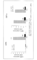
図5に示すように、共培養により3T3-L1におけるGIPRの発現が亢進した。また、遺伝子発現亢進の程度は、Raw264の細胞数に依存した。
図5に示すように、共培養により3T3-L1におけるGIPRの発現が亢進した。また、遺伝子発現亢進の程度は、Raw264の細胞数に依存した。
実施例5 GIPRの機能解析
実施例4の方法により発現されたGIPRの機能をIL6の発現を指標として確認した。
(1)IL6遺伝子発現量
実施例4と同様の方法により、3T3-L1とRaw264を24時間共培養した後、GIP,GIP+insulinを含む培地で刺激し、1,3,6時間後に下層ウェルの3T3-L1を採取し、cDNA合成(High capacity RNA-to-cDNA Kit,Applied Biosystems)後、定量的リアルタイムPCR(Applied Biosystems 7500 Fast リアルタイムPCRシステム)により遺伝子の発現定量を行い、36B4による補正を行った。なお、プライマーはApplied BiosystemsのTaqman Gene Expression assaysのIL6(Mm00446190_m1)を用いた。その結果、図6に示すようにGIPはインスリン存在下、非存在下いずれにおいても刺激後1時間においてIL6の遺伝子発現を亢進した。
実施例4の方法により発現されたGIPRの機能をIL6の発現を指標として確認した。
(1)IL6遺伝子発現量
実施例4と同様の方法により、3T3-L1とRaw264を24時間共培養した後、GIP,GIP+insulinを含む培地で刺激し、1,3,6時間後に下層ウェルの3T3-L1を採取し、cDNA合成(High capacity RNA-to-cDNA Kit,Applied Biosystems)後、定量的リアルタイムPCR(Applied Biosystems 7500 Fast リアルタイムPCRシステム)により遺伝子の発現定量を行い、36B4による補正を行った。なお、プライマーはApplied BiosystemsのTaqman Gene Expression assaysのIL6(Mm00446190_m1)を用いた。その結果、図6に示すようにGIPはインスリン存在下、非存在下いずれにおいても刺激後1時間においてIL6の遺伝子発現を亢進した。
(2)IL6産生量
実施例4と同様の方法により、3T3-L1とRaw264を24時間共培養した後、GIP,GIP+insulinを含む培地で刺激し、3時間後に培養上清を回収し、市販の固相酵素免疫検定法(Mouse IL6 ELISA MAX, Biolegend)を用いて、上清中IL6の定量を行った。
その結果、図7に示すようにGIPによりIL6の産生が亢進した。また、インスリン共存下においても、GIP刺激によりIL6の産生が亢進した。
実施例4と同様の方法により、3T3-L1とRaw264を24時間共培養した後、GIP,GIP+insulinを含む培地で刺激し、3時間後に培養上清を回収し、市販の固相酵素免疫検定法(Mouse IL6 ELISA MAX, Biolegend)を用いて、上清中IL6の定量を行った。
その結果、図7に示すようにGIPによりIL6の産生が亢進した。また、インスリン共存下においても、GIP刺激によりIL6の産生が亢進した。
実施例6 HIF1αsiRNAがGIPRの発現に及ぼす影響
(1)siRNAの導入
分化誘導後6日目の3T3-L1に対し、Lipofectamine RNAiMAX(Invitrogen)を用いてnegative control siRNA (Silencer(R) Select Negative Control #1 siRNA; Applied Biosystems)、あるいはHIF1αに対するsiRNA(FlexiTube GeneSolution GS15251 for HIF1α; Qiagen)の導入を行った。これらの操作は添付のプロトコルに従い、Revers Transfection法で行った。なお、各ウェルへの細胞播種数は5×10⁴/well(24ウェルプレート)とし、siRNA濃度は10nMとした。
(1)siRNAの導入
分化誘導後6日目の3T3-L1に対し、Lipofectamine RNAiMAX(Invitrogen)を用いてnegative control siRNA (Silencer(R) Select Negative Control #1 siRNA; Applied Biosystems)、あるいはHIF1αに対するsiRNA(FlexiTube GeneSolution GS15251 for HIF1α; Qiagen)の導入を行った。これらの操作は添付のプロトコルに従い、Revers Transfection法で行った。なお、各ウェルへの細胞播種数は5×10⁴/well(24ウェルプレート)とし、siRNA濃度は10nMとした。
(2)HIF1αsiRNAがGIPRの発現に及ぼす影響
HIF1αに対するsiRNAを用いて、GIPRの発現に及ぼす影響を検討した。その結果、HIF1αを70%Knock down (KD)した条件において、実施例4と同様の方法でマクロファージとの共培養した。その結果、マクロファージとの共培養(■Co-culture)によって増強されるGIPRの発現が著明に低下した(図8)。これらの結果から、脂肪細胞とマクロファージの共培養によって誘導されるGIPRはHIF1αの制御下にあることが示唆された。
HIF1αに対するsiRNAを用いて、GIPRの発現に及ぼす影響を検討した。その結果、HIF1αを70%Knock down (KD)した条件において、実施例4と同様の方法でマクロファージとの共培養した。その結果、マクロファージとの共培養(■Co-culture)によって増強されるGIPRの発現が著明に低下した(図8)。これらの結果から、脂肪細胞とマクロファージの共培養によって誘導されるGIPRはHIF1αの制御下にあることが示唆された。
実施例7 GIP受容体阻害物質のIL6産生阻害作用
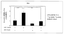
実施例4と同様の方法により、3T3-L1とRaw264を24時間共培養した後、GIPにより誘導される炎症性サイトカイン(IL6)の産生を抑制する物質として公知(Timper, K et al. Glucose-dependent insulinotropic polypeptide induces cytokine expression, lipolysis, and insulin resistance in human adipocytes. Am J Physiol Endocrinol Metab (2013), 304, E1-E13)のH-89(Cell Signaling Technology Japan)20nMを1時間前処理した後、GIP 100nMを含む培地で刺激し、1時間後に下層ウェルの3T3-L1を採取し、IL6の遺伝子発現レベルの測定を行った。
その結果、図9に示すように、H-89により、GIPによるIL6の発現亢進が有意に阻害された。これより、本発明の共培養系は、H-89のようなGIP受容体阻害物質をスクリーニングするための系として有用であることが確認された。
実施例4と同様の方法により、3T3-L1とRaw264を24時間共培養した後、GIPにより誘導される炎症性サイトカイン(IL6)の産生を抑制する物質として公知(Timper, K et al. Glucose-dependent insulinotropic polypeptide induces cytokine expression, lipolysis, and insulin resistance in human adipocytes. Am J Physiol Endocrinol Metab (2013), 304, E1-E13)のH-89(Cell Signaling Technology Japan)20nMを1時間前処理した後、GIP 100nMを含む培地で刺激し、1時間後に下層ウェルの3T3-L1を採取し、IL6の遺伝子発現レベルの測定を行った。
その結果、図9に示すように、H-89により、GIPによるIL6の発現亢進が有意に阻害された。これより、本発明の共培養系は、H-89のようなGIP受容体阻害物質をスクリーニングするための系として有用であることが確認された。
実施例8 GIP受容体親和性物質の評価
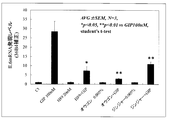
実施例7と同様の方法により、分化誘導後6日目の3T3-L1とRaw264を24時間共培養した後、実施例7で用いたH-89(Cell Signaling Technology Japan) 20nM、オウゴンエキス0.005%、ジンジャーエキス0.005%を添加した培地にて1h前処理した後、GIP 100nMを含む培地で刺激し、1時間後に下層ウェルの3T3-L1を採取し、IL6の遺伝子発現レベルの測定を行った。
その結果、図10に示すように、オウゴンエキス及びジンジャーエキスは、H-89と同様に、GIPによるIL6の発現亢進を有意に阻害した。また、結果は各剤を添加した刺激培地に対する相対値で示している。
したがって、オウゴンエキス及びジンジャーエキスは、GIP受容体阻害物質であると評価できる。
実施例7と同様の方法により、分化誘導後6日目の3T3-L1とRaw264を24時間共培養した後、実施例7で用いたH-89(Cell Signaling Technology Japan) 20nM、オウゴンエキス0.005%、ジンジャーエキス0.005%を添加した培地にて1h前処理した後、GIP 100nMを含む培地で刺激し、1時間後に下層ウェルの3T3-L1を採取し、IL6の遺伝子発現レベルの測定を行った。
その結果、図10に示すように、オウゴンエキス及びジンジャーエキスは、H-89と同様に、GIPによるIL6の発現亢進を有意に阻害した。また、結果は各剤を添加した刺激培地に対する相対値で示している。
したがって、オウゴンエキス及びジンジャーエキスは、GIP受容体阻害物質であると評価できる。
参考例1
3T3-L1を実施例1の方法に従い、分化誘導させ、分化誘導後0min、30min、2h、4h、8h、24h、2day、3day、4day、6day、8day、12day、16dayにおいて上記方法によりRNA抽出、GIP受容体の遺伝子発現解析を行った。
その結果、3T3-L1細胞の分化に伴い、GIP受容体は分化後2h、6日目に2相性の発現を示すことが確認された(図11)。
3T3-L1を実施例1の方法に従い、分化誘導させ、分化誘導後0min、30min、2h、4h、8h、24h、2day、3day、4day、6day、8day、12day、16dayにおいて上記方法によりRNA抽出、GIP受容体の遺伝子発現解析を行った。
その結果、3T3-L1細胞の分化に伴い、GIP受容体は分化後2h、6日目に2相性の発現を示すことが確認された(図11)。
Claims (15)
- 脂肪細胞においてHIFαを活性化する工程を含む、脂肪細胞におけるGIP受容体の発現増強方法。
- 脂肪細胞においてHIFαを活性化する工程を含む、脂肪細胞におけるGIP受容体の機能解析方法。
- 脂肪細胞においてHIFαを活性化する工程を含む、GIP受容体発現制御物質のスクリーニング方法。
- 脂肪細胞においてHIFαを活性化する工程を含む、GIP受容体親和性物質のスクリーニング方法。
- HIFαを活性化する工程が、低酸素環境下での培養である、請求項1~4の何れか1項記載の方法。
- HIFαを活性化する工程が、HIFα活性化剤を培地に添加する培養である、請求項1~4の何れか1項記載の方法。
- HIFαを活性化する工程が、脂肪細胞とマクロファージの共培養である、請求項1~4の何れか1項記載の方法。
- 以下の工程(a)~(c)を含む、請求項3記載の方法。
(a)脂肪細胞を低酸素環境下で培養する工程、又は培地にHIFα活性化剤を添加して脂肪細胞を培養する工程
(b)被験物質を前記培養系に接触させる工程
(c)工程(b)において被験物質を接触させた場合におけるGIP受容体の発現レベルと、被験物質を接触させない場合におけるGIP受容体の発現レベルとを比較する工程 - 以下の工程(d)~(f)を含む、請求項4記載の方法。
(d)脂肪細胞を低酸素環境下で培養する工程、又は培地にHIFα活性化剤を添加して脂肪細胞を培養する工程
(e)被験物質及びGIPを前記培養系に接触させる工程
(f)工程(e)において被験物質の存在下でGIPを培養系へ接触させた場合における受容体活性と、被験物質の非存在下でGIPを培養系へ接触させた場合における受容体活性とを比較する工程 - 以下の工程(h)~(j)を含む、請求項3記載の方法。
(h)脂肪細胞をマクロファージと共培養する工程
(i)被験物質を前記共培養系に接触させる工程
(j)工程(i)において被験物質を接触させた場合におけるGIP受容体の発現レベルと、被験物質を接触させない場合におけるGIP受容体の発現レベルとを比較する工程 - 以下の工程(k)~(m)を含む、請求項4記載の方法。
(k)脂肪細胞をマクロファージと共培養する工程
(l)被験物質及びGIPを前記共培養系に接触させる工程
(m)工程(l)において被験物質の存在下でGIPを共培養系へ接触させた場合における受容体活性と、被験物質の非存在下でGIPを共培養系へ接触させた場合における受容体活性とを比較する工程 - 脂肪細胞が、3T3-L1細胞、3T3-F442A細胞及びOb1771細胞から選ばれる前駆脂肪細胞から分化誘導された細胞である、請求項1~11のいずれか1項記載の方法。
- 脂肪細胞として、3T3-L1細胞を分化誘導後、6~16日目の細胞を用いる請求項1~12のいずれか1項記載の方法。
- HIFα活性化剤が、2価のコバルトイオン、ニッケルイオン、マンガンイオン及びデフェロキサミンメシル酸塩、O-フェナントロリン、ヒドロキシピリジノンから選ばれる1種以上である、請求項6、8及び9のいずれか1項記載の方法。
- マクロファージが、RAW264細胞、RAW264.7細胞又は腹腔マクロファージである、請求項7、10及び11のいずれか1項記載の方法。
Applications Claiming Priority (8)
| Application Number | Priority Date | Filing Date | Title |
|---|---|---|---|
| JP2013-081114 | 2013-04-09 | ||
| JP2013081113 | 2013-04-09 | ||
| JP2013081114 | 2013-04-09 | ||
| JP2013-081113 | 2013-04-09 | ||
| JP2014-060555 | 2014-03-24 | ||
| JP2014060556A JP2014217376A (ja) | 2013-04-09 | 2014-03-24 | Gip受容体の発現制御方法 |
| JP2014-060556 | 2014-03-24 | ||
| JP2014060555A JP2014217375A (ja) | 2013-04-09 | 2014-03-24 | Gip受容体の発現制御方法 |
Publications (1)
| Publication Number | Publication Date |
|---|---|
| WO2014168153A1 true WO2014168153A1 (ja) | 2014-10-16 |
Family
ID=51689560
Family Applications (1)
| Application Number | Title | Priority Date | Filing Date |
|---|---|---|---|
| PCT/JP2014/060222 Ceased WO2014168153A1 (ja) | 2013-04-09 | 2014-04-08 | Gip受容体の発現制御方法 |
Country Status (1)
| Country | Link |
|---|---|
| WO (1) | WO2014168153A1 (ja) |
Citations (1)
| Publication number | Priority date | Publication date | Assignee | Title |
|---|---|---|---|---|
| WO2006112418A1 (ja) * | 2005-04-18 | 2006-10-26 | Tokyo Medical And Dental University | 脂肪細胞の炎症性変化を抑制する物質のスクリーニング系及びそれを用いたスクリーニング方法 |
-
2014
- 2014-04-08 WO PCT/JP2014/060222 patent/WO2014168153A1/ja not_active Ceased
Patent Citations (1)
| Publication number | Priority date | Publication date | Assignee | Title |
|---|---|---|---|---|
| WO2006112418A1 (ja) * | 2005-04-18 | 2006-10-26 | Tokyo Medical And Dental University | 脂肪細胞の炎症性変化を抑制する物質のスクリーニング系及びそれを用いたスクリーニング方法 |
Non-Patent Citations (4)
| Title |
|---|
| AMERICAN JOURNAL OF PHYSIOLOGY ENDOCRINOLOGY AND METABOLISM, vol. 293, no. E1590, 18 September 2007 (2007-09-18), Retrieved from the Internet <URL:http://ajpendo.physiology.org/content/293/6/E1590> * |
| AMERICAN JOURNAL OF PHYSIOLOGY ENDOCRINOLOGY AND METABOLISM, vol. 300, no. E877-, 22 February 2011 (2011-02-22), Retrieved from the Internet <URL:http://ajpendo.physiology.org/content/300/5/E877> * |
| FEBS LETTERS, vol. 505, no. ISSUE., 27 August 2001 (2001-08-27), pages 269 - 274, Retrieved from the Internet <URL:http://www.sciencedirect.com/science/article/pii/S0014579301028332#> * |
| JOURNAL OF LIPID RESEARCH, vol. 52, 18 January 2011 (2011-01-18), pages 759 - 770, Retrieved from the Internet <URL:http://www.jlr.org/content/52/4/759> * |
Similar Documents
| Publication | Publication Date | Title |
|---|---|---|
| Fry et al. | Myogenic progenitor cells control extracellular matrix production by fibroblasts during skeletal muscle hypertrophy | |
| Huang et al. | Aging reduces an ERRalpha-directed mitochondrial glutaminase expression suppressing glutamine anaplerosis and osteogenic differentiation of mesenchymal stem cells | |
| YlÖstalo et al. | Human mesenchymal stem/stromal cells cultured as spheroids are self-activated to produce prostaglandin E2 that directs stimulated macrophages into an anti-inflammatory phenotype | |
| Chen et al. | High mobility group box 1-induced epithelial mesenchymal transition in human airway epithelial cells | |
| Woeller et al. | Thy1 (CD90) controls adipogenesis by regulating activity of the Src family kinase, Fyn | |
| Shu et al. | TCF7L2 promotes beta cell regeneration in human and mouse pancreas | |
| Chen et al. | Enhancer of zeste homolog 2 and histone deacetylase 9c regulate age-dependent mesenchymal stem cell differentiation into osteoblasts and adipocytes | |
| Xi et al. | HMGA2 promotes adipogenesis by activating C/EBPβ-mediated expression of PPARγ | |
| Zhu et al. | MicroRNA-24 promotes pancreatic beta cells toward dedifferentiation to avoid endoplasmic reticulum stress-induced apoptosis | |
| Yuan et al. | STAT3 stimulates adipogenic stem cell proliferation and cooperates with HMGA2 during the early stage of differentiation to promote adipogenesis | |
| Zhang et al. | LncRNA TUG1 reduces inflammation and enhances insulin sensitivity in white adipose tissue by regulating miR-204/SIRT1 axis in obesity mice | |
| Chung et al. | Isolation of neural crest derived chromaffin progenitors from adult adrenal medulla | |
| Ayala-Sumuano et al. | Srebf1a is a key regulator of transcriptional control for adipogenesis | |
| WO2018164228A1 (ja) | Ror1陽性の間葉系幹細胞を含有する、線維症を伴う疾患の予防又は処置のための医薬組成物、及びその調製方法、並びにror1陽性の間葉系幹細胞を用いる線維症を伴う疾患の予防又は処置方法 | |
| Li et al. | PI3K/AKT pathway mediates induction of IL-1RA by TSH in fibrocytes: modulation by PTEN | |
| JPWO2017038784A1 (ja) | Ror1陽性の間葉系幹細胞及びその調製方法、ror1陽性の間葉系幹細胞を含む医薬組成物及びその調製方法、並びにror1陽性の間葉系幹細胞を用いる疾患の予防又は治療方法 | |
| Wang et al. | Effect of advanced glycosylation end products on apoptosis in human adipose tissue-derived stem cells in vitro | |
| Deng et al. | Cysteine dioxygenase type 1 promotes adipogenesis via interaction with peroxisome proliferator-activated receptor gamma | |
| Kaneda-Ikeda et al. | Regulation of osteogenesis via miR-101-3p in mesenchymal stem cells by human gingival fibroblasts | |
| Tseng et al. | Markers of accelerated skeletal muscle regenerative response in murphy roths large mice: characteristics of muscle progenitor cells and circulating factors | |
| Sanap et al. | Conditioned medium of adipose derived mesenchymal stem cells reverse insulin resistance through downregulation of stress induced serine kinases | |
| Liu et al. | Conditionally immortalized brown preadipocytes can switch between proliferative and differentiated states | |
| Mandl et al. | Sprouty1 is a weight-loss target gene in human adipose stem/progenitor cells that is mandatory for the initiation of adipogenesis | |
| Bai et al. | miR‐182‐5p overexpression inhibits chondrogenesis by down‐regulating PTHLH | |
| Teng et al. | Tfh exosomes derived from allergic rhinitis promote DC maturation through miR-142-5p/CDK5/STAT3 pathway |
Legal Events
| Date | Code | Title | Description |
|---|---|---|---|
| 121 | Ep: the epo has been informed by wipo that ep was designated in this application |
Ref document number: 14783453 Country of ref document: EP Kind code of ref document: A1 |
|
| NENP | Non-entry into the national phase |
Ref country code: DE |
|
| 122 | Ep: pct application non-entry in european phase |
Ref document number: 14783453 Country of ref document: EP Kind code of ref document: A1 |