WO2014129653A1 - 血管透過性亢進抑制作用の評価方法 - Google Patents
血管透過性亢進抑制作用の評価方法 Download PDFInfo
- Publication number
- WO2014129653A1 WO2014129653A1 PCT/JP2014/054528 JP2014054528W WO2014129653A1 WO 2014129653 A1 WO2014129653 A1 WO 2014129653A1 JP 2014054528 W JP2014054528 W JP 2014054528W WO 2014129653 A1 WO2014129653 A1 WO 2014129653A1
- Authority
- WO
- WIPO (PCT)
- Prior art keywords
- vascular endothelial
- cells
- test substance
- endothelial cells
- cell
- Prior art date
- Legal status (The legal status is an assumption and is not a legal conclusion. Google has not performed a legal analysis and makes no representation as to the accuracy of the status listed.)
- Ceased
Links
Images




Classifications
-
- C—CHEMISTRY; METALLURGY
- C12—BIOCHEMISTRY; BEER; SPIRITS; WINE; VINEGAR; MICROBIOLOGY; ENZYMOLOGY; MUTATION OR GENETIC ENGINEERING
- C12Q—MEASURING OR TESTING PROCESSES INVOLVING ENZYMES, NUCLEIC ACIDS OR MICROORGANISMS; COMPOSITIONS OR TEST PAPERS THEREFOR; PROCESSES OF PREPARING SUCH COMPOSITIONS; CONDITION-RESPONSIVE CONTROL IN MICROBIOLOGICAL OR ENZYMOLOGICAL PROCESSES
- C12Q1/00—Measuring or testing processes involving enzymes, nucleic acids or microorganisms; Compositions therefor; Processes of preparing such compositions
- C12Q1/02—Measuring or testing processes involving enzymes, nucleic acids or microorganisms; Compositions therefor; Processes of preparing such compositions involving viable microorganisms
-
- G—PHYSICS
- G01—MEASURING; TESTING
- G01N—INVESTIGATING OR ANALYSING MATERIALS BY DETERMINING THEIR CHEMICAL OR PHYSICAL PROPERTIES
- G01N33/00—Investigating or analysing materials by specific methods not covered by groups G01N1/00 - G01N31/00
- G01N33/48—Biological material, e.g. blood, urine; Haemocytometers
- G01N33/50—Chemical analysis of biological material, e.g. blood, urine; Testing involving biospecific ligand binding methods; Immunological testing
- G01N33/5005—Chemical analysis of biological material, e.g. blood, urine; Testing involving biospecific ligand binding methods; Immunological testing involving human or animal cells
- G01N33/5008—Chemical analysis of biological material, e.g. blood, urine; Testing involving biospecific ligand binding methods; Immunological testing involving human or animal cells for testing or evaluating the effect of chemical or biological compounds, e.g. drugs, cosmetics
- G01N33/502—Chemical analysis of biological material, e.g. blood, urine; Testing involving biospecific ligand binding methods; Immunological testing involving human or animal cells for testing or evaluating the effect of chemical or biological compounds, e.g. drugs, cosmetics for testing non-proliferative effects
- G01N33/5026—Chemical analysis of biological material, e.g. blood, urine; Testing involving biospecific ligand binding methods; Immunological testing involving human or animal cells for testing or evaluating the effect of chemical or biological compounds, e.g. drugs, cosmetics for testing non-proliferative effects on cell morphology
-
- G—PHYSICS
- G01—MEASURING; TESTING
- G01N—INVESTIGATING OR ANALYSING MATERIALS BY DETERMINING THEIR CHEMICAL OR PHYSICAL PROPERTIES
- G01N33/00—Investigating or analysing materials by specific methods not covered by groups G01N1/00 - G01N31/00
- G01N33/48—Biological material, e.g. blood, urine; Haemocytometers
- G01N33/483—Physical analysis of biological material
- G01N33/4833—Physical analysis of biological material of solid biological material, e.g. tissue samples, cell cultures
-
- G—PHYSICS
- G01—MEASURING; TESTING
- G01N—INVESTIGATING OR ANALYSING MATERIALS BY DETERMINING THEIR CHEMICAL OR PHYSICAL PROPERTIES
- G01N33/00—Investigating or analysing materials by specific methods not covered by groups G01N1/00 - G01N31/00
- G01N33/48—Biological material, e.g. blood, urine; Haemocytometers
- G01N33/50—Chemical analysis of biological material, e.g. blood, urine; Testing involving biospecific ligand binding methods; Immunological testing
- G01N33/5005—Chemical analysis of biological material, e.g. blood, urine; Testing involving biospecific ligand binding methods; Immunological testing involving human or animal cells
- G01N33/5008—Chemical analysis of biological material, e.g. blood, urine; Testing involving biospecific ligand binding methods; Immunological testing involving human or animal cells for testing or evaluating the effect of chemical or biological compounds, e.g. drugs, cosmetics
- G01N33/5044—Chemical analysis of biological material, e.g. blood, urine; Testing involving biospecific ligand binding methods; Immunological testing involving human or animal cells for testing or evaluating the effect of chemical or biological compounds, e.g. drugs, cosmetics involving specific cell types
- G01N33/5064—Endothelial cells
Definitions
- the present invention relates to a method for evaluating vascular permeability enhancement inhibitory effect.
- Type I allergic reaction occurs by the following mechanism.
- the allergen enters the body and IgE on the cell surface of the mast cell binds to the allergen, the mast cell releases a physiologically active substance such as histamine. This causes vasodilation and increased vascular permeability, and causes symptoms such as edema and pruritus.
- Vascular hyper-permeability is a phenomenon in which vascular endothelial cells respond to histamine, the gap between the cells expands, and blood vessel permeability increases. This phenomenon has been evaluated by taking a two-dimensional image of vascular endothelial cells and measuring the expansion of the gap between the cells (Non-Patent Documents 1 and 2). Since the antihistamine has an action of suppressing the increase in vascular permeability caused by histamine stimulation, an evaluation system that measures the suppression of the expansion of the gap between cells can be used for the evaluation of the drug effect of the antihistamine.
- the present inventors have found that, depending on the vascular endothelial cells, since the adhesion of the cells is strong, the gap between the cells may not expand, and there is a possibility that accurate evaluation cannot be performed.
- An object of the present invention is to provide a method for evaluating an inhibitory action on vascular permeability enhancement, which enables quick and accurate measurement.
- the inventors of the present invention observed three-dimensionally the vascular endothelial cells after histamine stimulation and found for the first time that the morphology of vascular endothelial cells changed immediately after histamine stimulation. Specifically, it was found that the vascular endothelial cells show a morphological change that immediately after histamine stimulation, the apex height of the cells increases, and after a while, the adhesion area of the cells decreases. Furthermore, the present inventors have found that the apex height of cells increases immediately after histamine stimulation even when the adhesion of vascular endothelial cells is strong and no change in the adhesion area is finally observed. It was. Based on these findings, the present inventors consider that it is possible to quickly and accurately evaluate the inhibitory effect on the increase in vascular permeability by utilizing the change in the apex height of the cells, and complete the invention. It came to.
- the present invention is a method for evaluating the inhibitory action of a test substance on the increase in vascular permeability of vascular endothelial cells, the method comprising contacting a test substance with vascular endothelial cells, and enhancing the vascular permeability of the vascular endothelial cells.
- the method comprises a step of bringing a drug into contact with vascular endothelial cells and a step of measuring the apex height of vascular endothelial cells, wherein the apex height of vascular endothelial cells is an indicator of an inhibitory action on the increase in vascular permeability of vascular endothelial cells as a test substance
- apex height of vascular endothelial cells is an indicator of an inhibitory action on the increase in vascular permeability of vascular endothelial cells as a test substance
- the above indicator indicates that when the apex height of the vascular endothelial cell in the presence of the test substance is lower than the apex height of the vascular endothelial cell in the absence of the test substance, the test substance increases the vascular permeability of the vascular endothelial cell. It can be used as an index for determining suppression.
- the method further comprises the step of measuring the adhesion area of the vascular endothelial cell, and using the apex height and the adhesion area of the vascular endothelial cell as an index of the inhibitory action of the test substance on the enhancement of vascular permeability of the vascular endothelial cell.
- the adhesion area is used as an index in addition to the apex height, more accurate evaluation can be performed.
- the above index indicates that the apex height of the vascular endothelial cell in the presence of the test substance is lower than the apex height of the vascular endothelial cell in the absence of the test substance and the adhesion of the vascular endothelial cell in the presence of the test substance.
- the test substance can be used as an index for determining that the increase in vascular permeability of the vascular endothelial cell is suppressed.
- the agent that enhances the vascular permeability of vascular endothelial cells is preferably histamine.
- histamine which is the original ligand, it is possible to observe a reaction close to the actual living body.
- apex height and / or adhesion area of vascular endothelial cells it is preferable to measure the apex height and / or adhesion area of vascular endothelial cells with a quantitative phase microscope.
- a quantitative phase microscope it is possible to accurately and easily measure the apex height and / or the adhesion area.
- the method for evaluating the inhibitory effect on the increase in vascular permeability of the present invention enables rapid and accurate measurement. By using this evaluation method, it is possible to evaluate the efficacy of antihistamines. Moreover, it is possible to screen for antihistamines by using this evaluation method.
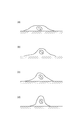
- FIG. 1 (a) shows vascular endothelial cells before histamine stimulation.
- FIG. 1 (b) represents vascular endothelial cells immediately after histamine stimulation. By histamine stimulation, the apex height of the cells first increases. The cell adhesion area does not change immediately after histamine stimulation.
- FIG. 1 (c) represents vascular endothelial cells about 10 minutes after histamine stimulation. The apex height of the cell is maintained. Cell adhesion area begins to decrease.
- FIG. 1 (d) represents vascular endothelial cells about 15 minutes after histamine stimulation. The apex height of the cell is further increased, the cell adhesion area is reduced, and this state is maintained.
- test substance for evaluating the inhibitory effect of the test substance on the increase in vascular permeability of vascular endothelial cells
- test substance for evaluating the inhibitory action.
- a substance expected to have an H 1 receptor antagonistic action is used as a test substance.
- vascular endothelial cells vascular endothelial cells derived from mammals such as mice, rats, cows and humans can be used. Since it is possible to appropriately evaluate the efficacy of the antihistamine when administered to humans, it is preferable to use human-derived vascular endothelial cells.
- human-derived vascular endothelial cells cutaneous microvascular endothelial cells, umbilical vein vascular endothelial cells, vena cava vascular endothelial cells, and the like can be used.
- Vascular endothelial cells may be derived from adults or newborns. Vascular endothelial cells can be collected and isolated from animals by known methods. It can also be obtained from a cell bank.
- vascular endothelial cells The method for culturing vascular endothelial cells is well known to those skilled in the art. For example, it can be cultured in a 37 ° C., 5% CO 2 incubator using a commercially available medium for vascular endothelial cells.
- Vascular seedlings are seeded in a dish and cultured until they are sufficiently adhered.
- the culture period is preferably 24 hours.
- test substance is added to the medium, and the test substance is brought into contact with the vascular endothelial cells (hereinafter also referred to as pretreatment).
- the contact time is usually from 30 minutes to 60 minutes.
- the test substance is preferably added as a solution in an appropriate solvent.
- the solvent is preferably a medium.
- the test substance may be diluted in a medium after being dissolved in a solvent that dissolves the test substance.
- a sample in which only the solvent not containing the test substance is added to the medium may be used as a negative control.
- a drug that enhances the vascular permeability of the vascular endothelial cells is added to the medium and brought into contact with the vascular endothelial cells (hereinafter also referred to as drug stimulation).
- the drug that enhances vascular permeability of vascular endothelial cells may be an H 1 receptor agonist, and examples thereof include histamine, 2-methylhistamine, and 2- (2-pyridyl) ethylamine. Histamine is preferably used because it is a natural ligand of the H 1 receptor.
- the vertex height can be measured by using a three-dimensional imaging apparatus.
- the three-dimensional imaging apparatus include a quantitative phase microscope, a low coherence interference microscope, a digital holography microscope, a tomographic phase microscope, and a scanning probe microscope, and a quantitative phase microscope is preferable.
- Quantitative phase microscopes are disclosed in, for example, Japanese Patent No. 4090244 and Japanese Patent Application Laid-Open No. 2010-48619.
- the phase of the optical field can be measured quantitatively using phase shift interferometry.
- the quantitative phase microscope it is possible to obtain a three-dimensional morphology of the cell with a resolution of 1 micron or less, thereby accurately measuring the apex height and the adhesion area of the cell.
- the optical thickness (OT: Optical Thickness) is an index given by Equation (1), where ⁇ is the phase change received by the light transmitted through the measurement object.
- ⁇ represents the wavelength of light.
- n1 is the refractive index of the measurement object
- n0 is the refractive index of the medium around the measurement object
- h is the height of the measurement object. Therefore, by measuring the optical thickness, a value proportional to the height of the measurement object can be approximately obtained.
- OT h ⁇ (n1-n0) (2)
- the cell apex is the highest point in a single cell.
- the height refers to the distance from the bottom surface of the cells adhered to the culture container to the cell surface not adhered to the culture container.
- the apex height of the cell may be the actual height or the optical thickness.
- the measurement of the apex height of the cells may be performed at any time after drug stimulation.
- the apex height of the cells rises immediately after the stimulation, remains substantially the same until about 10 minutes, and further rises after about 15 minutes.
- the apex height of the cells rises immediately after the stimulation, remains substantially the same until about 10 minutes, and further rises after about 15 minutes.
- the measurement of the apex height of the cells is preferably performed between 3 minutes and 9 minutes after the start of drug stimulation.
- the index of the evaluation method may be an index based on the apex height of a single cell, or may be an index based on an average value of apex heights of a plurality of cells. More specifically, the index of the evaluation method is that when the apex height of vascular endothelial cells in the presence of the test substance is lower than the apex height of vascular endothelial cells in the absence of the test substance, the test substance is It can be determined that the increase in vascular permeability of vascular endothelial cells is suppressed.
- the adhesion area of vascular endothelial cells is measured. Evaluation based on the adhesion area of vascular endothelial cells is equivalent to the conventional evaluation method based on the expansion of cell gaps, and more accurate evaluation is possible compared to the case where the apex height of vascular endothelial cells alone is used. It becomes possible.
- the adhesion area of vascular endothelial cells refers to the area of the bottom surface of the cells adhered to the culture container. By using a three-dimensional imaging apparatus, it is possible to measure the adhesion area as well as the apex height of the cells. The cell adhesion area may be measured at any time after drug stimulation.
- the adhesion area of the cell starts to decrease after about 10 minutes from the stimulation and becomes constant after about 15 minutes. Therefore, the measurement of the cell adhesion area is preferably performed after 15 minutes from the start of drug stimulation. Further, more accurate measurement can be performed by measuring the cell adhesion area in a time zone in which the variation of the cell adhesion area is as small as possible. In order to increase measurement efficiency, it is preferable to perform measurement in a short time. Therefore, the measurement of the cell adhesion area is preferably performed between 15 minutes and 20 minutes after the start of drug stimulation.
- the inhibitory effect of the test substance on the increase in vascular permeability of vascular endothelial cells is evaluated. It may be an index based on the apex height and adhesion area of a single cell, or may be an index based on the average value of apex heights and the average value of adhesion areas of a plurality of cells.
- the apex height of the vascular endothelial cell in the presence of the test substance is lower than the apex height of the vascular endothelial cell in the absence of the test substance, and the adhesion area of the vascular endothelial cell in the presence of the test substance Is larger than the adhesion area of vascular endothelial cells in the absence of the test substance, it can be determined that the test substance suppresses the increase in vascular permeability of the vascular endothelial cells.
- a method for evaluating the vascular permeability enhancing effect of a vascular endothelial cell of a test substance the step of contacting the test substance with the vascular endothelial cell, and the step of measuring the apex height of the vascular endothelial cell And using the height of the apex of the vascular endothelial cell as an indicator of the vascular permeability enhancing action of the vascular endothelial cell of the test substance.
- the above index indicates that when the apex height of vascular endothelial cells in the presence of the test substance is higher than the apex height of vascular endothelial cells in the absence of the test substance, the test substance enhances the vascular permeability of the vascular endothelial cells. Then, it can be used as an index to judge.
- the method further comprises a step of measuring the adhesion area of the vascular endothelial cell, wherein the apex height and the adhesion area of the vascular endothelial cell are used as an index of the test substance for enhancing the vascular permeability of the vascular endothelial cell. preferable.
- the above index indicates that the apex height of the vascular endothelial cell in the presence of the test substance is higher than the apex height of the vascular endothelial cell in the absence of the test substance, and the adhesion of the vascular endothelial cell in the presence of the test substance
- the test substance can be used as an index for determining that the vascular permeability of the vascular endothelial cell is enhanced.
- test substance for evaluating the enhancing action.
- a substance expected to have an H 1 receptor agonistic action is used as a test substance.
- vascular endothelial cells used and the culture method thereof are as described above.
- Test substance is added to the medium, and the test substance is brought into contact with vascular endothelial cells.
- This step is a step corresponding to the above-described drug stimulation.
- the measurement of the apex height and adhesion area of vascular endothelial cells after drug stimulation is as described above.
- Vascular endothelial cells Human neonatal skin-derived microvascular endothelial cells (HMVEC) were used as vascular endothelial cells. After subculture, the culture solution up to immediately before the measurement was obtained by adding microvascular endothelial cell additive factor set-2 EGM-2 MV SingleQuots (Lonza CC-4147) to 500 mL of EBM-2 medium (Lonza CC-3156) Was used. Incubation was performed in a 5% CO 2 environment at 37 ° C. for 24 hours after the passage, and the culture medium was replaced with EBM-2 medium immediately before the pretreatment. An untreated plastic dish was used as a culture dish at the time of passage.
- HMVEC Human neonatal skin-derived microvascular endothelial cells
- Racemic cetirizine, levocetirizine (1-cetirizine), and dextrocetirizine (d-cetirizine) were used as histamine pretreatment reagents.
- Racemic cetirizine and levocetirizine are known to have an antihistamine action, that is, an inhibitory action on the increase in vascular permeability of vascular endothelial cells, whereas dextrocetirizine is known to have little antihistamine action.
- the binding ability of levocetirizine to the H 1 receptor is about 33 times that of dextrocetirizine.
- Racemic cetirizine was added to the medium so that the final concentration was 2 ⁇ M, and the mixture was allowed to stand in a CO 2 incubator at 37 ° C. for 30 minutes.
- Levocetirizine and dextrocetirizine were pretreated in the same manner as racemic cetirizine except that the final concentration was 1 ⁇ M.
- Histamine stimulation Cells were set in the following apparatus, and microscopic observation was started. 300 seconds after the start of observation, histamine was added so that the final concentration was 100 ⁇ M, and vascular permeability of vascular endothelial cells was increased.
- Example 1 The state of vascular endothelial cells after histamine stimulation was observed with a quantitative phase microscope depending on the presence or absence of pretreatment with levocetirizine.
- FIG. 2 shows the state of cells without pretreatment
- FIG. 3 shows the state of cells with pretreatment with levocetirizine.
- (a), (b), (c), and (d) represent the state of cells after 270 seconds, 402 seconds, 1002 seconds, and 1170 seconds from the start of observation, respectively. Histamine stimulation is performed 300 seconds after the start of observation.
- FIG. 4 shows changes in cell morphology without pretreatment
- FIG. 5 shows changes in cell morphology when pretreated with levocetirizine.
- FIG. 4 it was confirmed that the apex height of the cells was first increased by histamine stimulation, and then the cell adhesion area was decreased.
- FIG. 5 when pretreatment with levocetirizine was performed, even when histamine stimulation was performed, almost no cell shape change was observed.
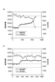
- Example 2 Similarly to Example 1, the state of vascular endothelial cells after stimulation with histamine was observed with a quantitative phase microscope depending on the presence or absence of pretreatment with levocetirizine, and the apex height and adhesion area of the cells were measured. The results are shown in FIGS. 6 (a) and (b). As is clear from FIG. 6 (a), it was confirmed that in the case of no pretreatment, the apex height of the cells was increased immediately after histamine stimulation and further increased after about 10 minutes. Further, in the case of no pretreatment, it was confirmed that the adhesion area of the cells was reduced about 10 minutes after histamine stimulation. On the other hand, as is clear from FIG. 6 (b), when pretreatment with levocetirizine was performed, the apex height and adhesion area of the cells hardly changed even when histamine stimulation was performed.
- FIGS. 7 (a) and (b) show the results.
- FIG. 7A is the same as FIG. In FIG. 7 (b), the apex height of the cells increased immediately after histamine stimulation, but thereafter the apex height and the adhesion area hardly changed. This is presumed to mean that the adhesive strength, that is, the difficulty of peeling, differs depending on the cell.
- the cell having a weak adhesive strength and easily peeled off exhibits a behavior as shown in FIG. 7A, and has a strong adhesive strength and is difficult to peel off. Can be interpreted to show the behavior as shown in FIG.
- the cells used for evaluation are cells that have strong adhesive force and are difficult to peel off, it means that the conventional method of measuring changes in the adhesion area of the cells cannot perform appropriate evaluation.
- the method of the present invention of measuring the change in the apex height of a cell can perform an appropriate evaluation even when such a cell is used.
- Example 4 instead of levocetirizine, pretreatment was performed with dextrocetirizine or racemic cetirizine, and the state of vascular endothelial cells after histamine stimulation was observed with a quantitative phase microscope, and the apex height and adhesion area of the cells were measured.
- FIGS. 8A and 8B show the state of cells when pretreated with dextrocetirizine
- FIGS. 9A and 9B show the state of cells when pretreated with racemic cetirizine.
- (a) and (b) show the state of the cells after 270 seconds and 1170 seconds from the start of observation, respectively. Histamine stimulation is performed 300 seconds after the start of observation.
- the results are summarized in FIG. FIG. 10 (a) shows no pretreatment, (b) shows pretreatment with levocetirizine, (c) shows pretreatment with dextrocetirizine, and (d) shows the apex height of cells when pretreatment with racemic cetirizine is performed. It shows the change of the height.
- the horizontal axis of FIG. 10 represents the elapsed time (seconds) when the histamine stimulation time is 0 seconds.
- the apex height of the cells is increased after histamine stimulation.
- the apex height of the cells is increased after histamine stimulation. Less than (a) and (c).
- FIG. 11 shows the averaged change in the apex height of the cells in each treatment group.
- the apex height of the cells when pretreated with dextrocetirizine increased in the same manner as without pretreatment, whereas when pretreated with levocetirizine or racemic cetirizine The apex height of the cells did not increase.
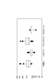
- the box apexes in FIG. 12 are obtained by standardizing the apex height of the cells 8 minutes after histamine stimulation based on the apex height of the cells at the time of histamine stimulation.
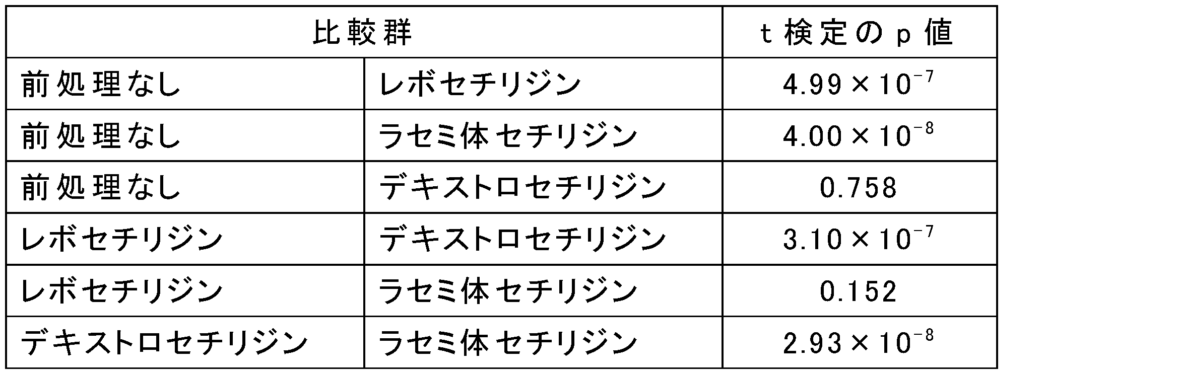
- Table 1 summarizes the results of t-test between the treatment groups. As is clear from the results shown in FIG. 12 and Table 1, the cell apex height is significantly higher when pretreated with levocetirizine or racemic cetirizine than when pretreated with no pretreatment or with dextrocetirizine. It was confirmed that there was a correlation between the antihistamine action (inhibition of the increase in vascular permeability of vascular endothelial cells) and the apex height of the cells.
Landscapes
- Health & Medical Sciences (AREA)
- Life Sciences & Earth Sciences (AREA)
- Engineering & Computer Science (AREA)
- Biomedical Technology (AREA)
- Immunology (AREA)
- Chemical & Material Sciences (AREA)
- Molecular Biology (AREA)
- Hematology (AREA)
- Urology & Nephrology (AREA)
- Cell Biology (AREA)
- Physics & Mathematics (AREA)
- Biochemistry (AREA)
- General Health & Medical Sciences (AREA)
- Analytical Chemistry (AREA)
- General Physics & Mathematics (AREA)
- Biotechnology (AREA)
- Food Science & Technology (AREA)
- Medicinal Chemistry (AREA)
- Pathology (AREA)
- Microbiology (AREA)
- Bioinformatics & Cheminformatics (AREA)
- Toxicology (AREA)
- Tropical Medicine & Parasitology (AREA)
- Organic Chemistry (AREA)
- Physiology (AREA)
- Proteomics, Peptides & Aminoacids (AREA)
- Biophysics (AREA)
- Wood Science & Technology (AREA)
- Zoology (AREA)
- Optics & Photonics (AREA)
- General Engineering & Computer Science (AREA)
- Genetics & Genomics (AREA)
- Measuring Or Testing Involving Enzymes Or Micro-Organisms (AREA)
- Investigating Or Analysing Biological Materials (AREA)
Abstract
被験物質を血管内皮細胞に接触させる工程と、血管内皮細胞の血管透過性を亢進する薬剤を血管内皮細胞に接触させる工程と、血管内皮細胞の頂点高さを測定する工程とを備え、血管内皮細胞の頂点高さを被験物質の血管内皮細胞の血管透過性亢進の抑制作用の指標とする方法。
Description
本発明は、血管透過性亢進抑制作用の評価方法に関する。
I型アレルギー反応は、以下の機序で生じる。アレルゲンが体内に侵入し、肥満細胞の細胞表面のIgEとアレルゲンが結合すると、肥満細胞はヒスタミン等の生理活性物質を放出する。これにより、血管拡張及び血管透過性亢進などが起こり、浮腫及び掻痒等の症状が引き起こされる。
血管透過性亢進(vascular hyper-permeability)とは、血管内皮細胞がヒスタミンに応答して、細胞間の間隙が広がり、血管の透過性が上昇する現象である。この現象は、血管内皮細胞の二次元画像を撮影し、細胞間の間隙の拡大を測定することによって評価されてきた(非特許文献1及び2)。抗ヒスタミン剤は、ヒスタミン刺激による血管透過性亢進を抑制する作用を有することから、抗ヒスタミン剤の薬効評価に細胞間の間隙の拡大の抑制を測定する評価系が利用可能である。
Wysolmerskiら, The American Journal of Pathology, 132(1), 28-37 (1988).
Killackeyら, The American journal of pathology, 122(1), 50-61 (1986).
しかしながら、細胞間の間隙の拡大は、ヒスタミン刺激から約30分経過しなければ観察することができず、迅速な測定を行うことが困難であった。
また、血管内皮細胞によっては、細胞の接着力が強いため、細胞間の間隙の拡大が起きない場合があり、正確な評価を行えない可能性があることを本発明者らは見出した。
本発明の課題は、迅速かつ正確な測定が可能な、血管透過性亢進の抑制作用の評価方法を提供することにある。
本発明者らは、ヒスタミン刺激後の血管内皮細胞を三次元的に観察したところ、ヒスタミン刺激の直後から血管内皮細胞の形態が変化することを初めて見出した。具体的には、ヒスタミン刺激の直後に、細胞の頂点高さが高くなり、その後しばらく経ってから細胞の接着面積が小さくなっていくという形態変化を血管内皮細胞は示すことが分かった。さらに、血管内皮細胞の接着力が強く、最終的に接着面積に変化が見られない場合であっても、ヒスタミン刺激直後には細胞の頂点高さが高くなることを、本発明者らは見出した。これらの知見に基づき、細胞の頂点高さの変化を利用することで、迅速かつ正確に血管透過性亢進の抑制作用の評価することが可能であると本発明者らは考え、発明を完成するに至った。
すなわち、本発明は、被験物質の血管内皮細胞の血管透過性亢進の抑制作用を評価する方法であって、被験物質を血管内皮細胞に接触させる工程と、血管内皮細胞の血管透過性を亢進する薬剤を血管内皮細胞に接触させる工程と、血管内皮細胞の頂点高さを測定する工程とを備え、血管内皮細胞の頂点高さを被験物質の血管内皮細胞の血管透過性亢進の抑制作用の指標とする方法を提供する。
上記指標は、被験物質の存在下における血管内皮細胞の頂点高さが被験物質の非存在下における血管内皮細胞の頂点高さよりも低い場合に、当該被験物質は血管内皮細胞の血管透過性亢進を抑制すると判断する指標とすることができる。
上記方法は、血管内皮細胞の接着面積を測定する工程をさらに備え、血管内皮細胞の頂点高さ及び接着面積を、被験物質の、血管内皮細胞の血管透過性亢進の抑制作用の指標とすることが好ましい。頂点高さに加えて、接着面積も指標とすることで、より正確な評価を行うことが可能となる。この場合、上記指標は、被験物質の存在下における血管内皮細胞の頂点高さが被験物質の非存在下における血管内皮細胞の頂点高さよりも低く、かつ被験物質の存在下における血管内皮細胞の接着面積が被験物質の非存在下における血管内皮細胞の接着面積よりも大きい場合に、当該被験物質は血管内皮細胞の血管透過性亢進を抑制すると判断する指標とすることができる。
上記方法において、血管内皮細胞の血管透過性を亢進する薬剤はヒスタミンであることが好ましい。本来のリガンドであるヒスタミンを用いることで、実際の生体内に近い反応を観察することが可能となる。
上記方法において、血管内皮細胞の頂点高さ及び/又は接着面積の測定を定量位相顕微鏡で行うことが好ましい。定量位相顕微鏡を用いることで、正確かつ簡便に頂点高さ及び/又は接着面積の測定することが可能となる。
本発明の血管透過性亢進の抑制作用の評価方法は、迅速かつ正確な測定が可能である。この評価方法を利用することで、抗ヒスタミン剤の薬効評価を行うことが可能である。また、この評価方法を利用することで、抗ヒスタミン剤のスクリーニングを行うことが可能である。
(ヒスタミン刺激による、血管内皮細胞の形態変化)
ヒスタミン刺激による血管内皮細胞の形態変化を図1に沿って説明する。図1(a)はヒスタミン刺激前の血管内皮細胞を表す。図1(b)はヒスタミン刺激直後の血管内皮細胞を表す。ヒスタミン刺激によって、まずは細胞の頂点高さが高くなる。細胞の接着面積は、ヒスタミン刺激直後は変化しない。図1(c)はヒスタミン刺激から約10分後の血管内皮細胞を表す。細胞の頂点高さは維持される。細胞の接着面積が小さくなり始める。図1(d)はヒスタミン刺激から約15分後の血管内皮細胞を表す。細胞の頂点高さがさらに高くなり、細胞の接着面積が小さくなり、この状態が維持される。
ヒスタミン刺激による血管内皮細胞の形態変化を図1に沿って説明する。図1(a)はヒスタミン刺激前の血管内皮細胞を表す。図1(b)はヒスタミン刺激直後の血管内皮細胞を表す。ヒスタミン刺激によって、まずは細胞の頂点高さが高くなる。細胞の接着面積は、ヒスタミン刺激直後は変化しない。図1(c)はヒスタミン刺激から約10分後の血管内皮細胞を表す。細胞の頂点高さは維持される。細胞の接着面積が小さくなり始める。図1(d)はヒスタミン刺激から約15分後の血管内皮細胞を表す。細胞の頂点高さがさらに高くなり、細胞の接着面積が小さくなり、この状態が維持される。
一方、抗ヒスタミン剤等の血管内皮細胞の血管透過性亢進の抑制作用を有する物質で前処理を行った後、血管内皮細胞をヒスタミン刺激すると、細胞の形態変化はおきず、図1(a)の状態が維持される。
この形態変化の差異に基づいて、血管透過性亢進の抑制作用の評価を行う。
(被験物質の血管内皮細胞の血管透過性亢進の抑制作用を評価する方法)
抑制作用を評価する被験物質は、どのような物質を用いてもよい。好ましくは、H1受容体拮抗作用を有することが期待される物質を被験物質として用いる。
抑制作用を評価する被験物質は、どのような物質を用いてもよい。好ましくは、H1受容体拮抗作用を有することが期待される物質を被験物質として用いる。
血管内皮細胞は、マウス、ラット、ウシ及びヒト等の哺乳類由来の血管内皮細胞を用いることができる。ヒトに投与した場合の抗ヒスタミン剤の薬効の評価を適切に行うことが可能であることから、ヒト由来の血管内皮細胞を用いることが好ましい。ヒト由来の血管内皮細胞は、皮膚微小血管内皮細胞、臍帯静脈血管内皮細胞及び大静脈血管内皮細胞等を利用することができる。血管内皮細胞は、成人由来でも新生児由来でも構わない。血管内皮細胞は、公知の方法により動物から採取し単離することができる。また、細胞バンクから入手することも可能である。血管内皮細胞の培養方法は当業者にとって周知であり、例えば、市販の血管内皮細胞用培地を用いて、37℃、5%CO2インキュベータ―で、培養することが可能である。
血管内皮細胞をディッシュに播種し、十分に接着するまで培養する。培養期間は好ましくは24時間である。
次に、被験物質を培地に添加して、被験物質を血管内皮細胞に接触させる(以下、前処理ともいう)。接触時間は、通常、30分~60分である。被験物質は適切な溶媒に溶かした溶液として添加することが好ましい。溶媒は好ましくは培地である。被験物質が培地に溶解し難い場合は、被験物質を溶解する溶媒に溶解した後に培地で希釈してもよい。なお、この際、被験物質を含まない溶媒のみを培地に添加したサンプルをネガティブコントロールとしてもよい。
次に、血管内皮細胞の血管透過性を亢進する薬剤を培地に添加して、血管内皮細胞に接触させる(以下、薬剤刺激ともいう)。血管内皮細胞の血管透過性を亢進する薬剤は、H1受容体作動薬であればよく、ヒスタミン、2-メチルヒスタミン及び2-(2-ピリジル)エチルアミン等が挙げられる。H1受容体本来のリガンドであることから、ヒスタミンを用いることが好ましい。
薬剤刺激後、血管内皮細胞の頂点高さを測定する。三次元イメージング装置を用いることで、頂点高さを測定することができる。三次元イメージング装置として、定量位相顕微鏡、低コヒーレンス干渉顕微鏡、デジタルホログラフィ顕微鏡、トモグラフィック位相顕微鏡及び走査型プローブ顕微鏡等が挙げられ、定量位相顕微鏡が好ましい。
定量位相型顕微鏡は、例えば特許第4090244号公報及び特開第2010-48619号公報に開示される。定量位相型顕微鏡を用いることにより、位相シフト干渉法を用いて光場の位相を定量的に測定することができる。定量位相顕微鏡を用いることにより、細胞の三次元的な形態を1ミクロン以下の分解能で得ることができ、それにより細胞の頂点高さ及び接着面積を正確に測定することが可能である。
定量位相顕微鏡は位相シフト法を用いることにより、測定対象物を透過した光の受ける位相変化を分布的に求めることができる。光学的な厚さ(OT:Optical Thickness)とは、測定対象物を透過した光の受ける位相変化をφとしたときに、(1)式で与えられる指標である。ここでλは光の波長を表す。
OT=λ×φ/2π (1)
OT=λ×φ/2π (1)
光学的な厚さは、測定対象物の屈折率をn1、測定対象物の周囲の媒質の屈折率をn0、測定対象物の高さをhとしたときに、(2)式で記述される。したがって、光学的な厚さを測定することにより、測定対象物の高さに比例する値を近似的に求めることができる。
OT=h×(n1-n0) (2)
OT=h×(n1-n0) (2)
細胞の頂点とは、単一の細胞における高さが最も高い点をいう。高さとは、培養容器に接着している細胞の底面から、培養容器に接着していない細胞表面までの距離をいう。細胞の頂点高さは、実高さであっても光学的厚さであってもよい。
細胞の頂点高さの測定は、薬剤刺激後の任意の時点で行ってよい。被験物質の前処理なしで薬剤刺激した場合、細胞の頂点高さは刺激直後に上昇し約10分後まではほぼ同じ高さを維持し、約15分後にはさらに上昇する。しかしながら、接着力の強い血管内皮細胞の場合には、約15分後の細胞の頂点高さのさらなる上昇が認められない場合もある。また、測定効率を上げるためには、短時間で測定を行うことが好ましい。したがって、細胞の頂点高さの測定は、薬剤刺激開始から10分後以内に行うことが好ましい。また、細胞の頂点高さの変動ができるだけ少ない時間帯で細胞の頂点高さを測定する方が、より正確な測定を行える。したがって、細胞の頂点高さの測定は、薬剤刺激開始から3分後~9分後の間に行うことが好ましい。
測定した細胞の頂点高さに基づいて、被験物質の血管内皮細胞の血管透過性亢進の抑制作用を評価する。上記評価方法の指標は、単一の細胞の頂点高さに基づく指標であってもよく、複数の細胞の頂点高さの平均値に基づく指標であってもよい。より具体的には、上記評価方法の指標は、被験物質の存在下における血管内皮細胞の頂点高さが被験物質の非存在下における血管内皮細胞の頂点高さよりも低い場合に、当該被験物質は血管内皮細胞の血管透過性亢進を抑制すると判断することができる。
別の実施形態において、血管内皮細胞の頂点高さの測定に加えて、血管内皮細胞の接着面積の測定を行う。血管内皮細胞の接着面積に基づく評価は、従来の評価方法である細胞間隙の拡大に基づく評価に相当するものであり、血管内皮細胞の頂点高さ単独の場合と比較してより正確な評価が可能となる。血管内皮細胞の接着面積とは、培養容器に接着している細胞の底面の面積をいう。3次元イメージング装置を用いることで、細胞の頂点高さとともに接着面積も測定することができる。細胞の接着面積の測定は、薬剤刺激後の任意の時点で行ってよい。被験物質の前処理なしで薬剤刺激した場合、細胞の接着面積は刺激から約10分後に小さくなり始め、約15分後には一定となる。したがって、細胞の接着面積の測定は、薬剤刺激開始から15分後以降に行うことが好ましい。また、細胞の接着面積の変動ができるだけ少ない時間帯で細胞の接着面積を測定する方が、より正確な測定を行える。また、測定効率を上げるためには、短時間で測定を行うことが好ましい。したがって、細胞の接着面積の測定は、薬剤刺激開始から15分後~20分後の間に行うことが好ましい。
測定した細胞の頂点高さ及び接着面積に基づいて、被験物質の、血管内皮細胞の血管透過性亢進の抑制作用を評価する。単一の細胞の頂点高さ及び接着面積に基づく指標であってもよく、複数の細胞の頂点高さの平均値及び接着面積の平均値に基づく指標であってもよい。より具体的には、被験物質の存在下における血管内皮細胞の頂点高さが被験物質の非存在下における血管内皮細胞の頂点高さよりも低く、かつ被験物質の存在下における血管内皮細胞の接着面積が被験物質の非存在下における血管内皮細胞の接着面積よりも大きい場合に、当該被験物質は血管内皮細胞の血管透過性亢進を抑制すると判断することができる。
(被験物質の血管内皮細胞の血管透過性の亢進作用を評価する方法)
他の実施形態において、被験物質の血管内皮細胞の血管透過性の亢進作用を評価する方法であって、被験物質を血管内皮細胞に接触させる工程と、血管内皮細胞の頂点高さを測定する工程とを備え、血管内皮細胞の頂点高さを被験物質の血管内皮細胞の血管透過性の亢進作用の指標とする方法を提供する。この評価方法を利用することで、H1受容体作動薬のスクリーニングを行うことが可能である。
他の実施形態において、被験物質の血管内皮細胞の血管透過性の亢進作用を評価する方法であって、被験物質を血管内皮細胞に接触させる工程と、血管内皮細胞の頂点高さを測定する工程とを備え、血管内皮細胞の頂点高さを被験物質の血管内皮細胞の血管透過性の亢進作用の指標とする方法を提供する。この評価方法を利用することで、H1受容体作動薬のスクリーニングを行うことが可能である。
上記指標は、被験物質の存在下における血管内皮細胞の頂点高さが被験物質の非存在下における血管内皮細胞の頂点高さよりも高い場合に、当該被験物質は血管内皮細胞の血管透過性を亢進すると判断する指標とすることができる。
上記方法は、血管内皮細胞の接着面積を測定する工程をさらに備え、血管内皮細胞の頂点高さ及び接着面積を、被験物質の、血管内皮細胞の血管透過性の亢進作用の指標とすることが好ましい。この場合、上記指標は、被験物質の存在下における血管内皮細胞の頂点高さが被験物質の非存在下における血管内皮細胞の頂点高さよりも高く、かつ被験物質の存在下における血管内皮細胞の接着面積が被験物質の非存在下における血管内皮細胞の接着面積よりも小さい場合に、当該被験物質は血管内皮細胞の血管透過性を亢進すると判断する指標とすることができる。
上記方法において、血管内皮細胞の頂点高さ及び/又は接着面積の測定を定量位相顕微鏡で行うことが好ましい。
亢進作用を評価する被験物質は、どのような物質を用いてもよい。好ましくは、H1受容体アゴニスト作用を有することが期待される物質を被験物質として用いる。
使用する血管内皮細胞及びその培養方法は上述の通りである。
被験物質を培地に添加して、被験物質を血管内皮細胞に接触させる。この工程は、上述の薬剤刺激に相当する工程となる。
薬剤刺激後の血管内皮細胞の頂点高さ及び接着面積の測定は上述の通りである。
材料及び方法
(1)血管内皮細胞
血管内皮細胞として、ヒト新生児皮膚由来微小血管内皮細胞(HMVEC)を用いた。継代後、測定直前までの培養液は、EBM-2培地500mL(Lonza社CC-3156)に微小血管内皮細胞添加因子セット-2 EGM-2 MV SingleQuots(Lonza社CC-4147)を添加したものを用いた。継代後24時間、37℃、5%CO2環境でインキュベートし、前処理の直前に培養液を、EBM-2培地に置換した。継代時の培養ディッシュは、無処理のプラスチックディッシュを用いた。
血管内皮細胞として、ヒト新生児皮膚由来微小血管内皮細胞(HMVEC)を用いた。継代後、測定直前までの培養液は、EBM-2培地500mL(Lonza社CC-3156)に微小血管内皮細胞添加因子セット-2 EGM-2 MV SingleQuots(Lonza社CC-4147)を添加したものを用いた。継代後24時間、37℃、5%CO2環境でインキュベートし、前処理の直前に培養液を、EBM-2培地に置換した。継代時の培養ディッシュは、無処理のプラスチックディッシュを用いた。
(2)薬剤
血管内皮細胞の血管透過性を亢進する薬剤としてヒスタミンを用いた。また、ラセミ体のセチリジン、レボセチリジン(l-セチリジン)及びデキストロセチリジン(d-セチリジン)をヒスタミンの前処理の試薬として用いた。ラセミ体のセチリジン及びレボセチリジンは抗ヒスタミン作用すなわち血管内皮細胞の血管透過性亢進の抑制作用があることが知られているが、デキストロセチリジンは抗ヒスタミン作用がわずかしかないことが知られている。また、レボセチリジンのH1受容体の結合能は、デキストロセチリジンと比べて約33倍であることが知られている。
血管内皮細胞の血管透過性を亢進する薬剤としてヒスタミンを用いた。また、ラセミ体のセチリジン、レボセチリジン(l-セチリジン)及びデキストロセチリジン(d-セチリジン)をヒスタミンの前処理の試薬として用いた。ラセミ体のセチリジン及びレボセチリジンは抗ヒスタミン作用すなわち血管内皮細胞の血管透過性亢進の抑制作用があることが知られているが、デキストロセチリジンは抗ヒスタミン作用がわずかしかないことが知られている。また、レボセチリジンのH1受容体の結合能は、デキストロセチリジンと比べて約33倍であることが知られている。
(3)前処理
最終濃度が2μMとなるようにラセミ体のセチリジンを培地に添加し、30分間、37℃にてCO2インキュベータ内に静置した。レボセチリジン及びデキストロセチリジンの場合は、最終濃度を1μMとする以外は、ラセミ体のセチリジンと同様に前処理した。
最終濃度が2μMとなるようにラセミ体のセチリジンを培地に添加し、30分間、37℃にてCO2インキュベータ内に静置した。レボセチリジン及びデキストロセチリジンの場合は、最終濃度を1μMとする以外は、ラセミ体のセチリジンと同様に前処理した。
(4)ヒスタミン刺激
細胞を下記の装置にセットし、顕微鏡観察を開始した。観察開始から300秒後に最終濃度が100μMとなるようにヒスタミンを添加し、血管内皮細胞の血管透過性亢進を惹起した。
細胞を下記の装置にセットし、顕微鏡観察を開始した。観察開始から300秒後に最終濃度が100μMとなるようにヒスタミンを添加し、血管内皮細胞の血管透過性亢進を惹起した。
(5)血管内皮細胞の頂点高さ及び接着面積を測定する装置
反射型定量位相顕微鏡を、ガラス底面反射による透過モードで用いて、サンプルをタイムラプス撮像した。干渉画像1枚の露光時間=25ミリ秒。位相ステップの時間間隔=100ミリ秒。位相シフト法=5点法の往復(合計10点)。撮像サイクル間の待ち時間=2秒。タイムラプスの時間間隔=3秒。観察時間は開始から終了までで20分~30分。対物レンズは水没型の20倍とした(Nikon,CFI Fluor 20xW,NA=0.5)。
反射型定量位相顕微鏡を、ガラス底面反射による透過モードで用いて、サンプルをタイムラプス撮像した。干渉画像1枚の露光時間=25ミリ秒。位相ステップの時間間隔=100ミリ秒。位相シフト法=5点法の往復(合計10点)。撮像サイクル間の待ち時間=2秒。タイムラプスの時間間隔=3秒。観察時間は開始から終了までで20分~30分。対物レンズは水没型の20倍とした(Nikon,CFI Fluor 20xW,NA=0.5)。
(実施例1)
レボセチリジンによる前処理の有無により、ヒスタミン刺激後の血管内皮細胞の様子を定量位相顕微鏡で観察した。図2は前処理なしの場合の細胞の様子を表したものであり、図3はレボセチリジンで前処理した場合の細胞の様子を表したものである。図2及び3において、(a)、(b)、(c)及び(d)はそれぞれ観察開始から270秒後、402秒後、1002秒後及び1170秒後の細胞の様子を表している。ヒスタミン刺激は観察開始から300秒後に行っている。
レボセチリジンによる前処理の有無により、ヒスタミン刺激後の血管内皮細胞の様子を定量位相顕微鏡で観察した。図2は前処理なしの場合の細胞の様子を表したものであり、図3はレボセチリジンで前処理した場合の細胞の様子を表したものである。図2及び3において、(a)、(b)、(c)及び(d)はそれぞれ観察開始から270秒後、402秒後、1002秒後及び1170秒後の細胞の様子を表している。ヒスタミン刺激は観察開始から300秒後に行っている。
図2(b)から明らかなように、ヒスタミン刺激の直後から、細胞の頂点高さが高くなり始める様子が観察された。また、図2(c)及び(d)から明らかなように、ヒスタミン刺激から約10分後には、細胞の頂点高さがさらに高くなり、また、細胞の接着面積が小さくなり始める様子が観察された。
一方、図3から明らかなように、抗ヒスタミン薬であるレボセチリジンによる前処理を行った場合は、ヒスタミン刺激を行っても細胞の頂点高さも接着面積も変化が認められなかった。
単一細胞における細胞の形態変化を図4及び5に示す。図4は前処理なしの場合の細胞の形態変化を表したものであり、図5はレボセチリジンで前処理した場合の細胞の形態変化を表したものである。図4から明らかなように、ヒスタミン刺激により、まず細胞の頂点高さが高くなり、その後、細胞の接着面積が小さくなる様子が確認された。一方、図5から明らかなように、レボセチリジンによる前処理を行った場合は、ヒスタミン刺激を行っても細胞の形態変化はほとんど認められなかった。
(実施例2)
実施例1と同様に、レボセチリジンの前処理の有無により、ヒスタミン刺激後の血管内皮細胞の様子を定量位相顕微鏡で観察し、細胞の頂点高さ及び接着面積を測定した。結果を図6(a)及び(b)に示す。図6(a)から明らかなように、前処理なしの場合は、ヒスタミン刺激の直後に細胞の頂点高さが高くなり、約10分後にさらに高くなることが確認された。また、前処理なしの場合は、ヒスタミン刺激から約10分後に細胞の接着面積が小さくなることが確認された。一方、図6(b)から明らかなように、レボセチリジンによる前処理を行った場合は、ヒスタミン刺激を行っても細胞の頂点高さ及び接着面積ともにほとんど変化しなかった。
実施例1と同様に、レボセチリジンの前処理の有無により、ヒスタミン刺激後の血管内皮細胞の様子を定量位相顕微鏡で観察し、細胞の頂点高さ及び接着面積を測定した。結果を図6(a)及び(b)に示す。図6(a)から明らかなように、前処理なしの場合は、ヒスタミン刺激の直後に細胞の頂点高さが高くなり、約10分後にさらに高くなることが確認された。また、前処理なしの場合は、ヒスタミン刺激から約10分後に細胞の接着面積が小さくなることが確認された。一方、図6(b)から明らかなように、レボセチリジンによる前処理を行った場合は、ヒスタミン刺激を行っても細胞の頂点高さ及び接着面積ともにほとんど変化しなかった。
(実施例3)
前処理なしでヒスタミン刺激を行った時に、血管内皮細胞によって異なる挙動を示すことが観察された。前処理なしでヒスタミン刺激を行い、血管内皮細胞の様子を定量位相顕微鏡で観察し、異なる血管内皮細胞について、細胞の頂点高さ及び接着面積を測定した。結果を図7(a)及び(b)に示す。図7(a)は図6(a)と同一である。図7(b)では、ヒスタミン刺激直後に細胞の頂点高さが高くなったが、その後は頂点高さも接着面積もほとんど変化しなかった。これは、細胞によって接着力すなわち剥がれ難さが異なることを意味していると推察され、接着力が弱く剥がれ易い細胞は図7(a)のような挙動を示し、接着力が強く剥がれ難い細胞は図7(b)のような挙動を示すものと解釈できる。
前処理なしでヒスタミン刺激を行った時に、血管内皮細胞によって異なる挙動を示すことが観察された。前処理なしでヒスタミン刺激を行い、血管内皮細胞の様子を定量位相顕微鏡で観察し、異なる血管内皮細胞について、細胞の頂点高さ及び接着面積を測定した。結果を図7(a)及び(b)に示す。図7(a)は図6(a)と同一である。図7(b)では、ヒスタミン刺激直後に細胞の頂点高さが高くなったが、その後は頂点高さも接着面積もほとんど変化しなかった。これは、細胞によって接着力すなわち剥がれ難さが異なることを意味していると推察され、接着力が弱く剥がれ易い細胞は図7(a)のような挙動を示し、接着力が強く剥がれ難い細胞は図7(b)のような挙動を示すものと解釈できる。
評価に用いる細胞が、接着力が強く剥がれ難い細胞である場合は、細胞の接着面積の変化を測定するという従来の方法では適切な評価が行えないことを意味する。一方、細胞の頂点高さの変化を測定するという本願発明の方法では、そのような細胞を用いた場合でも適切な評価が可能であることが、図7に示した結果から理解できる。
(実施例4)
レボセチリジンに代えて、デキストロセチリジン又はラセミ体のセチリジンで前処理し、ヒスタミン刺激後の血管内皮細胞の様子を定量位相顕微鏡で観察し、細胞の頂点高さ及び接着面積を測定した。デキストロセチリジンで前処理した場合の細胞の様子を図8(a)及び(b)に示し、ラセミ体のセチリジンで前処理した場合の細胞の様子を図9(a)及び(b)に示す。図8及び9において、(a)及び(b)はそれぞれ観察開始から270秒後及び1170秒後の細胞の様子を表している。ヒスタミン刺激は観察開始から300秒後に行っている。
レボセチリジンに代えて、デキストロセチリジン又はラセミ体のセチリジンで前処理し、ヒスタミン刺激後の血管内皮細胞の様子を定量位相顕微鏡で観察し、細胞の頂点高さ及び接着面積を測定した。デキストロセチリジンで前処理した場合の細胞の様子を図8(a)及び(b)に示し、ラセミ体のセチリジンで前処理した場合の細胞の様子を図9(a)及び(b)に示す。図8及び9において、(a)及び(b)はそれぞれ観察開始から270秒後及び1170秒後の細胞の様子を表している。ヒスタミン刺激は観察開始から300秒後に行っている。
図8(b)から明らかなように、デキストロセチリジンで前処理した場合は、ヒスタミン刺激により、細胞の頂点高さが高くなり、また、細胞の接着面積が小さくなる様子が観察された。この観察結果は、デキストロセチリジンに抗ヒスタミン作用がほとんどないことと一致するものである。
図9(b)から明らかなように、ラセミ体のセチリジンで前処理した場合は、ヒスタミン刺激を行っても、細胞の頂点高さも接着面積もほとんど変化しなかった。この結果は、ラセミ体のセチリジンが抗ヒスタミン作用を有することと一致するものである。
(実施例5)
細胞の頂点高さの変化を複数の細胞(N=25~37)について測定し、細胞の頂点高さをヒスタミン刺激時の細胞の頂点高さで標準化した結果を図10にまとめた。図10(a)は前処理なし、(b)はレボセチリジンによる前処理、(c)はデキストロセチリジンによる前処理、(d)はラセミ体のセチリジンによる前処理を行った時の細胞の頂点高さの変化を示すものである。図10の横軸は、ヒスタミン刺激時を0秒とした場合の経過時間(秒)を表している。図10(a)及び(c)では、ヒスタミン刺激後に細胞の頂点高さが高くなっているが、図10(b)及び(d)では、ヒスタミン刺激後に細胞の頂点高さの上昇が図10(a)及び(c)よりも少ない。
細胞の頂点高さの変化を複数の細胞(N=25~37)について測定し、細胞の頂点高さをヒスタミン刺激時の細胞の頂点高さで標準化した結果を図10にまとめた。図10(a)は前処理なし、(b)はレボセチリジンによる前処理、(c)はデキストロセチリジンによる前処理、(d)はラセミ体のセチリジンによる前処理を行った時の細胞の頂点高さの変化を示すものである。図10の横軸は、ヒスタミン刺激時を0秒とした場合の経過時間(秒)を表している。図10(a)及び(c)では、ヒスタミン刺激後に細胞の頂点高さが高くなっているが、図10(b)及び(d)では、ヒスタミン刺激後に細胞の頂点高さの上昇が図10(a)及び(c)よりも少ない。
各処理群での細胞の頂点高さの変化の平均化したものを図11に示す。図11から明らかなように、デキストロセチリジンで前処理した場合の細胞の頂点高さは、前処理なしの場合と同様に上昇したのに対して、レボセチリジン又はラセミ体のセチリジンで前処理した場合の細胞の頂点高さはほとんど上昇しなかった。
ヒスタミン刺激から8分後の細胞の頂点高さをヒスタミン刺激時の細胞の頂点高さを基準に標準化したものを図12のボックスプロットにまとめた。また、各処理群間でのt検定の結果を表1にまとめた。図12及び表1に示す結果から明らかなように、レボセチリジン又はラセミ体のセチリジンで前処理した場合は、前処理なし又はデキストロセチリジンで前処理した場合と比較して有意に細胞の頂点高さが低く、抗ヒスタミン作用(血管内皮細胞の血管透過性亢進の抑制作用)と細胞の頂点高さには相関があることが確認できた。
Claims (6)
- 被験物質の、血管内皮細胞の血管透過性亢進の抑制作用を評価する方法であって、
被験物質を血管内皮細胞に接触させる工程と、
血管内皮細胞の血管透過性を亢進する薬剤を血管内皮細胞に接触させる工程と、
血管内皮細胞の頂点高さを測定する工程と、
を備え、
血管内皮細胞の頂点高さを、被験物質の、血管内皮細胞の血管透過性亢進の抑制作用の指標とする、方法。 - 前記指標は、被験物質の存在下における血管内皮細胞の頂点高さが被験物質の非存在下における血管内皮細胞の頂点高さよりも低い場合に、当該被験物質は血管内皮細胞の血管透過性亢進を抑制すると判断する、請求項1に記載の方法。
- 血管内皮細胞の接着面積を測定する工程をさらに備え、
血管内皮細胞の頂点高さ及び接着面積を、被験物質の、血管内皮細胞の血管透過性亢進の抑制作用の指標とする、請求項1又は2に記載の方法。 - 前記指標は、被験物質の存在下における血管内皮細胞の頂点高さが被験物質の非存在下における血管内皮細胞の頂点高さよりも低く、かつ被験物質の存在下における血管内皮細胞の接着面積が被験物質の非存在下における血管内皮細胞の接着面積よりも大きい場合に、当該被験物質は血管内皮細胞の血管透過性亢進を抑制すると判断する、請求項3に記載の方法。
- 前記血管内皮細胞の血管透過性を亢進する薬剤がヒスタミンである、請求項1~4のいずれか一項に記載の方法。
- 血管内皮細胞の頂点高さ及び/又は接着面積の測定を定量位相顕微鏡で行う、請求項1~5のいずれか一項に記載の方法。
Priority Applications (1)
| Application Number | Priority Date | Filing Date | Title |
|---|---|---|---|
| GB1516314.0A GB2531646B (en) | 2013-02-25 | 2014-02-25 | Method for evaluating suppressive action on vascular hyper-permeability |
Applications Claiming Priority (2)
| Application Number | Priority Date | Filing Date | Title |
|---|---|---|---|
| JP2013-034570 | 2013-02-25 | ||
| JP2013034570A JP6057333B2 (ja) | 2013-02-25 | 2013-02-25 | 血管透過性亢進抑制作用の評価方法 |
Publications (1)
| Publication Number | Publication Date |
|---|---|
| WO2014129653A1 true WO2014129653A1 (ja) | 2014-08-28 |
Family
ID=51391429
Family Applications (1)
| Application Number | Title | Priority Date | Filing Date |
|---|---|---|---|
| PCT/JP2014/054528 Ceased WO2014129653A1 (ja) | 2013-02-25 | 2014-02-25 | 血管透過性亢進抑制作用の評価方法 |
Country Status (3)
| Country | Link |
|---|---|
| JP (1) | JP6057333B2 (ja) |
| GB (1) | GB2531646B (ja) |
| WO (1) | WO2014129653A1 (ja) |
Citations (4)
| Publication number | Priority date | Publication date | Assignee | Title |
|---|---|---|---|---|
| JP2005002093A (ja) * | 2003-05-20 | 2005-01-06 | Masako Nozaki | 血管透過性亢進抑制剤 |
| JP2006506321A (ja) * | 2002-04-11 | 2006-02-23 | チルドレンズ メディカル センター コーポレーション | 血管透過性亢進を阻害する方法 |
| JP2012524058A (ja) * | 2009-04-15 | 2012-10-11 | フォンダツィオーネ アイアールシーシーエス イスティトゥト ナツィオナーレ デイ トゥモリ | 血管透過性亢進の治療におけるマルチキナーゼ阻害剤の使用 |
| JP2012530784A (ja) * | 2009-06-22 | 2012-12-06 | ディエムアイ アクイジション コーポレイション | 疾患の治療のための方法 |
-
2013
- 2013-02-25 JP JP2013034570A patent/JP6057333B2/ja active Active
-
2014
- 2014-02-25 WO PCT/JP2014/054528 patent/WO2014129653A1/ja not_active Ceased
- 2014-02-25 GB GB1516314.0A patent/GB2531646B/en not_active Expired - Fee Related
Patent Citations (4)
| Publication number | Priority date | Publication date | Assignee | Title |
|---|---|---|---|---|
| JP2006506321A (ja) * | 2002-04-11 | 2006-02-23 | チルドレンズ メディカル センター コーポレーション | 血管透過性亢進を阻害する方法 |
| JP2005002093A (ja) * | 2003-05-20 | 2005-01-06 | Masako Nozaki | 血管透過性亢進抑制剤 |
| JP2012524058A (ja) * | 2009-04-15 | 2012-10-11 | フォンダツィオーネ アイアールシーシーエス イスティトゥト ナツィオナーレ デイ トゥモリ | 血管透過性亢進の治療におけるマルチキナーゼ阻害剤の使用 |
| JP2012530784A (ja) * | 2009-06-22 | 2012-12-06 | ディエムアイ アクイジション コーポレイション | 疾患の治療のための方法 |
Also Published As
| Publication number | Publication date |
|---|---|
| GB201516314D0 (en) | 2015-10-28 |
| GB2531646B (en) | 2019-09-25 |
| JP6057333B2 (ja) | 2017-01-11 |
| JP2014161270A (ja) | 2014-09-08 |
| GB2531646A (en) | 2016-04-27 |
Similar Documents
| Publication | Publication Date | Title |
|---|---|---|
| JP6348635B2 (ja) | 生細胞干渉法を介した急速超並列単細胞薬物反応測定法 | |
| Rappaz et al. | Digital holographic microscopy: a quantitative label-free microscopy technique for phenotypic screening | |
| Monzel et al. | Measuring fast stochastic displacements of bio-membranes with dynamic optical displacement spectroscopy | |
| Reed et al. | Rapid, massively parallel single-cell drug response measurements via live cell interferometry | |
| Wang et al. | Label-free intracellular transport measured by spatial light interference microscopy | |
| Parviz et al. | Real-time bioimpedance sensing of antifibrotic drug action in primary human cells | |
| Martin et al. | A novel approach for assessing cardiac fibrosis using label-free second harmonic generation | |
| JP6057333B2 (ja) | 血管透過性亢進抑制作用の評価方法 | |
| Zhu et al. | Refractive Index‐Corrected Light‐Sheet Microscopy for Macro‐View Cardiovascular Imaging | |
| Berclaz et al. | Label-free fast 3D coherent imaging reveals pancreatic islet micro-vascularization and dynamic blood flow | |
| Eldridge | Development of optical tools for the assessment of cellular biomechanics | |
| Rahman et al. | Adaptive optics for high-resolution microscopy: wave front sensing using back scattered light | |
| JP2015047086A (ja) | 分子標的薬の皮膚系細胞に対する傷害性を定量評価する方法 | |
| US20210278393A1 (en) | Method for measuring single-cell biomass to predict clinical outcomes for stem cell transplant patients | |
| Beslmüller | Extracellular matrix mechanics in the regulation of the earl steps of the metastatic cascade | |
| TWI258598B (en) | Confocal micro measurement system | |
| Boss et al. | Exploring red blood cell membrane dynamics with digital holographic microscopy | |
| Koester | The spatial dynamics of insulin-regulated GLUT4 dispersal | |
| Singla et al. | Quantitative Detection of Morphological Alterations in Red Blood Cell During Storage Using PS-FF-OCT System | |
| Glazowski et al. | Coherent pupil engineered scanning reflectance confocal microscope (SRCM) for turbid imaging | |
| Bell et al. | Super-resolution microscopy of cells expressing rhabdovirus proteins | |
| Li et al. | Digital holographic microscopy for imaging growth and treatment response in 3D tumor models | |
| Aknoun et al. | Tomographic incoherent phase imaging, a diffraction tomography alternative | |
| Ebenhan et al. | Investigation of Hybrid Lipid-Polymer GUVs by Fluorescence Correlation Spectroscopy | |
| Cnossen et al. | Accurate High Throughput Bright Field Bead Tracking using Gpu Hardware |
Legal Events
| Date | Code | Title | Description |
|---|---|---|---|
| 121 | Ep: the epo has been informed by wipo that ep was designated in this application |
Ref document number: 14754537 Country of ref document: EP Kind code of ref document: A1 |
|
| NENP | Non-entry into the national phase |
Ref country code: DE |
|
| WWE | Wipo information: entry into national phase |
Ref document number: 1516314.0 Country of ref document: GB |
|
| 122 | Ep: pct application non-entry in european phase |
Ref document number: 14754537 Country of ref document: EP Kind code of ref document: A1 |