WO2014112318A1 - Method for immunologically assaying hemoglobin a1c in specimen - Google Patents

Method for immunologically assaying hemoglobin a1c in specimen Download PDF

Info

Publication number
WO2014112318A1
WO2014112318A1 PCT/JP2013/084905 JP2013084905W WO2014112318A1 WO 2014112318 A1 WO2014112318 A1 WO 2014112318A1 JP 2013084905 W JP2013084905 W JP 2013084905W WO 2014112318 A1 WO2014112318 A1 WO 2014112318A1
Authority
WO
WIPO (PCT)
Prior art keywords
hba1c
hemoglobin
whole blood
sample
antibody
Prior art date
Application number
PCT/JP2013/084905
Other languages
French (fr)
Japanese (ja)
Inventor
融 鉄本
平田 稔
Original Assignee
富士レビオ株式会社
Priority date (The priority date is an assumption and is not a legal conclusion. Google has not performed a legal analysis and makes no representation as to the accuracy of the date listed.)
Filing date
Publication date
Family has litigation
First worldwide family litigation filed litigation Critical https://patents.darts-ip.com/?family=51209420&utm_source=google_patent&utm_medium=platform_link&utm_campaign=public_patent_search&patent=WO2014112318(A1) "Global patent litigation dataset” by Darts-ip is licensed under a Creative Commons Attribution 4.0 International License.
Application filed by 富士レビオ株式会社 filed Critical 富士レビオ株式会社
Priority to JP2014557383A priority Critical patent/JP6172163B2/en
Priority to US14/655,277 priority patent/US20150316541A1/en
Priority to EP13871622.0A priority patent/EP2947458A4/en
Priority to CN201380070635.2A priority patent/CN105122060A/en
Publication of WO2014112318A1 publication Critical patent/WO2014112318A1/en

Links

Images

Classifications

    • GPHYSICS
    • G01MEASURING; TESTING
    • G01NINVESTIGATING OR ANALYSING MATERIALS BY DETERMINING THEIR CHEMICAL OR PHYSICAL PROPERTIES
    • G01N33/00Investigating or analysing materials by specific methods not covered by groups G01N1/00 - G01N31/00
    • G01N33/48Biological material, e.g. blood, urine; Haemocytometers
    • G01N33/50Chemical analysis of biological material, e.g. blood, urine; Testing involving biospecific ligand binding methods; Immunological testing
    • G01N33/53Immunoassay; Biospecific binding assay; Materials therefor
    • G01N33/5306Improving reaction conditions, e.g. reduction of non-specific binding, promotion of specific binding
    • GPHYSICS
    • G01MEASURING; TESTING
    • G01NINVESTIGATING OR ANALYSING MATERIALS BY DETERMINING THEIR CHEMICAL OR PHYSICAL PROPERTIES
    • G01N33/00Investigating or analysing materials by specific methods not covered by groups G01N1/00 - G01N31/00
    • G01N33/48Biological material, e.g. blood, urine; Haemocytometers
    • G01N33/50Chemical analysis of biological material, e.g. blood, urine; Testing involving biospecific ligand binding methods; Immunological testing
    • G01N33/53Immunoassay; Biospecific binding assay; Materials therefor
    • G01N33/543Immunoassay; Biospecific binding assay; Materials therefor with an insoluble carrier for immobilising immunochemicals
    • G01N33/54313Immunoassay; Biospecific binding assay; Materials therefor with an insoluble carrier for immobilising immunochemicals the carrier being characterised by its particulate form
    • GPHYSICS
    • G01MEASURING; TESTING
    • G01NINVESTIGATING OR ANALYSING MATERIALS BY DETERMINING THEIR CHEMICAL OR PHYSICAL PROPERTIES
    • G01N33/00Investigating or analysing materials by specific methods not covered by groups G01N1/00 - G01N31/00
    • G01N33/48Biological material, e.g. blood, urine; Haemocytometers
    • G01N33/50Chemical analysis of biological material, e.g. blood, urine; Testing involving biospecific ligand binding methods; Immunological testing
    • G01N33/53Immunoassay; Biospecific binding assay; Materials therefor
    • G01N33/543Immunoassay; Biospecific binding assay; Materials therefor with an insoluble carrier for immobilising immunochemicals
    • G01N33/54393Improving reaction conditions or stability, e.g. by coating or irradiation of surface, by reduction of non-specific binding, by promotion of specific binding
    • GPHYSICS
    • G01MEASURING; TESTING
    • G01NINVESTIGATING OR ANALYSING MATERIALS BY DETERMINING THEIR CHEMICAL OR PHYSICAL PROPERTIES
    • G01N33/00Investigating or analysing materials by specific methods not covered by groups G01N1/00 - G01N31/00
    • G01N33/48Biological material, e.g. blood, urine; Haemocytometers
    • G01N33/50Chemical analysis of biological material, e.g. blood, urine; Testing involving biospecific ligand binding methods; Immunological testing
    • G01N33/72Chemical analysis of biological material, e.g. blood, urine; Testing involving biospecific ligand binding methods; Immunological testing involving blood pigments, e.g. haemoglobin, bilirubin or other porphyrins; involving occult blood
    • G01N33/721Haemoglobin
    • G01N33/723Glycosylated haemoglobin

Definitions

  • the present invention relates to an immunoassay method for hemoglobin A1c in a specimen.
  • Hemoglobin A1c is a glycation in which the ⁇ -amino group of the ⁇ -chain N-terminal valine is glycosylated non-enzymatically in hemoglobin (Hb) with a heterotetramer structure consisting of two ⁇ -chains and two ⁇ -chains. Hemoglobin.
  • This blood level of HbA1c reflects the relatively long-term glycemic control state of diabetes. Therefore, measuring HbA1c is extremely useful clinically for knowing the glycemic control state, and HbA1c has been measured for monitoring the therapeutic effect of diabetes.
  • HbA1c 6.1% or higher JDS level
  • Non-patent Document 1 JDS level
  • JDS value JDS value
  • NGSP value 6.5% or more
  • HbA1c adsorbed on the latex particles is brought into contact with the anti-HbA1c antibody, and HbA1c in the specimen is measured by immunoassay.
  • the immunoassay technique itself is well known in this field, and any technique may be used in the method of the present invention.
  • the immunoassay methods can be classified based on the reaction format: sandwich method, competitive method, aggregation method, immunochromatography method, Western blot method, etc. If classified by label, radioimmunoassay, fluorescent immunoassay, enzyme immunoassay (EIA) Biotin immunoassay and the like are all included in the “immunoassay” referred to in the present invention, and can be employed in the method of the present invention.
  • Standard sample 5 HbA1c standard product reconstituted with 100 ⁇ L of R1 buffer containing 2.5 mg / mL ⁇ globulin.
  • Standard sample 6 HbA1c standard product reconstituted with 100 ⁇ L of R1 buffer containing 5 mg / mL ⁇ globulin.

Abstract

Disclosed is a means whereby hemoglobin A1c in a whole blood sample can be immunologically assayed without preliminarily treating the sample. According to the present invention, the immunoassay of hemoglobin A1c in a specimen comprises contacting latex particles with a whole blood specimen, said whole blood specimen having not been preliminarily treated, in a hypotonic solution, and then contacting hemoglobin A1c adsorbed on the surface of the latex particles with anti-hemoglobin A1c antibody. The immunoassay is preferably carried out by the aggregation method. The hypotonic solution is, for example, a buffer solution containing a good buffer, which shows the maximum buffer capacity in the neutral to alkaline side, at a concentration of 0.02-0.2 mol/L.

Description

検体中のヘモグロビンA1cの免疫測定方法Immunoassay for hemoglobin A1c in specimens
 本発明は、検体中のヘモグロビンA1cの免疫測定方法に関する。 The present invention relates to an immunoassay method for hemoglobin A1c in a specimen.
 ヘモグロビンA1c(HbA1c)は、2本のα鎖と2本のβ鎖からなるヘテロテトラマー構造を有するヘモグロビン(Hb)において、β鎖のN末端バリンのαアミノ基が非酵素的にグリコシル化した糖化ヘモグロビンである。このHbA1cの血中量は糖尿病の比較的長期の血糖コントロール状態を反映する。従って、HbA1cを測定することは血糖コントロール状態を知る上で臨床的に極めて有意義であり、糖尿病の治療効果のモニタリング等のためにHbA1cの測定が行なわれてきた。その後、糖尿病の診断基準は2010年7月から改定され、HbA1c値が網膜症の発症との間に一定の関係が認められることが評価され、HbA1c 6.1%以上(JDS値)が糖尿病の判定基準に加えられた(非特許文献1)。さらに2012年4月からは世界標準に合わせるため、6.1%以上(JDS値)から6.5%以上(NGSP値)へと変更された。 Hemoglobin A1c (HbA1c) is a glycation in which the α-amino group of the β-chain N-terminal valine is glycosylated non-enzymatically in hemoglobin (Hb) with a heterotetramer structure consisting of two α-chains and two β-chains. Hemoglobin. This blood level of HbA1c reflects the relatively long-term glycemic control state of diabetes. Therefore, measuring HbA1c is extremely useful clinically for knowing the glycemic control state, and HbA1c has been measured for monitoring the therapeutic effect of diabetes. Later, the diagnostic criteria for diabetes were revised in July 2010, and it was evaluated that a certain relationship was observed between the HbA1c level and the onset of retinopathy. HbA1c 6.1% or higher (JDS level) (Non-patent Document 1). Furthermore, from April 2012, it was changed from 6.1% or more (JDS value) to 6.5% or more (NGSP value) to meet the global standard.
 近年、HbA1c測定において、検査センターを中心に、HPLC法での多数検体処理の限界から、自動分析機による免疫測定法の開発が進んだ。そして、遠心沈降血の血球層から血球を採取し、検体希釈液で溶血液を調製する機器も開発され、自動化が進んだ。 In recent years, in the HbA1c measurement, the development of immunoassay using an automatic analyzer has progressed, mainly due to the limitations of HPLC-based multiple sample processing, mainly in the laboratory. An instrument that collects blood cells from the blood cell layer of centrifugally-precipitated blood and prepares hemolyzed blood with a specimen diluent has also been developed and automated.
 しかし、遠心操作や溶血・希釈などの前処理が出来ないとして敬遠する施設があることも事実であり、自動化が進んだとは言えHPLC法と比較すれば、免疫法においては、前処理が必要であることに変わりはない。例えば、遠心沈降血の希釈溶血をせず、全血検体を希釈溶血して測定するにしても、HbA1c測定では赤血球中のヘモグロビンが測定対象であり、赤血球中に存在するが、採血後に採血管を立てておくと、赤血球は沈降し、血清サンプルのように、液面からのサンプリングでは沈降した赤血球を採取することが出来ないことになる。そこで、転倒混和後短時間のうちにサンプリングをするか、管底近くにニードルを差し込んで採取する必要があった。また、HbA1cの特異的部位であるβ鎖N末端は、蛋白質分子の高次構造の中に埋没して存在するために、ネイティブな状態ではHbA1cに特異的なモノクローナル抗体と反応しないことが分かっている。したがって、このことによっても、HbA1cに変性剤を加えて蛋白の高次構造を変化させたり、あるいはβ鎖N末端領域を酵素で切断して抗体と反応可能な状態にする、などの前処理が必要であった。 However, it is also true that there are facilities that refrain from pretreatment such as centrifugation and hemolysis / dilution, and even though automation has advanced, pretreatment is necessary for immunization compared to HPLC. It remains the same. For example, even if the whole blood sample is diluted and hemolyzed without measurement by dilution hemolysis of centrifugal sedimentation blood, hemoglobin in erythrocytes is the measurement target in HbA1c measurement, and is present in erythrocytes, but after blood collection, the blood collection tube When standing up, the red blood cells settle, and like the serum sample, the sedimented red blood cells cannot be collected by sampling from the liquid surface. Therefore, it was necessary to sample within a short time after mixing by inversion, or to collect by inserting a needle near the bottom of the tube. In addition, the β-chain N-terminus, which is a specific site of HbA1c, is buried in the higher-order structure of the protein molecule, so that it does not react with a monoclonal antibody specific for HbA1c in its native state. Yes. Therefore, pretreatment such as adding a denaturing agent to HbA1c to change the higher-order structure of the protein, or cleaving the β-chain N-terminal region with an enzyme to make it capable of reacting with the antibody is possible. It was necessary.
特許第2677753号Patent No. 2671773
 本発明は、全血試料を前処理することなくHbA1cを免疫測定できる手段を提供することを目的とする。 The object of the present invention is to provide means for immunoassay of HbA1c without pretreatment of a whole blood sample.
 本願発明者らは、鋭意研究の結果、低浸透圧の緩衝液中にラテックス粒子を浮遊させたラテックス試薬中に全血検体を前処理なしでそのまま添加することにより、ヘモグロビンが大過剰で存在することの悪影響を受けることなく、全血検体の溶血とラテックス粒子表面へのヘモグロビンの吸着を生じさせることができ、溶血処理や変性処理なしで抗HbA1c抗体によりHbA1cを検出可能であることを見出し、本願発明を完成した。 As a result of diligent research, the present inventors have found that hemoglobin is present in a large excess by adding the whole blood sample without any pretreatment to the latex reagent in which latex particles are suspended in a low osmotic pressure buffer. It was found that the hemolysis of whole blood specimens and the adsorption of hemoglobin to the latex particle surface can be caused without being adversely affected by this, and that HbA1c can be detected by anti-HbA1c antibody without hemolysis treatment or denaturation treatment, The present invention has been completed.
 すなわち、本発明は、ラテックス粒子と前処理が施されていない全血検体を低張液中で接触させ、次いで、ラテックス粒子上に吸着したヘモグロビンA1cと抗ヘモグロビンA1c抗体を接触させることを含む、検体中のヘモグロビンA1cの免疫測定方法を提供する。 That is, the present invention includes contacting latex particles and a non-pretreated whole blood specimen in a hypotonic solution, and then contacting hemoglobin A1c adsorbed on the latex particles and anti-hemoglobin A1c antibody. An immunoassay method for hemoglobin A1c in a specimen is provided.
 本発明によれば、例えばラテックス粒子を含む低張液と全血検体を接触させるという1工程の中で、全血の溶血及びヘモグロビンのラテックス粒子上への吸着の両者を達成することができる。従来のHbA1c免疫測定法では検体の希釈溶血処理や特別な変性処理が必須であったが、本発明によればそのような前処理工程を省略することができる。特に、汎用の自動分析機では、検体希釈と溶血のためだけに反応槽(セル)を使用している例もあるが、本発明によればその必要がなくなるため、検体処理能力が飛躍的に向上する。処理時間が短縮化されるほか、従来前処理に使用されていた試薬類やチューブ、チップ等の消耗品も低減され、医療廃棄物量及び実施のコストを抑えることができる。 According to the present invention, for example, both hemolysis of whole blood and adsorption of hemoglobin onto latex particles can be achieved in one step of bringing a hypotonic solution containing latex particles into contact with a whole blood sample. In the conventional HbA1c immunoassay method, a dilution hemolysis treatment and a special denaturation treatment of a specimen are essential, but according to the present invention, such a pretreatment step can be omitted. In particular, in general-purpose automatic analyzers, there are cases in which reaction vessels (cells) are used only for sample dilution and hemolysis, but according to the present invention, there is no need for this, so the sample processing capability is dramatically improved. improves. In addition to shortening the processing time, consumables such as reagents, tubes, and chips conventionally used for pretreatment are also reduced, so that the amount of medical waste and the cost of implementation can be suppressed.
HbA1c標準品溶液(No.1)、溶血処理済み全血溶液(No.2)、及び血漿溶液(No.3)のそれぞれについて、吸光度を測定した結果である。分光光度計の性能を考慮し、通常使用濃度より2倍希釈したものを測定した。It is the result of measuring the absorbance for each of the HbA1c standard solution (No. 1), the hemolyzed whole blood solution (No. 2), and the plasma solution (No. 3). In consideration of the performance of the spectrophotometer, a sample diluted twice the normal use concentration was measured. 2通りの標準試料を調製して作成した検量線である。It is a calibration curve prepared by preparing two standard samples. 血球希釈溶血サンプルと、前処理が施されていない全血検体をそのまま全血サンプルとして測定したときの、反応過程における吸光度の変化を示したものである。The figure shows changes in absorbance during the reaction process when a blood cell diluted hemolyzed sample and a whole blood sample that has not been pretreated are directly measured as a whole blood sample. 標準品にγグロブリンを異なる濃度で加えて調製した標準試料で作成した検量線である。It is a calibration curve prepared with a standard sample prepared by adding γ globulin at different concentrations to a standard product.
 本発明の方法は、採血後の全血を前処理なしでそのまま用いることを特徴とする。「前処理」とは、ラテックス粒子と検体とを接触させる前に検体に対して施される処理であり、溶血処理及びタンパク質変性処理が包含される。採血後の血液の溶血を防ぐためのEDTA添加やヘパリン添加は本発明にいう「前処理」に包含されない。 The method of the present invention is characterized in that whole blood after blood collection is used as it is without pretreatment. The “pretreatment” is a treatment performed on the specimen before bringing the latex particles into contact with the specimen, and includes hemolysis treatment and protein denaturation treatment. Addition of EDTA or heparin to prevent hemolysis of blood after blood collection is not included in the “pretreatment” referred to in the present invention.
 本発明で用いる低張液は、全血を溶血させることができる程度に浸透圧が低い水性の液体であれば特に限定されない。血液の浸透圧は約280 mOsm/kgH2Oであるから、これよりも十分に低い浸透圧(例えば100 mOsm/kgH2O以下)の液体であれば低張液として使用可能である。例えば、低張液として純水を用いてもよく、あるいは中性からアルカリ側で緩衝能が最大となるグッドバッファーを適当な濃度(例えば0.02~0.2mol/L程度)で含む低浸透圧の緩衝液を好ましく用いることもできる。そのようなグッドバッファーの具体例としては、以下のものを挙げることができるが、これらに限定されない。
BES [N,N-Bis(2-hydroxyethyl)-2-aminoethanesulfonic acid]
MOPS [3-Morpholinopropanesulfonic acid]
TES [N-Tris(hydroxymethyl)methyl-2-aminoethanesulfonic acid]
HEPES [4-(2-hydroxyethyl)-1-piperazineethanesulfonic acid]
DIPSO [3-[N,N-Bis(2-hydroxyethyl)amino]-2-hydroxy-1-propanesulfonic acid]
TAPSO [3-(N-tris[Hydroxymethyl]methylamino)-2-hydroxypropanesulfonic acid]
POPSO [Piperazine-1,4-bis(2-hydroxypropanesulfonic acid)]
HEPPSO [N-(Hydroxyethyl)piperazine-N'-2-hydroxypropanesulfonic acid]
EPPS [3-[4-(2-Hydroxyethyl)-1-piperazinyl]propanesulfonic acid]
Tricine [N-[Tris(hydroxymethyl)methyl]glycine]
Bicine [N,N-Bis(2-hydroxyethyl)glycine]
TAPS [N-Tris(hydroxymethyl)methyl-3-aminopropanesulfonic acid]
The hypotonic solution used in the present invention is not particularly limited as long as it is an aqueous liquid having an osmotic pressure low enough to lyse whole blood. Since the osmotic pressure of blood is about 280 mOsm / kgH 2 O, any liquid having a sufficiently lower osmotic pressure (for example, 100 mOsm / kgH 2 O or less) can be used as a hypotonic solution. For example, pure water may be used as a hypotonic solution, or a buffer with low osmotic pressure containing a good buffer having a maximum buffer capacity on the neutral to alkaline side (for example, about 0.02 to 0.2 mol / L). A liquid can also be preferably used. Specific examples of such a good buffer include, but are not limited to, the following.
BES [N, N-Bis (2-hydroxyethyl) -2-aminoethanesulfonic acid]
MOPS [3-Morpholinopropanesulfonic acid]
TES [N-Tris (hydroxymethyl) methyl-2-aminoethanesulfonic acid]
HEPES [4- (2-hydroxyethyl) -1-piperazineethanesulfonic acid]
DIPSO [3- [N, N-Bis (2-hydroxyethyl) amino] -2-hydroxy-1-propanesulfonic acid]
TAPSO [3- (N-tris [Hydroxymethyl] methylamino) -2-hydroxypropanesulfonic acid]
POPSO [Piperazine-1,4-bis (2-hydroxypropanesulfonic acid)]
HEPPSO [N- (Hydroxyethyl) piperazine-N'-2-hydroxypropanesulfonic acid]
EPPS [3- [4- (2-Hydroxyethyl) -1-piperazinyl] propanesulfonic acid]
Tricine [N- [Tris (hydroxymethyl) methyl] glycine]
Bicine [N, N-Bis (2-hydroxyethyl) glycine]
TAPS [N-Tris (hydroxymethyl) methyl-3-aminopropanesulfonic acid]
 本発明で用いるラテックス粒子は、未感作のもの、すなわち、抗原等のタンパク質を吸着させていないラテックス粒子である。ラテックス粒子自体は、公知のHbA1c免疫凝集測定キットで用いられているものと同じものを使用することができる。 The latex particles used in the present invention are those that have not been sensitized, that is, latex particles on which proteins such as antigens are not adsorbed. Latex particles themselves can be the same as those used in known HbA1c immunoagglutination measurement kits.
 本発明の方法では、前処理が施されていない全血検体とラテックス粒子を低張液中で接触させ、ラテックス粒子表面にヘモグロビン(通常のヘモグロビン及び糖化ヘモグロビンであるHbA1cが含まれる)を吸着させる。全血検体中にはラテックス粒子量に比して過剰量のヘモグロビンが存在するが、通常のヘモグロビンと糖化ヘモグロビンHbA1cとの間では未感作ラテックスへの吸着に実質上差がないため、ラテックス粒子表面に吸着されるHbA1c量は、全血検体中に存在する総Hb量に占めるHbA1cの比率をそのまま反映することになる。ラテックス粒子(粒径 50~200nm、濃度 0.05~0.2%)を含む低張液100~200μLを用いる反応系の場合、全血検体を1μL~2μL程度添加すればよい。全血検体とラテックス粒子を低張液中で接触させる時間は、上記したスケールの反応系なら室温もしくは37℃で1~5分程度でよい。従来のHbA1c免疫測定法において用いられている抗HbA1c抗体は、HbA1cに特異的なβ鎖N末端領域を認識する抗体であるが、このβ鎖N末端領域はHbA1c分子の高次構造中に埋没して存在するため、抗HbA1c抗体でHbA1cを免疫測定するためには検体の変性処理も通常必要となる。しかしながら、ラテックスにHbA1cを吸着させると、HbA1cのβ鎖N末端領域が露出するので、特別な変性処理なしでも抗HbA1c抗体による免疫測定が可能になる。 In the method of the present invention, a whole blood sample that has not been pretreated is brought into contact with latex particles in a hypotonic solution, and hemoglobin (containing normal hemoglobin and glycated hemoglobin HbA1c) is adsorbed on the latex particle surface. . There is an excess amount of hemoglobin in the whole blood sample compared to the amount of latex particles, but there is virtually no difference in adsorption to unsensitized latex between normal hemoglobin and glycated hemoglobin HbA1c. The amount of HbA1c adsorbed on the surface directly reflects the ratio of HbA1c in the total amount of Hb present in the whole blood sample. In the case of a reaction system using 100 to 200 μL of a hypotonic solution containing latex particles (particle size of 50 to 200 nm, concentration of 0.05 to 0.2%), about 1 μL to 2 μL of whole blood sample may be added. The time for contacting the whole blood sample and latex particles in the hypotonic solution may be about 1 to 5 minutes at room temperature or 37 ° C. in the case of the above-described reaction system. The anti-HbA1c antibody used in conventional HbA1c immunoassays is an antibody that recognizes the β-chain N-terminal region specific to HbA1c, but this β-chain N-terminal region is buried in the higher-order structure of the HbA1c molecule. Therefore, in order to immunoassay HbA1c with an anti-HbA1c antibody, a sample denaturation treatment is usually required. However, when HbA1c is adsorbed on latex, the β-chain N-terminal region of HbA1c is exposed, so that immunoassay using an anti-HbA1c antibody is possible without special denaturation treatment.
 次いで、ラテックス粒子上に吸着したHbA1cと、抗HbA1c抗体を接触させ、免疫測定により検体中のHbA1cを測定する。免疫測定の手法自体はこの分野において周知であり、本発明の方法ではいずれの手法を用いてもよい。免疫測定法を反応形式に基づいて分類すると、サンドイッチ法、競合法、凝集法、イムノクロマト法、ウエスタンブロット法等があり、標識で分類すると、放射免疫測定、蛍光免疫測定、酵素免疫測定(EIA)、ビオチン免疫測定等があるが、これらのいずれもが本発明で言う「免疫測定」に包含され、本発明の方法に採用することができる。本発明において最も好ましい免疫測定の手法は凝集法であるが、ラテックス粒子を含む低張液と溶血処理が施されていない全血検体を接触させ、ラテックス粒子表面にHbA1cを吸着させた後、この反応液を粒子ごとサンプルとして用いて通常の免疫測定を行ない、HbA1cを測定することができるので、凝集法以外の手法も利用可能である。なお、本発明において、「測定」という語には定性的検出、定量、半定量が包含される。 Next, HbA1c adsorbed on the latex particles is brought into contact with the anti-HbA1c antibody, and HbA1c in the specimen is measured by immunoassay. The immunoassay technique itself is well known in this field, and any technique may be used in the method of the present invention. The immunoassay methods can be classified based on the reaction format: sandwich method, competitive method, aggregation method, immunochromatography method, Western blot method, etc. If classified by label, radioimmunoassay, fluorescent immunoassay, enzyme immunoassay (EIA) Biotin immunoassay and the like are all included in the “immunoassay” referred to in the present invention, and can be employed in the method of the present invention. The most preferable immunoassay method in the present invention is an agglutination method, but after contacting a hypotonic solution containing latex particles and a whole blood sample not subjected to hemolysis treatment to adsorb HbA1c on the latex particle surface, Since normal immunoassay can be performed using the reaction solution as a sample for each particle and HbA1c can be measured, techniques other than the aggregation method can also be used. In the present invention, the term “measurement” includes qualitative detection, quantification, and semi-quantification.
 例えば、凝集法では、ラテックス粒子の表面に吸着したHbA1cと抗HbA1c抗体とを接触させることで、抗HbA1c抗体によりHbA1cを介してラテックス粒子が結合、凝集し、反応液の濁度が増加する。目視で反応液の濁りを確認することでHbA1cの存在を検出することができる(定性的検出)。自動分析機等で濁度を光学的に測定し、HbA1c量を定量することもできる。その場合、HbA1c濃度が既知の標準試料を準備して測定を行ない、各標準試料の濁度を光学的に測定し、濁度測定値とHbA1c濃度との関係をプロットして検量線を作成しておけばよい。全血検体の測定値をこの検量線に当てはめることで、全血検体中のHbA1c量を求めることができる。 For example, in the aggregation method, when HbA1c adsorbed on the surface of latex particles and anti-HbA1c antibody are brought into contact, latex particles are bound and aggregated via HbA1c by the anti-HbA1c antibody, and the turbidity of the reaction solution is increased. The presence of HbA1c can be detected by visually checking the turbidity of the reaction solution (qualitative detection). The amount of HbA1c can also be quantified by optically measuring the turbidity with an automatic analyzer or the like. In that case, prepare a standard sample with a known HbA1c concentration, measure it, optically measure the turbidity of each standard sample, and plot the relationship between the measured turbidity value and the HbA1c concentration to create a calibration curve. Just keep it. By applying the measurement value of the whole blood sample to this calibration curve, the amount of HbA1c in the whole blood sample can be obtained.
 サンドイッチ法によりHbA1cを測定する場合、抗Hb抗体(HbA1cを含めヘモグロビン全般に結合する)を固相に固定化しておき、ラテックス粒子を含む低張液と全血検体を接触させた反応液を固相化抗体と反応させ、洗浄後、標識された抗HbA1c抗体を反応させ、洗浄後、固相に結合した標識抗体を標識からのシグナルに基づいて測定すればよい。抗HbA1c抗体を固相化抗体とし、抗Hb抗体を標識抗体として用いてもよい。HbA1c濃度が既知の標準試料を測定し、シグナル強度とHbA1c濃度との関係をプロットして検量線を作成し、この検量線に全血検体の測定値を当てはめれば、全血検体中のHbA1c量を定量することができる。 When measuring HbA1c by the sandwich method, an anti-Hb antibody (which binds to all of hemoglobin including HbA1c) is immobilized on a solid phase, and the reaction solution in which a hypotonic solution containing latex particles is contacted with a whole blood sample is immobilized. After reacting with the phased antibody and washing, the labeled anti-HbA1c antibody is reacted, and after washing, the labeled antibody bound to the solid phase may be measured based on the signal from the label. An anti-HbA1c antibody may be used as a solid-phased antibody, and an anti-Hb antibody may be used as a labeled antibody. If a standard sample with a known HbA1c concentration is measured, a calibration curve is created by plotting the relationship between the signal intensity and the HbA1c concentration, and the measured value of the whole blood sample is applied to this calibration curve, the HbA1c in the whole blood sample The amount can be quantified.
 サンドイッチ法の一例として、例えばラテラルフロー方式のイムノクロマトグラフィーでHbA1cを測定する場合、イムノクロマト器具は以下のような構成のものを用いればよい。 As an example of the sandwich method, for example, when HbA1c is measured by a lateral flow immunochromatography, an immunochromatography instrument having the following configuration may be used.
 ニトロセルロース膜のような多孔性素材からなるマトリックスは、通常、帯状に形成される。このマトリックス上に、抗HbA1cモノクローナル抗体を固相化した検出ゾーンを設け、その上流側(後述する展開液が流れる方向における上流側)に、標識した抗Hbモノクローナル抗体を点着した標識試薬ゾーンを設ける。標識試薬ゾーンは、通常、標識抗体を点着した多孔性のパッドにより構成される。マトリックスの上流端には、展開液を貯蔵した展開液槽を設ける。標識として酵素を用いる場合、標識酵素の基質を点着した基質ゾーンを標識試薬ゾーンの上流に設ければよい。 A matrix made of a porous material such as a nitrocellulose membrane is usually formed in a strip shape. On this matrix, a detection zone in which the anti-HbA1c monoclonal antibody is solid-phased is provided, and a labeled reagent zone in which a labeled anti-Hb monoclonal antibody is spotted on the upstream side (upstream side in the flow direction of the developing solution described later). Provide. The labeling reagent zone is usually composed of a porous pad spotted with a labeling antibody. A developing solution tank storing a developing solution is provided at the upstream end of the matrix. When an enzyme is used as a label, a substrate zone in which a substrate for the labeling enzyme is spotted may be provided upstream of the labeling reagent zone.
 測定時には、HbA1cをラテックス粒子表面に吸着させた後の反応液を粒子ごと標識試薬ゾーンに添加する。添加後に展開液槽を破ると、展開液がマトリックスの毛管現象により下流に向かって流れる。基質ゾーン及び標識試薬ゾーンを展開液が順次通過し、基質、標識抗体、及びヘモグロビンが吸着したラテックス粒子が展開液と共に下流に流れていく。この時にラテックス粒子表面のヘモグロビンと標識抗Hb抗体とが抗原抗体反応により結合する。この免疫複合体が検出ゾーンに達すると、ラテックス粒子表面のヘモグロビンのうちのHbA1cを介して標識抗体が検出ゾーンの抗HbA1c抗体に捕捉される。基質も同時に流れているため、標識抗体が捕捉された検出ゾーンが呈色する。この呈色を目視により判定すればよい。呈色の有無に基づいてHbA1cの定性的検出が可能である他、検出ゾーンの呈色の強さに基づいて大まかな定量も可能である。 During measurement, the reaction solution after adsorbing HbA1c on the latex particle surface is added to the labeling reagent zone together with the particles. When the developing solution tank is broken after the addition, the developing solution flows downstream due to the capillary action of the matrix. The developing solution sequentially passes through the substrate zone and the labeling reagent zone, and latex particles adsorbed with the substrate, the labeled antibody, and hemoglobin flow downstream along with the developing solution. At this time, hemoglobin on the latex particle surface and the labeled anti-Hb antibody are bound by an antigen-antibody reaction. When this immune complex reaches the detection zone, the labeled antibody is captured by the anti-HbA1c antibody in the detection zone via HbA1c of hemoglobin on the latex particle surface. Since the substrate is also flowing at the same time, the detection zone where the labeled antibody is captured is colored. This coloration may be determined visually. In addition to qualitative detection of HbA1c based on the presence or absence of coloration, rough quantification is also possible based on the intensity of coloration in the detection zone.
 なお、イムノクロマト器具では、検出ゾーンに固相化する抗体を抗Hb抗体とし、標識試薬ゾーンに点着する標識抗体を抗HbA1c抗体としてもよい。また、標識物質に対する抗体を上記検出ゾーンの隣に点着してコントロールゾーンを設け、標識抗体の展開が適切に生じたかどうかを確認できるようにしてもよい。 In the immunochromatography instrument, the antibody immobilized on the detection zone may be an anti-Hb antibody, and the labeled antibody spotted on the labeling reagent zone may be an anti-HbA1c antibody. Alternatively, an antibody against the labeling substance may be spotted next to the detection zone to provide a control zone so that it can be confirmed whether or not the development of the labeled antibody has occurred appropriately.
 抗HbA1c抗体は公知であり、抗HbA1c抗体を用いたHbA1c免疫測定キットも各種のものが市販されている。本発明ではそのような公知の抗HbA1c抗体を用いることができる。また、抗体の作製方法も周知の常法であるので、糖化していない通常のヘモグロビンと区別してHbA1cを特異的に認識できる抗体を作製して用いてもよい。上記の通り、HbA1cに特異的な部位はβ鎖N末端領域であるので、このHbA1cのβ鎖N末端領域を免疫原として用いて動物(ヒトを除く)を免疫し、該動物体内でHbA1cに対する抗体を誘起させれば、該動物の血清からHbA1cに対するポリクローナル抗体を得ることができる。また、免疫した動物から脾細胞を回収し、常法のハイブリドーマ法により、HbA1cに特異的に結合する抗HbA1cモノクローナル抗体を調製することができる。免疫測定における再現性等の観点から、本発明では抗HbA1cモノクローナル抗体を用いることが好ましい。 Anti-HbA1c antibodies are known, and various HbA1c immunoassay kits using anti-HbA1c antibodies are commercially available. In the present invention, such known anti-HbA1c antibodies can be used. In addition, since the antibody production method is also a well-known conventional method, an antibody capable of specifically recognizing HbA1c may be prepared and used in distinction from normal hemoglobin that is not glycated. As described above, since the site specific to HbA1c is the β-chain N-terminal region, the β-chain N-terminal region of this HbA1c is used as an immunogen to immunize animals (except humans), and against HbA1c in the animal body When the antibody is induced, a polyclonal antibody against HbA1c can be obtained from the serum of the animal. Further, spleen cells can be collected from the immunized animal, and an anti-HbA1c monoclonal antibody that specifically binds to HbA1c can be prepared by a conventional hybridoma method. From the viewpoint of reproducibility in immunoassay and the like, it is preferable to use an anti-HbA1c monoclonal antibody in the present invention.
 上記の免疫原として用いるHbA1cのβ鎖N末端領域の調製方法、及び該領域に対するモノクローナル抗体の作製方法の具体例は、例えば特許文献1(特許第2677753号)等に詳述されており、当業者であれば適宜調製することができる。以下、そのプロセスについて具体的に説明する。 Specific examples of a method for preparing the β-chain N-terminal region of HbA1c used as the immunogen and a method for producing a monoclonal antibody against the region are described in detail in, for example, Patent Document 1 (Japanese Patent No. 2677753). If it is a trader, it can prepare suitably. The process will be specifically described below.
 免疫原として用いるHbA1cのβ鎖N末端領域は、ヘモグロビンβ鎖のN末端領域に相当するペプチドを合成し、N末端アミノ酸残基であるバリンのαアミノ基にグルコースを非酵素的に結合させることにより調製することができる。糖化処理に付すペプチドの合成は、市販のペプチド合成機を用いて常法により実施できる。合成するヘモグロビンβ鎖N末端領域は数残基程度でよい。配列番号1にはヒトヘモグロビンβ鎖N末端の5残基の配列を示した。調製後のN末端ペプチドを酢酸に溶解し、ピリジン存在下でモル比2倍量のグルコースを添加し、室温で約10日間撹拌することにより、N末端ペプチドを糖化することができる。ペプチドが糖化されるとHPLCでの保持時間が短くなるので、糖化反応の進行はHPLCでチェックすることができる。HPLCによる分析条件は、例えば以下の通りでよい。 The β-chain N-terminal region of HbA1c used as an immunogen synthesizes a peptide corresponding to the N-terminal region of hemoglobin β-chain and non-enzymatically binds glucose to the α-amino group of valine, the N-terminal amino acid residue. Can be prepared. The peptide to be subjected to saccharification treatment can be synthesized by a conventional method using a commercially available peptide synthesizer. The synthesized hemoglobin β chain N-terminal region may be about several residues. SEQ ID NO: 1 shows a 5-residue sequence at the N-terminus of human hemoglobin β chain. The N-terminal peptide can be saccharified by dissolving the prepared N-terminal peptide in acetic acid, adding glucose in a molar ratio of 2 in the presence of pyridine, and stirring at room temperature for about 10 days. Since the retention time in HPLC is shortened when the peptide is saccharified, the progress of the saccharification reaction can be checked by HPLC. The analysis conditions by HPLC may be as follows, for example.
カラム:TSKgel、ODS-120A(4.6×250mm、東ソー社製)
機器:島津HPLCシステム
移動相:10%アセトニトリル/0.1%トリフルオロ酢酸から60%アセトニトリル/0.1%トリフルオロ酢酸への直線勾配
流 速:0.8mL/min
時間:25min
モニター:吸光度280nm
Column: TSKgel, ODS-120A (4.6 × 250mm, manufactured by Tosoh Corporation)
Instrument: Shimadzu HPLC system Mobile phase: Linear gradient flow rate from 10% acetonitrile / 0.1% trifluoroacetic acid to 60% acetonitrile / 0.1% trifluoroacetic acid: 0.8 mL / min
Time: 25min
Monitor: Absorbance 280nm
 得られた糖化ペプチド(HbA1cのβ鎖N末端領域)は、例えば以下の分離条件でHPLC精製することができる。 The obtained glycated peptide (β chain N-terminal region of HbA1c) can be purified by HPLC under the following separation conditions, for example.
カラム:TSKgel、ODS-120A(21.5×300mm、東ソー社製)
移動相:10%アセトニトリル/0.1%トリフルオロ酢酸から60%アセトニトリル/0.1%トリフルオロ酢酸への直線勾配
流 速:5mL/min
モニター:吸光度280nm
Column: TSKgel, ODS-120A (21.5 × 300mm, manufactured by Tosoh Corporation)
Mobile phase: Linear gradient flow from 10% acetonitrile / 0.1% trifluoroacetic acid to 60% acetonitrile / 0.1% trifluoroacetic acid Speed: 5 mL / min
Monitor: Absorbance 280nm
 精製後のペプチドから、ペプチド化学合成工程で付加されているシステイン保護基を常法により脱離後、上記の分離条件(ただしモニターする吸光度は215nmでよい)で再度HPLC精製したものを免疫原として用いることができる。 The purified cysteine protecting group added in the peptide chemical synthesis process is eliminated from the purified peptide by a conventional method, and then purified again by HPLC under the above separation conditions (however, the monitored absorbance may be 215 nm). Can be used.
 上記の通りに製造したHbA1cのβ鎖N末端領域は数残基のサイズであるので、免疫原として用いる場合、適当なキャリアータンパク質(例えば、チログロブリン、エデスチン等)に結合させ、適当なアジュバント(例えば、完全フロインドアジュバント、不完全フロインドアジュバント等)と混合して動物(ヒトを除く)に免疫すればよい。これにより、上述した通り、該動物体内で免疫原に対する抗体を誘導することができ、該動物から脾細胞を回収してハイブリドーマ法によりモノクローナル抗体を調製することができる。 Since the β-chain N-terminal region of HbA1c produced as described above is several residues in size, when used as an immunogen, it is bound to an appropriate carrier protein (eg, thyroglobulin, edestin, etc.) and an appropriate adjuvant ( For example, it may be mixed with complete Freund's adjuvant, incomplete Freund's adjuvant, etc.) to immunize animals (except humans). Thus, as described above, antibodies against the immunogen can be induced in the animal body, and spleen cells can be collected from the animal and a monoclonal antibody can be prepared by the hybridoma method.
 HbA1cへの望ましい特異性を有する抗HbA1c抗体のスクリーニングは、例えば、通常のヘモグロビンを固相化したプレートとHbA1cを固相化したプレートを準備し、得られた複数ラインの抗体の両者への反応性を確認することにより実施できる。通常のヘモグロビンへの結合性がバックグラウンド程度以下であり、かつ、HbA1cへの反応性が高い抗体を、HbA1c特異抗体として好ましく用いることができる。 Screening for anti-HbA1c antibodies with the desired specificity for HbA1c can be achieved, for example, by preparing normal hemoglobin-immobilized plates and HbA1c-immobilized plates, and reacting to both of the obtained multiple lines of antibodies. This can be done by checking the sex. An antibody having a binding ability to normal hemoglobin of about the background or less and a high reactivity to HbA1c can be preferably used as the HbA1c-specific antibody.
 適当な標準試料を調製して検量線を作成すれば、HbA1cの定量も可能になる。本発明では全血検体を用いるが、血球溶血処理検体と比べて全血検体には血漿が多く含まれる。このような検体を用いる場合、検量線の作成には、血清、血漿ないしは血漿タンパク質(例えばアルブミン、γグロブリン等)、その他の生体タンパク質等の適当な物質を添加して調製したHbA1c標準試料を用いればよい。このような技術自体は常法であり、当業者であれば適宜、全血検体の測定に適したHbA1c標準試料を調製して検量線を作成することができる。 調製 HbA1c can be quantified by preparing an appropriate standard sample and creating a calibration curve. In the present invention, a whole blood sample is used, but the whole blood sample contains a larger amount of plasma than the blood cell hemolyzed sample. When using such a specimen, a standard curve prepared by adding an appropriate substance such as serum, plasma or plasma protein (eg, albumin, γ globulin, etc.) or other biological protein is used to prepare a calibration curve. That's fine. Such a technique itself is a conventional method, and a person skilled in the art can appropriately prepare a standard curve by preparing a HbA1c standard sample suitable for measurement of a whole blood sample.
 以下、本発明を実施例に基づきより具体的に説明する。もっとも、本発明は下記実施例に限定されるものではない。 Hereinafter, the present invention will be described more specifically based on examples. However, the present invention is not limited to the following examples.
1.緩衝液による溶血の確認
 前処理が施されていない全血検体の測定系では、ラテックス試薬(ラテックス粒子を含む低浸透圧の緩衝液)中に全血を添加して赤血球の溶血及びヘモグロビンのラテックス表面への吸着を完結させる必要がある。そのため、まず、ラテックスを浮遊させる緩衝液自体に赤血球を溶血させる効果があるかどうかを確認した。
1. Confirmation of hemolysis with buffer In the measurement system for whole blood specimens that have not been pretreated, hemolysis of erythrocytes and latex of hemoglobin is performed by adding whole blood to a latex reagent (a low osmotic pressure buffer containing latex particles). It is necessary to complete the adsorption to the surface. Therefore, first, it was confirmed whether or not the buffer solution itself for suspending latex had an effect of hemolyzing red blood cells.
 表1に示す溶液をそれぞれチューブに2mLとり、これに全血10μLを加え、目視で溶血の判定を行なった。0.15M NaClは溶血を生じさせない溶液として検討に加えた。 2 mL of each of the solutions shown in Table 1 was placed in a tube, 10 μL of whole blood was added thereto, and hemolysis was visually determined. 0.15M NaCl was added to the study as a solution that did not cause hemolysis.
Figure JPOXMLDOC01-appb-T000001
 市販のキットに含まれる検体希釈液でも溶血は確認されたが、採血直後の全血ではクリアな溶血ではなかった。しかし、採血3時間後の全血を添加した場合には、R1緩衝液との間で明確な差はなかった。R1緩衝液では採血直後の全血でもクリアに溶血した。 Although hemolysis was confirmed even in the sample diluent contained in the commercially available kit, the whole blood immediately after blood collection was not clear hemolysis. However, when whole blood 3 hours after blood collection was added, there was no clear difference from the R1 buffer. With R1 buffer, even whole blood immediately after blood collection was clearly hemolyzed.
2.全血サンプルでの吸光度測定
 下記のサンプルについて、分光光度計Ubest-V530(JASCO)で吸光度を測定した。
No.1:HbA1c標準品(凍乾品、市販)にR1緩衝液2mLを加えて復元したもの
No.2:全血10μLをR1緩衝液2mLに添加して溶血したもの
No.3:血漿10μLをR1緩衝液2mLに溶解したもの
2. Absorbance measurement of whole blood sample The absorbance of the following samples was measured with a spectrophotometer Ubest-V530 (JASCO).
No.1: Restored by adding 2 ml of R1 buffer to HbA1c standard (freeze-dried, commercially available)
No.2: 10 μL of whole blood was added to 2 mL of R1 buffer and hemolyzed
No.3: 10 μL of plasma dissolved in 2 mL of R1 buffer
 結果を図1に示す。全血検体の溶血液は、ヘモグロビン濃度が標準品の約半分であることが確認された。標準品(サンプルNo.1)は血球層から血球を採取して溶血したサンプルを想定したものであり、全血検体(サンプルNo.2)は血漿が含まれる分、サンプルNo.1よりもヘモグロビン濃度が低くなっていた。ヘモグロビン色素の吸光度を示す波長がほぼ 600nm より短波長側に限定されているので、その影響を避けるために、ラテックスの凝集の程度を測定するための波長は、それよりも長波長側の波長を使用する必要があることを示す。 The results are shown in FIG. The hemolysis of the whole blood sample was confirmed to have a hemoglobin concentration about half that of the standard product. The standard product (sample No. 1) is assumed to be a hemolyzed sample obtained by collecting blood cells from the blood cell layer. The whole blood sample (sample No. 2) contains more plasma than sample No. 1, because it contains plasma. The concentration was low. Since the wavelength indicating the absorbance of the hemoglobin dye is limited to a wavelength shorter than about 600 nm, in order to avoid the influence, the wavelength for measuring the degree of aggregation of the latex should be a wavelength longer than that. Indicates that it should be used.
3.免疫凝集法による前処理が施されていない全血サンプルからのHbA1cの検出
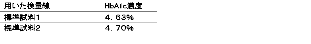
 HbA1c標準品(凍乾品、市販)を用いて以下の2通りの標準試料(HbA1c濃度は0.0%~14.9%)を調製し、日立7170自動分析機を用いてラテックス凝集法による測定を行ない、検量線を作成した(図2)。HbA1c特異抗体として、市販のキットにも用いられている公知の抗HbA1cモノクローナル抗体(特許文献1参照)を用いた。
標準試料1:
 HbA1c標準品を検体希釈液1mLで復元し、4.8μLをサンプルとした。
標準試料2:
 HbA1c標準品を検体希釈液100μLで復元し、12μLをサンプルとした。
3. Detection of HbA1c from whole blood sample not pretreated by immunoagglutination method Using HbA1c standard (freeze-dried, commercially available), the following two standard samples (HbA1c concentration is 0.0% to 14.9%) The sample was prepared and measured by latex agglutination using Hitachi 7170 automatic analyzer, and a calibration curve was created (FIG. 2). As the HbA1c specific antibody, a known anti-HbA1c monoclonal antibody (see Patent Document 1) used in a commercially available kit was used.
Standard sample 1:
The HbA1c standard product was reconstituted with 1 mL of the specimen diluent, and 4.8 μL was used as a sample.
Standard sample 2:
The HbA1c standard product was reconstituted with 100 μL of the specimen diluent, and 12 μL was used as a sample.
 ラテックス粒子を浮遊させたR1緩衝液を用いて、日立7170自動分析機により全血サンプル2μLの測定を行なった。測定シグナルを図2に示した2種の検量線にそれぞれ当てはめ、全血サンプル中のHbA1c濃度を算出した。その結果、表2に示す通りに濃度が算出された。 2 μL of whole blood sample was measured by Hitachi 7170 automatic analyzer using R1 buffer with latex particles suspended. The measurement signal was applied to each of the two calibration curves shown in FIG. 2 to calculate the HbA1c concentration in the whole blood sample. As a result, the concentration was calculated as shown in Table 2.
Figure JPOXMLDOC01-appb-T000002
 ここで使用した全血サンプルの血球層から血球を採取して溶血させ、従来法による測定を行なったところ、HbA1cの濃度は5.2%と算出された。検量線を作成する標準試料の種類に応じて測定値が変動したが、溶血等の前処理をしない全血サンプルをダイレクトに免疫測定しても測定値を得ることができることが確認された。標準試料を適当に調製すればより正確な測定が可能になる。 When blood cells were collected from the blood cell layer of the whole blood sample used here for hemolysis and measured by the conventional method, the concentration of HbA1c was calculated to be 5.2%. Although the measured value fluctuated depending on the type of the standard sample for preparing the calibration curve, it was confirmed that the measured value could be obtained even by direct immunoassay of a whole blood sample not subjected to pretreatment such as hemolysis. If a standard sample is appropriately prepared, more accurate measurement is possible.
4.免疫凝集法による前処理を施されていない全血からのHbA1cの検出時における反応過程の吸光度変化
 全血検体の血球層から血球を採取して溶血させた希釈サンプルと、前処理を施されていない全血サンプルをダイレクトに免疫測定した場合の反応過程の吸光度変化を図3に示した。
4). Absorbance change in the reaction process during detection of HbA1c from whole blood that has not been pretreated by immunoagglutination method A diluted sample obtained by lysing blood cells from the blood cell layer of a whole blood sample and hemolyzed, and pretreated FIG. 3 shows the change in absorbance during the reaction process when the whole blood sample was directly immunoassayed.
 全血サンプルの測光ポイント1で吸光度が高いのは、溶血の途中であることを示していて、測光ポイント2では吸光度はほぼ一定になっていることから、溶血が速やかに進行していることが示された。希釈サンプルの反応過程の吸光度変化と比較して、全血サンプルの場合は、全体的に吸光度は高い方に平行移動してかさ上げされている状態だった。かさ上げの原因は、赤血球の溶血後の細胞膜などの残骸の影響と思われた。測定値は、R2試薬(抗体液)が入るポイント16過ぎから、最終ポイント34の間の適当な区間の吸光度差で求めるので、かさ上げの実質上の影響は無いことになる。 A high absorbance at photometric point 1 of the whole blood sample indicates that the hemolysis is in progress, and the absorbance at photometric point 2 is almost constant, so that hemolysis proceeds rapidly. Indicated. Compared to the change in absorbance in the reaction process of the diluted sample, the whole blood sample was in a state where the absorbance was increased in parallel by moving toward the higher side. The cause of the increase was thought to be the effect of debris such as cell membranes after hemolysis of red blood cells. Since the measured value is obtained from the difference in absorbance in an appropriate section between the final point 34 after the point 16 where the R2 reagent (antibody solution) enters, there is no substantial effect of raising.
5.標準品に異なる濃度のγグロブリンを加えてHbA1c標準試料を調製して測定することにより作成した検量線
 HbA1c標準品(凍乾品、市販)を用いて、以下の4通りの標準試料(HbA1c濃度は0.0%~14.9%)を調製し、2μLをサンプルとして日立7170自動分析機を用いてラテックス凝集法による測定を行ない、検量線を作成した。その検量線を図4に示す。
標準試料3:
 HbA1c標準品をR1緩衝液 100μLで復元したもの。
標準試料4:
 HbA1c標準品を、γグロブリン1.25mg/mL 含むR1緩衝液 100μLで復元したもの。
標準試料5:
 HbA1c標準品を、γグロブリン 2.5mg/mL 含むR1緩衝液 100μLで復元したもの。
標準試料6:
 HbA1c標準品をγグロブリン 5mg/mL 含むR1緩衝液 100μLで復元したもの。
5. A standard curve prepared by preparing and measuring HbA1c standard samples by adding different concentrations of γ globulin to the standard products. Using the HbA1c standard products (freeze-dried products, commercially available), the following four standard samples (HbA1c concentrations) 0.02% to 14.9%), and 2 μL as a sample was measured by latex agglutination using a Hitachi 7170 automatic analyzer to prepare a calibration curve. The calibration curve is shown in FIG.
Standard sample 3:
HbA1c standard product restored with 100 μL of R1 buffer.
Standard sample 4:
HbA1c standard product reconstituted with 100 μL of R1 buffer containing 1.25 mg / mL of gamma globulin.
Standard sample 5:
HbA1c standard product reconstituted with 100 μL of R1 buffer containing 2.5 mg / mL γ globulin.
Standard sample 6:
HbA1c standard product reconstituted with 100 μL of R1 buffer containing 5 mg / mL γ globulin.
 γグロブリンの濃度が高くなるにつれて、検量線のシグナルは低下した。検体の最低分注可能サンプル量は自動分析機により異なるため、加えるγグロブリン等の濃度及び標準試料のサンプル量は、前処理を施されていない全血サンプルの量(μL)と、ラテックス試薬の濃度や試薬量により、適宜最適なものを検討選択して決めることができる。 As the γ globulin concentration increased, the calibration curve signal decreased. Since the minimum amount of sample that can be dispensed varies depending on the automatic analyzer, the concentration of γ globulin and the like and the sample volume of the standard sample should be the same as the amount of whole blood sample (μL) that has not been pretreated and the amount of latex reagent. Depending on the concentration and the amount of reagent, the optimum one can be selected and determined as appropriate.

Claims (3)

  1.  ラテックス粒子と前処理が施されていない全血検体を低張液中で接触させ、次いで、ラテックス粒子上に吸着したヘモグロビンA1cと抗ヘモグロビンA1c抗体を接触させることを含む、検体中のヘモグロビンA1cの免疫測定方法。 Contact of the latex particles with an untreated whole blood sample in a hypotonic solution, and then contacting the hemoglobin A1c adsorbed onto the latex particles with an anti-hemoglobin A1c antibody. Immunoassay method.
  2.  前記免疫測定が凝集法により行なわれる請求項1記載の方法。 The method according to claim 1, wherein the immunoassay is performed by an agglutination method.
  3.  前記低張液が、緩衝能が中性からアルカリ側で最大となるグッドバッファーを0.02~0.2mol/Lの濃度で含む緩衝液である請求項1又は2記載の方法。 The method according to claim 1 or 2, wherein the hypotonic solution is a buffer solution containing a good buffer having a buffer capacity that is neutral to maximum on the alkali side at a concentration of 0.02 to 0.2 mol / L.
PCT/JP2013/084905 2013-01-16 2013-12-26 Method for immunologically assaying hemoglobin a1c in specimen WO2014112318A1 (en)

Priority Applications (4)

Application Number Priority Date Filing Date Title
JP2014557383A JP6172163B2 (en) 2013-01-16 2013-12-26 Method for immunoassay of hemoglobin A1c in specimen
US14/655,277 US20150316541A1 (en) 2013-01-16 2013-12-26 Method for immunologically assaying hemoglobin a1c in specimen
EP13871622.0A EP2947458A4 (en) 2013-01-16 2013-12-26 Method for immunologically assaying hemoglobin a1c in specimen
CN201380070635.2A CN105122060A (en) 2013-01-16 2013-12-26 Method for immunologically assaying hemoglobin A1c in specimen

Applications Claiming Priority (2)

Application Number Priority Date Filing Date Title
JP2013-005130 2013-01-16
JP2013005130 2013-01-16

Publications (1)

Publication Number Publication Date
WO2014112318A1 true WO2014112318A1 (en) 2014-07-24

Family

ID=51209420

Family Applications (1)

Application Number Title Priority Date Filing Date
PCT/JP2013/084905 WO2014112318A1 (en) 2013-01-16 2013-12-26 Method for immunologically assaying hemoglobin a1c in specimen

Country Status (6)

Country Link
US (1) US20150316541A1 (en)
EP (1) EP2947458A4 (en)
JP (1) JP6172163B2 (en)
CN (1) CN105122060A (en)
TW (1) TW201441621A (en)
WO (1) WO2014112318A1 (en)

Cited By (4)

* Cited by examiner, † Cited by third party
Publication number Priority date Publication date Assignee Title
WO2019207724A1 (en) * 2018-04-26 2019-10-31 株式会社ニコン Blood component separation device, blood component separation method, and blood component analysis method
WO2020067396A1 (en) * 2018-09-28 2020-04-02 積水メディカル株式会社 Glycated hemoglobin (%) assay method
WO2023145915A1 (en) * 2022-01-31 2023-08-03 積水メディカル株式会社 Immunoassay method for pulmonary surfactant protein and immunoassay reagent
WO2023195347A1 (en) * 2022-04-04 2023-10-12 デンカ株式会社 ANTI-HUMAN HEMOGLOBIN β-CHAIN MONOCLONAL ANTIBODY OR ANTIGEN-BINDING FRAGMENT THEREOF, METHOD FOR DETECTING HUMAN HEMOGLOBIN AND/OR GLYCOSYLATED HUMAN HEMOGLOBIN, KIT FOR DETECTING HUMAN HEMOGLOBIN AND/OR GLYCOSYLATED HUMAN HEMOGLOBIN, AND PEPTIDE

Families Citing this family (9)

* Cited by examiner, † Cited by third party
Publication number Priority date Publication date Assignee Title
CN105683374A (en) * 2013-10-25 2016-06-15 龟甲万株式会社 HEMOGLOBIN A1c MEASUREMENT METHOD AND MEASUREMENT KIT
CN105891519B (en) * 2016-07-01 2018-03-30 安邦(厦门)生物科技有限公司 A kind of kit and assay method for being used to determine glycosylated hemoglobin ratio
CN107490677A (en) * 2017-07-21 2017-12-19 王贤俊 The cross-linking composition liquid and its cross-linking method of a kind of carboxylated latex microballoon and glycosylated hemoglobin antibody
JP7184054B2 (en) * 2018-01-11 2022-12-06 東洋紡株式会社 Measurement sample diluent, kit and measurement method
EP3917398A1 (en) 2019-01-31 2021-12-08 Aidian Oy Sampling and assay kit and method for sampling a biological sample
US20200284807A1 (en) * 2019-03-05 2020-09-10 Victor Manneh Saturation binding ratiometric assay
CN111057150B (en) * 2019-12-30 2021-10-29 深圳开立生物医疗科技股份有限公司 Latex microsphere, application thereof and glycosylated hemoglobin detection kit
CN114280312B (en) * 2020-09-27 2023-09-15 河北特温特生物科技发展有限公司 Whole blood separation membrane for immunofluorescence chromatography detection and preparation method and application thereof
WO2023147522A1 (en) * 2022-01-28 2023-08-03 GATC Health Corp Biomarkers for early detection of diabetes

Citations (3)

* Cited by examiner, † Cited by third party
Publication number Priority date Publication date Assignee Title
JPH0735752A (en) * 1993-07-22 1995-02-07 S R L:Kk Method for immunological assay for agglutination
JP2001296292A (en) * 2001-03-26 2001-10-26 Horiba Ltd Method of immunoassay
JP2004059477A (en) * 2002-07-26 2004-02-26 Fujikura Kasei Co Ltd Monoclonal antibody specifically bonded to non-glycosylated hemoglobin or glycosylated hemoglobin, method for producing the same, and method for assaying non-glycosylated or glycosylated hemoglobin using the same

Family Cites Families (5)

* Cited by examiner, † Cited by third party
Publication number Priority date Publication date Assignee Title
ATE438097T1 (en) * 2000-08-29 2009-08-15 Kyowa Medex Co Ltd HIGHLY REPRODUCABLE AGGLUTINATION IMMUNOASSAY METHOD AND REAGENTS
JP2010507798A (en) * 2006-10-24 2010-03-11 コーニンクレッカ フィリップス エレクトロニクス エヌ ヴィ Quantitative measurement of glycated hemoglobin
US8580097B2 (en) * 2009-04-27 2013-11-12 Wako Pure Chemical Industries, Ltd. Isotachophoresis of blood-derived samples
WO2010147251A1 (en) * 2009-06-19 2010-12-23 주식회사 인포피아 Method for measuring glycated hemoglobin
CN101915849B (en) * 2010-06-30 2013-06-05 深圳市国赛生物技术有限公司 Agent ,which is convenient for sampling, for measuring glycosylated hemoglobin percentage

Patent Citations (4)

* Cited by examiner, † Cited by third party
Publication number Priority date Publication date Assignee Title
JPH0735752A (en) * 1993-07-22 1995-02-07 S R L:Kk Method for immunological assay for agglutination
JP2677753B2 (en) 1993-07-22 1997-11-17 株式会社エスアールエル Aggregation immunoassay
JP2001296292A (en) * 2001-03-26 2001-10-26 Horiba Ltd Method of immunoassay
JP2004059477A (en) * 2002-07-26 2004-02-26 Fujikura Kasei Co Ltd Monoclonal antibody specifically bonded to non-glycosylated hemoglobin or glycosylated hemoglobin, method for producing the same, and method for assaying non-glycosylated or glycosylated hemoglobin using the same

Non-Patent Citations (2)

* Cited by examiner, † Cited by third party
Title
JOURNAL OF THE JAPAN DIABETES SOCIETY, vol. 53, no. 6, 2010, pages 450
See also references of EP2947458A4

Cited By (6)

* Cited by examiner, † Cited by third party
Publication number Priority date Publication date Assignee Title
WO2019207724A1 (en) * 2018-04-26 2019-10-31 株式会社ニコン Blood component separation device, blood component separation method, and blood component analysis method
JPWO2019207724A1 (en) * 2018-04-26 2021-01-14 株式会社ニコン Blood component separation device, blood component separation method, and blood component analysis method
WO2020067396A1 (en) * 2018-09-28 2020-04-02 積水メディカル株式会社 Glycated hemoglobin (%) assay method
JP7382071B2 (en) 2018-09-28 2023-11-16 積水メディカル株式会社 How to measure glycated hemoglobin (%)
WO2023145915A1 (en) * 2022-01-31 2023-08-03 積水メディカル株式会社 Immunoassay method for pulmonary surfactant protein and immunoassay reagent
WO2023195347A1 (en) * 2022-04-04 2023-10-12 デンカ株式会社 ANTI-HUMAN HEMOGLOBIN β-CHAIN MONOCLONAL ANTIBODY OR ANTIGEN-BINDING FRAGMENT THEREOF, METHOD FOR DETECTING HUMAN HEMOGLOBIN AND/OR GLYCOSYLATED HUMAN HEMOGLOBIN, KIT FOR DETECTING HUMAN HEMOGLOBIN AND/OR GLYCOSYLATED HUMAN HEMOGLOBIN, AND PEPTIDE

Also Published As

Publication number Publication date
US20150316541A1 (en) 2015-11-05
JPWO2014112318A1 (en) 2017-01-19
EP2947458A1 (en) 2015-11-25
TW201441621A (en) 2014-11-01
EP2947458A4 (en) 2016-08-10
JP6172163B2 (en) 2017-08-02
CN105122060A (en) 2015-12-02

Similar Documents

Publication Publication Date Title
JP6172163B2 (en) Method for immunoassay of hemoglobin A1c in specimen
JP5199067B2 (en) Immunoagglutination reagent kit and antigen measurement method
EP3264083B1 (en) L-fabp immunoassay method
EP3859332B1 (en) Glycated hemoglobin (%) assay method
JP2008175814A (en) Method of examining diabetic nephropathy based on detection and quantitative determination of protein molecule in urine, and kit used therefor
JP6304231B2 (en) How to prevent deterioration of unsensitized latex reagent
JPH0735752A (en) Method for immunological assay for agglutination
CN101046479B (en) Process of preparing human serum base matter containing no target protein
EP2717054A1 (en) Method for inhibiting non-specific reaction in pivka-ii measurement reagent
EP3264085A1 (en) Immunoassay method and assay reagent used in said method
JP5191291B2 (en) Method and reagent kit for measuring hemoglobin in stool specimen
JPH06167495A (en) Aggregation immunoassay method
JPH049262B2 (en)
JP5294257B2 (en) Hemoglobin measurement method and measurement kit
JP4796893B2 (en) Method for measuring hemoglobin A1c
WO2024053586A1 (en) Free hemoglobin measurement reagent, free hemoglobin measurement method, and anti-hemoglobin antibody
JP2885092B2 (en) Method and reagent for measuring C-reactive protein
JP2001228153A (en) Immunological measurement method
WO2014177701A1 (en) Process for diagnosing a human subject with diseases affecting the kidneys, or at risk of acquiring diseases affecting the kidneys
KR20230074115A (en) Ferritin measurement reagent
JPH0783921A (en) Method for measuring saccharification protein
JPH10227793A (en) Measuring method for carbamic hemoglobin
JP2018128394A (en) Measuring method using separation material

Legal Events

Date Code Title Description
121 Ep: the epo has been informed by wipo that ep was designated in this application

Ref document number: 13871622

Country of ref document: EP

Kind code of ref document: A1

ENP Entry into the national phase

Ref document number: 2014557383

Country of ref document: JP

Kind code of ref document: A

WWE Wipo information: entry into national phase

Ref document number: 2013871622

Country of ref document: EP

WWE Wipo information: entry into national phase

Ref document number: 14655277

Country of ref document: US

NENP Non-entry into the national phase

Ref country code: DE