WO2014098249A1 - Composition having tissue repairing activity and utilization thereof - Google Patents
Composition having tissue repairing activity and utilization thereof Download PDFInfo
- Publication number
- WO2014098249A1 WO2014098249A1 PCT/JP2013/084523 JP2013084523W WO2014098249A1 WO 2014098249 A1 WO2014098249 A1 WO 2014098249A1 JP 2013084523 W JP2013084523 W JP 2013084523W WO 2014098249 A1 WO2014098249 A1 WO 2014098249A1
- Authority
- WO
- WIPO (PCT)
- Prior art keywords
- component
- siglec
- activity
- tissue repair
- mcp
- Prior art date
Links
- 230000000694 effects Effects 0.000 title claims abstract description 97
- 239000000203 mixture Substances 0.000 title abstract description 33
- 108090000623 proteins and genes Proteins 0.000 claims abstract description 76
- 102000004169 proteins and genes Human genes 0.000 claims abstract description 73
- 101000863883 Homo sapiens Sialic acid-binding Ig-like lectin 9 Proteins 0.000 claims abstract description 43
- 102000005598 Chondroitin Sulfate Proteoglycans Human genes 0.000 claims abstract description 41
- 108010059480 Chondroitin Sulfate Proteoglycans Proteins 0.000 claims abstract description 41
- 102100029965 Sialic acid-binding Ig-like lectin 9 Human genes 0.000 claims abstract description 40
- SQDAZGGFXASXDW-UHFFFAOYSA-N 5-bromo-2-(trifluoromethoxy)pyridine Chemical group FC(F)(F)OC1=CC=C(Br)C=N1 SQDAZGGFXASXDW-UHFFFAOYSA-N 0.000 claims abstract description 39
- 229920001287 Chondroitin sulfate Polymers 0.000 claims abstract description 39
- 229940059329 chondroitin sulfate Drugs 0.000 claims abstract description 39
- 230000001737 promoting effect Effects 0.000 claims abstract description 37
- SQVRNKJHWKZAKO-UHFFFAOYSA-N beta-N-Acetyl-D-neuraminic acid Natural products CC(=O)NC1C(O)CC(O)(C(O)=O)OC1C(O)C(O)CO SQVRNKJHWKZAKO-UHFFFAOYSA-N 0.000 claims abstract description 30
- SQVRNKJHWKZAKO-OQPLDHBCSA-N sialic acid Chemical compound CC(=O)N[C@@H]1[C@@H](O)C[C@@](O)(C(O)=O)OC1[C@H](O)[C@H](O)CO SQVRNKJHWKZAKO-OQPLDHBCSA-N 0.000 claims abstract description 30
- 101710197064 Lectin 9 Proteins 0.000 claims abstract description 23
- 230000017423 tissue regeneration Effects 0.000 claims description 90
- 210000000274 microglia Anatomy 0.000 claims description 79
- 239000003795 chemical substances by application Substances 0.000 claims description 74
- 210000002540 macrophage Anatomy 0.000 claims description 62
- 210000001519 tissue Anatomy 0.000 claims description 53
- 102000004127 Cytokines Human genes 0.000 claims description 32
- 108090000695 Cytokines Proteins 0.000 claims description 32
- 230000003110 anti-inflammatory effect Effects 0.000 claims description 32
- 238000004519 manufacturing process Methods 0.000 claims description 27
- 239000003814 drug Substances 0.000 claims description 21
- 208000001940 Massive Hepatic Necrosis Diseases 0.000 claims description 19
- 230000028550 monocyte chemotaxis Effects 0.000 claims description 19
- 206010039073 rheumatoid arthritis Diseases 0.000 claims description 17
- 208000020431 spinal cord injury Diseases 0.000 claims description 16
- 208000006454 hepatitis Diseases 0.000 claims description 13
- 239000004480 active ingredient Substances 0.000 claims description 12
- 230000001154 acute effect Effects 0.000 claims description 10
- 208000015114 central nervous system disease Diseases 0.000 claims description 10
- 238000000338 in vitro Methods 0.000 claims description 10
- 206010008909 Chronic Hepatitis Diseases 0.000 claims description 8
- 239000003153 chemical reaction reagent Substances 0.000 claims description 8
- FWMNVWWHGCHHJJ-SKKKGAJSSA-N 4-amino-1-[(2r)-6-amino-2-[[(2r)-2-[[(2r)-2-[[(2r)-2-amino-3-phenylpropanoyl]amino]-3-phenylpropanoyl]amino]-4-methylpentanoyl]amino]hexanoyl]piperidine-4-carboxylic acid Chemical compound C([C@H](C(=O)N[C@H](CC(C)C)C(=O)N[C@H](CCCCN)C(=O)N1CCC(N)(CC1)C(O)=O)NC(=O)[C@H](N)CC=1C=CC=CC=1)C1=CC=CC=C1 FWMNVWWHGCHHJJ-SKKKGAJSSA-N 0.000 claims description 7
- 208000029523 Interstitial Lung disease Diseases 0.000 claims description 7
- 206010008118 cerebral infarction Diseases 0.000 claims description 7
- 230000016396 cytokine production Effects 0.000 claims description 7
- 239000000411 inducer Substances 0.000 claims description 7
- 230000006698 induction Effects 0.000 claims description 7
- 208000026106 cerebrovascular disease Diseases 0.000 claims description 6
- 208000018737 Parkinson disease Diseases 0.000 claims description 5
- 231100000354 acute hepatitis Toxicity 0.000 claims description 5
- 238000012258 culturing Methods 0.000 claims description 5
- 208000001072 type 2 diabetes mellitus Diseases 0.000 claims description 5
- 208000024827 Alzheimer disease Diseases 0.000 claims description 4
- 208000002381 Brain Hypoxia Diseases 0.000 claims description 4
- 208000003556 Dry Eye Syndromes Diseases 0.000 claims description 4
- 206010013774 Dry eye Diseases 0.000 claims description 4
- 208000031071 Hamman-Rich Syndrome Diseases 0.000 claims description 4
- 206010070511 Hypoxic-ischaemic encephalopathy Diseases 0.000 claims description 4
- 208000021386 Sjogren Syndrome Diseases 0.000 claims description 4
- 206010067584 Type 1 diabetes mellitus Diseases 0.000 claims description 4
- 201000004073 acute interstitial pneumonia Diseases 0.000 claims description 4
- 206010002026 amyotrophic lateral sclerosis Diseases 0.000 claims description 4
- 238000010322 bone marrow transplantation Methods 0.000 claims description 4
- 201000006417 multiple sclerosis Diseases 0.000 claims description 4
- 208000010125 myocardial infarction Diseases 0.000 claims description 4
- 230000003449 preventive effect Effects 0.000 claims description 4
- 229940124597 therapeutic agent Drugs 0.000 claims description 4
- 230000029663 wound healing Effects 0.000 claims description 4
- 206010072170 Skin wound Diseases 0.000 claims description 3
- 239000002260 anti-inflammatory agent Substances 0.000 claims description 3
- 229940121363 anti-inflammatory agent Drugs 0.000 claims description 3
- 201000000596 systemic lupus erythematosus Diseases 0.000 claims description 3
- 125000003275 alpha amino acid group Chemical group 0.000 claims 4
- 238000006243 chemical reaction Methods 0.000 abstract description 7
- 101710155857 C-C motif chemokine 2 Proteins 0.000 abstract 2
- 102000000018 Chemokine CCL2 Human genes 0.000 abstract 2
- 235000018102 proteins Nutrition 0.000 description 61
- 150000001413 amino acids Chemical group 0.000 description 48
- 238000000034 method Methods 0.000 description 31
- 230000014509 gene expression Effects 0.000 description 30
- 206010061218 Inflammation Diseases 0.000 description 29
- 230000004054 inflammatory process Effects 0.000 description 29
- 230000002757 inflammatory effect Effects 0.000 description 28
- 241000700159 Rattus Species 0.000 description 25
- 101710149815 C-C chemokine receptor type 2 Proteins 0.000 description 23
- 102100031151 C-C chemokine receptor type 2 Human genes 0.000 description 23
- 206010016654 Fibrosis Diseases 0.000 description 23
- 210000004027 cell Anatomy 0.000 description 21
- 206010003246 arthritis Diseases 0.000 description 18
- 230000006378 damage Effects 0.000 description 18
- 229940079593 drug Drugs 0.000 description 17
- 239000000243 solution Substances 0.000 description 17
- 241000699666 Mus <mouse, genus> Species 0.000 description 16
- 241000699670 Mus sp. Species 0.000 description 15
- 238000010186 staining Methods 0.000 description 15
- 108020004414 DNA Proteins 0.000 description 14
- 241001465754 Metazoa Species 0.000 description 14
- 208000037265 diseases, disorders, signs and symptoms Diseases 0.000 description 14
- 230000001965 increasing effect Effects 0.000 description 14
- 239000002243 precursor Substances 0.000 description 14
- MZOFCQQQCNRIBI-VMXHOPILSA-N (3s)-4-[[(2s)-1-[[(2s)-1-[[(1s)-1-carboxy-2-hydroxyethyl]amino]-4-methyl-1-oxopentan-2-yl]amino]-5-(diaminomethylideneamino)-1-oxopentan-2-yl]amino]-3-[[2-[[(2s)-2,6-diaminohexanoyl]amino]acetyl]amino]-4-oxobutanoic acid Chemical compound OC[C@@H](C(O)=O)NC(=O)[C@H](CC(C)C)NC(=O)[C@H](CCCN=C(N)N)NC(=O)[C@H](CC(O)=O)NC(=O)CNC(=O)[C@@H](N)CCCCN MZOFCQQQCNRIBI-VMXHOPILSA-N 0.000 description 13
- 108060008682 Tumor Necrosis Factor Proteins 0.000 description 13
- 102000000852 Tumor Necrosis Factor-alpha Human genes 0.000 description 13
- 230000001939 inductive effect Effects 0.000 description 13
- 230000004083 survival effect Effects 0.000 description 13
- 230000007882 cirrhosis Effects 0.000 description 12
- 208000019425 cirrhosis of liver Diseases 0.000 description 12
- 201000010099 disease Diseases 0.000 description 12
- 108090000174 Interleukin-10 Proteins 0.000 description 11
- 238000011156 evaluation Methods 0.000 description 11
- 230000004761 fibrosis Effects 0.000 description 11
- 230000000451 tissue damage Effects 0.000 description 11
- 231100000827 tissue damage Toxicity 0.000 description 11
- 238000004458 analytical method Methods 0.000 description 10
- 208000009386 Experimental Arthritis Diseases 0.000 description 9
- 230000001684 chronic effect Effects 0.000 description 9
- 108010035532 Collagen Proteins 0.000 description 8
- 102000008186 Collagen Human genes 0.000 description 8
- 229920001436 collagen Polymers 0.000 description 8
- 238000004043 dyeing Methods 0.000 description 8
- 238000007490 hematoxylin and eosin (H&E) staining Methods 0.000 description 8
- 210000005228 liver tissue Anatomy 0.000 description 8
- 150000007523 nucleic acids Chemical class 0.000 description 8
- 230000008439 repair process Effects 0.000 description 8
- 206010067125 Liver injury Diseases 0.000 description 7
- FAPWRFPIFSIZLT-UHFFFAOYSA-M Sodium chloride Chemical compound [Na+].[Cl-] FAPWRFPIFSIZLT-UHFFFAOYSA-M 0.000 description 7
- 235000001014 amino acid Nutrition 0.000 description 7
- 210000000988 bone and bone Anatomy 0.000 description 7
- 231100000234 hepatic damage Toxicity 0.000 description 7
- 208000027866 inflammatory disease Diseases 0.000 description 7
- 239000002523 lectin Substances 0.000 description 7
- 230000008818 liver damage Effects 0.000 description 7
- 210000004072 lung Anatomy 0.000 description 7
- 238000003753 real-time PCR Methods 0.000 description 7
- 230000000638 stimulation Effects 0.000 description 7
- 101000897480 Homo sapiens C-C motif chemokine 2 Proteins 0.000 description 6
- 208000004852 Lung Injury Diseases 0.000 description 6
- 206010069363 Traumatic lung injury Diseases 0.000 description 6
- 229940024606 amino acid Drugs 0.000 description 6
- 230000037396 body weight Effects 0.000 description 6
- 239000000872 buffer Substances 0.000 description 6
- 210000002950 fibroblast Anatomy 0.000 description 6
- 210000003494 hepatocyte Anatomy 0.000 description 6
- 102000046768 human CCL2 Human genes 0.000 description 6
- 230000002401 inhibitory effect Effects 0.000 description 6
- 208000014674 injury Diseases 0.000 description 6
- NOESYZHRGYRDHS-UHFFFAOYSA-N insulin Chemical compound N1C(=O)C(NC(=O)C(CCC(N)=O)NC(=O)C(CCC(O)=O)NC(=O)C(C(C)C)NC(=O)C(NC(=O)CN)C(C)CC)CSSCC(C(NC(CO)C(=O)NC(CC(C)C)C(=O)NC(CC=2C=CC(O)=CC=2)C(=O)NC(CCC(N)=O)C(=O)NC(CC(C)C)C(=O)NC(CCC(O)=O)C(=O)NC(CC(N)=O)C(=O)NC(CC=2C=CC(O)=CC=2)C(=O)NC(CSSCC(NC(=O)C(C(C)C)NC(=O)C(CC(C)C)NC(=O)C(CC=2C=CC(O)=CC=2)NC(=O)C(CC(C)C)NC(=O)C(C)NC(=O)C(CCC(O)=O)NC(=O)C(C(C)C)NC(=O)C(CC(C)C)NC(=O)C(CC=2NC=NC=2)NC(=O)C(CO)NC(=O)CNC2=O)C(=O)NCC(=O)NC(CCC(O)=O)C(=O)NC(CCCNC(N)=N)C(=O)NCC(=O)NC(CC=3C=CC=CC=3)C(=O)NC(CC=3C=CC=CC=3)C(=O)NC(CC=3C=CC(O)=CC=3)C(=O)NC(C(C)O)C(=O)N3C(CCC3)C(=O)NC(CCCCN)C(=O)NC(C)C(O)=O)C(=O)NC(CC(N)=O)C(O)=O)=O)NC(=O)C(C(C)CC)NC(=O)C(CO)NC(=O)C(C(C)O)NC(=O)C1CSSCC2NC(=O)C(CC(C)C)NC(=O)C(NC(=O)C(CCC(N)=O)NC(=O)C(CC(N)=O)NC(=O)C(NC(=O)C(N)CC=1C=CC=CC=1)C(C)C)CC1=CN=CN1 NOESYZHRGYRDHS-UHFFFAOYSA-N 0.000 description 6
- 230000003914 insulin secretion Effects 0.000 description 6
- 210000004185 liver Anatomy 0.000 description 6
- 231100000515 lung injury Toxicity 0.000 description 6
- 239000006166 lysate Substances 0.000 description 6
- 238000010172 mouse model Methods 0.000 description 6
- 230000001575 pathological effect Effects 0.000 description 6
- 238000002360 preparation method Methods 0.000 description 6
- 230000008569 process Effects 0.000 description 6
- 108091032973 (ribonucleotides)n+m Proteins 0.000 description 5
- 208000034656 Contusions Diseases 0.000 description 5
- WQZGKKKJIJFFOK-GASJEMHNSA-N Glucose Natural products OC[C@H]1OC(O)[C@H](O)[C@@H](O)[C@@H]1O WQZGKKKJIJFFOK-GASJEMHNSA-N 0.000 description 5
- 241000282412 Homo Species 0.000 description 5
- 108090001090 Lectins Proteins 0.000 description 5
- 102000004856 Lectins Human genes 0.000 description 5
- 241000283973 Oryctolagus cuniculus Species 0.000 description 5
- 230000009519 contusion Effects 0.000 description 5
- 230000034994 death Effects 0.000 description 5
- 231100000517 death Toxicity 0.000 description 5
- 230000007547 defect Effects 0.000 description 5
- 238000002474 experimental method Methods 0.000 description 5
- 239000008103 glucose Substances 0.000 description 5
- 238000002347 injection Methods 0.000 description 5
- 239000007924 injection Substances 0.000 description 5
- 210000004731 jugular vein Anatomy 0.000 description 5
- 210000003141 lower extremity Anatomy 0.000 description 5
- 108020004999 messenger RNA Proteins 0.000 description 5
- 108091033319 polynucleotide Proteins 0.000 description 5
- 102000040430 polynucleotide Human genes 0.000 description 5
- 239000002157 polynucleotide Substances 0.000 description 5
- 230000035755 proliferation Effects 0.000 description 5
- 238000011084 recovery Methods 0.000 description 5
- 230000002829 reductive effect Effects 0.000 description 5
- 239000011780 sodium chloride Substances 0.000 description 5
- 210000000278 spinal cord Anatomy 0.000 description 5
- 238000006467 substitution reaction Methods 0.000 description 5
- 238000001356 surgical procedure Methods 0.000 description 5
- MSWZFWKMSRAUBD-GASJEMHNSA-N 2-amino-2-deoxy-D-galactopyranose Chemical compound N[C@H]1C(O)O[C@H](CO)[C@H](O)[C@@H]1O MSWZFWKMSRAUBD-GASJEMHNSA-N 0.000 description 4
- 102100021723 Arginase-1 Human genes 0.000 description 4
- 101710129000 Arginase-1 Proteins 0.000 description 4
- 208000023275 Autoimmune disease Diseases 0.000 description 4
- 108010012236 Chemokines Proteins 0.000 description 4
- 102000019034 Chemokines Human genes 0.000 description 4
- 239000006144 Dulbecco’s modified Eagle's medium Substances 0.000 description 4
- 101000934338 Homo sapiens Myeloid cell surface antigen CD33 Proteins 0.000 description 4
- 210000004322 M2 macrophage Anatomy 0.000 description 4
- 241000124008 Mammalia Species 0.000 description 4
- 102000007056 Recombinant Fusion Proteins Human genes 0.000 description 4
- 108010008281 Recombinant Fusion Proteins Proteins 0.000 description 4
- 208000027418 Wounds and injury Diseases 0.000 description 4
- 238000002869 basic local alignment search tool Methods 0.000 description 4
- MSWZFWKMSRAUBD-UHFFFAOYSA-N beta-D-galactosamine Natural products NC1C(O)OC(CO)C(O)C1O MSWZFWKMSRAUBD-UHFFFAOYSA-N 0.000 description 4
- 210000003169 central nervous system Anatomy 0.000 description 4
- 210000003690 classically activated macrophage Anatomy 0.000 description 4
- 230000003247 decreasing effect Effects 0.000 description 4
- 238000001114 immunoprecipitation Methods 0.000 description 4
- 230000028709 inflammatory response Effects 0.000 description 4
- 230000033001 locomotion Effects 0.000 description 4
- 238000002156 mixing Methods 0.000 description 4
- 210000004126 nerve fiber Anatomy 0.000 description 4
- 108020004707 nucleic acids Proteins 0.000 description 4
- 102000039446 nucleic acids Human genes 0.000 description 4
- DHRLEVQXOMLTIM-UHFFFAOYSA-N phosphoric acid;trioxomolybdenum Chemical compound O=[Mo](=O)=O.O=[Mo](=O)=O.O=[Mo](=O)=O.O=[Mo](=O)=O.O=[Mo](=O)=O.O=[Mo](=O)=O.O=[Mo](=O)=O.O=[Mo](=O)=O.O=[Mo](=O)=O.O=[Mo](=O)=O.O=[Mo](=O)=O.O=[Mo](=O)=O.OP(O)(O)=O DHRLEVQXOMLTIM-UHFFFAOYSA-N 0.000 description 4
- 230000010399 physical interaction Effects 0.000 description 4
- 229920000729 poly(L-lysine) polymer Polymers 0.000 description 4
- 230000000770 proinflammatory effect Effects 0.000 description 4
- 239000002994 raw material Substances 0.000 description 4
- 230000008929 regeneration Effects 0.000 description 4
- 238000011069 regeneration method Methods 0.000 description 4
- 230000035939 shock Effects 0.000 description 4
- 210000003625 skull Anatomy 0.000 description 4
- 239000000126 substance Substances 0.000 description 4
- 239000006228 supernatant Substances 0.000 description 4
- 230000002195 synergetic effect Effects 0.000 description 4
- 238000012360 testing method Methods 0.000 description 4
- VKUYLANQOAKALN-UHFFFAOYSA-N 2-[benzyl-(4-methoxyphenyl)sulfonylamino]-n-hydroxy-4-methylpentanamide Chemical compound C1=CC(OC)=CC=C1S(=O)(=O)N(C(CC(C)C)C(=O)NO)CC1=CC=CC=C1 VKUYLANQOAKALN-UHFFFAOYSA-N 0.000 description 3
- 101150053137 AIF1 gene Proteins 0.000 description 3
- 241000283707 Capra Species 0.000 description 3
- 208000017667 Chronic Disease Diseases 0.000 description 3
- 201000004624 Dermatitis Diseases 0.000 description 3
- LFQSCWFLJHTTHZ-UHFFFAOYSA-N Ethanol Chemical compound CCO LFQSCWFLJHTTHZ-UHFFFAOYSA-N 0.000 description 3
- 102100039289 Glial fibrillary acidic protein Human genes 0.000 description 3
- 101710193519 Glial fibrillary acidic protein Proteins 0.000 description 3
- -1 IL-1β Proteins 0.000 description 3
- 201000009794 Idiopathic Pulmonary Fibrosis Diseases 0.000 description 3
- 102000004877 Insulin Human genes 0.000 description 3
- 108090001061 Insulin Proteins 0.000 description 3
- 206010028980 Neoplasm Diseases 0.000 description 3
- 102000005348 Neuraminidase Human genes 0.000 description 3
- 108010006232 Neuraminidase Proteins 0.000 description 3
- HEMHJVSKTPXQMS-UHFFFAOYSA-M Sodium hydroxide Chemical compound [OH-].[Na+] HEMHJVSKTPXQMS-UHFFFAOYSA-M 0.000 description 3
- 102100030416 Stromelysin-1 Human genes 0.000 description 3
- 101710108790 Stromelysin-1 Proteins 0.000 description 3
- 102000046299 Transforming Growth Factor beta1 Human genes 0.000 description 3
- 101800002279 Transforming growth factor beta-1 Proteins 0.000 description 3
- 230000009471 action Effects 0.000 description 3
- 206010069351 acute lung injury Diseases 0.000 description 3
- 238000009534 blood test Methods 0.000 description 3
- 230000010478 bone regeneration Effects 0.000 description 3
- 201000011510 cancer Diseases 0.000 description 3
- 230000008859 change Effects 0.000 description 3
- 230000000295 complement effect Effects 0.000 description 3
- 239000012228 culture supernatant Substances 0.000 description 3
- 210000005258 dental pulp stem cell Anatomy 0.000 description 3
- 230000001066 destructive effect Effects 0.000 description 3
- 239000002552 dosage form Substances 0.000 description 3
- 210000005046 glial fibrillary acidic protein Anatomy 0.000 description 3
- 239000008187 granular material Substances 0.000 description 3
- 102000045929 human SIGLEC9 Human genes 0.000 description 3
- 238000012744 immunostaining Methods 0.000 description 3
- 238000001727 in vivo Methods 0.000 description 3
- 230000008595 infiltration Effects 0.000 description 3
- 238000001764 infiltration Methods 0.000 description 3
- 229940125396 insulin Drugs 0.000 description 3
- 208000036971 interstitial lung disease 2 Diseases 0.000 description 3
- 230000035772 mutation Effects 0.000 description 3
- 230000004770 neurodegeneration Effects 0.000 description 3
- 238000010827 pathological analysis Methods 0.000 description 3
- 239000002244 precipitate Substances 0.000 description 3
- 210000002966 serum Anatomy 0.000 description 3
- 239000007787 solid Substances 0.000 description 3
- 210000000130 stem cell Anatomy 0.000 description 3
- 210000002437 synoviocyte Anatomy 0.000 description 3
- 201000004595 synovitis Diseases 0.000 description 3
- 238000003786 synthesis reaction Methods 0.000 description 3
- 230000009885 systemic effect Effects 0.000 description 3
- 230000001225 therapeutic effect Effects 0.000 description 3
- KIUKXJAPPMFGSW-DNGZLQJQSA-N (2S,3S,4S,5R,6R)-6-[(2S,3R,4R,5S,6R)-3-Acetamido-2-[(2S,3S,4R,5R,6R)-6-[(2R,3R,4R,5S,6R)-3-acetamido-2,5-dihydroxy-6-(hydroxymethyl)oxan-4-yl]oxy-2-carboxy-4,5-dihydroxyoxan-3-yl]oxy-5-hydroxy-6-(hydroxymethyl)oxan-4-yl]oxy-3,4,5-trihydroxyoxane-2-carboxylic acid Chemical compound CC(=O)N[C@H]1[C@H](O)O[C@H](CO)[C@@H](O)[C@@H]1O[C@H]1[C@H](O)[C@@H](O)[C@H](O[C@H]2[C@@H]([C@@H](O[C@H]3[C@@H]([C@@H](O)[C@H](O)[C@H](O3)C(O)=O)O)[C@H](O)[C@@H](CO)O2)NC(C)=O)[C@@H](C(O)=O)O1 KIUKXJAPPMFGSW-DNGZLQJQSA-N 0.000 description 2
- 206010071155 Autoimmune arthritis Diseases 0.000 description 2
- 206010015150 Erythema Diseases 0.000 description 2
- ZHNUHDYFZUAESO-UHFFFAOYSA-N Formamide Chemical compound NC=O ZHNUHDYFZUAESO-UHFFFAOYSA-N 0.000 description 2
- DHMQDGOQFOQNFH-UHFFFAOYSA-N Glycine Chemical compound NCC(O)=O DHMQDGOQFOQNFH-UHFFFAOYSA-N 0.000 description 2
- 108090001005 Interleukin-6 Proteins 0.000 description 2
- 208000012659 Joint disease Diseases 0.000 description 2
- 101710197058 Lectin 7 Proteins 0.000 description 2
- 208000019693 Lung disease Diseases 0.000 description 2
- 108010031099 Mannose Receptor Proteins 0.000 description 2
- 102000001776 Matrix metalloproteinase-9 Human genes 0.000 description 2
- 108010015302 Matrix metalloproteinase-9 Proteins 0.000 description 2
- MWUXSHHQAYIFBG-UHFFFAOYSA-N Nitric oxide Chemical compound O=[N] MWUXSHHQAYIFBG-UHFFFAOYSA-N 0.000 description 2
- 108091028043 Nucleic acid sequence Proteins 0.000 description 2
- 206010033799 Paralysis Diseases 0.000 description 2
- 102000035195 Peptidases Human genes 0.000 description 2
- 108091005804 Peptidases Proteins 0.000 description 2
- 208000006735 Periostitis Diseases 0.000 description 2
- 238000001190 Q-PCR Methods 0.000 description 2
- 238000010240 RT-PCR analysis Methods 0.000 description 2
- 208000017442 Retinal disease Diseases 0.000 description 2
- 108010047827 Sialic Acid Binding Immunoglobulin-like Lectins Proteins 0.000 description 2
- 102000007073 Sialic Acid Binding Immunoglobulin-like Lectins Human genes 0.000 description 2
- 206010052779 Transplant rejections Diseases 0.000 description 2
- 238000009825 accumulation Methods 0.000 description 2
- 230000004913 activation Effects 0.000 description 2
- 238000000540 analysis of variance Methods 0.000 description 2
- 238000010171 animal model Methods 0.000 description 2
- 238000003556 assay Methods 0.000 description 2
- 230000001363 autoimmune Effects 0.000 description 2
- 230000015572 biosynthetic process Effects 0.000 description 2
- 210000004369 blood Anatomy 0.000 description 2
- 239000008280 blood Substances 0.000 description 2
- 210000004556 brain Anatomy 0.000 description 2
- 239000013592 cell lysate Substances 0.000 description 2
- 238000002512 chemotherapy Methods 0.000 description 2
- 210000004748 cultured cell Anatomy 0.000 description 2
- 230000007423 decrease Effects 0.000 description 2
- 230000007850 degeneration Effects 0.000 description 2
- 238000012217 deletion Methods 0.000 description 2
- 230000037430 deletion Effects 0.000 description 2
- 230000002708 enhancing effect Effects 0.000 description 2
- 231100000321 erythema Toxicity 0.000 description 2
- 239000000835 fiber Substances 0.000 description 2
- 238000009472 formulation Methods 0.000 description 2
- 238000012252 genetic analysis Methods 0.000 description 2
- 238000010353 genetic engineering Methods 0.000 description 2
- 210000004024 hepatic stellate cell Anatomy 0.000 description 2
- 229920002674 hyaluronan Polymers 0.000 description 2
- 229960003160 hyaluronic acid Drugs 0.000 description 2
- 238000009396 hybridization Methods 0.000 description 2
- 210000002865 immune cell Anatomy 0.000 description 2
- 238000002991 immunohistochemical analysis Methods 0.000 description 2
- 230000001976 improved effect Effects 0.000 description 2
- 210000004969 inflammatory cell Anatomy 0.000 description 2
- 238000001802 infusion Methods 0.000 description 2
- 239000004615 ingredient Substances 0.000 description 2
- 238000001990 intravenous administration Methods 0.000 description 2
- 238000010253 intravenous injection Methods 0.000 description 2
- 230000002427 irreversible effect Effects 0.000 description 2
- 238000002684 laminectomy Methods 0.000 description 2
- 239000007788 liquid Substances 0.000 description 2
- 210000005229 liver cell Anatomy 0.000 description 2
- 230000007774 longterm Effects 0.000 description 2
- 239000003550 marker Substances 0.000 description 2
- 239000000463 material Substances 0.000 description 2
- 210000004379 membrane Anatomy 0.000 description 2
- 239000012528 membrane Substances 0.000 description 2
- 239000011259 mixed solution Substances 0.000 description 2
- 210000001616 monocyte Anatomy 0.000 description 2
- 238000002703 mutagenesis Methods 0.000 description 2
- 231100000350 mutagenesis Toxicity 0.000 description 2
- 230000036961 partial effect Effects 0.000 description 2
- 244000052769 pathogen Species 0.000 description 2
- 208000028169 periodontal disease Diseases 0.000 description 2
- 210000003460 periosteum Anatomy 0.000 description 2
- 229920001296 polysiloxane Polymers 0.000 description 2
- 239000003755 preservative agent Substances 0.000 description 2
- 238000002203 pretreatment Methods 0.000 description 2
- 230000000750 progressive effect Effects 0.000 description 2
- 238000003757 reverse transcription PCR Methods 0.000 description 2
- 238000012552 review Methods 0.000 description 2
- 239000000523 sample Substances 0.000 description 2
- 238000013424 sirius red staining Methods 0.000 description 2
- 238000002741 site-directed mutagenesis Methods 0.000 description 2
- 238000002415 sodium dodecyl sulfate polyacrylamide gel electrophoresis Methods 0.000 description 2
- 159000000000 sodium salts Chemical class 0.000 description 2
- 241000894007 species Species 0.000 description 2
- VZGDMQKNWNREIO-UHFFFAOYSA-N tetrachloromethane Chemical compound ClC(Cl)(Cl)Cl VZGDMQKNWNREIO-UHFFFAOYSA-N 0.000 description 2
- 230000008733 trauma Effects 0.000 description 2
- 230000000472 traumatic effect Effects 0.000 description 2
- 238000013042 tunel staining Methods 0.000 description 2
- 238000001262 western blot Methods 0.000 description 2
- MTCFGRXMJLQNBG-REOHCLBHSA-N (2S)-2-Amino-3-hydroxypropansäure Chemical compound OC[C@H](N)C(O)=O MTCFGRXMJLQNBG-REOHCLBHSA-N 0.000 description 1
- JKMHFZQWWAIEOD-UHFFFAOYSA-N 2-[4-(2-hydroxyethyl)piperazin-1-yl]ethanesulfonic acid Chemical compound OCC[NH+]1CCN(CCS([O-])(=O)=O)CC1 JKMHFZQWWAIEOD-UHFFFAOYSA-N 0.000 description 1
- QKNYBSVHEMOAJP-UHFFFAOYSA-N 2-amino-2-(hydroxymethyl)propane-1,3-diol;hydron;chloride Chemical compound Cl.OCC(N)(CO)CO QKNYBSVHEMOAJP-UHFFFAOYSA-N 0.000 description 1
- FWBHETKCLVMNFS-UHFFFAOYSA-N 4',6-Diamino-2-phenylindol Chemical compound C1=CC(C(=N)N)=CC=C1C1=CC2=CC=C(C(N)=N)C=C2N1 FWBHETKCLVMNFS-UHFFFAOYSA-N 0.000 description 1
- 102000007469 Actins Human genes 0.000 description 1
- 108010085238 Actins Proteins 0.000 description 1
- 206010001052 Acute respiratory distress syndrome Diseases 0.000 description 1
- 239000012103 Alexa Fluor 488 Substances 0.000 description 1
- ZAINTDRBUHCDPZ-UHFFFAOYSA-M Alexa Fluor 546 Substances [H+].[Na+].CC1CC(C)(C)NC(C(=C2OC3=C(C4=NC(C)(C)CC(C)C4=CC3=3)S([O-])(=O)=O)S([O-])(=O)=O)=C1C=C2C=3C(C(=C(Cl)C=1Cl)C(O)=O)=C(Cl)C=1SCC(=O)NCCCCCC(=O)ON1C(=O)CCC1=O ZAINTDRBUHCDPZ-UHFFFAOYSA-M 0.000 description 1
- 239000012114 Alexa Fluor 647 Substances 0.000 description 1
- 206010002199 Anaphylactic shock Diseases 0.000 description 1
- 239000004475 Arginine Substances 0.000 description 1
- DCXYFEDJOCDNAF-UHFFFAOYSA-N Asparagine Natural products OC(=O)C(N)CC(N)=O DCXYFEDJOCDNAF-UHFFFAOYSA-N 0.000 description 1
- 201000001320 Atherosclerosis Diseases 0.000 description 1
- 208000030767 Autoimmune encephalitis Diseases 0.000 description 1
- 208000001992 Autosomal Dominant Optic Atrophy Diseases 0.000 description 1
- 210000002237 B-cell of pancreatic islet Anatomy 0.000 description 1
- 241000894006 Bacteria Species 0.000 description 1
- 208000023514 Barrett esophagus Diseases 0.000 description 1
- 229940122361 Bisphosphonate Drugs 0.000 description 1
- 206010005176 Blindness congenital Diseases 0.000 description 1
- 208000010392 Bone Fractures Diseases 0.000 description 1
- 208000006386 Bone Resorption Diseases 0.000 description 1
- 241000283690 Bos taurus Species 0.000 description 1
- 101001011741 Bos taurus Insulin Proteins 0.000 description 1
- 108091003079 Bovine Serum Albumin Proteins 0.000 description 1
- 201000006474 Brain Ischemia Diseases 0.000 description 1
- 102100023702 C-C motif chemokine 13 Human genes 0.000 description 1
- 238000011740 C57BL/6 mouse Methods 0.000 description 1
- 108010017312 CCR2 Receptors Proteins 0.000 description 1
- 102000004497 CCR2 Receptors Human genes 0.000 description 1
- UXVMQQNJUSDDNG-UHFFFAOYSA-L Calcium chloride Chemical compound [Cl-].[Cl-].[Ca+2] UXVMQQNJUSDDNG-UHFFFAOYSA-L 0.000 description 1
- 241000282472 Canis lupus familiaris Species 0.000 description 1
- 208000031229 Cardiomyopathies Diseases 0.000 description 1
- 241000700198 Cavia Species 0.000 description 1
- 206010057248 Cell death Diseases 0.000 description 1
- 208000016615 Central areolar choroidal dystrophy Diseases 0.000 description 1
- 241000282693 Cercopithecidae Species 0.000 description 1
- 206010008111 Cerebral haemorrhage Diseases 0.000 description 1
- 206010008120 Cerebral ischaemia Diseases 0.000 description 1
- 208000033810 Choroidal dystrophy Diseases 0.000 description 1
- 208000006545 Chronic Obstructive Pulmonary Disease Diseases 0.000 description 1
- 206010009900 Colitis ulcerative Diseases 0.000 description 1
- 102000012422 Collagen Type I Human genes 0.000 description 1
- 108010022452 Collagen Type I Proteins 0.000 description 1
- 208000011231 Crohn disease Diseases 0.000 description 1
- 206010012689 Diabetic retinopathy Diseases 0.000 description 1
- 238000002965 ELISA Methods 0.000 description 1
- 238000008157 ELISA kit Methods 0.000 description 1
- 206010014612 Encephalitis viral Diseases 0.000 description 1
- 208000010334 End Stage Liver Disease Diseases 0.000 description 1
- 241000283086 Equidae Species 0.000 description 1
- 108010087819 Fc receptors Proteins 0.000 description 1
- 102000009109 Fc receptors Human genes 0.000 description 1
- 241000282326 Felis catus Species 0.000 description 1
- 108010073385 Fibrin Proteins 0.000 description 1
- 102000009123 Fibrin Human genes 0.000 description 1
- 108010080379 Fibrin Tissue Adhesive Proteins 0.000 description 1
- BWGVNKXGVNDBDI-UHFFFAOYSA-N Fibrin monomer Chemical compound CNC(=O)CNC(=O)CN BWGVNKXGVNDBDI-UHFFFAOYSA-N 0.000 description 1
- 102000008946 Fibrinogen Human genes 0.000 description 1
- 108010049003 Fibrinogen Proteins 0.000 description 1
- 206010017076 Fracture Diseases 0.000 description 1
- 208000010412 Glaucoma Diseases 0.000 description 1
- 206010018364 Glomerulonephritis Diseases 0.000 description 1
- WHUUTDBJXJRKMK-UHFFFAOYSA-N Glutamic acid Natural products OC(=O)C(N)CCC(O)=O WHUUTDBJXJRKMK-UHFFFAOYSA-N 0.000 description 1
- 239000004471 Glycine Substances 0.000 description 1
- 239000007995 HEPES buffer Substances 0.000 description 1
- 208000032456 Hemorrhagic Shock Diseases 0.000 description 1
- 206010019772 Hepatitis fulminant Diseases 0.000 description 1
- 101000978379 Homo sapiens C-C motif chemokine 13 Proteins 0.000 description 1
- 101000978392 Homo sapiens Eotaxin Proteins 0.000 description 1
- 101000863882 Homo sapiens Sialic acid-binding Ig-like lectin 7 Proteins 0.000 description 1
- 101000863884 Homo sapiens Sialic acid-binding Ig-like lectin 8 Proteins 0.000 description 1
- 208000023105 Huntington disease Diseases 0.000 description 1
- 208000035150 Hypercholesterolemia Diseases 0.000 description 1
- 102000016844 Immunoglobulin-like domains Human genes 0.000 description 1
- 108050006430 Immunoglobulin-like domains Proteins 0.000 description 1
- 208000022559 Inflammatory bowel disease Diseases 0.000 description 1
- 108090000723 Insulin-Like Growth Factor I Proteins 0.000 description 1
- 102000004218 Insulin-Like Growth Factor I Human genes 0.000 description 1
- 102100034343 Integrase Human genes 0.000 description 1
- YQEZLKZALYSWHR-UHFFFAOYSA-N Ketamine Chemical compound C=1C=CC=C(Cl)C=1C1(NC)CCCCC1=O YQEZLKZALYSWHR-UHFFFAOYSA-N 0.000 description 1
- 238000012218 Kunkel's method Methods 0.000 description 1
- QNAYBMKLOCPYGJ-REOHCLBHSA-N L-alanine Chemical compound C[C@H](N)C(O)=O QNAYBMKLOCPYGJ-REOHCLBHSA-N 0.000 description 1
- ODKSFYDXXFIFQN-BYPYZUCNSA-P L-argininium(2+) Chemical compound NC(=[NH2+])NCCC[C@H]([NH3+])C(O)=O ODKSFYDXXFIFQN-BYPYZUCNSA-P 0.000 description 1
- DCXYFEDJOCDNAF-REOHCLBHSA-N L-asparagine Chemical compound OC(=O)[C@@H](N)CC(N)=O DCXYFEDJOCDNAF-REOHCLBHSA-N 0.000 description 1
- CKLJMWTZIZZHCS-REOHCLBHSA-N L-aspartic acid Chemical compound OC(=O)[C@@H](N)CC(O)=O CKLJMWTZIZZHCS-REOHCLBHSA-N 0.000 description 1
- WHUUTDBJXJRKMK-VKHMYHEASA-N L-glutamic acid Chemical compound OC(=O)[C@@H](N)CCC(O)=O WHUUTDBJXJRKMK-VKHMYHEASA-N 0.000 description 1
- ZDXPYRJPNDTMRX-VKHMYHEASA-N L-glutamine Chemical compound OC(=O)[C@@H](N)CCC(N)=O ZDXPYRJPNDTMRX-VKHMYHEASA-N 0.000 description 1
- AGPKZVBTJJNPAG-WHFBIAKZSA-N L-isoleucine Chemical compound CC[C@H](C)[C@H](N)C(O)=O AGPKZVBTJJNPAG-WHFBIAKZSA-N 0.000 description 1
- ROHFNLRQFUQHCH-YFKPBYRVSA-N L-leucine Chemical compound CC(C)C[C@H](N)C(O)=O ROHFNLRQFUQHCH-YFKPBYRVSA-N 0.000 description 1
- KDXKERNSBIXSRK-YFKPBYRVSA-N L-lysine Chemical compound NCCCC[C@H](N)C(O)=O KDXKERNSBIXSRK-YFKPBYRVSA-N 0.000 description 1
- COLNVLDHVKWLRT-QMMMGPOBSA-N L-phenylalanine Chemical compound OC(=O)[C@@H](N)CC1=CC=CC=C1 COLNVLDHVKWLRT-QMMMGPOBSA-N 0.000 description 1
- AYFVYJQAPQTCCC-GBXIJSLDSA-N L-threonine Chemical compound C[C@@H](O)[C@H](N)C(O)=O AYFVYJQAPQTCCC-GBXIJSLDSA-N 0.000 description 1
- OUYCCCASQSFEME-QMMMGPOBSA-N L-tyrosine Chemical compound OC(=O)[C@@H](N)CC1=CC=C(O)C=C1 OUYCCCASQSFEME-QMMMGPOBSA-N 0.000 description 1
- KZSNJWFQEVHDMF-BYPYZUCNSA-N L-valine Chemical compound CC(C)[C@H](N)C(O)=O KZSNJWFQEVHDMF-BYPYZUCNSA-N 0.000 description 1
- 101710197062 Lectin 8 Proteins 0.000 description 1
- 206010024229 Leprosy Diseases 0.000 description 1
- ROHFNLRQFUQHCH-UHFFFAOYSA-N Leucine Natural products CC(C)CC(N)C(O)=O ROHFNLRQFUQHCH-UHFFFAOYSA-N 0.000 description 1
- 239000004472 Lysine Substances 0.000 description 1
- KDXKERNSBIXSRK-UHFFFAOYSA-N Lysine Natural products NCCCCC(N)C(O)=O KDXKERNSBIXSRK-UHFFFAOYSA-N 0.000 description 1
- 101150004219 MCR1 gene Proteins 0.000 description 1
- 206010061296 Motor dysfunction Diseases 0.000 description 1
- 208000001089 Multiple system atrophy Diseases 0.000 description 1
- 101000777597 Mus musculus C-C chemokine receptor type 2 Proteins 0.000 description 1
- 101001033265 Mus musculus Interleukin-10 Proteins 0.000 description 1
- 102100025243 Myeloid cell surface antigen CD33 Human genes 0.000 description 1
- 206010029164 Nephrotic syndrome Diseases 0.000 description 1
- 208000028389 Nerve injury Diseases 0.000 description 1
- 241000243985 Onchocerca volvulus Species 0.000 description 1
- 206010031253 Osteomyelitis acute Diseases 0.000 description 1
- 206010031256 Osteomyelitis chronic Diseases 0.000 description 1
- 238000012408 PCR amplification Methods 0.000 description 1
- 239000002033 PVDF binder Substances 0.000 description 1
- 208000002193 Pain Diseases 0.000 description 1
- 241001494479 Pecora Species 0.000 description 1
- 206010057249 Phagocytosis Diseases 0.000 description 1
- 229920001213 Polysorbate 20 Polymers 0.000 description 1
- 208000034461 Progressive cone dystrophy Diseases 0.000 description 1
- 239000004365 Protease Substances 0.000 description 1
- 108010076504 Protein Sorting Signals Proteins 0.000 description 1
- 102000016611 Proteoglycans Human genes 0.000 description 1
- 108010067787 Proteoglycans Proteins 0.000 description 1
- 201000004681 Psoriasis Diseases 0.000 description 1
- 108010092799 RNA-directed DNA polymerase Proteins 0.000 description 1
- 238000011529 RT qPCR Methods 0.000 description 1
- 241000700157 Rattus norvegicus Species 0.000 description 1
- 208000013616 Respiratory Distress Syndrome Diseases 0.000 description 1
- 208000002367 Retinal Perforations Diseases 0.000 description 1
- 206010038848 Retinal detachment Diseases 0.000 description 1
- 206010038897 Retinal tear Diseases 0.000 description 1
- 208000007014 Retinitis pigmentosa Diseases 0.000 description 1
- 206010040021 Sensory abnormalities Diseases 0.000 description 1
- 229920002684 Sepharose Polymers 0.000 description 1
- MTCFGRXMJLQNBG-UHFFFAOYSA-N Serine Natural products OCC(N)C(O)=O MTCFGRXMJLQNBG-UHFFFAOYSA-N 0.000 description 1
- 206010049771 Shock haemorrhagic Diseases 0.000 description 1
- 101710143288 Sialic acid-binding Ig-like lectin 12 Proteins 0.000 description 1
- 102100027093 Sialic acid-binding Ig-like lectin 12 Human genes 0.000 description 1
- 102100029946 Sialic acid-binding Ig-like lectin 7 Human genes 0.000 description 1
- 102100029964 Sialic acid-binding Ig-like lectin 8 Human genes 0.000 description 1
- 241000282887 Suidae Species 0.000 description 1
- 206010042742 Sympathetic ophthalmia Diseases 0.000 description 1
- AYFVYJQAPQTCCC-UHFFFAOYSA-N Threonine Natural products CC(O)C(N)C(O)=O AYFVYJQAPQTCCC-UHFFFAOYSA-N 0.000 description 1
- 239000004473 Threonine Substances 0.000 description 1
- 229920004890 Triton X-100 Polymers 0.000 description 1
- 239000013504 Triton X-100 Substances 0.000 description 1
- 238000010162 Tukey test Methods 0.000 description 1
- 201000006704 Ulcerative Colitis Diseases 0.000 description 1
- 206010046851 Uveitis Diseases 0.000 description 1
- KZSNJWFQEVHDMF-UHFFFAOYSA-N Valine Natural products CC(C)C(N)C(O)=O KZSNJWFQEVHDMF-UHFFFAOYSA-N 0.000 description 1
- 206010047115 Vasculitis Diseases 0.000 description 1
- 241000700605 Viruses Species 0.000 description 1
- 230000003187 abdominal effect Effects 0.000 description 1
- 239000002253 acid Substances 0.000 description 1
- 208000038016 acute inflammation Diseases 0.000 description 1
- 230000006022 acute inflammation Effects 0.000 description 1
- 238000007792 addition Methods 0.000 description 1
- 239000000654 additive Substances 0.000 description 1
- 208000011341 adult acute respiratory distress syndrome Diseases 0.000 description 1
- 201000000028 adult respiratory distress syndrome Diseases 0.000 description 1
- 238000001042 affinity chromatography Methods 0.000 description 1
- 239000011543 agarose gel Substances 0.000 description 1
- 206010064930 age-related macular degeneration Diseases 0.000 description 1
- 235000004279 alanine Nutrition 0.000 description 1
- 210000002821 alveolar epithelial cell Anatomy 0.000 description 1
- 230000001668 ameliorated effect Effects 0.000 description 1
- 208000003455 anaphylaxis Diseases 0.000 description 1
- 239000000427 antigen Substances 0.000 description 1
- 102000036639 antigens Human genes 0.000 description 1
- 108091007433 antigens Proteins 0.000 description 1
- 239000002246 antineoplastic agent Substances 0.000 description 1
- ODKSFYDXXFIFQN-UHFFFAOYSA-N arginine Natural products OC(=O)C(N)CCCNC(N)=N ODKSFYDXXFIFQN-UHFFFAOYSA-N 0.000 description 1
- 230000002917 arthritic effect Effects 0.000 description 1
- 235000009582 asparagine Nutrition 0.000 description 1
- 229960001230 asparagine Drugs 0.000 description 1
- 235000003704 aspartic acid Nutrition 0.000 description 1
- 208000006673 asthma Diseases 0.000 description 1
- 210000001130 astrocyte Anatomy 0.000 description 1
- 108010045569 atelocollagen Proteins 0.000 description 1
- 208000010668 atopic eczema Diseases 0.000 description 1
- 108010030694 avidin-horseradish peroxidase complex Proteins 0.000 description 1
- 210000001099 axilla Anatomy 0.000 description 1
- 210000003719 b-lymphocyte Anatomy 0.000 description 1
- 230000001580 bacterial effect Effects 0.000 description 1
- 230000008901 benefit Effects 0.000 description 1
- OQFSQFPPLPISGP-UHFFFAOYSA-N beta-carboxyaspartic acid Natural products OC(=O)C(N)C(C(O)=O)C(O)=O OQFSQFPPLPISGP-UHFFFAOYSA-N 0.000 description 1
- 150000004663 bisphosphonates Chemical class 0.000 description 1
- 230000000903 blocking effect Effects 0.000 description 1
- 210000001185 bone marrow Anatomy 0.000 description 1
- 230000024279 bone resorption Effects 0.000 description 1
- IXIBAKNTJSCKJM-BUBXBXGNSA-N bovine insulin Chemical compound C([C@@H](C(=O)N[C@@H](CC(C)C)C(=O)N[C@H]1CSSC[C@H]2C(=O)N[C@@H](C)C(=O)N[C@@H](CO)C(=O)N[C@H](C(=O)N[C@H](C(N[C@@H](CO)C(=O)N[C@@H](CC(C)C)C(=O)N[C@@H](CC=3C=CC(O)=CC=3)C(=O)N[C@@H](CCC(N)=O)C(=O)N[C@@H](CC(C)C)C(=O)N[C@@H](CCC(O)=O)C(=O)N[C@@H](CC(N)=O)C(=O)N[C@@H](CC=3C=CC(O)=CC=3)C(=O)N[C@@H](CSSC[C@H](NC(=O)[C@H](C(C)C)NC(=O)[C@H](CC(C)C)NC(=O)[C@H](CC=3C=CC(O)=CC=3)NC(=O)[C@H](CC(C)C)NC(=O)[C@H](C)NC(=O)[C@H](CCC(O)=O)NC(=O)[C@H](C(C)C)NC(=O)[C@H](CC(C)C)NC(=O)[C@H](CC=3NC=NC=3)NC(=O)[C@H](CO)NC(=O)CNC1=O)C(=O)NCC(=O)N[C@@H](CCC(O)=O)C(=O)N[C@@H](CCCNC(N)=N)C(=O)NCC(=O)N[C@@H](CC=1C=CC=CC=1)C(=O)N[C@@H](CC=1C=CC=CC=1)C(=O)N[C@@H](CC=1C=CC(O)=CC=1)C(=O)N[C@@H]([C@@H](C)O)C(=O)N1[C@@H](CCC1)C(=O)N[C@@H](CCCCN)C(=O)N[C@@H](C)C(O)=O)C(=O)N[C@@H](CC(N)=O)C(O)=O)=O)CSSC[C@@H](C(N2)=O)NC(=O)[C@H](CCC(N)=O)NC(=O)[C@H](CCC(O)=O)NC(=O)[C@H](C(C)C)NC(=O)[C@@H](NC(=O)CN)[C@@H](C)CC)C(C)C)NC(=O)[C@H](CCC(N)=O)NC(=O)[C@H](CC(N)=O)NC(=O)[C@@H](NC(=O)[C@@H](N)CC=1C=CC=CC=1)C(C)C)C1=CN=CN1 IXIBAKNTJSCKJM-BUBXBXGNSA-N 0.000 description 1
- 239000001110 calcium chloride Substances 0.000 description 1
- 229910001628 calcium chloride Inorganic materials 0.000 description 1
- 239000002775 capsule Substances 0.000 description 1
- 239000000969 carrier Substances 0.000 description 1
- 210000000845 cartilage Anatomy 0.000 description 1
- 238000004113 cell culture Methods 0.000 description 1
- 230000005779 cell damage Effects 0.000 description 1
- 230000030833 cell death Effects 0.000 description 1
- 208000037887 cell injury Diseases 0.000 description 1
- 210000000170 cell membrane Anatomy 0.000 description 1
- 239000002458 cell surface marker Substances 0.000 description 1
- 208000025434 cerebellar degeneration Diseases 0.000 description 1
- 208000037976 chronic inflammation Diseases 0.000 description 1
- 230000006020 chronic inflammation Effects 0.000 description 1
- 208000011444 chronic liver failure Diseases 0.000 description 1
- 229960005188 collagen Drugs 0.000 description 1
- 230000000052 comparative effect Effects 0.000 description 1
- 239000002299 complementary DNA Substances 0.000 description 1
- 238000013329 compounding Methods 0.000 description 1
- 230000009514 concussion Effects 0.000 description 1
- 201000008615 cone dystrophy Diseases 0.000 description 1
- 239000000470 constituent Substances 0.000 description 1
- 238000010276 construction Methods 0.000 description 1
- 239000006071 cream Substances 0.000 description 1
- 230000001086 cytosolic effect Effects 0.000 description 1
- 229940127089 cytotoxic agent Drugs 0.000 description 1
- 230000003013 cytotoxicity Effects 0.000 description 1
- 231100000135 cytotoxicity Toxicity 0.000 description 1
- 230000007123 defense Effects 0.000 description 1
- 230000001419 dependent effect Effects 0.000 description 1
- 230000004069 differentiation Effects 0.000 description 1
- 230000006806 disease prevention Effects 0.000 description 1
- 239000007884 disintegrant Substances 0.000 description 1
- 208000035475 disorder Diseases 0.000 description 1
- 231100000673 dose–response relationship Toxicity 0.000 description 1
- 210000001951 dura mater Anatomy 0.000 description 1
- 230000004064 dysfunction Effects 0.000 description 1
- 230000008030 elimination Effects 0.000 description 1
- 238000003379 elimination reaction Methods 0.000 description 1
- 239000003995 emulsifying agent Substances 0.000 description 1
- 206010014801 endophthalmitis Diseases 0.000 description 1
- 208000030533 eye disease Diseases 0.000 description 1
- 239000003889 eye drop Substances 0.000 description 1
- 229940012356 eye drops Drugs 0.000 description 1
- 239000012894 fetal calf serum Substances 0.000 description 1
- 229950003499 fibrin Drugs 0.000 description 1
- 229940012952 fibrinogen Drugs 0.000 description 1
- 239000012634 fragment Substances 0.000 description 1
- 102000037865 fusion proteins Human genes 0.000 description 1
- 108020001507 fusion proteins Proteins 0.000 description 1
- 238000002695 general anesthesia Methods 0.000 description 1
- 208000007565 gingivitis Diseases 0.000 description 1
- 235000013922 glutamic acid Nutrition 0.000 description 1
- 239000004220 glutamic acid Substances 0.000 description 1
- ZDXPYRJPNDTMRX-UHFFFAOYSA-N glutamine Natural products OC(=O)C(N)CCC(N)=O ZDXPYRJPNDTMRX-UHFFFAOYSA-N 0.000 description 1
- 150000004676 glycans Chemical class 0.000 description 1
- 210000003714 granulocyte Anatomy 0.000 description 1
- 208000019622 heart disease Diseases 0.000 description 1
- 230000002008 hemorrhagic effect Effects 0.000 description 1
- 238000010562 histological examination Methods 0.000 description 1
- 238000002868 homogeneous time resolved fluorescence Methods 0.000 description 1
- 235000003642 hunger Nutrition 0.000 description 1
- 208000009326 ileitis Diseases 0.000 description 1
- 230000008105 immune reaction Effects 0.000 description 1
- 210000000987 immune system Anatomy 0.000 description 1
- 238000003119 immunoblot Methods 0.000 description 1
- 230000006872 improvement Effects 0.000 description 1
- 201000001371 inclusion conjunctivitis Diseases 0.000 description 1
- 238000011534 incubation Methods 0.000 description 1
- 208000015181 infectious disease Diseases 0.000 description 1
- 230000003993 interaction Effects 0.000 description 1
- 238000001361 intraarterial administration Methods 0.000 description 1
- 208000020658 intracerebral hemorrhage Diseases 0.000 description 1
- 238000007918 intramuscular administration Methods 0.000 description 1
- 238000010255 intramuscular injection Methods 0.000 description 1
- 239000007927 intramuscular injection Substances 0.000 description 1
- 238000007912 intraperitoneal administration Methods 0.000 description 1
- 239000007928 intraperitoneal injection Substances 0.000 description 1
- 238000007913 intrathecal administration Methods 0.000 description 1
- 238000002955 isolation Methods 0.000 description 1
- 229960000310 isoleucine Drugs 0.000 description 1
- AGPKZVBTJJNPAG-UHFFFAOYSA-N isoleucine Natural products CCC(C)C(N)C(O)=O AGPKZVBTJJNPAG-UHFFFAOYSA-N 0.000 description 1
- 229960003299 ketamine Drugs 0.000 description 1
- 208000017169 kidney disease Diseases 0.000 description 1
- 210000000629 knee joint Anatomy 0.000 description 1
- 230000003902 lesion Effects 0.000 description 1
- 239000003446 ligand Substances 0.000 description 1
- 208000019423 liver disease Diseases 0.000 description 1
- 238000002690 local anesthesia Methods 0.000 description 1
- 239000006210 lotion Substances 0.000 description 1
- 206010025135 lupus erythematosus Diseases 0.000 description 1
- 239000012139 lysis buffer Substances 0.000 description 1
- 208000002780 macular degeneration Diseases 0.000 description 1
- 230000007246 mechanism Effects 0.000 description 1
- 230000010534 mechanism of action Effects 0.000 description 1
- 230000002025 microglial effect Effects 0.000 description 1
- 230000000877 morphologic effect Effects 0.000 description 1
- 210000003205 muscle Anatomy 0.000 description 1
- 208000031225 myocardial ischemia Diseases 0.000 description 1
- 208000009928 nephrosis Diseases 0.000 description 1
- 231100001027 nephrosis Toxicity 0.000 description 1
- 230000008764 nerve damage Effects 0.000 description 1
- 230000003188 neurobehavioral effect Effects 0.000 description 1
- 208000015122 neurodegenerative disease Diseases 0.000 description 1
- 210000002569 neuron Anatomy 0.000 description 1
- 230000003472 neutralizing effect Effects 0.000 description 1
- 239000002773 nucleotide Substances 0.000 description 1
- 125000003729 nucleotide group Chemical group 0.000 description 1
- 229920002113 octoxynol Polymers 0.000 description 1
- 239000004006 olive oil Substances 0.000 description 1
- 235000008390 olive oil Nutrition 0.000 description 1
- 208000002042 onchocerciasis Diseases 0.000 description 1
- 210000001328 optic nerve Anatomy 0.000 description 1
- 230000008520 organization Effects 0.000 description 1
- 201000008482 osteoarthritis Diseases 0.000 description 1
- 208000029985 osteonecrosis of the jaw Diseases 0.000 description 1
- 238000007911 parenteral administration Methods 0.000 description 1
- 230000001936 parietal effect Effects 0.000 description 1
- 238000005192 partition Methods 0.000 description 1
- 230000007170 pathology Effects 0.000 description 1
- 239000008188 pellet Substances 0.000 description 1
- 201000001245 periodontitis Diseases 0.000 description 1
- 210000000578 peripheral nerve Anatomy 0.000 description 1
- 210000003200 peritoneal cavity Anatomy 0.000 description 1
- 230000008782 phagocytosis Effects 0.000 description 1
- 239000000546 pharmaceutical excipient Substances 0.000 description 1
- COLNVLDHVKWLRT-UHFFFAOYSA-N phenylalanine Natural products OC(=O)C(N)CC1=CC=CC=C1 COLNVLDHVKWLRT-UHFFFAOYSA-N 0.000 description 1
- 239000002504 physiological saline solution Substances 0.000 description 1
- 239000000256 polyoxyethylene sorbitan monolaurate Substances 0.000 description 1
- 235000010486 polyoxyethylene sorbitan monolaurate Nutrition 0.000 description 1
- 229920001282 polysaccharide Polymers 0.000 description 1
- 239000005017 polysaccharide Substances 0.000 description 1
- 229920002981 polyvinylidene fluoride Polymers 0.000 description 1
- 239000000843 powder Substances 0.000 description 1
- 201000011461 pre-eclampsia Diseases 0.000 description 1
- 230000002265 prevention Effects 0.000 description 1
- 230000006013 primary lung bud formation Effects 0.000 description 1
- 210000002243 primary neuron Anatomy 0.000 description 1
- 230000002035 prolonged effect Effects 0.000 description 1
- 235000004252 protein component Nutrition 0.000 description 1
- 230000002685 pulmonary effect Effects 0.000 description 1
- 208000005069 pulmonary fibrosis Diseases 0.000 description 1
- 230000006798 recombination Effects 0.000 description 1
- 230000004044 response Effects 0.000 description 1
- 230000004264 retinal detachment Effects 0.000 description 1
- 230000002207 retinal effect Effects 0.000 description 1
- 150000003839 salts Chemical class 0.000 description 1
- 230000001953 sensory effect Effects 0.000 description 1
- 239000012679 serum free medium Substances 0.000 description 1
- 206010040560 shock Diseases 0.000 description 1
- 238000013223 sprague-dawley female rat Methods 0.000 description 1
- 238000012453 sprague-dawley rat model Methods 0.000 description 1
- 239000007921 spray Substances 0.000 description 1
- 238000005507 spraying Methods 0.000 description 1
- 239000003381 stabilizer Substances 0.000 description 1
- 230000037351 starvation Effects 0.000 description 1
- 210000002330 subarachnoid space Anatomy 0.000 description 1
- 238000007920 subcutaneous administration Methods 0.000 description 1
- 239000000829 suppository Substances 0.000 description 1
- 239000000375 suspending agent Substances 0.000 description 1
- 210000001258 synovial membrane Anatomy 0.000 description 1
- 210000005222 synovial tissue Anatomy 0.000 description 1
- 238000001308 synthesis method Methods 0.000 description 1
- 238000007910 systemic administration Methods 0.000 description 1
- 230000008718 systemic inflammatory response Effects 0.000 description 1
- 239000003826 tablet Substances 0.000 description 1
- 230000008467 tissue growth Effects 0.000 description 1
- 230000000699 topical effect Effects 0.000 description 1
- 206010044325 trachoma Diseases 0.000 description 1
- 102000035160 transmembrane proteins Human genes 0.000 description 1
- 108091005703 transmembrane proteins Proteins 0.000 description 1
- 201000008827 tuberculosis Diseases 0.000 description 1
- OUYCCCASQSFEME-UHFFFAOYSA-N tyrosine Natural products OC(=O)C(N)CC1=CC=C(O)C=C1 OUYCCCASQSFEME-UHFFFAOYSA-N 0.000 description 1
- 230000003827 upregulation Effects 0.000 description 1
- 239000004474 valine Substances 0.000 description 1
- 230000002792 vascular Effects 0.000 description 1
- 201000002498 viral encephalitis Diseases 0.000 description 1
- 230000003612 virological effect Effects 0.000 description 1
- 238000005406 washing Methods 0.000 description 1
- XLYOFNOQVPJJNP-UHFFFAOYSA-N water Substances O XLYOFNOQVPJJNP-UHFFFAOYSA-N 0.000 description 1
- 230000003442 weekly effect Effects 0.000 description 1
- 238000005303 weighing Methods 0.000 description 1
- 239000012130 whole-cell lysate Substances 0.000 description 1
- BPICBUSOMSTKRF-UHFFFAOYSA-N xylazine Chemical compound CC1=CC=CC(C)=C1NC1=NCCCS1 BPICBUSOMSTKRF-UHFFFAOYSA-N 0.000 description 1
- 229960001600 xylazine Drugs 0.000 description 1
Images


Classifications
-
- A—HUMAN NECESSITIES
- A61—MEDICAL OR VETERINARY SCIENCE; HYGIENE
- A61K—PREPARATIONS FOR MEDICAL, DENTAL OR TOILETRY PURPOSES
- A61K38/00—Medicinal preparations containing peptides
- A61K38/16—Peptides having more than 20 amino acids; Gastrins; Somatostatins; Melanotropins; Derivatives thereof
- A61K38/17—Peptides having more than 20 amino acids; Gastrins; Somatostatins; Melanotropins; Derivatives thereof from animals; from humans
- A61K38/19—Cytokines; Lymphokines; Interferons
- A61K38/195—Chemokines, e.g. RANTES
-
- A—HUMAN NECESSITIES
- A61—MEDICAL OR VETERINARY SCIENCE; HYGIENE
- A61K—PREPARATIONS FOR MEDICAL, DENTAL OR TOILETRY PURPOSES
- A61K31/00—Medicinal preparations containing organic active ingredients
- A61K31/70—Carbohydrates; Sugars; Derivatives thereof
- A61K31/715—Polysaccharides, i.e. having more than five saccharide radicals attached to each other by glycosidic linkages; Derivatives thereof, e.g. ethers, esters
- A61K31/737—Sulfated polysaccharides, e.g. chondroitin sulfate, dermatan sulfate
-
- A—HUMAN NECESSITIES
- A61—MEDICAL OR VETERINARY SCIENCE; HYGIENE
- A61K—PREPARATIONS FOR MEDICAL, DENTAL OR TOILETRY PURPOSES
- A61K38/00—Medicinal preparations containing peptides
- A61K38/16—Peptides having more than 20 amino acids; Gastrins; Somatostatins; Melanotropins; Derivatives thereof
- A61K38/17—Peptides having more than 20 amino acids; Gastrins; Somatostatins; Melanotropins; Derivatives thereof from animals; from humans
- A61K38/177—Receptors; Cell surface antigens; Cell surface determinants
- A61K38/178—Lectin superfamily, e.g. selectins
-
- A—HUMAN NECESSITIES
- A61—MEDICAL OR VETERINARY SCIENCE; HYGIENE
- A61K—PREPARATIONS FOR MEDICAL, DENTAL OR TOILETRY PURPOSES
- A61K45/00—Medicinal preparations containing active ingredients not provided for in groups A61K31/00 - A61K41/00
- A61K45/06—Mixtures of active ingredients without chemical characterisation, e.g. antiphlogistics and cardiaca
-
- A—HUMAN NECESSITIES
- A61—MEDICAL OR VETERINARY SCIENCE; HYGIENE
- A61P—SPECIFIC THERAPEUTIC ACTIVITY OF CHEMICAL COMPOUNDS OR MEDICINAL PREPARATIONS
- A61P1/00—Drugs for disorders of the alimentary tract or the digestive system
- A61P1/16—Drugs for disorders of the alimentary tract or the digestive system for liver or gallbladder disorders, e.g. hepatoprotective agents, cholagogues, litholytics
-
- A—HUMAN NECESSITIES
- A61—MEDICAL OR VETERINARY SCIENCE; HYGIENE
- A61P—SPECIFIC THERAPEUTIC ACTIVITY OF CHEMICAL COMPOUNDS OR MEDICINAL PREPARATIONS
- A61P11/00—Drugs for disorders of the respiratory system
-
- A—HUMAN NECESSITIES
- A61—MEDICAL OR VETERINARY SCIENCE; HYGIENE
- A61P—SPECIFIC THERAPEUTIC ACTIVITY OF CHEMICAL COMPOUNDS OR MEDICINAL PREPARATIONS
- A61P17/00—Drugs for dermatological disorders
- A61P17/02—Drugs for dermatological disorders for treating wounds, ulcers, burns, scars, keloids, or the like
-
- A—HUMAN NECESSITIES
- A61—MEDICAL OR VETERINARY SCIENCE; HYGIENE
- A61P—SPECIFIC THERAPEUTIC ACTIVITY OF CHEMICAL COMPOUNDS OR MEDICINAL PREPARATIONS
- A61P19/00—Drugs for skeletal disorders
-
- A—HUMAN NECESSITIES
- A61—MEDICAL OR VETERINARY SCIENCE; HYGIENE
- A61P—SPECIFIC THERAPEUTIC ACTIVITY OF CHEMICAL COMPOUNDS OR MEDICINAL PREPARATIONS
- A61P19/00—Drugs for skeletal disorders
- A61P19/02—Drugs for skeletal disorders for joint disorders, e.g. arthritis, arthrosis
-
- A—HUMAN NECESSITIES
- A61—MEDICAL OR VETERINARY SCIENCE; HYGIENE
- A61P—SPECIFIC THERAPEUTIC ACTIVITY OF CHEMICAL COMPOUNDS OR MEDICINAL PREPARATIONS
- A61P21/00—Drugs for disorders of the muscular or neuromuscular system
-
- A—HUMAN NECESSITIES
- A61—MEDICAL OR VETERINARY SCIENCE; HYGIENE
- A61P—SPECIFIC THERAPEUTIC ACTIVITY OF CHEMICAL COMPOUNDS OR MEDICINAL PREPARATIONS
- A61P21/00—Drugs for disorders of the muscular or neuromuscular system
- A61P21/02—Muscle relaxants, e.g. for tetanus or cramps
-
- A—HUMAN NECESSITIES
- A61—MEDICAL OR VETERINARY SCIENCE; HYGIENE
- A61P—SPECIFIC THERAPEUTIC ACTIVITY OF CHEMICAL COMPOUNDS OR MEDICINAL PREPARATIONS
- A61P25/00—Drugs for disorders of the nervous system
-
- A—HUMAN NECESSITIES
- A61—MEDICAL OR VETERINARY SCIENCE; HYGIENE
- A61P—SPECIFIC THERAPEUTIC ACTIVITY OF CHEMICAL COMPOUNDS OR MEDICINAL PREPARATIONS
- A61P25/00—Drugs for disorders of the nervous system
- A61P25/14—Drugs for disorders of the nervous system for treating abnormal movements, e.g. chorea, dyskinesia
- A61P25/16—Anti-Parkinson drugs
-
- A—HUMAN NECESSITIES
- A61—MEDICAL OR VETERINARY SCIENCE; HYGIENE
- A61P—SPECIFIC THERAPEUTIC ACTIVITY OF CHEMICAL COMPOUNDS OR MEDICINAL PREPARATIONS
- A61P25/00—Drugs for disorders of the nervous system
- A61P25/28—Drugs for disorders of the nervous system for treating neurodegenerative disorders of the central nervous system, e.g. nootropic agents, cognition enhancers, drugs for treating Alzheimer's disease or other forms of dementia
-
- A—HUMAN NECESSITIES
- A61—MEDICAL OR VETERINARY SCIENCE; HYGIENE
- A61P—SPECIFIC THERAPEUTIC ACTIVITY OF CHEMICAL COMPOUNDS OR MEDICINAL PREPARATIONS
- A61P27/00—Drugs for disorders of the senses
- A61P27/02—Ophthalmic agents
- A61P27/04—Artificial tears; Irrigation solutions
-
- A—HUMAN NECESSITIES
- A61—MEDICAL OR VETERINARY SCIENCE; HYGIENE
- A61P—SPECIFIC THERAPEUTIC ACTIVITY OF CHEMICAL COMPOUNDS OR MEDICINAL PREPARATIONS
- A61P29/00—Non-central analgesic, antipyretic or antiinflammatory agents, e.g. antirheumatic agents; Non-steroidal antiinflammatory drugs [NSAID]
-
- A—HUMAN NECESSITIES
- A61—MEDICAL OR VETERINARY SCIENCE; HYGIENE
- A61P—SPECIFIC THERAPEUTIC ACTIVITY OF CHEMICAL COMPOUNDS OR MEDICINAL PREPARATIONS
- A61P3/00—Drugs for disorders of the metabolism
- A61P3/08—Drugs for disorders of the metabolism for glucose homeostasis
- A61P3/10—Drugs for disorders of the metabolism for glucose homeostasis for hyperglycaemia, e.g. antidiabetics
-
- A—HUMAN NECESSITIES
- A61—MEDICAL OR VETERINARY SCIENCE; HYGIENE
- A61P—SPECIFIC THERAPEUTIC ACTIVITY OF CHEMICAL COMPOUNDS OR MEDICINAL PREPARATIONS
- A61P37/00—Drugs for immunological or allergic disorders
- A61P37/02—Immunomodulators
-
- A—HUMAN NECESSITIES
- A61—MEDICAL OR VETERINARY SCIENCE; HYGIENE
- A61P—SPECIFIC THERAPEUTIC ACTIVITY OF CHEMICAL COMPOUNDS OR MEDICINAL PREPARATIONS
- A61P37/00—Drugs for immunological or allergic disorders
- A61P37/02—Immunomodulators
- A61P37/06—Immunosuppressants, e.g. drugs for graft rejection
-
- A—HUMAN NECESSITIES
- A61—MEDICAL OR VETERINARY SCIENCE; HYGIENE
- A61P—SPECIFIC THERAPEUTIC ACTIVITY OF CHEMICAL COMPOUNDS OR MEDICINAL PREPARATIONS
- A61P43/00—Drugs for specific purposes, not provided for in groups A61P1/00-A61P41/00
-
- A—HUMAN NECESSITIES
- A61—MEDICAL OR VETERINARY SCIENCE; HYGIENE
- A61P—SPECIFIC THERAPEUTIC ACTIVITY OF CHEMICAL COMPOUNDS OR MEDICINAL PREPARATIONS
- A61P9/00—Drugs for disorders of the cardiovascular system
-
- A—HUMAN NECESSITIES
- A61—MEDICAL OR VETERINARY SCIENCE; HYGIENE
- A61P—SPECIFIC THERAPEUTIC ACTIVITY OF CHEMICAL COMPOUNDS OR MEDICINAL PREPARATIONS
- A61P9/00—Drugs for disorders of the cardiovascular system
- A61P9/10—Drugs for disorders of the cardiovascular system for treating ischaemic or atherosclerotic diseases, e.g. antianginal drugs, coronary vasodilators, drugs for myocardial infarction, retinopathy, cerebrovascula insufficiency, renal arteriosclerosis
-
- C—CHEMISTRY; METALLURGY
- C12—BIOCHEMISTRY; BEER; SPIRITS; WINE; VINEGAR; MICROBIOLOGY; ENZYMOLOGY; MUTATION OR GENETIC ENGINEERING
- C12N—MICROORGANISMS OR ENZYMES; COMPOSITIONS THEREOF; PROPAGATING, PRESERVING, OR MAINTAINING MICROORGANISMS; MUTATION OR GENETIC ENGINEERING; CULTURE MEDIA
- C12N5/00—Undifferentiated human, animal or plant cells, e.g. cell lines; Tissues; Cultivation or maintenance thereof; Culture media therefor
- C12N5/06—Animal cells or tissues; Human cells or tissues
- C12N5/0602—Vertebrate cells
- C12N5/0634—Cells from the blood or the immune system
- C12N5/0645—Macrophages, e.g. Kuepfer cells in the liver; Monocytes
-
- C—CHEMISTRY; METALLURGY
- C12—BIOCHEMISTRY; BEER; SPIRITS; WINE; VINEGAR; MICROBIOLOGY; ENZYMOLOGY; MUTATION OR GENETIC ENGINEERING
- C12N—MICROORGANISMS OR ENZYMES; COMPOSITIONS THEREOF; PROPAGATING, PRESERVING, OR MAINTAINING MICROORGANISMS; MUTATION OR GENETIC ENGINEERING; CULTURE MEDIA
- C12N2501/00—Active agents used in cell culture processes, e.g. differentation
- C12N2501/20—Cytokines; Chemokines
- C12N2501/21—Chemokines, e.g. MIP-1, MIP-2, RANTES, MCP, PF-4
-
- C—CHEMISTRY; METALLURGY
- C12—BIOCHEMISTRY; BEER; SPIRITS; WINE; VINEGAR; MICROBIOLOGY; ENZYMOLOGY; MUTATION OR GENETIC ENGINEERING
- C12N—MICROORGANISMS OR ENZYMES; COMPOSITIONS THEREOF; PROPAGATING, PRESERVING, OR MAINTAINING MICROORGANISMS; MUTATION OR GENETIC ENGINEERING; CULTURE MEDIA
- C12N2501/00—Active agents used in cell culture processes, e.g. differentation
- C12N2501/50—Cell markers; Cell surface determinants
- C12N2501/59—Lectins
-
- C—CHEMISTRY; METALLURGY
- C12—BIOCHEMISTRY; BEER; SPIRITS; WINE; VINEGAR; MICROBIOLOGY; ENZYMOLOGY; MUTATION OR GENETIC ENGINEERING
- C12N—MICROORGANISMS OR ENZYMES; COMPOSITIONS THEREOF; PROPAGATING, PRESERVING, OR MAINTAINING MICROORGANISMS; MUTATION OR GENETIC ENGINEERING; CULTURE MEDIA
- C12N2501/00—Active agents used in cell culture processes, e.g. differentation
- C12N2501/90—Polysaccharides
Definitions
- the present specification relates to a tissue repair active composition in inflammatory diseases and use thereof.
- Inflammatory reaction is a series of processes related to the elimination of foreign substances and pathogens, and the defense and repair of tissues.
- an immune reaction that actively eliminates non-self simultaneously proceeds simultaneously.
- macrophages prey and digest bacteria, viruses, or dead cells that have entered the body. It also presents antigens and contributes to the production of antibodies by B cells.
- microglia is an immunocompetent cell and is considered to have a macrophage-like action.
- Non-Patent Document 1 Tissue destructive microglia and macrophages accumulate in the process of eliminating pathogens of inflammatory responses and protecting tissues. However, excessive accumulation can cause self-organization damage or exacerbation of pain. On the other hand, in the tissue repair process, tissue repair type microglia and macrophages promote tissue repair.
- composition containing a culture supernatant of stem cells such as dental pulp stem cells is effective for the treatment of an injured part (Patent Document 1).
- tissue in which an inflammatory reaction has occurred it is considered effective to treat inflammation by actively promoting the appearance of tissue repair-type microglia and macrophages in tissue destructive microglia and macrophages.
- tissue repair-type microglia or macrophages it is considered effective that anti-inflammatory cytokines enhanced by tissue repair-type microglia or macrophages are enhanced in tissues in which an inflammatory reaction occurs.
- the present specification provides a tissue repair agent capable of promoting a repair-related reaction in a damaged or possibly damaged tissue, including a tissue in which an inflammatory reaction occurs, and use thereof.
- tissue repair agent capable of promoting a repair-related reaction in a damaged or possibly damaged tissue, including a tissue in which an inflammatory reaction occurs, and use thereof.
- the following tissue repair agent and use thereof are also provided at the same time.
- the present inventors have examined various components contained in the culture supernatant of stem cells such as dental pulp stem cells. Specific three types of components induce tissue repair-type microglia / macrophages in inflamed tissues, or We have found that anti-inflammatory cytokines are enhanced. Moreover, when at least one part of these components was applied to the tissue in which the inflammatory reaction has arisen, the knowledge that it heals effectively was acquired. The present specification provides the following means based on these findings.
- a first component which is a protein having monocyte chemotaxis promoting factor-1 (MCP-1) activity A second component which is a protein having an extracellular domain activity of sialic acid-binding immunoglobulin-like lectin-9 (Siglec-9) (hereinafter also referred to as ED-Siglec-9), and at least chondroitin sulfate and chondroitin sulfate proteoglycan
- a third component that is a kind A tissue repair active composition comprising at least one component selected from the group consisting of: (2) The composition according to (1) and (2), comprising the first component and the second component.
- A Tissue repair type macrophage and / or microglia inducing activity in inflammatory tissue
- B Anti-inflammatory cytokine production promoting activity
- the composition according to any one of (1) to (6), Inflammatory agent composition.
- (8) For treatment of central nervous system diseases selected from the group consisting of spinal cord injury, cerebral infarction, neonatal hypoxic encephalopathy, Alzheimer's disease, multiple sclerosis, amyotrophic lateral sclerosis, and Parkinson's disease. The composition as described in 7).
- composition according to (7) which is used for treatment of a non-central nervous system disease selected from the group consisting of immune rejection associated with.
- a tissue repair type macrophage and / or microglia inducer comprising at least one component selected from the group consisting of: (11) a first component which is a protein having monocyte chemotaxis promoting factor-1 (MCP-1) activity; A second component that is a protein having extracellular domain activity of sialic acid-binding immunoglobulin-like lectin-9 (Siglec-9), and a third component that is at least one of chondroitin sulfate and chondroitin sulfate proteoglycan,
- An anti-inflammatory cytokine production promoter comprising at least one selected
- a first component which is a protein having monocyte chemotaxis promoting factor-1 (MCP-1) activity; At least selected from the group consisting of a second component that is a protein having extracellular domain activity of sialic acid-binding immunoglobulin-like lectin-9 (Siglec-9), and a third component that is chondroitin sulfate or chondroitin sulfate proteoglycan
- MCP-1 monocyte chemotaxis promoting factor-1
- Siglec-9 sialic acid-binding immunoglobulin-like lectin-9
- a third component that is chondroitin sulfate or chondroitin sulfate proteoglycan
- a first component which is a protein having monocyte chemotaxis promoting factor-1 (MCP-1) activity
- a second component that is a protein having extracellular domain activity of sialic acid-binding immunoglobulin-like lectin-9 (Siglec-9)
- a third component that is at least one of chondroitin sulfate and chondroitin sulfate proteoglycan
- a method of promoting tissue repair by delivering at least one selected from the group consisting of to damaged or inflamed tissue.
- the tissue repair agent according to (16) comprising at least one of or both of the first component and the second component.
- One or more components selected from the group consisting of the first component, the second component, and the third component exhibit the activity described in (a) or (b) below: The tissue repair agent according to any one of (16) to (21), which contains an effective amount.
- tissue repair macrophages and / or microglia inflamed tissues
- tissue repair agent according to any one of (16) to (21) as an active ingredient
- An anti-inflammatory agent (3)
- a preventive or therapeutic agent for central nervous system diseases selected from the group consisting of Parkinson's disease and Parkinson disease.
- a first component which is a protein having monocyte chemotaxis promoting factor-1 (MCP-1) activity Selected from the group consisting of a second component that is a protein having extracellular domain activity of sialic acid-binding immunoglobulin-like lectin-9 (Siglec-9)
- a tissue repair type macrophage and / or microglia inducer comprising at least one selected from the group consisting of:
- a first component which is a protein having monocyte chemotaxis promoting factor-1 (MCP-1) activity Selected from the group consisting of a second component that is a protein having extracellular domain activity of sialic acid-binding immunoglobulin-like lectin-9 (Siglec-9)
- a first component which is a protein having monocyte chemotaxis promoting factor-1 (MCP-1) activity; Selected from the group consisting of a second component that is a protein having extracellular domain activity of sialic acid-binding immunoglobulin-like lectin-9 (Siglec-9), and a third component that is at least one of chondroitin sulfate and chondroitin sulfate proteoglycan To promote tissue repair by delivering at least one of them to a damaged or inflamed tissue.
- MCP-1 monocyte chemotaxis promoting factor-1
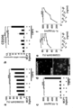
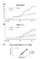
- FIGS. A and B show the evaluation results of hepatocyte death (HE staining and TUNEL staining) in fulminant hepatitis model rats.
- Analysis of gene expression of mannose receptor CD206, a sensor of dead cells of inflammatory cytokines (TNF- ⁇ , IL-1 ⁇ , IL-6) and anti-inflammatory cytokines (IL-10, TGF-b) in fulminant hepatitis model rats It is a figure which shows a result.
- FIGS. 2a to 2f show synergistic effects of MCP-1, ED-Siglec-9 and CSPG on induction of tissue repair type microglia.
- FIGS. 3A to 3C show the synergistic effect of MCP-1, ED-Siglec-9 and CSPG on the induction of tissue repair type microglia.
- the upper photograph of a shows the result of GFAP and HE staining, and the lower photograph shows Quantitative results of tissue cavity area at 8 weeks after SCI are shown
- b shows an immunohistological image of 5-HT positive nerve fibers
- c shows 5 mm head side and 5 mm tail side from the center of the tissue cavity.
- the quantitative result of 5-HT positive nerve fiber is shown. It is a figure which shows the result of having evaluated the gene expression of the cytokine and the cell surface marker in a spinal cord injury site
- FIG. 1 It is a figure which shows the time passage of the functional recovery of the hind limb after spinal cord contusion.
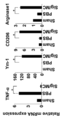
- A shows the result of immunoprecipitation of THP-1 lysate with ED-Siglec-9 and CCR2 and immunoblotting with anti-CCR2 antibody or MAH-lectin.
- B shows that CSPG treatment increases CCR2 speech of microglia.
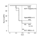
- C shows that ED-Siglec-9 physically interacts with CCR2 in microglia. It is a figure which shows the change of a survival rate and a body weight regarding the administration group of MCP-1 and ED-Siglec-9, and a non-administration (PBS administration) group.
- FIG. 3 is a graph showing that no clear inhibitory effect is exhibited on the enhancement of MMP-3 expression by TNF- ⁇ stimulation. It is a figure which shows the bone regeneration effect in 6 weeks after administering ED-Siglec-9 / MCP-1 to a rat skull defect
- tissue repair active composition comprising at least one selected from the group consisting of a second component that is a protein and a third component that is at least one of chondroitin sulfate and proteoglycan chondroitin sulfate (tissue repair agent, hereinafter simply referred to as “this”) And the use thereof.
- microglia / macrophages that are immunocompetent cells are differentiated or converted into tissue repair type by synergistic effects with the remaining components at the tissue repair site. Can be induced. Therefore, by delivering this agent to the inflammatory reaction site, the tissue repair type microglia / macrophage can be made to act positively and the repair of the tissue at the inflammatory reaction site can be activated.
- the first component, the second component and the third component may be present at the inflammatory reaction site. When either is present, a component present at the inflammatory reaction site can be excluded from the present agent, and the amount of the component can be reduced.
- the first component is known to be present at the site of inflammatory reaction.
- the third component is also a component that is universally present at the site of inflammation. Among these, it is known that the first component is present at an inflammatory reaction site, particularly a chronic inflammatory reaction site.
- the third component is a constituent component of the cell membrane or intercellular substance.
- the present agent can contain the second component as a main component or only the second component.
- This agent preferably contains two or more of these components.
- the conversion of microglia / macrophages can be more effectively promoted.
- This agent preferably contains the first component and the second component among the three components.
- the third component which is chondroitin sulfate or chondroitin sulfate proteoglycan, is a component that is resident in the inflammatory site, and induces tissue repair type microglia / macrophage phage in cooperation with the first component and the second component. Therefore, this agent may consist only of the first component and the second component.
- repair means that part or all of the function lost due to damage in the target tissue is maintained or widened compared to the function of the damaged part at the time of damage. Yes. This includes not only recovery of function but also regeneration as a functional organization. The method for evaluating whether or not the function is maintained or restored differs depending on the damage content at the damaged site. Commonly used assays are used to assess the appearance of the damaged site and the degree of function of interest.
- inflammation refers to a mechanism in a mammal that attempts to protect the body, induced by the presence of a foreign body or tissue damage due to some cause.
- Inflammatory response refers to a series of processes that occur in inflammation.
- An “inflammatory response” can include inflammation-induced tissue destruction.
- Inflammatory disease means a disease, disorder or condition characterized by inflammation of body tissues or by having an inflammatory component. These include local inflammatory responses and systemic inflammatory responses.
- the first component and the second component which are proteins that can be included in the tissue repair agent disclosed in the present specification, may be recovered from a living body such as an animal, or may be genetically engineered or chemically treated. It may be synthesized.
- the total protein amount of the first component and the second component is preferably 50% or more, more preferably 60% or more, still more preferably 70% or more, and even more preferably 80% or more as the protein component of the entire restorative agent. More preferably, it is 90% or more, still more preferably 95% or more, more preferably 98% or more, still more preferably 99% or more, and most preferably 99.5% or more.
- the restoration agent when the total protein amount of the first component and the second component occupies the above range in the protein amount of the entire composition, the restoration agent has higher tissue repair activity.
- the first component is a protein having monocyte chemotaxis promoting factor-1 (MCP-1) activity. Homologs of this type of protein are known in various animals including humans.
- MCP-1 monocyte chemotaxis promoting factor-1
- human MCP-1 has the amino acid sequence represented by SEQ ID NO: 2 (NCBI accession number: NP02973.1). Human MCP-1 is encoded by DNA consisting of the base sequence represented by SEQ ID NO: 1.
- the first component has MCP-1 activity. That is, it may be a known natural MCP-1 or a protein newly confirmed to have MCP-1 activity in the agent. Further, natural MCP-1 may be modified and may have MCP-1 activity in this drug. Examples of MCP-1 activity in this drug include activity capable of inducing microglia / macrophages to a tissue repair type in cooperation with the second component and chondroitin sulfate or chondroitin sulfate proteoglycan, or activity to promote production of anti-inflammatory cytokines . Such activity can be easily evaluated by those skilled in the art with reference to the examples described later.
- the activity capable of inducing microglia / macrophages into a tissue repair type is that microglia / macrophages are isolated and cultured from mice or the like, and the established second component and, if necessary, CS and the like described later are used for the cultured cells. In the presence, it can be evaluated by supplying a possible first component and confirming the production of CD206, Arginase1 protein or mRNA that serves as an index of tissue repair type microglia / macrophage.
- the activity of promoting the production of anti-inflammatory cytokines is the activity that can induce microglia / macrophages to be tissue-repaired, and the microglia / macrophages are isolated and cultured from mice and the like.
- a potential first component in the presence of two components and, if necessary, CS, etc., for example by confirming the production of anti-inflammatory cytokines such as IL-10 and TGF- ⁇ 1 Can be evaluated.
- the MCP-1 activity of the first component such as modified MCP-1 is sufficient as long as it has the above-mentioned activity, and the degree thereof is not limited.
- the first component other than the known MCP-1 may be a protein having a certain relationship with the sequence information of MCP-1 disclosed in a database or the like.
- a protein having an MCP-1 activity which consists of an amino acid sequence in which one or several amino acids are deleted, substituted or added in the disclosed amino acid sequence. Any one type of amino acid mutation relative to the disclosed amino acid sequence, that is, deletion, substitution, or addition, may be used, or two or more types may be combined.
- the total number of these mutations is not particularly limited, but is preferably about 1 or more and 10 or less. More preferably, it is 1 or more and 5 or less.
- conservative substitution is preferable, and specific examples include substitution within the following groups.
- a protein having an amino acid sequence having 60% or more identity with the disclosed amino acid sequence of MCP-1 and having MCP-1 activity can be mentioned.
- the identity is more preferably 65% or more, further preferably 70% or more, still more preferably 80% or more, more preferably 85% or more, still more preferably 90% or more, More preferably, it is 95% or more, still more preferably 98% or more, and most preferably 99% or more.
- identity or similarity is a relationship between two or more proteins or two or more polynucleotides determined by comparing sequences, as is known in the art.
- identity identity means between protein or polynucleotide sequences, as determined by alignment between protein or polynucleotide sequences, or in some cases by alignment between a series of such sequences. Means the degree of sequence invariance. Similarity is also the correlation between protein or polynucleotide sequences, as determined by alignment between protein or polynucleotide sequences, or in some cases by alignment between a series of partial sequences.
- Similarity Means the degree of More specifically, it is determined by sequence identity and conservation (substitutions that maintain specific amino acids in the sequence or physicochemical properties in the sequence).
- the similarity is referred to as “Similarity” in the BLAST sequence homology search result described later.
- the method for determining identity and similarity is preferably a method designed to align the longest between the sequences to be compared. Methods for determining identity and similarity are provided as programs available to the public. For example, BLAST (Basic Local Alignment Search Tool) programs (eg, Altschul SF, Gish W, Miller W, Myers EW, Lipman DJ., J. Mol.
- proteins that can be used as the first component include proteins having a certain relationship with known amino acid sequences and base sequences of MCP-1, in addition to the known amino acid sequences and base sequences of MCP-1.
- proteins having MCP-1 activity include proteins having MCP-1 activity.
- An example is the chemokine ligand (CCL) family. Typically, CCL13, 7, 7, 8, etc. are mentioned.
- examples of the protein having 60% or more identity include the following four types.
- Human CC motif chemokine 13 precursor NCBI accession number: NP_005399.1, identity with the amino acid sequence represented by SEQ ID NO: 2 is 65%, similarity is 82%)
- Human CC motif chemokine 7 precursor NCBI accession number: NP_6264.2, 73% identity, 78% similarity
- Human CC motif chemokine 8 precursor NCBI accession number: NP_005614.2, 69% identity, 84% similarity
- Human eotaxin precursor NCBI accession number: NP_002977.1, 70% identity, 84% similarity
- the DNA is encoded by DNA that hybridizes under stringent conditions with DNA comprising a base sequence complementary to the DNA comprising a base sequence encoding MCP-1 and has MCP-1 activity
- Examples include proteins.
- the stringent condition refers to, for example, a condition in which a so-called specific hybrid is formed and a non-specific hybrid is not formed.
- a nucleic acid having high base sequence identity that is, 70% or more, more preferably 80% or more, still more preferably 85% or more, more preferably 90% or more, with the disclosed base sequence, even more preferably 90% or more.
- the sodium salt concentration is 15 to 750 mM, preferably 50 to 750 mM, more preferably 300 to 750 mM
- the temperature is 25 to 70 ° C., preferably 50 to 70 ° C., more preferably 55 to 65 ° C., formamide
- the condition is that the concentration is 0 to 50%, preferably 20 to 50%, more preferably 35 to 45%.
- the conditions for washing the filter after hybridization are usually that the sodium salt concentration is 15 to 600 mM, preferably 50 to 600 mM, more preferably 300 to 600 mM, and the temperature is 50 to 70 ° C., preferably The temperature is 55 to 70 ° C, more preferably 60 to 65 ° C.
- the disclosed base sequence is 80% or more, more preferably 85% or more, still more preferably 90% or more, and still more preferably 95% or more. Yes, more preferably 97% or more, still more preferably 98% or more, and most preferably a protein encoded by DNA having a base sequence having an identity of 99% or more and having MCP-1 activity. It is done.
- Such proteins and DNAs encoding the same are, for example, nucleic acids derived from DNA extracted from various organisms, various cDNA libraries, genomic DNA libraries, etc., using primers designed based on the disclosed nucleotide sequences.
- PCR amplification By performing PCR amplification using as a template, it can be obtained as a nucleic acid fragment. Further, it can be obtained as a nucleic acid fragment by performing hybridization using a nucleic acid derived from the above library or the like as a template and a DNA fragment which is a part of the gene encoding MCP-1 as a probe.
- the gene may be synthesized as a nucleic acid fragment by various nucleic acid sequence synthesis methods known in the art such as chemical synthesis methods.
- the protein and DNA encoding the same can be obtained by, for example, subjecting the DNA encoding the disclosed amino acid sequence to conventional mutagenesis, site-directed mutagenesis, molecular evolution using error-prone PCR, etc. It can be obtained by modifying.
- a known technique such as Kunkel method or Gapped duplex method, or a method equivalent thereto is mentioned.
- a mutation introduction kit using site-directed mutagenesis for example, Mutant-K (manufactured by TAKARA) ), Mutant-G (manufactured by TAKARA)), or the like, or the LAKPCR in vitro Mutagenesis series kit from TAKARA.
- the second component is a protein having the extracellular domain activity of sialic acid-binding immunoglobulin-like lectin-9 (Siglec-9). Homologues of this kind of protein and its full-length protein are known in various animals including humans.
- Siglec-9 is a transmembrane protein expressed in monocytes, granulocytes, and macrophage phages, and has an extracellular domain, a transmembrane domain, and a cytoplasmic domain.
- the extracellular domain of Siglec-9 can be preferably used. It is also known that the extracellular domain contains an immunoglobulin-like domain.
- natural Siglec-9 derived from animals such as humans may be recovered from natural raw materials, or may be obtained by genetic engineering or chemical.
- human Sigle-9 has the amino acid sequence represented by SEQ ID NO: 4. It is also encoded by DNA consisting of the base sequence represented by SEQ ID NO: 3.
- the second component has the extracellular domain activity of Siglec-9. That is, in addition to the known natural Siglec-9, it may be a protein newly confirmed to function as the extracellular domain of Siglec-9 in this drug. Further, natural Siglec-9 may be modified as described above.
- the extracellular domain activity of Siglec-9 in this drug includes, for example, the activity that can induce microglia into a tissue repair type in cooperation with the first component and chondroitin sulfate or chondroitin sulfate proteoglycan, or the activity of enhancing anti-inflammatory cytokines The protein which has is mentioned.
- Human Siglec-9 consists of an amino acid sequence of 463 amino acids represented by SEQ ID NO: 37.
- the amino acid sequence from the 1st position to the 17th position is a signal peptide.
- the second component of the present disclosure may be, for example, the amino acid sequence from the 18th position to the 348th position (SEQ ID NO: 4) of this amino acid sequence, or may have the full length amino acid of this amino acid sequence. Alternatively, it may have an amino acid sequence from the 18th position to the 463rd position.
- the Siglec-9 extracellular domain activity in the extracellular domain of modified Siglec-9 is sufficient as long as it has the above-mentioned inducing activity and production promoting activity, and the degree thereof is not limited.
- the activity capable of inducing microglia / macrophages into a tissue repair type is that microglia / macrophages are isolated and cultured from a mouse or the like, and the cultured first cell is cultured in the presence of CS or the like as necessary.
- the activity of promoting the production of anti-inflammatory cytokines is the activity that can induce microglia / macrophages to be tissue-repaired, and the microglia / macrophages are isolated and cultured from mice and the like.
- a potential second component in the presence of one component and, if necessary, CS or the like, for example, confirming the production of anti-inflammatory cytokines such as IL-10 and TGF- ⁇ 1 Can be evaluated.
- the Siglec-9 extracellular domain activity of the protein consisting of the amino acid represented by SEQ ID NO: 4, more preferably 60% or more, still more preferably 70% or more, still more preferably 80% or more, even more Preferably it is 90% or more, Most preferably, it is 100% or more.
- Various proteins that can be used as the second component include known amino acid sequences and base sequences of the extracellular domain of Siglec-9, as well as known amino acid sequences and base sequences known as the extracellular domain of Siglec-9. And a protein having the extracellular domain activity of Siglec-9 in the present specification.
- Siglec family protein is mentioned. Typical examples include Siglec-9, Siglec-7, Siglec-12, Siglec-8, and CD33.
- Human sialic acid-binding immunoglobulin-like lectin 9 isotope 2 precursor (NCBI accession number: NP_055256.1, the amino acid sequence represented by SEQ ID NO: 4 has 100% identity, similarity is 100%, nucleotide sequence Is represented by SEQ ID NO: 5, and the amino acid sequence is represented by SEQ ID NO: 6.)
- Human sialic acid-binding immunoglobulin-like lectin 9 isotope 1 precursor NCBI accession number: NP_001185487.1, identity with the amino acid sequence represented by SEQ ID NO: 4 is 100%, similarity is 100%
- Human sialic acid-binding immunoglobulin-like lectin 7 isotope 1 precursor (NCBI accession number: NP_00055220.1, identity with amino acid sequence represented by SEQ ID NO: 4 is 81%, similarity is 85%)
- Human sialic acid-binding immunoglobulin-like lectin 7 isotope 1 precursor (NCBI accession number: NP_00055
- the third component is chondroitin sulfate (CS) or chondroitin sulfate proteoglycan (CSPG).
- CS chondroitin sulfate
- CSPG chondroitin sulfate proteoglycan
- the third component may be recovered from nature or may be artificially synthesized.
- the agent By including at least one selected from the first component, the second component, and the third component, the agent exhibits tissue repair activity in the presence of the remaining component when delivered to the inflamed tissue. be able to. Therefore, the present agent may have one component selected from the group consisting of the first component, the second component, and the third component as an active component, or two components as an active component. Three kinds of components may be used as active ingredients.
- the active ingredient contained in this drug is determined according to the type of disease, the site of tissue damage and its characteristics, and the dosage form of this drug.
- this agent contains the 1st component and / or the 2nd component. More preferably, a third component is included in addition to the first component and / or the second component.
- concentration (content) of a 1st component and / or a 2nd component is not specifically limited. It is only necessary for the agent to contain an effective amount that exhibits tissue repair activity. More specifically, for example, the following (a) or (b) is carried out by cooperating the first component and / or the second component with the third component in the present agent or at the tissue repair site. It is sufficient that it is contained in an effective amount to exhibit the activity described in. These activities may be confirmed in the living body in the presence of the third component as necessary, or may be confirmed in vitro.
- A Tissue repair type microglia or macrophage induction activity
- the blending ratio of the first component and the second component is set as necessary.
- the first component: the second component 1: 10 to 10: 1.
- the concentration is not particularly limited. It is only necessary for the agent to contain an effective amount that exhibits tissue repair activity. Moreover, when this agent further contains a 1st component and / or a 2nd component, the density
- the blending ratio of CS or the like is set as necessary.
- the total amount of the first component and the second component: CS, etc. can be set as appropriate in the mass ratio range of 1000: 1 to 1: 1000.
- the total amount of the first component and the second component: CS, etc. 100: 1 to 1: 100, more preferably 1:10 to 10: 1.
- This agent can be produced by blending and mixing one or more selected from the first component, the second component and the third component. That is, each component obtained (extracted from natural raw materials, produced by genetic recombination, chemical synthesis, etc.) and purified as necessary can be blended and mixed. Further, when there are raw materials containing these components at the same time, these components may be selectively extracted by affinity chromatography or the like. Moreover, the density
- the agent Since the agent has tissue repair activity, it can be used as a damaged tissue repair composition that activates and repairs tissue repair due to various causes. Moreover, it can utilize as an anti-inflammatory agent composition which prevents, treats, and alleviates various inflammations.
- the central nervous system diseases are not particularly limited, and neuronal degeneration in the central nervous system. • It can include all conditions that cause omission.
- spinal cord injury cerebral infarction, neonatal hypoxic encephalopathy, neurodegenerative diseases (amyotrophic lateral sclerosis, multiple sclerosis, Alzheimer's disease, Parkinson's disease, progressive epithelial paralysis, Huntington's disease, multiple system atrophy And spinal cerebellar degeneration etc.), viral or autoimmune encephalitis, neurodegeneration due to cerebral infarction associated with cerebral ischemia, intracerebral hemorrhage, etc., and retinal diseases accompanied by nerve cell damage.
- neurodegenerative diseases amotrophic lateral sclerosis, multiple sclerosis, Alzheimer's disease, Parkinson's disease, progressive epithelial paralysis, Huntington's disease, multiple system atrophy And spinal cerebellar degeneration etc.
- viral or autoimmune encephalitis neurodegeneration due to cerebral infarction associated with cerebral ischemia, intracerebral hemorrhage, etc.
- retinal diseases accompanied by nerve cell damage accompanied by nerve cell damage.
- Retinal diseases include traumatic retinal detachment, retinal tear, retinal concussion, optic nerve tube fracture, diabetic retinopathy, age-related macular degeneration, retinitis pigmentosa, glaucoma, colleidelemia, label congenital blindness, cone dystrophy, family Sex drusen, central ring-shaped choroidal dystrophy, autosomal dominant optic atrophy, and the like.
- this agent is preferably applied to acute and subacute diseases and conditions. Examples include spinal cord injury and cerebral infarction. Furthermore, there are motor dysfunction and sensory sensory abnormalities caused by peripheral nerve damage.
- non-central nervous system diseases are not particularly limited, and can include all pathologies that cause cell degeneration / dropout in tissues other than the central nervous system.
- Chronic diseases such as type I and type II diabetes, Sjogren's disease, dry eye, cutaneous wound healing, myocardial infarction, immune rejection associated with bone marrow transplantation, arthritis, rheumatoid arthritis, osteoarthritis and increased bone resorption
- Inflammatory lung diseases such as inflammatory joint diseases, ileitis, ulcerative colitis, inflammatory bowel diseases such as Barrett syndrome and Crohn's disease, asthma, acute and chronic interstitial pneumonia, adult respiratory distress syndrome and chronic obstructive airway disease Diseases, trachoma, onchocerciasis, uveitis, inflammatory eye diseases such as sympathetic ophthalmitis and endophthalmitis, chronic inflammatory periodontal diseases such as gingivitis and periodontitis, tuberculosis, leprosy, uremic complications, Inflammatory kidney diseases such as glomer
- Various other diseases having a significant inflammatory component having a significant inflammatory component.
- systemic inflammation such as gram positive or gram negative bacterial shock, hemorrhagic or anaphylactic shock, or shock induced by cancer chemotherapy in response to proinflammatory cytokines (eg, proinflammatory cytokine related shocks).
- proinflammatory cytokines eg, proinflammatory cytokine related shocks
- Such shock can be induced, for example, by chemotherapeutic agents used in cancer chemotherapy.
- graft rejection such as skin transplant rejection can also be mentioned.
- this agent is preferably applied to acute and subacute diseases and conditions. Examples include acute hepatitis and fulminant hepatitis. At the same time, it is preferably applied to cirrhosis, which is a terminal form of various liver disorders.
- acute and chronic bone marrow inflammatory diseases are mentioned.
- diseases include bisphosphonate drug-related jaw osteonecrosis, acute and chronic osteomyelitis due to trauma and infection.
- bone deformation and bone damage due to periodontal disease or trauma can be mentioned. When this drug is administered to such injuries, the formation of new bone can be promoted.
- Rheumatoid arthritis is an inflammatory autoimmune disease of unknown origin with chronic polysynovitis as the main body. Prolonged synovitis leads to bone and cartilage destruction and permanent dysfunction.
- MCP-1 can be continuously supplied from the living body side, and therefore, the single administration of the second component may be effective. Therefore, in addition to type 2 diabetes, chronic hepatitis, and cirrhosis, chronic administration of the second component may be effective in chronic and autoimmune diseases such as those described above as chronic diseases that cause tissue damage. .
- chronic administration of the second component may be effective in chronic and autoimmune diseases such as those described above as chronic diseases that cause tissue damage.
- the supply of MCP-1 from the living body side is not necessarily sufficient, and therefore it is preferable to administer at least the first component and the second component.
- Interstitial pneumonia including idiopathic pulmonary fibrosis (IPF) can be mentioned.
- Interstitial pneumonia is an inflammatory disease whose main body is the partition between the alveoli.
- the pathological characteristics of IPF are fibrosis in the alveolar wall and changes in alveolar structure (honeycomb lung formation), and the vital capacity, lung compliance, and lung diffusing capacity decrease, and the patient's QOL decreases accordingly. It is said that the fatality rate in the case of acute change following a chronic and progressive course is 80%.
- This agent can additionally contain components other than the first component, the second component, and the third component. Moreover, various preparation forms according to the administration method can be taken as described later.
- This agent can contain bioabsorbable materials such as hyaluronic acid, collagen, fibrinogen, and platelet plasma. Moreover, this agent may contain gelling materials, such as a hyaluronic acid, collagen, and a fibrin glue.
- the agent can contain known pharmaceutical acceptable ingredients. For example, carriers, excipients, disintegrants, buffers, emulsifiers, suspending agents, soothing agents, stabilizers, preservatives, preservatives, physiological saline and the like can be included. As these various additives, any of various known components can be appropriately used.
- the formulation form of this drug is not particularly limited.
- Various known formulation forms can be employed. Tablets, powders, granules, granules, fine granules, capsules, solid injections that dissolve when used, solid agents such as suppositories, liquid injections (intravenous / intramuscular injection), injections, infusions And topical external preparations such as eye drops, sprays, lotions, creams, patches, and the like.
- the form supported by the indwelling medical instrument etc. can also be taken.
- the agent can contain known pharmaceutically acceptable salts.
- this agent When this agent contains two or more types of components, it may be provided in advance as a preparation (mixture) containing these two or more types of components, or two or more types of components can be used at the time of use. Or a combination (kit) of a single agent and a combination of two components. Or you may accommodate in the integral container which can mix these at the time of use.
- the dosage form of this drug is not particularly limited.
- parenteral administration may be systemic administration or local administration. More specifically, injection, application, or spraying to an inflamed site can be mentioned.
- intravenous administration, intraarterial administration, intraportal administration, intradermal administration, subcutaneous administration, intramuscular administration, intraperitoneal administration, intranasal administration, intraoral administration and the like can be mentioned. Intranasal administration is preferred when the site of inflammation is the brain.
- any form may be used as long as the first component, the second component, and the third component coexist in a site requiring tissue repair. Therefore, the specific dosage form is not particularly limited.
- active ingredients to be administered as a preparation may be administered simultaneously or sequentially (the kind and order of active ingredients are arbitrary).
- the dosage of this drug is not particularly limited. It can be set in consideration of the age, weight, disease state, etc. of the test subject.
- Test subjects to which this drug is applied include mammals including humans (pets, domestic animals, laboratory animals, etc.). Examples thereof include dogs, cats, rabbits, mice, cows, pigs, goats, sheep, horses, monkeys, guinea pigs, rats and mice.
- tissue repair macrophage and / or microglia inducer contains one or more selected from the group consisting of a first component, a second component, and a third component.
- This inducing agent is based on the inducing activity of tissue-inducing microglia / macrophages exerted by the first component, the second component, and the third component.
- the first component, the second component, and the third component are as described above, and the present inducing agent can be constituted and produced in the same manner as the above-described present agent.
- the production promoter of anti-inflammatory cytokine disclosed in the present specification contains one or more selected from the group consisting of a first component, a second component, and a third component. This production promoter is based on the production promoting activity of the anti-inflammatory cytokine exhibited by the first component, the second component, and the third component. As with this inducer, various aspects of this agent are applied to this production promoter.
- tissue repair type microglia or macrophage production method The method for producing a tissue repair type microglia or macrophage disclosed in the present specification comprises a step of culturing microglia or macrophage in the presence of a first component, a second component, and a third component in vitro. Can be provided. According to this production method, tissue repair type microglia or macrophages can be produced in vitro. Such a tissue repair type activates tissue repair by, for example, collecting microglia / macrophages from a mammal such as a human, inducing (converting) the tissue into a tissue repair type in vitro, and returning it to the mammal. It can suppress inflammatory reaction and prevent, alleviate or treat inflammation. In producing such microglia / macrophages, the present inducer can be used in the various forms described above.
- the reagent kit disclosed in this specification can include one or more selected from the group consisting of a first component, a second component, and a third component. When two or more kinds of components are included, they may be mixed in advance or may be separately provided so as to be mixed at the time of use. Further, it may be solid, liquid, or soluble at the time of use. If necessary, a solution can be separately provided. Further, the chondroitin sulfate proteoglycan may be premixed with the first component and the second component or may be separately provided.
- the method for promoting tissue repair disclosed herein comprises a step of delivering one or more selected from the group consisting of a first component, a second component, and a third component to inflamed tissue or damaged tissue. be able to. According to this method, tissue repair can be effectively promoted or tissue damage can be prevented or alleviated in the presence of the first to third components in the inflamed tissue or damaged tissue.
- This promotion method can also be implemented as a prevention, treatment or alleviation method in inflammatory diseases.
- the various aspects of the agent described above can be applied, and the administration forms (delivery forms) for the subject also apply the various aspects described for the agent. it can.
- ED-Siglec-9 may be simply expressed as Siglec-9 or Siglec in addition to ED-Siglec-9.
- FIGS. 1 and 2 The determination of 1-week survival rate and evaluation of liver damage by blood test are shown in FIGS. 1 and 2, respectively.
- Sprague-Dawley rat 200 to 250 g was intraperitoneally administered with a D-galactosamine solution that induces significant liver damage at a rate of 1.2 g / kg.
- the weekly survival rate in the PBS administration group decreased to 30% or less.
- the pathological condition improved dramatically, and the one-week survival rate was 100%.
- blood AST and ALT in the control group were 8000 U / L and 8300 U / L, respectively, whereas in the mixed administration group, both were 2000 U / L or less, and ED-Siglec- It was 6000/4000 U / L in the 9 single administration group, and 7000/5500 U / L in the MCP-1 single administration group.
- AST 6000 U / L
- ALT 4000 U / L.
- M1 enhances gene expression of pro-inflammatory cytokines (TNF- ⁇ , IL-1 ⁇ , IL-6).
- M2 expresses a large amount of dead cell sensors: mannose receptor CD206, anti-inflammatory cytokines (IL-10, TGF-b).
- IL-10 pro-inflammatory cytokines
- TGF-b pro-inflammatory cytokines
- FIG. 5 shows the results of CD206 staining in tissues of the fulminant hepatitis model in the ED-Siglec-9 / MCP-1 mixed administration group and the control group. As shown in FIG. 5, in the ED-Siglec-9 / MCP-1 mixed administration group, the expression of CD206 was remarkable and the conversion into tissue repair type macrophages was clearly observed.
- mice Primary neurons were isolated from neonatal C57BL / 6 mice. After 14 days of culture, microglia were isolated from the cell mixture by the “shaking off” method. The purity of the cultured cells was 97-100% as determined by Fc receptor immunostaining. Cultures were maintained in DMEM supplemented with 10% fetal calf serum, 5 ⁇ g / ml bovine insulin and 0.2% glucose.
- CD206 protein and mRNA expression were analyzed by immunohistochemical analysis and real-time quantitative PCR, respectively.
- the IL-10 concentration of the culture supernatant was measured with an ELISA kit (Quantikine ELISA Mouse IL-10, R & D Systems).
- the RT reaction was performed using Superscript III reverse transcriptase (Invitrogen). Used at 0.5 ⁇ g total RNA in a total reaction volume of 25 ⁇ l.
- Real-time Q-PCR was performed using a THUNDERBIRD SYBR quantitative PCR mix (Toyobo) and running a StepOnePlus apparatus real-time PCR system (Applied Biosystems). The primers for rats and mice were as follows.
- Rat Contusion Model and Surgical Procedure Adult female Sprague-Dawley rats weighing 200-230 grams were used. Rats were anesthetized by intraperitoneal injection of ketamine (60-90 mg / kg) and xylazine (100-150 mg / kg). After Th9 lumbar laminectomy, the dura mater was exposed and 200 kdyn of traumatic force was applied by a commercially available spinal cord injury device (Infinite Horizon Impactor, Precision Systems & Instrumentation). Immediately after spinal cord contusion, Th12 partial laminectomy was performed, and a thin silicone tube connected to SMP-200 model (Primetech Inc.): iPRECIO was inserted into the medullary cavity under a surgical microscope.
- BBB Open Field Movement Score The hindlimb neurobehavioral test was performed using the BBB movement rating scale. A 22-point (0-21) BBB scale was used to assess hindlimb recovery, including joint movement, stepping ability, coordination, and trunk stability. A score of 21 means unimpaired movement in intact rats. Two examiners, blinded to the treatment content of the animals, evaluated. The duration of each session was 4 minutes per rat. Scores were analyzed at each time point by repeated measures analysis of variance with Tukey's multiple comparison test.
- Immunoprecipitation, lectin blot and Western blot THP-1 cell lysates were immunoprecipitated using antibodies against ED-Siglec-9 or CCR2, and the precipitates were immunoblotted with anti-CCR2 antibody or MAH-lectin .
- Lectin blots were prepared from THP-1 (RIKEN Cell Bank, Japan) lysate (lysis buffer: 1% Triton X, 150 mM NaCl, 20 mM Tris-HCl, 2 mM calcium chloride) to anti-CCR2 antibody (rabbit IgG and 1 : 50, Abcam), CCR2 proteins were separated by SDS-PAGE and electroblotted onto Immobilon-P PVDF membrane.
- MAL buffer (10 mM HEPES, pH 7.5, 150 mM NaCl, 0.2% BSA, 0.2% Tween-20) for 12 hours at 4 ° C. Thereafter, the probe was probed with 5 mg / ml biotinylated MAL (Vector Laboratories) at 4 ° C. for 12 hours in MAL buffer. MAL was detected using avidin-HRP (Vector Laboratories) and ECL (GE Healthcare) and analyzed by LAS-4000 Minilumino Image Analyzer (GE Healthcare).
- THP-1 or native mouse microglia lysate was used at 4 ° C. with 0.15 nM ED-Siglec-9-Fc or Fc. After overnight incubation, it was immunoprecipitated with Protein A Sepharose (GE Healthcare). Whole cell lysates and resuspended pellets were immunoblotted with antibodies against mouse CCR2 (rabbit IgG, 1: 500, Abcam).
- mice (result) (Synergistic effect of MCP-1, ED-Siglec-9 and CSPG on induction of tissue repair type microglia)
- MCP-1 50 ng / ml
- ED-Siglec-9 50 ng / ml
- CSPG tissue repair type microglia
- FIGS. 6a to 6d microglia treated with MCP-1 (50 ng / ml) and ED-Siglec-9 (50 ng / ml) on CSPG significantly expressed at the protein level of CD-206. And increased expression at the gene level and increased production of IL-10.
- tissue repair type microglia could be confirmed by CD206 by immunostaining. Further, as shown in e and f of FIG. 6, production of CD206, which is a marker of tissue repair type microglia, and anti-inflammatory cytokine IL-10 depends on the concentration of MCP-1 / ED-Siglec-9 / CSPG. Increased.
- FIG. 7a GAFP and HE staining revealed that the activity of GFAP-positive astrocytes decreased and the tissue loss area decreased at 8 weeks after SCI. Further, as shown in FIG. 7b, it was found that 5-HT repaired positive nerve fibers 8 weeks after SCI. Furthermore, as shown in FIG. 7c, it was found by 5-HT staining that nerve fiber repair progressed from the center of injury to the cranial and caudal sides.
- MCP-1 and ED-Siglec-9 were delivered at a concentration of 1 ⁇ g / ml to the SCI site with 3 ⁇ l / h of solution by an iPRECIO infusion pump.
- a silicone tube was inserted into the subarachnoid space.
- Gene expression of cytokines and cell surface markers at the site of spinal cord injury was assessed by quantitative RT-PCR analysis.
- Total RNA was collected from the lesion site 72 hours after SCI. The results are shown in FIG.
- the Y axis represents the ratio to the numerical value in the SHAM model (sham surgery model).
- inflammatory cytokines IL-1 ⁇ , TNF- ⁇
- inducible nitric oxide synthesis iNOS
- MCP-1 and ED-Siglec-9 were administered in combination.
- MCP-1 / ED-Siglec-9 increased anti-inflammatory cytokine expression (IL-10 and TGF- ⁇ 1) and M2 microglia / macrophage markers (CD206, Arginase1).
- the experiment was repeated three times, and similar results were obtained in all cases.
- the data in FIG. 8 represent the mean ⁇ SEM (* P ⁇ 0.05, ** P ⁇ 0.01, *** P ⁇ 0.001).
- FIG. 9 shows a typical image and quantitative results of microglia / macrophages surrounding the injury site 72 hours after spinal cord injury.
- Iba1 + microglia was observed, but no CD206 + cells were observed.
- MCP-1 / ED-Siglec-9 treated rats, a large number of Iba + cells migrated to the damaged site 72 hours after the spinal cord injury, and these Iba1 + cells Most co-expressed CD206.
- FIG. 9 shows a typical image and quantitative results of microglia / macrophages surrounding the injury site 72 hours after spinal cord injury.
- CD206 + cells at the injury site were not observed to express IL-10 simultaneously, whereas they were treated with MCP-1 / ED-Siglec-9.
- CD206 + cells at the damaged site also expressed IL-10 at the same time.
- Data in the figure represent mean ⁇ SD (* P ⁇ 0.05, ** P ⁇ 0.01, MCP-1 / ED-Siglec-9 treated spinal cord injury model versus PBS treatment).
- THP-1 human monocyte cell line THP-1 that abundantly expresses endogenous CCR2
- MCP-1 receptor CCR2
- ED-Siglec-9 ED-Siglec-9
- FIG. 11 (A) THP-1 expressed multiple CCR2 proteins with different molecular weights (FIG. 11 (A) Total lysate) by SDS-PAGE analysis.
- two major species of CCR2 protein at 55 and 42 kDa were detected by immunoprecipitation of THP-1 lysates with anti-CCR2 antibody (FIG. 11 (A) anti-CCR2).
- the MAH blot specifically recognized sugar chains containing ⁇ 2-3-linked sialic acid, which is the main target of Siglec-9.
- FIG. 11 (A) MAH-blot High molecular weight sialylated CCR2 was detected only in THP-1 dissolved precipitate with ED-Siglec-9-Fc (FIG. 11 (A) CCR2-blot).
- pretreatment of THP-1 lysate with sialidase inhibited the physical interaction between CCR2 and ED-Siglec-9 (FIG. 11 (A) CCR2-blot (sialidase +)).
- the physical interaction of ED-Siglec-9 and CCR2 was dependent on sialylated CCR2 containing ⁇ 2-3 linked sialic acid.
- CSPG-treated native microglia increased the expression of CCR2 of different molecular weight (FIG. 11 (B)).
- CSPG contributed at least in part to the induction of M2 by increasing the expression of CCR2.
- sialylated CCR2 with higher molecular weight was also found in ED-Siglec-9 and microglia dissolved precipitates. It was shown that ED-Siglec-9 physically interacts with mouse microglia sialylated CCR2 (FIG. 11 (C)).
- PBS solutions of MCP-1 and ED-Siglec-9 recombinant proteins each having a concentration of 1 ⁇ g / ml were intravenously administered from the jugular vein to model mice 24 hours after the occurrence of lung injury, and the pathological conditions thereafter were observed. Changes in survival rate and body weight are shown in FIG. 12 for the MCP-1 and ED-Siglec-9 administration group and the non-administration (PBS administration) group.
- MCP-1 and ED-Siglec-9 have a repair activity against tissue damage caused by inflammatory diseases in the lung, and as a result, pulmonary inflammatory properties such as pulmonary fibrosis It was found to be effective in the treatment and prevention of diseases and acute lung diseases.
- Sham exhibits normal liver tissue, but in the pre-treatment tissue (immediately before administration of ED-Siglec-9 / MCP-1), many liver cell deaths and fibrosis in the Dysse cavity Increased was observed. On the other hand, intense cell infiltration and significant fibrosis were observed in the PBS administration group, but the ED-Siglec-9 / MCP-1 administration group exhibited a tissue image close to that of a normal liver image.
- TNF- ⁇ As shown in FIG. 16, the expression of TNF- ⁇ produced by pro-inflammatory M1 macrophages was increased in the PBS-administered group, whereas TNF- ⁇ was increased in the ED-Siglec-9 / MCP-1 administered group. Expression is suppressed. On the other hand, the expression of anti-inflammatory M2 macrophage markers Ym-1, CD206, Arginase-1 was increased.
- ⁇ -Smooth muscle actin ⁇ -SMA
- Collagen ⁇ 1 produced by activated hepatic stellate cells hepatic stellate cells
- MMP-9 matrix metalloproteinase-9
- IGF-1 IGF-1
- HGF HGF related to fibrosis
- ⁇ -SMA staining was performed on liver tissues collected from the ED-Siglec-9 and MCP-1 administration group and the non-administration (PBS administration) group. The results are shown in FIG.
- ED-Siglec-9 and MCP-1 administered to the liver leading to cirrhosis can suppress hepatocyte death, inflammatory cell infiltration and irreversible fibrous tissue growth characteristic of cirrhosis. I understood.
- Collagen-induced arthritis mice have been used as the main animal model for rheumatoid arthritis. Collagen-induced arthritis was developed by subcutaneously injecting a heterologous animal type 2 collagen into the tail base of DBA / 1J mice and then reinjecting it 21 days later. The severity of arthritis was assessed using the arthritis score. The average arthritis score of each group was calculated using the scoring system shown in FIG. In addition, an arthritis score of 1 or more in each mouse was defined as the onset of arthritis.
- the ED-Siglec-9 0.1 and 10 ⁇ g groups showed no significant difference from the control group, whereas the 1 ⁇ g administration group was significantly more arthritic than the control group. The score was suppressed.
- the serum concentration of TNF- ⁇ which is a major inflammatory cytokine in rheumatoid arthritis, was measured, it was significantly suppressed in the 1 ⁇ g group compared to the control group.
- ED-Siglec-9 The dose of ED-Siglec-9 was 1 ⁇ g. From 2 days after the second collagen administration, saline or ED-Siglec-9 was administered from the jugular vein at a frequency of once every 7 days. The evaluation result of the arthritis score is shown in FIG.
- the incidence of arthritis reached 100% in the control group, whereas the incidence was significantly suppressed to 40% in the ED-Siglec-9 administration group.
- the arthritis score was significantly suppressed to 1.4 in the ED-Siglec-9 group, while the average was 4.33 in the control group.
- the correlation coefficient (R) was 0.837, which was a very strong and significant positive correlation. Indicated.
- ED-Siglec-9 showed a tendency to suppress the increase in expression of TNF- ⁇ (a representative inflammatory cytokine in rheumatoid arthritis) by LPS stimulation in a dose-dependent manner.
- Synovial fibroblasts were pre-cultured for 48 hours, stimulated with TNF- ⁇ (10 mg / ml) for 12 hours in the presence / absence of ED-Siglec-9 (50 ng / ml), mRNA was extracted, and real-time PCR Gene expression was measured by the method. The results are shown in FIG.
- ED-Siglec-9 is expressed in MMP-3 (a representative proteolytic enzyme produced by synovitis) by TNF- ⁇ stimulation in human rheumatoid arthritis-derived synovial fibroblasts. There was no apparent inhibitory effect on the enhancement.
- M1-macrophages In acute and chronic inflammation, disordered activation of M1-macrophages promotes tissue damage and fibrosis and prevents tissue repair.
- anti-inflammatory M2-macrophages promote tissue regeneration by promoting vascular regeneration, phagocytosis of dead cells, and proliferation and accumulation of in vivo stem cells.
- M2 the ability of the living body to induce M2 is limited, and most tissue destruction environments are dominant in M1. It was found that ED-Siglec-9 and MCP-1 convert the M1-dominant tissue destructive inflammatory environment into an anti-inflammatory and tissue regeneration environment.
- ED-Siglec-9 / MCP-1 was administered to a rat skull defect model, and a remarkable bone regeneration effect was confirmed histologically and on CT 6 weeks after the operation. From the above results, it was revealed that ED-Siglec-9 / MCP-1 can be clinically applied as a bone regeneration promoting factor preparation.
- glucose-responsive insulin secretion experiment was conducted.
- MIN6 (1 ⁇ 10 6 / well) was cultured until it became 90% confluent and washed three times with DMEM (serum-free medium).
- DMEM serum-free medium
- Each culture solution was supplemented with SHED-CM, SHED-CM + various antibodies (anti-human IgG: 1/500, anti-human ED-Siglec-9 antibody: 1/500, anti-MCP-1 antibody 1/500, anti-human ED- Siglec-9 antibody: 1/500 + anti-MCP-1 antibody: 1/500) and cultured for 6 hours.
Abstract
Description
シアル酸結合イムノグロブリン様レクチン-9(Siglec-9)の細胞外ドメイン活性を有するタンパク質(以下、ED-Siglec-9ともいう。)である第2の成分、及び
コンドロイチン硫酸及びコンドロイチン硫酸プロテオグリカンの少なくとも一種である第3の成分、
からなる群から選択される少なくとも1種の成分を含む、組織修復活性組成物。
(2)前記第1の成分と、前記第2の成分と、を含む(1)及び(2)に記載の組成物。
(3)前記第1の成分は、配列番号2で表されるアミノ酸配列と60%以上の同一性を有するアミノ酸配列を有する(1)又は(2)に記載の組成物。
(4)前記第2の成分は、配列番号4で表されるアミノ酸配列と60%以上の同一性を有するアミノ酸配列を有する、(1)~(3)のいずれかに記載の組成物。
(5)さらに、コンドロイチン硫酸又はコンドロイチン硫酸プロテオグリカンを含む、(1)~(4)のいずれかに記載の組成物。
(6)前記第1の成分及び前記第2の成分を、以下の(a)又は(b)に記載の活性を呈するのに有効量含有する、(1)~(5)のいずれかに記載の組成物。
(a)炎症組織における組織修復型マクロファージ及び/又はミクログリアの誘導活性
(b)抗炎症性サイトカインの産生促進活性
(7)(1)~(6)のいずれかに記載の組成物である、抗炎症剤組成物。
(8)脊髄損傷、脳梗塞、新生児低酸素脳症、アルツハイマー症、多発性硬化症、筋萎縮性側索硬化症及びパーキンソン病からなる群から選択される中枢神経系疾患の治療用である、(7)に記載の組成物。
(9)劇症肝炎、急性肝炎、慢性肝炎、急性および慢性間質性肺炎、I型およびII型糖尿病、シェーグレン病、ドライアイ、関節リウマチ、全身性エリテマトーデス、皮膚創傷治癒、心筋梗塞、骨髄移植に伴う免疫拒絶からなる群から選択される非中枢神経系疾患の治療用である、(7)に記載の組成物。
(10)単球走化性促進因子-1(MCP-1)活性を有するタンパク質である第1の成分、
シアル酸結合イムノグロブリン様レクチン-9(Siglec-9)の細胞外ドメイン活性を有するタンパク質である第2の成分、及び
コンドロイチン硫酸及びコンドロイチン硫酸プロテオグリカンの少なくとも一種である第3の成分、
からなる群から選択される少なくとも1種の成分を含む、組織修復型マクロファージ及び/又はミクログリアの誘導剤。
(11)単球走化性促進因子-1(MCP-1)活性を有するタンパク質である第1の成分、
シアル酸結合イムノグロブリン様レクチン-9(Siglec-9)の細胞外ドメイン活性を有するタンパク質である第2の成分、及び
コンドロイチン硫酸及びコンドロイチン硫酸プロテオグリカンの少なくとも一種である第3の成分、
からなる群から選択される少なくとも1種を含む、抗炎症性サイトカインの産生促進剤。
(12)生体外で、単球走化性促進因子-1(MCP-1)活性を有するタンパク質である第1の成分、
シアル酸結合イムノグロブリン様レクチン-9(Siglec-9)の細胞外ドメイン活性を有するタンパク質である第2の成分、及び
コンドロイチン硫酸及びコンドロイチン硫酸プロテオグリカンの少なくとも一種である第3の成分、の存在下で、ミクログリア又はマクロファージを培養する工程を備える、組織修復型ミクログリア又はマクロファージの生産方法。
(13)単球走化性促進因子-1(MCP-1)活性を有するタンパク質である第1の成分、
シアル酸結合イムノグロブリン様レクチン-9(Siglec-9)の細胞外ドメイン活性を有するタンパク質である第2の成分、及び
コンドロイチン硫酸又はコンドロイチン硫酸プロテオグリカンである第3の成分からなる群から選択される少なくとも1種を備える、ミクログリア及び/又はマクロファージ用の試薬キット。
(14)前記第1の成分及び前記第2の成分を含む、(13)に記載の試薬キット。
(15)単球走化性促進因子-1(MCP-1)活性を有するタンパク質である第1の成分、
シアル酸結合イムノグロブリン様レクチン-9(Siglec-9)の細胞外ドメイン活性を有するタンパク質である第2の成分、及び
コンドロイチン硫酸及びコンドロイチン硫酸プロテオグリカンの少なくとも一種である第3の成分、
からなる群から選択される少なくとも1種を損傷組織又は炎症組織に送達して組織修復を促進する方法。 (1) a first component which is a protein having monocyte chemotaxis promoting factor-1 (MCP-1) activity,
A second component which is a protein having an extracellular domain activity of sialic acid-binding immunoglobulin-like lectin-9 (Siglec-9) (hereinafter also referred to as ED-Siglec-9), and at least chondroitin sulfate and chondroitin sulfate proteoglycan A third component that is a kind,
A tissue repair active composition comprising at least one component selected from the group consisting of:
(2) The composition according to (1) and (2), comprising the first component and the second component.
(3) The composition according to (1) or (2), wherein the first component has an amino acid sequence having 60% or more identity with the amino acid sequence represented by SEQ ID NO: 2.
(4) The composition according to any one of (1) to (3), wherein the second component has an amino acid sequence having 60% or more identity with the amino acid sequence represented by SEQ ID NO: 4.
(5) The composition according to any one of (1) to (4), further comprising chondroitin sulfate or chondroitin sulfate proteoglycan.
(6) The composition according to any one of (1) to (5), which contains the first component and the second component in an effective amount to exhibit the activity described in the following (a) or (b): Composition.
(A) Tissue repair type macrophage and / or microglia inducing activity in inflammatory tissue (b) Anti-inflammatory cytokine production promoting activity (7) The composition according to any one of (1) to (6), Inflammatory agent composition.
(8) For treatment of central nervous system diseases selected from the group consisting of spinal cord injury, cerebral infarction, neonatal hypoxic encephalopathy, Alzheimer's disease, multiple sclerosis, amyotrophic lateral sclerosis, and Parkinson's disease. The composition as described in 7).
(9) Fulminant hepatitis, acute hepatitis, chronic hepatitis, acute and chronic interstitial pneumonia, type I and type II diabetes, Sjogren's disease, dry eye, rheumatoid arthritis, systemic lupus erythematosus, skin wound healing, myocardial infarction, bone marrow transplantation The composition according to (7), which is used for treatment of a non-central nervous system disease selected from the group consisting of immune rejection associated with.
(10) a first component which is a protein having monocyte chemotaxis promoting factor-1 (MCP-1) activity;
A second component that is a protein having extracellular domain activity of sialic acid-binding immunoglobulin-like lectin-9 (Siglec-9), and a third component that is at least one of chondroitin sulfate and chondroitin sulfate proteoglycan,
A tissue repair type macrophage and / or microglia inducer comprising at least one component selected from the group consisting of:
(11) a first component which is a protein having monocyte chemotaxis promoting factor-1 (MCP-1) activity;
A second component that is a protein having extracellular domain activity of sialic acid-binding immunoglobulin-like lectin-9 (Siglec-9), and a third component that is at least one of chondroitin sulfate and chondroitin sulfate proteoglycan,
An anti-inflammatory cytokine production promoter comprising at least one selected from the group consisting of:
(12) a first component that is a protein having monocyte chemotaxis promoting factor-1 (MCP-1) activity in vitro;
In the presence of a second component that is a protein having extracellular domain activity of sialic acid-binding immunoglobulin-like lectin-9 (Siglec-9), and a third component that is at least one of chondroitin sulfate and chondroitin sulfate proteoglycan A method for producing tissue repair-type microglia or macrophages, comprising a step of culturing microglia or macrophages.
(13) a first component which is a protein having monocyte chemotaxis promoting factor-1 (MCP-1) activity;
At least selected from the group consisting of a second component that is a protein having extracellular domain activity of sialic acid-binding immunoglobulin-like lectin-9 (Siglec-9), and a third component that is chondroitin sulfate or chondroitin sulfate proteoglycan A reagent kit for microglia and / or macrophages, comprising one kind.
(14) The reagent kit according to (13), including the first component and the second component.
(15) a first component which is a protein having monocyte chemotaxis promoting factor-1 (MCP-1) activity,
A second component that is a protein having extracellular domain activity of sialic acid-binding immunoglobulin-like lectin-9 (Siglec-9), and a third component that is at least one of chondroitin sulfate and chondroitin sulfate proteoglycan,
A method of promoting tissue repair by delivering at least one selected from the group consisting of to damaged or inflamed tissue.
シアル酸結合イムノグロブリン様レクチン-9(Siglec-9)の細胞外ドメイン活性を有する第2の成分、及び
コンドロイチン硫酸及びコンドロイチン硫酸プロテオグリカンの少なくとも一種である第3の成分からなる群から選択される少なくとも1種を含む、組織修復剤。
(17)少なくとも前記第1の成分及び前記第2の成分のいずれか又は双方を含む、(16)に記載の組織修復剤。
(18)前記第1の成分と前記第2の成分とを含む、(16)又は(17)に記載の組織修復剤。
(19)前記第1の成分は、配列番号2で表されるアミノ酸配列と60%以上の同一性を有するアミノ酸配列を有する、(16)~(18)のいずれかに記載の組織修復剤。
(20)前記第2の成分は、配列番号4で表されるアミノ酸配列と60%以上の同一性を有するアミノ酸配列を有する、(16)~(19)のいずれかに記載の組織修復剤。
(21)前記第3の成分を含む、(16)~(20)のいずれかに記載の組織修復剤。
(22)前記第1の成分、前記第2の成分及び第3の成分からなる群から選択される1種以上の成分を、以下の(a)又は(b)に記載の活性を呈するのに有効量含有する、(16)~(21)のいずれかに記載の組織修復剤。
(a)炎症組織における組織修復型マクロファージ及び/又はミクログリアの誘導活性
(b)抗炎症性サイトカインの産生促進活性
(22)(16)~(21)のいずれかに記載の組織修復剤を有効成分とする、抗炎症剤。
(23)(16)~(21)のいずれかに記載の組織修復剤を有効成分とする、脊髄損傷、脳梗塞、新生児低酸素脳症、アルツハイマー症、多発性硬化症、筋萎縮性側索硬化症及びパーキンソン病からなる群から選択される中枢神経系疾患の予防又は治療剤。
(24)(16)~(21)のいずれかに記載の組織修復剤を有効成分とする、劇症肝炎、急性肝炎、慢性肝炎、急性および慢性間質性肺炎、I型およびII型糖尿病、シェーグレン病、ドライアイ、関節リウマチ、全身性エリテマトーデス、皮膚創傷治癒、心筋梗塞、骨髄移植に伴う免疫拒絶からなる群から選択される非中枢神経系疾患の予防又は治療剤。
(25)単球走化性促進因子-1(MCP-1)活性を有するタンパク質である第1の成分、
シアル酸結合イムノグロブリン様レクチン-9(Siglec-9)の細胞外ドメイン活性を有するタンパク質である第2の成分、及び
コンドロイチン硫酸及びコンドロイチン硫酸プロテオグリカンの少なくとも一種である第3の成分からなる群から選択される少なくとも1種を含む、組織修復型マクロファージ及び/又はミクログリアの誘導剤。
(26)単球走化性促進因子-1(MCP-1)活性を有するタンパク質である第1の成分、
シアル酸結合イムノグロブリン様レクチン-9(Siglec-9)の細胞外ドメイン活性を有するタンパク質である第2の成分、及び
コンドロイチン硫酸及びコンドロイチン硫酸プロテオグリカンの少なくとも一種である第3の成分からなる群から選択される少なくとも1種を含む、抗炎症性サイトカインの産生促進剤。
(27)生体外で、単球走化性促進因子-1(MCP-1)活性を有するタンパク質である第1の成分、
シアル酸結合イムノグロブリン様レクチン-9(Siglec-9)の細胞外ドメイン活性を有するタンパク質である第2の成分、及び
コンドロイチン硫酸及びコンドロイチン硫酸プロテオグリカンの少なくとも一種である第3の成分からなる群から選択される少なくとも1種の存在下でと、ミクログリア又はマクロファージを培養する工程、を備える、組織修復型ミクログリア又はマクロファージの生産方法。
(28)単球走化性促進因子-1(MCP-1)活性を有するタンパク質である第1の成分、
シアル酸結合イムノグロブリン様レクチン-9(Siglec-9)の細胞外ドメイン活性を有するタンパク質である第2の成分、及び
コンドロイチン硫酸及びコンドロイチン硫酸プロテオグリカンの少なくとも一種である第3の成分からなる群から選択される少なくとも1種を含む、ミクログリア/マクロファージ用の試薬キット。
(29)前記第1の成分と前記第2の成分とを含む、(28)に記載の試薬キット。
(30)単球走化性促進因子-1(MCP-1)活性を有するタンパク質である第1の成分、
シアル酸結合イムノグロブリン様レクチン-9(Siglec-9)の細胞外ドメイン活性を有するタンパク質である第2の成分、及び
コンドロイチン硫酸及びコンドロイチン硫酸プロテオグリカンの少なくとも一種である第3の成分からなる群から選択される少なくとも1種を損傷組織又は炎症組織に送達して組織修復を促進する方法。 (16) a first component having monocyte chemotaxis promoting factor-1 (MCP-1) activity,
A second component having extracellular domain activity of sialic acid-binding immunoglobulin-like lectin-9 (Siglec-9), and at least selected from the group consisting of chondroitin sulfate and a third component that is at least one of chondroitin sulfate proteoglycans A tissue repair agent comprising one species.
(17) The tissue repair agent according to (16), comprising at least one of or both of the first component and the second component.
(18) The tissue repair agent according to (16) or (17), comprising the first component and the second component.
(19) The tissue repair agent according to any one of (16) to (18), wherein the first component has an amino acid sequence having 60% or more identity with the amino acid sequence represented by SEQ ID NO: 2.
(20) The tissue repair agent according to any one of (16) to (19), wherein the second component has an amino acid sequence having 60% or more identity with the amino acid sequence represented by SEQ ID NO: 4.
(21) The tissue repair agent according to any one of (16) to (20), comprising the third component.
(22) One or more components selected from the group consisting of the first component, the second component, and the third component exhibit the activity described in (a) or (b) below: The tissue repair agent according to any one of (16) to (21), which contains an effective amount.
(A) Inducing activity of tissue repair macrophages and / or microglia in inflamed tissues (b) Production promoting activity of anti-inflammatory cytokines (22) The tissue repair agent according to any one of (16) to (21) as an active ingredient An anti-inflammatory agent.
(23) Spinal cord injury, cerebral infarction, neonatal hypoxic encephalopathy, Alzheimer's disease, multiple sclerosis, amyotrophic lateral sclerosis comprising the tissue repair agent according to any one of (16) to (21) as an active ingredient A preventive or therapeutic agent for central nervous system diseases selected from the group consisting of Parkinson's disease and Parkinson disease.
(24) Fulminant hepatitis, acute hepatitis, chronic hepatitis, acute and chronic interstitial pneumonia, type I and type II diabetes, comprising the tissue repair agent according to any one of (16) to (21) as an active ingredient A preventive or therapeutic agent for non-central nervous system diseases selected from the group consisting of Sjogren's disease, dry eye, rheumatoid arthritis, systemic lupus erythematosus, skin wound healing, myocardial infarction, and immune rejection associated with bone marrow transplantation.
(25) a first component which is a protein having monocyte chemotaxis promoting factor-1 (MCP-1) activity,
Selected from the group consisting of a second component that is a protein having extracellular domain activity of sialic acid-binding immunoglobulin-like lectin-9 (Siglec-9), and a third component that is at least one of chondroitin sulfate and chondroitin sulfate proteoglycan A tissue repair type macrophage and / or microglia inducer comprising at least one selected from the group consisting of:
(26) a first component which is a protein having monocyte chemotaxis promoting factor-1 (MCP-1) activity,
Selected from the group consisting of a second component that is a protein having extracellular domain activity of sialic acid-binding immunoglobulin-like lectin-9 (Siglec-9), and a third component that is at least one of chondroitin sulfate and chondroitin sulfate proteoglycan An anti-inflammatory cytokine production promoter comprising at least one selected from the group consisting of:
(27) a first component that is a protein having monocyte chemotaxis promoting factor-1 (MCP-1) activity in vitro;
Selected from the group consisting of a second component that is a protein having extracellular domain activity of sialic acid-binding immunoglobulin-like lectin-9 (Siglec-9), and a third component that is at least one of chondroitin sulfate and chondroitin sulfate proteoglycan A tissue repair type microglia or macrophage production method comprising the step of culturing microglia or macrophages in the presence of at least one selected from the group consisting of:
(28) a first component which is a protein having monocyte chemotaxis promoting factor-1 (MCP-1) activity;
Selected from the group consisting of a second component that is a protein having extracellular domain activity of sialic acid-binding immunoglobulin-like lectin-9 (Siglec-9), and a third component that is at least one of chondroitin sulfate and chondroitin sulfate proteoglycan A reagent kit for microglia / macrophage comprising at least one selected from the group consisting of:
(29) The reagent kit according to (28), comprising the first component and the second component.
(30) a first component which is a protein having monocyte chemotaxis promoting factor-1 (MCP-1) activity;
Selected from the group consisting of a second component that is a protein having extracellular domain activity of sialic acid-binding immunoglobulin-like lectin-9 (Siglec-9), and a third component that is at least one of chondroitin sulfate and chondroitin sulfate proteoglycan To promote tissue repair by delivering at least one of them to a damaged or inflamed tissue.
本明細書に開示される組織修復剤が備えうるタンパク質である、第1の成分及び第2の成分は、動物などの生体から回収されるものであってもよいし、遺伝子工学的あるいは化学的に合成されたものであってもよい。第1の成分と第2の成分との総タンパク量が、修復剤全体のタンパク質成分として、好ましくは50%以上、より好ましくは60%以上、さらに好ましくは70%以上、一層好ましくは80%以上、より一層好ましくは90%以上、さらに一層好ましくは95%以上、より好ましくは98%以上、さらに好ましくは99%以上、最も好ましくは99.5%以上であることが好ましい。本修復剤において第1の成分と第2の成分との総タンパク質量が組成物全体のタンパク質量において上記範囲を占めることで、より組織修復活性の高い修復剤となる。 (Repair active composition (tissue repair agent))
The first component and the second component, which are proteins that can be included in the tissue repair agent disclosed in the present specification, may be recovered from a living body such as an animal, or may be genetically engineered or chemically treated. It may be synthesized. The total protein amount of the first component and the second component is preferably 50% or more, more preferably 60% or more, still more preferably 70% or more, and even more preferably 80% or more as the protein component of the entire restorative agent. More preferably, it is 90% or more, still more preferably 95% or more, more preferably 98% or more, still more preferably 99% or more, and most preferably 99.5% or more. In the present restoration agent, when the total protein amount of the first component and the second component occupies the above range in the protein amount of the entire composition, the restoration agent has higher tissue repair activity.
第1の成分は、単球走化性促進因子-1(MCP-1)活性を有するタンパク質である。この種のタンパク質は、ヒトをはじめとして各種動物においてホモログが知られている。本剤においては、こうしたヒトなどの動物に由来する天然のMCP-1を天然の原料から回収したものであってもよいし、遺伝子工学的又は化学的に取得したものであってもよい。例えば、ヒトのMCP-1は配列番号2で表されるアミノ酸配列(NCBIアクセッション番号:NP02973.1)を有している。また、ヒトのMCP-1は、配列番号1で表される塩基配列からなるDNAによってコードされている。 (First ingredient)
The first component is a protein having monocyte chemotaxis promoting factor-1 (MCP-1) activity. Homologs of this type of protein are known in various animals including humans. In this agent, natural MCP-1 derived from such animals as humans may be recovered from natural raw materials, or may be obtained by genetic engineering or chemical. For example, human MCP-1 has the amino acid sequence represented by SEQ ID NO: 2 (NCBI accession number: NP02973.1). Human MCP-1 is encoded by DNA consisting of the base sequence represented by SEQ ID NO: 1.
(1)ヒトC-Cモチーフケモカイン13プレカーサー(NCBIアクセッション番号:NP_005399.1、配列番号2で表されるアミノ酸配列との同一性は65%、類似性は82%)
(2)ヒトC-Cモチーフケモカイン7プレカーサー(NCBIアクセッション番号:NP_6264.2、同同一性73%、同類似性78%)
(3)ヒトC-Cモチーフケモカイン8プレカーサー(NCBIアクセッション番号:NP_005614.2、同同一性69%、同類似性84%)
(4)ヒトエオタキシンプレカーサー(NCBIアクセッション番号:NP_002977.1、同同一性70%、同類似性84%) For example, in the relationship with the amino acid sequence represented by SEQ ID NO: 2, examples of the protein having 60% or more identity include the following four types.
(1) Human CC motif chemokine 13 precursor (NCBI accession number: NP_005399.1, identity with the amino acid sequence represented by SEQ ID NO: 2 is 65%, similarity is 82%)
(2) Human
(3) Human
(4) Human eotaxin precursor (NCBI accession number: NP_002977.1, 70% identity, 84% similarity)
第2の成分は、シアル酸結合イムノグロブリン様レクチン-9(Siglec-9)の細胞外ドメイン活性を有するタンパク質である。この種のタンパク質及びその全長タンパク質は、ヒトをはじめとして各種動物においてホモログが知られている。Siglec-9は、単球、顆粒球及びマクロファージファージにおいて発現する膜貫通タンパク質であって、細胞外ドメインと膜貫通ドメインと細胞質ドメインとを備えている。本明細書においては、Siglec-9の細胞外ドメインを好ましく用いることができる。細胞外ドメインは、免疫グロブリン様ドメインを含んでいることも知られている。 (Second component)
The second component is a protein having the extracellular domain activity of sialic acid-binding immunoglobulin-like lectin-9 (Siglec-9). Homologues of this kind of protein and its full-length protein are known in various animals including humans. Siglec-9 is a transmembrane protein expressed in monocytes, granulocytes, and macrophage phages, and has an extracellular domain, a transmembrane domain, and a cytoplasmic domain. In the present specification, the extracellular domain of Siglec-9 can be preferably used. It is also known that the extracellular domain contains an immunoglobulin-like domain.
(1)ヒトシアル酸結合イムノグロブリン様レクチン9同位体2プレカーサー(NCBIアクセッション番号:NP_055256.1、配列番号4で表されるアミノ酸配列との同一性は100%、類似性は100%、塩基配列は配列番号5で表され、アミノ酸配列は配列番号6で表される。)
(2)ヒトシアル酸結合イムノグロブリン様レクチン9同位体1プレカーサー(NCBIアクセッション番号:NP_001185487.1、配列番号4で表されるアミノ酸配列との同一性は100%、類似性は100%)
(3)ヒトシアル酸結合イムノグロブリン様レクチン7同位体1プレカーサー(NCBIアクセッション番号:NP_00055220.1、配列番号4で表されるアミノ酸配列との同一性は81%、類似性は85%)
(4)ヒトシアル酸結合イムノグロブリン様レクチン12同位体bプレカーサー(NCBIアクセッション番号:NP_2015856.1、配列番号4で表されるアミノ酸配列との同一性は68%、類似性は79%)
(5)ヒトシアル酸結合イムノグロブリン様レクチン12同位体aプレカーサー(NCBIアクセッション番号:NP_443729.1、配列番号4で表されるアミノ酸配列との同一性は68%、類似性は79%)
(6)ヒトシアル酸結合イムノグロブリン様レクチン8プレカーサー(NCBIアクセッション番号:NP_055257.2、配列番号4で表されるアミノ酸配列との同一性は70%、類似性は78%)
(7)ヒトミエロイド細胞表層抗原CD33同位体3プレカーサー(NCBIアクセッション番号:NP_001171079.1、配列番号4で表されるアミノ酸配列との同一性は63%、類似性は74%)
(8)ヒトミエロイド細胞表層抗原CD33同位体1プレカーサー(NCBIアクセッション番号:NP055257.2、配列番号4で表されるアミノ酸配列との同一性は63%、類似性は74%)
(9)ヒトシアル酸結合イムノグロブリン様レクチン7同位体2プレカーサー(NCBIアクセッション番号:NP_057627.2、配列番号4で表されるアミノ酸配列との同一性は78%、類似性は82%)
(10)ヒトミエロイド細胞表層抗原CD33同位体2プレカーサー(NCBIアクセッション番号:NP_001076087.1、配列番号4で表されるアミノ酸配列との同一性は65%、類似性は77%) For example, in relation to the amino acid sequence represented by SEQ ID NO: 4, the following 10 types can be mentioned as proteins having an identity of 60% or more.
(1) Human sialic acid-binding immunoglobulin-
(2) Human sialic acid-binding immunoglobulin-
(3) Human sialic acid-binding immunoglobulin-
(4) Human sialic acid-binding immunoglobulin-
(5) Human sialic acid-binding immunoglobulin-
(6) Human sialic acid-binding immunoglobulin-
(7) Human myeloid cell surface
(8) Human myeloid cell surface
(9) Human sialic acid-binding immunoglobulin-
(10) Human myeloid cell surface
第3の成分は、コンドロイチン硫酸(CS)又はコンドロイチン硫酸プロテオグリカン(CSPG)である。本発明者らによれば、炎症反応部位など組織損傷部位における組織修復活性は、第1の成分、第2の成分及び第3の成分の共存下において発揮される。しかしながら、CS等は、炎症組織においては常在する多糖類であるため、特に、有効成分として含めなくとも、本剤が組織において作用することができる。もっとも、本剤が第3の成分を含むことでより確実にあるいはより広い範囲に適用できる組成物となる。一方、生体外において本剤を炎症部位やミクログリア/マクロファージに適用するときには、第3の成分であるCS等を添加することが好ましい。 (Third component)
The third component is chondroitin sulfate (CS) or chondroitin sulfate proteoglycan (CSPG). According to the present inventors, the tissue repair activity at the site of tissue damage such as the inflammatory reaction site is exhibited in the presence of the first component, the second component and the third component. However, since CS and the like are polysaccharides that are resident in inflamed tissues, the present agent can act on tissues even when not included as an active ingredient. However, when this agent contains the third component, the composition can be applied more reliably or in a wider range. On the other hand, when this agent is applied to inflamed sites or microglia / macrophages ex vivo, it is preferable to add the third component CS or the like.
本剤は、第1の成分、第2の成分及び第3の成分から選択される少なくとも1種を含むことにより、炎症組織に送達されるとき、残余の成分の存在下、組織修復活性を呈することができる。したがって、本剤は、第1の成分、第2の成分及び第3の成分からなる群から選択される1種の成分を有効成分としてもよいし、2種の成分を有効成分としてもよいし、3種の成分を有効成分としてもよい。本剤に含まれる有効成分は、疾患の種類、組織障害部位やその特性、さらには、本剤の投与形態に応じて決定される。好ましくは、本剤は、第1の成分及び/又は第2の成分を含んでいる。さらに好ましくは、第1の成分及び/又は第2の成分に加えて、第3の成分を含んでいる。 (Combination of active ingredients in this drug)
By including at least one selected from the first component, the second component, and the third component, the agent exhibits tissue repair activity in the presence of the remaining component when delivered to the inflamed tissue. be able to. Therefore, the present agent may have one component selected from the group consisting of the first component, the second component, and the third component as an active component, or two components as an active component. Three kinds of components may be used as active ingredients. The active ingredient contained in this drug is determined according to the type of disease, the site of tissue damage and its characteristics, and the dosage form of this drug. Preferably, this agent contains the 1st component and / or the 2nd component. More preferably, a third component is included in addition to the first component and / or the second component.
(a)組織修復型ミクログリア又はマクロファージの誘導活性
(b)抗炎症性サイトカインの産生促進活性 When this agent contains a 1st component and / or a 2nd component, the density | concentration (content) of a 1st component and / or a 2nd component is not specifically limited. It is only necessary for the agent to contain an effective amount that exhibits tissue repair activity. More specifically, for example, the following (a) or (b) is carried out by cooperating the first component and / or the second component with the third component in the present agent or at the tissue repair site. It is sufficient that it is contained in an effective amount to exhibit the activity described in. These activities may be confirmed in the living body in the presence of the third component as necessary, or may be confirmed in vitro.
(A) Tissue repair type microglia or macrophage induction activity (b) Anti-inflammatory cytokine production promoting activity
(a)組織修復型ミクログリア/マクロファージの誘導活性
(b)抗炎症性サイトカインの産生促進活性 When this agent contains the third component, the concentration is not particularly limited. It is only necessary for the agent to contain an effective amount that exhibits tissue repair activity. Moreover, when this agent further contains a 1st component and / or a 2nd component, the density | concentration etc. with respect to the 1st component and / or 2nd component of a 3rd component are not specifically limited. An effective amount that exhibits tissue repair activity may be included. In any case, more specifically, for example, the following (a) or (b) is described in cooperation with the first component and / or the second component in the agent or at the tissue repair site. It is sufficient that it is contained in an effective amount to exhibit the above activity. These activities may be confirmed either in vivo or in vitro.
(A) Tissue repair type microglia / macrophage induction activity (b) Anti-inflammatory cytokine production promoting activity
本明細書に開示される組織修復型マクロファージ及び/又はミクログリアの誘導剤は、第1の成分、第2の成分及び第3の成分からなる群から選択される1種以上を含有する。本誘導剤は、第1の成分、第2の成分及び第3の成分によって発揮される組織誘導型のミクログリア/マクロファージの誘導活性に基づくものである。第1の成分、第2の成分及び第3の成分については既に説明した通りであり、本誘導剤は、上記した本剤と同様の各種態様で構成することできまた製造できる。 (Tissue repair macrophage and / or microglia inducer)
The tissue repair-type macrophage and / or microglia inducer disclosed herein contains one or more selected from the group consisting of a first component, a second component, and a third component. This inducing agent is based on the inducing activity of tissue-inducing microglia / macrophages exerted by the first component, the second component, and the third component. The first component, the second component, and the third component are as described above, and the present inducing agent can be constituted and produced in the same manner as the above-described present agent.
本明細書に開示される抗炎症性サイトカインの産生促進剤は、第1の成分、第2の成分及び第3の成分からなる群から選択される1種以上を含有する。本産生促進剤は、第1の成分、第2の成分及び第3の成分によって発揮される抗炎症性サイトカインの産生促進活性に基づくものである。本誘導剤と同様、本産生促進剤についても、本剤についての各種態様が適用される。 (Anti-inflammatory cytokine production promoter)
The production promoter of anti-inflammatory cytokine disclosed in the present specification contains one or more selected from the group consisting of a first component, a second component, and a third component. This production promoter is based on the production promoting activity of the anti-inflammatory cytokine exhibited by the first component, the second component, and the third component. As with this inducer, various aspects of this agent are applied to this production promoter.
本明細書に開示される組織修復型ミクログリア又はマクロファージの生産方法は、生体外で、第1の成分、第2の成分及び第3の成分の存在下で、ミクログリア又はマクロファージを培養する工程、を備えることができる。この生産方法によれば、生体外で組織修復型ミクログリア又はマクロファージを製造することができる。こうした組織修復型は、例えば、ミクログリア/マクロファージをヒトなどの哺乳動物から採取したうえ、生体外で組織修復型に誘導(変換)して、当該哺乳動物に戻すことなどにより、組織修復を活性化し、炎症性反応を抑制し、炎症を予防、緩和又は治療できる。こうしたミクログリア/マクロファージの生産にあたっては、本誘導剤を上記各種形態で使用できる。 (Tissue repair type microglia or macrophage production method)
The method for producing a tissue repair type microglia or macrophage disclosed in the present specification comprises a step of culturing microglia or macrophage in the presence of a first component, a second component, and a third component in vitro. Can be provided. According to this production method, tissue repair type microglia or macrophages can be produced in vitro. Such a tissue repair type activates tissue repair by, for example, collecting microglia / macrophages from a mammal such as a human, inducing (converting) the tissue into a tissue repair type in vitro, and returning it to the mammal. It can suppress inflammatory reaction and prevent, alleviate or treat inflammation. In producing such microglia / macrophages, the present inducer can be used in the various forms described above.
本明細書に開示される試薬キットは、第1の成分と、第2の成分及び第3の成分からなる群から選択される1種以上を備えることができる。2種以上の成分を含むとき、予め混合されていてもよいし、用時に混合するように別々に備えられていてもよい。また、固形であってもよいし、液状であってもよく、用時溶解するものであってもよい。必要に応じて溶解液を別途備えることもできる。さらに、コンドロイチン硫酸プロテオグリカンを、第1の成分及びは第2の成分と予め混合し、あるいは別々に備えられていてもよい。 (Reagent kit)
The reagent kit disclosed in this specification can include one or more selected from the group consisting of a first component, a second component, and a third component. When two or more kinds of components are included, they may be mixed in advance or may be separately provided so as to be mixed at the time of use. Further, it may be solid, liquid, or soluble at the time of use. If necessary, a solution can be separately provided. Further, the chondroitin sulfate proteoglycan may be premixed with the first component and the second component or may be separately provided.
本明細書に開示される組織修復の促進方法は、第1の成分、第2の成分及び第3の成分からなる群から選択される1種以上を炎症組織又は損傷組織に送達する工程を備えることができる。本法によれば、炎症組織又は損傷組織において第1の成分~第3の成分の存在下、組織修復を効果的に促進し、あるいは組織障害を予防し緩和できる。本促進方法は、また、炎症疾患における予防、治療又は緩和方法として実施することができる。こうした第1の成分及、第2の成分及び第3の成分については、既に説明した本剤の各種態様を適用でき、対象に対する投与形態(送達形態)等も本剤に関して記載した各種態様を適用できる。 (Promotion method of tissue repair)
The method for promoting tissue repair disclosed herein comprises a step of delivering one or more selected from the group consisting of a first component, a second component, and a third component to inflamed tissue or damaged tissue. be able to. According to this method, tissue repair can be effectively promoted or tissue damage can be prevented or alleviated in the presence of the first to third components in the inflamed tissue or damaged tissue. This promotion method can also be implemented as a prevention, treatment or alleviation method in inflammatory diseases. For the first component, the second component, and the third component, the various aspects of the agent described above can be applied, and the administration forms (delivery forms) for the subject also apply the various aspects described for the agent. it can.
(1)劇症肝炎モデルラットの作製
著しい肝障害を誘発する、D-ガラクトサミン(D-galactosamine)溶液をPBS/NaOH溶液に溶解し作製した。この溶液をSprague-Dawleyラット(200~250g)に、D-ガラクトサミン1.2g/kg(ラット体重)となるように腹腔内投与した。投与から24時間後に採血を行ってAST及びALTを測定し、著しい肝障害が誘発(劇症肝炎)されていることを確認した。 (Analysis of therapeutic efficacy of dental pulp stem cells using fulminant hepatitis model)
(1) Production of fulminant hepatitis model rat A D-galactosamine solution that induces significant liver damage was dissolved in a PBS / NaOH solution. This solution was intraperitoneally administered to Sprague-Dawley rats (200 to 250 g) so that the concentration of D-galactosamine was 1.2 g / kg (rat body weight). Blood was collected 24 hours after administration and AST and ALT were measured to confirm that significant liver damage was induced (fulminant hepatitis).
ED-Siglec-9(組換えヒトSiglec-9Fcキメラ、R&Dシステムズ、ヒトSiglec-9のGln18~Gly348を含むキメラタンパク質である。)単独1μg/mlPBS溶液、MCP-1リコンビナントタンパク質(組換えヒトMCP-1/CCL2(Peprotech社))単独1μg/mlPBS溶液、ED-Siglec-9及びMCP-1リコンビナントタンパク質各1μg/ml含有PBS溶液をそれぞれ調製した。 (2) Preparation of drug ED-Siglec-9 (recombinant human Siglec-9 Fc chimera, R & D Systems, a chimeric protein containing human Siglec-9 Gln18 to Gly348) alone 1 μg / ml PBS solution, MCP-1 recombinant protein (Recombinant human MCP-1 / CCL2 (Peprotech)) Single 1 μg / ml PBS solution, ED-Siglec-9 and MCP-1 recombinant protein-containing PBS solutions each containing 1 μg / ml were prepared.
(2)で調製した3種の薬剤液各1mlを、劇症肝炎の発症後24時間(D-ガラクトサミン投与から48時間)のラットに頸静脈から静注した。また、対照としてPBS1mlを劇症肝炎の発症後24時間のラットに頸静脈から静注した。 (3)
1週間生存率の判定、血液検査による肝障害の評価をそれぞれ図1及び図2に示す。Sprague-Dawley rat(200~250g)腹腔内に、著しい肝障害を誘発するD-galactosamine溶液を1.2g/kgの割合で投与した。図1に示すように、PBS投与群では一週間生存率が30%以下に低下した。これに対して、ED-Siglec-9/MCP-1混合液の静注群においては劇的に病態が改善され、1週間生存率は100%であった。一方、ED-Siglec-9やMCP-1の各単独投与群では病態改善効果が得られなかった。ED-Siglec-9単独投与群の生存率は40%、MCP-1単独投与群の生存率は0%であった。 (4) Determination of 1-week survival rate and evaluation of liver damage by blood test The determination of 1-week survival rate and evaluation of liver damage by blood test are shown in FIGS. 1 and 2, respectively. Sprague-Dawley rat (200 to 250 g) was intraperitoneally administered with a D-galactosamine solution that induces significant liver damage at a rate of 1.2 g / kg. As shown in FIG. 1, the weekly survival rate in the PBS administration group decreased to 30% or less. In contrast, in the intravenous group of the ED-Siglec-9 / MCP-1 mixture, the pathological condition improved dramatically, and the one-week survival rate was 100%. On the other hand, no effect of improving the pathological condition was obtained in the single administration groups of ED-Siglec-9 and MCP-1. The survival rate of the ED-Siglec-9 single administration group was 40%, and the survival rate of the MCP-1 single administration group was 0%.
劇症肝炎患者の肝臓では、一般に、広範な肝細胞死と肝細胞再生不全が認められる。本モデルラットにおいても、これらの発現を解析することにより、病態を評価した。肝細胞死は、HE染色及びTUNEL染色により評価を行った。結果を図3に示す。 (5) Pathological analysis in fulminant hepatitis model In the liver of fulminant hepatitis patients, in general, extensive hepatocyte death and hepatocyte regeneration failure are observed. In this model rat, the pathological condition was evaluated by analyzing these expressions. Hepatocyte death was evaluated by HE staining and TUNEL staining. The results are shown in FIG.
劇症性炎症反応では、炎症性組織破壊型M1マクロファージと抗炎症・組織再生型M2マクロファージが肝組織損傷に重要な役割を果たす。M1は、前炎症性サイトカイン(TNF-α、IL-1β、IL-6)の遺伝子発現を亢進する。M2は、死細胞のセンサー:マンノースレセプターCD206、抗炎症性サイトカイン(IL-10、TGF-b)を大量に発現する。本モデルラットにおいても、これらの因子の産生量を定量的RT-PCRで解析することにより、病態を評価した。結果を図4に示す。定量的RT-PCRに用いたプライマーは、表1に示す。 (6) Genetic analysis in fulminant hepatitis model In fulminant inflammatory reaction, inflammatory tissue destruction type M1 macrophages and anti-inflammatory / tissue regeneration type M2 macrophages play an important role in liver tissue damage. M1 enhances gene expression of pro-inflammatory cytokines (TNF-α, IL-1β, IL-6). M2 expresses a large amount of dead cell sensors: mannose receptor CD206, anti-inflammatory cytokines (IL-10, TGF-b). Also in this model rat, the pathological condition was evaluated by analyzing the production amount of these factors by quantitative RT-PCR. The results are shown in FIG. The primers used for quantitative RT-PCR are shown in Table 1.
図5には、ED-Siglec-9/MCP-1混合投与群及び対照群の劇症肝炎モデルの組織におけるCD206染色結果を示す。図5に示すように、ED-Siglec-9/MCP-1混合投与群では、CD206の発現が顕著であり、組織修復型マクロファージへ変換が明らかに認められた。 (7) Results of CD206 immunostaining in fulminant hepatitis model FIG. 5 shows the results of CD206 staining in tissues of the fulminant hepatitis model in the ED-Siglec-9 / MCP-1 mixed administration group and the control group. As shown in FIG. 5, in the ED-Siglec-9 / MCP-1 mixed administration group, the expression of CD206 was remarkable and the conversion into tissue repair type macrophages was clearly observed.
初代神経細胞は、新生児のC57BL/6マウスから単離した。14日培養後、ミクログリアは、”振り切り”法(”shaking off”method)によって細胞混合物から単離した。Fc受容体の免疫染色によって決定された培養細胞の純度は97~100%であった。培養物は、10%ウシ胎児血清、5μg/mlのウシインスリンおよび0.2%グルコースを添加したDMEM中で維持した。 (1) Isolation and cell culture of mouse microglia Primary neurons were isolated from neonatal C57BL / 6 mice. After 14 days of culture, microglia were isolated from the cell mixture by the “shaking off” method. The purity of the cultured cells was 97-100% as determined by Fc receptor immunostaining. Cultures were maintained in DMEM supplemented with 10% fetal calf serum, 5 μg / ml bovine insulin and 0.2% glucose.
48個のウェルの組織培養プレートは1μg/mlのポリ-L-リジン(PLL;Sigma)又は100ng/mlの細胞外コンドロイチン硫酸プロテオグリカン混合物(CSPG;ミリポア)とともにコーティングした。ミクログリアは、PLLまたはPLL/CSPG上の無血清DMEM中に2.0×105個の細胞/ウェルで播種した。無血清DMEM中には、組換えヒトMCP-1/CCL2(Peprotech社)と組換えヒトED-Siglec-9(R&DSystems)を含んでいた。24時間培養後、CD206タンパク質とmRNAの発現を、免疫組織化学的分析及びリアルタイム定量的PCRでそれぞれ分析した。48時間培養後には、培養上清のIL-10濃度をELISAキット(Quantikine ELISA MouseIL-10,R&DSystems)で測定した。 (2) Microglial CSPG activation assay 48 well tissue culture plates were coated with 1 μg / ml poly-L-lysine (PLL; Sigma) or 100 ng / ml extracellular chondroitin sulfate proteoglycan mixture (CSPG; Millipore). . Microglia were seeded at 2.0 × 10 5 cells / well in serum-free DMEM on PLL or PLL / CSPG. Serum-free DMEM contained recombinant human MCP-1 / CCL2 (Peprotech) and recombinant human ED-Siglec-9 (R & D Systems). After 24 hours of culture, CD206 protein and mRNA expression were analyzed by immunohistochemical analysis and real-time quantitative PCR, respectively. After culturing for 48 hours, the IL-10 concentration of the culture supernatant was measured with an ELISA kit (Quantikine ELISA Mouse IL-10, R & D Systems).
全RNAは、分光光度計によって定量し、RNAの状態を1%アガロースゲルで確認した。RT反応は、スーパースクリプトIII逆転写酵素(Invitrogen)を用いて行った。25μlの合計反応体積中全RNA0.5μgで使用した。リアルタイムQ-PCRは、THUNDERBIRD SYBR定量PCRミックス(東洋紡)を用い、StepOnePlus装置リアルタイムPCRシステム(Applied Biosystems)を稼働させて行った。ラット及びマウス用のプライマーは、以下のとおりとした。 (3) Real-time Q-PCR
Total RNA was quantified with a spectrophotometer, and the state of RNA was confirmed on a 1% agarose gel. The RT reaction was performed using Superscript III reverse transcriptase (Invitrogen). Used at 0.5 μg total RNA in a total reaction volume of 25 μl. Real-time Q-PCR was performed using a THUNDERBIRD SYBR quantitative PCR mix (Toyobo) and running a StepOnePlus apparatus real-time PCR system (Applied Biosystems). The primers for rats and mice were as follows.
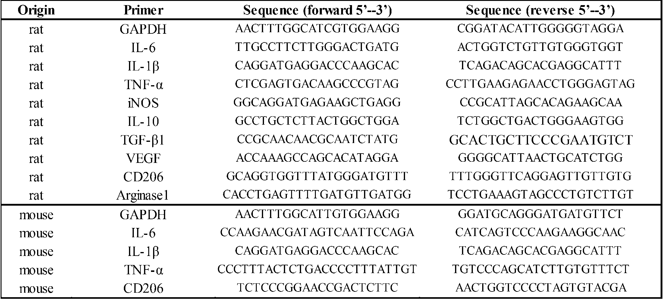
200~230グラムの重量の成体雌Sprague-Dawleyラットを用いた。ラットは、ケタミン(60~90mg/kg)およびキシラジン(100~150mg/kg)の腹腔内注射により麻酔した。Th9腰椎椎弓切除後、硬膜を露出させ、市販されている脊髄損傷装置(Infinite Horizon Impactor,Precision Systems & Instrumentation)によって200kdynの外傷力を付与した。脊髄挫傷後すぐに、Th12部分椎弓切除術を行い、髄腔内にSMP-200モデル(プライムテック株式会社):iPRECIOと接続した薄いシリコンチューブを手術用顕微鏡下で挿入した。管はそれを固定する棘突起に縫合して配置し、ポンプは動物の腋窩の皮膚の下に置かれた。術後、膀胱は1日2回手動腹圧によって圧縮して排尿させた。術後翌日に完全な麻痺(BBBスコア=0)を示す動物を評価に用いた。後肢を動かすことができた動物、即死した動物は評価から除外した。 (4) Rat Contusion Model and Surgical Procedure Adult female Sprague-Dawley rats weighing 200-230 grams were used. Rats were anesthetized by intraperitoneal injection of ketamine (60-90 mg / kg) and xylazine (100-150 mg / kg). After Th9 lumbar laminectomy, the dura mater was exposed and 200 kdyn of traumatic force was applied by a commercially available spinal cord injury device (Infinite Horizon Impactor, Precision Systems & Instrumentation). Immediately after spinal cord contusion, Th12 partial laminectomy was performed, and a thin silicone tube connected to SMP-200 model (Primetech Inc.): iPRECIO was inserted into the medullary cavity under a surgical microscope. The tube was sutured into the spinous process that secured it, and the pump was placed under the skin of the animal's axilla. After surgery, the bladder was compressed by manual abdominal pressure twice daily to urinate. Animals showing complete paralysis (BBB score = 0) the next day after surgery were used for evaluation. Animals that were able to move their hind limbs and animals that died immediately were excluded from the evaluation.
後肢神経行動試験はBBBの運動評価尺度を用いて行った。22点(0~21)BBBスケールは、関節の動き、ステッピング能力、調整、体幹の安定性を含む後肢運動のリカバリを評価するために使用した。21のスコアは、無傷のラットにおける損なわれていない運動を意味している。動物の治療内容に関して盲検化された二人の審査官が評価を行った。各セッションの継続時間は、ラットあたり4分であった。スコアは各時点において、Tukeyの多重比較検定による反復測定分散分析により解析した。 (5) BBB Open Field Movement Score The hindlimb neurobehavioral test was performed using the BBB movement rating scale. A 22-point (0-21) BBB scale was used to assess hindlimb recovery, including joint movement, stepping ability, coordination, and trunk stability. A score of 21 means unimpaired movement in intact rats. Two examiners, blinded to the treatment content of the animals, evaluated. The duration of each session was 4 minutes per rat. Scores were analyzed at each time point by repeated measures analysis of variance with Tukey's multiple comparison test.
処理された脊髄の組織学的検査のために、動物を麻酔し、挫傷後72時間後及び8週間後に、経心臓的に4%PFAの0.1MPBS溶液で灌流した。脊髄をOCTコンパウンド(サクラファイン)に埋め込んで、区画されたクライオスタット(ライカ)に20μmで矢状又は横平面で切片とした。組織切片とミクログリアは、0.1%(v/v)TritonX-100のPBS溶液で5分間浸透処理した。ヤギ血清10%(v/v)で30分間ブロッキング後、一次抗体:5-HT(ウサギIgG、1:500、Sigma-Aldrich)、GFAP(マウスIgG、1:500、ミリポア)、Iba1(ヤギIgG、1:500、アブカム)、CD206(ウサギIgG、1:1000、アブカム)、IL-10(マウスIgG、1:250、アブカム)とインキュベートした。二次抗体は、抗マウスIgG-のAlexa Fluor 488、抗ヤギIgG-Alexa Fluor 546及び抗ウサギIgG-のAlexa Fluor 647とした。DAPI(Sigma-Aldrich)で対比染色後、組織像は、ユニバーサル蛍光顕微鏡(BZ9000、キーエンス)で観察した。 (6) Immunohistochemical analysis For histological examination of the treated spinal cord, animals were anesthetized and perfused with 0.1% PBS in 4% PFA transcardially 72 hours and 8 weeks after contusion. did. The spinal cord was embedded in OCT compound (Sakura Fine) and sectioned into a sectioned cryostat (Leica) at 20 μm in a sagittal or horizontal plane. Tissue sections and microglia were permeabilized with 0.1% (v / v) Triton X-100 in PBS for 5 minutes. After blocking with
THP-1細胞の溶解物を、ED-Siglec-9またはCCR2に対する抗体を用いて免疫沈降させ、沈殿物を抗CCR2抗体またはMAH-レクチンでイムノブロットした。レクチンブロットは、THP-1(理研セルバンク、日本)溶解物(溶解緩衝液:1%トリトンX、150mMのNaCl、20mMのトリス-HCl、2mMの塩化カルシウム)から抗CCR2抗体(ウサギIgGと、1:50、Abcam)で免疫沈降させたCCR2タンパク質をSDS-PAGEで分離し、イモビロン-PのPVDF膜上にエレクトロブロットした。ブロッティング後、その膜を、MAL緩衝液(10mMのHEPES、pH7.5、150mMのNaCl、0.2%BSA、0.2%Tween-20)で、12時間、4℃でブロックした。その後、MAL緩衝液中、12時間、4℃で、5mg/mlのビオチン化MAL(Vector Laboratories社)でプローブした。MALは、アビジン-HRP(Vector Laboratories社)およびECL(GE Healthcare)を用いて検出し、LAS-4000ミニルミノイメージアナライザー(GE Healthcare社)により分析した。 (7) Immunoprecipitation, lectin blot and Western blot THP-1 cell lysates were immunoprecipitated using antibodies against ED-Siglec-9 or CCR2, and the precipitates were immunoblotted with anti-CCR2 antibody or MAH-lectin . Lectin blots were prepared from THP-1 (RIKEN Cell Bank, Japan) lysate (lysis buffer: 1% Triton X, 150 mM NaCl, 20 mM Tris-HCl, 2 mM calcium chloride) to anti-CCR2 antibody (rabbit IgG and 1 : 50, Abcam), CCR2 proteins were separated by SDS-PAGE and electroblotted onto Immobilon-P PVDF membrane. After blotting, the membrane was blocked with MAL buffer (10 mM HEPES, pH 7.5, 150 mM NaCl, 0.2% BSA, 0.2% Tween-20) for 12 hours at 4 ° C. Thereafter, the probe was probed with 5 mg / ml biotinylated MAL (Vector Laboratories) at 4 ° C. for 12 hours in MAL buffer. MAL was detected using avidin-HRP (Vector Laboratories) and ECL (GE Healthcare) and analyzed by LAS-4000 Minilumino Image Analyzer (GE Healthcare).
Turkeyの事後検定(SPSS19.0)による反復測定分散分析を用いた。P値が0.05未満を有意とした。 (8) Statistics Repeated analysis of variance with Turkey's post-test (SPSS 19.0) was used. A P value of less than 0.05 was considered significant.
(MCP-1、ED-Siglec-9及びCSPGの組織修復型ミクログリアの誘導に対する相乗効果)
マウスミクログリアについての結果を図6に示す。図6のa~dに示すように、CSPG上においてMCP-1(50ng/ml)及びED-Siglec-9(50ng/ml)で処理したミクログリアは、有意にCD-206のタンパク質レベルでの発現及び遺伝子レベルでの発現を増大させるとともに、IL-10の産生を増大させた。また、免疫染色により、CSPG上においてMCP-1(50ng/ml)及びED-Siglec-9(50ng/ml)で処理したミクログリアに関し、組織修復型ミクログリアの発現をCD206で確認することができた。また、図6のe及びfに示すように、MCP-1/ED-Siglec-9/CSPGの濃度に依存して組織修復型ミクログリアのマーカーであるCD206及び抗炎症性サイトカインIL-10の産生が増大した。 (result)
(Synergistic effect of MCP-1, ED-Siglec-9 and CSPG on induction of tissue repair type microglia)
The results for mouse microglia are shown in FIG. As shown in FIGS. 6a to 6d, microglia treated with MCP-1 (50 ng / ml) and ED-Siglec-9 (50 ng / ml) on CSPG significantly expressed at the protein level of CD-206. And increased expression at the gene level and increased production of IL-10. Moreover, regarding the microglia treated with MCP-1 (50 ng / ml) and ED-Siglec-9 (50 ng / ml) on CSPG, the expression of tissue repair type microglia could be confirmed by CD206 by immunostaining. Further, as shown in e and f of FIG. 6, production of CD206, which is a marker of tissue repair type microglia, and anti-inflammatory cytokine IL-10 depends on the concentration of MCP-1 / ED-Siglec-9 / CSPG. Increased.
MCP-1及びED-Siglec-9を各濃度1μg/mlでSCI部位にiPRECIO注入ポンプによって液3μl/hを送達した。シリコンチューブはくも膜下腔に挿入した。脊髄損傷部位におけるサイトカインと細胞表面マーカーの遺伝子発現を、定量的RT-PCR分析により評価した。総RNAは、SCIから72時間後に病変部位から採取した。結果を図8に示す。Y軸は、SHAMモデル(偽手術モデル)での数値に対する比率を表す。 (Conversion of inflammatory spinal cord injury to anti-inflammatory state / tissue regeneration state by intrathecal administration of MCP-1 and ED-Siglec-9)
MCP-1 and ED-Siglec-9 were delivered at a concentration of 1 μg / ml to the SCI site with 3 μl / h of solution by an iPRECIO infusion pump. A silicone tube was inserted into the subarachnoid space. Gene expression of cytokines and cell surface markers at the site of spinal cord injury was assessed by quantitative RT-PCR analysis. Total RNA was collected from the lesion site 72 hours after SCI. The results are shown in FIG. The Y axis represents the ratio to the numerical value in the SHAM model (sham surgery model).
脊髄損傷から72時間後の損傷部位を取り囲むミクログリア/マクロファージの典型的画像と定量結果を図9に示す。図9の上段に示すように、コントロール群では、Iba1+ミクログリアが観察されたが、CD206+細胞が全く観察されなかった。これに対して、図9の中段に示すように、MCP-1/ED-Siglec-9処理ラットにおいては、脊髄損傷から72時間後、損傷部位に多数のIba+細胞が移動し、これらIba1+細胞のほとんどは、CD206を共発現していた。また、図9の下段に示すように、コントロールラットでは、損傷部位におけるCD206+細胞は、IL-10も同時に発現することは観察されなかったのに対し、MCP-1/ED-Siglec-9処理された損傷部位におけるCD206+細胞は、IL-10も同時に発現していた。図中のデータは、平均±SDを表す(*P<0.05、**P<0.01、PBS処理に対するMCP-1/ED-Siglec-9処理した脊髄損傷モデル)。 (Promotion of differentiation of microglia / macrophages into tissue repair type after spinal cord injury by MCP-1 / ED-Siglec-9)
FIG. 9 shows a typical image and quantitative results of microglia / macrophages surrounding the injury site 72 hours after spinal cord injury. As shown in the upper part of FIG. 9, in the control group, Iba1 + microglia was observed, but no CD206 + cells were observed. In contrast, as shown in the middle part of FIG. 9, in the MCP-1 / ED-Siglec-9 treated rats, a large number of Iba + cells migrated to the damaged site 72 hours after the spinal cord injury, and these Iba1 + cells Most co-expressed CD206. In addition, as shown in the lower part of FIG. 9, in control rats, CD206 + cells at the injury site were not observed to express IL-10 simultaneously, whereas they were treated with MCP-1 / ED-Siglec-9. CD206 + cells at the damaged site also expressed IL-10 at the same time. Data in the figure represent mean ± SD (* P <0.05, ** P <0.01, MCP-1 / ED-Siglec-9 treated spinal cord injury model versus PBS treatment).
脊髄挫傷後の後肢の機能回復の時間経過を図10に示す。MCP-1/ED-Siglec-9投与ラットは、コントロールと異なり早期から明らかな機能回復傾向を呈した。図中のデータは、平均±SDを表す。 (Therapeutic benefits of MCP-1 / ED-Siglec-9 in spinal cord injury)
The time course of functional recovery of the hind limb after spinal cord contusion is shown in FIG. Rats treated with MCP-1 / ED-Siglec-9, unlike the control, showed a clear tendency toward functional recovery from an early stage. Data in the figure represent mean ± SD.
免疫沈降、レクチンブロット及びウェスタンブロットの結果を図11に示す。ED-Siglec-9は、物理的に、THP-1細胞溶解液中のシアル化CCR2と相互作用し、CSPG処理は、ミクログリアのCCR2発現を増加させ、ED-Siglec-9は物理的にミクログリアのCCR2と相互作用した。 (Interaction between ED-Siglec-9 and MCR-1 receptor CCR2)
The results of immunoprecipitation, lectin blot and western blot are shown in FIG. ED-Siglec-9 physically interacts with sialylated CCR2 in THP-1 cell lysate, CSPG treatment increases CCR2 expression in microglia, and ED-Siglec-9 physically interacts with microglia. It interacted with CCR2.
本実施例では、MCP-1とED-Siglec-9の投与によって急性肺傷害で生じる炎症および線維化が抑制されることを見出した。 (Application to acute lung injury (interstitial pneumonia))
In this example, it was found that administration of MCP-1 and ED-Siglec-9 suppresses inflammation and fibrosis caused by acute lung injury.
肺胞上皮細胞に障害をもたらすBleomycin(BLM)を3mg/mlの割合でPBSに溶解しBLMのPBS溶液を調製した。この溶液をC57Bl/6Jマウス(7~9週齢)に6U/kgの量気管内投与を行い、24時間後にベロクロラ音を聴取して肺傷害が起こっていることを確認した。 (Acute lung injury model mouse)
Blemycin (BLM) that causes damage to alveolar epithelial cells was dissolved in PBS at a rate of 3 mg / ml to prepare a PBS solution of BLM. This solution was intratracheally administered to C57B1 / 6J mice (7 to 9 weeks old) in an amount of 6 U / kg, and after 24 hours, velocura sound was observed to confirm that lung injury had occurred.
図12に示すように、PBS投与群では、BLM溶液(6U/kg)が気管内投与されて肺傷害が引き起こされたC57Bl/6Jマウスは、9日間の体重率は70%(n=10)に低下し、14日間の生存率は約40%(n=10)に低下することを確認した。 (Evaluation of body weight rate for 9 days and survival rate for 14 days)
As shown in FIG. 12, in the PBS-administered group, C57B1 / 6J mice in which BLM solution (6 U / kg) was administered intratracheally and caused lung injury had a body weight ratio of 70% (n = 10) for 9 days. It was confirmed that the survival rate for 14 days was reduced to about 40% (n = 10).
肺傷害患者の肺では線維化の進行と、肺胞構造の破壊を認める。BLMにより傷害されたモデルマウスの肺も同様の構造変化を示すため、その構造変化と膠原線維の増殖をHE染色およびマッソントリクローム(MT)染色にて評価した。結果を図13に示す。 (Pathological analysis in lung injury model)
Progression of fibrosis and destruction of alveolar structures are observed in lungs of lung injury patients. Since the lungs of model mice injured by BLM also show similar structural changes, the structural changes and collagen fiber proliferation were evaluated by HE staining and Masson trichrome (MT) staining. The results are shown in FIG.
本実施例では、MCP-1とED-Siglec-9の投与によって肝硬変、慢性肝炎が寛解することを見いだした。 (Application to cirrhosis (chronic hepatitis))
In this example, it was found that cirrhosis and chronic hepatitis were ameliorated by administration of MCP-1 and ED-Siglec-9.
四塩化炭素(CCl4)を1.0ml/kgの割合でオリーブオイルに溶解し肝障害を誘発する薬剤を作製した。この溶液をC57BL6 mice(20~25g)に、1週間に2回、4週連続腹腔内投与し、肝硬変モデルマウスを作製した。このモデルマウスに対して、リコンビナントタンパクED-Siglec-9及びMCP-1をPBSに1μg/mlの濃度で溶解し、混合溶液を調製した。この混合溶液を最終のCCl4溶液を投与(CCl4投与開始から1ヶ月)してから24時間後に、500μlを1回静脈内投与し、病態改善を検証した。 (Production of cirrhosis model mice)
Carbon tetrachloride (CCl 4 ) was dissolved in olive oil at a rate of 1.0 ml / kg to prepare a drug that induces liver damage. This solution was intraperitoneally administered to C57BL6 mice (20-25 g) twice a week for 4 weeks continuously to produce cirrhosis model mice. For this model mouse, recombinant proteins ED-Siglec-9 and MCP-1 were dissolved in PBS at a concentration of 1 μg / ml to prepare a mixed solution. 24 hours after the final CCl 4 solution was administered to this mixed solution (one month from the start of CCl 4 administration), 500 μl was intravenously administered once to verify improvement of the disease state.
肝硬変、慢性肝炎患者の肝臓では、肝細胞死、炎症性細胞浸潤及び広範に不可逆性の線維性組織の増殖が認められる。ED-Siglec-9及びMCP-1投与群と非投与(PBS投与)群について組織解析(ED-Siglec-9及びMCP-1投与から3日後)を行った。HE染色結果及びシリウスレッド染色(赤染色:コラーゲンIの染色)結果をそれぞれ図14及び図15に示す。 (Pathological analysis in cirrhosis model)
In livers of cirrhotic and chronic hepatitis patients, hepatocyte death, inflammatory cell infiltration and extensively irreversible fibrous tissue proliferation are observed. Histological analysis (3 days after administration of ED-Siglec-9 and MCP-1) was performed on the ED-Siglec-9 and MCP-1 administration group and the non-administration (PBS administration) group. The results of HE staining and Sirius red staining (red staining: collagen I staining) are shown in FIGS. 14 and 15, respectively.
ED-Siglec-9/MCP-1投与後3日の肝臓組織からRNAを採取し、炎症性サイトカインなどの遺伝子発現を定量的PCR法にて解析した。結果を図16及び図17に示す。 (Genetic analysis in cirrhosis and chronic hepatitis models)
RNA was collected from
ED-Siglec-9及びMCP-1投与群と非投与(PBS投与)群から採取した肝臓組織に関しα-SMAの染色を行った。結果を図18に示す。 (Staining of α-SMA, a marker of activated hepatic stellate cells)
Α-SMA staining was performed on liver tissues collected from the ED-Siglec-9 and MCP-1 administration group and the non-administration (PBS administration) group. The results are shown in FIG.
(コラーゲン誘発関節炎モデルマウスの作製)
コラーゲン誘発関節炎マウスは、関節リウマチの主要な動物モデルとして用いられている。DBA/1Jマウスの尾底部に、異種動物である牛由来の2型コラーゲンを皮下注射、更に21日後に再注射することにより、コラーゲン誘発性関節炎を発症させた。関節炎の重症度は関節炎スコアを用いて評価した。図19に示すスコアリングシステムを用いて、各群の平均関節炎スコアを算出した。また各マウスで関節炎スコア1以上を、関節炎発症と定義した。 (Analysis of therapeutic utility using collagen-induced arthritis model mice)
(Production of collagen-induced arthritis model mice)
Collagen-induced arthritis mice have been used as the main animal model for rheumatoid arthritis. Collagen-induced arthritis was developed by subcutaneously injecting a
まず、最適なED-Siglec-9投与量を決定するために、コントロール群(生食)とED-Siglec-9群(0.1μg、1μg、10μg/マウス)の4群(各n=7)で検討を行った。2回目コラーゲン投与の2日後、頚静脈より生食または各濃度のED-Siglec-9を投与し、その7日後の関節炎スコアを評価した。結果を図20に示す。 (1) Analysis of arthritis inhibitory effect of ED-Siglec-9 administration in collagen-induced arthritis (CIA) mice First, in order to determine the optimal ED-Siglec-9 dose, control group (saline) and ED- The study was conducted in 4 groups (each n = 7) of the Siglec-9 group (0.1 μg, 1 μg, 10 μg / mouse). Two days after the second collagen administration, saline or various concentrations of ED-Siglec-9 were administered from the jugular vein, and the arthritis score after 7 days was evaluated. The results are shown in FIG.
ED-Siglec-9のin vivo(CIAマウス)における関節炎抑制効果の作用機序を、in vitroで検討した。関節リウマチでは、活性化された免疫細胞(マクロファージなど)により分泌される炎症性サイトカイン(TNF-α等)が滑膜細胞を刺激し、活性化された滑膜細胞が蛋白分解酵素(MMP-3等)を分泌して最終的に骨軟骨破壊を引き起こす。ED-Siglec-9の作用点が免疫系にあるのか、それとも炎症の主座である滑膜にあるのかを探るため、マクロファージ(自己免疫疾患における中心的免疫細胞の一つ)と、関節炎の主座である滑膜細胞を用いて実験を行った。 (Changes in gene expression by ED-Siglec-9 in mouse macrophages and human rheumatoid arthritis-derived synovial fibroblasts)
The mechanism of action of ED-Siglec-9 in suppressing arthritis in vivo (CIA mice) was examined in vitro. In rheumatoid arthritis, inflammatory cytokines (such as TNF-α) secreted by activated immune cells (such as macrophages) stimulate synovial cells, and activated synovial cells act as proteases (MMP-3). Etc.) and eventually cause osteochondral destruction. To investigate whether ED-Siglec-9 acts in the immune system or in the synovium, which is the main site of inflammation, macrophages (one of the central immune cells in autoimmune diseases) Experiments were performed using synovial cells as the locus.
全身および局所麻酔下にて7週齢SDラットの頭頂部皮膚を骨膜とともに剥離し、直径5mmの骨採取用トレフィンバーにて注水下で頭蓋骨を削除し、骨欠損を形成した。このとき骨膜は温存した。ED-Siglec-9及びMCP-1各100ng)をPBSに溶解し、アテロコラーゲンをスキャフォールドとして骨欠損部に留置し、皮膚を復位して縫合、閉創した。術後6週で、μCT、組織学的評価(H-E染色)を行った。結果を図23に示す。 (Rat skull defect model: morphological analysis)
Under the general and local anesthesia, the parietal skin of 7-week-old SD rats was peeled off along with the periosteum, and the skull was removed under water injection with a 5 mm diameter bone-collecting trephine bar to form a bone defect. At this time, the periosteum was preserved. ED-Siglec-9 and MCP-1 (100 ng each) were dissolved in PBS, and atelocollagen was placed as a scaffold on the bone defect, and the skin was restored and sutured and closed. At 6 weeks after surgery, μCT and histological evaluation (HE staining) were performed. The results are shown in FIG.
Claims (16)
- 単球走化性促進因子-1(MCP-1)活性を有する第1の成分、
シアル酸結合イムノグロブリン様レクチン-9(Siglec-9)の細胞外ドメイン活性を有する第2の成分、及び
コンドロイチン硫酸及びコンドロイチン硫酸プロテオグリカンの少なくとも一種である第3の成分からなる群から選択される少なくとも1種を含む、組織修復剤。 A first component having monocyte chemotaxis promoting factor-1 (MCP-1) activity;
A second component having extracellular domain activity of sialic acid-binding immunoglobulin-like lectin-9 (Siglec-9), and at least selected from the group consisting of chondroitin sulfate and a third component that is at least one of chondroitin sulfate proteoglycans A tissue repair agent comprising one species. - 少なくとも前記第1の成分及び前記第2の成分のいずれか又は双方を含む、請求項1に記載の組織修復剤。 The tissue repair agent according to claim 1, comprising at least one or both of the first component and the second component.
- 前記第1の成分と前記第2の成分とを含む、請求項1又は2に記載の組織修復剤。 The tissue repair agent according to claim 1 or 2, comprising the first component and the second component.
- 前記第1の成分は、配列番号2で表されるアミノ酸配列と60%以上の同一性を有するアミノ酸配列を有する、請求項1~3のいずれかに記載の組織修復剤。 The tissue repair agent according to any one of claims 1 to 3, wherein the first component has an amino acid sequence having 60% or more identity with the amino acid sequence represented by SEQ ID NO: 2.
- 前記第2の成分は、配列番号4で表されるアミノ酸配列と60%以上の同一性を有するアミノ酸配列を有する、請求項1~4のいずれかに記載の組織修復剤。 The tissue repair agent according to any one of claims 1 to 4, wherein the second component has an amino acid sequence having 60% or more identity with the amino acid sequence represented by SEQ ID NO: 4.
- 前記第3の成分を含む、請求項1~5のいずれかに記載の組織修復剤。 The tissue repair agent according to any one of claims 1 to 5, comprising the third component.
- 前記第1の成分、前記第2の成分及び第3の成分からなる群から選択される1種以上の成分を、以下の(a)又は(b)に記載の活性を呈するのに有効量含有する、請求項1~6のいずれかに記載の組織修復剤。
(a)炎症組織における組織修復型マクロファージ及び/又はミクログリアの誘導活性
(b)抗炎症性サイトカインの産生促進活性 One or more components selected from the group consisting of the first component, the second component, and the third component are contained in an effective amount to exhibit the activity described in (a) or (b) below. The tissue repair agent according to any one of claims 1 to 6.
(A) Induction activity of tissue repair type macrophages and / or microglia in inflamed tissues (b) Production promoting activity of anti-inflammatory cytokines - 請求項1~7のいずれかに記載の組織修復剤を有効成分とする、抗炎症剤。 An anti-inflammatory agent comprising the tissue repair agent according to any one of claims 1 to 7 as an active ingredient.
- 請求項1~7のいずれかに記載の組織修復剤を有効成分とする、脊髄損傷、脳梗塞、新生児低酸素脳症、アルツハイマー症、多発性硬化症、筋萎縮性側索硬化症及びパーキンソン病からなる群から選択される中枢神経系疾患の予防又は治療剤。 From spinal cord injury, cerebral infarction, neonatal hypoxic encephalopathy, Alzheimer's disease, multiple sclerosis, amyotrophic lateral sclerosis and Parkinson's disease comprising the tissue repair agent according to any one of claims 1 to 7 as an active ingredient A preventive or therapeutic agent for central nervous system diseases selected from the group consisting of:
- 請求項1~7のいずれかに記載の組織修復剤を有効成分とする、劇症肝炎、急性肝炎、慢性肝炎、急性および慢性間質性肺炎、I型およびII型糖尿病、シェーグレン病、ドライアイ、関節リウマチ、全身性エリテマトーデス、皮膚創傷治癒、心筋梗塞、骨髄移植に伴う免疫拒絶からなる群から選択される非中枢神経系疾患の予防又は治療剤。 Fulminant hepatitis, acute hepatitis, chronic hepatitis, acute and chronic interstitial pneumonia, type I and type II diabetes, Sjogren's disease, dry eye, comprising the tissue repair agent according to any one of claims 1 to 7 as an active ingredient A preventive or therapeutic agent for non-central nervous system diseases selected from the group consisting of rheumatoid arthritis, systemic lupus erythematosus, skin wound healing, myocardial infarction, and immune rejection associated with bone marrow transplantation.
- 単球走化性促進因子-1(MCP-1)活性を有するタンパク質である第1の成分、
シアル酸結合イムノグロブリン様レクチン-9(Siglec-9)の細胞外ドメイン活性を有するタンパク質である第2の成分、及び
コンドロイチン硫酸及びコンドロイチン硫酸プロテオグリカンの少なくとも一種である第3の成分からなる群から選択される少なくとも1種を含む、組織修復型マクロファージ及び/又はミクログリアの誘導剤。 A first component which is a protein having monocyte chemotaxis promoting factor-1 (MCP-1) activity;
Selected from the group consisting of a second component that is a protein having extracellular domain activity of sialic acid-binding immunoglobulin-like lectin-9 (Siglec-9), and a third component that is at least one of chondroitin sulfate and chondroitin sulfate proteoglycan A tissue repair type macrophage and / or microglia inducer comprising at least one selected from the group consisting of: - 単球走化性促進因子-1(MCP-1)活性を有するタンパク質である第1の成分、
シアル酸結合イムノグロブリン様レクチン-9(Siglec-9)の細胞外ドメイン活性を有するタンパク質である第2の成分、及び
コンドロイチン硫酸及びコンドロイチン硫酸プロテオグリカンの少なくとも一種である第3の成分からなる群から選択される少なくとも1種を含む、抗炎症性サイトカインの産生促進剤。 A first component which is a protein having monocyte chemotaxis promoting factor-1 (MCP-1) activity;
Selected from the group consisting of a second component that is a protein having extracellular domain activity of sialic acid-binding immunoglobulin-like lectin-9 (Siglec-9), and a third component that is at least one of chondroitin sulfate and chondroitin sulfate proteoglycan An anti-inflammatory cytokine production promoter comprising at least one selected from the group consisting of: - 生体外で、球走化性促進因子-1(MCP-1)活性を有するタンパク質である第1の成分、
シアル酸結合イムノグロブリン様レクチン-9(Siglec-9)の細胞外ドメイン活性を有するタンパク質である第2の成分、及び
コンドロイチン硫酸及びコンドロイチン硫酸プロテオグリカンの少なくとも一種である第3の成分からなる群から選択される少なくとも1種の存在下でと、ミクログリア又はマクロファージを培養する工程、を備える、組織修復型ミクログリア又はマクロファージの生産方法。 A first component that is a protein having the activity of promoting chemotaxis-1 (MCP-1) in vitro,
Selected from the group consisting of a second component that is a protein having extracellular domain activity of sialic acid-binding immunoglobulin-like lectin-9 (Siglec-9), and a third component that is at least one of chondroitin sulfate and chondroitin sulfate proteoglycan A tissue repair type microglia or macrophage production method comprising the step of culturing microglia or macrophages in the presence of at least one selected from the group consisting of: - 単球走化性促進因子-1(MCP-1)活性を有するタンパク質である第1の成分、
シアル酸結合イムノグロブリン様レクチン-9(Siglec-9)の細胞外ドメイン活性を有するタンパク質である第2の成分、及び
コンドロイチン硫酸及びコンドロイチン硫酸プロテオグリカンの少なくとも一種である第3の成分からなる群から選択される少なくとも1種を含む、ミクログリア/マクロファージ用の試薬キット。 A first component which is a protein having monocyte chemotaxis promoting factor-1 (MCP-1) activity;
Selected from the group consisting of a second component that is a protein having extracellular domain activity of sialic acid-binding immunoglobulin-like lectin-9 (Siglec-9), and a third component that is at least one of chondroitin sulfate and chondroitin sulfate proteoglycan A reagent kit for microglia / macrophage comprising at least one selected from the group consisting of: - 前記第1の成分と前記第2の成分とを含む、請求項14に記載の試薬キット。 The reagent kit according to claim 14, comprising the first component and the second component.
- 単球走化性促進因子-1(MCP-1)活性を有するタンパク質である第1の成分、
シアル酸結合イムノグロブリン様レクチン-9(Siglec-9)の細胞外ドメイン活性を有するタンパク質である第2の成分、及び
コンドロイチン硫酸及びコンドロイチン硫酸プロテオグリカンの少なくとも一種である第3の成分からなる群から選択される少なくとも1種を損傷組織又は炎症組織に送達して組織修復を促進する方法。 A first component which is a protein having monocyte chemotaxis promoting factor-1 (MCP-1) activity;
Selected from the group consisting of a second component that is a protein having extracellular domain activity of sialic acid-binding immunoglobulin-like lectin-9 (Siglec-9), and a third component that is at least one of chondroitin sulfate and chondroitin sulfate proteoglycan To promote tissue repair by delivering at least one of them to a damaged or inflamed tissue.
Priority Applications (5)
| Application Number | Priority Date | Filing Date | Title |
|---|---|---|---|
| JP2014553236A JP6403062B2 (en) | 2012-12-21 | 2013-12-24 | Tissue repair active composition and use thereof |
| EP13864639.3A EP2937088B1 (en) | 2012-12-21 | 2013-12-24 | Composition having tissue repairing activity and utilization thereof |
| US14/654,624 US9962428B2 (en) | 2012-12-21 | 2013-12-24 | Composition having tissue-repairing activity, and use therefor |
| US15/938,839 US10507230B2 (en) | 2012-12-21 | 2018-03-28 | Composition having tissue-repairing activity, and use therefor |
| US16/671,497 US11000572B2 (en) | 2012-12-21 | 2019-11-01 | Composition having tissue-repairing activity, and use therefor |
Applications Claiming Priority (2)
| Application Number | Priority Date | Filing Date | Title |
|---|---|---|---|
| JP2012-280022 | 2012-12-21 | ||
| JP2012280022 | 2012-12-21 |
Related Child Applications (2)
| Application Number | Title | Priority Date | Filing Date |
|---|---|---|---|
| US14/654,624 A-371-Of-International US9962428B2 (en) | 2012-12-21 | 2013-12-24 | Composition having tissue-repairing activity, and use therefor |
| US15/938,839 Division US10507230B2 (en) | 2012-12-21 | 2018-03-28 | Composition having tissue-repairing activity, and use therefor |
Publications (1)
| Publication Number | Publication Date |
|---|---|
| WO2014098249A1 true WO2014098249A1 (en) | 2014-06-26 |
Family
ID=50978561
Family Applications (1)
| Application Number | Title | Priority Date | Filing Date |
|---|---|---|---|
| PCT/JP2013/084523 WO2014098249A1 (en) | 2012-12-21 | 2013-12-24 | Composition having tissue repairing activity and utilization thereof |
Country Status (4)
| Country | Link |
|---|---|
| US (3) | US9962428B2 (en) |
| EP (1) | EP2937088B1 (en) |
| JP (1) | JP6403062B2 (en) |
| WO (1) | WO2014098249A1 (en) |
Cited By (4)
| Publication number | Priority date | Publication date | Assignee | Title |
|---|---|---|---|---|
| JP2017527611A (en) * | 2014-08-15 | 2017-09-21 | ピクサーバイオ コーポレーション | Compositions for inhibiting inflammation in patients with spinal cord injury and methods of using the same |
| JP2018016596A (en) * | 2016-07-29 | 2018-02-01 | 小林製薬株式会社 | α-SMA PRODUCTION INHIBITOR |
| WO2022009912A1 (en) * | 2020-07-08 | 2022-01-13 | 株式会社U-Factor | Composition containing culture supernatant of dental pulp stem cells and for improving blood oxygen saturation or preventing reduction of blood oxygen saturation |
| EP3802582A4 (en) * | 2018-06-07 | 2022-06-15 | Palleon Pharmaceuticals Inc. | Multimeric proteins for detecting a carbohydrate and/or treating a siglec-mediated disorder |
Families Citing this family (4)
| Publication number | Priority date | Publication date | Assignee | Title |
|---|---|---|---|---|
| EP3391899B1 (en) * | 2011-12-14 | 2020-07-15 | The Board of Trustees of the University of Arkansas | Delivery of therapeutic agents by a collagen binding protein |
| CA3003458A1 (en) | 2015-10-29 | 2017-05-04 | Alector Llc | Anti-siglec-9 antibodies and methods of use thereof |
| JP7397440B2 (en) | 2017-02-10 | 2023-12-13 | ザ ボード オブ トラスティーズ オブ ザ ユニバーシティ オブ アーカンソー | Collagen-binding drug composition and method of use thereof |
| WO2019060696A1 (en) | 2017-09-25 | 2019-03-28 | Surface Pharmaceuticals, Inc. | Ophthalmic pharmaceutical compositions and methods for treating ocular surface disease |
Citations (1)
| Publication number | Priority date | Publication date | Assignee | Title |
|---|---|---|---|---|
| WO2011118795A1 (en) | 2010-03-26 | 2011-09-29 | 国立大学法人名古屋大学 | Composition for treatment of damaged part |
Family Cites Families (12)
| Publication number | Priority date | Publication date | Assignee | Title |
|---|---|---|---|---|
| SG48733A1 (en) | 1992-10-01 | 1998-05-18 | Schering Corp | Use of il-10 to prevent or treat insulin-dependent diabetes mellitus |
| US6989435B2 (en) * | 1997-09-11 | 2006-01-24 | Cambridge University Technical Services Ltd. | Compounds and methods to inhibit or augment an inflammatory response |
| US6685931B1 (en) | 1998-12-22 | 2004-02-03 | Schering Corporation | Treatment of hepatitis C virus infections with interleukin-10 |
| JP2001352977A (en) * | 2000-06-12 | 2001-12-25 | Kirin Brewery Co Ltd | ANTIBODY TO DENDRITIC CELL(DC) MEMBRANE MOLECULE Siglec-9, METHOD FOR DC DETECTION USING THE SAME AND METHOD FOR DC ISOLATION USING THE SAME |
| WO2002008257A2 (en) * | 2000-07-21 | 2002-01-31 | Bristol-Myers Squibb Company | Siglec (sialic acid-binding ig-related lectin) polypeptides and uses thereof |
| AU2002319207B2 (en) * | 2001-06-19 | 2007-03-01 | Reinhard Brossmer | Sialic acid derivatives for use as siglec inhibitors |
| US20070065415A1 (en) * | 2005-09-16 | 2007-03-22 | Kleinsek Donald A | Compositions and methods for the augmentation and repair of defects in tissue |
| WO2007056525A2 (en) * | 2005-11-10 | 2007-05-18 | James Paulson | High affinity siglec ligands |
| WO2007120815A2 (en) * | 2006-04-12 | 2007-10-25 | The Regents Of The University Of California | Methods for treating lymphocyte-associated disorders by modulation of siglec activity |
| WO2007148317A1 (en) * | 2006-06-21 | 2007-12-27 | Compugen Ltd. | Mcp-1 splice variants and methods of using same |
| JP2008094746A (en) * | 2006-10-11 | 2008-04-24 | Nihon Pharmaceutical Co Ltd | Therapeutic or prophylactic agent for inflammatory disease |
| US8163281B2 (en) * | 2009-03-04 | 2012-04-24 | The Regents Of The University Of Michigan | Treatment of drug-related side effect and tissue damage by targeting the CD24-HMGB1-Siglec10 axis |
-
2013
- 2013-12-24 US US14/654,624 patent/US9962428B2/en active Active
- 2013-12-24 EP EP13864639.3A patent/EP2937088B1/en active Active
- 2013-12-24 WO PCT/JP2013/084523 patent/WO2014098249A1/en active Application Filing
- 2013-12-24 JP JP2014553236A patent/JP6403062B2/en active Active
-
2018
- 2018-03-28 US US15/938,839 patent/US10507230B2/en active Active
-
2019
- 2019-11-01 US US16/671,497 patent/US11000572B2/en active Active
Patent Citations (1)
| Publication number | Priority date | Publication date | Assignee | Title |
|---|---|---|---|---|
| WO2011118795A1 (en) | 2010-03-26 | 2011-09-29 | 国立大学法人名古屋大学 | Composition for treatment of damaged part |
Non-Patent Citations (11)
| Title |
|---|
| "Introduction to the Glycoscience Dai 5 Kai -Saibogai Matrix 2- Kenko na Karada o Sasaeru Urakata - Chondroitin Ryusan", 2007, XP055267080, Retrieved from the Internet <URL:http://www.glycoforum.gr.jp/glyco/05.html> * |
| ALTSCHUL SF; GISH W; MILLER W; MYERS EW; LIPMAN DJ., J. MOL. BID., vol. 215, 1990, pages 403 - 410 |
| ALTSCHUL SF; MADDEN TL; SCHAFFER AA; ZHANG J; MILLER W; LIPMAN DJ., NUCLEIC ACIDS RES., vol. 25, 1997, pages 3389 - 3402 |
| ANDO Y ET AL.: "Stem cell -conditioned medium accelerates distraction osteogenesis through multiple regenerative mechanisms", BONE, vol. 61, 2014, pages 82 - 90, XP028667601 * |
| DAVID, S.; KRONER, A., NATURE REVIEWS NEUROSCIENCE, vol. 12, no. 7, pages 388 - 399 |
| ICHIRO TSUJI: "Physical Properties and Tissue Response of Canal Sealer Pastes Containing Tetracalcium Phosphate-Chondroitin Sulfate A", NIHON SHIKA HOZONGAKU ZASSHI, vol. 36, no. 3, 1993, pages 31 - 32, XP008179985 * |
| MUZZARELLI R A.A ET AL.: "Chitosan, hyaluronan and chondroitin sulfate in tissue engineering for cartilage regeneration", A REVIEW, CARBOHYDRATE POLYMERS, vol. 89, no. 3, July 2012 (2012-07-01), pages 723 - 739, XP055266574 * |
| POPOVICH, P.G.; LONGBRAKE, E.E., NATURE REVIEWS NEUROSCIENCE, vol. 9, no. 6, pages 481 - 493 |
| SZYMCZAK WA ET AL.: "The CCL7-CCL2-CCR2 axis regulates IL -4 production in lungs and fungal immunity", J IMMUNOL, vol. 183, no. 3, 2009, pages 1964 - 1974, XP055266565 * |
| TOMOJI ANDO ET AL.: "Jiko Soshiki Shinsei Saisei ni Okeru CCR2 Izongata - Seitainai Kansaibo Shuseki no Yakuwari", REGENERATIVE MEDICINE, vol. 10, 2011, XP008181710 * |
| ZHANG JQ ET AL.: "Siglec-9, a novel sialic acid binding member of the immunoglobulin superfamily expressed broadly on human blood leukocytes", J BIOL CHEM, vol. 275, no. 29, 2000, pages 22121 - 22126, XP002909451 * |
Cited By (5)
| Publication number | Priority date | Publication date | Assignee | Title |
|---|---|---|---|---|
| JP2017527611A (en) * | 2014-08-15 | 2017-09-21 | ピクサーバイオ コーポレーション | Compositions for inhibiting inflammation in patients with spinal cord injury and methods of using the same |
| JP2018016596A (en) * | 2016-07-29 | 2018-02-01 | 小林製薬株式会社 | α-SMA PRODUCTION INHIBITOR |
| EP3802582A4 (en) * | 2018-06-07 | 2022-06-15 | Palleon Pharmaceuticals Inc. | Multimeric proteins for detecting a carbohydrate and/or treating a siglec-mediated disorder |
| WO2022009912A1 (en) * | 2020-07-08 | 2022-01-13 | 株式会社U-Factor | Composition containing culture supernatant of dental pulp stem cells and for improving blood oxygen saturation or preventing reduction of blood oxygen saturation |
| JP2022015343A (en) * | 2020-07-08 | 2022-01-21 | 株式会社U-Factor | Composition for improving blood oxygen saturation or suppressing decrease in blood oxygen saturation comprising culture supernatant of dental pulp stem cells |
Also Published As
| Publication number | Publication date |
|---|---|
| JPWO2014098249A1 (en) | 2017-01-12 |
| US10507230B2 (en) | 2019-12-17 |
| US20150352187A1 (en) | 2015-12-10 |
| EP2937088A1 (en) | 2015-10-28 |
| US20180311313A1 (en) | 2018-11-01 |
| US9962428B2 (en) | 2018-05-08 |
| US11000572B2 (en) | 2021-05-11 |
| JP6403062B2 (en) | 2018-10-10 |
| US20200129591A1 (en) | 2020-04-30 |
| EP2937088A4 (en) | 2016-12-28 |
| EP2937088B1 (en) | 2020-11-18 |
Similar Documents
| Publication | Publication Date | Title |
|---|---|---|
| JP6403062B2 (en) | Tissue repair active composition and use thereof | |
| US9359405B2 (en) | Antagonists of the interleukin-1 receptor | |
| JP6474549B2 (en) | Evaluation index of stem cell culture product and its use | |
| JP5208135B2 (en) | Recombinant leukocyte inhibitory factor and hirugen chimeric protein and drug composition thereof | |
| ES2656353T3 (en) | New HIP / PAP applications or derivatives thereof | |
| MXPA05002617A (en) | Long acting erythropoietins that maintain tissue protective activity of endogenous erythropoietin. | |
| US20120225028A1 (en) | Compositions and methods for mobilization of stem cells | |
| WO2007136157A1 (en) | Compositions and methods for treating motor neuron diseases | |
| US10160791B2 (en) | Protamine in treatment of neuronal injuries | |
| WO2013021196A2 (en) | Oligodendrocyte differentiation | |
| CN112351791A (en) | Glycosaminoglycan inhibitors and promoters | |
| JP5784754B2 (en) | Use of ICAM-1 for prevention or treatment of neurological diseases | |
| WO2005112911A2 (en) | Compositions and methods for treating myelin deficiency disorders | |
| JP2001524944A (en) | How to relieve neuropathic pain | |
| Class et al. | Patent application title: Antagonists of the Interleukin-1 Receptor Inventors: Vladimir Berezin (Copenhagen N, DK) Vladimir Berezin (Copenhagen N, DK) Elisabeth Bock (Charlottenlund, DK) Assignees: SERODUS ASA | |
| CN115515969A (en) | Application of FGF4 and Fgfr1 binding ligand in diabetes | |
| US8207111B2 (en) | Method for treating muscular dystrophy | |
| NZ615710B2 (en) | Antagonists of the interleukin- 1 receptor | |
| Zhang et al. | SOCS3 Attenuates GM-CSF/IFN-r-Mediated Inflammation During Spontaneous Spinal Cord Regeneration | |
| WO2016020426A1 (en) | Compositions for the therapy of mucopolysaccharidoses |
Legal Events
| Date | Code | Title | Description |
|---|---|---|---|
| 121 | Ep: the epo has been informed by wipo that ep was designated in this application |
Ref document number: 13864639 Country of ref document: EP Kind code of ref document: A1 |
|
| DPE1 | Request for preliminary examination filed after expiration of 19th month from priority date (pct application filed from 20040101) | ||
| ENP | Entry into the national phase |
Ref document number: 2014553236 Country of ref document: JP Kind code of ref document: A |
|
| NENP | Non-entry into the national phase |
Ref country code: DE |
|
| WWE | Wipo information: entry into national phase |
Ref document number: 14654624 Country of ref document: US |
|
| WWE | Wipo information: entry into national phase |
Ref document number: 2013864639 Country of ref document: EP |