WO2014027656A1 - 補体C3bによる細菌の菌体表面への沈着および貪食細胞による貪食活性を増加させる方法、並びに細菌感染症の治療方法および治療剤 - Google Patents
補体C3bによる細菌の菌体表面への沈着および貪食細胞による貪食活性を増加させる方法、並びに細菌感染症の治療方法および治療剤 Download PDFInfo
- Publication number
- WO2014027656A1 WO2014027656A1 PCT/JP2013/071863 JP2013071863W WO2014027656A1 WO 2014027656 A1 WO2014027656 A1 WO 2014027656A1 JP 2013071863 W JP2013071863 W JP 2013071863W WO 2014027656 A1 WO2014027656 A1 WO 2014027656A1
- Authority
- WO
- WIPO (PCT)
- Prior art keywords
- antibody
- amino acid
- acid residue
- complement
- activity
- Prior art date
- Legal status (The legal status is an assumption and is not a legal conclusion. Google has not performed a legal analysis and makes no representation as to the accuracy of the status listed.)
- Ceased
Links
Images







Classifications
-
- C—CHEMISTRY; METALLURGY
- C07—ORGANIC CHEMISTRY
- C07K—PEPTIDES
- C07K16/00—Immunoglobulins [IG], e.g. monoclonal or polyclonal antibodies
- C07K16/12—Immunoglobulins [IG], e.g. monoclonal or polyclonal antibodies against material from bacteria
- C07K16/1267—Gram-positive bacteria
- C07K16/1271—Micrococcaceae (F); Staphylococcaceae (F), e.g. Staphylococcus (G)
-
- A—HUMAN NECESSITIES
- A61—MEDICAL OR VETERINARY SCIENCE; HYGIENE
- A61P—SPECIFIC THERAPEUTIC ACTIVITY OF CHEMICAL COMPOUNDS OR MEDICINAL PREPARATIONS
- A61P31/00—Antiinfectives, i.e. antibiotics, antiseptics, chemotherapeutics
- A61P31/04—Antibacterial agents
-
- C—CHEMISTRY; METALLURGY
- C07—ORGANIC CHEMISTRY
- C07K—PEPTIDES
- C07K16/00—Immunoglobulins [IG], e.g. monoclonal or polyclonal antibodies
- C07K16/12—Immunoglobulins [IG], e.g. monoclonal or polyclonal antibodies against material from bacteria
- C07K16/1267—Gram-positive bacteria
- C07K16/1275—Streptococcus (G)
-
- C—CHEMISTRY; METALLURGY
- C07—ORGANIC CHEMISTRY
- C07K—PEPTIDES
- C07K16/00—Immunoglobulins [IG], e.g. monoclonal or polyclonal antibodies
- C07K16/18—Immunoglobulins [IG], e.g. monoclonal or polyclonal antibodies against material from animals or humans
- C07K16/28—Immunoglobulins [IG], e.g. monoclonal or polyclonal antibodies against material from animals or humans against receptors, cell surface antigens or cell surface determinants
- C07K16/30—Immunoglobulins [IG], e.g. monoclonal or polyclonal antibodies against material from animals or humans against receptors, cell surface antigens or cell surface determinants from tumour cells
- C07K16/3076—Immunoglobulins [IG], e.g. monoclonal or polyclonal antibodies against material from animals or humans against receptors, cell surface antigens or cell surface determinants from tumour cells against structure-related tumour-associated moieties
- C07K16/3084—Immunoglobulins [IG], e.g. monoclonal or polyclonal antibodies against material from animals or humans against receptors, cell surface antigens or cell surface determinants from tumour cells against structure-related tumour-associated moieties against tumour-associated gangliosides
-
- C—CHEMISTRY; METALLURGY
- C07—ORGANIC CHEMISTRY
- C07K—PEPTIDES
- C07K2317/00—Immunoglobulins specific features
- C07K2317/30—Immunoglobulins specific features characterized by aspects of specificity or valency
- C07K2317/34—Identification of a linear epitope shorter than 20 amino acid residues or of a conformational epitope defined by amino acid residues
-
- C—CHEMISTRY; METALLURGY
- C07—ORGANIC CHEMISTRY
- C07K—PEPTIDES
- C07K2317/00—Immunoglobulins specific features
- C07K2317/40—Immunoglobulins specific features characterized by post-translational modification
- C07K2317/41—Glycosylation, sialylation, or fucosylation
-
- C—CHEMISTRY; METALLURGY
- C07—ORGANIC CHEMISTRY
- C07K—PEPTIDES
- C07K2317/00—Immunoglobulins specific features
- C07K2317/70—Immunoglobulins specific features characterized by effect upon binding to a cell or to an antigen
- C07K2317/72—Increased effector function due to an Fc-modification
Definitions
- the present invention relates to an antibody that binds to a bacterial cell surface molecule, wherein at least one amino acid residue is substituted with another amino acid residue, and complement-dependent cells than the antibody before substitution of the amino acid residue.
- complement-mediated cytolysis complement mediated lysis
- CML complement mediated lysis
- CDC biological defense reactions
- complement activation cascade Also known in the complement activation cascade are the classical complement pathway that begins with C1q binding to antibodies or the second pathway that begins with complement C3 degradation and the lectin pathway that begins with lectin binding. Has a strong proteoglycan cell membrane and is known to be less susceptible to cell damage due to complement activity.
- phagocytic cells granulocytes, macrophages, dendritic cells, etc.
- opsonized phagocytic activities that phagocytose and kill bacteria bound to C3b or C3d, which are intermediate products of the complement cascade ( It is known to have opsonophagocytosis).
- An antibody is a heterotetramer composed of a heavy chain (hereinafter referred to as H chain) and a light chain (Light chain; hereinafter referred to as L chain), and an Fc region that binds to an Fab and an Fc receptor involved in antigen binding. (Hereinafter sometimes simply referred to as Fc). It is known that antibodies cause CDC, antibody-dependent cellular cytotoxicity (hereinafter referred to as ADCC), and phagocytic activity via Fc. It is known that amino acid residue substitution of antibody Fc can control the activity of antibody CDC and / or ADCC.
- Non-patent Document 1 N-acetylglucosamine
- AlcNAc N-acetylglucosamine
- the present invention relates to the following (1) to (11).
- (1) In an antibody that binds to a bacterial cell surface molecule, at least one amino acid residue is substituted with another amino acid residue, and complement-dependent cytotoxicity is greater than that of the antibody before substitution of the amino acid residue.
- a method for increasing the phagocytic activity of phagocytic cells by depositing complement C3b on the surface of the microbial cell using a modified antibody having increased activity.
- the modified antibody is a modified antibody having increased binding activity to complement C1q as compared to an antibody having an amino acid sequence before substitution of amino acid residues.
- the subclass of the antibody that binds to the cell surface molecule is human IgG1.
- the bacterium is at least one bacterium selected from a gram positive bacterium and a gram negative bacterium.
- the Gram-positive bacterium is selected from Bacillus, Listeria, Staphylococcus, Streptococcus, Enterococcus, Clostridium and Mycobacterium.
- the gram-negative bacterium is at least one gram-negative bacterium selected from Pseudomonas, Escherichia, Salmonella, and Acinetobacter.
- the bacterial cell surface molecule is at least one molecule selected from ganglioside, capsular polysaccharide (capsular polysaccharide (CP)), surface protein [surface protein (SP)] and lipopolysaccharide [lipopolysaccharide (LPS)].
- the modified antibody has an N-linked complex type sugar chain in the Fc region, and among all N-linked complex type sugar chains bound to the Fc region, ⁇ 1-6 is added to N-acetylglucosamine at the sugar chain reducing end.
- the method according to any one of (1) to (8), wherein the proportion of sugar chains to which fucose is not bound is 20% or more.
- An engineered antibody in which at least one amino acid residue is substituted with another amino acid residue in an antibody that binds to a bacterial cell surface molecule, and the CDC is increased as compared with the antibody before substitution of the amino acid residue A therapeutic agent for bacterial infection, characterized in that the growth of the bacterium is reduced by depositing complement C3b on the surface of the cell and increasing phagocytic activity by phagocytic cells.
- An antibody that binds to a bacterial cell surface molecule, wherein at least one amino acid residue is substituted with another amino acid residue, and the modified antibody has an increased CDC than the antibody before the substitution of the amino acid residue A method for treating a bacterial infection, comprising reducing the growth of the bacterium by depositing complement C3b on the surface of the cell and increasing phagocytic activity by phagocytic cells.
- the modification has an increased CDC as compared to the antibody before the substitution of the amino acid residue.
- a method for increasing the deposition of complement C3b on the cell surface using an antibody and in an antibody that binds to a bacterial cell surface molecule, at least one amino acid residue is substituted with another amino acid, and the amino acid
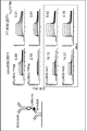
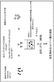
- FIG. 1 shows the binding activity of anti-ganglioside GD3 antibody to Fc ⁇ RIIIA and Fc ⁇ RIIIB.
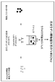
- FIG. 2 shows a method for measuring phagocytic activity in vitro using an anti-ganglioside GD3 antibody.
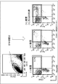
- FIG. 3 shows a histogram of neutrophils incorporating S. aureus in an anti-ganglioside GD3 antibody-dependent manner.
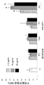
- FIG. 4 shows the ratio (%) of neutrophils that have incorporated Staphylococcus aureus in an anti-ganglioside GD3 antibody-dependent manner relative to the total amount of neutrophils on the vertical axis. The antibody concentration is shown on the horizontal axis.
- FIG. 1 shows the binding activity of anti-ganglioside GD3 antibody to Fc ⁇ RIIIA and Fc ⁇ RIIIB.
- FIG. 2 shows a method for measuring phagocytic activity in vitro using an anti-ganglioside GD3 antibody.
- FIG. 3 shows a hist
- FIG. 5 shows the total amount ( ⁇ 10 6 ) of Staphylococcus aureus taken up by neutrophils depending on the anti-ganglioside GD3 antibody on the vertical axis. The antibody concentration is shown on the horizontal axis.
- FIG. 6 shows the method of anti-ganglioside GD3 antibody complement C3b deposition assay.
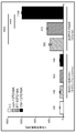
- FIG. 7 shows the deposition effect of complement C3b on anti-ganglioside GD3 antibody S. aureus SA113 (ATCC 35556).
- the vertical axis shows C3b average fluorescence intensity (MFI) (deposition amount), and the horizontal axis shows the antibody used.
- FIG. 8 shows the C3b deposition effect of anti-ganglioside GD3 antibody on Staphylococcus aureus Newman (ATCC 25905).
- the vertical axis shows C3b average fluorescence intensity (MFI) representing the amount of deposited C3b, and the horizontal axis shows the antibody used.
- FIG. 9 shows a method for evaluating opsonophagocytic injury activity by anti-ganglioside GD3 antibody.
- FIG. 10 shows the opsonic phagocytosis injury effect of human neutrophils against Staphylococcus aureus SA113 by anti-ganglioside GD3 antibody.
- the vertical axis indicates colony forming unit concentration (CFU / mL), and the horizontal axis indicates the presence or absence of human polymorphonuclear neutrophils (hereinafter abbreviated as PMN).
- FIG. 11 shows the opsonic phagocytic cytotoxic effect of human neutrophils against Staphylococcus aureus Newman by anti-ganglioside GD3 antibody.
- the vertical axis represents the colony forming unit concentration (CFU / mL), and the horizontal axis represents the presence or absence of PMN and the antibody concentration.
- FIG. 12 shows the method of anti-CP5 antibody complement C3b deposition assay.
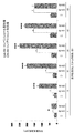
- FIG. 13 shows complement C3b deposition effect of anti-CP5 antibody on S. aureus Lowenstein.
- the vertical axis indicates C3b average fluorescence intensity (MFI) (deposition amount), and the horizontal axis indicates the type of antibody.
- FIG. 14 shows the effect of anti-CP5 antibody deposition of complement C3b in the presence or absence of C1q on Staphylococcus aureus Lowenstein.
- the vertical axis indicates C3b average fluorescence intensity (MFI) (deposition amount), and the horizontal axis indicates the type of antibody.
- FIG. 15 shows the complement C3b deposition effect over time of anti-CP5 antibody against S. aureus Lowenstein.
- the vertical axis indicates C3b average fluorescence intensity (MFI) (deposition amount), and the horizontal axis indicates the type of antibody and the time (minutes) after the addition of the antibody.
- FIG. 16 shows the anti-CP5 antibody-dependent neutrophil uptake effect of S. aureus Lowenstein.
- the vertical axis shows the ratio (%) of neutrophils incorporating S. aureus to the total amount of neutrophils, and the horizontal axis shows antibody concentration.
- FIG. 17 shows the anti-PspA antibody-dependent neutrophil uptake effect of Streptococcus pneumoniae D39.
- the vertical axis shows the ratio (%) of neutrophils incorporating pneumococci to the total amount of neutrophils, and the horizontal axis shows the type of antibody.
- FIG. 18 shows the method of complement C3b deposition assay with anti-PspA antibody.
- FIG. 19 shows the C3b deposition effect on anti-PspA antibodies 140G1 and 140H1-dependent pneumococci BAA-658.
- the vertical axis represents the number of cells, and the horizontal axis represents C3b fluorescence intensity (MFI) (deposition amount).
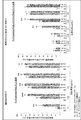
- FIG. 20 shows the results of C3b deposition effect on three strains of pneumococcal PJ-1324, WU2, and BAA-658, which are dependent on anti-PspA antibody 140H1.
- FIG. 21 shows the therapeutic effect of anti-CP5 antibody in a roweed nude (Rowett Nude, hereinafter referred to as RNU) rat model infected with S. aureus MSSA Reynolds (ATCC-25923).
- the vertical axis shows the survival rate (%) of rats in each group, and the horizontal axis shows the number of days after administration of bacteria pre-opsonized with each antibody.
- the present invention relates to an antibody that binds to a bacterial cell surface molecule, wherein at least one amino acid residue is substituted with another amino acid residue, and the CDC is increased as compared with the antibody before substitution of the amino acid residue
- the present invention relates to a method for increasing the phagocytic activity of phagocytic cells by depositing complement C3b on the surface of the microbial cell using an antibody.
- the present invention also relates to an antibody that binds to a bacterial cell surface molecule and uses a modified antibody in which at least one amino acid residue is substituted and the CDC is increased as compared with the antibody before substitution of the amino acid residue. Furthermore, the present invention relates to a method and a therapeutic agent for bacterial infection characterized by decreasing the growth of the bacteria by depositing complement C3b on the surface of the cells and increasing phagocytic activity by phagocytic cells.
- any modified antibody can be used as long as it has an increased CDC as a result of substitution of at least one amino acid residue with another amino acid residue in an antibody that binds to a bacterial cell surface molecule. It may be an antibody.
- the amino acid residue to be substituted is preferably an amino acid residue in the CH2 and CH3 domains (Fragment, crystallizable, hereinafter referred to as Fc region or Fc) of the antibody constant region, and more preferably at least one amino acid contained in the CH2 domain Residue.
- modified antibody used in the present invention examples include an antibody that binds to a bacterial cell surface molecule and includes the amino acid sequence shown in SEQ ID NO: 1 or 2 in the Fc region.
- the modified antibody used in the present invention is an antibody that binds to a bacterial cell surface molecule, wherein at least one amino acid residue is substituted with another amino acid residue, and compared with the antibody before substitution of the amino acid residue.
- Modified antibodies having increased CDC activity and complement C1q binding activity are also included in the present invention.
- the species and subclass of the antibody that binds to the bacterial cell surface molecule are not particularly limited, but are preferably human IgG, more preferably human IgG1.
- the present invention relates to a modified antibody in which at least one amino acid residue is substituted in an antibody that binds to a bacterial cell surface molecule, and the CDC is increased compared to the antibody before substitution of the amino acid residue,
- the modified antibody has an N-linked complex type sugar chain in the Fc region, and of all N-linked complex type sugar chains that bind to the Fc region, ⁇ 1,6-fucose is present in N-acetylglucosamine at the sugar chain reducing end. Examples include a method of increasing the phagocytic activity by phagocytic cells and the deposition of complement C3b on the surface of the cells using a modified antibody in which the proportion of sugar chains not bound is 20% or more.
- the present invention also relates to a modified antibody in which at least one amino acid residue is substituted in an antibody that binds to a bacterial cell surface molecule, and the CDC is increased compared to the antibody before substitution of the amino acid residue.
- the modified antibody has an N-linked complex sugar chain in the Fc region, and among all N-linked complex sugar chains bound to the Fc region, ⁇ 1,6-fucose is added to N-acetylglucosamine at the sugar chain reducing end.
- a modified antibody in which the proportion of sugar chains to which no saccharide is bound is 20% or more, reducing the growth of the bacterium by depositing complement C3b on the surface of the cell and increasing phagocytic activity by phagocytic cells. Examples include a method for treating bacterial infections and a therapeutic agent.
- the modified antibody having 20% or more can increase the phagocytic activity of phagocytic cells as a result of increasing the deposition of complement C3b on the cell surface and increasing the binding activity to the Fc receptor.
- the modified antibody has an N-linked complex type sugar chain in the Fc region, and among all N-linked complex type sugar chains that bind to the Fc region, ⁇ 1,6-fucose is added to N-acetylglucosamine at the sugar chain reducing end.
- the proportion of sugar chains not bound to 20% or more means that the modified antibody has an N-glycoside-bonded complex sugar chain in the Fc region and all N-glycoside-linked complex sugars that bind to the Fc region This is synonymous with the ratio of the sugar chain in which fucose is not bound to N-acetylglucosamine at the sugar chain reducing end of the chain being 20% or more.
- the antibody used in the present invention may be, for example, either a monoclonal antibody or a polyclonal antibody, and preferably a monoclonal antibody that binds to a single epitope.
- the monoclonal antibody may be a monoclonal antibody produced from a hybridoma, or may be any recombinant antibody produced by a gene recombination technique or any monoclonal antibody.
- chimeric antibodies hereinafter also simply referred to as chimeric antibodies
- humanized antibodies also referred to as human complementarity determining regions (CDR) transplanted antibodies
- human antibodies also referred to as human antibodies.
- a monoclonal antibody is an antibody that is secreted by a single clone of an antibody-producing cell, recognizes only one epitope (also called an antigenic determinant), and has an amino acid sequence (primary structure) constituting the monoclonal antibody. It is uniform.
- Epitopes include, for example, single amino acid sequences that are recognized and bound by monoclonal antibodies, three-dimensional structures consisting of amino acid sequences, modified residues such as sugar chains, glycolipids, polysaccharide lipids, amino groups, carboxyl groups, phosphates, and sulfates. And a three-dimensional structure composed of an amino acid sequence to which the modified residue is bonded.
- the three-dimensional structure is a three-dimensional structure possessed by a naturally occurring protein and refers to a three-dimensional structure constituted by a protein expressed in a cell or on a cell membrane.
- an antibody molecule is also referred to as an immunoglobulin (hereinafter referred to as Ig), and human antibodies are divided into IgA1, IgA2, IgD, IgE, IgG1, IgG2, IgG3, IgG4 and IgM depending on the molecular structure. Classified as isotype. IgG1, IgG2, IgG3, and IgG4 having relatively high amino acid sequence homology are collectively referred to as IgG.
- Antibody molecules are composed of polypeptides called heavy chains (H chains) and light chains (L chains).
- the H chain is an H chain variable region (also expressed as VH), an H chain constant region (also expressed as CH) from the N terminal side, and an L chain is also expressed as an L chain variable region (VL) from the N terminal side. ),
- CH ⁇ , ⁇ , ⁇ , and ⁇ chains are known for each subclass.
- CH is further composed of each domain of a CH1 domain, a hinge domain, a CH2 domain, and a CH3 domain from the N-terminal side.
- the CH2 and CH3 domains are collectively referred to as the Fc region or simply Fc.
- CL C ⁇ chain and C ⁇ chain are known.
- the CH1 domain, hinge domain, CH2 domain, CH3 domain, and Fc region are EU indexes [Kabat et al. , Sequences of Proteins of Immunological Interest, US Dept. Health and Human Services (1991)] can be specified by the number of amino acid residues from the N-terminus.
- CH1 is an amino acid sequence of EU indexes 118 to 215
- hinge is an amino acid sequence of EU indexes 216 to 230
- CH2 is an amino acid sequence of EU indexes 231 to 340
- CH3 is an EU index 341 to 447. Each amino acid sequence is identified.
- a chimeric antibody is an antibody comprising a heavy chain variable region (VH) and a light chain variable region (VL) of an antibody of a non-human animal, and a heavy chain constant region (CH) and a light chain constant region (CL) of a human antibody. It is.
- the type of animal for the variable region is not particularly limited as long as it is an animal capable of producing a hybridoma such as a mouse, rat, hamster, or rabbit.
- human chimeric antibodies For human chimeric antibodies, cDNAs encoding VH and VL of non-human animal antibodies are obtained and inserted into expression vectors having genes encoding CH and CL of human antibodies to construct human chimeric antibody expression vectors. However, it can be prepared by introducing it into animal cells and expressing it.
- the CH of the human chimeric antibody is not particularly limited as long as it is a human immunoglobulin (hereinafter abbreviated as hIg), but is preferably of the hIgG class.
- the CL of the human chimeric antibody may be C ⁇ or C ⁇ .
- a humanized antibody is an antibody in which the VH and VL complementarity determining regions (hereinafter abbreviated as CDR) of an antibody of a non-human animal are transplanted to appropriate positions of the human antibody VH and VL.
- An area other than the CDRs of VH and VL is referred to as a framework area (hereinafter referred to as FR).
- the human CDR-grafted antibody constructs a cDNA encoding a V region in which the VH and VL CDRs of the non-human animal antibody are grafted on the VH and VL frameworks of any human antibody, and the human antibody CH and CL It can be produced by constructing a humanized antibody expression vector by inserting it into an expression vector having a DNA encoding, and introducing it into an animal cell for expression.
- the amino acid sequences of human antibody VH and VL FRs are not particularly limited as long as they are amino acid sequences derived from human antibodies.
- the CH of the humanized antibody is not particularly limited as long as it is hIg, but is preferably of the hIgG class.
- the CL of the humanized antibody may be C ⁇ or C ⁇ .
- a human antibody originally refers to an antibody that naturally exists in the human body, but a human antibody phage library and a human antibody-producing transgene prepared by recent advances in genetic engineering, cell engineering, and developmental engineering techniques. Also included are antibodies obtained from transgenic animals.
- a human antibody can be obtained by immunizing a mouse carrying a human immunoglobulin gene (Tomizuka K. et. Al., Proc Natl Acad Sci USA. 97, 722-7, 2000.) with a desired antigen. I can do it. Further, by using a phage display library obtained by amplifying antibody genes from human-derived B cells, a human antibody can be obtained without immunization by selecting a human antibody having a desired binding activity ( Winter G. et.al., Annu Rev Immunol. 12: 433-55.1994). Furthermore, by immortalizing human B cells using EB virus, cells that produce human antibodies having a desired binding activity can be produced and human antibodies can be obtained (Rosen A. et. Al.,). Nature 267, 52-54.1977).
- the antibody present in the human body can be cultured, for example, by immortalizing lymphocytes isolated from human peripheral blood by infecting EB virus or the like and then cloning the lymphocytes, The antibody can be purified from the culture.
- the human antibody phage library is a phage library in which antibody fragments such as Fab and scFv are expressed on the surface by inserting antibody genes prepared from human B cells into the phage genes. From the library, phages expressing antibody fragments having a desired antigen-binding activity can be collected using the binding activity to the substrate on which the antigen is immobilized as an index. The antibody fragment can be further converted into a human antibody molecule comprising two complete heavy chains and two complete light chains by genetic engineering techniques.
- a human antibody-producing transgenic animal is an animal in which a human antibody gene is integrated into the chromosome of a host animal.
- a human antibody-producing transgenic animal can be produced by introducing a human antibody gene into mouse ES cells, and then transplanting the ES cells into early embryos of other mice and then generating them.
- a human antibody production method from a human antibody-producing transgenic animal is obtained by obtaining and culturing a human antibody-producing hybridoma by a hybridoma production method performed in a normal non-human mammal. Production can be accumulated.
- the antibody fragments used in the treatment method of the present invention include the above antibody fragments.
- the type of antibody fragment is not particularly limited, and examples thereof include Fab, Fab ′, F (ab ′) 2 , scFv, diabody, dsFv, and a peptide containing CDR.
- Fab is an antibody fragment having an antigen binding activity of about 50,000 molecular weight among fragments obtained by treating IgG antibody with papain (proteolytic enzyme).
- the Fab can be produced by treating an antibody with papain or inserting DNA encoding the antibody Fab into an expression vector and introducing the vector into prokaryotes or eukaryotes for expression.
- F (ab ′) 2 is an antibody fragment having an antigen binding activity with a molecular weight of about 100,000 among fragments obtained by treating an IgG antibody with pepsin (proteolytic enzyme).
- F (ab ′) 2 can be produced by treating an antibody with pepsin or binding Fab ′ (described later) with a thioether bond or a disulfide bond.
- the F (ab '), the F (ab') an antibody fragment having a second disulfide bond cleavage molecular weight of about 50,000 and antigen binding activity of the hinge region Fab 'is an antibody of the F (ab') 2 dithiocarbonate It can be prepared by treating with thritol or inserting the DNA encoding Fab ′ of the antibody into an expression vector and introducing the vector into prokaryotic or eukaryotic organisms for expression.
- ScFv is an antibody fragment having an antigen binding activity in which one VH and one VL are linked using an appropriate peptide linker. scFv obtains cDNA encoding antibody VH and VL, constructs DNA encoding scFv, inserts this DNA into an expression vector, and introduces this expression vector into prokaryotic or eukaryotic cells for expression Thus, it can be produced.
- Diabody is an antibody fragment in which scFv having the same or different antigen binding specificity is dimerized, and is an antibody fragment having a bivalent antigen-binding activity for the same antigen or a bivalent antigen-binding activity for different antigens.
- Diabody obtains cDNAs encoding antibody VH and VL, constructs DNA encoding diabody, inserts the DNA into an expression vector, and introduces the expression vector into prokaryotic or eukaryotic cells for expression. Thus, it can be produced.
- DsFv is an antibody fragment in which a polypeptide in which one amino acid residue in each of VH and VL is substituted with a cysteine residue is bound via a disulfide bond between cysteine residues.
- dsFv obtains cDNA encoding antibody VH and VL, constructs DNA encoding dsFv, inserts this DNA into an expression vector, and introduces this expression vector into prokaryotic or eukaryotic cells for expression Thus, it can be produced.
- the peptide containing CDR is a peptide containing at least one region of CDR of VH or VL.
- a peptide containing antibody CDRs is constructed by constructing DNA encoding antibody VH and VL CDRs, inserting the DNA into an expression vector, and introducing the expression vector into prokaryotes or eukaryotes for expression. Can be made.
- a peptide containing CDR can also be produced by a chemical synthesis method such as Fmoc method (fluorenylmethyloxycarbonyl method) or tBoc method (t-butyloxycarbonyl method).
- the effector activity refers to the activity caused through the Fc region of an antibody.
- Antibody-dependent cytotoxic activity ADCC activity
- complement-dependent cytotoxic activity CDC activity
- granulocytes macrophages
- ADP activity Antibody-dependent phagocytosis
- ADCC activity means that an antibody bound to an antigen on a target cell binds to an Fc receptor of an immune cell through the Fc region of the antibody, thereby activating an immune cell (natural killer cell, etc.) and damaging the target cell. Activity.
- the Fc receptor (hereinafter also referred to as FcR) is a receptor that binds to the Fc region of an antibody, and induces various effector activities by the binding of the antibody.
- FcR corresponds to an antibody subclass, and IgG, IgE, IgA, and IgM specifically bind to Fc ⁇ R, Fc ⁇ R, Fc ⁇ R, and Fc ⁇ R, respectively. Furthermore, Fc ⁇ R has Fc ⁇ RI (CD64), Fc ⁇ RII (CD32) and Fc ⁇ RIII (CD16) subtypes, and Fc ⁇ RIA, Fc ⁇ RIB, Fc ⁇ RIIC, Fc ⁇ RIIA, Fc ⁇ RIIB, Fc ⁇ RIIC, Fc ⁇ RIIIA, and Fc ⁇ RIIIB isoforms, respectively. To do. These different Fc ⁇ Rs are present on different cells [Annu. Rev. Immunol. 9: 457-492 (1991)].
- Fc ⁇ RIIIB is specifically expressed in neutrophils, and Fc ⁇ RIIIA is expressed in monocytes, Natural Killer cells (NK cells) and some T cells. Antibody binding via Fc ⁇ RIIIA induces NK cell-dependent ADCC activity.
- CDC activity refers to an activity in which an antibody bound to an antigen on a target cell activates a series of cascades (complement activation pathways) composed of complement-related proteins in the blood and damages the target cell.
- cascades complement activation pathways
- migration and activation of immune cells can be induced by protein fragments generated by complement activation.
- the cascade of CDC activity begins when C1q, which has a binding domain with the Fc region of an antibody, binds to the Fc region and binds to two serine proteases, C1r and C1s, to form a C1 complex.
- phagocytic activity means that complement C3b or C3d generated by an intermediate reaction of a complement activation cascade that starts when an antibody binds to a bacterial cell surface molecule and complement factor C1q binds to the antibody is Phagocytic activity via C3 receptors on phagocytic cells caused by deposition on the surface, and antibodies bind to bacterial cell surface molecules at the Fab portion, and Fc receptors on phagocytic cells at the Fc portion of the antibody Any phagocytic activity caused by binding to is included.
- the activity of phagocytosing bacteria opsonized by complement factors or antibodies in this way is called opsonophagocytosis.
- the phagocytic cell may be any cell as long as it has phagocytic activity, and specific examples include granulocytes, macrophages and dendritic cells.
- Granulocytes include neutrophils, eosinophils and basophils. More preferred are neutrophils, macrophages and dendritic cells.
- the phagocytic cells have different levels of phagocytic activity depending on the activated state, but may be in any activated state as long as phagocytic activity can be caused.
- examples of C3 receptor and Fc receptor expressed on phagocytic cells include Mac-1 and Fc ⁇ RIIIB, respectively.
- EU index of the Fc region of an antibody As a method for controlling the effector activity, EU index of the Fc region of an antibody (Kabat et al, Sequence of Proteins of Immunological Interests, 5 th edition, 1991) 297 th N-linked complex type sugar chain which binds to asparagine (Asn) Of controlling the amount of fucose (also referred to as core fucose) that binds ⁇ 1-6 to N-acetylglucosamine (GlcNAc) present at the reducing end of WO 2005/035586, WO 2002/31140, WO No. 00/61739) and a method of controlling by substituting amino acid residues in the Fc region of an antibody are known.
- fucose also referred to as core fucose
- GlcNAc N-acetylglucosamine
- an antibody that binds to a cell surface molecule of the present invention at least one amino acid residue is substituted, and in a modified antibody in which CDC is increased compared to the antibody before substitution, the antibody binds to the Fc of the antibody.
- the effector activity of the antibody can be increased or decreased.
- CHO cells deficient in the ⁇ 1,6-fucose transferase gene (fucosyltransferase-8, FUT8) are used. By expressing the antibody, an antibody to which fucose is not bound can be obtained.
- An antibody to which fucose is not bound has high ADCC activity and high phagocytic activity.
- the antibody is expressed using a host cell into which an ⁇ 1,6-fucose transferase gene has been introduced.
- an antibody to which fucose is bound can be obtained.
- An antibody to which fucose is bound has lower ADCC activity and lower phagocytic activity than an antibody to which fucose is not bound.
- the antibody of the present invention can be said to be a composition composed of antibody molecules having single or plural different sugar chain structures.
- the antibody composition is a composition comprising an antibody molecule having an Fc region in which an N-glycoside-linked complex type sugar chain is bound to the 297th Asn from the N-terminal side of the antibody molecule.
- the ratio of sugar chains without core fucose refers to the number of N-glycosides to which core fucose is not bound relative to the total number of N-glycoside-linked complex sugar chains that bind to Fc of antibody molecules contained in the composition. This refers to the number of conjugated complex sugar chains.
- the proportion of sugar chain without core fucose includes any proportion of the antibody composition as long as the ADCC activity and phagocytic activity of the antibody are increased, but preferably 20% or more, more preferably 51% -100%, The ratio is preferably 80% to 100%, particularly preferably 90%, 91%, 92%, 93%, 94%, 95%, 96%, 97%, 98%, 99%, most preferably 100%. It is done.
- an antibody composition having no core fucose is defined as Potellent (PT), non-fucosylated antibody, or Fuc (-) IgG.
- the ratio of sugar chains without core fucose being 50% includes 100% of molecules in which fucose is not bound to one of the N-glycoside-linked sugar chains bound to the two H chains of the antibody molecule.
- the antibody composition contains 50% of molecules in which fucose is not bound to both sugar chains of the N-glycoside-linked sugar chain that is bound to the two H chains of the antibody molecule, and the two H of the antibody molecule Any antibody composition containing 50% of molecules in which fucose is bound to both sugar chains of the N-glycoside-linked sugar chain bound to the chain is included.
- the antibody composition in which the ratio of the sugar chain without core fucose in the antibody is 20% or more increases phagocytic cell-dependent phagocytic activity by increasing the binding activity to Fc ⁇ RIIIb on phagocytic cells. Can do.
- the sugar chain having no fucose has any non-reducing terminal sugar chain structure as long as fucose is not bound to N-acetylglucosamine on the reducing terminal side in the chemical formula shown above. May be.
- ADCC activity and CDC activity can be increased or decreased by substituting amino acid residues in the Fc region of the antibody.
- ADCC activity can be controlled by increasing or decreasing the binding activity to Fc ⁇ R
- CDC activity can be controlled by increasing or decreasing binding activity.
- the CDC activity of an antibody can be increased by using the amino acid sequence of the Fc region described in US Patent Application Publication No. 2007/0148165 and US Patent Application Publication No. 2012/0010387.
- amino acid residues described in US Pat. No. 6,737,056, US Pat. No. 7,297,775, US Pat. No. 7,317,091 and International Publication No. 2005/070963 By performing group substitution, ADCC activity or CDC activity can be increased or decreased.
- the amount of complement C3b deposited on the cell surface is increased.
- the amount of C3b binding on the cell surface caused by binding of the antibody to the cell surface molecule of the bacterium is determined by the modified antibody of the present invention. It means that the amount of C3b binding caused by the antibody before substitution of amino acid residues is increased.
- the increase in phagocytic activity by phagocytic cells means that the phagocytic activity caused by phagocytic cells caused by binding of antibodies to bacterial cell surface molecules is an antibody before substitution of amino acid residues in the modified antibody of the present invention. It means that the phagocytic activity caused by is increased.
- reducing bacterial growth means that the growth of bacteria taken into phagocytic cells by the above phagocytic activity is reduced or inhibited compared to the growth of bacteria before being taken up by phagocytic cells. Bacteria taken up by phagocytic cells are damaged and killed (disinfected) or destroyed.
- the modified antibody in the present invention is an antibody in which at least one amino acid residue contained in the Fc region of a natural antibody molecule is substituted with another amino acid residue, and human IgG1 containing the same VH and VL amino acid sequences as the modified antibody Antibodies and modified antibodies that have a higher CDC than that caused by human IgG3 antibodies.
- the position of the amino acid residue to be substituted and the amino acid residue to be substituted are any as long as the substitution can obtain a higher CDC than the CDC caused by the human IgG1 antibody and human IgG3 antibody containing the same VH and VL amino acid sequences as the modified antibody.
- the position may be any kind of amino acid residue, but it is preferable to substitute at least one amino acid residue contained in the CH2 domain.
- the modified antibody in the present invention is a human IgG1 antibody that binds to a bacterial cell surface molecule, wherein at least one amino acid residue in the Fc region is substituted with another amino acid residue, and the human IgG1 antibody and human before modification It includes an antibody having a CDC activity higher than that caused by a human IgG3 antibody containing the same VH and VL amino acid sequences as the IgG1 antibody.
- Specific amino acid residue substitutions that increase CDC activity include K326A, S267E, H268F, S324T, K274Q, N276K, Y296F, Y300F, K326W, K326Y, E333A, E333S, A339T, D356E, L358M, N384T, K392N, , T394Y, V397M and V422I at least one amino acid residue substitution.
- the amino acid residue substitution for increasing CDC activity is at least one amino acid residue substitution selected from N276K, A339T, T394F and T394Y, N276K and A339T amino acid residue substitutions, K274Q, N276K, Y296F, Y300F, A339T, And amino acid residue positions of D356E, L358M, N384S, K392N, V397M and V422I and amino acid residue positions of K274Q, N276K, Y296F, Y300F, A339T, D356E, L358M, N384S, V397M and V422I.
- This is expressed in one letter, before the number, before the substitution, and after the number, the amino acid residue after the substitution, and each amino acid residue number is a number based on the EU index. .
- modified antibodies used in the therapeutic methods and therapeutic agents of the present invention include K326A, S267E, H268F, S324T, K274Q, N276K, Y296F, Y300F, K326W, K326Y, E333A, E333S, A339T, D356E, L358M, N384S.
- Modified antibodies comprising at least one amino acid residue substitution selected from K392N, T394F, T394Y, V397M and V422I, preferably US Patent Application Publication No. 2007/0148165 and US Patent Application Publication No. 2012/0010387.
- a modified antibody comprising the amino acid sequence of the Fc region of the human IgG1 antibody according to, comprising at least one amino acid residue substitution selected from N276K, A339T, T394F and T394Y, Modified antibodies comprising amino acid residue substitutions of 276K and A339T, modified antibodies comprising amino acid residue positions of K274Q, N276K, Y296F, Y300F, A339T, D356E, L358M, N384S, K392N, V397M and V422I, and K274Q, N276K, Y296F, Examples include modified antibodies containing amino acid residue positions of Y300F, A339T, D356E, L358M, N384S, V397M and V422I.
- modified antibody used in the therapeutic method and therapeutic agent of the present invention a modified antibody and a modified antibody composition that are the above-described modified antibody and have a sugar chain ratio without antibody core fucose of 20% or more are preferable. .
- modified antibodies containing amino acid residue substitutions of K274Q, N276K, Y296F, Y300F, A339T, D356E, L358M, N384S, K392N, V397M and V422I are designated as Complement (registered trademark), K274Q, N276K, Y296F.
- a modified antibody comprising amino acid residue substitutions of Y300F, A339T, D356E, L358M, N384S, K392N, V397M and V422I, and to which no core fucose is bound, AccretamaMab (registered trademark), K274Q, N276K , Y296F, Y300F, A339T, D356E, L358M, N384S, V397M and a modified antibody comprising amino acid residue substitution of V422I Neo-Acclitamab (NeoAccretab) which is a modified antibody comprising amino acid residue substitutions of Neo-gentle and K274Q, N276K, Y296F, Y300F, A339T, D356E, L358M, N384S, V397M and V422I. ) May be defined.
- the bacterium may be a bacterium of any genus and species as long as the treatment method of the present invention is effective, and specific examples include gram positive bacteria and gram negative bacteria.
- Examples of gram-positive bacteria include staphylococci, streptococcus, pneumococcus, mycoplasma, listeria, diphtheria, and corynebacterium diphtheria. Examples include Clostridium botulinum, Clostridium tetani, Clostridium perfringens, acid-fast bacteria (Mycobacterium, Mycobacterium), tuberculosis, and leprosy.
- Gram-negative bacteria examples include Neisseria gonorrhoeae, Neisseria meningitidis, Shigella, Escherichia coli, Salmonella, Salmonella, Salmonella, and S. typhi. (Haemophilus influenzae), Klebsiella pneumoniae, Bordetella pertussis, Vibrio cholerae, Campylobacter pneumoniae. ), Bacteroides, Spirochete: Syphilis, Leptospira, Borrelia (Lyme disease), Chlamydia: Trachoma, Parrot fungus, Nongonococcal urethritis and Rickettsia: Tsutsugamushi disease It is done.
- the target of the treatment method of the present invention is more preferably a Gram-positive bacterium having a thick proteoglycan cell membrane, Staphylococcus aureus, Streptococcus pneumonia, Bacillus anthracis, Botulinum.
- a Gram-positive bacterium having a thick proteoglycan cell membrane, Staphylococcus aureus, Streptococcus pneumonia, Bacillus anthracis, Botulinum.
- Examples thereof include Clostridium botulinum, Clostridium tetani, Clostridium perfringens, and pneumonia plasma (Mycoplasma pneumoniae).
- bacteria in the present invention include mutants in which the above-mentioned bacteria are resistant to one or more drugs.
- bacteria showing resistance to ⁇ -lactam antibiotics such as methicillin and amoxicillin or macrolide antibiotics such as erythromycin and vancomycin.
- MRSA methicillin resistant Staphylococcus aureus
- VI vancomycin mildly resistant Staphylococcus aureus
- VRSA vancomycin resist Staphylococcus aureus
- the bacterial cell surface molecule may be, for example, any of the proteins, glycoproteins, glycolipids and lipopolysaccharides expressed by the above-mentioned bacteria.
- capsular polysaccharide [capsular polysaccharide (CP)]
- ganglioside ganglioside
- pneumococcal surface protein [pneumococcal surface protein (Psp)]
- lipopolysaccharide lipopolysaccharide (LPS)].
- S. aureus capsular polysaccharide 5 Staphylococcus aureus capsular polysaccharide 5 (SACP5)
- SACP8 pneumococcal surface protein A [pneumonia surface protein A (PspA)], pneumococcal p surface protein C (PspC)], ganglioside GD3, and the like.
- the treatment method of the present invention includes a combination therapy in which an antibody and another therapeutic agent are used in combination.
- the combination therapy of the present invention in an antibody that binds to a bacterial cell surface molecule, at least one amino acid residue is substituted with another amino acid residue, and the CDC is more than the antibody before substitution of the amino acid residue.
- Antibody with increased N a ratio of ⁇ 1,6-fucose not bound to N-linked complex sugar chain is 20% or more, and binds to bacterial cell surface molecules and binds to bacterial cell surface molecules
- the antibody is a modified antibody in which at least one amino acid residue is substituted with another amino acid residue and the CDC is increased as compared with the antibody before the substitution of the amino acid residue, and the N-linked complex type sugar chain
- Examples include combination therapy in which an antibody selected from a modified antibody in which the proportion of ⁇ 1,6-fucose not bound is 20% or more and an antibiotic are used in combination.
- the therapeutic agent of the present invention may be any therapeutic agent containing an antibody having the above-mentioned activity as an active ingredient, but is usually mixed together with one or more pharmacologically acceptable carriers. However, it is preferably provided as a pharmaceutical preparation produced by any method well known in the technical field of pharmaceutics.
- a sterile solution dissolved in water or an aqueous carrier such as an aqueous solution of salt, glycine, glucose, human albumin or the like is used.
- pharmacologically acceptable additives such as buffering agents and isotonic agents for bringing the formulation solution close to physiological conditions, such as sodium acetate, sodium chloride, sodium lactate, potassium chloride, citric acid Sodium or the like can also be added.
- it can also be freeze-dried and stored, and can be used by dissolving in an appropriate solvent at the time of use.
- the administration route of the therapeutic agent of the present invention is preferably one that is most effective in the treatment, such as oral administration or oral, respiratory tract, rectal, subcutaneous, intramuscular, intrathecal and intravenous. Parenteral administration can be mentioned, but intrathecal or intravenous administration is preferred.
- preparations suitable for oral administration include emulsions, syrups, capsules, tablets, powders, granules and the like.
- Liquid preparations such as emulsions and syrups include saccharides such as water, sucrose, sorbitol and fructose, glycols such as polyethylene glycol and propylene glycol, oils such as sesame oil, olive oil and soybean oil, p-hydroxybenzoic acid Preservatives such as acid esters and flavors such as strawberry flavor and peppermint can be used as additives.
- excipients such as lactose, glucose, sucrose, mannitol, disintegrants such as starch and sodium alginate, lubricants such as magnesium stearate and talc, polyvinyl alcohol, hydroxy A binder such as propylcellulose and gelatin, a surfactant such as fatty acid ester, a plasticizer such as glycerin and the like can be used as additives.
- preparations suitable for parenteral administration include injections, suppositories, and sprays.
- an injection is prepared using a carrier comprising a salt solution, a glucose solution, or a mixture of both.
- Suppositories are prepared using a carrier such as cacao butter, hydrogenated fat or carboxylic acid.
- the spray is prepared using a carrier or the like that does not irritate the antibody itself or the recipient's oral cavity and airway mucosa and that disperses the antibody as fine particles to facilitate absorption.
- Specific examples of the carrier include lactose and glycerin.
- preparations such as aerosols and dry powders are possible.
- the components exemplified as additives for oral preparations can also be added.
- the dose or frequency of administration of the therapeutic agent of the present invention varies depending on the intended therapeutic effect, administration method, treatment period, age, weight, etc., but is usually 1 ⁇ g / kg to 10 mg / kg per day for an adult.
- the present invention uses a modified antibody that binds to a bacterial cell surface molecule comprising at least one amino acid residue substitution and increased complement-dependent cytotoxic activity CDC to deposit complement C3b on the cell surface and Also included are methods of increasing phagocytic activity by phagocytic cells.
- IgG antibody in which core fucose is bound to an N-linked complex type sugar chain that binds to Fc of the antibody is conventional (conventional, hereinafter abbreviated as con), and an antibody to which core fucose is not bound is potentiogenic (Potelligent (registered trademark), hereinafter abbreviated as PT), an antibody whose CDC has been enhanced by substitution of amino acid residues in the Fc region is referred to as complete (hereinafter abbreviated as CM) or neocomplement (hereinafter referred to as NCM).
- Ac and Serial to) or Neoakuritamabu NeoAccretaMab, hereinafter NAc and described.
- Example 1 Evaluation of binding activity of anti-ganglioside GD3 antibody to Fc ⁇ RIII Biacore 3000 using surface plasmon resonance (SPR) using a fusion protein in which a His tag is linked to the amino acid sequence of the extracellular region of human Fc ⁇ RIIIA and human Fc ⁇ RIIIB was used to measure the binding activity of the anti-ganglioside GD3 antibody KW-2871.
- Human Fc ⁇ RIIIA produced two gene polymorphisms of 158Val and 158Phe
- human Fc ⁇ RIIIB produced two gene polymorphisms of NA1 and NA2.
- the anti-ganglioside GD3 antibody KW-2871 was prepared by preparing fuosylated KW-2871 (con-KW-2871) to which core fucose was bound and non-fucosylated KW-2871 (PT-KW-2871) to which core fucose was not bound. used. Said antibody was produced using the method of international publication 2011/068136.
- PT-KW-2871 bound to any Fc ⁇ RIII with a stronger affinity than con-KW-2871.
- Example 2 Evaluation of phagocytic activity of anti-ganglioside GD3 antibody Using human polymorphonuclear neutrophils (hereinafter abbreviated as PMN) and Staphylococcus aureus strains, con-KW-2871 and PT-KW-2871 Opsonized phagocytic activity was compared. As shown in FIG. 2, S. aureus labeled with the fluorescent dye Alexa488 was reacted with PBS (negative control) or each serially diluted antibody in the presence of human neutrophils. 15 minutes after the start of the reaction, cold PBS was added to the reaction solution to stop neutrophil phagocytosis.
- PMN human polymorphonuclear neutrophils
- PBS negative control
- the anti-ganglioside GD3 antibody KW-2871 is an antibody in which core fucose is not bound to the N-linked complex sugar chain that binds to the Fc of the antibody.
- the amino acid sequence of the Fc region of Ac-KW2871 is set forth as SEQ ID NO: 1.
- AC-KW-2871 and CDC-defective-KW-2871 use the amino acid sequence of the variable region described in International Publication No. 2011/068136, and Ac-KW-2871 describes US Patent Application Publication No. 2007/0148165.
- the CDC-defective-KW-2871 was prepared by the method described in US Pat. No. 6,242,195.
- the experiment was performed in triplicate, and the average fluorescence intensity (mean fluorescence intensity, hereinafter referred to as MFI) and the standard deviation obtained as a result of the measurement were shown in a graph. *, P ⁇ 0.05; unpaired two-tailed t-test.
- an anti-dinitrophenylhydrazine (hereinafter referred to as DNP) antibody was used as an isotype control.
- Example 4 Evaluation of opsonophagocytic damage activity of anti-ganglioside GD3 antibody As shown in FIG. 9, the opsonophagocytic damage activity of con-KW-2871, PT-KW-2871 and Ac-KW-2871 Measured using nucleated neutrophils. Agar-cultured Newman was reacted with purified human polymorphonuclear neutrophils and each antibody. Human polymorphonuclear neutrophils were collected from two donors and reacted with human polymorphonuclear neutrophils (effector cells) and bacteria (target) at a ratio of 2.5 to 1.
- Ac-KW-2871 showed stronger opsonophagocytic injury activity against S. aureus strains than PT-KW-2871.
- PT-KW-2871 tended to show stronger opsonophagocytotic damage activity than con-KW-2871.
- Example 5 Deposition evaluation of complement C3b on the surface of S. aureus strain by anti-CP5 antibody
- capsular polysaccharide 5 purified from S. aureus strain Reynolds
- anti-S. Aureus capsular polysaccharide 5 Staphylococcus aureus capsule polysaccharide 5, hereinafter referred to as SACP5 or CP5
- SACP5 Staphylococcus aureus capsule polysaccharide 5
- the anti-CP5 monoclonal antibody 137G18A was chimerized by a conventional method to produce an anti-CP5 chimeric antibody 137G18A.
- nucleotide sequence and amino acid sequence of VH of the anti-CP5 monoclonal antibody 137G18A are shown in SEQ ID NOs: 3 and 4, and the nucleotide sequence and amino acid sequence of VL are shown in SEQ ID NOs: 5 and 6, respectively.
- Con-137G18A an IgG antibody in which core fucose is bound to an N-linked complex type sugar chain that binds to Fc of anti-CP5 chimeric antibody 137G18A, an antibody in which core fucose is not bound to an N-linked complex type sugar chain that binds to Fc
- CM-137G18A and Ac-137G18A are prepared using the method described in US Patent Application Publication No. 2007/0148165, and the amino acid sequence of the Fc region is shown in SEQ ID NO: 1.
- CM-137G18A showed the highest C3b deposition activity on the bacterial surface among the antibodies used.
- FIG. 14 shows the results of examining the effect of complement C3b deposition in the presence or absence of C1q on S. aureus Lowenstein of anti-CP5 antibody.
- 500 ⁇ g of antibody and 10 8 CFU of bacteria were used, and the serum concentration was 2.5%.
- the amount of C3b deposited on the bacterial surface by the anti-CP5 chimeric antibody was measured over time.
- 5 ⁇ 10 8 CFU / mL (10 8 CFU total) of Staphylococcus aureus strain Lowenstein was added to 1.25 mg / mL (total amount of 0.25 mg) of anti-CP5 in RPMI containing 2.5% human serum.
- the reaction was carried out in the presence or absence of antibody.
- C3b deposition indicating complement deposition on bacteria was measured by FACS analysis using a FITC-labeled anti-C3b antibody. The experiment was performed in triplicate, and the average value and standard deviation of the results obtained are shown in the graph of FIG. (*, P ⁇ 0.05; ***, p ⁇ 0.0005; unpaired two-tailed t-test).
- Example 6 Evaluation of phagocytic activity of anti-CP5 antibody in the presence of human polymorphonuclear neutrophils Similar to the method described in Example 2, anti-CP5 chimeric antibody in the presence of human polymorphonuclear neutrophils The phagocytic activity of S. aureus strain Loewenstein by 137G18A was measured.
- Example 7 Evaluation of phagocytic activity of pneumococcal strains using anti-PspA chimeric antibody 140H1 Mice were immunized with pneumococcal strain D39 and TIGR4-derived recombinant pneumococcal surface protein A (streptococcus pneumoniae surface protein protein A, hereinafter referred to as PspA). An anti-PspA mouse antibody was established.
- PspA pneumococcal surface protein A
- Anti-PspA monoclonal antibodies 140H1 and 140G1 were chimerized by a conventional method to prepare anti-PspA chimeric antibodies 140H1 and 140G1.
- the base sequence and amino acid sequence of VH of the anti-PspA monoclonal antibody 140H1 are shown in SEQ ID NOs: 7 and 8, and the base sequence and amino acid sequence of VL are shown in SEQ ID NOs: 9 and 10, respectively.
- the base sequence and amino acid sequence of VH of the anti-PspA monoclonal antibody 140G1 are shown in SEQ ID NOs: 11 and 12, and the base sequence and amino acid sequence of VL are shown in SEQ ID NOs: 13 and 14, respectively.
- NCM-140H1, NCM-140G1, NAc-140H1 and NAc-140G1 are prepared by the method described in US Patent Application Publication No. 2012/0010387, and the amino acid sequence of the Fc region is shown in SEQ ID NO: 2.
- Example 8 Evaluation of complement deposition on the surface of pneumococcal strains by anti-PspA chimeric antibodies 140H1 and 140G1 Using the three strains of pneumococcal strains WU2, BAA-658 and PJ-1324 in the method shown in FIG. C3b deposition activity on the bacterial surface of anti-PspA chimeric antibodies 140H1 and 140G1 in the presence of serum was measured. A histogram of experimental results using BAA-658 is shown in FIG. Moreover, the summary of the measurement result in the three strains is shown in FIG.
- modified antibodies with increased CDC activity such as complementary and acritamab, can enhance phagocytic activity via C3b complement receptors induced by anti-PspA antibodies.
- Example 9 In vivo efficacy evaluation of anti-CP5 chimeric antibody in RNU rat sepsis model In order to evaluate whether Fc modification of an antibody can enhance the in vivo efficacy of anti-CP5 chimeric antibody, a rat systemic infection model was developed. Were used to compare the in vivo efficacy of con-137G18A and NAc-137G18A.
- RNU Rawett Nude
- RNU rats infected with MSSA Reynolds pre-opsonized with NAc-137G18A were compared to RNU rats infected with anti-DNP antibody used as isotype control or MSSA Reynolds pre-opsonized with con-137G18A. High survival rate.
- NAc-137G18A has a higher infectious disease prevention effect in the living body than con-137G18A.
- SEQ ID NO: 1 amino acid sequence of IgG1 / IgG3 chimeric Fc
- SEQ ID NO: 2 amino acid sequence of IgG1 / IgG3 chimeric Fc_N392K
Landscapes
- Chemical & Material Sciences (AREA)
- Health & Medical Sciences (AREA)
- Organic Chemistry (AREA)
- Life Sciences & Earth Sciences (AREA)
- Immunology (AREA)
- General Health & Medical Sciences (AREA)
- Medicinal Chemistry (AREA)
- Biochemistry (AREA)
- Biophysics (AREA)
- Genetics & Genomics (AREA)
- Molecular Biology (AREA)
- Proteomics, Peptides & Aminoacids (AREA)
- Cell Biology (AREA)
- Communicable Diseases (AREA)
- Oncology (AREA)
- Chemical Kinetics & Catalysis (AREA)
- General Chemical & Material Sciences (AREA)
- Nuclear Medicine, Radiotherapy & Molecular Imaging (AREA)
- Pharmacology & Pharmacy (AREA)
- Animal Behavior & Ethology (AREA)
- Public Health (AREA)
- Veterinary Medicine (AREA)
- Peptides Or Proteins (AREA)
- Medicines Containing Antibodies Or Antigens For Use As Internal Diagnostic Agents (AREA)
Description
本発明は、細菌の菌体表面分子に結合する抗体において、少なくとも1つのアミノ酸残基が他のアミノ酸残基に置換され、かつ該アミノ酸残基を置換する前の抗体よりも補体依存性細胞傷害活性(complement-dependent cytotoxicity,CDC)が増加した改変抗体を用いて、補体C3bの該菌体表面への沈着および貪食細胞による貪食活性を増加させる方法、並びに細菌の菌体表面分子に結合する抗体において、少なくとも1つのアミノ酸残基が他のアミノ酸残基に置換され、かつ該アミノ酸残基を置換する前の抗体よりもCDCが増加した改変抗体を用いて、補体C3bの該菌体表面への沈着および貪食細胞による貪食活性の増加により該細菌の増殖を低下させることを特徴とする細菌感染症の治療方法および治療剤に関する。
菌感染症の治療において、血液中の補体成分による一連の活性化カスケードを介した補体媒介細胞融解(complement mediated lysis,CML)またはCDC、および貪食活性(phagocytosis)などの生体防御反応が重要であることが知られている。
また、補体活性化カスケードには、抗体へのC1q結合に始まる古典的補体経路または補体C3分解に始まる第二経路およびレクチン結合に始まるレクチン経路が知られているが、特にグラム陽性細菌は強固なプロテオグリカン細胞膜を有するため、補体活性による細胞傷害を受けにくいことが知られている。更に、生体内の貪食活性に関与する貪食細胞(顆粒球、マクロファージ、樹状細胞など)は、補体カスケードの中間産物であるC3bまたはC3dが結合した細菌を貪食および殺菌するオプソニン化貪食活性(opsonophagocytosis)を有することが知られている。
抗体は、重鎖(Heavy chain、以下H鎖と記す)および軽鎖(Light chain、以下L鎖と記す)からなるヘテロテトラマーであり、抗原結合に関与するFabとFc受容体に結合するFc領域(以下、単にFcと記載する場合もある)から構成される。抗体は、Fcを介してCDC、抗体依存性細胞傷害活性(antibody-dependent cellular cytotoxicity、以下、ADCC)、貪食活性を引き起こすことが知られている。抗体Fcのアミノ酸残基置換は、抗体のCDCおよび/またはADCCの活性を制御できることが知られている。
また、抗体のFc領域のEUインデックス(非特許文献1)297番目のアスパラギン(Asn)に結合するN結合複合型糖鎖の還元末端に存在するN-アセチルグルコサミン(GlcNAc)にα1-6結合するフコースの量を制御することで、抗体のADCC活性を制御できることが知られている。
Kabat et al、Sequence of Proteins of Immunological Interests,5th edition,1991
細菌感染症の治療において、補体媒介細胞融解(または補体依存性細胞傷害活性)、貪食活性による細菌抑制活性を高めた治療方法が求められている。
本願発明は、以下の(1)~(11)に関する。
(1)細菌の菌体表面分子に結合する抗体において、少なくとも1つのアミノ酸残基が他のアミノ酸残基に置換され、かつ該アミノ酸残基を置換する前の抗体よりも補体依存性細胞傷害活性が増加した改変抗体を用いて、補体C3bの該菌体表面への沈着および貪食細胞による貪食活性を増加させる方法。
(2)前記少なくとも1つのアミノ酸残基が、抗体Fc領域のCH2ドメインに含まれるアミノ酸残基である(1)に記載の方法。
(3)前記改変抗体が、アミノ酸残基を置換する前のアミノ酸配列を有する抗体よりも補体C1qへの結合活性が増加した改変抗体である(1)または(2)に記載の方法。
(4)前記菌体表面分子に結合する前記抗体のサブクラスがヒトIgG1である、(1)~(3)のいずれか1に記載の方法。
(5)前記細菌が、グラム陽性細菌およびグラム陰性細菌から選ばれる少なくとも1つの細菌である(1)~(4)のいずれか1に記載の方法。
(6)前記グラム陽性細菌が、バシラス(Bacillus)、リステリア(Listeria)、ブドウ球菌(Staphylococcus)、連鎖球菌(Streptococcus)、腸球菌(Enterococcus)、クロストリジウム(Clostridium)および抗酸菌(Mycobacterium)から選ばれる少なくとも1つのグラム陽性細菌である(5)に記載の方法。
(7)前記グラム陰性細菌が、シュードモナス(Pseudomonas)、大腸菌(Escherichia)、サルモネラ菌(Salmonella)およびアシネトバクター(Acinetobacter)から選ばれる少なくとも1つのグラム陰性細菌である(5)に記載の方法。
(8)前記細菌の菌体表面分子が、ガングリオシド、莢膜多糖[capsular polysaccharide(CP)]、表面タンパク質[surface protein(SP)]およびリポ多糖[lipopolysaccharide(LPS)]から選ばれる少なくとも1つの分子である(1)~(7)のいずれか1に記載の方法。
(9)前記改変抗体が、N結合複合型糖鎖をFc領域に有し、該Fc領域に結合する全N結合複合型糖鎖のうち、糖鎖還元末端のN-アセチルグルコサミンにα1-6フコースが結合していない糖鎖の割合が20%以上である(1)~(8)のいずれか1に記載の方法。
(10)細菌の菌体表面分子に結合する抗体において、少なくとも1つのアミノ酸残基が他のアミノ酸残基に置換され、かつ該アミノ酸残基を置換する前の抗体よりもCDCが増加した改変抗体を用いて、補体C3bの該菌体表面への沈着および貪食細胞による貪食活性の増加により該細菌の増殖を低下させることを特徴とする細菌感染症の治療剤。
(11)細菌の菌体表面分子に結合する抗体において、少なくとも1つのアミノ酸残基が他のアミノ酸残基に置換され、かつ該アミノ酸残基を置換する前の抗体よりもCDCが増加した改変抗体を用いて、補体C3bの該菌体表面への沈着および貪食細胞による貪食活性の増加により該細菌の増殖を低下させることを特徴とする細菌感染症の治療方法。
(1)細菌の菌体表面分子に結合する抗体において、少なくとも1つのアミノ酸残基が他のアミノ酸残基に置換され、かつ該アミノ酸残基を置換する前の抗体よりも補体依存性細胞傷害活性が増加した改変抗体を用いて、補体C3bの該菌体表面への沈着および貪食細胞による貪食活性を増加させる方法。
(2)前記少なくとも1つのアミノ酸残基が、抗体Fc領域のCH2ドメインに含まれるアミノ酸残基である(1)に記載の方法。
(3)前記改変抗体が、アミノ酸残基を置換する前のアミノ酸配列を有する抗体よりも補体C1qへの結合活性が増加した改変抗体である(1)または(2)に記載の方法。
(4)前記菌体表面分子に結合する前記抗体のサブクラスがヒトIgG1である、(1)~(3)のいずれか1に記載の方法。
(5)前記細菌が、グラム陽性細菌およびグラム陰性細菌から選ばれる少なくとも1つの細菌である(1)~(4)のいずれか1に記載の方法。
(6)前記グラム陽性細菌が、バシラス(Bacillus)、リステリア(Listeria)、ブドウ球菌(Staphylococcus)、連鎖球菌(Streptococcus)、腸球菌(Enterococcus)、クロストリジウム(Clostridium)および抗酸菌(Mycobacterium)から選ばれる少なくとも1つのグラム陽性細菌である(5)に記載の方法。
(7)前記グラム陰性細菌が、シュードモナス(Pseudomonas)、大腸菌(Escherichia)、サルモネラ菌(Salmonella)およびアシネトバクター(Acinetobacter)から選ばれる少なくとも1つのグラム陰性細菌である(5)に記載の方法。
(8)前記細菌の菌体表面分子が、ガングリオシド、莢膜多糖[capsular polysaccharide(CP)]、表面タンパク質[surface protein(SP)]およびリポ多糖[lipopolysaccharide(LPS)]から選ばれる少なくとも1つの分子である(1)~(7)のいずれか1に記載の方法。
(9)前記改変抗体が、N結合複合型糖鎖をFc領域に有し、該Fc領域に結合する全N結合複合型糖鎖のうち、糖鎖還元末端のN-アセチルグルコサミンにα1-6フコースが結合していない糖鎖の割合が20%以上である(1)~(8)のいずれか1に記載の方法。
(10)細菌の菌体表面分子に結合する抗体において、少なくとも1つのアミノ酸残基が他のアミノ酸残基に置換され、かつ該アミノ酸残基を置換する前の抗体よりもCDCが増加した改変抗体を用いて、補体C3bの該菌体表面への沈着および貪食細胞による貪食活性の増加により該細菌の増殖を低下させることを特徴とする細菌感染症の治療剤。
(11)細菌の菌体表面分子に結合する抗体において、少なくとも1つのアミノ酸残基が他のアミノ酸残基に置換され、かつ該アミノ酸残基を置換する前の抗体よりもCDCが増加した改変抗体を用いて、補体C3bの該菌体表面への沈着および貪食細胞による貪食活性の増加により該細菌の増殖を低下させることを特徴とする細菌感染症の治療方法。
本発明によれば、細菌の菌体表面分子に結合する抗体において、少なくとも1つのアミノ酸残基が他のアミノ酸に置換され、かつ該アミノ酸残基を置換する前の抗体よりもCDCが増加した改変抗体を用いて、補体C3bの菌体表面への沈着を増加させる方法、並びに細菌の菌体表面分子に結合する抗体において、少なくとも1つのアミノ酸残基が他のアミノ酸に置換され、かつ該アミノ酸残基を置換する前の抗体よりもCDCが増加した改変抗体を用いて、補体C3bの該菌体表面への沈着並びに貪食細胞による貪食活性の増加により該細菌の増殖を低下させることを特徴とする細菌感染症の治療方法および治療剤を提供することができる。
本発明は、細菌の菌体表面分子に結合する抗体において、少なくとも1つのアミノ酸残基が他のアミノ酸残基に置換され、かつ該アミノ酸残基を置換する前の抗体よりもCDCが増加した改変抗体を用いて、補体C3bの該菌体表面への沈着および貪食細胞による貪食活性を増加させる方法に関する。
また、本発明は、細菌の菌体表面分子に結合する抗体において、少なくとも1つのアミノ酸残基が置換され、かつ該アミノ酸残基を置換する前の抗体よりもCDCが増加した改変抗体を用いて、補体C3bの該菌体表面への沈着および貪食細胞による貪食活性の増加により該細菌の増殖を低下させることを特徴とする細菌感染症の治療方法および治療剤に関する。
本発明に用いる改変抗体としては、細菌の菌体表面分子に結合する抗体において、少なくとも1つのアミノ酸残基を他のアミノ酸残基に置換した結果、CDCが増加した抗体であれば、いずれの改変抗体であってもよい。置換されるアミノ酸残基は、抗体定常領域のCH2およびCH3ドメイン(Fragment,crystallizable、以下Fc領域またはFcと記載する)中のアミノ酸残基が好ましく、より好ましくはCH2ドメインに含まれる少なくとも1つのアミノ酸残基が挙げられる。
本発明に用いる改変抗体としては、細菌の菌体表面分子に結合する抗体において、Fc領域に配列番号1または2に記載のアミノ酸配列を含む抗体が挙げられる。
本発明に用いる改変抗体としては、細菌の菌体表面分子に結合する抗体において、少なくとも1つのアミノ酸残基が他のアミノ酸残基に置換され、かつ該アミノ酸残基を置換する前の抗体と比べてCDC活性と補体C1qへの結合活性が増加している改変抗体が挙げられる。
また本発明において、細菌の菌体表面分子に結合する抗体の種とサブクラスは特に限定されないが、好ましくはヒトIgG、より好ましくはヒトIgG1である。
本発明としては、細菌の菌体表面分子に結合する抗体において、少なくとも1つのアミノ酸残基が置換され、かつ該アミノ酸残基を置換する前の抗体よりもCDCが増加した改変抗体であって、かつ該改変抗体がN結合複合型糖鎖をFc領域に有し、該Fc領域に結合する全N結合複合型糖鎖のうち、糖鎖還元末端のN-アセチルグルコサミンにα1,6-フコースが結合していない糖鎖の割合が20%以上である改変抗体を用いて、補体C3bの該菌体表面への沈着および貪食細胞による貪食活性を増加させる方法が挙げられる。
また本発明としては、細菌の菌体表面分子に結合する抗体において、少なくとも1つのアミノ酸残基が置換され、かつ該アミノ酸残基を置換する前の抗体よりもCDCが増加した改変抗体であって、かつ該改変抗体がN結合複合型糖鎖をFc領域に有し、該Fc領域に結合する全N結合複合型糖鎖のうち、糖鎖還元末端のN-アセチルグルコサミンにα1,6-フコースが結合していない糖鎖の割合が20%以上である改変抗体を用いて、補体C3bの該菌体表面への沈着および貪食細胞による貪食活性の増加により該細菌の増殖を低下させることを特徴とする細菌感染症の治療方法および治療剤が挙げられる。
菌体表面分子に結合する抗体において、少なくとも1つのアミノ酸残基が置換され、かつアミノ酸残基を置換する前の抗体よりもCDCが増加した改変抗体であって、かつ該改変抗体がN結合複合型糖鎖をFc領域に有し、該Fc領域に結合する全N結合複合型糖鎖のうち、糖鎖還元末端のN-アセチルグルコサミンにα1,6-フコースが結合していない糖鎖の割合が20%以上である改変抗体は、補体C3bの菌体表面への沈着を増加させかつFc受容体への結合活性が増加した結果、貪食細胞による貪食活性を増加させることができる。
なお、該改変抗体がN結合複合型糖鎖をFc領域に有し、該Fc領域に結合する全N結合複合型糖鎖のうち、糖鎖還元末端のN-アセチルグルコサミンにα1,6-フコースが結合していない糖鎖の割合が20%以上であるとは、該改変抗体がN-グリコシド結合複合型糖鎖をFc領域に有し、Fc領域に結合する全N-グリコシド結合複合型糖鎖のうち、糖鎖還元末端のN-アセチルグルコサミンにフコースが結合していない糖鎖の割合が20%以上であることと同義である。
なお、該改変抗体がN結合複合型糖鎖をFc領域に有し、該Fc領域に結合する全N結合複合型糖鎖のうち、糖鎖還元末端のN-アセチルグルコサミンにα1,6-フコースが結合していない糖鎖の割合が20%以上であるとは、該改変抗体がN-グリコシド結合複合型糖鎖をFc領域に有し、Fc領域に結合する全N-グリコシド結合複合型糖鎖のうち、糖鎖還元末端のN-アセチルグルコサミンにフコースが結合していない糖鎖の割合が20%以上であることと同義である。
本発明に用いられる抗体としては、例えば、モノクローナル抗体、ポリクローナル抗体のいずれであってもよいが、好ましくは単一のエピトープに結合するモノクローナル抗体が挙げられる。
モノクローナル抗体は、ハイブリドーマから生産されるモノクローナル抗体であってもよいし、遺伝子組換え技術によって作製された遺伝子組換え抗体であってもいずれのモノクローナル抗体でもよい。ヒトにおいて免疫原性を低下させるために、ヒト型キメラ抗体(以下、単にキメラ抗体ともいう)、ヒト化抗体[ヒト型complementarity determining region(CDR)移植抗体ともいう]およびヒト抗体を用いることが好ましい。
本発明においてモノクローナル抗体とは、単一クローンの抗体産生細胞が分泌する抗体であり、ただ1つのエピトープ(抗原決定基ともいう)を認識し、モノクローナル抗体を構成するアミノ酸配列(1次構造)が均一である。
エピトープとしては、例えば、モノクローナル抗体が認識し、結合する単一のアミノ酸配列、アミノ酸配列からなる立体構造、糖鎖、糖脂質、多糖脂質、アミノ基、カルボキシル基、リン酸、硫酸など修飾残基が結合したアミノ酸配列および該修飾残基が結合したアミノ酸配列からなる立体構造などが挙げられる。立体構造は、天然に存在するタンパク質が有する3次元立体構造であり、細胞内または細胞膜上に発現しているタンパク質が構成する立体構造をいう。
本発明において抗体分子はイムノグロブリン(以下、Igと表記する)とも称され、ヒト抗体は、分子構造の違いに応じて、IgA1、IgA2、IgD、IgE、IgG1、IgG2、IgG3、IgG4およびIgMのアイソタイプに分類される。アミノ酸配列の相同性が比較的高いIgG1、IgG2、IgG3およびIgG4を総称してIgGともいう。
抗体分子は重鎖(H鎖)および軽鎖(L鎖)と呼ばれるポリペプチドより構成される。
また、H鎖はN末端側よりH鎖可変領域(VHとも表記される)、H鎖定常領域(CHとも表記される)、L鎖はN末端側よりL鎖可変領域(VLとも表記される)、L鎖定常領域(CLとも表記される)の各領域により、それぞれ構成される。CHは各サブクラスごとに、α、δ、ε、γおよびμ鎖がそれぞれ知られている。CHはさらに、N末端側よりCH1ドメイン、ヒンジドメイン、CH2ドメインおよびCH3ドメインの各ドメインにより構成される。CH2ドメインおよびCH3ドメインを合わせてFc領域又は単にFcと呼ばれる。CLは、Cλ鎖およびCκ鎖が知られている。
本発明におけるCH1ドメイン、ヒンジドメイン、CH2ドメイン、CH3ドメインおよびFc領域は、EUインデックス[Kabat et al.,Sequences of Proteins of Immunological Interest,US Dept.Health and Human Services(1991)]により、N末端からのアミノ酸残基の番号で特定することができる。具体的には、CH1はEUインデックス118~215番のアミノ酸配列、ヒンジはEUインデックス216~230番のアミノ酸配列、CH2はEUインデックス231~340番のアミノ酸配列、CH3はEUインデックス341~447番のアミノ酸配列とそれぞれ特定される。
キメラ抗体は、ヒト以外の動物の抗体の重鎖可変領域(VH)および軽鎖可変領域(VL)と、ヒト抗体の重鎖定常領域(CH)および軽鎖定常領域(CL)とからなる抗体である。可変領域についての動物の種類は、マウスやラット、ハムスター、ウサギなどのハイブリドーマを作製しうる動物であれば特に限定されない。
ヒト型キメラ抗体はヒト以外の動物の抗体のVHおよびVLをコードするcDNAを取得し、ヒト抗体のCHおよびCLをコードする遺伝子を有する発現ベクターにそれぞれ挿入してヒト型キメラ抗体発現ベクターを構築し、動物細胞へ導入して発現させることで作製できる。ヒト型キメラ抗体のCHは、ヒトイムノグロブリン(以下、hIgと略記する)であれば特に限定されないが、hIgGクラスのものが好ましい。ヒト型キメラ抗体のCLは、CκまたはCλのいずれでもよい。
ヒト化抗体は、ヒト以外の動物の抗体のVHおよびVLの相補性決定領域(以下、CDRと略記する)をヒト抗体のVHおよびVLの適切な位置に移植した抗体である。VHおよびVLのCDR以外の領域はフレームワーク領域(以下、FRと表記する)と称される。ヒト型CDR移植抗体は、ヒト以外の動物の抗体のVHおよびVLのCDRを任意のヒト抗体のVHおよびVLのフレームワークに移植したV領域をコードするcDNAを構築し、ヒト抗体のCHおよびCLをコードするDNAを有する発現ベクターにそれぞれ挿入してヒト化抗体発現ベクターを構築し、動物細胞へ導入して発現させることで、作製されうる。ヒト抗体のVHおよびVLのFRのアミノ酸配列は、ヒト抗体由来のアミノ酸配列であれば特に限定されない。
ヒト化抗体のCHは、hIgであれば特に限定されないが、hIgGクラスのものが好ましい。ヒト化抗体のCLは、CκまたはCλのいずれでもよい。
ヒト抗体は、元来、ヒト体内に天然に存在する抗体をいうが、最近の遺伝子工学的、細胞工学的、発生工学的な技術の進歩により作製されたヒト抗体ファージライブラリーおよびヒト抗体産生トランスジェニック動物から得られる抗体等も含まれる。
ヒト抗体は、ヒトイムノグロブリン遺伝子を保持するマウス(Tomizuka K.et.al.,Proc Natl Acad Sci USA.97,722-7,2000.)に所望の抗原を免疫することにより、取得することが出来る。また、ヒト由来のB細胞から抗体遺伝子を増幅したphage displayライブラリーを用いることにより、所望の結合活性を有するヒト抗体を選択することで、免疫を行わずにヒト抗体を取得することができる(Winter G.et.al.,Annu Rev Immunol.12:433-55.1994)。さらに、EBウイルスを用いてヒトB細胞を不死化することにより、所望の結合活性を有するヒト抗体を生産する細胞を作製し、ヒト抗体を取得することができる(Rosen A.et.al.,Nature 267,52-54.1977)。
ヒト体内に存在する抗体は、例えば、ヒト末梢血から単離したリンパ球を、EBウイルス等を感染させることによって不死化した後、クローニングすることにより、該抗体を産生するリンパ球を培養でき、培養物中より該抗体を精製することができる。
ヒト抗体ファージライブラリーは、ヒトB細胞から調製した抗体遺伝子をファージ遺伝子に挿入することによりFab、scFv等の抗体断片を表面に発現させたファージのライブラリーである。該ライブラリーより、抗原を固定化した基質に対する結合活性を指標として所望の抗原結合活性を有する抗体断片を発現しているファージを回収することができる。該抗体断片は、更に遺伝子工学的手法により、2本の完全なH鎖および2本の完全なL鎖からなるヒト抗体分子へも変換することができる。
ヒト抗体産生トランスジェニック動物は、ヒト抗体遺伝子が宿主動物の染色体内に組込まれた動物をいう。具体的には、マウスES細胞へヒト抗体遺伝子を導入し、該ES細胞を他のマウスの初期胚へ移植後、発生させることによりヒト抗体産生トランスジェニック動物を作製することができる。ヒト抗体産生トランスジェニック動物からのヒト抗体の作製方法は、通常のヒト以外の哺乳動物で行われているハイブリドーマ作製方法によりヒト抗体産生ハイブリドーマを取得し、培養することで培養物中にヒト抗体を産生蓄積させることができる。
本発明の治療方法に用いられる抗体断片としては、上記各抗体断片が含まれる。抗体断片の種類は特に限定されず、例えばFab、Fab’、F(ab’)2、scFv、diabody、dsFv、CDRを含むペプチドなどが挙げられる。
Fabは、IgG抗体をパパイン(タンパク質分解酵素)で処理して得られる断片のうち、分子量約5万の抗原結合活性を有する抗体断片である。Fabは、抗体をパパインで処理するか、前記抗体のFabをコードするDNAを発現ベクターに挿入し、このベクターを原核生物あるいは真核生物へ導入して発現させることで、作製されうる。
F(ab’)2は、IgG抗体をペプシン(タンパク質分解酵素)で処理して得られる断片のうち、分子量約10万の抗原結合活性を有する抗体断片である。F(ab’)2は、抗体をペプシンで処理するか、Fab’(後述)をチオエーテル結合またはジスルフィド結合で結合させることで、作製されうる。
F(ab’)は、F(ab’)2のヒンジ領域のジスルフィド結合を切断した分子量約5万の抗原結合活性を有する抗体断片であるFab’は、抗体のF(ab’)2をジチオスレイトールで処理するか、前記抗体のFab’をコードするDNAを発現ベクターに挿入し、このベクターを原核生物または真核生物へ導入して発現させることで、作製されうる。
scFvは、1本のVHと1本のVLとを適当なペプチドリンカーを用いて連結した抗原結合活性を有する抗体断片である。scFvは、抗体のVHおよびVLをコードするcDNAを取得し、scFvをコードするDNAを構築し、このDNAを発現ベクターに挿入し、この発現ベクターを原核生物または真核生物へ導入して発現させることで、作製されうる。
diabodyは、抗原結合特異性の同じまたは異なるscFvが二量体化した抗体断片で、同じ抗原に対する二価の抗原結合活性または異なる抗原に対する二価の抗原結合活性を有する抗体断片である。diabodyは、抗体のVHおよびVLをコードするcDNAを取得し、diabodyをコードするDNAを構築し、このDNAを発現ベクターに挿入し、この発現ベクターを原核生物または真核生物へ導入して発現させることで、作製されうる。
dsFvは、VHおよびVL中のそれぞれ1アミノ酸残基をシステイン残基に置換したポリペプチドを、システイン残基間のジスルフィド結合を介して結合させた抗体断片である。dsFvは、抗体のVHおよびVLをコードするcDNAを取得し、dsFvをコードするDNAを構築し、このDNAを発現ベクターに挿入し、この発現ベクターを原核生物または真核生物へ導入して発現させることで、作製されうる。
CDRを含むペプチドは、VHまたはVLのCDRの少なくとも1領域以上を含むペプチドである。抗体のCDRを含むペプチドは、抗体のVHおよびVLのCDRをコードするDNAを構築し、このDNAを発現ベクターに挿入し、この発現ベクターを原核生物または真核生物へ導入して発現させることで、作製されうる。また、CDRを含むペプチドは、Fmoc法(フルオレニルメチルオキシカルボニル法)、tBoc法(t-ブチルオキシカルボニル法)などの化学合成法によっても作製することができる。
本発明においてエフェクター活性とは、抗体のFc領域を介して引き起こされる活性をいい、抗体依存性細胞傷害活性(ADCC活性)、補体依存性細胞傷害活性(CDC活性)および顆粒球、マクロファージ並びに樹状細胞などの貪食細胞による抗体依存性ファゴサイトーシス(Antibody-dependent phagocytosis,ADP活性)などが知られている。
ADCC活性とは、標的細胞上の抗原に結合した抗体が、抗体のFc領域を介して免疫細胞のFc受容体と結合することで免疫細胞(ナチュラルキラー細胞など)を活性化し、標的細胞を傷害する活性をいう。
Fc受容体(以下、FcRと記すこともある)とは、抗体のFc領域に結合する受容体であり、抗体の結合によりさまざまなエフェクター活性を誘導する。
FcRは抗体のサブクラスに対応しており、IgG、IgE、IgAおよびIgMはそれぞれFcγR、FcεR、FcαRおよびFcμRに特異的に結合する。更にFcγRには、FcγRI(CD64)、FcγRII(CD32)およびFcγRIII(CD16)のサブタイプが存在し、ぞれぞれFcγRIA、FcγRIB、FcγRIC、FcγRIIA、FcγRIIB、FcγRIIC、FcγRIIIA、FcγRIIIBのアイソフォームが存在する。これらの異なるFcγRは異なる細胞上に存在している[Annu.Rev.Immunol.9:457-492(1991)]。
ヒトにおいては、FcγRIIIBは好中球に特異的に発現しており、FcγRIIIAは、単球、Natural Killer細胞(NK細胞)および一部のT細胞に発現している。FcγRIIIAを介した抗体の結合は、NK細胞依存的なADCC活性を誘導する。
CDC活性とは標的細胞上の抗原に結合した抗体が血液中の補体関連タンパク質群からなる一連のカスケード(補体活性化経路)を活性化し、標的細胞を傷害する活性をいう。また、補体の活性化により生じるタンパク質断片により免疫細胞の遊走、活性化を誘導することができる。CDC活性のカスケードは、抗体のFc領域との結合ドメインを有するC1qが、Fc領域に結合し、2つのセリンプロテアーゼであるC1rおよびC1sと結合することでC1複合体を形成することで開始する。
本発明において貪食活性とは、抗体が細菌の菌体表面分子に結合し、補体因子C1qが抗体に結合して始まる補体活性化カスケードの中間反応で生じる補体C3bまたはC3dが、菌体表面上に沈着した結果引き起こされる、貪食細胞上のC3受容体を介した貪食活性、および抗体がFab部分で細菌の菌体表面分子に結合し、抗体のFc部分で貪食細胞上のFc受容体に結合した結果引き起こされる貪食活性いずれのものも含まれる。このように補体因子または抗体によってオプソニン化された細菌を貪食する活性をオプソノファゴサイトーシスという。
本発明において貪食細胞とは、貪食活性を有する細胞であればいずれの細胞であってもよいが、具体的には、顆粒球、マクロファージおよび樹状細胞が挙げられる。顆粒球としては、好中球、好酸球および好塩基球が挙げられる。より好ましくは、好中球、マクロファージおよび樹状細胞が挙げられる。貪食細胞は活性化状態によって貪食活性のレベルが異なるが、貪食活性を引き起こすことができればいずれの活性化状態であってもよい。
本発明において、貪食細胞上に発現するC3受容体およびFc受容体としては、それぞれMac-1およびFcγRIIIBなどが挙げられる。
エフェクター活性を制御する方法としては、抗体のFc領域のEUインデックス(Kabat et al、Sequence of Proteins of Immunological Interests,5th edition,1991)297番目のアスパラギン(Asn)に結合するN結合複合型糖鎖の還元末端に存在するN-アセチルグルコサミン(GlcNAc)にα1-6結合するフコース(コアフコースともいう)の量を制御する方法(国際公開第2005/035586号、国際公開第2002/31140号、国際公開第00/61739号)や、抗体のFc領域のアミノ酸残基を置換することで制御する方法などが知られている。
したがって、本発明の菌体表面分子に結合する抗体において、少なくとも1つのアミノ酸残基が置換され、かつアミノ酸残基を置換する前の抗体よりもCDCが増加した改変抗体において、抗体のFcに結合しているN結合複合型糖鎖のコアフコースの含量を制御することで、抗体のエフェクター活性を増加または低下させることができる。抗体のFcに結合しているN結合複合型糖鎖に結合するフコースの含量を低下させる方法としては、α1,6-フコース転移酵素遺伝子(fucosyltransferase-8,FUT8)が欠損したCHO細胞を用いて抗体を発現させることで、フコースが結合していない抗体を取得することができる。
フコースが結合していない抗体は高いADCC活性および高い貪食活性を有する。一方、抗体のFcに結合しているN結合複合型糖鎖に結合するフコースの含量を増加させる方法としては、α1,6-フコース転移酵素遺伝子を導入した宿主細胞を用いて抗体を発現させることで、フコースが結合している抗体を取得できる。フコースが結合している抗体は、フコースが結合していない抗体よりも低いADCC活性および低い貪食活性を有する。
したがって、本発明の抗体としては、単一または複数の異なる糖鎖構造を有する抗体分子から構成されている組成物ということができる。当該抗体組成物は、抗体分子のN末端側から297番目のAsnにN-グリコシド結合複合型糖鎖が結合したFc領域を有する抗体分子からなる組成物である。
本明細書において、コアフコースが無い糖鎖の割合とは、該組成物中に含まれる抗体分子のFcに結合する全N-グリコシド結合複合型糖鎖数に対する、コアフコースが結合していないN-グリコシド結合複合型糖鎖数をいう。
コアフコースが無い糖鎖の割合としては、抗体のADCC活性および貪食活性が増加すれば、いずれの割合の抗体組成物も含まれるが、好ましくは20%以上、より好ましくは51%-100%、更に好ましくは80%-100%、特に好ましくは90%、91%、92%、93%、94%、95%、96%、97%、98%、99%、最も好ましくは100%の割合が挙げられる。本発明において、コアフコースが無い抗体組成物をPotelligent(PT)、non-fucosylated antibodyまたはFuc(-)IgGと定義する。
本明細書において、コアフコースが無い糖鎖の割合とは、該組成物中に含まれる抗体分子のFcに結合する全N-グリコシド結合複合型糖鎖数に対する、コアフコースが結合していないN-グリコシド結合複合型糖鎖数をいう。
コアフコースが無い糖鎖の割合としては、抗体のADCC活性および貪食活性が増加すれば、いずれの割合の抗体組成物も含まれるが、好ましくは20%以上、より好ましくは51%-100%、更に好ましくは80%-100%、特に好ましくは90%、91%、92%、93%、94%、95%、96%、97%、98%、99%、最も好ましくは100%の割合が挙げられる。本発明において、コアフコースが無い抗体組成物をPotelligent(PT)、non-fucosylated antibodyまたはFuc(-)IgGと定義する。
コアフコースが無い糖鎖の割合が50%とは、抗体分子の2本のH鎖に結合しているN-グリコシド結合糖鎖の片方の糖鎖にフコースが結合していない分子が100%含まれる抗体組成物、または抗体分子の2本のH鎖に結合しているN-グリコシド結合糖鎖の両方の糖鎖にフコースが結合していない分子が50%含まれかつ抗体分子の2本のH鎖に結合しているN-グリコシド結合糖鎖の両方の糖鎖にフコースが結合している分子が50%含まれる抗体組成物いずれも含まれる。
本発明において、抗体のコアフコースの無い糖鎖の割合が20%以上である抗体組成物は、貪食細胞上のFcγRIIIbへの結合活性が増加することで、貪食細胞依存的な貪食活性を増加させることができる。

本発明において、フコースが無い糖鎖としては、上記で示された化学式中、還元末端側のN-アセチルグルコサミンにフコースが結合していなければ、非還元末端の糖鎖の構造はいかなるものであってもよい。
また、抗体のFc領域のアミノ酸残基を置換することでADCC活性やCDC活性を増加または低下させることができる。Fc領域のアミノ酸残基置換を行うことで、FcγRへの結合活性を増加させるあるいは低下させることによりADCC活性を制御することができるし、Fc領域のアミノ酸残基置換を行うことで、補体の結合活性を増加させるあるいは低下させることによりCDC活性を制御することができる。
例えば、米国特許出願公開第2007/0148165号明細書および米国特許出願公開第2012/0010387号明細書に記載のFc領域のアミノ酸配列を用いることで、抗体のCDC活性を増加させることができる。また、米国特許第6,737,056号明細書、米国特許第7,297,775号明細書、米国特許第7,317,091号明細書および国際公開第2005/070963号に記載のアミノ酸残基置換を行うことで、ADCC活性またはCDC活性を、増加させることも低下させることもできる。
本発明において補体C3bの菌体表面への沈着が増加するとは、抗体が細菌の菌体表面分子に結合した結果引き起こされる菌体表面上へのC3b結合量が、本発明の改変抗体において、アミノ酸残基を置換する前の抗体が引き起こすC3b結合量よりも増加していることをいう。
本発明において貪食細胞による貪食活性の増加とは、抗体が細菌の菌体表面分子に結合した結果引き起こされる貪食細胞による貪食活性が、本発明の改変抗体において、アミノ酸残基を置換する前の抗体が引き起こす貪食活性よりも増加していることをいう。
本発明において、細菌の増殖を低下させるとは、上述の貪食活性によって貪食細胞に取り込まれた細菌の細胞増殖が、貪食細胞に取り込まれる前の細菌の細胞増殖と比べて低下または阻害されることをいい、貪食細胞に取り込まれた細菌は傷害を受け死滅(殺菌)または破壊される。
本発明における改変抗体とは、天然の抗体分子のFc領域に含まれる少なくとも1つのアミノ酸残基を他のアミノ酸残基に置換した抗体であり、改変抗体と同じVH、VLアミノ酸配列を含むヒトIgG1抗体およびヒトIgG3抗体が引き起こすCDCよりも高いCDCを有する改変抗体を含む。置換されるアミノ酸残基の位置および置換するアミノ酸残基は、改変抗体と同じVH、VLアミノ酸配列を含むヒトIgG1抗体およびヒトIgG3抗体が引き起こすCDCよりも高いCDCを得られる置換であればいずれの位置、いずれの種類のアミノ酸残基であってもよいが、好ましくはCH2ドメインに含まれる少なくとも1つのアミノ酸残基を置換することが挙げられる。
本発明における改変抗体とは、細菌の菌体表面分子に結合するヒトIgG1抗体において、Fc領域の少なくとも1つのアミノ酸残基が他のアミノ酸残基に置換され、かつ改変前のヒトIgG1抗体およびヒトIgG1抗体と同じVH、VLアミノ酸配列を含むヒトIgG3抗体が引き起こすCDC活性よりも高いCDC活性を有する抗体を含む。
CDC活性を増加させる具体的なアミノ酸残基置換としては、K326A、S267E、H268F、S324T、K274Q、N276K、Y296F、Y300F、K326W、K326Y、E333A、E333S、A339T、D356E、L358M、N384S、K392N、T394F、T394Y、V397MおよびV422Iから選ばれる少なくとも1つのアミノ酸残基置換が挙げられる。
CDC活性を増加させるアミノ酸残基置換として好ましくは、N276K、A339T、T394FおよびT394Yから選ばれる少なくとも1つのアミノ酸残基置換、N276KおよびA339Tのアミノ酸残基置換、K274Q、N276K、Y296F、Y300F、A339T、D356E、L358M、N384S、K392N、V397MおよびV422Iのアミノ酸残基置並びにK274Q、N276K、Y296F、Y300F、A339T、D356E、L358M、N384S、V397MおよびV422Iのアミノ酸残基置などが挙げられる(アミノ酸残基を1文字で表記し番号の前に置換前、番号の後に置換後のアミノ酸残基を示す。また、いずれのアミノ酸残基の番号も、EUインデックスに基づく番号を示す)。
本発明の治療方法および治療剤に用いられる改変抗体として、具体的にはK326A、S267E、H268F、S324T、K274Q、N276K、Y296F、Y300F、K326W、K326Y、E333A、E333S、A339T、D356E、L358M、N384S、K392N、T394F、T394Y、V397MおよびV422Iから選ばれる少なくとも1つのアミノ酸残基置換を含む改変抗体、好ましくは米国特許出願公開第2007/0148165号明細書および米国特許出願公開第2012/0010387号明細書に記載のヒトIgG1抗体のFc領域のアミノ酸配列であって、N276K、A339T、T394FおよびT394Yから選ばれる少なくとも1つのアミノ酸残基置換を含む改変抗体、N276KおよびA339Tのアミノ酸残基置換を含む改変抗体、K274Q、N276K、Y296F、Y300F、A339T、D356E、L358M、N384S、K392N、V397MおよびV422Iのアミノ酸残基置を含む改変抗体並びにK274Q、N276K、Y296F、Y300F、A339T、D356E、L358M、N384S、V397MおよびV422Iのアミノ酸残基置を含む改変抗体などが挙げられる。
また、本発明の治療方法および治療剤に用いられる改変抗体としては、上述の改変抗体であってかつ抗体のコアフコースの無い糖鎖の割合が20%以上である改変抗体および改変抗体組成物が好ましい。
本発明において、K274Q、N276K、Y296F、Y300F、A339T、D356E、L358M、N384S、K392N、V397MおよびV422Iのアミノ酸残基置換を含む改変抗体をコンプリジェント(Complegent)(登録商標)、K274Q、N276K、Y296F、Y300F、A339T、D356E、L358M、N384S、K392N、V397MおよびV422Iのアミノ酸残基置換を含む改変抗体であって、かつコアフコースが結合していない抗体をアクリタマブ(AccretaMab)(登録商標)、K274Q、N276K、Y296F、Y300F、A339T、D356E、L358M、N384S、V397MおよびV422Iのアミノ酸残基置換を含む改変抗体をネオコンプリジェント(Neocomplegent)、並びにK274Q、N276K、Y296F、Y300F、A339T、D356E、L358M、N384S、V397MおよびV422Iのアミノ酸残基置換を含む改変抗体であってかつコアフコースが結合していない抗体をネオアクリタマブ(NeoAccretaMab)と定義する場合がある。
本発明において細菌とは、本発明の治療方法が有効であればいずれの属、種の細菌であってもよいが、具体的には、例えば、グラム陽性細菌およびグラム陰性細菌が挙げられる。
グラム陽性細菌としては、例えば、ブドウ球菌(Staphylococcus)、連鎖球菌(Streptococcus)、肺炎球菌(Pneumococcus)、マイコプラスマ(Mycoplasma)、リステリア(Listeria)、ジフテリア(Corynebacterium dyphtheriae)、炭そ菌(Bacillus anthracis)、ボツリヌス菌(Clostridium botulinum)、破傷風菌 (Clostridium tetani)、ウエルシュ菌(Clostridium perfringens)、抗酸菌(マイコバクテリア、Mycobacterium)、結核菌およびらい菌などが挙げられる。
グラム陰性細菌としては、例えば、淋菌(Neisseria gonorrhoeae)、髄膜炎菌(Neisseria meningitidis)、赤痢菌(Shigella)、大腸菌(Escherichia coli)、サルモネラ菌(Salmonella)、チフス菌(S. typhi)、インフルエンザ菌(Haemophilus influenzae)、肺炎かん菌(Klebsiella pneumoniae)、百日咳菌(Bordetella pertussis)、コレラ菌(Vibrio cholerae)、キャンピロバクター(Campylobacter)、緑膿菌(Pseudomonas aeruginosa)、在郷軍人病菌(Legionella pneuophila)、バクテロイデス(Bacteroides)、スピロヘータ(Spirochete):梅毒、レプトスピラ、ボレリア(ライム病)、クラミジア(Chlamydia):トラコーマ、オウム病菌、非淋菌性尿道炎およびリケッチャ(Rickettsia):つつがむし病菌などが挙げられる。
本発明の治療方法の標的として、より好ましくは、プロテオグリカンの肥厚な細胞膜を有するグラム陽性細菌である、黄色ブドウ球菌(Staphylococcus aureus)、肺炎連鎖球菌(Streptococcus pneumonia)、炭疽菌(Bacillus antracis)、ボツリヌス菌(Clostridium botulinum), 破傷風菌(Clostridium tetani)、ウェルシュ菌(Clostridium perfringens)および肺炎プラズマ(Mycoplasma pneumoniae)などが挙げられる。
また本発明における細菌としては、上述した細菌が1つ以上の薬剤に耐性を示す変異体も含まれる。
具体的には、例えば、メチシリン、アモキシシリンなどのβラクタム系抗生物質またはエリスロマイシン、バンコマイシンなどのマクロライド系抗生物質に耐性を示す細菌が挙げられる。より具体的には、例えば、メチシリン耐性黄色ブドウ球菌[meticillin resistant Staphylococcus aureus(MRSAと略記する)]、バンコマイシン軽度耐性黄色ブドウ球菌[vancomycin intermediate Staphylococcus aureus(VISAと略記する)]およびバンコマイシン耐性黄色ブドウ球菌[vancomycin resistant Staphylococcus aureus(VRSAと略記する)]などが挙げられる。
本発明において細菌の菌体表面分子としては、例えば、上述の細菌が発現しているタンパク質、糖タンパク質、糖脂質およびリポ多糖いずれの分子であってもよい。具体的には、例えば、莢膜多糖[capsular polysaccharide(CP)]、ガングリオシド、肺炎球菌表面タンパク[pneumococcal surface protein(Psp)]およびリポ多糖[lipopolysaccharide(LPS)]が挙げられる。
より具体的には、例えば、黄色ブドウ球菌莢膜多糖5[Staphylococcus aureus capsular polysaccharide 5(SACP5)]、SACP8、肺炎球菌表面タンパクA[pneumonial surface protein A(PspA)]、肺炎球菌表面タンパクC[pneumococcal surface protein C(PspC)]、ガングリオシドGD3などが挙げられる。
本発明の治療方法には、抗体とその他の治療剤を併用して治療する併用療法も含まれる。
本発明の併用療法としては、細菌の菌体表面分子に結合する抗体において、少なくとも1つのアミノ酸残基が他のアミノ酸残基に置換され、かつ該アミノ酸残基を置換する前の抗体よりもCDCが増加した改変抗体、N結合複合型糖鎖にα1,6-フコースが結合していない割合が20%以上であり細菌の菌体表面分子に結合する抗体並びに細菌の菌体表面分子に結合する抗体において、少なくとも1つのアミノ酸残基が他のアミノ酸残基に置換され、かつ該アミノ酸残基を置換する前の抗体よりもCDCが増加した改変抗体であってかつ、N結合複合型糖鎖にα1,6-フコースが結合していない割合が20%以上である改変抗体から選ばれる抗体と抗生物質を併用する併用療法が挙げられる。
本発明の治療剤としては、上述の活性を有する抗体を有効成分として含む治療剤であればいかなるものでもよいが、通常は薬理学的に許容される1つあるいはそれ以上の担体と一緒に混合し、製剤学の技術分野においてよく知られる任意の方法により製造した医薬製剤として提供するのが好ましい。
好ましくは水、または食塩、グリシン、グルコース、ヒトアルブミン等の水溶液等の水性担体に溶解した無菌的な溶液が用いられる。また、製剤溶液を生理的条件に近づけるための緩衝化剤や等張化剤のような、薬理学的に許容される添加剤、例えば、酢酸ナトリウム、塩化ナトリウム、乳酸ナトリウム、塩化カリウム、クエン酸ナトリウム等を添加することもできる。また、凍結乾燥して貯蔵し、使用時に適当な溶媒に溶解させて用いることもできる。
本発明の治療剤の投与経路は、治療に際し最も効果的なものを使用するのが好ましく、経口投与、あるいは口腔内、気道内、直腸内、皮下、筋肉内、髄腔内および静脈内等の非経口投与をあげることができるが、髄腔内投与または静脈内投与が好ましい。
経口投与に適当な製剤としては、例えば、乳剤、シロップ剤、カプセル剤、錠剤、散剤、顆粒剤等が挙げられる。例えば乳剤およびシロップ剤のような液体調製物は、水、ショ糖、ソルビトール、果糖等の糖類、ポリエチレングリコール、プロピレングリコール等のグリコール類、ごま油、オリーブ油、大豆油などの油類、p-ヒドロキシ安息香酸エステル類等の防腐剤、ストロベリーフレーバー、ペパーミント等のフレーバー類等を添加剤として用いて製造できる。カプセル剤、錠剤、散剤、顆粒剤等は、乳糖、ブドウ糖、ショ糖、マンニトール等の賦形剤、デンプン、アルギン酸ナトリウム等の崩壊剤、ステアリン酸マグネシウム、タルク等の滑沢剤、ポリビニルアルコール、ヒドロキシプロピルセルロース、ゼラチン等の結合剤、脂肪酸エステル等の界面活性剤、グリセリン等の可塑剤等を添加剤として用いて製造できる。
非経口投与に適当な製剤としては、例えば、注射剤、座剤、噴霧剤等が挙げられる。例えば、注射剤は、塩溶液、ブドウ糖溶液、あるいは両者の混合物からなる担体等を用いて調製する。座剤はカカオ脂、水素化脂肪またはカルボン酸等の担体を用いて調製される。また、噴霧剤は該抗体そのもの、ないしは受容者の口腔および気道粘膜を刺激せず、かつ該抗体を微細な粒子として分散させ吸収を容易にさせる担体等を用いて調製する。担体として具体的には乳糖、グリセリン等が例示される。該抗体および用いる担体の性質により、エアロゾル、ドライパウダー等の製剤が可能である。また、これらの非経口剤においても経口剤で添加剤として例示した成分を添加することもできる。
本発明の治療剤の投与量または投与回数は、目的とする治療効果、投与方法、治療期間、年齢、体重等により異なるが、通常成人1日当たり1μg/kg~10mg/kgである。
本発明は、少なくとも1つのアミノ酸残基置換を含み補体依存性細胞傷害活性CDCが増加した細菌の菌体表面分子に結合する改変抗体を用いて、補体C3bの菌体表面への沈着および貪食細胞による貪食活性を増加させる方法も含まれる。
以下に本発明を実施例により詳細に説明するが、本発明はこれら実施例によって限定されない。
また、以下実施例において、抗体のFcに結合するN結合複合型糖鎖にコアフコースが結合しているIgG抗体をコンベンショナル(conventional、以下conと略記する)、コアフコースが結合していない抗体をポテリジェント(Potelligent(登録商標)、以下PTと略記する)、Fc領域のアミノ酸残基置換によりCDCが増強された抗体をコンプリジェント(Completent、以下CMと略記する)またはネオコンプリジェント(Neocomplegent、以下NCMと略記する)、および抗体のFcに結合するN結合複合型糖鎖にコアフコースが結合していない抗体であって、かつFc領域のアミノ酸残基置換によりCDCが増強された抗体をアクリタマブ(AccretaMab、以下Acと略記する)またはネオアクリタマブ(NeoAccretaMab、以下NAc)と記載する。
また、以下実施例において、抗体のFcに結合するN結合複合型糖鎖にコアフコースが結合しているIgG抗体をコンベンショナル(conventional、以下conと略記する)、コアフコースが結合していない抗体をポテリジェント(Potelligent(登録商標)、以下PTと略記する)、Fc領域のアミノ酸残基置換によりCDCが増強された抗体をコンプリジェント(Completent、以下CMと略記する)またはネオコンプリジェント(Neocomplegent、以下NCMと略記する)、および抗体のFcに結合するN結合複合型糖鎖にコアフコースが結合していない抗体であって、かつFc領域のアミノ酸残基置換によりCDCが増強された抗体をアクリタマブ(AccretaMab、以下Acと略記する)またはネオアクリタマブ(NeoAccretaMab、以下NAc)と記載する。
[実施例1]抗ガングリオシドGD3抗体のFcγRIIIに対する結合活性評価
ヒトFcγRIIIAおよびヒトFcγRIIIBの細胞外領域のアミノ酸配列にHisタグを連結した融合タンパク質を用いて、表面プラズモン共鳴法(SPR)を利用したBiacore3000により、抗ガングリオシドGD3抗体KW-2871の結合活性を測定した。ヒトFcγRIIIAは、158Valおよび158Pheの遺伝子多型2種類、ヒトFcγRIIIBは、NA1およびNA2の遺伝子多型2種類を作製した。
ヒトFcγRIIIAおよびヒトFcγRIIIBの細胞外領域のアミノ酸配列にHisタグを連結した融合タンパク質を用いて、表面プラズモン共鳴法(SPR)を利用したBiacore3000により、抗ガングリオシドGD3抗体KW-2871の結合活性を測定した。ヒトFcγRIIIAは、158Valおよび158Pheの遺伝子多型2種類、ヒトFcγRIIIBは、NA1およびNA2の遺伝子多型2種類を作製した。
抗ガングリオシドGD3抗体KW-2871は、コアフコースが結合したfuosylated KW-2871(con-KW-2871)とコアフコースが結合していないnon-fucosylated KW-2871(PT-KW-2871)を作製してサンプルとして使用した。上記の抗体は、国際公開第2011/068136号の方法を用いて作製した。
その結果、図1に示すように、PT-KW-2871は、いずれのFcγRIIIにもcon-KW-2871より強いアフィニティーで結合した。
[実施例2]抗ガングリオシドGD3抗体の貪食活性評価
ヒト多形核好中球(polymorphonuclear neutrophils、以下PMNと略記する)と黄色ブドウ球菌株を用いて、con-KW-2871とPT-KW-2871のオプソニン化貪食活性を比較した。図2に示すように、ヒト好中球の存在下で、蛍光色素Alexa488で標識した黄色ブドウ球菌を、PBS(ネガティブコントロール)または段階希釈した各抗体と反応させた。反応開始15分後に該反応液に冷PBSを添加して好中球の貪食を停止させた。
ヒト多形核好中球(polymorphonuclear neutrophils、以下PMNと略記する)と黄色ブドウ球菌株を用いて、con-KW-2871とPT-KW-2871のオプソニン化貪食活性を比較した。図2に示すように、ヒト好中球の存在下で、蛍光色素Alexa488で標識した黄色ブドウ球菌を、PBS(ネガティブコントロール)または段階希釈した各抗体と反応させた。反応開始15分後に該反応液に冷PBSを添加して好中球の貪食を停止させた。
次に、該反応液中の菌体と好中球を2回洗浄した後、フローサイトメーター及び蛍光顕微鏡を用いて、細胞内に細菌を取り込んだ好中球の割合を測定した。好中球に取り込まれた細菌と、取り込まれていない細菌を区別するために、サンプル中にエチジウムブロイマイドを添加した。
その結果、図3~図5に示すようにPT-KW-2871存在下では、con-KW-2871存在下と比較して、好中球への黄色ブドウ球菌の取り込みが亢進していた。
[実施例3]抗ガングリオシドGD3抗体の細菌表面への補体C3b沈着活性評価
抗ガングリオシドGD3抗体KW-2871について、抗体のFcに結合するN結合複合型糖鎖にコアフコースが結合していない抗体であって、かつFc領域のアミノ酸残基置換によりCDCが増強された抗体であるAc-KW-2871、およびCDCを欠損した抗体であるCDC-deficient-KW-2871を作製し、サンプルとして使用した。Ac-KW2871のFc領域のアミノ酸配列を配列番号1と記載する。AC―KW-2871およびCDC-deficient-KW-2871は、国際公開第2011/068136号に記載の可変領域のアミノ酸配列を使用し、Ac-KW-2871は米国特許出願公開第2007/0148165号明細書に記載の方法で、またCDC-deficient-KW-2871は米国特許第6242195号明細書に記載の方法で作製した。
抗ガングリオシドGD3抗体KW-2871について、抗体のFcに結合するN結合複合型糖鎖にコアフコースが結合していない抗体であって、かつFc領域のアミノ酸残基置換によりCDCが増強された抗体であるAc-KW-2871、およびCDCを欠損した抗体であるCDC-deficient-KW-2871を作製し、サンプルとして使用した。Ac-KW2871のFc領域のアミノ酸配列を配列番号1と記載する。AC―KW-2871およびCDC-deficient-KW-2871は、国際公開第2011/068136号に記載の可変領域のアミノ酸配列を使用し、Ac-KW-2871は米国特許出願公開第2007/0148165号明細書に記載の方法で、またCDC-deficient-KW-2871は米国特許第6242195号明細書に記載の方法で作製した。
図6に示す方法で、ヒト補体および黄色ブドウ球菌株Newman(ATCC 25905)またはSA113(ATCC 35556)を用いた、con-KW-2871、PT-KW-2871、Ac-KW-2871およびCDC-deficient-KW-2871による細菌表面への補体C3bの沈着活性を測定した。図6中の、内在性の抗黄色ブドウ球菌抗体を吸着した血漿は、内在性の抗黄色ブドウ球菌イムノグロブリンを除去するために、ヒト血液由来の血漿と黄色ブドウ球菌株を氷上で1時間反応させることを3回繰り返して取得した。図6に示すように、貪食細胞は、C3bが沈着した細菌を、Mac-1またはCR1などのC3b受容体を介して貪食する。また、グラム陽性菌はCDCによって溶菌されない。
実験は三連で行い、測定の結果得られた平均蛍光強度(mean fluorescence intensity、以下MFIと記載)および標準偏差をグラフに記した。*,p<0.05;対応のない両側t検定。アイソタイプコントロールとして、抗ジニトロフェニルヒドラジン(dinitrophenylhydrazine、以下DNPと記載)抗体を使用した。
その結果、図7および図8に示すように、使用した抗体のうち、Ac-KW-2871のみが、NewmanおよびSA113表面上への補体C3bの沈着を、抗体濃度依存的に誘導した。
[実施例4]抗ガングリオシドGD3抗体のオプソニン食作用傷害活性評価
図9に示すように、con-KW-2871、PT-KW-2871およびAc-KW-2871のオプソニン食作用傷害活性を、ヒト多形核好中球を用いて測定した。寒天培養したNewmanを、精製したヒト多形核好中球および各抗体と反応させた。ヒト多形核好中球は2人のドナーより採取し、ヒト多形核好中球(エフェクター細胞)と細菌(ターゲット)を2.5対1の割合で反応させた。
図9に示すように、con-KW-2871、PT-KW-2871およびAc-KW-2871のオプソニン食作用傷害活性を、ヒト多形核好中球を用いて測定した。寒天培養したNewmanを、精製したヒト多形核好中球および各抗体と反応させた。ヒト多形核好中球は2人のドナーより採取し、ヒト多形核好中球(エフェクター細胞)と細菌(ターゲット)を2.5対1の割合で反応させた。
ネガティブコントロールとして、ヒト多形核好中球の代わりにバッファーを添加したサンプルを使用した。実験は4連で行い、細菌のコロニーフォーミングユニット濃度(CFU/mL)の平均値および標準偏差をグラフに示した。点線は、培養0時間でのCFU濃度を示した(~3.1x107CFU/mL)。*,p<0.01,対応のない両側t検定。
その結果、図10および図11に示すように、Ac-KW-2871は、黄色ブドウ球菌株に対して、PT-KW-2871よりも強いオプソニン食作用傷害活性を示した。また、PT-KW-2871は、con-KW-2871よりも強いオプソニン食作用傷害活性を示す傾向にあった。
[実施例5]抗CP5抗体による黄色ブドウ球菌株表面への補体C3bの沈着評価
黄色ブドウ球菌株Reynoldsより精製した莢膜多糖5をマウスに免疫することにより、抗黄色ブドウ球菌莢膜多糖5(staphylococcus aureus capsular polysaccharide 5、以下SACP5またはCP5と記載)マウス抗体を確立した。抗CP5モノクローナル抗体137G18Aを常法によりキメラ化し、抗CP5キメラ抗体137G18Aを作製した。抗CP5モノクローナル抗体137G18AのVHの塩基配列およびアミノ酸配列を配列番号3および4、VLの塩基配列およびアミノ酸配列を配列番号5および6にそれぞれ示す。
黄色ブドウ球菌株Reynoldsより精製した莢膜多糖5をマウスに免疫することにより、抗黄色ブドウ球菌莢膜多糖5(staphylococcus aureus capsular polysaccharide 5、以下SACP5またはCP5と記載)マウス抗体を確立した。抗CP5モノクローナル抗体137G18Aを常法によりキメラ化し、抗CP5キメラ抗体137G18Aを作製した。抗CP5モノクローナル抗体137G18AのVHの塩基配列およびアミノ酸配列を配列番号3および4、VLの塩基配列およびアミノ酸配列を配列番号5および6にそれぞれ示す。
抗CP5キメラ抗体137G18AのFcに結合するN結合複合型糖鎖にコアフコースが結合しているIgG抗体であるcon-137G18A、Fcに結合するN結合複合型糖鎖にコアフコースが結合していない抗体であるPT-137G18A、Fc領域のアミノ酸残基置換によりCDCが増強された抗体であるCM-137G18A、およびFcに結合するN結合複合型糖鎖にコアフコースが結合していない抗体であって、かつFc領域のアミノ酸残基置換によりCDCが増強された抗体であるAc-137G18Aを作製し、サンプルとして使用した。
CM-137G18AおよびAc-137G18Aは米国特許出願公開第2007/0148165号明細書に記載の方法を用いて作製し、Fc領域のアミノ酸配列を配列番号1に記載する。
con-137G18A、PT-137G18AおよびCM-137G18Aの、ヒト血清存在下での細菌表面への補体沈着活性を図12に示す方法で測定した。その結果を図13に示す。
実験方法として、5x10⁸CFU/mL(総量108CFU)の黄色ブドウ球菌株Lowensteinを、2.5%ヒト補体入りのRPMI 200μL中で、2.5mg/mL(総量0.5mg)のコントロール抗体および抗CP5キメラ抗体の存在下または非存在下で、15分間反応させた。次に、細菌上への補体沈着を示すC3bの沈着を、FITC標識された抗C3b抗体を用いたFACS解析により測定した。実験は三連または六連で行い、平均値と標準偏差を図13のグラフ中に示す。(***,p<0.0005;対応しない両側t検定)。
その結果、図13に示すように、PT-137G18Aの細菌表面へのC3b沈着活性は、con-137G18Aと比べて高い傾向にあった。また、CM-137G18Aは、使用した抗体の中で最も高い細菌表面へのC3b沈着活性を示した。
さらに、図14に、抗CP5抗体の黄色ブドウ球菌 Lowensteinに対するC1q存在下または非存在下の補体C3b沈着効果を調べた結果を示す。実験条件としては、抗体 500μg、細菌 108CFUを用い、血清濃度は2.5%とした。
図14に示すように、細菌表面へのC3bの沈着は、血清から補体C1qを欠失させることにより解消されるが、再度C1qタンパクを添加することによって回復した。つまり、抗CP5キメラ抗体による細菌表面へのC3bの沈着は、C1q依存的に生じることが明らかとなった。
また、抗CP5キメラ抗体による細菌表面へのC3b沈着量を経時的に測定した。測定方法としては、5x10⁸CFU/mL(108CFU total)の黄色ブドウ球菌株Lowensteinを、2.5%のヒト血清入りRPMI中で、1.25mg/mL(総量0.25mg)の抗CP5抗体の存在下または非存在下で反応させた。次に、細菌上の補体沈着を示すC3b沈着を、FITC標識された抗C3b抗体を用いたFACS解析により測定した。実験は三連で行い、得られた結果の平均値および標準偏差を図15のグラフに示す。(*,p<0.05;***,p<0.0005;対応しない両側t検定)。
その結果、図15に示すように、抗体を添加してから60分間以内に、細菌へのC3b沈着量がピークを迎えることが明らかとなった。
[実施例6]ヒト多形核好中球存在下での抗CP5抗体の貪食活性評価
実施例2に記載の方法と同様に、ヒト多形核好中球の存在下での抗CP5キメラ抗体137G18Aによる黄色ブドウ球菌株Loewensteinの貪食活性を測定した。
実施例2に記載の方法と同様に、ヒト多形核好中球の存在下での抗CP5キメラ抗体137G18Aによる黄色ブドウ球菌株Loewensteinの貪食活性を測定した。
その結果、図16に示すように、ヒト多形核好中球存在下では、ヒト補体の有無によらず、Ac-137G18Aがcon-137G18Aよりも高い貪食活性を示した。以上より、Ac-137G18AはFcγRIIIおよび補体受容体の両方を介して、高い貪食活性を示していることが明らかとなった。
[実施例7]抗PspAキメラ抗体140H1による、肺炎球菌株の貪食活性評価
肺炎球菌株D39とTIGR4由来の組み換え肺炎球菌表面タンパクA(Streptococcus pneumonial surface protein A、以下PspAと記載)をマウスに免疫し、抗PspAマウス抗体を樹立した。
肺炎球菌株D39とTIGR4由来の組み換え肺炎球菌表面タンパクA(Streptococcus pneumonial surface protein A、以下PspAと記載)をマウスに免疫し、抗PspAマウス抗体を樹立した。
抗PspAモノクローナル抗体140H1、140G1を常法によりキメラ化し、抗PspAキメラ抗体140H1、140G1を作製した。抗PspAモノクローナル抗体140H1のVHの塩基配列およびアミノ酸配列を配列番号7および8、並びにVLの塩基配列およびアミノ酸配列を配列番号9および10にそれぞれ示す。また、抗PspAモノクローナル抗体140G1のVHの塩基配列およびアミノ酸配列を配列番号11および12、並びにVLの塩基配列およびアミノ酸配列を配列番号13および14にそれぞれ示す。抗PspAキメラ抗体140H1、140G1において、抗体のFcに結合するN結合複合型糖鎖にコアフコースが結合しているIgG抗体であるcon-140H1、con-140G1、抗体のFcに結合するN結合複合型糖鎖にコアフコースが結合していない抗体であるPT-140H1、PT-140G1、Fc領域のアミノ酸残基置換によりCDCが増強された抗体であるNCM-140H1、NCM-140G1および抗体のFcに結合するN結合複合型糖鎖にコアフコースが結合していない抗体で、かつFc領域のアミノ酸残基置換によりCDCが増強された抗体であるNAc-140H1、NAc-140G1を作製し、サンプルとして使用した。
NCM-140H1、NCM-140G1、NAc-140H1およびNAc-140G1は米国特許出願公開第2012/0010387号明細書に記載の方法で作製し、Fc領域のアミノ酸配列を配列番号2に記載する。
96ウェルプレートの各ウェルにおいて、2.5×10⁶個のフルオレセインで蛍光標識した肺炎球菌を、200μL HBSS/PBS中に懸濁した5×105細胞のヒト多形核好中球および終濃度10μg/mLの抗DNP抗体(アイソタイプコントロール)、PT-140H1またはNAc-140H1と30分間反応させた。
次に、該反応溶液を洗浄し、蛍光顕微鏡を用いて、1サンプルあたり200細胞の生存しているヒト多形核好中球における、肺炎球菌を貪食したヒト多形核好中球の割合を測定した。多形核好中球に取り込まれた細菌と、細胞外に存在している細菌を区別するために、サンプル中に終濃度0.25mg/mLのエチジウムブロマイドを添加した。実験は4連で行い、少なくとも2回実験を行ったうち、代表的な実験結果の平均値および標準偏差を図17のグラフに示す。**,p<0.005;***,p<0.0005 con-140H1に対する対応しない両側t検定。
その結果、図17に示すように、補体非存在下では、PT-140H1およびNAc-140H1添加時に、con-140H1およびNCM-140H1の添加時よりも、ヒト多形核好中球による肺炎球菌株D39の取り込みが亢進した。以上より、抗PspA抗体の補体非依存性のオプソニン化貪食活性については、抗体のフコシル化の状態に大きく依存していることが明らかになった。
[実施例8]抗PspAキメラ抗体140H1および140G1による肺炎球菌株表面への補体沈着評価
図18に示す方法で、3株の肺炎球菌株WU2、BAA-658およびPJ-1324を用いて、ヒト血清存在下での抗PspAキメラ抗体140H1および140G1の細菌表面へのC3b沈着活性を測定した。BAA-658を使用した実験結果のヒストグラムを図19に示す。また、上記3菌株での測定結果のまとめを図20に示す。
図18に示す方法で、3株の肺炎球菌株WU2、BAA-658およびPJ-1324を用いて、ヒト血清存在下での抗PspAキメラ抗体140H1および140G1の細菌表面へのC3b沈着活性を測定した。BAA-658を使用した実験結果のヒストグラムを図19に示す。また、上記3菌株での測定結果のまとめを図20に示す。
その結果、図19および図20に示すように、上記の肺炎球菌3株のいずれにおいても、NCM-140H1およびNAc-140H1のC3b沈着活性は、con-140H1およびPT-140HのC3b沈着活性と比較して、ヒト補体存在下で増加していた。抗PspAキメラ抗体140G1においても同様の結果が得られた。
以上より、コンプリジェントおよびアクリタマブのようにCDC活性が増加した改変抗体は、抗PspA抗体が誘導するC3b補体受容体を介した貪食活性を亢進させうることがわかった。
[実施例9]RNUラット敗血症モデルにおける抗CP5キメラ抗体のin vivo薬効評価
抗体のFc改変が、抗CP5キメラ抗体のin vivo薬効を増強することできるかを評価するため、ラット全身感染症モデルを用いて、con-137G18AおよびNAc-137G18Aのin vivo薬効を比較した。
抗体のFc改変が、抗CP5キメラ抗体のin vivo薬効を増強することできるかを評価するため、ラット全身感染症モデルを用いて、con-137G18AおよびNAc-137G18Aのin vivo薬効を比較した。
con-137G18AおよびNAc-137G18Aのin vivo薬効は、in vitroにて各抗体1μgでプレオプソナイズした細菌を投与したロウェットヌード(Rowett Nude、以下RNUと略記する)ラットモデルを用いて評価を行い、細菌は黄色ブドウ球菌株MSSA Reynolds(ATCC-25923)を使用した。
図21に示すように、NAc-137G18AでプレオプソナイズしたMSSA Reynoldsを感染させたRNUラットは、アイソタイプコントロールとして用いた抗DNP抗体またはcon-137G18AでプレオプソナイズしたMSSA Reynoldsを感染させたRNUラットに比べて、高い生存率を示した。
以上より、NAc-137G18Aは、con-137G18Aよりも生体における感染症予防効果が高い可能性が見出された。
本発明を特定の態様を用いて詳細に説明したが、本発明の意図と範囲を離れることなく様々な変更および変形が可能であることは、当業者にとって明らかである。なお、本出願は、2012年8月13日付けで出願された米国仮出願(61/682,404号)に基づいており、その全体が引用により援用される。
配列番号1:IgG1/IgG3キメラFcのアミノ酸配列
配列番号2:IgG1/IgG3キメラFc_N392Kのアミノ酸配列
配列番号2:IgG1/IgG3キメラFc_N392Kのアミノ酸配列
Claims (11)
- 細菌の菌体表面分子に結合する抗体において、少なくとも1つのアミノ酸残基が他のアミノ酸残基に置換され、かつ該アミノ酸残基を置換する前の抗体よりも補体依存性細胞傷害活性(complement dependent cytotoxicity、以下、CDCと略記する)が増加した改変抗体を用いて、補体C3bの該菌体表面への沈着および貪食細胞による貪食活性を増加させる方法。
- 前記少なくとも1つのアミノ酸残基が、抗体Fc領域のCH2ドメインに含まれるアミノ酸残基である請求項1に記載の方法。
- 前記改変抗体が、アミノ酸残基を置換する前のアミノ酸配列を有する抗体よりも補体C1qへの結合活性が増加した改変抗体である請求項1または2に記載の方法。
- 前記菌体表面分子に結合する前記抗体のサブクラスがヒトIgG1である、請求項1~3のいずれか1項に記載の方法。
- 前記細菌が、グラム陽性細菌およびグラム陰性細菌から選ばれる少なくとも1つの細菌である請求項1~4のいずれか1項に記載の方法。
- 前記グラム陽性細菌が、バシラス(Bacillus)、リステリア(Listeria)、ブドウ球菌(Staphylococcus)、連鎖球菌(Streptococcus)、腸球菌(Enterococcus)、クロストリジウム(Clostridium)および抗酸菌(Mycobacterium)から選ばれる少なくとも1つのグラム陽性細菌である請求項5に記載の方法。
- 前記グラム陰性細菌が、シュードモナス(Pseudomonas)、大腸菌(Escherichia)、サルモネラ菌(Salmonella)およびアシネトバクター(Acinetobacter)から選ばれる少なくとも1つのグラム陰性細菌である請求項5に記載の方法。
- 前記細菌の菌体表面分子が、ガングリオシド、莢膜多糖[capsular polysaccharide(CP)]、表面タンパク質[surface protein(SP)]およびリポ多糖[(lipopolysaccharide(LPS)]から選ばれる少なくとも1つの分子である請求項1~7のいずれか1項に記載の方法。
- 前記改変抗体が、N結合複合型糖鎖をFc領域に有し、該Fc領域に結合する全N結合複合型糖鎖のうち、糖鎖還元末端のN-アセチルグルコサミンにα1-6フコースが結合していない糖鎖の割合が20%以上である請求項1~8のいずれか1項に記載の方法。
- 細菌の菌体表面分子に結合する抗体において、少なくとも1つのアミノ酸残基が他のアミノ酸残基に置換され、かつ該アミノ酸残基を置換する前の抗体よりもCDCが増加した改変抗体を用いて、補体C3bの該菌体表面への沈着および貪食細胞による貪食活性の増加により該細菌の増殖を低下させることを特徴とする細菌感染症の治療剤。
- 細菌の菌体表面分子に結合する抗体において、少なくとも1つのアミノ酸残基が他のアミノ酸残基に置換され、かつ該アミノ酸残基を置換する前の抗体よりもCDCが増加した改変抗体を用いて、補体C3bの該菌体表面への沈着および貪食細胞による貪食活性の増加により該細菌の増殖を低下させることを特徴とする細菌感染症の治療方法。
Applications Claiming Priority (2)
| Application Number | Priority Date | Filing Date | Title |
|---|---|---|---|
| US201261682404P | 2012-08-13 | 2012-08-13 | |
| US61/682,404 | 2012-08-13 |
Publications (1)
| Publication Number | Publication Date |
|---|---|
| WO2014027656A1 true WO2014027656A1 (ja) | 2014-02-20 |
Family
ID=50685604
Family Applications (1)
| Application Number | Title | Priority Date | Filing Date |
|---|---|---|---|
| PCT/JP2013/071863 Ceased WO2014027656A1 (ja) | 2012-08-13 | 2013-08-13 | 補体C3bによる細菌の菌体表面への沈着および貪食細胞による貪食活性を増加させる方法、並びに細菌感染症の治療方法および治療剤 |
Country Status (2)
| Country | Link |
|---|---|
| US (1) | US20140212409A1 (ja) |
| WO (1) | WO2014027656A1 (ja) |
Cited By (2)
| Publication number | Priority date | Publication date | Assignee | Title |
|---|---|---|---|---|
| JP2021500865A (ja) * | 2017-09-29 | 2021-01-14 | リジェネロン・ファーマシューティカルズ・インコーポレイテッドRegeneron Pharmaceuticals, Inc. | スタフィロコッカス(Staphsylococcus)標的抗原および補体成分に結合する二重特異性抗原結合分子ならびにその使用 |
| US12459991B2 (en) | 2019-02-12 | 2025-11-04 | Regeneron Pharmaceuticals, Inc. | Compositions and methods for using bispecific antibodies to bind complement and a target antigen |
Families Citing this family (1)
| Publication number | Priority date | Publication date | Assignee | Title |
|---|---|---|---|---|
| WO2017198731A1 (en) * | 2016-05-18 | 2017-11-23 | Genmab B.V. | Antibodies and methods of use thereof in treatment of infectious disease |
Citations (3)
| Publication number | Priority date | Publication date | Assignee | Title |
|---|---|---|---|---|
| WO2003085118A1 (en) * | 2002-04-09 | 2003-10-16 | Kyowa Hakko Kogyo Co., Ltd. | Process for producing antibody composition |
| WO2011068136A1 (ja) * | 2009-12-01 | 2011-06-09 | 協和発酵キリン株式会社 | ガングリオシドgd3に特異的に結合する抗体を含む医薬 |
| WO2011108502A1 (ja) * | 2010-03-02 | 2011-09-09 | 協和発酵キリン株式会社 | 改変抗体組成物 |
-
2013
- 2013-08-13 US US13/965,347 patent/US20140212409A1/en not_active Abandoned
- 2013-08-13 WO PCT/JP2013/071863 patent/WO2014027656A1/ja not_active Ceased
Patent Citations (3)
| Publication number | Priority date | Publication date | Assignee | Title |
|---|---|---|---|---|
| WO2003085118A1 (en) * | 2002-04-09 | 2003-10-16 | Kyowa Hakko Kogyo Co., Ltd. | Process for producing antibody composition |
| WO2011068136A1 (ja) * | 2009-12-01 | 2011-06-09 | 協和発酵キリン株式会社 | ガングリオシドgd3に特異的に結合する抗体を含む医薬 |
| WO2011108502A1 (ja) * | 2010-03-02 | 2011-09-09 | 協和発酵キリン株式会社 | 改変抗体組成物 |
Cited By (2)
| Publication number | Priority date | Publication date | Assignee | Title |
|---|---|---|---|---|
| JP2021500865A (ja) * | 2017-09-29 | 2021-01-14 | リジェネロン・ファーマシューティカルズ・インコーポレイテッドRegeneron Pharmaceuticals, Inc. | スタフィロコッカス(Staphsylococcus)標的抗原および補体成分に結合する二重特異性抗原結合分子ならびにその使用 |
| US12459991B2 (en) | 2019-02-12 | 2025-11-04 | Regeneron Pharmaceuticals, Inc. | Compositions and methods for using bispecific antibodies to bind complement and a target antigen |
Also Published As
| Publication number | Publication date |
|---|---|
| US20140212409A1 (en) | 2014-07-31 |
Similar Documents
| Publication | Publication Date | Title |
|---|---|---|
| JP7660269B2 (ja) | CD32Bに交差結合した抗CD19抗体を用いたIgG4関連疾患の治療 | |
| CN114470193B (zh) | 对人类cd19具有专一性的抗体药剂和其用途 | |
| JP5718637B2 (ja) | 共有結合型ダイアボディおよびその使用 | |
| TWI912972B (zh) | 與cd22及cd3結合之多特異性重鏈抗體 | |
| RU2744176C2 (ru) | Ковалентные диантитела и их применение | |
| JP6461808B2 (ja) | 炎症および細菌感染症処置におけるモノクローナル抗体の使用 | |
| JP2021530236A (ja) | Cd19に結合する重鎖抗体 | |
| CN108135969B (zh) | 具有改善的结合、功能和安全性特征的抗cd154抗体及其在人免疫治疗中的用途 | |
| KR20220032640A (ko) | Cd3 결합 항체 | |
| CN121729428A (zh) | α4β7整联蛋白抗体组合物和使用方法 | |
| WO2014027656A1 (ja) | 補体C3bによる細菌の菌体表面への沈着および貪食細胞による貪食活性を増加させる方法、並びに細菌感染症の治療方法および治療剤 | |
| US20250051473A1 (en) | Anti-cd38 binding molecules and uses thereof | |
| JP6895884B2 (ja) | 肺高血圧症の治療薬及び治療方法 | |
| JP2021533806A (ja) | 抗fcイプシロン−r1アルファ(fcer1a)抗体、fcer1aおよびcd3に結合する二重特異性抗原結合分子、ならびにそれらの使用 | |
| KR20260026013A (ko) | 항-ccr8 항체, 접합체 및 이의 용도 | |
| WO2021190582A1 (zh) | 一种抗ox40抗体药物组合物及其用途 | |
| US20230042316A1 (en) | Monoclonal antibodies that target human cd47 protein | |
| EP4671271A1 (en) | FC VARIANTS WITH IMPROVED HUMAN C1Q LINKAGE AFFINITY | |
| WO2010018846A1 (ja) | ガングリオシドgm2に特異的に結合する抗体組成物を含む医薬 | |
| HK40034502B (en) | Methods for tumor treatment using cd3xcd20 bispecific antibody | |
| HK1241907B (zh) | 对人类cd19具有专一性的抗体药剂和其用途 | |
| HK1172910B (en) | Anti-flt3 antibodies and methods of using the same |
Legal Events
| Date | Code | Title | Description |
|---|---|---|---|
| 121 | Ep: the epo has been informed by wipo that ep was designated in this application |
Ref document number: 13879351 Country of ref document: EP Kind code of ref document: A1 |
|
| NENP | Non-entry into the national phase |
Ref country code: DE |
|
| 122 | Ep: pct application non-entry in european phase |
Ref document number: 13879351 Country of ref document: EP Kind code of ref document: A1 |
|
| NENP | Non-entry into the national phase |
Ref country code: JP |