WO2014007252A1 - タンパク質マーカー、パラゴムノキ(Hevea brasiliensis)の根白腐病罹病診断キット、及びパラゴムノキの根白腐病罹病診断方法 - Google Patents
タンパク質マーカー、パラゴムノキ(Hevea brasiliensis)の根白腐病罹病診断キット、及びパラゴムノキの根白腐病罹病診断方法 Download PDFInfo
- Publication number
- WO2014007252A1 WO2014007252A1 PCT/JP2013/068144 JP2013068144W WO2014007252A1 WO 2014007252 A1 WO2014007252 A1 WO 2014007252A1 JP 2013068144 W JP2013068144 W JP 2013068144W WO 2014007252 A1 WO2014007252 A1 WO 2014007252A1
- Authority
- WO
- WIPO (PCT)
- Prior art keywords
- glucosidase
- protein
- tree
- root rot
- trees
- Prior art date
- Legal status (The legal status is an assumption and is not a legal conclusion. Google has not performed a legal analysis and makes no representation as to the accuracy of the status listed.)
- Ceased
Links
Images



Classifications
-
- C—CHEMISTRY; METALLURGY
- C12—BIOCHEMISTRY; BEER; SPIRITS; WINE; VINEGAR; MICROBIOLOGY; ENZYMOLOGY; MUTATION OR GENETIC ENGINEERING
- C12N—MICROORGANISMS OR ENZYMES; COMPOSITIONS THEREOF; PROPAGATING, PRESERVING, OR MAINTAINING MICROORGANISMS; MUTATION OR GENETIC ENGINEERING; CULTURE MEDIA
- C12N9/00—Enzymes; Proenzymes; Compositions thereof; Processes for preparing, activating, inhibiting, separating or purifying enzymes
- C12N9/14—Hydrolases (3)
- C12N9/24—Hydrolases (3) acting on glycosyl compounds (3.2)
- C12N9/2402—Hydrolases (3) acting on glycosyl compounds (3.2) hydrolysing O- and S- glycosyl compounds (3.2.1)
- C12N9/2405—Glucanases
- C12N9/2434—Glucanases acting on beta-1,4-glucosidic bonds
- C12N9/2445—Beta-glucosidase (3.2.1.21)
-
- C—CHEMISTRY; METALLURGY
- C07—ORGANIC CHEMISTRY
- C07K—PEPTIDES
- C07K16/00—Immunoglobulins [IG], e.g. monoclonal or polyclonal antibodies
- C07K16/40—Immunoglobulins [IG], e.g. monoclonal or polyclonal antibodies against enzymes
-
- G—PHYSICS
- G01—MEASURING; TESTING
- G01N—INVESTIGATING OR ANALYSING MATERIALS BY DETERMINING THEIR CHEMICAL OR PHYSICAL PROPERTIES
- G01N33/00—Investigating or analysing materials by specific methods not covered by groups G01N1/00 - G01N31/00
- G01N33/48—Biological material, e.g. blood, urine; Haemocytometers
- G01N33/50—Chemical analysis of biological material, e.g. blood, urine; Testing involving biospecific ligand binding methods; Immunological testing
- G01N33/53—Immunoassay; Biospecific binding assay; Materials therefor
- G01N33/573—Immunoassay; Biospecific binding assay; Materials therefor for enzymes or isoenzymes
Definitions
- the present invention relates to a protein marker, a kit for diagnosing root rot of Para rubber tree using the same, and a method for diagnosing root rot of Para rubber tree.
- This application claims priority based on Japanese Patent Application No. 2012-150716 for which it applied to Japan on July 4, 2012, and uses the content here.
- Natural rubber is produced mainly by processing latex called latex, which is made in the ductal cells of Hevea brasiliensis. Natural rubber is used widely and in large quantities in various applications as the main raw material for rubber products.
- latex For the purpose of collecting latex, para rubber trees are planted in a climate region suitable for the growth of para rubber trees. Such climatic zones are also suitable for the growth of pathogenic bacteria that infect para rubber trees, and the root rot caused by the infection of para rubber trees with pathogenic bacteria causes serious damage to the cultivation of para rubber trees in rubber plantations.
- Root white rot is a disease that causes destructive damage to the roots of Para rubber tree.
- Rigidoporus microporus is known as the main pathogen of this root rot.
- Rigidoporus microporus belongs to the Basidiomycota and forms a huge fruit body at the foot of an infected para rubber tree.
- root rot in Para rubber tree has been controlled by a physical removal method by incineration of infected roots or a chemical removal method by a bactericide.
- a physical removal method by incineration of infected roots or a chemical removal method by a bactericide.
- a method capable of early diagnosis of root rot infection has been desired.
- Non-Patent Document 1 confirmed the correlation between geographical distribution and genetic diversity, but there was a clear correlation between pathogenicity and genetic diversity. In order to apply such analysis results to a method for controlling root rot, which is not confirmed, further knowledge is required.
- the present invention has been made in view of the above circumstances, and is a protein marker capable of early discovery of a tree afflicted with root rot of Para rubber tree, a kit for diagnosing root rot of Para rubber tree, and the root white of Para rubber tree It is an object to provide a method for diagnosing morbidity.
- the present invention has found that the expression level of specific proteins in latex obtained from root rot disease-affected and healthy trees changes, and the present invention has been completed. It was.
- the present invention provides a protein marker having the following characteristics, a kit for diagnosing root rot of Para rubber tree using the same, and a method for diagnosing the root rot of Para rubber tree using the same.
- a protein marker used for diagnosing root rot of Hevea brasiliensis A protein marker, which is a protein selected from the group consisting of ⁇ -glucosidase I and ⁇ -glucosidase II, or a protein having substantially the same function as the protein.
- a method for diagnosing the root rot of Para rubber tree comprising a step of comparing the activity and / or the activity of ⁇ -glucosidase I or a protein having a function equivalent to ⁇ -glucosidase I in latex derived from healthy trees.
- the protein marker of the present invention is a protein marker used for diagnosing root rot of Hevea brasiliensis, A protein selected from the group consisting of ⁇ -glucosidase I and ⁇ -glucosidase II, or a protein having substantially the same function as the protein.
- the “protein marker” of the present invention refers to an index for determining whether a para rubber tree to be diagnosed is a healthy tree or a root white rot disease-affected tree.
- the present inventor uses a highly sensitive fluorescent dye for latex derived from root white rot diseased trees (para rubber tree infected with root white rot) and latex derived from healthy trees. Dimensional electrophoresis was performed. As a result, the above-mentioned two proteins whose expression levels varied between root white rot disease-affected trees and healthy trees were found. Conventionally, it has not been possible to accurately diagnose root rot of Para rubber tree, but the protein marker of the present invention is useful as a biomarker for diagnosing root rot. That is, with the protein marker of the present invention, the presence or absence of root rot of Para rubber tree can be determined at an early stage.
- the expression of the protein marker of the present invention is increased or decreased in the in vivo tissue depending on whether the para rubber tree to be diagnosed is a healthy tree or a root rot disease-affected tree. When diagnosing root rot, it is sufficient to measure and evaluate the expression level of the protein marker in any tissue in which the protein marker is expressed. It is preferable to measure the expression level of the protein marker.
- the protein marker of the present invention includes a protein having substantially the same function as a protein selected from the group consisting of ⁇ -glucosidase I and ⁇ -glucosidase II.
- proteins include those having substitution, addition, deletion or insertion in one or more amino acids of the amino acid sequences constituting ⁇ -glucosidase I and ⁇ -glucosidase II, and having equivalent activities. . “Having equivalent activity” means having an enzyme activity equivalent to these proteins.
- the protein marker of the present invention is suitably used as an index for improving the morbidity of root white rot diseased trees.
- a diseased tree infected with root rot is sprayed with a chemical substance such as an agricultural chemical on diseased soil, and the expression level of the protein marker of the present invention is measured after a certain time, thereby causing root rot disease. The degree of improvement in tree morbidity can be assessed.
- the protein marker of the present invention is suitably used for screening of chemical substances that are effective in treating root white rot disease-affected trees.
- a chemical substance such as an agricultural chemical is sprayed on diseased soil, and by measuring the expression level of the protein marker of the present invention after a certain time, the chemical substance is: It can be determined whether it is effective in ameliorating the morbidity of a diseased tree.
- the method for measuring the expression level of the protein marker of the present invention in the test tree is not particularly limited.
- ELISA enzyme-linked immunosorbent assay
- radioimmunoassay method radioimmunoassay method
- immunohistochemical staining method Western blot Method
- immunoprecipitation method immunostaining method
- immunochromatography method and the like.
- Western blotting is performed by a method known to those skilled in the art. For example, a tissue protein collected from a test tree is developed by SDS-PAGE together with a tissue protein collected from a healthy tree and / or a root rot disease-affected tree, and transferred to a nitrocellulose membrane or a PVDF membrane. Next, an antibody against the protein marker of the present invention is added to and reacted with the nitrocellulose membrane or PVDF membrane, and a secondary antibody is further added and reacted to detect the secondary antibody, whereby the protein marker of the present invention is detected. What is necessary is just to detect.
- the immunoprecipitation method for example, a tissue protein collected from a living body is immunoprecipitated with an antibody against the protein marker of the present invention, and the protein obtained as a precipitate is developed by SDS-PAGE, followed by Western blotting. The method of detecting is mentioned.
- the antibody used in the method for measuring the expression level of the protein marker of the present invention may be any antibody that can detect ⁇ -glucosidase I or ⁇ -glucosidase II, which is the protein marker of the present invention, and may be a polyclonal antibody. It may be a monoclonal antibody.
- a polyclonal antibody may be prepared by a method known to those skilled in the art. For example, it can be prepared according to the following method. That is, for example, an antigen is immunized to mammals such as mice, rats, hamsters, guinea pigs or rabbits, together with Freund's Adjuvant as necessary, and obtained from serum obtained from the immunized animal. be able to.
- the monoclonal antibody immunizes mammals such as mice, rats, hamsters, guinea pigs or rabbits with, for example, an antigen, together with Freund's adjuvant as necessary.
- a hybridoma fusion cell
- myeloma cells myeloma cells having no autoantibody-producing ability
- the hybridoma is cloned and used for immunization of mammals.
- the antigen is immunized by injecting or transplanting the antigen subcutaneously, intramuscularly, intravenously, intraperitoneally, etc.
- immunization is performed about 1 to 4 times every 1 to 14 days from the first immunization, and antibody-producing cells are obtained from the immunized mammal about 1 to 5 days after the final immunization.
- Preparation of a hybridoma (fused cell) secreting a monoclonal antibody can be performed according to a conventional method. That is, antibody-producing cells contained in a spleen, lymph node, bone marrow, tonsil, etc., preferably from a spleen obtained from a mammal immunized by the above-described method, and autoantibody-producing ability derived from mouse, rat, human or the like It is prepared by cell fusion with myeloma cells without.
- Hybridoma clones that produce monoclonal antibodies are screened by culturing the hybridomas using a microtiter plate or the like, and measuring the reactivity of the culture supernatant of the wells in which the proliferation has been observed with immunogens such as RIA or ELISA. This can be done by measuring by the method.
- Production of monoclonal antibodies from hybridomas can be achieved by culturing the hybridomas in vitro or in vivo, such as in the ascites of mice, rats, guinea pigs, hamsters or rabbits, and then isolated from the resulting culture supernatant or ascites of mammals. This can be done.
- Isolation and purification of the monoclonal antibody can be carried out by using the above-mentioned culture supernatant or ascites, saturated ammonium sulfate, euglobulin precipitation method, caproic acid method, caprylic acid method, ion exchange chromatography (DEAE or DE52 etc.), anti-immunoglobulin column or It can be performed by subjecting to affinity column chromatography such as a protein A column.
- an antibody array using a polyclonal antibody or a monoclonal antibody obtained by the method described above. That is, the above-described antibody may be fixed to a membrane such as a nitrocellulose membrane or a PVDF membrane, a slide coated with nitrocellulose, or a glass substrate using a microarray spotter.
- a membrane such as a nitrocellulose membrane or a PVDF membrane, a slide coated with nitrocellulose, or a glass substrate using a microarray spotter.
- a protein selected from the group consisting of ⁇ -glucosidase I and ⁇ -glucosidase II, which are protein markers of the present invention, or a protein having a function substantially equivalent to that of the protein is an enzyme and thus has an enzyme activity. Therefore, by measuring and comparing the enzyme activity of the protein marker in the tissue collected from the test tree, and the enzyme activity of the protein marker in the tissue collected from the healthy tree and / or root rot disease-affected tree, The presence or absence of the diseased tree can be determined.
- the root rot disease diagnosis kit of the present invention comprises an antibody capable of detecting the protein marker of the present invention.
- the antibody refers to a polyclonal antibody and a monoclonal antibody against a protein selected from the group consisting of ⁇ -glucosidase I and ⁇ -glucosidase II described above or a protein having a function substantially equivalent to the protein.
- the kit for diagnosing root rot of the present invention may include, for example, an antibody detection reagent, a secondary antibody for antibody detection, and a plate used for the reaction. It may be a kit for ELISA or immunochromatography.
- the root rot disease diagnosis kit of the present invention By using the root rot disease diagnosis kit of the present invention, it becomes possible to quickly and easily detect a protein marker in a tissue (particularly latex) collected from a test tree of Para rubber tree. Can be detected early.
- the method for diagnosing root rot of Para rubber tree of this embodiment is as follows: Measuring the expression level of ⁇ -glucosidase I or a protein having a function equivalent to ⁇ -glucosidase I in latex derived from a test tree; Protein having the same function as ⁇ -glucosidase I or ⁇ -glucosidase I in latex derived from the test tree and ⁇ -glucosidase I or ⁇ -glucosidase I in latex derived from root rot disease-affected trees And / or the expression level of ⁇ -glucosidase I or a protein having a function equivalent to ⁇ -glucosidase I in latex derived from healthy trees.
- ⁇ -glucosidase I or a protein having a function equivalent to ⁇ -glucosidase I in latex derived from a test tree and a root rot disease-affected tree and / or healthy tree As a method for measuring the expression level of ⁇ -glucosidase I or a protein having a function equivalent to ⁇ -glucosidase I in latex derived from a test tree and a root rot disease-affected tree and / or healthy tree, the above-mentioned ELISA method or Simple methods such as immunochromatography are preferred.
- the method for diagnosing the root rot of Para rubber tree of this embodiment further comprises the step of measuring the expression level of a protein having a function equivalent to ⁇ -glucosidase II or ⁇ -glucosidase II in latex derived from a test tree, Protein having the same function as ⁇ -glucosidase II or ⁇ -glucosidase II in the latex derived from the test tree and ⁇ -glucosidase II or ⁇ -glucosidase II in the latex derived from root rot disease-affected trees And / or the expression level of ⁇ -glucosidase II or a protein having a function equivalent to ⁇ -glucosidase II in latex derived from healthy trees may be included.
- the expression level of ⁇ -glucosidase II in the latex derived from root rot disease-affected trees is 1/5 to 1/3 times the expression level of ⁇ -glucosidase II in the latex derived from healthy trees. It became. Therefore, in addition to the measurement result of ⁇ -glucosidase I, the expression level of the protein having the same function as ⁇ -glucosidase II or ⁇ -glucosidase II in the latex derived from the test tree corresponds to the root white rot disease-affected tree or healthy tree. By determining which of the protein expression levels is close to, it becomes possible to detect root white rot disease-affected trees early and more accurately.
- the method for diagnosing root rot of Para rubber tree of this embodiment is as follows: Measuring the activity of ⁇ -glucosidase I or a protein having a function equivalent to ⁇ -glucosidase I in latex derived from a test tree; Activity of a protein having a function equivalent to ⁇ -glucosidase I or ⁇ -glucosidase I in latex derived from a test tree, and a protein having a function equivalent to ⁇ -glucosidase I or ⁇ -glucosidase I in latex derived from root rot disease-affected trees It includes a step of comparing the activity and / or the activity of ⁇ -glucosidase I or a protein having a function equivalent to ⁇ -glucosidase I in latex derived from healthy trees.
- ⁇ -glucosidase I activity per unit amount of latex in root white rot disease-affected trees is much higher than ⁇ -glucosidase I activity per unit amount of latex in healthy trees.
- 5-bromo-4-chloro-3-indoxyl- ⁇ -D-glucopyranoside is preferred from the standpoint of distinguishability.
- these substrates are converted into chromogenic compounds or fluorescent dyes by the action of ⁇ -glucosidase. Those chemically modified so as to be liberated are also preferably used.
- the method for diagnosing root rot of Hevea brasiliensis according to the present embodiment may be performed in combination with the first embodiment. Thereby, root white rot disease-affected trees can be found early and more accurately.
- Example 1 ⁇ Latex serum protein extraction ⁇ Three healthy trees and three root rot-affected trees were selected from the mature para rubber tree 7-7 years after planting, and latex serum protein was extracted from these tree-derived latex by acetone precipitation.
- the labeling reaction was terminated by adding urea dissolution buffer containing DTT and Pharmalyte (pH 4-7, GE Healthcare Biosciences) to final concentrations of 65 mM and 2.0%, respectively.
- a fluorescent label is obtained by treating latex serum protein extracted from root rot disease-affected trees with 12 nmol Cy5 (CyDye DIGE Fluor saturation dye, GE Healthcare Biosciences) at 37 ° C. for 30 minutes. gave.
- Cy5 Cy5
- Cy2 fluorescent staining CyDye DIGE Fluor saturation dye, GE Healthcare Bioscience
- Cy3 labeled latex serum protein, Cy5 labeled latex serum protein, and Cy2 labeled internal standard sample were mixed in equal amounts, and then 35 mM DTT and 1.0% Pharmalyte (GE Healthcare Bioscience) Co.)-Containing urea dissolution buffer was added to a final volume of 420 mL.
- the said sample preparation was each performed about three healthy trees and three root rot disease affected trees.
- 2D-DIGE was performed by a method well known to those skilled in the art using the prepared sample. Separation in the first dimension was performed using an immobilized pH gradient polyacrylamide gel (IPG (immobilized pH gradient) Dry Strip, GE Healthcare Biosciences) having a length of 24 cm and a pI range of 3 to 9. Separation in the second dimension was performed using SDS-PAGE using a 38 cm long large format gel (Biocraft Co., Ltd.). The gel after electrophoresis was scanned at an appropriate wavelength using a laser scanner (Tyhoon Trio, GE Healthcare Bioscience).
- IPG immobilized pH gradient polyacrylamide gel
- Fluorescence intensity data obtained in each experiment was log10 converted and converted into a Z score to standardize the fluorescence intensity data distribution.
- Hierarchical cluster analysis was performed on the standardized data using the Euclidean distance and UPGMA method, and the overall characteristics of the proteome were confirmed.
- FIG. 1 shows the 2D-DIGE results of protein spots that showed a significant difference in the expression level between healthy trees and root rot-affected trees.
- the protein spots shown in FIG. 1 were identified by mass spectral analysis.
- Paradigm MS4 HPLC dual solvent delivery system for microflow HPLC Michrom BioResource
- HTS PAL Autosampler CTC Analytics
- Finigan equipped with nano-ESI (NSI) source AMR Corporation
- Protein identification was performed using an LTQ linear IT mass spectrometer (ThermoElectron).
- the digested peptide mixture was separated on a microcapillary RP Magic C18 column (3 mm, 200 A, 5060.2 mm id; Michrom).
- the peptide is buffer A (2% acetonitrile (v / v) containing 10 to 80% linear gradient buffer B (acetonitrile containing 10% water (v / v) and 0.1% formic acid (v / v)). And 0.1% formic acid (v / v) -containing water) for 10 minutes.
- the eluate from the HPLC was put into a mass spectrometer at a rate of 1.0 to 1.2 mL / min through an NSI needle (FortisTip, Omniseparo Appropriate School).
- the capillary was heated to 200 ° C. with a voltage of 1.8 kV. Sheath and auxiliary gas were not used.
- the mass spectrometer is set to a data-dependent acquisition mode, so that mass range acquisition at m / z 450-1800 automatically switches to MS / MS acquisition under the automatic control of Xcalibur software (version 2.0, ThermoElectron). did.
- the obtained data was extracted in dta format (peak list file) using ExtractMS software version 2.11 (ThermoElectron) provided with the instrument, with a peptide mass range of 450 to 600 Da and MS / MS minimum peak of 25. Converted and MASCOT® search was performed. MASCOT® (version 2.1, Matrix Science) search was performed on the Hevea brasiliensis subset of the sequences in the Swiss Plot and NCBI non-overlapping protein sequence databases. The following search parameters were used for all MASCOT® searches. Omission of two trypsin degradation products was allowed, and variable modification of methionine residue (oxidation, +16 Da), and ⁇ 2.0 Da in MS data and ⁇ 1.0 Da in MS / MS data were set as the maximum error tolerance.
- Proteins containing two or more well-matched peptides with characteristic sequences (p ⁇ 0.05, under the search parameters in this experiment, MASCOT ion score of 34 or higher for the Swiss plot database and for the NCBI database And the reliability of protein identification was evaluated.
- the MS / MS spectrum of the identified peptides was examined manually.
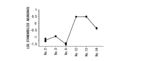
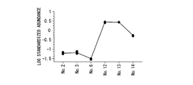
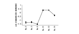
- Example 2 Based on the result of 2D-DIGE shown in FIG. 1, the expression intensity of each of the identified ⁇ -glucosidase I in three healthy trees and three root rot disease-affected trees was analyzed. The results are shown in FIGS. In FIGS. 2A to 2C, the expression intensity of ⁇ -glucosidase I in each of three healthy trees is No. 2, no. 3, No. No. 6 shows the expression intensity of ⁇ -glucosidase I in each of three root rot disease-affected trees. 12, no. 13, no. 14. The fluorescence intensity of the 2D-DIGE protein spot shown in FIG. 1 was measured three times. The results obtained by performing 2D-DIGE three times on the same sample are shown in FIGS.
- Table 1 shows the ratio of the expression intensity of each protein in the healthy tree to the expression intensity of each protein in the root white rot disease-affected tree (root white rot disease-affected tree / healthy tree). As shown in Table 1, ⁇ -glucosidase I in root rot disease-affected trees showed an expression intensity of 33 to 44 times the expression intensity of ⁇ -glucosidase I in healthy trees.
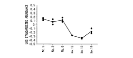
- ⁇ -glucosidase II in healthy trees showed an expression intensity of 1/5 to 1/3 of the expression intensity of ⁇ -glucosidase II in root white rot disease-affected trees. Therefore, the expression level of these proteins in latex derived from the test tree is measured and compared with the expression level of these proteins in latex derived from healthy trees and / or root rot-affected trees. Presence or absence of morbidity can be determined early.
- the root rot disease-affected tree of Para rubber tree can be found at an early stage.
Landscapes
- Health & Medical Sciences (AREA)
- Life Sciences & Earth Sciences (AREA)
- Chemical & Material Sciences (AREA)
- Engineering & Computer Science (AREA)
- Molecular Biology (AREA)
- Immunology (AREA)
- Organic Chemistry (AREA)
- Biochemistry (AREA)
- Medicinal Chemistry (AREA)
- Biomedical Technology (AREA)
- Genetics & Genomics (AREA)
- General Health & Medical Sciences (AREA)
- Biotechnology (AREA)
- Hematology (AREA)
- Zoology (AREA)
- Wood Science & Technology (AREA)
- Microbiology (AREA)
- Bioinformatics & Cheminformatics (AREA)
- Urology & Nephrology (AREA)
- Food Science & Technology (AREA)
- General Physics & Mathematics (AREA)
- Proteomics, Peptides & Aminoacids (AREA)
- Biophysics (AREA)
- General Engineering & Computer Science (AREA)
- Physics & Mathematics (AREA)
- Analytical Chemistry (AREA)
- Cell Biology (AREA)
- Pathology (AREA)
- Peptides Or Proteins (AREA)
- Enzymes And Modification Thereof (AREA)
- Measuring Or Testing Involving Enzymes Or Micro-Organisms (AREA)
- Investigating Or Analysing Biological Materials (AREA)
- Medicines Containing Plant Substances (AREA)
Description
本発明は、タンパク質マーカー、これを用いたパラゴムノキの根白腐病罹病診断キット、及びパラゴムノキの根白腐病罹病診断方法に関する。
本願は、2012年7月4日に、日本に出願された特願2012-150716号に基づき優先権を主張し、その内容をここに援用する。
本願は、2012年7月4日に、日本に出願された特願2012-150716号に基づき優先権を主張し、その内容をここに援用する。
天然ゴムは、主にパラゴムノキ(Hevea brasiliensis)の乳管細胞内で造られているラテックスという乳液を加工することにより製造される。天然ゴムは、ゴム製品の主原料として、様々な用途において幅広くかつ大量に用いられている。
このラテックス採取を目的として、パラゴムノキの生育に適した気候区において、パラゴムノキの植林が行われている。かかる気候区は、パラゴムノキに感染する病原菌の生育においても適しており、パラゴムノキに病原菌が感染することによりもたらされる根白腐病が、ゴム農園におけるパラゴムノキの栽培に深刻な被害を与えている。
このラテックス採取を目的として、パラゴムノキの生育に適した気候区において、パラゴムノキの植林が行われている。かかる気候区は、パラゴムノキに感染する病原菌の生育においても適しており、パラゴムノキに病原菌が感染することによりもたらされる根白腐病が、ゴム農園におけるパラゴムノキの栽培に深刻な被害を与えている。
根白腐病は、パラゴムノキの根に破壊的なダメージを与える病気である。この根白腐病の主な病原菌として、リジドポラス・ミクロポラス(Rigidoporus microporus)が知られている。
リジドポラス・ミクロポラス(Rigidoporus microporus)は、担子菌門に属し、感染したパラゴムノキの足元に巨大な子実体を形成する。
リジドポラス・ミクロポラス(Rigidoporus microporus)は、担子菌門に属し、感染したパラゴムノキの足元に巨大な子実体を形成する。
従来、パラゴムノキにおける根白腐病は、感染した根の焼却による物理的除去方法、又は殺菌剤による化学的除去方法により制御されていた。
しかしながら、これらの方法では、根白腐病にかかったパラゴムノキを回復させるのに手遅れの場合が多々あり、根白腐病の感染を早期に診断できる方法が望まれていた。
しかしながら、これらの方法では、根白腐病にかかったパラゴムノキを回復させるのに手遅れの場合が多々あり、根白腐病の感染を早期に診断できる方法が望まれていた。
これに対して、根白腐病の制御系の改良発展には、病原菌であるリジドポラス・ミクロポラス(Rigidoporus microporus)の遺伝的多様性を理解することが重要であり、例えば非特許文献1において、リジドポラス・ミクロポラス(Rigidoporus microporus)の遺伝的多様性と病原性の関係を解析する試みがなされている。
Saithong Kら、Afr. J. Microbiol. Res.、第3巻、第641~648頁、2009年
しかしながら、非特許文献1に記載された解析結果からは、地理的分布と遺伝的多様性との間に相関が確認されたが、病原性と遺伝的多様性の間には明確な相関性が確認されず、かかる解析結果を根白腐病の制御方法に応用するには、更なる知見を必要としている。
本発明は、上記事情に鑑みてなされたものであって、パラゴムノキの根白腐病罹病木を早期に発見することができるタンパク質マーカー、パラゴムノキの根白腐病罹病診断キット、及びパラゴムノキの根白腐病罹病診断方法を提供することを課題とする。
本発明は、上記課題を解決すべく鋭意研究した結果、根白腐病罹病木及び健全木において、これらから得られるラテックス中の特定タンパク質の発現量が変化することを見出し、本発明を完成させた。
すなわち、本発明は、下記の特徴を有するタンパク質マーカー、これを用いたパラゴムノキの根白腐病罹病診断キット、及びパラゴムノキの根白腐病罹病診断方法を提供する。
(1)パラゴムノキ(Hevea brasiliensis)の根白腐病罹病を診断するために用いられるタンパク質マーカーであって、
β-glucosidaseI及びβ-glucosidaseIIからなる群から選ばれるタンパク質又は前記タンパク質と実質的に同等の機能を有するタンパク質であるタンパク質マーカー。
(3)被検木由来のラテックスにおけるβ-glucosidaseI若しくはβ-glucosidaseIと同等の機能を有するタンパク質の発現量を測定する工程と、
被検木由来のラテックスにおけるβ-glucosidaseI若しくはβ-glucosidaseIと同等の機能を有するタンパク質の発現量と、根白腐病罹病木由来のラテックスにおけるβ-glucosidaseI若しくはβ-glucosidaseIと同等の機能を有するタンパク質の発現量及び/又は健全木由来のラテックスにおけるβ-glucosidaseI若しくはβ-glucosidaseIと同等の機能を有するタンパク質の発現量と、を比較する工程を含むパラゴムノキの根白腐病罹病診断方法。
(5)被検木由来のラテックスにおけるβ-glucosidaseI若しくはβ-glucosidaseIと同等の機能を有するタンパク質の活性を測定する工程と、
被検木由来のラテックスにおけるβ-glucosidaseI若しくはβ-glucosidaseIと同等の機能を有するタンパク質の活性と、根白腐病罹病木由来のラテックスにおけるβ-glucosidaseI若しくはβ-glucosidaseIと同等の機能を有するタンパク質の活性及び/又は健全木由来のラテックスにおけるβ-glucosidaseI若しくはβ-glucosidaseIと同等の機能を有するタンパク質の活性と、を比較する工程を含むパラゴムノキの根白腐病罹病診断方法。
本発明によれば、パラゴムノキの根白腐病罹病木を早期に発見することができる。
以下、本発明の好ましい例を説明するが、本発明はこれら例に限定されることはない。本発明の趣旨を逸脱しない範囲で、構成の付加、省略、置換、およびその他の変更が可能である。
[タンパク質マーカー]
本発明のタンパク質マーカーは、パラゴムノキ(Hevea brasiliensis)の根白腐病罹病を診断するために用いられるタンパク質マーカーであって、
β-glucosidaseI及びβ-glucosidaseIIからなる群から選ばれるタンパク質又は前記タンパク質と実質的に同等の機能を有するタンパク質である。
本発明の「タンパク質マーカー」とは、診断対象となるパラゴムノキが健全木であるか、根白腐病罹病木であるかを判断する指標をいう。
[タンパク質マーカー]
本発明のタンパク質マーカーは、パラゴムノキ(Hevea brasiliensis)の根白腐病罹病を診断するために用いられるタンパク質マーカーであって、
β-glucosidaseI及びβ-glucosidaseIIからなる群から選ばれるタンパク質又は前記タンパク質と実質的に同等の機能を有するタンパク質である。
本発明の「タンパク質マーカー」とは、診断対象となるパラゴムノキが健全木であるか、根白腐病罹病木であるかを判断する指標をいう。
実施例で後述するように、本発明者は、根白腐病罹病木(根白腐病に感染したパラゴムノキ)由来のラテックスと、健全木由来のラテックスについて、高感度の蛍光色素を用いて二次元電気泳動を行った。その結果、根白腐病罹病木と健全木の間で発現量が変動する上記2つのタンパク質を見出した。
従来、的確にパラゴムノキの根白腐病罹病を診断することができなかったが、本発明のタンパク質マーカーは、根白腐病罹病を診断するバイオマーカーとして有用である。即ち、本発明のタンパク質マーカーにより、パラゴムノキの根白腐病罹病の有無を早期に判定することができる。
従来、的確にパラゴムノキの根白腐病罹病を診断することができなかったが、本発明のタンパク質マーカーは、根白腐病罹病を診断するバイオマーカーとして有用である。即ち、本発明のタンパク質マーカーにより、パラゴムノキの根白腐病罹病の有無を早期に判定することができる。
本発明のタンパク質マーカーは、診断対象となるパラゴムノキが健全木であるか、根白腐病罹病木であるかによって、生体内組織で、その発現が増加又は減少する。
根白腐病罹病を診断する場合には、タンパク質マーカーが発現している任意の組織における前記タンパク質マーカーの発現量を測定・評価すればよいが、サンプルの入手が容易であるという観点からラテックスにおける前記タンパク質マーカーの発現量を測定することが好ましい。
根白腐病罹病を診断する場合には、タンパク質マーカーが発現している任意の組織における前記タンパク質マーカーの発現量を測定・評価すればよいが、サンプルの入手が容易であるという観点からラテックスにおける前記タンパク質マーカーの発現量を測定することが好ましい。
ここで、本発明のタンパク質マーカーは、β-glucosidaseI及びβ-glucosidaseIIからなる群から選ばれるタンパク質と実質的に同等の機能を有するタンパク質を含む。このようなタンパク質としては、β-glucosidaseI及びβ-glucosidaseIIを構成するアミノ酸配列のうち1又は2以上のアミノ酸に置換、付加、欠失又は挿入があり、且つ、同等の活性を有するものが挙げられる。
「同等の活性を有する」とは、これらタンパク質と同等の酵素活性を有することをいう。
「同等の活性を有する」とは、これらタンパク質と同等の酵素活性を有することをいう。
また、本発明のタンパク質マーカーは、根白腐病罹病木の罹病改善の指標として好適に用いられる。例えば、根白腐病に感染した罹病木に対して、農薬等の化学物質を病害土壌に散布し、一定時間後の本発明のタンパク質マーカーの発現量を測定することにより、根白腐病罹病木の罹病改善の度合いを評価することができる。
さらに、本発明のタンパク質マーカーは、根白腐病罹病木の治療に効果を示す化学物質のスクリーニングに好適に用いられる。例えば、根白腐病に感染した罹病木に対して、農薬等の化学物質を病害土壌に散布し、一定時間後の本発明のタンパク質マーカーの発現量を測定することにより、前記化学物質が、罹病木の罹病を改善するものとして有効であるかどうかを判定することができる。
被検木中における、本発明のタンパク質マーカーの発現量の測定方法としては、特に制限はないが、例えば、ELISA(enzyme‐linked immunosorbent assay)法、ラジオイムノアッセイ法、免疫組織化学染色法、ウエスタンブロット法、免疫沈降法、免疫染色法、イムノクロマト法等が挙げられる。
ウエスタンブロット法は、当業者に公知の方法で行われる。例えば、被検木から採取した組織のタンパク質を、健全木及び/又は根白腐病罹病木から採取した組織のタンパク質とともに、SDS-PAGEにより展開し、ニトロセルロース膜やPVDF膜に転写する。次いで、前記ニトロセルロース膜やPVDF膜に、本発明のタンパク質マーカーに対する抗体を添加、反応させ、さらに2次抗体を添加、反応させ、前記2次抗体を検出することにより、本発明のタンパク質マーカーを検出すればよい。
また、免疫沈降法としては、例えば、生体から採取した組織のタンパク質を、本発明のタンパク質マーカーに対する抗体で免疫沈降し、沈降物として得られたタンパク質をSDS-PAGEにより展開し、ウエスタンブロット法により検出する方法が挙げられる。
本発明のタンパク質マーカーの発現量の測定方法に使用される抗体としては、本発明のタンパク質マーカーであるβ-glucosidaseI又はβ-glucosidaseIIを検出可能な抗体であればよく、ポリクローナル抗体であってもよくモノクローナル抗体であってもよい。
かかるポリクローナル抗体は当業者に公知の方法で調製すればよく、例えば下記の方法に従って調製できる。すなわち、例えば、抗原を、必要に応じてフロイントアジュバント(Freund’s Adjuvant)とともに、マウス、ラット、ハムスター、モルモット又はウサギ等の哺乳動物に免疫し、前記免疫感作動物から得た血清から取得することができる。
かかるポリクローナル抗体は当業者に公知の方法で調製すればよく、例えば下記の方法に従って調製できる。すなわち、例えば、抗原を、必要に応じてフロイントアジュバント(Freund’s Adjuvant)とともに、マウス、ラット、ハムスター、モルモット又はウサギ等の哺乳動物に免疫し、前記免疫感作動物から得た血清から取得することができる。
また、前記モノクローナル抗体は、例えば、抗原を、必要に応じてフロイントアジュバントとともに、マウス、ラット、ハムスター、モルモットまたはウサギ等の哺乳動物に免疫する。
次いで、免疫感作動物から得た抗体産生細胞と自己抗体産生能のない骨髄腫系細胞(ミエローマ細胞)からハイブリドーマ(融合細胞)を調製し、ハイブリドーマをクローン化し、哺乳動物の免疫に用いた抗原に対して特異的親和性を示すモノクローナル抗体を産生するクローンを選択することによって調製することができる。
さらに具体的には、抗原を、必要に応じてフロイントアジュバントとともに、哺乳動物の皮下内、筋肉内、静脈内又は腹腔内等に1又は数回注射するかあるいは移植することにより免疫感作を施す。通常、最初の免疫から1~14日毎に1~4回程度免疫を行い、最終免疫より1~5日程度後に免疫感作された哺乳動物から抗体産生細胞を取得する。
モノクローナル抗体を分泌するハイブリドーマ(融合細胞)の調製は、定法に従って行うことができる。すなわち、上述した方法により免疫感作された哺乳動物から取得される脾臓、リンパ節、骨髄あるいは扁桃等、好ましくは脾臓に含まれる抗体産生細胞と、マウス、ラット又はヒト等由来の自己抗体産生能のないミエローマ細胞との細胞融合させることにより調製する。モノクローナル抗体を産生するハイブリドーマクローンのスクリーニングは、ハイブリドーマをマイクロタイタープレート等を用いて培養し、増殖の見られたウェルの培養上清の免疫抗原に対する反応性を、例えばRIAやELISA等の酵素免疫測定法によって測定することにより行なうことができる。
ハイブリドーマからのモノクローナル抗体の製造は、ハイブリドーマをインビトロ、又はマウス、ラット、モルモット、ハムスターまたはウサギ等の腹水中等でのインビボで培養した後、得られた培養上清、又は哺乳動物の腹水から単離することにより行うことができる。モノクローナル抗体の単離、精製は、上述の培養上清あるいは腹水を、飽和硫酸アンモニウム、ユーグロブリン沈澱法、カプロイン酸法、カプリル酸法、イオン交換クロマトグラフィー(DEAEまたはDE52等)、抗イムノグロブリンカラムあるいはプロテインAカラム等のアフィニティカラムクロマトグラフィーに供すること等により行うことができる。
さらに、上述の方法により得られたポリクローナル抗体又はモノクローナル抗体を用いて抗体アレイを調製することも可能である。すなわち、上述の抗体をニトロセルロース膜やPVDF膜等の膜や、ニトロセルロースがコートされたスライドやガラス基盤に、マイクロアレイスポッターを用いて固定すればよい。
また、本発明のタンパク質マーカーであるβ-glucosidaseI及びβ-glucosidaseIIからなる群から選ばれるタンパク質又は前記タンパク質と実質的に同等の機能を有するタンパク質は、酵素であるため、酵素活性を有する。
従って、被検木から採取した組織における前記タンパク質マーカーの酵素活性、並びに、健全木及び/又は根白腐病罹病木から採取した組織における前記タンパク質マーカーの酵素活性を測定し、比較することで、被検木の罹病の有無を判定することができる。
従って、被検木から採取した組織における前記タンパク質マーカーの酵素活性、並びに、健全木及び/又は根白腐病罹病木から採取した組織における前記タンパク質マーカーの酵素活性を測定し、比較することで、被検木の罹病の有無を判定することができる。
[根白腐病罹病診断キット]
本発明の根白腐病罹病診断キットは、本発明のタンパク質マーカーを検出可能な抗体を含む。前記抗体は、上述したβ-glucosidaseI及びβ-glucosidaseIIからなる群から選ばれるタンパク質又は前記タンパク質と実質的に同等の機能を有するタンパク質に対するポリクローナル抗体及びモノクローナル抗体をいう。
本発明の根白腐病罹病診断キットは、本発明のタンパク質マーカーを検出可能な抗体を含む。前記抗体は、上述したβ-glucosidaseI及びβ-glucosidaseIIからなる群から選ばれるタンパク質又は前記タンパク質と実質的に同等の機能を有するタンパク質に対するポリクローナル抗体及びモノクローナル抗体をいう。
本発明の根白腐病罹病診断キットには、本発明のタンパク質マーカーを検出可能な抗体の他、例えば、抗体検出用試薬、抗体検出用2次抗体、反応に用いるプレートを備えていてもよく、ELISA又はイムノクロマト用のキットであってもよい。
本発明の根白腐病罹病診断キットを用いることにより、パラゴムノキの被検木から採取した組織(特に、ラテックス)におけるタンパク質マーカーを迅速且つ簡便に検出することが可能となり、根白腐病罹病木を早期に発見することが可能となる。
[パラゴムノキ(Hevea brasiliensis)の根白腐病罹病診断方法]
<第一実施形態>
本実施形態のパラゴムノキの根白腐病罹病診断方法は、
被検木由来のラテックスにおけるβ-glucosidaseI若しくはβ-glucosidaseIと同等の機能を有するタンパク質の発現量を測定する工程と、
被検木由来のラテックスにおけるβ-glucosidaseI若しくはβ-glucosidaseIと同等の機能を有するタンパク質の発現量と、根白腐病罹病木由来のラテックスにおけるβ-glucosidaseI若しくはβ-glucosidaseIと同等の機能を有するタンパク質の発現量及び/又は健全木由来のラテックスにおけるβ-glucosidaseI若しくはβ-glucosidaseIと同等の機能を有するタンパク質の発現量と、を比較する工程を含む。
被検木、並びに、根白腐病罹病木及び/又は健全木由来のラテックスにおけるβ-glucosidaseI又はβ-glucosidaseIと同等の機能を有するタンパク質の発現量を測定する方法としては、上述したELISA法やイムノクロマト法等、簡便な方法が好ましい。
後述する実施例において、根白腐病罹病木由来のラテックスにおけるβ-glucosidaseIの発現量は、健全木由来のラテックスにおけるβ-glucosidaseIの発現量の33~44倍であることが明らかとなった。
従って、被検木由来のラテックスにおけるβ-glucosidaseI又はβ-glucosidaseIと同等の機能を有するタンパク質の発現量が、根白腐病罹病木又は健全木における対応するタンパク質の発現量のどちらに近いのかを判定することにより、根白腐病罹病木を早期に発見することが可能となる。
従って、被検木由来のラテックスにおけるβ-glucosidaseI又はβ-glucosidaseIと同等の機能を有するタンパク質の発現量が、根白腐病罹病木又は健全木における対応するタンパク質の発現量のどちらに近いのかを判定することにより、根白腐病罹病木を早期に発見することが可能となる。
本実施形態のパラゴムノキの根白腐病罹病診断方法は、更に、被検木由来のラテックスにおけるβ-glucosidaseII若しくはβ-glucosidaseIIと同等の機能を有するタンパク質の発現量を測定する工程と、
被検木由来のラテックスにおけるβ-glucosidaseII若しくはβ-glucosidaseIIと同等の機能を有するタンパク質の発現量と、根白腐病罹病木由来のラテックスにおけるβ-glucosidaseII若しくはβ-glucosidaseIIと同等の機能を有するタンパク質の発現量及び/又は健全木由来のラテックスにおけるβ-glucosidaseII若しくはβ-glucosidaseIIと同等の機能を有するタンパク質の発現量と、を比較する工程を含んでいてもよい。
被検木由来のラテックスにおけるβ-glucosidaseII若しくはβ-glucosidaseIIと同等の機能を有するタンパク質の発現量と、根白腐病罹病木由来のラテックスにおけるβ-glucosidaseII若しくはβ-glucosidaseIIと同等の機能を有するタンパク質の発現量及び/又は健全木由来のラテックスにおけるβ-glucosidaseII若しくはβ-glucosidaseIIと同等の機能を有するタンパク質の発現量と、を比較する工程を含んでいてもよい。
後述する実施例において、根白腐病罹病木由来のラテックスにおけるβ-glucosidaseIIの発現量は、健全木由来のラテックスにおけるβ-glucosidaseIIの発現量の1/5~1/3倍であることが明らかとなった。
従って、β-glucosidaseIの測定結果に加えて、被検木由来のラテックスにおけるβ-glucosidaseII又はβ-glucosidaseIIと同等の機能を有するタンパク質の発現量が、根白腐病罹病木又は健全木における対応するタンパク質の発現量のどちらに近いのかを判定することにより、根白腐病罹病木を早期に、かつ、より正確に発見することが可能となる。
従って、β-glucosidaseIの測定結果に加えて、被検木由来のラテックスにおけるβ-glucosidaseII又はβ-glucosidaseIIと同等の機能を有するタンパク質の発現量が、根白腐病罹病木又は健全木における対応するタンパク質の発現量のどちらに近いのかを判定することにより、根白腐病罹病木を早期に、かつ、より正確に発見することが可能となる。
<第二実施形態>
本実施形態のパラゴムノキの根白腐病罹病診断方法は、
被検木由来のラテックスにおけるβ-glucosidaseI若しくはβ-glucosidaseIと同等の機能を有するタンパク質の活性を測定する工程と、
被検木由来のラテックスにおけるβ-glucosidaseI若しくはβ-glucosidaseIと同等の機能を有するタンパク質の活性と、根白腐病罹病木由来のラテックスにおけるβ-glucosidaseI若しくはβ-glucosidaseIと同等の機能を有するタンパク質の活性及び/又は健全木由来のラテックスにおけるβ-glucosidaseI若しくはβ-glucosidaseIと同等の機能を有するタンパク質の活性と、を比較する工程を含む。
上述したように、根白腐病罹病木由来のラテックスにおけるβ-glucosidaseIの発現量は、健全木由来のラテックスにおけるβ-glucosidaseIの発現量と比較して非常に豊富である。
従って、根白腐病罹病木におけるラテックスの単位量当たりのβ-glucosidaseI活性は、健全木におけるラテックスの単位量当たりのβ-glucosidaseI活性より格段に高いことは明らかである。被検木由来のラテックスにおけるβ-glucosidaseI又はβ-glucosidaseIと同等の機能を有するタンパク質の活性が、根白腐病罹病木又は健全木における対応するタンパク質の活性のどちらに近いのかを判定することにより、根白腐病罹病木を早期に発見することが可能となる。
従って、根白腐病罹病木におけるラテックスの単位量当たりのβ-glucosidaseI活性は、健全木におけるラテックスの単位量当たりのβ-glucosidaseI活性より格段に高いことは明らかである。被検木由来のラテックスにおけるβ-glucosidaseI又はβ-glucosidaseIと同等の機能を有するタンパク質の活性が、根白腐病罹病木又は健全木における対応するタンパク質の活性のどちらに近いのかを判定することにより、根白腐病罹病木を早期に発見することが可能となる。
β-glucosidaseの基質としては、5-ブロモ-4-クロロ-3-インドキシル-β-D-グルコピラノシド、5-ブロモ-6-クロロ-3-インドキシル-β-D-グルコピラノシド、5-ブロモ-6-クロロ-3-インドリル-β-D-グルコピラノシド、6-クロロ-3-インドリル-β-D-グルコピラノシド、5-ブロモ-3-インドリル-β-D-グルコピラノシド、4-メチル-ウンベリフェリル-β-D-グルコピラノシド、o-ニトロフェニル-β-D-グルコピラノシド、フェニル-β-D-グルコピラノシド、3-ニトロフェニル-β-D-グルコピラノシド、4-ニトロフェニル-β-D-グルコピラノシド、3-インドキシル-β-グルコピラノシドトリハイドレート、n-ヘプティル-β-D-グルコピラノシド、5-ブロモ-4-クロロ-3-インドキシル-2-アセトアミド-2-デオキシ-β-D-グルコピラノシド等が挙げられる。このうち鑑別性の点から5-ブロモ-4-クロロ-3-インドキシル-β-D-グルコピラノシドが好ましい
また、これらの基質を、β-glucosidaseの作用によって、色原体化合物又は蛍光性色素が遊離されるように化学修飾したものも好ましく用いられる。
また、本実施形態のパラゴムノキ(Hevea brasiliensis)の根白腐病罹病診断方法は、第一実施形態と併用して行われてもよい。これにより、根白腐病罹病木を早期に、かつ、より正確に発見することが可能となる。
また、これらの基質を、β-glucosidaseの作用によって、色原体化合物又は蛍光性色素が遊離されるように化学修飾したものも好ましく用いられる。
また、本実施形態のパラゴムノキ(Hevea brasiliensis)の根白腐病罹病診断方法は、第一実施形態と併用して行われてもよい。これにより、根白腐病罹病木を早期に、かつ、より正確に発見することが可能となる。
次に実施例を示して本発明をさらに詳細に説明するが、本発明は以下の実施例に限定されるものではない。
[実験例1]
≪ラテックスセラムタンパク質抽出≫
植林後7~8年目のパラゴムノキの成熟林から、健全木3本、根白腐病罹病木3本を選択し、これらの木由来のラテックスから、アセトン沈殿により、ラテックスセラムタンパク質を抽出した。
≪ラテックスセラムタンパク質抽出≫
植林後7~8年目のパラゴムノキの成熟林から、健全木3本、根白腐病罹病木3本を選択し、これらの木由来のラテックスから、アセトン沈殿により、ラテックスセラムタンパク質を抽出した。
≪2D-DIGE(2 Dimensional Fluorescence Difference Gel Electrophoresis、蛍光標識2次元ディファレンス電気泳動)によるタンパク質発現プロファイリング≫
抽出したこのラテックスセラムタンパク質を、8nmol トリス-(2-カルボキシエチル)ホスフィン塩酸塩(TCEP;シグマアルドリッチ社)で、37℃60分間処理することにより還元した。
次いで、健全木から抽出したラテックスセラムタンパク質に対して、12nmol Cy3(CyDye DIGE Fluor saturation dye、GEヘルスケアバイオサイエンス社)を用いて37℃で30分間処理することにより蛍光ラベルを施した。ラベル化反応は、DTT及びPharmalyte(pH4-7、GEヘルスケアバイオサイエンス社)含有尿素溶解バッファを、それぞれ最終濃度が65mM及び2.0%となるように添加して終了させた。
同様に、根白腐病罹病木から抽出したラテックスセラムタンパク質に対して、12nmol Cy5(CyDye DIGE Fluor saturation dye、GEヘルスケアバイオサイエンス社)を用いて37℃で30分間処理することにより蛍光ラベルを施した。
更に、内部標準検体として、健全木から抽出したラテックスセラムタンパク質及び根白腐病罹病木から抽出したラテックスセラムタンパク質を混合し、Cy2蛍光染色(CyDye DIGE Fluor saturation dye、GEヘルスケアバイオサイエンス社)でラベルした。ラベル化反応終了後、Cy3ラベル化ラテックスセラムタンパク質と、Cy5ラベル化ラテックスセラムタンパク質と、Cy2ラベル化内部標準検体とを等量混合した後、35mM DTT及び1.0%Pharmalyte(GEヘルスケアバイオサイエンス社)含有尿素溶解バッファーを終量が420mLとなるよう添加した。
尚、健全木3本及び根白腐病罹病木3本について、それぞれ上記サンプル調製を行った。
次いで、調製したサンプルを用いて、2D-DIGEを、当業者周知の方法により行った。一次元目の分離は、24cm長、pIレンジ3~9の固定化pH勾配ポリアクリルアミドゲル(IPG(immobilized pH gradient) Dry Strip、GEヘルスケアバイオサイエンス社)を用いて行った。二次元目の分離は、38cm長のラージフォーマットゲル(株式会社バイオクラフト)を用いたSDS-PAGEを用いて行った。電気泳動後のゲルを、レーザースキャナ(Tyhoon Trio、GEヘルスケアバイオサイエンス)を用いて適切な波長でスキャンした。
次いで、二次元電気泳動解析ソフトウェア(Progenesis PG240、ノンリニアダイナミックス(Nonlinear dynamics Ltd.))を用いて、全てのスポットについて、Cy3及びCy5のイメージ強度を、同一ゲル内のCy2のイメージ強度により正規化し、ゲル間の相違を補正した。システムの再現性は、同一のサンプルについて、2D-DIGEをn=3で行い、得られたタンパク質プロファイルを比較することにより検証した。
更に、健全木3本及び根白腐病罹病木3本について、調整した残りのサンプルについて、それぞれ2D-DIGEをn=3で行った。
次いで、二次元電気泳動解析ソフトウェア(Progenesis PG240、ノンリニアダイナミックス(Nonlinear dynamics Ltd.))を用いて、全てのスポットについて、Cy3及びCy5のイメージ強度を、同一ゲル内のCy2のイメージ強度により正規化し、ゲル間の相違を補正した。システムの再現性は、同一のサンプルについて、2D-DIGEをn=3で行い、得られたタンパク質プロファイルを比較することにより検証した。
更に、健全木3本及び根白腐病罹病木3本について、調整した残りのサンプルについて、それぞれ2D-DIGEをn=3で行った。
各実験で得られた蛍光強度データをlog10変換し、Zスコアに変換して蛍光強度データ分布を標準化した。標準化したデータについて、ユークリッド距離とUPGMA法により階層的クラスタ分析を行い、プロテオームの全体的な特徴を確認した。
健全木と根白腐病罹病木間で発現量において顕著な差を示したタンパク質スポットの2D-DIGEの結果を図1に示す。図1に示されたタンパク質スポットを、マススペクトルによる分析により同定した。
≪質量分析法によるタンパク質の同定≫
健全木と根白腐病罹病木間で明らかに顕著な差を示した標的タンパク質スポットについては、自動スポット回収装置(ProHunter、アズワン株式会社)を使用して、ゲルプラグを96穴PCRプレートに回収した。
次いで、前記ゲルプラグを、メタノール、炭酸アンモニウム及びアセトノトリルで三回洗浄し、乾燥後、TPCK処理トリプシンで一晩処理を行った。ゲルプラグをアセトニトリルで処理することにより、ゲルプラグからペプチドを抽出した。
このようにしてゲル内消化により得られたトリプシンペプチドを、ナノスケールマイクロキャピラリRP LC-ESI MS/MSによる解析にかけた。具体的には、マイクロフローHPLC用のパラダイムMS4 HPLCデュアルソルベントデリバリーシステム(Michrom BioResource社)、HTS PAL オートサンプラ(CTC Analytics社)、及び、ナノ-ESI(NSI)ソース(エーエムアール株式会社)搭載フィニガンLTQリニアITマススペクトロメータ(ThermoElectron社)を用いて、タンパク質の同定を行った。
消化されたペプチド混合物は、マイクロキャピラリRP Magic C18カラム(3mm、200A、5060.2mm i.d.;Michrom)上で分離した。ペプチドは、10から80%の直線勾配のバッファB(10%水(v/v)及び0.1%ギ酸(v/v)含有アセトニトリル)を含有するバッファA(2%アセトニトリル(v/v)及び0.1%ギ酸(v/v)含有水)で10分間溶出した。
HPLCからの溶出物は、NSI針を通じて1.0~1.2mL/分の速度でマススペクトロメータに入れた(FortisTip、有限会社オムニセパロ適塾)。1.8kVの電圧でキャピラリを200℃に加熱した。シース(sheath)及び補助ガス(auxiliary gas)は使用しなかった。マススペクトロメータは、データ依存的収集モードに設定し、Xcaliburソフトウェア(バージョン2.0、ThermoElectron)の自動制御下でm/z450-1800のマスレンジの収集が自動的にMS/MS収集に切り替わるようにした。全MSスキャンの結果は、サーベイスキャンで最も豊富に検出された3つのイオンについて活性化振幅パラメータを35%にセットし、分離幅m/Z2.0としたMS/MS試験により得られた。データは、30秒の排除期間及び排除マス幅が-1.0及び+2.0Daであるダイナミックマス排除枠により取得した。
得られたデータは、装置と共に提供されているExtractMSソフトウェア バージョン2.11(ThermoElectron社)を用いて、ペプチドマスレンジを450から600Da、MS/MS最小ピークを25としてdtaフォーマット(ピークリストファイル)に変換し、MASCOT(登録商標)サーチを行った。
MASCOT(登録商標)(バージョン2.1、マトリックスサイエンス社)サーチは、スイスプロット及びNCBIの重複のないタンパク質配列データベース中の配列のうち、Hevea brasiliensisサブセットに対して行った。
以下のサーチパラメータを全てのMASCOT(登録商標)サーチに使用した。2つのトリプシン分解物の欠落を許容範囲とし、メチオニン残基の可変修飾(酸化、+16Da)、並びに、MSデータにおける±2.0Da及びMS/MSデータにおける±1.0Daを最大誤差許容範囲とした。特徴的な配列を有する2以上の顕著に適合したペプチドを含むタンパク質(p<0.05、本実験におけるサーチパラメータ下において、MASCOTイオンスコアがスイスプロットデータベースに対して34以上及びNCBIデータベースに対して42以上と同等である)について統計的に検討し、タンパク質同定の信頼性を評価した。加えて、同定されたペプチドのMS/MSスペクトルを手動で調べた。
20個のタンパク質スポットのうち、マススペクトルによるタンパク質同定により、2個のタンパク質が同定された。結果を表1に示す。

[実験例2]
図1で示された2D-DIGEの結果に基づき、同定されたβ-glucosidaseIについて、健全木3本及び根白腐病罹病木3本それぞれにおける発現強度を解析した。結果を図2A~Cに示す。図2A~C中、健全木3本それぞれにおけるβ-glucosidaseIの発現強度をNo.2,No.3,No.6で示し、根白腐病罹病木3本それぞれにおけるβ-glucosidaseIの発現強度をNo.12,No.13,No.14で示す。図1で示された2D-DIGEのタンパク質スポットの蛍光強度を3回測定した。
尚、同一サンプルに対して2D-DIGEを3回行って得られたそれぞれの結果を図2A、2B、2Cに示す。
また、同定されたβ-glucosidaseIIについても、β-glucosidaseIと同様の解析を行った。結果を図3A、3B、3Cに示す。
更に、根白腐病罹病木における各タンパク質の発現強度に対する健全木における各タンパク質の発現強度の比(根白腐病罹病木/健全木)を表1に示した。
表1に示されるように、根白腐病罹病木におけるβ-glucosidaseIは、健全木におけるβ-glucosidaseIの発現強度に対して、33倍~44倍の発現強度を示した。一方、健全木におけるβ-glucosidaseIIは、根白腐病罹病木におけるβ-glucosidaseIIの発現強度に対して、1/5~1/3の発現強度を示した。
従って、被検木由来のラテックスにおけるこれらのタンパク質の発現量を測定し、健全木及び/又は根白腐病罹病木由来のラテックスにおけるこれらのタンパク質の発現量と比較することにより、パラゴムノキの根白腐病罹病の有無を早期に判定することができる。
以上の結果から、本発明によれば、パラゴムノキの根白腐病罹病木を早期に発見することができることが明らかである。
パラゴムノキの根白腐病罹病木を早期に発見することができるタンパク質マーカー、パラゴムノキの根白腐病罹病診断キット、及びパラゴムノキの根白腐病罹病診断方法を提供することができる。
Claims (5)
- パラゴムノキの根白腐病罹病を診断するために用いられるタンパク質マーカーであって、
β-glucosidaseI及びβ-glucosidaseIIからなる群から選ばれるタンパク質又は前記タンパク質と実質的に同等の機能を有するタンパク質であるタンパク質マーカー。 - 請求項1に記載のタンパク質マーカーを検出可能な抗体を含むパラゴムノキの根白腐病罹病診断キット。
- 被検木由来のラテックスにおけるβ-glucosidaseI若しくはβ-glucosidaseIと同等の機能を有するタンパク質の発現量を測定する工程と、
被検木由来のラテックスにおけるβ-glucosidaseI若しくはβ-glucosidaseIと同等の機能を有するタンパク質の発現量と、根白腐病罹病木由来のラテックスにおけるβ-glucosidaseI若しくはβ-glucosidaseIと同等の機能を有するタンパク質の発現量及び/又は健全木由来のラテックスにおけるβ-glucosidaseI若しくはβ-glucosidaseIと同等の機能を有するタンパク質の発現量と、を比較する工程を含むパラゴムノキの根白腐病罹病診断方法。 - 更に、被検木由来のラテックスにおけるβ-glucosidaseII若しくはβ-glucosidaseIIと同等の機能を有するタンパク質の発現量を測定する工程と、
被検木由来のラテックスにおけるβ-glucosidaseII若しくはβ-glucosidaseIIと同等の機能を有するタンパク質の発現量と、根白腐病罹病木由来のラテックスにおけるβ-glucosidaseII若しくはβ-glucosidaseIIと同等の機能を有するタンパク質の発現量及び/又は健全木由来のラテックスにおけるβ-glucosidaseII若しくはβ-glucosidaseIIと同等の機能を有するタンパク質の発現量と、を比較する工程を含む請求項3に記載のパラゴムノキの根白腐病罹病診断方法。 - 被検木由来のラテックスにおけるβ-glucosidaseI若しくはβ-glucosidaseIと同等の機能を有するタンパク質の活性を測定する工程と、
被検木由来のラテックスにおけるβ-glucosidaseI若しくはβ-glucosidaseIと同等の機能を有するタンパク質の活性と、根白腐病罹病木由来のラテックスにおけるβ-glucosidaseI若しくはβ-glucosidaseIと同等の機能を有するタンパク質の活性及び/又は健全木由来のラテックスにおけるβ-glucosidaseI若しくはβ-glucosidaseIと同等の機能を有するタンパク質の活性と、を比較する工程を含むパラゴムノキの根白腐病罹病診断方法。
Priority Applications (1)
| Application Number | Priority Date | Filing Date | Title |
|---|---|---|---|
| MYPI2014703829A MY169380A (en) | 2012-07-04 | 2013-07-02 | Protein marker, kit for diagnosis of occurrence of white root disease in hevea brasiliensis, and method for diagnosing occurrence of white root disease in hevea brasiliensis |
Applications Claiming Priority (2)
| Application Number | Priority Date | Filing Date | Title |
|---|---|---|---|
| JP2012150716A JP6078244B2 (ja) | 2012-07-04 | 2012-07-04 | パラゴムノキ(Hevea brasiliensis)の根白腐病罹病診断キット、及びパラゴムノキの根白腐病罹病診断方法 |
| JP2012-150716 | 2012-07-04 |
Publications (1)
| Publication Number | Publication Date |
|---|---|
| WO2014007252A1 true WO2014007252A1 (ja) | 2014-01-09 |
Family
ID=49882006
Family Applications (1)
| Application Number | Title | Priority Date | Filing Date |
|---|---|---|---|
| PCT/JP2013/068144 Ceased WO2014007252A1 (ja) | 2012-07-04 | 2013-07-02 | タンパク質マーカー、パラゴムノキ(Hevea brasiliensis)の根白腐病罹病診断キット、及びパラゴムノキの根白腐病罹病診断方法 |
Country Status (3)
| Country | Link |
|---|---|
| JP (1) | JP6078244B2 (ja) |
| MY (1) | MY169380A (ja) |
| WO (1) | WO2014007252A1 (ja) |
-
2012
- 2012-07-04 JP JP2012150716A patent/JP6078244B2/ja active Active
-
2013
- 2013-07-02 WO PCT/JP2013/068144 patent/WO2014007252A1/ja not_active Ceased
- 2013-07-02 MY MYPI2014703829A patent/MY169380A/en unknown
Non-Patent Citations (3)
| Title |
|---|
| KEIGO INAMI: "PCR Oyobi Real Time PCR ni yoru Para Rubber Tree Neshirogusarebyo-kin Rigidoporus microporus no Tokui Shikibetsu", HEISEI 24 NENDO PHYTOPATHOLOGICAL SOCIETY OF JAPAN PROGRAM. KOEN YOSHI YOKOSHU, 15 March 2012 (2012-03-15), pages 66 * |
| KEIKO NATSUAKI: "Indonesia no Gomu ni Okeru white root disease no Hassei to sono Kanri", RESEARCH FOR TROPICAL AGRICULTURE, vol. 3, no. EXTRA., 9 October 2010 (2010-10-09), pages 43 * |
| SAITHONG KAEWCHAI: "Genetic variation among isolates of Rigidoporus microporus causing white root disease of rubber trees in Southern Thailand revealed by ISSR markers and pathogenicity", AFRICAN JOURNAL OF MICROBIOLOGY RESEARCH, vol. 3, no. 10, October 2009 (2009-10-01), pages 641 - 648 * |
Also Published As
| Publication number | Publication date |
|---|---|
| MY169380A (en) | 2019-03-26 |
| JP2014013192A (ja) | 2014-01-23 |
| JP6078244B2 (ja) | 2017-02-08 |
Similar Documents
| Publication | Publication Date | Title |
|---|---|---|
| Cleland et al. | A comparison of common mass spectrometry approaches for paleoproteomics | |
| Villén et al. | The SCX/IMAC enrichment approach for global phosphorylation analysis by mass spectrometry | |
| Nilsson et al. | Towards a human proteome atlas: high‐throughput generation of mono‐specific antibodies for tissue profiling | |
| KR20120100982A (ko) | 시그너처 펩티드 및 질량 분광법에 의해 혼합물에서 개별적인 재조합 단백질의 다중 정량 | |
| JP2020515238A (ja) | 抗pd−l1抗体に対するイディオタイプ抗体及びそれらの使用 | |
| Zhou et al. | Recent advances in stable isotope labeling based techniques for proteome relative quantification | |
| EP1605266B1 (en) | Urothelial cancer tumor marker | |
| Yuan et al. | Proteomics analysis of phosphotyrosyl-proteins in human lumbar cerebrospinal fluid | |
| US20080241862A1 (en) | Propionyl and butyryl lysine modifications in proteins | |
| AU2009234930B2 (en) | Method of discriminating bovine, thus discriminated bovine and kit for discriminating bovine | |
| Cuollo et al. | Toward milk speciation through the monitoring of casein proteotypic peptides | |
| Boström et al. | Antibodies as means for selective mass spectrometry | |
| JP6078244B2 (ja) | パラゴムノキ(Hevea brasiliensis)の根白腐病罹病診断キット、及びパラゴムノキの根白腐病罹病診断方法 | |
| US11965889B2 (en) | Lysine monomethylated derivative and corresponding antibody and use thereof | |
| US20080319678A1 (en) | Mass Tagging for Quantitative Analysis of Biomolecules using 13C Labeled Phenylisocyanate | |
| JP6553264B2 (ja) | 卵子の受精可能性の検査のためのバイオマーカーおよびそれを用いた判定 | |
| Cuomo et al. | SILAC-based quantitative strategies for accurate histone posttranslational modification profiling across multiple biological samples | |
| CN107365386A (zh) | 一种抗细胞质胸苷激酶-IgG抗体的制备方法和用途 | |
| KR101396300B1 (ko) | 에틸벤젠 독성 중독 진단용 바이오마커 | |
| KR102493640B1 (ko) | 브루셀라 감염증 진단용 펩타이드 및 이의 용도 | |
| Krey et al. | Analysis of the proteome of hair-cell stereocilia by mass spectrometry | |
| Zienkiewicz et al. | Identification of olive pollen allergens using a fluorescence‐based 2D multiplex method | |
| Soriano et al. | A fluorescent-based HPLC assay for quantification of cysteine and cysteamine adducts in Escherichia coli-derived proteins | |
| US20100173322A1 (en) | Reagents for the detection of protein phosphorylation in anaplastic large cell lymphoma signaling pathways | |
| Dullnig et al. | Confirmation of immuno‐reactivity of the recombinant major birch pollen allergen Bet v 1a by affinity‐CIEF |
Legal Events
| Date | Code | Title | Description |
|---|---|---|---|
| 121 | Ep: the epo has been informed by wipo that ep was designated in this application |
Ref document number: 13813711 Country of ref document: EP Kind code of ref document: A1 |
|
| NENP | Non-entry into the national phase |
Ref country code: DE |
|
| WWE | Wipo information: entry into national phase |
Ref document number: IDP00201500407 Country of ref document: ID |
|
| 122 | Ep: pct application non-entry in european phase |
Ref document number: 13813711 Country of ref document: EP Kind code of ref document: A1 |