WO2013176131A1 - Template base for use in production of artificial tissue body for transplantation - Google Patents
Template base for use in production of artificial tissue body for transplantation Download PDFInfo
- Publication number
- WO2013176131A1 WO2013176131A1 PCT/JP2013/064082 JP2013064082W WO2013176131A1 WO 2013176131 A1 WO2013176131 A1 WO 2013176131A1 JP 2013064082 W JP2013064082 W JP 2013064082W WO 2013176131 A1 WO2013176131 A1 WO 2013176131A1
- Authority
- WO
- WIPO (PCT)
- Prior art keywords
- cells
- cell
- tissue
- base material
- substrate
- Prior art date
Links
Images


Classifications
-
- C—CHEMISTRY; METALLURGY
- C12—BIOCHEMISTRY; BEER; SPIRITS; WINE; VINEGAR; MICROBIOLOGY; ENZYMOLOGY; MUTATION OR GENETIC ENGINEERING
- C12N—MICROORGANISMS OR ENZYMES; COMPOSITIONS THEREOF; PROPAGATING, PRESERVING, OR MAINTAINING MICROORGANISMS; MUTATION OR GENETIC ENGINEERING; CULTURE MEDIA
- C12N5/00—Undifferentiated human, animal or plant cells, e.g. cell lines; Tissues; Cultivation or maintenance thereof; Culture media therefor
- C12N5/0062—General methods for three-dimensional culture
-
- A—HUMAN NECESSITIES
- A61—MEDICAL OR VETERINARY SCIENCE; HYGIENE
- A61L—METHODS OR APPARATUS FOR STERILISING MATERIALS OR OBJECTS IN GENERAL; DISINFECTION, STERILISATION OR DEODORISATION OF AIR; CHEMICAL ASPECTS OF BANDAGES, DRESSINGS, ABSORBENT PADS OR SURGICAL ARTICLES; MATERIALS FOR BANDAGES, DRESSINGS, ABSORBENT PADS OR SURGICAL ARTICLES
- A61L27/00—Materials for grafts or prostheses or for coating grafts or prostheses
- A61L27/36—Materials for grafts or prostheses or for coating grafts or prostheses containing ingredients of undetermined constitution or reaction products thereof, e.g. transplant tissue, natural bone, extracellular matrix
- A61L27/38—Materials for grafts or prostheses or for coating grafts or prostheses containing ingredients of undetermined constitution or reaction products thereof, e.g. transplant tissue, natural bone, extracellular matrix containing added animal cells
- A61L27/3804—Materials for grafts or prostheses or for coating grafts or prostheses containing ingredients of undetermined constitution or reaction products thereof, e.g. transplant tissue, natural bone, extracellular matrix containing added animal cells characterised by specific cells or progenitors thereof, e.g. fibroblasts, connective tissue cells, kidney cells
- A61L27/3813—Epithelial cells, e.g. keratinocytes, urothelial cells
-
- A—HUMAN NECESSITIES
- A61—MEDICAL OR VETERINARY SCIENCE; HYGIENE
- A61L—METHODS OR APPARATUS FOR STERILISING MATERIALS OR OBJECTS IN GENERAL; DISINFECTION, STERILISATION OR DEODORISATION OF AIR; CHEMICAL ASPECTS OF BANDAGES, DRESSINGS, ABSORBENT PADS OR SURGICAL ARTICLES; MATERIALS FOR BANDAGES, DRESSINGS, ABSORBENT PADS OR SURGICAL ARTICLES
- A61L27/00—Materials for grafts or prostheses or for coating grafts or prostheses
- A61L27/36—Materials for grafts or prostheses or for coating grafts or prostheses containing ingredients of undetermined constitution or reaction products thereof, e.g. transplant tissue, natural bone, extracellular matrix
- A61L27/38—Materials for grafts or prostheses or for coating grafts or prostheses containing ingredients of undetermined constitution or reaction products thereof, e.g. transplant tissue, natural bone, extracellular matrix containing added animal cells
- A61L27/3804—Materials for grafts or prostheses or for coating grafts or prostheses containing ingredients of undetermined constitution or reaction products thereof, e.g. transplant tissue, natural bone, extracellular matrix containing added animal cells characterised by specific cells or progenitors thereof, e.g. fibroblasts, connective tissue cells, kidney cells
- A61L27/3834—Cells able to produce different cell types, e.g. hematopoietic stem cells, mesenchymal stem cells, marrow stromal cells, embryonic stem cells
-
- A—HUMAN NECESSITIES
- A61—MEDICAL OR VETERINARY SCIENCE; HYGIENE
- A61L—METHODS OR APPARATUS FOR STERILISING MATERIALS OR OBJECTS IN GENERAL; DISINFECTION, STERILISATION OR DEODORISATION OF AIR; CHEMICAL ASPECTS OF BANDAGES, DRESSINGS, ABSORBENT PADS OR SURGICAL ARTICLES; MATERIALS FOR BANDAGES, DRESSINGS, ABSORBENT PADS OR SURGICAL ARTICLES
- A61L27/00—Materials for grafts or prostheses or for coating grafts or prostheses
- A61L27/50—Materials characterised by their function or physical properties, e.g. injectable or lubricating compositions, shape-memory materials, surface modified materials
- A61L27/507—Materials characterised by their function or physical properties, e.g. injectable or lubricating compositions, shape-memory materials, surface modified materials for artificial blood vessels
-
- C—CHEMISTRY; METALLURGY
- C12—BIOCHEMISTRY; BEER; SPIRITS; WINE; VINEGAR; MICROBIOLOGY; ENZYMOLOGY; MUTATION OR GENETIC ENGINEERING
- C12M—APPARATUS FOR ENZYMOLOGY OR MICROBIOLOGY; APPARATUS FOR CULTURING MICROORGANISMS FOR PRODUCING BIOMASS, FOR GROWING CELLS OR FOR OBTAINING FERMENTATION OR METABOLIC PRODUCTS, i.e. BIOREACTORS OR FERMENTERS
- C12M21/00—Bioreactors or fermenters specially adapted for specific uses
- C12M21/08—Bioreactors or fermenters specially adapted for specific uses for producing artificial tissue or for ex-vivo cultivation of tissue
-
- C—CHEMISTRY; METALLURGY
- C12—BIOCHEMISTRY; BEER; SPIRITS; WINE; VINEGAR; MICROBIOLOGY; ENZYMOLOGY; MUTATION OR GENETIC ENGINEERING
- C12M—APPARATUS FOR ENZYMOLOGY OR MICROBIOLOGY; APPARATUS FOR CULTURING MICROORGANISMS FOR PRODUCING BIOMASS, FOR GROWING CELLS OR FOR OBTAINING FERMENTATION OR METABOLIC PRODUCTS, i.e. BIOREACTORS OR FERMENTERS
- C12M25/00—Means for supporting, enclosing or fixing the microorganisms, e.g. immunocoatings
- C12M25/14—Scaffolds; Matrices
Definitions
- the present invention relates to a mold base material for producing an artificial tissue body for transplantation.
- Non-Patent Document 1 An in vivo tissue formation technique in which a living body-derived tissue body is formed in a living body by using the latter self-defense reaction and used for transplantation has been proposed (Non-Patent Document 1).
- tissue body for transplantation that does not contain artificial materials at all by the in vivo tissue formation technology. Further, in such a technique, since the inside of the body is used as a bioreactor, a complicated body operation that is essential for conventional tissue engineering is not required, and a tissue body can be produced safely, simply, and at low cost. Furthermore, when forming a tissue in the body of the person who receives the transplant, the transplanted tissue is an autologous tissue, and it is possible to eliminate problems such as immunity problems and infectious diseases in biocompatibility after transplantation. it can.
- Patent Document 1 a spiral groove is formed on the surface of a rod-like structure, and the rod-like structure is embedded in a living body to form a membrane-like connective tissue on the surface of the rod-like structure. A point of increasing the mechanical strength of the connective tissue is disclosed.
- Patent Document 2 discloses that an outer shell member is formed in a spiral shape along the outer periphery of a rod-shaped structural member, and this is embedded in a living body to form a connective tissue body on the outer edge of the rod-shaped structural member. .
- the connective tissue body enters between the outer member and the surface of the rod-shaped structural member, and the inner surface shape of the connective tissue body is formed on the same smooth surface as the surface of the rod-shaped structural member.
- a connective tissue body is formed to a thickness that embeds the shell member.
- Patent Document 3 discloses that an outer member is formed on the surface of a rod-shaped structural member, and this is used as a connective tissue forming substrate. By embedding this base material in the living body, a film-like tissue body is formed on the surface of the base material. By using a material that is excellent in biocompatibility but not easily invaded by the tissue body or its constituents as the material of the shell member, the shell member adheres to the tissue body, and the mechanical strength of the connective tissue body is increased and the shell is increased. An artificial blood vessel in which the tissue body and its constituent components are not exposed on the inner surface of the member is obtained.
- Patent Document 4 discloses a method of obtaining a tissue body closer to a living body by irradiating light when a base material is embedded in the living body to form a connective tissue body.
- Patent Document 5 and Non-Patent Document 2 disclose a core base material for obtaining a valved artificial blood vessel having an overhanging portion having a Valsalva sinus shape and a trilobal valve.
- a living tissue is composed of cells peculiar to the tissue.
- the inner surface of a blood vessel is covered with vascular endothelial cells, and these vascular endothelial cells have a function of preventing the formation of thrombus.
- the tissue obtained by the method described in Non-Patent Document 1 is mainly formed from connective tissue, that is, fibroblasts and a collagen matrix.
- connective tissue that is, fibroblasts and a collagen matrix.
- Non-Patent Document 6 Non-Patent Documents 3 and 4
- the present invention relates to a method for forming a tissue for transplantation comprising a desired tissue or a living tissue containing a cell having a function similar to that of a desired cell in in vivo tissue formation technology, and an artificial graft for use in the method. It aims at providing the mold base material for tissue body manufacture.
- the present invention is a template substrate that can hold desired cells, cells that substitute for the desired cells, or cells that have the ability to differentiate into the desired cells so as to be able to invade from the substrate surface to the outside,
- the template substrate is placed in an environment in which a living tissue exists, by placing the template substrate holding a desired cell, a substitute for the desired cell, or a cell capable of differentiating into a desired cell.
- Used to produce an artificial tissue body for transplantation by coating with a tissue derived from a living body containing desired cells or cells having functions similar to the desired cells, and then removing the template substrate from the coated template substrate
- a mold base material for producing an artificial tissue body for transplantation.
- the present invention also provides (1) A step of holding desired cells, cells that substitute for desired cells, or cells that have the ability to differentiate into desired cells on the template base material, (2) A step of placing the template substrate holding the cells in an environment where biological tissue material exists, (3) A step of taking out the mold base material coated with the biological tissue from the environment where the biological tissue material exists, (4) Provided is a method for producing an artificial tissue body for transplant, which includes a step of removing a mold base material from a mold base material coated with a biological tissue.
- a transplanted tissue body containing desired cells or cells having functions similar to desired cells for example, an artificial blood vessel containing vascular endothelial cells and smooth muscle cells.
- an artificial tissue body for transplantation composed of cells close to a living tissue can be obtained.
- the transplantable tissue obtained by the present invention can exhibit a desired function immediately after transplantation.
- an excellent artificial tissue for transplantation having a low risk of thrombus formation can be obtained. it can.
- FIG. 1 The photograph of the fat origin stromal cell used in Example 1.
- FIG. 1 The hollow mold base material (porous cylindrical base material) used in the examples. 2 is a photograph of the tissue obtained in Example 1. 3 is a graph showing the thickness of the tissue obtained in Example 1. 2 is a photograph of a cross section of the tissue body obtained in Example 1 and a section of the tissue body. 3 is a graph showing the elastic modulus of the tissue body obtained in Example 2. 2 is a photograph of a section of a tissue obtained in Example 2. 4 is a photograph of the beagle dog oral mucosa epithelial cells used in Example 3. 4 is a photograph of a section of a tubular tissue body obtained in Example 3. The photograph of the hollow mold base material used in Examples 4 and 5. FIG.
- FIG. 11 is a photograph showing a state in which a blood vessel piece is sealed in the hollow mold base material of FIG. 10 (Example 4).
- 4 is a photograph of a section of the tissue obtained in Example 4.
- FIG. 11 is a photograph showing a state in which oral mucosa tissue is sealed in the hollow mold base material of FIG. 10 (Example 5).
- 6 is a photograph of a section of the tissue obtained in Example 5.
- the template base material of the present invention is formed so that cells can be held so as to be able to invade from the surface of the base material to the outside.
- the cells can be retained by being embedded in a dispersion or fixed in a template substrate.
- examples of the template base material include a material that can hold cells and can maintain a certain shape, such as a scaffold for three-dimensional cell culture.
- Another embodiment is a hollow mold base material having a passage through which cells can invade from the surface of the base material to the outside, and used by enclosing cells in the hollow portion of the hollow mold base material.
- the material of the hollow mold base material can be molded into a desired shape as a mold base material for forming a target transplant tissue, and has a strength (hardness) that does not greatly deform when implanted in a living body. It is not particularly limited as long as it is not easily decomposed in the living body, can be sterilized, and has no or little eluate that stimulates the living body.
- Preferable ones are acrylic resin, olefin resin, styrene resin, polyester resin, polyamide resin, vinyl chloride resin, silicon resin, fluorine resin, epoxy resin, polycarbonate resin, polyethylene resin, polyurethane resin, glass, metal (titanium, platinum, Stainless steel, SUS (stainless steel, etc.) etc. are mentioned.
- the material is not limited to one type, and two or more types can be used in combination.
- an acrylic resin or the like is preferable in terms of having a certain strength, and a silicon resin is preferable in terms of easy manufacture.
- the shape of the mold base material is a simple shape such as a columnar shape having a uniform thickness, and the mold base material can be easily extracted by incising a part of the formed artificial tissue body There is no particular restriction on the hardness of the resin.
- a soft resin that can be crushed when extracted is suitably used as the material of the mold base material.
- an artificial tissue body having a desired shape may be obtained by appropriately combining the base materials from a plurality of parts so that the base material can be easily taken out.
- the hollow mold base is provided with a passage to the outside so that the cells held inside can invade to the outside.
- the passage is not limited, and may be a hole or a slit, for example.
- the shape and size of the holes, the length and width of the slits, and the number thereof are not particularly limited as long as the cells can pass through, and the shape of the cells encapsulated inside or the mode of encapsulating the cells inside Accordingly, it may be determined appropriately.
- the diameter of the hole may be 10 ⁇ m or more through which cells can pass, for example, 0.01 mm or more.
- a hollow substrate is exemplified.
- the upper limit of the pore diameter is not particularly limited as long as the loaded cells do not flow out at a stretch.
- a support base material for example, a commercially available gel-like cell support base material such as Matrigel (trademark) or the like is encapsulated in a hollow mold base material, for example, the hole has a size of up to about 5 mm, preferably up to about 1 mm. It may be.
- An example of the density of the holes is a mold base material in which 5 to 50 ⁇ 5 to 50 holes, for example, 15 ⁇ 15 holes are formed on the surface of a 1.5 cm ⁇ 1.5 cm square plate-shaped hollow base material. Is done.
- the “desired cell” means a cell necessary for constructing the artificial tissue body.
- desired cells include vascular endothelial cells and smooth muscle cells.
- the desired cells include corneal epithelial cells.
- Cells that substitute for desired cells are cells having functions similar to those of desired cells.
- oral mucosal epithelial cells are used as substitutes for epithelial cells such as vascular endothelial cells and corneal epithelial cells.
- Oral mucosal epithelial cells are cultured into a sheet and used as an artificial cornea for transplantation in clinical practice (Artificial organ 38, No. 3, 2009, pages 168-172), Nishida et al. N Engl J Med 351; 12; 2004; 1187-1196.
- the “cell having the ability to differentiate into a desired cell” is not particularly limited as long as it has such ability.
- examples include somatic stem cells, iPS cells, and pluripotent cells such as ES cells.
- somatic stem cells mesenchymal stem cells are used for various cells such as adipocytes, fibroblasts, stromal cells, cardiomyocytes, smooth muscle cells, endothelial cells, epithelial cells, osteoblasts, chondrocytes, and nerve cells. It is suitably used as a cell having the ability to differentiate into a desired cell from the viewpoint of having differentiation ability.
- Mesenchymal stem cells can be collected by known methods from mammalian bone marrow fluid, peripheral blood, umbilical cord blood, adipose tissue, and the like.
- a cell having a function similar to a desired cell means not only a function that is equivalent to or similar to all functions of a desired cell, but also a function that is desirable as an artificial tissue body for transplantation. Similar cells are also included.
- a desired cell a cell that can substitute for a desired cell, or a cell that has the ability to differentiate into a desired cell, a cell obtained from a transplant recipient, a culture of a cell obtained from a recipient, a transplant recipient It is preferable to use cells derived from the transplant recipient, such as universal stem cells obtained from these cells. When cells derived from a person other than the transplant recipient are used, it is necessary that the donor of the cell has high compatibility with the transplant recipient and HLA.
- the tissue obtained from the living body may be used as it is or after being processed into an appropriate shape, or the whole or part of the tissue separated from the living body is dispersed. But you can.
- cells obtained from living organisms can be dispersed to obtain cells, and the obtained cells can be processed by conventional methods to isolate only the target cell population even if the fraction containing a large amount of the target cells is used. You may use what you did.
- cultured cells obtained by culturing cells obtained from a living body as described above may be used.
- a desired cell, a substitute for a desired cell, or a cell having an ability to differentiate into a desired cell may be a pellet-like aggregate, a tissue extracted from a living body, or a tissue obtained by culture , And those partially dispersed may be enclosed in the hollow portion of the hollow mold base material.
- the cells and the cell support substrate may be enclosed in the hollow portion of the hollow mold substrate.
- the cells may be dispersed and embedded in the support substrate or may be fixed.
- a commercially available substrate for three-dimensional cell culture is used.
- collagen gel for cell culture BD Matrigel (trademark), gelatin for cell culture, agar, hyaluronic acid hydrogel, etc.
- mucopolysaccharides e.g., mucopolysaccharides.
- a hollow portion of the hollow mold substrate is prepared by embedding or fixing cells in the three-dimensional cell culture substrate according to the attached instructions.
- the liquid Matrigel and cells are mixed according to the procedure described in the instructions, injected into the hollow part of the base material, and left for a while, whereby the hollow part of the base material is mixed.
- the matrigel may be gelled inside.
- cell culture substrates provide an environment suitable for cell culture, and often include a three-dimensional scaffold and an appropriate growth factor.
- a commercially available cell support substrate containing an appropriate growth factor may be employed, or a growth factor corresponding to the cell may be added.
- a growth factor to be added for example, in the case of producing a circulatory prosthesis such as an artificial blood vessel, in addition to growth factors such as vascular endothelial growth factor (VEGF) and fibroblast growth factor (bFGF), blood vessels
- VEGF vascular endothelial growth factor
- bFGF fibroblast growth factor
- a growth factor may be appropriately selected according to the cells to be retained and the target tissue and used in combination.
- the cell support substrate may also be a blood-derived gel-like substance such as fibrin glue for hemostasis, or blood (whole blood) is used as the cell support substrate, and cells and blood are mixed to form a hollow substrate. You may enclose and use in the hollow part.
- blood when blood and cells are mixed, injected into the hollow mold base as it is and left for a while, the blood coagulates and the cells are held in a dispersed form in the coagulated blood.
- blood obtained from a living body from which the retained cells are derived is preferably used.
- the present invention also provides a kit including the hollow mold substrate as described above and a cell support substrate.
- the shape of the template substrate may be designed to match the shape of the target artificial tissue body.
- a columnar shape is used to obtain a tubular tissue body
- a plate shape is used to obtain a membrane-like tissue body.
- the shape may be used, or a tubular tissue obtained from a columnar mold base material may be cut and used.
- a cylindrical mold base material is used, and since the thickness of the blood vessel is determined by the outer diameter, the diameter and length may be designed according to the target thickness and length.
- shape such as a columnar shape or a membrane shape
- other shapes such as a spherical shape, a cubic shape, a rectangular parallelepiped shape, and the like may be used according to the shape of a desired biological tissue.
- a base material for obtaining a valved artificial blood vessel having an overhanging part having a Valsalva sinus shape as described in Patent Document 5 and a trilobal valve may be used.
- Such a substrate is composed of (a) a columnar part and (b) a valve forming part.
- a stent for indwelling in the body may be placed on the mold base material of the present invention to obtain an artificial tissue body for transplantation including the stent.
- a part of or the entire template base material holding cells is covered with a stent is placed in an environment where living tissue exists.
- any conventionally used material and shape can be suitably used as long as they do not interfere with the invasiveness of cells from the surface of the template base material.
- the material of the stent include nickel-titanium alloy, cobalt-chromium alloy, stainless steel, iron, titanium, aluminum, tin, and zinc-tungsten alloy.
- a nickel-titanium alloy or stainless steel such as SUS316L, which has a proven record as a stent, is preferable.
- the template base material of the present invention is used for producing an artificial tissue body for transplantation by holding cells and placing them in an environment where biological tissue materials are present.
- the present invention also provides a method for producing an artificial tissue body for transplantation using the mold base material of the present invention.
- the number of cells to be retained on the template substrate of the present invention is not limited, the type of cells to be retained, the mode of cell retention such as using the cell support substrate, using cells and tissues as they are, and the biological tissue material described later What is necessary is just to optimize according to the environment which exists, the structure of the desired artificial tissue body, etc.
- Examples of the environment in which the biological tissue material exists include an in vivo body of an animal and an artificial environment such as a culture fluid in which a biological tissue material, for example, a cell floats.
- a template base material holding cells is embedded in the body.
- the subject to be implanted is preferably an animal of the same species as the subject receiving the transplant. Moreover, the subject to be implanted may be the same as or different from the subject to receive the transplantation artificial tissue body.
- it is preferable to implant in a subject that is unlikely to elicit an immune response when undergoing transplantation for example, both HLA types are compatible. Since there is no induction of an immune reaction, it is preferable to be implanted in the body of a subject to receive a transplant.
- a template base material When embedding a template base material in an animal's body, it is only necessary to select a part that has little influence on the activity of the living body. Examples include the extremities, the waist, the back and the abdomen, and the abdominal cavity. However, it is not limited to these. It is preferable that the implantation is appropriately anesthetized, the incision is minimized, and a minimally invasive method is possible.
- a biological material such as a cell dispersed in an appropriate medium
- a growth factor or the like for promoting tissue formation may be appropriately added to the medium. What is necessary is just to culture
- the capsule of the tissue body is formed around the template base material by placing the template base material holding the cells of the present invention in an environment where the biological tissue material exists.
- the period of placing the template substrate holding the cells of the present invention in the environment where the biological tissue material exists is not limited, and when implanted in the living body, the animal to be implanted, its age, health condition, implantation Depending on the site, desired cells, the types of cells that can substitute for the desired cells, or the cells that can differentiate into the desired cells, the size of the substrate, the thickness of the target tissue, the density of the target cells, etc. do it.
- the template substrate holding the cells of the present invention in a living body of a mammal it may be placed for 1 to 8 weeks, preferably 2 to 6 weeks.
- the mold base material covered with the biological tissue is taken out from the environment where the biological tissue material exists. Even when a template base material is embedded in a living body, the living tissue is formed in a capsule shape so as to cover the living tissue, and can be easily peeled off from the surrounding tissue.
- the mold base is taken out from the tissue formed around the mold base. For example, in the case of a tubular tissue, the mold base material may be extracted by cutting one end or both ends and extracting the base material. When the mold base is made of a soft resin material, the mold base may be extracted by crushing.
- the artificial tissue body formed by the conventional mold base material is mainly composed of fibrous connective tissue bodies such as collagen and fibroblasts, and does not contain much other cells, but according to the base material of the present invention,
- an artificial tissue body for transplantation containing desired cells or cells having a function similar to the desired cells can be obtained.
- a template base material in which mesenchymal stem cells are held so as to invade from the surface of the base material is embedded in the living body
- the resulting tissue is a connective tissue such as collagen or fibroblast.
- a portion of the base material surface that is in contact with the surface of the base material is covered with endothelial cells, and a tissue body in a form containing smooth muscle cells is formed inside the tissue body. If such a tissue body is manufactured in a tubular shape, it is suitably used as an artificial blood vessel, for example.
- a template base material in which oral mucosal epithelial cells are held in the template base material so as to invade from the base material surface is embedded in the living body, the portion of the resulting tissue that contacts the base material surface is the oral mucosal epithelium.
- a cell layer derived from cells is formed, and a fibroblast layer is formed outside the cell layer.
- a thicker tissue can be obtained than when the same substrate is used except that the cells are not supported.
- the template base material holding the cells of the present invention is embedded in the body of the same subject as the subject to receive the obtained artificial tissue body to produce the artificial tissue body.
- the artificial tissue obtained as described above can be appropriately trimmed to a desired shape, and then used for transplantation into a subject.
- the present invention will be described in more detail with reference to examples.
- Example of separating fat-derived stromal cells from fat collected from the skin and invading it from the base material (Separation and culture of cells) Approximately 10 g of adipose tissue is collected from the hind limb subcutaneously of a 10-week-old Wister Rat (male, weight 300 g), suspended in 0.1% collagenase type I solution (dissolved in serum-free DMEM solution), 37 The cell suspension was obtained by shaking at 0 ° C. for 1 hour. This cell suspension is cultured at 37 ° C in a 5% CO2 atmosphere for 1 week, and the cells with fibroblast-like morphology adhered to the culture dish are converted into adipose-derived stromal cells (ADSC). (FIG. 1). Adipose-derived stromal cells are cells known as a kind of mesenchymal cells.
- BD Matrigel (trademark) is a soluble basement membrane preparation extracted from Engelbreth Holm Swarn (EHS) ⁇ mouse sarcoma, which is rich in extracellular matrix protein, and is mainly composed of laminin, collagen IV, entactin, and heparan sulfate proteoglycan It is a product that forms a bioactive matrix similar to the mammalian cell basement membrane that is polymerized at room temperature to form a gel. Growth factors include EGF, PDGF, IGF-1 and TGF ⁇ .
- FIG. 3 shows a photograph of the extracted coated substrate and a photograph of the tissue obtained by peeling the substrate from the tubular coated tissue.
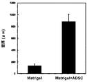
- the wall thickness was measured by cutting the ring around the center.
- the wall thickness of the tissue covering the substrate filled with ADSC-embedded Matrigel (883 ⁇ 122 ⁇ m) is about 6 times that of the tissue covering the substrate filled only with Matrigel (137 ⁇ 31 ⁇ m) It was found that a high-thickness structure was formed by slowly releasing ADSC from the porous cylindrical substrate (FIG. 4).
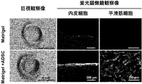
- tubular tissue was immersed in PBS containing 4% paraformaldehyde, fixed overnight, frozen, and thin sections (thickness 8 ⁇ m) were prepared using a frozen section preparation machine (cryostat). After air-drying thin sections and incubating with PBS containing 1% bovine serum albumin (Sigma) for 1 hour, endothelial cells were treated with Isolectin B4-FITC conjugate (1: 200, Sigma) for 1 hour, and smooth muscle cells were the primary antibody.
- PBS containing 1% bovine serum albumin Sigma
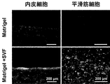
- Endothelial cells and smooth muscle cells were scarcely observed in the tubular tissue obtained from the matrix filled only with Matrigel, but the tubular tissue obtained from the matrix filled with ADSC-embedded Matrigel contained no lumen. Endothelial cells were confirmed to be oriented along the plane, and smooth muscle cells were present in the vicinity of the intermediate layer (FIG. 5). By gradually releasing ADSC using a porous substrate, a tubular tissue body having endothelial cells and smooth muscle cells as vascular constituent cells was obtained.
- adipose tissue is collected from the subcutaneous abdomen of a beagle dog (female, 10 kg body weight), suspended in 0.1% collagenase type I solution (dissolved in serum-free DMEM solution), and shaken at 37 ° C for 1 hour. To digest the adipose tissue. The digested liquid was centrifuged at 1000 rpm for 5 minutes at room temperature, and a cell mass that settled to the bottom of the centrifuge tube was obtained as a group of fat-derived blood vessel constituent cells (SVF). SVF is known as a cell group including mesenchymal stem cells in addition to vascular endothelial cells and smooth muscle cells.
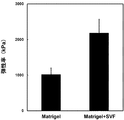
- the elastic modulus (2185 ⁇ 384 kPa) of the tissue covering the base material filled with SVF-embedded Matrigel is about twice that of the tissue covering the base material filled with Matrigel (1022 ⁇ 178 kPa). It was found that a high-strength tissue was formed by slowly releasing SVF from the cylindrical substrate.
- tubular tissue was immersed in PBS containing 4% paraformaldehyde, fixed overnight, frozen, and thin sections (thickness 8 ⁇ m) were prepared using a frozen section preparation machine (cryostat). After air-drying thin sections and incubating with PBS containing 1% bovine serum albumin (Sigma) for 1 hour, endothelial cells were treated with Isolectin B4-FITC conjugate (1: 200, Sigma) for 1 hour, and smooth muscle cells were the primary antibody.
- PBS containing 1% bovine serum albumin Sigma
- Endothelial cells were confirmed to be oriented along the plane, and smooth muscle cells were present in the vicinity of the intermediate layer (FIG. 7). It was found that a tubular tissue body having endothelial cells and smooth muscle cells, which are vascular constituent cells, can be obtained by slowly releasing ADSC using a porous substrate.
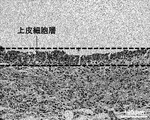
- a layered structure consisting of epithelial cells having a thickness of about 50 ⁇ m was observed on the surface of the lumen of the obtained tubular tissue (FIG. 9). It was found that the epithelial cell layer can be introduced on the surface of the tissue body mainly composed of collagen and fibroblasts by sustained release of OSECs using a porous substrate.
- the tissue body thus obtained is suitably used as an artificial cornea, similar to a cell sheet derived from oral mucosal epithelial cells that has already been confirmed in clinical practice.
- Example of vascular cells invading by holding the vascular tissue on the mold substrate as it is (Tissue collection and processing) A common carotid artery blood vessel (length: about 1 cm) of a beagle dog (female, body weight: 10 kg) was collected, and this was cut in the long axis direction to obtain a sheet-like blood vessel piece of about 1 cm ⁇ 1 cm.
- FIG. 10 shows a photograph of the produced mold base material.
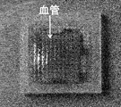
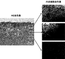
- the obtained tissue body has a very high film thickness (film thickness: about 2 mm), and a high cell density layer (inner layer, with many new blood vessels (Fig. 12, white arrows) from the contact surface side with the concave substrate. It had a two-layer structure of about 0.5 to 1 mm, and a layer with a dense collagen and a relatively small amount of cellular components (outer layer, about 0.5 to 1 mm).
- outer layer about 0.5 to 1 mm
- endothelial cells were hardly found in the outer layer mainly composed of collagen, it was confirmed by endothelial cell staining that the endothelial cells were present in high density in the inner layer rich in many new blood vessels and cellular components. .
- Oral mucosal tissue (about 5 mm ⁇ 5 mm) was collected from the palate of a beagle dog (female, body weight 10 kg).
- a base material having the same shape and size as those prepared in Example 4 was used as a mold base material.
- the aforementioned oral mucosa tissue piece was placed in a square concave base material so that the epithelial cell layer on the surface was in contact with the micropores on the bottom surface of the base material, and the convex base material was fitted from above (FIG. 13).
- This substrate was embedded in a gap formed in the subcutaneous connective tissue on the left side of the chest of a beagle dog. The experiment was performed using one beagle dog.
- an oral mucosal tissue-derived epithelial cell layer can be introduced on the surface of a tissue body mainly composed of collagen and fibroblasts by sandwiching an oral mucosa tissue piece between a porous concave substrate and a convex substrate. It was.
- the tissue body thus obtained is suitably used as an artificial cornea, similar to a cell sheet derived from oral mucosal epithelial cells that has already been confirmed in clinical practice.
Abstract
Provided are: a method for forming a tissue body for transplantation which comprises a biological tissue containing a desired cell or a cell having a function similar to that of the desired cell; and a template base for use in the production of an artificial tissue body for transplantation, which can be used in the method.
Provided is a template base for use in the production of an artificial tissue body for transplantation, which can carry thereon a desired cell, a cell that can become an alternative for the desired cell or a cell that can be differentiated into the desired cell in such a manner that the cell can be exuded out of the surface of the base. The template base can be used for producing an artificial tissue body for transplantation, wherein the production of the artificial tissue body is carried out by placing the template base, on which a desired cell, a cell that can become an alternative for the desired cell or a cell that can be differentiated into the desired cell has been carried, under the environment in which a biological tissue is present so that the template base can be covered with the biological tissue containing the desired cell or a cell having a function similar to that of the desired cell, and then removing the template base from the resultant product.
Description
本発明は、移植用人工組織体製造用鋳型基材に関する。
The present invention relates to a mold base material for producing an artificial tissue body for transplantation.
病気や事故で失われた細胞、組織、器官を、人工素材や細胞により再び蘇らせる再生医療の研究が数多くなされている。通常、身体には自己防衛機能があり、体内の浅い位置にトゲ等の異物が侵入した場合には体外へ押し出そうとするが、体内の深い位置に異物が侵入した場合にはその周りに繊維芽細胞が集まってきて、主に繊維芽細胞とコラーゲンからなる結合組織体のカプセルを形成し異物を覆うことにより、体内において隔離することが知られている。後者のような自己防衛反応を利用して、生体内において生体由来組織体を形成させ、これを移植に用いる生体内組織形成技術が提案されている(非特許文献1)。
There have been many studies on regenerative medicine in which cells, tissues, and organs lost due to illness and accidents are revived with artificial materials and cells. Normally, the body has a self-defense function, and when a foreign object such as a thorn enters a shallow position inside the body, it tries to push it out of the body, but when a foreign object enters a deep position inside the body, It is known that fibroblasts gather and sequester in the body by forming a capsule of connective tissue composed mainly of fibroblasts and collagen and covering foreign matter. An in vivo tissue formation technique in which a living body-derived tissue body is formed in a living body by using the latter self-defense reaction and used for transplantation has been proposed (Non-Patent Document 1).
生体内組織形成技術により、人工材料を全く含まない移植用の組織体を形成することが可能である。またかかる技術においては、体内をバイオリアクターとして用いるため、従来の組織工学に必須であった煩雑な細胞操作を必要とせず、安全、簡便に、そして低コストで組織体を製造することができる。さらに、移植を受ける者本人の体内にて組織を形成させる場合には、移植される組織は自家組織であり、移植後の生体適合における免疫上の問題、感染症等の問題を排除することもできる。
It is possible to form a tissue body for transplantation that does not contain artificial materials at all by the in vivo tissue formation technology. Further, in such a technique, since the inside of the body is used as a bioreactor, a complicated body operation that is essential for conventional tissue engineering is not required, and a tissue body can be produced safely, simply, and at low cost. Furthermore, when forming a tissue in the body of the person who receives the transplant, the transplanted tissue is an autologous tissue, and it is possible to eliminate problems such as immunity problems and infectious diseases in biocompatibility after transplantation. it can.
より生体組織に近い、あるいは移植のために有用な組織体を得るために鋳型となる基材については種々の検討が為されている。例えば特許文献1には、棒状構造体の表面に螺旋状溝を形成し、この棒状構造体を生体内に埋入することにより、棒状構造体の表面に膜状の結合組織体を形成し、結合組織体の機械的強度を増加させる点が開示されている。
Various studies have been made on a base material that is a template for obtaining a tissue body closer to a living tissue or useful for transplantation. For example, in Patent Document 1, a spiral groove is formed on the surface of a rod-like structure, and the rod-like structure is embedded in a living body to form a membrane-like connective tissue on the surface of the rod-like structure. A point of increasing the mechanical strength of the connective tissue is disclosed.
特許文献2には、棒状構造部材の外周に沿って外郭部材を螺旋形に形成し、これを生体に埋入して、棒状構造部材の外縁に結合組織体を形成する点が開示されている。結合組織体が外郭部材と棒状構造部材の表面との間に侵入し、結合組織体の内面形状が棒状構造部材の表面と同様の平滑面に形成される。結合組織体が、外郭部材を包埋する厚さに形成される。
Patent Document 2 discloses that an outer shell member is formed in a spiral shape along the outer periphery of a rod-shaped structural member, and this is embedded in a living body to form a connective tissue body on the outer edge of the rod-shaped structural member. . The connective tissue body enters between the outer member and the surface of the rod-shaped structural member, and the inner surface shape of the connective tissue body is formed on the same smooth surface as the surface of the rod-shaped structural member. A connective tissue body is formed to a thickness that embeds the shell member.
特許文献3には、棒状構造部材の表面に外郭部材を形成し、これを結合組織形成用基材とする点が開示されている。この基材を生体内に埋入することにより、基材表面に膜状の組織体を形成する。外郭部材の材料として、生体適合性に優れるが組織体やその構成成分に侵襲されにくい材料を使用することにより、外郭部材は組織体と癒着し結合組織体の機械的強度が増加されるとともに外郭部材の内面に組織体やその構成成分が露出しない人工血管が得られる。
Patent Document 3 discloses that an outer member is formed on the surface of a rod-shaped structural member, and this is used as a connective tissue forming substrate. By embedding this base material in the living body, a film-like tissue body is formed on the surface of the base material. By using a material that is excellent in biocompatibility but not easily invaded by the tissue body or its constituents as the material of the shell member, the shell member adheres to the tissue body, and the mechanical strength of the connective tissue body is increased and the shell is increased. An artificial blood vessel in which the tissue body and its constituent components are not exposed on the inner surface of the member is obtained.
特許文献4には、基材を生体内に埋入して結合組織体を形成させる際に光照射をすることによって、より生体に近い組織体を得る方法が開示されている。
Patent Document 4 discloses a method of obtaining a tissue body closer to a living body by irradiating light when a base material is embedded in the living body to form a connective tissue body.
また、管状や膜状の単純な形状の組織体のみならず、複雑な形状を有する鋳型基材についての開発も行われている。特許文献5および非特許文献2には、バルサルバ洞の形状を有する張り出し部位と三葉弁を有する弁付き人工血管を得るための芯基材を開示する。
Further, not only a tubular or membrane-like tissue body but also a mold base material having a complicated shape has been developed. Patent Document 5 and Non-Patent Document 2 disclose a core base material for obtaining a valved artificial blood vessel having an overhanging portion having a Valsalva sinus shape and a trilobal valve.
生体組織はその組織に特有の細胞にて構成されている。例えば血管はその内表面が血管内皮細胞に覆われており、この血管内皮細胞が、血栓の形成を妨げる働きを担っている。一方、上記非特許文献1に記載の方法で得られる組織体は、主に結合組織体、即ち繊維芽細胞とコラーゲンマトリックスから形成される。かかる人工血管を生体へ移植した場合、移植後に生体環境に応じた自己組織が徐々に構築されるものの、移植直後は表面にコラーゲン繊維や細胞が露出しているため血栓形成の問題がある。血栓形成を防止するために、移植前にアルガトロバン、ヒルジン、ヘパリンなどの抗血栓物質でコーティングする等の処理が行われている。
A living tissue is composed of cells peculiar to the tissue. For example, the inner surface of a blood vessel is covered with vascular endothelial cells, and these vascular endothelial cells have a function of preventing the formation of thrombus. On the other hand, the tissue obtained by the method described in Non-Patent Document 1 is mainly formed from connective tissue, that is, fibroblasts and a collagen matrix. When such an artificial blood vessel is transplanted into a living body, a self-tissue corresponding to the living environment is gradually constructed after the transplantation, but there is a problem of thrombus formation because collagen fibers and cells are exposed on the surface immediately after the transplantation. In order to prevent thrombus formation, treatment such as coating with an antithrombotic substance such as argatroban, hirudin or heparin is performed before transplantation.
患者由来の角膜上皮細胞や口腔粘膜上皮細胞を培養してシート状にし、これを人工角膜として角膜移植に用いることは既に臨床にて行われている(特許文献6、非特許文献3および4)。
It has already been practiced clinically to cultivate patient-derived corneal epithelial cells and oral mucosal epithelial cells into a sheet and to use it as an artificial cornea for corneal transplantation (Patent Document 6, Non-Patent Documents 3 and 4). .
本発明は生体内組織形成技術において、所望の細胞もしくは所望の細胞に類似の機能を有する細胞を含む生体組織からなる移植用組織体を形成させるための方法、並びに当該方法に用いられる移植用人工組織体製造用鋳型基材を提供することを目的とする。
The present invention relates to a method for forming a tissue for transplantation comprising a desired tissue or a living tissue containing a cell having a function similar to that of a desired cell in in vivo tissue formation technology, and an artificial graft for use in the method. It aims at providing the mold base material for tissue body manufacture.
本発明は、所望の細胞、所望の細胞の代替となる細胞もしくは所望の細胞へ分化する能力を有する細胞を基材表面から外部へ侵出可能なように保持させ得る鋳型基材であって、所望の細胞、所望の細胞の代替となる細胞もしくは所望の細胞へ分化する能力を有する細胞を保持させた該鋳型基材を生体組織の存在する環境下に置くことによって、該鋳型基材を該所望の細胞もしくは所望の細胞に類似の機能を有する細胞を含む生体由来組織にて被覆させ、次いで被覆された鋳型基材より鋳型基材を取り除くことによって移植用人工組織体を製造するために用いられる、移植用人工組織体製造用鋳型基材を提供する。
The present invention is a template substrate that can hold desired cells, cells that substitute for the desired cells, or cells that have the ability to differentiate into the desired cells so as to be able to invade from the substrate surface to the outside, The template substrate is placed in an environment in which a living tissue exists, by placing the template substrate holding a desired cell, a substitute for the desired cell, or a cell capable of differentiating into a desired cell. Used to produce an artificial tissue body for transplantation by coating with a tissue derived from a living body containing desired cells or cells having functions similar to the desired cells, and then removing the template substrate from the coated template substrate There is provided a mold base material for producing an artificial tissue body for transplantation.
本発明はまた、
(1)上記鋳型基材へ、所望の細胞、所望の細胞の代替となる細胞もしくは所望の細胞へ分化する能力を有する細胞を保持させる工程、
(2)細胞を保持させた鋳型基材を、生体組織材料の存在する環境下に置く工程、
(3)生体組織材料の存在する環境から、生体組織で被覆された鋳型基材を取り出す工程、
(4)生体組織で被覆された鋳型基材から、鋳型基材を取り除く工程
を含む移植用人工組織体製造方法を提供する。 The present invention also provides
(1) A step of holding desired cells, cells that substitute for desired cells, or cells that have the ability to differentiate into desired cells on the template base material,
(2) A step of placing the template substrate holding the cells in an environment where biological tissue material exists,
(3) A step of taking out the mold base material coated with the biological tissue from the environment where the biological tissue material exists,
(4) Provided is a method for producing an artificial tissue body for transplant, which includes a step of removing a mold base material from a mold base material coated with a biological tissue.
本発明により、所望の細胞もしくは所望の細胞類似した機能を有する細胞を含む移植用組織体、例えば血管内皮細胞と平滑筋細胞を含む人工血管を提供することができる。本発明により、生体組織に近い細胞にて構成される移植用人工組織体を得ることができる。本発明により得られる移植用組織体は移植後速やかに所望の機能を発揮することができ、例えば人工血管の場合には血栓形成の危険性が低い、優れた移植用人工組織体を得ることができる。
According to the present invention, it is possible to provide a transplanted tissue body containing desired cells or cells having functions similar to desired cells, for example, an artificial blood vessel containing vascular endothelial cells and smooth muscle cells. According to the present invention, an artificial tissue body for transplantation composed of cells close to a living tissue can be obtained. The transplantable tissue obtained by the present invention can exhibit a desired function immediately after transplantation. For example, in the case of an artificial blood vessel, an excellent artificial tissue for transplantation having a low risk of thrombus formation can be obtained. it can.
本発明の鋳型基材は細胞を、基材表面から外部へ侵出可能なように保持させることができるよう形成されている。細胞は、鋳型基材中に分散包埋あるいは固定して保持させることができる。かかる態様においては、鋳型基材としては三次元細胞培養用足場材等の、細胞を保持可能でありかつ、一定の形状を保つことのできる材料が例示される。
The template base material of the present invention is formed so that cells can be held so as to be able to invade from the surface of the base material to the outside. The cells can be retained by being embedded in a dispersion or fixed in a template substrate. In such an embodiment, examples of the template base material include a material that can hold cells and can maintain a certain shape, such as a scaffold for three-dimensional cell culture.
別の態様としては、細胞が基材表面から外部へ侵出可能な通路を有する中空鋳型基材であって、該中空鋳型基材の中空部分へ細胞を封入して用いられるものが例示される。
Another embodiment is a hollow mold base material having a passage through which cells can invade from the surface of the base material to the outside, and used by enclosing cells in the hollow portion of the hollow mold base material. .
中空鋳型基材の材料は、目的とする移植用組織体を形成させるための鋳型基材として所望の形状に成形でき、生体に埋入した際に大きく変形することが無い強度(硬度)を有しており、生体内で容易に分解されず、滅菌可能であり、生体を刺激する溶出物が無いまたは少ないものであれば特に限定されるものではない。
The material of the hollow mold base material can be molded into a desired shape as a mold base material for forming a target transplant tissue, and has a strength (hardness) that does not greatly deform when implanted in a living body. It is not particularly limited as long as it is not easily decomposed in the living body, can be sterilized, and has no or little eluate that stimulates the living body.
好ましいものとしては、アクリル樹脂、オレフィン樹脂、スチレン樹脂、ポリエステル樹脂、ポリアミド樹脂、塩化ビニル樹脂、シリコン樹脂、フッ素樹脂、エポキシ樹脂、ポリカーボネート樹脂、ポリエチレン樹脂、ポリウレタン樹脂、ガラス、金属(チタン、プラチナ、ステンレス、及びSUS(ステンレス鋼(Stainless steel))等)等が挙げられる。材質は1種類に限定される訳では無く、2種以上を組み合わせて用いることができる。
Preferable ones are acrylic resin, olefin resin, styrene resin, polyester resin, polyamide resin, vinyl chloride resin, silicon resin, fluorine resin, epoxy resin, polycarbonate resin, polyethylene resin, polyurethane resin, glass, metal (titanium, platinum, Stainless steel, SUS (stainless steel, etc.) etc. are mentioned. The material is not limited to one type, and two or more types can be used in combination.
一定の強度を有する点ではアクリル樹脂等が好ましく、製造が容易である点では、シリコン樹脂が好ましい。また、鋳型基材の形状が太さの均一な柱状等の単純な形状であって、形成された人工組織体の一部を切開すれば容易に鋳型基材を抜き出すことのできるものである場合には樹脂の硬度に特段の制限はない。形成される人工組織体の一部より鋳型基材を抜き出すのが困難な形状である場合には、鋳型基材の材料としては抜き出す際に押しつぶせるような柔らかな樹脂が好適に用いられる。また、基材を取り出しやすいよう、複数のパーツから基材を適宜組み合わせて用いて所望の形状の人工組織体を得てもよい。
An acrylic resin or the like is preferable in terms of having a certain strength, and a silicon resin is preferable in terms of easy manufacture. Also, when the shape of the mold base material is a simple shape such as a columnar shape having a uniform thickness, and the mold base material can be easily extracted by incising a part of the formed artificial tissue body There is no particular restriction on the hardness of the resin. When it is difficult to extract the mold base material from a part of the artificial tissue body to be formed, a soft resin that can be crushed when extracted is suitably used as the material of the mold base material. In addition, an artificial tissue body having a desired shape may be obtained by appropriately combining the base materials from a plurality of parts so that the base material can be easily taken out.
中空鋳型基材は、内部に保持した細胞が外側へ侵出できるよう外部との通路が設けられている。通路は限定的ではなく、例えば孔であってもスリットであってもよい。孔の形状やサイズ、スリットの長さや幅、並びにそれらの数は細胞が通過できるものであれば特に限定されるものではなく、内部に封入する細胞、あるいは内部へ細胞を封入する際の態様に応じて、適宜定めればよい。
The hollow mold base is provided with a passage to the outside so that the cells held inside can invade to the outside. The passage is not limited, and may be a hole or a slit, for example. The shape and size of the holes, the length and width of the slits, and the number thereof are not particularly limited as long as the cells can pass through, and the shape of the cells encapsulated inside or the mode of encapsulating the cells inside Accordingly, it may be determined appropriately.
例えば中空基材へ孔を形成する場合には、孔の径は細胞が通過可能な口径10μm以上であればよく、例えば0.01mm以上,加工の容易性から直径0.1mm以上の孔を有する中空基材が例示される。孔の口径の上限は特に限定的ではなく担持させた細胞が一気に流出するものでなければよい。中空鋳型基材中に支持基材、例えばマトリゲル(商標)などの市販のゲル状細胞支持基材等を封入して用いる場合は、例えば孔は例えば5mm程度まで、好ましくは1mm程度までの大きさであってもよい。
For example, when forming a hole in a hollow base material, the diameter of the hole may be 10 μm or more through which cells can pass, for example, 0.01 mm or more. A hollow substrate is exemplified. The upper limit of the pore diameter is not particularly limited as long as the loaded cells do not flow out at a stretch. When a support base material, for example, a commercially available gel-like cell support base material such as Matrigel (trademark) or the like is encapsulated in a hollow mold base material, for example, the hole has a size of up to about 5 mm, preferably up to about 1 mm. It may be.
孔の密度としては、例えば1.5cm×1.5cmの正方形の板状中空基材の表面に5~50個×5~50個、例えば15×15個の孔を空けた鋳型基材が例示される。
An example of the density of the holes is a mold base material in which 5 to 50 × 5 to 50 holes, for example, 15 × 15 holes are formed on the surface of a 1.5 cm × 1.5 cm square plate-shaped hollow base material. Is done.
本発明において、「所望の細胞」とは、人工組織体構成のために必要な細胞を意味する。例えば人工血管や弁付き人工血管等の循環器系組織体を製造する場合には、所望の細胞としては血管内皮細胞並びに平滑筋細胞が挙げられる。人工角膜を製造する場合には、所望の細胞としては角膜上皮細胞が挙げられる。
In the present invention, the “desired cell” means a cell necessary for constructing the artificial tissue body. For example, when producing a circulatory tissue such as an artificial blood vessel or a valved artificial blood vessel, examples of desired cells include vascular endothelial cells and smooth muscle cells. In the case of producing an artificial cornea, the desired cells include corneal epithelial cells.
「所望の細胞の代替となる細胞」とは、所望の細胞と類似の機能を有する細胞であり、例えば血管内皮細胞や角膜上皮細胞等の上皮細胞の代替となる細胞としては口腔粘膜上皮細胞が例示される。口腔粘膜上皮細胞を培養してシート状にし、これを人工角膜として移植に用いることは既に臨床にて行われている(人工臓器38巻第3号2009年168-172頁)、Nishida et al., N Engl J Med 351;12; 2004; 1187-1196。
“Cells that substitute for desired cells” are cells having functions similar to those of desired cells. For example, oral mucosal epithelial cells are used as substitutes for epithelial cells such as vascular endothelial cells and corneal epithelial cells. Illustrated. Oral mucosal epithelial cells are cultured into a sheet and used as an artificial cornea for transplantation in clinical practice (Artificial organ 38, No. 3, 2009, pages 168-172), Nishida et al. N Engl J Med 351; 12; 2004; 1187-1196.
「所望の細胞へ分化する能力を有する細胞」としてはかかる能力を有する細胞であれば特に限定されるものではない。体性幹細胞、iPS細胞およびES細胞などの分化万能性細胞などが例示される。体性幹細胞の中でも間葉系幹細胞は、脂肪細胞、線維芽細胞、間質細胞、心筋細胞、平滑筋細胞、内皮細胞、上皮細胞、骨芽細胞、軟骨細胞、神経細胞など様々な細胞への分化能を有している観点から所望の細胞へ分化する能力を有する細胞として好適に用いられる。間葉系幹細胞は、哺乳動物の骨髄液、末梢血、臍帯血、脂肪組織などから公知の方法で採取することが出来る。
The “cell having the ability to differentiate into a desired cell” is not particularly limited as long as it has such ability. Examples include somatic stem cells, iPS cells, and pluripotent cells such as ES cells. Among somatic stem cells, mesenchymal stem cells are used for various cells such as adipocytes, fibroblasts, stromal cells, cardiomyocytes, smooth muscle cells, endothelial cells, epithelial cells, osteoblasts, chondrocytes, and nerve cells. It is suitably used as a cell having the ability to differentiate into a desired cell from the viewpoint of having differentiation ability. Mesenchymal stem cells can be collected by known methods from mammalian bone marrow fluid, peripheral blood, umbilical cord blood, adipose tissue, and the like.
「所望の細胞と類似の機能を有する細胞」とは、所望の細胞の全ての機能と同等あるいは類似の機能を発揮しうるものみならず、移植用人工組織体として望ましい機能が所望の細胞と類似している細胞も含まれる。
“A cell having a function similar to a desired cell” means not only a function that is equivalent to or similar to all functions of a desired cell, but also a function that is desirable as an artificial tissue body for transplantation. Similar cells are also included.
所望の細胞、所望の細胞の代替となる細胞または所望の細胞へ分化する能力を有する細胞としては、移植対象者より取得された細胞、移植対象者より取得された細胞の培養物、移植対象者の細胞より得られる万能性幹細胞等、移植対象者に由来する細胞を用いることが好ましい。移植対象者以外の者に由来する細胞を用いる場合、当該細胞のドナーは移植対象者とHLAの適合性が高い者であることが必要である。
As a desired cell, a cell that can substitute for a desired cell, or a cell that has the ability to differentiate into a desired cell, a cell obtained from a transplant recipient, a culture of a cell obtained from a recipient, a transplant recipient It is preferable to use cells derived from the transplant recipient, such as universal stem cells obtained from these cells. When cells derived from a person other than the transplant recipient are used, it is necessary that the donor of the cell has high compatibility with the transplant recipient and HLA.
本発明の鋳型基材へ保持させる細胞としては、生体より得られた組織をそのまま、あるいは適当な形状へ加工して用いてもよいし、生体より分離した組織の全体あるいは部分を分散させたものでもよい。また、生体より得られた組織を分散させて細胞を得、得られた細胞を常套法で処理して目的とする細胞を多く含む分画を用いても、目的とする細胞集団のみを単離したものを用いてもよい。
As the cells to be held on the template substrate of the present invention, the tissue obtained from the living body may be used as it is or after being processed into an appropriate shape, or the whole or part of the tissue separated from the living body is dispersed. But you can. In addition, cells obtained from living organisms can be dispersed to obtain cells, and the obtained cells can be processed by conventional methods to isolate only the target cell population even if the fraction containing a large amount of the target cells is used. You may use what you did.
別の態様においては、上記のようにして生体より得られた細胞を培養した培養細胞を用いてもよい。また、公知の方法にて得られるiPS細胞やES細胞を用いてもよい。
In another embodiment, cultured cells obtained by culturing cells obtained from a living body as described above may be used. Moreover, you may use the iPS cell and ES cell obtained by a well-known method.
一の態様において、所望の細胞、所望の細胞の代替となる細胞または所望の細胞ヘ分化する能力を有する細胞としては、ペレット状の凝集塊、生体より摘出された組織、培養によって得られた組織、およびこれらを部分的に分散させた状態のものを中空鋳型基材の中空部分へ封入してもよい。
In one embodiment, a desired cell, a substitute for a desired cell, or a cell having an ability to differentiate into a desired cell may be a pellet-like aggregate, a tissue extracted from a living body, or a tissue obtained by culture , And those partially dispersed may be enclosed in the hollow portion of the hollow mold base material.
別の態様においては、中空鋳型基材の中空部分へ、細胞と細胞支持基材を封入してもよい。細胞は支持基材中に分散包埋されていても、固定されていてもよい。かかる態様に用いられる細胞支持基材としては、市販の三次元細胞培養用基材が用いられ、例えば細胞培養用コラーゲンゲル、BD マトリゲル(商標)、細胞培養用ゼラチン、寒天、ヒアルロン酸ハイドロゲルなどのムコ多糖類が例示される。
In another embodiment, the cells and the cell support substrate may be enclosed in the hollow portion of the hollow mold substrate. The cells may be dispersed and embedded in the support substrate or may be fixed. As the cell support substrate used in such an embodiment, a commercially available substrate for three-dimensional cell culture is used. For example, collagen gel for cell culture, BD Matrigel (trademark), gelatin for cell culture, agar, hyaluronic acid hydrogel, etc. Of mucopolysaccharides.
市販の三次元細胞培養用基材を用いる場合には、添付される説明書に従って該三次元細胞培養用基材中に細胞が埋入、あるいは固定化されたものを中空鋳型基材の中空部分に保持させる。例えば、BD マトリゲル(商標)を用いる場合は、説明書に記載の手順で液状としたマトリゲルと細胞を混合し、これを基材の中空部分へ注入し、しばらく置くことによって、基材の中空部分内部でマトリゲルをゲル化させればよい。
When using a commercially available three-dimensional cell culture substrate, a hollow portion of the hollow mold substrate is prepared by embedding or fixing cells in the three-dimensional cell culture substrate according to the attached instructions. To hold. For example, in the case of using BD Matrigel (trademark), the liquid Matrigel and cells are mixed according to the procedure described in the instructions, injected into the hollow part of the base material, and left for a while, whereby the hollow part of the base material is mixed. The matrigel may be gelled inside.
市販の細胞培養用基材は、細胞培養に適した環境を提供するものであり、三次元の足場並びに適当な増殖因子を含んでいるものが多い。本発明において、細胞を鋳型基材内へ保持する際に、適当な増殖因子を含む市販の細胞支持基材を採用してもよいし、細胞に応じた増殖因子を添加してもよい。添加する増殖因子としては、例えば人工血管などの循環器系人工組織体を製造する場合には血管内皮細胞増殖因子(VEGF)や繊維芽細胞増殖因子(bFGF)などの増殖因子に加えて、血管壁細胞に分化させる目的の分化誘導因子などを添加することが例示される。保持する細胞並びに目的とする組織に応じて増殖因子を適宜選択し、組み合わせて用いればよい。
Commercially available cell culture substrates provide an environment suitable for cell culture, and often include a three-dimensional scaffold and an appropriate growth factor. In the present invention, when the cells are held in the template substrate, a commercially available cell support substrate containing an appropriate growth factor may be employed, or a growth factor corresponding to the cell may be added. As a growth factor to be added, for example, in the case of producing a circulatory prosthesis such as an artificial blood vessel, in addition to growth factors such as vascular endothelial growth factor (VEGF) and fibroblast growth factor (bFGF), blood vessels An example is the addition of a differentiation-inducing factor for the purpose of differentiation into mural cells. A growth factor may be appropriately selected according to the cells to be retained and the target tissue and used in combination.
細胞支持基材としてはまた、止血用フィブリン糊のごとき血液由来のゲル状物質を用いてもよく、あるいは細胞支持基材として血液(全血)を用い、細胞と血液を混合して中空基材の中空部へ封入して用いてもよい。血液を用いる場合には、血液と、細胞を混合し、そのまま中空鋳型基材へ注入してしばらく置くと、血液が凝固し、細胞が凝固した血中に分散された形で保持される。血液を用いる場合には、保持される細胞が由来した生体より取得した血液が好適に用いられる。
The cell support substrate may also be a blood-derived gel-like substance such as fibrin glue for hemostasis, or blood (whole blood) is used as the cell support substrate, and cells and blood are mixed to form a hollow substrate. You may enclose and use in the hollow part. In the case of using blood, when blood and cells are mixed, injected into the hollow mold base as it is and left for a while, the blood coagulates and the cells are held in a dispersed form in the coagulated blood. In the case of using blood, blood obtained from a living body from which the retained cells are derived is preferably used.
本発明はまた、上記のような中空鋳型基材と、細胞支持基材とを含むキットを提供する。
The present invention also provides a kit including the hollow mold substrate as described above and a cell support substrate.
本発明において、鋳型基材の形状は目的とする人工組織体の形状に合うよう設計すればよく、例えば管状組織体を得るためには柱状の、膜状組織体を得るためには板状の形状とするか、柱状の鋳型基材により得られる管状組織を切開して用いればよい。例えば人工血管を製造する場合には、円柱状鋳型基材とし、その外径により血管の太さが決定されることから目的の太さ並びに長さによって直径および長さを設計すればよい。
In the present invention, the shape of the template substrate may be designed to match the shape of the target artificial tissue body. For example, a columnar shape is used to obtain a tubular tissue body, and a plate shape is used to obtain a membrane-like tissue body. The shape may be used, or a tubular tissue obtained from a columnar mold base material may be cut and used. For example, when an artificial blood vessel is manufactured, a cylindrical mold base material is used, and since the thickness of the blood vessel is determined by the outer diameter, the diameter and length may be designed according to the target thickness and length.
また、柱状や膜状等の形状以外にも、所望の生体組織の形状に従って、球状、立方体状、直方体状等の他の形にしてもよい。
In addition to the shape such as a columnar shape or a membrane shape, other shapes such as a spherical shape, a cubic shape, a rectangular parallelepiped shape, and the like may be used according to the shape of a desired biological tissue.
さらには特許文献5に記載のようなバルサルバ洞の形状を有する張り出し部位と三葉弁を有する弁付き人工血管を得るための基材としてもよい。かかる基材は(a)柱状部位、および(b)弁形成部位より構成される。
Furthermore, a base material for obtaining a valved artificial blood vessel having an overhanging part having a Valsalva sinus shape as described in Patent Document 5 and a trilobal valve may be used. Such a substrate is composed of (a) a columnar part and (b) a valve forming part.
さらに別の態様としては、本発明の鋳型基材に体内へ留置するためのステントを被せ、ステントを内包した移植用人工組織体を得てもよい。本態様においては、細胞を保持させた鋳型基材の一部または全体にステントを被せたものを、生体組織の存在する環境下へ置く。ステントとしては、鋳型基材の表面からの細胞の侵出を妨害しないものであれば従来用いられている材質、形状のものが何れも好適に用いられる。ステントの材質としては、例えば、ニッケル-チタン合金、コバルト-クロム合金、ステンレス、鉄、チタン、アルミニウム、スズ、亜鉛-タングステン合金などが挙げられる。これらのなかでも、ステントとして実績があるニッケル-チタン合金またはSUS316L等のステンレスが好ましい。
As yet another aspect, a stent for indwelling in the body may be placed on the mold base material of the present invention to obtain an artificial tissue body for transplantation including the stent. In this embodiment, a part of or the entire template base material holding cells is covered with a stent is placed in an environment where living tissue exists. As the stent, any conventionally used material and shape can be suitably used as long as they do not interfere with the invasiveness of cells from the surface of the template base material. Examples of the material of the stent include nickel-titanium alloy, cobalt-chromium alloy, stainless steel, iron, titanium, aluminum, tin, and zinc-tungsten alloy. Among these, a nickel-titanium alloy or stainless steel such as SUS316L, which has a proven record as a stent, is preferable.
本発明の鋳型基材は、細胞を保持させて生体組織材料の存在する環境下に置くことによって、移植用人工組織体を製造するために用いられる。本発明はまた、本発明の鋳型基材を用いて移植用人工組織体を製造する方法を提供する。
The template base material of the present invention is used for producing an artificial tissue body for transplantation by holding cells and placing them in an environment where biological tissue materials are present. The present invention also provides a method for producing an artificial tissue body for transplantation using the mold base material of the present invention.
本発明の鋳型基材へ保持させる細胞の数は限定的でなく、保持させる細胞の種類、細胞支持基材を用いる、細胞や組織をそのまま用いる等の細胞保持の態様、後述する生体組織材料の存在する環境、所望する人工組織体の構成等に応じて最適化すればよい。
The number of cells to be retained on the template substrate of the present invention is not limited, the type of cells to be retained, the mode of cell retention such as using the cell support substrate, using cells and tissues as they are, and the biological tissue material described later What is necessary is just to optimize according to the environment which exists, the structure of the desired artificial tissue body, etc.
生体組織材料の存在する環境下とは、動物の生体内、および生体組織材料、例えば細胞が浮遊する培養液等の人工環境が例示される。
Examples of the environment in which the biological tissue material exists include an in vivo body of an animal and an artificial environment such as a culture fluid in which a biological tissue material, for example, a cell floats.
動物の生体内にて人工組織体を製造する場合、細胞を保持させた鋳型基材を生体内へ埋入する。埋入する対象は、移植を受ける対象と同種の動物であることが好ましい。また、埋入する対象は移植用人工組織体の移植を受ける対象と同一であっても異なっていてもよい。移植を受ける対象と異なる対象へ埋入する場合、両者のHLA型が適合している等、移植を受けた際に免疫反応を惹起しにくい対象に埋入することが好ましい。免疫反応惹起が無いことから、移植を受ける対象の体内へ埋入することが好ましい。
When producing an artificial tissue body in an animal body, a template base material holding cells is embedded in the body. The subject to be implanted is preferably an animal of the same species as the subject receiving the transplant. Moreover, the subject to be implanted may be the same as or different from the subject to receive the transplantation artificial tissue body. When embedding in a subject different from the subject to be transplanted, it is preferable to implant in a subject that is unlikely to elicit an immune response when undergoing transplantation, for example, both HLA types are compatible. Since there is no induction of an immune reaction, it is preferable to be implanted in the body of a subject to receive a transplant.
動物の生体内へ鋳型基材を埋入する場合、埋入する部位としては生体の活動に影響が少ない部位を選べばよく、四肢部、腰部、背部や腹部などの皮下、および腹腔内が例示されるがこれらに限定されない。埋入は適宜麻酔を施し、切開を最小限とし、可能な限り低侵襲な方法にて行うことが好ましい。
When embedding a template base material in an animal's body, it is only necessary to select a part that has little influence on the activity of the living body. Examples include the extremities, the waist, the back and the abdomen, and the abdominal cavity. However, it is not limited to these. It is preferable that the implantation is appropriately anesthetized, the incision is minimized, and a minimally invasive method is possible.
本発明の鋳型基材を人工環境である生体組織材料の存在する環境下へ置く場合には、適当な培地中に生体材料、例えば細胞を分散させたものを用いればよい。培地中には組織体形成を促進するための増殖因子等を適宜添加しても良い。培養条件を整え、無菌下で培養を行えばよい。
When placing the template base material of the present invention in an environment where biological tissue material, which is an artificial environment, is present, a biological material such as a cell dispersed in an appropriate medium may be used. A growth factor or the like for promoting tissue formation may be appropriately added to the medium. What is necessary is just to culture | cultivate under aseptic conditions and culture conditions.
本発明の細胞を保持させた鋳型基材を生体組織材料の存在する環境下に置くことにより、鋳型基材の周囲に組織体のカプセルが形成される。本発明の細胞を保持させた鋳型基材を生体組織材料の存在する環境下へ置く期間は限定的ではなく、生体内に埋入する場合は埋入する動物、その年齢、健康状態、埋入部位等、所望の細胞、所望の細胞の代替となる細胞あるいは所望の細胞に分化しうる細胞の種類、基材のサイズ、目的とする組織体の厚み、目的とする細胞の密度等によって適宜決定すればよい。例えば、哺乳動物の生体内へ本発明の細胞を保持させた鋳型基材を埋入する場合には、1~8週間、好適には2~6週間程度の期間置けばよい。
The capsule of the tissue body is formed around the template base material by placing the template base material holding the cells of the present invention in an environment where the biological tissue material exists. The period of placing the template substrate holding the cells of the present invention in the environment where the biological tissue material exists is not limited, and when implanted in the living body, the animal to be implanted, its age, health condition, implantation Depending on the site, desired cells, the types of cells that can substitute for the desired cells, or the cells that can differentiate into the desired cells, the size of the substrate, the thickness of the target tissue, the density of the target cells, etc. do it. For example, when the template substrate holding the cells of the present invention in a living body of a mammal is embedded, it may be placed for 1 to 8 weeks, preferably 2 to 6 weeks.
次いで生体組織材料の存在する環境下から、生体組織に覆われた鋳型基材を取り出す。生体内に鋳型基材を埋入した場合であっても、生体組織は生体組織を覆うようにカプセル状に形成され、容易に周囲の組織より剥離して取り出すことができる。次いで鋳型基材の周囲に形成された組織体より、鋳型基材を取り出す。鋳型基材の取り出しは、例えば管状組織であれば一端または両端を切断して基材を抜き出せばよい。鋳型基材が柔らかい樹脂で材料にて形成されている場合には、鋳型基材を押しつぶすようにして抜き出してもよい。
Next, the mold base material covered with the biological tissue is taken out from the environment where the biological tissue material exists. Even when a template base material is embedded in a living body, the living tissue is formed in a capsule shape so as to cover the living tissue, and can be easily peeled off from the surrounding tissue. Next, the mold base is taken out from the tissue formed around the mold base. For example, in the case of a tubular tissue, the mold base material may be extracted by cutting one end or both ends and extracting the base material. When the mold base is made of a soft resin material, the mold base may be extracted by crushing.
従来の鋳型基材により形成される人工組織体は主にコラーゲン、繊維芽細胞などの繊維性の結合組織体により構成され、その他の細胞があまり含まれないが、本発明の基材によれば、所望の細胞または所望の細胞に類似する機能を有する細胞を含む移植用人工組織体が得られる。例えば、鋳型基材へ間葉系幹細胞を基材表面より侵出するように保持させた鋳型基材を生体内へ埋入した場合、得られる組織体はコラーゲン、繊維芽細胞などの結合組織体を主体とするが、基材表面と接する部分が内皮細胞で覆われ、組織体内部には平滑筋細胞が含まれる態様の組織体が形成される。かかる組織体を管状製造すれば、例えば人工血管として好適に用いられる。
The artificial tissue body formed by the conventional mold base material is mainly composed of fibrous connective tissue bodies such as collagen and fibroblasts, and does not contain much other cells, but according to the base material of the present invention, Thus, an artificial tissue body for transplantation containing desired cells or cells having a function similar to the desired cells can be obtained. For example, when a template base material in which mesenchymal stem cells are held so as to invade from the surface of the base material is embedded in the living body, the resulting tissue is a connective tissue such as collagen or fibroblast. However, a portion of the base material surface that is in contact with the surface of the base material is covered with endothelial cells, and a tissue body in a form containing smooth muscle cells is formed inside the tissue body. If such a tissue body is manufactured in a tubular shape, it is suitably used as an artificial blood vessel, for example.
また、鋳型基材へ口腔粘膜上皮細胞を基材表面より侵出するように保持させた鋳型基材を生体内へ埋入した場合、得られる組織体の基材表面と接する部分は口腔粘膜上皮細胞由来の細胞層が形成され、その外側に繊維芽細胞層が形成される。かかる組織体を膜状に形成することにより、人工角膜として用い得る組織体を得ることができる。かかる人工角膜は角膜上皮と角膜実質を切除した後へ移植する表層または深層角膜移植手術に好適に用いることができる。
In addition, when a template base material in which oral mucosal epithelial cells are held in the template base material so as to invade from the base material surface is embedded in the living body, the portion of the resulting tissue that contacts the base material surface is the oral mucosal epithelium. A cell layer derived from cells is formed, and a fibroblast layer is formed outside the cell layer. By forming such a tissue body into a film shape, a tissue body that can be used as an artificial cornea can be obtained. Such an artificial cornea can be suitably used for a surface layer or deep layer corneal transplantation operation in which the corneal epithelium and the corneal stroma are excised.
また、本発明の細胞を保持させた鋳型基材を用いた場合、細胞を担持させない以外は同一の基材を用いた場合より、高膜厚の組織体を得ることができる。
In addition, when the template substrate holding cells of the present invention is used, a thicker tissue can be obtained than when the same substrate is used except that the cells are not supported.
本発明の細胞を保持させた鋳型基材は最も好適には、得られる人工組織体の移植を受ける対象と同一の対象の体内に埋入して人工組織体を製造する。かかる態様を用いることにより、免疫反応がなく、生体適合性に優れ、移植後に体内で成長でき、細胞の採取や培養、無菌室管理等の必要がないなどの利点が得られる。そして、得られた移植用人工組織体は本人由来の細胞を含んでおり、移植後早期に定着し、機能することが期待される。
Most preferably, the template base material holding the cells of the present invention is embedded in the body of the same subject as the subject to receive the obtained artificial tissue body to produce the artificial tissue body. By using such an embodiment, there are advantages such as no immune reaction, excellent biocompatibility, growth in the body after transplantation, and the need for cell collection, culture, and aseptic room management. The obtained artificial tissue for transplantation contains cells derived from the subject, and is expected to settle and function early after transplantation.
上記のようにして得られる人工組織体は、適宜トリミングして所望の形状とした後、対象への移植に用いることができる。
以下実施例により本願発明を更に詳細に説明する。 The artificial tissue obtained as described above can be appropriately trimmed to a desired shape, and then used for transplantation into a subject.
Hereinafter, the present invention will be described in more detail with reference to examples.
以下実施例により本願発明を更に詳細に説明する。 The artificial tissue obtained as described above can be appropriately trimmed to a desired shape, and then used for transplantation into a subject.
Hereinafter, the present invention will be described in more detail with reference to examples.
皮下から採取した脂肪より脂肪由来間質細胞を分離して、それを基材から侵出させた例
(細胞の分離と培養)
生後10週目のWister Rat(雄、体重300 g)の後肢部皮下から約1 gの脂肪組織を採取し、0.1%のコラゲナーゼタイプI溶液(無血清のDMEM溶液に溶解)に懸濁し、37℃で1時間振とうすることで細胞懸濁液を得た。この細胞懸濁液を37℃、5% CO2雰囲気下にて1週間培養して、培養皿に接着した繊維芽状の形態を有する細胞を脂肪由来間質細胞(Adipose-derived stromal cells: ADSC)として得た(図1)。脂肪由来間質細胞は、間葉系細胞の一種として知られている細胞である。 Example of separating fat-derived stromal cells from fat collected from the skin and invading it from the base material
(Separation and culture of cells)
Approximately 10 g of adipose tissue is collected from the hind limb subcutaneously of a 10-week-old Wister Rat (male, weight 300 g), suspended in 0.1% collagenase type I solution (dissolved in serum-free DMEM solution), 37 The cell suspension was obtained by shaking at 0 ° C. for 1 hour. This cell suspension is cultured at 37 ° C in a 5% CO2 atmosphere for 1 week, and the cells with fibroblast-like morphology adhered to the culture dish are converted into adipose-derived stromal cells (ADSC). (FIG. 1). Adipose-derived stromal cells are cells known as a kind of mesenchymal cells.
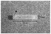
(鋳型基材の作製)
直径0.5 mmの微細孔を180個有するアクリル製の多孔性円筒型基材(口径: 5 mm, 長さ: 2.4 cm、両端に蓋付き)を、3次元光造形機(Projet HD3000)を用いて作製した(図2)。 (Production of mold substrate)
Using a three-dimensional stereolithography machine (Projet HD3000), an acrylic porous cylindrical substrate (caliber: 5 mm, length: 2.4 cm, with lids on both ends) having 180 micropores with a diameter of 0.5 mm It produced (FIG. 2).
(細胞並びに細胞支持基材を充填した鋳型基材の皮下への埋入)
片端に蓋をした先の円筒基材内にマトリゲル(BD, 11715)中に懸濁した9.0×10⁶ cells/mlの濃度のADSCの懸濁液を333 μl(それぞれ基材あたり3.0×10⁶ cellsとなる)流し込んだ後、もう一方の端に蓋をし、37℃のインキュベーター内で15分静置することでマトリゲルを完全にゲル化させた。15分後、上述のADSCの分離に用いたWister Ratの後背部皮下結合組織内に作製した間隙に基材を埋入した。また、比較例としてADSCを含まないマトリゲルのみを充填した基材を同様の方法で同一のラットに埋入した。実験は各実験群で5匹のラットを用いて行った。 (Subcutaneous embedding of template substrate filled with cells and cell support substrate)
333 μl of a suspension of ADSC at a concentration of 9.0 × 10 6 cells / ml suspended in Matrigel (BD, 11715) in a cylindrical substrate with a lid on one end (3.0 × 10 6 per substrate each) Then, the other end was capped and allowed to stand in an incubator at 37 ° C. for 15 minutes to completely gel the Matrigel. After 15 minutes, the substrate was embedded in the gap created in the dorsal subcutaneous connective tissue of the Wister Rat used for the above-mentioned ADSC separation. Further, as a comparative example, a base material filled with only Matrigel not containing ADSC was implanted in the same rat by the same method. The experiment was performed using 5 rats in each experimental group.
ここでBD マトリゲル(商標)は、細胞外マトリックスタンパク質に富むEngelbreth Holm Swarn(EHS) マウス肉腫から抽出された可溶性基底膜調製品であり、ラミニン、コラーゲンIV、エンタクチン、ヘパラン硫酸プロテオグリカンを主成分とする、室温にて重合してゲル状となる哺乳類の細胞基底膜と似た生物活性のあるマトリックスとなる製品である。増殖因子として、EGF、PDGF、IGF-1およびTGFβが含まれている。
Here, BD Matrigel (trademark) is a soluble basement membrane preparation extracted from Engelbreth Holm Swarn (EHS) 腫 mouse sarcoma, which is rich in extracellular matrix protein, and is mainly composed of laminin, collagen IV, entactin, and heparan sulfate proteoglycan It is a product that forms a bioactive matrix similar to the mammalian cell basement membrane that is polymerized at room temperature to form a gel. Growth factors include EGF, PDGF, IGF-1 and TGFβ.
(基材の摘出と組織観察)
埋入から2週間後、細胞に覆われた基材を皮下から摘出した。摘出された被覆基材の写真、管状の被覆組織体より基材を剥離して得た組織体の写真を図3に示す。また、ホルマリンにて1時間固定した後、中央辺りで輪切りにして壁厚の測定を行った。 (Extraction of base material and tissue observation)
Two weeks after implantation, the substrate covered with cells was removed subcutaneously. FIG. 3 shows a photograph of the extracted coated substrate and a photograph of the tissue obtained by peeling the substrate from the tubular coated tissue. In addition, after fixing with formalin for 1 hour, the wall thickness was measured by cutting the ring around the center.
ADSC包埋マトリゲルを充填した基材を被覆する組織体の壁厚(883±122 μm)は、マトリゲルのみを充填した基材を被覆する組織体(137±31 μm)の約6倍を示し、多孔性円筒基材からADSCを徐放することで高膜厚な組織が形成されることが分かった(図4)。
The wall thickness of the tissue covering the substrate filled with ADSC-embedded Matrigel (883 ± 122 μm) is about 6 times that of the tissue covering the substrate filled only with Matrigel (137 ± 31 μm) It was found that a high-thickness structure was formed by slowly releasing ADSC from the porous cylindrical substrate (FIG. 4).
(組織学的分析)
得られた管状組織体を、4%パラホルムアルデヒドを含有するPBSに浸漬し一晩固定後、凍結し、凍結切片作製機(クライオスタット)を用いて薄切片(厚さ 8 μm)を作製した。薄切片を風乾し、1%ウシ血清アルブミン(Sigma)を含有するPBSで1時間インキュベートした後、内皮細胞はIsolectin B4-FITC conjugate(1:200, Sigma)と1時間、平滑筋細胞は一次抗体としてマウス抗ラットα-SMAモノクローナル抗体(1:100, Abcam)と1時間、二次抗体としてFITC標識抗マウスIgG抗体(1:100, Santacruz)と1時間反応させることで染色した。観察結果を図5に示す。 (Histological analysis)
The obtained tubular tissue was immersed in PBS containing 4% paraformaldehyde, fixed overnight, frozen, and thin sections (thickness 8 μm) were prepared using a frozen section preparation machine (cryostat). After air-drying thin sections and incubating with PBS containing 1% bovine serum albumin (Sigma) for 1 hour, endothelial cells were treated with Isolectin B4-FITC conjugate (1: 200, Sigma) for 1 hour, and smooth muscle cells were the primary antibody. As a secondary antibody, staining was performed by reacting with a mouse anti-rat α-SMA monoclonal antibody (1: 100, Abcam) for 1 hour, and as a secondary antibody with a FITC-labeled anti-mouse IgG antibody (1: 100, Santacruz) for 1 hour. The observation results are shown in FIG.
マトリゲルのみを充填した基材から得られた管状組織体には内皮細胞および平滑筋細胞はほとんど認められなかったが、ADSC包埋マトリゲルを充填した基材から得られた管状組織体には内腔面にそって配向するように内皮細胞が、中間層付近に平滑筋細胞の存在が確認された(図5)。多孔性基材を用いてADSCを徐放することで血管構成細胞である内皮細胞および平滑筋細胞を有する管状組織体が得られた。
Endothelial cells and smooth muscle cells were scarcely observed in the tubular tissue obtained from the matrix filled only with Matrigel, but the tubular tissue obtained from the matrix filled with ADSC-embedded Matrigel contained no lumen. Endothelial cells were confirmed to be oriented along the plane, and smooth muscle cells were present in the vicinity of the intermediate layer (FIG. 5). By gradually releasing ADSC using a porous substrate, a tubular tissue body having endothelial cells and smooth muscle cells as vascular constituent cells was obtained.
皮下から採取した脂肪より脂肪由来血管構成細胞群を分離して、それを基材から侵出させた例
(細胞の分離)
ビーグル犬(雌、体重10 kg)の腹部皮下から約3 gの脂肪組織を採取し、0.1%のコラゲナーゼタイプI溶液(無血清のDMEM溶液に溶解)に懸濁し、37℃で1時間振とうすることで脂肪組織を消化した。この消化液を1000 rpm、室温で5分間遠心し遠心管の底に沈降した細胞塊を脂肪由来血管構成細胞群(Stromal Vessel Fraction:SVF)として得た。SVFは血管内皮細胞、平滑筋細胞に加え間葉系幹細胞を含む細胞群として知られる。 An example of separating fat-derived blood vessel constituent cells from fat collected subcutaneously and invading it
(Separation of cells)
Approximately 3 g of adipose tissue is collected from the subcutaneous abdomen of a beagle dog (female, 10 kg body weight), suspended in 0.1% collagenase type I solution (dissolved in serum-free DMEM solution), and shaken at 37 ° C for 1 hour. To digest the adipose tissue. The digested liquid was centrifuged at 1000 rpm for 5 minutes at room temperature, and a cell mass that settled to the bottom of the centrifuge tube was obtained as a group of fat-derived blood vessel constituent cells (SVF). SVF is known as a cell group including mesenchymal stem cells in addition to vascular endothelial cells and smooth muscle cells.
(鋳型基材の作製)
実施例1と同様の3次元光造形機を用いて直径0.5 mmの微細孔を324個有するアクリル製の多孔性円筒型基材(口径: 3 mm, 長さ: 4 cm)を作製した。 (Production of mold substrate)
An acrylic porous cylindrical substrate (caliber: 3 mm, length: 4 cm) having 324 fine holes with a diameter of 0.5 mm was prepared using the same three-dimensional stereolithography machine as in Example 1.
(細胞並びに細胞支持基材を充填した鋳型基材の皮下への埋入)
片端に蓋をした先の円筒基材内にマトリゲル(BD, 11715)中に懸濁したSVFの懸濁液を300 μl流し込んだ後(基材あたり脂肪組織約1 gから得られたSVFを充填した)、もう一方の端に蓋をし37℃のインキュベーター内で15分静置することでマトリゲルを完全にゲル化させた。15分後、上述のSVFの分離に用いたビーグル犬の胸部左側の皮下結合組織内に作製した間隙に基材を埋入した。ここで、SVFを含まないネガティブコントロールとしてマトリゲルのみを充填した基材を同様の方法で同一のビーグル犬に埋入した。実験は一匹のビーグル犬を用いて行った。 (Subcutaneous embedding of template substrate filled with cells and cell support substrate)
After pouring 300 μl of the suspension of SVF suspended in Matrigel (BD, 11715) into the cylindrical base with a lid on one end (filled with SVF obtained from about 1 g of adipose tissue per base) The other end was covered and allowed to stand in an incubator at 37 ° C. for 15 minutes to completely gel the Matrigel. After 15 minutes, the base material was embedded in the gap formed in the subcutaneous connective tissue on the left side of the chest of the beagle dog used for the above SVF separation. Here, as a negative control containing no SVF, a base material filled with only Matrigel was embedded in the same beagle dog in the same manner. The experiment was performed using one beagle dog.
(基材の摘出と組織観察)
埋入から2週間後、基材を皮下から摘出した。管状の被覆組織体を基材から剥離した後、組織の弾性率を短軸押し込み試験機(Yamaden)を用いて測定した。結果を図6に示す。SVF包埋マトリゲルを充填した基材を被覆する組織体の弾性率(2185±384 kPa)はマトリゲルを充填した基材を被覆する組織体(1022±178 kPa)の約2倍を示し、多孔性円筒基材からSVFを徐放することで高強度の組織体が形成されることが分かった。 (Extraction of base material and tissue observation)
Two weeks after implantation, the substrate was removed subcutaneously. After the tubular coated tissue body was peeled from the substrate, the elastic modulus of the tissue was measured using a short-axis indentation tester (Yamaden). The results are shown in FIG. The elastic modulus (2185 ± 384 kPa) of the tissue covering the base material filled with SVF-embedded Matrigel is about twice that of the tissue covering the base material filled with Matrigel (1022 ± 178 kPa). It was found that a high-strength tissue was formed by slowly releasing SVF from the cylindrical substrate.
(組織学的分析)
得られた管状組織体を4%パラホルムアルデヒドを含有するPBSに浸漬し一晩固定後、凍結し、凍結切片作製機(クライオスタット)を用いて薄切片(厚さ 8 μm)を作製した。薄切片を風乾し、1%ウシ血清アルブミン(Sigma)を含有するPBSで1時間インキュベートした後、内皮細胞はIsolectin B4-FITC conjugate(1:200, Sigma)と1時間、平滑筋細胞は一次抗体としてマウス抗イヌα-SMAモノクローナル抗体(1:100, Abcam)と1時間、二次抗体としてFITC標識抗マウスIgG抗体(1:100, Santacruz)と1時間反応させることで染色した。細胞核は、1 μg/mlのDAPI(Roche)で染色した。観察結果を図7に示す。
マトリゲルのみを充填した基材から得られた管状組織体には内皮細胞および平滑筋細胞はほとんど認められなかったが、SVF包埋マトリゲルを充填した基材から得られた管状組織体には内腔面にそって配向するように内皮細胞が、中間層付近に平滑筋細胞の存在が確認された(図7)。多孔性基材を用いてADSCを徐放することで血管構成細胞である内皮細胞および平滑筋細胞を有する管状組織体が得られることが分かった。 (Histological analysis)
The obtained tubular tissue was immersed in PBS containing 4% paraformaldehyde, fixed overnight, frozen, and thin sections (thickness 8 μm) were prepared using a frozen section preparation machine (cryostat). After air-drying thin sections and incubating with PBS containing 1% bovine serum albumin (Sigma) for 1 hour, endothelial cells were treated with Isolectin B4-FITC conjugate (1: 200, Sigma) for 1 hour, and smooth muscle cells were the primary antibody. As a secondary antibody, staining was performed by reacting with a mouse anti-canine α-SMA monoclonal antibody (1: 100, Abcam) for 1 hour and as a secondary antibody with a FITC-labeled anti-mouse IgG antibody (1: 100, Santacruz) for 1 hour. Cell nuclei were stained with 1 μg / ml DAPI (Roche). The observation results are shown in FIG.
Endothelial cells and smooth muscle cells were scarcely observed in the tubular tissue obtained from the matrix filled only with Matrigel, but the tubular tissue obtained from the matrix filled with SVF-embedded Matrigel contained no lumen. Endothelial cells were confirmed to be oriented along the plane, and smooth muscle cells were present in the vicinity of the intermediate layer (FIG. 7). It was found that a tubular tissue body having endothelial cells and smooth muscle cells, which are vascular constituent cells, can be obtained by slowly releasing ADSC using a porous substrate.
口腔内から採取した口腔粘膜組織から口腔粘膜上皮細胞を分離して、それを基材から侵出させた例
(細胞の分離)
ビーグル犬(雌、体重10 kg)の口蓋から約0.5 gの口腔粘膜組織を採取し、0.5%のディスパーゼII溶液(無血清のDMEM溶液に溶解)に懸濁し、37℃で1時間振とう後、1000 rpmで5分間遠心して遠心管の底に組織消化物を沈降させた。これを0.05%のトリプシン-EDTA溶液に再懸濁しさらに37℃で15分間振とうすることで細胞懸濁液を得た。この細胞懸濁液を37℃、5% CO2雰囲気下にて3週間培養して、培養皿に接着した敷石状の形態を有する細胞を口腔粘膜上皮細胞(Oral Mucosal Epithelial Cells:OMECs)として得た(図8)。 An example of separating oral mucosal epithelial cells from oral mucosal tissues collected from the oral cavity and invading them from the base material
(Separation of cells)
Approximately 0.5 g of oral mucosal tissue was collected from the palate of a beagle dog (female, 10 kg), suspended in 0.5% dispase II solution (dissolved in serum-free DMEM solution), and shaken at 37 ° C for 1 hour. The tissue digest was sedimented at the bottom of the centrifuge tube by centrifugation at 1000 rpm for 5 minutes. This was resuspended in 0.05% trypsin-EDTA solution and further shaken at 37 ° C. for 15 minutes to obtain a cell suspension. This cell suspension is cultured at 37 ° C in 5% CO 2 atmosphere for 3 weeks to obtain palate-like cells adhering to the culture dish as oral mucosal epithelial cells (OMECs). (Figure 8).
(鋳型基材の作製)
実施例1と同様の3次元光造形機を用いて直径0.5 mmの微細孔を342個有するアクリル製の多孔性円筒型基材(口径: 5 mm, 長さ: 3 cm)を作製した。 (Production of mold substrate)
An acrylic porous cylindrical substrate (caliber: 5 mm, length: 3 cm) having 342 fine holes with a diameter of 0.5 mm was produced using the same three-dimensional stereolithography machine as in Example 1.
(細胞並びに細胞支持基材を充填した鋳型基材の皮下への埋入)
片端に蓋をした先の円筒基材内にマトリゲル(BD, 11715)中に懸濁した5.26×10⁶ cells/mlの濃度のOMECs懸濁液を380 μl流し込んだ後(基材あたり2.0×10⁶ cellsとなる)、もう一方の端に蓋をし37℃のインキュベーター内で15分静置することでマトリゲルを完全にゲル化させた。15分後、上述のOMECsの分離に用いたビーグル犬の胸部左側の皮下結合組織内に作製した間隙に基材を埋入した。実験は一匹のビーグル犬を用いて行った。 (Subcutaneous embedding of template substrate filled with cells and cell support substrate)
After pouring 380 μl of 5.26 × 10 6 cells / ml OOMEs suspension in Matrigel (BD, 11715) into the cylindrical base with a lid on one end (2.0 × 10 6 per base) Then , the other end was covered and allowed to stand in a 37 ° C. incubator for 15 minutes to completely gel the Matrigel. After 15 minutes, the base material was embedded in the gap formed in the subcutaneous connective tissue on the left side of the chest of the beagle dog used for the separation of the above-mentioned OECs. The experiment was performed using one beagle dog.
(基材の摘出と組織観察)
埋入から2週間後、基材を皮下から摘出し、得られた管状組織体を4%パラホルムアルデヒドを含有するPBSに浸漬し一晩固定、メタノールで脱水しパラフィンに包埋した後、ミクロトームを用いて薄切片(厚さ約10 μm)を作製した。ヘマトキシリン・エオシン(HE)染色した薄切片の観察結果を図9に示す。 (Extraction of base material and tissue observation)
Two weeks after implantation, the base material was removed subcutaneously, and the resulting tubular tissue was immersed in PBS containing 4% paraformaldehyde, fixed overnight, dehydrated with methanol, embedded in paraffin, and a microtome. A thin section (thickness of about 10 μm) was prepared. The observation results of the thin sections stained with hematoxylin and eosin (HE) are shown in FIG.
得られた管状組織体の内腔表面に約50 μmの厚みの上皮細胞からなる層構造が認められた(図9)。多孔性基材を用いてOMECsを徐放することでコラーゲンと繊維芽細胞を主成分とする組織体の表面に上皮細胞層を導入できることが分かった。こうして得られた組織体は、既に臨床にて効果の確認されている口腔粘膜上皮細胞由来の細胞シートと同様、人工角膜として好適に用いられる。
A layered structure consisting of epithelial cells having a thickness of about 50 μm was observed on the surface of the lumen of the obtained tubular tissue (FIG. 9). It was found that the epithelial cell layer can be introduced on the surface of the tissue body mainly composed of collagen and fibroblasts by sustained release of OSECs using a porous substrate. The tissue body thus obtained is suitably used as an artificial cornea, similar to a cell sheet derived from oral mucosal epithelial cells that has already been confirmed in clinical practice.
血管組織をそのまま鋳型基材に保持させて、血管細胞を侵出させた例
(組織の採取と加工)
ビーグル犬(雌、体重10 kg)の総頸動脈血管(長さ約1 cm)を採取し、これを長軸方向に切開し約1 cm×1 cmのシート状の血管片とした。 Example of vascular cells invading by holding the vascular tissue on the mold substrate as it is
(Tissue collection and processing)
A common carotid artery blood vessel (length: about 1 cm) of a beagle dog (female, body weight: 10 kg) was collected, and this was cut in the long axis direction to obtain a sheet-like blood vessel piece of about 1 cm × 1 cm.
(鋳型基材の作製)
実施例1と同様の3次元光造形機を用いて、底面に口径約0.5mmの微細孔を縦横に15個×15個、計225個有する正方形凹型基材(凹部分、1.5 cm×1.5 cm)およびその蓋となる正方形凸型基材(凸部分1.5 cm×1.5 cm)を作製した。凹凸の基材を組み合わせて、中空の鋳型基材となる。図10に作製した鋳型基材の写真を示す。 (Production of mold substrate)
Using the same three-dimensional stereolithography machine as in Example 1, a square concave base material (concave part, 1.5 cm × 1.5 cm) and a square convex substrate (convex portion 1.5 cm × 1.5 cm) serving as a lid. A hollow mold base material is formed by combining concave and convex base materials. FIG. 10 shows a photograph of the produced mold base material.
(組織を保持させた鋳型基材の皮下への埋入)
正方形凹型基材の中に先のシート状に加工した血管片を内腔表面(表面に内皮細胞層がある)が基材底面の微細孔に接する方向で入れ、その上から凸型基材をはめ込んだ(図11)。この基材をビーグル犬の胸部左側の皮下結合組織内に作製した間隙に埋入した。実験は一匹のビーグル犬を用いて行った。 (Subcutaneous embedding of the template substrate holding the tissue)
Place a piece of blood vessel that has been processed into a sheet-like shape into a square concave base material in a direction in which the surface of the lumen (the surface of the endothelial cell layer is in contact with the micropores on the bottom surface of the base material). Inset (Fig. 11). This substrate was embedded in a gap formed in the subcutaneous connective tissue on the left side of the chest of a beagle dog. The experiment was performed using one beagle dog.
(基材の摘出と組織観察)
埋入から2週間後、基材を皮下から摘出し、得られた組織体を4%パラホルムアルデヒドを含有するPBSに浸漬し一晩固定後、凍結し、凍結切片作製機(クライオスタット)を用いて薄切片(厚さ8 μm)を作製した。薄切片を風乾し、1%ウシ血清アルブミン(Sigma)を含有するPBSで1時間インキュベートした後、内皮細胞をIsolectin B4-FITC conjugate(1:200, Sigma)と1時間反応させることで染色した。一方で、薄切片をHE染色することで細胞を可視化した。 (Extraction of base material and tissue observation)
Two weeks after implantation, the base material was removed subcutaneously, and the resulting tissue was immersed in PBS containing 4% paraformaldehyde, fixed overnight, frozen, and then frozen using a cryosection machine (cryostat). Thin sections (thickness 8 μm) were prepared. Thin sections were air-dried, incubated for 1 hour in PBS containing 1% bovine serum albumin (Sigma), and then stained by reacting endothelial cells with Isolectin B4-FITC conjugate (1: 200, Sigma) for 1 hour. On the other hand, cells were visualized by HE staining of thin sections.
得られた組織体は極めて高膜厚(膜厚:約2 mm)で、凹型基材との接触面側から多数の新生血管(図12、白矢印)を有する高細胞密度の層(内層、約0.5~1 mm)、およびその外側にコラーゲンが密で細胞成分が比較的少ない層(外層、約0.5~1 mm)の2層構造を有していた。また、コラーゲンを主成分とする外層には内皮細胞はほとんど認められなかったが、多数の新生血管と細胞成分に富む内層には内皮細胞が高密度に存在することが内皮細胞染色によって確認された。これは基材に挟み込んだ血管片由来の内皮細胞と思われる。多孔性の凹型基材と凸型基材の間に血管組織片を挟み込むことで従来のコラーゲンと繊維芽細胞を主成分とする組織体に多数の新生血管と内皮細胞を有する層を導入できることが分かった。
The obtained tissue body has a very high film thickness (film thickness: about 2 mm), and a high cell density layer (inner layer, with many new blood vessels (Fig. 12, white arrows) from the contact surface side with the concave substrate. It had a two-layer structure of about 0.5 to 1 mm, and a layer with a dense collagen and a relatively small amount of cellular components (outer layer, about 0.5 to 1 mm). In addition, although endothelial cells were hardly found in the outer layer mainly composed of collagen, it was confirmed by endothelial cell staining that the endothelial cells were present in high density in the inner layer rich in many new blood vessels and cellular components. . This seems to be an endothelial cell derived from a blood vessel piece sandwiched between substrates. By sandwiching a vascular tissue piece between a porous concave base material and a convex base material, it is possible to introduce a layer having many new blood vessels and endothelial cells into a tissue body mainly composed of collagen and fibroblasts. I understood.
口腔粘膜組織をそのまま基材に保持させて侵出させた例
(組織の採取)
ビーグル犬(雌、体重10 kg)の口蓋から口腔粘膜組織(約5 mm×5 mm)を採取した。 Example of oral mucosal tissue that is held on the substrate
(Tissue collection)
Oral mucosal tissue (about 5 mm × 5 mm) was collected from the palate of a beagle dog (female, body weight 10 kg).
(組織の採取)
ビーグル犬(雌、体重10 kg)の口蓋から口腔粘膜組織(約5 mm×5 mm)を採取した。 Example of oral mucosal tissue that is held on the substrate
(Tissue collection)
Oral mucosal tissue (about 5 mm × 5 mm) was collected from the palate of a beagle dog (female, body weight 10 kg).
(組織を保持させた鋳型基材の皮下への埋入)
鋳型基材として実施例4で作製したものと同一形状、サイズの基材を用いた。正方形凹型基材の中に先の口腔粘膜組織片を表面の上皮細胞層が基材底面の微細孔に接するように入れ、その上から凸型基材をはめ込んだ(図13)。この基材をビーグル犬の胸部左側の皮下結合組織内に作製した間隙に埋入した。実験は一匹のビーグル犬を用いて行った。 (Subcutaneous embedding of the template substrate holding the tissue)
A base material having the same shape and size as those prepared in Example 4 was used as a mold base material. The aforementioned oral mucosa tissue piece was placed in a square concave base material so that the epithelial cell layer on the surface was in contact with the micropores on the bottom surface of the base material, and the convex base material was fitted from above (FIG. 13). This substrate was embedded in a gap formed in the subcutaneous connective tissue on the left side of the chest of a beagle dog. The experiment was performed using one beagle dog.
(基材の摘出と組織観察)
埋入から2週間後、基材を皮下から摘出し、得られた管状組織体を4%パラホルムアルデヒドを含有するPBSに浸漬し一晩固定、メタノールで脱水しパラフィンに包埋した後、ミクロトームを用いて薄切片(厚さ約10 μm)を作製した。HE染色した薄切片の観察結果を図14に示す。
得られた組織体の表面(凹型基材との接触面)に約50 μmの厚みの上皮細胞からなる層構造が認められた(図14)。多孔性の凹型基材と凸型基材の間に口腔粘膜組織片を挟み込むことでコラーゲンと繊維芽細胞を主成分とする組織体の表面に口腔粘膜組織由来の上皮細胞層を導入できることが分かった。こうして得られた組織体は、既に臨床にて効果の確認されている口腔粘膜上皮細胞由来の細胞シートと同様、人工角膜として好適に用いられる。 (Extraction of base material and tissue observation)
Two weeks after implantation, the base material was removed subcutaneously, and the resulting tubular tissue was immersed in PBS containing 4% paraformaldehyde, fixed overnight, dehydrated with methanol, embedded in paraffin, and a microtome. A thin section (thickness of about 10 μm) was prepared. The observation result of the HE-stained thin section is shown in FIG.
A layer structure composed of epithelial cells having a thickness of about 50 μm was observed on the surface of the obtained tissue (contact surface with the concave base material) (FIG. 14). It was found that an oral mucosal tissue-derived epithelial cell layer can be introduced on the surface of a tissue body mainly composed of collagen and fibroblasts by sandwiching an oral mucosa tissue piece between a porous concave substrate and a convex substrate. It was. The tissue body thus obtained is suitably used as an artificial cornea, similar to a cell sheet derived from oral mucosal epithelial cells that has already been confirmed in clinical practice.
Claims (13)
- 所望の細胞、所望の細胞の代替となる細胞もしくは所望の細胞へ分化する能力を有する細胞を基材表面から外部へ侵出可能なように担持し得る鋳型基材であって、所望の細胞、所望の細胞の代替となる細胞もしくは所望の細胞へ分化する能力を有する細胞を担持させた該鋳型基材を生体組織の存在する環境下に置くことによって、該鋳型基材を該所望の細胞もしくは所望の細胞と類似の機能を有する細胞を含む生体由来組織にて被覆させ、次いで該鋳型基材を取り除くことによって移植用人工組織体を製造するために用いられる、移植用人工組織体製造用鋳型基材。 A template substrate capable of supporting a desired cell, a cell that is a substitute for the desired cell, or a cell having an ability to differentiate into a desired cell so as to be able to invade from the surface of the substrate to the outside. By placing the template substrate carrying a cell that can substitute for a desired cell or a cell having the ability to differentiate into a desired cell in an environment in which a living tissue exists, the template substrate is placed in the desired cell or A template for producing an artificial tissue body for transplantation, which is used for producing an artificial tissue body for transplantation by coating with a biological tissue containing cells having functions similar to desired cells and then removing the template base material. Base material.
- 生体組織の存在する環境が、哺乳動物の生体内である、請求項1記載の鋳型基材。 The mold substrate according to claim 1, wherein the environment in which the living tissue exists is in a mammal.
- 哺乳動物が、移植用人工組織体を移植する対象である、請求項2記載の鋳型基材。 The mold base material according to claim 2, wherein the mammal is a target for transplanting the artificial tissue body for transplantation.
- 所望の細胞が上皮細胞を含む、請求項1~3いずれかに記載の鋳型基材。 The template substrate according to any one of claims 1 to 3, wherein the desired cells include epithelial cells.
- 所望の細胞へ分化し得る細胞が、間葉系幹細胞、iPS細胞、ES細胞からなる群から選択される、請求項1~3何れかに記載の鋳型基材。 The template substrate according to any one of claims 1 to 3, wherein the cells capable of differentiating into desired cells are selected from the group consisting of mesenchymal stem cells, iPS cells, and ES cells.
- 所望の細胞、所望の細胞の代替となる細胞もしくは所望の細胞へ分化する能力を有する細胞が、該移植用人工組織体を移植する対象より取得された細胞、あるいは該細胞より誘導された細胞である、請求項1~5何れかに記載の鋳型基材。 A desired cell, a cell that substitutes for a desired cell, or a cell that has the ability to differentiate into a desired cell is a cell obtained from a subject to which the artificial tissue for transplantation is transplanted, or a cell derived from the cell. The mold substrate according to any one of claims 1 to 5, wherein
- 柱状である、請求項1~6何れかに記載の鋳型基材。 The mold substrate according to any one of claims 1 to 6, which has a columnar shape.
- 板状である、請求項1~6何れかに記載の鋳型基材。 The mold substrate according to any one of claims 1 to 6, which is plate-shaped.
- (a)柱状部位、および
(b)弁形成部位、
を有し、移植用人工組織体が弁付人工血管である、請求項1~6いずれかに記載の鋳型基材。 (A) a columnar site, and (b) a valve formation site,
The mold substrate according to any one of claims 1 to 6, wherein the graft artificial tissue body is a valved artificial blood vessel. - 鋳型基材が、中空部分へ所望の細胞、所望の細胞の代替となる細胞もしくは所望の細胞へ分化しうる細胞を封入して用いられる中空の基材であり、該中空部分へ封入された細胞が基材表面より侵出可能な通路を有している請求項1~9何れかに記載の鋳型基材。 The template base material is a hollow base material that is used by enclosing desired cells, cells that substitute for the desired cells, or cells that can be differentiated into the desired cells, and the cells enclosed in the hollow portion The mold substrate according to any one of claims 1 to 9, wherein the mold substrate has a passage that can penetrate from the substrate surface.
- 中空部分へ細胞並びに細胞支持基材を封入して用いられる、請求項10記載の鋳型基材。 The mold base material according to claim 10, which is used by enclosing cells and a cell support base material in a hollow part.
- (1)請求項11記載の中空鋳型基材、および
(2)細胞支持基材
を含む、移植用人工組織体製造用キット。 (1) A kit for producing an artificial tissue body for transplantation, comprising the hollow mold substrate according to claim 11 and (2) a cell support substrate. - (1)請求項1~10いずれかに記載の鋳型基材へ、所望の細胞、所望の細胞の代替となる細胞もしくは所望の細胞へ分化する能力を有する細胞を保持させる工程、
(2)細胞を保持させた鋳型基材を、生体組織材料の存在する環境下に置く工程、
(3)生体組織材料の存在する環境から、生体組織で被覆された鋳型基材を取り出す工程、
(4)生体組織で被覆された鋳型基材から、鋳型基材を取り出す工程
を含む移植用人工組織体製造方法。 (1) A step of retaining desired cells, cells that substitute for desired cells, or cells that have the ability to differentiate into desired cells on the template substrate according to any one of claims 1 to 10,
(2) A step of placing the template substrate holding the cells in an environment where biological tissue material exists,
(3) A step of taking out the mold base material coated with the biological tissue from the environment where the biological tissue material exists,
(4) A method for producing an artificial tissue body for transplantation, comprising a step of taking out a mold base material from a mold base material coated with a living tissue.
Applications Claiming Priority (2)
| Application Number | Priority Date | Filing Date | Title |
|---|---|---|---|
| JP2012-116780 | 2012-05-22 | ||
| JP2012116780A JP2013240306A (en) | 2012-05-22 | 2012-05-22 | Molding base material for producing artificial tissue body for transplantation |
Publications (1)
| Publication Number | Publication Date |
|---|---|
| WO2013176131A1 true WO2013176131A1 (en) | 2013-11-28 |
Family
ID=49623820
Family Applications (1)
| Application Number | Title | Priority Date | Filing Date |
|---|---|---|---|
| PCT/JP2013/064082 WO2013176131A1 (en) | 2012-05-22 | 2013-05-21 | Template base for use in production of artificial tissue body for transplantation |
Country Status (2)
| Country | Link |
|---|---|
| JP (1) | JP2013240306A (en) |
| WO (1) | WO2013176131A1 (en) |
Cited By (2)
| Publication number | Priority date | Publication date | Assignee | Title |
|---|---|---|---|---|
| WO2017158791A1 (en) * | 2016-03-17 | 2017-09-21 | 富士機械製造株式会社 | Cell chamber for artificial organs |
| EP3998042A4 (en) * | 2019-07-10 | 2023-07-12 | Biotube Co., Ltd. | Connective-tissue body and method for producing same |
Families Citing this family (7)
| Publication number | Priority date | Publication date | Assignee | Title |
|---|---|---|---|---|
| JP2015181347A (en) * | 2014-03-20 | 2015-10-22 | Cyberdyne株式会社 | Method of culturing nerve cell and transplantation module |
| JP2016013094A (en) * | 2014-07-02 | 2016-01-28 | 国立研究開発法人国立循環器病研究センター | Substrate for artificial cartilage formation |
| CN107106285A (en) * | 2014-11-13 | 2017-08-29 | 国立研究开发法人国立循环器病研究中心 | Connective tissue body formation base material and base material take out utensil |
| EP3231392B1 (en) * | 2014-12-19 | 2019-08-07 | National Cerebral and Cardiovascular Center | Substrate for forming artificial valve and artificial valve |
| JP6033979B1 (en) * | 2016-05-12 | 2016-11-30 | 新幹工業株式会社 | Tissue formation device |
| JP6727637B1 (en) * | 2020-02-20 | 2020-07-22 | バイオチューブ株式会社 | Tissue body forming apparatus, tissue body forming method, and connective tissue body |
| JP6875691B1 (en) * | 2020-02-20 | 2021-05-26 | バイオチューブ株式会社 | Tissue formation device, tissue formation method, and connective tissue |
Citations (3)
| Publication number | Priority date | Publication date | Assignee | Title |
|---|---|---|---|---|
| JP2007068879A (en) * | 2005-09-09 | 2007-03-22 | National Cardiovascular Center | Biological implant member and artificial tissue |
| JP2007143833A (en) * | 2005-11-28 | 2007-06-14 | National Cardiovascular Center | In vivo implanting member, and artificial tissue |
| JP2011025002A (en) * | 2009-07-02 | 2011-02-10 | Japan Health Science Foundation | Artificial blood vessel with valve, pillar-shaped core base material for artificial blood vessel with valve, and method for manufacturing artificial blood vessel with valve |
Family Cites Families (1)
| Publication number | Priority date | Publication date | Assignee | Title |
|---|---|---|---|---|
| JP2003275215A (en) * | 2002-03-27 | 2003-09-30 | National Cardiovascular Center | Living body tract joining material |
-
2012
- 2012-05-22 JP JP2012116780A patent/JP2013240306A/en active Pending
-
2013
- 2013-05-21 WO PCT/JP2013/064082 patent/WO2013176131A1/en active Application Filing
Patent Citations (3)
| Publication number | Priority date | Publication date | Assignee | Title |
|---|---|---|---|---|
| JP2007068879A (en) * | 2005-09-09 | 2007-03-22 | National Cardiovascular Center | Biological implant member and artificial tissue |
| JP2007143833A (en) * | 2005-11-28 | 2007-06-14 | National Cardiovascular Center | In vivo implanting member, and artificial tissue |
| JP2011025002A (en) * | 2009-07-02 | 2011-02-10 | Japan Health Science Foundation | Artificial blood vessel with valve, pillar-shaped core base material for artificial blood vessel with valve, and method for manufacturing artificial blood vessel with valve |
Cited By (2)
| Publication number | Priority date | Publication date | Assignee | Title |
|---|---|---|---|---|
| WO2017158791A1 (en) * | 2016-03-17 | 2017-09-21 | 富士機械製造株式会社 | Cell chamber for artificial organs |
| EP3998042A4 (en) * | 2019-07-10 | 2023-07-12 | Biotube Co., Ltd. | Connective-tissue body and method for producing same |
Also Published As
| Publication number | Publication date |
|---|---|
| JP2013240306A (en) | 2013-12-05 |
Similar Documents
| Publication | Publication Date | Title |
|---|---|---|
| WO2013176131A1 (en) | Template base for use in production of artificial tissue body for transplantation | |
| KR100907248B1 (en) | Transplantation of differentiated immature adipocytes and biodegradable scaffold for tissue augmentation | |
| US9867854B2 (en) | Therapeutic method using cardiac tissue-derived pluripotent stem cells | |
| JP6687757B2 (en) | Methods for preparing 3D cartilage organoid blocks | |
| US20050106724A1 (en) | Pluripotent embryonic-like stem cells derived from teeth and uses thereof | |
| US20120276632A1 (en) | Plasma-free platelet lysate for use as a supplement in cell cultures and for the preparation of cell therapeutics | |
| JP2020072783A (en) | Tissue grafts and methods for making and using the same | |
| Danišovič et al. | Comparative analysis of mesenchymal stromal cells from different tissue sources in respect to articular cartilage tissue engineering | |
| Peister et al. | Amniotic fluid stem cells produce robust mineral deposits on biodegradable scaffolds | |
| CN110478528B (en) | Preparation method and application of novel tissue repair promoting material | |
| JP4122280B2 (en) | Manufacturing method of tissue plug | |
| JP5890222B2 (en) | Cultured cartilage tissue material | |
| US20080241111A1 (en) | Pluripotent Stem Cell Derived from Cardiac Tissue | |
| Liu et al. | Combination of bone marrow mesenchymal stem cells sheet and platelet rich plasma for posterolateral lumbar fusion | |
| Nie et al. | Preparing adipogenic hydrogel with neo-mechanical isolated adipose-derived extracellular vesicles for adipose tissue engineering | |
| CN103874763A (en) | Preparation of parental cell bank from foetal tissue | |
| US11760976B2 (en) | Stem cells and decellularization of tissue matrix from cord tissue | |
| KR101649375B1 (en) | The method of manufacturing the transplantable spheroids of mixed cellular complexes for cell transplantation and the usage of the same | |
| WO2021065984A1 (en) | Method for forming sheet of cardiomyocytes | |
| KR20210103984A (en) | Heart extracellular matrix-derived scaffold for culture and transplantation of cardiac organoid and preparing method thereof | |
| US9670457B2 (en) | Stem cells and matrix from cord tissue | |
| WO2013180431A1 (en) | Composition for enhancing cell engraftment and homing properties, containing prp as active ingredient | |
| US20200390936A1 (en) | Method for cryopreserving cells | |
| WO2023210609A1 (en) | Method for producing cell sheet | |
| US20180291324A1 (en) | Biomimetic amniotic membrane niche for stem cells |
Legal Events
| Date | Code | Title | Description |
|---|---|---|---|
| 121 | Ep: the epo has been informed by wipo that ep was designated in this application |
Ref document number: 13793893 Country of ref document: EP Kind code of ref document: A1 |
|
| NENP | Non-entry into the national phase |
Ref country code: DE |
|
| 122 | Ep: pct application non-entry in european phase |
Ref document number: 13793893 Country of ref document: EP Kind code of ref document: A1 |