WO2013168602A1 - Method for detecting disseminated intravascular coagulation or infectious disseminated intravascular coagulation - Google Patents
Method for detecting disseminated intravascular coagulation or infectious disseminated intravascular coagulation Download PDFInfo
- Publication number
- WO2013168602A1 WO2013168602A1 PCT/JP2013/062352 JP2013062352W WO2013168602A1 WO 2013168602 A1 WO2013168602 A1 WO 2013168602A1 JP 2013062352 W JP2013062352 W JP 2013062352W WO 2013168602 A1 WO2013168602 A1 WO 2013168602A1
- Authority
- WO
- WIPO (PCT)
- Prior art keywords
- dic
- scd14
- value
- infectious
- disseminated intravascular
- Prior art date
Links
Images


Classifications
-
- G—PHYSICS
- G01—MEASURING; TESTING
- G01N—INVESTIGATING OR ANALYSING MATERIALS BY DETERMINING THEIR CHEMICAL OR PHYSICAL PROPERTIES
- G01N33/00—Investigating or analysing materials by specific methods not covered by groups G01N1/00 - G01N31/00
- G01N33/48—Biological material, e.g. blood, urine; Haemocytometers
- G01N33/50—Chemical analysis of biological material, e.g. blood, urine; Testing involving biospecific ligand binding methods; Immunological testing
- G01N33/68—Chemical analysis of biological material, e.g. blood, urine; Testing involving biospecific ligand binding methods; Immunological testing involving proteins, peptides or amino acids
- G01N33/6893—Chemical analysis of biological material, e.g. blood, urine; Testing involving biospecific ligand binding methods; Immunological testing involving proteins, peptides or amino acids related to diseases not provided for elsewhere
-
- C—CHEMISTRY; METALLURGY
- C07—ORGANIC CHEMISTRY
- C07K—PEPTIDES
- C07K14/00—Peptides having more than 20 amino acids; Gastrins; Somatostatins; Melanotropins; Derivatives thereof
- C07K14/435—Peptides having more than 20 amino acids; Gastrins; Somatostatins; Melanotropins; Derivatives thereof from animals; from humans
- C07K14/705—Receptors; Cell surface antigens; Cell surface determinants
- C07K14/70596—Molecules with a "CD"-designation not provided for elsewhere
-
- C—CHEMISTRY; METALLURGY
- C12—BIOCHEMISTRY; BEER; SPIRITS; WINE; VINEGAR; MICROBIOLOGY; ENZYMOLOGY; MUTATION OR GENETIC ENGINEERING
- C12Q—MEASURING OR TESTING PROCESSES INVOLVING ENZYMES, NUCLEIC ACIDS OR MICROORGANISMS; COMPOSITIONS OR TEST PAPERS THEREFOR; PROCESSES OF PREPARING SUCH COMPOSITIONS; CONDITION-RESPONSIVE CONTROL IN MICROBIOLOGICAL OR ENZYMOLOGICAL PROCESSES
- C12Q1/00—Measuring or testing processes involving enzymes, nucleic acids or microorganisms; Compositions therefor; Processes of preparing such compositions
- C12Q1/56—Measuring or testing processes involving enzymes, nucleic acids or microorganisms; Compositions therefor; Processes of preparing such compositions involving blood clotting factors, e.g. involving thrombin, thromboplastin, fibrinogen
-
- G—PHYSICS
- G01—MEASURING; TESTING
- G01N—INVESTIGATING OR ANALYSING MATERIALS BY DETERMINING THEIR CHEMICAL OR PHYSICAL PROPERTIES
- G01N33/00—Investigating or analysing materials by specific methods not covered by groups G01N1/00 - G01N31/00
- G01N33/48—Biological material, e.g. blood, urine; Haemocytometers
- G01N33/50—Chemical analysis of biological material, e.g. blood, urine; Testing involving biospecific ligand binding methods; Immunological testing
- G01N33/86—Chemical analysis of biological material, e.g. blood, urine; Testing involving biospecific ligand binding methods; Immunological testing involving blood coagulating time or factors, or their receptors
-
- G—PHYSICS
- G01—MEASURING; TESTING
- G01N—INVESTIGATING OR ANALYSING MATERIALS BY DETERMINING THEIR CHEMICAL OR PHYSICAL PROPERTIES
- G01N2333/00—Assays involving biological materials from specific organisms or of a specific nature
- G01N2333/435—Assays involving biological materials from specific organisms or of a specific nature from animals; from humans
- G01N2333/705—Assays involving receptors, cell surface antigens or cell surface determinants
- G01N2333/70596—Molecules with a "CD"-designation not provided for elsewhere in G01N2333/705
-
- G—PHYSICS
- G01—MEASURING; TESTING
- G01N—INVESTIGATING OR ANALYSING MATERIALS BY DETERMINING THEIR CHEMICAL OR PHYSICAL PROPERTIES
- G01N2800/00—Detection or diagnosis of diseases
- G01N2800/22—Haematology
-
- G—PHYSICS
- G01—MEASURING; TESTING
- G01N—INVESTIGATING OR ANALYSING MATERIALS BY DETERMINING THEIR CHEMICAL OR PHYSICAL PROPERTIES
- G01N2800/00—Detection or diagnosis of diseases
- G01N2800/22—Haematology
- G01N2800/224—Haemostasis or coagulation
Definitions
- the present invention relates to a method for detecting disseminated intravascular coagulation (DIC) and a kit for carrying out the method.
- the present invention also relates to a method for detecting infectious disseminated intravascular coagulation syndrome (infectious DIC) and a kit for carrying out the method.
- DIC Disseminated intravascular coagulation syndrome
- SF soluble fibrin monomer
- DD D-dimer
- TAT thrombin / antithrombin III complex
- PIC Plasmin / ⁇ 2 plasmin inhibitor complex
- Non-Patent Document 1 studies for making a diagnosis with higher accuracy are being conducted (Non-Patent Document 1), and for example, discovery of a marker with higher sensitivity and specificity is desired.
- DIC is a disease that occurs in combination with a serious underlying disease such as a malignant tumor, leukemia, or severe infection (or severe sepsis).
- a malignant tumor such as a malignant tumor, leukemia, or severe infection (or severe sepsis).
- severe sepsis is associated with DIC, and with DIC, mortality increases from 22.2 to 26.5% to 40 to 46.2% compared to non-DIC. Is very important to improve the prognosis of sepsis.
- Non-Patent Document 2 Since every DIC always has a basic disease, the basic disease is first treated. For example, there are chemotherapy for acute leukemia and advanced cancer, and antibiotic treatment sensitive to sepsis. Since there are a wide variety of basic diseases in this way, it is important to grasp what pathological condition is in selecting an effective treatment method. Needless to say, even if the same DIC is diagnosed, if the wrong treatment is performed, it will fall into a serious state. In the case of septic DIC, it has been reported that in patients who are refractory to treatment centering on antithrombin preparations, the therapeutic effect is improved with a recombinant thrombomodulin preparation that has anti-inflammatory action in addition to anticoagulant action. (Non-Patent Document 2).
- a septic DIC is diagnosed at the judgment of a doctor based on sepsis or DIC diagnostic criteria.
- various diagnostic coagulation / fibrinolytic molecular markers for example, soluble fibrin monomer (SF), D-dimer (DD), thrombin / antithrombin III complex] Body (TAT), plasmin / ⁇ 2 plasmin inhibitor complex (PIC)]
- SF soluble fibrin monomer
- DD D-dimer
- DD thrombin / antithrombin III complex
- TAT body
- ISTH over-DIC diagnostic standards and acute DIC diagnostic standards which are modified from the Ministry of Health, Labor and Welfare standards.
- the Ministry of Health, Labor and Welfare standards and the ISTH over-DIC diagnostic criteria are known to have low sensitivity
- the acute DIC diagnostic criteria are known to have low specificity.
- sepsis diagnosis is based on blood culture, white blood cell count, body temperature, diagnostic imaging, blood biochemical findings such as systemic inflammatory response syndrome (SIRS) duration, and the like.
- SIRS systemic inflammatory response syndrome
- Procalcitonin (PCT) and sCD14-ST have been reported as markers that can easily detect sepsis.
- Non-patent Document 3 the sCD14-ST value in a sample is much higher than that of a healthy person, and it has been disclosed to measure sCD14-ST to diagnose sepsis (Non-patent Document 3 and Patent Document 1). It is also disclosed that sCD14-ST is measured with its specific antibody (Patent Document 1, Patent Document 2). In addition, a method for early detection and evaluation of severity is disclosed (Patent Document 2, Non-Patent Document 3-5). Thus, sCD14-ST is known to be able to accurately diagnose sepsis associated with bacterial infection, but there is no report useful for detecting DIC.
- An object of the present invention is to provide an effective marker for diagnosis of disseminated intravascular coagulation syndrome (DIC) or infectious DIC. After infectious DIC is suspected, there is a need to quickly determine an appropriate treatment strategy for it.
- DIC disseminated intravascular coagulation syndrome
- DIC disseminated intravascular coagulation syndrome
- the present inventor measured sCD14-ST and a coagulation-related marker in a sample from the measured value or degree, The present invention was completed by discovering that it is a specific marker that can easily detect infectious DIC and grasp its pathological condition.
- the present invention [1] A method for detecting disseminated intravascular coagulation syndrome, which measures sCD14-ST in a sample, [2] A step of measuring sCD14-ST in a sample collected from a patient suspected of disseminated intravascular coagulation syndrome or a patient of disseminated intravascular coagulation syndrome, and an sCD14-ST value of A method for detecting disseminated intravascular coagulation syndrome according to [1], comprising a step of determining that the disseminated intravascular coagulation syndrome is present when the value is high compared to the above; [3] The method according to [2], wherein in the determining step, the sCD14-ST value is compared with a predetermined threshold value.
- a step of measuring sCD14-ST and coagulation-related markers in a sample collected from a patient suspected of infectious disseminated intravascular coagulation syndrome or an infectious disseminated intravascular coagulation syndrome, and sCD14-ST and coagulation-related comprising the step of determining that the marker measurement value is infectious disseminated intravascular coagulation syndrome when the measurement value is changed compared to an infectious non-disseminated intravascular coagulation syndrome patient, [7] The method according to [6], wherein in the determination step, the measured values of the measured sCD14-ST and the coagulation-related marker are compared with respective predetermined threshold values.
- human sCD14-ST also referred to as Presepsin (registered trademark) as another name
- the soluble CD14 antigen of the first aspect described in Japanese Patent No. 4040666, and more specifically Is a soluble CD14 antigen having the following properties 1) to 3).
- SEQ ID NO: 1 Thr Thr Pro Glu Pro Cys Glu LeuAsp Asp Glu 1 5 10
- SEQ ID NO: 2 Arg Val Asp AlaAsp Ala Asp Pro Arg Gln Tyr Ala Asp Thr Val Lys 1 5 10 15
- infection includes infection of bacteria, fungi, parasites, or viruses.
- the method for confirming the presence of infection is not particularly limited, but in addition to the commonly used blood culture, gene identification methods (PCR, RT-PCR, etc.), image diagnosis, ultrasonic diagnosis, internal Examples include endoscopy and biopsy (Am J Infect Control 1988; 16: 128-140).
- infectious disease in the present invention is a phenomenon characterized by an invasion of a normal tissue of a microorganism or an inflammatory response to its presence (Chest 1992; 101 (6): 1644-1655).
- Pathogens that cause infections include bacteria, fungi, parasites, or viruses.
- Bacteremia, sepsis, severe sepsis and septic shock are included in “infections”.
- Bacteremia is a state where viable bacteria are present in the blood (bacterial infection, latest internal medicine university, volume 27, 1994: 69-84).
- Sepsis is a systemic inflammatory response syndrome (SIRS) caused by infection, and is a state in which microorganisms such as bacteria and fungi and their metabolites are continuously transferred to the blood from the infected lesion in the body.
- SIRS systemic inflammatory response syndrome
- SIRS systemic inflammatory response syndrome
- Diagnosis of “sepsis” refers to a condition that satisfies two or more of the following four items that are diagnostic items of systemic inflammatory response syndrome (SIRS) in addition to the presence of the above infection (Chest 1992; 101 (6) : 1644-1655).
- infectious DIC refers to disseminated intravascular coagulation syndrome (DIC) having infectious disease (or sepsis) as a basic disease. For example, about 35% of severe sepsis is said to be associated with DIC.
- diagnosis of “infectious DIC” (or “septic DIC”) is made by a doctor in a comprehensive manner based on the diagnostic criteria for the infectious disease (or sepsis) and the diagnostic criteria for DIC. Understand the condition.
- the treatment method is treatment for each disease state. For example, an antibacterial agent or the like is appropriately used for the treatment of infectious diseases (or sepsis), and an anticoagulant or the like is used for the treatment of DIC.
- the kit of the present invention can be used in the method of the present invention.
- FIG. 3 is a graph showing the clinical sensitivity and specificity of sCD14-ST from the results of ROC analysis performed according to the acute phase DIC standard of Example 2.
- FIG. The horizontal axis represents sCD14-ST value, and the vertical axis represents sensitivity or specificity.
- 4 is a graph showing the clinical sensitivity and specificity of sCD14-ST based on the results of ROC analysis performed according to the DIC standards of the Ministry of Health, Labor and Welfare in Example 3.
- the horizontal axis represents sCD14-ST value, and the vertical axis represents sensitivity or specificity.
- 4 is a graph showing the clinical sensitivity and specificity of D-dimer based on the results of ROC analysis performed according to the acute phase DIC standard of Example 2.
- the horizontal axis represents the D-dimer value, and the vertical axis represents sensitivity or specificity.
- 4 is a graph showing the clinical sensitivity and specificity of D-dimer from the results of ROC analysis conducted according to the DIC standards of the Ministry of Health, Labor and Welfare in Example 3.
- the horizontal axis represents the D-dimer value, and the vertical axis represents sensitivity or specificity.
- It is a graph which shows the clinical sensitivity and specificity of a fibrin fibrinogen degradation product (FDP) from the result of the ROC analysis performed in the acute phase DIC standard of Example 2.
- the horizontal axis indicates the FDP value, and the vertical axis indicates sensitivity or specificity.
- FIG. 6 is a box and whisker plot showing a result of comparison of measured value distributions of sCD14-ST by DIC score.
- FIG. 16 is a box-and-whisker diagram showing the results of measuring the DIC score and measuring the procalcitonin (PCT) and comparing the PCT measurement value distribution by DIC score for the same 33 cases as in FIG. 15.
- FIG. 16 is a box-and-whisker diagram illustrating a result of comparing CRP measurement value distributions by DIC score while obtaining a DIC score and measuring C-reactive protein (CRP) for the same 33 cases as in FIG. 15.
- Box-whisker plots showing the results of measuring DIC scores, measuring interleukin-6 (IL-6), and comparing the measured distribution of IL-6 by DIC score for the same 33 cases as in FIG. It is.
- IL-6 interleukin-6
- the patients (49 patients) enrolled in clinical trials are classified into 13 non-infected groups (SIRS and non-infectious diseases) and 36 infected groups (sepsis, severe sepsis, septic shock and infectious diseases).
- 5 is a graph showing the results of evaluating the usefulness of sCD14-ST values in detecting erythema by ROC analysis.
- DIC group (19 cases) according to acute DIC diagnostic criteria, targeting 36 cases of sepsis, severe sepsis, septic shock or infectious disease with infection And a non-DIC group (17 cases), and the usefulness of the sCD14-ST value in detecting infectious DIC was evaluated by ROC analysis.
- FIG. 22 is a box-and-whisker diagram showing a result of comparing the distribution of measured FDP values according to DIC scores by obtaining DIC scores and measuring fibrin fibrinogen degradation products (FDP) for the same 26 cases as in FIG. 21.
- FDP fibrin fibrinogen degradation products
- FIG. 22 is a box-and-whisker diagram showing the results of comparing the measured value distribution of platelets according to DIC scores while obtaining DIC scores and measuring platelets (Plt) for the same 26 cases as in FIG. 21.
- Box whisker plot showing the result of comparing the measured value distribution of PT (seconds) by DIC score while obtaining DIC score and measuring prothrombin time (PT) (second) for the same 26 cases as in FIG. It is.
- Box whiskers showing the results of measuring the DIC score and measuring the prothrombin time (PT) (%) and comparing the measured value distribution of PT (%) by DIC score for the same 26 cases as in FIG. It is.
- FIG. 22 is a box-and-whisker diagram showing the result of comparing the measured value distribution of INR values according to DIC scores while obtaining DIC scores for the same 26 cases as in FIG. 21.
- FIG. 22 is a box-and-whisker diagram showing the result of comparing the measured value distribution of APTT for each DIC score by obtaining the DIC score and measuring the activated partial thromboplastin time (APTT) for the same 26 cases as in FIG. 21.
- FIG. 22 is a box-and-whisker diagram showing a result of comparing fibrinogen measurement value distributions by DIC scores while obtaining DIC scores and measuring fibrinogen (FIB) for the same 26 cases as in FIG. 21.
- FIG. 22 is a box-and-whisker diagram showing the results of measuring the DIC score and measuring the antithrombin III (ATIII) and comparing the measured value distribution of antithrombin III by DIC score for the same 26 cases as in FIG. 21.
- FIG. 22 is a box-and-whisker diagram showing a result of comparing lactic acid (Lact) measurement results and distribution of measured lactic acid values by DIC score for 26 cases identical to FIG.
- FIG. 21 is a box-and-whisker diagram showing a result of comparing the distribution of measured values of TAT for each DIC score by measuring the DIC score and measuring the thrombin / antithrombin III complex (TAT) for the same 26 cases as in FIG. is there.
- TAT antithrombin III
- FIG. 21 is a boxplot showing the result of comparing the distribution of measured values of PIC by DIC score by measuring the plasmin / ⁇ 2 plasmin inhibitor complex (PIC) for the same 26 cases as in FIG. is there.
- FIG. 22 is a box-and-whisker diagram showing the results of measuring DIC scores, measuring protein C (PC), and comparing the measured value distribution of protein C by DIC score for the same 26 cases as in FIG. 21.
- FIG. 22 is a box-and-whisker diagram showing the results of comparing the measured value distribution of thrombomodulin by DIC score while obtaining the DIC score and measuring thrombomodulin (TM) for the same 26 cases as in FIG. 21. In the same 26 cases as in FIG.
- FIG. 6 is a box and whisker plot showing the result of comparing the measured value distributions of another Total PAI-1.
- tPA tissue plasminogen activator
- PAI-1 plasminogen activator inhibitor
- CRP C-reactive protein
- D-dimer value D-dimer value
- FDP fibrin fibrinogen degradation product
- sCD14-ST is measured in a subject, particularly a patient suspected of disseminated intravascular coagulation (DIC) or a sample collected from a DIC patient.
- DIC disseminated intravascular coagulation
- sCD14-ST and coagulation-related in a sample collected from a patient suspected of infectious disseminated intravascular coagulation syndrome (infectious DIC) or an infectious DIC patient Measure the marker.
- Coagulation-related markers that can be used in the present invention include, for example, D-dimer, fibrin fibrinogen degradation product (FDP), platelet count (Plt), prothrombin time (PT), INR value, activated partial thromboplastin time (APTT), Fibrinogen (FIB), antithrombin III (ATIII) (quantitative / active), lactate (Lact), thrombin-antithrombin complex (TAT), ⁇ 2 plasmin inhibitor / plasmin complex (PIC), protein C (PC, quantitative / active) Activity), thrombomodulin (TM), and tissue plasminogen activator (tPA) / plasminogen activator inhibitor (PAI-1) complex (Total PAI-1), plasminogen activator inhibitor (PAI-1), soluble Fibrin (SF), a E- selectin and the like, preferably, D-dimer, FDP, TAT, platelet count, a PC.
- FDP fibrin fibrinogen degradation product
- the measuring method of sCD14-ST and coagulation-related markers is known per se, and commercially available measuring reagents and measuring devices can also be used.
- analysis methods for proteins for example, immunological analysis methods using antibodies, biochemical analysis methods such as electrophoresis, and automatic analyzers for clinical tests can also be used.
- Analytical methods using substances having properties similar to antibodies, such as RNA aptamers, are also part of the present invention.
- Patent No. 4040666 discloses a method for measuring human sCD14-ST, more specifically, a peptide comprising 16 amino acid residues described in SEQ ID NO: 2 (S68 peptide described in Patent No. 4040666).
- an antigen and a monoclonal antibody F1146-17-2 antibody
- an anti-CD14 antigen monoclonal antibody eg, F1031-8-3 antibody, F1106-13-3 antibody, etc.
- a sandwich EIA system using a combination [Example 7- (1) of Japanese Patent No. 4040666] is disclosed and can be applied to the method of the present invention.
- the measurement reagent for sCD14-ST uses an automatic chemiluminescence immunoassay device (PATHHFAST; Mitsubishi Chemical Rulece) by chemiluminescence enzyme immunoassay using magnetic particles. You can also.
- PATHHFAST automatic chemiluminescence immunoassay device
- the sample used in the method of the present invention is not particularly limited as long as sCD14-ST (optionally a coagulation-related marker) can be measured.
- a blood sample for example, whole blood, plasma, serum
- a person skilled in the art can select and use as appropriate.
- an increase in the sCD14-ST concentration in the sample is used as an index of DIC.
- the sCD14-ST value is significantly increased in DIC patients.
- the cut-off value (threshold value) of sCD14-ST is as high as 1100 pg / mL, and this can be used to determine DIC with high sensitivity and specificity. .
- the sCD14-ST value shows a high value, it can be determined that DIC has occurred.
- the sCD14-ST value is low, it can be determined that DIC has not occurred.
- the concentration of sCD14-ST in the sample is higher than the quantile (eg, median) of healthy persons or non-DIC patients, it can be determined that DIC has occurred. Further, it can be performed by a statistical method such as Cox regression or logistic regression. A “threshold value” prepared in advance can be used as a criterion for determination.
- the threshold value of the sCD14-ST concentration for detecting DIC is expected to vary depending on various conditions such as sex, age, and the like.
- the threshold for determination can be determined by appropriately selecting a population and performing statistical processing using data obtained from the population. As the population, a group of healthy persons, a non-DIC group, a DIC group of each disease state, and the like can be selected. In Example 5 to be described later, an optimal cutoff value of 1100 pg / mL is determined from ROC analysis of the non-DIC group and the DIC group.
- the determination threshold value is determined, and the measured value of the sCD14-ST concentration in the sample is compared with the determination threshold value, so that the detection of DIC can be performed automatically without requiring the judgment of a doctor. It can be carried out.
- the cut-off value for detecting DIC is preferably set as appropriate from the distribution of the DIC group and the non-DIC group. Specifically, it is 150 to 10,000 pg / mL, preferably 250 to 5000 pg / mL, more preferably 500 to 2300 pg / mL. A person skilled in the art can appropriately set a cutoff value of one point within this range without excessive trial and error.
- an increase or decrease in the sCD14-ST value and the coagulation-related marker value in the sample is used as an index of infectious DIC.
- sCD14-ST value D-dimer value, fibrin fibrinogen degradation product (FDP) value, prothrombin time (PT (Sec)) value, INR value, lactate value, thrombin-antithrombin complex (TAT) value, ⁇ 2 plasmin inhibitor / plasmin complex (PIC value), thrombomodulin (TM value), tissue plasminogen activator (tPA) Plasminogen activator inhibitor (PAI-1) complex (total PAI-1) value increases, while platelet (Plt) value, PT (%) value, activated partial thromboplastin time (APTT) value, fibrinogen (FIB) ) Value, antithrombin III (AtIII) value (%), Tain C (PC) value
- sCD14-ST and the coagulation-related marker are preferably used as an index, and more preferably, sCD14-ST and at least one coagulation-related marker selected from D-dimer, FDP, TAT, platelet count, and PC are used. Can be used in combination. Furthermore, a plurality of sepsis markers or coagulation-related markers can be used in combination.
- sCD14-ST For example, if sCD14-ST is high, D-dimer, FDP or TAT is high or the platelet count or PC value is low, it can be determined that infectious DIC has occurred, and sCD14-ST is low. If D-dimer, FDP, or TAT is low, or if the platelet count or PC value is high, it can be determined that infectious DIC has not occurred.
- a high value or a low value can be indicated by comparing each value with a quantile (for example, median value) of a healthy person or an infectious non-DIC. The comparison can be performed by a statistical method such as Cox regression or logistic regression.
- a “threshold value” prepared in advance can be used as a criterion for determination.
- the threshold value of the sCD14-ST concentration and the measurement value of the coagulation-related marker for detecting infectious DIC is expected to vary depending on various conditions such as sex, age, etc. If so, the determination threshold can be determined by appropriately selecting an appropriate population corresponding to the subject and statistically processing the data obtained from the population.
- Examples of the population include a healthy group, a DIC group, a non-DIC group, a non-infectious disease group, an infectious disease group, a SIRS group, a sepsis group, a severe sepsis group, a septic shock group, a sepsis severity group, a sepsis
- a healthy group a DIC group, a non-DIC group, a non-infectious disease group, an infectious disease group, a SIRS group, a sepsis group, a severe sepsis group, a septic shock group, a sepsis severity group, a sepsis
- an optimum cutoff value of 900 to 1000 pg / mL is determined as in Example 13 described later.
- the D-dimer value is 6.18 ⁇ g / mL or 10 ⁇ g / mL
- the FDP value is 25 ⁇ g / mL or 35 ⁇ g / mL
- the TAT value is 10 ng / mL or 26 ng / mL
- the platelet value Is 12 ⁇ 10 4 / ⁇ L
- the PC value determines the optimum cutoff value of 45% or 55% as in Example 20.
- the determination threshold value is determined, and the measurement value of the sCD14-ST concentration in the sample and the measurement value of the coagulation-related marker are compared with the determination threshold value. Infectious DIC can be detected.
- the cut-off value for detecting infectious DIC is preferably set appropriately from the distribution of the infectious DIC group and the infectious non-DIC group.
- the sCD14-ST value is 150 to 10,000 pg / mL, preferably 250 to 5000 pg / mL, more preferably 500 to 2300 pg / mL.
- a person skilled in the art can appropriately set a cutoff value of one point within this range without excessive trial and error.
- the specimen is preferably collected at a stage when suspicion of DIC or infectious DIC occurs, after treatment, or the like. By collecting and measuring over time, changes in pathological conditions can be appropriately grasped.
- the DIC detection kit of the present invention can be used to carry out the method of the present invention, (A) an antibody specific for sCD14-ST, (B) Standard data indicating the correlation between the amount of sCD14-ST in the sample and DIC, (C) Includes instruction manual.
- kit for detecting infectious DIC of the present invention can be used for carrying out the method of the present invention, (A) an antibody specific for sCD14-ST, (B) a reagent for measuring a coagulation-related marker, (C) Standard data indicating the correlation between measured values of sCD14-ST and coagulation-related markers in samples and infectious DIC; (D) Includes instruction manual.
- the antibody used in the kit of the present invention can be either a monoclonal antibody or a polyclonal antibody. In addition, it can also be used in a kit as an antibody fragment that retains the specific binding ability to sCD14-ST, for example, Fab, Fab ′, F (ab ′) 2 , or Fv.
- the antibody can be used as it is in a kit, or based on the immunological technique to be used, if a suitable form, for example, a latex agglutination immunoassay is used, a latex carrier is used. If a high-sensitivity measurement method using magnetic particles or the like is used in a fixed state, it is fixed to the magnetic particles and a method using a substrate such as an immunochromatography method is fixed to the substrate. If it is necessary to label with a labeling substance (for example, enzyme, fluorescent substance, chemiluminescent substance, radioisotope, biotin, avidin), it can be used in the kit in a labeled state.
- a labeling substance for example, enzyme, fluorescent substance, chemiluminescent substance, radioisotope, biotin, avidin
- a person skilled in the art can use a combination of known reagents as appropriate for the coagulation-related marker measurement reagent that can be used in the kit of the present invention.
- the standard data contained in the DIC detection kit of the present invention is not particularly limited as long as it shows a correlation between the amount of sCD14-ST in the sample and DIC.
- original data or statistical processing data for calculating a threshold for determination can be used.
- the standard data contained in the kit for detecting infectious DIC of the present invention is particularly limited as long as it shows the correlation between the measured values of sCD14-ST and coagulation-related markers in the sample and the infectious DIC.
- the threshold value for determination, the original data for calculating the threshold value for determination, or statistical processing data etc. can be mentioned, for example.
- the standard data may be described in the instruction manual or separately attached as a data sheet. Also, the form of the attached document includes downloading from paper, an electronic medium such as a CD-ROM, a homepage, and the like.
- the instruction manual contained in the DIC detection kit of the present invention is not particularly limited as long as it refers to at least the relationship between the amount of sCD14-ST in the sample and DIC.
- explanation on the procedure for performing immunological measurement using the kit of the present invention explanation on the procedure for detecting DIC based on the obtained measurement value, notes on the storage and handling of the kit itself, etc. Can be included.
- the instruction manual contained in the kit for detecting infectious DIC of the present invention should at least refer to the relationship between the measured values of sCD14-ST and coagulation-related markers in the sample and the infectious DIC.
- ⁇ Test subject 1 ⁇ In Examples 1 to 15, Comparative Example 1, and Reference Example 1, the following were tested. The study was conducted for emergency patients who were registered for a certain period of time at a clinical facility. This test was conducted with the approval of the Ethics Committee. Patients enrolled in the study were males and females 18 years of age or older who were admitted to an emergency hospital, and those who fulfilled one or more of the following four items were targeted. 1) Body temperature> 38 ° C or ⁇ 36 ° C 2) Heart rate> 90 / min 3) Respiratory rate> 20 / min or PaCO 2 > 32 Torr 4) White blood cell count> 12000, ⁇ 4000 / mm 3 or immature leukocyte> 10%
- each patient's DIC was performed using three DIC diagnostic criteria, namely, the acute DIC diagnostic criteria, the Ministry of Health, Labor and Welfare DIC diagnostic criteria, and the ISTH over-DIC diagnostic criteria.
- the definitions of each diagnostic criterion are shown below.
- Platelet count can be reduced within 24 hours before and after score calculation.
- An extension of seconds or a decrease in activity value corresponding to a PT ratio of 1.2 may be used at each facility.
- D-dimer may be used as an alternative to fibrin fibrinogen degradation product (FDP).
- FDP fibrin fibrinogen degradation product
- SIRS means a state satisfying two or more of the following four items [Chest 1992; 101 (6): 1644-1655]. 1) Body temperature> 38 ° C or ⁇ 36 ° C 2) Heart rate> 90 / min 3) Respiratory rate> 20 / min or PaCO 2 > 32 Torr 4) White blood cell count> 12000, ⁇ 4000 / mm 3 or immature leukocyte> 10%
- infectious disease refers to a phenomenon characterized by the invasion of a normal tissue of a microorganism or an inflammatory response to its presence (Chest 1992; 101 (6): 1644-1655).
- Pathogens that cause infections include bacteria, fungi, parasites, or viruses.
- Bacteremia, sepsis, severe sepsis and septic shock are included in “infections”.
- Sepsis refers to SIRS with infection. Diagnosis of infection requires identification of inflammatory findings, organ symptoms, and pathogenic bacteria, and the diagnosis is confirmed by a doctor. However, it is desirable to aseptically collect the pathogenic bacteria from blood, spinal fluid, pleural ascites, etc., and when sputum, urine, skin, etc. are used as materials, be aware of the resident bacteria. In addition, when identification of the causative bacteria is impossible, the diagnosis of sepsis can be performed based on the comprehensive judgment of the doctor.
- “Severe sepsis” refers to organ dysfunction / circulatory insufficiency (lactic acidosis, oliguria, acute consciousness disorder, etc.) or decreased blood pressure (systolic blood pressure ⁇ 90 mmHg or 40 mmHg or higher than normal systolic blood pressure). (Decrease).
- “Septic shock” refers to a state in which blood pressure lowering (systolic blood pressure ⁇ 90 mmHg or blood pressure lowering by 40 mmHg or more than normal systolic blood pressure) persists even with appropriate fluid replacement in severe sepsis. Even when blood pressure is maintained by the use of vasoactive drugs, organ dysfunction and circulatory failure (lactate acidosis, oliguria, acute consciousness disorder, etc.) may occur.
- PCT procalcitonin
- IL-6 interleukin-6
- C-reactive protein C-reactive protein
- Platelet count was collected from EDTA-added blood
- lactate was collected from deproteinized supernatant
- thrombomodulin was collected from serum and used for each measurement.
- “After hospitalization” means within 3 hours after hospitalization.
- Example 1 Measurement of sepsis marker >> The measurement of sCD14-ST was performed by modifying Example 7- (1) of Japanese Patent No. 4040666. That is, using an alkaline phosphatase (ALP) labeled polyclonal antibody (S68 antibody) and a monoclonal antibody (F1031-8-3 antibody) immobilized on magnetic particles (manufactured by JSR), an automated chemiluminescent enzyme immunoassay device is used. The measurement was performed with a certain PATHHFAST (Mitsubishi Chemical Rulece).
- ALP alkaline phosphatase
- S68 antibody polyclonal antibody
- F1031-8-3 antibody monoclonal antibody
- PATHHFAST Mitsubishi Chemical Rulece
- An alkaline phosphatase (ALP) labeled polyclonal antibody (S68 antibody) was prepared by preparing the Fab ′ fraction of the polyclonal antibody (S68 antibody) and binding it to ALP by the maleimide method.
- CDP-star (Applied Biosystems) was used as the luminescent substrate. The measurement was performed according to the following procedure. First, a sample is reacted with a magnetic particle-immobilized antibody and an ALP-labeled antibody to form a complex with sCD14-ST in the sample and the two antibodies, and then this complex is collected with a magnetic body and does not bind. ALP labeled antibody was removed. A luminescent substrate was added, and the amount of luminescence was detected as the amount of sCD14-ST.
- PCT procalcitonin
- IL-6 Interleukin-6
- CRP C-reactive protein
- Example 2 Evaluation of DIC diagnostic ability of each item based on acute DIC diagnostic criteria» For 49 cases registered in clinical trials, DIC group (23 cases) and non-DIC group (26 cases) were classified according to acute DIC diagnostic criteria, and sCD14-ST, procalcitonin (PCT) in DIC detection, The usefulness of C-reactive protein (CRP), interleukin-6 (IL-6), D-dimer and fibrin fibrinogen degradation product (FDP) were compared by ROC analysis. Measurement of sCD14-ST was performed according to Example 1. Measurement of PCT was performed using Ecrusys reagent Brahms PCT (Roche Diagnostics). IL-6 was measured using Imrise IL-6 (Siemens Healthcare Diagnostics).
- CRP C-reactive protein
- IL-6 interleukin-6
- FDP fibrin fibrinogen degradation product
- CRP was measured using CRP-latex X2 “Seiken” (Denka Seiken Co., Ltd.). Hitachi 7170S (Hitachi High-Tech) was used as a measuring instrument. D-dimer was measured using Nanopia D-dimer (Sekisui Medical). As a measuring instrument, Core Presta 2000 (Sekisui Medical Co., Ltd.) was used. FDP was measured using Nanopia P-FDP (Sekisui Medical). As a measuring instrument, Core Presta 2000 (Sekisui Medical Co., Ltd.) was used. 1 and 2, the horizontal axis represents “1-specificity” and the vertical axis represents “sensitivity”.
- sCD14-ST was 0.834
- PCT was 0.791
- IL-6 was 0.734
- CRP was 0.567
- D-dimer was 0.824
- FDP was 0.810.
- SCD14-ST had the largest AUC. From these results, it was confirmed that sCD14-ST is useful for existing indicators such as D-dimer and FDP.
- Example 3 Evaluation of DIC diagnostic ability of each item based on DIC diagnostic criteria of Ministry of Health, Labor and Welfare >> For the same 49 cases as in Example 2, the DIC group (13 cases) and the non-DIC group (36 cases) were classified according to the DIC diagnostic criteria of the Ministry of Health, Labor and Welfare, and sCD14-ST, procalcitonin (PCT), C in DIC detection The usefulness of reactive protein (CRP), interleukin-6 (IL-6), D-dimer and fibrin fibrinogen degradation product (FDP) was compared by ROC analysis. Measurement of sCD14-ST, PCT, CRP, IL-6, D-dimer and FDP was carried out in the same manner as in Example 2.
- CRP reactive protein
- IL-6 interleukin-6
- FDP fibrin fibrinogen degradation product
- the horizontal axis represents “1-specificity” and the vertical axis represents “sensitivity”.
- sCD14-ST was 0.842
- PCT was 0.739
- IL-6 was 0.697
- CRP was 0.634
- D-dimer was 0.741
- FDP was 0.731.
- SCD14-ST had the largest AUC. From these results, it was confirmed that sCD14-ST is useful for existing indicators such as D-dimer and FDP.
- Example 4 Evaluation of DIC diagnostic ability of each item based on ISTH over-DIC diagnostic criteria >> For the same 49 cases as in Example 2, the DIC group (15 cases) and the non-DIC group (34 cases) were classified according to the ISTH over-DIC diagnostic criteria, and sCD14-ST, procalcitonin (PCT) in DIC detection, The usefulness of C-reactive protein (CRP), interleukin-6 (IL-6), D-dimer and fibrin fibrinogen degradation product (FDP) were compared by ROC analysis. The results are shown in FIG. Measurement of sCD14-ST, PCT, CRP, IL-6, D-dimer and FDP was carried out in the same manner as in Example 2.
- CRP C-reactive protein
- IL-6 interleukin-6
- FDP fibrin fibrinogen degradation product
- the horizontal axis represents “1-specificity” and the vertical axis represents “sensitivity”.
- sCD14-ST was 0.853
- PCT was 0.694
- IL-6 was 0.665
- CRP was 0.605
- D-dimer was 0.807
- FDP was 0.805.
- SCD14-ST had the largest AUC. From these results, it was confirmed that sCD14-ST is useful for existing indicators such as D-dimer and FDP.
- Example 5 Evaluation of sCD14-ST cutoff value in DIC diagnosis From the results of the ROC analysis performed in Example 2 or Example 3, the clinical sensitivity and specificity of sCD14-ST are shown in FIGS. From FIG. 7, when the sCD14-ST value was 1033 pg / mL, the clinical sensitivity was 78.3% and the specificity was 76.9%, which was considered to be the optimum cutoff value. Further, from FIG. 8, when the sCD14-ST value was 1170 pg / mL, the clinical sensitivity was 76.9% and the specificity was 75.0%, which was considered to be the optimum cutoff value. From these results, it was considered that 1100 pg / mL is the optimum cutoff value for the DIC detection method using sCD14-ST.
- Example 6 Evaluation of D-dimer cutoff value in DIC diagnosis From the results of ROC analysis performed in Example 2 or Example 3, the clinical sensitivity and specificity of D-dimer are shown in FIG. 9 and FIG. From FIG. 9, when the D-dimer value was 10.1 ⁇ g / mL, the clinical sensitivity was 78.3% and the specificity was 84.6%, which was considered to be the optimum cutoff value. Further, from FIG. 10, when the D-dimer value was 11.3 ⁇ g / mL, the clinical sensitivity was 69.2% and the specificity was 69.4%, which was considered to be the optimum cutoff value. From these results, it was considered that the optimum cut-off value of the DIC detection method by D-dimer was 11 ⁇ g / mL.
- Example 7 FDP cutoff value evaluation in DIC diagnosis From the results of the ROC analysis performed in Example 2 or Example 3, the clinical sensitivity and specificity of fibrin fibrinogen degradation product (FDP) are shown in FIG. 11 and FIG. From FIG. 11, when the FDP value was 20 ⁇ g / mL, the clinical sensitivity was 73.9% and the specificity was 73.1%, which was considered to be the optimum cutoff value. From FIG. 12, when the FDP value was 25 ⁇ g / mL, the clinical sensitivity was 69.2% and the specificity was 72.2%, which was considered to be the optimum cut-off value. From these results, it was considered that the cut-off value of the FDP DIC detection method is optimally 23 ⁇ g / mL.
- FDP fibrin fibrinogen degradation product
- Example 8 Clinical sensitivity and specificity of each item in DIC diagnosis» Using the cut-off values of sCD14-ST, D-dimer and fibrin fibrinogen degradation products (FDP) evaluated according to Examples 5 to 7, clinical sensitivity, specificity, positive reactivity and negative reactivity The results are summarized in Table 4. Based on these results, sCD14-ST is equivalent to or better than existing indicators such as D-dimer and FDP, regardless of which criteria are used: acute DIC standards, Ministry of Health, Labor and Welfare DIC standards, and ISTH over-DIC diagnostic standards. In particular, it was confirmed that the sensitivity and the negative predictive value were particularly useful.
- Example 9 Usefulness for DIC diagnosis in non-infected group >> Of the 49 cases of Example 2, 13 cases of SIRS or non-infectious disease that were non-infected were subjected to DIC groups (4 cases) and non-DIC groups (9 cases) according to the acute DIC diagnostic criteria. Classification and usefulness of sCD14-ST, procalcitonin (PCT), C-reactive protein (CRP), interleukin-6 (IL-6), D-dimer and fibrin fibrinogen degradation product (FDP) in DIC detection Comparison was made by ROC analysis. The results are shown in FIGS. 13 and 14, the horizontal axis represents “1-specificity” and the vertical axis represents “sensitivity”.
- sCD14-ST was 0.806
- PCT was 0.778
- IL-6 was 0.639
- CRP was 0.583
- D-dimer was 0.764
- FDP was 0.792.
- SCD14-ST had the largest AUC. From these results, it was confirmed that sCD14-ST is useful for existing indicators such as D-dimer and FDP regardless of the presence or absence of infection.
- the cut-off value of the sCD14-ST value was considered optimal with a clinical sensitivity of 75.0% and a specificity of 100% at 600 pg / mL.
- Example 10 Usefulness of each marker as an index of anticoagulant therapy for DIC patients >> Of 49 cases same as Example 2, anticoagulant therapy implementation rate in cases where DIC group (23 cases) classified according to acute DIC diagnostic criteria was used and cases where cut-off value of each marker was below or above cut-off value The results are shown in Table 5. From this result, sCD14-ST had a higher proportion of anticoagulant therapy in the case group having the cut-off value or more than in the case group having the cut-off value or less. On the other hand, for D-dimer and FDP, the proportion of anticoagulant therapy performed in the case group with the cut-off value or more was smaller than that with the case group with the cut-off value or less. Therefore, it was suggested that sCD14-ST is a more useful marker as an indicator of whether or not to perform anticoagulation therapy than D-dimer and FDP which are existing indicators of DIC.
- Example 11 Distribution of measured values of sepsis markers for each DIC score in the infected group >>
- the DIC score point was determined based on the acute DIC diagnostic criteria, The measured value distributions of sCD14-ST, procalcitonin (PCT), C-reactive protein (CRP) and interleukin-6 (IL-6) for each DIC score were compared.
- Measurement of sCD14-ST was performed according to Example 1.
- sCD14-ST was significantly different from PCT, IL-6, and CRP.
- sCD14-ST was considered the marker that most reflected the infectious DIC score. 15 to 18 show the median value of each marker and the number of cases in each DIC score.
- infectious non-DIC DIC score of less than 4 points
- infectious DIC DIC score of 4 points
- the sCD14-ST value increases as the DIC score increases, suggesting that the degree of DIC can be determined from the sCD14-ST value.
- Example 12 Infectious disease detection ability and cut-off value evaluation of sCD14-ST >> 49 cases enrolled in the study were classified into 13 non-infected groups (SIRS and non-infectious diseases) and 36 infected groups (sepsis, severe sepsis, septic shock and infectious diseases). The usefulness of sCD14-ST in detection was evaluated by ROC analysis. In FIG. 19, the horizontal axis represents “1-specificity” and the vertical axis represents “sensitivity”. The AUC of sCD14-ST was 0.833, which was confirmed to be useful.
- the sCD14-ST value at which clinical sensitivity / specificity was maximum was 647 pg / mL, the clinical sensitivity was 83.3%, and the specificity was 76.9%. From these results, it was considered that the cut-off value of sCD14-ST in the detection of infectious diseases is optimally 600 to 700 pg / mL.
- Example 13 Evaluation of DIC detection ability and cut-off value of sCD14-ST in infected group >> Of the 49 cases enrolled in the study, 36 cases of sepsis with infection, severe sepsis, septic shock or infectious disease were studied, and DIC group (19 cases) and non-DIC It was classified into groups (17 cases), and the usefulness of sCD14-ST in detecting infectious DIC was evaluated by ROC analysis. In FIG. 20, the horizontal axis represents “1-specificity” and the vertical axis represents “sensitivity”. The AUC of sCD14-ST was 0.811, which was confirmed to be useful.
- the sCD14-ST value that maximized clinical sensitivity / specificity was 929 pg / mL, the clinical sensitivity was 94.7%, and the specificity was 64.7%. From this result, it was considered that the cut-off value of sCD14-ST in the detection of infectious DIC was optimally 900 to 1000 pg / mL.
- Example 14 Measurement value distribution of coagulation-related markers for each DIC score in the infected group >> Among the 49 cases enrolled in the study, 26 cases of sepsis, severe sepsis, septic shock or infectious disease with infection were used for D-dimer and fibrin fibrinogen degradation products by DIC score ( FDP), platelets (Plt), prothrombin time (PT), INR value, activated partial thromboplastin time (APTT), fibrinogen (FIB), antithrombin III (ATIII), lactate (Lact), thrombin-antithrombin complex ( TAT), ⁇ 2 plasmin inhibitor / plasmin complex (PIC), protein C (PC), thrombomodulin (TM) and tissue plasminogen activator (tPA) / plasminogen activator inhibitor (PAI-1) complex (Total PAI ⁇ ) 1) Measurement To compare the value distribution.
- FDP DIC score
- Plt platelets
- PT prothrombin time
- INR value activated
- D-dimer was measured using Nanopia D-dimer (Sekisui Medical).
- FDP was measured using Nanopia P-FDP (Sekisui Medical).
- Platelets were measured using Cell Pack II (Sysmex).
- PT was measured using Coagpia PT-S (Sekisui Medical).
- APTT was measured using Coagpia APTT-S (Sekisui Medical).
- Fibrinogen was measured using Coagpia Fbg (Sekisui Medical).
- Antithrombin III was measured using Stacia CLEIA TAT (Mitsubishi Chemical Medical). Lactic acid was measured using Detamina-LA (Kyowa Medics).
- TAT was measured using Stacia CLEIA TAT (Mitsubishi Chemical Medical).
- the PIC was measured using Elpia Ace PPIII (Mitsubishi Chemical Rulece).
- Protein C was measured using Elpia Ace PCII (Mitsubishi Chemical Rulece).
- Thrombomodulin was measured using Stacia CLEIA TM (Mitsubishi Chemical Medical).
- Total PAI-1 was measured using the LPIA tPAI test (Mitsubishi Chemical Medical).
- Table 7 shows the results of statistical analysis between multiple groups using the Kruskal-Wallis test. D-dimer, FDP, platelets and TAT were considered markers reflecting infectious DIC scores. 21 to 35 show the median value of each marker and the number of cases in each DIC score.
- Example 15 Evaluation of detectability of infectious DIC combining sCD14-ST and coagulation-related marker»
- sepsis markers sCD14-ST, procalcitonin (PCT), interleukin- 6 (IL-6) and C-reactive protein (CRP)
- coagulation-related markers D-dimer, fibrin fibrinogen degradation product (FDP), platelet (Plt), prothrombin time (PT), INR value, activation moiety Thromboplastin time (APTT), fibrinogen (FIB), antithrombin III (ATIII), lactic acid (Lact), thrombin-antithrombin complex (TAT), ⁇ 2 plasmin inhibitor / plasmin complex (PIC), protein C (PC), Thrombomodulin (TM) and tissue SCD14-ST and D-dimer (Table 8) were prepared in 44 cases in which all measured values of lasminogen activator (tPA) / plasminogen activator inhibitor
- Cutoff value of platelets the cut-off value of .TAT with 12 ⁇ 10 4 cells / [mu] L being conventionally frequently used, Conventionally used 10 ng / mL or 26 ng / mL determined and set by the same procedure as in Example 13.
- Clinical sensitivity, specificity, negative reactive predictive value, positive reactive predictive value of each condition were used. Was calculated.
- TAT Thrombin-antithrombin complex
- Comparative example 1 Evaluation of infectious DIC detection ability combining PCT and TAT >> For 44 cases same as Example 15, the infectious DIC detection ability was evaluated by the combination of procalcitonin (PCT) and thrombin-antithrombin complex (TAT) (Table 19 to Table 20). As the cut-off value of PCT, 0.5 ng / mL and 2.0 ng / mL, which have been widely used conventionally, were used. As the cut-off value of TAT, 10 ng / mL, which has been frequently used in the past, was used. In the evaluation, the clinical sensitivity, specificity, negative reactive predictive value, and positive reactive predictive value of each condition were calculated.
- PCT procalcitonin
- TAT thrombin-antithrombin complex
- the infectious DIC detection method by the combination of PCT and TAT has no condition that satisfies both the clinical sensitivity and the specificity of 80% or more, and is inferior to the combination of sCD14-ST and TAT of Example 15. became.
- sCD14-ST has been shown to be useful for infectious DIC detection compared to PCT, which is conventionally known as a sepsis marker.
- ⁇ Test subject 2 ⁇ In Examples 16 to 20, the following were tested. Evaluation was performed on all 87 cases registered at the clinical site of one facility according to the same criteria as those of the test subject 1. Among them, there were 84 cases whose disease names were determined, specifically, 19 cases of systemic inflammatory response syndrome (SIRS) without infection, 8 cases of sepsis, 14 cases of severe sepsis, 21 cases of septic shock, There were 12 cases of non-infectious diseases (state where neither infectious disease nor SIRS correspond), and 10 cases of infectious diseases (state where infectious disease was not accompanied by SIRS).
- SIRS systemic inflammatory response syndrome
- Example 16 Multiple Logistic Regression Analysis in DIC for Sepsis Markers
- the objective variable is the acute DIC diagnostic criteria (non-DIC group or DIC group)
- the explanatory variables are sCD14-ST, procalcitonin (PCT), interleukin-6 (IL-6), C-reactive protein (CRP)
- PCT procalcitonin
- IL-6 interleukin-6
- CRP C-reactive protein
- the unit odds ratio was determined when the continuous variable was changed by one unit.
- sCD14-ST was most useful with an odds ratio of 1.001572, 95% CI of 1.000865-1.002446, and p ⁇ 0.0001.
- Example 17 Multiple Logistic Regression Analysis in DIC of Coagulation Related Markers Multiple logistic regression analysis (stepwise method) was performed on 84 cases whose disease names were determined.
- the objective variable is the acute DIC diagnostic criteria (non-DIC group or DIC group)
- the explanatory variables are antithrombin III (ATIII), protein C (PC), thrombomodulin (TM), and the unit odds ratio is 1 This was done when the unit was changed.
- protein C was most useful with an odds ratio of 0.944377, 95% CI of 0.918842-0.965239, and p ⁇ 0.0001.
- Example 18 ROC analysis and cut-off value evaluation regarding the presence or absence of sCD14-ST and PC sepsis >> ROC analysis of 80 cases (septic group: 40 cases, non-septic group: 40 cases) in which all measured values of sCD14-ST and protein C (PC) were confirmed among 84 cases whose disease names were determined
- AUC was 0.925 (p ⁇ 0.0001) for sCD14-ST and 0.833 (p ⁇ 0.0001) for PC.
- Example 19 ROC analysis and cut-off value evaluation regarding DIC presence / absence of sCD14-ST and PC >> ROC analysis of 81 cases (DIC group: 36 cases, non-DIC group: 45 cases) in which all measured values of sCD14-ST and protein C (PC) were confirmed among 84 cases whose disease names were determined
- AUC was 0.836 (p ⁇ 0.0001) for sCD14-ST and 0.891 (p ⁇ 0.0001) for PC.
- Example 20 Evaluation of detectability of infectious DIC combining sCD14-ST and PC Of the 84 cases whose disease names were determined, 81 cases in which all the measured values of sCD14-ST and protein C (PC) were confirmed were examined, and sCD14-ST and PC (Table 21 to Table 22) was evaluated for infectious DIC detectability. The cut-off value was set from the value obtained in Example 18 or 19. In the evaluation, the clinical sensitivity, specificity, negative reactive predictive value, and positive reactive predictive value of each condition were calculated.
- Example 15 From the results, as in Example 15, the detection methods using sCD14-ST and PC alone had high clinical sensitivity but low specificity. On the other hand, the specificity increased by combining sCD14-ST and PC. Use of a combination of sCD14-ST and PC for infectious DIC detection increases clinical sensitivity and specificity to 80% or more, and is an index that combines clinical sensitivity and specificity that was not possible with conventional indicators. It was confirmed that.
- IL-6 was measured using Imrise IL-6 (Siemens Healthcare Diagnostics).
- CRP was measured using CRP-latex X2 “Seiken” (Denka Seiken Co., Ltd.).
- Hitachi 7170S Hitachi High-Tech was used as a measuring instrument.
- D-dimer was measured using Nanopia D-dimer (Sekisui Medical).
- Core Presta 2000 was used as a measuring instrument.
- FDP was measured using Nanopia P-FDP (Sekisui Medical).
- Core Presta 2000 (Sekisui Medical Co., Ltd.) was used.
- the horizontal axis represents “1-specificity” and the vertical axis represents “sensitivity”.
- sCD14-ST was 0.806
- PCT was 0.778
- IL-6 was 0.639
- CRP was 0.583
- D-dimer was 0.764
- FDP was 0.792.
- SCD14-ST had the largest AUC. From these results, it was confirmed that sCD14-ST is useful for existing indicators such as D-dimer and FDP regardless of the presence or absence of infection.
- the cut-off value of the sCD14-ST value was considered optimal with a clinical sensitivity of 75.0% and a specificity of 100% at 600 pg / mL.
- the present invention can be used for detection of disseminated intravascular coagulation syndrome (DIC) and understanding of its pathological condition.
- a DIC patient can be identified from patients suspected of having DIC, and the condition can be monitored by measuring the DIC patient over time, which can be used to formulate an appropriate treatment policy.
- this invention can be utilized for the detection of infectious disseminated intravascular coagulation syndrome (infectious DIC), and grasping
- infectious DIC patients can be identified from patients suspected of infectious DIC, and the status can be monitored by measuring the infectious DIC patients over time. Can be useful.
- infectious DIC patients can be identified from patients suspected of infectious DIC, and the status can be monitored by measuring the infectious DIC patients over time. Can be useful.
- this invention was demonstrated along the specific aspect, the deformation
Abstract
Description
[1]試料中のsCD14-STを測定する、播種性血管内凝固症候群の検出方法、
[2]播種性血管内凝固症候群が疑われる患者又は播種性血管内凝固症候群患者から採取した試料中のsCD14-STを測定する工程、及び
sCD14-ST値が非播種性血管内凝固症候群患者と比較して高値である場合に播種性血管内凝固症候群であると判断する工程
を含む、[1]の播種性血管内凝固症候群の検出方法、
[3]前記判断工程において、sCD14-ST値と予め決定した閾値とを比較する、[2]の方法、
[4]該sCD14-STを免疫学的測定方法で測定する、[1]~[3]のいずれかの方法、
[5]前記播種性血管内凝固症候群が感染性播種性血管内凝固症候群であり、sCD14-STに加え、更に試料中の凝固関連マーカーを測定する、[1]の方法、
[6]感染性播種性血管内凝固症候群が疑われる患者又は感染性播種性血管内凝固症候群患者から採取した試料中のsCD14-ST及び凝固関連マーカーを測定する工程、及び
sCD14-ST及び凝固関連マーカーの測定値が感染性非播種性血管内凝固症候群患者と比較して変化している場合に感染性播種性血管内凝固症候群であると判断する工程
を含む、[5]の方法、
[7]前記判断工程において、測定されたsCD14-ST及び凝固関連マーカーの測定値とそれぞれの予め決定した閾値とを比較する、[6]の方法、
[8]前記凝固関連マーカーが、D-dimer、FDP、トロンビン・アンチトロンビンIII複合体、血小板数、プロテインCから少なくとも一つ選ばれる、[5]~[7]のいずれかの方法、
[9]該sCD14-STを免疫学的測定方法で測定する、[5]~[8]のいずれかの方法、
[10]播種性血管内凝固症候群を検出するキットであって、
(a)sCD14-STに特異的な抗体、
(b)試料中のsCD14-STの量と播種性血管内凝固症候群との相関を示す標準データ、
(c)取扱説明書
を含む、該キット、
[11]感染性播種性血管内凝固症候群を検出するキットであって、
(a)sCD14-STに特異的な抗体、
(b)凝固関連マーカー測定用試薬、
(c)試料中のsCD14-ST及び凝固関連マーカーの測定値と感染性播種性血管内凝固症候群との相関を示す標準データ、
(d)取扱説明書
を含む、該キット、
[12]前記凝固関連マーカーが、D-dimer、FDP、トロンビン・アンチトロンビンIII複合体、血小板数、プロテインCから少なくとも一つ選ばれる、[11]のキット
に関する。 The present invention
[1] A method for detecting disseminated intravascular coagulation syndrome, which measures sCD14-ST in a sample,
[2] A step of measuring sCD14-ST in a sample collected from a patient suspected of disseminated intravascular coagulation syndrome or a patient of disseminated intravascular coagulation syndrome, and an sCD14-ST value of A method for detecting disseminated intravascular coagulation syndrome according to [1], comprising a step of determining that the disseminated intravascular coagulation syndrome is present when the value is high compared to the above;
[3] The method according to [2], wherein in the determining step, the sCD14-ST value is compared with a predetermined threshold value.
[4] The method according to any one of [1] to [3], wherein the sCD14-ST is measured by an immunological measurement method,
[5] The method according to [1], wherein the disseminated intravascular coagulation syndrome is an infectious disseminated intravascular coagulation syndrome, and in addition to sCD14-ST, a coagulation-related marker in the sample is further measured.
[6] A step of measuring sCD14-ST and coagulation-related markers in a sample collected from a patient suspected of infectious disseminated intravascular coagulation syndrome or an infectious disseminated intravascular coagulation syndrome, and sCD14-ST and coagulation-related The method according to [5], comprising the step of determining that the marker measurement value is infectious disseminated intravascular coagulation syndrome when the measurement value is changed compared to an infectious non-disseminated intravascular coagulation syndrome patient,
[7] The method according to [6], wherein in the determination step, the measured values of the measured sCD14-ST and the coagulation-related marker are compared with respective predetermined threshold values.
[8] The method according to any one of [5] to [7], wherein the coagulation-related marker is selected from at least one of D-dimer, FDP, thrombin / antithrombin III complex, platelet count, and protein C.
[9] The method according to any one of [5] to [8], wherein the sCD14-ST is measured by an immunological measurement method,
[10] A kit for detecting disseminated intravascular coagulation syndrome,
(A) an antibody specific for sCD14-ST,
(B) Standard data showing the correlation between the amount of sCD14-ST in the sample and disseminated intravascular coagulation syndrome,
(C) the kit, including instruction manuals,
[11] A kit for detecting infectious disseminated intravascular coagulation syndrome,
(A) an antibody specific for sCD14-ST,
(B) a reagent for measuring a coagulation-related marker,
(C) Standard data showing correlation between measured values of sCD14-ST and coagulation-related markers in the sample and infectious disseminated intravascular coagulation syndrome,
(D) the kit, including instruction manuals,
[12] The kit according to [11], wherein the coagulation-related marker is selected from at least one of D-dimer, FDP, thrombin / antithrombin III complex, platelet count, and protein C.
1)非還元条件下SDS-PAGEでは、分子量13±2kDa、
2)N末端配列に配列番号1のアミノ酸配列を有する、及び
3)配列番号2に記載の16アミノ酸残基からなるペプチドを抗原として作製した抗体に特異的に結合する。 In the present specification, “human sCD14-ST” (also referred to as Presepsin (registered trademark) as another name) means “the soluble CD14 antigen of the first aspect” described in Japanese Patent No. 4040666, and more specifically Is a soluble CD14 antigen having the following properties 1) to 3).
1) For SDS-PAGE under non-reducing conditions, molecular weight 13 ± 2 kDa,
2) It specifically binds to an antibody having the amino acid sequence of SEQ ID NO: 1 at its N-terminal sequence, and 3) a peptide consisting of 16 amino acid residues described in SEQ ID NO: 2 as an antigen.
Thr Thr Pro Glu Pro Cys Glu LeuAsp Asp Glu
1 5 10
配列番号2:
Arg Val Asp AlaAsp Ala Asp Pro Arg Gln Tyr Ala Asp Thr Val Lys
1 5 10 15 SEQ ID NO: 1:
Thr Thr Pro Glu Pro Cys Glu LeuAsp Asp Glu
1 5 10
SEQ ID NO: 2:
Arg Val Asp AlaAsp Ala Asp Pro Arg Gln Tyr Ala Asp Thr Val Lys
1 5 10 15
感染の存在を確認する方法としては、特に限定するものではないが、一般的に行われている血液培養の他に遺伝子同定法(PCR、RT-PCRなど)、画像診断、超音波診断、内視鏡検査、生検などが挙げられる(Am J Infect Control 1988;16:128-140)。 As used herein, “infection” includes infection of bacteria, fungi, parasites, or viruses.
The method for confirming the presence of infection is not particularly limited, but in addition to the commonly used blood culture, gene identification methods (PCR, RT-PCR, etc.), image diagnosis, ultrasonic diagnosis, internal Examples include endoscopy and biopsy (Am J Infect Control 1988; 16: 128-140).
1)体温>38℃または<36℃
2)心拍数>90回/分
3)呼吸数>20回/分またはPaCO2>32Torr
4)白血球数>12000、<4000/mm3または未熟型白血球>10% As used herein, “sepsis” is systemic inflammatory response syndrome (SIRS) caused by infection. That is, the infectious disease spreads throughout the body and is a very serious condition, and without treatment, death may occur from shock, DIC, multiple organ failure, and the like. Diagnosis of “sepsis” refers to a condition that satisfies two or more of the following four items that are diagnostic items of systemic inflammatory response syndrome (SIRS) in addition to the presence of the above infection (Chest 1992; 101 (6) : 1644-1655).
1) Body temperature> 38 ° C or <36 ° C
2) Heart rate> 90 / min 3) Respiratory rate> 20 / min or PaCO 2 > 32 Torr
4) White blood cell count> 12000, <4000 / mm 3 or immature leukocyte> 10%
また、その治療法はそれぞれの病態の対処療法であり、例えば、感染症(もしくは敗血症)の治療のためには抗菌剤など、及び、DICの治療のためには抗凝固剤などが適宜使用される。 As used herein, “infectious DIC” refers to disseminated intravascular coagulation syndrome (DIC) having infectious disease (or sepsis) as a basic disease. For example, about 35% of severe sepsis is said to be associated with DIC. In the conventional method, the diagnosis of “infectious DIC” (or “septic DIC”) is made by a doctor in a comprehensive manner based on the diagnostic criteria for the infectious disease (or sepsis) and the diagnostic criteria for DIC. Understand the condition.
In addition, the treatment method is treatment for each disease state. For example, an antibacterial agent or the like is appropriately used for the treatment of infectious diseases (or sepsis), and an anticoagulant or the like is used for the treatment of DIC. The
また、本発明のキットは、本発明方法に用いることができる。 According to the method of the present invention, since the onset of disseminated intravascular coagulation syndrome (DIC) or infectious DIC can be detected quickly and accurately, an appropriate treatment policy can be formulated.
The kit of the present invention can be used in the method of the present invention.
また、後述する実施例に示すように、sCD14-STの測定試薬は、磁性粒子を使用した化学発光酵素免疫測定法により、自動化学発光免疫測定装置(PATHFAST;三菱化学メディエンス社)を使用することもできる。 For example, Patent No. 4040666 discloses a method for measuring human sCD14-ST, more specifically, a peptide comprising 16 amino acid residues described in SEQ ID NO: 2 (S68 peptide described in Patent No. 4040666). ) As an antigen and a monoclonal antibody (F1146-17-2 antibody) and an anti-CD14 antigen monoclonal antibody (eg, F1031-8-3 antibody, F1106-13-3 antibody, etc.) A sandwich EIA system using a combination [Example 7- (1) of Japanese Patent No. 4040666] is disclosed and can be applied to the method of the present invention.
In addition, as shown in the examples described later, the measurement reagent for sCD14-ST uses an automatic chemiluminescence immunoassay device (PATHHFAST; Mitsubishi Chemical Medience) by chemiluminescence enzyme immunoassay using magnetic particles. You can also.
(a)sCD14-STに特異的な抗体、
(b)試料中のsCD14-STの量とDICとの相関を示す標準データ、
(c)取扱説明書
を含む。 The DIC detection kit of the present invention can be used to carry out the method of the present invention,
(A) an antibody specific for sCD14-ST,
(B) Standard data indicating the correlation between the amount of sCD14-ST in the sample and DIC,
(C) Includes instruction manual.
(a)sCD14-STに特異的な抗体、
(b)凝固関連マーカー測定用試薬、
(c)試料中のsCD14-ST及び凝固関連マーカーの測定値と感染性DICとの相関を示す標準データ、
(d)取扱説明書
を含む。 Further, the kit for detecting infectious DIC of the present invention can be used for carrying out the method of the present invention,
(A) an antibody specific for sCD14-ST,
(B) a reagent for measuring a coagulation-related marker,
(C) Standard data indicating the correlation between measured values of sCD14-ST and coagulation-related markers in samples and infectious DIC;
(D) Includes instruction manual.
本発明のキットに用いることができる凝固関連マーカー測定用試薬は、当業者であれば、公知の試薬を適宜組み合わせて使用することができる。 Furthermore, the antibody can be used as it is in a kit, or based on the immunological technique to be used, if a suitable form, for example, a latex agglutination immunoassay is used, a latex carrier is used. If a high-sensitivity measurement method using magnetic particles or the like is used in a fixed state, it is fixed to the magnetic particles and a method using a substrate such as an immunochromatography method is fixed to the substrate. If it is necessary to label with a labeling substance (for example, enzyme, fluorescent substance, chemiluminescent substance, radioisotope, biotin, avidin), it can be used in the kit in a labeled state.
A person skilled in the art can use a combination of known reagents as appropriate for the coagulation-related marker measurement reagent that can be used in the kit of the present invention.
実施例1~15、比較例1、参考例1は、以下を試験対象とした。
1施設の臨床現場で一定期間、登録された救急患者を対象に行った。なお、この試験は倫理委員会の承認を得て行った。試験に登録された患者は、救急病院に入院した18才以上の男女で、以下4項目のうち1項目以上を満たした場合を対象とした。
1)体温>38℃または<36℃
2)心拍数>90回/分
3)呼吸数>20回/分またはPaCO2>32Torr
4)白血球数>12000、<4000/mm3または未熟型白血球>10% ≪Test subject 1≫
In Examples 1 to 15, Comparative Example 1, and Reference Example 1, the following were tested.
The study was conducted for emergency patients who were registered for a certain period of time at a clinical facility. This test was conducted with the approval of the Ethics Committee. Patients enrolled in the study were males and females 18 years of age or older who were admitted to an emergency hospital, and those who fulfilled one or more of the following four items were targeted.
1) Body temperature> 38 ° C or <36 ° C
2) Heart rate> 90 / min 3) Respiratory rate> 20 / min or PaCO 2 > 32 Torr
4) White blood cell count> 12000, <4000 / mm 3 or immature leukocyte> 10%
また、別途、以下のように基礎疾患の有無を分類した。感染症を伴わない全身性炎症反応症候群(SIRS)9例、敗血症6例、重症敗血症12例、敗血症性ショック10例、非感染性疾患(感染症、SIRSどちらも該当しない状態)4例、感染性疾患(感染症であるがSIRSは伴っていない状態)8例であった。
各患者のDICの判定は、3つのDIC診断基準、すなわち急性期DIC診断基準、厚生労働省DIC診断基準、ISTH overt-DIC診断基準を用いて実施した。以下に、それぞれの診断基準の定義を示す。 There were a total of 49 cases enrolled in this study, and as shown in the examples below, they were classified into DIC patient groups and non-DIC patient groups according to the respective DIC diagnostic criteria.
Separately, the presence or absence of the underlying disease was classified as follows. 9 cases of systemic inflammatory response syndrome (SIRS) without infection, 6 cases of sepsis, 12 cases of severe sepsis, 10 cases of septic shock, 4 cases of non-infectious diseases (states in which neither infection nor SIRS applies), infection There were 8 cases of sex diseases (infectious disease but not SIRS).
The determination of each patient's DIC was performed using three DIC diagnostic criteria, namely, the acute DIC diagnostic criteria, the Ministry of Health, Labor and Welfare DIC diagnostic criteria, and the ISTH over-DIC diagnostic criteria. The definitions of each diagnostic criterion are shown below.

1)血小板数減少はスコア算定の前後いずれの24時間以内でも可能。
2)プロトロンビン時間(PT)比(検体PT秒/正常対照値)ISI=1.0の場合はINRに等しい。各施設においてPT比1.2に相当する秒数の延長または活性値の低下を使用してもよい。
3)フィブリン・フィブリノーゲン分解産物(FDP)の代替としてD-dimerを使用してもよい。各施設の測定キットにより別表の換算表を使用する。
2) Prothrombin time (PT) ratio (analyte PT seconds / normal control value) equal to INR when ISI = 1.0. An extension of seconds or a decrease in activity value corresponding to a PT ratio of 1.2 may be used at each facility.
3) D-dimer may be used as an alternative to fibrin fibrinogen degradation product (FDP). Use the conversion table in the separate table according to the measurement kit of each facility.


「SIRS」とは、下記4項目のうち2項目以上を満たす状態の事を言う〔Chest 1992;101(6):1644-1655〕。
1)体温>38℃または<36℃
2)心拍数>90回/分
3)呼吸数>20回/分またはPaCO2>32Torr
4)白血球数>12000、<4000/mm3または未熟型白血球>10% The presence or absence of the underlying disease was classified based on the following.
“SIRS” means a state satisfying two or more of the following four items [Chest 1992; 101 (6): 1644-1655].
1) Body temperature> 38 ° C or <36 ° C
2) Heart rate> 90 / min 3) Respiratory rate> 20 / min or PaCO 2 > 32 Torr
4) White blood cell count> 12000, <4000 / mm 3 or immature leukocyte> 10%
sCD14-STの測定は、特許第4040666号明細書の実施例7-(1)を改変して行った。すなわち、アルカリフォスファターゼ(ALP)標識したポリクローナル抗体(S68抗体)と、磁性粒子(JSR社製)に固定化したモノクローナル抗体(F1031-8-3抗体)を使用し、自動化学発光酵素免疫測定装置であるPATHFAST(三菱化学メディエンス社製)で測定した。アルカリフォスファターゼ(ALP)標識したポリクローナル抗体(S68抗体)は、該ポリクローナル抗体(S68抗体)のFab’画分を調製し、マレイミド法によりALPと結合させて作製した。発光基質は、CDP-star(アプライドバイオシステム社製)を使用した。
測定は以下の手順に従って行った。まず、検体を磁性粒子固定化抗体とALP標識抗体を反応させ、検体中のsCD14-STと前記2つの抗体で複合体を形成させた後、この複合体を磁力体で収集し、結合しなかったALP標識抗体を除いた。発光基質を加え、発光量をsCD14-ST量として検出した。 << Example 1: Measurement of sepsis marker >>
The measurement of sCD14-ST was performed by modifying Example 7- (1) of Japanese Patent No. 4040666. That is, using an alkaline phosphatase (ALP) labeled polyclonal antibody (S68 antibody) and a monoclonal antibody (F1031-8-3 antibody) immobilized on magnetic particles (manufactured by JSR), an automated chemiluminescent enzyme immunoassay device is used. The measurement was performed with a certain PATHHFAST (Mitsubishi Chemical Medience). An alkaline phosphatase (ALP) labeled polyclonal antibody (S68 antibody) was prepared by preparing the Fab ′ fraction of the polyclonal antibody (S68 antibody) and binding it to ALP by the maleimide method. CDP-star (Applied Biosystems) was used as the luminescent substrate.
The measurement was performed according to the following procedure. First, a sample is reacted with a magnetic particle-immobilized antibody and an ALP-labeled antibody to form a complex with sCD14-ST in the sample and the two antibodies, and then this complex is collected with a magnetic body and does not bind. ALP labeled antibody was removed. A luminescent substrate was added, and the amount of luminescence was detected as the amount of sCD14-ST.
インターロイキン-6(IL-6)は、イムライズIL-6(シーメンスヘルスケア・ダイアグノスティクス社)を用いて測定を行った。
C反応性蛋白(CRP)は、CRP-ラテックスX2「生研」(デンカ生研社)を用いて測定を行った。測定機器は、日立7170S(日立ハイテク社)を用いた。 Measurement of procalcitonin (PCT) was carried out using an Ecrusys reagent Brahms PCT (Roche Diagnostics).
Interleukin-6 (IL-6) was measured using Imrise IL-6 (Siemens Healthcare Diagnostics).
C-reactive protein (CRP) was measured using CRP-latex X2 “Seiken” (Denka Seiken Co., Ltd.). Hitachi 7170S (Hitachi High-Tech) was used as a measuring instrument.
臨床試験に登録された症例49例を対象に、急性期DIC診断基準に従ってDIC群(23例)と非DIC群(26例)に分類し、DIC検出におけるsCD14-ST、プロカルシトニン(PCT)、C反応性蛋白(CRP)、インターロイキン-6(IL-6)、D-dimer及びフィブリン・フィブリノーゲン分解産物(FDP)の有用性をROC分析により比較を行った。sCD14-STの測定は、実施例1に従って行った。PCTの測定は、エクルーシス試薬ブラームスPCT(ロシュ・ダイアグノスティックス社)を用いて測定を行った。IL-6は、イムライズIL-6(シーメンスヘルスケア・ダイアグノスティクス社)を用いて測定を行った。CRPは、CRP-ラテックスX2「生研」(デンカ生研社)を用いて測定を行った。測定機器は、日立7170S(日立ハイテク社)を用いた。D-dimerは、ナノピアDダイマー(積水メディカル社)を用いて測定を行った。測定機器は、コアプレスタ2000(積水メディカル社)を用いた。FDPは、ナノピアP-FDP(積水メディカル社)を用いて測定を行った。測定機器は、コアプレスタ2000(積水メディカル社)を用いた。図1及び図2において、横軸は「1-特異度」であり、縦軸は「感度」である。 «Example 2: Evaluation of DIC diagnostic ability of each item based on acute DIC diagnostic criteria»
For 49 cases registered in clinical trials, DIC group (23 cases) and non-DIC group (26 cases) were classified according to acute DIC diagnostic criteria, and sCD14-ST, procalcitonin (PCT) in DIC detection, The usefulness of C-reactive protein (CRP), interleukin-6 (IL-6), D-dimer and fibrin fibrinogen degradation product (FDP) were compared by ROC analysis. Measurement of sCD14-ST was performed according to Example 1. Measurement of PCT was performed using Ecrusys reagent Brahms PCT (Roche Diagnostics). IL-6 was measured using Imrise IL-6 (Siemens Healthcare Diagnostics). CRP was measured using CRP-latex X2 “Seiken” (Denka Seiken Co., Ltd.). Hitachi 7170S (Hitachi High-Tech) was used as a measuring instrument. D-dimer was measured using Nanopia D-dimer (Sekisui Medical). As a measuring instrument, Core Presta 2000 (Sekisui Medical Co., Ltd.) was used. FDP was measured using Nanopia P-FDP (Sekisui Medical). As a measuring instrument, Core Presta 2000 (Sekisui Medical Co., Ltd.) was used. 1 and 2, the horizontal axis represents “1-specificity” and the vertical axis represents “sensitivity”.
実施例2と同じ症例49例を対象に、厚生労働省DIC診断基準に従ってDIC群(13例)と非DIC群(36例)に分類し、DIC検出におけるsCD14-ST、プロカルシトニン(PCT)、C反応性蛋白(CRP)、インターロイキン-6(IL-6)、D-dimer及びフィブリン・フィブリノーゲン分解産物(FDP)の有用性をROC分析により比較を行った。sCD14-ST、PCT、CRP、IL-6、D-dimer及びFDPの測定は、実施例2と同様の方法で行った。図3及び図4において、横軸は「1-特異度」であり、縦軸は「感度」である。
AUCをそれぞれ求めたところ、sCD14-STは0.842、PCTは0.739、IL-6は0.697、CRPは0.634、D-dimerは0.741、FDPは0.731であり、sCD14-STのAUCが最も大きかった。この結果からsCD14-STは既存の指標であるD-dimerやFDP等に対し有用であることが確認された。 << Example 3: Evaluation of DIC diagnostic ability of each item based on DIC diagnostic criteria of Ministry of Health, Labor and Welfare >>
For the same 49 cases as in Example 2, the DIC group (13 cases) and the non-DIC group (36 cases) were classified according to the DIC diagnostic criteria of the Ministry of Health, Labor and Welfare, and sCD14-ST, procalcitonin (PCT), C in DIC detection The usefulness of reactive protein (CRP), interleukin-6 (IL-6), D-dimer and fibrin fibrinogen degradation product (FDP) was compared by ROC analysis. Measurement of sCD14-ST, PCT, CRP, IL-6, D-dimer and FDP was carried out in the same manner as in Example 2. 3 and 4, the horizontal axis represents “1-specificity” and the vertical axis represents “sensitivity”.
When AUC was calculated, sCD14-ST was 0.842, PCT was 0.739, IL-6 was 0.697, CRP was 0.634, D-dimer was 0.741, and FDP was 0.731. SCD14-ST had the largest AUC. From these results, it was confirmed that sCD14-ST is useful for existing indicators such as D-dimer and FDP.
実施例2と同じ症例49例を対象に、ISTH overt-DIC診断基準に従ってDIC群(15例)と非DIC群(34例)に分類し、DIC検出におけるsCD14-ST、プロカルシトニン(PCT)、C反応性蛋白(CRP)、インターロイキン-6(IL-6)、D-dimer及びフィブリン・フィブリノーゲン分解産物(FDP)の有用性をROC分析により比較を行った。結果を図3に示す。sCD14-ST、PCT、CRP、IL-6、D-dimer及びFDPの測定は、実施例2と同様の方法で行った。図5及び図6において、横軸は「1-特異度」であり、縦軸は「感度」である。
AUCをそれぞれ求めたところ、sCD14-STは0.853、PCTは0.694、IL-6は0.665、CRPは0.605、D-dimerは0.807、FDPは0.805であり、sCD14-STのAUCが最も大きかった。この結果からsCD14-STは既存の指標であるD-dimerやFDP等に対し有用であることが確認された。 << Example 4: Evaluation of DIC diagnostic ability of each item based on ISTH over-DIC diagnostic criteria >>
For the same 49 cases as in Example 2, the DIC group (15 cases) and the non-DIC group (34 cases) were classified according to the ISTH over-DIC diagnostic criteria, and sCD14-ST, procalcitonin (PCT) in DIC detection, The usefulness of C-reactive protein (CRP), interleukin-6 (IL-6), D-dimer and fibrin fibrinogen degradation product (FDP) were compared by ROC analysis. The results are shown in FIG. Measurement of sCD14-ST, PCT, CRP, IL-6, D-dimer and FDP was carried out in the same manner as in Example 2. 5 and 6, the horizontal axis represents “1-specificity” and the vertical axis represents “sensitivity”.
When AUC was calculated, sCD14-ST was 0.853, PCT was 0.694, IL-6 was 0.665, CRP was 0.605, D-dimer was 0.807, and FDP was 0.805. SCD14-ST had the largest AUC. From these results, it was confirmed that sCD14-ST is useful for existing indicators such as D-dimer and FDP.
実施例2または実施例3で行ったROC分析の結果から、sCD14-STの臨床感度および特異度を図7、図8に示す。図7から、sCD14-ST値が1033pg/mLの場合に、臨床感度は78.3%、特異度76.9%となり、最適のカットオフ値と考えられた。また、図8から、sCD14-ST値が1170pg/mLの場合に、臨床感度は76.9%、特異度75.0%であり最適のカットオフ値と考えられた。これらの結果から、sCD14-STによるDIC検出法のカットオフ値は、1100pg/mLが最適であると考えられた。 Example 5: Evaluation of sCD14-ST cutoff value in DIC diagnosis
From the results of the ROC analysis performed in Example 2 or Example 3, the clinical sensitivity and specificity of sCD14-ST are shown in FIGS. From FIG. 7, when the sCD14-ST value was 1033 pg / mL, the clinical sensitivity was 78.3% and the specificity was 76.9%, which was considered to be the optimum cutoff value. Further, from FIG. 8, when the sCD14-ST value was 1170 pg / mL, the clinical sensitivity was 76.9% and the specificity was 75.0%, which was considered to be the optimum cutoff value. From these results, it was considered that 1100 pg / mL is the optimum cutoff value for the DIC detection method using sCD14-ST.
実施例2または実施例3で行ったROC分析の結果から、D-dimerの臨床感度および特異度を図9、図10に示す。図9から、D-dimer値が10.1μg/mLの場合に、臨床感度は78.3%、特異度84.6%となり、最適のカットオフ値と考えられた。また、図10から、D-dimer値が11.3μg/mLの場合に、臨床感度は69.2%、特異度69.4%であり最適のカットオフ値と考えられた。これらの結果から、D-dimerによるDIC検出法のカットオフ値は、11μg/mLが最適であると考えられた。 Example 6: Evaluation of D-dimer cutoff value in DIC diagnosis
From the results of ROC analysis performed in Example 2 or Example 3, the clinical sensitivity and specificity of D-dimer are shown in FIG. 9 and FIG. From FIG. 9, when the D-dimer value was 10.1 μg / mL, the clinical sensitivity was 78.3% and the specificity was 84.6%, which was considered to be the optimum cutoff value. Further, from FIG. 10, when the D-dimer value was 11.3 μg / mL, the clinical sensitivity was 69.2% and the specificity was 69.4%, which was considered to be the optimum cutoff value. From these results, it was considered that the optimum cut-off value of the DIC detection method by D-dimer was 11 μg / mL.
実施例2または実施例3で行ったROC分析の結果から、フィブリン・フィブリノーゲン分解産物(FDP)の臨床感度および特異度を図11、図12に示す。図11から、FDP値が20μg/mLの場合に、臨床感度は73.9%、特異度73.1%となり、最適のカットオフ値と考えられた。また、図12から、FDP値が25μg/mLの場合に、臨床感度は69.2%、特異度72.2%であり最適のカットオフ値と考えられた。これらの結果から、FDPによるDIC検出法のカットオフ値は、23μg/mLが最適であると考えられた。 Example 7: FDP cutoff value evaluation in DIC diagnosis
From the results of the ROC analysis performed in Example 2 or Example 3, the clinical sensitivity and specificity of fibrin fibrinogen degradation product (FDP) are shown in FIG. 11 and FIG. From FIG. 11, when the FDP value was 20 μg / mL, the clinical sensitivity was 73.9% and the specificity was 73.1%, which was considered to be the optimum cutoff value. From FIG. 12, when the FDP value was 25 μg / mL, the clinical sensitivity was 69.2% and the specificity was 72.2%, which was considered to be the optimum cut-off value. From these results, it was considered that the cut-off value of the FDP DIC detection method is optimally 23 μg / mL.
実施例5~実施例7によって評価したsCD14-ST、D-dimerおよびフィブリン・フィブリノーゲン分解産物(FDP)のカットオフ値を用いて、臨床感度、特異度、陽性反応的中率および陰性反応的中率をまとめた結果を表4に示す。この結果からsCD14-STは、急性期DIC基準、厚生労働省DIC基準、ISTH overt-DIC診断基準のいずれの基準を使用しても、既存の指標であるD-dimerやFDPに対し同等以上であり、特に感度および陰性反応的中率が高く有用であることが確認された。 «Example 8: Clinical sensitivity and specificity of each item in DIC diagnosis»
Using the cut-off values of sCD14-ST, D-dimer and fibrin fibrinogen degradation products (FDP) evaluated according to Examples 5 to 7, clinical sensitivity, specificity, positive reactivity and negative reactivity The results are summarized in Table 4. Based on these results, sCD14-ST is equivalent to or better than existing indicators such as D-dimer and FDP, regardless of which criteria are used: acute DIC standards, Ministry of Health, Labor and Welfare DIC standards, and ISTH over-DIC diagnostic standards. In particular, it was confirmed that the sensitivity and the negative predictive value were particularly useful.

実施例2の症例49例のうち、非感染であったSIRSまたは非感染性疾患の症例13例を対象に、急性期DIC診断基準に従ってDIC群(4例)と非DIC群(9例)に分類し、DIC検出におけるsCD14-ST、プロカルシトニン(PCT)、C反応性蛋白(CRP)、インターロイキン-6(IL-6)、D-dimer及びフィブリン・フィブリノーゲン分解産物(FDP)の有用性をROC分析により比較を行った。結果を図13、図14に示す。図13、図14において、横軸は「1-特異度」であり、縦軸は「感度」である。
AUCをそれぞれ求めたところ、sCD14-STは0.806、PCTは0.778、IL-6は0.639、CRPは0.583、D-dimerは0.764、FDPは0.792であり、sCD14-STのAUCが最も大きかった。この結果からsCD14-STは感染の有無に関わらず、既存の指標であるD-dimerやFDP等に対し有用であることが確認された。sCD14-ST値のカットオフ値は、600pg/mLの場合に臨床感度が75.0%、特異度が100%となり、最適と考えられた。 << Example 9: Usefulness for DIC diagnosis in non-infected group >>
Of the 49 cases of Example 2, 13 cases of SIRS or non-infectious disease that were non-infected were subjected to DIC groups (4 cases) and non-DIC groups (9 cases) according to the acute DIC diagnostic criteria. Classification and usefulness of sCD14-ST, procalcitonin (PCT), C-reactive protein (CRP), interleukin-6 (IL-6), D-dimer and fibrin fibrinogen degradation product (FDP) in DIC detection Comparison was made by ROC analysis. The results are shown in FIGS. 13 and 14, the horizontal axis represents “1-specificity” and the vertical axis represents “sensitivity”.
When AUC was calculated, sCD14-ST was 0.806, PCT was 0.778, IL-6 was 0.639, CRP was 0.583, D-dimer was 0.764, and FDP was 0.792. SCD14-ST had the largest AUC. From these results, it was confirmed that sCD14-ST is useful for existing indicators such as D-dimer and FDP regardless of the presence or absence of infection. The cut-off value of the sCD14-ST value was considered optimal with a clinical sensitivity of 75.0% and a specificity of 100% at 600 pg / mL.
実施例2と同じ症例49例のうち、急性期DIC診断基準に従って分類されたDIC群(23例)を用いて、各マーカーのカットオフ値未満またはカットオフ値以上の症例における抗凝固療法実施率の比較を行い、結果を表5に示す。この結果から、sCD14-STはカットオフ値未満の症例群に対しカットオフ値以上の症例群の方が抗凝固療法の実施割合が大きかった。一方で、D-dimerやFDPは、カットオフ値未満の症例群よりもカットオフ値以上の症例群の方が抗凝固療法の実施割合が小さかった。従って、sCD14-STは既存のDICの指標であるD-dimerやFDPよりも抗凝固療法を実施するか否かの指標として有用なマーカーであることが示唆された。 << Example 10: Usefulness of each marker as an index of anticoagulant therapy for DIC patients >>
Of 49 cases same as Example 2, anticoagulant therapy implementation rate in cases where DIC group (23 cases) classified according to acute DIC diagnostic criteria was used and cases where cut-off value of each marker was below or above cut-off value The results are shown in Table 5. From this result, sCD14-ST had a higher proportion of anticoagulant therapy in the case group having the cut-off value or more than in the case group having the cut-off value or less. On the other hand, for D-dimer and FDP, the proportion of anticoagulant therapy performed in the case group with the cut-off value or more was smaller than that with the case group with the cut-off value or less. Therefore, it was suggested that sCD14-ST is a more useful marker as an indicator of whether or not to perform anticoagulation therapy than D-dimer and FDP which are existing indicators of DIC.
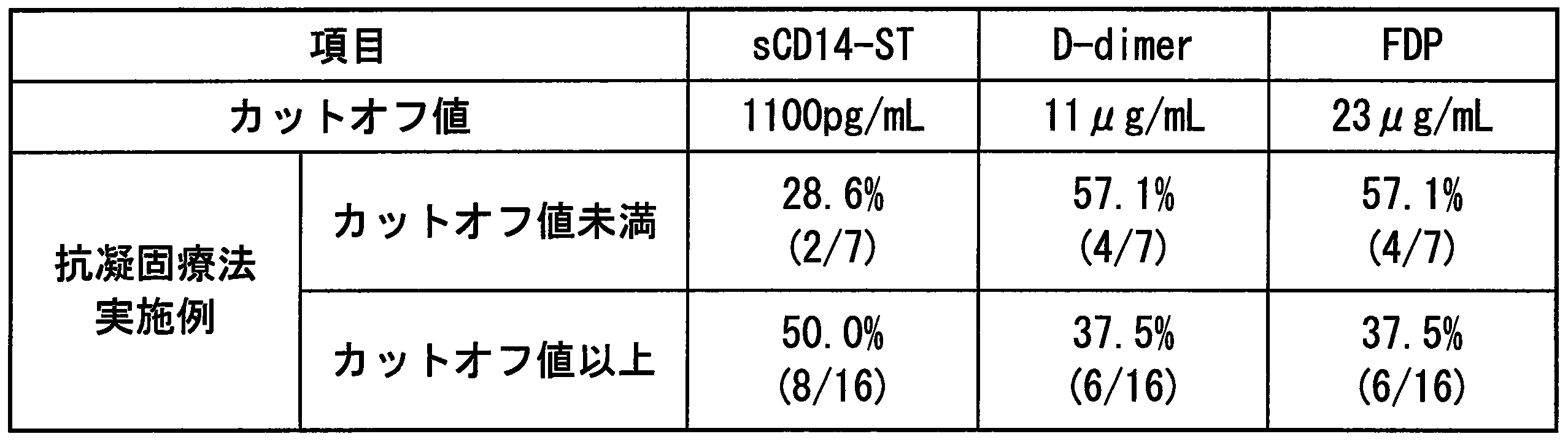
試験に登録された症例49例のうち、感染を伴う敗血症、重症敗血症、敗血症性ショックまたは感染性疾患の症例33例を対象に、急性期DIC診断基準に基づいてDICスコア(点)を求め、各DICスコア別のsCD14-ST、プロカルシトニン(PCT)、C反応性蛋白(CRP)およびインターロイキン-6(IL-6)の測定値分布を比較した。sCD14-STの測定は、実施例1に従って行った。 << Example 11: Distribution of measured values of sepsis markers for each DIC score in the infected group >>
Of the 49 cases registered in the study, for 33 cases of sepsis with infection, severe sepsis, septic shock or infectious disease, the DIC score (point) was determined based on the acute DIC diagnostic criteria, The measured value distributions of sCD14-ST, procalcitonin (PCT), C-reactive protein (CRP) and interleukin-6 (IL-6) for each DIC score were compared. Measurement of sCD14-ST was performed according to Example 1.
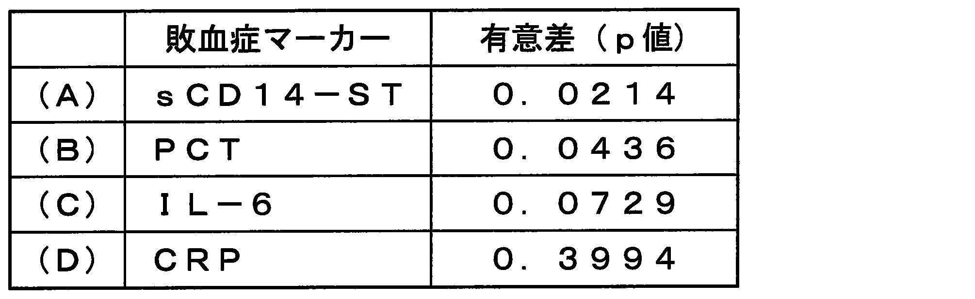
また、sCD14-ST値は、DICスコアが高くなるのに従って上昇することから、sCD14-ST値からDICの程度を把握することが可能であることが示唆された。 Furthermore, for sCD14-ST and PCT, which were significantly different by multi-group statistical analysis, infectious non-DIC (DIC score of less than 4 points) (n = 15) and infectious DIC (DIC score of 4 points) The above was classified into (n = 18), and statistical analysis between the two groups was performed by the Mann-Whitney U test from the measured values of each sCD14-ST or PCT. As a result, p = 0.0075 for sCD14-ST and p = 0.0329 for PCT, indicating that sCD14-ST is most useful for detecting infectious DIC.
In addition, the sCD14-ST value increases as the DIC score increases, suggesting that the degree of DIC can be determined from the sCD14-ST value.
試験に登録された症例49例を対象に、非感染群(SIRS及び非感染性疾患)13例と感染群(敗血症、重症敗血症、敗血症性ショック及び感染性疾患)36例に分類し、感染症検出におけるsCD14-STの有用性をROC分析により評価を行った。図19において、横軸は「1-特異度」であり、縦軸は「感度」である。
sCD14-STのAUCは0.833であり、有用であることが確認された。また、臨床感度/特異度が最大となるsCD14-ST値は647pg/mLであり、その臨床感度は83.3%、特異度76.9%であった。この結果から、感染症検出におけるsCD14-STのカットオフ値は600~700pg/mLが最適であると考えられた。 << Example 12: Infectious disease detection ability and cut-off value evaluation of sCD14-ST >>
49 cases enrolled in the study were classified into 13 non-infected groups (SIRS and non-infectious diseases) and 36 infected groups (sepsis, severe sepsis, septic shock and infectious diseases). The usefulness of sCD14-ST in detection was evaluated by ROC analysis. In FIG. 19, the horizontal axis represents “1-specificity” and the vertical axis represents “sensitivity”.
The AUC of sCD14-ST was 0.833, which was confirmed to be useful. The sCD14-ST value at which clinical sensitivity / specificity was maximum was 647 pg / mL, the clinical sensitivity was 83.3%, and the specificity was 76.9%. From these results, it was considered that the cut-off value of sCD14-ST in the detection of infectious diseases is optimally 600 to 700 pg / mL.
試験に登録された症例49例のうち、感染を伴った敗血症、重症敗血症、敗血症性ショックまたは感染性疾患の症例36例を対象に、急性期DIC診断基準に従ってDIC群(19例)と非DIC群(17例)に分類し、感染性DIC検出におけるsCD14-STの有用性をROC分析により評価を行った。図20において、横軸は「1-特異度」であり、縦軸は「感度」である。
sCD14-STのAUCは0.811であり、有用であることが確認された。また、臨床感度/特異度が最大となるsCD14-ST値は929pg/mLであり、その臨床感度は94.7%、特異度64.7%であった。この結果から、感染性DIC検出におけるsCD14-STのカットオフ値は900~1000pg/mLが最適であると考えられた。 << Example 13: Evaluation of DIC detection ability and cut-off value of sCD14-ST in infected group >>
Of the 49 cases enrolled in the study, 36 cases of sepsis with infection, severe sepsis, septic shock or infectious disease were studied, and DIC group (19 cases) and non-DIC It was classified into groups (17 cases), and the usefulness of sCD14-ST in detecting infectious DIC was evaluated by ROC analysis. In FIG. 20, the horizontal axis represents “1-specificity” and the vertical axis represents “sensitivity”.
The AUC of sCD14-ST was 0.811, which was confirmed to be useful. The sCD14-ST value that maximized clinical sensitivity / specificity was 929 pg / mL, the clinical sensitivity was 94.7%, and the specificity was 64.7%. From this result, it was considered that the cut-off value of sCD14-ST in the detection of infectious DIC was optimally 900 to 1000 pg / mL.
試験に登録された症例49例のうち、感染を伴った敗血症、重症敗血症、敗血症性ショックまたは感染性疾患の症例26例を対象に、各DICスコア別のD-dimer、フィブリン・フィブリノーゲン分解産物(FDP)、血小板(Plt)、プロトロンビン時間(PT)、INR値、活性化部分トロンボプラスチン時間(APTT)、フィブリノーゲン(FIB)、アンチトロンビンIII(ATIII)、乳酸(Lact)、トロンビン-アンチトロンビン複合体(TAT)、α2プラスミンインヒビター・プラスミン複合体(PIC)、プロテインC(PC)、トロンボモジュリン(TM)及び組織プラスミノーゲンアクチベータ(tPA)・プラスミノーゲンアクチベータインヒビター(PAI-1)複合体(Total PAI-1)の測定値分布を比較した。D-dimerは、ナノピアDダイマー(積水メディカル社)を用いて測定を行った。FDPは、ナノピアP-FDP(積水メディカル社)を用いて測定を行った。血小板は、セルパックII(シスメックス社)を用いて測定を行った。PTは、コアグピアPT-S(積水メディカル社)を用いて測定を行った。APTTは、コアグピアAPTT-S(積水メディカル社)を用いて測定を行った。フィブリノーゲンは、コアグピアFbg(積水メディカル社)を用いて測定を行った。アンチトロンビンIIIは、ステイシアCLEIA TAT(三菱化学メディカル社)を用いて測定を行った。乳酸は、デタミナ-LA(協和メディックス社)を用いて測定を行った。TATは、ステイシアCLEIA TAT(三菱化学メディカル社)を用いて測定を行った。PICは、エルピアエースPPIII(三菱化学メディエンス社)を用いて測定を行った。プロテインCは、エルピアエースPCII(三菱化学メディエンス社)を用いて測定を行った。トロンボモジュリンは、ステイシアCLEIA TM(三菱化学メディカル社)を用いて測定を行った。Total PAI-1は、LPIA tPAIテスト(三菱化学メディカル社)を用いて測定を行った。
Kruskal-Wallis検定による多群間統計解析を行った結果を、表7に示した。D-dimer、FDP、血小板及びTATは、感染性DICスコアを反映するマーカーと考えられた。図21~35に、各DICスコアに各マーカーの中央値と症例数を示した。 << Example 14: Measurement value distribution of coagulation-related markers for each DIC score in the infected group >>
Among the 49 cases enrolled in the study, 26 cases of sepsis, severe sepsis, septic shock or infectious disease with infection were used for D-dimer and fibrin fibrinogen degradation products by DIC score ( FDP), platelets (Plt), prothrombin time (PT), INR value, activated partial thromboplastin time (APTT), fibrinogen (FIB), antithrombin III (ATIII), lactate (Lact), thrombin-antithrombin complex ( TAT), α2 plasmin inhibitor / plasmin complex (PIC), protein C (PC), thrombomodulin (TM) and tissue plasminogen activator (tPA) / plasminogen activator inhibitor (PAI-1) complex (Total PAI−) 1) Measurement To compare the value distribution. D-dimer was measured using Nanopia D-dimer (Sekisui Medical). FDP was measured using Nanopia P-FDP (Sekisui Medical). Platelets were measured using Cell Pack II (Sysmex). PT was measured using Coagpia PT-S (Sekisui Medical). APTT was measured using Coagpia APTT-S (Sekisui Medical). Fibrinogen was measured using Coagpia Fbg (Sekisui Medical). Antithrombin III was measured using Stacia CLEIA TAT (Mitsubishi Chemical Medical). Lactic acid was measured using Detamina-LA (Kyowa Medics). TAT was measured using Stacia CLEIA TAT (Mitsubishi Chemical Medical). The PIC was measured using Elpia Ace PPIII (Mitsubishi Chemical Medience). Protein C was measured using Elpia Ace PCII (Mitsubishi Chemical Medience). Thrombomodulin was measured using Stacia CLEIA ™ (Mitsubishi Chemical Medical). Total PAI-1 was measured using the LPIA tPAI test (Mitsubishi Chemical Medical).
Table 7 shows the results of statistical analysis between multiple groups using the Kruskal-Wallis test. D-dimer, FDP, platelets and TAT were considered markers reflecting infectious DIC scores. 21 to 35 show the median value of each marker and the number of cases in each DIC score.
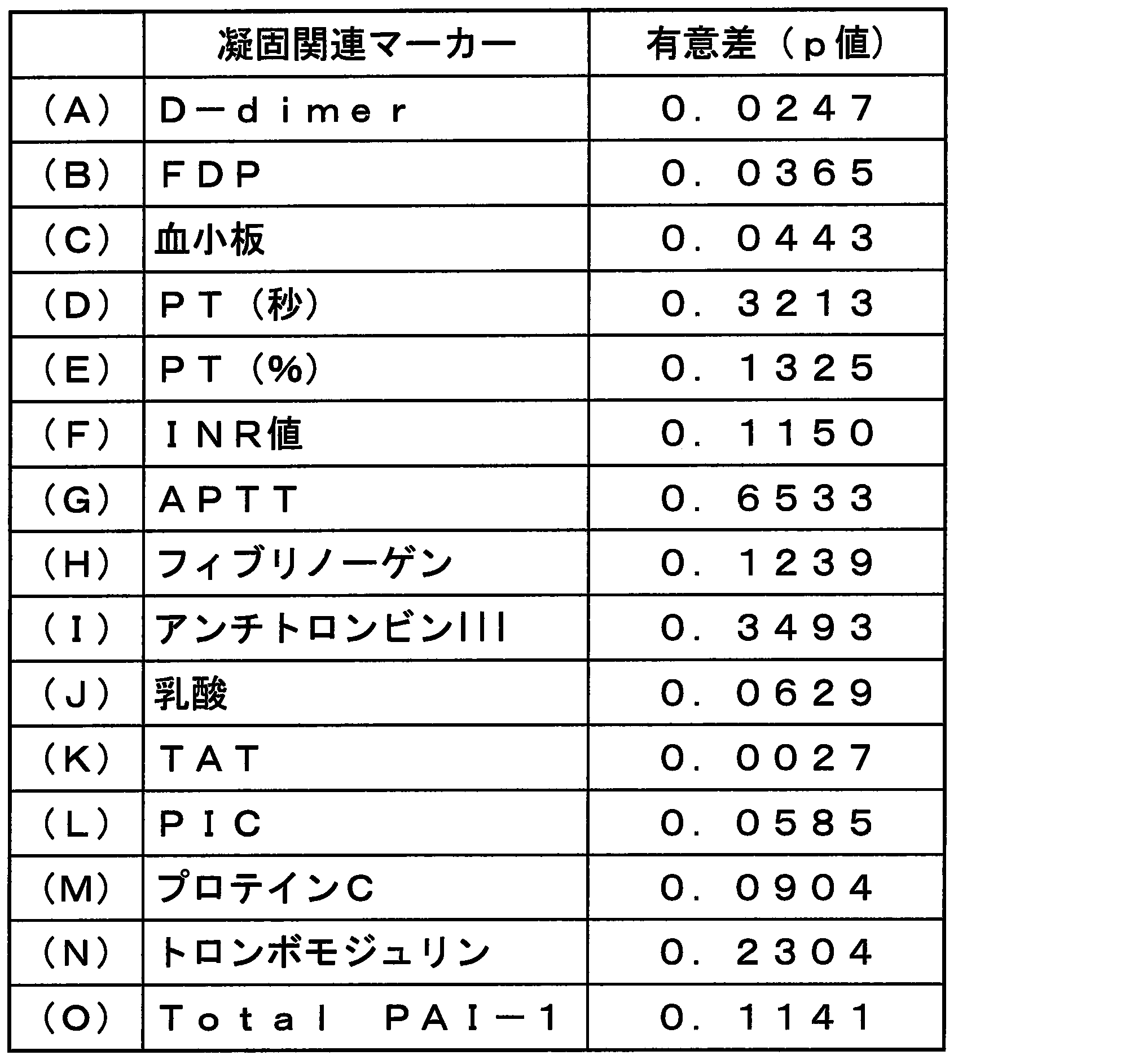


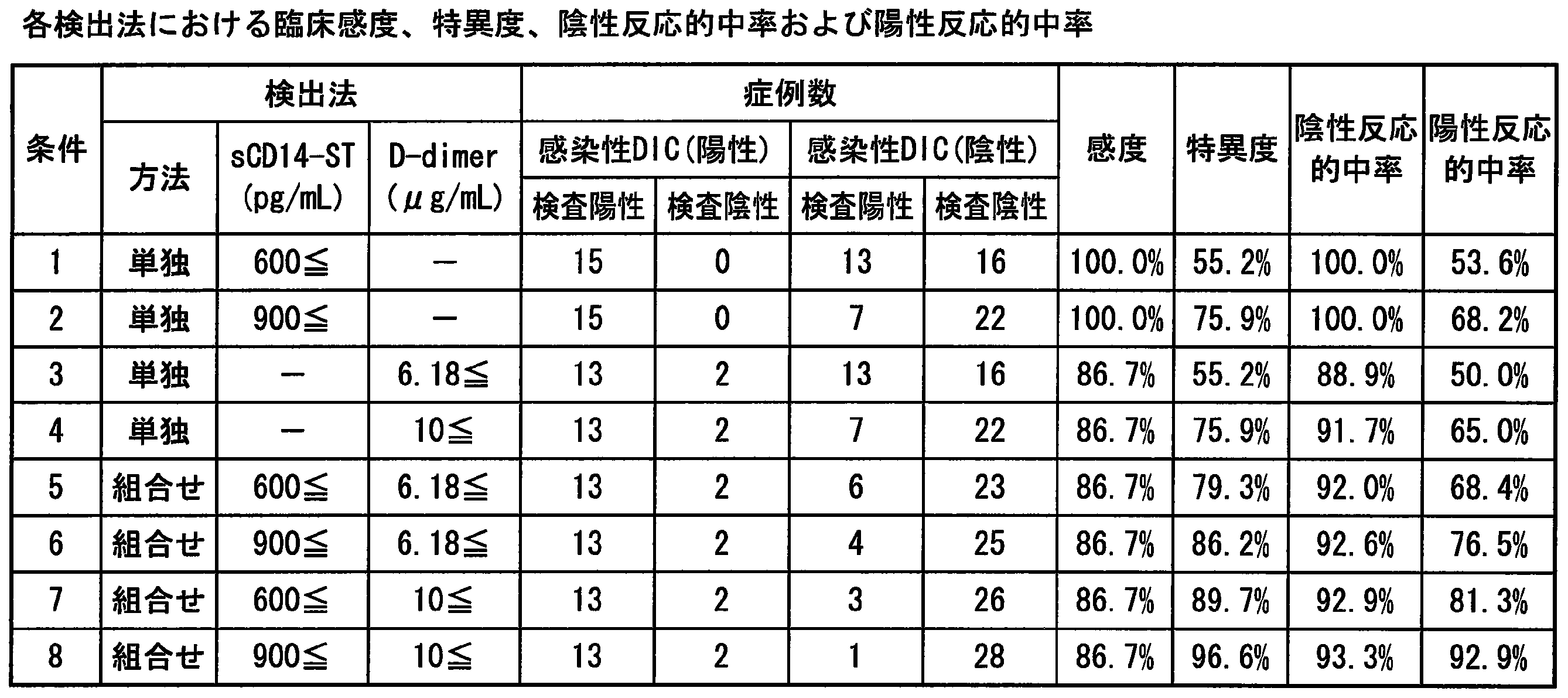

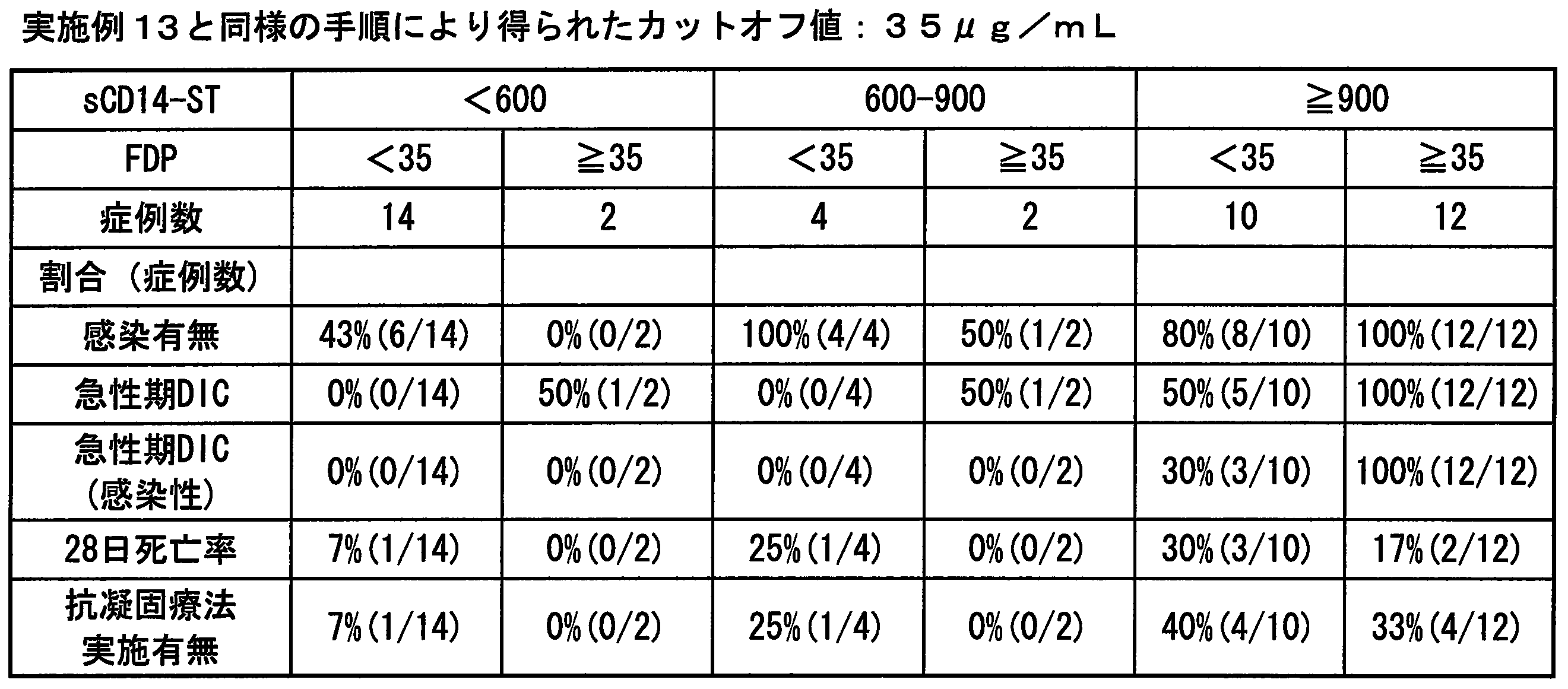
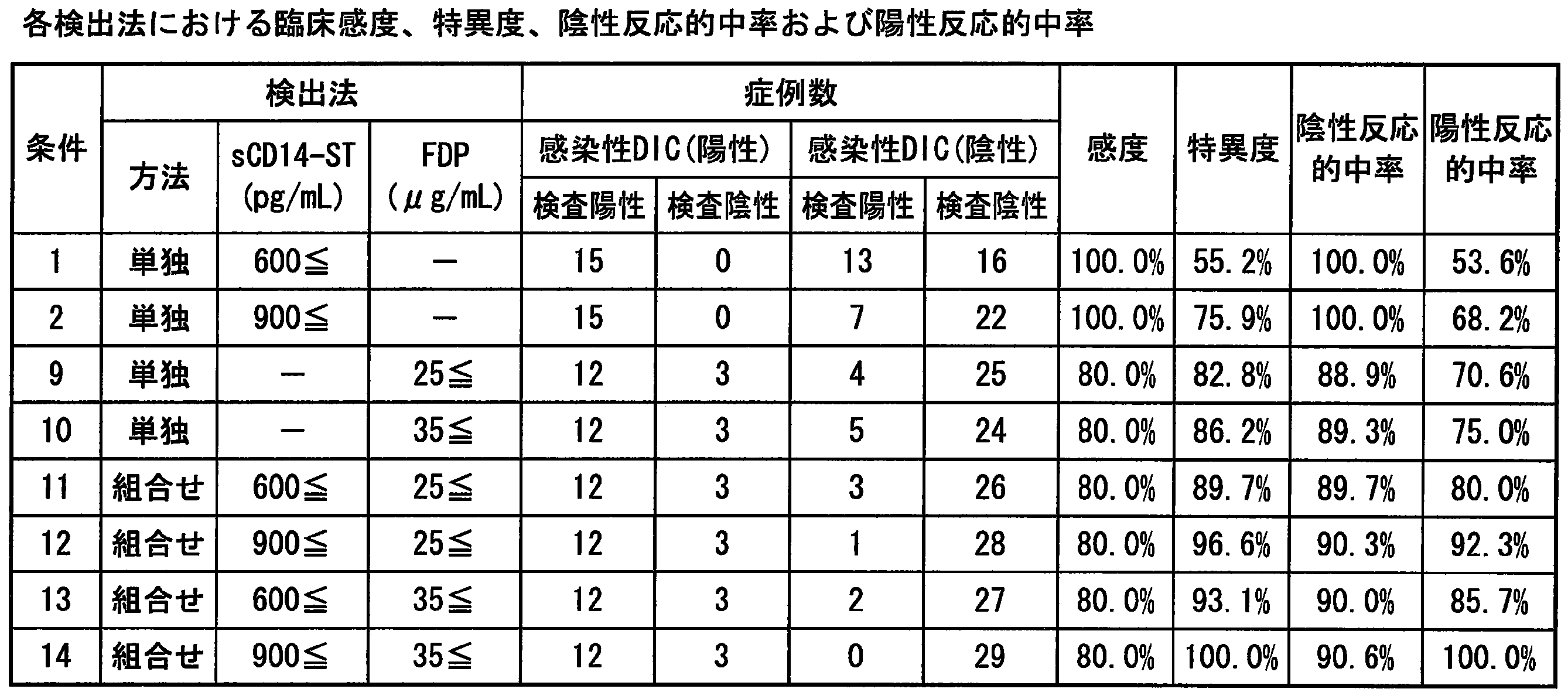


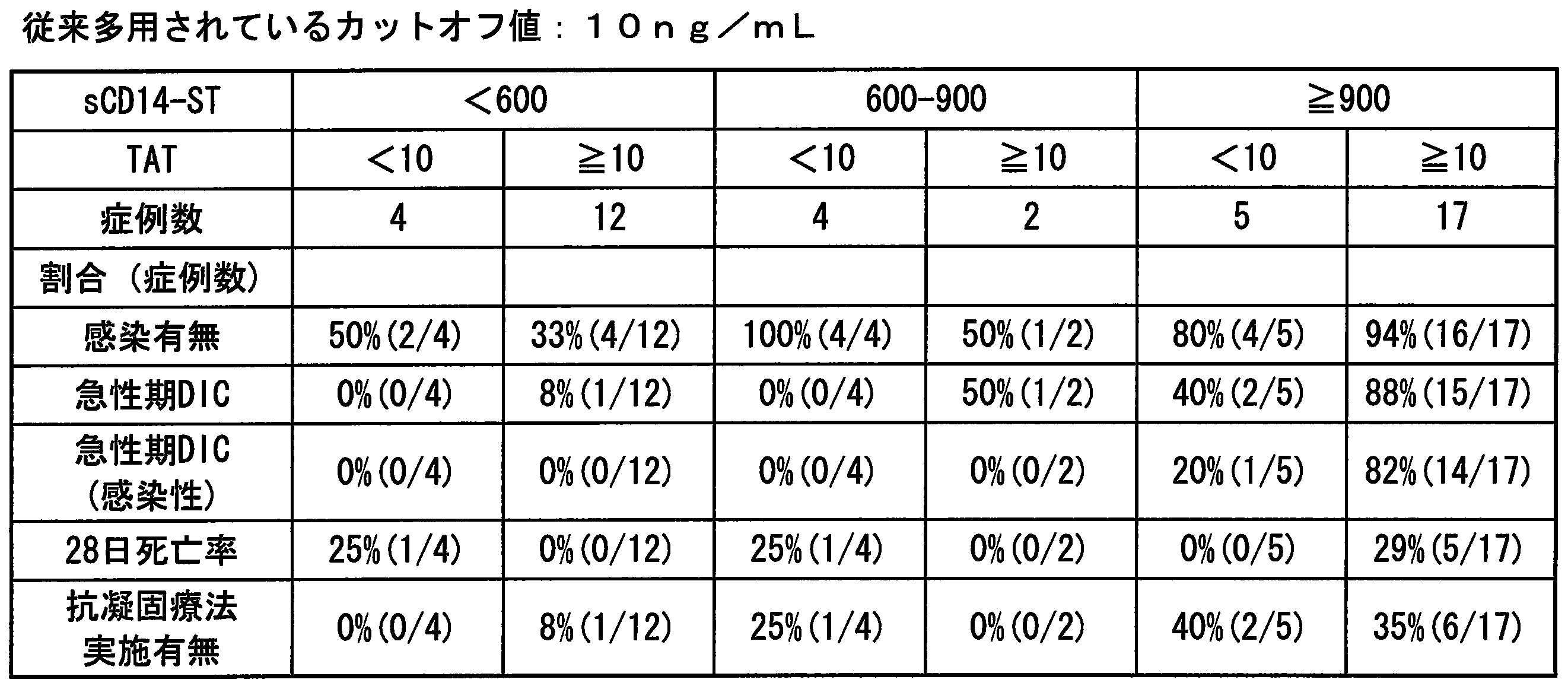
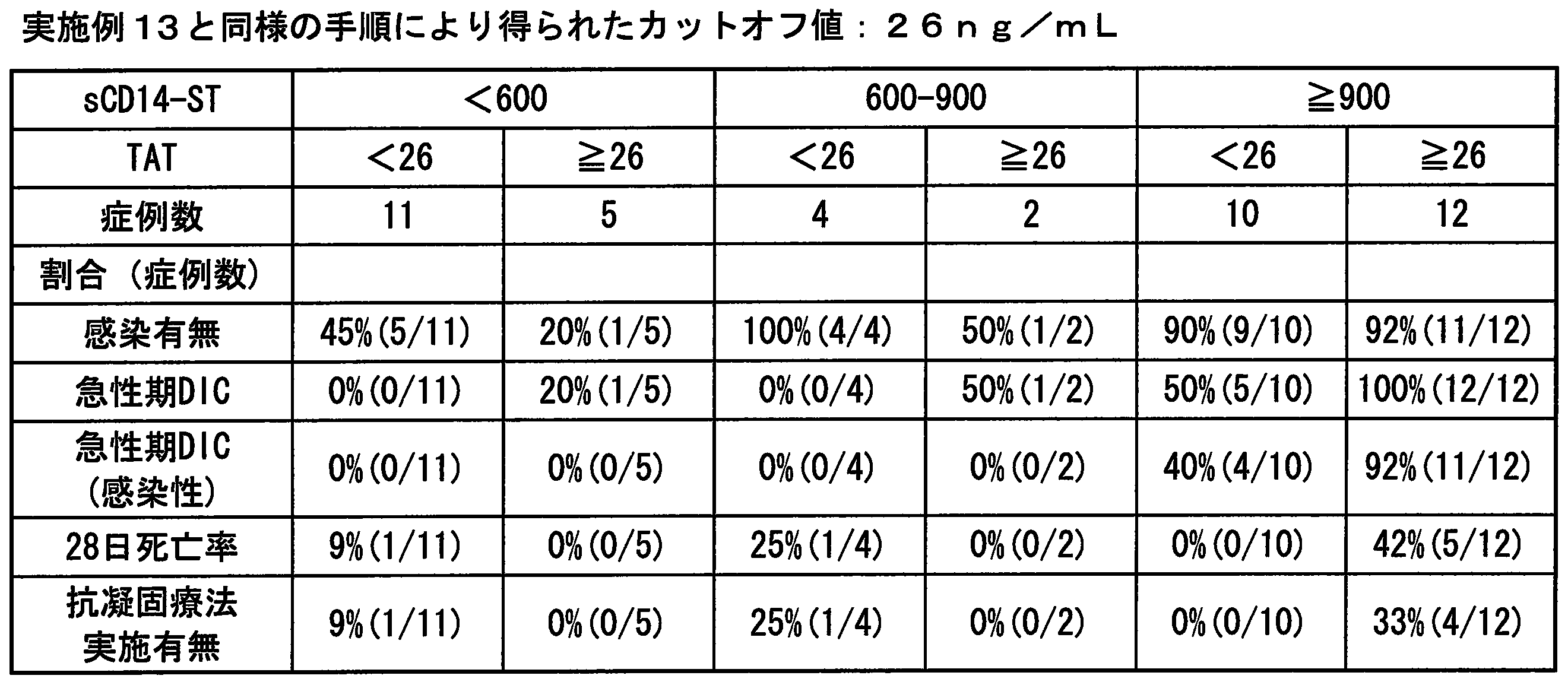
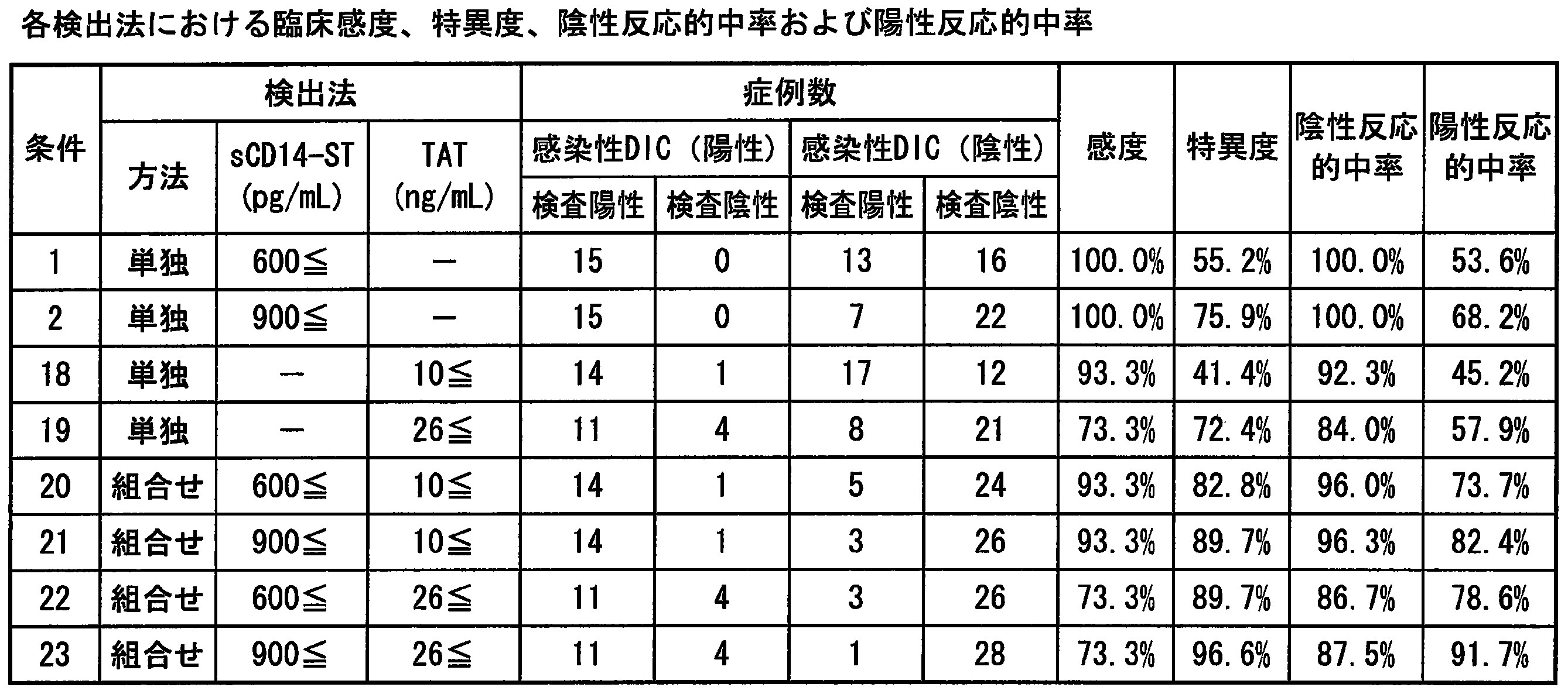
実施例15と同じ症例44例を対象に、プロカルシトニン(PCT)とトロンビン-アンチトロンビン複合体(TAT)の組み合わせ(表19~表20)による感染性DIC検出能の評価を行った。PCTのカットオフ値は、従来多用されている0.5ng/mL及び2.0ng/mLを用いた。TATのカットオフ値は、従来多用されている10ng/mLを用いた。評価にあたって、各条件の臨床感度、特異度、陰性反応的中率、陽性反応的中率を算出した。 << Comparative example 1: Evaluation of infectious DIC detection ability combining PCT and TAT >>
For 44 cases same as Example 15, the infectious DIC detection ability was evaluated by the combination of procalcitonin (PCT) and thrombin-antithrombin complex (TAT) (Table 19 to Table 20). As the cut-off value of PCT, 0.5 ng / mL and 2.0 ng / mL, which have been widely used conventionally, were used. As the cut-off value of TAT, 10 ng / mL, which has been frequently used in the past, was used. In the evaluation, the clinical sensitivity, specificity, negative reactive predictive value, and positive reactive predictive value of each condition were calculated.
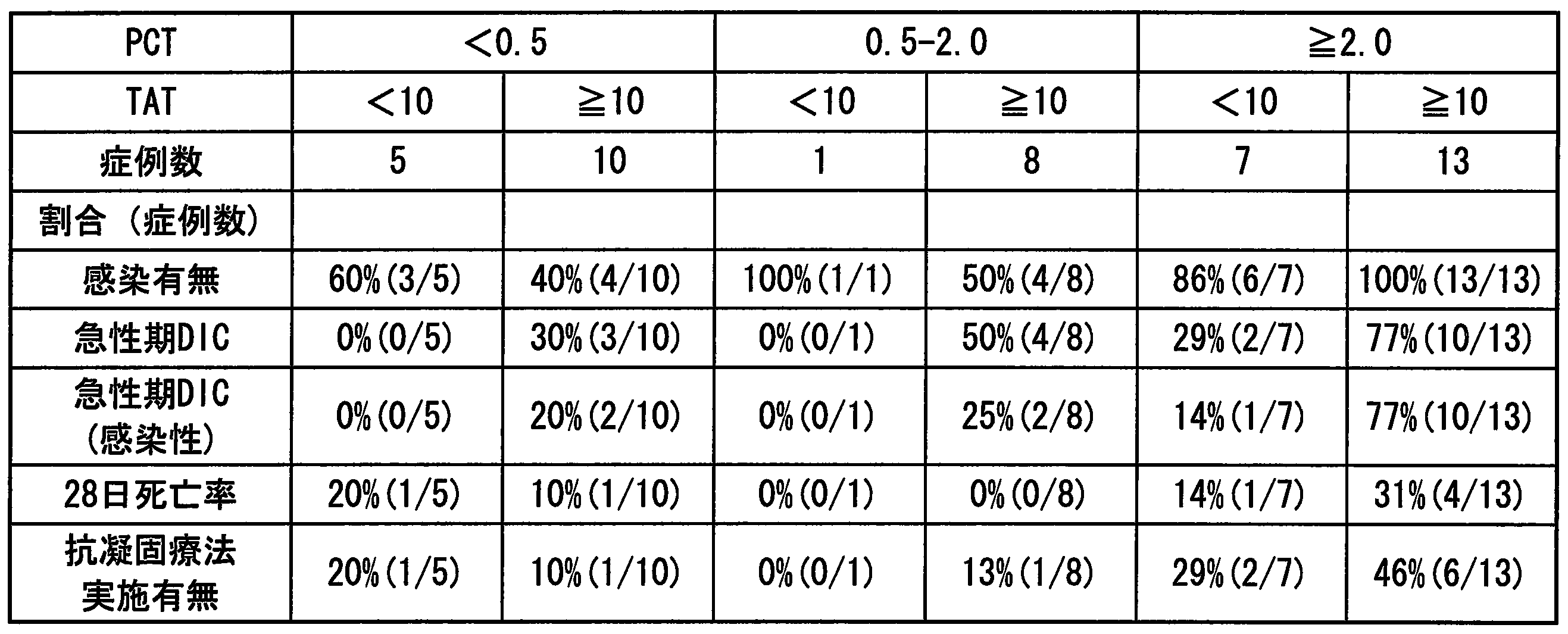
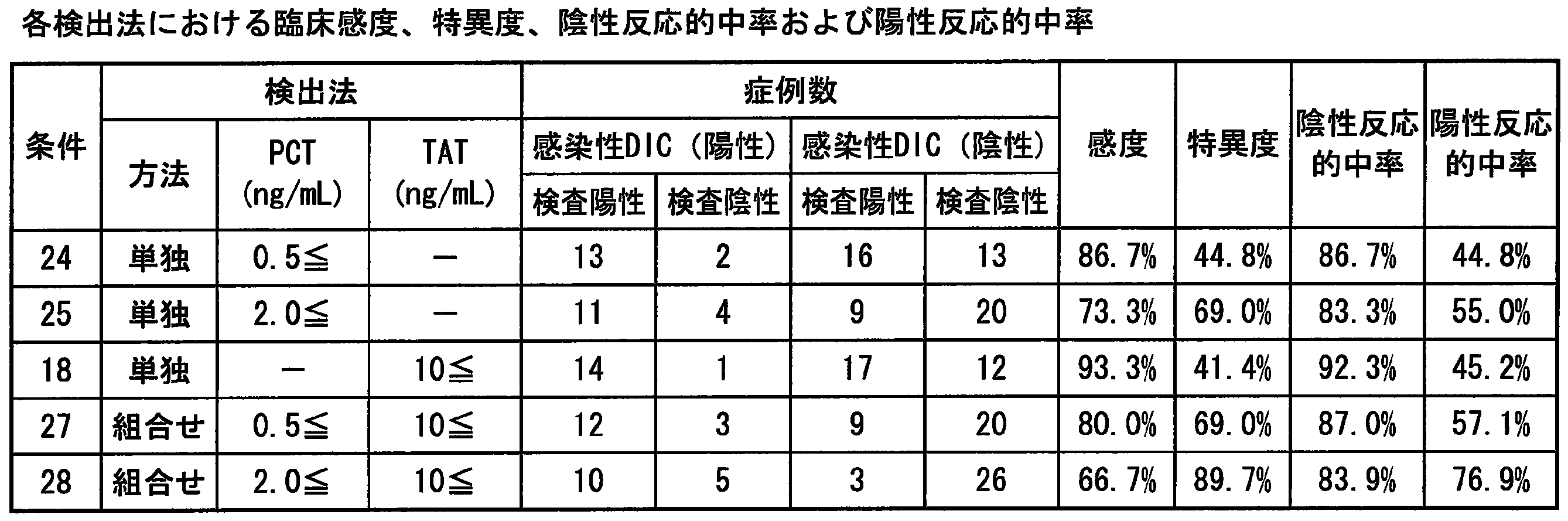
実施例16~20は、以下を試験対象とした。
1施設の臨床現場で、試験対象1と同様の基準により登録された症例全87例を対象に評価を行った。その内、疾患名が決定された症例は84例あり、詳細には、感染症を伴わない全身性炎症反応症候群(SIRS)19例、敗血症8例、重症敗血症14例、敗血症性ショック21例、非感染性疾患(感染症、SIRSどちらも該当しない状態)12例、感染性疾患(感染症であるがSIRSは伴っていない状態)10例であった。 ≪Test subject 2≫
In Examples 16 to 20, the following were tested.
Evaluation was performed on all 87 cases registered at the clinical site of one facility according to the same criteria as those of the
疾患名が決定された症例84例を対象に、多重ロジスティック回帰分析(ステップワイズ法)を行った。分析にあたり、目的変数を急性期DIC診断基準(非DIC群またはDIC群)、説明変数をsCD14-ST、プロカルシトニン(PCT)、インターロイキン-6(IL-6)、C反応性蛋白(CRP)とし、単位オッズ比を連続変数が1単位だけ変化した場合で行った。効果に対する尤度比検定の結果、sCD14-STがオッズ比1.001572、95%CIが1.000865-1.002446、p<0.0001となりもっとも有用であった。 Example 16 Multiple Logistic Regression Analysis in DIC for Sepsis Markers
Multiple logistic regression analysis (stepwise method) was performed on 84 cases whose disease names were determined. In the analysis, the objective variable is the acute DIC diagnostic criteria (non-DIC group or DIC group), the explanatory variables are sCD14-ST, procalcitonin (PCT), interleukin-6 (IL-6), C-reactive protein (CRP) The unit odds ratio was determined when the continuous variable was changed by one unit. As a result of the likelihood ratio test for the effect, sCD14-ST was most useful with an odds ratio of 1.001572, 95% CI of 1.000865-1.002446, and p <0.0001.
疾患名が決定された症例84例を対象に、多重ロジスティック回帰分析(ステップワイズ法)を行った。分析にあたり、目的変数を急性期DIC診断基準(非DIC群またはDIC群)、説明変数をアンチトロンビンIII(ATIII)、プロテインC(PC)、トロンボモジュリン(TM)とし、単位オッズ比を連続変数が1単位だけ変化した場合で行った。効果に対する尤度比検定の結果、プロテインCがオッズ比0.944377、95%CIが0.918842-0.965239、p<0.0001となりもっとも有用であった。 Example 17 Multiple Logistic Regression Analysis in DIC of Coagulation Related Markers
Multiple logistic regression analysis (stepwise method) was performed on 84 cases whose disease names were determined. In the analysis, the objective variable is the acute DIC diagnostic criteria (non-DIC group or DIC group), the explanatory variables are antithrombin III (ATIII), protein C (PC), thrombomodulin (TM), and the unit odds ratio is 1 This was done when the unit was changed. As a result of the likelihood ratio test for the effect, protein C was most useful with an odds ratio of 0.944377, 95% CI of 0.918842-0.965239, and p <0.0001.
疾患名が決定された症例84例のうち、sCD14-ST及びプロテインC(PC)の測定値がすべて確認された80例(敗血症群:40例、非敗血症群:40例)を対象にROC解析を行ったところ、AUCはsCD14-STが0.925(p<0.0001)、PCが0.833(p<0.0001)であった。また、カットオフ値はsCD14-STが647pg/mL(感度=93.0%、特異度=75.6%)、PCが47%(感度=77.5%、特異度=80.0%)となった。以上の結果から、カットオフ値はsCD14-STが650pg/mL、PCが45%で好適と考えられた。 << Example 18: ROC analysis and cut-off value evaluation regarding the presence or absence of sCD14-ST and PC sepsis >>
ROC analysis of 80 cases (septic group: 40 cases, non-septic group: 40 cases) in which all measured values of sCD14-ST and protein C (PC) were confirmed among 84 cases whose disease names were determined As a result, AUC was 0.925 (p <0.0001) for sCD14-ST and 0.833 (p <0.0001) for PC. The cutoff values are 647 pg / mL for sCD14-ST (sensitivity = 93.0%, specificity = 75.6%), and 47% for PC (sensitivity = 77.5%, specificity = 80.0%). It became. From the above results, it was considered that cut-off values of sCD14-ST were 650 pg / mL and PC was 45%.
疾患名が決定された症例84例のうち、sCD14-ST及びプロテインC(PC)の測定値がすべて確認された81例(DIC群:36例、非DIC群:45例)を対象にROC解析を行ったところ、AUCはsCD14-STが0.836(p<0.0001)、PCが0.891(p<0.0001)であった。また、カットオフ値はsCD14-STが899pg/mL(感度=81.6%、特異度=80.9%)、PCが55%(感度=89.2%、特異度=75.6%)となった。以上の結果から、カットオフ値はsCD14-STが900pg/mL、PCが55%で好適と考えられた。 << Example 19: ROC analysis and cut-off value evaluation regarding DIC presence / absence of sCD14-ST and PC >>
ROC analysis of 81 cases (DIC group: 36 cases, non-DIC group: 45 cases) in which all measured values of sCD14-ST and protein C (PC) were confirmed among 84 cases whose disease names were determined As a result, AUC was 0.836 (p <0.0001) for sCD14-ST and 0.891 (p <0.0001) for PC. The cut-off values were 899 pg / mL for sCD14-ST (sensitivity = 81.6%, specificity = 80.9%) and 55% for PC (sensitivity = 89.2%, specificity = 75.6%). It became. From the above results, it was considered that cut-off values of 900 pg / mL for sCD14-ST and 55% for PC were suitable.
疾患名が決定された症例84例のうち、sCD14-ST及びプロテインC(PC)の測定値がすべて確認された81例を対象に、実施例15と同様にsCD14-STとPC(表21~表22)の組み合わせで感染性DIC検出能の評価を行った。カットオフ値は、実施例18または19で得られた値から設定した。評価にあたって、各条件の臨床感度、特異度、陰性反応的中率、陽性反応的中率を算出した。 Example 20: Evaluation of detectability of infectious DIC combining sCD14-ST and PC
Of the 84 cases whose disease names were determined, 81 cases in which all the measured values of sCD14-ST and protein C (PC) were confirmed were examined, and sCD14-ST and PC (Table 21 to Table 22) was evaluated for infectious DIC detectability. The cut-off value was set from the value obtained in Example 18 or 19. In the evaluation, the clinical sensitivity, specificity, negative reactive predictive value, and positive reactive predictive value of each condition were calculated.

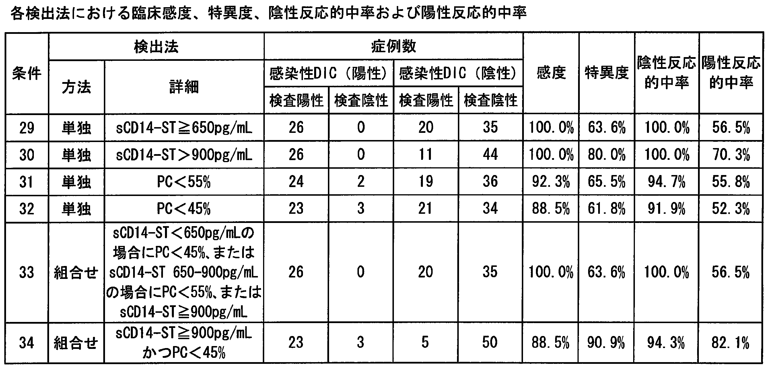
実施例12の非感染群、すなわち、非感染であったSIRSまたは非感染性疾患の症例13例を対象に、急性期DIC診断基準に従ってDIC群(4例)と非DIC群(9例)に分類し、DIC検出におけるsCD14-ST、プロカルシトニン(PCT)、C反応性蛋白(CRP)、インターロイキン-6(IL-6)、D-dimer及びフィブリン・フィブリノーゲン分解産物(FDP)の有用性をROC分析により比較を行った。
PCTの測定は、エクルーシス試薬ブラームスPCT(ロシュ・ダイアグノスティックス社)を用いて測定を行った。IL-6は、イムライズIL-6(シーメンスヘルスケア・ダイアグノスティクス社)を用いて測定を行った。CRPは、CRP-ラテックスX2「生研」(デンカ生研社)を用いて測定を行った。測定機器は、日立7170S(日立ハイテク社)を用いた。D-dimerは、ナノピアDダイマー(積水メディカル社)を用いて測定を行った。測定機器は、コアプレスタ2000(積水メディカル社)を用いた。FDPは、ナノピアP-FDP(積水メディカル社)を用いて測定を行った。測定機器は、コアプレスタ2000(積水メディカル社)を用いた。 << Reference Example 1: Usefulness for DIC diagnosis in non-infected group >>
In the non-infected group of Example 12, ie, 13 cases of SIRS or non-infectious disease that were non-infected, the DIC group (4 cases) and the non-DIC group (9 cases) according to the acute DIC diagnostic criteria Classification and usefulness of sCD14-ST, procalcitonin (PCT), C-reactive protein (CRP), interleukin-6 (IL-6), D-dimer and fibrin fibrinogen degradation product (FDP) in DIC detection Comparison was made by ROC analysis.
Measurement of PCT was performed using Ecrusys reagent Brahms PCT (Roche Diagnostics). IL-6 was measured using Imrise IL-6 (Siemens Healthcare Diagnostics). CRP was measured using CRP-latex X2 “Seiken” (Denka Seiken Co., Ltd.). Hitachi 7170S (Hitachi High-Tech) was used as a measuring instrument. D-dimer was measured using Nanopia D-dimer (Sekisui Medical). As a measuring instrument, Core Presta 2000 (Sekisui Medical Co., Ltd.) was used. FDP was measured using Nanopia P-FDP (Sekisui Medical). As a measuring instrument, Core Presta 2000 (Sekisui Medical Co., Ltd.) was used.
AUCをそれぞれ求めたところ、sCD14-STは0.806、PCTは0.778、IL-6は0.639、CRPは0.583、D-dimerは0.764、FDPは0.792であり、sCD14-STのAUCが最も大きかった。この結果からsCD14-STは感染の有無に関わらず、既存の指標であるD-dimerやFDP等に対し有用であることが確認された。sCD14-ST値のカットオフ値は、600pg/mLの場合に臨床感度が75.0%、特異度が100%となり、最適と考えられた。 The results are shown in FIGS. 36 and 37, the horizontal axis represents “1-specificity” and the vertical axis represents “sensitivity”.
When AUC was calculated, sCD14-ST was 0.806, PCT was 0.778, IL-6 was 0.639, CRP was 0.583, D-dimer was 0.764, and FDP was 0.792. SCD14-ST had the largest AUC. From these results, it was confirmed that sCD14-ST is useful for existing indicators such as D-dimer and FDP regardless of the presence or absence of infection. The cut-off value of the sCD14-ST value was considered optimal with a clinical sensitivity of 75.0% and a specificity of 100% at 600 pg / mL.
また、本発明は、感染性播種性血管内凝固症候群(感染性DIC)の検出及びその病態の把握に利用することができる。感染性DICが疑われる患者から感染性DIC患者を特定することもできるし、感染性DIC患者に対して経時的に測定することによって、状態をモニタリングすることができ、適切な治療方針の策定に役立てることができる。
以上、本発明を特定の態様に沿って説明したが、当業者に自明の変形や改良は本発明の範囲に含まれる。 The present invention can be used for detection of disseminated intravascular coagulation syndrome (DIC) and understanding of its pathological condition. A DIC patient can be identified from patients suspected of having DIC, and the condition can be monitored by measuring the DIC patient over time, which can be used to formulate an appropriate treatment policy.
Moreover, this invention can be utilized for the detection of infectious disseminated intravascular coagulation syndrome (infectious DIC), and grasping | ascertaining the pathological condition. Infectious DIC patients can be identified from patients suspected of infectious DIC, and the status can be monitored by measuring the infectious DIC patients over time. Can be useful.
As mentioned above, although this invention was demonstrated along the specific aspect, the deformation | transformation and improvement obvious to those skilled in the art are included in the scope of the present invention.
Claims (12)
- 試料中のsCD14-STを測定する、播種性血管内凝固症候群の検出方法。 A method for detecting disseminated intravascular coagulation syndrome by measuring sCD14-ST in a sample.
- 播種性血管内凝固症候群が疑われる患者又は播種性血管内凝固症候群患者から採取した試料中のsCD14-STを測定する工程、及び
sCD14-ST値が非播種性血管内凝固症候群患者と比較して高値である場合に播種性血管内凝固症候群であると判断する工程
を含む、請求項1に記載の播種性血管内凝固症候群の検出方法。 A step of measuring sCD14-ST in a sample collected from a patient suspected of disseminated intravascular coagulation syndrome or a patient of disseminated intravascular coagulation syndrome, and the sCD14-ST value is compared with a non-disseminated intravascular coagulation syndrome patient The method for detecting disseminated intravascular coagulation syndrome according to claim 1, comprising a step of determining that the disseminated intravascular coagulation syndrome is present when the value is high. - 前記判断工程において、sCD14-ST値と予め決定した閾値とを比較する、請求項2に記載の方法。 The method according to claim 2, wherein in the determination step, the sCD14-ST value is compared with a predetermined threshold value.
- 該sCD14-STを免疫学的測定方法で測定する、請求項1~3のいずれか一項に記載の方法。 The method according to any one of claims 1 to 3, wherein the sCD14-ST is measured by an immunological measurement method.
- 前記播種性血管内凝固症候群が感染性播種性血管内凝固症候群であり、sCD14-STに加え、更に試料中の凝固関連マーカーを測定する、請求項1に記載の方法。 The method according to claim 1, wherein the disseminated intravascular coagulation syndrome is an infectious disseminated intravascular coagulation syndrome, and in addition to sCD14-ST, a coagulation-related marker in the sample is further measured.
- 感染性播種性血管内凝固症候群が疑われる患者又は感染性播種性血管内凝固症候群患者から採取した試料中のsCD14-ST及び凝固関連マーカーを測定する工程、及び
sCD14-ST及び凝固関連マーカーの測定値が感染性非播種性血管内凝固症候群患者と比較して変化している場合に感染性播種性血管内凝固症候群であると判断する工程
を含む、請求項5に記載の方法。 Measuring sCD14-ST and coagulation-related markers in a sample collected from a patient suspected of infectious disseminated intravascular coagulation syndrome or an infectious disseminated intravascular coagulation syndrome; and measurement of sCD14-ST and coagulation-related markers 6. The method of claim 5, comprising determining an infectious disseminated intravascular coagulation syndrome if the value is changed compared to an infectious non-disseminated intravascular coagulation syndrome patient. - 前記判断工程において、測定されたsCD14-ST及び凝固関連マーカーの測定値とそれぞれの予め決定した閾値とを比較する、請求項6に記載の方法。 The method according to claim 6, wherein in the determination step, the measured values of the measured sCD14-ST and the coagulation-related marker are compared with respective predetermined threshold values.
- 前記凝固関連マーカーが、D-dimer、FDP、トロンビン・アンチトロンビンIII複合体、血小板数、プロテインCから少なくとも一つ選ばれる、請求項5~7のいずれか一項に記載の方法。 The method according to any one of claims 5 to 7, wherein the coagulation-related marker is selected from at least one of D-dimer, FDP, thrombin / antithrombin III complex, platelet count, and protein C.
- 該sCD14-STを免疫学的測定方法で測定する、請求項5~8のいずれか一項に記載の方法。 The method according to any one of claims 5 to 8, wherein the sCD14-ST is measured by an immunological measurement method.
- 播種性血管内凝固症候群を検出するキットであって、
(a)sCD14-STに特異的な抗体、
(b)試料中のsCD14-STの量と播種性血管内凝固症候群との相関を示す標準データ、
(c)取扱説明書
を含む、該キット。 A kit for detecting disseminated intravascular coagulation syndrome,
(A) an antibody specific for sCD14-ST,
(B) Standard data showing the correlation between the amount of sCD14-ST in the sample and disseminated intravascular coagulation syndrome,
(C) The kit including instructions. - 感染性播種性血管内凝固症候群を検出するキットであって、
(a)sCD14-STに特異的な抗体、
(b)凝固関連マーカー測定用試薬、
(c)試料中のsCD14-ST及び凝固関連マーカーの測定値と感染性播種性血管内凝固症候群との相関を示す標準データ、
(d)取扱説明書
を含む、該キット。 A kit for detecting infectious disseminated intravascular coagulation,
(A) an antibody specific for sCD14-ST,
(B) a reagent for measuring a coagulation-related marker,
(C) Standard data showing correlation between measured values of sCD14-ST and coagulation-related markers in the sample and infectious disseminated intravascular coagulation syndrome,
(D) The kit, including instructions. - 前記凝固関連マーカーが、D-dimer、FDP、トロンビン・アンチトロンビンIII複合体、血小板数、プロテインCから少なくとも一つ選ばれる、請求項11に記載のキット。 The kit according to claim 11, wherein the coagulation-related marker is selected from at least one of D-dimer, FDP, thrombin / antithrombin III complex, platelet count, and protein C.
Priority Applications (6)
| Application Number | Priority Date | Filing Date | Title |
|---|---|---|---|
| CN201380024024.4A CN104285148B (en) | 2012-05-07 | 2013-04-26 | Detection disseminated inravascular coagulation or the method for infectious disseminated inravascular coagulation |
| KR1020147034082A KR102044940B1 (en) | 2012-05-07 | 2013-04-26 | Method for detecting disseminated intravascular coagulation or infectious disseminated intravascular coagulation |
| JP2014514682A JP6108409B2 (en) | 2012-05-07 | 2013-04-26 | Methods for detecting disseminated intravascular coagulation syndrome or infectious disseminated intravascular coagulation syndrome |
| EP13787336.0A EP2848936B1 (en) | 2012-05-07 | 2013-04-26 | Method for detecting disseminated intravascular coagulation or infectious disseminated intravascular coagulation |
| ES13787336T ES2755623T3 (en) | 2012-05-07 | 2013-04-26 | Detection procedure for disseminated intravascular coagulation or infectious disseminated intravascular coagulation |
| US14/398,971 US9910052B2 (en) | 2012-05-07 | 2013-04-26 | Method of diagnosing and treating infectious disseminated intravascular coagulation |
Applications Claiming Priority (4)
| Application Number | Priority Date | Filing Date | Title |
|---|---|---|---|
| JP2012-106217 | 2012-05-07 | ||
| JP2012106216 | 2012-05-07 | ||
| JP2012106217 | 2012-05-07 | ||
| JP2012-106216 | 2012-05-07 |
Publications (1)
| Publication Number | Publication Date |
|---|---|
| WO2013168602A1 true WO2013168602A1 (en) | 2013-11-14 |
Family
ID=49550647
Family Applications (1)
| Application Number | Title | Priority Date | Filing Date |
|---|---|---|---|
| PCT/JP2013/062352 WO2013168602A1 (en) | 2012-05-07 | 2013-04-26 | Method for detecting disseminated intravascular coagulation or infectious disseminated intravascular coagulation |
Country Status (7)
| Country | Link |
|---|---|
| US (1) | US9910052B2 (en) |
| EP (1) | EP2848936B1 (en) |
| JP (1) | JP6108409B2 (en) |
| KR (1) | KR102044940B1 (en) |
| CN (1) | CN104285148B (en) |
| ES (1) | ES2755623T3 (en) |
| WO (1) | WO2013168602A1 (en) |
Cited By (3)
| Publication number | Priority date | Publication date | Assignee | Title |
|---|---|---|---|---|
| JP2017044668A (en) * | 2015-08-28 | 2017-03-02 | シスメックス株式会社 | Method, device, and computer program for analyzing blood samples |
| WO2020084853A1 (en) * | 2018-10-22 | 2020-04-30 | 旭化成ファーマ株式会社 | Drug for treating and/or improving septicemia associated with coagulation abnormality |
| WO2021172493A1 (en) * | 2020-02-28 | 2021-09-02 | 株式会社Lsiメディエンス | Method for measuring platelet activation based on soluble clec-2 and platelet count |
Families Citing this family (5)
| Publication number | Priority date | Publication date | Assignee | Title |
|---|---|---|---|---|
| CN106822875A (en) * | 2017-03-01 | 2017-06-13 | 中国人民解放军军事医学科学院微生物流行病研究所 | A kind of prevention method of disseminated intravascular coagulation |
| EP3639276A1 (en) * | 2017-06-12 | 2020-04-22 | Koninklijke Philips N.V. | Risk assessment of disseminated intravascular coagulation |
| PL3682247T3 (en) * | 2017-09-13 | 2022-04-25 | B.R.A.H.M.S Gmbh | Proadrenomedullin as a marker for abnormal platelet levels |
| CN110596398A (en) * | 2019-08-14 | 2019-12-20 | 南京浦光生物科技有限公司 | Protein chip for detecting blood coagulation marker and preparation method and application thereof |
| WO2021097685A1 (en) * | 2019-11-19 | 2021-05-27 | 深圳迈瑞生物医疗电子股份有限公司 | Kit and method for mixed detection of pct and presepsin, and use thereof |
Citations (2)
| Publication number | Priority date | Publication date | Assignee | Title |
|---|---|---|---|---|
| JP2005106694A (en) | 2003-09-30 | 2005-04-21 | Mochida Pharmaceut Co Ltd | Early detection of blood poisoning and evaluation on seriousness |
| JP4040666B2 (en) | 2004-05-11 | 2008-01-30 | 持田製薬株式会社 | Novel soluble CD14 antigen |
Family Cites Families (6)
| Publication number | Priority date | Publication date | Assignee | Title |
|---|---|---|---|---|
| ES2231167T3 (en) * | 1999-02-04 | 2005-05-16 | Biomerieux, Inc. | PROCEDURE AND APPARATUS TO PREACH HEMOSTATIC DYSFUNCTION IN A PATIENT SAMPLE. |
| JP4681175B2 (en) * | 2001-09-28 | 2011-05-11 | 三菱化学メディエンス株式会社 | Method for detecting disseminated intravascular coagulation syndrome and its pre-onset stage |
| CN102121939A (en) | 2004-11-08 | 2011-07-13 | 三菱化学美迪恩斯株式会社 | Method of detecting platelet thrombosis or organ injury |
| ES2373989T3 (en) | 2006-01-31 | 2012-02-10 | Mitsubishi Chemical Medience Corporation | PROCEDURE FOR THE DETERMINATION OF THE STATE OF A DISSEMINATED INTRAVASCULAR COAGULATION SYNDROME. |
| WO2012096245A1 (en) * | 2011-01-11 | 2012-07-19 | 三菱化学メディエンス株式会社 | Method for prediction of prognosis of sepsis |
| ES2578160T3 (en) | 2011-08-12 | 2016-07-21 | Lsi Medience Corporation | Diagnostic procedure for surgical site infections |
-
2013
- 2013-04-26 JP JP2014514682A patent/JP6108409B2/en active Active
- 2013-04-26 WO PCT/JP2013/062352 patent/WO2013168602A1/en active Application Filing
- 2013-04-26 CN CN201380024024.4A patent/CN104285148B/en active Active
- 2013-04-26 KR KR1020147034082A patent/KR102044940B1/en active IP Right Grant
- 2013-04-26 ES ES13787336T patent/ES2755623T3/en active Active
- 2013-04-26 EP EP13787336.0A patent/EP2848936B1/en active Active
- 2013-04-26 US US14/398,971 patent/US9910052B2/en active Active
Patent Citations (2)
| Publication number | Priority date | Publication date | Assignee | Title |
|---|---|---|---|---|
| JP2005106694A (en) | 2003-09-30 | 2005-04-21 | Mochida Pharmaceut Co Ltd | Early detection of blood poisoning and evaluation on seriousness |
| JP4040666B2 (en) | 2004-05-11 | 2008-01-30 | 持田製薬株式会社 | Novel soluble CD14 antigen |
Non-Patent Citations (10)
| Title |
|---|
| "Saishin Naika-gaku Taikei", SAIKIN KANSEN-SHO, vol. 27, 1994, pages 69 - 84 |
| AM. J. INFECT. CONTROL, vol. 16, 1988, pages 128 - 140 |
| CHEST, vol. 101, no. 6, 1992, pages 1644 - 1655 |
| IGAKU NO AYUMI, vol. 238, no. 1, 2011, pages 114 - 119 |
| IGAKU NO AYUMI, vol. 238, no. 1, 2011, pages 63 - 68 |
| J INFECT CHEMOTHER, vol. 11, 2005, pages 234 - 238 |
| MEDICAL POSTGRADUATES, vol. 48, 2010, pages 25 - 27 |
| RIENK NIEUWLAND: "Cellular origin and procoagulant properties of microparticles in meningococcal sepsis", BLOOD, vol. 95, no. 3, 1 February 2000 (2000-02-01), pages 930 - 935, XP055176118 * |
| SANG MEE HWANG: "Thrombomodulin phenotype of a distinct monocyte subtype is an independent prognostic marker for disseminated intravascular coagulation", CRITICAL CARE, vol. 15, no. 2, 2011, pages R113, XP021102320 * |
| THERAPEUTIC RESEARCH, vol. 25, 2004, pages 1689 - 1694 |
Cited By (5)
| Publication number | Priority date | Publication date | Assignee | Title |
|---|---|---|---|---|
| JP2017044668A (en) * | 2015-08-28 | 2017-03-02 | シスメックス株式会社 | Method, device, and computer program for analyzing blood samples |
| WO2020084853A1 (en) * | 2018-10-22 | 2020-04-30 | 旭化成ファーマ株式会社 | Drug for treating and/or improving septicemia associated with coagulation abnormality |
| JPWO2020084853A1 (en) * | 2018-10-22 | 2021-09-16 | 旭化成ファーマ株式会社 | Drugs for the treatment and / or amelioration of sepsis with coagulation abnormalities |
| JP7237085B2 (en) | 2018-10-22 | 2023-03-10 | 旭化成ファーマ株式会社 | Medicine for treatment and/or improvement of sepsis accompanied by coagulation abnormality |
| WO2021172493A1 (en) * | 2020-02-28 | 2021-09-02 | 株式会社Lsiメディエンス | Method for measuring platelet activation based on soluble clec-2 and platelet count |
Also Published As
| Publication number | Publication date |
|---|---|
| JPWO2013168602A1 (en) | 2016-01-07 |
| EP2848936B1 (en) | 2019-10-02 |
| CN104285148B (en) | 2016-10-12 |
| US20150118699A1 (en) | 2015-04-30 |
| JP6108409B2 (en) | 2017-04-05 |
| EP2848936A4 (en) | 2015-10-14 |
| ES2755623T3 (en) | 2020-04-23 |
| EP2848936A1 (en) | 2015-03-18 |
| CN104285148A (en) | 2015-01-14 |
| KR20150022804A (en) | 2015-03-04 |
| US9910052B2 (en) | 2018-03-06 |
| KR102044940B1 (en) | 2019-11-14 |
Similar Documents
| Publication | Publication Date | Title |
|---|---|---|
| JP6108409B2 (en) | Methods for detecting disseminated intravascular coagulation syndrome or infectious disseminated intravascular coagulation syndrome | |
| JP6018923B2 (en) | How to predict the prognosis of sepsis | |
| Nakamura et al. | Comparison of accuracy of presepsin and procalcitonin concentrations in diagnosing sepsis in patients with and without acute kidney injury | |
| Maskin et al. | Cerebrospinal fluid lactate in post-neurosurgical bacterial meningitis diagnosis | |
| Meshaal et al. | Neutrophil-to-lymphocyte ratio (NLR) and platelet-to-lymphocyte ratio (PLR) as independent predictors of outcome in infective endocarditis (IE) | |
| Rau | Predicting severity of acute pancreatitis | |
| Li et al. | Diagnostic value of soluble triggering receptor expressed on myeloid cells-1 in critically-ill, postoperative patients with suspected sepsis | |
| Aksaray et al. | Diagnostic value of sTREM-1 and procalcitonin levels in the early diagnosis of sepsis | |
| Patil et al. | Comparative study of procalcitonin and C-reactive protein in patients with sepsis | |
| US9506923B2 (en) | Method of diagnosing surgical site infections | |
| Guo et al. | The leukocyte VCS parameters compared with procalcitonin, interleukin-6, and soluble hemoglobin scavenger receptor sCD163 for prediction of sepsis in patients with cirrhosis | |
| CN114636826B (en) | Application of CD177+ neutrophils in preparation of detection product for neonatal necrotizing enterocolitis | |
| US20150369820A1 (en) | Novel disease-marker | |
| US20140120174A1 (en) | Methods of prognosis and diagnosis of sepsis | |
| Fouad et al. | Comparison of Histidine rich glycoprotein, a novel biomarker for sepsis, with traditional biomarkers in adult ICU patients | |
| CN112485448B (en) | Application of NMI protein as biological target in preparation of adult community acquired pneumonia severity and prognosis evaluation kit | |
| Imabayashi et al. | Factors Associated with Delayed Inflammatory Recovery after Spinal Surgery without Surgical Site Infection: A Retrospective Study | |
| Susilo et al. | Admission C-reactive protein, D-dimer, IL-6 and XCL1/lymphotactin levels as predictors of 28-day mortality in COVID-19 patients: a prospective cohort study | |
| US9759729B2 (en) | Blood biomarkers for necrotizing enterocolitis | |
| Salam et al. | SERUM PROCALCITONIN AND ITS RELATION TO THE OUTCOME OF VENTILATOR-ASSOCIATED PNEUMONIA IN CHILDREN |
Legal Events
| Date | Code | Title | Description |
|---|---|---|---|
| 121 | Ep: the epo has been informed by wipo that ep was designated in this application |
Ref document number: 13787336 Country of ref document: EP Kind code of ref document: A1 |
|
| ENP | Entry into the national phase |
Ref document number: 2014514682 Country of ref document: JP Kind code of ref document: A |
|
| WWE | Wipo information: entry into national phase |
Ref document number: 14398971 Country of ref document: US |
|
| NENP | Non-entry into the national phase |
Ref country code: DE |
|
| WWE | Wipo information: entry into national phase |
Ref document number: 2013787336 Country of ref document: EP |
|
| ENP | Entry into the national phase |
Ref document number: 20147034082 Country of ref document: KR Kind code of ref document: A |